EP1302157A2 - Vorrichtung zur Bestimmung des intrazerebralen Druckgradienten - Google Patents
Vorrichtung zur Bestimmung des intrazerebralen Druckgradienten Download PDFInfo
- Publication number
- EP1302157A2 EP1302157A2 EP02022414A EP02022414A EP1302157A2 EP 1302157 A2 EP1302157 A2 EP 1302157A2 EP 02022414 A EP02022414 A EP 02022414A EP 02022414 A EP02022414 A EP 02022414A EP 1302157 A2 EP1302157 A2 EP 1302157A2
- Authority
- EP
- European Patent Office
- Prior art keywords
- catheter
- lumen
- pressure
- catheter tube
- sensors
- Prior art date
- Legal status (The legal status is an assumption and is not a legal conclusion. Google has not performed a legal analysis and makes no representation as to the accuracy of the status listed.)
- Withdrawn
Links
- 238000007917 intracranial administration Methods 0.000 claims abstract description 22
- 210000004556 brain Anatomy 0.000 claims abstract description 16
- 238000005259 measurement Methods 0.000 claims abstract description 12
- 239000000463 material Substances 0.000 claims abstract description 6
- 238000003745 diagnosis Methods 0.000 claims description 4
- 239000000919 ceramic Substances 0.000 claims description 3
- 239000000696 magnetic material Substances 0.000 claims description 3
- 230000001681 protective effect Effects 0.000 claims description 3
- RTAQQCXQSZGOHL-UHFFFAOYSA-N Titanium Chemical compound [Ti] RTAQQCXQSZGOHL-UHFFFAOYSA-N 0.000 claims description 2
- 239000003086 colorant Substances 0.000 claims description 2
- 229920002635 polyurethane Polymers 0.000 claims description 2
- 239000004814 polyurethane Substances 0.000 claims description 2
- 229920002379 silicone rubber Polymers 0.000 claims description 2
- 239000004945 silicone rubber Substances 0.000 claims description 2
- 239000010936 titanium Substances 0.000 claims description 2
- 229910052719 titanium Inorganic materials 0.000 claims description 2
- 238000003384 imaging method Methods 0.000 claims 2
- 239000004952 Polyamide Substances 0.000 claims 1
- 229920002647 polyamide Polymers 0.000 claims 1
- 229920000642 polymer Polymers 0.000 claims 1
- 229920000098 polyolefin Polymers 0.000 claims 1
- 239000012780 transparent material Substances 0.000 claims 1
- 238000009530 blood pressure measurement Methods 0.000 description 3
- 238000002604 ultrasonography Methods 0.000 description 3
- 238000013461 design Methods 0.000 description 2
- 210000002418 meninge Anatomy 0.000 description 2
- 238000000034 method Methods 0.000 description 2
- 239000002861 polymer material Substances 0.000 description 2
- 239000000523 sample Substances 0.000 description 2
- 210000003625 skull Anatomy 0.000 description 2
- 210000001519 tissue Anatomy 0.000 description 2
- 239000004698 Polyethylene Substances 0.000 description 1
- 230000005540 biological transmission Effects 0.000 description 1
- 208000029028 brain injury Diseases 0.000 description 1
- 210000005013 brain tissue Anatomy 0.000 description 1
- 230000002490 cerebral effect Effects 0.000 description 1
- 239000003795 chemical substances by application Substances 0.000 description 1
- 239000002131 composite material Substances 0.000 description 1
- 238000012937 correction Methods 0.000 description 1
- 230000003247 decreasing effect Effects 0.000 description 1
- 238000011982 device technology Methods 0.000 description 1
- 238000009429 electrical wiring Methods 0.000 description 1
- 238000011156 evaluation Methods 0.000 description 1
- 229920005570 flexible polymer Polymers 0.000 description 1
- PCHJSUWPFVWCPO-UHFFFAOYSA-N gold Chemical compound [Au] PCHJSUWPFVWCPO-UHFFFAOYSA-N 0.000 description 1
- 239000007943 implant Substances 0.000 description 1
- 238000002513 implantation Methods 0.000 description 1
- 230000036512 infertility Effects 0.000 description 1
- 238000002372 labelling Methods 0.000 description 1
- 238000011369 optimal treatment Methods 0.000 description 1
- 238000005457 optimization Methods 0.000 description 1
- 230000036285 pathological change Effects 0.000 description 1
- 231100000915 pathological change Toxicity 0.000 description 1
- 230000035515 penetration Effects 0.000 description 1
- -1 polyethylene Polymers 0.000 description 1
- 229920000573 polyethylene Polymers 0.000 description 1
- 238000012545 processing Methods 0.000 description 1
- 125000006850 spacer group Chemical group 0.000 description 1
- 230000001225 therapeutic effect Effects 0.000 description 1
- 238000002560 therapeutic procedure Methods 0.000 description 1
- WFKWXMTUELFFGS-UHFFFAOYSA-N tungsten Chemical compound [W] WFKWXMTUELFFGS-UHFFFAOYSA-N 0.000 description 1
- XLYOFNOQVPJJNP-UHFFFAOYSA-N water Substances O XLYOFNOQVPJJNP-UHFFFAOYSA-N 0.000 description 1
Images

Classifications
-
- A—HUMAN NECESSITIES
- A61—MEDICAL OR VETERINARY SCIENCE; HYGIENE
- A61B—DIAGNOSIS; SURGERY; IDENTIFICATION
- A61B5/00—Measuring for diagnostic purposes; Identification of persons
- A61B5/03—Measuring fluid pressure within the body other than blood pressure, e.g. cerebral pressure ; Measuring pressure in body tissues or organs
- A61B5/031—Intracranial pressure
-
- A—HUMAN NECESSITIES
- A61—MEDICAL OR VETERINARY SCIENCE; HYGIENE
- A61B—DIAGNOSIS; SURGERY; IDENTIFICATION
- A61B5/00—Measuring for diagnostic purposes; Identification of persons
- A61B5/0002—Remote monitoring of patients using telemetry, e.g. transmission of vital signals via a communication network
- A61B5/0031—Implanted circuitry
-
- A—HUMAN NECESSITIES
- A61—MEDICAL OR VETERINARY SCIENCE; HYGIENE
- A61M—DEVICES FOR INTRODUCING MEDIA INTO, OR ONTO, THE BODY; DEVICES FOR TRANSDUCING BODY MEDIA OR FOR TAKING MEDIA FROM THE BODY; DEVICES FOR PRODUCING OR ENDING SLEEP OR STUPOR
- A61M25/00—Catheters; Hollow probes
- A61M2025/0001—Catheters; Hollow probes for pressure measurement
- A61M2025/0002—Catheters; Hollow probes for pressure measurement with a pressure sensor at the distal end
-
- A—HUMAN NECESSITIES
- A61—MEDICAL OR VETERINARY SCIENCE; HYGIENE
- A61M—DEVICES FOR INTRODUCING MEDIA INTO, OR ONTO, THE BODY; DEVICES FOR TRANSDUCING BODY MEDIA OR FOR TAKING MEDIA FROM THE BODY; DEVICES FOR PRODUCING OR ENDING SLEEP OR STUPOR
- A61M2205/00—General characteristics of the apparatus
- A61M2205/33—Controlling, regulating or measuring
- A61M2205/3331—Pressure; Flow
- A61M2205/3344—Measuring or controlling pressure at the body treatment site
-
- A—HUMAN NECESSITIES
- A61—MEDICAL OR VETERINARY SCIENCE; HYGIENE
- A61M—DEVICES FOR INTRODUCING MEDIA INTO, OR ONTO, THE BODY; DEVICES FOR TRANSDUCING BODY MEDIA OR FOR TAKING MEDIA FROM THE BODY; DEVICES FOR PRODUCING OR ENDING SLEEP OR STUPOR
- A61M2210/00—Anatomical parts of the body
- A61M2210/06—Head
- A61M2210/0693—Brain, cerebrum
Definitions
- the intracranial pressure is usually only recorded by a single measuring point.
- the desirable position of the measuring sensor in the immediate vicinity of the pathological change can be reached in rare cases - and only by chance. Since so far only the general Brain pressure could be recorded, was the placement on the quasi-standardized Measuring locations usual.
- this is done by a catheter system in which at least two sensors are arranged in series, at least one sensor detecting the intracranial pressure in the damaged brain region and the second or more in the adjacent region of the brain measuring the intracranial pressure.
- the positioning is preferably done via neuronavigation or ultrasound, either using a separate sleeve in connection with an inserted stylet to create the access channel and then inserting the catheter system, or the catheter system contains a stylet and has an atraumatic catheter tip so that it can be inserted directly and can be positioned.
- All materials are CT, MRI and X-ray compatible, i.e. H. made of non-magnetic materials, such as B. titanium, ceramic or high polymer materials.
- a first intracranial pressure measurement probe can be produced in a single-lumen catheter with two piezoelectric sensors for intracranial pressure measurement.
- the first sensor is located at the tip of the catheter tube, the second at a distance behind it in the same lumen.
- the catheter tube itself is made of polyurethane, which is equipped with X-ray, CT and MRI contrast in the usual way, for example with the addition of tungsten powder. Furthermore, the catheter tube is signal-colored orange so that it can be easily recognized and identified during the operation, given the small diameter of 2 mm.
- the bolt kit is placed, as usual, and the part of the catheter according to the invention equipped with the sensors is placed in the head via a system with a sleeve and stylet. This is done using a neuronavigation system or using ultrasound, so that one sensor is placed in the damaged area of the brain, a second sensor is placed in the healthy brain or in the ventricle.
- the sensors are coupled to the measuring monitor via a corresponding adapter system, the pressure gradients accessible in this way and the changes over time can now be called up and documented.
- the catheter is provided with a scale and labeling.
- a second possibility for embodying the invention is based on a two-lumen system with two pressure sensors for measuring intracranial pressure, a large-lumen catheter tube at its distal end having a pressure sensor and an axial opening into which an inserted small-lumen catheter tube is provided at its distal end a pressure sensor. Both distal sensors form an atraumatic catheter tip in their starting position.
- This arrangement is also positioned, for example, via a sleeve with an internal stylet.
- Both catheter tubes each consist of a composite tube, the inner layer of which is made of polyethylene, the outer layer of which is made of silicone rubber.
- the two catheter tubes are also equipped with CT, MRI and X-ray contrast and are colored in different signal colors.
- the lumens of both catheter tubes are coupled via a proximally mounted valve, which is firmly attached to the large-lumen catheter tube and in the lumen of which the small-lumen catheter tube is tightly and axially fixed by means of a pinch seal.
- the seal is now opened, the required distance is established - checked by the markings on the catheter tubes - and the seal is closed again.
- the moving section of the small-lumen catheter tube is surrounded by a flexible tube protective cover, which secures the sterile surface of the moving tube section during the necessary change in position.
- both sensors are placed in series in one lumen of the catheter, in a second Lumen of the catheter is a rigid stylet that allows the catheter to be secure to stabilize and run without deviation.
- the stylet tip is so in the Catheter tip centrally located so that there is no axial deviation due to thrust during the implantation can come.
- the advantages of the catheter system according to the invention are numerous. First of all, it is possible that the measurement can take place at the site of the brain injury and in parallel in the area of the surrounding tissue. This makes it possible to make a concrete statement about the actual pressure conditions and especially about the pressure gradient. Furthermore, only one access channel is necessary in order to be able to carry out a measurement at several points with several sensors. It is particularly advantageous that the sensors can be positioned precisely, both in classic, manual handling and with the help of neuronavigation or ultrasound. This is possible either by using a rigid stylet in the catheter (see example 2) or by placing the catheter after inserting the access channel via the sleeve / stylet combination. Last but not least, the combination is secured with common aids, such as tunnel sleeves or cap screws or lumen-guided rigid guide wires.
- the diagnosis of the intracranial pressure profiles in the damaged brain area is carried out in a direct comparison with the intracranial pressure in the surrounding, healthy tissue and can thus be assessed locally.
- This enables the comparison of local and general intracranial pressure for the first time in the diagnosis of intracranial pressure, whereby the so-called gold standard of intracranial pressure, measured in the ventricle, is aimed at as general cerebral pressure in the sense of the best possible reference.
- the individual acquisition of the measurement data per sensor, as well as their processing z. B. in the form of pressure difference values leads to presentation of results that specifically improve the patient's outcome.
- the acquisition and evaluation of the measurement results is simple and adapted to the previous procedures. No additional hardware is required and only minimal software expenditure for the existing monitor systems. It is particularly advantageous for the diagnosis that, as described in Example 2, the position of the second sensor in the longitudinal axis of the catheter can be changed.
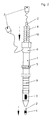
- Two sensors 2a, 3a for measuring intracranial pressure are integrated in the catheter tube 1a, the first sensor 2a being arranged at the distal end and the second sensor 3a being located proximally behind it.
- the sensors are located in housings made of non-magnetizable material, here ceramic.
- the scale 4a is MRI-contrasting, in this case titanium-colored, and is ring-shaped attached a length of 20 cm. The distance between the markings is 1 cm each.
- the sensors 2a, 3a are wired to the plugs 7a via a switch 6a.
- the hose rider 5a serves as a spacer or to fix the penetration depth of the catheter.
- the large-lumen catheter tube 1 has an axial one at the distal end Opening, proximally behind it is a sensor 3, a tube rider 5 and the ring-shaped scale 9 at intervals of 1 cm.
- the valve 6 is designed as an end piece, in which the electrical wiring of the sensor through a lateral lumen outlet of the valve 6 branches off to the monitor and in the central lumen of valve 6 the small-lumen catheter tube 2 or a rigid stylet may be positioned.
- the small-lumen catheter tube 2 has a conical tip with sensor 4 at the distal end.
- the two axially arranged catheter tubes form an atraumatic tip from the sensor housing of the small-lumen catheter tube 2 and the conical tip of the large-lumen catheter tube 1, which enables precise guidance and placement of the system in the brain.
- the valve 6 can be opened and the sensors 3, 4 can be axially displaced relative to one another. This movement can be tracked in a controlled manner via the scalings 9, 10.
- the connectors 7 can be coupled to the measuring device technology, the transmission of the measuring signals to the monitor via cable or - if the monitor is equipped accordingly - telemetrically, for example via radio , he follows. Corrections of the sensor position are also possible during the measurement.
- a protective cover 8 with one end on the valve 6 and the other end on the catheter tube 2 is fixedly mounted, which is preferably designed as a bellows or film tube made of transparent, flexible polymer material.
Landscapes
- Health & Medical Sciences (AREA)
- Life Sciences & Earth Sciences (AREA)
- Biomedical Technology (AREA)
- Heart & Thoracic Surgery (AREA)
- Hematology (AREA)
- Biophysics (AREA)
- Pathology (AREA)
- Engineering & Computer Science (AREA)
- Neurosurgery (AREA)
- Physics & Mathematics (AREA)
- Medical Informatics (AREA)
- Molecular Biology (AREA)
- Surgery (AREA)
- Animal Behavior & Ethology (AREA)
- General Health & Medical Sciences (AREA)
- Public Health (AREA)
- Veterinary Medicine (AREA)
- Measuring And Recording Apparatus For Diagnosis (AREA)
Abstract
Description
Dazu wird - nach der bisher geübten medizinischen Praxis - der Katheter meistens im Bereich des vorderen Schädels, rechts oder links frontal, eingeführt und der Hirndruck an folgenden Messorten bestimmt:
Nachteilig ist darüber hinaus, dass mehrere Sensoren nicht über einen Zugangskanal und in variablen Abständen positionierbar sind. Wenn mehrere Sensoren zum Einsatz kommen müssen, so geschieht dies in der Regel zwar über eine Kopfschraube, jedoch über mehrere Katheter und damit über mehrere Zugangskanäle in der Hirnhaut.
Die Positionierung geschieht vorzugsweise über Neuronavigation oder Ultraschall, wobei entweder über eine separate Hülse in Verbindung mit einem eingebrachten Mandrin der Zugangskanal erzeugt wird und anschließend das Kathetersystem eingebracht wird, oder das Kathetersystem enthält einen Mandrin und verfügt über eine atraumatische Katheterspitze, so dass es direkt eingeführt und positioniert werden kann.
Weiterhin ist der Katheterschlauch signalfarben orange eingefärbt, damit er - bei dem geringen Durchmesser von 2 mm - bei der Operation leicht erkennbar und identifizierbar ist.
Die Sensoren sind über ein entsprechendes Adaptersystem an den Messmonitor gekoppelt, die so zugänglichen Druckgradienten der einzelnen Messstellen und deren zeitliche Veränderung lassen sich nun abrufen und dokumentieren.
Um beim Implantieren des Katheters die einzelnen Abstände der Sensoren zu erkennen, ist der Katheter zirkulär mit einer Skalierung und Beschriftung versehen.
Die Lumen beider Katheterschläuche sind über ein proximal montiertes Ventil gekoppelt, welches mit dem großlumigen Katheterschlauch fest montiert ist und in dessen Lumen der kleinlumige Katheterschlauch durch eine Quetschdichtung dicht und axial fixiert ist.
Der bewegte Abschnitt des kleinlumigen Katheterschlauches ist für diese Handhabung mit einer flexiblen Schlauchschutzhülle umgeben, welche die sterile Oberfläche des bewegten Schlauchabschnittes während der notwendigen Positionsveränderung sichert.
Alternativ ist es auch möglich, den großlumigen Katheterschlauch über einen atraumatischen, starren Mandrin vorab zu implantieren und nach dem Entfernen dieses Mandrins den kleinlumigen Katheterschlauch zu installieren.
Nicht zuletzt ist die Kombination mit gebräuchlichen Hilfsmitteln gesichert, z.B. mit Tunnelungshülsen oder Kopfschrauben oder lumengeführten starren Führungsdrähten.
Weiterhin ist die Erfassung und Auswertung der Messergebnisse einfach und den bisherigen Verfahrensweisen angepasst. Es ist keine zusätzliche Hardware nötig und nur geringfügige Aufwendungen an Software für die vorhandenen Monitorsysteme.
Für die Diagnose ist besonders vorteilhaft, dass, wie im Beispiel 2 beschrieben, die Lage des zweiten Sensors in der Längsachse des Katheters veränderbar ist.
Dabei zeigt
- Figur 1 die erfindungsgemäße Vorrichtung in der Ausführung eines Einlumen-Katheters
- Figur 2 die erfindungsgemäße Vorrichtung in der Ausführung eines Zweilumen-Katheters
In den Katheterschlauch 1a sind zwei Sensoren 2a, 3a zur Hirndruckmessung integriert, wobei der erste Sensor 2a am distalen Ende und der zweite Sensor 3a proximal dahinter angeordnet sind. Die Sensoren befinden sich in Gehäusen aus nicht magnetisierbarem Material, hier Keramik.
Die beiden axial angeordneten Katheterschläuche bilden aus dem Sensorgehäuse des kleinlumigen Katheterschlauches 2 und der konischen Spitze des großlumigen Katheterschlauches 1 eine atraumatische Spitze, die eine präzise Führung und Platzierung des Systems im Hirn ermöglicht. Ist die Positionierung erfolgt, so kann das Ventil 6 geöffnet und die Sensoren 3, 4 gegeneinander axial verschoben werden. Diese Bewegung ist über die Skalierungen 9, 10 kontrolliert verfolgbar. Mit dem Verschluss des Ventils 6 und der damit erfolgten Fixierung des kleinlumigen Katheterschlauches 2 können die Stecker 7 an die Messgerätetechnik gekoppelt werden, wobei die Übertragung der Messsignale an den Monitor über Kabel, oder - wenn der Monitor entsprechend ausgestattet ist - telemetrisch, beispielsweise über Funk, erfolgt.
Korrekturen der Sensorposition sind auch während der Messung möglich.
Zu Sicherung der sterilen Oberfläche des bewegten Schlauchabschnittes des Katheterschlauches 2 ist eine Schutzhülle 8 mit einem Ende am Ventil 6 und dem anderen Ende am Katheterschlauch 2 fest montiert, die vorzugsweise als Faltenbalg oder Folienschlauch aus transparentem, flexiblen Polymermaterial gestaltet ist.
Claims (11)
- Vorrichtung zur Messung des Hirndruckes mittels Katheter,
dadurch gekennzeichnet, dass der Katheter mindestens aus einem Katheterschlauch aus polymerem Material besteht, und über mindestens zwei Drucksensoren verfügt, die in Reihe angeordnet sind, wobei der eine Drucksensor den Hirndruck in der geschädigten Region, der zweite, oder weitere Drucksensoren den Hirndruck in den angrenzenden Bereichen erfassen und dass die Platzierung des Katheters und die Messung des Hirndruckes an beliebigen Stellen im Hirn erfolgt. - Vorrichtung gemäß Anspruch 1, dadurch gekennzeichnet, dass der Katheter aus einem großlumigen Katheterschlauch, der an seinem distalen Ende über einen Drucksensor und eine axiale Öffnung verfügt, und aus einem darin eingebrachten, kleinlumigen Katheterschlauch besteht, der an seinem distalen Ende mit einem Drucksensor versehen ist und der in die axiale Öffnung des großlumigen Katheterschlauches so hineinragt, dass beide Sensoren in ihrer Ausgangsposition eine atraumatische Katheterspitze bilden.
- Vorrichtung gemäß Anspruch 1, dadurch gekennzeichnet, dass der Katheter über zwei Lumen verfügt, wobei in dem einen Lumen in Reihe Drucksensoren angeordnet sind, und in einem zweiten Lumen ein starrer Mandrin so platziert ist, dass mit dessen Hilfe das Kathetersystem präzise positionierbar ist.
- Vorrichtung nach Anspruch 1, dadurch gekennzeichnet, dass für alle Elemente, die während der bildgebenden Diagnostik am Patienten verbleiben, unmagnetische Materialien zum Einsatz kommen.
- Vorrichtung nach den Ansprüchen 1 und 4, dadurch gekennzeichnet, dass die unmagnetischen Materialien ausgewählt sind aus der Gruppe Keramik und/oder Polymere und/oder Titan.
- Vorrichtung gemäß Anspruch 2, dadurch gekennzeichnet, dass der bewegliche, kleinlumige Katheterschlauch durch eine Schutzhülle aus flexiblem, transparentem Material gegen äußere Einflüsse geschützt ist.
- Vorrichtung nach einem der vorigen Ansprüche, dadurch gekennzeichnet, dass die Katheterschläuche im nicht implantierten Bereich im erforderlichen Maße zirkulär skaliert sind und diese Skalierung mit Abstandsziffern versehen ist.
- Vorrichtung nach einem der vorigen Ansprüche, dadurch gekennzeichnet, dass die Druck-Sensoren phasengleich arbeiten und das Messsignal ungedämpft an den Empfänger übertragen.
- Katheter nach einem der vorigen Ansprüche, dadurch gekennzeichnet, dass die präzise Platzierung im Hirn mittels Mandrin, insbesondere mittels Hülsen-Mandrin-System erfolgt und die Platzierung über bildgebende Verfahren überwacht wird.
- Katheterschlauch nach Anspruch 1, dadurch gekennzeichnet, dass das polymere Material signalfarben eingefärbt, sowie CT-, MRT- und röntgentauglich ist.
- Katheterschlauch nach den Ansprüchen 1 und 10, dadurch gekennzeichnet, dass der Werkstoff ausgewählt ist aus der Gruppe Silikonkautschuk und/oder Polyurethan und/oder Polyamid und/oder Polyolefin.
Applications Claiming Priority (2)
| Application Number | Priority Date | Filing Date | Title |
|---|---|---|---|
| DE20116879U | 2001-10-13 | ||
| DE20116879U DE20116879U1 (de) | 2001-10-13 | 2001-10-13 | Vorrichtung zur Bestimmung des intrazerebralen Druckgradienten |
Publications (2)
| Publication Number | Publication Date |
|---|---|
| EP1302157A2 true EP1302157A2 (de) | 2003-04-16 |
| EP1302157A3 EP1302157A3 (de) | 2005-04-13 |
Family
ID=7962860
Family Applications (1)
| Application Number | Title | Priority Date | Filing Date |
|---|---|---|---|
| EP02022414A Withdrawn EP1302157A3 (de) | 2001-10-13 | 2002-10-04 | Vorrichtung zur Bestimmung des intrazerebralen Druckgradienten |
Country Status (2)
| Country | Link |
|---|---|
| EP (1) | EP1302157A3 (de) |
| DE (1) | DE20116879U1 (de) |
Families Citing this family (2)
| Publication number | Priority date | Publication date | Assignee | Title |
|---|---|---|---|---|
| US8313442B2 (en) * | 2009-10-21 | 2012-11-20 | Codman & Shurtleff, Inc. | Cerebral compliance monitoring |
| CN105962916B (zh) * | 2016-06-08 | 2017-05-03 | 深圳北芯生命科技有限公司 | 制造测量导管的治具 |
Family Cites Families (12)
| Publication number | Priority date | Publication date | Assignee | Title |
|---|---|---|---|---|
| US4114603A (en) | 1976-08-06 | 1978-09-19 | Wilkinson Harold A | Intracranial pressure monitoring catheter |
| DE3918142A1 (de) | 1989-05-31 | 1990-12-13 | Wiest Peter P | Druckmessvorrichtung fuer in leitungen stroemende fluide |
| DE4036355C2 (de) | 1990-11-15 | 1999-09-16 | Rehau Ag & Co | Druckmeßkatheter |
| US5409453A (en) | 1992-08-12 | 1995-04-25 | Vidamed, Inc. | Steerable medical probe with stylets |
| NL9401180A (nl) * | 1994-07-18 | 1996-03-01 | Draeger Med Electronics Bv | Catheter met meerdere sensoren op afstand van elkaar. |
| US5573007A (en) * | 1994-08-08 | 1996-11-12 | Innerspace, Inc. | Gas column pressure monitoring catheters |
| DE19654990A1 (de) | 1996-09-09 | 1998-06-18 | Steffen Dr Ing Leonhardt | Implantat zur kontrollierten Ableitung von Gehirnflüssigkeit |
| NL1005134C2 (nl) * | 1997-01-30 | 1998-08-03 | Industrial Res Bv | Meetkathetersamenstel. |
| US6061587A (en) * | 1997-05-15 | 2000-05-09 | Regents Of The University Of Minnesota | Method and apparatus for use with MR imaging |
| US6537232B1 (en) * | 1997-05-15 | 2003-03-25 | Regents Of The University Of Minnesota | Intracranial pressure monitoring device and method for use in MR-guided drug delivery |
| EP0879617B1 (de) * | 1997-05-21 | 2003-04-16 | Schneider (Europe) GmbH | Führungsdraht mit Druckanzeige und Verfahren zur Herstellung eines solchen Führungsdrahtes |
| US6453185B1 (en) | 2000-03-17 | 2002-09-17 | Integra Lifesciences, Inc. | Ventricular catheter with reduced size connector and method of use |
-
2001
- 2001-10-13 DE DE20116879U patent/DE20116879U1/de not_active Expired - Lifetime
-
2002
- 2002-10-04 EP EP02022414A patent/EP1302157A3/de not_active Withdrawn
Also Published As
| Publication number | Publication date |
|---|---|
| DE20116879U1 (de) | 2001-12-20 |
| EP1302157A3 (de) | 2005-04-13 |
Similar Documents
| Publication | Publication Date | Title |
|---|---|---|
| DE69733249T2 (de) | Bestimmung der genauen position von endoskopen | |
| DE3504292C1 (de) | Instrument fuer endoskopische Eingriffe,insbesondere zur perkutanen Gallensteinentfernung oder Gallenblasenveroedung | |
| EP1065987B1 (de) | Cas-erfassbarer endoskop-adapter | |
| EP3641621B1 (de) | Endoskop-vorrichtung | |
| EP1319366A1 (de) | Magnetische Katheternavigation | |
| DE4440346A1 (de) | Punktionsinstrument | |
| WO2002045588A1 (de) | Ultraschallsonde mit positioniereinrichtung für untersuchungs- und operationsvorrichtungen | |
| EP1731105A1 (de) | Vorrichtung zum Schaffen eines freien transkutanen Zuganges zu einem endoskopischen Operationsgebiet | |
| EP3979929B1 (de) | Chirurgisches nadel-set zur positionsbestimmung eines chirurgischen instruments | |
| EP3585474B1 (de) | Vorrichtung für eine gehirndrainage | |
| DE10029737B4 (de) | Navigation eines medizinischen Instrumentes | |
| EP2185062B1 (de) | Kathetersystem mit optischer sonde und verfahren zur applikation einer optischen sonde in ein kathetersystem | |
| EP3024409A1 (de) | Vorrichtung und verfahren zur anbindung eines medizinischen instruments an ein lageerfassungssystem | |
| EP1302157A2 (de) | Vorrichtung zur Bestimmung des intrazerebralen Druckgradienten | |
| DE102005027677B4 (de) | Vorrichtung zum automatischen Auswechseln von Instrumenten bei minimalinvasiven Verfahren | |
| EP1487364A2 (de) | Medizinisches instrument zur behandlung von gewebe mittels hochfrequenzstrom sowie medizinisches system mit einem derartigen medizinischen instrument | |
| WO2017220812A1 (de) | Punktionskatheter | |
| DE10040164C2 (de) | Drainage- und Spülkatheter mit Druckmesseinrichtung | |
| DE102013201259A1 (de) | Verfahren und Vorrichtung zur Abbildung der Prostata | |
| DE102016119065B4 (de) | Nadelstrumpf mit Trägersystem | |
| EP0450111A1 (de) | Verwendung der Computertomographie, Magnetresonanztomographie, Ultraschallbildgebung od. dgl. bildgebende Verfahren zur Leitung für endoskopische Diagnostik und operative Therapie | |
| WO2007087798A2 (de) | Zielvorrichtung zur durchführung minimal-invasiver interventionen | |
| DE102016006425A1 (de) | Führungskatheter, Verfahren zur Positionierung von Sonden sowie Verwendung eines Führungskatheters | |
| DE202010012342U1 (de) | Vaginalsonde für Röntgen und MRT-Untersuchungen | |
| EP1199034A2 (de) | Vorrichtung zur linearen und/oder rotatorischen Führung eines Gegenstandes wie einer Sonden- und Instrumentenführung |
Legal Events
| Date | Code | Title | Description |
|---|---|---|---|
| PUAI | Public reference made under article 153(3) epc to a published international application that has entered the european phase |
Free format text: ORIGINAL CODE: 0009012 |
|
| AK | Designated contracting states |
Designated state(s): AT BE BG CH CY CZ DE DK EE ES FI FR GB GR IE IT LI LU MC NL PT SE SK TR |
|
| AX | Request for extension of the european patent |
Extension state: AL LT LV MK RO SI |
|
| RAP1 | Party data changed (applicant data changed or rights of an application transferred) |
Owner name: RAUMEDIC AG |
|
| PUAL | Search report despatched |
Free format text: ORIGINAL CODE: 0009013 |
|
| AK | Designated contracting states |
Kind code of ref document: A3 Designated state(s): AT BE BG CH CY CZ DE DK EE ES FI FR GB GR IE IT LI LU MC NL PT SE SK TR |
|
| AX | Request for extension of the european patent |
Extension state: AL LT LV MK RO SI |
|
| AKX | Designation fees paid | ||
| STAA | Information on the status of an ep patent application or granted ep patent |
Free format text: STATUS: THE APPLICATION IS DEEMED TO BE WITHDRAWN |
|
| 18D | Application deemed to be withdrawn |
Effective date: 20051014 |
|
| REG | Reference to a national code |
Ref country code: DE Ref legal event code: 8566 |