EP1294927B1 - Screeningverfahren für modulatoren von methyltransferase abhängiger chromosomenstabilität - Google Patents
Screeningverfahren für modulatoren von methyltransferase abhängiger chromosomenstabilität Download PDFInfo
- Publication number
- EP1294927B1 EP1294927B1 EP01940552A EP01940552A EP1294927B1 EP 1294927 B1 EP1294927 B1 EP 1294927B1 EP 01940552 A EP01940552 A EP 01940552A EP 01940552 A EP01940552 A EP 01940552A EP 1294927 B1 EP1294927 B1 EP 1294927B1
- Authority
- EP
- European Patent Office
- Prior art keywords
- suv39h
- suv39h1
- histone
- cells
- suv39h2
- Prior art date
- Legal status (The legal status is an assumption and is not a legal conclusion. Google has not performed a legal analysis and makes no representation as to the accuracy of the status listed.)
- Expired - Lifetime
Links
Images












Classifications
-
- G—PHYSICS
- G01—MEASURING; TESTING
- G01N—INVESTIGATING OR ANALYSING MATERIALS BY DETERMINING THEIR CHEMICAL OR PHYSICAL PROPERTIES
- G01N33/00—Investigating or analysing materials by specific methods not covered by groups G01N1/00 - G01N31/00
- G01N33/48—Biological material, e.g. blood, urine; Haemocytometers
- G01N33/50—Chemical analysis of biological material, e.g. blood, urine; Testing involving biospecific ligand binding methods; Immunological testing
- G01N33/68—Chemical analysis of biological material, e.g. blood, urine; Testing involving biospecific ligand binding methods; Immunological testing involving proteins, peptides or amino acids
- G01N33/6875—Nucleoproteins
-
- A—HUMAN NECESSITIES
- A61—MEDICAL OR VETERINARY SCIENCE; HYGIENE
- A61P—SPECIFIC THERAPEUTIC ACTIVITY OF CHEMICAL COMPOUNDS OR MEDICINAL PREPARATIONS
- A61P15/00—Drugs for genital or sexual disorders; Contraceptives
- A61P15/16—Masculine contraceptives
-
- A—HUMAN NECESSITIES
- A61—MEDICAL OR VETERINARY SCIENCE; HYGIENE
- A61P—SPECIFIC THERAPEUTIC ACTIVITY OF CHEMICAL COMPOUNDS OR MEDICINAL PREPARATIONS
- A61P35/00—Antineoplastic agents
-
- C—CHEMISTRY; METALLURGY
- C07—ORGANIC CHEMISTRY
- C07K—PEPTIDES
- C07K16/00—Immunoglobulins [IG], e.g. monoclonal or polyclonal antibodies
- C07K16/40—Immunoglobulins [IG], e.g. monoclonal or polyclonal antibodies against enzymes
-
- C—CHEMISTRY; METALLURGY
- C12—BIOCHEMISTRY; BEER; SPIRITS; WINE; VINEGAR; MICROBIOLOGY; ENZYMOLOGY; MUTATION OR GENETIC ENGINEERING
- C12N—MICROORGANISMS OR ENZYMES; COMPOSITIONS THEREOF; PROPAGATING, PRESERVING, OR MAINTAINING MICROORGANISMS; MUTATION OR GENETIC ENGINEERING; CULTURE MEDIA
- C12N9/00—Enzymes; Proenzymes; Compositions thereof; Processes for preparing, activating, inhibiting, separating or purifying enzymes
- C12N9/10—Transferases (2.)
- C12N9/1003—Transferases (2.) transferring one-carbon groups (2.1)
- C12N9/1007—Methyltransferases (general) (2.1.1.)
-
- C—CHEMISTRY; METALLURGY
- C12—BIOCHEMISTRY; BEER; SPIRITS; WINE; VINEGAR; MICROBIOLOGY; ENZYMOLOGY; MUTATION OR GENETIC ENGINEERING
- C12Q—MEASURING OR TESTING PROCESSES INVOLVING ENZYMES, NUCLEIC ACIDS OR MICROORGANISMS; COMPOSITIONS OR TEST PAPERS THEREFOR; PROCESSES OF PREPARING SUCH COMPOSITIONS; CONDITION-RESPONSIVE CONTROL IN MICROBIOLOGICAL OR ENZYMOLOGICAL PROCESSES
- C12Q1/00—Measuring or testing processes involving enzymes, nucleic acids or microorganisms; Compositions therefor; Processes of preparing such compositions
- C12Q1/48—Measuring or testing processes involving enzymes, nucleic acids or microorganisms; Compositions therefor; Processes of preparing such compositions involving transferase
-
- G—PHYSICS
- G01—MEASURING; TESTING
- G01N—INVESTIGATING OR ANALYSING MATERIALS BY DETERMINING THEIR CHEMICAL OR PHYSICAL PROPERTIES
- G01N2333/00—Assays involving biological materials from specific organisms or of a specific nature
- G01N2333/90—Enzymes; Proenzymes
- G01N2333/91—Transferases (2.)
- G01N2333/91005—Transferases (2.) transferring one-carbon groups (2.1)
- G01N2333/91011—Methyltransferases (general) (2.1.1.)
-
- G—PHYSICS
- G01—MEASURING; TESTING
- G01N—INVESTIGATING OR ANALYSING MATERIALS BY DETERMINING THEIR CHEMICAL OR PHYSICAL PROPERTIES
- G01N2500/00—Screening for compounds of potential therapeutic value
Definitions
- the invention relates to a method for identifying compounds influencing chromosome dynamics in eukaryotic cells.
- the invention relates to the treatment and prevention of human conditions by modulating higher order chromatin dependent chromosome stability during mitosis and meiosis.
- Higher-order chromatin is essential for epigenetic gene control and for the functional organisation of chromosomes. Differences in higher-order chromatin structure have been linked with distinct covalent modifications of histone tails which regulate transcriptional 'on' or 'off' states (Grunstein, 1998; Turner, 1998; Strahl and Allis, 2000) and influence chromosome condensation and segregation (Karpen and Allshire, 1997; Wei et al ., 1999).
- Histones constitute a highly conserved family of proteins (H3, H4, H2A, H2B, H1) which are the major components of eukaryotic chromatin structure. Histones compact genomic DNA into basic repeating structural units, the nucleosomes. In addition to their DNA packaging function, histones have been proven to be integral components of the molecular machinery that regulates gene expression.
- HATs histone acetyltransferases
- HDACs histone deacetylases
- Su(var)2-5 which is allelic to HP1
- Su(var)3-7 (Cléard et al ., 1997)
- Su(var)3-9 (Tschiersch et al ., 1994; Schotta and Reuter, 2000) encode heterochromatin-associated proteins.
- Su(var) gene function thus suggests a model, in which modifications at the nucleosomal level may initiate the formation of defined chromosomal subdomains that are then stabilised and propagated by heterochromatic SU(VAR) proteins (Henikoff, 1997).
- Su(var)3-9 is dominant over most PEV modifier mutations (Tschiersch et al ., 1994), and mutants in the corresponding S.pombe clr4 gene (Ivanova et al ., 1998) disrupt heterochromatin association of other modifying factors and result in chromosome segregation defects (Ekwall et al ., 1996).
- human ( SUV39H1 ) and murine ( Suv39h1 and Suv39h2 ) Su(var)3-9 homologues have been isolated (Aagaard et al., 1999). It has been shown that they encode heterochromatic proteins which associate with mammalian HP1 (Aagaard et al ., 1999).
- the SU(VAR)3-9 protein family combines two of the most evolutionarily conserved domains of 'chromatin regulators': the chromo (Aasland and Stewart, 1995; Koonin et al ., 1995) and the SET (Tschiersch et al., 1994; Jenuwein et al ., 1998) domain.
- the 60 amino acids chromo domain represents an ancient histone-like fold (Ball et al ., 1997) that directs eu- or heterochromatic localisations (Platero et al ., 1995)
- the molecular role of the 130 amino acids SET domain has remained enigmatic.
- chromosome stability implies successful segregation of chromosomes resulting in the maintenance of a stable karyotype).
- Examples 1 to 7 of the present invention show that mammalian SU(VAR)3-9 related proteins (human SUV39H1, murine Suv39h1 and murine Suv39h2) are SET domain-dependent H3-specific histone methyltransferases which selectively methylate lysine 9 ("K9") of the H3 N-terminus. Methylation of K9 negatively regulates phosphorylation of adjacent serine 10 and reveals a 'histone code' that appears intrinsically linked to the organisation of higher-order chromatin. (In the following, h istone m ethyl t ransferases are termed "HMTases” or, more generally, “MTases”).
- Suv39h HMTases are heterochromatin-enriched enzymes which transiently accumulate at centromeres during mitosis (Aagaard et al ., 1999; Aagaard et al ., 2000).
- methylation of histone H3 at lysine 9 creates a high-affinity binding site for HP1 proteins (Lachner et al ., 2001; Bannister et al ., 2001), thereby defining the SUV39H1-HP1 methylation system as a crucial regulatory mechanism for the assembly and propagation of heterochromatin (Jenuwein, 2001).
- Overexpression of human SUV39H1 induces ectopic heterochromatin and results in chromosome mis-segregation in mammalian cell lines (Melcher et al ., 2000).
- heterochromatin is also crucial for the dynamic reorganization of meiotic chromosomes.
- Meiosis is initiated by chromosomal movements from the nuclear lumen to the nuclear envelope, where chromosomes cluster via their pericentric satellite sequences (Hawley et al ., 1992; Scherthan et al ., 1996).
- chromosomes condense, followed by homolog pairing and recombination (at pachytene) between maternal and paternal chromosomes.
- the onset of the meiotic divisions is preceded by desynapsis, further chromosome condensation and histone H3 phosphorylation at pericentric heterochromatin (Cobb et al ., 1999).
- the haploid genome content is finally organized into one heterochromatic block in elongating spermatids.
- heterochromatin and its associated satellite sequences have been proposed to assist in the initial meiotic chromosome movements and in homolog pairing by orienting chromosomes along a similar higher-order structure (Hawley et al ., 1992; Karpen et al ., 1996; Dernburg et al ., 1996b).
- a pachytene checkpoint (de Vries et al ., 1999) monitors mis-aligned and unpaired chromosomes and arrests cells in meiotic prophase, thereby preventing the production of aneuploid gametes.
- bioinformatics techniques were applied. Using the SET domains of the SU(VAR)3-9 protein family as a starting alignment, significant sequence and secondary structure similarities (see Methods) to six plant protein methyltransferases were detected.
- both murine GST-Suv39h1(82-412) and the corresponding human SUV39H1 fusion protein [GST-SUV39H1(82-412)] were shown to be catalytically active. Short internal deletions were introduced into the two conserved regions of the SET domain core in GST-SUV39H11(82-412), and additional mutants lacking the C-terminal tail ( ⁇ C-tail) or the SET-associated cysteine-rich region ( ⁇ cys) were generated. All mutant proteins failed to demonstrate HMTase activity.
- the wild-type H3 N-terminal peptide was in vitro methylated by GST-Suv39h1(82-412), using S-adenosyl-[ methyl - 3 H]-L-methionine.
- the labelled peptide purified by reverse-phase HPLC, was then directly microsequenced, and 3 H-incorporation associated with each individual amino acid was analysed. The results confirmed selective transfer of methyl-label to lysine 9, demonstrating that Suv39h1 is a highly site-specific HMTase for the H3 N-terminus in vitro .
- Murine Suv39h genes are encoded by 2 loci, Suv39h1 and Suv39h2.
- mouse strains deficient for both Suv39h1 and Suv39h2 were generated.
- Suv39h1 and Suv39h2 deficient strains were intercrossed to produce Suv39h double deficient mice.
- Double mutant mice were born in sub-Mendelian ratios. Some double null embryos exhibited severe growth retardation and exencephaly. In addition surviving double mutants were growth retarded, suggesting a role for Suv39h in cell proliferation.
- Suv39h null mice In order to determine whether the embryonic phenotypes in Suv39h null mice can be attributed to mitotic defects, PMEFs (primary mouse embryonic fibroblasts) derived from Suv39h double mice were analysed.
- Suv39h double null PMEFs display a reduced G1-index and an increased proportion of cells with aberrant nuclear morphologies, reminiscent of division defects during mitosis.
- double null cells also show genomic instabilities and readily become aneuploid. The severity of these aneuploidies increases with higher passage numbers.
- the inability of Suv39h double null cells to maintain a stable karyotype may underlie the Suv39h embryonic phenotype.
- phosH3 Phosphorylation at serine 10 in the N-terminal tail of H3 has been shown to be required for condensation and subsequent segregation of chromosomes (Wei et al ., 1999).
- phosH3 initiates within pericentric heterochromatin in late G2 and then progresses along the entire chromosomes during mitosis (Hendzel et al ., 1997). It was found that in wild-type PMEFs, approximately 7% of the cells stain positive for the characteristic, heterochromatin-associated phosH3 foci. In contrast, this number is increased by a factor of about 3-fold in Suv39h double null PMEFs.
- a set of experiments of the present invention provides in vivo evidence that the absence of Suv39h HMTase activities impairs development and viability of mutant mice, and directly correlates with a nearly complete lack of H3-K9 methylation at pericentric heterochromatin.
- Suv39h-deficient mice display chromosomal instabilities in both somatic and meiotic cells that are further evidenced by an increased risk for development of B-cell lymphomas and perturbed chromosome interactions during male meiosis.
- Suv39h1 or Suv39h2 Single gene disruptions for either Suv39h1 or Suv39h2 allow for normal mouse development and do not appear to affect viability and fertility of mutant mice. This apparent redundancy in gene function would be consistent with the overlapping expression profile of the two Suv39h genes during mouse embryogenesis (O'Carroll et al ., 2000).
- combined disruption of both genes in Suv39h double null (dn) mice results in severely impaired perinatal viability ( ⁇ 33%; Table I), growth retardation and hypogonadism in males. Both Suv39h dn males and are infertile.
- Suv39h dn fetuses appear to develop normally until day E12.5, they then display smaller body sizes and frequently are resorbed during late gestation. These in vivo analyses indicate an important role(s) for the Suv39h genes during mammalian development and for overall viability. Since the absence of Suv39h HMTase activities induces genomic instabilities, the high lethality of Suv39h dn fetuses could mainly be a consequence of perturbed chromosome segregation which would significantly impair the proliferation and differentiation programmes of the developing embryo.
- Suv39h enzymes are the major HMTases for H3-K9 methylation at pericentric heterochromatin in somatic cells (see Figure 12) and in early meiotic cells (see Figure 13B), there are ⁇ 15 unique gene sequences in the mouse genome that contain the evolutionarily highly conserved SET domain and which are likely to encode additional enzymes with putative HMTase activity (Jenuwein, 2001).
- At least one of these SET domain containing proteins can indeed also methylate H3 at lysine Tachibana et al ., 2001
- the ⁇ 33% viability of Suv39h dn mice could be dependent on the compensating activity of other HMTases that may be expressed to varying degrees in the mixed genetic background, in which the Suv39h dn mice have been analyzed.
- Suv39h HMTase activities triggers genomic instabilities in a variety of cell types, including mouse embryonic fibroblasts (PMEFs) (see Figure 10), fetal liver and bone marrow cells and in spermatogonia (see Figure 14C).
- PMEFs mouse embryonic fibroblasts
- fetal liver and bone marrow cells and in spermatogonia
- Figure 14C mouse embryonic fibroblasts
- Suv39h -deficient mice display an increased risk for tumorigenesis, resulting in late-onset B-cell lymphomas in ⁇ 33% of Suv39h dn mice (see Figure 11). B-cell lymphomas also develop upon reduced Suv39h gene dosage in compound mutant mice that contain gene disruptions of Suv39h1 (see Table II).
- Suv39h -induced aneuploidies are mainly characterized by segregation failure of the nearly complete set of the chromosomes, resulting in hypo-tetraploid or even hypo-octaploid cells (see Figure 10).
- These data suggest a general impairment of chromosome segregation, consistent with the lack of H3-K9 methylation around all acrocentric centromeres in Suv39h dn cells (see Figure 12).
- H3-K9 methylation restricts H3 phosphorylation mediated by the Ipl1/aurora kinase (Hsu et al ., 2000) and is also interdependent with histone acetylation (Rea et al ., 2000), the absence of Suv39h HMTase activities is likely to perturb this distinct histone modification pattern.
- histone modifications can generate specific interaction affinities for chromatin-associated proteins (Rice and Allis, 2001). Although the localisation of CENP epitopes appears unaltered, heterochromatic enrichment of HP1 proteins is largely lost in Suv39h dn somatic cells (Lachner et al ., 2001).
- HP1 interacts in vitro with INCENP (Ainsztein et al ., 1998) which forms a complex with aurora-B (Adams et al ., 2000; Kaitna et al ., 2000).
- INCENP Insztein et al ., 1998) which forms a complex with aurora-B (Adams et al ., 2000; Kaitna et al ., 2000).
- Mutation of INCENP induces severe mitotic abnormalities including macronuclei and internuclear bridges, and results in nearly complete chromosome mis-segregation and cytokinesis failure (Cutts et al ., 1999; Adams et al ., 2000; Kaitna et al ., 2000).
- Suv39h -mediated chromosomal instabilities only affect a sub-population of cells and do not appear to trigger pronounced apoptosis (see Figure 10b), consistent with similar analyses of clr4 mutants in S.pombe (Ekwall et al., 1996; Ivanova et al ., 1998).
- Suv39h -induced defects in somatic cells are not under strict surveillance of known checkpoint controls (Cortez and Elledge, 2000; but see Bernard et al ., 1998) and may be caused by rather late segregation problems during mitosis. Indeed, a fraction of Suv39h dn cells contain chromosomes that lag at anaphase.
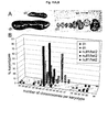
- Suv39h dn PMEFs are characterized by hypo-tetraploid and hypo-octaploid karyotypes (see Figure 10d) and tumor cells contain 'butterfly' chromosomes (see Figure 11c)
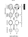
- a model is proposed ( Figure 16, top panel), in which the absence of H3-K9 methylation would allow stronger or more persistent pericentric associations between aligned metaphase chromosomes.
- Suv39h HMTases in centromeric cohesion remains to be determined, it provides an attractive mechanism to explain possible cytokinesis failure and mis-segregation of the entire chromosome complement without activating known checkpoint controls.
- pericentric H3-K9 methylation is specifically reduced at the pre-leptotene stage but, surprisingly, appears as a wild-type staining during later meiotic stages (see Figure 13b, bottom panel).
- impairment of H3-K9 methylation at the onset of meiosis induces aberrant centromere clustering that can no longer be 'rescued' by the hypothetical activity of additional H3-K9 HMTases during mid-pachytene.
- spermatogenic failure is promoted by illegitimate chromosomal interactions, synaptic delay, unpaired sex chromosomes and bivalent mis-segregation at meiosis I (see Figures 14 -15).
- a major fraction of these 'forbidden' interactions comprises physical contacts between the sex chromosomes and autosomes that are largely mediated through centromeric regions (see Figure 14D and 14J).
- Heterochromatin has been first described more than 70 years ago (Heitz, 1928). Because of its stable appearance in the cell nucleus, it has been proposed to serve crucial functions for the inheritance of cell type identities and the fidelity of chromosome segregation.
- the discoveries of the first HMTases (Rea et al ., 2000; O'Carroll et al ., 2000) and their mechanistic link to generate a heterochromatic affinity through H3-K9 methylation and recruitment of HP1 proteins (Lachner et al ., 2001; Bannister et al ., 2001; Nakayama et al ., 2001) has now defined an entry point to start dissecting some of the basic roles of heterochromatin.
- the results obtained in the experiments of the present invention show that members of the SU(VAR)3-9 protein family have HMTase activity which identifies them as novel targets for the therapy of proliferative disorders, in particular cancer.
- Suv39h double null male mice display complete spermatogenic failure that is largely caused by non-homologous chromosome associations and delayed synapsis, resulting in apoptosis of meiotic prophase cells.
- these results establish histone H3-K9 methylation as a crucial determinant for pericentric heterochromatin and provide a direct role for the Suv39h HMTases in maintaining a 'heterochromatic competence' that protects chromosome stability during mitosis and meiosis.
- compounds modulating the MTase activity of these proteins and thus modulating spermatogenesis may also be used in the treatment of male infertility (using compounds that enhance Suv39h MTase activity) and for reversible male contraception (using compounds that inhibit Suv39h MTase activity).
- the present invention relates to a method for identifying a compound that alters higher order chromatin dependent chromosome stability during mitosis and meiosis, said method comprising incubating a substrate for a methyltransferase, in the presence of a methyl donor, with a methyltransferase having Suv39h-like histone H3 K9 methyltransferase activity, in the presence and absence of a test compound and determining whether the compound modulates the MTase activity.
- Suv39h variants with point mutations in the SET domain were shown to confer hyperactive HMTase activity to the protein, these Suv39h variants may be advantageously used in the method of the invention.
- the MTase is mouse Suv39h1 or Suv39h2, most preferably, the MTase is human SUV39H1 or SUV39H2.
- Suv39h retains HMTase activity
- a recombinant MTase is employed.
- Suv39h or Suv39h variants can be produced recombinantly according to standard methods by expression in suitable hosts, e.g. bacteria, yeast, insect or eukaryotic cells and purified, e.g. on glutathione-agarose columns if it has been tagged with GST.
- Suv39h1 and SUV39H1 cDNA sequences are known from the literature (Aagaard et al., 1999), the Suv39h2 cDNA sequence is shown in SEQ ID NO: 1; the human SUV39H2 cDNA is defined by the ESTs as shown in SEQ ID NO: 3 - 6.
- the assay comprises, as its essential features, incubating a histone H3 protein or a histone H3 N-terminal fragment including K9, a methyl donor, e.g. methionine or S-adenosyl-L-methionine, with a preparation containing a Suv39h MTase activity and determining MTase activity in the presence or absence of a test substance.
- a histone H3 protein or a histone H3 N-terminal fragment including K9, a methyl donor e.g. methionine or S-adenosyl-L-methionine
- MTase substrates useful in the method of the invention may be those equivalent to or mimicking the naturally occurring substrates, e.g. biochemically purified histone H3, recombinantly produced histone H3, or a histone H3 peptide that contains the K9 methylation site.
- Novel Suv39h substrates can be identified by bioinformatic and biochemical techniques and tested using the biochemical assays described in the Examples of the present invention.
- novel Suv39h substrates can be identified by co-immunoprecipitation techniques.
- Suv39h proteins or tagged versions of Suv39h proteins can be immunoprecipitated with specific antisera and interacting proteins identified by mass spectroscopy techniques.
- a yeast two hybrid screen using Suv39h proteins or portions of Suv39h proteins as a bait can also be employed to identify novel interacting protein from a variety of cDNA libraries.
- the histone H3 fragment ARTKQTARKSTGGKAPRKQL (SEQ ID NO:7) is employed.
- a modified peptide may be used for which the MTase has increased affinity/activity.
- Such peptides can be designed by exchanging and/or adding and/or deleting amino acids and testing the substrate in serial experiments for MTase affinity/activity.
- the methyl group of the methyl donor preferably carries a detectable label, e.g. a radioactive or a chromogenic label, which can be quantified upon transfer to the substrate.
- a detectable label e.g. a radioactive or a chromogenic label
- the methyl donor is radioactively labelled methionine or S-adenosyl-L-methionine.
- the substrate upon methylation by the enzyme, is used to serve as an epitope which can be recognised by a specific antibody and hence be quantified by standard immunoassay techniques, e.g. ELISAs.
- Antibodies useful in this type of assay can be obtained by using the methylated substrate, preferably a small peptide, e.g. the peptide with the sequence shown in SEQ ID NO:7, as an antigen and obtaining polyclonal or monoclonal antibodies according to standard techniques.
- the generation and purification of a methyl-specific antibody against the histone H3 lysine 9 position is described in the Materials and Methods section.
- a suitable H3-K9 methyl antibody was also described by Nakayama et al ., 2001.
- the screening method can be based on an assay as described in Example 2, 3 or 4.
- the screening method of the invention utilizes the fact that the methylation of histone H3 at lysine 9 (H3-K9) creates a high-affinity binding site for HP1 proteins.
- the substrate upon methylation, is allowed to bind to HP1 and then incubated with a labelled anti-HP1 antibody. The difference in label intensity between the reaction in the absence or presence of the test compound is indicative for the compound's modulating effect on MTase activity.
- HP1 is preferably used in recombinant form. Based on the information of the HP1 cDNA sequence (Jones et al ., 2000; Accession No. BC006821), HP1 is produced recombinantly according to standard technology. The recombinant protein or fragments thereof are used to generate polyclonal or monoclonal antibodies that are employed in this assay format.
- the method of the invention is performed on a high-throughput scale.
- the major assay components in particular Suv39h, are employed in recombinant form.
- the screening methods of the invention to identify MTase inhibitors are carried out according to standard assay procedures.
- Such assays are based on the catalytic transfer, mediated by Suv39h or a Suv39h variant, of a methyl group from a donor to a substrate, e.g. a histone H3 peptide.
- a substrate e.g. a histone H3 peptide.
- the substrate e.g. histone H3 or a variant or fragment thereof, is immobilised on a carrier, usually a microtiter plate, and incubated with recombinant Suv39h or a Suv39h variant and a methyl donor.
- the methyl group of the methyl donor carries a label, preferably a chromogenic or radioactive label.
- Fluorescent or radioactive labels and the other reagents for carrying out the enzymatic reaction on a high-throughput scale are commercially available and can be employed according to the supplier's instructions (e.g. Molecular Probes, Wallac).
- suitable fluorescent labels are coumarin derivatives, e.g., 7-amino-4-methylcoumarin or 7-amino-4-trifluoromethylcoumarin.
- the radioactive label may be a 14 C or a 3 H atom.
- the specific assay design depends on various parameters, e.g. on the size of the substrate used.
- the fluorescence quenching or the fluorescence resonance energy transfer methods are examples for suitable assay technologies, as described below.
- the substrate may be tagged, e.g. with biotin, the reaction is then carried out in solution and then transferred to streptavidin coated microtiter plates, e.g. in the case of a radioactive methyl group, "flash" plates, the material of which contains the scintillant, or plates which are coated with scintillant.
- streptavidin coated microtiter plates e.g. in the case of a radioactive methyl group, "flash" plates, the material of which contains the scintillant, or plates which are coated with scintillant.
- the level of methylation of the substrate can be quantified in a suitable scintillation machine/reader.
- the assay can be carried out in the streptavidin coated "flash” plates with the biotinylated substrate already bound to the plates.
- This type of assay may also be conducted in the form of a so-called “homogenous assay” (an assay type which does not require intermediate transfer and washing steps) e.g. by using microbeads that are coated with scintillant and streptavidin, to which the biotinylated substrate is bound.
- homogenous assay an assay type which does not require intermediate transfer and washing steps
- tags e.g. Flag, Myc, HA, GST, that are suitable to immobilize the substrate to the plate that is coated with the tag-specific antibody, may be used in the above-described assays.
- this assay is conducted in the format ELISA type assay; in this case, a methyl-specific antibody is used to detect the amount of methylated substrate bound to the plate.
- the plate is coated with an antibody against the methylated substrate to capture the methylated substrate; the substrate is also either tagged or chromogenically labeled and the amount of bound methylated tagged/labeled substrate can be quantified either by a tag-specific antibody or by measuring the level of chromogenic label.
- the substrate is a linear or a branched peptide, e.g. [TARKST] 4 -K 2 -K-cys) that is labeled with a chromogenic label, e.g.
- a carrier e.g. microtiter plate.
- the non-captured substrate is washed away, the europium label is then cleaved and its fluorescence enhanced and the level of fluorescence is calculated by time resolved fluorescence.
- the level of fluorescence is directly related to the level of methylated substrate (figure 17).
- An alternative embodiment is based on the principle that methylation of the peptide may alter its sensitivity to cleavage by a protease.
- the fluorescence quenching (Resonance Energy Transfer "RET") assay may be employed to determine the amount of methylation of peptidic substrates.
- RET Resonance Energy Transfer
- a Suv39h peptidic substrate which contains the methylation site and a recognition/cleavage site for a defined protease, that is sensitive to modification (in the particular case, methylation of the lysine) of the recognition/cleavage site, e.g. trypsin or LysC.
- the peptide carries a fluorescent donor near one end and an acceptor near the other end.
- the fluorescence of the substrate is quenched by the persisting intramolecular RET between donor and acceptor.
- the protease Upon cleavage of the (unmethylated) substrate by the protease, the cleavage products are released from RET quenching and a fluorescence signal is generated.
- Methylation of the substrate abolishes the ability of the protease to cleave the substrate.
- abolishment of the protease activity (which is proportional to methylation) is reflected by signal repression, in case of total protease inhibition, total signal repression to the basal level.
- an assay of this type may be carried out as follows: the solution of the labeled substrate (e.g. the peptide labeled with 4-[[4'-(dimethylamino)phenyl]azo]benzoic acid (DABCYL) at the one end and with 5-[(2'-aminoethyl)amino]naphtalenesulfonic acid (EDANS) at the other end or labeled with benzyloxycarbonyl at the one end and with 4-aminomethylcoumarin at the other end) in assay buffer is transferred into each well of black 96-well microtiter plates. After addition of the test substances in the defined concentration, the MTase and the methyldonor are added to the wells.
- the labeled substrate e.g. the peptide labeled with 4-[[4'-(dimethylamino)phenyl]azo]benzoic acid (DABCYL) at the one end and with 5-[(2'-aminoe
- the protease e.g. trypsin
- the fluorescence is measured in a fluorometer at the excitation wavelength, e.g. at 340 nm, and at the emission wavelength, e.g. at 485 nm.
- the following commercially available labeling pairs are suitable for the method of the invention: Europium (Eu) and Allophycocyanin (APC), Eu and Cy5, Eu and PE (Wallac, Turku, Finland). If a test substance is a modulator of the MTase activity, there will be, depending on the detection system and depending on whether the test substance has an inhibiting or an activating effect, a decrease or an increase in the detectable signal as compared to a control sample in the absence of a test substance. In the high-throughput format, compounds with a modulating effect Suv39h MTase activity can be identified by screening test substances from compound libraries according to known assay principles, e.g. in an automated system on microtiter plates.
- the present invention provides a basis for inhibiting the proliferation of rapidly dividing animal cells, in particular tumour cells.
- the compounds identified in the above methods have the ability to interfere with chromosome stability and high fidelity chromosome segregation by modulating the MTase activity of Suv39h.
- the compounds are specific modulators of Suv39h, in particular Suv39h1 or Suv39h2.
- the compounds which act as modulators of a Suv39h-like MTase activity, in particular modulators of Suv39h, are useful in human therapy, in particular cancer therapy.
- Compounds inhibiting Suv39h HMTase activity result in decreased genome stability and can be used in therapy for targeting dividing cells, in particular highly proliferative tumour cells. They are preferably administered in combination with other genome destabilising agents, e.g. mitose inhibitors like tubulin binders (taxanes, e.g. taxol, Paclitaxel; or epithelones).
- SUV39H inhibitors may also be used jointly with or before the application of conventional tumour therapies, e.g. radiotherapy or chemotherapy, in particular DNA damaging agents, in order to pre-sensitize the tumour cells. By destabilizing the cell's genome, the SUV39H inhibitors make the cell more susceptible to the parallel/subsequent treatment.
- the SUV39H inhibitors will preferably be used in a combination therapy and applied in consecutive and transient treatments. Since the development of B-cell lymphomas in Suv39h double null mice only occurs with a late onset (i.e. after 9 months of age), transient treatments with SUV39H inhibitors should not induce an immediate increase in tumor risk but rather weaken overall genomic stabilities of highly proliferating cells.
- agents which enhance Suv39h HMTase activity can be used to stabilise the genome of inherently unstable cells, rendering them less prone to acquiring proliferation promoting mutations.
- a model for Suv39h function and effects of inhibition or enhancement of Suv39h enzymes is shown in Figure 8.
- the efficacy of compounds identified as Suv39h modulators can be tested for in vivo efficacy in mammalian cells with Suv39h double null cells serving as a positive control.
- Compounds effective in cancer therapy should interfere with chromosome stability and segregation, which can be measured by karyotyping, e.g. by analysing the DNA content by FACS or standard cytological techniques. Substances whose potential for therapeutic use has been confirmed in such secondary screens can be further tested for their effect on tumour cells.
- To test the inhibition of tumour cell proliferation primary human tumour cells are incubated with the compound identified in the screen and the inhibition of tumour cell proliferation is tested by conventional methods, e.g. bromo-desoxy-uridine or 3 H thymidine incorporation. Compounds that exhibit an anti-proliferative effect in these assays may be further tested in tumour animal models and used for the therapy of tumours.
- Toxicity and therapeutic efficacy of the compounds identified as drug candidates by the method of the invention can be determined by standard pharmaceutical procedures, which include conducting cell culture and animal experiments to determine the IC 50 , LD 50 , the ED 50 .
- the data obtained are used for determining the human dose range, which will also depend on the dosage form (tablets, capsules, aerosol sprays, ampules, etc.) and the administration route (oral, buccal, nasal, paterental, rectal or, in the case of temporary male contraceptive applications, local sustained release form applications, e. g. slow-releasing micropellets that are implanted into or adjacent to the gonads).
- a pharmaceutical composition containing the compound as the active ingredient can be formulated in conventional manner using one or more physiologically active carriers and excipients. Methods for making such formulations can be found in manuals, e.g. "Remington Pharmaceutical Sciences".
- Suv39h is required to maintain a stable karyotype, it can be considered as a tumour suppressor gene. If SUV39H mutations also prove to be a factor underlying cellular transformation events in humans, which is strongly indicated by the analysis of Suv39h double null mice in developing B-cell lymphomas, it can be expected that the re-introduction of a wild type Suv39h gene by gene therapy results in increased genomic stability delaying or inhibiting cancer progression.
- the Suv39h DNA molecules may be administered, preferably contained on a plasmid in recombinant form, directly or as part of a recombinant virus or bacterium.
- any method of gene therapy may be used for applying Suv39h recombinant DNA, both in vivo and ex vivo .
- Examples of in vivo administration are the direct injection of "naked" DNA, either by intramuscular route or using gene guns.
- Examples of recombinant organisms are vaccinia virus or adenovirus.
- synthetic carriers for nucleic acids such as cationic lipids, microspheres, micropellets or liposomes may be used for in vivo administration of nucleic acid molecules coding for the Suv39h polypeptide.
- H3-K9 Me histone H3-K9 methylation
- the SET domains of human SUV39H1 (Aagaard et al ., 1999), Drosophila SU(VAR)3-9 (Tschiersch et al ., 1994) and S.pombe CLR4 (Ivanova et al ., 1998) were used as a multiple starting alignment for database similarity searches using Profile (Birney et al ., 1996), hidden Markov (Eddy, 1998) and position-specific iterative BLAST (Altschul et al ., 1997) methods (representative listings are available from the SET domain page of the SMART WWW-server (Schultz et al ., 2000).
- the HeLa cell lines overexpressing full-length (myc) 3 -SUV39H1 (aa 3-412) or (myc) 3 -Nchromo (aa 3-118) have been described (Aagaard et al ., 1999; Melcher et al ., 2000). Nuclear extracts were immunoprecipitated with anti-myc antibody beads (Aagaard et al., 1999), and approximately 1-3 ⁇ g of matrix-bound (myc) 3 -tagged SUV39H1 proteins were used for in vitro HMTase assays.
- GST-Suv1 (82-412) product expressed from the pGEX-2T vector (Pharmacia) as a glutathione- S -transferase (GST) fusion protein has been described (Aagaard et al ., 1999).
- Additional GST constructs were generated by transferring BamHI-EcoRI PCR amplicons into pGEX-2T, encoding in-frame fusions for Suv39h1(7-221), SUV39H1(82-412), SUV39H1(82-378) ⁇ C-tail, SUV39H1(255-412) ⁇ cys, Suv39h2(157-477) (O'Carroll et al ., 2000), CLR4(127-490) (Ivanova et al ., 1998), EZH2(382-747) (Laible et al ., 1997) and HRX(3643-3969) (Tkachuk et al ., 1992).
- Short internal deletions ( ⁇ NHSCDPN 323-329 and ⁇ GEELTFDY 358-365 ) or point mutations within the - 320 H ⁇ NHSC 326 - motif were directly engineered in the GST-SUV39H1(82-412) plasmid by double PCR mutagenesis. All constructs were confirmed by sequencing.
- Recombinant proteins were expressed in 1l cultures of E.coli strain BL21 and solubilized in 10 ml RIPA buffer (20 mM Tris pH 7.5, 500 mM NaCl, 5 mM EDTA, 1% NP-40, 0.5% sodium deoxycholate) containing a full set of protease inhibitors (Boehringer Mannheim) and lysozyme (5 mg/ml; Sigma) by freeze-thawing in liquid N 2 , followed by sonication. Soluble proteins were cleared by centrifugation, purified with 800 ⁇ l glutathione Sepharose beads (Pharmacia) and washed twice in RIPA buffer. Protein concentration was determined by Coomassie staining of SDS-PAGE gels. Matrix-bound fusion proteins were used immediately for in vitro HMTase assays or stored at 4°C.
- HMTase histone methyltransferase
- HMTase assays with individual histones Boehringer Mannheim, insulin (Sigma) or N-terminal peptides were performed with 5 ⁇ g of substrate.
- the following peptides were used: wild-type N-terminus of human histone H3 (ARTKQTARKSTGGKAPRKQL) (SEQ ID NO:7) and mutant peptide which changes lysine 9 (bold) to leucine; N-terminus of human CENP-A (MGPRRRSRKPEAPRRRSPSP) (SEQ ID NO:8) (Sullivan et al ., 1994); N-terminus of rat macro-H2A (MSSRGGKKKSTKTSRSAKAG) (SEQ ID NO:9) (Pehrson and Fried, 1992).
- Partial genomic clones of the Suv39h1 locus (X chromosome) and of the Suv39h2 locus (chromosome 2) were used to generate short and long arms of homology, in a strategy to produce in-frame fusion proteins of the first 40 amino acids of Suv39h1 or of the first 113 amino acids of Suv39h2 with ⁇ -gatactosidase (LacZ) modified with a nuclear localization signal (nis).
- a 1.2 kb Pfu PCR amplicon and a 5.4 kb Sacl DNA fragment were derived from the genomic subclone gSuv39h1 #18, and a 1.3 kb Pfu PCR amplicon and a 5.0 kb Mlu l/ Apa l DNA fragment were prepared from the genomic subclone gSuv39h2 #28 (see Figure 9A).
- the pGNA-derived targeting cassettes contained an RSV- neomycin (neo) gene for positive selection and two polyadenylation sites.
- DTA diphtheria toxin A
- G418-resistant ES cell colonies were screened for homologous recombination by nested PCR using primers external to the short arms of Suv39h1 (PCR1: 5'-ATGGGGGCAGGGTTTTCGGGTAGAC, SEQ ID NO:10; PCR2: 5'-AAATGGTATTTGCAGGCCAGTTCTTG, SEQ ID NO:11) or of Suv39h2 (PCR1: 5'-GAAAAGGTTGTTCTCCAGCTC, SEQ ID NO:12; PCR2: 5'-GGATGGGATGGTGG-AATGGTTTTTAT, SEQ ID NO:13) and primers within the lacZ gene (lacZ-PCR1: 5'-AACCCGTCGGATTCTCCGTGGGAAC, SEQ ID NO:14; lacZ-PCR2: 5'-CTCAGGAA-GATCGCACTCCAGCC, SEQ ID NO:15).
- DNA probes detect a 4.5 kb fragment from the wildtype Suv39h1 allele and a 4.0 kb fragment from the targeted allele, or 11 kb and 6.1 kb fragments from the Suv39h2 wildtype and targeted alleles (see Figure 9B).
- Suv39h1 -/- and Suv39h2 -/- mice were intercrossed to produce compound Suv39h mutant mice (e.g. Suv39h1 -/-, Suv39h2 +/-; null1/het2), which were then mated to generate Suv39h double null (dn) mice. All mice described in this study were maintained on a mixed genetic background of 129/Sv and C57BI/6J origin.
- Genotyping of mutant mice was done by Southern blot analysis as described above. Protein blot analysis of nuclear extracts from mouse testes with ⁇ -Suv39h1 and ⁇ -Suv39h2 antibodies was performed as described previously (O'Carroll et al., 2000).
- PMEFs were derived from day E12.5 Suv39h double null embryos obtained after intercrossing Suv39h1 -/- / Suv39h2 +/- compound mutant mice. As controls, PMEFs were prepared from wild-type embryos of the same genetic background. For cell cycle profiles and growth curve analysis, passage 2 PMEFs were analyzed as described (Xu et al ., 1999). Staining of PMEF interphase chromatin with ⁇ -phosH3 (Hendzel et al ., 1997) antibodies was done in unpermeabilized cells as described (Melcher et al ., 2000).
- PMEFs were seeded onto 10 cm 2 dishes. Over the next 30 passages, 3x10 5 cells were continually reseeded every third day onto a new 10 cm 2 dish (3T3 protocol), and their doubling rates determined.
- the DNA profiles of passage 3 and passage 8 PMEF cultures were obtained by FACS of ethanol-fixed and propidium-iodide stained cells, using chicken erythrocyte nuclei (Becton Dickinson) as an internal standard.
- Bone marrow cells from wt and Suv39h dn mice were cultivated for two weeks in StemPro-34 SFM medium (Life Technologies) supplemented with IL-3 (10 ng/ml), IL-6 (5 ng/ml), SCF (100 ng/ml), FLT 3 ligand (20 ng/ml), GM-CSF (1 ng/ml) (all from R&D Systems), 10 ⁇ M dexamethasone (Sigma) and IGF-1 (40 ng/ml) (Sigma). Cultures were grown at densities of ⁇ 3x10 6 cells per ml, and purified from differentiated and dead cells by Ficoll-Paque gradient centrifugation (Pharmacia).
- IMDM Iscove's modified Dulbecco's medium
- fetal calf serum 2 mM glutamine and 1% penicillin-streptomycin (all Gibco-BRL).
- Single cells suspensions were grown O/N in medium additionally containing 50 ⁇ M ⁇ -mercaptoethanol and 5% conditioned supernatant from rIL-7 producing J558L cells.
- the identity of the tumor cells was determined by FACS analyses using antibodies (all from Pharmingen) that detect specific cell surface markers. All tumor cells were double positive for the B-cell markers B220-low (RA3-6B2) and CD19 (1D3), but negative for the T-cell markers CD3 (145-2C11), CD4 (RM4-5), CD8 (53-6.7), or for the granulocyte/macrophage markers Gr-1 (RB6-8C5), Mac-1 (M1/70) and for a marker of the eythroid lineage, Ter-119. The majority of the B-cell lymphoma cells were also double positive for CD43 (S7) and IgM (R6-60.2), while some clonal cultures displayed reactivity towards CD5 (53-7.3). These FACS profiles characterize the Suv39h-mediated tumors as being similar to chronic lymphoid leukemia in humans (Foon and Gale, 1995).
- PMEF and tumor cell karyotypes were analyzed on colchicine-arrested and Giemsa-stained metaphase chromosome spreads.
- Metaphase spreads of spermatogonia and spermatocytes were prepared from isolated seminiferous tubule fragments which had been hypotonically swollen with 1% sodium citrate for 10 min. at RT and fixed O/N at 4°C with Carnoy's solution (75% methanol, 25% acetic acid). After incubation of seminiferous fragments in 60% acetic acid for 2 min., a single cell suspension was generated by repeated pipetting, transferred onto a pre-heated (60°C) glass slide, and cells were spread by mechanical shearing with a glass hockey stick.
- a hexameric peptide was generated, -TARK(Me) 2 ST-cys, containing a di-methylated lysine (Bachem) and a terminal cysteine.
- a 'branched' peptide that consists of four -TARK(Me) 2 ST- 'fingers' which are linked at their C-termini via lysine residues was also synthesized. The sequence of this 'branched' peptide is [TARK(Me) 2 ST] 4 -K 2 -K-cys. Peptides were coupled to KLH and rabbit polyclonal antisera were raised, indicating that the 'branched' peptide was much more immunogenic than the linear peptide.
- bone marrow cells or primary tumor cells were arrested by colchicine treatment (0.5 mg/ml) (Sigma) for 2.5 hrs., followed by hypotonic swelling in 0.6 % KCl or RBS buffer (10 mM TrisHCl pH 7.4; 10 mM NaCl; 5 mM MgCl 2 ) for 15 min. at 37°C and centrifugation for 8 min. at 2000 rpm in a Cytospin (Shandon).
- Spreaded cells were immediately fixed with icecold 2% p-FA in PBS for 15 min., washed twice and stained with the ⁇ -methH3-K9 antibodies as described above.
- Testes were dissected from adult mice, fixed in Bouins fluid (75 % saturated picric acid, 5 % glacial acetic acid, 9.3 % formaldehyde) and stained with haematoxylin/eosine. Staging of the seminiferous tubules was performed according to Oakberg (1956) and Russell et al . (1990). FISH analyses with mouse major satellite DNA probes were done as recently described (Scherthan et al., 1996), and Tunel assays were performed using the DeadEnd apoptosis detection system (Promega).
- testis cryosections (O'Carroll et al., 2000) were also analyzed by immuno-histochemistry with ⁇ -Scp, ⁇ -Hp1 ⁇ , ⁇ -phosH3 and ⁇ -meth H3-K9 antibodies.
- Chromosome spreads of spermatogenic cells were prepared according to Peters et al. (1997a) with some minor modifications.
- a single germ cell suspension was obtained in DMEM medium by mechanical disruption of isolated seminiferous tubules. After several washes and hypotonic swelling in hypobuffer (30mM TrisHCl pH 8.2, 50 mM sucrose, 17 mM sodium citrate) for 10 min. at RT, cells were resuspended in 100 mM sucrose, 15 mM TrisHCl pH 8.2 and spreaded on precleaned slides covered by a thin film of 1% p-FA containing 5 mM borate pH 9.2 and 0.15% TritonX-100.
- Double-labelling immunofluorescence of these germ cell preparations was performed by sequential incubation with rabbit polyclonal ⁇ -methH3-K9 antibodies and with goat ⁇ -rabbit Alexa568-conjugated secondary antibodies. After a brief fixation in 1% p-FA, samples were incubated with rabbit polyclonal ⁇ -Scp3 antibodies (Lammers et al., 1995) that were visualized with goat ⁇ -rabbit Alexa488-conjugated secondary antibodies.
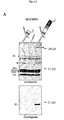
- Fig. 1A triple myc-tagged full-length human SUV39H1 (aa 3-412) or a C-terminally truncated SUV39H1 protein (aa 3-118) were immunoprecipitated from 'stably' transfected HeLa cell lines with anti-myc antibody beads and used in in vitro HMTase reactions with free histones as substrates and S-adenosyl-[ methyl - 14 C]-L-methionine as methyl donor.
- the Coomassie stain top panel shows purified proteins by arrowheads and free histones by dots. Fluorography (bottom panel, Figure 1A) indicates HMTase activity of (myc) 3 -SUV1(3-412).
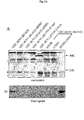
- the corresponding human SUV39H1 fusion protein [GST-SUV39H1(82-412)] is catalytically active (see Figure 2).
- Short internal deletions ⁇ NHSCDPN 323-329 ; ⁇ NHSC and ⁇ GEELTFDY 358-365 ; ⁇ GEEL
- ⁇ C-tail mutants that lack the C-terminal tail
- ⁇ cys mutants that lack the SET-associated cysteine-rich region
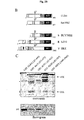
- EZH2 lacks the C-terminal cysteines, and HRX does not contain the SET associated cysteine-rich region (Figure 2B). Both of these cysteine domains are present in CLR4, Suv39h2 and SUV39H1. In agreement with the mutational analysis of SUV39H1, it thus appears that HMTase activity towards free histones requires the combination of the SET domain with adjacent cysteine-rich regions, and is a quality found in only a restricted number of SET domain containing proteins.
- Fig. 2 shows a diagram representing the domain structures of CLR4, Suv39h2, SUV39H1, EZH2 and HRX proteins, with the arrowheads demarcating the N-terminal fusion to GST. Cysteine-rich regions are indicated by grey stippling.
- Fig. 2C approximately 10 ⁇ g of the indicated fusion proteins encoding S.pombe CLR4 [GST-CLR4(127-490)], murine Suv39h2 [GST-Suv2(157-477)], human EZH2 [GST-EZH2(382-747)], human HRX [GST-HRX(3643-3969)] and human SUV39H1 [GST-SUV1(82-402)] were used in in vitro HMTase reactions with free histones as outlined in Figure 1.
- Lysine 9 of the H3 N-terminus is the major site for in vitro methylation by recombinant Suv39h1
- HMTase activity of mammalian SU(VAR)3-9 related proteins is selective for H3 under the chosen assay conditions.
- in vitro methylation reactions were performed with individual histones, using GST-Suv39h1(82-412) as an enzyme.
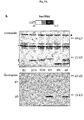
- H3 is specifically methylated by GST-Suv39h1 (82-412), whereas no signals are detected with H2A, H2B or H4.
- a weak signal is present if H1 was used as the sole substrate; the significance of H1 methylation remains to be determined.
- Methylation of H3 has been shown to occur predominantly at lysine 4 in a wide range of organisms, as well as at lysine 9 in HeLa cells, although the responsible HMTase(s) have yet to be defined (Strahl et al ., 1999).
- unmodified peptides comprising the wild-type H3 N-terminus (aa 1-20) and a mutant K9L peptide, changing lysine 9 to leucine were tested as substrates.
- insulin and peptides comprising the N-termini of CENP-A (Sullivan et al ., 1994) and macroH2A (Pehrson and Fried, 1992) were included.
- the wild-type H3 N-terminal peptide was in vitro methylated by GST-Suv39h1(82-412), using S-adenosyl-[ methyl - 3 H]-L-methionine.
- the labelled peptide purified by reverse-phase HPLC, was then directly microsequenced, and 3 H-incorporation associated with each individual amino acid was analysed by scintillation counting. The results confirmed selective transfer of methyl-label to lysine 9 (see Figure 3/2C), demonstrating that Suv39h1 is a highly site-specific HMTase for the H3 N-terminus in vitro .
- Fig. 3A approximately 10 ⁇ g of murine GST-Suv39h1(82-412) were used in in vitro HMTase reactions with individual histones as outlined in Figure 1.
- Fig 3B shows the results of in vitro methylation assays using GST-Suv39h1(82-412) as enzyme and the indicated N-terminal peptides of wild-type H3, mutated H3 (K9L), CENP-A, macroH2A or insulin as substrates.
- Fig 3C shows the result of automated sequencing of the wild-type H3 N-terminal peptide (aa 1-20) that had been methylated in vitro by recombinant GST-Suv39h1(82-412). Displayed is the 3 H-incorporation of individual amino acids identified at each successive round of microsequencing.
- Murine Suv39h genes are encoded by 2 loci, Suv39h1 and Suv39h2 (O'Carroll et al ., 2000).
- mouse strains deficient for both Suv39h1 and Suv39h2 were generated according to standard techniques. The targeting strategies are shown in Figure 4, as well as demonstrating the production of null alleles for both Suv39h1 and Suv39h2 . Mutation of either gene results in viable and fertile mice as a consequence of functional redundancy between both loci. Therefore, Suv39h1 and Suv39h2 deficient strains were intercrossed to produce Suv39h double null mice.
- FIG. 4 shows a conventional targeting strategy used to inactivate the X-linked Suv39h1 locus.
- Fig. 4B shows the Northern blot analysis of Suv39h1 from spleen (Sp), liver (Li), kidney (Kidney), and brain (Br) from wild-type and Suv39h1 null mice.
- Fig. 4C shows the conventional targeting strategy used to inactivate the autosomal Suv39h2 locus. (Bottom panel) Western blot analysis with ⁇ -Suv39h2 antibodies on protein extracts derived from wild-type and Suv39h2 null testis.
- PMEFs Suv39h double null primary mouse fibroblasts
- Suv39h double null PMEFs contain approximately two-fold elevated numbers of cells with micro- and polynuclei, and are further characterised by cell subpopulations with oversized nuclei or a weak definition of heterochromatin that appears in only a few unusually condensed foci (see Figure 5B).
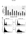
- Suv39h double null cells also show genomic instabilities and readily become aneuploid (see also Example 9, below). The severity of these aneuploidies increases with higher passage numbers (see Figure 6).
- the inability of Suv39h double null cells to maintain a stable karyotype may underlie the Suv39h embryonic phenotype.
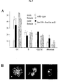
- Fig. 5A shows the percentages of cells in various cell cycle stages of wild-type and Suv39h double null PMEFs.
- Fig. 5B shows representative images (left and middle) of aberrant mitoses in Suv39h double null PMEFs detected by ⁇ -tubulin (not shown) and DAPI staining. Also shown (right image) is a nucleus exemplifying the unusual definition of heterochromatin in a subpopulation of Suv39h double null PMEFs. All images were taken at a magnification of 630x.
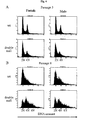
- Fig. 6A shows the DNA content profile of wild-type and Suv39h double null PMEFs at passage 3.
- Fig. 6B shows the DNA content profile of wild-type and Suv39h double null PMEFs at passage 8.
- phosH3 Phosphorylation at serine 10 in the N-terminal tail of H3 has been shown to be required for condensation and subsequent segregation of chromosomes (Wei et al ., 1999).
- phosH3 initiates within pericentric heterochromatin in late G2 and then progresses along the entire chromosomes during mitosis (Hendzel et al ., 1997).
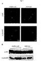
- wild-type PMEFs approximately 7% of the cells stain positive for the characteristic, heterochromatin-associated phosH3 foci, as detected by indirect immunofluorescence with ⁇ -phosH3 antibodies (see Figure 7A, right panel).
- Fig. 7A shows an interphase chromatin staining with ⁇ -phosH3 antibodies and CY3-conjugated secondary antibodies. DNA was counterstained with DAPI. At least 1,000 cells were counted to evaluate the percentage (indicated in the Figure) of ⁇ -phosH3 positive cells.
- Fig 7B shows a quantitative Western analysis with 15 ⁇ g and 30 ⁇ g of total nuclear proteins immuno-blotted with ⁇ -H3 and ⁇ -phosH3 antibodies.
- Murine Suv39h HMTases are encoded by two loci which have been mapped to centromere-proximal positions in the X chromosome ( Suv39h1 ) or in chromosome 2 ( Suv39h2 ) (O'Carroll et al., 2000). Both gene loci were independently disrupted by homologous recombination in embryonic stem (ES) cells using a conventional targeting approach that replaces parts of the evolutionarily conserved chromo domain with the bacterial LacZ gene and an RSV-neomycin selection cassette (Figure 9a).
- Suv39h1 or Suv39h2 display normal viability and fertility, and do not exhibit apparent phenotypes, suggesting that both genes may be functionally redundant during mouse development (O'Carroll et al ., 2000). Therefore, Suv39h1 -/- and Suv39h2 -/- mice were intercrossed to generate compound Suv39h mutants that were then used to derive Suv39h double null (dn) mice.
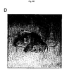
- Suv39h dn mice obtained from several different intercrosses (Table I) are born at only sub-Mendelian ratios, are growth retarded ( Figure 9d) and are characterized by hypogonadism in males.
- mice For example, from a total of 197 mice, 46 mice would have been expected to be double null (Table I), but only 15 Suv39h dn mice ( ⁇ 33 %) were born. Analysis of mouse embryogenesis indicated normal development of Suv39h dn fetuses until day E12.5, whereas at later stages, Suv39h dn fetuses are smaller and display an increased rate of resorptions and prenatal lethality. Together, these results demonstrate that the Suv39h genes are required for normal viability, and for pre- and postnatal development.
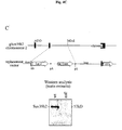
- Figure 9 shows the targeting and genotyping of Suv39h1 - and Suv39h2 -deficient mice as follows:
- A Diagrammatic representation of the Suv39h1 and Suv39h2 genomic loci, the replacement vectors and the targeted alleles. Exons are indicated by black boxes with numbers referring to the starting amino acid positions of the respective exons (O'Carroll et al ., 2000). Also shown are the diagnostic restriction sites and the external probes used for Southern blot analyses. pA indicates polyadenylation signals.
- B Southern blot analyses of Pvu II- or Hind III-digested DNA isolated from offspring of Suv39h1 +/- or Suv39h2 +/- heterozygous intercrosses.
- PMEFs primary mouse embryonic fibroblasts
- Suv39h dn PMEFs are largely hypo-tetraploid or hypo-octaploid. Chromosomes in Suv39h dn PMEFs appear of normal morphology and Robertsonian fusions were not observed. It was concluded that the absence of Suv39h function induces genomic instabilities, primarily by impairing segregation of the entire set of chromosomes.
- Suv39h mutant mice were analyzed for the incidence of tumorigenesis. Because the majority of Suv39h dn mice are non-viable, distinct Suv39h genotypes that differ in their gene dosage for either Suv39h1 or Suv39h2 were examined. For example, it was expected that random X-inactivation of the X-linked Suv39h1 gene could increase the tumor risk in Suv39h1 +/- mice, even in the presence of a functional copy of Suv39h2 which is significantly down-regulated in most adult tissues (O'Carroll et al ., 2000).
- mice which are either heterozygous (het) or null for the Suv39h1 locus indicated an ⁇ 28% penetrance of tumor formation with an onset between 9 - 15 months of age (Table II).
- These tumors are predominantly B-cell lymphomas ( Figure 11A) that resemble by FACS profiling (see Materials and Methods) slowly progressing non-Hodgin lymphomas in humans (Foon and Gale, 1995).
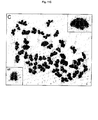
- Figure 11 shows the development of B-cell lymphomas in Suv39h mutant mice as follows: (A) Spleen and lymph nodes of an 11-month old Suv39h dn tumor mouse and of a wild-type control mouse. (B) Karyotype analysis of four independent primary cultures derived from the lymph nodes of tumor-bearing Suv39h dn (null1/null2) and Suv39h1 -/-, Suv39h +/- (null1/het2) mice. (C) Metaphase spread from a primary Suv39h dn B-cell lymphoma cell showing 'butterfly' chromosomes that remain associated through their acrocentric regions.
- Mitotic chromosome spreads from bone marrow cells were also analyzed with the ⁇ -methH3-K9 antiserum.
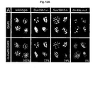
- pericentric heterochromatin was selectively visualised (see inserts in Figure 12B), whereas only residual staining was detected in Suv39h dn spreads.
- Suv39h enzymes are the major HMTases to methylate H3-K9 in pericentric heterochromatin of somatic cells.
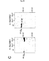
- Figure 12 shows the Suv39h -dependent H3-K9 methylation at pericentric heterochromatin as follows:
- A DAPI and methH3-K9 staining on interphase chromatin of wild-type (wt), Suv39h1 -/- , Suv39h2 -/-, and Suv39h dn PMEFs. Percentages refer to interphase nuclei displaying H3-K9 methylation at heterochromatic foci.
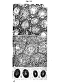
- Histological sections were performed, demonstrating normally developed seminiferous tubules in wt testis which display the characteristic differentiation from the mitotically proliferating spermatogonia (Sg) to meiotic spermatocytes (Sc) and the post-meiotic haploid spermatids (St) ( Figure 13A).
- spermatogenesis was severely impaired in Suv39h dn mice, with an apparent differentiation arrest at the transition between early to late spermatocytes, resulting in highly vacuolarized seminiferous tubules ( Figure 13A).
- stage V - VI tubules that largely lack late pachytene spermatocytes and which do not contain haploid spermatids. It was concluded that the absence of Suv39h gene function induces delayed entry into meiotic prophase and triggers pronounced apoptosis of spermatocytes during the mid- to late pachytene stage.
- testis spread preparations and cryosections were analyzed with the ⁇ -methH3-K9 antibodies.
- the ⁇ -methH3-K9 antibodies decorate heterochromatic foci in spermatogonia (B-Sg) and in pre-leptotene spermatocytes (preL-Sc) ( Figure 13B, left images, top panel).
- Zyg-Sc early meiotic prophase
- Zyg-Sc early pachytene
- the ⁇ -methH3-K9 staining was not exclusive for heterochromatin but also extended into euchromatin.
- H3-K9 methylation was absent in spermatogonia and pre-leptotene spermatocytes ( Figure 13B, left images, bottom panel). Further, the pronounced euchromatic staining that characterizes early spermatocytes (Zyg-Sc) at the onset of meiotic prophase was not observed. The impairment of H3-K9 methylation was accompanied by a dispersed distribution of phosH3 in ⁇ 60% of Suv39h dn spermatogonia. By contrast, HP1 ⁇ was largely undetectable in both wt and Suv39h dn spermatogonia.
- Figure 13 shows the spermatogenic failure and H3-K9 methylation in germ cells of Suv39h dn mice as follows:
- the Suv39h dn testis section reveals many seminiferous tubules that lack spermatocytes (Sc) and spermatids (St).
- Sc seminiferous tubules
- St spermatids
- a few seminiferous tubules (1) contain zygotene spermatocytes (Zyg-Sc)
- more advanced differentiation stages (2) display apoptotic spermatocytes (arrows) at pachytene.
- pachytene spermatocytes are almost completely absent.
- SeC Sertoli cells
- H3-K9 methylation appears in a wild-type staining (compare top and bottom panels), although Suv39h dn sex chromosomes (arrows) remain more intensely labeled at diplotene and diakinesis.
- the double arrow indicates the pseudo-autosomal region (PAR).
- Non-homologous interactions were even more frequent ( ⁇ 35%) between sex chromosomes and autosomes (X/Y-A).
- these illegitimate associations occurred predominantly between the acrocentric ends (cen-cen) of non-homologous chromosomes, to a lesser extent between centromeres and telomeres (cen-tel) and only very rarely between telomeres (tel-tel) ( Figure 14J).
- Suv39h dn spermatocytes contained unsynapsed sex chromosomes (see below) and autosomal bivalents that were delayed in synapsis. Delayed synapsis of autosomes (A-del) almost invariably was correlated with engagement in non-homologous associations ( Figure 14A), suggesting that both processes may be functionally related.
- testis spread preparations were next analyzed for diakinesis/metaphase I (M-I) and metaphase II (M-II) cells.
- M-I diakinesis/metaphase I
- M-II metaphase II
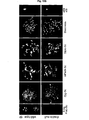
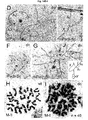
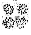
- Figure 14 shows the illegitimate associations and delayed synapsis of Suv39h dn meiotic chromosomes as follows: (A-C) Double-labelling immunofluorescence of Suv39h dn pachytene spermatocytes with antibodies that are specific for the axial/lateral elements ⁇ -Scp3 (in green) and central elements ⁇ -Scp1 (in red) of the synaptonemal complex (SC). This co-labelling reveals unsynapsed chromosomes in a green-like staining and synapsed chromosomes in an orange-red colour. DNA was counterstained with DAPI (blue) which highlights pericentric heterochromatin in a more intense blue contrast.
- A-C Double-labelling immunofluorescence of Suv39h dn pachytene spermatocytes with antibodies that are specific for the axial/lateral elements ⁇ -Scp3 (in green) and central elements ⁇ -Scp1 (in red
- A Two mid-pachytene spermatocytes (mPach-Sc) showing multiple illegitimate associations (arrowheads) between non-homologous autosomes (A) and between autosomes and sex chromomes (X, Y). Several autosomes are also delayed in synapsis (A del ).
- B Late pachytene (IPach-Sc) spermatocyte containing two autosomes which are engaged in non-homologous interaction through their pericentric regions (arrowhead). In addition, the sex chromosomes failed to pair.
- C Tetraploid spermatocyte resulting from complete mis-segregation of all chromosomes in the preceding mitotic division of a Suv39h dn spermatogonium.
- sex chromosomes which form a unique chromatin region known as the sex vesicle or XY body (Solari, 1974).
- Y chromosome is the most heterochromatic chromosome in the mouse (Pardue and Gall, 1970).
- Homolog pairing and cross-over between sex chromosomes is dependent upon the presence of a small, pseudo-autosomal region called PAR (Burgoyne, 1982).
- the absence of Suv39h function interferes with the chromatin organization and segregation of the sex chromosomes in several ways.
- This reaction is then left at room temperature for 40 mins to allow the antibody to bind methylated substrate.
- unbound non-methylated substrate is washed off in 50 mM tris pH 8.5.
- the europium label is then cleaved from the peptide in 50 ⁇ l pH 4.5 enhancement solution for 25 mins.
- the chelated europium molecules are then excited at 360nm and the level of emitted fluorescence at 620nm is then calculated using time-resolved fluorescence in a PolarStar plate reader. The results are then automatically graphed.
- the level of fluorescence is directly related to the level of MTase activity.
- the effect of the different compounds on the MTase activity can be clearly seen on the graph when compared to control reactions with no compounds added or with no enzyme added.
- FIG 17 illustrates the principle of the screening method as follows:
Landscapes
- Health & Medical Sciences (AREA)
- Life Sciences & Earth Sciences (AREA)
- Chemical & Material Sciences (AREA)
- Organic Chemistry (AREA)
- Engineering & Computer Science (AREA)
- General Health & Medical Sciences (AREA)
- Medicinal Chemistry (AREA)
- Molecular Biology (AREA)
- Proteomics, Peptides & Aminoacids (AREA)
- Genetics & Genomics (AREA)
- Immunology (AREA)
- Bioinformatics & Cheminformatics (AREA)
- Biochemistry (AREA)
- Wood Science & Technology (AREA)
- Zoology (AREA)
- Biomedical Technology (AREA)
- Biotechnology (AREA)
- Microbiology (AREA)
- Animal Behavior & Ethology (AREA)
- General Chemical & Material Sciences (AREA)
- Physics & Mathematics (AREA)
- Veterinary Medicine (AREA)
- Hematology (AREA)
- Urology & Nephrology (AREA)
- Public Health (AREA)
- Analytical Chemistry (AREA)
- Chemical Kinetics & Catalysis (AREA)
- Pharmacology & Pharmacy (AREA)
- Biophysics (AREA)
- Nuclear Medicine, Radiotherapy & Molecular Imaging (AREA)
- General Engineering & Computer Science (AREA)
- Reproductive Health (AREA)
- Food Science & Technology (AREA)
- Pathology (AREA)
- General Physics & Mathematics (AREA)
- Endocrinology (AREA)
- Cell Biology (AREA)
- Measuring Or Testing Involving Enzymes Or Micro-Organisms (AREA)
- Enzymes And Modification Thereof (AREA)
- Medicines That Contain Protein Lipid Enzymes And Other Medicines (AREA)
Claims (12)
- Verfahren zur Identifizierung einer Verbindung, welche eine von Chromatin höherer Ordnung abhängige Chromosomenstabilität bei der Mitose und Meiose ändert, wobei das Verfahren umfasst das Inkubieren eines Substrats für eine Histon H3 K9-Methyltransferase in Anwesenheit eines Methyldonors mit einer Methyltransferase mit Suv39h-artiger Histon H3 K9-Methyltransferase-Aktivität in Anwesenheit und in Abwesenheit einer Testverbindung und das Bestimmen, ob die Testverbindung die Methyltransferase-Aktivität moduliert.
- Verfahren nach Anspruch 1, in dem die Methyltransferase murines Suv39h1 oder humanes SUV39H1 ist.
- Verfahren nach Anspruch 1, in dem die Methyltransferase murines Suv39h2 oder humanes SUV39H2 ist.
- Verfahren nach irgendeinem der Ansprüche 1 bis 3, in dem das Substrat Histon H3 oder ein N-terminales Fragment desselben ist, welches die Methylierungsstelle am Lysin 9 enthält.
- Verfahren nach Anspruch 4, in dem das N-terminale Histon-H3-Fragment die in SEQ ID NO: 7 angegebene Aminosäuresequenz aufweist.
- Verfahren nach irgendeinem der Ansprüche 1 bis 5, in dem der Methyldonor Methionin oder S-Adenosyl-L-methionin ist.
- Verfahren nach irgendeinem der Ansprüche 1 bis 6, in dem die Methylgruppe des Methyldonors eine detektierbare Markierung trägt.
- Verfahren nach Anspruch 7, in dem der Methyldonor eine chromogene Markierung trägt und die Methyltransferase-Aktivität durch Messen der Änderung der Farbe nach Übertragung der Methylgruppe auf das Substrat bestimmt wird.
- Verfahren nach Anspruch 7, in dem der Methyldonor eine radioaktive Markierung trägt und die Methyltransferase-Aktivität durch Messen der Radioaktivität bestimmt wird, die nach Übertragung der Methylgruppe auf das Substrat übertragen ist.
- Verfahren nach irgendeinem der Ansprüche 1 bis 6, in dem die Methyltransferase-Aktivität immunologisch durch Quantifizierung der Bindung eines Antikörpers bestimmt wird, der für die Methylierungsstelle des Substrats spezifisch ist.
- Verfahren nach Anspruch 10, in dem das Substrat eine detektierbare Markierung trägt.
- Antikörper, der methyliertes Lysin 9 in Histon H3 spezifisch erkennt.
Priority Applications (1)
| Application Number | Priority Date | Filing Date | Title |
|---|---|---|---|
| EP01940552A EP1294927B1 (de) | 2000-06-09 | 2001-06-07 | Screeningverfahren für modulatoren von methyltransferase abhängiger chromosomenstabilität |
Applications Claiming Priority (6)
| Application Number | Priority Date | Filing Date | Title |
|---|---|---|---|
| EP00112479A EP1162460A1 (de) | 2000-06-09 | 2000-06-09 | Screeningverfahren für Modulatoren des Suv39h2 Methyltransferases und der männlichen Gametogenese |
| EP00112345 | 2000-06-09 | ||
| EP00112345A EP1162274A1 (de) | 2000-06-09 | 2000-06-09 | Verfahren zur Identifizierung von Verbindungen, die die Methyltransferase-abhängige Chromosomen-Stabilität beeinflussen |
| EP00112479 | 2000-06-09 | ||
| PCT/EP2001/006485 WO2001094620A1 (en) | 2000-06-09 | 2001-06-07 | Screening-method for modulators of methyltransferase-dependent chromosome stability |
| EP01940552A EP1294927B1 (de) | 2000-06-09 | 2001-06-07 | Screeningverfahren für modulatoren von methyltransferase abhängiger chromosomenstabilität |
Publications (2)
| Publication Number | Publication Date |
|---|---|
| EP1294927A1 EP1294927A1 (de) | 2003-03-26 |
| EP1294927B1 true EP1294927B1 (de) | 2005-11-16 |
Family
ID=26071033
Family Applications (2)
| Application Number | Title | Priority Date | Filing Date |
|---|---|---|---|
| EP01940552A Expired - Lifetime EP1294927B1 (de) | 2000-06-09 | 2001-06-07 | Screeningverfahren für modulatoren von methyltransferase abhängiger chromosomenstabilität |
| EP01943488A Withdrawn EP1294928A1 (de) | 2000-06-09 | 2001-06-08 | Screeningverfahren für modulatoren des suv39h2-methyltransferases |
Family Applications After (1)
| Application Number | Title | Priority Date | Filing Date |
|---|---|---|---|
| EP01943488A Withdrawn EP1294928A1 (de) | 2000-06-09 | 2001-06-08 | Screeningverfahren für modulatoren des suv39h2-methyltransferases |
Country Status (7)
| Country | Link |
|---|---|
| EP (2) | EP1294927B1 (de) |
| JP (2) | JP4896344B2 (de) |
| AT (1) | ATE310100T1 (de) |
| AU (2) | AU2001274088A1 (de) |
| DE (1) | DE60115039T2 (de) |
| ES (1) | ES2252240T3 (de) |
| WO (2) | WO2001094620A1 (de) |
Families Citing this family (5)
| Publication number | Priority date | Publication date | Assignee | Title |
|---|---|---|---|---|
| US20050106152A1 (en) * | 2001-12-27 | 2005-05-19 | Yuichi Hikichi | Preventives/remedies for cancer |
| GB0301450D0 (en) * | 2003-01-22 | 2003-02-19 | Medical Res Council | Protein crystal structure |
| JP3913181B2 (ja) * | 2003-02-06 | 2007-05-09 | 三菱重工業株式会社 | カーボンナノファイバの製造方法及び製造装置 |
| WO2008114709A1 (ja) * | 2007-03-15 | 2008-09-25 | Reverse Proteomics Research Institute Co., Ltd. | 血球系に特異的あるいは破骨細胞分化に特異的なバイオマーカー |
| EP2714903B1 (de) * | 2011-06-03 | 2018-03-21 | OncoTherapy Science, Inc. | Suv39h2 als zielgen für krebstherapie |
Family Cites Families (1)
| Publication number | Priority date | Publication date | Assignee | Title |
|---|---|---|---|---|
| AU751784B2 (en) * | 1997-08-29 | 2002-08-29 | Innogenetics N.V. | Methylated, SmD homologous peptides, reactive with the antibodies from sera of living beings affected with systemic lupus erythematosus |
-
2001
- 2001-06-07 DE DE60115039T patent/DE60115039T2/de not_active Expired - Lifetime
- 2001-06-07 WO PCT/EP2001/006485 patent/WO2001094620A1/en not_active Ceased
- 2001-06-07 JP JP2002502160A patent/JP4896344B2/ja not_active Expired - Lifetime
- 2001-06-07 EP EP01940552A patent/EP1294927B1/de not_active Expired - Lifetime
- 2001-06-07 AT AT01940552T patent/ATE310100T1/de active
- 2001-06-07 ES ES01940552T patent/ES2252240T3/es not_active Expired - Lifetime
- 2001-06-07 AU AU2001274088A patent/AU2001274088A1/en not_active Abandoned
- 2001-06-08 JP JP2002502161A patent/JP2003535596A/ja active Pending
- 2001-06-08 WO PCT/EP2001/006520 patent/WO2001094621A1/en not_active Ceased
- 2001-06-08 EP EP01943488A patent/EP1294928A1/de not_active Withdrawn
- 2001-06-08 AU AU2001266055A patent/AU2001266055A1/en not_active Abandoned
Also Published As
| Publication number | Publication date |
|---|---|
| JP4896344B2 (ja) | 2012-03-14 |
| ES2252240T3 (es) | 2006-05-16 |
| AU2001274088A1 (en) | 2001-12-17 |
| JP2003535595A (ja) | 2003-12-02 |
| DE60115039T2 (de) | 2006-07-27 |
| JP2003535596A (ja) | 2003-12-02 |
| EP1294927A1 (de) | 2003-03-26 |
| DE60115039D1 (de) | 2005-12-22 |
| WO2001094621A1 (en) | 2001-12-13 |
| ATE310100T1 (de) | 2005-12-15 |
| EP1294928A1 (de) | 2003-03-26 |
| AU2001266055A1 (en) | 2001-12-17 |
| WO2001094620A1 (en) | 2001-12-13 |
Similar Documents
| Publication | Publication Date | Title |
|---|---|---|
| US6555329B2 (en) | Method for identifying compounds altering higher-order chromatin-dependent chromosome stability | |
| Nekrasov et al. | Pcl-PRC2 is needed to generate high levels of H3-K27 trimethylation at Polycomb target genes | |
| Pasini et al. | Suz12 is essential for mouse development and for EZH2 histone methyltransferase activity | |
| Roguev et al. | The Saccharomyces cerevisiae Set1 complex includes an Ash2 homologue and methylates histone 3 lysine 4 | |
| Marklund et al. | The phenotype of a “Cdc2 kinase target site-deficient” mutant of oncoprotein 18 reveals a role of this protein in cell cycle control. | |
| Adhvaryu et al. | Methylation of histone H3 lysine 36 is required for normal development in Neurospora crassa | |
| US9534259B2 (en) | DOT1 histone methyltransferases as a target for identifying therapeutic agents for leukemia | |
| Mis et al. | Identification of three histone methyltransferases in Drosophila: dG9a is a suppressor of PEV and is required for gene silencing | |
| US7052889B2 (en) | Mammalian SUV39H2 proteins and isolated DNA molecules encoding them | |
| US20100144543A1 (en) | Epigenetic silencing of tumor suppressor genes | |
| US6534056B1 (en) | Therapeutic and diagnostic uses of protein tyrosine phosphatase TC-PTP | |
| JP4896344B2 (ja) | メチルトランスフェラーゼ依存性染色体安定性の調節因子のスクリーニング方法 | |
| US6753158B1 (en) | Assays, agents, therapy and diagnosis relating to modulation of cellular DNA repair activity | |
| EP1162274A1 (de) | Verfahren zur Identifizierung von Verbindungen, die die Methyltransferase-abhängige Chromosomen-Stabilität beeinflussen | |
| EP1162460A1 (de) | Screeningverfahren für Modulatoren des Suv39h2 Methyltransferases und der männlichen Gametogenese | |
| Braun | Characterization of the telomere binding protein ZNF524 | |
| BHAT | CROSSTALK OF CHROMATIN MODIFIERS IN THE REGULATION OF GENE EXPRESSION AND GENOME INTEGRITY Thèse présentée | |
| Carter et al. | The DNA-activated protein kinase, DNA-PK | |
| Kim | The role of protein methylation as a modifier of cellular pathways | |
| Tan et al. | Control of the DNA Methylation System | |
| Cao | Identification and characterization of human EED-EZH2 complex: A crucial player in polycomb-group silencing through lysine 27 methylation on histone H3 | |
| Polager | E2Fs and their regulated genes are involved in the DNA damage response | |
| Thompson | CHD8, a novel ATP-dependent chromatin remodeling enzyme | |
| Fyodorov | Protein Complex of Drosophila ATRX/XNP and HP1a Is Required for the Formation of Pericentric Beta-heterochromatin in Vivo | |
| Mottus | A genetic and molecular analysis of histone deacetylase one in Drosophila melanogaster: specific missense mutations suppress position effect variegation |
Legal Events
| Date | Code | Title | Description |
|---|---|---|---|
| PUAI | Public reference made under article 153(3) epc to a published international application that has entered the european phase |
Free format text: ORIGINAL CODE: 0009012 |
|
| 17P | Request for examination filed |
Effective date: 20030109 |
|
| AK | Designated contracting states |
Kind code of ref document: A1 Designated state(s): AT BE CH CY DE DK ES FI FR GB GR IE IT LI LU MC NL PT SE TR |
|
| AX | Request for extension of the european patent |
Extension state: AL LT LV MK RO SI |
|
| 17Q | First examination report despatched |
Effective date: 20040517 |
|
| GRAP | Despatch of communication of intention to grant a patent |
Free format text: ORIGINAL CODE: EPIDOSNIGR1 |
|
| GRAS | Grant fee paid |
Free format text: ORIGINAL CODE: EPIDOSNIGR3 |
|
| GRAA | (expected) grant |
Free format text: ORIGINAL CODE: 0009210 |
|
| AK | Designated contracting states |
Kind code of ref document: B1 Designated state(s): AT BE CH CY DE DK ES FI FR GB GR IE IT LI LU MC NL PT SE TR |
|
| PG25 | Lapsed in a contracting state [announced via postgrant information from national office to epo] |
Ref country code: BE Free format text: LAPSE BECAUSE OF FAILURE TO SUBMIT A TRANSLATION OF THE DESCRIPTION OR TO PAY THE FEE WITHIN THE PRESCRIBED TIME-LIMIT Effective date: 20051116 Ref country code: NL Free format text: LAPSE BECAUSE OF FAILURE TO SUBMIT A TRANSLATION OF THE DESCRIPTION OR TO PAY THE FEE WITHIN THE PRESCRIBED TIME-LIMIT Effective date: 20051116 Ref country code: FI Free format text: LAPSE BECAUSE OF FAILURE TO SUBMIT A TRANSLATION OF THE DESCRIPTION OR TO PAY THE FEE WITHIN THE PRESCRIBED TIME-LIMIT Effective date: 20051116 |
|
| REG | Reference to a national code |
Ref country code: GB Ref legal event code: FG4D |
|
| RIN1 | Information on inventor provided before grant (corrected) |
Inventor name: REA, STEPHEN Inventor name: EISENHABER, FRANK Inventor name: JENUWEIN, THOMAS Inventor name: O'CARROLL, DONAL |
|
| REG | Reference to a national code |
Ref country code: CH Ref legal event code: EP |
|
| REG | Reference to a national code |
Ref country code: IE Ref legal event code: FG4D |
|
| REF | Corresponds to: |
Ref document number: 60115039 Country of ref document: DE Date of ref document: 20051222 Kind code of ref document: P |
|
| REG | Reference to a national code |
Ref country code: CH Ref legal event code: NV Representative=s name: SCHMAUDER & PARTNER AG PATENTANWALTSBUERO |
|
| PG25 | Lapsed in a contracting state [announced via postgrant information from national office to epo] |
Ref country code: SE Free format text: LAPSE BECAUSE OF FAILURE TO SUBMIT A TRANSLATION OF THE DESCRIPTION OR TO PAY THE FEE WITHIN THE PRESCRIBED TIME-LIMIT Effective date: 20060216 Ref country code: GR Free format text: LAPSE BECAUSE OF FAILURE TO SUBMIT A TRANSLATION OF THE DESCRIPTION OR TO PAY THE FEE WITHIN THE PRESCRIBED TIME-LIMIT Effective date: 20060216 Ref country code: DK Free format text: LAPSE BECAUSE OF FAILURE TO SUBMIT A TRANSLATION OF THE DESCRIPTION OR TO PAY THE FEE WITHIN THE PRESCRIBED TIME-LIMIT Effective date: 20060216 |
|
| PG25 | Lapsed in a contracting state [announced via postgrant information from national office to epo] |
Ref country code: PT Free format text: LAPSE BECAUSE OF FAILURE TO SUBMIT A TRANSLATION OF THE DESCRIPTION OR TO PAY THE FEE WITHIN THE PRESCRIBED TIME-LIMIT Effective date: 20060417 |
|
| NLV1 | Nl: lapsed or annulled due to failure to fulfill the requirements of art. 29p and 29m of the patents act | ||
| REG | Reference to a national code |
Ref country code: ES Ref legal event code: FG2A Ref document number: 2252240 Country of ref document: ES Kind code of ref document: T3 |
|
| PG25 | Lapsed in a contracting state [announced via postgrant information from national office to epo] |
Ref country code: IE Free format text: LAPSE BECAUSE OF NON-PAYMENT OF DUE FEES Effective date: 20060607 |
|
| PG25 | Lapsed in a contracting state [announced via postgrant information from national office to epo] |
Ref country code: MC Free format text: LAPSE BECAUSE OF NON-PAYMENT OF DUE FEES Effective date: 20060630 |
|
| ET | Fr: translation filed | ||
| PLBE | No opposition filed within time limit |
Free format text: ORIGINAL CODE: 0009261 |
|
| STAA | Information on the status of an ep patent application or granted ep patent |
Free format text: STATUS: NO OPPOSITION FILED WITHIN TIME LIMIT |
|
| 26N | No opposition filed |
Effective date: 20060817 |
|
| REG | Reference to a national code |
Ref country code: IE Ref legal event code: MM4A |
|
| PG25 | Lapsed in a contracting state [announced via postgrant information from national office to epo] |
Ref country code: LU Free format text: LAPSE BECAUSE OF NON-PAYMENT OF DUE FEES Effective date: 20060607 Ref country code: TR Free format text: LAPSE BECAUSE OF FAILURE TO SUBMIT A TRANSLATION OF THE DESCRIPTION OR TO PAY THE FEE WITHIN THE PRESCRIBED TIME-LIMIT Effective date: 20051116 |
|
| PG25 | Lapsed in a contracting state [announced via postgrant information from national office to epo] |
Ref country code: CY Free format text: LAPSE BECAUSE OF FAILURE TO SUBMIT A TRANSLATION OF THE DESCRIPTION OR TO PAY THE FEE WITHIN THE PRESCRIBED TIME-LIMIT Effective date: 20051116 |
|
| REG | Reference to a national code |
Ref country code: CH Ref legal event code: PCAR Free format text: SCHMAUDER & PARTNER AG PATENT- UND MARKENANWAELTE VSP;ZWAENGIWEG 7;8038 ZUERICH (CH) |
|
| REG | Reference to a national code |
Ref country code: FR Ref legal event code: PLFP Year of fee payment: 15 |
|
| REG | Reference to a national code |
Ref country code: FR Ref legal event code: PLFP Year of fee payment: 16 |
|
| REG | Reference to a national code |
Ref country code: FR Ref legal event code: PLFP Year of fee payment: 17 |
|
| REG | Reference to a national code |
Ref country code: FR Ref legal event code: PLFP Year of fee payment: 18 |
|
| PGFP | Annual fee paid to national office [announced via postgrant information from national office to epo] |
Ref country code: FR Payment date: 20200619 Year of fee payment: 20 Ref country code: CH Payment date: 20200618 Year of fee payment: 20 Ref country code: DE Payment date: 20200618 Year of fee payment: 20 |
|
| PGFP | Annual fee paid to national office [announced via postgrant information from national office to epo] |
Ref country code: GB Payment date: 20200625 Year of fee payment: 20 |
|
| PGFP | Annual fee paid to national office [announced via postgrant information from national office to epo] |
Ref country code: AT Payment date: 20200619 Year of fee payment: 20 |
|
| PGFP | Annual fee paid to national office [announced via postgrant information from national office to epo] |
Ref country code: ES Payment date: 20200824 Year of fee payment: 20 |
|
| PGFP | Annual fee paid to national office [announced via postgrant information from national office to epo] |
Ref country code: IT Payment date: 20200625 Year of fee payment: 20 |
|
| REG | Reference to a national code |
Ref country code: DE Ref legal event code: R071 Ref document number: 60115039 Country of ref document: DE |
|
| REG | Reference to a national code |
Ref country code: CH Ref legal event code: PL |
|
| REG | Reference to a national code |
Ref country code: GB Ref legal event code: PE20 Expiry date: 20210606 |
|
| REG | Reference to a national code |
Ref country code: AT Ref legal event code: MK07 Ref document number: 310100 Country of ref document: AT Kind code of ref document: T Effective date: 20210607 |
|
| PG25 | Lapsed in a contracting state [announced via postgrant information from national office to epo] |
Ref country code: GB Free format text: LAPSE BECAUSE OF EXPIRATION OF PROTECTION Effective date: 20210606 |
|
| PG25 | Lapsed in a contracting state [announced via postgrant information from national office to epo] |
Ref country code: ES Free format text: LAPSE BECAUSE OF EXPIRATION OF PROTECTION Effective date: 20210608 |