EP1250091B1 - Hydrophilic cyanine dyes - Google Patents
Hydrophilic cyanine dyes Download PDFInfo
- Publication number
- EP1250091B1 EP1250091B1 EP01904882A EP01904882A EP1250091B1 EP 1250091 B1 EP1250091 B1 EP 1250091B1 EP 01904882 A EP01904882 A EP 01904882A EP 01904882 A EP01904882 A EP 01904882A EP 1250091 B1 EP1250091 B1 EP 1250091B1
- Authority
- EP
- European Patent Office
- Prior art keywords
- och
- conh
- nhco
- independently
- independently selected
- Prior art date
- Legal status (The legal status is an assumption and is not a legal conclusion. Google has not performed a legal analysis and makes no representation as to the accuracy of the status listed.)
- Expired - Lifetime
Links
- ANRHNWWPFJCPAZ-UHFFFAOYSA-M thionine Chemical compound [Cl-].C1=CC(N)=CC2=[S+]C3=CC(N)=CC=C3N=C21 ANRHNWWPFJCPAZ-UHFFFAOYSA-M 0.000 title claims abstract 4
- 239000000975 dye Substances 0.000 title abstract description 101
- 108090000765 processed proteins & peptides Proteins 0.000 claims abstract description 50
- 206010028980 Neoplasm Diseases 0.000 claims abstract description 49
- IAZDPXIOMUYVGZ-UHFFFAOYSA-N Dimethylsulphoxide Chemical compound CS(C)=O IAZDPXIOMUYVGZ-UHFFFAOYSA-N 0.000 claims abstract description 26
- 230000000975 bioactive effect Effects 0.000 claims abstract description 19
- 238000003384 imaging method Methods 0.000 claims abstract description 19
- 150000001720 carbohydrates Chemical class 0.000 claims abstract description 18
- 229940079593 drug Drugs 0.000 claims abstract description 17
- 239000003814 drug Substances 0.000 claims abstract description 17
- 150000001875 compounds Chemical class 0.000 claims abstract description 16
- 238000002560 therapeutic procedure Methods 0.000 claims abstract description 15
- 238000001514 detection method Methods 0.000 claims abstract description 14
- 229940088597 hormone Drugs 0.000 claims abstract description 9
- 239000005556 hormone Substances 0.000 claims abstract description 9
- 239000000203 mixture Substances 0.000 claims description 71
- 238000000034 method Methods 0.000 claims description 35
- 125000003178 carboxy group Chemical group [H]OC(*)=O 0.000 claims description 33
- IJGRMHOSHXDMSA-UHFFFAOYSA-N Atomic nitrogen Chemical compound N#N IJGRMHOSHXDMSA-UHFFFAOYSA-N 0.000 claims description 30
- 239000003795 chemical substances by application Substances 0.000 claims description 22
- 229910052739 hydrogen Inorganic materials 0.000 claims description 18
- 239000001257 hydrogen Substances 0.000 claims description 18
- DQJCDTNMLBYVAY-ZXXIYAEKSA-N (2S,5R,10R,13R)-16-{[(2R,3S,4R,5R)-3-{[(2S,3R,4R,5S,6R)-3-acetamido-4,5-dihydroxy-6-(hydroxymethyl)oxan-2-yl]oxy}-5-(ethylamino)-6-hydroxy-2-(hydroxymethyl)oxan-4-yl]oxy}-5-(4-aminobutyl)-10-carbamoyl-2,13-dimethyl-4,7,12,15-tetraoxo-3,6,11,14-tetraazaheptadecan-1-oic acid Chemical compound NCCCC[C@H](C(=O)N[C@@H](C)C(O)=O)NC(=O)CC[C@H](C(N)=O)NC(=O)[C@@H](C)NC(=O)C(C)O[C@@H]1[C@@H](NCC)C(O)O[C@H](CO)[C@H]1O[C@H]1[C@H](NC(C)=O)[C@@H](O)[C@H](O)[C@@H](CO)O1 DQJCDTNMLBYVAY-ZXXIYAEKSA-N 0.000 claims description 15
- MYMOFIZGZYHOMD-UHFFFAOYSA-N Dioxygen Chemical compound O=O MYMOFIZGZYHOMD-UHFFFAOYSA-N 0.000 claims description 15
- 102000002068 Glycopeptides Human genes 0.000 claims description 15
- 108010015899 Glycopeptides Proteins 0.000 claims description 15
- 125000003545 alkoxy group Chemical group 0.000 claims description 15
- 125000004103 aminoalkyl group Chemical group 0.000 claims description 15
- 239000008280 blood Substances 0.000 claims description 15
- 210000004369 blood Anatomy 0.000 claims description 15
- 125000002837 carbocyclic group Chemical group 0.000 claims description 15
- 239000002738 chelating agent Substances 0.000 claims description 15
- 150000004696 coordination complex Chemical class 0.000 claims description 15
- 125000000623 heterocyclic group Chemical group 0.000 claims description 15
- 229910052751 metal Inorganic materials 0.000 claims description 15
- 239000002184 metal Substances 0.000 claims description 15
- 150000002772 monosaccharides Chemical class 0.000 claims description 15
- 229910052757 nitrogen Inorganic materials 0.000 claims description 15
- 229920001542 oligosaccharide Polymers 0.000 claims description 15
- 150000002482 oligosaccharides Chemical class 0.000 claims description 15
- 229910052760 oxygen Inorganic materials 0.000 claims description 15
- 239000001301 oxygen Substances 0.000 claims description 15
- 230000002285 radioactive effect Effects 0.000 claims description 15
- 229910052717 sulfur Inorganic materials 0.000 claims description 15
- 125000004434 sulfur atom Chemical group 0.000 claims description 15
- 125000000008 (C1-C10) alkyl group Chemical group 0.000 claims description 14
- CFODQUSMSYDHBS-UHFFFAOYSA-N octreotate Chemical group O=C1NC(CC=2C=CC=CC=2)C(=O)NC(CC=2[C]3C=CC=CC3=NC=2)C(=O)NC(CCCCN)C(=O)NC(C(C)O)C(=O)NC(C(=O)NC(C(O)C)C(O)=O)CSSCC1NC(=O)C(N)CC1=CC=CC=C1 CFODQUSMSYDHBS-UHFFFAOYSA-N 0.000 claims description 11
- 102000004169 proteins and genes Human genes 0.000 claims description 11
- 108090000623 proteins and genes Proteins 0.000 claims description 11
- 108010021625 Immunoglobulin Fragments Proteins 0.000 claims description 8
- 102000008394 Immunoglobulin Fragments Human genes 0.000 claims description 8
- 125000000217 alkyl group Chemical group 0.000 claims description 8
- 125000004093 cyano group Chemical group *C#N 0.000 claims description 8
- 229910052736 halogen Inorganic materials 0.000 claims description 8
- 150000002367 halogens Chemical class 0.000 claims description 8
- 230000003278 mimic effect Effects 0.000 claims description 8
- 125000000449 nitro group Chemical group [O-][N+](*)=O 0.000 claims description 8
- 239000000816 peptidomimetic Substances 0.000 claims description 8
- 238000001727 in vivo Methods 0.000 claims description 7
- 238000010521 absorption reaction Methods 0.000 claims description 6
- 238000000338 in vitro Methods 0.000 claims description 5
- 238000012544 monitoring process Methods 0.000 claims description 5
- 230000003287 optical effect Effects 0.000 claims description 5
- 238000010791 quenching Methods 0.000 claims description 5
- 230000000171 quenching effect Effects 0.000 claims description 5
- 238000012800 visualization Methods 0.000 claims description 5
- DNDCVAGJPBKION-DOPDSADYSA-N bombesin Chemical group C([C@@H](C(=O)N[C@@H](CC(C)C)C(=O)N[C@@H](CCSC)C(N)=O)NC(=O)CNC(=O)[C@@H](NC(=O)[C@H](C)NC(=O)[C@H](CC=1NC2=CC=CC=C2C=1)NC(=O)[C@H](CCC(N)=O)NC(=O)[C@H](CC(N)=O)NC(=O)CNC(=O)[C@H](CC(C)C)NC(=O)[C@H](CCCNC(N)=N)NC(=O)[C@H](CCC(N)=O)NC(=O)[C@H]1NC(=O)CC1)C(C)C)C1=CN=CN1 DNDCVAGJPBKION-DOPDSADYSA-N 0.000 claims description 3
- 239000000546 pharmaceutical excipient Substances 0.000 claims description 3
- 238000001356 surgical procedure Methods 0.000 claims description 3
- 108010051479 Bombesin Proteins 0.000 claims description 2
- 102000013585 Bombesin Human genes 0.000 claims description 2
- 208000003788 Neoplasm Micrometastasis Diseases 0.000 claims description 2
- 208000007536 Thrombosis Diseases 0.000 claims description 2
- 230000003213 activating effect Effects 0.000 claims description 2
- 238000001839 endoscopy Methods 0.000 claims description 2
- 239000003960 organic solvent Substances 0.000 claims description 2
- 238000002428 photodynamic therapy Methods 0.000 claims description 2
- 125000003275 alpha amino acid group Chemical group 0.000 claims 6
- 150000002431 hydrogen Chemical class 0.000 claims 6
- UFHFLCQGNIYNRP-UHFFFAOYSA-N Hydrogen Chemical compound [H][H] UFHFLCQGNIYNRP-UHFFFAOYSA-N 0.000 claims 4
- 238000002405 diagnostic procedure Methods 0.000 claims 4
- 125000004008 6 membered carbocyclic group Chemical group 0.000 claims 3
- 229930182830 galactose Natural products 0.000 claims 3
- 125000006736 (C6-C20) aryl group Chemical group 0.000 claims 2
- VZGJNCHEUPMLPM-DGKZTOLNSA-N C([C@@H](C(=O)N[C@@H](CC(C)C)C(=O)N[C@@H](CCSC)C(N)=O)NC(=O)CNC(=O)[C@@H](NC(=O)[C@H](C)NC(=O)[C@H](CC=1C2=CC=CC=C2NC=1)NC(=O)[C@@H](N)CCC(N)=O)C(C)C)C1=CN=CN1 Chemical group C([C@@H](C(=O)N[C@@H](CC(C)C)C(=O)N[C@@H](CCSC)C(N)=O)NC(=O)CNC(=O)[C@@H](NC(=O)[C@H](C)NC(=O)[C@H](CC=1C2=CC=CC=C2NC=1)NC(=O)[C@@H](N)CCC(N)=O)C(C)C)C1=CN=CN1 VZGJNCHEUPMLPM-DGKZTOLNSA-N 0.000 claims 2
- 238000000149 argon plasma sintering Methods 0.000 claims 2
- 108010062050 bombesin (7-14) Proteins 0.000 claims 2
- 239000003937 drug carrier Substances 0.000 claims 2
- 238000002835 absorbance Methods 0.000 claims 1
- 238000003325 tomography Methods 0.000 claims 1
- 102000004196 processed proteins & peptides Human genes 0.000 abstract description 6
- 230000005856 abnormality Effects 0.000 abstract description 4
- 210000004881 tumor cell Anatomy 0.000 abstract description 4
- 235000014633 carbohydrates Nutrition 0.000 abstract description 2
- 238000002059 diagnostic imaging Methods 0.000 abstract 2
- 239000012867 bioactive agent Substances 0.000 abstract 1
- 229940077731 carbohydrate nutrients Drugs 0.000 abstract 1
- 150000001732 carboxylic acid derivatives Chemical class 0.000 abstract 1
- 230000001419 dependent effect Effects 0.000 abstract 1
- 230000002349 favourable effect Effects 0.000 abstract 1
- 239000012216 imaging agent Substances 0.000 abstract 1
- NFHFRUOZVGFOOS-UHFFFAOYSA-N palladium;triphenylphosphane Chemical compound [Pd].C1=CC=CC=C1P(C=1C=CC=CC=1)C1=CC=CC=C1.C1=CC=CC=C1P(C=1C=CC=CC=1)C1=CC=CC=C1.C1=CC=CC=C1P(C=1C=CC=CC=1)C1=CC=CC=C1.C1=CC=CC=C1P(C=1C=CC=CC=1)C1=CC=CC=C1 NFHFRUOZVGFOOS-UHFFFAOYSA-N 0.000 abstract 1
- RTZKZFJDLAIYFH-UHFFFAOYSA-N Diethyl ether Chemical compound CCOCC RTZKZFJDLAIYFH-UHFFFAOYSA-N 0.000 description 32
- 241000700159 Rattus Species 0.000 description 31
- QGKMIGUHVLGJBR-UHFFFAOYSA-M (4z)-1-(3-methylbutyl)-4-[[1-(3-methylbutyl)quinolin-1-ium-4-yl]methylidene]quinoline;iodide Chemical group [I-].C12=CC=CC=C2N(CCC(C)C)C=CC1=CC1=CC=[N+](CCC(C)C)C2=CC=CC=C12 QGKMIGUHVLGJBR-UHFFFAOYSA-M 0.000 description 29
- YMWUJEATGCHHMB-UHFFFAOYSA-N Dichloromethane Chemical compound ClCCl YMWUJEATGCHHMB-UHFFFAOYSA-N 0.000 description 27
- ZMXDDKWLCZADIW-UHFFFAOYSA-N N,N-Dimethylformamide Chemical compound CN(C)C=O ZMXDDKWLCZADIW-UHFFFAOYSA-N 0.000 description 27
- 239000000562 conjugate Substances 0.000 description 26
- 239000000243 solution Substances 0.000 description 26
- WEVYAHXRMPXWCK-UHFFFAOYSA-N Acetonitrile Chemical compound CC#N WEVYAHXRMPXWCK-UHFFFAOYSA-N 0.000 description 24
- 230000015572 biosynthetic process Effects 0.000 description 22
- MOFVSTNWEDAEEK-UHFFFAOYSA-M indocyanine green Chemical compound [Na+].[O-]S(=O)(=O)CCCCN1C2=CC=C3C=CC=CC3=C2C(C)(C)C1=CC=CC=CC=CC1=[N+](CCCCS([O-])(=O)=O)C2=CC=C(C=CC=C3)C3=C2C1(C)C MOFVSTNWEDAEEK-UHFFFAOYSA-M 0.000 description 22
- 229960004657 indocyanine green Drugs 0.000 description 22
- 238000003786 synthesis reaction Methods 0.000 description 22
- 239000007924 injection Substances 0.000 description 21
- 238000002347 injection Methods 0.000 description 21
- 239000007787 solid Substances 0.000 description 21
- XLYOFNOQVPJJNP-UHFFFAOYSA-N water Substances O XLYOFNOQVPJJNP-UHFFFAOYSA-N 0.000 description 16
- 241001465754 Metazoa Species 0.000 description 14
- 150000001413 amino acids Chemical group 0.000 description 14
- 238000006243 chemical reaction Methods 0.000 description 13
- 208000006336 acinar cell carcinoma Diseases 0.000 description 11
- 108020003175 receptors Proteins 0.000 description 11
- 102000005962 receptors Human genes 0.000 description 11
- 210000001519 tissue Anatomy 0.000 description 11
- -1 succinimidyl esters Chemical class 0.000 description 10
- RFFLAFLAYFXFSW-UHFFFAOYSA-N 1,2-dichlorobenzene Chemical compound ClC1=CC=CC=C1Cl RFFLAFLAYFXFSW-UHFFFAOYSA-N 0.000 description 9
- 235000001014 amino acid Nutrition 0.000 description 9
- 239000007943 implant Substances 0.000 description 9
- 210000000056 organ Anatomy 0.000 description 9
- 239000000047 product Substances 0.000 description 9
- DTQVDTLACAAQTR-UHFFFAOYSA-N Trifluoroacetic acid Chemical compound OC(=O)C(F)(F)F DTQVDTLACAAQTR-UHFFFAOYSA-N 0.000 description 8
- 125000004435 hydrogen atom Chemical class [H]* 0.000 description 8
- 125000001997 phenyl group Chemical group [H]C1=C([H])C([H])=C(*)C([H])=C1[H] 0.000 description 8
- 235000018102 proteins Nutrition 0.000 description 7
- 239000011347 resin Substances 0.000 description 7
- 229920005989 resin Polymers 0.000 description 7
- LFQSCWFLJHTTHZ-UHFFFAOYSA-N Ethanol Chemical compound CCO LFQSCWFLJHTTHZ-UHFFFAOYSA-N 0.000 description 6
- SIKJAQJRHWYJAI-UHFFFAOYSA-N Indole Chemical compound C1=CC=C2NC=CC2=C1 SIKJAQJRHWYJAI-UHFFFAOYSA-N 0.000 description 6
- OKKJLVBELUTLKV-UHFFFAOYSA-N Methanol Chemical compound OC OKKJLVBELUTLKV-UHFFFAOYSA-N 0.000 description 6
- 210000004027 cell Anatomy 0.000 description 6
- 210000004185 liver Anatomy 0.000 description 6
- 230000037361 pathway Effects 0.000 description 6
- 239000002904 solvent Substances 0.000 description 6
- VEXZGXHMUGYJMC-UHFFFAOYSA-N Hydrochloric acid Chemical compound Cl VEXZGXHMUGYJMC-UHFFFAOYSA-N 0.000 description 5
- BZLVMXJERCGZMT-UHFFFAOYSA-N Methyl tert-butyl ether Chemical compound COC(C)(C)C BZLVMXJERCGZMT-UHFFFAOYSA-N 0.000 description 5
- 238000013459 approach Methods 0.000 description 5
- 239000002872 contrast media Substances 0.000 description 5
- 230000005284 excitation Effects 0.000 description 5
- 238000009472 formulation Methods 0.000 description 5
- 239000000543 intermediate Substances 0.000 description 5
- 230000008685 targeting Effects 0.000 description 5
- 125000003088 (fluoren-9-ylmethoxy)carbonyl group Chemical group 0.000 description 4
- XKRFYHLGVUSROY-UHFFFAOYSA-N Argon Chemical compound [Ar] XKRFYHLGVUSROY-UHFFFAOYSA-N 0.000 description 4
- JGFZNNIVVJXRND-UHFFFAOYSA-N N,N-Diisopropylethylamine (DIPEA) Chemical compound CCN(C(C)C)C(C)C JGFZNNIVVJXRND-UHFFFAOYSA-N 0.000 description 4
- 206010061902 Pancreatic neoplasm Diseases 0.000 description 4
- FAPWRFPIFSIZLT-UHFFFAOYSA-M Sodium chloride Chemical compound [Na+].[Cl-] FAPWRFPIFSIZLT-UHFFFAOYSA-M 0.000 description 4
- 102000005157 Somatostatin Human genes 0.000 description 4
- 108010056088 Somatostatin Proteins 0.000 description 4
- WYURNTSHIVDZCO-UHFFFAOYSA-N Tetrahydrofuran Chemical compound C1CCOC1 WYURNTSHIVDZCO-UHFFFAOYSA-N 0.000 description 4
- QTBSBXVTEAMEQO-UHFFFAOYSA-N acetic acid Substances CC(O)=O QTBSBXVTEAMEQO-UHFFFAOYSA-N 0.000 description 4
- NPZTUJOABDZTLV-UHFFFAOYSA-N hydroxybenzotriazole Substances O=C1C=CC=C2NNN=C12 NPZTUJOABDZTLV-UHFFFAOYSA-N 0.000 description 4
- 238000011694 lewis rat Methods 0.000 description 4
- VLKZOEOYAKHREP-UHFFFAOYSA-N n-Hexane Chemical compound CCCCCC VLKZOEOYAKHREP-UHFFFAOYSA-N 0.000 description 4
- 201000002528 pancreatic cancer Diseases 0.000 description 4
- 239000000863 peptide conjugate Substances 0.000 description 4
- 125000006239 protecting group Chemical group 0.000 description 4
- NHXLMOGPVYXJNR-ATOGVRKGSA-N somatostatin Chemical compound C([C@H]1C(=O)N[C@H](C(N[C@@H](CO)C(=O)N[C@@H](CSSC[C@@H](C(=O)N[C@@H](CCCCN)C(=O)N[C@@H](CC(N)=O)C(=O)N[C@@H](CC=2C=CC=CC=2)C(=O)N[C@@H](CC=2C=CC=CC=2)C(=O)N[C@@H](CC=2C3=CC=CC=C3NC=2)C(=O)N[C@@H](CCCCN)C(=O)N[C@H](C(=O)N1)[C@@H](C)O)NC(=O)CNC(=O)[C@H](C)N)C(O)=O)=O)[C@H](O)C)C1=CC=CC=C1 NHXLMOGPVYXJNR-ATOGVRKGSA-N 0.000 description 4
- 229960000553 somatostatin Drugs 0.000 description 4
- 239000006228 supernatant Substances 0.000 description 4
- HNKJADCVZUBCPG-UHFFFAOYSA-N thioanisole Chemical compound CSC1=CC=CC=C1 HNKJADCVZUBCPG-UHFFFAOYSA-N 0.000 description 4
- 210000003462 vein Anatomy 0.000 description 4
- WJZSZXCWMATYFX-UHFFFAOYSA-N 1,1,2-trimethylbenzo[e]indole Chemical compound C1=CC=CC2=C(C(C(C)=N3)(C)C)C3=CC=C21 WJZSZXCWMATYFX-UHFFFAOYSA-N 0.000 description 3
- ISWSIDIOOBJBQZ-UHFFFAOYSA-N Phenol Chemical compound OC1=CC=CC=C1 ISWSIDIOOBJBQZ-UHFFFAOYSA-N 0.000 description 3
- VYPSYNLAJGMNEJ-UHFFFAOYSA-N Silicium dioxide Chemical compound O=[Si]=O VYPSYNLAJGMNEJ-UHFFFAOYSA-N 0.000 description 3
- DTQVDTLACAAQTR-UHFFFAOYSA-M Trifluoroacetate Chemical compound [O-]C(=O)C(F)(F)F DTQVDTLACAAQTR-UHFFFAOYSA-M 0.000 description 3
- 230000002776 aggregation Effects 0.000 description 3
- 238000004220 aggregation Methods 0.000 description 3
- 239000007864 aqueous solution Substances 0.000 description 3
- STLZCUYBVPNYED-UHFFFAOYSA-N chlorbetamide Chemical compound OCCN(C(=O)C(Cl)Cl)CC1=CC=C(Cl)C=C1Cl STLZCUYBVPNYED-UHFFFAOYSA-N 0.000 description 3
- 230000003292 diminished effect Effects 0.000 description 3
- 239000000835 fiber Substances 0.000 description 3
- WFSXUTWNNVIIIG-ZPUQHVIOSA-N glutaconaldehyde Chemical compound O\C=C\C=C\C=O WFSXUTWNNVIIIG-ZPUQHVIOSA-N 0.000 description 3
- 238000004128 high performance liquid chromatography Methods 0.000 description 3
- 230000002209 hydrophobic effect Effects 0.000 description 3
- PZOUSPYUWWUPPK-UHFFFAOYSA-N indole Natural products CC1=CC=CC2=C1C=CN2 PZOUSPYUWWUPPK-UHFFFAOYSA-N 0.000 description 3
- RKJUIXBNRJVNHR-UHFFFAOYSA-N indolenine Natural products C1=CC=C2CC=NC2=C1 RKJUIXBNRJVNHR-UHFFFAOYSA-N 0.000 description 3
- 230000004807 localization Effects 0.000 description 3
- 239000000463 material Substances 0.000 description 3
- 229920000962 poly(amidoamine) Polymers 0.000 description 3
- 239000000843 powder Substances 0.000 description 3
- 238000007363 ring formation reaction Methods 0.000 description 3
- IZTQOLKUZKXIRV-YRVFCXMDSA-N sincalide Chemical class C([C@@H](C(=O)N[C@@H](CCSC)C(=O)NCC(=O)N[C@@H](CC=1C2=CC=CC=C2NC=1)C(=O)N[C@@H](CCSC)C(=O)N[C@@H](CC(O)=O)C(=O)N[C@@H](CC=1C=CC=CC=1)C(N)=O)NC(=O)[C@@H](N)CC(O)=O)C1=CC=C(OS(O)(=O)=O)C=C1 IZTQOLKUZKXIRV-YRVFCXMDSA-N 0.000 description 3
- 239000011780 sodium chloride Substances 0.000 description 3
- 238000003756 stirring Methods 0.000 description 3
- 150000000000 tetracarboxylic acids Chemical class 0.000 description 3
- 229910052716 thallium Inorganic materials 0.000 description 3
- BKVIYDNLLOSFOA-UHFFFAOYSA-N thallium Chemical compound [Tl] BKVIYDNLLOSFOA-UHFFFAOYSA-N 0.000 description 3
- 238000005160 1H NMR spectroscopy Methods 0.000 description 2
- BDKLKNJTMLIAFE-UHFFFAOYSA-N 2-(3-fluorophenyl)-1,3-oxazole-4-carbaldehyde Chemical compound FC1=CC=CC(C=2OC=C(C=O)N=2)=C1 BDKLKNJTMLIAFE-UHFFFAOYSA-N 0.000 description 2
- HZAXFHJVJLSVMW-UHFFFAOYSA-N 2-Aminoethan-1-ol Chemical compound NCCO HZAXFHJVJLSVMW-UHFFFAOYSA-N 0.000 description 2
- 0 CC(*)=C(*)*CC(*)(C[Rn])C(*)=C(C)CC(*)CN* Chemical compound CC(*)=C(*)*CC(*)(C[Rn])C(*)=C(C)CC(*)CN* 0.000 description 2
- 101800001982 Cholecystokinin Proteins 0.000 description 2
- 102100025841 Cholecystokinin Human genes 0.000 description 2
- COLNVLDHVKWLRT-MRVPVSSYSA-N D-phenylalanine Chemical compound OC(=O)[C@H](N)CC1=CC=CC=C1 COLNVLDHVKWLRT-MRVPVSSYSA-N 0.000 description 2
- QIVBCDIJIAJPQS-SECBINFHSA-N D-tryptophane Chemical compound C1=CC=C2C(C[C@@H](N)C(O)=O)=CNC2=C1 QIVBCDIJIAJPQS-SECBINFHSA-N 0.000 description 2
- LRQKBLKVPFOOQJ-YFKPBYRVSA-N L-norleucine Chemical compound CCCC[C@H]([NH3+])C([O-])=O LRQKBLKVPFOOQJ-YFKPBYRVSA-N 0.000 description 2
- CSNNHWWHGAXBCP-UHFFFAOYSA-L Magnesium sulfate Chemical compound [Mg+2].[O-][S+2]([O-])([O-])[O-] CSNNHWWHGAXBCP-UHFFFAOYSA-L 0.000 description 2
- PCLIMKBDDGJMGD-UHFFFAOYSA-N N-bromosuccinimide Chemical compound BrN1C(=O)CCC1=O PCLIMKBDDGJMGD-UHFFFAOYSA-N 0.000 description 2
- KDLHZDBZIXYQEI-UHFFFAOYSA-N Palladium Chemical compound [Pd] KDLHZDBZIXYQEI-UHFFFAOYSA-N 0.000 description 2
- NQRYJNQNLNOLGT-UHFFFAOYSA-N Piperidine Chemical compound C1CCNCC1 NQRYJNQNLNOLGT-UHFFFAOYSA-N 0.000 description 2
- 206010060862 Prostate cancer Diseases 0.000 description 2
- UIIMBOGNXHQVGW-UHFFFAOYSA-M Sodium bicarbonate Chemical class [Na+].OC([O-])=O UIIMBOGNXHQVGW-UHFFFAOYSA-M 0.000 description 2
- 102000004338 Transferrin Human genes 0.000 description 2
- 108090000901 Transferrin Proteins 0.000 description 2
- 239000002253 acid Substances 0.000 description 2
- 230000004913 activation Effects 0.000 description 2
- 239000007825 activation reagent Substances 0.000 description 2
- 230000004931 aggregating effect Effects 0.000 description 2
- 238000010171 animal model Methods 0.000 description 2
- 239000012736 aqueous medium Substances 0.000 description 2
- 239000007900 aqueous suspension Substances 0.000 description 2
- 229910052786 argon Inorganic materials 0.000 description 2
- 230000008901 benefit Effects 0.000 description 2
- 230000017531 blood circulation Effects 0.000 description 2
- 239000000872 buffer Substances 0.000 description 2
- 239000000969 carrier Substances 0.000 description 2
- 229940107137 cholecystokinin Drugs 0.000 description 2
- 238000003776 cleavage reaction Methods 0.000 description 2
- 238000005859 coupling reaction Methods 0.000 description 2
- 239000013058 crude material Substances 0.000 description 2
- 125000004122 cyclic group Chemical group 0.000 description 2
- JHIVVAPYMSGYDF-UHFFFAOYSA-N cyclohexanone Chemical compound O=C1CCCCC1 JHIVVAPYMSGYDF-UHFFFAOYSA-N 0.000 description 2
- XUJNEKJLAYXESH-UHFFFAOYSA-N cysteine Natural products SCC(N)C(O)=O XUJNEKJLAYXESH-UHFFFAOYSA-N 0.000 description 2
- 235000018417 cysteine Nutrition 0.000 description 2
- 238000003745 diagnosis Methods 0.000 description 2
- 238000001035 drying Methods 0.000 description 2
- 238000001704 evaporation Methods 0.000 description 2
- 238000001914 filtration Methods 0.000 description 2
- 239000007850 fluorescent dye Substances 0.000 description 2
- 230000006870 function Effects 0.000 description 2
- 230000002440 hepatic effect Effects 0.000 description 2
- 239000007788 liquid Substances 0.000 description 2
- 230000003908 liver function Effects 0.000 description 2
- 230000014759 maintenance of location Effects 0.000 description 2
- 238000005259 measurement Methods 0.000 description 2
- 230000001404 mediated effect Effects 0.000 description 2
- 238000012986 modification Methods 0.000 description 2
- 230000004048 modification Effects 0.000 description 2
- 238000009206 nuclear medicine Methods 0.000 description 2
- 238000010647 peptide synthesis reaction Methods 0.000 description 2
- 230000010412 perfusion Effects 0.000 description 2
- XHXFXVLFKHQFAL-UHFFFAOYSA-N phosphoryl trichloride Chemical compound ClP(Cl)(Cl)=O XHXFXVLFKHQFAL-UHFFFAOYSA-N 0.000 description 2
- 230000000704 physical effect Effects 0.000 description 2
- 238000006862 quantum yield reaction Methods 0.000 description 2
- 230000007017 scission Effects 0.000 description 2
- 239000000741 silica gel Substances 0.000 description 2
- 229910002027 silica gel Inorganic materials 0.000 description 2
- 235000017281 sodium acetate Nutrition 0.000 description 2
- 229940087562 sodium acetate trihydrate Drugs 0.000 description 2
- 239000007790 solid phase Substances 0.000 description 2
- 125000000999 tert-butyl group Chemical group [H]C([H])([H])C(*)(C([H])([H])[H])C([H])([H])[H] 0.000 description 2
- 238000012360 testing method Methods 0.000 description 2
- 125000003396 thiol group Chemical group [H]S* 0.000 description 2
- 239000012581 transferrin Substances 0.000 description 2
- RIOQSEWOXXDEQQ-UHFFFAOYSA-N triphenylphosphine Chemical compound C1=CC=CC=C1P(C=1C=CC=CC=1)C1=CC=CC=C1 RIOQSEWOXXDEQQ-UHFFFAOYSA-N 0.000 description 2
- DYWUPCCKOVTCFZ-LBPRGKRZSA-N (2s)-2-amino-3-[1-[(2-methylpropan-2-yl)oxycarbonyl]indol-3-yl]propanoic acid Chemical compound C1=CC=C2N(C(=O)OC(C)(C)C)C=C(C[C@H](N)C(O)=O)C2=C1 DYWUPCCKOVTCFZ-LBPRGKRZSA-N 0.000 description 1
- VVQIIIAZJXTLRE-QMMMGPOBSA-N (2s)-2-amino-6-[(2-methylpropan-2-yl)oxycarbonylamino]hexanoic acid Chemical compound CC(C)(C)OC(=O)NCCCC[C@H](N)C(O)=O VVQIIIAZJXTLRE-QMMMGPOBSA-N 0.000 description 1
- GDIYMWAMJKRXRE-UHFFFAOYSA-N (2z)-2-[(2e)-2-[2-chloro-3-[(z)-2-(1,3,3-trimethylindol-1-ium-2-yl)ethenyl]cyclohex-2-en-1-ylidene]ethylidene]-1,3,3-trimethylindole Chemical compound CC1(C)C2=CC=CC=C2N(C)C1=CC=C1C(Cl)=C(C=CC=2C(C3=CC=CC=C3[N+]=2C)(C)C)CCC1 GDIYMWAMJKRXRE-UHFFFAOYSA-N 0.000 description 1
- DEQANNDTNATYII-OULOTJBUSA-N (4r,7s,10s,13r,16s,19r)-10-(4-aminobutyl)-19-[[(2r)-2-amino-3-phenylpropanoyl]amino]-16-benzyl-n-[(2r,3r)-1,3-dihydroxybutan-2-yl]-7-[(1r)-1-hydroxyethyl]-13-(1h-indol-3-ylmethyl)-6,9,12,15,18-pentaoxo-1,2-dithia-5,8,11,14,17-pentazacycloicosane-4-carboxa Chemical compound C([C@@H](N)C(=O)N[C@H]1CSSC[C@H](NC(=O)[C@H]([C@@H](C)O)NC(=O)[C@H](CCCCN)NC(=O)[C@@H](CC=2C3=CC=CC=C3NC=2)NC(=O)[C@H](CC=2C=CC=CC=2)NC1=O)C(=O)N[C@H](CO)[C@H](O)C)C1=CC=CC=C1 DEQANNDTNATYII-OULOTJBUSA-N 0.000 description 1
- DHBXNPKRAUYBTH-UHFFFAOYSA-N 1,1-ethanedithiol Chemical compound CC(S)S DHBXNPKRAUYBTH-UHFFFAOYSA-N 0.000 description 1
- LEEANUDEDHYDTG-UHFFFAOYSA-N 1,2-dimethoxypropane Chemical compound COCC(C)OC LEEANUDEDHYDTG-UHFFFAOYSA-N 0.000 description 1
- ASOKPJOREAFHNY-UHFFFAOYSA-N 1-Hydroxybenzotriazole Chemical compound C1=CC=C2N(O)N=NC2=C1 ASOKPJOREAFHNY-UHFFFAOYSA-N 0.000 description 1
- QUBPDPHWANFEJB-UHFFFAOYSA-N 2-(2,3,3-trimethylindol-5-yl)acetic acid Chemical class C1=C(CC(O)=O)C=C2C(C)(C)C(C)=NC2=C1 QUBPDPHWANFEJB-UHFFFAOYSA-N 0.000 description 1
- QBSIFZWYKGBSFI-UHFFFAOYSA-N 2-(4-hydrazinylphenyl)acetic acid;hydrochloride Chemical compound Cl.NNC1=CC=C(CC(O)=O)C=C1 QBSIFZWYKGBSFI-UHFFFAOYSA-N 0.000 description 1
- HQHOLQHDYUTHJZ-UHFFFAOYSA-N 2-[3,3-bis(hydroxymethyl)-2-methylindol-5-yl]acetic acid Chemical compound C1=C(CC(O)=O)C=C2C(CO)(CO)C(C)=NC2=C1 HQHOLQHDYUTHJZ-UHFFFAOYSA-N 0.000 description 1
- ZRNNOBJKYYTQTL-UHFFFAOYSA-N 2-[3-bromopropyl(carboxymethyl)amino]acetic acid Chemical compound OC(=O)CN(CC(O)=O)CCCBr ZRNNOBJKYYTQTL-UHFFFAOYSA-N 0.000 description 1
- MONMFXREYOKQTI-UHFFFAOYSA-N 2-bromopropanoic acid Chemical compound CC(Br)C(O)=O MONMFXREYOKQTI-UHFFFAOYSA-N 0.000 description 1
- HZEJQOSRBUXFPW-UHFFFAOYSA-N 2-chloro-3-(hydroxymethylidene)cyclohexane-1-carbaldehyde Chemical compound OC=C1CCCC(C=O)C1Cl HZEJQOSRBUXFPW-UHFFFAOYSA-N 0.000 description 1
- DHXNZYCXMFBMHE-UHFFFAOYSA-N 3-bromopropanoic acid Chemical compound OC(=O)CCBr DHXNZYCXMFBMHE-UHFFFAOYSA-N 0.000 description 1
- NQMZUMSFJBKHAU-UHFFFAOYSA-N 4-hydroxy-3-(hydroxymethyl)butan-2-one Chemical compound CC(=O)C(CO)CO NQMZUMSFJBKHAU-UHFFFAOYSA-N 0.000 description 1
- NVRVNSHHLPQGCU-UHFFFAOYSA-N 6-bromohexanoic acid Chemical compound OC(=O)CCCCCBr NVRVNSHHLPQGCU-UHFFFAOYSA-N 0.000 description 1
- 206010002091 Anaesthesia Diseases 0.000 description 1
- OIXQINQYMGNCII-YRVFCXMDSA-N Asp-Tyr-Met-Gly-Trp-Met-Asp-Phe-NH2 Chemical compound C([C@@H](C(=O)N[C@@H](CCSC)C(=O)NCC(=O)N[C@@H](CC=1C2=CC=CC=C2NC=1)C(=O)N[C@@H](CCSC)C(=O)N[C@@H](CC(O)=O)C(=O)N[C@@H](CC=1C=CC=CC=1)C(N)=O)NC(=O)[C@@H](N)CC(O)=O)C1=CC=C(O)C=C1 OIXQINQYMGNCII-YRVFCXMDSA-N 0.000 description 1
- 208000037260 Atherosclerotic Plaque Diseases 0.000 description 1
- CPELXLSAUQHCOX-UHFFFAOYSA-M Bromide Chemical compound [Br-] CPELXLSAUQHCOX-UHFFFAOYSA-M 0.000 description 1
- CKLJMWTZIZZHCS-UHFFFAOYSA-N D-OH-Asp Natural products OC(=O)C(N)CC(O)=O CKLJMWTZIZZHCS-UHFFFAOYSA-N 0.000 description 1
- CKLJMWTZIZZHCS-UWTATZPHSA-N D-aspartic acid Chemical compound OC(=O)[C@H](N)CC(O)=O CKLJMWTZIZZHCS-UWTATZPHSA-N 0.000 description 1
- KDXKERNSBIXSRK-RXMQYKEDSA-N D-lysine Chemical compound NCCCC[C@@H](N)C(O)=O KDXKERNSBIXSRK-RXMQYKEDSA-N 0.000 description 1
- 238000001712 DNA sequencing Methods 0.000 description 1
- ZNZYKNKBJPZETN-WELNAUFTSA-N Dialdehyde 11678 Chemical compound N1C2=CC=CC=C2C2=C1[C@H](C[C@H](/C(=C/O)C(=O)OC)[C@@H](C=C)C=O)NCC2 ZNZYKNKBJPZETN-WELNAUFTSA-N 0.000 description 1
- JOYRKODLDBILNP-UHFFFAOYSA-N Ethyl urethane Chemical compound CCOC(N)=O JOYRKODLDBILNP-UHFFFAOYSA-N 0.000 description 1
- 238000006641 Fischer synthesis reaction Methods 0.000 description 1
- YQEZLKZALYSWHR-UHFFFAOYSA-N Ketamine Chemical compound C=1C=CC=C(Cl)C=1C1(NC)CCCCC1=O YQEZLKZALYSWHR-UHFFFAOYSA-N 0.000 description 1
- OUYCCCASQSFEME-QMMMGPOBSA-N L-tyrosine Chemical compound OC(=O)[C@@H](N)CC1=CC=C(O)C=C1 OUYCCCASQSFEME-QMMMGPOBSA-N 0.000 description 1
- 108010016076 Octreotide Proteins 0.000 description 1
- SJEYSFABYSGQBG-UHFFFAOYSA-M Patent blue Chemical compound [Na+].C1=CC(N(CC)CC)=CC=C1C(C=1C(=CC(=CC=1)S([O-])(=O)=O)S([O-])(=O)=O)=C1C=CC(=[N+](CC)CC)C=C1 SJEYSFABYSGQBG-UHFFFAOYSA-M 0.000 description 1
- 239000002202 Polyethylene glycol Substances 0.000 description 1
- 238000011579 SCID mouse model Methods 0.000 description 1
- 239000003875 Wang resin Substances 0.000 description 1
- NERFNHBZJXXFGY-UHFFFAOYSA-N [4-[(4-methylphenyl)methoxy]phenyl]methanol Chemical compound C1=CC(C)=CC=C1COC1=CC=C(CO)C=C1 NERFNHBZJXXFGY-UHFFFAOYSA-N 0.000 description 1
- 230000002159 abnormal effect Effects 0.000 description 1
- NOSIYYJFMPDDSA-UHFFFAOYSA-N acepromazine Chemical compound C1=C(C(C)=O)C=C2N(CCCN(C)C)C3=CC=CC=C3SC2=C1 NOSIYYJFMPDDSA-UHFFFAOYSA-N 0.000 description 1
- 229960005054 acepromazine Drugs 0.000 description 1
- DHKHKXVYLBGOIT-UHFFFAOYSA-N acetaldehyde Diethyl Acetal Natural products CCOC(C)OCC DHKHKXVYLBGOIT-UHFFFAOYSA-N 0.000 description 1
- 239000000980 acid dye Substances 0.000 description 1
- 102000030621 adenylate cyclase Human genes 0.000 description 1
- 108060000200 adenylate cyclase Proteins 0.000 description 1
- 150000001412 amines Chemical class 0.000 description 1
- 230000037005 anaesthesia Effects 0.000 description 1
- 239000012300 argon atmosphere Substances 0.000 description 1
- 238000003556 assay Methods 0.000 description 1
- 238000011717 athymic nude mouse Methods 0.000 description 1
- 230000004888 barrier function Effects 0.000 description 1
- JHVLLYQQQYIWKX-UHFFFAOYSA-N benzyl 2-bromoacetate Chemical compound BrCC(=O)OCC1=CC=CC=C1 JHVLLYQQQYIWKX-UHFFFAOYSA-N 0.000 description 1
- 230000004071 biological effect Effects 0.000 description 1
- 230000037396 body weight Effects 0.000 description 1
- 239000012267 brine Substances 0.000 description 1
- 201000011510 cancer Diseases 0.000 description 1
- RBHJBMIOOPYDBQ-UHFFFAOYSA-N carbon dioxide;propan-2-one Chemical compound O=C=O.CC(C)=O RBHJBMIOOPYDBQ-UHFFFAOYSA-N 0.000 description 1
- 125000006297 carbonyl amino group Chemical group [H]N([*:2])C([*:1])=O 0.000 description 1
- 230000000747 cardiac effect Effects 0.000 description 1
- 230000001413 cellular effect Effects 0.000 description 1
- 239000003153 chemical reaction reagent Substances 0.000 description 1
- 238000009833 condensation Methods 0.000 description 1
- 230000005494 condensation Effects 0.000 description 1
- 230000001268 conjugating effect Effects 0.000 description 1
- 230000021615 conjugation Effects 0.000 description 1
- 229940039231 contrast media Drugs 0.000 description 1
- 239000012059 conventional drug carrier Substances 0.000 description 1
- 238000001816 cooling Methods 0.000 description 1
- 230000002079 cooperative effect Effects 0.000 description 1
- 238000011613 copenhagen rat Methods 0.000 description 1
- 238000002586 coronary angiography Methods 0.000 description 1
- 229940124446 critical care medicine Drugs 0.000 description 1
- 239000003431 cross linking reagent Substances 0.000 description 1
- 239000012043 crude product Substances 0.000 description 1
- 239000013078 crystal Substances 0.000 description 1
- 238000013480 data collection Methods 0.000 description 1
- 239000000412 dendrimer Substances 0.000 description 1
- 229920000736 dendritic polymer Polymers 0.000 description 1
- 238000010511 deprotection reaction Methods 0.000 description 1
- 238000009792 diffusion process Methods 0.000 description 1
- XBDQKXXYIPTUBI-UHFFFAOYSA-N dimethylselenoniopropionate Natural products CCC(O)=O XBDQKXXYIPTUBI-UHFFFAOYSA-N 0.000 description 1
- 238000012377 drug delivery Methods 0.000 description 1
- 230000000694 effects Effects 0.000 description 1
- 239000003792 electrolyte Substances 0.000 description 1
- 230000002708 enhancing effect Effects 0.000 description 1
- 239000012259 ether extract Substances 0.000 description 1
- 238000000605 extraction Methods 0.000 description 1
- 239000000796 flavoring agent Substances 0.000 description 1
- 238000000799 fluorescence microscopy Methods 0.000 description 1
- 235000013355 food flavoring agent Nutrition 0.000 description 1
- 238000007306 functionalization reaction Methods 0.000 description 1
- 239000011521 glass Substances 0.000 description 1
- 210000002216 heart Anatomy 0.000 description 1
- 238000010438 heat treatment Methods 0.000 description 1
- 125000005842 heteroatom Chemical group 0.000 description 1
- 108091008039 hormone receptors Proteins 0.000 description 1
- 239000004615 ingredient Substances 0.000 description 1
- 230000003993 interaction Effects 0.000 description 1
- 239000007927 intramuscular injection Substances 0.000 description 1
- 238000010255 intramuscular injection Methods 0.000 description 1
- 239000007928 intraperitoneal injection Substances 0.000 description 1
- 238000011835 investigation Methods 0.000 description 1
- 230000005865 ionizing radiation Effects 0.000 description 1
- 229960003299 ketamine Drugs 0.000 description 1
- 210000001865 kupffer cell Anatomy 0.000 description 1
- 238000002372 labelling Methods 0.000 description 1
- 238000002357 laparoscopic surgery Methods 0.000 description 1
- 238000004895 liquid chromatography mass spectrometry Methods 0.000 description 1
- 229910052943 magnesium sulfate Inorganic materials 0.000 description 1
- 235000019341 magnesium sulphate Nutrition 0.000 description 1
- 238000002595 magnetic resonance imaging Methods 0.000 description 1
- 238000004949 mass spectrometry Methods 0.000 description 1
- 210000003205 muscle Anatomy 0.000 description 1
- DUWWHGPELOTTOE-UHFFFAOYSA-N n-(5-chloro-2,4-dimethoxyphenyl)-3-oxobutanamide Chemical compound COC1=CC(OC)=C(NC(=O)CC(C)=O)C=C1Cl DUWWHGPELOTTOE-UHFFFAOYSA-N 0.000 description 1
- PCJGZPGTCUMMOT-ISULXFBGSA-N neurotensin Chemical class C([C@@H](C(=O)N[C@@H]([C@@H](C)CC)C(=O)N[C@@H](CC(C)C)C(O)=O)NC(=O)[C@H]1N(CCC1)C(=O)[C@H](CCCN=C(N)N)NC(=O)[C@H](CCCN=C(N)N)NC(=O)[C@H]1N(CCC1)C(=O)[C@H](CCCCN)NC(=O)[C@H](CC(N)=O)NC(=O)[C@H](CCC(O)=O)NC(=O)[C@H](CC=1C=CC(O)=CC=1)NC(=O)[C@H](CC(C)C)NC(=O)[C@H]1NC(=O)CC1)C1=CC=C(O)C=C1 PCJGZPGTCUMMOT-ISULXFBGSA-N 0.000 description 1
- 229960002700 octreotide Drugs 0.000 description 1
- 238000005580 one pot reaction Methods 0.000 description 1
- 238000012634 optical imaging Methods 0.000 description 1
- 210000000496 pancreas Anatomy 0.000 description 1
- 201000008129 pancreatic ductal adenocarcinoma Diseases 0.000 description 1
- 238000007911 parenteral administration Methods 0.000 description 1
- 230000001817 pituitary effect Effects 0.000 description 1
- 150000004291 polyenes Polymers 0.000 description 1
- 229920001223 polyethylene glycol Polymers 0.000 description 1
- 150000004032 porphyrins Chemical class 0.000 description 1
- 235000015497 potassium bicarbonate Nutrition 0.000 description 1
- 229910000028 potassium bicarbonate Inorganic materials 0.000 description 1
- 239000011736 potassium bicarbonate Substances 0.000 description 1
- TYJJADVDDVDEDZ-UHFFFAOYSA-M potassium hydrogencarbonate Chemical compound [K+].OC([O-])=O TYJJADVDDVDEDZ-UHFFFAOYSA-M 0.000 description 1
- 239000002244 precipitate Substances 0.000 description 1
- 235000019260 propionic acid Nutrition 0.000 description 1
- 210000002307 prostate Anatomy 0.000 description 1
- 208000023958 prostate neoplasm Diseases 0.000 description 1
- 238000010926 purge Methods 0.000 description 1
- WVIICGIFSIBFOG-UHFFFAOYSA-N pyrylium Chemical compound C1=CC=[O+]C=C1 WVIICGIFSIBFOG-UHFFFAOYSA-N 0.000 description 1
- 238000011552 rat model Methods 0.000 description 1
- 239000011541 reaction mixture Substances 0.000 description 1
- 230000035484 reaction time Effects 0.000 description 1
- 238000010992 reflux Methods 0.000 description 1
- 230000000717 retained effect Effects 0.000 description 1
- 239000000523 sample Substances 0.000 description 1
- 230000035945 sensitivity Effects 0.000 description 1
- HPALAKNZSZLMCH-UHFFFAOYSA-M sodium;chloride;hydrate Chemical compound O.[Na+].[Cl-] HPALAKNZSZLMCH-UHFFFAOYSA-M 0.000 description 1
- 239000006104 solid solution Substances 0.000 description 1
- 238000004611 spectroscopical analysis Methods 0.000 description 1
- 238000001228 spectrum Methods 0.000 description 1
- 210000000952 spleen Anatomy 0.000 description 1
- 238000012453 sprague-dawley rat model Methods 0.000 description 1
- 239000007858 starting material Substances 0.000 description 1
- 239000000126 substance Substances 0.000 description 1
- 238000006467 substitution reaction Methods 0.000 description 1
- 239000004094 surface-active agent Substances 0.000 description 1
- 238000001308 synthesis method Methods 0.000 description 1
- 238000007910 systemic administration Methods 0.000 description 1
- 230000002123 temporal effect Effects 0.000 description 1
- 230000001225 therapeutic effect Effects 0.000 description 1
- 238000004809 thin layer chromatography Methods 0.000 description 1
- 239000013008 thixotropic agent Substances 0.000 description 1
- 231100000331 toxic Toxicity 0.000 description 1
- 230000002588 toxic effect Effects 0.000 description 1
- 231100000419 toxicity Toxicity 0.000 description 1
- 230000001988 toxicity Effects 0.000 description 1
- 230000001052 transient effect Effects 0.000 description 1
- FIQMHBFVRAXMOP-UHFFFAOYSA-N triphenylphosphane oxide Chemical compound C=1C=CC=CC=1P(C=1C=CC=CC=1)(=O)C1=CC=CC=C1 FIQMHBFVRAXMOP-UHFFFAOYSA-N 0.000 description 1
- VBEQCZHXXJYVRD-GACYYNSASA-N uroanthelone Chemical compound C([C@@H](C(=O)N[C@H](C(=O)N[C@@H](CS)C(=O)N[C@@H](CC(N)=O)C(=O)N[C@@H](CS)C(=O)N[C@H](C(=O)N[C@@H]([C@@H](C)CC)C(=O)NCC(=O)N[C@@H](CC=1C=CC(O)=CC=1)C(=O)N[C@@H](CO)C(=O)NCC(=O)N[C@@H](CC(O)=O)C(=O)N[C@@H](CCCNC(N)=N)C(=O)N[C@@H](CS)C(=O)N[C@@H](CCC(N)=O)C(=O)N[C@@H]([C@@H](C)O)C(=O)N[C@@H](CCCNC(N)=N)C(=O)N[C@@H](CC(O)=O)C(=O)N[C@@H](CC(C)C)C(=O)N[C@@H](CCCNC(N)=N)C(=O)N[C@@H](CC=1C2=CC=CC=C2NC=1)C(=O)N[C@@H](CC=1C2=CC=CC=C2NC=1)C(=O)N[C@@H](CCC(O)=O)C(=O)N[C@@H](CC(C)C)C(=O)N[C@@H](CCCNC(N)=N)C(O)=O)C(C)C)[C@@H](C)O)NC(=O)[C@H](CO)NC(=O)[C@H](CC(O)=O)NC(=O)[C@H](CC(C)C)NC(=O)[C@H](CO)NC(=O)[C@H](CCC(O)=O)NC(=O)[C@@H](NC(=O)[C@H](CC=1NC=NC=1)NC(=O)[C@H](CCSC)NC(=O)[C@H](CS)NC(=O)[C@@H](NC(=O)CNC(=O)CNC(=O)[C@H](CC(N)=O)NC(=O)[C@H](CC(C)C)NC(=O)[C@H](CS)NC(=O)[C@H](CC=1C=CC(O)=CC=1)NC(=O)CNC(=O)[C@H](CC(O)=O)NC(=O)[C@H](CC=1C=CC(O)=CC=1)NC(=O)[C@H](CO)NC(=O)[C@H](CO)NC(=O)[C@H]1N(CCC1)C(=O)[C@H](CS)NC(=O)CNC(=O)[C@H]1N(CCC1)C(=O)[C@H](CC=1C=CC(O)=CC=1)NC(=O)[C@H](CO)NC(=O)[C@@H](N)CC(N)=O)C(C)C)[C@@H](C)CC)C1=CC=C(O)C=C1 VBEQCZHXXJYVRD-GACYYNSASA-N 0.000 description 1
- 230000002792 vascular Effects 0.000 description 1
- 239000003981 vehicle Substances 0.000 description 1
- 238000005406 washing Methods 0.000 description 1
- BPICBUSOMSTKRF-UHFFFAOYSA-N xylazine Chemical compound CC1=CC=CC(C)=C1NC1=NCCCS1 BPICBUSOMSTKRF-UHFFFAOYSA-N 0.000 description 1
- 229960001600 xylazine Drugs 0.000 description 1
Images


Classifications
-
- A—HUMAN NECESSITIES
- A61—MEDICAL OR VETERINARY SCIENCE; HYGIENE
- A61K—PREPARATIONS FOR MEDICAL, DENTAL OR TOILETRY PURPOSES
- A61K49/00—Preparations for testing in vivo
- A61K49/001—Preparation for luminescence or biological staining
- A61K49/0013—Luminescence
- A61K49/0017—Fluorescence in vivo
- A61K49/005—Fluorescence in vivo characterised by the carrier molecule carrying the fluorescent agent
- A61K49/0056—Peptides, proteins, polyamino acids
-
- A—HUMAN NECESSITIES
- A61—MEDICAL OR VETERINARY SCIENCE; HYGIENE
- A61K—PREPARATIONS FOR MEDICAL, DENTAL OR TOILETRY PURPOSES
- A61K49/00—Preparations for testing in vivo
- A61K49/001—Preparation for luminescence or biological staining
- A61K49/0013—Luminescence
- A61K49/0017—Fluorescence in vivo
- A61K49/0019—Fluorescence in vivo characterised by the fluorescent group, e.g. oligomeric, polymeric or dendritic molecules
- A61K49/0021—Fluorescence in vivo characterised by the fluorescent group, e.g. oligomeric, polymeric or dendritic molecules the fluorescent group being a small organic molecule
- A61K49/0032—Methine dyes, e.g. cyanine dyes
Definitions
- This invention relates generally to compositions of cyanine dye bioconjugates with bioactive molecules for diagnosis and therapy, particularly, for visualization and detection of tumors.
- Cyanine dyes with intense absorption and emission in the near-infrared (NIR) region are particularly useful because biological tissues are optically transparent in this region ( B. C. Wilson, Optical properties of tissues. Encyclopedia of Human Biology, 1991, 5, 587-597 ).
- NIR near-infrared
- indocyanine green which absorbs and emits in the NIR region, has been used for monitoring cardiac output, hepatic functions, and liver blood flow ( Y-L. He, et al., Measurement of blood volume using indocyanine green measured with pulse-spectrometry: lts reproducibility and reliability.
- Critical Care Medicine 1998, 26(8), 1446-1451 ; J.
- cyanine dye derivatives A major drawback in the use of cyanine dye derivatives is the potential for hepatobiliary toxicity resulting from the rapid clearance of these dyes by the liver ( G. R. Cherrick, et al., Indocyanine green: Observations on its physical properties, plasma decay, and hepatic extraction. J. Clinical Investigation, 1960, 39, 592-600 ). This is associated with the tendency of cyanine dyes in solution to form aggregates, which could be taken up by Kupffer cells in the liver.
- the invention is directed to compositions, and methods of preparing the compositions, of low molecular weight biomolecule-dye conjugates to enhance tumor detection.
- inventive compositions preserve the fluorescence efficiency of the dye molecules, do not aggregate in solution, form starburst dendrimers, are capable of absorbing or emitting light in the near infrared region (beyond 800 nm), and can be rendered tissue-specific.
- the inventive composition comprises cyanine dyes of general formula 1 wherein W 3 and X 3 may be the same or different and are selected from the group consisting of -CR 1 R 2 , -O-, -NR 3 , -S-, and -Se; Y 3 is selected from the group consisting of -(CH 2 ) a -CONH-Bm, -CH 2 -(CH 2 OCH 2 ) b -CH 2 -CONH-Bm, -(CH 2 ) a -NHCO-Bm, -CH 2 -(CH 2 OCH 2 ) b -CH 2 -NHCO-Bm, -(CH 2 ) a -N(R 3 )-(CH 2 ) b -CONH-Bm, (CH 2 ) a -N(R 3 )-(CH 2 ) b -CONH-Bm, (CH 2 ) a -N(R 3 )-(CH 2 ) c
- the inventive composition comprises cyanine dyes of general formula 2 wherein W 4 and X 4 may be the same or different and are selected from the group consisting of -CR 1 R 2 , -O-, -NR 3 , -S-, and -Se; Y 4 is selected from the group consisting of -(CH 2 ) a -CONH-Bm, -CH 2 -(CH 2 OCH 2 ) b -CH 2 -CONH-Bm, -(CH 2 ) a -NHCO-Bm, -CH 2 -(CH 2 OCH 2 ) b -CH 2 -NHCO-Bm, -(CH 2 ) a -N(R 3 )-(CH 2 ) b -CONH-Bm, (CH 2 ) a -N(R 3 )-(CH 2 ) b -CONH-Bm, (CH 2 ) a -N(R 3 )-(CH 2 ) c
- the inventive composition comprises cyanine dyes of general formula 3 wherein W 5 and X 5 may be the same or different and are selected from the group consisting of -CR 1 R 2 , -0-, -NR 3 , -S-, and -Se; Y 5 is selected from the group consisting of -(CH 2 ) a -CONH-Bm, -CH 2 -(CH 2 OCH 2 ) b -CH 2 -CONH-Bm, -(CH 2 ) a -NHCO-Bm, -CH 2 -(CH 2 OCH 2 ) b -CH 2 -NHCO-Bm, -(CH 2 ) a N(R 3 )-(CH 2 ) b -CONH-Bm, (CH 2 ) a -N(R 3 )-(CH 2 ) c -NHCO-Bm, -(CH 2 ) a -N(R 3 )-CH 2 )-CH 2
- inventive composition comprises cyanine dyes of general formula 4 wherein W 6 and X 6 may be the same or different and are selected from the group consisting of -CR 1 R 2 , -O-, -NR 3 , -S-, and -Se; Y 6 is selected from the group consisting of -(CH 2 ) a -CONH-Bm, -CH 2 -(CH 2 OCH 2 ) b -CH 2 -CONH-Bm, -(CH 2 ) a -NHCO-Bm, -CH 2 -(CH 2 OCH 2 ) b -CH 2 -NHCO-Bm, -(CH 2 ) a -N(R 3 )-(CH 2 ) b -CONH-Bm, (CH 2 ) a -N(R 3 )-(CH 2 ) b -CONH-Bm, (CH 2 ) a -N(R 3 )-(CH 2 ) c
- novel compositions of the present invention comprising dyes of formulas 1 to 4 offer significant advantages over those currently described in the art.
- inventive dyes form starburst dendrimers which prevent aggregation in solution by preventing intramolecular and intermolecular ordered hydrophobic interactions, and have multiple attachment sites proximal to the dye chromophore for ease of forming bioactive molecules.
- the presence of rigid and extended chromophore backbone enhances their fluorescence quantum yield and extends their maximum absorption beyond 800 nm. Conjugation of biomolecules to these dyes is readily achievable.
- the inventive bioconjugates of the present invention also exploit the symmetric nature of the cyanine and indocyanine dye structures by incorporating one to ten receptor targeting groups in close proximity to each other, such that the receptor binding can be greatly enhanced due to a cooperative effect. Accordingly, several cyanine dyes containing one or more targeting domains have been prepared and tested in vivo for biological activity.
- inventive dye-bioconjugates of formulas 1 to 4 are useful for various biomedical applications. These include, but are not limited to, tomographic imaging of organs, monitoring of organ functions, coronary angiography, fluorescence endoscopy, detection, imaging, and therapy of tumors, laser assisted guided surgery, photoacoustic methods, and sonofluorescent methods.
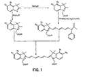
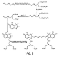
- novel dyes of the present invention are prepared according the methods well known in the art and are illustrated in FIGS. 1-5 .
- the inventive bioconjugates have the Formula 1, wherein W 3 and X 3 may be the same or different and are selected from the group consisting of -C(CH 3 ) 2 , -C((CH 2 ) a OH)CH 3 , -C((CH 2 ) a OH) 2 , -C((CH 2 ) a CO 2 H)CH 3 , -C((CH 2 ) a CO 2 H) 2 , -C((CH 2 ) a NH 2 )CH 3 , -C((CH 2 ) a NH 2 ) 2 , C((CH 2 ) a NR 3 R 4 ) 2 , -NR 3 , and -S-; Y 3 is selected from the group consisting of -(CH 2 ) a -CONH-Bm, -CH 2 -(CH 2 OCH 2 ) b -CH 2 -CONH-Bm, -(CH 2 )
- the inventive bioconjugates have the general Formula 2, wherein W 4 and X 4 may be the same or different and are selected from the group consisting of -C(CH 3 ) 2 , -C((CH 2 ) a OH)CH 3 , -C((CH 2 ) a OH) 2 , -C((CH 2 ) a CO 2 H)CH 3 , -C((CH 2 ) a CO 2 H) 2 , -C((CH 2 ) a NH 2 )CH 3 , C((CH 2 ) a NH 2 ) 2 , -C((CH 2 ) a NR 3 R 4 ) 2 , -NR 3 , and -S-; Y 4 is selected from the group consisting of -(CH 2 ) a -CONH-Bm, -CH 2 -(CH 2 OCH 2 ) b -CH 2 -CONH-Bm, -(CH 2
- the inventive bioconjugates have the general Formula 3, wherein W 5 and X 5 may be the same or different and are selected from the group consisting of -C(CH 3 ) 2 , -C((CH 2 ) a OH)CH 3 , -C((CH 2 ) a OH) 2 , -C((CH 2 ) a CO 2 H)CH 3 , -C((CH 2 ) a CO 2 H) 2 , -C((CH 2 ) a NH 2 )CH 3 , -C((CH 2 ) a NH 2 ) 2 , -C((CH 2 ) a NR 3 R 4 ) 2 , -NR 3 , and -S-; Y 5 is selected from the group consisting of -(CH 2 ) a -CONH-Bm, -CH 2 -CCH 2 OCH 2 ) b -CH 2 -CONH-Bm, -(
- the inventive bioconjugates have the general Formula 4, wherein W 6 and X 6 may be the same or different and are selected from the group consisting of -C(CH 3 ) 2 , -C((CH 2 ) a OH)CH 3 , -C((CH 2 ) a OH) 2 , -C((CH 2 ) a CO 2 H)CH 3 , - C((CH 2 ) a CO 2 H) 2 , -C((CH 2 ) a NH 2 )CH 3 , -C((CH 2 ) a NH 2 ) 2 , C((CH 2 ) a NR 3 R 4 ) 2 , -NR 3 , and -S-; Y 6 is selected from the group consisting of -(CH 2 ) a -CONH-Bm, -CH 2 -(CH 2 OCH 2 ) b -CH 2 -CONH-Bm, -(CH 2
- This invention is also related to the method of preventing fluorescence quenching. It is known that cyanine dyes generally form aggregates in aqueous media, leading to fluorescence quenching. Where the presence of a hydrophobic core in the dyes leads to fluorescence quenching, the addition of a biocompatible organic solvent, such, as 1-50% dimethylsulfoxide (DMSO) for example, restored fluorescence by preventing aggregation and allowed in vivo organ visualization.
- DMSO dimethylsulfoxide
- the inventive dye-biomolecule conjugates are used for optical tomographic, endoscopic, photoacoustic and sonofluorescent applications for the detection and treatment of tumors and other abnormalities.
- Dye-biomolecule conjugates are also used for localized therapy. This may be accomplished by attaching a porphyrin or other photodynamic therapy agent to a bioconjugate, shining light of an appropriate wavelength to activate the agent, and detecting and/or treating the abnormality.
- the inventive conjugates can also be used for the detection of the presence of tumors and other abnormalities by monitoring the blood clearance profile of the conjugates, for laser assisted guided surgery for the detection of small micrometastases of, e.g., somatostatin subtype 2 (SST-2) positive tumors, upon laparoscopy, and for diagnosis of atherosclerotic plaques and blood clots.
- SST-2 somatostatin subtype 2
- compositions of the invention can be formulated into diagnostic and therapeutic compositions for enteral or parenteral administration.
- These compositions contain an effective amount of the dye along with conventional pharmaceutical carriers and excipients appropriate for the type of administration contemplated.
- parenteral formulations advantageously contain the inventive agent in a sterile aqueous solution or suspension.
- Parenteral compositions may be injected directly or mixed with a large volume parenteral composition for systemic administration.
- Such solutions also may contain pharmaceutically acceptable buffers and, optionally, electrolytes such as sodium chloride.
- Formulations for enteral administration may vary widely, as is well known in the art.
- such formulations are liquids, which include an effective amount of the inventive agent in aqueous solution or suspension.
- Such enteral compositions may optionally include buffers, surfactants, thixotropic agents, and the like.
- Compositions for oral administration may also contain flavoring agents and other ingredients for enhancing their organoleptic qualities.
- the diagnostic compositions are administered in doses effective to achieve the desired enhancement. Such doses may vary widely, depending upon the particular dye employed, the organs or tissues to be imaged, the imaging equipment being used, and the like.
- the diagnostic compositions of the invention are used in the conventional manner.
- the compositions may be administered to a patient, typically a warm-blooded animal, either systemically or locally to the organ or tissue to be imaged, and the patient is then subjected to the imaging procedure.
- compositions and methods represent an important approach to the synthesis and use of novel cyanine and indocyanine dyes with a variety of photophysical and chemical properties.
- the combination also represents an important approach to the use of small molecular targeting groups to image tumors by optical methods.
- the invention is further detailed in the following Examples, which are offered by way of illustration and are not intended to limit the scope of the invention in any manner.
- This compound was prepared as described in Example 1 except that 1,1,2-trimethylindole was used as the starting material.
- This compound was prepared as described in Example 1 except that ⁇ -bromohexaoxyethyleneglycolpropiolic acid was used in place of bromopropanoic acid and the reaction was carried out in 1,2-dimethoxypropane.
- the reaction mixture was partitioned between 100 ml of methylene chloride and 100 ml of saturated sodium bicarbonate solution. The layers were separated and the methylene chloride layer was again washed with 100 ml of saturated sodium bicarbonate solution. The combined aqueous layers were extracted twice with 25 ml of methylene chloride. The combined methylene chloride layers were washed with 1 00 ml of brine, and dried over magnesium sulfate. The methylene chloride was removed with aspirator vacuum at about 35°C, and the remaining dimethylformamide was removed with vacuum at about 45°C. The crude material was left on a vacuum line overnight at room temperature.
- the ether solution was decanted and the oil was again triturated with a 100 ml portion of ether.
- the ether was decanted and the combined ether solution was allowed to stand for about two hours to allow the triphenylphosphine oxide to crystallize.
- the ether solution was decanted from the crystals and the solid was washed with 100 ml of ether.
- the volume of the combined ether extracts was reduced with vacuum until a volume of about 25 ml was obtained. This was allowed to stand overnight at 0°C. Ether (10 ml) was added to the cold mixture, which was mixed.to suspend the solid.
- the mixture was percolated through a column of 45 g of silica gel and eluted with ether, and 75 ml fractions were collected.
- the fractions that contained product, as determined by thin layer chromatography, were pooled and the ether was removed with vacuum. This yielded 10.1 g of crude product.
- the material was flash chromatographed on silica gel with hexane, changing to 9:1 hexane:ether.
- the product-containing fractions were pooled and the solvents removed with vacuum. This yielded 7.4 g (57% yield) of pure product.
- the hydroxy-indole compound is readily prepared by a known method ( P. L. Southwick et al., One pot Fischer synthesis of (2,3,3-trimethyl-3-H-indol-5-yl)-acetic acid derivatives as intermediates for fluorescent biolabels. Org. Prep. Proced. Int. Briefs, 1988, 20(3), 279-284 ).
- the intermediate 2-chloro-1-formyl-3-hydroxymethylenecyclohexane was prepared as described in the literature ( G. A. Reynolds and K. H. Drexhage, Stable heptamethine pyrylium dyes that absorb in the infrared. J. Org. Chem., 1977, 42(5), 885-888 ).
- Equal volumes (40 mL each) of dimethylformamide (DMF) and dichloromethane were mixed and the solution was cooled to -10°C in an acetone-dry ice bath. Under argon atmosphere, phosphorus oxychloride (40 mL) in dichloromethane was added dropwise to the cool DMF solution, followed by the addition of 10 g of cyclohexanone.
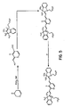
- These dyes are prepared as described in Example 7. These dyes absorb in the infrared region.
- the typical example shown in FIG. 5 has an estimated absorption maximum at 1036 nm.
- Octreotate The procedure described below is for the synthesis of Octreotate.
- the amino acid sequence of Octreotate is: D-Phe-Cys'-Tyr-D-Trp-Lys-Thr-Cys'-Thr (SEQ ID NO:1), wherein Cys' indicates the presence of an intramolecular disulfide bond between two cysteine amino acids.
- Other peptides of this invention were prepared by a similar procedure with slight modifications in some cases.
- the octapeptide was prepared by an automated fluorenylmethoxycarbonyl (Fmoc) solid phase peptide synthesis using a commercial peptide synthesizer from Applied Biosystems (Model 432A SYNERGY Peptide Synthesizer).
- the first peptide cartridge contained Wang resin pre-loaded with Fmoc-Thr on 25 ⁇ mole scale.
- Subsequent cartridges contained Fmoc-protected amino acids with side chain protecting groups for the following amino acids: Cys(Acm), Thr(t-Bu), Lys(Boc), Trp(Boc) and Tyr(t-Bu).
- the amino acid cartridges were placed on the peptide synthesizer and the product was synthesized from the C-to the N-terminal position.
- the coupling reaction was carried out with 75 ⁇ moles of the protected amino acids in the presence of 2-(1H-benzotriazol-1-yl)-1,1,3,3-tetramethyluronium hexafluorophosphate (HBTU)/N-hydroxybenzotriazole (HOBt).
- HBTU 2-(1H-benzotriazol-1-yl)-1,1,3,3-tetramethyluronium hexafluorophosphate
- HOBt N-hydroxybenzotriazole
- the Fmoc protecting group was removed with 20% piperidine in dimethylformamide.
- the thiol group was cyclized with thallium trifluoroacetate and the product was cleaved from the solid support with a cleavage mixture containing trifluoroacetic acid (85%):water (5%):phenol (5%):thioanisole (5%) for six hours.
- the peptide was precipitated with t-butyl methyl ether and lyophilized with water:acetonitrile (2:3) mixture.
- the peptide was purified by HPLC and analyzed with LC/MS.
- Octreotide D-Phe-Cys'-Tyr-D-Trp-Lys-Thr-Cys'-Thr-OH (SEQ ID NO:2), wherein Cys' indicates the presence of an intramolecular disulfide bond between two cysteine amino acids, was prepared by the same procedure.
- Bombesin analogs were prepared by the same procedure except that cyclization with thallium trifluoroacetate was not needed. Side-chain deprotection and cleavage from the resin was carried out with 50 ⁇ L each of ethanedithiol, thioanisole and water, and 850 ⁇ L of trifluoroacetic acid. Two analogues were prepared: Gly-Ser-Gly-Gln-Trp-Ala-Val-Gly-His-Leu-Met-NH 2 (SEQ ID NO:3) and Gly-Asp-Gly-Gln-Trp-Ala-Val-Gly-His-Leu-Met-NH 2 (SEQ ID NO:4).
- Cholecystokinin octapeptide analogs were prepared as described for Octreotate without the cyclization step. Three analogs were prepared: Asp-Tyr-Met-Gly-Trp-Met-Asp-Phe-NH 2 (SEQ ID NO:5); Asp-Tyr-Nie-Gly-Trp-Nle-Asp-Phe-NH 2 (SEQ ID NO:6); and D-Asp-Tyr-Nle-Gly-Trp-Nle-Asp-Phe-NH 2 (SEQ ID NO:7), where Nle is norleucine.
- a neurotensin analog D-Lys-Pro-Arg-Arg-Pro-Tyr-Ile-Leu (SEQ ID NO:8), was prepared as described for Octreotate without the cyclization step.
- Octreotate was prepared as described in Example 9 but the peptide was not cleaved from the solid support and the N-terminal Fmoc group of Phe was retained. The thiol group was cyclized with thallium trifluoroacetate and the Phe was deprotected to liberate the free amine. Bisethylcarboxymethylindocyanine dye (53 mg, 75 ⁇ moles) was added to an activation reagent consisting of a 0.2 M solution of HBTU/HOBt in DMSO (375 ⁇ L), and 0.2 M solution of diisopropylethylamine in DMSO (375 ⁇ L).
- the activation was complete in about 30 minutes and the resin-bound peptide (25 ⁇ moles) was added to the dye.
- the coupling reaction was carried out at room temperature for three hours. The mixture was filtered and the solid residue was washed with DMF, acetonitrile and THF. After drying the green residue, the peptide was cleaved from the resin and the side chain protecting groups were removed with a mixture of 85% trifluoroacetic acid, 2.5% water, 2.5% thioanisole and 2.5% phenol.
- the resin was filtered and cold t-butyl methyl ether (MTBE) was used to precipitate the dye-peptide conjugate, which was dissolved in an acetonitrile:water (2:3) mixture and lyophilized.
- MTBE cold t-butyl methyl ether
- the product was purified by HPLC to give the monoOctreotate-Bisethylcarboxymethylindocyanine dye (Cytate 1, 80%) and the bis Octreotate-Bisethylcarboxymethylindocyanine dye (Cytate 2, 20%).
- the monoOctreotate conjugate is obtained almost exclusively (>95%) over the bis conjugate by reducing the reaction time to two hours. However, this also leads to incomplete reaction, and the free Octreotate must be carefully separated from the dye conjugate in order to avoid saturation of the receptors by the non-dye conjugated peptide.
- Octreatate-bispentylcarboxymethylindocyanine dye was prepared as described above with some modifications.
- Bispentylcarboxymethylindocyanine dye (60 mg, 75 ⁇ moles) was added to an activation reagent consisting of a 0.2 M solution of HBTU/HOBt in DMSO (400 ⁇ L), and a 0.2 M solution of diisopropylethylamine in DMSO (400 ⁇ L).
- the activation was complete in about 30 minutes and the resin-bound peptide (25 ⁇ moles) was added to the dye.
- the reaction was carried out at room temperature for three hours. The mixture was filtered and the solid residue was washed with DMF, acetonitrile and THF.
- the peptide was cleaved from the resin and the side chain protecting groups were removed with a mixture of 85% trifluoroacetic acid, 2.5% water, 2.5% thioanisole and 2.5% phenol.
- the resin was filtered and cold t-butyl methyl ether (MTBE) was used to precipitate the dye-peptide conjugate, which was dissolved in an acetonitrile:water (2:3) mixture and lyophilized.
- MTBE cold t-butyl methyl ether
- the product was purified by HPLC to give Octreotate-1,1,2-trimethyl-[1 H]-benz[e]indole propanoic acid conjugate (10%), monoOctreotate-bispentylcarboxymethylindocyanine dye (Cytate 3, 60%) and bisOctreotate-bispentylcarboxymethylindocyanine dye (Cytate 4, 30%).
- the dye-peptide conjugates are sparingly soluble in water and require the addition of solubilizing agents or co-solvents.
- Addition of 1-20% aqueous ethanol to the conjugates partially quenched the fluorescence intensity in vitro and the fluorescence was completely quenched in vivo (the conjugate was not detected by the charge coupled device (CCD) camera).
- Addition of 1-50% of DMSO either re-established or increased the fluorescence intensity of the conjugates in vitro and in vivo. The dye fluorescence remained intense for over one week.
- the DMSO formulations were well tolerated by experimental animals used for this invention.
- a non-invasive in vivo fluorescence imaging apparatus was employed to assess the efficacy of contrast agents developed for tumor detection in animal models.
- a LaserMax Inc. laser diode of nominal wavelength 780 nm and nominal power of 40 mW was used.
- the detector was a Princeton Instruments model RTE/CCD-1317-K/2 CCD camera with a Rodenstock 10 mm F2 lens (stock #542.032.002.20) attached.
- An 830 nm interference lens (CVI Laser Corp., part # F10-830-4-2) was mounted in front of the CCD input lens such that only emitted fluorescent light from the contrast agent was imaged.
- an image of the animal was taken pre-injection of contrast agent. This image was subsequently subtracted (pixel by pixel) from the post injection images.
- the background subtraction was never done once the animal had been removed from the sample area and returned at a later time for images taken several hours post injection.
- DSL 6A tumors were induced in male Lewis rats in the left flank area by the introduction of material from a solid (donor) implant, and the tumors were palpable in approximately 14 days.
- the animals were anesthetized with xylazine: ketamine: acepromazine, 1.5: 1.5: 0.5 at 0.8 mL/kg via intramuscular injection.
- the area of the tumor (left flank) was shaved to expose the tumor and the surrounding surface area.
- a 21 gauge butterfly equipped with a stopcock and two syringes containing heparinized saline was placed into the later tail vein of the rat. Patency of the vein was checked prior to administration of the ICG via the butterfly apparatus.
- Each animal received 500 ⁇ L of a 0.42 mg/mL solution of ICG in water.
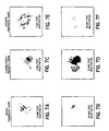
- FIGS. 7A-B are tumor images at two minutes ( FIG. 7A ) and 30 minutes ( FIG. 7B ) post bolus injection of a 0.5 mL aqueous solution of ICG (5.4 ⁇ m).
- the Figures are false color images of fluorescent intensity measured at the indicated times, with images constrained to the tumor and a small surrounding area. As is shown, the dye intensity in the tumor is considerably diminished 30 minutes post-ICG injection.
- FIGS. 7C-D are images of a rat with an induced prostatic carcinoma tumor (R3327-H) imaged at two minutes ( FIG. 7C ) and 30 minutes ( FIG. 7D ) post injection.
- the Figures are false color images of fluorescent intensity measured at the indicated times, with images constrained to the tumor and a small surrounding area. As is shown, the dye intensity in the tumor is considerably diminished 30 minutes post-ICG injection.
- FIGS. 7E-F are images of a rat with an induced pancreatic acinar carcinoma ( CA20948 ) expressing the SST-2 receptor imaged at two minutes ( FIG. 7E ) and 30 minutes ( FIG. 7F ) post injection.
- the Figures are false color images of fluorescent intensity measured at the indicated times, with images constrained to the tumor and a small surrounding area. As is shown, the dye intensity in the tumor is considerably diminished and almost absent 30 minutes post-ICG injection.
- Example 12 The imaging apparatus and the procedure used are described in Example 12, except that each animal received 500 ⁇ l of a 1.0 mg/mL solution of Cytate 1 solution of 25% dimethylsulfoxide in water.
- Rat pancreatic acinar carcinoma expressing the SST-2 receptor ( CA20948 ) were induced by solid implant technique in the left flank area, and palpable masses were detected 24 days post implant. Images were obtained at various times post injection. Uptake into the tumor was seen at two minutes but was not maximal until about five minutes.
- FIGS. 8A-B show a comparison of the uptake of ICG and Cytate 1 at 45 minutes in rats with the CA20948 tumor cell line. By 45 minutes the ICG has mostly cleared ( FIG. 8A ) whereas the Cytate 1 is still intense ( FIG. 8B ). This dye fluorescence remained intense in the tumor for several hours post-injection.
- ICG indocyanine green
- the first two tumor lines are not as highly vascularized as CA20948 , which is also rich in somatostatin (SST-2) receptors. Consequently, the detection and retention of a dye in this tumor model is a good index of receptor-mediated specificity.
- Octreotate is known to target somatostatin (SST-2) receptors, hence, cyano-Octreotates (Cytate 1 and Cytate 2) were prepared. Cytate 1 was evaluated in the CA20948 Lewis rat model. Using the CCD camera apparatus, localization of this dye was observed in the tumor (indicated by arrow) at 45 minutes post injection ( FIG. 9A ). At 27 hours post injection, the animal was again imaged ( FIG. 9B ). Tumor visualization was easily observed (indicated by arrow), showing specificity of this agent for the SST-2 receptors present in the CA20948 tumor line.
- the AR42-J cell line is derived from exocrine rat pancreatic acinar carcinoma. It can be grown in continuous culture or maintained in vivo in athymic nude mice; SCID mice, or in Lewis rats. This cell line is particularly attractive for in vitro receptor assays, as it is known to express a variety of hormone receptors including cholecystokinin (CCK), epidermal growth factor (EGF), pituitary adenylate cyclase activating peptide (PACAP), somatostatin (SST-2) and bombesin.
- CCK cholecystokinin
- EGF epidermal growth factor
- PACAP pituitary adenylate cyclase activating peptide
- SST-2 somatostatin
- FIG. 11 is an image of bombesinate in an AR42-J tumor-bearing rat, as described in Example 16, at 22 hours post injection of bombesinate. As shown in FIG. 11 , specific localization of the bioconjugate in the tumor (indicated by arrow) was observed.
- a laser of appropriate wavelength for excitation of the dye chromophore was directed into one end of a fiber optic bundle and the other end was positioned a few millimeters from the ear of a rat.
- a second fiber optic bundle was also positioned near the same ear to detect the emitted fluorescent light and the other end was directed into the optics and electronics for data collection.
- An interference filter (IF) in the collection optics train was used to select emitted fluorescent light of the appropriate wavelength for the dye chromophore.
- Sprague-Dawley or Fischer 344 rats were used in these studies.
- the animals were anesthetized with urethane administered via intraperitoneal injection at a dose of 1.35 g/kg body weight.
- a 21 gauge butterfly with 12" tubing was placed in the lateral tail vein of each animal and flushed with heparinized saline.
- the animals were placed on a heating pad and kept warm throughout the entire study.
- the lobe of the left ear was affixed to a glass microscope slide to reduce movement and vibration.
- Incident laser light delivered from the fiber optic was centered on the affixed ear. Data acquisition was then initiated, and a background reading of fluorescence was obtained prior to administration of the test agent.
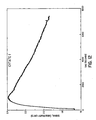
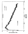
- the peptide-dye conjugate was administered to the animal through a bolus injection, typically 0.5 to 2.0 mL, in the lateral tail vein. This procedure was repeated with several dye-peptide conjugates in normal and tumor-bearing rats. Representative profiles as a method to monitor blood clearance of the peptide-dye conjugate in normal and tumor-bearing animals are shown in FIGS. 12 to 16 . The data were analyzed using a standard sigma plot software program for a one-compartment model.
- FIG. 12 shows the clearance profile of Cytate 1 from the blood of a normal rat monitored at 830 nm after excitation at 780 nm.
- FIG. 13 shows the clearance profile of Cytate 1 from the blood of a pancreatic tumor ( CA20948 )-bearing rat also monitored at 830 nm after excitation at 780 nm.
- FIG. 14 shows the clearance profile of Cytate 2 from the blood of a normal rat
- FIG. 15 shows the clearance profile of Cytate 2 from the blood of a pancreatic tumor ( CA20948 )-bearing rat, monitored at 830 nm after excitation at 780 nm.
- FIG. 16 shows the clearance profile of Cytate 4 from the blood of a normal rat monitored at 830 nm after excitation at 780 nm.
Abstract
Description
- This application is a continuation-in-part of application Serial No.
09/484,319, filed January 18, 2000 - This invention relates generally to compositions of cyanine dye bioconjugates with bioactive molecules for diagnosis and therapy, particularly, for visualization and detection of tumors.
- Several dyes that absorb and emit light in the visible and near-infrared region of the electromagnetic spectrum are currently being used for various biomedical applications due to their biocompatibility, high molar absorptivity, and/or high fluorescence quantum yields. The high sensitivity of the optical modality in conjunction with dyes as contrast agents parallels that of nuclear medicine, and permits visualization of organs and tissues without the undesirable effect of ionizing radiation.
- Cyanine dyes with intense absorption and emission in the near-infrared (NIR) region are particularly useful because biological tissues are optically transparent in this region (B. C. Wilson, Optical properties of tissues. Encyclopedia of Human Biology, 1991, 5, 587-597). For example, indocyanine green, which absorbs and emits in the NIR region, has been used for monitoring cardiac output, hepatic functions, and liver blood flow (Y-L. He, et al., Measurement of blood volume using indocyanine green measured with pulse-spectrometry: lts reproducibility and reliability. Critical Care Medicine, 1998, 26(8), 1446-1451; J. Caesar, et al., The use of Indocyanine green in the measurement of hepatic blood flow and as a test of hepatic function. Clin. Sci. 1961, 21, 43-57), and its functionalized derivatives have been used to conjugate biomolecules for diagnostic purposes (R. B. Mujumdar, et al., Cyanine dye labeling reagents: Sulfoindocyanine succinimidyl esters. Bioconjugate Chemistry, 1993, 4(2), 105-111;
U.S. Patent No. 5,453,505 ;WO 98/48846 WO 98/22146 WO 96/17628 WO 98/48838 - A major drawback in the use of cyanine dye derivatives is the potential for hepatobiliary toxicity resulting from the rapid clearance of these dyes by the liver (G. R. Cherrick, et al., Indocyanine green: Observations on its physical properties, plasma decay, and hepatic extraction. J. Clinical Investigation, 1960, 39, 592-600). This is associated with the tendency of cyanine dyes in solution to form aggregates, which could be taken up by Kupffer cells in the liver.
- Various attempts to obviate this problem have not been very successful. Typically, hydrophilic peptides, polyethyleneglycol or oligosaccharide conjugates have been used, but these resulted in long-circulating products, which are eventually still cleared by the liver. Another major difficulty with current cyanine and indocyanine dye systems is that they offer a limited scope in the ability to induce large changes in the absorption and emission properties of these dyes. Attempts have been made to incorporate various heteroatoms and cyclic moieties into the polyene chain of these dyes (L. Strekowski, et al., Substitution reactions of a nucleofugal group in hetamethine cyanine dyes. J. Org. Chem., 1992, 57, 4578-4580; N. Narayanan and G. Patonay, A new method for the synthesis of heptamethine cyanine dyes: Synthesis of new near infrared fluorescent labels. J. Org. Chem., 1995, 60, 2391-2395;
U.S. Patent Nos. 5,732,104 ;5,672,333 ; and5,709,845 ), but the resulting dye systems do not show large differences in absorption and emission maxima, especially beyond 830 nm where photoacoustic diagnostic applications are very sensitive. They also possess a prominent hydrophobic core, which enhances liver uptake. Further, most cyanine dyes do not have the capacity to form starburst dendrimers, which are useful in biomedical applications. - For the purpose of tumor detection, many conventional dyes are useful for in vitro applications because of their highly toxic effect on both normal and abnormal tissues. Other dyes lack specificity for particular organs or tissues and, hence, must be attached to bioactive carriers such as proteins, peptides, carbohydrates, and the like to deliver the dyes to specific regions in the body. Several studies on the use of near infrared dyes and dye-biomolecule conjugates have been published (G. Patonay and M. D. Antoine, Near-Infrared Fluorogenic Labels: New Approach to an Old Problem, Analytical Chemistry, 1991, 63:321 A-327A and references therein; M. Brinkley, A Brief Survey of Methods for Preparing Protein Conjugates with Dyes, Haptens, and Cross-Linking Reagents, Perspectives in Bioconjugate Chemistry 1993, pp. 59-70, C. Meares (Ed), ACS Publication, Washington, DC; J. Slavik, Fluorescent Probes in Cellular and Molecular Biology, 1994, CRC Press, Inc.;
U.S. Patent No. 5,453,505 ;WO 98/48846 WO 98/22146 WO 96/17628 WO 98/48838 - Of particular interest is the targeting of tumor cells with antibodies or other large protein carriers such as transferrin as delivery vehicles (A. Becker et al., "Transferrin Mediated Tumor Delivery of Contrast Media for Optical Imaging and Magnetic Resonance Imaging", Biomedical Optics meeting, January 23-29, 1999, San Jose, CA). Such an approach has been widely used in nuclear medicine applications. Its major advantage is the retention of a carrier's tissue specificity, since the molecular volume of the dye is substantially smaller than the carrier. However, this approach does have some serious limitations in that the diffusion of high molecular weight bioconjugates to tumor cells is highly unfavorable, and is further complicated by the net positive pressure in solid tumors (R. K. Jain, Barriers to Drug Delivery in Solid Tumors, Scientific American 1994, 271:58-65. Furthermore, many dyes in general, and cyanine dyes in particular, tend to form aggregates in aqueous media that lead to fluorescence quenching.
- Therefore, there is a need for dyes that could prevent dye aggregation in solution, that are predisposed to form dendrimers, that are capable of absorbing or emitting beyond 800 nm, that possess desirable photo physical properties, and that are endowed with tissue-specific targeting capability.
- The invention is directed to compositions, and methods of preparing the compositions, of low molecular weight biomolecule-dye conjugates to enhance tumor detection. The inventive compositions preserve the fluorescence efficiency of the dye molecules, do not aggregate in solution, form starburst dendrimers, are capable of absorbing or emitting light in the near infrared region (beyond 800 nm), and can be rendered tissue-specific.
- In one embodiment, the inventive composition comprises cyanine dyes of
general formula 1
- In a second embodiment, the inventive composition comprises cyanine dyes of
general formula 2
- In a third embodiment, the inventive composition comprises cyanine dyes of
general formula 3
- In a fourth embodiment, inventive composition comprises cyanine dyes of general formula 4

- The invention will be further appreciated in light of the following figures, detailed description, and examples.
- The file of this patent contains at least one drawing executed in color. Copies of this patent with color drawing(s) will be provided by the Patent and Trademark Office upon request and payment of the necessary fee.
-
FIG. 1 shows the reaction pathway for the synthesis of bis-carboxylic acid cyanine dyes. -
FIG. 2 shows the reaction pathway for the synthesis of tetracarboxylic acid cyanine dyes. -
FIG. 3 shows the reaction pathway for the synthesis of polyhydroxycarboxylic acid dyes. -
FIG. 4 shows the reaction pathway for the synthesis of non-aggregating cyanine dyes. -
FIG. 5 shows the reaction pathway for the synthesis of long wavelength absorbing dyes. -
FIG. 6 shows the reaction pathway for the synthesis of cyanine dye bioconjugates. -
FIGS. 7A-F represent images at 2 minutes and 30 minutes post injection of indocyanine green (ICG) into rats with various tumors. -
FIGS. 8A-B show a comparison of the uptake of ICG (FIG. 8A ) and Cytate 1 (FIG. 8B ) in rats with the pancreatic acinar carcinoma (CA20948 ). -
FIGS. 9A-B show images of rats with the pancreatic acinar carcinoma (CA20948 ) 45 minutes (FIG. 9A ) and 27 hours (FIG. 9B ) post injection ofCytate 1. -
FIG. 10 is an image of individual organs taken from a rat with pancreatic acinar carcinoma (CA20948 ) about 24 hours after injection withCytate 1. -
FIG. 11 is an image of bombesinate in an AR42-J tumor-bearing rat 22 hours after injection. -
FIG. 12 is the clearance profile ofCytate 1 from the blood of a normal rat. -
FIG. 13 is the clearance profile ofCytate 1 from the blood of a pancreatic tumor-bearing rat. -
FIG. 14 is the clearance profile ofCytate 2 from the blood of a normal rat. -
FIG. 15 is the clearance profile ofCytate 2 from the blood of a pancreatic tumor-bearing rat. - FIG. 16 is the clearance profile of Cytate 4 from the blood of a normal rat.
- The novel compositions of the present invention comprising dyes of
formulas 1 to 4 offer significant advantages over those currently described in the art. These inventive dyes form starburst dendrimers which prevent aggregation in solution by preventing intramolecular and intermolecular ordered hydrophobic interactions, and have multiple attachment sites proximal to the dye chromophore for ease of forming bioactive molecules. The presence of rigid and extended chromophore backbone enhances their fluorescence quantum yield and extends their maximum absorption beyond 800 nm. Conjugation of biomolecules to these dyes is readily achievable. - The inventive bioconjugates of the present invention also exploit the symmetric nature of the cyanine and indocyanine dye structures by incorporating one to ten receptor targeting groups in close proximity to each other, such that the receptor binding can be greatly enhanced due to a cooperative effect. Accordingly, several cyanine dyes containing one or more targeting domains have been prepared and tested in vivo for biological activity.
- The inventive dye-bioconjugates of
formulas 1 to 4 are useful for various biomedical applications. These include, but are not limited to, tomographic imaging of organs, monitoring of organ functions, coronary angiography, fluorescence endoscopy, detection, imaging, and therapy of tumors, laser assisted guided surgery, photoacoustic methods, and sonofluorescent methods. - Specific embodiments to accomplish some of the aforementioned biomedical applications are given below. The novel dyes of the present invention are prepared according the methods well known in the art and are illustrated in
FIGS. 1-5 . -
FIG. 1 illustrates the synthetic scheme for bis-carboxylic acid cyanine dyes, where A = CH2 or CH2OCH2; R = COOH; R' = COOH, NHFmoc; CO2t-Bu; SO3-; R1 = R2 = H (Formula 1) or R1,R2 = fused phenyl (Formula 2). -
FIG. 2 illustrates the synthetic scheme for tetracarboxylic acid cyanine dyes, where A = CH2 or CH2OCH2; R1 = R2 = H (Formula 1) or R1,R2 = fused phenyl (Formula 2). -
FIG. 3 illustrates the synthetic scheme for polyhydroxycarboxylic acid cyanine dyes. -
FIG. 4 illustrates the synthetic scheme for non-aggregating cyanine dyes. -
FIG. 5 illustrates the synthetic scheme for long wavelength-absorbing tunable cyanine dyes. - In one embodiment, the inventive bioconjugates have the Formula 1, wherein W3 and X3 may be the same or different and are selected from the group consisting of -C(CH3)2, -C((CH2)aOH)CH3, -C((CH2)aOH)2, -C((CH2)aCO2H)CH3, -C((CH2)aCO2H)2, -C((CH2)aNH2)CH3, -C((CH2)aNH2)2, C((CH2)aNR3R4)2, -NR3, and -S-; Y3 is selected from the group consisting of -(CH2)a-CONH-Bm, -CH2 -(CH2OCH2)b-CH2-CONH-Bm, -(CH2)a-NHCO-Bm, -CH2-(CH2OCH2)b-CH2 -NHCO-Bm, and -CH2(CH2OCH2)b-CH2NR3R4; Z3 is selected from the group consisting of -(CH2)a-CONH-Dm, -CH2-(CH2OCH2)b-CH2 -CONH-Dm, -(CH2)a-NHCO-Dm, -CH2-(CH2OCH2)b-CH2-NHCO-Dm, and -CH2(CH2OCH2)b-CH2NR3R4; A1 is a single or a double bond; B1, C1, and D1 are independently selected from the group consisting of -O-, -S-, NR3, (CH2)a -CR1R2, and -CR1; A1, B1, C1, and D1 may together form a 6- to 10-membered carbocyclic ring or a 6- to 10-membered heterocyclic ring optionally containing one or more oxygen, nitrogen, or sulfur atom; a3 and b3 are independently from 0 to 3; R1 to R4, and R29 to R37 are independently selected from the group consisting of hydrogen, C1 -C10 alkyl, C5-C12 aryl, C1-C10 alkoxyl, C1-C10 polyhydroxyalkyl, C5-C12 polyhydroxyaryl, C1-C10 aminoalkyl, mono- or oligosaccharide, peptide with 2 to 30 amino acid units, -CH2(CH2OCH2)b-CH2-OH, -(CH2)a-CO2H, -CCH2)a-CONH-Bm, -CH2-(CH2OCH2)b-CH2-CONH-Bm, -(CH2)a-NHCO-Bm, -CH2-(CH2OCH2)b-CH2-NHCO-Bm, -(CH2)a-OH and -CH2-(CH2OCH2)b-CO2H; Bm and Dm are independently selected from the group consisting of a bioactive peptide containing 2 to 30 amino acid units, an antibody, a mono- or oligosaccharide, a glycopeptide, a metal chelating agent, a radioactive or nonradioactive metal complex, and an echogenic agent; a and c are independently from 1 to 10; and b and d are independently from 1 to 30.
- In a second embodiment, the inventive bioconjugates have the general Formula 2, wherein W4 and X4 may be the same or different and are selected from the group consisting of -C(CH3)2, -C((CH2)aOH)CH3, -C((CH2)aOH)2, -C((CH2)aCO2H)CH3, -C((CH2)aCO2H)2, -C((CH2)aNH2)CH3, C((CH2)aNH2)2, -C((CH2)aNR3R4)2, -NR3, and -S-; Y4 is selected from the group consisting of -(CH2)a-CONH-Bm, -CH2-(CH2OCH2)b-CH2-CONH-Bm, -(CH2)a-NHCO-Bm, -CH2-(CH2OCH2)b-CH2-NHCO-Bm, -(CH2)a-NR3R4, and -CH2(CH2OCH2)b-CH2NR3R4; Z4 is selected from the group consisting of -(CH2)a-CONH-Dm, -CH2-(CH2OCH2)b-CH2-CONH-Dm, -(CH2)a-NHCO-Dm, -CH2-(CH2OCH2)b-CH2-NHCO-Dm, -(CH2)a-NR3R4, and -CH2(CH2OCH2)b-CH2NR3R4; A2 is a single or a double bond; B2, C2, and D2 are independently selected from the group consisting of -O-, -S-, NR3, (CH2)a -CR1R2, and -CR1; A2, B2, C2, and D2 may together form a 6- to 1 0-membered carbocyclic ring or a 6- to 10-membered heterocyclic ring optionally containing one or more oxygen, nitrogen, or sulfur atom; a4 and b4 are independently from 0 to 3; R1 to R4, and R45 to R57 are independently selected from the group consisting of hydrogen, C1-C10 alkyl, C5-C12 aryl, C1-C10 alkoxyl, C1-C10 polyhydroxyalkyl, C5-C12 polyhydroxyaryl, C1-C10 aminoalkyl, mono- or oligosaccharide, peptide with 2 to 30 amino acid units, -CH2(CH2OCH2)b-CH2-OH, -(CH2)a-CO2H, -(CH2)a-CONH-Bm, -CH2-(CH2OCH2)b-CH2-CONH-Bm, -(CH2)a-NHCO-Bm, -CH2-(CH2OCH2)b-CH2-NHCO-Bm, -(CH2)a-OH and -CH2-(CH2OCH2)b-CO2H; Bm and Dm are independently selected from the group consisting of a bioactive peptide containing 2 to 30 amino acid units, an antibody, a mono- or oligosaccharide, a glycopeptide, a metal chelating agent, a radioactive or nonradioactive metal complex, and an echogenic agent; a and c are independently from 1 to 10; and b and d are independently from 1 to 30.
- In a third embodiment, the inventive bioconjugates have the general Formula 3, wherein W5 and X5 may be the same or different and are selected from the group consisting of -C(CH3)2 , -C((CH2)aOH)CH3, -C((CH2)aOH)2 , -C((CH2)aCO2H)CH3, -C((CH2)aCO2H)2, -C((CH2)aNH2)CH3, -C((CH2)aNH2)2 , -C((CH2)aNR3R4)2, -NR3, and -S-; Y5 is selected from the group consisting of -(CH2)a-CONH-Bm, -CH2-CCH2OCH2)b-CH2-CONH-Bm, -(CH2)a-NHCO-Bm, -CH2-(CH2OCH2)b-CH2-NHCO-Bm, -(CH2)a-NR3R4, and -CH2(CH2OCH2)b-CH2NR3R4; Z5 is selected from the group consisting of -(CH2)a-CONH-Dm, -CH2-(CH2OCH2)b-CH2-CONH-Om, -(CH2)a-NHCO-Dm, -CH2-(CH2OCH2)b-CH2-NHCO-Om, -(CH2)a-NR3R4, and -CH2(CH2OCH2)b-CH2NR3R4; A3 is a single or a double bond; B3, C3, and D3 are independently selected from the group consisting of -O-, -S-, NR3, (CH2)a -CR1R2, and -CR1; A3, B3, C3, and D3 may together form a 6- to 10-membered carbocyclic ring or a 6- to 10-membered heterocyclic ring optionally containing one or more oxygen, nitrogen, or sulfur atom; a5 is from 0 to 3; R1 to R4, and R58 to R66 are independently selected from the group consisting of hydrogen, C1-C10 alkyl, C5-C12 aryl, C1-C10 alkoxyl, C1-C10 polyhydroxyalkyl, C5-C12 polyhydroxyaryl, C1-C10 aminoalkyl, mono- or oligosaccharide, peptide with 2 to 30 amino acid units, -CH2(CH2OCH2)b-CH2-OH, -(CH2)a-CO2H, -(CH2)a-CONH-Bm, -CH2-(CH2OCH2)b-CH2-CONH-Bm, -(CH2)a-NHCO-Bm, -CH2-(CH2OCH2)b-CH2-NHCO-Bm, -(CH2)a-OH and -CH2-(CH2OCH2)b-CO2H; Bm and Dm are independently selected from the group consisting of a bioactive peptide containing 2 to 30 amino acid units, an antibody, a mono- or oligosaccharide, a glycopeptide, a metal chelating agent, a radioactive or nonradioactive metal complex, and an echogenic agent; a and c are independently from 1 to 10; and b and d are independently from 1 to 30.
- In a fourth embodiment, the inventive bioconjugates have the general Formula 4, wherein W6 and X6 may be the same or different and are selected from the group consisting of -C(CH3)2, -C((CH2)aOH)CH3, -C((CH2)aOH)2, -C((CH2)aCO2H)CH3, - C((CH2)aCO2H)2, -C((CH2)aNH2)CH3, -C((CH2)aNH2)2, C((CH2)aNR3R4)2, -NR3, and -S-; Y6 is selected from the group consisting of -(CH2)a-CONH-Bm, -CH2-(CH2OCH2)b-CH2-CONH-Bm, -(CH2)a-NHCO-Bm, -CH2-(CH2OCH2)b-CH2-NHCO-Bm, -(CH2)a-NR3R4, and -CH2(CH2OCH2)b-CH2NR3R4; Z6 is selected from the group consisting of -(CH2)a-CONH-Dm, -CH2-(CH2OCH2)b-CH2-CONH-Dm, -(CH2)a-NHCO-Dm, -CH2-(CH2OCH2)b-CH2-NHCO-Dm, -(CH2)a-NR3R4, and -CH2(CH2OCH2)b-CH2NR3R4; A4 is a single or a double bond; B4, C4, and D4 are independently selected from the group consisting of -O-, -S-, NR3, (CH2)a-CR1R2, and -CR1; A4, B4, C4, and D4 may together form a 6- to 10-membered carbocyclic ring or a 6- to 10-membered heterocyclic ring optionally containing one or more oxygen, nitrogen, or sulfur atom; a6 is from 0 to 3; R1 to R4, and R67 to R79 are independently selected from the group consisting of hydrogen, C1-C10 alkyl, C5-C12 aryl, C1-C10 alkoxyl, C1-C10 polyhydroxyalkyl, C5-C12 polyhydroxyaryl, C1-C10 aminoalkyl, mono- or oligosaccharide, peptide with 2 to 30 amino acid units, -CH2(CH2OCH2)bCH2-OH, -(CH2)a-CO2H, -(CH2)a-CONH-Bm, -CH2-(CH2OCH2)b-CH2-CONH-Bm, -(CH2)a-NHCO-Bm, -CH2(CH2OCH2)bCH2-NHCO-Bm, -(CH2)a-OH and -CH2-(CH2OCH2)b-CO2H; Bm and Dm are independently selected from the group consisting of a bioactive peptide containing 2 to 30 amino acid units, an antibody, a mono- or oligosaccharide, a glycopeptide, a metal chelating agent, a radioactive or nonradioactive metal complex, and an echogenic agent; a and c are independently from 1 to 10; and b and d are independently from 1 to 30.
- This invention is also related to the method of conjugating the inventive dyes to peptides or biomolecules by solid phase or solution synthesis methods.
FIG. 6 illustrates the synthetic scheme for bioconjugates incorporating the cyanine dyes ofFIGS. 1-5 , using automated peptide synthesis in a solid support, where A = CH2 or CH2OCH2; R1 = R2 = H (Formula 1) or R1,R2 = fused phenyl (Formula 2); AA = amino acids; R = CONH peptide; R' = R (bis conjugate) or COOH (mono conjugate); Ⓟ = solid support; Ⓟ = presence or absence depends on R' definition. - This invention is also related to the method of preventing fluorescence quenching. It is known that cyanine dyes generally form aggregates in aqueous media, leading to fluorescence quenching. Where the presence of a hydrophobic core in the dyes leads to fluorescence quenching, the addition of a biocompatible organic solvent, such, as 1-50% dimethylsulfoxide (DMSO) for example, restored fluorescence by preventing aggregation and allowed in vivo organ visualization.
- The inventive dye-biomolecule conjugates are used for optical tomographic, endoscopic, photoacoustic and sonofluorescent applications for the detection and treatment of tumors and other abnormalities.
- Dye-biomolecule conjugates are also used for localized therapy. This may be accomplished by attaching a porphyrin or other photodynamic therapy agent to a bioconjugate, shining light of an appropriate wavelength to activate the agent, and detecting and/or treating the abnormality.
- The inventive conjugates can also be used for the detection of the presence of tumors and other abnormalities by monitoring the blood clearance profile of the conjugates, for laser assisted guided surgery for the detection of small micrometastases of, e.g., somatostatin subtype 2 (SST-2) positive tumors, upon laparoscopy, and for diagnosis of atherosclerotic plaques and blood clots.
- The compositions of the invention can be formulated into diagnostic and therapeutic compositions for enteral or parenteral administration. These compositions contain an effective amount of the dye along with conventional pharmaceutical carriers and excipients appropriate for the type of administration contemplated. For example, parenteral formulations advantageously contain the inventive agent in a sterile aqueous solution or suspension. Parenteral compositions may be injected directly or mixed with a large volume parenteral composition for systemic administration. Such solutions also may contain pharmaceutically acceptable buffers and, optionally, electrolytes such as sodium chloride.
- Formulations for enteral administration may vary widely, as is well known in the art. In general, such formulations are liquids, which include an effective amount of the inventive agent in aqueous solution or suspension. Such enteral compositions may optionally include buffers, surfactants, thixotropic agents, and the like. Compositions for oral administration may also contain flavoring agents and other ingredients for enhancing their organoleptic qualities.
- The diagnostic compositions are administered in doses effective to achieve the desired enhancement. Such doses may vary widely, depending upon the particular dye employed, the organs or tissues to be imaged, the imaging equipment being used, and the like. The diagnostic compositions of the invention are used in the conventional manner. The compositions may be administered to a patient, typically a warm-blooded animal, either systemically or locally to the organ or tissue to be imaged, and the patient is then subjected to the imaging procedure.
- The inventive compositions and methods represent an important approach to the synthesis and use of novel cyanine and indocyanine dyes with a variety of photophysical and chemical properties. The combination also represents an important approach to the use of small molecular targeting groups to image tumors by optical methods. The invention is further detailed in the following Examples, which are offered by way of illustration and are not intended to limit the scope of the invention in any manner.
- A mixture of 1,1,2-trimethyl-[1H]-benz[e]indole (9.1 g, 43.58 mmoles) and 3-bromopropanoic acid (10.0 g, 65.37 mmoles) in 1,2-dichlorobenzene (40 mL) was heated at 110°C for 12 hours. The solution was cooled to room temperature and the red residue obtained was filtered and washed with acetonitrile:diethyl ether (1:1) mixture. The solid obtained was dried under vacuum to give 10 g (64%) of light brown powder. A portion of this solid (6.0 g; 16.56 mmoles), glutaconaldehyde dianil monohydrochloride (2.36 g, 8.28 mmoles), and sodium acetate trihydrate (2.93 g, 21.53 mmoles) in ethanol (150 mL) were refluxed for 90 minutes. After evaporating the solvent, 40 mL of 2 N aqueous HCl was added to the residue. The mixture was centrifuged and the supernatant was decanted. This procedure was repeated until the supernatant became nearly colorless. About 5 mL of water:acetonitrile (3:2) mixture was added to the solid residue and lyophilized to obtain 2 g of dark green flakes. The purity of the compound was established with 1H-NMR and liquid chromatography-mass spectroscopy (LC-MS).
- A mixture of 1,1,2-trimethyl-[1 H]-benz[e]indole (20 g, 95.6 mmoles) and 6-bromohexanoic acid (28.1 g, 144.1 mmoles) in 1,2-dichlorobenzene (250 mL) was heated at 110°C for 12 hours. The green solution was cooled to room temperature and the brown solid precipitate formed was collected by filtration. After washing the solid with 1,2-dichlorobenzene and diethyl ether, the brown powder obtained (24 g, 64%) was dried under vacuum at room temperature. A portion of this solid (4.0 g; 9.8 mmoles), glutaconaldehyde dianil monohydrochloride (1.4 g, 5 mmoles) and sodium acetate trihydrate (1.8 g, 12.9 mmoles) in ethanol (80 mL) were refluxed for 1 hour. After evaporating the solvent, 20 mL of 2 N aqueous HCl was added to the residue. The mixture was centrifuged and the supernatant was decanted. This procedure was repeated until the supernatant became nearly colorless. About 5 mL of water:acetonitrile (3:2) mixture was added to the solid residue and lyophilized to obtain about 2 g of dark green flakes. The purity of the compound was established with 1H-NMR and LC-MS.
- This compound was prepared as described in Example 1 except that 1,1,2-trimethylindole was used as the starting material.
- This compound was prepared as described in Example 1 except that
ω -bromohexaoxyethyleneglycolpropiolic acid was used in place of bromopropanoic acid and the reaction was carried out in 1,2-dimethoxypropane. - A solution of 50 ml of dimethylformamide and benzyl bromoacetate (16.0 g, 70 mmol) was stirred in a 100 ml three-neck flask. Solid potassium bicarbonate (7.8 g, 78 mmol) was added. The flask was purged with argon and cooled to 0°C with an ice bath. To the stirring mixture was added dropwise a solution of ethanolamine (1.9 g, 31 mmol) and 4 ml of dimethylformamide over 5 minutes. After the addition was complete the mixture was stirred for 1 hour at 0°C. The ice bath was removed and the mixture was stirred at room temperature overnight. The reaction mixture was partitioned between 100 ml of methylene chloride and 100 ml of saturated sodium bicarbonate solution. The layers were separated and the methylene chloride layer was again washed with 100 ml of saturated sodium bicarbonate solution. The combined aqueous layers were extracted twice with 25 ml of methylene chloride. The combined methylene chloride layers were washed with 1 00 ml of brine, and dried over magnesium sulfate. The methylene chloride was removed with aspirator vacuum at about 35°C, and the remaining dimethylformamide was removed with vacuum at about 45°C. The crude material was left on a vacuum line overnight at room temperature.
- The crude material was then dissolved in 100 ml of methylene chloride at room temperature. Triphenylphosphine (8.91 g, 34 mmol) was added and dissolved with stirring. An argon purge was started and the mixture was cooled to 0°C with an ice bath. The N-bromosuccinimide (6.05 g, 34 mmol) was added portionwise over two minutes. The mixture was stirred for 1.5 hours at 0°C. The methylene chloride was removed with vacuum and gave a purple oil. This oil was triturated with 200 ml of ether with constant manual stirring. During this time the oil became very thick. The ether solution was decanted and the oil was triturated with 100 ml of ether. The ether solution was decanted and the oil was again triturated with a 100 ml portion of ether. The ether was decanted and the combined ether solution was allowed to stand for about two hours to allow the triphenylphosphine oxide to crystallize. The ether solution was decanted from the crystals and the solid was washed with 100 ml of ether. The volume of the combined ether extracts was reduced with vacuum until a volume of about 25 ml was obtained. This was allowed to stand overnight at 0°C. Ether (10 ml) was added to the cold mixture, which was mixed.to suspend the solid. The mixture was percolated through a column of 45 g of silica gel and eluted with ether, and 75 ml fractions were collected. The fractions that contained product, as determined by thin layer chromatography, were pooled and the ether was removed with vacuum. This yielded 10.1 g of crude product. The material was flash chromatographed on silica gel with hexane, changing to 9:1 hexane:ether. The product-containing fractions were pooled and the solvents removed with vacuum. This yielded 7.4 g (57% yield) of pure product.
- A mixture of 10% palladium on carbon (1 g) and a solution of the benzyl ester (10 g) in 150 ml of methanol was hydrogenolyzed at 25 psi for two hours. The mixture was filtered over celite and the residue was washed with methanol. The solvent was evaporated to give a viscous oil in quantitative yield.
- Reaction of the bromide with 1,1,2-trimethyl-[1H]-benz[e]indole was carried out as described in Example 1.
- The hydroxy-indole compound is readily prepared by a known method (P. L. Southwick et al., One pot Fischer synthesis of (2,3,3-trimethyl-3-H-indol-5-yl)-acetic acid derivatives as intermediates for fluorescent biolabels. Org. Prep. Proced. Int. Briefs, 1988, 20(3), 279-284). Reaction of p-carboxymethylphenylhydrazine hydrochloride (30 mmol, 1 equiv.) and 1,1-bis(hydroxymethyl)propanone (45 mmole, 1.5 equiv.) in acetic acid (50 mL) at room temperature for 30 minutes and at reflux for one minute gives (3,3-dihydroxymethyl-2-methyl-3-H-indol-5-yl)-acetic acid as a solid residue. The reaction of 3-bromopropyl-N,N-bis(carboxymethyl)amine, which was prepared as described in Example 5, with the intermediate indole and subsequent reaction of the indole intermediate with glutaconaldehyde dianil monohydrochloride (see Example 1) gives the desired product.
- The intermediate 2-chloro-1-formyl-3-hydroxymethylenecyclohexane was prepared as described in the literature (G. A. Reynolds and K. H. Drexhage, Stable heptamethine pyrylium dyes that absorb in the infrared. J. Org. Chem., 1977, 42(5), 885-888). Equal volumes (40 mL each) of dimethylformamide (DMF) and dichloromethane were mixed and the solution was cooled to -10°C in an acetone-dry ice bath. Under argon atmosphere, phosphorus oxychloride (40 mL) in dichloromethane was added dropwise to the cool DMF solution, followed by the addition of 10 g of cyclohexanone. The resulting solution was allowed to warm to room temperature and was refluxed for six hours. After cooling to room temperature, the mixture was poured into ice-cold water and stored at 4°C for twelve hours. About 8 g of yellow powder was obtained after filtration. Condensation of the cyclic dialdehyde with the indole intermediate is carried out as described in Example 1. Further functionalization of the dye with bis isopropylidene acetal protected monosaccharide was accomplished by the method described in the literature (J. H. Flanagan, et al., Near infrared heavy-atom-modified fluorescent dyes for base-calling in DNA-sequencing application using temporal discrimination. Anal. Chem., 1998, 70(13), 2676-2684).
- These dyes are prepared as described in Example 7. These dyes absorb in the infrared region. The typical example shown in
FIG. 5 has an estimated absorption maximum at 1036 nm. - The procedure described below is for the synthesis of Octreotate. The amino acid sequence of Octreotate is: D-Phe-Cys'-Tyr-D-Trp-Lys-Thr-Cys'-Thr (SEQ ID NO:1), wherein Cys' indicates the presence of an intramolecular disulfide bond between two cysteine amino acids. Other peptides of this invention were prepared by a similar procedure with slight modifications in some cases.
- The octapeptide was prepared by an automated fluorenylmethoxycarbonyl (Fmoc) solid phase peptide synthesis using a commercial peptide synthesizer from Applied Biosystems (Model 432A SYNERGY Peptide Synthesizer). The first peptide cartridge contained Wang resin pre-loaded with Fmoc-Thr on 25 µmole scale. Subsequent cartridges contained Fmoc-protected amino acids with side chain protecting groups for the following amino acids: Cys(Acm), Thr(t-Bu), Lys(Boc), Trp(Boc) and Tyr(t-Bu). The amino acid cartridges were placed on the peptide synthesizer and the product was synthesized from the C-to the N-terminal position. The coupling reaction was carried out with 75 µmoles of the protected amino acids in the presence of 2-(1H-benzotriazol-1-yl)-1,1,3,3-tetramethyluronium hexafluorophosphate (HBTU)/N-hydroxybenzotriazole (HOBt). The Fmoc protecting group was removed with 20% piperidine in dimethylformamide. After the synthesis was complete, the thiol group was cyclized with thallium trifluoroacetate and the product was cleaved from the solid support with a cleavage mixture containing trifluoroacetic acid (85%):water (5%):phenol (5%):thioanisole (5%) for six hours. The peptide was precipitated with t-butyl methyl ether and lyophilized with water:acetonitrile (2:3) mixture. The peptide was purified by HPLC and analyzed with LC/MS.
- Octreotide, D-Phe-Cys'-Tyr-D-Trp-Lys-Thr-Cys'-Thr-OH (SEQ ID NO:2), wherein Cys' indicates the presence of an intramolecular disulfide bond between two cysteine amino acids, was prepared by the same procedure.
- Bombesin analogs were prepared by the same procedure except that cyclization with thallium trifluoroacetate was not needed. Side-chain deprotection and cleavage from the resin was carried out with 50 µL each of ethanedithiol, thioanisole and water, and 850 µL of trifluoroacetic acid. Two analogues were prepared: Gly-Ser-Gly-Gln-Trp-Ala-Val-Gly-His-Leu-Met-NH2 (SEQ ID NO:3) and Gly-Asp-Gly-Gln-Trp-Ala-Val-Gly-His-Leu-Met-NH2 (SEQ ID NO:4).
- Cholecystokinin octapeptide analogs were prepared as described for Octreotate without the cyclization step. Three analogs were prepared: Asp-Tyr-Met-Gly-Trp-Met-Asp-Phe-NH2 (SEQ ID NO:5); Asp-Tyr-Nie-Gly-Trp-Nle-Asp-Phe-NH2 (SEQ ID NO:6); and D-Asp-Tyr-Nle-Gly-Trp-Nle-Asp-Phe-NH2 (SEQ ID NO:7), where Nle is norleucine.
- A neurotensin analog, D-Lys-Pro-Arg-Arg-Pro-Tyr-Ile-Leu (SEQ ID NO:8), was prepared as described for Octreotate without the cyclization step.
- The method described below is for the synthesis of Octreotate-cyanine dye conjugates, but a similar procedure is used for the synthesis of other peptide-dye conjugates.
- Octreotate was prepared as described in Example 9 but the peptide was not cleaved from the solid support and the N-terminal Fmoc group of Phe was retained. The thiol group was cyclized with thallium trifluoroacetate and the Phe was deprotected to liberate the free amine. Bisethylcarboxymethylindocyanine dye (53 mg, 75 µmoles) was added to an activation reagent consisting of a 0.2 M solution of HBTU/HOBt in DMSO (375 µL), and 0.2 M solution of diisopropylethylamine in DMSO (375 µL). The activation was complete in about 30 minutes and the resin-bound peptide (25 µmoles) was added to the dye. The coupling reaction was carried out at room temperature for three hours. The mixture was filtered and the solid residue was washed with DMF, acetonitrile and THF. After drying the green residue, the peptide was cleaved from the resin and the side chain protecting groups were removed with a mixture of 85% trifluoroacetic acid, 2.5% water, 2.5% thioanisole and 2.5% phenol. The resin was filtered and cold t-butyl methyl ether (MTBE) was used to precipitate the dye-peptide conjugate, which was dissolved in an acetonitrile:water (2:3) mixture and lyophilized. The product was purified by HPLC to give the monoOctreotate-Bisethylcarboxymethylindocyanine dye (
Cytate 1, 80%) and the bis Octreotate-Bisethylcarboxymethylindocyanine dye (Cytate - Octreatate-bispentylcarboxymethylindocyanine dye was prepared as described above with some modifications. Bispentylcarboxymethylindocyanine dye (60 mg, 75 µmoles) was added to an activation reagent consisting of a 0.2 M solution of HBTU/HOBt in DMSO (400 µL), and a 0.2 M solution of diisopropylethylamine in DMSO (400 µL). The activation was complete in about 30 minutes and the resin-bound peptide (25 µmoles) was added to the dye. The reaction was carried out at room temperature for three hours. The mixture was filtered and the solid residue was washed with DMF, acetonitrile and THF. After drying the green residue, the peptide was cleaved from the resin and the side chain protecting groups were removed with a mixture of 85% trifluoroacetic acid, 2.5% water, 2.5% thioanisole and 2.5% phenol. The resin was filtered and cold t-butyl methyl ether (MTBE) was used to precipitate the dye-peptide conjugate, which was dissolved in an acetonitrile:water (2:3) mixture and lyophilized. The product was purified by HPLC to give Octreotate-1,1,2-trimethyl-[1 H]-benz[e]indole propanoic acid conjugate (10%), monoOctreotate-bispentylcarboxymethylindocyanine dye (
Cytate 3, 60%) and bisOctreotate-bispentylcarboxymethylindocyanine dye (Cytate 4, 30%). - The dye-peptide conjugates are sparingly soluble in water and require the addition of solubilizing agents or co-solvents. Addition of 1-20% aqueous ethanol to the conjugates partially quenched the fluorescence intensity in vitro and the fluorescence was completely quenched in vivo (the conjugate was not detected by the charge coupled device (CCD) camera). Addition of 1-50% of DMSO either re-established or increased the fluorescence intensity of the conjugates in vitro and in vivo. The dye fluorescence remained intense for over one week. The DMSO formulations were well tolerated by experimental animals used for this invention.
- A non-invasive in vivo fluorescence imaging apparatus was employed to assess the efficacy of contrast agents developed for tumor detection in animal models. A LaserMax Inc. laser diode of nominal wavelength 780 nm and nominal power of 40 mW was used. The detector was a Princeton Instruments model RTE/CCD-1317-K/2 CCD camera with a Rodenstock 10 mm F2 lens (stock #542.032.002.20) attached. An 830 nm interference lens (CVI Laser Corp., part # F10-830-4-2) was mounted in front of the CCD input lens such that only emitted fluorescent light from the contrast agent was imaged. Typically, an image of the animal was taken pre-injection of contrast agent. This image was subsequently subtracted (pixel by pixel) from the post injection images. However, the background subtraction was never done once the animal had been removed from the sample area and returned at a later time for images taken several hours post injection.
- DSL 6A tumors were induced in male Lewis rats in the left flank area by the introduction of material from a solid (donor) implant, and the tumors were palpable in approximately 14 days. The animals were anesthetized with xylazine: ketamine: acepromazine, 1.5: 1.5: 0.5 at 0.8 mL/kg via intramuscular injection. The area of the tumor (left flank) was shaved to expose the tumor and the surrounding surface area. A 21 gauge butterfly equipped with a stopcock and two syringes containing heparinized saline was placed into the later tail vein of the rat. Patency of the vein was checked prior to administration of the ICG via the butterfly apparatus. Each animal received 500 µL of a 0.42 mg/mL solution of ICG in water.
-
FIGS. 7A-B are tumor images at two minutes (FIG. 7A ) and 30 minutes (FIG. 7B ) post bolus injection of a 0.5 mL aqueous solution of ICG (5.4 µm). Tetracarboxylic acid cyanine dyes were synthesized as shown inFIG. 2 , with A = CH2 or CH2OCH2; R1 = R2 = H (Formula 1) or R1,R2 = fused phenyl (Formula 2). - The Figures are false color images of fluorescent intensity measured at the indicated times, with images constrained to the tumor and a small surrounding area. As is shown, the dye intensity in the tumor is considerably diminished 30 minutes post-ICG injection.
- The imaging apparatus and the procedure used are described as in Example 12. Prostate tumors (Dunning R3327-H) were induced in young male Copenhagen rats in the left flank area from a solid implant. These tumors grow very slowly and palpable masses were present 4-5 months post implant.
FIGS. 7C-D are images of a rat with an induced prostatic carcinoma tumor (R3327-H) imaged at two minutes (FIG. 7C ) and 30 minutes (FIG. 7D ) post injection. - The Figures are false color images of fluorescent intensity measured at the indicated times, with images constrained to the tumor and a small surrounding area. As is shown, the dye intensity in the tumor is considerably diminished 30 minutes post-ICG injection.
- The imaging apparatus and the procedure used are described in Example 12. Rat pancreatic acinar carcinoma expressing the SST-2 receptor (
CA20948 ) was induced by solid implant technique in the left flank area, and palpable masses were detected nine days post implant. The images obtained at 2 and 30 minutes post injection are shown inFIG. 7E-F. FIGS. 7E-F are images of a rat with an induced pancreatic acinar carcinoma (CA20948 ) expressing the SST-2 receptor imaged at two minutes (FIG. 7E ) and 30 minutes (FIG. 7F ) post injection. - The Figures are false color images of fluorescent intensity measured at the indicated times, with images constrained to the tumor and a small surrounding area. As is shown, the dye intensity in the tumor is considerably diminished and almost absent 30 minutes post-ICG injection.
- The imaging apparatus and the procedure used are described in Example 12, except that each animal received 500 µl of a 1.0 mg/mL solution of
Cytate 1 solution of 25% dimethylsulfoxide in water. - Rat pancreatic acinar carcinoma expressing the SST-2 receptor (
CA20948 ) were induced by solid implant technique in the left flank area, and palpable masses were detected 24 days post implant. Images were obtained at various times post injection. Uptake into the tumor was seen at two minutes but was not maximal until about five minutes. -
FIGS. 8A-B show a comparison of the uptake of ICG andCytate 1 at 45 minutes in rats with theCA20948 tumor cell line. By 45 minutes the ICG has mostly cleared (FIG. 8A ) whereas theCytate 1 is still intense (FIG. 8B ). This dye fluorescence remained intense in the tumor for several hours post-injection. - Using indocyanine green (ICG), three different tumor lines were imaged optically using a CCD camera apparatus. Two of the lines, DSL 6/A (pancreatic) and Dunning R3327H (prostate) indicated slow perfusion of the agent over time into the tumor and reasonable images were obtained for each. The third line,
CA20948 (pancreatic), indicated only a slight but transient perfusion that was absent after only 30 minutes post injection. This indicated no non-specific localization of ICG into this line compared to the other two tumor lines, suggesting a different vascular architecture for this type of tumor (seeFIGS. 7A-F ). The first two tumor lines (DSL 6/A and R3327H) are not as highly vascularized asCA20948 , which is also rich in somatostatin (SST-2) receptors. Consequently, the detection and retention of a dye in this tumor model is a good index of receptor-mediated specificity. - Octreotate is known to target somatostatin (SST-2) receptors, hence, cyano-Octreotates (
Cytate 1 and Cytate 2) were prepared.Cytate 1 was evaluated in theCA20948 Lewis rat model. Using the CCD camera apparatus, localization of this dye was observed in the tumor (indicated by arrow) at 45 minutes post injection (FIG. 9A ). At 27 hours post injection, the animal was again imaged (FIG. 9B ). Tumor visualization was easily observed (indicated by arrow), showing specificity of this agent for the SST-2 receptors present in theCA20948 tumor line. - Individual organs were removed at about 24 hours post
Cytate 1 administration and imaged. As shown inFIG. 10 , high uptake ofCytate 1 was observed in the pancreas, adrenals and tumor tissue, while heart, muscle, spleen and liver indicated significantly lower uptake. These data correlate well with radiolabeled Octreotate in the same model system (M. de Jong, et al. Cancer Res. 1998, 58, 437-441). - The AR42-J cell line is derived from exocrine rat pancreatic acinar carcinoma. It can be grown in continuous culture or maintained in vivo in athymic nude mice; SCID mice, or in Lewis rats. This cell line is particularly attractive for in vitro receptor assays, as it is known to express a variety of hormone receptors including cholecystokinin (CCK), epidermal growth factor (EGF), pituitary adenylate cyclase activating peptide (PACAP), somatostatin (SST-2) and bombesin.
- In this model, male Lewis rats were implanted with solid tumor material in a similar manner as described for the
CA20948 rat model. Palpable masses were present seven days post implant, and imaging studies were conducted on animals at 10-12 days post implant when the mass had achieved about 2-2.5 g. -
FIG. 11 is an image of bombesinate in an AR42-J tumor-bearing rat, as described in Example 16, at 22 hours post injection of bombesinate. As shown inFIG. 11 , specific localization of the bioconjugate in the tumor (indicated by arrow) was observed. - A laser of appropriate wavelength for excitation of the dye chromophore was directed into one end of a fiber optic bundle and the other end was positioned a few millimeters from the ear of a rat. A second fiber optic bundle was also positioned near the same ear to detect the emitted fluorescent light and the other end was directed into the optics and electronics for data collection. An interference filter (IF) in the collection optics train was used to select emitted fluorescent light of the appropriate wavelength for the dye chromophore.
- Sprague-Dawley or Fischer 344 rats were used in these studies. The animals were anesthetized with urethane administered via intraperitoneal injection at a dose of 1.35 g/kg body weight. After the animals had achieved the desired plane of anesthesia, a 21 gauge butterfly with 12" tubing was placed in the lateral tail vein of each animal and flushed with heparinized saline. The animals were placed on a heating pad and kept warm throughout the entire study. The lobe of the left ear was affixed to a glass microscope slide to reduce movement and vibration.
- Incident laser light delivered from the fiber optic was centered on the affixed ear. Data acquisition was then initiated, and a background reading of fluorescence was obtained prior to administration of the test agent. For
Cytates FIGS. 12 to 16 . The data were analyzed using a standard sigma plot software program for a one-compartment model. - In rats treated with
Cytates FIG. 12 shows the clearance profile ofCytate 1 from the blood of a normal rat monitored at 830 nm after excitation at 780 nm.FIG. 13 shows the clearance profile ofCytate 1 from the blood of a pancreatic tumor (CA20948 )-bearing rat also monitored at 830 nm after excitation at 780 nm. -
FIG. 14 shows the clearance profile ofCytate 2 from the blood of a normal rat, andFIG. 15 shows the clearance profile ofCytate 2 from the blood of a pancreatic tumor (CA20948 )-bearing rat, monitored at 830 nm after excitation at 780 nm. - FIG. 16 shows the clearance profile of Cytate 4 from the blood of a normal rat monitored at 830 nm after excitation at 780 nm.
- It should be understood that the embodiments of the present invention shown and described in the specification are only specific embodiments of the inventors who are skilled in the art and are not limiting in any way.
-
- <110> APPLICANT: Samuel I. Achilefu
Raghavan Rajagopalan
Richard B. Dorshow
Joseph E. Bugaj
ASSIGNEE: Mallinckrodt Inc. - <120> TITLE: Hydrophilic Cyanine Dyes
- <130> DOCKET/FILE REFERENCE: MRD-66
- <150> PRIOR APPLICATION NUMBER: 09/484,319
<151> FILING DATE: 2000-01-18 - <160> NUMBER OF SEQUENCES: 8
- <170> SOFTWARE: FastSEQ for Windows Version 3.0
- <210> SEQ ID NO:1
<211> LENGTH: 8
<212> TYPE: PRT
<213> ORGANISM:Synthetic - <221> MOD_RES
<222> (1)... (0) - <223> Xaa = D-Phe
<224> Xbb = Cys with an intramolecular disulfide bond between two Cys amino acids
<225> Xcc = D-Trp - <400> SEQ ID NO:1

- <210> SEQ ID NO:2
<211> LENGTH: 8
<212> TYPE: PRT
<213> ORGANISM:Synthetic - <221> MOD_RES
<222> (1) ... (0) - <223> Xaa = D-Phe
<224> Xbb = Cys with an intramolecular disulfide bond between two Cys amino acids - <225> Xcc = D-Trp
<226> Xdd = Thr-OH - <400> SEQ ID NO:2

- <210> SEQ ID NO:3
<211> LENGTH: 11
<212> TYPE: PRT
<213> ORGANISM:Synthetic - <221> MOD_RES
<222> (1)... (0) - <400> SEQ ID NO:3

- <210> SEQ ID NO:4
<211> LENGTH: 11
<212> TYPE: PRT
<213> ORGANISM:Synthetic - <221> MOD_RES
<222> (1)...(0) - <400> SEQ ID NO:4

- <210> SEQ ID NO:5
<211> LENGTH: 8
<212> TYPE: PRT
<213> ORGANISM:Synthetic - <221> MOD_RES
<222> (1)...(0) - <400> SEQ ID NO:5

- <210> SEQ ID NO:6
<211> LENGTH: 8
<212> TYPE: PRT
<213> ORGANISM:Synthetic - <221> MOD_RES
<222> (1) ... (0) - <400> SEQ ID NO:6

- <210> SEQ ID NO:7
<211> LENGTH: 8
<212> TYPE: PRT
<213> ORGANISM:Synthetic - <221> MOD_RES
<222> (1)...(0) - <228> Xff = D-Asp
- <400> SEQ ID NO:7

- <210> SEQ ID NO:8
<211> LENGTH: 8
<212> TYPE: PRT
<213> ORGANISM:Synthetic - <229> Xgg = D-Lys
- <400> SEQ ID NO:8

Claims (21)
- A compound of formula

- The compound of claim 1 wherein W3 and X3 are independently selected from -C(CH3)2, -C((CH2)aOH)CH3, -C((CH2)aOH)2, -C((CH2)aCO2H)CH3 , -C((CH2) aCO2H)2, -C((CH2)aNH2)CH3, C((CH2)aNH2)2, C((CH2)aNR3R4)2, -NR3, and -S-; Y3 is selected from -(CH2)a-CONH-Bm, -CH2-(CH2OCH2)b-CH2-CONH-Bm, -(CH2)a-NHCO-Bm, -CH2-(CH2OCH2)b-CH2-NHCO-Bm, and -CH2(CH2OCH2)b-CH2NR3R4; Z3 is selected from - -(CH2)a-CONH-Dm, -CH2-(CH2OCH2)b-CH2-CONH-Dm, -(CH2)a-NHCO-Dm, -CH2-(CH2OCH2)b-CH2-NHCO-Dm, and -CH2(CH2OCH2)b-CH2NR3R4; A1 is a single or a double bond; B1, C1, and D1 are independently selected from -O-, -S-, NR3, (CH2)a -CR1R2, and -CR1; A1, B1, C1, and D1 may together form a 6- to 10-membered carbocyclic ring or a 6- to, 10-membered heterocyclic ring optionally containing one or more oxygen, nitrogen, or sulfur atom; a3 and b3 are independently from 0 to 3; R1 to R4, and R29 to R37 are independently selected from hydrogen. C1-C10 alkyl, C5-C12 aryl, C1-C10 alkoxyl, C1-C10 polyhydroxyalkyl, C5-C12 polyhydroxyaryl, C1-C10 aminoalkyl, mono- or oligosaccharide, peptide with 2 to 30 amino acid units, -CH2(CH2OCH2)b-CH2-OH, -(CH2)a-CO2H, -(CH2)a CONH-Bm, -CH2-(CH2OCH2)b-CH2-CONH-Bm, -(CH2)a-NHCO-Bm, -CH2-(CH2OCH2)b-CH2-NHCO-Bm, -(CH2)a-OH and - CH2-(CH2OCH2)b-CO2H; Bm and Dm are independently selected from a bioactive peptide containing 2 to 30 amino acid units, an antibody, a mono- or oligosaccharide, a glycopeptide, a metal chelating agent, a radioactive or nonradioactive metal complex, and an echogenic agent; a and c are independently from 1 to 10; and b and d are independently from 1 to 30.
- The compound of claim 2 wherein each of W3 and X3 is C((CH2)OH)2; Y3 is -(CH2)2-CONH-Bm; Z3 is -(CH2)2-CONH-Dm; A1 is a single bond; A1, B1, C1, and D1 together form a 6-membered carbocyclic ring; each of a3 and b3 is 1; R29 is galactose; each of R30 to R37 is hydrogen; Bm is Octreotate; and Dm is bombesin.
- A composition for use in performing a diagnostic or therapeutic procedure, said composition comprising
an effective amount of a compound of formula
activating the compound using light, and
performing the diagnostic or therapeutic procedure. - The composition of claim 4 wherein W3 and X3 are independently selected from -C(CH3)2, -C((CH2)aOH)CH3, -C((CH2)aOH)2, -C((CH2)aCO2H)CH3, - C((CH2)aCO2H)2, -C((CH2)aNH2)CH3, C((CH2)aNH2)2, C((CH2)aNR3R4)2, -NR3, and -S-; Y3 is selected from -(CH2)a-CONH-Bm, -CH2-(CH2OCH2)b-CH2-CONH-Bm, -(CH2)a-NHCO-Bm, -CH2-(CH2OCH2)b-CH2-NHCO-Bm, and -CH2(CH2OCH2)b-CH2NR3R4; Z3 is selected from -(CH2)a-CONH-Dm, -CH2-(CH2OCH2)b-CH2-CONH-Dm, -(CH2)a-NHCO-Dm, -CH2-(CH2OCH2)b-CH2-NHCO-Dm,
and -CH2(CH2OCH2)b-CH2NR3R4; A1 is a single or a double bond; B1, C1, and D1 are independently selected from -O-, -S-, NR3, (CH2), -CR1R2, and -CR1; A1, B1, C1, and D1 may together form a 6- to 10-membered carbocyclic ring or a 6- to 10-membered heterocyclic ring optionally containing one or more oxygen, nitrogen, or sulfur atom; a3 and b3 independently vary from 0 to 3; R1 to R4, and R29 to R37 are independently selected from hydrogen, C1-C10 alkyl, C5-C12 aryl, C1-C10 alkoxyl, C1-C10 polyhydroxyalkyl, C5-C12 polyhydroxyaryl, C1-C10 aminoalkyl, mono- or oligosaccharide, peptide with 2 to 30 amino acid units, -CH2(CH2OCH2)b-CH2-OH, -(CH2)a-CO2H, -(CH2)a-CONH-Bm, -CH2-(CH2OCH2)b-CH2-CONH-Bm, -(CH2)a-NHCO-Bm, -CH2-(CH2OCH2)b-CH2-NHCO-Bm, -(CH2)a-OH and -CH2-(CH2OCH2)b-CO2H; Bm and Dm are independently selected from a bioactive peptide containing 2 to 30 amino acid units, an antibody, a mono- or oligosaccharide, a glycopeptide, a metal chelating agent, a radioactive or nonradioactive metal complex, and an echogenic agent; a and c are independently from 1 to 10; and b and d are independently from 1 to 30. - The composition of claim 5 wherein each of W3 and X3 is C((CHZ)OH)2; Y3 is -(CH2)2-CONH-Bm; Z3 is -(CH2)2-CONH-Dm; A1 is a single bond; A1 B1, C1, and D1 together form a 6-membered carbocyclic ring; each a3 and b3 is 1; R29 is galactose; each R30 to R37 is hydrogen; Bm is Octreotate; and Dm is bombesin (7-14).
- The composition of any of claims 4-6 wherein said procedure uses light of wavelength in the region of 350-1300 nm.
- The composition of any of claims 4-7 wherein the diagnostic procedure is optical tomography.
- The composition of any of claims 4-7 wherein the diagnostic procedure is fluorescence endoscopy.
- The composition of any of claims 4-7 wherein said procedure further comprises monitoring a blood clearance profile of said compound by a method selected from fluorescence, absorbance, and light scattering, wherein light of wavelength in the region of 350-1300 nm is used.
- The composition of any of claims 4-7 wherein said procedure further comprises imaging and therapy, wherein said imaging and therapy is selected from absorption, light scattering, photoacoustic and sonofluorescence technique.
- The composition of any of claims 4-7 wherein said procedure is capable of diagnosing atheroscerotic plaques and blood clots.
- The composition of any of claims 4-7 wherein said procedure comprises administering localized therapy.
- The composition of any of claims 4-7 wherein said therapeutic procedure comprises photodynamic therapy.
- The composition of any of claims 4-7 wherein said therapeutic procedure comprises laser assisted guided surgery for the detection of micrometastases.
- The composition of any of claims 4-15 further comprising adding a biocompatible organic solvent at a concentration of one to fifty percent to the composition to prevent in vivo or in vitro fluorescence quenching.
- The composition of claim 16 wherein said compound is dissolved in a medium comprising one to fifty percent dimethyl sulfoxide.
- A composition comprising a cyanine dye bioconjugate of formula

- The composition of claim 18 wherein W3 and X3 are independently selected from -C(CH3)2, -C((CH2)aOH)CH3, -C((CH2)aOH)2, -C((CH2)aCO2H)CH3, - C((CH2)aCO2H)2, -C((CH2)aNH2)CH3, C((CH2)aNH2)2, C((CH2)aNR3R4)2, -NR3, and -S-; Y3 is selected from -(CH2)a-CONH-Bm, -CH2-(CH2OCH2)b-CH2-CONH-Bm, -(CH2)a-NHCO-Bm, -CH2-(CH2OCH2)b-CH2-NHCO-Bm, and -CH2(CH2OCH2)b-CH2NR3R4; Z3 is selected from -(CH2)a-CONH-Dm, -CH2-(CH2OCH2)b-CH2-CONH-Dm, -(CH2)a-NHCO-Dm, -CH2-(CH2OCH2)b-CH2-NHCO-Dm, - and -CH2(CH2OCH2)b-CH2NR3R4; A1 is a single or a double bond; B1, C1, and D1 are independently selected from -O-, -S-, NR3, (CH2)a -CR1R2, and -CR1; A1, B1, C1, and D1 may together form a 6- to 10-membered carbocyclic ring or a 6- to 10-membered heterocyclic ring optionally containing one or more oxygen, nitrogen, or sulfur atom; a3 and b3 are independently from 0 to 3; R1 to R4, and R29 to R37 are independently selected from hydrogen, C1-C10 alkyl, C5-C12 aryl, C1-C10 alkoxyl, C1-C10 polyhydroxyalkyl, C6-C12 polyhydroxyaryl, C1-C10 aminoalkyl, mono- or oligosaccharide, peptide with 2 to 30 amino acid units, -CH2(CH2OCH2)b-CH2-OH, -(CH2)a-CO2H, -(CH2)a-CONH-Bm, -CH2-(CH2OCH2)b-CH2-CONH-Bm, -(CH2)a-NHCO-Bm, -CH2-(CH2OCH2)b-CH2-NHCO-Bm, -(CH2)a-OH and -CH2-(CH2OCH2)b-CO2H; Bm and Dm are independently selected from a bioactive peptide containing 2 to 30 amino acid units, an antibody, a mono- or oligosaccharide, a glycopeptide, a metal chelating agent, a radioactive or nonradioactive metal complex, and an echogenic agent; a and c are independently from 1 to 10; and b and d are independently from 1 to 30.
- The composition of claim 19 wherein each of W3 and X3 is C((CH2)OH)2; Y3 is -(CH2)2-CONH-Bm; Z3 is -(CH2)2-CONH-Dm; A1 is a single bond; A1, B1, C1, and D1 together form a 6-membered carbocyclic ring; each of a3 and b3 is 1; R29 is galactose; each of R30 to R37 is hydrogen; Bm is Octreotate; and Dm is bombesin (7-14).
- A composition comprising a compound of the formula

Applications Claiming Priority (5)
| Application Number | Priority Date | Filing Date | Title |
|---|---|---|---|
| US484319 | 1983-04-12 | ||
| US757332 | 1991-09-10 | ||
| US09/484,319 US6180086B1 (en) | 2000-01-18 | 2000-01-18 | Hydrophilic cyanine dyes |
| US09/757,332 US7011817B2 (en) | 2000-01-18 | 2001-01-09 | Hydrophilic cyanine dyes |
| PCT/US2001/001468 WO2001052744A1 (en) | 2000-01-18 | 2001-01-17 | Hydrophilic cyanine dyes |
Publications (3)
| Publication Number | Publication Date |
|---|---|
| EP1250091A1 EP1250091A1 (en) | 2002-10-23 |
| EP1250091A4 EP1250091A4 (en) | 2003-05-07 |
| EP1250091B1 true EP1250091B1 (en) | 2008-04-16 |
Family
ID=27047953
Family Applications (1)
| Application Number | Title | Priority Date | Filing Date |
|---|---|---|---|
| EP01904882A Expired - Lifetime EP1250091B1 (en) | 2000-01-18 | 2001-01-17 | Hydrophilic cyanine dyes |
Country Status (5)
| Country | Link |
|---|---|
| EP (1) | EP1250091B1 (en) |
| JP (1) | JP2003529632A (en) |
| AT (1) | ATE392183T1 (en) |
| AU (1) | AU2001232818A1 (en) |
| WO (1) | WO2001052744A1 (en) |
Families Citing this family (9)
| Publication number | Priority date | Publication date | Assignee | Title |
|---|---|---|---|---|
| US7790144B2 (en) * | 2000-01-18 | 2010-09-07 | Mallinckrodt Inc. | Receptor-avid exogenous optical contrast and therapeutic agents |
| US6663847B1 (en) * | 2000-10-13 | 2003-12-16 | Mallinckrodt Inc. | Dynamic organ function monitoring agents |
| US6733744B1 (en) * | 2000-10-16 | 2004-05-11 | Mallinckrodt Inc. | Indole compounds as minimally invasive physiological function monitoring agents |
| US7556797B2 (en) * | 2000-10-16 | 2009-07-07 | Mallinckrodt Inc. | Minimally invasive physiological function monitoring agents |
| US20030105300A1 (en) * | 2001-10-17 | 2003-06-05 | Mallinckrodt Inc. | Tumor targeted photodiagnostic-phototherapeutic agents |
| US20030105299A1 (en) * | 2001-10-17 | 2003-06-05 | Mallinckrodt Inc. | Carbocyanine dyes for tandem, photodiagnostic and therapeutic applications |
| US20030152577A1 (en) * | 2002-02-07 | 2003-08-14 | Mallinckrodt Inc. | Dye-bioconjugates for simultaneous optical diagnostic and therapeutic applications |
| US6919333B2 (en) * | 2002-11-12 | 2005-07-19 | Rutgers, The State University Of New Jersey | Bis-transition-metal-chelate probes |
| AU2002367810A1 (en) * | 2002-03-28 | 2003-11-10 | Rutgers, The State Of University Of New Jersey | Bis-transition-metal-chelate-probes |
Family Cites Families (10)
| Publication number | Priority date | Publication date | Assignee | Title |
|---|---|---|---|---|
| GB8615995D0 (en) * | 1986-07-01 | 1986-08-06 | Glaxo Group Ltd | Veterinary preparations |
| DE3721850A1 (en) * | 1987-07-02 | 1989-01-12 | Bayer Ag | Indoline-carbocyanine dyes |
| JP3354975B2 (en) * | 1992-10-06 | 2002-12-09 | イビデン株式会社 | Fluorescent labeling reagent and fluorescent immunoassay |
| US5631364A (en) * | 1994-03-31 | 1997-05-20 | Axis Biochemicals Asa | Labelled boronic acid derivatives |
| EP0769145A1 (en) * | 1994-06-30 | 1997-04-23 | Biometric Imaging Inc. | N-heteroaromatic ion and iminium ion substituted cyanine dyes for use as fluorescence labels |
| IT1276833B1 (en) * | 1995-10-09 | 1997-11-03 | Sorin Biomedica Cardio Spa | FLUORESCENT DYES OF THE SULFUR BENZ AND INDOCYANINE FAMILY |
| AU5651799A (en) * | 1998-09-17 | 2000-04-10 | Nippon Kayaku Kabushiki Kaisha | Remedies for photochemotherapy |
| JP4041613B2 (en) * | 1999-03-12 | 2008-01-30 | 富士フイルム株式会社 | Image forming material |
| DE19911102A1 (en) * | 1999-03-12 | 2000-09-14 | Few Chemicals Gmbh Wolfen | New cyanine dye comprises barbituric acid or thiobarbituric acid substitutes and reactive groups, which bind carrier substances |
| US6180086B1 (en) * | 2000-01-18 | 2001-01-30 | Mallinckrodt Inc. | Hydrophilic cyanine dyes |
-
2001
- 2001-01-17 EP EP01904882A patent/EP1250091B1/en not_active Expired - Lifetime
- 2001-01-17 WO PCT/US2001/001468 patent/WO2001052744A1/en active Application Filing
- 2001-01-17 AU AU2001232818A patent/AU2001232818A1/en not_active Abandoned
- 2001-01-17 AT AT01904882T patent/ATE392183T1/en not_active IP Right Cessation
- 2001-01-17 JP JP2001552799A patent/JP2003529632A/en active Pending
Also Published As
| Publication number | Publication date |
|---|---|
| EP1250091A1 (en) | 2002-10-23 |
| AU2001232818A1 (en) | 2001-07-31 |
| JP2003529632A (en) | 2003-10-07 |
| WO2001052744A1 (en) | 2001-07-26 |
| EP1250091A4 (en) | 2003-05-07 |
| ATE392183T1 (en) | 2008-05-15 |
Similar Documents
| Publication | Publication Date | Title |
|---|---|---|
| US7011817B2 (en) | Hydrophilic cyanine dyes | |
| US6641798B2 (en) | Tumor-targeted optical contrast agents | |
| US6706254B2 (en) | Receptor-avid exogenous optical contrast and therapeutic agents | |
| US7566444B2 (en) | Versatile hydrophilic dyes | |
| US7514069B2 (en) | Tumor-targeted optical contrast agents | |
| US7504087B2 (en) | Receptor-avid exogenous optical contrast and therapeutic agents | |
| US6761878B2 (en) | Pathological tissue detection and treatment employing targeted benzoindole optical agents | |
| EP1178830B1 (en) | Cyanine and indocyanine dye bioconjugates for biomedical applications | |
| US6180085B1 (en) | Dyes | |
| US20080233050A1 (en) | Diagnostic and therapeutic optical agents | |
| US6190641B1 (en) | Indocyanine dyes | |
| US6183726B1 (en) | Versatile hydrophilic dyes | |
| US20100022449A1 (en) | Receptor-avid exogenous optical contrast and therapeutic agents | |
| EP1250091B1 (en) | Hydrophilic cyanine dyes | |
| EP2201897A2 (en) | Tumor targeted photodiagnostic-phototherapeutic agents |
Legal Events
| Date | Code | Title | Description |
|---|---|---|---|
| PUAI | Public reference made under article 153(3) epc to a published international application that has entered the european phase |
Free format text: ORIGINAL CODE: 0009012 |
|
| 17P | Request for examination filed |
Effective date: 20020809 |
|
| AK | Designated contracting states |
Kind code of ref document: A1 Designated state(s): AT BE CH CY DE DK ES FI FR GB GR IE IT LI LU MC NL PT SE TR |
|
| AX | Request for extension of the european patent |
Free format text: AL;LT;LV;MK;RO;SI |
|
| RIN1 | Information on inventor provided before grant (corrected) |
Inventor name: RAJAGOPALAN, RAGHAVAN Inventor name: DORSHOW, RICHARD, B. Inventor name: BUGAJ, JOSEPH, E. Inventor name: ACHILEFU, SAMUEL, I. |
|
| RIC1 | Information provided on ipc code assigned before grant |
Ipc: 7A 61B 8/00 B Ipc: 7A 61B 10/00 A |
|
| A4 | Supplementary search report drawn up and despatched |
Effective date: 20030326 |
|
| 17Q | First examination report despatched |
Effective date: 20070523 |
|
| GRAP | Despatch of communication of intention to grant a patent |
Free format text: ORIGINAL CODE: EPIDOSNIGR1 |
|
| GRAS | Grant fee paid |
Free format text: ORIGINAL CODE: EPIDOSNIGR3 |
|
| GRAA | (expected) grant |
Free format text: ORIGINAL CODE: 0009210 |
|
| AK | Designated contracting states |
Kind code of ref document: B1 Designated state(s): AT BE CH CY DE DK ES FI FR GB GR IE IT LI LU MC NL PT SE TR |
|
| REG | Reference to a national code |
Ref country code: CH Ref legal event code: EP |
|
| REG | Reference to a national code |
Ref country code: IE Ref legal event code: FG4D |
|
| REF | Corresponds to: |
Ref document number: 60133626 Country of ref document: DE Date of ref document: 20080529 Kind code of ref document: P |
|
| NLV1 | Nl: lapsed or annulled due to failure to fulfill the requirements of art. 29p and 29m of the patents act | ||
| PG25 | Lapsed in a contracting state [announced via postgrant information from national office to epo] |
Ref country code: FI Free format text: LAPSE BECAUSE OF FAILURE TO SUBMIT A TRANSLATION OF THE DESCRIPTION OR TO PAY THE FEE WITHIN THE PRESCRIBED TIME-LIMIT Effective date: 20080416 Ref country code: PT Free format text: LAPSE BECAUSE OF FAILURE TO SUBMIT A TRANSLATION OF THE DESCRIPTION OR TO PAY THE FEE WITHIN THE PRESCRIBED TIME-LIMIT Effective date: 20080916 Ref country code: NL Free format text: LAPSE BECAUSE OF FAILURE TO SUBMIT A TRANSLATION OF THE DESCRIPTION OR TO PAY THE FEE WITHIN THE PRESCRIBED TIME-LIMIT Effective date: 20080416 |
|
| REG | Reference to a national code |
Ref country code: ES Ref legal event code: FG2A Ref document number: 2305054 Country of ref document: ES Kind code of ref document: T3 |
|
| PG25 | Lapsed in a contracting state [announced via postgrant information from national office to epo] |
Ref country code: AT Free format text: LAPSE BECAUSE OF FAILURE TO SUBMIT A TRANSLATION OF THE DESCRIPTION OR TO PAY THE FEE WITHIN THE PRESCRIBED TIME-LIMIT Effective date: 20080416 |
|
| PG25 | Lapsed in a contracting state [announced via postgrant information from national office to epo] |
Ref country code: DK Free format text: LAPSE BECAUSE OF FAILURE TO SUBMIT A TRANSLATION OF THE DESCRIPTION OR TO PAY THE FEE WITHIN THE PRESCRIBED TIME-LIMIT Effective date: 20080416 Ref country code: SE Free format text: LAPSE BECAUSE OF FAILURE TO SUBMIT A TRANSLATION OF THE DESCRIPTION OR TO PAY THE FEE WITHIN THE PRESCRIBED TIME-LIMIT Effective date: 20080716 |
|
| ET | Fr: translation filed | ||
| PLBE | No opposition filed within time limit |
Free format text: ORIGINAL CODE: 0009261 |
|
| STAA | Information on the status of an ep patent application or granted ep patent |
Free format text: STATUS: NO OPPOSITION FILED WITHIN TIME LIMIT |
|
| PG25 | Lapsed in a contracting state [announced via postgrant information from national office to epo] |
Ref country code: BE Free format text: LAPSE BECAUSE OF FAILURE TO SUBMIT A TRANSLATION OF THE DESCRIPTION OR TO PAY THE FEE WITHIN THE PRESCRIBED TIME-LIMIT Effective date: 20080416 |
|
| 26N | No opposition filed |
Effective date: 20090119 |
|
| PG25 | Lapsed in a contracting state [announced via postgrant information from national office to epo] |
Ref country code: MC Free format text: LAPSE BECAUSE OF NON-PAYMENT OF DUE FEES Effective date: 20090131 |
|
| REG | Reference to a national code |
Ref country code: CH Ref legal event code: PL |
|
| PG25 | Lapsed in a contracting state [announced via postgrant information from national office to epo] |
Ref country code: CY Free format text: LAPSE BECAUSE OF FAILURE TO SUBMIT A TRANSLATION OF THE DESCRIPTION OR TO PAY THE FEE WITHIN THE PRESCRIBED TIME-LIMIT Effective date: 20080416 |
|
| PG25 | Lapsed in a contracting state [announced via postgrant information from national office to epo] |
Ref country code: CH Free format text: LAPSE BECAUSE OF NON-PAYMENT OF DUE FEES Effective date: 20090131 Ref country code: LI Free format text: LAPSE BECAUSE OF NON-PAYMENT OF DUE FEES Effective date: 20090131 |
|
| PG25 | Lapsed in a contracting state [announced via postgrant information from national office to epo] |
Ref country code: IE Free format text: LAPSE BECAUSE OF NON-PAYMENT OF DUE FEES Effective date: 20090119 |
|
| PG25 | Lapsed in a contracting state [announced via postgrant information from national office to epo] |
Ref country code: GR Free format text: LAPSE BECAUSE OF FAILURE TO SUBMIT A TRANSLATION OF THE DESCRIPTION OR TO PAY THE FEE WITHIN THE PRESCRIBED TIME-LIMIT Effective date: 20080717 |
|
| PG25 | Lapsed in a contracting state [announced via postgrant information from national office to epo] |
Ref country code: LU Free format text: LAPSE BECAUSE OF NON-PAYMENT OF DUE FEES Effective date: 20090117 |
|
| PGFP | Annual fee paid to national office [announced via postgrant information from national office to epo] |
Ref country code: FR Payment date: 20110301 Year of fee payment: 11 Ref country code: IT Payment date: 20110125 Year of fee payment: 11 Ref country code: DE Payment date: 20110127 Year of fee payment: 11 |
|
| PGFP | Annual fee paid to national office [announced via postgrant information from national office to epo] |
Ref country code: GB Payment date: 20110125 Year of fee payment: 11 Ref country code: ES Payment date: 20110126 Year of fee payment: 11 |
|
| PG25 | Lapsed in a contracting state [announced via postgrant information from national office to epo] |
Ref country code: TR Free format text: LAPSE BECAUSE OF FAILURE TO SUBMIT A TRANSLATION OF THE DESCRIPTION OR TO PAY THE FEE WITHIN THE PRESCRIBED TIME-LIMIT Effective date: 20080416 |
|
| REG | Reference to a national code |
Ref country code: FR Ref legal event code: CJ Effective date: 20111116 Ref country code: FR Ref legal event code: CA Effective date: 20111116 |
|
| REG | Reference to a national code |
Ref country code: ES Ref legal event code: PC2A Owner name: MALLINCKRODT LLC Effective date: 20120207 |
|
| REG | Reference to a national code |
Ref country code: DE Ref legal event code: R081 Ref document number: 60133626 Country of ref document: DE Owner name: MALLINCKRODT LLC (N.D.GES.D.STAATES DELAWARE),, US Free format text: FORMER OWNER: MALLINCKRODT, INC., ST. LOUIS, MO., US Effective date: 20120621 Ref country code: DE Ref legal event code: R081 Ref document number: 60133626 Country of ref document: DE Owner name: MALLINCKRODT LLC (N.D.GES.D.STAATES DELAWARE), US Free format text: FORMER OWNER: MALLINCKRODT, INC., ST. LOUIS, US Effective date: 20120621 Ref country code: DE Ref legal event code: R082 Ref document number: 60133626 Country of ref document: DE Representative=s name: WINTER, BRANDL, FUERNISS, HUEBNER, ROESS, KAIS, DE Effective date: 20120621 |
|
| GBPC | Gb: european patent ceased through non-payment of renewal fee |
Effective date: 20120117 |
|
| REG | Reference to a national code |
Ref country code: FR Ref legal event code: ST Effective date: 20120928 |
|
| PG25 | Lapsed in a contracting state [announced via postgrant information from national office to epo] |
Ref country code: GB Free format text: LAPSE BECAUSE OF NON-PAYMENT OF DUE FEES Effective date: 20120117 Ref country code: DE Free format text: LAPSE BECAUSE OF NON-PAYMENT OF DUE FEES Effective date: 20120801 |
|
| REG | Reference to a national code |
Ref country code: DE Ref legal event code: R119 Ref document number: 60133626 Country of ref document: DE Effective date: 20120801 |
|
| PG25 | Lapsed in a contracting state [announced via postgrant information from national office to epo] |
Ref country code: IT Free format text: LAPSE BECAUSE OF NON-PAYMENT OF DUE FEES Effective date: 20120117 |
|
| PG25 | Lapsed in a contracting state [announced via postgrant information from national office to epo] |
Ref country code: FR Free format text: LAPSE BECAUSE OF NON-PAYMENT OF DUE FEES Effective date: 20120131 |
|
| REG | Reference to a national code |
Ref country code: ES Ref legal event code: FD2A Effective date: 20130708 |
|
| PG25 | Lapsed in a contracting state [announced via postgrant information from national office to epo] |
Ref country code: ES Free format text: LAPSE BECAUSE OF NON-PAYMENT OF DUE FEES Effective date: 20120118 |