EP1237511B1 - Präparat für die reparatur von knorpel- oder knorpel/knochen-defekten in menschlichen oder tierischen gelenken - Google Patents
Präparat für die reparatur von knorpel- oder knorpel/knochen-defekten in menschlichen oder tierischen gelenken Download PDFInfo
- Publication number
- EP1237511B1 EP1237511B1 EP00979315A EP00979315A EP1237511B1 EP 1237511 B1 EP1237511 B1 EP 1237511B1 EP 00979315 A EP00979315 A EP 00979315A EP 00979315 A EP00979315 A EP 00979315A EP 1237511 B1 EP1237511 B1 EP 1237511B1
- Authority
- EP
- European Patent Office
- Prior art keywords
- bone
- cartilage
- preparation
- bone plate
- tissue
- Prior art date
- Legal status (The legal status is an assumption and is not a legal conclusion. Google has not performed a legal analysis and makes no representation as to the accuracy of the status listed.)
- Expired - Lifetime
Links
- 210000000988 bone and bone Anatomy 0.000 title claims abstract description 133
- 210000000845 cartilage Anatomy 0.000 title claims abstract description 117
- 230000007547 defect Effects 0.000 title claims abstract description 48
- 241001465754 Metazoa Species 0.000 title claims abstract description 18
- 238000002360 preparation method Methods 0.000 title abstract description 88
- 210000005065 subchondral bone plate Anatomy 0.000 claims abstract description 46
- 239000000463 material Substances 0.000 claims abstract description 22
- 210000001519 tissue Anatomy 0.000 claims description 14
- 241000283690 Bos taurus Species 0.000 claims description 5
- 230000007704 transition Effects 0.000 claims description 4
- 239000000316 bone substitute Substances 0.000 claims description 2
- 238000007539 photo-oxidation reaction Methods 0.000 claims description 2
- 239000000126 substance Substances 0.000 claims description 2
- 230000008439 repair process Effects 0.000 abstract description 26
- 238000002513 implantation Methods 0.000 abstract description 21
- 230000015556 catabolic process Effects 0.000 abstract description 6
- 238000005520 cutting process Methods 0.000 abstract description 6
- 230000002950 deficient Effects 0.000 abstract description 3
- 238000010276 construction Methods 0.000 abstract 1
- 239000007943 implant Substances 0.000 description 46
- 206010011732 Cyst Diseases 0.000 description 12
- 208000031513 cyst Diseases 0.000 description 12
- 230000035876 healing Effects 0.000 description 9
- 210000004027 cell Anatomy 0.000 description 7
- 230000006378 damage Effects 0.000 description 6
- 210000001188 articular cartilage Anatomy 0.000 description 4
- 238000000034 method Methods 0.000 description 4
- 241001494479 Pecora Species 0.000 description 3
- 210000002808 connective tissue Anatomy 0.000 description 3
- 238000006731 degradation reaction Methods 0.000 description 3
- 238000005553 drilling Methods 0.000 description 3
- 230000008569 process Effects 0.000 description 3
- 208000027418 Wounds and injury Diseases 0.000 description 2
- 230000003698 anagen phase Effects 0.000 description 2
- 230000008901 benefit Effects 0.000 description 2
- 238000006243 chemical reaction Methods 0.000 description 2
- 230000009849 deactivation Effects 0.000 description 2
- 238000002474 experimental method Methods 0.000 description 2
- 230000001900 immune effect Effects 0.000 description 2
- 208000014674 injury Diseases 0.000 description 2
- 238000012423 maintenance Methods 0.000 description 2
- 238000004519 manufacturing process Methods 0.000 description 2
- 230000000399 orthopedic effect Effects 0.000 description 2
- 210000000323 shoulder joint Anatomy 0.000 description 2
- 238000003307 slaughter Methods 0.000 description 2
- 206010059837 Adhesion Diseases 0.000 description 1
- 208000020084 Bone disease Diseases 0.000 description 1
- 206010061728 Bone lesion Diseases 0.000 description 1
- 206010061762 Chondropathy Diseases 0.000 description 1
- 241000282412 Homo Species 0.000 description 1
- 102000055008 Matrilin Proteins Human genes 0.000 description 1
- 108010072582 Matrilin Proteins Proteins 0.000 description 1
- 201000009859 Osteochondrosis Diseases 0.000 description 1
- 241000282887 Suidae Species 0.000 description 1
- 238000010420 art technique Methods 0.000 description 1
- 230000004323 axial length Effects 0.000 description 1
- 230000008827 biological function Effects 0.000 description 1
- 230000015572 biosynthetic process Effects 0.000 description 1
- 230000017531 blood circulation Effects 0.000 description 1
- 230000036770 blood supply Effects 0.000 description 1
- 230000037176 bone building Effects 0.000 description 1
- 239000002639 bone cement Substances 0.000 description 1
- 239000001506 calcium phosphate Substances 0.000 description 1
- 239000000919 ceramic Substances 0.000 description 1
- 210000001612 chondrocyte Anatomy 0.000 description 1
- 230000001419 dependent effect Effects 0.000 description 1
- 238000013461 design Methods 0.000 description 1
- 238000010586 diagram Methods 0.000 description 1
- 201000010099 disease Diseases 0.000 description 1
- 208000037265 diseases, disorders, signs and symptoms Diseases 0.000 description 1
- 230000000694 effects Effects 0.000 description 1
- 238000005516 engineering process Methods 0.000 description 1
- 239000004744 fabric Substances 0.000 description 1
- 230000006870 function Effects 0.000 description 1
- 229910052588 hydroxylapatite Inorganic materials 0.000 description 1
- 230000008105 immune reaction Effects 0.000 description 1
- 230000028993 immune response Effects 0.000 description 1
- 238000000338 in vitro Methods 0.000 description 1
- 230000005226 mechanical processes and functions Effects 0.000 description 1
- 230000011164 ossification Effects 0.000 description 1
- 210000000963 osteoblast Anatomy 0.000 description 1
- 208000007656 osteochondritis dissecans Diseases 0.000 description 1
- 210000002997 osteoclast Anatomy 0.000 description 1
- XYJRXVWERLGGKC-UHFFFAOYSA-D pentacalcium;hydroxide;triphosphate Chemical compound [OH-].[Ca+2].[Ca+2].[Ca+2].[Ca+2].[Ca+2].[O-]P([O-])([O-])=O.[O-]P([O-])([O-])=O.[O-]P([O-])([O-])=O XYJRXVWERLGGKC-UHFFFAOYSA-D 0.000 description 1
- 230000002093 peripheral effect Effects 0.000 description 1
- 230000002265 prevention Effects 0.000 description 1
- 238000012545 processing Methods 0.000 description 1
- 238000011084 recovery Methods 0.000 description 1
- 238000009418 renovation Methods 0.000 description 1
- 230000004044 response Effects 0.000 description 1
- 239000006228 supernatant Substances 0.000 description 1
- 238000001356 surgical procedure Methods 0.000 description 1
- 238000012360 testing method Methods 0.000 description 1
- 230000009466 transformation Effects 0.000 description 1
- QORWJWZARLRLPR-UHFFFAOYSA-H tricalcium bis(phosphate) Chemical compound [Ca+2].[Ca+2].[Ca+2].[O-]P([O-])([O-])=O.[O-]P([O-])([O-])=O QORWJWZARLRLPR-UHFFFAOYSA-H 0.000 description 1
- 229940078499 tricalcium phosphate Drugs 0.000 description 1
- 229910000391 tricalcium phosphate Inorganic materials 0.000 description 1
- 235000019731 tricalcium phosphate Nutrition 0.000 description 1
- 239000002699 waste material Substances 0.000 description 1
Images



Classifications
-
- A—HUMAN NECESSITIES
- A61—MEDICAL OR VETERINARY SCIENCE; HYGIENE
- A61B—DIAGNOSIS; SURGERY; IDENTIFICATION
- A61B17/00—Surgical instruments, devices or methods
- A61B17/32—Surgical cutting instruments
- A61B17/3205—Excision instruments
- A61B17/32053—Punch like cutting instruments, e.g. using a cylindrical or oval knife
-
- A—HUMAN NECESSITIES
- A61—MEDICAL OR VETERINARY SCIENCE; HYGIENE
- A61F—FILTERS IMPLANTABLE INTO BLOOD VESSELS; PROSTHESES; DEVICES PROVIDING PATENCY TO, OR PREVENTING COLLAPSING OF, TUBULAR STRUCTURES OF THE BODY, e.g. STENTS; ORTHOPAEDIC, NURSING OR CONTRACEPTIVE DEVICES; FOMENTATION; TREATMENT OR PROTECTION OF EYES OR EARS; BANDAGES, DRESSINGS OR ABSORBENT PADS; FIRST-AID KITS
- A61F2/00—Filters implantable into blood vessels; Prostheses, i.e. artificial substitutes or replacements for parts of the body; Appliances for connecting them with the body; Devices providing patency to, or preventing collapsing of, tubular structures of the body, e.g. stents
- A61F2/02—Prostheses implantable into the body
- A61F2/28—Bones
-
- A—HUMAN NECESSITIES
- A61—MEDICAL OR VETERINARY SCIENCE; HYGIENE
- A61F—FILTERS IMPLANTABLE INTO BLOOD VESSELS; PROSTHESES; DEVICES PROVIDING PATENCY TO, OR PREVENTING COLLAPSING OF, TUBULAR STRUCTURES OF THE BODY, e.g. STENTS; ORTHOPAEDIC, NURSING OR CONTRACEPTIVE DEVICES; FOMENTATION; TREATMENT OR PROTECTION OF EYES OR EARS; BANDAGES, DRESSINGS OR ABSORBENT PADS; FIRST-AID KITS
- A61F2/00—Filters implantable into blood vessels; Prostheses, i.e. artificial substitutes or replacements for parts of the body; Appliances for connecting them with the body; Devices providing patency to, or preventing collapsing of, tubular structures of the body, e.g. stents
- A61F2/02—Prostheses implantable into the body
- A61F2/30—Joints
- A61F2/30756—Cartilage endoprostheses
-
- A—HUMAN NECESSITIES
- A61—MEDICAL OR VETERINARY SCIENCE; HYGIENE
- A61F—FILTERS IMPLANTABLE INTO BLOOD VESSELS; PROSTHESES; DEVICES PROVIDING PATENCY TO, OR PREVENTING COLLAPSING OF, TUBULAR STRUCTURES OF THE BODY, e.g. STENTS; ORTHOPAEDIC, NURSING OR CONTRACEPTIVE DEVICES; FOMENTATION; TREATMENT OR PROTECTION OF EYES OR EARS; BANDAGES, DRESSINGS OR ABSORBENT PADS; FIRST-AID KITS
- A61F2/00—Filters implantable into blood vessels; Prostheses, i.e. artificial substitutes or replacements for parts of the body; Appliances for connecting them with the body; Devices providing patency to, or preventing collapsing of, tubular structures of the body, e.g. stents
- A61F2/02—Prostheses implantable into the body
- A61F2/30—Joints
- A61F2/46—Special tools for implanting artificial joints
- A61F2/4601—Special tools for implanting artificial joints for introducing bone substitute, for implanting bone graft implants or for compacting them in the bone cavity
-
- A—HUMAN NECESSITIES
- A61—MEDICAL OR VETERINARY SCIENCE; HYGIENE
- A61B—DIAGNOSIS; SURGERY; IDENTIFICATION
- A61B17/00—Surgical instruments, devices or methods
- A61B17/16—Instruments for performing osteoclasis; Drills or chisels for bones; Trepans
- A61B17/1637—Hollow drills or saws producing a curved cut, e.g. cylindrical
-
- A—HUMAN NECESSITIES
- A61—MEDICAL OR VETERINARY SCIENCE; HYGIENE
- A61B—DIAGNOSIS; SURGERY; IDENTIFICATION
- A61B17/00—Surgical instruments, devices or methods
- A61B2017/00969—Surgical instruments, devices or methods used for transplantation
-
- A—HUMAN NECESSITIES
- A61—MEDICAL OR VETERINARY SCIENCE; HYGIENE
- A61F—FILTERS IMPLANTABLE INTO BLOOD VESSELS; PROSTHESES; DEVICES PROVIDING PATENCY TO, OR PREVENTING COLLAPSING OF, TUBULAR STRUCTURES OF THE BODY, e.g. STENTS; ORTHOPAEDIC, NURSING OR CONTRACEPTIVE DEVICES; FOMENTATION; TREATMENT OR PROTECTION OF EYES OR EARS; BANDAGES, DRESSINGS OR ABSORBENT PADS; FIRST-AID KITS
- A61F2/00—Filters implantable into blood vessels; Prostheses, i.e. artificial substitutes or replacements for parts of the body; Appliances for connecting them with the body; Devices providing patency to, or preventing collapsing of, tubular structures of the body, e.g. stents
- A61F2/02—Prostheses implantable into the body
- A61F2/28—Bones
- A61F2002/2835—Bone graft implants for filling a bony defect or an endoprosthesis cavity, e.g. by synthetic material or biological material
- A61F2002/2839—Bone plugs or bone graft dowels
-
- A—HUMAN NECESSITIES
- A61—MEDICAL OR VETERINARY SCIENCE; HYGIENE
- A61F—FILTERS IMPLANTABLE INTO BLOOD VESSELS; PROSTHESES; DEVICES PROVIDING PATENCY TO, OR PREVENTING COLLAPSING OF, TUBULAR STRUCTURES OF THE BODY, e.g. STENTS; ORTHOPAEDIC, NURSING OR CONTRACEPTIVE DEVICES; FOMENTATION; TREATMENT OR PROTECTION OF EYES OR EARS; BANDAGES, DRESSINGS OR ABSORBENT PADS; FIRST-AID KITS
- A61F2/00—Filters implantable into blood vessels; Prostheses, i.e. artificial substitutes or replacements for parts of the body; Appliances for connecting them with the body; Devices providing patency to, or preventing collapsing of, tubular structures of the body, e.g. stents
- A61F2/02—Prostheses implantable into the body
- A61F2/30—Joints
- A61F2002/30001—Additional features of subject-matter classified in A61F2/28, A61F2/30 and subgroups thereof
- A61F2002/30003—Material related properties of the prosthesis or of a coating on the prosthesis
- A61F2002/30004—Material related properties of the prosthesis or of a coating on the prosthesis the prosthesis being made from materials having different values of a given property at different locations within the same prosthesis
- A61F2002/30032—Material related properties of the prosthesis or of a coating on the prosthesis the prosthesis being made from materials having different values of a given property at different locations within the same prosthesis differing in absorbability or resorbability, i.e. in absorption or resorption time
-
- A—HUMAN NECESSITIES
- A61—MEDICAL OR VETERINARY SCIENCE; HYGIENE
- A61F—FILTERS IMPLANTABLE INTO BLOOD VESSELS; PROSTHESES; DEVICES PROVIDING PATENCY TO, OR PREVENTING COLLAPSING OF, TUBULAR STRUCTURES OF THE BODY, e.g. STENTS; ORTHOPAEDIC, NURSING OR CONTRACEPTIVE DEVICES; FOMENTATION; TREATMENT OR PROTECTION OF EYES OR EARS; BANDAGES, DRESSINGS OR ABSORBENT PADS; FIRST-AID KITS
- A61F2/00—Filters implantable into blood vessels; Prostheses, i.e. artificial substitutes or replacements for parts of the body; Appliances for connecting them with the body; Devices providing patency to, or preventing collapsing of, tubular structures of the body, e.g. stents
- A61F2/02—Prostheses implantable into the body
- A61F2/30—Joints
- A61F2002/30001—Additional features of subject-matter classified in A61F2/28, A61F2/30 and subgroups thereof
- A61F2002/30003—Material related properties of the prosthesis or of a coating on the prosthesis
- A61F2002/3006—Properties of materials and coating materials
- A61F2002/30062—(bio)absorbable, biodegradable, bioerodable, (bio)resorbable, resorptive
-
- A—HUMAN NECESSITIES
- A61—MEDICAL OR VETERINARY SCIENCE; HYGIENE
- A61F—FILTERS IMPLANTABLE INTO BLOOD VESSELS; PROSTHESES; DEVICES PROVIDING PATENCY TO, OR PREVENTING COLLAPSING OF, TUBULAR STRUCTURES OF THE BODY, e.g. STENTS; ORTHOPAEDIC, NURSING OR CONTRACEPTIVE DEVICES; FOMENTATION; TREATMENT OR PROTECTION OF EYES OR EARS; BANDAGES, DRESSINGS OR ABSORBENT PADS; FIRST-AID KITS
- A61F2/00—Filters implantable into blood vessels; Prostheses, i.e. artificial substitutes or replacements for parts of the body; Appliances for connecting them with the body; Devices providing patency to, or preventing collapsing of, tubular structures of the body, e.g. stents
- A61F2/02—Prostheses implantable into the body
- A61F2/30—Joints
- A61F2002/30001—Additional features of subject-matter classified in A61F2/28, A61F2/30 and subgroups thereof
- A61F2002/30108—Shapes
- A61F2002/3011—Cross-sections or two-dimensional shapes
- A61F2002/30112—Rounded shapes, e.g. with rounded corners
- A61F2002/30113—Rounded shapes, e.g. with rounded corners circular
-
- A—HUMAN NECESSITIES
- A61—MEDICAL OR VETERINARY SCIENCE; HYGIENE
- A61F—FILTERS IMPLANTABLE INTO BLOOD VESSELS; PROSTHESES; DEVICES PROVIDING PATENCY TO, OR PREVENTING COLLAPSING OF, TUBULAR STRUCTURES OF THE BODY, e.g. STENTS; ORTHOPAEDIC, NURSING OR CONTRACEPTIVE DEVICES; FOMENTATION; TREATMENT OR PROTECTION OF EYES OR EARS; BANDAGES, DRESSINGS OR ABSORBENT PADS; FIRST-AID KITS
- A61F2/00—Filters implantable into blood vessels; Prostheses, i.e. artificial substitutes or replacements for parts of the body; Appliances for connecting them with the body; Devices providing patency to, or preventing collapsing of, tubular structures of the body, e.g. stents
- A61F2/02—Prostheses implantable into the body
- A61F2/30—Joints
- A61F2002/30001—Additional features of subject-matter classified in A61F2/28, A61F2/30 and subgroups thereof
- A61F2002/30108—Shapes
- A61F2002/3011—Cross-sections or two-dimensional shapes
- A61F2002/30138—Convex polygonal shapes
- A61F2002/30143—Convex polygonal shapes hexagonal
-
- A—HUMAN NECESSITIES
- A61—MEDICAL OR VETERINARY SCIENCE; HYGIENE
- A61F—FILTERS IMPLANTABLE INTO BLOOD VESSELS; PROSTHESES; DEVICES PROVIDING PATENCY TO, OR PREVENTING COLLAPSING OF, TUBULAR STRUCTURES OF THE BODY, e.g. STENTS; ORTHOPAEDIC, NURSING OR CONTRACEPTIVE DEVICES; FOMENTATION; TREATMENT OR PROTECTION OF EYES OR EARS; BANDAGES, DRESSINGS OR ABSORBENT PADS; FIRST-AID KITS
- A61F2/00—Filters implantable into blood vessels; Prostheses, i.e. artificial substitutes or replacements for parts of the body; Appliances for connecting them with the body; Devices providing patency to, or preventing collapsing of, tubular structures of the body, e.g. stents
- A61F2/02—Prostheses implantable into the body
- A61F2/30—Joints
- A61F2002/30001—Additional features of subject-matter classified in A61F2/28, A61F2/30 and subgroups thereof
- A61F2002/30108—Shapes
- A61F2002/3011—Cross-sections or two-dimensional shapes
- A61F2002/30138—Convex polygonal shapes
- A61F2002/30153—Convex polygonal shapes rectangular
-
- A—HUMAN NECESSITIES
- A61—MEDICAL OR VETERINARY SCIENCE; HYGIENE
- A61F—FILTERS IMPLANTABLE INTO BLOOD VESSELS; PROSTHESES; DEVICES PROVIDING PATENCY TO, OR PREVENTING COLLAPSING OF, TUBULAR STRUCTURES OF THE BODY, e.g. STENTS; ORTHOPAEDIC, NURSING OR CONTRACEPTIVE DEVICES; FOMENTATION; TREATMENT OR PROTECTION OF EYES OR EARS; BANDAGES, DRESSINGS OR ABSORBENT PADS; FIRST-AID KITS
- A61F2/00—Filters implantable into blood vessels; Prostheses, i.e. artificial substitutes or replacements for parts of the body; Appliances for connecting them with the body; Devices providing patency to, or preventing collapsing of, tubular structures of the body, e.g. stents
- A61F2/02—Prostheses implantable into the body
- A61F2/30—Joints
- A61F2002/30001—Additional features of subject-matter classified in A61F2/28, A61F2/30 and subgroups thereof
- A61F2002/30108—Shapes
- A61F2002/3011—Cross-sections or two-dimensional shapes
- A61F2002/30138—Convex polygonal shapes
- A61F2002/30154—Convex polygonal shapes square
-
- A—HUMAN NECESSITIES
- A61—MEDICAL OR VETERINARY SCIENCE; HYGIENE
- A61F—FILTERS IMPLANTABLE INTO BLOOD VESSELS; PROSTHESES; DEVICES PROVIDING PATENCY TO, OR PREVENTING COLLAPSING OF, TUBULAR STRUCTURES OF THE BODY, e.g. STENTS; ORTHOPAEDIC, NURSING OR CONTRACEPTIVE DEVICES; FOMENTATION; TREATMENT OR PROTECTION OF EYES OR EARS; BANDAGES, DRESSINGS OR ABSORBENT PADS; FIRST-AID KITS
- A61F2/00—Filters implantable into blood vessels; Prostheses, i.e. artificial substitutes or replacements for parts of the body; Appliances for connecting them with the body; Devices providing patency to, or preventing collapsing of, tubular structures of the body, e.g. stents
- A61F2/02—Prostheses implantable into the body
- A61F2/30—Joints
- A61F2002/30001—Additional features of subject-matter classified in A61F2/28, A61F2/30 and subgroups thereof
- A61F2002/30108—Shapes
- A61F2002/3011—Cross-sections or two-dimensional shapes
- A61F2002/30138—Convex polygonal shapes
- A61F2002/30156—Convex polygonal shapes triangular
-
- A—HUMAN NECESSITIES
- A61—MEDICAL OR VETERINARY SCIENCE; HYGIENE
- A61F—FILTERS IMPLANTABLE INTO BLOOD VESSELS; PROSTHESES; DEVICES PROVIDING PATENCY TO, OR PREVENTING COLLAPSING OF, TUBULAR STRUCTURES OF THE BODY, e.g. STENTS; ORTHOPAEDIC, NURSING OR CONTRACEPTIVE DEVICES; FOMENTATION; TREATMENT OR PROTECTION OF EYES OR EARS; BANDAGES, DRESSINGS OR ABSORBENT PADS; FIRST-AID KITS
- A61F2/00—Filters implantable into blood vessels; Prostheses, i.e. artificial substitutes or replacements for parts of the body; Appliances for connecting them with the body; Devices providing patency to, or preventing collapsing of, tubular structures of the body, e.g. stents
- A61F2/02—Prostheses implantable into the body
- A61F2/30—Joints
- A61F2002/30001—Additional features of subject-matter classified in A61F2/28, A61F2/30 and subgroups thereof
- A61F2002/30108—Shapes
- A61F2002/3011—Cross-sections or two-dimensional shapes
- A61F2002/30159—Concave polygonal shapes
- A61F2002/30172—T-shaped
-
- A—HUMAN NECESSITIES
- A61—MEDICAL OR VETERINARY SCIENCE; HYGIENE
- A61F—FILTERS IMPLANTABLE INTO BLOOD VESSELS; PROSTHESES; DEVICES PROVIDING PATENCY TO, OR PREVENTING COLLAPSING OF, TUBULAR STRUCTURES OF THE BODY, e.g. STENTS; ORTHOPAEDIC, NURSING OR CONTRACEPTIVE DEVICES; FOMENTATION; TREATMENT OR PROTECTION OF EYES OR EARS; BANDAGES, DRESSINGS OR ABSORBENT PADS; FIRST-AID KITS
- A61F2/00—Filters implantable into blood vessels; Prostheses, i.e. artificial substitutes or replacements for parts of the body; Appliances for connecting them with the body; Devices providing patency to, or preventing collapsing of, tubular structures of the body, e.g. stents
- A61F2/02—Prostheses implantable into the body
- A61F2/30—Joints
- A61F2002/30001—Additional features of subject-matter classified in A61F2/28, A61F2/30 and subgroups thereof
- A61F2002/30108—Shapes
- A61F2002/30199—Three-dimensional shapes
- A61F2002/30224—Three-dimensional shapes cylindrical
-
- A—HUMAN NECESSITIES
- A61—MEDICAL OR VETERINARY SCIENCE; HYGIENE
- A61F—FILTERS IMPLANTABLE INTO BLOOD VESSELS; PROSTHESES; DEVICES PROVIDING PATENCY TO, OR PREVENTING COLLAPSING OF, TUBULAR STRUCTURES OF THE BODY, e.g. STENTS; ORTHOPAEDIC, NURSING OR CONTRACEPTIVE DEVICES; FOMENTATION; TREATMENT OR PROTECTION OF EYES OR EARS; BANDAGES, DRESSINGS OR ABSORBENT PADS; FIRST-AID KITS
- A61F2/00—Filters implantable into blood vessels; Prostheses, i.e. artificial substitutes or replacements for parts of the body; Appliances for connecting them with the body; Devices providing patency to, or preventing collapsing of, tubular structures of the body, e.g. stents
- A61F2/02—Prostheses implantable into the body
- A61F2/30—Joints
- A61F2002/30001—Additional features of subject-matter classified in A61F2/28, A61F2/30 and subgroups thereof
- A61F2002/30108—Shapes
- A61F2002/30199—Three-dimensional shapes
- A61F2002/30224—Three-dimensional shapes cylindrical
- A61F2002/30233—Stepped cylinders, i.e. having discrete diameter changes
-
- A—HUMAN NECESSITIES
- A61—MEDICAL OR VETERINARY SCIENCE; HYGIENE
- A61F—FILTERS IMPLANTABLE INTO BLOOD VESSELS; PROSTHESES; DEVICES PROVIDING PATENCY TO, OR PREVENTING COLLAPSING OF, TUBULAR STRUCTURES OF THE BODY, e.g. STENTS; ORTHOPAEDIC, NURSING OR CONTRACEPTIVE DEVICES; FOMENTATION; TREATMENT OR PROTECTION OF EYES OR EARS; BANDAGES, DRESSINGS OR ABSORBENT PADS; FIRST-AID KITS
- A61F2/00—Filters implantable into blood vessels; Prostheses, i.e. artificial substitutes or replacements for parts of the body; Appliances for connecting them with the body; Devices providing patency to, or preventing collapsing of, tubular structures of the body, e.g. stents
- A61F2/02—Prostheses implantable into the body
- A61F2/30—Joints
- A61F2002/30001—Additional features of subject-matter classified in A61F2/28, A61F2/30 and subgroups thereof
- A61F2002/30108—Shapes
- A61F2002/30199—Three-dimensional shapes
- A61F2002/30299—Three-dimensional shapes umbrella-shaped or mushroom-shaped
-
- A—HUMAN NECESSITIES
- A61—MEDICAL OR VETERINARY SCIENCE; HYGIENE
- A61F—FILTERS IMPLANTABLE INTO BLOOD VESSELS; PROSTHESES; DEVICES PROVIDING PATENCY TO, OR PREVENTING COLLAPSING OF, TUBULAR STRUCTURES OF THE BODY, e.g. STENTS; ORTHOPAEDIC, NURSING OR CONTRACEPTIVE DEVICES; FOMENTATION; TREATMENT OR PROTECTION OF EYES OR EARS; BANDAGES, DRESSINGS OR ABSORBENT PADS; FIRST-AID KITS
- A61F2/00—Filters implantable into blood vessels; Prostheses, i.e. artificial substitutes or replacements for parts of the body; Appliances for connecting them with the body; Devices providing patency to, or preventing collapsing of, tubular structures of the body, e.g. stents
- A61F2/02—Prostheses implantable into the body
- A61F2/30—Joints
- A61F2002/30001—Additional features of subject-matter classified in A61F2/28, A61F2/30 and subgroups thereof
- A61F2002/30316—The prosthesis having different structural features at different locations within the same prosthesis; Connections between prosthetic parts; Special structural features of bone or joint prostheses not otherwise provided for
- A61F2002/30317—The prosthesis having different structural features at different locations within the same prosthesis
- A61F2002/30327—The prosthesis having different structural features at different locations within the same prosthesis differing in diameter
-
- A—HUMAN NECESSITIES
- A61—MEDICAL OR VETERINARY SCIENCE; HYGIENE
- A61F—FILTERS IMPLANTABLE INTO BLOOD VESSELS; PROSTHESES; DEVICES PROVIDING PATENCY TO, OR PREVENTING COLLAPSING OF, TUBULAR STRUCTURES OF THE BODY, e.g. STENTS; ORTHOPAEDIC, NURSING OR CONTRACEPTIVE DEVICES; FOMENTATION; TREATMENT OR PROTECTION OF EYES OR EARS; BANDAGES, DRESSINGS OR ABSORBENT PADS; FIRST-AID KITS
- A61F2/00—Filters implantable into blood vessels; Prostheses, i.e. artificial substitutes or replacements for parts of the body; Appliances for connecting them with the body; Devices providing patency to, or preventing collapsing of, tubular structures of the body, e.g. stents
- A61F2/02—Prostheses implantable into the body
- A61F2/30—Joints
- A61F2/30721—Accessories
- A61F2002/30733—Inserts placed into an endoprosthetic cavity, e.g. for modifying a material property
-
- A—HUMAN NECESSITIES
- A61—MEDICAL OR VETERINARY SCIENCE; HYGIENE
- A61F—FILTERS IMPLANTABLE INTO BLOOD VESSELS; PROSTHESES; DEVICES PROVIDING PATENCY TO, OR PREVENTING COLLAPSING OF, TUBULAR STRUCTURES OF THE BODY, e.g. STENTS; ORTHOPAEDIC, NURSING OR CONTRACEPTIVE DEVICES; FOMENTATION; TREATMENT OR PROTECTION OF EYES OR EARS; BANDAGES, DRESSINGS OR ABSORBENT PADS; FIRST-AID KITS
- A61F2/00—Filters implantable into blood vessels; Prostheses, i.e. artificial substitutes or replacements for parts of the body; Appliances for connecting them with the body; Devices providing patency to, or preventing collapsing of, tubular structures of the body, e.g. stents
- A61F2/02—Prostheses implantable into the body
- A61F2/30—Joints
- A61F2/30721—Accessories
- A61F2/30749—Fixation appliances for connecting prostheses to the body
- A61F2002/30751—Fixation appliances for connecting prostheses to the body for attaching cartilage scaffolds to underlying bone
-
- A—HUMAN NECESSITIES
- A61—MEDICAL OR VETERINARY SCIENCE; HYGIENE
- A61F—FILTERS IMPLANTABLE INTO BLOOD VESSELS; PROSTHESES; DEVICES PROVIDING PATENCY TO, OR PREVENTING COLLAPSING OF, TUBULAR STRUCTURES OF THE BODY, e.g. STENTS; ORTHOPAEDIC, NURSING OR CONTRACEPTIVE DEVICES; FOMENTATION; TREATMENT OR PROTECTION OF EYES OR EARS; BANDAGES, DRESSINGS OR ABSORBENT PADS; FIRST-AID KITS
- A61F2/00—Filters implantable into blood vessels; Prostheses, i.e. artificial substitutes or replacements for parts of the body; Appliances for connecting them with the body; Devices providing patency to, or preventing collapsing of, tubular structures of the body, e.g. stents
- A61F2/02—Prostheses implantable into the body
- A61F2/30—Joints
- A61F2/30756—Cartilage endoprostheses
- A61F2002/30759—Mosaicplasty, i.e. using a plurality of individual cartilage plugs for filling a substantial cartilage defect
-
- A—HUMAN NECESSITIES
- A61—MEDICAL OR VETERINARY SCIENCE; HYGIENE
- A61F—FILTERS IMPLANTABLE INTO BLOOD VESSELS; PROSTHESES; DEVICES PROVIDING PATENCY TO, OR PREVENTING COLLAPSING OF, TUBULAR STRUCTURES OF THE BODY, e.g. STENTS; ORTHOPAEDIC, NURSING OR CONTRACEPTIVE DEVICES; FOMENTATION; TREATMENT OR PROTECTION OF EYES OR EARS; BANDAGES, DRESSINGS OR ABSORBENT PADS; FIRST-AID KITS
- A61F2/00—Filters implantable into blood vessels; Prostheses, i.e. artificial substitutes or replacements for parts of the body; Appliances for connecting them with the body; Devices providing patency to, or preventing collapsing of, tubular structures of the body, e.g. stents
- A61F2/02—Prostheses implantable into the body
- A61F2/30—Joints
- A61F2/30767—Special external or bone-contacting surface, e.g. coating for improving bone ingrowth
- A61F2/30771—Special external or bone-contacting surface, e.g. coating for improving bone ingrowth applied in original prostheses, e.g. holes or grooves
- A61F2002/30795—Blind bores, e.g. of circular cross-section
- A61F2002/30807—Plurality of blind bores
- A61F2002/30808—Plurality of blind bores parallel
-
- A—HUMAN NECESSITIES
- A61—MEDICAL OR VETERINARY SCIENCE; HYGIENE
- A61F—FILTERS IMPLANTABLE INTO BLOOD VESSELS; PROSTHESES; DEVICES PROVIDING PATENCY TO, OR PREVENTING COLLAPSING OF, TUBULAR STRUCTURES OF THE BODY, e.g. STENTS; ORTHOPAEDIC, NURSING OR CONTRACEPTIVE DEVICES; FOMENTATION; TREATMENT OR PROTECTION OF EYES OR EARS; BANDAGES, DRESSINGS OR ABSORBENT PADS; FIRST-AID KITS
- A61F2/00—Filters implantable into blood vessels; Prostheses, i.e. artificial substitutes or replacements for parts of the body; Appliances for connecting them with the body; Devices providing patency to, or preventing collapsing of, tubular structures of the body, e.g. stents
- A61F2/02—Prostheses implantable into the body
- A61F2/30—Joints
- A61F2/30767—Special external or bone-contacting surface, e.g. coating for improving bone ingrowth
- A61F2002/30929—Special external or bone-contacting surface, e.g. coating for improving bone ingrowth having at least two superposed coatings
-
- A—HUMAN NECESSITIES
- A61—MEDICAL OR VETERINARY SCIENCE; HYGIENE
- A61F—FILTERS IMPLANTABLE INTO BLOOD VESSELS; PROSTHESES; DEVICES PROVIDING PATENCY TO, OR PREVENTING COLLAPSING OF, TUBULAR STRUCTURES OF THE BODY, e.g. STENTS; ORTHOPAEDIC, NURSING OR CONTRACEPTIVE DEVICES; FOMENTATION; TREATMENT OR PROTECTION OF EYES OR EARS; BANDAGES, DRESSINGS OR ABSORBENT PADS; FIRST-AID KITS
- A61F2/00—Filters implantable into blood vessels; Prostheses, i.e. artificial substitutes or replacements for parts of the body; Appliances for connecting them with the body; Devices providing patency to, or preventing collapsing of, tubular structures of the body, e.g. stents
- A61F2/02—Prostheses implantable into the body
- A61F2/30—Joints
- A61F2/3094—Designing or manufacturing processes
- A61F2002/30971—Laminates, i.e. layered products
-
- A—HUMAN NECESSITIES
- A61—MEDICAL OR VETERINARY SCIENCE; HYGIENE
- A61F—FILTERS IMPLANTABLE INTO BLOOD VESSELS; PROSTHESES; DEVICES PROVIDING PATENCY TO, OR PREVENTING COLLAPSING OF, TUBULAR STRUCTURES OF THE BODY, e.g. STENTS; ORTHOPAEDIC, NURSING OR CONTRACEPTIVE DEVICES; FOMENTATION; TREATMENT OR PROTECTION OF EYES OR EARS; BANDAGES, DRESSINGS OR ABSORBENT PADS; FIRST-AID KITS
- A61F2/00—Filters implantable into blood vessels; Prostheses, i.e. artificial substitutes or replacements for parts of the body; Appliances for connecting them with the body; Devices providing patency to, or preventing collapsing of, tubular structures of the body, e.g. stents
- A61F2/02—Prostheses implantable into the body
- A61F2/30—Joints
- A61F2/46—Special tools for implanting artificial joints
- A61F2002/4631—Special tools for implanting artificial joints the prosthesis being specially adapted for being cemented
-
- A—HUMAN NECESSITIES
- A61—MEDICAL OR VETERINARY SCIENCE; HYGIENE
- A61F—FILTERS IMPLANTABLE INTO BLOOD VESSELS; PROSTHESES; DEVICES PROVIDING PATENCY TO, OR PREVENTING COLLAPSING OF, TUBULAR STRUCTURES OF THE BODY, e.g. STENTS; ORTHOPAEDIC, NURSING OR CONTRACEPTIVE DEVICES; FOMENTATION; TREATMENT OR PROTECTION OF EYES OR EARS; BANDAGES, DRESSINGS OR ABSORBENT PADS; FIRST-AID KITS
- A61F2/00—Filters implantable into blood vessels; Prostheses, i.e. artificial substitutes or replacements for parts of the body; Appliances for connecting them with the body; Devices providing patency to, or preventing collapsing of, tubular structures of the body, e.g. stents
- A61F2/02—Prostheses implantable into the body
- A61F2/30—Joints
- A61F2/46—Special tools for implanting artificial joints
- A61F2/4644—Preparation of bone graft, bone plugs or bone dowels, e.g. grinding or milling bone material
- A61F2002/4649—Bone graft or bone dowel harvest sites
-
- A—HUMAN NECESSITIES
- A61—MEDICAL OR VETERINARY SCIENCE; HYGIENE
- A61F—FILTERS IMPLANTABLE INTO BLOOD VESSELS; PROSTHESES; DEVICES PROVIDING PATENCY TO, OR PREVENTING COLLAPSING OF, TUBULAR STRUCTURES OF THE BODY, e.g. STENTS; ORTHOPAEDIC, NURSING OR CONTRACEPTIVE DEVICES; FOMENTATION; TREATMENT OR PROTECTION OF EYES OR EARS; BANDAGES, DRESSINGS OR ABSORBENT PADS; FIRST-AID KITS
- A61F2/00—Filters implantable into blood vessels; Prostheses, i.e. artificial substitutes or replacements for parts of the body; Appliances for connecting them with the body; Devices providing patency to, or preventing collapsing of, tubular structures of the body, e.g. stents
- A61F2/02—Prostheses implantable into the body
- A61F2/30—Joints
- A61F2/46—Special tools for implanting artificial joints
- A61F2002/4685—Special tools for implanting artificial joints by means of vacuum
-
- A—HUMAN NECESSITIES
- A61—MEDICAL OR VETERINARY SCIENCE; HYGIENE
- A61F—FILTERS IMPLANTABLE INTO BLOOD VESSELS; PROSTHESES; DEVICES PROVIDING PATENCY TO, OR PREVENTING COLLAPSING OF, TUBULAR STRUCTURES OF THE BODY, e.g. STENTS; ORTHOPAEDIC, NURSING OR CONTRACEPTIVE DEVICES; FOMENTATION; TREATMENT OR PROTECTION OF EYES OR EARS; BANDAGES, DRESSINGS OR ABSORBENT PADS; FIRST-AID KITS
- A61F2210/00—Particular material properties of prostheses classified in groups A61F2/00 - A61F2/26 or A61F2/82 or A61F9/00 or A61F11/00 or subgroups thereof
- A61F2210/0004—Particular material properties of prostheses classified in groups A61F2/00 - A61F2/26 or A61F2/82 or A61F9/00 or A61F11/00 or subgroups thereof bioabsorbable
-
- A—HUMAN NECESSITIES
- A61—MEDICAL OR VETERINARY SCIENCE; HYGIENE
- A61F—FILTERS IMPLANTABLE INTO BLOOD VESSELS; PROSTHESES; DEVICES PROVIDING PATENCY TO, OR PREVENTING COLLAPSING OF, TUBULAR STRUCTURES OF THE BODY, e.g. STENTS; ORTHOPAEDIC, NURSING OR CONTRACEPTIVE DEVICES; FOMENTATION; TREATMENT OR PROTECTION OF EYES OR EARS; BANDAGES, DRESSINGS OR ABSORBENT PADS; FIRST-AID KITS
- A61F2230/00—Geometry of prostheses classified in groups A61F2/00 - A61F2/26 or A61F2/82 or A61F9/00 or A61F11/00 or subgroups thereof
- A61F2230/0002—Two-dimensional shapes, e.g. cross-sections
- A61F2230/0004—Rounded shapes, e.g. with rounded corners
- A61F2230/0006—Rounded shapes, e.g. with rounded corners circular
-
- A—HUMAN NECESSITIES
- A61—MEDICAL OR VETERINARY SCIENCE; HYGIENE
- A61F—FILTERS IMPLANTABLE INTO BLOOD VESSELS; PROSTHESES; DEVICES PROVIDING PATENCY TO, OR PREVENTING COLLAPSING OF, TUBULAR STRUCTURES OF THE BODY, e.g. STENTS; ORTHOPAEDIC, NURSING OR CONTRACEPTIVE DEVICES; FOMENTATION; TREATMENT OR PROTECTION OF EYES OR EARS; BANDAGES, DRESSINGS OR ABSORBENT PADS; FIRST-AID KITS
- A61F2230/00—Geometry of prostheses classified in groups A61F2/00 - A61F2/26 or A61F2/82 or A61F9/00 or A61F11/00 or subgroups thereof
- A61F2230/0002—Two-dimensional shapes, e.g. cross-sections
- A61F2230/0017—Angular shapes
-
- A—HUMAN NECESSITIES
- A61—MEDICAL OR VETERINARY SCIENCE; HYGIENE
- A61F—FILTERS IMPLANTABLE INTO BLOOD VESSELS; PROSTHESES; DEVICES PROVIDING PATENCY TO, OR PREVENTING COLLAPSING OF, TUBULAR STRUCTURES OF THE BODY, e.g. STENTS; ORTHOPAEDIC, NURSING OR CONTRACEPTIVE DEVICES; FOMENTATION; TREATMENT OR PROTECTION OF EYES OR EARS; BANDAGES, DRESSINGS OR ABSORBENT PADS; FIRST-AID KITS
- A61F2230/00—Geometry of prostheses classified in groups A61F2/00 - A61F2/26 or A61F2/82 or A61F9/00 or A61F11/00 or subgroups thereof
- A61F2230/0002—Two-dimensional shapes, e.g. cross-sections
- A61F2230/0017—Angular shapes
- A61F2230/0019—Angular shapes rectangular
-
- A—HUMAN NECESSITIES
- A61—MEDICAL OR VETERINARY SCIENCE; HYGIENE
- A61F—FILTERS IMPLANTABLE INTO BLOOD VESSELS; PROSTHESES; DEVICES PROVIDING PATENCY TO, OR PREVENTING COLLAPSING OF, TUBULAR STRUCTURES OF THE BODY, e.g. STENTS; ORTHOPAEDIC, NURSING OR CONTRACEPTIVE DEVICES; FOMENTATION; TREATMENT OR PROTECTION OF EYES OR EARS; BANDAGES, DRESSINGS OR ABSORBENT PADS; FIRST-AID KITS
- A61F2230/00—Geometry of prostheses classified in groups A61F2/00 - A61F2/26 or A61F2/82 or A61F9/00 or A61F11/00 or subgroups thereof
- A61F2230/0002—Two-dimensional shapes, e.g. cross-sections
- A61F2230/0017—Angular shapes
- A61F2230/0021—Angular shapes square
-
- A—HUMAN NECESSITIES
- A61—MEDICAL OR VETERINARY SCIENCE; HYGIENE
- A61F—FILTERS IMPLANTABLE INTO BLOOD VESSELS; PROSTHESES; DEVICES PROVIDING PATENCY TO, OR PREVENTING COLLAPSING OF, TUBULAR STRUCTURES OF THE BODY, e.g. STENTS; ORTHOPAEDIC, NURSING OR CONTRACEPTIVE DEVICES; FOMENTATION; TREATMENT OR PROTECTION OF EYES OR EARS; BANDAGES, DRESSINGS OR ABSORBENT PADS; FIRST-AID KITS
- A61F2230/00—Geometry of prostheses classified in groups A61F2/00 - A61F2/26 or A61F2/82 or A61F9/00 or A61F11/00 or subgroups thereof
- A61F2230/0002—Two-dimensional shapes, e.g. cross-sections
- A61F2230/0017—Angular shapes
- A61F2230/0023—Angular shapes triangular
-
- A—HUMAN NECESSITIES
- A61—MEDICAL OR VETERINARY SCIENCE; HYGIENE
- A61F—FILTERS IMPLANTABLE INTO BLOOD VESSELS; PROSTHESES; DEVICES PROVIDING PATENCY TO, OR PREVENTING COLLAPSING OF, TUBULAR STRUCTURES OF THE BODY, e.g. STENTS; ORTHOPAEDIC, NURSING OR CONTRACEPTIVE DEVICES; FOMENTATION; TREATMENT OR PROTECTION OF EYES OR EARS; BANDAGES, DRESSINGS OR ABSORBENT PADS; FIRST-AID KITS
- A61F2230/00—Geometry of prostheses classified in groups A61F2/00 - A61F2/26 or A61F2/82 or A61F9/00 or A61F11/00 or subgroups thereof
- A61F2230/0002—Two-dimensional shapes, e.g. cross-sections
- A61F2230/0028—Shapes in the form of latin or greek characters
- A61F2230/0052—T-shaped
-
- A—HUMAN NECESSITIES
- A61—MEDICAL OR VETERINARY SCIENCE; HYGIENE
- A61F—FILTERS IMPLANTABLE INTO BLOOD VESSELS; PROSTHESES; DEVICES PROVIDING PATENCY TO, OR PREVENTING COLLAPSING OF, TUBULAR STRUCTURES OF THE BODY, e.g. STENTS; ORTHOPAEDIC, NURSING OR CONTRACEPTIVE DEVICES; FOMENTATION; TREATMENT OR PROTECTION OF EYES OR EARS; BANDAGES, DRESSINGS OR ABSORBENT PADS; FIRST-AID KITS
- A61F2230/00—Geometry of prostheses classified in groups A61F2/00 - A61F2/26 or A61F2/82 or A61F9/00 or A61F11/00 or subgroups thereof
- A61F2230/0063—Three-dimensional shapes
- A61F2230/0069—Three-dimensional shapes cylindrical
-
- A—HUMAN NECESSITIES
- A61—MEDICAL OR VETERINARY SCIENCE; HYGIENE
- A61F—FILTERS IMPLANTABLE INTO BLOOD VESSELS; PROSTHESES; DEVICES PROVIDING PATENCY TO, OR PREVENTING COLLAPSING OF, TUBULAR STRUCTURES OF THE BODY, e.g. STENTS; ORTHOPAEDIC, NURSING OR CONTRACEPTIVE DEVICES; FOMENTATION; TREATMENT OR PROTECTION OF EYES OR EARS; BANDAGES, DRESSINGS OR ABSORBENT PADS; FIRST-AID KITS
- A61F2230/00—Geometry of prostheses classified in groups A61F2/00 - A61F2/26 or A61F2/82 or A61F9/00 or A61F11/00 or subgroups thereof
- A61F2230/0063—Three-dimensional shapes
- A61F2230/0093—Umbrella-shaped, e.g. mushroom-shaped
-
- A—HUMAN NECESSITIES
- A61—MEDICAL OR VETERINARY SCIENCE; HYGIENE
- A61F—FILTERS IMPLANTABLE INTO BLOOD VESSELS; PROSTHESES; DEVICES PROVIDING PATENCY TO, OR PREVENTING COLLAPSING OF, TUBULAR STRUCTURES OF THE BODY, e.g. STENTS; ORTHOPAEDIC, NURSING OR CONTRACEPTIVE DEVICES; FOMENTATION; TREATMENT OR PROTECTION OF EYES OR EARS; BANDAGES, DRESSINGS OR ABSORBENT PADS; FIRST-AID KITS
- A61F2240/00—Manufacturing or designing of prostheses classified in groups A61F2/00 - A61F2/26 or A61F2/82 or A61F9/00 or A61F11/00 or subgroups thereof
- A61F2240/001—Designing or manufacturing processes
-
- A—HUMAN NECESSITIES
- A61—MEDICAL OR VETERINARY SCIENCE; HYGIENE
- A61F—FILTERS IMPLANTABLE INTO BLOOD VESSELS; PROSTHESES; DEVICES PROVIDING PATENCY TO, OR PREVENTING COLLAPSING OF, TUBULAR STRUCTURES OF THE BODY, e.g. STENTS; ORTHOPAEDIC, NURSING OR CONTRACEPTIVE DEVICES; FOMENTATION; TREATMENT OR PROTECTION OF EYES OR EARS; BANDAGES, DRESSINGS OR ABSORBENT PADS; FIRST-AID KITS
- A61F2250/00—Special features of prostheses classified in groups A61F2/00 - A61F2/26 or A61F2/82 or A61F9/00 or A61F11/00 or subgroups thereof
- A61F2250/0014—Special features of prostheses classified in groups A61F2/00 - A61F2/26 or A61F2/82 or A61F9/00 or A61F11/00 or subgroups thereof having different values of a given property or geometrical feature, e.g. mechanical property or material property, at different locations within the same prosthesis
- A61F2250/003—Special features of prostheses classified in groups A61F2/00 - A61F2/26 or A61F2/82 or A61F9/00 or A61F11/00 or subgroups thereof having different values of a given property or geometrical feature, e.g. mechanical property or material property, at different locations within the same prosthesis differing in adsorbability or resorbability, i.e. in adsorption or resorption time
-
- A—HUMAN NECESSITIES
- A61—MEDICAL OR VETERINARY SCIENCE; HYGIENE
- A61F—FILTERS IMPLANTABLE INTO BLOOD VESSELS; PROSTHESES; DEVICES PROVIDING PATENCY TO, OR PREVENTING COLLAPSING OF, TUBULAR STRUCTURES OF THE BODY, e.g. STENTS; ORTHOPAEDIC, NURSING OR CONTRACEPTIVE DEVICES; FOMENTATION; TREATMENT OR PROTECTION OF EYES OR EARS; BANDAGES, DRESSINGS OR ABSORBENT PADS; FIRST-AID KITS
- A61F2250/00—Special features of prostheses classified in groups A61F2/00 - A61F2/26 or A61F2/82 or A61F9/00 or A61F11/00 or subgroups thereof
- A61F2250/0014—Special features of prostheses classified in groups A61F2/00 - A61F2/26 or A61F2/82 or A61F9/00 or A61F11/00 or subgroups thereof having different values of a given property or geometrical feature, e.g. mechanical property or material property, at different locations within the same prosthesis
- A61F2250/0039—Special features of prostheses classified in groups A61F2/00 - A61F2/26 or A61F2/82 or A61F9/00 or A61F11/00 or subgroups thereof having different values of a given property or geometrical feature, e.g. mechanical property or material property, at different locations within the same prosthesis differing in diameter
-
- A—HUMAN NECESSITIES
- A61—MEDICAL OR VETERINARY SCIENCE; HYGIENE
- A61F—FILTERS IMPLANTABLE INTO BLOOD VESSELS; PROSTHESES; DEVICES PROVIDING PATENCY TO, OR PREVENTING COLLAPSING OF, TUBULAR STRUCTURES OF THE BODY, e.g. STENTS; ORTHOPAEDIC, NURSING OR CONTRACEPTIVE DEVICES; FOMENTATION; TREATMENT OR PROTECTION OF EYES OR EARS; BANDAGES, DRESSINGS OR ABSORBENT PADS; FIRST-AID KITS
- A61F2310/00—Prostheses classified in A61F2/28 or A61F2/30 - A61F2/44 being constructed from or coated with a particular material
- A61F2310/00005—The prosthesis being constructed from a particular material
- A61F2310/00179—Ceramics or ceramic-like structures
- A61F2310/00293—Ceramics or ceramic-like structures containing a phosphorus-containing compound, e.g. apatite
-
- A—HUMAN NECESSITIES
- A61—MEDICAL OR VETERINARY SCIENCE; HYGIENE
- A61F—FILTERS IMPLANTABLE INTO BLOOD VESSELS; PROSTHESES; DEVICES PROVIDING PATENCY TO, OR PREVENTING COLLAPSING OF, TUBULAR STRUCTURES OF THE BODY, e.g. STENTS; ORTHOPAEDIC, NURSING OR CONTRACEPTIVE DEVICES; FOMENTATION; TREATMENT OR PROTECTION OF EYES OR EARS; BANDAGES, DRESSINGS OR ABSORBENT PADS; FIRST-AID KITS
- A61F2310/00—Prostheses classified in A61F2/28 or A61F2/30 - A61F2/44 being constructed from or coated with a particular material
- A61F2310/00389—The prosthesis being coated or covered with a particular material
- A61F2310/00964—Coating or prosthesis-covering structure made of cartilage
Definitions
- the invention is in the field of medical technology and relates to a preparation according to the preamble of the independent claim.
- the preparation is used for Repair of cartilage or cartilage / bone defects in human or animal Joints, that is, it is used to repair defects in the cartilage layer. that covers the bone surface in joints, or defects that affect this layer of cartilage and the underlying bone tissue.
- a preparation for the repair of cartilage or cartilage / bone defects according to the preamble of claim 1 is from WO-A-93/15694 known.
- Damage to articular cartilage from injuries or from age or Disease-related regression is very common, especially in humans.
- the bone tissue under the articular cartilage is also of such type Damage affected.
- the severity of cartilage Cartilage / bone defects are recorded using the outer bridge scale, with the following categories: superficial fraying (approx. 10% of all cases), cracks in cartilage (approx. 28%), cracks down to the bone (approx. 41%), damage with involvement of the bone (approx. 19%), other damage such as osteochondritis dissecans and joint fractures (approx. 2% of all recorded cases).
- Vital cartilage tissue contains living cells, through their activity in the Growth phase the specific intercellular cartilage matrix is built up, it is but hardly has blood flow, at least when fully grown, and therefore has only a very limited ability to regenerate after the actual growth phase.
- cartilage or cartilage / bone defects in particular defects of this kind which affect a relatively large cartilage surface, do not heal by themselves and therefore need to be repaired surgically (Mankin HJ: The response of articular cartilage to mechanical injury, Journal of Bone and Joint Surgery (Am) 64A (1982) March: pages 460-466).
- Preparations are proposed in which the tissue to be replaced already exists or is well educated.
- Such preparations are cylindrical and wise a layer of cartilage on one end.
- the preparation is inserted into the bore such that the cartilage layer of the Implant is directed towards the outside.
- the hole is sufficient regardless of the depth of the defect down to healthy bone tissue.
- the preparation has a slightly larger one Diameter on than the bore and same axial length. So that is achieved that there is a radial tension between the native tissue and the implant (press fit), through which the implant is held in the hole and that the cartilage surface of the implant is aligned with the surrounding native cartilage surface.
- preparations have a diameter of 4 to 10 mm (e.g. 5.4mm for specimen and 5.3mm for drilling) and lengths of approx. 10 to 20mm
- the cylindrical preparations are, for example, autologous (car transplants). It is used, for example, for the repair of a defect in the articular cartilage a heavily loaded part of a joint can arise. from a relatively little a suitable hollow point with a suitable hollow drill Tissue piece removed and into a hole made at the defective point transplanted (Hangody L et al .: Mosaicplasty for the treatment of articular cartilage defects: application in clinical practice. Orthopedics 1998 Jul, 21 (7): 751-6).
- the cylindrical preparations can also come from a suitable donor (homologous grafts).
- corresponding heterologous or xenografts are known which can be prepared using a suitable method, e.g. Photooxidation (as described in Sulzer Innotec's publication EP-0768332), treated in such a way that an immune reaction after implantation is prevented or is at least greatly weakened (immunological deactivation).
- a suitable method e.g. Photooxidation (as described in Sulzer Innotec's publication EP-0768332)
- implants are made from shoulder joints of slaughtered cattle withdrawn and have the advantage that they are available in much larger quantities are available as autologous or homologous grafts and that they have no removal defects cause, which in turn have to be repaired and new ones Can cause difficulties.
- the subchondral bone plate also provides one Area that, thanks to its higher density, has a higher mechanical strength has as the inner bone tissue.
- the bone part of the implants is not in the native tissue integrated or successively replaced by new repair fabric, but that the bone part of the implant undergoes a transformation process that essentially has three successive phases.
- bone-degrading cells osteoclasts
- This first phase is already clear after six to eight weeks visible.
- a cavity is created in the implant (Cyst), which is filled with connective tissue.
- This second phase culminates after about six months.
- bone-building Cells osteoblasts
- the renovation process is completed after about twelve months. Then the newly formed bone structure is so well adapted that the original boundary hardly perceptible between the implant and the surrounding bone tissue is.
- the described three-phase conversion process creates in the middle Phase in which the cartilage layer of the implant is not separated from the load-bearing part of the bone but is carried by a mechanically inferior cyst, a high one Risk of pushing the cartilage layer into this cyst where it is can neither fulfill their mechanical function nor their biological function and from where they no longer moved during the subsequent phases of the healing process can be. This risk increases the chances of success of the cure significantly reduced and it needs to be healed with poorly positioned Cartilage layer consequential disadvantages are accepted.
- the object of the invention is now a preparation for repairing cartilage defects or cartilage / bone defects in human or animal joints to create with which preparation the risk of sinking described above an implanted layer of cartilage in the area of the native bone tissue is prevented.
- the preparation according to the invention is to be produced in a similarly simple manner and can be implanted like the known ones described at the beginning Preparations that have one part of the bone and one that has grown together with this part of the bone Have a cartilage layer.
- the preparation according to the invention for repairing cartilage or cartilage / bone defects in human or animal joints is based on the finding that subchondral bone plate in the middle, critical phase of the above Healing process is apparently still essentially unchanged, if the bone part is completely or largely replaced by connective tissue. This is probably due to the fact that this subchondral bone plate is degraded much more slowly than the inner ones because of its higher density Areas of the bone part. Because this subchondral bone plate is mechanical is sufficiently stable, the cartilage layer grown on it can sink prevent it from developing into an underlying cyst if it is not only on the Bone material of the implanted bone part but also on material is supported with other degradation properties in such a way that it also during the critical healing phase cannot be moved. If the subchondral bone plate of the implant after this critical phase, i.e. at a time in which the load-bearing capacity of the inner implant area is restored, is also broken down, this can no longer strongly influence the healing process.
- a support for the implanted specimen during the critical healing phase can be realized in two ways.
- the implant can be shaped such that the cartilage layer and the subchondral bone plate of the specimen have a larger cross-sectional area than the bone part.
- Such a preparation is implanted in a two-stage bore. such that the subchondral bone plate of the preparation is not only on the bone part of the implant but also on healthy bone tissue supports next to the hole created for the repair.
- columns with reduced Degradability can be created. These columns extend axially through the bone part and extend below the subchondral bone plate.
- the opposite of the Bone partial areas between the pillars reduced degradability can be realized by chemical treatment, or by that axially running bores are created in the bone part of the specimen and with an artificial, more resistant to degradation material.
- Figure 1 shows a preparation as it is in the prior art for the repair of Cartilage or cartilage / bone defects on human and animal joints is used.
- the preparation is cylindrical, advantageously with a circular one Cross-section. and has a bone part 1 and an end face on the bone part 1 grown cartilage layer 2.
- the cartilage layer 2 forms one Cartilage surface 3.
- Extends between bone part 1 and cartilage layer 2 a subschondral bone plate 4.
- the transitions from bone part 1 to subchondral Bone plate 4 and from subchondral bone plate 4 to the cartilage layer 2 are not recognizable as lines, as shown in FIG. 1 for simplicity rather, they are natural rather continuous transitions.
- a preparation as shown in FIG. 1 is advantageously punched out of vital tissue using a hollow drill (Auto grafts and home grafts) and implanted as soon as possible or it is made from joints of slaughter animals (for example from shoulder joints of slaughtered animals Cattle) and treated for implantation prior to implantation subjected to immunological deactivation.
- a hollow drill Auto grafts and home grafts
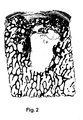
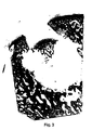
- FIGS. 2 to 4 illustrate the experiments already mentioned above Implants according to FIG. 1 in sheep joints and the mentioned risk that Repairs with such implants include. These are tissue sections by repair points parallel to the axis of the implant, which in the present Enlargement of the cartilage surface shown above about 7 cm in the bone tissue protrudes.
- Figures 2 and 3 show preparations that last six months were created after the implantation and in which cyst-like at the repair sites Cavities in the bone tissue are visible.
- Figure 2 shows a case in which the cartilage layer is still positioned in its original place, in Figure 3 they sank into the cyst room.
- Figure 4 shows an identical repair site after twelve months. It is clearly one the unevenness in the implanted cartilage layer Cartilage surface visible.
- repair sites were found at seven treated animals were examined after twelve months and in two cases bumps were found in the cartilage surface as found in FIG. 4. In the rest In cases, the cartilage surface in the repair area was flat.
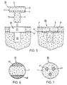
- FIG. 5 shows a preferred embodiment of the preparation according to the invention for the repair of cartilage or cartilage / bone defects in human or animal joints. It shows the preparation 10, for the implantation of the preparation 10 opening or bore 20 to be created and the inserted into the opening Specimen, the implant 30 (cuts along the axis of the specimen 10 or Opening 20).
- the preparation 10 has a bone part in the same way as the preparation of FIG. 1 1, a frontal cartilage layer 2, which forms a cartilage surface 3, and in the transition area between the bone part 1 and cartilage layer 2 a subchondral Bone plate 4.
- the preparation has a head part 11 with a larger one Cross section and a foot part 12 with a smaller cross section.
- the head part 11 essentially comprises the cartilage layer 2 and the subschondral bone plate 4, the foot part 12 essentially corresponds to the bone part 1.
- the foot part 12 has advantageously (but not necessarily) the shape of a circular cylinder or steep circular truncated cone and the head part 11 is on all sides above the bone part 1 before and is also circular cylindrical, for example.
- Auto grafts and grafts from living donors advantageously have cylindrical head parts, because such preparations should be as small as possible Cause removal opening.
- Preparations made from tissue from slaughter animals can be produced easily Have headboards with different shapes of cartilage surface because that Material is available in large quantities and can therefore also be processed with waste become. But even in this case it is advantageous to design the foot section in such a way that the opening to be created for the implantation is simple Tool, for example, can be created with a drill.
- a preparation 10 with a cross section that is at least round in the foot region is, for example made from a corresponding cylindrical preparation in that the bone part is processed accordingly.
- This processing can be done with a Tool can be carried out in which the cylindrical preparation can be positioned is and in the blades can be activated in a predetermined or adjustable Distance from the cartilage surface the cross-section of the preparation to a predetermined one Reduce measure.
- the Dimensioning of preparation 10 and bore 20 is for a press fit both in Area of the head part as well as in the foot part of the preparation.
- a bore 20, as shown in FIG. 5, is created, for example with a tool that has a blade with a circular cutting edge has and two drills axially limited relative to the cutting edge or hollow drill.
- the tool is positioned on the defect and the blade pressed down to the subchondral bone plate.
- the blade can be moved and its diameter substantially corresponds to the inner diameter of the blade, the outer region 21 is drilled out.
- the inner area 22 is subsequently drilled out with the second drill, whereby the drilling depth relative to the cutting edge or relative to the end position of the first Drill is adjustable.
- FIG. 5 also shows the implant 30 on the right-hand side, that is to say in the bore 20 used preparation 10.
- the implant 30 has one with the native cartilage surface 3 'aligned cartilage surface 3 and one with the native subchondral Bone plate 4 'approximately aligned subchondral bone plate 4. Die subchondral bone plate 4 of the implant is obviously supported on the one hand on the bone part 1 of the implant and on the other hand on native bone tissue immediately below or in the native, subchondral bone plate 4 '.
- a tool for implanting a preparation in a bore, as shown by FIG. 5 is used, for example, a tool that has a sleeve and an in the sleeve has axially movable plunger.
- the sleeve has an inner cross section, which corresponds to the cross section of the head part of the preparation to be implanted.
- the plunger advantageously has an approximately same cross section as this Headboard; it is longer than the sleeve and has a channel on one Front of the ram begins and in the area of the other ram end to one Suction line can be connected.
- the face of the plunger is inserted into the channel opening Pushed sleeve and a preparation is pulled into the sleeve with the suction force generated. Then the sleeve with pestle and the preparation sucked on it the prepared hole and the preparation is positioned using the pestle and, if necessary, a hammer is pressed into the bore.
- the head part it is recommended to use the supernatant dimension the head part to about 1 to 2 mm (e.g. bone part with diameter approx. 3 mm, head section with a diameter of 5 to 6 mm)
- Figures 6 and 7 show two further exemplary embodiments of the inventive Preparation. These are based on the idea of the subchondral bone plate of the preparation additionally not on native bone tissue, as in FIG. 5 shown, but on at least one created in the bone part of the preparation Support column, whereby the columns from the remaining bone material of the bone part 1 have a different degradability in such a way that they Time. in which the remaining bone material of the bone part is broken down, the wear subchondral bone plate and the overgrown cartilage layer and prevent sinking into the cyst area.
- Figures 6 and 7 are cross sections through bone parts 1 of inventive Preparations. These preparations are cylindrical, for example, and have a bone part axially extending columns, which consist of a material that is slower is degradable as the bone material of the areas between the pillars.
- the Columns are arranged, for example, on the surface of the bone part (surface columns 40 in Figure 6) and are treated appropriately with the bone material generated or they are located inside the bone part (inner Columns 41 in Figure 7) and are created by drilling and filling the holes with a suitable material. In both cases, the pillars are enough at least to the subchondral bone plate.
- Resorbable material for filling holes can be, for example, hydroxylapatite ceramic be used.
- Figures 8 and 9 show a cartilage defect (Figure 8) and a cartilage / bone defect ( Figure 9) with sizes such that they can not with a single Implant can be repaired. The defects are indicated by dash-dotted lines.
- the repair consists of a mosaic-like arrangement of inventive Preparations as shown in Figure 5.
- FIG. 8 shows that the implants 30 with the head part 11 are also in a mosaic repair a support for the cartilage layer and the subchondral bone plate of the implant allow on native bone tissue 1 'and thereby a decrease in Counteract the cartilage layer in a critical healing phase. It is also evident that the areas of native bone tissue 1 'between the foot parts 12 of the implants are wider than would be possible with cylindrical implants. That also means that the chances of a cure compared to the state of the art are improved. And it can be seen that the cartilage surface is all over Defect area can be designed essentially without interruption if the shape corresponding to the headboards (e.g. square, rectangular, triangular or hexagonal) is chosen.
- the shape corresponding to the headboards e.g. square, rectangular, triangular or hexagonal
- FIG. 9 shows a cartilage / bone defect (indicated by a dash-dotted line), which is repaired with a plurality of preparations according to FIG. 5.
- the foot parts 12 of the implants 30 extend into healthy bone tissue 1 '.
- a suitable material e.g. tricalcium phosphate or hydraulic Bone cement.
- This material should be chosen so that it is in front of or is only dismantled after the foot parts 12 of the implants 30, that is to say in the critical phase of the healing process, in which the bone parts of the implants degraded but not yet replaced, support the cartilage layers and sink in that can prevent cartilage layers.
- the filling material advantageously has sufficient mechanical strength, such that it is first filled in the defect and then can be pierced.
Landscapes
- Health & Medical Sciences (AREA)
- Life Sciences & Earth Sciences (AREA)
- General Health & Medical Sciences (AREA)
- Transplantation (AREA)
- Engineering & Computer Science (AREA)
- Biomedical Technology (AREA)
- Heart & Thoracic Surgery (AREA)
- Orthopedic Medicine & Surgery (AREA)
- Veterinary Medicine (AREA)
- Animal Behavior & Ethology (AREA)
- Public Health (AREA)
- Cardiology (AREA)
- Oral & Maxillofacial Surgery (AREA)
- Vascular Medicine (AREA)
- Surgery (AREA)
- Nuclear Medicine, Radiotherapy & Molecular Imaging (AREA)
- Molecular Biology (AREA)
- Medical Informatics (AREA)
- Physical Education & Sports Medicine (AREA)
- Rheumatology (AREA)
- Prostheses (AREA)
- Materials For Medical Uses (AREA)
Description
- Der Knochenteil des Präparates soll eine feste Verankerung des Implantates im gesunden Knochengewebe durch Pressfit ermöglichen, derart, dass das Implantat keine weiteren, gesunde Knorpelbereiche beeinflussende Befestigungsmittel benötigt.
- Die Verwachsung von Knorpelschicht und Knochenteil im Präparat soll dem Implantat eine Stabilität geben, derart, dass die Knorpelschicht auch ohne Stilllegung des betroffenen Gelenkes sich nicht ablösen und aus der Defektstelle entfernen kann.
- Die Knorpelschicht des Präparates soll eine derartige mechanische Festigkeit und Elastizität aufweisen, dass die Reparaturstelle schon unmittelbar nach der Implantation voll belastbar ist.
- Die Knorpelschicht soll eine Zone bilden, in der für implantierte Zellen oder für nach der Implantation zuwandernde Zellen geeignete Bedingungen für die Erzeugung und/oder Aufrechterhaltung von funktionsfähigem Knorpelgewebe herrschen. Dies soll auch unterstützt werden durch die subchondrale Knochenplatte, die die Knorpelschicht vom Knochenteil trennt und die hilft, eine vom Knochenteil ausgehende Vaskularisierung der Knorpelschicht zu verhindern.
- Der Knochenteil soll eine Zone darstellen, in der für implantierte Zellen oder für nach der Implantation zuwandernde Zellen geeignete Bedingungen für die Erzeugung und/oder für die Aufrechterhaltung von funktionsfähigem Knochengewebe herrschen.
- Figur 1
- ein zylinderförmiges Präparat gemäss dem Stande der Technik zur Reparatur von Knorpel- oder Knorpel/Knochen-Defekten in menschlichen oder tierischen Gelenken;
- Figuren 2 bis 4
- Gewebeschnitte durch mit einem Präparat gemäss Figur 1 reparierte Knorpel/Knochen-Defekte in einem Schafgelenk zu verschiedenen Zeitpunkten nach der Implantation;
- Figur 5
- ein Schema der Reparatur eines Knorpel-Defektes mit Hilfe einer bevorzugten Ausführungsform des erfindungsgemässen Präparates (Schnitte entlang der Achse des Präparates bzw. der Bohrung);
- Figuren 6 und 7
- zwei weitere beispielhafte Ausführungsformen des erfindungsgemässen Präparates im Querschnitt;
- Figuren 8 und 9
- Schnitte durch einen Knorpel-Defekt (Figur 8) und durch einen Knorpel/Knochen-Defekt (Figur 9), die beide mosaikartig mit einer Mehrzahl von erfindungsgemässen Präparaten gemäss Figur 5 repariert sind.
Claims (14)
- Präparat (10) zur Reparatur von Knorpel- oder Knorpel/Knochen-Defekten in menschlichen oder tierischen Gelenken, welches Präparat einen Knochenteil (1), eine auf den Knochenteil (1) aufgewachsene, eine Knorpeloberfläche (3) bildende Knorpelschicht (2) und im Übergangsbereich zwischen Knochenteil (1) und Knorpelschicht (2) eine subchondrale Knochenplatte (4) oder eine Nachbildung einer derartigen Knochenplatte aufweist und welches Präparat für die Reparatur derart in eine im Defektbereich erstellte Öffnung oder Bohrung (20) implantierbar ist, dass der Knochenteil in gesundern Knochengewebe verankert ist und die Knorpeloberfläche (3) mit einer nativen Knorpeloberfläche (3') fluchtet, dadurch gekennzeichnet, dass für eine Abstützung der subchondralen Knochenplatte (4) und der Knorpelschicht (2) des implantierten Präparates auf mindestens zwei verschieden abbaubaren Materialien der Knochenteil (1) einen kleineren Querschnitt als die subchondrale Platte (4) und die Knorpelschicht (2) aufweist oder der Knochenteil (1) bis zur subchondralen Knochenplatte (4) verlaufende Säulenbereiche (40, 41) aufweist, die ein anderes Abbauverhalten aufweisen als die Bereiche zwischen den Säulenbereichen.
- Präparat nach Anspruch 1, dadurch gekennzeichnet, dass es einen Kopfteil (11) und einen Fussteil (12) aufweist, wobei der Kopfteil (11) im wesentlichen die Knorpelschicht (2) und die subchondrale Knochenplatte (4) umfasst und der Fussteil (12) im wesentlichen dem Knochenteil entspricht und wobei die subchondrale Knochenplatte (4) grösser ist als ein dazu paralleler Querschnitt durch den Fussteil (12), derart, dass die subchondrale Knochenplatte (4) des implantierten Präparates (30) auf nativem Knochengewebe abstützbar ist.
- Präparat nach Anspruch 2, dadurch gekennzeichnet, dass der Kopfteil (11) und der Fussteil (12) koaxiale Kreiszylinder sind.
- Präparat nach Anspruch 2, dadurch gekennzeichnet, dass der Fussteil (12) ein Kreiszylinder ist und dass der Kopfteil (11) eine quadratische, rechteckige, dreieckige oder sechseckige Knorpeloberfläche (3) aufweist.
- Präparat nach Anspruch 1, dadurch gekennzeichnet, dass die Saülenbereiche (40, 41) senkrecht zur subchondralen Knochenplatte (4) durch den Knochenteil (1) bis zur subchondralen Knochenplatte (4) verlaufen.
- Präparat nach Anspruch 5, dadurch gekennzeichnet, dass es im wesentlichen zylinderförmig ist und die Knorpelschicht (3) stirnseitig angeordnet ist.
- Präparat nach Anspruch 5 oder 6, dadurch gekennzeichnet, dass das Abbauverhalten der Säulenbereiche (40) durch eine chemische Behandlung gegenüber dem Abbauverhalten der Bereiche zwischen den Säulenbereichen verändert ist.
- Präparat nach Anspruch 7, dadurch gekennzeichnet, dass die Säulenbereiche (40) aus mit Biphosphonat behandeltem Knochengewebe bestehen.
- Präparat nach Anspruch 5 oder 6, dadurch gekennzeichnet, dass die Säulenbereiche (41) aus einem in entsprechende axiale Bohrungen eingefüllten Knochenersatzmaterial bestehen.
- Präparat nach einem der Ansprüche 1 bis 9, dadurch gekennzeichnet, dass es aus autologem Gewebe hergestellt ist.
- Präparat nach einem der Ansprüche 1 bis 9, dadurch gekennzeichnet, dass es aus homologem oder heterologem, immunologisch desaktiviertem Gewebe hergestellt ist.
- Präparat nach Anspruch 11, dadurch gekennzeichnet, dass es aus durch Photooxidation immunologisch desaktiviertem Gewebe hergestellt ist.
- Präparat nach einem der Ansprüche 11 oder 12, dadurch gekennzeichnet, dass es aus aus Schlachttieren entnommenem Gewebe besteht.
- Präparat nach Anspruch 13, dadurch gekennzeichnet, dass es aus Rinder-oder Schweinegelenken entnommen ist.
Priority Applications (1)
| Application Number | Priority Date | Filing Date | Title |
|---|---|---|---|
| EP04020622A EP1498089A1 (de) | 1999-12-15 | 2000-12-12 | Präparat für die Reparatur von Knorpel- oder Knorpel/Knochen-Defekten in menschlichen oder tierischen Gelenken |
Applications Claiming Priority (3)
| Application Number | Priority Date | Filing Date | Title |
|---|---|---|---|
| CH229699 | 1999-12-15 | ||
| CH229699 | 1999-12-15 | ||
| PCT/CH2000/000659 WO2001043667A1 (de) | 1999-12-15 | 2000-12-12 | Präparat für die reparatur von knorpel- oder knorpel/knochen-defekten in menschlichen oder tierischen gelenken |
Related Child Applications (1)
| Application Number | Title | Priority Date | Filing Date |
|---|---|---|---|
| EP04020622A Division EP1498089A1 (de) | 1999-12-15 | 2000-12-12 | Präparat für die Reparatur von Knorpel- oder Knorpel/Knochen-Defekten in menschlichen oder tierischen Gelenken |
Publications (2)
| Publication Number | Publication Date |
|---|---|
| EP1237511A1 EP1237511A1 (de) | 2002-09-11 |
| EP1237511B1 true EP1237511B1 (de) | 2004-09-01 |
Family
ID=4230469
Family Applications (2)
| Application Number | Title | Priority Date | Filing Date |
|---|---|---|---|
| EP00979315A Expired - Lifetime EP1237511B1 (de) | 1999-12-15 | 2000-12-12 | Präparat für die reparatur von knorpel- oder knorpel/knochen-defekten in menschlichen oder tierischen gelenken |
| EP04020622A Withdrawn EP1498089A1 (de) | 1999-12-15 | 2000-12-12 | Präparat für die Reparatur von Knorpel- oder Knorpel/Knochen-Defekten in menschlichen oder tierischen Gelenken |
Family Applications After (1)
| Application Number | Title | Priority Date | Filing Date |
|---|---|---|---|
| EP04020622A Withdrawn EP1498089A1 (de) | 1999-12-15 | 2000-12-12 | Präparat für die Reparatur von Knorpel- oder Knorpel/Knochen-Defekten in menschlichen oder tierischen Gelenken |
Country Status (9)
| Country | Link |
|---|---|
| US (2) | US6858042B2 (de) |
| EP (2) | EP1237511B1 (de) |
| JP (1) | JP2003516803A (de) |
| AT (1) | ATE274875T1 (de) |
| AU (1) | AU783038B2 (de) |
| CA (1) | CA2393776C (de) |
| DE (1) | DE50007640D1 (de) |
| ES (1) | ES2230170T3 (de) |
| WO (1) | WO2001043667A1 (de) |
Cited By (8)
| Publication number | Priority date | Publication date | Assignee | Title |
|---|---|---|---|---|
| US7488348B2 (en) | 2003-05-16 | 2009-02-10 | Musculoskeletal Transplant Foundation | Cartilage allograft plug |
| US7815926B2 (en) | 2005-07-11 | 2010-10-19 | Musculoskeletal Transplant Foundation | Implant for articular cartilage repair |
| US7837740B2 (en) | 2007-01-24 | 2010-11-23 | Musculoskeletal Transplant Foundation | Two piece cancellous construct for cartilage repair |
| US7901457B2 (en) | 2003-05-16 | 2011-03-08 | Musculoskeletal Transplant Foundation | Cartilage allograft plug |
| USRE42208E1 (en) | 2003-04-29 | 2011-03-08 | Musculoskeletal Transplant Foundation | Glue for cartilage repair |
| US8292968B2 (en) | 2004-10-12 | 2012-10-23 | Musculoskeletal Transplant Foundation | Cancellous constructs, cartilage particles and combinations of cancellous constructs and cartilage particles |
| US8435551B2 (en) | 2007-03-06 | 2013-05-07 | Musculoskeletal Transplant Foundation | Cancellous construct with support ring for repair of osteochondral defects |
| US8753406B2 (en) | 2010-08-31 | 2014-06-17 | Zimmer Inc. | Osteochondral graft delivery device and uses thereof |
Families Citing this family (69)
| Publication number | Priority date | Publication date | Assignee | Title |
|---|---|---|---|---|
| EP0896825B1 (de) | 1997-08-14 | 2002-07-17 | Sulzer Innotec Ag | Zusammensetzung und Vorrichtung zur Reparatur von Knorpelgewebe in vivo bestehend aus Nanokapseln mit osteoinduktiven und/oder chondroinduktiven Faktoren |
| US8563232B2 (en) | 2000-09-12 | 2013-10-22 | Lifenet Health | Process for devitalizing soft-tissue engineered medical implants, and devitalized soft-tissue medical implants produced |
| EP1099443A1 (de) | 1999-11-11 | 2001-05-16 | Sulzer Orthopedics Ltd. | Transplantations/Implantatsvorrichtung und ihre Herstellungsmethode |
| US6858042B2 (en) | 1999-12-15 | 2005-02-22 | Zimmer Orthobiologics, Inc. | Preparation for repairing cartilage defects or cartilage/bone defects in human or animal joints |
| US6626950B2 (en) | 2001-06-28 | 2003-09-30 | Ethicon, Inc. | Composite scaffold with post anchor for the repair and regeneration of tissue |
| BR0307033A (pt) * | 2002-01-22 | 2004-12-07 | Pfizer | ácidos 3-(imidazolil)-2-aminopropanóicos para o uso como inibidores tafi-a para o tratamento de doenças trombóticas |
| AU2004216551B2 (en) * | 2003-02-26 | 2008-09-18 | Zimmer Orthobiologics, Inc. | Preparation for repairing cartilage tissue, especially articular cartilage defects |
| US20050222687A1 (en) * | 2004-04-02 | 2005-10-06 | Gordana Vunjak-Novakovic | Cartilage implant assembly and method for implantation |
| US20050064042A1 (en) * | 2003-04-29 | 2005-03-24 | Musculoskeletal Transplant Foundation | Cartilage implant plug with fibrin glue and method for implantation |
| US20090291112A1 (en) * | 2003-05-16 | 2009-11-26 | Truncale Katherine G | Allograft osteochondral plug combined with cartilage particle mixture |
| GB0325141D0 (en) * | 2003-10-28 | 2003-12-03 | Xiros Plc | Repair of damaged tissue on a bone site |
| CN1972644B (zh) * | 2003-11-21 | 2010-08-25 | 骨棚国际私人有限公司 | 用于骨组织再生的生物可吸收插销式植入体和方法 |
| US20070185585A1 (en) * | 2004-03-09 | 2007-08-09 | Brat Bracy | Implant Scaffold Combined With Autologous Tissue, Allogenic Tissue, Cultured Tissue, or combinations Thereof |
| CA2555586A1 (en) * | 2004-03-09 | 2005-09-22 | Osteobiologics, Inc. | Implant scaffold combined with autologous or allogenic tissue |
| US20090149893A1 (en) * | 2007-12-05 | 2009-06-11 | Semler Eric J | Cancellous Bone Implant for Cartilage Repair |
| US20080220044A1 (en) * | 2007-03-06 | 2008-09-11 | Semler Eric J | Cancellous construct with support ring for repair of osteochondral defects |
| US8071574B2 (en) * | 2005-02-22 | 2011-12-06 | John Dennis Bobyn | Implant improving local bone formation |
| CA2623106C (en) | 2005-09-19 | 2013-12-24 | Histogenics Corporation | Cell-support matrix having narrowly defined uniformly vertically and non-randomly organized porosity and pore density and a method for preparation thereof |
| EP1764117A1 (de) | 2005-09-20 | 2007-03-21 | Zimmer GmbH | Implantat zur Wiederherstellung von Knorpeldefekten und Verfahren zu seiner Herstellung |
| RU2307399C1 (ru) * | 2006-03-03 | 2007-09-27 | Федеральное агентство по здравоохранению и социальному развитию Федеральное государственное учреждение "Российский научно-исследовательский институт травматологии и ортопедии им. Р.Р. Вредена Федерального агентства по здравоохранению и социальному развитию" (ФГУ "РНИИТО им. Р.Р. Вредена Росздрава") | Способ моделирования хронического полнослойного локального дефекта хряща коленного сустава в эксперименте на собаках |
| US20080125863A1 (en) * | 2006-11-28 | 2008-05-29 | Mckay William F | Implant designs and methods of improving cartilage repair |
| US7758643B2 (en) * | 2007-02-26 | 2010-07-20 | Biomet Sports Medicine, Llc | Stable cartilage defect repair plug |
| US20080294270A1 (en) * | 2007-05-24 | 2008-11-27 | Zimmer Orthobiologics, Inc. | Differentially processed tissue and processing methods thereof |
| US9744043B2 (en) | 2007-07-16 | 2017-08-29 | Lifenet Health | Crafting of cartilage |
| US20090054906A1 (en) * | 2007-08-24 | 2009-02-26 | Zimmer Orthobiologics, Inc. | Medical device and method for delivering an implant to an anatomical site |
| CA2711847C (en) * | 2008-01-09 | 2018-05-08 | Innovative Health Technologies, Llc | Implant pellets and methods for performing bone augmentation and preservation |
| US8801725B2 (en) | 2008-03-10 | 2014-08-12 | Zimmer Orthobiologics, Inc. | Instruments and methods used when repairing a defect on a tissue surface |
| GB0809721D0 (en) * | 2008-05-28 | 2008-07-02 | Univ Bath | Improvements in or relating to joints and/or implants |
| WO2010014446A1 (en) * | 2008-07-28 | 2010-02-04 | Zimmer, Inc. | Mosaicplasty constructs |
| US8308814B2 (en) | 2009-03-27 | 2012-11-13 | Depuy Mitek, Inc. | Methods and devices for preparing and implanting tissue scaffolds |
| US8241298B2 (en) | 2009-03-27 | 2012-08-14 | Depuy Mitek, Inc. | Methods and devices for delivering and affixing tissue scaffolds |
| US8556972B2 (en) * | 2009-04-02 | 2013-10-15 | Sevika Holding AG | Monolithic orthopedic implant with an articular finished surface |
| US20100256758A1 (en) * | 2009-04-02 | 2010-10-07 | Synvasive Technology, Inc. | Monolithic orthopedic implant with an articular finished surface |
| US20110257753A1 (en) * | 2009-04-02 | 2011-10-20 | Synvasive Technology, Inc. | Implant having a convex surface surrounding a concave articular surface |
| US20100331998A1 (en) * | 2009-06-30 | 2010-12-30 | Ringeisen Timothy A | Electrokinetic device for tissue repair |
| US9744123B2 (en) * | 2009-06-30 | 2017-08-29 | Kensey Nash Corporation | Biphasic implant device providing gradient |
| US10016278B2 (en) * | 2009-06-30 | 2018-07-10 | Dsm Ip Assets B.V. | Biphasic implant device providing joint fluid therapy |
| US8858592B2 (en) * | 2009-11-24 | 2014-10-14 | Covidien Lp | Wound plugs |
| US8994666B2 (en) * | 2009-12-23 | 2015-03-31 | Colin J. Karpfinger | Tactile touch-sensing interface system |
| US8444699B2 (en) * | 2010-02-18 | 2013-05-21 | Biomet Manufacturing Corp. | Method and apparatus for augmenting bone defects |
| IT1398443B1 (it) * | 2010-02-26 | 2013-02-22 | Lima Lto S P A Ora Limacorporate Spa | Elemento protesico integrato |
| US20110288652A1 (en) * | 2010-05-20 | 2011-11-24 | Indiana University Research & Technology Corporation | Materials and methods for treating critically sized defects in mouse bone |
| EP2389904B1 (de) * | 2010-05-24 | 2013-07-24 | Episurf IP Management AB | OP-Kit zur Knorpelreparatur mit einem Implantat und einem Werkzeugsatz |
| EP2389899B1 (de) | 2010-05-24 | 2015-04-29 | Episurf IP Management AB | Methode zur Gestaltung eines OP-Kits zur Knorpelreparatur in einem Gelenk |
| EP2389901B8 (de) | 2010-05-24 | 2013-05-15 | Episurf IP Management AB | Implantat zur Knorpelreparatur |
| EP2389905B1 (de) | 2010-05-24 | 2012-05-23 | Episurf Medical AB | Methode zur Gestaltung eines OP-Kits zur Knorpelreparatur in einem Gelenk |
| US9113916B2 (en) | 2010-08-31 | 2015-08-25 | Zimmer, Inc. | Drill bit for osteochondral drilling with guiding element and uses thereof |
| US8641718B2 (en) * | 2010-10-19 | 2014-02-04 | Biomet Manufacturing, Llc | Method and apparatus for harvesting cartilage for treatment of a cartilage defect |
| EP2640430B1 (de) * | 2010-11-17 | 2016-03-09 | Zimmer, Inc. | Keramische monoblockimplantate mit osseointegrationsbefestigungsflächen |
| US8551525B2 (en) | 2010-12-23 | 2013-10-08 | Biostructures, Llc | Bone graft materials and methods |
| US11000387B2 (en) | 2011-09-02 | 2021-05-11 | Episurf Ip-Management Ab | Implant for cartilage repair |
| EP2564792A1 (de) | 2011-09-02 | 2013-03-06 | Episurf Medical AB | Modularer Chirurgensatz zur Knorpelreparatur |
| US10603049B2 (en) | 2011-09-02 | 2020-03-31 | Episurf Ip-Management Ab | Implant specific drill bit in surgical kit for cartilage repair |
| US9498334B2 (en) * | 2012-03-27 | 2016-11-22 | DePuy Synthes Products, Inc. | Glenoid defect-filling component |
| WO2014130883A2 (en) | 2013-02-22 | 2014-08-28 | Allosource | Cartilage mosaic compositions and methods |
| KR102312720B1 (ko) | 2013-03-15 | 2021-10-13 | 알로소스 | 연조직 회복 및 재생을 위한 세포 재배치된 콜라겐 매트릭스 |
| EP2967874B1 (de) | 2013-03-15 | 2019-11-20 | AlloSource | Perforierte osteochondrale allograft-zusammensetzungen |
| AU2015229405B2 (en) | 2014-03-11 | 2019-10-31 | The Trustees Of Columbia University In The City Of New York | Customized bendable osteochondral allografts |
| WO2016004991A1 (en) | 2014-07-09 | 2016-01-14 | Episurf Ip-Management Ab | Customized implant for cartilage repair and corresponding method of design |
| US20170172744A1 (en) | 2014-07-09 | 2017-06-22 | Episurf Ip-Management Ab | Surgical joint implant and a bone-mountable rig |
| US10077420B2 (en) | 2014-12-02 | 2018-09-18 | Histogenics Corporation | Cell and tissue culture container |
| EP3316798B1 (de) * | 2015-07-02 | 2024-05-22 | Episurf IP-Management AB | Entwurfsverfahren für ein system zur durchführung von osteochondraler transplantationschirurgie in einem gelenk |
| CN108495595B (zh) | 2015-11-25 | 2022-02-01 | 软骨解决方案股份有限公司 | 用于修复解剖关节病症的方法、系统和设备 |
| US9943414B2 (en) * | 2015-12-30 | 2018-04-17 | Wasas, Llc. | System and method for non-binding allograft subtalar joint implant |
| US10786370B2 (en) * | 2017-12-28 | 2020-09-29 | Industrial Technology Research Institute | Cartilage repair implant, auxiliary surgical tool kit and cartilage repair system |
| CN109966029A (zh) * | 2017-12-28 | 2019-07-05 | 财团法人工业技术研究院 | 软骨修复载体、所搭配的手术用具组与软骨修复系统 |
| IL275811B2 (en) | 2018-01-02 | 2025-05-01 | Cartiheal 2009 Ltd | A tool and protocol for optimal implantation of a solid implant that promotes cell and tissue growth |
| SE543241C2 (en) | 2018-04-27 | 2020-10-27 | Episurf Ip Man Ab | An implant for cartilage and / or bone repair |
| CN116019610B (zh) * | 2023-01-09 | 2023-07-25 | 北京大学口腔医学院 | 一种多层帽型垂直增高颌骨缺损修复体及其制备方法 |
Family Cites Families (113)
| Publication number | Priority date | Publication date | Assignee | Title |
|---|---|---|---|---|
| US1703154A (en) | 1927-05-13 | 1929-02-26 | Lanzkron Anna | Vegetable slicer |
| US3971273A (en) | 1975-02-26 | 1976-07-27 | Leo Peters | Meat slicing board |
| CH632923A5 (en) | 1978-10-06 | 1982-11-15 | Sulzer Ag | Implant for partial replacement of a sliding surface of a human joint |
| US4932973A (en) | 1983-09-30 | 1990-06-12 | El Gendler | Cartilage and bone induction by artificially perforated organic bone matrix |
| US4589206A (en) | 1984-09-24 | 1986-05-20 | Emery Marcoux | Slicing knife and board |
| US4627853A (en) | 1985-05-29 | 1986-12-09 | American Hospital Supply Corporation | Method of producing prostheses for replacement of articular cartilage and prostheses so produced |
| IL76079A (en) | 1985-08-13 | 1991-03-10 | Univ Ramot | Collagen implants |
| DE3787077T2 (de) | 1986-05-15 | 1994-01-05 | Sumitomo Cement Co | Knochenimplantat. |
| US5112354A (en) | 1989-11-16 | 1992-05-12 | Northwestern University | Bone allograft material and method |
| US5152791A (en) * | 1989-12-07 | 1992-10-06 | Olympus Optical Co., Ltd. | Prosthetic artificial bone having ceramic layers of different porosity |
| JP2951342B2 (ja) * | 1989-12-07 | 1999-09-20 | オリンパス光学工業株式会社 | 人工骨補綴部材 |
| ES2088690T3 (es) * | 1990-12-19 | 1996-08-16 | Harle Anton | Procedimiento para la fabricacion de un implante medico e implante fabricado de acuerdo con dicho procedimiento. |
| US5320115A (en) | 1991-01-16 | 1994-06-14 | Applied Biological Concepts | Method and apparatus for arthroscopic knee surgery |
| US5853746A (en) | 1991-01-31 | 1998-12-29 | Robert Francis Shaw | Methods and compositions for the treatment and repair of defects or lesions in cartilage or bone using functional barrier |
| US5115704A (en) | 1991-06-03 | 1992-05-26 | International Coffee & Tea Inc. | Bread cutter box |
| US5116337A (en) | 1991-06-27 | 1992-05-26 | Johnson Lanny L | Fixation screw and method for ligament reconstruction |
| DE69307299T2 (de) * | 1992-02-14 | 1997-04-30 | Board of Regents, the University of Texas System, Austin, Tex. | Mehrphasiges, biologisch abbaubares implantat/träger und verfahren zu seiner herstellung |
| US6013853A (en) | 1992-02-14 | 2000-01-11 | The University Of Texas System | Continuous release polymeric implant carrier |
| US5876452A (en) * | 1992-02-14 | 1999-03-02 | Board Of Regents, University Of Texas System | Biodegradable implant |
| US5320626A (en) | 1992-02-19 | 1994-06-14 | Arthrex Inc. | Endoscopic drill guide |
| SE9201670D0 (sv) | 1992-05-27 | 1992-05-27 | Astra Ab | Method of treating surgical drill, surgical drill and use of surgical drill |
| US5370692A (en) | 1992-08-14 | 1994-12-06 | Guild Associates, Inc. | Rapid, customized bone prosthesis |
| DE4302709C1 (de) | 1993-02-01 | 1994-07-28 | Kirsch Axel | Abdeckeinrichtung mit Abdeckmembran |
| DE4317448A1 (de) | 1993-05-19 | 1994-11-24 | Gross Ulrich | Gelenkflächenersatz |
| US5906827A (en) | 1994-06-03 | 1999-05-25 | Creative Biomolecules, Inc. | Matrix for the manufacture of autogenous replacement body parts |
| US5632745A (en) | 1995-02-07 | 1997-05-27 | R&D Biologicals, Inc. | Surgical implantation of cartilage repair unit |
| US5769899A (en) | 1994-08-12 | 1998-06-23 | Matrix Biotechnologies, Inc. | Cartilage repair unit |
| HU214568B (hu) | 1994-09-16 | 1998-04-28 | METRIMED Orvosi Műszergyártó Kft. | Műszerkészlet oszteokondrális graftok mozaiktechnikával történő átültetéséhez |
| US6080194A (en) | 1995-02-10 | 2000-06-27 | The Hospital For Joint Disease Orthopaedic Institute | Multi-stage collagen-based template or implant for use in the repair of cartilage lesions |
| US5919196A (en) | 1995-02-16 | 1999-07-06 | Arthrex, Inc. | Method and apparatus for osteochondral autograft transplantation |
| US5603716A (en) | 1995-02-16 | 1997-02-18 | Arthrex Inc. | Method of ligament reconstruction using double socket graft placement and fixation |
| US6592588B1 (en) | 1995-02-16 | 2003-07-15 | Arthrex, Inc. | Apparatus for osteochondral autograft transplantation |
| US7141072B2 (en) * | 1998-10-05 | 2006-11-28 | Ed. Geistlich Soehne Ag Fuer Chemische Industrie | Method for promoting regeneration of surface cartilage in a damaged joint using multi-layer covering |
| US5782835A (en) | 1995-03-07 | 1998-07-21 | Innovasive Devices, Inc. | Apparatus and methods for articular cartilage defect repair |
| US5632747A (en) * | 1995-03-15 | 1997-05-27 | Osteotech, Inc. | Bone dowel cutter |
| WO1996034955A1 (en) | 1995-05-03 | 1996-11-07 | Warner-Lambert Company | Method of treating cartilaginous diseases with genetically modified chondrocytes |
| US5741685A (en) | 1995-06-07 | 1998-04-21 | Children's Medical Center Corporation | Parenchymal cells packaged in immunoprotective tissue for implantation |
| US5655546A (en) | 1995-06-07 | 1997-08-12 | Halpern; Alan A. | Method for cartilage repair |
| US7291149B1 (en) | 1995-06-07 | 2007-11-06 | Warsaw Orthopedic, Inc. | Method for inserting interbody spinal fusion implants |
| DE19520867C2 (de) | 1995-06-08 | 1998-05-20 | Kirsch Axel | Abdeckmembran |
| DE59509135D1 (de) | 1995-10-11 | 2001-05-03 | Sulzer Markets & Technology Ag | Verfahren zu einer photooxidativen Behandlung von kollagenhaltigen Geweben |
| US5788625A (en) | 1996-04-05 | 1998-08-04 | Depuy Orthopaedics, Inc. | Method of making reconstructive SIS structure for cartilaginous elements in situ |
| ATE250666T1 (de) * | 1996-06-04 | 2003-10-15 | Sulzer Orthopedics Ltd | Verfahren zur herstellung von knorpelgewebe und von implantaten |
| US5935172A (en) | 1996-06-28 | 1999-08-10 | Johnson & Johnson Professional, Inc. | Prosthesis with variable fit and strain distribution |
| US6126690A (en) | 1996-07-03 | 2000-10-03 | The Trustees Of Columbia University In The City Of New York | Anatomically correct prosthesis and method and apparatus for manufacturing prosthesis |
| US5881733A (en) | 1996-09-13 | 1999-03-16 | Depuy Orthopaedic Technology, Inc. | Technique for osteocartilaginous transplantation in a mammalian joint |
| US7468075B2 (en) | 2001-05-25 | 2008-12-23 | Conformis, Inc. | Methods and compositions for articular repair |
| US5718707A (en) * | 1997-01-22 | 1998-02-17 | Mikhail; W. E. Michael | Method and apparatus for positioning and compacting bone graft |
| US6126919A (en) * | 1997-02-07 | 2000-10-03 | 3M Innovative Properties Company | Biocompatible compounds for pharmaceutical drug delivery systems |
| US6146385A (en) | 1997-02-11 | 2000-11-14 | Smith & Nephew, Inc. | Repairing cartilage |
| US5885293A (en) | 1997-03-03 | 1999-03-23 | Innovasive Devices, Inc. | Apparatus and method for cutting a surface at a repeatable angle |
| JP3669107B2 (ja) * | 1997-03-17 | 2005-07-06 | 東レ株式会社 | 繊維構造物 |
| KR100541994B1 (ko) | 1997-04-25 | 2006-04-12 | 술저 오소패디 악티엔게젤샤프트 | 연골내 또는 골연골의 구멍을 만드는 장치 |
| FR2762776B1 (fr) | 1997-04-30 | 1999-06-04 | Implant Diffusion | Foret chirurgical recuperateur d'os comportant une butee a repere en couleur |
| US5993844A (en) | 1997-05-08 | 1999-11-30 | Organogenesis, Inc. | Chemical treatment, without detergents or enzymes, of tissue to form an acellular, collagenous matrix |
| US6113640A (en) * | 1997-06-11 | 2000-09-05 | Bionx Implants Oy | Reconstructive bioabsorbable joint prosthesis |
| JP2001520911A (ja) | 1997-10-27 | 2001-11-06 | ズルツァー オーソピーディクス リミテッド | 軟骨内および骨軟骨の欠損症を修復するための方法および器具 |
| US6123731A (en) | 1998-02-06 | 2000-09-26 | Osteotech, Inc. | Osteoimplant and method for its manufacture |
| US6293970B1 (en) | 1998-06-30 | 2001-09-25 | Lifenet | Plasticized bone and soft tissue grafts and methods of making and using same |
| US6193722B1 (en) | 1998-09-29 | 2001-02-27 | Sulzer Orthopaedic Ag | Hollow milling tool |
| US6025538A (en) | 1998-11-20 | 2000-02-15 | Musculoskeletal Transplant Foundation | Compound bone structure fabricated from allograft tissue |
| US6727224B1 (en) | 1999-02-01 | 2004-04-27 | Genetics Institute, Llc. | Methods and compositions for healing and repair of articular cartilage |
| DE50009320D1 (de) | 1999-03-23 | 2005-03-03 | Zimmer Gmbh Winterthur | Instrument und Instrumentensatz zum Einbringen eines osteochondralen Transplantats |
| US6251143B1 (en) * | 1999-06-04 | 2001-06-26 | Depuy Orthopaedics, Inc. | Cartilage repair unit |
| WO2001005336A1 (de) | 1999-07-16 | 2001-01-25 | Eska Implants Gmbh & Co. | Implantat zum ausfüllen von knorpel- und knochendefekten in gelenken |
| DE19950406A1 (de) | 1999-10-20 | 2001-05-31 | Volkmar Jansson | System zur Deckung von Knorpeldefekten mittels Knorpelersatzstrukturen |
| DE19952550A1 (de) | 1999-11-02 | 2001-05-03 | Tutogen Medical Gmbh | Knochenimplantat |
| CA2638625C (en) * | 1999-12-03 | 2012-07-24 | University Of Leeds | Fixation technology for bone repair |
| US6858042B2 (en) | 1999-12-15 | 2005-02-22 | Zimmer Orthobiologics, Inc. | Preparation for repairing cartilage defects or cartilage/bone defects in human or animal joints |
| US6591581B2 (en) | 2000-03-08 | 2003-07-15 | Arthrex, Inc. | Method for preparing and inserting round, size specific osteochondral cores in the knee |
| US6632246B1 (en) | 2000-03-14 | 2003-10-14 | Chondrosite, Llc | Cartilage repair plug |
| US6626945B2 (en) | 2000-03-14 | 2003-09-30 | Chondrosite, Llc | Cartilage repair plug |
| EP2314257B9 (de) | 2000-05-01 | 2013-02-27 | ArthroSurface, Inc. | System zur Gelenkoberflächenerneuerung |
| US6679917B2 (en) | 2000-05-01 | 2004-01-20 | Arthrosurface, Incorporated | System and method for joint resurface repair |
| US6488033B1 (en) | 2000-05-15 | 2002-12-03 | Cryolife, Inc. | Osteochondral transplant techniques |
| US6440141B1 (en) | 2000-07-24 | 2002-08-27 | Oratec Interventions, Inc. | Method and apparatus for treating osteochondral pathologies |
| US6575986B2 (en) | 2001-02-26 | 2003-06-10 | Ethicon, Inc. | Scaffold fixation device for use in articular cartilage repair |
| US6699252B2 (en) | 2001-04-17 | 2004-03-02 | Regeneration Technologies, Inc. | Methods and instruments for improved meniscus transplantation |
| US6626950B2 (en) | 2001-06-28 | 2003-09-30 | Ethicon, Inc. | Composite scaffold with post anchor for the repair and regeneration of tissue |
| ATE499908T1 (de) | 2001-07-16 | 2011-03-15 | Depuy Products Inc | Gerät zur reparatur von knorpelmaterial |
| ATE314828T1 (de) | 2001-09-27 | 2006-02-15 | Zimmer Spine Inc | Modulare zwischenwirbelfusionierungseinrichtung und herstellungsverfahren dafür |
| US7931687B2 (en) | 2002-05-13 | 2011-04-26 | Articular Engineering, Llc | Tissue engineered osteochondral implant |
| US8187326B2 (en) | 2002-05-22 | 2012-05-29 | Advanced Technologies And Regenerative Medicine, Llc. | Attachment of absorbable tissue scaffolds to fixation devices |
| US6989034B2 (en) | 2002-05-31 | 2006-01-24 | Ethicon, Inc. | Attachment of absorbable tissue scaffolds to fixation devices |
| US7591820B2 (en) | 2002-08-09 | 2009-09-22 | Arthrex, Inc. | Retrograde osteochondral autograft transfer system |
| US7264634B2 (en) | 2002-09-20 | 2007-09-04 | Arthrex, Inc. | Method and instrumentation for osteochondral repair using preformed implants |
| US6960214B2 (en) | 2002-10-15 | 2005-11-01 | Zimmer Austin, Inc. | Method for performing automated microfracture |
| US20040147932A1 (en) | 2002-10-15 | 2004-07-29 | Brian Burkinshaw | Device for performing automated microfracture |
| AU2004216551B2 (en) | 2003-02-26 | 2008-09-18 | Zimmer Orthobiologics, Inc. | Preparation for repairing cartilage tissue, especially articular cartilage defects |
| US7160305B2 (en) | 2003-03-07 | 2007-01-09 | Arthrex, Inc. | Retrodrill technique for insertion of autograft, allograft or synthetic osteochondral implants |
| US20050222687A1 (en) | 2004-04-02 | 2005-10-06 | Gordana Vunjak-Novakovic | Cartilage implant assembly and method for implantation |
| US20050064042A1 (en) | 2003-04-29 | 2005-03-24 | Musculoskeletal Transplant Foundation | Cartilage implant plug with fibrin glue and method for implantation |
| US7488348B2 (en) | 2003-05-16 | 2009-02-10 | Musculoskeletal Transplant Foundation | Cartilage allograft plug |
| US7901457B2 (en) | 2003-05-16 | 2011-03-08 | Musculoskeletal Transplant Foundation | Cartilage allograft plug |
| US7217294B2 (en) | 2003-08-20 | 2007-05-15 | Histogenics Corp. | Acellular matrix implants for treatment of articular cartilage, bone or osteochondral defects and injuries and method for use thereof |
| EP1662973A4 (de) | 2003-08-20 | 2011-07-20 | Histogenics Corp | Azelluläre matrix implantiert in eine gelenkknorpel oder osteochondrale läsion, geschützt mit einem biologisch abbaubaren polymer modifiziert für verlängerte polymerisationszeit und herstellungs- und anwendungsverfahren dafür |
| FR2860423B1 (fr) | 2003-10-02 | 2006-06-09 | Tbf Lab | Implant destine au traitement des lesions isolees du cartilage par greffe de chondrocytes |
| DE10348219A1 (de) | 2003-10-13 | 2005-05-12 | Aesculap Ag & Co Kg | Knorpelersatzimplantat und Verfahren zur Herstellung eines Knorpelersatzimplantats |
| WO2005092208A1 (en) | 2004-03-03 | 2005-10-06 | Schwartz Biomedical, Llc | Articular cartilage fixation device and method |
| CA2555586A1 (en) | 2004-03-09 | 2005-09-22 | Osteobiologics, Inc. | Implant scaffold combined with autologous or allogenic tissue |
| DE102004044102B4 (de) | 2004-09-07 | 2014-07-31 | Technische Universität Dresden | Implantat zur Behandlung von osteochondralen Defekten, sowie Verfahren zu dessen Herstellung |
| US8043315B2 (en) | 2004-09-23 | 2011-10-25 | Arthrex, Inc. | Osteochondral repair using plug fashioned from partial distal allograft femur or condyle |
| US9981063B2 (en) | 2004-11-24 | 2018-05-29 | Mayo Foundation For Medical Education And Research | Biosynthetic composite for osteochondral defect repair |
| JP2008521560A (ja) | 2004-11-30 | 2008-06-26 | オステオバイオロジクス・インコーポレーテッド | 関節面の欠損を治療するためのインプラント及びそのデリバリーシステム |
| ITMI20050343A1 (it) | 2005-03-04 | 2006-09-05 | Fin Ceramica Faenza S R L | Sostituto cartilagineo e osteocindrale comprendente una struttura multistrato e relativo impiego |
| US20060247790A1 (en) | 2005-04-30 | 2006-11-02 | Mckay William F | Shaped osteochondral grafts and methods of using same |
| US20080051812A1 (en) | 2006-08-01 | 2008-02-28 | Baxano, Inc. | Multi-Wire Tissue Cutter |
| US7371260B2 (en) | 2005-10-26 | 2008-05-13 | Biomet Sports Medicine, Inc. | Method and instrumentation for the preparation and transplantation of osteochondral allografts |
| US20070276506A1 (en) | 2006-05-25 | 2007-11-29 | Biomet Manufacturing Corp. | Demineralized osteochondral plug |
| US20070299517A1 (en) | 2006-06-21 | 2007-12-27 | Howmedica Osteonics Corp. | Articular cartilage implant |
| US7985230B2 (en) | 2006-07-27 | 2011-07-26 | Warsaw Orthopedic, Inc. | System and method of harvesting osteochondral plugs |
| US9113916B2 (en) | 2010-08-31 | 2015-08-25 | Zimmer, Inc. | Drill bit for osteochondral drilling with guiding element and uses thereof |
| US8435305B2 (en) | 2010-08-31 | 2013-05-07 | Zimmer, Inc. | Osteochondral graft delivery device and uses thereof |
-
2000
- 2000-12-12 US US10/149,853 patent/US6858042B2/en not_active Expired - Lifetime
- 2000-12-12 ES ES00979315T patent/ES2230170T3/es not_active Expired - Lifetime
- 2000-12-12 EP EP00979315A patent/EP1237511B1/de not_active Expired - Lifetime
- 2000-12-12 US US11/705,575 patent/USRE43714E1/en not_active Expired - Lifetime
- 2000-12-12 DE DE50007640T patent/DE50007640D1/de not_active Expired - Lifetime
- 2000-12-12 AT AT00979315T patent/ATE274875T1/de not_active IP Right Cessation
- 2000-12-12 CA CA002393776A patent/CA2393776C/en not_active Expired - Fee Related
- 2000-12-12 AU AU16857/01A patent/AU783038B2/en not_active Ceased
- 2000-12-12 JP JP2001544609A patent/JP2003516803A/ja active Pending
- 2000-12-12 EP EP04020622A patent/EP1498089A1/de not_active Withdrawn
- 2000-12-12 WO PCT/CH2000/000659 patent/WO2001043667A1/de not_active Ceased
Cited By (11)
| Publication number | Priority date | Publication date | Assignee | Title |
|---|---|---|---|---|
| USRE42208E1 (en) | 2003-04-29 | 2011-03-08 | Musculoskeletal Transplant Foundation | Glue for cartilage repair |
| USRE43258E1 (en) | 2003-04-29 | 2012-03-20 | Musculoskeletal Transplant Foundation | Glue for cartilage repair |
| US7488348B2 (en) | 2003-05-16 | 2009-02-10 | Musculoskeletal Transplant Foundation | Cartilage allograft plug |
| US7901457B2 (en) | 2003-05-16 | 2011-03-08 | Musculoskeletal Transplant Foundation | Cartilage allograft plug |
| US8221500B2 (en) | 2003-05-16 | 2012-07-17 | Musculoskeletal Transplant Foundation | Cartilage allograft plug |
| US8292968B2 (en) | 2004-10-12 | 2012-10-23 | Musculoskeletal Transplant Foundation | Cancellous constructs, cartilage particles and combinations of cancellous constructs and cartilage particles |
| US7815926B2 (en) | 2005-07-11 | 2010-10-19 | Musculoskeletal Transplant Foundation | Implant for articular cartilage repair |
| US7837740B2 (en) | 2007-01-24 | 2010-11-23 | Musculoskeletal Transplant Foundation | Two piece cancellous construct for cartilage repair |
| US8906110B2 (en) | 2007-01-24 | 2014-12-09 | Musculoskeletal Transplant Foundation | Two piece cancellous construct for cartilage repair |
| US8435551B2 (en) | 2007-03-06 | 2013-05-07 | Musculoskeletal Transplant Foundation | Cancellous construct with support ring for repair of osteochondral defects |
| US8753406B2 (en) | 2010-08-31 | 2014-06-17 | Zimmer Inc. | Osteochondral graft delivery device and uses thereof |
Also Published As
| Publication number | Publication date |
|---|---|
| CA2393776A1 (en) | 2001-06-21 |
| US6858042B2 (en) | 2005-02-22 |
| DE50007640D1 (de) | 2004-10-07 |
| CA2393776C (en) | 2009-02-10 |
| EP1498089A1 (de) | 2005-01-19 |
| US20030100947A1 (en) | 2003-05-29 |
| USRE43714E1 (en) | 2012-10-02 |
| JP2003516803A (ja) | 2003-05-20 |
| AU783038B2 (en) | 2005-09-15 |
| EP1237511A1 (de) | 2002-09-11 |
| ATE274875T1 (de) | 2004-09-15 |
| ES2230170T3 (es) | 2005-05-01 |
| WO2001043667A1 (de) | 2001-06-21 |
| AU1685701A (en) | 2001-06-25 |
Similar Documents
| Publication | Publication Date | Title |
|---|---|---|
| EP1498089A1 (de) | Präparat für die Reparatur von Knorpel- oder Knorpel/Knochen-Defekten in menschlichen oder tierischen Gelenken | |
| DE60220249T2 (de) | Wirbelsäulenimplantat aus knochenmaterial | |
| DE60113095T2 (de) | Oberflächen entmineralisiertes osteoimplantat und verfahren zu seiner herstellung | |
| DE69307299T2 (de) | Mehrphasiges, biologisch abbaubares implantat/träger und verfahren zu seiner herstellung | |
| EP0922093B1 (de) | Verfahren zur herstellung von knorpelgewebe und von implantaten | |
| EP1093773B1 (de) | Biodegradierbarer Fixationskörper | |
| EP1206226B1 (de) | Implantat aus knochenmaterial | |
| DE69728424T2 (de) | Abstandsstück für wirbel | |
| DE69822817T2 (de) | Offenes zwischenwirbel-distanzstück | |
| EP1673446B1 (de) | Knorpelersatzimplantat und verfahren zur herstellung eines knorpelersatzimplantats | |
| DE60009446T2 (de) | Biegsames implantat mit teilweise entmineralisiertem knochen | |
| EP2994074B1 (de) | Wirbelsäulenimplantat | |
| DE60115215T2 (de) | Pfropfen zum füllen von knochendefekten | |
| CH660122A5 (de) | Verfahren zur herstellung eines implantates als knochenersatz. | |
| EP0489684A1 (de) | Implantat zum Aufbau von Knochengewebe | |
| WO1998052498A1 (de) | Knochen- und knorpel-ersatzstrukturen | |
| EP0891167A1 (de) | Prothesenteil | |
| EP1138285A1 (de) | Wirbelkäfig zum Einbau zwischen zwei Wirbelkörper der Wirbelsäule | |
| DE29515769U1 (de) | Vorrichtung zum Implantieren einer Prothese | |
| DE60300465T2 (de) | Testsystem für Femurprothese | |
| EP1389979B1 (de) | Knochenimplantat | |
| DE4336819C1 (de) | Femurhüftgelenks-Endoprothese | |
| EP2168528A2 (de) | Dentalimplantat zur Implantation in einen menschlichen oder tierischen Kieferknochen und Verfahren zu seiner Herstellung | |
| AU2005239666B2 (en) | Repairing cartilage defects or cartilage/bone defects in human or animal joints | |
| DE68928675T2 (de) | Fusionskäfig für Knochengelenke |
Legal Events
| Date | Code | Title | Description |
|---|---|---|---|
| PUAI | Public reference made under article 153(3) epc to a published international application that has entered the european phase |
Free format text: ORIGINAL CODE: 0009012 |
|
| 17P | Request for examination filed |
Effective date: 20020610 |
|
| AK | Designated contracting states |
Kind code of ref document: A1 Designated state(s): AT BE CH CY DE DK ES FI FR GB GR IE IT LI LU MC NL PT SE TR |
|
| AX | Request for extension of the european patent |
Free format text: AL;LT;LV;MK;RO;SI |
|
| RIN1 | Information on inventor provided before grant (corrected) |
Inventor name: AUER, JOERG Inventor name: AKENS, MARGARETE Inventor name: VON RECHENBERG, BRIGITTE Inventor name: NADLER, DANIEL Inventor name: BITTMANN, PEDRO |
|
| RIN1 | Information on inventor provided before grant (corrected) |
Inventor name: NADLER, DANIEL Inventor name: BITTMANN, PEDRO Inventor name: AUER, JOERG Inventor name: VON RECHENBERG, BRIGITTE Inventor name: AKENS, MARGARETE |
|
| GRAP | Despatch of communication of intention to grant a patent |
Free format text: ORIGINAL CODE: EPIDOSNIGR1 |
|
| GRAS | Grant fee paid |
Free format text: ORIGINAL CODE: EPIDOSNIGR3 |
|
| GRAA | (expected) grant |
Free format text: ORIGINAL CODE: 0009210 |
|
| AK | Designated contracting states |
Kind code of ref document: B1 Designated state(s): AT BE CH CY DE DK ES FI FR GB GR IE IT LI LU MC NL PT SE TR |
|
| PG25 | Lapsed in a contracting state [announced via postgrant information from national office to epo] |
Ref country code: CY Free format text: LAPSE BECAUSE OF FAILURE TO SUBMIT A TRANSLATION OF THE DESCRIPTION OR TO PAY THE FEE WITHIN THE PRESCRIBED TIME-LIMIT Effective date: 20040901 Ref country code: FI Free format text: LAPSE BECAUSE OF FAILURE TO SUBMIT A TRANSLATION OF THE DESCRIPTION OR TO PAY THE FEE WITHIN THE PRESCRIBED TIME-LIMIT Effective date: 20040901 Ref country code: IE Free format text: LAPSE BECAUSE OF FAILURE TO SUBMIT A TRANSLATION OF THE DESCRIPTION OR TO PAY THE FEE WITHIN THE PRESCRIBED TIME-LIMIT Effective date: 20040901 Ref country code: TR Free format text: LAPSE BECAUSE OF FAILURE TO SUBMIT A TRANSLATION OF THE DESCRIPTION OR TO PAY THE FEE WITHIN THE PRESCRIBED TIME-LIMIT Effective date: 20040901 Ref country code: NL Free format text: LAPSE BECAUSE OF FAILURE TO SUBMIT A TRANSLATION OF THE DESCRIPTION OR TO PAY THE FEE WITHIN THE PRESCRIBED TIME-LIMIT Effective date: 20040901 |
|
| REG | Reference to a national code |
Ref country code: GB Ref legal event code: FG4D Free format text: NOT ENGLISH |
|
| REG | Reference to a national code |
Ref country code: CH Ref legal event code: EP |
|
| REG | Reference to a national code |
Ref country code: IE Ref legal event code: FG4D Free format text: GERMAN |
|
| REF | Corresponds to: |
Ref document number: 50007640 Country of ref document: DE Date of ref document: 20041007 Kind code of ref document: P |
|
| RAP2 | Party data changed (patent owner data changed or rights of a patent transferred) |
Owner name: ZIMMER GMBH |
|
| PG25 | Lapsed in a contracting state [announced via postgrant information from national office to epo] |
Ref country code: DK Free format text: LAPSE BECAUSE OF FAILURE TO SUBMIT A TRANSLATION OF THE DESCRIPTION OR TO PAY THE FEE WITHIN THE PRESCRIBED TIME-LIMIT Effective date: 20041201 Ref country code: SE Free format text: LAPSE BECAUSE OF FAILURE TO SUBMIT A TRANSLATION OF THE DESCRIPTION OR TO PAY THE FEE WITHIN THE PRESCRIBED TIME-LIMIT Effective date: 20041201 Ref country code: GR Free format text: LAPSE BECAUSE OF FAILURE TO SUBMIT A TRANSLATION OF THE DESCRIPTION OR TO PAY THE FEE WITHIN THE PRESCRIBED TIME-LIMIT Effective date: 20041201 |
|
| PGFP | Annual fee paid to national office [announced via postgrant information from national office to epo] |
Ref country code: NL Payment date: 20041216 Year of fee payment: 5 |
|
| PGFP | Annual fee paid to national office [announced via postgrant information from national office to epo] |
Ref country code: MC Payment date: 20041220 Year of fee payment: 5 |
|
| PGFP | Annual fee paid to national office [announced via postgrant information from national office to epo] |
Ref country code: SE Payment date: 20041221 Year of fee payment: 5 |
|
| PGFP | Annual fee paid to national office [announced via postgrant information from national office to epo] |
Ref country code: IE Payment date: 20041222 Year of fee payment: 5 |
|
| PGFP | Annual fee paid to national office [announced via postgrant information from national office to epo] |
Ref country code: LU Payment date: 20041227 Year of fee payment: 5 |
|
| NLT2 | Nl: modifications (of names), taken from the european patent patent bulletin |
Owner name: ZIMMER GMBH |
|
| PGFP | Annual fee paid to national office [announced via postgrant information from national office to epo] |
Ref country code: BE Payment date: 20050119 Year of fee payment: 5 |
|
| LTIE | Lt: invalidation of european patent or patent extension |
Effective date: 20040901 |
|
| NLV1 | Nl: lapsed or annulled due to failure to fulfill the requirements of art. 29p and 29m of the patents act | ||
| GBT | Gb: translation of ep patent filed (gb section 77(6)(a)/1977) |
Effective date: 20050208 |
|
| REG | Reference to a national code |
Ref country code: GB Ref legal event code: 732E |
|
| REG | Reference to a national code |
Ref country code: IE Ref legal event code: FD4D |
|
| REG | Reference to a national code |
Ref country code: ES Ref legal event code: FG2A Ref document number: 2230170 Country of ref document: ES Kind code of ref document: T3 |
|
| PLBE | No opposition filed within time limit |
Free format text: ORIGINAL CODE: 0009261 |
|
| STAA | Information on the status of an ep patent application or granted ep patent |
Free format text: STATUS: NO OPPOSITION FILED WITHIN TIME LIMIT |
|
| ET | Fr: translation filed | ||
| 26N | No opposition filed |
Effective date: 20050602 |
|
| PG25 | Lapsed in a contracting state [announced via postgrant information from national office to epo] |
Ref country code: MC Free format text: LAPSE BECAUSE OF NON-PAYMENT OF DUE FEES Effective date: 20051231 Ref country code: BE Free format text: LAPSE BECAUSE OF NON-PAYMENT OF DUE FEES Effective date: 20051231 Ref country code: LU Free format text: LAPSE BECAUSE OF NON-PAYMENT OF DUE FEES Effective date: 20051231 |
|
| BERE | Be: lapsed |
Owner name: *SULZER ORTHOPEDICS LTD Effective date: 20051231 |
|
| PG25 | Lapsed in a contracting state [announced via postgrant information from national office to epo] |
Ref country code: PT Free format text: LAPSE BECAUSE OF NON-PAYMENT OF DUE FEES Effective date: 20050201 |
|
| PGFP | Annual fee paid to national office [announced via postgrant information from national office to epo] |
Ref country code: AT Payment date: 20081119 Year of fee payment: 9 |
|
| PG25 | Lapsed in a contracting state [announced via postgrant information from national office to epo] |
Ref country code: AT Free format text: LAPSE BECAUSE OF NON-PAYMENT OF DUE FEES Effective date: 20091212 |
|
| PGFP | Annual fee paid to national office [announced via postgrant information from national office to epo] |
Ref country code: IT Payment date: 20101217 Year of fee payment: 11 |
|
| PGFP | Annual fee paid to national office [announced via postgrant information from national office to epo] |
Ref country code: DE Payment date: 20121205 Year of fee payment: 13 Ref country code: CH Payment date: 20121213 Year of fee payment: 13 |
|
| PGFP | Annual fee paid to national office [announced via postgrant information from national office to epo] |
Ref country code: ES Payment date: 20121227 Year of fee payment: 13 Ref country code: GB Payment date: 20121212 Year of fee payment: 13 |
|
| PGFP | Annual fee paid to national office [announced via postgrant information from national office to epo] |
Ref country code: FR Payment date: 20130107 Year of fee payment: 13 |
|
| PG25 | Lapsed in a contracting state [announced via postgrant information from national office to epo] |
Ref country code: IT Free format text: LAPSE BECAUSE OF NON-PAYMENT OF DUE FEES Effective date: 20121212 |
|
| REG | Reference to a national code |
Ref country code: CH Ref legal event code: NV Representative=s name: DR. GRAF AND PARTNER AG INTELLECTUAL PROPERTY, CH |
|
| REG | Reference to a national code |
Ref country code: DE Ref legal event code: R119 Ref document number: 50007640 Country of ref document: DE |
|
| REG | Reference to a national code |
Ref country code: CH Ref legal event code: PL |
|
| GBPC | Gb: european patent ceased through non-payment of renewal fee |
Effective date: 20131212 |
|
| REG | Reference to a national code |
Ref country code: FR Ref legal event code: ST Effective date: 20140829 |
|
| REG | Reference to a national code |
Ref country code: DE Ref legal event code: R119 Ref document number: 50007640 Country of ref document: DE Effective date: 20140701 |
|
| PG25 | Lapsed in a contracting state [announced via postgrant information from national office to epo] |
Ref country code: CH Free format text: LAPSE BECAUSE OF NON-PAYMENT OF DUE FEES Effective date: 20131231 Ref country code: DE Free format text: LAPSE BECAUSE OF NON-PAYMENT OF DUE FEES Effective date: 20140701 Ref country code: LI Free format text: LAPSE BECAUSE OF NON-PAYMENT OF DUE FEES Effective date: 20131231 |
|
| PG25 | Lapsed in a contracting state [announced via postgrant information from national office to epo] |
Ref country code: GB Free format text: LAPSE BECAUSE OF NON-PAYMENT OF DUE FEES Effective date: 20131212 Ref country code: FR Free format text: LAPSE BECAUSE OF NON-PAYMENT OF DUE FEES Effective date: 20131231 |
|
| REG | Reference to a national code |
Ref country code: ES Ref legal event code: FD2A Effective date: 20150407 |
|
| PG25 | Lapsed in a contracting state [announced via postgrant information from national office to epo] |
Ref country code: ES Free format text: LAPSE BECAUSE OF NON-PAYMENT OF DUE FEES Effective date: 20131213 |