EP1221299B1 - Procedé et dispositif de géneration d'un électrocardiogramme à 12 conducteurs ayant moins de 10 électrodes - Google Patents
Procedé et dispositif de géneration d'un électrocardiogramme à 12 conducteurs ayant moins de 10 électrodes Download PDFInfo
- Publication number
- EP1221299B1 EP1221299B1 EP01310732A EP01310732A EP1221299B1 EP 1221299 B1 EP1221299 B1 EP 1221299B1 EP 01310732 A EP01310732 A EP 01310732A EP 01310732 A EP01310732 A EP 01310732A EP 1221299 B1 EP1221299 B1 EP 1221299B1
- Authority
- EP
- European Patent Office
- Prior art keywords
- electrodes
- patient
- twelve
- leads
- electrode
- Prior art date
- Legal status (The legal status is an assumption and is not a legal conclusion. Google has not performed a legal analysis and makes no representation as to the accuracy of the status listed.)
- Expired - Lifetime
Links
Images


Classifications
-
- A—HUMAN NECESSITIES
- A61—MEDICAL OR VETERINARY SCIENCE; HYGIENE
- A61B—DIAGNOSIS; SURGERY; IDENTIFICATION
- A61B5/00—Measuring for diagnostic purposes; Identification of persons
- A61B5/24—Detecting, measuring or recording bioelectric or biomagnetic signals of the body or parts thereof
- A61B5/316—Modalities, i.e. specific diagnostic methods
- A61B5/318—Heart-related electrical modalities, e.g. electrocardiography [ECG]
- A61B5/327—Generation of artificial ECG signals based on measured signals, e.g. to compensate for missing leads
-
- A—HUMAN NECESSITIES
- A61—MEDICAL OR VETERINARY SCIENCE; HYGIENE
- A61B—DIAGNOSIS; SURGERY; IDENTIFICATION
- A61B5/00—Measuring for diagnostic purposes; Identification of persons
- A61B5/24—Detecting, measuring or recording bioelectric or biomagnetic signals of the body or parts thereof
- A61B5/25—Bioelectric electrodes therefor
- A61B5/279—Bioelectric electrodes therefor specially adapted for particular uses
- A61B5/28—Bioelectric electrodes therefor specially adapted for particular uses for electrocardiography [ECG]
- A61B5/282—Holders for multiple electrodes
-
- A—HUMAN NECESSITIES
- A61—MEDICAL OR VETERINARY SCIENCE; HYGIENE
- A61B—DIAGNOSIS; SURGERY; IDENTIFICATION
- A61B5/00—Measuring for diagnostic purposes; Identification of persons
- A61B5/24—Detecting, measuring or recording bioelectric or biomagnetic signals of the body or parts thereof
- A61B5/30—Input circuits therefor
- A61B5/307—Input circuits therefor specially adapted for particular uses
- A61B5/308—Input circuits therefor specially adapted for particular uses for electrocardiography [ECG]
Definitions
- the invention relates to a method and apparatus for generating a twelve-lead electrocardiogram (ECG) from a plurality of fewer than ten electrodes for attachment to a patient in the standard ten-electrode, twelve-lead ECG positions.
- ECG electrocardiogram
- a standard, resting ECG is acquired with ten electrodes. Four of the ten electrodes are placed on the patient's limbs. Six of the ten electrodes are attached to the patient's chest over the heart. The signals acquired by the ten electrodes are amplified and processed to generate twelve channels of ECG data. The twelve channels or leads are generally split into two groups - the frontal plane leads (I, II, III, aVR, aVL, aVF) and the horizontal plane leads (V1, V2, V3, V4, V5, V6).
- a standard, resting ECG has several limitations.
- the electrodes, leadwires, and amplifiers necessary to acquire twelve channels of ECG data increase the cost of the ECG machine.
- Fourth, the amount of data representing twelve channels of ECG generally exceeds the maximum amount or bandwidth that typical telemetry units are able to transmit.
- U.S. Patent No. 4,850,370 provides a solution to some of the above-described limitations.
- the '370 Patent discloses a method of sensing and analyzing the electrical activity of the human heart by sensing the voltage signals generated by the heart between four electrodes located at key positions on the surface of the subject's body.
- a signal processing means produces electrocardiographic signals corresponding to the lead signals of a twelve-lead electrocardiogram.
- FIG. 3 illustrates the electrode placement for the method of generating a twelve-lead ECG from four electrodes as disclosed in the '370 Patent.
- the four electrodes are designated as E, A, S, and I (hereinafter referred to collectively as the "EASI electrodes").
- the E electrode is located at the front midline over the lower end of the sternum.
- the A electrode is located at the left mid-axillary line.
- the S electrode is located at the front midline over the upper end of the sternum.
- the I electrode is located at the right mid-axillary line.
- the EASI electrodes are coupled to a signal processor (not shown) having a first stage and a second stage.
- the first stage of the signal processor does not generate the twelve-lead ECG, but rather generates xyz vectorcardiographic signals.
- the twelve-lead ECG is then derived from the xyz vectorcardiographic signals in the second stage of the signal processor.
- each of the twelve leads generated from the EASI electrodes are mathematically generated. In other words, none of the leads are the same as the leads that would be generated from the electrical signals of a standard ten-electrode, twelve-lead ECG.
- the method of the '370 Patent has several limitations.
- Third, two of the four electrodes are placed directly over the sternum. The sternum is cracked for all open chest surgeries in a procedure called sternotomy. Thus, the clinician may not be able to attach the two electrodes directly over the sternum due to sternotomy wounds and bandages.
- US 6,119,035 discloses a system for synthesizing the 12 lead electrocardiogram. All leads for a standard 12 lead electrocardiograph are attached to a patient. The user then decides which combinations of electrode signals from all of the leads attached to the patient are used to synthesize the electrocardiogram.
- embodiments of the invention provide a method and apparatus for generating a twelve-lead ECG from fewer than ten electrodes for attachment to a patient in at least some of the standard ten-electrode, twelve-lead ECG positions.
- An embodiment of the apparatus is a device for acquiring and processing electrical signals produced by a patient's heart.
- the device includes fewer than ten electrodes for attachment to the patient. Each electrode is attached in a respective one of the standard ten-electrode, twelve-lead ECG positions.
- the device includes a signal processor connected to the electrodes. The signal processor acquires electrical signals from the electrodes and generates a twelve-lead ECG from the electrical signals. The signal processor generates less than twelve of the leads mathematically.
- a plurality of less than ten electrodes are attached to the patient.
- Each electrode is attached in a respective one of the standard ten-electrode, twelve-lead ECG positions. Electrical signals are acquired from the electrodes and a twelve-lead ECG is generated from the acquired electrical signals. But not all twelve leads are generated mathematically.
- An embodiment of the device employs multiple-linear regression using expansion-coefficient equations to mathematically generate fewer than twelve of the leads.
- the expansion-coefficient equations are determined either from ECGs from a hospital's general population, from a sub-population of the hospital's general population, or from ECGs previously acquired from the patient.
- the invention employs multiple-linear regression to generate the leads that are missing due to the use of fewer than ten electrodes. Stated differently, some of the leads (for the twelve lead ECG) are generated from a standard electrical manipulation of the signals acquired from the electrodes, while the remaining leads are generated mathematically by a signal processor
- An embodiment of the invention further includes a telemetry unit to acquire electrical signals from the plurality of less than ten electrodes and to transmit the electrical signals to the signal processor to generate a twelve-lead ECG.
- FIG. 2 illustrates the electrode placement for a standard or resting ten-electrode, twelve-lead ECG (hereinafter "standard ECG").
- standard ECG twelve-lead ECG
- ten electrodes are attached to a patient's body.
- One electrode is attached to each of the patient's four limbs at the wrists and ankles. These electrodes are referred to as left arm (LA), right arm (RA), left leg (LL), and right leg (RL).
- the RL electrode generally serves as an electrical ground.
- the limb electrodes are attached at any point along the limb from the wrist or ankle towards the point of attachment of the limb to the trunk of the body.
- six electrodes are attached in standard positions on the chest around the heart.
- the ten electrodes are connected via leadwires and resistor networks to enough amplifiers to record twelve separate ECG channels or leads.
- the twelve leads are split into two groups: the frontal plane and the horizontal plane. If a straight line were drawn from the heart to each wrist and each ankle, the four lines would lie in the frontal plane. Similarly, if a straight line were drawn from the heart to each of the six electrodes placed on the patient's chest, the six lines would generally lie in the horizontal plane.
- the leads in the frontal plane are referred to as the frontal leads, the limb leads, or the Einthoven leads, and include leads I, II, III, aVR, aVL, and aVF.
- the leads in the horizontal plane are referred to as the horizontal leads, the precordial leads, the chest leads, or the unipolar leads, and include leads V1, V2, V3, V4, V5, and V6.
- the frontal leads are obtained with various permutations of the LA, RA, and LL electrodes, with the RL electrode serving as an electrical ground.
- the frontal leads are comprised of the potential between two of the limb electrodes: lead I corresponds to the potential between LA and RA, lead II corresponds to the potential between LL and RA, and lead III corresponds to the potential between LL and LA.
- Leads aVR, aVL, and aVF are comprised of the potential between one electrode and a reference input, the reference input being the average of two electrodes.
- lead aVF is the signal between LL and a reference input, where the reference input is the average of the potentials at electrodes RA and LA.
- the horizontal leads are obtained with various permutations of the six electrodes attached to the patient's chest, in addition to three of the four limb electrodes.
- Each of the six horizontal leads is comprised of the signal between the potential at the particular electrode placed on the patient's chest and the potential at Wilson's central terminal.
- Wilson's central terminal refers to the average potential between the RA, LA, and LL electrodes.
- lead V1 is the signal between electrode V1 and Wilson's central terminal.
- Cardiologists are trained to recognize the subtle characteristics of ECG waveforms and to correlate the subtle characteristics to specific cardiovascular events and conditions. In general, any ECG machine that does not generate the standard twelve-lead ECG is undesirable, because cardiologists depend on consistency for their interpretation of ECG waveforms. Accordingly, the preferred embodiments of the present invention provide a method and apparatus for generating a standard twelve-lead ECG from fewer than the ten electrodes.
- the method and apparatus of the present invention generates a plurality of leads that are the same as the leads that would be generated from the electrical signals of a standard ECG.
- the essence of the invention includes the use of any number of electrodes and any configuration of electrode placement, as long as fewer than ten electrodes are each attached to the patient in a respective one of the standard ECG positions, and a twelve-lead ECG is derived therefrom.
- FIG. 4 illustrates the electrode placement for six electrodes used to acquire electrical signals from the patient's heart.
- V1 is attached to the patient in approximately the fourth intercostal space at the right border of the patient's sternum.
- V5 is attached to the patient in approximately the fifth intercostal space at the patient's anterior axillary line.
- Four electrodes are attached to the patient's limbs. The electrodes attached to the right arm, left arm, and left leg acquire electrical signals, while the electrode attached to the right leg generally acts as an electrical ground.
- electrodes V2, V3, V4, and V6 are not attached to the patient.
- leads V2, V3, V4, and V6 corresponding to the omitted electrodes must be derived mathematically.
- leads I, II, III, aVR, aVL, aVF, V1, and V5 are generated in the same manner as in a standard ten-electrode, twelve-lead ECG.
- Electrodes V1 and V5 are the most preferred chest electrodes for a number of reasons.
- the information in the V1 lead is very important to clinicians.
- Lead V1 is, of course, most accurate if an electrode is placed directly in the V1 position.
- the positions of the V1 and V5 electrodes do not interfere with the clinician's access to the patient's chest as much as the positions of the V2, V3, and V4 electrodes.
- the clinician is still able to access the area of the chest closest to the patient's heart. For example, the clinician is able to use imaging probes in the area of the chest closest to the patient's heart.
- V2 V3, and V4 electrodes may not even be able to attach the V2, V3, and V4 electrodes, because the patient may have wounds or bandages from surgical procedures involving the heart in the area of the chest where the V2, V3, and V4 electrodes are normally positioned.
- electrode V1 is placed near the sternum, but not directly on the sternum, avoiding sternotomy wounds and bandages.
- FIG. 5 also illustrates the electrode placement for six electrodes used to acquire electrical signals from the patient's heart. Again, only two electrodes are attached to the patient's chest. V2 is attached to the patient in approximately the fourth intercostal space at the left border of the patient's sternum. V5 is attached to the patient in approximately the fifth intercostal space at the patient's anterior axillary line. Again, four electrodes are attached to the patient's limbs. The electrodes attached to the right arm, left arm, and left leg acquire electrical signals, while the electrode attached to the right leg generally acts as an electrical ground.
- electrodes V1, V3, V4, and V6 are not attached to the patient.
- leads V1, V3, V4, and V6 corresponding to the omitted electrodes must be derived mathematically. Again, only these four leads need to be derived mathematically.
- Eight of the leads namely leads I, II, III, aVR, aVL, aVF, V2, and V5, are generated in the same manner as in a standard ten-electrode, twelve-lead ECG.
- One disadvantage of the less preferred embodiment is that the mathematically derived V1 lead is not as accurate as the V1 lead generated from the use of the V1 electrode. Since the V1 lead is considered important to clinicians, the embodiment using the V2 and V5 electrodes is less desirable, even though the embodiment produces generally acceptable results.
- the four missing leads are generated using a mathematical method called multiple-linear regression.
- Multiple-linear regression is a technique used to compute a prediction of a given data set from associated members of other data sets.
- these "other" data sets are either ECGs from a hospital's general population, from a sub-population of the hospital's general population, or from ECGs previously acquired from the patient.
- the sub-populations of the hospital's general population are based on one or several parameters, such as sex, race, age, weight, height, or body habitus.
- Body habitus refers to a combination of body build, height, and weight.
- the four missing leads are calculated based on the relationship between the available leads and a data set of previously acquired ECGs.
- an algorithm uses the multiple-linear regression technique to generate the four missing leads.
- the input into the algorithm is a data set of previously acquired ECGs.
- the data set consists of previously acquired ECGs from a hospital's general population or from a sub-population of the hospital's general population.
- the data set consists of previously acquired ECGs from the particular patient.
- the data set consists of previously acquired ECGs that have been sampled at a rate of between 120 and 1000 times per second.
- the data set consists of twelve columns, corresponding to each of the twelve leads, with hundreds of thousands of rows of samples, corresponding to the voltage values for each of the twelve leads.
- the algorithm performs multiple-linear regression between the columns of lead data. Specifically, the algorithm performs multiple-linear regression between the columns of lead data corresponding to the electrodes that will be attached to the patient and between the columns of lead data corresponding to an electrode that will not be attached to the patient.
- the algorithm creates an equation from the previously-acquired ECG data defining the leads that will be missing, due to the omitted electrodes, as a function of the leads that will be available.
- the algorithm creates a first equation to determine the value of lead V2 based on the values of leads V1, V5, I, and II.
- the inputs to the algorithm are five columns of data corresponding to the previously-acquired ECG voltage values for leads V1, V2, V5, I, and II.
- the columns of data corresponding to leads V1, V5, I, and II are compared to the column of data corresponding to lead V2.
- the algorithm generates an equation relating the data in the V1, V5, I, and II lead columns to the data in the V2 lead column by minimizing the sum squared error between the five columns of data.
- an expansion-coefficient equation is derived for each of the leads corresponding to the omitted electrodes.
- a separate equation is derived for each of leads V2, V3, V4, and V6.
- the columns of data corresponding to leads V1, V5, I, and II are compared to the column of data corresponding to lead V3 to generate an expansion-coefficient equation for lead V3.
- the columns of data corresponding to leads V1, V5, I, and II are compared to the column of data corresponding to lead V4 to generate an expansion-coefficient equation for lead V4.
- the columns of data corresponding to leads V1, V5, I, and II are compared to the column of data corresponding to lead V6 to generate an expansion-coefficient equation for lead V6.
- the patient's ECG is acquired with only chest electrodes V1 and V5 and the four limb electrodes. From these six electrodes, eight leads are generated in the same manner as for a standard ECG. Four of the twelve leads, however, must be determined using the expansion-coefficient equations previously derived. Accordingly, the expansion-coefficient equations are used to derive the voltage values for the four missing leads for each individual sample. For example, the expansion-coefficient equation for lead V2 is used to derive the voltage value of lead V2 for each individual sample. In the most preferred embodiment, the voltage values for each sample of leads V3, V4, and V6 are derived in the same manner. With the addition of the data for the four derived leads, the data set comprises a complete twelve-lead ECG.
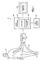
- FIG. 1 illustrates an apparatus 10 embodying the invention.
- the apparatus 10 is a patient monitoring or patient data acquisition device. While any device for acquiring ECG signals (such as, for example, a bedside monitor, transport monitor, or Holter monitor) is contemplated by the invention, the apparatus of the preferred embodiment employs a telemetry-based monitoring device.
- the apparatus 10 includes six electrodes 12 attachable to a patient 14, leadwires 16 coupled to the electrodes 12, a telemetry unit 18 coupled to the electrodes 12 by leadwires 16, a signal processor 20 wirelessly coupled to the telemetry unit 18, a telemetry monitor 22 coupled to the signal processor 20, and an ECG storage facility 24 coupled to the signal processor 20.
- FIG. 1 illustrates the apparatus 10 including a telemetry unit 18 wirelessly coupled to the signal processor 20 and telemetry monitor 22. Conventional methods of wireless transmission are used to transmit the electrical signals from the telemetry unit 18 to the receiver 28 in the signal processor 20.
- Biopotential signals are often processed by telemetry, a technique that provides a wireless link between the patient and the signal processing components.
- clinicians can monitor a patient while the patient has full mobility.
- Traditional methods of telemetry utilize from three to five electrodes, but are unable to acquire a twelve-lead ECG from these three to five electrodes.
- the limiting factor in telemetry is the bandwidth of the signal being transmitted to the signal processing components. Accordingly, traditional telemetry monitors are unable to support the bandwidth necessary to transmit the electrical signals representing an entire twelve-lead ECG.
- the bandwidth necessary to transmit the electrical signals representing the patient's ECG is reduced by at least half. Due to the reduced number of electrodes and the reduced bandwidth, the telemetry unit 18 is capable of monitoring the electrical activity of the patient's heart while the patient has full mobility. Moreover, the apparatus 10 is capable of acquiring more data from each particular patient or data from more than one patient.
- a first electrode (V1) is attached in approximately the fourth intercostal space at the right border of the patient's sternum.
- a second electrode (V5) is attached in approximately the fifth intercostal space at the patient's anterior axillary line.
- Four additional electrodes (LA, RA, LL, and G) are attached to the patient's limbs. Electrical signals are acquired from the six electrodes 12 and transmitted via leadwires 16 to the telemetry unit 18. The electrical signals are amplified and transmitted to the receiver 28 of the signal processor 20.
- reducing the number of electrodes 12 attached to the patient reduces the number of leadwires 16 and amplifiers (not shown) within the telemetry unit 18 necessary to acquire the electrical signals.
- a software module 26 within signal processor 20 uses an algorithm to calculate the expansion-coefficient equations for the four missing leads.
- the input to the algorithm is, most preferably, a data set of the patient's previously acquired ECGs.
- the data set is preferably stored in a hospital ECG storage facility 24 and accessed by the signal processor 20 of the ECG machine 10.
- the apparatus 10 then acquires an ECG for the patient.
- the telemetry unit 18 transmits the electrical signals from the electrodes 12 to a receiver 28 within the signal processor 20.
- the four missing leads are calculated by the software module 26 within signal processor 20 using the previously-derived expansion-coefficient equations. All twelve leads, including the four generated leads, are then displayed for the clinician on telemetry monitor 22.
Landscapes
- Health & Medical Sciences (AREA)
- Life Sciences & Earth Sciences (AREA)
- Heart & Thoracic Surgery (AREA)
- Medical Informatics (AREA)
- Biophysics (AREA)
- Pathology (AREA)
- Engineering & Computer Science (AREA)
- Biomedical Technology (AREA)
- Veterinary Medicine (AREA)
- Physics & Mathematics (AREA)
- Molecular Biology (AREA)
- Surgery (AREA)
- Animal Behavior & Ethology (AREA)
- General Health & Medical Sciences (AREA)
- Public Health (AREA)
- Cardiology (AREA)
- Measurement And Recording Of Electrical Phenomena And Electrical Characteristics Of The Living Body (AREA)
- Electrotherapy Devices (AREA)
Claims (8)
- Dispositif pour acquérir et traiter des signaux électriques produits par le coeur d'un patient, le dispositif comprenant :un ensemble de moins de dix électrodes (12) destinées chacune à être fixée au patient (14) dans l'une des positions standard d'électrodes d'électrocardiogramme à dix électrodes et à douze dérivations ; etun dispositif de traitement de signaux (20) connecté à six desdites électrodes (12) pour acquérir des signaux électriques des électrodes (12) et pour générer un électrocardiogramme à douze dérivations à partir de ces signaux électriques, certaines des dérivations pour l'électrocardiogramme à douze dérivations étant générées à partir d'une manipulation électrique standard des signaux acquis à partir des électrodes, les autres dérivations étant générées de manière mathématique par le dispositif de traitement de signaux,dans lequel la valeur de la dérivation V2 est déterminée en fonction des valeurs des dérivations V1, V5, 1 et II en utilisant l'équation suivante :

- Dispositif selon la revendication 1, dans lequel l'ensemble d'électrodes (12) comprend soit (a) une électrode destinée à être fixée au patient à peu près dans le quatrième espace intercostal au niveau du bord droit du sternum du patient, soit (b) une électrode destinée à être fixée au patient à peu près dans le quatrième espace intercostal au niveau du bord gauche du sternum du patient.
- Dispositif selon la revendication 1, dans lequel au moins deux des électrodes (12) peuvent être attachées à la poitrine du patient dans deux des positions standard d'électrocardiographe à dix électrodes et à douze dérivations, et au moins deux des électrodes peuvent être attachées aux membres du patient dans deux des positions standard d'électrocardiographe à dix électrodes et à douze dérivations.
- Dispositif selon la revendication 3, dans lequel la sortie des électrodes (12) pouvant être attachées à la poitrine du patient sont un premier et un deuxième canal de données, et la sortie des électrodes (12) pouvant être attachées aux membres du patient sont un troisième et un quatrième canal de données, et le dispositif de traitement de signaux (20) est adapté pour acquérir les canaux de données et générer un électrocardiogramme à douze dérivations à partir des canaux de données acquis.
- Procédé d'acquisition et de traitement de signaux électriques produits par le coeur d'un patient, le procédé comprenant les opérations consistant à :attacher un ensemble de moins de dix électrodes (12) au patient, cette opération comprenant le fait d'attacher chacune des électrodes dans l'une des positions standard d'électrocardiogramme à dix électrodes et à douze dérivations ;acquérir des signaux électriques de six desdites électrodes ; etgénérer un électrocardiogramme à douze dérivations à partir des signaux électriques acquis, certaines des dérivations pour l'électrocardiogramme à douze dérivations étant générées à partir d'une manipulation électrique standard des signaux acquis à partir des électrodes, les autres dérivations étant générées de manière mathématique par le dispositif de traitement de signaux,dans lequel la valeur de la dérivation V2 est déterminée en fonction des valeurs des dérivations V1, V5, 1 et II en utilisant l'équation suivante :

- Procédé selon la revendication 5, dans lequel l'opération consistant à attacher un ensemble d'électrodes (12) comprend en outre l'opération consistant à attacher une électrode (12) soit (a) à peu près dans le quatrième espace intercostal au niveau du bord droit du sternum du patient, soit (b) à peu près dans le quatrième espace intercostal au niveau du bord gauche du sternum du patient.
- Procédé selon la revendication 5, dans lequel l'opération consistant à attacher comprend le fait d'attacher au moins deux des électrodes (12) sur la poitrine du patient dans deux des positions standard d'électrocardiogramme à dix électrodes et à douze dérivations, et d'attacher au moins deux des électrodes (12) aux membres du patient dans deux des positions standard d'électrocardiogramme à dix électrodes et à douze dérivations.
- Procédé selon la revendication 7, dans lequel l'opération consistant à acquérir des signaux électriques à partir des électrodes comprend les étapes consistant à :acquérir un premier et un deuxième canal de données à partir des électrodes (12) attachées à la poitrine du patient ;acquérir un troisième et un quatrième canal de données à partir des électrodes (12) attachées aux membres du patient ; etgénérer un électrocardiogramme à douze dérivations à partir des canaux de données acquis.
Applications Claiming Priority (2)
| Application Number | Priority Date | Filing Date | Title |
|---|---|---|---|
| US09/752,140 US6636761B2 (en) | 2000-12-29 | 2000-12-29 | Method and apparatus for generating a twelve-lead ECG from fewer than ten electrodes |
| US752140 | 2000-12-29 |
Publications (3)
| Publication Number | Publication Date |
|---|---|
| EP1221299A2 EP1221299A2 (fr) | 2002-07-10 |
| EP1221299A3 EP1221299A3 (fr) | 2004-04-21 |
| EP1221299B1 true EP1221299B1 (fr) | 2008-01-30 |
Family
ID=25025059
Family Applications (1)
| Application Number | Title | Priority Date | Filing Date |
|---|---|---|---|
| EP01310732A Expired - Lifetime EP1221299B1 (fr) | 2000-12-29 | 2001-12-20 | Procedé et dispositif de géneration d'un électrocardiogramme à 12 conducteurs ayant moins de 10 électrodes |
Country Status (4)
| Country | Link |
|---|---|
| US (2) | US6636761B2 (fr) |
| EP (1) | EP1221299B1 (fr) |
| JP (1) | JP4027091B2 (fr) |
| DE (1) | DE60132655T2 (fr) |
Families Citing this family (59)
| Publication number | Priority date | Publication date | Assignee | Title |
|---|---|---|---|---|
| US6643539B2 (en) | 2000-08-03 | 2003-11-04 | Siemens Medical Solutions Usa, Inc. | Electrocardiogram system for synthesizing leads and providing an accuracy measure |
| DE60136629D1 (de) | 2000-08-03 | 2009-01-02 | Draeger Medical Systems Inc | Elektrokardiologisches system zur synthesierung von ableitungen und lieferung einer genauen messung |
| US6636761B2 (en) * | 2000-12-29 | 2003-10-21 | Ge Medical Systems Information Technologies, Inc. | Method and apparatus for generating a twelve-lead ECG from fewer than ten electrodes |
| US6716165B1 (en) * | 2001-03-02 | 2004-04-06 | Ge Medical Systems Information Technologies, Inc. | Patient telemetry device and leadset designs for providing antenna diversity |
| US8069418B2 (en) * | 2002-07-31 | 2011-11-29 | Draeger Medical Systems, Inc | Medical information system and user interface supporting treatment administration |
| US6931271B2 (en) * | 2002-08-12 | 2005-08-16 | Draeger Medical Systems, Inc | System for adaptively deriving ECG chest lead signal data |
| JP4777326B2 (ja) * | 2003-02-26 | 2011-09-21 | 大名 魏 | 付加誘導機能を備えた心電計及び付加誘導心電図導出方法 |
| US20060235317A1 (en) | 2003-02-26 | 2006-10-19 | Daming Wei | Electrocardiograph device having additional lead function and method for obtaining additional-lead electrocardiogram |
| JP2004266399A (ja) * | 2003-02-28 | 2004-09-24 | Hirona Gi | 医療情報テレメータシステム |
| GB0317947D0 (en) * | 2003-07-31 | 2003-09-03 | Mar Reynolds Medical Del Ltd | Reduced electrode electrocardiography system |
| DE10336809B4 (de) * | 2003-08-07 | 2007-08-02 | Charité - Universitätsmedizin Berlin | EKG-System zur grossflächigen Messung von EKG-Signalen |
| JP4830266B2 (ja) * | 2004-05-14 | 2011-12-07 | 日本光電工業株式会社 | 標準12誘導心電図の構築方法および心電図検査装置 |
| JP4470063B2 (ja) | 2004-08-27 | 2010-06-02 | 大名 魏 | 導出12誘導心電図の構築方法およびモニタリング装置 |
| US20060058591A1 (en) * | 2004-09-16 | 2006-03-16 | Memtec Corporation | First-response portable recorder and automated report generator |
| GB2425181B (en) * | 2005-04-14 | 2010-02-03 | Justin Pisani | Wearable physiological monitoring device |
| US7881778B2 (en) | 2006-09-28 | 2011-02-01 | The General Electric Company | Floating physiological data acquisition system with expandable ECG and EEG |
| US8005531B2 (en) | 2006-09-29 | 2011-08-23 | The General Electric Company | Method and apparatus with reduced electrode system specific ECG interpretation |
| US8494620B2 (en) * | 2008-06-18 | 2013-07-23 | Koninklijke Philips Electronics N.V. | Electrocardiograph for magnetic resonance imaging and electrode patch for same |
| US11375938B2 (en) | 2008-08-14 | 2022-07-05 | Ticker Medical Ltd | Miniature ECG data acquisition device |
| US8082025B2 (en) * | 2008-08-14 | 2011-12-20 | David Amitai | ECG data acquisition device |
| US8509882B2 (en) | 2010-06-08 | 2013-08-13 | Alivecor, Inc. | Heart monitoring system usable with a smartphone or computer |
| US9351654B2 (en) | 2010-06-08 | 2016-05-31 | Alivecor, Inc. | Two electrode apparatus and methods for twelve lead ECG |
| JP5422513B2 (ja) * | 2010-07-30 | 2014-02-19 | 大名 魏 | 導出心電図生成システム及び導出心電図生成方法 |
| CN102106728A (zh) * | 2011-01-27 | 2011-06-29 | 孙迎春 | 运动心电图中基于四个电极片描记十八导联心电图的方法 |
| US10932720B2 (en) | 2011-03-08 | 2021-03-02 | Nanowear Inc. | Smart materials, dry textile sensors, and electronics integration in clothing, bed sheets, and pillow cases for neurological, cardiac and/or pulmonary monitoring |
| US20130281815A1 (en) * | 2012-04-18 | 2013-10-24 | The Board Of Trustees Of The University Of Arkansas | Wearable remote electrophysiological monitoring system |
| US8923957B2 (en) * | 2011-07-15 | 2014-12-30 | Verathon Inc. | Data conversion in ECG techniques |
| WO2014074913A1 (fr) | 2012-11-08 | 2014-05-15 | Alivecor, Inc. | Détection de signal d'électrocardiogramme |
| US9220430B2 (en) | 2013-01-07 | 2015-12-29 | Alivecor, Inc. | Methods and systems for electrode placement |
| WO2014145927A1 (fr) | 2013-03-15 | 2014-09-18 | Alivecor, Inc. | Systèmes et procédés pour traiter et analyser des données médicales |
| US9247911B2 (en) | 2013-07-10 | 2016-02-02 | Alivecor, Inc. | Devices and methods for real-time denoising of electrocardiograms |
| JP6118229B2 (ja) * | 2013-10-28 | 2017-04-19 | 日本光電工業株式会社 | 心電図測定装置、導出心電図生成方法および導出心電図生成プログラム |
| US9420956B2 (en) | 2013-12-12 | 2016-08-23 | Alivecor, Inc. | Methods and systems for arrhythmia tracking and scoring |
| TWI637723B (zh) | 2014-12-09 | 2018-10-11 | 國立交通大學 | 利用三個導程之差動電壓產生十二導程心電圖信號之方法與系統 |
| US11111593B2 (en) | 2015-01-16 | 2021-09-07 | Nanowear Inc. | Large scale manufacturing of hybrid nanostructured textile sensors |
| US10131993B2 (en) | 2015-01-16 | 2018-11-20 | Nanowear, Inc. | Large scale manufacturing of hybrid nanostructured textile sensors |
| US10561329B2 (en) | 2015-04-14 | 2020-02-18 | Koninklijke Philips N.V. | Method and system for ECG based cardiac ischemia detection |
| CN107847154B (zh) | 2015-05-13 | 2021-07-16 | 阿利弗克公司 | 不一致监测 |
| US20180271392A1 (en) * | 2015-10-16 | 2018-09-27 | Enrique Saldivar | Device and method of using hexaxial electrocardiograph |
| JP6724348B2 (ja) * | 2015-11-27 | 2020-07-15 | 東洋紡株式会社 | 心電図測定方法および心電図測定用衣服 |
| US10602946B2 (en) * | 2015-12-30 | 2020-03-31 | Vectracor, Inc. | Mobile cardiac monitoring device |
| US10231623B2 (en) | 2016-02-04 | 2019-03-19 | Nanowear Inc. | Roll-to-roll printing process for manufacturing a wireless nanosensor |
| CN105832328A (zh) * | 2016-03-15 | 2016-08-10 | 安徽华米信息科技有限公司 | 心电数据的处理方法、装置及衣服 |
| US10123741B2 (en) | 2016-11-30 | 2018-11-13 | Huami Inc. | Cardiac condition detection |
| US10617356B2 (en) | 2016-03-15 | 2020-04-14 | Anhui Huami Information Technology Co., Ltd. | Garment and cardiac data processing |
| US10959634B2 (en) | 2017-05-02 | 2021-03-30 | Nanowear Inc. | Wearable congestive heart failure management system |
| CN107440706A (zh) * | 2017-05-23 | 2017-12-08 | 北京蓬阳丰业医疗设备有限公司 | 基于wilson心电图导联体系中的9电极同步采集方法 |
| CN107440707A (zh) * | 2017-05-23 | 2017-12-08 | 北京蓬阳丰业医疗设备有限公司 | 基于wilson心电图导联体系的18导/21导动态心电图方法 |
| CN107440711A (zh) * | 2017-05-23 | 2017-12-08 | 北京蓬阳丰业医疗设备有限公司 | 基于wilson心电图导联体系的8电极和10电极同步采集方法 |
| CN107440710A (zh) * | 2017-05-23 | 2017-12-08 | 北京蓬阳丰业医疗设备有限公司 | 基于wilson心电图导联体系的4电极至7电极同步采集方法 |
| TWI669096B (zh) * | 2017-07-13 | 2019-08-21 | 國立臺灣大學 | 具有確定頸動脈血壓的多功能量測裝置 |
| JP6795003B2 (ja) * | 2018-03-08 | 2020-12-02 | 東洋紡株式会社 | 電極及び配線、電極及び配線付き衣服の製造方法 |
| US20210113108A1 (en) * | 2019-10-22 | 2021-04-22 | Sensesemi Technologies Private Limited | Method, system and device for generating electrocardiogram with fewer number of probes |
| US11523766B2 (en) | 2020-06-25 | 2022-12-13 | Spacelabs Healthcare L.L.C. | Systems and methods of analyzing and displaying ambulatory ECG data |
| JP2020192391A (ja) * | 2020-08-26 | 2020-12-03 | 東洋紡株式会社 | 心電図測定方法 |
| CN113208599B (zh) * | 2021-05-17 | 2024-02-27 | 北京蓬阳丰业科技有限公司 | 一种基于心电单极导联v1、v5r推衍导联v3r、v4r的方法及设备 |
| CN113208600B (zh) * | 2021-05-17 | 2024-02-27 | 北京蓬阳丰业科技有限公司 | 一种基于心电单极导联v6、v9推衍导联v7、v8的方法及设备 |
| CN113223702B (zh) * | 2021-05-17 | 2022-05-03 | 北京蓬阳丰业科技有限公司 | 一种基于心电单极导联v2、v5推衍导联v3、v4的方法及设备 |
| CN115886831B (zh) * | 2022-09-05 | 2023-09-26 | 荣耀终端有限公司 | 多导联心电检测方法及电子设备 |
Family Cites Families (13)
| Publication number | Priority date | Publication date | Assignee | Title |
|---|---|---|---|---|
| US4850370A (en) | 1987-07-22 | 1989-07-25 | Dower Gordon E | Method and apparatus for sensing and analyzing electrical activity of the human heart |
| US5058598A (en) * | 1990-08-03 | 1991-10-22 | Nicklas John M | Method and apparatus for synthesizing leads of an electrocardiogram |
| AU4616896A (en) | 1995-02-09 | 1996-08-27 | Gordon Ewbank Dower | Apparatus and method for monitoring activity of the human heart |
| US5913828A (en) * | 1996-10-29 | 1999-06-22 | Hewlett-Packard Company | Method and apparatus for distinguishing pacing pulses in an EKG using conduction velocity calculations |
| US5794624A (en) * | 1997-01-31 | 1998-08-18 | Hewlett-Packard Company | Method and system for the fast determination of EKG waveform morphology |
| US6006125A (en) * | 1998-02-12 | 1999-12-21 | Unilead International Inc. | Universal electrocardiogram sensor positioning device and method |
| US6119035A (en) * | 1998-03-26 | 2000-09-12 | Hewlett-Packard Company | Method and system for synthesizing the 12-lead electrocardiogram |
| US6217525B1 (en) * | 1998-04-30 | 2001-04-17 | Medtronic Physio-Control Manufacturing Corp. | Reduced lead set device and method for detecting acute cardiac ischemic conditions |
| US6167258A (en) * | 1998-10-09 | 2000-12-26 | Cleveland Medical Devices Inc. | Programmable wireless data acquisition system |
| JP4587008B2 (ja) | 2000-07-24 | 2010-11-24 | 大名 魏 | 標準12誘導心電図の構築方法および心電図検査装置 |
| DE60136629D1 (de) | 2000-08-03 | 2009-01-02 | Draeger Medical Systems Inc | Elektrokardiologisches system zur synthesierung von ableitungen und lieferung einer genauen messung |
| US6505067B1 (en) * | 2000-11-22 | 2003-01-07 | Medtronic, Inc. | System and method for deriving a virtual ECG or EGM signal |
| US6636761B2 (en) * | 2000-12-29 | 2003-10-21 | Ge Medical Systems Information Technologies, Inc. | Method and apparatus for generating a twelve-lead ECG from fewer than ten electrodes |
-
2000
- 2000-12-29 US US09/752,140 patent/US6636761B2/en not_active Expired - Lifetime
-
2001
- 2001-12-20 EP EP01310732A patent/EP1221299B1/fr not_active Expired - Lifetime
- 2001-12-20 DE DE60132655T patent/DE60132655T2/de not_active Expired - Lifetime
- 2001-12-28 JP JP2001399036A patent/JP4027091B2/ja not_active Expired - Lifetime
-
2003
- 2003-08-20 US US10/644,539 patent/US7136693B2/en not_active Expired - Lifetime
Also Published As
| Publication number | Publication date |
|---|---|
| US7136693B2 (en) | 2006-11-14 |
| US20040015089A1 (en) | 2004-01-22 |
| JP4027091B2 (ja) | 2007-12-26 |
| DE60132655T2 (de) | 2009-01-29 |
| EP1221299A2 (fr) | 2002-07-10 |
| JP2002282229A (ja) | 2002-10-02 |
| EP1221299A3 (fr) | 2004-04-21 |
| DE60132655D1 (de) | 2008-03-20 |
| US20020087088A1 (en) | 2002-07-04 |
| US6636761B2 (en) | 2003-10-21 |
Similar Documents
| Publication | Publication Date | Title |
|---|---|---|
| EP1221299B1 (fr) | Procedé et dispositif de géneration d'un électrocardiogramme à 12 conducteurs ayant moins de 10 électrodes | |
| US6052615A (en) | Method and apparatus for sensing and analyzing electrical activity of the human heart using a four electrode arrangement | |
| JP4587008B2 (ja) | 標準12誘導心電図の構築方法および心電図検査装置 | |
| Feild et al. | Improved EASI coefficients: their derivation, values, and performance | |
| US4850370A (en) | Method and apparatus for sensing and analyzing electrical activity of the human heart | |
| EP1659936B8 (fr) | Appareil et procede d'enregistrement sans fil, de transmission par telecommunication et de traitement des signaux de trois fils speciaux d'ecg | |
| EP1952761B1 (fr) | Système d'électrocardiogramme pour synthétiser des dérivations et fournir une mesure précise | |
| US8005532B2 (en) | Electrocardiograph with extended lead function, and extended lead electrocardiogram deriving method | |
| WO1994001039A9 (fr) | Systeme electrocardiographique sans fil et ensembles electrodes sans fil | |
| US20080081960A1 (en) | Floating physiological data acquisition system with expandable ECG and EEG | |
| WO1994001039A1 (fr) | Systeme electrocardiographique sans fil et ensembles electrodes sans fil | |
| EP1217946B1 (fr) | Procede et appareil pour la reconstruction de derivations de frank a partir de derivations unipolaires precordiales | |
| US6496720B1 (en) | Process for sensing and analyzing electrical activity of the human heart utilizing one lead system with an egg monitor designed for use with another lead system | |
| US20070197925A1 (en) | Acquisition of multiple-lead electrocardiogram | |
| Sejersten et al. | Comparison of EASI-derived 12-lead electrocardiograms versus paramedic-acquired 12-lead electrocardiograms using Mason-Likar limb lead configuration in patients with chest pain | |
| Nelwan | Evaluation of12-LeadElectrocardiogramReconstruction Methods forPatientMonitoring | |
| Wei | Deriving the 12-lead electrocardiogram from four standard leads based on the Frank Torso model | |
| JP2008100080A (ja) | 付加誘導機能を備えた心電計及び付加誘導心電図導出方法 | |
| KR20060116190A (ko) | 3개의 특수 ecg 리드의 무선 기록 및 전기통신 전송,그리고 그 처리를 위한 장치 및 방법 | |
| Macfarlane | The Pierre Richlant Lecture 2007: The Future of Electrocardiography. | |
| van Herpen et al. | Are additional right precordial and left posterior ECG leads useful for the diagnosis of right ventricular infarct and posterior infarct? Also a plea for the revival of vectorcardiography | |
| Dotsinsky et al. | Twelve-lead electrocardiogram obtained by eight leads | |
| Kligfield et al. | AHA/ACC/HRS Scientific Statement | |
| WEI | TWELVE-LEAD ELECTROCARDIOGRAM TELEMONITORING | |
| Lead | THE STANDARD 12-LEAD ELECTROCARDIOGRAM |
Legal Events
| Date | Code | Title | Description |
|---|---|---|---|
| PUAI | Public reference made under article 153(3) epc to a published international application that has entered the european phase |
Free format text: ORIGINAL CODE: 0009012 |
|
| AK | Designated contracting states |
Kind code of ref document: A2 Designated state(s): AT BE CH CY DE DK ES FI FR GB GR IE IT LI LU MC NL PT SE TR |
|
| AX | Request for extension of the european patent |
Free format text: AL;LT;LV;MK;RO;SI |
|
| PUAL | Search report despatched |
Free format text: ORIGINAL CODE: 0009013 |
|
| AK | Designated contracting states |
Kind code of ref document: A3 Designated state(s): AT BE CH CY DE DK ES FI FR GB GR IE IT LI LU MC NL PT SE TR |
|
| AX | Request for extension of the european patent |
Extension state: AL LT LV MK RO SI |
|
| RIC1 | Information provided on ipc code assigned before grant |
Ipc: 7A 61B 5/0428 A Ipc: 7A 61B 5/0408 B |
|
| 17P | Request for examination filed |
Effective date: 20041021 |
|
| AKX | Designation fees paid |
Designated state(s): DE FI FR GB SE |
|
| 17Q | First examination report despatched |
Effective date: 20060505 |
|
| GRAP | Despatch of communication of intention to grant a patent |
Free format text: ORIGINAL CODE: EPIDOSNIGR1 |
|
| RAP1 | Party data changed (applicant data changed or rights of an application transferred) |
Owner name: GE MEDICAL SYSTEMS INFORMATION TECHNOLOGIES, INC. |
|
| GRAS | Grant fee paid |
Free format text: ORIGINAL CODE: EPIDOSNIGR3 |
|
| GRAA | (expected) grant |
Free format text: ORIGINAL CODE: 0009210 |
|
| AK | Designated contracting states |
Kind code of ref document: B1 Designated state(s): DE FI FR GB SE |
|
| REG | Reference to a national code |
Ref country code: GB Ref legal event code: FG4D |
|
| REF | Corresponds to: |
Ref document number: 60132655 Country of ref document: DE Date of ref document: 20080320 Kind code of ref document: P |
|
| PG25 | Lapsed in a contracting state [announced via postgrant information from national office to epo] |
Ref country code: FI Free format text: LAPSE BECAUSE OF FAILURE TO SUBMIT A TRANSLATION OF THE DESCRIPTION OR TO PAY THE FEE WITHIN THE PRESCRIBED TIME-LIMIT Effective date: 20080130 |
|
| PG25 | Lapsed in a contracting state [announced via postgrant information from national office to epo] |
Ref country code: SE Free format text: LAPSE BECAUSE OF FAILURE TO SUBMIT A TRANSLATION OF THE DESCRIPTION OR TO PAY THE FEE WITHIN THE PRESCRIBED TIME-LIMIT Effective date: 20080430 |
|
| EN | Fr: translation not filed | ||
| PLBE | No opposition filed within time limit |
Free format text: ORIGINAL CODE: 0009261 |
|
| STAA | Information on the status of an ep patent application or granted ep patent |
Free format text: STATUS: NO OPPOSITION FILED WITHIN TIME LIMIT |
|
| 26N | No opposition filed |
Effective date: 20081031 |
|
| PG25 | Lapsed in a contracting state [announced via postgrant information from national office to epo] |
Ref country code: FR Free format text: LAPSE BECAUSE OF FAILURE TO SUBMIT A TRANSLATION OF THE DESCRIPTION OR TO PAY THE FEE WITHIN THE PRESCRIBED TIME-LIMIT Effective date: 20081121 |
|
| REG | Reference to a national code |
Ref country code: DE Ref legal event code: R079 Ref document number: 60132655 Country of ref document: DE Free format text: PREVIOUS MAIN CLASS: A61B0005042800 Ipc: A61B0005308000 |
|
| PGFP | Annual fee paid to national office [announced via postgrant information from national office to epo] |
Ref country code: GB Payment date: 20201123 Year of fee payment: 20 Ref country code: DE Payment date: 20201119 Year of fee payment: 20 |
|
| REG | Reference to a national code |
Ref country code: DE Ref legal event code: R071 Ref document number: 60132655 Country of ref document: DE |
|
| REG | Reference to a national code |
Ref country code: GB Ref legal event code: PE20 Expiry date: 20211219 |
|
| PG25 | Lapsed in a contracting state [announced via postgrant information from national office to epo] |
Ref country code: GB Free format text: LAPSE BECAUSE OF EXPIRATION OF PROTECTION Effective date: 20211219 |