EP1210430B1 - Muc-1 derived peptides - Google Patents
Muc-1 derived peptides Download PDFInfo
- Publication number
- EP1210430B1 EP1210430B1 EP00965943A EP00965943A EP1210430B1 EP 1210430 B1 EP1210430 B1 EP 1210430B1 EP 00965943 A EP00965943 A EP 00965943A EP 00965943 A EP00965943 A EP 00965943A EP 1210430 B1 EP1210430 B1 EP 1210430B1
- Authority
- EP
- European Patent Office
- Prior art keywords
- cell
- polypeptide
- cells
- polynucleotide
- restricted
- Prior art date
- Legal status (The legal status is an assumption and is not a legal conclusion. Google has not performed a legal analysis and makes no representation as to the accuracy of the status listed.)
- Expired - Lifetime
Links
- 108090000765 processed proteins & peptides Proteins 0.000 title claims abstract description 314
- 102000004196 processed proteins & peptides Human genes 0.000 title claims abstract description 268
- 101100346932 Mus musculus Muc1 gene Proteins 0.000 title description 3
- 229920001184 polypeptide Polymers 0.000 claims abstract description 239
- 230000004044 response Effects 0.000 claims abstract description 20
- 108091028043 Nucleic acid sequence Proteins 0.000 claims abstract description 13
- 210000004027 cell Anatomy 0.000 claims description 184
- 210000001744 T-lymphocyte Anatomy 0.000 claims description 107
- 206010028980 Neoplasm Diseases 0.000 claims description 65
- 238000000034 method Methods 0.000 claims description 63
- 108091033319 polynucleotide Proteins 0.000 claims description 58
- 102000040430 polynucleotide Human genes 0.000 claims description 58
- 239000002157 polynucleotide Substances 0.000 claims description 58
- 239000000203 mixture Substances 0.000 claims description 48
- 201000011510 cancer Diseases 0.000 claims description 39
- 230000027455 binding Effects 0.000 claims description 38
- 108091054437 MHC class I family Proteins 0.000 claims description 35
- 102000043129 MHC class I family Human genes 0.000 claims description 31
- 239000003795 chemical substances by application Substances 0.000 claims description 29
- 230000005867 T cell response Effects 0.000 claims description 26
- 239000012634 fragment Substances 0.000 claims description 25
- 239000000126 substance Substances 0.000 claims description 24
- 239000013598 vector Substances 0.000 claims description 24
- 125000003275 alpha amino acid group Chemical group 0.000 claims description 19
- 238000000338 in vitro Methods 0.000 claims description 17
- 229960005486 vaccine Drugs 0.000 claims description 15
- 150000001875 compounds Chemical class 0.000 claims description 14
- 239000008194 pharmaceutical composition Substances 0.000 claims description 12
- 238000002360 preparation method Methods 0.000 claims description 12
- 230000014509 gene expression Effects 0.000 claims description 11
- 239000013612 plasmid Substances 0.000 claims description 6
- 239000013603 viral vector Substances 0.000 claims description 6
- 230000004936 stimulating effect Effects 0.000 claims description 5
- 102100037850 Interferon gamma Human genes 0.000 claims description 4
- 108010074328 Interferon-gamma Proteins 0.000 claims description 4
- 210000004102 animal cell Anatomy 0.000 claims description 3
- 239000003937 drug carrier Substances 0.000 claims description 3
- 210000004962 mammalian cell Anatomy 0.000 claims description 3
- 230000010076 replication Effects 0.000 claims description 3
- 230000008685 targeting Effects 0.000 claims description 3
- 210000005253 yeast cell Anatomy 0.000 claims description 3
- 239000003814 drug Substances 0.000 claims description 2
- 210000001236 prokaryotic cell Anatomy 0.000 claims description 2
- 230000005859 cell recognition Effects 0.000 claims 1
- 230000000295 complement effect Effects 0.000 claims 1
- 239000002773 nucleotide Substances 0.000 claims 1
- 125000003729 nucleotide group Chemical group 0.000 claims 1
- 239000003381 stabilizer Substances 0.000 claims 1
- 102100034256 Mucin-1 Human genes 0.000 abstract description 52
- 108010008707 Mucin-1 Proteins 0.000 abstract description 38
- 210000001151 cytotoxic T lymphocyte Anatomy 0.000 abstract description 29
- 230000000890 antigenic effect Effects 0.000 abstract description 10
- 230000001225 therapeutic effect Effects 0.000 abstract description 3
- 241000282414 Homo sapiens Species 0.000 description 87
- 108020004414 DNA Proteins 0.000 description 39
- 108090000623 proteins and genes Proteins 0.000 description 33
- 239000000427 antigen Substances 0.000 description 31
- 108091007433 antigens Proteins 0.000 description 31
- 102000036639 antigens Human genes 0.000 description 31
- 210000003819 peripheral blood mononuclear cell Anatomy 0.000 description 29
- 210000000612 antigen-presenting cell Anatomy 0.000 description 28
- 239000000047 product Substances 0.000 description 28
- 235000018102 proteins Nutrition 0.000 description 25
- 102000004169 proteins and genes Human genes 0.000 description 25
- 238000012360 testing method Methods 0.000 description 24
- 238000002255 vaccination Methods 0.000 description 24
- 235000001014 amino acid Nutrition 0.000 description 19
- 238000003556 assay Methods 0.000 description 19
- 230000028993 immune response Effects 0.000 description 19
- 101001133056 Homo sapiens Mucin-1 Proteins 0.000 description 17
- 150000001413 amino acids Chemical class 0.000 description 17
- 238000011282 treatment Methods 0.000 description 17
- 241001465754 Metazoa Species 0.000 description 16
- 102000008949 Histocompatibility Antigens Class I Human genes 0.000 description 14
- 150000007523 nucleic acids Chemical group 0.000 description 14
- 238000002474 experimental method Methods 0.000 description 13
- 102000004127 Cytokines Human genes 0.000 description 12
- 108090000695 Cytokines Proteins 0.000 description 12
- 108700018351 Major Histocompatibility Complex Proteins 0.000 description 12
- 241000699670 Mus sp. Species 0.000 description 12
- 230000004048 modification Effects 0.000 description 12
- 238000012986 modification Methods 0.000 description 12
- 230000020382 suppression by virus of host antigen processing and presentation of peptide antigen via MHC class I Effects 0.000 description 12
- UZOVYGYOLBIAJR-UHFFFAOYSA-N 4-isocyanato-4'-methyldiphenylmethane Chemical compound C1=CC(C)=CC=C1CC1=CC=C(N=C=O)C=C1 UZOVYGYOLBIAJR-UHFFFAOYSA-N 0.000 description 11
- 239000002671 adjuvant Substances 0.000 description 11
- LOKCTEFSRHRXRJ-UHFFFAOYSA-I dipotassium trisodium dihydrogen phosphate hydrogen phosphate dichloride Chemical compound P(=O)(O)(O)[O-].[K+].P(=O)(O)([O-])[O-].[Na+].[Na+].[Cl-].[K+].[Cl-].[Na+] LOKCTEFSRHRXRJ-UHFFFAOYSA-I 0.000 description 11
- 239000001963 growth medium Substances 0.000 description 11
- 239000002953 phosphate buffered saline Substances 0.000 description 11
- 210000004881 tumor cell Anatomy 0.000 description 11
- 108010088652 Histocompatibility Antigens Class I Proteins 0.000 description 10
- 108010081355 beta 2-Microglobulin Proteins 0.000 description 10
- 102000015736 beta 2-Microglobulin Human genes 0.000 description 10
- 238000001727 in vivo Methods 0.000 description 10
- 102000039446 nucleic acids Human genes 0.000 description 10
- 108020004707 nucleic acids Proteins 0.000 description 10
- 239000013641 positive control Substances 0.000 description 10
- 230000008569 process Effects 0.000 description 10
- 239000000523 sample Substances 0.000 description 10
- 108010088729 HLA-A*02:01 antigen Proteins 0.000 description 9
- 241000700605 Viruses Species 0.000 description 9
- 239000012636 effector Substances 0.000 description 9
- 238000004519 manufacturing process Methods 0.000 description 9
- 210000001519 tissue Anatomy 0.000 description 9
- 108091008874 T cell receptors Proteins 0.000 description 8
- 102000016266 T-Cell Antigen Receptors Human genes 0.000 description 8
- 210000000481 breast Anatomy 0.000 description 8
- 210000004443 dendritic cell Anatomy 0.000 description 8
- 239000012091 fetal bovine serum Substances 0.000 description 8
- 238000002347 injection Methods 0.000 description 8
- 239000007924 injection Substances 0.000 description 8
- 239000002609 medium Substances 0.000 description 8
- 108010075704 HLA-A Antigens Proteins 0.000 description 7
- 102000011786 HLA-A Antigens Human genes 0.000 description 7
- 108010074032 HLA-A2 Antigen Proteins 0.000 description 7
- 102000025850 HLA-A2 Antigen Human genes 0.000 description 7
- YBJHBAHKTGYVGT-ZKWXMUAHSA-N biotin Natural products N1C(=O)N[C@@H]2[C@H](CCCCC(=O)O)SC[C@@H]21 YBJHBAHKTGYVGT-ZKWXMUAHSA-N 0.000 description 7
- 230000001413 cellular effect Effects 0.000 description 7
- 230000008859 change Effects 0.000 description 7
- 239000003085 diluting agent Substances 0.000 description 7
- 239000002158 endotoxin Substances 0.000 description 7
- 206010022000 influenza Diseases 0.000 description 7
- 230000005764 inhibitory process Effects 0.000 description 7
- 229920006008 lipopolysaccharide Polymers 0.000 description 7
- 239000013642 negative control Substances 0.000 description 7
- 239000006228 supernatant Substances 0.000 description 7
- IAZDPXIOMUYVGZ-UHFFFAOYSA-N Dimethylsulphoxide Chemical compound CS(C)=O IAZDPXIOMUYVGZ-UHFFFAOYSA-N 0.000 description 6
- PEDCQBHIVMGVHV-UHFFFAOYSA-N Glycerine Chemical compound OCC(O)CO PEDCQBHIVMGVHV-UHFFFAOYSA-N 0.000 description 6
- 230000030741 antigen processing and presentation Effects 0.000 description 6
- 210000004369 blood Anatomy 0.000 description 6
- 239000008280 blood Substances 0.000 description 6
- 238000001514 detection method Methods 0.000 description 6
- 238000002649 immunization Methods 0.000 description 6
- 230000002163 immunogen Effects 0.000 description 6
- 230000003612 virological effect Effects 0.000 description 6
- XLYOFNOQVPJJNP-UHFFFAOYSA-N water Substances O XLYOFNOQVPJJNP-UHFFFAOYSA-N 0.000 description 6
- 208000026310 Breast neoplasm Diseases 0.000 description 5
- LFQSCWFLJHTTHZ-UHFFFAOYSA-N Ethanol Chemical compound CCO LFQSCWFLJHTTHZ-UHFFFAOYSA-N 0.000 description 5
- 108010002350 Interleukin-2 Proteins 0.000 description 5
- 102000000588 Interleukin-2 Human genes 0.000 description 5
- 241000251539 Vertebrata <Metazoa> Species 0.000 description 5
- 210000003719 b-lymphocyte Anatomy 0.000 description 5
- 239000011230 binding agent Substances 0.000 description 5
- 231100000433 cytotoxic Toxicity 0.000 description 5
- 230000001472 cytotoxic effect Effects 0.000 description 5
- 238000010790 dilution Methods 0.000 description 5
- 239000012895 dilution Substances 0.000 description 5
- 238000003114 enzyme-linked immunosorbent spot assay Methods 0.000 description 5
- 108020001507 fusion proteins Proteins 0.000 description 5
- 102000037865 fusion proteins Human genes 0.000 description 5
- 238000001415 gene therapy Methods 0.000 description 5
- 210000000496 pancreas Anatomy 0.000 description 5
- 230000037361 pathway Effects 0.000 description 5
- 238000000159 protein binding assay Methods 0.000 description 5
- 210000004988 splenocyte Anatomy 0.000 description 5
- 230000002269 spontaneous effect Effects 0.000 description 5
- 238000002560 therapeutic procedure Methods 0.000 description 5
- 238000001890 transfection Methods 0.000 description 5
- IJGRMHOSHXDMSA-UHFFFAOYSA-N Atomic nitrogen Chemical compound N#N IJGRMHOSHXDMSA-UHFFFAOYSA-N 0.000 description 4
- 206010006187 Breast cancer Diseases 0.000 description 4
- 108090000790 Enzymes Proteins 0.000 description 4
- 102000004190 Enzymes Human genes 0.000 description 4
- 206010062016 Immunosuppression Diseases 0.000 description 4
- 241000699660 Mus musculus Species 0.000 description 4
- 241000700618 Vaccinia virus Species 0.000 description 4
- 239000002253 acid Substances 0.000 description 4
- 230000004913 activation Effects 0.000 description 4
- 229960002685 biotin Drugs 0.000 description 4
- 235000020958 biotin Nutrition 0.000 description 4
- 239000011616 biotin Substances 0.000 description 4
- 239000000872 buffer Substances 0.000 description 4
- 125000002091 cationic group Chemical group 0.000 description 4
- 238000005119 centrifugation Methods 0.000 description 4
- 238000003745 diagnosis Methods 0.000 description 4
- 238000000684 flow cytometry Methods 0.000 description 4
- 230000003053 immunization Effects 0.000 description 4
- 230000001506 immunosuppresive effect Effects 0.000 description 4
- 238000011534 incubation Methods 0.000 description 4
- 210000004698 lymphocyte Anatomy 0.000 description 4
- 239000011159 matrix material Substances 0.000 description 4
- 210000001672 ovary Anatomy 0.000 description 4
- 230000002265 prevention Effects 0.000 description 4
- 230000035755 proliferation Effects 0.000 description 4
- 210000002966 serum Anatomy 0.000 description 4
- 239000000243 solution Substances 0.000 description 4
- 230000009870 specific binding Effects 0.000 description 4
- 238000006467 substitution reaction Methods 0.000 description 4
- 238000011830 transgenic mouse model Methods 0.000 description 4
- 108091032973 (ribonucleotides)n+m Proteins 0.000 description 3
- WEVYAHXRMPXWCK-UHFFFAOYSA-N Acetonitrile Chemical compound CC#N WEVYAHXRMPXWCK-UHFFFAOYSA-N 0.000 description 3
- 108091003079 Bovine Serum Albumin Proteins 0.000 description 3
- 108091026890 Coding region Proteins 0.000 description 3
- 102000043131 MHC class II family Human genes 0.000 description 3
- 108091054438 MHC class II family Proteins 0.000 description 3
- ZMXDDKWLCZADIW-UHFFFAOYSA-N N,N-Dimethylformamide Chemical compound CN(C)C=O ZMXDDKWLCZADIW-UHFFFAOYSA-N 0.000 description 3
- 206010061902 Pancreatic neoplasm Diseases 0.000 description 3
- DNIAPMSPPWPWGF-UHFFFAOYSA-N Propylene glycol Chemical compound CC(O)CO DNIAPMSPPWPWGF-UHFFFAOYSA-N 0.000 description 3
- 108010090804 Streptavidin Proteins 0.000 description 3
- 238000004458 analytical method Methods 0.000 description 3
- 238000010171 animal model Methods 0.000 description 3
- 210000000170 cell membrane Anatomy 0.000 description 3
- 239000003153 chemical reaction reagent Substances 0.000 description 3
- KRKNYBCHXYNGOX-UHFFFAOYSA-N citric acid Chemical compound OC(=O)CC(O)(C(O)=O)CC(O)=O KRKNYBCHXYNGOX-UHFFFAOYSA-N 0.000 description 3
- 238000012258 culturing Methods 0.000 description 3
- 238000012217 deletion Methods 0.000 description 3
- 230000037430 deletion Effects 0.000 description 3
- 201000010099 disease Diseases 0.000 description 3
- 208000037265 diseases, disorders, signs and symptoms Diseases 0.000 description 3
- 230000005714 functional activity Effects 0.000 description 3
- 230000013595 glycosylation Effects 0.000 description 3
- 238000006206 glycosylation reaction Methods 0.000 description 3
- 210000002443 helper t lymphocyte Anatomy 0.000 description 3
- 238000001990 intravenous administration Methods 0.000 description 3
- 230000002147 killing effect Effects 0.000 description 3
- 239000002502 liposome Substances 0.000 description 3
- 239000007788 liquid Substances 0.000 description 3
- 210000004072 lung Anatomy 0.000 description 3
- 239000000463 material Substances 0.000 description 3
- 229910052757 nitrogen Inorganic materials 0.000 description 3
- 238000012545 processing Methods 0.000 description 3
- 238000011002 quantification Methods 0.000 description 3
- 230000028327 secretion Effects 0.000 description 3
- 239000007787 solid Substances 0.000 description 3
- 210000000952 spleen Anatomy 0.000 description 3
- 238000010561 standard procedure Methods 0.000 description 3
- 210000002784 stomach Anatomy 0.000 description 3
- 239000000725 suspension Substances 0.000 description 3
- 210000001541 thymus gland Anatomy 0.000 description 3
- 238000013518 transcription Methods 0.000 description 3
- 230000035897 transcription Effects 0.000 description 3
- 238000013519 translation Methods 0.000 description 3
- 230000004614 tumor growth Effects 0.000 description 3
- DGVVWUTYPXICAM-UHFFFAOYSA-N β‐Mercaptoethanol Chemical compound OCCS DGVVWUTYPXICAM-UHFFFAOYSA-N 0.000 description 3
- JKMHFZQWWAIEOD-UHFFFAOYSA-N 2-[4-(2-hydroxyethyl)piperazin-1-yl]ethanesulfonic acid Chemical compound OCC[NH+]1CCN(CCS([O-])(=O)=O)CC1 JKMHFZQWWAIEOD-UHFFFAOYSA-N 0.000 description 2
- 108700028369 Alleles Proteins 0.000 description 2
- 102000006306 Antigen Receptors Human genes 0.000 description 2
- 210000001266 CD8-positive T-lymphocyte Anatomy 0.000 description 2
- 101710132601 Capsid protein Proteins 0.000 description 2
- 229920002307 Dextran Polymers 0.000 description 2
- 239000006144 Dulbecco’s modified Eagle's medium Substances 0.000 description 2
- 238000011510 Elispot assay Methods 0.000 description 2
- 101710122231 Epstein-Barr nuclear antigen 3 Proteins 0.000 description 2
- 229920001917 Ficoll Polymers 0.000 description 2
- 102000003886 Glycoproteins Human genes 0.000 description 2
- 108090000288 Glycoproteins Proteins 0.000 description 2
- 239000007995 HEPES buffer Substances 0.000 description 2
- 108010035452 HLA-A1 Antigen Proteins 0.000 description 2
- 108010036972 HLA-A11 Antigen Proteins 0.000 description 2
- 108010086377 HLA-A3 Antigen Proteins 0.000 description 2
- 101000599940 Homo sapiens Interferon gamma Proteins 0.000 description 2
- 241000701044 Human gammaherpesvirus 4 Species 0.000 description 2
- 229920000057 Mannan Polymers 0.000 description 2
- 108010063954 Mucins Proteins 0.000 description 2
- 239000000020 Nitrocellulose Substances 0.000 description 2
- 229930040373 Paraformaldehyde Natural products 0.000 description 2
- 206010060862 Prostate cancer Diseases 0.000 description 2
- 208000000236 Prostatic Neoplasms Diseases 0.000 description 2
- 241000700159 Rattus Species 0.000 description 2
- PXIPVTKHYLBLMZ-UHFFFAOYSA-N Sodium azide Chemical compound [Na+].[N-]=[N+]=[N-] PXIPVTKHYLBLMZ-UHFFFAOYSA-N 0.000 description 2
- FAPWRFPIFSIZLT-UHFFFAOYSA-M Sodium chloride Chemical compound [Na+].[Cl-] FAPWRFPIFSIZLT-UHFFFAOYSA-M 0.000 description 2
- IQFYYKKMVGJFEH-XLPZGREQSA-N Thymidine Chemical compound O=C1NC(=O)C(C)=CN1[C@@H]1O[C@H](CO)[C@@H](O)C1 IQFYYKKMVGJFEH-XLPZGREQSA-N 0.000 description 2
- 108700019146 Transgenes Proteins 0.000 description 2
- 101800001690 Transmembrane protein gp41 Proteins 0.000 description 2
- 229920004890 Triton X-100 Polymers 0.000 description 2
- 239000013504 Triton X-100 Substances 0.000 description 2
- 108060008682 Tumor Necrosis Factor Proteins 0.000 description 2
- 102000000852 Tumor Necrosis Factor-alpha Human genes 0.000 description 2
- 206010046865 Vaccinia virus infection Diseases 0.000 description 2
- -1 amino, acetyl Chemical group 0.000 description 2
- 238000004166 bioassay Methods 0.000 description 2
- 230000015572 biosynthetic process Effects 0.000 description 2
- 210000001185 bone marrow Anatomy 0.000 description 2
- 229940098773 bovine serum albumin Drugs 0.000 description 2
- 210000004556 brain Anatomy 0.000 description 2
- 239000000969 carrier Substances 0.000 description 2
- 210000000845 cartilage Anatomy 0.000 description 2
- 230000009137 competitive binding Effects 0.000 description 2
- 238000004590 computer program Methods 0.000 description 2
- 239000012228 culture supernatant Substances 0.000 description 2
- 125000000151 cysteine group Chemical group N[C@@H](CS)C(=O)* 0.000 description 2
- 230000009089 cytolysis Effects 0.000 description 2
- 210000000805 cytoplasm Anatomy 0.000 description 2
- 210000001508 eye Anatomy 0.000 description 2
- 230000002349 favourable effect Effects 0.000 description 2
- GNBHRKFJIUUOQI-UHFFFAOYSA-N fluorescein Chemical compound O1C(=O)C2=CC=CC=C2C21C1=CC=C(O)C=C1OC1=CC(O)=CC=C21 GNBHRKFJIUUOQI-UHFFFAOYSA-N 0.000 description 2
- 230000002068 genetic effect Effects 0.000 description 2
- 210000002216 heart Anatomy 0.000 description 2
- 210000004408 hybridoma Anatomy 0.000 description 2
- 229910052739 hydrogen Inorganic materials 0.000 description 2
- 239000001257 hydrogen Substances 0.000 description 2
- 230000007062 hydrolysis Effects 0.000 description 2
- 238000006460 hydrolysis reaction Methods 0.000 description 2
- 238000009169 immunotherapy Methods 0.000 description 2
- 230000001939 inductive effect Effects 0.000 description 2
- 208000015181 infectious disease Diseases 0.000 description 2
- 108010061181 influenza matrix peptide (58-66) Proteins 0.000 description 2
- 238000003780 insertion Methods 0.000 description 2
- 230000037431 insertion Effects 0.000 description 2
- 210000000936 intestine Anatomy 0.000 description 2
- 238000007918 intramuscular administration Methods 0.000 description 2
- 238000007912 intraperitoneal administration Methods 0.000 description 2
- 210000003734 kidney Anatomy 0.000 description 2
- 230000021633 leukocyte mediated immunity Effects 0.000 description 2
- 210000004185 liver Anatomy 0.000 description 2
- 208000015486 malignant pancreatic neoplasm Diseases 0.000 description 2
- 230000007246 mechanism Effects 0.000 description 2
- 201000001441 melanoma Diseases 0.000 description 2
- 239000012528 membrane Substances 0.000 description 2
- 108020004999 messenger RNA Proteins 0.000 description 2
- 210000005087 mononuclear cell Anatomy 0.000 description 2
- 210000003205 muscle Anatomy 0.000 description 2
- 229920001220 nitrocellulos Polymers 0.000 description 2
- 231100000252 nontoxic Toxicity 0.000 description 2
- 230000003000 nontoxic effect Effects 0.000 description 2
- 201000002528 pancreatic cancer Diseases 0.000 description 2
- 208000008443 pancreatic carcinoma Diseases 0.000 description 2
- 229920002866 paraformaldehyde Polymers 0.000 description 2
- 230000001717 pathogenic effect Effects 0.000 description 2
- 210000005259 peripheral blood Anatomy 0.000 description 2
- 239000011886 peripheral blood Substances 0.000 description 2
- 239000000825 pharmaceutical preparation Substances 0.000 description 2
- 230000001681 protective effect Effects 0.000 description 2
- 210000000664 rectum Anatomy 0.000 description 2
- 238000011160 research Methods 0.000 description 2
- 238000012552 review Methods 0.000 description 2
- 238000013207 serial dilution Methods 0.000 description 2
- 239000011734 sodium Substances 0.000 description 2
- PFNFFQXMRSDOHW-UHFFFAOYSA-N spermine Chemical compound NCCCNCCCCNCCCN PFNFFQXMRSDOHW-UHFFFAOYSA-N 0.000 description 2
- HHVIBTZHLRERCL-UHFFFAOYSA-N sulfonyldimethane Chemical compound CS(C)(=O)=O HHVIBTZHLRERCL-UHFFFAOYSA-N 0.000 description 2
- 210000001550 testis Anatomy 0.000 description 2
- 238000012546 transfer Methods 0.000 description 2
- 208000007089 vaccinia Diseases 0.000 description 2
- MZOFCQQQCNRIBI-VMXHOPILSA-N (3s)-4-[[(2s)-1-[[(2s)-1-[[(1s)-1-carboxy-2-hydroxyethyl]amino]-4-methyl-1-oxopentan-2-yl]amino]-5-(diaminomethylideneamino)-1-oxopentan-2-yl]amino]-3-[[2-[[(2s)-2,6-diaminohexanoyl]amino]acetyl]amino]-4-oxobutanoic acid Chemical compound OC[C@@H](C(O)=O)NC(=O)[C@H](CC(C)C)NC(=O)[C@H](CCCN=C(N)N)NC(=O)[C@H](CC(O)=O)NC(=O)CNC(=O)[C@@H](N)CCCCN MZOFCQQQCNRIBI-VMXHOPILSA-N 0.000 description 1
- WHTVZRBIWZFKQO-AWEZNQCLSA-N (S)-chloroquine Chemical compound ClC1=CC=C2C(N[C@@H](C)CCCN(CC)CC)=CC=NC2=C1 WHTVZRBIWZFKQO-AWEZNQCLSA-N 0.000 description 1
- 125000003088 (fluoren-9-ylmethoxy)carbonyl group Chemical group 0.000 description 1
- AVQQQNCBBIEMEU-UHFFFAOYSA-N 1,1,3,3-tetramethylurea Chemical compound CN(C)C(=O)N(C)C AVQQQNCBBIEMEU-UHFFFAOYSA-N 0.000 description 1
- BQCCJWMQESHLIT-UHFFFAOYSA-N 1-propylsulfinylpropane Chemical compound CCCS(=O)CCC BQCCJWMQESHLIT-UHFFFAOYSA-N 0.000 description 1
- FWMNVWWHGCHHJJ-SKKKGAJSSA-N 4-amino-1-[(2r)-6-amino-2-[[(2r)-2-[[(2r)-2-[[(2r)-2-amino-3-phenylpropanoyl]amino]-3-phenylpropanoyl]amino]-4-methylpentanoyl]amino]hexanoyl]piperidine-4-carboxylic acid Chemical compound C([C@H](C(=O)N[C@H](CC(C)C)C(=O)N[C@H](CCCCN)C(=O)N1CCC(N)(CC1)C(O)=O)NC(=O)[C@H](N)CC=1C=CC=CC=1)C1=CC=CC=C1 FWMNVWWHGCHHJJ-SKKKGAJSSA-N 0.000 description 1
- 108010042708 Acetylmuramyl-Alanyl-Isoglutamine Proteins 0.000 description 1
- 102000002260 Alkaline Phosphatase Human genes 0.000 description 1
- 108020004774 Alkaline Phosphatase Proteins 0.000 description 1
- 241000710929 Alphavirus Species 0.000 description 1
- 108010083359 Antigen Receptors Proteins 0.000 description 1
- 206010003445 Ascites Diseases 0.000 description 1
- 208000023275 Autoimmune disease Diseases 0.000 description 1
- 241000271566 Aves Species 0.000 description 1
- 241000894006 Bacteria Species 0.000 description 1
- DWRXFEITVBNRMK-UHFFFAOYSA-N Beta-D-1-Arabinofuranosylthymine Natural products O=C1NC(=O)C(C)=CN1C1C(O)C(O)C(CO)O1 DWRXFEITVBNRMK-UHFFFAOYSA-N 0.000 description 1
- 241000589567 Brucella abortus Species 0.000 description 1
- 241000283707 Capra Species 0.000 description 1
- 108020004705 Codon Proteins 0.000 description 1
- 241001146209 Curio rowleyanus Species 0.000 description 1
- 229930105110 Cyclosporin A Natural products 0.000 description 1
- PMATZTZNYRCHOR-CGLBZJNRSA-N Cyclosporin A Chemical compound CC[C@@H]1NC(=O)[C@H]([C@H](O)[C@H](C)C\C=C\C)N(C)C(=O)[C@H](C(C)C)N(C)C(=O)[C@H](CC(C)C)N(C)C(=O)[C@H](CC(C)C)N(C)C(=O)[C@@H](C)NC(=O)[C@H](C)NC(=O)[C@H](CC(C)C)N(C)C(=O)[C@H](C(C)C)NC(=O)[C@H](CC(C)C)N(C)C(=O)CN(C)C1=O PMATZTZNYRCHOR-CGLBZJNRSA-N 0.000 description 1
- 108010036949 Cyclosporine Proteins 0.000 description 1
- FBPFZTCFMRRESA-KVTDHHQDSA-N D-Mannitol Chemical compound OC[C@@H](O)[C@@H](O)[C@H](O)[C@H](O)CO FBPFZTCFMRRESA-KVTDHHQDSA-N 0.000 description 1
- 241000702421 Dependoparvovirus Species 0.000 description 1
- 239000012591 Dulbecco’s Phosphate Buffered Saline Substances 0.000 description 1
- 241000196324 Embryophyta Species 0.000 description 1
- 241000588724 Escherichia coli Species 0.000 description 1
- 241000206602 Eukaryota Species 0.000 description 1
- WQZGKKKJIJFFOK-GASJEMHNSA-N Glucose Natural products OC[C@H]1OC(O)[C@H](O)[C@@H](O)[C@@H]1O WQZGKKKJIJFFOK-GASJEMHNSA-N 0.000 description 1
- 208000009329 Graft vs Host Disease Diseases 0.000 description 1
- 108060003393 Granulin Proteins 0.000 description 1
- 102100028976 HLA class I histocompatibility antigen, B alpha chain Human genes 0.000 description 1
- 108010013476 HLA-A24 Antigen Proteins 0.000 description 1
- 108010058607 HLA-B Antigens Proteins 0.000 description 1
- 108010091938 HLA-B7 Antigen Proteins 0.000 description 1
- 108010039075 HLA-B8 Antigen Proteins 0.000 description 1
- 241000175212 Herpesvirales Species 0.000 description 1
- 241000282412 Homo Species 0.000 description 1
- 101000957351 Homo sapiens Myc-associated zinc finger protein Proteins 0.000 description 1
- 108010084873 Human Immunodeficiency Virus nef Gene Products Proteins 0.000 description 1
- 108091006905 Human Serum Albumin Proteins 0.000 description 1
- 102000008100 Human Serum Albumin Human genes 0.000 description 1
- 241000714192 Human spumaretrovirus Species 0.000 description 1
- UFHFLCQGNIYNRP-UHFFFAOYSA-N Hydrogen Chemical compound [H][H] UFHFLCQGNIYNRP-UHFFFAOYSA-N 0.000 description 1
- 206010061598 Immunodeficiency Diseases 0.000 description 1
- 108060003951 Immunoglobulin Proteins 0.000 description 1
- 102000013462 Interleukin-12 Human genes 0.000 description 1
- 108010065805 Interleukin-12 Proteins 0.000 description 1
- 108090000978 Interleukin-4 Proteins 0.000 description 1
- 108010002586 Interleukin-7 Proteins 0.000 description 1
- 102000000704 Interleukin-7 Human genes 0.000 description 1
- ZDXPYRJPNDTMRX-VKHMYHEASA-N L-glutamine Chemical compound OC(=O)[C@@H](N)CCC(N)=O ZDXPYRJPNDTMRX-VKHMYHEASA-N 0.000 description 1
- 229930182816 L-glutamine Natural products 0.000 description 1
- ROHFNLRQFUQHCH-UHFFFAOYSA-N Leucine Natural products CC(C)CC(N)C(O)=O ROHFNLRQFUQHCH-UHFFFAOYSA-N 0.000 description 1
- 206010058467 Lung neoplasm malignant Diseases 0.000 description 1
- 206010064912 Malignant transformation Diseases 0.000 description 1
- 229930195725 Mannitol Natural products 0.000 description 1
- 241000699666 Mus <mouse, genus> Species 0.000 description 1
- 102100038750 Myc-associated zinc finger protein Human genes 0.000 description 1
- FXHOOIRPVKKKFG-UHFFFAOYSA-N N,N-Dimethylacetamide Chemical compound CN(C)C(C)=O FXHOOIRPVKKKFG-UHFFFAOYSA-N 0.000 description 1
- 108091061960 Naked DNA Proteins 0.000 description 1
- 108700019961 Neoplasm Genes Proteins 0.000 description 1
- 102000048850 Neoplasm Genes Human genes 0.000 description 1
- 108091034117 Oligonucleotide Proteins 0.000 description 1
- 241000283973 Oryctolagus cuniculus Species 0.000 description 1
- 101710160107 Outer membrane protein A Proteins 0.000 description 1
- 206010033128 Ovarian cancer Diseases 0.000 description 1
- 206010061535 Ovarian neoplasm Diseases 0.000 description 1
- 229930182555 Penicillin Natural products 0.000 description 1
- JGSARLDLIJGVTE-MBNYWOFBSA-N Penicillin G Chemical compound N([C@H]1[C@H]2SC([C@@H](N2C1=O)C(O)=O)(C)C)C(=O)CC1=CC=CC=C1 JGSARLDLIJGVTE-MBNYWOFBSA-N 0.000 description 1
- 102000004160 Phosphoric Monoester Hydrolases Human genes 0.000 description 1
- 108090000608 Phosphoric Monoester Hydrolases Proteins 0.000 description 1
- 239000002202 Polyethylene glycol Substances 0.000 description 1
- 240000004808 Saccharomyces cerevisiae Species 0.000 description 1
- 108091081024 Start codon Proteins 0.000 description 1
- QAOWNCQODCNURD-UHFFFAOYSA-L Sulfate Chemical compound [O-]S([O-])(=O)=O QAOWNCQODCNURD-UHFFFAOYSA-L 0.000 description 1
- 230000024932 T cell mediated immunity Effects 0.000 description 1
- 230000006052 T cell proliferation Effects 0.000 description 1
- 210000000173 T-lymphoid precursor cell Anatomy 0.000 description 1
- 206010052779 Transplant rejections Diseases 0.000 description 1
- KZSNJWFQEVHDMF-UHFFFAOYSA-N Valine Natural products CC(C)C(N)C(O)=O KZSNJWFQEVHDMF-UHFFFAOYSA-N 0.000 description 1
- UZQJVUCHXGYFLQ-AYDHOLPZSA-N [(2s,3r,4s,5r,6r)-4-[(2s,3r,4s,5r,6r)-4-[(2r,3r,4s,5r,6r)-4-[(2s,3r,4s,5r,6r)-3,5-dihydroxy-6-(hydroxymethyl)-4-[(2s,3r,4s,5s,6r)-3,4,5-trihydroxy-6-(hydroxymethyl)oxan-2-yl]oxyoxan-2-yl]oxy-3,5-dihydroxy-6-(hydroxymethyl)oxan-2-yl]oxy-3,5-dihydroxy-6-(hy Chemical compound O([C@H]1[C@H](O)[C@@H](CO)O[C@H]([C@@H]1O)O[C@H]1[C@H](O)[C@@H](CO)O[C@H]([C@@H]1O)O[C@H]1CC[C@]2(C)[C@H]3CC=C4[C@@]([C@@]3(CC[C@H]2[C@@]1(C=O)C)C)(C)CC(O)[C@]1(CCC(CC14)(C)C)C(=O)O[C@H]1[C@@H]([C@@H](O[C@H]2[C@@H]([C@@H](O[C@H]3[C@@H]([C@@H](O[C@H]4[C@@H]([C@@H](O[C@H]5[C@@H]([C@@H](O)[C@H](O)[C@@H](CO)O5)O)[C@H](O)[C@@H](CO)O4)O)[C@H](O)[C@@H](CO)O3)O)[C@H](O)[C@@H](CO)O2)O)[C@H](O)[C@@H](CO)O1)O)[C@@H]1O[C@H](CO)[C@@H](O)[C@H](O)[C@H]1O UZQJVUCHXGYFLQ-AYDHOLPZSA-N 0.000 description 1
- JLCPHMBAVCMARE-UHFFFAOYSA-N [3-[[3-[[3-[[3-[[3-[[3-[[3-[[3-[[3-[[3-[[3-[[5-(2-amino-6-oxo-1H-purin-9-yl)-3-[[3-[[3-[[3-[[3-[[3-[[5-(2-amino-6-oxo-1H-purin-9-yl)-3-[[5-(2-amino-6-oxo-1H-purin-9-yl)-3-hydroxyoxolan-2-yl]methoxy-hydroxyphosphoryl]oxyoxolan-2-yl]methoxy-hydroxyphosphoryl]oxy-5-(5-methyl-2,4-dioxopyrimidin-1-yl)oxolan-2-yl]methoxy-hydroxyphosphoryl]oxy-5-(6-aminopurin-9-yl)oxolan-2-yl]methoxy-hydroxyphosphoryl]oxy-5-(6-aminopurin-9-yl)oxolan-2-yl]methoxy-hydroxyphosphoryl]oxy-5-(6-aminopurin-9-yl)oxolan-2-yl]methoxy-hydroxyphosphoryl]oxy-5-(6-aminopurin-9-yl)oxolan-2-yl]methoxy-hydroxyphosphoryl]oxyoxolan-2-yl]methoxy-hydroxyphosphoryl]oxy-5-(5-methyl-2,4-dioxopyrimidin-1-yl)oxolan-2-yl]methoxy-hydroxyphosphoryl]oxy-5-(4-amino-2-oxopyrimidin-1-yl)oxolan-2-yl]methoxy-hydroxyphosphoryl]oxy-5-(5-methyl-2,4-dioxopyrimidin-1-yl)oxolan-2-yl]methoxy-hydroxyphosphoryl]oxy-5-(5-methyl-2,4-dioxopyrimidin-1-yl)oxolan-2-yl]methoxy-hydroxyphosphoryl]oxy-5-(6-aminopurin-9-yl)oxolan-2-yl]methoxy-hydroxyphosphoryl]oxy-5-(6-aminopurin-9-yl)oxolan-2-yl]methoxy-hydroxyphosphoryl]oxy-5-(4-amino-2-oxopyrimidin-1-yl)oxolan-2-yl]methoxy-hydroxyphosphoryl]oxy-5-(4-amino-2-oxopyrimidin-1-yl)oxolan-2-yl]methoxy-hydroxyphosphoryl]oxy-5-(4-amino-2-oxopyrimidin-1-yl)oxolan-2-yl]methoxy-hydroxyphosphoryl]oxy-5-(6-aminopurin-9-yl)oxolan-2-yl]methoxy-hydroxyphosphoryl]oxy-5-(4-amino-2-oxopyrimidin-1-yl)oxolan-2-yl]methyl [5-(6-aminopurin-9-yl)-2-(hydroxymethyl)oxolan-3-yl] hydrogen phosphate Polymers Cc1cn(C2CC(OP(O)(=O)OCC3OC(CC3OP(O)(=O)OCC3OC(CC3O)n3cnc4c3nc(N)[nH]c4=O)n3cnc4c3nc(N)[nH]c4=O)C(COP(O)(=O)OC3CC(OC3COP(O)(=O)OC3CC(OC3COP(O)(=O)OC3CC(OC3COP(O)(=O)OC3CC(OC3COP(O)(=O)OC3CC(OC3COP(O)(=O)OC3CC(OC3COP(O)(=O)OC3CC(OC3COP(O)(=O)OC3CC(OC3COP(O)(=O)OC3CC(OC3COP(O)(=O)OC3CC(OC3COP(O)(=O)OC3CC(OC3COP(O)(=O)OC3CC(OC3COP(O)(=O)OC3CC(OC3COP(O)(=O)OC3CC(OC3COP(O)(=O)OC3CC(OC3COP(O)(=O)OC3CC(OC3COP(O)(=O)OC3CC(OC3CO)n3cnc4c(N)ncnc34)n3ccc(N)nc3=O)n3cnc4c(N)ncnc34)n3ccc(N)nc3=O)n3ccc(N)nc3=O)n3ccc(N)nc3=O)n3cnc4c(N)ncnc34)n3cnc4c(N)ncnc34)n3cc(C)c(=O)[nH]c3=O)n3cc(C)c(=O)[nH]c3=O)n3ccc(N)nc3=O)n3cc(C)c(=O)[nH]c3=O)n3cnc4c3nc(N)[nH]c4=O)n3cnc4c(N)ncnc34)n3cnc4c(N)ncnc34)n3cnc4c(N)ncnc34)n3cnc4c(N)ncnc34)O2)c(=O)[nH]c1=O JLCPHMBAVCMARE-UHFFFAOYSA-N 0.000 description 1
- 230000002159 abnormal effect Effects 0.000 description 1
- 150000007513 acids Chemical class 0.000 description 1
- 239000000443 aerosol Substances 0.000 description 1
- 125000000539 amino acid group Chemical group 0.000 description 1
- 238000004873 anchoring Methods 0.000 description 1
- 125000000129 anionic group Chemical group 0.000 description 1
- 230000000259 anti-tumor effect Effects 0.000 description 1
- 230000007503 antigenic stimulation Effects 0.000 description 1
- 238000013459 approach Methods 0.000 description 1
- 230000003190 augmentative effect Effects 0.000 description 1
- 230000008901 benefit Effects 0.000 description 1
- WQZGKKKJIJFFOK-VFUOTHLCSA-N beta-D-glucose Chemical compound OC[C@H]1O[C@@H](O)[C@H](O)[C@@H](O)[C@@H]1O WQZGKKKJIJFFOK-VFUOTHLCSA-N 0.000 description 1
- IQFYYKKMVGJFEH-UHFFFAOYSA-N beta-L-thymidine Natural products O=C1NC(=O)C(C)=CN1C1OC(CO)C(O)C1 IQFYYKKMVGJFEH-UHFFFAOYSA-N 0.000 description 1
- 238000010256 biochemical assay Methods 0.000 description 1
- 239000012472 biological sample Substances 0.000 description 1
- 210000003969 blast cell Anatomy 0.000 description 1
- 210000000988 bone and bone Anatomy 0.000 description 1
- 210000004899 c-terminal region Anatomy 0.000 description 1
- 239000001506 calcium phosphate Substances 0.000 description 1
- 229910000389 calcium phosphate Inorganic materials 0.000 description 1
- 235000011010 calcium phosphates Nutrition 0.000 description 1
- 244000309466 calf Species 0.000 description 1
- 230000005907 cancer growth Effects 0.000 description 1
- 229940022399 cancer vaccine Drugs 0.000 description 1
- 125000000837 carbohydrate group Chemical group 0.000 description 1
- 229920006317 cationic polymer Polymers 0.000 description 1
- 230000022534 cell killing Effects 0.000 description 1
- 238000001516 cell proliferation assay Methods 0.000 description 1
- 239000006285 cell suspension Substances 0.000 description 1
- 210000002421 cell wall Anatomy 0.000 description 1
- 210000003169 central nervous system Anatomy 0.000 description 1
- 229960003677 chloroquine Drugs 0.000 description 1
- WHTVZRBIWZFKQO-UHFFFAOYSA-N chloroquine Natural products ClC1=CC=C2C(NC(C)CCCN(CC)CC)=CC=NC2=C1 WHTVZRBIWZFKQO-UHFFFAOYSA-N 0.000 description 1
- 229960001265 ciclosporin Drugs 0.000 description 1
- 238000003776 cleavage reaction Methods 0.000 description 1
- 238000000576 coating method Methods 0.000 description 1
- 238000012875 competitive assay Methods 0.000 description 1
- 239000002299 complementary DNA Substances 0.000 description 1
- 210000002808 connective tissue Anatomy 0.000 description 1
- 239000013601 cosmid vector Substances 0.000 description 1
- 210000004748 cultured cell Anatomy 0.000 description 1
- 229940127089 cytotoxic agent Drugs 0.000 description 1
- 239000002254 cytotoxic agent Substances 0.000 description 1
- 231100000599 cytotoxic agent Toxicity 0.000 description 1
- 230000003013 cytotoxicity Effects 0.000 description 1
- 231100000135 cytotoxicity Toxicity 0.000 description 1
- 230000001419 dependent effect Effects 0.000 description 1
- 238000013461 design Methods 0.000 description 1
- 238000011161 development Methods 0.000 description 1
- 239000008121 dextrose Substances 0.000 description 1
- CCAFPWNGIUBUSD-UHFFFAOYSA-N diethyl sulfoxide Chemical compound CCS(=O)CC CCAFPWNGIUBUSD-UHFFFAOYSA-N 0.000 description 1
- 239000012470 diluted sample Substances 0.000 description 1
- 238000007865 diluting Methods 0.000 description 1
- BNIILDVGGAEEIG-UHFFFAOYSA-L disodium hydrogen phosphate Chemical compound [Na+].[Na+].OP([O-])([O-])=O BNIILDVGGAEEIG-UHFFFAOYSA-L 0.000 description 1
- 229910000397 disodium phosphate Inorganic materials 0.000 description 1
- 239000002612 dispersion medium Substances 0.000 description 1
- 230000003828 downregulation Effects 0.000 description 1
- 238000010828 elution Methods 0.000 description 1
- 210000002472 endoplasmic reticulum Anatomy 0.000 description 1
- 210000003038 endothelium Anatomy 0.000 description 1
- 239000003623 enhancer Substances 0.000 description 1
- 210000002919 epithelial cell Anatomy 0.000 description 1
- 210000000981 epithelium Anatomy 0.000 description 1
- 210000003527 eukaryotic cell Anatomy 0.000 description 1
- 238000000605 extraction Methods 0.000 description 1
- 239000012530 fluid Substances 0.000 description 1
- 238000001943 fluorescence-activated cell sorting Methods 0.000 description 1
- 125000001153 fluoro group Chemical group F* 0.000 description 1
- 239000012737 fresh medium Substances 0.000 description 1
- 230000006870 function Effects 0.000 description 1
- 210000000232 gallbladder Anatomy 0.000 description 1
- 210000004907 gland Anatomy 0.000 description 1
- 125000003147 glycosyl group Chemical group 0.000 description 1
- 208000024908 graft versus host disease Diseases 0.000 description 1
- 230000012010 growth Effects 0.000 description 1
- 230000003394 haemopoietic effect Effects 0.000 description 1
- 229910052736 halogen Inorganic materials 0.000 description 1
- 150000002367 halogens Chemical class 0.000 description 1
- 238000003306 harvesting Methods 0.000 description 1
- XLYOFNOQVPJJNP-ZSJDYOACSA-N heavy water Substances [2H]O[2H] XLYOFNOQVPJJNP-ZSJDYOACSA-N 0.000 description 1
- 102000043557 human IFNG Human genes 0.000 description 1
- 210000005260 human cell Anatomy 0.000 description 1
- 230000008348 humoral response Effects 0.000 description 1
- 150000002431 hydrogen Chemical class 0.000 description 1
- 125000002887 hydroxy group Chemical group [H]O* 0.000 description 1
- 230000008105 immune reaction Effects 0.000 description 1
- 230000037451 immune surveillance Effects 0.000 description 1
- 230000002998 immunogenetic effect Effects 0.000 description 1
- 102000018358 immunoglobulin Human genes 0.000 description 1
- 229940072221 immunoglobulins Drugs 0.000 description 1
- 230000001976 improved effect Effects 0.000 description 1
- 230000006698 induction Effects 0.000 description 1
- 208000027866 inflammatory disease Diseases 0.000 description 1
- 230000002401 inhibitory effect Effects 0.000 description 1
- 238000011081 inoculation Methods 0.000 description 1
- 230000010354 integration Effects 0.000 description 1
- 230000003993 interaction Effects 0.000 description 1
- 238000001361 intraarterial administration Methods 0.000 description 1
- 230000003834 intracellular effect Effects 0.000 description 1
- 230000002601 intratumoral effect Effects 0.000 description 1
- 108010045069 keyhole-limpet hemocyanin Proteins 0.000 description 1
- 208000032839 leukemia Diseases 0.000 description 1
- 239000003446 ligand Substances 0.000 description 1
- 201000005202 lung cancer Diseases 0.000 description 1
- 208000020816 lung neoplasm Diseases 0.000 description 1
- 210000002751 lymph Anatomy 0.000 description 1
- 210000004324 lymphatic system Anatomy 0.000 description 1
- 210000005210 lymphoid organ Anatomy 0.000 description 1
- 210000002540 macrophage Anatomy 0.000 description 1
- 230000036212 malign transformation Effects 0.000 description 1
- 239000000594 mannitol Substances 0.000 description 1
- 235000010355 mannitol Nutrition 0.000 description 1
- 241001515942 marmosets Species 0.000 description 1
- 238000005259 measurement Methods 0.000 description 1
- 210000003071 memory t lymphocyte Anatomy 0.000 description 1
- 238000002156 mixing Methods 0.000 description 1
- 238000007479 molecular analysis Methods 0.000 description 1
- 238000001823 molecular biology technique Methods 0.000 description 1
- 229940035032 monophosphoryl lipid a Drugs 0.000 description 1
- BSOQXXWZTUDTEL-ZUYCGGNHSA-N muramyl dipeptide Chemical compound OC(=O)CC[C@H](C(N)=O)NC(=O)[C@H](C)NC(=O)[C@@H](C)O[C@H]1[C@H](O)[C@@H](CO)O[C@@H](O)[C@@H]1NC(C)=O BSOQXXWZTUDTEL-ZUYCGGNHSA-N 0.000 description 1
- 210000000663 muscle cell Anatomy 0.000 description 1
- 210000000653 nervous system Anatomy 0.000 description 1
- 238000011275 oncology therapy Methods 0.000 description 1
- 210000000056 organ Anatomy 0.000 description 1
- 230000002018 overexpression Effects 0.000 description 1
- 210000003101 oviduct Anatomy 0.000 description 1
- 239000002245 particle Substances 0.000 description 1
- 244000052769 pathogen Species 0.000 description 1
- 229940049954 penicillin Drugs 0.000 description 1
- 230000002093 peripheral effect Effects 0.000 description 1
- 210000001428 peripheral nervous system Anatomy 0.000 description 1
- 238000009520 phase I clinical trial Methods 0.000 description 1
- 239000013600 plasmid vector Substances 0.000 description 1
- 229920003023 plastic Polymers 0.000 description 1
- 239000004033 plastic Substances 0.000 description 1
- 230000008488 polyadenylation Effects 0.000 description 1
- 229920001223 polyethylene glycol Polymers 0.000 description 1
- 238000012809 post-inoculation Methods 0.000 description 1
- 230000004481 post-translational protein modification Effects 0.000 description 1
- 239000000843 powder Substances 0.000 description 1
- 239000002243 precursor Substances 0.000 description 1
- 230000003449 preventive effect Effects 0.000 description 1
- 230000001737 promoting effect Effects 0.000 description 1
- 210000002307 prostate Anatomy 0.000 description 1
- 230000017854 proteolysis Effects 0.000 description 1
- 230000002685 pulmonary effect Effects 0.000 description 1
- 239000001397 quillaja saponaria molina bark Substances 0.000 description 1
- 230000005855 radiation Effects 0.000 description 1
- 230000002285 radioactive effect Effects 0.000 description 1
- 230000009257 reactivity Effects 0.000 description 1
- 108020003175 receptors Proteins 0.000 description 1
- 102000005962 receptors Human genes 0.000 description 1
- 239000013643 reference control Substances 0.000 description 1
- 108091008146 restriction endonucleases Proteins 0.000 description 1
- 229930182490 saponin Natural products 0.000 description 1
- 150000007949 saponins Chemical class 0.000 description 1
- 229920006298 saran Polymers 0.000 description 1
- 230000007017 scission Effects 0.000 description 1
- 230000003248 secreting effect Effects 0.000 description 1
- 230000035945 sensitivity Effects 0.000 description 1
- 238000000926 separation method Methods 0.000 description 1
- 238000012163 sequencing technique Methods 0.000 description 1
- 210000003491 skin Anatomy 0.000 description 1
- 239000002904 solvent Substances 0.000 description 1
- 241000894007 species Species 0.000 description 1
- 229940063675 spermine Drugs 0.000 description 1
- 210000004989 spleen cell Anatomy 0.000 description 1
- 210000000130 stem cell Anatomy 0.000 description 1
- 230000000638 stimulation Effects 0.000 description 1
- 238000007920 subcutaneous administration Methods 0.000 description 1
- 239000000758 substrate Substances 0.000 description 1
- HXJUTPCZVOIRIF-UHFFFAOYSA-N sulfolane Chemical compound O=S1(=O)CCCC1 HXJUTPCZVOIRIF-UHFFFAOYSA-N 0.000 description 1
- 229910021653 sulphate ion Inorganic materials 0.000 description 1
- 230000004083 survival effect Effects 0.000 description 1
- 230000002195 synergetic effect Effects 0.000 description 1
- 229940104230 thymidine Drugs 0.000 description 1
- 230000000699 topical effect Effects 0.000 description 1
- 210000003437 trachea Anatomy 0.000 description 1
- 230000002103 transcriptional effect Effects 0.000 description 1
- 230000009466 transformation Effects 0.000 description 1
- 230000001052 transient effect Effects 0.000 description 1
- 230000014621 translational initiation Effects 0.000 description 1
- 230000032258 transport Effects 0.000 description 1
- 238000011269 treatment regimen Methods 0.000 description 1
- QORWJWZARLRLPR-UHFFFAOYSA-H tricalcium bis(phosphate) Chemical compound [Ca+2].[Ca+2].[Ca+2].[O-]P([O-])([O-])=O.[O-]P([O-])([O-])=O QORWJWZARLRLPR-UHFFFAOYSA-H 0.000 description 1
- 239000003656 tris buffered saline Substances 0.000 description 1
- 239000000439 tumor marker Substances 0.000 description 1
- 230000037455 tumor specific immune response Effects 0.000 description 1
- 241000701161 unidentified adenovirus Species 0.000 description 1
- 241001529453 unidentified herpesvirus Species 0.000 description 1
- 241000712461 unidentified influenza virus Species 0.000 description 1
- 241001430294 unidentified retrovirus Species 0.000 description 1
- 210000004291 uterus Anatomy 0.000 description 1
- 239000004474 valine Substances 0.000 description 1
- 125000002987 valine group Chemical group [H]N([H])C([H])(C(*)=O)C([H])(C([H])([H])[H])C([H])([H])[H] 0.000 description 1
- 238000005406 washing Methods 0.000 description 1
Images


Classifications
-
- C—CHEMISTRY; METALLURGY
- C07—ORGANIC CHEMISTRY
- C07K—PEPTIDES
- C07K14/00—Peptides having more than 20 amino acids; Gastrins; Somatostatins; Melanotropins; Derivatives thereof
- C07K14/435—Peptides having more than 20 amino acids; Gastrins; Somatostatins; Melanotropins; Derivatives thereof from animals; from humans
- C07K14/46—Peptides having more than 20 amino acids; Gastrins; Somatostatins; Melanotropins; Derivatives thereof from animals; from humans from vertebrates
- C07K14/47—Peptides having more than 20 amino acids; Gastrins; Somatostatins; Melanotropins; Derivatives thereof from animals; from humans from vertebrates from mammals
-
- A—HUMAN NECESSITIES
- A61—MEDICAL OR VETERINARY SCIENCE; HYGIENE
- A61P—SPECIFIC THERAPEUTIC ACTIVITY OF CHEMICAL COMPOUNDS OR MEDICINAL PREPARATIONS
- A61P35/00—Antineoplastic agents
-
- A—HUMAN NECESSITIES
- A61—MEDICAL OR VETERINARY SCIENCE; HYGIENE
- A61P—SPECIFIC THERAPEUTIC ACTIVITY OF CHEMICAL COMPOUNDS OR MEDICINAL PREPARATIONS
- A61P37/00—Drugs for immunological or allergic disorders
- A61P37/02—Immunomodulators
- A61P37/06—Immunosuppressants, e.g. drugs for graft rejection
-
- A—HUMAN NECESSITIES
- A61—MEDICAL OR VETERINARY SCIENCE; HYGIENE
- A61P—SPECIFIC THERAPEUTIC ACTIVITY OF CHEMICAL COMPOUNDS OR MEDICINAL PREPARATIONS
- A61P43/00—Drugs for specific purposes, not provided for in groups A61P1/00-A61P41/00
-
- A—HUMAN NECESSITIES
- A61—MEDICAL OR VETERINARY SCIENCE; HYGIENE
- A61K—PREPARATIONS FOR MEDICAL, DENTAL OR TOILETRY PURPOSES
- A61K39/00—Medicinal preparations containing antigens or antibodies
Definitions
- the present invention relates to MHC class I restricted epitopes and T cells which can be used to prevent or treat cancer or cause immunosuppression; and to the use of the epitopes to diagnose cancer.
- the present invention relates to antigenic polypeptides of the MUC-1 protein which are able to activate Cytotoxic T Lymphocytes (CTL) response and to nucleotide sequences encoding such polypeptides.
- CTL Cytotoxic T Lymphocytes
- the present invention relates to vectors comprising such nucleotide sequences, host cells comprising the same and their use in the production of the antigenic polypeptides.
- the present invention relates to compositions comprising the polypeptides, nucleotide sequences, vector or host cells of the present invention and to therapeutic and diagnostic uses of such compositions.
- MHC major histocompatibility complex
- APC antigen presenting cells
- T lymphocytes which are secreted by helper T lymphocytes after their activation by antigenic fragments presented by APC in association with MHC-II.
- Cytotoxic T Lymphocytes CTL are activated, induced to proliferate and to exert their antigen-specific cytotoxic function upon exposure to antigenic polypeptides complexed with autologous MHC-1, co-stimulatory molecules on the surface of the APC and cytokines, often derived from helper T cells.
- T cell derived cytokines can also trigger and drive the proliferation and antigen processing capacity of APC as well as activation and induction of proliferation in other cells, including other T cells.
- cytotoxic T lymphocytes recognise epitopes bound to MHC class I molecules on the surface of cells. Recognition of such epitopes on the surface of target cells by CTL leads to the killing of the target cells by the CTL.
- the epitopes which are displayed on the cell are fragments from proteins which have been processed in the class I antigen processing pathway of the cell. In this pathway proteins (generally from the cytoplasm) are broken down in the cytoplasm into small peptides. The small peptides are then transported through the endoplasmic reticulum (where they bind to the MHC molecules) to the surface of the cell.
- MHC-I molecules Said antigen presentation by MHC-I molecules has been characterized (see for example Groettrup et al., 1996, Immunology Today 17, 429-435): a full-sized protein or glycoprotein antigen is digested into shorter antigenic polypeptides (of about 7 to 13 amino acids in length). Said polypeptides are associated with MHC-I molecules and ⁇ -2 microglobulin leading to a ternary complex which is further presented on the cell surface. It is not possible to predict which proteins will enter the antigen processing pathway, which fragments will be produced, or which fragments will bind to MHC molecules and be presented at the surface of the cell. Additionally it is not possible to predict which fragments T cells will recognise and whether the T cells which recognise the fragments will be protective.
- MHC-I specificity towards antigens can vary greatly depending on the considered MHC-I molecule (HLA-A, HLA-B, ...) and on the allele (HLA-A2, HLA-A3, HLA-A11, ...) since genes encoding the MHC molecules are greatly variable between individuals among a species (reviewed in George et al., 1995, Immunology Today, 16, 209-212). Most tumor cells express antigens at their surface which differ either qualitatively or quantitatively from the antigens present at the surface of the corresponding normal cells. These antigens are specific when they are expressed only by tumor cells.
- these antigens When they are present on both normal and tumor cells, these antigens are said to be associated with the tumor; in this case, they are present either in larger amounts or in a different form in the tumor cells. It is now well known that patients suffering from a cancer may develop an immune response to their tumor. This has been revealed, in particular, by demonstrating that the serum of some patients contain anti-tumor antigen antibodies, and that their serum was capable of inhibiting the growth of cancer cells in vitro. Nevertheless, inasmuch as spontaneous tumor regressions are extremely rare, it appears that the immune response observed in vitro remains ineffective in vivo. Hellstrom et al. (1969, Adv.Cancer Res. 12, 167-223) have shown that antigen-specific CTL can be effective mediators in a tumor-specific immune response.
- MUC-1 is a glycosylated mucin polypeptide found on the apical surface of mucin-secreting epithelial cells in various tissues, including breast, lung, pancreas, stomach, ovaries, fallopian tubes, and intestine (Peat et al., 1992, Cancer Res. 52:1954-60 - Ho et al., 1993, Cancer Res. 53:641-51). Malignant transformation of breast, ovary, pancreas and probably other epithelial tissues, results in over expression of MUC-1 polypeptide in tumor cells (Hareuveni et al., 1990, Eur. Journ. Biochem. 189:475-86 ; Layton et al., 1990, Tumor Biol.
- MHC major-histocompatibility-complex
- T cells from breast and pancreatic cancer patients have also been reported (Jerome et al., 1991, Cancer Res. 51:2908-16) in addition to MHC restricted, MUC-1-specific CTL (Reddish et al., 1995, Int. J. Cancer 10:817-823).
- proliferation of T cells to purified MUC-1 has been seen (Keydar et al., 1989, Proc.Natl. Acad. Sci. USA 86:1362-6).
- MUC-1 may be an effective target antigen for active immunotherapy in breast, as well as other cancers. Hareuveni et al. (1991, Vaccine 9:618-27) expressed the MUC-1 antigen in vaccinia virus and showed that rat immunized with VV-MUC-1 rejected MUC-1-bearing tumor cells at a rate of 60-80 % (Hareuveni et al., 1990, Proc. Natl. Acad. Sci. USA 87:9498-502).
- WO98/50527 describes, inter alia, a method for generating a mixture of activated T-cells by combining a plurality of peripheral blood lymphozytes with an antigen-loaded liposome to produce antigen-loaded PBLS, and combining the antigen-loaded PBLS with a naive, anergic or memory T-cell, to produce an activated T-cell.
- a MUC-1 derived peptide said to bind to HLA-A2.
- the inventors have now identified epitopes which can be used to induce a MHC class I restricted response which is protective against a tumor challenge.
- the epitopes are from the MUC1 protein.
- the present invention concerns immuno-reactive polypeptides identified from the MUC-1 polypeptide sequence and their uses in cancer therapy and diagnosis.
- the invention could also be used to follow MUC-1 specific immune responses in patients during the course of disease and/or treatment.
- the invention also concerns nucleotide sequences encoding these polypeptides, vectors useful for transferring and expressing said nucleotide sequences into target cells, and uses of said nucleotide sequences in cancer gene therapy vaccination and diagnosis.
- the present invention describes polypeptides consisting of at least one amino acid sequence of at most 20 consecutive amino-acids defined in SEQ ID NO: 1, wherein said polypeptide is different from SEQ ID NO : 2 and is capable of binding with at least one MHC-I molecule.
- Capable of binding with means that the considered polypeptide is capable to interact and to bind with MHC-I molecules. In a preferred embodiment of the invention, this binding results in cell surface presentation of these polypeptides by MHC class I molecules in order to elicit a specific immune response or for the detection of a specific immune response, eg, by Tetramer analysis (as described, e.g., in Altman et al., 1996, Science 274:94-96).
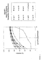
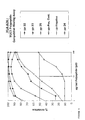
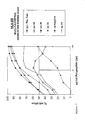
- said amino acid sequence is selected from the group consisting of SEQ ID NO: 5, 7, 8, 10 to 19 and 22 to 33. Data to explain why these sequences have been selected are shown in Figures 1 to 7.
- said polypeptide presents at least one of the following properties :
- polypeptide is a peptide that comprises a MHC class I restricted T cell epitope, the epitope being contained in or represented by any one of the aforementioned SEQ ID NOs, preferably SEQ ID NO: 5. These latter epitopes lie outside the immunogenic variable non tandem repeat (VNTR) region.
- VNTR immunogenic variable non tandem repeat
- amino acid sequence present in the polypeptide of the invention may be a sequence represented by any one of the hereinabove described SEQ ID NOs or the amino acid sequence of an epitope present within these sequences (such as the fragments of the sequences shown in the brackets shown below, e.g., for SEQ ID NO: 5):
- the polypeptide of the invention has the same sequence as the "epitope".
- the peptide typically comprises 1, 2, 3 or more copies of each of 1, 2 or more, or all of the above defined "epitopes".
- a 'linker' sequence may or may not separate the "epitopes" and/or there may or may not be additional (non-"epitope") sequences at the N terminal or C terminal of the polypeptide.
- the peptide comprises 1, 2, 3 or more linkers.
- the linkers are typically 1, 2, 3, 4 or more amino acids in length and may comprise amino acid sequence encoded by a polynucleotide sequence that comprises enzyme restriction sites or amino acids that constitute proteosomal cleavage sites.
- the polypeptide 1 2 or more, or all of the “epitopes” may be contiguous with each other or separated from each other.
- the "epitope” sequences may overlap with each other.
- the polypeptide is typically 8 to 2000 amino acids in length, such as 9 to 1000, 10 to 500, 11 to 200, 12 to 100 or 15 to 50 amino acids.
- the polypeptide may also comprise a sequence which aids the stimulation of a CTL response directed to the epitope. Such sequence may act as adjuvant or may target the polypeptide to antigen presenting cells (APCs) or to compartments in the antigen processing pathway.
- the sequence may stimulate a T helper response, such as a Th1 response, and thus may comprise a T helper (e.g.
- the polypeptide may also comprise the sequence of any of the proteins mentioned herein.
- the polypeptide may be free from modifications.
- the polypeptide is modified, for example by a natural post-translational modification (e.g. glycosylation) or an artificial modification.
- the sequence in the polypeptide may or may not comprise the modification(s) that are present when the sequence is expressed in a normal or cancer cell.
- the polypeptide may comprise the modifications that occur when it is expressed in a eukaryotic (e.g. human) or prokaryotic (e.g. E. coli) cell.
- the polypeptide lacks glycosylation.
- the modification may provide a chemical moiety (typically by substitution of a hydrogen, e.g. the hydrogen of a C-H bond), such as an amino, acetyl, hydroxy or halogen (e.g. fluorine) group or carbohydrate group. Typically the modification is present on the N or C terminus.
- a chemical moiety typically by substitution of a hydrogen, e.g. the hydrogen of a C-H bond
- halogen e.g. fluorine
- processed' refers to being processed by the class I antigen presentation pathway (generally this will be hydrolysis, e.g. proteolysis).
- homology is calculated on the basis of amino acid identity (sometimes referred to as "hard homology").
- the UWGCG Package provides the BESTFIT program which can be used (e.g. on its default setting) to calculate homology (Devereux et al., Nucl. Acids Res. 12 (1984), 387-395).
- the homologous peptide typically differs from the epitope present in the polypeptide of the present invention by substitution, insertion or deletion, for example from 1, 2, 3, 4 or more substitutions and/or 1, 2, 3, 4 or more deletions and/or 1, 2, 3, 4 or more insertions over its length.
- the invention also relates to polynucleotides comprising a nucleic acid sequence encoding at least one polypeptide of the present invention. Based on the amino acid sequences provided within the present application and by using the genetic code those skilled in the art can easily identify said nucleic acid sequences.
- the nucleic acid sequence of the present invention is selected from the group consisting of SEQ ID NOs: 34, 36, 38, 39, 41 to 50 and 53 to 64.
- polynucleotide as used in the scope of the present invention means a DNA and/or RNA fragment, single or double-stranded, linear or circular, natural or synthetic, modified or not (see US-A-5,525,711, US-A-4,711,955, US-A-5,792,608 or EP-A-0 302 175 for modification examples) defining a fragment or a portion of a nucleic acid, without size limitation. It may be, inter alia, a genomic DNA, a cDNA, an mRNA. "Polynucleotides” and “nucleic acids” are synonyms with regard to the present invention.
- the nucleic acid may be in the form of a linear polynucleotide, and preferably in the form of a plasmid.
- plasmids A wide range of plasmids is commercially available and well known by one skilled in the art. These available plasmids are easily modified by the molecular biology techniques (Sambrook et al., 1989, Laboratory Manual, Cold Spring Harbor Laboratory Press, Cold Spring Harbor, New York).
- Plasmids derived from pBR322 (Gibco BRL), pUC (Gibco BRL), pBluescript (Stratagene), pREP4, pCEP4 (Invitrogen) and also p Poly (Lathe et al., 1987, Gene 57, 193-201) are illustrative of these modifications.
- the nucleic acid can be a naked polynucleotide (Wolff et al., Science 247 (1990),1465-1468) or is formulated with at least one compound such as a polypeptide, preferably viral polypeptides, oligonucleotides or cationic lipids, or cationic polymers which can participate in the uptake of the nucleic acid into the cells (see Ledley, Human Gene Therapy 6 (1995), 1129-1144 for a review) or a protic polar compound (examples are provided below in the present specification or in EP-A-0 890362).
- a polypeptide preferably viral polypeptides, oligonucleotides or cationic lipids, or cationic polymers which can participate in the uptake of the nucleic acid into the cells
- a protic polar compound examples are provided below in the present specification or in EP-A-0 890362.
- Polynucleotide also designates nucleic acid of viral origin (viral vector) which encodes at least for the polypeptide of the invention.
- viral vector preferably derives from a virus selected among poxvirus (vaccine virus, MVA, canarypox....), adenovirus, retrovirus, herpes virus, alpha virus, foamy virus or adeno associated virus. Said viral vectors and their uses are widely disclosed in gene therapy literature.
- said nucleic acid includes at least one therapeutically useful gene sequence that can be transcribed and translated to generate a polypeptide of interest and the elements enabling its expression.
- the genetic information necessary for expression by a target cell comprises all the elements required for transcription of DNA into RNA and, if necessary, for translation of mRNA into a polypeptide.
- Transcriptional promoters suitable for use in various vertebrate systems are well known.
- suitable promoters include viral promoters like RSV, MPSV, SV40, CMV or 7.5k, vaccinia promoter, inducible promoters, tissue specific promoters, synthetic promoters, etc or combination thereof.
- the nucleic acid can also include intron sequences, targeting sequences, transport sequences, sequences involved in replication or integration. Said sequences have been reported in the literature and can be readily obtained by those skilled in the art.
- the nucleic acid can also be modified in order to be stabilized with specific components as spermine.
- the polynucleotide of the invention is capable of expressing 1, 2, 3 or more (different) compounds, each of which is a polypeptide of the invention.
- the polynucleotide is typically DNA or RNA, and is single or double stranded.
- the polynucleotide generally comprises 1, 2, 3 or more coding sequences which may be the same or different. At least one of the coding sequences encodes a polypeptide of the invention.
- the coding sequence is typically operably linked to a control sequence capable of providing for expression of the polynucleotide.
- the polynucleotide comprises 5' and 3' to the coding sequence sequences which aid expression, such as aiding transcription and/or translation of the coding sequence.
- the polynucleotide comprises a promoter, enhancer, transcription terminator, polyadenylation signal, polyA tail, intron, translation initiation codon or translation stop codon.
- the polynucleotide may in particular be capable of expressing a polypeptide of the invention in a mammalian or avian cell, such as in any of the cells discussed herein.
- the polynucleotide may furthermore be capable of expressing the polypeptide in the cellular vector discussed below.
- the polynucleotide may form or be part of a vector, such as a plasmid or cosmid vector.
- the polynucleotide is present in a virus or cellular vector, such as a virus which is capable of stimulating a MHC class I restricted T cell response (e.g. a vaccinia virus).
- Intraduction or transfer means that the polynucleotide is transferred into the cell and is located, at the end of the process, inside said cell or within or on its membrane. It is also called “transfection” or “infection” depending of the nature of the vector.
- the invention is therefore further directed to a vector, e.g. of viral or plasmid origin, comprising at least a nucleic acid sequence of the invention.
- a vector e.g. of viral or plasmid origin
- the vector of the invention comprises one or more nucleotide sequences selected from the group consisting of:
- the present invention relates to host cells comprising at least one polynucleotide or at least one vector according to the invention.
- a host cell is a prokaryotic cell or a eukaryotic cell, such as a yeast cell, more preferably an animal cell, most preferably a mammalian cell.
- the present invention also relates to a composition (iv) that comprises two or more different compounds wherein each of the compounds is (i) a polypeptide or (ii) a polynucleotide of the invention as defined above.
- composition (iv) 1, 2, 3, 4, 5 or more different compounds may be present, wherein each of these compounds is (i) or (ii).
- the composition may comprise all the epitopes of the invention.
- the composition may comprise 1, 2, 3, 4, 5 or more polynucleotides which together are capable of being expressed to provide 1, 2, 3, 4, 5 or more different epitopes or polypeptides of the invention, or all the epitopes of the invention.
- (i), (ii) or (iv) are provided for use in the form of a pharmaceutical composition for vaccination against cancer or for use in immunosuppression. 1, 2, 3, 4, 5 or more different epitopes of the invention (or all of the epitopes of the invention) may be used.
- each epitope or one or more groups of epitopes within the combination are suitable for being administered to the host separately or sequentially.
- the epitopes in each group are typically together in the form of a single peptide of the invention or in the form of the composition of the invention.
- different polypeptides or polynucleotides of the invention may be administered separately or sequentially, for example polynucleotides capable of expressing individual or groups of epitopes and/or polypeptides and/or analogues and/or compositions.
- the invention provides a combination of 1, 2, 3, 4, 5 or more different epitopes and/or polypeptides and/or compositions and/or polynucleotides of the invention for simultaneous, separate or sequential use in the preparation of a pharmaceutical composition for the treatment of the human or animal body by therapy, for example in vaccination against cancer or in immunosuppression.
- Vaccination against cancer or immunosuppression typically leads to a MHC class I restricted T cell response, the T cells of which are specific for an epitope of the invention.
- (i), (ii) or (iv) can be used in a form or manner in which they stimulate such a MHC class I restricted T cell response. Such methods are known in the art.
- the vaccine of the invention may comprise one or more components (for example, as discussed herein in relation to the vaccine of the invention) in addition to (i), (ii) or (iv).
- the components of the vaccine may be administered simultaneously, separately or sequentially to the host.
- the invention also provides a vaccine comprising (i), (ii) or (iv), which vaccine is capable of stimulating a MHC class I restricted T cell response directed to an epitope (polypeptide) of the invention.
- a vaccine comprises an adjuvant or delivery system which stimulates a MHC class I restricted T cell response.
- the adjuvant may be capable of causing or augmenting a MHC class II restricted T cell (typically CD4) reponse which is favourable to the production of a MHC class I restricted T cell response, such as a Th1 response.
- the adjuvant may comprise a MHC class II restricted T cell epitope (or a precursor which can be processed in vivo to provide such an epitope).
- the adjuvant may be a cytokine, such as a cytokine which stimulates a MHC class I restricted T cell response or favourable MHC class II restricted T cell response (e.g. IL-2, IL-7, IL-12 or IFN- ⁇ ).
- the adjuvant may be, for example, CFA (Golding and Scott, Ann. N.Y.
- a muramyl dipeptide e.g. of a mycobacterial cell wall
- monophosphoryl lipid A lipopolysaccharide (e.g. from B. abortus), liposomes
- SAF-1 Golding and Scott, Ann. N.Y. Acad. Sci. 754 (1995), 126-137
- a saponin e.g. Quil A
- keyhole limpet hemocyanin yeast TY particle
- beta 2-microglobulin or mannan e.g. oxidised mannan
- the delivery system is typically capable of providing (i), (ii), (iv) or an epitope expressed from (iv) to an APC, such as a professional APC.
- an APC such as a professional APC.
- the particular route of administration suitable for use may aid the stimulating of a MHC class I restricted T cell response and, thus, (i), (ii), (iv) or the vaccine of the invention may be provided in a form suitable for administering by such a route.
- Intraperitoneal or intravenous routes are preferred.
- these substances are delivered by biolistic means.
- a low dose of antigen favours the development of a MHC class I restricted T cell response.
- a suitable low dose of a compound of the invention can be given.
- the vaccine may be provided in an amount and concentration that is suitable for administering to provide an appropriate low dose.
- (iv) is administered in the form of "naked DNA".
- the invention also relates to a composition, preferably a pharmaceutical composition, which is particularly useful for the delivery of polynucleotides of the invention to cells or tissues of a subject in the scope of a gene therapeutic method, especially in case of cancer treatment.
- the term "gene therapy method” is preferably understood as a method for the introduction of a polynucleotide into cells either in vivo or by introduction into cells in vitro followed by reimplantation into a subject. "Gene therapy” in particular concerns the case where the polynucleotide is expressed in a target tissue, especially tissue comprising cell expressing MHC-I molecules.
- the composition in particular pharmaceutical composition, furthermore comprises a pharmaceutically acceptable carrier or diluent.
- the carrier or diluent is non toxic to recipients at the dosages and concentrations employed.
- Representative examples of carrier or diluent for injectable solutions include water, isotonic saline solutions which are preferably buffered at the physiological pH (such as phosphate buffered saline or Tris-buffered saline), mannitol, dextrose, glycerol and ethanol, as well as polypeptides or protein such as human serum albumin.
- This carrier or diluent is preferably isotonic, hypotonic or weakly hypertonic and has a relatively low ionic strength.
- it may contain any relevant solvents, aqueous or partly aqueous liquid carriers comprising sterile, pyrogen-free water, dispersion media, coatings, and equivalents.
- the pH of the pharmaceutical preparation is suitably adjusted and buffered.
- the invention more particularly pertains to a composition, in particular pharmaceutical composition, comprising at least one of the complexes described above and also incorporating at least one adjuvant capable of improving the transfection capacity of said complex.
- Adjuvants may be selected from the group consisting of a chloroquine, protic polar compounds such as propylene glycol, polyethylene glycol, glycerol, EtOH, 1-methyl L -2-pyrolidine or their derivatives, or aprotic polar compounds such as dimethylsulfoxide (DMSO), diethylsulfoxide, di-n-propylsulfoxide, dimethylsulfone, sulfolane, dimethylformamide, dimethylacetamide, tetramethylurea, acetonitrile and their derivatives.
- DMSO dimethylsulfoxide
- DMSO dimethylsulfoxide
- di-n-propylsulfoxide dimethylsulfone
- sulfolane dimethylformamide
- the polynucleotide which is contained in the composition is a DNA.
- concentration of polynucleotide in such a composition is from about 0.1 ⁇ g/ml to about 20 mg/ml.
- compositions, in particular pharmaceutical composition, in accordance with the present invention are suitable to be administered into a vertebrate tissue.
- This administration may be made by intradermal, subdermal, intravenous, intramuscular, intranasal, intracerebral, intratracheal, intraarterial, intraperitoneal, intravesical, intrapleural, intracoronary or intratumoral injection, by means of a syringe or other devices.
- Transdermal administration is also contemplated, as are inhalation, aerosol routes, instillation or topical application.
- the composition in particular pharmaceutical composition, is suitable to be administered into target tissues of the vertebrate body including those of muscle, skin, brain, lung, liver, spleen, bone marrow, thymus, heart, lymph, bone, cartilage, pancreas, kidney, gall bladder, stomach, intestine, testis, ovary, uterus, rectum, nervous system, eye, gland, connective tissue, blood, etc.
- said composition will be administered into tumor.
- Administration of such a composition to a patient allows to elicit an immune response based on the activation of cytotoxic lymphocytes by the polypeptides encoded by said nucleotide sequences.
- composition of the invention is particularly suitable for the treatment or prevention of MUC-1-expressing cancers, such as breast cancer, ovary cancer, pancreas or lung cancer.
- MUC-1-expressing cancers such as breast cancer, ovary cancer, pancreas or lung cancer.
- compositions of the present invention i.e. containing polypeptide or polynucleotide sequences of the instant invention (see above) is suitable for the treatment or prevention of MUC1-expressing cancer, wherein said treatment or prevention comprises:
- the term "cells” includes prokaryote cells and eukaryote cells, yeast cells, plant cells, human or animal cells, in particular mammalian cells.
- cancer cells should be mentioned.
- the invention can be applied in vivo to the interstitial or luminal space of tissues in the lungs, the trachea, the skin, the muscles, the brain, the liver, the heart, the spleen, the bone marrow, the thymus, the bladder, the lymphatic system, the blood, the pancreas, the stomach, the kidneys, the ovaries, the testicles, the rectum, the peripheral or central nervous system, the eyes, the lymphoid organs, the cartilage, the endothelium.
- the cell will be a muscle cell, a haematopoietic system stem cell or an airways cell, a tracheal or pulmonary cell, or a tumor cell.
- the present invention also relates to a process for transferring a nucleic acid into cells wherein said process comprises contacting said cells with at least one polynucleotide according to the invention.
- This process is suitable to be applied by direct administration of said polynucleotide to cells of the animal in vivo, or may be applied by in vitro treatment of cells which were recovered from the animal and then re-introduced into the animal body ( ex vivo process).
- in vitro application cells cultivated on an appropriate medium are placed in contact with a suspension consisting of polynucleotide of the invention. After an incubation time, the cells are washed and recovered.
- the polynucleotide can be verified (eventually after lysis of the cells) by any appropriate method.
- the patient may undergo a macrophage depletion treatment prior to administration of the pharmaceutical preparations described above.
- a technique is described in the literature (refer particularly to Van Rooijen et al., 1997, TibTech 15, 178-184).
- the present invention further concerns the use of a polypeptide or a polynucleotide, a vector or a host cell as defined above for the preparation of a composition intended for diagnostic, curative, preventive or vaccination treatment of man or animals, and more specifically for the treatment of cancer.
- the present invention relates to a diagnostic composition
- a diagnostic composition comprising at least one polypeptide as defined above.
- the use of a polypeptide of the invention in a diagnostic composition is illustrated by the following processes:
- compositions or uses of the invention are suitable to be used for the treatment of all kinds of cancer the treatment and/or diagnostic of which being related to or dependent on the immune properties of the polypeptides of the invention.
- the compositions, and uses of the present invention may be desirably employed in humans, although animal treatment is also encompassed by the uses described herein.
- the MHC molecule of (a) is generally a class I molecule (e.g. HLA-A*0201). Such molecules comprise an ⁇ chain and a ⁇ chain. The fragment may comprise only the extracellular domain of the MHC molecule. The fragment may or may not be capable of binding a cell membrane.
- (b) may comprise a protein which has homology with a naturally occurring ⁇ chain (or a fragment thereof) and/or a protein which has homology with a naturally occurring ⁇ chain (or a fragment thereof). The naturally occurring ⁇ or ⁇ chain may be of an HLA-A molecule (e.g.
- HLA-A*0201 Any of the above homologous proteins or fragments may be present as part of fusion proteins.
- (b) is typically a derivative of (a) and, thus, may be made by modifying (a) by any of the modifications mentioned herein.
- the product may be designed, made or identified using methods known in the art. Thus, the invention provides use of a polypeptide (epitope or epitope sequence) of the invention to design or identify the product.
- the product may be designed by computational means or may be identified from a library of compounds.
- the invention also provides a method of identifying a product of the invention comprising contacting a candidate substance with a T cell receptor or fragment of the invention and determining whether the candidate substance binds to the T cell receptor or fragment, the binding of the candidate substance to the T cell receptor or fragment indicating that the substance is such a product.
- the product may be present on the surface of a cell, such as a professional APC.
- the binding may be measured by contacting the candidate substance with a T cell of the invention and determining whether the candidate substance causes antigen specific functional activity of the T cell (such as by any means mentioned herein).
- the product may be linked to a cytotoxic agent.
- the product is an antibody.
- the invention provides a multimer comprising 2 or more products of the invention.
- a multimer may be used in the same manner as the product is used in the different aspects of the invention and, thus, the term 'product' as used in the context of the other aspects of the invention includes the multimer.
- the products in the multimer may be linked by a covalent bond or by non-covalent means.
- the products are linked by a streptavidin - biotin interaction and, thus, typically the products comprise a biotin portion (typically chemically linked to or in a fusion protein with the product) which allows the products to be linked together by streptavidin.
- the multimer generally has a higher binding affinity to the T cell receptor of the invention than the product, and in one embodiment is able to cause more antigen specific functional activity than the product.
- the multimer may also comprise a detectable label, such as a radioactive or a light detectable (e.g. fluorescent) label.
- the label may allow the multimer to be sorted by flow cytometry (e.g. when the multimer is bound to a T cell receptor which is present on a T cell of the invention).
- the multimer may be a soluble multimer or may be capable of associating with a cell membrane. In one embodiment the multimer is attached to a solid support, such as a microtitre plate.
- the invention also provides a cell comprising a product of the invention.
- the cell may be any of the types of cells mentioned herein, such as a professional APC or T cell.
- the cell may be capable of stimulating antigen specific functional activation of a T cell of the invention.
- the cell may be used to stimulate a MHC class I restricted T cell response in vitro or in vivo, which response is directed to a polypeptide (epitope) of the invention.
- the cell may, therefore, be used for the preparation of a pharmaceutical composition for the treatment of the human or animal body by therapy, particularly in treating or preventing cancer.
- the cell may be made by providing a polypeptide, polynucleotide or composition of the invention to a cell which is able to process the polypeptide, polynucleotide or composition and present them on its surface (under conditions in which such processing occurs).
- the invention furthermore describes a method of causing the in vitro replication of MHC class I restricted T cells which are specific for a cancer epitope comprising contacting a population of cells which comprises MHC class I restricted T cells with a polypeptide of the invention under conditions in which the polypeptide or analogue are presented to T cells in the population, or with a product or cell of the invention.
- the invention includes use of a T cell of the invention (including a T cell replicated by the above method) in vitro or which may be suitable to be used in vivo to kill a cell which presents the polypeptide (epitope) of the invention.
- a cell is typically a cancer cell, but in one embodiment is a T cell (typically a MUC1 expressing activated T cell).
- the invention provides a T cell of the invention, or a cell which has been replicated in the method of the invention for use in a method of treatment of the human or animal body by therapy.
- a pharmaceutical composition for preventing or treating cancer or a disease caused by an immune response such as an inflammatory disorder, autoimmune disease, organ transplant rejection or graft versus host disease.
- the invention also provides a method of identifying a MHC class I restricted T cell response which is based on determining whether MHC class I restricted T cells from a host recognise a polypeptide or analogue of the invention (either of which may be provided by a polynucleotide of the invention), or a product or cell of the invention.
- the polypeptide or analogue may be in the form of the composition of the invention.
- the determination of whether the T cells recognise the polypeptide or analogue is done by detecting a change in the state of the T cells in the presence of the polypeptide or analogue or determining whether the T cells bind the polypeptide or the analogue.
- the change in state is generally caused by antigen specific functional activity of the T cell after the T cell receptor binds the polypeptide or the analogue.
- the polypeptide or the analogue is bound to an MHC class I molecule, which is typically present on the surface of an APC.
- the change in state of the T cell may be the start of or increase in the expression of a substance in the T cells and/or secretion of a substance from the T cell, such as a cytokine (e.g. IFN- ⁇ , IL-2 or TNF- ⁇ ). Determination of IFN- ⁇ expression or secretion is particularly preferred.
- the substance can typically be detected by allowing it to bind to a specific binding agent and then measuring the presence of the specific binding agent/substance complex.
- the specific binding agent is typically an antibody, such as polyclonal or monoclonal antibodies. Antibodies to cytokines are commercially available, or can be made using standard techniques.
- the specific binding agent is immobilised on a solid support (and thus the method may based on the ELISPOT assay to detect secretion of the substance). After the substance is allowed to bind the solid support can optionally be washed to remove material which is not specifically bound to the agent.
- the agent/substance complex may be detected by using a second binding agent which will bind the complex.
- the second agent binds the substance at a site which is different from the site which binds the first agent.
- the second agent is preferably an antibody and is labelled directly or indirectly by a detectable label.
- the second agent may be detected by a third agent which is typically labelled directly or indirectly by a detectable label.
- the second agent may comprise a biotin moiety, allowing detection by a third agent which comprises a streptavidin moiety and typically alkaline phosphatase as a detectable label.
- the change in state of the T cell which can be measured may be the increase in the uptake of substances by the T cell, such as the uptake of thymidine.
- the change in state may be an increase in the size of the T cells, or proliferation of the T cells, or a change in cell surface markers on the T cell.
- the change in state may be the killing (by the T cell) of a cell which presents the polypeptide, the analogue or the product of the invention to the T cell (e.g. the killing of the cell of the invention).
- the determination of whether the T cells recognise the peptide may be carried out using a CTL assay.
- the T cells which are contacted in the method are obtained from the host in a blood sample, although other types of samples which contain T cells can be used.
- the sample may be added directly to the assay or may be processed first. Typically the processing may comprise diluting of the sample, for example with water or buffer.
- the sample is diluted from 1.5 to 100 fold, for example 2 to 50 or 5 to 10 fold.
- the processing may comprise separation of components of the sample.
- mononuclear cells (MCs) are separated from the sample.
- the MCs will comprise the T cells and APCs.
- the APCs present in the separated MCs can present the peptide to the T cells.
- T cells in one embodiment only CD8 T cells
- PBMCs, MCs and T cells can be separated from the sample using techniques known in the art.
- the T cells used in the assay can be in the form of unprocessed or diluted samples, or are freshly isolated T cells (such as in the form of freshly isolated MCs or PBMCs) which are used directly ex vivo, i.e. they are not cultured before being used in the method.
- the T cells are cultured before use, for example in the presence of the polypeptide or the analogue of the invention and, generally, also exogenous growth promoting cytokines.
- the polypeptide or the analogue are typically present on the surface of APCs, such as the APC used in the method. Pre-culturing of the T cells may lead to an increase in the sensitivity of the method.
- the APC which is typically used in the method is from the same host as the T cell or from a different host.
- the APC can be a non-professional APC, but is typically a professional APC, such as any of the APCs mentioned herein.
- the APC maybe an artificial APC.
- the APC is a cell which is capable of presenting the peptide to a T cell. It is typically separated from the same sample as the T cell and is typically co-purified with the T cell. Thus, the APC may be present in MCs or PBMCs.
- the APC is typically a freshly isolated ex vivo cell or a cultured cell. It may be in the form of a cell line, such as a short term or immortalised cell line.
- the APC may express empty MHC class I molecules on its surface.
- the method identifies a MHC class I restricted T cell response to any of the combinations of a polypeptide of the invention discussed above in relation to the composition of the invention.
- the T cells can be placed into an assay with the composition of the invention (which comprises the combination of the polypeptide or the analogue which are to be tested).
- the T cells can be divided and placed into separate assays each of which contains a group of polypeptides or analogues within the combination.
- a polypeptide per se is added directly to an assay comprising T cells and APCs.
- the T cells and APCs in such an assay could be in the form of MCs.
- the polypeptide is provided to the APC in the absence of the T cell.
- the APC is then provided to the T cell, typically after being allowed to present the polypeptide or the analogue on its surface.
- the polypeptide may have been taken up inside the APC and presented, or simply be taken up onto the surface without entering inside the APC.
- the duration for which the polypeptide is contacted with the T cells will vary depending on the method used for determining recognition of the peptide.
- concentration of T cells used is 10 3 /ml to 10 9 /ml, preferably 10 5 /ml to 10 7 /ml.
- the concentration of the polypeptide by the T cells is typically from 0.1 to 1000 ⁇ g/ml, preferably 10 to 100 ⁇ g/ml.
- the length of time for which the T cells are incubated with the polypeptide or the analogue is from 4 to 24 hours, preferably 6 to 16 hours.
- the determination of the recognition of the polypeptide by the T cells may be done by measuring the binding the polypeptide or the analogue to the T cells.
- T cells which bind the polypeptide can be sorted based on this binding, for example using a FACS machine. The presence of T cells which recognise the polypeptide will be deemed to occur if the frequency of cells sorted using the polypeptide is above a 'control' value (i.e. above the frequency of antigen naive T cells which recognise the polypeptide).
- the frequency of antigen-experienced T cells during a disease state can be up to 2.5% of the total CD8 T cells.
- the polypeptide, the polynucleotide, the composition, the product or the cell of the invention can be used to detect a MHC class I restricted T cell response to a polypeptide (epitope) of the invention in vitro (such as in a sample from a host) or is suitable to be used in vivo. This can be done, for example, by using the method discussed above.
- the presence of a response generally indicates the presence of a cell which is expressing MUC1, such as a cancer cell or an activated T cell.
- the detection of the response may be used to diagnose cancer.
- Measurement of the level of the response may be used to monitor the severity of the cancer (i.e. the number of cancer cells present in the host), a larger response indicating a more severe cancer.
- the presence or absence of the MHC class I restricted T cell response is typically determined by the method of identifying a MHC class I restricted T cell response discussed above.
- the antibodies mentioned herein may be produced by raising antibody in a host animal. Such antibodies will be specific to the peptide or to the substances mentioned above which bind antibodies. The peptide or substances are referred to as the 'immunogen' below. Methods of producing monoclonal and polyclonal antibodies are well-known.
- a method for producing a polyclonal antibody comprises immunising a suitable host animal, for example an experimental animal, with the immunogen and isolating immunoglobulins from the serum.
- a method for producing a monoclonal antibody comprises immortalising cells which produce the desired antibody.
- Hybridoma cells may be produced by fusing spleen cells from an inoculated experimental animal with tumour cells, for example as described in Köhler and Milstein (Nature 256 (1975), 495-497).
- An immortalized cell producing the desired antibody may be selected by a conventional procedure.
- the hybridomas may be grown in culture or injected intraperitoneally for formation of ascites fluid or into the blood stream of an allogenic host or immunocompromised host.
- Human antibody may be prepared by in vitro immunisation of human lymphocytes, followed by transformation of the lymphocytes with Epstein-Barr virus.
- the experimental animal is suitably a goat, rabbit, rat or mouse.
- the immunogen may be administered as a conjugate in which the immunogen is coupled, for example via a side chain of one of the amino acid residues, to a suitable carrier.
- the carrier molecule is typically a physiologically acceptable carrier.
- the antibody obtained may be isolated and, if desired, purified.
- any of the polypeptides or polynucleotides discussed above in any form or in association with any other agent discussed above is included in the termed "vaccination agent" below.
- An effective non-toxic amount of such a vaccination agent may be given to a human or non-human patient in need thereof.
- the condition of a patient suffering from a cancer can therefore be improved by administration of such a vaccination agent.
- the vaccination agent may be administered prophylactically to an individual who does not have a cancer in order to prevent the individual developing cancer.
- the present invention provides uses for the preparation of the vaccination agent for treating the human or animal body by therapy.
- the invention provides the use of the vaccination agent in the manufacture of a medicament for vaccinating against cancer.
- the invention provides uses for the preparation of a vaccine composition for vaccinating an individual.
- the vaccination agent is typically administered by any standard technique used for administering vaccines, such as by injection. Typically after the initial administration of the vaccination agent a booster of the same or a different vaccination agent of the invention can be given. In one embodiment the subject is given 1, 2, 3 or more separate adminstrations, each of which is separated by at least 12 hours, 1 day, 2, days, 7 days, 14 days, 1 month or more.
- the vaccination agent may be in the form of a pharmaceutical composition which comprises the vaccination agent and a pharmaceutically acceptable carrier or diluent. Suitable carriers and diluents include isotonic saline solutions, for example phosphate-buffered saline.
- the composition is formulated for parenteral, intravenous, intramuscular, subcutaneous, transdermal, intradermal, oral, intranasal, intravaginal, or intrarectal administration.
- the dose of vaccination may be determined according to various parameters, especially according to the substance used; the age, weight and condition of the patient to be treated; the route of administration; and the required regimen. A physician will be able to determine the required route of administration and dosage for any particular patient.
- a suitable dose may however be from 10 ⁇ g to 10g, for example from 100 ⁇ g to 1g of the vaccination agent. These values may represent the total amount administered in the complete treatment regimen or may represent each separate administration in the regimen.
- transfection agents may also be administered to enhance the uptake of the polynucleotides by cells.
- suitable transfection agents include cationic agents (for example calcium phosphate and DEAE-dextran) and lipofectants (for example lipofectamTM and transfectamTM).
- the vaccination agent is a polynucleotide which is in the form of a viral vector
- the amount of virus administered is in the range of from 10 4 to 10 12 pfu, preferably from 10 7 to 10 10 pfu (for example for adenoviral vectors), more preferably about 10 8 pfu for herpes viral vectors.
- a pox virus vector may also be used (e.g. vaccinia virus), typically at any of the above dosages.
- Polypeptide fragments 8 - 13 amino acids long, of proteins produced within a nucleated, vertebrate cell are associated with newly formed cellular proteins of the MHC-I complex.
- the complex of MHC-I protein and polypeptide fragment are further associated with a protein known as Beta-2 microglobulin.
- Beta-2 microglobulin This trimolecular complex is then transported to the cell surface, anchored to the cell membrane and exposed to the extracellular milieu.
- Thymus-derived, or T lymphocytes of the CD8 category have specific 'antigen' receptors on their cell surface, which recognise the MHC-I-Beta-2-microgobulin-peptide complex.
- T lymphocytes are not 'stimulated' due either to the deletion of self-specific T cells from the immune repertoire or due to negative regulation of self-specific immune responses.
- the polypeptide is from a pathogenic organism, such as a virus
- specific T cells are activated to proliferate and to become cytotoxic such that CD8 'cytotoxic effector cells' or CTL specifically recognise the infected cell and eliminate that cell in an effort to contain the pathogenic condition.
- Tumours can also produce 'tumour-specific' protein molecules or modifications of cellular proteins.
- specific CD8 T cells can recognise a tumour cell as pathogenic and eliminate these cells by the same mechanism as is used to eliminate virus-infected cells.
- the tumour-specific modification of a protein is merely quantitative in that a protein is over-produced in tumour cells. It has been shown that such proteins can also be recognised by specific Cytotoxic T Lymphocytes (CTL).
- CTL Cytotoxic T Lymphocytes
- Binding can be envisioned with this apparatus since the polypeptide known to bind is tagged with a flourescene molecule. Thus cells binding the polypeptide become fluorescent. Each polypeptide to be screened for binding is mixed together with the reference fluorescent polypeptide, which is at a constant concentration of 150 nM. The test polypeptide is added at various concentrations and the mixture exposed to the same cells. A test polypeptide is considered 'positive' if the binding of the reference polypeptide is inhibited by 50 % at 20 ⁇ g/ml or less of the test polypeptide.
- Figures 1 - 7 are shown data for the competitive binding of polypeptides deemed, by this assay, to be positive for binding to HLA-A2, B7, A3, A11, A24, A1, and B8 respectively.
- binding is compared to a negative control polypeptide, known not to bind to that HLA type and a positive control polypeptide, known to bind to that HLA type.
- the positive control 'Flu' is a polypeptide with the sequence GILGFVFTL from the influenza virus matrix protein (Scheibenbogen et al. International Journal of Cancer, 71: 932-936 (1997)).
- EBV-B cell lines were derived by cultivating human Peripheral Blood Mononuclear Cells (PBMC) in filtered culture supernatant which had been used to grow cells from the marmoset line B-958 These cells produce the Epstein Barr Virus.
- PBMC Peripheral Blood Mononuclear Cells
- Cyclosporin A to inhibit T cell reactivity to the virus
- HLA-typed EBV-transformed human B cell lines were used for all tests HLA types are described in Table 2.
- Culture medium was Dulbecco's Modified Eagle's Medium (DMEM) + 5x10 -5 M BetaMercaptoethanol (with the addition of 25 mM HEPES buffer for the step of polypeptide detachment and cell washing) plus either 2% or 10% Fetal bovine Serum. All tests were performed in 96 well, V-bottom microtitre plates (PS micro plate). ⁇ -2 microglobulin was purchased from Sigma. The buffer for the polypeptide detachment used was:
- Cells were cultivated in Coming T175 flasks in 20 of culture medium (10% FBS). The night before the assay, cells were re-suspended and 10 ml of fresh medium added. The day of the test cells were resuspended, counted, pelleted by centrifugation and resuspended in 5 ml complete medium with 10% FBS. Distribute cells into a 6 well plate, 10 5 cells per well in 5 ml culture medium. Cells were then cultured for 4 hours at 37°, 5% CO 2 . During that time polypeptides were prepared by dilution in 600 ⁇ g/ml in PBS.
- test plate (96 well, V-bottom) was prepared as follows:
- Cells in the 5 ml culture medium in the 6 well plate were resuspended and transferred to a 15 ml conical bottom test tube and then centrifuged for 5 minutes at 1500 rpm (500g). Resuspend the cells in PBS and centrifuge a second time (500g). Supernatant was removed and 2 ml detachment buffer while on ice. Cells were resuspended by gentle pipetting curing the first 30 seconds of this 2 minute period. After 2 minutes, 14 ml culture medium with 2% FBS was added. Cells are mixed by inverting the tube twice, then centrifuged at 2000 rpm (800g) for 3 minutes at 4°C.
- the mean fluorescence intensity (MFI) of cells with the fluorescent reference polypeptide but with no competitor polypeptide (positive control) was taken as 0% inhibition.
- the MFI of cells without the fluorescent reference polypeptide (negative control) was taken to be equal to 100 % inhibition.
- C* indicates that the flourescene molecule is coupled to a cysteine residue (as described in van der Burg et al., Human Immunology, 44 : 189-198 (1995)).
- Positive control polypeptides are published in Rammensee et al ., 'MHC Ligands and polypeptide Motifs' Springer, New York (1995)
- the ELISpot is a technique which allows the identification of antigen-specific (in this case, MUC-1-specific) T cell recognician by the detection of antigen induced production of cytokines (IFN ⁇ , TNF ⁇ , IL-4, etc8) following an antigenic stimulation in vitro. More particularly, ELISpot allows the determination of the number of antigen specific T lymphocytes in a population of peripheral blood mononuclear cells (PBMC) (Scheibenbogen et al., 1997, Int. J. Cancer 71-1). In this case, the production of IFN ⁇ produced by CD8+ T cells (CTL) in responses to polypeptides as presented by autologous HLA molecules were examined.
- PBMC peripheral blood mononuclear cells
- the cytokines are captured between two specific antibodies.
- the first antibody specific for human IFN ⁇ , is adsorbed on a nitrocellulose membrane.
- Lymphocytes from human blood samples are added to the microtitre wells containing the attached antibody.
- Antigen in the form of polypeptides, is also added to the wells.
- polypeptides will attach to cell surface HLA molecules (together with ⁇ -2 microglobulin).
- Polypeptide specific T cells will recognize the complex of the polypeptide:HLA: ⁇ -2 ⁇ globulin.
- the T cells Upon recognition of antigen, the T cells become 'activated' to produce cytokines such as IFN ⁇ . Secreted IFN ⁇ is then captured by the antibody which is attached to the nitrocellulose.
- Peripheral blood mononuclear cells Peripheral blood mononuclear cells
- patient PBMC Peripheral Blood Mononuclear Cells
- PBMC Peripheral Blood Mononuclear Cells
- Figure 10 patient PBMC came from prostate cancer patients who had participated in a similar immunotherapy Phase I trial in Los Angeles, USA.
- patients were immunized with a Vaccinia virus construct which expresses, upon infection, the both MUC-1 and IL2. The goal was to generate an immune response to MUC-1 which is an antigen over-expressed on both types of cancers.
- PBMC peripheral blood by Hypaque-Ficol density centrifugation and resulting mononuclear cells frozen in aliquotes of 2 to 4x10 6 cells in a 1 ml volume of culture medium containing 10% DMSO and stored in the vapour phase of liquid nitrogen until use.
- TABLE 3 Patient Identification Code Cancer HLA-Type Pt#4 SOM Advanced Breast HLA-A 01/02; B 44/08 Pt#5 LEC Advanced Breast HLA-A 02/24; B 07/44 A002 Prostate HLA-A01/26; B38/08
- Breast cancer patient PBMC was HLA typed by serology and PCR at the Etableau de Transfusion Sanguine, France.
- Prostate cancer patients' PBMC were HLA typed by PCR at Transgene using the 'One Lamda' HLA typing kit (One Lamda, Canoga Park, CA, USA)
- Polypeptides were produced at NeoSytem (Strasbourg, France)
- the ELISPOT kit was purchased from and used according the instructions of MABTECH (Nacka, Sweden). The technique was carried out according to the manufacturer's instructions. Briefly, PBMC were cultured in 96 well microtitre plates for 48 hours in the presence of test or control polypeptides at 5 ⁇ g/ml and recombinant IL-2 at 30 units/ml. IFN ⁇ spots were revealed with a second antibody reagent, also specific for Human Interferon gamma, according to the manufacturer's instructions.
- Figure 9 is shown the results of an experiment in which PBMC from patient #5 (HLA-A2 and B7) are stimulated to produce IFN ⁇ ELISpots upon exposure to polypeptide 4 (SEQ ID N° 4) and polypeptide 10 (SEQ ID N° 10) but not to the negative control or to polypeptides 3 or 7.
- polypeptide 4 SEQ ID N° 4
- polypeptide 10 SEQ ID N° 10
- No PBMC from prior to vaccination were available, but the patient's T cell response, as determined by in vitro CD4+ T cell proliferation assay, to a longer (24 aa) MUC-1 polypeptide was discemable only in the weeks following vaccination but was undetectable 5 months afterwards.
- T cell responses The transient nature of the T cell responses is verified in Figure 9 in that only PBMC taken 28 days after vaccination (black histograms) were able to produce ELISpots over background, whereas PBMC taken 5 months after injection produced no ELISpot response to these peptide (white histograms).
- PBMC taken 28 days after vaccination black histograms
- PBMC taken 5 months after injection produced no ELISpot response to these peptide (white histograms).
- These examples demonstrate the value of the invention in the diagnosis of a CD8+T cell immune response to MUC-1.
- the invention could also be used in other diagnostic applications such as Tetramer analysis in which soluble MHC-I, beta-2-microglobulin and polypeptides of this invention are complexed together with a florescent reagent. The complex is then used to flourescently tag T cells with an antigen receptor specific for that polypeptide.
- the quantification of the specific T cells is accomplished with a florescence activated flow cytometer and can be done by one practised in the art.
- the polypeptides of the invention could also be use in therapeutic or vaccine composition in order to prevent or treat MUC-1-expressing cancers.
- Polypeptides could be administered alone or complexed with MHC-I and beta-2-microglobulin to stimulate a MUC-1-specific CD8+ (CTL) T cell immune response.
- CTL CD8+
- the invention could also be used as a DNA-based vector in which olignucleotide sequences coding for the polypeptides of this invention, incorporated into viral or synthetic vector, are used to vaccinate a patient for the treatment or prevention of MUC-1-expressing cancers.
- a computer program (D'Amaro et al., Hum. Immunol. 43 (1995), 13-18) was used to scan the MUC 1 sequence with two tandem repeats for nine amino acid long peptides complying with the anchor residue motifs for HLA-A*0201. A full set of ninemers with an eight amino acid overlap covering the tandem repeat as well as ninemers in the top 10% of the scoring data for HLA-A*0201 were synthesised (90 peptides in total) by fmoc chemistry with a yield of 5-15 mg.
- HLA-A*0201 Peptide binding to HLA-A*0201 was analysed using HLA-A*0201 + B lymphoblastoid JY cells in a semi-quantitative competition assay (van der Burg et al. (J. Immunol. 156 (1996), 3308-3314)).
- the assay is based on competitive binding of two peptides to acid stripped MHC class I molecules on a B cell line (JY).
- a test peptide competes with a fluorescently labelled reference peptide for the empty class I molecules on the cell surface.
- the percentage inhibition was calculated using the formula: ⁇ 1 - ( MF 150 nM reference and competitor peptide - MF no reference peptide ) / ( MF 150 nM reference peptide - MF no reference peptide ) ⁇ ⁇ 100 %
- the binding capacity of competitor peptides is expressed as the concentration needed to inhibit 50% of binding of the FL-labelled reference peptide (IC50). All peptides were tested several times in two fold dilutions starting with a concentration of 100 ⁇ M. The six peptides that showed any significant binding were further analysed. The IC 50 values of these peptides are shown in the table below together with the value for a flu peptide.
- the peptides are defined in terms of the amino acid numbering used in Figure 12.
- the tandem repeat can be defined using the restriction enzyme Smal which cuts at CCCGGG three times in the MUC1 sequence, once either side of the tandem repeat and once in the C-terminus. This leads to the tandem repeat being defined as amino acids 129 to 148 in Figure 12.
- CTL cytotoxic T lymphocyte
- mice expressing the chimeric protein A*0201K b underwent an immunization protocol with a MUC1-derived peptide and T helper epitope formulated with adjuvant.
- the mice were then sacrificed and the splenocytes restimulated by culturing with peptide-loaded, irradiated LPS-elicited B lymphoblasts.
- the restimulated cells were separated from the lymphoblasts and used in a CTL assay as effector cells. Effector cells were incubated with Na 51 CrO 4 loaded target cells at various E:T ratios and cell killing estimated by measuring the amount of 51 Cr released into the cell supernatant using a gamma radiation counter.
- mice expressing the chimeric protein A*0201K b were immunised subcutaneously in the base of the tail with 100 ⁇ g of MUC1-derived peptide and 140 ⁇ g of H-2 I-A b -restricted HBV core antigen-derived T helper epitope (amino acid sequence; TPPAYRPPNAPIL) (Milich et al., Proc. Natl. Acad. Sci. USA 85 (1988), 1610-1614) emulsified in a 1:1 ratio with Incomplete Freund's Adjuvant (IFA) in a total volume of 200 ⁇ l. After a minimum of two weeks, the mice were boosted using the same protocol.
- IFA Incomplete Freund's Adjuvant
- Splenocytes from (unimmunized) transgenic mice expressing the chimeric protein A*0201K b were prepared 72h prior to use as stimulator cells.
- the cells of several mice were pooled and resuspended in IMDM N medium (IMDM (Biowhittaker) supplemented with 2mM L-glutamine, 8% (v/v) heat inactivated foetal calf serum (FCS), 20 ⁇ M 2-mercaptoethanol and 100 IU/ml penicillin) containing 25 ⁇ g/ml LPS (Sigma) and 7 ⁇ g/ml dextran sulphate (Pharmacia).
- IMDM N medium IMDM (Biowhittaker) supplemented with 2mM L-glutamine, 8% (v/v) heat inactivated foetal calf serum (FCS), 20 ⁇ M 2-mercaptoethanol and 100 IU/ml penicillin
- mice Two weeks after the final immunization, the mice were sacrificed and the spleens removed.
- Splenocytes (30 x 10 6 cells in a 9 ml volume of IMDM N medium were restimulated by incubation in complete medium with a 1ml volume of syngeneic, irradiated LPS-elicited B cell lymphoblasts (such that the ratio of splenocytes to blast cells is 3:1).
- the cells On day 7 of culture the cells were separated on a Ficoll gradient, resuspended in IMDM N medium and counted to generate a preparation of effector cells of known concentration.
- the Jurkat-A*0201K b cell line which is a stable transfectant of a human T cell leukaemia line expressing the product of the HLA-A*0201Kb chimeric gene construct was used as a source of target cells.
- Cells growing in log phase were harvested, washed once, counted and 10 6 cells transferred to a microfuge tube.
- the cells were pelleted and resuspended in a 100 ⁇ l volume of 1 mCi/ml Na 51 CrO 4 solution (Amersham) followed immediately by the addition of a 5 ⁇ l volume of 1M HEPES pH 7.0 and gentle mixing of the cell suspension by pipetting.
- the tubes were incubated for 1h at 37°C.
- the cells were then washed four times in IMDM N medium and resuspended in a 25 ml volume of IMDM N medium containing the relevant peptide. After a 20 min incubation the cells were plated out into wells already containing effector CTLs. The final concentration of peptide in each well was 2 ⁇ g/ml.
- Effector cells prepared as above, were added in triplicate to wells of a 96 well plate (round bottom wells) such that the resulting ratio of Effector:Target cells was a range from 5:1 to 100:1.
- a 96 well plate round bottom wells
- IMDM N or PBS with 2% (v/v) triton X-100 were prepared as controls to measure the spontaneous and maximal release of 51 Cr respectively.
- a 50 ⁇ l volume of the preparation of target cells 1000 or 2000 cells depending on preparation and number of effector cells was then added to each well and the 96 well plates centrifuged for 2 min at 1200rpm. The plates were then incubated for 6 h at 37°C.
- the culture supernatants from each well were then harvested using Skaton harvesting frames according to the manufacturer's instructions and the 51 Cr in each supernatant measured using a Wallac gamma counter.
- the data was presented as %age specific 51 Cr release which is defined as 100 x ([experimental cpm - spontaneous cpm]/[total cpm - spontaneous cpm]) where the experimental value was the average of three test wells, the spontaneous value, the average of six wells containing IMDM N and target cells and the total value is the average of six wells containing 2% (v/v) triton X-100 and target cells.
- Data is shown for peptides MUC1 79-87 , MUC1 167-175 , MUC1 264-272 , MUC1 460-468 and MUC1 13-21 in Figure 11.
- mice were inoculated subcutaneously with 10 5 , 5x 10 5 and 10 6 B16-MUC1-A2K b cells (a melanoma cell line constitutively expressing MUC1 and the chimeric gene product HLA-A*0201K b ). Tumour growth was observed 20 days post inoculation and continued until sacrifice of the animal. An inoculation of 5x10 5 B16-MUC1-A2K b was defined as the optimal dose for tumour challenge experiments. To test whether the HLA-A*0201 binding peptides that were previously identified could protect A2K b transgenic mice (Vitiello et al., loc.
- mice were immunised with 100 ⁇ g of peptide in IFA in the presence of 140 ⁇ g of the H-2 I-A b -restricted HBV core antigen-derived T helper epitope (128-140; sequence TPPAYRPPNAPIL)(Milich et al., loc. cit.), on day -28, boosted on day -14 and challenged with 5x10 5 B16-MUC1-A2K b cells on day 0. Control mice were given IFA or PBS. A measurable tumour was defined as having a volume greater than 36 mm 3 .
- results from these experiments are shown in the tables below in the form of the percentage of mice surviving at a given day.
- results using a vaccinia construct that expresses MUC1 are also shown.
- immunising with MUC1 67-175 and boosting with MUC1 79-87 or immunising with MUC1 79-87 and boosting with MUC1 167-175 gave a percentage survival of between 60 and 70% at day 30.
- Experiment 3 shows results from an experiment in which the mice were inoculated with 8x10 5 A2K b dendritic cells (DC) which had been pulsed with the peptides.
- DC dendritic cells
Landscapes
- Health & Medical Sciences (AREA)
- Chemical & Material Sciences (AREA)
- Organic Chemistry (AREA)
- Life Sciences & Earth Sciences (AREA)
- General Health & Medical Sciences (AREA)
- Medicinal Chemistry (AREA)
- General Chemical & Material Sciences (AREA)
- Nuclear Medicine, Radiotherapy & Molecular Imaging (AREA)
- Chemical Kinetics & Catalysis (AREA)
- Pharmacology & Pharmacy (AREA)
- Bioinformatics & Cheminformatics (AREA)
- Animal Behavior & Ethology (AREA)
- Engineering & Computer Science (AREA)
- Public Health (AREA)
- Veterinary Medicine (AREA)
- Immunology (AREA)
- Proteomics, Peptides & Aminoacids (AREA)
- Molecular Biology (AREA)
- Zoology (AREA)
- Gastroenterology & Hepatology (AREA)
- Biochemistry (AREA)
- Biophysics (AREA)
- Genetics & Genomics (AREA)
- Toxicology (AREA)
- Transplantation (AREA)
- Medicines Containing Antibodies Or Antigens For Use As Internal Diagnostic Agents (AREA)
- Medicines That Contain Protein Lipid Enzymes And Other Medicines (AREA)
- Peptides Or Proteins (AREA)
- Micro-Organisms Or Cultivation Processes Thereof (AREA)
- Medicines Containing Material From Animals Or Micro-Organisms (AREA)
- Measuring Or Testing Involving Enzymes Or Micro-Organisms (AREA)
- Pharmaceuticals Containing Other Organic And Inorganic Compounds (AREA)
Abstract
Description
- The present invention relates to MHC class I restricted epitopes and T cells which can be used to prevent or treat cancer or cause immunosuppression; and to the use of the epitopes to diagnose cancer.
In particular, the present invention relates to antigenic polypeptides of the MUC-1 protein which are able to activate Cytotoxic T Lymphocytes (CTL) response and to nucleotide sequences encoding such polypeptides. Furthermore, the present invention relates to vectors comprising such nucleotide sequences, host cells comprising the same and their use in the production of the antigenic polypeptides. In addition, the present invention relates to compositions comprising the polypeptides, nucleotide sequences, vector or host cells of the present invention and to therapeutic and diagnostic uses of such compositions. - Generally speaking, there are two major types of immune response: the humoral response which is characterized by the production of antibodies by B lymphocytes, and the cell-mediated immune response. Antibodies are able to recognize antigens in their three dimensional form, either soluble or bound to an insoluble support such as a cell, while T cells recognize processed antigen fragments which are bound and presented by glycoproteins encoded by the major histocompatibility complex (MHC) notably MHC-I genes which are expressed at the cell surface of almost all vertebrate cells or MHC-II genes which are expressed on antigen presenting cells (APC).
A cell-mediated immune response usually necessitates the cooperation of helper T lymphocytes and effector cells. This cooperation takes place, in particular, as a result of interleukin-2 and/or various other cytokines which are secreted by helper T lymphocytes after their activation by antigenic fragments presented by APC in association with MHC-II. Cytotoxic T Lymphocytes (CTL) are activated, induced to proliferate and to exert their antigen-specific cytotoxic function upon exposure to antigenic polypeptides complexed with autologous MHC-1, co-stimulatory molecules on the surface of the APC and cytokines, often derived from helper T cells. T cell derived cytokines can also trigger and drive the proliferation and antigen processing capacity of APC as well as activation and induction of proliferation in other cells, including other T cells.
Thus, cytotoxic T lymphocytes (CTL) recognise epitopes bound to MHC class I molecules on the surface of cells. Recognition of such epitopes on the surface of target cells by CTL leads to the killing of the target cells by the CTL. The epitopes which are displayed on the cell are fragments from proteins which have been processed in the class I antigen processing pathway of the cell. In this pathway proteins (generally from the cytoplasm) are broken down in the cytoplasm into small peptides. The small peptides are then transported through the endoplasmic reticulum (where they bind to the MHC molecules) to the surface of the cell.
Said antigen presentation by MHC-I molecules has been characterized (see for example Groettrup et al., 1996, Immunology Today 17, 429-435): a full-sized protein or glycoprotein antigen is digested into shorter antigenic polypeptides (of about 7 to 13 amino acids in length).
Said polypeptides are associated with MHC-I molecules and β-2 microglobulin leading to a ternary complex which is further presented on the cell surface.
It is not possible to predict which proteins will enter the antigen processing pathway, which fragments will be produced, or which fragments will bind to MHC molecules and be presented at the surface of the cell. Additionally it is not possible to predict which fragments T cells will recognise and whether the T cells which recognise the fragments will be protective. MHC-I specificity towards antigens can vary greatly depending on the considered MHC-I molecule (HLA-A, HLA-B, ...) and on the allele (HLA-A2, HLA-A3, HLA-A11, ...) since genes encoding the MHC molecules are greatly variable between individuals among a species (reviewed in George et al., 1995, Immunology Today, 16, 209-212).
Most tumor cells express antigens at their surface which differ either qualitatively or quantitatively from the antigens present at the surface of the corresponding normal cells. These antigens are specific when they are expressed only by tumor cells. When they are present on both normal and tumor cells, these antigens are said to be associated with the tumor; in this case, they are present either in larger amounts or in a different form in the tumor cells.
It is now well known that patients suffering from a cancer may develop an immune response to their tumor. This has been revealed, in particular, by demonstrating that the serum of some patients contain anti-tumor antigen antibodies, and that their serum was capable of inhibiting the growth of cancer cells in vitro. Nevertheless, inasmuch as spontaneous tumor regressions are extremely rare, it appears that the immune response observed in vitro remains ineffective in vivo.
Hellstrom et al. (1969, Adv.Cancer Res. 12, 167-223) have shown that antigen-specific CTL can be effective mediators in a tumor-specific immune response. However, this natural immune response is not always effective enough to limit tumor growth. Although an immune response may develop against a tumor, it is not known whether it is of real benefit to the patient. Seemingly uncontrolled tumor growth would suggest that a tumor eludes the body's mechanisms of immune surveillance. Tumor-derived molecules are considered to play a significant part in modifying or diverting the immune response in favor of the tumor rather in favor of the individual.
In the light of the complexity of the immune response against tumors and the modest state of current knowledge in this field, the use of an anticancer vaccine is not obvious. Animal studies have shown that immunization using living or killed cancer cells could lead to rejection of a subsequent tumor graft, however attempts at immunization using acellular products, for example administration of the complete antigenic protein, with polypeptide fragments of such protein DNA fragment encoding all or part of tumor-associated proteins, have generally been less successful.
Recently, Toes et al. (1997, Proc. Natl. Acad. Sci. 94, 14660-14665) have developed an alternative approach based on minimal antigenic polypeptide fragments selection which might be specifically recognized by the CTL. According to said method, the minimal antigenic fragments are expressed in the host cells where they can be associated with MHC-I molecules and then be presented on the cell surface, inducing a specific immune reaction. More specifically, it has been shown that intra-cellular expression of "minigens" encoding very short epitopes (from 7 to 13 amino acids in length) can induce a cellular immune response. Moreover, Whitton et al. (1993, J. of Virology 67, 348-352) have proposed the use of a vector, called "string of beads" construct, which co-expresses several minigens and can induce a synergetic CTL immune response.
Another recent and important use for such polypeptides is in association with soluble complexes of MHC-I, β-2 microglobulin and a fluorescent or otherwise visually detectable reagent. These, so called "Tetramers" ( eg, as described in Altman et al, 1996, Science, 274:94-96) can be used to identify by flow cytometry or histology, antigen specific CTL ex vivo. - MUC-1 is a glycosylated mucin polypeptide found on the apical surface of mucin-secreting epithelial cells in various tissues, including breast, lung, pancreas, stomach, ovaries, fallopian tubes, and intestine (Peat et al., 1992, Cancer Res. 52:1954-60 - Ho et al., 1993, Cancer Res. 53:641-51). Malignant transformation of breast, ovary, pancreas and probably other epithelial tissues, results in over expression of MUC-1 polypeptide in tumor cells (Hareuveni et al., 1990, Eur. Journ. Biochem. 189:475-86 ; Layton et al., 1990, Tumor Biol. 11:274-86). In addition, abnormal glycosylation of MUC-1 polypeptide in breast, and probably other MUC-1-expressing tumour cells results in the exposure of tumor-associated antigenic epitopes on the protein core of MUC-1 (Burchell et al., 1987, Cancer Res. 47:5476-82; Devine et al., 1990, J. Tumor Marker Oncol. 5:11-26; Xing et al., 1989, Immun.Cell Biol. 67:183-95) as well as on the glycosyl side chains (Samuel et al., 1990, Cancer Res. 50:4801-8).
Monoclonal antibodies specific for these epitopes have been described which can identify more than 90% of breast and pancreatic tumors. Non major-histocompatibility-complex (MHC) restricted cytotoxic T cell responses to the MUC-1 tumor specific protein epitope by T cells from breast and pancreatic cancer patients have also been reported (Jerome et al., 1991, Cancer Res. 51:2908-16) in addition to MHC restricted, MUC-1-specific CTL (Reddish et al., 1995, Int. J. Cancer 10:817-823). Moreover, proliferation of T cells to purified MUC-1 has been seen (Keydar et al., 1989, Proc.Natl. Acad. Sci. USA 86:1362-6). These various observations suggest that MUC-1 may be an effective target antigen for active immunotherapy in breast, as well as other cancers. Hareuveni et al. (1991, Vaccine 9:618-27) expressed the MUC-1 antigen in vaccinia virus and showed that rat immunized with VV-MUC-1 rejected MUC-1-bearing tumor cells at a rate of 60-80 % (Hareuveni et al., 1990, Proc. Natl. Acad. Sci. USA 87:9498-502). WO98/50527 describes, inter alia, a method for generating a mixture of activated T-cells by combining a plurality of peripheral blood lymphozytes with an antigen-loaded liposome to produce antigen-loaded PBLS, and combining the antigen-loaded PBLS with a naive, anergic or memory T-cell, to produce an activated T-cell. Among the peptides described in WO98/50527 to be loaded on liposomes is a MUC-1 derived peptide said to bind to HLA-A2. The inventors have now identified epitopes which can be used to induce a MHC class I restricted response which is protective against a tumor challenge. The epitopes are from the MUC1 protein. Since activated T cells express MUC1, these epitopes can also be used to induce an immune response against such T cells or be used to obtain products which are capable of targeting activated T cells.
Thus, the present invention concerns immuno-reactive polypeptides identified from the MUC-1 polypeptide sequence and their uses in cancer therapy and diagnosis. The invention could also be used to follow MUC-1 specific immune responses in patients during the course of disease
and/or treatment. The invention also concerns nucleotide sequences encoding these polypeptides, vectors useful for transferring and expressing said nucleotide sequences into target cells, and uses of said nucleotide sequences in cancer gene therapy vaccination and diagnosis. - Accordingly, the present invention describes polypeptides consisting of at least one amino acid sequence of at most 20 consecutive amino-acids defined in SEQ ID NO: 1, wherein said polypeptide is different from SEQ ID NO : 2 and is capable of binding with at least one MHC-I molecule.
- "Capable of binding with" means that the considered polypeptide is capable to interact and to bind with MHC-I molecules. In a preferred embodiment of the invention, this binding results in cell surface presentation of these polypeptides by MHC class I molecules in order to elicit a specific immune response or for the detection of a specific immune response, eg, by Tetramer analysis (as described, e.g., in Altman et al., 1996, Science 274:94-96).
- According to a preferred embodiment, said amino acid sequence is selected from the group consisting of SEQ ID NO: 5, 7, 8, 10 to 19 and 22 to 33. Data to explain why these sequences have been selected are shown in Figures 1 to 7.
- According to another embodiment of the invention, said polypeptide presents at least one of the following properties :
- (a) the amino acid sequence is selected from the group consisting of SEQ ID NO:5, and said polypeptide binds the HLA-A2 glycoprotein of MHC-I;
- (b) the amino acid sequence is selected from the group consisting of SEQ ID NO:7, SEQ ID NO:8, SEQ ID NO:10 to SEQ ID NO: 15, and said polypeptide binds the HLA-B7 glycoprotein of MHC-I;
- (c) the amino acid sequence is selected from the group consisting of SEQ ID NO: 16 to SEQ ID NO: 19, and said polypeptide binds the HLA-A3 glycoprotein of MHC-I;
- (d) the amino acid sequence is selected from the group consisting of SEQ ID NO: 19, and said polypeptide binds the HLA A11 glycoprotein of MHC-I;
- (e) the amino acid sequence is selected from the group consisting of SEQ ID NO: 22 to SEQ ID NO: 25, and said polypeptide binds the HLA-A24 glycoprotein of MHC-I;
- (f) the amino acid sequence is selected from the group consisting of SEQ ID NO: 26 to SEQ ID NO: 29, and said polypeptide binds the HLA-A1 glycoprotein of MHC-I; and
- (g) the amino acid sequence is selected from the group consisting of SEQ ID NO: 30 to SEQ ID NO: 33, and said polypeptide binds the HLA-B8 glycoprotein of MHC-I.
- In a particular preferred embodiment the polypeptide is a peptide that comprises a MHC class I restricted T cell epitope, the epitope being contained in or represented by any one of the aforementioned SEQ ID NOs, preferably SEQ ID NO: 5. These latter epitopes lie outside the immunogenic variable non tandem repeat (VNTR) region.
- Preferably, the amino acid sequence present in the polypeptide of the invention may be a sequence represented by any one of the hereinabove described SEQ ID NOs or the amino acid sequence of an epitope present within these sequences (such as the fragments of the sequences shown in the brackets shown below, e.g., for SEQ ID NO: 5):
- In one embodiment the polypeptide of the invention has the same sequence as the "epitope". The peptide typically comprises 1, 2, 3 or more copies of each of 1, 2 or more, or all of the above defined "epitopes".
Typically in the polypeptide, a 'linker' sequence may or may not separate the "epitopes" and/or there may or may not be additional (non-"epitope") sequences at the N terminal or C terminal of the polypeptide. Typically the peptide comprises 1, 2, 3 or more linkers. The linkers are typically 1, 2, 3, 4 or more amino acids in length and may comprise amino acid sequence encoded by a polynucleotide sequence that comprises enzyme restriction sites or amino acids that constitute proteosomal cleavage sites. Thus, in thepolypeptide 1, 2 or more, or all of the "epitopes" may be contiguous with each other or separated from each other. The "epitope" sequences may overlap with each other. The polypeptide is typically 8 to 2000 amino acids in length, such as 9 to 1000, 10 to 500, 11 to 200, 12 to 100 or 15 to 50 amino acids.
The polypeptide may also comprise a sequence which aids the stimulation of a CTL response directed to the epitope. Such sequence may act as adjuvant or may target the polypeptide to antigen presenting cells (APCs) or to compartments in the antigen processing pathway. The sequence may stimulate a T helper response, such as a Th1 response, and thus may comprise a T helper (e.g. Th1) cell epitope. The polypeptide may also comprise the sequence of any of the proteins mentioned herein.
The polypeptide may be free from modifications. In one embodiment the polypeptide is modified, for example by a natural post-translational modification (e.g. glycosylation) or an artificial modification. Thus, the sequence in the polypeptide may or may not comprise the modification(s) that are present when the sequence is expressed in a normal or cancer cell. The polypeptide may comprise the modifications that occur when it is expressed in a eukaryotic (e.g. human) or prokaryotic (e.g. E. coli) cell. In a further embodiment the polypeptide lacks glycosylation. - The modification may provide a chemical moiety (typically by substitution of a hydrogen, e.g. the hydrogen of a C-H bond), such as an amino, acetyl, hydroxy or halogen (e.g. fluorine) group or carbohydrate group. Typically the modification is present on the N or C terminus.
- The term 'processed' refers to being processed by the class I antigen presentation pathway (generally this will be hydrolysis, e.g. proteolysis).
- Methods of measuring protein homology are well known in the art and it will be understood by those skilled in the art that in the present context, homology is calculated on the basis of amino acid identity (sometimes referred to as "hard homology"). For example the UWGCG Package provides the BESTFIT program which can be used (e.g. on its default setting) to calculate homology (Devereux et al., Nucl. Acids Res. 12 (1984), 387-395).
The homologous peptide typically differs from the epitope present in the polypeptide of the present invention by substitution, insertion or deletion, for example from 1, 2, 3, 4 or more substitutions and/or 1, 2, 3, 4 or more deletions and/or 1, 2, 3, 4 or more insertions over its length. The substitutions are preferably 'conservative'. These are defined according to the following Table. Amino acids in the same block in the second column and preferably in the same line in the third column may be substituted for each other:Table 1 ALIPHATIC Non-polar G A P I L V Polar - uncharged C S T M N Q Polar - charged D E K R AROMATIC H F W Y - The invention also relates to polynucleotides comprising a nucleic acid sequence encoding at least one polypeptide of the present invention. Based on the amino acid sequences provided within the present application and by using the genetic code those skilled in the art can easily identify said nucleic acid sequences. In a preferred embodiment, the nucleic acid sequence of the present invention is selected from the group consisting of SEQ ID NOs: 34, 36, 38, 39, 41 to 50 and 53 to 64.
The term "polynucleotide" as used in the scope of the present invention means a DNA and/or RNA fragment, single or double-stranded, linear or circular, natural or synthetic, modified or not (see US-A-5,525,711, US-A-4,711,955, US-A-5,792,608 or EP-A-0 302 175 for modification examples) defining a fragment or a portion of a nucleic acid, without size limitation. It may be, inter alia, a genomic DNA, a cDNA, an mRNA. "Polynucleotides" and "nucleic acids" are synonyms with regard to the present invention. The nucleic acid may be in the form of a linear polynucleotide, and preferably in the form of a plasmid. A wide range of plasmids is commercially available and well known by one skilled in the art. These available plasmids are easily modified by the molecular biology techniques (Sambrook et al., 1989, Laboratory Manual, Cold Spring Harbor Laboratory Press, Cold Spring Harbor, New York). - Plasmids derived from pBR322 (Gibco BRL), pUC (Gibco BRL), pBluescript (Stratagene), pREP4, pCEP4 (Invitrogen) and also p Poly (Lathe et al., 1987, Gene 57, 193-201) are illustrative of these modifications. According to the invention, the nucleic acid can be a naked polynucleotide (Wolff et al., Science 247 (1990),1465-1468) or is formulated with at least one compound such as a polypeptide, preferably viral polypeptides, oligonucleotides or cationic lipids, or cationic polymers which can participate in the uptake of the nucleic acid into the cells (see Ledley, Human Gene Therapy 6 (1995), 1129-1144 for a review) or a protic polar compound (examples are provided below in the present specification or in EP-A-0 890362). "Polynucleotide" also designates nucleic acid of viral origin (viral vector) which encodes at least for the polypeptide of the invention. Such viral vector preferably derives from a virus selected among poxvirus (vaccine virus, MVA, canarypox....), adenovirus, retrovirus, herpes virus, alpha virus, foamy virus or adeno associated virus. Said viral vectors and their uses are widely disclosed in gene therapy literature.
- Preferably, said nucleic acid includes at least one therapeutically useful gene sequence that can be transcribed and translated to generate a polypeptide of interest and the elements enabling its expression. The genetic information necessary for expression by a target cell comprises all the elements required for transcription of DNA into RNA and, if necessary, for translation of mRNA into a polypeptide. Transcriptional promoters suitable for use in various vertebrate systems are well known. For example, suitable promoters include viral promoters like RSV, MPSV, SV40, CMV or 7.5k, vaccinia promoter, inducible promoters, tissue specific promoters, synthetic promoters, etc or combination thereof. The nucleic acid can also include intron sequences, targeting sequences, transport sequences, sequences involved in replication or integration. Said sequences have been reported in the literature and can be readily obtained by those skilled in the art. The nucleic acid can also be modified in order to be stabilized with specific components as spermine.
- The polynucleotide of the invention is capable of expressing 1, 2, 3 or more (different) compounds, each of which is a polypeptide of the invention. The polynucleotide is typically DNA or RNA, and is single or double stranded. The polynucleotide generally comprises 1, 2, 3 or more coding sequences which may be the same or different. At least one of the coding sequences encodes a polypeptide of the invention. The coding sequence is typically operably linked to a control sequence capable of providing for expression of the polynucleotide. Thus, typically the polynucleotide comprises 5' and 3' to the coding sequence sequences which aid expression, such as aiding transcription and/or translation of the coding sequence. Typically the polynucleotide comprises a promoter, enhancer, transcription terminator, polyadenylation signal, polyA tail, intron, translation initiation codon or translation stop codon.
The polynucleotide may in particular be capable of expressing a polypeptide of the invention in a mammalian or avian cell, such as in any of the cells discussed herein. The polynucleotide may furthermore be capable of expressing the polypeptide in the cellular vector discussed below.
The polynucleotide may form or be part of a vector, such as a plasmid or cosmid vector. In one embodiment the polynucleotide is present in a virus or cellular vector, such as a virus which is capable of stimulating a MHC class I restricted T cell response (e.g. a vaccinia virus). - The introduction or transfer process of an anionic substance of interest into a cell is by itself well known. "Introduction or transfer" means that the polynucleotide is transferred into the cell and is located, at the end of the process, inside said cell or within or on its membrane. It is also called "transfection" or "infection" depending of the nature of the vector.
- The invention is therefore further directed to a vector, e.g. of viral or plasmid origin, comprising at least a nucleic acid sequence of the invention.
- According to a preferred embodiment, the vector of the invention comprises one or more nucleotide sequences selected from the group consisting of:
- the sequences encoding a polypeptide as defined in (a) in addition to one or more of the sequences encoding a polypeptide as defined in (b), (c), (d), (e), (f) or (g),
- the sequences encoding a polypeptide as defined in (b) in addition to one or more of the sequences encoding a polypeptide as defined in (a), (c), (d), (e), (f) or (g),
- the sequences encoding a polypeptide as defined in (c) in addition to one or more of the sequences encoding a polypeptide as defined in (a), (b), (d), (e), (f) or (g),
- the sequences encoding a polypeptide as defined in (d) in addition to one or more of the sequences encoding a polypeptide as defined in (a), (b), (c), (e), (f) or (g),
- the sequences encoding a polypeptide as defined in (e) in addition to one or more of the sequences encoding a polypeptide as defined in (a), (b), (c), (d), (f) or (g),
- the sequences encoding a polypeptide as defined in (f) in addition to one or more of the sequences encoding a polypeptide as defined in (a), (b), (c), (d), (e) or (g), and
- the sequences encoding a polypeptide as defined in (g) in addition to one or more of the sequences encoding a polypeptide as defined in (a), (b), (c), (d), (e) or (f).
- Furthermore, the present invention relates to host cells comprising at least one polynucleotide or at least one vector according to the invention. Preferably such a host cell is a prokaryotic cell or a eukaryotic cell, such as a yeast cell, more preferably an animal cell, most preferably a mammalian cell.
- The present invention also relates to a composition (iv) that comprises two or more different compounds wherein each of the compounds is (i) a polypeptide or (ii) a polynucleotide of the invention as defined above.
- In the composition (iv) 1, 2, 3, 4, 5 or more different compounds may be present, wherein each of these compounds is (i) or (ii). Thus, the composition may comprise all the epitopes of the invention. The composition may comprise 1, 2, 3, 4, 5 or more polynucleotides which together are capable of being expressed to provide 1, 2, 3, 4, 5 or more different epitopes or polypeptides of the invention, or all the epitopes of the invention.
In particular, (i), (ii) or (iv) are provided for use in the form of a pharmaceutical composition for vaccination against cancer or for use in immunosuppression. 1, 2, 3, 4, 5 or more different epitopes of the invention (or all of the epitopes of the invention) may be used. Similarly 1, 2, 3, 4, 5 or
more different polynucleotides may be used which together are capable of being expressed to provide any of the combinations of epitopes, polypeptides or compositions mentioned herein. However, in one embodiment each epitope or one or more groups of epitopes within the combination are suitable for being administered to the host separately or sequentially. The epitopes in each group are typically together in the form of a single peptide of the invention or in the form of the composition of the invention. Similarly, different polypeptides or polynucleotides of the invention may be administered separately or sequentially, for example polynucleotides capable of expressing individual or groups of epitopes and/or polypeptides and/or analogues and/or compositions.
Thus, the invention provides a combination of 1, 2, 3, 4, 5 or more different epitopes and/or polypeptides and/or compositions and/or polynucleotides of the invention for simultaneous, separate or sequential use in the preparation of a pharmaceutical composition for the treatment of the human or animal body by therapy, for example in vaccination against cancer or in immunosuppression.
Vaccination against cancer or immunosuppression typically leads to a MHC class I restricted T cell response, the T cells of which are specific for an epitope of the invention.
Thus, (i), (ii) or (iv) can be used in a form or manner in which they stimulate such a MHC class I restricted T cell response. Such methods are known in the art. Generally a MHC class I restricted T cell response can be obtained by vaccinating using an appropriate dose, route of administration, adjuvant or delivery system. Thus, the vaccine of the invention may comprise one or more components (for example, as discussed herein in relation to the vaccine of the invention) in addition to (i), (ii) or (iv). The components of the vaccine may be administered simultaneously, separately or sequentially to the host.
Thus, the invention also provides a vaccine comprising (i), (ii) or (iv), which vaccine is capable of stimulating a MHC class I restricted T cell response directed to an epitope (polypeptide) of the invention. Typically such a vaccine comprises an adjuvant or delivery system which stimulates a MHC class I restricted T cell response.
The adjuvant may be capable of causing or augmenting a MHC class II restricted T cell (typically CD4) reponse which is favourable to the production of a MHC class I restricted T cell response, such as a Th1 response. Thus, the adjuvant may comprise a MHC class II restricted T cell epitope (or a precursor which can be processed in vivo to provide such an epitope). The adjuvant may be a cytokine, such as a cytokine which stimulates a MHC class I restricted T cell response or favourable MHC class II restricted T cell response (e.g. IL-2, IL-7, IL-12 or IFN-γ). The adjuvant may be, for example, CFA (Golding and Scott, Ann. N.Y. Acad. Sci. 754 (1995), 126-137), a muramyl dipeptide (e.g. of a mycobacterial cell wall), monophosphoryl lipid A, lipopolysaccharide (e.g. from B. abortus), liposomes, SAF-1 (Golding and Scott, Ann. N.Y. Acad. Sci. 754 (1995), 126-137), a saponin (e.g. Quil A), keyhole limpet hemocyanin, yeast TY particle, beta 2-microglobulin or mannan (e.g. oxidised mannan).
The delivery system is typically capable of providing (i), (ii), (iv) or an epitope expressed from (iv) to an APC, such as a professional APC.
As mentioned above the particular route of administration suitable for use may aid the stimulating of a MHC class I restricted T cell response and, thus, (i), (ii), (iv) or the vaccine of the invention may be provided in a form suitable for administering by such a route. Intraperitoneal or intravenous routes are preferred. In one embodiment these substances are delivered by biolistic means.
Generally a low dose of antigen favours the development of a MHC class I restricted T cell response. Thus, in the method a suitable low dose of a compound of the invention can be given. The vaccine may be provided in an amount and concentration that is suitable for administering to provide an appropriate low dose. In one embodiment (iv) is administered in the form of "naked DNA". - The invention also relates to a composition, preferably a pharmaceutical composition, which is particularly useful for the delivery of polynucleotides of the invention to cells or tissues of a subject in the scope of a gene therapeutic method, especially in case of cancer treatment. The term "gene therapy method" is preferably understood as a method for the introduction of a polynucleotide into cells either in vivo or by introduction into cells in vitro followed by reimplantation into a subject. "Gene therapy" in particular concerns the case where the polynucleotide is expressed in a target tissue, especially tissue comprising cell expressing MHC-I molecules.
- Preferably, the composition, in particular pharmaceutical composition, furthermore comprises a pharmaceutically acceptable carrier or diluent. The carrier or diluent is non toxic to recipients at the dosages and concentrations employed. Representative examples of carrier or diluent for injectable solutions include water, isotonic saline solutions which are preferably buffered at the physiological pH (such as phosphate buffered saline or Tris-buffered saline), mannitol, dextrose, glycerol and ethanol, as well as polypeptides or protein such as human serum albumin. This carrier or diluent is preferably isotonic, hypotonic or weakly hypertonic and has a relatively low ionic strength. Furthermore, it may contain any relevant solvents, aqueous or partly aqueous liquid carriers comprising sterile, pyrogen-free water, dispersion media, coatings, and equivalents. The pH of the pharmaceutical preparation is suitably adjusted and buffered.
- The invention more particularly pertains to a composition, in particular pharmaceutical composition, comprising at least one of the complexes described above and also incorporating at least one adjuvant capable of improving the transfection capacity of said complex. Adjuvants may be selected from the group consisting of a chloroquine, protic polar compounds such as propylene glycol, polyethylene glycol, glycerol, EtOH, 1-methyl L -2-pyrolidine or their derivatives, or aprotic polar compounds such as dimethylsulfoxide (DMSO), diethylsulfoxide, di-n-propylsulfoxide, dimethylsulfone, sulfolane, dimethylformamide, dimethylacetamide, tetramethylurea, acetonitrile and their derivatives.
- In a preferred embodiment, the polynucleotide which is contained in the composition is a DNA. Other particular embodiments of the invention are compositions, in particular pharmaceutical compositions, wherein said polynucleotide is naked, associated with viral polypeptides or complexed with cationic components, more preferably with cationic lipids. In general, the concentration of polynucleotide in such a composition is from about 0.1 µg/ml to about 20 mg/ml.
- The composition, in particular pharmaceutical composition, in accordance with the present invention are suitable to be administered into a vertebrate tissue. This administration may be made by intradermal, subdermal, intravenous, intramuscular, intranasal, intracerebral, intratracheal, intraarterial, intraperitoneal, intravesical, intrapleural, intracoronary or intratumoral injection, by means of a syringe or other devices. Transdermal administration is also contemplated, as are inhalation, aerosol routes, instillation or topical application.
- According to the present invention, the composition, in particular pharmaceutical composition, is suitable to be administered into target tissues of the vertebrate body including those of muscle, skin, brain, lung, liver, spleen, bone marrow, thymus, heart, lymph, bone, cartilage, pancreas, kidney, gall bladder, stomach, intestine, testis, ovary, uterus, rectum, nervous system, eye, gland, connective tissue, blood, etc. In a preferred embodiment, said composition will be administered into tumor.
Administration of such a composition to a patient allows to elicit an immune response based on the activation of cytotoxic lymphocytes by the polypeptides encoded by said nucleotide sequences. The composition of the invention is particularly suitable for the treatment or prevention of MUC-1-expressing cancers, such as breast cancer, ovary cancer, pancreas or lung cancer.
According to a special embodiment, the compositions of the present invention, i.e. containing polypeptide or polynucleotide sequences of the instant invention (see above), is suitable for the treatment or prevention of MUC1-expressing cancer, wherein said treatment or prevention comprises: - a step a) consisting in administering to a patient a composition of the present invention,
- a step b) consisting in administering to the same patient in need a second composition, wherein said composition is either a composition of the present invention, or a composition containing a MUC-1 polypeptide, or a polynucleotide encoding such a MUC-1 polypeptide, wherein said MUC-1 polypeptide is the full length MUC-1 polypeptide of SEQ ID NO: 2, or a MUC-1 polypeptide as disclosed in US 4,963,848,
US 5,053489, WO 8805054 or US 5,861,381 corresponding to MUC-1 polypeptide presenting varying tandem repeat sequence. - According to the invention, the term "cells" includes prokaryote cells and eukaryote cells, yeast cells, plant cells, human or animal cells, in particular mammalian cells. In particular, cancer cells should be mentioned. The invention can be applied in vivo to the interstitial or luminal space of tissues in the lungs, the trachea, the skin, the muscles, the brain, the liver, the heart, the spleen, the bone marrow, the thymus, the bladder, the lymphatic system, the blood, the pancreas, the stomach, the kidneys, the ovaries, the testicles, the rectum, the peripheral or central nervous system, the eyes, the lymphoid organs, the cartilage, the endothelium. In preferred embodiments, the cell will be a muscle cell, a haematopoietic system stem cell or an airways cell, a tracheal or pulmonary cell, or a tumor cell.
- The present invention also relates to a process for transferring a nucleic acid into cells wherein said process comprises contacting said cells with at least one polynucleotide according to the invention. This process is suitable to be applied by direct administration of said polynucleotide to cells of the animal in vivo, or may be applied by in vitro treatment of cells which were recovered from the animal and then re-introduced into the animal body (ex vivo process). In in vitro application, cells cultivated on an appropriate medium are placed in contact with a suspension consisting of polynucleotide of the invention. After an incubation time, the cells are washed and recovered. Introduction of the polynucleotide can be verified (eventually after lysis of the cells) by any appropriate method.
In the case of in vivo treatment according to the invention, in order to improve the transfection rate, the patient may undergo a macrophage depletion treatment prior to administration of the pharmaceutical preparations described above. Such a technique is described in the literature (refer particularly to Van Rooijen et al., 1997,TibTech 15, 178-184). - The present invention further concerns the use of a polypeptide or a polynucleotide, a vector or a host cell as defined above for the preparation of a composition intended for diagnostic, curative, preventive or vaccination treatment of man or animals, and more specifically for the treatment of cancer.
- Moreover, the present invention relates to a diagnostic composition comprising at least one polypeptide as defined above. The use of a polypeptide of the invention in a diagnostic composition is illustrated by the following processes:
- a process which enables the detection and eventually the quantification of an antibody directed against said polypeptide consists in (i) contacting with said polypeptide a biological sample susceptible to containing said antibody and (ii) detecting the formation of an immune complex between said antibody and said polypeptide.
- a process which enables the detection and eventually the quantification of MUC-1-specific T lymphocytes according to the ELISPOT technique (Scheibenbogen et al., 1997,
Clinical Cancer Research 3, 221-226); Tetramer analysis (e.g., as described in Altman et al., 1996, Science 274:94-96) or other techniques which allow the identification of specific T cells by virtue of specificity of their T cell receptor for the polypeptides of this invention. - The compositions or uses of the invention are suitable to be used for the treatment of all kinds of cancer the treatment and/or diagnostic of which being related to or dependent on the immune properties of the polypeptides of the invention. The compositions, and uses of the present invention may be desirably employed in humans, although animal treatment is also encompassed by the uses described herein.
- The MHC molecule of (a) is generally a class I molecule (e.g. HLA-A*0201). Such molecules comprise an α chain and a β chain. The fragment may comprise only the extracellular domain of the MHC molecule. The fragment may or may not be capable of binding a cell membrane. (b) may comprise a protein which has homology with a naturally occurring α chain (or a fragment thereof) and/or a protein which has homology with a naturally occurring β chain (or a fragment thereof). The naturally occurring α or β chain may be of an HLA-A molecule (e.g.
- HLA-A*0201). Any of the above homologous proteins or fragments may be present as part of fusion proteins.
(b) is typically a derivative of (a) and, thus, may be made by modifying (a) by any of the modifications mentioned herein.
The product may be designed, made or identified using methods known in the art. Thus, the invention provides use of a polypeptide (epitope or epitope sequence) of the invention to design or identify the product. The product may be designed by computational means or may be identified from a library of compounds. - Thus, the invention also provides a method of identifying a product of the invention comprising contacting a candidate substance with a T cell receptor or fragment of the invention and determining whether the candidate substance binds to the T cell receptor or fragment, the binding of the candidate substance to the T cell receptor or fragment indicating that the substance is such a product.
In the method the product may be present on the surface of a cell, such as a professional APC. The binding may be measured by contacting the candidate substance with a T cell of the invention and determining whether the candidate substance causes antigen specific functional activity of the T cell (such as by any means mentioned herein).
The product may be linked to a cytotoxic agent. In one embodiment the product is an antibody. In oneembodiment
The products in the multimer may be linked by a covalent bond or by non-covalent means. In a preferred embodiment the products are linked by a streptavidin - biotin interaction and, thus, typically the products comprise a biotin portion (typically chemically linked to or in a fusion protein with the product) which allows the products to be linked together by streptavidin.
The multimer generally has a higher binding affinity to the T cell receptor of the invention than the product, and in one embodiment is able to cause more antigen specific functional activity than the product. The multimer may also comprise a detectable label, such as a radioactive or a light detectable (e.g. fluorescent) label. The label may allow the multimer to be sorted by flow cytometry (e.g. when the multimer is bound to a T cell receptor which is present on a T cell of the invention).
The multimer may be a soluble multimer or may be capable of associating with a cell membrane. In one embodiment the multimer is attached to a solid support, such as a microtitre plate. - The invention also provides a cell comprising a product of the invention. The cell may be any of the types of cells mentioned herein, such as a professional APC or T cell. The cell may be capable of stimulating antigen specific functional activation of a T cell of the invention. Thus, the cell may be used to stimulate a MHC class I restricted T cell response in vitro or in vivo, which response is directed to a polypeptide (epitope) of the invention. The cell may, therefore, be used for the preparation of a pharmaceutical composition for the treatment of the human or animal body by therapy, particularly in treating or preventing cancer.
In one embodiment the cell may be made by providing a polypeptide, polynucleotide or composition of the invention to a cell which is able to process the polypeptide, polynucleotide or composition and present them on its surface (under conditions in which such processing occurs). - The invention furthermore describes a method of causing the in vitro replication of MHC class I restricted T cells which are specific for a cancer epitope comprising contacting a population of cells which comprises MHC class I restricted T cells with a polypeptide of the invention under conditions in which the polypeptide or analogue are presented to T cells in the population, or with a product or cell of the invention. The invention includes use of a T cell of the invention (including a T cell replicated by the above method) in vitro or which may be suitable to be used in vivo to kill a cell which presents the polypeptide (epitope) of the invention. Such a cell is typically a cancer cell, but in one embodiment is a T cell (typically a MUC1 expressing activated T cell). Thus, the invention provides a T cell of the invention, or a cell which has been replicated in the method of the invention for use in a method of treatment of the human or animal body by therapy. In particular for use for the preparation of a pharmaceutical composition for preventing or treating cancer or a disease caused by an immune response, such as an inflammatory disorder, autoimmune disease, organ transplant rejection or graft versus host disease.
- As mentioned above, the invention also provides a method of identifying a MHC class I restricted T cell response which is based on determining whether MHC class I restricted T cells from a host recognise a polypeptide or analogue of the invention (either of which may be provided by a polynucleotide of the invention), or a product or cell of the invention. In the method the polypeptide or analogue may be in the form of the composition of the invention. In one embodiment the determination of whether the T cells recognise the polypeptide or analogue is done by detecting a change in the state of the T cells in the presence of the polypeptide or analogue or determining whether the T cells bind the polypeptide or the analogue. The change in state is generally caused by antigen specific functional activity of the T cell after the T cell receptor binds the polypeptide or the analogue. Generally, when binding the T cell receptor the polypeptide or the analogue is bound to an MHC class I molecule, which is typically present on the surface of an APC.
The change in state of the T cell may be the start of or increase in the expression of a substance in the T cells and/or secretion of a substance from the T cell, such as a cytokine (e.g. IFN-γ, IL-2 or TNF-α). Determination of IFN-γ expression or secretion is particularly preferred. The substance can typically be detected by allowing it to bind to a specific binding agent and then measuring the presence of the specific binding agent/substance complex. The specific binding agent is typically an antibody, such as polyclonal or monoclonal antibodies. Antibodies to cytokines are commercially available, or can be made using standard techniques.
Typically the specific binding agent is immobilised on a solid support (and thus the method may based on the ELISPOT assay to detect secretion of the substance). After the substance is allowed to bind the solid support can optionally be washed to remove material which is not specifically bound to the agent. The agent/substance complex may be detected by using a second binding agent which will bind the complex. Typically the second agent binds the substance at a site which is different from the site which binds the first agent. The second agent is preferably an antibody and is labelled directly or indirectly by a detectable label.
Thus, the second agent may be detected by a third agent which is typically labelled directly or indirectly by a detectable label. For example, the second agent may comprise a biotin moiety, allowing detection by a third agent which comprises a streptavidin moiety and typically alkaline phosphatase as a detectable label.
Alternatively, the change in state of the T cell which can be measured may be the increase in the uptake of substances by the T cell, such as the uptake of thymidine. The change in state may be an increase in the size of the T cells, or proliferation of the T cells, or a change in cell surface markers on the T cell.
The change in state may be the killing (by the T cell) of a cell which presents the polypeptide, the analogue or the product of the invention to the T cell (e.g. the killing of the cell of the invention). Thus, the determination of whether the T cells recognise the peptide may be carried out using a CTL assay.
In one embodiment the T cells which are contacted in the method are obtained from the host in a blood sample, although other types of samples which contain T cells can be used. The sample may be added directly to the assay or may be processed first. Typically the processing may comprise diluting of the sample, for example with water or buffer. Typically the sample is diluted from 1.5 to 100 fold, for example 2 to 50 or 5 to 10 fold.
The processing may comprise separation of components of the sample. Typically mononuclear cells (MCs)are separated from the sample. The MCs will comprise the T cells and APCs. Thus, in the method the APCs present in the separated MCs can present the peptide to the T cells. In another embodiment only T cells, (in one embodiment only CD8 T cells), can be purified from the sample. PBMCs, MCs and T cells can be separated from the sample using techniques known in the art.
The T cells used in the assay can be in the form of unprocessed or diluted samples, or are freshly isolated T cells (such as in the form of freshly isolated MCs or PBMCs) which are used directly ex vivo, i.e. they are not cultured before being used in the method. However, more typically the T cells are cultured before use, for example in the presence of the polypeptide or the analogue of the invention and, generally, also exogenous growth promoting cytokines. During culturing the polypeptide or the analogue are typically present on the surface of APCs, such as the APC used in the method. Pre-culturing of the T cells may lead to an increase in the sensitivity of the method.
The APC which is typically used in the method is from the same host as the T cell or from a different host. The APC can be a non-professional APC, but is typically a professional APC, such as any of the APCs mentioned herein. The APC maybe an artificial APC. The APC is a cell which is capable of presenting the peptide to a T cell. It is typically separated from the same sample as the T cell and is typically co-purified with the T cell. Thus, the APC may be present in MCs or PBMCs. The APC is typically a freshly isolated ex vivo cell or a cultured cell. It may be in the form of a cell line, such as a short term or immortalised cell line. The APC may express empty MHC class I molecules on its surface.
In one embodiment the method identifies a MHC class I restricted T cell response to any of the combinations of a polypeptide of the invention discussed above in relation to the composition of the invention. Thus, in the method the T cells can be placed into an assay with the composition of the invention (which comprises the combination of the polypeptide or the analogue which are to be tested). Alternatively, the T cells can be divided and placed into separate assays each of which contains a group of polypeptides or analogues within the combination.
In one embodiment a polypeptide per se is added directly to an assay comprising T cells and APCs. As discussed above the T cells and APCs in such an assay could be in the form of MCs. In one embodiment the polypeptide is provided to the APC in the absence of the T cell. The APC is then provided to the T cell, typically after being allowed to present the polypeptide or the analogue on its surface. The polypeptide may have been taken up inside the APC and presented, or simply be taken up onto the surface without entering inside the APC.
The duration for which the polypeptide is contacted with the T cells will vary depending on the method used for determining recognition of the peptide. Typically the concentration of T cells used is 103/ml to 109/ml, preferably 105/ml to 107/ml. In the case where peptide is added directly to the assay its concentration is typically from 0.1 to 1000 µg/ml, preferably 10 to 100µg/ml.
Typically the length of time for which the T cells are incubated with the polypeptide or the analogue is from 4 to 24 hours, preferably 6 to 16 hours.
The determination of the recognition of the polypeptide by the T cells may be done by measuring the binding the polypeptide or the analogue to the T cells. Typically, T cells which bind the polypeptide can be sorted based on this binding, for example using a FACS machine. The presence of T cells which recognise the polypeptide will be deemed to occur if the frequency of cells sorted using the polypeptide is above a 'control' value (i.e. above the frequency of antigen naive T cells which recognise the polypeptide). The frequency of antigen-experienced T cells during a disease state can be up to 2.5% of the total CD8 T cells. - The polypeptide, the polynucleotide, the composition, the product or the cell of the invention can be used to detect a MHC class I restricted T cell response to a polypeptide (epitope) of the invention in vitro (such as in a sample from a host) or is suitable to be used in vivo. This can be done, for example, by using the method discussed above. The presence of a response generally indicates the presence of a cell which is expressing MUC1, such as a cancer cell or an activated T cell. Thus, the detection of the response may be used to diagnose cancer. Measurement of the level of the response may be used to monitor the severity of the cancer (i.e. the number of cancer cells present in the host), a larger response indicating a more severe cancer.
In the in vitro method of diagnosis of the invention the presence or absence of the MHC class I restricted T cell response is typically determined by the method of identifying a MHC class I restricted T cell response discussed above.
The antibodies mentioned herein may be produced by raising antibody in a host animal. Such antibodies will be specific to the peptide or to the substances mentioned above which bind antibodies. The peptide or substances are referred to as the 'immunogen' below. Methods of producing monoclonal and polyclonal antibodies are well-known. A method for producing a polyclonal antibody comprises immunising a suitable host animal, for example an experimental animal, with the immunogen and isolating immunoglobulins from the serum. The animal may therefore be inoculated with the immunogen, blood subsequently removed from the animal and the IgG fraction purified. A method for producing a monoclonal antibody comprises immortalising cells which produce the desired antibody. Hybridoma cells may be produced by fusing spleen cells from an inoculated experimental animal with tumour cells, for example as described in Köhler and Milstein (Nature 256 (1975), 495-497).
An immortalized cell producing the desired antibody may be selected by a conventional procedure. The hybridomas may be grown in culture or injected intraperitoneally for formation of ascites fluid or into the blood stream of an allogenic host or immunocompromised host. Human antibody may be prepared by in vitro immunisation of human lymphocytes, followed by transformation of the lymphocytes with Epstein-Barr virus.
For the production of both monoclonal and polyclonal antibodies, the experimental animal is suitably a goat, rabbit, rat or mouse. If desired, the immunogen may be administered as a conjugate in which the immunogen is coupled, for example via a side chain of one of the amino acid residues, to a suitable carrier. The carrier molecule is typically a physiologically acceptable carrier. The antibody obtained may be isolated and, if desired, purified. - Any of the polypeptides or polynucleotides discussed above in any form or in association with any other agent discussed above is included in the termed "vaccination agent" below. An effective non-toxic amount of such a vaccination agent may be given to a human or non-human patient in need thereof. The condition of a patient suffering from a cancer can therefore be improved by administration of such a vaccination agent. The vaccination agent may be administered prophylactically to an individual who does not have a cancer in order to prevent the individual developing cancer.
Thus, the present invention provides uses for the preparation of the vaccination agent for treating the human or animal body by therapy. The invention provides the use of the vaccination agent in the manufacture of a medicament for vaccinating against cancer. Thus, the invention provides uses for the preparation of a vaccine composition for vaccinating an individual.
The vaccination agent is typically administered by any standard technique used for administering vaccines, such as by injection.
Typically after the initial administration of the vaccination agent a booster of the same or a different vaccination agent of the invention can be given. In one embodiment the subject is given 1, 2, 3 or more separate adminstrations, each of which is separated by at least 12 hours, 1 day, 2, days, 7 days, 14 days, 1 month or more.
The vaccination agent may be in the form of a pharmaceutical composition which comprises the vaccination agent and a pharmaceutically acceptable carrier or diluent. Suitable carriers and diluents include isotonic saline solutions, for example phosphate-buffered saline. Typically the composition is formulated for parenteral, intravenous, intramuscular, subcutaneous, transdermal, intradermal, oral, intranasal, intravaginal, or intrarectal administration.
The dose of vaccination may be determined according to various parameters, especially according to the substance used; the age, weight and condition of the patient to be treated; the route of administration; and the required regimen. A physician will be able to determine the required route of administration and dosage for any particular patient. A suitable dose may however be from 10µg to 10g, for example from 100 µg to 1g of the vaccination agent. These values may represent the total amount administered in the complete treatment regimen or may represent each separate administration in the regimen. - In the case of vaccination agents which are polynucleotides transfection agents may also be administered to enhance the uptake of the polynucleotides by cells. Examples of suitable transfection agents include cationic agents (for example calcium phosphate and DEAE-dextran) and lipofectants (for example lipofectam™ and transfectam™).
When the vaccination agent is a polynucleotide which is in the form of a viral vector the amount of virus administered is in the range of from 104 to 1012 pfu, preferably from 107 to 1010 pfu (for example for adenoviral vectors), more preferably about 108 pfu for herpes viral vectors. A pox virus vector may also be used (e.g. vaccinia virus), typically at any of the above dosages. When injected, typically 1-2 ml of virus in a pharmaceutically acceptable suitable carrier or diluent is administered. - These and other embodiments are disclosed or are obvious from and encompassed by the description and examples of the present invention. Further literature concerning any one of the methods, uses and compounds to be employed in accordance with the present invention may be retrieved from public libraries, using for example electronic devices. For example the public database "Medline" may be utilized which is available on Internet, e.g. under
http://www.ncbi.nlm.nih.gov/PubMed/medline.html. Further databases and addresses, such as http://www.ncbi.nlm.nih.gov/,
http://www.infobiogen.fr/, http://www.fmi.ch/biology/research_tools.html,
http://www.tigr.org/, are known to the person skilled in the art and can also be obtained using, e.g., http://www.lycos.com. An overview of patent information in biotechnology and a survey of relevant sources of patent information useful for retrospective searching and for current awareness is given in Berks, TIBTECH 12 (1994), 352-364. - The invention has been described in an illustrative manner, and it is to be understood that the terminology which has been used is intended to be in the nature of words of description rather than of limitation. Obviously, many modifications and variations of the present invention are possible in light of the above teachings. It is therefore to be understood that within the scope of the appended claims, the invention may be practised otherwise than as specifically described. Accordingly, those skilled in the art will recognize, or able to ascertain using no more than routine experimentation, many equivalents to the specific embodiments of the invention described specifically herein. Such equivalents are intended to be encompassed in the scope of the following claims.
- Figures 1 to 7 show competition binding data from polypeptides whose sequences are from within the sequence of human MUC-1. Experiments were performed according to the method described in van der Burg et al. (1995, Hum. Immunol. 44:189-198). Polypeptides described as 'pp x' correspond to the SEQ ID NO. For example,
pp 27 corresponds toSEQ ID NO 27. In some Figures the competion binding curves of some negative (therefore not claimed) polypeptide sequences are shown to demonstrate the specificity of the competition binding assay. - Figures 8, 9 and 10 show ELISpot data from three experiments performed with PBMC, from patients immunized with VV-MUC-1-IL2, exposed to polypeptides from this invention. Spots per 10e6 PBMC indicate the number of CD8+ (CTL) T lymphocytes, per million PBMC, which are specific for that polypeptide. Black histograms represent the ELISpot responses of PBMC drawn from the
patient 1 weeks after the injection of VV-MUC-1-IL2 (Figure 8) , 4 weeks after injection (Figure 9) or 4 weeks after the second injection (Figure 10). The white bars correspond to the ELISpot response of patient PBMC taken before VV-MUC-1 administration (Figure 8) 5 months after injection (Figure 9) or before the second injection (Figure 10). - Figure 11 shows that MUC1 derived HLA-A*0201 binding peptides induce peptide specific cytotoxic CTL responses. A2Kb mice were immunised twice with 100µg of
MUC 1 peptide in IFA and 140µg of Th peptide on day -28 and -14. Onday 0 single cell splenocyte suspensions were restimulated in vitro for one week with peptide loaded syngeneic LPS-elicited lymphoblasts and tested for cytotoxicity of peptide loaded Jurkat-A*0201 Kb. Groups of A2Kb mice were immunised with MUC1 peptides MUC1264-272 (FLSFHISNL), MUC1460-468 (SLSYTNPAV), MUC113-21 (LLLTVLTVV), MUC1167-175 (ALGSTAPPV) or MUC179-87 (TLAPATEPA). CTL bulk cultures were tested against Jurkat-A*0201Kb cells loaded with the cognate peptide (filled triangles) or irrelevant influenza matrix control peptide (open circles). Three representative graphs for each peptide are shown. The vertical axis shows % specific lysis. - Figure 12 shows the amino acid sequence of one version of the MUC-1 protein.
- The following examples serve to further illustrate the invention.
These examples show: - 1) The identification of MUC-1 polypeptides which bind specifically to molecules of the human Major Histocompatibility Complex I (MHC-I); and
- 2) The utility of these polypeptides in a functional bioassay, known as ELISpot (Enzyme Linked Immuno Spot) assay.
- INTRODUCTION: Polypeptide fragments 8 - 13 amino acids long, of proteins produced within a nucleated, vertebrate cell are associated with newly formed cellular proteins of the MHC-I complex. The complex of MHC-I protein and polypeptide fragment are further associated with a protein known as Beta-2 microglobulin. This trimolecular complex is then transported to the cell surface, anchored to the cell membrane and exposed to the extracellular milieu. Thymus-derived, or T lymphocytes of the CD8 category have specific 'antigen' receptors on their cell surface, which recognise the MHC-I-Beta-2-microgobulin-peptide complex. Individual CD8+ T cells or clonal progeny of an individual T cell precursor express on their cell surface antigen receptors which recognise only one (or very few) such polypeptides within the context of the MHC-I-Beta-2-microglobulin-peptide complex. This is known as 'Antigen Specificity' of T lymphocytes. If the polypeptide is derived from a normal or 'self protein, T lymphocytes are not 'stimulated' due either to the deletion of self-specific T cells from the immune repertoire or due to negative regulation of self-specific immune responses. If, however, the polypeptide is from a pathogenic organism, such as a virus, then specific T cells are activated to proliferate and to become cytotoxic such that CD8 'cytotoxic effector cells' or CTL specifically recognise the infected cell and eliminate that cell in an effort to contain the pathogenic condition. Tumours can also produce 'tumour-specific' protein molecules or modifications of cellular proteins. In those cases, specific CD8 T cells can recognise a tumour cell as pathogenic and eliminate these cells by the same mechanism as is used to eliminate virus-infected cells. Often, the tumour-specific modification of a protein is merely quantitative in that a protein is over-produced in tumour cells. It has been shown that such proteins can also be recognised by specific Cytotoxic T Lymphocytes (CTL). For example see Disis et al., Cancer Research, 54: 1071-1076 (1994); or Barnd et al., Proc Natl Acad Sci USA, 86: 7159-7163 (1989).
- It has been shown in numerous publications that the binding of said polypeptides to MHC-I molecules depends upon certain 'motifs' of amino acids at defined positions within the polypeptides. For example, the amino acids Leucine at position 2 and Valine at
position 9 of a nine amino acid polypeptide will result in the binding of that polypeptide to HLA-A2. For a review, see Rammensee et al., Immunogenetics, 41: 178-228 (1995). The knowledge of the required amino acid positions had been acquired by the extraction of polypeptides from MHC-I molecules and sequencing them. In addition to the 'anchoring' residues there are various other 'preferred' flanking amino acids, such that a polypeptide can be given a 'rank' of likelihood that it will bind to a particular MHC-I molecule, depending on its sequence. Such ranking of predicted binding of polypeptides can be determined by one of several computer programs. In accordance with the invention, the program 'BIMAS' has been consulted (BioInformatics & Molecular Analysis Section) 'HLA POLYPEPTIDE Binding Predictions' (http://bimas.dcrt.nih.gov/molbio/hla_bind/) for predictions of which polypeptides from the human MUC-1 sequence are likely to bind to various HLA types. This, of course, is only a computer prediction and binding must be ascertained with a biochemical assay. Then, whether the polypeptides selected by the binding assay must be tested in a biological assay. - About 200 of the top ranking MUC-1 polypeptides, as predicted by the BIMAS program, predicted to bind to HLA-A1, A2, A3, A11, A24, B7 and B8 were produced (NeoSytem, Strasbourg, France) and were screened for HLA binding by a competitive binding assay. This assay is described in van der Burg et al., Human Immunology, 44:189-198 (1995). Briefly, EBV-transformed B lymphocyte cell lines, of a known HLA-type, are exposed to a polypeptide known to bind to that HLA type. Binding of the polypeptide is determined by flow cytometry using a Fluorescence Activated Cell Sorter. Binding can be envisioned with this apparatus since the polypeptide known to bind is tagged with a flourescene molecule. Thus cells binding the polypeptide become fluorescent. Each polypeptide to be screened for binding is mixed together with the reference fluorescent polypeptide, which is at a constant concentration of 150 nM. The test polypeptide is added at various concentrations and the mixture exposed to the same cells. A test polypeptide is considered 'positive' if the binding of the reference polypeptide is inhibited by 50 % at 20 µg/ml or less of the test polypeptide. In Figures 1 - 7 are shown data for the competitive binding of polypeptides deemed, by this assay, to be positive for binding to HLA-A2, B7, A3, A11, A24, A1, and B8 respectively. In each case, binding is compared to a negative control polypeptide, known not to bind to that HLA type and a positive control polypeptide, known to bind to that HLA type. For example, the positive control 'Flu' is a polypeptide with the sequence GILGFVFTL from the influenza virus matrix protein (Scheibenbogen et al. International Journal of Cancer, 71: 932-936 (1997)).
- EBV-B cell lines were derived by cultivating human Peripheral Blood Mononuclear Cells (PBMC) in filtered culture supernatant which had been used to grow cells from the marmoset line B-958 These cells produce the Epstein Barr Virus. PBMC are cultured for 2-3 days in the presence of 1µg/ml Cyclosporin A (to inhibit T cell reactivity to the virus) in the B-958 supernatant, then thereafter cultured in fresh culture medium. HLA-typed EBV-transformed human B cell lines were used for all tests HLA types are described in Table 2.
- Culture medium was Dulbecco's Modified Eagle's Medium (DMEM) + 5x10⁻⁵ M BetaMercaptoethanol (with the addition of 25 mM HEPES buffer for the step of polypeptide detachment and cell washing) plus either 2% or 10% Fetal bovine Serum. All tests were performed in 96 well, V-bottom microtitre plates (PS micro plate). β-2 microglobulin was purchased from Sigma. The buffer for the polypeptide detachment used was:
- 13.76 g citric acid (M MUC-1 210.14 g/L)
- 5.43 g Na2HPO4. 2H2O (MMUC-1 177.9 g/L)
- in 500 ml distilled H2O
- Plastics, unless otherwise indicated, were purchased from Coming.
- Cells were cultivated in Coming T175 flasks in 20 of culture medium (10% FBS). The night before the assay, cells were re-suspended and 10 ml of fresh medium added. The day of the test cells were resuspended, counted, pelleted by centrifugation and resuspended in 5 ml complete medium with 10% FBS. Distribute cells into a 6 well plate, 105 cells per well in 5 ml culture medium. Cells were then cultured for 4 hours at 37°, 5% CO2. During that time polypeptides were prepared by dilution in 600 µg/ml in PBS. Two-fold serial dilutions from 600 - 4.68 (to have a final dilution of 100 to 0.78 µg/ml in the test plate) were first prepared in a separate 96 well 'polypeptide dilution' plate.
- The test plate (96 well, V-bottom) was prepared as follows:
- negative control (no polypeptide): 50 µl PBS
- positive control (reference polypeptide only): 25 µl PBS + 25 µl Fl-reference polypeptide
- tests: 25 µl Fl-reference polypeptide at 150 nM (final) then 25 µl of test polypeptides (including positive and negative control polypeptides) at their various dilutions were added. Plates were then placed in a refrigerator in the dark.
- After the 4 hour incubation of cells, the following were prepared, on ice:
- Two 15 ml conical bottom test tubes containing culture medium with 2% FBS.
- One 15 ml conical bottom test tube containing 10 ml culture medium with 2% FBS and including 1.5 µg/ml β-2 microglobulin
- One 15 ml conical bottom test tube containing 2 ml acid 'peptide detachment' buffer at the pH for the particular HLA type as indicated in Table 2.
- Cells in the 5 ml culture medium in the 6 well plate were resuspended and transferred to a 15 ml conical bottom test tube and then centrifuged for 5 minutes at 1500 rpm (500g). Resuspend the cells in PBS and centrifuge a second time (500g). Supernatant was removed and 2 ml detachment buffer while on ice. Cells were resuspended by gentle pipetting curing the first 30 seconds of this 2 minute period. After 2 minutes, 14 ml culture medium with 2% FBS was added. Cells are mixed by inverting the tube twice, then centrifuged at 2000 rpm (800g) for 3 minutes at 4°C. Supernatant was removed and cells resuspended in 14 ml cold culture medium with 2% FBS and centrifugation repeated (3 minutes at 800g). Supernatant was removed and cells gently resuspended in 14 ml culture medium, 2 % FBS and 1.5 µg/ml β-2 microglobulin. One hundred µl cells from this suspension were added to each well of the 96 well plate, which already contained the polypeptides. The plate was wrapped in Saran Wrap and left 24 hours at 4°C. The next day, plates were centrifuged at 1000 rpm (200g), supernatant removed and cells resuspended in 100 µl PBS containing 0.1% Bovine Serum Albumin (BSA) and 0.02% sodium azide and cells pelleted by centrifugation at 200g. This step was repeated once more, then cells were resuspended in 1% paraformaldehyde and analysed for fluorescence by a FACScan (Becton Dickenson, Mountainview California).
- The mean fluorescence intensity (MFI) of cells with the fluorescent reference polypeptide but with no competitor polypeptide (positive control) was taken as 0% inhibition. Similarly, the MFI of cells without the fluorescent reference polypeptide (negative control) was taken to be equal to 100 % inhibition. Percentage inhibition was calculated as:

- High Affinity Binding was taken to be 50 % inhibition at ≤ 10 µM polypeptide (~10µg/ml)
TABLE 2: Reference and Positive Control polypeptides used in the Competition Binding assay Allele tested elution pH Reference Polypeptide Positive Control Polypeptide B-EBV Line HLA Type sequence (origin) Cone. pmol/µl Final Conc. nM A1 pH 3.1 YLEPAC*AKY 183 150 CTELKLSDY MAR A01, A02, B08, B27, C01, C07 (Influenza NP 44-52) A2 pH 3.1 FLPSDC* FPSV 250 150 GILGFVFTL JY A02, B07, C07 (HBV core 18-27) (Influenza matrix 58-66) A3 pH 3 KVFPC*ALINK 28 et 20 150 QVPLRPMTYK (HIV nef 73-82) FRE BVR A03, A24, B35, B08, C04, C07 A11, B35, C04 A11 pH 3 KVFPC*ALINK 28 et 20 150 A24 pH 3.1 RYLKC*QQLL 66 et 20 150 AYGLDFYIL YT2 A24, B54, C01 (HIV gp41 583-591) (melanoma p15 10-18) B7 pH 3.1 APAPAPC* WPL 29 et 20 150 RPPIFIRRL JY A02, B07, C07 (human p53 84-93) (EBNA-3A 379-387) B8 pH 3.1 FLRGRAC* GI 20 150 YLKDQQLL MAR A01, A02, B08, B27, C01, C07 (EBNA-3 339-347) (HIV gp41 591-598) Where polypeptide origin is not indicated, HLA-A consenus sequences, as described in Sette et al., Molecular Immunolgy, 31: 813-822 (1994) were used. C* indicates that the flourescene molecule is coupled to a cysteine residue (as described in van der Burg et al., Human Immunology, 44: 189-198 (1995)). Positive control polypeptides are published in Rammensee et al., 'MHC Ligands and polypeptide Motifs' Springer, New York (1995) - The competition for binding to selected HLA types between serial dilutions of selected peptides and the reference peptides (as described in Table 2) are shown in Figures 1-7. Binding of polypeptides, from the sequence of human MUC-1, to HLA -A2, B7,A3, A11, A24, A1 and B8 are shown in Figures 1,2,3,4,5,6, and 7 respectively. High affinity binding sequences were often, but not always, within the top 20 predicted binding polypeptide sequences as predicted by the BIMAS HLA Peptide Motif program (as described above).
- The ELISpot is a technique which allows the identification of antigen-specific (in this case, MUC-1-specific) T cell recognician by the detection of antigen induced production of cytokines (IFNγ, TNFα, IL-4, etc...) following an antigenic stimulation in vitro. More particularly, ELISpot allows the determination of the number of antigen specific T lymphocytes in a population of peripheral blood mononuclear cells (PBMC) (Scheibenbogen et al., 1997, Int. J. Cancer 71-1). In this case, the production of IFNγ produced by CD8+ T cells (CTL) in responses to polypeptides as presented by autologous HLA molecules were examined.
Briefly, in an ELISpot, the cytokines are captured between two specific antibodies. The first antibody, specific for human IFNγ, is adsorbed on a nitrocellulose membrane. Lymphocytes from human blood samples are added to the microtitre wells containing the attached antibody. Antigen, in the form of polypeptides, is also added to the wells. The principal is that polypeptides will attach to cell surface HLA molecules (together with β-2 microglobulin). Polypeptide specific T cells will recognize the complex of the polypeptide:HLA:β-2µglobulin. Upon recognition of antigen, the T cells become 'activated' to produce cytokines such as IFNγ. Secreted IFNγ is then captured by the antibody which is attached to the nitrocellulose. Cells are washed away leaving behind the areas of secreted IFNγ. These areas are revealed by the second antibody (coupled to biotin) and then by a streptavidin-alkaline phosphatase conjugate. The enzyme substrate hydrolysis by the enzyme leads to a spot appearance. Thus each spot represents the 'fingerprint' of a cytokine producing cell. The tests described below were performed using a commercially available kit (MABTECH, Nacka, Sweden) - In Figures 8 and 9 patient PBMC (Peripheral Blood Mononuclear Cells) were obtained from breast cancer patients who had participated in a Phase I clinical trial carried out in the Institut Curie, Paris. In Figure 10, patient PBMC came from prostate cancer patients who had participated in a similar immunotherapy Phase I trial in Los Angeles, USA. In these trials, patients were immunized with a Vaccinia virus construct which expresses, upon infection, the both MUC-1 and IL2. The goal was to generate an immune response to MUC-1 which is an antigen over-expressed on both types of cancers. PBMC were isolated from peripheral blood by Hypaque-Ficol density centrifugation and resulting mononuclear cells frozen in aliquotes of 2 to 4x10⁶ cells in a 1 ml volume of culture medium containing 10% DMSO and stored in the vapour phase of liquid nitrogen until use.
TABLE 3 Patient Identification Code Cancer HLA- Type Pt# 4 SOM Advanced Breast HLA-A 01/02; B 44/08Pt# 5 LECAdvanced Breast HLA-A 02/24; B 07/44 A002 Prostate HLA-A01/26; B38/08 - Breast cancer patient PBMC was HLA typed by serology and PCR at the Etablissement de Transfusion Sanguine, Strasbourg. Prostate cancer patients' PBMC were HLA typed by PCR at Transgene using the 'One Lamda' HLA typing kit (One Lamda, Canoga Park, CA, USA)
- The ELISPOT kit was purchased from and used according the instructions of MABTECH (Nacka, Sweden). The technique was carried out according to the manufacturer's instructions. Briefly, PBMC were cultured in 96 well microtitre plates for 48 hours in the presence of test or control polypeptides at 5 µg/ml and recombinant IL-2 at 30 units/ml. IFNγ spots were revealed with a second antibody reagent, also specific for Human Interferon gamma, according to the manufacturer's instructions.
- The results from three experiments are shown in Figures 8, 9 and 10. Patient PBMC were taken from liquid nitrogen and thawed the day before the ELISpot assay. The controls and the polypeptides (numbered according to their SEQ ID N°) were added as described above. Duplicate or triplicate wells containing 1- 2
x 105 PBMC were used. The number of spots was determined and is represented as the number of spots per 106 cells. - These data shown in Figure 8 show that PBMC from patient #4 (who is HLA-A2) are able to respond to the polypeptide of SEQ ID NO: 4 in that PBMC from this patient are stimulated to produce IFNγ in response to the presence of this polypeptide but not in the presence of the negative control polypeptide or polypeptide SEQ ID N° 3. The response is seen after vaccination (black histograms), but not before (white histograms). In Figure 9 is shown the results of an experiment in which PBMC from patient #5 (HLA-A2 and B7) are stimulated to produce IFNγ ELISpots upon exposure to polypeptide 4 (SEQ ID N° 4) and polypeptide 10 (SEQ ID N° 10) but not to the negative control or to
polypeptides
These examples demonstrate the value of the invention in the diagnosis of a CD8+T cell immune response to MUC-1.
The invention could also be used in other diagnostic applications such as Tetramer analysis in which soluble MHC-I, beta-2-microglobulin and polypeptides of this invention are complexed together with a florescent reagent. The complex is then used to flourescently tag T cells with an antigen receptor specific for that polypeptide. The quantification of the specific T cells is accomplished with a florescence activated flow cytometer and can be done by one practised in the art.
The polypeptides of the invention could also be use in therapeutic or vaccine composition in order to prevent or treat MUC-1-expressing cancers. Polypeptides could be administered alone or complexed with MHC-I and beta-2-microglobulin to stimulate a MUC-1-specific CD8+ (CTL) T cell immune response. The invention could also be used as a DNA-based vector in which olignucleotide sequences coding for the polypeptides of this invention, incorporated into viral or synthetic vector, are used to vaccinate a patient for the treatment or prevention of MUC-1-expressing cancers. - A computer program (D'Amaro et al., Hum. Immunol. 43 (1995), 13-18) was used to scan the
MUC 1 sequence with two tandem repeats for nine amino acid long peptides complying with the anchor residue motifs for HLA-A*0201. A full set of ninemers with an eight amino acid overlap covering the tandem repeat as well as ninemers in the top 10% of the scoring data for HLA-A*0201 were synthesised (90 peptides in total) by fmoc chemistry with a yield of 5-15 mg. - Peptide binding to HLA-A*0201 was analysed using HLA-A*0201+ B lymphoblastoid JY cells in a semi-quantitative competition assay (van der Burg et al. (J. Immunol. 156 (1996), 3308-3314)). The assay is based on competitive binding of two peptides to acid stripped MHC class I molecules on a B cell line (JY). A test peptide competes with a fluorescently labelled reference peptide for the empty class I molecules on the cell surface. Mild-acid-treated JY cells were incubated with 150nM fluorescein (FL)-labelled reference peptide FLPSDC(-FL)FPSV and with several concentrations of competitor peptide for 24 hours at 37°C in the presence of 1.0µg/ml β2-microslobulin. Subsequently, the cells were washed, fixed with paraformaldehyde and analysed by flow cytometry. The mean fluorescence (MF) obtained in the absence of competitor peptide was regarded as maximal binding and equated to 0%; the MF obtained without reference peptide was equated to 100% inhibition. The percentage inhibition was calculated using the formula:

- The binding capacity of competitor peptides is expressed as the concentration needed to inhibit 50% of binding of the FL-labelled reference peptide (IC50). All peptides were tested several times in two fold dilutions starting with a concentration of 100µM. The six peptides that showed any significant binding were further analysed. The IC50 values of these peptides are shown in the table below together with the value for a flu peptide.
The peptides are defined in terms of the amino acid numbering used in Figure 12. The tandem repeat can be defined using the restriction enzyme Smal which cuts at CCCGGG three times in the MUC1 sequence, once either side of the tandem repeat and once in the C-terminus. This leads to the tandem repeat being defined as amino acids 129 to 148 in Figure 12. The six peptides were analysed further as described below.Peptide Position Amino Acid Sequence Motif Score * IC50 mM/ml Flu Matrix58-66 GILGVVFTL 54 <5 MUC1264-272 FLSFHISNL 59 3-5 MUC1460-468 SLSYTNPA V 62 5-10 MUC113-21 LLLTVLTVV 63 6-10 MUC1167-175 ALGSTAPPV 64 10 MUC179-87 TLAPATEPA 58 10-15 MUC1107-115 ALGSTTPPA 56 25 *The algorithm used to define the motif score is described in (D'Amaro et al. (loc. cit.)). - To show that the six peptides were functional in vivo, transgenic mice expressing the chimeric protein A*0201Kb (Vitiello et al. (J. Exp. Med. 173 (1991), 1007-1015)) underwent an immunization protocol with a MUC1-derived peptide and T helper epitope formulated with adjuvant. The mice were then sacrificed and the splenocytes restimulated by culturing with peptide-loaded, irradiated LPS-elicited B lymphoblasts. The restimulated cells were separated from the lymphoblasts and used in a CTL assay as effector cells. Effector cells were incubated with Na51CrO4 loaded target cells at various E:T ratios and cell killing estimated by measuring the amount of 51Cr released into the cell supernatant using a gamma radiation counter.
- Transgenic mice expressing the chimeric protein A*0201Kb (Vitiello et al., loc. cit.) were immunised subcutaneously in the base of the tail with 100µg of MUC1-derived peptide and 140µg of H-2 I-Ab-restricted HBV core antigen-derived T helper epitope (amino acid sequence; TPPAYRPPNAPIL) (Milich et al., Proc. Natl. Acad. Sci. USA 85 (1988), 1610-1614) emulsified in a 1:1 ratio with Incomplete Freund's Adjuvant (IFA) in a total volume of 200 µl. After a minimum of two weeks, the mice were boosted using the same protocol.
- Splenocytes from (unimmunized) transgenic mice expressing the chimeric protein A*0201Kb (Vitiello et al., loc. cit.) were prepared 72h prior to use as stimulator cells. The cells of several mice were pooled and resuspended in IMDM N medium (IMDM (Biowhittaker) supplemented with 2mM L-glutamine, 8% (v/v) heat inactivated foetal calf serum (FCS), 20µM 2-mercaptoethanol and 100 IU/ml penicillin) containing 25µg/ml LPS (Sigma) and 7µg/ml dextran sulphate (Pharmacia). A 30 ml culture of cellular concentration, 1.5 X 10⁶ cells per ml was incubated at 37°C for 72h.
Cells were then collected, resuspended in IMDM N, separated on a Ficoll gradient and adjusted to a cellular concentration of 5 x 10⁶ cells/ml. Cells were then irradiated for 8 min (the equivalent of 2500 RAD). Cells were then washed once and resupsended in IMDM to a cellular concentration of 40 x 106 cells/ml.
Each MUC1 derived peptide, at a concentration of 100µg/ml, was incubated for 1h at 37°C with 1 ml LPS-elicited B lymphoblasts. The cells were then washed once and resuspended in IMDM N at a concentration of 10 x 106 cells/ml. - Two weeks after the final immunization, the mice were sacrificed and the spleens removed. Splenocytes (30 x 106 cells in a 9 ml volume of IMDM N medium were restimulated by incubation in complete medium with a 1ml volume of syngeneic, irradiated LPS-elicited B cell lymphoblasts (such that the ratio of splenocytes to blast cells is 3:1). On
day 7 of culture the cells were separated on a Ficoll gradient, resuspended in IMDM N medium and counted to generate a preparation of effector cells of known concentration. - The Jurkat-A*0201Kb cell line which is a stable transfectant of a human T cell leukaemia line expressing the product of the HLA-A*0201Kb chimeric gene construct was used as a source of target cells.
Cells growing in log phase were harvested, washed once, counted and 106 cells transferred to a microfuge tube. The cells were pelleted and resuspended in a 100µl volume of 1 mCi/ml Na51CrO4 solution (Amersham) followed immediately by the addition of a 5µl volume of 1M HEPES pH 7.0 and gentle mixing of the cell suspension by pipetting. The tubes were incubated for 1h at 37°C. The cells were then washed four times in IMDM N medium and resuspended in a 25 ml volume of IMDM N medium containing the relevant peptide. After a 20 min incubation the cells were plated out into wells already containing effector CTLs. The final concentration of peptide in each well was 2µg/ml. - Effector cells, prepared as above, were added in triplicate to wells of a 96 well plate (round bottom wells) such that the resulting ratio of Effector:Target cells was a range from 5:1 to 100:1. For each target cell line tested, six wells containing IMDM N or PBS with 2% (v/v) triton X-100 were prepared as controls to measure the spontaneous and maximal release of 51Cr respectively.
A 50µl volume of the preparation of target cells (1000 or 2000 cells depending on preparation and number of effector cells) was then added to each well and the 96 well plates centrifuged for 2 min at 1200rpm. The plates were then incubated for 6 h at 37°C. The culture supernatants from each well were then harvested using Skaton harvesting frames according to the manufacturer's instructions and the 51Cr in each supernatant measured using a Wallac gamma counter.
The data was presented as %age specific 51Cr release which is defined as 100 x ([experimental cpm - spontaneous cpm]/[total cpm - spontaneous cpm]) where the experimental value was the average of three test wells, the spontaneous value, the average of six wells containing IMDM N and target cells and the total value is the average of six wells containing 2% (v/v) triton X-100 and target cells. Data is shown for peptides MUC179-87, MUC1167-175, MUC1264-272, MUC1460-468 and MUC113-21 in Figure 11. - Mice were inoculated subcutaneously with 105,
5x
To test whether the HLA-A*0201 binding peptides that were previously identified could protect A2Kb transgenic mice (Vitiello et al., loc. cit.) against subsequent tumour challenge with B16-MUC1-A2Kb, groups of 6-8 animals were immunised with 100µg of peptide in IFA in the presence of 140µg of the H-2 I-Ab-restricted HBV core antigen-derived T helper epitope (128-140; sequence TPPAYRPPNAPIL)(Milich et al., loc. cit.), on day -28, boosted on day -14 and challenged with 5x10⁵ B16-MUC1-A2Kb cells onday 0. Control mice were given IFA or PBS. A measurable tumour was defined as having a volume greater than 36 mm3.
Results from these experiments are shown in the tables below in the form of the percentage of mice surviving at a given day. Forexperiments 2 and 3 results using a vaccinia construct that expresses MUC1 (VV-MUC1) are also shown. In other experiments immunising with MUC167-175 and boosting with MUC179-87 or immunising with MUC179-87 and boosting with MUC1167-175 gave a percentage survival of between 60 and 70% atday 30.Experiment 3 shows results from an experiment in which the mice were inoculated with 8x10⁵ A2Kb dendritic cells (DC) which had been pulsed with the peptides. -
Day 0 21 22 23 26 32 34 IFA 100 38 0 0 0 0 0 MUC1 264-272100 75 75 75 63 63 63 MUC1167-175 100 63 63 63 63 63 63 MUC179-87 100 100 75 75 75 75 63 MUC1460-468 100 25 25 25 25 25 25 MUC113-21 100 25 13 13 0 0 0 VV- MUC1 100 75 75 75 63 63 38 -
Day 0 13 24 26 28 33 38 46 52 53 IFA 100 100 70 50 50 40 40 30 30 30 MUC1264-272 100 100 100 88 88 75 75 63 63 63 MUC1167-175 100 100 88 63 63 63 63 38 38 38 MUC179-87 100 100 100 100 100 88 88 75 75 75 MUC1460-468 100 100 100 75 75 50 50 38 38 38 MUC113-21 100 100 50 25 25 25 25 25 25 25 VV- MUC1 100 100 90 80 80 80 80 60 60 60 -
Day 0 15 21 24 27 32 39 40 42 45 72 PBS DC + Flu Matrix 58-66100 89 56 11 11 11 11 11 11 11 0 100 88 63 50 50 38 38 38 38 38 38 DC + MUC1 264-272100 100 88 88 88 88 88 88 88 88 88 DC + MUC1 167-175100 100 78 78 78 78 78 78 67 67 67 DC + MUC1 79-87100 100 89 89 89 67 67 67 67 67 67 DC + MUC1 460-468100 100 75 63 38 38 25 25 25 13 13 DC + MUC1 13-21100 100 67 56 56 44 22 22 22 22 22 -
- <110> TRANSGENE SA et al.
- <120> MUC-1 derived peptides
- <130> D 2195 PCT
- <140>
<141> - <160> 67
- <170> PatentIn Ver. 2.1
- <210> 1
<211> 1572
<212> DNA
<213> Homo sapiens - <220>
<221> CDS
<222> (58)..(1542) - <400> 1


- <210> 2
<211> 495
<212> PRT
<213> Homo sapiens - <400> 2


- <210> 3
<211> 9
<212> PRT
<213> Homo sapiens - <220>
<221> CDS
<222> (58)..(1542) - <400> 3

- <210> 4
<211> 9
<212> PRT
<213> Homo sapiens - <400> 4

- <210> 5
<211> 9
<212> PRT
<213> Homo sapiens - <400> 5

- <210> 6
<211> 9
<212> PRT
<213> Homo sapiens - <400> 6

- <210> 7
<211> 9
<212> PRT
<213> Homo sapiens - <400> 7

- <210> 8
<211> 9
<212> PRT
<213> Homo sapiens - <400> 8

- <210> 9
<211> 9
<212> PRT
<213> Homo sapiens - <400> 9

- <210> 10
<211> 9
<212> PRT
<213> Homo sapiens - <400> 10

- <210> 11
<211> 9
<212> PRT
<213> Homo sapiens - <400> 11

- <210> 12
<211> 9
<212> PRT
<213> Homo sapiens - <400> 12

- <210> 13
<211> 9
<212> PRT
<213> Homo sapiens - <400> 13

- <210> 14
<211> 9
<212> PRT
<213> Homo sapiens - <400> 14

- <210> 15
<211> 9
<212> PRT
<213> Homo sapiens - <400> 15

- <210> 16
<211> 9
<212> PRT
<213> Homo sapiens - <400> 16

- <210> 17
<211> 9
<212> PRT
<213> Homo sapiens - <400> 17

- <210> 18
<211> 10
<212> PRT
<213> Homo sapiens - <400> 18

- <210> 19
<211> 9
<212> PRT
<213> Homo sapiens - <400> 19

- <210> 20
<211> 9
<212> PRT
<213> Homo sapiens - <400> 20

- <210> 21
<211> 9
<212> PRT
<213> Homo sapiens - <400> 21

- <210> 22
<211> 10
<212> PRT
<213> Homo sapiens - <400> 22

- <210> 23
<211> 10
<212> PRT
<213> Homo sapiens - <400> 23

- <210> 24
<211> 10
<212> PRT
<213> Homo sapiens - <220>
<221> CDS
<222> (58)..(1542) - <400> 24

- <210> 25
<211> 10
<212> PRT
<213> Homo sapiens - <400> 25

- <210> 26
<211> 9
<212> PRT
<213> Homo sapiens - <400> 26

- <210> 27
<211> 9
<212> PRT
<213> Homo sapiens - <400> 27

- <210> 28
<211> 10
<212> PRT
<213> Homo sapiens - <400> 28

- <210> 29
<211> 10
<212> PRT
<213> Homo sapiens - <400> 29

- <210> 30
<211> 9
<212> PRT
<213> Homo sapiens - <400> 30


- <210> 31
<211> 9
<212> PRT
<213> Homo sapiens - <400> 31

- <210> 32
<211> 10
<212> PRT
<213> Homo sapiens - <400> 32

- <210> 33
<211> 9
<212> PRT
<213> Homo sapiens - <400> 33

- <210> 34
<211> 27
<212> DNA
<213> Homo sapiens - <400> 34
gccttgggct ccaccgcccc tccagtc 27 - <210> 35
<211> 27
<212> DNA
<213> Homo sapiens - <400> 35
ttcctgtctt ttcacatttc aaacctc 27 - <210> 36
<211> 27
<212> DNA
<213> Homo sapiens - <400>36
actctggccc cggccacgga accagct 27 - <210> 37
<211> 27
<212> DNA
<213> Homo sapiens - <400> 37 27
agcctctctt acacaaaccc agcagtg 27 - <210> 38
<211> 27
<212> DNA
<213> Homo sapiens - <400> 38 27
tcggtcccag tcaccaggcc agccctg 27 - <210> 39
<211> 27
<212> DNA
<213> Homo sapiens - <400> 39 27
ggggtgccag gctggggcat cgcgctg 27 - <210> 40
<211> 27
<212> DNA
<213> Homo sapiens - <400> 40 27
gccccgggct ccaccgcccc cccggcc 27 - <210> 41
<211> 27
<212> DNA
<213> Homo sapiens - <400> 41 27
gctgtgagta tgaccagcag cgtactc 27 - <210> 42
<211> 27
<212> DNA
<213> Homo sapiens - <400> 42 27
gcagtggcag ccacttctgc caacttg 27 - <210> 43
<211> 27
<212> DNA
<213> Homo sapiens - <400> 43 27
aggccaggat ctgtggtggt acaattg 27 - <210> 44
<211> 27
<212> DNA
<213> Homo sapiens - <400> 44
gtgccaggct ggggcatcgc gctgctg 27 - <210> 45
<211> 27
<212> DNA
<213> Homo sapiens - <400> 45
tctgcatcag gctcagcttc tactctg 27 - <210> 46
<211> 27
<212> DNA
<213> Homo sapiens - <400> 46
tctgcaggta atggtggcag cagcctc 27 - <210> 47
<211> 27
<212> DNA
<213> Homo sapiens - <400> 47
gccttggctg tctgtcagtg ccgccga 27 - <210> 48
<211> 27
<212> DNA
<213> Homo sapiens - <400> 48
gggcagctgg acatctttcc agcccgg 27 - <210> 49
<211> 30
<212> DNA
<213> Homo sapiens - <400> 49
actctggtgc acaacggcac ctctgccagg 30 - <210> 50
<211> 27
<212> DNA
<213> Homo sapiens - <400> 50
accacccttg ccagccatag caccaag 27 - <210> 51
<211> 27
<212> DNA
<213> Homo sapiens - <400> 51
ggtgtcacct cggccccgga caccagg 27 - <210> 52
<211> 27
<212> DNA
<213> Homo sapiens - <400> 52
ggtgtcacct cggccccgga caacagg 27 - <210> 53
<211> 30
<212> DNA
<213> Homo sapiens - <400> 53
atttataaac aagggggttt tctgggcctc 30 - <210> 54
<211> 30
<212> DNA
<213> Homo sapiens - <400> 54
tttttcctgt cttttcacat ttcaaacctc 30 - <210> 55
<211> 30
<212> DNA
<213> Homo sapiens - <400> 55
ggttttctgg gcctctccaa tattaagttc 30 - <210> 56
<211> 30
<212> DNA
<213> Homo sapiens - <400> 56
tactaccaag agctgcagag agacatttct 30 - <210> 57
<211> 27
<212> DNA
<213> Homo sapiens - <400> 57
atttctgaaa tgtttttgca gatttat 27 - <210> 58
<211> 30
<212> DNA
<213> Homo sapiens - <400> 58
atttctgaaa tgtttttgca gatttataaa 30 - <210> 59
<211> 30
<212> DNA
<213> Homo sapiens - <400> 59
tctctggaag atcccagcac cgactactac 30 - <210> 60
<211> 27
<212> DNA
<213> Homo sapiens - <400> 60
gacgtggaga cacagttcaa tcagtat 27 - <210> 61
<211> 27
<212> DNA
<213> Homo sapiens - <400> 61
aatattaagt tcaggccagg atctgtg 27 - <210> 62
<211> 27
<212> DNA
<213> Homo sapiens - <400> 62
actctggcct tccgagaagg taccatc 27 - <210> 63
<211> 30
<212> DNA
<213> Homo sapiens - <400> 63
cagtgccgcc gaaagaacta cgggcagctg 30 - <210> 64
<211> 27
<212> DNA
<213> Homo sapiens - <400> 64
gaagcagcct ctcgatataa cctgacg 27 <210> 65
<211> 9
<212> PRT
<213> Homo sapiens - <400> 65

- <210> 66
<211> 9
<212> PRT
<213> Homo sapiens . - <400> 66

- <210> 67
<211> 475
<212> PRT
<213> Homo sapiens - <400> 1


Dulbecco's Phosphate Buffered Saline (PBS) was purchased as a powder from Sigma. Reference polypeptides labelled with flourescene at the cysteine residue (Table 2) were prepared according to Van de Burg et al., 1995, Hum. Immunol., 44, 189-198. Test polypeptides were purchased from NeoSystem, Strasbourg or prepared according to standard methods. Positive control polypeptides were purchased from NeoSystem (Strasbourg, France). Details of the polypeptides used are described in Table 2.
Claims (27)
- A polypeptide consisting of one amino acid sequence selected from the group consisting of the amino acid sequence shown in SEQ ID NOs: 5, 7, 8, 10 to 19 and 22 to 33.
- A polynucleotide encoding the polypeptide of claim 1.
- The polynucleotide of claim 2, consisting of a nucleotide sequence selected from the group consisting of SEQ ID NOs: 34, 36, 38, 39, 41 to 50 or 53 to 64
and their complementary sequences. - The polynucleotide of claim 2 or 3, further containing elements allowing the expression of the polypeptide in a host cell.
- The polynucleotide of claim 4, wherein said element for expression in a host cell is a promoter.
- The polynucleotide of any one of claims 2 to 5, wherein said polynucleotide is associated with one or more compounds selected from the group consisting of transfecting agents, stabilizing agents and targeting agents.
- A vector comprising at least one polynucleotide of any one of claims 2 to 6.
- The vector of claim 7, comprising at least two different nucleotide sequences encoding at least two polypeptides as defined in claim 1.
- The vector of claim 7 or 8, which is a plasmid.
- The vector of claim 7 or 8, which is a viral vector.
- An isolated host cell comprising a polynucleotide of any one of claims 2 to 6 or a vector of any one of claims 7 to 10.
- The host cell of claim 11, which is a prokaryotic cell, a yeast cell, or an animal cell, such as a mammalian cell.
- A composition comprising a polypeptide of claim 1, a polynucleotide of any one of claims 2 to 5, a vector of any one of claims 7 to 10, or a host cell of claim 11 or 12, or a combination of two or more of these different compounds.
- The composition of claim 13, further comprising a pharmaceutical carrier.
- Use of a polypeptide of claim 1, of a polynucleotide of any one of claims 2 to 5, of a vector of any one of claims 7 to 10, or a host cell of claim 11 or 12 or of a composition of claim 13 for the preparation of a medicament for effecting a CTL response in a subject.
- A diagnostic composition comprising a polypeptide of claim 1.
- A vaccine comprising a polypeptide of claim 1, a polynucleotide of any one of claims 2 to 5, a vector of any one of claims 7 to 10, or a host cell of claim 11 or 12, which vaccine is capable of stimulating a MHC class I restricted T cell response directed to an epitope as contained in a polypeptide of claim 1.
- A product comprising an HLA molecule or a fragment thereof, comprising a polypeptide of claim 1 in its peptide binding groove.
- An isolated cell comprising a product of claim 18.
- A method of identifying a MHC class I restricted T cell response, said method comprising contacting, in vitro, a population of cells comprising MHC class I restricted T cells with:the polypeptide of claim 1 under conditions suitable for the presentation of the polypeptide ora product of claim 18 or cells of claim 19, anddetermining whether the CD8 T cells recognize the polypeptide or the cell, recognition by the T cells indicating the presence of a MHC class I restricted T cell response.
- The method of claim 20, in which the determination of the T cell recognition is done by detecting the expression of a substance by the T cells, the expression of the substance indicating that the T cells have recognized the polypeptide or the product or the cell.
- The method of claim 21, in which the substance which is detected is IFN-γ.
- A method of diagnosing cancer in a host said method comprising determining in vitro the presence or absence in the host of a MHC class I restricted T cell response to a polypeptide of claim 1 or the presence of the MHC class I restricted T cell response indicating that the host has cancer.
- The method of claim 23, in which the presence or absence of the MHC class I restricted T cell response is determined by the method of any one of claims 20 to 22.
- A method of causing the replication of MHC class I restricted T cells which recognize an epitope of a cancer cell or an activated T cell, said method comprising contacting in vitro a population of cells which comprises MHC class I restricted T cells with the polypeptide of claim 1 under conditions in which the polypeptide is presented to T cells in the population, or with a product of claim 18 or with a cell of claim 19.
- A pharmaceutical composition comprising a product of claim 18, a cell of claim 19 or a cell which has been replicated in the method of claim 25.
- A kit for carrying out a method of any one of claims 18 to 23 comprising a polypeptide of claim 1, a polynucleotide of any one of claims 2 to 5, a composition of claim 13 and/or a product of claim 18.
Priority Applications (1)
| Application Number | Priority Date | Filing Date | Title |
|---|---|---|---|
| EP00965943A EP1210430B1 (en) | 1999-09-08 | 2000-09-07 | Muc-1 derived peptides |
Applications Claiming Priority (8)
| Application Number | Priority Date | Filing Date | Title |
|---|---|---|---|
| GBGB9921242.5A GB9921242D0 (en) | 1999-09-08 | 1999-09-08 | Cancer vaccine and diagnosis |
| GB9921242 | 1999-09-08 | ||
| EP99402237 | 1999-09-10 | ||
| EP99402237 | 1999-09-10 | ||
| US18721500P | 2000-03-03 | 2000-03-03 | |
| US187215P | 2000-03-03 | ||
| EP00965943A EP1210430B1 (en) | 1999-09-08 | 2000-09-07 | Muc-1 derived peptides |
| PCT/EP2000/008761 WO2001018035A2 (en) | 1999-09-08 | 2000-09-07 | Muc-1 derived peptides |
Publications (2)
| Publication Number | Publication Date |
|---|---|
| EP1210430A2 EP1210430A2 (en) | 2002-06-05 |
| EP1210430B1 true EP1210430B1 (en) | 2006-08-30 |
Family
ID=27240232
Family Applications (1)
| Application Number | Title | Priority Date | Filing Date |
|---|---|---|---|
| EP00965943A Expired - Lifetime EP1210430B1 (en) | 1999-09-08 | 2000-09-07 | Muc-1 derived peptides |
Country Status (11)
| Country | Link |
|---|---|
| US (3) | US20050142640A1 (en) |
| EP (1) | EP1210430B1 (en) |
| JP (1) | JP2003510094A (en) |
| AT (1) | ATE338121T1 (en) |
| AU (1) | AU778701B2 (en) |
| CA (1) | CA2383737C (en) |
| DE (1) | DE60030450T2 (en) |
| DK (1) | DK1210430T3 (en) |
| ES (1) | ES2270870T3 (en) |
| PT (1) | PT1210430E (en) |
| WO (1) | WO2001018035A2 (en) |
Families Citing this family (31)
| Publication number | Priority date | Publication date | Assignee | Title |
|---|---|---|---|---|
| US20090004212A1 (en) * | 1997-12-30 | 2009-01-01 | Franz-Georg Hanisch | Tumour vaccines for MUC1-positive carcinomas |
| JP2003533181A (en) * | 2000-02-01 | 2003-11-11 | ジ・オースティン・リサーチ・インスティテュート | Mucin-1 derived antigen and its use in immunotherapy |
| US20020110841A1 (en) | 2000-12-22 | 2002-08-15 | KUFE Donald W. | Regulation of cell growth by MUC1 |
| JP2004533421A (en) | 2001-03-27 | 2004-11-04 | バイオミラ,インコーポレイテッド | Vaccine for modulating between T1 and T2 immune responses |
| EP2314712B1 (en) | 2001-11-07 | 2014-01-08 | Mannkind Corporation | Expression vectors encoding epitopes of antigens and methods for their design |
| JP2006504630A (en) * | 2002-04-22 | 2006-02-09 | ダイアックス コーポレイション | Antibodies specific for mucin polypeptides |
| GB0212036D0 (en) * | 2002-05-24 | 2002-07-03 | Glaxo Group Ltd | Vaccines |
| GB0212046D0 (en) * | 2002-05-24 | 2002-07-03 | Glaxo Group Ltd | Vaccines |
| GB0304634D0 (en) * | 2003-02-28 | 2003-04-02 | Glaxo Group Ltd | Vaccines |
| US7696306B2 (en) | 2003-07-11 | 2010-04-13 | Board of Agents of the University of Nebraska | Compositions and methods for preventing or treating cancer |
| GB0321614D0 (en) * | 2003-09-15 | 2003-10-15 | Glaxo Group Ltd | Vaccines |
| WO2005042573A1 (en) | 2003-10-24 | 2005-05-12 | Dana-Farber Cancer Institute, Inc. | Modulation of the interaction of muc1 with muc1 ligands |
| US7999071B2 (en) | 2003-12-12 | 2011-08-16 | The United States Of America, As Represented By The Secretary, Department Of Health And Human Services | Human cytotoxic T-lymphoctye epitope and its agonist eptiope from the non-variable number of tandem repeat sequence of MUC-1 |
| TW201204410A (en) | 2004-04-01 | 2012-02-01 | Oncothyreon Inc | Mucinous glycoprotein (MUC-1) vaccine |
| CN101309697A (en) * | 2005-06-28 | 2008-11-19 | 拜奥米拉公司 | Method of treating patients with a mucinous glycoprotein (muc-1) vaccine |
| JP2009544291A (en) * | 2006-07-25 | 2009-12-17 | フォージー ヴァクシンズ ピーティーワイ エルーティーディー | Mucin 1 (MUC1) T cell epitope-derived peptide-containing cancer vaccine |
| AU2007298494B2 (en) | 2006-09-21 | 2013-09-26 | Vaxil Biotherapeutics Ltd. | Antigen specific multi epitope vaccines |
| US7871784B2 (en) | 2007-02-02 | 2011-01-18 | Dana-Farber Cancer Institute, Inc. | Methods and compositions relating to the regulation of apoptosis by MUC1 and BH3-containing proapoptotic proteins |
| US7972870B2 (en) | 2007-02-02 | 2011-07-05 | Dana-Farber Cancer Institute, Inc. | Methods and compositions relating to the regulation of MUC1 by HSF1 and STAT3 |
| WO2009108807A1 (en) * | 2008-02-26 | 2009-09-03 | The Regents Of The University Of California | Glycopeptides and methods of making and using them |
| BRPI0920360A2 (en) | 2008-10-17 | 2021-03-02 | Dana-Farber Cancer Institute, Inc. | muc-1 cytoplasmic domain peptides and cancer inhibitors |
| CA2763327C (en) | 2009-05-27 | 2019-04-02 | Donald W. Kufe | Inhibition 0f inflammation using antagonists of muc1 |
| US8685928B2 (en) | 2010-02-12 | 2014-04-01 | Dana-Farber Cancer Institute, Inc. | Antagonists of MUC1 |
| EP2374470A1 (en) * | 2010-04-08 | 2011-10-12 | Beta Innov | Therapeutic use of the Beta2m protein |
| US9597392B2 (en) | 2010-05-10 | 2017-03-21 | Ascend Biopharmaceuticals Pty Ltd. | Use of high molecular weight mannan for inducing and/or enhancing an immune response |
| WO2012116225A2 (en) | 2011-02-24 | 2012-08-30 | Oncothyreon Inc. | Muc1 based glycolipopeptide vaccine with adjuvant |
| JP6122007B2 (en) | 2011-08-17 | 2017-04-26 | グローブイミューン,インコーポレイテッド | Yeast-MUC1 immunotherapy composition and use thereof |
| CA2860599C (en) * | 2012-01-03 | 2021-07-13 | The United States Of America, As Represented By The Secretary, Department Of Health And Human Services | Native and agonist ctl epitopes of the muc1 tumor antigen |
| US9044421B2 (en) | 2012-03-28 | 2015-06-02 | Genus Oncology, Llc | Treating MUC1-expressing cancers with combination therapies |
| TW201639868A (en) * | 2015-03-09 | 2016-11-16 | Nec Corp | Peptide derived from muc1, pharmaceutical composition for treating or preventing cancer using same, immunity inducer, and method for producing antigen-presenting cells |
| EP3903811A1 (en) * | 2020-05-01 | 2021-11-03 | R.G.C.C. Holdings AG | Pharmaceutical composition and method for inducing an immune response |
Citations (1)
| Publication number | Priority date | Publication date | Assignee | Title |
|---|---|---|---|---|
| WO1992007000A1 (en) * | 1990-10-23 | 1992-04-30 | Transgene S.A. | Pharmaceutical composition for the treatment or prevention of a malignant tumor |
Family Cites Families (11)
| Publication number | Priority date | Publication date | Assignee | Title |
|---|---|---|---|---|
| US4281061A (en) * | 1979-07-27 | 1981-07-28 | Syva Company | Double antibody for enhanced sensitivity in immunoassay |
| JPH02273195A (en) * | 1988-11-17 | 1990-11-07 | Univ Melbourne | Monoclonal antibody reacting with human polymorphic epithelial mucin |
| CA2136624A1 (en) * | 1992-05-26 | 1993-12-09 | Cornelis J. M. Melief | Peptides of human p53 protein for use in human t cell response inducing compositions, and human p53 protein-specific cytotoxic t-lymphocytes |
| IL110464A0 (en) | 1994-07-26 | 1994-10-21 | Univ Ramot | Novel proteins for the diagnosis, imaging, and therapy of human cancer |
| US6548643B1 (en) * | 1994-11-16 | 2003-04-15 | Austin Research Institute | Antigen carbohydrate compounds and their use in immunotherapy |
| US6024965A (en) | 1996-10-18 | 2000-02-15 | Erasums University Rotterdam | Induction of REV and TAT specific cytotoxic T-cells for prevention and treatment of human immunodeficiency virus (HIV) infection |
| DE69842060D1 (en) * | 1997-05-08 | 2011-01-27 | Oncothyreon Inc | Method of obtaining activated T cells and antigen-incubated antigen-presenting cells |
| BR9908619A (en) * | 1998-03-05 | 2001-10-02 | Agouron Pharma | Compound and its production process, pharmaceutical composition, and method to regulate the secretion of gonadotropins in mammals |
| US6248329B1 (en) * | 1998-06-01 | 2001-06-19 | Ramaswamy Chandrashekar | Parasitic helminth cuticlin nucleic acid molecules and uses thereof |
| IL125608A0 (en) * | 1998-07-30 | 1999-03-12 | Yeda Res & Dev | Tumor associated antigen peptides and use of same as anti-tumor vaccines |
| US20020052308A1 (en) * | 1999-03-12 | 2002-05-02 | Rosen Craig A. | Nucleic acids, proteins and antibodies |
-
2000
- 2000-09-07 WO PCT/EP2000/008761 patent/WO2001018035A2/en active IP Right Grant
- 2000-09-07 AU AU76515/00A patent/AU778701B2/en not_active Ceased
- 2000-09-07 ES ES00965943T patent/ES2270870T3/en not_active Expired - Lifetime
- 2000-09-07 DK DK00965943T patent/DK1210430T3/en active
- 2000-09-07 DE DE60030450T patent/DE60030450T2/en not_active Expired - Lifetime
- 2000-09-07 EP EP00965943A patent/EP1210430B1/en not_active Expired - Lifetime
- 2000-09-07 CA CA2383737A patent/CA2383737C/en not_active Expired - Fee Related
- 2000-09-07 PT PT00965943T patent/PT1210430E/en unknown
- 2000-09-07 AT AT00965943T patent/ATE338121T1/en active
- 2000-09-07 JP JP2001527622A patent/JP2003510094A/en active Pending
-
2005
- 2005-02-11 US US11/055,119 patent/US20050142640A1/en not_active Abandoned
-
2008
- 2008-03-04 US US12/042,196 patent/US20080227082A1/en not_active Abandoned
-
2009
- 2009-10-13 US US12/578,499 patent/US7892828B2/en not_active Expired - Fee Related
Patent Citations (1)
| Publication number | Priority date | Publication date | Assignee | Title |
|---|---|---|---|---|
| WO1992007000A1 (en) * | 1990-10-23 | 1992-04-30 | Transgene S.A. | Pharmaceutical composition for the treatment or prevention of a malignant tumor |
Also Published As
| Publication number | Publication date |
|---|---|
| US7892828B2 (en) | 2011-02-22 |
| US20080227082A1 (en) | 2008-09-18 |
| AU7651500A (en) | 2001-04-10 |
| PT1210430E (en) | 2006-12-29 |
| ES2270870T3 (en) | 2007-04-16 |
| US20100255501A1 (en) | 2010-10-07 |
| WO2001018035A3 (en) | 2001-11-08 |
| DK1210430T3 (en) | 2006-11-20 |
| DE60030450D1 (en) | 2006-10-12 |
| AU778701B2 (en) | 2004-12-16 |
| WO2001018035A2 (en) | 2001-03-15 |
| JP2003510094A (en) | 2003-03-18 |
| EP1210430A2 (en) | 2002-06-05 |
| ATE338121T1 (en) | 2006-09-15 |
| CA2383737C (en) | 2011-01-18 |
| US20050142640A1 (en) | 2005-06-30 |
| DE60030450T2 (en) | 2007-08-30 |
| WO2001018035A9 (en) | 2002-09-06 |
| CA2383737A1 (en) | 2001-03-15 |
Similar Documents
| Publication | Publication Date | Title |
|---|---|---|
| EP1210430B1 (en) | Muc-1 derived peptides | |
| EP2228072B1 (en) | Cancer vaccine composition | |
| JP3433322B2 (en) | Nucleic acid molecules encoding tumor rejection antigen precursors | |
| AU749544B2 (en) | Tumor antigen peptides originating in cyclophilin B | |
| US9404925B2 (en) | Cancer antigen and use thereof | |
| JPH09502086A (en) | Cloning and characterization of the complete MAGE1 gene | |
| JP4722289B2 (en) | Tumor antigens and CTL clones isolated by novel methods | |
| EP2043679A2 (en) | Cytotoxic t-lymphocyte-inducing immunogens for prevention, treatment, and diagnosis of cancer | |
| JP2002509716A (en) | Methods and compositions for raising an immune response to a telomerase antigen | |
| WO2009036246A2 (en) | Immunogens that induce cytotoxic t-lymphocytes and their use in prevention, treatment, and diagnosis of cancer | |
| KR19990067653A (en) | Tumor vaccines and methods of making the same | |
| CN117083081A (en) | Tissue specific antigens for cancer immunotherapy | |
| JP4945444B2 (en) | Prostate-related protein-derived peptide that is a cancer vaccine candidate for HLA-A3 supertype allele molecule positive prostate cancer patients | |
| JP4365405B2 (en) | Tumor associated peptides that bind to MHC molecules | |
| CA2322659A1 (en) | Compositions and methods for gene-based vaccines to provoke t cell responses | |
| EP1103561B1 (en) | Hla-a2 restraint tumor antigen peptide originating in sart-1 | |
| Tüting et al. | DNA vaccines targeting dendritic cells for the immunotherapy of cancer | |
| US20040053391A1 (en) | Renal cell carcinoma-antigen G250-derived peptides that elicit both CD4+ and CD8+ T-cell responses | |
| CA2387391A1 (en) | Hu antigen-derived peptides and uses thereof | |
| WO2001098363A2 (en) | Peptides for use in the immunotherapy of renal cell carcinoma | |
| WO2003104259A2 (en) | PEPTIDES PRESENTED BY HLA-B18 AND Cw16 MOLECULES, AND USES THEREOF | |
| CA2344768A1 (en) | Hu antigen-derived peptides and uses thereof |
Legal Events
| Date | Code | Title | Description |
|---|---|---|---|
| PUAI | Public reference made under article 153(3) epc to a published international application that has entered the european phase |
Free format text: ORIGINAL CODE: 0009012 |
|
| 17P | Request for examination filed |
Effective date: 20020307 |
|
| AK | Designated contracting states |
Kind code of ref document: A2 Designated state(s): AT BE CH CY DE DK ES FI FR GB GR IE IT LI LU MC NL PT SE |
|
| RIN1 | Information on inventor provided before grant (corrected) |
Inventor name: OFFRINGA, RIENK Inventor name: HEUKAMP, LUKAS, CARL Inventor name: MELIEF, CORNELIS, JOHANNA, MARIA Inventor name: ACRES, BRUCE Inventor name: THOMAS, MIREILLE Inventor name: TAYLOR-PAPADIMITRIOU, JOYCE |
|
| 17Q | First examination report despatched |
Effective date: 20030613 |
|
| GRAP | Despatch of communication of intention to grant a patent |
Free format text: ORIGINAL CODE: EPIDOSNIGR1 |
|
| GRAC | Information related to communication of intention to grant a patent modified |
Free format text: ORIGINAL CODE: EPIDOSCIGR1 |
|
| GRAS | Grant fee paid |
Free format text: ORIGINAL CODE: EPIDOSNIGR3 |
|
| GRAA | (expected) grant |
Free format text: ORIGINAL CODE: 0009210 |
|
| AK | Designated contracting states |
Kind code of ref document: B1 Designated state(s): AT BE CH CY DE DK ES FI FR GB GR IE IT LI LU MC NL PT SE |
|
| PG25 | Lapsed in a contracting state [announced via postgrant information from national office to epo] |
Ref country code: FI Free format text: LAPSE BECAUSE OF FAILURE TO SUBMIT A TRANSLATION OF THE DESCRIPTION OR TO PAY THE FEE WITHIN THE PRESCRIBED TIME-LIMIT Effective date: 20060830 |
|
| REG | Reference to a national code |
Ref country code: GB Ref legal event code: FG4D |
|
| REG | Reference to a national code |
Ref country code: CH Ref legal event code: EP |
|
| PG25 | Lapsed in a contracting state [announced via postgrant information from national office to epo] |
Ref country code: MC Free format text: LAPSE BECAUSE OF NON-PAYMENT OF DUE FEES Effective date: 20060930 |
|
| REG | Reference to a national code |
Ref country code: IE Ref legal event code: FG4D |
|
| REF | Corresponds to: |
Ref document number: 60030450 Country of ref document: DE Date of ref document: 20061012 Kind code of ref document: P |
|
| REG | Reference to a national code |
Ref country code: SE Ref legal event code: TRGR |
|
| REG | Reference to a national code |
Ref country code: DK Ref legal event code: T3 |
|
| REG | Reference to a national code |
Ref country code: GR Ref legal event code: EP Ref document number: 20060403694 Country of ref document: GR |
|
| REG | Reference to a national code |
Ref country code: CH Ref legal event code: NV Representative=s name: VOSSIUS & PARTNER Ref country code: PT Ref legal event code: SC4A Free format text: AVAILABILITY OF NATIONAL TRANSLATION Effective date: 20061103 |
|
| ET | Fr: translation filed | ||
| REG | Reference to a national code |
Ref country code: ES Ref legal event code: FG2A Ref document number: 2270870 Country of ref document: ES Kind code of ref document: T3 |
|
| PLBE | No opposition filed within time limit |
Free format text: ORIGINAL CODE: 0009261 |
|
| STAA | Information on the status of an ep patent application or granted ep patent |
Free format text: STATUS: NO OPPOSITION FILED WITHIN TIME LIMIT |
|
| 26N | No opposition filed |
Effective date: 20070531 |
|
| REG | Reference to a national code |
Ref country code: CH Ref legal event code: PCAR Free format text: VOSSIUS & PARTNER;NADELBERG 3;4051 BASEL (CH) |
|
| PG25 | Lapsed in a contracting state [announced via postgrant information from national office to epo] |
Ref country code: LU Free format text: LAPSE BECAUSE OF NON-PAYMENT OF DUE FEES Effective date: 20060907 |
|
| PG25 | Lapsed in a contracting state [announced via postgrant information from national office to epo] |
Ref country code: CY Free format text: LAPSE BECAUSE OF FAILURE TO SUBMIT A TRANSLATION OF THE DESCRIPTION OR TO PAY THE FEE WITHIN THE PRESCRIBED TIME-LIMIT Effective date: 20060830 |
|
| PGFP | Annual fee paid to national office [announced via postgrant information from national office to epo] |
Ref country code: DE Payment date: 20101029 Year of fee payment: 11 |
|
| PGFP | Annual fee paid to national office [announced via postgrant information from national office to epo] |
Ref country code: IE Payment date: 20110915 Year of fee payment: 12 Ref country code: DK Payment date: 20110921 Year of fee payment: 12 |
|
| PGFP | Annual fee paid to national office [announced via postgrant information from national office to epo] |
Ref country code: AT Payment date: 20110920 Year of fee payment: 12 Ref country code: GB Payment date: 20110929 Year of fee payment: 12 Ref country code: GR Payment date: 20110928 Year of fee payment: 12 Ref country code: SE Payment date: 20110920 Year of fee payment: 12 Ref country code: PT Payment date: 20110818 Year of fee payment: 12 Ref country code: FR Payment date: 20110927 Year of fee payment: 12 Ref country code: ES Payment date: 20110926 Year of fee payment: 12 |
|
| PGFP | Annual fee paid to national office [announced via postgrant information from national office to epo] |
Ref country code: IT Payment date: 20110920 Year of fee payment: 12 Ref country code: NL Payment date: 20110927 Year of fee payment: 12 |
|
| PGFP | Annual fee paid to national office [announced via postgrant information from national office to epo] |
Ref country code: CH Payment date: 20111108 Year of fee payment: 12 Ref country code: BE Payment date: 20110922 Year of fee payment: 12 |
|
| REG | Reference to a national code |
Ref country code: PT Ref legal event code: MM4A Free format text: LAPSE DUE TO NON-PAYMENT OF FEES Effective date: 20130307 |
|
| BERE | Be: lapsed |
Owner name: S.A. *TRANSGENE Effective date: 20120930 Owner name: *IMPERIAL CANCER RESEARCH TECHNOLOGY LTD Effective date: 20120930 |
|
| REG | Reference to a national code |
Ref country code: NL Ref legal event code: V1 Effective date: 20130401 |
|
| PG25 | Lapsed in a contracting state [announced via postgrant information from national office to epo] |
Ref country code: SE Free format text: LAPSE BECAUSE OF NON-PAYMENT OF DUE FEES Effective date: 20120908 |
|
| REG | Reference to a national code |
Ref country code: SE Ref legal event code: EUG Ref country code: CH Ref legal event code: PL |
|
| REG | Reference to a national code |
Ref country code: AT Ref legal event code: MM01 Ref document number: 338121 Country of ref document: AT Kind code of ref document: T Effective date: 20120907 |
|
| REG | Reference to a national code |
Ref country code: GR Ref legal event code: ML Ref document number: 20060403694 Country of ref document: GR Effective date: 20130404 |
|
| GBPC | Gb: european patent ceased through non-payment of renewal fee |
Effective date: 20120907 |
|
| PG25 | Lapsed in a contracting state [announced via postgrant information from national office to epo] |
Ref country code: PT Free format text: LAPSE BECAUSE OF NON-PAYMENT OF DUE FEES Effective date: 20130307 |
|
| REG | Reference to a national code |
Ref country code: DK Ref legal event code: EBP |
|
| REG | Reference to a national code |
Ref country code: IE Ref legal event code: MM4A |
|
| REG | Reference to a national code |
Ref country code: FR Ref legal event code: ST Effective date: 20130531 |
|
| PG25 | Lapsed in a contracting state [announced via postgrant information from national office to epo] |
Ref country code: CH Free format text: LAPSE BECAUSE OF NON-PAYMENT OF DUE FEES Effective date: 20120930 Ref country code: AT Free format text: LAPSE BECAUSE OF NON-PAYMENT OF DUE FEES Effective date: 20120907 Ref country code: IE Free format text: LAPSE BECAUSE OF NON-PAYMENT OF DUE FEES Effective date: 20120907 Ref country code: DE Free format text: LAPSE BECAUSE OF NON-PAYMENT OF DUE FEES Effective date: 20130403 Ref country code: LI Free format text: LAPSE BECAUSE OF NON-PAYMENT OF DUE FEES Effective date: 20120930 Ref country code: BE Free format text: LAPSE BECAUSE OF NON-PAYMENT OF DUE FEES Effective date: 20120930 Ref country code: GB Free format text: LAPSE BECAUSE OF NON-PAYMENT OF DUE FEES Effective date: 20120907 |
|
| PG25 | Lapsed in a contracting state [announced via postgrant information from national office to epo] |
Ref country code: FR Free format text: LAPSE BECAUSE OF NON-PAYMENT OF DUE FEES Effective date: 20121001 Ref country code: NL Free format text: LAPSE BECAUSE OF NON-PAYMENT OF DUE FEES Effective date: 20130401 Ref country code: IT Free format text: LAPSE BECAUSE OF NON-PAYMENT OF DUE FEES Effective date: 20120907 Ref country code: GR Free format text: LAPSE BECAUSE OF NON-PAYMENT OF DUE FEES Effective date: 20130404 |
|
| REG | Reference to a national code |
Ref country code: DE Ref legal event code: R119 Ref document number: 60030450 Country of ref document: DE Effective date: 20130403 |
|
| REG | Reference to a national code |
Ref country code: ES Ref legal event code: FD2A Effective date: 20131022 |
|
| PG25 | Lapsed in a contracting state [announced via postgrant information from national office to epo] |
Ref country code: DK Free format text: LAPSE BECAUSE OF NON-PAYMENT OF DUE FEES Effective date: 20121001 |
|
| PG25 | Lapsed in a contracting state [announced via postgrant information from national office to epo] |
Ref country code: ES Free format text: LAPSE BECAUSE OF NON-PAYMENT OF DUE FEES Effective date: 20120908 |