EP1203775A2 - Chemoattraktion-auslösender Faktor aus Lymphozyten und dessen Verwendung - Google Patents
Chemoattraktion-auslösender Faktor aus Lymphozyten und dessen Verwendung Download PDFInfo
- Publication number
- EP1203775A2 EP1203775A2 EP01201544A EP01201544A EP1203775A2 EP 1203775 A2 EP1203775 A2 EP 1203775A2 EP 01201544 A EP01201544 A EP 01201544A EP 01201544 A EP01201544 A EP 01201544A EP 1203775 A2 EP1203775 A2 EP 1203775A2
- Authority
- EP
- European Patent Office
- Prior art keywords
- lcf
- polypeptide
- recombinant
- cells
- cell
- Prior art date
- Legal status (The legal status is an assumption and is not a legal conclusion. Google has not performed a legal analysis and makes no representation as to the accuracy of the status listed.)
- Granted
Links
Images


Classifications
-
- A—HUMAN NECESSITIES
- A61—MEDICAL OR VETERINARY SCIENCE; HYGIENE
- A61K—PREPARATIONS FOR MEDICAL, DENTAL OR TOILETRY PURPOSES
- A61K38/00—Medicinal preparations containing peptides
- A61K38/16—Peptides having more than 20 amino acids; Gastrins; Somatostatins; Melanotropins; Derivatives thereof
- A61K38/17—Peptides having more than 20 amino acids; Gastrins; Somatostatins; Melanotropins; Derivatives thereof from animals; from humans
- A61K38/19—Cytokines; Lymphokines; Interferons
- A61K38/20—Interleukins [IL]
- A61K38/2013—IL-2
-
- A—HUMAN NECESSITIES
- A61—MEDICAL OR VETERINARY SCIENCE; HYGIENE
- A61P—SPECIFIC THERAPEUTIC ACTIVITY OF CHEMICAL COMPOUNDS OR MEDICINAL PREPARATIONS
- A61P11/00—Drugs for disorders of the respiratory system
-
- A—HUMAN NECESSITIES
- A61—MEDICAL OR VETERINARY SCIENCE; HYGIENE
- A61P—SPECIFIC THERAPEUTIC ACTIVITY OF CHEMICAL COMPOUNDS OR MEDICINAL PREPARATIONS
- A61P11/00—Drugs for disorders of the respiratory system
- A61P11/06—Antiasthmatics
-
- A—HUMAN NECESSITIES
- A61—MEDICAL OR VETERINARY SCIENCE; HYGIENE
- A61P—SPECIFIC THERAPEUTIC ACTIVITY OF CHEMICAL COMPOUNDS OR MEDICINAL PREPARATIONS
- A61P17/00—Drugs for dermatological disorders
-
- A—HUMAN NECESSITIES
- A61—MEDICAL OR VETERINARY SCIENCE; HYGIENE
- A61P—SPECIFIC THERAPEUTIC ACTIVITY OF CHEMICAL COMPOUNDS OR MEDICINAL PREPARATIONS
- A61P19/00—Drugs for skeletal disorders
- A61P19/02—Drugs for skeletal disorders for joint disorders, e.g. arthritis, arthrosis
-
- A—HUMAN NECESSITIES
- A61—MEDICAL OR VETERINARY SCIENCE; HYGIENE
- A61P—SPECIFIC THERAPEUTIC ACTIVITY OF CHEMICAL COMPOUNDS OR MEDICINAL PREPARATIONS
- A61P29/00—Non-central analgesic, antipyretic or antiinflammatory agents, e.g. antirheumatic agents; Non-steroidal antiinflammatory drugs [NSAID]
-
- A—HUMAN NECESSITIES
- A61—MEDICAL OR VETERINARY SCIENCE; HYGIENE
- A61P—SPECIFIC THERAPEUTIC ACTIVITY OF CHEMICAL COMPOUNDS OR MEDICINAL PREPARATIONS
- A61P31/00—Antiinfectives, i.e. antibiotics, antiseptics, chemotherapeutics
- A61P31/02—Local antiseptics
-
- A—HUMAN NECESSITIES
- A61—MEDICAL OR VETERINARY SCIENCE; HYGIENE
- A61P—SPECIFIC THERAPEUTIC ACTIVITY OF CHEMICAL COMPOUNDS OR MEDICINAL PREPARATIONS
- A61P31/00—Antiinfectives, i.e. antibiotics, antiseptics, chemotherapeutics
- A61P31/12—Antivirals
-
- A—HUMAN NECESSITIES
- A61—MEDICAL OR VETERINARY SCIENCE; HYGIENE
- A61P—SPECIFIC THERAPEUTIC ACTIVITY OF CHEMICAL COMPOUNDS OR MEDICINAL PREPARATIONS
- A61P37/00—Drugs for immunological or allergic disorders
- A61P37/02—Immunomodulators
- A61P37/06—Immunosuppressants, e.g. drugs for graft rejection
-
- C—CHEMISTRY; METALLURGY
- C07—ORGANIC CHEMISTRY
- C07K—PEPTIDES
- C07K14/00—Peptides having more than 20 amino acids; Gastrins; Somatostatins; Melanotropins; Derivatives thereof
- C07K14/435—Peptides having more than 20 amino acids; Gastrins; Somatostatins; Melanotropins; Derivatives thereof from animals; from humans
- C07K14/52—Cytokines; Lymphokines; Interferons
- C07K14/54—Interleukins [IL]
- C07K14/5446—IL-16
-
- C—CHEMISTRY; METALLURGY
- C07—ORGANIC CHEMISTRY
- C07K—PEPTIDES
- C07K16/00—Immunoglobulins [IGs], e.g. monoclonal or polyclonal antibodies
- C07K16/18—Immunoglobulins [IGs], e.g. monoclonal or polyclonal antibodies against material from animals or humans
- C07K16/22—Immunoglobulins [IGs], e.g. monoclonal or polyclonal antibodies against material from animals or humans against growth factors ; against growth regulators
-
- C—CHEMISTRY; METALLURGY
- C07—ORGANIC CHEMISTRY
- C07K—PEPTIDES
- C07K16/00—Immunoglobulins [IGs], e.g. monoclonal or polyclonal antibodies
- C07K16/18—Immunoglobulins [IGs], e.g. monoclonal or polyclonal antibodies against material from animals or humans
- C07K16/24—Immunoglobulins [IGs], e.g. monoclonal or polyclonal antibodies against material from animals or humans against cytokines, lymphokines or interferons
- C07K16/244—Interleukins [IL]
-
- A—HUMAN NECESSITIES
- A61—MEDICAL OR VETERINARY SCIENCE; HYGIENE
- A61K—PREPARATIONS FOR MEDICAL, DENTAL OR TOILETRY PURPOSES
- A61K39/00—Medicinal preparations containing antigens or antibodies
- A61K2039/505—Medicinal preparations containing antigens or antibodies comprising antibodies
Definitions
- This invention relates to lymphocyte chemoattractant factors.
- CD4 a cell-cell adhesion protein, is expressed on a subset of T lymphocytes (Krensky et al., Proc. Natl. Acad. Sci. USA 79:2365-2369, 1982; Biddison et al., J. Exp. Med. 156:1065-1076, 1982; and Wilde et al., J. Immunol. 131:152-157, 1983), mononuclear cells (Stewart et al., J. Immunol. 136:3773-3778, 1986), and eosinophils (Rand et al., J. Exp. Med. 173:1521-1528, 1991).
- CD4 contributes to antigen receptor signaling (Collins et al., J. Immunol. 148:2159-2162, 1992; Anderson et al., J. Immunol. 139-678-682, 1987; Eichmann et al., J. Immunol. 17:643-650, 1987; Walker et al., Eur. J. Immunol. 17:873-880 1987; and Sleckman et al., Nature 328:351-353, 1987) by direct interaction with MHC Class II molecules (Doyle et al., Nature 330:256-259, 1987).
- lymphocyte chemoattractant factor requires cell surface expression of CD4 to induce chemotactic activity in monocytes (Cruikshank et al., J. Immunol. 138:3817-3823, 1987), eosinophils (Rand et al., J. Exp. Med. 173:1521-1528, 1991) and T lymphocytes (Cruikshank et al., J. Immunol. 138:3817-3823, 1987; Cruikshank et al., J. Immunol. 146:2928-2934, 1991).
- LCF acts as a competence growth factor for human T lymphocytes (Cruikshank et al., J. Immunol. 138:3817-3823, 1987).
- LCF is a cationic, 56-kD glycoprotein representing the tetrameric form of four 14-kD monomeric chains.
- LCF is produced by T lymphocytes and is specifically chemoattractant for CD4+ T-cells, monocytes and eosinophils (see, e.g., Berman et al. Cell Immunol. 95:105-112, 1985; Rand et al., JEM 173:1521-1528, 1991).
- Secretion of LCF by CD8+ T cells occurs (Cruikshank et al., J. Immunol. 138:3817, 1987;) after stimulation by mitogen, antigen, histamine or serotonin.
- the invention features recombinant lymphocyte chemoattractant factor (LCF) polypeptide, e.g., LCF produced in a prokaryotic or baculovirus expression system.
- the polypeptide includes an amino acid sequence substantially identical to the amino acid sequence shown in Fig. 2 (SEQ ID NO: 1).
- lymphocyte chemoattractant factor polypeptide is meant all or part of a protein which specifically binds CD4 and signals the appropriate LCF-mediated cascade of biological events, e.g., a polypeptide capable of promoting or stimulating the migration of unactivated or activated CD4 + lymphocytes, eosinophils, monocytes, and the like.
- polypeptide is meant any chain of amino acids, regardless of length or post-translational modification (e.g., glycosylation).
- substantially identical amino acid sequence is meant an amino acid sequence which differs only by conservative amino acid substitutions, for example, substitution of one amino acid for another of the same class (e.g., valine for glycine, arginine for lysine and the like) or by one or more non-conservative amino acid substitutions, deletions, or insertions located at positions of the amino acid sequence which do not destroy the biological activity of the pclypeptide.
- Such equivalent polypeptides can be isolated by extraction from tissues or cells of any animal which naturally produce such a polypeptide or which can be induced to do so, using the methods described below, or their equivalent; or can be isolated by chemical synthesis; or can be isolated by standard techniques of recombinant DNA technology, e.g., by isolation of cDNA or genomic DNA encoding such a polypeptide.
- the invention features a fragment or analog of LCF which exhibits LCF agonist or antagonist activity.
- the invention thus includes any biologically active fragment or analog of LCF polypeptide.
- biologically active is meant possessing any activity which is characteristic of the 130-amino acid LCF polypeptide shown in Fig. 2 (SEQ ID NO: 1).
- a useful LCF polypeptide fragment or LCF polypeptide analog is one which exhibits a biological activity in any biological assay for LCF polypeptide activity, for example, those assays described herein. Most preferably it possesses 10%, preferably 40%, or at least 90% of the activity of LCF polypeptide (shown in Fig. 2; SEQ ID NO: 1), in any LCF polypeptide assay.
- Preferred analogs include LCF polypeptide (or biologically active fragments thereof) whose sequences differ from the wild-type sequence only by conservative amino acid substitutions, for example, substitution of one amino acid for another with similar characteristics (e.g., valine for glycine, arginine for lysine, and the like) or by one or more non-conservative amino acid substitutions, deletions, or insertions which do not abolish the polypeptide's biological activity.
- Other useful modifications include those which increase peptide stability; such analogs may contain, for example, one or more non-peptide bonds (which replace the peptide bonds) or D-amino acids in the peptide sequence.
- Analogs can differ from naturally occurring LCF polypeptide in amino acid sequence or can be modified in ways that do not involve sequence, or both. Analogs of the invention will generally exhibit at least 70%, more preferably 80%, more preferably 90%, and most preferably 95% or even 99%, homology with a segment of 20 amino acid residues, preferably more than 40 amino acid residues, or more preferably the entire sequence of a naturally occurring LCF polypeptide sequence.
- Alterations in primary sequence include genetic variants, both natural and induced. Also included are analogs that include residues other than naturally occurring L-amino acids, e.g., D-amino acids or non-naturally occurring or synthetic amino acids, e.g., ⁇ or ⁇ amino acids. Alternatively, increased stability may be conferred by cyclizing the peptide molecule.
- Modifications include in vivo or in vitro chemical derivatization of polypeptides, e.g., acetylation, methylation, phosphorylation, phremylation, isupremylation, myristilation, carboxylation, or glycosylation; glycosylation can be modified, e.g., by modifying the glycosylation patterns of a polypeptide during its synthesis and processing or in further processing steps, e.g., by exposing the polypeptide to glycosylation affecting enzymes derived from cells that normally provide such processing, e.g., mammalian glycosylation enzymes; phosphorylation can be modified by exposing the polypeptide to phosphorylation-altering enzymes, e.g., kinases or phosphatases, etc.
- phosphorylation-altering enzymes e.g., kinases or phosphatases, etc.
- substantially pure is meant that the LCF polypeptide provided by the invention is at least 60%, by weight, free from the proteins and naturally-occurring organic molecules with which it is naturally associated.
- the preparation is at least 75%, more preferably at least 90%, and most preferably at least 99%, by weight, LCF polypeptide.
- a substantially pure LCF polypeptide may be obtained, for example, by extraction from a natural source (e.g., a human peripheral blood mononuclear cell) using the methods outlined below; or can be isolated by expression of a recombinant nucleic acid encoding a LCF polypeptide using the standard techniques of recombinant DNA technology, e.g., by isolation of cDNA or genomic DNA encoding such an LCF polypeptide, or by chemically synthesizing the protein, fragment or analog thereof. Purity can be measured by any appropriate method, e.g., column chromatography, polyacrylamide gel electrophoresis, or high-performance liquid chromatography (HPLC) analysis.
- a natural source e.g., a human peripheral blood mononuclear cell
- HPLC high-performance liquid chromatography
- the invention features substantially pure DNA encoding a LCF polypeptide (or polypeptide fragment or analog thereof) as described above.
- the DNA comprises a nucleotide sequence substantially identical to the nucleotide sequence shown in Fig. 2 (SEQ ID NO: 2).
- a DNA is cDNA and encodes a mammalian LCF polypeptide, e.g., a human.
- the invention also features a vector which includes such substantially pure DNA and which is capable of directing expression of the protein encoded by the DNA in a vector-containing cell.
- the invention features a cell which contains the substantially pure DNA.
- the cell may be either prokaryotic, e.g., E. coli or eukaryotic, e.g., a mammalian cell or the cell of an arthropod, e.g., a grasshopper.
- substantially pure DNA DNA that is free of the genes which, in the naturally-occurring genome of the organism from which the DNA of the invention is derived, flank the gene.
- the term therefore includes, for example, a recombinant DNA which is incorporated into a vector; into an autonomously replicating plasmid or virus; or into the genomic DNA of a prokaryote or eukaryote; or which exists as a separate molecule (e.g., a cDNA or a genomic or cDNA fragment produced by polymerase chain reaction (PCR) methodologies or restriction endonuclease digestion) independent of other sequences. It also includes a recombinant DNA which is part of a hybrid gene encoding additional polypeptide sequence.
- the invention features a method of producing a recombinant LCF polypeptide (or a fragment or analog thereof).
- the method involves (a) providing a cell (e.g., E. coli or S. frugidera transformed with DNA encoding a LCF polypeptide or a fragment or analog thereof positioned for expression in the cell; (b) culturing the transformed cell under conditions for expressing the DNA; and (c) isolating the recombinant LCF polypeptide.
- transformed cell is meant a cell into which (or into an ancestor of which) has been introduced, by means of recombinant techniques, a DNA molecule encoding (as used herein) an LCF polypeptide.
- a DNA molecule is "positioned for expression” meaning that the DNA molecule is positioned adjacent to a DNA sequence which directs transcription and translation of the sequence (i.e., facilitates the production of, e.g. LCF, or fragment or analog thereof).
- the invention features a substantially pure antibody which binds preferentially to a LCF (or a fragment or analog thereof).
- substantially pure antibody is meant antibody which is at least 60%, by weight, free from the proteins and naturally-occurring organic molecules with which it is naturally associated.
- the preparation is at least 75%, more preferably at least 90%, and more preferably at least 99%, by weight, antibody, e.g., LCF antibody.
- a substantially pure LCF antibody may be obtained, for example, by affinity chromatography using recombinantly-produced LCF polypeptide and standard techniques.
- the purified antibody is sufficiently free of other proteins, carbohydrates, and lipids with which it is naturally associated to permit therapeutic administration.
- Such an antibody "preferentially binds" to an LCF polypeptide (or a fragment or analog thereof), i.e., does not substantially recognize and bind to other antigenically-unrelated molecules.
- the antibody neutralizes the biological activity of the protein to which it binds.
- neutralize is meant to partially or completely block (e.g., the biological activity of a LCF polypeptide).
- the polypeptides or antibodies described above are used as the active ingredient of therapeutic compositions.
- the active ingredient is formulated with a physiologically-acceptable carrier.
- These therapeutic compositions are used in a method of suppressing or mimicking LCF-CD4 interaction mediated physiological response. In particular, these methods are used to reduce an immune response and/or inflammation.
- Compounds useful in practicing the method include, without limitation, an LCF antibody, or an LCF fragment or analog, or a drug, e.g. an organic compound.
- the invention features a method of screening candidate compounds for their ability to inhibit interaction between LCF and CD4.
- the method involves: a) mixing a candidate antagonist compound with LCF; b) measuring LCF-CD4 binding; and c) identifying antagonistic compounds as those that interfere with the binding.
- the invention features a method of screening candidate compounds for the ability to mimick LCF activity, the method comprising: a) mixing a candidate agonist compound with CD4 receptor; b) measuring binding of said compound to CD4 receptor; and c) identifying agonist compounds as those that bind CD4 receptor and mediate cell migration.
- the invention features a composition for stimulating proliferation of CD4+ T-cells in a mammal, the composition comprising LCF and a growth factor.
- the composition includes LCF and a growth factor in a ratio which causes synergy, e.g., ranging from 1:100 to 1:1 (LCF to growth factor).
- the growth factor is a cytokine e.g., IL-2, IL-4, IL-6, IL-7, IL-8, insulin, and insulin-like growth factor I.
- the invention also features a method for stimulating proliferation of CD4+ T cells in a mammal, the method includes contacting cells with LCF and IL-2 together or close enough in time to cause synergy.
- the method includes administering to a mammal (e.g., a human patient) an effective amount of LCF and a growth factor, wherein the proliferative activity of LCF in combination with the growth factor is greater than the proliferative activity of the LCF in the absence of the growth factor and the proliferative activity of the growth factor in the absence of LCF.
- the growth factor is a cytokine and, if desired, the administration of the composition occurs more than once.
- the proteins of the invention are thought to be involved in events leading to inducing the migration of specialized immune cells, e.g., eosinophils, monocytes, and T lymphocytes, which are important constituents and mediators of both the immune response and inflammation. Such proteins are therefore useful to treat or, alternatively, to develop therapeutics to treat hyperresponsive immune reactions and inflammation that pertain to the activation and subsequent infiltration of T lymphocytes, monocytes and eosinophils.
- any granulomatous immune reaction e.g., as effected by tissue-invasive helminth parasites, cutaneous and respiratory late-phase reactions to allergens, asthma, sarcoidosis, hypersensitivity pneumonitis, interstitial pulmonary fibrosis, tuberculosis, rheumatoid arthritis
- Preferred therapeutics include antagonists, e.g., peptide fragments, or antibodies, or drugs, which block LCF or LCF:CD4 receptor function by interfering with the LCF:CD4 receptor interaction and any concomitant biological activity directed by LCF.
- Recombinant LCF can also be used as an immunosuppressive agent or as part of immunosuppressive therapy.
- recombinant LCF may serve to attenuate, interupt, or prevent the cascade of events that eventually result in immunological rejection of tissue or organ transplants.
- recombinant LCF may be used to attenuate, interupt, or prevent a patient from rejecting a kidney, lung, or combined heart-lung, or liver transplants.
- recombinant LCF by virtue of its ability to interact and bind with CD4 receptors may be useful in the design of immunotoxins that selectively destroy CD4+ receptor bearing cells.
- recombinant LCF may be used, alone or in combination with other compounds (e.g. growth factors), to activate and replenish a CD4 lymphocyte population in any patient with a depleted population.
- LCF may now be produced by recombinant techniques and because candidate antagonists or agonists may be screened according to the assays described herein.
- the instant invention provides a simple and rapid approach to the identification of useful therapeutics. Such an approach was previously difficult because insufficient LCF was available to identify its role in disease in animal models, and antibodies and DNA and RNA probes were previously unavailable for detection of LCF protein or gene expression in diseased tissues.
- a peptide- or antibody-based therapeutic may be produced, in large quantity and inexpensively, using recombinant and molecular biological techniques, and the methods of the present invention.
- any chemical compound e.g., an organic compound, may be easily screened according to the methods outlined herein in order to evaluate their effect on LCF:CD4 interaction.
- Fig. 1 shows a northern analysis of LCF from total cellular RNA prepared from human T lymphocytes. Positions of 18S and 28S RNA visualized by ethidium bromide staining are shown at their respective arrows.
- Fig. 2 shows the nucleotide sequence of the LCF-A cDNA (SEQ ID NO: 2) and predicted amino acid sequence of the encoded protein (SEQ ID NO: 1). Nucleotides are numbered on the left side beginning with the first nucleotide of the cDNA. The poly A tail begins immediately after the last indicated nucleotide (2152) and is omitted. Translation of the putative LCF coding sequence is indicated below the corresponding nucleotide sequence starting with Met. Each amino acid is consecutively numbered. An Asn residue (amino acid residue 5) represents a potential glycosylation site (marked with a dot). Two candidate polyadenylation signal sequences are underlined.
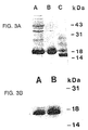
- Fig. 3A and Fig. 3B show a SDS-PAGE of recombinant LCF expressed in E. coli and a rabbit reticulocyte in vitro translation of RNA synthesized from LCF cDNA .
- Fig. 3A shows recombinant LCF protein run on a 15% SDS-PAGE followed by coomassie blue staining.
- lane A shows crude supernatant from E. coli induced to express LCF protein
- lane B shows LCF protein generated as a fusion protein conjugated to a polyhistidine linker purified by nickel affinity chromatography
- lane C shows LCF after Factor Xa cleavage.
- Fig. 3B shows a rabbit reticulocyte in vitro translation of LCF cDNA: the 35 S-labeled protein product of LCF cDNA translated by rabbit reticulocytes was run on a 15% SDS-PAGE.
- lane A shows LCF protein translated under non-glycosylating conditions
- lane B shows LCF translated under glycosylating conditions.
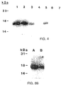
- Fig. 4 shows the immunoprecipitation of recombinant LCF by rsCD4.
- lane 1 shows 10 ⁇ g of recombinant LCF
- lane 2 shows recombinant LCF incubated with 50 ⁇ g rsCD4 immunoprecipitated with 10 ⁇ g rabbit polyclonal anti-CD4 antibody
- lane 3 shows recombinant LCF incubated with 10 ⁇ g rsCD4 immunoprecipitated with polyclonal anti-CD4 antibody
- lane 4 shows recombinant LCF incubated with rsCD4 (10 ⁇ g) immunoprecipitated with rabbit polyclonal anti-IgG (10 ⁇ g)
- lane 5 shows recombinant LCF incubated with rsCD4 and immunoprecipitated with monoclonal anti-CD4 (10 ⁇ g)
- lane 6, shows recombinant LCF incubated with rsCD4 and immunoprecipitate
- Fig. 5 shows a dose response curve for recombinant LCF induced chemotaxis of human peripheral blood T lymphocytes.
- an asterisk (*) represents statistical significance at p ⁇ 0.05 (using a Student's T test from control cell migration).
- Fig. 6 shows recombinant LCF-induced chemotaxis in murine T cell hybridoma cells.
- Murine cell lines expressing either wild-type CD4 (13.13), truncated CD4 (delta-13), or mock infected cells lacking CD4 expression (155.16) were stimulated by recombinant LCF (10 -9 M) (open bars) or 2C11 antibody (10 ⁇ g/ml) (striped horizontal bars) and the migratory response quantitated.
- Cells stimulated by recombinant LCF in the presence of a 100 fold excess of anti-CD4 Fab fragments (10 ⁇ g/ml) are also shown (solid bars).
- Cell migration is expressed as mean of ten high power fields +/- S.D. Migration which was significantly different (p ⁇ 0.05 by Student's T test) from control cell migration (designated as 100%) is indicated by asterisks.
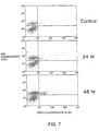
- Fig. 7 shows the specificity of recombinant LCF for CD4+ human T cells using FACs analysis.
- Two X 10 6 human T lymphocytes were cultured for 24 and 48 h in the presence of 10 -8 M recombinant LCF.
- Cells double-labelled with phycoerythrin-conjugated anti-CD4 antibody and fluorescein-conjugated anti-IL-2R antibody were analyzed on a Becton Dickinson FACscan flow cytometer.
- Recombinant LCF induced an increase in CD4+/IL-2R + cells from a control level of 3% (top panel) to 17% (bottom panel) by 48 h.
- the 24h time point demonstrated an increase in 9% of the cells.
- At no time did CD4 - cells show an increase in IL-2R expression.
- Fig. 8A and Fig. 8B show the aggregation of recombinant LCF under physiological conditions.
- Fig. 8A shows a molecular sieve HPLC of 35 S-labelled recombinant LCF (run in phosphate buffered saline, pH 8.0). Fractions were collected and analyzed by scintillation counting (open squares). Parallel samples were collected and assayed for the induction of lymphocyte chemotaxis (solid squares).
- Fig. 8B, lane A and lane B show an autoradiogram of the peak fraction for both radioactivity and cell migration (fraction 13 shown in Fig. 8A) and the second peak of radioactivity which had no corresponding chemoattractant activity (fraction 17 shown in Fig. 8A) after separation by SDS-PAGE, respectively.
- Fig. 9 shows a hydrophilicity plot of recombinant LCF predicted by the method of Kyte and Doolittle (Kyte et al., J. Molec. Bio. 157:105-132 (1982)). Peptides were synthesized and rabbit anti-peptide specific antisera were generated to four major hydrophilic regions designated by A,B,C,D.
- LCF polypeptides according to the invention include the full-length LCF polypeptide (as described in Fig. 2, SEQ ID NO: 1). Such polypeptides may be derived from any source. These polypeptides are used, e.g, to screen for antagonists which disrupt a LCF:CD4 receptor interaction or an LCF:mediated physiological response (see below). LCF fragments or analogs may also be useful candidate antagonists of LCF:CD4 receptor activity. The efficacy of a LCF fragment or analog antagonist is dependent upon its ability to interact with CD4; such an interaction may be readily assayed using any number of standard binding methods and LCF-mediated CD4 receptor functional assays (e.g., those described below). Polypeptides of the invention also include any fragment or analog capable of interacting with the CD4 receptor and mediating the LCF biological cascade, i.e. LCF agonists.
- LCF polypeptide fragments of interest include any portion of the LCF polypeptide which are capable of interaction with CD4 receptor, e.g., all or part of the N-terminus or e.g., a hydrophilic domain. Based on the hydrophilicity analysis (see Fig. 9) and biologic inhibition data, other likely candidates include without limitation, the four hydrophilic regions, A, B, C and D (see Fig. 5) and the FEAW (Phe, Glu, Ala, Trp) sequence from amino acids 96-99 of LCF (Fig. 2 and SEQ ID NO: 1). Such fragments may be useful as agonists or antagonists (as described above), and are also useful as immunogens for producing antibodies which neutralize the activity of LCF; see infra ).
- the secondary protein structure and, therefore, the domains of LCF may be deduced semi-empirically using any standard hydrophobicity/hydrophilicity calculation, e.g., the Chou-Fasman method (see,e.g., Chou and Fasman, Ann. Rev. Blochem. 47:251, 1978).
- Hydrophilic domains present themselves as strong candidates for antigenicity and hydrophobic regions for binding domains, and therefore, useful antagonists or agonists.
- Candidate fragments are then tested for interaction with CD4 receptor and their ability to induce an LCF-mediated physiological response, i.e., serve as LCF agonists, by assays described herein. Fragments are also tested for their ability to antagonize the interaction between LCF and CD4 using the assays described herein. Analogs of useful LCF fragments (as described above) may also be produced and tested for efficacy as screening components or antagonists (using the assays described herein); such analogs are also considered to be useful in the invention.
- the human LCF gene was isolated as follows.
- PBMC peripheral blood mononuclear cells
- LCF cDNA was isolated by screening a COS cell expression library of mitogen-stimulated human peripheral blood mononuclear cells (PMBC). Supernatants were assessed for the presence of LCF by the induction of human CD4+ T cell chemotaxis and cell cycle changes as determined by upregulations of IL-2 receptors (IL-2R) (Cruikshank et al., J. Immunol. 138:3817-3823, 1987). Following four rounds of screening, a positive supernatant from a single clone of 1-kb was identified. The LCF cDNA was used to probe a northern blot of total RNA isolated from human T cells (Fig. 1). A single band of 2.2-kb was detected.
- PMBC mitogen-stimulated human peripheral blood mononuclear cells
- the 1-kb LCF cDNA was used to probe a second mitogen-stimulated human PBMC cDNA library.
- Three clones were isolated, and the sequence of the largest clone is shown in Fig. 2 and SEQ ID NO: 2.
- LCF cDNA there is an open reading frame of 393 base pairs extending from nucleotide 783 to 1176 that codes for a 130 residue protein with a predicted molecular mass of 13,385 daltons.
- the methionine at nucleotide 783 is in good context for initiation by Fickett analysis (Fickett, Nucleic Acids Res. 10:5303-5318, 1982).
- the only other possible initiation site lies downstream and is in-frame, representing residue 38 of the predicted amino acid sequence.
- PBMC Human peripheral blood mononuclear cells
- Monocytes were purified from PBMC using sheep erythrocyte rosetting to deplete T lymphocytes, followed by plastic adherence of the cells remaining in the supernatant after the rosetting step. Adherent cells recovered from the plastic were >92% monocytes by fluorescence analysis. All cells were lysed with cold 4 M guanidinium isothiocyanate and RNA was isolated by CsCl centrifugation (Ausubel et al., Current Protocols in Molecular Biology, John Wiley & Sons, New York, 1989). Ten ⁇ g of RNA from each sample was loaded on a 1% agarose-formaldehyde gel for electrophoresis, and blotted onto nylon membrane.
- a cDNA probe from a 704 bp Pst I fragment of recombinant LCF-7 was [ 32 P]dCTP-labeled by the random primer method (Feinberg et al., Anal. Biochem. 132:6-13, 1983) and the blot was hybridized with 1 x 10 6 cpm/ml for 24 hr. After hybridization the blot was washed with 0.2 X SSC (30 mM NaCl, 3 mM sodium citrate, 0.05% sodium pyrophosphate, 0.1% sodium lauryl sarcosine) at 56°C, and hybridization was visualized by autoradiography. As shown in Fig. 1, the probe hybridized specifically to a lymphocyte RNA of approximately 2.2 kilobases. This confirmed that LCF was expressed in T lymphocytes and indicated that the clone was full-length.
- Polypeptides according to the invention may be produced by transformation of a suitable host cell with all or part of an LCF-encoding cDNA fragment (e.g., the cDNA described above) in a suitable expression vehicle.
- an LCF-encoding cDNA fragment e.g., the cDNA described above
- LCF polypeptide may be produced in a prokaryotic host (e.g., E. coli ), or in a eukaryotic host (e.g., S. cerevisiae or mammalian cells, e.g., COS1, NIH3T3, and JEG3 cells, or in the cells of an arthropod, e.g. Spodoptera frugiperda (SF9) cells).
- a prokaryotic host e.g., E. coli
- a eukaryotic host e.g., S. cerevisiae or mammalian cells, e.g., COS1, NIH3T3, and JEG3 cells
- an arthropod e.g. Spodoptera frugiperda (SF9) cells.
- Such cells are available from a wide range of sources (e.g., the American Type Culture Collection, Rockland, MD; also see, e.g., Ausubel et al., supra ).
- the method of transfection and the choice of expression vehicle will depend on the host system selected. Transformation and transfection methods are described, e.g., in Ausubel et al., supra; expression vehicles may be chosen from those provided, e.g., in Cloning Vectors: A Laboratory Manual (P.H. Pouwels et al., 1985, Supp. 1987).
- LCF expression system is a prokaryotic expression system as described by Ausubel et al. (supra).
- a DNA fragment containing the LCF cDNA open reading frame with flanking BamH1 and Nde1 restriction sites was generated by PCR according to standard methods and ligated into the E. coli expression vector pT-16b (Novagen).
- This plasmid, pET-166-ICF was then used to transform E. coli JM109.
- the transformed bacterial were stimulated with IPTG, grown in culture media and subsequently lysed.
- Recombinant protein was isolated by metal chelation chromatography according the well known methods (see, e.g., Studier Meth.
- SDS-PAGE mass of 17.5 kDa is larger than the expected 13.4 kDa based on nucleotide sequence, it is identical to the migration pattern of 35 S-labeled in vitro translated protein (Fig. 3B).
- the discrepancy in mass determined by SDS-PAGE from the predicted sequence may be due to aberrant migration of recombinant LCF in the SDS acrylamide gel system.
- LCF expression system is a baculovirus expression system as described by Ausubel et al. ( supra ).
- DNA encoding an LCF polypeptide is inserted into an appropriate transfer vector, e.g., pVL1392 (Invitrogen Corp., San Diego, CA).
- the vector is co-transfected with wild type baculovirus genomic DNA into Spodoptera frugiperda (SF9) cells (ATTC Accession No: CRL 1711) and recombinant viruses are isolated by standard techniques, e.g., see Ausubel et al. ( supra ).
- Recombinant LCF produced in a baculovirus system was found to synthesize a protein with an apparent molecular weight of 17.5 kDa which is similar to the protein synthesized using the E. coli expression system shown in Fig. 3A and Fig. 3B. Sequencing of the first five N-terminal amino acid residues of the baculovirus recombinant LCF was performed. The sequences were found to be identical to the predicted amino acid sequence shown in Fig. 2 (SEQ. ID No.: 1) with a methionine at position 783 as the initiation site.
- an LCF polypeptide may be produced by a stably-transfected mammalian cell line.
- a number of vectors suitable for stable transfection of mammalian cells are available to the public, e.g., see Pouwels et al. (supra); methods for constructing such cell lines are also publicly available, e.g., see Ausubel et al. (supra).
- cDNA encoding the LCF polypeptide is cloned into an expression vector which includes the dihydrofolate reductase (DHFR) gene.
- DHFR dihydrofolate reductase
- Integration of the plasmid and, therefore, the LCF-encoding gene into the host cell chromosome is selected for by inclusion of 0.01-300 ⁇ M methotrexate in the cell culture medium (as described in Ausubel et al., supra ). This dominant selection can be accomplished in most cell types. Recombinant protein expression can be increased by DHFR-mediated amplification of the transfected gene. Methods for selecting cell lines bearing gene amplifications are described in Ausubel et al. (supra); such methods generally involve extended culture in medium containing gradually increasing levels of methotrexate.
- DHFR-containing expression vectors commonly used for this purpose include pCVSEII-DHRF and pAdD26SV(A) as described in Ausubel et al. ( supra ).
- Any of the host cells described above or, preferably, a DHFR-deficient CHO cell line e.g., CHO DHFR-cells, ATCC Accession No. CRL 9096 are among the host cells preferred for DHFR selection of a stably-transfected cell line or DHFR-mediated gene amplification.
- a CD4 affinity column was prepared by coupling recombinant soluble CD4 (rsCD4) to CNBr Sepharose 4B according to previously described methods (see, e.g., Cruikshank et al., Journal of Immunology 1991). Thus, 100 ⁇ g rsCD4 was covalently conjugated to a CNBr activated Sepharose 4B (Pharmacia, Piscataway, NJ).
- RNA transcript of LCF was generated and used for in vitro translation with rabbit reticulocyte lysate in the presence of [ 35 S] methionine according to standard methods.
- 35 S-labeled in vitro LCF was applied to the column for 3hr at 37°C at which time the column was extensively washed with wash buffer (0.01 M Tris-Cl, pH 8.0, 0.14 M Nad, 0.025% NaN 3 , 0.5% Triton X-100, 0.5% sodium deoxycholate).
- wash buffer (0.01 M Tris-Cl, pH 8.0, 0.14 M Nad, 0.025% NaN 3 , 0.5% Triton X-100, 0.5% sodium deoxycholate.
- LCF was eluted with a triethanolamine solution (50mM triethanolamine, pH 11, 0.1% Triton X-100, 0.15 M NaCl) into tubes containing 1 M Tris-Cl, pH 6.7 and analyzed.
- the recombinant protein can, if desired, be further purified, e.g., by high performance liquid chromatography. These general techniques of polypeptide expression and purification can also be used to produce and isolate useful LCF fragments or analogs (as described below). Furthermore, the eluate may then, if desired, be run on a SDS-PAGE and visualized by autoradiography (see, e.g., the results from the above experiment presented in Fig. 3B).
- LCF polypeptides particularly short LCF fragments, can be produced by chemical synthesis (e.g., by the method described in Solid Phase Peptide Synthesis, 1984, 2nd ed. , Stewart and Young, eds., Pierce Chemical Co., Rockford, IL).
- LCF polypeptide fragments or analogs in the invention are those which interact with CD4 receptor, e.g., LCF agonists or antagonists. Such an interaction may be detected by an in vitro binding assay (as described infra) followed by functional analysis. Thus, the fragments or analogs thereof may also be assayed functionally, i.e., for its ability to bind CD4 receptor and to induce the migration of T4+ lymphocytes, monocytes, eosinophils and the like (as described infra ).
- LCF LCF
- rsCD4 recombinant soluble CD4 receptor
- CD4 receptor-bearing cell e.g., an eosinophil
- the invention includes methods for screening compounds useful as LCF agonists.
- LCF polypeptide fragment or analog thereof
- CD4 receptor component is produced either as a recombinant soluble component or is produced as a membrane component by a cell, e.g., a T lymphocyte, monocyte or eosinophil.
- LCF fragment or analog thereof binding to rsCD4 or CD4 receptor-bearing cells
- a whole cell assay is preferably performed by fixing the cell expressing the CD4 receptor, e.g, eosinophils, to a solid substrate (e.g., a test tube, or a microtiter well) by means well known to those in the art (see, e.g., Ausubel et al. supra) and presenting labelled LCF polypeptide (e.g., 125 I-labelled LCF). Labelling of LCF, e.g., with 125 I, is performed according to standard techniques known in the art. Binding is assayed by the detection label in association with the receptor component (and, therefore, in association with the solid substrate and CD4 receptor) by techniques well known in the art.
- the assay format may be any of a number of suitable formats for detecting suitable binding, such as a radioimmunoassay format (see, e.g., Ausubel et al., supra ).
- a radioimmunoassay format see, e.g., Ausubel et al., supra .
- cells bearing CD4 receptor are immobilized on a solid substrate (e.g., the well of a microtiter plate) and reacted with LCF polypeptide which is detectably labelled, e.g., with a radiolabel such as 125 I or an enzyme which can be assayed, e.g., alkaline phosphatase or horseradish peroxidase.
- 125 I-labelled LCF is bound to the cells and assayed for specific activity; specific binding is determined by comparison with binding assays performed in the presence of excess unlabelled LCF polypeptide.
- LCF polypeptide fragment or analog thereof
- the solid substrate e.g., to a microtiter plate using methods similar to those for adhering cells for an ELISA assay; Ausubel et al. supra
- the ability of labelled rsCD4 receptor to bind LCF can be used to detect specific rsCD4 receptor binding to the immobilized LCF.
- LCF polypeptide may also be assayed functionally for its ability to mediate migration of CD4+ lymphocytes, monocytes, eosinophils and the like.
- Migration assays may be employed using any suitable cell, e.g., T lymphocytes, monocytes or eosinophils as described in (Cruikshank et al., 1987, J. Immunol. 128: 2569-2571; Rand et al., 1992, J. Exp. Med. 173:1521-1528) follows.
- recombinant LCF synthesized in an expression system e.g., E.
- murine cell chemotaxis was performed using a modified Boyden chemotaxis chamber (Cruikshank et al, J. Immunol. 128: 2569-2571). The cells were suspended in RPMI 1640 containing 10% FBS at a concentration of 5 X 10 6 cells/ml. A 12 ⁇ m nitrocellulose membrane was used and the cells were incubated for 4 h. Next, the membranes were stained with hematoxylin and dehydrated using sequential washing with ethanol, propanol, and finally xylene to clarify the filters and allow for cell counting by light microscopy.
- Fig. 5 shows a representative dose response curve for protein generated from the E. coli expression system ( supra ). As indicated from the dose response curve, maximal migration was induced with a concentration of recombinant LCF at 10 -9 M, and ED 50 of 10 -11 M. Statistics were performed using Student's T Test (or 5 analysis of variance modifications when data from multiple experiments were pooled) and counts statistically different from control cell migration (p ⁇ 0.05) are designated by an asterisk. Similar results were obtained when baculovirus-produced LCF was substituted for E. coli -produced LCF.
- the cell lines expressing either intact CD4 or delta 13 CD4 were chosen for their comparable levels of CD4. As shown in Fig. 6 cells which expressed intact CD4 migrated in response to recombinant LCF stimulation. Cells either lacking CD4 or expressing delta 13 CD4 were unresponsive to recombinant LCF. These cells were responsive to murine T cell receptor-stimulated migration as the antibody 2C11 induced migratory responses of 198% ⁇ 4% and 192% ⁇ 3% for the mock transfected and delta 13 CD4 cell lines respectively (Fig. 6). These studies demonstrate that CD4 must be expressed for LCF-induced cell motile responsiveness and that the cytoplasmic tail is required.
- CD4 specificity for LCF stimulation in human T cells was demonstrated using the expression of IL-2R to identify LCF responsive cells.
- Mixed T cells were cultured in the presence of recombinant LCF (10 -8 M) for 24 and 48 hrs at which time the cells were labeled for their expression of both CD4 and IL-2R.
- LCF recombinant LCF
- FIG. 7 only cells which were CD4 + demonstrated an increase in surface expressed IL-2R.
- an increase in IL-2R was observed for 17% of the CD4 + cells. This indicates not only LCF specificity for CD4 + cells, but also suggests that recombinant LCF acts only on a subset of CD4 + cells.
- one aspect of the invention features screening for compounds that antagonize the interaction between LCF and CD4 receptor, thereby preventing or reducing the cascade of events that are mediated by that interaction.
- Chemical antagonists to LCF which bind to LCF or LCF/CD4 receptor or CD4 receptor without triggering a response are used to reduce, attenuate or interfere with the effects of LCF or crosslinked LCF agonists or biologically active LCF polypeptide fragments or analogs thereof which act to stimulate or activate LCF-mediated events of the immune response and inflammation.
- the invention provides for methods to screen for such useful compounds.
- These antagonists include, without limitation, e.g., crosslinked LCF, synthetic LCF, anti-LCF antibodies, or other drugs, e.g. organic compounds.
- LCF polypeptide can be used to prepare compounds that tend to neutralize or impede its activity.
- one approach pertains to identification of the active sites of LCF, followed by the alteration of those sites of the LCF amino acid sequence by substitution of amino acids within the active site by other amino acids, so that the peptide does not lose its binding affinity for the CD4 receptor, but upon binding is unable to promote activity, and thereby blocks the effect of LCF.
- LCF activity may also be blocked, attenuated, or interfered with by using antibodies, e.g., monoclonal, or chemical antagonists to LCF.
- These chemical antagonists include any organic compounds, or any of the other aforementioned compounds, which can be assayed or screened for their ability to interfere with LCF:CD4 mediate events by the methods that follow.
- the elements of the screen are LCF polypeptide (or a suitable fragment or analog thereof) and rsCD4 or, a CD4 receptor expressing cell, e.g., CD4 + lymphocyte, monocyte, eosinophil and the like, configured to permit detection of binding.
- LCF polypeptide or a suitable fragment or analog thereof
- rsCD4 or, a CD4 receptor expressing cell, e.g., CD4 + lymphocyte, monocyte, eosinophil and the like, configured to permit detection of binding.
- a full-length LCF polypeptide (fragment or analog thereof) and rsCD4 may be produced as described above.
- Binding of LCF to its receptor may be assayed by any suitable method (as described above).
- cells expressing CD4 receptor e.g., eosinophils
- a solid substrate e.g., the well of a microtiter plate
- detectably-labelled LCF polypeptide fragment or analog thereof
- Binding is assayed by the detection label in association with the receptor component (and, therefore, in association with the solid substrate). Binding of labelled full-length recombinant LCF polypeptide to CD4 receptor bearing cells is used as a "control" against which antagonist assays are measured.
- the antagonist assays involve incubation of the CD4 receptor bearing cells with an appropriate amount of candidate antagonist, e.g., an antibody or an organic compound. To this mix, an equivalent amount of labelled LCF is added.
- candidate antagonist e.g., an antibody or an organic compound.
- an equivalent amount of labelled LCF is added.
- An antagonist useful in the invention interferes with labelled-LCF binding to the immobilized receptor-bearing cells. Alternatively, an antagonist may bind but not activate a biological response.
- an antagonist if desired, may be tested for its ability to interfere with LCF function, i.e., to specifically interfere with labelled LCF binding without resulting in signal transduction normally mediated by a full-length LCF polypeptide.
- Appropriate candidate antagonists include e.g., the polypeptides FEAW (Phe-Glu-Ala-Trp at amino acids 96-99) and RKSLQSKETTAAGDS (Arg-Lys-Ser-Leu-Gln-Ser-Lys-Glu-Thr-Thr-Ala-Ala-Gly-Asp-Ser at amino acids 116-130) see e.g., SEQ ID No.:1 analogs of LCF, and other peptides as well as non-peptide compounds, and anti-LCF polypeptide antibodies designed or derived from analysis of LCF/CD4 receptor interaction or the primary structure of LCF.
- FEAW Phe-Glu-Ala-Trp at amino acids 96-99
- RKSLQSKETTAAGDS Arg-Lys-Ser-Leu-Gln-Ser-Lys-Glu-Thr-Thr-Ala-Ala-Gly-Asp-Ser at amino acids 116
- Human LCF (or fragments or analogs) may be used to raise antibodies useful in the invention; such polypeptides may be produced by recombinant or peptide synthetic techniques (see, e.g., Solid Phase Peptide Synthesis, supra; Ausubel et al., supra).
- the peptides may be coupled to a carrier protein such as KLH as described in Ausubel et al., supra.
- KLH-peptide is mixed with Freund's adjuvant and injected into guinea pigs, rats, donkeys and like or preferably rabbits.
- Antibodies may be purified by peptide antigen affinity chromatography.
- monoclonal antibodies may be prepared using LCF polypeptides described above and standard hybridoma technology (see, e.g. Kohler et al., Nature, 256:495, 1975; Kohler et al., Eur. J. Immunol. 6:511, 1976; Kohler et al., Eur. J. Immunol ., 6:292, 1976; Hammerling et al., In Monoclonal Antibodies and T Cell Hybridomas , Elsevier, NY, 1981; Ausubel et al., supra ).
- monoclonal antibodies to LCF can be raised in Balb/C or other similar strains of mice by immunization with purified or partially purified preparations of LCF (fragments or analogs thereof).
- the spleens of these mice can be removed, and their lymphocytes fused to a mouse myeloma cell line.
- a stable hybrid will be isolated that produces antibodies against LCF (fragments or analogs thereof).
- Such activity can be demonstrated by the ability of the antibody to prevent the binding of radiolabelled LCF (e.g., 125 I-LCF) to the CD4 receptor.
- radiolabelled LCF e.g., 125 I-LCF
- the monoclonal antibody can then be examined for its ability to prevent the biological activity of LCF, e.g., cell migration (as discussed above).
- polyclonal or monoclonal antibodies are tested for specific LCF polypeptide recognition by Western blot or immunoprecipitation analysis (by methods described in Ausubel et al., supra).
- Antibodies which specifically recognize an LCF polypeptide (fragment or analog thereof) are considered to be likely candidates for useful antagonists; or such antibodies may be used, e.g., in an immunoassay to monitor the level of LCF polypeptide produced by a mammal, e.g., a human.
- Antibodies which antagonize LCF/CD4 receptor binding or LCF mediated CD4 receptor function are considered to be useful antagonists in the invention.
- recombinant LCF induces the expression of cell receptors, e.g., IL-2R, which subsequently render a cell-bearing the receptor, e.g., a T cell, competent to respond to its cognate growth factor, e.g., IL-2.
- cell receptors e.g., IL-2R
- IL-2R cell receptors
- human T cells were stimulated with recombinant LCF (a concentration range of 10 -5 M to 10 -10 M was used with similar results, data for 10 -8 M is shown) for 24h at which time rIL-2 (2U/ml) or anti-CD3 (OKT3, 50 ng/ml) were added to the cell cultures.
- Particularly suitable therapeutics for the treatment of hyperresponsive immune responses and inflammatory diseases are the soluble antagonistic fragments described above formulated in an appropriate buffer such as physiological saline.
- anti-LCF polypeptide (fragments or analogs thereof) antibodies produced as described above may be used as therapeutics. Again, the antibodies would be administered in a pharmaceutically-acceptable buffer (e.g., physiological saline). If appropriate, the antibody preparation may be combined with a suitable adjuvant.
- the methods of the invention provide for the identification of an organic compound useful to antagonize LC4:CD4 interaction, once identified and isolated such a compound can then be formulated in an appropriate buffer and used as a therapeutic.
- suitable therapeutics for the use of LCF or LCF agonists as immunosuppressive agents or as therapeutics to stimulate the expansion of CD4+ receptor bearing cells are formulated in an appropriate buffer such as physiological saline. Again, these formulations would be administered in a pharmaceutically-acceptable buffer (e.g., physiological saline).
- the therapeutic composition will be administered intravenously, at a dosage effective to stimulate activation of new CD4 lymphocyte populations; to induce anergy (see table above) and inhibit rejection in transplants; and to attenuate a hyperresponsive immune response and inflammation, e.g., asthma.
- the therapeutic orally, nasally, or topically e.g. as a liquid or a spray as a primary product or as a viral vector carrying LCF cDNA.
- the dosages are as described above.
- the dosage of the compound for treating any of the above-mentioned disorders varies depending upon the manner of administration, the age and the body weight of the subject, and the condition of the subject to be treated, and ultimately will be decided by the attending physician or veterinarian.
- Such amount of the active compound as determined by the attending physician or veterinarian is referred to herein as a "therapeutically effective amount.”
- the compounds of the invention can be administered to a mammal, e.g., a human patient in a dosage of 0.5 ⁇ g/kg/day to 5 mg/kg/day.
- Synergistic effect between recombinant LCF and growth factor could be induced by sequential administration of recombinant LCF (0.5 ⁇ g/kg to 5 mg/kg followed in 24 hours by similar doses or rIL-2.
- the methods of the invention may be used to reduce the disorders described herein in any mammal, for example, humans, domestic pets, or livestock.
- the LCF polypeptide or the antibody employed is preferably but not necessarily specific for that species.
- the invention includes any protein which is substantially homologous to LCF polypeptide (Fig. 2, SEQ ID NO: 1).
- LCF is expressed in human T cells and exocrine pancreas. It is also expressed in the human monocytoid cell line THP-1.
- allelic variations are included in the human T cells and exocrine pancreas. It is also expressed in the human monocytoid cell line THP-1.
- allelic variations are included in DNA that hybridizes under high or low (e.g., washing at 2xSSC at 40° C with a probe length of at least 40 nucleotides) stringency conditions to a nucleic acid naturally occurring (for other definitions of high and low stringency see Current Protocols in Molecular Biology, John Wiley & Sons, New York, 1989); and polypeptides or proteins specifically bound by antisera to LCF polypeptide, especially by antisera to the active site or binding domain of LCF polypeptide.
- the term also includes chimeric polypeptides that include LCF polypeptide.
- the invention also includes biologically active fragments of the polypeptides.
- fragment as applied to a polypeptide, will ordinarily be at least about residues, more typically at least about 40 residues, preferably at least about 60 residues in length. Fragments of LCF polypeptide can be generated by methods known to those skilled in the art. The ability of a candidate fragment to exhibit a biological activity of LCF polypeptide can be assessed by methods known to those skilled in the art as described herein. Also included are LCF polypeptides containing residues that are not required for biological activity of the peptide such as residues that are not required for the biological activity of the polypeptide, or that result from alternative mRNA splicing or alternative protein processing events.
Applications Claiming Priority (3)
| Application Number | Priority Date | Filing Date | Title |
|---|---|---|---|
| US6894993A | 1993-05-21 | 1993-05-21 | |
| US68949 | 1993-05-21 | ||
| EP94917399A EP0700439A1 (de) | 1993-05-21 | 1994-05-16 | Chemoattraktion auslösender faktor aus lymphozyten und seine verwendungsmöglichkeiten |
Related Parent Applications (1)
| Application Number | Title | Priority Date | Filing Date |
|---|---|---|---|
| EP94917399A Division EP0700439A1 (de) | 1993-05-21 | 1994-05-16 | Chemoattraktion auslösender faktor aus lymphozyten und seine verwendungsmöglichkeiten |
Publications (3)
| Publication Number | Publication Date |
|---|---|
| EP1203775A2 true EP1203775A2 (de) | 2002-05-08 |
| EP1203775A3 EP1203775A3 (de) | 2004-02-04 |
| EP1203775B1 EP1203775B1 (de) | 2008-12-31 |
Family
ID=22085751
Family Applications (3)
| Application Number | Title | Priority Date | Filing Date |
|---|---|---|---|
| EP94917399A Withdrawn EP0700439A1 (de) | 1993-05-21 | 1994-05-16 | Chemoattraktion auslösender faktor aus lymphozyten und seine verwendungsmöglichkeiten |
| EP01201544A Expired - Lifetime EP1203775B1 (de) | 1993-05-21 | 1994-05-16 | Chemoattraktion-auslösender Faktor aus Lymphozyten und dessen Verwendung |
| EP01201543A Ceased EP1155700A3 (de) | 1993-05-21 | 1994-05-16 | Chemoattraktion-auslösender Faktor aus Lymfozyten und dessen Verwendungen |
Family Applications Before (1)
| Application Number | Title | Priority Date | Filing Date |
|---|---|---|---|
| EP94917399A Withdrawn EP0700439A1 (de) | 1993-05-21 | 1994-05-16 | Chemoattraktion auslösender faktor aus lymphozyten und seine verwendungsmöglichkeiten |
Family Applications After (1)
| Application Number | Title | Priority Date | Filing Date |
|---|---|---|---|
| EP01201543A Ceased EP1155700A3 (de) | 1993-05-21 | 1994-05-16 | Chemoattraktion-auslösender Faktor aus Lymfozyten und dessen Verwendungen |
Country Status (7)
| Country | Link |
|---|---|
| US (2) | US5807712A (de) |
| EP (3) | EP0700439A1 (de) |
| JP (3) | JP4278710B2 (de) |
| AT (1) | ATE419356T1 (de) |
| DE (1) | DE69435177D1 (de) |
| ES (1) | ES2320090T3 (de) |
| WO (1) | WO1994028134A1 (de) |
Families Citing this family (14)
| Publication number | Priority date | Publication date | Assignee | Title |
|---|---|---|---|---|
| US5807549A (en) * | 1993-05-21 | 1998-09-15 | Research Corporation Technologies, Inc. | Lymphocyte chemoattractant factor and uses thereof |
| DE69435177D1 (de) * | 1993-05-21 | 2009-02-12 | Univ Boston | Chemoattraktion-auslösender Faktor aus Lymphozyten und dessen Verwendung |
| DE19513152A1 (de) | 1995-04-07 | 1996-10-10 | Bundesrep Deutschland | Verwendung eines "Immundefizienzvirus-supprimierenden Lymphokins (ISL)" zur Hemmung der Virusvermehrung, insbesondere von Retroviren |
| AU1301797A (en) * | 1995-12-22 | 1997-07-17 | Bundesrepublik Deutschland Vertreten Durch Den Bundesminister Fur Gesundheit | Polypeptides with interleukin 16 activity, process for the preparation and use thereof |
| DE19547933A1 (de) * | 1995-12-22 | 1997-06-26 | Boehringer Mannheim Gmbh | Multimere Formen von IL-16, Verfahren zu ihrer Herstellung und Verwendung |
| DE19614099A1 (de) * | 1996-04-10 | 1997-10-16 | Bundesrep Deutschland | Genomische Nukleinsäuren, die für Polypeptide mit IL-16-Aktivität codieren, Verfahren zu ihrer Herstellung und Verwendung |
| AU712122B2 (en) * | 1996-04-30 | 1999-10-28 | Bundesrepublik Deutschland Vertreten Durch Den Bundesminister Fur Gesundheit | Processed polypeptides with IL-16 activity, processes for their production and their use |
| US6444202B1 (en) | 1996-11-25 | 2002-09-03 | Bundesrepublic Deutschland, Vertreten Durch Den Bundesminister Fur Gesundheit | Processed polypeptides with IL-16 activity, processes for their production and their use |
| JP2002500889A (ja) * | 1998-01-24 | 2002-01-15 | ドイツ連邦共和国 | Il−16の変異体、その製造方法およびその使用 |
| WO1999048514A1 (en) * | 1998-03-25 | 1999-09-30 | Mayo Foundation For Medical Education And Research | Methods and materials for treating inflammatory diseases |
| US6699466B1 (en) * | 1999-08-05 | 2004-03-02 | Research Corporation Technologies, Inc. | IL-16 antagonist peptides and DNA encoding the peptides |
| WO2001010891A2 (en) | 1999-08-05 | 2001-02-15 | Research Corporation Technologies, Inc. | Il-16 antagonists |
| US20090130661A1 (en) * | 2006-09-28 | 2009-05-21 | Glass William G | Method for Detecting IL-16 Activity and Modulation of IL-16 Activity Based on Phosphorylated Stat-6 Proxy Levels |
| US7754207B2 (en) * | 2006-10-27 | 2010-07-13 | Centocor Ortho Biotech Inc. | Methods of treating pulmonary fibrosis |
Family Cites Families (7)
| Publication number | Priority date | Publication date | Assignee | Title |
|---|---|---|---|---|
| US3958661A (en) * | 1972-09-06 | 1976-05-25 | Atlantic Richfield Company | Method and apparatus for generating seismic waves |
| US3889710A (en) * | 1972-11-07 | 1975-06-17 | Julien H Brost | Check valve with elastomeric valve element |
| JPS59205026A (ja) * | 1983-05-02 | 1984-11-20 | Yaskawa Electric Mfg Co Ltd | 磁気軸受装置の制御方法 |
| US4745051A (en) * | 1983-05-27 | 1988-05-17 | The Texas A&M University System | Method for producing a recombinant baculovirus expression vector |
| US4863727A (en) * | 1986-04-09 | 1989-09-05 | Cetus Corporation | Combination therapy using interleukin-2 and tumor necrosis factor |
| US5807549A (en) * | 1993-05-21 | 1998-09-15 | Research Corporation Technologies, Inc. | Lymphocyte chemoattractant factor and uses thereof |
| DE69435177D1 (de) * | 1993-05-21 | 2009-02-12 | Univ Boston | Chemoattraktion-auslösender Faktor aus Lymphozyten und dessen Verwendung |
-
1994
- 1994-05-16 DE DE69435177T patent/DE69435177D1/de not_active Expired - Lifetime
- 1994-05-16 ES ES01201544T patent/ES2320090T3/es not_active Expired - Lifetime
- 1994-05-16 EP EP94917399A patent/EP0700439A1/de not_active Withdrawn
- 1994-05-16 EP EP01201544A patent/EP1203775B1/de not_active Expired - Lifetime
- 1994-05-16 AT AT01201544T patent/ATE419356T1/de active
- 1994-05-16 EP EP01201543A patent/EP1155700A3/de not_active Ceased
- 1994-05-16 WO PCT/US1994/005442 patent/WO1994028134A1/en not_active Application Discontinuation
- 1994-05-16 JP JP50073495A patent/JP4278710B2/ja not_active Expired - Fee Related
- 1994-12-13 US US08/354,961 patent/US5807712A/en not_active Expired - Lifetime
-
1995
- 1995-12-29 US US08/581,103 patent/US6159463A/en not_active Expired - Fee Related
-
2006
- 2006-01-30 JP JP2006020169A patent/JP4279289B2/ja not_active Expired - Fee Related
-
2008
- 2008-12-25 JP JP2008330880A patent/JP4804529B2/ja not_active Expired - Lifetime
Non-Patent Citations (3)
| Title |
|---|
| CRUIKSHANK ET AL., J. IMMUNOL. |
| WILSON ET AL.: "Principles of Internal Medicine", MCGRAW HILL, INC. |
| WONG ET AL., SCIENCE, vol. 228, 1985, pages 801 - 815 |
Also Published As
| Publication number | Publication date |
|---|---|
| US5807712A (en) | 1998-09-15 |
| JP4278710B2 (ja) | 2009-06-17 |
| JP2009149651A (ja) | 2009-07-09 |
| ATE419356T1 (de) | 2009-01-15 |
| WO1994028134A1 (en) | 1994-12-08 |
| JP4279289B2 (ja) | 2009-06-17 |
| EP1203775A3 (de) | 2004-02-04 |
| JP2006182783A (ja) | 2006-07-13 |
| US6159463A (en) | 2000-12-12 |
| EP1155700A3 (de) | 2002-01-02 |
| DE69435177D1 (de) | 2009-02-12 |
| JP4804529B2 (ja) | 2011-11-02 |
| ES2320090T3 (es) | 2009-05-19 |
| EP1155700A2 (de) | 2001-11-21 |
| JPH08510907A (ja) | 1996-11-19 |
| EP0700439A1 (de) | 1996-03-13 |
| EP1203775B1 (de) | 2008-12-31 |
Similar Documents
| Publication | Publication Date | Title |
|---|---|---|
| JP4804529B2 (ja) | リンパ球化学誘引因子およびその用途 | |
| US5728536A (en) | Jak kinases and regulation of Cytokine signal transduction | |
| AU651596B2 (en) | Type II interleukin-1 receptors | |
| EP0832132B1 (de) | Chemoattraktion-auslösender faktor aus lymphozyten und dessen verwendungen | |
| IL91705A (en) | Interleukin-AND 4 expression vectors that encode them and processes for the production of receptors | |
| IE883523L (en) | Interleukin-1 Receptors | |
| WO1996023067A1 (en) | Human interleukin-1 receptor accessory protein | |
| EP0409091A1 (de) | Vom Carbonylende abgeleitete Antagonisten von GM-CSF | |
| WO1996032481A1 (en) | New chemokine expressed in eosinophils | |
| US5852176A (en) | Antibodies to receptors for human interleukin-12 | |
| US6458932B1 (en) | Interferon-α/β binding protein, its preparation and use | |
| IL108584A (en) | Cloning of the interferon-binding protein ALPHA / BETA | |
| EP0528928B1 (de) | Interleukin-4-bindeprotein-gamma |
Legal Events
| Date | Code | Title | Description |
|---|---|---|---|
| PUAI | Public reference made under article 153(3) epc to a published international application that has entered the european phase |
Free format text: ORIGINAL CODE: 0009012 |
|
| 17P | Request for examination filed |
Effective date: 20010525 |
|
| AC | Divisional application: reference to earlier application |
Ref document number: 700439 Country of ref document: EP |
|
| AK | Designated contracting states |
Kind code of ref document: A2 Designated state(s): AT BE CH DE DK ES FR GB GR IE IT LI NL PT SE |
|
| PUAL | Search report despatched |
Free format text: ORIGINAL CODE: 0009013 |
|
| RIC1 | Information provided on ipc code assigned before grant |
Ipc: 7A 61K 38/19 B Ipc: 7C 12N 15/19 A Ipc: 7C 07K 16/24 B Ipc: 7A 61K 39/395 B |
|
| RIC1 | Information provided on ipc code assigned before grant |
Ipc: 7C 12N 15/19 A Ipc: 7C 07K 14/54 B Ipc: 7A 61K 39/395 B Ipc: 7A 61K 38/19 B Ipc: 7C 07K 16/24 B |
|
| AK | Designated contracting states |
Kind code of ref document: A3 Designated state(s): AT BE CH DE DK ES FR GB GR IE IT LI NL PT SE |
|
| AKX | Designation fees paid |
Designated state(s): AT BE CH DE DK ES FR GB GR IE IT LI NL PT SE |
|
| 17Q | First examination report despatched |
Effective date: 20050330 |
|
| RAP1 | Party data changed (applicant data changed or rights of an application transferred) |
Owner name: TRUSTEES OF BOSTON UNIVERSITY |
|
| 17Q | First examination report despatched |
Effective date: 20050330 |
|
| GRAP | Despatch of communication of intention to grant a patent |
Free format text: ORIGINAL CODE: EPIDOSNIGR1 |
|
| GRAS | Grant fee paid |
Free format text: ORIGINAL CODE: EPIDOSNIGR3 |
|
| GRAA | (expected) grant |
Free format text: ORIGINAL CODE: 0009210 |
|
| AC | Divisional application: reference to earlier application |
Ref document number: 0700439 Country of ref document: EP Kind code of ref document: P |
|
| AK | Designated contracting states |
Kind code of ref document: B1 Designated state(s): AT BE CH DE DK ES FR GB GR IE IT LI NL PT SE |
|
| REG | Reference to a national code |
Ref country code: CH Ref legal event code: EP Ref country code: GB Ref legal event code: FG4D |
|
| REF | Corresponds to: |
Ref document number: 69435177 Country of ref document: DE Date of ref document: 20090212 Kind code of ref document: P |
|
| REG | Reference to a national code |
Ref country code: IE Ref legal event code: FG4D |
|
| REG | Reference to a national code |
Ref country code: CH Ref legal event code: NV Representative=s name: KATZAROV S.A. |
|
| REG | Reference to a national code |
Ref country code: ES Ref legal event code: FG2A Ref document number: 2320090 Country of ref document: ES Kind code of ref document: T3 |
|
| PG25 | Lapsed in a contracting state [announced via postgrant information from national office to epo] |
Ref country code: NL Free format text: LAPSE BECAUSE OF FAILURE TO SUBMIT A TRANSLATION OF THE DESCRIPTION OR TO PAY THE FEE WITHIN THE PRESCRIBED TIME-LIMIT Effective date: 20081231 |
|
| NLV1 | Nl: lapsed or annulled due to failure to fulfill the requirements of art. 29p and 29m of the patents act | ||
| PG25 | Lapsed in a contracting state [announced via postgrant information from national office to epo] |
Ref country code: SE Free format text: LAPSE BECAUSE OF FAILURE TO SUBMIT A TRANSLATION OF THE DESCRIPTION OR TO PAY THE FEE WITHIN THE PRESCRIBED TIME-LIMIT Effective date: 20090331 Ref country code: PT Free format text: LAPSE BECAUSE OF FAILURE TO SUBMIT A TRANSLATION OF THE DESCRIPTION OR TO PAY THE FEE WITHIN THE PRESCRIBED TIME-LIMIT Effective date: 20090601 |
|
| PG25 | Lapsed in a contracting state [announced via postgrant information from national office to epo] |
Ref country code: DK Free format text: LAPSE BECAUSE OF FAILURE TO SUBMIT A TRANSLATION OF THE DESCRIPTION OR TO PAY THE FEE WITHIN THE PRESCRIBED TIME-LIMIT Effective date: 20081231 |
|
| PLBE | No opposition filed within time limit |
Free format text: ORIGINAL CODE: 0009261 |
|
| STAA | Information on the status of an ep patent application or granted ep patent |
Free format text: STATUS: NO OPPOSITION FILED WITHIN TIME LIMIT |
|
| 26N | No opposition filed |
Effective date: 20091001 |
|
| PG25 | Lapsed in a contracting state [announced via postgrant information from national office to epo] |
Ref country code: GR Free format text: LAPSE BECAUSE OF FAILURE TO SUBMIT A TRANSLATION OF THE DESCRIPTION OR TO PAY THE FEE WITHIN THE PRESCRIBED TIME-LIMIT Effective date: 20090401 |
|
| PGFP | Annual fee paid to national office [announced via postgrant information from national office to epo] |
Ref country code: ES Payment date: 20110526 Year of fee payment: 18 Ref country code: FR Payment date: 20110607 Year of fee payment: 18 Ref country code: IE Payment date: 20110525 Year of fee payment: 18 Ref country code: CH Payment date: 20110525 Year of fee payment: 18 |
|
| PGFP | Annual fee paid to national office [announced via postgrant information from national office to epo] |
Ref country code: GB Payment date: 20110525 Year of fee payment: 18 Ref country code: BE Payment date: 20110530 Year of fee payment: 18 Ref country code: AT Payment date: 20110519 Year of fee payment: 18 |
|
| PGFP | Annual fee paid to national office [announced via postgrant information from national office to epo] |
Ref country code: DE Payment date: 20110527 Year of fee payment: 18 Ref country code: IT Payment date: 20110526 Year of fee payment: 18 |
|
| BERE | Be: lapsed |
Owner name: TRUSTEES OF BOSTON UNIVERSITY Effective date: 20120531 |
|
| REG | Reference to a national code |
Ref country code: CH Ref legal event code: PL |
|
| REG | Reference to a national code |
Ref country code: AT Ref legal event code: MM01 Ref document number: 419356 Country of ref document: AT Kind code of ref document: T Effective date: 20120516 |
|
| GBPC | Gb: european patent ceased through non-payment of renewal fee |
Effective date: 20120516 |
|
| PG25 | Lapsed in a contracting state [announced via postgrant information from national office to epo] |
Ref country code: AT Free format text: LAPSE BECAUSE OF NON-PAYMENT OF DUE FEES Effective date: 20120516 Ref country code: CH Free format text: LAPSE BECAUSE OF NON-PAYMENT OF DUE FEES Effective date: 20120531 Ref country code: LI Free format text: LAPSE BECAUSE OF NON-PAYMENT OF DUE FEES Effective date: 20120531 |
|
| REG | Reference to a national code |
Ref country code: IE Ref legal event code: MM4A |
|
| PG25 | Lapsed in a contracting state [announced via postgrant information from national office to epo] |
Ref country code: BE Free format text: LAPSE BECAUSE OF NON-PAYMENT OF DUE FEES Effective date: 20120531 Ref country code: IT Free format text: LAPSE BECAUSE OF NON-PAYMENT OF DUE FEES Effective date: 20120516 |
|
| REG | Reference to a national code |
Ref country code: FR Ref legal event code: ST Effective date: 20130131 |
|
| REG | Reference to a national code |
Ref country code: DE Ref legal event code: R119 Ref document number: 69435177 Country of ref document: DE Effective date: 20121201 |
|
| PG25 | Lapsed in a contracting state [announced via postgrant information from national office to epo] |
Ref country code: GB Free format text: LAPSE BECAUSE OF NON-PAYMENT OF DUE FEES Effective date: 20120516 Ref country code: FR Free format text: LAPSE BECAUSE OF NON-PAYMENT OF DUE FEES Effective date: 20120531 Ref country code: IE Free format text: LAPSE BECAUSE OF NON-PAYMENT OF DUE FEES Effective date: 20120516 |
|
| PG25 | Lapsed in a contracting state [announced via postgrant information from national office to epo] |
Ref country code: DE Free format text: LAPSE BECAUSE OF NON-PAYMENT OF DUE FEES Effective date: 20121201 |
|
| REG | Reference to a national code |
Ref country code: ES Ref legal event code: FD2A Effective date: 20130820 |
|
| PG25 | Lapsed in a contracting state [announced via postgrant information from national office to epo] |
Ref country code: ES Free format text: LAPSE BECAUSE OF NON-PAYMENT OF DUE FEES Effective date: 20120517 |