EP1177314B1 - Assays for nucleoside diphosphates and triphospates - Google Patents
Assays for nucleoside diphosphates and triphospates Download PDFInfo
- Publication number
- EP1177314B1 EP1177314B1 EP00927529A EP00927529A EP1177314B1 EP 1177314 B1 EP1177314 B1 EP 1177314B1 EP 00927529 A EP00927529 A EP 00927529A EP 00927529 A EP00927529 A EP 00927529A EP 1177314 B1 EP1177314 B1 EP 1177314B1
- Authority
- EP
- European Patent Office
- Prior art keywords
- ndpk
- label
- phosphoenzyme
- adp
- nucleoside
- Prior art date
- Legal status (The legal status is an assumption and is not a legal conclusion. Google has not performed a legal analysis and makes no representation as to the accuracy of the status listed.)
- Expired - Lifetime
Links
Images


Classifications
-
- C—CHEMISTRY; METALLURGY
- C12—BIOCHEMISTRY; BEER; SPIRITS; WINE; VINEGAR; MICROBIOLOGY; ENZYMOLOGY; MUTATION OR GENETIC ENGINEERING
- C12Q—MEASURING OR TESTING PROCESSES INVOLVING ENZYMES, NUCLEIC ACIDS OR MICROORGANISMS; COMPOSITIONS OR TEST PAPERS THEREFOR; PROCESSES OF PREPARING SUCH COMPOSITIONS; CONDITION-RESPONSIVE CONTROL IN MICROBIOLOGICAL OR ENZYMOLOGICAL PROCESSES
- C12Q1/00—Measuring or testing processes involving enzymes, nucleic acids or microorganisms; Compositions therefor; Processes of preparing such compositions
- C12Q1/48—Measuring or testing processes involving enzymes, nucleic acids or microorganisms; Compositions therefor; Processes of preparing such compositions involving transferase
-
- C—CHEMISTRY; METALLURGY
- C12—BIOCHEMISTRY; BEER; SPIRITS; WINE; VINEGAR; MICROBIOLOGY; ENZYMOLOGY; MUTATION OR GENETIC ENGINEERING
- C12N—MICROORGANISMS OR ENZYMES; COMPOSITIONS THEREOF; PROPAGATING, PRESERVING, OR MAINTAINING MICROORGANISMS; MUTATION OR GENETIC ENGINEERING; CULTURE MEDIA
- C12N9/00—Enzymes; Proenzymes; Compositions thereof; Processes for preparing, activating, inhibiting, separating or purifying enzymes
- C12N9/10—Transferases (2.)
- C12N9/12—Transferases (2.) transferring phosphorus containing groups, e.g. kinases (2.7)
Definitions
- the invention relates to assays for nucleoside diphosphates, particularly ADP and GDP, and assays for nucleoside triphosphates, particularly ATP and GTP.
- Nucleoside diphosphates and triphosphates play important roles in biology.
- ADP is the immediate precursor for the formation of ATP, the universal currency of cellular energy.
- GDP is a substrate for succinyl CoA synthetase, a key enzyme of the Krebs cycle, and is formed during gluconeogenesis by phosphoenolpyruvate carboxykinase. It is also essential in G-protein signalling, microtubule growth, and visual excitation.
- UDP is involved in the epimerisation of galactose to glucose, the formation of sucrose, and in the growth of glycogen.
- CDP is an important group in the synthesis of phosphoglycerides.
- Nucleoside diphosphates are also the products of reactions catalysed by several major classes of enzymes, such as triphosphatases and kinases, and are therefore produced by many cellular processes, including motility, muscle contraction, DNA synthesis, transcription, translation and nitrogen fixation.
- nucleoside diphosphates and triphosphates The detection and measurement of nucleoside diphosphates and triphosphates is thus important in the study of biology and metabolism, particularly in bioenergetics.
- Reference 11 discloses column-based chromatographic assays for ADP, GDP, CDP and UDP. Radioactive assays for GDP and GTP have also been described [12,13]. NMR-based assays for measuring in vivo ADP levels are known for yeast [14], and NMR has also been used to measure ADP and ATP and erythrocytes [15].
- Reference 33 discloses methods for measuring the total amounts of G protein-hound GDP and GTP in mammalian cells and tissue. The method avoids the need to expose the cells to phosphate-free growth conditions, which can adversely affect the accuracy of the. GTP determination. GDP is converted to GTP by phosphorylation using the nucleoside diphosphate kinase (NDPK) enzyme.
- NDPK nucleoside diphosphate kinase
- Reference 34 discloses a method which uses NDPK to convert ADP to ATP. The ATP is then used to produce NADH or NADPH, which is used as an indirect measure of the initial amount of ADP.
- NDPK is suggested for use in an assay for detecting GTP in reference 35, with the enzyme being used to convert GTP to ATP.
- Reference 36 discloses methods for detecting highly proliferative cells, such as tumour cells, in which the activity or the quantity of NDPK is measured. NDPK is trapped in its phosphorylated form by using a radio-labelled suicide ⁇ -thiophosphate derivative, and it can then be detected.
- nucleoside diphosphates are detected or measured by following the dephosphorylation of the phosphoenzyme form of nucleoside diphosphate kinase (NDPK), and nucleoside triphosphates are detected or measured by following the phosphorylation of NDPK to its phosphoenzyme form.
- NDPK nucleoside diphosphate kinase
- the invention thus provides (a) a process for detecting the presence of a nucleoside diphosphate in a sample, comprising the step of detecting the dephosphorylation of the phosphoenzyme form of a nucleoside diphosphate kinase, and (b) a process for detecting the presence of a nucleoside triphosphate in a sample, comprising the step of detecting the phosphorylation of a nucleoside diphosphate kinase to the phosphoenzyme form.
- the process will typically comprise the steps of:
- NDPK means an enzyme having the activity of the enzyme classified as EC 2.7.4.6, namely the transfer of the ⁇ -phosphate group of a nucleoside triphosphate (N 1 TP) to a nucleoside diphosphate (N 2 DP) via a ping-pong mechanism: N 1 TP + N 2 DP ⁇ N 1 DP + N 2 TP
- NDPK ATP:nucleoside-diphosphate phosphotransferase
- the common name is "nucleoside diphosphate kinase”.
- the enzyme has also been variously described as: kinase (phosphorylating), nucleoside diphosphate; nucleoside 5'-diphosphate kinase; nucleoside diphosphate (UDP) kinase; nucleoside diphosphokinase; nucleotide phosphate kinase; NM23.
- NDPKs have been described for a number of organisms, both prokaryotic and eukaryotic e.g. human, cows, monkeys, mice, Xenopus , oats, peas, potatoes, yeast, Bacillus subtilis , E . coli , Myxococcus xanthus , avian myeloblastosis virus etc . These differ by cellular location, molecular weight, oligomeric structure, isoelectric point, reaction kinetics, substrate preference, pH optimum, pH range, temperature optimum, cation requirements (Mn 2+ , Mg 2+ , Co 2+ , Ca 2 etc .), and various isoforms have been described. Given the variety of suitable enzymes available, the skilled person can easily select and purify a NDPK to suit any particular situation.
- prokaryotic and eukaryotic e.g. human, cows, monkeys, mice, Xenopus , oats, peas, potatoes
- the NDPK enzyme uses a ping-pong mechanism, transferring the ⁇ -phosphate from a nucleoside triphosphate (N 1 TP) to an active site histidine to form a phosphoenzyme intermediate, and then to a nucleoside diphosphate (N 2 DP).
- N 1 TP nucleoside triphosphate
- N 2 DP nucleoside diphosphate
- the invention is based on the finding that the phosphoenzyme intermediate is stable over a time-scale that allows its detection and measurement.
- Other enzymes that phosphorylate nucleoside diphosphates via a phosphoenzyme intermediate, preferably with a single binding site for nucleotide, may also be used in the invention.
- the phosphoenzyme is able to transfer its phosphate group to N 2 DP in a sample to form the corresponding N 2 TP. Detection of this transfer can therefore be used for the detection of nucleoside diphosphate.
- phosphoenzyme is required as a reagent. This can be readily formed by, for example, incubating NDPK with excess NTP, typically ATP. Formation of phosphoenzyme in this way is facilitated by removing Mg 2+ [16], for instance by using EDTA. Chemical phosphorylation of histidine using phosphoramidate as a phosphorylating agent may also be used [17].
- the phosphoenzyme can be isolated for use as a reagent. It has been found that the phosphoenzyme can be stored on ice for over 48 hours without dephosphorylation, and can be stored for longer periods (at least 5 months) at -80°C (although repeated freeze-thawing results in some dephosphorylation). The stability of the phosphoenzyme over the time range needed for its preparation, and subsequently for monitoring kinetic events such as the release of ADP from an ATPase, is particularly advantageous.
- the phosphoenzyme When added to a sample containing NDP, the phosphoenzyme is dephosphorylated by the transfer of its phosphate group to the NDP. When added to a sample containing NTP. the phosphoenzyme is formed by the transfer of the NTP ⁇ -phosphate group to the enzyme.
- the invention relies on the ability to distinguish between the phosphorylated and dephosphorylated forms of NDPK.
- any suitable measurable change can be used.
- intrinsic properties of the enzyme can be used.
- the following methods are examples of how dephosphorylation/phosphorylation may be detected. with varying levels of sensitivity:
- One particularly preferred modification is the addition of a fluorescent label to the enzyme, typically via a cysteine residue.
- a suitable cysteine residue e.g. the NDPK of Myxococcus xanthus
- a suitable position for mutation can easily be determined by the skilled person, whilst ensuring that the mutation does not disrupt the enzymatic activity [ e . g . 20].
- particular labels may give better results than others. Suitable combinations of label and residue can be determined by routine experimentation.
- Preferred fluorescent labels are based around coumarin. Particularly preferred is N -[2-(iodoacetamido)ethyl]-7-diethylaminocoumarin-3-carboxamide [21; Fig. 1], referred to simply as 'IDCC' hereafter.
- This is preferably attached to a cysteine residue. and preferably exhibits a high fluorescence when NDPK is phosphorylated. and a low fluorescence when NDPK is dephosphorylated.
- this label offers the advantage that the phosphoenzyme can detect small quantities of ADP in the presence of much higher concentrations of ATP. This is extremely important for experiments in situations where ATP levels are high e.g. in single muscle fibres. It is also able to respond very quickly to changes in ADP levels, and gives a large signal change over a range of several hundred micromolar.
- ESR labels ESR labels
- luminescent labels phosphorescent labels
- chromophores other suitable chromophores
- the nucleoside diphosphate/triphosphate which is assayed must be a substrate of the NDPK being utilised.
- NDP substrates have been described [ e.g . 22, 23, 24] including ADP, CDP, GDP, UDP, IDP, XDP, their deoxy-derivatives ( e . g . dADP, dCDP, dGDP, dTDP, dUDP), 6-aza-UDP, 8-bromo-IDP, 8-aza-GDP, and 8-aza-UDP, and adenosine 5'-methylene diphosphonate.
- Each of these compounds is phosphorylated by the phosphoenzyme (with varying reaction affinities and kinetics, depending on both the NDPK and the substrate being utilised), and can thus be assayed according to the invention.
- the invention is preferably used to detect and measure ADP or GDP. Accordingly, a NDPK may be chosen which shows a preference towards one of these substrates.
- the detection process gives quantitative data, that is to say the invention provides a process for quantifying nucleoside diphosphate or triphosphate in a sample.
- This will typically involve the step of relating a change in the detectable characteristics of a NDPK to a concentration of NDP or NTP. It will be appreciated that this may require a calibration to be performed (e.g. for measuring dephosphorylation via a fluorescent label such as IDDC) or comparison with a standard. Calibration will typically be performed for the desired range of concentrations to be measured.
- the amount of nucleoside diphosphate or nucleoside triphosphate is determined by measuring the decrease (NDP) or increase (NTP) in the level of phosphoenzyme after the addition of phosphoenzyme (NDP) or unphosphorylated enzyme (NTP) to a sample.
- the rate of production of nucleoside diphosphate or triphosphate can be determined by following the decrease (NDP) or increase (NTP) in the level of phosphoenzyme over time.
- NDP decrease
- NTP increase
- the rate of nucleoside diphosphate or triphosphate production can be determined.
- dephosphorylation of the phosphoenzyme (NDP) or phosphorylation of the enzyme (NTP) is preferably measured using a real-time detection method.
- the process of the invention is preferably suitable for use either in vivo or in vitro .
- the method is preferably suitable for in situ use in a muscle fibre, and is preferably gives data suitable for calculating the rate of ADP release from actomyosin.
- the process will usually comprise the initial step of adding NDPK phosphoenzyme to a sample of interest. This may be preceded by the preparation of phosphoenzyme from unphosphorylated NDPK.
- the process may also include a step of analysing any data obtained during the process, such as fitting the data to an equation in order to derive quantitative values.
- the process preferably avoids the use of reagents such as theophylline, desdanine and Ag + , which may inhibit the NDPK activity.
- the invention provides reagents for use in the processes.
- the invention provides NDPK which is modified to carry a label which gives a different detectable signal when the enzyme is phosphorylated from when it is unphosphorylated.
- the label on the modified NDPK may be a fluorescent group, preferably IDCC.
- the label is attached to an amino acid residue in the enzyme. It is preferred to attach the label to a cysteine residue.
- a particularly preferred reagent is the NDPK of M . xanthus carrying a Asp112 ⁇ Cys mutation, and carrying an IDCC label at this mutated residue.
- This reagent as a phosphoenzyme is about three orders of magnitude more sensitive to ADP than to ATP.
- the invention also provides a NDPK modified by the attachment of at least one detectable label that is sensitive to the binding of a nucleoside diphosphate.
- the invention also provides substrates having these NDPK reagents immobilised thereto. These include columns or beads. This may be used in combination with 32 P-phosphoenzyme, such that ADP in a sample incubated with immobilised NDPK will become radio-labelled in its conversion to ATP. Radioactivity in free solution will therefore indicate the amount of ADP in the original sample.
- the invention also provides processes for the production of these NDPK reagents.
- the invention provides these NDPK reagents for use as in vivo or in vitro diagnostic reagent.
- the NDPK of M . xanthus is encoded by the ndk gene, which has been cloned and expressed in E . coli [26].
- the protein is a homo-tetramer of 16kDa subunits, it has been characterised, and a crystal structure has been determined [16,27].
- the wild-type sequence does not contain any cysteine, so the gene was manipulated to introduce cysteine residues by site-directed mutagenesis in E . coli strains TG1 and DH5 ⁇ using either a phosphothioate-based method [28, produced in kit form by Amersham] or the PCR-based QuikChange kit [Stratagene].
- the 0.8 kb Hind III -EcoR I fragment of pJM5C2A [29] containing the ndk gene from M.xanthus was ligated with M13mp19, and the resulting recombinant clones were used to provide single-stranded DNA templates for mutagenesis.
- the mutated ndk genes were cloned back into pJM5C2A.
- the 0.7 kb BstX I -EcoR I fragment of the M13ndk constructs was ligated into a modified form of the InvitrogenTM pRSetA expression vector, whose coding sequence for a histidine tag fused to the N-terminus of NDPK had been removed. This yielded the 3.5 kb pRSndkX series of plasmids, where the final "X" is a number in a series of ndk mutations.
- pRSndk was also used as a template for the QuikChange method.
- D112C i . e . Asp-112 was mutated to Cys
- D62C Positions for mutation were typically chosen on the basis of their proximity to the nucleotide-binding cleft seen in the crystal structure [16].
- the mutant D112C gene was produced in plasmid pRSndk4, which was also used for expression.
- freshly-transformed cells were used for starter cultures.
- 200 ⁇ l calcium-competent BL21 cells [Novagen] were incubated with 2ng pRSndk4 plasmid DNA for 30 minutes on ice.
- Half of this mixture was then spread onto an LB agar plate containing 0.1mg/ml ampicillin and incubated overnight at 37°C. Plates Typically contained 50-100 colonies.
- 100ml LB medium containing 0.1mg/ml ampicillin was inoculated with 2-3 colonies from the plate and grown for 9 hours at 37°C until cells had just entered stationary phase.
- NDPK purification involved two column chromatography steps. First, after adjusting pH and ionic strength using Buffer A + 10mM DTT, the crude extract (approx. 200ml) was loaded onto a 120ml Q-Sepharose ion-exchange column at 4°C and a flow rate of 2.0ml/min. The column was washed with 1 volume buffer and eluted with a continuous linear gradient (500ml) from 0-0.3M NaCl at 1ml/min flow rate. The bulk of NDPK (about 80%) does not bind to the resin under these conditions.
- the eluate was loaded onto a G-100 Sepharose gel filtration column in Buffer A. At a flow-rate of 1.0ml/min, the elution profile showed 2 peaks. The first contained DNA, and the second contained proteins. Fractions containing NDPK were pooled and concentrated as before.
- NDPK D112C was incubated with a 2.5-fold excess of IDCC (Fig. 1) in 50mM Tris/HCl, pH 8.1 for 1 hour at 37°C.
- IDCC IDCC
- the volume of the labelling solution was 3ml and the concentration of NDPK D112C was 150 ⁇ M.
- this solution was filtered through a 0.2 ⁇ M Acrodisc filter (Gelman) and loaded onto a PD10 desalting column (Pharmacia) equilibrated in Buffer B (10mM Tris-HCl, pH 8.0, 1mM EDTA). The eluate (approx.
- the molar extinction coefficient for NDPK could be calculated as ⁇ 276nm 7600 M -1 cm -1 .
- the yield after labelling and purification was typically 65%. This high yield indicates that the thiol group of Cys-112 is easily accessible. Mass spectrometry revealed that one molecule IDCC was incorporated per protein molecule, with no indication of second-site labelling. and the mass was that expected for IDCC-NDPK.
- IDCC-NDPK is a tetramer of MW 62.7kDa (4% lower than that calculated for a tetramer from mass spectrometry data).
- labelling was performed on a smaller scale, typically using a NDPK concentration of 100-200 ⁇ l and avoiding the ultrafiltration and Q-Sepharose steps.
- IDCC-NPDK was incubated with ATP, typically at a 5-fold excess, but ⁇ 1mM. The incubation was for 30-45 minutes at 37°C in either in 10mM Tris/HCl, pH 8.0, 1mM EDTA, or in 10mM PIPES, pH 7.0, 1mM EDTA.
- the protein was separated using a PD10 column equilibrated in the same buffer. Typically, more than 90% of the eluted protein was phosphoenzyme.
- the solution was either used immediately or after storage on ice for up to 48 hours. If necessary, the solution was concentrated using a microcentrifuge concentrator.
- the spectroscopic properties of the phosphoenzyme were compared with the unphosphorylated form of the enzyme. Data was recorded on a Perkin-Elmer LS50B luminescence spectrometer with a xenon lamp and monochromator slit widths set to 2.5 or 5nm.
- the calibration curve shown in fig. 5 was plotted.
- the calibration data were obtained in a stopped flow experiment by mixing 10 ⁇ M IDCC-NDPK ⁇ P and 50 ⁇ M ATP with 0, 2.5, 5.0, 7.5 or 10.0 ⁇ M ADP (10mM PIPES buffer, pH 7.0, 1mM MgCl 2 , 20°C).
- the amplitudes of the fluorescence change ( ⁇ F) were averaged from two experiments at each concentration and plotted against ADP concentration.
- FIG. 6A shows the emission data for mixing 50 ⁇ M (a) and 250 ⁇ M (b) ADP with 1 ⁇ M protein in 20mM PIPES, pH 7.0, 2mM MgCl 2 .
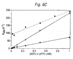
- the observed rate constants (a: 11.1 ⁇ 0.01 s -1 ; b: 57.2 ⁇ 0.07s -1 ) increased linearly in proportion to the increasing ADP concentrations, and a second order rate constant was calculated from the slope of the linear fit, giving 0.21x10 6 M -1 s -1 ( Figure 6C).
- the observed rate is k + [ADP] + k - [ATP], and therefore increases with both [ADP] and [ATP].
- This rate acceleration is advantageous in muscle fibres, for instance, where ATP levels are usually at least 1mM.
- Dephosphorylation of the phosphoenzyme should be very fast, even at ADP concentrations which are initially low.
- the fluorescence trace (bottom curve, Figure 7) after time zero consists of three parts: first, a flash artefact; then, a lag, during the time for the first ATPase cycle to occur as far as bound ADP: finally. a slow phase of decreasing fluorescence, representing the reaction of IDCC-NDPK ⁇ P with ADP.
- the initial slope of this curve gives the rate of ADP release from actomyosin as 11s -1 .
- the rate of nucleotide exchange from a small G protein, rho (which also has GTPase activity), was determined in solution.
- the rate of hydrolysis was also obtained indirectly.
- Human rho protein was prepared in a form containing tightly-bound GDP. In the presence of excess added GTP, nucleotide exchange occurs such that GDP is released into free solution where it can be measured according to the invention. Once GTP is bound to rho, its hydrolysis is slowly catalysed by the protein at a steady-state rate. This creates more GDP, which is measured once it is released.
- Fluorescence emission was recorded for a 200 ⁇ l solution containing 20mM Tris/HCl, pH 7.6, 1mM MgCl 2 , 100mM (NH 4 ) 2 SO 4 , 10 ⁇ M GTP, and 2 ⁇ M IDCC-NDPK ⁇ P.
- Excitation was at 440nm with the emission monochromator set to 475nm.
- the temperature was 30°C.
- the data were normalised using the fluorescence change for 1 ⁇ M GDP obtained from a titration of IDCC-NDPK ⁇ P with different GTP/GDP ratios.
Landscapes
- Chemical & Material Sciences (AREA)
- Life Sciences & Earth Sciences (AREA)
- Health & Medical Sciences (AREA)
- Organic Chemistry (AREA)
- Zoology (AREA)
- Wood Science & Technology (AREA)
- Engineering & Computer Science (AREA)
- Genetics & Genomics (AREA)
- Bioinformatics & Cheminformatics (AREA)
- Biochemistry (AREA)
- Biotechnology (AREA)
- Molecular Biology (AREA)
- General Health & Medical Sciences (AREA)
- General Engineering & Computer Science (AREA)
- Microbiology (AREA)
- Proteomics, Peptides & Aminoacids (AREA)
- Physics & Mathematics (AREA)
- Immunology (AREA)
- Analytical Chemistry (AREA)
- Biophysics (AREA)
- Medicinal Chemistry (AREA)
- Biomedical Technology (AREA)
- Measuring Or Testing Involving Enzymes Or Micro-Organisms (AREA)
- Saccharide Compounds (AREA)
- Preparation Of Compounds By Using Micro-Organisms (AREA)
- Investigating, Analyzing Materials By Fluorescence Or Luminescence (AREA)
- Investigating Or Analysing Biological Materials (AREA)
- Enzymes And Modification Thereof (AREA)
Abstract
Description
- The invention relates to assays for nucleoside diphosphates, particularly ADP and GDP, and assays for nucleoside triphosphates, particularly ATP and GTP.
- Nucleoside diphosphates and triphosphates play important roles in biology. ADP is the immediate precursor for the formation of ATP, the universal currency of cellular energy. GDP is a substrate for succinyl CoA synthetase, a key enzyme of the Krebs cycle, and is formed during gluconeogenesis by phosphoenolpyruvate carboxykinase. It is also essential in G-protein signalling, microtubule growth, and visual excitation. UDP is involved in the epimerisation of galactose to glucose, the formation of sucrose, and in the growth of glycogen. CDP is an important group in the synthesis of phosphoglycerides. Nucleoside diphosphates are also the products of reactions catalysed by several major classes of enzymes, such as triphosphatases and kinases, and are therefore produced by many cellular processes, including motility, muscle contraction, DNA synthesis, transcription, translation and nitrogen fixation.
- The detection and measurement of nucleoside diphosphates and triphosphates is thus important in the study of biology and metabolism, particularly in bioenergetics.
- Assays for ADP and ATP in biological samples based on luciferase have been known for over 20 years [e.g. references 1-4]. Bioluminescent assays for ADP and ATP have been described for use in muscle and adipose tissue biopsies [5] and a three-enzyme bioluminescent system utilising luciferase has been reported for use in bacterial cell extracts [6]. A bioluminescent ADP assay optimised for use at high ATP:ADP ratios has been reported [7], but this requires the enzymatic removal of ATP. In general, it is easier to measure ATP in the presence of ADP than to measure ADP in the presence of ATP.
- Enzymatic spectrophotometric assays have also been described [e.g. 8].
- Assays for GDP and GTP in biological samples are also well known [e.g. refs. 9 & 10].
- Reference 11 discloses column-based chromatographic assays for ADP, GDP, CDP and UDP. Radioactive assays for GDP and GTP have also been described [12,13]. NMR-based assays for measuring in vivo ADP levels are known for yeast [14], and NMR has also been used to measure ADP and ATP and erythrocytes [15].
- Reference 33 discloses methods for measuring the total amounts of G protein-hound GDP and GTP in mammalian cells and tissue. The method avoids the need to expose the cells to phosphate-free growth conditions, which can adversely affect the accuracy of the. GTP determination. GDP is converted to GTP by phosphorylation using the nucleoside diphosphate kinase (NDPK) enzyme.
- Reference 34 discloses a method which uses NDPK to convert ADP to ATP. The ATP is then used to produce NADH or NADPH, which is used as an indirect measure of the initial amount of ADP.
- NDPK is suggested for use in an assay for detecting GTP in reference 35, with the enzyme being used to convert GTP to ATP.
- Reference 36 discloses methods for detecting highly proliferative cells, such as tumour cells, in which the activity or the quantity of NDPK is measured. NDPK is trapped in its phosphorylated form by using a radio-labelled suicide γ-thiophosphate derivative, and it can then be detected.
- According to the present invention, nucleoside diphosphates are detected or measured by following the dephosphorylation of the phosphoenzyme form of nucleoside diphosphate kinase (NDPK), and nucleoside triphosphates are detected or measured by following the phosphorylation of NDPK to its phosphoenzyme form.
- The invention thus provides (a) a process for detecting the presence of a nucleoside diphosphate in a sample, comprising the step of detecting the dephosphorylation of the phosphoenzyme form of a nucleoside diphosphate kinase, and (b) a process for detecting the presence of a nucleoside triphosphate in a sample, comprising the step of detecting the phosphorylation of a nucleoside diphosphate kinase to the phosphoenzyme form.
- The process will typically comprise the steps of:
- causing nucleoside diphosphate in sample to bind to NDPK phosphoenzyme, or causing nucleoside triphosphate in sample to phosphorylate NDPK; and
- detecting a change in a characteristic of the enzyme which differs between its phosphorylated and unphosphorylated forms.
- The term "NDPK" means an enzyme having the activity of the enzyme classified as EC 2.7.4.6, namely the transfer of the γ-phosphate group of a nucleoside triphosphate (N1TP) to a nucleoside diphosphate (N2DP) via a ping-pong mechanism:
- Based on this reaction scheme, the systematic name of NDPK is "ATP:nucleoside-diphosphate phosphotransferase", but the common name is "nucleoside diphosphate kinase". The enzyme has also been variously described as: kinase (phosphorylating), nucleoside diphosphate; nucleoside 5'-diphosphate kinase; nucleoside diphosphate (UDP) kinase; nucleoside diphosphokinase; nucleotide phosphate kinase; NM23.
- NDPKs have been described for a number of organisms, both prokaryotic and eukaryotic e.g. human, cows, monkeys, mice, Xenopus, oats, peas, potatoes, yeast, Bacillus subtilis, E.coli, Myxococcus xanthus, avian myeloblastosis virus etc. These differ by cellular location, molecular weight, oligomeric structure, isoelectric point, reaction kinetics, substrate preference, pH optimum, pH range, temperature optimum, cation requirements (Mn2+, Mg2+, Co2+, Ca2 etc.), and various isoforms have been described. Given the variety of suitable enzymes available, the skilled person can easily select and purify a NDPK to suit any particular situation.
- The NDPK enzyme uses a ping-pong mechanism, transferring the γ-phosphate from a nucleoside triphosphate (N1TP) to an active site histidine to form a phosphoenzyme intermediate, and then to a nucleoside diphosphate (N2DP). The invention is based on the finding that the phosphoenzyme intermediate is stable over a time-scale that allows its detection and measurement. Other enzymes that phosphorylate nucleoside diphosphates via a phosphoenzyme intermediate, preferably with a single binding site for nucleotide, may also be used in the invention.
- The phosphoenzyme is able to transfer its phosphate group to N2DP in a sample to form the corresponding N2TP. Detection of this transfer can therefore be used for the detection of nucleoside diphosphate. To detect nucleoside diphosphate according to the invention, therefore, phosphoenzyme is required as a reagent. This can be readily formed by, for example, incubating NDPK with excess NTP, typically ATP. Formation of phosphoenzyme in this way is facilitated by removing Mg2+ [16], for instance by using EDTA. Chemical phosphorylation of histidine using phosphoramidate as a phosphorylating agent may also be used [17].
- The phosphoenzyme can be isolated for use as a reagent. It has been found that the phosphoenzyme can be stored on ice for over 48 hours without dephosphorylation, and can be stored for longer periods (at least 5 months) at -80°C (although repeated freeze-thawing results in some dephosphorylation). The stability of the phosphoenzyme over the time range needed for its preparation, and subsequently for monitoring kinetic events such as the release of ADP from an ATPase, is particularly advantageous.
- When added to a sample containing NDP, the phosphoenzyme is dephosphorylated by the transfer of its phosphate group to the NDP. When added to a sample containing NTP. the phosphoenzyme is formed by the transfer of the NTP γ-phosphate group to the enzyme. The invention relies on the ability to distinguish between the phosphorylated and dephosphorylated forms of NDPK.
- In order to distinguish the phosphorylated and unphosphorylated forms of NDPK, any suitable measurable change can be used.
- For instance, intrinsic properties of the enzyme can be used. Depending on the particular NDPK chosen, the following methods are examples of how dephosphorylation/phosphorylation may be detected. with varying levels of sensitivity:
- The location of a phosphate (i.e. either bound to NDPK, or as the γ-phosphate of a NTP) can be ascertained by following the 31P NMR spectrum.
- Protons whose environment changes upon dephosphorylation can be detected by for instance. NMR.
- Dephosphorylation may cause a change in the fluorescence of a tryptophan residue in the protein [e.g. ref 18].
- Dephosphorylation can be detected by following the loss of 32P from radio-labelled phosphoenzyme. The radio-isotope can be conveniently incorporated into NDPK by using [γ-32P]ATP.
- Circular dichroism, or any other suitable spectrometric technique, can detect conformational changes which occur on dephosphorylation.
- Dephosphorylation may result in a change in surface plasmon resonance properties.
- Rather than using properties inherent in the wild-type enzyme, it may be desired to modify the enzyme in some way. This may also be important where dephosphorylation of the NDPK of choice does not exhibit an intrinsic measurable change which can be readily followed.
- One particularly preferred modification is the addition of a fluorescent label to the enzyme, typically via a cysteine residue. If the wild-type protein lacks a suitable cysteine residue (e.g. the NDPK of Myxococcus xanthus), this can easily be introduced by mutagenesis [e.g. 19]. A suitable position for mutation can easily be determined by the skilled person, whilst ensuring that the mutation does not disrupt the enzymatic activity [e.g. 20]. At any given amino acid residue. particular labels may give better results than others. Suitable combinations of label and residue can be determined by routine experimentation.
- Preferred fluorescent labels are based around coumarin. Particularly preferred is N-[2-(iodoacetamido)ethyl]-7-diethylaminocoumarin-3-carboxamide [21; Fig. 1], referred to simply as 'IDCC' hereafter. This is preferably attached to a cysteine residue. and preferably exhibits a high fluorescence when NDPK is phosphorylated. and a low fluorescence when NDPK is dephosphorylated. When suitably attached to NDPK. this label offers the advantage that the phosphoenzyme can detect small quantities of ADP in the presence of much higher concentrations of ATP. This is extremely important for experiments in situations where ATP levels are high e.g. in single muscle fibres. It is also able to respond very quickly to changes in ADP levels, and gives a large signal change over a range of several hundred micromolar.
- Other labels which can be introduced in similar ways include ESR labels, luminescent labels, phosphorescent labels, and other suitable chromophores.
- It will be appreciated that some of the various options available to the skilled person are more suitable than others for detecting the phosphorylation/dephosphorylation of the enzyme in real-time. Fluorescence is highly suitable for real-time detection, whereas methods such as those using radio-labels are more suitable for measuring end-points.
- The nucleoside diphosphate/triphosphate which is assayed must be a substrate of the NDPK being utilised. Various NDP substrates have been described [e.g. 22, 23, 24] including ADP, CDP, GDP, UDP, IDP, XDP, their deoxy-derivatives (e.g. dADP, dCDP, dGDP, dTDP, dUDP), 6-aza-UDP, 8-bromo-IDP, 8-aza-GDP, and 8-aza-UDP, and adenosine 5'-methylene diphosphonate. Each of these compounds is phosphorylated by the phosphoenzyme (with varying reaction affinities and kinetics, depending on both the NDPK and the substrate being utilised), and can thus be assayed according to the invention.
- The invention is preferably used to detect and measure ADP or GDP. Accordingly, a NDPK may be chosen which shows a preference towards one of these substrates.
- In preferred embodiments of the invention, the detection process gives quantitative data, that is to say the invention provides a process for quantifying nucleoside diphosphate or triphosphate in a sample. This will typically involve the step of relating a change in the detectable characteristics of a NDPK to a concentration of NDP or NTP. It will be appreciated that this may require a calibration to be performed (e.g. for measuring dephosphorylation via a fluorescent label such as IDDC) or comparison with a standard. Calibration will typically be performed for the desired range of concentrations to be measured.
- In a first quantitative aspect, the amount of nucleoside diphosphate or nucleoside triphosphate is determined by measuring the decrease (NDP) or increase (NTP) in the level of phosphoenzyme after the addition of phosphoenzyme (NDP) or unphosphorylated enzyme (NTP) to a sample.
- In a second quantitative aspect, the rate of production of nucleoside diphosphate or triphosphate can be determined by following the decrease (NDP) or increase (NTP) in the level of phosphoenzyme over time. By fitting the measured values to a suitable mathematical model (e.g. a simple model based on first-order exponential decrease), the rate of nucleoside diphosphate or triphosphate production can be determined. In this aspect. dephosphorylation of the phosphoenzyme (NDP) or phosphorylation of the enzyme (NTP) is preferably measured using a real-time detection method.
- The process of the invention is preferably suitable for use either in vivo or in vitro. The method is preferably suitable for in situ use in a muscle fibre, and is preferably gives data suitable for calculating the rate of ADP release from actomyosin.
- As well as the step of detecting the dephosphorylation of the phosphoenzyme (NDP-related aspects), the process will usually comprise the initial step of adding NDPK phosphoenzyme to a sample of interest. This may be preceded by the preparation of phosphoenzyme from unphosphorylated NDPK.
- The process may also include a step of analysing any data obtained during the process, such as fitting the data to an equation in order to derive quantitative values.
- The process preferably avoids the use of reagents such as theophylline, desdanine and Ag+, which may inhibit the NDPK activity.
- As well as the above processes, the invention provides reagents for use in the processes.
- The invention provides NDPK which is modified to carry a label which gives a different detectable signal when the enzyme is phosphorylated from when it is unphosphorylated.
- The label on the modified NDPK may be a fluorescent group, preferably IDCC.
- The label is attached to an amino acid residue in the enzyme. It is preferred to attach the label to a cysteine residue.
- A particularly preferred reagent is the NDPK of M.xanthus carrying a Asp112→Cys mutation, and carrying an IDCC label at this mutated residue. This reagent as a phosphoenzyme is about three orders of magnitude more sensitive to ADP than to ATP.
- The invention also provides a NDPK modified by the attachment of at least one detectable label that is sensitive to the binding of a nucleoside diphosphate.
- The invention also provides substrates having these NDPK reagents immobilised thereto. These include columns or beads. This may be used in combination with 32P-phosphoenzyme, such that ADP in a sample incubated with immobilised NDPK will become radio-labelled in its conversion to ATP. Radioactivity in free solution will therefore indicate the amount of ADP in the original sample.
- The invention also provides processes for the production of these NDPK reagents.
- Furthermore, the invention provides these NDPK reagents for use as in vivo or in vitro diagnostic reagent.
-
- Figure 1 shows the structure of a preferred fluorescent label, IDCC.
- Figure 2 shows the emission spectra of IDCC-NDPK in the presence of (A) ADP/ATP and (B) GDP/GTP.
- Figure 3 shows a titration of IDCC-labelled phosphoenzyme with ADP/GDP (open symbols ○ and □) and ATP/GTP (closed symbols and ▪). Figure 4 shows the same experiments using unphosphorylated enzyme.
- Figure 5 shows a calibration curve obtained in a stopped flow experiment, indicating how fluorescence change varies with ADP concentration.
- Figure 6 shows transient kinetic data obtained using stopped-flow fluorescence. The dashed curves represent the best fit to a single exponential equation. 6A & 6B show data obtained with ADP and ATP, respectively. 6C shows a plot of observed rate constants against ADP (○) or ATP () concentration. The solid line shows the best fit to a linear equation with the respective deduced second-order rate constants. The dashed line and squares (▪) show observed rate constants that were obtained when the ADP titration was repeated in the presence of 1mM ATP.
- Figure 7 shows a plot obtained from a single rabbit psoas muscle fibre (skinned), indicating the increase in muscle tension and the accompanying decrease in fluorescence due to dephosphorylation of IDCC-NDPK∼P.
- Figure 8 shows a plot obtained using human rho protein, indicating the release of GDP into solution. Data were fitted to a single-exponential plus linear equation, shown as a solid line.
-
- General molecular biology techniques were carried out according to Sambrook et al. [25].
- The NDPK of M.xanthus is encoded by the ndk gene, which has been cloned and expressed in E.coli [26]. The protein is a homo-tetramer of 16kDa subunits, it has been characterised, and a crystal structure has been determined [16,27]. The wild-type sequence does not contain any cysteine, so the gene was manipulated to introduce cysteine residues by site-directed mutagenesis in E.coli strains TG1 and DH5α using either a phosphothioate-based method [28, produced in kit form by Amersham] or the PCR-based QuikChange kit [Stratagene].
- Using the Amersham kit, the 0.8 kb HindIII-EcoRI fragment of pJM5C2A [29] containing the ndk gene from M.xanthus was ligated with M13mp19, and the resulting recombinant clones were used to provide single-stranded DNA templates for mutagenesis. For cloning, the mutated ndk genes were cloned back into pJM5C2A. As an alternative, the 0.7 kb BstXI-EcoRI fragment of the M13ndk constructs was ligated into a modified form of the Invitrogen™ pRSetA expression vector, whose coding sequence for a histidine tag fused to the N-terminus of NDPK had been removed. This yielded the 3.5 kb pRSndkX series of plasmids, where the final "X" is a number in a series of ndk mutations.
- pRSndk was also used as a template for the QuikChange method.
- Various mutant proteins containing cysteine residues were prepared, including D112C (i.e. Asp-112 was mutated to Cys) and D62C. Positions for mutation were typically chosen on the basis of their proximity to the nucleotide-binding cleft seen in the crystal structure [16].
- The mutant D112C gene was produced in plasmid pRSndk4, which was also used for expression. For best results, freshly-transformed cells were used for starter cultures. 200µl calcium-competent BL21 cells [Novagen] were incubated with 2ng pRSndk4 plasmid DNA for 30 minutes on ice. Half of this mixture was then spread onto an LB agar plate containing 0.1mg/ml ampicillin and incubated overnight at 37°C. Plates Typically contained 50-100 colonies. 100ml LB medium containing 0.1mg/ml ampicillin was inoculated with 2-3 colonies from the plate and grown for 9 hours at 37°C until cells had just entered stationary phase. For the main culture, 8x500ml LB+ampicillin was inoculated with 10ml started culture and incubated at 37°C for 6 hours, after which time the cells had typically reached OD595 of 0.38. At this point. 0.5mg/ml IPTG was added to each flask, and the cells grown for a further 16 hours. Cells were harvested by centrifugation in a Beckman L2 centrifuge at 3800rpm and 20°C for 20 minutes. The pellet was resuspended in 100ml Buffer A (20mM Tris-HCl, pH 8.2. 1mM EDTA) and stored at -80°C.
- Approx. 35ml of the frozen cell suspension was lysed by slow thawing and then sonication. The supernatant (i.e. crude extract) was retained. Expression levels were determined by SDS-PAGE. NDPKD112C made up >50% cytosolic protein.
- NDPK purification involved two column chromatography steps. First, after adjusting pH and ionic strength using Buffer A + 10mM DTT, the crude extract (approx. 200ml) was loaded onto a 120ml Q-Sepharose ion-exchange column at 4°C and a flow rate of 2.0ml/min. The column was washed with 1 volume buffer and eluted with a continuous linear gradient (500ml) from 0-0.3M NaCl at 1ml/min flow rate. The bulk of NDPK (about 80%) does not bind to the resin under these conditions.
- After concentration using an Amicon YM10 ultrafiltration membrane, the eluate was loaded onto a G-100 Sepharose gel filtration column in Buffer A. At a flow-rate of 1.0ml/min, the elution profile showed 2 peaks. The first contained DNA, and the second contained proteins. Fractions containing NDPK were pooled and concentrated as before.
- It was confirmed that there was no contamination with E.coli NDPK, which has a similar sequence and is also a tetramer [30]. Mass spectrometry data showed that the purified protein was a single species with MW 15993±1 Da, matching the calculated MW of the mutant protein minus the N-terminal Met, which was shown to be missing when expressed in E.coli [16]. The yield of pure NDPKD112C from a 4-litre culture was around 300mg.
- NDPKD112C was incubated with a 2.5-fold excess of IDCC (Fig. 1) in 50mM Tris/HCl, pH 8.1 for 1 hour at 37°C. In a typical experiment, the volume of the labelling solution was 3ml and the concentration of NDPKD112C was 150µM. After incubation, this solution was filtered through a 0.2µM Acrodisc filter (Gelman) and loaded onto a PD10 desalting column (Pharmacia) equilibrated in Buffer B (10mM Tris-HCl, pH 8.0, 1mM EDTA). The eluate (approx. 6ml) containing labelled protein was immediately loaded onto a 25ml Q-Sepharose column equilibrated in Buffer B, followed by a 20ml linear gradient from 0-0.1M NaCl. The labelled protein was completely eluted before the gradient was applied, and was concentrated in the same way as unlabelled protein. The concentration of the protein was determined using absorbance spectroscopy, assuming that the absorbance spectrum of the fluorophore was unaltered (ε430nm 46800 M-1cm-1). This concentration value agree with that measured by a colorimetric assay using a bovine serum albumin standard curve. After correcting for the absorbance of IDCC at 276nm (0.198 of that at 430nM in the dithiothreitol adduct), the molar extinction coefficient for NDPK could be calculated as ε276nm 7600 M-1cm-1.
- The yield after labelling and purification was typically 65%. This high yield indicates that the thiol group of Cys-112 is easily accessible. Mass spectrometry revealed that one molecule IDCC was incorporated per protein molecule, with no indication of second-site labelling. and the mass was that expected for IDCC-NDPK.
- To check that mutation and labelling did not affect oligomerisation, the MW of the complex was determined by sedimentation equilibrium centrifugation. The results indicate that IDCC-NDPK is a tetramer of MW 62.7kDa (4% lower than that calculated for a tetramer from mass spectrometry data).
- Where rapid analysis of the fluorescent properties of a new mutant/fluorophore combination was required, labelling was performed on a smaller scale, typically using a NDPK concentration of 100-200µl and avoiding the ultrafiltration and Q-Sepharose steps.
- Other thiol-reactive environmentally-sensitive fluorescent labels which were tested included MDCC (N-[2-(1-maleimidyl)ethyl]-7-diethylaminocoumarin-3-carboxamide) [21].
- IDCC-NPDK was incubated with ATP, typically at a 5-fold excess, but ≤1mM. The incubation was for 30-45 minutes at 37°C in either in 10mM Tris/HCl, pH 8.0, 1mM EDTA, or in 10mM PIPES, pH 7.0, 1mM EDTA. The protein was separated using a PD10 column equilibrated in the same buffer. Typically, more than 90% of the eluted protein was phosphoenzyme. The solution was either used immediately or after storage on ice for up to 48 hours. If necessary, the solution was concentrated using a microcentrifuge concentrator.
- The spectroscopic properties of the phosphoenzyme (IDCC-NDPK∼P) were compared with the unphosphorylated form of the enzyme. Data was recorded on a Perkin-Elmer LS50B luminescence spectrometer with a xenon lamp and monochromator slit widths set to 2.5 or 5nm.
- The fluorescence of IDCC-NDPK was 4-fold greater in the presence of 50µM ATP than in the presence of 50µM ADP (Figure 2A). Similar results were obtained with 100µM GDP/GTP (Figure 2B). In both cases the spectra were recorded at 20°C from a 200µl solution of 2µM protein in 20mM PIPES, pH 7.0. 2mM MgCl2. Excitation was at 441nm, slit width 5nm. No correction was made for the small (1%) volume change. These results indicate that IDCC-NDPK∼P has a high fluorescence compared with the unphosphorylated form.
- A titration of a solution of 2µM IDCC-NDPK∼P with ADP showed a large decrease in the fluorescence signal (Figure 3A), whereas there was no change with ATP over the same concentration range (<100µM). Similar data were obtained with GDP/GTP (Figure 3B). The ADP/ATP assays were carried out at 21°C in 10mM Tris/HCl, pH 8.0, 5mM MgCl2. Excitation was at 432nm (5nm slit width) and emission was recorded at 478nm (2.5nm slit width). The GDP/GTP assays were carried out at 20°C in 20mM PIPES, pH 7.0, 2mM MgCl2. Excitation was at 441nm (5nm slit width) and emission was recorded at 475nm (5nm slit width). In both cases data were corrected for the small volume increases. The small decrease at the first addition of GTP in Figure 3B is due to partial hydrolysis of GTP in the diluted 200µM stock solution; subsequent GTP additions were from a 2mM stock solution.
- Together with data from unphosphorylated enzyme (Figures 4A and 4B), this suggests that most of the fluorescence change is due to phosphorylation/dephosphorylation of the enzyme.
- Increasing ADP above 50µM produces a small fluorescence increase. This is probably due to a non-specific interaction between ADP and NDPK, as suggested for non-labelled wild-type enzyme [18]. The change in fluorescence signal is linear with [ADP] up to at least 50% of the protein concentration.
- To derive quantitative data from the fluorescence measurements, the calibration curve shown in fig. 5 was plotted. The calibration data were obtained in a stopped flow experiment by mixing 10µM IDCC-NDPK∼P and 50µM ATP with 0, 2.5, 5.0, 7.5 or 10.0µM ADP (10mM PIPES buffer, pH 7.0, 1mM MgCl2, 20°C). The amplitudes of the fluorescence change (ΔF) were averaged from two experiments at each concentration and plotted against ADP concentration.
- There is a shift of 6nm in the absorbance maxima of the fluorophore between IDCC-NDPK and IDCC-NDPK∼P, but only 2nm in the emission maxima. The fluorescence quantum yields for the two forms of the labelled enzyme were determined using as reference the known value of 0.83 for Coumarin 314 in ethanol [31]. There was a 4-fold decrease in the quantum yield when going from phosphorylated (0.22) to unphosphorylated enzyme (0.054), in good agreement with the observed change in emission intensity (Figure 2), although the quantum yield for the phosphoenzyme was still significantly lower than that for Coumarin 314 in buffer (0.52).
- Three different transient kinetic measurements were carried out in a stopped-flow apparatus.
- Firstly, the rate of IDCC-NDPK-P dephosphorylation was measured over a range of ADP concentrations. Figure 6A shows the emission data for mixing 50µM (a) and 250µM (b) ADP with 1µM protein in 20mM PIPES, pH 7.0, 2mM MgCl2. The observed rate constants (a: 11.1±0.01 s-1; b: 57.2±0.07s-1) increased linearly in proportion to the increasing ADP concentrations, and a second order rate constant was calculated from the slope of the linear fit, giving 0.21x10⁶ M-1s-1 (Figure 6C).
- In a second experiment, the rate of phosphoenzyme production was measured by mixing IDCC-NDPK-P enzyme with ATP (Figure 6B). Again, there was a linear increase in rates with increasing ATP (5.57±0.01 s-1 at 50µM ATP; 18.4±0.03 s-1 at 250µM ATP), and the deduced second order rate constant was 0.072x10⁶ M-1s-1 (Figure 6C).
- In a third experiment, the rate of dephosphorylation was measured in the presence of varying concentrations of ATP. The observed rates were higher but the intensity changes lower in the presence of ATP than in its absence. From the slope of the dashed line in Figure 6C (▪), the second order rate constant is calculated as 0.135x10⁶ M-1s-1 in the presence of ATP. The increase in rate produced by ATP can be explained by considering the enzymatic equilibrium:

- The observed rate is k +[ADP] + k -[ATP], and therefore increases with both [ADP] and [ATP].
- This rate acceleration is advantageous in muscle fibres, for instance, where ATP levels are usually at least 1mM. Dephosphorylation of the phosphoenzyme should be very fast, even at ADP concentrations which are initially low.
- An experiment was performed on a single rabbit psoas muscle fibre in the presence of Ca2+, essentially as described in reference 32, with the following modifications. An incubation of the fibre for 10 minutes in rigor solution containing 8U/ml Apyrase to remove background ADP was followed by 10 minutes incubation in loading solution containing 380µM IDCC-NDPK∼P and 5mM NPE-caged ATP [the P 3-1-(2-nitrophenyl)ethyl ester of ATP). The fibre was transferred to silicone oil. and ATP (approx. 1 mM) was released by laser flash photolysis (347nm) at time zero. The temperature during the experiment was 18°C, and the aqueous solutions were pH 7.1 and pCa 4.5. Fluorescence excitation was at 425nm, and emission was detected through a long-pass filter (450-650nm).
- The fluorescence trace (bottom curve, Figure 7) after time zero consists of three parts: first, a flash artefact; then, a lag, during the time for the first ATPase cycle to occur as far as bound ADP: finally. a slow phase of decreasing fluorescence, representing the reaction of IDCC-NDPK∼P with ADP. The initial slope of this curve gives the rate of ADP release from actomyosin as 11s-1.
- Similar experiments in muscle fibres using NPE-caged ADP to measure ADP production independently from myosin ATPase show that (i) the phosphoenzyme is able to maintain its sensitivity and reactivity for ADP when it is within a muscle fibre (ii) a signal is detectable for ADP released from myosin ATPase (iii) the muscle fibre is developing tension in the expected manner.
- The rate of nucleotide exchange from a small G protein, rho (which also has GTPase activity), was determined in solution. The rate of hydrolysis was also obtained indirectly.
- Human rho protein was prepared in a form containing tightly-bound GDP. In the presence of excess added GTP, nucleotide exchange occurs such that GDP is released into free solution where it can be measured according to the invention. Once GTP is bound to rho, its hydrolysis is slowly catalysed by the protein at a steady-state rate. This creates more GDP, which is measured once it is released.
- Fluorescence emission was recorded for a 200µl solution containing 20mM Tris/HCl, pH 7.6, 1mM MgCl2, 100mM (NH4)2SO4, 10µM GTP, and 2µM IDCC-NDPK∼P. The reaction was started at t=0 by adding 2µM Rho.GDP. Excitation was at 440nm with the emission monochromator set to 475nm. The temperature was 30°C. The data were normalised using the fluorescence change for 1µM GDP obtained from a titration of IDCC-NDPK∼P with different GTP/GDP ratios. The data (figure 8) were fitted to a single-exponential plus linear equation with a rate constant of 0.196min-1 for the exponential phase and a slope of 41.2nM GDP min-1 (3.43x10⁻⁴ s-1, after dividing by [Rho]) for the linear phase. The exponential "burst" represents the exchange of GDP bound to rho for GTP. The linear phase represents steady state: exchange of further GTP onto rho is limited by hydrolysis to GDP.
- The rates were also measured by 'standard' means, and similar results were found.
- It will be understood that the invention is described above by way of example only and modifications may be made whilst remaining within the scope of the invention.
-
- 1. Holmsen et al. (1972) Anal. Biochem. 46:489-501
- 2. Kimmich et al. (1975) Anal Biochem 69:187-206
- 3. Loxdale (1976) J Physiol (Lond) 240-4P-5P
- 4. Spielmann et al. (1981) Anal Biochem 113:172-178
- 5. Feradui et al. (1981) Int J Sports Med 2:106-109
- 6. Brovko et al. (1994) Anal Biochem 220:410-414
- 7. Schultz et al. (1993) Anal Biochem 215:302-304
- 8. Tornheim & Schultz (1993) Anal Biochem 211:329-330
- 9. Pogson et al. (1979) Int J Biochem 10:995-1000
- 10. de Azeredo et al. (1979) Anal Biochem 95:512-519
- 11. Sato et al. (1983) Anal Biochem 135:431-435
- 12. Goswami & Pande (1984) J Biochem Biophys Methods 9:143-151
- 13. Cerpovicz & Ochs (1991) Anal Biochem 192:197-202
- 14. Brindle et al. (1990) Biochem 29:3295-3302
- 15. Petersen et al. (1990) Biochim Biophys Acta 1035:169-174
- 16. Williams et al. (1993) J Mol Biol 234:1230-1247.
- 17. Morera et al. (1995) Biochemistry 34:11062-11070.
- 18. Schaertl et al. (1998) J Biol Chem 273:5662-5669
- 19. Sundin et al. (1996) J.Bacteriol 178:7120-7128
- 20. Izumiya & Yamamoto (1995) J Biol Chem 270:27859-27864.
- 21. Corrie (1994) J Chem Soc, Perkin Trans 1:2975-2982.
- 22. Parks & Agarwal in The Enzymes, 3rd ed (ed. Boyer), 8:307-333
- 23. Agarwal et al. (1978) Methods Enzymol 51:376-386
- 24. Jong & Ma (1991) Arch Biochem Biophys 291:241-246.
- 25. Sambrook et al. (1989) Molecular cloning - a laboratory manual.
- 26 Muñoz-Dorado et al. (1990) J Biol Chem 265:2702-2706.
- 27. Muñoz-Dorado et al. (1990) J Biol Chem 265:2707-2712.
- 28. Olsen et al. (1993) Meth Enzymol 217:189-217.
- 29. Kindly provided by Dr R Williams, Cambridge.
- 30. Almaula et al. (1995) J Bacteriol 177:2524-2529.
- 31. Fletcher & Bliss (1978) Appl Phys 16:289-295.
- 32. He et al. (1997) J. Physiol. 501.1:125-148.
- 33. US patent 5,741,635.
- 34. US patent 4,923,796.
- 35. US patent 4,806,415.
- 36. French patent application 2,660,933.
-
Claims (17)
- A process for detecting the presence of a nucleoside diphosphate in a sample, comprising the step of detecting the dephosphorylation of the phosphoenzyme form of a nucleoside diphosphate kinase (NDPK) that has a label attached to an amino acid residue, by detecting a change in a characteristic of the NDPK which differs between its phosphorylated and unphosphorylated forms.
- A process for detecting the presence of a nucleoside triphosphate in a sample, comprising the step of detecting the phosphorylation of a nucleoside diphosphate kinase (NDPK), the NDPK having a label attached to an amino acid residue, to the phosphoenzyme form by detecting a change in a characteristic of the NDPK which differs between its phosphorylated and unphosphorylated forms.
- The process of claim 1 or claim 2, wherein the NDPK is modified to carry a label which gives a different detectable signal when the enzyme is phosphorylated from when it is unphosphorylated.
- The process of claim 3, wherein the NDPK carries a fluorescent label.
- The process of claim 4, wherein the fluorescent label is attached to the NDPK via a cysteine residue.
- The process of claim 4 or claim 5, wherein the fluorescent label is IDCC (N-[2-(iodoacetamido)ethyl]-7-diethylaminocoumarin-3-carboxamide).
- The process of claim 1, wherein the nucleoside diphosphate is ADP or GDP.
- The process of claim 2, wherein the nucleoside triphosphate is ATP or GTP.
- The process of any preceding claim, being a quantitative process.
- The process of any preceding claim, wherein the NDPK is the NDPK of Myxococcus xanthus carrying a Asp112→Cys mutation, and carrying an IDCC label at this mutated residue.
- NDPK which is modified to carry a label which is attached to an amino acid residue of the NDPK and which gives a different detectable signal when the enzyme is phosphorylated from when it is unphosphorylated.
- The NDPK of claim 11, wherein the label on the modified NDPK is a fluorescent label.
- The NDPK of claim 12, wherein the fluorescent label is attached to the NDPK via a cysteine residue.
- The NDPK of claim 12 or claim 13, wherein the fluorescent label is IDCC.
- The NDPK of claim 14, which is NDPK of Myxococcus xanthus with a Asp112→Cys mutation, and which is carrying an IDCC label at this mutated residue.
- A substrate having the NDPK of any one of claims 11 to 15 immobilised thereto.
- The NDPK of any one of claims 11 to 15 for use as an in vivo or in vitro diagnostic reagent.
Applications Claiming Priority (3)
| Application Number | Priority Date | Filing Date | Title |
|---|---|---|---|
| GB9910811 | 1999-05-10 | ||
| GBGB9910811.0A GB9910811D0 (en) | 1999-05-10 | 1999-05-10 | Assays |
| PCT/GB2000/001740 WO2000068418A1 (en) | 1999-05-10 | 2000-05-05 | Assays for nucleoside diphosphates and triphospates |
Publications (2)
| Publication Number | Publication Date |
|---|---|
| EP1177314A1 EP1177314A1 (en) | 2002-02-06 |
| EP1177314B1 true EP1177314B1 (en) | 2004-04-14 |
Family
ID=10853167
Family Applications (1)
| Application Number | Title | Priority Date | Filing Date |
|---|---|---|---|
| EP00927529A Expired - Lifetime EP1177314B1 (en) | 1999-05-10 | 2000-05-05 | Assays for nucleoside diphosphates and triphospates |
Country Status (9)
| Country | Link |
|---|---|
| US (2) | US6746849B1 (en) |
| EP (1) | EP1177314B1 (en) |
| JP (1) | JP4338320B2 (en) |
| AT (1) | ATE264400T1 (en) |
| AU (1) | AU771858B2 (en) |
| CA (1) | CA2371176C (en) |
| DE (1) | DE60009891T2 (en) |
| GB (1) | GB9910811D0 (en) |
| WO (1) | WO2000068418A1 (en) |
Families Citing this family (6)
| Publication number | Priority date | Publication date | Assignee | Title |
|---|---|---|---|---|
| JP4431334B2 (en) * | 2003-07-29 | 2010-03-10 | 独立行政法人科学技術振興機構 | Improved ATP amplification method and use thereof |
| GB0711328D0 (en) * | 2007-06-12 | 2007-07-25 | Medical Res Council | Biosensor for detection and visualisation of single-stranded dna |
| GB0817166D0 (en) * | 2008-09-19 | 2008-10-29 | Medical Res Council | Sensor |
| CN103031287B (en) * | 2012-12-10 | 2014-12-10 | 浙江工业大学 | Cordyceps Chinese Hirsutella nucleoside diphosphokinase, coding gene and application thereof |
| GB201505266D0 (en) * | 2015-03-27 | 2015-05-13 | Medical Res Council | ATP sensor |
| CN106290166A (en) * | 2016-09-20 | 2017-01-04 | 江南大学 | A kind of circular dichroism real-time detection method of intracellular ATP |
Family Cites Families (5)
| Publication number | Priority date | Publication date | Assignee | Title |
|---|---|---|---|---|
| DE2834704A1 (en) * | 1978-08-08 | 1980-02-21 | Boehringer Mannheim Gmbh | METHOD FOR THE QUANTITATIVE ENZYMATIC DETERMINATION OF ADP |
| IT1172385B (en) * | 1983-12-21 | 1987-06-18 | Miles Italiana | COMPOSITION AND METHOD FOR THE ENZYMATIC DETERMINATION OF ATP AND FMN |
| FR2660933A1 (en) | 1990-04-12 | 1991-10-18 | Pasteur Institut | Means for the detection of highly proliferative cells |
| US5741635A (en) * | 1996-01-30 | 1998-04-21 | Mount Sinai Hospital Corporation | Method of quantitating GTP and GDP bound to a G protein and uses thereof |
| JP2002537793A (en) * | 1999-03-02 | 2002-11-12 | ザ ユニヴァーシティー オブ ダンディー | Method for determining altered NDPK function and method for diagnosing cystic fibrosis |
-
1999
- 1999-05-10 GB GBGB9910811.0A patent/GB9910811D0/en not_active Ceased
-
2000
- 2000-05-05 AT AT00927529T patent/ATE264400T1/en not_active IP Right Cessation
- 2000-05-05 JP JP2000616383A patent/JP4338320B2/en not_active Expired - Fee Related
- 2000-05-05 AU AU45923/00A patent/AU771858B2/en not_active Ceased
- 2000-05-05 CA CA002371176A patent/CA2371176C/en not_active Expired - Lifetime
- 2000-05-05 EP EP00927529A patent/EP1177314B1/en not_active Expired - Lifetime
- 2000-05-05 US US09/937,296 patent/US6746849B1/en not_active Expired - Lifetime
- 2000-05-05 DE DE60009891T patent/DE60009891T2/en not_active Expired - Lifetime
- 2000-05-05 WO PCT/GB2000/001740 patent/WO2000068418A1/en active IP Right Grant
-
2004
- 2004-02-18 US US10/779,718 patent/US7297506B2/en not_active Expired - Fee Related
Also Published As
| Publication number | Publication date |
|---|---|
| ATE264400T1 (en) | 2004-04-15 |
| AU771858B2 (en) | 2004-04-01 |
| DE60009891T2 (en) | 2005-04-21 |
| US7297506B2 (en) | 2007-11-20 |
| EP1177314A1 (en) | 2002-02-06 |
| US20040248079A1 (en) | 2004-12-09 |
| DE60009891D1 (en) | 2004-05-19 |
| CA2371176C (en) | 2008-12-23 |
| JP2002543798A (en) | 2002-12-24 |
| JP4338320B2 (en) | 2009-10-07 |
| WO2000068418A1 (en) | 2000-11-16 |
| GB9910811D0 (en) | 1999-07-07 |
| US6746849B1 (en) | 2004-06-08 |
| CA2371176A1 (en) | 2000-11-16 |
| AU4592300A (en) | 2000-11-21 |
Similar Documents
| Publication | Publication Date | Title |
|---|---|---|
| Paraskevopoulou et al. | The elongator subunit Elp3 contains a Fe4S4 cluster and binds S‐adenosylmethionine | |
| Kwon et al. | Function of a conserved sequence motif in biotin holoenzyme synthetases | |
| Herberg et al. | Expression of the catalytic subunit of cAMP-dependent protein kinase in Escherichia coli: multiple isozymes reflect different phosphorylation states | |
| Krepkiy et al. | Identification of active site residues in mevalonate diphosphate decarboxylase: implications for a family of phosphotransferases | |
| BRPI0213936B1 (en) | Homocysteine and cystathionine enzymatic cyclization assay methods and homocysteine-containing sample testing, as well as homocysteine quantification test kit and homocysteine quantity assays | |
| Talfournier et al. | Comparative study of the catalytic domain of phosphorylating glyceraldehyde‐3‐phosphate dehydrogenases from bacteria and archaea via essential cysteine probes and site‐directed mutagenesis | |
| Byer et al. | Mechanistic studies of radical SAM enzymes: pyruvate Formate-Lyase activating enzyme and lysine 2, 3-Aminomutase case studies | |
| Allocati et al. | Functional analysis of the evolutionarily conserved proline 53 residue in Proteus mirabilis glutathione transferase B1-1 | |
| LEE et al. | Complex formation between deoxyhypusine synthase and its protein substrate, the eukaryotic translation initiation factor 5A (eIF5A) precursor | |
| EP1177314B1 (en) | Assays for nucleoside diphosphates and triphospates | |
| Studer et al. | Properties of the methylcobalamin: H4folate methyltransferase involved in chloromethane utilization by Methylobacterium sp. strain CM4 | |
| Wiame et al. | Identification of enzymes acting on α-glycated amino acids in Bacillus subtilis | |
| Curien et al. | Allosteric monofunctional aspartate kinases from Arabidopsis | |
| Brune et al. | A fluorescent sensor of the phosphorylation state of nucleoside diphosphate kinase and its use to monitor nucleoside diphosphate concentrations in real time | |
| Pang et al. | Lessons from the studies of a CC bond forming radical SAM enzyme in molybdenum cofactor biosynthesis | |
| JP2013198448A (en) | METHOD FOR QUANTIFYING AMINO ACID USING AMINOACYL tRNA SYNTHASE | |
| KR20020065925A (en) | High expression and production of high-specific activity recombinant S-adenosyl homocysteinase(SAHH) and improved assays for S-adenosylmethionine(SAM) | |
| Vyazmensky et al. | Interactions between large and small subunits of different acetohydroxyacid synthase isozymes of Escherichia coli | |
| EP1230381A2 (en) | Enzymatic cycling assays for homocysteine and cystathionine | |
| Carpenter et al. | Catalytic role of a conserved cysteine residue in the desulfonation reaction by the alkanesulfonate monooxygenase enzyme | |
| Gharib et al. | Heterologous gene expression and characterization of recombinant aspartate aminotransferase from Geobacillus thermopakistaniensis | |
| CA2516029A1 (en) | Method of measuring homocysteine | |
| Adina-Zada et al. | Insights into the mechanism and regulation of pyruvate carboxylase by characterisation of a biotin-deficient mutant of the Bacillus thermodenitrificans enzyme | |
| Betti et al. | ATP binding to purified homopolymeric plant glutamine synthetase studied by isothermal titration calorimetry | |
| D’Auria et al. | Pyruvate kinase from the thermophilic eubacterium Bacillus acidocaldarius as probe to monitor the sodium concentrations in the blood |
Legal Events
| Date | Code | Title | Description |
|---|---|---|---|
| PUAI | Public reference made under article 153(3) epc to a published international application that has entered the european phase |
Free format text: ORIGINAL CODE: 0009012 |
|
| 17P | Request for examination filed |
Effective date: 20011008 |
|
| AK | Designated contracting states |
Kind code of ref document: A1 Designated state(s): AT BE CH CY DE DK ES FI FR GB GR IE IT LI LU MC NL PT SE |
|
| AX | Request for extension of the european patent |
Free format text: AL;LT;LV;MK;RO;SI |
|
| 17Q | First examination report despatched |
Effective date: 20030328 |
|
| GRAP | Despatch of communication of intention to grant a patent |
Free format text: ORIGINAL CODE: EPIDOSNIGR1 |
|
| GRAS | Grant fee paid |
Free format text: ORIGINAL CODE: EPIDOSNIGR3 |
|
| GRAA | (expected) grant |
Free format text: ORIGINAL CODE: 0009210 |
|
| AK | Designated contracting states |
Kind code of ref document: B1 Designated state(s): AT BE CH CY DE DK ES FI FR GB GR IE IT LI LU MC NL PT SE |
|
| PG25 | Lapsed in a contracting state [announced via postgrant information from national office to epo] |
Ref country code: CY Free format text: LAPSE BECAUSE OF FAILURE TO SUBMIT A TRANSLATION OF THE DESCRIPTION OR TO PAY THE FEE WITHIN THE PRESCRIBED TIME-LIMIT Effective date: 20040414 Ref country code: FI Free format text: LAPSE BECAUSE OF FAILURE TO SUBMIT A TRANSLATION OF THE DESCRIPTION OR TO PAY THE FEE WITHIN THE PRESCRIBED TIME-LIMIT Effective date: 20040414 Ref country code: AT Free format text: LAPSE BECAUSE OF FAILURE TO SUBMIT A TRANSLATION OF THE DESCRIPTION OR TO PAY THE FEE WITHIN THE PRESCRIBED TIME-LIMIT Effective date: 20040414 |
|
| REG | Reference to a national code |
Ref country code: GB Ref legal event code: FG4D |
|
| REG | Reference to a national code |
Ref country code: CH Ref legal event code: EP |
|
| PG25 | Lapsed in a contracting state [announced via postgrant information from national office to epo] |
Ref country code: LU Free format text: LAPSE BECAUSE OF NON-PAYMENT OF DUE FEES Effective date: 20040505 |
|
| REF | Corresponds to: |
Ref document number: 60009891 Country of ref document: DE Date of ref document: 20040519 Kind code of ref document: P |
|
| REG | Reference to a national code |
Ref country code: IE Ref legal event code: FG4D |
|
| PG25 | Lapsed in a contracting state [announced via postgrant information from national office to epo] |
Ref country code: MC Free format text: LAPSE BECAUSE OF NON-PAYMENT OF DUE FEES Effective date: 20040531 |
|
| REG | Reference to a national code |
Ref country code: SE Ref legal event code: TRGR |
|
| PG25 | Lapsed in a contracting state [announced via postgrant information from national office to epo] |
Ref country code: GR Free format text: LAPSE BECAUSE OF FAILURE TO SUBMIT A TRANSLATION OF THE DESCRIPTION OR TO PAY THE FEE WITHIN THE PRESCRIBED TIME-LIMIT Effective date: 20040714 Ref country code: DK Free format text: LAPSE BECAUSE OF FAILURE TO SUBMIT A TRANSLATION OF THE DESCRIPTION OR TO PAY THE FEE WITHIN THE PRESCRIBED TIME-LIMIT Effective date: 20040714 |
|
| REG | Reference to a national code |
Ref country code: CH Ref legal event code: NV Representative=s name: PATENTANWAELTE SCHAAD, BALASS, MENZL & PARTNER AG |
|
| PG25 | Lapsed in a contracting state [announced via postgrant information from national office to epo] |
Ref country code: ES Free format text: LAPSE BECAUSE OF FAILURE TO SUBMIT A TRANSLATION OF THE DESCRIPTION OR TO PAY THE FEE WITHIN THE PRESCRIBED TIME-LIMIT Effective date: 20040725 |
|
| LTIE | Lt: invalidation of european patent or patent extension |
Effective date: 20040414 |
|
| ET | Fr: translation filed | ||
| PLBE | No opposition filed within time limit |
Free format text: ORIGINAL CODE: 0009261 |
|
| STAA | Information on the status of an ep patent application or granted ep patent |
Free format text: STATUS: NO OPPOSITION FILED WITHIN TIME LIMIT |
|
| 26N | No opposition filed |
Effective date: 20050117 |
|
| PG25 | Lapsed in a contracting state [announced via postgrant information from national office to epo] |
Ref country code: PT Free format text: LAPSE BECAUSE OF NON-PAYMENT OF DUE FEES Effective date: 20040914 |
|
| REG | Reference to a national code |
Ref country code: FR Ref legal event code: PLFP Year of fee payment: 17 |
|
| REG | Reference to a national code |
Ref country code: DE Ref legal event code: R082 Ref document number: 60009891 Country of ref document: DE Representative=s name: MUELLER-BORE & PARTNER PATENTANWAELTE PARTG MB, DE Ref country code: DE Ref legal event code: R081 Ref document number: 60009891 Country of ref document: DE Owner name: THE FRANCIS CRICK INSTITUTE LTD., GB Free format text: FORMER OWNER: MEDICAL RESEARCH COUNCIL, LONDON, GB |
|
| REG | Reference to a national code |
Ref country code: GB Ref legal event code: 732E Free format text: REGISTERED BETWEEN 20160616 AND 20160622 |
|
| REG | Reference to a national code |
Ref country code: FR Ref legal event code: TP Owner name: THE FRANCIS CRICK INSTITUTE LIMITED, GB Effective date: 20160725 |
|
| REG | Reference to a national code |
Ref country code: NL Ref legal event code: PD Owner name: THE FRANCIS CRICK INSTITUTE LIMETED; GB Free format text: DETAILS ASSIGNMENT: VERANDERING VAN EIGENAAR(S), OVERDRACHT; FORMER OWNER NAME: MEDICAL RESEARCH COUNCIL Effective date: 20160610 |
|
| REG | Reference to a national code |
Ref country code: CH Ref legal event code: PUE Owner name: THE FRANCIS CRICK INSTITUTE LIMITED, GB Free format text: FORMER OWNER: MEDICAL RESEARCH COUNCIL, GB |
|
| REG | Reference to a national code |
Ref country code: FR Ref legal event code: PLFP Year of fee payment: 18 |
|
| PGFP | Annual fee paid to national office [announced via postgrant information from national office to epo] |
Ref country code: NL Payment date: 20170515 Year of fee payment: 18 |
|
| PGFP | Annual fee paid to national office [announced via postgrant information from national office to epo] |
Ref country code: IE Payment date: 20170529 Year of fee payment: 18 Ref country code: DE Payment date: 20170530 Year of fee payment: 18 Ref country code: CH Payment date: 20170531 Year of fee payment: 18 |
|
| PGFP | Annual fee paid to national office [announced via postgrant information from national office to epo] |
Ref country code: SE Payment date: 20170529 Year of fee payment: 18 Ref country code: IT Payment date: 20170524 Year of fee payment: 18 Ref country code: BE Payment date: 20170522 Year of fee payment: 18 |
|
| PGFP | Annual fee paid to national office [announced via postgrant information from national office to epo] |
Ref country code: GB Payment date: 20180403 Year of fee payment: 19 |
|
| REG | Reference to a national code |
Ref country code: DE Ref legal event code: R119 Ref document number: 60009891 Country of ref document: DE |
|
| REG | Reference to a national code |
Ref country code: CH Ref legal event code: PL |
|
| REG | Reference to a national code |
Ref country code: SE Ref legal event code: EUG Ref country code: NL Ref legal event code: MM Effective date: 20180601 |
|
| REG | Reference to a national code |
Ref country code: BE Ref legal event code: MM Effective date: 20180531 Ref country code: BE Ref legal event code: PD Owner name: THE FRANCIS CRICK INSTITUTE LIMITED; GB Free format text: DETAILS ASSIGNMENT: CHANGE OF OWNER(S), AFFECTATION / CESSION; FORMER OWNER NAME: MEDICAL RESEARCH COUNCIL Effective date: 20160526 |
|
| PG25 | Lapsed in a contracting state [announced via postgrant information from national office to epo] |
Ref country code: SE Free format text: LAPSE BECAUSE OF NON-PAYMENT OF DUE FEES Effective date: 20180506 |
|
| REG | Reference to a national code |
Ref country code: IE Ref legal event code: MM4A |
|
| PG25 | Lapsed in a contracting state [announced via postgrant information from national office to epo] |
Ref country code: CH Free format text: LAPSE BECAUSE OF NON-PAYMENT OF DUE FEES Effective date: 20180531 Ref country code: LI Free format text: LAPSE BECAUSE OF NON-PAYMENT OF DUE FEES Effective date: 20180531 |
|
| PG25 | Lapsed in a contracting state [announced via postgrant information from national office to epo] |
Ref country code: DE Free format text: LAPSE BECAUSE OF NON-PAYMENT OF DUE FEES Effective date: 20181201 Ref country code: IT Free format text: LAPSE BECAUSE OF NON-PAYMENT OF DUE FEES Effective date: 20180505 Ref country code: NL Free format text: LAPSE BECAUSE OF NON-PAYMENT OF DUE FEES Effective date: 20180601 Ref country code: IE Free format text: LAPSE BECAUSE OF NON-PAYMENT OF DUE FEES Effective date: 20180505 Ref country code: FR Free format text: LAPSE BECAUSE OF NON-PAYMENT OF DUE FEES Effective date: 20180531 |
|
| PG25 | Lapsed in a contracting state [announced via postgrant information from national office to epo] |
Ref country code: BE Free format text: LAPSE BECAUSE OF NON-PAYMENT OF DUE FEES Effective date: 20180531 |
|
| GBPC | Gb: european patent ceased through non-payment of renewal fee |
Effective date: 20190505 |
|
| PG25 | Lapsed in a contracting state [announced via postgrant information from national office to epo] |
Ref country code: GB Free format text: LAPSE BECAUSE OF NON-PAYMENT OF DUE FEES Effective date: 20190505 |