EP1149112B1 - Ewing tumor antigen, nukleinsäure und antikörper - Google Patents
Ewing tumor antigen, nukleinsäure und antikörper Download PDFInfo
- Publication number
- EP1149112B1 EP1149112B1 EP00901593A EP00901593A EP1149112B1 EP 1149112 B1 EP1149112 B1 EP 1149112B1 EP 00901593 A EP00901593 A EP 00901593A EP 00901593 A EP00901593 A EP 00901593A EP 1149112 B1 EP1149112 B1 EP 1149112B1
- Authority
- EP
- European Patent Office
- Prior art keywords
- cdna
- cells
- etaa16
- buffer
- antibody
- Prior art date
- Legal status (The legal status is an assumption and is not a legal conclusion. Google has not performed a legal analysis and makes no representation as to the accuracy of the status listed.)
- Expired - Lifetime
Links
Images


Classifications
-
- C—CHEMISTRY; METALLURGY
- C07—ORGANIC CHEMISTRY
- C07K—PEPTIDES
- C07K14/00—Peptides having more than 20 amino acids; Gastrins; Somatostatins; Melanotropins; Derivatives thereof
- C07K14/435—Peptides having more than 20 amino acids; Gastrins; Somatostatins; Melanotropins; Derivatives thereof from animals; from humans
- C07K14/46—Peptides having more than 20 amino acids; Gastrins; Somatostatins; Melanotropins; Derivatives thereof from animals; from humans from vertebrates
- C07K14/47—Peptides having more than 20 amino acids; Gastrins; Somatostatins; Melanotropins; Derivatives thereof from animals; from humans from vertebrates from mammals
- C07K14/4701—Peptides having more than 20 amino acids; Gastrins; Somatostatins; Melanotropins; Derivatives thereof from animals; from humans from vertebrates from mammals not used
- C07K14/4748—Tumour specific antigens; Tumour rejection antigen precursors [TRAP], e.g. MAGE
-
- A—HUMAN NECESSITIES
- A61—MEDICAL OR VETERINARY SCIENCE; HYGIENE
- A61P—SPECIFIC THERAPEUTIC ACTIVITY OF CHEMICAL COMPOUNDS OR MEDICINAL PREPARATIONS
- A61P35/00—Antineoplastic agents
-
- C—CHEMISTRY; METALLURGY
- C07—ORGANIC CHEMISTRY
- C07K—PEPTIDES
- C07K16/00—Immunoglobulins [IG], e.g. monoclonal or polyclonal antibodies
- C07K16/18—Immunoglobulins [IG], e.g. monoclonal or polyclonal antibodies against material from animals or humans
- C07K16/28—Immunoglobulins [IG], e.g. monoclonal or polyclonal antibodies against material from animals or humans against receptors, cell surface antigens or cell surface determinants
- C07K16/30—Immunoglobulins [IG], e.g. monoclonal or polyclonal antibodies against material from animals or humans against receptors, cell surface antigens or cell surface determinants from tumour cells
-
- C—CHEMISTRY; METALLURGY
- C12—BIOCHEMISTRY; BEER; SPIRITS; WINE; VINEGAR; MICROBIOLOGY; ENZYMOLOGY; MUTATION OR GENETIC ENGINEERING
- C12Q—MEASURING OR TESTING PROCESSES INVOLVING ENZYMES, NUCLEIC ACIDS OR MICROORGANISMS; COMPOSITIONS OR TEST PAPERS THEREFOR; PROCESSES OF PREPARING SUCH COMPOSITIONS; CONDITION-RESPONSIVE CONTROL IN MICROBIOLOGICAL OR ENZYMOLOGICAL PROCESSES
- C12Q1/00—Measuring or testing processes involving enzymes, nucleic acids or microorganisms; Compositions therefor; Processes of preparing such compositions
- C12Q1/68—Measuring or testing processes involving enzymes, nucleic acids or microorganisms; Compositions therefor; Processes of preparing such compositions involving nucleic acids
- C12Q1/6876—Nucleic acid products used in the analysis of nucleic acids, e.g. primers or probes
- C12Q1/6883—Nucleic acid products used in the analysis of nucleic acids, e.g. primers or probes for diseases caused by alterations of genetic material
- C12Q1/6886—Nucleic acid products used in the analysis of nucleic acids, e.g. primers or probes for diseases caused by alterations of genetic material for cancer
-
- A—HUMAN NECESSITIES
- A61—MEDICAL OR VETERINARY SCIENCE; HYGIENE
- A61K—PREPARATIONS FOR MEDICAL, DENTAL OR TOILETRY PURPOSES
- A61K39/00—Medicinal preparations containing antigens or antibodies
- A61K2039/505—Medicinal preparations containing antigens or antibodies comprising antibodies
-
- A—HUMAN NECESSITIES
- A61—MEDICAL OR VETERINARY SCIENCE; HYGIENE
- A61K—PREPARATIONS FOR MEDICAL, DENTAL OR TOILETRY PURPOSES
- A61K38/00—Medicinal preparations containing peptides
-
- A—HUMAN NECESSITIES
- A61—MEDICAL OR VETERINARY SCIENCE; HYGIENE
- A61K—PREPARATIONS FOR MEDICAL, DENTAL OR TOILETRY PURPOSES
- A61K48/00—Medicinal preparations containing genetic material which is inserted into cells of the living body to treat genetic diseases; Gene therapy
-
- C—CHEMISTRY; METALLURGY
- C12—BIOCHEMISTRY; BEER; SPIRITS; WINE; VINEGAR; MICROBIOLOGY; ENZYMOLOGY; MUTATION OR GENETIC ENGINEERING
- C12Q—MEASURING OR TESTING PROCESSES INVOLVING ENZYMES, NUCLEIC ACIDS OR MICROORGANISMS; COMPOSITIONS OR TEST PAPERS THEREFOR; PROCESSES OF PREPARING SUCH COMPOSITIONS; CONDITION-RESPONSIVE CONTROL IN MICROBIOLOGICAL OR ENZYMOLOGICAL PROCESSES
- C12Q2600/00—Oligonucleotides characterized by their use
- C12Q2600/158—Expression markers
Definitions
- the invention relates to a nucleic acid coding for a polypeptide which is expressed by Ewing tumor cells and recognized as a characteristic antigen of the tumor of antibodies or cytolytic cells.
- Ewing tumors include round cell malignant Ewing sarcoma of the plate bones, the group of primitive neuroectodermal tumors (PNET) and the Askin's tumors, and are especially common in children aged 5-15 years. They are considered the most common pediatric malignant bone tumors after osteosarcoma. Specifically, the Ewing Sarcoma and the PNET show aggressive clinical behavior and rapidly form metastases.
- Ewing tumors are characterized by a chromosomal translocation of the long arm of chromosome 11 fused to a portion of the long arm of chromosome 22 (11; 22) (q24; q12). With this translocation, the genes fli-1 of chromosome 11 and ews fuse on chromosomes 22. Depending on the location of the chromosomal breakpoint, different chimeric fusion transcripts EWS / FLI-1 can arise, which are characteristic of the defined tumors. They seem to be of decisive importance for tumor development and progression. For detailed explanations see also Aurias et al., N. Engl., J. Med.
- the fusion product contains the transcriptional regulatory elements of the two
- its starting materials differ in its properties from native sources.
- the ET cells show expression patterns that differ greatly from those of the healthy cells.
- ET patients are traditionally treated with a combination of chemotherapy and radiation.
- the prognosis for patients with localized small bone tumors is good with this therapy, as 66% of patients survive after five years. With a chance of survival of only 10%, the prognosis for patients with metastases is far worse. This goes to investigations of Jürgens et al., Cancer 61: 23-32 (1988 ) back.
- a problem of tumor therapy especially in Ewing sarcoma is the occurrence of so-called multidrug resistance, since a membrane-bound transport protein MDR-1 removes various applied chemotherapeutic agents from the cell in an energy-dependent manner.
- Proto-oncogenes tumor suppressor genes, adhesion molecules and growth factor receptors are potential antigens for tumor immunotherapy.
- the surface-expressed antigen MIC is known as a cell adhesion molecule.
- Both tumors differ in the expression of two neuronal differentiation antigens (NSE / s-100).
- NSE / s-100 two neuronal differentiation antigens
- a vast majority of ET cells are characterized by transcription products of the fusion gene ews / fli-1 .
- the MIC antigen appears to be inappropriate for immunotherapy, although it is specifically responsible for cell adhesion and is believed to regulate cell growth. However, it is also expressed on T cells so that MIC antibodies recognize both T cells and tumor cells. For a meaningful and efficient immunotherapy a high specificity of the antigens and thus also of the antibody binding is however absolutely necessary.
- TSA tumor-specific antigens
- TAA tumor-associated antigens
- the object of the invention is therefore to identify a specific polypeptide of the Ewing tumor with an antigenic character and the nucleic acid coding for it.
- the object is to isolate the ETAA16 cDNA according to SEQ.IDN from a cDNA expression library of the VH-64 cell line of Ewing tumors and to subsequently characterize its transcription product.
- TSA tumor-specific antigens
- TAA tumor-associated antigens
- APC antigen presenting cell
- a further possible use of the antigens according to the invention is to generate or to enhance by their application a stimulation of the immune response against the Ewing tumors. This is achievable if the antigen is effective as a biological immunomodulator.
- the uses of the ETAA16 antigen are not limited to its direct application, but extend in particular to the particularly important area of generating antigen-specific monoclonal antibodies. This opens up the entire field of passive immunotherapy. This is all the more important as there is no known TSA or TAA for Ewing tumors so the use of specific monoclonal antibodies for efficient immunotherapy has been impossible. Transplantation medicine methods such as the cleansing of autologous stem cell preparations and bone marrow are possible. An increase in the therapeutic effect, in particular the complement-mediated and specific cytotoxicity, is achieved by a coupling of the antibodies with active substances, active substance precursors, (exo) toxins or other therapeutic substances. Also, especially to improve the penetration of diseased tissues, only fragments of the monoclonal antibody can be applied or genetically engineered based on single domain antibodies.
- the antibody directed against the ETAA16 antigen can be used not only in immunotherapy but also in immunodiagnostics. It can be used to determine the amount of antigen and thus the load on the tumor, as well as its localization and identification.
- methods such as immunoscintigraphy with radiolabelled antibodies or in vitro methods, such as the indirect immunoperoxidase technique, are important.
- this antibody is also used in conventional immunological working methods such as in an ELISA, nephelometry, immunoprecipitation or Westerb blot.
- the ETAA16 cDNA sequence also serves as a template for the generation of specific oligonucleotides, which are used in RT-PCR and in-situ PCR for the more sensitive diagnosis of Ewing tumors. Both methods increase the sensitivity in the diagnosis compared to the pure use of previously known antibodies.
- PCR By detecting the ETAA16 antigen by PCR, a distinction is made between Ewing tumors and other tumors and healthy tissues. This exact differentiation is essential for the subsequent therapy, so that the chances of recovery of the patient improve significantly.
- cDNA is used herein as a DNA copy of an mRNA template.
- a “cDNA clone” is used to sum the cDNA resulting from the cloning of the identical cDNA fragment.
- a “deletion clone” is a cDNA clone which has a nucleic acid sequence of defined length lost original cDNA sequence. This may be due to treatment with an exonuclease.
- a “full length clone” is defined as a cDNA clone of the original length without subsequent loss of a DNA fragment.
- “Homologous sequences” are understood to mean those sequences which have a similar sequence.
- an “antigen” is any molecule that can trigger a specific immune response or react with components of an already running immune response. It can induce both an antibody-mediated and T cell-specific response.
- a “tumor-specific antigen” (TSA) as a novel antigen characterizes the tumor cell in contrast to a healthy tissue cell.
- TSA tumor-associated antigen
- TAA tumor-associated antigen
- immunotherapy encompasses all forms of active, passive, specific and non-specific curative treatments of tumors as well as the prevention and establishment of their origin.
- an “antibody” is defined herein as a molecule that is formed in response to contact with an antigen or generated with the aid of an antigen and that can specifically bind to that antigen. That's it "Epitope” or “antibody binding site” means a determinant of the antigen to which the antibody or parts thereof bind.
- oligonucleotide refers to a molecule of two or more deoxyribonucleotides or ribonucleotides.
- a "primer” is defined herein as a natural or synthetic oligonucleotide that can initiate nucleic acid synthesis and is preferably single-stranded. It can also be double-stranded.
- a cDNA expression library of the Ewing tumor cell line VH-64 in the derivative of the ⁇ ZAP was used, which carries 5 'of the lacZ promoter in addition to the CMV promoter for eukaryotic expression. This construction is more accurate at Short et al., Nucleic. Acids. Res. 16: 7583 (1983 ).
- the cDNA expression library had a primary phage titer of ⁇ 5 ⁇ 10 5 and a proportion of non-recombinant ⁇ phage of about 2%.
- the integrity of the cDNA expression library was analyzed by sizing the cDNA inserts of 26 randomly selected clones.
- the isolated phage were transferred to the phagemid pBK-CMV by in vivo excision and the cDNA inserts were excised by incubation with Eco RI and XhoI.
- the size of the cloned cDNA was determined in a 1% TBE agarose gel. Only 7% of the analyzed clones carried a cDNA that was smaller than 1 kb.
- the primary cDNA library was amplified with 3-4x10 4 pfu per 150 mm 2 NZY agar plate.
- the amplified VH-64 cDNA expression library had a titer of ⁇ 1.5x10 9 pfu / ml.
- the cDNA expression library of the cell line VH-64 was searched with the pool from the flow cytometry of hybridoma supernatants of the ET-specific antibodies from Balb / c mice; 5 ⁇ 10 5 phages (titers of the primary library) were plated out. The recovery of these antibodies may be at Shi et. al., Cancer Immunol. Immunother. 38: 208-213 (1994 ). After primary immunoscreen, 27 clones were identified on the double-drawn filters.
- the isolated clones were converted by the in vivo excision into the phagemid form pBK-CMV of the ⁇ ZAP-Express TM and the cDNA inserts were excised from the vector by incubation with Eco RI and Xho I. The size of the cloned cDNAs was determined in a 0.8% TBE agarose gel.
- the clones whose transcription products the antibody 16 recognized carry a ⁇ 3.5 kb cDNA with a 350 bp Eco RI recognition sequence. Only clone 3.3.1 shows a cDNA insertion of about 3.3 kb and has a deletion of about 150 bp in this fragment.
- the three 3.5 kb clones 6.1.1, 8.1.1, 9.2.2 and the 3.3 kb clone 3.3.1 are referred to as ETAA16 cDNA clones, and the corresponding transcripts as ETAA16 antigen. All clones were sequenced from the 5 'and 3' ends. Thus, the three clones 6.1.1, 8.1.1, 9.2.2 are identical to the 3.5 kb insertion. In contrast, the ETAA16 clone 3.3.1 has a deletion of 54 bp at the 5 'end and a deletion of 160 bp at the 3' end.
- both 5'- and 3'-deletion clones were generated from clone 6.1.1 using the exonuclease III.
- the pBK-CMV backbone was protected in the 5 'deletion clones by incubation with the restriction enzyme Bss HII and subsequent filling of the overhanging end with ⁇ -thio phosphate dNTPs, while the 3' deletion clones were protected by opening the phagemid clone with the restriction enzymes Kpn I or Bst XI who produced protected 3 'overhangs.
- clone 6.1.1 was opened with the respective restriction enzyme, the pBK-CMV backbone was purified with the remaining part of clone 6.1.1 in an agarose gel and religated.
- restriction enzymes were used: Sma I (3' ⁇ 3145), Eco RI (5' ⁇ 471), Hin dIII (5' ⁇ 1692, 3' ⁇ 1766), Spe I (5' ⁇ 1611).
- the complete sequence of the ETAA16 antigen is 3457 bp (see Seq Id No: 1). It was processed with the ALFwin.1.1 software and evaluated using the Hitachi DNASIS program.
- the open reading frame (ORF) is in the first reading frame: the start codon is at position 403 and the stop codon at position 2925.
- the start codon lies within a good context to the Kozak sequence, which is the initiation of translation, an A in position -3 and a G in position + 4 favors.
- further short ORFs occur in the 5 'UTR in all 3 reading branches: 1: 166-258; 2: 221-292 and 3: 147-356. However, all of them have a stop codon before the actual start codon of ETAA16. These start codons do not carry the Kozak sequence, so that the translation initiation apparatus can bypass them.
- the 3 'UTR region there are three potential polyadenylation signals in positions 3057, 3296, and 3408 bp.
- an AU-rich element (ARE) at position 3053 bp can be identified in front of the first potential polyadenylation site.
- the 402 bp 5'-UTR range of the ETAA16 has a G / C content of 62.18%, while the 3'-UTR region only has a G / C content of 26.98%.
- the open reading frame has a G / C content of 34.4% and yields a protein of 841 AS with a potential molecular weight of 94427.03 Da.
- BLAST Basic Local Aligned Search Tool
- NCBI National Center for Biotechnology Information
- EBI European Bioinformatics Institute
- a 197 bp embryonic EST sequence (Accession AA330059) from a 12-week-old embryo cDNA expression library is 100% homologous to the 5 'region of the 594-790 bp ETAA16 cDNA , In the 3 'region, a 100% homologous sequence of 78 bp was found (3241-3319 bp), which is a gene signature from an adult human 3'-oriented cDNA expression library (Accession C21290).
- a 665 bp EST sequence from a fetal lung-specific cDNA expression library (Accession W16645) has a homology of 97.2% over 462 bp (2605-3074 bp) to the ETAA16 cDNA.
- an EST sequence of fetal lung tissue (571 bp, Accession W16643) with a homology of 96.6% over 495 bp of ETAA16 cDNA.
- a third 324 bp independent homologous fetal EST sequence from a brain cDNA library (Accession D60196) is 93.6% homologous with the ETAA16 cDNA in the range of 2793 to 3119 bp.
- the 343 bp placenta EST sequence with the Accesion R78670 is 99.4% homologous to the ETAA16 cDNA region from 2457 to 2799 bp.
- the second placental EST sequence (Accession R32610) from the same cDNA library is of length 2822 bp and a homology of 96.2%.
- a 232 bp EST sequence from an aortic-specific expression library (Accession D62095) has a homology of 91.4% to the ETAA16 cDNA in the region between 2929 and 3160.
- the third independently homologous EST sequence was isolated from a human Tonsillar cell cDNA expression library (Accession AA829856). This 318 bp EST sequence has a homology of 97.6% over 297 bp, which is in the potential 5 'untranslated region of ETAA16 cDNA from 74 to 370 bp.
- a cDNA from an expression library of Bos taurus shows the highest homologies to ETAA16 cDNA. It is a cDNA encoding a cytoplasmic protein from the junctional plaques of Sertoli cells and hepatocytes (Accession Y08459).
- This Bos taurus cDNA has several blocks of homologous sequences.
- the first block is located in the 5 'region of the ETAA16 cDNA and has a homology of 83.7 over 1323 bp corresponding to the region of 45-1359 bp in the ETAA16 cDNA.
- homologous blocks appear: the first four are in the range of 1376-1682 bp with a length of 19 to 118 bp and homologies between 84 and 94%. In the 3 'region of ETAA16 cDNA from 2916-3284 bp, eight other regions with homologies of 84% occur 94%, while the homologous regions are between 17 and 43 bp long.
- Bo s taurus cDNA sequence number EST found sequences from cDNA expression libraries from mouse tissues by the FastA homology comparisons. Two homologous sequences were isolated from cDNA expression libraries of CD4 positive T cells, with the first T cell specific EST sequence (Accession AA666753) having a homology of 75.3% over 392 bp (712-1098 bp). The second 421 bp T-cell specific EST sequence (Accession AA666724) shows homologies to the middle part of the ETAA16 cDNA in the range of 1755 to 2073 bp with a homology of 65.8% over 313 bp.
- a homologous cDNA sequence from an expression library of the skin of an 11-week-old female mouse was isolated in this region (Accession AA756745).
- the cDNA sequence shows homology of 69.5% over 357 bp (1703-2059 bp).
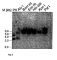
- the Southern blot in Figure 1 shows that the 501 bp ETAA16 cDNA probe isolated and labeled from the 3 'region by a double batch of Dra I / Dra III followed by gel purification, was digested with genomic DNA fragments as large as -. 6 kb hybridized after incubation with hexanucleotide-recognizing restriction enzymes ( Bam HI, Eco RI, Hin dIII, Kpn I, Pst I).
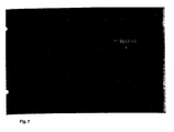
- the FISH analysis revealed one DNA sequence for the ETAA16 antigen Localization on chromosome 2p13-15 (FIG. 2).
- subclones of the ETAA16 cDNA obtained by the internal restriction sites of restriction enzymes from the MCS ( Sma I (3' ⁇ 3145), Eco RI (5' ⁇ 471), Hin dIII (5' ⁇ 1692, 3' ⁇ 1766), Spe I (5' ⁇ 1611)) were used.
- the cDNA sequence of ETAA16 according to SEQ. ID. N.1 serves as a template for the preparation of oligonucleotides used in the specific detection of ETAA16 mRNA. These oligonucleotides serve as primers or for the production of primers and oligonucleotides which are preferably used in the polymerase chain reaction, for example for the identification of the specific ETAA16 cDNA transcripts.
- the primers and oligonucleotides are prepared using the cDNA sequence and the ETAA16 antigen so that they are exon-spanning.
- the amino acid sequence according to FIG. 5 has a transmembrane region (see FIG. 3), so that this sequence region is encoded by different exons.
- the primers or oligonucleotides are accordingly selected from this sequence region which encode this amino acid sequence.
- the isolation of the genomic clone of ETAA16 from chromosome 2p13-15 allows the use of additional primers and oligonucleotides. These are used, inter alia, for the diagnostic polymerase chain reaction (PCR), which is carried out both with RNA from biopsies and from peripheral blood or bone marrow. In addition, it is preferable to use the in situ PCR, which bypasses RNA purification and is performed directly on tissue sections.
- PCR polymerase chain reaction
- the methodology of the PCR is carried out according to standard protocols, whereby only the choice of primers or oligonucleotides results from the ETAA16 cDNA sequence. For more details on the methodology, see also McPherson et al., PCR: A practical approach, Oxford University Press (1991 ).
- the genomic clone of the ETAA16 gene is isolated using the ETAA16 cDNA by hybridizing a human genomic library constructed in cosmid vectors with portions of the ETAA16 cDNA.
- fragments of the ETAA16 cDNA are radioactively labeled according to the standard method of Feinberg and Vogelstein. See also Feinberg and Vogelstein, Analytical Biochemistry 178: 6-13 (1983 ). These fragments are hybridized with the genomic library transferred to nitrocellulose membranes.
- the positive clones are isolated, purified and analyzed according to standard protocols. Details on this Sambrook et al. A laboratory manual. Chapter 9 2nd edition (1989 ).
- the cloned genomic clone is sequenced and its exon structure analyzed using the ETAA16 cDNA. This serves for the exon-spanning positioning of the primers and oligonucleotides, in particular for the PCR.
- ETAA16 cDNA which encodes the complete cellular protein ETAA16, defines the amino acid sequence according to the foundations of the genetic code. It is also shown in the sequence listing. From the amino acid sequence peptides can be prepared, for example, with the aid of a commercially available Peptidsynthetisier learners. See also Rudensky, Nature 353: 622-627 , The peptides can be produced as overlapping sequences and used to produce monoclonal antibodies. They can be chemically modified by tripalmitoyl-S-glycerylcysteine. This method enhances its immunogenicity in vivo.
- the polypeptide encoded by the ETAA16 cDNA consists of 841 amino acids with a calculated molecular weight of 94427.03 Da.
- the calculated isoelectric point is pl 6.86.
- the polypeptide is rich in serine (11.89%), lysine (9.51%), threonine (8.44%), asparagine (8.32%), leucine (7.13%), glutamic acid (6.77%) %) and isoleucine (6.42%).
- the translated sequence of ETAA16 cDNA was subjected to a hydrophobicity blot with a window of 10 amino acids by the method of Kyte-Doolittle ( Figure 3). Accordingly, the ETAA16 antigen has a potentially membrane-bound region of amino acids 186 to 203. There are another 10 short hydrophobic areas with a value greater than +0.5. These are more likely to be internal domains of the ETAA16 antigen because these regions achieve lower hydrophobicity than the potentially membrane-bound region. It can be seen that many portions of the ETAA16 antigen have a value less than -0.5.
- the full-length clones 6.1.1 and 3.1.1 and the 3 'deletion clones in the colony immunoscreen and in the Western blot were investigated. For their length determination, these were incubated in duplicate with Hpa I / Sac I, since the 3 'deletion clones were prepared by opening the vector with Kpn I and thus all restriction recognition sequences of the MCS 3' of the ETAA16 cDNA ( Xho I Kpn I) were lost. Therefore, the subclones were excised with Hpa I. It follows that the actual size of the subclones extended by 427 bp because Hpa I cuts 3 'of the cDNA of the GFP in the SV40 polyadenylation site of the pBK-CMV.
- the two full-length clones 6.1.1 and 3.3.1, which has a 150 bp deletion in the 3 'region and a 54 bp deletion in the 5 'region were labeled by the antibody 16.
- the epitope is encoded by a fragment located between the 1396 and 1856 bp and encodes amino acids 332-485. Since the epitopes of proteins and polypeptides are often about 15 amino acids in length, this 115 amino acid peptide of the ETAA16 antigen is believed to contain regions that are not required for this epitope. They may form further epitopes.
- the ETAA16 antigen expressed in E. coli has a molecular weight of -90 kDa and thus deviates from the calculated size by 5 kDa. This phenomenon can be caused by SDS-PAGE, where molecular weight determinations of proteins often deviate from the calculated molecular weight due to secondary structures still present.
- the detected lower molecular weight proteins are due to degradation products of the ETAA16 antigen, which are particularly found in eukaryotic proteins with transmembrane domains, unless they are expressed as a fusion protein. This goes on executions of Mount, Ann Rev. Genetics 14: 279-319 (1980 ), back.
- the 3'-deletion clone ⁇ 820 is the first clone to have a slightly lower molecular weight than the full-length clones because it does not carry a 3 'untranslated region due to its 288 bp deletion of the translated region.
- the ⁇ 1256 deletion clone has a molecular weight of 55-63 kDa and the subclone with the deletion of ⁇ 1601 bp encodes a protein of about 43 kDa.
- Epitope mapping reveals that antibody 16 recognizes the clones in the prokaryotic system.
- Transient transfection experiments also show the surface expression of the antigen encoded by ETAA16 cDNA on transfected cells of the human cell line COS-7 cells (FIG. 5).
- the transfection efficiency of the COS-7 cells was detected with the phagemid pBK-GFP in manflußzytometrie, while the phagemid pBK-CMV was isolated from the VH-64 cDNA expression library on the basis of the functional ⁇ -galactosidase.
- the Green Fluorescent Protein (GFP) was isolated from the pEGFP-1 and cloned into the Bam HI / Xba I sites so that the GFP is under the control of the prokaryotic lacZ as well as the eukaryotic CMV promoter. Transfection licenses of almost 40% were achieved after 24 hours.
- the cell line VH-64 was used as a positive control and transfected with the pBK-CMV COS-7 cells as a negative control for antibody binding: Clones 3.3.1 and 9.2.2 were after 24 h on the surface with 14 , 49% and 14.31% of the cells, respectively. Clones 6.1.1 and 8.1.1 show similar results to the negative control.
- the expression of the antigen encoded by ETAA16 cDNA was examined intracellularly in the transfected COS-7 cells in the flow cytometer ( FIG. 6 ). For this purpose, the transfected COS-7 cells were fixed and also labeled with the antibody 16.
- the different clones of the ETAA16 cDNA were weakly expressed in the transfected COS-7 cells after 24 h, only the clone 9.2.2 did not express the antigen at this time. After 48 h, the expression of the ETAA16 cDNA clones appeared heterogeneous, since the negative control with 48.06% has a high cross-reactivity with the antibody 16. In contrast, the expression of the ETAA16 cDNA after 72 h of all 4 cones detectable, however, the clones were 8.1.1 and 9.2.2 with 71.14 and 72.87% more expressed than the clones 3.3.1 and 6.1.1 with 63.31 and 53.03% respectively.
- the antibody 16 additionally binds to fixed cells of the ET cell line VH-64. This binding is at least 70% and is thus lower than the surface binding of the antibody to the ET cell line VH-64. Thus, the fixed epitope can also be detected by the antibody.
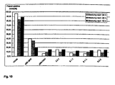
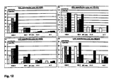
- NK-specific lysis was determined in the cytotoxicity assay with the nonadherent lymphocyte fraction (NAL) of different donors ( FIG. 12 ).
- the lymphokine activated cells (LAK) were generated by the same donors. With approximately 3.5% specific lysis, the cells of the RD-ES cell line represent a significantly poorer target for the NK cells than the K562 myeloid leukemia cell line with 13% specific lysis.
- Preincubation with antibody 16 resulted in inhibition of NK-specific Cytotoxicity against the ET cell line RD-ES, which remained at an effector / target ratio (E / T ratio) of 11: 1.
- NK-specific cytotoxicity decreased with lower effector cell counts.
- the influence of the antibody 16 on the total cytotoxicity which was determined by the crosslinking of the effector cell with the target cell by the lectin phytohemagglutinin (PHA), was different for the two cell lines used.
- NK-specific total lysis of cell line K562 was scarcely affected by antibody 16 except for the 100: 1 E / T ratio, where the total lysis had increased by 12.7%.
- the cytotoxicity of IL-2 activated NK cells was higher for the ET cell line RD-ES than for the K562 cell line.
- the cytotoxicity of the LAK cells to the cells of the cell line K562 changes with the addition of the antibody 16, PHA and their combination similar to the NK cell-specific cytotoxicity. With the exception of the effector / target ratio of 100: 1, antibody 16 also inhibited LAK-specific cytotoxicity against the ET cell line RD-ES.
- the Ewing tumor cells of the RD-ES cell line are lysed by the NK cells to a lesser extent when the antibody 16 has bound to the ETAA16 antigen.
- the antibody binding inhibits the cytotoxicity of these effector cells against this cell line.
- the ETAA16 antigen plays an essential role in the interaction between the Ewing tumor cells and the natural killer cells.
- the ETAA 16 antigen as a surface protein can interact on the one hand with the activating receptors of the NK cells, or as adhesion molecule enable the interaction between the effector cell and the target cell only in the first place. In both cases, a regulatory function of the ETAA16 antigen can be assumed in NK-specific tumor lysis.
- ET-cell lines Chromosomal translocations Type of fusion product * Classification of pathology A673 nd I PNET STA-ET-1 11/22 I PNET STA-ET-2.1 del (22) (q12) VII PNET STA-ET-2.2 del (22) (q12) VII PNET STA-ET-5 - - IT GG-62 del (22) (q12) - atypical ES MA-ES nd IT NT-68 ⁇ nd II PNET RD-ES del (22) (q12) II IT RM-82 del (22) (q12) - IT SK-ES-1 11/22 II IT VH-64 11/22 II IT * See for typing too Dockhom et al., Virchows Arch. 425: 375-382 (1994 )
- the monoclonal antibodies to the Ewing tumor used herein are of Shi et al., Cancer Immunol. Immunother. 38: 208-213 (1994 ): antibody clone 1 SN11 312.84.33 / 32 2 SN11 813.92.23 3 SN 11 944.73.47 4 SN51 739.72.90 5 SN54 1050.82.33 6 SN54 1059.107.38 7 SN55 159.50.12.15 8th SN55 291.71.44 9 SN55 303.45.16 10 SN55 671.89.55 / 35 11 SN55 697: 89: 23 12 SN55 786.14.74134 13 SN55 94.96.45 14 SN58 1580.149.8 15 SN58 162.28.11.37 16 SN58 1857.15.30
- the cell lines were cultured in 25 cm 2 collagen-coated culture flasks overnight. The cells were detached with 0.5 mM EDTA for 5-10 min at 37 ° C and then washed 2x with ice-cold FACS buffer. Intracellular antigens were made accessible by fixing the detached cells with 500 ⁇ l of BFA fixan with constant vortexing. Subsequently, the cells were washed again in FACS buffer and treated as the live cells. See also here Slaper-Cortenbach et al., Blood 72: 1639-1644 (1988 ).
- the cells were aliquoted at 2 ⁇ 10 5 per batch and the batches were incubated with 20 ⁇ l of the respective hybridoma supernatant of the first antibody (3-10 ⁇ g / ml) on ice in the dark for at least 30 minutes.
- the ET-labeling antibodies were as positive controls two antibodies W6 / 32HL and B9.12.1 used. See also Ziegler and Milstein Nature 279: 243 (1979 ) and Rebai and Malissen, Antigens 22: 107-117 (1983 ) used.
- the batches were washed 2 ⁇ with 2-3 ml of FACS buffer and centrifuged for 3 min at 4 ° C. at 225 g.
- the antibody-antigen complex was labeled with a dye-labeled ⁇ -mouse-2. Antibodies marked.
- fluorescein-conjugated ⁇ -mouse IgG F (ab ') 2 fragments were used and these preparations were also incubated for at least 30 min on ice in the dark.
- the cells were washed as above and then measured in the flow cytometer, dead cells were marked by the addition of 1 .mu.g per propidium iodide approach in order to exclude them in the evaluation can.
- the Cr 51 -release assay was used in which the target cells are labeled with Na 2 [ 51 Cr] O 4 .
- the target cells were incubated for 3-4h with 100 ⁇ g Ci 51 Cr at 37 ° C and 5% CO 2 and then washed 3x with PBS.
- the effector cells were isolated from peripheral blood of a healthy donor via a Ficoll gradient (1.077 g / cm 3 seromed), separating the mononuclear cells (PBMC) from the polymorphous cells and the erythrocytes.
- PBMC mononuclear cells
- the heparin blood was diluted with 1 vol PBS before coating on the Ficoll gradient.
- the gradient was centrifuged for 20 min at 839 g at room temperature.
- the fraction with the mononuclear cells was washed 2x with 10 vol PBS and pelleted at 225g for 10 min.
- the monocytes were removed by incubating the mononuclear cells in 3 ml culture medium / 25 cm 2 at 37 ° C and 5% CO 2 for 1 hour.
- LAK cells were generated from the isolated PBMC by culturing with 10 3 IU / ml IL-2 in culture medium for 72 h.
- the target cells were preincubated with this at 4 ° C for 15 min. Subsequently, 50 ⁇ l of the effector cells were added to the respective batch and incubated for 4 h or 16 h at 37 ° C. and 5% CO 2 .
- the K562 was used as a positive control. Total cytotoxic activity was determined by incubation with 0.3% PHA medium. The spontaneous lysis was determined only with the target cells and the maximum release of 51 Cr from the target cells was found to be 2.5% Triton-X100. After incubation, 25 ⁇ l were removed from the batches and applied to Spoton filters. The filters were dried, mixed with 10 ml of scintillation fluid and the released radioactivity in the ⁇ -counter (LKB) was determined.
- LLB ⁇ -counter
- lysis buffer 10 mM Tris / HCl, pH 8.0 100 mM NaCl 25 mM EDTA 0.5% SDS 0.1 mg / ml proteinase K.
- Adherent cell lines were detached for 5 min at 37 ° C. for isolation of genomic DNA by trypsin / EDTA and filled up with 10 volumes of PBS.
- the detached cells or cells from suspension cultures were centrifuged at 225g for 10 min at 4 ° C, 2x with cold PBS washed and the pelleted cells are flash frozen in liquid nitrogen or taken directly into lysis buffer.
- 1x10 8 cells were incubated for 12-18 h with 1 ml lysis buffer. Subsequently, the nucleic acids were extracted with 1 volume of TE buffered phenol, phenol / chloroform and chloroform / isoamyl alcohol 49: 1.
- RNA was degraded with 10 ⁇ g / ml RNase A for 3 h at 37 ° C and the DNA was purified by phenol / chloroform and chloroform / isoamyl alcohol 49: 1.
- the DNA was precipitated with 2 M ammonium acetate and 2 vol. Ethanol, precipitated at 5000 g for 5 min. At room temperature and washed with 70% ethanol. The pellet was dried, taken up in 1 ml TE buffer / 1x10 7 and stored at 4 ° C.
- RNA isolation was carried out according to the single-step method Chomzynski and Sacchi, Analytical Biochemistry 162: 156 (1987 ). denaturing: 4 M guanidinium isothiocyanate 25 mM Na citrate, pH 7.0 0.1 M ⁇ -mercaptoethanol 0.5% N-lauroylsarcosyl
- the cells were cultured overnight in fresh medium before RNA isolation.
- Adherently growing cells were detached with 0.5 mM EDTA for 5 min at 37 ° C, centrifuged at 225g and washed 2x in ice-cold PBS.
- Suspension cultures were also centrifuged at 225g for 10 min and washed 2x in ice-cold PBS.
- 1x10 7 cells were homogenized in 1 ml denaturation solution. The homogenate was mixed with 100 ⁇ l of 2 M Na acetate, pH 4.0, and mixed by inversion.
- the isolation of the mRNA was carried out with the following buffers: 2x binding buffer. 20 mM Tris-HCl, pH 7.5 1000 mM NaCl 2mM EDTA 1% SDS Wash buffer: OW2: 10 mM Tris-HCl 150 mM NaCl 1mM EDTA elution: 5mM Tris-HCl, pH 7.5 Heated to 70 ° C.
- RNA isolation of the mRNA was performed with Qiagen's Oligotex TM particles in a batch process.
- the protocol was modified to completely purify the mRNA as follows: The total RNA was mixed with an appropriate volume of 2x binding buffer and heated to 65 ° C. The batch was mixed according to the amount of RNA used with 10% Oligotex TM suspension according to the manufacturer (85 ul 10% Oligotex TM to 1 mg total RNA) and incubated for 5 min at 65 ° C with shaking. The mixture was then cooled slowly to room temperature and incubated for a further 10 min at room temperature.
- the Oligotex TM particles were centrifuged for 1 min at 15,000 rpm, washed 2x with 1 ml 1x binding buffer and 2x with 1 ml OW2 wash buffer. Subsequently, the RNA was eluted with 100 ⁇ l elution buffer for 3 min at 70 ° C. In most cases, the ribosomal RNA could not be completely removed, so that the mRNA was purified again on the same Oligotex TM particles by adjusting the volume of the eluted mRNA to 500 ⁇ l and adding a corresponding volume of 2x binding buffer has been. The RNA was denatured again at 65 ° C and then hybridized to the Oligotex TM particles for 10 min at room temperature. The particles were washed as above and then the mRNA was eluted with 2x25 ⁇ l elution buffer for 3 min at 70 ° C. The mRNA was then stored at -70 ° C.
- Plasmid DNA was prepared by either the alkaline-lysis method of Sambrook et al., A laboratory manual, 2nd. edition (1989 ) or with Qiaprep Spin Columns (Qiagen) purified according to manufacturer's instructions. Plasmids for sequencing were purified with Qiawell8 anion exchange columns according to the manufacturer's instructions (Qiagen). The purification of preparative plasmid DNA was carried out from 100-200 ml LB cultures with a corresponding antibiotic with Qiatip500 columns (Qiagen).
- the analytical restriction mixtures of the DNA were carried out with 0.5-1 ⁇ g of plasmid DNA and 2.5 U of the respective restriction enzymes with the 10 ⁇ buffer supplied by the manufacturer in a volume of 10-20 ⁇ l.
- the generated DNA fragments were analyzed in the Agarosegelelekrophorese (SeaKem ® LM, FMC).
- the 1 kb ladder (BRL Gibco) was used by default in agarose gel electrophoresis.
- the DNA was precipitated with 0.3 M Na acetate and 2 vol 100% ethanol for 20 min at -20 ° C and then precipitated by centrifugation for 15 min at 4 ° C. After the DNA pellet was washed in 70% ethanol and dried, it was resuspended in the appropriate volume of 10 mM Tris, pH 8.5.
- a cDNA expression library of the ET cell line VH-64 in ⁇ ZAP Express TM (Stratagene) was prepared according to the manufacturer's protocol.
- the mRNA was isolated from the ET cell line VH-64 according to the methods already explained above. 10 ⁇ g of mRNA were used in the cDNA synthesis, the following modifications were carried out deviating from the company protocol: The mRNA was added to the first strand synthesis with the following agents, after denaturing for 5 min at 70 ° C: 5.0 ⁇ l 10x first strand synthesis buffer 3.0 ⁇ l Nucleotide mixture with 5-methylcytosine 2.0 ⁇ l Linker primer (2.8 ⁇ g) 20.0 ⁇ l mRNA (0.5 ⁇ g / ⁇ l) 17.5 ⁇ l H 2 O DEPC 1.0 ⁇ l RNase inhibitor (40U) 48.5 ⁇ l approach
- the reaction was incubated for 10 min at room temperature for primer annealing , which is poly (dT) primer flanked at the 5 'end by the recognition sequence of the Xho I restriction enzyme. Subsequently, the batch was mixed with 1.5 ⁇ l of MMLV reverse transcriptase (75 U). The cDNA first strand synthesis was monitored by adding 5 ⁇ l of the Approach to 0.5 ul [ ⁇ - 32 P] dATP (800 Ci / mmol). Both batches were incubated for 1 h at 42 ° C.
- the double strand synthesis was carried out in the following approach for 21 ⁇ 2 h at 16 ° C: 45.0 ⁇ l cDNA first strand approach 20.0 ⁇ l 10x two-strand buffer 6.0 ⁇ l Second strand nucleotides 113.9 ⁇ l H 2 O DEPC 2.0 ⁇ l [ ⁇ 32 -P] dNTP 2.0 ⁇ l RNase H (3U) 11.1 ⁇ l DNA polymerase I (100 U) 200.0 ⁇ l approach
- the protruding ends of the cDNA after the second strand synthesis were filled with 5 U Pfu-DNA polymerase for 30 min at 72 ° C (23 ul blunting dNTP mix, 2.0 ul Pfu).
- First and second strand synthesis were monitored by denaturing agarose gel electrophoresis (300 mM NaOH, 150 mM NaCl, 1% agarose).
- the cDNA was phenol / chloroform extracted, precipitated with 0.3 M Na acetate and 2 vol. 100% ethanol overnight at -20 ° C and centrifuged for 1 h at 15000 rpm at 4 ° C. The precipitated cDNA was washed with 70% ethanol and then air-dried.
- the cDNA was taken up in 8 ⁇ l Eco RI (0.4 ⁇ g / ⁇ l) adapters.
- the cDNA / Eco RI adapter (8 ⁇ l) was admixed with 1 ⁇ l of 10 ⁇ ligase buffer and 1 ⁇ l of ligase and ligated for 2 days at 4 ° C. The ligation reaction was stopped by incubation at 70 ° C for 30 min. The ligation of the adapters was done in the PAGE / TBE gel in a control reaction batch, the cDNA was ligated with [ ⁇ - 32 P] ATP labeled Eco RI adapters. This ligation mixture was separated from the cDNA alone and the cDNA with labeled adapters without ligase in the PAA gel.
- the adapters were then phosphorylated in the following batch for 30 min at 37 ° C: 10.0 ⁇ l ⁇ cDNA with ligated Eco RI adapters 1.0 ⁇ l 10x ligase buffer 2.0 ⁇ l 10 mM rATP 6.0 ⁇ l H 2 O 1.0 ⁇ l T4 polynucleotide kinase (10U) 20.0 ⁇ l approach
- the kinase was inactivated at 70 ° C for 30 min and then the cDNA with ligated adapters with Xho I for 1.5 h at 37 ° C incubated. 20.0 ⁇ l cDNA with phosphorylated adapters 28.0 ⁇ l Xho I buffer 3.0 ⁇ l Xho I (120 U) 51.0 ⁇ l approach
- the cDNA was then fractionated according to its size via a C4-LB Sepharose column (Pharmacia). For this purpose, the cDNA was mixed with 5 ⁇ l of 10 ⁇ STE (100 mM NaCl, 10 mM Tris / HCl, pH 8.0, 1 mM EDTA, pH 8.0), 3 ⁇ l 2% bromophenol blue and 15 ⁇ l 80% glycerol. There were taken 40 fractions with a volume of ⁇ 100 ⁇ l. The incorporated radioactivity of the individual fractions was measured in the scintillator (Beckmann). The Length of the cDNA was controlled in 1% TBE agarose gel by separating 1/20 of the fractions.
- the fractions containing cDNA molecules larger than 500 bp were combined and precipitated with 1 volume of 100% ethanol.
- the packaging of the ⁇ Zap Express TM ligated cDNA was performed according to the instructions of Gigapack II Gold (Stratagene).
- the bacteria were pelleted at 2000 rpm for 10 min and taken up in 10 mM MgSO 4 solution.
- 600 ⁇ l of XL1-blue MRF ' were mixed with the appropriate volume of primary phage and incubated for 15 min at 37 ° C for the adhesion of the phage to the bacteria.
- NZY top agarose 48 ° C
- 6.5 ml of NZY top agarose 48 ° C
- the agar plates were left for 6-8 h incubated at 37 ° C so that the phage plaques did not become larger than 1-2 mm.
- the plates were overlaid with 10 ml SM buffer and stored at 4 ° C overnight. These were then incubated for a further 2 hours at room temperature with gentle shaking.
- the SM buffer was removed, the 150 mm agar plates were washed with 2 ml SM buffer, then 5% chloroform was added, incubated for 15 min at room temperature, and cell debris removed by centrifugation at 5000 g for 10 min.
- a colony of XL1-blue MRF ' was grown overnight in LB / 0.2% maltose / 10mM MgSO 4 medium and by XLOLR in LB medium at 30 ° C.
- the bacteria were centrifuged at 1000 g for 10 min and taken up in 10 mM MgSO 4 with the OD 600 of 1.0.
- the isolated ⁇ phage clones were taken up in 500 ⁇ l SM buffer with 20 ⁇ l chloroform and stored overnight at 4 ° C.
- phagemids In order to convert the phagemids into double-stranded plasmids, 10-100 ⁇ l of the phagemid supernatant were mixed with 200 ⁇ l of the XLOLR suspension and incubated for 15 min at 37 ° C. The batch was admixed with 300 ⁇ l LB medium and incubated for 45 min at 37 ° C. 200 ⁇ l of the batch was plated on LB agar plates containing 50 ⁇ g / ml kanamycin and incubated overnight at 37 ° C. The in vivo excision converts the ⁇ ZAP Express TM into the phagemid pBK-CMV.
- a single colony of the XL1-blue MRF ' was grown in LB / 0.2% maltose / 10 mM MgSO 4 medium to an optical density of OD 600 0.5.
- the bacteria were centrifuged at 2000 rpm for 10 min at 4 ° C. and taken up in the appropriate volume of 10 mM MgSO 4 .
- the primary screen of the expression library was performed at 5x10 5 pfu, the size of the primary expression library.
- 600 ⁇ l of the XL1-blue MRF 'suspension were mixed with 5 ⁇ 10 4 pfu per 150 mm 2 Petri dish (Greiner) and incubated at 37 ° C. for 15 min.
- the batches were spiked with 7 ml of NZY top agarose (48 ° C) and plated on NZY agar plates.
- the agar plates were incubated for 3.5 h at 42 ° C, then with nitrocellulose membranes (HATF, Millipore), previously in 10 mM IPTG was swirled and dried, covered and incubated for 4.5 h at 37 ° C.
- the labeled membranes were removed, washed in TBST for 30 min and stored at 4 ° C.
- a second set of membranes were removed from the agar plates by incubating at 37 ° C overnight and then also washing in TBST.
- the filters were incubated in 1 ml / cm 2 blocking buffer for at least 30 min at room temperature.
- the membranes of the double screen were mixed together with the antibodies in the blocking buffer, while the hybridoma supernatants of the ET specific monoclonal antibodies were combined in equal parts and added in a ratio of 1: 100 to the blocking buffer.
- the incubation with the antibodies was carried out overnight at 4 ° C. on a horizontal shaker (5 rpm).
- the membranes were washed individually for three times 10 min in TBST and incubated with the second antibody goat ⁇ -mouse AP conjugated (1: 1000, DAKO) in blocking buffer overnight at 4 ° C.
- the membranes were washed as above and equilibrated in AP buffer.
- the membranes were spiked with AP substrate buffer and developed for 3 h.
- the potentially positive clones were punched out and stored in 500 ⁇ l of SM buffer containing 20 ⁇ l of chloroform at 4 ° C. Subsequently, the phage clones were purified by rescreening and transferred by the in vivo excision in the plasmid form.
- lysis buffer 100 mM Tris / HCl, pH 7.8 150 mM NaCl 5 mM MgCl 2 1 mg / ml pancreatic DNase I 40 mg / ml lysozyme
- the transformed bacteria were streaked on LB agar plates, grown at 37 ° C overnight and then stored in inverted position at 4 ° C.
- the agar plate was then overlaid with nitrocellulose filter pivoted in 10 mM IPTG and incubated for 2-4 h at 37 ° C. After removing the filters, they were exposed to chlorine fumes for 15 minutes to kill the bacteria.
- the nitrocellulose membranes were then incubated for 12-16 h with -1 ml / cm 2 lysis buffer and then washed 2 ⁇ 30 min in TBST at room temperature.
- the antibody label was then performed as described above.
- the hybridoma supernatant of the 1st antibody was used 1:50 in blocking buffer. The detection also took place as already described.
- TSS 10% PEG 8000 5% DMSO 50mM MgSO4 in LB medium, pH 6.5
- E. coli The transformation of E. coli was carried out by the method of Chung et al., Proceeding of the National Academy of Science 86: 2172-2175 (1989 ).
- the respective bacterial strains were transferred from an overnight culture 1: 100 in LB medium without antibiotic and cultured until the early logarithmic growth phase (OD 600 0.3-0.4).
- the cells were harvested at 1000 g for 10 min at 4 ° C, taken up in 1/10 of the starting volume in TSS and snap frozen to 100 ⁇ l aliquots in the ethanol / dry ice bath. The cells were used directly in the transformation or stored at -70 ° C.
- the cells were thawed on ice, mixed with 0.1-1 ⁇ g of plasmid DNA and incubated on ice for 30 min. After a 1 minute heat shock of 42 ° C., the batch was admixed with 0.9 ml LB / 20 mM glucose and incubated for 1 h at 37 ° C. at 225 rpm. The transformed cells were harvested at 1000 g for 10 min and plated with 100 ul LB medium on LB agar plates with appropriate antibiotic.
- Eukaryotic cells were transiently transformed with FuGENE6 TM according to the protocol of Boehringer Mannheim.
- the COS-7 cells were seeded on the day before transfection with 2-3 ⁇ 10 5 cells per cm 2 in MEM ⁇ medium / 10% FCS in 6-well culture dishes.
- 4 ⁇ g FuGENE6 TM were taken up directly in 100 ⁇ l MEM ⁇ and incubated for 5 min at room temperature.
- Two micrograms of the respective plasmids purified from a maxi preparation on Qiagen columns were placed in 1.5 ml Eppendorf tubes and then the FuGENE6 TM / MEM ⁇ mixture was added dropwise to the DNA.
- the reaction was incubated for 15 min at room temperature and then added dropwise to the cells supplemented with fresh MEM ⁇ complete medium.
- the cells were incubated for 24 h, 48 h and 72 h without medium change and then detached with 0.5 mM EDTA for 5-10 min at 37 ° C.
- flow cytometric analysis the cells were treated as described above.
- the ⁇ ZAP-Express TM with intact ⁇ -galactosidase gene was isolated from the VH-64 cDNA expression library , converted into the phagemid form and then sequenced the multicloning site .
- the reporter gene used was the humanized GFP from the vector pEGFP-1 (Clontech).
- the pEGFP-1 was isolated from the dam negative E. coli strain GM-84 and GFP with the restriction enzymes Bam HI and Xba I excised. After gel purification, this was then directionally cloned into the pBK-CMV.
- the deletion clones were made with Stratagene's Exo III / Mung Bean Nuclease Kit.
- the clones to be deleted were treated with a 5 'or "blunt end" cutting and a 3'-overhang-producing restriction enzyme which protects the vector backbone from exonuclease III.
- the pBK-CMV backbone was protected for the 5 'deletion clones by incubation with Bss HII and subsequent filling of the overhanging ends with ⁇ -thio phosphate dNTPs.
- the pBK-CMV backbone was protected by opening the phagemid clones with the 3'-overhang-producing restriction enzymes Kpn I and Bst XI, respectively.
- the cDNA was made available for exonuclease III by incubating with Bam HI for the 5 'deletions and Xba I for the 3' deletions. After phenol / chloroform and chloroform / isoamyl alcohol 49: 1 extraction, 5 ⁇ g each of opened phagemid per minute exonuclease III incubation were used.
- the exonuclease III was used with 20 U / pmol 5'-end and the batch incubated at 30 ° C. The removed aliquots were placed in stop buffer (20 ⁇ l 10x Mung bean buffer to 155 ul H 2 O) and deactivated the exonuclease III at 68 ° C for 15 min. The single-stranded 5 'overhangs were removed with 15 U mung bean nuclease at 30 ° C for 30 min. The mixtures were extracted with phenol / chloroform and precipitated with 0.3 M Na acetate and 2 vol ethanol. The pellet was washed with 70% ethanol, dried and taken up in 15 ⁇ l of TE buffer.
- exonuclease III and mung bean nuclease treated clones were religated by T4 DNA ligase according to the manufacturer's instructions and transformed into XL1-blue MRF '.
- the clones were transferred to master plates and after 8 h incubation at 37 ° C with the minipreparation method of Good and Nazar, Biotechniques 22: 404-406 (1997 ) searches for clones of the correct size.
- sequencing was performed with the automatic laser fluorescence sequencer ALF express (Pharmacia). Sequencing of the plasmid DNA was performed by the chain termination method of Sanger et al., Proceedings of the National Academy of Science 80: 3963-3965 (1977 ) either with T7 DNA polymerase or with cyclosequencing with Thermosequenase TM (Amersham).
- Cyclosequencing was carried out with 1 ⁇ g of plasmid DNA according to the manufacturer's instructions with primer (1 pmol) and the thermosequenase / ddNTP reagents.
- the cycle conditions were changed to a 3-step cycle, started with an initial denaturation of 5 min at 95 ° C and then 30 cycles with the denaturation for 30 sec at 95 ° C, the annealing for 15 sec at the corresponding primer melting temperature ( Tm) and an elongation of 30 sec at 72 ° C. Cyclosequencing was completed with a 5 min elongation phase.
- the reaction was treated with 1/3 vol stop solution (5 mg / ml dextrin in deionized formamide) and then denatured at 85 ° C for 3 min.
- sequence gel ran with 0.5x TBE running buffer at a constant 25 watts and 51 ° C for 900 min.
- the sequences were processed with the ALFwin 1.1 software and analyzed using the evaluation program DNASIS (Hitachi).
- the composite sequences were identified by the NCBI's BLAST alignment programs (Web page: http://www.ncbi.nlm.nih.gov/) and FastA by the EBI (Web site: http://www.ebi.ac.uk_ebi_home.html ) was analyzed for homologous sequences.
- Radioactive labeling of in vitro transcripts was performed with the T7 / T3 transcription kit (Stratagene).
- the probes were cleaned by precipitation with 0.3 M Na acetate, pH 5.2, and 2 volumes of ice-cold EtOH, washed with 2 volumes of 70% EtOH, dried and taken up in appropriate volumes of H 2 O or TE buffer, or in centrifuge columns Microcon-100 (Millipore) cleaned.
- the non-radioactive label was made with the Amersham Direct Labeling Kit.
- genomic DNA was incubated with various restriction enzymes according to the manufacturer's instructions and 3-5 ⁇ g of the cut DNA were separated in a 0.7% TBE agarose gel. Subsequently, the gel was incubated for 15-30 min in 10 gel volumes of 0.25 M HCl. The genomic DNA was then denatured 2x10 min with 10 vol denaturation solution and treated 2x10 min with neutralization solution. The DNA was transferred by capillary transfer to 20xSSC on Hybond N + (Nycomed-Amersham). Subsequently, the blot was fixed at 120 ° C for 30 min.
- the size standard was separated from the blot, stained with 0.3% methylene blue in 0.3 M Na acetate solution for 2 min, and then destained with water for 5 min at room temperature.
- the fixed Southem blot was prehybridized in hybridization buffer (Amersham) for 15 min at 55 ° C. Subsequently, the labeled probe was added to the hybridization buffer and hybridized overnight at 55 ° C in the hybridization oven (Biometra). After hybridization, the blot was washed 2x15 min at 60 ° C in 1st wash buffer and 2x5 min in 2nd wash buffer. The blots were spiked with CPD Star and incubated for 5 min at room temperature. The excess CPD Star was removed and the blot pricked. The hybridized probe was detected on the blot by exposure of a Hyper-ECL film (Amersham) for several minutes to several hours.
- the acrylamide concentration for the separating gel was prepared according to the desired gel concentration, TEMED and APS were added at a final concentration of 0.02% and thus charged the prepared gel combs.
- the sedimented cell lines (1.25 ⁇ 10 6 cells) or transformed E. coli cells (25 ⁇ l of an overnight culture) were taken up in 1 ⁇ sample buffer, these samples were denatured for 5 min at 95 ° C. and then applied to the gel.
- the proteins were blotted from the gel to the membrane at a voltage of 20 V and 300 mA for 2 h.
- the membrane was incubated for 3 h at room temperature in blocking buffer and then incubated with fresh blocking buffer, which were added to the hybridoma supernatant of an ET specific antibody in the ratio of 1: 250, overnight at 4 ° C. Subsequently, the membrane was washed twice with TBST (2.2.4.5) for 15 min. And with the 2nd antibody rabbit ⁇ -mouse Ig (H + L) AP-conjugated (DAKO) 1: 1500 in blocking buffer for 2 h at room temperature. The membrane was washed as previously described and developed with AP substrate buffer.
Landscapes
- Health & Medical Sciences (AREA)
- Chemical & Material Sciences (AREA)
- Life Sciences & Earth Sciences (AREA)
- Organic Chemistry (AREA)
- Proteomics, Peptides & Aminoacids (AREA)
- Immunology (AREA)
- General Health & Medical Sciences (AREA)
- Genetics & Genomics (AREA)
- Medicinal Chemistry (AREA)
- Zoology (AREA)
- Molecular Biology (AREA)
- Biochemistry (AREA)
- Biophysics (AREA)
- Analytical Chemistry (AREA)
- Pathology (AREA)
- Engineering & Computer Science (AREA)
- Wood Science & Technology (AREA)
- Animal Behavior & Ethology (AREA)
- Nuclear Medicine, Radiotherapy & Molecular Imaging (AREA)
- Veterinary Medicine (AREA)
- Toxicology (AREA)
- Public Health (AREA)
- Gastroenterology & Hepatology (AREA)
- Hospice & Palliative Care (AREA)
- Oncology (AREA)
- Pharmacology & Pharmacy (AREA)
- Cell Biology (AREA)
- Physics & Mathematics (AREA)
- General Chemical & Material Sciences (AREA)
- Biotechnology (AREA)
- Microbiology (AREA)
- Chemical Kinetics & Catalysis (AREA)
- Bioinformatics & Cheminformatics (AREA)
- General Engineering & Computer Science (AREA)
- Peptides Or Proteins (AREA)
- Preparation Of Compounds By Using Micro-Organisms (AREA)
- Micro-Organisms Or Cultivation Processes Thereof (AREA)
Description
- Die Erfindung betrifft eine Nukleinsäure, die für ein Polypeptid kodiert, welches von Ewing Tumor-Zellen exprimiert wird und als charakteristisches Antigen des Tumors von Antikörpern oder zytolytischen Zellen erkannt werden.
- Die Ewing Tumore (ET) umfassen das rundzellige, maligne Ewing Sarkom der Plattenknochen, die Gruppe der primitiven neuroektodermalen Tumore (PNET) und die Askin's Tumore, und treten vor allem häufig bei Kindern im Alter von 5-15 Jahren auf. Sie gelten nach dem Osteosarkom als die häufigsten pädiatrischen malignen Knochentumore. Dabei zeigen speziell das Ewing Sarkom und die PNET aggressives klinisches Verhalten und bilden sehr schnell Metastasen.
- Die überwiegende Mehrzahl der Ewing Tumore zeichnen sich durch eine chromosomale Translokation des langen Arms des Chromosoms 11 aus, das mit einem Abschnitt des langen Arms des Chromosomen 22 fusioniert ist t(11;22)(q24;q12). Mit dieser Translokation fusionieren die Gene fli-1 des Chromosomen 11 und ews auf Chromosomen 22. In Abhängigkeit von der Lage der Chromosomenbruchstelle können verschiedene chimäre Fusionstranskripte EWS/FLI-1 entstehen, die jeweils für die definierten Tumore charakteristisch sind. Sie scheinen für die Tumorenstehung und - progression entscheidende Bedeutung zu haben. Für detaillierte Ausführungen dazu siehe auch Aurias et al., N. Engl., J.Med. 309: 496 (1991) und Delattre et al., Nature 359: 162-165 (1992). Das Fusionsprodukt beinhaltet die transkriptionsregulierenden Elemente der beiden Ausgangsprodukte, unterscheidet sich hingegen in seinen Eigenschaften von den nativen Ausgangsproteinen. Somit zeigen die ET-Zellen Expressionsmuster, die von denen der gesunden Zellen stark abweichen.
- Die ET-Patienten werden herkömmlich mit einer Kombination aus Chemotherapie und Bestrahlung behandelt. Die Prognose für Patienten mit lokalisierten kleinen Tumoren der Knochen ist mit dieser Therapie gut, da nach fünf Jahren 66% der Patienten überleben. Mit einer Überlebenschance von nur 10% ist die Prognose für Patienten mit Metastasen weitaus schlechter. Dies geht auf Untersuchungen von Jürgens et al., Cancer 61: 23-32 (1988) zurück. Ein Problem der Tumortherapie speziell beim Ewing Sarcom ist das Auftreten einer sog. Multidrug Resistenz, da ein membranständiges Transportprotein MDR-1 verschiedene applizierte Chemotherapeutika energieabhängig aus der Zelle herausschleust.
- Protoonkogene, Tumorsupressorgene, Adhäsionsmoleküle und Rezeptoren für Wachstumsfaktoren stellen potentielle Antigene für eine Immuntherapie der Tumoren dar. Bei Zellen der Ewing Sarkome und der PNET ist das oberflächenexprimierte Antigen MIC als Zelladhäsionsmolekül bekannt. Beide Tumoren unterscheiden sich durch die Expression zweier neuronaler Differenzierungsantigene (NSE/s-100). Einer überwiegenden Mehrzahl der ET-Zellen ist durch Transkriptionsprodukte des Fusionsgenes ews/fli-1 gekennzeichnet.
- Die bisherigen Versuche der Induzierung einer Imunantwort zur Behandlung der Ewing Tumoren waren wenig erfolgreich: Bertazzoli et al., Tumori. 83: 847-855 (1997) haben Trankriptionsprodukte von ews/fli-1 identifiziert, die in MHC Klasse I Moleküle passen, eine CD8+ spezifische T-Zellantwort konnte jedoch nicht ausgelöst werden. Shamamian et al., Cancer Immunol. Immunther. 39: 73-83 (1994) konnten erst nach einer Stimulation der MHC-Klasse I Expression eine zytotoxische T-Zellreaktion hervorrufen. Die Expression der MHC-Moleküle Klasse I ist in ET-Zellinien folglich für die Induktion einer T-Zell-Antwort zu gering.
- Auch das MIC Antigen erscheint für die Immuntherapie eher ungeeignet, obwohl es speziell für die Zelladhäsion verantwortlich ist und vermutlich das Wachstum der Zellen reguliert. Es wird allerdings auch auf T-Zellen exprimiert, so daß MIC Antikörper sowohl T-Zellen als auch Tumorzellen erkennen. Für eine sinnvolle und effiziente Immuntherapie ist eine hohe Spezifität der Antigene und damit auch der Antikörperbindung aber unbedingte Voraussetzung.
- Durch die Immunisierung von Balb/c Mäusen mit einem Tumorisolat gemischt mit der ET-Zellinie VH-64 und anschließender Fusion einer Milzzelle und einer NS-1 Myelomzelle gelang die Generierung monoklonaler Antikörper gegen diese Zellinie des Ewing Tumors. Weitere Analysen zeigten, daß diese Antikörper Antigene der ET-Zellen erkannten. Ein Teil der Antikörper regierte auch mit fötalen Leberzellen und entdifferenzierten Makrophagen. Nähere Einzelheiten sind dazu bei Shi et al., Cancer Immunol. Immunother. 38: 208-213 (1994) nachzulesen.
- Die meisten der bisher beschriebenen tumorspezifischen Antigene (TSA) oder tumorassoziierten Antigenen (TAA), die immunologisch relevant sind, sind in Karzinomen entdeckt worden. Spezifische Antigene von Sarkomen sind bislang nur für das Melanom identifiziert. Daher ist die Suche nach TSA bzw. TAA, unabhängig davon, ob es sich um Antikörper spezifische oder T-Zell identifizierte handelt, vor allem für Sarkome, zu denen auch die Familie der Ewing Tumore gehört, essentiell für eine aussagefähige Diagnostik und erfolgreiche Immuntherapie.
- Der Erfindung liegt daher die Aufgabe zugrunde, ein spezifisches Polypeptid des Ewing Tumors mit antigenischem Charakter und die diese codierende Nukleinsäure zu identifizieren.
- Die Lösung der Aufgabe besteht in der Isolierung der ETAA16 cDNA gemäß SEQ.IDN aus einer cDNA Expressionsbibliothek der VH-64 Zellinie der Ewing Tumoren und der anschließenden Charakterisierung dessen Transkriptionsprodukts.
- Ein Weg zur verbesserten Behandlung von Ewing Tumoren liegt in der Immuntherapie. Sowohl für die Antikörper vermittelte als auch für eine T-Zell spezifische Immunantwort sind tumorspezifische Antigene (TSA) oder tumorassoziierte Antigene (TAA) unabdingbar, die entweder de novo oder aber mit einer zum normalen Gewebe abweichenden Dichte exprimiert werden, und so die Tumorzellen von gesunden Zellen unterscheiden. Diese Antigene müssen entweder auf der Zelloberfläche exprimiert, oder aber in der Tumorzelle oder antigenpräsentierenden Zelle (APC) prozessiert und nachfolgend durch MHC Klasse I bzw. II Moleküle an der Zelloberfläche präsentiert werden.
- Mit Hilfe der erfindungsgemäßen ETAA16 cDNA und dem davon codierten Antigen wird eine Immuntherapie der Ewing Tumore überhaupt erst ermöglicht. Dabei ist auch eine Verwendung der Antigene zur Weiterentwicklung einer aktiven Immunisierung denkbar. Bei prädisponierten Patienten kann diese Behandlung als Präventivmaßnahme bereits vor der eigentlichen Belastung mit dem Tumor stattfinden. Auch ist es möglich, die Regression bereits etablierter Tumoren durch die Applikation des ETAA16-Antigens und damit durch eine Expansion antigenspezifischer T-Zellen zu induzieren. Diese inflammatorische Antwort des Immunsystems kann helfen, die inhibitorischen Effekte des Tumors zu überwinden.
- Eine weitere Einsatzmöglichkeit der erfindungsgemäßen Antigene besteht darin, durch ihre Applikation eine Stimulation der Immunantwort gegen die Ewing Tumore zu erzeugen oder zu stärken. Diese ist erreichbar, wenn das Antigen als biologischer Immunmodulator wirksam ist.
- Die Verwendungsmöglichkeiten des ETAA16-Antigens erschöpfen sich nicht in der direkten Applikation desselben, sondern erstrecken sich vor allem auf den besonders wesentlichen Bereich der Generierung antigenspezifischer, monoklonaler Antikörper. Damit eröffnet sich das gesamte Gebiet der passiven Immuntherapie. Dies ist um so wichtiger, als daß bislang keine TSA oder TAA für die Ewing Tumore bekannt sind, und so der Einsatz spezifischer monoklonaler Antikörper für eine effiziente Immuntherapie unmöglich war. Dabei sind transplantationsmedizinische Methoden wie z.B. die Säuberung von autologen Stammzellpräperaten und Knochenmark möglich. Eine Steigerung des therapeutischen Effekts, insbesondere der Komplement-vermittelten und spezifischen Zytotoxizität, wird durch eine Kopplung der Antikörper mit Wirkstoffen, Wirkstoffvorstufen, (Exo-)Toxinen oder anderen therapeutischen Substanzen erreicht. Auch können, vor allem zur Verbesserung der Penetration erkrankter Gewebe, nur Fragmente des monoklonalen Antikörpers appliziert oder auf ihrer Grundlage Einzeldomänenantikörper gentechnisch hergestellt werden.
- Der gegen das ETAA16 Antigen gerichtete Antikörper kann neben der Immuntherapie auch in der Immundiagnostik eingesetzt werden. Er kann sowohl der Bestimmung der Antigenmengen und damit der Belastung mit dem Tumor, als auch dessen Lokalisierung und Identifizierung dienen. Hier sind in vivo Verfahren wie beispielsweise die Immunszintigraphie mit radioaktivmarkierten Antikörpern oder in vitro Verfahren, wie beispielsweise die indirekte Immunoperoxidasetechnik wichtig. Daneben findet dieser Antikörper auch einen Einsatz in den herkömmlichen immunologischen Arbeitsmethoden wie beispielsweise bei einem ELISA, der Nephelometrie, der Immunpräzipitation oder im Westerb Blot.
- Die ETAA16 cDNA Sequenz dient ferner als Matritze zur Generierung spezifischer Oligonukleotide, die in der RT-PCR und in der in-situ PCR zur sensitiveren Diagnostik der Ewing Tumoren eingesetzt werden. Beide Verfahren erhöhen die Sensitivität in der Diagnostik gegenüber der reinen Nutzung der bisher bekannten Antikörper. Indem das ETAA16 Antigen durch die PCR nachgewiesen wird, wird eine Unterscheidung zwischen Ewing Tumoren und anderen Tumoren und gesunden Geweben erzielt. Diese genaue Differenzierung ist essentiell für die sich anschließende Therapie, so daß sich die Heilungschancen des Patienten erheblich verbessern.
- Die vorliegende Erfindung wird durch die nachfolgenden Figuren näher erläutert:
- Fig. 1.:
- Southern Blot einer humanen genomischen DNA mit ETAA 16. auf einem 0,7 % Agarosegel, nachdem 5 µg genomischer VH-64 DNA mit einem der Restriktionsenzyme Alu I, Bam HI, Eco RI, Hind III, KPN I oder Pst I behandelt, das Agarosegel geblottet und mit einer 501 bp Dra I/ Dra III Sonde aus dem Bereich des 3'- Endes von ETAA16 cDNA hybridisiert wurde.
- Fig. 2.:
- FISH-Analyse der Subklone der ETAA16 cDNA, die durch interne Schnittstellen folgender Restriktionsenzyme aus der Multi Cloning Site (MCS) hergestellt wurden: Sma I (3'Δ3145), Eco RI (5'Δ471), Hind III (5'Δ1692, 3'Δ1766) und Spe I (5'Δ1611).
- Fig. 3.:
- Hydrophobozitäts Blot der computerabgeleiteten Proteinsequenz des ETAA16 Antigens nach der Analysemethode von Kyte und Doolittle: durchgezogene Linie symbolisiert die Basislinie, die gestrichelte Linie gibt den "cut off " für membran-assoziierte Proteinbereiche an und der Pfeil markiert eine potentiell membranständige Region des Proteins.
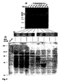
- Fig. 4.:
- Epitopkartierung des ETAA 16 Antigens: Die mit Hpa I/Sac I behandelten 3'-Deletionsklone von ETAA16 cDNA und die Vollängenklone 6.1.1 und 3.1.1 (3'Δ150 bp, 5'Δ54) wurden über ein 0,7 % Agarosegel getrennt (a) und im Western Blot eines 10 % SDS PAGES mit dem Antikörper 16 umgesetzt (c). Für einen Kolonie-Immunoscreen wurden die cDNA Klone in XL-1 blue E. coli transformiert, die entsprechenden Übernachtkulturen auf LB-Platten ausgestrichen, auf Nitrocellulosemembranen übertragen und mit einem Hybridomaüberstand des Antikörpers 16 inkubiert (b).
- Fig. 5.:
- Darstellung der Oberflächenexpression der ETAA16 cDNA Klone nach transienter Transfektion in den Zellen der humanen Zellinie COS-7 nach ihrer Inkubation mit dem Antikörper 16 und Detektion der Antikörper-Bindung im Durchflußzytometer.
- Fig. 6.:
- Darstellung der intrazellulären Expression der ETAA16 cDNA Klone nach transienter Transfektion in den Zellen der humanen zellinie COS-7-Zellen nach Fixierung der transfektierten Zellen, Inkubation mit dem Antikörper 16 und Detektion der Antikörper-Bindung im Durchflußzytometer.
- Fig. 7.:
- 51Cr-Freisetzungsassay von nicht adhärenten Lymphozyten (NAL) und Lymphokin aktivierten Killerzellen (LAK) gegen Zellen der myeloischen Leukämie Zellinie K562 und der ET-Zellinie RD-ES mit vier verschiedenen Effektor/Target-Konzentrationen (100:1; 33:1; 11:1; 4:1) in vier verschiedenen Ansätzen (kein Zusatz; Antikörper 16 aus einem Hybridome-Überstand; Phytohämagglutinin (PHA); Antikörper 16+PHA).
- Der Begriff "cDNA" wird hier verwendet als DNA-Kopie einer mRNA-Vorlage. Unter einem "cDNA-Klon" wird die cDNA summiert, die aus der Klonierung des identischen cDNA-Fragments entstanden ist. Ein "Deletionsklon" ist ein cDNA-Klon, der eine Nukleinsäuresequenz definierter Länge seiner ursprünglichen cDNA-Sequenz verloren hat. Dies kann aufgrund einer Behandlung mit einer Exonuklease geschehen. Dagegen ist ein "Vollängenklon" definiert als ein cDNA-Klon der ursprünglichen Länge ohne nachfolgenden Verlust eines DNA-Fragments. Als "homologe Sequenzen" werden diejenigen Sequenzen verstanden, die eine ähnliche Sequenz aufweisen.
- Als ein "Antigen" wird jedes Molekül verstanden, das eine spezifische Immunantwort auslösen bzw. mit Komponenten einer bereits ablaufenden Immunantwort reagieren kann. Es kann sowohl eine Antikörper vermittelte oder auch T-Zell spezifische Antwort induzieren. Ein "tumorspezifisches Antigen" (TSA) charakterisiert als neuartiges Antigen die Tumorzelle in Abgrenzung zu einer gesunden Gewebezelle. Dagegen bezeichnet ein "tumorassoziiertes Antigen" (TAA) ein Antigen, das überhaupt nicht oder aber in nur kleinen Mengen von einer gesunden Zelle exprimiert wird, bei Tumorzellen aber verstärkt vorkommt.
- Der Begriff der "Immundiagnose" ist hier verwendet als eine Bezeichnung für alle in vitro oder in vivo Verfahren und Techniken zum Nachweis, der Identifikation und der Lokalisation von Tumoren. Die "Immuntherapie" umfaßt als Sammelbegriff sowohl alle Formen der aktiven, passiven, spezifischen und unspezifischen kurativen Behandlungen von Tumoren als auch der Prävention und Etablierung ihrer Enstehung.
- Ein "Antikörper" wird hier definiert als ein Molekül, das als Reaktion auf den Kontakt mit einem Antigen gebildet oder unter Zuhilfenahme eines Antigens erzeugt wird, und an dieses Antigen spezifisch binden kann. Dabei ist das "Epitop" oder die "Antikörperbindungsstelle" eine Determinante des Antigens, an die der Antikörper oder Teile desselben binden können.
- Ein Oligonukleotid bezeichnet ein Molekül aus zwei oder mehreren Desoxyribonukleotiden oder Ribonukleotiden. Ein "Primer" wird hier definiert als ein natürliches oder synthetisches Oligonukleotid, das eine Nukleinsäuresynthese initiieren kann und vorzugsweise einzelsträngig ist. Es kann aber auch doppelsträngig sein.
- Zur Identifizierung und Isolierung der ETAA16 cDNA wurde eine cDNA Expressionsbibliothek der Ewing Tumor Zellinie VH-64 in dem Derivat des λ ZAP verwendet, welcher 5' vom lacZ Promoter zusätzlich den CMV Promoter für eine eukaryotische Expression trägt. Diese Konstruktion ist genauer bei Short et al., Nucleic. Acids. Res. 16: 7583 (1983) beschrieben. Die cDNA Expressionsbibliothek hatte einen Primärphagentiter von ∼5x10⁵ und einen Anteil von nicht-rekombinanten λ Phagen von ca. 2%. Die Integrität der cDNA Expressionsbibliothek wurde durch die Größenbestimmung der cDNA Insertionen von 26 zufällig ausgewählten Klonen analysiert.
- Die isolierten Phagen wurden durch die in vivo Excision in das Phagemid pBK-CMV überführt und die cDNA Insertionen durch die Inkubation mit Eco RI und XhoI ausgeschnitten. Die Größe der klonierten cDNA wurde in einem 1% TBE-Agarosegel bestimmt. Lediglich 7% der analysierten Klone trugen eine cDNA, die kleiner als 1 kb war. Die Primär-cDNA Bibliothek wurde mit 3-4x10⁴ pfu pro 150 mm2 NZY-Agarplatte amplifiziert. Die amplifizierte VH-64 cDNA Expressionsbibliothek besaß einen Titer von ∼1,5x10⁹ pfu/ml.
- Zur Isolierung der von den ET spezifischen Antikörpern definierten Antigenen wurde die cDNA Expressionsbibliothek der Zelllinie VH-64 mit dem Pool aus der Durchflußzytometrie von Hybridomaüberständen der ET spezifischen Antikörper aus Balb/c Mäusen durchsucht, dabei wurden 5x10⁵ Phagen (Titer der Primärbibliothek) ausplattiert. Die Gewinnung dieser Antikörper kann bei Shi et. al., Cancer Immunol. Immunother. 38: 208-213 (1994) nachgelesen werden.Nachdem Primärimmunoscreen wurden 27 Klone auf den doppelt-gezogenen Filtern identifiziert. Im nachfolgenden Rescreen bestätigten sich 9 Klone als tatsächlich positiv, von denen sich 5 Klone (2.1.1, 4.3.1, 5.1.1, 5.2.1, 10.2.1) durch ihre Detektion vom 2. Antikörper Ziege-α-Maus (H+L) konjugiert mit alkalischer Phosphatase als falsch-positiv herausstellten. Die verbleibenden 4 Klone 3.3.1, 6.1.1, 8.1.1, 9.2.2 konnten dem ET spezifischen Antikörper 16 (SN58/1857.15.30) zugeordnet werden. Dieser Antikörper wird für die weiteren Schritte der Isolierung der ETAA16cDNA und des ETAA16 Antigens verwendet und als Antikörper 16 bezeichnet.
- Die isolierten Klone wurden durch die in vivo Excision in die Phagemid-Form pBK-CMV des λ ZAP-Express™ überführt und die cDNA-Insertionen durch die Inkubation mit Eco RI und XhoI aus dem Vektor ausgeschnitten. Die Größe der klonierten cDNAs wurde in einem 0,8% TBE-Agarosegel bestimmt.
- Die Klone, deren Transkriptionsprodukte der Antikörper 16 erkannte, tragen eine ∼3,5 kb große cDNA mit einer 350 bp langen Eco RI Erkennungssequenz. Lediglich der Klon 3.3.1 zeigt ein cDNA Insertion von ca. 3,3 kb und weist in diesem Fragment eine Deletion von ca. 150 bp auf.
- Die drei 3,5 kb Klone 6.1.1, 8.1.1, 9.2.2 und der 3,3 kb Klon 3.3.1 werden als ETAA16 cDNA Klone, und die entsprechenden Transkripte als ETAA16 Antigen bezeichnet. Alle Klone wurden vom 5'- und 3'-Ende ansequenziert. Demnach sind die drei Klone 6.1.1, 8.1.1, 9.2.2 mit der 3,5 kb Insertion identisch. Der ETAA16 Klon 3.3.1 besitzt dagegen eine Deletion von 54 bp am 5'-Ende und eine Deletion von 160 bp am 3'-Ende.
- Zur Erstellung der vollständigen Sequenz wurden vom Klon 6.1.1 mit Hilfe der Exonuklease III sowohl 5'- als auch 3'-Deletionsklone erzeugt. Das pBK-CMV Rückgrat wurde bei den 5' Deletionsklonen durch Inkubation mit dem Restriktionsenzym Bss HII und anschließendem Auffüllen der überhängenden Ende mit α-thio Phosphat dNTPs geschützt, während die 3'-Deletionsklone durch das Öffnen des Phagemidklons mit den Restriktionsenzymen Kpn I bzw. Bst XI, die geschützten 3'-Überhänge produzierten.
- Zusätzlich wurden die intern auftretenden Restriktionsschnittstellen der Restriktionsenzyme aus der Multi Cloning Site (MCS) ebenfalls dazu verwendet, Deletionsklone zu subklonieren. Dazu wurde der Klon 6.1.1 mit dem jeweiligen Restriktionsenzym geöffnet, das pBK-CMV Rückgrat mit dem restlichen Teil des Klons 6.1.1 im Agarosegel gereinigt und religiert. Folgende Restriktionsenzyme wurden dazu verwendet: Sma I (3'Δ3145), Eco RI (5'Δ471), HindIII (5'Δ1692, 3'Δ1766), Spe I (5'Δ1611). Die HindIII Schnittstelle geht durch die Klonierung der cDNA in die Eco RI und XhoI Erkennungssequenzen verloren, deshalb wurden die HindIII Deletionsklone zusätzliche mit Bam HI für die 5'-Deletion und Xho I für die 3'-Deletion inkubiert und nach einer Auffüllreaktion religiert.
- Die vollständige Sequenz des ETAA16 Antigens beträgt einschließlich des Eco RI Adapters und der Xho I Erkennungssequenz 3457 bp (siehe Seq Id No:1). Es wurde mit der ALFwin.1.1 Software prozessiert und mit Hilfe des DNASIS Programms von Hitachi ausgewertet:. Demnach liegt das offene Leseraster (ORF) im ersten Leserahmen: Das Startcodon befindet sich an Position 403 und das Stopcodon an Position 2925. Das Startcodon liegt innerhalb eines guten Kontexts zur Kozak Sequenz, die die Initiation der Translation, ein A in Position -3 und ein G in Position+4 begünstigt. Neben diesem ORF treten in der 5`-UTR weitere kurze ORFs in allen 3 Leserastem auf: 1: 166-258; 2.: 221-292 und 3: 147-356. Dies besitzen aber alle vor dem eigentlichen Startcodon von ETAA16 ein Stopcodon. Diese Startcodons tragen nicht die Kozak Sequenz, so daß der Initiationsapparat der Translation diese übergehen kann.
- Im 3'-UTR Bereich befinden sich drei potentielle Polyadenylierungssignale in den Postionen 3057, 3296 und 3408 bp. Zusätzlich läßt sich ein AU-rich Element (ARE) an der Position 3053 bp vor der ersten potentiellen Polyadenylierungsstelle identifizieren. Der 402 bp umfassende 5'-UTR Bereich des ETAA16 besitzt einen G/C-Gehalt von 62,18 %, während die 3'-UTR Region lediglich einen G/C-Gehalt von 26,98% erreicht. Das offene Leseraster besitzt einen G/C-Gehalt von 34,4 % und liefert ein Protein von 841 AS mit einem potentiellen Molekulargewicht von 94427,03 Da.
- Der Homologievergleich der die cDNA-Sequenz mit bereits bekannten EST-Sequenzen wurde mit dem Basic Local Alignemt Search Tool (BLAST) vom National Center for Biotechnology Information (NCBI) und der FastA Analyse vom European Bioinformatics Institute (EBI) durchgeführt.
- Es wurden Homologien zu folgenden humanen EST-Sequenzen gefunden: Eine 197 bp lange embryonale EST Sequenz (Accession AA330059) aus einer von einem 12 Wochen alten Embryo erstellten cDNA Expressionsbibliothek ist zu 100% homolog für den 5'-Bereich der 594-790 bp ETAA16cDNA. Im 3'-Bereich wurde eine 100% homologe Sequenz von 78 bp gefunden (3241-3319 bp), bei der es sich um eine Gen-Signatur aus einer adulten humanen 3'-orientierten cDNA Expressionsbibliothek handelt (Accession C21290).
- Im 3'-Bereich treten drei homologe Sequenzen aus Expressionsbibliotheken fötaler Gewebe auf. Eine 665 bp umfassende EST Sequenz aus einer fötalen lungenspezifischen cDNA Expressionsbibliothek (Accession W16645) weist eine Homologie von 97,2% über 462 bp (2605-3074 bp) zur ETAA16 cDNA auf. Daneben gibt eine EST Sequenz aus fötalem Lungengewebe (571 bp, Accession W16643) mit einer Homologie von 96,6 % über 495 bp der ETAA16 cDNA. Eine dritte 324 bp lange unabhängige homologe fötale EST Sequenz aus einer Gehim cDNA Bibliothek (Accession D60196) ist in dem Bereich von 2793 bis 3119 bp zu 93,6% mit der ETAA16 cDNA homolog.
- Zusätzlich wurden fünf homologe Sequenzen aus differenzierten Geweben gefunden. Aus einer Plazenta cDNA Bibliothek wurden zwei Klone mit nahezu identischer Sequenz isoliert: Die 343 bp lange Plazenta EST Sequenz mit der Accesion R78670 ist zu 99,4% homolog zu dem ETAA16 cDNA Bereich von 2457 bis 2799 bp. Die zweite Plazenta EST Sequenz (Accession R32610) aus derselben cDNA Bibliothek weist eine Länge von 2822 bp und eine Homologie von 96,2% auf. Eine 232 bp lange EST Sequenz aus einer Aorta-spezifschen Expressionsbibliothek (Accession D62095) besitzt zu der ETAA16 cDNA in dem Bereich zwischen 2929 und 3160 eine Homologie von 91,4 %. Die dritte unabhängig homologe EST Sequenz wurde aus einer humanen cDNA Expressionsbibliothek von Tonsillar Zellen isoliert (Accession AA829856). Diese 318 bp lange EST Sequenz weist eine Homologie von 97,6% über 297 bp auf, die im potentiellen 5' untranslatierten Bereich von ETAA16 cDNA von 74 bis 370 bp liegt.
- Neben den homologen humanen EST-Sequenzen ergaben die FastA und BLAST Untersuchungen Homologien zu nicht humanen Sequenzen. Eine cDNA aus einer Expressionsbibliothek von Bos taurus zeigt die höchsten Homologien zu ETAA16 cDNA. Es handelt sich dabei um eine cDNA, die für ein zytoplasmatisches Protein aus den Junction Plaques von Sertoli Zellen und Hepatozyten (Accession Y08459) kodiert.
- Diese cDNA aus Bos taurus besitzt mehrere Blöcke von homologen Sequenzen. Der erste Block liegt im 5'-Bereich der ETAA16 cDNA und besitzt über 1323 bp, die in der ETAA16 cDNA dem Bereich von 45-1359 bp entsprechen, eine Homologie von 83,7. Es liegen drei weitere Bereiche mit längeren homologen Sequenzen vor: 85,2% über 345 bp (entsprechen bei der ETAA16 DNA dem Abschnitt von 1735-2079 bp), 83,2% über 339 bp (entsprechen bei der ETAA16 cDNA dem Abschnitt von 2192-3531 bp) und 84,5% über 290 bp (entsprechen bei der ETAA16 cDNA dem Abschnitt von 2611-2900 bp). Zusätzlich treten kürzere homologe Blöcke auf: Die ersten vier liegen im Bereich von 1376-1682 bp mit einer Länge von 19 bis 118 bp und Homologien zwischen 84 und 94%. Im 3'-Bereich von ETAA16 cDNA von 2916-3284 bp treten acht weitere Bereiche mit Homologien von 84% bis 94% auf, dabei sind die homologen Bereiche zwischen 17 und 43 bp lang.
- Neben dieser Bos taurus cDNA Sequenz wurden durch die FastA Homologievergleiche einige EST Sequenzen aus cDNA Expressionsbibliotheken aus Mausgeweben gefunden. Zwei homologe Sequenzen wurden aus cDNA Expressionsbibliotheken von CD4 positiven T-Zellen isoliert, dabei weist die erste T-Zell spezfische-EST Sequenz (Accession AA666753) eine Homologie von 75,3% über 392 bp (712-1098 bp) auf. Die zweite 421 bp lange T-Zell spezifische EST Sequenz (Accession AA666724) zeigt Homologien zum mittleren Teil der ETAA16 cDNA und zwar im Bereich von 1755 bis 2073 bp mit einer Homologie von 65,8% über 313 bp. In diesem Bereich wurden zudem eine homologe cDNA Sequenz aus einer Expressionsbibliothek aus der Haut einer 11 Wochen alten weiblichen Maus, isoliert (Accession AA756745). Die cDNA Sequenz zeigt eine Homologie von 69,5% über 357 bp (1703-2059 bp).
- Es wurden weitere homologe EST Sequenzen aus cDNA Expressionsbibliotheken verschiedener embryonaler Entwicklungsstadien gefunden: Eine EST Sequenz aus der Expressionsbibliothek eines 7,5 Tage alten Gastrulastadiums zeigt für den 5'-Bereich von 891-1354 bp eine Homologie von 75,9% über 463 bp. Die restlichen drei EST Sequenzen aus embryonalen Geweben unterschiedlichster Entwicklungsstadien sind homolog zum 3'-Bereich von ETAA16 cDNA. Die längste EST Sequenz (508 bp) wurde aus der Expressionsbibliothek eines 2-Zellstadium Mausembryos ermittelt und besitzt über 272 bp eine Homologie von 73,5% (Accession AA473938). In dem ETAA16 cDNA Abschnitt von 2953-3089 bp sind zwei weitere EST Sequenzen aus Expressionsbibliotheken von 7,5 und 5,5 alten embryonalen Gewebe zu 85,71% (82 bp) bzw. zu 82,86% (140 bp) homolog. (Accession AA139242 und AA396877).
- Aus den Homologievergleichen ist abzuleiten, daß keine vollständige humane homologe cDNA bisher beschrieben wurde. Durch Hybridisierungsversuche und eine FISH-Analyse (Fluorescence In Situ Hybridisation) wurde die Annahme bestätigt, daß es sich um ein humanes "single-copy" Gen handelt:
- Der Southern Blot in Figur 1 zeigt, daß die ETAA16 cDNA-Sonde von 501 bp, welche aus dem 3'-Bereich durch einen Doppelansatz Dra I/Dra III mit anschließender Gelaufreinigung isoliert und markiert wurde, mit genomischen DNA-Fragmenten einer Größe von -6 kb nach der Inkubation mit Hexanukleotid erkennenden Restriktionsenzymen (Bam HI, Eco RI, Hin dIII, Kpn I, Pst I) hybridisiert.
- Lediglich nach der Inkubation der genomischen DNA mit Alu I, welches eine Tetranukleotiderkennungssequenz besitzt, hybridisiert die Sonde mit zwei Fragmenten von 3,5 und 5,5 kb Größe, obwohl die Sonde keine interne Alu I Erkennungssequenzen besitzt. Dies zeigt, daß die Sonde von verschiedenen Exons kodiert wird und im Intron Erkennungssequenzen für Alu I existieren. Als Positivkontrolle wurde der Klon 6.1.1 nach der Doppelinkubation mit Bam HI und Xho I verwendet, wobei die Sonde mit der erwarteten kompletten cDNA von 3,5 kb hybridisiert. Die 4,5 und 5,0 kb Banden innerhalb der Positivkontrolle erklären sich durch eine Kreuzhybridisierung mit dem Rückgrad des Vektors (4,5 kb) bzw. durch die Hybridisierung mit unvollständig geschnittenem ETAA16 Klon 6.1.1.
- Die FISH-Analyse ergab für DNA Sequenz des ETAA16 Antigens eine Lokalisation auf dem Chromosom 2p13-15 (Figur 2). In die FISH Analyse wurden Subklone der ETAA16 cDNA, die durch die internen Restriktionsschnittstellen von Restriktionsenzymen aus der MCS (Sma I (3'Δ3145), Eco RI (5'Δ471), HindIII (5'Δ1692, 3'Δ1766), Spe I (5'Δ1611)) hergestellt worden sind, eingesetzt.
- Die cDNA Sequenz des ETAA16 gemäß SEQ. ID. N.1 dient als Matrize zur Herstellung von Oligonukleotiden, die in der spezifische Detektion der ETAA16 mRNA eingesetzt werden. Diese Oligonukleotide dienen als Primer oder zur Herstellung von Primem und Oligonukleotiden, die vorzugsweise in die Polymerasdekettenreaktion beispielsweise zur Identifikation der spezifischen ETAA16 cDNA Transkripte eingesetzt werden. Die Primer und Oligonukleotide werden anhand der cDNA Sequenz und des ETAA16 Antigens erstellt, damit diese exonübergreifend sind. Die Aminosäuresequenz nach Figur 5 besitzt einen transmembranen Bereich (siehe Figur 3), so daß dieser Sequenzbereich von verschiedenen Exons kodiert wird. Die Primer oder Oligonukleotide werden entsprechend aus diesem Sequenzbereich gewählt, die diese Aminosäuresequenz codieren.
- Die Isolierung des genomischen Klons von ETAA16 vom Chromosom 2p13-15 ermöglicht den Einsatz weiterer Primer und Oligonukleotide. Diese werden unter anderem für die diagnostische Polymerasekettenreaktion (PCR) eingesetzt, die sowohl mit RNA aus Biopsien als auch aus peripherem Blut oder Knochenmark durchgeführt wird. Zusätzlich wird vorzugsweise die in situ-PCR verwendet, die eine Reinigung der RNA umgeht und direkt auf Gewebeschnitten durchgeführt wird. Die Methodik der PCR wird nach Standard Protokollen durchgeführt, wobei sich lediglich die Wahl der Primer oder Oligonukleotide sich aus der ETAA16 cDNA Sequenz ergibt. Nähere Einzelheiten zu der Methodik siehe auch McPherson et al., PCR: A practical approach, Oxford University Press (1991).
- Der genomische Klon des ETAA16 Gens wird unter Verwendung der ETAA16 cDNA isoliert, indem eine humane genomische Bibliothek, die in Cosmid Vektoren erstellt wurde, mit Teilen der ETAA16 cDNA hybridisiert wird. Dazu werden Fragmente der ETAA16 cDNA nach der Standard Methode von Feinberg und Vogelstein radioaktiv markiert. Sieh dazu auch Feinberg und Vogelstein, Analytical Biochemistry 178: 6-13 (1983). Diese Fragmente werden mit der genomischen Bibliothek, die auf Nitrocellulosemembranen übertragen wurde, hybridisiert. Die positiven Klone werden nach Standardprotokollen isoliert, gereinigt und analysiert. Einzelheiten dazu bei Sambrook et al. A laboratory Manual. Chapter 9 2nd edition (1989). Der klonierte genomische Klon wird sequenziert, und seine Exonstruktur mit Hilfe der ETAA16 cDNA analysiert. Diese dient der exonübergreifenden Positionierung der Primer und Oligonukleotide insbesondere für die PCR.
- Mit der ETAA16 cDNA gemäß SEQ. ID. Nr.1, die für das komplette zelluläre Protein ETAA16 codiert, ist die Aminosäuresequenz nach den Grundlagen des genetischen Codes definiert. Sie ist ebenso im Sequenzprotokoll dargestellt. Aus der Aminosäuresequenz können Peptide z.B, mit Hilfe eines im Handel erhältlichen Peptidsynthetisiergerätes hergestellt werden. Siehe dazu auch Rudensky, Nature 353: 622 - 627. Die Peptide können als überlappende Sequenzen produziert und zur Herstellung monoklonaler Antikörper eingesetzt werden. Sie lassen sich chemisch durch Tripalmitoyl-S-glycerylcystein modifizieren. Dieses Verfahren steigert ihre Immunogenität in vivo.
- Die erfolgreiche Herstellung monoklonaler Antikörper gegen Peptide, die aus einer Aminosäuresequenz eines Proteins theoretisch abgeleitet und chemisch hergestellt wurden, ist in R. Spohn, B-, Th- und CTL-Epitope des HIV-1 Nef-Proteins, Dissertation der Fakultät für Chemie und Pharmazie, Universität Tübingen (1993) detailliert nachzulesen.
- Das von der ETAA16 cDNA codierte Polypeptid besteht aus 841 Aminosäuren mit einem errechneten Molekulargewicht von 94427,03 Da. Der berechnete isoelektrische Punkt liegt bei pl 6,86. Das Polypeptid ist reich an Serin (11,89%), Lysin (9,51%), Threonin (8,44%), Asparagin (8,32%), Leucin (7,13%), Glutaminsäure (6,77%) und Isoleucin (6,42%).
- Die translatierte Sequenz von ETAA16 cDNA wurde nach der Methode von Kyte-Doolittle einem Hydrophobizitäts Blot mit einem Fenster von 10 Aminosäuren unterzogen (Figur 3). Demnach weist das ETAA16 Antigen einen potentiell membranständigen Bereich der Aminosäuren 186 bis 203 auf. Es liegen weitere 10 kurze hydrophobe Bereiche mit einem Wert größer als +0,5 vor. Diese stellen eher interne Domänen des ETAA16 Antigens dar, da diese Regionen eine geringere Hydrophobizität erreichen als der potentiell membranständige Bereich. Es wird deutlich, daß viele Abschnitte des ETAA16 Antigens einen Wert kleiner als -0,5 besitzen.
- Zur Epitopkartierung des ETAA16 Antigens wurden die Vollängenklone 6.1.1 und 3.1.1 und die 3'-Deletionsklone im Kolonie-Immunoscreen und im Western Blot (Figur 4) untersucht. Zur ihrer Längenbestimmung wurden diese im Doppelansatz mit Hpa I/Sac I inkubiert, da die 3'-Deletionsklone durch Öffnen des Vektors mit Kpn I hergestellt und somit alle Restriktionserkennungssequenzen der MCS 3' der ETAA16 cDNA (Xho I-Kpn I) verloren wurden. Daher wurden die Subklone mit Hpa I ausgeschnitten. Daraus folgt, daß sich die eigentliche Größe der Subklone um 427 bp verlängerte, weil Hpa I 3' von der cDNA des GFP in der SV40 Polyadenylierungsstelle des pBK-CMV schneidet.
- Die beiden Vollängenklone 6.1.1 und 3.3.1, der eine 150 bp Deletion im 3'-Bereich und eine 54 bp Deletion im 5'-Bereich aufweist, wurden durch den Antkörper 16 markiert. Lediglich die 3'-Deletionsklone Δ2061 und Δ3151, die durch die Subklonierung des Sma I Fragmentes entstanden sind, kodieren nicht mehr für das Epitop des Antikörpers. Das Epitop wird von einem Fragment kodiert, daß zwischen dem 1396 und 1856 bp liegt und für die Aminosäuren 332-485 kodiert. Da die Epitope von Proteinen und Polypeptiden häufig eine Länge von etwa 15 Aminosäuren besitzen, sind in diesem 115 Aminosäuren umfassenden Peptid des ETAA16 Antigens vermutlich Bereiche vorhanden, die nicht für dieses Epitop benötigt werden. Möglicherweise bilden sie weitere Epitope.
- Der Western Blot der beiden Vollängenklone und der 3`-Deletionsklone bestätigt die Annahme, daß ab Position Δ2061 das Epitop deletiert wurde.
- Das in E.coli exprimierte ETAA16 Antigen besitzt ein Molekulargewicht von -90 kDa und weicht damit von der berechneten Größe um 5 kDa ab. Dies Phänomen kann durch die SDS-PAGE hervorgerufen werden, bei der Molekulargewichtsbestimmungen von Proteinen aufgrund von noch vorhandenen Sekundärstrukturen von dem berechneten Molekulargewicht häufig abweichen. Die detektierten Proteine mit niedrigerem Molekulargewicht sind auf Degradationprodukte des ETAA16 Antigens zurückzuführen, die besonders bei eukaryotischen Proteinen mit transmembran Domänen auftreten, sofern diese nicht als Fusionprotein exprimiert werden. Dies geht auf Ausführungen von Mount, Ann Rev. Genetics 14: 279-319 (1980), zurück. Der 3'-Deletionsklon Δ820 ist der erste Klon, der ein geringfühgig kleineres Molekulargewicht als die Vollängenklone aufweist, da dieser aufgrund seiner 288 bp Deletion des translatierten Bereichs keine 3'-untranslatierte Region trägt. Der Δ1256 Deletionsklon besitzt ein Molekulargewicht von 55-63 kDa und der Subklon mit der Deletion von Δ1601 bp kodiert für ein Protein von etwa 43 kDa.
- Die Epitopkartierung macht deutlich, daß der Antikörper 16 die Klone im prokaryotischen System erkennt. Transiente Transfektionsexperimente zeigen weiterhin die Oberflächenexpression des von ETAA16 cDNA codierten Antigens auf transfektierten Zellen der humanen Zellinie COS-7 Zellen (Figur 5).
- Die Transfektionseffizienz der COS-7 Zellen wurde mit dem Phagemid pBK-GFP in der Durchflußzytometrie detektiert, dabei wurde das Phagemid pBK-CMV aus der VH-64 cDNA Expressionsbibliothek anhand der funktionsfähigen β-Galactosidase isoliert. Das Green Fluoreszent Protein (GFP) wurde aus dem pEGFP-1 isoliert und in die Bam HI/Xba I Schnittstellen kloniert, so daß das GFP sowohl unter der Kontrolle des prokaryontischen lacZ als auch des eukaryotischen CMV Promoters steht. Es konnten Transfektionseffizenzen von fast 40% nach 24 h erreicht werden.
- In die Durchflußzytometrie wurde die Zellinie VH-64 als Positivkontrolle und die mit dem pBK-CMV transfektierten COS-7 Zellen als Negativkontrolle für die Antikörper-Bindung eingesetzt: Die Klone 3.3.1 und 9.2.2 waren nach 24 h auf der Oberfläche mit 14,49% bzw. 14,31% der Zellen nachweisbar. Die Klone 6.1.1 und 8.1.1 zeigen ähnliche Ergebnisse wie die Negativkontrolle.
- Die Expression des von ETAA16 cDNA codierten Antigens wurde intrazellulär in den transfizierten COS-7 Zellen im Durchflußzytometer untersucht (
Figur 6 ). Dazu wurde die transfektierten COS-7 Zellen fixierte und ebenfalls mit dem Antikörper 16 markiert. - Die verschiedenen Klone der ETAA16 cDNA wurden nach 24 h schwach in den transfizierten COS-7 Zellen exprimiert, lediglich der Klon 9.2.2 exprimierte das Antigen zu diesem Zeitpunkt nicht. Nach 48 h erschien die Expression der ETAA16 cDNA Klone heterogen, da die Negativkontrolle mit 48,06% eine hohe Kreuzreatkivität mit dem Antikörper 16 besitzt. Dagegen ist die Expression der ETAA16 cDNA nach 72 h von allen 4 Konen nachweisbar, allerdings wurden die Klone 8.1.1 und 9.2.2 mit 71,14 bzw. 72,87% stärker exprimiert als die Klone 3.3.1 und 6.1.1 mit 63,31 bzw. 53,03%.
- Der Antikörper 16 bindet zusätzlich an fixierte Zellen der ET Zellinie VH-64. Dies Bindung liegt mindestens bei 70% und ist somit geringer als die Oberflächenbindung des Antikörpers an der ET Zelllinie VH-64. Somit kann auch das fixierte Epitop vom Antikörper detektiert werden.
- Blockierungsexperimente mit verschiedenen ET Zellinien zeigen die Funktion des ETAA16 Antigens bei der durch Natürliche Killerzellen (NK) vermittelten Zellyse.
- Die NK-spezifische Lyse wurde im Zytotoxizitätsassay mit der nichtadhärenten Lymphozyten-Fraktion (NAL) verschiedener Spender ermittelt (
Figur 12 ). Die Lymphokin aktivierten Zellen (LAK) wurden von denselben Spendern generiert. Mit einer etwa 3,5% spezifischen Lyse stellen die Zellen der Zellinie RD-ES ein deutlich schlechteres Target für die NK-Zellen dar als die myeloische Leukämie-Zellinie K562 mit 13% spezifischer Lyse. Die Präinkubation mit dem Antikörpers 16 führte zur inhibition der NK-spezifischen Zytotoxozität gegen die ET Zellinie RD-ES, die bei einem Effektor/Target-Verhältniss (E/T-Ratio) von 11:1 ausblieb. Mit einer 23,5% spezifischen Lyse wurde bei einem E/T-Verhältnis von 100:1 die Zytotoxizität gegen die Zellinie K562 durch den Antikörper 16 sogar verstärkt. Die NK-spezifische Zytotoxizität nahm mit niegrigerer Effektorzellzahl ab. Der Einfluß des Antikörpers 16 auf die Gesamtzytotoxizität, welche durch die Vernetzung der Effektorzelle mit der Targetzelle durch das Lektin Phytohämagglutinin (PHA) ermittelt wurde, war für die beiden verwendeten Zellinien unterschiedlich. Die NK-spezfische Gesamtlyse der Zelllinie K562 wurde durch den Antikörper 16 mit Ausnahme des E/T-Verhältnisses 100:1, bei der die Gesamtlyse um 12,7% gesteigert worden ist, kaum beeinflußt. Im Vergleich der verschiedenen E/T-Verhältnisse erscheint die Gesamtlyse der ET Zellinie RD-ES eher heterogen; allerdings zeigte sich bei einem E/T-Verhältnis von 100:1, bei dem die stärkste Inhibition der Zytotoxizität durch den Ak16 hervorgerufen wurde, kein Effekt auf die Gesamtlyse durch die Präinkubation mit dem Antikörpers 16. - Die Zytotoxizität von IL-2 aktivierten NK-Zellen (LAK-Zellen) war für die ET Zellinie RD-ES höher als für die K562 Zellinie. Die Zytotoxizität der LAK-Zellen gegenüber den Zellen der Zellinie K562 verändert sich bei der Zugabe des Antikörpers 16, von PHA und deren Kombination ähnlich wie die NK-Zell-spezifische Zytotoxizität. Mit Ausnahme des Effektor/Target-Verhältnisses von 100:1 inhibierte der Antikörper 16 auch die LAKspezifische Zytotoxizität gegen die ET Zellinie RD-ES.
- Damit wird deutlich, daß die Ewing Tumorzellen der RD-ES Zellinie von den NK-Zellen zu einem geringeren Anteil lysiert werden, wenn der Antikörper 16 an das ETAA16 Antigen gebunden hat. Demnach inhibiert die Antikörper-Bindung die Zytotoxizität dieser Effektorzellen gegen diese Zellinie. Daraus folgt unmittelbar, daß das ETAA16 Antigen eine wesentliche Funktion in der Interaktion zwischen den Ewing Tumorzellen und den Natürlichen Killerzellen übernimmt. Dazu kann das ETAA 16 Antigen als Oberflächenprotein einerseits mit den aktivierenden Rezeptoren der NK-Zellen interagieren, oder als Adhäsionsmolekül die Interaktion zwischen der Effektorzelle und der Targetzelle erst ermöglichen. In beiden Fällen kann von einer regulativen Funktion des ETAA16 Antigens bei der NK-spezifischen Tumorlyse ausgegangen werden.
- Die Isolierung und Charakterisierung der erfindungsgemäßen ETAA16 cDNA und des ETAA16 Antigens erfolgte mit folgenden Materialien und Methoden:
- Es wurden folgende E. coli Stämme eingesetzt:
E.coli Stamm Genotyp DH5a F-, F80dlacZDM15, D(lacZYA-arg-F), U169, deoR, recA1, end A1, phoA, hsdR17(rk-, mk+), supE44, I-, thi-1, gyr A96, relA1 VCS257 Derivative of DP50 supF XL-1 blue recA1 endA1 gyrA96 thi-1 hsdR17 supE44 relA1 lac [F' proAB laclqZDM15 Tn10 (Tetr)] XL-1 blue D(mcrA)183 D(mcrCB-hsdSMR-mrr)173 endA1 supE44 thi-1 recA1 MRF' gyrA96 relA1 lac [F' proAB laclqZDM15 Tn10 (Tetr)] XLOLR D(mcrA)183 D(mcrCB-hsdSMR-mrr)173 endA1 thi-1 recA1 gyrA96 relA1 lac [F' proAB laclqZDM15 Tn10 (Tetr)] Su- Ir - Es fanden folgende humane ET Zellinien Verwendung:
ET-Zellinien Chromosomen translokationen Typ des Fusions-produktes* Klassifizierung der Pathologie A673 n. d. I PNET STA-ET-1 11/22 I PNET STA-ET-2.1 del(22)(q12) VII PNET STA-ET-2.2 del(22)(q12) VII PNET STA-ET-5 - - ES GG-62 del(22)(q12) - atypical ES MA-ES n. d. ES NT-68§ n. d. II PNET RD-ES del (22)(q12) II ES RM-82 del (22)(q12) - ES SK-ES-1 11/22 II ES VH-64 11/22 II ES * Siehe für die Typisierung auch Dockhom et al., Virchows Arch. 425: 375-382(1994) - Die weiteren humanen Zellinien wurden eingesetzt:
Zellinie Zellart COS-7 SV40 transformierte Affenzelllinie HeLa Epitheliales Karzinom K562 Myeloische Leukämie Monomac6 Makrophagen Zelllinie Polat S. Keimzelltumor T2 T-Zelllinie, TAP negativ - Die monoklonalen Antikörper gegen den Ewing Tumor, die hier verwendet wurden, sind von Shi et al., Cancer Immunol. Immunother. 38: 208-213 (1994) näher beschrieben:
Antikörper Klon 1 SN11 312.84.33/32 2 SN11 813.92.23 3 SN 11 944.73.47 4 SN51 739.72.90 5 SN54 1050.82.33 6 SN54 1059.107.38 7 SN55 159.50.12.15 8 SN55 291.71.44 9 SN55 303.45.16 10 SN55 671.89.55/35 11 SN55 697:89:23 12 SN55 786.14.74134 13 SN55 94.96.45 14 SN58 1580.149.8 15 SN58 162.28.11.37 16 SN58 1857.15.30 - Folgende Materialien wurden in der Durchflußzytometrie verwendet:
FACScan (Becton Dickinson) FACS-Puffer: PBS versetzt mit 0,1% (w/v) NaN3 (Merck) 0,1% (w/v) BSA (Behring) Propidiumjodid (Sigma, Deisenhofen) BFA-Fixans (pH 6,8): 0,2 mg/ml Na2HPO4·2xH2O 1,0 mg/ml KH2PO4 45% (v/v) Aceton 9,25% (v/v) Formaldehyd - Zur Analyse der Oberflächenexpression von membranständigen Zelloberflächenmolekülen wurden die Zellinien in 25 cm2 kollagenbeschichteten Kulturflaschen über Nacht kultiviert. Die Zellen wurden mit 0,5 mM EDTA für 5-10 min bei 37°C abgelöst und anschließend 2x mit eiskaltem FACS-Puffer gewaschen. Intrazelluläre Antigene wurden zugänglich gemacht, indem die abglösten Zellen mit 500 µl BFA-Fixans unter ständigem Vortexen fixiert wurden. Anschlließend wurden die Zellen erneut in FACS-Puffer gewaschen und wie die lebenden Zellen weiter behandelt. Hier siehe auch Slaper-Cortenbach et al., Blood 72: 1639-1644 (1988).
- Die Zellen wurden zu 2x10⁵ pro Ansatz aliquotiert und die Ansätze mit 20 µl des jeweiligen Hybridomaüberstandes des 1. Antikörpers (3-10 µg/ml) auf Eis im Dunkeln mindestens 30 min inkubiert. Neben den ET markierenden Antikörpern wurden als Positivkontrollen zwei Antikörper W6/32HL und B9.12.1 eingesetzt. Hierzu siehe auch Ziegler and Milstein Nature 279: 243 (1979) und Rebai and Malissen, Antigens 22: 107-117 (1983) eingesetzt. Die Ansätze wurden 2x mit 2-3 ml FACS-Puffer gewaschen und je für 3 min bei 4°C mit 225 g zentrifugiert. Der Antikörper-Antigen Komplex wurde mit einem farbstoffmarkierten α-Maus-2. Antikörper markiert. In diesem Fall wurden Fluoreszein-konjugierte-α-Maus-IgG F(ab')2 Fragmente (Cappel, West Chester, USA) verwendet und diese Ansätze ebenfalls für mindestens 30 min auf Eis im Dunkeln inkubiert. Die Zellen wurden wie oben gewaschen und anschließend im Durchflußzytometer gemessen, dabei wurden tote Zellen durch Zugabe von 1µg pro Ansatz Propidiumjodid markiert, um diese bei der Auswertung ausschließen zu können.
- Zur Bestimmung der zellvermittelten Zytotoxizität wurde der Cr51-Release Assay verwendet, bei dem die Targetzellen mit Na2[51Cr]O4 markiert werden. Die Target-Zellen wurden 3-4h mit 100 µgCi 51Cr bei 37°C und 5% CO2 inkubiert und anschließend 3x mit PBS gewaschen.
- Die Effektorzellen wurden aus peripherem Blut eines gesunden Spenders über einen Ficoll-Gradienten (1,077g/cm3 Seromed) isoliert, wodurch die mononukleären Zellen (PBMC) von den polymorphkemigen Zellen und den Erythrozyten getrennt werden. Das Heparinblut wurde vor dem Aufschichten auf den Ficoll-Gradienten mit 1 Vol PBS verdünnt. Der Gradient wurde für 20 min mit 839g bei Raumtemperatur zentrifugiert. Die Fraktion mit den mononukleären Zellen wurde 2x mit 10 Vol PBS gewaschen und bei 225g für 10 min pelletiert. Die Monozyten wurden durch eine einstündige Inkubation der mononukleären Zellen in 3 ml Kulturmedium/25cm2 bei 37°C und 5% CO2 entfernt.
- LAK-Zellen wurden aus den isolierten PBMC durch Kultivierung mit 103 IU/ml IL-2 in Kulturmedium für 72 h generiert.
- Zur Untersuchung der Antikörperfunktion wurden die Zielzellen 15 min mit diesem bei 4°C präinkubiert. Anschließend wurden 50 µl der Effektorzellen zum jeweiligen Ansatz dazugegeben und für 4 h bzw. 16 h bei 37°C und 5% CO2 inkubiert. Neben den Tumorzellinien wurde die K562 als Positivkontrolle eingesetzt. Die gesamtzytotoxische Aktivität wurde durch Inkubation mit 0,3% PHA-Medium ermittelt. Die Spontanlyse wurde nur mit den Targetzellen ermittelt und die maximale Freisetzung von 51Cr aus den Targetzellen wurde mit 2,5% Triton-X100 festgestellt. Nach den Inkubationszeiten wurden 25 µl aus den Ansätzen entnommen und auf Spoton-Filter aufgebracht. Die Filter wurden getrocknet, mit 10 ml Szintillationsflüssigkeit versetzt und die freigesetzte Radioaktivität im β-Counter (LKB) ermittelt.
- Zur Isolierung genomischer DNA aus Zellinien wurde folgender Lysepuffer gewählt:
Lysepuffer: 10 mM Tris/HCl, pH 8,0 100 mM NaCl 25 mM EDTA 0.5% SDS 0,1 mg/ml Proteinase K. - Adhärente Zellinien wurden für die Isolierung genomischer DNA mittels Trypsin/EDTA für 5 min bei 37°C abgelöst und mit 10 Volumen PBS aufgefüllt. Die abgelösten Zellen oder Zellen aus Suspensionskulturen wurden bei 225g für 10 min bei 4°C zentrifugiert, 2x mit kaltem PBS gewaschen und die pelletierten Zellen in flüssigem Stickstoff schockgefroren bzw. direkt in Lysepuffer aufgenommen. 1x10⁸ Zellen wurden für 12-18 h mit 1 ml Lysepuffer inkubiert. Anschließend wurden die Nukleinsäuren mit 1 Vol TE gepuffertem Phenol, Phenol/Chloroform und Chloroform/Isoamylalkohol 49:1 extrahiert. Die RNA wurde mit 10 µg/ml RNase A für 3 h bei 37°C degradiert und die DNA durch Phenol/Chloroform und Chloroform/Isoamylalkohol 49:1 aufgereinigt. Die DNA wurde mit 2 M Ammonium-Acetat und 2 Vol Ethanol gefällt, mit 5000g für 5 min bei Raumtemperatur präzipitiert und mit 70% Ethanol gewaschen. Das Pellet wurde getrocknet, in 1 ml TE-Puffer/1x107 aufgenommen und bei 4°C gelagert.
- Die Isolierung der Gesamt-RNA lerfolgte nach der Single-Step Methode nach Chomzynski und Sacchi, Analytical Biochemistry 162: 156 (1987).
Denaturierungslösung: 4 M Guanidinium-isothiocyanat 25 mM Na-Citrat, pH 7,0 0,1 M β-Mercaptoethanol 0,5% N-Lauroylsarkosyl - Diese wurde als Stocklösung ohne β-Mereaptoethanol hergestellt, und erst kurz vor Gebrauch das β-Mercaptoethanol zugegeben.
- 2 M Na-Acetat, pH 4
- Wasser-gesättigtes Phenol
- Chloroformlisoamylalkohol 49:1
- Die Zellen wurden vor der RNA-Isolierung in frischem Medium über Nacht kultiviert. Adhärent wachsende Zellen wurden mit 0,5 mM EDTA für 5 min bei 37°C abgelöst, mit 225g zentrifugiert und 2x in eiskaltem PBS gewaschen. Suspensionskulturen wurden ebenfalls bei 225g für 10 min zentrifugiert und 2x in eiskaltem PBS gewaschen. 1x10⁷ Zellen wurden in 1 ml Denaturierungslösung homogenisiert. Das Homogenat wurde mit 100 µl 2 M Na-Acetat, pH 4,0, versetzt und durch Inversion durchmischt. Anschließend wurde 1 ml wassergesättigtes Phenol zugegeben und 5 min bei Raumtemperatur inkubiert, dann wurde der Ansatz mit 200µl Chloroform/Isoamylalkohol versetzt und für 15 min bei 0°C-4°C inkubiert. Die wässrige Phase wurde von der organischen durch 20 min Zentrifugieren mit 15000 rpm bei 4°C getrennt. Die wässrige Phase wurde überführt und mit 1 Vol Isopropanol mindestens für 30 min bei -20°C gefällt. Die RNA wurde für 30 min mit 15000 rpm bei 4°C präzipitiert und in 300 µl Denaturierungslösung resuspendiert. Die RNA wurde wie oben angegeben gefällt, in 70% Ethanol gewaschen, das Pellet getrocknet, in entsprechendem Volumen H2ODEPC aufgenommen und bei -70°C gelagert.
- Die Isolierung der mRNA erfolgte mit folgenden Puffern:
2x Bindungspuffer. 20 mM Tris-HCl, pH 7,5 1000 mM NaCl 2 mM EDTA 1% SDS Waschpuffer: OW2: 10 mM Tris-HCl 150 mM NaCl 1 mM EDTA Elutionspuffer: 5 mM Tris-HCl, pH 7,5, auf 70°C erhitzt. - Die Isolierung der mRNA erfolgte mit Oligotex™-Partikeln von Qiagen im Batch-Verfahren. Das Protokoll wurde zur vollständigen Reinigung der mRNA wie folgt modifiziert: Die Gesamt-RNA wurde mit entsprechendem Volumen 2x Bindungspuffer versetzt und auf 65°C erhitzt. Der Ansatz wurde entsprechend der eingesetzten RNA-Menge mit 10% Oligotex™-Suspension nach Herstellerangaben versetzt (85 µl 10% Oligotex™ auf 1 mg Gesamt-RNA) und für 5 min bei 65°C unter Schütteln inkubiert. Anschließend wurde der Ansatz langsam auf Raumtemperatur abgekühlt und für weitere 10 min bei Raumtemperatur inkubiert. Die Oligotex™-Partikel wurden für 1 min bei 15000 rpm zentrifugiert, 2x mit 1 ml 1x Bindungspuffer und 2x mit 1 ml OW2 Waschpuffer gewaschen. Anschließend wurde die RNA mit 100 µl Elutionspuffer für 3 min bei 70°C eluiert. Die ribosomale RNA konnte in den meisten Fällen so nicht komplett entfernt werden, so daß die mRNA erneut an den gleichen Oligotex™-Partikeln aufgereinigt worden ist, indem das Volumen der eluierten mRNA auf 500 µl eingestellt worden ist und mit entsprechendem Volumen an 2x Bindungspuffer versetzt wurde. Die RNA wurde erneut bei 65°C denaturiert und anschließend für 10 min bei Raumtemperatur an die Oligotex™-Partikel hybridisiert. Die Partikel wurden wie oben gewaschen und anschließend wurde die mRNA mit 2x 25 µl Elutionspuffer für 3 min bei 70°C eluiert. Die mRNA wurde dann bei -70°C gelagert.
- Plasmid DNA wurde entweder nach der alkalischen-Lyse Methode von Sambrook et al., A laboratory manual, 2nd. edition (1989) oder mit Qiaprep Spin Columns (Qiagen) nach Herstellerangaben aufgereinigt. Plasmide für die Sequenzierung wurden mit Qiawell8 Anionenaustauscher Säulen nach Herstellerangaben (Qiagen) aufgereinigt. Die Aufreinigung präparativer Plasmid DNA erfolgte aus 100-200 ml LB-Kulturen mit entsprechendem Antibiotikum mit Qiatip500-Säulen (Qiagen).
- Die analytischen Restriktionsansätzen der DNA wurden mit 0,5-1 µg Plasmid-DNA und 2,5 U der jeweiligen Restriktionsenzyme mit dem vom Hersteller mitgelieferten 10x Puffer in einem Volumen von 10-20 µl durchgeführt. Die enstandenen DNA-Fragmente wurden in der Agarosegelelekrophorese (SeaKem® LM, FMC) analysiert. Als Längenstandard wurde die 1 kb Leiter (BRL Gibco) standardmäßig in die Agarosegelelektrophorese eingesetzt.
- In präparativen Restriktionsansätzen wurden bis zu 5 µg Plasmid-DNA in einem 100 µl Ansatz mit den entsprechenden Restriktionsenzymen inkubiert, wobei Doppelansätze entsprechend den Herstellerangaben mit dem optimalen Puffer versetzt worden sind. Die DNA-Fragemente wurden nach wie bereits oben beschrieben isoliert.
- Gelelution von DNA-Fragmenten erfolgte nach Herstellerangaben mit Qiaex II (Qiagen) aus Agarose. Für die Subklonierung der DNA-Fagmente wurden diese in 1 % Low Melting Agarose (Nusieve® GTG, FMC) aufgetrennt, ausgeschnitten und die Agarose bei 67°C für 10 min geschmolzen. Die geschmolzene Agarose wurde mit 4 bis 5 Volumen TE-Puffer (67°C) versetzt, so daß der Agaroseanteil unter 0,2% gesunken ist. Anschließend wurde 2x mit 1 Vol TE gesättigtes Phenol, 1 x 1 Vol Phenol/Chloroform 25:1 und 1 x 1 Vol Chloroform/Isoamylalkohol 49:1 die DNA extrahiert. Die DNA wurde mit 0,3 M Na-Acetat und 2 Vol 100% Ethanol für 20 min bei -20°C gefällt und anschließend durch Zentrifugation für 15 min bei 4°C präzipitiert. Nachdem das DNA-Pellet in 70% Ethanol gewaschen und getrocknet worden ist, wurde es in entsprechendem Volumen 10 mM Tris, pH 8,5 resuspendiert.
- Zur Isolierung der ET spezifischen Antigene wurde eine cDNA Expressionsbibliothek von der ET Zelllinie VH-64 im λ ZAP Express™ (Stratagene) nach entsprechendem Protokoll des Herstellers erstellt.
- Dazu wurde die mRNA nach den bereits oben erläuterten Methoden aus der ET-Zelllinie VH-64 isoliert. In die cDNA Synthese wurden 10 µg mRNA eingesetzt, dabei wurden folgende Modifikationen abweichend vom Firmenprotokoll durchgeführt: Die mRNA wurde für die Erststrangsynthese mit folgenden Agenzien versetzt, nachdem sie für 5 min bei 70°C denaturierte:
5,0 µl 10x Erststrang Synthese Puffer 3,0 µl Nukleotide Mixture mit 5-Methylcytosine 2,0 µl Linker-Primer (2,8µg) 20,0 µl mRNA (0,5µg/µl) 17,5 µl H2ODEPC 1,0 µl RNase Inhibitor (40U) 48,5 µl Ansatz - Der Ansatz wurde 10 min bei Raumtemperatur für das Primerannealing inkubiert, dabei handelt es sich um Poly (dT) Primer, der am 5'-Ende durch die Erkennungssequenz des Xho I-Restriktionsenzyms flankiert wird. Anschließend wurde der Ansatz mit 1,5 µl MMLV-Reverse Transkriptase (75 U) versetzt. Die cDNA-Erststrangsynthese wurde kontrolliert, indem 5 µl des Ansatzes zu 0,5 µl [α-32P]dATP (800Ci/mmol) gegeben wurden. Beide Ansätze wurden 1 h bei 42°C inkubiert.
- Die Doppelstrangsynthese wurde in folgendem Ansatz für 2½ h bei 16°C durchgeführt:
45,0 µl cDNA-Erststrang Ansatz 20,0 µl 10x Zweistrang-Puffer 6,0 µl Zweitstrang-Nukleotide 113,9 µl H2ODEPC 2,0 µl [α32-P] dNTP 2,0 µl RNase H (3U) 11,1 µl DNA Polymerase I (100 U) 200,0 µl Ansatz - Die überstehenden Enden der cDNA nach der 2. Strangsynthese wurden mit 5 U Pfu-DNA Polymerase für 30 min bei 72°C aufgefüllt (23 µl blunting dNTP-Mix, 2,0 µl Pfu). Die Erst- und Zweitstrangsynthese wurden in der denaturierenden Agarosegelelektrophorese (300 mM NaOH, 150 mM NaCl, 1 % Agarose) kontrolliert. Die cDNA wurde Phenol/Chloroform extrahiert, mit 0,3 M Na-Acetat und 2 Vol. 100% Ethanol über Nacht bei -20°C präzipitiert und für 1 h mit 15000 rpm bei 4°C zentrifugiert. Die präzipitierte cDNA wurde mit 70% Ethanol gewaschen und anschließend luftgetrocknet. Die cDNA wurde in 8 µl Eco RI (0,4µg/µl) Adaptern aufgenommen.
- Für die gerichtete Klonierung in den λ ZAP-Express™ wurde die cDNA/Eco RI Adapter (8 µl) mit 1 µl 10x Ligasepuffer und 1 µl Ligase versetzt und für 2 d bei 4°C ligiert. Die Ligationsreaktion wurde durch die Inkubation bei 70°C für 30 min gestoppt. Die Ligation der Adapter wurde im PAGE/TBE Gel kontrolliert, indem in einem Kontrollreaktionsansatz die cDNA mit [α-32P]ATP markierten Eco RI-Adaptern ligiert wurde. Dieser Ligationsansatz wurde gegenüber der cDNA allein und der cDNA mit markierten Adaptern ohne Ligase im PAA Gel aufgetrennt.
Die Adapter wurden anschließend in folgendem Ansatz für 30 min bei 37°C phosphoryliert:10,0 µlµ cDNA mit ligierten Eco RI Adaptern 1,0 µl 10x Ligase Puffer 2,0 µl 10 mM rATP 6,0 µl H2O 1,0 µl T4 Polynukleotide Kinase (10U) 20,0 µl Ansatz - Die Kinase wurde bei 70°C für 30 min inaktiviert und anschließend die cDNA mit ligierten Adaptern mit Xho I für 1,5 h bei 37°C inkubiert.
20,0 µl cDNA mit phosphorylierten Adaptern 28,0 µl Xho I Puffer 3,0 µl Xho I (120 U) 51,0 µl Ansatz - Die cDNA wurde anschließend ihrer Größe entsprechend über eine C4-LB Sepharose Säule (Pharmacia) fraktioniert. Dazu wurde die cDNA mit 5 µl 10x STE (100 mM NaCl; 10 mM Tris/HCl, pH8,0; 1 mM EDTA, pH8,0), 3µl 2% Bromphenolblau und 15 µl 80% Glycerin versetzt. Es wurden 40 Fraktionen mit einem Volumen von ∼100 µl entnommen. Die eingebaute Radioaktivität der einzelnen Fraktionen wurde im Szintillator (Beckmann) gemessen. Die Länge der cDNA wurde im 1% TBE-Agarosegel kontrolliert, indem 1/20 der Fraktionen aufgetrennt wurde.
- Die Fraktionen, die cDNA Moleküle größer als 500 bp enthielten, wurden vereinigt und mit 1 Vol 100% Ethanol gefällt. Die mit 70% Ethanol gewaschene cDNA wurde für die Vektor-Ligation in 3 µl H2O aufgenommen und in folgendem Ansatz für 2 Tage bei 4°C in den λ ZAP Express™ ligiert:
3,0 µl größenfraktionierte cDNA mit phosphorylierten Adaptern 0,5 µl 10x Ligasepuffer 0,5 µl 10 mM rATP 1,0 µl λ ZAP-Express™ Arme (1 µg) 0,5 µl T4 DNA-Ligase 5,5 µl Ansatz - Die Verpackung der in den λ Zap Express™ ligierten cDNA wurde nach den Instruktionen des Gigapack II Gold (Stratagene) durchgeführt.
- Zur Amplifikation der cDNA Bibliothek wurde eine Kolonie des E. coli Stammes XL1-blue MRF' über Nacht bei 30°C mit 200 rpm oder 4-6 h bei 37°C bis zu einer OD600 von 0,5 in LB-Medium mit 12,5 µg/ml tetracyclin-Sulfat angezogen. Die Bakterien wurden mit 2000 rpm für 10 min pelletiert und entsprechend in 10 mM MgSO4 Lösung aufgenommen. 600 µl XL1-blue MRF' wurden mit dem entsprechenden Volumen an Primärphagen versetzt, und für 15 min bei 37°C zur Adhäsion der Phagen an die Bakterien inkubiert. Anschließend wurden 6,5 ml NZY Top-Agarose (48°C) zugegeben und auf eine 150 mm2 NZY-Agarplatte ausplattiert, Die Agarplaten wurden für 6-8 h bei 37°C inkubiert, so daß die Phagenplaques nicht größer als 1-2 mm wurden. Die Platten wurden mit 10 ml SM-Puffer überschichtet und bei 4°C über Nacht gelagert. Anschließend wurden diese noch 2 Stunden bei Raumtemperatur unter leichtem schütteln inkubiert. Der SM-Puffer wurde abgenommen, die 150 mm Agarplatten mit 2 ml SM-Puffer gewaschen und anschließend mit 5% Chloroform versetzt, 15 min bei Raumtemperatur inkubiert und Zelltrümmer durch Zentrifugieren für 10 min mit 5000 g entfernt.
- Um die in vivo Excision des pBK-CMV ducrhzuführen, wurde eine Kolonie vom XL1-blue MRF' über Nacht in LB/0,2% Maltose/10 mM MgSO4 Medium und vom XLOLR in LB-Medium bei 30°C angezogen. Die Bakterien wurden mit 1000 g für 10 min zentrifugiert und in 10 mM MgSO4 mit der OD600 von 1,0 aufgenommen. Die isolierten λ-Phagenklone wurden in 500 µl SM-Puffer mit 20 µl Chloroform aufgenommen und über Nacht bei 4°C gelagert. Zur in vivo Excision der X-Phagenklone wurden 200 µl vom XL1-blue MRF' mit 250 µl vom λ-Phagenstock und 1 µl ExAssist™ (>1x10⁶ pfu/µl) versetzt und für 15 min bei 37°C inkubiert. Der Ansatz wurde mit 3 ml LB-Medium versetzt und für 2½-3 h bei 37°C mit 225 rpm inkubiert. Die Bakterien wurden für 20 min bei 70°C hitzeinaktiviert und mit 1000 g für 15 min abzentrifugiert. Der Überstand, der die pBK-CMV Phagemide als einzelsträngige filamentös Phagen enthält, überführt. Um die Phagemide in doppelsträngige Plasmide zu überführen, wurden 10-100 µl des Phagemidüberstandes mit 200 µl der XLOLR-Suspension versetzt und für 15 min bei 37°C inkubiert. Der Ansatz wurde mit 300 µl LB-Medium versetzt und für 45 min bei 37°C inkubiert. 200 µl des Ansatzes wurden auf LB-Agarplatten mit 50 µg/ml Kanamycin ausplattiert und über Nacht bei 37°C inkubiert. Die in vivo Excision überführt den λ ZAP Express™ in das Phagemid pBK-CMV.
- Für den Immunoscreen der λ ZAP Express™ cDNA Expressionsbibliothek wurden folgende Ansätze verwendet:
TBS 10 mM Tris/HCl, pH 7,5 150 mM NaCl Blockierungspuffer TBS1% BSA 0,05 Tween 20 TBST TBS 0,1% Tween 20 AP-Puffer 100 mM Tris/HCl, pH 9,5 100 mM NaCl 2 mM MgCl2 AP-Substratpuffer 6,6 µl 50 mg/ml NBT in 70% DMF 3,3 µl 50 mg/ml BCIP in 100% DMF pro ml AP-Puffer - Eine Einzelkolonie vom XL1-blue MRF' wurde in LB/0,2% Maltose/10 mM MgSO4 Medium bis zu einer optischen Dichte von OD600 0,5 angezogen. Die Bakterien wurden mit 2000 rpm für 10 min bei 4°C zentrifugiert und im entsprechenden Volumen 10 mM MgSO4 aufgenommen. Der Primärscreen der Expressionsbibliothek wurde mit 5x10⁵ pfu durchgeführt, das entspricht dem Umfang der Primärexpressionsbibliothek. Dazu wurden 600 µl der XL1-blue MRF' Suspension mit 5x10⁴ pfu pro 150 mm2 Petrischale (Greiner) versetzt und 15 min bei 37°C inkubiert. Die Ansätze wurden mit 7 ml NZY-Top-Agarose (48°C) versetzt und auf NZY-Agarplatten ausplattiert. Die Agarplatten wurden für 3,5 h bei 42°C inkubiert, anschließend mit Nitrozellulosemembranen (HATF, Millipore), die vorher in 10 mM IPTG geschwenkt und getrocknet wurde, bedeckt und für 4,5 h bei 37°C inkubiert. Die markierten Membranen wurden entfernt, 30 min in TBST gewaschen und bei 4°C gelagert. Es wurde ein zweiter Satz Membranen von den Agarplatten gezogen, indem diese bei 37°C über Nacht inkubiert und anschließend ebenfalls in TBST gewaschen wurde.
- Die Filter wurden in 1 ml/cm2 Blockierungspuffer für mindestens 30 min bei Raumtemperatur inkubiert. Die Membranen des Doppelscreens wurden gemeinsam mit den Antikörpern im Blockierungspuffer versetzt, dabei wurden die Hybridomaüberstande der ET spezifischen monoklonalen Antikörper zu gleichen Teilen vereinigt und in einem Verhältnis von 1:100 zum Blockierungspuffer gegeben. Die Inkubation mit den Antikörpern wurde über Nacht bei 4°C auf einem Horizontalschüttler (5 rpm) durchgeführt. Anschließend wurden die Membranen für 3x 10 min in TBST einzeln gewaschen und mit dem 2. Antikörper Ziege-α-Maus AP konjugiert (1:1000, DAKO) in Blockierungspuffer über Nacht bei 4°C inkubiert. Die Membranen wurden wie oben gewaschen und in AP-Puffer äquilibriert. Die Membranen wurden mit AP-Substratpuffer versetzt und für 3 h entwickelt. Die potentiellpositiven Klone wurden ausgestochen und in 500 µl SM-Puffer mit 20 µl Chloroform bei 4°C gelagert. Anschließend wurden die Phagenklone durch Rescreens aufgereinigt und durch die in vivo Excision in die Plasmidform überführt.
- Im Immunoscreen von transformierten E. coli Kolonien fand folgender Lysepuffer Verwendung:
Lysepuffer: 100 mM Tris/HCl, pH 7,8 150 mM NaCl 5 mM MgCl2 1 mg/ml Pankreas DNase I 40 mg/ml Lysozym - Die transformierten Bakterien wurden auf LB-Agarplatten ausgestrichen, bei 37°C über Nacht angezogen und anschließend in invertierter Stellung bei 4°C gelagert. Die Agarplatte wurde dann mit in 10 mM IPTG geschwenkten Nitrocellulose Filter überlagert und für 2-4 h bei 37°C inkubiert. Nach Entfernen der Filter wurden diese zum Abtöten der Bakterien 15 min Chlordämpfen ausgesetzt. Die Nitrocellulose-Membranen wurden dann für 12-16 h mit -1 ml/cm2 Lysepuffer inkubiert und anschließend 2x 30 min in TBST bei Raumtemperatur gewaschen. Die Antikörpermarkierung wurde dann wie oben beschrieben durchgeführt. Der Hybridomaüberstand des 1. Antikörpers wurde 1:50 in Blockierungspuffer eingesetzt. Die Detektion erfolgte ebenfalls wie bereits beschrieben.
- Für die Transformation von E. coli wurde verwendet:
TSS: 10% PEG 8000 5% DMSO 50 mM MgSO4 in LB-Medium, pH 6,5 - Die Transformation von E. coli erfolgte nach der Methode von Chung et al., Proceeding of the National Academy of Science 86: 2172-2175 (1989). Dabei wurden die jeweiligen Bakterienstämme aus einer Übemachtkultur 1:100 in LB-Medium ohne Antibiotikum überimpft und bis zur frühen logarithmischen Wachstumsphase (OD600 0,3-0,4) kultiviert. Die Zellen wurden mit 1000g für 10 min bei 4°C geerntet, in 1/10 des Ausgangsvolumens in TSS aufgenommen und zu 100 µl Aliquots im Ethanol/Trockeneisbad schockgefroren. Die Zellen wurden direkt in die Transformation eingesetzt oder bei -70°C gelagert.
- Zur Transformation wurden die Zellen auf Eis aufgetaut, mit 0,1-1 µg Plasmid-DNA versetzt und für 30 min auf Eis inkubiert. Nach einem 1 minütigen Hitzeschock von 42°C wurde der Ansatz mit 0,9 ml LB/20 mM Glucose versetzt und für 1 h bei 37°C mit 225 rpm inkubiert. Die transformierten Zellen wurden mit 1000g für 10 min geemtet und mit 100 µl LB-Medium auf LB-Agarplatten mit entsprechendem Antibiotikum ausplattiert.
- Eukaryotische Zellen wurden nach dem Protokoll von Boehringer Mannheim mit FuGENE6™ transient transformiert. Dazu wurden die COS-7 Zellen am Tag vor der Transfektion mit 2-3x10⁵ Zellen pro cm2 in MEMα-Medium/10% FCS in 6-Well Kulturschalen ausgesät. Am folgenden Tag wurden 4 µg FuGENE6™ direkt in 100 µl MEMα aufgenommen und 5 min bei Raumtemperatur inkubiert. Zwei Mikrogramm der jeweiligen Plasmide, die aus einer Maxi-Präparation über Qiagen-Säulen aufgereinigt worden sind, wurden in 1,5 ml Eppendorfgefäße vorgelegt und anschließend das FuGENE6™/MEMα-Gemisch tropfenweise zur DNA gegeben. Der Ansatz wurde 15 min bei Raumtemperatur inkubiert und dann tropfenweise zu den Zellen, die mit frischem MEMα-komplett Medium versetzt worden sind, gegeben. Die Zellen wurden für 24 h, 48 h und 72 h ohne Mediumswechsel inkubiert und anschließend mit 0,5 mM EDTA für 5-10 min bei 37°C abgelöst. Zur durchflußzytometrischen Analyse wurden die Zellen wie bereits oben beschrieben behandelt.
- Zur Bestimmung der Transfektionseffizienz wurde der λ ZAP-Express™ mit intaktem β-Galactosidase Gen aus der VH-64 cDNA Expressionsbibliothek isoliert, in die Phagemid Form überführt und anschließend die Multicloning site sequenziert.
- Als Reportergen wurde das humanisierte GFP aus dem Vektor pEGFP-1 (Clontech) verwendet. Das pEGFP-1 wurde aus dem dam negativen E.coli-Stamm GM-84 isoliert und GFP mit den Restriktionsenzymen Bam HI und Xba I ausgeschnitten. Nach einer Gelaufreinigung wurde dieses dann gerichtet in den pBK-CMV kloniert.
- Die Deletionsklone wurden mit dem Exo III/Mung Bean Nuklease Kit von Stratagene hergestellt. Dazu wurden die zu deletierenden Klone mit einem 5' bzw. "Blunt End" schneidenden und einem 3'-Überhang produzierenden Restriktionsenzym behandelt, welches das Vektor-Rückgrat vor Exonuklease III schützt. Das pBK-CMV Rückgrat wurde für die 5' Deletionsklone durch Inkubation mit Bss HII und anschließendem Auffüllen der überhängenden Enden mit α-thio Phosphat dNTPs geschützt. Zur Herstellung der 3'-Deletionsklone wurde das pBK-CMV Rückgrat durch das Öffnen der Phagemidklone mit den 3'-Überhängen produzierenden Restriktionsenzymen Kpn I bzw. Bst XI geschützt. Die cDNA wurde für die Exonuklease III zugänglich gemacht, indem sie für die 5' Deletionen mit Bam HI und für die 3' Deletionen mit Xba I inkubiert worden sind. Nach einer Phenol/Chloroform und Chloroform/Isoamylalkohol 49:1 Extraktion wurden jeweils 5 µg geöffnetes Phagemid pro Minute Exonuklease III Inkubation eingesetzt. Die Exonuklease III wurde mit 20 U/pmol 5'-Ende eingesetzt und der Ansatz bei 30°C inkubiert. Die entnommenen Aliquots wurden in Stoppuffer (20 µl 10x Mung Bean Puffer auf 155 µl H2O) überführt und die Exonuklease III bei 68°C für 15 min deaktiviert. Die einzelsträngigen 5'-Überhänge wurden mit 15 U Mung Bean Nuklease bei 30°C für 30 min entfernt. Die Ansätze wurden mit Phenol/Chloroform extrahiert und mit 0,3 M Na-Acetat und 2 Vol Ethanol gefällt. Das Pellet wurde mit 70% Ethanol gewaschen, getrocknet und in 15 µl TE-Puffer aufgenommen. Die Exonuklease III und Mung Bean Nuklease behandelten Klone wurden mittels der T4 DNA Ligase nach Herstellerangaben religiert und in XL1-blue MRF' transformiert. Die Klone wurden auf Masterplatten übertragen und nach 8 h Inkubation bei 37°C mit der Minipräparationsmethode, die von Good and Nazar, Biotechniques 22: 404-406 (1997) nach Klonen mit der richtigen Größe durchsucht.
- In der DNA-Sequenzierung wurden folgende Ansätze verwendet:
10xTBE (Sequenzierung): 1 M Tris/HCl, pH8,9 89 mM Borsäure 1 mM EDTA Sequenzgel: 5% Longrangermatrix™ 7 M Harnstoff 1,5x TBE 0,5% APS 0,05% TEMED Laufpuffer: 0,5x TBE - Die Sequenzierungen wurden mit dem automatischen Laser Fluoreszenz Sequenzierer ALFexpress (Pharmacia) durchgeführt. Die Sequenzierung der Plasmid-DNA wurde nach der Kettenabbruchmethode von Sanger et al., Proceedings of the National Academy of Science 80: 3963-3965 (1977) entweder mit der T7-DNA Polymerase oder durch Cyclesequenzierung mit der Thermosequenase™ (Amersham) durchgeführt.
- Fünf µg der doppelsträngigen Plasmid-DNA, die über Anionenaustauscher-Säulen Qiawell8 (Qiagen) isoliert wurden, wurden für die T7-Reaktion nach der Quick-Denaturierungs Methode nach Zimmermann et al., Nucleid. Acids.Res. 18: 1067 (1990) behandelt, und anschließend die Sequenzreaktion mit dem T7 Autoread Kit (Pharmacia) nach Herstellerangaben durchgeführt.
- Die Cyclesequenzierung wurde mit 1 µg Plasmid-DNA nach Herstellerangaben mit Primer (1 pmol) und den Thermosequenase/ddNTP-Reagentien durchgeführt. Die Cyclebedingungen wurden in einen 3-Schritt Cyclus geändert, mit einer initiale Denaturierung von 5 min bei 95°C gestartet und anschließend 30 Zyklen mit der Denaturierung für 30 sec bei 95°C, dem Annealing für 15 sec bei der entsprechenden Primer-Schmelztemperatur (Tm) und einer Elongation von 30 sec bei 72°C durchgeführt. Die Cyclesequenzierung wurde mit einer 5 min Elongationsphase abgeschlossen. Die Reaktion wurde mit 1/3 Vol Stopp-Lösung (5 mg/ml Dextrin in deionisiertem Formamid) versetzt und anschließend bei 85°C für 3 min denaturiert. Die Primer für beide Sequenzierungsmethoden waren am 5'-Ende mit dem Chromophor Cy-5 markiert.
Primer Sequenz 5' → 3' Tm [°C] Bp T7 (-) GTAATACGACTCACTATAGGGC 54,9 22 T3 (+) AATTAACCCTCACTAAAGGG 55,1 20 M13-universal CGACGTTGTAAAACGACGGCCAGT 72,7 24 M13-reversal CAGGAAACAGCTATGAC 47,8 17 - Die Sequenzierungsreaktion wurde in einem 0,5 mm Gel mit folgender Matrix getrennt:
- 5% Longrangergel™
- 7 M Harnstoff
- 1,5x TBE
- 0,5% APS
- 0,05% TEMED.
- Das Sequenzgel lief mit 0,5x TBE Laufpuffer bei konstanten 25 Watt und 51°C für 900 min. Die Sequenzen wurden mit der ALFwin 1.1 Software prozessiert und dem Auswerteprogramm DNASIS (Hitachi) analysiert. Die zusammengesetzten Sequenzen wurden mit den Alignmentprogrammen BLAST vom NCBI (Web Seite: http://www.ncbi.nlm.nih.gov/) und FastA vom EBI (Web Seite: http://www.ebi.ac.uk_ebi_home.html) auf homologe Sequenzen analysiert.
- Der Nachweis spezifischer DNA- und RNA-Sequenzen erfolgte mit folgenden Lösungen:
20x SSC: 3 M NaCl 300 mM Na-Citrat, pH 7,0 Denaturierungslösung 0,5 M NaOH 0,5 M NaCl Neutralisierungslösung 0,5 M Tris/HCl, pH 8,0 1,5 M NaCl 10x MOPS 200 mM MOPS 50 mM NaAc 10 mM EDTA, pH 7,0 RNA-loading-Dye 20% Ficoll 20 mM EDTA 0,15% Bromphenol Blau RNA-Präybridisierungslösung 50% deionisiertes Formamid 6x SSC 5x Denhardt's Reagent 0,5% SDS 100 µg/ml Heringssperma RNA-Hybridisierungslösung 50% deionisiertes Formamid 6x SSC 0,5% SDS 100 µg/ml Heringssperma - Die radioaktive Markierung von DNA-Fragmenten erfolgte nach der "random priming" Methode mit dem NEBlot Kit (NEB) nach den Anweisungen des Herstellers, die auf der "random-priming" Methode nach Feinberg und Vogelstein, Analytical Biochemsitry 178: 6-13 (1983) beruht. Die radioaktive Markierung von in vitro Transkripten wurde mit dem T7/T3 Transkription Kit (Stratagene) durchgeführt. Die Sonden wurden durch Fällen mit 0,3 M Na-Acetat, pH5,2, und 2 Vol eiskaltem EtOH gereinigt, mit 2 Vol 70% EtOH gewaschen, getrocknet und in entsprechendem Volumen H2O oder TE-Puffer aufgenommen, oder in Zentrifugensäulchen Microcon-100 (Millipore) gereinigt.
- Die nicht-radioaktive Markierung wurde mit dem Direkt Labeling Kit von Amersham durchgeführt.
- Für den Southem Blot wurde die genomische DNA entsprechend den Angaben der Hersteller mit verschiedenen Restriktionsenzymen inkubiert und 3-5 µg der geschnittenen DNA wurden in einem 0,7% TBE-Agarosegel getrennt. Anschließend wurde das Gel für 15-30 min in 10 Gelvolumen 0,25 M HCl inkubiert. Die genomische DNA wurde dann 2x 15 min mit 10 Vol Denaturierungslösung denaturiert und 2x 10 min mit Neutralisierungslösung behandelt. Die DNA wurde im Kapillartransferverfahren mit 20xSSC auf Hybond N+ (Nycomed-Amersham) transferriert. Anschließend wurde der Blot für 30 min bei 120°C fixiert. Der Größenstandard wurde vom Blot abgetrennt, mit 0,3% Methylenblau in 0,3 M Na-Acetat Lösung für 2 min angefärbt und anschließend mit Wasser für 5 min bei Raumtemperatur entfärbt. Der fixierte Southem Blot wurde im Hybridisierungspuffer (Amersham) 15 min bei 55°C prähybridisiert. Anschließend wurde die markierte Sonde zum Hybridisierungspuffer gegeben und diese über Nacht bei 55°C im Hybridisierungsofen (Biometra) hybridisiert. Nach der Hybridisierung wurde der Blot 2x15 min bei 60°C in 1. Waschpuffer und 2x5 min im 2. Waschpuffer gewaschen. Die Blots wurden mit CPDStar versetzt und 5 min bei Raumtemperatur inkubiert. Das überschüssige CPDStar wurde entfernt und der Blot eingeschweist. Die hybridisierte Sonde wurde auf dem Blot durch Belichtung eines Hyper-ECL Films (Amersham) für einige Minuten bis zu mehreren Stunden nachgewiesen.
- Für die Proteinanalysen in der SDS-PAGE fanden folgende Materialien Verwendung:
Acrylamid-Stammlösung: 30% Acrylamid 0,75% Bis-Acrylamid (Biozym) 4x (1,5 M) Tris/HCl, pH 8,8 4x (0,5 M) Tris/HCl, pH 6,8 10% Ammoniumpersulfat (APS) N, N, N', N'-tetramethylethylenediamine (TEMED) 5% Acrylamid-Sammelgel: 125 mM Tris/HCl, pH 6,8 0,1% SDS 0,09% TEMED 0,09% APS Elektrophoresepuffer: 20 mM Tris 190 mM Glycin 0,1% SDS 2x Probenpuffer: 125 mM Tris/HCl, pH 6,8 20% (v/v)Glycerin 2% (v/v) b-Mercaptoethanol 1% (w/v) Bromphenolblau 4% SDS - Die Acrylamidkonzentration für das Trenngel wurde entsprechend der gewünschten Gelkonzentration hergestellt, TEMED und APS mit einer Endkonzentration von 0,02% zugegeben und damit die vorbereiteten Gelkammem beschickt.
- Die sedimentierten Zellinien (1,25x10⁶ Zellen) bzw. transformierten E.coli Zellen (25 µl einer Übernachtkultur) wurden in 1 x Probenpuffer aufgenommen, diese Proben wurden für 5 min bei 95°C denaturiert und anschießend auf das Gel aufgetragen.
- Im Western Blot wurden folgende Lösungen verwendet:
Transfer-Puffer: 25 mM Tris/HCl 192 mM Glycin, pH 8,3 20% (v/v) Methanol Coomassie-Gelfärbungslösung 0,05% (w/v) Coomassie Brilliant Blue R-250 50% (v/v) Methanol 10% (v/v) Essigsäure Gel-Entfärber 10% (v/v) Essigsäure 20% (v/v) Methanol Transferpuffer 25 mM Tris/HCl 192 mM Glycin, pH 8,3 20% (v/v) Methanol Membran Färbelösung 0,5% (w/v) Ponceau S (Sigma) 1% (v/v) Essigsäure Membran Entfärber 5% (v/v) Essigsäure - Die Proteine wurden mit einer Spannung von 20 V und 300 mA für 2 h von dem Gel auf die Membran geblottet.
- Zur Detektion der Antigene durch den Antikörper wurde die Membran für 3 h bei Raumtemperatur in Blockierungspuffer inkubiert und anschließend mit frischem Blockierungspuffer, der mit dem Hybridomaüberstand eines ET spezifischen Antikörpers im Verhältniss von 1:250 versetzt wurden, über Nacht bei 4°C inkubiert. Anschließend wurde die Membran 2x 15 min mit TBST (2.2.4.5) gewaschen und mit dem 2. Antikörper Kaninchen-α-Maus-Ig (H+L) AP-konjugiert (DAKO) 1:1500 in Blockierungspuffer für 2 h bei Raumtemperatur inkubiert. Die Membran wurde wie vorher beschrieben gewaschen und mit AP-Substratpuffer entwickelt.
-
- ALLGEMEINE ANGABEN:
- ANMELDER:
- NAME: E. Marion Schneider
- STRASSE: Sebastian-Kneipp-Weg 25
- ORT: Ulm
- LAND: Deutschland
- POSTLEITZAHL: 89075
- BEZEICHNUNG DER ERFINDUNG: Ewing Tumor Antigen und Nukleinsäure
- ANZAHL DER SEQUENZEN: 1
- COMPUTERLESBARE FASSUNG:
- DATENTRÄGER: Diskette
- COMPUTER: IBM PC-kompatibel
- BETRIEBSSYSTEM: Windows 95
- SOFTWARE: PatentIn 2.1
- DATEN DER JETZIGEN ANMELDUNG:
- ANMELDENUMMER: 10212779
- ANMELDER:
- ANGABEN ZUR SEQ ID NO: 01
- SEQUENZCHARAKTERISTIKA:
- LÄNGE: 3457 bp
- ART:Nukleinsäure
- STRANGFORM: Einzelstrang
- TOPOLOGIE: linear
- ART DES MOLEKÜLS: cDNA zu mRNA
- HYPOTHETISCH: nein
- ANTISENSE: nein
- SEQUENZCHARAKTERISTIKA:
- URSPRÜNGLICHE HERKUNFT:
- ORGANISMUS: Homo sapiens
- ZELLTYP: Ewing Tumor Zellinie
- ZELLINIE: VH-64
- UNMITTELBARE HERKUNFT:
- BIBLIOTHEK: λZAP Express™ Expressionsbibliothek der ET-Zellinie
- VH-64
- CLONE: 3.3.1; 6.1.1; 8.1.1; 9.2.2
- BIBLIOTHEK: λZAP Express™ Expressionsbibliothek der ET-Zellinie
- POSITION IM GENOM
- CHROMOSOM/SEGMENT: 2p13-15
- MERKMALE:
- CODIERENDE SEQUENZ: 403 bis 2925 bp
- 1. POLY A-BEREICH: 3057 - 3062 bp
- 2. POLY A-BEREICH: 3269 - 3274 bp
- 3. POLY A-BEREICH: 3408 - 3413 bp
- 5'UTR-BEREICH: 1 - 402 bp
- 3'UTR-BEREICH: 2926 - 3480 bp
- ARE-BEREICH: 3051 - 3056 bp
- KOZAK SEQUENZ:400 - 406 bp
- ECO RI ADAPTER: 1 - 14 bp
- SMA I ERKENNUNGSSEQUENZ: 304 - 309 bp
- ECO RI ERKENNUNGSSEQUENZ: 474 - 479 bp
- HIN DIII ERKENNUNGSSEQUENZ: 1691 - 1696 bp
- SPE I ERKENNUNGSSEQUENZ:1846 - 1851 bp
- XHO I ERKENNUNGSSEQUENZ: 3475 - 3457 bp
- STARTCODON: 403 405 bp
- STOPPCODON: 2923 - 2925 bp
- 1. ORF IN 5'-UTR-BEREICH: 166 - 258 bp
- 2. ORF IN 5'-UTR-BEREICH: 221 - 292 bp
- 3. ORF IN 5'-UTR-BEREICH: 147 - 356 bp
- ERMITTLUNGSMETHODE: alle Merkmale wurden mit dem
- Auswerteprogramm
- DNAsis von Hitachi ermittelt.
- SONSTIGE ANGABEN:
- GC-GEHALT DES 5'-UTR-BEREICHS: 62,18 %
- GC-GEHALT DES 3'-UTR-Bereichs: 26,92 %
- GC-GEHALT DES ORF: 26,98 %
- BILOGISCHE FUNKTIONEN DES TRANSKRIPTS:
- das native ETAA16 (und/oder Teile daraus) übernimmt (übernehmen) regulative Funktionen in der Interaktion mit cytolytischen Zellen
- das Polypeptid (oder Teile daraus) liefert (liefern) Epitope des monoklonalen Antikörper SN58/1857.15.30 gegen Zellen des Ewing Tumors
- SEQUENZBESCHREIBUNG: SEO ID NO: 1


Claims (6)
- Polypeptid mit einer Aminosäuresequenz gemäß SEQ. ID. Nr. 1.
- Nukleinsäure mit einer Nukleotidsequenz gemäß SEQ. ID. Nr. 1 codierend ein Ewing Tumor spezifisches oder Ewing Tumor assoziierte Antigen.
- Nukleinsäure codierend ein Polypeptid nach Anspruch 1.
- Verwendung einer Nukleinsäure nach einem der Ansprüche 2 oder 3 zur Herstellung eines Oligonukleotids, eines Proteins oder Antikörpers zur Identifizierung und Diagnose von Ewing Tumorzellen.
- Verwendung eines Polypeptids nach Anspruch 1 zur Herstellung eines Präparats für die Diagnose, Therapie oder Prävention vom Ewing Tumor.
- Verwendung eines Polypeptids nach Anspruch 1 zur Herstellung eines Antikörpers gegen Ewing Tumor Zellen in vitro.
Applications Claiming Priority (3)
| Application Number | Priority Date | Filing Date | Title |
|---|---|---|---|
| DE19903495A DE19903495A1 (de) | 1999-01-29 | 1999-01-29 | Ewing-Tumor-Antigen und Nukleinsäure |
| DE19903495 | 1999-01-29 | ||
| PCT/EP2000/000568 WO2000044782A2 (de) | 1999-01-29 | 2000-01-26 | Ewing tumor antigen, nukleinsäure und antikörper |
Publications (2)
| Publication Number | Publication Date |
|---|---|
| EP1149112A2 EP1149112A2 (de) | 2001-10-31 |
| EP1149112B1 true EP1149112B1 (de) | 2013-01-02 |
Family
ID=7895752
Family Applications (1)
| Application Number | Title | Priority Date | Filing Date |
|---|---|---|---|
| EP00901593A Expired - Lifetime EP1149112B1 (de) | 1999-01-29 | 2000-01-26 | Ewing tumor antigen, nukleinsäure und antikörper |
Country Status (4)
| Country | Link |
|---|---|
| EP (1) | EP1149112B1 (de) |
| AU (1) | AU2293300A (de) |
| DE (1) | DE19903495A1 (de) |
| WO (1) | WO2000044782A2 (de) |
-
1999
- 1999-01-29 DE DE19903495A patent/DE19903495A1/de not_active Withdrawn
-
2000
- 2000-01-26 EP EP00901593A patent/EP1149112B1/de not_active Expired - Lifetime
- 2000-01-26 AU AU22933/00A patent/AU2293300A/en not_active Abandoned
- 2000-01-26 WO PCT/EP2000/000568 patent/WO2000044782A2/de not_active Ceased
Also Published As
| Publication number | Publication date |
|---|---|
| WO2000044782A3 (de) | 2001-07-19 |
| AU2293300A (en) | 2000-08-18 |
| WO2000044782A9 (de) | 2001-10-18 |
| WO2000044782A2 (de) | 2000-08-03 |
| DE19903495A1 (de) | 2000-08-03 |
| EP1149112A2 (de) | 2001-10-31 |
| WO2000044782B1 (de) | 2001-08-16 |
Similar Documents
| Publication | Publication Date | Title |
|---|---|---|
| DE69526339T2 (de) | Monoklonale antikörper, die an den vorläufer des tumorabstossantigen mage-1 anbinden, rekombinantes mage-1, und von mage-1 abgebieteteimmunogene peptide | |
| DE3752360T2 (de) | Menschliche DNS zum Nachweis von Retinoblastoma | |
| DE69524264T2 (de) | Bestimmung krebsartiger beschaffenheiten durch die genexpression eines mitgliedes der melanomartigengruppe, mage | |
| DE69431834T2 (de) | Für den vorläufer des für die tumorabstossung verantwortlichen antigens mage-3 kodierendes, isoliertes nukleinsäuremolekül und seine verwendung | |
| EP2399996B1 (de) | Differentiell in tumoren exprimierte genprodukte und deren verwendung | |
| DE69631540T2 (de) | Materialien und methoden mit bezug zur identifizierung und sequenzierung des krebs-suszeptilitaetsgens brca2 und dessen anwendungen | |
| EP1759013B1 (de) | Identifizierung von oberflächen-assoziierten antigenen für die tumordiagnose und -therapie | |
| DE69317883T2 (de) | Neue Sieben-Transmembran-Rezeptor V28 | |
| EP1664113B1 (de) | Identifizierung von tumorassoziierten zelloberflächen-antigenen für die diagnose und therapie | |
| DE69302234T2 (de) | Menschliches Prohibitin und Prohibitin kodierende DNS | |
| DE69837761T2 (de) | 5' ests für sekretierte proteine aus gehirngeweben | |
| DE69433665T3 (de) | SPEZIFISCHE FUSIONSNUKLEINSÄUREN UND PROTEINE IN HUMANEN t(2;5)LYMPHOMEN, VERFAHREN ZU IHREM NACHWEIS UND IHRE VERWENDUNG | |
| EP0531300B1 (de) | Variante cd44-oberflächenproteine, diese kodierende dna-sequenzen, antikörper gegen diese proteine sowie ihre verwendung in der diagnostik und therapie | |
| EP2287182B1 (de) | Differentiell in Tumoren exprimierte Genprodukte und deren Verwendung | |
| DE69734141T2 (de) | PROTEIN, DAS FÜR MENSCHLICHE Th2-ZELLEN SPEZIFISCH IST, DAFÜR KODIERENDES GEN UND KORRESPONDIERENDE TRANSFORMANTE, REKOMBINANTER VEKTOR UND MONOKLONALER ANTKÖRPER | |
| Richter et al. | Comparative analyses of glycerotoxin expression unveil a novel structural organization of the bloodworm venom system | |
| EP2314613A2 (de) | Differentiell in Tumoren exprimierte Genprodukte und deren Verwendung | |
| DE69433915T2 (de) | MDC-Proteine und dafür kodierende DNS | |
| DE4110405A1 (de) | Verfahren zum nachweis der differenzierung und der invasivitaet von karzinomzellen | |
| DE3789138T2 (de) | Nukleotid-Sequenzen, die selektiv exprimiert sind in pre-B-Zellen und Sonden zu diesem Zweck. | |
| DE19617940A1 (de) | Nebenhoden-spezifisches Rezeptorprotein und dessen Verwendung | |
| DE69809930T2 (de) | Isolierung und verwendung von nukleinsäuremolekülen, welche für mitglieder der ssx-familie kodieren | |
| EP1149112A2 (de) | Ewing tumor antigen, nukleinsäure und antikörper | |
| DE60020213T2 (de) | Zusammensetzungen sowie verfahren zur diagnose und behandlung von tumorerkrankungen | |
| WO2001040461A2 (de) | cDNA-SEQUENZEN VON ZWEI INTERAKTOREN FANCIP4 UND FANCIP5 DER FANCONI-ANÄMIE-PROTEINE DER KOMPLEMENTATIONSGRUPPEN A UND C |
Legal Events
| Date | Code | Title | Description |
|---|---|---|---|
| PUAI | Public reference made under article 153(3) epc to a published international application that has entered the european phase |
Free format text: ORIGINAL CODE: 0009012 |
|
| 17P | Request for examination filed |
Effective date: 20010827 |
|
| AK | Designated contracting states |
Kind code of ref document: A2 Designated state(s): AT BE CH CY DE DK ES FI FR GB GR IE IT LI LU MC NL PT SE |
|
| AX | Request for extension of the european patent |
Free format text: AL PAYMENT 20010827;LT PAYMENT 20010827;LV PAYMENT 20010827;MK PAYMENT 20010827;RO PAYMENT 20010827;SI PAYMENT 20010827 |
|
| RAP1 | Party data changed (applicant data changed or rights of an application transferred) |
Owner name: SCHNEIDER, MARION |
|
| RIN1 | Information on inventor provided before grant (corrected) |
Inventor name: SCHNEIDER, MARION |
|
| RAP1 | Party data changed (applicant data changed or rights of an application transferred) |
Owner name: UNIVERSITAET ULM |
|
| RIN1 | Information on inventor provided before grant (corrected) |
Inventor name: SCHNEIDER, MARION, PROF. DR. |
|
| RIN1 | Information on inventor provided before grant (corrected) |
Inventor name: DR. ALEXANDER BOROWSKI Inventor name: SCHNEIDER, MARION, PROF. DR. |
|
| 17Q | First examination report despatched |
Effective date: 20040804 |
|
| GRAP | Despatch of communication of intention to grant a patent |
Free format text: ORIGINAL CODE: EPIDOSNIGR1 |
|
| GRAS | Grant fee paid |
Free format text: ORIGINAL CODE: EPIDOSNIGR3 |
|
| GRAA | (expected) grant |
Free format text: ORIGINAL CODE: 0009210 |
|
| AK | Designated contracting states |
Kind code of ref document: B1 Designated state(s): AT BE CH CY DE DK ES FI FR GB GR IE IT LI LU MC NL PT SE |
|
| AX | Request for extension of the european patent |
Extension state: AL LT LV MK RO SI |
|
| REG | Reference to a national code |
Ref country code: GB Ref legal event code: FG4D Free format text: NOT ENGLISH |
|
| REG | Reference to a national code |
Ref country code: CH Ref legal event code: EP Ref country code: AT Ref legal event code: REF Ref document number: 591579 Country of ref document: AT Kind code of ref document: T Effective date: 20130115 |
|
| REG | Reference to a national code |
Ref country code: IE Ref legal event code: FG4D Free format text: LANGUAGE OF EP DOCUMENT: GERMAN |
|
| REG | Reference to a national code |
Ref country code: DE Ref legal event code: R096 Ref document number: 50016285 Country of ref document: DE Effective date: 20130228 |
|
| REG | Reference to a national code |
Ref country code: NL Ref legal event code: VDEP Effective date: 20130102 |
|
| REG | Reference to a national code |
Ref country code: LT Ref legal event code: MG9D |
|
| BERE | Be: lapsed |
Owner name: UNIVERSITAT ULM Effective date: 20130131 |
|
| PG25 | Lapsed in a contracting state [announced via postgrant information from national office to epo] |
Ref country code: ES Free format text: LAPSE BECAUSE OF FAILURE TO SUBMIT A TRANSLATION OF THE DESCRIPTION OR TO PAY THE FEE WITHIN THE PRESCRIBED TIME-LIMIT Effective date: 20130413 Ref country code: CY Free format text: LAPSE BECAUSE OF FAILURE TO SUBMIT A TRANSLATION OF THE DESCRIPTION OR TO PAY THE FEE WITHIN THE PRESCRIBED TIME-LIMIT Effective date: 20130102 Ref country code: SE Free format text: LAPSE BECAUSE OF FAILURE TO SUBMIT A TRANSLATION OF THE DESCRIPTION OR TO PAY THE FEE WITHIN THE PRESCRIBED TIME-LIMIT Effective date: 20130102 |
|
| PG25 | Lapsed in a contracting state [announced via postgrant information from national office to epo] |
Ref country code: MC Free format text: LAPSE BECAUSE OF NON-PAYMENT OF DUE FEES Effective date: 20130131 Ref country code: FI Free format text: LAPSE BECAUSE OF FAILURE TO SUBMIT A TRANSLATION OF THE DESCRIPTION OR TO PAY THE FEE WITHIN THE PRESCRIBED TIME-LIMIT Effective date: 20130102 Ref country code: GR Free format text: LAPSE BECAUSE OF FAILURE TO SUBMIT A TRANSLATION OF THE DESCRIPTION OR TO PAY THE FEE WITHIN THE PRESCRIBED TIME-LIMIT Effective date: 20130403 Ref country code: NL Free format text: LAPSE BECAUSE OF FAILURE TO SUBMIT A TRANSLATION OF THE DESCRIPTION OR TO PAY THE FEE WITHIN THE PRESCRIBED TIME-LIMIT Effective date: 20130102 Ref country code: PT Free format text: LAPSE BECAUSE OF FAILURE TO SUBMIT A TRANSLATION OF THE DESCRIPTION OR TO PAY THE FEE WITHIN THE PRESCRIBED TIME-LIMIT Effective date: 20130502 |
|
| REG | Reference to a national code |
Ref country code: CH Ref legal event code: PL |
|
| REG | Reference to a national code |
Ref country code: IE Ref legal event code: MM4A |
|
| PG25 | Lapsed in a contracting state [announced via postgrant information from national office to epo] |
Ref country code: CH Free format text: LAPSE BECAUSE OF NON-PAYMENT OF DUE FEES Effective date: 20130131 Ref country code: DK Free format text: LAPSE BECAUSE OF FAILURE TO SUBMIT A TRANSLATION OF THE DESCRIPTION OR TO PAY THE FEE WITHIN THE PRESCRIBED TIME-LIMIT Effective date: 20130102 Ref country code: BE Free format text: LAPSE BECAUSE OF NON-PAYMENT OF DUE FEES Effective date: 20130131 Ref country code: LI Free format text: LAPSE BECAUSE OF NON-PAYMENT OF DUE FEES Effective date: 20130131 |
|
| PLBE | No opposition filed within time limit |
Free format text: ORIGINAL CODE: 0009261 |
|
| STAA | Information on the status of an ep patent application or granted ep patent |
Free format text: STATUS: NO OPPOSITION FILED WITHIN TIME LIMIT |
|
| 26N | No opposition filed |
Effective date: 20131003 |
|
| REG | Reference to a national code |
Ref country code: FR Ref legal event code: ST Effective date: 20131108 |
|
| GBPC | Gb: european patent ceased through non-payment of renewal fee |
Effective date: 20130402 |
|
| PG25 | Lapsed in a contracting state [announced via postgrant information from national office to epo] |
Ref country code: IT Free format text: LAPSE BECAUSE OF FAILURE TO SUBMIT A TRANSLATION OF THE DESCRIPTION OR TO PAY THE FEE WITHIN THE PRESCRIBED TIME-LIMIT Effective date: 20130102 |
|
| REG | Reference to a national code |
Ref country code: DE Ref legal event code: R097 Ref document number: 50016285 Country of ref document: DE Effective date: 20131003 |
|
| PG25 | Lapsed in a contracting state [announced via postgrant information from national office to epo] |
Ref country code: FR Free format text: LAPSE BECAUSE OF NON-PAYMENT OF DUE FEES Effective date: 20130304 Ref country code: IE Free format text: LAPSE BECAUSE OF NON-PAYMENT OF DUE FEES Effective date: 20130126 Ref country code: GB Free format text: LAPSE BECAUSE OF NON-PAYMENT OF DUE FEES Effective date: 20130402 |
|
| REG | Reference to a national code |
Ref country code: AT Ref legal event code: MM01 Ref document number: 591579 Country of ref document: AT Kind code of ref document: T Effective date: 20130126 |
|
| PGFP | Annual fee paid to national office [announced via postgrant information from national office to epo] |
Ref country code: DE Payment date: 20140331 Year of fee payment: 15 |
|
| PG25 | Lapsed in a contracting state [announced via postgrant information from national office to epo] |
Ref country code: AT Free format text: LAPSE BECAUSE OF NON-PAYMENT OF DUE FEES Effective date: 20130126 |
|
| PG25 | Lapsed in a contracting state [announced via postgrant information from national office to epo] |
Ref country code: LU Free format text: LAPSE BECAUSE OF NON-PAYMENT OF DUE FEES Effective date: 20130126 |
|
| REG | Reference to a national code |
Ref country code: DE Ref legal event code: R119 Ref document number: 50016285 Country of ref document: DE |
|
| PG25 | Lapsed in a contracting state [announced via postgrant information from national office to epo] |
Ref country code: DE Free format text: LAPSE BECAUSE OF NON-PAYMENT OF DUE FEES Effective date: 20150801 |