EP1117332B1 - Adaptive unterdrückung von abkling-artefakten in der ivus bildgebung - Google Patents
Adaptive unterdrückung von abkling-artefakten in der ivus bildgebung Download PDFInfo
- Publication number
- EP1117332B1 EP1117332B1 EP99940432A EP99940432A EP1117332B1 EP 1117332 B1 EP1117332 B1 EP 1117332B1 EP 99940432 A EP99940432 A EP 99940432A EP 99940432 A EP99940432 A EP 99940432A EP 1117332 B1 EP1117332 B1 EP 1117332B1
- Authority
- EP
- European Patent Office
- Prior art keywords
- ring
- blood
- region
- transition
- component
- Prior art date
- Legal status (The legal status is an assumption and is not a legal conclusion. Google has not performed a legal analysis and makes no representation as to the accuracy of the status listed.)
- Expired - Lifetime
Links
Images


Classifications
-
- A—HUMAN NECESSITIES
- A61—MEDICAL OR VETERINARY SCIENCE; HYGIENE
- A61B—DIAGNOSIS; SURGERY; IDENTIFICATION
- A61B8/00—Diagnosis using ultrasonic, sonic or infrasonic waves
-
- G—PHYSICS
- G01—MEASURING; TESTING
- G01S—RADIO DIRECTION-FINDING; RADIO NAVIGATION; DETERMINING DISTANCE OR VELOCITY BY USE OF RADIO WAVES; LOCATING OR PRESENCE-DETECTING BY USE OF THE REFLECTION OR RERADIATION OF RADIO WAVES; ANALOGOUS ARRANGEMENTS USING OTHER WAVES
- G01S7/00—Details of systems according to groups G01S13/00, G01S15/00, G01S17/00
- G01S7/52—Details of systems according to groups G01S13/00, G01S15/00, G01S17/00 of systems according to group G01S15/00
- G01S7/52017—Details of systems according to groups G01S13/00, G01S15/00, G01S17/00 of systems according to group G01S15/00 particularly adapted to short-range imaging
- G01S7/52023—Details of receivers
- G01S7/52025—Details of receivers for pulse systems
- G01S7/52026—Extracting wanted echo signals
-
- A—HUMAN NECESSITIES
- A61—MEDICAL OR VETERINARY SCIENCE; HYGIENE
- A61B—DIAGNOSIS; SURGERY; IDENTIFICATION
- A61B8/00—Diagnosis using ultrasonic, sonic or infrasonic waves
- A61B8/12—Diagnosis using ultrasonic, sonic or infrasonic waves in body cavities or body tracts, e.g. by using catheters
Definitions
- This invention relates to a system and apparatus for ultrasonic imaging and more particularly to suppression of spurious artifact signals at-ranges close to an excitation source, herein known as ring-down artifact.
- Ring-down artifact is caused by transients associated with an exciter which cause interference with informational signals reflected from sources close to the exciter (echo signals).
- echo signals informational signals reflected from sources close to the exciter
- undesired ring-down artifact can impede accurate imaging.
- one known mechanism for eliminating ring-down artifact is to gate on the echo signal so that all artifacts are eliminated in the close-in region where ring-down is expected to occur.
- useful echo signals are also eliminated by gating.
- WO 93/00036 discloses an apparatus and method for imaging a small cavity in which data for providing a reference waveform is collected for only a portion of the sampling time period usually dedicated to detecting an entire echo waveform, thereby reducing the number of distant echoes received during sampling.
- an apparatus for suppressing ring-down artifact in an in-vivo ultrasonic imaging system as defined in appendant independent claim 1, to which reference should now be made.
- ring-down artifact is reduced or eliminated by dynamically enhancing the ring-down over a plurality of scans, and then determining the ring-down range by keying on a ring-down-to-blood transition characterized by a rapid change from high amplitude to low amplitude echoes.
- a ring-down pattern is computed for a single or several A-scans within the ring-down range, using for example an FFT analysis, and then selectively filtering subsequent images using the recently computed ring-down pattern.
- an ultrasonic signal is emitted and a return signal is collected which includes at least an artifact component and a blood component.
- a transition region in the collected return signal is then identified, with the transition region having the artifact component and the artifact component combined with the blood component.
- a ring-down pattern in the transition region is then determined based at least in part on the artifact component. Once the ring-down pattern is identified, at least some of (and preferably substantially all of) the artifact component is filtered from the collected return signal based on the ring-down pattern.
- the transition region is preferably identified by examining amplitude patterns in the collected return signal.
- the signal may be analyzed to determine a rapid change from high amplitude to low amplitude.
- the return signal will include a low frequency, high amplitude pattern which is indicative of the ring-down artifact, and a high frequency, low amplitude pattern which is indicative of blood.
- the point at which such a change is detected is referred to as a transition point and divides the signal into the transition region and a target or blood region.
- spectral patterns in the collected return signal may also be examined. Use of the spectral patterns can assist in identifying the transition region after the transition point has been identified or approximated.
- a catheter is introduced into a body lumen and an ultrasonic source is excited within the catheter to emit the ultrasonic signal.
- the artifact component is enhanced so that the artifact component is readily identified. This may be done mechanically by repositioning the ultrasonic source. Enhancement may also occur electronically or by software. For example, the emitting and collecting steps may be repeated at different locations to obtain multiple scans. These scans are then convolved to dynamically enhance a pattern of ring-down artifacts as an accumulated ring-down pattern.
- the ring-down pattern is stored for use in analyzing subsequent scans.
- the stored ring-down pattern for is then used for filtering where a ring-down-to-blood transition is not.found in a subsequent scan.
- the step of determining the ring-down pattern comprises obtaining a Fourier transform of the transition region and the blood region of the collected return signal and subtracting the transformed blood region from the transformed transition region.
- the invention provides exemplary apparatus and systems for suppressing spurious artifact signals at ranges close to an excitation source.
- the invention will find its greatest use with ultrasonic imaging elements which are disposed within catheters, and particularly, imaging catheters employed to produce images of the vascular anatomy.
- imaging catheters include an imaging element that is held within a housing. As the imaging element is excited, transients reflected from the housing interfere with the signals reflected from objects within the anatomy, such as blood, vessel walls, and the like.
- the invention is able to substantially reduce or eliminate the ring-down artifact caused by such transient signals.
- FIG. 1 there is shown basic elements of a simple intravascular ultrasonic (IVUS) imaging system 10 providing imaging of the interior 12 of a vascular subject 14, as shown in an enlarged cross-section.
- a catheter 16 contains electrical conduits 18 that communicate between a transducer 20 and a console 22 housing an exciter source 24, a receiver 26 a signal processor 28 with associated controls, the output of which is provided to an output device 30, such as a television monitor or a computer display or a combination thereof.
- the exciter source 24 generates ultrasonic excitation signals 32 of a finite duration that are applied to the transducer 20, which in turn directs those excitation signals 32 in a generally-defined directional beam.
- Ultrasonic artifact signal 34 is reflected from the interior of the space under observation to be intercepted by the transducer 20, inducing an electrical report which is recovered by the receiver 26 in the console 22.
- the electrical signals recovered are analyzed by a signal processor 28 operative according to the invention to present an output to the output device 30 which is preferably a reconstructed two-dimensional image of the target cross-section displayed in near real-time.
- An exemplary medical imaging system that may be used to implement the techniques of the invention is a Galaxy medical imaging system, commercially available from Boston Scientific Corporation.
- the transducer 20 may be an array disposed around the skin of the catheter 16, or a single transducer or transducer set which might rotate around the skin of the catheter.
- the signal which is emitted from the transducer is referred to as an A-scan.
- the detected signal along any axis can be reconstructed as the sum of the echo and ring-down artifact which is an amplitude as a function of time.
- Fig. 2 is a graph of a trace 40, in this case a convoluted A-scan, and includes both ring-down artifact and echo signal.
- a scan is typical of a scan produced when the transducer is separated from a target region (such as plaque) by blood.
- the segment I of trace 40 represents pure ring-down.
- the segment R of trace 40 represents the portion of the overlap of contribution of echo and ring-down, that is, the region where echo begins before the transducer 20 settles.
- the combination of segments I and R are referred to as the transition region.
- the segment T is the pure echo without ring-down of the target, which in this case is blood.
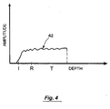
- the ring-down contribution or pattern is determined as shown in Fig. 3, then its contribution is subtracted from the composite echo signal in order to yield a more accurate image of the target area as shown in Fig. 4.
- the ring-down artifact may be characterized in the time domain and/or the time domain across consecutive scans: several sequential scans are convolved or otherwise averaged together to determine the nature of any repetitive artifacts while canceling any short-term artifacts.
- the resultant convoluted ring-down pattern (see Fig. 3) is subtracted from the current scan report to yield a scan report 42 with ring-down effectively eliminated, as shown in Fig. 4.
- the computation of the ring-down pattern is made for scans for which the following assumption holds: along the A-scan axis, tissue does not intervene between the transducer and the blood region nearest the transducer, such as for example, in Fig. 2. Within that range, it is assumed that only ring-down and blood echoes are present.
- the typical transition from ring-down to blood echo can be identified by the distinction between signals produced by ring-down and produced by blood. As shown in Fig. 2, the signals produced by ring-down are high amplitude oscillations of relatively low frequency.
- the signals produced by blood are of low amplitude and high frequency.
- the ring-down contribution is represented by the crossed hatched area in Fig. 2.
- the ring-down signal may detrimentally overload the finite, lower-amplitude echo from the target region.
- the previous assumption does not hold because tissue exists near the transducer.
- a previously computed and stored ring-down pattern (such as the pattern of Fig. 3) is used for selective filtering.
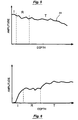
- Fig. 5 is a graph of a trace 44 where the transducer is adjacent tissue.
- the pattern of Fig. 3, which was previously computed, is used for selective filtering.
- the result is illustrated in Fig. 6 which includes only the signal of the target.
- Step A the data on a plurality of frames of R- ⁇ data is collected (Step A), and the latest frame is preferably selected as the current frame for processing (Step B).
- Step B the latest frame is preferably selected as the current frame for processing
- Step C the ring-down artifact may be enhanced so it can be more easily characterized. This may be done mechanically by repositioning the transducer to zero tilt, whereas a slight tilt is normally preferred to suppress such artifact. Enhancement may also be done electronically or by software by the process of convolving several sequential A-scans.
- Step D the A-scan is inspected to determine the presence of the transition region of ring-down to blood. This can be an iterative process of examining the time domain signal searching for the boundary between rapid high-amplitude transitions and low-amplitude transitions.
- the transition between the transition region and the blood region is referred to as a transition point, such as point 48 on Fig. 2.
- such an amplitude analysis may serve only as a first approximation of the transition point. If so, a second processes may be employed to further define the transition point. For example, the estimated target point may be varied and a fast Fourier transform may be performed on the target region T and on the transition region I and R (see Fig. 2) to covert the time-domain data to frequency-domain data for each variation. This process may be repeated until consistent results are obtained.
- the ring-down artifact pattern in the transition region is computed (Step E). This may be done dynamically by computing the ring-down pattern for one A-scan within the ring-down range.
- a straightforward fast Fourier transform (FFT) of the transition region and the target region may be used for frequency domain analysis. Such an FFT computation can be performed periodically during real-time imaging for each individual A-scan following other filtering processes, such as blood speckle reduction.
- a weighted subtraction is performed to selectively filter out the ring down pattern (such as is shown in Fig. 3).
- the ring down pattern is preferably saved, and the filtered data is converted back to the time domain to produce the signal shown in Fig. 4.
- the ring-down pattern is preferably saved and/or updated for use in the cases where a ring-down-to-blood transition is lacking, e.g., where there is tissue residing next to the transducer (Step F) as shown, for example, in Fig. 5. If there is no ring-down-to-blood transition present, the system checks to see if there is already a ring-down pattern available or previously stored (Step G). If not, the process begins again (Step A) until a pattern emerges, e.g., after a ring-down-to-blood transition is found.
- ring-down artifact is suppressed by a selective filtering, i.e., by subtraction of the ring-down contribution from the signal, to yield a filtered image (Step H).
- a selective filtering i.e., by subtraction of the ring-down contribution from the signal
Claims (9)
- Vorrichtung (22) zur Ring-Down-Artefakt-Unterdrückung in einem in-vivo-Ultraschallbilderzeugungssystem (10), mit:einer Erregereinrichtung (20), um einen Zielbereich Ultraschallenergie auszusetzen,einer Einrichtung zum Auffangen eines Rücklaufsignals, das wenigstens eine Ring-Down-Artefaktkomponente, die durch mit der Energieeinrichtung (20) verbundene Transienten verursacht wird, und eine Blutkomponente umfasst,einer Einrichtung zum Untersuchen von Amplitudenmustern in dem aufgefangenen Rücklaufsignal, um eine Übergangsstelle zu ermitteln, die einen Übergang von einem Übergangsbereich zu einem Blutbereich angibt, wobei der Übergangsbereich einen ersten Bereich (I), der die Ring-Down-Artefaktkomponente und keine Blutkomponente umfasst, und einen zweiten Bereich (R) aufweist, der die Ring-Down-Artefaktkomponente kombiniert mit der Blutkomponente umfasst, und der Blutbereich die Blutkomponente und keine Ring-Down-Artefaktkomponente umfasst,einer Einrichtung zum Bestimmen eines Ring-Down-Musters, das für die Ring-Down-Artefaktkomponente im Übergangsbereich repräsentativ ist, wenn eine Übergangsstelle ermittelt wird, undeiner Einrichtung zum Filtern wenigstens eines Teils der Ring-Down-Artefaktkomponente aus dem aufgefangenen Rücklaufsignal unter Verwendung des Ring-Down-Musters.
- Vorrichtung nach Anspruch 1,
wobei die Untersuchungseinrichtung für Amplitudenmuster dafür konfiguriert ist, einen Übergang von einem Muster mit niedriger Frequenz und hoher Amplitude, das einen Ring-Down-Artefakt angibt, und einem Muster mit hoher Frequenz und niedriger Amplitude, das Blut angibt, zu ermitteln. - Vorrichtung (22) nach Anspruch 1,
die ferner Erregermittel zum Verstärken des Ring-Down-Artefakts umfasst, so dass sich das Ring-Down-Artefakt leicht charakterisieren lässt. - Vorrichtung (22) nach Anspruch 1, die ferner umfasst:eine Einrichtung zum Falten sequenzieller Abtastungen zum dynamischen Verstärken eines Musters von Ring-Down-Artefakten als akkumuliertes Ring-Down-Muster,eine Einrichtung zum Speichern des akkumulierten Ring-Down-Musters zur Verwendung beim Analysieren anschließender Abtastungen, undeine Einrichtung zur Verwendung des akkumulierten Ring-Down-Musters für die Filterung, wenn sich kein Ring-Down-/Blut-Übergang findet.
- Vorrichtung (22) nach Anspruch 1,
die ferner Mittel zur Unterstützung der Ermittlung des Übergangsbereichs durch Untersuchen von Spektralmustem umfasst. - Vorrichtung (22) nach Anspruch 5,
wobei die Ring-Dvwn-Bestimmungseinrichtung eine Einrichtung zum Erhalt einer Fourier-Transformation umfasst, um die Spektralmuster hervorzubringen. - Ultraschallbilderzeugungssystem (10) mit:einem Prozessor (28),einem Speicher zum Speichern von Ultraschallbilderzeugungsdaten, die ein Rücklaufsignal umfassen, das wenigstens eine Ring-Down-Artefaktkomponente und eine Blutkomponente aufweist,einem Bildschirm (30), der mit dem Prozessor (28) verbunden ist, um die Bilderzeugungsdaten anzuzeigen,einem Code zum Untersuchen von Amplitudenmustern in dem aufgefangenen Rücklaufsignal, um eine Übergangsstelle zu ermitteln, die auf einen Übergang von einem Übergangsbereich zu einem Blutbereich hinweist, wobei der Übergangsbereich einen ersten Bereich (I), der die Ring-Down-Artefaktkomponente und keine Blutkomponente umfasst, und einen zweiten Bereich (R) aufweist, der die Ring-Down-Artefaktkomponente kombiniert mit der Blutkomponente umfasst, und der Blutbereich die Blutkomponente und keine Ring-Down-Artefaktkomponente umfasst,einem Code zum Bestimmen eines Ring-Down-Musters, das für die Ring-Down-Artefaktkomponente im Übergangsbereich repräsentativ ist, wenn eine Übergangsstelle ermittelt wird, undeinem Code zum Filtern wenigstens eines Teils der Ring-Down-Artefaktkomponente aus dem aufgefangenen Rücklaufsignal unter Verwendung des Ring-Down-Musters.
- System nach Anspruch 7,
wobei der Code zum Untersuchen von Amplitudenmustern dafür konfiguriert ist, einen Übergang von einem Muster mit niedriger Frequenz und hoher Amplitude, das ein Ring-Down-Artefakt angibt, und einem Muster mit hoher Frequenz und niedriger Amplitude, das Blut angibt, zu ermitteln. - System (10) nach Anspruch 7,
das ferner einen Katheter (16) mit einem Ultraschallelement (20) umfasst, um ein Ultraschallsignal (32) zu erzeugen und das Rücklaufsignal (34) aufzufangen.
Applications Claiming Priority (3)
| Application Number | Priority Date | Filing Date | Title |
|---|---|---|---|
| US165807 | 1998-10-02 | ||
| US09/165,807 US6102862A (en) | 1998-10-02 | 1998-10-02 | Adaptive cancellation of ring-down artifact in IVUS imaging |
| PCT/IB1999/001542 WO2000019904A1 (en) | 1998-10-02 | 1999-09-13 | Adaptive cancellation of ring-down artifact in ivus imaging |
Publications (2)
| Publication Number | Publication Date |
|---|---|
| EP1117332A1 EP1117332A1 (de) | 2001-07-25 |
| EP1117332B1 true EP1117332B1 (de) | 2005-08-31 |
Family
ID=22600563
Family Applications (1)
| Application Number | Title | Priority Date | Filing Date |
|---|---|---|---|
| EP99940432A Expired - Lifetime EP1117332B1 (de) | 1998-10-02 | 1999-09-13 | Adaptive unterdrückung von abkling-artefakten in der ivus bildgebung |
Country Status (6)
| Country | Link |
|---|---|
| US (3) | US6102862A (de) |
| EP (1) | EP1117332B1 (de) |
| JP (1) | JP4463422B2 (de) |
| CA (1) | CA2340246C (de) |
| DE (1) | DE69927040T2 (de) |
| WO (1) | WO2000019904A1 (de) |
Cited By (17)
| Publication number | Priority date | Publication date | Assignee | Title |
|---|---|---|---|---|
| US9999371B2 (en) | 2007-11-26 | 2018-06-19 | C. R. Bard, Inc. | Integrated system for intravascular placement of a catheter |
| US10046139B2 (en) | 2010-08-20 | 2018-08-14 | C. R. Bard, Inc. | Reconfirmation of ECG-assisted catheter tip placement |
| US10105121B2 (en) | 2007-11-26 | 2018-10-23 | C. R. Bard, Inc. | System for placement of a catheter including a signal-generating stylet |
| US10231753B2 (en) | 2007-11-26 | 2019-03-19 | C. R. Bard, Inc. | Insertion guidance system for needles and medical components |
| US10231643B2 (en) | 2009-06-12 | 2019-03-19 | Bard Access Systems, Inc. | Apparatus and method for catheter navigation and tip location |
| US10238418B2 (en) | 2007-11-26 | 2019-03-26 | C. R. Bard, Inc. | Apparatus for use with needle insertion guidance system |
| US10271762B2 (en) | 2009-06-12 | 2019-04-30 | Bard Access Systems, Inc. | Apparatus and method for catheter navigation using endovascular energy mapping |
| US10602958B2 (en) | 2007-11-26 | 2020-03-31 | C. R. Bard, Inc. | Systems and methods for guiding a medical instrument |
| US10751509B2 (en) | 2007-11-26 | 2020-08-25 | C. R. Bard, Inc. | Iconic representations for guidance of an indwelling medical device |
| US10849695B2 (en) | 2007-11-26 | 2020-12-01 | C. R. Bard, Inc. | Systems and methods for breaching a sterile field for intravascular placement of a catheter |
| US10863920B2 (en) | 2014-02-06 | 2020-12-15 | C. R. Bard, Inc. | Systems and methods for guidance and placement of an intravascular device |
| US10973584B2 (en) | 2015-01-19 | 2021-04-13 | Bard Access Systems, Inc. | Device and method for vascular access |
| US10992079B2 (en) | 2018-10-16 | 2021-04-27 | Bard Access Systems, Inc. | Safety-equipped connection systems and methods thereof for establishing electrical connections |
| US11000207B2 (en) | 2016-01-29 | 2021-05-11 | C. R. Bard, Inc. | Multiple coil system for tracking a medical device |
| US11027101B2 (en) | 2008-08-22 | 2021-06-08 | C. R. Bard, Inc. | Catheter assembly including ECG sensor and magnetic assemblies |
| US11026630B2 (en) | 2015-06-26 | 2021-06-08 | C. R. Bard, Inc. | Connector interface for ECG-based catheter positioning system |
| US11207496B2 (en) | 2005-08-24 | 2021-12-28 | C. R. Bard, Inc. | Stylet apparatuses and methods of manufacture |
Families Citing this family (70)
| Publication number | Priority date | Publication date | Assignee | Title |
|---|---|---|---|---|
| US6102862A (en) * | 1998-10-02 | 2000-08-15 | Scimed Life Systems, Inc. | Adaptive cancellation of ring-down artifact in IVUS imaging |
| US6200268B1 (en) * | 1999-09-10 | 2001-03-13 | The Cleveland Clinic Foundation | Vascular plaque characterization |
| US6416492B1 (en) | 2000-09-28 | 2002-07-09 | Scimed Life Systems, Inc. | Radiation delivery system utilizing intravascular ultrasound |
| CA2449080A1 (en) | 2003-11-13 | 2005-05-13 | Centre Hospitalier De L'universite De Montreal - Chum | Apparatus and method for intravascular ultrasound image segmentation: a fast-marching method |
| US20050240105A1 (en) * | 2004-04-14 | 2005-10-27 | Mast T D | Method for reducing electronic artifacts in ultrasound imaging |
| US9867530B2 (en) | 2006-08-14 | 2018-01-16 | Volcano Corporation | Telescopic side port catheter device with imaging system and method for accessing side branch occlusions |
| US8540515B2 (en) | 2006-11-27 | 2013-09-24 | Pharos Innovations, Llc | Optimizing behavioral change based on a population statistical profile |
| US8540517B2 (en) | 2006-11-27 | 2013-09-24 | Pharos Innovations, Llc | Calculating a behavioral path based on a statistical profile |
| US8540516B2 (en) | 2006-11-27 | 2013-09-24 | Pharos Innovations, Llc | Optimizing behavioral change based on a patient statistical profile |
| US9596993B2 (en) | 2007-07-12 | 2017-03-21 | Volcano Corporation | Automatic calibration systems and methods of use |
| WO2009009802A1 (en) | 2007-07-12 | 2009-01-15 | Volcano Corporation | Oct-ivus catheter for concurrent luminal imaging |
| JP5524835B2 (ja) | 2007-07-12 | 2014-06-18 | ヴォルカノ コーポレイション | 生体内撮像用カテーテル |
| US9713448B2 (en) | 2008-04-03 | 2017-07-25 | Infraredx, Inc. | System and method for intravascular structural analysis compensation of chemical analysis modality |
| WO2011019760A2 (en) | 2009-08-10 | 2011-02-17 | Romedex International Srl | Devices and methods for endovascular electrography |
| US9808222B2 (en) | 2009-10-12 | 2017-11-07 | Acist Medical Systems, Inc. | Intravascular ultrasound system for co-registered imaging |
| US8961420B2 (en) | 2010-04-01 | 2015-02-24 | Siemens Medical Solutions Usa, Inc. | System for cardiac condition detection and characterization |
| US11141063B2 (en) | 2010-12-23 | 2021-10-12 | Philips Image Guided Therapy Corporation | Integrated system architectures and methods of use |
| US11040140B2 (en) | 2010-12-31 | 2021-06-22 | Philips Image Guided Therapy Corporation | Deep vein thrombosis therapeutic methods |
| WO2013033489A1 (en) | 2011-08-31 | 2013-03-07 | Volcano Corporation | Optical rotary joint and methods of use |
| US11272845B2 (en) | 2012-10-05 | 2022-03-15 | Philips Image Guided Therapy Corporation | System and method for instant and automatic border detection |
| US10568586B2 (en) | 2012-10-05 | 2020-02-25 | Volcano Corporation | Systems for indicating parameters in an imaging data set and methods of use |
| US9292918B2 (en) | 2012-10-05 | 2016-03-22 | Volcano Corporation | Methods and systems for transforming luminal images |
| US9858668B2 (en) | 2012-10-05 | 2018-01-02 | Volcano Corporation | Guidewire artifact removal in images |
| US9286673B2 (en) | 2012-10-05 | 2016-03-15 | Volcano Corporation | Systems for correcting distortions in a medical image and methods of use thereof |
| US9367965B2 (en) | 2012-10-05 | 2016-06-14 | Volcano Corporation | Systems and methods for generating images of tissue |
| US9307926B2 (en) | 2012-10-05 | 2016-04-12 | Volcano Corporation | Automatic stent detection |
| JP2015532536A (ja) | 2012-10-05 | 2015-11-09 | デイビッド ウェルフォード, | 光を増幅するためのシステムおよび方法 |
| US10070827B2 (en) | 2012-10-05 | 2018-09-11 | Volcano Corporation | Automatic image playback |
| US9324141B2 (en) | 2012-10-05 | 2016-04-26 | Volcano Corporation | Removal of A-scan streaking artifact |
| US9840734B2 (en) | 2012-10-22 | 2017-12-12 | Raindance Technologies, Inc. | Methods for analyzing DNA |
| US9295393B2 (en) | 2012-11-09 | 2016-03-29 | Elwha Llc | Embolism deflector |
| CA2894403A1 (en) | 2012-12-13 | 2014-06-19 | Volcano Corporation | Devices, systems, and methods for targeted cannulation |
| US11406498B2 (en) | 2012-12-20 | 2022-08-09 | Philips Image Guided Therapy Corporation | Implant delivery system and implants |
| CA2895989A1 (en) | 2012-12-20 | 2014-07-10 | Nathaniel J. Kemp | Optical coherence tomography system that is reconfigurable between different imaging modes |
| CA2895770A1 (en) | 2012-12-20 | 2014-07-24 | Jeremy Stigall | Locating intravascular images |
| US10939826B2 (en) | 2012-12-20 | 2021-03-09 | Philips Image Guided Therapy Corporation | Aspirating and removing biological material |
| US10942022B2 (en) | 2012-12-20 | 2021-03-09 | Philips Image Guided Therapy Corporation | Manual calibration of imaging system |
| CA2895502A1 (en) | 2012-12-20 | 2014-06-26 | Jeremy Stigall | Smooth transition catheters |
| EP2934280B1 (de) | 2012-12-21 | 2022-10-19 | Mai, Jerome | Ultraschallbildgebung mit variabler liniendichte |
| WO2014100606A1 (en) | 2012-12-21 | 2014-06-26 | Meyer, Douglas | Rotational ultrasound imaging catheter with extended catheter body telescope |
| US9486143B2 (en) | 2012-12-21 | 2016-11-08 | Volcano Corporation | Intravascular forward imaging device |
| US9612105B2 (en) | 2012-12-21 | 2017-04-04 | Volcano Corporation | Polarization sensitive optical coherence tomography system |
| WO2014100530A1 (en) | 2012-12-21 | 2014-06-26 | Whiseant Chester | System and method for catheter steering and operation |
| EP2936626A4 (de) | 2012-12-21 | 2016-08-17 | David Welford | Systeme und verfahren zur verengung einer wellenlängenlichtemission |
| EP2934323A4 (de) | 2012-12-21 | 2016-08-17 | Andrew Hancock | System und verfahren zur mehrpfad-verarbeitung von bildsignalen |
| US10058284B2 (en) | 2012-12-21 | 2018-08-28 | Volcano Corporation | Simultaneous imaging, monitoring, and therapy |
| JP2016508233A (ja) | 2012-12-21 | 2016-03-17 | ナサニエル ジェイ. ケンプ, | 光学スイッチを用いた電力効率のよい光学バッファリング |
| JP2016508757A (ja) | 2012-12-21 | 2016-03-24 | ジェイソン スペンサー, | 医療データのグラフィカル処理のためのシステムおよび方法 |
| US10226597B2 (en) | 2013-03-07 | 2019-03-12 | Volcano Corporation | Guidewire with centering mechanism |
| EP2965263B1 (de) | 2013-03-07 | 2022-07-20 | Bernhard Sturm | Multimodale segmentierung in intravaskulären bildern |
| CN105228518B (zh) | 2013-03-12 | 2018-10-09 | 火山公司 | 用于诊断冠状微脉管疾病的系统和方法 |
| US11154313B2 (en) | 2013-03-12 | 2021-10-26 | The Volcano Corporation | Vibrating guidewire torquer and methods of use |
| US11026591B2 (en) | 2013-03-13 | 2021-06-08 | Philips Image Guided Therapy Corporation | Intravascular pressure sensor calibration |
| CN105120759B (zh) | 2013-03-13 | 2018-02-23 | 火山公司 | 用于从旋转血管内超声设备产生图像的系统和方法 |
| US9301687B2 (en) | 2013-03-13 | 2016-04-05 | Volcano Corporation | System and method for OCT depth calibration |
| EP2967606B1 (de) | 2013-03-14 | 2018-05-16 | Volcano Corporation | Filter mit echogenen eigenschaften |
| US10292677B2 (en) | 2013-03-14 | 2019-05-21 | Volcano Corporation | Endoluminal filter having enhanced echogenic properties |
| US10219887B2 (en) | 2013-03-14 | 2019-03-05 | Volcano Corporation | Filters with echogenic characteristics |
| JP6353038B2 (ja) | 2013-10-07 | 2018-07-04 | アシスト・メディカル・システムズ,インコーポレイテッド | 血管内撮像の信号処理 |
| US10909661B2 (en) * | 2015-10-08 | 2021-02-02 | Acist Medical Systems, Inc. | Systems and methods to reduce near-field artifacts |
| US10653393B2 (en) | 2015-10-08 | 2020-05-19 | Acist Medical Systems, Inc. | Intravascular ultrasound imaging with frequency selective imaging methods and systems |
| US11369337B2 (en) | 2015-12-11 | 2022-06-28 | Acist Medical Systems, Inc. | Detection of disturbed blood flow |
| JP7104632B2 (ja) | 2015-12-31 | 2022-07-21 | アシスト・メディカル・システムズ,インコーポレイテッド | 半自動化画像セグメント化システム及び方法 |
| US10489919B2 (en) | 2016-05-16 | 2019-11-26 | Acist Medical Systems, Inc. | Motion-based image segmentation systems and methods |
| US20190223831A1 (en) * | 2016-06-16 | 2019-07-25 | Koninklijke Philips N.V. | Image orientation identification for an external microconvex-linear ultrasound probe |
| US11284840B1 (en) | 2016-08-26 | 2022-03-29 | W. L. Gore & Associates, Inc. | Calibrating passive LC sensor |
| US10307067B1 (en) * | 2016-08-26 | 2019-06-04 | W. L. Gore & Associates, Inc. | Wireless LC sensor reader |
| EP3975108A1 (de) * | 2016-11-16 | 2022-03-30 | Koninklijke Philips N.V. | Adaptive ringdown-subtraktion für koronaren und peripheren intravaskulären ultraschall (ivus) |
| US11024034B2 (en) | 2019-07-02 | 2021-06-01 | Acist Medical Systems, Inc. | Image segmentation confidence determination |
| WO2021040635A1 (en) | 2019-08-28 | 2021-03-04 | Ozyegin Universitesi | An atomizer and atomization system using the same |
Family Cites Families (12)
| Publication number | Priority date | Publication date | Assignee | Title |
|---|---|---|---|---|
| US5183048A (en) * | 1991-06-24 | 1993-02-02 | Endosonics Corporation | Method and apparatus for removing artifacts from an ultrasonically generated image of a small cavity |
| GB2263974B (en) * | 1992-01-30 | 1995-11-08 | Intravascular Res Ltd | Ultrasound imaging and catheters for use therein |
| GB2301892B (en) * | 1992-07-14 | 1997-02-26 | Intravascular Res Ltd | Methods and apparatus for the examination and treatment of internal organs |
| US5453575A (en) * | 1993-02-01 | 1995-09-26 | Endosonics Corporation | Apparatus and method for detecting blood flow in intravascular ultrasonic imaging |
| US5474074A (en) * | 1994-03-08 | 1995-12-12 | Cardiovascular Imaging Systems, Incorporated | Low profile transducer for intravascular ultrasound imaging and method for mounting |
| US5560242A (en) | 1994-08-16 | 1996-10-01 | Flextech Systems, Inc. | Ultrasonic system evaluation phantoms |
| ATE283496T1 (de) * | 1994-09-15 | 2004-12-15 | Intravascular Res Ltd | Verfahren und gerät zur sichtbarmachung von ultraschall |
| GB2293240B (en) * | 1994-09-15 | 1998-05-20 | Intravascular Res Ltd | Ultrasonic visualisation method and apparatus |
| GB2293651B (en) * | 1994-09-30 | 1998-04-22 | Intravascular Res Ltd | Medical untrasound imaging |
| US5921931A (en) * | 1997-04-08 | 1999-07-13 | Endosonics Corporation | Method and apparatus for creating a color blood flow image based upon ultrasonic echo signals received by an intravascular ultrasound imaging probe |
| US6036650A (en) * | 1998-09-15 | 2000-03-14 | Endosonics Corporation | Ultrasonic imaging system and method with ringdown reduction |
| US6102862A (en) * | 1998-10-02 | 2000-08-15 | Scimed Life Systems, Inc. | Adaptive cancellation of ring-down artifact in IVUS imaging |
-
1998
- 1998-10-02 US US09/165,807 patent/US6102862A/en not_active Expired - Lifetime
-
1999
- 1999-09-13 DE DE69927040T patent/DE69927040T2/de not_active Expired - Lifetime
- 1999-09-13 EP EP99940432A patent/EP1117332B1/de not_active Expired - Lifetime
- 1999-09-13 JP JP2000573267A patent/JP4463422B2/ja not_active Expired - Lifetime
- 1999-09-13 CA CA2340246A patent/CA2340246C/en not_active Expired - Fee Related
- 1999-09-13 WO PCT/IB1999/001542 patent/WO2000019904A1/en active IP Right Grant
-
2000
- 2000-07-25 US US09/625,149 patent/US6254543B1/en not_active Expired - Lifetime
-
2001
- 2001-05-18 US US09/860,907 patent/US6589181B2/en not_active Expired - Lifetime
Cited By (27)
| Publication number | Priority date | Publication date | Assignee | Title |
|---|---|---|---|---|
| US11207496B2 (en) | 2005-08-24 | 2021-12-28 | C. R. Bard, Inc. | Stylet apparatuses and methods of manufacture |
| US10602958B2 (en) | 2007-11-26 | 2020-03-31 | C. R. Bard, Inc. | Systems and methods for guiding a medical instrument |
| US10751509B2 (en) | 2007-11-26 | 2020-08-25 | C. R. Bard, Inc. | Iconic representations for guidance of an indwelling medical device |
| US10165962B2 (en) | 2007-11-26 | 2019-01-01 | C. R. Bard, Inc. | Integrated systems for intravascular placement of a catheter |
| US10231753B2 (en) | 2007-11-26 | 2019-03-19 | C. R. Bard, Inc. | Insertion guidance system for needles and medical components |
| US11707205B2 (en) | 2007-11-26 | 2023-07-25 | C. R. Bard, Inc. | Integrated system for intravascular placement of a catheter |
| US10238418B2 (en) | 2007-11-26 | 2019-03-26 | C. R. Bard, Inc. | Apparatus for use with needle insertion guidance system |
| US10105121B2 (en) | 2007-11-26 | 2018-10-23 | C. R. Bard, Inc. | System for placement of a catheter including a signal-generating stylet |
| US11123099B2 (en) | 2007-11-26 | 2021-09-21 | C. R. Bard, Inc. | Apparatus for use with needle insertion guidance system |
| US11529070B2 (en) | 2007-11-26 | 2022-12-20 | C. R. Bard, Inc. | System and methods for guiding a medical instrument |
| US10849695B2 (en) | 2007-11-26 | 2020-12-01 | C. R. Bard, Inc. | Systems and methods for breaching a sterile field for intravascular placement of a catheter |
| US11779240B2 (en) | 2007-11-26 | 2023-10-10 | C. R. Bard, Inc. | Systems and methods for breaching a sterile field for intravascular placement of a catheter |
| US9999371B2 (en) | 2007-11-26 | 2018-06-19 | C. R. Bard, Inc. | Integrated system for intravascular placement of a catheter |
| US10966630B2 (en) | 2007-11-26 | 2021-04-06 | C. R. Bard, Inc. | Integrated system for intravascular placement of a catheter |
| US11134915B2 (en) | 2007-11-26 | 2021-10-05 | C. R. Bard, Inc. | System for placement of a catheter including a signal-generating stylet |
| US11027101B2 (en) | 2008-08-22 | 2021-06-08 | C. R. Bard, Inc. | Catheter assembly including ECG sensor and magnetic assemblies |
| US10912488B2 (en) | 2009-06-12 | 2021-02-09 | Bard Access Systems, Inc. | Apparatus and method for catheter navigation and tip location |
| US11419517B2 (en) | 2009-06-12 | 2022-08-23 | Bard Access Systems, Inc. | Apparatus and method for catheter navigation using endovascular energy mapping |
| US10271762B2 (en) | 2009-06-12 | 2019-04-30 | Bard Access Systems, Inc. | Apparatus and method for catheter navigation using endovascular energy mapping |
| US10231643B2 (en) | 2009-06-12 | 2019-03-19 | Bard Access Systems, Inc. | Apparatus and method for catheter navigation and tip location |
| US10046139B2 (en) | 2010-08-20 | 2018-08-14 | C. R. Bard, Inc. | Reconfirmation of ECG-assisted catheter tip placement |
| US10863920B2 (en) | 2014-02-06 | 2020-12-15 | C. R. Bard, Inc. | Systems and methods for guidance and placement of an intravascular device |
| US10973584B2 (en) | 2015-01-19 | 2021-04-13 | Bard Access Systems, Inc. | Device and method for vascular access |
| US11026630B2 (en) | 2015-06-26 | 2021-06-08 | C. R. Bard, Inc. | Connector interface for ECG-based catheter positioning system |
| US11000207B2 (en) | 2016-01-29 | 2021-05-11 | C. R. Bard, Inc. | Multiple coil system for tracking a medical device |
| US10992079B2 (en) | 2018-10-16 | 2021-04-27 | Bard Access Systems, Inc. | Safety-equipped connection systems and methods thereof for establishing electrical connections |
| US11621518B2 (en) | 2018-10-16 | 2023-04-04 | Bard Access Systems, Inc. | Safety-equipped connection systems and methods thereof for establishing electrical connections |
Also Published As
| Publication number | Publication date |
|---|---|
| US6254543B1 (en) | 2001-07-03 |
| JP4463422B2 (ja) | 2010-05-19 |
| JP2002526142A (ja) | 2002-08-20 |
| DE69927040D1 (de) | 2005-10-06 |
| US6589181B2 (en) | 2003-07-08 |
| DE69927040T2 (de) | 2006-09-21 |
| CA2340246A1 (en) | 2000-04-13 |
| US20010027332A1 (en) | 2001-10-04 |
| EP1117332A1 (de) | 2001-07-25 |
| WO2000019904A1 (en) | 2000-04-13 |
| US6102862A (en) | 2000-08-15 |
| CA2340246C (en) | 2011-04-05 |
Similar Documents
| Publication | Publication Date | Title |
|---|---|---|
| EP1117332B1 (de) | Adaptive unterdrückung von abkling-artefakten in der ivus bildgebung | |
| US8094893B2 (en) | Segmentation tool for identifying flow regions in an image system | |
| US6106465A (en) | Ultrasonic method and system for boundary detection of an object of interest in an ultrasound image | |
| US5628321A (en) | Processing velocity information in an ultrasonic system | |
| US8162836B2 (en) | System and method for characterizing tissue based upon split spectrum analysis of backscattered ultrasound | |
| US8460191B2 (en) | Ultrasonic medical diagnostic device for imaging changes with time | |
| JP7252206B2 (ja) | 画像アーチファクト特定及び除去のための深層学習ネットワークを有する超音波システム | |
| JP2001522628A (ja) | 脈管内超音波イメージングシステムにおける空間フィルタリング方法及び装置 | |
| JPH10127638A (ja) | 運動部分を含む対象に関する信号処理方法及びこの方法を実施するエコーグラフィック装置 | |
| US5299174A (en) | Automatic clutter elimination | |
| US5601082A (en) | Medical ultrasound imaging | |
| US20060030777A1 (en) | T-statistic method for suppressing artifacts in blood vessel ultrasonic imaging | |
| US5623929A (en) | Ultrasonic doppler flow imaging method for eliminating motion artifacts | |
| US6287258B1 (en) | Method and apparatus for medical ultrasound flash suppression | |
| JP2002177273A (ja) | 超音波診断装置 | |
| US6358206B1 (en) | Ultrasound process for the determination of the location of a parietal surface in a tissue and of the absolute radius of an artery, and ultrasound apparatus for carrying out such process | |
| US11141138B2 (en) | Kalman filtering for flash artifact suppression in ultrasound imaging | |
| JP3887040B2 (ja) | 超音波診断装置 | |
| JP2508000B2 (ja) | 超音波診断装置 | |
| EP4275612A1 (de) | Verbesserungen in ultraschallbasierten blutflussgeschwindigkeitsmessungen | |
| WO2023088760A1 (en) | Improvements in ultrasound based blood flow velocity measurements | |
| JPH11226019A (ja) | 超音波診断装置 |
Legal Events
| Date | Code | Title | Description |
|---|---|---|---|
| PUAI | Public reference made under article 153(3) epc to a published international application that has entered the european phase |
Free format text: ORIGINAL CODE: 0009012 |
|
| 17P | Request for examination filed |
Effective date: 20010326 |
|
| AK | Designated contracting states |
Kind code of ref document: A1 Designated state(s): AT BE CH CY DE DK ES FI FR GB GR IE IT LI LU MC NL PT SE |
|
| 17Q | First examination report despatched |
Effective date: 20031126 |
|
| RBV | Designated contracting states (corrected) |
Designated state(s): DE FR GB IT |
|
| GRAP | Despatch of communication of intention to grant a patent |
Free format text: ORIGINAL CODE: EPIDOSNIGR1 |
|
| RAP1 | Party data changed (applicant data changed or rights of an application transferred) |
Owner name: BOSTON SCIENTIFIC LIMITED |
|
| GRAS | Grant fee paid |
Free format text: ORIGINAL CODE: EPIDOSNIGR3 |
|
| GRAA | (expected) grant |
Free format text: ORIGINAL CODE: 0009210 |
|
| AK | Designated contracting states |
Kind code of ref document: B1 Designated state(s): DE FR GB IT |
|
| REG | Reference to a national code |
Ref country code: GB Ref legal event code: FG4D |
|
| REF | Corresponds to: |
Ref document number: 69927040 Country of ref document: DE Date of ref document: 20051006 Kind code of ref document: P |
|
| ET | Fr: translation filed | ||
| PLBE | No opposition filed within time limit |
Free format text: ORIGINAL CODE: 0009261 |
|
| STAA | Information on the status of an ep patent application or granted ep patent |
Free format text: STATUS: NO OPPOSITION FILED WITHIN TIME LIMIT |
|
| 26N | No opposition filed |
Effective date: 20060601 |
|
| PGFP | Annual fee paid to national office [announced via postgrant information from national office to epo] |
Ref country code: IT Payment date: 20100918 Year of fee payment: 12 Ref country code: FR Payment date: 20100920 Year of fee payment: 12 |
|
| PGFP | Annual fee paid to national office [announced via postgrant information from national office to epo] |
Ref country code: GB Payment date: 20100809 Year of fee payment: 12 |
|
| PGFP | Annual fee paid to national office [announced via postgrant information from national office to epo] |
Ref country code: DE Payment date: 20100930 Year of fee payment: 12 |
|
| GBPC | Gb: european patent ceased through non-payment of renewal fee |
Effective date: 20110913 |
|
| PG25 | Lapsed in a contracting state [announced via postgrant information from national office to epo] |
Ref country code: IT Free format text: LAPSE BECAUSE OF NON-PAYMENT OF DUE FEES Effective date: 20110913 |
|
| REG | Reference to a national code |
Ref country code: FR Ref legal event code: ST Effective date: 20120531 |
|
| REG | Reference to a national code |
Ref country code: DE Ref legal event code: R119 Ref document number: 69927040 Country of ref document: DE Effective date: 20120403 |
|
| PG25 | Lapsed in a contracting state [announced via postgrant information from national office to epo] |
Ref country code: DE Free format text: LAPSE BECAUSE OF NON-PAYMENT OF DUE FEES Effective date: 20120403 |
|
| PG25 | Lapsed in a contracting state [announced via postgrant information from national office to epo] |
Ref country code: FR Free format text: LAPSE BECAUSE OF NON-PAYMENT OF DUE FEES Effective date: 20110930 Ref country code: GB Free format text: LAPSE BECAUSE OF NON-PAYMENT OF DUE FEES Effective date: 20110913 |