EP1113397A2 - Methods and apparatus for tilted helical reconstruction multislice ct - Google Patents
Methods and apparatus for tilted helical reconstruction multislice ct Download PDFInfo
- Publication number
- EP1113397A2 EP1113397A2 EP00311613A EP00311613A EP1113397A2 EP 1113397 A2 EP1113397 A2 EP 1113397A2 EP 00311613 A EP00311613 A EP 00311613A EP 00311613 A EP00311613 A EP 00311613A EP 1113397 A2 EP1113397 A2 EP 1113397A2
- Authority
- EP
- European Patent Office
- Prior art keywords
- projection data
- fourier transform
- zero
- phase shift
- product
- Prior art date
- Legal status (The legal status is an assumption and is not a legal conclusion. Google has not performed a legal analysis and makes no representation as to the accuracy of the status listed.)
- Withdrawn
Links
Images
Classifications
-
- G—PHYSICS
- G06—COMPUTING OR CALCULATING; COUNTING
- G06T—IMAGE DATA PROCESSING OR GENERATION, IN GENERAL
- G06T11/00—2D [Two Dimensional] image generation
- G06T11/003—Reconstruction from projections, e.g. tomography
- G06T11/005—Specific pre-processing for tomographic reconstruction, e.g. calibration, source positioning, rebinning, scatter correction, retrospective gating
-
- A—HUMAN NECESSITIES
- A61—MEDICAL OR VETERINARY SCIENCE; HYGIENE
- A61B—DIAGNOSIS; SURGERY; IDENTIFICATION
- A61B6/00—Apparatus or devices for radiation diagnosis; Apparatus or devices for radiation diagnosis combined with radiation therapy equipment
- A61B6/02—Arrangements for diagnosis sequentially in different planes; Stereoscopic radiation diagnosis
- A61B6/027—Arrangements for diagnosis sequentially in different planes; Stereoscopic radiation diagnosis characterised by the use of a particular data acquisition trajectory, e.g. helical or spiral
-
- A—HUMAN NECESSITIES
- A61—MEDICAL OR VETERINARY SCIENCE; HYGIENE
- A61B—DIAGNOSIS; SURGERY; IDENTIFICATION
- A61B6/00—Apparatus or devices for radiation diagnosis; Apparatus or devices for radiation diagnosis combined with radiation therapy equipment
- A61B6/58—Testing, adjusting or calibrating thereof
- A61B6/582—Calibration
- A61B6/583—Calibration using calibration phantoms
-
- G—PHYSICS
- G06—COMPUTING OR CALCULATING; COUNTING
- G06T—IMAGE DATA PROCESSING OR GENERATION, IN GENERAL
- G06T2211/00—Image generation
- G06T2211/40—Computed tomography
- G06T2211/421—Filtered back projection [FBP]
-
- Y—GENERAL TAGGING OF NEW TECHNOLOGICAL DEVELOPMENTS; GENERAL TAGGING OF CROSS-SECTIONAL TECHNOLOGIES SPANNING OVER SEVERAL SECTIONS OF THE IPC; TECHNICAL SUBJECTS COVERED BY FORMER USPC CROSS-REFERENCE ART COLLECTIONS [XRACs] AND DIGESTS
- Y10—TECHNICAL SUBJECTS COVERED BY FORMER USPC
- Y10S—TECHNICAL SUBJECTS COVERED BY FORMER USPC CROSS-REFERENCE ART COLLECTIONS [XRACs] AND DIGESTS
- Y10S378/00—X-ray or gamma ray systems or devices
- Y10S378/901—Computer tomography program or processor
Definitions
- This invention relates generally to methods and apparatus for computed tomography (CT) imaging of objects, and more particularly to methods and apparatus for producing compensated tilted, helically scanned CT images of objects.
- CT computed tomography
- an x-ray source projects a fan-shaped beam which is collimated to lie within an X-Y plane of a Cartesian coordinate system and generally referred to as the "imaging plane".
- the x-ray beam passes through the object being imaged, such as a patient.
- the beam after being attenuated by the object, impinges upon an array of radiation detectors.
- the intensity of the attenuated beam radiation received at the detector array is dependent upon the attenuation of the x-ray beam by the object.
- Each detector element of the array produces a separate electrical signal that is a measurement of the beam attenuation at the detector location.
- the attenuation measurements from all the detectors are acquired separately to produce a transmission profile.
- the x-ray source and the detector array are rotated with a gantry within the imaging plane and around the object to be imaged so that the angle at which the x-ray beam intersects the object constantly changes.
- a group of x-ray attenuation measurements, i.e., projection data, from the detector array at one gantry angle is referred to as a "view”.
- a "scan" of the object comprises a set of views made at different gantry angles, or view angles, during one revolution of the x-ray source and detector.
- the projection data is processed to construct an image that corresponds to a two dimensional slice taken through the object.
- CT numbers integers called "CT numbers” or “Hounsfield units”, which are used to control the brightness of a corresponding pixel on a cathode ray tube display.
- gantry tilt is disabled for helical scans.
- degraded image quality results because the isocenter is inherently detector row dependent.
- the isocenter is shifted relative to the patient scanning axis, and the amount of shift depends upon detector row, amount of gantry tilt, and projection angle.
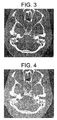
- an axial scan of a human skull is depicted for a 1.25 mm slice thickness.
- the gantry was tilted at -20° and no projection weighting was applied in the reconstruction of the image of Figure 3.
- This image was reconstructed with a 15 cm field of view (FOV) and a high-resolution algorithm to illustrate fine structural details of the scanned phantom.
- This image serves as a standard for image quality evaluation, as no degradation is present in a tilted axial scan mode.
- a method for filtering projection data of a computed tomographic scan of an object includes steps of: acquiring projection data representing a tilted, helical scan of an object; zero padding the acquired projection data; determining a Fourier transform of the zero padded projection data; determining a product of the Fourier transform of the zero padded projection data, a ramp function, and a phase shift function; and determining an inverse Fourier transform of the multiplied, transformed projection data as filtered projection data.
- the above-described method provides filtered data that produces compensated tilted, helically scanned CT images having significantly better spatial resolution than those methods employing a high frequency kernel boost.
- Figure 3 is an axial scan of the phantom without application of projection weighting.
- Figure 4 is a helical scan at 3:1 helical pitch, reconstructed without z-smoothing.
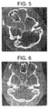
- Figure 5 is a "sharpened" version of the same scan shown in Figure 4, using a high-frequency kernel boost.
- Figure 6 is a version of the same scan shown in Figure 4, but using data to which a method embodiment of the present invention has been applied.
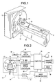
- a computed tomograph (CT) imaging system 10 is shown as including a gantry 12 representative of a "third generation" CT scanner.
- Gantry 12 has an x-ray source 14 that projects a beam of x-rays 16 toward a detector array 18 on the opposite side of gantry 12.
- Detector array 18 is formed by detector elements 20 which together sense the projected x-rays that pass through an object 22, for example a medical patient.
- Detector array 18 may be fabricated in a single slice or multi-slice configuration.
- Each detector element 20 produces an electrical signal that represents the intensity of an impinging x-ray beam and hence the attenuation of the beam as it passes through patient 22.
- gantry 12 and the components mounted thereon rotate about a center of rotation 24.
- Control mechanism 26 includes an x-ray controller 28 that provides power and timing signals to x-ray source 14 and a gantry motor controller 30 that controls the rotational speed and position of gantry 12.
- a data acquisition system (DAS) 32 in control mechanism 26 samples analog data from detector elements 20 and converts the data to digital signals for subsequent processing.
- An image reconstructor 34 receives sampled and digitized x-ray data from DAS 32 and performs high speed image reconstruction. The reconstructed image is applied as an input to a computer 36 which stores the image in a mass storage device 38.
- DAS data acquisition system
- Computer 36 also receives commands and scanning parameters from an operator via console 40 that has a keyboard.
- An associated cathode ray tube display 42 allows the operator to observe the reconstructed image and other data from computer 36.
- the operator supplied commands and parameters are used by computer 36 to provide control signals and information to DAS 32, x-ray controller 28 and gantry motor controller 30.
- computer 36 operates a table motor controller 44 which controls a motorized table 46 to position patient 22 in gantry 12. Particularly, table 46 moves portions of patient 22 through gantry opening 48.
- a first step of the filter process is to perform a Fourier transform of a zero padded projection.
- the transformed projection is then multiplied by a ramp function and a phase-shift function, zero padded, and inversely Fourier transformed to arrive at the filtered projection.
- p ( ⁇ , ⁇ , n ) denotes the measured projection data with detector angle ⁇ , view angle ⁇ , and detector row number n.
- ⁇ depends upon ⁇ , n , gantry tilt angle, x-ray beam width, and source to isocenter distance.
- projection data are weighted and summed before the filtering operation for computational performance. Denoting by k the number of rows that are summed for a particular projection angle, k Fourier transforms of the original projection and one inverse Fourier transform are required.
- the helical scan of Figure 4 is shown reconstructed in Figure 6 using an embodiment of the present invention.
- a Fourier transformed projection is multiplied by a ramp projection and a phase shift function, zero padded, and inversely Fourier transformed to arrive at the filtered projection.
- a significant improvement in spatial resolution is evident compared to Figure 5.
- the reconstructed object is nearly as sharp as the axial scan illustrated in Figure 4.
- Some slight degradation in some fine structures may result from the constant translation of the patient table in the helical scan mode.
- Some streaking is also evident because aliasing is increased.
- the increased aliasing results from destruction of quarter detector offset sampling by the detector row-dependent isocenter shift.
- the methods described above are carried out in CT imaging system 10 in which, for example, a tilt angle of gantry 12 is selectable relative to table 46.
- DAS 32 converts signals received from a multislice detector 18 into data for subsequent processing by computer 36.
- computer 36 also establishes a tilt angle for gantry 12. Data processing steps for filtering the acquired projections are also performed by computer 36.
- phase shift operation is performed right after the ramp filter operation.
- a combined filter (ramp multipled by the phase shift) is pre-calculated, stored, and multiplied by the Fourier transform for computational efficiency.
- CT system described herein is a "third generation” system in which both the x-ray source and detector rotate with the gantry.
- CT systems including "fourth generation” systems wherein the detector is a full-ring stationary detector and only the x-ray source rotates with the gantry, may be used if individual detector elements are corrected to provide substantially uniform responses to a given x-ray beam.
Landscapes
- Life Sciences & Earth Sciences (AREA)
- Engineering & Computer Science (AREA)
- Health & Medical Sciences (AREA)
- Physics & Mathematics (AREA)
- Medical Informatics (AREA)
- Optics & Photonics (AREA)
- Heart & Thoracic Surgery (AREA)
- High Energy & Nuclear Physics (AREA)
- Theoretical Computer Science (AREA)
- Nuclear Medicine, Radiotherapy & Molecular Imaging (AREA)
- General Physics & Mathematics (AREA)
- Pathology (AREA)
- Radiology & Medical Imaging (AREA)
- Biomedical Technology (AREA)
- Biophysics (AREA)
- Molecular Biology (AREA)
- Surgery (AREA)
- Animal Behavior & Ethology (AREA)
- General Health & Medical Sciences (AREA)
- Public Health (AREA)
- Veterinary Medicine (AREA)
- Apparatus For Radiation Diagnosis (AREA)
Applications Claiming Priority (2)
| Application Number | Priority Date | Filing Date | Title |
|---|---|---|---|
| US09/473,341 US6332013B1 (en) | 1999-12-28 | 1999-12-28 | Methods and apparatus for tilted helical reconstruction multislice CT |
| US473341 | 1999-12-28 |
Publications (1)
| Publication Number | Publication Date |
|---|---|
| EP1113397A2 true EP1113397A2 (en) | 2001-07-04 |
Family
ID=23879152
Family Applications (1)
| Application Number | Title | Priority Date | Filing Date |
|---|---|---|---|
| EP00311613A Withdrawn EP1113397A2 (en) | 1999-12-28 | 2000-12-22 | Methods and apparatus for tilted helical reconstruction multislice ct |
Country Status (5)
| Country | Link |
|---|---|
| US (1) | US6332013B1 (enExample) |
| EP (1) | EP1113397A2 (enExample) |
| JP (1) | JP2001218764A (enExample) |
| CN (1) | CN1225716C (enExample) |
| IL (1) | IL140572A (enExample) |
Cited By (1)
| Publication number | Priority date | Publication date | Assignee | Title |
|---|---|---|---|---|
| WO2004015632A1 (en) * | 2002-08-06 | 2004-02-19 | Koninklijke Philips Electronics N.V. | Reconstruction method for tilted-gantry computed tomography |
Families Citing this family (17)
| Publication number | Priority date | Publication date | Assignee | Title |
|---|---|---|---|---|
| US6704392B2 (en) * | 2001-07-03 | 2004-03-09 | Ge Medical Systems Global Technology Company, Llc | X-ray tube and method having tilted rotation axis |
| JP4175791B2 (ja) * | 2001-08-20 | 2008-11-05 | ジーイー・メディカル・システムズ・グローバル・テクノロジー・カンパニー・エルエルシー | 画像生成方法およびx線ct装置 |
| US6587537B1 (en) | 2002-04-01 | 2003-07-01 | Ge Medical Systems Global Technology Company, Llc | Methods and apparatus for multi-slice image reconstruction |
| US6904117B2 (en) * | 2002-10-30 | 2005-06-07 | Toshiba Corporation | Tilted gantry helical cone-beam Feldkamp reconstruction for multislice CT |
| US6647084B1 (en) | 2002-11-11 | 2003-11-11 | Ge Medical Systems Global Technology Company, Llc | Method and apparatus for filtering projection data of a helical scan |
| US7239730B2 (en) * | 2003-01-29 | 2007-07-03 | Ge Medical Systems Global Technology Company, Llc | Method and apparatus for volume scoring calcification concentrations of a CT scan |
| JP4409223B2 (ja) * | 2003-07-24 | 2010-02-03 | 東芝医用システムエンジニアリング株式会社 | X線ct装置及びx線ct用逆投影演算方法 |
| US6977984B2 (en) * | 2003-10-07 | 2005-12-20 | Ge Medical Systems Global Technology Company, Llc | Methods and apparatus for dynamical helical scanned image production |
| US7076029B2 (en) * | 2003-10-27 | 2006-07-11 | General Electric Company | Method and apparatus of radiographic imaging with an energy beam tailored for a subject to be scanned |
| CN100443053C (zh) * | 2005-04-29 | 2008-12-17 | Ge医疗系统环球技术有限公司 | 信号处理方法与装置以及x射线ct装置 |
| US7344306B2 (en) * | 2005-06-30 | 2008-03-18 | General Electric Company | Systems and methods for compensating for table sag |
| JP4611168B2 (ja) * | 2005-10-07 | 2011-01-12 | ジーイー・メディカル・システムズ・グローバル・テクノロジー・カンパニー・エルエルシー | 画像再構成方法、およびx線ct装置 |
| CN100401983C (zh) * | 2005-10-27 | 2008-07-16 | 上海交通大学 | 基于双源双螺旋多层螺旋ct的重建方法 |
| JP2007236662A (ja) * | 2006-03-09 | 2007-09-20 | Ge Medical Systems Global Technology Co Llc | X線ct装置およびそのx線ct画像再構成方法、x線ct画像撮影方法。 |
| EP2036498A1 (en) * | 2006-06-22 | 2009-03-18 | Tohoku University | X-ray ct device, and image reconfiguration method and image reconfiguration program for the device |
| US8224056B2 (en) * | 2009-12-15 | 2012-07-17 | General Electronic Company | Method for computed tomography motion estimation and compensation |
| CN114140544B (zh) * | 2021-10-26 | 2024-12-27 | 上海东软医疗科技有限公司 | 图像重建方法、装置及设备 |
Family Cites Families (5)
| Publication number | Priority date | Publication date | Assignee | Title |
|---|---|---|---|---|
| DE19626095C2 (de) * | 1996-06-28 | 2001-02-08 | Siemens Ag | Computertomograph |
| WO1999001736A2 (en) * | 1997-07-01 | 1999-01-14 | Analogic Corporation | Improved helical scan computed tomography detector geometry |
| CN1336811A (zh) * | 1997-10-10 | 2002-02-20 | 模拟技术公司 | 计算层析扫描目标探测 |
| US6061420A (en) * | 1998-08-25 | 2000-05-09 | General Electric Company | Methods and apparatus for graphical Rx in a multislice imaging system |
| US6229869B1 (en) * | 1998-08-25 | 2001-05-08 | General Electric Company | Tilted gantry image correction for a multislice computed tomography system |
-
1999
- 1999-12-28 US US09/473,341 patent/US6332013B1/en not_active Expired - Fee Related
-
2000
- 2000-12-22 EP EP00311613A patent/EP1113397A2/en not_active Withdrawn
- 2000-12-26 IL IL14057200A patent/IL140572A/en not_active IP Right Cessation
- 2000-12-27 JP JP2000396388A patent/JP2001218764A/ja not_active Withdrawn
- 2000-12-28 CN CNB001375350A patent/CN1225716C/zh not_active Expired - Fee Related
Cited By (2)
| Publication number | Priority date | Publication date | Assignee | Title |
|---|---|---|---|---|
| WO2004015632A1 (en) * | 2002-08-06 | 2004-02-19 | Koninklijke Philips Electronics N.V. | Reconstruction method for tilted-gantry computed tomography |
| CN1675656B (zh) * | 2002-08-06 | 2010-08-18 | 皇家飞利浦电子股份有限公司 | 倾斜台架计算层析x射线摄影法的重建方法和设备 |
Also Published As
| Publication number | Publication date |
|---|---|
| IL140572A (en) | 2004-08-31 |
| CN1304036A (zh) | 2001-07-18 |
| CN1225716C (zh) | 2005-11-02 |
| JP2001218764A (ja) | 2001-08-14 |
| US6332013B1 (en) | 2001-12-18 |
| IL140572A0 (en) | 2002-02-10 |
Similar Documents
| Publication | Publication Date | Title |
|---|---|---|
| US6035012A (en) | Artifact correction for highly attenuating objects | |
| US6332013B1 (en) | Methods and apparatus for tilted helical reconstruction multislice CT | |
| US5559847A (en) | Systems, methods and apparatus for reconstructing images in a CT system implementing a helical scan | |
| US5663995A (en) | Systems and methods for reconstructing an image in a CT system performing a cone beam helical scan | |
| US6115487A (en) | Correction algorithm for bone-induced spectral artifacts in computed tomograph imaging | |
| US6266388B1 (en) | Methods and apparatus for two-pass cone beam image reconstruction | |
| EP1113396B1 (en) | Method and apparauts for multislice CT using partial scan | |
| US5606585A (en) | Methods and apparatus for multislice helical image reconstruction in a computer tomography system | |
| US5727041A (en) | Methods and apparatus for reducing partial volume image artifacts | |
| US6285732B1 (en) | Methods and apparatus for adaptive interpolation reduced view CT scan | |
| US5708690A (en) | Methods and apparatus for helical image reconstruction in a computed tomography fluoro system | |
| IL127009A (en) | Image reconstruction in a ct fluoroscopy system | |
| EP1351192A1 (en) | Methods and apparatus for adaptive interpolation of multislice helical CT data | |
| EP0969414B1 (en) | Computerized tomographic multi-frame image reconstruction method and apparatus for helical scanning | |
| JP4676641B2 (ja) | マルチ・スライスct走査の螺旋再構成の方法及び装置 | |
| EP0989521B1 (en) | Fluoroscopy image reconstruction | |
| US5974110A (en) | Helical reconstruction algorithm | |
| IL119543A (en) | Methods and apparatus for reducing image artifacts | |
| US5546439A (en) | Systems, methods and apparatus for incrementally reconstructing overlapped images in a CT system implementing a helical scan | |
| US6873676B2 (en) | Convolution reconstruction algorithm for multi-slice CT | |
| US6798860B1 (en) | Methods and apparatus for deconvolving imaging data | |
| US6327325B1 (en) | Methods and apparatus for adaptive interpolation reduced view CT scan | |
| EP0982680A2 (en) | Systems, methods and apparatus for reconstructing images | |
| US6091840A (en) | Methods and apparatus for single slice helical image reconstruction in a computed tomography system | |
| US5764720A (en) | Methods and apparatus for simplified pre-processing of data in a computed tomography system |
Legal Events
| Date | Code | Title | Description |
|---|---|---|---|
| PUAI | Public reference made under article 153(3) epc to a published international application that has entered the european phase |
Free format text: ORIGINAL CODE: 0009012 |
|
| AK | Designated contracting states |
Kind code of ref document: A2 Designated state(s): AT BE CH CY DE DK ES FI FR GB GR IE IT LI LU MC NL PT SE TR |
|
| AX | Request for extension of the european patent |
Free format text: AL;LT;LV;MK;RO;SI |
|
| STAA | Information on the status of an ep patent application or granted ep patent |
Free format text: STATUS: THE APPLICATION IS DEEMED TO BE WITHDRAWN |
|
| 18D | Application deemed to be withdrawn |
Effective date: 20090701 |