EP1035219A1 - Gastrische Helicobacter 16S-rDNA Sequenzen von Rind und Schwein und ihre Anwendung zur Detektion and Typisierung von Helicobacter Stämmen - Google Patents
Gastrische Helicobacter 16S-rDNA Sequenzen von Rind und Schwein und ihre Anwendung zur Detektion and Typisierung von Helicobacter Stämmen Download PDFInfo
- Publication number
- EP1035219A1 EP1035219A1 EP99870035A EP99870035A EP1035219A1 EP 1035219 A1 EP1035219 A1 EP 1035219A1 EP 99870035 A EP99870035 A EP 99870035A EP 99870035 A EP99870035 A EP 99870035A EP 1035219 A1 EP1035219 A1 EP 1035219A1
- Authority
- EP
- European Patent Office
- Prior art keywords
- helicobacter
- sequences
- sequence
- probe
- seq
- Prior art date
- Legal status (The legal status is an assumption and is not a legal conclusion. Google has not performed a legal analysis and makes no representation as to the accuracy of the status listed.)
- Withdrawn
Links
Images


Classifications
-
- C—CHEMISTRY; METALLURGY
- C12—BIOCHEMISTRY; BEER; SPIRITS; WINE; VINEGAR; MICROBIOLOGY; ENZYMOLOGY; MUTATION OR GENETIC ENGINEERING
- C12Q—MEASURING OR TESTING PROCESSES INVOLVING ENZYMES, NUCLEIC ACIDS OR MICROORGANISMS; COMPOSITIONS OR TEST PAPERS THEREFOR; PROCESSES OF PREPARING SUCH COMPOSITIONS; CONDITION-RESPONSIVE CONTROL IN MICROBIOLOGICAL OR ENZYMOLOGICAL PROCESSES
- C12Q1/00—Measuring or testing processes involving enzymes, nucleic acids or microorganisms; Compositions therefor; Processes of preparing such compositions
- C12Q1/68—Measuring or testing processes involving enzymes, nucleic acids or microorganisms; Compositions therefor; Processes of preparing such compositions involving nucleic acids
- C12Q1/6876—Nucleic acid products used in the analysis of nucleic acids, e.g. primers or probes
- C12Q1/6888—Nucleic acid products used in the analysis of nucleic acids, e.g. primers or probes for detection or identification of organisms
- C12Q1/689—Nucleic acid products used in the analysis of nucleic acids, e.g. primers or probes for detection or identification of organisms for bacteria
Definitions
- the present invention relates to the field of detection and typing of Helicobacter infection in clinical samples from humans and other mammals.
- the present invention relates more particularly to bovine and porcine 16 rDNA polynucleotide sequences as well as their use in diagnostic applications.
- Helicobacter consists of 18 different species (On, 1996; Franklin et al., 1996; Mendes et al., 1996; Jalava et al., 1997; Trivett-Moore et al., 1997; Shen et al., 1997) and constitutes together with the genera Wolinella, Campylobacter and Arcobacter, the epsilon subdivision of the Proteobacteria, also known as rRNA superfamily VI (Vandamme et al., 1991).
- H. pylori The pathogenic role of H. pylori led to speculations about the association of bovine Helicobacter -like bacteria with abomasal ulcer disease, although no conclusive evidence has been provided to date (Günther & Schulze, 1992 ; Haringsma & Mouwen, 1992). Other bacteria such as Campylobacter species and Clostridium perfringens have also been studied in association with the occurrence of abomasal lesions (Al Mashat & Taylor, 1980; Mills et al., 1990, Jelinski et al., 1995).
- a phylogenetic subgroup of morphologically similar bacteria can be distinguished. These bacteria, characterized by their long and tightly coiled (gastrospirillum-like) appearance, have been observed in gastric biopsies of humans, cats, lemurs, dogs, pigs and exotic carnivores (Dent et al., 1987; Lee et al., 1988; O'Rourke et al., 1992; Hänninen et al., 1996; Jalava et al., 1997; Queiroz et al., 1990; Eaton et al., 1993; Jakob et al., 1997). Three species with this morphology ( H . felis , H. bizzozeronii, H. salomonis) have been isolated and characterised from gastric samples of cats and dogs (Paster et al., 1991; Hänninen et al., 1996; Jalava et al., 1997).
- gastrospirillum-like bacteria were observed in the antral pits and at the mucosal surface of the stomach (Quieroz et al.,1990) and have provisionally been named " Gastrospirillum suis " (Mendes et al., 1990). Histopathological studies associated this bacterium with pyloric lymphonodular gastritis (Mendes et al., 1991) and gastric ulcer disease of the pars oesophagea in pigs (Barbosa et al., 1995, Quieroz et al., 1996).
- Gastrospirillum suis remains unculturable, an official species designation is impossible according to the guidelines of the International Code of Nomenclature of Bacteria which are stating the necessity of a broad range of phenotypic and phylogenetic data.
- Murray and Schleifer (1994) anticipated this problem, and proposed a provisional status to record the properties of putative taxa of prokaryotes. This proposal was implemented in 1995 by the International Committee on Systematic Bacteriology by the introduction of the provisional status Candidatus for the description of uncultivable organisms based upon genomic data and to a certain extent structural, metabolic, reproductive and environmental characteristics (Murray and Stackebrandt, 1995).
- the present invention relates to an isolated 16S rDNA Helicobacter polynucleic acid sequence selected from any of the following
- 16S ribosomal polynucleic acid sequences refers to 16S rRNA or 16S rDNA polynucleic acid sequences.
- abomasal biopsies of adult cattle were sampled from different Belgian and Dutch farms.
- Bacterial 16S rDNA was amplified from each sample by PCR and sequences were determined either by direct or indirect sequence analysis. Pairwise comparisons revealed all sequences to be more than 99 % homologous.
- Phylogenetic analysis placed the organism, corresponding to the reference sequence R2XA, within the genus Helicobacter.
- a diagnostic PCR-assay was designed, differentiating the bovine 16S rDNA sequences from those of 15 different Helicobacter strains and Wolinella succinogenes.
- the present invention relates to new Helicobacter sequences from pigs.
- Stomachs of five slaughterhouse pigs originating from different Belgian and Dutch farms were selected based on the presence of "Gastrospirillum suis" -like bacteria as demonstrated by biochemical, immunohistochemical and electronmicroscopical data.
- broad range primers bacterial 16S rDNA was amplified by PCR and five Helicobacter -like sequences were determined either by direct or indirect sequence analysis. An intersequence homology of 99.7 % was observed, suggesting that the sequences originated from strains belonging to a single species.
- sequences are commonly characterized by the fact that they can be used to study and most probably detect pathogenic Helicobacter strains in mammals, more particularly in cattle and pigs. Such pathogenic strains cause for instance gastric ulcers and chronic gastritis.
- the present invention relates more particularly to an isolated polynucleic acid sequence as defined above represented by any of SEQ ID NO 1 or 2 or 15 to 24.
- the present invention also relates to an isolated polynluceic acid sequence as defined above which is more than 92.8%, preferably more than 93.5%, more preferably more than 95% and most preferably more than 97.5% homologous to SEQ ID NO 1.
- Other preferred ranges of homology include 93, 94, 94.5, 95.5, 96, 96.5, 97, 98, 98.5, 99 or 99.5%.
- Sequences which have a homology of more than 92.8% to SEQ ID NO 1 are considered to belong to the same group of organisms as the one where SEQ ID NO 1 has been derived from.
- the homologies of SEQ ID NO 1 were calculated by means of the GENESCAN program (Applied Maths bvba, Risquons-toutstraat 38, B-8511 Kortrijk, Belgium).
- SEQ ID NO 2 is 99.5% homologous to the closest found sequence. Sequences of more than 99.5% homology compared to SEQ ID NO 2 are also within the scope of the present invention.
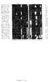
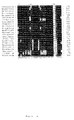
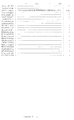
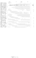
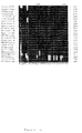
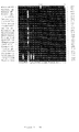
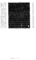
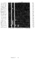
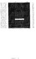
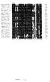
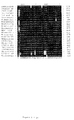
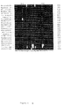
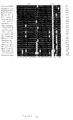
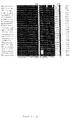
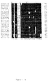
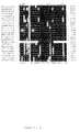
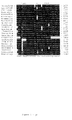
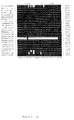
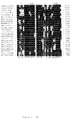
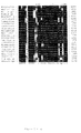
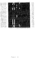
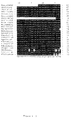
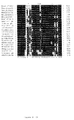
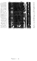
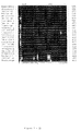
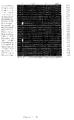
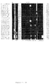
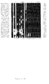
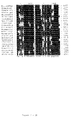
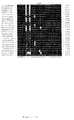
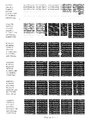
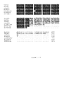
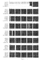
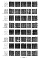
- Preferred sequences according to the present invention are set out in Figures 1, 2, 4 and 5: SEQ ID NO 1 to 2 and 15 to 24. Also unique parts and fragments of these sequences are part of the present invention. Preferred unique parts are set out in Table 2.
- sequences of more than 96.6% homology to SEQ ID NO 1 for identification or typing of Helicobacter species is also within the scope of the present invention.
- sequences of more than 97%, 97.5%, 98%, 98.5%, 99% or 99.5% homology to SEQ ID NO 2 are used for this goal.
- the present invention relates to a part of an isolated polynucleic acid as defined above, more particularly part or a fragment of SEQ ID NO 1 or 2, wherein said part is unique to the polynucleic acid sequence it is derived from.
- the term "unique” implies that at least one nucleotide of the fragment or part is different from a nucleotide present at the same nucleotide position in a known 16S rRNA sequence or the corresponding gene.
- a nucleotide can be deduced theoretically by looking at an alignment of the new sequences of this invention with other closely related Helicobacter 16S rDNA gene nucleotide sequences (see Figures 1, 2, 4 and 5). Said type of nucleotides are unique to the sequence they are derived from. These fragments are thus not part of any known 16S rRNA or gene sequence encoding the same.
- the fragments according to this embodiment of the present invention may be of any length between 10 to the maximum number of nucleotides of SEQ ID NO 1 or 2 or its variants. Preferred lengths are 10, 15, 20, 25, 30, 35, 40, 45, 50, 55, 60, etc. nucleotides.
- the present invention relates to a probe which specifically hybridizes to a polynucleic acid sequence as defined above.
- Probe R628f is a preferred " Candidatus Helicobacter bovis” specific probe.
- Probe V100f is a preferred " Candidatus Helicobacter suis” specific probe.
- Other suitable probes may be derived from a visual inspection of the alignment shown in Figure 1 or 2.
- the present invention relates to a primer which specifically amplifies a polynucleic acid sequence as defined above.
- Preferred primers according to the present invention are given in Table 2.
- Primers R574f and R832r are preferred " Candidatus Helicobacter bovis” specific primers and are suited for a specific PCR and in situ hybridisation assays.
- Primers V832f and V1621r are preferred " Candidatus Helicobacter suis” specific primers for a specific PCR and in situ hybridisation assays.
- Other suitable primers according to the present invention may be derived from a visual inspection of the alignment shown in Figure 1 or 2.
- the skilled man will be able to select primers that allow specific amplification of SEQ ID NO 1 or 2 or the claimed variants thereof under given or experimental conditions, such as temperature, buffer composition, polymerase chain reaction cycle etc.
- primers that allow specific amplification of SEQ ID NO 1 or 2 or the claimed variants thereof under given or experimental conditions, such as temperature, buffer composition, polymerase chain reaction cycle etc.
- probes that specifically hybridize to either SEQ ID NO 1 or 2 or the claimed variants under given experimental conditions such as temperature, buffer composition etc.
- the skilled man will furthermore be able to assess the efficacy of these primers or probes without undue experimentation. It is also obvious that the skilled man may chose to combine more than one primer pair or more than one probe to carry out the method defined above.
- the present invention relates to a method for detection and/or typing of Helicobacter strains present in a biological sample comprising hybridizing the 16S rRNA or 16S rDNA target region polynucleotides of said Helicobacter strains present in said biological sample with at least one probe as defined above.
- Preferably said method may be used to study and detect the occurrence of pathogenic Helicobacter strains.
- the present invention relates to a method for detection and/or typing of Helicobacter strains present in a biological sample comprising specifically amplifying the 16S rRNA or 16S rDNA target region polynucleotides of said Helicobacter strains present in said biological sample with at least one primer as defined above.
- Preferably said method may be used to study and detect the occurrence of pathogenic Helicobacter strains.
- a preferred embodiment according to the present invention involves a method for detection and/or typing of Helicobacter strains present in a biological sample comprising first amplifying a specifc target region encompassed in or comprising the 16S rRNA region of said Helicobacter strains present in said biological sample and subsequently hybridizing the 16S rRNA or 16S rDNA target region polynucleotides of said Helicobacter strains present in said biological sample with at least one (or more than one) probe as defined above.
- These techniques may comprise immobilizing the target polynucleic acids, possibly after amplification, on a solid support and performing a hybridization with labelled oligonucleotide probes of the present invention.
- said probes may be immobilized on a solid support and hybridization may be performed with labelled target polynucleic acids, possibly after amplification (i.e. a reverse hybridization).
- a preferred method according to the present invention is an in situ hybridisation assay (see Examples section).
- the well-known technique of Southern blotting is one example of a hybridization assay that can be used to perform the methods of the present invention.
- Another example of a hybridization technique is the DNA enzyme immuno assay (DEIA).
- DEIA DNA enzyme immuno assay
- PCR products are generated by a primer set, of which either the forward or the reverse primer contain biotin at the 5' end. This allows binding of the biotinylated amplimers to streptavidin-coated microtiter wells. PCR products are denatured by sodium hydroxide, which allows removal of the non-biotinylated strand.
- Specific digoxigenin (DIG)-labelled oligonucleotide probes are hybridized to the single-stranded immobilized PCR product and hybrids are detected by enzyme-labelled conjugate and colorimetric methods.
- DIG digoxigenin
- LiPA LiPA assay
- the LiPA uses oligonucleotide probes immobilized as parallel lines on a solid support strip (Stuyver et al. 1993; international patent application WO 94/12670). This approach is particularly advantageous since it is fast and simple to perform.
- the present invention relates to a diagnostic kit for detection and/or typing of Helicobacter strains comprising:
- the present invention relates to a medicament comprising a polynucleic acid sequence as defined above.
- the present invention relates to a polynucleic acid sequence as defined above for use as a medicament.
- the target material in the samples to be analysed may either be DNA or RNA, e.g. genomic DNA, messenger RNA, viral RNA or amplified versions thereof. These molecules are in this application also termed “polynucleic acids” or “polynucleotides”. More particularly, the target material according to the present invention will be 16S ribosomal RNA or DNA or amplified versions thereof.
- probe refers to a single-stranded oligonucleotide which is designed to specifically hybridize to " Candidatus Helicobacter bovis or suis" polynucleic acids.
- primer refers to a single stranded oligonucleotide sequence capable of acting as a point of initiation for synthesis of a primer extension product which is complementary to the nucleic acid strand to be copied.
- the length and the sequence of the primer must be such that they allow to prime the synthesis of the extension products.
- the primer is about 5-50 nucleotides long. Specific length and sequence will depend on the complexity of the required DNA or RNA targets, as well as on the conditions at which the primer is used, such as temperature and ionic strength. It is to be understood that the primers of the present invention may be used as probes and vice versa, provided that the experimental conditions are adapted.
- suitable primer pair in this invention refers to a pair of primers allowing specific amplification of a " Candidatus Helicobacter bovis or suis" polynucleic acid fragment.
- target region of a probe or a primer according to the present invention is a sequence within the "Candidatus Helicobacter bovis or suis" polynucleic acids to which the probe or the primer is completely complementary or partially complementary (i.e. with some degree of mismatch). It is to be understood that the complement of said target sequence is also a suitable target sequence in some cases.
- Probe hybridization of a probe to a target region of respectively the " Candidatus Helicobacter bovis” or Candidatus Helicobacter suis” polynucleic acids means that said probe forms a duplex with part of this region or with the entire region under the experimental conditions used, and that under those conditions said probe does not form a duplex with other regions of the polynucleic acids present in the sample to be analysed.
- “Specific hybridization” of a primer to a target region of respectively the " Candidatus Helicobacter bovis” or “ Candidatus Helicobacter suis” polynucleic acids means that, during the amplification step, said primer forms a duplex with part of this region or with the entire region under the experimental conditions used, and that under those conditions said primer does not form a duplex with other regions of the polynucleic acids present in the sample to be analysed.
- duplex as used hereby, means a duplex that will lead to specific amplification.
- Specific amplification of a fragment of respectively the " Candidatus Helicobacter bovis” or Candidatus Helicobacter suis” polynucleic acids means amplification of the fragment for which the primers were designed, and not of any other fragment of the polynucleic acids present in a sample.
- amplification primers do not have to match exactly with the corresponding target sequence in the template to warrant proper amplification is amply documented in the literature (Kwok et al., 1990). However, when the primers are not completely complementary to their target sequence, it should be taken into account that the amplified fragments will have the sequence of the primers and not of the target sequence. Primers may be labelled with a label of choice (e.g. biotine).
- the amplification method used can be either polymerase chain reaction (PCR; Saiki et al., 1988), ligase chain reaction (LCR; Landgren et al., 1988; Wu & Wallace, 1989; Barany, 1991), nucleic acid sequence-based amplification (NASBA; Guatelli et al., 1990; Compton, 1991), transcription-based amplification system (TAS; Kwoh et al., 1989), strand displacement amplification (SDA; Duck, 1990) or amplification by means of Qß replicase (Lomeli et al., 1989) or any other suitable method to amplify nucleic acid molecules known in the art.
- PCR polymerase chain reaction
- LCR Landgren et al., 1988; Wu & Wallace, 1989
- NASBA nucleic acid sequence-based amplification
- TAS transcription-based amplification system
- SDA strand displacement amplification
- Duck, 1990 Duck, 1990
- the probes of the invention are about 5 to 50 nucleotides long, more preferably from about 10 to 25 nucleotides. Particularly preferred lengths of probes include 10, 11, 12, 13, 14, 15, 16, 17, 18, 19, 20, 21, 22, 23, 24 or 25 nucleotides.
- the nucleotides as used in the present invention may be ribonucleotides, deoxyribonucleotides and modified nucleotides such as inosine or nucleotides containing modified groups which do not essentially alter their hybridization characteristics.
- Probe and primer sequences are represented throughout the specification as single stranded DNA oligonucleotides from the 5' to the 3' end. It is obvious to the man skilled in the art that any of the below-specified probes can be used as such, or in their complementary form, or in their RNA form (wherein T is replaced by U).
- the probes according to the invention can be prepared by cloning of recombinant plasmids containing inserts including the corresponding nucleotide sequences, if need be by excision of the latter from the cloned plasmids by use of the adequate nucleases and recovering them, e.g. by fractionation according to molecular weight.
- the probes according to the present invention can also be synthesized chemically, for instance by the conventional phospho-triester method.
- the oligonucleotides used as primers or probes may also comprise nucleotide analogues such as phosphorothiates (Matsukura et al., 1987), alkylphosphorothiates (Miller et al., 1979) or peptide nucleic acids (Nielsen et al., 1991; Nielsen et al., 1993) or may contain intercalating agents (Asseline et al., 1984). As most other variations or modifications introduced into the original DNA sequences of the invention these variations will necessitate adaptions with respect to the conditions under which the oligonucleotide should be used to obtain the required specificity and sensitivity.
- nucleotide analogues such as phosphorothiates (Matsukura et al., 1987), alkylphosphorothiates (Miller et al., 1979) or peptide nucleic acids (Nielsen et al., 1991; Nielsen et al., 1993) or
- solid support can refer to any substrate to which an oligonucleotide probe can be coupled, provided that it retains its hybridization characteristics and provided that the background level of hybridization remains low.
- the solid substrate will be a microtiter plate, a membrane (e.g. nylon or nitrocellulose) or a microsphere (bead) or a chip.
- a membrane e.g. nylon or nitrocellulose
- a microsphere bead
- a chip Prior to application to the membrane or fixation it may be convenient to modify the nucleic acid probe in order to facilitate fixation or improve the hybridization efficiency. Such modifications may encompass homopolymer tailing, coupling with different reactive groups such as aliphatic groups, NH 2 groups, SH groups, carboxylic groups, or coupling with biotin, haptens or proteins.
- labelled refers to the use of labelled nucleic acids. Labelling may be carried out by the use of labelled nucleotides incorporated during the polymerase step of the amplification such as illustrated by Saiki et al. (1988) or Bej et al. (1990) or labelled primers, or by any other method known to the person skilled in the art.
- the nature of the label may be isotopic ( 32 P, 35 S, etc.) or non-isotopic (biotin, digoxigenin, etc.).
- the "biological sample” may be for instance cultured Helicobacter strains, gastric, abomasal stomachs, omasal stomachs, reticulum and rumen, or duodenal biopsies (fresh or parafine material), faeces, saliva, mouth mucosa, gastric juice or urine.
- these samples may be taken from piglets, pigs, humans, calves, cattle, etc.
- the stability of the [probe : target] nucleic acid hybrid should be chosen to be compatible with the assay conditions. This may be accomplished by avoiding long AT-rich sequences, by terminating the hybrids with G:C base pairs, and by designing the probe with an appropriate Tm. The beginning and end points of the probe should be chosen so that the length and %GC result in a Tm about 2-10EC higher than the temperature at which the final assay will be performed.
- the base composition of the probe is significant because G-C base pairs exhibit greater thermal stability as compared to A-T base pairs due to additional hydrogen bonding. Thus, hybridization involving complementary nucleic acids of higher G-C content will be more stable at higher temperatures.
- **It is desirable to have probes which hybridize only under conditions of high stringency. Under high stringency conditions only highly complementary nucleic acid hybrids will form; hybrids without a sufficient degree of complementarity will not form. Accordingly, the stringency of the assay conditions determines the amount of complementarity needed between two nucleic acid strands forming a hybrid. The degree of stringency is chosen such as to maximize the difference in stability between the hybrid formed with the target and the non-target nucleic acid.
- hybridization buffer means a buffer allowing a hybridization reaction between the probes and the polynucleic acids present in the sample, or the amplified products, under the appropriate stringency conditions.
- wash solution means a solution enabling washing of the hybrids formed under the appropriate stringency conditions.
- stomachs from clinically healthy slaughterhouse cattle originating from different Belgian and Dutch farms, were selected.
- the stomachs were opened longitudinally along the greater curvatura and rinsed gently with tap water.
- Two small mucosal fragments were taken from each stomach, one near the torus pyloricus and one in the fundic region, and were tested for urease activity (CUTest, Temmler Pharma) for h at 37 °C.
- Three mucosal biopsies from the pyloric region were taken for immunohistochemistry and in situ hybridisation and placed into 4 % buffered formaline for 24 hours.
- a pyloric sample was taken from the same region and fixed in cacodylate buffer (0.1 M, pH 7.0) containing 5 % glutaraldehyde and 0.15 % (wt/vol) ruthenium red. From each stomach a mucosal fragment was also taken for PCR analysis, placed into sterile PBS and frozen in liquid nitrogen. Special care was taken during sampling to avoid cross-contamination.
- Stomachs from 5 healthy slaughterhouse pigs were selected, all originating from different farms in Belgium and the Netherlands.
- the stomachs were opened longitudinally along the greater curvatura and rinsed gently with tap water.
- a small mucosal fragment was taken from each stomach near the torus pyloricus and placed into an urease test tube (CUTest, Temmler Pharma) for 2 hours at 37 °C.
- samples were taken from the same places and fixed in 0.1 M cacodylate buffer (pH 7.0) containing 5% glutaraldehyde and 0.15 % (wt/vol) ruthenium red. Of each stomach a mucosal fragment was also taken for PCR, placed into sterile PBS and frozen in liquid nitrogen. Special care was taken during sampling to avoid cross-contamination.
- cacodylate buffer pH 7.0
- Peroxidase activity was developed using H 2 O 2 with diaminobenzidine (DAB) as a chromogen (Fast DAB Tablet Set, Sigma-Aldrich). Subsequently, the sections were counterstained with Mayer's hematoxylin and mounted. As a negative control, the primary antibody was replaced with fetal calf serum in Tris-HCl buffer (pH 7.6). As a positive control, a section of a mouse stomach experimentally infected with Helicobacter pylori LMG 7539 T was used.
- DAB diaminobenzidine
- Candidatus Helicobacter bovis three different pyloric samples were selected for electronmicroscopic evaluation based upon the high presence of Helicobacter -like organisms in the corresponding immunostained sections.
- Candidatus Helicobacter suis two different antral biopsies were selected for electronmicroscopic evaluation based on the high presence of gastrospirillum-like organisms in the corresponding immunostained slides.
- DNA was isolated from the scrapings of the gastric biopsies and from the reference strains by lysis with guanidinium isothiocyanate and DNA was bound to silica particles according to the method of Boom et al. (1990).
- H33f, H61f and H1368r were selected from rRNA superfamily VI (Helicobacter, Campylobacter, Arcobacter, Wolinella) specific regions of the 16S rRNA gene (Table 2).
- primer 1492RPL The use of broad range primer 1492RPL was suggested by Weissburg et al. (1991).
- a genus Helicobacter -specific primer H274f was adapted from primer 274r described by Dewhirst et al. (1994) (Table 2).
- Primer combinations H33F-H1368r, H274f-1492RPL and H61f-1492RPL were used to amplify a ⁇ 1.3-Kb, ⁇ 1.2-Kb and a ⁇ 1.4-Kb fragment of "Candidatus Helicobacter suis" respectively.
- PCR reactions were performed in a volume of 50 pl containing 10mM Tris HCl (pH 8.3), 50 mM KCl, 3.5 mM MgCl 2, 200 pM of each deoxynucleoside triphosphate, 1.5 U of AmpliTaq Gold (Perkin-Elmer, Roche Molecular Systems) and 25 pmol of both forward and reverse primer (Eurogentec). Reactions were covered with mineral oil and PCR was performed in a Biomed-60 thermocycler under the following conditions: 9 min preincubation at 94 °C to activate AmpliTaq Gold, followed by 50 cycles of 30 s at 94 °C, 45 s at 55 °C and 45 s at 72 °C. Final extension was performed for 5 min at 72 °C. DNA-extractions of Helicobacter acinonychis LMG 12684 T and Helicobacter mustelae LMG 8776 were used as positive controls.
- PCR products were separated on 1 % agarose gels and stained with ethidium bromide.
- PCR products were derived from Helicobacter -like organisms
- the desired DNA-bands were cut from the gels, diluted 1/2 in distilled water and sequenced using the H33f and H1368r 5'-Indocarbocyanin (Cy5) for " Candidatus Helicobacter bovis” and respectively H61f and 1492RPL Indocarbocyanin (Cy5) labeled for " Candidatus Helicobacter suis”. Partial sequences were screened for homologous sequences using the NCBI GENINFO ® BLAST Network service (http://www.ncbi.nlm.nih.gov/BLAST/) (Altshul et al., 1997).
- PCR amplimers comprising the 16S rDNA-sequences derived from four different stomach samples (R2, R3, R5, R6) were each cloned into plasmid vector pGEM-T (Promega Biotech) according to the manufacturer's instructions and transformed into Escherichia coli JM109 using standard procedures. Plasmids were purified using the Easy Prep Plasmid Preparation Kit (Pharmacia Biotech). Sequences were determined by the T7-sequencing system (Pharmacia Biotech). Two primers flanking the multiple cloning sites (T7, SP6) as well as internal primers H390f and H1053r were used (Table 2).
- R2XA The sequence derived from the clone of the R2 sample (R2XA) was used as reference sequence. This sequence was has been asigned Genbank Accession No. AF127028. Sequence analysis was performed with the PCGene software (Intelligenetics)
- PCR amplimers comprising the 16S rDNA-sequences from 2 different stomachs (V2B, V4A) were cloned into plasmid vector pGEM-T (Promega Biotech) according to the manufacturer's instructions and transformed into Escherichia coli JM109 using standard procedures. Plasmids were purified using the Easy Prep Plasmid Prep Kit (Pharmacia Biotech). Sequences were determined by the T7-sequencing system (Pharmacia Biotech). Two primers flanking the multiple cloning sites (T7 and SP6) as well as internal primers H390f and H1053r were used (Table 2). Sequence analysis was performed with the PCGene software (Intelligenetics).
- a reference sequence was determined based on its high length and was compared to the new sequencve and the other derived sequences, to check its integrity (see Figures 4 and 5).
- the reference sequence V2BXA was assigned Genbank Accession No. AF127028.
- Candidatus Helicobacter bovis specific PCR-assay
- Candidatus Helicobacter bovis specific oligonucleotides R574f and R832r (Table 2), were selected from variable rDNA regions of the sequences determined by direct and indirect sequence analysis. These primers comprised a 259 bp 16S rDNA-fragment and were used to develop a specific PCR and an in situ hybridisation procedure. Within this fragment an internal " Candidatus Helicobacter bovis” specific probe R628f (Table 2) was selected for southern blot hybridisation purposes.
- PCR reactions were performed in a volume of 50 ⁇ l containing 10 mM Tris HCl (pH 8.3), 50 mM KCl, 2.5 mM MgCl 2, 200 ⁇ M of each deoxynucleoside triphosphate, 1.5 U of AmpliTaq Gold, and 25 pmol of both forward and reverse primer.
- PCR amplification was performed under the following conditions: 9 min preincubation at 94 °C to activate AmpliTaq Gold, followed by 40 cycles of 30 s at 94 °C, 45 s at 60 °C and 90 s at 72 °C. Final extension was performed for 5 min at 72 °C. All gastric DNA-extracts were tested with this PCR.
- plasmid DNA was used from the cloned 16S rDNA fragments (R2XA).
- R2XA cloned 16S rDNA fragments
- Candidatus Helicobacter bovis specific oligonucleotides R574f and R832r was tested by PCR using DNA-extracts of 15 different Helicobacter strains and a Wolinella succinogenes strain (Table 1).
- PCR products were separated on 2 % agarose gels, stained with ethidium bromide and transferred to Hybond N+ (Amersham) by electro-elution.
- Southern blot hybridisation was performed with the [ ⁇ 32P ] ATP labelled probe R628f (Table 2) according to standard procedures (Amersham Pharmacia Biotech). In order to ensure the specificity of the probe hybridisation, blots were washed twice with 0.1 x SSC + 0.1 % SDS at 55°C.
- Candidatus Helicobacter suis specific diagnostic PCR-assay and Southern blot hybridisation
- Candidatus Helicobacter suis -specific primers (V832f and V1261r) were selected from variable rDNA regions of the sequences determined by direct and indirect sequence analysis, comprising a ⁇ 0.4-Kb 16S rDNA-fragment. Within this fragment a " Candidatus Helicobacter suis”-specific probe V1000f (Table 2) was selected for hybridisation purposes. PCR reactions were performed in a volume of 50 pl containing 10 mM Tris HCl (pH 8.3), 50 mM KCl, 2.5 mM MgCl 2, 200 ⁇ M of each deoxynucleoside triphosphate, 1.5 U of AmpliTaq Gold (Perkin-Elmer), and 25 pmol of both forward and reverse primer (Eurogentec).
- PCR amplification was performed under the following conditions: 9 min preincubation at 94 °C to activate AmpliTaq Gold, followed by 40 cycles of 30 s at 94 °C, 45 s at 60 °C and 90 s at 72 °C. Final extension was performed for 5 min at 72 °C.
- plasmid DNA was used from the cloned 16S rDNA fragments (V2B, V4A).
- V2B, V4A 16S rDNA fragments
- PCR products were separated on 2% agarose gels, stained with ethidium bromide and transferred to Hybond N+ (Amersham) by electro-blotting.
- Southern blot hybridisation was performed with the [ ⁇ 32P ] ATP-labelled probe V1000f according to standard procedures (Amersham Pharmacia Biotech). In order to ensure the specificity of the probe hybridisation, blots were washed twice with 0.1 x SSC + 0.1 % SDS at 55°C.
- RNA'se activity was performed at 180°C for 3 hours. Further precautions included the use of RNA'se-free water, and the use of sterile disposable materials whenever possible. Sections of the paraffin-embedded tissues (4 ⁇ m thick) were mounted on RNA'se-free, APES-coated slides (Sigma-Aldrich) and fixed by heating for 1 hour at 60 °C. The sections were deparaffinized in xylene (2x5 min), rehydrated through graded ethanol, and washed twice in PBS for 5 min each. Sections were then treated with proteinase K (DAKO) for 15 min each at 37 °C in a humidified chamber.
- DAKO proteinase K
- the enzyme was inactivated by treatment with 0.2 % glycine in PBS for 3 min. Sections were washed twice in PBS for 5 min each, dehydrated in graded ethanol and air dried. Tissues were circumlined with a DAKO Pen (DAKO) to avoid liquid spillage during further processing and to ensure an efficient sealing of the coverslip.
- DAKO Pen DAKO Pen
- sections were covered with 5 to 15 pl solution, containing 5 ng/ ⁇ l labeled probe in 50 % deionized formamide, 2x SCC, 10 % dextran sulfate, 0.25 ⁇ g/ ⁇ l yeast t-RNA, 0.5 ⁇ g/ ⁇ l heat denatured salmon sperm DNA, and lx Denhart's solution.
- Sections were covered with a piece of coverslip to avoid evaporation.
- sections were heated for 10 min at 95 °C and chilled on ice for 10 min. Slides were then hybridised overnight at 37 °C in a humidified chamber.
- the coverslips were removed and the sections were washed in 2x SCC and 1x SCC at room temperature for 10 min each followed by two washes of 0.3x SCC at 40 °C for 10 min and at room temperature for 10 min, respectively.
- the "Tyramid Signal Amplification System” (NEN Life Science Products) was applied on each section, following manufacturer's instructions. The hybridised probe was then visualized, using H 2 O 2 with diaminobenzidine as a chromogen (Fast DAB Tablet Set, Sigma-Aldrich). Thereafter the sections were counterstained with Mayer's hematoxylin and mounted.
- the 16S rDNA nucleotide sequence of " Candidatus Helicobacter suis” has been deposited in the Genbank database under accession number AF127028.
- Urease activity was observed in all pyloric samples (7/7). In the fundic samples, urease activity was absent (0/7). Spiral immunostained organisms were observed in the pyloric samples of all animals. The highest concentration was seen in the most distal pyloric samples. They were mostly situated in the mucus layer and in the lumen of the proximal part of the gastric crypts where they formed small clusters. In some samples, coccoid organisms, were observed between the spiral bacteria, which also crossreacted with the H. pylori polyclonal antibodies. In the positive control only Helicobacter pylori - like bacteria were stained while in the negative controls no staining was observed.
- PCR amplification of the 16S rRNA gene using the H33f and H1368r primers produced a fragment of the expected size range ( ⁇ 1.3 Kbp) in all seven samples examined.
- Partial direct sequence analysis of four of these bands (R2, R3, R5, R6) and subsequent database comparison (BLAST) confirmed the PCR products to be Helicobacter -like 16S rDNA fragments.
- Four PCR products (R2, R3, R5, R6) were cloned followed by partial screening. In one clone a Clostridium -like 16S rDNA fragment was found. In all other clones Helicobacter -like fragments were inserted.
- the 16S rDNA sequences of four clones derived from different animals were determined. Additional sequences of three other samples (R13, R27, R28) were characterized by direct sequence analysis using the primers H33f, H1368r, H390f and H1053r.
- Sequence length varied from 1267 to 1335 basepairs. Pairwise comparisons between these 7 sequences revealed a sequence homology of more than 99 %.
- One reference sequence (R2XA) of 1335 bp (see Figure 1: SEQ ID NO 1) was selected for phylogenetic evaluation.
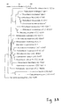
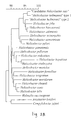
- a similarity matrix based on comparisons of 16S rRNA sequences of 23 strains representing all validly named Helicobacter species, " Helicobacter heilmannii " (type1, type2), Campylobacter jejuni, Arcobacter cryaerophilus and Wollinella succinogenes was calculated.
- the sequence of the porcine gastrospirillum-like organism formed a distinct subgroup within the Helicobacter lineage together with other gastrospirilla: Helicobacter felis, H. bizzozeronii, H. salomonis, " H. heilmannii " type 1 and type 2.
- the sequence was highly similar to that of " H. heilmannii " type 1 (level of similarity 99.5 %).
- the similarity level of other gastrospirillum-like bacteria, H. felis , H. bizzozeronii, H. salomonis and H. heilmanni type 2 was 96.4 %, 96.5 %, 96.6 % and 96.8 % respectively.
- the reference sequence was clearly distinct from sequences belonging to other superfamily VI-genera, as shown by a 86.2 %, 84.7 % and 89.6 % homology with Campylobacter jejuni, Arcobacter butzleri and Wolinella succinogenes respectively.
- a 259 base fragment was produced for all seven stomach samples with primer pair R574f-R832r. All PCR products crosshybridised with the R628f probe after southern blot hybridisation. No amplification product was obtained using DNA preparations from any of the Helicobacter strains, nor from the bovine Wolinella succinogenes strain (Table 1). The positive control yielded a ⁇ 0.3 Kb product as expected. There was no DNA-amplification using the negative control material.
- Candidatus Helicobacter suis -specific PCR and Southern blot hybridisation
- Amplification of Helicobacter DNA using the primers V832f and V1261r produced a 433-base fragment from all five stomach samples. All PCR products hybridised with the V1000f probe after Southern blot hybridisation. No amplification product was obtained using DNA preparations from any of the Helicobacter strains including H. felis , H. bizzozeronii and H. salomonis (Table 1), nor from the negative control. PCR with the cloned reference material (2BXA) yielded a ⁇ 0.4 Kb product as expected.
- Treponema bryantii sp.nov. a rumen spirochete that interacts with cellulolytic bacteria. Archiv Microbiol 127 , 145-156.
- Taxonomic note a place for DNA-DNA reassociation and 16S rRNA sequence analysis in the present species definition in bacteriology. Int J Sys Bacter 44 , 846-849.
- Taxonomic notes a proposal for recording the properties of putative taxa of procaryotes. Int J Syst Bacteriol 44, 174-176.
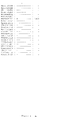
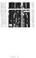
- Taxon Source Genbank Accession N° "Gastrospirillum hominis” type 1 Human gastric mucosa L10079 "Gastrospirillum hominis” type 2 Human gastric mucosa L10080 Helicobacter acinonychis Cheetahgastric mucosa M88148 Helicobacter bilis Murine liver U18766 Helicobacter bizzozeronii Canine gastric mucosa Y09404 Helicobacter canis Canine feces L13464 Helicobacter cholecystus Murine liver U46129 Helicobacter cinaedi Human feces M88150 Helicobacter felis Feline gastric mucosa M57398 Helicobacter fennelliae Human feces M88154 Helicobacter hepaticus Murine liver U07574 Helicobacter muridarum Murine intestinal mucosa M80205 Helicobacter mustelae Ferret gastric mucosa M35048 Helicobacter nemestrina
Priority Applications (1)
| Application Number | Priority Date | Filing Date | Title |
|---|---|---|---|
| EP99870035A EP1035219A1 (de) | 1999-02-25 | 1999-02-25 | Gastrische Helicobacter 16S-rDNA Sequenzen von Rind und Schwein und ihre Anwendung zur Detektion and Typisierung von Helicobacter Stämmen |
Applications Claiming Priority (1)
| Application Number | Priority Date | Filing Date | Title |
|---|---|---|---|
| EP99870035A EP1035219A1 (de) | 1999-02-25 | 1999-02-25 | Gastrische Helicobacter 16S-rDNA Sequenzen von Rind und Schwein und ihre Anwendung zur Detektion and Typisierung von Helicobacter Stämmen |
Publications (1)
| Publication Number | Publication Date |
|---|---|
| EP1035219A1 true EP1035219A1 (de) | 2000-09-13 |
Family
ID=8243803
Family Applications (1)
| Application Number | Title | Priority Date | Filing Date |
|---|---|---|---|
| EP99870035A Withdrawn EP1035219A1 (de) | 1999-02-25 | 1999-02-25 | Gastrische Helicobacter 16S-rDNA Sequenzen von Rind und Schwein und ihre Anwendung zur Detektion and Typisierung von Helicobacter Stämmen |
Country Status (1)
| Country | Link |
|---|---|
| EP (1) | EP1035219A1 (de) |
Cited By (37)
| Publication number | Priority date | Publication date | Assignee | Title |
|---|---|---|---|---|
| WO2008058727A1 (en) * | 2006-11-14 | 2008-05-22 | Universiteit Gent | In vitro cultivation of helicobacter species |
| US7666592B2 (en) | 2004-02-18 | 2010-02-23 | Ibis Biosciences, Inc. | Methods for concurrent identification and quantification of an unknown bioagent |
| US7666588B2 (en) | 2001-03-02 | 2010-02-23 | Ibis Biosciences, Inc. | Methods for rapid forensic analysis of mitochondrial DNA and characterization of mitochondrial DNA heteroplasmy |
| US7714275B2 (en) | 2004-05-24 | 2010-05-11 | Ibis Biosciences, Inc. | Mass spectrometry with selective ion filtration by digital thresholding |
| US7718354B2 (en) | 2001-03-02 | 2010-05-18 | Ibis Biosciences, Inc. | Methods for rapid identification of pathogens in humans and animals |
| US7741036B2 (en) | 2001-03-02 | 2010-06-22 | Ibis Biosciences, Inc. | Method for rapid detection and identification of bioagents |
| US7781162B2 (en) | 2001-03-02 | 2010-08-24 | Ibis Biosciences, Inc. | Methods for rapid identification of pathogens in humans and animals |
| US7811753B2 (en) | 2004-07-14 | 2010-10-12 | Ibis Biosciences, Inc. | Methods for repairing degraded DNA |
| US7939079B2 (en) | 2006-11-14 | 2011-05-10 | Universiteit Gent | Helicobacter species and cultivation thereof |
| US7956175B2 (en) | 2003-09-11 | 2011-06-07 | Ibis Biosciences, Inc. | Compositions for use in identification of bacteria |
| US7964343B2 (en) | 2003-05-13 | 2011-06-21 | Ibis Biosciences, Inc. | Method for rapid purification of nucleic acids for subsequent analysis by mass spectrometry by solution capture |
| US8026084B2 (en) | 2005-07-21 | 2011-09-27 | Ibis Biosciences, Inc. | Methods for rapid identification and quantitation of nucleic acid variants |
| US8046171B2 (en) | 2003-04-18 | 2011-10-25 | Ibis Biosciences, Inc. | Methods and apparatus for genetic evaluation |
| US8057993B2 (en) | 2003-04-26 | 2011-11-15 | Ibis Biosciences, Inc. | Methods for identification of coronaviruses |
| US8071309B2 (en) | 2002-12-06 | 2011-12-06 | Ibis Biosciences, Inc. | Methods for rapid identification of pathogens in humans and animals |
| US8073627B2 (en) | 2001-06-26 | 2011-12-06 | Ibis Biosciences, Inc. | System for indentification of pathogens |
| US8084207B2 (en) | 2005-03-03 | 2011-12-27 | Ibis Bioscience, Inc. | Compositions for use in identification of papillomavirus |
| US8097416B2 (en) * | 2003-09-11 | 2012-01-17 | Ibis Biosciences, Inc. | Methods for identification of sepsis-causing bacteria |
| US8119336B2 (en) | 2004-03-03 | 2012-02-21 | Ibis Biosciences, Inc. | Compositions for use in identification of alphaviruses |
| US8148163B2 (en) | 2008-09-16 | 2012-04-03 | Ibis Biosciences, Inc. | Sample processing units, systems, and related methods |
| US8158936B2 (en) | 2009-02-12 | 2012-04-17 | Ibis Biosciences, Inc. | Ionization probe assemblies |
| US8158354B2 (en) | 2003-05-13 | 2012-04-17 | Ibis Biosciences, Inc. | Methods for rapid purification of nucleic acids for subsequent analysis by mass spectrometry by solution capture |
| US8163895B2 (en) | 2003-12-05 | 2012-04-24 | Ibis Biosciences, Inc. | Compositions for use in identification of orthopoxviruses |
| US8182992B2 (en) | 2005-03-03 | 2012-05-22 | Ibis Biosciences, Inc. | Compositions for use in identification of adventitious viruses |
| US8298760B2 (en) | 2001-06-26 | 2012-10-30 | Ibis Bioscience, Inc. | Secondary structure defining database and methods for determining identity and geographic origin of an unknown bioagent thereby |
| US8407010B2 (en) | 2004-05-25 | 2013-03-26 | Ibis Biosciences, Inc. | Methods for rapid forensic analysis of mitochondrial DNA |
| US8534447B2 (en) | 2008-09-16 | 2013-09-17 | Ibis Biosciences, Inc. | Microplate handling systems and related computer program products and methods |
| US8546082B2 (en) | 2003-09-11 | 2013-10-01 | Ibis Biosciences, Inc. | Methods for identification of sepsis-causing bacteria |
| US8550694B2 (en) | 2008-09-16 | 2013-10-08 | Ibis Biosciences, Inc. | Mixing cartridges, mixing stations, and related kits, systems, and methods |
| US8563250B2 (en) | 2001-03-02 | 2013-10-22 | Ibis Biosciences, Inc. | Methods for identifying bioagents |
| US8871471B2 (en) | 2007-02-23 | 2014-10-28 | Ibis Biosciences, Inc. | Methods for rapid forensic DNA analysis |
| US8950604B2 (en) | 2009-07-17 | 2015-02-10 | Ibis Biosciences, Inc. | Lift and mount apparatus |
| US9149473B2 (en) | 2006-09-14 | 2015-10-06 | Ibis Biosciences, Inc. | Targeted whole genome amplification method for identification of pathogens |
| US9194877B2 (en) | 2009-07-17 | 2015-11-24 | Ibis Biosciences, Inc. | Systems for bioagent indentification |
| US9598724B2 (en) | 2007-06-01 | 2017-03-21 | Ibis Biosciences, Inc. | Methods and compositions for multiple displacement amplification of nucleic acids |
| US9890408B2 (en) | 2009-10-15 | 2018-02-13 | Ibis Biosciences, Inc. | Multiple displacement amplification |
| CN114317788A (zh) * | 2021-12-30 | 2022-04-12 | 漳州傲农现代农业开发有限公司 | 一种用于检测猪源奇异变形杆菌的探针引物组合及其试剂盒和方法 |
Citations (1)
| Publication number | Priority date | Publication date | Assignee | Title |
|---|---|---|---|---|
| WO1987002775A1 (en) * | 1985-10-24 | 1987-05-07 | Southwest Foundation For Biomedical Research | Synthetic peptides and use for diagnosis and vaccination for aids and arc |
-
1999
- 1999-02-25 EP EP99870035A patent/EP1035219A1/de not_active Withdrawn
Patent Citations (1)
| Publication number | Priority date | Publication date | Assignee | Title |
|---|---|---|---|---|
| WO1987002775A1 (en) * | 1985-10-24 | 1987-05-07 | Southwest Foundation For Biomedical Research | Synthetic peptides and use for diagnosis and vaccination for aids and arc |
Non-Patent Citations (4)
| Title |
|---|
| BATTLES ET AL.: "Diagnostic assay for helicobacter hepaticus based on nucleotide sequence of its rRNA gene", JOURNAL OF CLINICAL MICROBIOLOGY, vol. 33, no. 5, May 1995 (1995-05-01), pages 1344 - 1347, XP002110982 * |
| DATABASE GENESEQ PATENT SEQUE 1 January 1900 (1900-01-01), XP002110984, Database accession no. Q10072 * |
| DEWHIRST F E ET AL: "Phylogeny of Helicobacter isolates from bird and swine feces and description of Helicobacter pametensis sp. nov.", INTERNATIONAL JOURNAL OF SYSTEMATIC BACTERIOLOGY, (1994 JUL) 44 (3) 553-60., XP002110983 * |
| PASTER ET AL.: "Phylogeny of Helicobacter felis sp. nov., Helicobacter mustelae, and related bacteria.", INTERNATIONAL JOURNAL OF SYSTEMATIC BACTERIOLOGY, vol. 41, no. 1, January 1991 (1991-01-01), pages 31 - 38, XP002110981 * |
Cited By (66)
| Publication number | Priority date | Publication date | Assignee | Title |
|---|---|---|---|---|
| US8815513B2 (en) | 2001-03-02 | 2014-08-26 | Ibis Biosciences, Inc. | Method for rapid detection and identification of bioagents in epidemiological and forensic investigations |
| US8214154B2 (en) | 2001-03-02 | 2012-07-03 | Ibis Biosciences, Inc. | Systems for rapid identification of pathogens in humans and animals |
| US8802372B2 (en) | 2001-03-02 | 2014-08-12 | Ibis Biosciences, Inc. | Methods for rapid forensic analysis of mitochondrial DNA and characterization of mitochondrial DNA heteroplasmy |
| US9416424B2 (en) | 2001-03-02 | 2016-08-16 | Ibis Biosciences, Inc. | Methods for rapid identification of pathogens in humans and animals |
| US7718354B2 (en) | 2001-03-02 | 2010-05-18 | Ibis Biosciences, Inc. | Methods for rapid identification of pathogens in humans and animals |
| US7741036B2 (en) | 2001-03-02 | 2010-06-22 | Ibis Biosciences, Inc. | Method for rapid detection and identification of bioagents |
| US7781162B2 (en) | 2001-03-02 | 2010-08-24 | Ibis Biosciences, Inc. | Methods for rapid identification of pathogens in humans and animals |
| US8265878B2 (en) | 2001-03-02 | 2012-09-11 | Ibis Bioscience, Inc. | Method for rapid detection and identification of bioagents |
| US7666588B2 (en) | 2001-03-02 | 2010-02-23 | Ibis Biosciences, Inc. | Methods for rapid forensic analysis of mitochondrial DNA and characterization of mitochondrial DNA heteroplasmy |
| US9752184B2 (en) | 2001-03-02 | 2017-09-05 | Ibis Biosciences, Inc. | Methods for rapid forensic analysis of mitochondrial DNA and characterization of mitochondrial DNA heteroplasmy |
| US8268565B2 (en) | 2001-03-02 | 2012-09-18 | Ibis Biosciences, Inc. | Methods for identifying bioagents |
| US8563250B2 (en) | 2001-03-02 | 2013-10-22 | Ibis Biosciences, Inc. | Methods for identifying bioagents |
| US8017358B2 (en) | 2001-03-02 | 2011-09-13 | Ibis Biosciences, Inc. | Method for rapid detection and identification of bioagents |
| US8017743B2 (en) | 2001-03-02 | 2011-09-13 | Ibis Bioscience, Inc. | Method for rapid detection and identification of bioagents |
| US8017322B2 (en) | 2001-03-02 | 2011-09-13 | Ibis Biosciences, Inc. | Method for rapid detection and identification of bioagents |
| US8298760B2 (en) | 2001-06-26 | 2012-10-30 | Ibis Bioscience, Inc. | Secondary structure defining database and methods for determining identity and geographic origin of an unknown bioagent thereby |
| US8380442B2 (en) | 2001-06-26 | 2013-02-19 | Ibis Bioscience, Inc. | Secondary structure defining database and methods for determining identity and geographic origin of an unknown bioagent thereby |
| US8921047B2 (en) | 2001-06-26 | 2014-12-30 | Ibis Biosciences, Inc. | Secondary structure defining database and methods for determining identity and geographic origin of an unknown bioagent thereby |
| US8073627B2 (en) | 2001-06-26 | 2011-12-06 | Ibis Biosciences, Inc. | System for indentification of pathogens |
| US8822156B2 (en) | 2002-12-06 | 2014-09-02 | Ibis Biosciences, Inc. | Methods for rapid identification of pathogens in humans and animals |
| US8071309B2 (en) | 2002-12-06 | 2011-12-06 | Ibis Biosciences, Inc. | Methods for rapid identification of pathogens in humans and animals |
| US9725771B2 (en) | 2002-12-06 | 2017-08-08 | Ibis Biosciences, Inc. | Methods for rapid identification of pathogens in humans and animals |
| US8046171B2 (en) | 2003-04-18 | 2011-10-25 | Ibis Biosciences, Inc. | Methods and apparatus for genetic evaluation |
| US8057993B2 (en) | 2003-04-26 | 2011-11-15 | Ibis Biosciences, Inc. | Methods for identification of coronaviruses |
| US8158354B2 (en) | 2003-05-13 | 2012-04-17 | Ibis Biosciences, Inc. | Methods for rapid purification of nucleic acids for subsequent analysis by mass spectrometry by solution capture |
| US7964343B2 (en) | 2003-05-13 | 2011-06-21 | Ibis Biosciences, Inc. | Method for rapid purification of nucleic acids for subsequent analysis by mass spectrometry by solution capture |
| US8476415B2 (en) | 2003-05-13 | 2013-07-02 | Ibis Biosciences, Inc. | Methods for rapid purification of nucleic acids for subsequent analysis by mass spectrometry by solution capture |
| US8546082B2 (en) | 2003-09-11 | 2013-10-01 | Ibis Biosciences, Inc. | Methods for identification of sepsis-causing bacteria |
| US7956175B2 (en) | 2003-09-11 | 2011-06-07 | Ibis Biosciences, Inc. | Compositions for use in identification of bacteria |
| US8097416B2 (en) * | 2003-09-11 | 2012-01-17 | Ibis Biosciences, Inc. | Methods for identification of sepsis-causing bacteria |
| US8013142B2 (en) | 2003-09-11 | 2011-09-06 | Ibis Biosciences, Inc. | Compositions for use in identification of bacteria |
| US8163895B2 (en) | 2003-12-05 | 2012-04-24 | Ibis Biosciences, Inc. | Compositions for use in identification of orthopoxviruses |
| US9447462B2 (en) | 2004-02-18 | 2016-09-20 | Ibis Biosciences, Inc. | Methods for concurrent identification and quantification of an unknown bioagent |
| US8187814B2 (en) | 2004-02-18 | 2012-05-29 | Ibis Biosciences, Inc. | Methods for concurrent identification and quantification of an unknown bioagent |
| US7666592B2 (en) | 2004-02-18 | 2010-02-23 | Ibis Biosciences, Inc. | Methods for concurrent identification and quantification of an unknown bioagent |
| US8119336B2 (en) | 2004-03-03 | 2012-02-21 | Ibis Biosciences, Inc. | Compositions for use in identification of alphaviruses |
| US7714275B2 (en) | 2004-05-24 | 2010-05-11 | Ibis Biosciences, Inc. | Mass spectrometry with selective ion filtration by digital thresholding |
| US9449802B2 (en) | 2004-05-24 | 2016-09-20 | Ibis Biosciences, Inc. | Mass spectrometry with selective ion filtration by digital thresholding |
| US8987660B2 (en) | 2004-05-24 | 2015-03-24 | Ibis Biosciences, Inc. | Mass spectrometry with selective ion filtration by digital thresholding |
| US8173957B2 (en) | 2004-05-24 | 2012-05-08 | Ibis Biosciences, Inc. | Mass spectrometry with selective ion filtration by digital thresholding |
| US8407010B2 (en) | 2004-05-25 | 2013-03-26 | Ibis Biosciences, Inc. | Methods for rapid forensic analysis of mitochondrial DNA |
| US7811753B2 (en) | 2004-07-14 | 2010-10-12 | Ibis Biosciences, Inc. | Methods for repairing degraded DNA |
| US9873906B2 (en) | 2004-07-14 | 2018-01-23 | Ibis Biosciences, Inc. | Methods for repairing degraded DNA |
| US8084207B2 (en) | 2005-03-03 | 2011-12-27 | Ibis Bioscience, Inc. | Compositions for use in identification of papillomavirus |
| US8182992B2 (en) | 2005-03-03 | 2012-05-22 | Ibis Biosciences, Inc. | Compositions for use in identification of adventitious viruses |
| US8551738B2 (en) | 2005-07-21 | 2013-10-08 | Ibis Biosciences, Inc. | Systems and methods for rapid identification of nucleic acid variants |
| US8026084B2 (en) | 2005-07-21 | 2011-09-27 | Ibis Biosciences, Inc. | Methods for rapid identification and quantitation of nucleic acid variants |
| US9149473B2 (en) | 2006-09-14 | 2015-10-06 | Ibis Biosciences, Inc. | Targeted whole genome amplification method for identification of pathogens |
| US7939079B2 (en) | 2006-11-14 | 2011-05-10 | Universiteit Gent | Helicobacter species and cultivation thereof |
| WO2008058727A1 (en) * | 2006-11-14 | 2008-05-22 | Universiteit Gent | In vitro cultivation of helicobacter species |
| US8871471B2 (en) | 2007-02-23 | 2014-10-28 | Ibis Biosciences, Inc. | Methods for rapid forensic DNA analysis |
| US9598724B2 (en) | 2007-06-01 | 2017-03-21 | Ibis Biosciences, Inc. | Methods and compositions for multiple displacement amplification of nucleic acids |
| US8252599B2 (en) | 2008-09-16 | 2012-08-28 | Ibis Biosciences, Inc. | Sample processing units, systems, and related methods |
| US8609430B2 (en) | 2008-09-16 | 2013-12-17 | Ibis Biosciences, Inc. | Sample processing units, systems, and related methods |
| US9023655B2 (en) | 2008-09-16 | 2015-05-05 | Ibis Biosciences, Inc. | Sample processing units, systems, and related methods |
| US8148163B2 (en) | 2008-09-16 | 2012-04-03 | Ibis Biosciences, Inc. | Sample processing units, systems, and related methods |
| US9027730B2 (en) | 2008-09-16 | 2015-05-12 | Ibis Biosciences, Inc. | Microplate handling systems and related computer program products and methods |
| US8534447B2 (en) | 2008-09-16 | 2013-09-17 | Ibis Biosciences, Inc. | Microplate handling systems and related computer program products and methods |
| US8550694B2 (en) | 2008-09-16 | 2013-10-08 | Ibis Biosciences, Inc. | Mixing cartridges, mixing stations, and related kits, systems, and methods |
| US8158936B2 (en) | 2009-02-12 | 2012-04-17 | Ibis Biosciences, Inc. | Ionization probe assemblies |
| US8796617B2 (en) | 2009-02-12 | 2014-08-05 | Ibis Biosciences, Inc. | Ionization probe assemblies |
| US9165740B2 (en) | 2009-02-12 | 2015-10-20 | Ibis Biosciences, Inc. | Ionization probe assemblies |
| US8950604B2 (en) | 2009-07-17 | 2015-02-10 | Ibis Biosciences, Inc. | Lift and mount apparatus |
| US9194877B2 (en) | 2009-07-17 | 2015-11-24 | Ibis Biosciences, Inc. | Systems for bioagent indentification |
| US9890408B2 (en) | 2009-10-15 | 2018-02-13 | Ibis Biosciences, Inc. | Multiple displacement amplification |
| CN114317788A (zh) * | 2021-12-30 | 2022-04-12 | 漳州傲农现代农业开发有限公司 | 一种用于检测猪源奇异变形杆菌的探针引物组合及其试剂盒和方法 |
Similar Documents
| Publication | Publication Date | Title |
|---|---|---|
| EP1035219A1 (de) | Gastrische Helicobacter 16S-rDNA Sequenzen von Rind und Schwein und ihre Anwendung zur Detektion and Typisierung von Helicobacter Stämmen | |
| Logan et al. | Campylobacter lanienae sp. nov., a new species isolated from workers in an abattoir. | |
| Lage et al. | Diagnosis of Helicobacter pylori infection by PCR: comparison with other invasive techniques and detection of cagA gene in gastric biopsy specimens | |
| Fox et al. | Helicobacter hepaticus sp. nov., a microaerophilic bacterium isolated from livers and intestinal mucosal scrapings from mice | |
| Rossi et al. | Occurrence and species level diagnostics of Campylobacter spp., enteric Helicobacter spp. and Anaerobiospirillum spp. in healthy and diarrheic dogs and cats | |
| Moore et al. | Campylobacter | |
| van Doorn et al. | Expanding allelic diversity of Helicobacter pylori vacA | |
| De Groote et al. | ‘Candidatus Helicobacter suis’, a gastric helicobacter from pigs, and its phylogenetic relatedness to other gastrospirilla | |
| De Groote et al. | Detection of non‐pylori Helicobacter species in “Helicobacter heilmannii”‐infected humans | |
| Shen et al. | Coinfection of enteric Helicobacter spp. and Campylobacter spp. in cats | |
| Harmon et al. | Multiplex PCR for the identification of Arcobacter and differentiation of Arcobacter butzleri from other arcobacters | |
| Wesley et al. | Arcobacter-specific and Arcobacter butzleri-specific 16S rRNA-based DNA probes | |
| WANG et al. | Prevalence of Chlamydophila abortus infection in domesticated ruminants in Taiwan | |
| De Groote et al. | Phylogenetic characterization of ‘Candidatus Helicobacter bovis’, a new gastric helicobacter in cattle | |
| US5610060A (en) | Isolated Helicobacter hepaticus | |
| Robertson et al. | Helicobacter ganmani sp. nov., a urease-negative anaerobe isolated from the intestines of laboratory mice. | |
| Choi et al. | Identification of novel Helicobacter species in pig stomachs by PCR and partial sequencing | |
| Cantet et al. | Helicobacter species colonizing pig stomach: molecular characterization and determination of prevalence | |
| Proietti et al. | Detection of Helicobacter spp. in gastric, fecal and saliva samples from swine affected by gastric ulceration | |
| Phillips | Arcobacter spp in food: isolation, identification and control | |
| KR20120130759A (ko) | Fecv 및 fipv를 구별하는 수단 및 방법 | |
| Oyarzabal et al. | Specific identification of Campylobacter fetus by PCR targeting variable regions of the 16S rDNA | |
| Lage et al. | Rapid colorimetric hybridization assay for detecting amplified Helicobacter pylori DNA in gastric biopsy specimens | |
| Hanninen et al. | Helicobacter sp. flexispira 16S rDNA taxa 1, 4 and 5 and Finnish porcine Helicobacter isolates are members of the species Helicobacter trogontum (taxon 6) | |
| Segawa et al. | Helicobacter delphinicola sp. nov., isolated from common bottlenose dolphins Tursiops truncatus with gastric diseases |
Legal Events
| Date | Code | Title | Description |
|---|---|---|---|
| PUAI | Public reference made under article 153(3) epc to a published international application that has entered the european phase |
Free format text: ORIGINAL CODE: 0009012 |
|
| AK | Designated contracting states |
Kind code of ref document: A1 Designated state(s): AT BE CH CY DE DK ES FI FR GB GR IE IT LI LU MC NL PT SE |
|
| AX | Request for extension of the european patent |
Free format text: AL;LT;LV;MK;RO;SI |
|
| AKX | Designation fees paid | ||
| STAA | Information on the status of an ep patent application or granted ep patent |
Free format text: STATUS: THE APPLICATION IS DEEMED TO BE WITHDRAWN |
|
| 18D | Application deemed to be withdrawn |
Effective date: 20010314 |
|
| REG | Reference to a national code |
Ref country code: DE Ref legal event code: 8566 |