EP1024201B1 - Microassay zur Serienanalyse der Genexpression und Anwendungen davon - Google Patents
Microassay zur Serienanalyse der Genexpression und Anwendungen davon Download PDFInfo
- Publication number
- EP1024201B1 EP1024201B1 EP99400189A EP99400189A EP1024201B1 EP 1024201 B1 EP1024201 B1 EP 1024201B1 EP 99400189 A EP99400189 A EP 99400189A EP 99400189 A EP99400189 A EP 99400189A EP 1024201 B1 EP1024201 B1 EP 1024201B1
- Authority
- EP
- European Patent Office
- Prior art keywords
- cdna
- tags
- linkers
- mrna
- sau3a
- Prior art date
- Legal status (The legal status is an assumption and is not a legal conclusion. Google has not performed a legal analysis and makes no representation as to the accuracy of the status listed.)
- Expired - Lifetime
Links
- 238000003196 serial analysis of gene expression Methods 0.000 title description 2
- 239000002299 complementary DNA Substances 0.000 claims description 75
- 238000000034 method Methods 0.000 claims description 66
- 239000011324 bead Substances 0.000 claims description 47
- 108020004999 messenger RNA Proteins 0.000 claims description 46
- 108020004414 DNA Proteins 0.000 claims description 38
- 108091028732 Concatemer Proteins 0.000 claims description 26
- 102000004190 Enzymes Human genes 0.000 claims description 26
- 108090000790 Enzymes Proteins 0.000 claims description 26
- 230000014509 gene expression Effects 0.000 claims description 24
- 239000000872 buffer Substances 0.000 claims description 22
- 230000015572 biosynthetic process Effects 0.000 claims description 18
- 108091032973 (ribonucleotides)n+m Proteins 0.000 claims description 16
- 230000029087 digestion Effects 0.000 claims description 16
- 108010014303 DNA-directed DNA polymerase Proteins 0.000 claims description 15
- 102000016928 DNA-directed DNA polymerase Human genes 0.000 claims description 15
- 238000004873 anchoring Methods 0.000 claims description 14
- 238000010839 reverse transcription Methods 0.000 claims description 13
- 239000000523 sample Substances 0.000 claims description 13
- 238000003786 synthesis reaction Methods 0.000 claims description 13
- 102000012410 DNA Ligases Human genes 0.000 claims description 12
- 108010061982 DNA Ligases Proteins 0.000 claims description 12
- 108091008146 restriction endonucleases Proteins 0.000 claims description 12
- 108020004635 Complementary DNA Proteins 0.000 claims description 11
- 239000012472 biological sample Substances 0.000 claims description 11
- 238000010804 cDNA synthesis Methods 0.000 claims description 11
- 241000588724 Escherichia coli Species 0.000 claims description 10
- 230000003321 amplification Effects 0.000 claims description 9
- 238000003199 nucleic acid amplification method Methods 0.000 claims description 9
- 230000037452 priming Effects 0.000 claims description 9
- 108091034057 RNA (poly(A)) Proteins 0.000 claims description 8
- 238000010367 cloning Methods 0.000 claims description 8
- 210000003734 kidney Anatomy 0.000 claims description 8
- 230000005298 paramagnetic effect Effects 0.000 claims description 8
- 238000012163 sequencing technique Methods 0.000 claims description 8
- 102100031780 Endonuclease Human genes 0.000 claims description 6
- 108010092799 RNA-directed DNA polymerase Proteins 0.000 claims description 6
- 238000001502 gel electrophoresis Methods 0.000 claims description 6
- 108090000623 proteins and genes Proteins 0.000 claims description 6
- 238000001514 detection method Methods 0.000 claims description 5
- 230000000694 effects Effects 0.000 claims description 5
- 239000002773 nucleotide Substances 0.000 claims description 5
- 125000003729 nucleotide group Chemical group 0.000 claims description 5
- 238000002156 mixing Methods 0.000 claims description 4
- 210000000885 nephron Anatomy 0.000 claims description 4
- 241001529936 Murinae Species 0.000 claims description 3
- 241000700605 Viruses Species 0.000 claims description 3
- 230000000295 complement effect Effects 0.000 claims description 3
- 208000032839 leukemia Diseases 0.000 claims description 3
- 230000001575 pathological effect Effects 0.000 claims description 3
- 238000013519 translation Methods 0.000 claims description 3
- 108010001244 Tli polymerase Proteins 0.000 claims description 2
- 238000001976 enzyme digestion Methods 0.000 claims description 2
- 210000004027 cell Anatomy 0.000 description 44
- 210000001519 tissue Anatomy 0.000 description 41
- 239000000203 mixture Substances 0.000 description 36
- LFQSCWFLJHTTHZ-UHFFFAOYSA-N Ethanol Chemical compound CCO LFQSCWFLJHTTHZ-UHFFFAOYSA-N 0.000 description 32
- 239000000499 gel Substances 0.000 description 31
- 238000006243 chemical reaction Methods 0.000 description 21
- 239000012634 fragment Substances 0.000 description 20
- 235000002020 sage Nutrition 0.000 description 18
- 239000000047 product Substances 0.000 description 17
- TWRXJAOTZQYOKJ-UHFFFAOYSA-L Magnesium chloride Chemical compound [Mg+2].[Cl-].[Cl-] TWRXJAOTZQYOKJ-UHFFFAOYSA-L 0.000 description 16
- 238000004458 analytical method Methods 0.000 description 16
- 238000002474 experimental method Methods 0.000 description 15
- 239000006228 supernatant Substances 0.000 description 14
- 239000008188 pellet Substances 0.000 description 13
- 229920002527 Glycogen Polymers 0.000 description 12
- 229940096919 glycogen Drugs 0.000 description 12
- 108091060211 Expressed sequence tag Proteins 0.000 description 11
- 239000003153 chemical reaction reagent Substances 0.000 description 11
- 239000000463 material Substances 0.000 description 11
- 238000012546 transfer Methods 0.000 description 11
- 241000699666 Mus <mouse, genus> Species 0.000 description 10
- 238000012408 PCR amplification Methods 0.000 description 10
- 239000011543 agarose gel Substances 0.000 description 10
- UDSAIICHUKSCKT-UHFFFAOYSA-N bromophenol blue Chemical compound C1=C(Br)C(O)=C(Br)C=C1C1(C=2C=C(Br)C(O)=C(Br)C=2)C2=CC=CC=C2S(=O)(=O)O1 UDSAIICHUKSCKT-UHFFFAOYSA-N 0.000 description 10
- 238000000746 purification Methods 0.000 description 10
- 239000011535 reaction buffer Substances 0.000 description 10
- 230000009089 cytolysis Effects 0.000 description 9
- 238000005516 engineering process Methods 0.000 description 9
- 239000000284 extract Substances 0.000 description 9
- 108010006785 Taq Polymerase Proteins 0.000 description 8
- 239000012160 loading buffer Substances 0.000 description 8
- 229910001629 magnesium chloride Inorganic materials 0.000 description 8
- 102000040650 (ribonucleotides)n+m Human genes 0.000 description 7
- QKNYBSVHEMOAJP-UHFFFAOYSA-N 2-amino-2-(hydroxymethyl)propane-1,3-diol;hydron;chloride Chemical compound Cl.OCC(N)(CO)CO QKNYBSVHEMOAJP-UHFFFAOYSA-N 0.000 description 7
- USFZMSVCRYTOJT-UHFFFAOYSA-N Ammonium acetate Chemical compound N.CC(O)=O USFZMSVCRYTOJT-UHFFFAOYSA-N 0.000 description 7
- 239000005695 Ammonium acetate Substances 0.000 description 7
- KCXVZYZYPLLWCC-UHFFFAOYSA-N EDTA Chemical compound OC(=O)CN(CC(O)=O)CCN(CC(O)=O)CC(O)=O KCXVZYZYPLLWCC-UHFFFAOYSA-N 0.000 description 7
- CGNLCCVKSWNSDG-UHFFFAOYSA-N SYBR Green I Chemical compound CN(C)CCCN(CCC)C1=CC(C=C2N(C3=CC=CC=C3S2)C)=C2C=CC=CC2=[N+]1C1=CC=CC=C1 CGNLCCVKSWNSDG-UHFFFAOYSA-N 0.000 description 7
- 229940043376 ammonium acetate Drugs 0.000 description 7
- 235000019257 ammonium acetate Nutrition 0.000 description 7
- 239000012148 binding buffer Substances 0.000 description 7
- 238000012986 modification Methods 0.000 description 7
- 230000004048 modification Effects 0.000 description 7
- 229920002401 polyacrylamide Polymers 0.000 description 7
- HRPVXLWXLXDGHG-UHFFFAOYSA-N Acrylamide Chemical compound NC(=O)C=C HRPVXLWXLXDGHG-UHFFFAOYSA-N 0.000 description 6
- 229920000936 Agarose Polymers 0.000 description 6
- 238000003776 cleavage reaction Methods 0.000 description 6
- ZMMJGEGLRURXTF-UHFFFAOYSA-N ethidium bromide Chemical compound [Br-].C12=CC(N)=CC=C2C2=CC=C(N)C=C2[N+](CC)=C1C1=CC=CC=C1 ZMMJGEGLRURXTF-UHFFFAOYSA-N 0.000 description 6
- 229960005542 ethidium bromide Drugs 0.000 description 6
- 238000010841 mRNA extraction Methods 0.000 description 6
- 239000002244 precipitate Substances 0.000 description 6
- 230000007017 scission Effects 0.000 description 6
- QTBSBXVTEAMEQO-UHFFFAOYSA-N Acetic acid Chemical compound CC(O)=O QTBSBXVTEAMEQO-UHFFFAOYSA-N 0.000 description 5
- 241000282414 Homo sapiens Species 0.000 description 5
- 102000003960 Ligases Human genes 0.000 description 5
- 108090000364 Ligases Proteins 0.000 description 5
- 238000010276 construction Methods 0.000 description 5
- 238000002955 isolation Methods 0.000 description 5
- 239000006166 lysate Substances 0.000 description 5
- 239000003550 marker Substances 0.000 description 5
- 239000000243 solution Substances 0.000 description 5
- XLYOFNOQVPJJNP-UHFFFAOYSA-N water Substances O XLYOFNOQVPJJNP-UHFFFAOYSA-N 0.000 description 5
- PAYRUJLWNCNPSJ-UHFFFAOYSA-N Aniline Chemical compound NC1=CC=CC=C1 PAYRUJLWNCNPSJ-UHFFFAOYSA-N 0.000 description 4
- IJGRMHOSHXDMSA-UHFFFAOYSA-N Atomic nitrogen Chemical compound N#N IJGRMHOSHXDMSA-UHFFFAOYSA-N 0.000 description 4
- CTQNGGLPUBDAKN-UHFFFAOYSA-N O-Xylene Chemical compound CC1=CC=CC=C1C CTQNGGLPUBDAKN-UHFFFAOYSA-N 0.000 description 4
- FAPWRFPIFSIZLT-UHFFFAOYSA-M Sodium chloride Chemical compound [Na+].[Cl-] FAPWRFPIFSIZLT-UHFFFAOYSA-M 0.000 description 4
- ROOXNKNUYICQNP-UHFFFAOYSA-N ammonium persulfate Chemical compound [NH4+].[NH4+].[O-]S(=O)(=O)OOS([O-])(=O)=O ROOXNKNUYICQNP-UHFFFAOYSA-N 0.000 description 4
- 230000001174 ascending effect Effects 0.000 description 4
- 238000010805 cDNA synthesis kit Methods 0.000 description 4
- RGWHQCVHVJXOKC-SHYZEUOFSA-J dCTP(4-) Chemical compound O=C1N=C(N)C=CN1[C@@H]1O[C@H](COP([O-])(=O)OP([O-])(=O)OP([O-])([O-])=O)[C@@H](O)C1 RGWHQCVHVJXOKC-SHYZEUOFSA-J 0.000 description 4
- 239000012149 elution buffer Substances 0.000 description 4
- 238000007710 freezing Methods 0.000 description 4
- 230000008014 freezing Effects 0.000 description 4
- 239000013642 negative control Substances 0.000 description 4
- 238000002360 preparation method Methods 0.000 description 4
- 230000008569 process Effects 0.000 description 4
- 125000006850 spacer group Chemical group 0.000 description 4
- 230000009466 transformation Effects 0.000 description 4
- 239000008096 xylene Substances 0.000 description 4
- 101710110830 Beta-agarase Proteins 0.000 description 3
- IAZDPXIOMUYVGZ-UHFFFAOYSA-N Dimethylsulphoxide Chemical compound CS(C)=O IAZDPXIOMUYVGZ-UHFFFAOYSA-N 0.000 description 3
- PEDCQBHIVMGVHV-UHFFFAOYSA-N Glycerine Chemical compound OCC(O)CO PEDCQBHIVMGVHV-UHFFFAOYSA-N 0.000 description 3
- 241000700159 Rattus Species 0.000 description 3
- 108010090804 Streptavidin Proteins 0.000 description 3
- 239000007984 Tris EDTA buffer Substances 0.000 description 3
- 230000001580 bacterial effect Effects 0.000 description 3
- 238000013461 design Methods 0.000 description 3
- 238000009396 hybridization Methods 0.000 description 3
- 230000026731 phosphorylation Effects 0.000 description 3
- 238000006366 phosphorylation reaction Methods 0.000 description 3
- 230000002285 radioactive effect Effects 0.000 description 3
- 238000011084 recovery Methods 0.000 description 3
- 238000005070 sampling Methods 0.000 description 3
- 238000010186 staining Methods 0.000 description 3
- 230000004083 survival effect Effects 0.000 description 3
- 238000012360 testing method Methods 0.000 description 3
- 238000003260 vortexing Methods 0.000 description 3
- 239000011534 wash buffer Substances 0.000 description 3
- YBJHBAHKTGYVGT-ZKWXMUAHSA-N (+)-Biotin Chemical compound N1C(=O)N[C@@H]2[C@H](CCCCC(=O)O)SC[C@@H]21 YBJHBAHKTGYVGT-ZKWXMUAHSA-N 0.000 description 2
- OKTJSMMVPCPJKN-UHFFFAOYSA-N Carbon Chemical compound [C] OKTJSMMVPCPJKN-UHFFFAOYSA-N 0.000 description 2
- 102000053602 DNA Human genes 0.000 description 2
- 102100029995 DNA ligase 1 Human genes 0.000 description 2
- 101710148291 DNA ligase 1 Proteins 0.000 description 2
- 229920001917 Ficoll Polymers 0.000 description 2
- KFZMGEQAYNKOFK-UHFFFAOYSA-N Isopropanol Chemical compound CC(C)O KFZMGEQAYNKOFK-UHFFFAOYSA-N 0.000 description 2
- KWYHDKDOAIKMQN-UHFFFAOYSA-N N,N,N',N'-tetramethylethylenediamine Chemical compound CN(C)CCN(C)C KWYHDKDOAIKMQN-UHFFFAOYSA-N 0.000 description 2
- 239000012807 PCR reagent Substances 0.000 description 2
- UGPMCIBIHRSCBV-XNBOLLIBSA-N Thymosin beta 4 Chemical compound N([C@@H](CC(O)=O)C(=O)N[C@@H](CCSC)C(=O)N[C@@H](C)C(=O)N[C@@H](CCC(O)=O)C(=O)N[C@@H]([C@@H](C)CC)C(=O)N[C@@H](CCC(O)=O)C(=O)N[C@@H](CCCCN)C(=O)N[C@@H](CC=1C=CC=CC=1)C(=O)N[C@@H](CC(O)=O)C(=O)N[C@@H](CCCCN)C(=O)N[C@@H](CO)C(=O)N[C@@H](CCCCN)C(=O)N[C@@H](CC(C)C)C(=O)N[C@@H](CCCCN)C(=O)N[C@@H](CCCCN)C(=O)N[C@@H]([C@@H](C)O)C(=O)N[C@@H](CCC(O)=O)C(=O)N[C@@H]([C@@H](C)O)C(=O)N[C@@H](CCC(N)=O)C(=O)N[C@@H](CCC(O)=O)C(=O)N[C@@H](CCCCN)C(=O)N[C@@H](CC(N)=O)C(=O)N1[C@@H](CCC1)C(=O)N[C@@H](CC(C)C)C(=O)N1[C@@H](CCC1)C(=O)N[C@@H](CO)C(=O)N[C@@H](CCCCN)C(=O)N[C@@H](CCC(O)=O)C(=O)N[C@@H]([C@@H](C)O)C(=O)N[C@@H]([C@@H](C)CC)C(=O)N[C@@H](CCC(O)=O)C(=O)N[C@@H](CCC(N)=O)C(=O)N[C@@H](CCC(O)=O)C(=O)N[C@@H](CCCCN)C(=O)N[C@@H](CCC(N)=O)C(=O)N[C@@H](C)C(=O)NCC(=O)N[C@@H](CCC(O)=O)C(=O)N[C@@H](CO)C(O)=O)C(=O)[C@@H]1CCCN1C(=O)[C@H](CCCCN)NC(=O)[C@H](CC(O)=O)NC(=O)[C@H](CO)NC(C)=O UGPMCIBIHRSCBV-XNBOLLIBSA-N 0.000 description 2
- 239000007983 Tris buffer Substances 0.000 description 2
- 229960000583 acetic acid Drugs 0.000 description 2
- 229910001870 ammonium persulfate Inorganic materials 0.000 description 2
- 239000008346 aqueous phase Substances 0.000 description 2
- 238000003556 assay Methods 0.000 description 2
- 239000012620 biological material Substances 0.000 description 2
- 229910052799 carbon Inorganic materials 0.000 description 2
- 238000004113 cell culture Methods 0.000 description 2
- 238000005119 centrifugation Methods 0.000 description 2
- 238000011109 contamination Methods 0.000 description 2
- 238000007796 conventional method Methods 0.000 description 2
- 210000004748 cultured cell Anatomy 0.000 description 2
- 230000007423 decrease Effects 0.000 description 2
- 238000000605 extraction Methods 0.000 description 2
- 238000010348 incorporation Methods 0.000 description 2
- 238000011534 incubation Methods 0.000 description 2
- 239000007788 liquid Substances 0.000 description 2
- 238000005567 liquid scintillation counting Methods 0.000 description 2
- YFVGRULMIQXYNE-UHFFFAOYSA-M lithium;dodecyl sulfate Chemical compound [Li+].CCCCCCCCCCCCOS([O-])(=O)=O YFVGRULMIQXYNE-UHFFFAOYSA-M 0.000 description 2
- 230000005291 magnetic effect Effects 0.000 description 2
- 239000006249 magnetic particle Substances 0.000 description 2
- 238000013508 migration Methods 0.000 description 2
- 230000005012 migration Effects 0.000 description 2
- 239000002480 mineral oil Substances 0.000 description 2
- 235000010446 mineral oil Nutrition 0.000 description 2
- 229910052757 nitrogen Inorganic materials 0.000 description 2
- 239000012071 phase Substances 0.000 description 2
- 230000035790 physiological processes and functions Effects 0.000 description 2
- 239000013612 plasmid Substances 0.000 description 2
- 238000004445 quantitative analysis Methods 0.000 description 2
- 230000003252 repetitive effect Effects 0.000 description 2
- 150000003839 salts Chemical class 0.000 description 2
- 238000012216 screening Methods 0.000 description 2
- 239000011780 sodium chloride Substances 0.000 description 2
- 238000010561 standard procedure Methods 0.000 description 2
- LENZDBCJOHFCAS-UHFFFAOYSA-N tris Chemical compound OCC(N)(CO)CO LENZDBCJOHFCAS-UHFFFAOYSA-N 0.000 description 2
- 102000006739 11-beta-Hydroxysteroid Dehydrogenase Type 2 Human genes 0.000 description 1
- 108010086356 11-beta-Hydroxysteroid Dehydrogenase Type 2 Proteins 0.000 description 1
- OPIFSICVWOWJMJ-AEOCFKNESA-N 5-bromo-4-chloro-3-indolyl beta-D-galactoside Chemical compound O[C@@H]1[C@@H](O)[C@@H](O)[C@@H](CO)O[C@H]1OC1=CNC2=CC=C(Br)C(Cl)=C12 OPIFSICVWOWJMJ-AEOCFKNESA-N 0.000 description 1
- 102100035916 60S ribosomal protein L11 Human genes 0.000 description 1
- 102100021927 60S ribosomal protein L27a Human genes 0.000 description 1
- 102000007469 Actins Human genes 0.000 description 1
- 108010085238 Actins Proteins 0.000 description 1
- 229920001817 Agar Polymers 0.000 description 1
- 206010002091 Anaesthesia Diseases 0.000 description 1
- 241000894006 Bacteria Species 0.000 description 1
- 108091003079 Bovine Serum Albumin Proteins 0.000 description 1
- 108010017826 DNA Polymerase I Proteins 0.000 description 1
- 102000004594 DNA Polymerase I Human genes 0.000 description 1
- 238000000018 DNA microarray Methods 0.000 description 1
- 238000001712 DNA sequencing Methods 0.000 description 1
- 108010010803 Gelatin Proteins 0.000 description 1
- 101000753696 Homo sapiens 60S ribosomal protein L27a Proteins 0.000 description 1
- 102100024319 Intestinal-type alkaline phosphatase Human genes 0.000 description 1
- 101710184243 Intestinal-type alkaline phosphatase Proteins 0.000 description 1
- JVTAAEKCZFNVCJ-UHFFFAOYSA-M Lactate Chemical compound CC(O)C([O-])=O JVTAAEKCZFNVCJ-UHFFFAOYSA-M 0.000 description 1
- 108700005090 Lethal Genes Proteins 0.000 description 1
- 241001625930 Luria Species 0.000 description 1
- 108010052285 Membrane Proteins Proteins 0.000 description 1
- 102000018697 Membrane Proteins Human genes 0.000 description 1
- 241001465754 Metazoa Species 0.000 description 1
- 241000713869 Moloney murine leukemia virus Species 0.000 description 1
- 241000714177 Murine leukemia virus Species 0.000 description 1
- 241000699660 Mus musculus Species 0.000 description 1
- 238000000636 Northern blotting Methods 0.000 description 1
- 102000004316 Oxidoreductases Human genes 0.000 description 1
- 108090000854 Oxidoreductases Proteins 0.000 description 1
- ISWSIDIOOBJBQZ-UHFFFAOYSA-N Phenol Chemical compound OC1=CC=CC=C1 ISWSIDIOOBJBQZ-UHFFFAOYSA-N 0.000 description 1
- 108010021757 Polynucleotide 5'-Hydroxyl-Kinase Proteins 0.000 description 1
- 102000008422 Polynucleotide 5'-hydroxyl-kinase Human genes 0.000 description 1
- 239000004743 Polypropylene Substances 0.000 description 1
- 239000013614 RNA sample Substances 0.000 description 1
- 241000700157 Rattus norvegicus Species 0.000 description 1
- 101000923615 Rattus norvegicus Aquaporin-2 Proteins 0.000 description 1
- VMHLLURERBWHNL-UHFFFAOYSA-M Sodium acetate Chemical compound [Na+].CC([O-])=O VMHLLURERBWHNL-UHFFFAOYSA-M 0.000 description 1
- 102100035000 Thymosin beta-4 Human genes 0.000 description 1
- 230000006978 adaptation Effects 0.000 description 1
- 239000008272 agar Substances 0.000 description 1
- 239000005030 aluminium foil Substances 0.000 description 1
- AVKUERGKIZMTKX-NJBDSQKTSA-N ampicillin Chemical compound C1([C@@H](N)C(=O)N[C@H]2[C@H]3SC([C@@H](N3C2=O)C(O)=O)(C)C)=CC=CC=C1 AVKUERGKIZMTKX-NJBDSQKTSA-N 0.000 description 1
- 229960000723 ampicillin Drugs 0.000 description 1
- 238000001949 anaesthesia Methods 0.000 description 1
- 230000037005 anaesthesia Effects 0.000 description 1
- 230000003466 anti-cipated effect Effects 0.000 description 1
- 230000000692 anti-sense effect Effects 0.000 description 1
- 238000013459 approach Methods 0.000 description 1
- 238000000376 autoradiography Methods 0.000 description 1
- 229960002685 biotin Drugs 0.000 description 1
- 235000020958 biotin Nutrition 0.000 description 1
- 239000011616 biotin Substances 0.000 description 1
- KGBXLFKZBHKPEV-UHFFFAOYSA-N boric acid Chemical compound OB(O)O KGBXLFKZBHKPEV-UHFFFAOYSA-N 0.000 description 1
- 239000004327 boric acid Substances 0.000 description 1
- 229940098773 bovine serum albumin Drugs 0.000 description 1
- 210000004556 brain Anatomy 0.000 description 1
- 238000004364 calculation method Methods 0.000 description 1
- 244000309466 calf Species 0.000 description 1
- 230000015556 catabolic process Effects 0.000 description 1
- 239000006285 cell suspension Substances 0.000 description 1
- 238000012512 characterization method Methods 0.000 description 1
- 239000000356 contaminant Substances 0.000 description 1
- 238000007405 data analysis Methods 0.000 description 1
- 238000006731 degradation reaction Methods 0.000 description 1
- 230000001419 dependent effect Effects 0.000 description 1
- 230000009274 differential gene expression Effects 0.000 description 1
- 238000010790 dilution Methods 0.000 description 1
- 239000012895 dilution Substances 0.000 description 1
- 239000000539 dimer Substances 0.000 description 1
- LOKCTEFSRHRXRJ-UHFFFAOYSA-I dipotassium trisodium dihydrogen phosphate hydrogen phosphate dichloride Chemical compound P(=O)(O)(O)[O-].[K+].P(=O)(O)([O-])[O-].[Na+].[Na+].[Cl-].[K+].[Cl-].[Na+] LOKCTEFSRHRXRJ-UHFFFAOYSA-I 0.000 description 1
- 230000003828 downregulation Effects 0.000 description 1
- 238000001035 drying Methods 0.000 description 1
- 238000004520 electroporation Methods 0.000 description 1
- 230000008030 elimination Effects 0.000 description 1
- 238000003379 elimination reaction Methods 0.000 description 1
- 238000010828 elution Methods 0.000 description 1
- 230000002255 enzymatic effect Effects 0.000 description 1
- 238000012869 ethanol precipitation Methods 0.000 description 1
- 210000003527 eukaryotic cell Anatomy 0.000 description 1
- 239000012520 frozen sample Substances 0.000 description 1
- 230000006870 function Effects 0.000 description 1
- 239000008273 gelatin Substances 0.000 description 1
- 229920000159 gelatin Polymers 0.000 description 1
- 235000019322 gelatine Nutrition 0.000 description 1
- 235000011852 gelatine desserts Nutrition 0.000 description 1
- 239000012362 glacial acetic acid Substances 0.000 description 1
- 210000004907 gland Anatomy 0.000 description 1
- 239000001963 growth medium Substances 0.000 description 1
- ZJYYHGLJYGJLLN-UHFFFAOYSA-N guanidinium thiocyanate Chemical compound SC#N.NC(N)=N ZJYYHGLJYGJLLN-UHFFFAOYSA-N 0.000 description 1
- 238000005286 illumination Methods 0.000 description 1
- 238000013383 initial experiment Methods 0.000 description 1
- 230000000977 initiatory effect Effects 0.000 description 1
- 238000003780 insertion Methods 0.000 description 1
- 230000037431 insertion Effects 0.000 description 1
- BPHPUYQFMNQIOC-NXRLNHOXSA-N isopropyl beta-D-thiogalactopyranoside Chemical compound CC(C)S[C@@H]1O[C@H](CO)[C@H](O)[C@H](O)[C@H]1O BPHPUYQFMNQIOC-NXRLNHOXSA-N 0.000 description 1
- 238000012177 large-scale sequencing Methods 0.000 description 1
- 210000004185 liver Anatomy 0.000 description 1
- 238000011068 loading method Methods 0.000 description 1
- 239000012139 lysis buffer Substances 0.000 description 1
- 210000004962 mammalian cell Anatomy 0.000 description 1
- 238000004519 manufacturing process Methods 0.000 description 1
- 238000005259 measurement Methods 0.000 description 1
- 239000002609 medium Substances 0.000 description 1
- 238000002844 melting Methods 0.000 description 1
- 230000008018 melting Effects 0.000 description 1
- 239000012528 membrane Substances 0.000 description 1
- 238000001531 micro-dissection Methods 0.000 description 1
- 230000002438 mitochondrial effect Effects 0.000 description 1
- 238000012544 monitoring process Methods 0.000 description 1
- 239000004570 mortar (masonry) Substances 0.000 description 1
- 238000002515 oligonucleotide synthesis Methods 0.000 description 1
- 210000003101 oviduct Anatomy 0.000 description 1
- 230000000803 paradoxical effect Effects 0.000 description 1
- 239000000137 peptide hydrolase inhibitor Substances 0.000 description 1
- 239000002953 phosphate buffered saline Substances 0.000 description 1
- 239000002574 poison Substances 0.000 description 1
- 231100000614 poison Toxicity 0.000 description 1
- -1 polypropylene Polymers 0.000 description 1
- 229920001155 polypropylene Polymers 0.000 description 1
- 239000000843 powder Substances 0.000 description 1
- 125000002924 primary amino group Chemical group [H]N([H])* 0.000 description 1
- 239000012264 purified product Substances 0.000 description 1
- 230000001105 regulatory effect Effects 0.000 description 1
- 238000003757 reverse transcription PCR Methods 0.000 description 1
- 108010025552 ribosomal protein L11 Proteins 0.000 description 1
- 238000013515 script Methods 0.000 description 1
- 230000035945 sensitivity Effects 0.000 description 1
- 230000035939 shock Effects 0.000 description 1
- 239000001632 sodium acetate Substances 0.000 description 1
- 235000017281 sodium acetate Nutrition 0.000 description 1
- 239000007790 solid phase Substances 0.000 description 1
- 238000007447 staining method Methods 0.000 description 1
- 238000012409 standard PCR amplification Methods 0.000 description 1
- 239000007858 starting material Substances 0.000 description 1
- 239000000725 suspension Substances 0.000 description 1
- 238000010257 thawing Methods 0.000 description 1
- 108010079996 thymosin beta(4) Proteins 0.000 description 1
- 230000003827 upregulation Effects 0.000 description 1
- 230000000007 visual effect Effects 0.000 description 1
- DGVVWUTYPXICAM-UHFFFAOYSA-N β‐Mercaptoethanol Chemical compound OCCS DGVVWUTYPXICAM-UHFFFAOYSA-N 0.000 description 1
Images


Classifications
-
- C—CHEMISTRY; METALLURGY
- C12—BIOCHEMISTRY; BEER; SPIRITS; WINE; VINEGAR; MICROBIOLOGY; ENZYMOLOGY; MUTATION OR GENETIC ENGINEERING
- C12Q—MEASURING OR TESTING PROCESSES INVOLVING ENZYMES, NUCLEIC ACIDS OR MICROORGANISMS; COMPOSITIONS OR TEST PAPERS THEREFOR; PROCESSES OF PREPARING SUCH COMPOSITIONS; CONDITION-RESPONSIVE CONTROL IN MICROBIOLOGICAL OR ENZYMOLOGICAL PROCESSES
- C12Q1/00—Measuring or testing processes involving enzymes, nucleic acids or microorganisms; Compositions therefor; Processes of preparing such compositions
- C12Q1/68—Measuring or testing processes involving enzymes, nucleic acids or microorganisms; Compositions therefor; Processes of preparing such compositions involving nucleic acids
- C12Q1/6809—Methods for determination or identification of nucleic acids involving differential detection
Definitions
- paramagnetic beads covalently linked with oligo(dT) 25 in a RT-PCR method has been described in AM. Biotechnol. Lab., 12, 12-145, 5/94, to construct a cDNA library from small amounts of mRNA; said method has the drawbacks of PCR methods.
- SAGE has been shown to provide rapid and detailed information on transcript abundance and diversity (7-10). It involves several steps for mRNA purification, cDNA tags generation and isolation, and PCR amplification. We reasoned that increasing the yield of the various extraction procedures, together with slight modifications in the number of PCR cycles could enlarge SAGE potentiality.
- SADE (11) a microadaptation of SAGE, referred to as SADE (11) since, in contrast to the original method, it allows to provide quantitative gene expression data on a small number (30,000-50,000) of cells.
- said method was designed to study macroamounts of biological materials (5 ⁇ g of poly(A) RNAs, i.e. about 10 7 cells). Since mammalian tissues consist of several different cell types with specific physiological functions and gene expression patterns, it is most desirable to scale down the SAGE approach for studying well delineated tissue fragments or isolated cell populations.
- the inventors have now found a new method able to handle microamounts of samples.
- the subject of the present invention is a method of obtaining a library of tags able to define a specific state of a biological sample, such as a tissue or a cell culture, characterised in that it comprises the following successive steps:
- step (2) said synthesis of the 1 st strand of said cDNA is performed with Moloney Murine Leukaemia Virus reverse transcriptase (M-MLV RT), and oligo(dT) 25 as primers.
- M-MLV RT Moloney Murine Leukaemia Virus reverse transcriptase
- oligo(dT) 25 as primers.
- the linkers of step (5) are preferably hybrid DNA molecules formed from linkers 1A and 1B or from linkers 2A and 2B, having the following formulas: or
- the amount of each linker in step (5) is at most of 8-10 pmol and preferably comprised between 0.5 pmol and 8 pmol for initial amounts of respectively 10-40 ng of mRNAs and 5 ⁇ g of mRNAs.
- the primers of step (9) have preferably the following formulas:
- the biological sample of step (1 comprises ⁇ 5.10 6 cells, corresponding to at most 50 ⁇ g of total RNA or 1 ⁇ g of poly(A) RNA.
- biological sample means for instance : tissue, cells (native or cultured cells), which are lysed for extracting mRNA.
- said tissue sample is from kidney, more specifically from nephron segments corresponding to about 15,000 to 45,000 cells, corresponding to 0.15-0.45 ⁇ g of total RNA.
- the subject of the present invention is also the use of a library of tags obtained according to the method as defined above, for assessing the physiological or pathological condition of a biological sample, such as a tissue or a cell culture.
- the subject of the present invention is also a method of determination of a gene expression profile, characterised in that it comprises :
- OMCD mouse outer medullary collecting duct
- MTAL mouse medullary thick ascending limb
- the invention also relates to a kit useful for detection of gene expression profile, characterised in that the presence of a cDNA tag, obtained from the mRNA extracted from a biological sample, is indicative of expression of a gene having said tag sequence at an appropriate position, i.e.
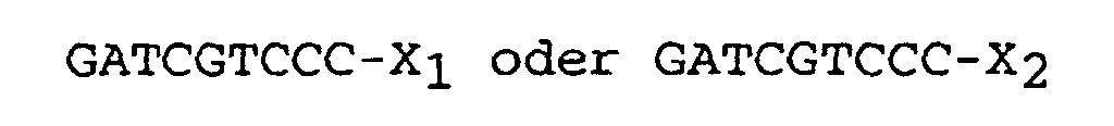
- the kit comprising further to usual buffers for cDNA synthesis, restriction enzyme digestion, ligation and amplification, - containers containing a linker consisting of one double-strand cDNA molecule having one of the following formulas: wherein X 1 and X 2 , which comprise 30-37 nucleotides and are different, include a 20-25 bp PCR priming site with a Tm of 55°C-65°C, and wherein GATCGTCCC (SEQ ID NO:1) correspond to a Sau3A I restriction site joined to a BsmF I restriction site, and - containers containing primers comprising 20-25 bp and having a Tm of 55°-65°C.
- said kit preferably contains
- the instant SADE method includes the following features: 1) single-step mRNA purification from tissue lysate; 2) use of a reverse transcriptase lacking Rnase H activity; 3) use of a different anchoring enzyme; 4) modification of procedures for blunt-ending cDNA tags; 5) design of new linkers and PCR primers.
- Figure 1 modified from the original studies of Velculescu et al., summarises the different steps of the SADE method, which is a microadaptation of SAGE.
- mRNAs are extracted using oligo(dT) 25 covalently bound to paramagnetic beads.
- Double strand cDNA is synthesised from mRNA using oligo(dT) 25 as primer for the 1st strand synthesis.
- the cDNA is then cleaved using a restriction endonuclease ( anchoring enzyme : SAU3A I) with a 4-bp recognition site.
- each cDNA is isolated using the property of the paramagnetic beads and divided in half.
- Each of the two aliquots is ligated via the anchoring enzyme restriction site to one of the two linkers containing a type IIS recognition site ( tagging enzyme : BsmF I) and a priming site for PCR amplification.
- Type IIS restriction endonucleases display recognition and cleavage sites separated by a defined length (14 bp for BsmF I), irrespective of the intercalated sequence.
- Digestion with the type IIs restriction enzyme thus releases linkers with an anchored short piece of cDNA, corresponding to a transcript-specific tag. After blunt ending of tags, the two aliquots are linked together and amplified by PCR. Since all targets are of the same length (110 bp) and are amplified with the same primers, potential distortions introduced by PCR are greatly reduced. Furthermore, these distortions can be evaluated, and the data corrected accordingly (7, 8). Ditags present in the PCR products are recovered through digestion with the anchoring enzyme and gel purification, then concatenated and cloned.
- mRNAs are isolated using conventional methods, then hybridized to biotinylated oligo(dT) for cDNA synthesis. After cleavage with the anchoring enzyme NIa III, the biotionylated cDNA fraction (3' end) is purified by binding to streptavidin beads.
- mRNAs are directly isolated from the tissue lysate through hybridization to oligo(dT) covalently bound to magnetic beads. Then, all steps of the experiment (until step 3 of protocol 5, as described here after) are performed on magnetic beads. This procedure saves time for the initial part of the experiment and, more importantly, provides better recovery. Quantitative analysis of the cDNA amounts available for library construction revealed dramatic differences between SAGE and SADE. With the SAGE method, starting from 500 mg tissue, 1.7 ⁇ g of cDNA are obtained, and only 4 ng were able to bind to streptavidin beads after Sau3A I digestion.
- SAGE and SADE Another important difference between SAGE and SADE concerns the selected anchoring enzyme. Although any restriction enzyme with a 4-bp restriction site could serve as anchoring enzyme, Sau3A I was preferred to Nla III (7-10) or other enzymes in our studies.
- cDNA libraries used for large scale sequencing are constructed by vector priming, followed by cDNA cleavage with Mbo I (an isoschizomer of Sau3A I which does not cut the vector (methylated) DNA), and circularisation (6).
- SADE tags therefore correspond to the cDNA 5' ends of these libraries, which enables to use more efficiently EST data bases to analyse the data.
- tissue i.e . kidney, liver, brain, .
- the following procedures may be routinely used. After animal anaesthesia or decapitation, the tissue is removed as quickly as possible, rapidly rinsed in ice-cold phosphate-buffered saline, sliced in ⁇ 50 mg-pieces, and frozen in liquid nitrogen. The frozen sample is then ground to a fine powder under liquid nitrogen using a mortar and a pestle, transferred into lysis binding buffer ( protocol 1 ), and homogenised with a Dounce tissue disrupter. To avoid loss of material, small samples ( ⁇ 20 mg) can be transferred without previous freezing in the lysis binding buffer, and homogenised in a 1 ml Dounce.
- tissue and lysis binding buffer needed for a variety of conditions are indicated in Table II.
- Protocols 1-7 describe the generation of a SADE library from 0.5 mg of tissue.
- the amount of cDNA recovered corresponds to an experiment carried out on the mouse kidney. Slightly different amounts are expected to be obtained from other tissues, according to their mRNA content.
- the procedures described herein have been repeatedly used without modifications with 3x10 4 -10 5 isolated cells. Since some applications can be performed on large amounts of tissue or cells, protocol adaptations and anticipated results for these kinds of experiments are also provided.
- RNAs were extracted using standard methods (13), and poly(A) RNAs were isolated on oligo(dT) columns. Besides being time consuming, this procedure provides low and variable mRNA amounts, and cannot be easily scaled down.
- the alternative procedure described here (use of oligo(dT) 25 covalently linked to paramagnetic beads) is a single tube assay for mRNA isolation from tissue lysate. In our hands, it yields 4-times higher mRNA amounts than standard methods. Kits and helpful instructions for mRNA isolation with oligo(dT) beads can be obtained from Dynal. Handling of these beads is relatively simple, but care must be taken to avoid centrifugation, drying or freezing, since all three processes are expected to lower their binding capacity. On the other hand, beads can be resuspended by gentle vortexing or pipetting without extreme precautions.
- RNA degradation has only to be expected in the three following conditions: 1) cell survival is not maintained before lysis or freezing; 2) cell thawing outside of lysis buffer, and 3) use of poor quality reagents. Since Rnase-free reagents are now available from a variety of company, it is much more rapid and effective to check for survival ( i.e . select the appropriate culture medium) and freezing conditions than to perform tricky tests on RNA aliquots.
- the first step in the synthesis of cDNA is copying the mRNA template into complementary single-strand cDNA.
- 1 st strand cDNA is synthesised using Moloney Murine Leukaemia Virus RT (M-MLV RT).
- M-MLV RT Moloney Murine Leukaemia Virus RT
- Superscript II M-MLV RT provided with the Superscript cDNA synthesis kit (Life Technologies, ref. 18090-019). In this case, the amount of cDNA formed (see 2.3) was increased ⁇ 4-fold.
- mRNAs are generally heated 5 min at 65°C before reverse transcription to break up secondary structures. Since such a high temperature will also denature the mRNA-oligo(dT) 25 hybrid, we only heat the sample at 42°C before initiation of 1st strand synthesis.
- the amount of double strand (ds) cDNA formed is calculated by measuring radioactivity incorporation in the 5' end of the cDNA, which is released in the supernatant after Sau3A I digestion (see Protocol 2 ).
- the 300 ⁇ l-supernatant is extracted with PCI and the ds cDNA is ethanol precipitated in the presence of glycogen (50 ⁇ g/ ml) and 2.5 M ammonium acetate.
- the pellet is resuspended in 8 ⁇ l of TE. Half of the material is used for liquid scintillation counting, and the remaining is loaded on a 1.0 or 1.5% agarose gel.
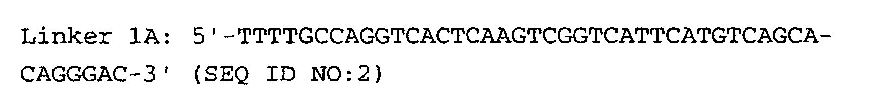
- Linkers must contain three important sequences : a) the appropriate anchoring enzyme overhang; b) a recognition site for a type IIs restriction enzyme (tagging enzyme); c) a priming site for PCR amplification. High quality linkers are crucial for successful library generation.
- Linkers 1B and 2B display two modifications: a) 5' end phosphorylation, and b) C7 amino modification on the 3' end.
- Linkers phosphorylation can be performed either enzymatically with T4 polynucleotide kinase, or chemically at the time of oligonucleotide synthesis. In both cases, phosphorylation efficiency must be tested ( Protocol 3 ).
- Protocol 3 We use chemically phosphorylated linkers.
- Linkers modification on the 3' end serves to increase the efficiency of ditag formation ( protocol 5, step 8-11 ). Indeed, the modified 3' end cannot be blunt-ended and will not ligate to cDNA tags or linkers.
- PCR priming site it was designed with the help of OligoTM software (Medprobe, Norway) in order to obtain PCR primers with high Tm (60°C), and avoid self-priming or sense/ antisense dimer formation.
- OligoTM software Medprobe, Norway
- Two different priming sites must be designed in " left " and " right “ linkers, otherwise the target will undergo panhandle formation, and thus escape PCR amplification.
- ds linkers can be ligated, PCR amplified, and digested with the anchoring and tagging enzyme. Success with protocol 3 experiments is a prerequisite before attempting to prepare a library.
- the PCR conditions described here have been optimised for Hybaid thermal reactors (TR1 and Touch Down) working under control or simulated tube conditions. Different conditions may be used with other machines. Note that since the target is quite small (90 bp), elongation is performed at a relatively low temperature.
- the concentration of ds linkers should be adapted to the amount of cDNA used to prepare the library.
- 2 ⁇ g (74 pmol) of ds linkers are used.
- the amount of cDNA available for ligation is in the range of 1.5-3 pmol. Since a large excess of linkers decreases the PCR signal to noise ratio, we perform ligation with 8 pmol ds linkers for libraries generated from 250 mg of tissue ( ⁇ 5 ⁇ g mRNAs). Starting from 5X10 4 -10 5 cells (10-40 ng mRNAs), 0.5 pmol of ds linkers are used. A lower amount of linkers may allow efficient ligation, but we have no experience for it.
- step 5 in protocol 4 After ligation (step 5 in protocol 4 ), it is very important to wash the beads extensively in order to remove free ds linkers. In fact, if ds linkers not ligated to cDNA fragments are not thoroughly eliminated from each sample, the library will contain large amounts (up to 25%) of linkers sequences. This will make data acquisition poorly efficient.
- Protocol 5 Release, blunt ending, and ligation of cDNA tags
- the desired PCR product is 110-bp long (90 bp of linkers derived sequences, and 20 bp of ditag).
- the optimal number of PCR cycles needs to be determined. This is best accomplished by performing duplicate PCR on 2% of the ligation product and sampling 7- ⁇ l aliquots at different cycles. The number of cycles will of course depend on the amount of starting material. For 250 mg tissue pieces, a PCR signal should be obtained with 18 cycles, and the plateau reached at 22-23 cycles. The 110-bp fragment should be largely predominant (amplified products of 90 and 100 bp are not unusual). Examples of PCR carried out on ditags generated from tiny amounts of cells (15,000 to 45,000) are given in Fig. 3. Using such low amounts of cells, the 110-bp product is no longer predominant. Nevertheless, if maximal yield is achieved with less than 30 cycles (as obtained from 45,000 cells in Fig. 3), a library which is fairly representative of the tissue can be generated. Small scale PCR (10 reactions, step 1-5 in protocol 6) is performed on 2 ⁇ l and 4 ⁇ l aliquots of ligation product for macro and microamounts of tissue, respectively.
- the 110-bp PCR product can be purified either on a 12% polyacrylamide (7) or a 3% agarose slab gel. To avoid overloading and achieve efficient purification, pool no more than 10-12 PCR reactions on an agarose gel and slice agarose as close as possible to the 110-bp fragment. Purification and optimal number of PCR cycles should then be tested on duplicate 2 ⁇ l aliquots of the purified product. A single band of 110 bp should now be obtained. The absence of interference from other amplified products is essential to produce large amounts of the 110-bp fragment.
- Ditags do not run as a single band on polyacrylamide gel. This may come from subtle effects of base composition on electrophoretic mobility and/ or some wobble for BsmF I digestion (7). We cut out from gel all the material ranging from 22 to 26 bp. The elution procedure is labour intensive but provides ditags that can be concatenated efficiently.
- Concatemers are heated at 45°C for 5 min immediately before loading on gel to separate unligated cohesive ends. Concatemers form a smear on the gel from about 100 bp to several kbp (Fig. 4) and can be easily detected using SYBR Green I stain. All fragments >300 bp (i.e. with 25 or more tags) are potentially interesting for library construction. We usually cut out fragments of 350-600, 600-2000, and >2000 bp and generate a first library using 600-2000 bp DNA fragments. Longer fragments will be more informative but are expected to be cloned with poor efficiency.
- Concatemers can be cloned and sequenced in a vector of choice.
- Velculescu et al.(7, 8) use pZero-1 from Invitrogen which only allows recombinants to grow (DNA insertion into the multiple cloning site disrupts a lethal gene).
- the competent cells and transformation procedures can also be changed according to your facilities.
- DNA miniprep provides more reproducible amounts of DNA than PCR, and avoids false positive signals.
- Qiaprep 8 miniprep kit Qiagen, ref. 27144 which enables to perform 96 minipreps in ⁇ 2 hours. Plasmid DNA is eluted from Qiagen columns with 100 ⁇ l of elution buffer; 5 ⁇ l are digested to evaluate insert size, and if insert is > 200 bp, 5 ⁇ l are directly used for DNA sequencing.
- tags must be extracted, quantified, and identified through possible data bank matches. Two softwares have been written to reach these goals.
- SAGE software was written in Visual Basic and is operating on personal computers through the Microsoft Windows system. It extracts tags from text sequence files, quantify them, allows to compare several libraries, and provides links to GenBank data downloaded from CD-ROM flat files or over the Internet. The latter function enables rapid identification of tags originating from characterised genes or cDNAs. However, description of EST sequences is truncated, which constrains to look for individual GenBank reports. SAGE software also includes several simulating tools which allows, for example, to assess the significance of differences observed between two libraries, and to evaluate the sequencing accuracy.
- the second software is currently developed at the University of criz-2 (France) by J. Marti and co-workers, as part of a database (CbC, for Cell by Cell) intended to store and retrieve data from SAGE experiments.
- Scripts for extraction of data are developed in C language under Unix environment and the database management system implemented in Acces®. Text files are concatenated to yield the working file from which tag sequences are extracted and enumerated.
- Treatment of raw data involves identification of vector contaminants, truncated and repeated ditags (see below). For experiments on human, mouse and rat cell samples, the tags are searched in the non-redundant set of sequences provided by the UniGene collection.
- RNA-cDNA hybridization The basic pattern of gene expression in eukaryotic cells have been established long ago by kinetics analysis of mRNA-cDNA hybridization (15, 16).
- the total RNA mass consists of 300,000 molecules, corresponding to ⁇ 12,000 transcripts which divide into three abundance classes.
- a very small number of mRNAs ( ⁇ 10) are expressed to exceedingly high levels (3,000-15,000 copies/ cell).
- a larger number of mRNAs ( ⁇ 500) reaches an expression level in the range of 100-500 copies/ cell.
- the majority of mRNAs >10,000 are poorly expressed (10-100 copies/ cell). This basic pattern should be observed in SAGE or SADE libraries. However, before translating tags abundance in a definite gene expression profile, the data must be scrutinised for artefacts encountered in library construction.
- linker sequences As mentioned above, some libraries display a high amount of linker sequences. If this amount is 20% or more, sequencing will be quite expensive, and it is better to start again from the RNA sample. Library contamination with 10-15% of linker sequences is acceptable, 5-10% is good, and ⁇ 5% is excellent.
- GTCCCTGTGC, and GTCCCTTCCG In addition to the two perfect linker matches (GTCCCTGTGC, and GTCCCTTCCG), reading ambiguities can lead to sequences with one mismatch.
- These linker-like sequences are also easily identified since, assuming efficient enzymatic cleavage, the probability of having adjacent Sau3A I and BsmF I sites in the concatemers is normally zero. Linker and linker-like sequences can be automatically discarded using SAGE or CbC software, and their relative amounts can be used to evaluate the sequencing accuracy (see 7. 1 .).
- Another category of sequences that must be deleted are those corresponding to duplicate ditags. Indeed, except for peculiar tissues (e.g. lactating gland or laying hen oviduct) in which one or a very small number of transcripts constitutes the bulk of the mRNA mass, the probability for any two tags to be found several times in the same ditag is very small. Elimination of repeated ditags will therefore correct for preferential PCR amplification of some targets, and for picking several bacterial colonies originating from the same clone. Most ditags (>95%) generally occur only once when the library is constructed from macroamounts of tissue. For microlibraries, the percent of unique ditags is generally lower. When it is no longer compatible ( ⁇ 75%) with efficient data acquisition, it is recommended to start again from the first (small scale) PCR (see protocol 6 ). Duplicate ditags are automatically retrieved from the sequence files by SAGE and CbC softwares.
- the number of tags to be analysed will obviously depend on the application and tissue source. As a matter of fact, reducing the tissue complexity through isolation of defined cell populations will allow to markedly diminish the minimum number of tags for accurate analysis, and to better correlate molecular and physiological phenotypes.
- the most difficult projects are those aiming to compare gene expression profiles in the same tissue under two physiological or pathological conditions. Differentially expressed genes could belong to any of the three abundance classes and, furthermore, they can be either up- or down-regulated. A reasonable number of tags to be sequenced would be in the range of 30,000-50,000.
- tags corresponding to poorly expressed transcripts may be detected 1 and ⁇ 5 times in control and experimental conditions, respectively.
- tags corresponding to down-regulation processes will be less exhaustive. It will only concern tags present > 5-10 times in the control condition, which excludes from the analysis part of the poorly expressed transcripts.
- Table I which corresponds to the characterisation of the most abundant nuclear transcripts in the mouse outer medullary collecting duct (OMCD) and establishes their differential expression in the medullary thick ascending limb (MTAL), the two left columns correspond to the data illustrated in Figure 5, and provide the abundance of each tag in the two libraries.
- the third column provides the sequence of the tags.
- the right column indicates results of individual BLAST search in GenBank, carried out using a 14-bp sequence (the Sau3A I recognition sequence, plus the 10 bp specific for each tag).
Landscapes
- Chemical & Material Sciences (AREA)
- Life Sciences & Earth Sciences (AREA)
- Organic Chemistry (AREA)
- Analytical Chemistry (AREA)
- Zoology (AREA)
- Wood Science & Technology (AREA)
- Proteomics, Peptides & Aminoacids (AREA)
- Health & Medical Sciences (AREA)
- Engineering & Computer Science (AREA)
- Microbiology (AREA)
- Immunology (AREA)
- Molecular Biology (AREA)
- Biotechnology (AREA)
- Biophysics (AREA)
- Physics & Mathematics (AREA)
- Biochemistry (AREA)
- Bioinformatics & Cheminformatics (AREA)
- General Engineering & Computer Science (AREA)
- General Health & Medical Sciences (AREA)
- Genetics & Genomics (AREA)
- Measuring Or Testing Involving Enzymes Or Micro-Organisms (AREA)
Claims (11)
- Verfahren zum Erhalt einer Genbank von tags, die in der Lage sind, einen spezifischen Zustand einer biologischen Probe zu definieren, dadurch gekennzeichnet, daß es die folgenden aufeinanderfolgenden Schritte umfaßt:(1) Einstufiges Extrahieren von mRNA mit kovalent an paramagnetische Kügelchen gebundenem Oligo(dT)25 aus einer kleinen Menge einer biologischen Probe,(2) Generieren einer doppelsträngigen cDNA-Genbank aus der mRNA gemäß den folgenden Schritten:Synthetisieren des ersten Strangs der cDNA durch reverse Transkription des mRNA-Templates mit reverser Transkriptase, die keine RNase H-Aktivität aufweist, in eine erste komplementäre einzelsträngige cDNA,Synthetisieren des zweiten Strangs der cDNA durch Nicktranslation der mRNA in dem gebildeten mRNA-cDNA-Hybrid mittels einer E. coli DNA-Polymerase,(3) Spalten der erhaltenen cDNAs mit der Restriktionsendonuklease Sau3A I als Anchoring-Enzym,(4) Auftrennen der gespaltenen cDNAs in zwei Aliquote,(5) Ligieren der in jedem der beiden Aliquote enthaltenen cDNA über die Sau3A I-Restriktionsschnittstelle mit einem Linker, der aus einem doppelsträngigen cDNA-Molekül besteht, das eine der folgenden Formeln besitzt:wobei x1 und X2, die 30 bis 37 Nukleotide umfassen und verschieden sind, eine PCR-Startstelle von 20-25 bp mit einer Tm von 55-65°C einschließen, und
wobei GATCGTCCC (SEQ ID NO:1) einer mit einer BsmF I-Restriktionsschnittstelle verknüpften Sau3A I-Restriktionsschnittstelle entspricht,(6) Verdauen der in Schritt (5) erhaltenen Produkte mit dem Tagging-Enzym BsmF I und Freisetzen von Linkern mit einem verankerten kurzen Stück cDNA, das einem Transkript-spezifischen tag entspricht, wobei der Verdau BsmF I-tags liefert, die für die ursprüngliche mRNA spezifisch sind,(7) Erzeugen glatter Enden an den BsmF I-tags mit einer DNA-Polymerase, vorzugsweise T7 DNA-Polymerase oder Vent-Polymerase, und Mischen der mit den verschiedenen Linkern ligierten tags,(8) Ligieren der in Schritt (7) erhaltenen tags mit einer DNA-Ligase unter Bildung von ditags,(9) Amplifizieren der in Schritt (8) erhaltenen ditags mit Primern, die 20-25 bp umfassen und eine Tm von 55-65°C besitzen,(10) Isolieren der ditags, die zwischen 20 und 28 bp aufweisen, aus den in Schritt (9) erhaltenen Amplifikationsprodukten durch Verdau der Amplifikationsprodukte mit dem Anchoring-Enzym Sau3A I und Auftrennen der verdauten Produkte auf einem geeigneten Elektrophoresegel,(11) Ligieren der in Schritt (10) erhaltenen ditags zu Konkatemeren, Reinigen der Konkatemeren und Abtrennen der Konkatemeren mit mehr als 300 bp,(12) Klonieren und Sequenzieren der Konkatemeren, und(13) Testen der verschiedenen erhaltenen tags. - Verfahren nach Anspruch 1, dadurch gekennzeichnet, daß die Synthese des ersten Strangs der cDNA in Schritt (2) mit reverser Transkriptase von Moloney-Maus-Leukämie-Virus (M-MLV RT) und mit Oligo(dT)25 als Primern erfolgt.
- Verfahren nach Anspruch 1 oder Anspruch 2, dadurch gekennzeichnet, daß die Linker von Schritt (5) vorzugsweise Hybrid-DNA-Moleküle sind, die aus Linkern 1A und 1B oder aus Linkern 2A und 2B mit den folgenden Formeln gebildet sind:
 oder
oder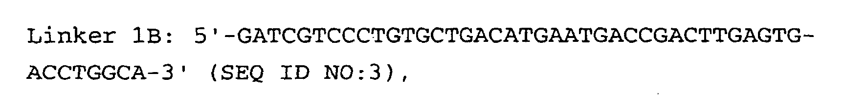


- Verfahren nach den Ansprüchen 1 bis 3, dadurch gekennzeichnet, daß die Menge von jedem Linker in Schritt (5) höchstens 8-10 pmol beträgt und vorzugsweise zwischen 0,5 pmol und 8 pmol für Anfangsmengen von 10-40 ng mRNAs bzw. 5 µg mRNAs liegt.
- Verfahren nach den Ansprüchen 1 bis 4, dadurch gekennzeichnet, daß die Primer von Schritt (9) vorzugsweise die folgenden Formeln haben:


- Verfahren nach den Ansprüchen 1 bis 5, dadurch gekennzeichnet, daß die biologische Probe von Schritt (1) ≤ 5·106 Zellen umfaßt, entsprechend höchstens 50 µg Gesamt-RNA oder 1 µg Poly(A)-RNA.
- Verfahren nach den Ansprüchen 1 bis 6, dadurch gekennzeichnet, daß die Gewebeprobe aus Nieren stammt, insbesondere aus Nephronsegmenten entsprechend etwa 15.000 bis 45.000 Zellen, entsprechend 0,15-0,45 µg Gesamt-RNA.
- Verwendung einer nach dem Verfahren der Ansprüche 1 bis 7 erhaltenen Genbank von tags zum Beurteilen des physiologischen oder pathologischen Zustands einer biologischen Probe.
- Verfahren zum Bestimmen eines Genexpressionsprofils, dadurch gekennzeichnet, daß es umfaßt:Durchführen der Schritte (1) bis (13) nach Anspruch 1 undÜbertragen der Häufigkeit von cDNA-tags auf ein Genexpressionsprofil.
- Kit zur Feststellung eines Genexpressionsprofils, dadurch gekennzeichnet, daß die Anwesenheit eines cDNA-tag, das von der aus einer biologischen Probe extrahierten mRNA erhalten wurde, auf die Expression eines Gens schließen läßt, das diese tag-Sequenz an einer entsprechenden Stelle trägt, d.h. unmittelbar benachbart zu der dem 3'-Ende in dieser cDNA am nächsten gelegenen Sau3A I-Stelle, wobei der Kit zusätzlich zu üblichen Puffern zur cDNA-Synthese, zum Verdau mit Restriktionsenzymen, zur Ligation und zur Amplifkation umfaßt:Behälter, die Linker enthalten, die aus einem doppelsträngigen cDNA-Molekül bestehen, das eine der folgenden Formeln besitzt:wobei X1 und X2, die 30 bis 37 Nukleotide umfassen und verschieden sind, eine PCR-Startstelle von 20-25 bp mit einer Tm von 55-65°C einschließen, und
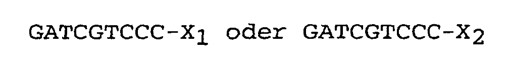
wobei GATCGTCCC (SEQ ID NO:1) einer mit einer BsmF I-Restriktionsschnittstelle verknüpften Sau3A I-Restriktionsschnittstelle entspricht, undBehälter, die Primer enthalten, die 20-25 bp umfassen und eine Tm von 55-65°C besitzen. - Kit nach Anspruch 10, dadurch gekennzeichnet, daß er enthält:Behälter, die Hybrid-DNA-Moleküle enthalten, die aus Linkern 1A und 1B oder aus Linkern 2A und 2B mit den folgenden Formeln gebildet sind:
 oder
oder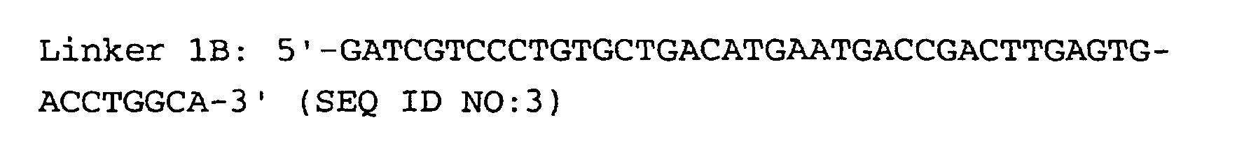
 und
und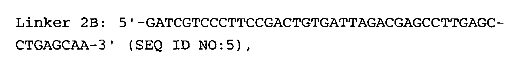 Behälter, die die folgenden Primer enthalten:

Priority Applications (7)
| Application Number | Priority Date | Filing Date | Title |
|---|---|---|---|
| EP99400189A EP1024201B1 (de) | 1999-01-27 | 1999-01-27 | Microassay zur Serienanalyse der Genexpression und Anwendungen davon |
| DE69913092T DE69913092T2 (de) | 1999-01-27 | 1999-01-27 | Microassay zur Serienanalyse der Genexpression und Anwendungen davon |
| US09/301,721 US6506561B1 (en) | 1999-01-27 | 1999-04-29 | Method of obtaining a library of tags capable of defining a specific state of a biological sample |
| CA002359272A CA2359272A1 (en) | 1999-01-27 | 2000-01-25 | Microassay for serial analysis of gene expression and applications thereof |
| JP2000596176A JP2002535012A (ja) | 1999-01-27 | 2000-01-25 | 遺伝子発現の連続分析用マイクロアッセイ及びその応用 |
| PCT/IB2000/000111 WO2000044936A1 (en) | 1999-01-27 | 2000-01-25 | Microassay for serial analysis of gene expression and applications thereof |
| US10/195,383 US20030165910A1 (en) | 1999-01-27 | 2002-07-16 | Microassay for serial analysis of gene expression and applications thereof |
Applications Claiming Priority (1)
| Application Number | Priority Date | Filing Date | Title |
|---|---|---|---|
| EP99400189A EP1024201B1 (de) | 1999-01-27 | 1999-01-27 | Microassay zur Serienanalyse der Genexpression und Anwendungen davon |
Publications (2)
| Publication Number | Publication Date |
|---|---|
| EP1024201A1 EP1024201A1 (de) | 2000-08-02 |
| EP1024201B1 true EP1024201B1 (de) | 2003-11-26 |
Family
ID=8241860
Family Applications (1)
| Application Number | Title | Priority Date | Filing Date |
|---|---|---|---|
| EP99400189A Expired - Lifetime EP1024201B1 (de) | 1999-01-27 | 1999-01-27 | Microassay zur Serienanalyse der Genexpression und Anwendungen davon |
Country Status (6)
| Country | Link |
|---|---|
| US (2) | US6506561B1 (de) |
| EP (1) | EP1024201B1 (de) |
| JP (1) | JP2002535012A (de) |
| CA (1) | CA2359272A1 (de) |
| DE (1) | DE69913092T2 (de) |
| WO (1) | WO2000044936A1 (de) |
Families Citing this family (85)
| Publication number | Priority date | Publication date | Assignee | Title |
|---|---|---|---|---|
| EP1024201B1 (de) * | 1999-01-27 | 2003-11-26 | Commissariat A L'energie Atomique | Microassay zur Serienanalyse der Genexpression und Anwendungen davon |
| EP2180064A3 (de) | 2000-10-18 | 2010-08-11 | Pharmasset, Inc. | Multiplex Quantifizierung von Nukleinsäuren in erkrankten Zellen |
| FR2821087B1 (fr) * | 2001-02-16 | 2004-01-02 | Centre Nat Rech Scient | Procede d'analyse qualitative et quantitative d'une population d'acides nucleiques contenus dans un echantillon |
| DE10144132A1 (de) * | 2001-09-07 | 2003-03-27 | Axaron Bioscience Ag | Identifikation und Quantifizierung von Nukleinsäuren durch Erzeugen und Analyse von Sequenz-tags einheitlicher Länge |
| US6977162B2 (en) * | 2002-03-01 | 2005-12-20 | Ravgen, Inc. | Rapid analysis of variations in a genome |
| IL163600A0 (en) | 2002-03-01 | 2005-12-18 | Ravgen Inc | Methods for detection of genetic disorders |
| US20070178478A1 (en) * | 2002-05-08 | 2007-08-02 | Dhallan Ravinder S | Methods for detection of genetic disorders |
| US7442506B2 (en) * | 2002-05-08 | 2008-10-28 | Ravgen, Inc. | Methods for detection of genetic disorders |
| US7727720B2 (en) * | 2002-05-08 | 2010-06-01 | Ravgen, Inc. | Methods for detection of genetic disorders |
| US7141371B2 (en) | 2002-09-06 | 2006-11-28 | State Of Oregon Acting By And Through The State Board Of Higher Education On Behalf Of The University Of Oregon | Methods for detecting and localizing DNA mutations by microarray |
| US20060234268A1 (en) * | 2005-04-15 | 2006-10-19 | Sungwhan An | Linear amplification of rna |
| WO2006122215A2 (en) | 2005-05-10 | 2006-11-16 | State Of Oregon Acting By & Through The State Board Of Higher Education On Behalf Of The University Of Oregon | Methods of mapping polymorphisms and polymorphism microarrays |
| US8428882B2 (en) * | 2005-06-14 | 2013-04-23 | Agency For Science, Technology And Research | Method of processing and/or genome mapping of diTag sequences |
| US7655543B2 (en) * | 2007-12-21 | 2010-02-02 | Asm America, Inc. | Separate injection of reactive species in selective formation of films |
| AU2009205523A1 (en) * | 2008-01-14 | 2009-07-23 | Applied Biosystems, Llc | Compositions, methods, and kits for detecting ribonucleic acid |
| US20190300945A1 (en) | 2010-04-05 | 2019-10-03 | Prognosys Biosciences, Inc. | Spatially Encoded Biological Assays |
| US10787701B2 (en) | 2010-04-05 | 2020-09-29 | Prognosys Biosciences, Inc. | Spatially encoded biological assays |
| GB201106254D0 (en) | 2011-04-13 | 2011-05-25 | Frisen Jonas | Method and product |
| WO2014047561A1 (en) | 2012-09-21 | 2014-03-27 | The Broad Institute Inc. | Compositions and methods for labeling of agents |
| WO2014047556A1 (en) * | 2012-09-21 | 2014-03-27 | The Broad Institute, Inc. | Compositions and methods for long insert, paired end libraries of nucleic acids in emulsion droplets |
| EP4592400A3 (de) | 2012-10-17 | 2025-10-29 | 10x Genomics Sweden AB | Verfahren und produkt zur optimierung einer lokalisierten oder räumlichen detektion einer genexpression in einer gewebeprobe |
| WO2014143158A1 (en) * | 2013-03-13 | 2014-09-18 | The Broad Institute, Inc. | Compositions and methods for labeling of agents |
| CA2916662C (en) | 2013-06-25 | 2022-03-08 | Prognosys Biosciences, Inc. | Methods and systems for determining spatial patterns of biological targets in a sample |
| EP3901282B1 (de) | 2015-04-10 | 2023-06-28 | Spatial Transcriptomics AB | Räumlich getrennte multiplex-nukleinsäureanalyse von biologischen proben |
| US11519033B2 (en) | 2018-08-28 | 2022-12-06 | 10X Genomics, Inc. | Method for transposase-mediated spatial tagging and analyzing genomic DNA in a biological sample |
| WO2020123316A2 (en) | 2018-12-10 | 2020-06-18 | 10X Genomics, Inc. | Methods for determining a location of a biological analyte in a biological sample |
| US11926867B2 (en) | 2019-01-06 | 2024-03-12 | 10X Genomics, Inc. | Generating capture probes for spatial analysis |
| US11649485B2 (en) | 2019-01-06 | 2023-05-16 | 10X Genomics, Inc. | Generating capture probes for spatial analysis |
| WO2020243579A1 (en) | 2019-05-30 | 2020-12-03 | 10X Genomics, Inc. | Methods of detecting spatial heterogeneity of a biological sample |
| WO2021091611A1 (en) | 2019-11-08 | 2021-05-14 | 10X Genomics, Inc. | Spatially-tagged analyte capture agents for analyte multiplexing |
| WO2021092433A2 (en) | 2019-11-08 | 2021-05-14 | 10X Genomics, Inc. | Enhancing specificity of analyte binding |
| CN115135984B (zh) | 2019-12-23 | 2025-12-23 | 10X基因组学有限公司 | 可逆固定试剂及其使用方法 |
| ES2982420T3 (es) | 2019-12-23 | 2024-10-16 | 10X Genomics Inc | Métodos para el análisis espacial mediante el uso de la ligazón con plantilla de ARN |
| EP4081656A1 (de) | 2019-12-23 | 2022-11-02 | 10X Genomics, Inc. | Zusammensetzungen und verfahren zur verwendung von fixierten biologischen proben bei partitionsbasierten tests |
| US12365942B2 (en) | 2020-01-13 | 2025-07-22 | 10X Genomics, Inc. | Methods of decreasing background on a spatial array |
| US12405264B2 (en) | 2020-01-17 | 2025-09-02 | 10X Genomics, Inc. | Electrophoretic system and method for analyte capture |
| US11732299B2 (en) | 2020-01-21 | 2023-08-22 | 10X Genomics, Inc. | Spatial assays with perturbed cells |
| US11702693B2 (en) | 2020-01-21 | 2023-07-18 | 10X Genomics, Inc. | Methods for printing cells and generating arrays of barcoded cells |
| US20210230681A1 (en) | 2020-01-24 | 2021-07-29 | 10X Genomics, Inc. | Methods for spatial analysis using proximity ligation |
| US11821035B1 (en) | 2020-01-29 | 2023-11-21 | 10X Genomics, Inc. | Compositions and methods of making gene expression libraries |
| US12076701B2 (en) | 2020-01-31 | 2024-09-03 | 10X Genomics, Inc. | Capturing oligonucleotides in spatial transcriptomics |
| US12110541B2 (en) | 2020-02-03 | 2024-10-08 | 10X Genomics, Inc. | Methods for preparing high-resolution spatial arrays |
| US11898205B2 (en) | 2020-02-03 | 2024-02-13 | 10X Genomics, Inc. | Increasing capture efficiency of spatial assays |
| US11732300B2 (en) | 2020-02-05 | 2023-08-22 | 10X Genomics, Inc. | Increasing efficiency of spatial analysis in a biological sample |
| WO2021158925A1 (en) | 2020-02-07 | 2021-08-12 | 10X Genomics, Inc. | Quantitative and automated permeabilization performance evaluation for spatial transcriptomics |
| US11835462B2 (en) | 2020-02-11 | 2023-12-05 | 10X Genomics, Inc. | Methods and compositions for partitioning a biological sample |
| US12399123B1 (en) | 2020-02-14 | 2025-08-26 | 10X Genomics, Inc. | Spatial targeting of analytes |
| US12281357B1 (en) | 2020-02-14 | 2025-04-22 | 10X Genomics, Inc. | In situ spatial barcoding |
| US11891654B2 (en) | 2020-02-24 | 2024-02-06 | 10X Genomics, Inc. | Methods of making gene expression libraries |
| US11926863B1 (en) | 2020-02-27 | 2024-03-12 | 10X Genomics, Inc. | Solid state single cell method for analyzing fixed biological cells |
| US11768175B1 (en) | 2020-03-04 | 2023-09-26 | 10X Genomics, Inc. | Electrophoretic methods for spatial analysis |
| EP4139485B1 (de) | 2020-04-22 | 2023-09-06 | 10X Genomics, Inc. | Verfahren zur räumlichen analyse unter verwendung von gezieltem rna-abbau |
| US12416603B2 (en) | 2020-05-19 | 2025-09-16 | 10X Genomics, Inc. | Electrophoresis cassettes and instrumentation |
| EP4153776B1 (de) | 2020-05-22 | 2025-03-05 | 10X Genomics, Inc. | Räumliche analyse zur erkennung von sequenzvarianten |
| EP4414459B1 (de) | 2020-05-22 | 2025-09-03 | 10X Genomics, Inc. | Simultane räumlich-zeitliche messung der genexpression und der zellaktivität |
| WO2021242834A1 (en) | 2020-05-26 | 2021-12-02 | 10X Genomics, Inc. | Method for resetting an array |
| AU2021283174A1 (en) | 2020-06-02 | 2023-01-05 | 10X Genomics, Inc. | Nucleic acid library methods |
| US12265079B1 (en) | 2020-06-02 | 2025-04-01 | 10X Genomics, Inc. | Systems and methods for detecting analytes from captured single biological particles |
| EP4600376A3 (de) | 2020-06-02 | 2025-10-22 | 10X Genomics, Inc. | Räumliche transkriptomik für antigenrezeptoren |
| US12031177B1 (en) | 2020-06-04 | 2024-07-09 | 10X Genomics, Inc. | Methods of enhancing spatial resolution of transcripts |
| EP4421186B1 (de) | 2020-06-08 | 2025-08-13 | 10X Genomics, Inc. | Verfahren zur bestimmung eines chirurgischen randes und verfahren zur verwendung davon |
| US12435363B1 (en) | 2020-06-10 | 2025-10-07 | 10X Genomics, Inc. | Materials and methods for spatial transcriptomics |
| EP4165207B1 (de) | 2020-06-10 | 2024-09-25 | 10X Genomics, Inc. | Verfahren zur bestimmung der position eines analyten in einer biologischen probe |
| WO2021252747A1 (en) | 2020-06-10 | 2021-12-16 | 1Ox Genomics, Inc. | Fluid delivery methods |
| ES2994976T3 (en) | 2020-06-25 | 2025-02-05 | 10X Genomics Inc | Spatial analysis of dna methylation |
| US11981960B1 (en) | 2020-07-06 | 2024-05-14 | 10X Genomics, Inc. | Spatial analysis utilizing degradable hydrogels |
| US11761038B1 (en) | 2020-07-06 | 2023-09-19 | 10X Genomics, Inc. | Methods for identifying a location of an RNA in a biological sample |
| US12209280B1 (en) | 2020-07-06 | 2025-01-28 | 10X Genomics, Inc. | Methods of identifying abundance and location of an analyte in a biological sample using second strand synthesis |
| US12553898B1 (en) | 2020-08-10 | 2026-02-17 | 10X Genomics, Inc. | Fluorescent hybridization of antibody-oligonucleotide for multiplexing and signal amplification |
| US11981958B1 (en) | 2020-08-20 | 2024-05-14 | 10X Genomics, Inc. | Methods for spatial analysis using DNA capture |
| WO2022061150A2 (en) | 2020-09-18 | 2022-03-24 | 10X Geonomics, Inc. | Sample handling apparatus and image registration methods |
| US11926822B1 (en) | 2020-09-23 | 2024-03-12 | 10X Genomics, Inc. | Three-dimensional spatial analysis |
| US11827935B1 (en) | 2020-11-19 | 2023-11-28 | 10X Genomics, Inc. | Methods for spatial analysis using rolling circle amplification and detection probes |
| EP4729631A2 (de) | 2020-12-21 | 2026-04-22 | 10X Genomics, Inc. | Verfahren, zusammensetzungen und systeme zur erfassung von sonden und/oder barcodes |
| EP4421491A3 (de) | 2021-02-19 | 2024-11-27 | 10X Genomics, Inc. | Verfahren zur verwendung einer modularen testträgervorrichtung |
| AU2022238446A1 (en) | 2021-03-18 | 2023-09-07 | 10X Genomics, Inc. | Multiplex capture of gene and protein expression from a biological sample |
| EP4305196B1 (de) | 2021-04-14 | 2025-04-02 | 10X Genomics, Inc. | Verfahren zur messung der fehllokalisierung eines analyten |
| EP4320271B1 (de) | 2021-05-06 | 2025-03-19 | 10X Genomics, Inc. | Verfahren zur erhöhung der auflösung von räumlicher analyse |
| WO2022256503A1 (en) | 2021-06-03 | 2022-12-08 | 10X Genomics, Inc. | Methods, compositions, kits, and systems for enhancing analyte capture for spatial analysis |
| US12553805B2 (en) | 2021-08-02 | 2026-02-17 | 10X Genomics, Inc. | Methods of preserving a biological sample |
| WO2023034489A1 (en) | 2021-09-01 | 2023-03-09 | 10X Genomics, Inc. | Methods, compositions, and kits for blocking a capture probe on a spatial array |
| WO2023086880A1 (en) | 2021-11-10 | 2023-05-19 | 10X Genomics, Inc. | Methods, compositions, and kits for determining the location of an analyte in a biological sample |
| WO2023102118A2 (en) | 2021-12-01 | 2023-06-08 | 10X Genomics, Inc. | Methods, compositions, and systems for improved in situ detection of analytes and spatial analysis |
| WO2023122033A1 (en) | 2021-12-20 | 2023-06-29 | 10X Genomics, Inc. | Self-test for pathology/histology slide imaging device |
| EP4482979B1 (de) | 2022-11-09 | 2025-10-01 | 10X Genomics, Inc. | Verfahren, zusammensetzungen und kits zur bestimmung der position mehrerer analyten in einer biologischen probe |
Family Cites Families (7)
| Publication number | Priority date | Publication date | Assignee | Title |
|---|---|---|---|---|
| US4683195A (en) * | 1986-01-30 | 1987-07-28 | Cetus Corporation | Process for amplifying, detecting, and/or-cloning nucleic acid sequences |
| DE4317845A1 (de) * | 1993-05-28 | 1994-12-01 | Bayer Ag | Desoxyribonukleinsäuren |
| US5597815A (en) * | 1995-07-13 | 1997-01-28 | Wisconsin Alumni Research Foundation | Prevention of hyperphosphatemia in kidney disorder patients |
| US5695937A (en) | 1995-09-12 | 1997-12-09 | The Johns Hopkins University School Of Medicine | Method for serial analysis of gene expression |
| US5866330A (en) * | 1995-09-12 | 1999-02-02 | The Johns Hopkins University School Of Medicine | Method for serial analysis of gene expression |
| AU2264197A (en) * | 1996-02-09 | 1997-08-28 | Government Of The United States Of America, As Represented By The Secretary Of The Department Of Health And Human Services, The | Restriction display (rd-pcr) of differentially expressed mrnas |
| EP1024201B1 (de) * | 1999-01-27 | 2003-11-26 | Commissariat A L'energie Atomique | Microassay zur Serienanalyse der Genexpression und Anwendungen davon |
-
1999
- 1999-01-27 EP EP99400189A patent/EP1024201B1/de not_active Expired - Lifetime
- 1999-01-27 DE DE69913092T patent/DE69913092T2/de not_active Expired - Fee Related
- 1999-04-29 US US09/301,721 patent/US6506561B1/en not_active Expired - Fee Related
-
2000
- 2000-01-25 JP JP2000596176A patent/JP2002535012A/ja not_active Withdrawn
- 2000-01-25 WO PCT/IB2000/000111 patent/WO2000044936A1/en not_active Ceased
- 2000-01-25 CA CA002359272A patent/CA2359272A1/en not_active Abandoned
-
2002
- 2002-07-16 US US10/195,383 patent/US20030165910A1/en not_active Abandoned
Non-Patent Citations (1)
| Title |
|---|
| ST. CROIX B. ET AL: "Genes expressed in human tumor endothelium", SCIENCE, vol. 289, 18 August 2000 (2000-08-18), US, pages 1197 - 1202, XP002222041, DOI: doi:10.1126/science.289.5482.1197 * |
Also Published As
| Publication number | Publication date |
|---|---|
| EP1024201A1 (de) | 2000-08-02 |
| US20030165910A1 (en) | 2003-09-04 |
| DE69913092T2 (de) | 2004-09-09 |
| DE69913092D1 (de) | 2004-01-08 |
| CA2359272A1 (en) | 2000-08-03 |
| WO2000044936A1 (en) | 2000-08-03 |
| JP2002535012A (ja) | 2002-10-22 |
| US6506561B1 (en) | 2003-01-14 |
Similar Documents
| Publication | Publication Date | Title |
|---|---|---|
| EP1024201B1 (de) | Microassay zur Serienanalyse der Genexpression und Anwendungen davon | |
| AU2024202865B2 (en) | Transposition into native chromatin for personal epigenomics | |
| KR20210107618A (ko) | 뉴클레아제 기반 rna 고갈 | |
| WO2007078599A2 (en) | Functional arrays for high throughput characterization of gene expression regulatory elements | |
| WO2003064691A2 (en) | Methods and means for amplifying nucleic acid | |
| WO2004015085A2 (en) | Method and compositions relating to 5’-chimeric ribonucleic acids | |
| GB2421243A (en) | Database generation | |
| AU2001279704B2 (en) | Method for identification, separation and quantitative measurement of nucleic acid fragments | |
| Cheval et al. | SADE: a microassay for serial analysis of gene expression | |
| AU2001279704A1 (en) | Method for identification, separation and quantitative measurement of nucleic acid fragments | |
| WO2023116490A1 (zh) | 小rna的新型检测方法及其应用 | |
| Pan et al. | Updated pseudo-seq protocol for transcriptome-wide detection of pseudouridines | |
| Le Dai | Serial analyses of gene expression (SAGE) | |
| CN120796253A (zh) | 构建单细胞新生rna测序文库的方法以及对单细胞进行新生rna测序的方法 | |
| CA2909972C (en) | Transposition into native chromatin for personal epigenomics | |
| CN120210325A (zh) | 高通量单细胞核酸甲基化测序方法 | |
| Subrahmanyam et al. | 28 Restriction Endonucleolytic Analysis of Differentially Expressed Sequences: READS |
Legal Events
| Date | Code | Title | Description |
|---|---|---|---|
| PUAI | Public reference made under article 153(3) epc to a published international application that has entered the european phase |
Free format text: ORIGINAL CODE: 0009012 |
|
| AK | Designated contracting states |
Kind code of ref document: A1 Designated state(s): AT BE CH CY DE DK ES FI FR LI |
|
| AX | Request for extension of the european patent |
Free format text: AL;LT;LV;MK;RO;SI |
|
| 17P | Request for examination filed |
Effective date: 20010122 |
|
| AKX | Designation fees paid |
Free format text: AT BE CH CY DE DK ES FI FR LI |
|
| RBV | Designated contracting states (corrected) |
Designated state(s): BE CH DE ES FR GB IT LI NL SE |
|
| 17Q | First examination report despatched |
Effective date: 20010327 |
|
| GRAH | Despatch of communication of intention to grant a patent |
Free format text: ORIGINAL CODE: EPIDOS IGRA |
|
| GRAS | Grant fee paid |
Free format text: ORIGINAL CODE: EPIDOSNIGR3 |
|
| GRAA | (expected) grant |
Free format text: ORIGINAL CODE: 0009210 |
|
| AK | Designated contracting states |
Kind code of ref document: B1 Designated state(s): BE CH DE ES FR GB IT LI NL SE |
|
| PG25 | Lapsed in a contracting state [announced via postgrant information from national office to epo] |
Ref country code: IT Free format text: LAPSE BECAUSE OF FAILURE TO SUBMIT A TRANSLATION OF THE DESCRIPTION OR TO PAY THE FEE WITHIN THE PRE;WARNING: LAPSES OF ITALIAN PATENTS WITH EFFECTIVE DATE BEFORE 2007 MAY HAVE OCCURRED AT ANY TIME BEFORE 2007. THE CORRECT EFFECTIVE DATE MAY BE DIFFERENT FROM THE ONE RECORDED.SCRIBED TIME-LIMIT Effective date: 20031126 |
|
| REG | Reference to a national code |
Ref country code: GB Ref legal event code: FG4D |
|
| REG | Reference to a national code |
Ref country code: CH Ref legal event code: EP |
|
| REF | Corresponds to: |
Ref document number: 69913092 Country of ref document: DE Date of ref document: 20040108 Kind code of ref document: P |
|
| PG25 | Lapsed in a contracting state [announced via postgrant information from national office to epo] |
Ref country code: SE Free format text: LAPSE BECAUSE OF FAILURE TO SUBMIT A TRANSLATION OF THE DESCRIPTION OR TO PAY THE FEE WITHIN THE PRESCRIBED TIME-LIMIT Effective date: 20040226 |
|
| REG | Reference to a national code |
Ref country code: CH Ref legal event code: NV Representative=s name: ISLER & PEDRAZZINI AG |
|
| PG25 | Lapsed in a contracting state [announced via postgrant information from national office to epo] |
Ref country code: ES Free format text: LAPSE BECAUSE OF FAILURE TO SUBMIT A TRANSLATION OF THE DESCRIPTION OR TO PAY THE FEE WITHIN THE PRESCRIBED TIME-LIMIT Effective date: 20040309 |
|
| ET | Fr: translation filed | ||
| PLBE | No opposition filed within time limit |
Free format text: ORIGINAL CODE: 0009261 |
|
| STAA | Information on the status of an ep patent application or granted ep patent |
Free format text: STATUS: NO OPPOSITION FILED WITHIN TIME LIMIT |
|
| 26N | No opposition filed |
Effective date: 20040827 |
|
| PGFP | Annual fee paid to national office [announced via postgrant information from national office to epo] |
Ref country code: FR Payment date: 20051229 Year of fee payment: 8 |
|
| PGFP | Annual fee paid to national office [announced via postgrant information from national office to epo] |
Ref country code: CH Payment date: 20060113 Year of fee payment: 8 |
|
| PGFP | Annual fee paid to national office [announced via postgrant information from national office to epo] |
Ref country code: NL Payment date: 20060125 Year of fee payment: 8 |
|
| PGFP | Annual fee paid to national office [announced via postgrant information from national office to epo] |
Ref country code: BE Payment date: 20060207 Year of fee payment: 8 |
|
| PGFP | Annual fee paid to national office [announced via postgrant information from national office to epo] |
Ref country code: DE Payment date: 20060329 Year of fee payment: 8 |
|
| PG25 | Lapsed in a contracting state [announced via postgrant information from national office to epo] |
Ref country code: LI Free format text: LAPSE BECAUSE OF NON-PAYMENT OF DUE FEES Effective date: 20070131 Ref country code: CH Free format text: LAPSE BECAUSE OF NON-PAYMENT OF DUE FEES Effective date: 20070131 |
|
| PG25 | Lapsed in a contracting state [announced via postgrant information from national office to epo] |
Ref country code: DE Free format text: LAPSE BECAUSE OF NON-PAYMENT OF DUE FEES Effective date: 20070801 |
|
| REG | Reference to a national code |
Ref country code: CH Ref legal event code: PL |
|
| GBPC | Gb: european patent ceased through non-payment of renewal fee |
Effective date: 20070127 |
|
| NLV4 | Nl: lapsed or anulled due to non-payment of the annual fee |
Effective date: 20070801 |
|
| REG | Reference to a national code |
Ref country code: FR Ref legal event code: ST Effective date: 20070930 |
|
| PG25 | Lapsed in a contracting state [announced via postgrant information from national office to epo] |
Ref country code: GB Free format text: LAPSE BECAUSE OF NON-PAYMENT OF DUE FEES Effective date: 20070127 |
|
| BERE | Be: lapsed |
Owner name: *CENTRE NATIONAL DE LA RECHERCHE SCIENTIFIQUE Effective date: 20070131 Owner name: COMMISSARIAT A L'ENERGIE *ATOMIQUE Effective date: 20070131 |
|
| PG25 | Lapsed in a contracting state [announced via postgrant information from national office to epo] |
Ref country code: BE Free format text: LAPSE BECAUSE OF NON-PAYMENT OF DUE FEES Effective date: 20070131 |
|
| PG25 | Lapsed in a contracting state [announced via postgrant information from national office to epo] |
Ref country code: NL Free format text: LAPSE BECAUSE OF NON-PAYMENT OF DUE FEES Effective date: 20070801 |
|
| PG25 | Lapsed in a contracting state [announced via postgrant information from national office to epo] |
Ref country code: FR Free format text: LAPSE BECAUSE OF NON-PAYMENT OF DUE FEES Effective date: 20070131 |
|
| PGFP | Annual fee paid to national office [announced via postgrant information from national office to epo] |
Ref country code: GB Payment date: 20060125 Year of fee payment: 8 |