EP1005480B1 - Durch cytokine, stress und oncoproteine aktivierte kinasen von menschlicher proteinkinase - Google Patents
Durch cytokine, stress und oncoproteine aktivierte kinasen von menschlicher proteinkinase Download PDFInfo
- Publication number
- EP1005480B1 EP1005480B1 EP98935560A EP98935560A EP1005480B1 EP 1005480 B1 EP1005480 B1 EP 1005480B1 EP 98935560 A EP98935560 A EP 98935560A EP 98935560 A EP98935560 A EP 98935560A EP 1005480 B1 EP1005480 B1 EP 1005480B1
- Authority
- EP
- European Patent Office
- Prior art keywords
- mkk7
- seq
- atf2
- mkk
- reagent
- Prior art date
- Legal status (The legal status is an assumption and is not a legal conclusion. Google has not performed a legal analysis and makes no representation as to the accuracy of the status listed.)
- Expired - Lifetime
Links
Images

























Classifications
-
- C—CHEMISTRY; METALLURGY
- C07—ORGANIC CHEMISTRY
- C07K—PEPTIDES
- C07K14/00—Peptides having more than 20 amino acids; Gastrins; Somatostatins; Melanotropins; Derivatives thereof
- C07K14/005—Peptides having more than 20 amino acids; Gastrins; Somatostatins; Melanotropins; Derivatives thereof from viruses
-
- A—HUMAN NECESSITIES
- A61—MEDICAL OR VETERINARY SCIENCE; HYGIENE
- A61P—SPECIFIC THERAPEUTIC ACTIVITY OF CHEMICAL COMPOUNDS OR MEDICINAL PREPARATIONS
- A61P1/00—Drugs for disorders of the alimentary tract or the digestive system
- A61P1/16—Drugs for disorders of the alimentary tract or the digestive system for liver or gallbladder disorders, e.g. hepatoprotective agents, cholagogues, litholytics
-
- A—HUMAN NECESSITIES
- A61—MEDICAL OR VETERINARY SCIENCE; HYGIENE
- A61P—SPECIFIC THERAPEUTIC ACTIVITY OF CHEMICAL COMPOUNDS OR MEDICINAL PREPARATIONS
- A61P11/00—Drugs for disorders of the respiratory system
-
- A—HUMAN NECESSITIES
- A61—MEDICAL OR VETERINARY SCIENCE; HYGIENE
- A61P—SPECIFIC THERAPEUTIC ACTIVITY OF CHEMICAL COMPOUNDS OR MEDICINAL PREPARATIONS
- A61P13/00—Drugs for disorders of the urinary system
- A61P13/12—Drugs for disorders of the urinary system of the kidneys
-
- A—HUMAN NECESSITIES
- A61—MEDICAL OR VETERINARY SCIENCE; HYGIENE
- A61P—SPECIFIC THERAPEUTIC ACTIVITY OF CHEMICAL COMPOUNDS OR MEDICINAL PREPARATIONS
- A61P17/00—Drugs for dermatological disorders
- A61P17/02—Drugs for dermatological disorders for treating wounds, ulcers, burns, scars, keloids, or the like
-
- A—HUMAN NECESSITIES
- A61—MEDICAL OR VETERINARY SCIENCE; HYGIENE
- A61P—SPECIFIC THERAPEUTIC ACTIVITY OF CHEMICAL COMPOUNDS OR MEDICINAL PREPARATIONS
- A61P19/00—Drugs for skeletal disorders
- A61P19/02—Drugs for skeletal disorders for joint disorders, e.g. arthritis, arthrosis
-
- A—HUMAN NECESSITIES
- A61—MEDICAL OR VETERINARY SCIENCE; HYGIENE
- A61P—SPECIFIC THERAPEUTIC ACTIVITY OF CHEMICAL COMPOUNDS OR MEDICINAL PREPARATIONS
- A61P29/00—Non-central analgesic, antipyretic or antiinflammatory agents, e.g. antirheumatic agents; Non-steroidal antiinflammatory drugs [NSAID]
-
- A—HUMAN NECESSITIES
- A61—MEDICAL OR VETERINARY SCIENCE; HYGIENE
- A61P—SPECIFIC THERAPEUTIC ACTIVITY OF CHEMICAL COMPOUNDS OR MEDICINAL PREPARATIONS
- A61P31/00—Antiinfectives, i.e. antibiotics, antiseptics, chemotherapeutics
- A61P31/04—Antibacterial agents
-
- A—HUMAN NECESSITIES
- A61—MEDICAL OR VETERINARY SCIENCE; HYGIENE
- A61P—SPECIFIC THERAPEUTIC ACTIVITY OF CHEMICAL COMPOUNDS OR MEDICINAL PREPARATIONS
- A61P37/00—Drugs for immunological or allergic disorders
-
- A—HUMAN NECESSITIES
- A61—MEDICAL OR VETERINARY SCIENCE; HYGIENE
- A61P—SPECIFIC THERAPEUTIC ACTIVITY OF CHEMICAL COMPOUNDS OR MEDICINAL PREPARATIONS
- A61P37/00—Drugs for immunological or allergic disorders
- A61P37/02—Immunomodulators
- A61P37/06—Immunosuppressants, e.g. drugs for graft rejection
-
- A—HUMAN NECESSITIES
- A61—MEDICAL OR VETERINARY SCIENCE; HYGIENE
- A61P—SPECIFIC THERAPEUTIC ACTIVITY OF CHEMICAL COMPOUNDS OR MEDICINAL PREPARATIONS
- A61P43/00—Drugs for specific purposes, not provided for in groups A61P1/00-A61P41/00
-
- A—HUMAN NECESSITIES
- A61—MEDICAL OR VETERINARY SCIENCE; HYGIENE
- A61P—SPECIFIC THERAPEUTIC ACTIVITY OF CHEMICAL COMPOUNDS OR MEDICINAL PREPARATIONS
- A61P9/00—Drugs for disorders of the cardiovascular system
- A61P9/02—Non-specific cardiovascular stimulants, e.g. drugs for syncope, antihypotensives
-
- A—HUMAN NECESSITIES
- A61—MEDICAL OR VETERINARY SCIENCE; HYGIENE
- A61P—SPECIFIC THERAPEUTIC ACTIVITY OF CHEMICAL COMPOUNDS OR MEDICINAL PREPARATIONS
- A61P9/00—Drugs for disorders of the cardiovascular system
- A61P9/10—Drugs for disorders of the cardiovascular system for treating ischaemic or atherosclerotic diseases, e.g. antianginal drugs, coronary vasodilators, drugs for myocardial infarction, retinopathy, cerebrovascula insufficiency, renal arteriosclerosis
-
- C—CHEMISTRY; METALLURGY
- C07—ORGANIC CHEMISTRY
- C07K—PEPTIDES
- C07K14/00—Peptides having more than 20 amino acids; Gastrins; Somatostatins; Melanotropins; Derivatives thereof
- C07K14/435—Peptides having more than 20 amino acids; Gastrins; Somatostatins; Melanotropins; Derivatives thereof from animals; from humans
- C07K14/46—Peptides having more than 20 amino acids; Gastrins; Somatostatins; Melanotropins; Derivatives thereof from animals; from humans from vertebrates
- C07K14/47—Peptides having more than 20 amino acids; Gastrins; Somatostatins; Melanotropins; Derivatives thereof from animals; from humans from vertebrates from mammals
- C07K14/4701—Peptides having more than 20 amino acids; Gastrins; Somatostatins; Melanotropins; Derivatives thereof from animals; from humans from vertebrates from mammals not used
- C07K14/4702—Regulators; Modulating activity
- C07K14/4705—Regulators; Modulating activity stimulating, promoting or activating activity
-
- C—CHEMISTRY; METALLURGY
- C07—ORGANIC CHEMISTRY
- C07K—PEPTIDES
- C07K14/00—Peptides having more than 20 amino acids; Gastrins; Somatostatins; Melanotropins; Derivatives thereof
- C07K14/435—Peptides having more than 20 amino acids; Gastrins; Somatostatins; Melanotropins; Derivatives thereof from animals; from humans
- C07K14/46—Peptides having more than 20 amino acids; Gastrins; Somatostatins; Melanotropins; Derivatives thereof from animals; from humans from vertebrates
- C07K14/47—Peptides having more than 20 amino acids; Gastrins; Somatostatins; Melanotropins; Derivatives thereof from animals; from humans from vertebrates from mammals
- C07K14/4701—Peptides having more than 20 amino acids; Gastrins; Somatostatins; Melanotropins; Derivatives thereof from animals; from humans from vertebrates from mammals not used
- C07K14/4736—Retinoblastoma protein
-
- C—CHEMISTRY; METALLURGY
- C07—ORGANIC CHEMISTRY
- C07K—PEPTIDES
- C07K14/00—Peptides having more than 20 amino acids; Gastrins; Somatostatins; Melanotropins; Derivatives thereof
- C07K14/82—Translation products from oncogenes
-
- C—CHEMISTRY; METALLURGY
- C12—BIOCHEMISTRY; BEER; SPIRITS; WINE; VINEGAR; MICROBIOLOGY; ENZYMOLOGY; MUTATION OR GENETIC ENGINEERING
- C12N—MICROORGANISMS OR ENZYMES; COMPOSITIONS THEREOF; PROPAGATING, PRESERVING, OR MAINTAINING MICROORGANISMS; MUTATION OR GENETIC ENGINEERING; CULTURE MEDIA
- C12N15/00—Mutation or genetic engineering; DNA or RNA concerning genetic engineering, vectors, e.g. plasmids, or their isolation, preparation or purification; Use of hosts therefor
- C12N15/09—Recombinant DNA-technology
- C12N15/11—DNA or RNA fragments; Modified forms thereof; Non-coding nucleic acids having a biological activity
- C12N15/62—DNA sequences coding for fusion proteins
-
- C—CHEMISTRY; METALLURGY
- C12—BIOCHEMISTRY; BEER; SPIRITS; WINE; VINEGAR; MICROBIOLOGY; ENZYMOLOGY; MUTATION OR GENETIC ENGINEERING
- C12N—MICROORGANISMS OR ENZYMES; COMPOSITIONS THEREOF; PROPAGATING, PRESERVING, OR MAINTAINING MICROORGANISMS; MUTATION OR GENETIC ENGINEERING; CULTURE MEDIA
- C12N9/00—Enzymes; Proenzymes; Compositions thereof; Processes for preparing, activating, inhibiting, separating or purifying enzymes
- C12N9/10—Transferases (2.)
- C12N9/12—Transferases (2.) transferring phosphorus containing groups, e.g. kinases (2.7)
-
- C—CHEMISTRY; METALLURGY
- C12—BIOCHEMISTRY; BEER; SPIRITS; WINE; VINEGAR; MICROBIOLOGY; ENZYMOLOGY; MUTATION OR GENETIC ENGINEERING
- C12N—MICROORGANISMS OR ENZYMES; COMPOSITIONS THEREOF; PROPAGATING, PRESERVING, OR MAINTAINING MICROORGANISMS; MUTATION OR GENETIC ENGINEERING; CULTURE MEDIA
- C12N9/00—Enzymes; Proenzymes; Compositions thereof; Processes for preparing, activating, inhibiting, separating or purifying enzymes
- C12N9/10—Transferases (2.)
- C12N9/12—Transferases (2.) transferring phosphorus containing groups, e.g. kinases (2.7)
- C12N9/1205—Phosphotransferases with an alcohol group as acceptor (2.7.1), e.g. protein kinases
-
- C—CHEMISTRY; METALLURGY
- C12—BIOCHEMISTRY; BEER; SPIRITS; WINE; VINEGAR; MICROBIOLOGY; ENZYMOLOGY; MUTATION OR GENETIC ENGINEERING
- C12Q—MEASURING OR TESTING PROCESSES INVOLVING ENZYMES, NUCLEIC ACIDS OR MICROORGANISMS; COMPOSITIONS OR TEST PAPERS THEREFOR; PROCESSES OF PREPARING SUCH COMPOSITIONS; CONDITION-RESPONSIVE CONTROL IN MICROBIOLOGICAL OR ENZYMOLOGICAL PROCESSES
- C12Q1/00—Measuring or testing processes involving enzymes, nucleic acids or microorganisms; Compositions therefor; Processes of preparing such compositions
- C12Q1/48—Measuring or testing processes involving enzymes, nucleic acids or microorganisms; Compositions therefor; Processes of preparing such compositions involving transferase
- C12Q1/485—Measuring or testing processes involving enzymes, nucleic acids or microorganisms; Compositions therefor; Processes of preparing such compositions involving transferase involving kinase
-
- C—CHEMISTRY; METALLURGY
- C12—BIOCHEMISTRY; BEER; SPIRITS; WINE; VINEGAR; MICROBIOLOGY; ENZYMOLOGY; MUTATION OR GENETIC ENGINEERING
- C12Q—MEASURING OR TESTING PROCESSES INVOLVING ENZYMES, NUCLEIC ACIDS OR MICROORGANISMS; COMPOSITIONS OR TEST PAPERS THEREFOR; PROCESSES OF PREPARING SUCH COMPOSITIONS; CONDITION-RESPONSIVE CONTROL IN MICROBIOLOGICAL OR ENZYMOLOGICAL PROCESSES
- C12Q1/00—Measuring or testing processes involving enzymes, nucleic acids or microorganisms; Compositions therefor; Processes of preparing such compositions
- C12Q1/68—Measuring or testing processes involving enzymes, nucleic acids or microorganisms; Compositions therefor; Processes of preparing such compositions involving nucleic acids
- C12Q1/6876—Nucleic acid products used in the analysis of nucleic acids, e.g. primers or probes
- C12Q1/6883—Nucleic acid products used in the analysis of nucleic acids, e.g. primers or probes for diseases caused by alterations of genetic material
-
- A—HUMAN NECESSITIES
- A61—MEDICAL OR VETERINARY SCIENCE; HYGIENE
- A61K—PREPARATIONS FOR MEDICAL, DENTAL OR TOILETRY PURPOSES
- A61K38/00—Medicinal preparations containing peptides
-
- C—CHEMISTRY; METALLURGY
- C12—BIOCHEMISTRY; BEER; SPIRITS; WINE; VINEGAR; MICROBIOLOGY; ENZYMOLOGY; MUTATION OR GENETIC ENGINEERING
- C12N—MICROORGANISMS OR ENZYMES; COMPOSITIONS THEREOF; PROPAGATING, PRESERVING, OR MAINTAINING MICROORGANISMS; MUTATION OR GENETIC ENGINEERING; CULTURE MEDIA
- C12N2710/00—MICROORGANISMS OR ENZYMES; COMPOSITIONS THEREOF; PROPAGATING, PRESERVING, OR MAINTAINING MICROORGANISMS; MUTATION OR GENETIC ENGINEERING; CULTURE MEDIA dsDNA viruses
- C12N2710/00011—Details
- C12N2710/10011—Adenoviridae
- C12N2710/10311—Mastadenovirus, e.g. human or simian adenoviruses
- C12N2710/10322—New viral proteins or individual genes, new structural or functional aspects of known viral proteins or genes
-
- C—CHEMISTRY; METALLURGY
- C12—BIOCHEMISTRY; BEER; SPIRITS; WINE; VINEGAR; MICROBIOLOGY; ENZYMOLOGY; MUTATION OR GENETIC ENGINEERING
- C12N—MICROORGANISMS OR ENZYMES; COMPOSITIONS THEREOF; PROPAGATING, PRESERVING, OR MAINTAINING MICROORGANISMS; MUTATION OR GENETIC ENGINEERING; CULTURE MEDIA
- C12N2710/00—MICROORGANISMS OR ENZYMES; COMPOSITIONS THEREOF; PROPAGATING, PRESERVING, OR MAINTAINING MICROORGANISMS; MUTATION OR GENETIC ENGINEERING; CULTURE MEDIA dsDNA viruses
- C12N2710/00011—Details
- C12N2710/10011—Adenoviridae
- C12N2710/10311—Mastadenovirus, e.g. human or simian adenoviruses
- C12N2710/10332—Use of virus as therapeutic agent, other than vaccine, e.g. as cytolytic agent
-
- C—CHEMISTRY; METALLURGY
- C12—BIOCHEMISTRY; BEER; SPIRITS; WINE; VINEGAR; MICROBIOLOGY; ENZYMOLOGY; MUTATION OR GENETIC ENGINEERING
- C12Q—MEASURING OR TESTING PROCESSES INVOLVING ENZYMES, NUCLEIC ACIDS OR MICROORGANISMS; COMPOSITIONS OR TEST PAPERS THEREFOR; PROCESSES OF PREPARING SUCH COMPOSITIONS; CONDITION-RESPONSIVE CONTROL IN MICROBIOLOGICAL OR ENZYMOLOGICAL PROCESSES
- C12Q2600/00—Oligonucleotides characterized by their use
- C12Q2600/158—Expression markers
-
- G—PHYSICS
- G01—MEASURING; TESTING
- G01N—INVESTIGATING OR ANALYSING MATERIALS BY DETERMINING THEIR CHEMICAL OR PHYSICAL PROPERTIES
- G01N2500/00—Screening for compounds of potential therapeutic value
- G01N2500/02—Screening involving studying the effect of compounds C on the interaction between interacting molecules A and B (e.g. A = enzyme and B = substrate for A, or A = receptor and B = ligand for the receptor)
Definitions
- This invention relates to protein kinases.
- Mitogen-activaced protein (MAP) kinases are important mediators of signal transduction from the cell surface to the nucleus. Multiple MAP kinases have been described in yeast including SMK1, HOG1, MPK1, FUS3, and KSS1. In mammals, the MAP kinases identified are extracellular signal-regulated MAP kinase (ERK), c-Jun amino-terminal kinase (JNK), and p38 kinase (Davis (1994) Trends Biochem. Sci. 19:470). These MAP kinase isoforms are activated by dual phosphorylation on threonine and tyrosine.
- ERK extracellular signal-regulated MAP kinase
- JNK c-Jun amino-terminal kinase
- p38 kinase p38 kinase
- ATF2 Activating Transcription Factor-2 (ATF2), ATFa, and cAMP Response Element Binding Protein (CRE-BPa) are related transcription factors that bind to similar sequences located in the promoters of many genes (Ziff (1990) Trends in Genet. 6: 69). The binding of these transcription factors leads to increased transcriptional activity.
- ATF2 binds to several viral proteins, including the oncoprotein E1a (Liu and Green (1994) Nature 368:520), the hepatitis B virus X protein (Magerie et al. (1991) Science 252:842), and the human T cell leukemia virus 1 tax protein (Wagner and Green (1993) Science 262:395).
- ATF2 also interacts with the tumor suppressor gene product Rb (Kim et al. (1992) Nature 358:331), the high mobility group protein HMG(I)Y (Du et al. (1993) Cell 74:887), and the transcription factors nuclear NF- ⁇ B (Du et al. (1993) Cell 74:887) and c-Jun (Benbrook and Jones (1990) Oncogene 5:295).
- the invention is based on the identification and isolation of a new group of human mitogen-activated protein kinase kinases (MKKs).
- MKK7 including murine MKK7, human MKK7, MKK7b, MKK7c, MKK7d, and MKK7e
- MKK7e mitogen-activated protein kinase kinases
- the MKK7 isoforms phosphorylate JNK at Thr 133 and Tyr 185 .
- the invention also features a substantially pure MKK polypeptide (MKK7) having serine, threonine, and tyrosine kinase activity that specifically phosphorylates mitogen-activated protein kinase JNK.
- MKK isoforms MKK7 (murine) and MKK7 (human) have the amino acid sequences of SEQ ID NOS:18 and 26, respectively.
- the MKK7 isoforms MKK7b, MKK7c, MKK7d, and MKK7e have the amino acid sequences of SEQ ID NO:20, SEQ ID NO:28, SEQ ID NO:30, and SEQ ID NO:32, respectively.
- mitogen-activating protein kinase kinase or "MKK” means a protein kinase which possesses the characteristic activity of phosphorylating and activating a human mitogen-activating protein kinase.
- MKK7 is a mammalian isoform of mitogen-activated protein kinase kinase (MKK) polypeptide having serine, threonine, and tyrosine kinase activity, and phosphorylating mitogen-activated protein (MAP) kinase JNK but not p38.
- MKK mitogen-activated protein kinase kinase
- the invention includes the specific p38 and JNK MKKs disclosed, as well as closely related MKKs which are identified and isolated by the use of probes or antibodies prepared from the polynucleotide and amino acid sequences disclosed for the MKKs of the invention. This can be done using standard techniques, e.g., by screening a genomic, cDNA, or combinatorial chemical library with a probe having all or a part of the nucleic acid sequences of the disclosed MKKs.
- the invention further includes synthetic polynucleotides having all or part of the amino acid sequence of the MKKs herein described.
- polypeptide means any chain of amino acids, regardless of length or post-translational modification (e.g., glycosylation or phosphorylation), and includes natural proteins as well as synthetic or recombinant polypeptides and peptides.
- substantially pure when referring to a polypeptide, means a polypeptide that is at least 60%, by weight, free from the proteins and naturally-occurring organic molecules with which it is naturally associated.
- a substantially pure MKK polypeptide e.g., human
- a substantially pure MKK can be obtained, for example, by extraction from a natural source; by expression of a recombinant nucleic acid encoding a MKK polypeptide, or by chemically synthesizing the protein. Purity can be measured by any appropriate method, e.g., column chromatography, polyacrylamide gel electrophoresis, or HPLC analysis.
- the invention features isolated polynucleotides which encode the MKKs of the invention.
- the polynucleotide is the nucleotide sequence of SEQ ID NO:1.
- the polynucleotide is the nucleotide sequence SEQ ID NO:17, SEQ ID NO:19, SEQ ID NO:25, SEQ ID NO:27, SEQ ID NO:29, or SEQ ID NO:31, respectively.
- polynucleotide refers to a nucleic acid sequence of deoxyribonucleotides or ribonucleotides in the form of a separate fragment or a component of a larger construct.
- DNA encoding portions or all of the polypeptides of the invention can be assembled from cDNA fragments or from oligonucleotides that provide a synthetic gene which can be expressed in a recombinant transcriptional unit.
- Polynucleotide sequences of the invention include DNA, RNA, and cDNA sequences, and can be derived from natural sources or synthetic sequences synthesized by methods known to the art.
- isolated polynucleotide is a nucleic acid molecule that is separated in some way from sequences in the naturally occurring genome of an organism.
- isolated polynucleotide includes any nucleic acid molecules that are not naturally occuring.
- the term therefore includes, for example, a recombinant polynucleotide which is incorporated into a vector, into an autonomously replicating plasmid or virus, or into the genomic DNA of a prokaryote or eukaryote, or which exists as a separate molecule independent of other sequences. It also includes a recombinant DNA which is part of a hybrid gene encoding additional polypeptide sequences.
- the isolated polynucleotide sequences of the invention also include polynucleotide sequences that hybridize under stringent conditions to the polynucleotide sequences specified herein.
- stringent conditions means hybridization conditions that guarantee specificity between hybridizing polynucleotide sequences, such as those described herein, or more stringent conditions.
- posthybridization washing conditions including temperature and salt concentrations, which reduce the number of nonspecific hybridizations such that only highly complementary sequences are identified (Sambrook et al. (1989) in Molecular Cloning , 2d ed.; Cold Spring Harbor Laboratory Press, Cold Spring Harbor, NY.
- the isolated polynucleotide sequences of the invention also include sequences complementary to the polynucleotides encoding MKK (antisense sequences).
- Antisense nucleic acids are DNA or RNA molecules that are complementary to at least a portion of a specific mRNA molecule (Weintraub (1990) Scientific American 262:40).
- the invention includes all antisense polynucleotides that inhibit production of MKK polypeptides. In the cell, the antisense nucleic acids hybridize to the corresponding mRNA, forming a double-stranded molecule.
- Antisense oligomers of about 15 nucleotides are preferred, since they are easily synthesized and introduced into a target MKK-producing cell.
- the use of antisense methods to inhibit the translation of genes is known in the art, and is described, e.g., in Marcus-Sakura Anal. Biochem., 172:289 (1988).
- Ribozymes are RNA molecules possessing the ability to specifically cleave other single-stranded RNA in a manner analogous to DNA restriction endonucleases. Through the modification of nucleotide sequences encoding these RNAs, molecules can be engineered to recognize specific nucleotide sequences in an RNA molecule and cleave it (Cech (1988) J. Amer. Med. Assn. 260:3030). A major advantage of this approach is that, because they are sequence-specific, only mRNAs with particular sequences are inactivated.
- Tetrahymena -type ribozymes recognize sequences which are four bases in length, while “hammerhead”-type ribozymes recognize base sequences 11-18 bases in length. The longer the sequence, the greater the likelihood that the sequence will occur exclusively in the target mRNA species. Consequently, hammerhead-type ribozymes are preferable to tetrahymena-type ribozymes for inactivating a specific mRNA species, and 18-base recognition sequences are preferable to shorter recognition sequences.
- the MKK polypeptides can also be used to produce antibodies that are immunoreactive or bind epitopes of the MKK polypeptides. Accordingly, one aspect of the invention features antibodies to the MKK polypeptides of the invention.
- the antibodies of the invention include polyclonal antibodies which include pooled monoclonal antibodies with different epitopic specificities, as well as distinct monoclonal antibody preparations. Monoclonal antibodies are made from antigen-containing fragments of the MKK polypeptide by methods known in the art (see, for example, Kohler et al. (1975) Nature 256:495).
- antibody as used herein includes intact molecules as well as fragments thereof, such as Fa, F(ab') 2' and Fv, which are capable of binding an epitopic determinant.
- Antibodies that specifically bind MKK polypeptides can be prepared using intact polypeptides or fragments containing small peptides of interest as the immunizing antigen.
- the polypeptide or peptide used to immunize an animal can be derived from translated cDNA or chemically synthesized, and can be conjugated to a carrier protein, if desired. Commonly used carriers that are chemically coupled to peptides include bovine serum albumin and thyroglobulin. The coupled peptide is then used to immunize the animal (e.g., a mouse, a rat, or a rabbit).
- a molecule that "specifically binds" is one that binds to a particular polypeptide, e.g., MKK7, but that does not substantially recoginze or bind to other molecules in a sample, e.g., a biological sample which includes MKK7.
- a sample e.g., a biological sample which includes MKK7.
- references to constructs made of an antibody (or fragment thereof) coupled to a compound comprising a detectable marker include constructs made by any technique, including chemical means and recombinant techniques.
- the invention also features methods of identifying subjects at risk for MKK-mediated disorders by measuring activation of the MKK signal transduction pathway.
- Activation of the MKK signal transduction pathway can be determined by measuring MKK synthesis; activation of MKK isoforms; activation of MKK substrates p38 or JNK isoforms; or activation of p38 and JNK substrates such as ATF2, ATFa, CRE-BPa, and c-Jun.
- JNK or "JNK isoforms” includes JNK1, JNK2, and JNK3.
- MKK substrate as used herein includes MKK substrates, as well as MKK substrate substrates, e.g., p38, JNK, ATF2, and c-Jun.
- activation of the MKK signal transduction pathway is determined by measuring activation of the appropriate MKK signal transduction pathway substrates (for example, selected from p38, JNK isoforms, ATF2, ATFa, CRE-BPa, cr c-Jun).
- MKK activity is measured by the rate of substrate phosphorylation as determined by quantitation of the rate of labelled phosphorus (e.g., [ 32 ]P or [ 33 ]P) incorporation. This can also be measured using phosphorylation-specific reagents, such as antibodies.
- MKK substrate phosphorylation can be tested by measuring p38 activation, JNK activation, or both, or by employing mutated p38 or JNK molecules that lack the sites for MKK phosphorylations. Altered phosphorylation of the substrate relative to control values indicates alteration of the MKK signal transduction pathway, and increased risk in a subject of an MKK-mediated disorder.
- MKK activation of p38 and JNK can be detected in a coupled assay with the MKK signal transduction substrate ATF2, or related compounds such as ATFa and CRE-BPa. Activation can also be detected with the substrate c-Jun.
- ATF2 When ATF2 is included in the assay, it is present as an intact protein or as a fragment of the intact protein, e.g., the activation domain (residues 1-109, or a portion thereof). ATF2 is incubated with a test sample in which MKK activity is to be measured and [ ⁇ - 32 P]ATP, under conditions sufficient to allow the phosphorylation of ATF2. ATF2 is then isolated and the amount of phosphorylation quantitated. In a specific embodiment, ATF2 is isolated by immunoprecipitation, resolved by SDS-PAGE, and detected by autoradiography.
- activation of the MKK signal transduction pathway is determined by measuring the level of MKK expression in a test sample.
- the level of MKK expression is measured by Western blot analysis.
- the proteins present in a sample are fractionated by gel electrophoresis, transferred to a membrane, and probed with labeled antibodies to MKK.
- the level of MKK expression is measured by Northern blot analysis. Total cellular or polyadenylated [poly(A) + ] mRNA is isolated from a test sample. The RNA is fractionated by electrophoresis and transferred to a membrane. The membrane is probed with labeled MKK cDNA.
- MKK expression is measured by quantitative PCR applied to expressed mRNA.
- the MKKs of the invention are useful for screening reagents that modulate MKK activity.
- MKKs are activated by phosphorylation.
- the invention features methods for identifying a reagent which modulates MKK activity, by incubating MKK with the test reagent and measuring the effect of the test reagent on MKK synthesis, phosphorylation, function, or activity.
- the test reagent is incubated with MKK and [ 32 ]P-ATP, and the rate of MKK phosphorylation determined, as described above.
- the test reagent is incubated with a cell transfected with an MKK polynucleotide expression vector, and the effect of the test reagent on MKK transcription is measured by Northern blot analysis, as described above.
- the effect of the test reagent on MKK synthesis is measured by Western blot analysis using an antibody to MKK.
- the effect of a reagent on MKK activity is measured by incubating MKK with the test reagent, [ 32 ]P-ATP, and a substrate in the MKK signal transduction pathway, including one or more of p38, JNK, and ATF2. The rate of substrate phosphorylation is determined as described above.
- modulation of MKK activity includes inhibitory or stimulatory effects.
- the invention is particularly useful for screening reagents that inhibit MKK activity.
- Such reagents are useful for the treatment or prevention of MKK-mediated disorders, for example, inflammation and oxidative damage.
- Fig. 1 is a comparison of the amino acid sequences of MKK3 (SEQ ID NO:2), MKK4- ⁇ (SEQ ID NO:6), the human MAP kinase kinases MEK1 (SEQ ID NO:11) and MEK2 (SEQ ID NO:12), and the yeast HOG1 MAP kinase kinase PBS2 (SEQ ID NO:13). Sequences were compared using the PILE-UP program (version 7.2; Wisconsin Genetics Computer Group).
- the protein sequences are presented in single letter code (A, Ala; C, Cys; D, Asp; E, Glu; F, Phe; G, Gly; H, His; I, Ile; K, Lys; L, Leu; M, Met; N, Asn; P, Pro; Q, Gln; R, Arg; S, Ser; T, Thr; V, Val; W, Trp, and Y, Tyr).
- the PBS2 sequence is truncated at both the NH 2 - ( ⁇ ) and COOH-(>) termini. Gaps introduced into the sequences to optimize the alignment are illustrated by a dash. Identical residues are indicated by a period. The sites of activating phosphorylation in MEK are indicated by asterisks.
- Fig. 2A is a dendrogram showing the relationship between members of the human and yeast MAP kinase kinases.
- the dendrogram was created by the unweighted pair-group method with the use of arithmetic averages (PILE-UP program).
- the human MAP kinase kinases MEK1, MEK2, MKK3, and MKK4; the Saccharomyces cerevisiae (sc) MAP kinase kinases PBS2, MKK1, and STE7; and the Saccharomyces pombe (sp) MAP kinase kinases WIS1 and BYR1 are presented.
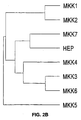
- Fig. 2B is a dendrogram showing the relationship between MKKs. The dendrogram was created as described for Fig. 2A.
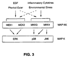
- Fig. 3 is a schematic representation of the ERK, p38, and JNK signal transduction pathways.
- MEK1 and MEK2 are activators of the ERK subgroup of MAP kinase.
- MKK3 and MKK4 are activators of the p38 MAP kinase.
- MKK4 is identified as an activator of both the p38 and JNK subgroups of MAP kinase.
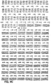
- Figs. 4A-4D are a representation of the nucleic acid (SEQ ID NO:1) and amino acid sequences (SEQ ID NO:2) for MKK3.
- Figs. 5A-5C are a representation of the nucleic acid (SEQ ID NO:3) and amino acid sequences (SEQ ID NO:4) for MKK6.
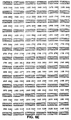
- Figs. 6A-6F are a representation of the nucleic acid (SEQ ID NO:5) and amino acid sequences (SEQ ID NO:6) for MKK4 ⁇ .
- Figs. 7A-7F are a representation of the nucleic acid (SEQ ID NO:7) and amino acid sequences (SEQ ID NO:8) for MKK4 ⁇ .
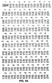
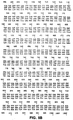
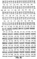
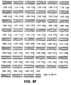
- Figs. 8A-8F are a representation of the nucleic acid (SEQ ID NO:9) and amino acid sequences (SEQ ID NO:10) for MKK4 ⁇ .
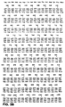
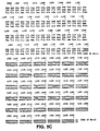
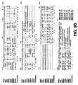
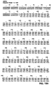
- Fig. 9 is a representation of the deduced primary structure of MKK7 (SEQ ID NO:18) compared with hep (SEQ ID NO:21), the MAP kinase kinases MEK1 (MKK1; SEQ ID NO:11), MEK2 (MKK2; SEQ ID NO:12), MKK3 (SEQ ID NO:2), MKK4 ⁇ (SEQ ID NO:10), MKK5 (SEQ ID NO:22), and MKK6 (SEQ ID NO:4) using the PILE-UP program (version 7,2; Wisconsin Genetics Computer Group). Gaps introduced into the sequences to optimize the alignment are illustrated with a dash (-). Identity is indicated with a dot (.). The sites of activating phosphorylation of MAP kinase kinases (2, 27, 37, and 38) are indicated with asterisks (*).
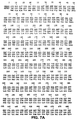
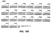
- Figs. 10A-10D are a representation of the nucleic acid (SEQ ID NO:17) and amino acid (SEQ ID NO:18) sequences for MKK7.
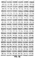
- Figs. 11A-11D are a representation of the nucleic acid (SEQ ID NO:19) and amino acid (SEQ ID NO:20) sequences of MKK7b.
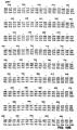
- Figs. 12A-12B are a representation of the nucleic acid (SEQ ID NO:25) and amino acid (SEQ ID NO:26) sequences of human MKK7.
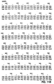
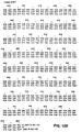
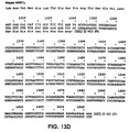
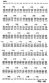
- Figs. 13A-13D are a representation of the nucleic acid (SEQ ID NO:27) and amino acid (SEQ ID NO:28) sequences of murine MKK7c.
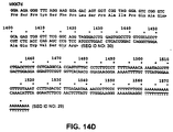
- Figs. 14A-14D are a representation of the nucleic acid (SEQ ID NO:29) and amino acid (SEQ ID NO:30) sequences of murine MKK7d.
- Figs. 15A-15D are a representation of the nucleic acid (SEQ ID NO:31) and amino acid (SEQ ID NO:32) sequences of murine MKK7e.
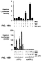
- Fig. 16A is a graph of data from a transfection assay in which cells were co-transfected with AP-1 reporter plasmid pTRE-Luciferase with expression vectors for MKK4, MKK7, JNK1, JNK1 (APF), or control vector.
- Fig. 16B is a graph of a transfection assay in which cells were co-transfected with a GAL4-ATF2 fusion vector and an expression vector for MKK4, MKK7, JNK1, JNK1(APF), or control vector.
- the human MAP kinase kinases MKK7 described herein mediate the transduction of specific signals from the cell surface to the nucleus along specific pathways. These signal transduction pathways are initiated by factors such as cytokines, UV radiation, osmotic shock, and oxidative stress. Activation of MKK3/4, MKK7 results in activation of the MAP kinases. JNK is activated by MKK7. p38 and JNK in turn activate a group of related transcription factors such as ATF2, ATFa, and CRE-BPa. These transcription factors in turn activate expression of specific genes.
- ATF2 in known to activate expression of human T cell leukemia virus 1 (Wagner and Green (1993) Science 262:395), transforming growth factor-b2 (Kim et al. (1992) supra ), interferon- ⁇ (Du et al. (1993) Cell 74:887), and E-selectin (DeLuca et al. (1994) J. Biol. Chem. 269:19193).
- ATF2 is implicated in the function of a T cell-specific enhancer (Georgopculos et al. (1992) Mol. Cell. Biol. 12:747).
- the JNK group of MAP kinases is activated by exposure of cells to environmental stress or by treatment of cells with pro-inflammatory cytokines (Gupta et al. (1994) EMBO J. 15:2760-2770; Dérijard et al. (1991) Cell 76:1025-1037; Kyriakis et al. (1994) Nature 369:156-160; Sluss et al. (1994) Mol. Cell. Biol. 14:8376-8384; Kallunki et al. (1994) Genes & Dev. 8:2996-3007).
- Targets of the JNK signal transduction pathway include the transcription factors ATF2 and c-jun (Whitmarsh & Davis (1996) J. Mol. Med.
- transcription factors are members of the bZIP group that bind as homo- and hecero-dimeric complexes to AP-1 and AP-1-like sites in the promoters of many genes (Curran & Franza (1988) Cell 55:395-397).
- JNK binds to an NH 2- terminal region of ATF2 and c-Jun and phosphorylates two sites within the activation domain of each transcription factor (Dérijard et al. (1994) Cell 76:1025-1037; van Dam et al. (1995) EMBO J. 14:1798-1811; Livingstone et al. (1995) EMBO J. 14:1785-1797).
- MKK4 also known as SEKI
- MKK4 was the first MAP kinase kinase identified as a component of the JNK signal transduction pathway (Derijard et al. 1995) Science 267:682-685; Lin et al. (1995) Science 268:286-290; Sanchez et al. (1994) Nature 372:794-798).
- Biochemical studies demonstrate that MKK4 phosphorylates and activates JNK (Dérijard et al. (1995) Science 267:682-685; Lin et al. (1995) Science 268:286-290; Sanchez et al.
- MKK4 may not be restricted to the JNK signal transduction pathway because MKK4 also phosphorylates and activates p38 MAP kinase (Dérijard et al. (1995) Science 267:682-685; Lin et al. (1995) Science 268:286-290). This specificity of MKK4 to activate both JNK and p38 MAP kinase provides a mechanism that may account for the co-ordinate activation of these MAP kinases in cells created with cytokines or environmental stress (Davis (1994) Trends Biochem. Sci. 19:470-473). However, this co-ordinate activation is not always observed.
- JNK activation in the liver correlates with decreased p38 MAP kinase activity (Mendelson et al. (1996) Proc. Natl. Acad. Sci. USA 93:12908-12913).
- MKK4 MKK4
- Example 1 The isolation of human MKKs is described in Example 1, Example 22, Dérijard et al. ((1995) Science 267:682-685, hereby specifically incorporated by reference), and Raingeaud et al. ((1995) Mol. Cell. Biol. 16:1247-1255). Distinctive regions of the yeast PBS2 sequence were used to design polymerase chain reaction (PCR) primers. Amplification of human brain mRNA with these primers resulted in the formation of specific products which were cloned into a plasmid vector and sequenced.
- PCR polymerase chain reaction
- JNK1 phosphorylation of ATF2 was examined by deletion analysis (Example 2.) Progressive NH 2 -terminal domain deletion GST-ATF2 fusion proteins were generated, and phosphorylation by JNK1 isolated from UV-irradiated cells was examined. The results showed that JNK1 requires the presence of ATF2 residues 1-60 for phosphorylation of the NH 2 -terminal domain of ATF2.
- JNK1 The ATF2 residues required for binding of JNK1 were similarly examined. JNK1 was incubated with immobilized ATF2, unbound JNK1 was removed by extensive washing, and bound JNK1 was detected by incubation with [ ⁇ - 32 P]ATP. Results indicate that residues 20 to 60 of ATF2 are required for binding and phosphorylation by JNK1. A similar binding interaction between ATF2 and the 55 kD JNK2 protein kinase has also been observed.
- Phosphorylation by JNK1 was shown to reduce the electrophoretic mobility of ATF2 (Example 9).
- Phosphoamino acid analysis of the full-length ATF2 molecule demonstrated that JNK phosphorylated both Thr and Ser residues.
- the major sites of Thr and Ser phosphorylation were located in the NH 2 and COOH terminal domains, respectively.
- the NH 2- terminal sites of phosphorylation were identified as Thr 69 and Thr 71 by phosphopeptide mapping and mutational analysis. These sites of Thr phosphorylation are located in a region of ATF2 that is distinct from the sub-domain required for JNK binding (residues 20 to 60).
- JNK1 was activated in CHO cells expressing JNK1 by treatment with UV radiation, pro-inflammatory cytokine interleukin-1 (IL-1), or serum.
- IL-1 pro-inflammatory cytokine interleukin-1
- a decreased electrophoretic mobility of JNK1-activated ATF2 was observed in cells treated with UV radiation and IL-1. Smaller effects were seen after treatment of cells with serum.
- the tumor suppressor gene product Rb binds to ATF2 and increases ATF2-stimulated gene expression (Kim et al. (1992) Nature 358:331).
- the adenovirus oncoprotein E1A associates with the DNA binding domain of ATF2 and increases ATF2-stimulated gene expression by a mechanism that requires the NH 2 -terminal activation domain of ATF2 (Liu and Green (1994) Nature 368:520).
- ATF2 transcriptional activity was investigated with the luciferase reporter gene system in control, Rb-treated, and ElA-treated cells expressing wild-type or mutant ATF2 molecules (Example 14). Rb and E1A were found to increase ATF2-stimulated gene expression of both wild-type and mutant ATF2.
- mutant ATF2 caused a lower level of reporter gene expression than did wild-type ATF2. Together, these results indicate a requirement for ATF2 phosphorylation (on Thr 69 and Thr 71 ) plus either Rb or E1A for maximal transcriptional activity. Thus, Rb and E1A act in concert with ATF2 phosphorylation to control transcriptional activity.
- cytoplasmic phospholipase A 2 (cPLA 2 ) (Lin et al. (1993) Cell 72:269), c-Myc (Alvarez et al. (1991) J. Biol. Chem. 266:15277), I ⁇ B, c-Jun, and wild-type (Thr 69, 71 ) or mutated (Ala 69, 71 ) ATF2.
- p38 phosphorylated MBP and EGF-R, and to a lesser extent I ⁇ B, but not the other ERK substrates, demonstrating that the substrate specificity of p38 differs from both the ERK and JNK groups of MAP kinases. Wild-type ATF2, but not mutated ATF2 (Ala 69, 71 ), was found to be an excellent p38 substrate.
- EGF and phorbol ester are potent activators of the ERK signal transduction pathway (Egan and Weinberg (1993) Nature 365:781), causing maximal activation of the ERK sub-group of MAP kinases. These treatments, however, cause only a small increase in JNK protein kinase activity (Dérijard et al. (1994) supra; Hibi et al. (1993) supra ). The effects of EGF or phorbol esters, as well UV radiation, osmotic shock, interleukin-1, tumor necrosis factor, and LPS, on p38 activity were all tested (Example 17).
- EGF and phorbol ester caused only a modest increase in p38 protein kinase activity, whereas environmental stress (UV radiation and osmotic shock) caused a marked increase in the activity of both p38 and JNK.
- Both p38 and JNK were activated in cells treated with pro-inflammatory cytokines (TNF and IL-1) or endotoxic LPS. Together, these results indicate that p38, like JNK, is activated by a stress-induced signal transduction pathway.
- ERKs and JNKs are activated by dual phosphorylation within the motifs Thr-Glu-Tyr and Thr-Pro-Tyr, respectively.
- p38 contains the related sequence Thr-Gly-Tyr. To test whether this motif is relevant to the activation of p38, the effect of the replacement of Thr-Gly-Tyr with Ala-Gly-Phe was examined (Example 18). The effect of UV radiation on cells expressing wild-type (Thr 180 , Tyr 182 ) or mutant p38 (Ala 180 , Phe 182 ) was studied. Western blot analysis using an anti-phosphotyrosine antibody demonstrated that exposure to UV radiation caused an increase in the Tyr phosphorylation of p38.
- ERK activity is regulated by the mitogen-induced dual specificity phosphatases MKP1 and PAC1 (Ward et al. (1994) Nature 367:651).
- the activation of p38 by dual phosphorylation raises the possibility that p38 may also be regulated by dual specificity phosphatases.
- MKP1 and PAC1 On p38 MAP kinase activation (Example 19).
- Cells expressing human MKP1 and PAC1 were treated with and without UV radiation, and p38 activity measured.
- the expression of PAC1 or MKP1 was found to inhibit p38 activity.
- the inhibitory effect of MKP1 was greater than PAC1.
- Example 20 The sub-cellular distribution of p38 MAP kinase was examined by indirect immunofluorescence microscopy (Example 20). Epitope-tagged p38 MAP kinase was detected using the M2 monoclonal antibody. Specific staining of cells transfected with epitope-tagged p38 MAP kinase was observed at the cell surface, in the cytoplasm, and in the nucleus. Marked changes in cell surface and nuclear p38 MAP kinase were not observed following UV irradiation, but an increase in the localization of cytoplasmic p38 MAP kinase to the perinuclear region was detected.
- JNK1 activity was measured 15 to 30 minutes after osmotic shock with a progressive decline in JNK1 activity at later times.
- the activation of JNK by osmotic shock was studied in cells expressing wild-type (Thr 183 , Tyr 185 ) or mutated (Ala 183 , Phe 185 ) JNK1.
- JNK1 activity was measured after incubation for 15 minutes with or without 300 mM sorbitol.
- Cells expressing wild-type JNK1 showed increased JNK1 activity, while cells expressing mutated JNK1 did not.
- Fig. 3 diagrams the ERK, p38, and JNK MAP kinase signal transduction pathways.
- ERKs are potently activated by treatment of cells with EGF or phorbol esters.
- p38 is only slightly activated under these conditions (Example 15).
- UV radiation, osmotic stress, and inflammatory cytokines cause a marked increase in p38 activity. This difference in the pattern of activation of ERK and p38 suggests that these MAP kinases are regulated by different signal transduction pathways.
- Example 22 The isolation of murine and human MKK7 is described in Example 22. Distinctive regions of the Drosophila MAP kinase kinase hep sequence were used to design polymerase chain reaction (PCR) primers. Amplification of murine testis mRNA with these primers resulted in the formation of specific products which were cloned into a plasmid vector and sequenced. One sequence related to hep was identified and used to screen a murine testis library. Five DNAs (cDNAs) that encoded protein kinases were identified: one encoding a MAP protein kinase kinase (MKK7). The others encoded various splice variants: MKK7b (a partial sequence appears in Fig.
- MKK7c (Fig. 13), MKK7d (Fig. 14), MKK7e (Fig. 15).
- the deduced amino acid sequences of MKK7 (SEQ ID NO:18) and hep (SEQ ID NO:21) are shown in Fig. 9, and compared to the MAP kinase kinases MEK1 (SEQ ID NO:11), MEK2 (SEQ ID NO:12), MKK3 (SEQ ID NO:2), MKK4 (SEQ ID NO:10), MKK5 (SEQ ID NO:22), and MKK6 (SEQ ID NO:4).
- a human MKK7 was identified by screening a human cDNA library with a full-length (mouse) MKK7 cDNA probe. The identified partial sequence (lacking the 3'end) is homologous to mouse MKK7c.
- MKK7 isoforms was examined by Northern (RNA) blot analysis of poly A+ mRNA isolated from eight murine tissues (Example 23). Both protein kinases were found to be widely expressed.
- MKK7 The substrate specificity of MKK7 was investigated in an in vitro phosphorylation assay with recombinant, epitope-tagged MAP kinases (JNK1, p38, and ERK2) as substrates (Example 24). MKK7 phosphorylated JNK, but did not phosphorylate p38 or ERK2. MKK7 was phosphorylated by p38 and JNK1.
- MKK7 was found to specifically activate JNK protein kinase in vivo (Example 19).
- CHO cells were co-transfected with an epitope-tagged MAP kinase (JNK1, p38, or ERK2) together with an empty expression vector or an expression vector encoding MKK7 and the product of the phosphorylation reaction analyzed.
- JNK1, p38, or ERK2 epitope-tagged MAP kinase
- Example 20 To test whether MKK7 could cause increased AP-1 transcriptional activity, a co-transfection assay was employed (Example 20). Co-expression of MKK7 with JNK caused an increase in AP-1 reporter gene expression A similar result was seen when ATF2 was used as the reporter gene. In addition, MKK7 alone was able to increase expression of ATF2 (Fig. 16).
- MKK isoforms are useful for screening reagents which modulate MKK activity. Described in the Use section following the Examples are methods for identifying reagents capable of inhibiting or activating MKK activity.
- COS-1 cells were maintained in Dulbecco's modified Eagle's medium supplemented with bovine serum albumin (5%) (Gibco-BRL). Metabolic labeling with [ 32 ]P was performed by incubation of cells for 3 hours in phosphate-free modified Eagle's medium (Flow Laboratories Inc.) supplemented with [ 32 P]orthophosphate (2 mCi/ml) (Dupont-NEN). COS-1 cells were transfected without (Mock) and with epitope-tagged JNK1 (JNK1). Plasmid expression vectors encoding the JNK1 cDNA have previously been described (Dérijard et al. (1994) Cell 76:1025, herein specifically incorporated by reference). Plasmid DNA was transfected into COS-1 cells by the lipofectamine method (Gibco-BRL). After 48 hours of incubation, some cultures were exposed to 40 J/m 2 UV radiation and incubated for 1 hour at 37°C.
- In-gel protein kinase assays were performed with cell lysates and JNK1 immunoprecipitates after SDS-PAGE by renaturation of protein kinases, polymerization of the substrate (GST-ATF2, residues 1-505) in the gel, and incubation with [ ⁇ - 32 P]ATP (Dérijard et al. (1994) supra ).
- the incorporation of [ 32 P]phosphate was visualized by autoradiography and quantitated with a Phosphorimager and ImageQuant software (Molecular Dynamics Inc., Sunnyvale, CA).
- the cell lysates demonstrate the presence of 46 kD and 55 kD protein kinases that phosphorylate ATF2 in extracts prepared from UV-irradiated cells.
- the 46 kD and 55 kD protein kinases were identified as JNK1 and JNK2, respectively.
- the site of JNK1 phosphorylation of ATF2 was investigated by generation of progressive NH 2 -terminal domain deletions of ATF2. Plasmid expression vectors encoding ATF2 (pECE-ATF2) (Liu and Green (1994) and (1990)), have been described. Bacterial expression vectors for GST-ATF2 fusion proteins were constructed by sub-cloning ATF2 cDNA fragments from a polymerase chain reaction (PCR) into pGEX-3X (Pharmacia-LKB Biotechnology Inc.). The sequence of all constructed plasmids was confirmed by automated sequencing with an Applied Biosystems model 373A machine.
- PCR polymerase chain reaction
- GST-ATF2 proteins were purified as described (Smith and Johnson (1988) Gene 67:31), resolved by SDS-PAGE and stained with Coomassie blue.
- GST-ATF2 fusion proteins contained residues 1-505, 1-349, 350-505, 1-109, 20-109, 0-109, and 60-109.
- Immunecomplex kinase assays were performed with Flag epitope-tagged JNK1 and the monoclonal antibody M2 (IBI-Kodak) as described by Dérijard et al. (1994) supra ). Immunecomplex protein kinase assays were also performed with a rabbit antiserum prepared with recombinant JNK1 as an antigen.
- the cells were solubilized with 20 mM Tris, pH 7.5, 10% glycerol, 1% Triton® X-100, 0.137 M NaCl, 25 mM ⁇ -glycerophosphate, 2 mM EDTA, 1 mM orthovanadate, 2 mM pyrophosphate, 10 ⁇ g/ml leupeptin, and 1 mM PMSF.
- JNK1 was immunoprecipitated with protein G-Sepharose bound to a rabbit polyclonal antibody to JNK or the M2 monoclonal antibody to the Flag epitope.
- the beads were washed three times with lysis buffer and once with kinase buffer (20 mM Hepes, pH 7.6, 20 mM MgCl 2 , 25 mM ⁇ -glycerophosphate, 100 ⁇ M Na orthovanadate, 2 mM dithiothreitol).
- the kinase assays were performed at 25°C for 10 minutes with 1 ⁇ g of substrate, 20 ⁇ M adenosine triphosphate and 10 ⁇ Ci of [ ⁇ - 32 P]ATP in 30 ⁇ l of kinase buffer.
- the reactions were terminated with Laemmli sample buffer and the products were resolved by SDS-PAGE (10% gel).
- JNK1 phosphorylates GST-ATF2 fusion proteins containing residues 1-505, 1-349, 1-109, 20-109, and 40-109, but not 60-109. These results indicate that the presence of ATF2 residues 1-60 are required for phosphorylation by JNK.
- JNK1 from UV-irradiated cells was incubated with GST-ATF2 fusion proteins bound to GSH-agarose. The agarose beads were washed extensively to remove the unbound JNK1. Phosphorylation of the GST-ATF2 fusion proteins by the bound JNK1 protein kinase was examined by addition of [ ⁇ - 32 P]ATP. JNK1 bound GST-ATF2 fusion proteins containing residues 1-505, 1-349, 1-109, 20-109, and 40-109, indicating that the presence of residues 20-60 were required for binding of JNK1 to ATF2.
- JNK1-transfected cells but not mock-transfected cells, phosphorylated ATF2.
- JNK1 phosphorylation of ATF2 was greater in cells exposed to UV radiation.
- Phosphorylation of ATF2 by JNK1 was associated with a decreased electrophoretic mobility.
- GST fusion proteins containing full-length ATF2 (residues 1 to 505), an NH 2- terminal fragment (residues 1 to 109), and a COOH-terminal fragment (residues 95 to 505) were phosphorylated with JNK1 and the sites of phosphorylation analyzed by phosphoamino acid analysis.
- the methods used for phosphopeptide mapping and phosphoamino acid analysis have been described (Alvarez et al. (1991) J. Biol. Chem. 266:15277).
- the horizontal dimension of the peptide maps was electrophoresis and the vertical dimension was chromatography.
- NH 2 -terminal sites of phosphorylation were identified as Thr 69 and Thr 71 by phosphopeptide mapping and mutational analysis. Site-directed mutagenesis was performed as described above, replacing Thr 69 and Thr 71 with Ala. Phosphorylation of mutated ATF2 was not observed.
- CHO cells were maintained in Ham's F12 medium supplemented with 5% bovine serum albumin (Gibco-BRL). Cells were labeled and transfected with JNK1 as described above. CHO cells were treated with UV-C (40 J/m 2 ), IL-1 ⁇ (10 ng/ml) (Genzyme), or fetal bovine serum (20%) (Gibco-BRL). The cells were incubated for 30 minutes as 37°C prior to harvesting. The electrophoretic mobility of ATF2 after SDS-PAGE was examined by protein immuno-blot analysis. A shift in ATF2 electrophoretic mobility was observed in cells treated with UV, IL-1, and serum.
- COS-1 cells were transfected without (control) and with an ATF2 expression vector (ATF2), as described above (Hai et al. (1989) supra ). The effect of exposure of the cells to 40 J/m 2 UV-C was examined. After irradiation, the cells were incubated for 0 or 30 minutes (control) or 0, 15, 30, and 45 minutes (ATF2) at 37°C and then collected. The electrophoretic mobility of ATF2 during SDS-PAGE was examined by protein immuno-blot analysis as described above. The two electrophoretic mobility forms of ATF2 were observed in ATF2-transfected cells, but not in control cells.
- ATF2 expression vector ATF2 expression vector
- the phosphorylation state of wild-type (Thr 69,71 ) ATF2 and mutated (Ala 69,71 ) ATF2 was examined in cells labeled with [ 32 ]P, treated without and with 40 J/m 2 UV-C, and then incubated at 37°C for 30 minutes (Hai et al. (1989) supra ).
- the ATF2 proteins were isolated by immunoprecipitation and analyzed by SDS-PAGE and autoradiography.
- the phosphorylated ATF2 proteins were examined by phosphoamino acid analysis as described above. Both forms of ATF2 contained phosphoserine, but only wild-type ATF2 contained phosphothreonine.
- Tryptic phosphopeptide mapping was used to compare ATF2 phosphorylated in vitro by JNK1 with ATF2 phosphorylated in COS-1 cells.
- a map was also prepared with a sample composed of equal amounts of in vivo and in vitro phosphorylated ATF2 (Mix). Mutation of ATF2 at Thr 69 and Thr 71 resulted in the loss of two tryptic phosphopeptides in maps of ATF2 isolated from UV-irradiated cells. These phosphopeptides correspond to mono- and bis-phosphorylated peptides containing Thr 69 and Thr 71 . Both of these phosphopeptides were found in maps of ATF2 phosphorylated by JNK1 in vitro.
- a fusion protein consisting of ATF2 and the GAL4 DNA binding domain was expressed in CHO cells as described above.
- the activity of the GAL4-ATF2 fusion protein was measured in co-transfection assays with the reporter plasmid pG5ElbLuc (Seth et al. (1992) J. Biol. Chem. 267:24796, hereby specifically incorporated by reference).
- the reporter plasmid contains five GAL4 sites cloned upstream of a minimal promoter element and the firefly luciferase gene. Transfection efficiency was monitored with a control plasmid that expresses ⁇ -galactosidase (pCH110; Pharmacia-LKB Biotechnology).
- GAL4-ATF2 wild type 320,000 GAL4-ATF2 (Ala 69 ) 24,000 GAL4-ATF2 (Ala 71 ) 22,000 GAL4-ATF2 (Ala 69, 71 ) 29,000 GAL4-ATF2 (GlU 69 ) 27,000
- the luciferase reporter plasmid system was used to determine the effect of point mutations at the ATF2 phosphorylation sites Thr 69 and Thr 71 in serum-treated CHO cells transfected with wild-type (Thr 183 , Tyr 185 ) or mutant (Ala 183 , Phe 185 ) JNK1. Control experiments were done with mock-transfected cells. The CHO cells were serum-starved for 18 hours and then incubated without or with serum for 4 hours. Expression of wild-type ATF2 caused a small increase in serum-stimulated ATF2 transcriptional activity. In contrast, mutant JNK1 inhibited both control and serum-stimulated ATF2 activity.
- Substrate phosphorylation by p38 MAP kinase was examined by incubation of bacterially-expressed p38 MAP kinase with I ⁇ B, cMyc, EGF-R, cytoplasmic phospholipase A 2 (cPLA 2 ), c-Jun, and mutated ATF2 (Thr 69,71 ) and ATP[ ⁇ -32 P] (Raingeaud et al. (1995) J. Biol. Chem 270:7420, herein specifically incorporated by reference).
- GST-I ⁇ B was provided by Dr D. Baltimore (Massachusetts Institute of Technology).
- GST-cMyc Alvarez et al. (1991) J. Biol. Chem.
- TNF ⁇ and IL-1 ⁇ were from Genzyme Corp.
- LPS lipolysaccharide
- EGF was purified from mouse salivary glands (Davis (1988) J. Biol. Chem. 263:9462). Kinase assays were performed using immunoprecipitates of p38 and JNK. The immunocomplexes were washed twice with kinase buffer (described above), and the assays initiated by the addition of 1 ⁇ g of ATF2 and 50 ⁇ M [ ⁇ - 32 P]ATP (10 Ci/mmol) in a final volume of 25 ⁇ l. The reactions were terminated after 30 minutes at 30°C by addition of Laemmli sample buffer. The phosphorylation of ATF2 was examined after SDS-PAGE by autoradiography, and the rate of ATF2 phosphorylation quantitated by PhosphorImager analysis.
- JNK p38 AND JNK1 ACTIVATION BY PRO-INFLAMMATORY CYTOKINES AND ENVIRONMENTAL STRESS.
- Relative Protein Kinase Activity JNK p38 Control 1.0 1.0 Epidermal Growth Factor (10 nM) 1.9 2.1 Phorbol Ester (10 nM) 2.3 2.9 Lipopolysaccharide (10 ng/ml) 3.6 3.7 Osmotic Shock (300 mM sorbitol) 18.1 4.2 Tumor Necrosis Factor (10 ng/ml) 19.3 10.3 Interleukin-1 (10 ng/ml) 8.9 6.2 UV (40 J/m 2 ) 7.4 17.1
- COS-1 cells expressing wild-type (Thr 180 , Tyr 182 ) or mutated (Ala 180 , Phe 182 ) p38 MAP kinase were treated without and with UV-C (40 J/m 2 ). The cells were harvested 30 minutes following exposure with or without UV radiation. Control experiments were performed using mock-transfected cells. The level of expression of epitope-tagged p38 MAP kinase and the state of Tyr phosphorylation of p38 MAP kinase was examined by Western blot analysis using the M2 monoclonal antibody and the phosphotyrosine monoclonal antibody PY20. Immune complexes were detected by enhanced chemiluminescence.
- Wild-type and mutant p38 were expressed at similar levels. Western blot analysis showed that UV radiation caused an increase in the Tyr phosphorylation of p38. The increased Tyr phosphorylation was confirmed by phosphoamino acid analysis of p38 isolated from [ 32 P] phosphate-labeled cells. The results also showed that UV radiation increased Thr phosphorylation of p38. The increased phosphorylation on Tyr and Thr was blocked by mutated p38. Wild-type and mutated p38 were isolated from the COS-1 cells by immunoprecipitation. Protein kinase activity was measured in the immune complex using [ ⁇ - 32 P]ATP and GST-ATF2 as substrates.
- the phosphorylated GST-ATF2 was detected after SDS-PAGE by autoradiography. UV radiation resulted in a marked increase in the activity of wild-type p38, while the mutant p38 was found to be catalytically inactive. These results show that p38 is activated by dual phosphorylation within the Thr-Gly-Tyr motif.
- the cells were treated without and with 40 J/m 2 UV-C.
- Control experiments were performed using mock-transfected cells (control) and cells transfected with the catalytically inactive mutated phosphatase mPAC1 (Cys 257 /Ser) and human MKP1.
- the activity of p38 MAP kinase was measured with an immunecomplex protein kinase assay employing [ ⁇ - 32 P]ATP and GST-ATF2 as substrates.
- the expression of PAC1 or MKP1 was found to inhibit p38 phosphorylation, demonstrating that p38 can be regulated by the dual specificity phosphatases PAC1 and MKP1.
- Epitope-tagged p38 MAP kinase was expressed in COS cells. The cells were treated without or with 40 J/m 2 UV radiation and then incubated for 60 minutes at 37°C. The p38 MAP kinase was detected by indirect immunofluorescence using the M2 monoclonal antibody. The images were acquired by digital imaging microscopy and processed for image restoration.
- Coverslips 22mm x 22mm No. 1; Gold Seal Cover Glass; Becton-Dickinson
- the coverslips were placed at the bottom of 35 mm multiwell tissue culture plates (Becton Dickinson, UK).
- Transfected COS-1 cells were plated directly on the coverslips and allowed to adhere overnight in Dulbecco's modified Eagle's medium supplemented with 5% fetal calf serum (Gibco-BRL).
- the cells were rinsed once and incubated at 37°C for 30 minutes in 25 mM Hepes, pH 7.4, 137 mM NaCl, 6 mM KCl, 1 mM MgCl 2 , 1 mM CaCl 2 , 10 mM glucose.
- the cells were rinsed once with phosphate-buffered saline and the coverslips removed from the tissue culture wells. Cells were fixed in fresh 4% paraformaldehyde in phosphate-buffered saline for 15 minutes at 22°C.
- the cells were permeabilized with 0.25% Triton® X-100 in phosphate-buffered saline for 5 minutes and washed three times in DWB solution (150 mM NaCl, 15 mM Na citrate, pH 7.0, 2% horse serum, 1% (w/v) bovine serum albumin, 0.05% Triton® X-100) for 5 minutes.
- the primary antibody (M2 anti-FLAG monoclonal antibody, Eastman-Kodak Co., New Haven, CT) was diluted 1:250 in DWB and applied to the cells in a humidified environment at 22°C for 1 hour.
- the cells were again washed three times as above and fluorescein isothiocyanate-conjugated goat anti-mouse Ig secondary antibody (Kirkegaard & Perry Laboratories Inc. Gaithersburg, MD) was applied at a 1:250 dilution for 1 hour at 22°C in a humidified environment.
- the cells were then washed three times in DWB and then mounted onto slides with Gel-Mount (Biomeda Corp. Foster City, CA) for immunofluorescence analysis. Control experiments were performed to assess the specificity of the observed immunofluorescence. No fluorescence was detected when the transfected cells were stained in the absence of the primary M2 monoclonal antibody, or mock-transfected cells.
- the exposure of the sample to the excitation source was determined by a computer-controlled shutter and wavelength selector system (MVI, Avon, MA).
- the charge-coupled device camera and microscope functions were controlled by a microcomputer, and the data acquired from the camera were transferred to a Silicon Graphics model 4D/GTX workstation (Mountainview, CA) for image processing. Images were corrected for non-uniformities in sensitivity and for the dark current of the charge coupled device detector.
- the calibration of the microscopy blurring was determined by measuring the instrument's point spread function as a series of optical sections at 0.125 ⁇ m intervals of a 0.3 ⁇ m diameter fluorescently labeled latex bead (Molecular Probes Inc.).
- the image restoration algorithm used is based upon the theory of ill-posed problems and obtains quantitative dye density values within the cell that are substantially more accurate than those in an unprocessed image (Carrington et al. (1990) supra ; Fay et al. (1989) supra ).
- image processing individual optical sections of cells were inspected and analyzed using computer graphics software on a Silicon Graphics workstation. p38 MAP kinase was observed at the cell surface, in the cytoplasm, and in the nucleus. After irradiation, an increased localization of cytoplasmic p38 to the perinuclear region was detected.
- CHO cells were co-transfected with the plasmid pCMV-Flag-Jnk1 and pRSV-Neo (Dérijard et al. (1994) supra ).
- a stable cell line expressing epitope-tagged Jnk1 was isolated by selection with Geneticin (Gibco-BRL). The cells were incubated with 0, 100, 150, 300, 600, or 1000 mM sorbitol for 1 hour at 37°C.
- the cells were collected in lysis buffer 20 mM Tris, pH 7.4, 1% TRITON® X-100, 2 mM EDTA, 137 mM NaCl, 25 mM ⁇ -glycercphosphate, 1 mM orthovanadate, 2 mM pyrophosphate, 10% glycerol, 1 mM phenylmethylsulfonyl fluoride, 10 ⁇ g/ml leupeptin) and a soluble extract was obtained by centrifugation at 100,000 g for 30 minutes at 4°C.
- the epitope-tagged JNK1 was isolated by immunoprecipitation with the monoclonal antibody M2 (Immunex Corp.). The immunoprecipitates were washed extensively with lysis buffer.
- Immunecomplex kinase assays were done in 25 ⁇ l of 25 mM Hepes, pH 7.4, 25 mM MgCl 2 , 25 mM ⁇ -alycerophosphate, 2 mM dithiothreitol, 100 ⁇ M orthovanadate, and 50 ⁇ M ATP [ ⁇ - 32 p] (10 Ci/mmole) with 2.5 ⁇ g of bacterially expressed c-Jun (residues 1-79) fused to glutathione-S-transferase (GST) as a substrate. The phosphorylation of c-Jun was examined after SDS-PAGE by autoradiography and Phosphorlmager (Molecular Dynamics Inc.) analysis. JNK1 activation was observed at all concentrations of sorbitol exposure.
- JNK1 protein kinase activation was measured in cells incubated in medium supplemented with 300 mM sorbitol as described above. Increased JNK1 activity was observed within 5 minutes of exposure to sorbitol, with maximum activity occurring after 15-30 minutes.
- JNK1 activation was seen in the wild-type but not mutated JNK1.
- RT-PCR was employed to identify a fragment of a novel mammalian MAP kinase kinase.
- Murine testis mRNA was used as the template.
- a single product (461 bp) was detected following RT-PCR amplification of murir.e testis mRNA.
- This sequence includes protein kinase sub-domains I - XI and is related to the MAP kinase kinase group.
- the novel protein kinase was designated MKK7.
- the sites of activating phosphorylation of MAP kinase kinases located in sub-domain VIII are conserved in MKK7.
- Comparison of MKK7 with other members of the mammalian MAP kinase kinase group demonstrates that MKK7 is related to the JNK activator MKK4.
- One additional cDNA clone isolated from the X phage library differed from the other seven clones.
- This clone contained the same 3' untranslated region and coding region of MKK7, but had a different 5' region that lacked an in-frame termination codon.
- This clone represents an alternatively spliced form of MKK7 (MKK7b; Fig. 11; SEQ ID NO:19).
- the MKK7b cDNA clone does not have an initiation codon in the alternative 5' region; this cDNA therefore encodes the same MKK7 protein kinase as the other clones that were isolated. However, if the MKK7b cDNA clone is not full-length it is possible that additional 5' sequence may include an in-frame initiation codon.
- MKK7b is predicted to fuse the sequence M-[?]-SPAPAPSQRAALQLPLANDGGSRSPSSESSPQHPTPPTRPRH- (SEQ ID NO:33) to the initiating methionine of MKK7 (Fig. 9).
- the Drosophila MAP kinase kinase hep shares substantial sequence similarity with MKK7, the sequence of the NH2-terminal extension of MKK7b is not conserved in the hep protein kinase.
- Three additional clones encoded MKK7 splice variants that differ in the 5' and 3 ' regions. These clones (MKK7c (Fig. 13), MKK7d (Fig. 14), and MKK7e (Fig. 15)) are full-length because of the presence of in-frame termination codons in the 5' and 3' regions.
- a human cDNA library was screened with a full-length mouse MKK7 cDNA probe. A single clone was identified and squenced. A partial MKK7 sequence was identified (Fig. 12; SEQ ID NO:25 and SEQ ID NO:25) that is missing the 3' end. The sequence is most homologous to mouse MKK7c.
- MKK7, MKK7b, hep, and human MKK7 cDNAs have been deposited in Genbank with accession numbers U93030, U93031, U93032, and AFOO319 respectively.
- MKK7 expression was examined by Northern blot analysis of mRNA isolated from different tissues. The analysis was done with poly A+ mRNA (2 ⁇ g) isolated from different tissues and fractionated by denaturing agarose gel electrophoresis and transferred to a nylon membrane (Clontech). The blot was probed with MKK4 and MKK7 cDNAs labeled by random priming with [ ⁇ - 32 P]dATP (Amersham International PLC).
- MKK7 was found to be widely expressed in murine tissues. A single MKK7 transcript (approximately 4.0-kb) was detected in all of the tissues examined, except for testis where two MKK7 transcripts (4.0 kb and 1.5 kb) were detected. The highest levels of MKK7 expression were in testis. Significant expression of MKK7 was also observed in heart, brain, lung, liver, and kidney. This contrasts with MKK4 expression which was highest in brain although significant amounts of expression were observed in brain, liver, muscle, heart, and kidney. Although MKK4 and MKK7 are co-expressed, the relative abundance of each MAP kinase kinase is different in each of the tissues examined.
- a bacterial MKK7 expression vector was prepared by sub-cloning an MKK7 cDNA ( Eco RI and Pvu II fragment) into the Eco RI and Sma I sites of pGEX-5Xl (Pharmacia-LKB).
- the glutathione-S-transferase (GST) fusion protein was purified by affinity chromatography (Smith and Johnson (1988) Gene 67:31-40).
- the recombinant proteins GST-ATF2 (Gupta et al.
- Protein kinase assays were performed in kinase buffer (25 mM 4-(2-hydroxyethyl)-1-piperazineethansulfonic acid (pH 7.4), 25 mM ⁇ -glycerophosphate, 25 mM MgCl 2 , 2 mM dithiothreitol, 0.1 mM orthovanadate).
- the assays were initiated by the addition of 1 ⁇ g of substrate proteins and 50 ⁇ M [ ⁇ -32P] ATP (10 Ci/mmol) in a final volume of 25 ⁇ l.
- the reactions were terminated after 30 minutes at 25°C by addition of Laemmli sample buffer.
- the phosphorylation of the substrate proteins was examined after SDS-polyacrylamide gel electrophoresis (PAGE) by autoradiography.
- Recombinant MAP kinases were incubated with GST (control) or GST-MKK7 using the substrate ATP [ ⁇ - 32 P]. Recombinant MKK7 purified from bacteria was not observed to autophosphorylace. Incubation of the recombinant MKK7 with MAP kinases demonstrated that MKK7 phosphorylated JNK1, but not p38 or ERK2. MKK7 was phosphorylated by p38 and JNK1.
- MKK7 did not phosphorylate the mutated JNK1 (APF) protein. Furthermore, MKK7 did not increase ATF2 phosphorylation by the mutated JNK1 protein kinase. Thus, MKK7 is a JNK activator in vitro .
- CHO cells were maintained in Dulbecco's modified Eagle's medium supplemented with fetal calf serum (5%; Gibco-BRL). The cells were transfected with the lipofectamine reagent according to the manufacturer's recommendations (Gibco-BRL) (Dérijard (1994) supra ). Cells were co-transfected with vectors encoding epitope-tagged JNK1 together with an empty expression vector (control) or an expression vector encoding MKK4 or MKK7.
- the epitope tag was derived from the hemagglutinin protein (HA) of the influenza virus.
- JNK1 was isolated by immunoprecipitation of cell lysates.
- the cells were solubilized with lysis buffer (20 mM Tris (pH 7.4), 1% TRITON X-100®, 10% glycerol, 137 mM NaCl, 2 mM EDTA, 25 mM ⁇ -glycerophosphate, 1 mM Na orthovanadate, 2 mM pyrophosphate, 1 mM PMSF, 10 ⁇ g/ml leupeptin) and centrifuged at 100,000 X g for 15 minutes at 4°C.
- lysis buffer (20 mM Tris (pH 7.4), 1% TRITON X-100®, 10% glycerol, 137 mM NaCl, 2 mM EDTA, 25 mM ⁇ -glycerophosphate, 1 mM Na orthovanadate, 2 mM pyrophosphate, 1 mM PMSF, 10 ⁇ g/ml leupeptin
- the epitope-tagged protein kinases were immunoprecipitated by incubation for 3 hours at 4°C with an anti-HA monoclonal antibody bound to protein-G Sepharose (Pharmacia-LKB Biotechnology Inc.). The immunoprecipitates were washed three times with lysis buffer (Gupta et al. (1995) Science 267:389-393). Protein kinase activity was measured in the immunecomplex with [ ⁇ - 23 P]ATP and c-Jun as substrates. The product of the phosphorylation reaction was visualized after SDS-PAGE by autoradiography. The ERK2 and p38 MAP kinases were not activated by co-expressed MKK7.
- the JNK signaling pathway is known to regulate AP-1 transcriptional activity (Whitmarsh (1996) supra ). To test the hypothesis that the expression of MKK7 would cause increased AP-1 transcriptional activity, a co-transfection assay was employed using a luciferase reporter gene that contains three AP-1 sites cloned upstream of a minimal promoter element (Rincon and Flavell (1994) EMBO J. 13:4370-4381).
- Luciferase reporter gene expression was measured in co-transfection assays using the 0.5 ⁇ g of the reporter plasmid pTRE-luciferase (Rincon (1994) supra ) and 0.25 ⁇ g of the ⁇ -galactosidase expression vector pCH110 (Pharmacia-LKB).
- GAL4 fusion proteins were performed using 0.25 ⁇ g of pGAL4-ATF2 (residues 1-109), 0.5 ⁇ g of the reporter plasmid pG5ElbLuc, and 0.25 ⁇ g of pCHllO (Gupta et al. (1995) supra ).
- the effect of protein kinases was examined by co-transfection with 0.3 ⁇ g of an empty expression vector or a protein kinase expression vector.
- the ERK2, p38, JNK1, MKK1, MKK3, MKK4, and MKK6 expression vectors have been described.
- the cells were harvested 36 hours post-transfection.
- the ⁇ -galactosidase and luciferase activity in the cell lysates was measured as described (Gupta (1995) supra ).
- Expression of MKK4, MKK7, or JNK1 did not cause marked changes in AP-1 reporter gene expression (Fig. 16A).
- co-expression of MKK7 with JNK1 caused increased AP-1-dependent reporter gene expression.
- the MKK polypeptides and polynucleotides of the invention are useful for identifying reagents that modulate the MKK signal transduction pathways.
- Reagents that modulate an MKK signal transduction pathway can be identified by their effect on MKK synthesis, MKK phosphorylation, or MKK activity.
- the effect of a reagent on MKK activity can be measured by the in vitro kinase assays described above. MKK is incubated without (control) and with a test reagent under conditions sufficient to allow the components to react, then the effect of the test reagent on kinase activity is subsequently measured.
- Reagents that inhibit an MKK signal transduction pathway can be used in the treatment of MKK-mediated disorders.
- Reagents that stimulate an MKK signal transduction pathway can be used in a number of ways, including induction of programmed cell death (apoptosis) in tissues. For example, the elimination of UV damaged cells can be used to prevent cancer.
- a kinase assay for identification of a reagent that inhibits the MKK signal transduction pathway, a kinase assay (see, for example, Example 3) is used.
- a range of reagent concentrations e.g., 1.0 nM to 100 mM
- a radioactive marker such as [ ⁇ - 32 P]ATP.
- Appropriate substrate molecules include p38, JNK1, JNK2, or ATF2.
- the incorporation of labelled phosphorus (e.g., [ 32 ]P or [ 33 ]P) into the substrate is determined, and the results obtained with the test reagent compared to control values.
- Phosphorylation-dependent antibodies may be made using MKK7 phosphorylated on the activating sites, Ser 198 and Thr 202 . This may be accomplished by immunizing animals with a synthetic peptide (for example, approximately 15 amino acids in length) corresponding to the MKK7 sequence with phosphorylated Ser 198 and Thr 202 . Methods of producing such antibodies are known in the art. Such antibodies are useful for the detection of activated MKK7 in tissues and cell extracts (e.g. on Western blots) and may be used in a kit.
- Assays that test the effect of a reagent on MKK synthesis can also be used to identify compounds that inhibit MKK signal transduction pathways.
- the effect of the test reagent on MKK expression is measured by, for example, Western blot analysis with an antibody specific for an MKK. Antibody binding is visualized by autoradiography or chemiluminescence, and is quantitated.
- the effect of the test reagent on MKK mRNA expression can be examined, for example, by Northern blot analysis using a polynucleotide probe or by polymerase chain reaction.
- Reagents found to inhibit MKK signal transduction pathways can be used as therapeutic agents for the treatment of MKK-mediated disorders. Such reagents are also useful in drug design for elucidation of the specific molecular features needed to inhibit MKK signal transduction pathways.
- Dominant-negative forms of MKK which effectively displace or compete with MKK for substrate binding and/or phosphorylation can be used to decrease protein kinase activity.
- Dominant-negative forms can be created by mutations within the catalytic domain of the protein kinases, using methods known in the art, and as described above (Example 7.) The catalytic residues are conserved in all the MKK isoforms. For example, mutation of Lys 76 inhibits MKK7 activity. Similarly, mutation of the conserved sites of activating phosphorylation (Ser 198 , Thr 202 ) inhibits MKK7 activity. These kinase-inactive forms of MKK7 act as dominant-negative inhibitors.
- augmentation of MKK activity is desirable, e.g., induction of apoptosis.
- the methods of the invention can be used to identify reagents capable of increasing MKK function or activity. Alternatively, increased activity is achieved by over-expression of MKK.
- a sense polynucleotide sequence (the DNA coding strand) or MKK polypeptide can be introduced into the cell to enhance normal MKK activity.
- MKK7 nucleic acid-containing vectors can be constructed by recombinant DNA technology methods standard in the art.
- Vectors can be plasmid, viral, or others known in the art, used for replication and expression in mammalian cells.
- Expression of the sequence encoding the MKK7 nucleic acid can be by any promoter known in the art to act in mammalian, preferably human cells. Such promoters can be inducible or constitutive.
- Such promoters include, but are not limited to: the SV40 early promoter region (Bernoist et al., Nature 290:304, 1981); the promoter contained in the 3' long terminal repeat of Rous sarcoma virus (Yamamoto et al., Cell 22:787-797, 1988); the herpes thymidine kinase promoter (Wagner et al., Proc. Natl. Acad. Sci. USA 78:1441, 1981); or the regulatory sequences of the metallothionein gene (Brinster et al., Nature 296:39, 1988).
Landscapes
- Health & Medical Sciences (AREA)
- Chemical & Material Sciences (AREA)
- Life Sciences & Earth Sciences (AREA)
- Organic Chemistry (AREA)
- Engineering & Computer Science (AREA)
- Genetics & Genomics (AREA)
- General Health & Medical Sciences (AREA)
- Bioinformatics & Cheminformatics (AREA)
- Zoology (AREA)
- Medicinal Chemistry (AREA)
- Molecular Biology (AREA)
- Wood Science & Technology (AREA)
- Proteomics, Peptides & Aminoacids (AREA)
- Biochemistry (AREA)
- Biophysics (AREA)
- Biotechnology (AREA)
- General Engineering & Computer Science (AREA)
- Pharmacology & Pharmacy (AREA)
- General Chemical & Material Sciences (AREA)
- Public Health (AREA)
- Animal Behavior & Ethology (AREA)
- Microbiology (AREA)
- Veterinary Medicine (AREA)
- Chemical Kinetics & Catalysis (AREA)
- Nuclear Medicine, Radiotherapy & Molecular Imaging (AREA)
- Immunology (AREA)
- Gastroenterology & Hepatology (AREA)
- Analytical Chemistry (AREA)
- Biomedical Technology (AREA)
- Physics & Mathematics (AREA)
- Toxicology (AREA)
- Pathology (AREA)
- Heart & Thoracic Surgery (AREA)
- Cardiology (AREA)
- Oncology (AREA)
- Rheumatology (AREA)
- Virology (AREA)
- Urology & Nephrology (AREA)
- Dermatology (AREA)
- Pain & Pain Management (AREA)
Claims (17)
- Ein substantiell reines säugertier mitogen-aktiviertes Protein Kinase Kinase (MKK7) Polypeptid, das Serin, Threonin und Tyrosin Kinase Aktivität besitzt und das die mitogen-aktivierte Protein (MAP) Kinase JNK, aber nicht p38 phosphoryliert, besagtes Polypeptid umfaßt die Aminosäure Sequenz von SEQ ID NO: 18, SEQ ID NO: 20, SEQ ID NO: 26, SEQ ID NO: 28, SEQ ID NO: 30, oder SEQ ID NO: 32.
- Eine isolierte Polynukleotidsequenz, kodierend für das Polypeptid gemäß Anspruch 1.
- Eine isolierte Polynukleotidsequenz gemäß Anspruch 2, umfassend eine Polynukleoidsequenz, die unter stringenten Hybridisierungsbedingungen mit der Sequenz von SEQ ID NO: 17, SEQ ID NO: 19, SEQ ID NO: 25, SEQ ID NO: 27, SEQ ID NO: 29, SEQ ID NO 31, oder einem Komplement davon hybridisiert.
- Ein rekombinanter Expressionsvektor enthaltend eine Polynukleotidsequenz gemäß Anspruch 2.
- Eine rekombinante Wirtszelle umfassend eine Polynukletidsequenz gemäß Anspruch 2.
- Ein aufgereinigter Antikörper, der spezifisch an das Polypeptid gemäß Anspruch 1 bindet.
- Ein Verfahren zur Messung der Aktivität einer mitogen-aktivierten Protein Kinase Kinase (MKK7) in einer biologischen Probe, besagtes Verfahren umfaßt:a) inkubieren besagter Probe mit einem MKK Substrat für das MKK Polypeptid gemäß Anspruch 1 und markiertem Phosphat unter Bedingungen, die ausreichen um die Physphorylierung besagten Substrats zu erlauben; undb) bestimmen der Rate des Einbaus des markierten Phosphats in besagtes Substrat, wobei besagte Rate ein Maß für MKK7 Aktivität ist.
- Ein Verfahren gemäß Anspruch 7, wobei besagtes MKK Substrat ausgewählt ist aus der Gruppe bestehend aus JNK MAP Kinasen, activating transcription factor-2 (ATF2), ATFa, cAMP response element binding protein (CRE-BPa) und c-Jun.
- Ein Verfahren gemäß Anspruch 7, wobei besagte Probe aus Flüssigkeit, Zellen, oder Gewebe, welches von einem Säugetier erhalten wurde, besteht.
- Ein Verfahren zur Messung der Synthese von MKK7 in einer biologischen Probe, besagtes Verfahren umfaßt:a) kontaktieren einer biologischen Probe mit einem Antikörper der spezifisch ein MKK7 Polypeptid gemäß Anspruch 1 bindet; undb) detektieren des besagten an das MKK7 Polypeptid gebundenen Antikörper, wobei der Grad der MKK7 Synthese durch die Menge des gebundenen Antikörpers bestimmt ist.
- Ein Verfahren zur Messung des Grads der Expression von MKK7 in einer Probe, das Verfahren umfaßt die Schritte:a) isolieren von totaler oder polyadenylierter RNA aus der Probe;b) inkubieren der RNA mit einer Polynukleotidsonde die spezifisch für ein MKK7 Polynukleotid nach Anspruch 3 ist; undc) bestimmen der Menge besagter Sonde, die mit der RNA hybrisisiert ist, wobei der Grad der Expression von MKK7 direkt mit der Menge der MKK7 Sonde, die mit der RNA hybridisiert ist in Beziehung steht.
- Ein Verfahren zur Identifizierung eines Reagenz das MKK7 Aktivität moduliert, besagtes Verfahren umfaßt:a) inkubieren einer MKK7 enthaltenden Probe mit einem MKK Substrat für das MKK Polypeptid nach Anspruch 1, einer Reihe von Reagenzkonzentrationen und markiertem Phosphat unter Bedingungen, die ausreichen die Physphorylierung besagten Substrats zu erlauben, wenn besagtes Reagenz nicht vorhanden ist;b) detektieren der Phosphorylierung von besagtem Substrat; undc) vergleichen des Effekts von besagtem Reagenz auf die MKK7 Aktivität relativ zu einer Kontrolle, wobei jede Abweichung im Vergleich zu der Kontrolle auf ein Reagenz hindeutet, welches fähig ist die MKK7 Substrat Phosphorylierung zu modulieren.
- Ein Verfahren gemäß Anspruch 12, wobei besagtes MKK7 Substrat eines oder mehreres von JNK, ATF2, ATFa, CRE-Bpa und c-Jun ist.
- Ein Verfahren gemäß Anspruch 12, wobei besagte Modulation eine Inhibierung der MKK7 Aktivität ist.
- Ein Verfahren zur Identifizierung eines Reagenz welches die MKK7 Synthese moduliert, besagtes Verfahren umfaßt:a) inkubieren einer Probe, die zur MKK7 Synthese fähig ist, mit einer Konzentrationsreihe des Reagenz unter Bedingungen, die eine Synthese von MKK7 erlauben, wenn besagtes Reagenz nicht vorhanden ist;b) detektieren eines MKK7 Polypeptids nach Anspruch 1; undc) vergleichen des Effekts von besagtem Reagenz auf die MKK7 Synthese relativ zu einer Kontrolle, wobei jede Abweichung im Vergleich zu der Kontrolle auf ein Reagenz hindeutet, welches fähig ist MKK7 zu modulieren.
- Ein Verfahren gemäß Anspruch 15, wobei besagte Modulation eine Inhibierung der MKK7 Synthese ist.
- Ein Verfahren zur Identifizierung eines Reagenz, welches die MKK7 Expression moduliert, besagtes Verfahren umfaßt:a) inkubieren einer Probe, die zur MKK7 Expression fähig ist, mit einer Konzentrationsreihe des Reagenz unter Bedingungen, unter denen MKK7 in Abwesenheit von besagtem Reagenz exprimiert wird;b) isolieren von totaler oder polyadenylierter RNA aus der Probe;c) inkubieren der RNA mit einer Polynukleotidsonde, die spezifisch für ein MKK7 Polynukleotid gemäß Anspruch 3 ist; undd) vergleichen des Effekts von besagtem Reagenz auf die MKK7 RNA Expression relativ zu einer Kontrolle, wobei jede Abweichung im Vergleich zu der Kontrolle auf ein Reagenz hindeutet, welches fähig ist, die MKK7 Expression zu modulieren.
Applications Claiming Priority (3)
| Application Number | Priority Date | Filing Date | Title |
|---|---|---|---|
| US888429 | 1997-07-07 | ||
| US08/888,429 US6136596A (en) | 1995-05-19 | 1997-07-07 | Cytokine-, stress-, and oncoprotein-activated human protein kinase kinases |
| PCT/US1998/014101 WO1999002547A1 (en) | 1997-07-07 | 1998-07-07 | Cytokine-, stress-, and oncoprotein-activated human protein kinase kinases |
Publications (3)
| Publication Number | Publication Date |
|---|---|
| EP1005480A1 EP1005480A1 (de) | 2000-06-07 |
| EP1005480A4 EP1005480A4 (de) | 2002-10-09 |
| EP1005480B1 true EP1005480B1 (de) | 2006-03-22 |
Family
ID=25393160
Family Applications (1)
| Application Number | Title | Priority Date | Filing Date |
|---|---|---|---|
| EP98935560A Expired - Lifetime EP1005480B1 (de) | 1997-07-07 | 1998-07-07 | Durch cytokine, stress und oncoproteine aktivierte kinasen von menschlicher proteinkinase |
Country Status (8)
| Country | Link |
|---|---|
| US (3) | US6136596A (de) |
| EP (1) | EP1005480B1 (de) |
| JP (2) | JP4377050B2 (de) |
| KR (1) | KR100566101B1 (de) |
| AU (1) | AU756143B2 (de) |
| CA (1) | CA2295876C (de) |
| DE (1) | DE69833946T2 (de) |
| WO (1) | WO1999002547A1 (de) |
Families Citing this family (22)
| Publication number | Priority date | Publication date | Assignee | Title |
|---|---|---|---|---|
| US6074862A (en) * | 1995-12-20 | 2000-06-13 | Signal Pharmaceuticals Inc. | Mitogen-activated protein kinase kinase MEK6 and variants thereof |
| EP1008650B1 (de) * | 1997-07-03 | 2008-05-14 | Asahi Kasei Pharma Corporation | Neue mapk kinase |
| US6492112B1 (en) * | 1997-12-31 | 2002-12-10 | Chiron Corporation | Mitogen-activated protein kinase kinase 7 (MKK7) |
| US6054440A (en) * | 1999-06-24 | 2000-04-25 | Isis Pharmaceuticals Inc. | Antisense inhibition of Jun N-terminal Kinase Kinase-2 expression |
| US6033910A (en) * | 1999-07-19 | 2000-03-07 | Isis Pharmaceuticals Inc. | Antisense inhibition of MAP kinase kinase 6 expression |
| US6010906A (en) * | 1999-07-21 | 2000-01-04 | Isis Pharmaceuticals Inc. | Antisense modulation of Jun N-terminal kinase kinase-1 expression |
| US6900043B1 (en) * | 1999-09-21 | 2005-05-31 | Amgen Inc. | Phosphatases which activate map kinase pathways |
| WO2001027624A2 (en) * | 1999-10-08 | 2001-04-19 | Superarray, Inc. | Compositions and methods for detecting protein modification and enzymatic activity |
| US6346416B1 (en) * | 2000-08-29 | 2002-02-12 | Isis Pharmaceuticals, Inc. | Antisense inhibition of HPK/GCK-like kinase expression |
| US7301023B2 (en) * | 2001-05-31 | 2007-11-27 | Pfizer Inc. | Chiral salt resolution |
| WO2003044529A1 (en) * | 2001-11-16 | 2003-05-30 | Rigel Pharmaceuticals, Inc. | Modulators of lymphocyte activation, mkk3b compositions and methods of use |
| WO2003074672A2 (en) * | 2002-03-01 | 2003-09-12 | Exelixis, Inc. | Crebpas as modifiers of cell death pathways and methods of use |
| DE10230593C1 (de) | 2002-07-06 | 2003-08-07 | Compo Gmbh & Co Kg | Bewässerungsdüngung von landwirtschaftlich oder gärtnerisch genutzten Substraten |
| GB0224013D0 (en) * | 2002-10-15 | 2002-11-27 | Oxford Glycosciences Uk Ltd | A protein involved in therapy |
| WO2004043976A2 (en) * | 2002-11-12 | 2004-05-27 | Warner-Lambert Company Llc | Mkk7gamma1 nucleic acids and polypeptides |
| AU2004203373A1 (en) * | 2003-07-25 | 2005-02-10 | University Of Chicago | Identification of novel factors that block programmed cell death or apoptosis by targeting JNK |
| WO2006054164A2 (en) * | 2004-11-19 | 2006-05-26 | Ranbaxy Laboratories Limited | A single step procedure for the purification and activation of tagged p38 map kinase |
| US20070224662A1 (en) * | 2004-12-17 | 2007-09-27 | Jun Luo | Post-translational modification of proteins in cell-free expression systems |
| WO2007127002A2 (en) * | 2006-03-31 | 2007-11-08 | Cell Signaling Technology, Inc. | Protein markers of responsiveness to type iii receptor tyrosine kinase inhibitors |
| US9061009B2 (en) * | 2007-06-08 | 2015-06-23 | University Of Massachusetts | Mixed lineage kinases and metabolic disorders |
| IL259810A (en) | 2018-06-04 | 2018-07-31 | Yeda Res & Dev | Mitogen-activated protein kinase kinase 7 inhibitors |
| CN113501876B (zh) * | 2021-07-16 | 2022-10-18 | 四川大学华西医院 | 一种特异性结合蛋白激酶p38δ的纳米抗体、核酸、表达载体、宿主细胞及其应用 |
Family Cites Families (4)
| Publication number | Priority date | Publication date | Assignee | Title |
|---|---|---|---|---|
| WO1995028421A1 (en) * | 1993-04-15 | 1995-10-26 | National Jewish Center For Immunology And Respiratory Medicine | Method and product for regulating cell responsiveness to external signals |
| US5405941A (en) * | 1993-04-15 | 1995-04-11 | National Jewish Center For Immunology And Respiratory Medicine | MEKK protein, capable of phosphorylating MEK |
| US5753446A (en) * | 1993-04-15 | 1998-05-19 | National Jewish Center For Immunology & Respiratory Medicine | Mitogen ERK kinase kinase (MEKK) assay |
| US5804427A (en) * | 1995-05-19 | 1998-09-08 | University Of Massachusetts | Cytokine-, stress-, and oncoprotein-activated human protein kinase kinases |
-
1997
- 1997-07-07 US US08/888,429 patent/US6136596A/en not_active Expired - Lifetime
-
1998
- 1998-07-07 EP EP98935560A patent/EP1005480B1/de not_active Expired - Lifetime
- 1998-07-07 WO PCT/US1998/014101 patent/WO1999002547A1/en not_active Ceased
- 1998-07-07 DE DE69833946T patent/DE69833946T2/de not_active Expired - Fee Related
- 1998-07-07 KR KR1020007000167A patent/KR100566101B1/ko not_active Expired - Fee Related
- 1998-07-07 AU AU84778/98A patent/AU756143B2/en not_active Ceased
- 1998-07-07 JP JP2000502066A patent/JP4377050B2/ja not_active Expired - Fee Related
- 1998-07-07 CA CA2295876A patent/CA2295876C/en not_active Expired - Lifetime
-
2000
- 2000-06-13 US US09/593,653 patent/US6610523B1/en not_active Expired - Fee Related
-
2002
- 2002-05-03 US US10/137,953 patent/US20030129606A1/en not_active Abandoned
-
2008
- 2008-12-04 JP JP2008309904A patent/JP2009131261A/ja active Pending
Also Published As
| Publication number | Publication date |
|---|---|
| KR100566101B1 (ko) | 2006-03-31 |
| DE69833946T2 (de) | 2006-12-14 |
| DE69833946D1 (de) | 2006-05-11 |
| EP1005480A1 (de) | 2000-06-07 |
| JP2001509370A (ja) | 2001-07-24 |
| JP4377050B2 (ja) | 2009-12-02 |
| CA2295876C (en) | 2010-11-30 |
| CA2295876A1 (en) | 1999-01-21 |
| WO1999002547A1 (en) | 1999-01-21 |
| US6136596A (en) | 2000-10-24 |
| US6610523B1 (en) | 2003-08-26 |
| JP2009131261A (ja) | 2009-06-18 |
| KR20010021609A (ko) | 2001-03-15 |
| EP1005480A4 (de) | 2002-10-09 |
| AU756143B2 (en) | 2003-01-02 |
| US20030129606A1 (en) | 2003-07-10 |
| AU8477898A (en) | 1999-02-08 |
Similar Documents
| Publication | Publication Date | Title |
|---|---|---|
| EP0830374B1 (de) | Cytokin -, stress - und oncogen - aktivierte kinasen der humanen proteinkinasen | |
| US6610523B1 (en) | Cytokine-, stress-, and oncoprotein-activated human protein kinase kinases | |
| WO1996036642A9 (en) | Cytokine-, stress-, and oncoprotein-activated human protein kinase kinases | |
| Raingeaud et al. | Pro-inflammatory cytokines and environmental stress cause p38 mitogen-activated protein kinase activation by dual phosphorylation on tyrosine and threonine | |
| Alvarez et al. | Pro-Leu-Ser/Thr-Pro is a consensus primary sequence for substrate protein phosphorylation. Characterization of the phosphorylation of c-myc and c-jun proteins by an epidermal growth factor receptor threonine 669 protein kinase | |
| US5804427A (en) | Cytokine-, stress-, and oncoprotein-activated human protein kinase kinases | |
| JP4194117B2 (ja) | インテグリン結合キナーゼ(integrin―linked kinase)、その阻害剤およびこれらの阻害剤を用いる医学的治療の方法、遺伝子治療ならびに偽基質阻害剤 | |
| US7202049B2 (en) | Mitogen-activated protein kinase p38-2 and methods of use therefor | |
| Zhao et al. | Cloning and chromosomal location of a novel member of the myotonic dystrophy family of protein kinases | |
| JP2002516577A (ja) | 哺乳動物chk1エフェクター細胞周期チェックポイントタンパク質キナーゼ物質及び方法 | |
| Takekawa et al. | Chromosomal localization of the protein tyrosine phosphatase G1 gene and characterization of the aberrant transcripts in human colon cancer cells | |
| HK1050218A (en) | Cytokine-, stress-, and oncoprotein-activited human protein kinase kinases | |
| US6682920B1 (en) | Compositions and methods for identifying PKB kinase inhibitors | |
| WO1999016887A2 (en) | Compositions and methods for identifying pkb kinase inhibitors | |
| Mickey et al. | Protein Phosphatase X Interacts with c-Rel and Stimulates c-Rel/Nuclear Factor B Activity |
Legal Events
| Date | Code | Title | Description |
|---|---|---|---|
| PUAI | Public reference made under article 153(3) epc to a published international application that has entered the european phase |
Free format text: ORIGINAL CODE: 0009012 |
|
| 17P | Request for examination filed |
Effective date: 20000207 |
|
| AK | Designated contracting states |
Kind code of ref document: A1 Designated state(s): BE CH DE FR GB LI NL SE |
|
| RIN1 | Information on inventor provided before grant (corrected) |
Inventor name: TOURNIER, CATHY Inventor name: WHITMARSH, ALAN Inventor name: DAVIS, ROGER, J. |
|
| A4 | Supplementary search report drawn up and despatched |
Effective date: 20020823 |
|
| AK | Designated contracting states |
Kind code of ref document: A4 Designated state(s): BE CH DE FR GB LI NL SE |
|
| RIC1 | Information provided on ipc code assigned before grant |
Free format text: 7C 07K 14/07 A, 7C 07K 14/47 B, 7A 61K 38/17 B |
|
| 17Q | First examination report despatched |
Effective date: 20030708 |
|
| GRAP | Despatch of communication of intention to grant a patent |
Free format text: ORIGINAL CODE: EPIDOSNIGR1 |
|
| GRAS | Grant fee paid |
Free format text: ORIGINAL CODE: EPIDOSNIGR3 |
|
| GRAA | (expected) grant |
Free format text: ORIGINAL CODE: 0009210 |
|
| AK | Designated contracting states |
Kind code of ref document: B1 Designated state(s): BE CH DE FR GB LI NL SE |
|
| PG25 | Lapsed in a contracting state [announced via postgrant information from national office to epo] |
Ref country code: NL Free format text: LAPSE BECAUSE OF FAILURE TO SUBMIT A TRANSLATION OF THE DESCRIPTION OR TO PAY THE FEE WITHIN THE PRESCRIBED TIME-LIMIT Effective date: 20060322 Ref country code: BE Free format text: LAPSE BECAUSE OF FAILURE TO SUBMIT A TRANSLATION OF THE DESCRIPTION OR TO PAY THE FEE WITHIN THE PRESCRIBED TIME-LIMIT Effective date: 20060322 |
|
| REG | Reference to a national code |
Ref country code: GB Ref legal event code: FG4D |
|
| REG | Reference to a national code |
Ref country code: CH Ref legal event code: EP |
|
| REF | Corresponds to: |
Ref document number: 69833946 Country of ref document: DE Date of ref document: 20060511 Kind code of ref document: P |
|
| PG25 | Lapsed in a contracting state [announced via postgrant information from national office to epo] |
Ref country code: SE Free format text: LAPSE BECAUSE OF FAILURE TO SUBMIT A TRANSLATION OF THE DESCRIPTION OR TO PAY THE FEE WITHIN THE PRESCRIBED TIME-LIMIT Effective date: 20060622 |
|
| PGFP | Annual fee paid to national office [announced via postgrant information from national office to epo] |
Ref country code: GB Payment date: 20060726 Year of fee payment: 9 |
|
| PGFP | Annual fee paid to national office [announced via postgrant information from national office to epo] |
Ref country code: CH Payment date: 20060727 Year of fee payment: 9 |
|
| REG | Reference to a national code |
Ref country code: CH Ref legal event code: NV Representative=s name: MATTHIAS W. SCHAEFER |
|
| PGFP | Annual fee paid to national office [announced via postgrant information from national office to epo] |
Ref country code: DE Payment date: 20060831 Year of fee payment: 9 |
|
| NLV1 | Nl: lapsed or annulled due to failure to fulfill the requirements of art. 29p and 29m of the patents act | ||
| RAP2 | Party data changed (patent owner data changed or rights of a patent transferred) |
Owner name: UNIVERSITY OF MASSACHUSETTS |
|
| REG | Reference to a national code |
Ref country code: HK Ref legal event code: WD Ref document number: 1028405 Country of ref document: HK |
|
| PLBE | No opposition filed within time limit |
Free format text: ORIGINAL CODE: 0009261 |
|
| STAA | Information on the status of an ep patent application or granted ep patent |
Free format text: STATUS: NO OPPOSITION FILED WITHIN TIME LIMIT |
|
| EN | Fr: translation not filed | ||
| 26N | No opposition filed |
Effective date: 20061227 |
|
| REG | Reference to a national code |
Ref country code: CH Ref legal event code: PL |
|
| GBPC | Gb: european patent ceased through non-payment of renewal fee |
Effective date: 20070707 |
|
| PG25 | Lapsed in a contracting state [announced via postgrant information from national office to epo] |
Ref country code: LI Free format text: LAPSE BECAUSE OF NON-PAYMENT OF DUE FEES Effective date: 20070731 Ref country code: FR Free format text: LAPSE BECAUSE OF FAILURE TO SUBMIT A TRANSLATION OF THE DESCRIPTION OR TO PAY THE FEE WITHIN THE PRESCRIBED TIME-LIMIT Effective date: 20070309 Ref country code: DE Free format text: LAPSE BECAUSE OF NON-PAYMENT OF DUE FEES Effective date: 20080201 Ref country code: CH Free format text: LAPSE BECAUSE OF NON-PAYMENT OF DUE FEES Effective date: 20070731 |
|
| PG25 | Lapsed in a contracting state [announced via postgrant information from national office to epo] |
Ref country code: GB Free format text: LAPSE BECAUSE OF NON-PAYMENT OF DUE FEES Effective date: 20070707 |
|
| PG25 | Lapsed in a contracting state [announced via postgrant information from national office to epo] |
Ref country code: FR Free format text: LAPSE BECAUSE OF FAILURE TO SUBMIT A TRANSLATION OF THE DESCRIPTION OR TO PAY THE FEE WITHIN THE PRESCRIBED TIME-LIMIT Effective date: 20060322 |