EP0953153B1 - Verfahren zur diagnose und behandlung von gliomas - Google Patents
Verfahren zur diagnose und behandlung von gliomas Download PDFInfo
- Publication number
- EP0953153B1 EP0953153B1 EP96946129.2A EP96946129A EP0953153B1 EP 0953153 B1 EP0953153 B1 EP 0953153B1 EP 96946129 A EP96946129 A EP 96946129A EP 0953153 B1 EP0953153 B1 EP 0953153B1
- Authority
- EP
- European Patent Office
- Prior art keywords
- chlorotoxin
- cells
- radiolabelled
- currents
- tissue
- Prior art date
- Legal status (The legal status is an assumption and is not a legal conclusion. Google has not performed a legal analysis and makes no representation as to the accuracy of the status listed.)
- Expired - Lifetime
Links
Images






Classifications
-
- G—PHYSICS
- G01—MEASURING; TESTING
- G01N—INVESTIGATING OR ANALYSING MATERIALS BY DETERMINING THEIR CHEMICAL OR PHYSICAL PROPERTIES
- G01N33/00—Investigating or analysing materials by specific methods not covered by groups G01N1/00 - G01N31/00
- G01N33/48—Biological material, e.g. blood, urine; Haemocytometers
- G01N33/50—Chemical analysis of biological material, e.g. blood, urine; Testing involving biospecific ligand binding methods; Immunological testing
- G01N33/53—Immunoassay; Biospecific binding assay; Materials therefor
- G01N33/575—Immunoassay; Biospecific binding assay; Materials therefor for cancer
- G01N33/5758—Immunoassay; Biospecific binding assay; Materials therefor for cancer involving compounds serving as markers for tumours, cancers or neoplasias, e.g. cellular determinants, receptors, heat shock/stress proteins, A-protein, oligosaccharides or metabolites
- G01N33/5759—Immunoassay; Biospecific binding assay; Materials therefor for cancer involving compounds serving as markers for tumours, cancers or neoplasias, e.g. cellular determinants, receptors, heat shock/stress proteins, A-protein, oligosaccharides or metabolites involving compounds localised on the membrane of tumour or cancer cells
-
- A—HUMAN NECESSITIES
- A61—MEDICAL OR VETERINARY SCIENCE; HYGIENE
- A61K—PREPARATIONS FOR MEDICAL, DENTAL OR TOILETRY PURPOSES
- A61K47/00—Medicinal preparations characterised by the non-active ingredients used, e.g. carriers or inert additives; Targeting or modifying agents chemically bound to the active ingredient
- A61K47/50—Medicinal preparations characterised by the non-active ingredients used, e.g. carriers or inert additives; Targeting or modifying agents chemically bound to the active ingredient the non-active ingredient being chemically bound to the active ingredient, e.g. polymer-drug conjugates
- A61K47/51—Medicinal preparations characterised by the non-active ingredients used, e.g. carriers or inert additives; Targeting or modifying agents chemically bound to the active ingredient the non-active ingredient being chemically bound to the active ingredient, e.g. polymer-drug conjugates the non-active ingredient being a modifying agent
- A61K47/62—Medicinal preparations characterised by the non-active ingredients used, e.g. carriers or inert additives; Targeting or modifying agents chemically bound to the active ingredient the non-active ingredient being chemically bound to the active ingredient, e.g. polymer-drug conjugates the non-active ingredient being a modifying agent the modifying agent being a protein, peptide or polyamino acid
- A61K47/64—Drug-peptide, drug-protein or drug-polyamino acid conjugates, i.e. the modifying agent being a peptide, protein or polyamino acid which is covalently bonded or complexed to a therapeutically active agent
- A61K47/6415—Toxins or lectins, e.g. clostridial toxins or Pseudomonas exotoxins
-
- C—CHEMISTRY; METALLURGY
- C07—ORGANIC CHEMISTRY
- C07K—PEPTIDES
- C07K14/00—Peptides having more than 20 amino acids; Gastrins; Somatostatins; Melanotropins; Derivatives thereof
- C07K14/435—Peptides having more than 20 amino acids; Gastrins; Somatostatins; Melanotropins; Derivatives thereof from animals; from humans
- C07K14/43504—Peptides having more than 20 amino acids; Gastrins; Somatostatins; Melanotropins; Derivatives thereof from animals; from humans from invertebrates
- C07K14/43513—Peptides having more than 20 amino acids; Gastrins; Somatostatins; Melanotropins; Derivatives thereof from animals; from humans from invertebrates from arachnidae
- C07K14/43522—Peptides having more than 20 amino acids; Gastrins; Somatostatins; Melanotropins; Derivatives thereof from animals; from humans from invertebrates from arachnidae from scorpions
-
- C—CHEMISTRY; METALLURGY
- C07—ORGANIC CHEMISTRY
- C07K—PEPTIDES
- C07K16/00—Immunoglobulins [IG], e.g. monoclonal or polyclonal antibodies
- C07K16/18—Immunoglobulins [IG], e.g. monoclonal or polyclonal antibodies against material from animals or humans
- C07K16/28—Immunoglobulins [IG], e.g. monoclonal or polyclonal antibodies against material from animals or humans against receptors, cell surface antigens or cell surface determinants
-
- C—CHEMISTRY; METALLURGY
- C07—ORGANIC CHEMISTRY
- C07K—PEPTIDES
- C07K16/00—Immunoglobulins [IG], e.g. monoclonal or polyclonal antibodies
- C07K16/18—Immunoglobulins [IG], e.g. monoclonal or polyclonal antibodies against material from animals or humans
- C07K16/28—Immunoglobulins [IG], e.g. monoclonal or polyclonal antibodies against material from animals or humans against receptors, cell surface antigens or cell surface determinants
- C07K16/30—Immunoglobulins [IG], e.g. monoclonal or polyclonal antibodies against material from animals or humans against receptors, cell surface antigens or cell surface determinants from tumour cells
- C07K16/3053—Skin, nerves, brain
-
- G—PHYSICS
- G01—MEASURING; TESTING
- G01N—INVESTIGATING OR ANALYSING MATERIALS BY DETERMINING THEIR CHEMICAL OR PHYSICAL PROPERTIES
- G01N33/00—Investigating or analysing materials by specific methods not covered by groups G01N1/00 - G01N31/00
- G01N33/48—Biological material, e.g. blood, urine; Haemocytometers
- G01N33/50—Chemical analysis of biological material, e.g. blood, urine; Testing involving biospecific ligand binding methods; Immunological testing
- G01N33/53—Immunoassay; Biospecific binding assay; Materials therefor
- G01N33/575—Immunoassay; Biospecific binding assay; Materials therefor for cancer
- G01N33/57557—Immunoassay; Biospecific binding assay; Materials therefor for cancer of other specific parts of the body, e.g. brain
-
- A—HUMAN NECESSITIES
- A61—MEDICAL OR VETERINARY SCIENCE; HYGIENE
- A61K—PREPARATIONS FOR MEDICAL, DENTAL OR TOILETRY PURPOSES
- A61K39/00—Medicinal preparations containing antigens or antibodies
- A61K2039/505—Medicinal preparations containing antigens or antibodies comprising antibodies
-
- A—HUMAN NECESSITIES
- A61—MEDICAL OR VETERINARY SCIENCE; HYGIENE
- A61K—PREPARATIONS FOR MEDICAL, DENTAL OR TOILETRY PURPOSES
- A61K38/00—Medicinal preparations containing peptides
-
- C—CHEMISTRY; METALLURGY
- C07—ORGANIC CHEMISTRY
- C07K—PEPTIDES
- C07K2319/00—Fusion polypeptide
- C07K2319/20—Fusion polypeptide containing a tag with affinity for a non-protein ligand
- C07K2319/23—Fusion polypeptide containing a tag with affinity for a non-protein ligand containing a GST-tag
-
- C—CHEMISTRY; METALLURGY
- C07—ORGANIC CHEMISTRY
- C07K—PEPTIDES
- C07K2319/00—Fusion polypeptide
- C07K2319/55—Fusion polypeptide containing a fusion with a toxin, e.g. diphteria toxin
-
- G—PHYSICS
- G01—MEASURING; TESTING
- G01N—INVESTIGATING OR ANALYSING MATERIALS BY DETERMINING THEIR CHEMICAL OR PHYSICAL PROPERTIES
- G01N2333/00—Assays involving biological materials from specific organisms or of a specific nature
- G01N2333/435—Assays involving biological materials from specific organisms or of a specific nature from animals; from humans
- G01N2333/705—Assays involving receptors, cell surface antigens or cell surface determinants
-
- Y—GENERAL TAGGING OF NEW TECHNOLOGICAL DEVELOPMENTS; GENERAL TAGGING OF CROSS-SECTIONAL TECHNOLOGIES SPANNING OVER SEVERAL SECTIONS OF THE IPC; TECHNICAL SUBJECTS COVERED BY FORMER USPC CROSS-REFERENCE ART COLLECTIONS [XRACs] AND DIGESTS
- Y10—TECHNICAL SUBJECTS COVERED BY FORMER USPC
- Y10S—TECHNICAL SUBJECTS COVERED BY FORMER USPC CROSS-REFERENCE ART COLLECTIONS [XRACs] AND DIGESTS
- Y10S436/00—Chemistry: analytical and immunological testing
- Y10S436/811—Test for named disease, body condition or organ function
- Y10S436/813—Cancer
Definitions
- the present invention relates generally to the fields of cell physiology, neurology and neuro-oncology. More specifically, the present invention relates to diagnosing and treating gliomas and meningiomas.
- Glial cells comprise a large proportion of the total cell population in the CNS. Unlike neurons, glial cells retain the ability to proliferate postnatally, and some glial cells still proliferate in the adult or aged brain. Uncontrolled glial proliferation can lead to aggressive primary intracranial tumors, the vast majority of which are astrocytomas, and therefore, of glial origin. Tumors of astrocytic origin vary widely in morphology and behavior, and, according to the 1993 WHO classification schema, can be separated into three subsets. Astrocytomas, the lowest grade tumors, are generally well-differentiated and tend to grow slowly. Anaplastic astrocytomas are characterized by increased cellularity, nuclear pleomorphism, and increased mitotic activity.
- Glioblastomas are considered the most aggressive, with poorly differentiated cells, vascular proliferation, and necrosis. Due to the common morphological heterogeneity of cells within a single tumor, such classification is not clear-cut and is somewhat unsatisfactory.
- astrocyte-derived tumors refers to astrocytomas. Meningiomas are tumor originating in the meninges.
- ion channels may function in regulating a cell's proliferative ability.
- mitogen-stimulated lymphocytes show an upregulation in the expression of a high conductance potassium channel (15).
- a high conductance potassium channel 15
- ras/raf signaling cascade induces expression of a Ca 2+ -activated K + channel that appears to be -essential in the cells' proliferative response (17).
- the idea that ion channel expression may be necessary for cell cycle progression is also supported by observations that pharmacological blockade of ion channels can inhibit cell proliferation.
- melanoma 28
- breast cancer cells 41
- brown fat cells (30)
- glial cell types such as Schwann cells (5), retinal glial cells (32) and astrocytes (29).
- Untransformed glial cells from which glial tumors may originate have been extensively characterized electrophysiologically (37). Surprisingly, they appear to be liberally endowed with voltage- and ligand-activated ion channels for Na + , K + , Ca 2+ and possibly Cl - ions. It is generally assumed that these ion channels perform homeostatic roles in the brain and may facilitate maintainance of K + and possibly Na + and Cl - ion concentrations in the extracellular space. In contrast to the numerous reports on ion channel expression and activity in nonneoplastic glial cells, electrophysiological properties of astrocytoma cells and the potential role of ion channels in growth control of astrocytomas remain largely unexplored. Inwardly rectifying K + currents have been demonstrated in several established astrocytoma cell lines (4).
- Gliomas cells are a very heterogeneous cell population that lack common antigens. Consequently, the prior art is deficient in the lack of effective means of identifying and treating malignant gliomas.
- the present invention fulfills this longstanding need and desire in the art.
- the invention provides a pharmaceutical composition, comprising (a) (i) chlorotoxin carrying a cytotoxic moiety, (ii) DIDS or (iii) DNDS and (b) a pharmaceutically acceptable carrier for use in treating an individual having a glioma or a meningioma.
- the individual may be a human.
- the chlorotoxin may be radiolabelled.
- the cytotoxic moiety may be gelonin, ricin, saporin, pseudomonas exotoxin, pokeweed, antiviral protein, diphtheria toxin or complement proteins.
- the invention provides a radiolabelled chlorotoxin for use in an in vivo method of differentiating glial-derived or meningioma-derived neoplastic tumour tissue from non-neoplastic tissue, the method comprising the steps of:
- the radiolabelled chlorotoxin may be selected from the group consisting of 131 I-chlorotoxin and 125 I-chlorotoxin.
- the invention provides a composition comprising chlorotoxin carrying a cytotoxic moiety.
- the cytotoxic moiety may be selected from the group consisting of 131 I, 125 I, gelonin, ricin, saporin, pseudomonas exotoxin, pokeweed, antiviral protein, diphtheria toxin, and complement proteins.
- the chlorotoxin may be selected from the group consisting of native, synthetic and recombinant chlorotoxin.
- the invention provides a pharmaceutical composition comprising the composition of the third aspect and a pharmaceutically acceptable carrier.
- the invention provides the pharmaceutical composition of the fourth aspect for use in treating an individual having a glioma or a meningioma.
- the invention provides an in vitro method of differentiating glial-derived or meningioma-derived neoplastic tumour tissue from non-neoplastic tissue, comprising the steps of:
- Described herein is the expression of a chloride conductance channel with unique properties that selectively characterizes tumor-derived cells of glial origin.
- Whole-cell patch-clamp techniques were used to characterize the biophysical and pharmacological properties of chloride channels in primary cultures and acutely isolated cells from biopsies of human astrocytomas and established cell lines.
- the expression of time-dependent and voltage-dependent outwardly rectifying currents was observed. These currents are sensitive to several Cl - channel blockers including chlorotoxin, a component of scorpion venom and also allow other anions to permeate. This chloride conductance is involved in the growth control of astrocytoma cells.
- glial-derived tumor cells express a unique voltage-dependent Cl - channel which is not found in non-glial tumors, such as melanoma or breast carcinoma, nor in untransformed glial cells, shows that expression of this unique Cl - channel plays a role in the cells' abnormal proliferative state, demonstrates the sensitivity of glioma Cl - channels to chlorotoxin, provides a monoclonal antibody which specifically binds to glial-derived or meningioma-derived tumor cells, demonstrates that glioma cells can be targeted and/or eliminated by a recombinant chlorotoxin fused to a cytotoxic protein, provides a method to screen for malignant gliomas, and provides a method of treating malignant gliomas, including glioblastoma multiforme and astrocytomas.
- An antibody which specifically recognizes an antigen in chloride channels of glial-derived tumor cells is disclosed herein.
- compositions comprising a ligand which binds specifically to glial-derived or meningioma-derived tumor cells and a pharmaceutically acceptable carrier.
- a method of differentiating glial-derived or meningioma-derived neoplastic tumor tissue from non-neoplastic tissue comprising the steps of: contacting a tissue of interest with an antibody that specifically recognizes an antigen in chloride channels of glial-derived tumor cells; and measuring the level of binding of the antibody, wherein a high level of binding is indicative that the tissue is neoplastic is disclosed herein.
- a fusion protein said protein comprised of: a ligand that specifically recognizes an antigen in chloride channels of glial-derived tumor fused to a cytotoxic moiety is also disclosed.
- a method of treating an individual having a glioma or meningioma, comprising the step of administering to said individual a pharmacologically effective dose of a composition of the present invention comprising the step of administering to said individual a pharmacologically effective dose of a composition of the present invention is disclosed herein.
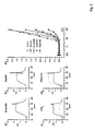
- Figure 1 shows the whole-cell voltage-clamp recordings obtained from a representative human astrocytoma cell from cell line STTG1 and from a primary cultured astrocytoma cell (UAB4630).
- Cells were stepped to test potentials between -105 mV and 195 mV in 25 mV increments from a holding potential of 0 mV (inset).
- Cells showed large transients upon termination of voltage steps (star, A, C).
- Potential >45 mV resulted in fast-activating, non-inactivating outwardly rectifying currents (B, D).
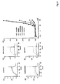
- Figure 2 shows that in order to determine the ion species that was carrying the outward current, the reversal potential of tail currents was analyzed.
- Cells were held at 0 mV, pulsed to 180 mV, and then pulsed in -20 mV steps from +120 mV to -120 mV (A, inset).
- Plotting tail current amplitudes (A, inset) as a function of voltage showed a reversal potential of 8 mV (B) in this cell.
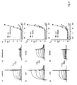
- Figure 3 shows the whole-cell leak subtracted current responses of STTG1 cells in response to a single 145 mV voltage step prior to and following substitution of extracellular Cl - with (125 mM) Br - (A), I - (B), NO 3 - (C), or F - (D). Dashed lines represent control current with standard external solution and straight lines represent current with replacement solution. E) peak current-voltage relations obtained as in Figure 1 , with current normalized to that obtained with standard NaCl-rich external solution.
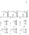
- Figure 4 shows as in Figure 3 , whole-cell leak subtracted current responses of STTG1 cells prior to and following substitution of extracellular Cl - with (125 mM) acetate (A), glutamate (B), isethionate (C), or sucrose (D).
- dashed lines represent control current with standard bath solution and straight lines represent current after replacement.
- Figure 5 shows the effect of bath application of chlorotoxin, DIDS (4,4'-Diisothiocyanostilbene-2,2'-disulfonic acid) and DNDS (4,4'-Dinitrostilbene-2,2'-disulfonic acid) on outward currents in STTG1 astrocytoma cells in response to test voltage pulses from -105 to +195 in 25 mV increments.
- Whole-cell currents are shown prior to (A) and following (B) bath application of 590 nM chlorotoxin. Chlorotoxin decreased outward currents by 81%.
- Figure 6 shows the effect of the zinc, cadmium, and calcium on outward currents.
- currents were decreased to 40% of that in control solution, containing 1 mM Ca 2+ ( Figure 6 , F-H).
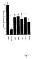
- Figure 7 shows the comparison of the effects of channel blockers on outward currents. Effects are expressed as percent of normalized to current amplitude obtained in standard NaCl-rich external solution for pooling of experimental data. Error bars reflect SEM.
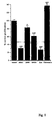
- Figure 9 shows the whole-cell voltage-clamp recordings obtained from representative human astrocytoma cell (STTG1).
- Cells were stepped to test potentials between -120 mV and 120 mV in 20 mV increments from a holding potential of 0 mV.
- Cells showed large tail currents upon termination of voltage steps (arrow, A).
- Potential >0 mV resulted in fast-activating, non-inactivating outwardly rectifying currents (B).
- B fast-activating, non-inactivating outwardly rectifying currents
- the reversal potential of tail currents was analyzed.
- Cells were held at 0 mV, pulsed to 200 mV, and then pulsed in -20 m V increments from +120 mV to -120 mV (C).
- Plotting tail current amplitudes (C, inset) as a function of voltage showed a reversal potential of 0 mV (D).
- Figure 10 shows the recordings from a glioma cell in biopsy tissue from a GBM in response to 13 depolarizing voltage steps ranging from -105 to 195 mV.
- Figure 11 shows the recordings from a xenografted D54MG glioma cell recorded in acute slices in response to depolarizing voltage steps ranging from -105 to 195 mV.
- Figure 12 shows the staining of a 200 ⁇ m section through a glioma induced experimentally in a scid mouse. Fluorescent cells are identified by staining with Ctx-GSt recognized by an anti-GST antibody conjugated to FITC. 20x magnification.
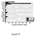
- Figure 13 shows the biodistribution of Chlorotoxin binding sites as determined by injection of 125 I-Ctx into a scid mouse bearing an experimental tumor.
- Figure 14 shows the bath application of chlorotoxin (590 nM) inhibits outward currents in STTG1 cell line.
- Figure 15 shows the representative whole-cell leak-subtracted currents from human tumor cell lines. Astrocytoma and glioblastoma cell lines were dominated by outwardly-rectifying voltage-activated chloride currents, whereas these currents were absent in cells from breast tumor and melanoma.
- Figure 16 shows a Ctx-GST is an effective blocker of glioma Cl - channels. 600 nM Ctx-GST was applied with bath perfusion and resulted in - 70% reduction in Cl - currents; GST alone was ineffective.
- Figure 17 shows that binding of 125 [I]-CTX to D54MG glioma cells.
- 125 [I]-CTX was added in duplicate in 400mL with or without a 100-fold molar excess of unlabeled Ctx from the same source. After 60min at room temperature, cell monolayers were rinsed 3 times with PBS and cells were harvested for assessment of cell-associated radioactivity. Four wells in each plate were harvested with trypsin-EDTA and cell number was established by trypan blue exclusion. (Note that not all data points used for the Scatchard analysis were plotted in the inset).
- Figure 18 shows the immunohistochemical staining of two glioma cell lines as compared to normal human glia.
- Cells were labeled with the recombinant chlorotoxin-GST fusion protein (Ctx-GST) and binding of Ctx-GST was visualized by an anti-GST antibody coupled to FITC.
- Ctx-GST chlorotoxin-GST fusion protein
- Figure 19 show the glioma cells were exposed to 600nM Ctx-GST followed by a mouse aGST and a goat anti-mouse antibody conjugated to saporin. Cell death increased with increasing saporin concentrations (right) and could be largely prevented by pre-treatment of cultures with 6 ⁇ M native chlorotoxin (bottom left).
- Figure 20 shows a western blot of U54MG membranes after immunpercipitation with either C1C5 antibodies or chlorotoxin (Ctx). Blots were probed with C1C5.
- Glioma cells e.g. primary brain tumors derived from glial cells, express a unique membrane protein which constitutes a Cl - ion channel termed herein G lioma C hloride C hannel (GCC). I n the brain, GCC is specific to gliomas and meningiomas and is not present in other cells. GCC was identified in 24/24 glioma patient biopsies, in 7/7 astrocytoma/glioblastoma cell lines and in 4 / 4 meningioma biopsies. GCC expression correlates with pathological tumor grade.
- GCC G lioma C hloride C hannel
- GCC expression is preserved in intracranial xenograft tumors in scid mice, which provide an excellent animal model for the disease.
- GCC binds chlorotoxin, a 36 amino acid peptide, with high affinity and selectivity. Binding is preserved in both synthetic and recombinant form of chlorotoxin, and also if the molecule is altered in ways to carry fluorescent or cytotoxic moieties.
- GCC is a specific marker and useful target for gliomas and meningiomas and can be used for diagnostic and therapeutic purposes.
- GCC can be targeted by antibodies to the protein and/or by molecules that bind to it.
- chlorotoxin and chlorotoxin-like molecules fusion proteins
- fusion proteins can be used to specifically direct molecules bound on it to gliomas and meningiomas. These molecules include but are not limited to: 125 I, 131 I, fluorescent moieties, cytotoxic moieties including but not limited to ricin, saporin, pseudonomas exotoxin.
- Binding of chlorotoxin-like molecules or antibodies to GCC can be utilized to diagnose gliomas and meningiomas.
- Non-invasive strategies can be devised to utilize GCC expression for diagnostic purposes. For example, binding of 131 I-Ctx to GCC can be visualized by PET scan.
- Chlorotoxin inhibits GCC currents in a dose-dependent manner, with an apparent IC 50 of 950 nM.
- a recombinant toxin was also generated (in E . coli ) after fusion to glutathione-S-transferase.
- the recombinant Ctx-GST fusion protein was even more effective in blocking GCC currents (IC 50 ⁇ 540 nM), and an example of a whole-cell recording in the presence and absence of Ctx-GST and GST alone in a single cell is presented below.
- Chlorotoxin binds directly to GCC channels, as is the case in rat colonic epithelial cells (DeBin and Strichartz, 1991), where one single Ctx molecule is sufficient for channel block. However, it is possible that Ctx binds to a binding protein or receptor, and that subsequent changes in GCC currents are mediated indirectly, e.g. through G-proteins or second messengers.
- the present invention is directed to novel methods of identifying, targeting and effectively suppressing the growth of glial-derived neoplastic cells.
- a pharmaceutical composition comprising a ligand which binds specifically to glial-derived or meningioma-derived tumor cells and a pharmaceutically acceptable carrier.
- the ligand may be an antibody which recognizes a n antigen that is a glioma or meningioma specific chloride channel.
- the ligand is a chlorotoxin-like compound and is radiolabeled.
- Also disclosed is a method of differentiating glial-derived or meningioma-derived neoplastic tumor tissue from non-neoplastic tissue comprising the steps of: contacting a tissue of interest with an antibody that specifically recognizes an antigen in chloride channels of glial-derived tumor cells; and measuring the level of binding of the antibody, wherein a high level of binding is indicative that the tissue is neoplastic.
- the level of antibody binding indicative of neoplastic tissue is from about 30% to about 90% of cells positively binding the antibody.
- the present invention is in one aspect directed to an in vitro method of differentiating glial-derived or meningioma-derived neoplastic tumour tissue from non-neoplastic tissue, comprising the steps of:
- a fusion protein said protein comprised of: a ligand that specifically recognizes an antigen in chloride channels of glial-derived tumor fused to a cytotoxic moiety.
- the ligand may be a chlorotoxin-like protein or an antibody.
- Representative cytotoxic moieties include gelonin, ricin, saponin, pseudonomas exotoxin, pokeweed antiviral protein, diphtheria toxin, and complement proteins.
- compositions of the present invention comprising the fusion protein and a pharmaceutically acceptable carrier.
- a method of treating an individual having a glioma or meningioma, comprising the step of administering to said individual a pharmacologically effective dose of any of the compositions of the present invention is also disclosed.
- compositions may be prepared using the novel antibodies and fusion protein disclosed herein.
- the pharmaceutical composition comprises the novel antibodies and fusion proteins and a pharmaceutically acceptable carrier.
- a person having ordinary skill in this art would readily be able to determine, without undue experimentation, the appropriate dosages and routes of administration of the novel antibodies and fusion proteins .
- STTG1 cell line (American Type Culture Collection, Rockville, MD) was grown in DMEM (Gibco) plus 10% FBS (Hyclone).
- Human Tumor Cell Lines established cell lines, derived from human malignant gliomas (D54MG, U105MG, U251MG, and U373MG obtained from D.D.Bigner, Duke University) and extraglial human tumors (all from ATCC), were studied in long term (>100) passages (see TABLE I for details). Cells were maintained in DMEM/F12 supplemented with 7% heat-inactivated FBS (Atlanta Biologicals) at 37°C in a 10% CO 2 /90% air atmosphere.
- Cells attaining nearly confluent growth were harvested and replated onto uncoated 75 cm 2 flasks or uncoated 12 mm circular glass coverslips for electrophysiology and were used 36-72 hours after plating, unless otherwise noted. Viable cell counts were determined by trypan blue exclusion.
- Freshly resected human brain tumor tissue are collected during surgery in ice-cold tissue culture medium and necrotic/hemorrhagic portions are removed aseptically. Tissue is maintained for ⁇ 15-20 min under 95/5% O 2 /CO 2 until used. Ice-cold tissue are embedded in BactoAgar and cut into blocks of ⁇ 10x10mm and glued to the bottom of a petri dish mounted to a Vibratome where 200 mm slices are cut. These are transferred to oxygenated saline and maintained at 37°C until recording.
- C.B.-17 SCID mice are anesthetized by intraperitoneal administration of the following mixture: ketamine, 20 mg/ml plus xylazine, 0.3 mg/ml, in saline, at 0.07 ml/10 g of body weight.
- a midline scalp incision is made and a 0.5 mm burr hole is made at 1.5-2 mm to the right of the midline and at 0.5-1.0 mm posterior to the coronal suture.
- Tumor cells (10 6 D54 MG- human glioma cells in 5 ml final injection volume/ mouse) are suspended in excipient (serum free DMEM/F12 + 5% methyl cellulose).
- Intracranial injection is performed stereotactically using a 250 ml Hamilton syringe with a 30-gauge needle mounted on a Stoelting stereotaxic apparatus.
- the needle is inserted vertically through the hole to a depth of 2.5 mm. 45-60 seconds after injection, the needle is slowly withdrawn and the incision closed with 9 m m Michel wound clips.
- Mice are then returned to sterile microisolator polycarbonate cages placed over a heating pad until recovery, and provided autoclaved lab chow and sterile water ad libitum. Slices are obtained from anesthetized mice after decapitation.
- the brain is quickly removed and placed in ice-cold (4°C) calcium-free ringers containing (in mM): NaCl 116; KCl 4.5; MgCl 2 0.8; NaHCO 3 26.2; glucose 11.1; N-2-hydroxyethlypiperazine-N'-2-ethanesulfonic acid (Hepes) 5.
- the solution is constantly bubbled with 95% O 2 /5% CO 2 mixture.
- the brain is hemisected and mounted onto a vibratome slice-holder using cyanoacrylate glue.
- Transverse tissue slices (50-200 mM) are cut in cold oxygenated saline solution and subsequently transferred to a beaker filled with Ca 2+ containing saline at room temperature.
- Patch-pipettes were made from thin-walled borosilicate glass (WPI, TW150F-40) o.d. 1.5 mm, i.d. 1.2 mm and were filled with a solution containing (in mM): KCl 145, MgCl 2 1, CaCl 2 0.2, EGTA 10, Hepes 10, pH adjusted to 7.4 using Tris, unless otherwise noted. Pipettes were not fire-polished and typically had resistances between 2-5M ⁇ . Cells were continuously superperfused with saline solution, allowing for rapid ( ⁇ 30 seconds) exchange of bath volume.
- the standard bath solution contained, in mM: NaCl 122.6, KCl 5, MgCl 2 1.2, CaCl 2 1.0, Na 2 HPO 4 2.0, NaH 2 PO 4 0.4, NaHCO 3 25.0, Na 2 SO 4 1.2, Glucose 10. 5 (bubbled with 5% CO 2 /95% O 2 ).
- the composition of bath solutions used for replacement studies is summarized in Table II. Drugs used to block ionic conductances were prepared freshly as stock solutions for each experiment and added to bath solution. Osmolality was measured with a vapor pressure osmometer (Wescor, Logan, UT) and adjusted to 308-312 mOsms.
- Proliferation was studied quantitatively by determining incorporation of 3 H-thymidine.
- cells were incubated for 24 hours in the continuous presence or absence of Ara-C (cytosine arabinoside, 10 ⁇ M), DIDS (200 ⁇ M), DNDS (200 ⁇ M), zinc (200 ⁇ M) or chlorotoxin (600 nM).
- Ara-C cytosine arabinoside, 10 ⁇ M
- DIDS 200 ⁇ M
- DNDS 200 ⁇ M
- zinc 200 ⁇ M
- chlorotoxin 600 nM
- Cells were incubated with 1 ⁇ Ci/ml radiolabelled thymidine ([methyl- 3 H]thymidine) for the final 4 hours (at 37°C).
- Culture dishes were rinsed three times with ice-cold PBS and solublized with 0.3N NaOH for 30 minutes at 37°C.
- DIDS 4,4'-Diisothiocyanostilbene-2,2'-disulfonic acid
- DNDS 4,4'-Dinitrostilbene-2,2'-disulfonic acid
- Ara-C Ara-C
- Chlorotoxin was purchased from Lanoxin (Accurate Chemical and Scientific Corp., Westbury, NY).
- FIG. 1 Representative examples of whole-cell recordings from an STTG1 human astrocytoma cell and an astrocytoma cell from primary culture (UAB4630) in response to depolarizing voltage steps are displayed in Figure 1 .
- the cells were stepped from a holding potential of 0 mV to a series of test potentials between - 105 mV and 195 mV in 25 mV increments. Potential >45 mV resulted in fast activating, non-inactivating outward currents. Cells showed large outward transients upon termination of voltage steps ( Figure 1A and C ).
- Figure 3 shows examples of whole-cell leak-subtracted current responses of human astrocytoma cells to test pulses stepped from a holding potential of 0 mV to 145 mV prior to and following substitution of bath chloride with the halide anions bromide (A), iodide (B), nitrate (C), and fluoride (D). Bromide, iodide, and nitrate increased outward currents, whereas fluoride substitution led to decreased currents. For each experiment, complete IV curves were plotted in (E). To compare I-V relations, currents were normalized to control currents with Cl- as the external anion as the membrane was stepped from 0 mV to a series of potentials between -105 and +195 mV.
- Table III summarizes the changes in the values of E rev for equimolar replacement of chloride by test anions and the calculated permeability ratios (P anion /P Cl ). These data suggest the following relative permeability sequence: I - >NO 3 - >Br - >Cl - >acetate>isethionate>F - >glutamate. TABLE III: Anion selectivity. Na + - Anion MW ⁇ E rev (mV) P anion / P Cl N Chloride 58.4 5 ------ ------ 48 Acetate 82.0 4 4.0 ⁇ 0 0.90 ⁇ 0.02 3 Bromide 102.
- Chlorotoxin is a protein having 36 amino acids that is derived from scorpion venom toxin that was originally described as a blocker of small conductance Cl - channels in epithelial cells (8).
- the peptide was synthesized and comparable inhibition of currents with the synthetic toxin were observed (data not shown).
- the present disclosure identified a voltage-dependent, outwardly-rectifying Cl - current in human astrocytoma cells. This current was present in all cells studied in both primary cultures of human astrocytomas and in an established human astrocytoma cell line. Cells showed large outward transients upon termination of voltage steps and reversed close to the calculated equilibrium potential for chloride. Upon replacement with various anions, the current reversal potential shifted in accordance with an anion-selective channel towards the new E Cl . Currents were sensitive to application of chloride channel blockers chlorotoxin, DIDS, DNDS, cadmium, and zinc. Under physiological conditions, the current would be carried by Cl - , so that currents were considered chloride currents.
- non-neoplastic glial cells are typically characterized by high levels of expression of voltage-gated K + channels; no appreciable contribution from K + currents to whole-cell outward currents was observed in all cells tested.
- Outwardly rectifying chloride currents have been described in many epithelial tissues including respiratory cells (24), submandibular gland (19), lacrimal gland (9), pancreatic duct cells (18), epididymis (31), and sweat gland (23), and in non-epithelial cells such as lymphocytes (11), squid axon (18), and rat skeletal muscle (3).
- respiratory cells 24
- submandibular gland (19)
- lacrimal gland 9
- pancreatic duct cells epididymis
- sweat gland 23
- non-epithelial cells such as lymphocytes (11), squid axon (18), and rat skeletal muscle (3).
- lymphocytes 11
- squid axon squid axon
- Cl - channels found in human macrophages(16), necturus enterocytes(12), squid axon(18) and sheep parotid gland (19).
- chloride channels do not show spontaneous activity in whole cell recordings and channel activation occurs only in excised patches.
- astrocytoma Cl - currents could be easily recorded in every recording in the whole-cell configuration.
- the permeability sequence of the chloride channel in astrocytoma cells does not correlate with the hydrated ion radii (NO 3 - >Cl - >I - >Br - ) or the mobility of ions in aqueous solution (Br - >I - >Cl - >NO 3 - ).
- the sequence most closely resembles the lyotrophic series (I - >NO 3 - >Br - >Cl - >F - ), which reflects the ability to denature macromolecules or to bind or absorb to proteins or lipid-water interfaces (6).
- the anion selectivity sequence here differs in only minor detail from those reported for outwardly rectifying channels in other tissues: submandibular duct gland (SCN - >NO 3 - >I - >Cl - >Br - >acetate)(19), canine airway epithelia (SCN - >NO 3 - >I - Br - >NO 3 - >Cl - ) (25), rat lacrimal gland (I - >NO 3 - >Br - >Cl - >F - > isethionate> glutamate) (9) and necturus enterocytes (SCN - >I - >Br - > Cl - >F - >gluconate) (12).
- replacement of Cl - by large organic anions results in the virtual abolishment of Cl - currents.
- currents were almost eliminated after glutamate or sucrose replacement.
- the present invention recorded Cl - currents in the cell line U373MG in addition to other established astrocytoma cell lines (CH-235MG, D-54MG, SK-MG-1, U-105MG, U251MG. Moreover, the currents were observed in all cells under normal conditions, with osmolality of each solution measured and matched to the osmolality of the growth medium.
- Ion channels have been shown to be part of the proliferative response in a number of cell types and in cultures of normal glial cells, the activity of K + channels is required for cell proliferation, since K + channel blockade leads to decreased proliferation.
- Potassium channels have been implicated in the proliferative response in a number of other cell types, including human melanoma cell lines (28), cultured brown fat cells (30), and Schwann cells, the principle glial cells in the peripheral nervous system (5).
- the present invention demonstrates that the link between channel activity and proliferation is more widespread. Modulation of channels may result from both longterm changes in gene expression and short-term modulation of pre-existing channel proteins.
- the present invention observed a decrease in proliferation by DIDS, DNDS, and zinc and a 37% enhancement of astrocytoma proliferation following application of chlorotoxin.
- One possible explanation is that the stilbene derivatives are affecting ion transport mechanisms, whereas chlorotoxin is a more specific ion channel inhibitor.
- the present invention shows that Cl - channels participates in the proliferative response in these cells.
- Electrodes (WPI, TW150F-40) o.d. 1.5 mm, i.d.
- GBM glioblastoma multiforme
- LGA low grade astrocytoma
- PA pilocytic astrocytoma
- AE anaplastic ependymoma
- AA anaplastic astrocytoma
- mela. melanoma
- N.B. normal breast
- B.CA breast cancer
- unk unkown
- Glioma cells express a unique transmembrane Cl - ion channel that binds a venom toxin (chlorotoxin) with very high affinity.
- chlorotoxin venom toxin
- the high affinity for chlorotoxin allow the development of glioma-specific agents including marker compounds for rapid diagnosis and immunotoxins for therapeutic treatment. Since this protein is only shared among malignant gliomas cells and meningioma cells it could be targeted by reagents that bind to the toxin binding site, or, following isolation of the protein, antibodies could be used to selectively eliminate cells expressing this protein. This approach has a high likelihood to yield new strategies for more specific and more effective therapeutic moralities for this uniformly fatal disease.
- Biopsies from 24 patients diagnose with glioma were thoroughly investigated histopathologically. Electrophysiological recordings and immunohistochemical methods were used to detect GCC. Expression was observed in all patients and spanning in age from 0.5 to 77 years and independent of pathological grade of the tumor. An example of a representative recording is shown in Figure 10 . Evidence for the expression of GCC was obtained in 4 / 4 biopsies of patients diagnosed with meningioma. A list of patient cases studied in which GCC was identified is presented in TABLE V.
- Glioma tumors experimentally induced in SCID mice were also studied by intracranial injection of D54MG glioma cells. This procedure resulted in rapidly growing, invasive brain tumors (Gladson et al. 1995) from which slice preparations were made. Close to 100% of cells in tumor xenografts showed prominent expression of GCC as illustrated by staining of tumor tissue with antibodies that recognize Ctx binding sites ( Figure 12 ) or electrophysiology ( Figure 11 ). An example of a representative recording from D54MG cells is shown in Figure 11 . These scid mice were also used to study the biodistribution of Ctx binding sites (therefore GCC channels) using 125 I-Ctx.
- 125 I-Ctx was injected into the cerebrum of a mouse in which a glioma had been induced in the right brain 14 days earlier. Brain and body tissue as well as blood was harvested and 125 I-Ctx levels were determined using a liquid scintillation counter. The resulting counts show the selective accumulation of 125 I-Ctx in the tumor ( Figure 13 ).
- FIG. 3 shows representative examples of voltage-dependent outwardly rectifying anion currents from selected primary cultures and more established cell lines.
- Cells from primary cultures displayed outward currents that were similar in size, voltage activation, reversal potential, and sensitivity to chlorotoxin as cell lines. Currents were qualitatively similar in all of the 7 cell lines evaluated (U251MG, CH235MG, U373MG, U105MG, D54MG, SK-MG-1, (all glioblastoma multiforme) and STGG1 (anaplastic astrocytoma)).
- the present invention is the first report of an outwardly-rectifying Cl - current in human malignant glioma cells.
- Currents were characteristic of both primary cultures of freshly resected brain tumors and established astrocytoma cell lines. These currents were not present in several extraglial human tumors such as melanoma, breast, rhabdomyosarcoma and neuroblastoma.
- Chloride currents were characteristic of cells from other preparations, including lymphocytes, submandibular gland, rat myotubes, and sweat gland.
- the Cl - current in astrocytoma cells exhibits a higher threshold for current activation, had large positive tail currents not previously reported, and could be easily recorded in whole-cell patches.
- the present invention demonstrates that this Cl - current is in all tumor cells studied of glial origin but not in normal non-malignant glial cells or in non-glial tumors.
- the present invention demonstrates a chloride conductance unique to human astrocytoma and glioblastoma cells which is not present in human tumor cells of extraglial origin.
- This channel can be blocked physiologically by chlorotoxin, a scorpion venom known to block epithelial chloride channels.
- chlorotoxin a scorpion venom known to block epithelial chloride channels.
- the presence of this chloride channel activity presents a diagnostic strategy to differentiate between glial and non-glial tumors.
- chlorotoxin is a 36 amino acid protein naturally derived from leiurus quinquestriatus scorpion venom.
- recombinant proteins specifically engineered to mimic the binding and action of the native toxin.
- recombinant chlorotoxin may be synthesized in E .
- a fusion protein such as a primary antibody can be used to stain cells using standard immunohistochemical methods.
- a GST protein which lacks an insert was also purified for use as a control.
- An antibody against the GST portion alone can be used as a secondary antibody.
- the physiological activity of the fusion protein can be examined by using the GST with no insert as an internal control.
- the biological activity of the synthetic chlorotoxin is as effective for chloride ion channel blockade as the native venom toxin.
- Recombinant techniques are used to synthesize chlorotoxin, in E coli using a modified PGEX vector system and the toxin may be linked to various fusion proteins using common restriction sites: GST-chlorotoxin, GST-Ala 10 linker-chlorotoxin, and GST-Ala 20 linker-chlorotoxin. These contain no linker, 10 alanine amino acid linker and 20 alanine amino acid linker, respectively.
- oligonucleotides of chlorotoxin sequence deduced from the peptide sequence were synthesized with a HindIII cohesive sequence at the 5' end of the sense oligonucleotide and an EcoRI cohesive sequence following the stop codon at the 3' end of the sense sequence.
- Each oligonucleotide was phosphorylated by T4 polynucleotide kinase using ATP as a substrate.
- Nucleotides were annealed by heating and slow cooling. Annealed oligonucleotides were cloned into HindIII/EcoRI site of pGBHE vector (pGCT-1) through ligation followed by transformation into E . coli competent cells.
- a 20 amino acid linker was cloned into the BamHI/HindIII site.
- This amino acid linker has a BgIII site in the middle that makes it possible to cut the BamHI and BgIII in order to create a 10 amino acid linker sequence.
- the orientation and preservation of the oligonucleotide has been verified within the fusion protein by sequencing methods and that the induction of fusion protein produces the expected size was verified by comparing their molicular weights on a 12% SDS-PAGE gel.
- recombinant chlorotoxin After synthesis of recombinant chlorotoxin, it may be linked to various cytotoxic fusion proteins including glutathione-S-transferase (GST), gelonin, ricin, diptheria toxin, complement proteins and radioligands and other such proteins as are well known in the immunotoxin art.
- GST glutathione-S-transferase
- gelonin gelonin
- ricin gelonin
- diptheria toxin complement proteins
- radioligands radioligands and other such proteins as are well known in the immunotoxin art.
- recombinantly prepared synthetic chlorotoxin linked to a cytotoxic moiety would be useful to specifically target and deliver a toxic substance to glial-derived tumors as a novel therapy.
- GST-chlorotoxin fusion protein may be prepared as follows.
- fusion proteins Three fusion proteins, GST alone, GST-chlorotoxin, and GST-Ala 20 linker-chlorotoxin were affinity purified using a glutothione conjugated agarose bead column and the resulting proteins were verified on a 12% SDS-PAGE gel. More specifically, E . coli were transformed with the vector and chlorotoxin insert and were induced to produce the fusion proteins. Resulting proteins were mixed with glutothione agarose beads and left for 15' to optimize absorption. Columns were washed with buffer and the fusion proteins were eluted by competition with free glutathione and collected in small vials. These proteins were then run on a 12% SDS-PAGE gel.
- Gluthathione column purified fusion protein Ctx-GST is diluted in 0.2M sodium carbonate (pH 9.0), at 1-2 mg/ml.
- DTAF (Calbiochem) is diluted in 1.0 M sodium carbonate (pH 9.0) at 2.5 mg/ml.
- DTAF is mixed gently with the diluted Ctx-GST, by adding 25 mg DTAF per milligram of Ctx-GST. Mixing continues at room temperature for 10 minutes, after which NH 4 Cl is added at a final concentration of 50 mM and glycerol up to 5% final volume (optional, xylene cylanol 0.1% is added to serve as indicator dye for the unbound material).
- the solution is placed at 4°C for 2 - 4 hours with gentle agitation. After mixing, the unbound dye is separated by gel size filtration (G-Sephadex column, with exclusion limit between 30,000-50,000 prepared according to the manufacturer's instructions (Pharmacia)).
- G-Sephadex column with exclusion limit between 30,000-50,000 prepared according to the manufacturer's instructions (Pharmacia)
- the conjugated Ctx-GST-DTAF elutes first, and its color is easily distinguished under room light. Protein content is then determined, and the fluorescent conjugate is stored in a light proof-container at 4°C, until ready to use for direct immunofluorescence labeling of cells or slices as described below.
- Ctx-GST GST-chlorotoxin
- Ctx-GST-DTAF are used to identify toxin binding sites.
- Ctx-GST is biologically active, binds to and blocks Cl - channels with similar affinity as the venom toxin.
- Ctx-GST are recognized immunohistochemically by an antibody to GST -(Chemicon) conjugated to either rhodamine or FITC, and binding are assayed under a fluorescence microscope.
- a single step fluorescence staining procedure are used utilizing Ctx-GST-DAFT (above), a fluorescent form of the Ctx-GST.
- the DTAF label can be visualized by direct immunofluoresence using standard FITC filters.
- the Ctx-GST-DTAF staining has the advantage that cross-reactivity with native GST does not pose a problem.
- Antibodies to the chloride ion channels in glial derived tumors may be prepared as follows. Polyclonal antisera are generated by injecting fusion proteins crated between the glutathione-S-transferase and the chlorotoxin insert into mice or rabbits. Mice are immunized with 0.5 ml of a 1:1 emulsion of 1 mg/ml purified fusion protein in Freund's complete adjuvant and subsequently with two additional injections after 14 and 28 days in Freund's incomplete adjuvant. The mouse and rabbit antibodies are purified from the antisera using the GST fusion protein immobilized on nitrocellulose filters. The antibodies are then examined for binding specificity in various tissues.
- Glioma cells bind 125 [I]-chlorotoxin with high affinity and selectivity
- binding affinity was determined using ( 125 [I]-Ctx) radiolabeled with 125 [I]-sodium iodide by the chloramine-T method. Saturated binding was achieved in D54MG glioblastoma cells at concentrations >15 nM ( Figure 17 , inset). Scatchard analysis of these data indicates two binding sites with estimated binding affinity values (K d ) of 4.2 nM and 660 nM. The latter value is in good agreement with the electrophysiologically determined IC 50 of ⁇ 950nM.
- D54MG cells contain approximately 1,300 high affinity Ctx binding sites and 13,300 low affinity binding sites per cell.
- no specific Ctx binding was observed in normal human glial cells nor in mixed brain cell cultures (not shown), suggesting that in brain, Ctx binding is glioma specific.
- Ctx with radioactive moieties can be used to treat gliomas. The molecule would selectively bind to gliomas and expose cells to high levels of radiation. 125 I-Ctx or 131 I-Ctx are candidates for this purpose.
- Chlorotoxin binding can be detected immunohistochemically using several approaches.
- cells can be labeled with Ctx-GST, the fusion protein that also inhibits GCC currents (see above, Fig. 16 ).
- This fusion protein can be detected by a FITC-conjugated antibody to GST, although numerous other detection procedures would be possible.
- this approach selectively labels astrocytoma and glioblastoma derived cell lines such as STTG1 or D54MG, but fails to label normal human glial cells.
- chlorotoxin can be labeled using DTAF as Ctx-DTAF. This procedures resulted in a directly fluorescent Ctx-molecule that selectively labels gliomas.
- chlorotoxin can be directly conjugated with biotin as Ctx-biotin. This allows binding to be identified using a reaction with avidin and subsequent recognition by antibodies or the reaction product. This approach was likewise successful in selectively labeling glioma and meningioma cells.
- Immunotoxins targeted to the Ctx binding site can specifically kill glioma cells in vitro
- Ctx-GST selectively labels glioma cells
- saporin Bostoxin, e.g., saporin (Benatti et al. 1989; Battelli et al. 1990; Fordham-Skelton et al. 1990; Tecce et al. 1991; Fordham-Skelton et t al. 1991).
- Such chimeric proteins can be made by fusing Ctx (the targeting moiety) with saporin (the toxin moiety).
- glioma cells were first treated with Ctx-GST, followed by a mouse anti-GST monoclonal antibody and lastly a goat anti-mouse antibody conjugated to saporin. This last step confers immunotoxicity on the "primary" (in this case, Ctx-GST) antibody and resulted in significant and specific killing of glioma cells ( Fig. 19 ).
- Primary in this case, Ctx-GST
- Normal nontumor human astrocytes were not influenced by treatment with the saporin conjugate and either antibody alone failed to reduce cell numbers or to reduce protein or DNA synthesis, as assayed by 3 H-leucine and 3 H-thymidine, respectively.
Landscapes
- Health & Medical Sciences (AREA)
- Life Sciences & Earth Sciences (AREA)
- Chemical & Material Sciences (AREA)
- Immunology (AREA)
- Engineering & Computer Science (AREA)
- Molecular Biology (AREA)
- Organic Chemistry (AREA)
- Medicinal Chemistry (AREA)
- General Health & Medical Sciences (AREA)
- Proteomics, Peptides & Aminoacids (AREA)
- Biomedical Technology (AREA)
- Biochemistry (AREA)
- Urology & Nephrology (AREA)
- Hematology (AREA)
- Genetics & Genomics (AREA)
- Cell Biology (AREA)
- Biophysics (AREA)
- General Physics & Mathematics (AREA)
- Toxicology (AREA)
- Pathology (AREA)
- Bioinformatics & Cheminformatics (AREA)
- Physics & Mathematics (AREA)
- Food Science & Technology (AREA)
- Microbiology (AREA)
- Biotechnology (AREA)
- Insects & Arthropods (AREA)
- Analytical Chemistry (AREA)
- Zoology (AREA)
- Tropical Medicine & Parasitology (AREA)
- Gastroenterology & Hepatology (AREA)
- Neurology (AREA)
- Pharmacology & Pharmacy (AREA)
- Epidemiology (AREA)
- Animal Behavior & Ethology (AREA)
- Public Health (AREA)
- Veterinary Medicine (AREA)
- Medicines That Contain Protein Lipid Enzymes And Other Medicines (AREA)
- Pharmaceuticals Containing Other Organic And Inorganic Compounds (AREA)
- Measuring Or Testing Involving Enzymes Or Micro-Organisms (AREA)
Claims (16)
- Pharmazeutische Zusammensetzung, die (a) (i) Chlorotoxin, das eine zytotoxische Einheit trägt oder (ii) DIDS 4,4'-Diisothiocyanostilben-2,2'-disulfolsäure oder (iii) DNDS 4,4'-Dinitrostilben-2,2'-disulfonsäure und (b) einen pharmazeutisch zulässigen Träger enthält, zur Verwendung beim Behandeln eines Individuums, das ein Gliom oder Meningiom aufweist.
- Pharmazeutische Zusammensetzung für die Verwendung nach Anspruch 1, wobei es sich bei dem Individuum um einen Menschen handelt.
- Zusammensetzung für die Verwendung nach Anspruch 1 oder Anspruch 2, wobei das Chlorotoxin radioaktiv markiert ist.
- Zusammensetzung für die Verwendung nach Anspruch 3, wobei das radioaktiv markierte Chlorotoxin aus der Gruppe ausgewählt ist, die aus 131I-Chlorotoxin und 125I-Chlorotoxin besteht.
- Zusammensetzung für die Verwendung nach einem der Ansprüche 1-4, wobei das Chlorotoxin aus der Gruppe ausgewählt ist, die aus nativem, synthetischem und rekombinantem Chlorotoxin besteht.
- Zusammensetzung für die Verwendung nach einem der vorherigen Ansprüche, wobei es sich bei der zytotoxischen Einheit um Gelonin, Ricin, Saporin, Pseudomonas-Exotoxin, Kermesbeere, antivirales Protein, Diphtherietoxin oder Komplementproteine handelt.
- Radioaktiv markiertes Chlorotoxin für die Verwendung in einem In-vivo-Verfahren des Differenzierens zwischen von Glia abstammendem oder einem Meningiom abstammendem neoplastischem Tumorgewebe und nicht-neoplastischem Gewebe, wobei das Verfahren folgende Schritte enthält:Inkontaktbringen eines Gewebes von Interesse mit radioaktiv markiertem Chlorotoxin, das spezifisch an von Glia abstammendes neoplastisches Tumorgewebe bindet; undMessen der Bindung des radioaktiv markierten Chlorotoxins,wobei ein Grad der Affinität der radioaktiv markierten Bindung von etwa 5 nM bis etwa 5 µM anzeigt, dass das Gewebe neoplastisch ist.
- Radioaktiv markiertes Chlorotoxin für die Verwendung nach Anspruch 7, wobei das Chlorotoxin aus der Gruppe ausgewählt ist, die aus nativem, synthetischem und rekombinantem Chlorotoxin besteht.
- Radioaktiv markiertes Chlorotoxin für die Verwendung nach Anspruch 7 oder Anspruch 8, wobei das radioaktiv markierte Chlorotoxin aus der Gruppe ausgewählt ist, die aus 131I-Chlorotoxin und 125I-Chlorotoxin besteht.
- Radioaktiv markiertes Chlorotoxin für die Verwendung nach Anspruch 7, 8 oder 9, wobei die Bindung des markierten Chlorotoxins mittels Positronenemissionstomographie-Scanning bestimmt wird.
- Zusammensetzung, die Chlorotoxin enthält, das eine zytotoxische Einheit trägt.
- Zusammensetzung nach Anspruch 11, wobei die zytotoxische Einheit aus der Gruppe ausgewählt ist, die aus 131I, 125I, Gelonin, Ricin, Saporin, Pseudomonas-Exotoxin, Kermesbeere, antiviralem Protein, Diphtherietoxin und Komplementproteinen besteht.
- Zusammensetzung nach Anspruch 11 oder Anspruch 12, wobei das Chlorotoxin aus der Gruppe ausgewählt ist, die aus nativem, synthetischem und rekombinantem Chlorotoxin besteht.
- Pharmazeutische Zusammensetzung, welche die Zusammensetzung nach einem der Ansprüche 11-13 und einen pharmazeutisch zulässigen Träger enthält.
- Pharmazeutische Zusammensetzung nach Anspruch 14 zum Behandeln eines Individuums, das ein Gliom oder Meningiom aufweist.
- In-vitro-Verfahren des Differenzierens zwischen von Glia abstammendem oder einem Meningiom abstammendem neoplastischem Tumorgewebe und nicht-neoplastischem Gewebe, wobei das Verfahren folgende Schritte enthält:Inkontaktbringen eines Gewebes von Interesse mit dem radioaktiv markierten Chlorotoxin, wie in einem der Ansprüche 7 bis 10 definiert; undMessen der Bindung des radioaktiv markierten Chlorotoxins,wobei ein Grad der Affinität der radioaktiv markierten Bindung von etwa 5 nM bis etwa 5 µM anzeigt, dass das Gewebe neoplastisch ist.
Applications Claiming Priority (3)
| Application Number | Priority Date | Filing Date | Title |
|---|---|---|---|
| US928395P | 1995-12-27 | 1995-12-27 | |
| US9283P | 1995-12-27 | ||
| PCT/US1996/020403 WO1997024619A1 (en) | 1995-12-27 | 1996-12-27 | Novel method of diagnosing and treating gliomas |
Publications (3)
| Publication Number | Publication Date |
|---|---|
| EP0953153A1 EP0953153A1 (de) | 1999-11-03 |
| EP0953153A4 EP0953153A4 (de) | 2006-06-28 |
| EP0953153B1 true EP0953153B1 (de) | 2013-07-10 |
Family
ID=21736710
Family Applications (1)
| Application Number | Title | Priority Date | Filing Date |
|---|---|---|---|
| EP96946129.2A Expired - Lifetime EP0953153B1 (de) | 1995-12-27 | 1996-12-27 | Verfahren zur diagnose und behandlung von gliomas |
Country Status (5)
| Country | Link |
|---|---|
| US (6) | US5905027A (de) |
| EP (1) | EP0953153B1 (de) |
| AU (1) | AU2239997A (de) |
| CA (1) | CA2249351C (de) |
| WO (1) | WO1997024619A1 (de) |
Families Citing this family (33)
| Publication number | Priority date | Publication date | Assignee | Title |
|---|---|---|---|---|
| US5905027A (en) * | 1995-12-27 | 1999-05-18 | Uab Research Foundation | Method of diagnosing and treating gliomas |
| US6667156B2 (en) * | 1995-12-27 | 2003-12-23 | Uab Research Foundation | Diagnosis and treatment of neuroectodermal tumors |
| US7069102B2 (en) * | 2000-05-16 | 2006-06-27 | Texas Instruments Incorporated | System and method to customize bond programs compensating integrated circuit bonder variability |
| WO2002044419A2 (en) * | 2000-11-28 | 2002-06-06 | Wyeth | Expression analysis of kiaa nucleic acids and polypeptides useful in the diagnosis and treatment of prostate cancer |
| HUP0303899A3 (en) * | 2000-11-28 | 2007-05-02 | Wyeth Corp | Expression analysis of smarc nucleic acids and polypeptides useful in the diagnosis and treatment of prostate cancer |
| US6821731B2 (en) * | 2000-11-28 | 2004-11-23 | Wyeth | Expression analysis of FKBP nucleic acids and polypeptides useful in the diagnosis of prostate cancer |
| US20020155440A1 (en) * | 2000-12-19 | 2002-10-24 | Ljubimova Julia Y. | Using overexpression of laminin alpha 4 subunit as a diagnostic and prognostic indicator of malignant tumors |
| AU2002345959A1 (en) * | 2001-06-26 | 2003-01-08 | Uab Research Foundation | Chlorotoxin inhibition of cell invasion, cancer metastasis, angiogenesis and tissue remodeling |
| AU2003228517B2 (en) * | 2002-04-16 | 2007-06-21 | Dana-Farber Cancer Institute, Inc. | Cancer models |
| US20060088899A1 (en) * | 2002-05-31 | 2006-04-27 | Alvarez Vernon L | Combination chemotherapy with chlorotoxin |
| JP2005537234A (ja) * | 2002-05-31 | 2005-12-08 | トランスモルキュラー インコーポレイテッド | クロロトキシンとの併用化学療法 |
| US20070275902A1 (en) * | 2003-11-26 | 2007-11-29 | Transmolecular, Inc. | Treatment of Phosphatidylinositol Phospholipid Disorders |
| US20080153746A1 (en) * | 2004-04-06 | 2008-06-26 | Transmolecular, Inc. | Diagnosis and Treatment of Myeloid and Lymphoid Cell Cancers |
| DE602006020710D1 (de) * | 2005-04-22 | 2011-04-28 | Fred Hutchinson Cancer Res Foundation | Fluoreszentes chlorotoxinkonjugat und verfahren zur intraoperativen sichtbarmachung von krebs |
| NZ566395A (en) * | 2005-09-26 | 2012-03-30 | Medarex Inc | Human monoclonal antibodies to CD70 |
| EP2013233A2 (de) * | 2006-03-31 | 2009-01-14 | Transmolecular, Inc. | Verwendung von tm-601 für die diagnose und behandlung von tumoren |
| AU2007333098A1 (en) * | 2006-12-14 | 2008-06-19 | Medarex, Inc. | Human antibodies that bind CD70 and uses thereof |
| US20110091380A1 (en) * | 2007-08-07 | 2011-04-21 | Transmolecular, Inc. | Chlorotoxins as drug carriers |
| AU2008310664A1 (en) * | 2007-10-12 | 2009-04-16 | Transmolecular, Inc. | Systemic administration of chlorotoxin agents for the diagnosis and treatment of tumors |
| US8097284B2 (en) * | 2007-11-13 | 2012-01-17 | Arthur Mikaelian | Polarized scorpion venom solution and a method for making polarized scorpion venom solution |
| CA2724384A1 (en) | 2008-05-15 | 2009-11-19 | Transmolecular, Inc. | Treatment of metastatic tumors |
| US20100260686A1 (en) * | 2009-04-09 | 2010-10-14 | Washington, University Of | Nanoparticles for brain tumor imaging |
| HRP20171291T1 (hr) | 2010-02-04 | 2017-10-20 | Morphotek, Inc. | Polipeptidi i konjugati klorotoksina i njihove uporabe |
| CN103097403B (zh) | 2010-05-11 | 2017-04-19 | 弗雷德哈钦森癌症研究中心 | 氯毒素变体、缀合物及其使用方法 |
| CU24055B1 (es) | 2010-09-27 | 2014-12-26 | Grupo Empresarial De Producciones Biofarmacéuticas Y Químicas Labiofam | Composiciones farmacéuticas de veneno de escorpión rhopalurus junceus |
| WO2013003507A1 (en) | 2011-06-27 | 2013-01-03 | Morphotek, Inc. | Multifunctional agents |
| CA2913127A1 (en) | 2012-12-10 | 2014-06-19 | Fred Hutchinson Cancer Research Center | Methods for screening |
| US9784730B2 (en) | 2013-03-21 | 2017-10-10 | University Of Washington Through Its Center For Commercialization | Nanoparticle for targeting brain tumors and delivery of O6-benzylguanine |
| US11559580B1 (en) | 2013-09-17 | 2023-01-24 | Blaze Bioscience, Inc. | Tissue-homing peptide conjugates and methods of use thereof |
| CA3021011A1 (en) | 2016-04-15 | 2017-10-19 | Blaze Bioscience, Inc. | Methods of treating breast cancer |
| EP3573619A4 (de) | 2017-01-27 | 2020-10-28 | SignalRX Pharmaceuticals, Inc. | Thienopyranone und furanopyranone als kinase-, bromodomänen- und checkpoint-inhibitoren |
| CN111542345B (zh) | 2017-09-15 | 2025-02-25 | 卫材公司 | 氯毒素剂及其用途 |
| CA3086040A1 (en) | 2017-12-19 | 2019-06-27 | Blaze Bioscience, Inc. | Tumor homing and cell penetrating peptide-immuno-oncology agent complexes and methods of use thereof |
Family Cites Families (17)
| Publication number | Priority date | Publication date | Assignee | Title |
|---|---|---|---|---|
| US5591829A (en) * | 1987-05-29 | 1997-01-07 | Matsushita; Shuzo | Antibodies modified with toxic substance |
| US5051364A (en) * | 1989-02-16 | 1991-09-24 | The Salk Institute For Biological Studies | Anti-lipocortin-I and anti-lipocortin-II monoclonal antibodies |
| DE3909799A1 (de) * | 1989-03-24 | 1990-09-27 | Behringwerke Ag | Monoklonale antikoerper (mak) gegen tumorassoziierte antigene, ihre herstellung und verwendung |
| CA2066428C (en) * | 1989-09-08 | 2000-11-28 | Bert Vogelstein | Structural alterations of the egf receptor gene in human gliomas |
| US5223253A (en) * | 1989-09-28 | 1993-06-29 | American Home Products Corporation | Bovine vaccine compositions and method for preventing trichomonas infections using same |
| GB2250993B (en) * | 1990-11-21 | 1995-02-15 | Inst Nat Sante Rech Med | Stromelysin-3 and its application in the diagnosis and treatment of malignant breast cancer |
| US5750376A (en) * | 1991-07-08 | 1998-05-12 | Neurospheres Holdings Ltd. | In vitro growth and proliferation of genetically modified multipotent neural stem cells and their progeny |
| WO1993006116A1 (en) * | 1991-09-20 | 1993-04-01 | Syntex-Synergen Neuroscience Joint Venture | Glial derived neurotrophic factor |
| US5314992A (en) * | 1991-11-25 | 1994-05-24 | Trustees Of Dartmouth College | Lipocortin-1 receptor protein and its uses |
| WO1993023075A1 (en) * | 1992-05-14 | 1993-11-25 | Oncologix, Inc. | Treatment of vascular leakage syndrome and collagenase induced disease by administration of matrix metalloproteinase inhibitors |
| US5626862A (en) * | 1994-08-02 | 1997-05-06 | Massachusetts Institute Of Technology | Controlled local delivery of chemotherapeutic agents for treating solid tumors |
| US5985822A (en) * | 1994-12-09 | 1999-11-16 | The Scripps Research Institute | Inhibition of glial cell proliferation with N-CAM homophilic peptides |
| US5756340A (en) * | 1995-05-08 | 1998-05-26 | The Regents Of The University Of California | Insect control with multiple toxins |
| US5905027A (en) * | 1995-12-27 | 1999-05-18 | Uab Research Foundation | Method of diagnosing and treating gliomas |
| AU2002345959A1 (en) * | 2001-06-26 | 2003-01-08 | Uab Research Foundation | Chlorotoxin inhibition of cell invasion, cancer metastasis, angiogenesis and tissue remodeling |
| US7094868B2 (en) * | 2002-02-04 | 2006-08-22 | Auburn University | Peptides for recognition and targeting of GLIAL cell tumors |
| DE602006020710D1 (de) * | 2005-04-22 | 2011-04-28 | Fred Hutchinson Cancer Res Foundation | Fluoreszentes chlorotoxinkonjugat und verfahren zur intraoperativen sichtbarmachung von krebs |
-
1996
- 1996-12-26 US US08/774,154 patent/US5905027A/en not_active Expired - Lifetime
- 1996-12-27 AU AU22399/97A patent/AU2239997A/en not_active Abandoned
- 1996-12-27 WO PCT/US1996/020403 patent/WO1997024619A1/en not_active Ceased
- 1996-12-27 EP EP96946129.2A patent/EP0953153B1/de not_active Expired - Lifetime
- 1996-12-27 CA CA002249351A patent/CA2249351C/en not_active Expired - Lifetime
-
1997
- 1997-11-28 US US08/980,388 patent/US6028174A/en not_active Expired - Lifetime
- 1997-11-28 US US08/980,394 patent/US6319891B1/en not_active Expired - Lifetime
- 1997-11-28 US US08/980,395 patent/US6429187B1/en not_active Expired - Lifetime
-
2001
- 2001-10-04 US US09/969,618 patent/US6870029B2/en not_active Expired - Lifetime
-
2005
- 2005-02-15 US US11/057,602 patent/US20050142062A1/en not_active Abandoned
Also Published As
| Publication number | Publication date |
|---|---|
| AU2239997A (en) | 1997-07-28 |
| US6870029B2 (en) | 2005-03-22 |
| US20020065216A1 (en) | 2002-05-30 |
| US6028174A (en) | 2000-02-22 |
| WO1997024619A1 (en) | 1997-07-10 |
| US20020071841A1 (en) | 2002-06-13 |
| CA2249351A1 (en) | 1997-07-10 |
| EP0953153A1 (de) | 1999-11-03 |
| CA2249351C (en) | 2009-06-02 |
| US6319891B1 (en) | 2001-11-20 |
| US5905027A (en) | 1999-05-18 |
| US20050142062A1 (en) | 2005-06-30 |
| EP0953153A4 (de) | 2006-06-28 |
| US6429187B1 (en) | 2002-08-06 |
Similar Documents
| Publication | Publication Date | Title |
|---|---|---|
| US6429187B1 (en) | Method of diagnosing and treating gliomas | |
| Kinch et al. | Tyrosine phosphorylation regulates the adhesions of ras-transformed breast epithelia. | |
| Barone et al. | CHOP (GADD153) and its oncogenic variant, TLS-CHOP, have opposing effects on the induction of G1/S arrest. | |
| DE69225499T2 (de) | Methoden zur erzeugung von analgesie und zur verstärkung von opiat-analgesie | |
| JP3809184B2 (ja) | 虚血関連ニューロン損傷を処置するための組成物 | |
| US6689744B2 (en) | Notch receptor agonists and uses | |
| EP0536275B1 (de) | Inhibitoren des tumorwachstums aus geweben, methoden ihrer herstellung und verwendungen | |
| US6927203B1 (en) | Treatment of metastatic disease | |
| JP4287147B2 (ja) | 小細胞肺癌(sclc)の検出および治療のためのモノクローナル抗体および細胞表面抗原 | |
| EP1025227B1 (de) | Menschliche toll homologen | |
| ES2363765T3 (es) | Agonistas de fgfr. | |
| WO2003047623A1 (en) | Treatment of prostate cancer by inhibitors of ncam2 | |
| AU6784400A (en) | Treatment of metastatic disease | |
| EP2147927A2 (de) | Verwendung von 131I-TM-601 für die Diagnose und Behandlung von Gliomas | |
| EP3036005A1 (de) | Zusammensetzungen und verfahren zur abzielung auf connexin-hemikanäle | |
| Schneider et al. | Co-localization and polarized distribution of Na, K-ATPase alpha 3 and beta 2 subunits in photoreceptor cells. | |
| PT1904102E (pt) | Anticorpos anti-receptor ccr7 para o tratamento do cancro | |
| US20150218273A1 (en) | Oncofetal antigen/immature laminin receptor antibodies for diagnostic and clinical applications | |
| WO1993004086A1 (en) | Receptors for advanced glycosylation endproducts and uses thereof | |
| JP4891065B2 (ja) | Slurp−1組成物及びその使用方法 | |
| WO1993010145A1 (en) | Compositions for delayed treatment of ischemia-related neuronal damage | |
| CA2538754A1 (en) | Inhibition of inward sodium currents in cancer | |
| Chance et al. | Possible role of neuropeptide Y in experimental cancer anorexia | |
| JP4365583B2 (ja) | ノッチレセプターアゴニスト及び用途 | |
| Davis et al. | Selective inhibition of growing pigment epithelial cells by a receptor-directed immunotoxin. |
Legal Events
| Date | Code | Title | Description |
|---|---|---|---|
| PUAI | Public reference made under article 153(3) epc to a published international application that has entered the european phase |
Free format text: ORIGINAL CODE: 0009012 |
|
| AK | Designated contracting states |
Kind code of ref document: A1 Designated state(s): AT BE CH DE DK ES FI FR GB GR IE IT LI LU MC NL PT SE |
|
| A4 | Supplementary search report drawn up and despatched |
Effective date: 20060526 |
|
| RIC1 | Information provided on ipc code assigned before grant |
Ipc: C07K 19/00 20060101ALI20060519BHEP Ipc: A61K 51/10 20060101ALI20060519BHEP Ipc: A61K 39/395 20060101ALI20060519BHEP Ipc: A61K 38/17 20060101ALI20060519BHEP Ipc: G01N 33/48 20060101ALI20060519BHEP Ipc: G01N 33/574 20060101AFI19970814BHEP |
|
| 17P | Request for examination filed |
Effective date: 19980710 |
|
| 17Q | First examination report despatched |
Effective date: 20080208 |
|
| GRAP | Despatch of communication of intention to grant a patent |
Free format text: ORIGINAL CODE: EPIDOSNIGR1 |
|
| RIN1 | Information on inventor provided before grant (corrected) |
Inventor name: SONTHEIMER, HARALD W. Inventor name: ULLRICH, NICOLE |
|
| GRAS | Grant fee paid |
Free format text: ORIGINAL CODE: EPIDOSNIGR3 |
|
| GRAA | (expected) grant |
Free format text: ORIGINAL CODE: 0009210 |
|
| AK | Designated contracting states |
Kind code of ref document: B1 Designated state(s): AT BE CH DE DK ES FI FR GB GR IE IT LI LU MC NL PT SE |
|
| REG | Reference to a national code |
Ref country code: GB Ref legal event code: FG4D |
|
| REG | Reference to a national code |
Ref country code: AT Ref legal event code: REF Ref document number: 621248 Country of ref document: AT Kind code of ref document: T Effective date: 20130715 Ref country code: CH Ref legal event code: EP |
|
| REG | Reference to a national code |
Ref country code: IE Ref legal event code: FG4D |
|
| REG | Reference to a national code |
Ref country code: CH Ref legal event code: NV Representative=s name: FIAMMENGHI-FIAMMENGHI, CH |
|
| REG | Reference to a national code |
Ref country code: DE Ref legal event code: R096 Ref document number: 69638598 Country of ref document: DE Effective date: 20130905 |
|
| REG | Reference to a national code |
Ref country code: AT Ref legal event code: MK05 Ref document number: 621248 Country of ref document: AT Kind code of ref document: T Effective date: 20130710 |
|
| REG | Reference to a national code |
Ref country code: NL Ref legal event code: VDEP Effective date: 20130710 |
|
| PG25 | Lapsed in a contracting state [announced via postgrant information from national office to epo] |
Ref country code: PT Free format text: LAPSE BECAUSE OF FAILURE TO SUBMIT A TRANSLATION OF THE DESCRIPTION OR TO PAY THE FEE WITHIN THE PRESCRIBED TIME-LIMIT Effective date: 20131111 Ref country code: BE Free format text: LAPSE BECAUSE OF FAILURE TO SUBMIT A TRANSLATION OF THE DESCRIPTION OR TO PAY THE FEE WITHIN THE PRESCRIBED TIME-LIMIT Effective date: 20130710 Ref country code: AT Free format text: LAPSE BECAUSE OF FAILURE TO SUBMIT A TRANSLATION OF THE DESCRIPTION OR TO PAY THE FEE WITHIN THE PRESCRIBED TIME-LIMIT Effective date: 20130710 Ref country code: SE Free format text: LAPSE BECAUSE OF FAILURE TO SUBMIT A TRANSLATION OF THE DESCRIPTION OR TO PAY THE FEE WITHIN THE PRESCRIBED TIME-LIMIT Effective date: 20130710 |
|
| PG25 | Lapsed in a contracting state [announced via postgrant information from national office to epo] |
Ref country code: NL Free format text: LAPSE BECAUSE OF FAILURE TO SUBMIT A TRANSLATION OF THE DESCRIPTION OR TO PAY THE FEE WITHIN THE PRESCRIBED TIME-LIMIT Effective date: 20130710 Ref country code: FI Free format text: LAPSE BECAUSE OF FAILURE TO SUBMIT A TRANSLATION OF THE DESCRIPTION OR TO PAY THE FEE WITHIN THE PRESCRIBED TIME-LIMIT Effective date: 20130710 Ref country code: ES Free format text: LAPSE BECAUSE OF FAILURE TO SUBMIT A TRANSLATION OF THE DESCRIPTION OR TO PAY THE FEE WITHIN THE PRESCRIBED TIME-LIMIT Effective date: 20131021 Ref country code: GR Free format text: LAPSE BECAUSE OF FAILURE TO SUBMIT A TRANSLATION OF THE DESCRIPTION OR TO PAY THE FEE WITHIN THE PRESCRIBED TIME-LIMIT Effective date: 20131011 |
|
| PG25 | Lapsed in a contracting state [announced via postgrant information from national office to epo] |
Ref country code: DK Free format text: LAPSE BECAUSE OF FAILURE TO SUBMIT A TRANSLATION OF THE DESCRIPTION OR TO PAY THE FEE WITHIN THE PRESCRIBED TIME-LIMIT Effective date: 20130710 |
|
| PLBE | No opposition filed within time limit |
Free format text: ORIGINAL CODE: 0009261 |
|
| STAA | Information on the status of an ep patent application or granted ep patent |
Free format text: STATUS: NO OPPOSITION FILED WITHIN TIME LIMIT |
|
| PG25 | Lapsed in a contracting state [announced via postgrant information from national office to epo] |
Ref country code: IT Free format text: LAPSE BECAUSE OF FAILURE TO SUBMIT A TRANSLATION OF THE DESCRIPTION OR TO PAY THE FEE WITHIN THE PRESCRIBED TIME-LIMIT Effective date: 20130710 |
|
| 26N | No opposition filed |
Effective date: 20140411 |
|
| REG | Reference to a national code |
Ref country code: DE Ref legal event code: R097 Ref document number: 69638598 Country of ref document: DE Effective date: 20140411 |
|
| PG25 | Lapsed in a contracting state [announced via postgrant information from national office to epo] |
Ref country code: MC Free format text: LAPSE BECAUSE OF FAILURE TO SUBMIT A TRANSLATION OF THE DESCRIPTION OR TO PAY THE FEE WITHIN THE PRESCRIBED TIME-LIMIT Effective date: 20130710 Ref country code: LU Free format text: LAPSE BECAUSE OF FAILURE TO SUBMIT A TRANSLATION OF THE DESCRIPTION OR TO PAY THE FEE WITHIN THE PRESCRIBED TIME-LIMIT Effective date: 20131227 |
|
| REG | Reference to a national code |
Ref country code: IE Ref legal event code: MM4A |
|
| PG25 | Lapsed in a contracting state [announced via postgrant information from national office to epo] |
Ref country code: IE Free format text: LAPSE BECAUSE OF NON-PAYMENT OF DUE FEES Effective date: 20131227 |
|
| REG | Reference to a national code |
Ref country code: FR Ref legal event code: PLFP Year of fee payment: 20 |
|
| PGFP | Annual fee paid to national office [announced via postgrant information from national office to epo] |
Ref country code: CH Payment date: 20151228 Year of fee payment: 20 Ref country code: GB Payment date: 20151229 Year of fee payment: 20 |
|
| PGFP | Annual fee paid to national office [announced via postgrant information from national office to epo] |
Ref country code: FR Payment date: 20151217 Year of fee payment: 20 |
|
| PGFP | Annual fee paid to national office [announced via postgrant information from national office to epo] |
Ref country code: DE Payment date: 20151229 Year of fee payment: 20 |
|
| REG | Reference to a national code |
Ref country code: DE Ref legal event code: R071 Ref document number: 69638598 Country of ref document: DE |
|
| REG | Reference to a national code |
Ref country code: CH Ref legal event code: PL |
|
| REG | Reference to a national code |
Ref country code: GB Ref legal event code: PE20 Expiry date: 20161226 |
|
| PG25 | Lapsed in a contracting state [announced via postgrant information from national office to epo] |
Ref country code: GB Free format text: LAPSE BECAUSE OF EXPIRATION OF PROTECTION Effective date: 20161226 |