EP0944715B1 - Pressure-mediated intracellular delivery of molecules or microparticles - Google Patents
Pressure-mediated intracellular delivery of molecules or microparticles Download PDFInfo
- Publication number
- EP0944715B1 EP0944715B1 EP97948268A EP97948268A EP0944715B1 EP 0944715 B1 EP0944715 B1 EP 0944715B1 EP 97948268 A EP97948268 A EP 97948268A EP 97948268 A EP97948268 A EP 97948268A EP 0944715 B1 EP0944715 B1 EP 0944715B1
- Authority
- EP
- European Patent Office
- Prior art keywords
- molecule
- cell
- pressure
- atom
- particle
- Prior art date
- Legal status (The legal status is an assumption and is not a legal conclusion. Google has not performed a legal analysis and makes no representation as to the accuracy of the status listed.)
- Expired - Lifetime
Links
Images


Classifications
-
- C—CHEMISTRY; METALLURGY
- C12—BIOCHEMISTRY; BEER; SPIRITS; WINE; VINEGAR; MICROBIOLOGY; ENZYMOLOGY; MUTATION OR GENETIC ENGINEERING
- C12M—APPARATUS FOR ENZYMOLOGY OR MICROBIOLOGY; APPARATUS FOR CULTURING MICROORGANISMS FOR PRODUCING BIOMASS, FOR GROWING CELLS OR FOR OBTAINING FERMENTATION OR METABOLIC PRODUCTS, i.e. BIOREACTORS OR FERMENTERS
- C12M3/00—Tissue, human, animal or plant cell, or virus culture apparatus
- C12M3/04—Tissue, human, animal or plant cell, or virus culture apparatus with means providing thin layers
-
- A—HUMAN NECESSITIES
- A61—MEDICAL OR VETERINARY SCIENCE; HYGIENE
- A61M—DEVICES FOR INTRODUCING MEDIA INTO, OR ONTO, THE BODY; DEVICES FOR TRANSDUCING BODY MEDIA OR FOR TAKING MEDIA FROM THE BODY; DEVICES FOR PRODUCING OR ENDING SLEEP OR STUPOR
- A61M5/00—Devices for bringing media into the body in a subcutaneous, intra-vascular or intramuscular way; Accessories therefor, e.g. filling or cleaning devices, arm-rests
- A61M5/14—Infusion devices, e.g. infusing by gravity; Blood infusion; Accessories therefor
- A61M5/142—Pressure infusion, e.g. using pumps
- A61M5/145—Pressure infusion, e.g. using pumps using pressurised reservoirs, e.g. pressurised by means of pistons
- A61M5/1452—Pressure infusion, e.g. using pumps using pressurised reservoirs, e.g. pressurised by means of pistons pressurised by means of pistons
-
- A—HUMAN NECESSITIES
- A61—MEDICAL OR VETERINARY SCIENCE; HYGIENE
- A61M—DEVICES FOR INTRODUCING MEDIA INTO, OR ONTO, THE BODY; DEVICES FOR TRANSDUCING BODY MEDIA OR FOR TAKING MEDIA FROM THE BODY; DEVICES FOR PRODUCING OR ENDING SLEEP OR STUPOR
- A61M37/00—Other apparatus for introducing media into the body; Percutany, i.e. introducing medicines into the body by diffusion through the skin
-
- A—HUMAN NECESSITIES
- A61—MEDICAL OR VETERINARY SCIENCE; HYGIENE
- A61P—SPECIFIC THERAPEUTIC ACTIVITY OF CHEMICAL COMPOUNDS OR MEDICINAL PREPARATIONS
- A61P43/00—Drugs for specific purposes, not provided for in groups A61P1/00-A61P41/00
-
- C—CHEMISTRY; METALLURGY
- C12—BIOCHEMISTRY; BEER; SPIRITS; WINE; VINEGAR; MICROBIOLOGY; ENZYMOLOGY; MUTATION OR GENETIC ENGINEERING
- C12M—APPARATUS FOR ENZYMOLOGY OR MICROBIOLOGY; APPARATUS FOR CULTURING MICROORGANISMS FOR PRODUCING BIOMASS, FOR GROWING CELLS OR FOR OBTAINING FERMENTATION OR METABOLIC PRODUCTS, i.e. BIOREACTORS OR FERMENTERS
- C12M23/00—Constructional details, e.g. recesses, hinges
- C12M23/02—Form or structure of the vessel
- C12M23/14—Bags
-
- C—CHEMISTRY; METALLURGY
- C12—BIOCHEMISTRY; BEER; SPIRITS; WINE; VINEGAR; MICROBIOLOGY; ENZYMOLOGY; MUTATION OR GENETIC ENGINEERING
- C12M—APPARATUS FOR ENZYMOLOGY OR MICROBIOLOGY; APPARATUS FOR CULTURING MICROORGANISMS FOR PRODUCING BIOMASS, FOR GROWING CELLS OR FOR OBTAINING FERMENTATION OR METABOLIC PRODUCTS, i.e. BIOREACTORS OR FERMENTERS
- C12M35/00—Means for application of stress for stimulating the growth of microorganisms or the generation of fermentation or metabolic products; Means for electroporation or cell fusion
- C12M35/04—Mechanical means, e.g. sonic waves, stretching forces, pressure or shear stimuli
-
- C—CHEMISTRY; METALLURGY
- C12—BIOCHEMISTRY; BEER; SPIRITS; WINE; VINEGAR; MICROBIOLOGY; ENZYMOLOGY; MUTATION OR GENETIC ENGINEERING
- C12M—APPARATUS FOR ENZYMOLOGY OR MICROBIOLOGY; APPARATUS FOR CULTURING MICROORGANISMS FOR PRODUCING BIOMASS, FOR GROWING CELLS OR FOR OBTAINING FERMENTATION OR METABOLIC PRODUCTS, i.e. BIOREACTORS OR FERMENTERS
- C12M41/00—Means for regulation, monitoring, measurement or control, e.g. flow regulation
- C12M41/40—Means for regulation, monitoring, measurement or control, e.g. flow regulation of pressure
-
- C—CHEMISTRY; METALLURGY
- C12—BIOCHEMISTRY; BEER; SPIRITS; WINE; VINEGAR; MICROBIOLOGY; ENZYMOLOGY; MUTATION OR GENETIC ENGINEERING
- C12N—MICROORGANISMS OR ENZYMES; COMPOSITIONS THEREOF; PROPAGATING, PRESERVING, OR MAINTAINING MICROORGANISMS; MUTATION OR GENETIC ENGINEERING; CULTURE MEDIA
- C12N15/00—Mutation or genetic engineering; DNA or RNA concerning genetic engineering, vectors, e.g. plasmids, or their isolation, preparation or purification; Use of hosts therefor
- C12N15/09—Recombinant DNA-technology
- C12N15/11—DNA or RNA fragments; Modified forms thereof; Non-coding nucleic acids having a biological activity
- C12N15/111—General methods applicable to biologically active non-coding nucleic acids
-
- C—CHEMISTRY; METALLURGY
- C12—BIOCHEMISTRY; BEER; SPIRITS; WINE; VINEGAR; MICROBIOLOGY; ENZYMOLOGY; MUTATION OR GENETIC ENGINEERING
- C12N—MICROORGANISMS OR ENZYMES; COMPOSITIONS THEREOF; PROPAGATING, PRESERVING, OR MAINTAINING MICROORGANISMS; MUTATION OR GENETIC ENGINEERING; CULTURE MEDIA
- C12N15/00—Mutation or genetic engineering; DNA or RNA concerning genetic engineering, vectors, e.g. plasmids, or their isolation, preparation or purification; Use of hosts therefor
- C12N15/09—Recombinant DNA-technology
- C12N15/11—DNA or RNA fragments; Modified forms thereof; Non-coding nucleic acids having a biological activity
- C12N15/113—Non-coding nucleic acids modulating the expression of genes, e.g. antisense oligonucleotides; Antisense DNA or RNA; Triplex- forming oligonucleotides; Catalytic nucleic acids, e.g. ribozymes; Nucleic acids used in co-suppression or gene silencing
- C12N15/1136—Non-coding nucleic acids modulating the expression of genes, e.g. antisense oligonucleotides; Antisense DNA or RNA; Triplex- forming oligonucleotides; Catalytic nucleic acids, e.g. ribozymes; Nucleic acids used in co-suppression or gene silencing against growth factors, growth regulators, cytokines, lymphokines or hormones
-
- C—CHEMISTRY; METALLURGY
- C12—BIOCHEMISTRY; BEER; SPIRITS; WINE; VINEGAR; MICROBIOLOGY; ENZYMOLOGY; MUTATION OR GENETIC ENGINEERING
- C12N—MICROORGANISMS OR ENZYMES; COMPOSITIONS THEREOF; PROPAGATING, PRESERVING, OR MAINTAINING MICROORGANISMS; MUTATION OR GENETIC ENGINEERING; CULTURE MEDIA
- C12N15/00—Mutation or genetic engineering; DNA or RNA concerning genetic engineering, vectors, e.g. plasmids, or their isolation, preparation or purification; Use of hosts therefor
- C12N15/09—Recombinant DNA-technology
- C12N15/87—Introduction of foreign genetic material using processes not otherwise provided for, e.g. co-transformation
- C12N15/89—Introduction of foreign genetic material using processes not otherwise provided for, e.g. co-transformation using microinjection
-
- A—HUMAN NECESSITIES
- A61—MEDICAL OR VETERINARY SCIENCE; HYGIENE
- A61M—DEVICES FOR INTRODUCING MEDIA INTO, OR ONTO, THE BODY; DEVICES FOR TRANSDUCING BODY MEDIA OR FOR TAKING MEDIA FROM THE BODY; DEVICES FOR PRODUCING OR ENDING SLEEP OR STUPOR
- A61M25/00—Catheters; Hollow probes
- A61M25/10—Balloon catheters
- A61M2025/1043—Balloon catheters with special features or adapted for special applications
- A61M2025/105—Balloon catheters with special features or adapted for special applications having a balloon suitable for drug delivery, e.g. by using holes for delivery, drug coating or membranes
-
- A—HUMAN NECESSITIES
- A61—MEDICAL OR VETERINARY SCIENCE; HYGIENE
- A61M—DEVICES FOR INTRODUCING MEDIA INTO, OR ONTO, THE BODY; DEVICES FOR TRANSDUCING BODY MEDIA OR FOR TAKING MEDIA FROM THE BODY; DEVICES FOR PRODUCING OR ENDING SLEEP OR STUPOR
- A61M25/00—Catheters; Hollow probes
- A61M25/10—Balloon catheters
- A61M2025/1043—Balloon catheters with special features or adapted for special applications
- A61M2025/1052—Balloon catheters with special features or adapted for special applications for temporarily occluding a vessel for isolating a sector
-
- A—HUMAN NECESSITIES
- A61—MEDICAL OR VETERINARY SCIENCE; HYGIENE
- A61M—DEVICES FOR INTRODUCING MEDIA INTO, OR ONTO, THE BODY; DEVICES FOR TRANSDUCING BODY MEDIA OR FOR TAKING MEDIA FROM THE BODY; DEVICES FOR PRODUCING OR ENDING SLEEP OR STUPOR
- A61M25/00—Catheters; Hollow probes
- A61M25/10—Balloon catheters
- A61M25/1011—Multiple balloon catheters
-
- C—CHEMISTRY; METALLURGY
- C12—BIOCHEMISTRY; BEER; SPIRITS; WINE; VINEGAR; MICROBIOLOGY; ENZYMOLOGY; MUTATION OR GENETIC ENGINEERING
- C12N—MICROORGANISMS OR ENZYMES; COMPOSITIONS THEREOF; PROPAGATING, PRESERVING, OR MAINTAINING MICROORGANISMS; MUTATION OR GENETIC ENGINEERING; CULTURE MEDIA
- C12N2310/00—Structure or type of the nucleic acid
- C12N2310/30—Chemical structure
- C12N2310/35—Nature of the modification
- C12N2310/351—Conjugate
- C12N2310/3517—Marker; Tag
-
- C—CHEMISTRY; METALLURGY
- C12—BIOCHEMISTRY; BEER; SPIRITS; WINE; VINEGAR; MICROBIOLOGY; ENZYMOLOGY; MUTATION OR GENETIC ENGINEERING
- C12N—MICROORGANISMS OR ENZYMES; COMPOSITIONS THEREOF; PROPAGATING, PRESERVING, OR MAINTAINING MICROORGANISMS; MUTATION OR GENETIC ENGINEERING; CULTURE MEDIA
- C12N2320/00—Applications; Uses
- C12N2320/30—Special therapeutic applications
- C12N2320/32—Special delivery means, e.g. tissue-specific
Definitions
- This invention relates to facilitation of the uptake of extracellular substances by living cells.
- WO 96/34967 is a prior art document under Art. 54(3) and (4) EPC and dicloses a method and apparatus for delivering nucleotides into living cells.
- the method of the invention involves delivery of a substance to the extracellular environment of a living cell or cells, creation of an enclosed space around the cell or cells, and establishment of an elevated incubation pressure within the enclosed space sufficient to enhance uptake of the substance by the cell or cells.
- the substance can range in size from'a small molecule to a microparticle, and can exert a range of effects on, or serve a range of functions in, the cells by which it is taken up.
- the substance can be a small molecule (e.g ., a drug), a sugar, a fatty acid or a fatty acid derivative, or a protein ( e.g. , an antibody, an enzyme, or a hormone).
- the cells can exist in vitro, in tissue or organ culture.
- a purpose of this invention is to induce a greater degree of intracellular uptake of microscopic or molecular species than would otherwise be expected or realized within a target cell or population of cells.
- This method can therefore be used to facilitate the intracellular delivery of larger amounts of a substance, which would normally be taken up by cells only in smaller quantities, or to allow uptake by a cell of a substance that would otherwise remain excluded by the cell membrane in the extracellular environment.
- This invention can also yield more efficient uptake by a larger percentage of cells in a target population, and allow specific organs or tissues to be targeted.
- Intracellular delivery carried out using the method of the invention is also advantageous in not causing tissue damage (e.g., distension) or trauma, and in not being potentially toxic to cells.
- the intracellular delivery method of the invention provides a means of delivery to the cell cytoplasm without entailing lysosomal compartmentalization, thereby providing protection from destruction of the delivered substance by lysosomal enzymes.
- This method can also enhance movement of substances across intracellular barriers, into spaces such as the cell nucleus.
- Fig. 1-A depicts a delivery system of the present invention.
- Fig. 1-B shows the system of Fig. 1-A attached to a free end of a blood vessel, before delivery of a solution containing a substance to be delivered to the blood vessel.
- Fig. 1-C shows the system and blood vessel of Fig. 1-B during delivery of a solution containing a substance to be delivered to the endothelium of the blood vessel.
- Fig. 1-C' shows the system and blood vessel of Fig. 1-B during delivery of a solution containing a substance to be delivered to the endothelium and outside surface of the blood vessel.
- Fig. 2 illustrates delivery to a portion of a blood vessel defining part of the boundary of the pressurized enclosure.
- Fig. 3-A depicts an alternative method of intracellular delivery to a blood vessel.
- Fig: 3-B shows the blood vessel of Fig. 3-A, wherein the blood vessel is pressurized mechanically.
- Fig. 4-A depicts a two-balloon catheter adapted to deliver a solution containing a substance to be delivered into a blood vessel.
- Fig. 4-B shows the catheter of Fig. 4-A with the balloons in an inflated state.
- Fig. 4-C shows a catheter system having a balloon and inner tubules for delivering a solution containing a substance to be delivered to the walls of a blood vessel.
- Fig. 5-A illustrates delivery to blood vessels in an organ.
- Fig. 5-B shows the use of balloon-catheters for pressurizing an organ.
- Fig. 6 shows a delivery system comprising a pressurization chamber.
- Fig. 7-A shows transfection efficiency as a function of applied pressure for human saphenous vein transfected according to the present invention.
- Fig. 7-B illustrates the effect of a distension-preventing sheath on transfection efficiency for FITC-ODN transfected human saphenous vein.
- Fig. 7-C illustrates inhibition of IL-6 protein production following transfection of IL-6 antisense ODN into human saphenous vein.
- Fig. 8-A illustrates inhibition of IL-6 mRNA production following transfection of IL-6 antisense ODN into human saphenous vein of a first subject.
- Fig. 8-B is a graph similar to that in Fig. 8-A, for a second subject.
- Fig. 8-C is a graph similar to that in Fig. 8-A, for a third subject.
- Fig. 9-A shows transfection efficiency for in vivo transfection of rabbit carotid artery measured by fluorescence microscopy.
- Fig. 9-B shows luciferase activity for control, healthy, and atherosclerotic cells following in vivo transfection of rabbit carotid artery with DNA encoding firefly luciferase.
- Fig. 10 shows transfection efficiencies for rat vascular smooth muscle cells transfected in vitro according to a method of the present invention.
- Fig. 11 shows luciferase activities for rat kidneys perfused with plasmid DNA containing the gene encoding firefly luciferase.
- Fig. 12-A shows the effect of pressure on transfection efficiency for rat aorta cells.
- Fig. 12-B shows ischemia-induced PCNA expression in transplanted rat aortae with and without pressure-mediated transfection of antisense-PCNA ODN.
- Fig. 12-C shows ischemia-induced cdc2 kinase expression in transplanted rat aortae with and without pressure-mediated transfection of antisense-cdc2 kinase ODN.
- Fig. 12-A shows the effect of pressure on transfection efficiency for rat aorta cells.
- Fig. 12-B shows ischemia-induced PCNA expression in transplanted rat aortae with and without pressure-mediated transfection of antisense-PCNA ODN.
- Fig. 12-C shows ischemia-induced cdc2 kinase expression in transplanted rat aortae with and without pressure-mediated transfection of antisense-cd
- 12-D shows illustrates the reduction in lumenal narrowing of isotransplanted, ischemic-injured rat aortae, resulting from pressure-mediated transfection with antisense ODN against both PCNA and cdc2 kinase.
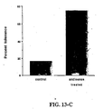
- Fig. 13-A shows transfection efficiencies for rat hearts transfected ex vivo with FITC-ODN, with and without pressure.
- Fig. 13-B shows ICAM-1 expression in transplanted rat hearts with and without pressure-mediated transfection of antisense-ICAM-1 ODN.
- Fig. 13-C illustrates the induction of long-term graft acceptance by pressure-mediated transfection of transplanted rat hearts with antisense ODN against ICAM-1.
- Fig. 14-A shows wall thicknesses at 6 weeks and 6 months after transplantation for untreated (control) grafts, and grafts transfected with either reverse antisense (control) ODN or with antisense ODN against both PCNA and cdc2 kinase.
- Fig. 14-B shows results similar to those in Fig. 14-A for veins transfected with E2F decoy ODN, as compared to untreated grafts and control grafts transfected with scrambled ODN.
- the types of substances that can be used with this invention include (1) charged atoms or ions, (2) neutral or charged small molecules (e.g ., molecules having a molecular weight of ⁇ 1,000), with the exception of small nucleic acids, (3) large molecules ( e.g. , molecules having a molecular weight of ⁇ 1,000), especially proteins and peptides, with the exception of nucleic acids, (4) polymers and filaments (generally ⁇ 10 ⁇ m in greatest dimension), (5) inorganic atoms and molecules, and (6) microscopic particles (generally ⁇ 10 ⁇ m in greatest dimension).
- These substances can be delivered to the extracellular environment of the cell in a wide range of concentrations, preferably, but not limited to, 1 nM to 1 mM, depending on the type of substance and its intended use.
- concentrations to be used in each application of the invention can be determined by the user based on his or her knowledge of the chemistry, biochemistry, or functional properties of the substance to be delivered.
- the substance can exist in solution in the extracellular environment, or it can comprise a suspension or colloid.
- a sealed enclosure containing the tissue and the extracellular environment is defined, and the incubation pressure is established, within the sealed enclosure.
- the boundary of the enclosure is defined substantially by an enclosing means, so that target tissue (tissue comprising the target cell) is subjected to isotropic pressure, and does not distend or experience trauma.
- part of the enclosure boundary is defined by a tissue.
- a protective means such as an inelastic sheath is then placed around the tissue to prevent distension and trauma in the tissue.
- a sealed enclosure is preferably defined between occlusions formed by inflatable balloons or tie wraps.
- a solution containing a substance to be delivered is delivered to the enclosure through a catheter having a delivery outlet between the occlusions.
- an enclosure is defined within an organ by establishing occlusions within organ conduits (e.g ., blood vessels), such that a space within the organ can be pressurized.
- the incubation pressure used to facilitate cellular uptake is preferably maintained at a predetermined level for a predetermined incubation period.
- the incubation pressure depends on the application, including parameters such as the incubation period, the tissue type, and the molecule being delivered.
- the incubation pressure used can range from, for example, 50 mm Hg to 5 atmospheres above ambient pressure, for example, 300 mm Hg to 1500 mm Hg above ambient pressure, or 0 to 2 atmospheres above ambient temperature; higher or lower pressures can also be used, provided that effective delivery is achieved and that a counter veiling injury is not sustained by the target cell or cells.
- the incubation period used to facilitate cellular uptake can vary, depending on parameters such as the incubation pressure, the target tissue type, the molecule being delivered, and the desired dosage.
- the duration of the incubation pressure can range, for example, from approximately 1 second to four hours.
- the pressure duration is between 20 seconds and 30 minutes, more preferably between 60 seconds and 10 minutes.
- Suitable mammalian target tissues include blood vessel tissue (in particular, veins used as grafts in arteries), heart, bone marrow, connective tissue, liver, kidney, genital-urinary system tissue, bones, muscles, gastrointestinal organs, and endocrine and exocrine organs.
- the in vitro methods of the present invention can be applied to parts of an organ, to a whole organ ( e.g ., a heart).
- the invention relates also to the use of a reservoir and a catheter in the manufacture of a system for delivering an atom, a molecule, or a particle of maximum dimension of ⁇ 10 ⁇ m into a living cell in a tissue in vivo.
- Applications of the present invention also include the treatment of allografts (grafts derived from a different subject than the transplant patient) and syngrafts (grafts derived from the transplant patient).
- the conditions for each application of the invention can be determined by labeling the substance with a radioactive, fluorescent, or chemical tag, and then following the substance, following pressure-enhanced administration, via the label.
- measurement of biological activity can be carried out.
- optimization of delivery conditions can be achieved via quantification of delivery of an easily-labeled material that is chemically and/or physically similar to the desired substance.
- a system for delivering a molecule into a cell using the in vitro methods of the invention includes an enclosing means for defining at least part of a boundary of a sealed enclosure, and a pressurization means for establishing an incubation pressure within the enclosure.
- the enclosure contains the target cell and an extracellular environment of the cell.
- a delivery means such as a catheter or syringe is used to deliver the molecule to the extracellular environment, directly or indirectly ( e.g ., by intravenous injection).
- the enclosure boundary is defined by the enclosing means, and possibly by tissue.
- Suitable enclosing means include, depending on the embodiment, a pressurization chamber, an impermeable sheath or bag, and an occlusion means for occluding a passage in a tissue.
- a pressurization chamber is particularly suited for the treatment of grafts or entire organisms, while other devices are well suited for intraoperative treatment of tissue.
- the enclosure boundary is defined substantially by the enclosing means. Pressure is then applied to the target tissue uniformly from all directions, and the target tissue is not subjected to a risk of suffering trauma.
- part of the boundary is defined by a tissue (e.g ., the target tissue). The tissue forming part of the enclosure boundary is subjected to pressure from one side only, and can become distended. A protective means such as an inelastic sheath is placed around the tissue to prevent distension and trauma in the tissue.
- Figs. 1-A through 1-C' illustrate methods of pressurized delivery of molecules to cells of a blood vessel.
- Fig. 1-A is a side view of a delivery system 11 of the present invention.
- System 11 comprises a reservoir 10 for holding a solution 40 containing a substance to be delivered, and a delivery means for expelling solution 40 from reservoir 10 .
- the delivery means comprises a plunger 12 and a delivery tube 14. Opposite plunger 12, reservoir 10 opens into tube 14. Attached to tube 14 are, listed in order of proximity to reservoir 10, a stopcock 16, a pressure gauge 18, a retracted sheath 20, and a notch 22. Sheath 20 is preferably impermeable and inelastic. Notch 22 is next to a distal open end 30 of tube 14. Stopcock 16 is initially in a closed position, preventing solution 40 from passing from reservoir 10 to tube 14 .
- Fig. 1-B shows a blood vessel 24 attached to system 11. Open end 30 is placed into a proximal end of blood vessel 24. Notch 22 fits inside a proximal end of tissue 24. Sheath 20 is pulled down to cover tissue 24. A tie or ligature 26A is wrapped around sheath 20 and tissue 24 at the point where they are attached to tube 14, to prevent tissue 24 from slipping from open end 30. When stopcock 16 is turned to an open position, the solution 40 enters tube 14 and sheath 20, flushing out all gases and liquids present through open end 28 of sheath 20. After the flushing, a tie wrap 26B is placed over distal open end 28 of sheath 20 to form a water-tight seal, as shown in Figs. 1-C and 1-C' .
- Fig. 1-C illustrates delivery targeted to the endothelium of blood vessel 24, while Fig. 1-C' illustrates delivery to the endothelium and to the outside surface of blood vessel 24.
- tie wrap 26B is placed around sheath 20 and tissue 24. Tie wrap 26B occludes blood vessel 24. Stopcock 16 is turned to its open position, and plunger 12 is pushed, such that the solution 40 is delivered into vessel 24 under a delivery pressure. The delivery pressure is allowed to increase until an incubation pressure is reached, and stopcock 16 is closed. Blood vessel 24 is allowed to incubate for an incubation period, after which tie wrap 26B is untied to release the pressure (not illustrated).
- the boundary of a sealed enclosure is defined by the walls of vessel 24 and by an enclosing means.
- the sealed enclosure contains the target (endothelial) cells of blood vessel 24, and their extracellular environment. If stopcock 16 is in a closed position, the enclosing means comprises tube 14, stopcock 16, and ligature 26B. If stopcock 16 is in an open position, the enclosing means comprises ligature 26B , tube 14, plunger 12, and parts of the walls of reservoir 10.
- the enclosing means defines at least part of the boundary of the enclosure.
- part of the boundary of the enclosure is defined by blood vessel 24. Applying pressure only to the inside of blood vessel 24 could cause blood vessel 24 to distend and experience trauma. Sheath 20 acts as a protective means, preventing blood vessel 24 from distending. In an arrangement such as the one in Fig. 1-C , it is thus important that sheath 20 be inelastic.
- tie wrap 26B around sheath 20 only, as illustrated in Fig. 1-C' .
- the sealed enclosure containing the target cells of blood vessel 24 and their extracellular environment is defined substantially by an enclosing means. If stopcock 16 is in a closed position, the enclosing means comprises sheath 20, tube 14 , stopcock 16, and ligature 26B' . If stopcock 16 is in an open position, the enclosing means comprises sheath 20, tube 14, ligature 26B' , plunger 12, and parts of the walls of reservoir 10.
- the boundary of the enclosure is defined substantially by the enclosing means.
- the pressure around blood vessel 24 is uniform, and thus blood vessel 24 does not experience trauma. Since sheath 20 acts as part of the enclosing means, it is important that sheath 20 be impermeable. Sheath 20 need not necessarily be inelastic in the arrangement of Fig. 1-C', however, since the use of an elastic sheath would not lead to trauma in blood vessel 24 .
- Fig. 2 illustrates delivery of molecules to a blood vessel.
- Tube 14 is inserted into the lumen of a vessel 224 .
- a sheath 220 wraps around vessel 224, and a fastener 228 (e.g ., a heat seal) attaches the two flaps of sheet 220 to form a tube.
- Sheath 220 acts as a protective means, preventing the distension of vessel 224.
- Two tie wraps 226A and 226B wrap around sheath 220. Tie wraps 226A and 226B act as occluding means, occluding vessel 224.
- Occlusions 226A and 226B, and the walls of vessel 224 between occlusions 226A and 226B define a sealed enclosure 230 containing the target cells of vessel 224 and their extracellular environment.
- the solution 40 is injected into the sealed enclosure, and segment 230 is allowed to incubate for an incubation period. After the incubation period, occlusions 226A and 226B are removed, and blood is allowed to flow through vessel 224 .
- Figs. 3-A and 3-B illustrate a delivery system having distinct delivery and pressurization elements, used to deliver molecules to the blood vessel shown in Fig. 2.
- a rigid tubular wrap 250 is placed around sheet 220, and a vise 252 is placed around wrap 250, as illustrated in Fig. 3-B.
- Wrap 250 is circumferentially flexible, so that the diameter of the tube it forms is variable, but it is rigid axially, so that even when its diameter changes, it still remains substantially tubular.
- a tightening screw 254 tightens vise 252 , pulling wrap 250 tight, creating pressure within vessel 224. This pressure is maintained for an incubation period, after which screw 254 is unscrewed.
- Figs. 4-A through 4-D illustrate a method of delivering a solution containing a substance to be delivered to the lumen of a blood vessel 324 through a catheter 314 .
- Catheter 314 is inserted into vessel 324.
- Catheter 314 is closed at its end 316.
- Catheter 314 has two balloons 332A and 332B, and a delivery port 330 between balloons 332A and 332B.
- balloons 332A and 332B are deflated, as shown in Fig. 4-A.
- balloons 332A and 332B are inflated, as shown in Fig. 3-B.
- Balloons 332A and 332B occlude vessel 324 and create a sealed enclosure 334 within vessel 324.
- a solution 340 containing a substance to be delivered is delivered to enclosure 334 through port 330.
- Solution 340 is delivered under pressure, such that enclosure 334 becomes pressurized.
- balloons 332A and 332B are deflated and target enclosure 334 is depressurized.
- Fig. 4-C shows an alternative delivery system of the present invention, in which a balloon mounted on a catheter has miniature tubules for delivering a solution containing a substance to be delivered to the walls of the vessel.
- a balloon 432 has tubules 450 that are directly connected to holes 452 in the segment of catheter 314 within balloon 432.
- solution 440 exits holes 452, travels through tubules 450, and reaches the walls of vessel 324.
- Fig. 5-A illustrates the use of system 11 for delivery to blood conduits (vessels and/or atria and ventricles) of an organ 124 such as a heart.
- a protective sheath 120 is wrapped around organ 124.
- Organ 124 has an artery 112 which carries blood into it and a vein 114 which carries blood away.
- Tube 14 is inserted into the lumen of artery 112, and sheath 120 is wrapped around artery 112 and vein 114.
- Tie wrap 126A is tightened around sheath 120 at artery 112, and tie wrap 126B is tightened around sheath 120 at vein 114 , to prevent leakage of fluid out of organ 124.
- Tie wrap 126A allows tube 14 to enter artery 112, yet wraps tightly enough to seal artery 112 from leakage.
- a solution 40 containing a substance to be delivered is injected, and organ 124 is allowed to incubate. After the incubation period, tie wraps 126A and 126B are removed, and blood is allowed to flow through organ 124 once more.
- Fig. 5-B illustrate the use of balloon-catheters for sealing an inlet and an outlet of an organ (e.g ., a gastrointestinal organ).
- a catheter 550 with a balloon 552 is inserted into a first organ conduit 512 in communication with an organ 524, and another catheter 560 with a balloon 562 is inserted into a second organ conduit 514 leading away from organ 524.
- balloons 552 and 562 are deflated (not illustrated).
- balloons 552, 562 are inflated and establish occlusions in conduits 512, 514, respectively.
- a solution 540 containing a substance to be delivered is delivered to organ 524 under a delivery pressure.
- FIG. 6 illustrates the use of a pressurization chamber to facilitate delivery of substances to cells.
- a holding means such as a dish 610 contains a tissue 624 containing target cells.
- a solution 640 containing the substance is placed in dish 610.
- Dish 610 is placed in a pressure chamber 650.
- Chamber 650 is closed and sealed, and a pressurized gas (e.g ., CO 2 ) is introduced into chamber 650 through a duct 660.
- Solution 640 and tissue 624 are maintained under an incubation pressure for an incubation period.
- Tissue 624 can comprise an entire organ.
- a pressurization chamber is shown in Fig. 6 incubation period.
- the increase in membrane permeability requires an increased pressure within the cell and/or extracellular environment, but not necessarily a pressure gradient across the cell membrane. It is possible that proteins forming transmembrane channels change conformation at high pressure, and thus allow the passage of molecules through the channels and into the cytoplasm.
- incubation periods and concentrations used depend on the target tissue type. For example, as is described further below, an incubation period of approximately 5 minutes at low pressure ( ⁇ 0.5 atm) is sufficient for achieving a near-maximal transfection efficiency in human saphenous vein, while an incubation period of over one hour at high pressure ( ⁇ 2 atm) is required for achieving a transfection efficiency of 80-90% in rat aortae. For rat hearts, an incubation period of 30 to 45 minutes at 2 atm is necessary for a transfection efficiency above 50%. In general, the incubation period necessary to achieve a given transfection efficiency in different tissue types varies from minutes to hours, at incubation pressures on the order of atmospheres. Suitable incubation periods and pressures for a given tissue type can be readily determined by the skilled artisan.
- the walls of the pressurized enclosure do not include living tissue, since tissue forming parts of the enclosure wall is subject to mechanical stress.
- Some surgical procedures such as the treatment of blood vessels connected to the circulatory system during the procedure (see Figs. 3-A and 3-B ), require that at least parts of the enclosure walls be defined by tissue. In such a case, it is important that a protective means be used to prevent distension of the tissue.
- Grafts treated ex-vivo are, in general, preferably treated by incubation in a pressurized chamber or an equivalent pressurized enclosure.
- the transfection efficiency of a method of the present invention was investigated in vitro by measuring the inhibition of IL-6 production by antisense ODN in whole organ culture. Vein segments were incubated in growth medium for 24 hours after transfection. Transfections were performed at 5 mM, 10 mM, and 100 mM for 10 minutes, according to a method similar to that illustrated in Fig. 1-C .
- Quantitative reverse transcription PCR was used to measure the reduction in IL-6 mRNA resulting from antisense-ODN transfection performed according to the present invention.
- Three specimens of human saphenous vein were transfected as illustrated in Fig. 1-C , and mRNA levels in antisense-transfected vein segments were compared to levels in untreated and reverse antisense (control) ODN-transfected segments. Results for the three specimens are shown in Figs. 8-A, 8-B, and 8-C, respectively. Reductions in mRNA levels indicate sequence-specific efficacy of antisense-ODN treatment.
- Fig. 11 illustrates the effect of pressure on transfection efficiency for rat kidney cells perfused in vivo with plasmid DNA containing the gene for firefly luciferase, as illustrated in Fig. 6 .
- Fig. 12-A shows the effect of pressure on transfection efficiency for rat aorta cells.
- Aortae were harvested from donor rats and incubated at 4°C for 24 hours in physiologic solution to induce ischemic injury, in a manner similar to that illustrated in Fig. 1-C' .
- Fig. 12-B shows ischemia-induced PCNA expression in transplanted rat aortae with and without pressure-mediated transfection of antisense-PCNA ODN.
- the transfection procedure was similar to that illustrated in Fig. 1-C' .
- Ischemic injuries were induced by 24 hour incubations at 4°C, either in saline solution (control), or in saline solution containing 40 ⁇ M ODN.
- a pressure of 2 atm above atmospheric pressure was applied during incubation to both control and ODN-treated samples.
- Fig. 12-C shows ischemia-induced cdc2 kinase expression in transplanted rat aortae with and without pressure-mediated transfection of antisense-cdc2 kinase ODN.
- the transfection, transplantation, and harvesting procedures were similar to those described above in relation to Fig. 12-B.
- Protein levels for cdc2 kinase were measured by ELISA.
- Fig. 12-D illustrates the reduction in lumenal narrowing of isotransplanted, ischemic-injured rat aortae, resulting from pressure-mediated transfection with antisense ODN against both PCNA and cdc2 kinase.
- Ischemic injury was induced by 24 hours of incubation at 4°C in either saline solution (control), or antisense-PCNA/antisense-cdc2 kinase ODN solution (40 ⁇ M each). A pressure of 2 atm above ambient pressure was applied to all tissues (including control).
- Fig. 13-A shows transfection efficiencies for rat hearts transfected ex vivo with FITC-ODN, with and without pressure.
- Fig. 13-B shows ICAM-1 expression in transplanted rat hearts, with and without pressure-mediated transfection of antisense-ICAM-1 ODN.
- Either saline (control) or antisense-ICAM-1-ODN solution (80 ⁇ M) was perfused into the coronary arteries of donor PVG strain hearts after aortic crossclamping. The hearts were then submerged in FITC-ODN solution and exposed to 2 atm above ambient pressure for 45 minutes at 4°C, as illustrated in Fig. 6 .
- Fig. 13-C illustrates the induction of long-term graft acceptance by pressure-mediated transfection of transplanted rat hearts with antisense ODN against ICAM-1.
- PVG strain rat hearts were harvested and transfected ex-vivo with either antisense-ICAM-1 ODN solution (80 ⁇ m) or with saline solution (control), as described above in relation to Fig. 13-B.
- Figs. 14-A and 14-B show inhibition of neointimal hyperplasia in rabbit jugular veins grafted into carotid arteries, following pressure-mediated transfection with ODN designed to block up regulation of cell cycle regulatory genes.
- Fig. 14-A shows wall thicknesses at 6 weeks and 6 months after transplantation for untreated (control) grafts, and grafts transfected with either reverse antisense (control) ODN or with,antisense ODN against both PCNA and cdc2 kinase.
- Neointima formation was inhibited for up to 6 months, while medial hypertrophy allows adaptive wall thickening to reduce wall stress in the high-pressure arterial environment.
- Fig. 14-B shows results similar to those in Fig. 14-A for veins transfected with E2F decoy ODN, as compared to untreated grafts and control grafts transfected with scrambled ODN.
- n 6, p ⁇ 0.005.
- a time-varying incubation pressure can be used in general.
- Many potential designs for enclosing means, protective means, and/or occluding means can be readily devised by one skilled in the art, depending on the application.
- Various incubation pressures, periods, and active agent dosages leading to desirable or near-maximal transfection efficiencies can be readily determined for different tissue types.
Abstract
Description
Applications of the present invention also include the treatment of allografts (grafts derived from a different subject than the transplant patient) and syngrafts (grafts derived from the transplant patient).
The examples are given for illustrative purpose only.
Claims (23)
- An in vitro method of enhancing the uptake by a cell of an atom, a molecule, with the exception of nucleic acids, or a particle of maximum dimension of <10 µm, said method comprisingbringing into contact said cell and a liquid medium comprising said atom, molecule, or particle,maintaining said cell and said liquid medium in an enclosed space, andsubjecting said enclosed cell and liquid medium to an incubation pressure sufficient to enhance uptake by said cell of said atom, molecule, or particle, excluding a method practised on the human or animal body.
- The method of claim 1, wherein said cell is contained within a mammalian blood vessel that is sealed at two locations to provide said enclosed space.
- The method of claim 1, wherein said molecule is a therapeutic, charged organic molecule having a molecular weight of less than 1,000.
- The method of claim 1, wherein said molecule is a protein or a peptide having a molecular weight of greater than 1,000.
- The method of claim 1, wherein said incubation pressure is between 50 mm Hg and 5 atmospheres above ambient pressure.
- The method of claim 5, wherein said incubation pressure is between 200 mm Hg and 2.5 atmospheres above ambient pressure.
- The method of claim 1, wherein said cell is contained in a mammalian blood vessel or organ, and the incubation pressure equilibrates between the exterior and interior of the vessel or organ so that there is substantially no pressure gradient between said exterior and interior.
- Use of a reservoir and a catheter in the manufacture of a system for delivering an atom, a molecule, or a particle of maximum dimension of <10 µm into a living cell in a tissue in vivo, wherein:said reservoir is adapted for holding a solution containing said atom, molecule, or particle;said system is adapted for delivering said atom, molecule, or particle into the extracellular environment of said living cell of said tissue, and for applying pressure to said cell and enviromment, thereby facilitating uptake of said atom, molecule, or particle by said living cell; andsaid atom, molecule, or particle is not a nucleotide.
- Use of a system for delivering in vitro an atom, a molecule, or a particle of maximum dimension of <10 µm into a living cell, wherein said atom, molecule, or particle is not a nucleotide, said system comprising:an encloding means for defining at least part of a boundary of a sealed enclosure, said sealed enclosure containing said cell and an extracellular environment of said cell; anda pressurization means for establishing an incubation pressure above atmospheric inside said sealed enclosure, thereby facilitating uptake of said atom, molecule, or particle by said living cell, excluding the use of said system in a method practised on the human or aninal body.
- The use of claim 9, wherein said enclosing means comprises a pressurization chamber.
- The use of claim 10, wherein a holding means is placed inside the pressurization chamber for holding a tissue containing target cells and a solution containing said atom, molecule or particle.
- The use of claim 8, wherein said cell is contained within a mammalian blood vessel that is sealed at two locations to provide said enclosed space.
- The use of claim 8 or 9, wherein said molecule is a therapeutic, charged organic molecule having a molecular weight of less than 1,000.
- The use of claim 8 or 9, wherein said molecule is a protein or a peptide having a molecular weight of greater than 1,000.
- The use of claim 8 or 9, wherein said incubation pressure is between 50 mm Hg and 5 atmospheres above ambient pressure.
- The use of claim 8 or 9, wherein said incubation pressure is between 200 mm Hg and 2.5 atmospheres above ambient pressure.
- The use of claim 8 or 9, wherein said cell is contained in a mammalian blood vessel or organ, and the incubation pressure equilibrates between the exterior and interior of the vessel or organ so that there is substantially no pressure gradient between said exterior and interior.
- The use of claim 9, wherein said enclosing means comprises an impermeable sheath.
- The use of claim 9, wherein said enclosing means comprises an occlusion means for occluding a passage in a tissue.
- The use of claim 9, wherein said boundary is defined substantially by said enclosing means.
- The use of claim 9, wherein part of said boundary is defined by a tissue.
- The use of claim 9, wherein said system further comprises a protective means adapted to be placed around said tissue, for preventing a trauma in said tissue.
- The use of claim 22, wherein said protective means comprises an inelastic sheath.
Priority Applications (1)
| Application Number | Priority Date | Filing Date | Title |
|---|---|---|---|
| EP02019889A EP1279412A3 (en) | 1996-11-07 | 1997-11-07 | Pressure-mediated intracellular delivery of molecules of microparticles |
Applications Claiming Priority (3)
| Application Number | Priority Date | Filing Date | Title |
|---|---|---|---|
| US745023 | 1996-11-07 | ||
| US08/745,023 US5922687A (en) | 1995-05-04 | 1996-11-07 | Intracellular delivery of nucleic acids using pressure |
| PCT/US1997/020696 WO1998020109A1 (en) | 1996-11-07 | 1997-11-07 | Pressure-mediated intracellular delivery of molecules or microparticles |
Related Child Applications (1)
| Application Number | Title | Priority Date | Filing Date |
|---|---|---|---|
| EP02019889A Division EP1279412A3 (en) | 1996-11-07 | 1997-11-07 | Pressure-mediated intracellular delivery of molecules of microparticles |
Publications (3)
| Publication Number | Publication Date |
|---|---|
| EP0944715A1 EP0944715A1 (en) | 1999-09-29 |
| EP0944715A4 EP0944715A4 (en) | 1999-12-22 |
| EP0944715B1 true EP0944715B1 (en) | 2002-09-11 |
Family
ID=24994918
Family Applications (2)
| Application Number | Title | Priority Date | Filing Date |
|---|---|---|---|
| EP97948268A Expired - Lifetime EP0944715B1 (en) | 1996-11-07 | 1997-11-07 | Pressure-mediated intracellular delivery of molecules or microparticles |
| EP02019889A Withdrawn EP1279412A3 (en) | 1996-11-07 | 1997-11-07 | Pressure-mediated intracellular delivery of molecules of microparticles |
Family Applications After (1)
| Application Number | Title | Priority Date | Filing Date |
|---|---|---|---|
| EP02019889A Withdrawn EP1279412A3 (en) | 1996-11-07 | 1997-11-07 | Pressure-mediated intracellular delivery of molecules of microparticles |
Country Status (16)
| Country | Link |
|---|---|
| US (1) | US5922687A (en) |
| EP (2) | EP0944715B1 (en) |
| JP (3) | JP2001505419A (en) |
| KR (1) | KR100449330B1 (en) |
| CN (1) | CN1109749C (en) |
| AT (1) | ATE223967T1 (en) |
| AU (1) | AU736298B2 (en) |
| BR (1) | BR9713334A (en) |
| CA (1) | CA2271244C (en) |
| DE (1) | DE69715449T2 (en) |
| DK (1) | DK0944715T3 (en) |
| ES (1) | ES2183221T3 (en) |
| HK (1) | HK1022711A1 (en) |
| IL (1) | IL129808A (en) |
| PT (1) | PT944715E (en) |
| WO (1) | WO1998020109A1 (en) |
Families Citing this family (132)
| Publication number | Priority date | Publication date | Assignee | Title |
|---|---|---|---|---|
| US7803782B2 (en) * | 2003-05-28 | 2010-09-28 | Roche Madison Inc. | Intravenous delivery of polynucleotides to cells in mammalian limb |
| US6627616B2 (en) * | 1995-12-13 | 2003-09-30 | Mirus Corporation | Intravascular delivery of non-viral nucleic acid |
| US20020001574A1 (en) * | 1995-12-13 | 2002-01-03 | Jon A. Woiff | Process of delivering a polynucleotide to a muscle cell via the vascular system |
| US6379966B2 (en) * | 1999-02-26 | 2002-04-30 | Mirus Corporation | Intravascular delivery of non-viral nucleic acid |
| US20040259828A1 (en) * | 1995-12-13 | 2004-12-23 | Wolff Jon A. | Intravascular delivery of non-viral nucleic acid |
| US7507722B1 (en) | 1999-11-05 | 2009-03-24 | Roche Madison Inc. | Intravascular delivery of nucleic acid |
| US6676626B1 (en) | 1998-05-01 | 2004-01-13 | Ekos Corporation | Ultrasound assembly with increased efficacy |
| US6582392B1 (en) * | 1998-05-01 | 2003-06-24 | Ekos Corporation | Ultrasound assembly for use with a catheter |
| US7435723B2 (en) * | 1997-11-21 | 2008-10-14 | Mirus Bio Corporation | Process for delivery of polynucleotides to the prostate |
| US6699231B1 (en) | 1997-12-31 | 2004-03-02 | Heartport, Inc. | Methods and apparatus for perfusion of isolated tissue structure |
| US6722370B1 (en) * | 1998-07-17 | 2004-04-20 | Corgentech, Inc. | Delivery of a composition to the liver by utilizing the portal vein |
| WO2000015285A1 (en) * | 1998-09-14 | 2000-03-23 | Mirus Corporation | A process for delivering nucleic acids to cardiac tissue |
| WO2000050617A1 (en) * | 1999-02-26 | 2000-08-31 | Mirus Corporation | Intravascular delivery of non-viral nucleic acid |
| WO2001000663A2 (en) * | 1999-06-28 | 2001-01-04 | Oklahoma Medical Research Foundation | Catalytically active recombinant memapsin and methods of use thereof |
| US7214369B2 (en) * | 2003-05-05 | 2007-05-08 | Mirus Bio Corporation | Devices and processes for distribution of genetic material to mammalian limb |
| EP1246649B1 (en) | 1999-11-05 | 2006-10-18 | Mirus Bio Corporation | Intravascular delivery of nucleic acid |
| US7642248B2 (en) * | 1999-11-05 | 2010-01-05 | Roche Madison Inc | Devices and processes for distribution of genetic material to mammalian limb |
| CA2386341A1 (en) | 1999-11-18 | 2001-05-25 | Epimmune Inc. | Heteroclitic analogs and related methods |
| US20040072785A1 (en) * | 1999-11-23 | 2004-04-15 | Wolff Jon A. | Intravascular delivery of non-viral nucleic acid |
| US6346098B1 (en) | 2000-03-07 | 2002-02-12 | The Board Of Trustees Of The Leland Stanford Junior University | Methods and kits for locally administering an active agent to an interstitial space of a host |
| JP2003531865A (en) * | 2000-04-28 | 2003-10-28 | アメリカ合衆国 | Improving immunogenicity using a combination of DNA and vaccinia virus vector vaccines |
| EP2075582A3 (en) | 2000-07-12 | 2010-01-06 | Agensys, Inc. | Novel tumor antigen useful in diagnosis and therapy of bladder, ovary, lung and kidney cancers |
| AU2001280789A1 (en) * | 2000-07-25 | 2002-02-05 | The Board Of Trustees Of The Leland Stanford Junior University | Non-viral linear dna vectors and methods for using the same |
| WO2002018578A2 (en) | 2000-08-28 | 2002-03-07 | Agensys, Inc. | Nucleic acid and corresponding protein entitled 85p1b3 useful in treatment and detection of cancer |
| CA2425648A1 (en) | 2000-10-19 | 2002-04-19 | Epimmune Inc. | Hla class i and ii binding peptides and their uses |
| WO2002066611A2 (en) * | 2001-02-16 | 2002-08-29 | The Board Of Trustees Of The Leland Stanford Junior University | Minimal plasmid vectors that provide for persistent and high level gene expression and methods for using the same |
| US6924358B2 (en) | 2001-03-05 | 2005-08-02 | Agensys, Inc. | 121P1F1: a tissue specific protein highly expressed in various cancers |
| US7271240B2 (en) | 2001-03-14 | 2007-09-18 | Agensys, Inc. | 125P5C8: a tissue specific protein highly expressed in various cancers |
| WO2002083921A2 (en) | 2001-04-10 | 2002-10-24 | Agensys, Inc. | Nuleic acids and corresponding proteins useful in the detection and treatment of various cancers |
| US20030191073A1 (en) | 2001-11-07 | 2003-10-09 | Challita-Eid Pia M. | Nucleic acid and corresponding protein entitled 161P2F10B useful in treatment and detection of cancer |
| US10590418B2 (en) * | 2001-07-23 | 2020-03-17 | The Board Of Trustees Of The Leland Stanford Junior University | Methods and compositions for RNAi mediated inhibition of gene expression in mammals |
| WO2003010180A1 (en) | 2001-07-23 | 2003-02-06 | The Board Of Trustees Of The Leland Stanford Junior University | Methods and compositions for rnai mediated inhibition of gene expression in mammals |
| EP2287186B1 (en) | 2001-09-06 | 2014-12-31 | Agensys, Inc. | Nucleic acid and corresponding protein entitled STEAP-1 useful in treatment and detection of cancer |
| EP1721977A3 (en) | 2001-09-17 | 2008-10-15 | PDL BioPharma, Inc. | Methods of diagnosis of cancer, compositions and methods of screening for modulators of cancer |
| AU2002359576A1 (en) | 2001-12-03 | 2003-06-17 | Ekos Corporation | Catheter with multiple ultrasound radiating members |
| US7141044B2 (en) * | 2001-12-11 | 2006-11-28 | Ekos Corporation | Alternate site gene therapy |
| DE10211589A1 (en) * | 2002-03-15 | 2003-10-02 | Steffen-Sebastian Bolz | In vitro transfection and long-term cultivation of isolated organs |
| US8226629B1 (en) | 2002-04-01 | 2012-07-24 | Ekos Corporation | Ultrasonic catheter power control |
| US20030204167A1 (en) * | 2002-04-24 | 2003-10-30 | Johnson Lanny L. | Device for delivering liquid medications, nutrients or gases to local tissue |
| AU2003301841A1 (en) * | 2002-05-01 | 2004-06-07 | National Institutes Of Health | Immunotherapy regimens in hiv-infected patients |
| US20030228691A1 (en) * | 2002-05-17 | 2003-12-11 | Lewis David L. | Processes for inhibiting gene expression using polynucleotides |
| CN100418981C (en) | 2002-06-10 | 2008-09-17 | 瓦西尼斯公司 | Gene differentially expressed in breast and bladder cancer and encoded polypeptides |
| WO2004011060A2 (en) * | 2002-07-26 | 2004-02-05 | Mirus Corporation | Delivery of molecules and complexes to mammalian cells in vivo |
| US20040081653A1 (en) | 2002-08-16 | 2004-04-29 | Raitano Arthur B. | Nucleic acids and corresponding proteins entitled 251P5G2 useful in treatment and detection of cancer |
| EP1572943B1 (en) | 2002-08-29 | 2015-04-22 | The Board of Trustees of The Leland S. Stanford Junior University | Circular nucleic acid vectors, and methods for making and using the same |
| US6921371B2 (en) | 2002-10-14 | 2005-07-26 | Ekos Corporation | Ultrasound radiating members for catheter |
| JP2006508163A (en) | 2002-11-27 | 2006-03-09 | アジェンシス, インコーポレイテッド | Nucleic acids and corresponding proteins referred to as 24P4C12 useful in the treatment and detection of cancer |
| EP1903056A3 (en) | 2002-12-10 | 2008-05-07 | Idm Pharma, Inc. | HLA-A1, -A2 -A3, -A24, -B7, and -B44 binding peptides comprising tumor associated antigen epitopes, and compositions thereof |
| US7781415B2 (en) * | 2003-02-07 | 2010-08-24 | Roche Madison Inc. | Process for delivering sirna to cardiac muscle tissue |
| EP2343315A3 (en) | 2003-02-10 | 2011-11-23 | Agensys, Inc. | Nucleic acid and corresponding protein named 158P1D7 useful in the treatment and detection of bladder and other cancers |
| US20070037151A1 (en) * | 2003-04-28 | 2007-02-15 | Babe Lilia M | Cd4+ human papillomavirus (hpv) epitopes |
| EP2319524B1 (en) | 2003-05-30 | 2013-08-21 | Agensys, Inc. | Prostate stem cell antigen (PSCA) variants and subsequences thereof |
| AU2004271951B2 (en) * | 2003-09-05 | 2008-08-21 | Genencor International, Inc. | HPV CD8+ T-cell epitopes |
| WO2005035547A2 (en) * | 2003-10-06 | 2005-04-21 | Corgentech, Inc. | E2f oligonucleotide decoy molecules |
| US20050215503A1 (en) * | 2003-12-03 | 2005-09-29 | Mcevoy Leslie M | HIF oligonucleotide decoy molecules |
| JP2007520481A (en) * | 2004-01-15 | 2007-07-26 | アルザ・コーポレーシヨン | Liposome composition for delivering therapeutic agents |
| GB0406728D0 (en) * | 2004-03-25 | 2004-04-28 | Hydrodynamic Gene Delivery Ltd | Gene therapy |
| EP1747023B2 (en) | 2004-05-04 | 2016-03-09 | The Board of Trustees of The Leland Stanford Junior University | Methods and compositions for reducing hcv viral genome amounts in a target cell |
| US20050289659A1 (en) * | 2004-05-18 | 2005-12-29 | Jacks E T | Cre-lox based method for conditional RNA interference |
| JP4651663B2 (en) | 2004-05-28 | 2011-03-16 | アジェンシス,インコーポレイテッド | Antibodies and related molecules that bind to PSCA proteins |
| US7482158B2 (en) * | 2004-07-01 | 2009-01-27 | Mathison Brian H | Composite polynucleic acid therapeutics |
| TW200626724A (en) * | 2004-08-23 | 2006-08-01 | Paik Medicine International Inc | Method for gene transfer into the organelles of cells: direct gene transfer to mitochondria |
| US20070036740A1 (en) * | 2004-10-06 | 2007-02-15 | Reed Kenneth C | Modulation of hair growth |
| AU2005299672A1 (en) * | 2004-10-22 | 2006-05-04 | Benitec, Inc. | Therapeutic RNAi agents for treating psoriasis |
| US20060115462A1 (en) * | 2004-12-01 | 2006-06-01 | Vladimir Subbotin | Direct DNA delivery to bone cells |
| SG158174A1 (en) * | 2005-01-06 | 2010-01-29 | Benitec Inc | Rnai agents for maintenance of stem cells |
| PL2325305T3 (en) | 2005-02-25 | 2014-07-31 | Oncotherapy Science Inc | Peptide vaccines for lung cancers expressing TTK, URLC10 or KOC1 polypeptides |
| WO2006093030A1 (en) | 2005-02-28 | 2006-09-08 | Oncotherapy Science, Inc. | Epitope peptides derived from vascular endothelial growth factor receptor 1 and vaccines containing these peptides |
| RU2413735C2 (en) | 2005-03-31 | 2011-03-10 | Эдженсис, Инк. | Antibodies and related molecules binding with proteins 161p2f10b |
| US20060292159A1 (en) * | 2005-06-08 | 2006-12-28 | Ranscht Barbara E | Methods for the inhibition of neovascularization and cancer metastasis |
| PL1910839T3 (en) | 2005-07-27 | 2016-11-30 | Colon cancer related gene tom34 | |
| EP1922083A2 (en) * | 2005-08-10 | 2008-05-21 | Oklahoma Medical Research Foundation | Truncated memapsin 2 for use for treating alzheimer's disease |
| US20070143195A1 (en) * | 2005-12-15 | 2007-06-21 | Bell Ryan B | Systems and methods for evaluating terms of a deal to purchase a vehicle |
| US20070173470A1 (en) * | 2006-01-23 | 2007-07-26 | Chi-Hung Lin | Methods for delivering extracellular target into cells |
| SG10201502098YA (en) | 2006-10-17 | 2015-05-28 | Oncotherapy Science Inc | Peptide Vaccines For Cancers Expressing MPHOSPH1 OR DEPDC1 Polypeptides |
| US10182833B2 (en) | 2007-01-08 | 2019-01-22 | Ekos Corporation | Power parameters for ultrasonic catheter |
| TWI596109B (en) | 2007-02-21 | 2017-08-21 | 腫瘤療法 科學股份有限公司 | Peptide vaccines for cancers expressing tumor-associated antigens |
| WO2008112226A2 (en) * | 2007-03-13 | 2008-09-18 | Massachusetts Institute Of Technology | Cre-lox based gene knockdown constructs and methods of use thereof |
| TW201425333A (en) | 2007-04-11 | 2014-07-01 | Oncotherapy Science Inc | TEM8 peptides and vaccines comprising the same |
| PL2476689T3 (en) | 2007-05-10 | 2016-04-29 | Agilent Technologies Inc | Thiocarbon-protecting groups for RNA synthesis |
| US9044568B2 (en) | 2007-06-22 | 2015-06-02 | Ekos Corporation | Method and apparatus for treatment of intracranial hemorrhages |
| DK2222697T3 (en) | 2007-11-01 | 2013-03-11 | Perseid Therapeutics Llc | Immunosuppressive polypeptides and nucleic acids |
| EP3085707B1 (en) | 2007-11-01 | 2019-02-27 | Mayo Foundation for Medical Education and Research | Hla-dr binding peptides and their uses |
| US9017660B2 (en) | 2009-11-11 | 2015-04-28 | Advaxis, Inc. | Compositions and methods for prevention of escape mutation in the treatment of Her2/neu over-expressing tumors |
| EP2853269B1 (en) | 2008-05-19 | 2019-05-01 | Advaxis, Inc. | Dual delivery system for heterologous antigens comprising a recombinant Listeria strain attenuated by mutation of dal/dat and deletion of ActA comprising a nucleic acid molecule encoding an listeriolysin O - prostate specific anigen fusion protein |
| US9650639B2 (en) | 2008-05-19 | 2017-05-16 | Advaxis, Inc. | Dual delivery system for heterologous antigens |
| CN102124333B (en) | 2008-06-17 | 2014-06-04 | 多伦多大学管理委员会 | Device for investigation of a flow conduit |
| EP2304025B1 (en) | 2008-07-03 | 2015-11-04 | The Board of Trustees of the Leland Stanford Junior University | Minicircle dna vector preparations and methods of making and using the same |
| US8945885B2 (en) | 2008-07-03 | 2015-02-03 | The Board Of Trustees Of The Leland Stanford Junior University | Minicircle DNA vector preparations and methods of making and using the same |
| EP2329044B1 (en) | 2008-08-27 | 2016-05-18 | Oncotherapy Science, Inc. | Prmt1 for target genes of cancer therapy and diagnosis |
| WO2010048252A1 (en) * | 2008-10-23 | 2010-04-29 | Intervet International B.V. | Lawsonia intracellularis vaccines |
| CN102292069B (en) | 2008-11-26 | 2014-07-30 | 中外制药株式会社 | Vesicle preparation |
| EP2368989A4 (en) | 2008-11-26 | 2012-09-26 | Chugai Pharmaceutical Co Ltd | Oligoribonucleotide or peptide nucleic acid capable of inhibiting activity of hepatitis c virus |
| TWI500932B (en) | 2008-12-05 | 2015-09-21 | Oncotherapy Science Inc | Wdrpuh epitope peptides and vaccines containing the same |
| TWI469791B (en) | 2009-02-18 | 2015-01-21 | Oncotherapy Science Inc | Foxm1 peptides and vaccines containing the same |
| EP2405959A4 (en) * | 2009-03-13 | 2013-10-16 | Univ Tufts | Methods, apparatuses, and kits for introducing genetic material into living cells |
| US9017991B2 (en) | 2009-03-13 | 2015-04-28 | Tufts University | Methods tip assemblies and kits for introducing material into cells |
| CN102356155B (en) | 2009-03-18 | 2016-02-24 | 肿瘤疗法科学股份有限公司 | NEIL3 peptide and comprise its vaccine |
| TWI507204B (en) | 2009-05-26 | 2015-11-11 | Oncotherapy Science Inc | Cdc45l peptides and vaccines including the same |
| US10016617B2 (en) | 2009-11-11 | 2018-07-10 | The Trustees Of The University Of Pennsylvania | Combination immuno therapy and radiotherapy for the treatment of Her-2-positive cancers |
| TW201136604A (en) | 2009-12-14 | 2011-11-01 | Oncotherapy Science Inc | TMEM22 peptides and vaccines including the same |
| US20130052221A1 (en) | 2010-02-26 | 2013-02-28 | The Govt. of the U.S, as represented by The Sec. of The Dept. of Health and Human Services | Dna-protein vaccination protocols |
| BR112012022641A2 (en) | 2010-03-11 | 2017-02-14 | Oncotherapy Science Inc | hjurp peptides and vaccines that include the same |
| US11020444B2 (en) * | 2010-04-23 | 2021-06-01 | Scicotec Gmbh | Transluminal delivery of viruses for treatment of diseased tissue |
| WO2012138377A2 (en) | 2010-10-01 | 2012-10-11 | Trustees Of The University Of Pennsylvania | The use of listeria vaccine vectors to reverse vaccine unresponsiveness in parasitically infected individuals |
| AU2012229218B2 (en) | 2011-03-11 | 2017-03-02 | Advaxis, Inc. | Listeria-based adjuvants |
| WO2013024582A1 (en) | 2011-08-12 | 2013-02-21 | Oncotherapy Science, Inc. | Mphosph1 peptides and vaccines including the same |
| CA3122778A1 (en) | 2011-10-28 | 2013-05-02 | Oncotherapy Science, Inc. | Topk peptides and vaccines including the same |
| SG10201700392UA (en) | 2012-03-12 | 2017-03-30 | Advaxis Inc | Suppressor cell function inhibition following listeria vaccine treatment |
| WO2014010231A1 (en) | 2012-07-10 | 2014-01-16 | Oncotherapy Science, Inc. | Kif20a epitope peptides for th1 cells and vaccines containing the same |
| WO2014010232A1 (en) | 2012-07-10 | 2014-01-16 | Oncotherapy Science, Inc. | Ly6k epitope peptides for th1 cells and vaccines containing the same |
| JP6283861B2 (en) | 2012-09-11 | 2018-02-28 | オンコセラピー・サイエンス株式会社 | UBE2T peptide and vaccine containing the same |
| GB2516196B (en) | 2013-01-25 | 2015-09-09 | Xcell Biosciences Inc | Methods, compositions, kits, and systems for selective enrichment of target cells |
| TWI658049B (en) | 2013-03-12 | 2019-05-01 | 腫瘤療法 科學股份有限公司 | Kntc2 peptides and vaccines containing the same |
| KR102202742B1 (en) | 2013-03-15 | 2021-01-12 | 더 리전트 오브 더 유니버시티 오브 캘리포니아 | High-throughput cargo delivery into live cells using photothermal platforms |
| WO2014153636A1 (en) | 2013-03-27 | 2014-10-02 | Immunovaccine Technologies Inc. | Method for improving the efficacy of a survivin vaccine in the treatment of cancer |
| WO2015148842A1 (en) | 2014-03-28 | 2015-10-01 | The Regents Of The University Of California | Efficient delivery of large cargos into cells on a porous substrate |
| US10760040B1 (en) | 2014-07-03 | 2020-09-01 | NanoCav, LLC | Mechanical transfection devices and methods |
| US10081816B1 (en) | 2014-07-03 | 2018-09-25 | Nant Holdings Ip, Llc | Mechanical transfection devices and methods |
| MX2017001650A (en) | 2014-08-04 | 2017-04-27 | Oncotherapy Science Inc | Cdca1-derived peptide and vaccine containing same. |
| KR20220165831A (en) | 2014-08-04 | 2022-12-15 | 온코세라피 사이언스 가부시키가이샤 | Urlc10-derived peptide and vaccine containing same |
| EP3590954A3 (en) | 2014-08-04 | 2020-03-25 | OncoTherapy Science, Inc. | Koc1-derived peptide and vaccine including same |
| CA2972635A1 (en) | 2015-01-06 | 2016-07-14 | Immunovaccine Technologies Inc. | Lipid a mimics, methods of preparation, and uses thereof |
| WO2016183420A1 (en) | 2015-05-13 | 2016-11-17 | The United States Of America As Represented By The Secretary Of The Department Of Health And Human Services | Methods and compositions for inducing an immune response using conserved element constructs |
| EP3307388B1 (en) | 2015-06-10 | 2022-06-22 | Ekos Corporation | Ultrasound catheter |
| EP3360886B1 (en) | 2015-10-08 | 2023-01-25 | Oncotherapy Science, Inc. | Foxm1-derived peptide, and vaccine including same |
| US20170369904A1 (en) * | 2016-06-22 | 2017-12-28 | Xcell Biosciences, Inc. | Methods for increasing cell culture transfection efficiency and cellular reprogramming |
| US10533189B2 (en) * | 2016-09-20 | 2020-01-14 | The Chinese University Of Hong Kong | Highly specific delivery of polynucleotides to the cell nucleus via compression |
| CN111032134B (en) * | 2017-06-30 | 2024-02-13 | 阿维塔斯有限公司 | electrospray catheter |
| TW202023581A (en) | 2018-08-02 | 2020-07-01 | 日商腫瘤療法 科學股份有限公司 | Cdca1-derived peptide and vaccine containing same |
| AU2022299660A1 (en) * | 2021-06-24 | 2024-01-18 | Avectas Limited | Spray nozzle |
| WO2023122797A1 (en) * | 2021-12-23 | 2023-06-29 | Intergalactic Therapeutics, Inc. | Devices and methods for hollow tissue electrotransfer |
Family Cites Families (5)
| Publication number | Priority date | Publication date | Assignee | Title |
|---|---|---|---|---|
| US5698531A (en) * | 1989-03-31 | 1997-12-16 | The Regents Of The University Of Michigan | Treatment of diseases by site-specific instillation of cells or site-specific transformation of cells and kits therefor |
| US5328470A (en) * | 1989-03-31 | 1994-07-12 | The Regents Of The University Of Michigan | Treatment of diseases by site-specific instillation of cells or site-specific transformation of cells and kits therefor |
| US5674192A (en) * | 1990-12-28 | 1997-10-07 | Boston Scientific Corporation | Drug delivery |
| US5584803A (en) * | 1991-07-16 | 1996-12-17 | Heartport, Inc. | System for cardiac procedures |
| US5766901A (en) * | 1995-05-04 | 1998-06-16 | The Board Of Trustees Of The Leland Stanford Junior University | Apparatus and method for delivering a nucleotide into cell nuclei |
-
1996
- 1996-11-07 US US08/745,023 patent/US5922687A/en not_active Expired - Fee Related
-
1997
- 1997-11-07 WO PCT/US1997/020696 patent/WO1998020109A1/en active IP Right Grant
- 1997-11-07 AT AT97948268T patent/ATE223967T1/en not_active IP Right Cessation
- 1997-11-07 BR BR9713334-5A patent/BR9713334A/en not_active Application Discontinuation
- 1997-11-07 ES ES97948268T patent/ES2183221T3/en not_active Expired - Lifetime
- 1997-11-07 PT PT97948268T patent/PT944715E/en unknown
- 1997-11-07 EP EP97948268A patent/EP0944715B1/en not_active Expired - Lifetime
- 1997-11-07 AU AU70020/98A patent/AU736298B2/en not_active Ceased
- 1997-11-07 DE DE69715449T patent/DE69715449T2/en not_active Expired - Fee Related
- 1997-11-07 KR KR10-1999-7004103A patent/KR100449330B1/en not_active IP Right Cessation
- 1997-11-07 DK DK97948268T patent/DK0944715T3/en active
- 1997-11-07 EP EP02019889A patent/EP1279412A3/en not_active Withdrawn
- 1997-11-07 CN CN97181265A patent/CN1109749C/en not_active Expired - Fee Related
- 1997-11-07 IL IL12980897A patent/IL129808A/en not_active IP Right Cessation
- 1997-11-07 JP JP52188198A patent/JP2001505419A/en not_active Withdrawn
- 1997-11-07 CA CA002271244A patent/CA2271244C/en not_active Expired - Fee Related
-
2000
- 2000-03-17 HK HK00101643A patent/HK1022711A1/en not_active IP Right Cessation
-
2007
- 2007-09-04 JP JP2007228799A patent/JP2008054681A/en not_active Ceased
-
2008
- 2008-05-07 JP JP2008121322A patent/JP2008237221A/en active Pending
Also Published As
| Publication number | Publication date |
|---|---|
| EP0944715A4 (en) | 1999-12-22 |
| WO1998020109A1 (en) | 1998-05-14 |
| DE69715449D1 (en) | 2002-10-17 |
| IL129808A0 (en) | 2000-02-29 |
| AU736298B2 (en) | 2001-07-26 |
| IL129808A (en) | 2004-06-20 |
| JP2001505419A (en) | 2001-04-24 |
| JP2008237221A (en) | 2008-10-09 |
| CA2271244C (en) | 2008-09-02 |
| JP2008054681A (en) | 2008-03-13 |
| CN1109749C (en) | 2003-05-28 |
| US5922687A (en) | 1999-07-13 |
| EP1279412A2 (en) | 2003-01-29 |
| AU7002098A (en) | 1998-05-29 |
| BR9713334A (en) | 2000-05-09 |
| ATE223967T1 (en) | 2002-09-15 |
| EP1279412A3 (en) | 2003-12-17 |
| DK0944715T3 (en) | 2002-12-30 |
| CA2271244A1 (en) | 1998-05-14 |
| KR100449330B1 (en) | 2004-09-18 |
| EP0944715A1 (en) | 1999-09-29 |
| HK1022711A1 (en) | 2000-08-18 |
| ES2183221T3 (en) | 2003-03-16 |
| DE69715449T2 (en) | 2003-07-31 |
| PT944715E (en) | 2003-01-31 |
| CN1244213A (en) | 2000-02-09 |
| KR20000053164A (en) | 2000-08-25 |
Similar Documents
| Publication | Publication Date | Title |
|---|---|---|
| EP0944715B1 (en) | Pressure-mediated intracellular delivery of molecules or microparticles | |
| Chapman et al. | Gene transfer into coronary arteries of intact animals with a percutaneous balloon catheter. | |
| Seunguk et al. | Improved distribution of small molecules and viral vectors in the murine brain using a hollow fiber catheter | |
| DE69925936T2 (en) | DEVICE FOR THE LOCAL ADMINISTRATION OF A MEDICINE IN A BODY HEIGHT | |
| AU2009202335B2 (en) | Pulsatile flux drug delivery | |
| JPH08512282A (en) | Subcutaneous implantable multiple substance delivery system | |
| CN103961788A (en) | Occlusion perfusion catheter | |
| JP2011087592A (en) | Micro-organ explant and method for producing the same | |
| CN108265052A (en) | A kind of small RNA and medical composition and its use | |
| EP0826059B1 (en) | Apparatus and method for delivering a nucleotide into cell nuclei | |
| Hasan et al. | Iontophoresis-mediated direct delivery of nucleic acid therapeutics, without use of carriers, to internal organs via non-blood circulatory pathways | |
| Mbituyimana et al. | Microneedle-based cell delivery and cell sampling for biomedical applications | |
| US9127281B2 (en) | siRNA molecules for the treatment of blood vessels | |
| MXPA99004201A (en) | Pressure-mediated intracellular delivery of molecules or microparticles | |
| Deng et al. | MicroRNA delivery strategies to the lung in a model of pulmonary hypertension | |
| US20060015065A1 (en) | Method of drug perfusion in paraaortic lymph node tumors, sheath for inserting catheter, and oxygenated blood perfusion apparatus | |
| JP2003519483A (en) | Methods and instruments for treating tissue | |
| WO2021081266A1 (en) | Devices, systems and methods for improved radiotherapy efficacy | |
| CN115869412A (en) | Method for gold nanoparticles to controllably open blood brain barrier and improve permeability of blood brain barrier and application | |
| US20040266712A1 (en) | Selective inhibition of vascular smooth muscle cell proliferation | |
| CA2637137A1 (en) | A novel method of protecting islet cells from apoptosis during the donor harvesting process | |
| Balsam et al. | Antisense oligonucleotides: Design, construction, and applications to cardiac allograft transfer | |
| TW200839013A (en) | A novel method of protecting islet cells from apoptosis during the donor harvesting process |
Legal Events
| Date | Code | Title | Description |
|---|---|---|---|
| PUAI | Public reference made under article 153(3) epc to a published international application that has entered the european phase |
Free format text: ORIGINAL CODE: 0009012 |
|
| 17P | Request for examination filed |
Effective date: 19990602 |
|
| AK | Designated contracting states |
Kind code of ref document: A1 Designated state(s): AT BE CH DE DK ES FI FR GB GR IE IT LI NL PT SE |
|
| A4 | Supplementary search report drawn up and despatched |
Effective date: 19991108 |
|
| AK | Designated contracting states |
Kind code of ref document: A4 Designated state(s): AT BE CH DE DK ES FI FR GB GR IE IT LI NL PT SE |
|
| 17Q | First examination report despatched |
Effective date: 20000320 |
|
| GRAG | Despatch of communication of intention to grant |
Free format text: ORIGINAL CODE: EPIDOS AGRA |
|
| GRAG | Despatch of communication of intention to grant |
Free format text: ORIGINAL CODE: EPIDOS AGRA |
|
| GRAG | Despatch of communication of intention to grant |
Free format text: ORIGINAL CODE: EPIDOS AGRA |
|
| GRAH | Despatch of communication of intention to grant a patent |
Free format text: ORIGINAL CODE: EPIDOS IGRA |
|
| GRAH | Despatch of communication of intention to grant a patent |
Free format text: ORIGINAL CODE: EPIDOS IGRA |
|
| GRAA | (expected) grant |
Free format text: ORIGINAL CODE: 0009210 |
|
| AK | Designated contracting states |
Kind code of ref document: B1 Designated state(s): AT BE CH DE DK ES FI FR GB GR IE IT LI NL PT SE |
|
| REF | Corresponds to: |
Ref document number: 223967 Country of ref document: AT Date of ref document: 20020915 Kind code of ref document: T |
|
| REG | Reference to a national code |
Ref country code: GB Ref legal event code: FG4D |
|
| REG | Reference to a national code |
Ref country code: CH Ref legal event code: EP |
|
| REG | Reference to a national code |
Ref country code: IE Ref legal event code: FG4D |
|
| REF | Corresponds to: |
Ref document number: 69715449 Country of ref document: DE Date of ref document: 20021017 |
|
| REG | Reference to a national code |
Ref country code: DK Ref legal event code: T3 |
|
| REG | Reference to a national code |
Ref country code: CH Ref legal event code: NV Representative=s name: BOVARD AG PATENTANWAELTE |
|
| REG | Reference to a national code |
Ref country code: PT Ref legal event code: SC4A Free format text: AVAILABILITY OF NATIONAL TRANSLATION Effective date: 20021210 |
|
| REG | Reference to a national code |
Ref country code: GR Ref legal event code: EP Ref document number: 20020404158 Country of ref document: GR |
|
| REG | Reference to a national code |
Ref country code: ES Ref legal event code: FG2A Ref document number: 2183221 Country of ref document: ES Kind code of ref document: T3 |
|
| ET | Fr: translation filed | ||
| PLBE | No opposition filed within time limit |
Free format text: ORIGINAL CODE: 0009261 |
|
| STAA | Information on the status of an ep patent application or granted ep patent |
Free format text: STATUS: NO OPPOSITION FILED WITHIN TIME LIMIT |
|
| 26N | No opposition filed |
Effective date: 20030612 |
|
| PGFP | Annual fee paid to national office [announced via postgrant information from national office to epo] |
Ref country code: NL Payment date: 20071124 Year of fee payment: 11 Ref country code: ES Payment date: 20071126 Year of fee payment: 11 Ref country code: DK Payment date: 20071130 Year of fee payment: 11 |
|
| PGFP | Annual fee paid to national office [announced via postgrant information from national office to epo] |
Ref country code: AT Payment date: 20071019 Year of fee payment: 11 Ref country code: CH Payment date: 20071129 Year of fee payment: 11 Ref country code: FI Payment date: 20071129 Year of fee payment: 11 Ref country code: IT Payment date: 20071128 Year of fee payment: 11 |
|
| PGFP | Annual fee paid to national office [announced via postgrant information from national office to epo] |
Ref country code: SE Payment date: 20071128 Year of fee payment: 11 Ref country code: BE Payment date: 20071213 Year of fee payment: 11 |
|
| PGFP | Annual fee paid to national office [announced via postgrant information from national office to epo] |
Ref country code: GB Payment date: 20071128 Year of fee payment: 11 Ref country code: FR Payment date: 20071119 Year of fee payment: 11 |
|
| PGFP | Annual fee paid to national office [announced via postgrant information from national office to epo] |
Ref country code: PT Payment date: 20071121 Year of fee payment: 11 Ref country code: GR Payment date: 20071129 Year of fee payment: 11 Ref country code: DE Payment date: 20071221 Year of fee payment: 11 |
|
| REG | Reference to a national code |
Ref country code: PT Ref legal event code: MM4A Free format text: LAPSE DUE TO NON-PAYMENT OF FEES Effective date: 20090507 |
|
| BERE | Be: lapsed |
Owner name: THE *BOARD OF TRUSTEES OF THE LELAND STANFORD JUNI Effective date: 20081130 |
|
| REG | Reference to a national code |
Ref country code: CH Ref legal event code: PL |
|
| REG | Reference to a national code |
Ref country code: DK Ref legal event code: EBP |
|
| EUG | Se: european patent has lapsed | ||
| GBPC | Gb: european patent ceased through non-payment of renewal fee |
Effective date: 20081107 |
|
| PG25 | Lapsed in a contracting state [announced via postgrant information from national office to epo] |
Ref country code: NL Free format text: LAPSE BECAUSE OF NON-PAYMENT OF DUE FEES Effective date: 20090601 Ref country code: FI Free format text: LAPSE BECAUSE OF NON-PAYMENT OF DUE FEES Effective date: 20081107 |
|
| NLV4 | Nl: lapsed or anulled due to non-payment of the annual fee |
Effective date: 20090601 |
|
| REG | Reference to a national code |
Ref country code: IE Ref legal event code: MM4A |
|
| PG25 | Lapsed in a contracting state [announced via postgrant information from national office to epo] |
Ref country code: PT Free format text: LAPSE BECAUSE OF NON-PAYMENT OF DUE FEES Effective date: 20090507 Ref country code: IT Free format text: LAPSE BECAUSE OF NON-PAYMENT OF DUE FEES Effective date: 20081107 Ref country code: AT Free format text: LAPSE BECAUSE OF NON-PAYMENT OF DUE FEES Effective date: 20081107 |
|
| PGFP | Annual fee paid to national office [announced via postgrant information from national office to epo] |
Ref country code: IE Payment date: 20071126 Year of fee payment: 11 |
|
| REG | Reference to a national code |
Ref country code: FR Ref legal event code: ST Effective date: 20090731 |
|
| PG25 | Lapsed in a contracting state [announced via postgrant information from national office to epo] |
Ref country code: BE Free format text: LAPSE BECAUSE OF NON-PAYMENT OF DUE FEES Effective date: 20081130 |
|
| PG25 | Lapsed in a contracting state [announced via postgrant information from national office to epo] |
Ref country code: LI Free format text: LAPSE BECAUSE OF NON-PAYMENT OF DUE FEES Effective date: 20081130 Ref country code: IE Free format text: LAPSE BECAUSE OF NON-PAYMENT OF DUE FEES Effective date: 20081107 Ref country code: DK Free format text: LAPSE BECAUSE OF NON-PAYMENT OF DUE FEES Effective date: 20081130 Ref country code: DE Free format text: LAPSE BECAUSE OF NON-PAYMENT OF DUE FEES Effective date: 20090603 Ref country code: CH Free format text: LAPSE BECAUSE OF NON-PAYMENT OF DUE FEES Effective date: 20081130 |
|
| PG25 | Lapsed in a contracting state [announced via postgrant information from national office to epo] |
Ref country code: GR Free format text: LAPSE BECAUSE OF NON-PAYMENT OF DUE FEES Effective date: 20090603 Ref country code: GB Free format text: LAPSE BECAUSE OF NON-PAYMENT OF DUE FEES Effective date: 20081107 |
|
| REG | Reference to a national code |
Ref country code: ES Ref legal event code: FD2A Effective date: 20081108 |
|
| PG25 | Lapsed in a contracting state [announced via postgrant information from national office to epo] |
Ref country code: ES Free format text: LAPSE BECAUSE OF NON-PAYMENT OF DUE FEES Effective date: 20081108 |
|
| PG25 | Lapsed in a contracting state [announced via postgrant information from national office to epo] |
Ref country code: SE Free format text: LAPSE BECAUSE OF NON-PAYMENT OF DUE FEES Effective date: 20081108 |
|
| PG25 | Lapsed in a contracting state [announced via postgrant information from national office to epo] |
Ref country code: FR Free format text: LAPSE BECAUSE OF NON-PAYMENT OF DUE FEES Effective date: 20081130 |