EP0935754B1 - Zusammensetzungen und verfahren zum nachweis der bindung von antikörpern in zellen - Google Patents
Zusammensetzungen und verfahren zum nachweis der bindung von antikörpern in zellen Download PDFInfo
- Publication number
- EP0935754B1 EP0935754B1 EP97909978A EP97909978A EP0935754B1 EP 0935754 B1 EP0935754 B1 EP 0935754B1 EP 97909978 A EP97909978 A EP 97909978A EP 97909978 A EP97909978 A EP 97909978A EP 0935754 B1 EP0935754 B1 EP 0935754B1
- Authority
- EP
- European Patent Office
- Prior art keywords
- cells
- antibody
- antigen
- bacteriophage
- phage
- Prior art date
- Legal status (The legal status is an assumption and is not a legal conclusion. Google has not performed a legal analysis and makes no representation as to the accuracy of the status listed.)
- Expired - Lifetime
Links
Images


Classifications
-
- C—CHEMISTRY; METALLURGY
- C07—ORGANIC CHEMISTRY
- C07K—PEPTIDES
- C07K16/00—Immunoglobulins [IG], e.g. monoclonal or polyclonal antibodies
- C07K16/18—Immunoglobulins [IG], e.g. monoclonal or polyclonal antibodies against material from animals or humans
- C07K16/34—Immunoglobulins [IG], e.g. monoclonal or polyclonal antibodies against material from animals or humans against blood group antigens
-
- C—CHEMISTRY; METALLURGY
- C07—ORGANIC CHEMISTRY
- C07K—PEPTIDES
- C07K16/00—Immunoglobulins [IG], e.g. monoclonal or polyclonal antibodies
- C07K16/08—Immunoglobulins [IG], e.g. monoclonal or polyclonal antibodies against material from viruses
Definitions
- the field of the invention is agglutination of cells.
- the ID-Micro Typing System, Inc. disclosed in U.S. Patent No. 5,338,689 (Ortho Diagnostics, Inc.) provides a simplified method for antigen typing of red blood cells.
- Other micro typing card systems are also knaown in the art.
- a DiaMed AG card system is available from Cressier, Switzerland, known as the DiaMed-ID Micro Typing System.
- a Bio Vue card is available from Ortho Diagnostics.
- a conventional blood typing test drops of typing serum and donor or recipient red blood cells are placed in test tubes and are incubated together. Excess unreacted serum is washed away and a drop of rabbit anti-IgG antibody (Coomb's reagent) is added to the mixture to induce agglutination between cells that may have bound the typing reagent.
- This test is known as an indirect agglutination test or an indirect Coomb's test. Agglutination is assessed briefly centrifuging the cells and gently shaking the tubes one by one over a concave mirror and observing the presence of red blood cell agglutinates as the cells return to a suspension. Microwell arrays in microplates may be used in place of test tubes.
- red blood cells are centrifuged in a controlled manner through a dextran-acrylamide gei and Coomb's reagent predispensed in a specially designed microtube. Measured volumes of serum or plasma and/or red blood cells are dispensed into the reaction chamber of the microtube. If necessary, the card is incubated and then centrifuged. Agglutinated red blood cells become trapped in or above the gel and unagglutinated red blood cells travel through the gel particles and form a pellet at the bottom of the microtube.
- a method of detecting a blood group antigen comprises adding a sample of red blood cells to a reaction tube which has a lengthwise axis containing a reaction medium consisting of several particles which have immunoglobulin-binding ligands selected from protein A, protein G, protein A/G or a universal kappa light chain binding protein, which ligands are coupled to the surface of the particles, and antibody, optionally a bridging antibody, specific for the antigen coupled to the ligand on the particles.
- the reaction tube is centrifuged for a time which is sufficient to remove red blood cells which have not attached to the antibody in the form of a pellet in the bottom of the tube.
- the attachment of the red blood cells, or the lack of attachment of red blood cells is detected and the attachment is correlated with the presence of the antigen.
- Each of these methods is designed to detect red blood cell antigens using antibodies which have been produced in eukaryotic cells, either as monoclonal or polyclonal antibodies. These methods cannot be used to detect antibodies which are expressed on the surface of virus particles.
- Monoclonal antibodies are typically produced by immortalization of antibody-producing mouse lymphocytes thus ensuring an endless supply of cells which produce mouse antibodies.
- antibodies which are administered to humans for either diagnostic or therapeutic purposes are human antibodies since administration of human antibodies to a human circumvents potential immune reactions to the administered antibody, which reactions may negate the purpose for which the antibody was administered.
- Amplified cDNA is cloned into M 13 expression vectors creating a library of phage which express human Fab fragments on their surface. Phage which display the antibody of interest are selected by antigen binding and are propagated in bacteria to produce soluble human Fab Ig. Thus, in contrast to conventional monoclonal antibody synthesis, this procedure immortalizes DNA encoding human Ig rather than cells which express human Ig.
- red blood cells were used as the panning antigen (Siegel et al., 1994, Blood 83 :2334-2344).
- phage are inherently "sticky" and red blood cells express a multitude of antigens on the cell surface, a sufficient amount of phage which do not express the appropriate antibody on the surface also adhere to the red blood cells, thus rendering the method impractical for isolation of phage which express antibody of desired specificity.
- De Kruif et al . (1995, Proc. Natl. Acad. Sci . USA 92 :3938-3942) disclose a method of isolating phage encoding antibodies, wherein antibody-expressing phage are incubated with a mixture of antigen-expressing cells and cells which do not express antigen. The antibody-expressing phage bind to the antigen-expressing cells. Following binding with phage, a fluorescently labeled antibody is added specifically to the antigen-expressing cells, which cells are removed from the mixture having antibody-expressing phage bound thereto. The isolation of fluorescently labeled cells is accomplished using the technique of fluorescently-activated cell sorting (FACS), an expensive and time-consuming procedure.
- FACS fluorescently-activated cell sorting
- the invention relates to a method of agglutinating cells comprising providing a mixture comprising a population of cells and a population of bacteriophage expressing a first antibody on the surface of the bacteriophage, the first antibody being specific for an antigen-bearing moiety expressed by at least a portion of the cells in the cell population, wherein the first antibody binds to the portion of the cells causing the bacteriophage to also bind to the portion of the cells, adding to the mixture a second antibody specific for the bacteriophage, wherein binding of the second antibody to bacteriophage bound to the portion of the cells causes the portion of the cells to agglutinate.
- Also included in the invention is a method of detecting cell agglutination, comprising providing a mixture comprising a population of cells and a population of bacteriophage expressing a first antibody on the surface of the bacteriophage, the first antibody being specific for an antigen expressed by at least a portion of the cells in the cell population, wherein the first antibody binds to the portion of the cells causing the bacteriophage to also bind to the portion of the cells, adding the mixture to a microtube containing inert particles and a second antibody specific for the bacteriophage, allowing the mixture to sediment under the force of gravity, and observing the location of the portion of the cells, wherein strong agglutination of the portion of the cells is indicated by the cells being located upon or within a top layer of the inert particles and weak agglutination of the cells is indicated by the cells being located within a lower layer of the inert particles and no agglutination is indicated by the cells being located at the bottom of the micro
- the step of sedimentation is effected by centrifugation.
- the invention further relates to a method of capturing cells comprising providing a mixture comprising a population of cells and a population of bacteriophage expressing a first antibody on the surface of the bacteriophage, the first antibody being specific for an antigen expressed by at least a portion of the cells in the cell population, wherein the first antibody binds to the portion of the cells causing the bacteriophage to also bind to the portion of the cells, adding the mixture to a microtube containing inert particles which have bound thereto a second antibody specific for the bacteriophage, allowing the mixture to sediment under force of gravity, wherein captured cells are located upon or within a top layer of the inert particles.
- the sedimentation step is effected by centrifugation.
- the invention relates to a method of detecting capturing of cells comprising providing a mixture comprising a population of cells and a population of bacteriophage expressing a first antibody on the surface of the bacteriophage, the first antibody being specific for an antigen expressed by at least a portion of the cells in the cell population, wherein the first antibody binds to the portion of the cells causing the bacteriophage to also bind to the portion of the cells, adding the mixture to a microtube containing inert particles which have bound thereto a second antibody specific for the bacteriophage, allowing the mixture to sediment under force of gravity, and observing the location of the portion of the cells, wherein capturing of the portion of the cells is indicated by the cells being located upon or within a top layer of the gel particles and the absence of capturing of the cells is indicated by the cells being located at the bottom of the microtube.
- the sedimentation step may also be effected by centrifugation.
- the invention also relates to a method of detecting the presence of an antigen-bearing moiety on a cell comprising providing a mixture comprising a population of celts and a population of bacteriophage expressing a known first antibody on the surface of the bacteriophage, wherein the presence of the antigen-bearing moiety on the cells is indicated by binding of the first antibody to at least two of the cells causing the bacteriophage to also bind to the at least two of the cells, wherein when a second antibody is added to the mixture which is specific for the bacteriophage the second antibody binds to bacteriophage bound to the at least two of the cells causing the cells to agglutinate, the agglutination being an indication of the presence of the antigen-bearing moiety on the cell, which antigen-bearing moiety is specific for the first antibody.
- a method of identifying an antigen-bearing moiety on a cell comprises providing a mixture comprising a population of cells and a population of bacteriophage expressing a known first antibody on the surface of the bacteriophage, wherein the presence of the antigen-bearing moiety on the cells is indicated by binding of the first antibody to at least two of the cells causing the bacteriophage to also bind to the at least two of the cells, wherein when a second antibody is added to the mixture which is specific for the bacteriophage the second antibody binds to bacteriophage bound to the at least two of the cells causing the cells to agglutinate, wherein the agglutination identifies the antigen-bearing moiety as being an antigen-bearing moiety specific for the first antibody.
- the invention further relates to a method of detecting the presence of an antigen-bearing moiety on a cell comprising providing a mixture comprising a population of cells and a population of bacteriophage expressing a known first antibody on the surface of the bacteriophage, wherein the presence of the antigen-bearing moiety on the cell is indicated by binding of the first antibody to at least two of the cells causing the bacteriophage to also bind to the at least two of the cells, adding the mixture to a microtube containing inert particles and a second antibody specific for the bacteriophage, allowing the mixture to sediment under the force of gravity, and observing the location of cell in the microtube, wherein strong agglutination of the cells is indicated by the cells being located upon or within a top layer of the inert particles which strong agglutination is an indication of the presence of the antigen-bearing moiety on the cell, which antigen-bearing moiety is specific for the first antibody.
- the invention also includes a method of identifying an antigen-bearing moiety on a cell comprising providing a mixture comprising a population of cells and a population of bacteriophage expressing a known first antibody on the surface of the bacteriophage, wherein the presence of the antigen-bearing moiety on the cell is indicated by binding of the first antibody to at least two of the cells causing the bacteriophage to also bind to the at least two of the cells, adding the mixture to a microtube containing inert particles and a second antibody specific for the bacteriophage, allowing the mixture to sediment under the force of gravity, and observing the location of cells in the microtube, wherein strong agglutination of cells is indicated by the cells being located upon or within a top layer of the inert particles which strong agglutination identifies the antigen-bearing moiety as being an antigen-bearing moiety specific for the first antibody.
- Also provided is a method of detecting the presence of an antigen-bearing moiety on a cell comprising providing a mixture comprising a population of cells and a population of bacteriophage expressing a known first antibody on the surface of the bacteriophage, wherein the presence of the antigen-bearing moiety on the cell is indicated by binding of the first antibody to at least two of the cells causing the bacteriophage to also bind to the at least two of the cells, adding the mixture to a microtube containing inert particles which have bound thereto a second antibody specific for the bacteriophage, allowing the mixture to sediment under force of gravity, wherein captured cells are located upon or within a top layer of the inert particles, the presence of the captured cells being an indication of the presence of an antigen-bearing moiety on the cell, which antigen-bearing moiety is specific for the first antibody.
- the invention includes a method of identifying an antigen-bearing moiety on a cell comprising providing a mixture comprising a population of cells and a population of bacteriophage expressing a known first antibody on the surface of the bacteriophage, wherein the presence of the antigen-bearing moiety on the cell is indicated by binding of the first antibody to at least two of the cells causing the bacteriophage to also bind to the at least two of the cells, adding the mixture to a microtube containing inert particles which have bound thereto a second antibody specific for the bacteriophage, allowing the mixture to sediment under force of gravity, wherein captured cells are located upon or within a top layer of the inert particles, the presence of the captured cells identifying the antigen-bearing moiety on the cell as being specific for the first antibody.
- the cells are selected from the group consisting of red blood cells and white blood cells.
- the cells are red blood cells.
- the bacteriophage is M13 and the second antibody is anti-M 13 antibody.
- the first antibody is an anti-red blood cell antibody, preferably, an anti-Rh antibody.
- the antigen-bearing moiety is a red blood cell antigen or a HLA antigen.
- the present invention there are provided rapid methods of typing cells with respect to the antigens expressed thereon, which methods are based upon the use of antibodies which are expressed on the surface of a virus.
- the typing of cells using phage-expressing antibodies is based on the use of an anti-phage antibody in an agglutination reaction.
- the cells to be typed are red blood cells although as discussed herein, the invention is not limited solely to typing of red blood cells.
- IgG anti-Rh typing serum is added to red blood cells.
- the unbound typing serum is washed, and Coombs reagent is added to induce agglutination.
- Coombs reagent is rabbit anti-human IgG.
- Red blood cells which are already coated with IgG will agglutinate whether or not the anti-Rh serum bound to them, that is, agglutination will occur whether the cells are Rh-positive or Rh-negative. This presents a dilemma because it is not possible to determine the subject's Rh phenotype.
- the agglutinating antibody (the bridging antibody) is anti-phage antibody.
- the bridging antibody is anti-phage antibody.
- typing of red blood cells using phage antibodies permits typing of cells which have a false positive direct Coombs test result.
- Another advantage of using a phage typing system is found in the superior sensitivity of the phage typing system over conventional systems.
- the standard indirect Coombs test requires approximately 150 to 3000 IgG molecules per red blood cell in order to yield a positive result.
- anti-Rh(D)-phage antibody was used in the assays described herein, only about 10-20 antibodies were required for a positive result. While not wishing to be bound to any particular theory, this approximate 10 to 100-fold increase in sensitivity is likely due to the long length (approximately 0.5 microns) of the phage which provides increased surface of the secondary antibody (i.e., anti-phage antibody) to bind and crosslink the cells.
- a novel method has been discovered for the isolation of DNA encoding a protein and the protein encoded thereby, wherein the protein is preferably an antibody, which protein is capable of specifically binding to an antigen-bearing moiety such as protein, a lipid, a carbohydrate, a nucleic acid and a complex of at least one of a protein, a lipid, a carbohydrate and a nucleic acid.
- the antigen-bearing moiety may be a membrane bound protein which is selected from the group consisting of an antigen and a receptor.
- the membrane bound protein is an antigen, such as a red blood cell antigen, such as Rh antigen.
- the antigen-bearing moiety is a carbohydrate, it may be a carbohydrate expressed on a glycolipid, for example, a P blood group antigen or other antigen.
- the method comprises generating bacteriophage which encode human antibodies.
- anti-Rh(D) red blood cell Fab/phage antibodies encoded by an M 13 filamentous phage library are obtained.
- the library is generated from antibody-producing cells obtained from a hyperimmunized donor by first obtaining cDNA derived from mRNA expressed in the antibody-producing cells. Ig encoding fragments of the cDNA are obtained using the polymerase chain reaction (PCR) and primers specific for such fragments of DNA. Ig-specific DNA so obtained is cloned into a bacteriophage.
- PCR polymerase chain reaction
- Bacteriophage encoding the Ig fragments are panned against a mixture of antigen-positive, biotinylated red blood cell target cells pre-coated with streptavidin-conjugated magnetic microbeads and excess unlabeled red blood cells.
- a cDNA library is first obtained from mRNA is isolated from cells which express the desired protein to be expressed on the phage surface, e.g. , the desired antibody. cDNA copies of the mRNA are produced using reverse transcriptase. cDNA which specifies Ig fragments are obtained by PCR and the resulting DNA is cloned into a suitable bacteriophage vector to generate a bacteriophage DNA library comprising DNA specifying Ig genes.
- the procedures for making a bacteriophage library comprising heterologous DNA are well known in the art and are described, for example, in Sambrook et al. (1989, Molecular Cloning: A Laboratory Manual, Cold Spring Harbor, NY).
- a bacteriophage library may also be obtained using cDNA rather than PCR-amplified Ig encoding fragments of cDNA. Generation of a cDNA library is useful for isolation of proteins which are not antibodies, such as ligands and the like.
- Bacteriophage which encode the desired protein may be engineered such that the protein is displayed on the surface thereof in such a manner that it is available for binding to its corresponding binding protein, e.g., the antigen against which the antibody is directed.
- the bacteriophage which express a specific antibody are incubated in the presence of a cell which expresses the corresponding antigen, the bacteriophage will bind to the cell.
- Bacteriophage which do not express the antibody will not bind to the cell.
- cells which express the corresponding antigen are labeled with a detectable label such as biotin.
- Streptavidin-conjugated magnetic beads are then added to the cells.
- the cells are mixed with an excess of unlabeled cells which do not express the antigen.
- This cell mixture is then incubated with the phage library, wherein phage which express the antibody bind to cells expressing the antigen.
- the presence of the excess unlabeled cells in the mixture serves as a means of removing bacteriophage which do not express the antibody but which might otherwise bind to antigen-expressing cells non-specifically.
- Antigen-expressing cells having antibody-expressing phage bound thereto are magnetically removed from the mixture.
- magnetic removal involves pouring the mixture of magnetic and non-magnetic cells into a column in the selective presence or absence of a magnetic field surrounding the column.
- magnetic cells may be separated from non-magnetic cells in solution by simply holding a magnet against the side of a test tube and attracting the cells to the inner wall and then carefully removing the non-magnetic cells from the solution.
- the method just described involves a procedure for enriching a population of recombinant phage for those expressing specific phage-displayed ligands derived from natural or synthetic phage DNA libraries by simultaneously performing negative and positive selection against a mixture of magnetically-labeled receptor-positive particles (i.e., cells) and unlabeled receptor-negative particles.
- bacteria and “phage” are used interchangeably herein and refer to viruses which infect bacteria.
- bacteriophage library or “phage library” as used herein, is meant a population of bacterial viruses comprising heterologous DNA, i.e., DNA which is not naturally encoded by the bacterial virus.
- virus vector includes a virus into which heterologous DNA has been inserted.
- the virus vector may be a bacteriophage or may be a eukaryotic virus.
- target cell as used herein, is meant a cell which expresses an antigen against which the desired antibody is sought.
- panning or “panned” as used herein, is meant the process of selecting phage which encode the desired antibody.
- Fab/phage as used herein, is meant a phage particle which expresses the Fab portion of an antibody.
- scFv/phage a phage particle which expresses the Fv portion of an antibody as a single chain.
- excess unlabeled cells an amount of unlabeled cells which exceeds the number of labeled cells.
- the ratio of labeled cells to unlabeled cells is about 1:2. More preferably, the ratio of labeled cells to unlabeled cells is greater than about 1:4. Even more preferably, the ratio of labeled cells to unlabeled cells is greater than about 1:10.
- Fab molecules comprise the entire Ig light chain, that is, they comprise both the variable and constant region of the light chain, but include only the variable region and first constant region domain (CH1) of the heavy chain.
- Single chain antibody molecules comprise a single chain of protein comprising the Ig Fv fragment. An Ig Fv fragment includes only the variable regions of the heavy and light chains of the antibody, having no constant region contained therein.
- Phage libraries comprising scFV DNA may be generated following the procedures described in Marks et al., 1991. J. Mol. Biol . 222 :581-597. Panning of phage so generated for the isolation of a desired antibody is conducted as described herein for phage libraries comprising Fab DNA.
- antibody-displaying libraries can be "natural” or “synthetic” (Barbas, 1995, Nature Medicine 1 :837-839; de Kruif et al. 1995, J. Mol. Biol . 248: 97-105).
- Antibody-displaying libraries comprising "natural” antibodies are generated as described in the experimental example section.
- Antibody-displaying libraries comprising "synthetic" antibodies are generated following the procedure described in Barbas (1995, supra) and the references cited therein.
- the method should be further construed to include generation of phage display libraries comprising bacteriophage other than M13 as exemplified herein.
- Other bacteriophage such as lambda phage, may also be useful in the method just described.
- Lambda phage display libraries have been generated which display peptides encoded by heterologous DNA on their surface (Stemberg et al., 1995, Proc. Natl. Acad Sci. USA 92 :1609-1613).
- the method described herein may be extended to include viruses other than bacteriophage, such as eukaryotic viruses.
- eukaryotic viruses may be generated which encode genes suitable for delivery to a mammal and which encode and display an antibody capable of targeting a specific cell type or tissue into which the gene is to be delivered.
- retroviral vectors have been generated which display functional antibody fragments (Russell et al ., 1993, Nucl. Acids Res. 21 :1081-1085).
- red blood cell antibodies to which antibodies may be generated include, but are not limited to, Rh antigens, including Rh(D), Rh(C), Rh(c), Rh(E), Rh(e), and other non-Rh antigens, including red blood cell antigens in the Kell, Duffy, Lutheran and Kidd blood groups.
- the method for generating phage expressing antibodies is not limited solely to the isolation of DNA encoding anti-Rh(D) antibodies, but rather may be used for the isolation of DNA encoding antibodies directed against any red blood cell antigen or other cell antigen, such as, but not limited to, tumor-specific antigen, bacterial antigens, and the like.
- the method of the invention is also useful for typing platelets by generating phage antibodies specific for a number of clinically important platelet antigens, notably, P1 A1 /P1 A2 , Bak a /Bak b , Pen A /Pen B , and the like.
- the invention is further useful for typing donor white blood cells for HLA antigens for the purposes of matching donors and recipients for potential transplant matching in the case of both solid (for example, kidney, heart, liver, lung) and non-solid (for example, bone marrow) organ or tissue transplanting.
- solid for example, kidney, heart, liver, lung
- non-solid for example, bone marrow
- the non-red blood cells may be agglutinated or captured following the procedures described herein for agglutination or capturing of red blood cells. Prior to agglutination or capturing, the cells may be rendered "visible" by staining or other labeling technique in order that agglutination or capturing is apparent to the naked eye or scanner.
- antigen-bearing moieties include those which are associated with other structures, usually membranes in the cell such as cell membranes or organelle membranes.
- the method is also useful for the generation of autoimmune antibodies such as those involved in autoimmune hemolytic anemia (AIHA) (Siegel et al., 1994, Structural analysis of red cell autoantibodies, Garratty (ed.) Immunobiology of Transfusion Medicine, Dekker, New York, New York).
- autoimmune antibodies such as those involved in autoimmune hemolytic anemia (AIHA) (Siegel et al., 1994, Structural analysis of red cell autoantibodies, Garratty (ed.) Immunobiology of Transfusion Medicine, Dekker, New York, New York).
- Autoimmune antibodies that are directed against cell antigens which are cell surface membrane associated or cell organelle membrane associated may be isolated using the technology described herein.
- Autoimmune diseases and their associated antigens to which antibodies may be isolated include, but are not limited to the following: Myasthenia gravis (acetylcholine receptor; neurons), chronic inflammatory demyelinating polyneuropathy (myelin; neurons), autoimmune thyroid disease (thyroid stimulating hormone receptor; thyroid cells), primary biliary cirrhosis (mitochondrial autoantigens; liver mitochondria), idiopathic thrombocytopenic purpura (platelet membrane integrins; platelets), pemphigus vulgaris (epidermal antigens; cpidermus), and Goodpasture's syndrome (basement membrane antigens; kidney or lung cells).
- Myasthenia gravis acetylcholine receptor; neurons
- chronic inflammatory demyelinating polyneuropathy myelin; neurons
- autoimmune thyroid disease thyroid stimulating hormone receptor; thyroid cells
- primary biliary cirrhosis mitochondrial autoantigens; liver mitochondria
- the method described herein is useful for the isolation of DNA clones encoding any antibody directed against an antigen expressed on a cell, which cell can be labeled with a magnetic label and which cell can be obtained in sufficient quantities in an unlabeled form so as to provide an excess of unlabeled cells as required in the assay.
- the method is not limited to the isolation of DNA encoding antibodies but rather may also be used for the isolation of DNA encoding other peptides or proteins having specificity for cell proteins, such as, for example, but not limited to, ligands which bind cell receptor proteins, peptide hormones, and the like.
- the method should also not be construed as being limited to the use of biotin as the cell-labeling agent.
- Other labels may be used provided their addition to a cell does not disturb the structural integrity of any surface proteins expressed thereon and provided such labels permit the addition of a paramagnetic microbead or other magnetic substance thereto.
- Other such labels include, but are not limited to, cell surface proteins or carbohydrates which can be directly derivitized with magnetic beads that possess activated amine, carboxyl, or thiol groups.
- dyes such as fluorescein or rhodamine may also be covalently attached to cells in a manner similar to biotin and magnetic beads coated with anti-dye antibodies may be attached thereto.
- the invention includes proteins and DNA encoding the same which are generated using the methods described herein.
- DNA is extracted from antibody expressing phage obtained according to the methods of the invention. Such extraction techniques are well known in the art and are described, for example, in Sambrook et al. ( supra ).
- isolated DNA refers to a DNA sequence, segment, or fragment which has been purified from the sequences which flank it in a naturally occurring state, e.g ., a DNA fragment which has been removed from the sequences which are normally adjacent to the fragment, e.g. , the sequences adjacent to the fragment in a genome in which it naturally occurs.
- the term also applies to DNA which has been substantially purified from other components which naturally accompany the DNA, e.g ., RNA or DNA or proteins which naturally accompany it in the cell.
- DNA which is substantially homologous is about 50% homologous, more preferably about 70% homologous, even more preferably about 80% homologous and most preferably about 90% homologous to DNA obtained using the method of the invention.
- homologous refers to the subunit sequence similarity between two polymeric molecules, e.g ., between two nucleic acid molecules, e.g ., two DNA molecules or two RNA molecules, or between two polypeptide molecules.
- a subunit position in both of the two molecules is occupied by the same monomeric subunit, e.g ., if a position in each of two DNA molecules is occupied by adenine, then they are homologous at that position.
- the homology between two sequences is a direct function of the number of matching or homologous positions, e.g.
- the two sequences are 50% homologous, if 90% of the positions, e.g ., 9 of 10, are matched or homologous, the two sequences share 90% homology.
- the DNA sequences 3'ATTGCC5' and 3'TATGCG5' share 50% homology.
- the protein may-be extracted from the surface of the phage on which it is expressed.
- the procedures for such extraction are well known to those in the art of protein purification.
- a substantially pure preparation of a protein comprising, for example, an antibody may be obtained by cloning an isolated DNA encoding the antibody into an expression vector and expressing the protein therefrom. Protein so expressed may be obtained using ordinary protein purification procedures well known in the art.
- substantially pure describes a compound, e.g. , a protein or polypeptide which has been separated from components which naturally accompany it.
- a compound is substantially pure when at least 10%, more preferably at least 20%, more preferably at least 50%, more preferably at least 60%, more preferably at least 75%, more preferably at least 90%, and most preferably at least 99% of the total material (by volume, by wet or dry weight, or by mole percent or mole fraction) in a sample is the compound of interest. Purity can be measured by any appropriate method, e.g., in the case of polypeptides by column chromatography, gel electrophoresis or HPLC analysis.
- a compound. e.g ., a protein is also substantially purified when it is essentially free of naturally associated components or when it is separated from the native contaminants which accompany it in its natural state.
- the present invention also provides for analogs of proteins or peptides obtained according to the methods of the invention.
- Analogs can differ from naturally occurring proteins or peptides by conservative amino acid sequence differences or by modifications which do not affect sequence, or by both.
- conservative amino acid changes may be made, which although they alter the primary sequence of the protein or peptide, do not normally alter its function.
- Conservative amino acid substitutions typically include substitutions within the following groups:
- polypeptides which have been modified using ordinary molecular biological techniques so as to improve their resistance to proteolytic degradation or to optimize solubility properties.
- Analogs of such polypeptides include those containing residues other than naturally occurring L-amino acids, e.g. , D-amino acids or non-naturally occurring synthetic amino acids.
- the peptides of the invention are not limited to products of any of the specific exemplary processes listed herein.
- the present invention provides for active fragments of the polypeptides.
- a specific polypeptide is considered to be active if it binds to an antigen-bearing moiety, for example, if a fragment of an antibody binds to its corresponding antigen in the same manner as the full length protein.
- fragment as applied to a polypeptide, will ordinarily be at least about fifty contiguous amino acids, typically at least about one hundred contiguous amino acids, more typically at least about two hundred continuous amino acids and usually at least about three hundred contiguous amino acids in length.
- the invention relates to a means of typing cells with respect to the antigens which are expressed thereon.
- the phage-expressing protein which is generated using the methods described herein is useful for typing red blood cells.
- Assays for the rapid testing of red blood cells, in particular, for the typing of red blood cells with respect to Rh antigen expression.
- Both conventional (standard Coomb's indirect assay) and non-conventional assays (Micro Typing System, Inc., described in U.S. Patent No. 5,338,689 and the protein A based assay described in WO 9531731 A) rely on the use of antibody in the assay which is expressed in eukaryotic cells, either as a monoclonal or as a polyclonal antibody.
- the cell typing method of the invention is not limited to the use of a particular panning method for the isolation of antibody-expressing bacteriophage described herein (i.e. , the use of magnetic beads to pan phage), but rather, is also applicable to antibody-expressing phage which are obtained using other known phage panning methods, including, but not limited to, the fluorescent panning method described in De Kruif et al., ( supra ), and is also applicable to phage which are obtained using as yet unknown phage panning methods which may become available in the future.
- the red blood cell typing method of the invention may be used with any bacteriophage which express antibody irrespective of the manner by which the bacteriophage are obtained.
- the cell typing method of the invention is not even limited to phage display libraries which require panning for screening. Rather, a monoclonal antibody system may be converted to a phage system as follows, the antibody produced by which would not require panning. This type of system is described in Siegel et al. (1994, Blood 83 :2334-2344). Briefly, monoclonal antibody producing hybridoma cell mRNA is converted to cDNA. This DNA is amplified many times using PCR technology to generate a multiple copy library which is packaged into the bacteriophage system described herein. A library so generated does not require panning per se; rather, a few colonies are picked and are screened for the production of the appropriate antibody.
- the method of the invention relates to the detection of an antigen on a red blood cell and comprises incubating a mixture of red blood cells, a bacteriophage having an antibody expressed thereby on the surface of the bacteriophage, wherein the antibody is specific for a red blood cell antigen, and an anti-bacteriophage antibody, and determining whether the red blood cells in the mixture have bound the phage, wherein binding of the red blood cells to the phage is an indication that the red blood cells contain an antigen which binds to the antibody expressed by the bacteriophage.
- Detection of red blood cell binding to phage may be accomplished in an agglutination assay.
- Agglutination may be detected using conventional agglutination assays wherein red blood cells are either centrifuged or are allowed to settle to the bottom of a tube or a well and the formation of agglutinates is assessed by examining the settled cells in a concave mirror.
- agglutination may be assessed using Micro Typing System cards wherein anti-phage antibody is used in place of Coomb's reagent.
- Micro typing System cards wherein anti-phage antibody is used in place of Coomb's reagent.
- a card typing assay for example, but not limited to the Micro System Typing assay
- a mixture of red blood cells and antibody-expressing phage is incubated and applied to a microtube containing inert particles and anti-phage antibody.
- the anti-phage antibody will bind only to those red blood cells which have antibody-expressing phage bound thereon, which red blood cells therefore themselves express an antigen to which the antibody expressed by the phage binds.
- the microtube is centrifuged in a controlled manner, wherein strong agglutination (cross linking of cells/phage-displayed antibody and the anti-phage antibody in the tube) causes essentially no movement of the red blood cell/phage-displayed antibody/anti-phage antibody agglutinate through the tube. If no agglutination has occurred then the red blood cells will form a visible pellet at the bottom of the microtube. If a weak agglutination reaction has occurred, then some dispersion of red blood cells throughout the tube will be evident.
- microtube be centrifuged. Sedimentation of the components of the assay may be accomplished by allowing the vessel to stand and take advantage of the force of gravity. However, it is more advantageous to centrifuge the tube.
- the microtube used to detect cell agglutination is a transparent microtube having an upper and a lower portion, the upper portion being wider than the lower portion.
- the microtube also has a openable top end and a closed bottom end, and is capable of withstanding a centrifugal force sufficient to pellet a population of cells.
- inert particles may comprise inert porous particles which are available in commerce for gas or liquid chromatography. These products are based on cross-linked polymers such as agarose, polyacrylamide, polydextran, or styrene-divinylbenzene polymers, such as Sephadex, Sepharose or Sephacryl sold by Pharmacia AB, Uppsaia, Sweden. Porous glass or silica gel particles are also suitable. The particle size is preferably 10-200 microns.
- microtubes in which the assays are conducted may be arranged in the form of a test card, such as that described in U.S. Patent No. 5,338, 689.
- the tubes may be affixed to a card, or may form an integral part of the card in the form of blisters contained therein.
- Binding of antibody expressing phage to red blood cells may also be detected in an assay which is not an agglutination assay.
- This assay is referred to herein as a "capturing" assay.
- an assay may be performed in a microtube containing inert particles, such as, but not limited to, gel beads, wherein the beads are coated with anti-phage antibody.
- the beads may first be coated with avidin or streptavidin.
- beads which are coated with anti-phage antibody may be generated by using goat anti-sheep antibodies and anti-M13 antibodies which are made in sheep (available from 5-Prime 3-Prime).
- beads may be coated with goat anti-rabbit antibodies and anti-M13 antibodies which are made in rabbits.
- beads may be coated with goat anti-mouse antibodies and mouse polyclonal or monoclonal anti-M13 antibodies.
- Beads may also be coated by conjugating anti-M13 antibodies with fluorescein or other compound and conjugating the beads with anti-fluorescein antibody or other appropriate antibody, depending on the compounds conjugated to the anti-M13 antibody.
- a biotinylated anti-phage antibody is added to beads so coated to effect coating of the beads with the anti-phage antibody.
- a mixture of red blood cells and phage-expressing antibody is applied to microtubes containing beads coated with anti-phage antibody, incubation is allowed for a period of time, and the tube is centrifuged. Red blood cells which have antibody-expressing phage bound thereon will be "captured” by the anti-phage coated beads and will therefore not sediment to the bottom of the tube, whereas red blood cells which do not have phage-expressing antibody bound thereon will not be “captured” and will sediment to the bottom of the tube.
- the antigen on the red blood cells is the Rh(D) antigen
- the antibody-expressing bacteriophage is M 13
- the anti-phage antibody is anti-M13 antibody.
- FIG. 1 A method is described in Figure 1 for the isolation of filamentous phage-displayed human monoclonal antibodies specific for unpurifiable cell surface expressed molecules.
- a simultaneous positive and negative selection strategy was employed.
- Cells bearing the antigen of interest are pre-coated with magnetic beads and are diluted into an excess of unmodified antigen-negative cells.
- the antigen positive cell population is retrieved using magnetically-activated cell sorting, and antigen-specific Fab/phage are eluted and propagated in bacterial culture.
- the cell-surface selection method of the present invention is readily adaptable for use in other systems, such as for the identification of putative tumor-specific antigens, and provides a rapid (less than one month), high yield approach for isolating self-replicative antibody reagents directed at novel or conformationally-dependent cell-surface epitopes.
- the experimental examples described herein provide procedures and results for the isolation and production of anti-Rh(D) red blood cell antibodies using Fab/phage display. These examples also provide procedures for agglutination of red blood cells using anti-phage antibodies.
- ⁇ 1 ⁇ and ⁇ 1 ⁇ phage libraries were constructed from 2 x 10 7 mononuclear cells derived from the peripheral blood from an Rh(D)-negative individual previously hyperimmunized with Rh(D)-positive red blood cells.
- the phagemid vector pComb3 (Barbas, 1991, Proc. Natl. Acad. Sci. USA 88: 7978-7982) was used to create the libraries utilizing previously published methods (Barbas et al., 1991, Combinatorial immunoglobulin libraries on the surface of phage (Phabs): Rapid selection of antigen-specific Fabs. Methods: A Companion to Methods in Enzymology 2 :119-124; Siegel et al., 1994, Blood 83 :2334-2344).
- cDNA was prepared from the mRNA of the donor cells and heavy chain and light chain immunoglobulin (Ig) cDNA segments were amplified using the polymerase chain reaction (PCR) and the battery of human Ig primers described by Kang et al. (1991, "Combinatorial Immunoglobulin Libraries on the Surface of Phase (Phabs): Rapid Selection of Antigen-Specific Fabs. Methods: A Companion to Methods" in Enzymology 2 :111-118) supplemented by those of Silverman et al . (1995, J. Clin. Invest . 96 :417-426). The heavy and light chain PCR products were cloned into pComb3 and electroporated into E. coli . Upon co-infection with VCSM 13 helper phage (Stratagene, La Jolla, CA), Ig DNA was packaged into filamentous phage particles which express human Fab molecules fused to the gene III bacteriophage coat protein.
- PCR
- Rh(D)-positive red blood cells were cell-surfaced biotinylated by incubating cells at a hematocrit of 10% with 500 ⁇ g/ml sulfo-NHS-LC-biotin (Pierce Chemical, Rockford, IL) for 40 minutes at room temperature (RT). Following 5 washes with phosphate-buffered saline (PBS), 8 x 10 6 biotinylated Rh(D)-positive red blood cells were incubated with 10 ⁇ l of streptavidin-coated paramagnetic microbeads (MACS Streptavidin Microbeads, Mitenyi Biotec, Sunnyvale, CA) for 1 hour at RT in a total volume of 100 ⁇ l PBS.
- PBS phosphate-buffered saline
- Rh(D)-positive red blood cells were mixed with a 10-fold excess (8 x 10 7 ) of the Rh(D)-negative (unmodified) red blood cells and ⁇ 3 x 10 11 colony-forming units (cfu) of either the ⁇ 1 ⁇ and ⁇ 1 ⁇ Fab/phage libraries (prepared as described above) in a final volume of 40 ⁇ l PBS containing 2% non-fat dry milk (MPBS, Carnation, Nestle Food Products, Glendale, CA).
- MPBS non-fat dry milk
- the red blood cell/phage suspension was loaded at a flow rate of 10 ⁇ l/minute onto a MiniMACS magnetic type MS column (Mitenyi Biotec, Sunnyvale, CA) that was pre-equilibrated with 2% MPBS.
- This loading step was performed without a magnetic field around the column so as to prevent magnetic bead-coated red blood cells from instantly adhering to the very top of the column, clogging it, and causing the trapping of Rh(D)negative unbiotinylated red blood cells.
- the first wash was carried out at a flow rate of 10 ⁇ l/minute during which time the bulk of Rh(D)-negative red blood cells washed off the column. All subsequent washes were performed at 200 ⁇ l/minute.
- the column was removed from the magnetic field and the bead-coated/phage-coated Rh(D)-positive red blood cells were flushed off the column with 500 ⁇ l PBS using the plunger from a 5 cc syringe (Becton-Dickinson, Franklin Lakes, NJ).
- SB super broth
- Phagemid particles were purified from the culture supernatant by polyethylene glycol 8000 (PEG) precipitation (Barbas et al.. 1991, supra), resuspended in 1% bovine serum albumin (BSA)/PBS, and dialyzed overnight to remove residual PEG that may lyse red blood cells during subsequent rounds of panning.
- PEG polyethylene glycol 8000
- BSA bovine serum albumin
- the ⁇ 1 ⁇ and ⁇ 1 ⁇ phage libraries were panned separately to prevent any bias in light chain isotype replication possibly introduced by bacterial amplification.
- Fab/phage for the Rh(D) antigen was assessed using anti-M13 antibody as a bridging antibody to induce agglutination between red blood cells that have bound anti-Rh(D) Fab/phage.
- anti-M13 antibody as a bridging antibody to induce agglutination between red blood cells that have bound anti-Rh(D) Fab/phage.
- One hundred ⁇ l aliquots of polyclonal Fab/phage from rounds of panning, or monoclonal Fab/phage derived from individual Fab/phage eluate clones, were incubated with 50 ⁇ l of a 3% suspension of red blood cells of defined phenotype ( i.e ., Rh(D)-negative or -positive).
- red blood cells were washed 3 times with 2 ml cold PBS to remove unbound Fab/phage.
- the resultant red blood cell pellets were resuspended in 100 ⁇ l of a 10 ⁇ g/ml solution of sheep anti-M13 antibody (5-Prime 3-Prime, Boulder, CO) and transferred to the round-bottomed wells of a 96-well microtiter plate. Plates were left undisturbed ( ⁇ 2 hours) and were then read.
- Wells having a negative reaction exhibit sharp ⁇ 2 mm diameter red blood cell spots whereas in wells having positive reactions, i . e ., agglutination, the red blood cells in agglutinated wells form a thin carpet coating the entire floor of the well.
- minicolumn gel cards ID-Micro-Typing System, Ortho Diagnostics, Raritan, NJ
- 25 ⁇ l of Fab/phage clones were mixed with 50 ⁇ l aliquots of red blood cells (0.8% suspensions in Micro Typing System buffer. Ortho Diagnostics).
- the mixtures were placed in the reservoirs above the minicolumns which contain dextran-acrylamide beads previously suspended in 100 ⁇ l/ml anti-M13 antibody. After incubation at 37°C, the gel cards were centrifuged at 70 x g for 10 minutes and were read.
- Plasmid DNA was prepared from bacterial clones (Qiawell Plus, Qiagen, Chatsworth, CA). Double-stranded DNA was sequenced using light chain or heavy chain Ig constant region reverse primers or unique pComb3 vector primers that anneal 5-prime to the respective Ig chain (Barbas et al., 1991, supra ; Roben et al ., 1995, J. Immunol.
- red blood cell samples biotinylated with an excess of streptavidin-coated magnetic microbeads and applied to a magnetic separation column. It was determined that as many as 10 8 red blood cells could be retained by the column for red blood cell samples biotinylated with greater than or equal to 500 ⁇ g/ml biotin reagent. Since the actual red blood cell/phage panning experiments were designed to use only ⁇ 10 7 Rh(D)-positive cells (see below), red blood cell biotinylation at 500 ⁇ g/ml was determined to be sufficient.
- ⁇ 1 ⁇ and ⁇ 1 ⁇ phage libraries were prepared as described herein and were found to contain 7 x 10 7 and 3 x 10 8 independent transformants, respectively. Table I tabulates the panning results for the libraries.
- a red blood cell agglutination assay utilizing anti-M13 secondary antibody as bridging antibody was used to detect anti-Rh(D) Fab/phage activity in the panned polyclonal libraries and the individual randomly-picked Fab/phage clones ( Figure 4).
- the results shown are a representative example of the assay depicting negative reactivity to Rh(D)-negative red blood cells and strongly positive reactivity to Rh(D)-positive red blood cells for the ⁇ 1 ⁇ library (panning #2) out to a dilution of 1/2048.
- Monoclonal Fab/phage were prepared from randomly-picked individual bacterial colonies obtained during each round of panning. It was apparent that by the third round of panning, all clones have anti-Rh(D) specificity (Table 1). To confirm that these Fab/phage have anti-Rh(D) specificity and are not binding to other unrelated antigens that may coincidentally be present on the particular Rh(D)-positive red blood cell and absent on the particular Rh(D)-negative red blood cell used in the agglutination assays, clones were screened against a panel of 11 Rh(D)-negative and-positive red blood cells of varying blood group specificities to verify their anti-Rh(D) specificity (Walker, 1993, supra ).
- plasmid DNA was prepared from each of the clones and the corresponding heavy and light chain Ig nucleotide sequences were identified.
- Table 2 there is listed a number of attributes for each clone including the name of the most closely-related germline heavy or light chain Ig gene. More detailed analysis at the nucleotide level revealed that among all of the anti-Rh(D) binding clones, there were a large number of unique heavy and light chain DNA sequences (Table 3).
- a detailed multiple alignment analysis of the predicted amino acid sequences revealed a total of twenty-five unique heavy chain, eighteen unique kappa light chain and twenty-three unique lambda light chain proteins. Due to the combinatorial effect during library construction, these heavy and light chain gene segments paired to produce fifty unique Fab antibodies (20 ⁇ 1 ⁇ and 30 ⁇ 1 ⁇ ). Of interest, all twenty five unique heavy chains and nearly all of the eighteen unique kappa light chains were derived from only 5 V H III or four V ⁇ I germline genes, respectively, while the lambda light chains were derived from a more diverse set of germline genes.
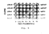
- Rh(D) antigen epitope specificity To investigate the diversity in fine specificity (Rh(D) antigen epitope specificity) among the anti-Rh(D) clones, agglutination experiments were performed with selected clones and with sets of rare Rh(D)-positive red blood cells which were obtained from individuals whose red blood cells produce Rh(D) antigen lacking certain epitopes. Examining the pattern of agglutination of a particular anti-Rh(D) antibody with such sets of mutant red blood cells enables the identification of the specific epitope on Rh(D) to which the antibody is directed (Mollison et al., 1993, supra ). A representative example of such an experiment is shown in Figure 5 and the Rh(D) epitopes for selected anti-Rh(D) Fab/phage clones are tabulated in Table 2.
- the gel test comprises a plastic card of approximately 5x7 cm, containing 6 minicolumns each filed with about 20 ⁇ l of dextran-acrylamide beads suspended in anti-human globulin (Coombs reagent). Red cells to be typed are incubated with the desired human anti-sera and are centrifuged through the gel. Red blood cells which are positive for antigens to which the antisera is directed agglutinate as they encounter the anti-human globulin and become trapped in or above the gel matrix. Unreactive red blood cells sediment through the gel particles and form a pellet at the bottom of the microtube.
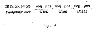
- Rh(D)-negative or -positive red blood cells were incubated with dilutions of anti-Rh(D) Fab/phage ( ⁇ 1 ⁇ library, panning #2) and were centrifuged into microcolumns containing beads suspended in anti-M 13 antibody.
- Undiluted Fab/phage stock had a titer of 5 x 10 12 cfu/ml similar to that in the microplate settling assay ( Figure 4). Because the volume of Fab/phage used in this assay is one-fourth of that in the microplate assay, the amount of Fab/phage present in the 1/625 dilution is approximately equal to that present in the 1/2048 dilution in Figure 4. Therefore, the number of Fab/phage required to yield a positive result is essentially equivalent in both assays.
- the columns of the Micro Typing System cards had added to them 100 ⁇ g/ml of anti-M13 antibody.
- Rh-negative or Rh-positive red blood cells were incubated with undiluted or with five-fold serial dilutions (1/5, 1/25, 1/125, 1/625 and 1/3125) of anti-Rh phage antibodies.
- the cards were centrifuged and samples were assessed for agglutination.
- the modified Micro Typing System card assay was capable of detecting anti-Rh agglutination at a dilution of between 1/625 and 1/3125.
- Fab/phage specific for tumor cells are useful for in vitro diagnosis (lab assays of biopsy, fluid, or blood samples), in vivo labeling of tumor/metastasis (coupling of antibody to imaging probe), or for treatment of malignancy (coupling of antibodies to chemical or radioactive toxins).
- Tumor-specific antibodies are also useful for the identification of novel antigens or markers on tumor cells which may form the basis for anti-tumor vaccines. Further, tumor-specific antibodies useful for the generation of anti-idiotypic antibodies may also form the basis for anti-tumor vaccines.
- Anti-tumor antibodies are generated essentially as described herein for the generation of anti-Rh antibodies.
- Tumor cells for example, but not limited to, malignant melanoma cells, are cell-surface biotinylated, labeled with streptavidin-magnetic microbeads, and are then mixed with excess normal melanocytes.
- Fab/phage libraries are generated from peripheral blood lymphocytes of melanoma patients who possess therapeutically useful anti-tumor antibodies. A number of melanoma patients who have "cured” themselves apparently have done so by mounting a humoral ( i.e ., antibody) immune response. These Fab/phage libraries are incubated with the admixture of cells. Fab/phage which are directed against epitopes specific for malignant cells will bind to the malignant cells and may then be isolated utilizing the magnetic column panning approach.
- the approach described herein may be used to isolate Fab/phage capable of detecting differences between the virulent bacteria and their nonpathogenic counterparts.
- the virulent strain of bacteria is magnetically labeled, diluted with the non-pathogenic counterpart, and an Fab/phage library which is generated from lymphocytes obtained from individuals infected with the virulent strain is added.
- Fab/phage which are isolated in this manner may be useful for the identification of novel bacterial antigens against which antibacterial compounds and/or vaccines may be developed.
Landscapes
- Chemical & Material Sciences (AREA)
- Health & Medical Sciences (AREA)
- Organic Chemistry (AREA)
- Life Sciences & Earth Sciences (AREA)
- Immunology (AREA)
- Medicinal Chemistry (AREA)
- Biophysics (AREA)
- General Health & Medical Sciences (AREA)
- Genetics & Genomics (AREA)
- Biochemistry (AREA)
- Molecular Biology (AREA)
- Proteomics, Peptides & Aminoacids (AREA)
- Hematology (AREA)
- Virology (AREA)
- Micro-Organisms Or Cultivation Processes Thereof (AREA)
- Measuring Or Testing Involving Enzymes Or Micro-Organisms (AREA)
- Peptides Or Proteins (AREA)
Claims (20)
- Ein Verfahren zum Agglutinieren von Zellen umfassend
Bereitstellen einer Mischung umfassend eine Population von Zellen und eine Population von einem Bakteriophagen, wobei ein erster Antikörper auf der Oberfläche von besagtem Bakteriophagen exprimiert wird und besagter erster Antikörper spezifisch für eine Antigen- tragende Gruppe ist, welche von zumindest einem Teil der Zellen in besagter Zellpopulation exprimiert wird,
wobei besagter erster Antikörper an besagten Teil von besagten Zellen bindet und verursacht, dass besagter Bakteriophage auch an besagten Teil von besagten Zellen bindet,
Hinzugeben eines zweiten Antikörpers spezifisch für besagten Bakteriophagen zu besagter Mischung,
wobei das Binden von besagtem zweiten Antikörper an den Bakteriophagen gebunden an besagten Teil von besagten Zellen verursacht, dass besagter Teil von besagten Zellen agglutiniert. - Das Verfahren von Anspruch 1, wobei besagte Zellen ausgewählt sind aus der Gruppe bestehend aus roten Blutkörperchen und weißen Blutkörperchen.
- Das Verfahren von Anspruch 2, wobei besagte Zellen rote Blutkörperchen sind.
- Das Verfahren von Anspruch 1, wobei besagter Bakteriophage M13 ist.
- Das Verfahren von Anspruch 4, wobei besagter Antikörper ein Anti- M13-Antikörper ist.
- Das Verfahren von Anspruch 3, wobei besagter erster Antikörper ein Antikörper gegen rote Blutkörperchen ist.
- Das Verfahren von Anspruch 6, wobei besagter erster Antikörper ein Anti- Rh-Körper ist.
- Das Verfahren von Anspruch 1, wobei besagte Antigen- tragende Gruppe ein Antigen eines roten Blutkörperchens ist.
- Das Verfahren von Anspruch 1, wobei besagte Antigen- tragende Gruppe ein HLA- Antigen ist.
- Das Verfahren von irgendeinem der Ansprüche 1 bis 9, wobei besagte Mischung zu einer Mikrotube hinzugefügt wird, welche inerte Partikel enthält und einen zweiten Antikörper spezifisch für besagten Bakteriophagen, und es ermöglicht wird, dass besagte Mischung unter Einwirkung von Schwerkraft sedimentiert und die Lokalisierung besagten Teils von besagten Zellen beobachtet wird, wobei starke Agglutinierung von besagtem Teil von besagten Zellen dadurch angezeigt wird, dass die Zellen auf oder innerhalb der obersten Schicht von besagten inerten Partikeln lokalisiert sind, und schwache Agglutinierung von besagten Zellen dadurch angezeigt wird, dass die Zellen innerhalb einer unteren Schicht von besagten inerten Partikeln lokalisiert sind und keine Agglutinierung dadurch angezeigt wird, dass die Zellen am Boden besagter Mikrotube lokalisiert sind.
- Das Verfahren von Anspruch 10, wobei der Schritt des Sedimentierens durch Zentrifugation erreicht wird.
- Ein Verfahren zum Einfangen von Zellen umfassend
Bereitstellen einer Mischung umfassend eine Population von Zellen und eine Population von einem Bakteriophagen, wobei ein erster Antikörper auf der Oberfläche von besagtem Bakteriophagen exprimiert wird und besagter erster Antikörper spezifisch für ein Antigen ist, welches durch zumindest einen Teil der Zellen in besagter Zellpopulation exprimiert wird, wobei besagter erster Antikörper an besagten Teil von besagten Zellen bindet und verursacht, dass besagter Bakteriophage auch an besagten Teil von besagten Zellen bindet,
Hinzugeben besagter Mischung zu einer Mikrotube enthaltend inerte Partikel, welche gebunden daran einen zweiten Antikörper aufweisen, welcher spezifisch für besagten Bakteriophagen ist,
Ermöglichen des Sedimentierens der Mischung unter Einfluss von Schwerkraft, wobei eingefangene Zellen oben oder innerhalb einer obersten Schicht von besagten inerten Partikeln lokalisiert sind. - Das Verfahren von Anspruch 12, wobei besagter Sedimentationsschritt durch Zentrifugation durchgeführt wird.
- Das Verfahren von Anspruch 12 oder 13, wobei nach Sedimentation die Lokalisation von besagtem Teil von besagten Zellen beobachtet wird und das Einfangen von besagtem Teil von besagten Zellen angezeigt wird dadurch, dass die Zellen oben oder innerhalb einer obersten Schicht von besagten inerten Partikeln lokalisiert sind und die Abwesenheit des Einfangens von besagten Zellen angezeigt wird, dadurch, dass die Zellen am Boden von besagter Mikrotube lokalisiert sind.
- Ein Verfahren zum Nachweis des Vorliegens einer Antigen- tragenden Gruppe auf einer Zelle umfassend
Bereitstellen einer Mischung umfassend eine Population von Zellen und eine Population eines Bakteriophagens, wobei ein erster bekannter Antikörper auf der Oberfläche von besagtem Bakteriophagen exprimiert wird, wobei das Vorliegen von besagter Antigen- tragender Gruppe auf besagten Zellen angezeigt wird durch Binden des ersten Antikörpers an zumindest zwei von besagten Zellen, was verursacht, dass besagter Bakteriophage auch an besagte zwei von besagten Zellen bindet, wobei, wenn ein zweiter Antikörper zu besagter Mischung hinzugefügt wird, welcher spezifisch für besagten Bakteriophagen ist, besagter zweiter Antikörper an den Bakteriophagen bindet, welcher an besagte zumindest zwei von besagten Zellen bindet, was verursacht, dass die Zellen agglutinieren, wobei besagte Agglutination ein Zeichen des Vorliegens von besagter Antigentragender Gruppe auf besagten Zellen ist, wobei die Antigen- tragende Gruppe spezifisch für besagten ersten Antikörper ist. - Das Verfahren von Anspruch 15, wobei besagte Agglutinierung besagte Antigentragende Gruppe als eine Antigen- tragende Gruppe spezifisch für besagten ersten Antikörper identifiziert.
- Das Verfahren von Anspruch 15, wobei besagte Mischung zu einer Mikrotube hinzugegeben wird, welche inerte Partikel enthält und einen zweiten Antikörper spezifisch für besagten Bakteriophagen, wobei man besagte Mischung unter dem Einfluss von Schwerkraft sedimentieren lässt, und die Lokalisierung der Zellen in besagter Mikrotube beobachtet wird, wobei starke Agglutinierung der Zellen angezeigt wird, dadurch, dass die Zellen oben oder innerhalb einer obersten Schicht von besagten inerten Partikeln lokalisiert sind, und die starke Agglutinierung ein Anzeigen der Gegenwart von besagter Antigen- tragender Gruppe auf besagten Zellen ist, und besagte Antigen- tragende Gruppe spezifisch für besagten ersten Antikörper ist.
- Das Verfahren von Anspruch 17, wobei besagte starke Agglutination besagte Antigen- tragende Gruppe als eine Antigen- tragende Gruppe spezifisch für besagten ersten Antikörper identifiziert.
- Das Verfahren von Anspruch 15, wobei besagte Mischung zu einer Mikrotube hinzugefügt wird, welche inerte Partikel enthält, welche daran gebunden einen zweiten Antikörper aufweisen, welcher spezifisch für besagten Bakteriophagen ist, wobei das Sedimentieren besagter Mischung ermöglich wird unter dem Einfluss von Schwerkraft, wobei die eingefangenen Zellen auf oder innerhalb der obersten Schicht von besagten inerten Partikeln positioniert sind, und die Gegenwart von besagten eingefangenen Zellen ein Hinweis auf die Gegenwart einer Antigen- tragenden Gruppe auf besagter Zelle ist, wobei die Antigentragende Gruppe spezifisch für besagten ersten Antikörper ist.
- Das Verfahren von Anspruch 19, wobei das Vorliegen von besagten eingefangenen Zellen besagte Antigen- tragende Gruppe auf besagten Zellen als spezifisch für besagten ersten Antikörper identifiziert.
Applications Claiming Priority (5)
| Application Number | Priority Date | Filing Date | Title |
|---|---|---|---|
| US2855096P | 1996-10-11 | 1996-10-11 | |
| US28550P | 1996-10-11 | ||
| US884046 | 1997-06-27 | ||
| US08/884,046 US5985543A (en) | 1996-10-11 | 1997-06-27 | Compositions and methods for detection of antibody binding to cells |
| PCT/US1997/017995 WO1998016827A1 (en) | 1996-10-11 | 1997-10-10 | Compositions and methods for detection of antibody binding to cells |
Publications (3)
| Publication Number | Publication Date |
|---|---|
| EP0935754A1 EP0935754A1 (de) | 1999-08-18 |
| EP0935754A4 EP0935754A4 (de) | 2003-04-23 |
| EP0935754B1 true EP0935754B1 (de) | 2005-07-27 |
Family
ID=26703824
Family Applications (1)
| Application Number | Title | Priority Date | Filing Date |
|---|---|---|---|
| EP97909978A Expired - Lifetime EP0935754B1 (de) | 1996-10-11 | 1997-10-10 | Zusammensetzungen und verfahren zum nachweis der bindung von antikörpern in zellen |
Country Status (9)
| Country | Link |
|---|---|
| US (2) | US5985543A (de) |
| EP (1) | EP0935754B1 (de) |
| JP (1) | JP3901222B2 (de) |
| AT (1) | ATE300738T1 (de) |
| AU (1) | AU732758B2 (de) |
| CA (1) | CA2268339C (de) |
| DE (1) | DE69733828T2 (de) |
| ES (1) | ES2246510T3 (de) |
| WO (1) | WO1998016827A1 (de) |
Families Citing this family (16)
| Publication number | Priority date | Publication date | Assignee | Title |
|---|---|---|---|---|
| US6255455B1 (en) * | 1996-10-11 | 2001-07-03 | The Trustees Of The University Of Pennsylvania | Rh(D)-binding proteins and magnetically activated cell sorting method for production thereof |
| IL144559A0 (en) | 1999-03-01 | 2002-05-23 | Genentech Inc | Antibodies for cancer therapy and diagnosis |
| US7811768B2 (en) * | 2001-01-26 | 2010-10-12 | Aviva Biosciences Corporation | Microdevice containing photorecognizable coding patterns and methods of using and producing the same |
| US7015047B2 (en) * | 2001-01-26 | 2006-03-21 | Aviva Biosciences Corporation | Microdevices having a preferential axis of magnetization and uses thereof |
| DE10122806A1 (de) * | 2001-05-10 | 2002-11-14 | Holger Kiesewetter | Verfahren zum Nachweis von Blutzell-Antigenen und gegen diese gerichtete Antikörper |
| US20050153298A1 (en) * | 2001-10-23 | 2005-07-14 | Gembitsky Dmitry S. | Protein micro-arrays and multi-layered affinity interaction detection |
| WO2004027028A2 (en) * | 2002-09-18 | 2004-04-01 | The Trustees Of The University Of Pennsylvania | Compositions, methods and kits for detection of an antigen on a cell and in a biological mixture |
| DE102004033811A1 (de) * | 2004-07-12 | 2006-02-02 | Salama, Abdulgabar, Prof. Dr. | Verfahren zum einfachen und schnellen Nachweis von Zellen und Biomolekülen mit Hilfe paramagnetischer Partikel |
| JP4729043B2 (ja) * | 2004-10-08 | 2011-07-20 | アフィテック エーエス | 抗体ライブラリーをスクリーニングする方法 |
| WO2007064462A1 (en) | 2005-11-29 | 2007-06-07 | The Trustees Of The University Of Pennsylvania | Phage particle diagnostic reagents |
| JPWO2007083793A1 (ja) * | 2006-01-23 | 2009-06-11 | 国立大学法人富山大学 | 光反応基を利用したパニング法およびそれに用いるキット |
| US7998696B2 (en) * | 2007-12-05 | 2011-08-16 | Zyomyx, Inc. | Cell assay kit and method |
| US7850917B2 (en) * | 2008-03-11 | 2010-12-14 | Ortho-Clinical Diagnostics, Inc. | Particle agglutination in a tip |
| US20100015690A1 (en) | 2008-07-16 | 2010-01-21 | Ortho-Clinical Diagnostics, Inc. | Use of fluid aspiration/dispensing tip as a microcentrifuge tube |
| CA2819260A1 (en) | 2010-12-15 | 2012-06-21 | Cytosed, Inc. | Antibody-linked immuno-sedimentation agent and method of isolating a target from a sample using same |
| KR101698040B1 (ko) * | 2013-06-27 | 2017-01-24 | 주식회사 중겸 | M13 박테리오파지에 대한 단일 도메인 항체 및 이를 포함하는 면역센서 융합 단백질 |
Family Cites Families (7)
| Publication number | Priority date | Publication date | Assignee | Title |
|---|---|---|---|---|
| WO1988006628A1 (en) * | 1987-03-02 | 1988-09-07 | Bristol-Myers Company | Platelet related growth regulator |
| US5338689A (en) * | 1987-08-24 | 1994-08-16 | Stiftung Fur Diagnostische Forschung | Method and card for detecting antigens and/or antibodies |
| US5663143A (en) * | 1988-09-02 | 1997-09-02 | Dyax Corp. | Engineered human-derived kunitz domains that inhibit human neutrophil elastase |
| US5498538A (en) * | 1990-02-15 | 1996-03-12 | The University Of North Carolina At Chapel Hill | Totally synthetic affinity reagents |
| ATE414768T1 (de) * | 1991-04-10 | 2008-12-15 | Scripps Research Inst | Bibliotheken heterodimerer rezeptoren mittels phagemiden |
| US5491067A (en) * | 1993-07-15 | 1996-02-13 | Ortho Diagnostic Systems Inc. | Agglutination reaction and separation vessel |
| US5665558A (en) * | 1994-05-17 | 1997-09-09 | Gamma Biologicals, Inc. | Method and apparatus useful for detecting bloodgroup antigens and antibodies |
-
1997
- 1997-06-27 US US08/884,046 patent/US5985543A/en not_active Expired - Fee Related
- 1997-10-10 DE DE69733828T patent/DE69733828T2/de not_active Expired - Lifetime
- 1997-10-10 WO PCT/US1997/017995 patent/WO1998016827A1/en not_active Ceased
- 1997-10-10 JP JP51841198A patent/JP3901222B2/ja not_active Expired - Lifetime
- 1997-10-10 ES ES97909978T patent/ES2246510T3/es not_active Expired - Lifetime
- 1997-10-10 CA CA2268339A patent/CA2268339C/en not_active Expired - Lifetime
- 1997-10-10 EP EP97909978A patent/EP0935754B1/de not_active Expired - Lifetime
- 1997-10-10 AT AT97909978T patent/ATE300738T1/de not_active IP Right Cessation
- 1997-10-10 AU AU47461/97A patent/AU732758B2/en not_active Expired
-
1999
- 1999-09-29 US US09/407,182 patent/US6979534B1/en not_active Expired - Fee Related
Also Published As
| Publication number | Publication date |
|---|---|
| EP0935754A1 (de) | 1999-08-18 |
| JP2001508865A (ja) | 2001-07-03 |
| JP3901222B2 (ja) | 2007-04-04 |
| DE69733828T2 (de) | 2006-01-05 |
| CA2268339A1 (en) | 1998-04-23 |
| WO1998016827A1 (en) | 1998-04-23 |
| ES2246510T3 (es) | 2006-02-16 |
| CA2268339C (en) | 2010-02-23 |
| AU732758B2 (en) | 2001-04-26 |
| DE69733828D1 (de) | 2005-09-01 |
| AU4746197A (en) | 1998-05-11 |
| US6979534B1 (en) | 2005-12-27 |
| EP0935754A4 (de) | 2003-04-23 |
| US5985543A (en) | 1999-11-16 |
| ATE300738T1 (de) | 2005-08-15 |
Similar Documents
| Publication | Publication Date | Title |
|---|---|---|
| US8124742B2 (en) | Rh(D)-binding proteins | |
| EP0935754B1 (de) | Zusammensetzungen und verfahren zum nachweis der bindung von antikörpern in zellen | |
| Siegel et al. | Isolation of cell surface-specific human monoclonal antibodies using phage display and magnetically-activated cell sorting: applications in immunohematology | |
| US5213960A (en) | Methods for selecting low frequency antigen-specific single B lymphocytes | |
| WO1999006834A2 (en) | Methods for identifying ligand specific binding molecules | |
| Azriel-Rosenfeld et al. | A human synthetic combinatorial library of arrayable single-chain antibodies based on shuffling in vivo formed CDRs into general framework regions | |
| US5876925A (en) | Magnetically activated cell sorting for production of proteins | |
| WO1998016541A9 (en) | Magnetically activated cell sorting for production of proteins | |
| Malone et al. | Analysis of antibody selection by phage display utilizing anti‐phenobarbital antibodies | |
| EP1963494B1 (de) | Diagnostische phagenpartikelreagentien | |
| Barbas et al. | Filamentous phage display | |
| Siegel | Research and clinical applications of antibody phage display in transfusion medicine | |
| US20260118366A1 (en) | Method for selecting or screening antibodies and antigens from an antibody library | |
| Siegel | Diagnostic and therapeutic applications of phage display technology | |
| Stephenson et al. | Cell-based panning as a means to isolate phage display Fabs specific for a bacterial surface protein | |
| HK1118863B (en) | Phage particle diagnostic reagents |
Legal Events
| Date | Code | Title | Description |
|---|---|---|---|
| PUAI | Public reference made under article 153(3) epc to a published international application that has entered the european phase |
Free format text: ORIGINAL CODE: 0009012 |
|
| 17P | Request for examination filed |
Effective date: 19990421 |
|
| AK | Designated contracting states |
Kind code of ref document: A1 Designated state(s): AT BE CH DE DK ES FI FR GB GR IE IT LI LU MC NL PT SE |
|
| A4 | Supplementary search report drawn up and despatched |
Effective date: 20030310 |
|
| 17Q | First examination report despatched |
Effective date: 20030723 |
|
| GRAP | Despatch of communication of intention to grant a patent |
Free format text: ORIGINAL CODE: EPIDOSNIGR1 |
|
| GRAS | Grant fee paid |
Free format text: ORIGINAL CODE: EPIDOSNIGR3 |
|
| GRAA | (expected) grant |
Free format text: ORIGINAL CODE: 0009210 |
|
| AK | Designated contracting states |
Kind code of ref document: B1 Designated state(s): AT BE CH DE DK ES FI FR GB GR IE IT LI LU MC NL PT SE |
|
| PG25 | Lapsed in a contracting state [announced via postgrant information from national office to epo] |
Ref country code: LI Free format text: LAPSE BECAUSE OF FAILURE TO SUBMIT A TRANSLATION OF THE DESCRIPTION OR TO PAY THE FEE WITHIN THE PRESCRIBED TIME-LIMIT Effective date: 20050727 Ref country code: FI Free format text: LAPSE BECAUSE OF FAILURE TO SUBMIT A TRANSLATION OF THE DESCRIPTION OR TO PAY THE FEE WITHIN THE PRESCRIBED TIME-LIMIT Effective date: 20050727 Ref country code: CH Free format text: LAPSE BECAUSE OF FAILURE TO SUBMIT A TRANSLATION OF THE DESCRIPTION OR TO PAY THE FEE WITHIN THE PRESCRIBED TIME-LIMIT Effective date: 20050727 Ref country code: AT Free format text: LAPSE BECAUSE OF FAILURE TO SUBMIT A TRANSLATION OF THE DESCRIPTION OR TO PAY THE FEE WITHIN THE PRESCRIBED TIME-LIMIT Effective date: 20050727 |
|
| REG | Reference to a national code |
Ref country code: GB Ref legal event code: FG4D |
|
| REG | Reference to a national code |
Ref country code: CH Ref legal event code: EP |
|
| REG | Reference to a national code |
Ref country code: IE Ref legal event code: FG4D |
|
| REF | Corresponds to: |
Ref document number: 69733828 Country of ref document: DE Date of ref document: 20050901 Kind code of ref document: P |
|
| REG | Reference to a national code |
Ref country code: SE Ref legal event code: TRGR |
|
| PG25 | Lapsed in a contracting state [announced via postgrant information from national office to epo] |
Ref country code: GR Free format text: LAPSE BECAUSE OF FAILURE TO SUBMIT A TRANSLATION OF THE DESCRIPTION OR TO PAY THE FEE WITHIN THE PRESCRIBED TIME-LIMIT Effective date: 20051027 Ref country code: DK Free format text: LAPSE BECAUSE OF FAILURE TO SUBMIT A TRANSLATION OF THE DESCRIPTION OR TO PAY THE FEE WITHIN THE PRESCRIBED TIME-LIMIT Effective date: 20051027 |
|
| PG25 | Lapsed in a contracting state [announced via postgrant information from national office to epo] |
Ref country code: PT Free format text: LAPSE BECAUSE OF FAILURE TO SUBMIT A TRANSLATION OF THE DESCRIPTION OR TO PAY THE FEE WITHIN THE PRESCRIBED TIME-LIMIT Effective date: 20051227 |
|
| REG | Reference to a national code |
Ref country code: CH Ref legal event code: PL |
|
| REG | Reference to a national code |
Ref country code: ES Ref legal event code: FG2A Ref document number: 2246510 Country of ref document: ES Kind code of ref document: T3 |
|
| ET | Fr: translation filed | ||
| PLBE | No opposition filed within time limit |
Free format text: ORIGINAL CODE: 0009261 |
|
| STAA | Information on the status of an ep patent application or granted ep patent |
Free format text: STATUS: NO OPPOSITION FILED WITHIN TIME LIMIT |
|
| 26N | No opposition filed |
Effective date: 20060428 |
|
| REG | Reference to a national code |
Ref country code: FR Ref legal event code: PLFP Year of fee payment: 19 |
|
| REG | Reference to a national code |
Ref country code: FR Ref legal event code: PLFP Year of fee payment: 20 |
|
| PGFP | Annual fee paid to national office [announced via postgrant information from national office to epo] |
Ref country code: LU Payment date: 20161018 Year of fee payment: 20 |
|
| PGFP | Annual fee paid to national office [announced via postgrant information from national office to epo] |
Ref country code: DE Payment date: 20161004 Year of fee payment: 20 Ref country code: MC Payment date: 20161012 Year of fee payment: 20 Ref country code: GB Payment date: 20161005 Year of fee payment: 20 Ref country code: NL Payment date: 20161010 Year of fee payment: 20 Ref country code: IE Payment date: 20161010 Year of fee payment: 20 Ref country code: FR Payment date: 20161014 Year of fee payment: 20 |
|
| PGFP | Annual fee paid to national office [announced via postgrant information from national office to epo] |
Ref country code: BE Payment date: 20161013 Year of fee payment: 20 Ref country code: ES Payment date: 20161011 Year of fee payment: 20 Ref country code: IT Payment date: 20161024 Year of fee payment: 20 Ref country code: SE Payment date: 20161011 Year of fee payment: 20 |
|
| REG | Reference to a national code |
Ref country code: DE Ref legal event code: R071 Ref document number: 69733828 Country of ref document: DE |
|
| REG | Reference to a national code |
Ref country code: NL Ref legal event code: MK Effective date: 20171009 |
|
| REG | Reference to a national code |
Ref country code: GB Ref legal event code: PE20 Expiry date: 20171009 |
|
| REG | Reference to a national code |
Ref country code: IE Ref legal event code: MK9A |
|
| REG | Reference to a national code |
Ref country code: BE Ref legal event code: MK Effective date: 20171010 |
|
| PG25 | Lapsed in a contracting state [announced via postgrant information from national office to epo] |
Ref country code: GB Free format text: LAPSE BECAUSE OF EXPIRATION OF PROTECTION Effective date: 20171009 Ref country code: IE Free format text: LAPSE BECAUSE OF EXPIRATION OF PROTECTION Effective date: 20171010 |
|
| REG | Reference to a national code |
Ref country code: ES Ref legal event code: FD2A Effective date: 20180508 |
|
| PG25 | Lapsed in a contracting state [announced via postgrant information from national office to epo] |
Ref country code: ES Free format text: LAPSE BECAUSE OF EXPIRATION OF PROTECTION Effective date: 20171011 |