EP0931094B1 - Methods for determining the presence of brain protein s-100 beta - Google Patents
Methods for determining the presence of brain protein s-100 beta Download PDFInfo
- Publication number
- EP0931094B1 EP0931094B1 EP97932092A EP97932092A EP0931094B1 EP 0931094 B1 EP0931094 B1 EP 0931094B1 EP 97932092 A EP97932092 A EP 97932092A EP 97932092 A EP97932092 A EP 97932092A EP 0931094 B1 EP0931094 B1 EP 0931094B1
- Authority
- EP
- European Patent Office
- Prior art keywords
- antibody
- monoclonal antibody
- sample
- seq
- amino acid
- Prior art date
- Legal status (The legal status is an assumption and is not a legal conclusion. Google has not performed a legal analysis and makes no representation as to the accuracy of the status listed.)
- Expired - Lifetime
Links
Classifications
-
- G—PHYSICS
- G01—MEASURING; TESTING
- G01N—INVESTIGATING OR ANALYSING MATERIALS BY DETERMINING THEIR CHEMICAL OR PHYSICAL PROPERTIES
- G01N33/00—Investigating or analysing materials by specific methods not covered by groups G01N1/00 - G01N31/00
- G01N33/48—Biological material, e.g. blood, urine; Haemocytometers
- G01N33/50—Chemical analysis of biological material, e.g. blood, urine; Testing involving biospecific ligand binding methods; Immunological testing
- G01N33/68—Chemical analysis of biological material, e.g. blood, urine; Testing involving biospecific ligand binding methods; Immunological testing involving proteins, peptides or amino acids
- G01N33/6893—Chemical analysis of biological material, e.g. blood, urine; Testing involving biospecific ligand binding methods; Immunological testing involving proteins, peptides or amino acids related to diseases not provided for elsewhere
- G01N33/6896—Neurological disorders, e.g. Alzheimer's disease
-
- C—CHEMISTRY; METALLURGY
- C07—ORGANIC CHEMISTRY
- C07K—PEPTIDES
- C07K14/00—Peptides having more than 20 amino acids; Gastrins; Somatostatins; Melanotropins; Derivatives thereof
- C07K14/435—Peptides having more than 20 amino acids; Gastrins; Somatostatins; Melanotropins; Derivatives thereof from animals; from humans
- C07K14/46—Peptides having more than 20 amino acids; Gastrins; Somatostatins; Melanotropins; Derivatives thereof from animals; from humans from vertebrates
- C07K14/47—Peptides having more than 20 amino acids; Gastrins; Somatostatins; Melanotropins; Derivatives thereof from animals; from humans from vertebrates from mammals
-
- C—CHEMISTRY; METALLURGY
- C07—ORGANIC CHEMISTRY
- C07K—PEPTIDES
- C07K16/00—Immunoglobulins [IG], e.g. monoclonal or polyclonal antibodies
- C07K16/18—Immunoglobulins [IG], e.g. monoclonal or polyclonal antibodies against material from animals or humans
-
- G—PHYSICS
- G01—MEASURING; TESTING
- G01N—INVESTIGATING OR ANALYSING MATERIALS BY DETERMINING THEIR CHEMICAL OR PHYSICAL PROPERTIES
- G01N33/00—Investigating or analysing materials by specific methods not covered by groups G01N1/00 - G01N31/00
- G01N33/48—Biological material, e.g. blood, urine; Haemocytometers
- G01N33/50—Chemical analysis of biological material, e.g. blood, urine; Testing involving biospecific ligand binding methods; Immunological testing
- G01N33/53—Immunoassay; Biospecific binding assay; Materials therefor
- G01N33/575—Immunoassay; Biospecific binding assay; Materials therefor for cancer
- G01N33/5751—Immunoassay; Biospecific binding assay; Materials therefor for cancer of the skin, e.g. melanoma
Definitions
- the present invention relates methods for diagnosis and follow-up of patients with cerebral dysfunction as well as melanoma cancer, by determining the presence of the brain protein S-100.
- the invention also relates to monoclonal antibodies binding to peptides comprising useful antigenic determinants from S-100.
- the nervous system contains a number of proteins unique to its various cellular elements.
- CSF cerebrospinal fluid
- blood serum and plasma
- Examples of representative soluble small molecule weight proteins of this type can be found in the S100 protein family. A review of this family can be found in Zimmer et al., Brain Research Bulletin, Vol. 37, pp 417-429. 1995.
- these proteins are released into the extracellular fluid in accordance with a time course and in quantities relative to the pathogenesis of the disease process or the extent of the brain tissue damages.
- the proteins diffuse into the CSF and then the blood or directly into the blood.
- the above mentioned cell membrane disruption is reflected by the blood plasmaor serum levels of one or more of these antigens and markers.
- These protein antigens have the advantage of being stable and specific, not only for the brain, but for the cellular components in the brain. By following the relative release of the various nervous system protein antigens, it is possible to deduce the kind of destructive process occuring in the course of neurological diseases and/or the extent of possible brain tissue damages. Information of this type permits the diagnosis, evaluation of severity and rate of progression of the above mentioned dieases and damages.
- US-A-4 654 313 discloses a radioimmunological assay method for S-100 protein.
- the patent document does neither mention anything about different kinds of S100-polypeptides nor about on which epitopes the assay method is based.
- the detection limit is declared to be 0.20 ng/ml but concentrations between 1.5 and 2.5 ng/ml is required in order to have less than 10% false positives. This concentration is rather high.
- GB-A-2 109 931 discloses a solid-phase immunoanalysis method comprising the use of enzyme-labelled antigens and particles coated with protein A on which antibodies are bound. S-100 proteins are only mentioned in claim 8 and nothing is revealed about the sensitivity of the method.
- JP-A-6/109 734 describes a method suitable for analysing S-100 polypeptides, using a first polyclonal antibody fixed to magnetic particles, and a second labelled polyclonal antibody.
- the method requires two different enzymes, namely horseradish peroxidase and alkaline phosphatase, and it comprises at least ten consecutive steps.
- the minimum detection limit is stated to be 0.02 ng/ml for cerebrospinal fluid and 0.06 ng/ml for bovine brain.
- An ideal clinical assay method should be quick, accurate and possible to perform with all types of clinical samples without degeneration of the accuracy for certain types of specimen. It should also require a minimum of extra components. This applies to determination of S-100 polypeptides as well as other substances of medical interest.
- the main object of the present invention is an assay method using monoclonal antibodies directed to these epitopes.
- Yet another object of the present invention relates to analytical kits for carrying out the assay methods.
- the present invention relates to an immunological assay method for determining the presence and/or content of human S-100 polypeptide based upon a selection of suitable S-100 epitopes and corresponding antibodies which fulfil the above mentioned requirements.
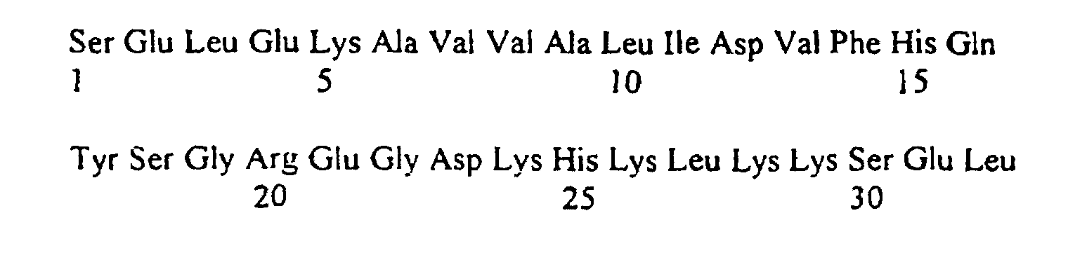
- epitopes of the present invention are all comprised in the human S-100 ⁇ polypepnde. Epitopes present within the amino acid sequences: and are preferred. Particularly preferred are epitopes comprised within the peptide and especially within the peptides and
- the disclosed epitopes are, among all, used to construct peptides for inducing the formation of suitable antibodies on which the claimed assay method is based. These peptides mostly consist of up to 38 amino acids.
- the whole amino acid sequence of a peptide useful in the present invention is derived from human S-100 ⁇ . These peptides may comprise variants wherein the original amino acid sequence is modified or altered by insertion, addition, substitution, inversion or deletion which preferably show at least 90% homology with the sequence of SEQ. ID. NO. 2 and SEQ.ID.NO.3 and retain essentially the same immunological properties.
- the peptides may also comprise multiples of certain epitopes, and in this case their sequence length may exceed 38 amino acids.
- sub-fragment is meant a polypeptide sequence having a length of at least 6 amino acids.
- the epitopes can also be used to construct fusion peptides comprising at least two distinct epitopes which, among all, can be used as internal standard in immunoassays.
- the peptides were prepared by the methods disclosed in Merrifield (1963), J. Am. Chem. Soc.. vol. 85, p 2149: Gutte et al.(1971), J. Biol Chem vol. 246, p. 1922: and Carpmo et al. (1970), J Am Chem Soc vol. 92, p. 5748.
- the monoclonal antibodies were prepared by the method according to Köhler et al.(1975), Nature vol. 256, p. 495: and Harlow et al.(1988). Antibodies. A Laboratory Manual. Cold Spring Harbor, p. 139.
- the fraction still soluble after saturation by ammonium sulphate was dialysed and purified by separation on a Sephadex G 150 Sepharose (Pharmacia Biotech AB, Uppsala Sweden)chromatographic column followed by separation on a DEAE-sephadex (ionic exchange) column (Pharmacia Biotech AB, Uppsala Sweden).
- the fraction eluted by 0.3 - 0 4 M NaCl was collected, desalted, lyophilised and used for further experiments.
- Balb/c mice were immunised with purified S100 ⁇ intraperitonially in Freund's complete adjuvant and were given booster iv. injection 6 weeks later during 3 consecutive days. The spleen was removed on the fourth day after last injection and prepared for fusion. The myeloma cell line Sp2/0-Ag14 was used for fusion of Balb/c spleen cells.
- Monoclonal antibodies were identified, extracted and purified from hybridoma supernatant according to Harlow & Lane Eds. in ANTIBODIES A LABORATORY MANUAL. Cold Spring Harbour Laboratory Press. New York 298-299 & 311. Briefly, positive hybridoma clones carrying supernatant with specific antibodies were identified using ELISA with microtitreplate wells coated with S100 ⁇ . Immunoglobulins were precipitated using saturated ammoniumsulphate and dialysed against 1.5 M Glycine, 3 M NaCl, pH 8.9. Dialysed material were affinity chromatography purified on an protein-A Sepharose (Pharmacia Biotech AB. Uppsala Sweden) column. Fractions were neutralised by addition of small volumes of 1M Tris pH 8 0.
- S100 ⁇ (monomer) epitopes for respective antibody was investigated by use of a synthetic peptide library.
- Peptides were linked to nitro-cellulose filter membrane via an amide link, according to the manufacturer (Research Genetics', USA) and covers all ninety-one aa in the protein.
- the library consisted of thirty-one, all except one being ten aa-residues long synthetic peptides. Each peptide was consecutively shifted three aa towards the -COOH terminal end of the protein. Positive antibody-binding was indicated by the use of a second antt-mouse antibody conjugated with HRP and detected using an ECL assay (Amersham, UK).
- S100 antigen was added to the chips at 200-450nM for reactivity measurements of the antibody intended for the solid phase and at 1000-1500nM for measurements of the antibody intended for tracers.
- Kinetics was determined using the BIAcoreTM Kinetic evaluation 2.1, software (Pharmacia Biosensor AB, Uppsala Sweden). It can be concluded from the reactivity profile that the antibodies reactive with the epitopes are specific for the ⁇ -contairung forms of S100 and not the ⁇ -containing form.
- ABEI Analog toyl
- EGS Ethylenglykolbis-succimidyl succinat
- the ABEI-EGS-conjugate was next mixed with monoclonal antiS100-antibody in an approximately 50:5 molar ratio in 100 ⁇ l of PBS pH 7.4, containing 15% acetonitrile and incubated 1 h at room temperature.
- the ABEI-conjugated antibody was purified on a Sephacryl®S 300 HR (Pharmacia Biotech AB, Uppsala Sweden) gelfiltration column, and appropriate fractions were pooled and diluted in phosphate-buffer.
- Polystyrene tubes (Greiner, Germany) were incubated overnight at room temperature with 3 ⁇ g of S100-antibodies in 300 ⁇ l of PBS pH 7.5 The tubes were washed with 0. 1 % Tween20® in PBS. Next, tubes were blocked with a solution containing 0.9%BSA and 4% Saccarose and incubated for 24h. The solution was aspirated and the tubes allowed to dry.
- the test was conducted in a two step procedure by incubating 100 ⁇ l of patient body fluid in antibody coated tubes, or S100 standard with 100 ⁇ l of diluent (PBS + 5%BSA) and incubated at room temperature. After washing 200 ⁇ l of the luminol-labelled antibody was added and a further 2 h of incubation was performed before measurement. After another washing the luminescence was developed using the LIA-mat starter service kit (Byk-Sangtec. Diezenbach Germany) and immediately measured as integrals over a period of 5 sec in luminometer (Berthold. Germany) In order to convert the obtained light signal into concentration of S100 measurements on patient samples were compared with measurements on solutions with known concentrations of S100 (standards). The detection limit (zero standard +3 standard deviations) was approximately 0.01 ⁇ g/l.
- S100B protein was obtained from Medisera, Lund, Sweden, and diluted in PBS + 5% BSA. Dilutions contained: 0.10,0.40, 2.00, 8.00 and 20.00 ⁇ g/l, of an S100 preparation consisting of 50 % of the ⁇ form and 50 % of the ⁇ form. PBS + 5 % BSA was used as standard 0. Three measurements were carried out for each dilution. The measured results as well as statistical calculations are presented in table 1 below: Concentration Counts Average Calculated conc.
- the lower detection limit was defined as three standard 0 determinations plus 3X the standard deviation value. For this measurement, it was calculated to be 0.006 ⁇ g/l.
- the S 100 concentration was determined in serum from patients receiving heart bypass surgery and being connected to a heart-lung machine. The results are presented in table 2 below: Patient Counts Average Concentration Average 1 94698 10.61 ⁇ g/l 98104 10.99 ⁇ g/l 96401 10.80 ⁇ g/l 2 1716 Not detected 1478 Not detected 1597 Not detected 3 3762 0.23 ⁇ g/l 3799 0.23 ⁇ g/l 3780 0.23 ⁇ g/l 4 13158 1.04 ⁇ g/l 14183 1.15 ⁇ g/l 13670 1.10 ⁇ g/l 5 8788 0.66 ⁇ g/l 8580 0.64 ⁇ g/l 8684 0.65 ⁇ g/l 6 10301 0.78 ⁇ g/l 10100 0.77 ⁇ g/l 10200 0.77 ⁇ g/l
- Microtiterplatewells (Coming, Denmark) were incubated overnight at +4°C with 2.5 ⁇ g of microtiter wells were finally washed three times with 0.05% Tween20® and air dried before use.
- the ELISA was conducted in a multiple step incubation procedure. 100 ⁇ l of 1:1 diluted patient sample or 100 ⁇ l of S100 standard (0 - 20 ⁇ g/ml) was added to the wells. The plate was incubated for 1.5h at RT under shaking.
- the plates were washed three times with 300 ⁇ l 0.05% Tween20® in PBS. 100 ⁇ l of alkaline phosphatase conjugated tracer antibody was added and a further 1.5h of incubation on a shaker was performed. The wells were then washed three times with 0.05% Tween20® in PBS. 100 ⁇ l of a 5% o-nitro-phenyl- ⁇ -galactoside substrate solution was added and the plates were incubated with substrate for another forty-five minutes and colour is developed.
- a monoclonal antiS100 antibody was conjugated with Iodine using the Chloramine T method according to Greenwood et al. (Biochem. J. 1963. 89: 114-123). The specific activity was determined to be 520 ⁇ 80 MBq/mg
- Monoclonal anti S100 antibodies were coupled to polystyrene beads by the Glutaraldehyde coupling method according to Harlow & Lane Eds. in ANTIBODIES, A LABORATORY MANUAL. Cold Spring Harbour Laboratory Press. New York, 533 & 536-537. Final blocking was by 1 % BSA, 0.1% NaN 3 in PBS pH7.5.
- the S100 based test procedure was applied on clinical questions relating to melanoma. Blood samples from patients with melanoma of various stages of cancer progression were collected in serum collecting tubes. Samples were then frozen and treated according to the test procedure described above in Example 3.
- the S100 based test procedure was applied on clinical questions relating to melanoma. Blood samples from panents with melanoma of various stages of cancer progression were collected in serum collecting tubes. Samples were then frozen and treated according to the test procedure described above in Example 3.
- Example 1 The S100 based test procedure in Example 1 was applied on monitoring cerebral injury following extra corporeal circulation (ECC). Blood samples from patients undergoing extra corporeal circulation were collected in serum tubes and treated according " Test procedure". Results Before start of ECC End of ECC 1 day after surgery 2 days after surgery S100 levels ⁇ g/l 0 1,67 0,21 0,13
- the S100 based test procedure was applied on clinical questions relating to melanoma. Blood samples from patients with melanoma of various stages of cancer progression and blood donors were collected in serum collecting tubes. Samples were frozen and treated according to the test procedure described above in Example 1.
Landscapes
- Health & Medical Sciences (AREA)
- Life Sciences & Earth Sciences (AREA)
- Chemical & Material Sciences (AREA)
- Engineering & Computer Science (AREA)
- Immunology (AREA)
- Molecular Biology (AREA)
- Biomedical Technology (AREA)
- General Health & Medical Sciences (AREA)
- Hematology (AREA)
- Urology & Nephrology (AREA)
- Medicinal Chemistry (AREA)
- Biochemistry (AREA)
- Organic Chemistry (AREA)
- Proteomics, Peptides & Aminoacids (AREA)
- Analytical Chemistry (AREA)
- Genetics & Genomics (AREA)
- Pathology (AREA)
- General Physics & Mathematics (AREA)
- Biophysics (AREA)
- Biotechnology (AREA)
- Cell Biology (AREA)
- Physics & Mathematics (AREA)
- Microbiology (AREA)
- Food Science & Technology (AREA)
- Zoology (AREA)
- Gastroenterology & Hepatology (AREA)
- Neurosurgery (AREA)
- Neurology (AREA)
- Toxicology (AREA)
- Peptides Or Proteins (AREA)
- Investigating Or Analysing Biological Materials (AREA)
- Preparation Of Compounds By Using Micro-Organisms (AREA)
Abstract
Description



- S100
- -S100β
- RT
- -Room Temperature
- BSA
- -Bovine Serum Albumin
- Mab(s)
- -Monoclonal anribody(ies)
- kD
- -kiloDalton
- ECL
- -Enhanced Chemiluminescent Assay
- CBB
- -Commassie Brilliant Blue
- LIA
- -Luminometric Immuno Assay
- IRMA
- -Immuno Radio Metric Assay
- ELISA
- -Enzyme Linked ImmunoSorbent Assay
- SDS-PAGE
- -SodiumDodecylSulfate - PolyAcrylamideGelElectrophoresis
- PBS
- -Phosphate Buffered Saline
- RLU
- -Relative Light Units
- NHS
- -N-HydroxySuccinimide
- EDC
- -N-ethyl-N'-(dimethylaminopropyl)-carbodiimide
- RAMFc
- -RabbitAnti-MouseFc antibody
- EDTA
- -EtylenDiamineTetraAcetie acid
- NaCl
- -Sodium Chloride
- NaN3
- -Sodium azide
- iv.
- -intravenously
- aa
- -amino acid
- ng
- -nanogram
- ml
- -millilitre
- mg
- -milligram
- HRP
- -HorseRadish Peroxidase
- h
- -hour(s)
- mm
- -minute(s)
- sec
- -second(s)


| Concentration | Counts | Average | Calculated conc. | Average |
| Standard 0 | 1996 | 0 µg/l | ||
| 2024 | 0 µg/l | |||
| 2053 | 0.0019 µg/l | |||
| 2024 | 0 µg/l | |||
| 0.10 µg/l | 3142 | 0.135 µg/l | ||
| 2760 | 0.0647 µg/l | |||
| 2988 | 0.105 µg/l | |||
| 2963 | 0.10 µg/l | |||
| 0.40 µg/l | 5494 | 0.394 µg/l | ||
| 5620 | 0 405 µg/l | |||
| 5579 | 0.401 µg/l | |||
| 5564 | 0.40 µg/l | |||
| 2.00 µg/l | 21430 | 2.049 µg/l | ||
| 21028 | 1.988 µg/l | |||
| 20869 | 1.966 µg/l | |||
| 21109 | 2.00 µg/l | |||
| 8.00 µg/l | 68389 | 7.823 µg/l | ||
| 67013 | 7.677 µg/l | |||
| 74791 | 8.494 µg/l | |||
| 70064 | 8.00 µg/l | |||
| 20.00 µg/l | 175560 | 22.12 µg/l | ||
| 155141 | 18.54 µg/l | |||
| 161052 | 19.51 µg/l | |||
| 163918 | 20.00 µg/l |
| Patient | Counts | Average | Concentration | Average |
| 1 | 94698 | 10.61 µg/l | ||
| 98104 | 10.99 µg/l | |||
| 96401 | 10.80 µg/l | |||
| 2 | 1716 | Not detected | ||
| 1478 | Not detected | |||
| 1597 | Not detected | |||
| 3 | 3762 | 0.23 µg/l | ||
| 3799 | 0.23 µg/l | |||
| 3780 | 0.23 µg/l | |||
| 4 | 13158 | 1.04 µg/l | ||
| 14183 | 1.15 µg/l | |||
| 13670 | 1.10 µg/l | |||
| 5 | 8788 | 0.66 µg/l | ||
| 8580 | 0.64 µg/l | |||
| 8684 | 0.65 µg/l | |||
| 6 | 10301 | 0.78 µg/l | ||
| 10100 | 0.77 µg/l | |||
| 10200 | 0.77 µg/l |
100 µl of 1:1 diluted patient sample or 100µl of S100 standard (0 - 20 µg/ml) was added to the wells.
The plate was incubated for 1.5h at RT under shaking.
100 µl of alkaline phosphatase conjugated tracer antibody was added and a further 1.5h of incubation on a shaker was performed.
The wells were then washed three times with 0.05% Tween20® in PBS.
100µl of a 5% o-nitro-phenyl-β-galactoside substrate solution was added and the plates were incubated with substrate for another forty-five minutes and colour is developed.
Each well of the plate was read at 405nm in a standard microtiterplate reader. In order to convert the obtained colour signal into concentration of S100, measurements on patient samples were compared with measurements on solutions with known concentrations of S100 (standards). The detection limit (zero standard +3 standard deviations) was approximately 0.2 µg/l.
| Standard (µg/l) | 0 | 0.5 | 1.5 | 5 | 15 |
| A 405 | 0.088 | 0.147 | 0.244 | 0.675 | 1.196 |
p-value < 0.001.
| Before start of ECC | End of ECC | 1 day after surgery | 2 days after surgery | |
| S100 levels µg/l | 0 | 1,67 | 0,21 | 0,13 |
Uncomplicated cases should return to normal levels within the first 24 hours (Ref P. Johnson et al. J. Cardiothor Vasc. Anaesthesia, 9:6 (1995) 694-99).



Claims (8)
- A monoclonal antibody or a fragment of such an antibody specifically binding to S100-beta and any of the amino acid sequences SEQ. ID. NO. 2, 3, 5, 6, 7 or 8.
- Use of a peptide consisting of an amino acid sequence as depicted in SEQ. ID. NO. 2, 3, 5, 6, 7 or 8, in in vitro immunological assay methods.
- A method of determining the presence of human S-100 beta polypeptide in a sample comprising the steps of:letting the sample to be analyzed immunologically react with a first monoclonal antibody according to claim 1, said first antibody being coupled to a carrier;letting the sample immunologically react with a second monoclonal antibody according to claim 1, which is different from said first antibody, said second monoclonal antibody being provided with detection means;washing; anddetecting the amount of S-100 beta polypeptide in the sample.
- A method according to claim 3 characterised in that the detection means is a group having the ability of emitting luminescence.
- A method according to claim 3 or 4, characterised in that the carrier is a magnetic particle.
- A kit for determining the presence of human S-100 beta polypeptide in a sample, comprising an antibody according to claim 1.
- A kit according to claim 6 comprising a first monoclonal antibody according to claim 1 and a second monoclonal antibody according to claim 1, which is different from said first monoclonal antibody, said first monoclonal antibody being coupled to a carrier and said second monoclonal antibody being provided with a detection means.
- A kit according to claim 6 or 7, wherein said carrier is a magnetic particle and said detection means is a group having the ability of emitting luminescence, such as luminol.
Applications Claiming Priority (3)
| Application Number | Priority Date | Filing Date | Title |
|---|---|---|---|
| SE9602677 | 1996-07-05 | ||
| SE9602677A SE9602677D0 (en) | 1996-07-05 | 1996-07-05 | Methods for determining brain antigens |
| PCT/SE1997/001164 WO1998001471A1 (en) | 1996-07-05 | 1997-06-27 | Methods for determining the presence of brain protein s-100 |
Publications (2)
| Publication Number | Publication Date |
|---|---|
| EP0931094A1 EP0931094A1 (en) | 1999-07-28 |
| EP0931094B1 true EP0931094B1 (en) | 2005-08-10 |
Family
ID=20403304
Family Applications (1)
| Application Number | Title | Priority Date | Filing Date |
|---|---|---|---|
| EP97932092A Expired - Lifetime EP0931094B1 (en) | 1996-07-05 | 1997-06-27 | Methods for determining the presence of brain protein s-100 beta |
Country Status (13)
| Country | Link |
|---|---|
| EP (1) | EP0931094B1 (en) |
| JP (1) | JP2000515854A (en) |
| AT (1) | ATE301671T1 (en) |
| AU (1) | AU715797B2 (en) |
| BR (1) | BR9710175A (en) |
| CA (1) | CA2259413A1 (en) |
| DE (1) | DE69733957T2 (en) |
| ES (1) | ES2247633T3 (en) |
| IL (1) | IL127278A0 (en) |
| NO (1) | NO986218D0 (en) |
| NZ (1) | NZ333408A (en) |
| SE (1) | SE9602677D0 (en) |
| WO (1) | WO1998001471A1 (en) |
Families Citing this family (11)
| Publication number | Priority date | Publication date | Assignee | Title |
|---|---|---|---|---|
| US6555327B1 (en) * | 1998-08-28 | 2003-04-29 | Canag Diagnostics Ab | Monoclonal antibodies against S100 |
| DE69941627D1 (en) * | 1998-11-05 | 2009-12-17 | Univ Michigan | FOR CANCER |
| US6780606B1 (en) | 1999-02-26 | 2004-08-24 | Synx Pharma, Inc. | Method for diagnosing and distinguishing stroke and diagnostic devices for use therein |
| CA2263063C (en) * | 1999-02-26 | 2004-08-10 | Skye Pharmatech Incorporated | Method for diagnosing and distinguishing stroke and diagnostic devices for use therein |
| WO2001023894A1 (en) * | 1999-09-28 | 2001-04-05 | Evotec Oai Ag | Quantitative analysis and typification of subcellular particles |
| AU2001265714A1 (en) * | 2000-06-14 | 2001-12-24 | Universite Libre De Bruxelles | Marker for neurodegenerative diseases and its use for drug screening targeted against said diseases |
| ES2394331B2 (en) * | 2011-06-17 | 2013-07-15 | Universidade De Santiago De Compostela | USE OF AN ISOLATED ANTIGENIC PEPTIDE DERIVED FROM THE S100-BETA PROTEIN NOT UNDER A CLASS II MHC MOLECULE IN THE PREVENTION, TREATMENT, DIAGNOSIS AND / OR FOLLOW-UP OF TYPE DIABETES |
| ES2394972B2 (en) * | 2011-06-17 | 2013-07-19 | Universidade De Santiago De Compostela | ISOLATED ANTIGEN PEPTIDE DERIVED FROM THE S100-BETA PROTEIN, IDENTIFICATION PROCEDURE AND ITS USE IN THE PREVENTION, TREATMENT, DIAGNOSIS AND / OR FOLLOW-UP OF TYPE I DIABETES. |
| CN102331502B (en) * | 2011-08-31 | 2013-12-04 | 内蒙古科慧生物科技有限责任公司 | Quantitative measurement kit for human S100 protein (S-100) |
| WO2014194329A1 (en) * | 2013-05-31 | 2014-12-04 | Banyan Biomarkers, Inc. | NEURAL SPECIFIC S100β FOR BIOMARKER ASSAYS AND DEVICES FOR DETECTION OF A NEUROLOGICAL CONDITION |
| CN111487409A (en) * | 2019-01-28 | 2020-08-04 | 艾维可生物科技有限公司 | Chemiluminescence detection kit for S100B protein and use method thereof |
-
1996
- 1996-07-05 SE SE9602677A patent/SE9602677D0/en unknown
-
1997
- 1997-06-27 AT AT97932092T patent/ATE301671T1/en not_active IP Right Cessation
- 1997-06-27 CA CA002259413A patent/CA2259413A1/en not_active Abandoned
- 1997-06-27 NZ NZ333408A patent/NZ333408A/en not_active IP Right Cessation
- 1997-06-27 ES ES97932092T patent/ES2247633T3/en not_active Expired - Lifetime
- 1997-06-27 DE DE69733957T patent/DE69733957T2/en not_active Expired - Lifetime
- 1997-06-27 IL IL12727897A patent/IL127278A0/en unknown
- 1997-06-27 BR BR9710175-3A patent/BR9710175A/en not_active Application Discontinuation
- 1997-06-27 AU AU35633/97A patent/AU715797B2/en not_active Expired
- 1997-06-27 WO PCT/SE1997/001164 patent/WO1998001471A1/en not_active Ceased
- 1997-06-27 JP JP10505124A patent/JP2000515854A/en not_active Ceased
- 1997-06-27 EP EP97932092A patent/EP0931094B1/en not_active Expired - Lifetime
-
1998
- 1998-12-30 NO NO986218A patent/NO986218D0/en not_active Application Discontinuation
Also Published As
| Publication number | Publication date |
|---|---|
| SE9602677D0 (en) | 1996-07-05 |
| AU3563397A (en) | 1998-02-02 |
| WO1998001471A1 (en) | 1998-01-15 |
| NZ333408A (en) | 1999-05-28 |
| IL127278A0 (en) | 1999-09-22 |
| CA2259413A1 (en) | 1998-01-15 |
| NO986218L (en) | 1998-12-30 |
| NO986218D0 (en) | 1998-12-30 |
| ATE301671T1 (en) | 2005-08-15 |
| JP2000515854A (en) | 2000-11-28 |
| AU715797B2 (en) | 2000-02-10 |
| ES2247633T3 (en) | 2006-03-01 |
| EP0931094A1 (en) | 1999-07-28 |
| DE69733957D1 (en) | 2005-09-15 |
| DE69733957T2 (en) | 2006-04-13 |
| BR9710175A (en) | 2000-01-11 |
Similar Documents
| Publication | Publication Date | Title |
|---|---|---|
| JP6580102B2 (en) | Markers for kidney disease | |
| JP2008513536A (en) | Monoclonal antibody against progastrin | |
| JP2009020120A (en) | Method for detecting early kidney disease in animal | |
| Huber | Improved solid-phase enzyme immunoassay systems in the ppt range for atrazin in fresh water | |
| US8399207B2 (en) | Monoclonal antibodies against osteopontin | |
| US20110312922A1 (en) | EDTA Resistant S100A12 Complexes (ERAC) | |
| AU715797B2 (en) | Methods for determining the presence of brain protein S-100 | |
| CN104991077B (en) | Troponin I competes turbidimetry detection kit | |
| JP2012106989A (en) | Monoclonal antibodies to gastrin hormone | |
| US12304937B2 (en) | Leptin immunogens, hybridoma cells, monoclonal antibodies, polyclonal antibodies and use thereof | |
| CA2088354C (en) | Pancreas-elastasis-1-specific antibody, a process for obtaining it and a test kit containing such antibodies | |
| JPH05501204A (en) | Diagnosis of glomerulonephritis | |
| AU601963B2 (en) | Modified beta-2-microglobulin | |
| US5175113A (en) | Modified β2 -microglobulin | |
| JP2675117B2 (en) | Serum measurement method of cancer | |
| JP2915530B2 (en) | Laminin fragment | |
| JP3973169B2 (en) | Transferrin-transferrin receptor complex measuring reagent and measuring method | |
| JP2878317B2 (en) | Laminin measurement reagent | |
| WO2025182761A1 (en) | Gfap detection method, method for assisting diagnosis of alzheimer 's disease, and kit used therefor | |
| JP5585587B2 (en) | Method for immunological measurement of 5.9 kDa peptide | |
| JPH0843398A (en) | Diagnosis of cancer | |
| Hofstad et al. | Myasthenia gravis muscle antibodies examined by ELISA: IgG and IgM antibodies characterize different patient subgroups | |
| JP2005321411A (en) | Transferrin-transferrin receptor complex measuring reagent and measuring method | |
| JP2001343389A (en) | Inspection method of cancer by measurement of autoantibody against mdm2, and its reagent | |
| JPH0674954A (en) | Anti-β2-microglobulin-derived substance antibody and method for immunoassay of β2-microglobulin-derived substance using the same |
Legal Events
| Date | Code | Title | Description |
|---|---|---|---|
| PUAI | Public reference made under article 153(3) epc to a published international application that has entered the european phase |
Free format text: ORIGINAL CODE: 0009012 |
|
| 17P | Request for examination filed |
Effective date: 19990116 |
|
| AK | Designated contracting states |
Kind code of ref document: A1 Designated state(s): AT BE CH DE DK ES FI FR GB GR IE IT LI LU MC NL PT SE |
|
| 17Q | First examination report despatched |
Effective date: 20020610 |
|
| RAP1 | Party data changed (applicant data changed or rights of an application transferred) |
Owner name: SANGTEC MEDICAL AB |
|
| RAP1 | Party data changed (applicant data changed or rights of an application transferred) |
Owner name: DIASORIN AB |
|
| GRAP | Despatch of communication of intention to grant a patent |
Free format text: ORIGINAL CODE: EPIDOSNIGR1 |
|
| RTI1 | Title (correction) |
Free format text: METHODS FOR DETERMINING THE PRESENCE OF BRAIN PROTEIN S-100 BETA |
|
| GRAS | Grant fee paid |
Free format text: ORIGINAL CODE: EPIDOSNIGR3 |
|
| GRAA | (expected) grant |
Free format text: ORIGINAL CODE: 0009210 |
|
| RAP1 | Party data changed (applicant data changed or rights of an application transferred) |
Owner name: DIASORIN AB |
|
| AK | Designated contracting states |
Kind code of ref document: B1 Designated state(s): AT BE CH DE DK ES FI FR GB GR IE IT LI LU MC NL PT SE |
|
| PG25 | Lapsed in a contracting state [announced via postgrant information from national office to epo] |
Ref country code: NL Free format text: LAPSE BECAUSE OF FAILURE TO SUBMIT A TRANSLATION OF THE DESCRIPTION OR TO PAY THE FEE WITHIN THE PRESCRIBED TIME-LIMIT Effective date: 20050810 Ref country code: LI Free format text: LAPSE BECAUSE OF FAILURE TO SUBMIT A TRANSLATION OF THE DESCRIPTION OR TO PAY THE FEE WITHIN THE PRESCRIBED TIME-LIMIT Effective date: 20050810 Ref country code: FI Free format text: LAPSE BECAUSE OF FAILURE TO SUBMIT A TRANSLATION OF THE DESCRIPTION OR TO PAY THE FEE WITHIN THE PRESCRIBED TIME-LIMIT Effective date: 20050810 Ref country code: CH Free format text: LAPSE BECAUSE OF FAILURE TO SUBMIT A TRANSLATION OF THE DESCRIPTION OR TO PAY THE FEE WITHIN THE PRESCRIBED TIME-LIMIT Effective date: 20050810 Ref country code: AT Free format text: LAPSE BECAUSE OF FAILURE TO SUBMIT A TRANSLATION OF THE DESCRIPTION OR TO PAY THE FEE WITHIN THE PRESCRIBED TIME-LIMIT Effective date: 20050810 |
|
| REG | Reference to a national code |
Ref country code: GB Ref legal event code: FG4D |
|
| REG | Reference to a national code |
Ref country code: CH Ref legal event code: EP |
|
| REG | Reference to a national code |
Ref country code: IE Ref legal event code: FG4D |
|
| REF | Corresponds to: |
Ref document number: 69733957 Country of ref document: DE Date of ref document: 20050915 Kind code of ref document: P |
|
| REG | Reference to a national code |
Ref country code: SE Ref legal event code: TRGR |
|
| PG25 | Lapsed in a contracting state [announced via postgrant information from national office to epo] |
Ref country code: GR Free format text: LAPSE BECAUSE OF FAILURE TO SUBMIT A TRANSLATION OF THE DESCRIPTION OR TO PAY THE FEE WITHIN THE PRESCRIBED TIME-LIMIT Effective date: 20051110 Ref country code: DK Free format text: LAPSE BECAUSE OF FAILURE TO SUBMIT A TRANSLATION OF THE DESCRIPTION OR TO PAY THE FEE WITHIN THE PRESCRIBED TIME-LIMIT Effective date: 20051110 |
|
| PG25 | Lapsed in a contracting state [announced via postgrant information from national office to epo] |
Ref country code: PT Free format text: LAPSE BECAUSE OF FAILURE TO SUBMIT A TRANSLATION OF THE DESCRIPTION OR TO PAY THE FEE WITHIN THE PRESCRIBED TIME-LIMIT Effective date: 20060110 |
|
| NLV1 | Nl: lapsed or annulled due to failure to fulfill the requirements of art. 29p and 29m of the patents act | ||
| REG | Reference to a national code |
Ref country code: CH Ref legal event code: PL |
|
| REG | Reference to a national code |
Ref country code: ES Ref legal event code: FG2A Ref document number: 2247633 Country of ref document: ES Kind code of ref document: T3 |
|
| ET | Fr: translation filed | ||
| PLBE | No opposition filed within time limit |
Free format text: ORIGINAL CODE: 0009261 |
|
| STAA | Information on the status of an ep patent application or granted ep patent |
Free format text: STATUS: NO OPPOSITION FILED WITHIN TIME LIMIT |
|
| PG25 | Lapsed in a contracting state [announced via postgrant information from national office to epo] |
Ref country code: IE Free format text: LAPSE BECAUSE OF NON-PAYMENT OF DUE FEES Effective date: 20060627 |
|
| PG25 | Lapsed in a contracting state [announced via postgrant information from national office to epo] |
Ref country code: MC Free format text: LAPSE BECAUSE OF NON-PAYMENT OF DUE FEES Effective date: 20060630 |
|
| 26N | No opposition filed |
Effective date: 20060511 |
|
| REG | Reference to a national code |
Ref country code: IE Ref legal event code: MM4A |
|
| PG25 | Lapsed in a contracting state [announced via postgrant information from national office to epo] |
Ref country code: LU Free format text: LAPSE BECAUSE OF NON-PAYMENT OF DUE FEES Effective date: 20060627 |
|
| REG | Reference to a national code |
Ref country code: FR Ref legal event code: PLFP Year of fee payment: 19 |
|
| REG | Reference to a national code |
Ref country code: FR Ref legal event code: PLFP Year of fee payment: 20 |
|
| PGFP | Annual fee paid to national office [announced via postgrant information from national office to epo] |
Ref country code: ES Payment date: 20160614 Year of fee payment: 20 Ref country code: DE Payment date: 20160621 Year of fee payment: 20 Ref country code: GB Payment date: 20160621 Year of fee payment: 20 |
|
| PGFP | Annual fee paid to national office [announced via postgrant information from national office to epo] |
Ref country code: SE Payment date: 20160620 Year of fee payment: 20 Ref country code: FR Payment date: 20160627 Year of fee payment: 20 Ref country code: BE Payment date: 20160620 Year of fee payment: 20 Ref country code: IT Payment date: 20160429 Year of fee payment: 20 |
|
| REG | Reference to a national code |
Ref country code: DE Ref legal event code: R071 Ref document number: 69733957 Country of ref document: DE |
|
| REG | Reference to a national code |
Ref country code: GB Ref legal event code: PE20 Expiry date: 20170626 |
|
| PG25 | Lapsed in a contracting state [announced via postgrant information from national office to epo] |
Ref country code: GB Free format text: LAPSE BECAUSE OF EXPIRATION OF PROTECTION Effective date: 20170626 |
|
| REG | Reference to a national code |
Ref country code: SE Ref legal event code: EUG |
|
| REG | Reference to a national code |
Ref country code: ES Ref legal event code: FD2A Effective date: 20180508 |
|
| PG25 | Lapsed in a contracting state [announced via postgrant information from national office to epo] |
Ref country code: ES Free format text: LAPSE BECAUSE OF EXPIRATION OF PROTECTION Effective date: 20170628 |