JP5585587B2 - Method for immunological measurement of 5.9 kDa peptide - Google Patents
Method for immunological measurement of 5.9 kDa peptide Download PDFInfo
- Publication number
- JP5585587B2 JP5585587B2 JP2011538339A JP2011538339A JP5585587B2 JP 5585587 B2 JP5585587 B2 JP 5585587B2 JP 2011538339 A JP2011538339 A JP 2011538339A JP 2011538339 A JP2011538339 A JP 2011538339A JP 5585587 B2 JP5585587 B2 JP 5585587B2
- Authority
- JP
- Japan
- Prior art keywords
- antibody
- kda peptide
- peptide
- recognizes
- kda
- Prior art date
- Legal status (The legal status is an assumption and is not a legal conclusion. Google has not performed a legal analysis and makes no representation as to the accuracy of the status listed.)
- Active
Links
Images


Classifications
-
- G—PHYSICS
- G01—MEASURING; TESTING
- G01N—INVESTIGATING OR ANALYSING MATERIALS BY DETERMINING THEIR CHEMICAL OR PHYSICAL PROPERTIES
- G01N33/00—Investigating or analysing materials by specific methods not covered by groups G01N1/00 - G01N31/00
- G01N33/48—Biological material, e.g. blood, urine; Haemocytometers
- G01N33/50—Chemical analysis of biological material, e.g. blood, urine; Testing involving biospecific ligand binding methods; Immunological testing
- G01N33/68—Chemical analysis of biological material, e.g. blood, urine; Testing involving biospecific ligand binding methods; Immunological testing involving proteins, peptides or amino acids
- G01N33/6893—Chemical analysis of biological material, e.g. blood, urine; Testing involving biospecific ligand binding methods; Immunological testing involving proteins, peptides or amino acids related to diseases not provided for elsewhere
-
- C—CHEMISTRY; METALLURGY
- C07—ORGANIC CHEMISTRY
- C07K—PEPTIDES
- C07K16/00—Immunoglobulins [IGs], e.g. monoclonal or polyclonal antibodies
- C07K16/18—Immunoglobulins [IGs], e.g. monoclonal or polyclonal antibodies against material from animals or humans
- C07K16/36—Immunoglobulins [IGs], e.g. monoclonal or polyclonal antibodies against material from animals or humans against blood coagulation factors
-
- G—PHYSICS
- G01—MEASURING; TESTING
- G01N—INVESTIGATING OR ANALYSING MATERIALS BY DETERMINING THEIR CHEMICAL OR PHYSICAL PROPERTIES
- G01N33/00—Investigating or analysing materials by specific methods not covered by groups G01N1/00 - G01N31/00
- G01N33/48—Biological material, e.g. blood, urine; Haemocytometers
- G01N33/50—Chemical analysis of biological material, e.g. blood, urine; Testing involving biospecific ligand binding methods; Immunological testing
- G01N33/53—Immunoassay; Biospecific binding assay; Materials therefor
- G01N33/5308—Immunoassay; Biospecific binding assay; Materials therefor for analytes not provided for elsewhere, e.g. nucleic acids, uric acid, worms, mites
-
- C—CHEMISTRY; METALLURGY
- C07—ORGANIC CHEMISTRY
- C07K—PEPTIDES
- C07K2317/00—Immunoglobulins specific features
- C07K2317/30—Immunoglobulins specific features characterized by aspects of specificity or valency
- C07K2317/34—Identification of a linear epitope shorter than 20 amino acid residues or of a conformational epitope defined by amino acid residues
-
- G—PHYSICS
- G01—MEASURING; TESTING
- G01N—INVESTIGATING OR ANALYSING MATERIALS BY DETERMINING THEIR CHEMICAL OR PHYSICAL PROPERTIES
- G01N2333/00—Assays involving biological materials from specific organisms or of a specific nature
- G01N2333/435—Assays involving biological materials from specific organisms or of a specific nature from animals; from humans
- G01N2333/745—Assays involving non-enzymic blood coagulation factors
- G01N2333/75—Fibrin; Fibrinogen
-
- G—PHYSICS
- G01—MEASURING; TESTING
- G01N—INVESTIGATING OR ANALYSING MATERIALS BY DETERMINING THEIR CHEMICAL OR PHYSICAL PROPERTIES
- G01N2800/00—Detection or diagnosis of diseases
- G01N2800/08—Hepato-biliairy disorders other than hepatitis
Description
本発明は、プロテオーム解析によって肝臓疾患用ペプチドマーカーとして利用できることが見出された、ヒトフィブリノーゲンα−E鎖、または、ヒトフィブリノーゲンα鎖の分解産物であって、配列番号1に示されるアミノ酸配列を有する分子量5,900のペプチド(以下、5.9kDaペプチドという)を検体から検出、あるいは、検体中の5.9kDaペプチド濃度を定量するための免疫学的測定方法、および、免疫学的測定用キットに関する。また、本発明は、5.9kDaペプチドの免疫学的測定方法に用いられる、5.9kDaペプチドのN末端領域を認識する抗体およびC末端領域を認識する抗体に関する。 The present invention is a degradation product of human fibrinogen α-E chain or human fibrinogen α chain that was found to be usable as a peptide marker for liver disease by proteome analysis, and the amino acid sequence represented by SEQ ID NO: 1 An immunological measurement method for detecting a peptide having a molecular weight of 5,900 (hereinafter referred to as a 5.9 kDa peptide) from a sample, or quantifying the concentration of a 5.9 kDa peptide in the sample, and an immunological measurement kit About. The present invention also relates to an antibody that recognizes the N-terminal region of the 5.9 kDa peptide and an antibody that recognizes the C-terminal region, which are used in the immunological measurement method of the 5.9 kDa peptide.
近年、網羅的なプロテオーム解析が世界規模で進行しており、プロテオーム解析を用いた疾患マーカーの探索も広く行われている(特許文献1−2、非特許文献1−3)。プロテオーム解析では、一般的に、生体由来試料中に含まれるタンパク質またはその分解産物であるペプチドをそれぞれ分離し、分離されたタンパク質またはペプチドのアミノ酸配列を質量分析装置により解析し、得られたアミノ酸配列とデータベース中のアミノ酸配列とを照合することで、試料中に含まれるタンパク質またはペプチドが同定される。疾患の有無によって生体内で発現するタンパク質は異なるため、プロテオーム解析により疾患特異的に発現量が増減することが見出されたタンパク質またはペプチドは、その疾患マーカーとして利用できる可能性を有する。
プロテオーム解析を通じて、本発明者らは、断酒目的に入院したアルコール依存症患者において経時的に採取された血清検体から、習慣飲酒に伴って増減する新規の血清ペプチドの一つとして5.9kDaペプチドを同定し、これが肝臓疾患診断マーカーとして利用できることを見出した(特許文献1、非特許文献1−2)。
また、本発明者らは、54残基からなる5.9kDaペプチドの全長ペプチドを抗原として得られた二種類のモノクローナル抗体を用いて、単離された5.9kDaペプチドの免疫学的測定が可能なことを示した(特許文献1)。In recent years, comprehensive proteome analysis has progressed on a global scale, and search for disease markers using proteome analysis is also widely performed (Patent Literatures 1-2 and 1-3). In proteome analysis, generally, a protein contained in a biological sample or a peptide that is a degradation product thereof is separated, and the amino acid sequence of the separated protein or peptide is analyzed by a mass spectrometer. By comparing the amino acid sequence with the amino acid sequence in the database, the protein or peptide contained in the sample is identified. Since proteins expressed in vivo differ depending on the presence or absence of a disease, a protein or peptide whose expression level is found to increase or decrease in a disease-specific manner by proteome analysis has the potential to be used as a disease marker.
Through proteomic analysis, the present inventors have identified a 5.9 kDa peptide as one of new serum peptides that increase or decrease with habitual drinking from serum samples collected over time in alcoholic patients admitted for the purpose of abstinence. And was found to be usable as a liver disease diagnostic marker (
In addition, the present inventors can immunologically measure an isolated 5.9 kDa peptide using two types of monoclonal antibodies obtained by using a full-length peptide of a 5.9 kDa peptide consisting of 54 residues as an antigen. (Patent Document 1).
今日までに、プロテオーム解析を用いて、臨床的に有用なペプチドが多数報告されている。しかし、本発明者らの知る限りにおいて、これらの疾患マーカー候補ペプチドが、臨床診断の分野で広く一般的に使用されている免疫学的な検出方法を用いて定量測定されている報告はほとんど見受けられない。この免疫学的測定方法の確立が困難な点が、プロテオーム解析により発見されたペプチドマーカーの普及を妨げてきた。
免疫学的測定方法の確立が困難である原因として、第一に、ペプチドに対する特異的抗体の作出が困難であることが挙げられる。第二に、プロテオーム解析により発見されているペプチドは、生体由来試料に存在しているマチュアなタンパク質の分解産物であることも多く、目的とするペプチドの配列を含む目的ペプチド以外の多くの分解産物が試料中に混在していることが挙げられる。このような場合、目的とするペプチドを免疫原として抗体を作出しても、混在した分解産物との非特異的な反応が生じ、目的ペプチドを正確に定量分析ができない。To date, a number of clinically useful peptides have been reported using proteomic analysis. However, as far as the present inventors know, there are almost no reports of these disease marker candidate peptides being quantitatively measured using immunological detection methods that are widely used in the field of clinical diagnosis. I can't. The difficulty of establishing this immunological measurement method has hindered the spread of peptide markers discovered by proteome analysis.
As a cause of the difficulty in establishing an immunological measurement method, firstly, it is difficult to produce a specific antibody against a peptide. Secondly, peptides discovered by proteome analysis are often degradation products of mature proteins present in biological samples, and many degradation products other than the target peptide containing the target peptide sequence. Is mixed in the sample. In such a case, even if an antibody is produced using the target peptide as an immunogen, a non-specific reaction with a mixed degradation product occurs, and the target peptide cannot be accurately quantitatively analyzed.
特に、本発明が測定対象とする5.9kDaペプチドを分解により産生するヒトフィブリノーゲンは、凝固・線溶系に関わるタンパク質であり、生体内にその分解物が多数存在する。具体的には、フィブリノーゲンは、凝固系においては、トロンビンによって分解され、フィブリンモノマーを生じる。線溶系においては、フィブリンモノマーから構成されるフィブリンポリマーがプラスミンにより複数の部位で分解されるため、必然的に、多数のフィブリノーゲン分解産物を生じる。
例えば、データベースPeptideAtlas(http://www.peptideatlas.org/)中のビルドHuman Plasma PeptideAtlas 2009−05によれば、ヒトフィブリノーゲンα−E鎖(データベース内タンパク質名:ENSP00000306361)の分解産物として239種類のペプチドが観測されている。
本発明者らは、前述したように54残基からなる5.9kDaペプチドの全長ペプチドを抗原とした5.9kDaペプチドに対する二種類のモノクローナル抗体の作成、および、単離された5.9kDaペプチドの免疫学的測定には成功した(特許文献1)。しかし、これらのモノクローナル抗体を用いても、多数のヒトフィブリノーゲン分解産物を含む可能性のある検体中の5.9kDaペプチドの定量を十分に正確に測定するには更に改良の余地があるものである。In particular, human fibrinogen that produces the 5.9 kDa peptide to be measured by the present invention by degradation is a protein involved in the coagulation / fibrinolysis system, and there are many degradation products in vivo. Specifically, fibrinogen is degraded by thrombin in the coagulation system to produce fibrin monomer. In a fibrinolytic system, a fibrin polymer composed of fibrin monomers is decomposed at a plurality of sites by plasmin, so that a large number of fibrinogen degradation products are inevitably produced.
For example, according to the build Human Plasma PeptideAtlas 2009-05 in the database PeptideAtlas (http://www.peptideatlas.org/), 239 kinds of degradation products of human fibrinogen α-E chain (protein name in the database: ENSP000000306361) Peptides have been observed.
As described above, the present inventors made two kinds of monoclonal antibodies against a 5.9 kDa peptide using the full-length peptide of a 5.9 kDa peptide consisting of 54 residues as described above, and the isolated 5.9 kDa peptide. Immunological measurement was successful (Patent Document 1). However, even if these monoclonal antibodies are used, there is room for further improvement to sufficiently accurately measure the 5.9 kDa peptide in a sample that may contain a large number of human fibrinogen degradation products. .
この実状に鑑み、本発明の課題は、多数のヒトフィブリノーゲンα−E鎖/α鎖分解産物を含む可能性のある検体から、5.9kDaペプチドを特異的に検出し、定量する免疫学的測定方法を提供することにある。更には、本発明の課題は、該免疫学的測定方法に用いるためのキット並びに該免疫学的測定方法および該キットに用いる5.9kDaペプチドのN末端領域を認識する抗体およびC末端領域を認識する抗体を提供することにある。 In view of this situation, an object of the present invention is to provide an immunological measurement for specifically detecting and quantifying a 5.9 kDa peptide from a sample that may contain a large number of degradation products of human fibrinogen α-E chain / α chain. It is to provide a method. Furthermore, the object of the present invention is to recognize a kit for use in the immunological measurement method, an antibody that recognizes the N-terminal region of the 5.9 kDa peptide used in the immunological measurement method and the kit, and a C-terminal region. It is in providing the antibody which does.
5.9kDaペプチドのアミノ酸配列を含むフィブリノーゲン鎖は、2種類が知られている。すなわち、ヒトフィブリノーゲンα−E鎖(以下、Fα−E鎖ということもある)と、ヒトフィブリノーゲンα鎖(以下、Fα鎖ということもある)である。Fα−E鎖とFα鎖とは、5.9kDaペプチドの配列を基準にしてN末端側上流領域のアミノ酸配列は完全に一致しているが、C末端側下流領域のアミノ酸配列が異なる。Fα−E鎖とFα鎖の分解産物は、前述したように5.9kDa以外にも血液試料中に少なくとも200種類以上の夾雑ペプチドが存在する。
そのため、本発明者らは、当初、Fα−E鎖とFα鎖中のいくつかのアミノ酸配列を有する複数のペプチドを抗原とする複数の抗体を製造し、それらの中で5.9kDaペプチドの配列の最近傍領域を抗原とする抗体を夾雑ペプチド除去用抗体とし、残りを5.9kDaペプチド測定用抗体として用いて、5.9kDaペプチドを測定することを試みた。すなわち、複数の夾雑ペプチド除去用抗体を用いて夾雑ペプチドを除去する操作の後に、5.9kDaペプチド測定用抗体を用いて試料中の5.9kDaペプチドを測定することを試みた。
具体的には、夾雑ペプチド除去用抗体を作成するために用いる抗原ペプチドは、(a1)Fα−E鎖またはFα鎖中の5.9kDaペプチドのN末端側上流領域7個のアミノ酸配列(アミノ酸配列EFPSRGK)、(a2)Fα−E鎖中の5.9kDaペプチド領域のC末端側下流領域の13個のアミノ酸配列(アミノ酸配列RDCDDVLQTHPSG)を認識する抗体、および、(a3)Fα鎖中の5.9kDaペプチド領域のC末端側下流領域13個のアミノ酸配列(アミノ酸配列RGIHTSPLGKPSL)とした。また、5.9kDaペプチド測定用抗体として、最終的には、(b1)5.9kDaペプチドのN末端領域を認識する抗体、(b2)5.9kDaペプチドのC末端領域を認識する抗体、を用いることが良好であった。Two types of fibrinogen chains containing the amino acid sequence of the 5.9 kDa peptide are known. That is, they are human fibrinogen α-E chain (hereinafter also referred to as Fα-E chain) and human fibrinogen α chain (hereinafter also referred to as Fα chain). The Fα-E chain and the Fα chain have the same amino acid sequence in the N-terminal upstream region based on the sequence of the 5.9 kDa peptide, but differ in the amino acid sequence in the C-terminal downstream region. As described above, the degradation products of the Fα-E chain and the Fα chain include at least 200 kinds of contaminating peptides in the blood sample in addition to 5.9 kDa.
Therefore, the present inventors initially produced a plurality of antibodies using as antigens a plurality of peptides having several amino acid sequences in the Fα-E chain and Fα chain, and among them, the sequence of the 5.9 kDa peptide An antibody having the nearest neighbor region as an antigen was used as an antibody for removing a contaminating peptide, and the rest was used as an antibody for measuring a 5.9 kDa peptide, and an attempt was made to measure the 5.9 kDa peptide. That is, after an operation of removing a contaminating peptide using a plurality of contaminating peptide removing antibodies, an attempt was made to measure a 5.9 kDa peptide in a sample using a 5.9 kDa peptide measuring antibody.
Specifically, the antigen peptide used for preparing the antibody for removing a contaminating peptide is (a1) the amino acid sequence (amino acid sequence) of the N-terminal upstream region of the 5.9 kDa peptide in the Fα-E chain or Fα chain. (EFPRGK), (a2) an antibody recognizing the 13 amino acid sequence (amino acid sequence RDCDDVLQTHPSG) in the C-terminal downstream region of the 5.9 kDa peptide region in the Fα-E chain, and (a3) 5. The amino acid sequence of 13 C-terminal downstream regions of the 9 kDa peptide region (amino acid sequence RGIHTSPLLGKPSL) was used. As the 5.9 kDa peptide measurement antibody, finally, (b1) an antibody that recognizes the N-terminal region of the 5.9 kDa peptide and (b2) an antibody that recognizes the C-terminal region of the 5.9 kDa peptide are used. Was good.
しかし、驚くべきことに、前述した夾雑ペプチドの除去操作を経ず、5.9kDaペプチドに5.9kDaペプチドのN末端領域を認識する抗体と5.9kDaペプチドのC末端領域を認識する抗体とを接触させて得られる、5.9kDaペプチドと二つの抗体との免疫複合体を免疫学的に測定するだけで、通常のウエスタンブロッティング法では検体中にこれら二つの抗体が結合する夾雑ペプチドが検出されるにもかかわらず、前述した夾雑ペプチド除去操作で除去されるべき夾雑ペプチドの影響を受けず、多数のヒトフィブリノーゲンα−E鎖/α鎖分解産物を含む可能性のある検体から実質的に5.9kDaペプチドのみを測定することが可能なことを本発明者らは見出し、本発明を完成させるに至った。 Surprisingly, however, an antibody recognizing the N-terminal region of the 5.9 kDa peptide and an antibody recognizing the C-terminal region of the 5.9 kDa peptide are obtained on the 5.9 kDa peptide without the above-described removal of the contaminating peptide. By simply immunologically measuring the immunocomplex of the 5.9 kDa peptide and the two antibodies obtained by contact, contaminating peptides to which these two antibodies bind are detected in the sample by the usual Western blotting method. In spite of the above, it is not affected by the contaminating peptide to be removed by the above-described contaminating peptide removing operation, and is substantially 5 out of a specimen that may contain many human fibrinogen α-E chain / α chain degradation products. The present inventors have found that it is possible to measure only a .9 kDa peptide, and have completed the present invention.
従って、本発明は、以下に挙げる検体中の5.9kDaペプチドの免疫学的測定方法、免疫学的測定用キットおよびそれらに用いる抗体に関する。
[1].検体中の配列番号1に示されるアミノ酸配列を有する分子量5,900のペプチド(5.9kDaペプチド)に、5.9kDaペプチドのN末端領域を認識する抗体またはその抗体断片と、5.9kDaペプチドのC末端領域を認識する抗体またはその抗体断片とを接触させて、5.9kDaペプチドと該2つの抗体またはそれらの抗体断片との免疫複合体を生成させ、得られた免疫複合体を測定することを特徴とする、5.9kDaペプチドの免疫学的測定方法。
[2].5.9kDaペプチドのN末端領域を認識する抗体が5.9kDaペプチドのN末端から1〜39番目のアミノ酸領域に存在するいずれかのエピトープを認識する抗体であり、5.9kDaペプチドのC末端領域を認識する抗体が、5.9kDaペプチドのN末端から18〜54番目のアミノ酸領域に存在しかつ前記N末端領域を認識する抗体が認識するエピトープよりもC末端側に位置するいずれかのエピトープを認識する抗体であって、これら2つの抗体が認識するエピトープは互いに重複せず、かつ、これら2つの抗体が互いに5.9kDaペプチドとの結合を妨げない、上記[1]記載の免疫学的測定方法。
[3].5.9kDaペプチドのN末端領域を認識する抗体が、5.9kDaペプチドのN末端から1〜17番目のアミノ酸領域に存在するエピトープを認識する抗体である、上記[1]または[2]に記載の免疫学的測定方法。
[4].5.9kDaペプチドのC末端領域を認識する抗体が、5.9kDaペプチドのN末端から40〜54番目のアミノ酸領域に存在するエピトープを認識する抗体である、上記[1]から[3]のいずれかに記載の免疫学的測定方法。
[5].免疫複合体を、サンドイッチELISA法により測定する、上記[1]から[4]のいずれかに記載の免疫学的測定方法。
[6].免疫複合体を、ラテックス免疫凝集測定法により測定する、上記[1]から[4]のいずれかに記載の免疫学的測定方法。
[7].検体が、全血、血清、血漿、尿、唾液、脳脊髄液、胸水、腹水、心嚢水、関節液、または、リンパ液であって、5.9kDaペプチドを含む可能性がある、上記[1]から[6]のいずれかに記載の免疫学的測定方法。
[8].5.9kDaペプチドのN末端領域を認識する抗体またはその抗体断片と5.9kDaペプチドのC末端領域を認識する抗体またはその抗体断片とを含む、5.9kDaペプチドの免疫学的測定用キット。
[9].5.9kDaペプチドのN末端領域を認識する抗体またはその抗体断片と5.9kDaペプチドのC末端領域を認識する抗体またはその抗体断片とを含み、いずれか一方の抗体またはその抗体断片が標識された標識抗体または標識抗体断片であり、他方の抗体またはその抗体断片が固相に結合された固相結合抗体または固相結合抗体断片であって、サンドイッチELISA法による5.9kDaペプチドを測定するための上記[8]に記載の免疫学的測定用キット。
[10].不溶性担体粒子に感作した5.9kDaペプチドのN末端領域を認識する抗体またはその抗体断片と、不溶性担体粒子に感作した5.9kDaペプチドのC末端領域を認識する抗体またはその抗体断片とを含み、ラテックス免疫凝集測定法による5.9kDaペプチドを測定するための上記[8]に記載の免疫学的測定用キット。
[11].5.9kDaペプチドのN末端領域を認識する抗体またはその抗体断片と、5.9kDaペプチドのC末端領域を認識する抗体またはその抗体断片の2つの抗体またはそれらの抗体断片の両方を1つの不溶性担体粒子に感作し得られた1種類の不溶性担体粒子;該2つの抗体またはそれらの抗体断片をそれぞれ別の不溶性担体粒子に感作し得られた2種類の不溶性担体粒子;または、これら3種類の不溶性担体粒子の混合物を含む、上記[10]に記載の免疫学的測定用キット。
[12].5.9kDaペプチドのN末端から1〜17番目のアミノ酸領域に存在するエピトープを認識する抗体またはその抗体断片。
[13].5.9kDaペプチドのN末端から40〜54番目のアミノ酸領域に存在するエピトープを認識する抗体またはその抗体断片。Therefore, the present invention relates to a method for immunological measurement of a 5.9 kDa peptide in a specimen listed below, a kit for immunological measurement, and an antibody used for them.
[1]. An antibody that recognizes the N-terminal region of a 5.9 kDa peptide or an antibody fragment thereof, and a 5.9 kDa peptide of a peptide having a molecular weight of 5,900 (5.9 kDa peptide) having the amino acid sequence shown in SEQ ID NO: 1 in a sample Contacting an antibody recognizing the C-terminal region or an antibody fragment thereof to form an immune complex of the 5.9 kDa peptide and the two antibodies or their antibody fragments, and measuring the resulting immune complex A method for immunological measurement of a 5.9 kDa peptide characterized by
[2]. An antibody that recognizes the N-terminal region of the 5.9 kDa peptide recognizes any epitope present in the 1st to 39th amino acid regions from the N-terminus of the 5.9 kDa peptide, and the C-terminal region of the 5.9 kDa peptide Any epitope that is present in the 18th to 54th amino acid region from the N-terminus of the 5.9 kDa peptide and is located on the C-terminal side of the epitope recognized by the antibody that recognizes the N-terminal region The immunoassay described in [1] above, wherein the epitopes recognized by these two antibodies do not overlap with each other, and these two antibodies do not interfere with the binding to the 5.9 kDa peptide. Method.
[3]. The antibody described in [1] or [2] above, wherein the antibody that recognizes the N-terminal region of the 5.9 kDa peptide is an antibody that recognizes an epitope present in the 1st to 17th amino acid region from the N-terminus of the 5.9 kDa peptide. Immunological measurement method.
[4]. Any of [1] to [3] above, wherein the antibody recognizing the C-terminal region of the 5.9 kDa peptide is an antibody recognizing an epitope present in the 40th to 54th amino acid region from the N terminus of the 5.9 kDa peptide. An immunological measurement method according to
[5]. The immunological measurement method according to any one of [1] to [4] above, wherein the immune complex is measured by a sandwich ELISA method.
[6]. The immunological measurement method according to any one of [1] to [4] above, wherein the immune complex is measured by a latex immunoagglutination measurement method.
[7]. The specimen is whole blood, serum, plasma, urine, saliva, cerebrospinal fluid, pleural effusion, ascites, pericardial effusion, joint fluid, or lymph fluid, which may contain a 5.9 kDa peptide [1] To [6].
[8]. A kit for immunological measurement of a 5.9 kDa peptide, comprising an antibody that recognizes the N-terminal region of the 5.9 kDa peptide or an antibody fragment thereof and an antibody that recognizes the C-terminal region of the 5.9 kDa peptide or an antibody fragment thereof.
[9]. An antibody recognizing the N-terminal region of the 5.9 kDa peptide or an antibody fragment thereof and an antibody recognizing the C-terminal region of the 5.9 kDa peptide or an antibody fragment thereof, wherein either one of the antibodies or the antibody fragment thereof is labeled A labeled antibody or labeled antibody fragment, wherein the other antibody or antibody fragment thereof is bound to a solid phase, and is a solid phase bound antibody or solid phase bound antibody fragment for measuring a 5.9 kDa peptide by sandwich ELISA The immunological measurement kit according to [8] above.
[10]. An antibody that recognizes the N-terminal region of the 5.9 kDa peptide sensitized to the insoluble carrier particles or an antibody fragment thereof, and an antibody that recognizes the C-terminal region of the 5.9 kDa peptide sensitized to the insoluble carrier particles or an antibody fragment thereof The kit for immunological measurement according to [8] above for measuring a 5.9 kDa peptide by a latex immunoagglutination measurement method.
[11]. An antibody that recognizes the N-terminal region of a 5.9 kDa peptide or an antibody fragment thereof, and an antibody that recognizes the C-terminal region of a 5.9 kDa peptide or two antibodies of the antibody fragment or an antibody fragment thereof, and one insoluble carrier One type of insoluble carrier particles sensitized to the particles; two types of insoluble carrier particles obtained by sensitizing the two antibodies or their antibody fragments to different insoluble carrier particles; or these three types The immunoassay kit according to [10] above, comprising a mixture of insoluble carrier particles.
[12]. An antibody or an antibody fragment thereof that recognizes an epitope present in the 1st to 17th amino acid region from the N-terminus of a 5.9 kDa peptide.
[13]. An antibody or an antibody fragment thereof that recognizes an epitope present in the 40-54th amino acid region from the N-terminus of a 5.9 kDa peptide.
本発明の5.9kDaペプチドの免疫学的測定方法により、多数の夾雑ペプチドを含む可能性のある検体から、簡便かつ正確に5.9kDaペプチドを特異的に測定することが可能である。質量分析装置を用いた5.9kDaペプチドの定量測定も可能であるが、より簡便で同等の精度を有し、かつ、スループットの高い本発明の5.9kDaペプチドの免疫学的測定方法を用いて、肝臓疾患診断用ペプチドマーカーである5.9kDaペプチドを定量することで、習慣飲酒者や問題飲酒者が肝臓疾患を発症する可能性や、飲酒以外が原因の肝臓疾患、例えば、肝炎、肝硬変、または、脂肪肝の診断を容易に行うことが可能となる。 By the immunological measurement method of the 5.9 kDa peptide of the present invention, a 5.9 kDa peptide can be specifically measured easily and accurately from a sample that may contain a large number of contaminating peptides. Although quantitative measurement of a 5.9 kDa peptide using a mass spectrometer is possible, the immunoassay method of the 5.9 kDa peptide of the present invention has a simpler, equivalent accuracy, and high throughput. By quantifying the 5.9 kDa peptide that is a peptide marker for liver disease diagnosis, a habitual drinker or a problem drinker may develop a liver disease, or a liver disease caused by other than drinking, such as hepatitis, cirrhosis, Or it becomes possible to easily diagnose fatty liver.
本発明の5.9kDaペプチドの免疫学的測定方法および免疫学的測定用キットにより測定される5.9kDaペプチドは、配列番号1に示される54アミノ酸残基からなるアミノ酸配列を有し、その理論分子量が5904.2であるペプチドである。該ペプチドは、ヒトフィブリノーゲンα−E鎖およびヒトフィブリノーゲンα鎖のN末端から576〜629番目のアミノ酸領域に存在し、ヒトフィブリノーゲンα−E鎖およびヒトフィブリノーゲンα鎖が分解されることによって産生される。
本発明の5.9kDaペプチドの免疫学的測定方法および免疫学的測定用キットにより測定される5.9kDaペプチドは、習慣飲酒などの要因に伴って生体由来試料からの検出量が減少する肝臓疾患診断用ペプチドマーカーである。The 5.9 kDa peptide measured by the immunological measurement method and immunological measurement kit of the 5.9 kDa peptide of the present invention has an amino acid sequence consisting of 54 amino acid residues shown in SEQ ID NO: 1, and its theory It is a peptide having a molecular weight of 5904.2. The peptide is present in the 576 to 629th amino acid region from the N-terminus of human fibrinogen α-E chain and human fibrinogen α chain, and is produced by degradation of human fibrinogen α-E chain and human fibrinogen α chain. .
The 5.9 kDa peptide measured by the immunoassay method and immunological measurement kit of the 5.9 kDa peptide of the present invention is a liver disease in which the amount detected from a biological sample decreases with factors such as habitual drinking It is a diagnostic peptide marker.
本発明の5.9kDaペプチドの免疫学的測定方法の対象となる検体としては、5.9kDaペプチドを含む可能性がある生体由来試料であれば特に制限されず、各種体液や細胞組織抽出液等が挙げられ、5.9kDの臨床マーカーとしての機能および試料採取の簡便性の観点から、肝臓疾患が疑われる患者から採取された体液が好ましい。ここで、体液としては、全血、血清、血漿、尿、唾液、リンパ液、脳脊髄液、または、腹水・胸水・心嚢水・関節液を含む穿刺液などを挙げられ、中でも、凝固線溶系に関わるフィブリノーゲンおよびフィブリンと、これらから産生される5.9kDaペプチドを含む多種の分解産物とを含有する可能性の高い血液由来試料、つまり、全血、血漿、血清が特に好ましい。とりわけ、本発明の5.9kDaペプチドの免疫学的測定方法により測定される検体としては、肝臓疾患が疑われる患者から採取された血清が好適である。 The sample to be subjected to the immunological measurement method of the 5.9 kDa peptide of the present invention is not particularly limited as long as it is a biological sample that may contain the 5.9 kDa peptide, and various body fluids, cell tissue extracts, etc. From the viewpoint of the function as a clinical marker of 5.9 kD and the ease of sampling, a body fluid collected from a patient suspected of having liver disease is preferable. Here, examples of the body fluid include whole blood, serum, plasma, urine, saliva, lymph fluid, cerebrospinal fluid, or puncture fluid containing ascites, pleural effusion, pericardial effusion, and joint fluid. Particularly preferred are blood-derived samples that are likely to contain the fibrinogen and fibrin involved and a variety of degradation products including the 5.9 kDa peptide produced therefrom, ie, whole blood, plasma, serum. In particular, serum collected from a patient suspected of having a liver disease is preferable as a sample measured by the immunoassay method for the 5.9 kDa peptide of the present invention.
本発明の5.9kDペプチドの免疫学的測定方法および免疫学的測定用キットに用いられる5.9kDaペプチドのN末端領域を認識する抗体および5.9kDaペプチドのC末端領域を認識する抗体は、5.9kDaペプチド中に存在し得るいくつかのエピトープのうち、互いに重複しないエピトープを認識し、かつ、互いに5.9kDaペプチドとの結合を妨げない抗体である。ここで、本発明において用いられる5.9kDaペプチドのN末端領域を認識する抗体の認識するエピトープは、本発明において用いられる5.9kDaペプチドのC末端領域を認識する抗体の認識するエピトープよりもN末端側に存在する。
ここで、本発明の5.9kDaペプチド免疫学的測定方法および免疫学的測定用キットに用いられる5.9kDaペプチドのN末端領域を認識する抗体および5.9kDaペプチドのC末端領域を認識する抗体は、5.9kDaペプチドに特異的な免疫学的測定が可能な限りにおいて、モノクローナル抗体であっても、ポリクローナル抗体であってもよい。また、該抗体のアイソタイプは特に限定されず、例えば、IgG1、IgG2、IgG3、IgG4、IgA1、IgA2、IgD、IgE、IgMのアイソタイプの抗体が挙げられ、抗体精製の容易さの観点からは、IgG型の抗体が好ましい。そして、該抗体を得るための製造方法・産生生物についても特に限定されず、例えば、マウス由来ハイブリドーマ細胞株を用いた該抗体の製造が挙げられる。An antibody recognizing the N-terminal region of the 5.9 kDa peptide and an antibody recognizing the C-terminal region of the 5.9 kDa peptide used in the immunological measurement method and immunological measurement kit of the 5.9 kDa peptide of the present invention are: Among several epitopes that may be present in a 5.9 kDa peptide, the antibody recognizes epitopes that do not overlap with each other and does not interfere with the binding to the 5.9 kDa peptide. Here, the epitope recognized by the antibody that recognizes the N-terminal region of the 5.9 kDa peptide used in the present invention is N more than the epitope recognized by the antibody that recognizes the C-terminal region of the 5.9 kDa peptide used in the present invention. Present on the terminal side.
Here, an antibody recognizing the N-terminal region of the 5.9 kDa peptide and an antibody recognizing the C-terminal region of the 5.9 kDa peptide used in the 5.9 kDa peptide immunoassay method and immunological measurement kit of the present invention May be a monoclonal antibody or a polyclonal antibody as long as immunological measurement specific for the 5.9 kDa peptide is possible. In addition, the isotype of the antibody is not particularly limited, and examples thereof include antibodies of IgG1, IgG2, IgG3, IgG4, IgA1, IgA2, IgD, IgE, IgM isotype. From the viewpoint of ease of antibody purification, IgG Type of antibody is preferred. The production method and production organism for obtaining the antibody are not particularly limited, and examples thereof include production of the antibody using a mouse-derived hybridoma cell line.
本発明の5.9kDaペプチドの免疫学的測定方法および免疫学的測定用キットに用いられる5.9kDaペプチドのN末端領域を認識する抗体および5.9kDaペプチドのC末端領域を認識する抗体に関して、エピトープとは、通常6〜11個程度のアミノ酸残基からなる抗原の分子表面に存在するアミノ酸領域であって、抗体が認識して結合する抗原の特定の構造単位を指す。通常、1つの抗原は複数のエピトープを有する。 Regarding the antibody recognizing the N-terminal region of the 5.9 kDa peptide and the antibody recognizing the C-terminal region of the 5.9 kDa peptide used in the immunological measurement method and immunological measurement kit of the 5.9 kDa peptide of the present invention, An epitope refers to a specific structural unit of an antigen that is recognized and bound by an antibody, which is an amino acid region usually existing on the molecular surface of an antigen consisting of about 6 to 11 amino acid residues. Usually, one antigen has multiple epitopes.
本発明の5.9kDaペプチドの免疫学的測定方法および免疫学的測定用キットに用いられる5.9kDaペプチドのN末端領域を認識する抗体および5.9kDaペプチドのC末端領域を認識する抗体に関して、2つの抗体が互いに5.9kDaペプチドとの結合を妨げないとは、例えば、一方の抗体が5.9kDaペプチド中のエピトープを認識し結合した場合に、その抗体が他方の抗体が認識するエピトープの全部または一部を立体的に覆うことで他方の抗体のエピトープ認識を阻害する、あるいは、他方の抗体がエピトープを認識して結合する際に2つの抗体が接触するなどの態様で、一方の抗体の5.9kDaペプチドへの結合が他方の抗体の結合にとって障害となることがないことをいう。
ここで、抗原中の重複しないエピトープを認識する抗体が互いに一方の抗原との結合を妨げないために2つの重複しないエピトープが有すべき間隔は、抗原が取りうる立体構造に依存するが、好ましくは2つのエピトープ間に6残基以上のアミノ酸領域、より好ましくは20残基以上のアミノ酸領域が存在し、さらに好ましくは2つのエピトープ間のアミノ酸領域中にβターン構造をとるアミノ酸領域が含まれる。2つのエピトープ間にある程度の間隔が存在することにより、2つの抗体が互いの立体障害になる可能性が減少する。さらに、4つのアミノ酸残基により形成されペプチド鎖が急激にターンするβターン構造が重複しない2つのエピトープ間に存在することで、それぞれのエピトープを認識する抗体が互いに接触する可能性が減少する。Regarding the antibody recognizing the N-terminal region of the 5.9 kDa peptide and the antibody recognizing the C-terminal region of the 5.9 kDa peptide used in the immunological measurement method and immunological measurement kit of the 5.9 kDa peptide of the present invention, Two antibodies do not interfere with each other's binding to the 5.9 kDa peptide. For example, when one antibody recognizes and binds to an epitope in the 5.9 kDa peptide, the other antibody recognizes the epitope recognized by the other antibody. One antibody in such a manner that the epitope recognition of the other antibody is inhibited by sterically covering all or part of the other antibody, or the two antibodies contact each other when the other antibody recognizes and binds to the epitope. Is that binding to the 5.9 kDa peptide does not hinder the binding of the other antibody.
Here, in order that an antibody recognizing a non-overlapping epitope in an antigen does not interfere with the binding to one antigen, the interval between two non-overlapping epitopes depends on the three-dimensional structure that can be taken by the antigen. Has an amino acid region of 6 residues or more between two epitopes, more preferably an amino acid region of 20 residues or more, and more preferably an amino acid region having a β-turn structure is included in the amino acid region between the two epitopes. . The presence of some spacing between the two epitopes reduces the likelihood that the two antibodies will be sterically hindered from each other. Furthermore, the presence of a β-turn structure formed by four amino acid residues between the two epitopes where the peptide chain suddenly turns does not overlap reduces the possibility that antibodies recognizing each epitope will contact each other.
実施例において詳述するが、配列番号1に示される5.9kDaペプチドのアミノ酸配列のN末端から1〜17番目または40〜54番目のアミノ酸領域にエピトープが存在することが確認されたことから、本発明の5.9kDaペプチドの免疫学的測定方法および免疫学的測定用キットに用いられる5.9kDaペプチドのN末端領域を認識する抗体は、好ましくは、配列番号1に示される5.9kDaペプチドのアミノ酸配列のN末端から1〜39番目のアミノ酸領域に存在するいずれかのエピトープを認識する抗体であり、より好ましくは、配列番号1に示される5.9kDaペプチドのアミノ酸配列のN末端から1〜17番目のアミノ酸領域に存在するエピトープを認識する抗体である。
一方、本発明の5.9kDaペプチドの免疫学的測定方法および免疫学的測定用キットに用いられる5.9kDaペプチドのC末端領域を認識する抗体は、好ましくは、配列番号1に示される5.9kDaペプチドのアミノ酸配列のN末端から18〜54番目のアミノ酸領域に存在するいずれかに存在し、かつ、前記5.9kDaペプチドのN末端領域を認識する抗体が認識するエピトープよりもC末端側に位置するエピトープを認識する抗体であり、より好ましくは、配列番号1に示される5.9kDaペプチドのアミノ酸配列のN末端から40〜54番目のアミノ酸領域に存在するエピトープを認識する抗体である。As described in detail in the Examples, it was confirmed that an epitope was present in the
On the other hand, the antibody that recognizes the C-terminal region of the 5.9 kDa peptide used in the immunological measurement method and immunological measurement kit of the 5.9 kDa peptide of the present invention is preferably shown in SEQ ID NO: 1. It exists in any of the 18th to 54th amino acid regions from the N-terminus of the amino acid sequence of the 9 kDa peptide, and is located on the C-terminal side of the epitope recognized by the antibody recognizing the N-terminal region of the 5.9 kDa peptide. It is an antibody which recognizes the located epitope, More preferably, it is an antibody which recognizes the epitope which exists in the 40th-54th amino acid region from the N terminal of the amino acid sequence of the 5.9 kDa peptide shown by
本発明の5.9kDaペプチドの免疫学的測定方法および免疫学的測定用キットに用いられる、もしくは、本発明で提供される5.9kDaペプチドのN末端から1〜17番目または40〜54番目のアミノ酸領域に存在するエピトープを認識する抗体としては、配列番号1に示される5.9kDaペプチドのアミノ酸配列のN末端から1〜17番目または40〜54番目のアミノ酸配列を含むペプチド、または、該アミノ酸配列において1ないし数個のアミノ酸の欠失、置換、付加あるいは挿入から選ばれる少なくとも1種の変異を有し、かつ、該アミノ酸配列の連続した90%以上を含むアミノ酸配列を有するペプチドを抗原として得られる抗体、もしくは、該抗原とするペプチドと担体とを結合させて得られる複合体を免疫原として得られる抗体が挙げられる。
ここで、抗原とするペプチドは、例えば、公知のペプチド合成技術を用いて、化学合成して入手することができる。
また、上記担体としては、スカシガイのヘモシアニン(KLH)、ウシ血清アルブミン(BSA)、ヒト血清アルブミン(HSA)、ニワトリ血清アルブミン、ポリ−L−リジン、ポリアラニルリジン、ジパルミチルリジン、破傷風トキソイドまたは多糖類等の担体として公知なものを用いることができる。ここで、担体と抗原とするペプチドを結合させる手法としては、例えば、抗原とするペプチドに含まれる、あるいは、抗原とするペプチドを人工的に導入したCys残基のSH基を利用して担体と抗原とするペプチドとを結合させるMBS(マレイミドベンゾイルオキシコハク酸イミド)法を挙げることができる。
本発明の5.9kDaペプチドの免疫学的測定方法および免疫学的測定用キットに用いられる、もしくは、本発明で提供される5.9kDaペプチドのN末端から1〜17番目および40〜54番目のアミノ酸領域に存在するエピトープを認識する抗体は、モノクローナル抗体であっても、ポリクローナル抗体であってもよい。1st to 17th or 40th to 54th from the N-terminus of the 5.9 kDa peptide used in the immunoassay method and immunoassay kit of the 5.9 kDa peptide of the present invention or provided in the present invention As an antibody that recognizes an epitope present in the amino acid region, a peptide containing the amino acid sequence of 1 to 17 or 40 to 54 from the N-terminal of the amino acid sequence of the 5.9 kDa peptide represented by SEQ ID NO: 1, or the amino acid A peptide having at least one mutation selected from deletion, substitution, addition or insertion of 1 to several amino acids in the sequence and having an amino acid sequence comprising 90% or more of the amino acid sequence is used as an antigen. Obtained antibody or complex obtained by binding peptide as antigen and carrier as immunogen Antibodies and the like.
Here, the peptide used as an antigen can be obtained by chemical synthesis using, for example, a known peptide synthesis technique.
Further, as the carrier, mussel hemocyanin (KLH), bovine serum albumin (BSA), human serum albumin (HSA), chicken serum albumin, poly-L-lysine, polyalanyl lysine, dipalmityl lysine, tetanus toxoid Alternatively, known carriers such as polysaccharides can be used. Here, as a method of binding the carrier and the peptide as the antigen, for example, the carrier and the peptide using the SH group of the Cys residue contained in the peptide as the antigen or artificially introduced with the peptide as the antigen are used. An MBS (maleimidobenzoyloxysuccinimide) method for binding a peptide as an antigen can be mentioned.
1st to 17th and 40th to 54th from the N-terminus of the 5.9 kDa peptide used in the immunoassay method and immunoassay kit of the 5.9 kDa peptide of the present invention or provided in the present invention The antibody that recognizes an epitope present in the amino acid region may be a monoclonal antibody or a polyclonal antibody.
本発明の5.9kDaペプチドの免疫学的測定方法および免疫学的測定用キットに用いられる、もしくは、本発明で提供される5.9kDaペプチドのN末端から1〜17番目のアミノ酸領域に存在するエピトープを認識する抗体として、好ましくは、配列番号1に示される5.9kDaペプチドのアミノ酸配列のN末端から1〜17番目のアミノ酸配列を含む抗原ペプチドのN末端またはC末端に、抗原ペプチドと上記担体とを結合させるために用いられるCys残基を導入したペプチドと上記担体との複合体を免疫原として得られる抗体、より好ましくは、その抗原ペプチドのC末端に、抗原ペプチドと担体との結合のために用いられるCys残基を導入したペプチド、すなわち配列番号2に示されるアミノ酸配列を有するペプチドと、担体であるKLHとの複合体を免疫原として得られる抗体、特に好ましくは、本発明者らが樹立したハイブリドーマ5.9N−06(国際受託番号:NITE BP−797)により産生されるモノクローナル抗体が挙げられる。 It is used in the immunoassay method and immunoassay kit for the 5.9 kDa peptide of the present invention, or is present in the 1st to 17th amino acid region from the N-terminal of the 5.9 kDa peptide provided in the present invention. As an antibody that recognizes an epitope, preferably, the antigen peptide and the above-mentioned peptide are present at the N-terminus or C-terminus of the antigen peptide containing the 1st to 17th amino acid sequence from the N-terminus of the amino acid sequence of the 5.9 kDa peptide represented by SEQ ID NO: 1. An antibody obtained by using a complex of a peptide introduced with a Cys residue used for binding to a carrier and the above carrier as an immunogen, more preferably, binding of the antigen peptide to the carrier at the C-terminus of the antigen peptide A peptide having a Cys residue introduced therein, that is, a peptide having the amino acid sequence shown in SEQ ID NO: 2, An antibody obtained by using a complex with KLH as an immunogen as an immunogen, particularly preferably a monoclonal antibody produced by hybridoma 5.9N-06 (international accession number: NITE BP-797) established by the present inventors Can be mentioned.
一方、本発明の5.9kDaペプチドの免疫学的測定方法および免疫学的測定用キットに用いられる、もしくは、本発明で提供される5.9kDaペプチドのN末端から40〜54番目のアミノ酸領域に存在するエピトープを認識する抗体として、好ましくは、配列番号1に示される5.9kDaペプチドのアミノ酸配列のN末端から40〜54番目のアミノ酸配列を含む抗原ペプチドのN末端またはC末端に、抗原ペプチドと上記担体とを結合させるために用いられるCys残基を導入したペプチドと上記担体との複合体を免疫原として得られる抗体、より好ましくは、その抗原ペプチドのN末端に、抗原ペプチドと担体との結合のために用いられるCys残基を導入したペプチド、すなわち配列番号3に示されるアミノ酸配列を有するペプチドと、担体であるKLHとの複合体を免疫原として得られる抗体、特に好ましくは、本発明者らが樹立したハイブリドーマ5.9C−02(国際受託番号:NITE BP−798)により産生されるモノクローナル抗体が挙げられる。 On the other hand, it is used in the immunological assay method and immunoassay kit for the 5.9 kDa peptide of the present invention, or in the 40-54th amino acid region from the N-terminus of the 5.9 kDa peptide provided in the present invention. As an antibody that recognizes an existing epitope, preferably, an antigen peptide at the N-terminus or C-terminus of an antigen peptide comprising the 40th to 54th amino acid sequence from the N-terminus of the amino acid sequence of the 5.9 kDa peptide represented by SEQ ID NO: 1 An antibody obtained by using a complex of a peptide introduced with a Cys residue used to bind the carrier and the carrier and the carrier as an immunogen, more preferably, an antigen peptide and a carrier at the N-terminus of the antigen peptide A peptide introduced with a Cys residue used for the binding of a peptide, ie, a peptide having the amino acid sequence shown in SEQ ID NO: 3. An antibody obtained by using a complex of tide and KLH as a carrier as an immunogen, particularly preferably produced by a hybridoma 5.9C-02 (International Accession Number: NITE BP-798) established by the present inventors A monoclonal antibody is mentioned.
本発明の5.9kDaペプチドの免疫学的測定方法および免疫学的測定用キットにおいては、5.9kDaペプチドのN末端領域を認識する抗体の抗体断片、および、5.9kDaペプチドのC末端領域を認識する抗体の抗体断片を、それぞれの抗体が認識するエピトープを認識する限りにおいて同様に用いることができる。また、本発明で提供される5.9kDaペプチドのN末端から1〜17番目のアミノ酸領域に存在するエピトープを認識する抗体または5.9kDaペプチドのN末端から40〜54番目のアミノ酸領域に存在するエピトープを認識する抗体についても、それぞれの抗体が認識するエピトープを認識する限りにおいて、それらの抗体断片も同様に提供される。
これらの抗体断片としては、特に限定されない。具体的には、Fab、Fab’、F(ab’)2、scFv、Diabody、dsFV、相補性決定領域(以下、CDRということもある)を含むペプチドが挙げられる。
Fabは、IgG型抗体をタンパク質分解酵素パパインで処理して得られる断片のうち、H鎖のN末端側約半分とL鎖全体がジスルフィド(S−S)結合で結合した分子量約5万Daの抗原に対する特異的結合能力を有する抗体断片である。本発明において、Fabは、例えば、本発明で提供される5.9kDaペプチドのN末端から1〜17番目のアミノ酸領域に存在するエピトープを認識する抗体または5.9kDaペプチドのN末端から40〜54番目のアミノ酸領域に存在するエピトープを認識する抗体をタンパク質分解酵素パパインで処理して得ることができる。
F(ab’)2は、IgG型抗体をタンパク質分解酵素ペプシンで処理して得られる断片のうち、Fabがヒンジ領域のS−S結合を介して結合されたものよりやや大きい、分子量約10万Daの抗原に対する特異的結合能力を有する抗体断片である。本発明において、F(ab’)2は、例えば、本発明で提供される5.9kDaペプチドのN末端から1〜17番目のアミノ酸領域に存在するエピトープを認識する抗体または5.9kDaペプチドのN末端から40〜54番目のアミノ酸領域に存在するエピトープを認識する抗体をタンパク質分解酵素ペプシンで処理して得ることができる。または、下記のFab’をチオエーテル結合あるいはS−S結合させ、作成することができる。
Fab’は、前記F(ab’)2のヒンジ領域のS−S結合を切断した分子量約5万Daの抗原に対する特異的結合能力を有する抗体断片である。本発明において、F(ab’)2を還元剤ジチオスレイトール処理して得ることができる。
scFvは、1本のH鎖可変領域(VH)と1本のL鎖可変領域(VL)とを12残基以上の適当なペプチドリンカー(P)を用いて連結した、VH−P−VLないしはVL−P−VHポリペプチドで、抗原に対する特異的結合能力を有する抗体断片である。
Diabodyは、抗原結合特異性の同じまたは異なるscFvが2量体を形成した抗体断片で、同じ抗原に対してscFVを上回る反応性を有する、もしくは、異なる抗原に対して同様の特異的結合能力を有する抗体断片である。
dsFvは、H鎖可変領域およびL鎖可変領域中のそれぞれ1アミノ酸残基をCys残基に置換したポリペプチドを該Cys残基間のS−S結合を介して結合させたものである。
CDRを含むペプチドは、H鎖可変領域またはL鎖可変領域のCDRの少なくとも1領域以上を含んで構成される。複数のCDRを含むペプチドは、直接または適当なペプチドリンカーを介して結合させることにより製造することができる。In the immunoassay method and kit for immunoassay of the 5.9 kDa peptide of the present invention, an antibody fragment of an antibody that recognizes the N-terminal region of the 5.9 kDa peptide, and the C-terminal region of the 5.9 kDa peptide The antibody fragment of the antibody to be recognized can be used in the same manner as long as it recognizes the epitope recognized by each antibody. Further, an antibody that recognizes an epitope present in the 1st to 17th amino acid region from the N-terminal of the 5.9 kDa peptide provided in the present invention or present in the 40th to 54th amino acid region from the N-terminal of the 5.9 kDa peptide. As for antibodies that recognize epitopes, as long as the epitopes recognized by the respective antibodies are recognized, their antibody fragments are also provided.
These antibody fragments are not particularly limited. Specific examples include peptides containing Fab, Fab ′, F (ab ′) 2 , scFv, Diabody, dsFV, and complementarity determining region (hereinafter sometimes referred to as CDR).
Fab has a molecular weight of about 50,000 Da in which about half of the H chain and the entire L chain are linked by a disulfide (SS) bond among fragments obtained by treating an IgG type antibody with the proteolytic enzyme papain. An antibody fragment having a specific binding ability to an antigen. In the present invention, Fab is, for example, an antibody that recognizes an epitope present in the 1st to 17th amino acid region from the N-terminus of the 5.9 kDa peptide provided in the present invention or 40 to 54 from the N-terminus of the 5.9 kDa peptide. An antibody that recognizes an epitope present in the 2nd amino acid region can be obtained by treating with the proteolytic enzyme papain.
F (ab ′) 2 is a fragment obtained by treating an IgG-type antibody with a proteolytic enzyme pepsin, which has a molecular weight of about 100,000, which is slightly larger than that obtained by binding Fab through an SS bond in the hinge region. It is an antibody fragment having a specific binding ability to Da antigen. In the present invention, F (ab ′) 2 is, for example, an antibody that recognizes an epitope present in the 1st to 17th amino acid region from the N-terminus of the 5.9 kDa peptide provided in the present invention or the N of the 5.9 kDa peptide. An antibody that recognizes an epitope present in the 40th to 54th amino acid region from the end can be obtained by treating with an protease pepsin. Alternatively, Fab ′ described below can be prepared by thioether bond or S—S bond.
Fab ′ is an antibody fragment having a specific binding ability to an antigen having a molecular weight of about 50,000 Da, which is obtained by cleaving the S—S bond in the hinge region of F (ab ′) 2 . In the present invention, F (ab ′) 2 can be obtained by treatment with a reducing agent dithiothreitol.
scFv is a VH-P-VL or a combination of one H chain variable region (VH) and one L chain variable region (VL) using an appropriate peptide linker (P) having 12 or more residues. VL-P-VH polypeptide, an antibody fragment that has the ability to specifically bind to an antigen.
Diabody is an antibody fragment in which scFv with the same or different antigen binding specificity forms a dimer, has reactivity higher than scFV for the same antigen, or has similar specific binding ability for different antigens. Antibody fragment.
dsFv is a polypeptide in which one amino acid residue in each of the heavy chain variable region and the light chain variable region is substituted with a Cys residue via an SS bond between the Cys residues.
The peptide containing CDR is configured to include at least one CDR region of H chain variable region or L chain variable region. A peptide containing a plurality of CDRs can be produced by binding directly or via an appropriate peptide linker.
本発明の5.9kDaペプチドの免疫学的測定方法および免疫学的測定用キットに用いられる、もしくは、本発明で提供される種々の抗体は、それぞれの抗原、例えば、それぞれの抗体が認識するエピトープを含んだ5.9kDaペプチド断片、その変異体、5.9kDaペプチドの全長ペプチド、5.9kDaペプチドの全長ペプチドの変異体、もしくはこれらのペプチドと上記担体との複合体を免疫原として動物を免疫した後、ポリクローナル抗体に関しては、その動物の血清から公知の方法で調製可能であり、モノクローナル抗体に関しては、その動物の脾臓等に由来する抗体産生細胞と骨髄腫細胞とを融合させることにより得られるハイブリドーマから回収および精製を経て入手することができる。
ここで、免疫原とされるペプチドは、ヒト血液等の生体由来試料から精製して入手してもよいが、公知のペプチド合成技術を用い、化学合成して入手してもよい。これに限らずリコンビナントにより産生したペプチドも抗原として用いることができる。The various antibodies used in the immunoassay method and immunoassay kit of the 5.9 kDa peptide of the present invention or provided in the present invention are each antigen, for example, an epitope recognized by each antibody. Immunization of animals using as an immunogen a 5.9 kDa peptide fragment containing the same, a mutant thereof, a full-length peptide of 5.9 kDa peptide, a variant of the full-length peptide of 5.9 kDa peptide, or a complex of these peptides and the above carrier Thereafter, the polyclonal antibody can be prepared from the serum of the animal by a known method, and the monoclonal antibody can be obtained by fusing an antibody-producing cell derived from the spleen of the animal and a myeloma cell. It can be obtained from the hybridoma through recovery and purification.
Here, the peptide used as the immunogen may be obtained by purification from a biological sample such as human blood, or may be obtained by chemical synthesis using a known peptide synthesis technique. Not only this but the peptide produced by the recombinant can also be used as an antigen.
本発明の5.9kDaペプチドの免疫学的測定方法および免疫学的測定用キットに用いられる、もしくは、本発明で提供される種々の抗体を産生するハイブリドーマは、公知の方法、例えば、KohlerとMilsteinの方法(Kohler, G. & Milstein, C. Nature, 256, 495-497, 1975)により作成することができる。即ち、前記免疫原を公知のアジュバントと共に混和した後、作成したアジュバント液をマウス、ラット、ハムスター、ヤギ等の各種免疫動物に時間を空けて必要回数免疫し、抗体価の上昇を確認後、その動物の脾臓等に由来する抗体産生細胞とマウス、ラット等の哺乳動物の骨髄腫細胞とを細胞融合してハイブリドーマが作成される。
本発明者らが作成し樹立した、前述した本発明の5.9kDaペプチドの免疫学的測定方法および免疫学的測定用キットに用いられる、もしくは、本発明で提供される5.9kDaペプチドのN末端から1〜17番目のアミノ酸領域に存在するエピトープを認識する抗体を産生するハイブリドーマ5.9N−06、および、前述した本発明の5.9kDaペプチドの免疫学的測定方法および免疫学的測定キットに用いられる、もしくは、本発明で提供される5.9kDaペプチドのC末端から40〜54番目のアミノ酸領域に存在するエピトープを認識する抗体を産生するハイブリドーマ5.9C−02は、〒292−0818日本国千葉県木更津市かずさ鎌足2−5−8独立行政法人製品評価技術基盤機構(NITE)特許微生物寄託センター(NPMD)に、2009年8月19日に国内寄託され、受託番号として、それぞれNITE P−797およびNITE P−798が付与されている。その後に、これらの国内寄託は、2010年8月20日付でブダペスト条約に基づく国際寄託への移管請求がなされ、2010年9月13日付で国際受託番号として、それぞれNITE BP−797およびNITE BP−798が付与されている。The hybridoma used in the immunoassay method and immunoassay kit for the 5.9 kDa peptide of the present invention or that produces various antibodies provided by the present invention can be prepared by known methods such as Kohler and Milstein. (Kohler, G. & Milstein, C. Nature, 256, 495-497, 1975). That is, after mixing the immunogen with a known adjuvant, immunize various immunized animals such as mice, rats, hamsters, goats, etc. with the required number of times and confirm the increase in antibody titer. Hybridomas are prepared by cell fusion of antibody-producing cells derived from animal spleens and the like with mammalian myeloma cells such as mice and rats.
The 5.9 kDa peptide N used in the above-described immunoassay method and immunological assay kit of the present invention prepared or established by the present inventors or provided in the present invention Hybridoma 5.9N-06 producing an antibody recognizing an epitope present in the 1st to 17th amino acid region from the end, and immunoassay method and immunoassay kit for the aforementioned 5.9 kDa peptide of the present invention Hybridoma 5.9C-02, which produces an antibody that recognizes an epitope present in the 40-54th amino acid region from the C-terminus of the 5.9 kDa peptide provided in the present invention or provided in the present invention, is 292-0818 2-5-8 Kazusa Kamashima, Kisarazu City, Chiba, Japan Japan Institute for Product Evaluation Technology (NITE) Patent Microorganism Deposit Center To over (NPMD), it is domestic deposited on August 19, 2009, as accession number, NITE P-797 and NITE P-798 each have been granted. Subsequently, these domestic deposits were requested to be transferred to international deposits based on the Budapest Treaty on August 20, 2010, and NITE BP-797 and NITE BP- as international deposit numbers on September 13, 2010, respectively. 798 is given.
本発明の5.9kDaペプチドの免疫学的測定方法および免疫学的測定用キットに用いられる、もしくは、本発明で提供される種々のモノクローナル抗体をハイブリドーマから得るためには、まず、作成されたハイブリドーマに対して選択培地を用いた選択を行い、選択されたハイブリドーマの培養上清をELISA法のような適当な免疫測定法で分析し、目的のモノクローナル抗体を産生するハイブリドーマを選択する。次いで、選択したクローンを限界希釈法等のような方法でクローニングを行い、モノクローナル化する。続いて、クローニングされたハイブリドーマを通常細胞培養に用いられる培地、例えばα−MEM、RPMI1640、ASF、S−cloneなどで培養し、その培養上清よりモノクローナル抗体を回収することができる。ハイブリドーマが由来する動物、ヌードマウスをあらかじめプリスタン処理しておき、その動物に細胞を腹腔内注射することによって腹水を貯留させ、その腹水からモノクローナル抗体を回収することもできる。最後に、上清、腹水よりモノクローナル抗体を回収する方法としては、通常の方法を用いることができる。例えば、硫酸アンモニウム、硫酸ナトリウムなどによる塩析法やクロマトグラフィー、イオン交換クロマトグラフィー、一般的にプロテインGなどによるアフィニティークロマトグラフィーなどが挙げられる。 In order to obtain various monoclonal antibodies used in the immunoassay method and immunoassay kit of the 5.9 kDa peptide of the present invention or provided by the present invention from a hybridoma, first, the prepared hybridoma A selection medium is used for selection, and the culture supernatant of the selected hybridoma is analyzed by an appropriate immunoassay method such as ELISA to select a hybridoma that produces the target monoclonal antibody. Next, the selected clone is cloned by a method such as a limiting dilution method and then monoclonalized. Subsequently, the cloned hybridoma can be cultured in a medium usually used for cell culture, such as α-MEM, RPMI1640, ASF, S-clone, and the monoclonal antibody can be recovered from the culture supernatant. An animal from which a hybridoma is derived and a nude mouse are treated in advance with pristane, and ascites can be retained by intraperitoneal injection of cells into the animal, and a monoclonal antibody can be recovered from the ascites. Finally, as a method for recovering the monoclonal antibody from the supernatant or ascites, a usual method can be used. For example, salting-out methods using ammonium sulfate, sodium sulfate, etc., chromatography, ion exchange chromatography, and generally affinity chromatography using protein G and the like can be mentioned.
本発明の5.9kDaペプチドの免疫学的測定方法および免疫学的測定用キットに用いられる、もしくは、本発明で提供される種々の抗体またはその抗体断片は、これらをコードするDNA配列を公知の方法で解析した後、そのDNA配列を含む遺伝子組換えベクターを作成し、作成された遺伝子組換えベクターを適当な宿主、例えば、大腸菌や酵母、に導入し、得られた遺伝子組換え体から該抗体またはその抗体断片を回収し、精製することによっても入手することができる。 Various antibodies or antibody fragments used in the immunoassay method and immunoassay kit of the 5.9 kDa peptide of the present invention, or provided by the present invention, have known DNA sequences encoding them. After analysis by the method, a gene recombination vector containing the DNA sequence is prepared, and the prepared gene recombination vector is introduced into an appropriate host, for example, Escherichia coli or yeast. It can also be obtained by recovering and purifying the antibody or antibody fragment thereof.
本発明の5.9kDaペプチドの免疫学的測定方法において生成する免疫複合体は、前述した本発明の5.9kDaペプチドの免疫学的測定方法および免疫学的測定用キットに用いられる、もしくは、本発明で提供される5.9kDaペプチドのN末端を認識する抗体またはその抗体断片および5.9kDaペプチドのC末端を認識する抗体またはその抗体断片がそれぞれ同時に5.9kDaペプチドに接触・結合することによって、あるいは、一方の抗体またはその抗体断片が5.9kDaペプチドに接触・結合して形成された5.9kDaペプチドと抗体またはその抗体断片との複合体にさらに他方の抗体またはその抗体断片が接触・結合することによって形成される、5.9kDaペプチドと2つの抗体またはそれらの抗体断片との複合体である。ここで、形成される複合体は、3量体であってもそれ以上の多量体であってもよい。
本発明の5.9kDaペプチドの免疫学的測定方法において生成する免疫複合体において、複合体を形成する前述した本発明の5.9kDaペプチドの免疫学的測定方法および免疫学的測定用キットに用いられる、もしくは、本発明で提供される5.9kDaペプチドのN末端を認識する抗体またはその抗体断片および5.9kDaペプチドのC末端を認識する抗体またはその抗体断片のうち一方が、その抗原認識能を維持する態様で、固相、即ち、ポリスチレン、ポリプロピレン、ポリカーボネート、ポリエチレン、ナイロン、ポリメタクリレートなどを素材とする基材、例えば、プラスチックチューブやマイクロタイタープレートに、直接または間接的に物理結合や化学結合、アフィニティーなどを利用して結合していてもよい。また、上記固相に結合していないもう一方の抗体またはその抗体断片が、その抗原認識能を維持する態様で、標識物質、例えば、HRP等の標識酵素、金コロイド、ユーロピウム等の標識金属、FTTC、ローダミン、Texas Red、Alexa、GFP等の化学的、生物的各種蛍光物質、32P、51Cr等の放射性物質などで標識されていてもよい。
あるいは、本発明の5.9kDaペプチドの免疫学的測定方法において生成する免疫複合体において、前述した本発明の5.9kDaペプチドの免疫学的測定方法および免疫学的測定用キットに用いられる、もしくは、本発明で提供される5.9kDaペプチドのN末端を認識する抗体またはその抗体断片および5.9kDaペプチドのC末端を認識する抗体またはその抗体断片の両方が、その抗原認識能を維持する態様で、不溶性担体粒子、例えば、ポリスチレン、スチレン−ブタジエン共重合体のような有機高分子のラテックスやシリカ、アルミナのような無機酸化物等に直接または間接的に物理結合や化学結合、アフィニティーなどを利用して結合されていてもよい。The immune complex produced in the immunoassay method of the 5.9 kDa peptide of the present invention is used in the above-described immunoassay method and immunological assay kit of the 5.9 kDa peptide of the present invention, or An antibody or an antibody fragment thereof that recognizes the N-terminus of the 5.9 kDa peptide and an antibody or an antibody fragment thereof that recognizes the C-terminus of the 5.9 kDa peptide provided in the invention simultaneously contact and bind to the 5.9 kDa peptide, respectively. Alternatively, one antibody or an antibody fragment thereof is contacted and bound to a 5.9 kDa peptide, and a complex of the 5.9 kDa peptide and the antibody or an antibody fragment thereof is further contacted with the other antibody or an antibody fragment thereof. A complex of the 5.9 kDa peptide and two antibodies or their antibody fragments formed by binding. It is a body. Here, the formed complex may be a trimer or a higher multimer.
In the immune complex produced | generated in the immunoassay method of the 5.9 kDa peptide of this invention, it uses for the immunoassay method and immunoassay kit of the 5.9 kDa peptide of this invention which form a complex above-mentioned. Or an antibody fragment thereof that recognizes the N-terminus of the 5.9 kDa peptide provided in the present invention or an antibody fragment thereof that recognizes the C-terminus of the 5.9 kDa peptide, or an antibody fragment thereof, In a mode to maintain the solid state, that is, physical bonding or chemical directly or indirectly to a base material made of polystyrene, polypropylene, polycarbonate, polyethylene, nylon, polymethacrylate, or the like, for example, a plastic tube or a microtiter plate. The binding may be performed using binding or affinity. Further, in a mode in which the other antibody or antibody fragment thereof not bound to the solid phase maintains its antigen recognition ability, a labeling substance, for example, a labeling enzyme such as HRP, a colloidal gold, a labeling metal such as europium, It may be labeled with various chemical and biological fluorescent materials such as FTTC, rhodamine, Texas Red, Alexa and GFP, and radioactive materials such as 32 P and 51 Cr.
Alternatively, the immune complex produced in the immunological measurement method of the 5.9 kDa peptide of the present invention is used in the aforementioned immunological measurement method and immunological measurement kit of the 5.9 kDa peptide of the present invention, or An embodiment in which both the antibody recognizing the N-terminus of the 5.9 kDa peptide or the antibody fragment thereof provided in the present invention and the antibody recognizing the C-terminus of the 5.9 kDa peptide or the antibody fragment thereof maintain their antigen recognition ability In physical support, chemical bond, affinity, etc. directly or indirectly to insoluble carrier particles, for example, latex of organic polymers such as polystyrene, styrene-butadiene copolymer, inorganic oxides such as silica and alumina. It may be combined using.
本発明の5.9kDaペプチドの免疫学的測定方法において生成する免疫複合体を測定する手段としては、酵素免疫測定法(ELISA法)、免疫比濁測定法(TIA法)、ラテックス免疫凝集測定法(LATEX法)、電気化学発光法、蛍光法などを例示することができる。またイムノクロマト法、試験紙を利用した方法も有効である。
優れた感度および定量性を有する点からは、本発明の5.9kDaペプチドの免疫学的測定方法において生成する免疫複合体を測定する手段としては、ELISA法が好ましく、サンドイッチELISA法がより好ましい。
また、測定が簡便かつ迅速である点からは、本発明の5.9kDaペプチドの免疫学的測定方法において生成する免疫複合体を測定する手段としては、ラテックス免疫凝集測定法が好ましい。Means for measuring the immune complex produced in the immunoassay method of the 5.9 kDa peptide of the present invention include enzyme immunoassay (ELISA), immunoturbidimetric assay (TIA), and latex immunoagglutination assay. (LATEX method), an electrochemiluminescence method, a fluorescence method, etc. can be illustrated. In addition, immunochromatography and methods using test paper are also effective.
From the viewpoint of excellent sensitivity and quantification, the ELISA method is preferable and the sandwich ELISA method is more preferable as a means for measuring the immune complex produced in the immunoassay method of the 5.9 kDa peptide of the present invention.
From the viewpoint of simple and rapid measurement, latex immunoagglutination measurement is preferred as a means for measuring the immune complex produced in the immunoassay of the 5.9 kDa peptide of the present invention.
本発明の5.9kDaペプチドの免疫学的測定方法において行われるサンドイッチELISA法による測定は、前述した本発明の5.9kDaペプチドの免疫学的測定方法および免疫学的測定用キットに用いられる、もしくは、本発明で提供される5.9kDaペプチドのN末端を認識する抗体またはその抗体断片および5.9kDaペプチドのC末端を認識する抗体またはその抗体断片のうち一方が、その抗原認識能を維持する態様で、標識物質、例えば、HRP等の標識酵素、金コロイド、ユーロピウム等の標識金属、FTTC、ローダミン、Texas Red、Alexa、GFP等の化学的、生物的各種蛍光物質、32P、51Cr等の放射性物質などで標識された標識抗体または標識抗体断片を用いる。さらに、本発明の5.9kDaペプチドの免疫学的測定方法において行われるサンドイッチELISA法による測定は、上記標識抗体または標識抗体断片に用いられなかったもう一方の抗体またはその抗体断片が、その抗原認識能を維持する態様で、固相、即ち、ポリスチレン、ポリプロピレン、ポリカーボネート、ポリエチレン、ナイロン、ポリメタクリレートなどを素材とする基材、例えば、プラスチックチューブやマイクロタイタープレートに、直接または間接的に物理結合や化学結合、アフィニティーなどを利用して結合された固相結合抗体または固相結合抗体断片を用いる。
本発明の5.9kDaペプチドの免疫学的測定方法において行われるサンドイッチELISA法による測定は、前記標識抗体または標識抗体断片と固相結合抗体または固相結合抗体断片とを用いて、公知の方法で行うことができる。即ち、先ず、固相結合抗体または固相結合抗体断片に検体を加えて反応させ、一定時間反応させた後、固相を洗浄し、標識抗体または標識抗体断片を加えてさらに2次反応させる。次に、固相を再度洗浄し、発色基質などを加えて反応させる。ここで、発色基質としては、標識抗体または標識抗体断片の標識物質としてHRPを用いた場合、既知のDAB、TMBなどを用いることができる。The measurement by the sandwich ELISA method performed in the immunoassay method of the 5.9 kDa peptide of the present invention is used in the above-described immunoassay method and immunological assay kit of the 5.9 kDa peptide of the present invention, or , One of an antibody recognizing the N-terminus of a 5.9 kDa peptide or an antibody fragment thereof provided in the present invention and an antibody recognizing the C-terminus of a 5.9 kDa peptide or an antibody fragment thereof maintain its antigen recognition ability In embodiments, a labeling substance, for example, a labeling enzyme such as HRP, a gold colloid, a labeling metal such as europium, FTTC, rhodamine, various chemical and biological fluorescent substances such as Texas Red, Alexa, GFP, 32 P, 51 Cr, etc. A labeled antibody or a labeled antibody fragment labeled with a radioactive substance is used. Furthermore, the measurement by the sandwich ELISA method performed in the immunoassay method of the 5.9 kDa peptide of the present invention is that the other antibody that was not used for the labeled antibody or the labeled antibody fragment is an antigen recognition In a mode that maintains the performance, physical bonding directly or indirectly to a solid phase, that is, a substrate made of polystyrene, polypropylene, polycarbonate, polyethylene, nylon, polymethacrylate, or the like, for example, a plastic tube or a microtiter plate, A solid-phase-bound antibody or a solid-phase-bound antibody fragment bound using chemical bonding, affinity, or the like is used.
The measurement by sandwich ELISA performed in the immunoassay method of the 5.9 kDa peptide of the present invention is performed by a known method using the labeled antibody or labeled antibody fragment and the solid-phase bound antibody or solid-phase bound antibody fragment. It can be carried out. That is, first, a sample is added to the solid phase-bound antibody or the solid-phase bound antibody fragment to cause a reaction, and the reaction is allowed to proceed for a certain period of time. Next, the solid phase is washed again and reacted by adding a chromogenic substrate or the like. Here, as the chromogenic substrate, when HRP is used as the labeling substance of the labeled antibody or the labeled antibody fragment, known DAB, TMB and the like can be used.
本発明で提供されるサンドイッチELISA法による5.9kDaペプチド免疫学的測定用キットは、前述した本発明の5.9kDaペプチドの免疫学的測定方法において行われるサンドイッチELISA法による測定で用いられる標識抗体または標識抗体断片および固相結合抗体または固相結合抗体断片を具備する。さらに、本発明で提供されるサンドイッチELISA法による5.9kDaペプチド免疫学的測定用キットは、基質、検体希釈液、洗浄液、陽性コントロール、陰性コントロール等を含んでもよい。ここで、多検体を簡便かつ迅速に測定するために、自動ELISA装置で測定可能な免疫学的測定キットとして構成することが好ましい。 The kit for 5.9 kDa peptide immunoassay by the sandwich ELISA method provided in the present invention is a labeled antibody used in the measurement by the sandwich ELISA method performed in the immunoassay method of the 5.9 kDa peptide of the present invention described above. Alternatively, it comprises a labeled antibody fragment and a solid-phase-bound antibody or solid-phase-bound antibody fragment. Furthermore, the kit for 5.9 kDa peptide immunoassay by the sandwich ELISA method provided in the present invention may contain a substrate, a specimen diluent, a washing solution, a positive control, a negative control and the like. Here, in order to easily and rapidly measure multiple specimens, it is preferable to configure as an immunological measurement kit that can be measured by an automatic ELISA apparatus.
本発明の5.9kDaペプチドの免疫学的測定方法において行われるラテックス免疫凝集測定法による測定は、不溶性担体粒子に感作した前述の本発明の5.9kDaペプチドの免疫学的測定方法および免疫学的測定用キットに用いられる、もしくは、本発明で提供される5.9kDaペプチドのN末端を認識する抗体またはその抗体断片と5.9kDaペプチドのC末端を認識する抗体またはその抗体断片とを用いる。ここで、不溶性担体粒子への抗体またはその抗体断片の感作とは、その抗原認識能を維持する態様で、直接または間接的に物理結合や化学結合、アフィニティーなどを利用して不溶性担体粒子に抗体またはその抗体断片を結合させることをいう。不溶性担体粒子としては、例えば、ポリスチレン、スチレン−ブタジエン共重合体のような有機高分子のラテックスやシリカ、アルミナのような無機酸化物などが挙げられる。
不溶性担体粒子に感作した前記2つの抗体またはそれらの抗体断片の使用態様としては、前記2つの抗体またはそれらの抗体断片の両方を1つの不溶性担体粒子に感作し得られた1種類の不溶性担体粒子のみを用いても、前記2つの抗体またはそれらの抗体断片をそれぞれ別の不溶性担体粒子に感作し得られた2種類の不溶性担体粒子を混合して用いても、これら3種類の不溶性担体粒子を混合して用いてもよい。
本発明の5.9kDaペプチドの免疫学的測定方法において行われるラテックス免疫凝集測定法による測定は、前記2種類の不溶性担体粒子を用いて、公知の方法で行うことができる。即ち、検体に前記2種類の不溶性担体粒子を加えて反応させ、一定時間反応させた後、形成された凝集を測定する。The measurement by the latex immunoagglutination measurement method performed in the immunoassay method of the 5.9 kDa peptide of the present invention is performed by the immunoassay method and immunology of the aforementioned 5.9 kDa peptide of the present invention sensitized to insoluble carrier particles. An antibody that recognizes the N-terminus of the 5.9 kDa peptide provided in the present invention or an antibody fragment thereof, and an antibody that recognizes the C-terminus of the 5.9 kDa peptide or an antibody fragment thereof . Here, sensitization of an antibody or its antibody fragment to an insoluble carrier particle is a mode in which the antigen recognition ability is maintained, and the insoluble carrier particle is directly or indirectly utilized by utilizing physical bond, chemical bond, affinity, etc. This refers to binding of an antibody or antibody fragment thereof. Examples of insoluble carrier particles include latexes of organic polymers such as polystyrene and styrene-butadiene copolymers, and inorganic oxides such as silica and alumina.
The two antibodies or their antibody fragments sensitized to insoluble carrier particles are used as one type of insolubility obtained by sensitizing both of the two antibodies or their antibody fragments to one insoluble carrier particle. Even if only carrier particles are used, these two types of insoluble carrier particles obtained by sensitizing the two antibodies or their antibody fragments to different insoluble carrier particles may be used in combination. You may mix and use a carrier particle.
The measurement by the latex immunoagglutination measurement method performed in the immunoassay method of the 5.9 kDa peptide of the present invention can be performed by a known method using the two types of insoluble carrier particles. That is, the two types of insoluble carrier particles are added to the sample to react, and after a predetermined time of reaction, the formed aggregate is measured.
本発明で提供されるラテックス免疫凝集測定法による5.9kDaペプチド免疫学的測定用キットは、前述した本発明の5.9kDaペプチドの免疫学的測定方法において行われるラテックス免疫凝集測定法による測定で用いられる不溶性担体粒子に感作した5.9kDaペプチドのN末端を認識する抗体またはその抗体断片と5.9kDaペプチドのC末端を認識する抗体またはその抗体断片とを具備する。さらに、本発明で提供されるラテックス免疫凝集測定法による5.9kDaペプチド免疫学的測定キットは、検体希釈液、陽性コントロール、陰性コントロール等を含んでもよい。ここで、多検体を簡便かつ迅速に測定するために、自動ラテックス免疫凝集測定装置で測定可能な免疫学的測定キットとして構成することが好ましい。 The kit for 5.9 kDa peptide immunoassay by the latex immunoagglutination measurement method provided by the present invention is a measurement by the latex immunoagglutination measurement method performed in the immunoassay method of the 5.9 kDa peptide of the present invention described above. The antibody which recognizes the N terminal of the 5.9 kDa peptide sensitized to the insoluble carrier particle used, or its antibody fragment, and the antibody which recognizes the C terminal of a 5.9 kDa peptide, or its antibody fragment are provided. Furthermore, the 5.9 kDa peptide immunoassay kit based on the latex immunoagglutination assay provided by the present invention may contain a sample diluent, a positive control, a negative control, and the like. Here, in order to easily and rapidly measure multiple specimens, it is preferable to configure as an immunological measurement kit that can be measured with an automatic latex immunoagglutination measuring apparatus.
以下に実施例を挙げて本発明をさらに詳しく説明するが、本発明はこれら実施例に何ら限定されるものではない。 The present invention will be described in more detail with reference to the following examples, but the present invention is not limited to these examples.
5.9kDaペプチドのN末端領域を認識する抗体、および、5.9kDaペプチドのC末端領域を認識する抗体の作成およびその特性の同定
(1)免疫原の作成
5.9kDaペプチドのN末端領域を認識する抗体、および、5.9kDaペプチドのC末端領域を認識する抗体を作成するために、5.9kDaペプチドのN末端またはC末端のアミノ酸配列を含むペプチドを含む免疫原を作成した。
具体的には、まず、配列番号1に示される5.9kDaペプチドのアミノ酸配列のN末端から1〜17番目の17アミノ酸残基からなる抗原ペプチド(以下、5.9Nという)のC末端に、抗原ペプチドと担体とを結合するために用いられるCys残基を導入したペプチド、すなわち配列番号2に示されるアミノ酸配列を有するペプチドを合成した。また、5.9kDaペプチドのC末端からの15残基、すなわち、配列番号1に示される5.9kDaペプチドのアミノ酸配列のN末端から40〜54番目の15アミノ酸残基からなる抗原ペプチド(以下、5.9Cという)のN末端に、抗原ペプチドと担体とを結合するために用いられるCys残基を導入したペプチド、すなわち配列番号3に示されるアミノ酸配列を有するペプチドを合成した。
次に、これらのペプチドのN末端Cys残基またはC末端Cys残基に担体であるスカシ貝ヘモシアニン(KLH)を、MBS(マレイミドベンゾイルオキシコハク酸イミド)法によりコンジュゲートし、免疫原として用いる複合体を作成した。 Preparation of an antibody that recognizes the N-terminal region of the 5.9 kDa peptide and an antibody that recognizes the C-terminal region of the 5.9 kDa peptide and identification of its characteristics (1) Preparation of the immunogen The N-terminal region of the 5.9 kDa peptide In order to create an antibody that recognizes and an antibody that recognizes the C-terminal region of the 5.9 kDa peptide, an immunogen comprising a peptide comprising the N-terminal or C-terminal amino acid sequence of the 5.9 kDa peptide was generated.
Specifically, first, at the C-terminus of an antigen peptide (hereinafter referred to as 5.9N) consisting of 17 to 17th amino acid residues from the N-terminus of the amino acid sequence of the 5.9 kDa peptide represented by SEQ ID NO: 1, A peptide into which a Cys residue used for binding the antigenic peptide and the carrier, that is, a peptide having the amino acid sequence shown in SEQ ID NO: 2 was synthesized. In addition, an antigenic peptide consisting of 15 residues from the C-terminal of the 5.9 kDa peptide, that is, the 40th to 54th 15 amino acid residues from the N-terminal of the amino acid sequence of the 5.9 kDa peptide shown in SEQ ID NO: 1 (hereinafter, A peptide having a Cys residue used for binding the antigenic peptide to the carrier at the N-terminus of 5.9C (ie, a peptide having the amino acid sequence shown in SEQ ID NO: 3) was synthesized.
Next, scab shell hemocyanin (KLH) as a carrier is conjugated to the N-terminal Cys residue or C-terminal Cys residue of these peptides by the MBS (maleimidobenzoyloxysuccinimide) method, and used as an immunogen. Created the body.
(2)マウスへの免疫
上記(1)で得られた免疫原をマウスに投与し、免疫マウスを得た。
具体的には、まず、上記(1)で作成した5.9N、もしくは、5.9Cを含む免疫原をそれぞれ1mg/mlとなるようにPBSで溶解した。次に、50μl(50μg)を取ってフロインド完全アジュバンド(和光純薬工業)50μlと乳化するまでよく混和した。続いて、調製した懸濁液をBalb/c6週齢雌マウス(日本クレアー)にジエチルエーテル麻酔科にてそれぞれ腹腔内投与した。2週間後には同量の5.9N、もしくは、5.9Cを含む免疫原をフロインド不完全アジュバンド(和光純薬工業)と混和し、フロインド完全アジュバンドのときと全く同様の操作により乳化懸濁液とし、それぞれマウスに投与した。以降2週間毎に同様の操作を行ない、4回目には最終免疫として5.9N、もしくは、5.9Cを含む免疫原50μl(50μg)をマウス尾静脈注射により投与した。(2) The resulting immunogen immunization above to mice (1) was administered to mice to obtain immune mice.
Specifically, first, the immunogen containing 5.9N or 5.9C prepared in the above (1) was dissolved in PBS so as to be 1 mg / ml. Next, 50 μl (50 μg) was taken and mixed well with 50 μl of Freund complete adjuvant (Wako Pure Chemical Industries) until emulsified. Subsequently, each of the prepared suspensions was intraperitoneally administered to Balb / c 6-week-old female mice (Clear Japan) in the diethyl ether anesthesia department. Two weeks later, the same amount of immunogen containing 5.9N or 5.9C is mixed with Freund's incomplete adjuvant (Wako Pure Chemical Industries), and emulsification suspension is carried out in exactly the same manner as Freund's complete adjuvant. Each was made into a turbid solution and administered to each mouse. Thereafter, the same operation was performed every 2 weeks, and the fourth immunization was administered with 50 μl (50 μg) of an immunogen containing 5.9 N or 5.9 C as the final immunization by tail vein injection of the mouse.
(3)ハイブリドーマの確立
上記(2)で得られた免疫マウスの脾臓細胞と骨髄腫細胞を融合してハイブリドーマを作成した。
具体的には、まず、5.9N、または、5.9Cを含む免疫原を最終免疫した3日後、それぞれのマウスよりジエチルエーテル麻酔下に外科的摘出された脾臓を無菌的に分散し、脾臓細胞を調製した。細胞融合はケーラーとミルスタインの方法(Kohler, G. & Milstein, C. Nature, 256, 495-497, 1975)に従って行われ、ポリエチレングリコール(PEG4000)(メルク)を用いて脾臓細胞と骨髄腫細胞P3−X63−Ag8−U1(P3U1)を融合した。融合比率は脾臓細胞数8×10⁷個に対して骨髄腫細胞P3−X63−Ag8−U1(P3U1)2×10⁷個で、約4:1であった。融合細胞は10%FCS(INVITROGEN)α−MEM(IRVINE)HAT(コスモバイオ)培地に分散し96穴マイクロタイターカルチャープレート(住友ベークライト)に分注して37℃、5%CO2条件にて培養した。(3) Establishment of hybridoma A hybridoma was prepared by fusing the spleen cells and myeloma cells of the immunized mouse obtained in (2) above.
Specifically, first, 3 days after the final immunization with an immunogen containing 5.9N or 5.9C, the spleen surgically removed from each mouse under diethyl ether anesthesia was aseptically dispersed, Cells were prepared. Cell fusion was performed according to the method of Kohler and Milstein (Kohler, G. & Milstein, C. Nature, 256, 495-497, 1975), and spleen cells and myeloma cells using polyethylene glycol (PEG 4000) (Merck). P3-X63-Ag8-U1 (P3U1) was fused. The fusion ratio was about 4: 1 with 2 × 10 7 myeloma cells against 8 × 10 7 spleen cells and P3-X63-Ag8-U1 (P3U1). The fused cells are dispersed in a 10% FCS (INVITROGEN) α-MEM (IRVINE) HAT (Cosmo Bio) medium, dispensed into a 96-well microtiter culture plate (Sumitomo Bakelite), and cultured under conditions of 37 ° C. and 5% CO 2 . did.
(4)コロニーのスクリーニング
上記(3)のハイブリドーマ確立の約2週間後にコロニーの生育を確認して、抗体を産生するハイブリドーマをスクリーニングした。
具体的には、まず、スクリーニング用プレートを作成するために上記(1)にて合成した5.9N、または、5.9CをPBS中に溶解させ、2μg/100μl/wellとなるように96穴マイクロタイタープレート(Nunc)に分注した。次に、プレートを4℃で2晩静置した後に0.05% Tween20(PBS−T)(和光純薬工業)を含むPBSで3回洗浄し、非特異的反応を抑えるためにPBSにて4倍希釈したN−102(日本油脂)溶液を200μl分注して、更に4℃で1晩静置した。続いて、完成したプレートをPBS−Tで1回洗浄した後に、上記(3)で得られたハイブリドーマの培養上清100μlを反応させ、更に洗浄を行った後に二次抗体であるHRP標識抗マウスイムノグロブリン抗体(Zymed)を加えて反応させた。洗浄後にHRPの発色基質である、TMB溶液(カイノス)を100μl加えて一定時間の発色後、1N硫酸(和光純薬工業)を停止液として更に100μl添加し、測定波長450nmにて吸光度を測定した。
前記スクリーニングにより、陽性と判定されたクローンについて、限界希釈法によって再クローニングを行い、上清を再度確認した。
結果として、5.9Nを感作したプレートと反応するハイブリドーマ5.9N−06、および、5.9Cを感作したプレートと反応するハイブリドーマ5.9C−02を得た。なお、得られたハイブリドーマの培養上清はコントロールのBSAプレートとは全く反応しなかった。
ハイブリドーマ5.9N−06および5.9C−02は、〒292−0818日本国千葉県木更津市かずさ鎌足2−5−8独立行政法人製品評価技術基盤機構(NITE)特許微生物寄託センター(NPMD)に、2009年8月19日に国内寄託し、受託番号として、それぞれNITE P−797およびNITE P−798が付与された。その後に、これらの国内寄託を、2010年8月20日付でブダペスト条約に基づく国際寄託への移管請求を行い、2010年9月13日付で国際受託番号として、それぞれNITE BP−797およびNITE BP−798が付与された。(4) Colony Screening About two weeks after the establishment of the hybridoma in (3) above, growth of the colony was confirmed, and hybridomas producing antibodies were screened.
Specifically, first, in order to prepare a screening plate, 5.9N or 5.9C synthesized in (1) above was dissolved in PBS, and 96 holes were prepared so as to be 2 μg / 100 μl / well. Dispense into microtiter plates (Nunc). Next, the plate was allowed to stand at 4 ° C. for 2 nights and then washed 3 times with PBS containing 0.05% Tween 20 (PBS-T) (Wako Pure Chemical Industries). In order to suppress non-specific reactions, the plate was washed with PBS. 200 μl of a 4-fold diluted N-102 (Japanese fat / oil) solution was dispensed, and the mixture was further allowed to stand at 4 ° C. overnight. Subsequently, after washing the completed plate once with PBS-T, the hybridoma culture supernatant obtained in (3) above was reacted with 100 μl, and further washed, and then washed with HRP-labeled anti-mouse as a secondary antibody. Immunoglobulin antibody (Zymed) was added and reacted. After washing, 100 μl of HMB chromogenic substrate, TMB solution (Kainos), was added for a certain period of time, and then 100 μl of 1N sulfuric acid (Wako Pure Chemical Industries) was added as a stop solution, and the absorbance was measured at a measurement wavelength of 450 nm. .
The clones judged positive by the screening were recloned by the limiting dilution method, and the supernatant was confirmed again.
As a result, hybridoma 5.9N-06 reacting with the plate sensitized with 5.9N and hybridoma 5.9C-02 reacting with the plate sensitized with 5.9C were obtained. The hybridoma culture supernatant obtained did not react at all with the control BSA plate.
Hybridomas 5.9N-06 and 5.9C-02 are registered with the National Institute of Technology and Evaluation (NPMD), 2-5-8 Kazusa Kamashi, Kisarazu City, Chiba Prefecture, Japan 292-0818. Were deposited in Japan on August 19, 2009, and NITE P-797 and NITE P-798 were assigned as the accession numbers, respectively. After that, these domestic deposits were requested to be transferred to international deposits based on the Budapest Treaty on August 20, 2010, and NITE BP-797 and NITE BP- as international deposit numbers on September 13, 2010, respectively. 798 was awarded.
(5)抗体のアイソタイプ同定
上記(4)で得られたハイブリドーマ5.9N−06、および、ハイブリドーマ5.9C−02がそれぞれ産生するモノクローナル抗体anti−5.9N、および、anti−5.9Cのアイソタイプを同定した。
具体的には、モノクローナル抗体タイピングキット(アマシャムファルマシア)を用いて、添付の使用説明書に従い、それぞれのハイブリドーマが産生する抗体のアイソタイプを検定した。
結果として、モノクローナル抗体anti−5.9N、および、anti−5.9Cは、表1に示すアイソタイプに属することを確認した。

Specifically, using a monoclonal antibody typing kit (Amersham Pharmacia), the isotype of the antibody produced by each hybridoma was assayed according to the attached instruction manual.
As a result, it was confirmed that the monoclonal antibodies anti-5.9N and anti-5.9C belong to the isotypes shown in Table 1.
(6)抗体の精製
ハイブリドーマ5.9N−06、および、ハイブリドーマ5.9C−02の培養上清を、Protein G Sepharose Fast Flow(GEヘルスケア)を用いて、添付の使用説明書に従い精製し、モノクローナル抗体anti−5.9N、および、anti−5.9Cを得た。(6) Purification of antibody The culture supernatants of hybridoma 5.9N-06 and hybridoma 5.9C-02 were purified using Protein G Sepharose Fast Flow (GE Healthcare) according to the attached instruction manual. Monoclonal antibodies anti-5.9N and anti-5.9C were obtained.
ELISA法を用いた5.9kDaペプチド測定系の構築と検体中の5.9kDaペプチド測定
(1)ウエスタンブロッティング法による抗体の5.9kDaペプチドとの反応性確認
ウエスタンブロッティング法を用いて、実施例1で作成した2種類の5.9kDaペプチドのN末端またはC末端に結合する抗体が、5.9kDaペプチドの全長ペプチドに結合することを、以下に具体的に記載する方法により確認した。 Construction of 5.9 kDa peptide measurement system using ELISA method and measurement of 5.9 kDa peptide in specimen (1) Confirmation of reactivity of antibody with 5.9 kDa peptide by Western blotting method Using Western blotting method, Example 1 It was confirmed by the method specifically described below that the antibodies that bind to the N-terminus or C-terminus of the two types of 5.9 kDa peptides produced in 1) bind to the full-length peptide of the 5.9 kDa peptide.
(1−1)SDS−PAGE・ブロッティング
合成した5.9kDaペプチドを0.5μgずつ非還元下にてSDS−PAGEを行った。その後、PVDFメンブレン(ミリポア)へ転写し、1時間ブロッキングを行った。(1-1) SDS-PAGE SDS-PAGE was performed under non-reducing conditions of 0.5 μg each of the synthesized 5.9 kDa peptide. Then, it transferred to a PVDF membrane (Millipore) and blocked for 1 hour.
(1−2)一次抗体反応
実施例1の(6)で精製した2種の抗体(anti−5.9Nを0.05mg/ml含むPBS−T、anti−5.9Cを0.005mg/ml含むPBS−T)をそれぞれ上記(1−1)で作成した5.9kDaペプチドを転写したメンブレンに1時間反応させた。(1-2) Primary antibody reaction Two antibodies purified in (6) of Example 1 (PBS-T containing 0.05 mg / ml of anti-5.9N, 0.005 mg / ml of anti-5.9C) PBS-T containing) was reacted for 1 hour on the membrane to which the 5.9 kDa peptide prepared in the above (1-1) was transferred.
(1−3)二次抗体反応
メンブレンをPBS−Tにて洗浄後、二次抗体としてHRP標識抗マウスイムノグロブリン抗体(Zymed)を、それぞれ30分間反応させた。(1-3) The secondary antibody reaction membrane was washed with PBS-T, and then reacted with an HRP-labeled anti-mouse immunoglobulin antibody (Zymed) as a secondary antibody for 30 minutes.
(1−4)発色
PBS−Tにて洗浄後、メンブレン用TMB溶液(和光純薬工業)にて検出を行なった。(1-4) Coloring After washing with PBS-T, detection was performed with a TMB solution for membrane (Wako Pure Chemical Industries).
(1−5)結果
作成した2種類の抗体はいずれも5.9kDaペプチドを認識することを確認した。(1-5) As a result, it was confirmed that the two kinds of antibodies prepared both recognized the 5.9 kDa peptide.
(2)ELISA法を用いた5.9kDaペプチド測定系の構築
実施例1で作成した抗体を用いたサンドイッチELISA測定系を作成し、作成されたELISA測定系で抗原抗体反応が濃度依存的に測定されることを、以下に具体的に記載する方法により確認した。(2) Construction of 5.9 kDa peptide measurement system using ELISA method A sandwich ELISA measurement system using the antibody prepared in Example 1 was prepared, and antigen-antibody reaction was measured in a concentration-dependent manner using the prepared ELISA measurement system. It was confirmed by the method specifically described below.
(2−1)抗体結合プレートの作成
実施例1の(6)で精製したモノクローナル抗体anti−5.9Cを0.5μg/100μl/wellの濃度でマキシソーププレート(Nunc)に一晩感作した。PBSで3回洗浄後、蒸留水で5倍希釈したN−102(日本油脂)にてブロッキングを行なった。(2-1) Preparation of Antibody-Binding Plate Monoclonal antibody anti-5.9C purified in Example 1 (6) was sensitized overnight at a maxisorp plate (Nunc) at a concentration of 0.5 μg / 100 μl / well. . After washing 3 times with PBS, blocking was performed with N-102 (Nippon Yushi) diluted 5-fold with distilled water.
(2−2)HRP標識抗体の作成
実施例1の(6)で精製したモノクローナル抗体anti−5.9Nに、Peroxidase Labeling Kit−NH2(Dojindo Molecular Technologies)を用いて、HRP標識をした。ここで、標識抗体濃度は1μg/μlとした。(2-2) Preparation of HRP-labeled antibody The monoclonal antibody anti-5.9N purified in (6) of Example 1 was labeled with HRP using Peroxidase Labeling Kit-NH 2 (Dojindo Molecular Technologies). Here, the labeled antibody concentration was 1 μg / μl.
(2−3)ELISA測定系の評価
上記(2−2)で作成したプレートと標識抗体を使用し、サンドイッチELISA測定系の有効性を評価した。
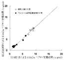
なお、試料として用いた血清はインフォームドコンセントを得た後、連結不可能匿名化し使用した。また、試料中の5.9kDaペプチド濃度は以下に示すSI−MS法により算出した。
SI−MS法を用いて含有5.9kDaペプチド濃度を算出した血清を試料として、サンドイッチ測定系を用いて測定したところ、5.9kDaペプチドの濃度依存的に吸光度が上昇することを確認した。(2-3) Evaluation of ELISA measurement system Using the plate prepared in the above (2-2) and labeled antibody, the effectiveness of the sandwich ELISA measurement system was evaluated.
In addition, after obtaining informed consent, the serum used as a sample was used after anonymizing the connection. Moreover, the 5.9 kDa peptide density | concentration in a sample was computed by the SI-MS method shown below.
When the serum whose 5.9 kDa peptide concentration was calculated using the SI-MS method was used as a sample and measured using a sandwich measurement system, it was confirmed that the absorbance increased depending on the concentration of the 5.9 kDa peptide.
SI−MS法:
磁性ビーズとMALDI−TOF/TOF MSを組み合わせたClinProtTMシステム(Bruker Daltonics)を用いて試料中の5.9kDaペプチド濃度を算出した。
具体的には、まず、血清に5.9kDaペプチドに対する安定同位体ペプチド(SI−5.9kDa)を内部標準として一定量添加した。次に、陽イオン交換磁気ビーズ(WCX)(Bruker Daltonics)に結合させ溶出させた。続いて、溶出させた試料をそのままAnchorChip(Bruker Daltonics)に調整後、AutoFlex(登録商標) II(MALDI−TOF/TOF)により測定を行った。
ここで、解析はFlexAnalysisTM software2.4(Bruker Daltonics)を用いてベースライン補正とスムージング処理を行い、SI−5.9kDaに対応するピーク強度と血清中の5.9kDaペプチドに対応するピーク強度との比から、添加したSI−5.9kDa量を基準に血清中の5.9kDaペプチド濃度を算出した。 SI-MS method :
The 5.9 kDa peptide concentration in the sample was calculated using the ClinProt ™ system (Bruker Daltonics) combining magnetic beads and MALDI-TOF / TOF MS.
Specifically, first, a constant amount of a stable isotope peptide (SI-5.9 kDa) against a 5.9 kDa peptide was added to serum as an internal standard. Next, it was bound to cation exchange magnetic beads (WCX) (Bruker Daltonics) and eluted. Subsequently, the eluted sample was directly adjusted to AnchorChip (Bruker Daltonics), and then measured with AutoFlex (registered trademark) II (MALDI-TOF / TOF).
Here, the analysis was performed with baseline correction and smoothing processing using Flex Analysis ™ software 2.4 (Bruker Daltonics), and peak intensity corresponding to SI-5.9 kDa and peak intensity corresponding to 5.9 kDa peptide in serum. From the ratio, the 5.9 kDa peptide concentration in the serum was calculated based on the added SI-5.9 kDa amount.
(3)ヒト血清中の5.9kDaペプチドの測定
上記(2)で作成したサンドイッチELISA測定系を用いて、ヒト血清中の5.9kDaペプチドを測定し、抗原抗体反応が5.9kDaペプチド濃度依存的に観測されることを、以下に具体的に記載する方法により確認した。
なお、用いた血清はインフォームドコンセントを得た後、連結不可能匿名化し使用した。(3) Measurement of 5.9 kDa peptide in human serum Using the sandwich ELISA measurement system prepared in (2) above, the 5.9 kDa peptide in human serum was measured, and the antigen-antibody reaction was dependent on the 5.9 kDa peptide concentration. It was confirmed by the method specifically described below.
The serum used was anonymized for connection after obtaining informed consent.
(3−1)一次抗体反応
SI−MS法を用いて含有5.9kDaペプチド濃度を算出したヒト血清をPBSにて希釈し、5.9KDaペプチド濃度が、それぞれ、0μg/ml、0.65μg/ml、1.30μg/ml、3.89μg/ml、7.12μg/ml、10.36μg/mlの試料を調製した。これらの試料をPBSで1/333に希釈し、100μlずつ上記(2−1)で作成した抗体結合プレートに1時間反応させた。(3-1) Primary antibody reaction Human serum whose 5.9 kDa peptide concentration was calculated using the SI-MS method was diluted with PBS, and the 5.9 KDa peptide concentrations were 0 μg / ml and 0.65 μg / ml, respectively. Samples of ml, 1.30 μg / ml, 3.89 μg / ml, 7.12 μg / ml, 10.36 μg / ml were prepared. These samples were diluted 1/333 with PBS and reacted with the antibody-binding plate prepared in the above (2-1) for 1 hour by 100 μl.
(3−2)二次抗体反応
プレートをPBS−Tにて3回洗浄した後、上記(2−2)で作成したHRP標識抗体(PBS−Tを用いて1/4000に希釈したもの)を30分間反応させた。(3-2) After washing the secondary antibody reaction plate three times with PBS-T, the HRP-labeled antibody prepared in (2-2) above (diluted to 1/4000 with PBS-T) was used. The reaction was allowed for 30 minutes.
(3−3)発色
プレートの洗浄後にHRPの発色基質である、TMB溶液(カイノス)を100μl加えて一定時間の発色後、1N硫酸(和光純薬工業)を停止液として更に100μl添加し、測定波長450nmにて吸光度を測定した。(3-3) After washing the chromogenic plate, 100 μl of TMB solution (Kainos), which is a chromogenic substrate for HRP, is added for a certain period of time, and then 100 μl of 1N sulfuric acid (Wako Pure Chemical Industries) is added as a stop solution, and measurement is performed. Absorbance was measured at a wavelength of 450 nm.
(3−4)測定
測定マイクロプレートリーダーにて波長450nmの吸光度測定を行なった。吸光度測定により得られた検量線を図1に示した。
図1に示すように、anti−5.9N、および、anti−5.9Cを用いたサンドイッチELISA測定系は、血清中に含まれる5.9kDaペプチドを高感度で検出し、5.9kDaペプチド濃度依存的な吸光度上昇が確認された。(3-4) Measurement Measurement Absorbance measurement at a wavelength of 450 nm was performed with a microplate reader. A calibration curve obtained by measuring the absorbance is shown in FIG.
As shown in FIG. 1, the sandwich ELISA measurement system using anti-5.9N and anti-5.9C detects the 5.9 kDa peptide contained in the serum with high sensitivity, and the 5.9 kDa peptide concentration. A dependent increase in absorbance was confirmed.
比較例1
5.9kDaペプチドの全長ペプチドを認識する抗体を用いたELISA測定系によるヒト血清中の5.9kDaペプチドの測定
実施例2の(2−1)において、anti−5.9Cに代えて5.9kDaペプチドの全長ペプチドを認識する抗体anti−5.9W1(国際公開第2004/058966号に記載のハイブリドーマCN−2(受託番号:IPOD FERM BP−08565)より抽出精製)を、実施例2の(2−2)においてanti−5.9Nに代えて5.9kDaペプチドの全長ペプチドを認識する抗体anti−5.9W2(国際公開第2004/058966号に記載のハイブリドーマCN−1(受託番号:IPOD FERM BP−08564)より抽出精製)を用いて構築されたサンドイッチELISA測定系を用いて、実施例2の(3)と同様の測定を行った。測定により得られた検量線を図1に示した。
図1に示すように、一次抗体にanti−5.9W1、2次抗体にanti−5.9W2を用いたサンドイッチELISA測定系は、血清中に低濃度で含まれる5.9kDaペプチド濃度依存的な吸光度上昇を示さなかった。
さらに、一次抗体にanti−5.9W1、二次抗体に本発明のモノクローナル抗体anti−5.9Cを用いたサンドイッチELISA測定系、一次抗体にanti−5.9W1、二次抗体に本発明のモノクローナル抗体anti−5.9Nを用いたサンドイッチELISA測定系、および、一次抗体に本発明のモノクローナル抗体anti−5.9C、二次抗体に5.9W2を用いたサンドイッチELISA測定系においても同様の挙動が確認された。Comparative Example 1
Measurement of 5.9 kDa peptide in human serum by ELISA measurement system using antibody recognizing full length peptide of 5.9 kDa peptide In (2-1) of Example 2, instead of anti-5.9C, 5.9 kDa The antibody anti-5.9W1 that recognizes the full-length peptide of the peptide (extracted and purified from the hybridoma CN-2 described in International Publication No. 2004/058966 (Accession No .: IPOD FERM BP-08565)) was used in Example 2 (2 -2) antibody anti-5.9W2 (hybridoma CN-1 described in International Publication No. 2004/058966 (accession number: IPOD FERM BP), which recognizes the full-length peptide of 5.9 kDa peptide instead of anti-5.9N -08564), a sandwich ELISA measurement system constructed using extraction and purification) The same measurement as in (2) of Example 2 was performed. The calibration curve obtained by the measurement is shown in FIG.
As shown in FIG. 1, the sandwich ELISA measurement system using anti-5.9W1 as the primary antibody and anti-5.9W2 as the secondary antibody is dependent on the concentration of 5.9 kDa peptide contained in serum at a low concentration. There was no increase in absorbance.
Further, a sandwich ELISA measurement system using anti-5.9W1 as the primary antibody and the monoclonal antibody anti-5.9C of the present invention as the secondary antibody, anti-5.9W1 as the primary antibody, and the monoclonal of the present invention as the secondary antibody. The same behavior was observed in the sandwich ELISA measurement system using the antibody anti-5.9N and the sandwich ELISA measurement system using the monoclonal antibody anti-5.9C of the present invention as the primary antibody and 5.9W2 as the secondary antibody. confirmed.
SI−MS法とELISA測定の相関性の確認
SI−MS法を用いた定量的測定法によるヒト血清中の5.9kDaペプチド濃度測定結果と実施例2で作成したELISA測定系を用いた場合の定量測定結果との相関性を確認し、ELISA測定系が血清中に多く含まれるフィブリノーゲン関連ペプチドの中で5.9kDaペプチドの全長ペプチドを特異的に定量していることを確認した。
なお、用いた試料は、健常者から得られた血清8検体とアルコール依存症患者から得られた血清8検体であり、インフォームドコンセントを得た後、連結不可能匿名化し使用した。
具体的には、実施例2の(3)の測定結果をもとに作成した検量線に基づいて、実施例2の(3)で作成したELISA測定系を用いて、SI−MS法により含有5.9kDaペプチド濃度を算出した血清16検体中の5.9kDaペプチド濃度を定量した。
同一の試料に対するSI−MS法に基づく5.9kDaペプチド定量結果とELISA測定に基づく定量結果を比較した結果を図2に示した。
図2に示すように、SI−MS法による定量結果とELISA測定による定量結果は明確な相関性を示した。この結果は、実施例2で作成したサンドイッチELISA系が、血清中に多数存在するペプチドの中で、5.9kDaペプチドを特異的に定量していることを示唆している。
健常者から得られた血清8検体とアルコール依存症患者から得られた血清8検体は、ELISA測定による5.9kDaペプチド定量結果に対して、閾値6.8μg/mlを適用することで完全に分離可能であった。また、健常者から得られた血清8検体からなる群とアルコール依存症患者から得られた血清8検体からなる群とは、ELISA測定による5.9kDaペプチド定量結果に関して、分散分析による危険率1%以下の有意差を示した。 Confirmation of correlation between SI-MS method and ELISA measurement Results of measurement of 5.9 kDa peptide concentration in human serum by quantitative measurement method using SI-MS method and ELISA measurement system prepared in Example 2 The correlation with the quantitative measurement results was confirmed, and it was confirmed that the ELISA measurement system specifically quantifies the full-length peptide of the 5.9 kDa peptide among fibrinogen-related peptides contained in a large amount in serum.
The samples used were 8 serum samples obtained from healthy subjects and 8 serum samples obtained from alcoholic patients. After obtaining informed consent, they were anonymized for connection.
Specifically, based on the calibration curve created based on the measurement result of Example 2 (3), contained by the SI-MS method using the ELISA measurement system created in (2) of Example 2. The 5.9 kDa peptide concentration in 16 serum samples for which the 5.9 kDa peptide concentration was calculated was quantified.
FIG. 2 shows the result of comparing the 5.9 kDa peptide quantification result based on the SI-MS method and the quantification result based on the ELISA measurement on the same sample.
As shown in FIG. 2, the quantitative result by SI-MS method and the quantitative result by ELISA measurement showed clear correlation. This result suggests that the sandwich ELISA system prepared in Example 2 specifically quantifies the 5.9 kDa peptide among a large number of peptides present in serum.
8 serum samples obtained from healthy subjects and 8 serum samples obtained from patients with alcohol dependence are completely separated by applying a threshold value of 6.8 μg / ml to the 5.9 kDa peptide quantification result by ELISA measurement. It was possible. In addition, the group consisting of 8 serum samples obtained from healthy subjects and the group consisting of 8 serum samples obtained from alcohol-dependent patients have a risk rate of 1% by analysis of variance for the 5.9 kDa peptide quantification result by ELISA measurement. The following significant differences were shown.
ラテックス粒子を用いた5.9kDaペプチド測定系の構築と検体中の5.9kDaペプチド測定
(1)抗5.9kDa抗体感作ラテックス粒子の調製
実施例1において作成した二種類のモノクローナル抗体(anti−5.9N、および、anti−5.9C)がそれぞれ結合した二種類のラテックス粒子を作成し、これらを混合した抗5.9kDa抗体感作ラテックス粒子を、以下に具体的に記載する方法により作成した。
なお、ここで用いられた試薬組成は以下の通りである。
BSAコート溶液;
HEPES 2−「4−(2−ヒドロキシエチル)
−1−ピペラジニル」エタンスルホン酸 25mM pH7.5
塩化ナトリウム 150mM
エチレンジアミン四酢酸・2ナトリウム 1.0mM
アジ化ナトリウム 0.05%
ウシ血清アルブミン(BSA) 1.0%
希釈用緩衝液;
HEPES 25mM pH7.5
エチレンジアミン四酢酸・2ナトリウム 1.0mM
アジ化ナトリウム 0.05%
分散用緩衝液;
HEPES 25mM pH7.5
塩化ナトリウム 150mM
エチレンジアミン四酢酸・2ナトリウム 1.0mM
アジ化ナトリウム 0.05% Construction of 5.9 kDa peptide measurement system using latex particles and measurement of 5.9 kDa peptide in specimen (1) Preparation of anti-5.9 kDa antibody-sensitized latex particles The two monoclonal antibodies (anti-anti- Two types of latex particles having 5.9N and anti-5.9C) bonded thereto were prepared, and anti-5.9 kDa antibody-sensitized latex particles mixed with these were prepared by the method specifically described below. did.
The reagent composition used here is as follows.
BSA coating solution ;
HEPES 2- “4- (2-hydroxyethyl)
-1-piperazinyl "ethanesulfonic acid 25 mM pH 7.5
Sodium chloride 150 mM
Ethylenediaminetetraacetic acid, disodium 1.0 mM
Sodium azide 0.05%
Bovine serum albumin (BSA) 1.0%
Dilution buffer ;
HEPES 25 mM pH 7.5
Ethylenediaminetetraacetic acid, disodium 1.0 mM
Sodium azide 0.05%
Dispersion buffer ;
HEPES 25 mM pH 7.5
Sodium chloride 150 mM
Ethylenediaminetetraacetic acid, disodium 1.0 mM
Sodium azide 0.05%
(1−1)anti−5.9N感作ラテックス粒子の調製
希釈用緩衝液で1%濃度とした、粒径120nmのラテックス粒子溶液100mlに、anti−5.9Nを希釈用緩衝液に3mg/mlとなるように溶解した溶液100mlを加え室温にて1時間攪拌した。その後、20,000rpmで1時間遠心分離を行い、上清を廃棄し沈殿物を回収した。この沈殿物にBSAコート溶液を100ml加え沈殿物を懸濁し、超音波処理を行い完全に分散させた後、室温で1時間攪拌した。次いで遠心分離を行い得られた沈殿物に分散用緩衝液を500ml加えて懸濁し、超音波処理を行い完全に分散させ0.2%濃度anti−5.9N感作ラテックス粒子を得た。(1-1) Preparation of anti-5.9N-sensitized latex particles The concentration of anti-5.9N in 100 ml of a latex particle solution having a particle diameter of 120 nm, adjusted to 1% with a buffer for dilution, and 3 mg / ml of anti-5.9N in a buffer for dilution. 100 ml of the solution dissolved so as to be ml was added and stirred at room temperature for 1 hour. Thereafter, centrifugation was performed at 20,000 rpm for 1 hour, the supernatant was discarded, and the precipitate was collected. 100 ml of the BSA coat solution was added to the precipitate, the precipitate was suspended, subjected to ultrasonic treatment to completely disperse, and then stirred at room temperature for 1 hour. Subsequently, 500 ml of a dispersion buffer was added to the precipitate obtained by centrifugation and suspended therein, followed by sonication to completely disperse it to obtain 0.2% anti-5.9N sensitized latex particles.
(1−2)anti−5.9C感作ラテックス粒子の調製
希釈用緩衝液で1%濃度とした、粒径120nmのラテックス粒子溶液100mlに、anti−5.9Cを希釈用緩衝液に3mg/mlとなるように溶解した溶液100mlを加え室温にて1時間攪拌した。その後、20,000rpmで1時間遠心分離を行い、上清を廃棄し沈殿物を回収した。この沈殿物にBSAコート溶液を100ml加え沈殿物を懸濁し、超音波処理を行い完全に分散させた後、室温で1時間攪拌した。次いで遠心分離を行い得られた沈殿物に分散用緩衝液を500ml加えて懸濁し、超音波処理を行い完全に分散させ0.2%濃度anti−5.9C感作ラテックス粒子を得た。(1-2) Preparation of anti-5.9C Sensitized Latex Particles Into 100 ml of latex particle solution with a particle size of 120 nm, adjusted to 1% with dilution buffer, anti-5.9C was diluted to 3 mg / dilution buffer. 100 ml of the solution dissolved so as to be ml was added and stirred at room temperature for 1 hour. Thereafter, centrifugation was performed at 20,000 rpm for 1 hour, the supernatant was discarded, and the precipitate was collected. 100 ml of the BSA coat solution was added to the precipitate, the precipitate was suspended, subjected to ultrasonic treatment to completely disperse, and then stirred at room temperature for 1 hour. Subsequently, 500 ml of a dispersion buffer was added to the precipitate obtained by centrifugation and suspended therein, followed by sonication to complete dispersion to obtain 0.2% anti-5.9C sensitized latex particles.
(1−3)抗5.9kDa抗体感作ラテックス粒子(LA試薬)の調製
上記(1−1)および(1−2)で調製されたラテックス粒子溶液を1:1の割合で混合したものを、ラテックス法の第二試薬として調製した。
なお、ラテックス法の第一試薬として分散用緩衝液を用いた。(1-3) Preparation of anti-5.9 kDa antibody-sensitized latex particles (LA reagent ) What was prepared by mixing the latex particle solutions prepared in the above (1-1) and (1-2) at a ratio of 1: 1. And prepared as a second reagent in the latex method.
A dispersion buffer was used as the first reagent in the latex method.
(2)ヒト血清中の5.9kDaペプチドの測定
上記(1−3)で調製したLA試薬を用いて、ラテックス免疫凝集測定(LATEX測定)によりヒト血清中の5.9kDAペプチドを定量可能なことを確認した。
具体的には、SI−MS法を用いて含有5.9kDaペプチド濃度を算出したヒト血清試料10μlに対し第一試薬100μl、第二試薬100μlを反応させ、日立7180型自動分析装置を用いて、主波長570nm、副波長800nmにて19−34測光ポイント間(第二試薬添加後約1分後から5分後に相当)において、2ポイントエンド法による吸光度変化量を測定した。
結果として、表2に示すように、5.9kDaペプチド濃度依存的な吸光度上昇が確認された。
Specifically, 100 μl of the first reagent and 100 μl of the second reagent were reacted with 10 μl of the human serum sample for which the concentration of the contained 5.9 kDa peptide was calculated using the SI-MS method, and using a Hitachi 7180 automatic analyzer, The change in absorbance by the two-point end method was measured between 19-34 photometric points (corresponding to about 5 minutes after addition of the second reagent) at a main wavelength of 570 nm and a sub wavelength of 800 nm.
As a result, as shown in Table 2, an increase in absorbance dependent on the 5.9 kDa peptide concentration was confirmed.
(3)ELISA測定とLATEX測定の相関性の確認
実施例3におけるSI−MS法との相関性より血清中の5.9kDaペプチドを特異的に検出することが示唆されたELISA測定とLATEX測定との相関性を確認し、LATEX測定が血清中の5.9kDaペプチドを特異的に検出することを確認した。
具体的には、上記(2)の測定結果をもとに作成された検量線に基づいて、血清中の5.9kDaペプチド濃度を定量した。同一の試料について、実施例3の(2−3)と同様にELISA測定により5.9kDaペプチド濃度を定量し、LATEX測定による定量結果との相関性を図3に示した。
結果として、図3に示すように、LATEX測定とELISA測定は高い相関性を示し、LATEX測定によっても血清中の5.9kDaペプチドを特異的に検出していることが示唆された。(3) Confirmation of correlation between ELISA measurement and LATEX measurement ELISA measurement and LATEX measurement suggested to specifically detect 5.9 kDa peptide in serum from the correlation with SI-MS method in Example 3 The LATEX measurement specifically detected the 5.9 kDa peptide in the serum.
Specifically, the 5.9 kDa peptide concentration in the serum was quantified based on a calibration curve created based on the measurement result of (2) above. About the same sample, the 5.9 kDa peptide density | concentration was quantified by ELISA measurement similarly to (2-3) of Example 3, and the correlation with the quantification result by LATEX measurement was shown in FIG.
As a result, as shown in FIG. 3, LATEX measurement and ELISA measurement showed high correlation, suggesting that the 5.9 kDa peptide in serum was specifically detected by LATEX measurement.
本発明の5.9kDaペプチドの免疫学的測定方法は、肝臓疾患診断用ペプチドマーカーである5.9kDaペプチドの簡便かつ正確な検体からの定量を可能にする。5.9kDaペプチドは、習慣飲酒者や問題飲酒者が肝臓疾患を発症する可能性の診断や、飲酒以外が原因の肝臓疾患、例えば、肝炎、肝硬変、脂肪肝などの肝臓疾患の診断に利用することが可能であり、本発明の5.9kDaペプチドの免疫学的測定方法は、これらの疾患の臨床診断方法として、現在の肝疾患人口から考えても今後の予防医学的見地から意義が大きいものと考えられる。 The immunological measurement method of the 5.9 kDa peptide of the present invention enables easy and accurate quantification of a 5.9 kDa peptide, which is a peptide marker for liver disease diagnosis, from a specimen. The 5.9 kDa peptide is used to diagnose the possibility of habitual drinkers and problem drinkers developing liver disease, and liver diseases caused by causes other than drinking, such as hepatic diseases such as hepatitis, cirrhosis, and fatty liver The immunoassay method of the 5.9 kDa peptide of the present invention has great significance as a clinical diagnosis method for these diseases from the viewpoint of future preventive medicine even from the viewpoint of the current liver disease population. it is conceivable that.
配列番号2−人工配列の説明:合成ペプチド
配列番号3−人工配列の説明:合成ペプチドSEQ ID NO: 2-description of artificial sequence: synthetic peptide SEQ ID NO: 3-description of artificial sequence: synthetic peptide
Claims (11)
Priority Applications (1)
| Application Number | Priority Date | Filing Date | Title |
|---|---|---|---|
| JP2011538339A JP5585587B2 (en) | 2009-10-28 | 2010-10-13 | Method for immunological measurement of 5.9 kDa peptide |
Applications Claiming Priority (4)
| Application Number | Priority Date | Filing Date | Title |
|---|---|---|---|
| JP2009247332 | 2009-10-28 | ||
| JP2009247332 | 2009-10-28 | ||
| JP2011538339A JP5585587B2 (en) | 2009-10-28 | 2010-10-13 | Method for immunological measurement of 5.9 kDa peptide |
| PCT/JP2010/067956 WO2011052380A1 (en) | 2009-10-28 | 2010-10-13 | 5.9 kDa PEPTIDE IMMUNOASSAY METHOD |
Publications (2)
| Publication Number | Publication Date |
|---|---|
| JPWO2011052380A1 JPWO2011052380A1 (en) | 2013-03-21 |
| JP5585587B2 true JP5585587B2 (en) | 2014-09-10 |
Family
ID=43921806
Family Applications (1)
| Application Number | Title | Priority Date | Filing Date |
|---|---|---|---|
| JP2011538339A Active JP5585587B2 (en) | 2009-10-28 | 2010-10-13 | Method for immunological measurement of 5.9 kDa peptide |
Country Status (5)
| Country | Link |
|---|---|
| US (1) | US9017959B2 (en) |
| EP (1) | EP2495564B1 (en) |
| JP (1) | JP5585587B2 (en) |
| CN (1) | CN102597772B (en) |
| WO (1) | WO2011052380A1 (en) |
Citations (3)
| Publication number | Priority date | Publication date | Assignee | Title |
|---|---|---|---|---|
| WO2004058966A1 (en) * | 2002-12-24 | 2004-07-15 | Nitto Boseki Co., Ltd. | Marker proteins for diagnosing liver disease and method of diagnosing liver disease using the same |
| WO2008105215A1 (en) * | 2007-02-28 | 2008-09-04 | Nitto Boseki Co., Ltd. | Marker peptide for diagnosis of alcoholic liver disease, and method for diagnosis of alcoholic liver disease using the same |
| WO2009088022A1 (en) * | 2008-01-07 | 2009-07-16 | Kagoshima University | Novel cancer marker, and diagnosis using the same |
Family Cites Families (8)
| Publication number | Priority date | Publication date | Assignee | Title |
|---|---|---|---|---|
| US20020182660A1 (en) * | 2000-02-18 | 2002-12-05 | Fong Kei-Lai L. | N- and C-terminus specific immunoassays for full length beta-amyloid peptide-Abeta(1-40), Abeta(1-39), Abeta(1-40), Abeta(1-42) , and Abeta(1-43) |
| US20030100035A1 (en) * | 2001-11-08 | 2003-05-29 | Aristo Vojdani | Saliva immunoassay for detection of antibodies for autoimmune disease |
| AU2003296086A1 (en) | 2002-12-26 | 2004-07-22 | Nitto Boseki Co., Ltd. | Immunoassay method and kit to be used therein |
| CN100538361C (en) * | 2002-12-26 | 2009-09-09 | 日本纺绩株式会社 | The kit of immunoassay and use thereof |
| AU2004227018A1 (en) * | 2003-04-08 | 2004-10-21 | Colotech A/S | A method for detection of colorectal cancer in human samples |
| JP4241301B2 (en) * | 2003-09-30 | 2009-03-18 | 和光純薬工業株式会社 | Reagent for immunological measurement |
| US20070190580A1 (en) * | 2005-10-31 | 2007-08-16 | Beckman Coulter, Inc. | Immunoassay of Fragments of Insulin-Like Growth Factor Binding Proteins |
| US8288110B2 (en) * | 2006-12-04 | 2012-10-16 | Perkinelmer Health Sciences, Inc. | Biomarkers for detecting cancer |
-
2010
- 2010-10-13 EP EP10826514.1A patent/EP2495564B1/en active Active
- 2010-10-13 US US13/502,198 patent/US9017959B2/en active Active
- 2010-10-13 JP JP2011538339A patent/JP5585587B2/en active Active
- 2010-10-13 WO PCT/JP2010/067956 patent/WO2011052380A1/en active Application Filing
- 2010-10-13 CN CN201080048601.XA patent/CN102597772B/en active Active
Patent Citations (3)
| Publication number | Priority date | Publication date | Assignee | Title |
|---|---|---|---|---|
| WO2004058966A1 (en) * | 2002-12-24 | 2004-07-15 | Nitto Boseki Co., Ltd. | Marker proteins for diagnosing liver disease and method of diagnosing liver disease using the same |
| WO2008105215A1 (en) * | 2007-02-28 | 2008-09-04 | Nitto Boseki Co., Ltd. | Marker peptide for diagnosis of alcoholic liver disease, and method for diagnosis of alcoholic liver disease using the same |
| WO2009088022A1 (en) * | 2008-01-07 | 2009-07-16 | Kagoshima University | Novel cancer marker, and diagnosis using the same |
Non-Patent Citations (5)
| Title |
|---|
| JPN6010066631; Fumio Nomura et al.: '"Identification of novel and downregulated biomarkers for alcoholism by surface enhanced laser desor' Proteomics Vol. 4, No. 4, 200404, p. 1187-1194 * |
| JPN6014015173; 中野 雅行: '「アルコール性肝障害(ASH) 3. 病態と病理診断」' 日本内科学会雑誌 Vol. 95, No. 1, 20060110, p. 28-32 * |
| JPN6014026048; 中野 雅行: '「III. アルコール性肝障害(ASH) 3. 病態と病理診断」' 日本内科学会雑誌 Vol. 95, No. 1, 20060110, p. 28-32 * |
| JPN6014026050; 曽川 一幸,他7名: '「質量分析装置を用いた問題飲酒者診断法の確立を目指して」' 日本臨床検査自動化学会会誌 Vol. 34, No. 4, 20090901, p. 796 * |
| JPN6014026053; 野村 文夫: '「疾患プロテオミクスの最前線 -消化器領域の新しい疾患マーカー探索への応用-」' 日本内科学会雑誌 Vol. 97, No. 8, 20080810, p. 1888-1894 * |
Also Published As
| Publication number | Publication date |
|---|---|
| CN102597772B (en) | 2014-08-20 |
| JPWO2011052380A1 (en) | 2013-03-21 |
| CN102597772A (en) | 2012-07-18 |
| EP2495564B1 (en) | 2015-12-16 |
| WO2011052380A1 (en) | 2011-05-05 |
| EP2495564A1 (en) | 2012-09-05 |
| US20120276562A1 (en) | 2012-11-01 |
| EP2495564A4 (en) | 2013-07-10 |
| US9017959B2 (en) | 2015-04-28 |
Similar Documents
| Publication | Publication Date | Title |
|---|---|---|
| US11372001B2 (en) | Anti-human IgG4 monoclonal antibody and methods of making and using same | |
| US9347954B2 (en) | Antibody capable of binding to specific region of periostin, and method of measuring periostin using the same | |
| US7867725B2 (en) | Monoclonal antibodies against osteopontin | |
| WO2021237534A1 (en) | Antibody against novel coronavirus, antibody combination, preparation method, and detection kit containing same | |
| JP5798679B2 (en) | Monoclonal antibody specifically recognizing human liver-carboxyl esterase 1, hybridoma cell line producing said antibody and use thereof | |
| JP6037043B2 (en) | Protein quantification method specific to TRACP-5b (tartrate-resistant acid phosphatase 5b) | |
| JP5585587B2 (en) | Method for immunological measurement of 5.9 kDa peptide | |
| EP2484694B1 (en) | Monoclonal antibody against human hig-1 polypeptide | |
| JP2018080114A (en) | Anti-human VAP-1 monoclonal antibody | |
| CN115232211A (en) | Novel anti-soluble CD14 subtype antibody and application thereof | |
| WO2023068248A1 (en) | Immunoassay method for cross-linked n-telopeptide of type i collagen, immunoassay kit, and antibody or antibody fragment thereof | |
| WO2022202876A1 (en) | Immunological analysis method for type-i collagen c-terminal telopeptide | |
| JP5231954B2 (en) | Albumin measuring reagent | |
| JP5448424B2 (en) | Reagent for measuring protein containing Fc of human IgG | |
| US20130288277A1 (en) | Immunoassay of cofilin 1 protein | |
| TW202342979A (en) | Detection method and detection reagent | |
| JP4517353B2 (en) | Method for measuring IgA-binding alkaline phosphatase 6 and kit used therefor | |
| JP2003180345A (en) | Method for assaying free type hepatocyte growth factor receptor | |
| JP2005314397A (en) | Anti-chondromodulin-1-specific antibody and its use |
Legal Events
| Date | Code | Title | Description |
|---|---|---|---|
| A621 | Written request for application examination |
Free format text: JAPANESE INTERMEDIATE CODE: A621 Effective date: 20130426 |
|
| A131 | Notification of reasons for refusal |
Free format text: JAPANESE INTERMEDIATE CODE: A131 Effective date: 20140411 |
|
| A521 | Request for written amendment filed |
Free format text: JAPANESE INTERMEDIATE CODE: A523 Effective date: 20140528 |
|
| TRDD | Decision of grant or rejection written | ||
| A01 | Written decision to grant a patent or to grant a registration (utility model) |
Free format text: JAPANESE INTERMEDIATE CODE: A01 Effective date: 20140624 |
|
| A61 | First payment of annual fees (during grant procedure) |
Free format text: JAPANESE INTERMEDIATE CODE: A61 Effective date: 20140707 |
|
| R150 | Certificate of patent or registration of utility model |
Ref document number: 5585587 Country of ref document: JP Free format text: JAPANESE INTERMEDIATE CODE: R150 |
|
| R250 | Receipt of annual fees |
Free format text: JAPANESE INTERMEDIATE CODE: R250 |
|
| R250 | Receipt of annual fees |
Free format text: JAPANESE INTERMEDIATE CODE: R250 |
|
| R250 | Receipt of annual fees |
Free format text: JAPANESE INTERMEDIATE CODE: R250 |
|
| R250 | Receipt of annual fees |
Free format text: JAPANESE INTERMEDIATE CODE: R250 |
|
| R250 | Receipt of annual fees |
Free format text: JAPANESE INTERMEDIATE CODE: R250 |
|
| R250 | Receipt of annual fees |
Free format text: JAPANESE INTERMEDIATE CODE: R250 |
|
| R255 | Notification that request for automated payment was rejected |
Free format text: JAPANESE INTERMEDIATE CODE: R2525 |
|
| R250 | Receipt of annual fees |
Free format text: JAPANESE INTERMEDIATE CODE: R250 |