EP0928168B1 - Resorbable, macro-porous, non-collapsing and flexible membrane barrier for skeletal repair and regeneration - Google Patents
Resorbable, macro-porous, non-collapsing and flexible membrane barrier for skeletal repair and regeneration Download PDFInfo
- Publication number
- EP0928168B1 EP0928168B1 EP97936390A EP97936390A EP0928168B1 EP 0928168 B1 EP0928168 B1 EP 0928168B1 EP 97936390 A EP97936390 A EP 97936390A EP 97936390 A EP97936390 A EP 97936390A EP 0928168 B1 EP0928168 B1 EP 0928168B1
- Authority
- EP
- European Patent Office
- Prior art keywords
- protective
- apertures
- membrane
- bone
- bone regeneration
- Prior art date
- Legal status (The legal status is an assumption and is not a legal conclusion. Google has not performed a legal analysis and makes no representation as to the accuracy of the status listed.)
- Expired - Lifetime
Links
- 230000008439 repair process Effects 0.000 title claims abstract description 5
- 230000008929 regeneration Effects 0.000 title description 7
- 238000011069 regeneration method Methods 0.000 title description 7
- 230000008384 membrane barrier Effects 0.000 title description 2
- 239000012528 membrane Substances 0.000 claims abstract description 130
- 230000007547 defect Effects 0.000 claims abstract description 69
- 210000001519 tissue Anatomy 0.000 claims abstract description 27
- 210000004872 soft tissue Anatomy 0.000 claims abstract description 16
- 239000007943 implant Substances 0.000 claims abstract description 8
- 210000005166 vasculature Anatomy 0.000 claims abstract description 5
- 238000001727 in vivo Methods 0.000 claims abstract description 3
- 230000001681 protective effect Effects 0.000 claims description 113
- 230000010478 bone regeneration Effects 0.000 claims description 106
- 239000000463 material Substances 0.000 claims description 27
- 229920000642 polymer Polymers 0.000 claims description 12
- 208000012287 Prolapse Diseases 0.000 claims description 8
- 239000012530 fluid Substances 0.000 claims description 5
- 230000035755 proliferation Effects 0.000 claims description 4
- 238000009826 distribution Methods 0.000 claims description 3
- 238000002513 implantation Methods 0.000 claims description 3
- 230000035876 healing Effects 0.000 claims description 2
- 239000011159 matrix material Substances 0.000 claims 1
- 239000012466 permeate Substances 0.000 claims 1
- 239000002356 single layer Substances 0.000 claims 1
- 239000007787 solid Substances 0.000 claims 1
- 210000000988 bone and bone Anatomy 0.000 abstract description 133
- 208000006735 Periostitis Diseases 0.000 abstract description 8
- 210000003460 periosteum Anatomy 0.000 abstract description 8
- 210000001608 connective tissue cell Anatomy 0.000 abstract description 2
- 239000011148 porous material Substances 0.000 abstract 1
- 210000004027 cell Anatomy 0.000 description 13
- 210000004204 blood vessel Anatomy 0.000 description 11
- 239000012634 fragment Substances 0.000 description 11
- 208000010392 Bone Fractures Diseases 0.000 description 9
- 206010017076 Fracture Diseases 0.000 description 7
- 238000000034 method Methods 0.000 description 7
- 210000002901 mesenchymal stem cell Anatomy 0.000 description 6
- RTAQQCXQSZGOHL-UHFFFAOYSA-N Titanium Chemical compound [Ti] RTAQQCXQSZGOHL-UHFFFAOYSA-N 0.000 description 5
- 239000010936 titanium Substances 0.000 description 5
- 229910052719 titanium Inorganic materials 0.000 description 5
- 102000004169 proteins and genes Human genes 0.000 description 4
- 108090000623 proteins and genes Proteins 0.000 description 4
- 210000003625 skull Anatomy 0.000 description 4
- 230000002269 spontaneous effect Effects 0.000 description 4
- 238000001356 surgical procedure Methods 0.000 description 4
- 238000013459 approach Methods 0.000 description 3
- 210000004369 blood Anatomy 0.000 description 3
- 239000008280 blood Substances 0.000 description 3
- 230000006835 compression Effects 0.000 description 3
- 238000007906 compression Methods 0.000 description 3
- 210000002808 connective tissue Anatomy 0.000 description 3
- 210000004373 mandible Anatomy 0.000 description 3
- 230000000921 morphogenic effect Effects 0.000 description 3
- 210000003205 muscle Anatomy 0.000 description 3
- 230000011164 ossification Effects 0.000 description 3
- 239000000126 substance Substances 0.000 description 3
- 208000024779 Comminuted Fractures Diseases 0.000 description 2
- 238000005452 bending Methods 0.000 description 2
- 230000008901 benefit Effects 0.000 description 2
- 210000000601 blood cell Anatomy 0.000 description 2
- 230000036770 blood supply Effects 0.000 description 2
- 230000012292 cell migration Effects 0.000 description 2
- 230000007613 environmental effect Effects 0.000 description 2
- 210000002919 epithelial cell Anatomy 0.000 description 2
- 239000003102 growth factor Substances 0.000 description 2
- 208000015181 infectious disease Diseases 0.000 description 2
- 238000003475 lamination Methods 0.000 description 2
- 230000007246 mechanism Effects 0.000 description 2
- 229910052751 metal Inorganic materials 0.000 description 2
- 239000002184 metal Substances 0.000 description 2
- 239000005445 natural material Substances 0.000 description 2
- 229920002994 synthetic fiber Polymers 0.000 description 2
- 206010003694 Atrophy Diseases 0.000 description 1
- 206010065687 Bone loss Diseases 0.000 description 1
- 102000008186 Collagen Human genes 0.000 description 1
- 108010035532 Collagen Proteins 0.000 description 1
- 206010010356 Congenital anomaly Diseases 0.000 description 1
- 102000018233 Fibroblast Growth Factor Human genes 0.000 description 1
- 108050007372 Fibroblast Growth Factor Proteins 0.000 description 1
- 241000282412 Homo Species 0.000 description 1
- 241001465754 Metazoa Species 0.000 description 1
- 206010029113 Neovascularisation Diseases 0.000 description 1
- 102000010780 Platelet-Derived Growth Factor Human genes 0.000 description 1
- 108010038512 Platelet-Derived Growth Factor Proteins 0.000 description 1
- 102000009618 Transforming Growth Factors Human genes 0.000 description 1
- 108010009583 Transforming Growth Factors Proteins 0.000 description 1
- 230000037444 atrophy Effects 0.000 description 1
- 230000003416 augmentation Effects 0.000 description 1
- 230000001580 bacterial effect Effects 0.000 description 1
- 239000000560 biocompatible material Substances 0.000 description 1
- 230000015572 biosynthetic process Effects 0.000 description 1
- 210000002449 bone cell Anatomy 0.000 description 1
- 210000002805 bone matrix Anatomy 0.000 description 1
- 210000000845 cartilage Anatomy 0.000 description 1
- 230000024245 cell differentiation Effects 0.000 description 1
- 230000004663 cell proliferation Effects 0.000 description 1
- 229910010293 ceramic material Inorganic materials 0.000 description 1
- 230000003399 chemotactic effect Effects 0.000 description 1
- 229920001436 collagen Polymers 0.000 description 1
- 239000002131 composite material Substances 0.000 description 1
- 238000011109 contamination Methods 0.000 description 1
- 230000001054 cortical effect Effects 0.000 description 1
- 230000007812 deficiency Effects 0.000 description 1
- 230000002950 deficient Effects 0.000 description 1
- 230000001419 dependent effect Effects 0.000 description 1
- 238000011161 development Methods 0.000 description 1
- 230000018109 developmental process Effects 0.000 description 1
- 238000009792 diffusion process Methods 0.000 description 1
- 239000003814 drug Substances 0.000 description 1
- 229940126864 fibroblast growth factor Drugs 0.000 description 1
- 230000036571 hydration Effects 0.000 description 1
- 238000006703 hydration reaction Methods 0.000 description 1
- 238000000338 in vitro Methods 0.000 description 1
- 230000002458 infectious effect Effects 0.000 description 1
- 230000008595 infiltration Effects 0.000 description 1
- 238000001764 infiltration Methods 0.000 description 1
- 230000002401 inhibitory effect Effects 0.000 description 1
- 208000014674 injury Diseases 0.000 description 1
- 210000003041 ligament Anatomy 0.000 description 1
- 230000033001 locomotion Effects 0.000 description 1
- 230000007774 longterm Effects 0.000 description 1
- 238000004519 manufacturing process Methods 0.000 description 1
- 210000002050 maxilla Anatomy 0.000 description 1
- 210000004086 maxillary sinus Anatomy 0.000 description 1
- 230000005541 medical transmission Effects 0.000 description 1
- 230000002297 mitogenic effect Effects 0.000 description 1
- 238000012986 modification Methods 0.000 description 1
- 230000004048 modification Effects 0.000 description 1
- 210000000663 muscle cell Anatomy 0.000 description 1
- 210000005036 nerve Anatomy 0.000 description 1
- 230000007935 neutral effect Effects 0.000 description 1
- 230000000399 orthopedic effect Effects 0.000 description 1
- 210000004197 pelvis Anatomy 0.000 description 1
- 239000002243 precursor Substances 0.000 description 1
- 238000004321 preservation Methods 0.000 description 1
- 230000002265 prevention Effects 0.000 description 1
- 230000002062 proliferating effect Effects 0.000 description 1
- 230000001737 promoting effect Effects 0.000 description 1
- 230000006641 stabilisation Effects 0.000 description 1
- 238000011105 stabilization Methods 0.000 description 1
- 238000006467 substitution reaction Methods 0.000 description 1
- 210000002435 tendon Anatomy 0.000 description 1
- ZRKFYGHZFMAOKI-QMGMOQQFSA-N tgfbeta Chemical compound C([C@H](NC(=O)[C@H](C(C)C)NC(=O)CNC(=O)[C@H](CCC(O)=O)NC(=O)[C@H](CCCNC(N)=N)NC(=O)[C@H](CC(N)=O)NC(=O)[C@H](CC(C)C)NC(=O)[C@H]([C@@H](C)O)NC(=O)[C@H](CCC(O)=O)NC(=O)[C@H]([C@@H](C)O)NC(=O)[C@H](CC(C)C)NC(=O)CNC(=O)[C@H](C)NC(=O)[C@H](CO)NC(=O)[C@H](CCC(N)=O)NC(=O)[C@@H](NC(=O)[C@H](C)NC(=O)[C@H](C)NC(=O)[C@@H](NC(=O)[C@H](CC(C)C)NC(=O)[C@@H](N)CCSC)C(C)C)[C@@H](C)CC)C(=O)N[C@@H]([C@@H](C)O)C(=O)N[C@@H](C(C)C)C(=O)N[C@@H](CC=1C=CC=CC=1)C(=O)N[C@@H](C)C(=O)N1[C@@H](CCC1)C(=O)N[C@@H]([C@@H](C)O)C(=O)N[C@@H](CC(N)=O)C(=O)N[C@@H](CCC(O)=O)C(=O)N[C@@H](C)C(=O)N[C@@H](CC=1C=CC=CC=1)C(=O)N[C@@H](CCCNC(N)=N)C(=O)N[C@@H](C)C(=O)N[C@@H](CC(C)C)C(=O)N1[C@@H](CCC1)C(=O)N1[C@@H](CCC1)C(=O)N[C@@H](CCCNC(N)=N)C(=O)N[C@@H](CCC(O)=O)C(=O)N[C@@H](CCCNC(N)=N)C(=O)N[C@@H](CO)C(=O)N[C@@H](CCCNC(N)=N)C(=O)N[C@@H](CC(C)C)C(=O)N[C@@H](CC(C)C)C(O)=O)C1=CC=C(O)C=C1 ZRKFYGHZFMAOKI-QMGMOQQFSA-N 0.000 description 1
- 229940124597 therapeutic agent Drugs 0.000 description 1
- 230000008733 trauma Effects 0.000 description 1
- 230000000472 traumatic effect Effects 0.000 description 1
Images


Classifications
-
- A—HUMAN NECESSITIES
- A61—MEDICAL OR VETERINARY SCIENCE; HYGIENE
- A61L—METHODS OR APPARATUS FOR STERILISING MATERIALS OR OBJECTS IN GENERAL; DISINFECTION, STERILISATION OR DEODORISATION OF AIR; CHEMICAL ASPECTS OF BANDAGES, DRESSINGS, ABSORBENT PADS OR SURGICAL ARTICLES; MATERIALS FOR BANDAGES, DRESSINGS, ABSORBENT PADS OR SURGICAL ARTICLES
- A61L27/00—Materials for grafts or prostheses or for coating grafts or prostheses
- A61L27/50—Materials characterised by their function or physical properties, e.g. injectable or lubricating compositions, shape-memory materials, surface modified materials
- A61L27/54—Biologically active materials, e.g. therapeutic substances
-
- A—HUMAN NECESSITIES
- A61—MEDICAL OR VETERINARY SCIENCE; HYGIENE
- A61B—DIAGNOSIS; SURGERY; IDENTIFICATION
- A61B17/00—Surgical instruments, devices or methods, e.g. tourniquets
- A61B17/56—Surgical instruments or methods for treatment of bones or joints; Devices specially adapted therefor
- A61B17/58—Surgical instruments or methods for treatment of bones or joints; Devices specially adapted therefor for osteosynthesis, e.g. bone plates, screws, setting implements or the like
- A61B17/68—Internal fixation devices, including fasteners and spinal fixators, even if a part thereof projects from the skin
- A61B17/688—Internal fixation devices, including fasteners and spinal fixators, even if a part thereof projects from the skin for reattaching pieces of the skull
-
- A—HUMAN NECESSITIES
- A61—MEDICAL OR VETERINARY SCIENCE; HYGIENE
- A61B—DIAGNOSIS; SURGERY; IDENTIFICATION
- A61B17/00—Surgical instruments, devices or methods, e.g. tourniquets
- A61B17/56—Surgical instruments or methods for treatment of bones or joints; Devices specially adapted therefor
- A61B17/58—Surgical instruments or methods for treatment of bones or joints; Devices specially adapted therefor for osteosynthesis, e.g. bone plates, screws, setting implements or the like
- A61B17/68—Internal fixation devices, including fasteners and spinal fixators, even if a part thereof projects from the skin
- A61B17/80—Cortical plates, i.e. bone plates; Instruments for holding or positioning cortical plates, or for compressing bones attached to cortical plates
- A61B17/8085—Cortical plates, i.e. bone plates; Instruments for holding or positioning cortical plates, or for compressing bones attached to cortical plates with pliable or malleable elements or having a mesh-like structure, e.g. small strips
-
- A—HUMAN NECESSITIES
- A61—MEDICAL OR VETERINARY SCIENCE; HYGIENE
- A61F—FILTERS IMPLANTABLE INTO BLOOD VESSELS; PROSTHESES; DEVICES PROVIDING PATENCY TO, OR PREVENTING COLLAPSING OF, TUBULAR STRUCTURES OF THE BODY, e.g. STENTS; ORTHOPAEDIC, NURSING OR CONTRACEPTIVE DEVICES; FOMENTATION; TREATMENT OR PROTECTION OF EYES OR EARS; BANDAGES, DRESSINGS OR ABSORBENT PADS; FIRST-AID KITS
- A61F2/00—Filters implantable into blood vessels; Prostheses, i.e. artificial substitutes or replacements for parts of the body; Appliances for connecting them with the body; Devices providing patency to, or preventing collapsing of, tubular structures of the body, e.g. stents
- A61F2/0063—Implantable repair or support meshes, e.g. hernia meshes
-
- A—HUMAN NECESSITIES
- A61—MEDICAL OR VETERINARY SCIENCE; HYGIENE
- A61F—FILTERS IMPLANTABLE INTO BLOOD VESSELS; PROSTHESES; DEVICES PROVIDING PATENCY TO, OR PREVENTING COLLAPSING OF, TUBULAR STRUCTURES OF THE BODY, e.g. STENTS; ORTHOPAEDIC, NURSING OR CONTRACEPTIVE DEVICES; FOMENTATION; TREATMENT OR PROTECTION OF EYES OR EARS; BANDAGES, DRESSINGS OR ABSORBENT PADS; FIRST-AID KITS
- A61F2/00—Filters implantable into blood vessels; Prostheses, i.e. artificial substitutes or replacements for parts of the body; Appliances for connecting them with the body; Devices providing patency to, or preventing collapsing of, tubular structures of the body, e.g. stents
- A61F2/02—Prostheses implantable into the body
- A61F2/28—Bones
- A61F2/2803—Bones for mandibular reconstruction
-
- A—HUMAN NECESSITIES
- A61—MEDICAL OR VETERINARY SCIENCE; HYGIENE
- A61F—FILTERS IMPLANTABLE INTO BLOOD VESSELS; PROSTHESES; DEVICES PROVIDING PATENCY TO, OR PREVENTING COLLAPSING OF, TUBULAR STRUCTURES OF THE BODY, e.g. STENTS; ORTHOPAEDIC, NURSING OR CONTRACEPTIVE DEVICES; FOMENTATION; TREATMENT OR PROTECTION OF EYES OR EARS; BANDAGES, DRESSINGS OR ABSORBENT PADS; FIRST-AID KITS
- A61F2/00—Filters implantable into blood vessels; Prostheses, i.e. artificial substitutes or replacements for parts of the body; Appliances for connecting them with the body; Devices providing patency to, or preventing collapsing of, tubular structures of the body, e.g. stents
- A61F2/02—Prostheses implantable into the body
- A61F2/28—Bones
- A61F2/2846—Support means for bone substitute or for bone graft implants, e.g. membranes or plates for covering bone defects
-
- A—HUMAN NECESSITIES
- A61—MEDICAL OR VETERINARY SCIENCE; HYGIENE
- A61F—FILTERS IMPLANTABLE INTO BLOOD VESSELS; PROSTHESES; DEVICES PROVIDING PATENCY TO, OR PREVENTING COLLAPSING OF, TUBULAR STRUCTURES OF THE BODY, e.g. STENTS; ORTHOPAEDIC, NURSING OR CONTRACEPTIVE DEVICES; FOMENTATION; TREATMENT OR PROTECTION OF EYES OR EARS; BANDAGES, DRESSINGS OR ABSORBENT PADS; FIRST-AID KITS
- A61F2/00—Filters implantable into blood vessels; Prostheses, i.e. artificial substitutes or replacements for parts of the body; Appliances for connecting them with the body; Devices providing patency to, or preventing collapsing of, tubular structures of the body, e.g. stents
- A61F2/02—Prostheses implantable into the body
- A61F2/28—Bones
- A61F2/2875—Skull or cranium
-
- A—HUMAN NECESSITIES
- A61—MEDICAL OR VETERINARY SCIENCE; HYGIENE
- A61F—FILTERS IMPLANTABLE INTO BLOOD VESSELS; PROSTHESES; DEVICES PROVIDING PATENCY TO, OR PREVENTING COLLAPSING OF, TUBULAR STRUCTURES OF THE BODY, e.g. STENTS; ORTHOPAEDIC, NURSING OR CONTRACEPTIVE DEVICES; FOMENTATION; TREATMENT OR PROTECTION OF EYES OR EARS; BANDAGES, DRESSINGS OR ABSORBENT PADS; FIRST-AID KITS
- A61F2/00—Filters implantable into blood vessels; Prostheses, i.e. artificial substitutes or replacements for parts of the body; Appliances for connecting them with the body; Devices providing patency to, or preventing collapsing of, tubular structures of the body, e.g. stents
- A61F2/02—Prostheses implantable into the body
- A61F2/30—Joints
- A61F2/46—Special tools or methods for implanting or extracting artificial joints, accessories, bone grafts or substitutes, or particular adaptations therefor
- A61F2/468—Testing instruments for artificial joints
-
- A—HUMAN NECESSITIES
- A61—MEDICAL OR VETERINARY SCIENCE; HYGIENE
- A61L—METHODS OR APPARATUS FOR STERILISING MATERIALS OR OBJECTS IN GENERAL; DISINFECTION, STERILISATION OR DEODORISATION OF AIR; CHEMICAL ASPECTS OF BANDAGES, DRESSINGS, ABSORBENT PADS OR SURGICAL ARTICLES; MATERIALS FOR BANDAGES, DRESSINGS, ABSORBENT PADS OR SURGICAL ARTICLES
- A61L27/00—Materials for grafts or prostheses or for coating grafts or prostheses
- A61L27/50—Materials characterised by their function or physical properties, e.g. injectable or lubricating compositions, shape-memory materials, surface modified materials
- A61L27/56—Porous materials, e.g. foams or sponges
-
- A—HUMAN NECESSITIES
- A61—MEDICAL OR VETERINARY SCIENCE; HYGIENE
- A61L—METHODS OR APPARATUS FOR STERILISING MATERIALS OR OBJECTS IN GENERAL; DISINFECTION, STERILISATION OR DEODORISATION OF AIR; CHEMICAL ASPECTS OF BANDAGES, DRESSINGS, ABSORBENT PADS OR SURGICAL ARTICLES; MATERIALS FOR BANDAGES, DRESSINGS, ABSORBENT PADS OR SURGICAL ARTICLES
- A61L27/00—Materials for grafts or prostheses or for coating grafts or prostheses
- A61L27/50—Materials characterised by their function or physical properties, e.g. injectable or lubricating compositions, shape-memory materials, surface modified materials
- A61L27/58—Materials at least partially resorbable by the body
-
- A—HUMAN NECESSITIES
- A61—MEDICAL OR VETERINARY SCIENCE; HYGIENE
- A61L—METHODS OR APPARATUS FOR STERILISING MATERIALS OR OBJECTS IN GENERAL; DISINFECTION, STERILISATION OR DEODORISATION OF AIR; CHEMICAL ASPECTS OF BANDAGES, DRESSINGS, ABSORBENT PADS OR SURGICAL ARTICLES; MATERIALS FOR BANDAGES, DRESSINGS, ABSORBENT PADS OR SURGICAL ARTICLES
- A61L31/00—Materials for other surgical articles, e.g. stents, stent-grafts, shunts, surgical drapes, guide wires, materials for adhesion prevention, occluding devices, surgical gloves, tissue fixation devices
- A61L31/14—Materials characterised by their function or physical properties, e.g. injectable or lubricating compositions, shape-memory materials, surface modified materials
- A61L31/146—Porous materials, e.g. foams or sponges
-
- A—HUMAN NECESSITIES
- A61—MEDICAL OR VETERINARY SCIENCE; HYGIENE
- A61L—METHODS OR APPARATUS FOR STERILISING MATERIALS OR OBJECTS IN GENERAL; DISINFECTION, STERILISATION OR DEODORISATION OF AIR; CHEMICAL ASPECTS OF BANDAGES, DRESSINGS, ABSORBENT PADS OR SURGICAL ARTICLES; MATERIALS FOR BANDAGES, DRESSINGS, ABSORBENT PADS OR SURGICAL ARTICLES
- A61L31/00—Materials for other surgical articles, e.g. stents, stent-grafts, shunts, surgical drapes, guide wires, materials for adhesion prevention, occluding devices, surgical gloves, tissue fixation devices
- A61L31/14—Materials characterised by their function or physical properties, e.g. injectable or lubricating compositions, shape-memory materials, surface modified materials
- A61L31/148—Materials at least partially resorbable by the body
-
- A—HUMAN NECESSITIES
- A61—MEDICAL OR VETERINARY SCIENCE; HYGIENE
- A61L—METHODS OR APPARATUS FOR STERILISING MATERIALS OR OBJECTS IN GENERAL; DISINFECTION, STERILISATION OR DEODORISATION OF AIR; CHEMICAL ASPECTS OF BANDAGES, DRESSINGS, ABSORBENT PADS OR SURGICAL ARTICLES; MATERIALS FOR BANDAGES, DRESSINGS, ABSORBENT PADS OR SURGICAL ARTICLES
- A61L31/00—Materials for other surgical articles, e.g. stents, stent-grafts, shunts, surgical drapes, guide wires, materials for adhesion prevention, occluding devices, surgical gloves, tissue fixation devices
- A61L31/14—Materials characterised by their function or physical properties, e.g. injectable or lubricating compositions, shape-memory materials, surface modified materials
- A61L31/16—Biologically active materials, e.g. therapeutic substances
-
- A—HUMAN NECESSITIES
- A61—MEDICAL OR VETERINARY SCIENCE; HYGIENE
- A61B—DIAGNOSIS; SURGERY; IDENTIFICATION
- A61B17/00—Surgical instruments, devices or methods, e.g. tourniquets
- A61B17/04—Surgical instruments, devices or methods, e.g. tourniquets for suturing wounds; Holders or packages for needles or suture materials
- A61B17/06—Needles ; Sutures; Needle-suture combinations; Holders or packages for needles or suture materials
- A61B17/06166—Sutures
-
- A—HUMAN NECESSITIES
- A61—MEDICAL OR VETERINARY SCIENCE; HYGIENE
- A61B—DIAGNOSIS; SURGERY; IDENTIFICATION
- A61B17/00—Surgical instruments, devices or methods, e.g. tourniquets
- A61B17/064—Surgical staples, i.e. penetrating the tissue
- A61B17/0642—Surgical staples, i.e. penetrating the tissue for bones, e.g. for osteosynthesis or connecting tendon to bone
-
- A—HUMAN NECESSITIES
- A61—MEDICAL OR VETERINARY SCIENCE; HYGIENE
- A61B—DIAGNOSIS; SURGERY; IDENTIFICATION
- A61B17/00—Surgical instruments, devices or methods, e.g. tourniquets
- A61B17/08—Wound clamps or clips, i.e. not or only partly penetrating the tissue ; Devices for bringing together the edges of a wound
-
- A—HUMAN NECESSITIES
- A61—MEDICAL OR VETERINARY SCIENCE; HYGIENE
- A61B—DIAGNOSIS; SURGERY; IDENTIFICATION
- A61B17/00—Surgical instruments, devices or methods, e.g. tourniquets
- A61B17/56—Surgical instruments or methods for treatment of bones or joints; Devices specially adapted therefor
- A61B17/58—Surgical instruments or methods for treatment of bones or joints; Devices specially adapted therefor for osteosynthesis, e.g. bone plates, screws, setting implements or the like
- A61B17/60—Surgical instruments or methods for treatment of bones or joints; Devices specially adapted therefor for osteosynthesis, e.g. bone plates, screws, setting implements or the like for external osteosynthesis, e.g. distractors, contractors
- A61B17/64—Devices extending alongside the bones to be positioned
-
- A—HUMAN NECESSITIES
- A61—MEDICAL OR VETERINARY SCIENCE; HYGIENE
- A61B—DIAGNOSIS; SURGERY; IDENTIFICATION
- A61B17/00—Surgical instruments, devices or methods, e.g. tourniquets
- A61B17/56—Surgical instruments or methods for treatment of bones or joints; Devices specially adapted therefor
- A61B17/58—Surgical instruments or methods for treatment of bones or joints; Devices specially adapted therefor for osteosynthesis, e.g. bone plates, screws, setting implements or the like
- A61B17/68—Internal fixation devices, including fasteners and spinal fixators, even if a part thereof projects from the skin
- A61B17/72—Intramedullary pins, nails or other devices
-
- A—HUMAN NECESSITIES
- A61—MEDICAL OR VETERINARY SCIENCE; HYGIENE
- A61B—DIAGNOSIS; SURGERY; IDENTIFICATION
- A61B17/00—Surgical instruments, devices or methods, e.g. tourniquets
- A61B17/56—Surgical instruments or methods for treatment of bones or joints; Devices specially adapted therefor
- A61B17/58—Surgical instruments or methods for treatment of bones or joints; Devices specially adapted therefor for osteosynthesis, e.g. bone plates, screws, setting implements or the like
- A61B17/68—Internal fixation devices, including fasteners and spinal fixators, even if a part thereof projects from the skin
- A61B17/80—Cortical plates, i.e. bone plates; Instruments for holding or positioning cortical plates, or for compressing bones attached to cortical plates
- A61B17/8061—Cortical plates, i.e. bone plates; Instruments for holding or positioning cortical plates, or for compressing bones attached to cortical plates specially adapted for particular bones
- A61B17/8071—Cortical plates, i.e. bone plates; Instruments for holding or positioning cortical plates, or for compressing bones attached to cortical plates specially adapted for particular bones for the jaw
-
- A—HUMAN NECESSITIES
- A61—MEDICAL OR VETERINARY SCIENCE; HYGIENE
- A61B—DIAGNOSIS; SURGERY; IDENTIFICATION
- A61B17/00—Surgical instruments, devices or methods, e.g. tourniquets
- A61B17/56—Surgical instruments or methods for treatment of bones or joints; Devices specially adapted therefor
- A61B17/58—Surgical instruments or methods for treatment of bones or joints; Devices specially adapted therefor for osteosynthesis, e.g. bone plates, screws, setting implements or the like
- A61B17/68—Internal fixation devices, including fasteners and spinal fixators, even if a part thereof projects from the skin
- A61B17/84—Fasteners therefor or fasteners being internal fixation devices
- A61B17/86—Pins or screws or threaded wires; nuts therefor
-
- A—HUMAN NECESSITIES
- A61—MEDICAL OR VETERINARY SCIENCE; HYGIENE
- A61B—DIAGNOSIS; SURGERY; IDENTIFICATION
- A61B17/00—Surgical instruments, devices or methods, e.g. tourniquets
- A61B2017/00004—(bio)absorbable, (bio)resorbable or resorptive
-
- A—HUMAN NECESSITIES
- A61—MEDICAL OR VETERINARY SCIENCE; HYGIENE
- A61B—DIAGNOSIS; SURGERY; IDENTIFICATION
- A61B17/00—Surgical instruments, devices or methods, e.g. tourniquets
- A61B17/064—Surgical staples, i.e. penetrating the tissue
- A61B2017/0647—Surgical staples, i.e. penetrating the tissue having one single leg, e.g. tacks
- A61B2017/0648—Surgical staples, i.e. penetrating the tissue having one single leg, e.g. tacks threaded, e.g. tacks with a screw thread
-
- A—HUMAN NECESSITIES
- A61—MEDICAL OR VETERINARY SCIENCE; HYGIENE
- A61C—DENTISTRY; APPARATUS OR METHODS FOR ORAL OR DENTAL HYGIENE
- A61C8/00—Means to be fixed to the jaw-bone for consolidating natural teeth or for fixing dental prostheses thereon; Dental implants; Implanting tools
- A61C8/0003—Not used, see subgroups
- A61C8/0004—Consolidating natural teeth
- A61C8/0006—Periodontal tissue or bone regeneration
-
- A—HUMAN NECESSITIES
- A61—MEDICAL OR VETERINARY SCIENCE; HYGIENE
- A61F—FILTERS IMPLANTABLE INTO BLOOD VESSELS; PROSTHESES; DEVICES PROVIDING PATENCY TO, OR PREVENTING COLLAPSING OF, TUBULAR STRUCTURES OF THE BODY, e.g. STENTS; ORTHOPAEDIC, NURSING OR CONTRACEPTIVE DEVICES; FOMENTATION; TREATMENT OR PROTECTION OF EYES OR EARS; BANDAGES, DRESSINGS OR ABSORBENT PADS; FIRST-AID KITS
- A61F2/00—Filters implantable into blood vessels; Prostheses, i.e. artificial substitutes or replacements for parts of the body; Appliances for connecting them with the body; Devices providing patency to, or preventing collapsing of, tubular structures of the body, e.g. stents
- A61F2/02—Prostheses implantable into the body
- A61F2/28—Bones
- A61F2002/2817—Bone stimulation by chemical reactions or by osteogenic or biological products for enhancing ossification, e.g. by bone morphogenetic or morphogenic proteins [BMP] or by transforming growth factors [TGF]
-
- A—HUMAN NECESSITIES
- A61—MEDICAL OR VETERINARY SCIENCE; HYGIENE
- A61F—FILTERS IMPLANTABLE INTO BLOOD VESSELS; PROSTHESES; DEVICES PROVIDING PATENCY TO, OR PREVENTING COLLAPSING OF, TUBULAR STRUCTURES OF THE BODY, e.g. STENTS; ORTHOPAEDIC, NURSING OR CONTRACEPTIVE DEVICES; FOMENTATION; TREATMENT OR PROTECTION OF EYES OR EARS; BANDAGES, DRESSINGS OR ABSORBENT PADS; FIRST-AID KITS
- A61F2/00—Filters implantable into blood vessels; Prostheses, i.e. artificial substitutes or replacements for parts of the body; Appliances for connecting them with the body; Devices providing patency to, or preventing collapsing of, tubular structures of the body, e.g. stents
- A61F2/02—Prostheses implantable into the body
- A61F2/28—Bones
- A61F2002/2835—Bone graft implants for filling a bony defect or an endoprosthesis cavity, e.g. by synthetic material or biological material
-
- A—HUMAN NECESSITIES
- A61—MEDICAL OR VETERINARY SCIENCE; HYGIENE
- A61F—FILTERS IMPLANTABLE INTO BLOOD VESSELS; PROSTHESES; DEVICES PROVIDING PATENCY TO, OR PREVENTING COLLAPSING OF, TUBULAR STRUCTURES OF THE BODY, e.g. STENTS; ORTHOPAEDIC, NURSING OR CONTRACEPTIVE DEVICES; FOMENTATION; TREATMENT OR PROTECTION OF EYES OR EARS; BANDAGES, DRESSINGS OR ABSORBENT PADS; FIRST-AID KITS
- A61F2/00—Filters implantable into blood vessels; Prostheses, i.e. artificial substitutes or replacements for parts of the body; Appliances for connecting them with the body; Devices providing patency to, or preventing collapsing of, tubular structures of the body, e.g. stents
- A61F2/02—Prostheses implantable into the body
- A61F2/28—Bones
- A61F2/2875—Skull or cranium
- A61F2002/2878—Skull or cranium for orbital repair
-
- A—HUMAN NECESSITIES
- A61—MEDICAL OR VETERINARY SCIENCE; HYGIENE
- A61F—FILTERS IMPLANTABLE INTO BLOOD VESSELS; PROSTHESES; DEVICES PROVIDING PATENCY TO, OR PREVENTING COLLAPSING OF, TUBULAR STRUCTURES OF THE BODY, e.g. STENTS; ORTHOPAEDIC, NURSING OR CONTRACEPTIVE DEVICES; FOMENTATION; TREATMENT OR PROTECTION OF EYES OR EARS; BANDAGES, DRESSINGS OR ABSORBENT PADS; FIRST-AID KITS
- A61F2/00—Filters implantable into blood vessels; Prostheses, i.e. artificial substitutes or replacements for parts of the body; Appliances for connecting them with the body; Devices providing patency to, or preventing collapsing of, tubular structures of the body, e.g. stents
- A61F2/02—Prostheses implantable into the body
- A61F2/28—Bones
- A61F2/2875—Skull or cranium
- A61F2002/2889—Maxillary, premaxillary or molar implants
-
- A—HUMAN NECESSITIES
- A61—MEDICAL OR VETERINARY SCIENCE; HYGIENE
- A61F—FILTERS IMPLANTABLE INTO BLOOD VESSELS; PROSTHESES; DEVICES PROVIDING PATENCY TO, OR PREVENTING COLLAPSING OF, TUBULAR STRUCTURES OF THE BODY, e.g. STENTS; ORTHOPAEDIC, NURSING OR CONTRACEPTIVE DEVICES; FOMENTATION; TREATMENT OR PROTECTION OF EYES OR EARS; BANDAGES, DRESSINGS OR ABSORBENT PADS; FIRST-AID KITS
- A61F2/00—Filters implantable into blood vessels; Prostheses, i.e. artificial substitutes or replacements for parts of the body; Appliances for connecting them with the body; Devices providing patency to, or preventing collapsing of, tubular structures of the body, e.g. stents
- A61F2/02—Prostheses implantable into the body
- A61F2/30—Joints
- A61F2002/30001—Additional features of subject-matter classified in A61F2/28, A61F2/30 and subgroups thereof
- A61F2002/30003—Material related properties of the prosthesis or of a coating on the prosthesis
- A61F2002/3006—Properties of materials and coating materials
- A61F2002/30062—(bio)absorbable, biodegradable, bioerodable, (bio)resorbable, resorptive
-
- A—HUMAN NECESSITIES
- A61—MEDICAL OR VETERINARY SCIENCE; HYGIENE
- A61F—FILTERS IMPLANTABLE INTO BLOOD VESSELS; PROSTHESES; DEVICES PROVIDING PATENCY TO, OR PREVENTING COLLAPSING OF, TUBULAR STRUCTURES OF THE BODY, e.g. STENTS; ORTHOPAEDIC, NURSING OR CONTRACEPTIVE DEVICES; FOMENTATION; TREATMENT OR PROTECTION OF EYES OR EARS; BANDAGES, DRESSINGS OR ABSORBENT PADS; FIRST-AID KITS
- A61F2/00—Filters implantable into blood vessels; Prostheses, i.e. artificial substitutes or replacements for parts of the body; Appliances for connecting them with the body; Devices providing patency to, or preventing collapsing of, tubular structures of the body, e.g. stents
- A61F2/02—Prostheses implantable into the body
- A61F2/30—Joints
- A61F2002/30001—Additional features of subject-matter classified in A61F2/28, A61F2/30 and subgroups thereof
- A61F2002/30108—Shapes
- A61F2002/30199—Three-dimensional shapes
- A61F2002/30224—Three-dimensional shapes cylindrical
- A61F2002/30235—Three-dimensional shapes cylindrical tubular, e.g. sleeves
-
- A—HUMAN NECESSITIES
- A61—MEDICAL OR VETERINARY SCIENCE; HYGIENE
- A61F—FILTERS IMPLANTABLE INTO BLOOD VESSELS; PROSTHESES; DEVICES PROVIDING PATENCY TO, OR PREVENTING COLLAPSING OF, TUBULAR STRUCTURES OF THE BODY, e.g. STENTS; ORTHOPAEDIC, NURSING OR CONTRACEPTIVE DEVICES; FOMENTATION; TREATMENT OR PROTECTION OF EYES OR EARS; BANDAGES, DRESSINGS OR ABSORBENT PADS; FIRST-AID KITS
- A61F2/00—Filters implantable into blood vessels; Prostheses, i.e. artificial substitutes or replacements for parts of the body; Appliances for connecting them with the body; Devices providing patency to, or preventing collapsing of, tubular structures of the body, e.g. stents
- A61F2/02—Prostheses implantable into the body
- A61F2/30—Joints
- A61F2002/30001—Additional features of subject-matter classified in A61F2/28, A61F2/30 and subgroups thereof
- A61F2002/30316—The prosthesis having different structural features at different locations within the same prosthesis; Connections between prosthetic parts; Special structural features of bone or joint prostheses not otherwise provided for
- A61F2002/30329—Connections or couplings between prosthetic parts, e.g. between modular parts; Connecting elements
- A61F2002/30428—Connections or couplings between prosthetic parts, e.g. between modular parts; Connecting elements made by inserting a protrusion into a slot
-
- A—HUMAN NECESSITIES
- A61—MEDICAL OR VETERINARY SCIENCE; HYGIENE
- A61F—FILTERS IMPLANTABLE INTO BLOOD VESSELS; PROSTHESES; DEVICES PROVIDING PATENCY TO, OR PREVENTING COLLAPSING OF, TUBULAR STRUCTURES OF THE BODY, e.g. STENTS; ORTHOPAEDIC, NURSING OR CONTRACEPTIVE DEVICES; FOMENTATION; TREATMENT OR PROTECTION OF EYES OR EARS; BANDAGES, DRESSINGS OR ABSORBENT PADS; FIRST-AID KITS
- A61F2/00—Filters implantable into blood vessels; Prostheses, i.e. artificial substitutes or replacements for parts of the body; Appliances for connecting them with the body; Devices providing patency to, or preventing collapsing of, tubular structures of the body, e.g. stents
- A61F2/02—Prostheses implantable into the body
- A61F2/30—Joints
- A61F2/30767—Special external or bone-contacting surface, e.g. coating for improving bone ingrowth
- A61F2/30771—Special external or bone-contacting surface, e.g. coating for improving bone ingrowth applied in original prostheses, e.g. holes or grooves
- A61F2002/30772—Apertures or holes, e.g. of circular cross section
- A61F2002/30784—Plurality of holes
- A61F2002/30785—Plurality of holes parallel
-
- A—HUMAN NECESSITIES
- A61—MEDICAL OR VETERINARY SCIENCE; HYGIENE
- A61F—FILTERS IMPLANTABLE INTO BLOOD VESSELS; PROSTHESES; DEVICES PROVIDING PATENCY TO, OR PREVENTING COLLAPSING OF, TUBULAR STRUCTURES OF THE BODY, e.g. STENTS; ORTHOPAEDIC, NURSING OR CONTRACEPTIVE DEVICES; FOMENTATION; TREATMENT OR PROTECTION OF EYES OR EARS; BANDAGES, DRESSINGS OR ABSORBENT PADS; FIRST-AID KITS
- A61F2/00—Filters implantable into blood vessels; Prostheses, i.e. artificial substitutes or replacements for parts of the body; Appliances for connecting them with the body; Devices providing patency to, or preventing collapsing of, tubular structures of the body, e.g. stents
- A61F2/02—Prostheses implantable into the body
- A61F2/30—Joints
- A61F2/30767—Special external or bone-contacting surface, e.g. coating for improving bone ingrowth
- A61F2/30771—Special external or bone-contacting surface, e.g. coating for improving bone ingrowth applied in original prostheses, e.g. holes or grooves
- A61F2002/30772—Apertures or holes, e.g. of circular cross section
- A61F2002/30784—Plurality of holes
- A61F2002/30787—Plurality of holes inclined obliquely with respect to each other
-
- A—HUMAN NECESSITIES
- A61—MEDICAL OR VETERINARY SCIENCE; HYGIENE
- A61F—FILTERS IMPLANTABLE INTO BLOOD VESSELS; PROSTHESES; DEVICES PROVIDING PATENCY TO, OR PREVENTING COLLAPSING OF, TUBULAR STRUCTURES OF THE BODY, e.g. STENTS; ORTHOPAEDIC, NURSING OR CONTRACEPTIVE DEVICES; FOMENTATION; TREATMENT OR PROTECTION OF EYES OR EARS; BANDAGES, DRESSINGS OR ABSORBENT PADS; FIRST-AID KITS
- A61F2/00—Filters implantable into blood vessels; Prostheses, i.e. artificial substitutes or replacements for parts of the body; Appliances for connecting them with the body; Devices providing patency to, or preventing collapsing of, tubular structures of the body, e.g. stents
- A61F2/02—Prostheses implantable into the body
- A61F2/30—Joints
- A61F2/46—Special tools or methods for implanting or extracting artificial joints, accessories, bone grafts or substitutes, or particular adaptations therefor
- A61F2/4644—Preparation of bone graft, bone plugs or bone dowels, e.g. grinding or milling bone material
- A61F2002/4649—Bone graft or bone dowel harvest sites
-
- A—HUMAN NECESSITIES
- A61—MEDICAL OR VETERINARY SCIENCE; HYGIENE
- A61F—FILTERS IMPLANTABLE INTO BLOOD VESSELS; PROSTHESES; DEVICES PROVIDING PATENCY TO, OR PREVENTING COLLAPSING OF, TUBULAR STRUCTURES OF THE BODY, e.g. STENTS; ORTHOPAEDIC, NURSING OR CONTRACEPTIVE DEVICES; FOMENTATION; TREATMENT OR PROTECTION OF EYES OR EARS; BANDAGES, DRESSINGS OR ABSORBENT PADS; FIRST-AID KITS
- A61F2210/00—Particular material properties of prostheses classified in groups A61F2/00 - A61F2/26 or A61F2/82 or A61F9/00 or A61F11/00 or subgroups thereof
- A61F2210/0004—Particular material properties of prostheses classified in groups A61F2/00 - A61F2/26 or A61F2/82 or A61F9/00 or A61F11/00 or subgroups thereof bioabsorbable
-
- A—HUMAN NECESSITIES
- A61—MEDICAL OR VETERINARY SCIENCE; HYGIENE
- A61F—FILTERS IMPLANTABLE INTO BLOOD VESSELS; PROSTHESES; DEVICES PROVIDING PATENCY TO, OR PREVENTING COLLAPSING OF, TUBULAR STRUCTURES OF THE BODY, e.g. STENTS; ORTHOPAEDIC, NURSING OR CONTRACEPTIVE DEVICES; FOMENTATION; TREATMENT OR PROTECTION OF EYES OR EARS; BANDAGES, DRESSINGS OR ABSORBENT PADS; FIRST-AID KITS
- A61F2220/00—Fixations or connections for prostheses classified in groups A61F2/00 - A61F2/26 or A61F2/82 or A61F9/00 or A61F11/00 or subgroups thereof
- A61F2220/0025—Connections or couplings between prosthetic parts, e.g. between modular parts; Connecting elements
-
- A—HUMAN NECESSITIES
- A61—MEDICAL OR VETERINARY SCIENCE; HYGIENE
- A61F—FILTERS IMPLANTABLE INTO BLOOD VESSELS; PROSTHESES; DEVICES PROVIDING PATENCY TO, OR PREVENTING COLLAPSING OF, TUBULAR STRUCTURES OF THE BODY, e.g. STENTS; ORTHOPAEDIC, NURSING OR CONTRACEPTIVE DEVICES; FOMENTATION; TREATMENT OR PROTECTION OF EYES OR EARS; BANDAGES, DRESSINGS OR ABSORBENT PADS; FIRST-AID KITS
- A61F2230/00—Geometry of prostheses classified in groups A61F2/00 - A61F2/26 or A61F2/82 or A61F9/00 or A61F11/00 or subgroups thereof
- A61F2230/0063—Three-dimensional shapes
- A61F2230/0069—Three-dimensional shapes cylindrical
-
- A—HUMAN NECESSITIES
- A61—MEDICAL OR VETERINARY SCIENCE; HYGIENE
- A61L—METHODS OR APPARATUS FOR STERILISING MATERIALS OR OBJECTS IN GENERAL; DISINFECTION, STERILISATION OR DEODORISATION OF AIR; CHEMICAL ASPECTS OF BANDAGES, DRESSINGS, ABSORBENT PADS OR SURGICAL ARTICLES; MATERIALS FOR BANDAGES, DRESSINGS, ABSORBENT PADS OR SURGICAL ARTICLES
- A61L2300/00—Biologically active materials used in bandages, wound dressings, absorbent pads or medical devices
- A61L2300/40—Biologically active materials used in bandages, wound dressings, absorbent pads or medical devices characterised by a specific therapeutic activity or mode of action
- A61L2300/412—Tissue-regenerating or healing or proliferative agents
- A61L2300/414—Growth factors
Definitions
- the present invention relates generally to implants for use in repairing various portions of the mammalian skeletal system and, more particularly, to implants for use in clinical procedures such as bone fracture repair, regeneration of bone loss, augmentation of deficient bone, and related procedures.
- defects in the mammalian skeletal system can be treated by various surgical procedures.
- Defects in the mammalian skeletal system may include bone fracture, loss of bone occurring from traumatic, surgical, or infectious sources, and bone deficiencies stemming from conditions such as atrophy and congenital anomalies.
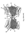
- a typical cell-occlusive, fluid permeable membrane 10 is illustrated surrounding a first section of the long bone 12 and a second section of long bone 14.
- the bone defect area 20 is bounded by the two ends 16, 18 of the first section of long bone 12 and the second section of long bone 14, respectively, and by the cell-occlusive, fluid-permeable membrane 10.
- this bone defect area 20 can receive blood from the bone vessels 23, blood and cells from the surrounding blood vessels 25 and tissues 27 is precluded from entering the bone defect area 20.
- the periosteum 31 and the surrounding tissues 27 are just external to the cell-occlusive, fluid-permeable membrane 10 and are guided in the directions of the arrows A1 and A2.
- US 5,496,372 discloses a hard tissue prostheses including porous thin metal sheets.
- the prostheses is for the replacement of hard tissues of human bones and joints and comprises a porous lamination component of metal thin sheets, each having a plurality of through holes and a thickness of 150 ⁇ m or less, and being unharmfull to the living body, the porous lamination component being formed such that the sheets are laid over one another and are then diffusion bonded there between into one body.
- EP 0 475 077 A2 pertains to a bone regeneration membrane.
- the bone regeneration membrane contains resorbable or degradable, polymeric and/or polymeric ceramic material.
- the membrane can be used for the spontaneous regeneration of bone and for the improved application of autologous and allogenic graft materials and therapeutic agents.
- the object of the present invention is to provide a protective bone regeneration membrane for protecting a hard tissue defect from a prolapse of adjacent soft tissues.
- a protective bone regeneration membrane 42 comprising a base material 44 and apertures 46.
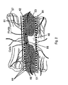
- the protective bone regeneration membrane 42 is shown in Figure 2 wrapped around a bone defect area 49.
- the bone which is surrounded by the protective bone regeneration membrane 42, comprises a first section of long bone 51, a second section of long bone 53, and a partially healed intermediate section of long bone 55.
- the protective bone regeneration membrane 42 is rigid enough to prevent prolapse of the surrounding tissues 57 into the bone defect area 49.
- the apertures 46 of the protective bone regeneration membrane 42 are large enough to allow for a proliferation of cells (not shown) and blood vessels 61 therethrough and into the first section of long bone 51, the second section of long bone 53, and the partially healed bone defect area 49. Since the protective bone regeneration membrane 42 of the presently preferred embodiment is rigid enough to withstand prolapse of the surrounding tissue 57, the regeneration of the partially damaged periosteum 64 is guided over the protective bone regeneration membrane 42 in a direction substantially parallel to the arrows A3 and A4.
- the apertures 46 within the protective bone regeneration membrane 42 are both cell and fluid permeable, and the base material 44 of the protective bone regeneration membrane 42 is rigid enough to maintain the available space between the first section of long bone 51 and the second section of long bone 53 for ideal bone regeneration. Additionally, the base material 44 is resorbable, according to the presently preferred embodiment.
- the cell-occlusive membrane of the prior art membrane 10 ( Figure 1), in contrast, is specifically designed to prevent the proliferation of cells and vessels therethrough. This membrane 10 is also insufficiently rigid and non-resorbable.
- Figures 3a and 3b illustrate different embodiments of a sheet of the protective bone regeneration membrane 42, comprising the base material 44 and the apertures 46.
- the protective bone regeneration membrane 42 comprises either a biodegradable synthetic material or a biodegradable natural material, or both.
- the biodegradable synthetic material may comprise polymers, for example, and the biodegradable natural material may comprise collagen, for example.
- Each of the apertures 46 preferably has a diameter within a range of between 20 microns and 3000 microns. In the presently preferred embodiment, each aperture 46 comprises a diameter of approximately 1500 microns.
- a thickness of the base material 44 is preferably within a range between 100 microns and 2000 microns, but may also be configured as thin as 10 microns.
- the pattern of distribution of the apertures 46 may vary according to the bone defect being treated.
- the ranges of aperture 46 sizes, base material 44 thickness, and aperture 46 shape and distribution is preferably implemented by the present invention in order to optimize the protective bone regeneration membrane 42 to different environmental conditions.
- Examples of the different environmental conditions encountered in different bone defects include the location of the defect (long bone or flat bone), the type of defect (discontinuity defect, contour defect, window defect, trephine defect), size of the defect, the presence or absence of periosteum 64, and the general condition of the adjacent soft tissues covering the bone defect.
- Figure 4 illustrates the protective bone regeneration membrane 42 applied to a long bone 68 of a patient.
- the protective bone regeneration membrane 42 is applied to the long bone 68 in combination with a fixation device 70.
- the fixation device 70 can be secured to the long bone 68 using conventional means, such as screws 72.
- tacks may be used in place of or in combination with the screws 72.
- fixation device 70, the screws 72, and the protective bone regeneration membrane 42 together securely hold the first section 75 of the long bone 68 to the second section 77 of long bone 68.
- a bone defect area 79 is protected against the prolapse of adjacent soft tissues, for example, by the protective bone regeneration membrane 42.
- the prior art titanium screen mesh is designed to remain permanently attached to the bone, resulting in long-term stress shielding and resorption of newly formed bone within the bone defect area 79.
- the protective bone regeneration membrane 42 of the present invention is preferably configured of a resorbable, bio-compatible material.
- the protective bone regeneration membrane 42 of the presently preferred embodiment will have resorbed sufficiently to no longer shield stress from the bone defect area 79 to thereby encourage an increase of bone formation.
- the fixation device 70, and/or the screws 72 are also formed of a resorbable material. That is, the combination of the fixation device 70, the screws 72, and the protective bone regeneration membrane 42 prevent excessive motion between the first section 75 and the second section 77 of the long bone 68.
- this period of time sufficient for complete new bone regeneration within the bone defect area 79 is between approximately 2 to 24 months.
- the resorption of the protective bone regeneration membrane 42 to a point where the protective bone regeneration membrane 42 can no longer shield significant mechanical stress on the first section 75 and the second section 77 is between approximately 2 and 24 months.
- the protective bone regeneration membrane 42 may comprise a non-resorbable material.
- the protective bone regeneration membrane 42 is non-resorbable and the fixation device 70 is resorbable, resorption of newly formed bone within the bone defect area 79 is still prevented.
- the protective bone regeneration membrane 42 is configured to be flexible enough to prevent stress shielding between the first section 75 and the second section 77, after the fixation device 70 has been resorbed to a point where the fixation device 70 no longer exerts mechanical strength on the first section 75 and the second section 77 of the long bone 68.
- the protective bone regeneration membrane 42 of the present invention is designed to be used in combination with a fixation device 70, in a preferred embodiment, while the titanium screen mesh of the prior art comprises a fixation device designed predominantly to be used alone.
- the protective bone regeneration membrane 42 of the present invention may be used in combination with the prior art titanium screen mesh, as well as in combination with any other conventional fixation device.
- internal fixation devices can be divided into two classes. Cortical compression plates comprise a first class and intramedullary rods comprise a second class. Both classes of devices are unable to secure and stabilize shattered bone, because bone fragments are often small and free floating within the fracture cavity.
- the protective bone regeneration membrane 42 of the presently preferred embodiment is preferably resorbed within the body of the patient to a point where substantial mechanical fixation is no longer exerted on the first section 75 and the second section 77 of the long bone 68, within a period of approximately 1 year. Complete resorption of the protective bone regeneration membrane 42 may subsequently occur after a total period of 11/2 to 2 years have elapsed since the initial implantation.
- the protective bone regeneration membrane 42 of the present invention is resorbed into the body of the patient. Allogenic bone grafts are only partially substituted with new bone over time, typically comprising 1 to 2 years, forming a permanent composite of viable (new) bone and non-viable cadaver bone.
- allogenic bone grafts cannot achieve a complete regeneration of the entire bone defect with new living bone, as can the protective bone regeneration membrane 42 of the present invention.
- This benefit is achieved by placement of the protective bone regeneration membrane 42 outside of the bone defect area 49, rather than within the bone defect area 49.
- the holes within the allogenic bone graft of the prior art are substantially occluded by induced bone formation therein within approximately 2 to 3 weeks after the initial implantation.
- the protective bone regeneration membrane 42 of the present invention is designed to be placed completely outside of the bone defect area, in order to maintain a maximal size of the bone defect area 79 for regeneration of new bone by the patient in the area 79.
- allogenic bone grafts are inferior to the protective bone regeneration membrane 42 of the present invention in providing a combination of patient safety in preventing disease transmission, optimal prolapse prevention and maximal space preservation for bone regeneration, and vasculature ingrowth potential.
- the above-mentioned skin graft of the prior art comprises apertures which are quickly occluded by the ingrowth of epithelial cells therein. These prior art apertures, similarly to the allogenic bone graft holes, are actually filled with the desired tissues, whereas, the apertures of the protective bone regeneration membrane 42 allow ongoing transmigration of cells and blood vessels for generating the desired tissue.
- these apertures are formed having a diameter of approximately 1 millimeter, whereas the preferred diameter of the apertures of the present invention are approximately 1.5 millimeters.
- the skin graft membrane of the prior art is specifically designed for providing an in vitro scaffold and subsequent transplantable skin graft, whereas the present invention preferably operates in vivo.
- the present invention is directed to maintaining a space, protected against adjacent soft tissue prolapse, to thereby facilitate spontaneous bone regeneration by the patient within the protected space.
- the present invention recognizes that spontaneous bone regeneration by the patient can be greatly accelerated and enhanced by allowing the infiltration of surrounding blood vessels and cells.
- mesenchymal stem cells which can be found in surrounding mesodermal tissues, are the precursor cells that eventually form muscle, cartilage, tendons, ligaments, connective tissues, and bone. These cells are present in these tissues and are involved in the perpetual renewal of each specific tissue, although in their earliest stage of development, these cells are not committed to becoming any given tissue. An uncommitted mesenchymal stem cell found in muscle, for example, will not strictly become a muscle cell. If the mesenchymal stem cell is needed to become a bone cell, the mesenchymal stem cell may migrate to a bone defect and differentiate into a bone forming cell.
- the mechanism for attracting these cells and directing them to become a specific tissue cell is understood by the present inventors to be controlled by morphogenic proteins, although other factors may be involved. In bone, for example, these proteins are commonly referred to as bone morphogenic proteins.
- the apertures 46 of the protective bone regeneration membrane 42 harness this mechanism, by allowing bone morphogenic proteins derived from within the bone matrix to attract mesenchymal stem cells from the surrounding connective tissues, musculature, periosteum, and vasculature. The attracted elements are then directed to differentiate into bone forming cells, which are essential for new bone formation by the patient.
- the apertures 46 of the present invention allow vital contributions of blood vessels from surrounding tissues, musculature, and periosteum into the protected area.
- Blood vessels invading the bone defect through the protective bone regeneration membrane 42 of the present invention greatly enhance the generation of new bone, as compared to prior art cell-occlusive membranes that limit the supply of blood to that coming from within the bone defect itself.
- the ability for capillaries from surrounding soft tissues to proliferate through the protective bone regeneration membrane 42 helps prevent migrating cells from the osseous bed and the periosteum from outstripping their proliferating blood supply. This proliferation of blood vessels increases the potential of spontaneous bone regeneration within a given defect.
- mesenchymal stem cells are believed to be perivascular (around blood vessels) connective tissue cells, which would additionally foster bone regeneration by the transmembranous sprouting of capillaries, since most vasculature has associated connective tissues.
- the base material 44 may be impregnated with a variety of substances for promoting the regeneration of different tissues such as bone and blood vessels.
- the base material 44 may be impregnated with a chemotactic substance for influencing cell-migration, an inhibitory substance for influencing cell-migration, a mitogenic growth factor for influencing cell proliferation and a growth factor for influencing cell differentiation (e.g. insulinelike growth factor, transforming growth factor-beta, fibroblast growth factor, platelet-derived growth factor), and factors which promote neoangiogenesis (formation of new blood vessels).
- a chemotactic substance for influencing cell-migration an inhibitory substance for influencing cell-migration
- a mitogenic growth factor for influencing cell proliferation e.g. insulinelike growth factor, transforming growth factor-beta, fibroblast growth factor, platelet-derived growth factor
- factors which promote neoangiogenesis formation of new blood vessels.
- the base material 44 is flexible both at the time of manufacture and after hydration. This flexibility allows the protective bone regeneration membrane 42 to be bent and shaped such that, after the area is completely healed, the contour of the healed bone matches the contour of the original bone, or matches the contour of the original bone as closely as possible.
- the base material 44 ( Figure 3) further provides an advantageous rigidity, which is higher than other currently used membrane materials ( Figure 1) to thereby provide sufficient strength against soft tissue pressure.
- the method generally comprises a step of affixing the protective bone regeneration membrane 42 (Figure 3) onto a portion of the mammalian skeletal system in need of repair.
- the fixation of the protective bone regeneration membrane 42 may be accomplished by any conventional surgical technique, including the use of resorbable pins, screws, and sutures.
- the protective bone regeneration membrane 42 of the present invention can be implanted into the patient without being affixed to existing bone, such as, for example, in the case of orbital floor reconstruction (Figure 5).
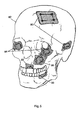
- Figure 5 illustrates several applications of the protective bone regeneration membrane in the cranio-facial region of a human skull.
- a protective bone regeneration membrane 80 is applied over the burrholes and the trephination defect of a human skull 82, after a neurosurgical procedure or trauma.
- protective bone regeneration membranes 84 are placed over orbital floor fractures to prevent entrapment of overlying muscles and nerves therein.
- Another protective bone regeneration membrane 86 is applied over a defect area in the maxillary sinus, and still another protective bone regeneration membrane 88 is applied over a bone defect area in the maxilla (upper jaw).
- Another protective bone regeneration membrane 90 is applied over an edentulous bone defect area in the mandible (lower jaw).
- a protective bone regeneration membrane 80 is illustrated in Figure 6, applied to the pelvis 82 of a human patient, after a bone autograft has been harvested therefrom.
- the protective bone regeneration membrane 80 protects the bone defect area 84 from soft tissue interposition, while allowing the ingrowth of blood vessels and cells. If necessary, the protective bone regeneration membrane 80 can be affixed onto the adjacent bone using pins, screws, sutures, or other conventional means.
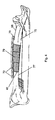
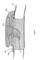
- Figure 7 illustrates a protective bone regeneration membrane 93 applied around a segmental defect 94 in a human mandible 95, for example.
- the protective bone regeneration membrane 93 can be implanted using an extra-oral (outside of the mouth) surgical approach.
- the epithelial lining of the mouth is not broken and the protective membrane is placed beneath the epithelial lining of the mouth (since the bone defect is accessed from an extra-oral area such as below the chin). Therefore the epithelial cells cannot enter the bone defect.
- the present invention is also intended to apply in intra-oral surgical approaches.
- the defect may be a discontinuity defect, comminuted, or just missing a part of the bone.
- the intact parts of the mandible 95 are fixated together by a plate 97 and screws 99, if necessary, and the protective bone regeneration membrane 93 protects the bone defect site from interposition of surrounding soft tissue.
- the protective bone regeneration membrane 93 holds any free-floating fragments of bone in place and provides additional circumferential stabilization to the bone defect.
- the protective bone regeneration membrane 93 is malleable to a certain extent, the protective bone regeneration membrane 93 is stiff enough to prevent collapse thereof under the weight of adjacent soft tissues.
- the protective bone regeneration membrane 93 can be easily cut with scissors and shaped by the hand of a user to adapt three-dimensionally to a bone defect area.
- Figure 8 illustrates another application of the protective bone regeneration membrane 105 of the present invention, as applied to a bone defect area of a long bone 101.
- the protective bone regeneration membrane 105 is secured to the long bone 101 using a fixation member 107 and screws 109, and comprises a belt-like tab 111.
- the belt-like tab 111 is adapted for being fed through a slot 113, which is formed between the fixation member 107 and the long bone 101.
- the protective bone regeneration membrane 105 is secured to the fixation member 107, and both the protective bone regeneration membrane 105 and the fixation member 107 are resorbable, in order to avoid a second surgery for removal of the devices.
- Surgical removal of non-resorbable, non-metallic membranes is necessary in the prior art, in order to avoid risk such as bacterial contamination and infection.
- a user can grip the belt-like tab 111 to securely fasten the protective bone regeneration membrane 105 around the long bone 101.
- This secure fastening of the protective membrane 105 around the long bone 101 can facilitate the holding of bone fragments in place within the bone defect area, in addition to adding stability to the bone fracture.
- the screws 109 are tightened into the long bone 101 after the protective bone regeneration membrane 105 is tightened around the long bone 101.
- the embodiment of Figure 8 is especially advantageous for setting comminuted fractures, having multiple bone fragments, to thereby reduce the risk of bone fragment resorption.
- the protective bone regeneration membrane 105 can be tightened around the long bone 101, until a desired tension is achieved for holding the native fracture fragments in place.
- the protective bone regeneration membrane 105 can also be used to prevent the dislocation of bone grafts or bone graft substitutes.
- the protective bone regeneration membrane 105 may be used without a fixation member 107. If it is necessary to stabilize major bone fragments, the protective bone regeneration membrane 105 may be used in conjunction with other rigid fixation devices, either internal or external.
- the protective bone regeneration membrane 105 may be used with or without a belt-like tab 111 to form a tube around a bone defect area of a long bone 101. If the tube overlaps both fracture ends of the long bone 101, the tube may provide sufficient structural support, resulting from the strength of the protective bone regeneration membrane 105 and the structural characteristics of the tube, to obviate the need for additional plates, screws, or external fixation devices. Structurally, a tube locates supporting elements in the area of highest stress when loaded in shear, compression, or in bending.
- the tube configuration is superior to intramedullary rods, which lay at the approximate neutral load axis, or eccentrically placed orthopedic plates, which support only one side of the fracture and which may introduce asymmetrical, non-axial loading on the fracture.
- a tube configuration will also have superior resistance to column (compression) loading. If the ends and seam of the protective bone regeneration membrane 105 are suitably fixated, the configuration will also be superior in shear strength.
Landscapes
- Health & Medical Sciences (AREA)
- Life Sciences & Earth Sciences (AREA)
- Veterinary Medicine (AREA)
- Public Health (AREA)
- General Health & Medical Sciences (AREA)
- Animal Behavior & Ethology (AREA)
- Orthopedic Medicine & Surgery (AREA)
- Transplantation (AREA)
- Heart & Thoracic Surgery (AREA)
- Biomedical Technology (AREA)
- Engineering & Computer Science (AREA)
- Oral & Maxillofacial Surgery (AREA)
- Vascular Medicine (AREA)
- Chemical & Material Sciences (AREA)
- Surgery (AREA)
- Epidemiology (AREA)
- Medicinal Chemistry (AREA)
- Cardiology (AREA)
- Molecular Biology (AREA)
- Dermatology (AREA)
- Neurosurgery (AREA)
- Neurology (AREA)
- Nuclear Medicine, Radiotherapy & Molecular Imaging (AREA)
- Medical Informatics (AREA)
- Dispersion Chemistry (AREA)
- Plastic & Reconstructive Surgery (AREA)
- Physical Education & Sports Medicine (AREA)
- Materials For Medical Uses (AREA)
- Prostheses (AREA)
- Separation Using Semi-Permeable Membranes (AREA)
Abstract
Description
- The present invention relates generally to implants for use in repairing various portions of the mammalian skeletal system and, more particularly, to implants for use in clinical procedures such as bone fracture repair, regeneration of bone loss, augmentation of deficient bone, and related procedures.
- Various types of defects in the mammalian skeletal system can be treated by various surgical procedures. Defects in the mammalian skeletal system may include bone fracture, loss of bone occurring from traumatic, surgical, or infectious sources, and bone deficiencies stemming from conditions such as atrophy and congenital anomalies.
- Turning to Figure 1, a typical cell-occlusive, fluid
permeable membrane 10 is illustrated surrounding a first section of thelong bone 12 and a second section oflong bone 14. Thebone defect area 20 is bounded by the twoends long bone 12 and the second section oflong bone 14, respectively, and by the cell-occlusive, fluid-permeable membrane 10. Although thisbone defect area 20 can receive blood from thebone vessels 23, blood and cells from the surroundingblood vessels 25 andtissues 27 is precluded from entering thebone defect area 20. Theperiosteum 31 and the surroundingtissues 27 are just external to the cell-occlusive, fluid-permeable membrane 10 and are guided in the directions of the arrows A1 and A2. - US 5,496,372 discloses a hard tissue prostheses including porous thin metal sheets. The prostheses is for the replacement of hard tissues of human bones and joints and comprises a porous lamination component of metal thin sheets, each having a plurality of through holes and a thickness of 150 µm or less, and being unharmfull to the living body, the porous lamination component being formed such that the sheets are laid over one another and are then diffusion bonded there between into one body.
- EP 0 475 077 A2 pertains to a bone regeneration membrane. The bone regeneration membrane contains resorbable or degradable, polymeric and/or polymeric ceramic material. The membrane can be used for the spontaneous regeneration of bone and for the improved application of autologous and allogenic graft materials and therapeutic agents.
- The object of the present invention is to provide a protective bone regeneration membrane for protecting a hard tissue defect from a prolapse of adjacent soft tissues.
- The aforementioned object is solved by the subject matter of claim 1. The dependent claims are directed to advantageous embodiments.
-
- Figure 1 illustrates a longitudinal cross-section of a cell-occlusive membrane secured around a long bone defect according to the prior art;
- Figure 2 illustrates a longitudinal cross-section of the protective bone regeneration membrane secured around a long bone defect according to the presently preferred embodiment;
- Figures. 3a and 3b illustrate the protective bone regeneration membrane according to the presently preferred embodiment;
- Figure 4 illustrates the protective bone regeneration membrane of the present invention, as applied to a long bone defect;
- Figure 5 illustrates the protective bone regeneration membrane of the present invention, applied to various bone defect areas of a human skull;
- Figure 6 illustrates the protective bone regeneration membrane of the presently preferred embodiment, used to facilitate bone regeneration of the iliac crest of a patient, after a bone autograft has been harvested from the patient;
- Figure 7 illustrates the protective bone regeneration membrane of the present invention, as applied to a mandibular (lower jaw) bone defect of a patient; and
- Figure 8 illustrates the protective bone regeneration membrane of the present invention, used in combination with a fixation device, as applied to a long bone defect of a patient.
- Turning to Figure 2, a protective
bone regeneration membrane 42 is illustrated, comprising abase material 44 andapertures 46. The protectivebone regeneration membrane 42 is shown in Figure 2 wrapped around abone defect area 49. The bone, which is surrounded by the protectivebone regeneration membrane 42, comprises a first section oflong bone 51, a second section oflong bone 53, and a partially healed intermediate section oflong bone 55. The protectivebone regeneration membrane 42 is rigid enough to prevent prolapse of the surroundingtissues 57 into thebone defect area 49. Unlike the prior-art apertures 35 of the cell-occlusive, fluid permeable membrane 10 (Figure 1), theapertures 46 of the protectivebone regeneration membrane 42 are large enough to allow for a proliferation of cells (not shown) andblood vessels 61 therethrough and into the first section oflong bone 51, the second section oflong bone 53, and the partially healedbone defect area 49. Since the protectivebone regeneration membrane 42 of the presently preferred embodiment is rigid enough to withstand prolapse of the surroundingtissue 57, the regeneration of the partially damagedperiosteum 64 is guided over the protectivebone regeneration membrane 42 in a direction substantially parallel to the arrows A3 and A4. - The
apertures 46 within the protectivebone regeneration membrane 42 are both cell and fluid permeable, and thebase material 44 of the protectivebone regeneration membrane 42 is rigid enough to maintain the available space between the first section oflong bone 51 and the second section oflong bone 53 for ideal bone regeneration. Additionally, thebase material 44 is resorbable, according to the presently preferred embodiment. The cell-occlusive membrane of the prior art membrane 10 (Figure 1), in contrast, is specifically designed to prevent the proliferation of cells and vessels therethrough. Thismembrane 10 is also insufficiently rigid and non-resorbable. - Figures 3a and 3b illustrate different embodiments of a sheet of the protective
bone regeneration membrane 42, comprising thebase material 44 and theapertures 46. As presently embodied, the protectivebone regeneration membrane 42 comprises either a biodegradable synthetic material or a biodegradable natural material, or both. The biodegradable synthetic material may comprise polymers, for example, and the biodegradable natural material may comprise collagen, for example. Each of theapertures 46 preferably has a diameter within a range of between 20 microns and 3000 microns. In the presently preferred embodiment, eachaperture 46 comprises a diameter of approximately 1500 microns. A thickness of thebase material 44 is preferably within a range between 100 microns and 2000 microns, but may also be configured as thin as 10 microns. The pattern of distribution of theapertures 46 may vary according to the bone defect being treated. The ranges ofaperture 46 sizes,base material 44 thickness, andaperture 46 shape and distribution is preferably implemented by the present invention in order to optimize the protectivebone regeneration membrane 42 to different environmental conditions. Examples of the different environmental conditions encountered in different bone defects include the location of the defect (long bone or flat bone), the type of defect (discontinuity defect, contour defect, window defect, trephine defect), size of the defect, the presence or absence ofperiosteum 64, and the general condition of the adjacent soft tissues covering the bone defect. - Figure 4 illustrates the protective
bone regeneration membrane 42 applied to along bone 68 of a patient. The protectivebone regeneration membrane 42 is applied to thelong bone 68 in combination with afixation device 70. Thefixation device 70 can be secured to thelong bone 68 using conventional means, such asscrews 72. In alternative embodiments, tacks may be used in place of or in combination with thescrews 72. - The
fixation device 70, thescrews 72, and the protectivebone regeneration membrane 42 together securely hold thefirst section 75 of thelong bone 68 to thesecond section 77 oflong bone 68. Abone defect area 79 is protected against the prolapse of adjacent soft tissues, for example, by the protectivebone regeneration membrane 42. - In contrast to the titanium screen mesh of the prior art, the inventors believe that the combination of the protective
bone regeneration membrane 42 and thefixation device 70 may in some instances be adapted for operating together to relieve stress shielding of thelong bone 68, to thereby prevent subsequent resorption of new bone. The prior art titanium screen mesh is designed to remain permanently attached to the bone, resulting in long-term stress shielding and resorption of newly formed bone within thebone defect area 79. In contrast to the prior art titanium screen mesh, the protectivebone regeneration membrane 42 of the present invention is preferably configured of a resorbable, bio-compatible material. At about the time that the new bone within thebone defect area 79 is fully regenerated, the protectivebone regeneration membrane 42 of the presently preferred embodiment will have resorbed sufficiently to no longer shield stress from thebone defect area 79 to thereby encourage an increase of bone formation. In addition, according to the presently preferred embodiment, thefixation device 70, and/or thescrews 72, are also formed of a resorbable material. That is, the combination of thefixation device 70, thescrews 72, and the protectivebone regeneration membrane 42 prevent excessive motion between thefirst section 75 and thesecond section 77 of thelong bone 68. - As presently embodied, this period of time sufficient for complete new bone regeneration within the
bone defect area 79 is between approximately 2 to 24 months. Thus, according to the present invention, the resorption of the protectivebone regeneration membrane 42 to a point where the protectivebone regeneration membrane 42 can no longer shield significant mechanical stress on thefirst section 75 and thesecond section 77 is between approximately 2 and 24 months. - In an alternative embodiment, the protective
bone regeneration membrane 42 may comprise a non-resorbable material. In this alternative embodiment where the protectivebone regeneration membrane 42 is non-resorbable and thefixation device 70 is resorbable, resorption of newly formed bone within thebone defect area 79 is still prevented. More particularly, the protectivebone regeneration membrane 42 is configured to be flexible enough to prevent stress shielding between thefirst section 75 and thesecond section 77, after thefixation device 70 has been resorbed to a point where thefixation device 70 no longer exerts mechanical strength on thefirst section 75 and thesecond section 77 of thelong bone 68. - As another distinguishing feature, the protective
bone regeneration membrane 42 of the present invention is designed to be used in combination with afixation device 70, in a preferred embodiment, while the titanium screen mesh of the prior art comprises a fixation device designed predominantly to be used alone. In one conceivable embodiment of the present invention, the protectivebone regeneration membrane 42 of the present invention may be used in combination with the prior art titanium screen mesh, as well as in combination with any other conventional fixation device. Generally, internal fixation devices can be divided into two classes. Cortical compression plates comprise a first class and intramedullary rods comprise a second class. Both classes of devices are unable to secure and stabilize shattered bone, because bone fragments are often small and free floating within the fracture cavity. Furthermore, the periosteum around such fracture sites is usually destroyed and cannot serve as a membrane barrier against the dislocation of bone fragments. Multiple bone fragments are naturally resorbed unless they can be rigidly held together and provided with sufficient blood supply. Bone fragment resorption can present a significant obstacle to efficient healing of comminuted fractures. Bone fragment resorption often necessitates additional bone grafting procedures. In contrast to the protectivebone regeneration membrane 42 of the present invention, both of the above mentioned classifications of fixation devices are unable to achieve this end. - The protective
bone regeneration membrane 42 of the presently preferred embodiment is preferably resorbed within the body of the patient to a point where substantial mechanical fixation is no longer exerted on thefirst section 75 and thesecond section 77 of thelong bone 68, within a period of approximately 1 year. Complete resorption of the protectivebone regeneration membrane 42 may subsequently occur after a total period of 11/2 to 2 years have elapsed since the initial implantation. In contrast to the allogenic bone grafts of the prior art, the protectivebone regeneration membrane 42 of the present invention is resorbed into the body of the patient. Allogenic bone grafts are only partially substituted with new bone over time, typically comprising 1 to 2 years, forming a permanent composite of viable (new) bone and non-viable cadaver bone. Thus, allogenic bone grafts cannot achieve a complete regeneration of the entire bone defect with new living bone, as can the protectivebone regeneration membrane 42 of the present invention. This benefit is achieved by placement of the protectivebone regeneration membrane 42 outside of thebone defect area 49, rather than within thebone defect area 49. Additionally, the holes within the allogenic bone graft of the prior art are substantially occluded by induced bone formation therein within approximately 2 to 3 weeks after the initial implantation. Finally, as a further distinguishing feature between the protectivebone regeneration membrane 42 of the present invention and the prior art allogenic bone graft, the prior art allogenic bone graft is placed within the bone defect area itself, since the purpose of the prior artallogenic bone graft 42 is to become a part of the new bone. In contrast, the protectivebone regeneration membrane 42 of the present invention is designed to be placed completely outside of the bone defect area, in order to maintain a maximal size of thebone defect area 79 for regeneration of new bone by the patient in thearea 79. - Still further, allogenic bone grafts are inferior to the protective
bone regeneration membrane 42 of the present invention in providing a combination of patient safety in preventing disease transmission, optimal prolapse prevention and maximal space preservation for bone regeneration, and vasculature ingrowth potential. Similarly to the allogenic bone graft of the prior art, the above-mentioned skin graft of the prior art comprises apertures which are quickly occluded by the ingrowth of epithelial cells therein. These prior art apertures, similarly to the allogenic bone graft holes, are actually filled with the desired tissues, whereas, the apertures of the protectivebone regeneration membrane 42 allow ongoing transmigration of cells and blood vessels for generating the desired tissue. Additionally, these apertures are formed having a diameter of approximately 1 millimeter, whereas the preferred diameter of the apertures of the present invention are approximately 1.5 millimeters. Additionally, the skin graft membrane of the prior art is specifically designed for providing an in vitro scaffold and subsequent transplantable skin graft, whereas the present invention preferably operates in vivo. - Many of the above-described differences between the protective
bone regeneration membrane 42 of the present invention and prior art devices help point to a fundamental difference between the present invention and prior art devices. The present invention is directed to maintaining a space, protected against adjacent soft tissue prolapse, to thereby facilitate spontaneous bone regeneration by the patient within the protected space. The present invention recognizes that spontaneous bone regeneration by the patient can be greatly accelerated and enhanced by allowing the infiltration of surrounding blood vessels and cells. - The present inventors recognize that mesenchymal stem cells, which can be found in surrounding mesodermal tissues, are the precursor cells that eventually form muscle, cartilage, tendons, ligaments, connective tissues, and bone. These cells are present in these tissues and are involved in the perpetual renewal of each specific tissue, although in their earliest stage of development, these cells are not committed to becoming any given tissue. An uncommitted mesenchymal stem cell found in muscle, for example, will not strictly become a muscle cell. If the mesenchymal stem cell is needed to become a bone cell, the mesenchymal stem cell may migrate to a bone defect and differentiate into a bone forming cell.
- The mechanism for attracting these cells and directing them to become a specific tissue cell is understood by the present inventors to be controlled by morphogenic proteins, although other factors may be involved. In bone, for example, these proteins are commonly referred to as bone morphogenic proteins. The
apertures 46 of the protectivebone regeneration membrane 42 harness this mechanism, by allowing bone morphogenic proteins derived from within the bone matrix to attract mesenchymal stem cells from the surrounding connective tissues, musculature, periosteum, and vasculature. The attracted elements are then directed to differentiate into bone forming cells, which are essential for new bone formation by the patient. In addition, theapertures 46 of the present invention allow vital contributions of blood vessels from surrounding tissues, musculature, and periosteum into the protected area. Blood vessels invading the bone defect through the protectivebone regeneration membrane 42 of the present invention greatly enhance the generation of new bone, as compared to prior art cell-occlusive membranes that limit the supply of blood to that coming from within the bone defect itself. The ability for capillaries from surrounding soft tissues to proliferate through the protectivebone regeneration membrane 42 helps prevent migrating cells from the osseous bed and the periosteum from outstripping their proliferating blood supply. This proliferation of blood vessels increases the potential of spontaneous bone regeneration within a given defect. Furthermore, mesenchymal stem cells are believed to be perivascular (around blood vessels) connective tissue cells, which would additionally foster bone regeneration by the transmembranous sprouting of capillaries, since most vasculature has associated connective tissues. - The base material 44 (Figure 3), according to the present invention, may be impregnated with a variety of substances for promoting the regeneration of different tissues such as bone and blood vessels. The
base material 44 may be impregnated with a chemotactic substance for influencing cell-migration, an inhibitory substance for influencing cell-migration, a mitogenic growth factor for influencing cell proliferation and a growth factor for influencing cell differentiation (e.g. insulinelike growth factor, transforming growth factor-beta, fibroblast growth factor, platelet-derived growth factor), and factors which promote neoangiogenesis (formation of new blood vessels). - According to the present invention, the
base material 44 is flexible both at the time of manufacture and after hydration. This flexibility allows the protectivebone regeneration membrane 42 to be bent and shaped such that, after the area is completely healed, the contour of the healed bone matches the contour of the original bone, or matches the contour of the original bone as closely as possible. According to the present invention, the base material 44 (Figure 3) further provides an advantageous rigidity, which is higher than other currently used membrane materials (Figure 1) to thereby provide sufficient strength against soft tissue pressure. - The method generally comprises a step of affixing the protective bone regeneration membrane 42 (Figure 3) onto a portion of the mammalian skeletal system in need of repair. The fixation of the protective
bone regeneration membrane 42 may be accomplished by any conventional surgical technique, including the use of resorbable pins, screws, and sutures. Alternatively, the protectivebone regeneration membrane 42 of the present invention can be implanted into the patient without being affixed to existing bone, such as, for example, in the case of orbital floor reconstruction (Figure 5). - Other applications of the protective bone regeneration membrane of the present invention are illustrated in Figures 5-8. Figure 5 illustrates several applications of the protective bone regeneration membrane in the cranio-facial region of a human skull. A protective
bone regeneration membrane 80 is applied over the burrholes and the trephination defect of ahuman skull 82, after a neurosurgical procedure or trauma. Inside the orbits of the skull, protectivebone regeneration membranes 84 are placed over orbital floor fractures to prevent entrapment of overlying muscles and nerves therein. Another protectivebone regeneration membrane 86 is applied over a defect area in the maxillary sinus, and still another protectivebone regeneration membrane 88 is applied over a bone defect area in the maxilla (upper jaw). Another protectivebone regeneration membrane 90 is applied over an edentulous bone defect area in the mandible (lower jaw). - A protective
bone regeneration membrane 80 is illustrated in Figure 6, applied to thepelvis 82 of a human patient, after a bone autograft has been harvested therefrom. The protectivebone regeneration membrane 80 protects thebone defect area 84 from soft tissue interposition, while allowing the ingrowth of blood vessels and cells. If necessary, the protectivebone regeneration membrane 80 can be affixed onto the adjacent bone using pins, screws, sutures, or other conventional means. Figure 7 illustrates a protectivebone regeneration membrane 93 applied around asegmental defect 94 in ahuman mandible 95, for example. The protectivebone regeneration membrane 93 can be implanted using an extra-oral (outside of the mouth) surgical approach. According to this approach, the epithelial lining of the mouth is not broken and the protective membrane is placed beneath the epithelial lining of the mouth (since the bone defect is accessed from an extra-oral area such as below the chin). Therefore the epithelial cells cannot enter the bone defect. The present invention, however, is also intended to apply in intra-oral surgical approaches. The defect may be a discontinuity defect, comminuted, or just missing a part of the bone. The intact parts of themandible 95 are fixated together by aplate 97 and screws 99, if necessary, and the protectivebone regeneration membrane 93 protects the bone defect site from interposition of surrounding soft tissue. Additionally, the protectivebone regeneration membrane 93 holds any free-floating fragments of bone in place and provides additional circumferential stabilization to the bone defect. Although the protectivebone regeneration membrane 93 is malleable to a certain extent, the protectivebone regeneration membrane 93 is stiff enough to prevent collapse thereof under the weight of adjacent soft tissues. The protectivebone regeneration membrane 93 can be easily cut with scissors and shaped by the hand of a user to adapt three-dimensionally to a bone defect area. - Figure 8 illustrates another application of the protective
bone regeneration membrane 105 of the present invention, as applied to a bone defect area of along bone 101. The protectivebone regeneration membrane 105 is secured to thelong bone 101 using afixation member 107 and screws 109, and comprises a belt-like tab 111. The belt-like tab 111 is adapted for being fed through aslot 113, which is formed between thefixation member 107 and thelong bone 101. In the presently preferred embodiment, the protectivebone regeneration membrane 105 is secured to thefixation member 107, and both the protectivebone regeneration membrane 105 and thefixation member 107 are resorbable, in order to avoid a second surgery for removal of the devices. Surgical removal of non-resorbable, non-metallic membranes is necessary in the prior art, in order to avoid risk such as bacterial contamination and infection. A user can grip the belt-like tab 111 to securely fasten the protectivebone regeneration membrane 105 around thelong bone 101. This secure fastening of theprotective membrane 105 around thelong bone 101 can facilitate the holding of bone fragments in place within the bone defect area, in addition to adding stability to the bone fracture. In the presently preferred embodiment, thescrews 109 are tightened into thelong bone 101 after the protectivebone regeneration membrane 105 is tightened around thelong bone 101. The embodiment of Figure 8 is especially advantageous for setting comminuted fractures, having multiple bone fragments, to thereby reduce the risk of bone fragment resorption. The protectivebone regeneration membrane 105 can be tightened around thelong bone 101, until a desired tension is achieved for holding the native fracture fragments in place. The protectivebone regeneration membrane 105 can also be used to prevent the dislocation of bone grafts or bone graft substitutes. Of course, the protectivebone regeneration membrane 105 may be used without afixation member 107. If it is necessary to stabilize major bone fragments, the protectivebone regeneration membrane 105 may be used in conjunction with other rigid fixation devices, either internal or external. - The protective
bone regeneration membrane 105 may be used with or without a belt-like tab 111 to form a tube around a bone defect area of along bone 101. If the tube overlaps both fracture ends of thelong bone 101, the tube may provide sufficient structural support, resulting from the strength of the protectivebone regeneration membrane 105 and the structural characteristics of the tube, to obviate the need for additional plates, screws, or external fixation devices. Structurally, a tube locates supporting elements in the area of highest stress when loaded in shear, compression, or in bending. The tube configuration, according to this alternative embodiment, is superior to intramedullary rods, which lay at the approximate neutral load axis, or eccentrically placed orthopedic plates, which support only one side of the fracture and which may introduce asymmetrical, non-axial loading on the fracture. In addition to superior strength in bending, a tube configuration will also have superior resistance to column (compression) loading. If the ends and seam of the protectivebone regeneration membrane 105 are suitably fixated, the configuration will also be superior in shear strength. Although the present material, configurations, and methods have been described in the context of treating humans, these materials, configurations, and methods can also be useful in treating animals. - Although an exemplary embodiment of the invention has been shown and described, many other changes, modifications and substitutions, in addition to those set forth in the above paragraphs, may be made by one having ordinary skill in the art without necessarily departing from the scope of this invention.
Claims (13)
- A protective bone regeneration membrane (42) for protecting a hard tissue defect from a prolapse of adjacent soft tissues during in vivo repair of the hard tissue defect, the protective membrane (42) having a pre-implant configuration, which is defined as a configuration of the protective membrane immediately before the protective membrane is implanted over the hard tissue defect and placed into contact with any adjacent soft tissue, the protective membrane (42) comprising:a substantially planar sheet of resorbable polymer base material having a first side, a second side, a thickness measured between the first side and the second side that is in a range between 100 microns and 2000 microns;a plurality of apertures (46) disposed in the substantially planar sheet of resorbable polymer base material (44) to substantially optimize healing of the hard tissue defect when the protective membrane is implanted over the hard tissue defect;wherein the protective membrane (42) further comprises a configuration and strength sufficient to prevent gross prolapse of adjacent soft tissues into the hard tissue defect, when the protective membrane (42) is implanted over the hard tissue defect and placed into contact with any adjacent soft tissue;
wherein the plurality of apertures (46) of the substantially planar sheet of resorbable polymer base material (44), immediately after implanting of the substantially planar sheet of resorbable polymer base material over the hard tissue defect, have diameters sufficient in size and distribution to allow and optimize a proliferation of vasculature and connecive tissue cells, derived from adjacent soft tissues, to permeate through the apertures and substantially into the hard tissue defect; and
wherein the protective membrane (42) is adapted to be resorbed into a mammalian body, and not remodeled, within a period of approximately 2 to 24 months from an initial implantation of the protective membrane into the mammalian body; characterized in that said apertures of the plurality of apertures of the protective membrane when the protective membrane is in the pre-implant configuration have minimum diameters which are greater than 1 millimeter and the upper limit of the range of diameter is 3 millimeter. - The protective bone regeneration membrane as recited in Claim 1, wherein cross-sectional areas of fluid flow paths are substantially constant along lengths of the fluid-flow paths from the first side to the second side.
- The protective bone regeneration membrane as recited in Claim 1, wherein the apertures (46) are uniformly distributed on the sheet of resorbable polymer base material (44) in rows of at least three apertures and columns of at least three apertures.
- The protective bone regeneration membrane as recited in Claim 1, wherein cross-sectional areas of the apertures (46) have circular perimeters.
- The protective bone regeneration membrane as recited in Claim 1, wherein the first side comprises a substantially-smooth side and the second side comprises a substantially-smooth side.
- The protective bone regeneration membrane as recited in Claim 1, wherein apertures (46) of the plurality of apertures of the protective membrane when the protective membrane is in the pre-implant configuration have minimum diameters less than about 2000 microns.
- The protective bone regeneration membrane as recited in Claim 1, wherein apertures (46) of the plurality of apertures (46) of the protective membrane when the protective membrane is in the pre-implant configuration have minimum diameters of about 1500 microns.
- The protective bone regeneration membrane as recited in Claim 1, wherein apertures (46) of the plurality of apertures (46) are arranged on the sheet of resorbable polymer base material in staggered rows.
- The protective bone regeneration membrane as recited in Claim 1, wherein cross-sectional areas of fluid flow paths are substantially constant along lengths of the fluid-flow paths from the first side to the second side.
- The protective bone regeneration membrane as recited in Claim 1, wherein the apertures (46) are uniformly distributed on the sheet of resorbable polymer base material (44).
- The protective bone regeneration membrane as recited in Claim 1, wherein the apertures (46) are uniformly distributed on the sheet of resorbable polymer base material (44) in rows and apertures (46) of the plurality of apertures (46) are arranged on the sheet of resorbable polymer base material (44) in staggered rows of at least three apertures (46) and columns of at least three apertures (46).
- The protective bone regeneration membrane as recited in Claim 1, wherein the sheet of resorbable polymer base material (44) comprises a single layer which is substantially solid.
- The protective bone regeneration membrane as recited in Claim 1, wherein:apertures (46) of the plurality of apertures (46) are arranged on the sheet of resorbable polymer base material (44) in a matrix having columns of at least three apertures (46) and rows of at least three apertures, andthe rows are staggered.
Applications Claiming Priority (6)
| Application Number | Priority Date | Filing Date | Title |
|---|---|---|---|
| US08/699,673 US5919234A (en) | 1996-08-19 | 1996-08-19 | Resorbable, macro-porous, non-collapsing and flexible membrane barrier for skeletal repair and regeneration |
| PCT/US1997/013695 WO1998007384A1 (en) | 1996-08-19 | 1997-08-05 | Resorbable, macro-porous, non-collapsing and flexible membrane barrier for skeletal repair and regeneration |
| US7240198P | 1998-01-23 | 1998-01-23 | |
| US7520498P | 1998-02-18 | 1998-02-18 | |
| US9606998P | 1998-08-11 | 1998-08-11 | |
| US699673 | 2000-10-30 |
Publications (2)
| Publication Number | Publication Date |
|---|---|
| EP0928168A1 EP0928168A1 (en) | 1999-07-14 |
| EP0928168B1 true EP0928168B1 (en) | 2006-06-21 |
Family
ID=27490977
Family Applications (1)
| Application Number | Title | Priority Date | Filing Date |
|---|---|---|---|
| EP97936390A Expired - Lifetime EP0928168B1 (en) | 1996-08-19 | 1997-08-05 | Resorbable, macro-porous, non-collapsing and flexible membrane barrier for skeletal repair and regeneration |
Country Status (5)
| Country | Link |
|---|---|
| US (1) | US5919234A (en) |
| EP (1) | EP0928168B1 (en) |
| JP (1) | JP2000516511A (en) |
| AU (1) | AU718801B2 (en) |
| WO (2) | WO1998007384A1 (en) |
Cited By (1)
| Publication number | Priority date | Publication date | Assignee | Title |
|---|---|---|---|---|
| EP2165943A1 (en) | 2008-09-19 | 2010-03-24 | Nature Delivered, Ltd. | Food delivery packaging and process |
Families Citing this family (114)
| Publication number | Priority date | Publication date | Assignee | Title |
|---|---|---|---|---|
| DE10237531A1 (en) * | 2002-08-16 | 2004-02-26 | Tutogen Medical Gmbh | implant |
| US8795332B2 (en) | 2002-09-30 | 2014-08-05 | Ethicon, Inc. | Barbed sutures |
| US6241747B1 (en) | 1993-05-03 | 2001-06-05 | Quill Medical, Inc. | Barbed Bodily tissue connector |
| FI954565A0 (en) * | 1995-09-27 | 1995-09-27 | Biocon Oy | Biologically applied polymeric material to the implant and foil preparation |
| US6280473B1 (en) * | 1996-08-19 | 2001-08-28 | Macropore, Inc. | Resorbable, macro-porous, non-collapsing and flexible membrane barrier for skeletal repair and regeneration |
| EP1018982B1 (en) * | 1996-12-03 | 2005-03-09 | Osteobiologics, Inc. | Biodegradable polymeric film |
| US5931855A (en) | 1997-05-21 | 1999-08-03 | Frank Hoffman | Surgical methods using one-way suture |
| US6652592B1 (en) | 1997-10-27 | 2003-11-25 | Regeneration Technologies, Inc. | Segmentally demineralized bone implant |
| US6090998A (en) * | 1997-10-27 | 2000-07-18 | University Of Florida | Segmentally demineralized bone implant |
| ATE286692T1 (en) * | 1998-04-07 | 2005-01-15 | Macropore Inc | MEMBRANE WITH A CORRUPTED SURFACE FOR GUIDING THE FABRIC |
| SE515572C2 (en) * | 1998-09-09 | 2001-09-03 | Lanka Ltd | Implants, ways of making it and using it |
| US6350284B1 (en) | 1998-09-14 | 2002-02-26 | Bionx Implants, Oy | Bioabsorbable, layered composite material for guided bone tissue regeneration |
| US7662409B2 (en) | 1998-09-25 | 2010-02-16 | Gel-Del Technologies, Inc. | Protein matrix materials, devices and methods of making and using thereof |
| US6328765B1 (en) | 1998-12-03 | 2001-12-11 | Gore Enterprise Holdings, Inc. | Methods and articles for regenerating living tissue |
| US6409764B1 (en) * | 1998-12-03 | 2002-06-25 | Charles F. White | Methods and articles for regenerating bone or peridontal tissue |
| US6375663B1 (en) * | 1999-03-17 | 2002-04-23 | Maxilon Laboratories, Inc. | Bone grafting material |
| US6520996B1 (en) | 1999-06-04 | 2003-02-18 | Depuy Acromed, Incorporated | Orthopedic implant |
| DE60041670D1 (en) * | 1999-12-03 | 2009-04-09 | Univ Leeds | Restoration of damaged tissue |
| AU2081701A (en) | 1999-12-09 | 2001-06-18 | Macropore | Partially resorbable connective tissue distraction devices and techniques |
| FR2802405A1 (en) * | 1999-12-21 | 2001-06-22 | Pierre Alain Janin | Orbital prosthesis for facial reconstruction comprises plate of synthetic biocompatible material with asymmetrical medial cavity |
| FR2808185B1 (en) * | 2000-04-26 | 2003-02-28 | Aspide | REINFORCEMENT WALL FOR THE TREATMENT OF ALTERED TISSUES OF THE ABDOMINAL WALL |
| WO2004043303A2 (en) * | 2002-11-12 | 2004-05-27 | Regenex Ltd. | Expandable devices and methods for tissue expansion, regenerationand fixation |
| US8622739B2 (en) * | 2001-05-09 | 2014-01-07 | Ben-Zion Karmon | Method for enlarging a jaw bone using a hollow dental implant having a side perforation |
| US7771482B1 (en) * | 2000-05-09 | 2010-08-10 | Ben-Zion Karmon | Method for tissue expansion and regeneration using bioresorbable inflatable devices |
| US20040010275A1 (en) * | 2000-05-19 | 2004-01-15 | Daniel Jacobs | Multi-point tissue tension distribution device and method, a custom-fittable variation |
| US7172615B2 (en) * | 2000-05-19 | 2007-02-06 | Coapt Systems, Inc. | Remotely anchored tissue fixation device |
| US7156862B2 (en) | 2000-05-19 | 2007-01-02 | Coapt Systems, Inc. | Multi-point tension distribution system device and method of tissue approximation using that device to improve wound healing |
| US6485503B2 (en) | 2000-05-19 | 2002-11-26 | Coapt Systems, Inc. | Multi-point tissue tension distribution device, a brow and face lift variation, and a method of tissue approximation using the device |
| AU6473901A (en) * | 2000-05-19 | 2001-12-03 | Coapt Systems Inc | Multi-point tissue tension distribution device, a combined orbital rim repair and suspension variation, and a method of tissue approximation using the device |
| US7510566B2 (en) * | 2000-05-19 | 2009-03-31 | Coapt Systems, Inc. | Multi-point tissue tension distribution device and method, a chin lift variation |
| US6645226B1 (en) | 2000-05-19 | 2003-11-11 | Coapt Systems, Inc. | Multi-point tension distribution system device and method of tissue approximation using that device to improve wound healing |
| US20050119694A1 (en) * | 2000-05-19 | 2005-06-02 | Jacobs Daniel I. | Remotely anchored tissue fixation device and method |
| US6613089B1 (en) | 2000-10-25 | 2003-09-02 | Sdgi Holdings, Inc. | Laterally expanding intervertebral fusion device |
| EP1331906A2 (en) | 2000-10-25 | 2003-08-06 | SDGI Holdings, Inc. | Vertically expanding intervertebral body fusion device |
| US6736848B2 (en) | 2001-05-29 | 2004-05-18 | Sdgi Holdings, Inc. | Method and apparatus for using formable polymers for orthopedic support |
| US7056331B2 (en) | 2001-06-29 | 2006-06-06 | Quill Medical, Inc. | Suture method |
| US6848152B2 (en) | 2001-08-31 | 2005-02-01 | Quill Medical, Inc. | Method of forming barbs on a suture and apparatus for performing same |
| CA2469370C (en) | 2001-12-07 | 2014-07-08 | Macropore Biosurgery, Inc. | Adipose-derived cell processing unit |
| US20030142676A1 (en) * | 2002-01-25 | 2003-07-31 | Raymond Zeisz | Method and apparauts for admission control in packet switch |
| US20030181889A1 (en) * | 2002-03-21 | 2003-09-25 | Little Adrian Leigh | Healing accelerator |
| WO2003092468A2 (en) * | 2002-04-29 | 2003-11-13 | Gel-Del Technologies, Inc. | Biomatrix structural containment and fixation systems and methods of use thereof |
| US6773450B2 (en) | 2002-08-09 | 2004-08-10 | Quill Medical, Inc. | Suture anchor and method |
| US8100940B2 (en) | 2002-09-30 | 2012-01-24 | Quill Medical, Inc. | Barb configurations for barbed sutures |
| US20040088003A1 (en) | 2002-09-30 | 2004-05-06 | Leung Jeffrey C. | Barbed suture in combination with surgical needle |
| US7682392B2 (en) | 2002-10-30 | 2010-03-23 | Depuy Spine, Inc. | Regenerative implants for stabilizing the spine and devices for attachment of said implants |
| KR20100102753A (en) * | 2003-04-16 | 2010-09-24 | 포렉스 서지칼, 인크. | Craniofacial implant |
| US8298292B2 (en) | 2003-04-16 | 2012-10-30 | Howmedica Osteonics Corp. | Craniofacial implant |
| US7624487B2 (en) | 2003-05-13 | 2009-12-01 | Quill Medical, Inc. | Apparatus and method for forming barbs on a suture |
| WO2004108180A1 (en) * | 2003-06-04 | 2004-12-16 | Inion Ltd | Biodegradable implant and method for manufacturing one |
| WO2005011569A2 (en) | 2003-06-18 | 2005-02-10 | Macropore Biosurgery Inc. | Methods of using adipose tissue-derived cells in augmenting autologous fat transfer |
| BRPI0411798A (en) | 2003-06-25 | 2006-08-08 | Macropore Biosurgery Inc | systems and methods for separating and concentrating regenerative cells from tissue |
| CN1878470A (en) | 2003-09-17 | 2006-12-13 | 马克罗珀尔生物外科公司 | Methods of using regenerative cells in the treatment of peripheral vascular disease and related disorders |
| US7666230B2 (en) * | 2003-12-08 | 2010-02-23 | Depuy Products, Inc. | Implant device for cartilage regeneration in load bearing articulation regions |
| US20050197699A1 (en) * | 2004-02-10 | 2005-09-08 | Jacobs Daniel I. | Tissue repair apparatus and method |
| US20050209542A1 (en) * | 2004-03-16 | 2005-09-22 | Jacobs Daniel I | Tissue approximation sling and method |
| JP2007529515A (en) | 2004-03-19 | 2007-10-25 | サイトリ セラピューティクス インコーポレイテッド | Cell carrier containing regenerative cells and cell carrier containment device |
| US20050216008A1 (en) * | 2004-03-24 | 2005-09-29 | Zwirnmann Ralph F | Bone fixation implants |
| WO2005114322A2 (en) | 2004-05-12 | 2005-12-01 | Massachusetts Institute Of Technology | Manufacturing process, such as three-dimensional printing, including solvent vapor filming and the like |
| WO2005112787A2 (en) | 2004-05-14 | 2005-12-01 | Quill Medical, Inc. | Suture methods and devices |
| US7887587B2 (en) * | 2004-06-04 | 2011-02-15 | Synthes Usa, Llc | Soft tissue spacer |
| TWI374036B (en) * | 2004-06-10 | 2012-10-11 | Sean Kerr | Flexible bone composite |
| ES2364957T3 (en) | 2004-07-01 | 2011-09-19 | Cytori Therapeutics, Inc. | PROCEDURES FOR THE USE OF REGENERATIVE CELLS TO PROMOTE WOUND CICATRIZATION. |
| JP4937119B2 (en) | 2004-07-01 | 2012-05-23 | サイトリ セラピューティクス インコーポレイテッド | Methods of using regenerative cells for the treatment of musculoskeletal diseases |
| US9452001B2 (en) * | 2005-02-22 | 2016-09-27 | Tecres S.P.A. | Disposable device for treatment of infections of human limbs |
| US7674296B2 (en) | 2005-04-21 | 2010-03-09 | Globus Medical, Inc. | Expandable vertebral prosthesis |
| KR101400544B1 (en) | 2005-05-25 | 2014-05-27 | 사이토리 테라퓨틱스, 인크. | Methods of using adipose tissue-derived cells in the treatment of cardiovascular conditions |
| US20070083231A1 (en) * | 2005-10-07 | 2007-04-12 | Benjamin Lee | Vascular closure |
| US20070083232A1 (en) * | 2005-10-07 | 2007-04-12 | Innovasive, Inc. | Vascular closure device |
| US8690957B2 (en) | 2005-12-21 | 2014-04-08 | Warsaw Orthopedic, Inc. | Bone graft composition, method and implant |
| US9943410B2 (en) | 2011-02-28 | 2018-04-17 | DePuy Synthes Products, Inc. | Modular tissue scaffolds |
| AU2008213973A1 (en) * | 2007-02-02 | 2008-08-14 | Tornier, Inc. | System and method for repairing tendons and ligaments |
| US8915943B2 (en) | 2007-04-13 | 2014-12-23 | Ethicon, Inc. | Self-retaining systems for surgical procedures |
| EP2197501B8 (en) | 2007-09-27 | 2012-10-03 | Ethicon, LLC | Self-retaining sutures including tissue retainers having improved strength |
| US8122510B2 (en) * | 2007-11-14 | 2012-02-21 | Bank Of America Corporation | Method for analyzing and managing unstructured data |
| CA2709581C (en) * | 2007-12-17 | 2013-06-11 | Anna Love | Soft tissue filler |
| US8916077B1 (en) | 2007-12-19 | 2014-12-23 | Ethicon, Inc. | Self-retaining sutures with retainers formed from molten material |
| WO2009086172A2 (en) | 2007-12-19 | 2009-07-09 | Angiotech Pharmaceuticals, Inc. | Self-retaining sutures with heat-contact mediated retainers |
| US8118834B1 (en) | 2007-12-20 | 2012-02-21 | Angiotech Pharmaceuticals, Inc. | Composite self-retaining sutures and method |
| US8615856B1 (en) | 2008-01-30 | 2013-12-31 | Ethicon, Inc. | Apparatus and method for forming self-retaining sutures |
| EP2242430B1 (en) | 2008-01-30 | 2016-08-17 | Ethicon, LLC | Apparatus and method for forming self-retaining sutures |
| EP3533399A3 (en) | 2008-02-21 | 2019-10-23 | Ethicon LLC | Method for elevating retainers on self-retaining sutures |
| US8216273B1 (en) | 2008-02-25 | 2012-07-10 | Ethicon, Inc. | Self-retainers with supporting structures on a suture |
| US8641732B1 (en) | 2008-02-26 | 2014-02-04 | Ethicon, Inc. | Self-retaining suture with variable dimension filament and method |
| US20090259263A1 (en) * | 2008-04-11 | 2009-10-15 | Biomet Microfixation, Inc. | Apparatus and methods of fixating bone |
| JP5619726B2 (en) | 2008-04-15 | 2014-11-05 | エシコン・エルエルシーEthicon, LLC | Self-retaining suture with bidirectional retainer or unidirectional retainer |
| US8961560B2 (en) | 2008-05-16 | 2015-02-24 | Ethicon, Inc. | Bidirectional self-retaining sutures with laser-marked and/or non-laser marked indicia and methods |
| EP3420923A1 (en) | 2008-11-03 | 2019-01-02 | Ethicon, LLC | Length of self-retaining suture and device for using the same |
| US8721723B2 (en) | 2009-01-12 | 2014-05-13 | Globus Medical, Inc. | Expandable vertebral prosthesis |
| FI20095084A0 (en) | 2009-01-30 | 2009-01-30 | Pekka Vallittu | Composite and its use |
| US8853298B2 (en) | 2009-07-10 | 2014-10-07 | Nagoya Institute Of Technology | Fiber wadding for filling bone defects |
| US8282683B2 (en) | 2010-04-12 | 2012-10-09 | Globus Medical, Inc. | Expandable vertebral implant |
| US8591585B2 (en) | 2010-04-12 | 2013-11-26 | Globus Medical, Inc. | Expandable vertebral implant |
| US20130180966A1 (en) | 2010-05-04 | 2013-07-18 | Jeffrey M. Gross | Laser cutting system and methods for creating self-retaining sutures |
| EP3155978B1 (en) | 2010-06-11 | 2022-04-13 | Cilag GmbH International | Suture delivery tools for endoscopic and robot-assisted surgery |
| EP2579807B1 (en) * | 2010-06-11 | 2015-05-13 | Sunnybrook Health Sciences Centre | Method of forming patient-specific implant |
| NZ706725A (en) | 2010-11-03 | 2016-03-31 | Tissuegen Inc | Drug-eluting self-retaining sutures and methods relating thereto |
| CA2817036C (en) | 2010-11-09 | 2019-01-15 | Ethicon, Llc | Emergency self-retaining sutures and packaging |
| KR20140048840A (en) | 2010-12-27 | 2014-04-24 | 인텔리셀 바이오사이언시즈, 인코포레이티드 | Ultrasonic cavitation derived stromal or mesenchymal vascular extracts and cells derived therefrom obtained from adipose tissue and use thereof |
| RU2659454C2 (en) | 2011-03-23 | 2018-07-02 | ЭТИКОН ЭлЭлСи | Self-retaining variable loop sutures |
| US20130172931A1 (en) | 2011-06-06 | 2013-07-04 | Jeffrey M. Gross | Methods and devices for soft palate tissue elevation procedures |
| EP2811941A4 (en) * | 2012-02-09 | 2015-10-14 | Osseous Technologies Of America | Biocompatible containment member for bone augmentation surgery made of processed natural membrane from an animal donor |
| US10213239B2 (en) | 2013-09-02 | 2019-02-26 | Fukushima Medical University | Porous plate for medical use and manufacturing method of porous plate for medical use |
| JP5747098B2 (en) * | 2014-03-27 | 2015-07-08 | 京セラメディカル株式会社 | Surgical device for artificial joint replacement |
| US9730773B2 (en) | 2015-04-22 | 2017-08-15 | Maxillent Ltd. | Bone graft injection methods |
| KR101655441B1 (en) * | 2015-08-27 | 2016-09-08 | 주식회사 덴티스 | Assembly mesh for implant surgery |
| IL243401A (en) | 2015-12-29 | 2017-12-31 | Zion Karmon Ben | Devices and methods for elevating the schneiderian membrane |
| IL248472A0 (en) | 2016-10-13 | 2017-01-31 | Zion Karmon Ben | Devices for tissue regeneration |
| WO2018085526A2 (en) * | 2016-11-03 | 2018-05-11 | DePuy Synthes Products, Inc. | Fold-up containment device for bone defects |
| IL277941B1 (en) * | 2018-04-12 | 2024-06-01 | Axogen Corp | Tissue grafts with pre-made attachment points |
| WO2020174005A1 (en) | 2019-02-28 | 2020-09-03 | The Regenerative Group Ltd. | Regenerative combination of plasma and adipose tissue |
| FR3112280B1 (en) | 2020-07-10 | 2022-09-16 | Jacques Attias | Kit or set for the implementation of guided bone regeneration |
| US20220104687A1 (en) * | 2020-10-06 | 2022-04-07 | Asensus Surgical Us, Inc. | Use of computer vision to determine anatomical structure paths |
| WO2023075358A1 (en) * | 2021-10-26 | 2023-05-04 | (의료)길의료재단 | Reduction mechanism used for comminuted fracture reduction |
| DE102022204397A1 (en) * | 2022-05-04 | 2023-11-09 | Aesculap Ag | Implant for fixing a cranial bone flap in a cranial opening of a bony skull |
Family Cites Families (38)
| Publication number | Priority date | Publication date | Assignee | Title |
|---|---|---|---|---|
| US3962153A (en) * | 1970-05-21 | 1976-06-08 | W. L. Gore & Associates, Inc. | Very highly stretched polytetrafluoroethylene and process therefor |
| CA962021A (en) * | 1970-05-21 | 1975-02-04 | Robert W. Gore | Porous products and process therefor |
| US3849805A (en) * | 1972-11-01 | 1974-11-26 | Attending Staff Ass Los Angele | Bone induction in an alloplastic tray |
| DE2454181C3 (en) * | 1974-11-15 | 1980-02-21 | Rosenthal Technik Ag, 8672 Selb | Ceramic implant |
| US4259072A (en) * | 1977-04-04 | 1981-03-31 | Kyoto Ceramic Co., Ltd. | Ceramic endosseous implant |
| US4231979A (en) * | 1979-10-12 | 1980-11-04 | Research Corporation | High surface area permeable material |
| US4713075A (en) * | 1981-06-10 | 1987-12-15 | Kurland Kenneth Z | Method for the repair of connective tissue |
| US4394370A (en) * | 1981-09-21 | 1983-07-19 | Jefferies Steven R | Bone graft material for osseous defects and method of making same |
| US4932973A (en) * | 1983-09-30 | 1990-06-12 | El Gendler | Cartilage and bone induction by artificially perforated organic bone matrix |
| US4569338A (en) * | 1984-02-09 | 1986-02-11 | Edwards Charles C | Sacral fixation device |
| US5133755A (en) * | 1986-01-28 | 1992-07-28 | Thm Biomedical, Inc. | Method and apparatus for diodegradable, osteogenic, bone graft substitute device |
| DE8605079U1 (en) * | 1986-02-21 | 1986-05-22 | Effner und Spreine GmbH, 1000 Berlin | Implant for skullcap defects |
| US5222987A (en) * | 1989-04-12 | 1993-06-29 | Imperial Chemical Industries Plc | Composite material for use in a prosthetic device |
| IT1248934B (en) * | 1990-06-01 | 1995-02-11 | Fidia Spa | BIOCOMPATIBLE PERFORATED MEMBRANES, PROCESSES FOR THEIR PREPARATION, THEIR USE AS A SUPPORT FOR THE IN VITRO GROWTH OF EPITHELIAL CELLS, ARTIFICIAL LEATHER THUS OBTAINED AND THEIR USE IN LEATHER TRANSPLANTS |
| ATE139126T1 (en) * | 1990-09-10 | 1996-06-15 | Synthes Ag | MEMBRANE FOR BONE REGENERATION |
| EP0483944B1 (en) * | 1990-10-31 | 1995-07-05 | El Gendler | Flexible membranes produced from organic bone matrix for skeletal repair and reconstruction |
| US5348788A (en) * | 1991-01-30 | 1994-09-20 | Interpore Orthopaedics, Inc. | Mesh sheet with microscopic projections and holes |
| ES2071471T3 (en) * | 1991-03-11 | 1995-06-16 | Straumann Inst Ag | AUXILIARY MEANS TO ATTACH AND SUPPORT A PROTECTION IN A MAXILLARY BONE. |
| DE9115341U1 (en) * | 1991-12-11 | 1992-02-20 | Merten, Hans Albert, Dr.med. Dr.med.dent., 3400 Göttingen | Support grid for receiving particulate bone replacement materials |
| US5383931A (en) * | 1992-01-03 | 1995-01-24 | Synthes (U.S.A.) | Resorbable implantable device for the reconstruction of the orbit of the human skull |
| US5211664A (en) * | 1992-01-14 | 1993-05-18 | Forschungsinstitut, Davos Laboratorium Fur Experimentelle Chirugie | Shell structure for bone replacement |
| US5346492A (en) * | 1992-03-30 | 1994-09-13 | Timesh, Inc. | Perforated metallic panels and strips for internal fixation of bone fractures and for reconstructive surgery |
| US5496372A (en) * | 1992-04-17 | 1996-03-05 | Kyocera Corporation | Hard tissue prosthesis including porous thin metal sheets |
| WO1994003121A1 (en) * | 1992-08-05 | 1994-02-17 | Guidor Ab | Surgical element and method for selective tissue regeneration |
| US5324294A (en) * | 1992-10-15 | 1994-06-28 | Dental Marketing Specialists | Bone augmentation method and apparatus |
| US5433996A (en) * | 1993-02-18 | 1995-07-18 | W. L. Gore & Associates, Inc. | Laminated patch tissue repair sheet material |
| DE4313192C1 (en) * | 1993-04-22 | 1994-09-15 | Kirsch Axel | Cuff for accelerating healing of bone defects |
| US5466262A (en) * | 1993-08-30 | 1995-11-14 | Saffran; Bruce N. | Malleable fracture stabilization device with micropores for directed drug delivery |
| US5514137A (en) * | 1993-12-06 | 1996-05-07 | Coutts; Richard D. | Fixation of orthopedic devices |
| US5466259A (en) * | 1994-03-07 | 1995-11-14 | Durette; Jean-Francois | Orbital implant and method |
| DE4414675C1 (en) * | 1994-04-27 | 1995-09-28 | Kirsch Axel | Covering device for bone defects and method for their production |
| US5520923A (en) * | 1994-09-19 | 1996-05-28 | Genetics Institute, Inc. | Formulations for delivery of osteogenic proteins |
| US5575188A (en) * | 1995-01-09 | 1996-11-19 | Tah Hsin Industry Co., Ltd. | Structure of card punch |
| US5755791A (en) * | 1996-04-05 | 1998-05-26 | Purdue Research Foundation | Perforated submucosal tissue graft constructs |
| JP2000512186A (en) * | 1996-06-14 | 2000-09-19 | デピュイ エース メディカル カンパニー | Upper limb bone plate |
| US5980450A (en) * | 1997-05-07 | 1999-11-09 | Pinotage, Llc | Coupling device for use in an imaging system |
| US6093188A (en) * | 1997-11-10 | 2000-07-25 | Murray; William M. | Adjustable bone fixation plate |
| US5968046A (en) * | 1998-06-04 | 1999-10-19 | Smith & Nephew, Inc. | Provisional fixation pin |
-
1996
- 1996-08-19 US US08/699,673 patent/US5919234A/en not_active Expired - Lifetime
-
1997
- 1997-08-05 AU AU39071/97A patent/AU718801B2/en not_active Ceased
- 1997-08-05 WO PCT/US1997/013695 patent/WO1998007384A1/en active IP Right Grant
- 1997-08-05 EP EP97936390A patent/EP0928168B1/en not_active Expired - Lifetime
- 1997-08-05 JP JP10510771A patent/JP2000516511A/en active Pending
-
1999
- 1999-01-22 WO PCT/US1999/001403 patent/WO1999037240A2/en active IP Right Grant
Cited By (2)
| Publication number | Priority date | Publication date | Assignee | Title |
|---|---|---|---|---|
| EP2165943A1 (en) | 2008-09-19 | 2010-03-24 | Nature Delivered, Ltd. | Food delivery packaging and process |
| EP2381411A2 (en) | 2008-09-19 | 2011-10-26 | Nature Delivered, Ltd. | Computer-based ordering system for selection of perishable foodstuffs |
Also Published As
| Publication number | Publication date |
|---|---|
| WO1999037240A2 (en) | 1999-07-29 |
| EP0928168A1 (en) | 1999-07-14 |
| AU3907197A (en) | 1998-03-06 |
| WO1998007384A1 (en) | 1998-02-26 |
| AU718801B2 (en) | 2000-04-20 |
| US5919234A (en) | 1999-07-06 |
| WO1999037240A3 (en) | 2002-11-14 |
| JP2000516511A (en) | 2000-12-12 |
| WO1999037240A9 (en) | 2000-07-06 |
Similar Documents
| Publication | Publication Date | Title |
|---|---|---|
| EP0928168B1 (en) | Resorbable, macro-porous, non-collapsing and flexible membrane barrier for skeletal repair and regeneration | |
| US6391059B1 (en) | Membrane with tissue-guiding surface corrugations | |
| US6280473B1 (en) | Resorbable, macro-porous, non-collapsing and flexible membrane barrier for skeletal repair and regeneration | |
| US6712851B1 (en) | Resorbable, macro-porous non-collapsing and flexible membrane barrier for skeletal repair and regeneration | |
| US8353967B2 (en) | Self-supporting collagen tunnel for guided tissue regeneration and method of using same | |
| US5466262A (en) | Malleable fracture stabilization device with micropores for directed drug delivery | |
| US9433707B2 (en) | Bone graft material containment structures | |
| AU2003200703B8 (en) | Methods for Governing Bone Growth | |
| US8187336B2 (en) | Device and method for reconstruction of osseous skeletal defects | |
| Marouf et al. | Efficacy of high-density versus semipermeable PTFE membranes in an elderly experimental model | |
| CA2318862A1 (en) | Process to pretreat copper surfaces | |
| US20150320463A1 (en) | Devices for treating bone fractures | |
| US6406711B1 (en) | Bone regeneration material | |
| AU752357B2 (en) | Resorbable, macro-porous, non-collapsing and flexible membrane barrier for skeletal repair and regeneration | |
| Antikainen et al. | Polylactide and polyglycolic acid-reinforced coralline hydroxy-apatite for the reconstruction of cranial bone defects in the rabbit | |
| Andrade et al. | Medpor in maxillofacial deformities: report of three cases | |
| CA2263933C (en) | Resorbable, macro-porous, non-collapsing and flexible membrane barrier for skeletal repair and regeneration | |
| RU2699211C1 (en) | Method for surgical treatment of patients with nonunions and pseudoarthrosis of long tubular bones | |
| RU168519U1 (en) | IMPLANT FOR SUBSTITUTION OF BONE DEFECTS AND INTERDERBINAL DISK | |
| RU2076662C1 (en) | Maxillary prosthesis | |
| OSSEO | THE CRITICAL SIZED BONE DEFECT: MORPHOLOGICAL STUDY OF BONE HEALING STUDIO MORFOLOGICO DELLA RIPARAZIONE OSSEA |
Legal Events
| Date | Code | Title | Description |
|---|---|---|---|
| PUAI | Public reference made under article 153(3) epc to a published international application that has entered the european phase |
Free format text: ORIGINAL CODE: 0009012 |
|
| 17P | Request for examination filed |
Effective date: 19990319 |
|
| AK | Designated contracting states |
Kind code of ref document: A1 Designated state(s): AT CH DE DK ES FI FR GB GR IE IT LI NL PT SE |
|
| 17Q | First examination report despatched |
Effective date: 20030319 |
|
| GRAP | Despatch of communication of intention to grant a patent |
Free format text: ORIGINAL CODE: EPIDOSNIGR1 |
|
| GRAS | Grant fee paid |
Free format text: ORIGINAL CODE: EPIDOSNIGR3 |
|
| GRAA | (expected) grant |
Free format text: ORIGINAL CODE: 0009210 |
|
| AK | Designated contracting states |
Kind code of ref document: B1 Designated state(s): AT CH DE DK ES FI FR GB GR IE IT LI NL PT SE |
|
| PG25 | Lapsed in a contracting state [announced via postgrant information from national office to epo] |
Ref country code: NL Free format text: LAPSE BECAUSE OF FAILURE TO SUBMIT A TRANSLATION OF THE DESCRIPTION OR TO PAY THE FEE WITHIN THE PRESCRIBED TIME-LIMIT Effective date: 20060621 Ref country code: LI Free format text: LAPSE BECAUSE OF FAILURE TO SUBMIT A TRANSLATION OF THE DESCRIPTION OR TO PAY THE FEE WITHIN THE PRESCRIBED TIME-LIMIT Effective date: 20060621 Ref country code: IT Free format text: LAPSE BECAUSE OF FAILURE TO SUBMIT A TRANSLATION OF THE DESCRIPTION OR TO PAY THE FEE WITHIN THE PRE;WARNING: LAPSES OF ITALIAN PATENTS WITH EFFECTIVE DATE BEFORE 2007 MAY HAVE OCCURRED AT ANY TIME BEFORE 2007. THE CORRECT EFFECTIVE DATE MAY BE DIFFERENT FROM THE ONE RECORDED.SCRIBED TIME-LIMIT Effective date: 20060621 Ref country code: CH Free format text: LAPSE BECAUSE OF FAILURE TO SUBMIT A TRANSLATION OF THE DESCRIPTION OR TO PAY THE FEE WITHIN THE PRESCRIBED TIME-LIMIT Effective date: 20060621 Ref country code: AT Free format text: LAPSE BECAUSE OF FAILURE TO SUBMIT A TRANSLATION OF THE DESCRIPTION OR TO PAY THE FEE WITHIN THE PRESCRIBED TIME-LIMIT Effective date: 20060621 |
|
| REG | Reference to a national code |
Ref country code: GB Ref legal event code: FG4D |
|
| REG | Reference to a national code |
Ref country code: CH Ref legal event code: EP |
|
| REG | Reference to a national code |
Ref country code: IE Ref legal event code: FG4D |
|
| REF | Corresponds to: |
Ref document number: 69736182 Country of ref document: DE Date of ref document: 20060803 Kind code of ref document: P |
|
| PG25 | Lapsed in a contracting state [announced via postgrant information from national office to epo] |
Ref country code: IE Free format text: LAPSE BECAUSE OF NON-PAYMENT OF DUE FEES Effective date: 20060808 |
|
| PG25 | Lapsed in a contracting state [announced via postgrant information from national office to epo] |
Ref country code: SE Free format text: LAPSE BECAUSE OF FAILURE TO SUBMIT A TRANSLATION OF THE DESCRIPTION OR TO PAY THE FEE WITHIN THE PRESCRIBED TIME-LIMIT Effective date: 20060921 Ref country code: DK Free format text: LAPSE BECAUSE OF FAILURE TO SUBMIT A TRANSLATION OF THE DESCRIPTION OR TO PAY THE FEE WITHIN THE PRESCRIBED TIME-LIMIT Effective date: 20060921 |
|
| PG25 | Lapsed in a contracting state [announced via postgrant information from national office to epo] |
Ref country code: ES Free format text: LAPSE BECAUSE OF FAILURE TO SUBMIT A TRANSLATION OF THE DESCRIPTION OR TO PAY THE FEE WITHIN THE PRESCRIBED TIME-LIMIT Effective date: 20061002 |
|
| PG25 | Lapsed in a contracting state [announced via postgrant information from national office to epo] |
Ref country code: PT Free format text: LAPSE BECAUSE OF FAILURE TO SUBMIT A TRANSLATION OF THE DESCRIPTION OR TO PAY THE FEE WITHIN THE PRESCRIBED TIME-LIMIT Effective date: 20061121 |
|
| NLV1 | Nl: lapsed or annulled due to failure to fulfill the requirements of art. 29p and 29m of the patents act | ||
| REG | Reference to a national code |
Ref country code: CH Ref legal event code: PL |
|
| PLBE | No opposition filed within time limit |
Free format text: ORIGINAL CODE: 0009261 |
|
| STAA | Information on the status of an ep patent application or granted ep patent |
Free format text: STATUS: NO OPPOSITION FILED WITHIN TIME LIMIT |
|
| 26N | No opposition filed |
Effective date: 20070322 |
|
| PG25 | Lapsed in a contracting state [announced via postgrant information from national office to epo] |
Ref country code: GR Free format text: LAPSE BECAUSE OF FAILURE TO SUBMIT A TRANSLATION OF THE DESCRIPTION OR TO PAY THE FEE WITHIN THE PRESCRIBED TIME-LIMIT Effective date: 20060922 |
|
| PGFP | Annual fee paid to national office [announced via postgrant information from national office to epo] |
Ref country code: DE Payment date: 20080821 Year of fee payment: 12 |
|
| PGFP | Annual fee paid to national office [announced via postgrant information from national office to epo] |
Ref country code: FR Payment date: 20080818 Year of fee payment: 12 Ref country code: FI Payment date: 20080815 Year of fee payment: 12 |
|
| PGFP | Annual fee paid to national office [announced via postgrant information from national office to epo] |
Ref country code: GB Payment date: 20080813 Year of fee payment: 12 |
|
| GBPC | Gb: european patent ceased through non-payment of renewal fee |
Effective date: 20090805 |
|
| PG25 | Lapsed in a contracting state [announced via postgrant information from national office to epo] |
Ref country code: FI Free format text: LAPSE BECAUSE OF NON-PAYMENT OF DUE FEES Effective date: 20090805 |
|
| REG | Reference to a national code |
Ref country code: FR Ref legal event code: ST Effective date: 20100430 |
|
| PG25 | Lapsed in a contracting state [announced via postgrant information from national office to epo] |
Ref country code: FR Free format text: LAPSE BECAUSE OF NON-PAYMENT OF DUE FEES Effective date: 20090831 Ref country code: DE Free format text: LAPSE BECAUSE OF NON-PAYMENT OF DUE FEES Effective date: 20100302 |
|
| PG25 | Lapsed in a contracting state [announced via postgrant information from national office to epo] |
Ref country code: GB Free format text: LAPSE BECAUSE OF NON-PAYMENT OF DUE FEES Effective date: 20090805 |