EP0914155B1 - Monoclonal antibodies specific for the extracellular domain of prostate specific membrane antigen - Google Patents
Monoclonal antibodies specific for the extracellular domain of prostate specific membrane antigen Download PDFInfo
- Publication number
- EP0914155B1 EP0914155B1 EP97917121A EP97917121A EP0914155B1 EP 0914155 B1 EP0914155 B1 EP 0914155B1 EP 97917121 A EP97917121 A EP 97917121A EP 97917121 A EP97917121 A EP 97917121A EP 0914155 B1 EP0914155 B1 EP 0914155B1
- Authority
- EP
- European Patent Office
- Prior art keywords
- psma
- antibody
- hybridoma
- monoclonal antibody
- atcc accession
- Prior art date
- Legal status (The legal status is an assumption and is not a legal conclusion. Google has not performed a legal analysis and makes no representation as to the accuracy of the status listed.)
- Revoked
Links
Images


Classifications
-
- C—CHEMISTRY; METALLURGY
- C07—ORGANIC CHEMISTRY
- C07K—PEPTIDES
- C07K16/00—Immunoglobulins [IGs], e.g. monoclonal or polyclonal antibodies
- C07K16/18—Immunoglobulins [IGs], e.g. monoclonal or polyclonal antibodies against material from animals or humans
- C07K16/28—Immunoglobulins [IGs], e.g. monoclonal or polyclonal antibodies against material from animals or humans against receptors, cell surface antigens or cell surface determinants
-
- A—HUMAN NECESSITIES
- A61—MEDICAL OR VETERINARY SCIENCE; HYGIENE
- A61P—SPECIFIC THERAPEUTIC ACTIVITY OF CHEMICAL COMPOUNDS OR MEDICINAL PREPARATIONS
- A61P13/00—Drugs for disorders of the urinary system
- A61P13/08—Drugs for disorders of the urinary system of the prostate
-
- A—HUMAN NECESSITIES
- A61—MEDICAL OR VETERINARY SCIENCE; HYGIENE
- A61P—SPECIFIC THERAPEUTIC ACTIVITY OF CHEMICAL COMPOUNDS OR MEDICINAL PREPARATIONS
- A61P35/00—Antineoplastic agents
-
- A—HUMAN NECESSITIES
- A61—MEDICAL OR VETERINARY SCIENCE; HYGIENE
- A61K—PREPARATIONS FOR MEDICAL, DENTAL OR TOILETRY PURPOSES
- A61K38/00—Medicinal preparations containing peptides
-
- C—CHEMISTRY; METALLURGY
- C07—ORGANIC CHEMISTRY
- C07K—PEPTIDES
- C07K2317/00—Immunoglobulins specific features
- C07K2317/30—Immunoglobulins specific features characterized by aspects of specificity or valency
- C07K2317/34—Identification of a linear epitope shorter than 20 amino acid residues or of a conformational epitope defined by amino acid residues
-
- C—CHEMISTRY; METALLURGY
- C12—BIOCHEMISTRY; BEER; SPIRITS; WINE; VINEGAR; MICROBIOLOGY; ENZYMOLOGY; MUTATION OR GENETIC ENGINEERING
- C12N—MICROORGANISMS OR ENZYMES; COMPOSITIONS THEREOF; PROPAGATING, PRESERVING, OR MAINTAINING MICROORGANISMS; MUTATION OR GENETIC ENGINEERING; CULTURE MEDIA
- C12N2799/00—Uses of viruses
- C12N2799/02—Uses of viruses as vector
- C12N2799/021—Uses of viruses as vector for the expression of a heterologous nucleic acid
- C12N2799/026—Uses of viruses as vector for the expression of a heterologous nucleic acid where the vector is derived from a baculovirus
Definitions
- the present invention relates to monoclonal antibodies that bind to the extracellular domain of prostate specific membrane antigen (PMSA), hybridoma cell lines producing the antibodies, and methods of using such antibodies for diagnosis and treatment of cancer.
- PMSA prostate specific membrane antigen
- it relates to a monoclonal antibody generated against a synthetic peptide substantially homologous to a portion of the carboxyl terminal region of PSMA, which antibody reacts with PSMA expressed on tumor cell surface and in sera of prostate cancer patients.
- it relates to two monoclonal antibodies generated against a prostatic carcinoma membrane preparation, which antibodies also react with PSMA expressed on the cell surface.
- the present invention also relates to a novel protein variant (PSM') of PSMA detected by the antibodies.
- PSM' novel protein variant
- Prostate cancer is the second leading cause of death from cancer among men.
- prostate cancer is the most common non-cutaneous cancer diagnosed in the American male.
- the number of men diagnosed with prostate cancer is steadily increasing as a result of the increasing population of older men as well as a greater awareness of the disease leading to its earlier diagnosis (Parker et al. , 1997, CA Cancer J. for Clin. 47 :5-28). It was projected that over 334,500 men would be diagnosed with prostate cancer in 1997, and that approximately 41,800 deaths would result from the disease.
- the life time risk for men developing prostate cancer is about 1 in 5 for Caucasians, and 1 in 6 for African Americans. High risk groups are represented by those with a positive family history of prostate cancer or African Americans.
- prostate cancer Over a lifetime, more than 2/3 of the men diagnosed with prostate cancer die of the disease (Wingo et al ., 1996, CA Cancer J. for Clin. 46 :113-25). Moreover, many patients who do not succumb to prostate cancer require continuous treatment to ameliorate symptoms such as pain, bleeding and urinary obstruction. Thus, prostate cancer also represents a major cause of suffering and increased health care expenditures (Catalona 1994, New Eng. J. Med. 31 :996-1004).
- PSMA is a 120 kDa molecular weight protein expressed in prostate tissues and was originally identified by reactivity with a monoclonal antibody designated 7E11-C5 (Horoszewicz et al., 1987, Anticancer Res. 7 :927-935; U.S. Patent No. 6,162,504). PSMA was obtained in purified form (Wright et al ., 1990, Antibody Immunoconjugates and Radio Pharmaceuticals 3 : Abstract 193) and characterized as a type II transmembrane protein having sequence identity with the transferrin receptor (Israeli et al. 1994, Cancer Res. 54 :1807-1811) and with NAALADase activity (Carter et al., 1996, Proc. Natl. Acad.
- PSMA is expressed in increased amounts in prostate cancer, and elevated levels of PSMA are also detectable in the sera of these patients (Horoszewicz et al., 1987, supra; Rochon et al ., 1994, Prostate 25 :219-223; Murphy et al., 1995, Prostate 26 :164-168; and Murphy et al., 1995, Anticancer Res. 15 :1473-1479).
- a cDNA encoding PSMA has been cloned (Israeli et al., 1993, Cancer Res.
- WO96/08570 discloses a fusion protein, which comprises a portion of the amino acid sequence encoding the extracellular domain of PSMA. It further describes methods of constructing an expression vector, translation, purification and cleavage of PSMA immunofusion protein, and use of purified PSMA extracellular domain to immunise mice. However, isolation of antibodies specific for the extracellular domain of PSMA are not described.
- WO94/09820 describes PSMA cDNA (designated PSM') and vectors for expressing PSM'. It further describes a method for selecting amino acid hydrophilic peptides for use as immunogens to produce antibodies specific for PSMA. However, no antibodies specific for PSMA are disclosed.
- WO96/26272 describes an isolated mammalian DNA and mRNA sequence which is predicted to encode an alternatively spliced PSM' antigen. However, at no point does this document specifically disclose a translated PSM' protein product from the defined mRNA sequence.
- NAALADase protein N-acetylated ⁇ -linked acidic dipeptidase
- PSMA is described as a type II membrane protein and it is known that the functional catalytic domain of type II membrane proteins resides in the C-terminal extracellular region of the molecule (DeVries, et al ., 1995, J. Biol. Chem., 270 :8712-8722).
- PSM' mRNA is found in greater quantities in normal prostate tissues as compared with prostate tissues of patients with benign hyperplasia or prostate cancer (Su et al ., 1995, supra).
- PSMA mRNA is found in greater levels in patients with prostate cancer as compared to patients without prostate cancer (Su et al ., 1995, supra). This observed difference is consistent with serum protein levels of PSMA described previously (Horoszewicz et al ., 1987, supra; Rochon et al ., 1994, supra; Murphy et al ., 1995, supra; and Murphy et al ., 1995, supra ).
- an elevated level of PSMA in sera of prostate cancer patients has been correlated with disease progression versus remission, and may be used as a prognostic marker (Murphy et al ., 1995, supra ).
- the epitope recognized by monoclonal antibody 7E11-C5 has been mapped to the first 6 amino acids of the intracellular N-terminal region of PSMA (Troyer et al ., 1995, Urol. Oncol. 1 :29-37) ( Figure 1). Electron immunocytochemistry using 7E11-C5 has localized its epitope to the cytoplasm, and specifically to the inner leaf of the plasma membrane (Troyer et al ., 1994, Proc. Am. Assoc. Cancer Res. 35 :283, Abstract 1688).
- monoclonal antibody 7E11-C5 stains only fixed and permeabilized cells (Horoszewicz et al ., 1987, supra), which is in accord with the mapping of the 7E11-C5 epitope to the N-terminus or intracellular domain of PSMA. While 7E11-C5 is useful for detecting prostate cancer in vivo which presumably exposes its epitope through necrosis and/or apoptosis, a monoclonal antibody specific for the extracellular domain of PSMA would allow more efficient detection of PSMA on the cancer cell surface. In addition, monoclonal antibody 7E11-C5 does not recognize PSM', since PSM' lacks the intracellular domain of PSMA, based on the sequence of its mRNA transcript.
- the present invention relates to monoclonal antibodies specific for the extracellular domain of PSMA, hybridoma cell lines that produce the antibodies, and methods of using the antibodies for prostate cancer diagnosis and treatment, as well as a variant protein form of PSMA known as PSM' recognized by such antibodies.
- the invention is based, in part, on the Applicants' discovery of three monoclonal antibodies that recognize the extracellular domain of PSMA.
- One antibody was generated by immunizing mice with a C-terminal peptide of PSMA having the amino acid sequence of ESKVDPSK (SEQ. ID NO:1).
- the antibody reacts with PSMA and PSM' proteins in tumor cell lysates and in sera of prostate cancer patients. In addition, it stains intact live tumor cells, confirming its specificity for the extracellular domain of PSMA or PSM' protein.
- the antibody also detects PSM' in human seminal fluids, and the PSM' therein exhibits NAALADase activity.
- Two additional monoclonal antibodies were generated against a prostatic carcinoma membrane preparation.
- antibodies also react with the extracellular domain of PSMA and PSM', including native PSMA isolated by immunoaffinity purification and recombinant PSMA produced by recombinant DNA technology.
- the antibodies are useful in combination with an antibody directed to the intracellular domain of PSMA in a two-site capture assay to detect the presence of PSMA in a test sample.
- all three antibodies disclosed herein may be used in a two-site capture assay to detect the presence of PSM' in a test sample.
- a wide variety of uses are encompassed by the present invention, including but not limited to, the development and use of an immunoassay to detect or stage prostate cancer in a patient, imaging of primary and/or metastatic prostate cancer in vivo, therapeutic uses of the antibodies, including uses of antibodies conjugated to a cytotoxic or chemotherapeutic agent; and the construction and use of antibody fragments, chimeric antibodies, humanized antibodies or bifunctional antibodies.
- the present invention relates to monoclonal antibodies specific for the extracellular domain of PSMA, methods of using such antibodies and a truncated protein variant, PSM', identified by such antibodies.
- PSM' truncated protein variant
- a synthetic peptide derived from the C-terminal region of PSMA was used as an immunogen.
- the results show that one antibody designated 3F5.4G6 binds to the extracellular domain of PSMA, which is exposed on the cell surface of live prostate cancer cells and in the sera of prostate cancer patients.
- a second working example in Section 7, infra demonstrates the production of two monoclonal antibodies directed to the extracellular domain of PSMA following immunization of animals with a PSMA-expressing tumor membrane preparation.
- cancer cells such as LNCaP that express PSMA, host cells transfected with PSMA coding sequence, purified PSMA, PSM' or PSMA extracellular domain peptides may be used as immunogen to elicit an immune response in animal hosts for the generation of monoclonal antibodies specific for the extracellular domain of PSMA.
- Somatic cells with the potential for producing antibody and, in particular B lymphocytes are suitable for fusion with a B-cell myeloma line.
- Somatic cells may be obtained from the lymph nodes, spleens and peripheral blood of antigen-primed animals, and the lymphatic cells of choice depend to a large extent on their empirical usefulness in the particular fusion system.
- Once-primed or hyperimmunized animals can be used as a source of antibody-producing lymphocytes.
- Mouse lymphocytes give a higher percentage of stable fusions with the mouse myeloma lines described below. Of these, the BALB/c mouse is preferred.
- mice may also be used as hosts for preparing antibody-producing cells.
- Coding in Monoclonal Antibodies: Principles and Practice, 2d ed., pp. 60-61, Orlando, Fla, Academic Press, 1986), use of rat lymphocytes may provide several advantages.
- human somatic cells capable of producing antibody are suitable for fusion with myeloma cell lines. While B lymphocytes from biopsied spleens, tonsils or lymph nodes of individual may be used, the more easily accessible peripheral blood B lymphocytes are preferred. The lymphocytes may be derived from patients with diagnosed prostate carcinomas. In addition, human B cells may be directly immortalized by the Epstein-Barr virus (Cole et al ., 1995, Monoclonal Antibodies and Cancer Therapy, Alan R. Liss, Inc., pp. 77-96).
- Myeloma cell lines suited for use in hybridoma-producing fusion procedures preferably are non-antibody-producing, have high fusion efficiency, and enzyme deficiencies that render them incapable of growing in certain selective media which support the growth of the desired hybridomas.
- myeloma cell lines that may be used for the production of fused cell hybrids of the invention, include P3-X63/Ag8, X63-Ag8.653, NS1/1.Ag 4.1, Sp210-Ag14, FO, NSO/U, MPC-11, MPC11-X45-GTG 1.7, S194/5XX0 Bul, all derived from mice; R210.RCY3, Y3-Ag 1.2.3, IR983F and 4B210 derived from rats and U-266, GM1500-GRG2, LICR-LON-HMy2, UC729-6, all derived from humans (Goding in Monoclonal Antibodies: Principles and Practice, 2d ed., pp.
- Methods for generating hybrids of antibody-producing spleen or lymph node cells and myeloma cells usually comprise mixing somatic cells with myeloma cells in a 2:1 proportion (though the proportion may vary from about 20:1 to about 1:1), respectively, in the presence of an agent or agents (chemical or electrical) that promote the fusion of cell membranes. It is often preferred that the same species of animal serve as the source of the somatic and myeloma cells used in the fusion procedure. Fusion methods have been described by Kohler and Milstein (1975, Nature 256 :495-497; 1976, Eur. J. Immunol. 6 :511-519), and by Gefter et al . (1977, Somatic Cell Genet. 3 :231-236).
- fusion-promotion agents used by those investigators were Sendai virus and polyethylene glycol (PEG), respectively. Fusion methods reviewed by Goding (1986, in Monoclonal Antibodies: Principles and Practice, 2d ed., pp. 71-74, Orlando, Fla, Academic Press), including the above as well as electrically induced fusion are also suitable to generate monoclonal antibodies of the invention.
- Fusion procedures usually produce viable hybrids at very low frequency, about 1 x 10 -6 to 1 x 10 -8 somatic cells. Because of the low frequency of obtaining viable hybrids, it is essential to have a means to select fused cell hybrids from the remaining unfused cells, particularly the unfused myeloma cells. A means of detecting the desired antibody-producing hybridomas among the other resulting fused cell hybrids is also necessary.
- the fused cells are cultured in selective media, for instance HAT medium containing hypoxanthine, aminopterin and thymidine.
- HAT medium permits the proliferation of hybrid cells and prevents growth of unfused myeloma cells which normally would continue to divide indefinitely.
- Aminopterin blocks de novo purine and pyrimidine synthesis by inhibiting the production of tetrahydrofolate.
- the addition of thymidine bypasses the block in pyrimidine synthesis, while hypoxanthine is included in the media so that inhibited cells synthesize purine using the nucleotide salvage pathway.
- the myeloma cells employed are mutants lacking hypoxanthine phosphoribosyl transferase (HPRT) and thus cannot utilize the salvage pathway.
- HPRT hypoxanthine phosphoribosyl transferase
- the B lymphocyte supplies genetic information for production of this enzyme. Since B lymphocytes themselves have a limited life span in culture (approximately two weeks), the only cells which can proliferate in HAT media are hybrids formed from myeloma and spleen cells.
- the mixture of fused myeloma and B lymphocytes is diluted in HAT medium and cultured in multiple wells of microtiter plates. In two to three weeks, when hybrid clones become visible microscopically, the supernatant fluid of the individual wells containing hybrid clones is assayed for specific antibody.
- the assay must be sensitive, simple and rapid. Assay techniques include radioimmunoassays, enzyme immunoassays, cytotoxicity assays, plaque assays, dot immunobinding assays, and the like.
- each cell line may be propagated in either of two standard ways.
- a sample of the hybridoma can be injected into a histocompatible animal of the type that was used to provide the somatic and myeloma cells for the original fusion.
- the injected animal develops tumors secreting the specific monoclonal antibody produced by the fused cell hybrid.
- the body fluids of the animal such as serum or ascites fluid, can be tapped to provide monoclonal antibodies in high concentration.
- the individual cell lines may be propagated in vitro in laboratory culture vessels; the culture medium, also containing high concentrations of a single specific monoclonal antibody, can be harvested by decantation, filtration or centrifugation.
- Monoclonal antibodies or purified fragments of the monoclonal antibodies having at least a portion of an antigen binding region including such as Fv, F(ab') 2 , Fab fragments (Harlow and Lane, 1988, Antibody, Cold Spring Harbor), single chain antibodies (U.S. Patent 4,946,778), chimeric or humanized antibodies (Morrison et al ., 1984, Proc. Natl. Acad. Sci. USA 81 :6851; Newuberger et al ., 1984 Nature 81 :6851) and complementarily determining regions (CDR) may be prepared by conventional procedure.
- an antigen binding region including such as Fv, F(ab') 2 , Fab fragments (Harlow and Lane, 1988, Antibody, Cold Spring Harbor), single chain antibodies (U.S. Patent 4,946,778), chimeric or humanized antibodies (Morrison et al ., 1984, Proc. Natl. Acad. Sci. USA 81 :6851; Newuberger
- Purification of the antibodies or fragments can be accomplished by a variety of methods known to those of skill including, precipitation by ammonium sulfate or sodium sulfate followed by dialysis against saline, ion exchange chromatography, affinity or immunoaffinity chromatography as well as gel filtration, zone electrophoresis, etc. (see Coding in , Monoclonal Antibodies: Principles and Practice, 2d ed., pp 104-126, Orlando, Fla, Academic Press).
- the present invention encompasses the 35F.4G6, 3D7-1.1 and 4E10-1.14 antibodies as well as other monoclonal antibodies that bind specifically to the extracellular domain of PSMA and PSM', particularly including any antibodies that competitively inhibit the binding of any one or more of the aforementioned three antibodies to PSMA as assessed in an enzyme immunoassay, a radioimmunoassay or any other competitive binding immunoassay.
- Antibody 3F5.4G6 is an IgM isotype antibody that binds specifically to PSMA expressed in prostate cancer cell lysates and on the cell surface of prostate cancer cells, as well as in sera obtained from prostate carcinoma patients. In addition, 3F5.4G6 also binds specifically to PSM'. The 3F5.4G6-reactive PSMA epitope is extracellular, c-terminal and distinct from that recognized by 7E11-C5 (Horoszewicz et al ., Anticancer Res. 7 :927-936) which is membrane associated in the cytoplasm of the cell.
- Antibodies 3D7-1.1 and 4E10-1.14 are also IgM antibodies and bind to PSMA expressed in prostate cancer cell lysates and on the cell surface. These antibodies may be used to detect both primary prostate cancer and metastatic tumors such as bone metastases of prostate cancer.
- IgM immunoglobulin-like globulin-like globulin-like globulin-like globulin-like globulin-like globulin-like globulin-like globulin-like globulin-like globulin-like globulin-like globulin-like globulin-like globulin-like globulin-like globulin-like globulin-like cytoplasmic factor receptor .
- IgE binds to mast cells in an allergic reaction to trigger histamine release.
- Hybridoma cell lines also produce class switch variants during long-term culture.
- monoclonal antibodies switching from IgM to IgG or IgG 1 to IgG 2a have been selected for their higher affinity for protein A, which facilitates their purification.
- Any class switch variant may be selected for a particular desirable effector function (Spira et al ., 1985, In Hybridoma Technology in the Biosciences and Medicine, ed. springer, pp. 77-88, Plenum Press, NY; Harlow and Lane, 1988 Antibodies, Cold Spring Harbor Laboratory).
- the present invention encompasses IgG variants of the monoclonal antibodies of the invention, including 3F5.4G6, 3D7-1.1 and 4E10-1.14.
- Sections 6 and 7, infra show that the exemplified antibodies recognize a 120 kDa molecular weight protein.
- 3F5.4G6 also recognizes a 105-110 kDa molecular weight protein in prostate tumor cell lysates. While the 120 kDa protein is also recognized by antibody 7E11-C5, the lower molecular weight protein is detected only by antibodies 3F5.4G6, 3D7-1.1 and 4E10-1.14. Therefore, the 105-110 kDa protein represents the product of a mRNA known as PSM'. However, prior to the present invention, a PSM' protein was never reported, and it was thought to be an untranslated mRNA.
- the exemplified hybridoma cell lines may be used to produce compositions comprising an antigen binding site or antibody variants which combine the murine variable or hypervariable regions with the human constant region or constant and variable framework regions, i.e., chimeric or humanized antibodies as well as humanized antibodies that retain only the antigen-binding CDRs from the parent antibody in association with human framework regions (see, Waldmann, 1991, Science 252 :1657, 1662, particularly 1658-59 and references cited therein).
- Such chimeric or humanized antibodies retaining binding specificity of the murine antibody are expected to have reduced immunogenicity when administered in vivo for diagnostic, prophylactic or therapeutic applications according to the invention.
- the invention encompasses the use of the hybridoma cell lines as a source of DNA or mRNA encoding for the rearranged, activated immunoglobulin genes, which may be isolated, cloned by known recombinant DNA techniques and transferred to other cells for the production of antigen binding fragments specific for the extracellular domain of PSMA.
- a sequence free of introns may be obtained.
- an immunoexpression library can be prepared and screened for antibody binding fragments for PSMA and PSM' as follows ( See , Huse et al ., 1989, Sci. 246 :1275-1281; Mullinax et al ., 1990, Proc. Natl Acad. Sci. USA 87 :8045-8099).
- Total RNA can be purified (e.g. , using commercially available kits) and converted to cDNA using an oligo (dT) primer for the light (L) chain and a specific primer for the heavy (H) chain using reverse transcriptase.
- Polymerase chain reaction (PCR) amplification of the immunoglobulin H and L chain sequences can be done separately with sets of primer pairs.
- Upstream primers can be designed to hybridize to partially conserved sequences in the leader and/or framework regions of V H or V L and downstream primers can be designed to hybridize to constant domain sequences. Such primers would preserve full length L chain and provide H chains corresponding to the Fd of IgG and conserving the H-L disulfide bonds.
- the PCR amplified L and H DNA fragments are then digested and separately ligated into H and L chain vectors.
- Such vectors contain a pelB leader sequence, a ribosome binding site and stop codons.
- Suitable ⁇ phage vectors for expression in E . coli can be prepared from commercially available vectors (ImmunoZAP L, ImmunoZAP H; Stratacyte, La Jolla, CA).
- the ligated recombinant phage DNA is incorporated into bacteriophage with in vitro packaging extract and used to infect E. coli .
- the immunoexpression library thus created is screened for antigen binding fragments using PSMA, PSM' or a specific peptide thereof. Positive clones can be screened and identified as described by Mullinax et al . ( supra ).
- Monoclonal antibodies of the present invention can be used to detect prostate carcinoma cells in histological and cytological specimens, and, in particular, to distinguish malignant tumors from normal tissues and non-malignant tumors.
- Tissue specimens may be stained by the antibodies and their binding detected by a second antibody conjugated to a label such as peroxidase, fluorescein, alkaline phosphatase, and the like.
- immunofluorescence techniques can use the monoclonal antibodies of the present invention to examine human tissue, cell and bodily fluid specimens.
- slides containing cryostat sections of frozen, unfixed tissue biopsy samples or cytological smears are air dried, formalin or acetone fixed, and incubated with the monoclonal antibody preparation in a humidified chamber at room temperature.
- the slides are then washed and further incubated with a preparation of antibody directed against the monoclonal antibody, usually some type of anti-mouse immunoglobulin if the monoclonal antibodies used are derived from the fusion of a mouse spleen lymphocyte and a mouse myeloma cell line.
- This anti-mouse immunoglobulin is tagged with a compound, for instance rhodamine or fluorescein isothiocyanate, that fluoresces at a particular wavelength.
- the staining pattern and intensities within the sample are then determined by fluorescent light microscopy and optionally photographically recorded.
- computer enhanced fluorescence image analysis or flow cytometry can be used to examine tissue specimens or exfoliated cells, i.e., single cell preparations from aspiration biopsies of prostate tumors using the monoclonal antibodies of the invention.
- the monoclonal antibodies of the invention are particularly useful in quantitation of live tumor cells, i.e., single cell preparations from aspiration biopsies of prostate tumors by computer enhanced fluorescence image analyzer or with a flow cytometer.
- Use of 3F5.4G6, 3D7-1.1 and 4E10-1.14 antibodies in such assays is valuable to differentiate benign from malignant prostate tumors since PSMA to which the monoclonal antibodies bind is expressed in increased amounts by malignant tumors.
- the percent PSMA positive cell population, alone or in conjunction with determination of the DNA ploidy of these cells may, additionally, provide very useful prognostic information by providing an early indicator of disease progression.
- the monoclonal antibodies of the present invention can be used in combination with other known prostate antibodies to provide additional information regarding the malignant phenotype of a prostate carcinoma.
- the use of the monoclonal antibodies of the invention can be extended to the screening of human biological fluids for the presence of the specific antigenic determinants recognized.
- In vitro immunoserological evaluation of biological fluids withdrawn from patients thereby permits non-invasive diagnosis of cancers.
- human bodily fluids such as prostatic fluid, seminal fluid, whole blood, serum or urine can be taken from a patient and assayed for the specific epitope, either as released antigen or membrane-bound on cells in the sample fluid, using monoclonal antibodies specific for the extracellular domain of PSMA and PSM' in standard radioimmunoassays or enzyme-linked immunoassays, competitive binding enzyme-linked immunoassays, dot blot or Western blot, or other assays known in the art.
- a more sensitive diagnostic assay for PSMA or PSM' protein can be developed through the use of monoclonal antibodies directed to non-overlapping epitopes on PSMA and PSM'.
- Antibodies specific for opposite ends of PSMA such as 7E11-C5 and 3F5.4G6, 3D7-1.1 or 4E10-1.14 are particularly suitable for use in such an assay.
- one antibody may be anchored to a substrate to capture PSMA or PSM' in a biological fluid, while the other antibody is used to detect the antibody-bound antigen.
- antibodies that distinguish these two forms may be used to provide a more accurate way to monitor tumor regression versus progression, following treatment.
- 7E11-C5 since 3F5.4G6, 3D7-1.1 and 4E10-1.14 recognize both forms, but 7E11-C5 only binds to PSMA, these antibodies may be used in conjunction to determine the precise levels of each form in a patient, thereby correlating their amounts with tumor burden.
- 7E11-C5 may be used as an anchored antibody in a two-site capture assay, and any one of the other three antibodies may be used as a detection antibody to quantitate PSMA.
- any combination of two of the three PSMA extracellular domain-specific antibodies may be used in a similar two-site capture assay to specifically measure total PSM' plus PSMA concentrations. A simple subtraction of PSMA from total PSMA and PSM' specifically quantitates PSM'.
- NAALADase enzyme activity measurements can be utilized to quantitate extracellular domain PSMA and/or PSM' in tissues and/or bodily fluids.
- tissue levels can be determined by detergent solubilizing homogenizing tissues, pelleting the insoluble material by centrifugation and measuring the NAALADase activity in the remaining supernatant.
- the NAALADase activity in bodily fluids can also be measured by first pelleting the cellular material by centrifugation and performing a typical enzyme assay for NAALADase activity on the supernatant.
- NAALADase assay protocols taking advantage of antibody binding specificities can also be applied.
- solid surfaces coated with either 7E11-C5, 3F5.4G6, 3D7-1.1 or 4E10-1.14 antibodies could be used to capture the PSMA or PSM' protein for detection using a NAALADase enzyme assay.
- this may be used to differentially detect and quantitate full length PSMA protein and PSM' in a specimen given that an extracellular domain-specific antibody binds to both PSMA and PSM', whereas 7E11-C5 would only bind to PSMA.
- NAALADase enzyme assays taking advantage of the reaction properties of glutamate dehydrogenase may also be applied (Frieden, 1959, J. Biol. Chem., 234 :2891).
- the reaction product of the NAALADase enzyme is glutamic acid. This is derived from the enzyme catalyzed cleavage of N-acetylaspartylglutamate to yield N-acetylaspartic acid and glutamic acid. Glutamic acid, in a NAD(P) + requiring step, yields 2-oxoglutarate plus NAD(P)H in a reaction catalyzed by glutamate dehydrogenase.
- Kits containing the monoclonal antibodies of the invention or fragments thereof can be prepared for in vitro diagnosis, prognosis and/or monitoring prostate carcinoma by the immunohistological, immunocytological and immunoserological methods described above.
- the components of the kits can be packaged either in aqueous medium or in lyophilized form.
- a label moiety such as an enzyme or a radioactive metal ion
- the components of such conjugates can be supplied either in fully conjugated form, in the form of intermediates or as separate moieties to be conjugated by the user of the kit.
- a kit may comprise a carrier being compartmentalized to receive in close confinement therein one or more container means or series of container means such as test tubes, vials, flasks, bottles, syringes, or the like.
- a first of said container means or series of container means may contain the monoclonal antibody (or fragment thereof) or PSMA or PSM'.
- a second container means or series of container means may contain a label or linker-label intermediate capable of binding to the primary antibody (or fragment thereof), PSMA or PSM'.
- the monoclonal antibodies or fragments thereof of this invention are particularly useful for targeting prostate cancer cells in vivo. They can be used for tumor localization for detection and monitoring as well as for therapy of primary prostate carcinoma and metastases. For these in vivo applications, it is preferable to use purified monoclonal antibodies or purified fragments of the monoclonal antibodies having at least a portion of an antigen binding region, including such as Fv, F(ab') 2 , Fab fragments (Harlow and Lane, 1988, Antibody Cold Spring Harbor), single chain antibodies (U.S. Patent 4,946,778), chimeric or humanized antibodies (Morrison et al ., 1984, Proc. Natl. Acad. Sci.
- CDR complementarily determining regions
- Purification of the antibodies or fragments can be accomplished by a variety of methods known to those of skill including, precipitation by ammonium sulfate or sodium sulfate followed by dialysis against saline, ion exchange chromatography, affinity or immunoaffinity chromatography as well as gel filtration, zone electrophoresis, etc. (see Goding in , Monoclonal Antibodies: Principles and Practice, 2d ed., pp 104-126, Orlando, Fla, Academic Press).
- the purified monoclonal antibodies can be covalently attached, either directly or via a linker, to a compound which serves as a reporter group to permit imaging of specific tissues or organs following administration and localization of the conjugates or complexes.
- a reporter group can serve as the reporter group, including such as radiopaque dyes, radioactive metal and non-metal isotopes, fluorogenic compounds, fluorescent compounds, positron emitting isotopes, non-paramagnetic metals, etc.
- the purified monoclonal antibodies can be used alone or covalently attached, either directly or via a linker, to a compound which kills and/or inhibits proliferation of the malignant cells or tissues following administration and localization of the conjugates.
- a linker to a compound which kills and/or inhibits proliferation of the malignant cells or tissues following administration and localization of the conjugates.
- the antibody may mediate tumor destruction by complement fixation or antibody-dependent cellular cytotoxicity.
- the antibody may be administered in combination with a chemotherapeutic drug to result synergistic therapeutic effects (Baslya and Mendelsohn, 1994 Breast Cancer Res. and Treatment 29 :127-138).
- a variety of different types of substances can be directly conjugated to the antibody for therapeutic uses, including radioactive metal and non-metal isotopes, chemotherapeutic drugs, toxins, etc. (Vitetta and Uhr, 1985, Annu. Rev. Immunol. 3 :197).
- the monoclonal antibodies of the present invention can be modified to be in the form of a bifunctional or bispecific antibody, i.e., an antibody having an antigen-binding region specific for the extracellular domain of prostate specific membrane antigen and an antigen-binding region specific for an effector cell which has tumorcidal or tumor inhibitory activity.
- the two antigen binding regions of the bispecific antibody are either chemically linked or can be expressed by a cell genetically engineered to produce the bispecific antibody. (See generally, Fanger et al., 1995 Drug News & Perspec. 8 (3):133-137).
- Suitable effector cells having tumorcidal activity include but are not limited to cytotoxic T-cells (primarily CD8 + cells), natural killer cells, etc.
- An effective amount of a bispecific antibody according to the invention is administered to a prostate cancer patient and the bispecific antibody kills and/or inhibits proliferation of the malignant cells after localization at sites of primary or metastic tumors bearing PSMA.
- Kits for use with such in vivo tumor localization and therapy methods containing the monoclonal antibodies (or fragments thereof) conjugated to any of the above types of substances can be prepared.
- the components of the kits can be packaged either in aqueous medium or in lyophilized form.
- the monoclonal antibodies (or fragments thereof) are used in the kits in the form of conjugates in which a label or a therapeutic moiety is attached, such as a radioactive metal ion or a therapeutic drug moiety
- the components of such conjugates can be supplied either in fully conjugated form, in the form of intermediates or as separate moieties to be conjugated by the user of the kit.
- PSMA peptide #716-723 (NH 2 -ESKVDPSK-) was coupled to keyhole limpet hemocyanin (KLH) as a carrier using the EDC method of Pierce (Rockford, IL).
- KLH keyhole limpet hemocyanin
- the peptide-KLH complex was emulsified in incomplete Freund's adjuvant (Sigma, St. Louis, MO) containing 1 mg/ml muramyl-dipeptide (MDP, Pierce, Rockford, IL) at a final concentration of 250 ⁇ g/ml.
- MDP muramyl-dipeptide
- mice BALB/c female mice were immunized subcutaneously with 0.1 ml of the emulsified peptide carrier-complex every fourteen days for a period of six weeks. The mice were bled and their sera were tested in a peptide-specific radioimmune assay (RIA) for the presence of anti-peptide antibodies. Mice that tested positive for anti-peptide antibodies with a titer of 1:1,000 or greater were used as donors in a fusion protocol. Three days prior to fusion, the mice were immunized intraperitoneally with 50 ⁇ g of peptide-KLH complex dissolved in saline.
- RIA radioimmune assay
- the spleen of a BALB/c mouse was aseptically removed and a single cell suspension was prepared.
- the red blood cells were lysed by osmotic shock and the remaining lymphocytes were suspended in RPMI-1640 medium.
- the splenocytes were mixed with P3X63Ag8U.1 (X63) myeloma cells (CRL 1597 from ATCC, Rockville, MD) at a ratio of 10:1 (100X10 6 splenocytes: 10X10 6 X63 myeloma cells).
- Hybridoma cells were selected by the inclusion of aminopterin in the cell culture medium (RPMI-1640-20% fetal calf serum).
- Fifty microliters ( ⁇ l) of cell culture supernatant were removed from individual hybridoma cultures and tested in a peptide-specific RIA for the presence of peptide-specific antibodies. Briefly, the supernatants were added to wells of a 96-well Pro-Bind plate (Falcon) that had previously been coated with peptide coupled to bovine serum albumin (BSA) at 50 ⁇ g/ml. Following an overnight incubation at 4°C, the plates were washed four times with PBS-0.1 % BSA. Fifty microliters of a 1:500 dilution of rabbit anti-mouse IgM and IgG (ICN) were added to each well and the plates were incubated for 1 hour at room temperature.
- BSA bovine serum albumin
- the plates were washed four times as above and 50 ⁇ l of 125 I-Protein A was added to each well. The plates were incubated for 1 hour at room temperature and washed 4 times as above. The plates were exposed to autorad film (Kodak, X-OMAT) overnight and developed. Positive wells were selected and the cells were expanded in cell culture medium for further testing.
- autorad film Kodak, X-OMAT
- the membrane was incubated for 90 minutes at room temperature and the blot was washed 5 times with Tris-buffered saline-0.5% Tween-20 (TBS-T). The washed blot was incubated with a 1:5,000 dilution of peroxidase-labelled goat anti-mouse IgG (Kirkegaard and Perry Laboratories, Gaithersburg, MD) for 1 hour at room temperature. The blot was washed 5 times as above and incubated for 1 minute with 2 ml of LumiGLOTM chemiluminescent substrate (KPL, Gaithersburg, MD). The blot was exposed to autorad film and developed. Positive hybridoma wells (anti-PSMA reactivity) were identified and selected for further development.
- TSS-T Tris-buffered saline-0.5% Tween-20
- the positive primary hybridoma wells identified by their reactivity to PSMA in the Western blot assay described above were cloned by limiting dilution.
- the cells were adjusted to 1 cell/ml in complete cell culture medium containing syngeneic thymocytes as a feeder cell population.
- the cell suspension was dispensed in 200 ⁇ l aliquots into the wells of a 96-well plate. Following 7-10 days of culture, colonies of cells were visible. Wells containing single colonies were picked and the cells were expanded in 24-well plates (1.5 ml cultures). Supernatants from the clonal cells were harvested and tested for anti-PSMA antibodies in the Western blot assay described above. Positive clones were expanded and frozen in liquid nitrogen.
- BALB/c mice were primed with 0.4 ml pristane intraperitoneally 7-10 days prior to the injection of 10x10 6 hybridoma cells.
- the ascites fluid containing monoclonal antibody was drained at periodic intervals and stored at 4°C.
- the monoclonal antibody was purified from ascites fluid using the ImmunoPureTM IgM Purification Kit from Pierce (Rockford, IL).
- LNCaP tumor cells were incubated with 1 ml of NP-40 lysis buffer (150 mM NaCl, 1% NP-40,50 mM Tris) for 30 minutes at 4°C.
- the lysate was centrifuged at 12,000 rpm and the resultant supernatant was precleared by incubating with 50 ⁇ l of normal mouse serum for 30 minutes followed by the addition of 60 ⁇ l of a 20% suspension of anti-mouse IgM agarose beads. Following incubation for 1 hour at 4°C, the preparation was centrifuged to remove the beads and the resultant supernatant was reacted with 3F5.4G6 monoclonal antibody.
- Varying amounts of 3F5.4G6 monoclonal antibody (2.5, 5, and 10 ⁇ g) was added to three replicate lysates and incubated for 1 hour at 4°C.
- the lysates were centrifuged at 12,000 rpm and the agarose beads were washed three times with NP-40 lysis buffer. Thirty microliters of electrophoresis sample buffer were added to the beads and they were heated for ten minutes at 95°C.
- the beads were centrifuged briefly at 12,000 rpm and the sample buffer was loaded onto an SDS-polyacrylamide gel. Following electrophoresis, the samples were electroblotted as described above and a Western blot was performed using the PSMA-specific monoclonal antibody 7E11-C5 as the reporting antibody.
- the cells were subsequently incubated on ice with 50 ⁇ l FITC-labelled secondary antibody (goat-antimouse IgG for 7E11-C5 or goat-anti-mouse IgM for 4G6) for 30 minutes. Excess secondary antibody was washed off the cells with PBS. Fluorescence was analyzed using a flow cytometer (FACScan, Becton Dickinson, San Jose, CA). Cell debris were excluded from the cell populations which were analyzed based on their forward and side scatter profiles.
- FITC-labelled secondary antibody goat-anti-mouse IgM for 4G6
- Serum samples were diluted 1:7 in lysis buffer (1% Triton X-100, 50 mM HEPES, 10% glycerol, 15 mM MgCl 2 , 1 mM AEBSF, 1 mM EGTA).
- LNCaP lysate was diluted 1:35 in lysis buffer.
- the diluted samples were then combined at a ratio of 2:3 with sample buffer (SDS reducing buffer).
- Samples (20 ⁇ l) were run on 8.5% SDS-PAGE (final protein concentration of 93 mg per sample, as determined using the Bio-Rad Protein Assay), and the separated proteins were blotted on PVDF membrane for one hour at 90 volts. Membranes were then blocked overnight in 5% milk-TBS.
- the membranes were probed with 3 ⁇ g/ml 7E11-C5 antibody in TBS-T for one hour, washed 5 times for five minutes in TBS-T, and probed with 167 ng/ml sheep anti-mouse horse radish peroxidase-labeled secondary antibody in TBS-T for 30 minutes. Again, the membranes were washed 5 times for five minutes each in TBS-T and the membranes developed using Chemiluminescent Substrate Kit (Kirkegaard & Perry Laboratories, Inc., Gaithersburg, MD) (Rochon et al ., 1994, The Prostate 25 :219-223).
- Blots were visualized by exposing X-ray film, revealing a protein band of approximately 120 kD.
- the blot image was scanned with a Microtek ScanMaker IIHR scanner and band intensities measured by "analysis performed on a Macintosh Quadra 605 computer using the public domain NIH image program (written by Wayne Rasband at the U.S. National Institutes of Health and available from the Internet by anonymous ftp from zippy.nimh.nih.gov or on floppy disk from NTIS, 5285 Port Royal Rd., Springfield, VA 22161, part number PB93-504868)". All patient samples were assessed against a healthy normal donor sample, and a prostate cancer patient sample with a high PSMA, from the same Western blot as standard controls.
- PSMA presence in each fraction was determined by Western dot blot using the monoclonal antibody 7E11-C5.
- Fractions containing 7E11-C5 reactive protein bands were pooled and precipitated using 70% ammonium sulfate.
- the precipitated proteins were pelleted by centrifugation at 10,000 rpm for 30 minutes and then resuspended in 1 liter of 200 mM Tris buffer, pH 7.6.
- the solubilized proteins were then dialyzed overnight against two changes of 20 mM Trist buffer, pH 7.6.
- the dialyzed material was then loaded onto a prewashed Sephacryl column and the proteins eluted, three ml fractions were collected.
- a Western dot blot was performed on the eluted protein using the monoclonal antibody 3F5.4G6. Fractions 88-96 were positive and each of these fractions was tested for purity by SDS polyacrylamide gel electrophoresis.
- Table 1 illustrates the relative hydrophilicity of several peptides examined.
- a peptide having the sequence of ESKVDPSK (Glu-Ser-Lys-Val-Asp-Pro-Ser-Lys) (SEQ ID NO: 1) was synthesized corresponding to amino acid residue numbers 716-723 in the C-terminal region of PSMA.
- other portions of the extracellular domain as shown in Table 1 or the entire extracellular domain itself could be used to produce antibodies to the extracellular domain.
- two amino acid peptides corresponding to residue #44-58 and residue #196-213 induced anti-peptide antibody responses that did not bind to native PSMA. Table 1.
- the peptide ESKVDPSK (SEQ ID: NO 1) was first conjugated to KLH as a carrier. Mice were then immunized and boosted with the same conjugated material at weekly intervals. Spleens of animals with a detectable anti-peptide serum titer were isolated and fused with myeloma cells.
- Initial screenings were performed by binding assays using peptide-bound-BSA as antigen. Fifty ⁇ l of cell culture supernatant were removed from individual hybridoma cultures and tested in a peptide-specific radioimmunoassay for the presence of peptide-specific antibodies. Briefly, the supernatants were added to wells of a 96 well Pro-Bind plate that had previously been coated with peptide coupled to bovine serum albumin (BSA). Following an overnight incubation at 4°C, the plates were washed with PBS. Fifty ⁇ l of a 1:500 dilution of rabbit anti-mouse IgM and IgG were added to each well and the plates incubated for 1 hr at room temperature.
- BSA bovine serum albumin
- LNCaP cells were cultured as described by Horoszewicz et al. (1983, Cancer Res. 43 :1809-1818), and the lysates prepared as described by Rochon et al .
- 3F5 hybridoma cells were cloned by limiting dilution, expanded in numbers and retested in a Western blot assay.
- a subclone of the antibody referred to as 3F5.4G6 reacted with a protein of 120 kDa molecular weight in the LNCaP lysates ( Figure 2).
- This antibody was isotyped as an IgM.
- ISOStrip obtained from Boehringer Mannheim for isotyping mouse monoclonal antibodies was used for determining the isotype of 3F5.4G6.
- the monoclonal antibody was diluted 1:100 in PBS and the diluted sample (150 ⁇ l) added to a development tube supplied with the kit and incubated for 30 seconds at room temperature and then agitated briefly. The isotype strip was then inserted into the tube and developed for 5 minutes. A blue band appeared in either the lambda or kappa section of the strip as well as in one of the class or subclass sections. Monoclonal antibody 3F5.4G6 was identified as an IgM isotype.
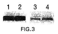
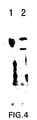
- Monoclonal antibody 3F5.4G6 was further tested against sera taken from stage D2 prostate cancer patients in progression, using monoclonal antibody 7E11-C5 as a control ( Figure 3). Both antibodies identified a band of about 120 kDa molecular weight ( Figure 3). An additional Western blot assay of LNCaP cells using the 3F5.4G6 monoclonal antibody was performed using a secondary antibody specific for IgM ( Figure 4). While monoclonal antibody 7E11-C5 recognized a single band of about 120 kDa, i.e., PSMA, 3F5.4G6 recognized a similar molecular weight band as well as a band of about 105-110 kDa. This band corresponds to the predicted protein form of PSM', and demonstrates the utility of an antibody that specifically recognizes the extracellular domain of both PSMA and PSM'.
- Immobilon P paper containing separated proteins derived from serum samples was reacted with either 7E11-C5 monoclonal antibody plus secondary antibody coupled to HRP or to secondary antibody coupled to HRP only. The film was exposed for 1 min or overexposed for 45 min in order to demonstrate the non-reactivity of the secondary antibody with any protein of 120 kDa in sera.
- the same secondary antibody was also used with 3F5.4G6 to detect the same antigen. Therefore, the 3F5.4G6 monoclonal antibody was specific for PSMA and PSM'.
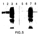
- Figure 5 confirms that the protein identified by 7E11-C5 was also recognized by monoclonal antibody 3F5.4G6.
- monoclonal antibody 3F5.4G6 also recognized a protein of 105-110 kDa not detected by monoclonal antibody 7E11-C5. This faster migrating protein corresponded to PSM'.
- the lysate was first precipitated with 7E11-C5, and the remaining proteins probed with 7E11-C5, the antibody did not detect any protein (Lane 4).
- the 7E11-C5 pre-treated lysate was probed with 3F5.4G6, it detected a protein of about 110 kDa.
- Figure 6 shows that the 120 kDa protein, i.e. PSMA, immunoprecipitated by 3F5.4G6 was also recognized by 7E11-C5.
- Figure 7A and B demonstrates that monoclonal antibody 3F5.4G6 recognized live LNCaP cells by FACS analysis, confirming that 3F5.4G6 recognized the extracellular domain of PSMA.
- Such an antibody recognizing the extracellular domain of PSMA is particularly useful as a diagnostic and/or therapeutic tool in prostate cancer.
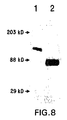
- Figure 8 illustrates that the protein recognized by monoclonal antibody 3F5.4G6 in Lane 2 is of approximate molecular weight 90 kDa. While PSM' was shown to have a molecular weight of 105-110 kDa in LNCaP lysates, the 90 kDa protein in seminal fluids was likely to be a non-glycosylated or partially glycosylated product of PSM'. Since PSM' contains several glycosylation sites, this lower molecular weight was the result of activities by glycosidases in the seminal fluid.
- PSMA was not present in this purified preparation is illustrated by the fact that 3F5.4G6 recognized a protein of molecular weight 120 kDa (Lane 1) present in a lysate of LNCaP cells which is PSMA, but did not recognize a protein of this molecular weight in Lane 2. In addition, antibody 7E11-C5 did not recognize the 90 KDa band in seminal fluids.
- LNCaP membranes were prepared from two 150 mm plates by removing cells in a versene solution followed by centrifugation to pellet the cells. Distilled water was added to the cell pellet and the cells were homogenized using a dounce homogenizer. The homogenized suspension was centrifuged at 30,000xg and the pelleted membrane fraction used for immunization.
- mice Male female BALB/c mice were immunized intraperitoneally four times (2-3 week intervals) with a LNCaP prostatic carcinoma cell membrane preparation emulsified in complete Freund's adjuvant. Five days prior to cell fusion, the mice were boosted with 50 ⁇ g of immunoaffinity purified PSMA in PBS. Cell fusion was performed as described in Section 6.1.3 supra.
- ELISA enzyme-linked immunoadsorbant assay
- the plates were washed as above and 50 ⁇ l of 1:600 dilution of rabbit-anti-mouse IgG and rabbit-anti-mouse IgM were added to each well. Following a one hour incubation at room temperature, the plates were washed as above and 50 ⁇ l of a 1:400 dilution of HRP-conjugated Protein-A were added to each well.
- ABTS 150 mg 2,2'-azino-bis (3-ethylbenzthiazoline-6-sulfonic acid in 500 ml of 0.1 M citric acid, pH 4.35)/H 2 O 2 (10 ⁇ l 30% H 2 O 2 per 10 ml of ABTS solution)chromogen/substrate solution were added to each well.
- the plates were read in a microplate reader and the OD405 was measured.
- the hybridoma cells producing supernatants with OD values 0.05 above background were cloned by limiting dilution and subjected to additional analysis.
- the aforementioned assay was modified as follows: Fifty microliters of a 40 ⁇ g/ml solution of 7E11-C5 anti-PSMA monoclonal antibody in 0.1 M NaHCO 2 , pH 8.2 binding buffer were added to wells of a Maxi-Sorp plate and allowed to adhere overnight at 4°C. The plates were washed and blocked as above. Fifty microliters of serially-diluted immunoaffinity-purified PSMA were added to the 7E11-C5-coated wells and the plates were incubated for two hours at room temperature.
- tissue culture supernatant from either 3D7-1.1 or 4E10-1.14 hybridoma clones were added to the wells and the plates were incubated for 90 minutes at room temperature. After washing as above, the wells were probed with 50 ⁇ l of a 1:1000 dilution of peroxidase-conjugated goat anti-mouse IgM and incubated for one hour at room temperature. Following extensive washing, 100 ⁇ l of ABTS/H 2 O 2 were added to each well and the plates were read in a microplate reader as described above.
- the beads were washed again with additional homogenization buffer containing 1% NP-40 followed by an additional wash with buffer containing 1% Triton X-100R.
- the washed beds were eluted with 100 mM glycine buffer, pH 2.5, 150 mM NaCl, 1% Triton X-100R in 2 ml fractions. Protein elution was monitored at OD280.
- LNCaP PSMA-expressing
- PC-3 cells PSMA-non-expressing
- the cells were washed two times with PBS-0.1% BSA, 0.01% Na azide, resuspended in 100 ⁇ l of a 1:100 dilution of FITC-conjugated rabbit-anti-mouse IgM, and incubated on ice for an additional 30 minutes.
- the cells were washed twice as above, resuspended in 500 ⁇ l of wash buffer, and analyzed for fluorescent staining by FACSCalibur (Becton-Dickinson) with CellQuest acquisition software.
- Tissue culture supernatants from the 3D7-1.1 and 4E10-1.14 hybridoma clones were tested in a Western blot assay for PSMA reactivity.
- Western blot analysis was performed following the protocol of Pelletier and Boynton (1994, J. Cell. Physiol. 158 :427-434). Briefly, lysates from LNCaP and PC-3 cells, immunoaffinity-purified PSMA, or Baculovirus-expressed full-length PSMA were electrophoresed on an 8.5% SDS-PAGE gel, and the separated proteins were electroblotted onto a PVDF membrane for one hour at 90 volts.
- the membranes were blocked overnight in 5% BLOTTO and incubated for 90 minutes with 20 ml undiluted tissue culture supernatant from the appropriate clone. The supernatant was removed, the blots were washed five times with TBS-0.5% Tween-20 (TBS-T), and probed with a 1:5000 dilution of peroxidase-conjugated goat anti-mouse IgM secondary antibody (Jackson) for one hour at room temperature. The membrane was washed five times with TBS-T, developed using the Chemiluminescent Substrate Kit (KPL), and visualized by exposing X-ray film (Kodak).
- TBS-0.5% Tween-20 TBS-0.5% Tween-20
- Jackson peroxidase-conjugated goat anti-mouse IgM secondary antibody
- PSMA protein was produced by isolating plaque-purified recombinant baculovirus particles, amplifying and infecting Sf9 cells at a multiplicity of infection of about 1:2 in the presence of SFM II medium (Gibco-BRI) supplemented with 5% FBS (Hyclone). Following a 48 hr.
- Monoclonal antibodies were generated against PSMA-containing prostatic carcinoma membranes.
- Supernatants from 3D7-1.1 and 4E10-1.14 hybridoma clones demonstrated comparable binding to native PSMA as compared to antibody 7E11-C5 ( Figure 9). Background non-specific binding to BSA was essentially comparable for all three antibody preparations.
- LNCaP cells and PC-3 cells were stained with supernatants from 3D7-1.1 and 4E10-1.14 hybridoma clones and analyzed by flow cytometry. Both antibodies stained live, non-fixed LNCaP cells but did not stain PC-3 cells ( Figure 12A-D). These results confirmed that these two antibodies react with epitopes in the extracellular domain of the PSMA molecule. Furthermore, the distinct shift in LNCaP staining observed with 4E10-1.14 monoclonal antibody compared to the shoulder seen with 3D7-1.1 suggests that these two antibodies recognize different epitopes in this particular region of the PSMA molecule.
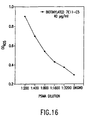
- a two-site capture ELISA for PSMA was developed utilizing the 7E11-C5 monoclonal antibody as a PSMA-capture reagent and 3D7-1.1 and 4E10-1.14 monoclonal antibodies as reporting or detection antibodies. Since these antibodies recognize different epitopes on the PSMA molecule (7E11-C5 reactive with the N-terminal 6 amino acids; 3D7-1.1 and 4E10-1.14 reactive with a sequence in the 134-475 amino acid region), they pair effectively in the two-site capture assay.
- Figure 16 demonstrates that monoclonal antibodies such as 3D7-1.1 or 4E10-1.14 which bind specifically to the extracellular domain of PSMA are useful in a two-site capture ELISA for PSMA.
- 3D7-1.1 for capture of PSMA indicates that another alternative immunoassay relying exclusively on the extracellular domain of the PSMA protein will be useful. Such an assay utilizing two extracellular domain-specific antibodies for capture and detection would be able to detect PSM' because of the location in the protein of its epitope. Thus, any assay utilizing 7E11-C5 for either capture or detection would specifically exclude PSM'.
- An example of a PSM' specific assay would include capture of PSMA and PSM' by an antibody such as 3D7-1.1 or any one of the monoclonal antibodies specific for the extracellular domain of PSMA in parallel tests.
- the transcript encoding PSMA is preferentially detected in prostate cancer patients (compared to normal males) although Su presents no demonstration that the PSMA transcript is in fact translated into protein in these patients. Additionally, Su shows that the transcript encoding PSM' is preferentially detected in normal males (compared to prostate cancer patients), although Su never detected any PSM' protein.
- the present inventors demonstrate that the PSMA protein is enhanced in body tissues and/or fluids of prostate cancer patients (compared to normal males) and that the PSM' protein is enhanced in body tissues and/or fluids of normal males (compared to prostate cancer patients).
- the ratio of PSM' to PSMA will have diagnostic and/or prognostic utility for clinical assessment of prostate cancer patients.
- a fragment of PSMA corresponding to amino acids 34 to 750 of full length PSMA was expressed in a baculovirus expression system as a 1.9 kb insert in a baculovirus expression system.
- the baculovirus expressed PSMA fragment is very similar to PSM' (which corresponds to residues 58-750 of full length PSMA) except that an additional 76 amino acids of the extracellular domain of PSMA are missing from the N-terminal of the fragment.
- Western blot analysis of various baculovirus expressed semi-purified PSMA fragment and LNCaP cell lysate were developed with monoclonal antibody 4E10-1.14 as probe. Results are shown in Figure 17.
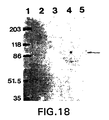
- Figure 17 indicates that antibodies such as 4E10-1.14 which are specific for the extracellular domain of PSMA are able also to bind a baculovirus expressed protein product very similar to PSM'.
- Figure 18 indicates that this is not a property of the 7E11-C5 monoclonal antibody due to its epitope specificity (see the negative reactivity of 7E11-C5 with the baculovirus expressed PSMA fragment in Figure 18).
- the baculovirus expressed PSM protein fragment is identical to PSM' (which corresponds to residues 58-750 of full length PSMA) except that it is missing an additional 76 amino acids from the N-terminal, all of which are in the extracellular domain.
- both antibodies would have the inherent property of binding to native PSM', a property not shared by 7E11-C5.
- the 3D7-1.1 monoclonal antibody was used as a probe in a Western blot with LNCaP cell derived PSMA as well as human serum and seminal fluid known also to contain PSMA. The results are shown in Figure 19.
- a band corresponding to PSMA migrating at about 120 Kd is present in all fractions.
- a second faster migrating band of molecular weight 90 to 100 Kd was observed in the serum and seminal fluid as revealed by antibody 3D7-1.1.
- This faster migrating band is not observed in Western blots with serum using the 7E11-C5 antibody (see Holmes, et al., The Prostate, Supple. 7 :25-29 (1996)).
- This faster migrating 3D7-1.1 reactive protein band is most probably PSM' present in biological fluids.
- hybridoma cell lines were deposited on March 12, 1996 and on March 11, 1997 with the American Type Culture Collection, 12301 Parklawn Drive, Rockville, Maryland 20852, and assigned the following accession number: Hybridoma ATCC Accession Number 3F5.4G6 HB12060 3D7-1.1 HB12309 4E10-1.14 HB12310
Abstract
Description
- The present invention relates to monoclonal antibodies that bind to the extracellular domain of prostate specific membrane antigen (PMSA), hybridoma cell lines producing the antibodies, and methods of using such antibodies for diagnosis and treatment of cancer. In particular, it relates to a monoclonal antibody generated against a synthetic peptide substantially homologous to a portion of the carboxyl terminal region of PSMA, which antibody reacts with PSMA expressed on tumor cell surface and in sera of prostate cancer patients. Additionally, it relates to two monoclonal antibodies generated against a prostatic carcinoma membrane preparation, which antibodies also react with PSMA expressed on the cell surface. The present invention also relates to a novel protein variant (PSM') of PSMA detected by the antibodies.
- Prostate cancer is the second leading cause of death from cancer among men. In fact, prostate cancer is the most common non-cutaneous cancer diagnosed in the American male. The number of men diagnosed with prostate cancer is steadily increasing as a result of the increasing population of older men as well as a greater awareness of the disease leading to its earlier diagnosis (Parker et al., 1997, CA Cancer J. for Clin. 47:5-28). It was projected that over 334,500 men would be diagnosed with prostate cancer in 1997, and that approximately 41,800 deaths would result from the disease. The life time risk for men developing prostate cancer is about 1 in 5 for Caucasians, and 1 in 6 for African Americans. High risk groups are represented by those with a positive family history of prostate cancer or African Americans. Over a lifetime, more than 2/3 of the men diagnosed with prostate cancer die of the disease (Wingo et al., 1996, CA Cancer J. for Clin. 46:113-25). Moreover, many patients who do not succumb to prostate cancer require continuous treatment to ameliorate symptoms such as pain, bleeding and urinary obstruction. Thus, prostate cancer also represents a major cause of suffering and increased health care expenditures (Catalona 1994, New Eng. J. Med. 31:996-1004).
- PSMA is a 120 kDa molecular weight protein expressed in prostate tissues and was originally identified by reactivity with a monoclonal antibody designated 7E11-C5 (Horoszewicz et al., 1987, Anticancer Res. 7:927-935; U.S. Patent No. 6,162,504). PSMA was obtained in purified form (Wright et al., 1990, Antibody Immunoconjugates and Radio Pharmaceuticals 3: Abstract 193) and characterized as a type II transmembrane protein having sequence identity with the transferrin receptor (Israeli et al. 1994, Cancer Res. 54:1807-1811) and with NAALADase activity (Carter et al., 1996, Proc. Natl. Acad. Sci. U.S.A. 93:749-753). More importantly, PSMA is expressed in increased amounts in prostate cancer, and elevated levels of PSMA are also detectable in the sera of these patients (Horoszewicz et al., 1987, supra; Rochon et al., 1994, Prostate 25:219-223; Murphy et al., 1995, Prostate 26:164-168; and Murphy et al., 1995, Anticancer Res. 15:1473-1479). A cDNA encoding PSMA has been cloned (Israeli et al., 1993, Cancer Res. 53:227-230), and it produces two alternatively spliced mRNA species: an mRNA species containing 2,653 nucleotides that encodes PSMA, and a second mRNA species containing 2,387 nucleotides referred to as PSM' (Su et al., 1995, Cancer Res. 55:1441-1443). WO96/08570 discloses a fusion protein, which comprises a portion of the amino acid sequence encoding the extracellular domain of PSMA. It further describes methods of constructing an expression vector, translation, purification and cleavage of PSMA immunofusion protein, and use of purified PSMA extracellular domain to immunise mice. However, isolation of antibodies specific for the extracellular domain of PSMA are not described.
- WO94/09820 describes PSMA cDNA (designated PSM') and vectors for expressing PSM'. It further describes a method for selecting amino acid hydrophilic peptides for use as immunogens to produce antibodies specific for PSMA. However, no antibodies specific for PSMA are disclosed.
- Leek et al., 1995, Br. J. Cancer, 72, 583-588 describe the characterisation of the gene encoding PSMA, specifically that PSMA genomic clones map to two loci on
human chromosome 11. - Prior to the present invention, it was not known whether PSM' encoded a protein product or existed only as an untranslated mRNA species because a PSM' protein product had never been detected. For example, WO96/26272 describes an isolated mammalian DNA and mRNA sequence which is predicted to encode an alternatively spliced PSM' antigen. However, at no point does this document specifically disclose a translated PSM' protein product from the defined mRNA sequence.
- A recent report by Carter et al. (1996, Proc. Natl. Acad. Sci. U.S.A., 93:749-753) shows a high degree of identity between 1428 bases representing a portion of the PSMA cDNA and the cDNA sequence of protein N-acetylated α-linked acidic dipeptidase (NAALADase). NAALADase has enzymatic activity towards the neuropeptide N-acetylaspartyl glutamate to yield glutamate and N-acetylaspartate. This report demonstrates NAALADase activity inherent to PSMA protein, but the catalytic portion of PSMA was not identified. NAALADase activity was found in LNCaP cells which expressed PSMA, but not in PC3 cells which do not express PSMA. Transfection of the PSMA cDNA into PC3 cells produced NAALADase activity and the presence of PSMA in these cells.
- The difference between the cDNA of PSMA and PSM' is the loss of the transmembrane and intracellular coding regions containing nucleotides #1-171 or amino acids #1-57. PSMA is described as a type II membrane protein and it is known that the functional catalytic domain of type II membrane proteins resides in the C-terminal extracellular region of the molecule (DeVries, et al., 1995, J. Biol. Chem., 270:8712-8722).
- PSM' mRNA is found in greater quantities in normal prostate tissues as compared with prostate tissues of patients with benign hyperplasia or prostate cancer (Su et al., 1995, supra). In contrast, PSMA mRNA is found in greater levels in patients with prostate cancer as compared to patients without prostate cancer (Su et al., 1995, supra). This observed difference is consistent with serum protein levels of PSMA described previously (Horoszewicz et al., 1987, supra; Rochon et al., 1994, supra; Murphy et al., 1995, supra; and Murphy et al., 1995, supra). In this connection, an elevated level of PSMA in sera of prostate cancer patients has been correlated with disease progression versus remission, and may be used as a prognostic marker (Murphy et al., 1995, supra).
- The epitope recognized by monoclonal antibody 7E11-C5 has been mapped to the first 6 amino acids of the intracellular N-terminal region of PSMA (Troyer et al., 1995, Urol. Oncol. 1:29-37) (Figure 1). Electron immunocytochemistry using 7E11-C5 has localized its epitope to the cytoplasm, and specifically to the inner leaf of the plasma membrane (Troyer et al., 1994, Proc. Am. Assoc. Cancer Res. 35:283, Abstract 1688). Furthermore, in in vitro tests, monoclonal antibody 7E11-C5 stains only fixed and permeabilized cells (Horoszewicz et al., 1987, supra), which is in accord with the mapping of the 7E11-C5 epitope to the N-terminus or intracellular domain of PSMA. While 7E11-C5 is useful for detecting prostate cancer in vivo which presumably exposes its epitope through necrosis and/or apoptosis, a monoclonal antibody specific for the extracellular domain of PSMA would allow more efficient detection of PSMA on the cancer cell surface. In addition, monoclonal antibody 7E11-C5 does not recognize PSM', since PSM' lacks the intracellular domain of PSMA, based on the sequence of its mRNA transcript.
- Citation or identification of any reference in this section or in any other section of this application shall not be construed as an admission that such reference is available as prior art to the present invention.
- The present invention relates to monoclonal antibodies specific for the extracellular domain of PSMA, hybridoma cell lines that produce the antibodies, and methods of using the antibodies for prostate cancer diagnosis and treatment, as well as a variant protein form of PSMA known as PSM' recognized by such antibodies.
- The invention is based, in part, on the Applicants' discovery of three monoclonal antibodies that recognize the extracellular domain of PSMA. One antibody was generated by immunizing mice with a C-terminal peptide of PSMA having the amino acid sequence of ESKVDPSK (SEQ. ID NO:1). The antibody reacts with PSMA and PSM' proteins in tumor cell lysates and in sera of prostate cancer patients. In addition, it stains intact live tumor cells, confirming its specificity for the extracellular domain of PSMA or PSM' protein. The antibody also detects PSM' in human seminal fluids, and the PSM' therein exhibits NAALADase activity. Two additional monoclonal antibodies were generated against a prostatic carcinoma membrane preparation. These antibodies also react with the extracellular domain of PSMA and PSM', including native PSMA isolated by immunoaffinity purification and recombinant PSMA produced by recombinant DNA technology. The antibodies are useful in combination with an antibody directed to the intracellular domain of PSMA in a two-site capture assay to detect the presence of PSMA in a test sample. Furthermore, all three antibodies disclosed herein may be used in a two-site capture assay to detect the presence of PSM' in a test sample.
- A wide variety of uses are encompassed by the present invention, including but not limited to, the development and use of an immunoassay to detect or stage prostate cancer in a patient, imaging of primary and/or metastatic prostate cancer in vivo, therapeutic uses of the antibodies, including uses of antibodies conjugated to a cytotoxic or chemotherapeutic agent; and the construction and use of antibody fragments, chimeric antibodies, humanized antibodies or bifunctional antibodies.
-
- Figure 1.
- Deduced amino acid sequences of PSMA and PSM' antigens (SEQ ID NO:2) (Israeli et al., 1994 Cancer Res. 54:1807-1811). PSM' mRNA does not contain the 5' end of the PSMA that would encode the first 57 amino acids (first line of amino acid sequence) and thus presumably begins at amino acid 58. However, prior to the present invention, PSM' had never been identified in its protein form. Underlined region is the putative transmembrane domain and the bold region (amino acid #716-723) is a peptide selected for monoclonal antibody development.
- Figure 2.
- Demonstration of monoclonal antibody 3F5.4G6 (a subclone derived from primary hybridoma 3F5) and its reactivity with a protein present in LNCaP lysate of 120 kDa molecular weight corresponding to PSMA. Western blot was developed with HRP-anti-IgG secondary antibody.
Lane 1 = LNCaP lysate probed with 7E11-C5;Lane 2 = LNCaP lysate probed with 3F5.4G6. - Figure 3.
- Demonstration by Western blot of PSMA in sera of prostate cancer patients (stage D2) using monoclonal antibodies 3F5.4G6 (
Lanes 3 and 4) and 7E11-C5 (Lanes 1 and 2) as control. - Figure 4.
- Western blot assay of LNCaP lysates using monoclonal antibodies 7E11-C5 (Lane 1) and 3F5.4G6 (Lane 2) and developed with HRP-anti-IgM secondary antibody. Both 7E11-C5 and 3F5.4G6 recognized a protein of
molecular weight 120 kDa. In addition, 3F5.4G6 also recognized a protein of 105-110 kDa molecular weight corresponding to the predicted protein form of PSM'. It should be noted that 7E11-C5 did not recognize PSM' because the epitope of 7E11-C5 monoclonal antibody was not found in PSM'. Antibody 3F5.4G6 recognizes the C-terminal portion of the protein (amino acid #716-723), which corresponds to the extracellular domain of PSMA and PSM'. - Figure 5.
- Demonstration that monoclonal antibodies 7E11-C5 and 3F5.4G6 recognized an identical protein but that 3F5.4G6 recognized an additional protein corresponding to PSM'. LNCaP lysate was initially immunoprecipitated with 7E11-C5 monoclonal antibody and the immunoprecipitated material separated on SDS gels and probed in a Western blot assay with either 7E11-C5 (lanes 1-4) or with 3F5.4G6 (Lanes 5-8) monoclonal antibodies.
Lanes Lanes Lanes Lanes - Figure 6.
- Demonstration that monoclonal antibodies 7E11-C5 and 3F5.4G6 recognized an identical 120 kDa protein. PSMA from an LNCaP lysate was immunoprecipitated by monoclonal antibody 3F5.4G6, the proteins in the immunoprecipitate were separated on a SDS gel, transferred to Immobilon P and probed in a Western blot with monoclonal antibody 7E11-C5.
Lane 1 = LNCaP lysate control and probed with 7E11-C5;Lane 2 = 3F5.4G6 immunoprecipitation. - Figure 7A & B
- Demonstration by FACS analysis of 3F5.4G6 monoclonal antibody recognition of live LNCaP cells illustrating antibody binding to the extracellular domain of PSMA. Fig. 7A represents control with no primary antibody; and Fig. 7B represents LNCaP cells incubated with 100 µg/ml of 3F5.4G6 prior to FACS analysis. The shift to the right indicates binding of the antibody to the live LNCaP cells.
- Figure 8.
- Demonstration of the reactivity of monoclonal antibody 3F5.4G6 with PSM' isolated and purified from seminal fluid.
Lane 1 is LNCaP lysate andLane 2 is purified PSM' from seminal fluid. Proteins were separated on SDS polyacrylamide gels and transferred to Immobilon P paper and probed with monoclonal antibody 3F5.4G6 by Western blot procedures. The protein purified from seminal fluid and represented inLane 2 is of molecular weight 90kDa, which is likely to be a non-glycosylated or partially glycosylated product of PSM' having a molecular weight of 105-110kDa. - Figure 9.
- Demonstration of the reactivity of monoclonal antibodies 3D7-1.1 and 4E10-1.14 with native PSMA and three PSMA fragments. Microtiter 96-well plates were coated with native PSMA or one of three bacterially-expressed polypeptide fragments of PSMA, and reacted with hybridoma supernatants in an ELISA. While all three tested antibodies showed comparable binding to native PSMA, 3D7-1.1 and 4E10-1.14 reacted strongly with a fragment corresponding to an epitope in the extracellular domain of PSMA.
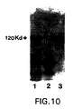
- Figure 10.
- Western blot analysis of PSMA using monoclonal antibodies 3D7-1.1.
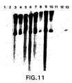
Lane 1=LNCaP lysate;Lane 2=PC-3 lysate;Lane 3=immunoaffinity-purified PSMA. - Figure 11.
- Western blot analysis of full-length baculovirus-expressed PSMA. Recombinant PSMA was electrophoresed on SDS-PAGE gel, electroblotted and probed with various antibody preparations.
Lane 1 = blank;
Lane 2 = control medium (20% FCSin RPMI 1640;
Lane 3 = 3D7-1.1 monoclonal antibody;
Lane 4 = 3D7-1.2 monoclonal antibody;
Lane 5 = 3D7-1.3 monoclonal antibody;
Lane 6 = 3D7-1.7 monoclonal antibody;
Lane 7 = 3D7-2.7 monoclonal antibody;
Lane 8 = 4E10 (parent) monoclonal antibody;
Lane 9 = 4E10-1.3 monoclonal antibody;
Lane 10 = 4E10-1.14 monoclonal antibody;
Lane 11 = blank;
Lane 12 = blank;
Lane 13 = 7E11-C5 monoclonal antibody. - Figure 12 A-D
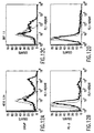
- Demonstration by FACS analysis of 3D7-1.1 and 4E10-1.14 monoclonal antibody recognition of live LNCaP cells, illustrating antibody binding to the extracellular domain of PSMA. Figure 12A represents LNCaP cells incubated with 4E10-1.14. Figure 12B represents PC-3 cells incubated with 4E10-1.14. Figure 12C represents LNCaP cells incubated with 3D7-1.1. Figure 12D represents PC-3 cells incubated with 3D7-1.1. The different patterns in the shift to the right in Figure 12A and 12C suggest that the two antibodies may recognize different epitopes of PSMA.
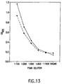
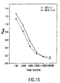
- Figure 13.
- Detection of PSMA by a two-site capture ELISA using two monoclonal antibodies to distinct epitopes of PSMA. Serially-diluted immunoaffinity-purified PSMA was added to 7E11-C5-coated 96 well plates and detected by incubating with 3D7-1.1 or 4E10-1.14 supernatants. The absorbance at 405 mm was measured in a microplate reader. ―●― = 3D7-1.1; ―■― = 4E10-1.14.
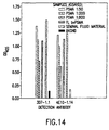
- Figure 14.
- Detection of PSMA in a variety of biological samples by a two-site capture ELISA using 3D7-1.1 and 4E10-1.14 monoclonal antibodies.
- Figure 15.
- Detection of immunoaffinity-purified PSMA serially diluted in normal human serum by a two-site capture ELISA using 3D7-1.1 and 4E10-1.14 monoclonal antibodies.
- Figure 16.
- Detection of PSMA by an alternate two-site capture ELISA. Serially diluted immunoaffinity purified PSMA was added to 3D7-1.1-coated 96 well plates and detected by incubating with biotinylated 7E11-C5 (40 µg/ml) followed by horse radish peroxidase conjugated streptavidin. The absorbance at 405 nm was measured in a microplate reader. 7E11-C5 was biotinylated using E-Z link Biotinylation kits (Pierce) according to manufacturer's instructions.
- Figure 17.
- Western blot analysis of LNCaP cell lysate and various fractions of a semi-purified PSMA fragment (corresponding to amino acids 134 to 750 of full length PSMA expressed as a 1.9 kb insert in a baculovirus expression system) probed with tissue culture supernatant from the 4E10-1.14 hybridoma. The identification of the protein product from the 1.9 kb construct (amino acids 134-750 of PSMA) is noted by the arrow.
Lane 1 = Markers;Lane 2 = LNCaP cell crude lysate;Lane 3 = Viral pellet, i.e., 100,000 xg pellet of lysed SF9 cells infected with baculovirus expressing 1.9 kb PSMA fragment;Lane 4 = 100,000 xg supernatant fraction from lysed SF9 cells infected with baculovirus expressing 1.9 kb PSMA fragment;Lane 5 = Flow thru of fraction shown inLane 4 after passage through a Ni-NTA matrix;Lane 6 = 0.5M NaCl elution of Ni-NTA matrix;Lane 7 = 1M imidazole, pH 7.6 elution of Ni-NTA matrix;Lane 8 = Flow thru of fraction shown inLane 4 after passage through a Ni-NTA matrix;Lane 9 = 0.5M NaCl elution of Ni-NTA matrix; andLane 10 = 1M imidazole, pH 7.6 elution of Ni-NTA matrix. Also note inLane 2 reactivity of 4E10-1.14 monoclonal antibody with native full length PSMA expressed in LNCaP cells. - Figure 18.
- Western blot of crude lysates of SF9 cells infected with a baculovirus containing either an irrelevant insert or a 1.9 kb insert encoding a portion of PSMA (amino acids 134-750 of full length PSMA) probed with antibody 7E11-C5.
Lanes Lane 3 = irrelevant virus infected SF9 cell lysate;Lane 4 = SF9 cell lysate; andLane 5 = 1.9kb PSMA insert containing virus infected SF9 lysate. Note that no 7E11-C5 positive bands were observed with any protein products present in SF9 cells or those infected with either virus. - Figure 19.
- Western blot of PSMA and PSM' obtained from LNCaP cells, human seminal fluid and human serum probed with monoclonal antibody 3D7-1.1.
Lane 1 = LNCaP cell lysate;Lane 2 = 7E11-C5 immunoaffinity purified PSMA from LNCaP cells;Lane 3 = human seminal fluid; andLane 4 = human male serum. The positions of PSMA and PSM' are indicated. - The present invention relates to monoclonal antibodies specific for the extracellular domain of PSMA, methods of using such antibodies and a truncated protein variant, PSM', identified by such antibodies. Although the specific procedures and methods described herein are exemplified using a C-terminal peptide or a PSMA-expressing tumor membrane preparation to immunize mice, they are merely illustrative for the practice of the invention. Analogous procedures and techniques are equally applicable to a variety of animal hosts immunized against PSMA in the form of protein, peptides, cell surface antigen and crude membrane preparations.
- In a specific embodiment by the way of example in
Section 6, infra, a synthetic peptide derived from the C-terminal region of PSMA was used as an immunogen. The results show that one antibody designated 3F5.4G6 binds to the extracellular domain of PSMA, which is exposed on the cell surface of live prostate cancer cells and in the sera of prostate cancer patients. Additionally, a second working example inSection 7, infra, demonstrates the production of two monoclonal antibodies directed to the extracellular domain of PSMA following immunization of animals with a PSMA-expressing tumor membrane preparation. In this connection, cancer cells such as LNCaP that express PSMA, host cells transfected with PSMA coding sequence, purified PSMA, PSM' or PSMA extracellular domain peptides may be used as immunogen to elicit an immune response in animal hosts for the generation of monoclonal antibodies specific for the extracellular domain of PSMA. - Somatic cells with the potential for producing antibody and, in particular B lymphocytes, are suitable for fusion with a B-cell myeloma line. Those antibody-producing cells that are in the dividing plasmablast stage fuse preferentially. Somatic cells may be obtained from the lymph nodes, spleens and peripheral blood of antigen-primed animals, and the lymphatic cells of choice depend to a large extent on their empirical usefulness in the particular fusion system. Once-primed or hyperimmunized animals can be used as a source of antibody-producing lymphocytes. Mouse lymphocytes give a higher percentage of stable fusions with the mouse myeloma lines described below. Of these, the BALB/c mouse is preferred. However, other mouse strains, rabbit, hamster, sheep and frog may also be used as hosts for preparing antibody-producing cells. As reviewed by Coding (in Monoclonal Antibodies: Principles and Practice, 2d ed., pp. 60-61, Orlando, Fla, Academic Press, 1986), use of rat lymphocytes may provide several advantages.
- Alternatively, human somatic cells capable of producing antibody, specifically B lymphocytes, are suitable for fusion with myeloma cell lines. While B lymphocytes from biopsied spleens, tonsils or lymph nodes of individual may be used, the more easily accessible peripheral blood B lymphocytes are preferred. The lymphocytes may be derived from patients with diagnosed prostate carcinomas. In addition, human B cells may be directly immortalized by the Epstein-Barr virus (Cole et al., 1995, Monoclonal Antibodies and Cancer Therapy, Alan R. Liss, Inc., pp. 77-96).
- Myeloma cell lines suited for use in hybridoma-producing fusion procedures preferably are non-antibody-producing, have high fusion efficiency, and enzyme deficiencies that render them incapable of growing in certain selective media which support the growth of the desired hybridomas. Examples of such myeloma cell lines that may be used for the production of fused cell hybrids of the invention, include P3-X63/Ag8, X63-Ag8.653, NS1/1.Ag 4.1, Sp210-Ag14, FO, NSO/U, MPC-11, MPC11-X45-GTG 1.7, S194/5XX0 Bul, all derived from mice; R210.RCY3, Y3-Ag 1.2.3, IR983F and 4B210 derived from rats and U-266, GM1500-GRG2, LICR-LON-HMy2, UC729-6, all derived from humans (Goding in Monoclonal Antibodies: Principles and Practice, 2d ed., pp. 65-66, Orlando, Fla, Academic Press, 1986; Campbell, in Monoclonal Antibody Technology, Laboratory Techniques in Biochemistry and Molecular Biology Vol. 13, Burden and Von Knippenberg, eds. pp. 75-83, Amsterdam, Elseview, 1984).
- Methods for generating hybrids of antibody-producing spleen or lymph node cells and myeloma cells usually comprise mixing somatic cells with myeloma cells in a 2:1 proportion (though the proportion may vary from about 20:1 to about 1:1), respectively, in the presence of an agent or agents (chemical or electrical) that promote the fusion of cell membranes. It is often preferred that the same species of animal serve as the source of the somatic and myeloma cells used in the fusion procedure. Fusion methods have been described by Kohler and Milstein (1975, Nature 256:495-497; 1976, Eur. J. Immunol. 6:511-519), and by Gefter et al. (1977, Somatic Cell Genet. 3:231-236). The fusion-promotion agents used by those investigators were Sendai virus and polyethylene glycol (PEG), respectively. Fusion methods reviewed by Goding (1986, in Monoclonal Antibodies: Principles and Practice, 2d ed., pp. 71-74, Orlando, Fla, Academic Press), including the above as well as electrically induced fusion are also suitable to generate monoclonal antibodies of the invention.
- Fusion procedures usually produce viable hybrids at very low frequency, about 1 x 10⁻⁶ to 1 x 10⁻⁸ somatic cells. Because of the low frequency of obtaining viable hybrids, it is essential to have a means to select fused cell hybrids from the remaining unfused cells, particularly the unfused myeloma cells. A means of detecting the desired antibody-producing hybridomas among the other resulting fused cell hybrids is also necessary.
- Generally, the fused cells are cultured in selective media, for instance HAT medium containing hypoxanthine, aminopterin and thymidine. HAT medium permits the proliferation of hybrid cells and prevents growth of unfused myeloma cells which normally would continue to divide indefinitely. Aminopterin blocks de novo purine and pyrimidine synthesis by inhibiting the production of tetrahydrofolate. The addition of thymidine bypasses the block in pyrimidine synthesis, while hypoxanthine is included in the media so that inhibited cells synthesize purine using the nucleotide salvage pathway. The myeloma cells employed are mutants lacking hypoxanthine phosphoribosyl transferase (HPRT) and thus cannot utilize the salvage pathway. In the surviving hybrid, the B lymphocyte supplies genetic information for production of this enzyme. Since B lymphocytes themselves have a limited life span in culture (approximately two weeks), the only cells which can proliferate in HAT media are hybrids formed from myeloma and spleen cells.
- To facilitate screening of antibody secreted by the hybrids and to prevent individual hybrids from overgrowing others, the mixture of fused myeloma and B lymphocytes is diluted in HAT medium and cultured in multiple wells of microtiter plates. In two to three weeks, when hybrid clones become visible microscopically, the supernatant fluid of the individual wells containing hybrid clones is assayed for specific antibody. The assay must be sensitive, simple and rapid. Assay techniques include radioimmunoassays, enzyme immunoassays, cytotoxicity assays, plaque assays, dot immunobinding assays, and the like.
- Once the desired fused cell hybrids have been selected and cloned into individual antibody-producing cell lines, each cell line may be propagated in either of two standard ways. A sample of the hybridoma can be injected into a histocompatible animal of the type that was used to provide the somatic and myeloma cells for the original fusion. The injected animal develops tumors secreting the specific monoclonal antibody produced by the fused cell hybrid. The body fluids of the animal, such as serum or ascites fluid, can be tapped to provide monoclonal antibodies in high concentration. Alternatively, the individual cell lines may be propagated in vitro in laboratory culture vessels; the culture medium, also containing high concentrations of a single specific monoclonal antibody, can be harvested by decantation, filtration or centrifugation.
- Monoclonal antibodies or purified fragments of the monoclonal antibodies having at least a portion of an antigen binding region, including such as Fv, F(ab')2, Fab fragments (Harlow and Lane, 1988, Antibody, Cold Spring Harbor), single chain antibodies (U.S. Patent 4,946,778), chimeric or humanized antibodies (Morrison et al., 1984, Proc. Natl. Acad. Sci. USA 81:6851; Newuberger et al., 1984 Nature 81:6851) and complementarily determining regions (CDR) may be prepared by conventional procedure. Purification of the antibodies or fragments can be accomplished by a variety of methods known to those of skill including, precipitation by ammonium sulfate or sodium sulfate followed by dialysis against saline, ion exchange chromatography, affinity or immunoaffinity chromatography as well as gel filtration, zone electrophoresis, etc. (see Coding in, Monoclonal Antibodies: Principles and Practice, 2d ed., pp 104-126, Orlando, Fla, Academic Press).
- Using techniques described generally in Section 5.1 supra and illustrated in the
Sections - Antibody 3F5.4G6 is an IgM isotype antibody that binds specifically to PSMA expressed in prostate cancer cell lysates and on the cell surface of prostate cancer cells, as well as in sera obtained from prostate carcinoma patients. In addition, 3F5.4G6 also binds specifically to PSM'. The 3F5.4G6-reactive PSMA epitope is extracellular, c-terminal and distinct from that recognized by 7E11-C5 (Horoszewicz et al., Anticancer Res. 7:927-936) which is membrane associated in the cytoplasm of the cell. Antibodies 3D7-1.1 and 4E10-1.14 are also IgM antibodies and bind to PSMA expressed in prostate cancer cell lysates and on the cell surface. These antibodies may be used to detect both primary prostate cancer and metastatic tumors such as bone metastases of prostate cancer.
- During the development of an antibody response, antibody-producing cells first secrete the IgM isotype which eventually switches to IgG. Such class switching events occur by DNA rearrangement of constant region genes so that the same antigen specificity is retained. The different antibody isotypes possess different effector functions. For example, IgM and all IgG subclasses except IgG4 can fix complement upon antigen binding. In contrast, IgE binds to mast cells in an allergic reaction to trigger histamine release.
- Hybridoma cell lines also produce class switch variants during long-term culture. In particular, monoclonal antibodies switching from IgM to IgG or IgG1 to IgG2a have been selected for their higher affinity for protein A, which facilitates their purification. Any class switch variant may be selected for a particular desirable effector function (Spira et al., 1985, In Hybridoma Technology in the Biosciences and Medicine, ed. springer, pp. 77-88, Plenum Press, NY; Harlow and Lane, 1988 Antibodies, Cold Spring Harbor Laboratory). In the case of the exemplified antibodies, since they are of IgM isotype, it is desirable to also select for IgG variants that possess the same antigen specificity, which may be more useful for certain purposes in vitro or in vivo. The present invention encompasses IgG variants of the monoclonal antibodies of the invention, including 3F5.4G6, 3D7-1.1 and 4E10-1.14.
-
Sections - In another embodiment of the invention, the exemplified hybridoma cell lines may be used to produce compositions comprising an antigen binding site or antibody variants which combine the murine variable or hypervariable regions with the human constant region or constant and variable framework regions, i.e., chimeric or humanized antibodies as well as humanized antibodies that retain only the antigen-binding CDRs from the parent antibody in association with human framework regions (see, Waldmann, 1991, Science 252:1657, 1662, particularly 1658-59 and references cited therein). Such chimeric or humanized antibodies retaining binding specificity of the murine antibody are expected to have reduced immunogenicity when administered in vivo for diagnostic, prophylactic or therapeutic applications according to the invention.
- In yet other embodiments, the invention encompasses the use of the hybridoma cell lines as a source of DNA or mRNA encoding for the rearranged, activated immunoglobulin genes, which may be isolated, cloned by known recombinant DNA techniques and transferred to other cells for the production of antigen binding fragments specific for the extracellular domain of PSMA. By isolating rearranged DNA or preparing cDNA from the messenger RNA of the hybridoma cell line of the invention, a sequence free of introns may be obtained.
- To illustrate, and not by way of limitation, an immunoexpression library can be prepared and screened for antibody binding fragments for PSMA and PSM' as follows (See, Huse et al., 1989, Sci. 246:1275-1281; Mullinax et al., 1990, Proc. Natl Acad. Sci. USA 87:8045-8099). Total RNA can be purified (e.g., using commercially available kits) and converted to cDNA using an oligo (dT) primer for the light (L) chain and a specific primer for the heavy (H) chain using reverse transcriptase. Polymerase chain reaction (PCR) amplification of the immunoglobulin H and L chain sequences can be done separately with sets of primer pairs. Upstream primers can be designed to hybridize to partially conserved sequences in the leader and/or framework regions of VH or VL and downstream primers can be designed to hybridize to constant domain sequences. Such primers would preserve full length L chain and provide H chains corresponding to the Fd of IgG and conserving the H-L disulfide bonds. The PCR amplified L and H DNA fragments are then digested and separately ligated into H and L chain vectors. Such vectors contain a pelB leader sequence, a ribosome binding site and stop codons. Suitable λ phage vectors for expression in E. coli can be prepared from commercially available vectors (ImmunoZAP L, ImmunoZAP H; Stratacyte, La Jolla, CA). The ligated recombinant phage DNA is incorporated into bacteriophage with in vitro packaging extract and used to infect E. coli. The immunoexpression library thus created is screened for antigen binding fragments using PSMA, PSM' or a specific peptide thereof. Positive clones can be screened and identified as described by Mullinax et al. (supra).
- Although the specific procedures and methods described herein are exemplified using the monoclonal antibodies of the invention, they are merely illustrative for the practice of the invention. Purified fragments of the monoclonal antibodies having at least a portion of the antigen-binding region, including Fv, F(ab')2, Fab fragments, single chain antibodies, chimeric or humanized antibodies or CDRs can be used in the procedures and methods described below according to the present invention.
- Monoclonal antibodies of the present invention can be used to detect prostate carcinoma cells in histological and cytological specimens, and, in particular, to distinguish malignant tumors from normal tissues and non-malignant tumors. Tissue specimens may be stained by the antibodies and their binding detected by a second antibody conjugated to a label such as peroxidase, fluorescein, alkaline phosphatase, and the like.
- In addition, immunofluorescence techniques can use the monoclonal antibodies of the present invention to examine human tissue, cell and bodily fluid specimens. In a typical protocol, slides containing cryostat sections of frozen, unfixed tissue biopsy samples or cytological smears are air dried, formalin or acetone fixed, and incubated with the monoclonal antibody preparation in a humidified chamber at room temperature.
- The slides are then washed and further incubated with a preparation of antibody directed against the monoclonal antibody, usually some type of anti-mouse immunoglobulin if the monoclonal antibodies used are derived from the fusion of a mouse spleen lymphocyte and a mouse myeloma cell line. This anti-mouse immunoglobulin is tagged with a compound, for instance rhodamine or fluorescein isothiocyanate, that fluoresces at a particular wavelength. The staining pattern and intensities within the sample are then determined by fluorescent light microscopy and optionally photographically recorded.
- As yet another alternative, computer enhanced fluorescence image analysis or flow cytometry can be used to examine tissue specimens or exfoliated cells, i.e., single cell preparations from aspiration biopsies of prostate tumors using the monoclonal antibodies of the invention. The monoclonal antibodies of the invention are particularly useful in quantitation of live tumor cells, i.e., single cell preparations from aspiration biopsies of prostate tumors by computer enhanced fluorescence image analyzer or with a flow cytometer. Use of 3F5.4G6, 3D7-1.1 and 4E10-1.14 antibodies in such assays is valuable to differentiate benign from malignant prostate tumors since PSMA to which the monoclonal antibodies bind is expressed in increased amounts by malignant tumors. The percent PSMA positive cell population, alone or in conjunction with determination of the DNA ploidy of these cells, may, additionally, provide very useful prognostic information by providing an early indicator of disease progression.
- In yet another alternative embodiment, the monoclonal antibodies of the present invention can be used in combination with other known prostate antibodies to provide additional information regarding the malignant phenotype of a prostate carcinoma.
- The use of the monoclonal antibodies of the invention can be extended to the screening of human biological fluids for the presence of the specific antigenic determinants recognized. In vitro immunoserological evaluation of biological fluids withdrawn from patients thereby permits non-invasive diagnosis of cancers. By way of illustration, human bodily fluids such as prostatic fluid, seminal fluid, whole blood, serum or urine can be taken from a patient and assayed for the specific epitope, either as released antigen or membrane-bound on cells in the sample fluid, using monoclonal antibodies specific for the extracellular domain of PSMA and PSM' in standard radioimmunoassays or enzyme-linked immunoassays, competitive binding enzyme-linked immunoassays, dot blot or Western blot, or other assays known in the art.
- In addition, a more sensitive diagnostic assay for PSMA or PSM' protein can be developed through the use of monoclonal antibodies directed to non-overlapping epitopes on PSMA and PSM'. Antibodies specific for opposite ends of PSMA such as 7E11-C5 and 3F5.4G6, 3D7-1.1 or 4E10-1.14 are particularly suitable for use in such an assay. In this regard, one antibody may be anchored to a substrate to capture PSMA or PSM' in a biological fluid, while the other antibody is used to detect the antibody-bound antigen. Also, since the expression of PSMA and PSM' is increased in prostate cancer and normal prostate tissues, respectively, antibodies that distinguish these two forms may be used to provide a more accurate way to monitor tumor regression versus progression, following treatment. Since 3F5.4G6, 3D7-1.1 and 4E10-1.14 recognize both forms, but 7E11-C5 only binds to PSMA, these antibodies may be used in conjunction to determine the precise levels of each form in a patient, thereby correlating their amounts with tumor burden. For example, 7E11-C5 may be used as an anchored antibody in a two-site capture assay, and any one of the other three antibodies may be used as a detection antibody to quantitate PSMA. On the other hand, any combination of two of the three PSMA extracellular domain-specific antibodies may be used in a similar two-site capture assay to specifically measure total PSM' plus PSMA concentrations. A simple subtraction of PSMA from total PSMA and PSM' specifically quantitates PSM'.
- In addition to the detection of extracellular domain PSMA and PSM' by a monoclonal antibody in tissues and bodily fluids, NAALADase enzyme activity measurements can be utilized to quantitate extracellular domain PSMA and/or PSM' in tissues and/or bodily fluids.
- For example, tissue levels can be determined by detergent solubilizing homogenizing tissues, pelleting the insoluble material by centrifugation and measuring the NAALADase activity in the remaining supernatant. Likewise, the NAALADase activity in bodily fluids can also be measured by first pelleting the cellular material by centrifugation and performing a typical enzyme assay for NAALADase activity on the supernatant.
- NAALADase assay protocols taking advantage of antibody binding specificities can also be applied. For example, solid surfaces coated with either 7E11-C5, 3F5.4G6, 3D7-1.1 or 4E10-1.14 antibodies could be used to capture the PSMA or PSM' protein for detection using a NAALADase enzyme assay. Thus, this may be used to differentially detect and quantitate full length PSMA protein and PSM' in a specimen given that an extracellular domain-specific antibody binds to both PSMA and PSM', whereas 7E11-C5 would only bind to PSMA.
- More convenient NAALADase enzyme assays, taking advantage of the reaction properties of glutamate dehydrogenase may also be applied (Frieden, 1959, J. Biol. Chem., 234:2891). In this assay, the reaction product of the NAALADase enzyme is glutamic acid. This is derived from the enzyme catalyzed cleavage of N-acetylaspartylglutamate to yield N-acetylaspartic acid and glutamic acid. Glutamic acid, in a NAD(P)+ requiring step, yields 2-oxoglutarate plus NAD(P)H in a reaction catalyzed by glutamate dehydrogenase. Progress of the reaction can easily and conveniently be measured by the change in absorbance at 340 nm due to the conversion of NAD(P)+ to NAD(P)H. Thus, improvements to the assay of NAALADase activity applicable to a solid phase format with immobilized capture antibodies can be achieved. In this way, multiple assays can be conducted simultaneously in a microplate reader based upon the absorbance change at 340 nm before and after addition of NAD+ or NADP+. It would not be restricted to solid phase assays, as solution assays of, eg., serum would also be possible with this type of NAALADase assay.
- Kits containing the monoclonal antibodies of the invention or fragments thereof can be prepared for in vitro diagnosis, prognosis and/or monitoring prostate carcinoma by the immunohistological, immunocytological and immunoserological methods described above. The components of the kits can be packaged either in aqueous medium or in lyophilized form. When the monoclonal antibodies (or fragments thereof) are used in the kits in the form of conjugates in which a label moiety is attached, such as an enzyme or a radioactive metal ion, the components of such conjugates can be supplied either in fully conjugated form, in the form of intermediates or as separate moieties to be conjugated by the user of the kit.
- A kit may comprise a carrier being compartmentalized to receive in close confinement therein one or more container means or series of container means such as test tubes, vials, flasks, bottles, syringes, or the like. A first of said container means or series of container means may contain the monoclonal antibody (or fragment thereof) or PSMA or PSM'. A second container means or series of container means may contain a label or linker-label intermediate capable of binding to the primary antibody (or fragment thereof), PSMA or PSM'.
- The monoclonal antibodies or fragments thereof of this invention are particularly useful for targeting prostate cancer cells in vivo. They can be used for tumor localization for detection and monitoring as well as for therapy of primary prostate carcinoma and metastases. For these in vivo applications, it is preferable to use purified monoclonal antibodies or purified fragments of the monoclonal antibodies having at least a portion of an antigen binding region, including such as Fv, F(ab')2, Fab fragments (Harlow and Lane, 1988, Antibody Cold Spring Harbor), single chain antibodies (U.S. Patent 4,946,778), chimeric or humanized antibodies (Morrison et al., 1984, Proc. Natl. Acad. Sci. USA 81:6851; Newuberger et al., 1984 Nature 81:6851), complementarily determining regions (CDR), and the like. Purification of the antibodies or fragments can be accomplished by a variety of methods known to those of skill including, precipitation by ammonium sulfate or sodium sulfate followed by dialysis against saline, ion exchange chromatography, affinity or immunoaffinity chromatography as well as gel filtration, zone electrophoresis, etc. (see Goding in, Monoclonal Antibodies: Principles and Practice, 2d ed., pp 104-126, Orlando, Fla, Academic Press).
- For use in in vivo detection and/or monitoring of prostate carcinoma, the purified monoclonal antibodies can be covalently attached, either directly or via a linker, to a compound which serves as a reporter group to permit imaging of specific tissues or organs following administration and localization of the conjugates or complexes. A variety of different types of substances can serve as the reporter group, including such as radiopaque dyes, radioactive metal and non-metal isotopes, fluorogenic compounds, fluorescent compounds, positron emitting isotopes, non-paramagnetic metals, etc.
- For use in in vivo therapy of prostate carcinoma, the purified monoclonal antibodies can be used alone or covalently attached, either directly or via a linker, to a compound which kills and/or inhibits proliferation of the malignant cells or tissues following administration and localization of the conjugates. When the antibody is used by itself, it may mediate tumor destruction by complement fixation or antibody-dependent cellular cytotoxicity. Alternatively, the antibody may be administered in combination with a chemotherapeutic drug to result synergistic therapeutic effects (Baslya and Mendelsohn, 1994 Breast Cancer Res. and Treatment 29:127-138). A variety of different types of substances can be directly conjugated to the antibody for therapeutic uses, including radioactive metal and non-metal isotopes, chemotherapeutic drugs, toxins, etc. (Vitetta and Uhr, 1985, Annu. Rev. Immunol. 3:197).
- According to an alternative embodiment, for in vivo therapy of prostate carcinoma the monoclonal antibodies of the present invention can be modified to be in the form of a bifunctional or bispecific antibody, i.e., an antibody having an antigen-binding region specific for the extracellular domain of prostate specific membrane antigen and an antigen-binding region specific for an effector cell which has tumorcidal or tumor inhibitory activity. The two antigen binding regions of the bispecific antibody are either chemically linked or can be expressed by a cell genetically engineered to produce the bispecific antibody. (See generally, Fanger et al., 1995 Drug News & Perspec. 8(3):133-137). Suitable effector cells having tumorcidal activity include but are not limited to cytotoxic T-cells (primarily CD8+ cells), natural killer cells, etc. An effective amount of a bispecific antibody according to the invention is administered to a prostate cancer patient and the bispecific antibody kills and/or inhibits proliferation of the malignant cells after localization at sites of primary or metastic tumors bearing PSMA.
- Methods for preparation of antibody conjugates of the antibodies (or fragments thereof) of the invention useful for detection, monitoring and/or therapy are described in U.S. Patent Nos. 4,671,958; 4,741,900 and 4,867,973.
- Kits for use with such in vivo tumor localization and therapy methods containing the monoclonal antibodies (or fragments thereof) conjugated to any of the above types of substances can be prepared. The components of the kits can be packaged either in aqueous medium or in lyophilized form. When the monoclonal antibodies (or fragments thereof) are used in the kits in the form of conjugates in which a label or a therapeutic moiety is attached, such as a radioactive metal ion or a therapeutic drug moiety, the components of such conjugates can be supplied either in fully conjugated form, in the form of intermediates or as separate moieties to be conjugated by the user of the kit.
- PSMA peptide #716-723 (NH2-ESKVDPSK-) was coupled to keyhole limpet hemocyanin (KLH) as a carrier using the EDC method of Pierce (Rockford, IL). The peptide-KLH complex was emulsified in incomplete Freund's adjuvant (Sigma, St. Louis, MO) containing 1 mg/ml muramyl-dipeptide (MDP, Pierce, Rockford, IL) at a final concentration of 250 µg/ml. The emulsified antigen preparation was stored at 4°C.
- BALB/c female mice were immunized subcutaneously with 0.1 ml of the emulsified peptide carrier-complex every fourteen days for a period of six weeks. The mice were bled and their sera were tested in a peptide-specific radioimmune assay (RIA) for the presence of anti-peptide antibodies. Mice that tested positive for anti-peptide antibodies with a titer of 1:1,000 or greater were used as donors in a fusion protocol. Three days prior to fusion, the mice were immunized intraperitoneally with 50 µg of peptide-KLH complex dissolved in saline.
- Three days following the final boost with the same peptide-KLH complex, the spleen of a BALB/c mouse was aseptically removed and a single cell suspension was prepared. The red blood cells were lysed by osmotic shock and the remaining lymphocytes were suspended in RPMI-1640 medium. The splenocytes were mixed with P3X63Ag8U.1 (X63) myeloma cells (CRL 1597 from ATCC, Rockville, MD) at a ratio of 10:1 (100X106 splenocytes: 10X106 X63 myeloma cells). Fusion of the splenocytes to X63 cells was performed by the method of Galfre and Milstein (1981, Methods in Enzymology, Vol.73, Immunochemical Techniques, Part B). Hybridoma cells were selected by the inclusion of aminopterin in the cell culture medium (RPMI-1640-20% fetal calf serum).
- Fifty microliters (µl) of cell culture supernatant were removed from individual hybridoma cultures and tested in a peptide-specific RIA for the presence of peptide-specific antibodies. Briefly, the supernatants were added to wells of a 96-well Pro-Bind plate (Falcon) that had previously been coated with peptide coupled to bovine serum albumin (BSA) at 50µg/ml. Following an overnight incubation at 4°C, the plates were washed four times with PBS-0.1 % BSA. Fifty microliters of a 1:500 dilution of rabbit anti-mouse IgM and IgG (ICN) were added to each well and the plates were incubated for 1 hour at room temperature. The plates were washed four times as above and 50 µl of 125I-Protein A was added to each well. The plates were incubated for 1 hour at room temperature and washed 4 times as above. The plates were exposed to autorad film (Kodak, X-OMAT) overnight and developed. Positive wells were selected and the cells were expanded in cell culture medium for further testing.
- Supernatants from the positive and expanded wells were tested in a Western blot assay for anti-PSMA antibodies. Lysates from the LNCaP tumor cell line (CRL 1740 from ATCC, Rockville, MD), a prostate tumor that expresses PSMA, were run on a SDS-polyacrylamide gel for 90 minutes at 175 volts. The electrophoresed proteins were electroblotted to an Immobilon-P™ membrane and the membrane was blocked by an overnight incubation with 5% BLOTTO in Tris-buffered saline. The membrane was placed in a Bio-Rad multi-screen apparatus (Bio-Rad) and approximately 650 µl of hybridoma supernatant were pipetted into individual lanes. The membrane was incubated for 90 minutes at room temperature and the blot was washed 5 times with Tris-buffered saline-0.5% Tween-20 (TBS-T). The washed blot was incubated with a 1:5,000 dilution of peroxidase-labelled goat anti-mouse IgG (Kirkegaard and Perry Laboratories, Gaithersburg, MD) for 1 hour at room temperature. The blot was washed 5 times as above and incubated for 1 minute with 2 ml of LumiGLO™ chemiluminescent substrate (KPL, Gaithersburg, MD). The blot was exposed to autorad film and developed. Positive hybridoma wells (anti-PSMA reactivity) were identified and selected for further development.
- The positive primary hybridoma wells identified by their reactivity to PSMA in the Western blot assay described above were cloned by limiting dilution. The cells were adjusted to 1 cell/ml in complete cell culture medium containing syngeneic thymocytes as a feeder cell population. The cell suspension was dispensed in 200 µl aliquots into the wells of a 96-well plate. Following 7-10 days of culture, colonies of cells were visible. Wells containing single colonies were picked and the cells were expanded in 24-well plates (1.5 ml cultures). Supernatants from the clonal cells were harvested and tested for anti-PSMA antibodies in the Western blot assay described above. Positive clones were expanded and frozen in liquid nitrogen.
- BALB/c mice were primed with 0.4 ml pristane intraperitoneally 7-10 days prior to the injection of 10x106 hybridoma cells. The ascites fluid containing monoclonal antibody was drained at periodic intervals and stored at 4°C. The monoclonal antibody was purified from ascites fluid using the ImmunoPure™ IgM Purification Kit from Pierce (Rockford, IL).
- Approximately 10x106 LNCaP tumor cells were incubated with 1 ml of NP-40 lysis buffer (150 mM NaCl, 1% NP-40,50 mM Tris) for 30 minutes at 4°C. The lysate was centrifuged at 12,000 rpm and the resultant supernatant was precleared by incubating with 50 µl of normal mouse serum for 30 minutes followed by the addition of 60 µl of a 20% suspension of anti-mouse IgM agarose beads. Following incubation for 1 hour at 4°C, the preparation was centrifuged to remove the beads and the resultant supernatant was reacted with 3F5.4G6 monoclonal antibody. Varying amounts of 3F5.4G6 monoclonal antibody (2.5, 5, and 10 µg) was added to three replicate lysates and incubated for 1 hour at 4°C. One-hundred microliters of a 10% suspension of anti-mouse IgM agarose beads (Sigma) were added and the lysates were incubated for an additional hour at 4°C. The lysates were centrifuged at 12,000 rpm and the agarose beads were washed three times with NP-40 lysis buffer. Thirty microliters of electrophoresis sample buffer were added to the beads and they were heated for ten minutes at 95°C. The beads were centrifuged briefly at 12,000 rpm and the sample buffer was loaded onto an SDS-polyacrylamide gel. Following electrophoresis, the samples were electroblotted as described above and a Western blot was performed using the PSMA-specific monoclonal antibody 7E11-C5 as the reporting antibody.
- Cells were first rinsed with phosphate buffered saline (PBS). Versene (0.2 g EDTA.4Na/L) solution (2ml for a 75 cm2 flask) was added. Most of the Versene solution were removed by aspiration prior to incubation at room temperature for 5 minutes. PBS was added and the cells were dislodged by pipetting. The cells were washed twice with PBS and counted. Five hundred thousand to one million cells were incubated on ice with 50 µl primary antibody for 30 minutes, followed by two washes with PBS. The cells were subsequently incubated on ice with 50 µl FITC-labelled secondary antibody (goat-antimouse IgG for 7E11-C5 or goat-anti-mouse IgM for 4G6) for 30 minutes. Excess secondary antibody was washed off the cells with PBS. Fluorescence was analyzed using a flow cytometer (FACScan, Becton Dickinson, San Jose, CA). Cell debris were excluded from the cell populations which were analyzed based on their forward and side scatter profiles.
- Serum samples were diluted 1:7 in lysis buffer (1% Triton X-100, 50 mM HEPES, 10% glycerol, 15 mM MgCl2, 1 mM AEBSF, 1 mM EGTA). LNCaP lysate was diluted 1:35 in lysis buffer. The diluted samples were then combined at a ratio of 2:3 with sample buffer (SDS reducing buffer). Samples (20µl) were run on 8.5% SDS-PAGE (final protein concentration of 93 mg per sample, as determined using the Bio-Rad Protein Assay), and the separated proteins were blotted on PVDF membrane for one hour at 90 volts. Membranes were then blocked overnight in 5% milk-TBS. The next day, the membranes were probed with 3 µg/ml 7E11-C5 antibody in TBS-T for one hour, washed 5 times for five minutes in TBS-T, and probed with 167 ng/ml sheep anti-mouse horse radish peroxidase-labeled secondary antibody in TBS-T for 30 minutes. Again, the membranes were washed 5 times for five minutes each in TBS-T and the membranes developed using Chemiluminescent Substrate Kit (Kirkegaard & Perry Laboratories, Inc., Gaithersburg, MD) (Rochon et al., 1994, The Prostate 25:219-223).
- Blots were visualized by exposing X-ray film, revealing a protein band of approximately 120 kD. The blot image was scanned with a Microtek ScanMaker IIHR scanner and band intensities measured by "analysis performed on a Macintosh Quadra 605 computer using the public domain NIH image program (written by Wayne Rasband at the U.S. National Institutes of Health and available from the Internet by anonymous ftp from zippy.nimh.nih.gov or on floppy disk from NTIS, 5285 Port Royal Rd., Springfield, VA 22161, part number PB93-504868)". All patient samples were assessed against a healthy normal donor sample, and a prostate cancer patient sample with a high PSMA, from the same Western blot as standard controls.
- One hundred ml of human semen were collected from paid donors under the WHO guidelines for fertility testing. The cellular material was pelleted by centrifugation at 10,000 rpm for 30 minutes and the supernatant carefully removed and dialyzed overnight against two changes of 20 mM Tris buffer, pH 7.6. The dialysate was centrifuged again at 10,000 rpm and loaded onto a DEAE sephacryl column which was previously washed with 20 mM Tris buffer, pH 7.6. The loaded column was then washed again with 500 ml of the same buffer and the proteins separated by applying a 20 mM to 200 MM Tris buffer gradient at pH 7.6. Fractions of 5 ml were collected. PSMA presence in each fraction was determined by Western dot blot using the monoclonal antibody 7E11-C5. Fractions containing 7E11-C5 reactive protein bands were pooled and precipitated using 70% ammonium sulfate. The precipitated proteins were pelleted by centrifugation at 10,000 rpm for 30 minutes and then resuspended in 1 liter of 200 mM Tris buffer, pH 7.6. The solubilized proteins were then dialyzed overnight against two changes of 20 mM Trist buffer, pH 7.6. The dialyzed material was then loaded onto a prewashed Sephacryl column and the proteins eluted, three ml fractions were collected. A Western dot blot was performed on the eluted protein using the monoclonal antibody 3F5.4G6. Fractions 88-96 were positive and each of these fractions was tested for purity by SDS polyacrylamide gel electrophoresis.
- In order to generate monoclonal antibodies to the extracellular domain of PSMA, several regions of the protein were analyzed with respect to their relative hydrophilicity based on the Hopp and Woods method (1983, Mol. Immunol. 20:483-489).
- Table 1, below, illustrates the relative hydrophilicity of several peptides examined. In particular, a peptide having the sequence of ESKVDPSK (Glu-Ser-Lys-Val-Asp-Pro-Ser-Lys) (SEQ ID NO: 1) was synthesized corresponding to amino acid residue numbers 716-723 in the C-terminal region of PSMA. Additionally, other portions of the extracellular domain as shown in Table 1 or the entire extracellular domain itself could be used to produce antibodies to the extracellular domain. In contrast, two amino acid peptides corresponding to residue #44-58 and residue #196-213 induced anti-peptide antibody responses that did not bind to native PSMA.
Table 1. Relative hydrophilicity of PSMA peptides PEPTIDE (amino acid #) RELATIVE HYDROPHILICITY 63-69 1.41 183-191 1.24 404-414 1.45 479-486 1.5 716-723 1.39 - Prior to immunization, the peptide ESKVDPSK (SEQ ID: NO 1) was first conjugated to KLH as a carrier. Mice were then immunized and boosted with the same conjugated material at weekly intervals. Spleens of animals with a detectable anti-peptide serum titer were isolated and fused with myeloma cells.
- Initial screenings were performed by binding assays using peptide-bound-BSA as antigen. Fifty µl of cell culture supernatant were removed from individual hybridoma cultures and tested in a peptide-specific radioimmunoassay for the presence of peptide-specific antibodies. Briefly, the supernatants were added to wells of a 96 well Pro-Bind plate that had previously been coated with peptide coupled to bovine serum albumin (BSA). Following an overnight incubation at 4°C, the plates were washed with PBS. Fifty µl of a 1:500 dilution of rabbit anti-mouse IgM and IgG were added to each well and the plates incubated for 1 hr at room temperature. The plates were then washed 4X and 50 µl of 125I-Protein A was added to each well. The plates were incubated for 1 hr at room temperature and washed 4X as above. The plates were exposed to autorad film overnight and developed. Positive wells were selected and the cells were expanded in cell culture medium for further testing. Among the positive wells identified, one hybridoma designated 3F5 was further tested in a Western blot assay and its secreted antibody was shown to react with PSMA contained in LNCaP lysates. LNCaP cells were cultured as described by Horoszewicz et al. (1983, Cancer Res. 43:1809-1818), and the lysates prepared as described by Rochon et al. (1994, Prostate 25:219-223). The 3F5 hybridoma cells were cloned by limiting dilution, expanded in numbers and retested in a Western blot assay. A subclone of the antibody referred to as 3F5.4G6 reacted with a protein of 120 kDa molecular weight in the LNCaP lysates (Figure 2). This antibody was isotyped as an IgM. ISOStrip obtained from Boehringer Mannheim for isotyping mouse monoclonal antibodies was used for determining the isotype of 3F5.4G6. The monoclonal antibody was diluted 1:100 in PBS and the diluted sample (150 µl) added to a development tube supplied with the kit and incubated for 30 seconds at room temperature and then agitated briefly. The isotype strip was then inserted into the tube and developed for 5 minutes. A blue band appeared in either the lambda or kappa section of the strip as well as in one of the class or subclass sections. Monoclonal antibody 3F5.4G6 was identified as an IgM isotype.
- Monoclonal antibody 3F5.4G6 was further tested against sera taken from stage D2 prostate cancer patients in progression, using monoclonal antibody 7E11-C5 as a control (Figure 3). Both antibodies identified a band of about 120 kDa molecular weight (Figure 3). An additional Western blot assay of LNCaP cells using the 3F5.4G6 monoclonal antibody was performed using a secondary antibody specific for IgM (Figure 4). While monoclonal antibody 7E11-C5 recognized a single band of about 120 kDa, i.e., PSMA, 3F5.4G6 recognized a similar molecular weight band as well as a band of about 105-110 kDa. This band corresponds to the predicted protein form of PSM', and demonstrates the utility of an antibody that specifically recognizes the extracellular domain of both PSMA and PSM'.
- The reactivity of 7E11-C5 with a protein of 120 kDa in the sera of prostate cancer patients was antibody-specific, and not due to the non-specific reactivity of the secondary antibody with serum proteins in general. In a Western blot assay, Immobilon P paper containing separated proteins derived from serum samples was reacted with either 7E11-C5 monoclonal antibody plus secondary antibody coupled to HRP or to secondary antibody coupled to HRP only. The film was exposed for 1 min or overexposed for 45 min in order to demonstrate the non-reactivity of the secondary antibody with any protein of 120 kDa in sera. The same secondary antibody was also used with 3F5.4G6 to detect the same antigen. Therefore, the 3F5.4G6 monoclonal antibody was specific for PSMA and PSM'.
- Figure 5 confirms that the protein identified by 7E11-C5 was also recognized by monoclonal antibody 3F5.4G6. In addition, monoclonal antibody 3F5.4G6 also recognized a protein of 105-110 kDa not detected by monoclonal antibody 7E11-C5. This faster migrating protein corresponded to PSM'. When the lysate was first precipitated with 7E11-C5, and the remaining proteins probed with 7E11-C5, the antibody did not detect any protein (Lane 4). In contrast, when the 7E11-C5 pre-treated lysate was probed with 3F5.4G6, it detected a protein of about 110 kDa. Figure 6 shows that the 120 kDa protein, i.e. PSMA, immunoprecipitated by 3F5.4G6 was also recognized by 7E11-C5.
- Figure 7A and B demonstrates that monoclonal antibody 3F5.4G6 recognized live LNCaP cells by FACS analysis, confirming that 3F5.4G6 recognized the extracellular domain of PSMA. Such an antibody recognizing the extracellular domain of PSMA is particularly useful as a diagnostic and/or therapeutic tool in prostate cancer.
- Human seminal fluid was reacted with a PSMA-specific antibody and assayed for enzymatic activity. Figure 8 illustrates that the protein recognized by monoclonal antibody 3F5.4G6 in
Lane 2 is of approximate molecular weight 90 kDa. While PSM' was shown to have a molecular weight of 105-110 kDa in LNCaP lysates, the 90 kDa protein in seminal fluids was likely to be a non-glycosylated or partially glycosylated product of PSM'. Since PSM' contains several glycosylation sites, this lower molecular weight was the result of activities by glycosidases in the seminal fluid. That PSMA was not present in this purified preparation is illustrated by the fact that 3F5.4G6 recognized a protein ofmolecular weight 120 kDa (Lane 1) present in a lysate of LNCaP cells which is PSMA, but did not recognize a protein of this molecular weight inLane 2. In addition, antibody 7E11-C5 did not recognize the 90 KDa band in seminal fluids. - This purified preparation of PSM' recognized by monoclonal antibody 3F5.4G6 was then assayed for NAALADase activity. The high speed supernatant prepared from a LNCaP lysate was used as a positive control. The protein reacting positively with the 3F5.4G6 monoclonal antibody and being consistent with it being PSM', contained inherent NAALADase activity of 16.9 nmol/min./mg protein using the assay described in Robinson et al. (1987, J. Biol. Chem. 262:14498-14506).
- LNCaP membranes were prepared from two 150 mm plates by removing cells in a versene solution followed by centrifugation to pellet the cells. Distilled water was added to the cell pellet and the cells were homogenized using a dounce homogenizer. The homogenized suspension was centrifuged at 30,000xg and the pelleted membrane fraction used for immunization.
- Adult female BALB/c mice were immunized intraperitoneally four times (2-3 week intervals) with a LNCaP prostatic carcinoma cell membrane preparation emulsified in complete Freund's adjuvant. Five days prior to cell fusion, the mice were boosted with 50 µg of immunoaffinity purified PSMA in PBS. Cell fusion was performed as described in Section 6.1.3 supra.
- A solid-phase enzyme-linked immunoadsorbant assay (ELISA)-based assay was employed for the detection of PSMA-specific antibodies. Immunoaffinity purified PSMA, Baculovirus-expressed full-length PSMA, or bacterially-expressed PSMA fragments were coated onto Maxi-Sorp (Nunc Immuno) 96-well plates with an overnight incubation at 4°C. The plates were washed with PBS-0.2% Tween-20 and the remaining sites were blocked with a 5% solution of BSA for one hour at room temperature. Fifty microliters of supernatant from the hybridoma cultures were added to the PSMA-coated wells and the plates were incubated for two hours at room temperature. The plates were washed as above and 50µl of 1:600 dilution of rabbit-anti-mouse IgG and rabbit-anti-mouse IgM were added to each well. Following a one hour incubation at room temperature, the plates were washed as above and 50 µl of a 1:400 dilution of HRP-conjugated Protein-A were added to each well. Following a one hour incubation at room temperature, the plates were washed as above and 100 µl ABTS (150
mg 2,2'-azino-bis (3-ethylbenzthiazoline-6-sulfonic acid in 500 ml of 0.1 M citric acid, pH 4.35)/H2O2 (10µl 30% H2O2 per 10 ml of ABTS solution)chromogen/substrate solution were added to each well. The plates were read in a microplate reader and the OD405 was measured. The hybridoma cells producing supernatants with OD values 0.05 above background were cloned by limiting dilution and subjected to additional analysis. - For solid-phase capture of PSMA, the aforementioned assay was modified as follows: Fifty microliters of a 40 µg/ml solution of 7E11-C5 anti-PSMA monoclonal antibody in 0.1 M NaHCO2, pH 8.2 binding buffer were added to wells of a Maxi-Sorp plate and allowed to adhere overnight at 4°C. The plates were washed and blocked as above. Fifty microliters of serially-diluted immunoaffinity-purified PSMA were added to the 7E11-C5-coated wells and the plates were incubated for two hours at room temperature. Following extensive washing, 50 µl of undiluted tissue culture supernatant from either 3D7-1.1 or 4E10-1.14 hybridoma clones were added to the wells and the plates were incubated for 90 minutes at room temperature. After washing as above, the wells were probed with 50 µl of a 1:1000 dilution of peroxidase-conjugated goat anti-mouse IgM and incubated for one hour at room temperature. Following extensive washing, 100 µl of ABTS/H2O2 were added to each well and the plates were read in a microplate reader as described above.
- Sixteen milliliters of packed LNCaP cells were homogenized in 5 volumes of 25 mM Tris-HCL, pH 7.4, 150 mM NaCl, 1% NP-40 by two strokes of a Potter-Elvehjem homogenizer followed by stirring overnight at 4°C. The extract was centrifuged at 100,000 xg for 1 hour and the pellet re-extracted as before. The combined supernatants were mixed in the cold overnight with 7E11-C5-Immunobeads (Pierce) (3-5 ml resin bed volume). The beads were centrifuged, washed extensively with homogenization buffer and poured into a column. The beads were washed again with additional homogenization buffer containing 1% NP-40 followed by an additional wash with buffer containing 1% Triton X-100R. The washed beds were eluted with 100 mM glycine buffer, pH 2.5, 150 mM NaCl, 1% Triton X-100R in 2 ml fractions. Protein elution was monitored at OD280.
- Fractions containing protein were analyzed by SDS-PAGE gels using silver staining and Western blotting. In typical preparations, the 120 kDa protein band corresponding to 7E11-C5 reactivity in a Western blot was 60-80% pure. An approximate yield from 16 ml of packed cells was 1 milligram of PSMA protein. The detergent in the PSMA preparation was removed by passing the solution over an Extractigel column (Pierce).
- The ability of monoclonal antibodies to recognize external or extracellular epitopes of PSMA was assessed by flow cytometry. LNCaP (PSMA-expressing) and PC-3 cells (PSMA-non-expressing) were freshly harvested from tissue culture flasks and a single cell suspension prepared. Approximately one million cells were resuspended in one ml of undiluted tissue culture supernatant from either 3D7-1.1 or 4E10-1.14 hybridoma clones and incubated on ice for two hours. The cells were washed two times with PBS-0.1% BSA, 0.01% Na azide, resuspended in 100 µl of a 1:100 dilution of FITC-conjugated rabbit-anti-mouse IgM, and incubated on ice for an additional 30 minutes. The cells were washed twice as above, resuspended in 500 µl of wash buffer, and analyzed for fluorescent staining by FACSCalibur (Becton-Dickinson) with CellQuest acquisition software.
- Tissue culture supernatants from the 3D7-1.1 and 4E10-1.14 hybridoma clones were tested in a Western blot assay for PSMA reactivity. Western blot analysis was performed following the protocol of Pelletier and Boynton (1994, J. Cell. Physiol. 158:427-434). Briefly, lysates from LNCaP and PC-3 cells, immunoaffinity-purified PSMA, or Baculovirus-expressed full-length PSMA were electrophoresed on an 8.5% SDS-PAGE gel, and the separated proteins were electroblotted onto a PVDF membrane for one hour at 90 volts. The membranes were blocked overnight in 5% BLOTTO and incubated for 90 minutes with 20 ml undiluted tissue culture supernatant from the appropriate clone. The supernatant was removed, the blots were washed five times with TBS-0.5% Tween-20 (TBS-T), and probed with a 1:5000 dilution of peroxidase-conjugated goat anti-mouse IgM secondary antibody (Jackson) for one hour at room temperature. The membrane was washed five times with TBS-T, developed using the Chemiluminescent Substrate Kit (KPL), and visualized by exposing X-ray film (Kodak).
- An insert containing the full length coding sequence of PSMA (Israeli et al., 1993, Cancer Res. 53:227-230) was cloned from a Lambda pDR2 human library (Clonetech) using probes specific for the gene sequence. The insert was excised from this vector by SmaI and SspI digestion and cloned into the transfer vector pAcHLT-C (Pharmingen) according to manufacturer's instructions. Co-transfection of the transfer vector with BacPAK6 linearized viral DNA (Clonetech) according to manufacturer's instructions yielded virus encoding full length PSMA protein containing a poly-histidine tail at the N-terminal of the protein to be used for protein isolation by binding to an Ni-NTA-column. PSMA protein was produced by isolating plaque-purified recombinant baculovirus particles, amplifying and infecting Sf9 cells at a multiplicity of infection of about 1:2 in the presence of SFM II medium (Gibco-BRI) supplemented with 5% FBS (Hyclone). Following a 48 hr. incubation, SFM II medium (Gibco-BRI) supplemented with 5% FBS (Hyclone). Following a 48 hr incubation, infected cells were harvested and lysed in 1% CHAPS, and recovered via Ni-NTA-Agarose (Quiagen) with imidazole elution according to manufacturer's instructions. The final product was dialyzed 3 times against 1 liter of PBS.
- Monoclonal antibodies were generated against PSMA-containing prostatic carcinoma membranes. Two hybridoma clones, 3D7-1.1 and 4E10-1.14, were selected by a solid-phase immunoassay using immunoaffinity-purified native PSMA from LNCaP cells and bacterially-expressed fragments of PSMA corresponding to amino acid regions 1-175, 134-437, and 438-750. Supernatants from 3D7-1.1 and 4E10-1.14 hybridoma clones demonstrated comparable binding to native PSMA as compared to antibody 7E11-C5 (Figure 9). Background non-specific binding to BSA was essentially comparable for all three antibody preparations.
- When epitope binding specificity was tested, 7E11-C5 monoclonal antibody bound to the amino acid fragment 1-175 which corresponds to the N-terminal, intracellular domain of PSMA. Although 3D7-1.1 and 4E10-1.14 displayed modest binding to this fragment, these two monoclonal antibodies demonstrated the strongest binding to the amino acid fragment 134-437 of PSMA, which is part of the extracellular domain of PSMA (Figure 9). Since this fragment is a part of PSM', these antibodies also react with PSM'.
- Supernatant from the 3D7-1.1 hybridoma clone was further tested in a Western blot assay against lysates from LNCaP and PC-3 cells, and immunoaffinity-purified PSMA. Figure 10 shows that 3D7-1.1 reacts with a 120 kDa band present in LNCaP cells (Lane 1) but not in PC-3 cells (Lane 2). Both
Lanes Lane 3 containing immunoaffinity purified PSMA shows a major band at 120 kDa when probed with 3D7-1.1 monoclonal antibody. Similar Western blot data were also obtained with supernatant from the 4E10-1.14 clone although the non-specific background of the blot was much greater than with 3D7-1.1. Thus, both 3D7-1.1 and 4E10-1.14 react with a 120kDa band present in LNCaP cells and with immunoaffinity-purified PSMA. - Full-length Baculovirus-expressed PSMA was electrophoresed on an SDS-PAGE gel and electroblotted to a PVDF membrane. The blot was inserted into a Mini-Protean II Multi-Screen apparatus (Bio-Rad), probed with a variety of antibody preparations, and developed as a Western blot. Figure 11 shows that 3D7-1.1 and 4E10-1.14 monoclonal antibodies reacted with a protein band that corresponded to the same band bound by 7E11-C5 monoclonal antibody.
- LNCaP cells and PC-3 cells were stained with supernatants from 3D7-1.1 and 4E10-1.14 hybridoma clones and analyzed by flow cytometry. Both antibodies stained live, non-fixed LNCaP cells but did not stain PC-3 cells (Figure 12A-D). These results confirmed that these two antibodies react with epitopes in the extracellular domain of the PSMA molecule. Furthermore, the distinct shift in LNCaP staining observed with 4E10-1.14 monoclonal antibody compared to the shoulder seen with 3D7-1.1 suggests that these two antibodies recognize different epitopes in this particular region of the PSMA molecule.
- A two-site capture ELISA for PSMA was developed utilizing the 7E11-C5 monoclonal antibody as a PSMA-capture reagent and 3D7-1.1 and 4E10-1.14 monoclonal antibodies as reporting or detection antibodies. Since these antibodies recognize different epitopes on the PSMA molecule (7E11-C5 reactive with the N-
terminal 6 amino acids; 3D7-1.1 and 4E10-1.14 reactive with a sequence in the 134-475 amino acid region), they pair effectively in the two-site capture assay. Using serially diluted immunoaffinity purified PSMA as a test antigen, supernatants from both 3D7-1.1 and 4E10-1.14 were able to detect PSMA following capture on 7E11-C5-coated 96-well plates (Figure 13). Additionally, purified PSMA from LNCaP cells and seminal fluid was tested as well as a crude preparation of baculovirus-expressed full-length PSMA (Figure 14). Significant OD405 readings were observed for the PSMA control antigen, seminal fluid, and the baculovirus PSMA preparation. When purified PSMA was diluted in normal female human serum and the samples were assayed using the two-site capture assay, the same antibodies also detected PSMA (Figure 15). Hence, the two-site capture assay developed with monoclonal antibodies directed to different portions of PSMA detected PSMA from a variety of sources in an antigen-specific manner. - An alternative two-site capture ELISA for PSMA was developed using 3D7-1.1 monoclonal antibody as a PSMA capture reagent and 7E11-C5 monoclonal antibody as a reporter or detection antibody. Serially diluted immunoaffinity purified PSMA was used as test antigen, captured on 3D7-1.1 coated plates and detected using biotinylated 7E11-C5 monoclonal antibody. Results are shown in Figure 16.
- Figure 16 demonstrates that monoclonal antibodies such as 3D7-1.1 or 4E10-1.14 which bind specifically to the extracellular domain of PSMA are useful in a two-site capture ELISA for PSMA.
- The utility of 3D7-1.1 for capture of PSMA indicates that another alternative immunoassay relying exclusively on the extracellular domain of the PSMA protein will be useful. Such an assay utilizing two extracellular domain-specific antibodies for capture and detection would be able to detect PSM' because of the location in the protein of its epitope. Thus, any assay utilizing 7E11-C5 for either capture or detection would specifically exclude PSM'. An example of a PSM' specific assay would include capture of PSMA and PSM' by an antibody such as 3D7-1.1 or any one of the monoclonal antibodies specific for the extracellular domain of PSMA in parallel tests. Subsequent detection using both 4E10-1.14 for total PSMA and PSM' and 7E11-C5 for only PSMA would yield the amount of PSM' by simple subtraction. From this data a ratio of PSM' to PSMA is derived which will have diagnostic relevance in view of the reference by Su et al., Cancer Res., 55:1441-1443 (1995).
- Su shows that the transcript encoding PSMA is preferentially detected in prostate cancer patients (compared to normal males) although Su presents no demonstration that the PSMA transcript is in fact translated into protein in these patients. Additionally, Su shows that the transcript encoding PSM' is preferentially detected in normal males (compared to prostate cancer patients), although Su never detected any PSM' protein. The present inventors, in this application, demonstrate that the PSMA protein is enhanced in body tissues and/or fluids of prostate cancer patients (compared to normal males) and that the PSM' protein is enhanced in body tissues and/or fluids of normal males (compared to prostate cancer patients). Thus, according to the present invention, the ratio of PSM' to PSMA will have diagnostic and/or prognostic utility for clinical assessment of prostate cancer patients.
- A fragment of PSMA corresponding to amino acids 34 to 750 of full length PSMA was expressed in a baculovirus expression system as a 1.9 kb insert in a baculovirus expression system. The baculovirus expressed PSMA fragment is very similar to PSM' (which corresponds to residues 58-750 of full length PSMA) except that an additional 76 amino acids of the extracellular domain of PSMA are missing from the N-terminal of the fragment. Western blot analysis of various baculovirus expressed semi-purified PSMA fragment and LNCaP cell lysate were developed with monoclonal antibody 4E10-1.14 as probe. Results are shown in Figure 17.
- Western blot analysis of crude lysates of SF9 cells infected with baculovirus containing either an irrelevant insert or the 1.9kb insert encoding the PSMA fragment, i.e. amino acids 134-750 of full length PSMA, was developed with monoclonal antibody 7E11-C5 as probe. Results are shown in Figure 18.
- Figure 17 indicates that antibodies such as 4E10-1.14 which are specific for the extracellular domain of PSMA are able also to bind a baculovirus expressed protein product very similar to PSM'. In contrast, Figure 18 indicates that this is not a property of the 7E11-C5 monoclonal antibody due to its epitope specificity (see the negative reactivity of 7E11-C5 with the baculovirus expressed PSMA fragment in Figure 18). The baculovirus expressed PSM protein fragment is identical to PSM' (which corresponds to residues 58-750 of full length PSMA) except that it is missing an additional 76 amino acids from the N-terminal, all of which are in the extracellular domain. Because the epitope specificity of both 3D7-1.1 and 4E10-1.14 map to a region of the extracellular domain contained in both PSM' and the 134-750 amino acid PSMA fragment (see Figure 9), both antibodies would have the inherent property of binding to native PSM', a property not shared by 7E11-C5.
- The 3D7-1.1 monoclonal antibody was used as a probe in a Western blot with LNCaP cell derived PSMA as well as human serum and seminal fluid known also to contain PSMA. The results are shown in Figure 19.
- A band corresponding to PSMA migrating at about 120 Kd is present in all fractions. In addition, a second faster migrating band of molecular weight 90 to 100 Kd was observed in the serum and seminal fluid as revealed by antibody 3D7-1.1. This faster migrating band is not observed in Western blots with serum using the 7E11-C5 antibody (see Holmes, et al., The Prostate, Supple. 7:25-29 (1996)). This faster migrating 3D7-1.1 reactive protein band is most probably PSM' present in biological fluids.
- The following hybridoma cell lines were deposited on March 12, 1996 and on March 11, 1997 with the American Type Culture Collection, 12301 Parklawn Drive, Rockville, Maryland 20852, and assigned the following accession number:
Hybridoma ATCC Accession Number 3F5.4G6 HB12060 3D7-1.1 HB12309 4E10-1.14 HB12310 - The present invention is not to be limited in scope by the exemplified embodiments which are intended as illustrations of single aspects of the invention. Indeed, various modifications of the invention in addition to those shown and described herein will become apparent to those skilled in the art from the foregoing description and accompanying drawings.


-
- <110> Pacific Northwest Cancer Foundation
- <120> MONOCLONAL ANTIBODIES SPECIFIC FOR THE EXTRACELLULAR DOMAIN OF PROSTATE SPECIFIC MEMBRANE ANTIGEN
- <130> P08504EP
- <140> PCT/US97/05214
<141> 1997-03-25 - <150> 08/621,399
<151> 1997-03-25 - <160> 2
- <170> PatentIn Ver. 2.1
- <210> 1
<211> 8
<212> PRT
<213> Homo sapiens - <400> 1

- <210> 2
<211> 750
<212> PRT
<213> Homo sapiens - <400> 2


Claims (36)
- A monoclonal antibody having an antigen-binding region specific for the extracellular domain of prostate specific membrane antigen (PSMA) and wherein said antibody binds to the surface of live cells expressing PSMA.
- The antibody of claim 1 in which the extracellular domain of PSMA comprises the amino acid sequence from residue #44 to 750 as depicted in Figure 1 (SEQ ID NO:2).
- The antibody of claim 2 which binds to the amino acid sequence ESKVDPSK (SEQ ID NO:1).
- The antibody of claim 3, the antigen-binding region of which competitively inhibits the immunospecific binding of monoclonal antibody produced by hybridoma 3F5.4G6 having ATCC accession number HB12060.
- The antibody of claim 1, the antigen-binding region of which competitively inhibits the immunospecific binding of monoclonal antibody produced by hybridoma 3D7-1.1 having ATCC accession number HB12309, or by hybridoma 4E10-1.14 having ATCC accession number HB12310, to its target epitope.
- A hybridoma cell line having ATCC accession numbers HB 12060, HB12309, or HB12310.
- Use of a monoclonal antibody having an antigen-binding region specific for the extracellular domain of PSMA and wherein said monoclonal antibody binds to the surface of live cells expressing PSMA for detecting the presence of PSMA in a biological specimen in vitro.
- The use of claim 7 in which the antigen-binding region of the antibody competitively inhibits the immunospecific binding of monoclonal antibody produced by hybridoma 3F5.4G6 having ATCC accession number HB12060, hybridoma 3D7-1.1 having ATCC accession number HB12309 or hybridoma 4E10-1.14 having ATCC accession number 12310 to its target epitope.
- The use of claim 7 in which the specimen is a biopsy specimen or bodily fluid.
- The use of claim 9, in which the bodily fluid is whole blood, serum, seminal fluid, urine or cells in urine.
- The use of claim 7 in which the detection of antibody-bound PSMA is by a second antibody specific for PSMA.
- The use of claim 7 in which the detection of antibody-bound PSMA is by NAALADase activity.
- The use of claim 12 in which the NAALADase activity is detected by an increase of NAD(P)H.
- Use of a monoclonal antibody having an antigen-binding region specific for the extracellular domain of PSMA and which binds to the surface of live cells expressing PSMA for detecting the presence of PSMA expressed by cancer cells in vitro.
- The use of claim 14 in which the antigen-binding region of the antibody competitively inhibits the immunospecific binding of monoclonal antibody produced by hybridoma 3F5.4G6 having ATCC accession number HB12060, hybridoma 3D7-1.1 having ATCC accession number HB12309 or hybridoma 4E10-1.14 having ATCC accession number HB 12310 to its target epitope.
- Use of monoclonal antibody having an antigen-binding region specific for the extracellular domain of PSMA and which binds to the surface of live cells expressing PSMA for the manufacture of a medicament for treating tumor comprising prostate cancer cells expressing PSMA.
- The use of any of claims 14-16 in which the antibody is conjugated to a radioisotope.
- An isolated nucleotide sequence encoding an antigen-binding site of a monoclonal antibody, which competitively inhibits the immunospecific binding of monoclonal antibody produced by hybridoma 3F5.4G6 having ATCC accession number HB12060, hybridoma 3D7-1.1 having ATCC accession number HB12309, or hybridoma 4E10-1.14 having ATCC accession number HB12310 to its target epitope.
- An isolated nucleotide sequence encoding an antigen-binding site of a monoclonal antibody produced by hybridoma having ATCC accession number HB12060, hybridoma having ATCC accession number HB12309, or hybridoma having ATCC accession number HB12310.
- Use of a monoclonal antibody having an antigen-binding region specific for the extracellular domain of PSMA for detecting the presence of PSM' protein in a biological specimen in vitro.
- The use of claim 20 in which the antigen-binding region of the antibody competitively inhibits the immunospecific binding of monoclonal antibody produced by hybridoma 3F5.4G6 having ATCC accession number HB12060, hybridoma 3D7-1.1 having ATCC accession number HB12309 or hybridoma 4E10-1.14 having ATCC accession number HB12310 to its target epitope.
- The use of claim 20 in which the specimen is a biopsy specimen, bodily fluid, urine or cells in urine.
- The use of claim 22 in which the bodily fluid is whole blood, serum, or seminal fluid.
- The use of claim 20 in which the detection of antibody-bound PSM' is by a second antibody specific for PSM'.
- The use of claim 20 in which the detection of antibody-bound PSM' is by NAALADase activity.
- The use of claim 25 in which the NAALADase activity is detected by an increase of NAD(P)H.
- Use of a monoclonal antibody having an antigen-binding region specific for the extracellular domain of prostate specific membrane antigen, and which binds to the surface of live cells expressing PSMA for the manufacture of a medicament for treating prostate cancer.
- The use of claim 27 in which the antigen-binding region of the antibody competitively inhibits the immunospecific binding of monoclonal antibody produced by hybridoma 3F5.4G6 having ATCC accession number HB12060, hybridoma 3D71.1 having ATCC accession number HB12309 or hybridoma 4E10-1.14 having ATCC accession number HB12310 to its target epitope.
- The use of claim 27 in which the antibody is conjugated to a drug, a toxin, or a radioisotope.
- The use of claim 27 in which the monoclonal antibody is a bispecific antibody, further comprising an antigen-binding region specific for an effector cell having tumorcidal or tumor inhibitory activity.
- A kit for diagnosis, prognosis or monitoring of prostate cancer, comprising the monoclonal antibody according to claim 1 or a binding fragment thereof.
- A kit for diagnosis, prognosis or monitoring of prostate cancer, comprising the monoclonal antibody according to claim 2 or a binding fragment thereof.
- A kit for diagnosis, prognosis or monitoring of prostate cancer, comprising the monoclonal antibody according to claim 3 or a binding fragment thereof.
- A kit for diagnosis, prognosis or monitoring of prostate cancer, comprising the monoclonal antibody according to claim 4 or 5 or a binding fragment thereof.
- The kit according to claim 31, 32, 33, or 34 in which the antibody or fragment thereof is packaged in an aqueous medium or in lyophilized form.
- The kit according to claim 31, 32, 33 or 34 which further comprises a second antibody specific for PSMA, or glutamate dehydrogenase.
Applications Claiming Priority (3)
| Application Number | Priority Date | Filing Date | Title |
|---|---|---|---|
| US62139996A | 1996-03-25 | 1996-03-25 | |
| US621399 | 1996-03-25 | ||
| PCT/US1997/005214 WO1997035616A1 (en) | 1996-03-25 | 1997-03-25 | Monoclonal antibodies specific for the extracellular domain of prostate specific membrane antigen |
Publications (3)
| Publication Number | Publication Date |
|---|---|
| EP0914155A1 EP0914155A1 (en) | 1999-05-12 |
| EP0914155A4 EP0914155A4 (en) | 2001-05-16 |
| EP0914155B1 true EP0914155B1 (en) | 2006-02-22 |
Family
ID=24490004
Family Applications (1)
| Application Number | Title | Priority Date | Filing Date |
|---|---|---|---|
| EP97917121A Revoked EP0914155B1 (en) | 1996-03-25 | 1997-03-25 | Monoclonal antibodies specific for the extracellular domain of prostate specific membrane antigen |
Country Status (10)
| Country | Link |
|---|---|
| EP (1) | EP0914155B1 (en) |
| JP (1) | JP4295826B2 (en) |
| AT (1) | ATE318147T1 (en) |
| AU (1) | AU725583B2 (en) |
| CA (1) | CA2250141C (en) |
| DE (1) | DE69735294T2 (en) |
| DK (1) | DK0914155T3 (en) |
| ES (1) | ES2260788T3 (en) |
| IL (1) | IL126314A (en) |
| WO (1) | WO1997035616A1 (en) |
Families Citing this family (53)
| Publication number | Priority date | Publication date | Assignee | Title |
|---|---|---|---|---|
| US7070782B1 (en) | 1992-11-05 | 2006-07-04 | Sloan-Kettering Institute For Cancer Research | Prostate-specific membrane antigen |
| US6953668B1 (en) | 1992-11-05 | 2005-10-11 | Sloan-Kettering Institute For Cancer Research | Prostate-specific membrane antigen |
| US7105159B1 (en) | 1992-11-05 | 2006-09-12 | Sloan-Kettering Institute For Cancer Research | Antibodies to prostate-specific membrane antigen |
| US6569432B1 (en) | 1995-02-24 | 2003-05-27 | Sloan-Kettering Institute For Cancer Research | Prostate-specific membrane antigen and uses thereof |
| DE69635801T2 (en) | 1995-02-24 | 2006-11-02 | Sloan-Kettering Institute For Cancer Research | PROSTATE-SPECIFIC MEMBRANES ANTIGEN AND ITS APPLICATIONS |
| US6150508A (en) * | 1996-03-25 | 2000-11-21 | Northwest Biotherapeutics, Inc. | Monoclonal antibodies specific for the extracellular domain of prostate-specific membrane antigen |
| US6962981B1 (en) | 1996-03-25 | 2005-11-08 | Medarex, Inc. | Monoclonal antibodies specific for the extracellular domain of prostate-specific membrane antigen |
| US7381407B1 (en) | 1996-03-25 | 2008-06-03 | Medarex, Inc. | Monoclonal antibodies specific for the extracellular domain of prostate-specific membrane antigen |
| US6136311A (en) | 1996-05-06 | 2000-10-24 | Cornell Research Foundation, Inc. | Treatment and diagnosis of cancer |
| US6107090A (en) | 1996-05-06 | 2000-08-22 | Cornell Research Foundation, Inc. | Treatment and diagnosis of prostate cancer with antibodies to extracellur PSMA domains |
| US6207805B1 (en) | 1997-07-18 | 2001-03-27 | University Of Iowa Research Foundation | Prostate cell surface antigen-specific antibodies |
| GB2328871A (en) * | 1997-09-05 | 1999-03-10 | Merck & Co Inc | Compositions for inhibiting platelet aggregation |
| WO1999043710A1 (en) * | 1998-02-26 | 1999-09-02 | Beckman Coulter, Inc. | Prostate-specific membrane antigens and methods of making and using |
| ES2329851T3 (en) * | 1998-06-01 | 2009-12-01 | Agensys, Inc. | NEW TRANSMEMBRANABLE ANTIGENS EXPRESSED IN HUMAN CANCER AND USING THEMSELVES. |
| US20030035798A1 (en) * | 2000-08-16 | 2003-02-20 | Fang Fang | Humanized antibodies |
| SE9900495D0 (en) * | 1999-02-12 | 1999-02-12 | Pharmatrix Ab | Vaccine composition |
| WO2000050457A1 (en) * | 1999-02-22 | 2000-08-31 | Beckman Coulter, Inc. | Prostate-specific membrane antigens and methods of making and using |
| KR100733933B1 (en) * | 1999-07-29 | 2007-07-03 | 메다렉스, 인코포레이티드 | Human Monoclonal Antibodies to Prostate Specific Membrane Antigen |
| EP1710256A1 (en) * | 1999-07-29 | 2006-10-11 | Medarex, Inc. | Human monoclonal antibodies to prostate specific membrane antigen |
| FR2797743B1 (en) * | 1999-08-23 | 2003-08-08 | Urogene | PROSTATIC CELL LINE AND ITS USE FOR OBTAINING AN ESTABLISHED PROSTATIC TUMOR IN ANIMALS |
| US6653129B1 (en) * | 1999-09-13 | 2003-11-25 | The Regents Of The University Of Colorado | Method for isolation of cells from a solution |
| JP4838436B2 (en) * | 2001-04-04 | 2011-12-14 | 積水メディカル株式会社 | Anti-human hepatic triglyceride lipase antibody |
| EP1392360B1 (en) | 2001-06-01 | 2011-11-30 | Cornell Research Foundation, Inc. | Modified antibodies to prostate-specific membrane antigen and uses thereof |
| IL159422A0 (en) | 2001-09-20 | 2004-06-01 | Cornell Res Foundation Inc | Methods and compositions for treating or preventing skin disorders using binding agents specific for prostate-specific membrane antigen |
| AU2002356844C1 (en) | 2001-10-23 | 2010-03-04 | Amgen Fremont Inc. | PSMA antibodies and protein multimers |
| US20040161776A1 (en) * | 2001-10-23 | 2004-08-19 | Maddon Paul J. | PSMA formulations and uses thereof |
| US20050215472A1 (en) | 2001-10-23 | 2005-09-29 | Psma Development Company, Llc | PSMA formulations and uses thereof |
| ES2314272T3 (en) | 2003-01-10 | 2009-03-16 | Millennium Pharmaceuticals, Inc. | METHODS FOR DIAGNOSIS AND TREATMENT OF CANCER. |
| MX2007009935A (en) | 2005-02-18 | 2007-10-10 | Medarex Inc | Monoclonal antibodies against prostate specific membrane antigen (psma) lacking in fucosyl residues. |
| EP1726650A1 (en) * | 2005-05-27 | 2006-11-29 | Universitätsklinikum Freiburg | Monoclonal antibodies and single chain antibody fragments against cell-surface prostate specific membrane antigen |
| EP1934261B1 (en) | 2005-09-26 | 2014-10-29 | Medarex, L.L.C. | Human monoclonal antibodies to cd70 |
| EP1996716B1 (en) | 2006-03-20 | 2011-05-11 | The Regents of the University of California | Engineered anti-prostate stem cell antigen (psca) antibodies for cancer targeting |
| JP6126773B2 (en) | 2007-09-04 | 2017-05-10 | ザ リージェンツ オブ ザ ユニバーシティ オブ カリフォルニア | High affinity anti-prostatic stem cell antigen (PSCA) antibody for cancer targeting and detection |
| US8569006B2 (en) | 2008-02-29 | 2013-10-29 | Imec | Cell-enzyme based biosensors |
| EP2727606A3 (en) | 2008-09-08 | 2015-09-23 | Psma Development Company, L.L.C. | Compounds for killing psma-expressing, taxane-resistant cancer cells |
| WO2010096486A1 (en) | 2009-02-17 | 2010-08-26 | Cornell Research Foundation, Inc. | Methods and kits for diagnosis of cancer and prediction of therapeutic value |
| CA2782333C (en) | 2009-12-02 | 2019-06-04 | Imaginab, Inc. | J591 minibodies and cys-diabodies for targeting human prostate specific membrane antigen (psma) and methods for their use |
| WO2012143499A2 (en) | 2011-04-21 | 2012-10-26 | Bayer Intellectual Property Gmbh | Novel binder-drug conjugates (adcs) and their use |
| SG194510A1 (en) | 2011-04-22 | 2013-12-30 | Emergent Product Dev Seattle | Prostate-specific membrane antigen binding proteins and related compositionsand methods |
| SG11201604758PA (en) | 2013-12-23 | 2016-07-28 | Bayer Pharma AG | Antibody drug conjugates (adcs) with kinesin spindel protein (ksp) |
| MX2017017172A (en) | 2015-06-22 | 2018-02-23 | Bayer Pharma AG | Antibody drug conjugates (adcs) and antibody prodrug conjugates (apdcs) with enzymatically cleavable groups. |
| JOP20160154B1 (en) * | 2015-07-31 | 2021-08-17 | Regeneron Pharma | Anti-psma antibodies, bispecific antigen-binding molecules that bind psma and cd3, and uses thereof |
| NZ739721A (en) | 2015-08-07 | 2019-09-27 | Imaginab Inc | Antigen binding constructs to target molecules |
| EP3352760A4 (en) | 2015-09-21 | 2019-03-06 | Aptevo Research and Development LLC | Cd3 binding polypeptides |
| WO2017060322A2 (en) | 2015-10-10 | 2017-04-13 | Bayer Pharma Aktiengesellschaft | Ptefb-inhibitor-adc |
| SG11201808167VA (en) | 2016-03-24 | 2018-10-30 | Bayer Pharma AG | Prodrugs of cytotoxic active agents having enzymatically cleavable groups |
| JP7022707B2 (en) | 2016-06-15 | 2022-02-18 | バイエル・ファルマ・アクティエンゲゼルシャフト | Specific antibody-drug-conjugate (ADC) including KSP inhibitor and anti-CD123 antibody |
| CA3047522A1 (en) | 2016-12-21 | 2018-06-28 | Bayer Pharma Aktiengesellschaft | Specific antibody drug conjugates (adcs) having ksp inhibitors |
| WO2018114578A1 (en) | 2016-12-21 | 2018-06-28 | Bayer Pharma Aktiengesellschaft | Antibody drug conjugates (adcs) having enzymatically cleavable groups |
| CN110312533B (en) | 2016-12-21 | 2023-11-03 | 拜耳公司 | Prodrugs of cytotoxic active agents having enzymatically cleavable groups |
| AU2019231205A1 (en) | 2018-03-06 | 2020-09-24 | The Trustees Of The University Of Pennsylvania | Prostate-specific membrane antigen cars and methods of use thereof |
| US20220185910A1 (en) * | 2018-06-18 | 2022-06-16 | Eureka Therapeutics, Inc. | Constructs targeting prostate-specific membrane antigen (psma) and uses thereof |
| CN112533956A (en) | 2018-07-31 | 2021-03-19 | 海德堡医药研究有限责任公司 | Humanized antibodies against PSMA |
Family Cites Families (3)
| Publication number | Priority date | Publication date | Assignee | Title |
|---|---|---|---|---|
| EP1757694A3 (en) * | 1992-11-05 | 2008-02-27 | Sloan Kettering Institute For Cancer Research | Prostate-specific membrane antigen |
| US5541087A (en) * | 1994-09-14 | 1996-07-30 | Fuji Immunopharmaceuticals Corporation | Expression and export technology of proteins as immunofusins |
| DE69635801T2 (en) * | 1995-02-24 | 2006-11-02 | Sloan-Kettering Institute For Cancer Research | PROSTATE-SPECIFIC MEMBRANES ANTIGEN AND ITS APPLICATIONS |
-
1997
- 1997-03-25 IL IL12631497A patent/IL126314A/en not_active IP Right Cessation
- 1997-03-25 DE DE69735294T patent/DE69735294T2/en not_active Revoked
- 1997-03-25 EP EP97917121A patent/EP0914155B1/en not_active Revoked
- 1997-03-25 CA CA002250141A patent/CA2250141C/en not_active Expired - Lifetime
- 1997-03-25 JP JP53466797A patent/JP4295826B2/en not_active Expired - Lifetime
- 1997-03-25 WO PCT/US1997/005214 patent/WO1997035616A1/en active IP Right Grant
- 1997-03-25 AT AT97917121T patent/ATE318147T1/en not_active IP Right Cessation
- 1997-03-25 AU AU25552/97A patent/AU725583B2/en not_active Expired
- 1997-03-25 ES ES97917121T patent/ES2260788T3/en not_active Expired - Lifetime
- 1997-03-25 DK DK97917121T patent/DK0914155T3/en active
Non-Patent Citations (1)
| Title |
|---|
| HOLMES E.H.: Exp. Opin. Invest. Drugs, 2001, Vol. 10(3), pp. 511-519 * |
Also Published As
| Publication number | Publication date |
|---|---|
| ATE318147T1 (en) | 2006-03-15 |
| DE69735294D1 (en) | 2006-04-27 |
| DK0914155T3 (en) | 2006-06-26 |
| EP0914155A4 (en) | 2001-05-16 |
| AU2555297A (en) | 1997-10-17 |
| CA2250141C (en) | 2008-07-29 |
| WO1997035616A1 (en) | 1997-10-02 |
| JP4295826B2 (en) | 2009-07-15 |
| ES2260788T3 (en) | 2006-11-01 |
| DE69735294T2 (en) | 2006-09-21 |
| IL126314A (en) | 2003-07-31 |
| CA2250141A1 (en) | 1997-10-02 |
| AU725583B2 (en) | 2000-10-12 |
| EP0914155A1 (en) | 1999-05-12 |
| IL126314A0 (en) | 1999-05-09 |
| JP2001503601A (en) | 2001-03-21 |
Similar Documents
| Publication | Publication Date | Title |
|---|---|---|
| EP0914155B1 (en) | Monoclonal antibodies specific for the extracellular domain of prostate specific membrane antigen | |
| US6962981B1 (en) | Monoclonal antibodies specific for the extracellular domain of prostate-specific membrane antigen | |
| EP1064303B1 (en) | Monoclonal antibodies specific for the extracellular domain of prostate-specific membrane antigen | |
| US7476513B2 (en) | Monoclonal antibody specific for the extracellular domain of prostate specific membrane antigen | |
| US5489525A (en) | Monoclonal antibodies to prostate cells | |
| EP2913673B1 (en) | Diagnosis of ovarian carcinoma | |
| EP1169645B1 (en) | Methods and compositions for diagnosing carcinomas | |
| US6770445B1 (en) | Methods and compositions for diagnosing carcinomas | |
| US20030086929A1 (en) | Treatment of prostate cancer by inhibitors of ATP synthase | |
| US5879898A (en) | Antibodies specific for peptide corresponding to CD44 exon 6, and use of these antibodies for diagnosis of tumors | |
| JP3950097B2 (en) | A-33 related antigens and their pharmacological uses | |
| JPH08506801A (en) | Peptides corresponding to CD44 exon 6, antibodies specific to the peptides, and methods of using these antibodies for tumor diagnosis | |
| AU6498499A (en) | Compositions and methods for the treatment of immune related diseases | |
| US7745159B2 (en) | Methods and compositions for diagnosing carcinomas | |
| US20110104675A1 (en) | Methods and Compositions for Diagnosing Carcinomas |
Legal Events
| Date | Code | Title | Description |
|---|---|---|---|
| PUAI | Public reference made under article 153(3) epc to a published international application that has entered the european phase |
Free format text: ORIGINAL CODE: 0009012 |
|
| 17P | Request for examination filed |
Effective date: 19981013 |
|
| AK | Designated contracting states |
Kind code of ref document: A1 Designated state(s): AT BE CH DE DK ES FI FR GB GR IE IT LI LU MC NL PT SE |
|
| RAP1 | Party data changed (applicant data changed or rights of an application transferred) |
Owner name: NORTHWEST BIOTHERAPEUTICS, INC. |
|
| A4 | Supplementary search report drawn up and despatched |
Effective date: 20010402 |
|
| AK | Designated contracting states |
Kind code of ref document: A4 Designated state(s): AT BE CH DE DK ES FI FR GB GR IE IT LI LU MC NL PT SE |
|
| RIC1 | Information provided on ipc code assigned before grant |
Free format text: 7A 61K 39/395 A, 7A 61K 39/40 B, 7A 61K 39/42 B, 7G 01N 33/574 B, 7G 01N 33/53 B, 7C 07K 16/00 B, 7C 12P 21/08 B, 7C 07K 16/28 B |
|
| 17Q | First examination report despatched |
Effective date: 20011207 |
|
| RAP1 | Party data changed (applicant data changed or rights of an application transferred) |
Owner name: MEDAREX, INC. |
|
| GRAP | Despatch of communication of intention to grant a patent |
Free format text: ORIGINAL CODE: EPIDOSNIGR1 |
|
| GRAS | Grant fee paid |
Free format text: ORIGINAL CODE: EPIDOSNIGR3 |
|
| GRAA | (expected) grant |
Free format text: ORIGINAL CODE: 0009210 |
|
| AK | Designated contracting states |
Kind code of ref document: B1 Designated state(s): AT BE CH DE DK ES FI FR GB GR IE IT LI LU MC NL PT SE |
|
| PG25 | Lapsed in a contracting state [announced via postgrant information from national office to epo] |
Ref country code: FI Free format text: LAPSE BECAUSE OF FAILURE TO SUBMIT A TRANSLATION OF THE DESCRIPTION OR TO PAY THE FEE WITHIN THE PRESCRIBED TIME-LIMIT Effective date: 20060222 |
|
| REG | Reference to a national code |
Ref country code: GB Ref legal event code: FG4D |
|
| REG | Reference to a national code |
Ref country code: CH Ref legal event code: EP |
|
| REG | Reference to a national code |
Ref country code: IE Ref legal event code: FG4D |
|
| PG25 | Lapsed in a contracting state [announced via postgrant information from national office to epo] |
Ref country code: MC Free format text: LAPSE BECAUSE OF NON-PAYMENT OF DUE FEES Effective date: 20060331 |
|
| REF | Corresponds to: |
Ref document number: 69735294 Country of ref document: DE Date of ref document: 20060427 Kind code of ref document: P |
|
| REG | Reference to a national code |
Ref country code: SE Ref legal event code: TRGR |
|
| REG | Reference to a national code |
Ref country code: DK Ref legal event code: T3 |
|
| PG25 | Lapsed in a contracting state [announced via postgrant information from national office to epo] |
Ref country code: PT Free format text: LAPSE BECAUSE OF FAILURE TO SUBMIT A TRANSLATION OF THE DESCRIPTION OR TO PAY THE FEE WITHIN THE PRESCRIBED TIME-LIMIT Effective date: 20060724 |
|
| ET | Fr: translation filed | ||
| REG | Reference to a national code |
Ref country code: ES Ref legal event code: FG2A Ref document number: 2260788 Country of ref document: ES Kind code of ref document: T3 |
|
| PLBI | Opposition filed |
Free format text: ORIGINAL CODE: 0009260 |
|
| PLAX | Notice of opposition and request to file observation + time limit sent |
Free format text: ORIGINAL CODE: EPIDOSNOBS2 |
|
| 26 | Opposition filed |
Opponent name: PSMA DEVELOPMENT COMPANY, L.L.C. Effective date: 20061122 |
|
| NLR1 | Nl: opposition has been filed with the epo |
Opponent name: PSMA DEVELOPMENT COMPANY, L.L.C. |
|
| PLAF | Information modified related to communication of a notice of opposition and request to file observations + time limit |
Free format text: ORIGINAL CODE: EPIDOSCOBS2 |
|
| PLAF | Information modified related to communication of a notice of opposition and request to file observations + time limit |
Free format text: ORIGINAL CODE: EPIDOSCOBS2 |
|
| PLBB | Reply of patent proprietor to notice(s) of opposition received |
Free format text: ORIGINAL CODE: EPIDOSNOBS3 |
|
| PG25 | Lapsed in a contracting state [announced via postgrant information from national office to epo] |
Ref country code: GR Free format text: LAPSE BECAUSE OF FAILURE TO SUBMIT A TRANSLATION OF THE DESCRIPTION OR TO PAY THE FEE WITHIN THE PRESCRIBED TIME-LIMIT Effective date: 20060523 |
|
| PGFP | Annual fee paid to national office [announced via postgrant information from national office to epo] |
Ref country code: DE Payment date: 20080430 Year of fee payment: 12 |
|
| TPAC | Observations filed by third parties |
Free format text: ORIGINAL CODE: EPIDOSNTIPA |
|
| RDAF | Communication despatched that patent is revoked |
Free format text: ORIGINAL CODE: EPIDOSNREV1 |
|
| PGFP | Annual fee paid to national office [announced via postgrant information from national office to epo] |
Ref country code: IE Payment date: 20090325 Year of fee payment: 13 Ref country code: ES Payment date: 20090326 Year of fee payment: 13 Ref country code: DK Payment date: 20090325 Year of fee payment: 13 Ref country code: AT Payment date: 20090304 Year of fee payment: 13 |
|
| PGFP | Annual fee paid to national office [announced via postgrant information from national office to epo] |
Ref country code: NL Payment date: 20090324 Year of fee payment: 13 |
|
| RDAG | Patent revoked |
Free format text: ORIGINAL CODE: 0009271 |
|
| STAA | Information on the status of an ep patent application or granted ep patent |
Free format text: STATUS: PATENT REVOKED |
|
| REG | Reference to a national code |
Ref country code: CH Ref legal event code: PL |
|
| PGFP | Annual fee paid to national office [announced via postgrant information from national office to epo] |
Ref country code: CH Payment date: 20090325 Year of fee payment: 13 |
|
| 27W | Patent revoked |
Effective date: 20090120 |
|
| GBPR | Gb: patent revoked under art. 102 of the ep convention designating the uk as contracting state |
Effective date: 20090120 |
|
| PGFP | Annual fee paid to national office [announced via postgrant information from national office to epo] |
Ref country code: SE Payment date: 20090327 Year of fee payment: 13 Ref country code: LU Payment date: 20090401 Year of fee payment: 13 Ref country code: IT Payment date: 20090330 Year of fee payment: 13 |
|
| NLR2 | Nl: decision of opposition |
Effective date: 20090120 |
|
| PGFP | Annual fee paid to national office [announced via postgrant information from national office to epo] |
Ref country code: BE Payment date: 20090430 Year of fee payment: 13 |
|
| PGFP | Annual fee paid to national office [announced via postgrant information from national office to epo] |
Ref country code: FR Payment date: 20090317 Year of fee payment: 13 |
|
| PGFP | Annual fee paid to national office [announced via postgrant information from national office to epo] |
Ref country code: GB Payment date: 20090403 Year of fee payment: 13 |
|
| REG | Reference to a national code |
Ref country code: SE Ref legal event code: ECNC |