JP4295826B2 - Monoclonal antibodies specific for the extracellular domain of prostate-specific membrane antigen - Google Patents
Monoclonal antibodies specific for the extracellular domain of prostate-specific membrane antigen Download PDFInfo
- Publication number
- JP4295826B2 JP4295826B2 JP53466797A JP53466797A JP4295826B2 JP 4295826 B2 JP4295826 B2 JP 4295826B2 JP 53466797 A JP53466797 A JP 53466797A JP 53466797 A JP53466797 A JP 53466797A JP 4295826 B2 JP4295826 B2 JP 4295826B2
- Authority
- JP
- Japan
- Prior art keywords
- psma
- antibody
- hybridoma
- monoclonal antibody
- accession number
- Prior art date
- Legal status (The legal status is an assumption and is not a legal conclusion. Google has not performed a legal analysis and makes no representation as to the accuracy of the status listed.)
- Expired - Lifetime
Links
Images


Classifications
-
- C—CHEMISTRY; METALLURGY
- C07—ORGANIC CHEMISTRY
- C07K—PEPTIDES
- C07K16/00—Immunoglobulins [IGs], e.g. monoclonal or polyclonal antibodies
- C07K16/18—Immunoglobulins [IGs], e.g. monoclonal or polyclonal antibodies against material from animals or humans
- C07K16/28—Immunoglobulins [IGs], e.g. monoclonal or polyclonal antibodies against material from animals or humans against receptors, cell surface antigens or cell surface determinants
-
- A—HUMAN NECESSITIES
- A61—MEDICAL OR VETERINARY SCIENCE; HYGIENE
- A61P—SPECIFIC THERAPEUTIC ACTIVITY OF CHEMICAL COMPOUNDS OR MEDICINAL PREPARATIONS
- A61P13/00—Drugs for disorders of the urinary system
- A61P13/08—Drugs for disorders of the urinary system of the prostate
-
- A—HUMAN NECESSITIES
- A61—MEDICAL OR VETERINARY SCIENCE; HYGIENE
- A61P—SPECIFIC THERAPEUTIC ACTIVITY OF CHEMICAL COMPOUNDS OR MEDICINAL PREPARATIONS
- A61P35/00—Antineoplastic agents
-
- A—HUMAN NECESSITIES
- A61—MEDICAL OR VETERINARY SCIENCE; HYGIENE
- A61K—PREPARATIONS FOR MEDICAL, DENTAL OR TOILETRY PURPOSES
- A61K38/00—Medicinal preparations containing peptides
-
- C—CHEMISTRY; METALLURGY
- C07—ORGANIC CHEMISTRY
- C07K—PEPTIDES
- C07K2317/00—Immunoglobulins specific features
- C07K2317/30—Immunoglobulins specific features characterized by aspects of specificity or valency
- C07K2317/34—Identification of a linear epitope shorter than 20 amino acid residues or of a conformational epitope defined by amino acid residues
-
- C—CHEMISTRY; METALLURGY
- C12—BIOCHEMISTRY; BEER; SPIRITS; WINE; VINEGAR; MICROBIOLOGY; ENZYMOLOGY; MUTATION OR GENETIC ENGINEERING
- C12N—MICROORGANISMS OR ENZYMES; COMPOSITIONS THEREOF; PROPAGATING, PRESERVING, OR MAINTAINING MICROORGANISMS; MUTATION OR GENETIC ENGINEERING; CULTURE MEDIA
- C12N2799/00—Uses of viruses
- C12N2799/02—Uses of viruses as vector
- C12N2799/021—Uses of viruses as vector for the expression of a heterologous nucleic acid
- C12N2799/026—Uses of viruses as vector for the expression of a heterologous nucleic acid where the vector is derived from a baculovirus
Abstract
Description
本出願は、1996年3月25日に出願された同時係属出願第08/621,399号の一部継続出願であり、その開示内容全体は参考として本明細書に援用される。
1.発明の分野
本発明は、前立腺特異的膜抗原(PSMA)の細胞外ドメインに結合するモノクローナル抗体、該抗体を生産するハイブリドーマ細胞系、並びにそのような抗体を癌の診断および治療に使用する方法に関する。特に本発明は、PSMAのカルボキシ末端領域の一部と実質的に相同である合成ペプチドに対して産生され、前立腺癌患者の腫瘍細胞表面上および血清中で発現するPSMAと反応するモノクローナル抗体に関する。さらに本発明は、前立腺癌の膜調製物に対して産生され、やはり細胞表面上で発現するPSMAと反応する2つのモノクローナル抗体に関する。本発明はまた、上記抗体により検出されるPSMAの新規のタンパク質変異型(PSM’)に関する。
2.発明の背景
前立腺癌は男性における癌による死亡原因の第2位を占める。実際、前立腺癌はアメリカ人男性において診断される最も一般的な皮膚以外に関わる癌である。高齢男性の人口の増加および前立腺癌に対する認識の広がりによる早期診断の結果、前立腺癌と診断される男性の数は着実に増加している(Parkerら、1997、CA Cancer J.for Clin.47:5-28)。1997年には334,500人を越える男性が前立腺癌と診断され、この病気によりおよそ41,800人が死亡すると予測された。男性が一生のうちに前立腺癌に罹る危険性は、白人では5人中約1人、アフリカ系アメリカ人では6人中1人である。高い危険性を有するグループは、家系に前立腺癌患者もしくはアフリカ系アメリカ人がいるグループに代表される。前立腺癌と診断された男性のうちの3分の2以上が一生のうちにこの病気が原因で死亡する(Wingoら、1996、CA Cancer J.for Clin.46:113-25)。また、前立腺癌で死亡していない患者の多くが、疼痛、出血、および尿路閉鎖などの症状を緩和するための継続的な治療を必要とする。つまり、前立腺癌はまた、苦痛および健康維持にかかる出費上昇の主な原因でもある(Catalona、1994、New Eng.J.Med.331:996-1004)。
PSMAは、前立腺組織で発現する分子量120kDaのタンパク質であり、従来7E11-C5と称されるモノクローナル抗体との反応性によって同定されていた(Horoszewiczら、1987、Anticancer Res.7:927-935;米国特許第5,162,504号)。PSMAは、精製された形態で入手でき(Wrightら、1990、Antibody Immunoconjugates and Radio Pharmaceuticals 3:要約193)、トランスフェリン受容体(Israeliら、1994、Cancer Res.54:1807-1811)およびNAALADase活性(Carterら、1996、Proc.Natl.Acad.Sci.U.S.A.93:749-753)と配列同一性を有するII型膜貫通タンパク質として特徴づけられる。さらに重要なことに、PSMAは前立腺癌においてより多く発現し、またこれらの患者の血清においても高い量のPSMAが検出される(Horoszewiczら、1987、同上;Rochonら、1994、Prostate 25:219-223;Murphyら、1995、Prostate 26:164-168;およびMurphyら、1995、Anticancer Res.15:1473-1479)。PSMAをコードするcDNAがクローニングされ(Israeliら、1993、Cancer Res.53:227-230)、このcDNAは、2つの選択的にスプライシングされたmRNA種、即ち、PSMAをコードする2,653ヌクレオチドを含むmRNA種、およびPSM’と称される2,387ヌクレオチドを含む第2のmRNA種を産生する(Suら、1995、Cancer Res.55:1441-1443)。本発明以前には、PSM’タンパク質産物は一度も検出されなかったために、PSM’がタンパク質産物をコードするのかそれとも単に未翻訳mRNA種として存在するだけなのか知られていなかった。
Carterらによる最近の報告(1996、Proc.Natl.Acad.Sci.U.S.A.、93:749-753)は、PSMAcDNAの一部に相当する1428塩基と、タンパク質N-アセチル化α結合酸性ジペプチダーゼ(NAALADase)のcDNA配列との間に高い同一性があることを示している。NAALADaseは、神経ペプチドN-アセチルアスパルチルグルタミン酸に対する酵素活性を有し、グルタミン酸とN-アセチルアスパラギン酸を生成する。この報告によれば、PSMAタンパク質固有のNAALADase活性は実証されているが、PSMAの触媒部分は同定されていない。NAALADase活性は、PSMAを発現したLNCaP細胞において認められたが、PSMAを発現しないPC3細胞においては認められなかった。PSMAcDNAをPC3細胞にトランスフェクトすることにより、これらの細胞にNAALADase活性およびPSMAの存在が生じた。
PSMAおよびPSM’のcDNAの違いは、ヌクレオチド#1〜171またはアミノ酸#1〜57を含む膜貫通、細胞内コード領域の欠損である。PSMAはII型膜タンパク質とされており、II型膜タンパク質の機能的触媒ドメインは分子のC-末端細胞外領域にあることが知られている(DeVriesら、1995、J.Biol.Chem.、270:8712-8722)。
良性の過形成または前立腺癌を患う患者の前立腺組織よりも正常な前立腺組織においてより多くのPSM’mRNAが認められた(Suら、1995、同上)。逆に、PSMA mRNAは、前立腺癌を患っていない患者よりも前立腺癌を患っている患者においてより高いレベルで認められた(Suら、1995、同上)。この観察された違いは、先に記載されたPSMAの血清タンパク質量と一致している(Horoszewiczら、1987、同上;Rochonら、1994、同上;Murphyら、1995、同上;およびMurphyら、1995、同上)。これに関連して、前立腺癌患者の血清中のPSMAの上昇量は、病気の進行対緩解に相関しており、予後マーカーとして用い得る(Murphyら、1995、同上)。
モノクローナル抗体7E11-C5によって認識されるエピトープは、PSMAの細胞内N-末端領域の最初の6個のアミノ酸にマッピングされた(Troyerら、1995、Urol.Oncol.1:29-37)(図1)。7E11-C5を用いた電子免疫細胞化学によって、この抗体に対するエピトープは細胞質と、特に原形質膜の内葉に局在化していた(Troyerら、1994、Proc.Am.Assoc.Cancer Res.35:283、要約1688)。さらに、モノクローナル抗体7E11-C5は、in vitro検査において、固定および透過した細胞のみを染色し(Horoszewiczら、1987、同上)。この結果、PSMAのN-末端または細胞内ドメインへの7E11-C5エピトープのマッピングと一致する。おそらく壊死および/またはアポトーシスを通じてそのエピトープを露呈すると考えられる前立腺癌をin vivoで検出するのに7E11-C5が有用であるのに対し、PSMAの細胞外ドメインに特異的なモノクローナル抗体は、癌細胞表面上のPSMAをより効率的に検出することを可能にする。また、PSM’はPSMAの細胞内ドメインを欠くために、モノクローナル抗体7E11-C5はPSM’のmRNA転写体の配列に基づいてPSM’を認識しない。
本節または本願のその他の全ての節における全参考文献を引用または列挙することにより、それらの参考文献が本発明の先行技術にあたると認めるものではない。
3.発明の要旨
本発明は、PSMAの細胞外ドメインに特異的なモノクローナル抗体、該抗体を生産するハイブリドーマ細胞系、該抗体を前立腺癌の診断および治療に使用する方法、並びにそのような抗体により認識されるPSM’として知られるPSMAの変異型タンパク質に関する。
本発明の一部は、出願人がPSMAの細胞外ドメインを認識する3つのモノクローナル抗体を発見したことに基づいている。抗体のうちの1つは、アミノ酸配列ESKVDPSK(配列番号1)を有するPSMAのC-末端ペプチドでマウスを免疫することにより産生した。この抗体は、前立腺癌患者の腫瘍細胞溶解物および血清中のPSMAおよびPSM’タンパク質と反応する。また、この抗体は、無傷の腫瘍生細胞を染色し、PSMAまたはPSM’タンパク質の細胞外ドメインに対する特異性が認識された。この抗体はまた、ヒト精液中のPSM’も検出し、このヒト精液中のPSM’はNAALADase活性を示す。2つの他のモノクローナル抗体は、前立腺癌の膜調製物に対して産生した。これらの抗体もまた、イムノアフィニティー精製により単離された天然PSMAおよび組換えDNA技術により生産された組換えPSMAを含むPSMAおよびPSM’の細胞外ドメインと反応する。これらの抗体は、PSMAの細胞内ドメインに対する抗体と組み合わせて、二部位捕捉アッセイにおいて検査サンプル中のPSMAの存在を検出するのに有用となる。さらに、本明細書において開示する3つの抗体全てが、検査サンプル中のPSM’の存在を検出するための二部位捕捉アッセイにおいて使用され得る。
本発明は、広範にわたる様々な使用を包含するものであり、以下に限定されないが、患者における前立腺癌の検出または病期分類のためのイムノアッセイを開発および使用すること、一次および/または転移前立腺癌をin vivoで画像化すること、細胞障害剤または化学療法剤に結合した抗体を使用する等抗体を治療に使用すること、並びに、抗体断片、キメラ抗体、ヒト化抗体、または二価抗体を構築および使用することを含む。
【図面の簡単な説明】
図1 PSMAおよびPSM’抗原の推定アミノ酸配列(配列番号2)(Israeliら、1994、Cancer Res.54:1807-1811)。PSM’ mRNAは、初めの57個のアミノ酸(一行目のアミノ酸配列)をコードするPSMAの5’末端を含まないためアミノ酸58から開始すると考えられる。しかし、本発明以前は、PSM’はタンパク質形態で同定されたことはなかった。下線領域は推定される膜貫通ドメインであり、太字領域(アミノ酸#716〜723)はモノクローナル抗体開発のために選択されたペプチドである。
図2モノクローナル抗体3F5.4G6(一次ハイブリドーマ3F5から誘導されたサブクローン)、およびPSMAに対応する分子量120kDaのLNCaP溶解物中に存在するタンパク質とその反応性の実例。HRP-抗IgG二次抗体を用いてウェスタンブロットを発色した。1列目は7E11-C5でプローブしたLNCaP溶解物、2列目は3F5.4G6でプローブしてLNCaP溶解物である。
図3モノクローナル抗体3F5.4G6(3列目および4列目)、並びに対照として7E11-C5(1列目および2列目)を用いた、前立腺癌患者(D2病期)の血清中のPSMAのウェスタンブロットによる実例。
図4モノクローナル抗体7E11-C5(1列目)および3F5.4G6(2列目)を使用し、HRP-抗IgM二次抗体を用いて発色したLNCaP溶解物のウェスタンブロットアッセイ。7E11-C5および3F5.4G6の両方が分子量120kDaのタンパク質を認識した。さらに、3F5.4G6はまた、PSM’のタンパク質形態と予測されるものに対応する分子量105〜110kDaのタンパク質も認識した。PSM’中に7E11-C5モノクローナル抗体のエピトープは認められなかったため、7E11-C5はPSM’を認識しなかったことに留意すべきである。抗体3F5.4G6は、PSMAおよびPSM’の細胞外ドメインに対応するタンパク質のC-末端部分(アミノ酸#716〜723)を認識する。
図5モノクローナル抗体7E11-C5および3F5.4G6が同じタンパク質を認識したが、3F5.4G6がさらにPSM’に対応するタンパク質を認識した実例。LNCaP溶解物をまず7E11-C5モノクローナル抗体で免疫沈降し、免疫沈降した物質をSDSゲル上で分離し、ウェスタンブロットアッセイにおいて7E11-C5(1〜4列目)もしくは3F5.4G6(5〜8列目)モノクローナル抗体のいずれかでプローブした。1列目および5列目は粗LNCaP溶解物;2列目および6列目は予め濁りを除いたLNCaP溶解物;3列目および7列目は7E11-C5モノクローナル抗体で免疫沈降した物質;そして4列目および8列目は先に免疫沈降したLNCaP溶解物中に残留したタンパク質であった。抗体7E11-C5は120kDaのタンパク質を免疫沈降し(3列目)、このタンパク質はまた3F5.4G6によっても認識された(7列目)。しかし、7E11-C5免疫沈降後、7E11-C5によって免疫沈降されなかった(4列目)第2のタンパク質は3F5.4G6によって認識され(8列目)、このタンパク質はPSM’に相当していた。つまり、7E11-C5はPSM’を認識しない。
図6モノクローナル抗体7E11-C5および3F5.4G6が同じ120kDaタンパク質を認識した実例。LNCaP溶解物由来のPSMAをモノクローナル抗体3F5.4G6で免疫沈降し、免疫沈降物中のタンパク質をSDSゲル上で分離し、Immobilon Pに転写し、ウェスタンブロットにおいてモノクローナル抗体7E11-C5でプローブした。1列目は7E11-C5でプローブしたLNCaP溶解物対照、2列目は3F5.4G6免疫沈降物である。
図7Aおよび7B PSMAの細胞外ドメインへの抗体結合を示した、3F5.4G6モノクロナール抗体によるLNCaP生細胞の認識についてのFACS分析の実例。図7Aは一次抗体のない対照を表し、図7BはFACS分析前に100μg/mlの3F5.4G6と共にインキュベートしたLNCaP細胞を表す。右方向へのシフトはLNCaP生細胞に対する抗体の結合を示す。
図8精液から単離、精製したPSM’とモノクローナル抗体3F5.4G6との反応性の実例。1列目はLNCaP溶解物、そして2列目は精液から精製したPSM’である。SDSポリアクリルアミドゲル上でタンパク質を分離し、Immobilon P紙に転写し、ウェスタンブロット法によりモノクローナル抗体3F5.4G6でプローブした。精液から精製した2列目に該当するタンパク質は分子量が90kDaであり、おそらく分子量105〜110kDaのグリコシル化されていないまたは部分的にグリコシル化されたPSM’産物と考えられる。
図9天然PSMAおよび3つのPSMA断片とモノクローナル抗体3D7-1.1および4E10-1.14との反応性の実例。マイクロタイター96ウェルプレートを天然PSMAかまたはPSMAの3つの細胞発現されたポリペプチド断片の内の1つで被覆し、ELISAにおいてハイブリドーマ上清と反応させた。3つの検査抗体の全てが天然PSMAと同等の結合を示したのに対して、3D7-1.1および4E10-1.14はPSMAの細胞外ドメイン中のエピトープに対応する断片と強く反応した。
図10モノクローナル抗体3D7-1.1を用いたPSMAのウェスタンブロット分析。1列目はLNCaP溶解物、2列目はPC-3溶解物、3列目はイムノアフィニティーによって精製されたPSMAである。
図11全長バキュロウィルスで発現されたPSMAのウェスタンブロット分析。組換えPSMAをSDS-PAGEゲル上で電気泳動し、エレクトロブロッティングし、種々の抗体調製物でプローブした。1列目はブランク;2列目は対照培地(20%FCS含有RPMI 1640);3列目は3D7-1.1モノクローナル抗体;4列目は3D7-1.2モノクローナル抗体;5列目は3D7-1.3モノクローナル抗体;6列目は3D7-1.7モノクローナル抗体;7列目は3D7-2.7モノクローナル抗体;8列目は4E10(親)モノクローナル抗体;9列目は4E10-1.3モノクローナル抗体;10列目は4E10-1.14モノクローナル抗体;11列目はブランク;12列目はブランク;13列目は7E11-C5モノクローナル抗体である。
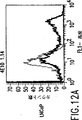
図12A〜DPSMAの細胞外ドメインとの抗体結合を例示した、3D7-1.1および4E10-1.14モノクローナル抗体によるLNCaP生細胞の認識についてのFACS分析による実例。図12Aは4E10-1.14と共にインキュベートしたLNCaP細胞を示す。図12Bは4E10-1.14と共にインキュベートしたPC-3細胞を示す。図12Cは3D7-1.1と共にインキュベートしたLNCaP細胞を示す。図12Dは3D7-1.1と共にインキュベートしたPC-3細胞を示す。図12Aおよび12Cにおける右へのシフトのパターンの違いは、2つの抗体がPSMAの異なるエピトープを認識しうることを示している。
図13PSMAのエピトープを区別するために2つのモノクローナル抗体を用いた二部位捕捉ELISAによるPSMAの検出。連続希釈したイムノアフィニティ精製PSMAを7E11-C5で被覆された96ウェルプレートに加え、3D7-1.1または4E10-1.14上清と共にインキュベートすることによって検出した。マイクロプレートリーダーで405nmの吸収を測定した。
![]()
![]()
図143D7-1.1および4E10-1.14モノクローナル抗体を用いた二部位捕捉ELISAによる、種々の生物学的サンプル中のPSMAの検出。
図153D7-1.1および4E10-1.14モノクローナル抗体を用いた二部位捕捉ELISAによる、正常なヒト血清で連続希釈したイムノアフィニティー精製PSMAの検出。
図16代替の二部位捕捉ELISAによるPSMAの検出。連続希釈したイムノアフィニティー精製PSMAを3D7-1.1で被覆された96ウェルプレートに加え、ビオチニル化7E11-C5(40μg/ml)次いで西洋ワサビペルオキシダーゼ結合ストレプトアビジンと共にインキュベートすることにより検出した。マイクロプレートリーダで405nmの吸収を測定した。7E11-C5をE-Z結合ビオチニル化キット(Pierce)を用いて製造元の使用説明書に従ってビオチニル化した。
図17LNCaP細胞溶解物、および4E10-1.14ハイブリドーマ由来の組織培養上清を用いてプローブした半精製PSMA断片(バキュロウィルス発現系で1.9kbインサートとして発現された全長PSMAのアミノ酸134〜750に対応する)の種々の画分のウェスタンブロット分析。1.9kb構築物(PSMAのアミノ酸134〜750)に由来するタンパク質産物の同定を矢印で示す。
1列目はマーカー;2列目はLNCaP細胞粗溶解物;3列目はウィルスペレット(即ち、1.9kb PSMA断片を発現するバキュロウィルスに感染させた100,000×gペレットの溶解SF9細胞);4列目は1.9kb PSMA断片を発現するバキュロウィルスに感染させた溶解SF9細胞由来の100,000×g上清画分;5列目はNi-NTAマトリックスを通過させた後の4列目に示す画分の素通り分;6列目はNi-NTAマトリックスの0.5M NaCl溶出;7列目はNi-NTAマトリックスの1Mイミダゾール(pH7.6)溶出;8列目はNi-NTAマトリックスを通過させた後の4列目に示す画分の素通り分;9列目はNi-NTAマトリックスの0.5M NaCl溶出;そして10列目はNi-NTAマトリックスの1Mイミダゾール(pH7.6)溶出を示す。また、2列目において、LNCaP細胞で発現された天然全長PSMAと4E10-1.14モノクローナル抗体との反応性にもふれておく。
図18関連性のないインサート、またはPSMAの一部(全長PSMAのアミノ酸134〜750)をコードする1.9kbインサートのいずれかを含むバキュロウィルスに感染させたSF9細胞の粗溶解物を抗体7E11-C5でプローブしたウェスタンブロット。1、2列目はMWマーカー;3列目は関連性のないウィルスで感染させたSF9細胞溶解物;4列目はSF9細胞溶解物;そして5列目はウィルスに感染させたSF9溶解物を含む1.9kb PSMAインサートである。SF9細胞またはいずれかのウィルスに感染させたいずれのタンパク質産物でも7E11-C5陽性バンドが観察されなかったことにふれておく。
図19モノクローナル抗体3D7-1.1を用いてプローブした、LNCaP細胞、ヒト精液、およびヒト血清から得たPSMAおよびPSM’のウェスタンブロット。1列目はLNCaP細胞溶解物;2列目は7E11-C5イムノアフィニティー精製されたLNCaP細胞由来PSMA;3列目はヒト精液;そして4列目はヒト男性血清。PSMAおよびPSM’の位置を示している。
5. 発明の詳細な説明
本発明は、PSMAの細胞外ドメインに特異的なモノクローナル抗体、そのような抗体の使用方法、およびそのような抗体により同定される末端切断型タンパク質変異体であるPSM’に関する。本明細書に記載の特定の手順および方法は、マウスを免疫するためのC末端ペプチドまたはPSMA発現性腫瘍膜調製物を用いて例示されるが、それらは本発明の実施を説明するためのものにすぎない。類似の手順および技法が、タンパク質、ペプチド、細胞表面抗原および粗製膜調製物の形態のPSMAに対して免疫された種々の動物宿主に対して同じように適用可能である。
5.1 ハイブリドーマ細胞系および抗体産生
第6節(後記)の実施例による特定の実施態様では、PSMAのC末端領域から誘導される合成ペプチドを免疫原として用いた。その結果、3F5.4G6と命名された1つの抗体が、PSMAの細胞外ドメインに結合しており、これは生きている前立腺癌細胞の細胞表面上および前立腺癌患者の血清中に露出していることが示される。さらに、第7節(後記)の第2の実施例では、PSMA発現性腫瘍膜調製物を用いて動物を免疫した後で、PSMAの細胞外ドメインに対する2つのモノクロナール抗体を産生させることが示されている。これに関連して、PSMAの細胞外ドメインに特異的なモノクローナル抗体を産生するために動物宿主内で免疫応答を生起させる免疫原としては、PSMAを発現するLNCaPのような癌細胞、PSMAコード配列でトランスフェクトした宿主細胞、精製PSMA、PSM’またはPSMA細胞外ドメインペプチドが使用可能である。
抗体を産生する可能性のある体細胞、特にBリンパ球は、B細胞ミエローマ系との融合に好適である。分裂中の形質芽球期(dividing plasmablast stage)にある抗体産生細胞が優先的に融合する。体細胞は、抗原感作動物のリンパ節、脾臓および末梢血から得ることが可能であり、選択されるリンパ細胞は、特定の融合系における経験に基づく有用性に大きく依存する。一次感作または超免疫された動物が、抗体産生リンパ球の供給源として使用できる。マウス・リンパ球は、以下に記載するマウスミエローマ系との安定な融合を高比率でもたらす。それらの中でも、BALB/cマウスが好ましい。しかし、他のマウス系統、ウサギ、ハムスター、ヒツジおよびカエルもまた、抗体産生細胞を調製するための宿主として使用可能である。Godingにより概説されているように(Monoclonal Antibodies: Principles and Practice,第2版、60〜61頁、Orlando,Fla,Academic Press,1986)、ラット・リンパ球を使用すると幾つかの利点がもたらされることがある。
あるいは、抗体の産生能を有するヒト体細胞(特に、Bリンパ球)が、ミエローマ細胞系との融合に好適である。個体の生検した脾臓、扁桃またはリンパ節からのBリンパ球が使用可能であるが、さらに容易に入手可能な末梢血Bリンパ球が好ましい。このリンパ球は、前立腺癌種と診断された患者から誘導され得る。さらに、ヒトB細胞は、エプスタイン−バーウイルス(Epstein-Barr virus)により直接不死化してもよい(Coleら,1995,Monoclonal Antibodies and Cancer Therapy,Alan R.Liss,Inc.77〜96頁)。
ハイブリドーマ産生融合法での使用に適するミエローマ細胞系は、好ましくは、非抗体産生性であり、高い融合効率を有し、かつ所望のハイブリドーマの増殖を維持する特定の選択培地で該細胞系を増殖不能にする酵素欠損体である。本発明の融合細胞ハイブリッドの産生に使用可能なそのようなミエローマ細胞系の例としては、P3-X63/Ag8、X63-Ag8.653、NS1/1.Ag4.1、Sp210-Ag14、FO、NSO/U、MPC-11、MPC11-X45-GTG1.7、S194/5XX0 Bul(これらは全てマウス由来);R210.RCY3、Y3-Ag1.2.3.、IR983Fおよび4B210(ラット由来);ならびにU-266、GM1500-GRG2、LICR-LON-HMy2、UC729-6(これらは全てヒト由来)が挙げられる(Goding,Monoclonal Antibodies: Principles and Practice,第2版,65〜66頁,Orlando,Fla,Academic Press,1986;Campbell,Monoclonal Antibody Technology,Laboratory Techniques in Biochemistry and Molecular Biology第13巻,BurdenおよびVon Knippenberg編,75〜83頁,Amsterdam,Elseview,1984)。
抗体産生脾臓またはリンパ節細胞とミエローマ細胞とのハイブリッドの作製方法は、一般に、細胞膜の融合を促進する(化学的または電気的)作用因子(agent)の存在下で体細胞とミエローマ細胞とを各々2:1の比率(但し、この比率は約20:1〜約1:1の間で変えることが可能である)で混合することを含む。この融合手法で用いられる体細胞およびミエローマ細胞の供給源としては、同じ種の動物を用いることが好ましい場合が多い。融合方法は、KohlerおよびMilstein(1975,Nature 256: 495-497; 1976,Eur.J.Immunol.6: 511-519)ならびにGefterら(1977,Somatic Cell Genet.3: 231-236)により記載されている。それらの研究者が用いた融合促進剤は、それぞれセンダイウイルスおよびポリエチレングリコール(PEG)であった。Godingが概説している融合方法(1986,Monoclonal Antibodies: Principles and Practice,第2版,71〜74頁,Orlando,Fla,Academic Press)は上記の融合方法ならびに電気的に誘導される融合を包含しており、これもまた本発明のモノクロナール抗体の作製に好適である。
融合手順により、一般に、生存可能なハイブリッドが非常に低い頻度(約1×10⁻⁶〜1×10⁻⁸個の体細胞)で作製される。生存可能なハイブリッドはこのように低頻度で得られるので、融合細胞ハイブリッドを残りの未融合細胞(特に、未融合ミエローマ細胞)から選別する手段を有することが必須である。さらに、所望の抗体産生ハイブリドーマを他の残りの融合細胞ハイブリッドから検出する手段も必要である。
一般に、融合細胞は、選択培地、例えば、ヒポキサンチン、アミノプテリンおよびチミジンを含有するHAT培地で培養する。HAT培地は、ハイブリッド細胞の増殖(proliferation)を可能にし、通常は無限に分裂し続ける未融合ミエローマ細胞の増殖(growth)を阻止する。アミノプテリンは、テトラヒドロ葉酸塩の生成を阻害することによりプリンおよびピリミジンのde novo合成を遮断する。チミジンの添加によりピリミジン合成の遮断が回避され、一方、ヒポキサンチンが培地に含まれることにより阻害された細胞がヌクレオチド再利用経路を用いてプリンを合成するようになる。用いられるミエローマ細胞は、ヒポキサンチンホスホリボシルトランスフェラーゼ(HPRT)が欠如しており、そのためにその再利用経路を利用できない変異体である。生存ハイブリッドでは、Bリンパ球は、この酵素を産生するための遺伝情報を提供する。Bリンパ球自体は培養下で有限の寿命(約2週間)を有するので、HAT培地で増殖できる細胞だけが、ミエローマ細胞と脾臓細胞とから形成されるハイブリッドである。
上記ハイブリッドにより分泌される抗体のスクリーニングを容易にするために、そして個々のハイブリッドが他のものよりも過剰増殖しないようにするために、融合したミエローマおよびBリンパ球の混合物をHAT培地に希釈し、マイクロタイタープレートの複数のウエル中で培養する。2〜3週間で、ハイブリッドクローンが顕微鏡観察下で見えるようになったら、ハイブリッドクローンを含有する個々のウエルの上清液を、特定の抗体についてアッセイする。このアッセイは、高感度で、簡易で、かつ迅速なものでなければならない。アッセイ技術としては、ラジオイムノアッセイ、酵素免疫アッセイ、細胞障害性アッセイ、プラークアッセイ、ドット免疫結合(dot immunobinding)アッセイなどが挙げられる。
所望の融合細胞ハイブリッドを選択し個々の抗体産生細胞系にクローニングしたら、各細胞系は、2つの標準的な方法のいずれかにより増殖させてもよい。ハイブリドーマのサンプルは、最初の融合のための体細胞およびミエローマ細胞を供給するのに用いた種類の組織適合性動物に注射することができる。注射した動物は、融合細胞ハイブリッドにより産生される特定のモノクローナル抗体を分泌する腫瘍を生ずる。この動物の体液(例えば、血清または腹水)を抜き取ることにより、高濃度のモノクローナル抗体が得られる。あるいはまた、個々の細胞系を、実験室用培養容器内でin vitroで増殖させることも可能である。この培養培地も単一の特定のモノクローナル抗体を高濃度で含有しており、これはデカンテーション、濾過または遠心分離により採取できる。
モノクローナル抗体または抗原結合領域の少なくとも一部を有する該モノクローナル抗体の精製断片[例えば、Fv、F(ab’)2、Fab断片](HarlowおよびLane,1988,Antibody,Cold Spring Harber)、一本鎖抗体(米国特許第4,946,778号)、キメラまたはヒト化抗体(Morrisonら、1984,Proc.Natl.Acad.Sci.USA 81:6851;Newubergerら、1984,Nature,81:6851)および相補性決定領域(CDR)は、慣用の手法により調製可能である。これらの抗体または断片の精製は、例えば、硫酸アンモニウムまたは硫酸ナトリウムにより沈殿させた後で、食塩水への透析、イオン交換クロマトグラフィー、アフィニティまたはイムノアフィニティクロマトグラフィー、ならびにゲル濾過、ゾーン電気泳動などを行う、当業者に公知の種々の方法により達成できる(Goding、Monoclonal Antibodies:Principles and Practice,第2版、104〜126頁、Orlando,Fla,Academic Press参照)。
5.2 モノクローナル抗体およびPSM’の特性決定
第5.1節(前記)に概略的に説明した技術および第6節および第7節(後記)で説明する技術を用いて、PSMAの細胞外ドメインの特異的なモノクローナル抗体の産生に基づいて、3つのハイブリドーマ細胞系を選択した。本発明は、35F.4G6、3D7-1.1および4E10-1.14抗体、ならびに、PSMAおよびPSM’の細胞外ドメインに特異的に結合する他のモノクローナル抗体を包含するものであり、特に、酵素免疫アッセイ、放射線免疫アッセイまたは他の任意の競合結合免疫アッセイで評価された際に、上記3つの抗体のうちの任意の1つ以上の抗体がPSMAに結合するのを競合的に阻害する全ての抗体を含む。
抗体3F5.4G6は、前立腺癌細胞溶解物中および前立腺癌細胞の細胞表面上ならびに前立腺癌患者から得た血清中に発現されるPSMAに特異的に結合するIgMのイソ型抗体である。さらに、3F5.4G6はPSM’にも特異的に結合する。3F5.4G6反応性PSMAエピトープは細胞外でC末端であり、細胞の細胞質で膜結合している7E11-C5により認識されるもの(Horoszewiczら、Anticancer Res.7:927-936)とは異なる。抗体3D7-1.1および4E10-1.14もまたIgM抗体であり、前立腺癌細胞溶解物中および細胞表面上で発現されるPSMAに結合する。これらの抗体は、原発性前立腺癌および転移性腫瘍(例えば、前立腺癌の骨への転移)の双方を検出するのに使用可能である。
抗体応答が生じる際に、抗体産生細胞はまず最初にIgMイソ型を分泌し、これは最終的にIgGにスイッチされる。そのようなクラススイッチ事象は、同じ抗原特異性が保持されるような定常領域遺伝子のDNA再編成により生じるものである。異なる抗体のイソ型は、異なるエフェクター機能を有する。例えば、IgMおよびIgG4以外の全てのIgGサブクラスは、抗原結合の際に補体を固定する。これに対して、IgEはアレルギー反応において肥満細胞に結合してヒスタミンの放出を誘発する。
ハイブリドーマ細胞系も、長期培養の間にクラススイッチ変異体を産生する。特に、IgMからIgGまたはIgG1からIgG2nへとスイッチするモノクローナル抗体はプロテインAに対する高い親和性に基づいて選択されて、これにより、それら抗体の精製が促進される。任意のクラススイッチ変異体を特定の所望のエフェクター機能について選択することができる(Spiraら、1985,Hybridoma Technology in the Biosciences and Medicine,Springer編、77〜88頁,Plenum Press,NY; HarlowおよびLane,1988 Antibodies,Cold Spring Harbor Laboratory)。例示した抗体の場合、それらはIgMのイソ型なので、同じ抗原特異生を有するIgG変異体を選択することも望ましく、これは、in vitroまたはin vivoにおける特定の目的にはさらに有用である場合がある。本発明は、3F5.4G6、3D7-1.1および4E10-1.14を含む本発明のモノクローナル抗体のIgG変異体を包含する。
第6節および第7節(後記)では、例示の抗体が120kDaの分子量のタンパク質を認識することが示されている。特に、3F5.4G6はまた、前立腺腫瘍細胞溶解物中の105〜110kDaの分子量のタンパク質を認識する。この120kDaのタンパク質は抗体7E11-C5によっても認識されるが、その低分子量の方のタンパク質は抗体3F5.4G6、3D7-1.1および4E10-1.14によってのみ検出される。したがって、その105〜110kDaのタンパク質は、PSM’として知られるmRNAの産物を提示する。しかしながら、本発明以前には、PSM’タンパク質はいっさい報告されておらず、それは未翻訳のmRNAであると考えられていた。PSM’のアミノ酸配列は、そのRNA配列から推定されるように、PSMAの細胞質領域および膜貫通領域が欠如していると予想されるので、7E11-C5が、その細胞内エピトープに対する特異性のために、この産物と反応しないであろうことは確実である。これに対して、PSMAの細胞ドメンに特異的な抗体3F5.4G6、3D7-1.1および4E10-1.14はPSM’も認識する。
5.3 PSMA特異的モノクローナル抗体のコード配列
本発明の別の実施態様では、例示のハイブリドーマ細胞系を用いて、抗原結合部位または抗体変異体を含む組成物を製造することが可能であり、該抗体変異体は、ネズミの可変領域または超可変領域をヒトの定常領域もしくは定常および可変フレームワーク領域と組合せたもの、すなわち、キメラまたはヒト化抗体、ならびに患者の抗体由来の抗原結合CDRのみをヒト・フレームワーク領域と結合させて保持するヒト化抗体である(Waldmann,1991,Science 252:1657,1662,特に1658-59、ならびにそこに引用されている文献を参照)。ネズミ抗体の結合特異性を保持しているそのようなキメラまたはヒト化抗体は、本発明による診断、予防または治療の用途のためにin vivoで投与した場合、低下した免疫原性を有すると予想される。
さらに別の実施態様では、本発明は、再編成された活性化免疫グロブリン遺伝子をコードするDNAまたはmRNAの供給源としてハイブリドーマ細胞系を使用することを包含するものであり、この免疫グロブリン遺伝子は単離し、公知の組換えDNA技術によりクローニングし、そしてPSMAの細胞外ドメインに特異的な抗原結合断片の作製のために他の細胞へ移送することが可能である。本発明のハイブリドーマ細胞系の再編成DNAの単離または該ハイブリドーマ細胞系のmRNAからのcDNAの調製により、イントロンを含まない配列が得られる可能性がある。
説明のために(しかし限定しようとするものではないが)、以下のようにして免疫発現ライブラリーを作製し、PSMAおよびPSM’に対する抗体結合断片をスクリーニングすることができる(Huseら,1989,Sci.246:1275-1281;Mullinaxら,1990,Proc.Natl.Acad.Sci.USA 87:8045-8099参照)。全RNAが精製でき(例えば、市販のキットを用いることによる)、軽(L)鎖のためのオリゴ(dT)プライマーおよび重(H)鎖のための特定のプライマーを用いて逆転写酵素を用いてcDNAに変換できる。免疫グロブリンH鎖およびL鎖の配列のポリメラーゼ連鎖反応(PCR)増幅は、プライマー対の組を用いて別々に行われる。上流プライマーはリーダーの部分的に保存された配列および/またはVHもしくはVLのフレームワーク領域にハイブリダイズするように設計でき、下流プライマーは定常ドメイン配列にハイブリダイズするように設計できる。そのようなプライマーは、完全長L鎖を維持し、IgGのFdに対応しH−Lジスルフィド結合を保存しているH鎖を付与するであろう。PCR増幅したLおよびH DNA断片を次いで消化し、それぞれHおよびL鎖ベクターに連結する。そのようなベクターはpelBリーダー配列、リボソーム結合部位および終止コドンを含有する。大腸菌(E.coli)中での発現に適切なλファージベクターは、市販のベクター(ImmunoZAP L、ImmunoZAP H;Stratacyte,La Jolla,Ca)から調製できる。連結した組換えファージDNAをin vitroパッケージング抽出物(in vitro packaging extract)と共にバクテリオファージに組み込み、大腸菌を感染させるのに用いる。このようにして作製された免疫発現ライブラリーを、PSMA、PSM’またはそれらの特定のペプチドを用いて抗原結合断片についてスクリーニングする。陽性のクローンは、Mullinaxら(前掲)により記載されているようにしてスクリーニングおよび同定できる
5.4 PSMA細胞外ドメイン特異的抗体および抗体組成物の使用
本明細書に記載する特定の手法および方法は、本発明のモノクローナル抗体を用いて例示されるが、それらは本発明を実施するための説明にしかすぎない。本発明による以下に記載の手法および方法では、抗原結合領域の少なくとも一部を有するモノクローナル抗体の精製断片(例えば、Fv、F(ab’)2、Fab断片)、一本鎖抗体、キメラまたはヒト化抗体またはCDRが使用できる。
5.4.1 免疫組織学的および免疫細胞学的適用
本発明のモノクローナル抗体は、組織学的および細胞学的検体中の前立腺癌腫瘍細胞の検出、特に正常組織および非悪性腫瘍からの悪性腫瘍を識別するのに使用することができる。組織検体は該抗体により染色可能であり、それらの結合は、ペルオキシダーゼ、フルオロセイン、アルカリホスファターゼなどの標識に結合した二次抗体により検出し得る。
さらに、免疫蛍光法で本発明のモノクローナル抗体を用いて、ヒト組織、細胞および体液の検体を調べることができる。典型的なプロトコールでは、凍結した非固定化組織生検サンプルのクリオスタット切片を含むスライドガラスまたは細胞学的スミアを風乾し、ホルマリンまたはアセトンで固定し、モノクローナル抗体調製物と共に給湿チャンバー内で室温にてインキュベートする。
次に、そのスライドガラスを洗浄し、上記モノクローナル抗体に対する抗体の調製物と共にさらにインキュベートする。この抗体は、使用するモノクローナル抗体がマウス脾臓リンパ球とマウスミエローマ細胞系との融合物に由来する場合には、通常は或る特定のタイプの抗-マウス免疫グロブリンである。この抗-マウス免疫グロブリンは、特定の波長で蛍光発光する化合物(例えば、ローダミンまたはフルオロセインイソチオシアナート)でタグ付けする。次いで、サンプル中の染色パターンおよび強度を蛍光顕微鏡により測定し、場合によっては写真に記録する。
さらに別の実施態様として、本発明のモノクローナル抗体を用いて組織検体または剥脱細胞(すなわち、前立腺腫瘍の吸引生検から得られる単一細胞調製物)を試験するために、コンピューターで画質向上させた(computer enhanced)蛍光画像分析またはフローサイトメトリーを用いることができる。本発明のモノクローナル抗体は、コンピューターで画質向上させた蛍光画像アナライザーによる、またはフローサイトメーターを用いる生きている腫瘍細胞(すなわち、前立腺腫瘍の吸引生検から得られる単一細胞調製物)の定量に特に有用である。そのようなアッセイにおいて抗体3F5.4G6、および4E10-1.14を用いることは、良性の前立腺腫瘍と悪性の前立腺腫瘍とを区別するのに有用である。何故ならば、該モノクローナル抗体が結合するPSMAは、悪性腫瘍により増大した量で発現されるからである。さらに、PSMA陽性細胞集団の割合は、それ単独で、もしくはこれらの細胞のDNA倍数性の測定と共に、疾患の進行の初期の指標を提供することにより、非常に有用な予後の情報を提供し得る。
さらに別の実施態様では、本発明のモノクローナル抗体は、他の既知の前立腺抗体と組合せて使用して、前立腺癌腫の悪性表現型に関するさらなる情報を提供することができる。
5.4.2 免疫血清学的適用
本発明のモノクローナル抗体は、その使用を、認識される特異的抗原決定基の存在についてヒト生物学的体液のスクリーニングへも広げることができる。患者から集めた生物学的体液のin vitro免疫血清学的評価によって、非侵襲的に癌を診断することができる。例示として、前立腺液、精液、全血、血清あるいは尿のようなヒト体液を患者から採取し、標準的なラジオイムノアッセイまたは酵素結合(enzyme-linked)イムノアッセイ、競合的結合酵素結合イムノアッセイ、ドットブロットあるいはウェスタンブロット、または当業界で公知の他の測定法において、PSMAおよびPSM’の細胞外ドメインに特異的なモノクローナル抗体を用い、サンプル液中の放出された抗原または細胞膜に結合した抗原として、特異的エピトープについてアッセイすることができる。
さらに、PSMAおよびPSM’上の重複していないエピトープに対するモノクローナル抗体を使用することによって、より感度の高いPSMAまたはPSM’タンパク質の診断アッセイを開発することができる。7E11-C5、3F5.4G6、3D7-1.1または4E10-1.14のようなPSMAの反対末端に特異的な抗体は、特に、このようなアッセイに使用するのに適している。この点に関し、一つの抗体は生物学的液体中のPSMAまたはPSM’を捕捉する基質に固定させておいてもよく、もう1つの抗体を使用して抗体結合抗原を検出する。また、PSMAおよびPSM’の発現は前立腺癌および正常前立腺組織のそれぞれにおいて増大するので、これら二つの形態を識別する抗体を使用して、処置後の腫瘍の退行対進行をモニターするさらに精確な方法を提供してもよい。3F5.4G6、3D7-1.1および4E10-1.14は両方の形態を認識するが、7E11-C5のほうはPSMAだけと結合するので、患者における各形態の正確なレベルを測定するのにこれらの抗体を共同して使ってもよい。そうすれば、腫瘍負荷(tumor burden)とその量とを相関させることができる。例えば、7E11-C5は、二サイト捕捉アッセイのアンカー抗体として使用することができ、他の三つの抗体のいずれか一つをPSMA定量用の検出抗体として使用することができる。一方、同様な二サイト捕捉アッセイに、この三つのPSMA細胞外ドメイン特異的抗体の中の2つの任意の組合せを使用して、全PSM’+PSMA濃度を特異的に測定することもできる。全PSM’+PSMA濃度から単純にPSMAを差し引けば、PSM’を具体的に定量することができる。
組織および体液中の細胞外ドメインのPSMAおよびPSM’をモノクローナル抗体によって検出することに加えて、NAALADase酵素活性測定を利用して、組織および/または体液中の細胞外ドメインのPSMAおよび/またはPSM’を定量することができる。
例えば、ホモジネートした組織を界面活性剤で可溶化し、遠心分離により不溶性物質をペレット化し、残留上清中のNAALADase活性を測定することによって、組織レベルを測定することができる。同様にして、まず遠心分離により細胞性物質をペレット化し、上清についてNAALADase活性の典型的なエンザイムアッセイを行うことによって、体液中のNAALADase活性を測定することもできる。
抗体結合の特異性を利用するNAALADaseアッセイプロトコールもまた適用することができる。例えば、7E11-C5、3F5.4G6、3D7-1.1または4E10-1.14抗体のいずれかを塗布した固体表面は、NAALADaseエンザイムアッセイを用いる検出用のPSMAまたはPSM’を捕捉するのに使用することができるであろう。従ってこれは、細胞外ドメイン特異的抗体がPSMAおよびPSM’の両方に結合するが7E11-C5はPSMAだけに結合するのであれば、所与の検体中の全長PSMAタンパク質およびPSM’を示差的に検出し定量するのに使うことができる。
グルタミン酸デヒドロゲナーゼの反応特性を利用して、さらに簡便なNAALADaseエンザイムアッセイを適用することもできる(Frieden,1959,J.Biol.Chem.,234: 2891)。このアッセイ法では、NAALADase酵素の反応生成物はグルタミン酸である。これは、N-アセチルアスパラギン酸とグルタミン酸を生ずるN-アセチルアスパルチルグルタミン酸の酵素触媒分解から誘導される。NSA(P)+を必要とする工程において、グルタミン酸は、グルタミン酸デヒドロゲナーゼによって触媒される反応で、2-オキソグルタル酸とNAD(P)Hを生ずる。この反応の進行は、NAD(P)+からNAD(P)Hへの変換による340nmの吸収変化によって容易かつ簡便に測定することができる。こうして、固定化した捕捉抗体を用いる固相フォーマットに適用可能なNAALADase活性のアッセイを改良することができる。このようにして、NAD+またはNAD(P)+の添加前および添加後の340nmにおける吸収変化に基づき、マイクロプレートリーダーにおいて、多重アッセイを同時に実施することができる。これは固相アッセイだけに限定されるものではない。何故ならば、このタイプのNAALADaseアッセイを用いて、例えば、血清のような溶液アッセイも可能だからである。
本発明のモノクローナル抗体またはその断片を含有するキットは、上記の免疫組織学的、免疫細胞学的および免疫血清学的方法による、in vitro診断、予後および/または前立腺癌のモニター用に誂えることができる。キット成分は、水性媒体か、または凍結乾燥形式にパッケージすることができる。酵素や放射性金属イオンのような標識部分が付いたコンジュゲートの形でモノクローナル抗体(またはその断片)を上記キットに使用する場合、そのようなコンジュゲートの成分は、完全にコンジュゲートした形か、中間体の形、またはキット使用者によりコンジュゲートされる別々の部分として供給することができる。
キットは、試験管やバイアル、フラスコ、瓶、注射器などの、一またはそれ以上の容器手段または容器セット手段をコンパクトに収容するようにコンパートメント化したキャリアを含んでもよい。上記容器手段または容器セット手段の第一のものは、モノクローナル抗体(またはその断片)あるいはPSMAまたはPSM’を含んでいてもよい。第二の容器手段または容器セット手段は、第一抗体(またはその断片)PSMAまたはPSM’と結合することができる標識またはリンカー標識中間体を含んでいてもよい。
5.4.3 in vivoでの診断、予防および治療への使用
本発明のモノクローナル抗体またはその断片は、in vivoでの前立腺癌細胞のターゲッティングに特に有用である。これらは、検出およびモニター用の腫瘍局在化ならびに原発性前立腺癌および転移の治療に使用することができる。これらをin vivoで適用するには、精製モノクローナル抗体または、Fv、F(ab’)2、Fab断片のような抗原結合領域の少なくとも一部をもつモノクローナル抗体の精製断片(HarlowおよびLane,1988,Antibody,Cold Spding Harbor)、単一鎖抗体(米国特許第4,946,778号)、キメラ抗体またはヒト化抗体(Morrison et al.,1984,Proc.Natl.Acad.Sci.USA 81: 6851; Newuberger et al.,1984,Nature,81: 6851)、相補性決定領域(CDR)等を使用することが望ましい。上記抗体またはその断片の精製は、当業者に公知の様々な方法によって行うことができ、そのような公知方法としては、硫酸アンモニウムまたは硫酸ナトリウムにより沈澱させた後で生理食塩水に対する透析、イオン交換クロマトグラフィ、アフィニティあるいは免疫アフィニティクロマトグラフィ、ならびにゲル濾過、ゾーン電気泳動などがある(Goding,Monoclonal Antibodies: Principles and Practice,第2版、104-126頁、Orlando,Fla,Academic Press参照)。
in vivoでの前立腺癌の検出および/またはモニターに使用するには、精製モノクローナル抗体は、コンジュゲートまたは複合体の投与および局在化(loca;lization)後、特定の組織または臓器の画像化を可能にするリポーター群(a reporter group)として役立つ化合物に、直接またはリンカーを介して共有結合させることができる。種々の異なるタイプの物質をリポーター群として役立てることができ、そのような物質には、放射線不透過性染料、放射性金属および非金属同位元素、蛍光原性化合物、発蛍光性化合物、陽電子発生性同位元素、非常磁性金属などがある。
前立腺癌のin vivo治療での使用の場合、精製モノクローナル抗体を単独で、あるいは、上記コンジュゲートの投与および局在化後、悪性細胞または組織を死滅および/または増殖抑制する化合物に、直接またはリンカーを介して共有結合させて、使用することができる。抗体をそれ自体で使用する場合、抗体は、補体の固定化または抗体依存細胞の細胞障害性によって腫瘍破壊を媒介してもよい。あるいは、前記抗体は、相乗的な治療効果を与えるために化学療法剤と併用投与することもできる(BaslyaおよびMendelsohn,1994,Breast Cancer Res.and Treatment,29: 127-138)。治療の目的で、放射性金属および非金属同位元素、化学療法剤、毒素などを含む、種々の異なるタイプの物質を該抗体と直接コンジュゲートすることができる(VitettaおよびUhr,1985,Annu.Rev.Immunol.,3: 197)。
別の態様によれば、前立腺癌のin vivo治療のために、本発明のモノクローナル抗体は、二官能性または二重特異的抗体、すなわち、前立腺特異的膜抗原の細胞外ドメインに対して特異的な抗原結合領域および殺腫瘍活性または腫瘍抑制活性をもつエフェクター細胞に特異的な抗原結合領域をもつ抗体の形に改変することができる。二重特異的抗体の二つの抗原結合領域は化学的に結合されているか、または二重特異的抗体を産生するように遺伝子工学的に操作された細胞によって発現することができる(一般に、Fanger et al.,1995,Drug News & Perspec.,8(3): 133-137参照)。殺腫瘍活性を有する適切なエフェクター細胞としては、細胞障害性T細胞(主としてCD8+細胞)、ナチュラルキラー細胞などを例示することができるが、これらには限定されない。本発明による二重特異的抗体の有効量を、前立腺癌患者に投与すると、この二重特異的抗体は、PSMAをもつ原発性または転移性腫瘍の部位に局在化後、悪性細胞を死滅させるか、および/またはその増殖を阻害する。
検出、モニターおよび/または治療に有用な、本発明の抗体(またはその断片)の抗体コンジュゲートの作製方法は、米国特許第4,671,958号、4,741,900号および4,867,973号に記載されている。
上記のタイプの物質のいずれかとコンジュゲートしたモノクローナル抗体(またはその断片)を含む、そのようなin vivo腫瘍局在化および治療法と共に用いるためのキットは以下のように作ることができる。キットの成分は、水性媒体か、または凍結乾燥形態にパッケージすることができる。このモノクローナル抗体(またはその断片)を上記キットの中で放射性金属イオンや治療剤部分のような標識または治療部分が結合したコンジュゲートの形でに使用する場合、そのようなコンジュゲートの成分は、完全にコンジュゲートした形態、中間体の形態、またはキットの使用者がコンジュゲートを生成する別々の部分として供給することができる。
6. 実施例:PSMAペプチドに対するモノクローナル抗体の産生
6.1 材料と方法
6.1.1 免疫用ペプチドの調製
PierceRockford,IL)のEDC方法を用いて、PSMAペプチド#716〜723(NH2-ESKVDPSK-)を、キャリアとしてキーホールリンペットヘモシアニン(KLH)と共役結合させた。このペプチド-KLH複合体を、1mg/mlのムラミルジペプチド(MDP、Pierce,Rockford,IL)を含有する不完全フロインドアジュバント(シグマ社、ミズーリ州セントルイス)に最終濃度250μg/mlで乳化した。乳化抗原調製物を4℃で保存した。
6.1.2 免疫
0.1mlの乳化ペプチドキャリア-複合体を6週間にわたって14日毎に皮下注して、雌のBALB/cマウスを免疫した。マウスから採血し、その血清を抗ペプチド抗体の存在について調べるペプチド特異的ラジオイムノアッセイ(RIA)にて試験した。1:1,000またはそれ以上の力価で抗ペプチド抗体が陽性と出たマウスを融合プロトコルのドナーとして使用した。融合3日前に、生理食塩水に溶かしたペプチド-KLH複合体50μgを腹腔内に投与してマウスを免疫した。
6.1.3 細胞融合
同じペプチド-KLH複合体による最終免疫した3日後に、BALB/cマウスの脾臓を無菌的に摘出し、単一細胞懸濁液を作った。浸透ショックにより赤血球を溶解し、残ったリンパ球をRPMI-1640培地に懸濁させた。脾臓細胞を10:1の割合でP3X63Ag8U.1(X63)ミエローマ細胞(CRL 1597、ATCC、Rockville,MD)と混合した(脾臓細胞100 x 106:X63ミエローマ細胞10 x 106)。X63細胞に対する脾臓細胞の融合はGalfreおよびMilsteinの方法によって行った(1981,Methods in Enzymology,Vol.73,Immunochemical Techniques,Part B)。細胞培養培地(RPMI-1640-20%ウシ胎児血清)中アミノプテリンのインクルージョンによってハイブリドーマ細胞を選択した。
6.1.4 一次ハイブリドーマのスクリーニング
各ハイブリドーマ培養液から細胞培養上清50μlを取り、ペプチド特異的抗体が存在するかどうかをペプチド特異的RIAによって試験した。簡単に説明すると、ウシ血清アルブミン(BSA)と共役結合させたペプチドを50μg/mlを予め塗布した96ウェルプロバインド(Pro-Bind)プレート(ファルコン社)のウェルに前記上清を加える。4℃で一夜インキュベーションした後、プレートをPBS-0.1% BSAで4回洗浄する。ウサギ抗マウスIgMとIgG(ICN)の1:500希釈液50μlを各ウェルに加え、プレートを室温で1時間インキュベーションする。プレートを上記のように4回洗浄し、各ウェルに125I-プロテインAを50μl加える。プレートを室温で1時間インキュベーションし、上記のように4回洗浄する。プレートをオートラドフィルム(コダック、X-OMAT)に一夜曝露して現像する。陽性のウェルを選択し、さらなる試験を行うために細胞を細胞培地に広げた。
6.1.5 ウェスタンブロットスクリーニング
広げた陽性のウェルから上清を得、これを抗PSMA抗体についてウェスタンブロットアッセイで試験した。PSMAを発現する前立腺腫瘍であるLNCaP腫瘍細胞系(CRL 1740、ATCC、Rockville,MD)の溶解物を90分間175ボルトのSDSポリアクリルアミドゲルにかける。電気泳動タンパク質をイモビロン(Immobilon)-P(商標)メンブランに対してエレクトロブロッティングし、そのメンブランを、トリス緩衝生理食塩水中5%のBLOTTOで一夜インキュベーションすることによりブロックした。メンブランをバイオラッド(Bio-Rad)マルチスクリーン装置(バイオラッド社)に置き、ハイブリドーマ上清約650μlをそれぞれのレーンにピペットで加えた。メンブランを室温で90分間インキュベーションし、ブロットをトリス緩衝生理食塩水-0.5% Tween-20(TBS-T)で5回洗浄した。洗浄したブロットを、ペルオキシダーゼ標識ヤギ抗マウスIgG(Kirkegaard and Perry Laboratories,Gaithersburg,MD)の1:5,000希釈液と共に室温で1時間インキュベーションした。このブロットを上記のように5回洗浄し、2mlのLumiGLO(商標)化学発光基質(KPL,Gaithersburg,MD)と共に1分間インキュベーションした。このブロットをオートラドフィルムに曝露し現像した。陽性のハイブリドーマウェル(抗PSMA反応性)を同定し、さらなる発育(development)用として選択した。
6.1.6 限界希釈クローニング
上記のウェスタンブロットアッセイにおいてPSMAに対する反応性によって同定した陽性一次ハイブリドーマウェルを限界希釈法によりクローニングした。クローニングした細胞を、フィーダー細胞集団としての相乗的な胸腺細胞を含有する完全細胞培地の1mlにつき1個となるよう調整する。細胞懸濁液を96ウェルプレートのウェルに加えて200μlずつのアリコートを作る。7〜10日間培養した後、細胞のコロニーが見えてくる。単一コロニーを含むウェルを取り、その細胞を24ウェルプレートに広げる(1.5ml培養液)。クローン細胞の上清を得て、上記ウェスタンブロットアッセイにより抗PSMA抗体について試験を行う。陽性クローンを拡大増殖(expand)し液体窒素で凍結する。
6.1.7 腹水液の発生と抗体精製
10 x 106のハイブリドーマ細胞を注入する7〜8日前に、プリスタン0.4mlを腹腔内注射することによりBALB/cマウスを初回抗原刺激を行った。モノクローナル抗体を含有する腹水液を一定時間毎に抜き取って4℃で保存する。ピアース社(Rockford,IL)のImmunoPureTM IgM精製キットを用いて、腹水液からモノクローナル抗体を精製した。
6.1.8 PSMAの免疫沈降
約10 x 106のLNCaP腫瘍細胞を、4℃で30分間、1mlのNP-40細胞融解緩衝液(150mM NaCl、1% NP-40、50mM Tris)と共にインキュベーションした。細胞溶解物を12,000rpmで遠心分離し、得られた上清を正常マウス血清50μlと共に30分間インキュベーションした後で抗マウスIgMアガロースビーズの20%懸濁液60μlを添加することによって予め澄明にした(precleared)。4℃で1時間インキュベーションを行った後、この調製物を遠心分離してビーズを除き、得られた上清を3F5.4G6モノクローナル抗体と反応させた。3F5.4G6モノクローナル抗体の量を変えて(2.5、5および10μg)3個の複製細胞溶解物(replicate lysate)に加え、4℃で1時間インキュベーションした。抗マウスIgMアガロースビーズ(シグマ社)の10%懸濁液100μlを加え、細胞溶解物をさらに1時間4℃でインキュベーションした。12,000rpmで溶解物を遠心分離して、アガロースビーズをNP-40細胞融解緩衝液で3回洗浄する。電気泳動サンプル緩衝液30μlを該sビーズに加え、95℃で10分間加熱する。1,200rpmでビーズを簡単に遠心分離して、サンプル緩衝液をSDSポリアクリルアミドゲルに流した。電気泳動後、サンプルを上記のようにエレクトロブロッティングに付し、リポート抗体としてPSMA特異的モノクローナル抗体7E11-C5を使用してウェスタンブロットを行った。
6.1.9 フローサイトメトリーによる分析
まず細胞を燐酸緩衝化生理食塩水(PBS)で濯いだ。Versene(0.2g EDTA.4Na/L)溶液(75cm2フラスコにつき2ml)を加えた。Versene溶液の大部分は、室温で5分間インキュベーションする前にアスピレーションにより除去した。PBSを加えて、ピペッティングにより細胞を移した(dislodge)。細胞をPBSで2回洗浄した後計数した。50万個〜100万個の細胞を、50μlの一次抗体と共に30分間氷上でインキュベーションした後、PBSで2回洗浄した。次いで細胞を、50μlのFITC標識二次抗体(7E11-C5に対するヤギ抗マウスIgGまたは4G6に対するヤギ抗マウスIgG)と共に30分間氷上でインキュベーションした。過剰の二次抗体をPBSで細胞を洗い流す。フローサイトメーター(FACScan,Becton Dickinson,San Jose,CA)を使って蛍光を分析した。その前散乱プロファイルおよび横散乱プロファイルに基づいて分析した細胞集団から細胞破片を除いた。
6.1.10 ウェスタンブロットによる血清分析
血清サンプルを細胞融解緩衝液(1%トリトンX-100、50mM HEPES、10%グリセロール、15mM MgCl2、1mM AEBSF、1mM EGTA)で1:7に希釈した。LNCaP溶解物を溶解緩衝液で1:35に希釈した。次いで希釈したサンプルをサンプル緩衝液(SDS還元緩衝液)と2:3の割合で合せた。サンプル(20μl)を8.5%のSDS-PAGE(Bio-Rad Protein Assayを用いて測定した場合、最終タンパク質濃度93mg/サンプル)にかけ、分離したタンパク質を90ボルトで1時間、PVDFメンブラン上にブロットした。次いでメンブランを5%ミルク-TBS中で一夜ブロックした。翌日、TBS-T中1時間メンブランを3μg/mlの7E11-C5抗体でプローブし、TBS-T中で5分間5回洗浄し、TBS-T中30分間167ng/mlのヒツジ抗マウス西洋ワサビペルオキシダーゼ標識二次抗体でプローブした。再びメンブランをそれぞれTBS-T中各5分間5回洗浄し、化学発光基質キット(Kirkegaard & Perry Laboratories,Inc.,Gaithersburg,MD)(Rochon et al.,1994,The Prostate 25: 219-223)を用いて、メンブランを展開(develop)した。
X線フィルムを曝露することによりブロットを可視化し、約120kDのタンパク質バンドを見えるようにした。ブロット像をマイクロテックスキャンメーカーIIHRスキャナーでスキャンし、「公共領域の国立衛生研究所(NIH)画像プログラム(Wayne Rasbandが米国立衛生研究所で書いたもので、zippy.nimh.nih.govから作者不明のftpによりインターメットから、またはNTIS,5285 Port Royal Rd.,Springfield,VA22161,part number PB93-504868からのフロッピーディスクで入手可能)を使うマッキントッシュクアドラ(Macintosh Quadra)605コンピューターで行う分析」によって、バンド強度を測定した。全患者サンプルを、健常者の正常ドナーサンプルおよび、標準対照として同じウェスタンブロットから、高いPSMAをもつ前立腺癌患者サンプルに対して評価した。
6.1.11 PSM’酵素活性の検出
妊娠試験のためのWHOガイドラインに従って報酬を支払ったドナーからヒト精液100mlを採取した。10,000rpmで30分間遠心分離して細胞物質をペレット化し、上清を注意深く除いて、2回取り替えたpH7.6の20mM Tris緩衝液に対して一夜透析した。透析液を再び10,000rpmで遠心分離し、予めpH7.6の20mM Tris緩衝液で洗浄しておいたDEAEセファクリルカラムに流した。次いでこのカラムを再び500mlの同じ緩衝液で洗浄し、pH7.6で20mM〜200mMのTris緩衝液勾配を適用したタンパク質を分離した。5mlの画分を集めた。モノクローナル抗体7E11-C5を使って、各画分におけるPSMAの存在をウェスタンドットブロットにより測定した。7E11-C反応性タンパク質バンドを含有する画分をプールし、70%硫酸アンモニウムを用いて沈澱させた。沈澱したタンパク質を10,000rpmで30分間遠心分離してペレット化した後、200mMのTris緩衝液、pH7.6、1リットルの再懸濁させた。次いで、溶解したタンパク質を、2回取り替えたpH7.6の20mM Tris緩衝液に対して一夜透析した。透析物を次いで予め洗浄しておいたセファクリルカラムに流してタンパク質を溶出し、3mlの画分を集めた。モノクローナル抗体3F5.4G6を使って、溶出タンパク質についてウェスタンドットブロットを行った。画分88-96が陽性であり、これら画分のそれぞれは、SDSポリアクリルアミドゲル電気泳動により、その純度について試験した。
6.2. 結果
PSMAの細胞外ドメインに対するモノクローナル抗体を生成するために、このタンパク質のいくつかの領域をそれらの相対親水性に関してHopp & Woods法(1983,Mol.Immunol.20:483-489)に基づいて分析した。
以下の表1は、試験した数種類のペプチドの相対親水性を示す。特に、PSMAのC末端領域中のアミノ酸残基番号716-723に対応するESKVDPSK(Glu-Ser-Lys-Val-Asp-Pro-Ser-Lys)(配列番号1)の配列をもつペプチドを合成した。さらに、細胞外ドメインに対する抗体を産生するために、表1に示した細胞外ドメインの他の部分や全細胞外ドメインそのものも使用できるだろう。これとは対照的に、残基#44-58および#196-213に対応する2つのアミノ酸ペプチドは、天然PSMAと結合しない抗ペプチド抗体応答をもたらした。

抗原としてペプチド結合BSAを用いて結合アッセイにより初期スクリーニングを実施いた。個々のハイブリドーマ培養物から細胞培養上清50μlを分離し、ペプチド特異的ラジオイムノアッセイでペプチド特異的抗体の存在について試験した。簡単に述べると、ウシ血清アルブミン(BSA)に結合させたペプチドを予めコーティングしておいた96ウェルのPro-Bindプレートのウェルに上清を添加した。4℃で一夜インキュベーションした後、プレートをPBSで洗った。1:500の希釈度のウサギ抗マウスIgMおよびIgG 50μlを各ウェルに添加し、プレートを室温で1時間インキュベートした。その後プレートを4回洗浄し、125I標識プロテインAを各ウェルに添加した。プレートを室温で1時間インキュベートし、上記のように4回洗浄した。プレートをオートラッドフィルムに一夜露光して現像した。陽性のウェルを選択し、以後の試験のために細胞を培地中で増やした。同定された陽性ウェルのうち、3F5と称する一つのハイブリドーマをウエスタンブロットアッセイでさらに試験したところ、その分泌抗体はLNCaP溶解物中に含まれるPSMAと反応することがわかった。LNCaP細胞をHoroszewiczら(1983,Cancer Res.43:1809-1818)が記載したとおりに培養し、細胞溶解物をRochonら(1994,Prostate 25:219-223)が記載したとおりに調製した。3F5ハイブリドーマ細胞を限界希釈法によりクローニングし、細胞数を増やし、ウエスタンブロットアッセイで再試験した。3F5.4G6と称する抗体のサブクローンはLNCaP溶解物中の分子量120kDaのタンパク質と反応した(図2)。この抗体はIgMとしてアイソタイピングされた。3F5.4G6のアイソタイプの決定には、Boehringer Mannheimから得られた、マウスモノクローナル抗体のアイソタイピング用のISOStripを用いた。モノクローナル抗体をPBSで1:100に希釈し、希釈サンプル(150μl)をこのキットと共に供給された展開チューブに添加し、室温で30秒インキュベートした後しばらく攪拌した。次いでチューブの中にアイソタイプストリップを挿入し、5分間展開した。このストリップのラムダまたはカッパセクションおよびクラスまたはサブクラスセクションの一つに青色のバンドが出現した。モノクローナル抗体3F5.4G6はIgMアイソタイプであると同定された。
さらに、モノクローナル抗体3F5.4G6は、対照としてモノクローナル抗体7E11-C5を用いて、ステージD2の進行中の前立腺癌患者から採取した血清に対して試験した(図3)。両抗体とも約120kDa分子量のバンドを同定した(図3)。3F5.4G6モノクローナル抗体を用いるLNCaP細胞の追加のウエスタンブロットアッセイを、IgMに特異的な二次抗体を用いて行った(図4)。モノクローナル抗体7E11-C5は約120kDaの単一バンド(すなわち、PSMA)を認識したが、3F5.4G6は同様の分子量バンドだけでなく約105-110kDaのバンドも認識した。このバンドはPSM’の想定されたタンパク質形態に一致しており、PSMAとPSM’の両方の細胞外ドメインを特異的に認識する抗体の有用性を示す。
7E11-C5と前立腺癌患者の血清中の120kDaのタンパク質との反応性は抗体特異的であり、一般的な血清タンパク質と二次抗体との非特異的な反応性によるものでなかった。ウエスタンブロットアッセイでは、血清サンプル由来の分離したタンパク質を含むImmobilon P紙を、7E11-C5モノクローナル抗体+HRPに結合させた二次抗体と、またはHRPに結合させた二次抗体のみに反応させた。フィルムを1分間露光し、また、二次抗体と血清中の120kDaのタンパク質との非反応性を実証するために45分にわたり過度に露光した。さらに、この二次抗体を3F5.4G6と共に使用して同抗原を検出した。したがって、3F5.4G6モノクローナル抗体はPSMAとPSM’に特異的であった。
図5から、7E11-C5により同定されたタンパク質はモノクローナル抗体3F5.4G6によっても認識されたことが裏づけられる。さらに、モノクローナル抗体3F5.4G6は、モノクローナル抗体7E11-C5により検出されない105-110kDaのタンパク質をも認識した。この比較的速く泳動するタンパク質はPSM’に相当する。溶解物を最初に7E11-C5で沈殿させ、残りのタンパク質を7E11-C5と反応させたとき、この抗体はどのようなタンパク質も検出しなかった(レーン4)。これに対して、7E11-C5で前処理した溶解物を3F5.4G6と反応させたときには、この抗体は約110kDaのタンパク質を検出した。図6は、3F5.4G6により免疫沈降された120kDaタンパク質(すなわち、PSMA)が7E11-C5によっても認識されたことを示す。
図7AおよびBは、FACS分析でモノクローナル抗体3F5.4G6が生存LNCaP細胞を認識したことを示しており、これにより3F5.4G6はPSMAの細胞外ドメインを認識することが確認される。PSMAの細胞外ドメインを認識する抗体は前立腺癌における診断および/または治療ツールとして特に有用である。
ヒト精液をPSMA特異的抗体と反応させて、酵素活性についてアッセイした。図8は、レーン2のモノクローナル抗体3F5.4G6により認識されたタンパク質はおよその分子量が90kDaであることを示す。PSM’はLNCaP溶解物中で105-110kDaの分子量をもつことがわかったが、精液中の90kDaタンパク質はPSM’の非グリコシル化または部分グリコシル化産物であるらしかった。PSM’はいくつかのグリコシル化部位を含むので、この低い分子量は精液中のグリコシダーゼ活性の結果であった。PSMAがこの精製調製物中に存在しないことは、3F5.4G6がLNCaP細胞の溶解物中に存在する分子量120kDaのタンパク質(すなわち、PSMA)を認識した(レーン1)が、レーン2ではこの分子量のタンパク質を認識しなかったという事実から明らかである。さらに、抗体7E11-C5は精液中の90kDaバンドを認識しなかった。
その後、モノクローナル抗体3F5.4G6により認識されたPSM’のこの精製調製物をNAALADase活性についてアッセイした。LNCaP溶解物から調製した高速上清を陽性対照として用いた。3F5.4G6モノクローナル抗体と陽性に反応するタンパク質であって、それがPSM’であることと一致するタンパク質は、Robinsonら(1987,J.Biol.Chem.262:14498-14506)により記載されたアッセイを行ったところ、16.9nmol/min./mgタンパク質の固有のNAALADase活性を含んでいた。
7. 実施例:PSMA含有腫瘍細胞膜調製物に対するモノクローナル抗体の産生
7.1 材料と方法
7.1.1 免疫
2枚の150mmプレートからLNCaP膜を調製するにあたり、versene溶液中に細胞を分離し、遠心して細胞をペレットとした。細胞ペレットに蒸留水を添加し、Dounce型ホモジナイザーを用いて細胞をホモジナイズした。ホモジナイズした懸濁液を30,000×gで遠心し、ペレット化した膜画分を免疫用に用いた。
完全フロイントアジュバントで乳化したLNCaP前立腺癌細胞膜調製物を用いて成熟雌BALB/cマウスを4回、腹腔内投与により免疫した。細胞融合を行う5日前に、イムノアフィニティーで精製したPSMA(PBS中)50μgを用いてマウスを追加免疫した。細胞融合は上記の第6.1.3節に記載のとおり行った。
7.1.2 一次ハイブリドーマのスクリーニング
固相ELISA法をベースとするアッセイ法によりPSMA特異的抗体の検出を行った。イムノアフィニティーで精製したPSMA、バキュロウイルスで発現させた完全長のPSMA、あるいは細菌で発現させたPSMA断片をMaxi-Sorp(NuncImmuno)96ウェルプレート上にコートし、4℃で一晩インキュベートした。プレートをPBS-0.2% Tween-20で洗い、残存したサイトを5% BSA溶液を用い1時間室温でブロックした。ハイブリドーマ培養物からの上清50μlをPSMAをコートしたウェルに添加し、そのプレートを室温で2時間インキュベートした。プレートを上述の方法で洗い、1:600希釈したウサギ抗マウスIgG及びウサギ抗マウスIgMを50μl各ウェルに添加した。室温で1時間インキュベートした後、プレートを上述の方法で洗い。1:400希釈のHRPを結合させたProtein-A 50μlを各ウェルに添加した。室温で1時間インキュベートした後、プレートを上述の方法で洗い、100μlのABTS(2,2’-アジノ-ビス(3-エチルベンズチアゾリン)-6-スルホン酸150mgを0.1Mのクエン酸500ml,pH4.35中に含有する)/過酸化水素(10mlのABTS溶液あたり10μlの30%H2O2を含有)色原体/基質溶液を各ウェルに添加した。プレートをマイクロプレートリーダーで読みとり、OD405を測定した。OD値がバックグラウンド値より0.05高い値を示した上清を産生するハイブリドーマを限界希釈法でクローン化し、それ以後の分析に用いた。
PSMAの固相への捕捉のために上述のアッセイ法を次のように改変した:40μg/mlの7E11-C5抗PSMAモノクローナル抗体溶液(0.1M NaHCO3結合緩衝液pH8.2中)50μlを、Maxi-Sorpプレートのウェルに添加し、4℃で一晩付着させた。プレートを上述の方法で洗い、ブロックした。階段希釈したイムノアフィニティー精製PSMA 50μlを、7E11-C5をコートしたウェルに添加し、室温で2時間インキュベートした。十分に洗った後、3D7-1.1あるいは4E10-1.14ハイブリドーマクローンから得た50μlの無希釈の組織培養上清をウェルに添加し、プレートを室温で90分間インキュベートした。上述の方法で洗った後、ウェルを1:1000希釈のペルオキシダーゼを結合させたヤギ抗マウスIgM 50μlと反応させ、室温で1時間インキュベートした。十分洗った後、ABTS/H2O2を100μl各ウェルに添加し、プレートをマイクロプレートリーダーで上述の方法で読み取った。
7.1.3 PSMAのイムノアフィニティーによる精製
パックされたLNCaP細胞16mlを、5倍容量の25mM Tris-HCl,pH7.4、150mM NaCl、1% NP-40中でPotter-Elvehjem型ホモジナイザーを2往復させてホモジナイズした後、4℃で一晩攪拌した。抽出液を100,000×gで1時間遠心し、さらにペレットから初回同様に再抽出を行った。上清をあわせ、冷室で一晩7E11-C5-Immunobeads(Pierce)(3-5ml樹脂ベッド容積)と混合した。ビーズを遠心し、ホモジナイズ用緩衝液で十分洗い、カラムに注入した。ビーズを再度1%NP-40含有のホモジナイズ用緩衝液で洗った後、1%Triton-X-100R含有の緩衝液で再度洗った。洗浄後のビーズを100mMのグリシン緩衝液pH2.5、150mM NaCl、1% Triton X-100Rで溶出し、2ml画分を集めた。タンパク質の溶出はOD280でモニターした。
タンパク質を含有する画分をSDS-PAGゲルで銀染色及びウエスタンブロッティング法を用いて分析した。典型的な調製物においては、ウエスタンブロッティング法で7E11-C5反応性の120kDaのタンパク質のバンドは60-80%の純度であった。パックされた細胞16mlからのおよその収率は、PSMAタンパク質1mgであった。PSMA調製物中の界面活性剤は、溶液をExtractigelカラム(Pierce)に通すことによって除去した。
7.1.4 フローサイトメトリー分析
PSMAの外部つまり細胞外にあるエピトープに対するモノクローナル抗体の認識能をフローサイトメトリーで評価した。LNCaP(PSMA発現)及びPC-3細胞(PSMA非発現)を組織培養フラスコから新たに採取し、単細胞懸濁液を調製した。約100万個の細胞を、3D7-1.1あるいは4E10-1.14ハイブリドーマクローンから得た無希釈組織培養上清1ml中に再懸濁し、氷上で2時間インキュベートした。細胞をPBS-0.1% BSA,0.01%アジ化ナトリウムで2回洗い、1:100希釈のFITCを結合させたウサギ抗マウスIgM 100μl中に再懸濁し、氷上でさらに30分間インキュベートした。細胞を上述の方法で2回洗い、500μlの洗浄用緩衝液に再懸濁し、蛍光染色をFACSCalibur(Becton-Dickinson)を用い、CellQuest acquisitionソフトウエアで分析した。
7.1.5 ウエスタンブロット分析
3D7-1.1及び4E10-1.14ハイブリドーマクローンから得た組織培養上清のPSMA反応性をウエスタンブロット分析で調べた。ウエスタンブロット分析はPelletierとBoyntonのプロトコール(1994,J.Cell.Physiol.158:427-434)に従って行った。その概略は次のとおりである。LNCaP細胞及びPC-3細胞から得た溶解物、イムノアフィニティー精製PSMA、あるいはバキュロウイルス発現完全長PSMAを8.5% SDS-PAGEゲル上で電気泳動させ、分離したタンパク質をPVDF膜上に90ボルトで1時間電気的にブロッティングした。その膜を5% BLOTTO中で一晩ブロックし、適切なクローンから得た無希釈組織培養上清20mlと90分間インキュベートした。上清を除去し、ブロットをTBS-0.5% Tween-20(TBS-T)で5回洗い、1:5000希釈のペルオキシダーゼを結合させたヤギ抗マウスIgM二次抗体(Jackson)と室温で1時間反応させた。その膜をTBS-Tで5回洗い、Chemiluminescent Substrate Kit(KPL)を用いて展開し、X線フィルム(Kodak)に露光させることによって可視化した。
7.1.6 バキュロウイルス発現系を用いた組換えPSMAの調製
PSMAの完全長コード配列(Israeliら,1993,Cancer Res.53:227-230)を含むインサートを、Lambda pDR2ヒトライブラリー(Clonetech)からその遺伝子配列に特異的なプローブを用いてクローン化した。インサートはこのベクターからSmaI及びSspI消化によって切り出し、トランスファーベクターpAcHLT-C(Pharmingen)中に、製造者の説明書に従ってクローン化した。このトランスファーベクターとBacPAK6直線化ウイルスDNA(Clonetech)の同時トランスフェクションは製造者の説明書に従って行い、その結果、Ni-NTAカラムとの結合によってタンパク質単離に用いうるポリヒスチジンテールをタンパク質のN末端に持つ完全長PSMAタンパク質をコードするウイルスが得られた。PSMAタンパク質を産生させるために、プラーク精製した組換えバキュロウイルス粒子を単離、増幅し、5% FBS(Hyclone)を補給したSFM II培地(Gibco-BRI)の存在下でSf9細胞に、感染の多重度を約1:2として感染させた。48時間インキュベーションした後、感染細胞を採取し、1%CHAPSで溶解し、Ni-NTA-アガロース(Quiagen)を通し、製造者の説明書に従ってイミダゾールで溶出させることにより回収した。最終産物を1LのPBSに対して3回透析した。
7.2 結果
PSMA含有前立腺癌細胞膜に対するモノクローナル抗体を作製した。LNCaP細胞からイムノアフィニティー精製した天然PSMA、及びアミノ酸配列の1-175、134-437、438-750の領域に対応する細菌で発現させたPSMAの断片を用いて固相イムノアッセイで、2種のハイブリドーマクローン、3D7-1.1及び4E10-1.14を選択した。天然PSMAとの結合性において、3D7-1.1及び4E10-1.14ハイブリドーマクローンから得た上清は7E11-C5抗体と比肩しうるものであった(図9)。バックグラウンドのBSAに対する非特異的結合は基本的には3種の抗体調製物とも同様であった。
エピトープとの結合の特異性を調べると、7E11-C5モノクローナル抗体は、PSMのN末端で細胞内ドメインに対応するアミノ酸断片1-175に結合した。3D7-1.1及び4E10-1.14はこの断片に対して中等度の結合しか示さなかったが、これら2種のモノクローナル抗体はPSMAのアミノ酸断片134-437(この断片はPSMAの細胞外ドメインの一部である)に最も強い結合性を示した。この断片はPSM’の一部であるので、これらの抗体はPSM’とも反応する。
さらに、3D7-1.1ハイブリドーマクローンから得た上清について、LNCaP細胞及びPC-3細胞の溶解物、及びイムノアフィニティー精製PSMAに対してのウエスタンブロット分析を行った。図10は3D7-1.1ハイブリドーマクローンがLNCaP細胞(レーン1)に存在する120kDaバンドとは反応するが、PC-3細胞(レーン2)とは反応しないことを示している。レーン1及び2のどちらも、おそらく二次抗体試薬の非特異的結合のためと思われる反応性を示している。イムノアフィニティー精製PSMAを含むレーン3は3D7-1.1モノクローナル抗体と反応させたとき120kDaに主要なバンドを示している。同様なウエスタンブロットデータが、ブロットの非特異的バックグラウンドは3D7-1.1の場合よりはるかに大きいとはいえ、4E10-1.14クローンの上清でも得られている。このように、3D7-1.1及び4E10-1.14のどちらも、LNCaP細胞に存在する120kDaバンド及びイムノアフィニティー精製PSMAと反応する。
バキュロウイルスで発現させた完全長のPSMAをSDS-PAGEゲル上で電気泳動し、PVDF膜に電気的にブロッティングした。ブロットをMuni-Protean II Milti-Screen装置(Bio-Rad)に挿入し、各種の抗体調製物と反応させ、ウエスタンブロットとして発現させた。図11は3D7-1.1及び4E10-1.14モノクローナル抗体が、7E11-C5モノクローナル抗体が結合するのと同じバンドに対応するタンパク質のバンドと反応したことを示している。
LNCaP細胞及びPC-3細胞を3D7-1.1及び4E10-1.14ハイブリドーマクローンから得た上清で染色し、フローサイトメトリーで分析した。2種の抗体のどちらも非同定のLNCaP生細胞を染色したが、PC-3細胞は染色しなかった(図12A-D)。これらの結果から、2種の抗体がPSMA分子の細胞外ドメインのエピトープと反応することを確認した。さらに、LNCaP染色において、3D7-1.1で見られたショルダーと比較して、4E10-1.14モノクローナル抗体では明らかなシフトが認められたが、これは2種の抗体がPSMA分子の特定の領域の異なるエピトープを認識することを示唆している。
PSMAのための二部位捕捉ELISA(two-site capture ELISA)を、7E11-C5モノクローナル抗体をPSMA捕捉試薬として、3D7-1.1及び4E10-1.14モノクローナル抗体をレポーターあるいは検出抗体として用いて開発した。これらの抗体はPSMA分子の異なるエピトープを認識するため(7E11-C5はN末端の6個のアミノ酸と反応し、3D7-1.1及び4E10-1.14は134-475アミノ酸領域内の一つの配列と反応する)、二部位捕捉アッセイにおいては効果的な組み合わせとなる。イムノアフィニティー精製PSMAを階段希釈したものを試験抗原として用いたとき、3D7-1.1と4E10-1.14のどちらの上清も、7E11-C5をコートした96ウェルプレート上での捕捉後、PSMAを検出し得た(図13)。さらに、LNCaP細胞及び精液から精製したPSMA及びバキュロウイルスで発現させた完全長PSMAの粗調製物を試験した(図14)。PSMA対照抗原、精液、及びバキュロウイルスPSMA調製物では有意に大きいOD405値が得られた。精製PSMAを正常ヒト女性の血清で希釈し、サンプルを二部位捕捉アッセイで調べると、上記と同じ抗体がPSMAを検出した(図15)。PSMAの異なる部分に対するモノクローナル抗体を用いて開発した二部位捕捉アッセイは多様なソースから抗原特異的な様式でPSMAを検出した。
PSMA用の二部位捕捉ELISAとしてもう一つ別の方法を3D7-1.1モノクローナル抗体をPSMA捕捉試薬として、7E11-C5モノクローナル抗体をレポーター又は検出試薬として開発した。階段希釈したイムノアフィニティー精製PSMAを試験抗原として用い、3D7-1.1をコートしたプレートで捕捉させ、ビオチン化した7E11-C5モノクローナル抗体を用いて検出した。結果は図16に示す。
図16は3D7-1.1や4E10-1.14などのPSMAの細胞外ドメインと特異的に結合するモノクローナル抗体が、PSMAの二部位捕捉ELISAに有用であることを示している。
PSMAの捕捉に3D7-1.1が有用であることは、PSMAタンパク質の細胞外ドメインのみに依存したもう一つ別のイムノアッセイが使用しうることを示している。そのような2種の細胞外ドメイン特異的抗体を捕捉及び検出に用いるアッセイは、PSM’のエピトープのタンパク質内での位置からみてPSM’を検出しうるであろう。7E11-C5を捕捉又は検出のどちらかに用いたアッセイはどんなものでも特異的にPSM’を除外する。PSM’に特異的なアッセイの1例としては、3D7-1.1又はPSMAの細胞外ドメインに特異的なモノクローナル抗体のうちの1種の抗体をPSMA及びPSM’の捕捉に用いて平行試験を行うものが挙げられる。それに引き続き、4E10-1.14をPSMAとPSM’を合計したものの検出に用い、7E11-C5をPSMAのみの検出に用いることにより、PSM’の量は単純な引き算によって得られる。このデータからPSM’とPSMAの比率が求められ、それはSuら(Cancer Res.55:1141-1443,1995)の観点によれば診断に用いうるものとなろう。
SuはPSMAをコードする転写物が前立腺癌患者で選択的によく検出される(正常男性と比べて)ことを示しているが、SuらはPSMA転写物がこれらの患者の体内で実際にタンパク質に翻訳されているかどうかは示していない。さらに、SuはPSM’タンパク質を検出してはいないが、PSM’をコードする転写物が正常男性に選択的によく検出される(前立腺癌患者に比べて)ことを示している。本発明者らは、本出願において、PSMAタンパク質が前立腺癌患者の体の組織及び/又は体液中で(正常男性と比べ)増加していること、及びPSM’タンパク質が正常男性の体の組織及び/又は体液中で(前立腺癌患者と比べ)増加していることを示している。従って、本発明によれば、PSM’のPSMAに対する比は、前立腺癌患者の臨床的評価にあたって、診断上及び/又は予後判定での有用性を有することとなるであろう。
完全長PSMAのアミノ酸34-750に対応するPSMA断片を、バキュロウイルス発現系で1.9kbのインサートとして発現させた。バキュロウイルスで発現させたPSMA断片は、その断片のN末端からPSMA細胞外ドメインのさらに76アミノ酸が欠失していることを除けば、PSM’(完全長PSMAの残基58-750に対応している)と非常に類似している。各種のバキュロウイルス発現させた半精製PSMA断片及びLNCaP細胞溶解物のウエスタンブロット分析を4E10-1.14モノクローナル抗体をプローブとして用いて行った。結果を図17に示す。
1.9kbのPSMA断片(すなわち完全長PSMAのアミノ酸134-750)をコードするインサートあるいは無関係のインサートのどちらかを含むバキュロウイルスを感染させたSF9細胞の粗溶解物のウエスタンブロット分析を、7E11-C5モノクローナル抗体をプローブとして行った。結果を図18に示す。
図17は、PSMAの細胞外ドメインに特異的な4E10-1.14などの抗体が、PSM’に非常に類似したバキュロウイルス発現タンパク質にも結合しうることを示している。これに対して図18は、7E11-C5モノクローナル抗体はそのエピトープ特異性の故にそのような性質を有していないことを示している(図18に示すようにバキュロウイルス発現PSMA断片と7E11-C5との陰性の反応性に注目されたい)。バキュロウイルス発現PSMA断片は、N末端から追加の76アミノ酸(その全てが細胞外ドメインにある)が欠失していることを除けばPSM’(完全長PSMAのアミノ酸58-750に対応)と同一である。3D7-1.1及び4E10-1.14のエピトープ特異性は、PSM’及びPSMAの134-750アミノ酸断片(図9参照)のいずれにも含まれる細胞外ドメインの領域にマップされるので、これらの抗体が双方とも天然のPSM’と結合する固有の特性を持っているであろうと考えられ、これは7E11-C5には無いものである。
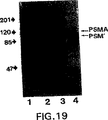
LNCaP細胞由来のPSMA、及びPSMAを含むことが知られたヒト血清及び精液のウエスタンブロットのプローブとして3D7-1.1モノクローナル抗体を用いた。結果を図19に示す。
約120kDaに移動するPSMAに対応するバンドが全画分に存在した。さらに、抗体3D7-1.1で現出させた場合には、血清及び精液中に、2番目に速く移動する分子量90から100Kdのバンドが認められた。この速く移動するバンドは血清を7E11-C5抗体を用いた場合のウエスタンブロットには認められなかった(Holmesら,The Prostate,Supple.7:25-29(1996)参照)。この速く移動する、3D7-1.1と反応するタンパク質のバンドはおそらく体液中に存在するPSM’であろう。
8. 細胞系の寄託
次のハイブリドーマ細胞系は1996年3月12日、及び1997年3月11日にAmerican Type Culture Collection,12301 Parklawn Drive,Rockville,Maryland 20852,U.S.A.に寄託され、下記の受託番号の指定を受けた。
ハイブリドーマ ATCC受託番号
3F5.4G6 HB12060
3D7-1.1 HB12309
4E10-1.14 HB12310
本発明は、本発明の個々の態様の説明を意図して例示した実施態様に示される範囲に限定されるものではない。事実、ここに示し、記述した事柄に加えて本発明に種々の改変を加えることは、上記の詳細な説明及び添付図面から当業者であれば自明なことであろう。そのような改変は本発明の特許請求の範囲に包含されるものとする。
本明細書中に引用された全ての公表文献は参照としてその全文を本明細書中に組み込むこととする。
1.Field of Invention
The present invention relates to monoclonal antibodies that bind to the extracellular domain of prostate specific membrane antigen (PSMA), hybridoma cell lines that produce such antibodies, and methods of using such antibodies in the diagnosis and treatment of cancer. In particular, the present invention relates to monoclonal antibodies that react with PSMA produced against synthetic peptides that are substantially homologous to a portion of the carboxy-terminal region of PSMA and expressed on the surface of tumor cells and in serum of prostate cancer patients. The invention further relates to two monoclonal antibodies that react with PSMA produced against prostate cancer membrane preparations and also expressed on the cell surface. The present invention also relates to a novel protein variant (PSM ') of PSMA detected by the antibody.
2.Background of the Invention
Prostate cancer is the second leading cause of cancer death in men. In fact, prostate cancer is the most common non-cutaneous cancer diagnosed in American men. The number of men diagnosed with prostate cancer has steadily increased as a result of early diagnosis due to the growing population of older men and the growing awareness of prostate cancer (Parker et al., 1997, CA Cancer J. for Clin.47: 5-28). In 1997, more than 334,500 men were diagnosed with prostate cancer and the disease was estimated to cause approximately 41,800 deaths. The risk for men to have prostate cancer in their lifetime is about 1 in 5 for whites and 1 in 6 for African Americans. High risk groups are represented by groups with prostate cancer patients or African Americans in the family. More than two-thirds of men diagnosed with prostate cancer die of this disease during their lifetime (Wingo et al., 1996, CA Cancer J. for Clin.46: 113-25). Also, many patients who have not died of prostate cancer require continuous treatment to relieve symptoms such as pain, bleeding, and urinary tract obstruction. Thus, prostate cancer is also a major cause of distress and increased health expenditure (Catalona, 1994, New Eng. J. Med.331: 996-1004).
PSMA is a protein having a molecular weight of 120 kDa that is expressed in prostate tissue and has been identified by the reactivity with a monoclonal antibody conventionally referred to as 7E11-C5 (Horoszewicz et al., 1987, Anticancer Res.7: 927-935; U.S. Pat. No. 5,162,504). PSMA is available in purified form (Wright et al., 1990, Antibody Immunoconjugates and Radio PharmaceuticalsThree: Summary 193), transferrin receptor (Israeli et al., 1994, Cancer Res. 54: 1807-1811) and NAALADase activity (Carter et al., 1996, Proc. Natl. Acad. Sci. U.S.A.93: 749-753) and is characterized as a type II transmembrane protein with sequence identity. More importantly, PSMA is more expressed in prostate cancer and high amounts of PSMA are also detected in the sera of these patients (Horoszewicz et al., 1987, ibid .; Rochon et al., 1994, Prostatetwenty five: 219-223; Murphy et al., 1995, Prostate26: 164-168; and Murphy et al., 1995, Anticancer Res.15: 1473-1479). The cDNA encoding PSMA has been cloned (Israeli et al., 1993, Cancer Res.53: 227-230), this cDNA comprises two alternatively spliced mRNA species, an mRNA species comprising 2,653 nucleotides encoding PSMA, and a second mRNA species comprising 2,387 nucleotides referred to as PSM '. (Su et al., 1995, Cancer Res.55: 1441-1443). Prior to the present invention, since no PSM 'protein product was detected, it was not known whether PSM' encodes a protein product or simply exists as an untranslated mRNA species.
A recent report by Carter et al. (1996, Proc. Natl. Acad. Sci. U.S.A.,93: 749-753) shows that there is a high degree of identity between the 1428 base corresponding to a part of the PSMA cDNA and the cDNA sequence of the protein N-acetylated α-linked acid dipeptidase (NAALADase). NAALADase has an enzyme activity for the neuropeptide N-acetylaspartylglutamate and produces glutamate and N-acetylaspartate. According to this report, the NAALADase activity inherent to PSMA protein has been demonstrated, but the catalytic portion of PSMA has not been identified. NAALADase activity was observed in LNCaP cells that expressed PSMA, but not in PC3 cells that did not express PSMA. Transfecting PSMA cDNA into PC3 cells resulted in NAALADase activity and the presence of PSMA in these cells.
The difference between the PSMA and PSM 'cDNAs is a transmembrane, intracellular coding region deletion that includes nucleotides # 1-171 or amino acids # 1-57. PSMA is a type II membrane protein, and the functional catalytic domain of type II membrane protein is known to be in the C-terminal extracellular region of the molecule (DeVries et al., 1995, J. Biol. Chem.,270: 8712-8722).
More PSM 'mRNA was found in normal prostate tissue than in patients with benign hyperplasia or prostate cancer (Su et al., 1995, ibid). Conversely, PSMA mRNA was found at higher levels in patients suffering from prostate cancer than in patients not suffering from prostate cancer (Su et al., 1995, ibid). This observed difference is consistent with previously described serum protein levels of PSMA (Horoszewicz et al., 1987, ibid; Rochon et al., 1994, ibid; Murphy et al., 1995, ibid; and Murphy et al., 1995, Ibid.) In this context, elevated levels of PSMA in the serum of prostate cancer patients correlate with disease progression versus remission and can be used as a prognostic marker (Murphy et al., 1995, ibid).
The epitope recognized by monoclonal antibody 7E11-C5 mapped to the first 6 amino acids of the intracellular N-terminal region of PSMA (Troyer et al., 1995, Urol. Oncol. 1:29-37) (Figure 1). By electroimmunocytochemistry using 7E11-C5, the epitope for this antibody was localized in the cytoplasm and especially in the inner lobe of the plasma membrane (Troyer et al., 1994, Proc. Am. Assoc. Cancer Res.35: 283, summary 1688). Furthermore, monoclonal antibody 7E11-C5 stains only fixed and permeabilized cells in in vitro studies (Horoszewicz et al., 1987, ibid). This result is consistent with the mapping of the 7E11-C5 epitope to the N-terminus or intracellular domain of PSMA. Monoclonal antibodies specific for the extracellular domain of PSMA are useful for detecting in vivo prostate cancer, which is likely to expose its epitopes through necrosis and / or apoptosis, whereas monoclonal antibodies specific for the extracellular domain of PSMA Enables more efficient detection of PSMA on the surface. Also, since PSM 'lacks the intracellular domain of PSMA, monoclonal antibody 7E11-C5 does not recognize PSM' based on the sequence of the PSM 'mRNA transcript.
Citation or listing of all references in this section or all other sections of this application is not an admission that such references are prior art to the present invention.
3.Summary of the Invention
The present invention relates to a monoclonal antibody specific for the extracellular domain of PSMA, a hybridoma cell line producing said antibody, a method of using said antibody for the diagnosis and treatment of prostate cancer, and PSM ′ recognized by such an antibody. PSMA mutant protein known as:
Part of the present invention is based on the discovery of three monoclonal antibodies by the applicant that recognize the extracellular domain of PSMA. One of the antibodies was produced by immunizing mice with a PSMA C-terminal peptide having the amino acid sequence ESKVDPSK (SEQ ID NO: 1). This antibody reacts with PSMA and PSM 'proteins in tumor cell lysates and serum of prostate cancer patients. This antibody also stained intact live tumor cells and was recognized for its specificity for the extracellular domain of PSMA or PSM 'protein. This antibody also detects PSM 'in human semen, and PSM' in human semen exhibits NAALADase activity. Two other monoclonal antibodies were raised against a prostate cancer membrane preparation. These antibodies also react with the extracellular domains of PSMA and PSM ', including natural PSMA isolated by immunoaffinity purification and recombinant PSMA produced by recombinant DNA technology. These antibodies are useful in combination with antibodies directed against the intracellular domain of PSMA to detect the presence of PSMA in a test sample in a two-site capture assay. Further, all three antibodies disclosed herein can be used in a two-site capture assay to detect the presence of PSM 'in a test sample.
The present invention encompasses a wide variety of uses, including but not limited to developing and using immunoassays for detection or staging of prostate cancer in patients, primary and / or metastatic prostate cancer Imaging in vivo, using antibodies conjugated to cytotoxic or chemotherapeutic agents for therapeutic use, and constructing antibody fragments, chimeric antibodies, humanized antibodies, or bivalent antibodies And including using.
[Brief description of the drawings]
FIG. 1 Deduced amino acid sequences of PSMA and PSM 'antigens (SEQ ID NO: 2) (Israeli et al., 1994, Cancer Res.54: 1807-1811). PSM 'mRNA is thought to start at amino acid 58 because it does not include the 5' end of PSMA encoding the first 57 amino acids (the amino acid sequence in the first row). However, prior to the present invention, PSM 'was never identified in protein form. The underlined region is the putative transmembrane domain, and the bold region (amino acids # 716-723) is the peptide selected for monoclonal antibody development.
FIG. 2. Example of protein and its reactivity in monoclonal antibody 3F5.4G6 (subclone derived from primary hybridoma 3F5) and in a LNCaP lysate with a molecular weight of 120 kDa corresponding to PSMA. Western blot was developed using HRP-anti-IgG secondary antibody. The first row is the LNCaP lysate probed with 7E11-C5, the second row is the LNCaP lysate probed with 3F5.4G6.
Figure 3. PSMA levels in serum of prostate cancer patients (stage D2) using monoclonal antibody 3F5.4G6 (
FIG. 4. Western blot assay of LNCaP lysates developed using monoclonal antibodies 7E11-C5 (first row) and 3F5.4G6 (second row) with HRP-anti-IgM secondary antibody. Both 7E11-C5 and 3F5.4G6 recognized a protein with a molecular weight of 120 kDa. In addition, 3F5.4G6 also recognized a protein with a molecular weight of 105-110 kDa, corresponding to what was predicted to be the protein form of PSM '. It should be noted that 7E11-C5 did not recognize PSM 'because no epitope of the 7E11-C5 monoclonal antibody was found in PSM'. Antibody 3F5.4G6 recognizes the C-terminal portion of the protein (amino acids # 716-723) corresponding to the extracellular domains of PSMA and PSM '.
FIG. 5: An example in which monoclonal antibodies 7E11-C5 and 3F5.4G6 recognized the same protein, but 3F5.4G6 further recognized a protein corresponding to PSM '. The LNCaP lysate was first immunoprecipitated with 7E11-C5 monoclonal antibody and the immunoprecipitated material was separated on an SDS gel and 7E11-C5 (1-4 columns) or 3F5.4G6 (5-8 columns) in a Western blot assay. I) Probed with one of the monoclonal antibodies.
FIG. 6 An example in which monoclonal antibodies 7E11-C5 and 3F5.4G6 recognized the same 120 kDa protein. PSMA from LNCaP lysate was immunoprecipitated with monoclonal antibody 3F5.4G6, proteins in the immunoprecipitate were separated on SDS gel, transferred to Immobilon P, and probed with monoclonal antibody 7E11-C5 in Western blot. The first row is the LNCaP lysate control probed with 7E11-C5, the second row is the 3F5.4G6 immunoprecipitate.
7A and 7B An example of FACS analysis for recognition of live LNCaP cells by 3F5.4G6 monoclonal antibody showing antibody binding to the extracellular domain of PSMA. FIG. 7A represents a control without primary antibody and FIG. 7B represents LNCaP cells incubated with 100 μg / ml 3F5.4G6 prior to FACS analysis. A shift to the right indicates antibody binding to live LNCaP cells.
FIG. 8. Example of reactivity between PSM 'isolated and purified from semen and monoclonal antibody 3F5.4G6. The first row is LNCaP lysate and the second row is PSM 'purified from semen. Proteins were separated on SDS polyacrylamide gel, transferred to Immobilon P paper and probed with monoclonal antibody 3F5.4G6 by Western blot. The protein in the second row purified from semen has a molecular weight of 90 kDa and is probably a non-glycosylated or partially glycosylated PSM 'product with a molecular weight of 105-110 kDa.
FIG. 9 Illustrative reactivity of native PSMA and three PSMA fragments with monoclonal antibodies 3D7-1.1 and 4E10-1.14. Microtiter 96-well plates were coated with native PSMA or one of the three cell-expressed polypeptide fragments of PSMA and reacted with the hybridoma supernatant in an ELISA. All three test antibodies showed equivalent binding to native PSMA, whereas 3D7-1.1 and 4E10-1.14 reacted strongly with fragments corresponding to epitopes in the extracellular domain of PSMA.
FIG. 10. Western blot analysis of PSMA using monoclonal antibody 3D7-1.1. The first row is LNCaP lysate, the second row is PC-3 lysate, the third row is PSMA purified by immunoaffinity.
FIG. 11. Western blot analysis of PSMA expressed in full length baculovirus. Recombinant PSMA was electrophoresed on SDS-PAGE gels, electroblotted and probed with various antibody preparations. 1st column blank; 2nd column control medium (RPMI 1640 with 20% FCS); 3rd column 3D7-1.1 monoclonal antibody; 4th column 3D7-1.2 monoclonal antibody; 5th column 3D7-1.3 monoclonal antibody 6th column is 3D7-1.7 monoclonal antibody; 7th column is 3D7-2.7 monoclonal antibody; 8th column is 4E10 (parent) monoclonal antibody; 9th column is 4E10-1.3 monoclonal antibody; 10th column is 4E10-1.14 monoclonal antibody; Antibody; 11th column blank; 12th column blank; 13th column is 7E11-C5 monoclonal antibody.
FIGS. 12A-D are illustrations by FACS analysis of LNCaP live cell recognition by 3D7-1.1 and 4E10-1.14 monoclonal antibodies, illustrating antibody binding to the extracellular domain of DPSMA. FIG. 12A shows LNCaP cells incubated with 4E10-1.14. FIG. 12B shows PC-3 cells incubated with 4E10-1.14. FIG. 12C shows LNCaP cells incubated with 3D7-1.1. FIG. 12D shows PC-3 cells incubated with 3D7-1.1. The difference in the pattern of shift to the right in FIGS. 12A and 12C indicates that the two antibodies can recognize different epitopes of PSMA.
FIG. 13. Detection of PSMA by two-site capture ELISA using two monoclonal antibodies to distinguish epitopes of PSMA. Serial dilutions of immunoaffinity purified PSMA were added to 96E plates coated with 7E11-C5 and detected by incubating with 3D7-1.1 or 4E10-1.14 supernatant. Absorption at 405 nm was measured with a microplate reader.
![]()
![]()
FIG. 143. Detection of PSMA in various biological samples by two-site capture ELISA using D7.1.1 and 4E10-1.14 monoclonal antibodies.
FIGS. 153D7-1.1 and detection of immunoaffinity purified PSMA serially diluted with normal human serum by two-site capture ELISA using 4E10-1.14 monoclonal antibodies.
Figure 16. Detection of PSMA by alternative two-site capture ELISA. Serial dilutions of immunoaffinity purified PSMA were added to 96-well plates coated with 3D7-1.1 and detected by incubation with biotinylated 7E11-C5 (40 μg / ml) followed by horseradish peroxidase-conjugated streptavidin. Absorption at 405 nm was measured with a microplate reader. 7E11-C5 was biotinylated using the E-Z coupled biotinylation kit (Pierce) according to the manufacturer's instructions.
Figure 17 Semi-purified PSMA fragment probed with LNCaP cell lysate and tissue culture supernatant from 4E10-1.14 hybridoma (corresponding to amino acids 134-750 of full-length PSMA expressed as a 1.9 kb insert in baculovirus expression system) Western blot analysis of various fractions. The identification of the protein product derived from the 1.9 kb construct (amino acids 134-750 of PSMA) is indicated by arrows.
FIG. 18 Antibody 7E11-C5 crude lysate of SF9 cells infected with baculovirus containing either an irrelevant insert or a 1.9 kb insert encoding part of PSMA (amino acids 134-750 of full length PSMA) Western blot probed with.
FIG. 19. Western blot of PSMA and PSM 'from LNCaP cells, human semen, and human serum probed with monoclonal antibody 3D7-1.1.
Five.Detailed Description of the Invention
The present invention relates to monoclonal antibodies specific for the extracellular domain of PSMA, methods of using such antibodies, and PSM ', a truncated protein variant identified by such antibodies. The specific procedures and methods described herein are exemplified using C-terminal peptides or PSMA-expressing tumor membrane preparations for immunizing mice, which are intended to illustrate the practice of the invention Only. Similar procedures and techniques are equally applicable to various animal hosts immunized against PSMA in the form of proteins, peptides, cell surface antigens and crude membrane preparations.
5.1 Hybridoma cell lines and antibody production
In a specific embodiment according to the examples in Section 6 (below), a synthetic peptide derived from the C-terminal region of PSMA was used as the immunogen. As a result, an antibody named 3F5.4G6 binds to the extracellular domain of PSMA, which is exposed on the cell surface of living prostate cancer cells and in the serum of prostate cancer patients It is shown. Furthermore, the second example in Section 7 (below) shows that after immunizing an animal with a PSMA-expressing tumor membrane preparation, it produces two monoclonal antibodies against the extracellular domain of PSMA. Has been. In this context, immunogens that generate an immune response in animal hosts to produce monoclonal antibodies specific for the extracellular domain of PSMA include cancer cells such as LNCaP that express PSMA, PSMA coding sequences Host cells transfected with, purified PSMA, PSM 'or PSMA extracellular domain peptides can be used.
Somatic cells that can produce antibodies, particularly B lymphocytes, are suitable for fusion with the B cell myeloma system. Antibody-producing cells in the dividing plasmablast stage during division fuse preferentially. Somatic cells can be obtained from lymph nodes, spleen and peripheral blood of antigen-sensitized animals, and the selected lymphocytes are highly dependent on experience based on experience in a particular fusion system. Primary sensitized or hyperimmunized animals can be used as a source of antibody-producing lymphocytes. Mouse lymphocytes provide a high proportion of stable fusion with the mouse myeloma line described below. Among them, BALB / c mice are preferable. However, other mouse strains, rabbits, hamsters, sheep and frogs can also be used as hosts for preparing antibody producing cells. As outlined by Goding (Monoclonal Antibodies: Principles and Practice, 2nd edition, pages 60-61, Orlando, Fla, Academic Press, 1986), the use of rat lymphocytes provides several advantages There is.
Alternatively, human somatic cells (especially B lymphocytes) capable of producing antibodies are suitable for fusion with the myeloma cell line. B lymphocytes from the spleen, tonsils or lymph nodes biopsied from the individual can be used, but more readily available peripheral blood B lymphocytes are preferred. The lymphocytes can be derived from patients diagnosed with prostate cancer. In addition, human B cells may be directly immortalized with Epstein-Barr virus (Cole et al., 1995, Monoclonal Antibodies and Cancer Therapy, Alan R. Liss, Inc. 77-96).
A myeloma cell line suitable for use in the hybridoma production fusion method is preferably non-antibody producing, growing the cell line in a specific selective medium that has high fusion efficiency and maintains the growth of the desired hybridoma. It is an enzyme deficient that makes it impossible. Examples of such myeloma cell lines that can be used to produce the fusion cell hybrids of the invention include P3-X63 / Ag8, X63-Ag8.653, NS1 / 1.Ag4.1, Sp210-Ag14, FO, NSO / U, MPC-11, MPC11-X45-GTG1.7, S194 / 5XX0 Bul (all from mouse); R210.RCY3, Y3-Ag1.2.3., IR983F and 4B210 (rat); and U-266 , GM1500-GRG2, LICR-LON-HMy2, UC729-6 (these are all derived from human) (Goding, Monoclonal Antibodies: Principles and Practice, 2nd edition, pages 65-66, Orlando, Fla, Academic Press, 1986; Campbell, Monoclonal Antibody Technology, Laboratory Techniques in Biochemistry and Molecular Biology,
Methods for producing hybrids of antibody-producing spleen or lymph node cells and myeloma cells generally involve somatic and myeloma cells, respectively, in the presence of an agent (chemical or electrical) that promotes cell membrane fusion. Mixing at a 2: 1 ratio, although this ratio can vary between about 20: 1 to about 1: 1. As a source of somatic cells and myeloma cells used in this fusion technique, it is often preferable to use animals of the same species. The fusion method is described by Kohler and Milstein (1975, Nature256: 495-497; 1976, Eur. J. Immunol.6: 511-519) and Gefter et al. (1977, Somatic Cell Genet.Three: 231-236). The fusion promoters used by these researchers were Sendai virus and polyethylene glycol (PEG), respectively. The fusion method outlined by Goding (1986, Monoclonal Antibodies: Principles and Practice, 2nd edition, pages 71-74, Orlando, Fla, Academic Press) includes the fusion methods described above as well as electrically induced fusions. This is also suitable for preparing the monoclonal antibody of the present invention.
Fusion procedures generally result in a very low frequency of viable hybrids (approximately 1 × 10⁻⁶~ 1 × 10⁻⁸Individual somatic cells). Since viable hybrids are obtained in this manner, it is essential to have a means of sorting the fused cell hybrids from the remaining unfused cells (particularly unfused myeloma cells). In addition, a means for detecting the desired antibody-producing hybridoma from other remaining fused cell hybrids is also needed.
In general, the fused cells are cultured in a selective medium such as HAT medium containing hypoxanthine, aminopterin and thymidine. HAT medium allows for the proliferation of hybrid cells and prevents the growth of unfused myeloma cells that normally continue to divide indefinitely. Aminopterin blocks de novo synthesis of purines and pyrimidines by inhibiting the production of tetrahydrofolate. Addition of thymidine avoids blocking pyrimidine synthesis, while cells that are inhibited by the inclusion of hypoxanthine in the medium synthesize purines using the nucleotide recycling pathway. The myeloma cells used are mutants that lack hypoxanthine phosphoribosyltransferase (HPRT) and therefore cannot utilize their recycling pathway. In surviving hybrids, B lymphocytes provide genetic information to produce this enzyme. Since B lymphocytes themselves have a finite lifetime in culture (about 2 weeks), the only cells that can grow in HAT medium are hybrids formed from myeloma cells and spleen cells.
In order to facilitate screening of antibodies secreted by the hybrid and to prevent individual hybrids from overgrowing over others, the mixture of fused myeloma and B lymphocytes is diluted in HAT medium. Incubate in multiple wells of microtiter plate. After 2-3 weeks, when hybrid clones become visible under a microscope, the supernatant of individual wells containing the hybrid clones are assayed for specific antibodies. This assay must be sensitive, simple and rapid. Assay techniques include radioimmunoassay, enzyme immunoassay, cytotoxicity assay, plaque assay, dot immunobinding assay and the like.
Once the desired fusion cell hybrid is selected and cloned into individual antibody producing cell lines, each cell line may be propagated by either of two standard methods. Hybridoma samples can be injected into histocompatible animals of the type used to supply somatic and myeloma cells for initial fusion. Injected animals develop tumors that secrete specific monoclonal antibodies produced by the fused cell hybrids. By extracting the body fluid (eg, serum or ascites) of this animal, a high concentration of monoclonal antibody can be obtained. Alternatively, individual cell lines can be grown in vitro in laboratory culture vessels. This culture medium also contains a high concentration of a single specific monoclonal antibody, which can be collected by decantation, filtration or centrifugation.
A monoclonal antibody or a purified fragment of the monoclonal antibody having at least a part of an antigen-binding region [eg, Fv, F (ab ')2Fab fragments] (Harlow and Lane, 1988, Antibody, Cold Spring Harber), single chain antibodies (US Pat. No. 4,946,778), chimeric or humanized antibodies (Morrison et al., 1984, Proc. Natl. Acad. Sci. USA).81: 6851; Newuberger et al., 1984, Nature,81: 6851) and the complementarity determining region (CDR) can be prepared by conventional techniques. For purification of these antibodies or fragments, for example, precipitation with ammonium sulfate or sodium sulfate followed by dialysis against saline, ion exchange chromatography, affinity or immunoaffinity chromatography, gel filtration, zone electrophoresis, etc. Can be achieved by various methods known to those skilled in the art (see Goding, Monoclonal Antibodies: Principles and Practice, 2nd edition, pages 104-126, Orlando, Fla, Academic Press).
5.2Characterization of monoclonal antibodies and PSM '
Based on the production of specific monoclonal antibodies in the extracellular domain of PSMA, using the techniques outlined in Section 5.1 (above) and the techniques described in
Antibody 3F5.4G6 is an IgM isotype antibody that specifically binds to PSMA expressed in prostate cancer cell lysates and on the cell surface of prostate cancer cells and in serum obtained from prostate cancer patients. Furthermore, 3F5.4G6 also binds specifically to PSM '. The 3F5.4G6-reactive PSMA epitope is extracellular C-terminal and is recognized by 7E11-C5 which is membrane bound in the cytoplasm of the cell (Horoszewicz et al., Anticancer Res.7: 927-936). Antibodies 3D7-1.1 and 4E10-1.14 are also IgM antibodies and bind to PSMA expressed in prostate cancer cell lysates and on the cell surface. These antibodies can be used to detect both primary prostate cancer and metastatic tumors (eg, prostate cancer metastasis to bone).
When an antibody response occurs, antibody producing cells first secrete IgM isoforms, which are eventually switched to IgG. Such class switch events are caused by DNA rearrangements of constant region genes such that the same antigen specificity is retained. Different antibody isoforms have different effector functions. For example, all IgG subclasses other than IgM and IgG4 fix complement upon antigen binding. In contrast, IgE binds to mast cells and induces histamine release in allergic reactions.
Hybridoma cell lines also produce class switch mutants during long-term culture. In particular, IgM to IgG or IgG1To IgG2nMonoclonal antibodies that switch to are selected based on their high affinity for protein A, which facilitates purification of the antibodies. Any class switch variant can be selected for a particular desired effector function (Spira et al., 1985, Hybridoma Technology in the Biosciences and Medicine, Ed. Springer, 77-88, Plenum Press, NY; Harlow and Lane, 1988 Antibodies, Cold Spring Harbor Laboratory). In the case of the exemplified antibodies, since they are isoforms of IgM, it is also desirable to select IgG variants with the same antigen-specific activity, which may be more useful for specific purposes in vitro or in vivo. is there. The present invention encompasses IgG variants of the monoclonal antibodies of the present invention including 3F5.4G6, 3D7-1.1 and 4E10-1.14.
5.3Coding sequence of PSMA-specific monoclonal antibody
In another embodiment of the invention, an exemplary hybridoma cell line can be used to produce a composition comprising an antigen binding site or antibody variant, wherein the antibody variant comprises a murine variable region or A variable region combined with a human constant region or a constant and variable framework region, ie, a chimeric or humanized antibody, and a human that retains only the antigen-binding CDRs from the patient's antibody in association with the human framework region Antibody (Waldmann, 1991, Science)252: 1657, 1662, especially 1658-59, and the references cited therein). Such chimeric or humanized antibodies that retain the binding specificity of murine antibodies are expected to have reduced immunogenicity when administered in vivo for diagnostic, prophylactic or therapeutic applications according to the present invention. Is done.
In yet another embodiment, the invention encompasses the use of a hybridoma cell line as a source of DNA or mRNA encoding a rearranged activated immunoglobulin gene, wherein the immunoglobulin gene is a single gene. It can be released, cloned by known recombinant DNA techniques, and transferred to other cells for production of antigen-binding fragments specific for the extracellular domain of PSMA. Isolation of the rearranged DNA of the hybridoma cell line of the present invention or preparation of cDNA from the mRNA of the hybridoma cell line may result in intron-free sequences.
For illustration (but not to limit), an immune expression library can be generated and antibody binding fragments against PSMA and PSM ′ can be screened as follows (Huse et al., 1989, Sci). .246: 1275-1281; Mullinax et al., 1990, Proc. Natl. Acad. Sci. USA87: 8045-8099). Total RNA can be purified (eg, by using commercially available kits), using reverse transcriptase with oligo (dT) primers for light (L) strands and specific primers for heavy (H) strands Can be converted to cDNA. Polymerase chain reaction (PCR) amplification of immunoglobulin heavy and light chain sequences is performed separately using pairs of primer pairs. The upstream primer is a partially conserved sequence of the leader and / or VHOr VLThe downstream primer can be designed to hybridize to a constant domain sequence. Such a primer would provide a heavy chain that maintains the full length light chain and corresponds to the Fd of IgG and preserves the HL disulfide bond. The PCR amplified L and H DNA fragments are then digested and ligated to heavy and light chain vectors, respectively. Such vectors contain a pelB leader sequence, a ribosome binding site and a stop codon. Λ phage vectors suitable for expression in E. coli can be prepared from commercially available vectors (ImmunoZAP L, ImmunoZAP H; Stratacyte, La Jolla, Ca). The ligated recombinant phage DNA is incorporated into a bacteriophage together with an in vitro packaging extract and used to infect E. coli. The immune expression library thus generated is screened for antigen-binding fragments using PSMA, PSM 'or specific peptides thereof. Positive clones can be screened and identified as described by Mullinax et al. (Supra).
5.4Use of PSMA extracellular domain specific antibodies and antibody compositions
Although the specific techniques and methods described herein are exemplified using the monoclonal antibodies of the invention, they are merely illustrative for carrying out the invention. In the methods and methods described below according to the present invention, a purified fragment (eg, Fv, F (ab ′) 2, Fab fragment) of a monoclonal antibody having at least a part of an antigen-binding region, single-chain antibody, chimeric or human An antibody or CDR can be used.
5.4.1Immunohistological and immunocytological applications
The monoclonal antibodies of the invention can be used to detect prostate cancer tumor cells in histological and cytological specimens, particularly to distinguish malignant tumors from normal tissues and non-malignant tumors. Tissue specimens can be stained with the antibody and their binding can be detected with a secondary antibody conjugated to a label such as peroxidase, fluorescein, alkaline phosphatase.
Furthermore, specimens of human tissues, cells and body fluids can be examined using the monoclonal antibody of the present invention by immunofluorescence. In a typical protocol, glass slides or cytological smears containing cryostat sections of frozen non-immobilized tissue biopsy samples are air-dried, fixed with formalin or acetone, and room temperature in a humidified chamber with a monoclonal antibody preparation. Incubate at
The slide is then washed and further incubated with an antibody preparation against the monoclonal antibody. This antibody is usually a specific type of anti-mouse immunoglobulin when the monoclonal antibody used is derived from a fusion of mouse spleen lymphocytes and mouse myeloma cell line. The anti-mouse immunoglobulin is tagged with a compound that fluoresces at a specific wavelength (eg, rhodamine or fluorescein isothiocyanate). The staining pattern and intensity in the sample is then measured with a fluorescence microscope and optionally recorded in a photograph.
In yet another embodiment, the monoclonal antibody of the present invention was used to improve image quality on a computer to test tissue specimens or exfoliated cells (ie, a single cell preparation obtained from aspiration biopsy of prostate tumor). (Computer enhanced) Fluorescence image analysis or flow cytometry can be used. The monoclonal antibodies of the present invention can be used to quantify live tumor cells (ie, single cell preparations obtained from aspiration biopsies of prostate tumors) with a computerized fluorescent image analyzer or using a flow cytometer. It is particularly useful. The use of antibodies 3F5.4G6 and 4E10-1.14 in such assays is useful in distinguishing between benign and malignant prostate tumors. This is because the PSMA to which the monoclonal antibody binds is expressed in increased amounts by malignant tumors. Furthermore, the proportion of PSMA positive cell populations can provide very useful prognostic information by providing an early indicator of disease progression, either alone or in conjunction with the measurement of the DNA ploidy of these cells. .
In yet another embodiment, the monoclonal antibodies of the invention can be used in combination with other known prostate antibodies to provide further information regarding the malignant phenotype of prostate carcinoma.
5.4.2Immunoserologic application
The monoclonal antibodies of the invention can also extend their use to the screening of human biological fluids for the presence of recognized specific antigenic determinants. Cancer can be diagnosed non-invasively by in vitro immunoserologic evaluation of biological fluids collected from patients. By way of example, human body fluids such as prostate fluid, semen, whole blood, serum or urine are collected from a patient, and standard radioimmunoassay or enzyme-linked immunoassay, competitive-linked enzyme-linked immunoassay, dot blot or In Western blots or other assays known in the art, using monoclonal antibodies specific for the extracellular domain of PSMA and PSM ', specifically as released antigen in the sample or bound to the cell membrane The epitope can be assayed.
In addition, more sensitive PSMA or PSM 'protein diagnostic assays can be developed by using monoclonal antibodies to non-overlapping epitopes on PSMA and PSM'. Antibodies specific for the opposite end of PSMA, such as 7E11-C5, 3F5.4G6, 3D7-1.1 or 4E10-1.14 are particularly suitable for use in such assays. In this regard, one antibody may be immobilized on a substrate that captures PSMA or PSM 'in the biological fluid, and another antibody is used to detect the antibody-bound antigen. Also, because PSMA and PSM 'expression is increased in prostate cancer and normal prostate tissue, respectively, a more accurate method to monitor tumor regression versus progression after treatment using antibodies that distinguish these two forms May be provided. 3F5.4G6, 3D7-1.1 and 4E10-1.14 recognize both forms, but 7E11-C5 binds only to PSMA, so these antibodies can be used to measure the exact level of each form in the patient. May be used jointly. Then, the tumor burden and the amount can be correlated. For example, 7E11-C5 can be used as an anchor antibody in a two-site capture assay, and any one of the other three antibodies can be used as a detection antibody for PSMA quantification. Alternatively, any combination of the two of the three PSMA extracellular domain specific antibodies can be used in a similar two-site capture assay to specifically measure total PSM '+ PSMA concentrations. PSM 'can be specifically quantified by simply subtracting PSMA from the total PSM' + PSMA concentration.
In addition to detecting PSMA and PSM 'of extracellular domains in tissues and body fluids with monoclonal antibodies, NAALADase enzyme activity measurements are used to measure PSMA and / or PSM' of extracellular domains in tissues and / or body fluids. Can be quantified.
For example, tissue levels can be determined by solubilizing homogenized tissue with a surfactant, pelleting insoluble material by centrifugation, and measuring NAALADase activity in the residual supernatant. Similarly, NAALADase activity in body fluids can also be measured by first pelleting cellular material by centrifugation and performing a typical enzyme assay for NAALADase activity on the supernatant.
NAALADase assay protocols that exploit the specificity of antibody binding can also be applied. For example, a solid surface coated with either the 7E11-C5, 3F5.4G6, 3D7-1.1 or 4E10-1.14 antibody can be used to capture PSMA or PSM 'for detection using the NAALADase enzyme assay. Will. Thus, if the extracellular domain-specific antibody binds to both PSMA and PSM ', but 7E11-C5 only binds to PSMA, it will differentially express full-length PSMA protein and PSM' in a given sample. Can be used to detect and quantify.
A simpler NAALADase enzyme assay can also be applied using the reaction characteristics of glutamate dehydrogenase (Frieden, 1959, J. Biol. Chem.,234: 2891). In this assay, the reaction product of the NAALADase enzyme is glutamic acid. This is derived from the enzyme-catalyzed degradation of N-acetylaspartyl glutamic acid to produce N-acetylaspartic acid and glutamic acid. NSA (P)+In a process that requires Glutamate, glutamate is a reaction catalyzed by glutamate dehydrogenase to yield 2-oxoglutarate and NAD (P) H. The progress of this reaction is NAD (P)+It can be measured easily and simply by the change in absorption at 340 nm due to the conversion from NAD (P) H. Thus, the NAALADase activity assay applicable to a solid phase format using an immobilized capture antibody can be improved. In this way, NAD+Or NAD (P)+Multiplex assays can be performed simultaneously in a microplate reader based on changes in absorbance at 340 nm before and after the addition of. This is not limited to solid phase assays alone. This is because, with this type of NAALADase assay, solution assays such as serum are possible.
The kit containing the monoclonal antibody or fragment thereof of the present invention can be used for in vitro diagnosis, prognosis and / or monitoring of prostate cancer by the immunohistological, immunocytological and immunoserologic methods described above. Can do. The kit components can be packaged in an aqueous medium or in a lyophilized format. When a monoclonal antibody (or fragment thereof) is used in the kit in the form of a conjugate with a labeling moiety such as an enzyme or a radioactive metal ion, the components of such conjugate are either fully conjugated or It can be supplied in intermediate form or as a separate part conjugated by the kit user.
The kit may include a carrier that is compartmented to compactly contain one or more container means or container setting means, such as test tubes, vials, flasks, bottles, syringes, and the like. The first of the container means or container set means may comprise a monoclonal antibody (or fragment thereof) or PSMA or PSM '. The second container means or container set means may comprise a label or linker label intermediate capable of binding to the first antibody (or fragment thereof) PSMA or PSM '.
5.4.3In vivo diagnostic, preventive and therapeutic use
The monoclonal antibodies or fragments thereof of the present invention are particularly useful for targeting prostate cancer cells in vivo. They can be used for tumor localization for detection and monitoring and treatment of primary prostate cancer and metastases. To apply these in vivo, purified monoclonal antibodies or purified fragments of monoclonal antibodies having at least part of an antigen binding region such as Fv, F (ab ′) 2, Fab fragments (Harlow and Lane, 1988, Antibody, Cold Spding Harbor), single chain antibody (US Pat. No. 4,946,778), chimeric antibody or humanized antibody (Morrison et al., 1984, Proc. Natl. Acad. Sci. USA)81: 6851; Newuberger et al. , 1984, Nature,81: 6851), complementarity determining region (CDR), etc. are desirable. The antibody or fragment thereof can be purified by various methods known to those skilled in the art. Examples of such known methods include dialysis against physiological saline and ion exchange chromatography after precipitation with ammonium sulfate or sodium sulfate. , Affinity or immunoaffinity chromatography, gel filtration, zone electrophoresis, etc. (see Goding, Monoclonal Antibodies: Principles and Practice, 2nd edition, pages 104-126, Orlando, Fla, Academic Press).
For use in the detection and / or monitoring of prostate cancer in vivo, purified monoclonal antibodies can be used to image specific tissues or organs after administration and localization (loca) of conjugates or complexes. It can be covalently linked directly or via a linker to compounds that serve as a reporter group to enable. A variety of different types of substances can serve as reporter groups, such as radiopaque dyes, radioactive and non-metallic isotopes, fluorogenic compounds, fluorescent compounds, positron generating isotopes. There are elements and very magnetic metals.
For use in in vivo treatment of prostate cancer, purified monoclonal antibodies alone or directly to a compound that kills and / or inhibits growth of malignant cells or tissues after administration and localization of the conjugate It can be used by covalently bonding via If the antibody is used by itself, the antibody may mediate tumor destruction by complement fixation or antibody-dependent cell cytotoxicity. Alternatively, the antibody can be administered in combination with a chemotherapeutic agent to provide a synergistic therapeutic effect (Baslya and Mendelsohn, 1994, Breast Cancer Res. And Treatment,29: 127-138). For therapeutic purposes, a variety of different types of substances can be directly conjugated with the antibody, including radioactive and non-metallic isotopes, chemotherapeutic agents, toxins, etc. (Vitetta and Uhr, 1985, Annu. Rev. Immunol.,Three: 197).
According to another aspect, for in vivo treatment of prostate cancer, the monoclonal antibodies of the invention are specific for a bifunctional or bispecific antibody, ie, the extracellular domain of a prostate specific membrane antigen. And an antigen-binding region specific to an effector cell having tumoricidal activity or tumor-suppressing activity. The two antigen-binding regions of a bispecific antibody are chemically linked or can be expressed by cells that have been genetically engineered to produce bispecific antibodies (generally Fanger et al. al., 1995, Drug News & Perspec.,8(3): See 133-137). Suitable effector cells with tumoricidal activity include cytotoxic T cells (mainly CD8+Cell), natural killer cells, and the like, but are not limited thereto. When an effective amount of a bispecific antibody according to the invention is administered to a prostate cancer patient, the bispecific antibody kills malignant cells after localization at the site of a primary or metastatic tumor with PSMA. And / or inhibit its growth.
Methods for making antibody conjugates of the antibodies of the invention (or fragments thereof) useful for detection, monitoring and / or therapy are described in US Pat. Nos. 4,671,958, 4,741,900 and 4,867,973.
A kit for use with such in vivo tumor localization and therapy comprising a monoclonal antibody (or fragment thereof) conjugated to any of the above types of substances can be made as follows. The components of the kit can be packaged in an aqueous medium or in lyophilized form. When this monoclonal antibody (or fragment thereof) is used in the above kit in the form of a conjugate with a label or therapeutic moiety, such as a radioactive metal ion or therapeutic agent moiety, the components of such conjugate are: It can be supplied in fully conjugated form, intermediate form, or as a separate part where the user of the kit produces the conjugate.
6. Examples:Production of monoclonal antibodies against PSMA peptidesLiving
6.1Materials and methods
6.1.1Preparation of peptide for immunization
PierceRockford, IL) PSMA peptide # 716-723 (NH2-ESKVDPSK-) was conjugated with keyhole limpet hemocyanin (KLH) as a carrier. The peptide-KLH complex was emulsified in incomplete Freund's adjuvant (Sigma, St. Louis, MO) containing 1 mg / ml muramyl dipeptide (MDP, Pierce, Rockford, IL) at a final concentration of 250 μg / ml. The emulsified antigen preparation was stored at 4 ° C.
6.1.2Immunity
Female BALB / c mice were immunized by subcutaneous injection of 0.1 ml emulsified peptide carrier-complex every 14 days for 6 weeks. Blood was collected from mice and the serum was tested with a peptide specific radioimmunoassay (RIA) to check for the presence of anti-peptide antibodies. Mice that tested positive for anti-peptide antibodies at a titer of 1: 1,000 or higher were used as donors for the fusion protocol. Three days before the fusion, 50 μg of peptide-KLH complex dissolved in physiological saline was intraperitoneally administered to immunize mice.
6.1.3Cell fusion
Three days after the final immunization with the same peptide-KLH complex, the spleens of BALB / c mice were aseptically removed to make a single cell suspension. Red blood cells were lysed by osmotic shock, and the remaining lymphocytes were suspended in RPMI-1640 medium. Spleen cells were mixed with P3X63Ag8U.1 (X63) myeloma cells (CRL 1597, ATCC, Rockville, MD) at a 10: 1 ratio (spleen cells 100 x 106: X63 myeloma cells 10 x 106). Fusion of spleen cells to X63 cells was performed by the method of Galfre and Milstein (1981, Methods in Enzymology, Vol. 73, Immunochemical Techniques, Part B). Hybridoma cells were selected by aminopterin inclusion in cell culture medium (RPMI-1640-20% fetal bovine serum).
6.1.4Primary hybridoma screening
50 μl of cell culture supernatant was taken from each hybridoma culture and tested for the presence of peptide-specific antibodies by peptide-specific RIA. Briefly, the supernatant is added to a well of a 96-well Pro-Bind plate (Falcon) pre-coated with 50 μg / ml of a peptide conjugated with bovine serum albumin (BSA). After overnight incubation at 4 ° C., the plate is washed 4 times with PBS-0.1% BSA. 50 μl of a 1: 500 dilution of rabbit anti-mouse IgM and IgG (ICN) is added to each well and the plate is incubated for 1 hour at room temperature. Wash
6.1.5Western blot screening
Supernatants were obtained from expanded positive wells and tested for anti-PSMA antibodies in a Western blot assay. Lysate of the LNCaP tumor cell line (CRL 1740, ATCC, Rockville, MD), a prostate tumor that expresses PSMA, is run on a 175 volt SDS polyacrylamide gel for 90 minutes. Electrophoretic proteins were electroblotted to Immobilon-P ™ membrane, and the membrane was blocked by overnight incubation with 5% BLOTTO in Tris-buffered saline. The membrane was placed on a Bio-Rad multi-screen device (Bio-Rad) and approximately 650 μl of hybridoma supernatant was pipetted into each lane. The membrane was incubated at room temperature for 90 minutes and the blot was washed 5 times with Tris-buffered saline-0.5% Tween-20 (TBS-T). Washed blots were incubated with a 1: 5,000 dilution of peroxidase-labeled goat anti-mouse IgG (Kirkegaard and Perry Laboratories, Gaithersburg, MD) for 1 hour at room temperature. The blot was washed 5 times as above and incubated with 2 ml of LumiGLO ™ chemiluminescent substrate (KPL, Gaithersburg, MD) for 1 minute. The blot was exposed to autorad film and developed. Positive hybridoma wells (anti-PSMA reactivity) were identified and selected for further development.
6.1.6Limiting dilution cloning
Positive primary hybridoma wells identified by reactivity to PSMA in the above Western blot assay were cloned by limiting dilution. Cloned cells are adjusted to one per ml of complete cell medium containing synergistic thymocytes as a feeder cell population. Add 200 μl aliquots of cell suspension to wells of a 96-well plate. After culturing for 7-10 days, cell colonies are visible. Take a well containing a single colony and spread the cells in a 24-well plate (1.5 ml culture). Clonal cell supernatants are obtained and tested for anti-PSMA antibodies by the Western blot assay. Positive clones are expanded and frozen in liquid nitrogen.
6.1.7Ascites fluid generation and antibody purification
10 x 1067-8 days prior to the injection of the hybridoma cells, BALB / c mice were primed by intraperitoneal injection of 0.4 ml pristane. Ascites fluid containing the monoclonal antibody is withdrawn at regular intervals and stored at 4 ° C. ImmunoPure from Pierce (Rockford, IL)TM Monoclonal antibodies were purified from ascites fluid using an IgM purification kit.
6.1.8PSMA immunitySettling
Approx. 10 x 106LNCaP tumor cells were incubated with 1 ml NP-40 cell lysis buffer (150 mM NaCl, 1% NP-40, 50 mM Tris) at 4 ° C. for 30 minutes. The cell lysate was centrifuged at 12,000 rpm and the resulting supernatant was precleared by incubation with 50 μl of normal mouse serum for 30 minutes followed by the addition of 60 μl of a 20% suspension of anti-mouse IgM agarose beads ( precleared). After 1 hour incubation at 4 ° C., the preparation was centrifuged to remove the beads and the resulting supernatant was reacted with 3F5.4G6 monoclonal antibody. Varying amounts of 3F5.4G6 monoclonal antibody (2.5, 5 and 10 μg) were added to 3 replicate lysate and incubated at 4 ° C. for 1 hour. 100 μl of a 10% suspension of anti-mouse IgM agarose beads (Sigma) was added and the cell lysate was further incubated at 4 ° C. for 1 hour. The lysate is centrifuged at 12,000 rpm and the agarose beads are washed 3 times with NP-40 cell lysis buffer. Add 30 μl of electrophoresis sample buffer to the beads and heat at 95 ° C. for 10 minutes. The beads were briefly centrifuged at 1,200 rpm and sample buffer was run on an SDS polyacrylamide gel. After electrophoresis, the samples were subjected to electroblotting as described above, and Western blotting was performed using PSMA-specific monoclonal antibody 7E11-C5 as a report antibody.
6.1.9Analysis by flow cytometry
The cells were first rinsed with phosphate buffered saline (PBS). Versene (0.2g EDTA.4Na / L) solution (
6.1.10Serum analysis by Western blot
Serum samples were lysed with cell lysis buffer (1% Triton X-100, 50 mM HEPES, 10% glycerol, 15
Blots were visualized by exposing X-ray film to reveal a protein band of approximately 120 kD. Blot images were scanned with a Microtech scan maker IIHR scanner, "National Institutes of Health (NIH) Imaging Program in Public Areas (written by Wayne Rasband at the National Institutes of Health, author from zippy.nimh.nih.gov Analyzes performed on Macintosh Quadra 605 computers from Intermet by unknown FTP or available on floppy disk from NTIS, 5285 Port Royal Rd., Springfield, VA22161, part number PB93-504868) Band intensity was measured. All patient samples were evaluated against healthy normal donor samples and prostate cancer patient samples with high PSMA from the same Western blot as standard controls.
6.1.11PSM 'enzyme activity testOut
100 ml of human semen was collected from a donor who paid in accordance with the WHO guidelines for pregnancy testing. Cell material was pelleted by centrifugation at 10,000 rpm for 30 minutes, the supernatant was carefully removed and dialyzed overnight against 20 mM Tris buffer, pH 7.6, which was replaced twice. The dialysate was centrifuged again at 10,000 rpm and applied to a DEAE Sephacryl column that had been washed in advance with 20 mM Tris buffer at pH 7.6. The column was then washed again with 500 ml of the same buffer, and the protein to which a 20 mM to 200 mM Tris buffer gradient was applied at pH 7.6 was separated. 5 ml fractions were collected. The presence of PSMA in each fraction was determined by Western dot blot using monoclonal antibody 7E11-C5. Fractions containing the 7E11-C reactive protein band were pooled and precipitated with 70% ammonium sulfate. The precipitated protein was pelleted by centrifugation at 10,000 rpm for 30 minutes, and then resuspended in 200 mM Tris buffer, pH 7.6, 1 liter. The dissolved protein was then dialyzed overnight against 20 mM Tris buffer, pH 7.6, which was replaced twice. The dialysate was then applied to a prewashed Sephacryl column to elute the protein and collect 3 ml fractions. Western dot blot was performed on the eluted protein using monoclonal antibody 3F5.4G6. Fractions 88-96 were positive and each of these fractions was tested for their purity by SDS polyacrylamide gel electrophoresis.
6.2.result
To generate monoclonal antibodies against the extracellular domain of PSMA, several regions of this protein were analyzed based on Hopp & Woods method (1983, Mol. Immunol. 20: 483-489) for their relative hydrophilicity. .
Table 1 below shows the relative hydrophilicity of several peptides tested. In particular, a peptide having the sequence of ESKVDPSK (Glu-Ser-Lys-Val-Asp-Pro-Ser-Lys) (SEQ ID NO: 1) corresponding to amino acid residues 716-723 in the C-terminal region of PSMA was synthesized. . Furthermore, other parts of the extracellular domain shown in Table 1 or the whole extracellular domain itself could be used to produce antibodies against the extracellular domain. In contrast, the two amino acid peptides corresponding to residues # 44-58 and # 196-213 resulted in an anti-peptide antibody response that did not bind to native PSMA.
Initial screening was performed by binding assay using peptide-bound BSA as antigen. 50 μl of cell culture supernatant was isolated from individual hybridoma cultures and tested for the presence of peptide-specific antibodies in a peptide-specific radioimmunoassay. Briefly, supernatant was added to wells of a 96-well Pro-Bind plate that had been pre-coated with peptides conjugated to bovine serum albumin (BSA). After overnight incubation at 4 ° C., the plates were washed with PBS. A 1: 500 dilution of rabbit anti-mouse IgM and 50 μl of IgG was added to each well and the plates were incubated for 1 hour at room temperature. Then wash the
In addition, monoclonal antibody 3F5.4G6 was tested against serum collected from stage D2 ongoing prostate cancer patients using monoclonal antibody 7E11-C5 as a control (FIG. 3). Both antibodies identified a band of approximately 120 kDa molecular weight (FIG. 3). An additional Western blot assay of LNCaP cells using the 3F5.4G6 monoclonal antibody was performed using a secondary antibody specific for IgM (FIG. 4). Monoclonal antibody 7E11-C5 recognized a single band of about 120 kDa (ie, PSMA), while 3F5.4G6 recognized a similar molecular weight band as well as a band of about 105-110 kDa. This band is consistent with the assumed protein form of PSM ', indicating the utility of antibodies that specifically recognize the extracellular domains of both PSMA and PSM'.
The reactivity of 7E11-C5 with the 120 kDa protein in the sera of prostate cancer patients was antibody specific and not due to nonspecific reactivity of common serum proteins with secondary antibodies. In Western blot assays, Immobilon P paper containing separated proteins from serum samples was reacted with a secondary antibody conjugated to 7E11-C5 monoclonal antibody + HRP or only to a secondary antibody conjugated to HRP. The film was exposed for 1 minute and overexposed for 45 minutes to demonstrate non-reactivity of the secondary antibody with the 120 kDa protein in serum. Furthermore, this secondary antibody was used with 3F5.4G6 to detect the same antigen. Therefore, the 3F5.4G6 monoclonal antibody was specific for PSMA and PSM '.
FIG. 5 confirms that the protein identified by 7E11-C5 was also recognized by monoclonal antibody 3F5.4G6. Furthermore, monoclonal antibody 3F5.4G6 also recognized a 105-110 kDa protein that was not detected by monoclonal antibody 7E11-C5. This relatively fast migrating protein corresponds to PSM '. When the lysate was first precipitated with 7E11-C5 and the remaining protein was reacted with 7E11-C5, the antibody did not detect any protein (lane 4). In contrast, when the lysate pretreated with 7E11-C5 was reacted with 3F5.4G6, the antibody detected a protein of about 110 kDa. FIG. 6 shows that the 120 kDa protein (ie, PSMA) immunoprecipitated with 3F5.4G6 was also recognized by 7E11-C5.
Figures 7A and B show that FACS analysis showed that monoclonal antibody 3F5.4G6 recognized viable LNCaP cells, confirming that 3F5.4G6 recognizes the extracellular domain of PSMA. Antibodies that recognize the extracellular domain of PSMA are particularly useful as diagnostic and / or therapeutic tools in prostate cancer.
Human semen was reacted with PSMA specific antibodies and assayed for enzyme activity. FIG. 8 shows that the protein recognized by the monoclonal antibody 3F5.4G6 in
This purified preparation of PSM 'recognized by monoclonal antibody 3F5.4G6 was then assayed for NAALADase activity. Fast supernatant prepared from LNCaP lysate was used as a positive control. A protein that reacts positively with the 3F5.4G6 monoclonal antibody and is consistent with being PSM 'is the assay described by Robinson et al. (1987, J. Biol. Chem. 262: 14498-14506). As a result, the inherent NAALADase activity of 16.9 nmol / min. / Mg protein was included.
7. Examples:Production of monoclonal antibodies against PSMA-containing tumor cell membrane preparations
7.1Materials and methods
7.1.1Immunity
In preparing an LNCaP membrane from two 150 mm plates, cells were separated in versene solution and centrifuged to pellet the cells. Distilled water was added to the cell pellet, and the cells were homogenized using a Dounce type homogenizer. The homogenized suspension was centrifuged at 30,000 × g, and the pelleted membrane fraction was used for immunization.
Adult female BALB / c mice were immunized four times intraperitoneally with LNCaP prostate cancer cell membrane preparation emulsified with complete Freund's adjuvant. Five days before cell fusion, mice were boosted with 50 μg of immunoaffinity purified PSMA (in PBS). Cell fusion was performed as described in section 6.1.3 above.
7.1.2Primary hybridoma screening
PSMA-specific antibodies were detected by an assay based on solid-phase ELISA. Immunoaffinity purified PSMA, full-length PSMA expressed in baculovirus, or PSMA fragment expressed in bacteria were coated on Maxi-Sorp (NuncImmuno) 96-well plates and incubated overnight at 4 ° C. The plate was washed with PBS-0.2% Tween-20, and the remaining sites were blocked with 5% BSA solution for 1 hour at room temperature. 50 μl of supernatant from the hybridoma culture was added to PSMA coated wells and the plates were incubated for 2 hours at room temperature. Plates were washed as described above and 1: 600 diluted rabbit anti-mouse IgG and rabbit anti-mouse IgM were added to each well 50 μl. After 1 hour incubation at room temperature, the plate is washed as described above. 50 μl of Protein-A coupled with 1: 400 dilution of HRP was added to each well. After incubation for 1 hour at room temperature, the plate was washed as described above, 100 μl ABTS (2,2′-azino-bis (3-ethylbenzthiazoline) -6-sulfonic acid 150 mg was added to 0.1M citric acid 500 ml,
The above assay was modified for capture of PSMA to the solid phase as follows: 40 μg / ml 7E11-C5 anti-PSMA monoclonal antibody solution (0.1
7.1.3Purification of PSMA by immunoaffinity
16 ml of packed LNCaP cells were homogenized by reciprocating two Potter-Elvehjem type homogenizers in 5 volumes of 25 mM Tris-HCl, pH 7.4, 150 mM NaCl, 1% NP-40, and then overnight at 4 ° C. Stir. The extract was centrifuged at 100,000 × g for 1 hour, and re-extracted from the pellet in the same manner as the first time. The supernatants were combined and mixed with 7E11-C5-Immunobeads (Pierce) (3-5 ml resin bed volume) overnight in a cold room. The beads were centrifuged, washed thoroughly with a homogenizing buffer, and injected onto the column. The beads were washed again with a homogenizing buffer containing 1% NP-40 and then again with a buffer containing 1% Triton-X-100R. The washed beads were eluted with 100 mM glycine buffer pH 2.5, 150 mM NaCl, 1% Triton X-100R, and 2 ml fractions were collected. Protein elution was monitored by OD280.
Protein containing fractions were analyzed on SDS-PAG gels using silver staining and Western blotting. In a typical preparation, the 120 kDa protein band 7E11-C5 reactive by Western blotting was 60-80% pure. The approximate yield from 16 ml packed cells was 1 mg PSMA protein. The surfactant in the PSMA preparation was removed by passing the solution through an Extractigel column (Pierce).
7.1.4Flow cytometry analysis
The ability of monoclonal antibodies to recognize epitopes outside PSMA, that is, extracellularly, was evaluated by flow cytometry. LNCaP (PSMA expression) and PC-3 cells (PSMA non-expression) were freshly collected from a tissue culture flask to prepare a single cell suspension. Approximately 1 million cells were resuspended in 1 ml of undiluted tissue culture supernatant from 3D7-1.1 or 4E10-1.14 hybridoma clones and incubated on ice for 2 hours. The cells were washed twice with PBS-0.1% BSA, 0.01% sodium azide, resuspended in 100 μl of rabbit anti-mouse IgM conjugated with 1: 100 dilution of FITC and incubated on ice for an additional 30 minutes. Cells were washed twice as described above, resuspended in 500 μl wash buffer, and fluorescent staining was analyzed with CellQuest acquisition software using a FACSCalibur (Becton-Dickinson).
7.1.5Western blot analysis
PSMA reactivity of tissue culture supernatants obtained from 3D7-1.1 and 4E10-1.14 hybridoma clones was examined by Western blot analysis. Western blot analysis was performed by the protocol of Pelletier and Boynton (1994, J. Cell. Physiol.158: 427-434). The outline is as follows. Lysates from LNCaP and PC-3 cells, immunoaffinity purified PSMA, or baculovirus-expressing full-length PSMA were electrophoresed on an 8.5% SDS-PAGE gel, and the separated protein was applied to a PVDF membrane at 90 volts. Blotted electrically for hours. The membrane was blocked overnight in 5% BLOTTO and incubated with 20 ml of undiluted tissue culture supernatant from appropriate clones for 90 minutes. The supernatant was removed, the blot was washed 5 times with TBS-0.5% Tween-20 (TBS-T), and 1: 5000 diluted peroxidase-conjugated goat anti-mouse IgM secondary antibody (Jackson) for 1 hour at room temperature. Reacted. The membrane was washed 5 times with TBS-T, developed using Chemiluminescent Substrate Kit (KPL), and visualized by exposing to X-ray film (Kodak).
7.1.6Preparation of recombinant PSMA using baculovirus expression system
The full-length coding sequence of PSMA (Israeli et al., 1993, Cancer Res.53: 227-230) was cloned from a Lambda pDR2 human library (Clonetech) with a probe specific for the gene sequence. The insert was excised from this vector by SmaI and SspI digestion and cloned into the transfer vector pAcHLT-C (Pharmingen) according to the manufacturer's instructions. Co-transfection of this transfer vector and BacPAK6 linearized viral DNA (Clonetech) was performed according to the manufacturer's instructions, resulting in a polyhistidine tail that could be used for protein isolation by binding to a Ni-NTA column. A virus encoding the full-length PSMA protein was obtained. To produce PSMA protein, plaque purified recombinant baculovirus particles were isolated, amplified, and infected with Sf9 cells in the presence of SFM II medium (Gibco-BRI) supplemented with 5% FBS (Hyclone). Infections with a multiplicity of approximately 1: 2. After 48 hours incubation, the infected cells were harvested, lysed with 1% CHAPS, and collected by passing through Ni-NTA-agarose (Quiagen) and eluting with imidazole according to the manufacturer's instructions. The final product was dialyzed 3 times against 1 L PBS.
7.2result
A monoclonal antibody against PSMA-containing prostate cancer cell membrane was prepared. Two hybridomas in solid phase immunoassay using native PSMA immunoaffinity-purified from LNCaP cells and fragments of PSMA expressed in bacteria corresponding to amino acid sequences 1-175, 134-437, 438-750 Clones, 3D7-1.1 and 4E10-1.14 were selected. The supernatant obtained from the 3D7-1.1 and 4E10-1.14 hybridoma clones was comparable to the 7E11-C5 antibody in binding to native PSMA (FIG. 9). Nonspecific binding to background BSA was essentially similar for the three antibody preparations.
When the specificity of binding to the epitope was examined, the 7E11-C5 monoclonal antibody bound to amino acid fragment 1-175 corresponding to the intracellular domain at the N-terminus of PSM. Although 3D7-1.1 and 4E10-1.14 showed only moderate binding to this fragment, these two monoclonal antibodies are PSMA amino acid fragments 134-437 (this fragment is part of the extracellular domain of PSMA). It has the strongest binding property. Since this fragment is part of PSM ', these antibodies also react with PSM'.
Furthermore, the supernatant obtained from the 3D7-1.1 hybridoma clone was subjected to Western blot analysis for LNCaP and PC-3 cell lysates and immunoaffinity purified PSMA. FIG. 10 shows that the 3D7-1.1 hybridoma clone reacts with the 120 kDa band present in LNCaP cells (lane 1) but does not react with PC-3 cells (lane 2). Both
Full length PSMA expressed in baculovirus was electrophoresed on an SDS-PAGE gel and electrically blotted onto a PVDF membrane. The blot was inserted into a Muni-Protean II Milti-Screen device (Bio-Rad), reacted with various antibody preparations and expressed as a Western blot. FIG. 11 shows that the 3D7-1.1 and 4E10-1.14 monoclonal antibodies reacted with a protein band corresponding to the same band to which the 7E11-C5 monoclonal antibody bound.
LNCaP cells and PC-3 cells were stained with supernatants obtained from 3D7-1.1 and 4E10-1.14 hybridoma clones and analyzed by flow cytometry. Neither of the two antibodies stained unidentified live LNCaP cells, but not PC-3 cells (FIGS. 12A-D). From these results, it was confirmed that the two antibodies react with an epitope of the extracellular domain of the PSMA molecule. In addition, in LNCaP staining, a clear shift was observed with the 4E10-1.14 monoclonal antibody compared to the shoulder seen with 3D7-1.1, but this is because the two antibodies have different epitopes in specific regions of the PSMA molecule. Suggests to recognize.
A two-site capture ELISA for PSMA was developed using the 7E11-C5 monoclonal antibody as the PSMA capture reagent and the 3D7-1.1 and 4E10-1.14 monoclonal antibodies as the reporter or detection antibody. Because these antibodies recognize different epitopes on the PSMA molecule (7E11-C5 reacts with the N-
Another method for two-site capture ELISA for PSMA was developed using 3D7-1.1 monoclonal antibody as a PSMA capture reagent and 7E11-C5 monoclonal antibody as a reporter or detection reagent. Serially diluted immunoaffinity purified PSMA was used as a test antigen, captured on a 3D7-1.1 coated plate, and detected using a biotinylated 7E11-C5 monoclonal antibody. The results are shown in FIG.
FIG. 16 shows that monoclonal antibodies that specifically bind to the extracellular domain of PSMA, such as 3D7-1.1 and 4E10-1.14, are useful for two-site capture ELISA of PSMA.
The usefulness of 3D7-1.1 for PSMA capture indicates that another immunoassay that relies solely on the extracellular domain of the PSMA protein can be used. An assay that uses two such extracellular domain specific antibodies for capture and detection would be able to detect PSM 'as viewed from the position of the PSM' epitope within the protein. Any assay that uses 7E11-C5 for either capture or detection specifically excludes PSM '. An example of an assay specific for PSM 'is a parallel test using 3D7-1.1 or one of the monoclonal antibodies specific for the extracellular domain of PSMA to capture PSMA and PSM'. Is mentioned. Subsequently, by using 4E10-1.14 to detect the sum of PSMA and PSM 'and 7E11-C5 to detect only PSMA, the amount of PSM' can be obtained by simple subtraction. From this data, the ratio of PSM 'to PSMA can be determined, which can be used for diagnosis according to the viewpoint of Su et al. (Cancer Res. 55: 1141-1443, 1995).
Although Su has shown that transcripts encoding PSMA are selectively and well detected in prostate cancer patients (compared to normal men), Su et al. Have found that PSMA transcripts are actually proteins in the body of these patients. It does not indicate whether it has been translated into. Furthermore, Su does not detect PSM 'protein, but indicates that transcripts encoding PSM' are selectively and well detected in normal men (compared to prostate cancer patients). In the present application, the inventors have found that PSMA protein is increased in body tissues and / or body fluids of prostate cancer patients (compared to normal males) and that PSM ′ protein is in normal male body tissues and It shows an increase in body fluid (compared to prostate cancer patients). Thus, according to the present invention, the ratio of PSM 'to PSMA will have diagnostic and / or prognostic utility in clinical evaluation of prostate cancer patients.
The PSMA fragment corresponding to amino acids 34-750 of full length PSMA was expressed as a 1.9 kb insert in a baculovirus expression system. The PSMA fragment expressed in baculoviruses corresponds to PSM '(residues 58-750 of full-length PSMA) except that an additional 76 amino acids of the PSMA extracellular domain are deleted from the N-terminus of the fragment. Is very similar). Western blot analysis of semi-purified PSMA fragments and LNCaP cell lysates expressed with various baculoviruses was performed using 4E10-1.14 monoclonal antibody as a probe. The results are shown in FIG.
Western blot analysis of crude lysates of SF9 cells infected with baculovirus containing either an insert encoding a 1.9 kb PSMA fragment (ie, amino acids 134-750 of full length PSMA) or an irrelevant insert was performed using 7E11-C5. A monoclonal antibody was used as a probe. The results are shown in FIG.
FIG. 17 shows that antibodies such as 4E10-1.14 specific for the extracellular domain of PSMA can also bind to baculovirus expressed proteins very similar to PSM '. In contrast, FIG. 18 shows that the 7E11-C5 monoclonal antibody does not have such properties because of its epitope specificity (as shown in FIG. 18, baculovirus-expressed PSMA fragment and 7E11-C5 Note the negative reactivity with). Baculovirus-expressed PSMA fragment is identical to PSM '(corresponding to amino acids 58-750 of full-length PSMA) except that an additional 76 amino acids (all of which are in the extracellular domain) are deleted from the N-terminus It is. Since the epitope specificity of 3D7-1.1 and 4E10-1.14 is mapped to a region of the extracellular domain contained in both PSM 'and PSMA 134-750 amino acid fragments (see Figure 9), both of these antibodies Both would have the unique property of binding to natural PSM ', which is not in 7E11-C5.
The 3D7-1.1 monoclonal antibody was used as a probe for Western blotting of PSMA derived from LNCaP cells and human serum and semen known to contain PSMA. The results are shown in FIG.
A band corresponding to PSMA migrating to approximately 120 kDa was present in all fractions. Furthermore, when the antibody 3D7-1.1 was used, a band with a molecular weight of 90 to 100 Kd that migrated second fastest was observed in serum and semen. This fast-moving band was not observed in Western blots using serum with 7E11-C5 antibody (see Holmes et al., The Prostate, Supple. 7: 25-29 (1996)). This fast moving, protein band that reacts with 3D7-1.1 is probably PSM 'present in body fluids.
8.Cell line deposit
The following hybridoma cell lines were deposited with the American Type Culture Collection, 12301 Parklawn Drive, Rockville, Maryland 20852, U.S.A. on March 12, 1996, and March 11, 1997, and designated with the following accession numbers.
Hybridoma ATCC accession number
3F5.4G6 HB12060
3D7-1.1 HB12309
4E10-1.14 HB12310
The present invention is not limited to the scope shown in the embodiments illustrated for the purpose of illustrating the individual aspects of the invention. In fact, it will be apparent to those skilled in the art from the foregoing detailed description and the accompanying drawings that various modifications can be made to the present invention in addition to those shown and described herein. Such modifications are intended to fall within the scope of the appended claims.
All published references cited herein are hereby incorporated by reference in their entirety.
Claims (29)
Applications Claiming Priority (3)
| Application Number | Priority Date | Filing Date | Title |
|---|---|---|---|
| US62139996A | 1996-03-25 | 1996-03-25 | |
| US08/621,399 | 1996-03-25 | ||
| PCT/US1997/005214 WO1997035616A1 (en) | 1996-03-25 | 1997-03-25 | Monoclonal antibodies specific for the extracellular domain of prostate specific membrane antigen |
Publications (3)
| Publication Number | Publication Date |
|---|---|
| JP2001503601A JP2001503601A (en) | 2001-03-21 |
| JP2001503601A5 JP2001503601A5 (en) | 2004-12-02 |
| JP4295826B2 true JP4295826B2 (en) | 2009-07-15 |
Family
ID=24490004
Family Applications (1)
| Application Number | Title | Priority Date | Filing Date |
|---|---|---|---|
| JP53466797A Expired - Lifetime JP4295826B2 (en) | 1996-03-25 | 1997-03-25 | Monoclonal antibodies specific for the extracellular domain of prostate-specific membrane antigen |
Country Status (10)
| Country | Link |
|---|---|
| EP (1) | EP0914155B1 (en) |
| JP (1) | JP4295826B2 (en) |
| AT (1) | ATE318147T1 (en) |
| AU (1) | AU725583B2 (en) |
| CA (1) | CA2250141C (en) |
| DE (1) | DE69735294T2 (en) |
| DK (1) | DK0914155T3 (en) |
| ES (1) | ES2260788T3 (en) |
| IL (1) | IL126314A (en) |
| WO (1) | WO1997035616A1 (en) |
Families Citing this family (53)
| Publication number | Priority date | Publication date | Assignee | Title |
|---|---|---|---|---|
| US6953668B1 (en) | 1992-11-05 | 2005-10-11 | Sloan-Kettering Institute For Cancer Research | Prostate-specific membrane antigen |
| US7105159B1 (en) | 1992-11-05 | 2006-09-12 | Sloan-Kettering Institute For Cancer Research | Antibodies to prostate-specific membrane antigen |
| US7070782B1 (en) | 1992-11-05 | 2006-07-04 | Sloan-Kettering Institute For Cancer Research | Prostate-specific membrane antigen |
| US6569432B1 (en) | 1995-02-24 | 2003-05-27 | Sloan-Kettering Institute For Cancer Research | Prostate-specific membrane antigen and uses thereof |
| JP2001526522A (en) | 1995-02-24 | 2001-12-18 | スローン−ケタリング・インスティテュート・フォー・キャンサー・リサーチ | Prostate specific membrane antigen and uses thereof |
| US6150508A (en) * | 1996-03-25 | 2000-11-21 | Northwest Biotherapeutics, Inc. | Monoclonal antibodies specific for the extracellular domain of prostate-specific membrane antigen |
| US6962981B1 (en) | 1996-03-25 | 2005-11-08 | Medarex, Inc. | Monoclonal antibodies specific for the extracellular domain of prostate-specific membrane antigen |
| US7381407B1 (en) | 1996-03-25 | 2008-06-03 | Medarex, Inc. | Monoclonal antibodies specific for the extracellular domain of prostate-specific membrane antigen |
| US6107090A (en) | 1996-05-06 | 2000-08-22 | Cornell Research Foundation, Inc. | Treatment and diagnosis of prostate cancer with antibodies to extracellur PSMA domains |
| US6136311A (en) | 1996-05-06 | 2000-10-24 | Cornell Research Foundation, Inc. | Treatment and diagnosis of cancer |
| US6207805B1 (en) * | 1997-07-18 | 2001-03-27 | University Of Iowa Research Foundation | Prostate cell surface antigen-specific antibodies |
| GB2328871A (en) * | 1997-09-05 | 1999-03-10 | Merck & Co Inc | Compositions for inhibiting platelet aggregation |
| WO1999043710A1 (en) * | 1998-02-26 | 1999-09-02 | Beckman Coulter, Inc. | Prostate-specific membrane antigens and methods of making and using |
| EP2080802B1 (en) * | 1998-06-01 | 2017-03-29 | Agensys, Inc. | Novel serpentine transmembrane antigens expressed in human cancers and uses thereof |
| US20030035798A1 (en) * | 2000-08-16 | 2003-02-20 | Fang Fang | Humanized antibodies |
| SE9900495D0 (en) * | 1999-02-12 | 1999-02-12 | Pharmatrix Ab | Vaccine composition |
| WO2000050457A1 (en) * | 1999-02-22 | 2000-08-31 | Beckman Coulter, Inc. | Prostate-specific membrane antigens and methods of making and using |
| EP1210374B1 (en) * | 1999-07-29 | 2006-10-11 | Medarex, Inc. | Human monoclonal antibodies to prostate specific membrane antigen |
| EP1710256A1 (en) * | 1999-07-29 | 2006-10-11 | Medarex, Inc. | Human monoclonal antibodies to prostate specific membrane antigen |
| FR2797743B1 (en) * | 1999-08-23 | 2003-08-08 | Urogene | PROSTATIC CELL LINE AND ITS USE FOR OBTAINING AN ESTABLISHED PROSTATIC TUMOR IN ANIMALS |
| EP1221053A4 (en) * | 1999-09-13 | 2004-12-22 | Neil H Bander | A method for isolation of prostatic epithelial cells from semen |
| JP4838436B2 (en) * | 2001-04-04 | 2011-12-14 | 積水メディカル株式会社 | Anti-human hepatic triglyceride lipase antibody |
| JP4619651B2 (en) | 2001-06-01 | 2011-01-26 | コーネル・リサーチ・ファンデーション・インコーポレイテッド | Modified antibodies against prostate-specific membrane antigen and uses thereof |
| EP1427377A4 (en) | 2001-09-20 | 2006-04-12 | Cornell Res Foundation Inc | Methods and compositions for treating or preventing skin disorders using binding agents specific for prostate specific membrane antigen |
| US20050215472A1 (en) | 2001-10-23 | 2005-09-29 | Psma Development Company, Llc | PSMA formulations and uses thereof |
| US20040161776A1 (en) * | 2001-10-23 | 2004-08-19 | Maddon Paul J. | PSMA formulations and uses thereof |
| WO2003034903A2 (en) | 2001-10-23 | 2003-05-01 | Psma Development Company, L.L.C. | Psma antibodies and protein multimers |
| DE60323677D1 (en) | 2003-01-10 | 2008-10-30 | Millennium Pharm Inc | METHOD FOR DETERMINING THE REPRODUCTION OF PROSTATE CANCER |
| US7875278B2 (en) | 2005-02-18 | 2011-01-25 | Medarex, Inc. | Monoclonal antibodies against prostate specific membrane antigen (PSMA) lacking in fucosyl residues |
| EP1726650A1 (en) * | 2005-05-27 | 2006-11-29 | Universitätsklinikum Freiburg | Monoclonal antibodies and single chain antibody fragments against cell-surface prostate specific membrane antigen |
| EA016186B1 (en) | 2005-09-26 | 2012-03-30 | Медарекс, Инк. | Human monoclonal antibodies to cd70 and use thereof |
| EP1996716B1 (en) | 2006-03-20 | 2011-05-11 | The Regents of the University of California | Engineered anti-prostate stem cell antigen (psca) antibodies for cancer targeting |
| WO2009032949A2 (en) | 2007-09-04 | 2009-03-12 | The Regents Of The University Of California | High affinity anti-prostate stem cell antigen (psca) antibodies for cancer targeting and detection |
| US8569006B2 (en) | 2008-02-29 | 2013-10-29 | Imec | Cell-enzyme based biosensors |
| DK2326350T3 (en) | 2008-09-08 | 2013-12-16 | Psma Dev Company L L C | RELATIONS TO KILL PSMA EXPRESSING TAXAN RESISTANT CANCER CELLS |
| EP3495000A1 (en) | 2009-02-17 | 2019-06-12 | Cornell Research Foundation, Inc. | Methods and kits for diagnosis of cancer and prediction of therapeutic value |
| MX354143B (en) | 2009-12-02 | 2018-02-14 | Imaginab Inc | J591 minibodies and cys-diabodies for targeting human prostate specific membrane antigen (psma) and methods for their use. |
| MX2013012253A (en) | 2011-04-21 | 2014-02-17 | Seattle Genetics Inc | Novel binder-drug conjugates (adcs) and their use. |
| AU2012245260B2 (en) | 2011-04-22 | 2016-09-08 | Aptevo Research And Development Llc | Prostate-specific membrane antigen binding proteins and related compositions and methods |
| KR102329024B1 (en) | 2013-12-23 | 2021-11-19 | 바이엘 파마 악티엔게젤샤프트 | Antibody drug conjugates (adcs) with kinesin spindel protein (ksp) |
| US11123439B2 (en) | 2015-06-22 | 2021-09-21 | Bayer Pharma Aktiengesellschaft | Antibody drug conjugates (ADCS) and antibody prodrug conjugates (APDCS) with enzymatically cleavable groups |
| JOP20160154B1 (en) * | 2015-07-31 | 2021-08-17 | Regeneron Pharma | Anti-psma antibodies, bispecific antigen-binding molecules that bind psma and cd3, and uses thereof |
| BR112018002451A2 (en) | 2015-08-07 | 2018-09-18 | Imaginab Inc | antigen binding structures for target molecules |
| UA126278C2 (en) | 2015-09-21 | 2022-09-14 | Аптево Рісьорч Енд Девелопмент Ллс | Cd3 binding polypeptides |
| WO2017060322A2 (en) | 2015-10-10 | 2017-04-13 | Bayer Pharma Aktiengesellschaft | Ptefb-inhibitor-adc |
| WO2017162663A1 (en) | 2016-03-24 | 2017-09-28 | Bayer Pharma Aktiengesellschaft | Prodrugs of cytotoxic active agents having enzymatically cleavable groups |
| US11001636B2 (en) | 2016-06-15 | 2021-05-11 | Bayer Pharma Aktiengesellschaft | Specific antibody-drug-conjugates (ADCs) with KSP inhibitors and anti-CD123-antibodies |
| CA3047491A1 (en) | 2016-12-21 | 2018-06-28 | Bayer Aktiengesellschaft | Prodrugs of cytotoxic active agents having enzymatically cleavable groups |
| BR112019012883A2 (en) | 2016-12-21 | 2019-11-26 | Bayer Ag | ligand-drug conjugates (adcs) which have enzymatically cleavable groups |
| WO2018114804A1 (en) | 2016-12-21 | 2018-06-28 | Bayer Pharma Aktiengesellschaft | Specific antibody drug conjugates (adcs) having ksp inhibitors |
| MX2020009272A (en) | 2018-03-06 | 2021-01-08 | Univ Pennsylvania | Prostate-specific membrane antigen cars and methods of use thereof. |
| EP3806904A4 (en) * | 2018-06-18 | 2022-04-27 | Eureka Therapeutics, Inc. | Constructs targeting prostate-specific membrane antigen (psma) and uses thereof |
| RS63943B1 (en) | 2018-07-31 | 2023-02-28 | Heidelberg Pharma Res Gmbh | Humanized antibodies against psma |
Family Cites Families (3)
| Publication number | Priority date | Publication date | Assignee | Title |
|---|---|---|---|---|
| ATE342356T1 (en) * | 1992-11-05 | 2006-11-15 | Sloan Kettering Inst Cancer | PROSTATE-SPECIFIC MEMBRANE ANTIGEN |
| US5541087A (en) * | 1994-09-14 | 1996-07-30 | Fuji Immunopharmaceuticals Corporation | Expression and export technology of proteins as immunofusins |
| JP2001526522A (en) * | 1995-02-24 | 2001-12-18 | スローン−ケタリング・インスティテュート・フォー・キャンサー・リサーチ | Prostate specific membrane antigen and uses thereof |
-
1997
- 1997-03-25 WO PCT/US1997/005214 patent/WO1997035616A1/en active IP Right Grant
- 1997-03-25 AT AT97917121T patent/ATE318147T1/en not_active IP Right Cessation
- 1997-03-25 IL IL12631497A patent/IL126314A/en not_active IP Right Cessation
- 1997-03-25 DE DE69735294T patent/DE69735294T2/en not_active Revoked
- 1997-03-25 DK DK97917121T patent/DK0914155T3/en active
- 1997-03-25 ES ES97917121T patent/ES2260788T3/en not_active Expired - Lifetime
- 1997-03-25 EP EP97917121A patent/EP0914155B1/en not_active Revoked
- 1997-03-25 JP JP53466797A patent/JP4295826B2/en not_active Expired - Lifetime
- 1997-03-25 CA CA002250141A patent/CA2250141C/en not_active Expired - Lifetime
- 1997-03-25 AU AU25552/97A patent/AU725583B2/en not_active Expired
Also Published As
| Publication number | Publication date |
|---|---|
| DK0914155T3 (en) | 2006-06-26 |
| DE69735294T2 (en) | 2006-09-21 |
| EP0914155A1 (en) | 1999-05-12 |
| ES2260788T3 (en) | 2006-11-01 |
| WO1997035616A1 (en) | 1997-10-02 |
| ATE318147T1 (en) | 2006-03-15 |
| CA2250141C (en) | 2008-07-29 |
| JP2001503601A (en) | 2001-03-21 |
| EP0914155A4 (en) | 2001-05-16 |
| EP0914155B1 (en) | 2006-02-22 |
| AU2555297A (en) | 1997-10-17 |
| AU725583B2 (en) | 2000-10-12 |
| DE69735294D1 (en) | 2006-04-27 |
| IL126314A (en) | 2003-07-31 |
| CA2250141A1 (en) | 1997-10-02 |
| IL126314A0 (en) | 1999-05-09 |
Similar Documents
| Publication | Publication Date | Title |
|---|---|---|
| JP4295826B2 (en) | Monoclonal antibodies specific for the extracellular domain of prostate-specific membrane antigen | |
| JP4229591B2 (en) | Monoclonal antibodies specific for the extracellular domain of prostate-specific membrane antigen | |
| US7201900B2 (en) | Monoclonal antibodies specific for the extracellular domain of prostate-specific membrane antigen | |
| US7476513B2 (en) | Monoclonal antibody specific for the extracellular domain of prostate specific membrane antigen | |
| AU733544B2 (en) | Treatment and diagnosis of cancer | |
| JP5328155B2 (en) | ADAM-9 modulator | |
| EP2913673B1 (en) | Diagnosis of ovarian carcinoma | |
| JP4813661B2 (en) | Methods and compositions for diagnosing cancer | |
| US6770445B1 (en) | Methods and compositions for diagnosing carcinomas | |
| US20030031673A1 (en) | Treatment and diagnosis of cancer | |
| WO2008146911A9 (en) | Antibody directed against il13ra2, and diagnostic/therapeutic agent comprising the antibody | |
| WO1998003873A9 (en) | Treatment and diagnosis of cancer | |
| US20030082188A1 (en) | Treatment of prostate cancer by inhibitors of NCAM2 | |
| US5879898A (en) | Antibodies specific for peptide corresponding to CD44 exon 6, and use of these antibodies for diagnosis of tumors | |
| KR100249294B1 (en) | A monoclonal antibody to a human mdr1 multidrug resistance gene product, and uses | |
| JPH08506801A (en) | Peptides corresponding to CD44 exon 6, antibodies specific to the peptides, and methods of using these antibodies for tumor diagnosis | |
| EP0906576B1 (en) | Prostate cancer specific antibodies for immunodetection and immunotherapy | |
| US20110104675A1 (en) | Methods and Compositions for Diagnosing Carcinomas |
Legal Events
| Date | Code | Title | Description |
|---|---|---|---|
| A521 | Request for written amendment filed |
Free format text: JAPANESE INTERMEDIATE CODE: A523 Effective date: 20040325 |
|
| A621 | Written request for application examination |
Free format text: JAPANESE INTERMEDIATE CODE: A621 Effective date: 20040325 |
|
| A711 | Notification of change in applicant |
Free format text: JAPANESE INTERMEDIATE CODE: A711 Effective date: 20040514 |
|
| A131 | Notification of reasons for refusal |
Free format text: JAPANESE INTERMEDIATE CODE: A131 Effective date: 20070306 |
|
| A521 | Request for written amendment filed |
Free format text: JAPANESE INTERMEDIATE CODE: A523 Effective date: 20070606 |
|
| A02 | Decision of refusal |
Free format text: JAPANESE INTERMEDIATE CODE: A02 Effective date: 20080507 |
|
| A521 | Request for written amendment filed |
Free format text: JAPANESE INTERMEDIATE CODE: A523 Effective date: 20080805 |
|
| A911 | Transfer to examiner for re-examination before appeal (zenchi) |
Free format text: JAPANESE INTERMEDIATE CODE: A911 Effective date: 20081127 |
|
| A131 | Notification of reasons for refusal |
Free format text: JAPANESE INTERMEDIATE CODE: A131 Effective date: 20081202 |
|
| A521 | Request for written amendment filed |
Free format text: JAPANESE INTERMEDIATE CODE: A523 Effective date: 20090212 |
|
| TRDD | Decision of grant or rejection written | ||
| A01 | Written decision to grant a patent or to grant a registration (utility model) |
Free format text: JAPANESE INTERMEDIATE CODE: A01 Effective date: 20090324 |
|
| A01 | Written decision to grant a patent or to grant a registration (utility model) |
Free format text: JAPANESE INTERMEDIATE CODE: A01 |
|
| A61 | First payment of annual fees (during grant procedure) |
Free format text: JAPANESE INTERMEDIATE CODE: A61 Effective date: 20090413 |
|
| FPAY | Renewal fee payment (event date is renewal date of database) |
Free format text: PAYMENT UNTIL: 20120417 Year of fee payment: 3 |
|
| R150 | Certificate of patent or registration of utility model |
Free format text: JAPANESE INTERMEDIATE CODE: R150 |
|
| FPAY | Renewal fee payment (event date is renewal date of database) |
Free format text: PAYMENT UNTIL: 20120417 Year of fee payment: 3 |
|
| FPAY | Renewal fee payment (event date is renewal date of database) |
Free format text: PAYMENT UNTIL: 20130417 Year of fee payment: 4 |
|
| FPAY | Renewal fee payment (event date is renewal date of database) |
Free format text: PAYMENT UNTIL: 20130417 Year of fee payment: 4 |
|
| FPAY | Renewal fee payment (event date is renewal date of database) |
Free format text: PAYMENT UNTIL: 20140417 Year of fee payment: 5 |
|
| S111 | Request for change of ownership or part of ownership |
Free format text: JAPANESE INTERMEDIATE CODE: R313111 |
|
| R360 | Written notification for declining of transfer of rights |
Free format text: JAPANESE INTERMEDIATE CODE: R360 |
|
| R360 | Written notification for declining of transfer of rights |
Free format text: JAPANESE INTERMEDIATE CODE: R360 |
|
| R371 | Transfer withdrawn |
Free format text: JAPANESE INTERMEDIATE CODE: R371 |
|
| S111 | Request for change of ownership or part of ownership |
Free format text: JAPANESE INTERMEDIATE CODE: R313111 |
|
| S531 | Written request for registration of change of domicile |
Free format text: JAPANESE INTERMEDIATE CODE: R313531 |
|
| R350 | Written notification of registration of transfer |
Free format text: JAPANESE INTERMEDIATE CODE: R350 |
|
| R250 | Receipt of annual fees |
Free format text: JAPANESE INTERMEDIATE CODE: R250 |
|
| R250 | Receipt of annual fees |
Free format text: JAPANESE INTERMEDIATE CODE: R250 |
|
| S111 | Request for change of ownership or part of ownership |
Free format text: JAPANESE INTERMEDIATE CODE: R313111 |
|
| R360 | Written notification for declining of transfer of rights |
Free format text: JAPANESE INTERMEDIATE CODE: R360 |
|
| R360 | Written notification for declining of transfer of rights |
Free format text: JAPANESE INTERMEDIATE CODE: R360 |
|
| R371 | Transfer withdrawn |
Free format text: JAPANESE INTERMEDIATE CODE: R371 |
|
| S111 | Request for change of ownership or part of ownership |
Free format text: JAPANESE INTERMEDIATE CODE: R313111 |
|
| R350 | Written notification of registration of transfer |
Free format text: JAPANESE INTERMEDIATE CODE: R350 |
|
| R250 | Receipt of annual fees |
Free format text: JAPANESE INTERMEDIATE CODE: R250 |
|
| EXPY | Cancellation because of completion of term |