EP0875584A2 - Analyse des acides nucléiques - Google Patents
Analyse des acides nucléiques Download PDFInfo
- Publication number
- EP0875584A2 EP0875584A2 EP98303458A EP98303458A EP0875584A2 EP 0875584 A2 EP0875584 A2 EP 0875584A2 EP 98303458 A EP98303458 A EP 98303458A EP 98303458 A EP98303458 A EP 98303458A EP 0875584 A2 EP0875584 A2 EP 0875584A2
- Authority
- EP
- European Patent Office
- Prior art keywords
- nucleic acid
- detection
- amplification
- capture
- sample
- Prior art date
- Legal status (The legal status is an assumption and is not a legal conclusion. Google has not performed a legal analysis and makes no representation as to the accuracy of the status listed.)
- Granted
Links
Images


Classifications
-
- G—PHYSICS
- G01—MEASURING; TESTING
- G01N—INVESTIGATING OR ANALYSING MATERIALS BY DETERMINING THEIR CHEMICAL OR PHYSICAL PROPERTIES
- G01N35/00—Automatic analysis not limited to methods or materials provided for in any single one of groups G01N1/00 - G01N33/00; Handling materials therefor
- G01N35/02—Automatic analysis not limited to methods or materials provided for in any single one of groups G01N1/00 - G01N33/00; Handling materials therefor using a plurality of sample containers moved by a conveyor system past one or more treatment or analysis stations
- G01N35/026—Automatic analysis not limited to methods or materials provided for in any single one of groups G01N1/00 - G01N33/00; Handling materials therefor using a plurality of sample containers moved by a conveyor system past one or more treatment or analysis stations having blocks or racks of reaction cells or cuvettes
-
- C—CHEMISTRY; METALLURGY
- C12—BIOCHEMISTRY; BEER; SPIRITS; WINE; VINEGAR; MICROBIOLOGY; ENZYMOLOGY; MUTATION OR GENETIC ENGINEERING
- C12Q—MEASURING OR TESTING PROCESSES INVOLVING ENZYMES, NUCLEIC ACIDS OR MICROORGANISMS; COMPOSITIONS OR TEST PAPERS THEREFOR; PROCESSES OF PREPARING SUCH COMPOSITIONS; CONDITION-RESPONSIVE CONTROL IN MICROBIOLOGICAL OR ENZYMOLOGICAL PROCESSES
- C12Q1/00—Measuring or testing processes involving enzymes, nucleic acids or microorganisms; Compositions therefor; Processes of preparing such compositions
- C12Q1/68—Measuring or testing processes involving enzymes, nucleic acids or microorganisms; Compositions therefor; Processes of preparing such compositions involving nucleic acids
- C12Q1/6813—Hybridisation assays
- C12Q1/6834—Enzymatic or biochemical coupling of nucleic acids to a solid phase
-
- C—CHEMISTRY; METALLURGY
- C12—BIOCHEMISTRY; BEER; SPIRITS; WINE; VINEGAR; MICROBIOLOGY; ENZYMOLOGY; MUTATION OR GENETIC ENGINEERING
- C12Q—MEASURING OR TESTING PROCESSES INVOLVING ENZYMES, NUCLEIC ACIDS OR MICROORGANISMS; COMPOSITIONS OR TEST PAPERS THEREFOR; PROCESSES OF PREPARING SUCH COMPOSITIONS; CONDITION-RESPONSIVE CONTROL IN MICROBIOLOGICAL OR ENZYMOLOGICAL PROCESSES
- C12Q1/00—Measuring or testing processes involving enzymes, nucleic acids or microorganisms; Compositions therefor; Processes of preparing such compositions
- C12Q1/68—Measuring or testing processes involving enzymes, nucleic acids or microorganisms; Compositions therefor; Processes of preparing such compositions involving nucleic acids
- C12Q1/6844—Nucleic acid amplification reactions
- C12Q1/6846—Common amplification features
-
- C—CHEMISTRY; METALLURGY
- C12—BIOCHEMISTRY; BEER; SPIRITS; WINE; VINEGAR; MICROBIOLOGY; ENZYMOLOGY; MUTATION OR GENETIC ENGINEERING
- C12Q—MEASURING OR TESTING PROCESSES INVOLVING ENZYMES, NUCLEIC ACIDS OR MICROORGANISMS; COMPOSITIONS OR TEST PAPERS THEREFOR; PROCESSES OF PREPARING SUCH COMPOSITIONS; CONDITION-RESPONSIVE CONTROL IN MICROBIOLOGICAL OR ENZYMOLOGICAL PROCESSES
- C12Q2600/00—Oligonucleotides characterized by their use
- C12Q2600/166—Oligonucleotides used as internal standards, controls or normalisation probes
-
- Y—GENERAL TAGGING OF NEW TECHNOLOGICAL DEVELOPMENTS; GENERAL TAGGING OF CROSS-SECTIONAL TECHNOLOGIES SPANNING OVER SEVERAL SECTIONS OF THE IPC; TECHNICAL SUBJECTS COVERED BY FORMER USPC CROSS-REFERENCE ART COLLECTIONS [XRACs] AND DIGESTS
- Y10—TECHNICAL SUBJECTS COVERED BY FORMER USPC
- Y10T—TECHNICAL SUBJECTS COVERED BY FORMER US CLASSIFICATION
- Y10T436/00—Chemistry: analytical and immunological testing
- Y10T436/14—Heterocyclic carbon compound [i.e., O, S, N, Se, Te, as only ring hetero atom]
- Y10T436/142222—Hetero-O [e.g., ascorbic acid, etc.]
- Y10T436/143333—Saccharide [e.g., DNA, etc.]
Definitions
- the present invention relates to the detection of specific nucleic acid sequences in a target test sample.
- the present invention relates to the automated detection of specific nucleic acid sequences which are either unamplified or amplified nucleic acid sequences (amplicons).
- the present invention relates to the use of automated amplification, methods and compositions for monitoring successful amplification, improved methods for reducing the chance for contamination, and the use of unified reaction buffers and unit dose aliquots of reaction components for amplification.
- the present invention also relates to unique constructs and methods for the conventional or automated detection of one, or more than one different nucleic acid sequences in a single assay.
- enzymatic amplification of nucleic acid sequences will enhance the ability to detect such nucleic acid sequences.
- the currently known amplification schemes can be broadly grouped into two classes based on whether, the enzymatic amplification reactions are driven by continuous cycling of the temperature between the denaturation temperature, the primer annealing temperature, and the amplicon (product of enzymatic amplification of nucleic acid) synthesis temperature, or whether the temperature is kept constant throughout the enzymatic amplification process (isothermal amplification).
- Typical cycling nucleic acid amplification technologies thermocycling
- PCR polymerase chain reaction
- LCR ligase chain reaction
- reactions which are isothermal include: transcription-mediated amplification (TMA), nucleic acid sequence-based amplification (NASBA), and strand displacement amplification (SDA).
- TMA transcription-mediated amplification
- NASBA nucleic acid sequence-based amplification
- SDA strand displacement amplification
- U.S. Patent documents which discuss nucleic acid amplification include 4,683,195; 4,683,202; 5,130,238; 4,876,187; 5,030,557; 5,399,491; 5,409,818; 5,485,184; 5,409,818; 5,554,517; 5,437,990 and 5,554,516 (each of which are hereby incorporated by reference in their entirety). It is well known that methods such as those described in these patents permit the amplification and detection of nucleic acids without requiring cloning, and are responsible for the most sensitive assays for nucleic acid sequences.
- thermostable enzymes Prior to the discovery of thermostable enzymes, methods that used thermocycling were made extremely difficult by the requirement for the addition of fresh enzyme after each denaturation step, since initially the elevated temperatures required for denaturation also inactivated the polymerases. Once thermostable enzymes were discovered, cycling nucleic acid amplification became a far more simplified procedure where the addition of enzyme was only needed at the beginning of the reaction. Thus reaction tubes did not need to be opened and new enzyme did not need to be added during the reaction, allowed for an improvement in efficiency and accuracy as the risk of contamination was reduced, and the cost of enzymes was also reduced.
- An example of a thermostable enzyme is the polymerase isolated from the organism Thermophilus aquaticus.
- isothermal amplification can require the combined activity of multiple enzyme activities for which no optimal thermostable variants have been described.
- the initial step of an amplification reaction will usually require denaturation of the nucleic acid target, for example in the TMA reaction, the initial denaturation step is usually ⁇ 65°C, but can be typically ⁇ 95°C, and is used when required to remove the secondary structure of the target nucleic acid.
- the reaction mixture is then cooled to a lower temperature which allows for primer annealing, and is the optimal reaction temperature for the combined activities of the amplification enzymes.
- the enzymes are generally a T7 RNA polymerase and a reverse transcriptase (which includes endogenous RNase H activity).
- the temperature of the reaction is kept constant through out the subsequent isothermal amplification cycle.
- thermostable enzymes Because of the lack of suitable thermostable enzymes, some isothermal amplifications will generally require the addition of enzymes to the reaction mixture after denaturation at high temperature, and cool-down to a lower temperature. This requirement is inconvenient, and requires the opening of the amplification reaction tube, which introduces a major opportunity for contamination.
- Typical reaction protocols require the use of several different buffers, tailored to optimize the activity of the particular enzyme being used at certain steps in the reaction, or for optimal resuspension of reaction components.
- a typical PCR 10x amplification buffer will contain 500mM KCl and 100mM Tris HCl, pH 8.4, the concentration of MgCl 2 will depend upon the nucleic acid target sequence and primer set of interest.
- Reverse transcription buffer (5x) typically contains 400mM Tris-Cl, pH 8.2; 400mM KCl and 300 mM MgCl 2
- Murine Maloney Leukemia Virus reverse transcriptase buffer (5x) typically contains 250mM Tris-Cl, pH 8.3; 375mM KCl; 50mM DTT (Dithiothreitol) and 15mM MgCl 2 .
- reaction buffers can be prepared in bulk from stock chemicals
- most commercially available amplification products provide bulk packaged reagents and specific buffers for use with the amplification protocol.
- commercially available manual amplification assays for detection of clinically significant pathogens for example Gen-Probe Inc.
- Chlamydia, and Mycobacterium tuberculosis detection assays requires several manual manipulations to perform the assay, including dilution of the test sample in a sample dilution buffer (SDB), combination of the diluted sample with amplification reaction reagents such as oligonucleotides and specific oligonucleotide promoter/primers which have been reconstituted in an amplification reconstitution buffer (ARB), and finally, the addition to this reaction mixture of enzymes reconstituted in an enzyme dilution buffer (EDB).
- SDB sample dilution buffer
- ARB amplification reconstitution buffer
- EDB enzyme dilution buffer
- reaction buffer mixture and a unified combination buffer will both simplify automation of the process and reduce the chance of contamination.
- Nucleic acid probe assays, and combination amplification/probe assays can be rapid, sensitive, highly specific, and usually require precise handling in order to minimize contamination with non-specific nucleic acids, and are thus prime candidates for automation. As with conventional nucleic acid detection protocols, it is generally required to utilize a detection probe oligonucleotide sequence which is linked by some means to a detectable signal generating component.
- a detection probe oligonucleotide sequence which is linked by some means to a detectable signal generating component.
- One possible probe detection system is described in U.S. Patent 4,581,333 hereby incorporated by reference in its entirety.
- nucleic acid detection system targeting unamplified or amplified nucleic acid
- a combined automated amplification/detection system will generally be adaptable to the use of nucleic acid capture oligonucleotides that are attached to some form of solid support system. Examples of such attachment and methods for attachment of nucleic acid to solid support are found in U.S. Patent 5,489,653 and 5,510,084 both of which are hereby incorporated by reference.
- Automation of amplification, detection, and a combination of amplification and detection is desirable to reduce the requirement of multiple user interactions with the assay.
- Apparatus and methods for optically analyzing test materials are described for example in U.S. Patent 5,122,284 (hereby incorporated by reference in its entirety). Automation is generally believed to be more economical, efficient, reproducible and accurate for the processing of clinical assays.
- the use of amplification of nucleic acid sequences, and automation at one or more phases of a assay protocol can enhance the utility of the assay protocol and its utility in a clinical setting.
- Nucleic acid amplification is highly sensitive to reaction conditions, and the failure to amplify and/or detect any specific nucleic acid sequences in a sample may be due to error in the amplification process as much as being due to absence of desired target sequence.
- Amplification reactions are notoriously sensitive to reaction conditions and have generally required including control reactions with known nucleic acid target and primers in separate reaction vessels treated at the same time.
- internal control sequences added into the test reaction mixture would truly control for the success of the amplification process in the subject test reaction mixture and would be most useful.
- U.S. Patent 5,457,027 (hereby incorporated by reference in its entirety) teaches certain internal control sequences which are useful as an internal oligonucleotide standard in isothermal amplification reactions for Mycobacterium tuberculosis.
- a single assay reaction for the detection of nucleic acid sequences is limited to the detection of a single target nucleic acid sequence.
- This single target limitation increases costs and time required to perform clinical diagnostic assays and verification control reactions.
- the detection of more than one nucleic acid sequence in a sample using a single assay would greatly enhance the efficiency of sample analysis and would be of a great economic benefit by reducing costs, for example helping to reduce the need for multiple clinical assays.
- the detection of more than one nucleic acid target sequence in a single assay would reduce the chance of erroneous results.
- multiple detection would greatly enhance the utility and benefit using internal control sequences and allow for the rapid validation of negative results.
- the present invention comprises methods for the automated isothermal amplification and detection of a specific nucleic acid in a test sample to be tested comprising:
- test sample includes samples taken from living patients, from non-living patients, from surfaces, gas, vacuum or liquids, from tissues, bodily fluids, swabs from body surfaces or cavities, and any similar source.
- buffer as used here encompasses suitable formulations of buffer which can support the effective activity of a label, for example an enzyme placed into such buffer when treated at the appropriate temperature for activity and given the proper enzymatic substrate and templates as needed.
- specific oligonucleotide nucleic acid primers means an oligonucleotide having a nucleic acid sequence which is substantially complementary to and will specifically hybridize/anneal to a target nucleic acid of interest and may optionally contain a promoter sequence recognized by RNA polymerase.
- reaction vessel means a container in which a chemical reaction can be performed and preferably capable of withstanding temperatures of anywhere from about -80°C to 100°C.
- the instant invention further provides for the method described above, wherein the reaction buffer is a unified buffer and as such is suitable for denaturation nucleic acids and annealing of nucleic acids, and is further capable of sustaining the enzymatic activity of nucleic acid polymerization and amplification enzyme. Further encompassed by the invention is the method wherein the nucleic acid amplification enzyme is administered in the second reaction chamber as a single assay dose amount in a lyophilized pellet, and the reaction chamber is sealed prior to the amplification step.
- the invention teaches an apparatus for the automated detection of more than one nucleic acid target sequences or amplicons comprising a solid phase receptacle (SPR® pipet-like devise) coated with at least two distinct zones of a capture nucleic acid oligonucleotide.
- a solid phase receptacle SPR® pipet-like devise
- the invention teaches a method for the automated detection of more than one nucleic acid target sequence comprising contacting a solid phase receptacle (SPR® pipet-like devise) coated with at least two distinct capture nucleic acid oligonucleotides in a single or multiple zones to a sample to be tested and detecting a signal(s) from specifically bound probe.
- the SPR is coated with two distinct zones of capture nucleic acid oligonucleotides which are specific for different nucleic acid sequence targets.
- the SPR is coated with at least one capture probe for a target nucleic acid sequence, and one capture probe for an amplification control nucleic acid sequence which when detected confirms that amplification did take place.
- the present invention also comprises an internal amplification randomly generated positive control nucleic acid including the nucleic acid sequence of RIC1 and a second internal amplification positive control nucleic acid having the nucleic acid sequence of RIC2.
- the present invention also comprises internal amplification positive control nucleic acids having the nucleic acid sequence of CRIC-2, GRIC, MRIC and HRIC.
- the present invention further comprises a method for generating an internal amplification positive control nucleic acid consisting of: generating random nucleic acid sequences of at least 10 nucleotides in length, screening said random nucleic acid sequence and selecting for specific functionality, combining in tandem a number of such functionally selected nucleic acid sequences, and screening the combined nucleic acid sequence and optionally selecting against formation of intra-strand nucleic acid dimers, or the formation of hairpin structures.
- TMA reaction on-line in a VIDAS or off-line in a separate instrument (with detection occurring on a VIDAS instrument) requires modification of the chemistry used to perform the reaction manually.
- bulk packaged reagents have been modified into single aliquot doses, and second, the buffer components of the reaction have been altered to form a single comprehensive multifunctional unified buffer solution.
- the reagents are prepared as lyophilized "cakes" of multiple-assay quantities.
- the amplification and enzyme reagents thus must be reconstituted in bulk and aliquoted for individual assays.
- TMA on the VIDAS system improves on the above manual method by utilizing single dose pellets of lyophilized reaction components that can be resuspended in a single unified buffer which will support sample dilution, denaturation of nucleic acids, annealing of nucleic acids, and desired enzymatic activity.
- the combined Unified Buffer used in this example consists of a combination of standard commercially available Gen-Probe Inc. Sample Dilution Buffer (SDB), Amplification Reconstitution Buffer (ARB), and Enzyme Dilution Buffer (EDB) in a 2:1:1 ratio.
- SDB Sample Dilution Buffer
- ARB Amplification Reconstitution Buffer
- EDB Enzyme Dilution Buffer
- Control reactions were prepared using Gen-Probe Control reagents which were reconstituted in the normal 1.5ml of ARB or EDB according to instructions provided in the Gen-Probe kit.
- 25 ⁇ l of the reconstituted amplification reagent was combined with 50 ⁇ l of the SDB with the positive control nucleic acid (+).
- the mixture was also heated to 95°C for 10 minutes and then cooled to 42°C for 5 minutes.
- Table 1 demonstrates that comparable results are obtained when using the single dose aliquots of dried amplification and enzyme reagent.
- the data shows that the results were comparable using three separate buffers (ARB, EDB and SDB) and one unified combined buffer (SDB, ARB and EDB combined at a ratio of 2:1:1) to resuspend the reagents and run the reactions.
- reagent pellets can be made by aliquoting an aqueous solution of the reagent of choice (that has been combined with an appropriate excipient, such as D(+) Trehalose ( ⁇ -D-Glucopyranosyl- ⁇ -D-glucopyranoside, purchased from Pfanstiehl Laboratories, Inc., Waukegan, IL) into a cryogenic fluid, and then using sublimation to remove the water from the pellet.
- an appropriate excipient such as D(+) Trehalose ( ⁇ -D-Glucopyranosyl- ⁇ -D-glucopyranoside, purchased from Pfanstiehl Laboratories, Inc., Waukegan, IL) into a cryogenic fluid, and then using sublimation to remove the water from the pellet.
- the reagent/trehalose mixture is aliquoted (drops) into the cryogenic fluid, it forms a spherical frozen pellet. These pellets are then placed in a lyophilizer where the frozen water molecules sublimate during the vacuum cycle. The result of this procedure is small, stable, non-flaking reagent pellets which can be dispensed into the appropriate packaging.
- Single dose aliquot pellets of reagents which contained RT, T7 and sugar were subjected to a wide range of temperatures to examine pellet stability. After being subject to a test temperature for 10 minutes, the pellets were then used for CT amplification. The results are graphed in Figure 1. The results show that the single dose reagent pellet remains stable even after to exposure to high temperatures for 10 minutes.
- the prepared amplification pellets were placed in a tube to which was added 75 ⁇ l of a mixture of ARB and SDB (mixed in a 1:2 ratio) with positive control nucleic acid. This sample was then heated to 95°C for 10 minutes and then cooled to 42°C for 5 minutes. To this was added 25 ⁇ l of enzyme reagent, which had been reconstituted using standard Gen-Probe Inc. procedure. This mixture was allowed to incubate for one hour at 42°C. The reactions were then analyzed by the HPA procedure, as described above. The results of this test are reported as RLU in Table 2, and labeled AMP Pellets(+). As above, negative control reactions were run without nucleic acid (-).
- the prepared enzyme pellets were tested by heating 100 ⁇ l of a combination of SDB with positive control nucleic acid, EDB, and the standard reconstituted amplification reagent (in a 2:1:1 ratio) at 95°C for 10 minutes and then cooled to 42°C for 5 minutes. The total volume of the reaction mix was added to the prepared enzyme pellet. After the pellet was dissolved, the reaction was heated to 42°C for one hour and then subjected to HPA analysis as above. The results of this test are reported as RLU in Table 2 below, labeled Enzyme Pellet (+). Control reactions were prepared using standard Gen-Probe Inc. reagents following standard procedure. Data reported as RLU, standard C. Trachomatis TMA/HPA reaction.
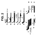
- RNA reverse transcriptase RNA polymerase RNase H
- FIG. 3A is a schematic representation of a disposable dual chamber reaction vessel 10 and the heating steps associated therewith to perform a TMA reaction in accordance with one possible embodiment of the invention.
- Chamber A contains the amplification mix, namely nucleotides, primers, MgCl 2 and other salts and buffer components.
- Chamber B contains the amplification enzyme that catalyzes the amplification reaction, e.g., T7 and/or RT.
- T7 and/or RT amplification enzyme that catalyzes the amplification reaction
- T7 and/or RT amplification enzyme that catalyzes the amplification reaction
- the temperature of chamber A is then cooled down to allow primer annealing. Subsequently, the solution of chamber A is brought into contact with chamber B.
- Chambers A and B now in fluid communication with each other, are then maintained at the optimum temperature for the amplification reaction, e.g., 42 degrees C.
- the optimum temperature for the amplification reaction e.g. 42 degrees C.
- Figure 3B is a schematic representation of an alternative form of the invention in which two separate reaction chambers 12 and 14 are combined to form a dual chamber reaction vessel 10.
- Chamber A is pre-loaded during a manufacturing step with an amplification mix, namely nucleotides, primers, MgCl 2 and other salts and buffer components.
- Chamber B is pre-loaded during manufacturing with the amplification enzyme that catalyzes the amplification reaction, e.g., T7 and/or RT.
- Fluid sample is then introduced into chamber A.
- the targets are heated for denaturation to 95°C in chamber A.
- the solution in chamber A is brought into contact with the enzymes in chamber B to trigger the isothermal amplification reaction.
- reaction vessel is designed such that, after having brought the contents of chambers A and B into contact, the amplification chamber does not allow any exchange of materials with the environment, a closed system amplification is realized that minimizes the risk of contaminating the amplification reaction with heterologous targets or amplification products from previous reactions.
- FIG. 3C is a schematic representation of two alternative dual chamber reaction vessels 10 and 10' that are snapped into place in a test strip 19 for processing with a solid phase receptacle and optical equipment in accordance with a preferred embodiment of the invention.
- a unidirectional flow system is provided.
- the sample is first introduced into chamber A for heating to the denaturation temperature.
- Chamber A contains the dried amplification reagent mix 16.
- the fluid is transferred to chamber B containing the dried enzyme 18 in the form of a pellet.
- Chamber B is maintained at 42°C after the fluid sample is introduced into Chamber B.
- the amplification reaction takes place in Chamber B at the optimum reaction temperature (e.g., 42°C).
- the test strip 19 is then processed in a machine such as the VIDAS instrument available from bioMérieux Vitek, Inc., the assignee of the present invention. Persons of skill in the art are familiar with the VIDAS instrument.
- the steps of heating and cooling of chamber A could be performed prior to the insertion of the dual chamber disposable reaction vessel 10 or 10 ' into the test strip 19, or, alternatively, suitable heating elements could be placed adjacent to the left hand end 24 of the test strip 19 in order to provide the proper temperature control of the reaction chamber A.
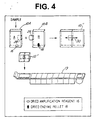
- the stand alone amplification processing station of Figures 4-14 described below, incorporates suitable heating elements and control systems to provide the proper temperature control for the reaction vessel 10.
- FIG 4 is a schematic representation of an alternative embodiment of a dual chamber reaction vessel 10 " formed from two separate interlocking vessels 10A and 10B that are combined in a manner to permit a fluid sample in one chamber to flow to the other, with the combined dual chamber vessel 10 " placed into a test strip 19 such as described above in Figure 30.
- the fluid sample is introduced into chamber A, which contains the dried amplification reagent mix 16.
- Vessel A is then heated off-line to 95 degrees C, then cooled to 42 degrees C.
- the two vessels A and B are brought together by means of a conventional snap fit between complementary locking surfaces on the tube projection 26 on chamber B and the recessed conduit 28 on chamber A.
- the mixing of the sample solution from chamber A with the enzyme from chamber B occurs since the two chambers are in fluid communication with each other, as indicated by the arrow 30.
- the sample can then be amplified in the combined dual chamber disposable reaction vessel 10 " off-line, or on-line by snapping the combined disposable vessel 10 " into a modified VIDAS strip.
- the VIDAS instrument could perform the detection of the amplification reaction in known fashion.
- FIG. 5 is a perspective view of a stand-alone amplification processing system 200 for the test strips 19 having the dual chamber reaction vessels in accordance with a presently preferred form of the invention.
- the system 200 consists of two identical amplification stations 202 and 204, a power supply module 206, a control circuitry module 208, a vacuum tank 210 and connectors 212 for the power supply module 206.
- the tank 210 has hoses 320 and 324 for providing vacuum to amplification stations 202 and 204 and ultimately to a plurality of vacuum probes (one per strip) in the manner described above for facilitating transfer of fluid from the first chamber to the second chamber.
- the vacuum subsystem is described below in conjunction with Figure 14.
- the amplification stations 202 and 204 each have a tray for receiving at least one of the strips and associated temperature control, vacuum and valve activation subsystems for heating the reaction wells of the strip to the proper temperatures, transferring fluid from the first chamber in the dual chamber reaction wells to the second chamber, and activating a valve such as a thimble valve or preferably a ball valve to open the fluid channel to allow the fluid to flow between the two chambers.
- a valve such as a thimble valve or preferably a ball valve to open the fluid channel to allow the fluid to flow between the two chambers.
- the stations 202 and 204 are designed as stand alone amplification stations for performing the amplification reaction in an automated manner after the patient or clinical sample has been added to the first chamber of the dual chamber reaction vessel described above.
- the processing of the strips after the reaction is completed with an SPR takes place in a separate machine, such as the VIDAS instrument. Specifically, after the strips have been placed in the stations 202 and 204 and the reaction run in the stations, the strips are removed from the stations 202 and 204 and placed into a VIDAS instrument for subsequent processing and analysis in known fashion.
- the entire system 200 is under microprocessor control by an amplification system interface board (not shown in Figure 5).
- the control system is shown in block diagram form in Figure 12 and will be described later.
- Figure 6 one of the amplification stations 202 is shown in a perspective view.
- the other amplification station is of identical design and construction.
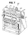
- Figure 7 is a perspective view of the front of the module of Figure 6.
- the station includes a vacuum probe slide motor 222 and vacuum probes slide cam wheel 246 that operate to slide a set of vacuum probes 244 (shown in Figure 7) for the thimble valves up and down relative to a vacuum probes slide 246 to open the thimble valves and apply vacuum so as to draw the fluid from the first chamber of the reaction vessel 10 to the second chamber.
- the vacuum probes 244 reciprocate within annular recesses provided in the vacuum probes slide 246. Obviously, proper registry of the pin structure and vacuum probe 244 with corresponding structure in the test strip as installed on the tray needs to be observed.
- the station includes side walls 228 and 230 that provide a frame for the station 202.
- Tray controller board 229 is mounted between the side walls 228 and 230.
- the electronics module for the station 202 is installed on the tray controller board 229.
- a set of tray thermal insulation covers 220 are part of a thermal subsystem and are provided to envelop a tray 240 ( Figure 7) that receives one or more of the test strips.
- the insulation covers 220 help maintain the temperature of the tray 240 at the proper temperatures.
- the thermal subsystem also includes a 42°C Peltier heat sink 242, a portion of which is positioned adjacent to the second chamber in the dual chamber reaction vessel in the test strip to maintain that chamber at the proper temperature for the enzymatic amplification reaction.
- a 95°C heat sink 250 is provided for the front of the tray 240 for maintaining the first chamber of the reaction well in the test strip at the denaturation temperature.
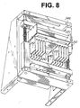
- FIG 8 is another perspective view of the module of Figure 7, showing the 95°C heat sink 250 and a set of fins 252. Note that the 95°C heat sink 250 is positioned to the front of and slightly below the tray 240. The 42°C heat sink 242 is positioned behind the heat sink 250.
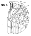
- FIG 9 is a detailed perspective view of a portion of the tray 240 that holds the test strips (not shown) as seen from above.
- the tray 240 includes a front portion having a base 254, a plurality of discontinuous raised parallel ridge structures 256 with recessed slots 258 for receiving the test strips.
- the base of the front 254 of the tray 240 is in contact with the 95°C heat sink 250.
- the side walls of the parallel raised ridges 256 at positions 256A and 256B are placed as close as possible to the first and second chambers of the reaction vessel 10 of Figure 3A so as to reduce thermal resistance.
- the base of the rear of the tray 240 is in contact with a 42°C Peltier heat sink, as best seen in Figure 8.
- portion 256B of the raised ridge for the rear of the tray is physically isolated from portion 256A for the front of the tray, and portion 256B is in contact with the 42°C heat sink so as to keep the second chamber of the reaction vessel in the test strip at the proper temperature.
- the vacuum probes 244 include a rubber gasket 260.
- the gaskets 260 are positioned on the upper surface of the test strip surrounding the vacuum port in the dual chamber reaction vessel so as to make a tight seal and permit vacuum to be drawn on the second chamber.
- Figure 10 is an isolated perspective view of the test strip holder or tray 240 of Figure 9, showing two test strips installed in the tray 240.
- the tray 240 has a plurality of lanes or slots 241 receiving up to six test strips 19 for simultaneous processing.
- Figure 10 shows the heat sinks 242 and 250 for maintaining the respective portions of the tray 240 and ridges 256 at the proper temperature.
- Figure 11 is a detailed perspective view of the test strip holder or tray 240 as seen from below.
- the 95°C Peltier heat sink which would be below front portion 254 has been removed in order to better illustrate the rear heat sink 242 beneath the rear portion of the tray 240.
- FIG 12 is a block diagram of the electronics and control system of the amplification processing system of Figure 5.
- the control system is divided into two boards 310 and 311, section A 310 at the top of the diagram devoted to amplification module or station 202 and the other board 311 (section B) devoted to the other module 204.
- the two boards 310 and 311 are identical and only the top section 310 will be discussed.
- the two boards 310 and 311 are connected to an amplification station interface board 300.
- the interface board 300 communicates with a stand alone personal computer 304 via a high speed data bus 302.
- the personal computer 304 is a conventional IBM compatible computer with hard disk drive, video monitor, etc.
- the stations 202 and 204 are under control by the interface board 300.
- the board 310 for station 202 controls the front tray 240 which is maintained at a temperature of 95°C by two Peltier heat sink modules, a pair of fans and a temperature sensor incorporated into the front portion 254 of the tray 240.
- the back of the tray is maintained at a temperature of 42°C by two Peltier modules and a temperature sensor.
- the movement of the vacuum probes 244 is controlled by the probes motor 222.
- Position sensors are provided to provide input signals to the tray controller board as to the position of the vacuum probes 244.
- the tray controller board 310 includes a set of drivers 312 for the active and passive components of the system which receive data from the temperature and position sensors and issue commands to the active components, i.e., motors, fans, Peltier modules, etc.
- the drivers are responsive to commands from the amplification interface board 300.
- the interface board also issues commands to the vacuum pump for the vacuum subsystem, as shown.
- FIG 13 is a diagram of the vacuum subsystem 320 for the amplification processing stations 202 and 204 of Figure 5.
- the subsystem includes a 1 liter plastic vacuum tank 210 which is connected via an inlet line 322 to a vacuum pump 323 for generating a vacuum in the tank 210.
- a vacuum supply line 324 is provided for providing vacuum to a pair of pinch solenoid valves 224 (see Figure 6) via supply lines 324A and 324B.
- These vacuum supply lines 324A and 324B supply vacuum to a manifold 226 distributing the vacuum to the vacuum probes 244.
- the vacuum system 320 also includes a differential pressure transducer 321 for monitoring the presence of vacuum in the tank 210. The transducer 321 supplies pressure signals to the interface board 300 of Figure 12.
- FIG 14 is a representative graph of the thermal cycle profile of the station of Figure 5.
- a first temperature T1 e.g., a denaturation temperature
- a ramp down of temperature as indicated at 404 occurs and the temperature of the reaction solution in the first chamber of the reaction vessel 10 cools to temperature T2.
- a fluid transfer occurs in which the solution in the first chamber is conveyed to the second chamber.
- Temperature T2 is maintained for an appropriate amount of time for the reaction of interest, such as one hour.
- the temperature is raised rapidly to a temperature T3 of 65°C to stop the amplification reaction.
- TMA temperature
- reaction vessels and amplification station components are also envisioned, and such alternative. embodiments are encompassed in the present disclosure.
- VIDAS in-line simple rapid nucleic acid amplification and detection assay for the clinical laboratory for the detection of Mtb in test samples which can be completed in a short time.
- the entire assay is designed to take place on a single test strip, minimizing the potential for target or amplicon contamination.
- the amplification based assay is capable of detection of Mtb where the sample contains only 5 cells similar to the sensitivity achieved by the Gen-Probe commercial kit.
- the amplification based assay utilizes isothermal transcription-mediated amplification (TMA) targeting unique sequences of rRNA, followed by hybridization and enzyme-linked fluorescent detection of nucleic acid probe (amplicon) in the VIDAS instrument.
- TMA isothermal transcription-mediated amplification
- the amplification/detection assay can detect approximately 1 fg of Mtb rRNA, or less than one Mtb organism per test, and is specific for all members of the Mtb complex. Specific probes for the detection of Mtb can be found in C. Mlessnesst, 1994, J. Clin. Microbiol. 32, 2707.
- Standard smears for acid-fast bacilli are not always reliable as a diagnostic tool, and even when positive may be a mycobateria other than Mtb.
- standard methods for diagnosis of tuberculosis requires culturing the slow-growing bacteria, and may take up to 6 weeks or longer. During this time, the patient is usually isolated. Initial results are that this automated test matches or exceeds the clinical sensitivity of the culture method, and offers a highly sensitive method to rapidly (in less than three hours) detect Mtb in infected samples, thereby aiding rapid diagnosis, isolation and treatment.
- a 450 ⁇ l volume of specimen is added to 50 ⁇ l of specimen dilution buffer in a lysing tube containing glass beads, sonicated for 15 minutes at room temperature to lyse organisms, heat inactivated for 15 minutes at 95°C.
- isothermal amplification was conducted as per a commercially available manual assay kit (Gen-Probe Inc.) following the kit instructions using standard kit reagents. However, similar assays can be conducted using the modified components as described in the Examples above.
- the detection system requires hybridization of the target nucleic acid or amplicon to a specific capture nucleic acid bound to a solid support, (in the VIDAS system called a "solid phase receptacle” SPR pipet-like device), and to a labeled detection probe nucleic acid (for example where the label can be alkaline phosphatase, a chemiluminescent signal compound, or other reagent that will allow for specific detection of bound probe).
- a solid phase receptacle for example where the label can be alkaline phosphatase, a chemiluminescent signal compound, or other reagent that will allow for specific detection of bound probe.
- the SPR transfers the probe-target hybrid to an enzyme substrate, whereby the detectable signal is triggered from the bound probe and detected by the assay instrument.
- the detection probe is conjugated to alkaline phosphatase, and once placed in contact with substrate of methyl umbelliferyl phosphate (MUMP), the substrate is converted into 4-methyl umbelliferone (4-MU) by the alkaline phosphatase.
- MUMP methyl umbelliferyl phosphate
- the 4-MU produces fluorescence which is measured and recorded by the standard VIDAS instrument as relative fluorescence units (RFU).
- REU relative fluorescence units
- Generally controls are prepared in a matrix of specimen dilution buffer with positive controls containing 5fg of Mtb rRNA, or the equivalent rRNA of approximately 1 M. tb cell.
- Sensitivity of the automated probe assay can be determined by testing dilutions of lysed M. tb cells.
- the cell lysates can generally be prepared with a 1 ⁇ l loop of cells (the assumption being that there are approximately 1x10 9 colony forming units (CFU) per 1 ⁇ l loop-full, based upon previous titration and CFU experiments). Dilutions of the Mtb lysates can then be tested with the automated probe assay.
- Figure 20A is a graph showing detection of Mtb amplicons according to the Gen-Probe kit.
- Figure 20B is a graph showing detection of Mtb amplicons from the same reactions as in Figure 20A by the VIDAS instrument.
- Figure 21 is a graph showing amplification and detection of Mtb nucleic acids on the modified VIDAS apparatus. Enzyme was used in liquid form and amplification was performed in-line with VIDAS assay instrument.
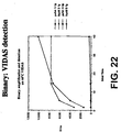
- Figure 22 is a graph showing amplification and detection of Mtb nucleic acids on the modified VIDAS apparatus using the binary/dual chamber disposable reaction vessel.
- the denaturation step was performed off-line of the VIDAS instrument, amplification and amplicon detection was performed in-line with VIDAS instrument.
- CT infection Chlamydia trachomatis
- CT Chlamydia trachomatis
- the cervical samples were collected with a Gen-Probe sample collection kit containing Gen-Probe transport medium; the urine samples were collected into standard urine collection devices. All samples were stored at 4°C.
- Cervical swabs were centrifuged at 425xg for 5 minutes to bring all liquid to the bottom of the tube.
- the swabs were then treated with 40 ⁇ l Gen-Probe Specimen Preparation Reagent and incubated at 60°C for 10 minutes. 20 ⁇ l of the treated sample was then pipetted into 400 ⁇ l of sample dilution buffer (SDB).
- SDB sample dilution buffer
- Samples were amplified using the TMA protocol, and rRNA targets were hybridized to oligomer conjugated to AMVE copolymer and an oligomer conjugated to alkaline phosphatase. See for example U.S. Patent 5,489,653 and 5,510,084.
- the solid phase receptacle SPR pipet-like device
- the alkaline phosphatase converts the substrate to fluoresence 4-MU, which is detected by the VIDAS assay machine and recorded as relative fluorescence units.
- RFV RFU - Background RFU
- Table 5 illustrates the results of clinical cervical swab specimen testing for CT comparing results from the Gen-Probe manual AMP-CT assay and the VIDAS automated probe assay.
- Table 6 illustrates the results of clinical urine specimen testing comparing the results of manual AMP-CT assay and the VIDAS automated probe assay.
- nucleic acid probes can be substantially increased through the detection of multiple different nucleic acid molecules, and the use of internal positive controls.
- An automated method has been devised for use with the VIDAS instrument (bioMérieux Vitek, Inc.) which can discretely detect at least two different nucleic acid sequences in one assay reaction, and is termed the Multiplex protocol.
- VIDAS instrument bioMérieux Vitek, Inc.
- a nucleic acid amplification procedure, or a processed test sample may be screened for more than one amplified nucleic acid molecule in the same assay.
- This method relies on the spatial separation of discrete nucleic acid probes which can specifically capture different target nucleic acid sequences (amplicons), on the SPR pipet-like device of the VIDAS instrument.
- the SPR is a disposable pipet-like tip which enables fluid movements as well as acting as the solid support for affinity capture. Multiplex capture by SPR is demonstrated using capture probes specific for Chlamydia trachomatis (CT) and Neisseria gonorrhoeae (NG).
- CT Chlamydia trachomatis
- NG Neisseria gonorrhoeae
- FIG. 15 illustrates a schematic of the operation of the multiplex VIDAS detection.
- the SPR tips are coated in two distinct zones with oligonucleotide nucleic acid sequences which are used to specifically capture complementary nucleic acid sequences (amplicons) with their corresponding specific reporter or detector probe nucleic acids labeled with alkaline phosphatase (AKP). Following washes to remove unbound reporter probes, AKP localized to the SPR bottom is detected with the fluorescent substrate 4-MUP. The AKP is stripped from the bottom of the SPR with NaOH or other reagents which promote denaturation of nucleic acid hybrids or inactivate alkaline phosphatase activity.
- amplicons complementary nucleic acid sequences
- AKP alkaline phosphatase
- the enzyme reaction well is emptied, washed, and re-filled with fresh 4-MUP.
- the new substrate is exposed to the bottom of the SPR and any residual fluorescence is measured.
- AKP-reporter probe bound to the top of the SPR is detected by immersing the SPR in the 4-MUP and representing the presence of the second amplicons.
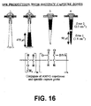
- Figure 16 illustrates the production of SPR with two distinct capture zones.
- the SPR is inserted tip-first into a silicon plug, which are held in a rack.
- Differential pressure is used to uniformly draw a solution of a specific capture probe at about I ⁇ g/ml, conjugated to AMVE copolymer, into all SPRs at one time.
- the amount of fluid drawn into each SPR, and thus the size of the zone, is controlled by regulating the amount of pressure in the system.
- Attachment of the conjugate to the SPR surface is achieved by passive adsorption for several hours at room temperature. After washing, and drying, the SPRs are capped with a small adhesive disc and inserted into new racks in a tip-down orientation. The lower portion of the SPR is then similarly coated with a second capture probe conjugate.
- SPRs are stable when stored dry at 4°C.
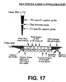
- Figure 17 illustrates a preferred embodiment of the VIDAS apparatus strip configuration for multiplex detection.
- the strip can be pre-filled with 200 ⁇ l of AKP-probe mix (about 1x10 12 molecules of each probe) in hybridization buffer in well X1, 600 ⁇ l of wash buffer in wells X3, X4, X5, 600 ⁇ l of stripping reagent in wells X6 and X7, and 400 ⁇ l of AKP substrate in X8 and sealed with foil.
- a foil-sealed optical cuvette (XA) containing 300 ⁇ l of 4-MUP is snapped into the strip, and the strips are inserted into the VIDAS instrument at 37°C.
- the multiplex VIDAS protocol is then executed using SPRs coated with two capture probes in distinct zones.
- the VIDAS multiplex protocol can involve many steps.
- the validation test protocol contained thirteen (13) basic steps as follows:
- Hybridization, substrate, wash and stripping steps involve multiple cycles of pipeting the respective solution into the SPR, holding the solution for a defined period of time, and pipeting the solution out of the SPR. Hold times for hybridization, substrate and washing or stripping are 3.0, 0.5 and 0.17 minutes respectively. The fluorescence signal is detected by the apparatus. Total assay time for the research protocol was about 1.75 hours but can be reduced to about 75 minutes.
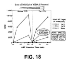
- Figure 18 illustrates and graphs the results of verification of the VIDAS multiplex protocol executed as described above, except the SPR was homogeneously coated with only a single capture probe for Neisseria gonorrhoeae (NG).
- the number of NG oligonucleotide targets in the test sample was varied from 0,1x10 10 , or 1x10 11 molecules in the test sample.
- the data shown are averages of replicate samples.
- the graph as illustrated is divided into two parts; the left and right halves show the results of two fluorescent measurements from the lower and the upper zones of the SPR, respectively.
- the measurements taken from the bottom zone after stripping the lower area of bound nucleic acid, and exposure for about 11 minutes in fresh 4-MUP substrate was approximately 46 RFU for all samples tested, and was equivalent to background fluorescence measured. This measurement is shown by the 0 time point in the center of the graph.
- the graph illustrates two sequential sets of measurements of fluorescence from a single SPR, the first set of measurements being taken from the bottom half of the SPR (left half of the graph), and a second set of measurements taken from the top of the SPR (the right of the graph). This experiment validates that the Multiplex protocol and zone coated SPR procedure yield essentially identical results, as indicated by the fluorescence intensities in the left- and right-hand parts of the graph, from the upper and lower portions of the SPR.
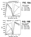
- Figure 19 illustrates Multiplex detection of CT and NG oligonucleotide targets at different input amounts.
- Figure 19A is a graph showing the results when 1x10 12 CT targets were mixed with 0,1x10 9 , 1x10 10 , 1x10 11 , or 1x10 12 , NG targets, and detected with the VIDAS instrument using the multiplex protocol and SPRs coated with CT capture probes on the bottom zone of the SPR, and NG capture probes on the top zone of the SPR.
- Figure 19B illustrates the results when 1x10 12 NG targets was mixed with 0, 1x10 9 , 1x10 10 , 1x10 11 , or 1x10 12 , CT targets, and detected with the VIDAS instrument using the multiplex protocol and SPRs coated with CT capture probes on the bottom zone of the SPR, and NG capture probes on the top zone of the SPR.
- the data is graphed as above where the graph illustrates two sequential sets of measurements of fluorescence from a single SPR, the first set of measurements being taken from the bottom half of the SPR (left half of the graph), and a second set of measurements taken from the top of the SPR (the right of the graph) with verification of stripping of the SPR in the center of the graph.
- this experiment shows that the two zones of the SPR act independently in the Multiplex protocol, since high fluorescence signals from one zone do not interfere with signals produced from the second zone, regardless of whether these latter signals are high (1x10 12 ), or low (1x10 9 , just detectable), or negative.
- Table 7 summarizes the data obtained by Multiplex VIDAS detection of CT and NG in a sample at various target levels, reported in RFUs. Detection of CT and NG targets in sample RFUs none 1x10 9 1x10 10 1x10 11 1x10 12 1x10 13 none 43/40 43/116 46/693 62/7116 174/11817 273/12136 1x10 9 189/41 246/118 169/773 220/5750 422/12522 399/11401 1x10 10 1736/41 2258/125 1937/734 1931/6639 2128/12390 2371/11180 1x10 11 10339/48 9815/145 9858/760 9369/4571 9784/11825 10252/10312 1x10 12 12149/49 13520/148 12940/796 13593/4397 11239/11786 10158/9900 1x10 13 11545/57 11713/121 10804/815 12805/5404 12305/12326 11416/10490
- the multiplex VIDAS protocol is clearly operative and enables the rapid and discrete detection of more than one different nucleic acid in a sample.
- This protocol, and the SPR coating can be manipulated in many formats to present coating zones of different surface area with different sized gaps between two or more detection zones.
- the SPR can be coated with nucleic acids which are designed to capture different regions of the same nucleic acid sequence to detect, for example, truncated gene expression, different alleles or alternatively spliced genes.
- the SPR can be coated to capture amplicons from internal control nucleic acid molecules which can be used to detect and confirm successful nucleic acid amplification reactions.
- the VIDAS Multiplex protocol is a flexible method for detection of more than one nucleic acid sequence in the same sample, in a single assay.
- control sequences composed of functional building blocks of sequences chosen by random generation of nucleic acid sequences for use as amplification reaction internal positive controls ideally requires that the control sequences be specifically designed to be used for the various nucleic acid amplification protocols including but not limited to PCR, LCR, TMA, NASBA, and SDA.
- the internal control nucleic acid sequence, in combination with the appropriate sequence specific oligonucleotide primers or promoter-primers will generate a positive amplification signal if the amplification reaction was successfully completed.
- the internal control nucleic acid is useful regardless of the nucleic acid sequences present in the target organism, the host organism, or nucleic acids present in the normal flora or in the environment.
- the internal control sequences should not be substantially similar to any nucleic acid sequences present in a clinical setting, including human, pathogenic organisms, normal flora organisms, or environmental organisms which could interfere with the amplification and detection of the internal control sequences.
- the internal control sequences of the instant invention are comprised of functional blocks of sequences chosen from a list of randomly generated nucleic acid sequences.
- the functional blocks are segments which provide for a special property needed to allow for amplification, capture, and detection of the amplification product.
- the internal control sequences are most useful when the functional blocks meet certain functional requirements of the amplification protocol, such as: a) a primer binding site on the anti-sense strand; b) a capture site; c) a detector probe binding site; d) a T7-promoter containing primer binding site on the sense strand.
- Each of these functional elements has its own particular constraints, such as length, %G-C content, Tm, lack of homology to known sequences, and absence of secondary structural features (i.e. free from dimer formation or hairpin structures) which can be used to select the appropriate sequence.
- randomly generated functional blocks of sequences can be screened for the desired functional properties before use in constructing internal control sequences.
- a random sequence generator was used to generate strings of numbers; each number being limited to the range from 0.000 to 4.000.
- the length of the strings is flexible and chosen based upon the desired lengths of the functional blocks.
- Each number in the string (i.e. n1, n2, n3, n4 ... nx where x is the length of the string) was then assigned a corresponding nucleotide as follows: guanosine (G) if 0 ⁇ n ⁇ 1; adenosine (A) if 1 ⁇ n ⁇ 2; thymidine (T) if 2 ⁇ n ⁇ 3; and cytosine (C) if 3 ⁇ n ⁇ 4.
- G guanosine
- A adenosine
- T thymidine
- C cytosine
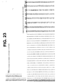
- Figure 23 illustrates the results generated by the method described showing a collection of strings of nucleic acid sequences and screening for specific functional parameters.
- the internal control sequence can include DNA, RNA, modified oligonucleotides, or any combination of nucleic acids, such that the illustrated sequences using DNA nomenclature can be readily adapted as desired to the appropriate nucleic acid.
- IC sequences were then constructed by assembling the functional blocks (selected at random) in the proper order. Finally, the assembled internal control sequence were then examined to insure that overall sequence and structural constraints were maintained. For example, in a TMA reaction, the internal control sequence should not have significant base-pairing potential between the two primer binding sites or from stable 3' dimer structures. Those internal control sequences which pass through these layers of screening were then physically produced using overlapping oligonucleotides and tested for performance in actual amplification/detection assays.
- any one functional block may have some homology to sequences present in a clinical setting (a perfect match of a 21 nucleotide block is expected at a random frequency of 1 in 4e12 sequences or about 4x10 12 ; generated sequences were screened against the GenBank data base) it is highly unlikely that all functional blocks will be found to have substantial homology. Since the internal control nucleic acid sequences are constricted of a group of functional blocks placed in tandem, the chance possibility that a natural nucleic acid sequence will have an identical string of nucleic acid sequence blocks in the same tandem organization is remote.
- Random Internal Control 1 (RIC1) is shown in Figure 24 with the possible oligonucleotide primers/probes for amplification and detection of the control sequence.
- Figure 25 shows an analysis of the possible secondary structure of the RIC1 molecule.
- RIC1 was constructed using randomly generated strings ran16, ran19, ran21 and ran33. The functional blocks requiring primer binding were met by ran16 and ranl9, while the capture site was satisfied by ran21 and the detector probe binding site was met by ran33.
- a capture probe or detection probe sequence designation can be interchanged, as along as the appropriate linker molecule is attached to the appropriate probe, wherein a reporter probe oligonucleotide is linked to a means for generating a detectable signal, and the capture probe oligonucleotide is linked to a means for adhering the capture probe to an appropriate support.
- the probes and oligos are described with the understanding that in the case of double stranded DNA, the complementary strand can be the target or as appropriate can be converted for use as the strand for detection.
- one or ordinary skill in the art will be able to modify the sequences as disclosed to generate alternative probes and primers which are suitable for use in an equivalent fashion as described herein.
- Random Internal Control 2 (RIC2) is shown in Figure 26 with the possible oligonucleotide primers/probes for amplification and detection of the control sequence.
- Figure 27 shows an analysis of the possible secondary structure of the RIC2 sequence.
- RIC2 was constructed using randomly generated strings ran27, ran32, ran39 and ran51.
- Figure 28 illustrates results from detection of RIC1 DNA, where the ran21 was the capture probe and ran33 was an enzyme-linked detector-probe, and shows that detection occurs under standard assay conditions with expected fluorescence intensities.
- Figure 29 shows that RIC1 RNA, amplified by TMA and detected on a VIDAS instrument (bioMérieux Vitek, Inc.) using the enzyme-linked detection system, has a limit of sensitivity of about 1000 molecules of RIC 1 RNA (without optimization of conditions). Similar analysis of RIC2 sequences was performed and found to be similar to RIC1. It is significant that the amplification and detection system of the internal control functioned effectively under the conditions optimized for the selected target.
- SPRs can be homogeneously coated with a mixture of different capture nucleic acid sequences in a single, whole-SPR zone.
- two capture nucleic acid sequences can be combined in one zone, one specific for a target test sequence, and one specific for a target test sequence, and one specific for an internal control sequence.
- Target amplicons, if present, and internal control amplicons are simultaneously hybridized to the SPR in the presence of labeled probe sequences specific for the target amplicons.
- a first signal read is done to so that the presence or absence of label on the SPR is determined to ascertain the presence or absence of the test target.
- a second hybridization is then done (sequential hybridization) to the SPR using a detection label probe specific for the internal control.
- the SPR is washed to remove excess unbound detection probe, and the second label is measured to indicate the presence or absence of the internal control.
- a positive signal from the IC second read confirms the functionality of the amplification/detection system. If the first and second label are the same, an additive signal will result from the positive first read and the positive second IC read. If both the first signal is negative and the second IC signal is also negative, then the amplification/detection functionality failed, which could be due to for example, sample interference or mechanical failure. In this case the test result is reported invalid (false negative) and re-testing is recommended. If the labels are different then neither sequential hybridizations nor sequential detection steps would be necessary.
- NCCLS Document MM3-A Molecular Diagnostic Methods for Infectious Diseases; Approved Guideline, p. 55, March 1995.
- This type of protocol can also be used for a combined GC/CT/internal control assay, if a screening approach is allowed (no discrimination between GC and/or CT positives during the first read).
- GC and CT specific signals have to be resolved by running the CT and GC specific assays on screen positive samples (5-10% of cases, depending on prevalence) SPRs would be coated homogeneously with 3 capture probes (CT/GC/internal control).
- the IC could share a capture probe with either CT or GC. Homogeneous Coated SPR Detection of multiple signals Target CT 1 st Read IC 2 nd Read Bkg.
- internal control nucleic acid sequences have been designed and validated for use in various amplification and detection systems including an internal control for Chlamydia trachomatis (CT) assay identified as CRIC-2; for a Neisseria gonorrhoeae (NG) assay identified as GRIC; and for Mycobacterium tuberculosis (MT) identified as MRIC.
- CT Chlamydia trachomatis
- NG Neisseria gonorrhoeae
- MRIC Mycobacterium tuberculosis
- An internal control was generated for HIV assays identified as HRIC, wherein both the capture probe sequence and reporter probe sequence were derived from random sequence.
- the sequence of the internal control, and the corresponding target sequence are shown in Figure 30.
- the Random Sequence Probe #1082 can be used as the reporter probe, when suitably conjugated to a reporter molecule as described previously.
- a capture oligonucleotide Random Sequence Probe #1081 has been designed for use in the capture of the control sequence, for improved quantitation by elimination of competition between the target amplicons and IC amplicons for a common capture probe.
Landscapes
- Chemical & Material Sciences (AREA)
- Life Sciences & Earth Sciences (AREA)
- Organic Chemistry (AREA)
- Health & Medical Sciences (AREA)
- Wood Science & Technology (AREA)
- Proteomics, Peptides & Aminoacids (AREA)
- Zoology (AREA)
- Engineering & Computer Science (AREA)
- Immunology (AREA)
- Physics & Mathematics (AREA)
- General Health & Medical Sciences (AREA)
- Biochemistry (AREA)
- Analytical Chemistry (AREA)
- General Engineering & Computer Science (AREA)
- Biotechnology (AREA)
- Molecular Biology (AREA)
- Bioinformatics & Cheminformatics (AREA)
- Biophysics (AREA)
- Microbiology (AREA)
- Genetics & Genomics (AREA)
- Chemical Kinetics & Catalysis (AREA)
- General Physics & Mathematics (AREA)
- Pathology (AREA)
- Measuring Or Testing Involving Enzymes Or Micro-Organisms (AREA)
- Apparatus Associated With Microorganisms And Enzymes (AREA)
Applications Claiming Priority (2)
| Application Number | Priority Date | Filing Date | Title |
|---|---|---|---|
| US850171 | 1977-11-10 | ||
| US85017197A | 1997-05-02 | 1997-05-02 |
Publications (3)
| Publication Number | Publication Date |
|---|---|
| EP0875584A2 true EP0875584A2 (fr) | 1998-11-04 |
| EP0875584A3 EP0875584A3 (fr) | 2001-12-05 |
| EP0875584B1 EP0875584B1 (fr) | 2010-09-15 |
Family
ID=25307435
Family Applications (1)
| Application Number | Title | Priority Date | Filing Date |
|---|---|---|---|
| EP98303458A Expired - Lifetime EP0875584B1 (fr) | 1997-05-02 | 1998-05-01 | Analyse des acides nucléiques |
Country Status (8)
| Country | Link |
|---|---|
| US (6) | US6558901B1 (fr) |
| EP (1) | EP0875584B1 (fr) |
| JP (1) | JP4526609B2 (fr) |
| KR (1) | KR19980086701A (fr) |
| BR (1) | BR9801572A (fr) |
| CA (1) | CA2230967C (fr) |
| DE (1) | DE69841895D1 (fr) |
| ES (1) | ES2350038T3 (fr) |
Cited By (17)
| Publication number | Priority date | Publication date | Assignee | Title |
|---|---|---|---|---|
| EP1045036A2 (fr) * | 1999-04-13 | 2000-10-18 | Biomerieux, Inc. | Méthodes et apparéils pour analyser un échantillon d'acide nucléique |
| WO2001030489A2 (fr) * | 1999-10-26 | 2001-05-03 | Epigenomics Ag | Dispositif et procede d'hybridation d'echantillons d'adn a deux brins sur des jeux ordonnes oligomeriques |
| WO2006000647A1 (fr) * | 2004-06-29 | 2006-01-05 | Wallac Oy | Amplification et detection integree, non homogene des acides nucleiques |
| WO2011099855A1 (fr) * | 2010-02-12 | 2011-08-18 | Vereniging Voor Christelijk Hoger Onderwijs, | Procédé pour la surveillance combinée de la détection d'au moins deux cibles moléculaires et trousse pour la mise en œuvre de ce procédé |
| WO2012003289A1 (fr) * | 2010-06-30 | 2012-01-05 | Gen-Probe Incorporated | Procédé et appareillage pour identifier des échantillons contenant un analyte par utilisation d'une détermination de l'analyte par une lecture unique, et de signaux de pilotage du procédé |
| US8192992B2 (en) | 1998-05-01 | 2012-06-05 | Gen-Probe Incorporated | System and method for incubating the contents of a reaction receptacle |
| US8546110B2 (en) | 1998-05-01 | 2013-10-01 | Gen-Probe Incorporated | Method for detecting the presence of a nucleic acid in a sample |
| EP2263799A3 (fr) * | 2000-09-15 | 2014-05-21 | Norchip A/S | Système de chambre de réaction microfabriqué |
| US8840848B2 (en) | 2010-07-23 | 2014-09-23 | Beckman Coulter, Inc. | System and method including analytical units |
| US8973736B2 (en) | 2011-11-07 | 2015-03-10 | Beckman Coulter, Inc. | Magnetic damping for specimen transport system |
| US9046506B2 (en) | 2011-11-07 | 2015-06-02 | Beckman Coulter, Inc. | Specimen container detection |
| US9446418B2 (en) | 2011-11-07 | 2016-09-20 | Beckman Coulter, Inc. | Robotic arm |
| US9482684B2 (en) | 2011-11-07 | 2016-11-01 | Beckman Coulter, Inc. | Centrifuge system and workflow |
| US9506943B2 (en) | 2011-11-07 | 2016-11-29 | Beckman Coulter, Inc. | Aliquotter system and workflow |
| US9910054B2 (en) | 2011-11-07 | 2018-03-06 | Beckman Coulter, Inc. | System and method for processing samples |
| US10370705B2 (en) | 2009-03-24 | 2019-08-06 | University Of Chicago | Analysis devices, kits, and related methods for digital quantification of nucleic acids and other analytes |
| US10543485B2 (en) | 2009-03-24 | 2020-01-28 | University Of Chicago | Slip chip device and methods |
Families Citing this family (57)
| Publication number | Priority date | Publication date | Assignee | Title |
|---|---|---|---|---|
| US6444444B1 (en) * | 1996-07-10 | 2002-09-03 | Aventis Pasteur Limited | Genes encoding mycobacterial proteins associated with cell binding and cell entry and uses thereof |
| IL124275A (en) * | 1997-05-02 | 2002-03-10 | Bio Merieux Vitek Inc | A method to produce sequences of nucleic acids |
| US6558901B1 (en) * | 1997-05-02 | 2003-05-06 | Biomerieux Vitek | Nucleic acid assays |
| US6849400B1 (en) * | 1997-07-23 | 2005-02-01 | Gen-Probe Incorporated | Methods for detecting and measuring spliced nucleic acids |
| US6582962B1 (en) * | 1998-02-27 | 2003-06-24 | Ventana Medical Systems, Inc. | Automated molecular pathology apparatus having independent slide heaters |
| WO2004083806A2 (fr) | 2003-01-22 | 2004-09-30 | University Of South Florida | Appareil a genocapteur autonome et procedes d'utilisation de celui-ci |
| BRPI0408967B8 (pt) | 2003-03-31 | 2021-07-27 | Hoffmann La Roche | kit e métodos para detecção de um ácido nucléico de vários membros do sorogrupo do vírus da encefalite japonesa em uma amostra biológica sob condições de hibridização rigorosas |
| US20070172886A1 (en) * | 2003-05-07 | 2007-07-26 | Bruno Colin | Reaction module for biological analysis |
| FR2854695B1 (fr) * | 2003-05-07 | 2006-01-20 | Biomerieux Sa | Module de reaction pour l analyse biologique |
| EP1625232B1 (fr) * | 2003-05-16 | 2013-09-04 | GOVERNMENT OF THE UNITED STATES OF AMERICA, as represented by THE SECRETARY, DEPARTMENT OF HEALTH AND HUMAN SERVICES | Molecule d'acide nucleique de controle interne pour systemes d'amplification d'acide nucleique |
| GB0317335D0 (en) * | 2003-07-24 | 2003-08-27 | Sec Dep For The Home Departmen | Improvements in and relating to interpretation |
| US7090803B1 (en) * | 2003-10-28 | 2006-08-15 | American Bio Medica Corporation | Lateral flow immunoassay device |
| US20050164204A1 (en) * | 2004-01-27 | 2005-07-28 | Reed Thomas D. | Single use lyophilized rnase reagents, and kits and methods for using same |
| WO2005072456A2 (fr) * | 2004-01-28 | 2005-08-11 | Gerard Biotech, Llc | Reactifs enzymatiques lyophilises a usage unique et kits et leurs procedes d'utilisation |
| US7939251B2 (en) | 2004-05-06 | 2011-05-10 | Roche Molecular Systems, Inc. | SENP1 as a marker for cancer |
| FI20040768A0 (fi) * | 2004-06-04 | 2004-06-04 | Teemu Korpimaeki | Menetelmä määritysreagenssien stabiloimiseksi, stabilisoituja määritysreagensseja sisältävä reagenssisäiliö ja sen käyttö |
| US20060068398A1 (en) * | 2004-09-24 | 2006-03-30 | Cepheid | Universal and target specific reagent beads for nucleic acid amplification |
| JP2008000002A (ja) * | 2004-09-30 | 2008-01-10 | Sysmex Corp | リブロース2リン酸カルボキシラーゼスモールチェーン1A(RBCS−1A)遺伝子及び/又は該遺伝子のmRNAを検出するための核酸増幅用プライマ、及び内部標準として該遺伝子及び/又は該遺伝子のmRNAを用いた検査方法。 |
| AU2005333163B2 (en) * | 2004-11-09 | 2011-06-16 | Gen-Probe Incorporated | Compositions and methods for detecting Group A streptococci |
| EP2322940B1 (fr) | 2005-03-10 | 2014-10-29 | Gen-Probe Incorporated | Systèmes et procédés pour effectuer des analyses afin de détecter ou quantifier des analytes dans des échantillons |
| US20070141605A1 (en) * | 2005-11-21 | 2007-06-21 | Applera Corporation | Portable preparation, analysis, and detection apparatus for nucleic acid processing |
| ES2688281T3 (es) | 2006-07-28 | 2018-10-31 | Diagnostics For The Real World, Ltd | Dispositivo, sistema y método para procesar una muestra |
| CN1888902B (zh) * | 2006-08-11 | 2011-05-18 | 杭州优思达生物技术有限公司 | 全封闭式靶核酸扩增物快速检测装置 |
| WO2008030605A2 (fr) * | 2006-09-08 | 2008-03-13 | The Regents Of The University Of Michigan | Virus herv de classe iii présents dans un lymphome et cancer |
| GB0701253D0 (en) | 2007-01-23 | 2007-02-28 | Diagnostics For The Real World | Nucleic acid amplification and testing |
| US8835157B2 (en) * | 2007-04-25 | 2014-09-16 | 3M Innovative Properties Company | Supported reagents, methods, and devices |
| CA2683990A1 (fr) * | 2007-04-25 | 2008-11-06 | 3M Innovative Properties Company | Compose chimique et assemblage a un dispositif de traitement |
| CA2691451C (fr) | 2007-06-21 | 2015-03-24 | Sara H. Fan | Instruments et receptacles pour effectuer des procedes |
| US20090011422A1 (en) * | 2007-06-28 | 2009-01-08 | Integrated Dna Technologies, Inc. | Methods for cloning small rna species |
| KR102112438B1 (ko) * | 2007-07-23 | 2020-06-04 | 더 차이니즈 유니버시티 오브 홍콩 | 대규모 병렬 게놈 서열분석을 이용한 태아 염색체 이수성의 진단 방법 |
| GB2456079B (en) * | 2007-08-17 | 2010-07-14 | Diagnostics For The Real World | Device, system and method for processing a sample |
| US7644610B2 (en) * | 2007-08-24 | 2010-01-12 | Baker Hughes Incorporated | Automated formation fluid clean-up to sampling switchover |
| EP2215211A1 (fr) * | 2007-11-06 | 2010-08-11 | 3M Innovative Properties Company | Comprimé de dispositif de traitement |
| CN102027132A (zh) * | 2008-05-12 | 2011-04-20 | 奥林巴斯株式会社 | 粪便处理方法及粪便处理容器 |
| US9464319B2 (en) | 2009-03-24 | 2016-10-11 | California Institute Of Technology | Multivolume devices, kits and related methods for quantification of nucleic acids and other analytes |
| US10196700B2 (en) | 2009-03-24 | 2019-02-05 | University Of Chicago | Multivolume devices, kits and related methods for quantification and detection of nucleic acids and other analytes |
| US9046507B2 (en) | 2010-07-29 | 2015-06-02 | Gen-Probe Incorporated | Method, system and apparatus for incorporating capacitive proximity sensing in an automated fluid transfer procedure |
| AU2012222178B2 (en) | 2011-02-24 | 2014-12-18 | Gen-Probe Incorporated | Systems and methods for distinguishing optical signals of different modulation frequencies in an optical signal detector |
| GB201122121D0 (en) * | 2011-12-22 | 2012-02-01 | Animal Health Trust | Diagnostic test for bacterial pathogens |
| US20130280696A1 (en) | 2012-04-23 | 2013-10-24 | Elliott Millenson | Devices and methods for detecting analyte in bodily fluid |
| CA2874407A1 (fr) | 2012-05-24 | 2013-11-28 | Fundacio Institut D'investigacio Biomedica De Bellvitge (Idibell) | Procede pour l'identification de l'origine d'un cancer d'origine primaire inconnue par analyse de methylation |
| US9400285B2 (en) | 2013-03-15 | 2016-07-26 | Abbot Laboratories | Automated diagnostic analyzers having vertically arranged carousels and related methods |
| EP2972402B1 (fr) | 2013-03-15 | 2023-12-20 | Abbott Laboratories | Analyseurs de diagnostic dotés de carrousels de prétraitement et méthodes associées |
| EP3964839B1 (fr) | 2013-03-15 | 2024-04-10 | Abbott Laboratories | Analyseurs diagnostiques automatiques comprenant des systèmes de rail accessibles par l'arrière et procédés associés |
| US9984201B2 (en) | 2015-01-18 | 2018-05-29 | Youhealth Biotech, Limited | Method and system for determining cancer status |
| GB201510723D0 (en) * | 2015-06-18 | 2015-08-05 | Alere Switzerland Gmbh | High throughput isothermal nucleic acid amplification |
| EP3464642A4 (fr) | 2016-05-31 | 2020-02-19 | The Regents of the University of California | Procédés d'évaluation, de surveillance et de modulation du processus de vieillissement |
| US10093986B2 (en) | 2016-07-06 | 2018-10-09 | Youhealth Biotech, Limited | Leukemia methylation markers and uses thereof |
| EP3481403B1 (fr) | 2016-07-06 | 2022-02-09 | Youhealth Biotech, Limited | Marqueurs de méthylation spécifiques d'une tumeur solide et utilisations de ces marqueurs |
| US11396678B2 (en) | 2016-07-06 | 2022-07-26 | The Regent Of The University Of California | Breast and ovarian cancer methylation markers and uses thereof |
| US10427162B2 (en) | 2016-12-21 | 2019-10-01 | Quandx Inc. | Systems and methods for molecular diagnostics |
| BR112019018272A2 (pt) | 2017-03-02 | 2020-07-28 | Youhealth Oncotech, Limited | marcadores metilação para diagnosticar hepatocelular carcinoma e câncer |
| US11313871B2 (en) | 2017-03-29 | 2022-04-26 | Universal Bio Research Co., Ltd. | Specimen treatment and measurement system |
| CN107460108A (zh) * | 2017-07-23 | 2017-12-12 | 新疆昆泰锐生物技术有限公司 | 一种连续进样的pcr反应体系配制及进样装置及pcr仪 |
| EP4175755A4 (fr) | 2020-07-02 | 2024-07-17 | Ultima Genomics, Inc. | Procédés et systèmes d'analyse d'acides nucléiques |
| CN114480098B (zh) * | 2022-02-15 | 2023-10-03 | 吉林正业生物制品股份有限公司 | 一种便携式鸡滑液囊支原体检测装置及其方法 |
| WO2024069939A1 (fr) * | 2022-09-30 | 2024-04-04 | 株式会社Mirai Genomics | Cartouche d'amplification d'acide nucléique et procédé d'amplification d'acide nucléique |
Citations (5)
| Publication number | Priority date | Publication date | Assignee | Title |
|---|---|---|---|---|
| US5229297A (en) * | 1989-02-03 | 1993-07-20 | Eastman Kodak Company | Containment cuvette for PCR and method of use |
| EP0622464A2 (fr) * | 1993-04-16 | 1994-11-02 | Becton, Dickinson and Company | Procédé de dosage d'acide nucléique |
| EP0623682A1 (fr) * | 1993-05-05 | 1994-11-09 | Becton, Dickinson and Company | Contrôles internes pour réactions d'amplification isotherme d'acide nucléique |
| DE19503685A1 (de) * | 1995-01-30 | 1996-08-01 | Invitek Gmbh | Verfahren zur Herstellung komplexer multienzymatischer lagerstabiler Reaktionsgemische und deren Verwendung |
| EP0726310A1 (fr) * | 1995-02-10 | 1996-08-14 | Gen-Probe Incorporated | Compositions d'enzyme stabilisées pour l'amplification d'acides nucléiques |
Family Cites Families (60)
| Publication number | Priority date | Publication date | Assignee | Title |
|---|---|---|---|---|
| US4087248A (en) | 1976-07-26 | 1978-05-02 | Miles Laughton E | Multiple assay machine and method |
| FR2422956A1 (fr) | 1978-04-13 | 1979-11-09 | Pasteur Institut | Procede de detection et de caracterisation d'un acide nucleique ou d'une sequence de celui-ci, et reactif enzymatique pour la mise en oeuvre de ce procede |
| US4457916A (en) | 1982-08-31 | 1984-07-03 | Asahi Kasei Kogyo Kabushiki Kaisha | Method for stabilizing Tumor Necrosis Factor and a stable aqueous solution or powder containing the same |
| DE3682047D1 (de) | 1985-07-09 | 1991-11-21 | Quadrant Bioresources Ltd | Beschuetzung von proteinen und aehnlichem. |
| US4762857A (en) | 1986-05-30 | 1988-08-09 | E. I. Du Pont De Nemours And Company | Trehalose as stabilizer and tableting excipient |
| US5541308A (en) | 1986-11-24 | 1996-07-30 | Gen-Probe Incorporated | Nucleic acid probes for detection and/or quantitation of non-viral organisms |
| US5202231A (en) * | 1987-04-01 | 1993-04-13 | Drmanac Radoje T | Method of sequencing of genomes by hybridization of oligonucleotide probes |
| GB8715238D0 (en) | 1987-06-29 | 1987-08-05 | Quadrant Bioresources Ltd | Food process |
| AU2087388A (en) | 1987-07-02 | 1989-01-30 | In Vitro Technologies, Inc. | Capillary device for immunoassay of multiple analytes |
| JP2774121B2 (ja) | 1987-07-31 | 1998-07-09 | ザ ボード オブ トラスティーズ オブ ザ リーランド スタンフォード ジュニア ユニバーシティ | 標的ポリヌクレオチド配列の選択的増幅 |
| US5124246A (en) * | 1987-10-15 | 1992-06-23 | Chiron Corporation | Nucleic acid multimers and amplified nucleic acid hybridization assays using same |
| GB8801338D0 (en) | 1988-01-21 | 1988-02-17 | Quadrant Bioresources Ltd | Preservation of viruses |
| US4983523A (en) * | 1988-04-08 | 1991-01-08 | Gene-Trak Systems | Methods for preparing sample nucleic acids for hybridization |
| GB8903593D0 (en) | 1989-02-16 | 1989-04-05 | Pafra Ltd | Storage of materials |
| AU5269590A (en) * | 1989-03-10 | 1990-10-09 | Gene-Trak Systems | Immobilized oligonucleotide probes and uses therefor |
| US5217862A (en) | 1989-05-24 | 1993-06-08 | Amoco Corporation | Method for the detection of Neisseria gonorrhoeae |
| WO1990015159A2 (fr) | 1989-05-31 | 1990-12-13 | Gene-Trak Systems | Sondes d'acides nucleiques pour la detection de chlamydia trachomatis |
| US5143854A (en) * | 1989-06-07 | 1992-09-01 | Affymax Technologies N.V. | Large scale photolithographic solid phase synthesis of polypeptides and receptor binding screening thereof |
| US5856088A (en) | 1989-07-11 | 1999-01-05 | Gen-Probe Incorporated | Detection of human immunodeficiency virus type 1 |
| US5766849A (en) | 1989-07-11 | 1998-06-16 | Gen-Probe Incorporated | Methods of amplifying nucleic acids using promoter-containing primer sequence |
| US5219727A (en) | 1989-08-21 | 1993-06-15 | Hoffmann-Laroche Inc. | Quantitation of nucleic acids using the polymerase chain reaction |
| US5122284A (en) | 1990-06-04 | 1992-06-16 | Abaxis, Inc. | Apparatus and method for optically analyzing biological fluids |
| US5395521A (en) * | 1991-05-31 | 1995-03-07 | Board Of Regents, The University Of Texas System | Automated column equilibration, column loading, column washing and column elution |
| GB9114202D0 (en) | 1991-07-01 | 1991-08-21 | Quadrant Holdings Cambridge | Blood products |
| FR2679255B1 (fr) | 1991-07-17 | 1993-10-22 | Bio Merieux | Procede d'immobilisation d'un fragment nucleique par fixation passive sur un support solide, support solide ainsi obtenu et son utilisation. |
| US5521300A (en) | 1991-08-13 | 1996-05-28 | Norval B. Galloway | Oligonucleotides complementary to mycobacterial nucleic acids |
| FR2688788B1 (fr) | 1992-03-17 | 1994-05-13 | Bio Merieux | Composes hydrosolubles derives d'un homopolymere ou copolymere de l'anhydride maleique, et applications desdits composes au support de molecules biologiques. |
| EP0644950A4 (fr) | 1992-04-09 | 1997-05-14 | Abbott Lab | Dosage pour la detection de l'antigene du vih et de l'anticorps du vih. |
| US5498392A (en) | 1992-05-01 | 1996-03-12 | Trustees Of The University Of Pennsylvania | Mesoscale polynucleotide amplification device and method |
| US5587128A (en) | 1992-05-01 | 1996-12-24 | The Trustees Of The University Of Pennsylvania | Mesoscale polynucleotide amplification devices |
| CA2135073C (fr) | 1992-05-06 | 2002-11-19 | Daniel L. Kacian | Procede, composition et trousse d'amplification des sequences d'acides nucleiques |
| US5715385A (en) * | 1992-07-10 | 1998-02-03 | Lsi Logic Corporation | Apparatus for 2-D affine transformation of images |
| WO1994002634A1 (fr) * | 1992-07-24 | 1994-02-03 | University Of South Australia | Procede d'amplification et de detection |
| FR2694754B1 (fr) | 1992-08-12 | 1994-09-16 | Bio Merieux | Fragments d'ADN de mycobactéries, amorces d'amplification, sondes d'hybridation, réactifs et procédé de détection de détection de mycobactéries. |
| AU4843893A (en) * | 1992-09-02 | 1994-03-29 | Scripps Research Institute, The | Coupled isothermal polynucleotide amplification and translation system |
| US5780273A (en) | 1993-04-09 | 1998-07-14 | Amoco Corporation | Insertion elements and amplifiable nucleic acids |
| US6709813B1 (en) * | 1993-05-14 | 2004-03-23 | Ortho-Clinical Diagnostics, Inc. | Diagnostic compositions, elements, methods and test kits for amplification and detection of human CMV DNA using primers having matched melting temperatures |
| US5837832A (en) * | 1993-06-25 | 1998-11-17 | Affymetrix, Inc. | Arrays of nucleic acid probes on biological chips |
| FR2709310B1 (fr) | 1993-07-23 | 1995-09-29 | Bio Merieux | Fragment nucléotidique de l'ARN ribosomique 23S de mycobactéries, sondes et amorces dérivées, réactif et procédé de détection. |
| US6136529A (en) | 1993-09-03 | 2000-10-24 | Gen-Probe Incorporated | Nucleic acid probes to Mycobacterium avium complex |
| FR2710075B1 (fr) * | 1993-09-15 | 1995-10-27 | Bio Merieux | Réactif et procédé pour la détection d'une séquence nucléotidique avec amplification de signal. |
| US5645801A (en) | 1993-10-21 | 1997-07-08 | Abbott Laboratories | Device and method for amplifying and detecting target nucleic acids |
| US5605793A (en) | 1994-02-17 | 1997-02-25 | Affymax Technologies N.V. | Methods for in vitro recombination |
| KR100238833B1 (ko) | 1994-03-10 | 2000-03-02 | 다니엘 엘. 캐시앙 | 이온성 세제에 의한 효소 개재 반응의 억제를 방지하는 방법 |
| CN1188171C (zh) | 1994-06-02 | 2005-02-09 | 廓德伦特控股剑桥有限公司 | 防止各种特质在再水化或熔化时聚集的方法以及由此获得的组合物 |
| US6060288A (en) * | 1994-08-03 | 2000-05-09 | Mosaic Technologies | Method for performing amplification of nucleic acid on supports |
| US5646801A (en) * | 1994-08-30 | 1997-07-08 | International Business Machines Corporation | Direct access storage device with improved reliability flex cable mounting |
| US5589336A (en) | 1994-11-10 | 1996-12-31 | New York Blood Center | Diagnostic method and kit for determining Kell blood group genotype |
| FR2730545B1 (fr) * | 1995-02-13 | 1997-03-28 | Bio Merieux | Vanne statique a congelation, et enceinte de traitement controlee par au moins une vanne |
| US5710029A (en) | 1995-06-07 | 1998-01-20 | Gen-Probe Incorporated | Methods for determining pre-amplification levels of a nucleic acid target sequence from post-amplification levels of product |
| US5747252A (en) | 1995-06-07 | 1998-05-05 | Gen-Probe Incorporated | Nucleic acid probes and amplification oligonucleotides for Neisseria species |
| US5856174A (en) * | 1995-06-29 | 1999-01-05 | Affymetrix, Inc. | Integrated nucleic acid diagnostic device |
| US5994066A (en) * | 1995-09-11 | 1999-11-30 | Infectio Diagnostic, Inc. | Species-specific and universal DNA probes and amplification primers to rapidly detect and identify common bacterial pathogens and associated antibiotic resistance genes from clinical specimens for routine diagnosis in microbiology laboratories |
| US5697409A (en) * | 1996-02-21 | 1997-12-16 | Biomerieux Vitek, Inc. | Diluting and pipetting stations for sample testing machine |
| US5762873A (en) * | 1996-02-21 | 1998-06-09 | Biomerieux Vitek, Inc. | Automatic sample testing machine |
| CA2199213C (fr) * | 1996-03-11 | 2007-04-17 | John Wesley Backus | Amplification et detection des acides nucleiques cibles grace a une etape d'incubation post-amplification |
| US5804384A (en) * | 1996-12-06 | 1998-09-08 | Vysis, Inc. | Devices and methods for detecting multiple analytes in samples |
| US5786182A (en) * | 1997-05-02 | 1998-07-28 | Biomerieux Vitek, Inc. | Dual chamber disposable reaction vessel for amplification reactions, reaction processing station therefor, and methods of use |
| US6558901B1 (en) | 1997-05-02 | 2003-05-06 | Biomerieux Vitek | Nucleic acid assays |
| IL124275A (en) * | 1997-05-02 | 2002-03-10 | Bio Merieux Vitek Inc | A method to produce sequences of nucleic acids |
-
1998
- 1998-04-24 US US09/065,914 patent/US6558901B1/en not_active Expired - Lifetime
- 1998-05-01 CA CA002230967A patent/CA2230967C/fr not_active Expired - Lifetime
- 1998-05-01 EP EP98303458A patent/EP0875584B1/fr not_active Expired - Lifetime
- 1998-05-01 KR KR1019980015754A patent/KR19980086701A/ko not_active Application Discontinuation
- 1998-05-01 DE DE69841895T patent/DE69841895D1/de not_active Expired - Lifetime
- 1998-05-01 ES ES98303458T patent/ES2350038T3/es not_active Expired - Lifetime
- 1998-05-04 BR BR9801572A patent/BR9801572A/pt not_active IP Right Cessation
- 1998-05-06 JP JP12356498A patent/JP4526609B2/ja not_active Expired - Lifetime
-
1999
- 1999-02-05 US US09/245,939 patent/US6300068B1/en not_active Expired - Lifetime
- 1999-02-05 US US09/245,940 patent/US6586234B1/en not_active Expired - Lifetime
-
2000
- 2000-05-31 US US09/586,546 patent/US6528632B1/en not_active Expired - Fee Related
-
2001
- 2001-07-09 US US09/901,181 patent/US7309588B2/en not_active Expired - Lifetime
-
2007
- 2007-07-19 US US11/780,164 patent/US20080240985A1/en not_active Abandoned
Patent Citations (5)
| Publication number | Priority date | Publication date | Assignee | Title |
|---|---|---|---|---|
| US5229297A (en) * | 1989-02-03 | 1993-07-20 | Eastman Kodak Company | Containment cuvette for PCR and method of use |
| EP0622464A2 (fr) * | 1993-04-16 | 1994-11-02 | Becton, Dickinson and Company | Procédé de dosage d'acide nucléique |
| EP0623682A1 (fr) * | 1993-05-05 | 1994-11-09 | Becton, Dickinson and Company | Contrôles internes pour réactions d'amplification isotherme d'acide nucléique |
| DE19503685A1 (de) * | 1995-01-30 | 1996-08-01 | Invitek Gmbh | Verfahren zur Herstellung komplexer multienzymatischer lagerstabiler Reaktionsgemische und deren Verwendung |
| EP0726310A1 (fr) * | 1995-02-10 | 1996-08-14 | Gen-Probe Incorporated | Compositions d'enzyme stabilisées pour l'amplification d'acides nucléiques |
Cited By (47)
| Publication number | Priority date | Publication date | Assignee | Title |
|---|---|---|---|---|
| US8883455B2 (en) | 1998-05-01 | 2014-11-11 | Gen-Probe Incorporated | Method for detecting the presence of a nucleic acid in a sample |
| US9598723B2 (en) | 1998-05-01 | 2017-03-21 | Gen-Probe Incorporated | Automated analyzer for performing a nucleic acid-based assay |
| US8709814B2 (en) | 1998-05-01 | 2014-04-29 | Gen-Probe Incorporated | Method for incubating the contents of a receptacle |
| US8569020B2 (en) | 1998-05-01 | 2013-10-29 | Gen-Probe Incorporated | Method for simultaneously performing multiple amplification reactions |
| US8569019B2 (en) | 1998-05-01 | 2013-10-29 | Gen-Probe Incorporated | Method for performing an assay with a nucleic acid present in a specimen |
| US8192992B2 (en) | 1998-05-01 | 2012-06-05 | Gen-Probe Incorporated | System and method for incubating the contents of a reaction receptacle |
| US8546110B2 (en) | 1998-05-01 | 2013-10-01 | Gen-Probe Incorporated | Method for detecting the presence of a nucleic acid in a sample |
| US9150908B2 (en) | 1998-05-01 | 2015-10-06 | Gen-Probe Incorporated | Method for detecting the presence of a nucleic acid in a sample |
| US8221682B2 (en) | 1998-05-01 | 2012-07-17 | Gen-Probe Incorporated | System for incubating the contents of a reaction receptacle |
| AU741530B2 (en) * | 1999-04-13 | 2001-12-06 | Biomerieux, Inc. | Methods and devices for performing analysis of a nucleic acid sample |
| EP1045036A2 (fr) * | 1999-04-13 | 2000-10-18 | Biomerieux, Inc. | Méthodes et apparéils pour analyser un échantillon d'acide nucléique |
| US6225067B1 (en) * | 1999-04-13 | 2001-05-01 | BIOMéRIEUX, INC. | Methods and devices for performing analysis of a nucleic acid sample |
| EP1045036A3 (fr) * | 1999-04-13 | 2004-03-24 | Biomerieux, Inc. | Méthodes et apparéils pour analyser un échantillon d'acide nucléique |
| US6235479B1 (en) | 1999-04-13 | 2001-05-22 | Bio Merieux, Inc. | Methods and devices for performing analysis of a nucleic acid sample |
| WO2001030489A3 (fr) * | 1999-10-26 | 2001-10-25 | Epigenomics Ag | Dispositif et procede d'hybridation d'echantillons d'adn a deux brins sur des jeux ordonnes oligomeriques |
| WO2001030489A2 (fr) * | 1999-10-26 | 2001-05-03 | Epigenomics Ag | Dispositif et procede d'hybridation d'echantillons d'adn a deux brins sur des jeux ordonnes oligomeriques |
| EP2263799A3 (fr) * | 2000-09-15 | 2014-05-21 | Norchip A/S | Système de chambre de réaction microfabriqué |
| WO2006000647A1 (fr) * | 2004-06-29 | 2006-01-05 | Wallac Oy | Amplification et detection integree, non homogene des acides nucleiques |
| US10543485B2 (en) | 2009-03-24 | 2020-01-28 | University Of Chicago | Slip chip device and methods |
| US10370705B2 (en) | 2009-03-24 | 2019-08-06 | University Of Chicago | Analysis devices, kits, and related methods for digital quantification of nucleic acids and other analytes |
| WO2011099855A1 (fr) * | 2010-02-12 | 2011-08-18 | Vereniging Voor Christelijk Hoger Onderwijs, | Procédé pour la surveillance combinée de la détection d'au moins deux cibles moléculaires et trousse pour la mise en œuvre de ce procédé |
| US12065694B2 (en) | 2010-06-30 | 2024-08-20 | Gen-Probe Incorporated | Kits for single-step analyte detection with process control |
| CN102985566A (zh) * | 2010-06-30 | 2013-03-20 | 简.探针公司 | 使用分析物和过程控制信号的单次读取确定而鉴定含分析物的样品的方法和设备 |
| US11104935B2 (en) | 2010-06-30 | 2021-08-31 | Gen-Probe Incorporated | Kits for single-step analyte detection with process control |
| WO2012003289A1 (fr) * | 2010-06-30 | 2012-01-05 | Gen-Probe Incorporated | Procédé et appareillage pour identifier des échantillons contenant un analyte par utilisation d'une détermination de l'analyte par une lecture unique, et de signaux de pilotage du procédé |
| US10487353B2 (en) | 2010-06-30 | 2019-11-26 | Gen-Probe Incorporated | Reaction mixture containing probes having indistinguishable labels |
| US9822397B2 (en) | 2010-06-30 | 2017-11-21 | Gen-Probe Incorporated | Method and apparatus for identifying analyte-containing samples using single-read determination of analyte and process control signals |
| CN102985566B (zh) * | 2010-06-30 | 2015-06-17 | 简.探针公司 | 使用分析物和过程控制信号的单次读取确定而鉴定含分析物的样品的方法和设备 |
| AU2011272868B2 (en) * | 2010-06-30 | 2015-09-17 | Gen-Probe Incorporated | Method and apparatus for identifying analyte-containing samples using single-read determination of analyte and process control signals |
| US8962308B2 (en) | 2010-07-23 | 2015-02-24 | Beckman Coulter, Inc. | System and method including thermal cycler modules |
| US8996320B2 (en) | 2010-07-23 | 2015-03-31 | Beckman Coulter, Inc. | System and method including analytical units |
| US9274132B2 (en) | 2010-07-23 | 2016-03-01 | Beckman Coulter, Inc. | Assay cartridge with reaction well |
| US9285382B2 (en) | 2010-07-23 | 2016-03-15 | Beckman Coulter, Inc. | Reaction vessel |
| US8840848B2 (en) | 2010-07-23 | 2014-09-23 | Beckman Coulter, Inc. | System and method including analytical units |
| US8932541B2 (en) | 2010-07-23 | 2015-01-13 | Beckman Coulter, Inc. | Pipettor including compliant coupling |
| US8956570B2 (en) | 2010-07-23 | 2015-02-17 | Beckman Coulter, Inc. | System and method including analytical units |
| US9519000B2 (en) | 2010-07-23 | 2016-12-13 | Beckman Coulter, Inc. | Reagent cartridge |
| US9140715B2 (en) | 2010-07-23 | 2015-09-22 | Beckman Coulter, Inc. | System and method for controlling thermal cycler modules |
| US9046455B2 (en) | 2010-07-23 | 2015-06-02 | Beckman Coulter, Inc. | System and method including multiple processing lanes executing processing protocols |
| US9910054B2 (en) | 2011-11-07 | 2018-03-06 | Beckman Coulter, Inc. | System and method for processing samples |
| US10048284B2 (en) | 2011-11-07 | 2018-08-14 | Beckman Coulter, Inc. | Sample container cap with centrifugation status indicator device |
| US10274505B2 (en) | 2011-11-07 | 2019-04-30 | Beckman Coulter, Inc. | Robotic arm |
| US9046506B2 (en) | 2011-11-07 | 2015-06-02 | Beckman Coulter, Inc. | Specimen container detection |
| US8973736B2 (en) | 2011-11-07 | 2015-03-10 | Beckman Coulter, Inc. | Magnetic damping for specimen transport system |
| US9506943B2 (en) | 2011-11-07 | 2016-11-29 | Beckman Coulter, Inc. | Aliquotter system and workflow |
| US9482684B2 (en) | 2011-11-07 | 2016-11-01 | Beckman Coulter, Inc. | Centrifuge system and workflow |
| US9446418B2 (en) | 2011-11-07 | 2016-09-20 | Beckman Coulter, Inc. | Robotic arm |
Also Published As
| Publication number | Publication date |
|---|---|
| JPH10304890A (ja) | 1998-11-17 |
| KR19980086701A (ko) | 1998-12-05 |
| US20080240985A1 (en) | 2008-10-02 |
| US6528632B1 (en) | 2003-03-04 |
| JP4526609B2 (ja) | 2010-08-18 |
| BR9801572A (pt) | 1999-06-29 |
| ES2350038T3 (es) | 2011-01-17 |
| DE69841895D1 (de) | 2010-10-28 |
| EP0875584A3 (fr) | 2001-12-05 |
| US20040248087A1 (en) | 2004-12-09 |
| EP0875584B1 (fr) | 2010-09-15 |
| US6558901B1 (en) | 2003-05-06 |
| US6586234B1 (en) | 2003-07-01 |
| US6300068B1 (en) | 2001-10-09 |
| CA2230967C (fr) | 2009-10-27 |
| US7309588B2 (en) | 2007-12-18 |
| CA2230967A1 (fr) | 1998-11-02 |
Similar Documents
| Publication | Publication Date | Title |
|---|---|---|
| EP0875584B1 (fr) | Analyse des acides nucléiques | |
| US7329493B2 (en) | One-tube nested PCR for detecting Mycobacterium tuberculosis | |
| US8288524B2 (en) | Molecular diagnosis of bacteremia | |
| Fredricks et al. | Application of polymerase chain reaction to the diagnosis of infectious diseases | |
| AU2010298202B2 (en) | Detection of nucleic acids in crude matrices | |
| JP2006525809A5 (fr) | ||
| US7262008B2 (en) | Nucleic acid assays | |
| EP1045030B1 (fr) | Amplification et détection de Campylobacter jejuni et Campylobacter coli | |
| AU769852B2 (en) | Improved nucleic acid assays | |
| Chapin et al. | Evaluation of a rapid air thermal cycler for detection of Mycobacterium tuberculosis | |
| MXPA98003500A (en) | Best nucleic acid experiments | |
| CA2303303A1 (fr) | Amplification et detection de yersinia enterocolitica | |
| Hirvonen | Utility and Impact of Detecting Clinically Important Bacteria with Small-Scale and Automated Nucleic-Acid Amplification Assays | |
| EP3066210B1 (fr) | Détection de streptococcus pneumoniae | |
| Wilson | Detection of Mycobacterium tuberculosis by a Calorimetric Polymerase Chain Reaction | |
| Singleton et al. | Probe-Based (Hybridization) Methods |
Legal Events
| Date | Code | Title | Description |
|---|---|---|---|
| PUAI | Public reference made under article 153(3) epc to a published international application that has entered the european phase |
Free format text: ORIGINAL CODE: 0009012 |
|
| 17P | Request for examination filed |
Effective date: 19980518 |
|
| AK | Designated contracting states |
Kind code of ref document: A2 Designated state(s): AT BE CH CY DE DK ES FI FR GB GR IE IT LI LU MC NL PT SE Kind code of ref document: A2 Designated state(s): BE CH DE ES FR GB IT LI |
|
| AX | Request for extension of the european patent |
Free format text: AL;LT;LV;MK;RO;SI |
|
| PUAL | Search report despatched |
Free format text: ORIGINAL CODE: 0009013 |
|
| AK | Designated contracting states |
Kind code of ref document: A3 Designated state(s): AT BE CH CY DE DK ES FI FR GB GR IE IT LI LU MC NL PT SE |
|
| AX | Request for extension of the european patent |
Free format text: AL;LT;LV;MK;RO;SI |
|
| RIC1 | Information provided on ipc code assigned before grant |
Free format text: 7C 12Q 1/68 A |
|
| RIN1 | Information on inventor provided before grant (corrected) |
Inventor name: MCKINLEY, GEOFF A. Inventor name: MOE, JAMES G. Inventor name: BURG, J. LAWRENCE Inventor name: VERA-GARCIA, MARCELA Inventor name: KLUTTZ, BRYAN W. Inventor name: CATANZARIT, LUIGI |
|
| AKX | Designation fees paid |
Free format text: BE CH DE ES FR GB IT LI |
|
| 17Q | First examination report despatched |
Effective date: 20031001 |
|
| GRAJ | Information related to disapproval of communication of intention to grant by the applicant or resumption of examination proceedings by the epo deleted |
Free format text: ORIGINAL CODE: EPIDOSDIGR1 |
|
| GRAP | Despatch of communication of intention to grant a patent |
Free format text: ORIGINAL CODE: EPIDOSNIGR1 |
|
| GRAP | Despatch of communication of intention to grant a patent |
Free format text: ORIGINAL CODE: EPIDOSNIGR1 |
|
| GRAS | Grant fee paid |
Free format text: ORIGINAL CODE: EPIDOSNIGR3 |
|
| GRAA | (expected) grant |
Free format text: ORIGINAL CODE: 0009210 |
|
| AK | Designated contracting states |
Kind code of ref document: B1 Designated state(s): BE CH DE ES FR GB IT LI |
|
| REG | Reference to a national code |
Ref country code: GB Ref legal event code: FG4D Ref country code: CH Ref legal event code: EP |
|
| REG | Reference to a national code |
Ref country code: CH Ref legal event code: PFA Owner name: BIOMERIEUX, INC. Free format text: BIOMERIEUX VITEK, INC.#595 ANGLUM DRIVE#HAZELWOOD, MISSOURI 63042-2905 (US) -TRANSFER TO- BIOMERIEUX, INC.#100 RODOLPHE STREET#DURHAM, NC 27712 (US) Ref country code: CH Ref legal event code: NV Representative=s name: KIRKER & CIE S.A. |
|
| REF | Corresponds to: |
Ref document number: 69841895 Country of ref document: DE Date of ref document: 20101028 Kind code of ref document: P |
|
| REG | Reference to a national code |
Ref country code: ES Ref legal event code: FG2A Effective date: 20110104 |
|
| PG25 | Lapsed in a contracting state [announced via postgrant information from national office to epo] |
Ref country code: BE Free format text: LAPSE BECAUSE OF FAILURE TO SUBMIT A TRANSLATION OF THE DESCRIPTION OR TO PAY THE FEE WITHIN THE PRESCRIBED TIME-LIMIT Effective date: 20100915 |
|
| PLBE | No opposition filed within time limit |
Free format text: ORIGINAL CODE: 0009261 |
|
| STAA | Information on the status of an ep patent application or granted ep patent |
Free format text: STATUS: NO OPPOSITION FILED WITHIN TIME LIMIT |
|
| 26N | No opposition filed |
Effective date: 20110616 |
|
| REG | Reference to a national code |
Ref country code: DE Ref legal event code: R097 Ref document number: 69841895 Country of ref document: DE Effective date: 20110616 |
|
| REG | Reference to a national code |
Ref country code: FR Ref legal event code: PLFP Year of fee payment: 19 |
|
| REG | Reference to a national code |
Ref country code: FR Ref legal event code: PLFP Year of fee payment: 20 |
|
| PGFP | Annual fee paid to national office [announced via postgrant information from national office to epo] |
Ref country code: CH Payment date: 20170527 Year of fee payment: 20 Ref country code: FR Payment date: 20170525 Year of fee payment: 20 Ref country code: DE Payment date: 20170530 Year of fee payment: 20 Ref country code: GB Payment date: 20170530 Year of fee payment: 20 |
|
| PGFP | Annual fee paid to national office [announced via postgrant information from national office to epo] |
Ref country code: IT Payment date: 20170524 Year of fee payment: 20 Ref country code: ES Payment date: 20170601 Year of fee payment: 20 |
|
| REG | Reference to a national code |
Ref country code: DE Ref legal event code: R071 Ref document number: 69841895 Country of ref document: DE |
|
| REG | Reference to a national code |
Ref country code: CH Ref legal event code: PL |
|
| REG | Reference to a national code |
Ref country code: GB Ref legal event code: PE20 Expiry date: 20180430 |
|
| PG25 | Lapsed in a contracting state [announced via postgrant information from national office to epo] |
Ref country code: GB Free format text: LAPSE BECAUSE OF EXPIRATION OF PROTECTION Effective date: 20180430 |
|
| REG | Reference to a national code |
Ref country code: ES Ref legal event code: FD2A Effective date: 20200903 |
|
| PG25 | Lapsed in a contracting state [announced via postgrant information from national office to epo] |
Ref country code: ES Free format text: LAPSE BECAUSE OF EXPIRATION OF PROTECTION Effective date: 20180502 |