EP0839496A1 - Nicht-invasive Foetussonde mit verbesserten mechanischen und elektrischen Eigenschaften - Google Patents
Nicht-invasive Foetussonde mit verbesserten mechanischen und elektrischen Eigenschaften Download PDFInfo
- Publication number
- EP0839496A1 EP0839496A1 EP97118170A EP97118170A EP0839496A1 EP 0839496 A1 EP0839496 A1 EP 0839496A1 EP 97118170 A EP97118170 A EP 97118170A EP 97118170 A EP97118170 A EP 97118170A EP 0839496 A1 EP0839496 A1 EP 0839496A1
- Authority
- EP
- European Patent Office
- Prior art keywords
- fetal
- probe
- sensor
- conductive
- maternal
- Prior art date
- Legal status (The legal status is an assumption and is not a legal conclusion. Google has not performed a legal analysis and makes no representation as to the accuracy of the status listed.)
- Withdrawn
Links
- 230000001605 fetal effect Effects 0.000 title claims abstract description 267
- 239000000523 sample Substances 0.000 title claims abstract description 144
- 230000008774 maternal effect Effects 0.000 claims abstract description 90
- 210000003754 fetus Anatomy 0.000 claims abstract description 56
- 239000000499 gel Substances 0.000 claims description 66
- 238000000576 coating method Methods 0.000 claims description 23
- 239000000017 hydrogel Substances 0.000 claims description 22
- 230000001070 adhesive effect Effects 0.000 claims description 21
- 239000000853 adhesive Substances 0.000 claims description 19
- 238000012544 monitoring process Methods 0.000 claims description 19
- 239000011248 coating agent Substances 0.000 claims description 18
- 230000004888 barrier function Effects 0.000 claims description 8
- 239000000835 fiber Substances 0.000 claims description 5
- 239000012530 fluid Substances 0.000 claims description 5
- 229920000642 polymer Polymers 0.000 claims description 4
- 210000002268 wool Anatomy 0.000 claims description 4
- 230000002745 absorbent Effects 0.000 claims 1
- 239000002250 absorbent Substances 0.000 claims 1
- 238000000926 separation method Methods 0.000 abstract description 18
- 238000002955 isolation Methods 0.000 abstract description 13
- 238000013461 design Methods 0.000 abstract description 12
- 239000000203 mixture Substances 0.000 abstract description 8
- 210000003679 cervix uteri Anatomy 0.000 abstract description 3
- 230000000712 assembly Effects 0.000 abstract description 2
- 238000000429 assembly Methods 0.000 abstract description 2
- 238000009472 formulation Methods 0.000 abstract description 2
- 210000002458 fetal heart Anatomy 0.000 description 25
- 238000000034 method Methods 0.000 description 23
- 239000000463 material Substances 0.000 description 14
- 210000003491 skin Anatomy 0.000 description 10
- 229910001220 stainless steel Inorganic materials 0.000 description 9
- 210000004209 hair Anatomy 0.000 description 8
- 230000001965 increasing effect Effects 0.000 description 7
- 239000010935 stainless steel Substances 0.000 description 7
- 230000008901 benefit Effects 0.000 description 6
- 238000002565 electrocardiography Methods 0.000 description 6
- 208000014674 injury Diseases 0.000 description 6
- 229920002614 Polyether block amide Polymers 0.000 description 5
- 210000004369 blood Anatomy 0.000 description 5
- 239000008280 blood Substances 0.000 description 5
- OKTJSMMVPCPJKN-UHFFFAOYSA-N Carbon Chemical compound [C] OKTJSMMVPCPJKN-UHFFFAOYSA-N 0.000 description 4
- 208000027418 Wounds and injury Diseases 0.000 description 4
- 239000004020 conductor Substances 0.000 description 4
- 230000006378 damage Effects 0.000 description 4
- 239000003292 glue Substances 0.000 description 4
- 210000003128 head Anatomy 0.000 description 4
- 230000033001 locomotion Effects 0.000 description 4
- 229920005989 resin Polymers 0.000 description 4
- 239000011347 resin Substances 0.000 description 4
- 229920000049 Carbon (fiber) Polymers 0.000 description 3
- FAPWRFPIFSIZLT-UHFFFAOYSA-M Sodium chloride Chemical compound [Na+].[Cl-] FAPWRFPIFSIZLT-UHFFFAOYSA-M 0.000 description 3
- 229920006362 Teflon® Polymers 0.000 description 3
- 229910052799 carbon Inorganic materials 0.000 description 3
- 239000004917 carbon fiber Substances 0.000 description 3
- 238000010276 construction Methods 0.000 description 3
- 230000010339 dilation Effects 0.000 description 3
- 210000002615 epidermis Anatomy 0.000 description 3
- WABPQHHGFIMREM-UHFFFAOYSA-N lead(0) Chemical compound [Pb] WABPQHHGFIMREM-UHFFFAOYSA-N 0.000 description 3
- 230000036961 partial effect Effects 0.000 description 3
- 238000012545 processing Methods 0.000 description 3
- 239000011780 sodium chloride Substances 0.000 description 3
- 208000030507 AIDS Diseases 0.000 description 2
- RYECOJGRJDOGPP-UHFFFAOYSA-N Ethylurea Chemical compound CCNC(N)=O RYECOJGRJDOGPP-UHFFFAOYSA-N 0.000 description 2
- 229910000831 Steel Inorganic materials 0.000 description 2
- 210000001015 abdomen Anatomy 0.000 description 2
- 229910052782 aluminium Inorganic materials 0.000 description 2
- XAGFODPZIPBFFR-UHFFFAOYSA-N aluminium Chemical compound [Al] XAGFODPZIPBFFR-UHFFFAOYSA-N 0.000 description 2
- 210000004381 amniotic fluid Anatomy 0.000 description 2
- 230000005540 biological transmission Effects 0.000 description 2
- 230000000747 cardiac effect Effects 0.000 description 2
- GTKRFUAGOKINCA-UHFFFAOYSA-M chlorosilver;silver Chemical compound [Ag].[Ag]Cl GTKRFUAGOKINCA-UHFFFAOYSA-M 0.000 description 2
- 238000001514 detection method Methods 0.000 description 2
- 229940079593 drug Drugs 0.000 description 2
- 239000003814 drug Substances 0.000 description 2
- 230000000694 effects Effects 0.000 description 2
- 238000004868 gas analysis Methods 0.000 description 2
- 230000036541 health Effects 0.000 description 2
- 208000015181 infectious disease Diseases 0.000 description 2
- 238000009413 insulation Methods 0.000 description 2
- 235000015110 jellies Nutrition 0.000 description 2
- 239000008274 jelly Substances 0.000 description 2
- 238000004519 manufacturing process Methods 0.000 description 2
- VNWKTOKETHGBQD-UHFFFAOYSA-N methane Chemical compound C VNWKTOKETHGBQD-UHFFFAOYSA-N 0.000 description 2
- 238000000465 moulding Methods 0.000 description 2
- 230000003387 muscular Effects 0.000 description 2
- 230000035515 penetration Effects 0.000 description 2
- 230000009984 peri-natal effect Effects 0.000 description 2
- -1 polytetramethylene Polymers 0.000 description 2
- 230000001737 promoting effect Effects 0.000 description 2
- 229910052709 silver Inorganic materials 0.000 description 2
- 239000004332 silver Substances 0.000 description 2
- 239000010959 steel Substances 0.000 description 2
- 238000012360 testing method Methods 0.000 description 2
- 230000008733 trauma Effects 0.000 description 2
- 210000004291 uterus Anatomy 0.000 description 2
- 208000032843 Hemorrhage Diseases 0.000 description 1
- 239000004698 Polyethylene Substances 0.000 description 1
- 239000004820 Pressure-sensitive adhesive Substances 0.000 description 1
- BQCADISMDOOEFD-UHFFFAOYSA-N Silver Chemical compound [Ag] BQCADISMDOOEFD-UHFFFAOYSA-N 0.000 description 1
- 206010047289 Ventricular extrasystoles Diseases 0.000 description 1
- 241000700605 Viruses Species 0.000 description 1
- 210000003815 abdominal wall Anatomy 0.000 description 1
- 238000010521 absorption reaction Methods 0.000 description 1
- 230000009471 action Effects 0.000 description 1
- 238000007792 addition Methods 0.000 description 1
- 230000002411 adverse Effects 0.000 description 1
- 239000013060 biological fluid Substances 0.000 description 1
- 210000001124 body fluid Anatomy 0.000 description 1
- 239000010839 body fluid Substances 0.000 description 1
- 238000006243 chemical reaction Methods 0.000 description 1
- 230000003247 decreasing effect Effects 0.000 description 1
- MTHSVFCYNBDYFN-UHFFFAOYSA-N diethylene glycol Chemical compound OCCOCCO MTHSVFCYNBDYFN-UHFFFAOYSA-N 0.000 description 1
- 201000010099 disease Diseases 0.000 description 1
- 208000037265 diseases, disorders, signs and symptoms Diseases 0.000 description 1
- 238000010292 electrical insulation Methods 0.000 description 1
- 230000002708 enhancing effect Effects 0.000 description 1
- 210000004700 fetal blood Anatomy 0.000 description 1
- 239000010408 film Substances 0.000 description 1
- 239000011888 foil Substances 0.000 description 1
- PCHJSUWPFVWCPO-UHFFFAOYSA-N gold Chemical compound [Au] PCHJSUWPFVWCPO-UHFFFAOYSA-N 0.000 description 1
- 239000010931 gold Substances 0.000 description 1
- 229910052737 gold Inorganic materials 0.000 description 1
- 229910002804 graphite Inorganic materials 0.000 description 1
- 239000010439 graphite Substances 0.000 description 1
- 238000009532 heart rate measurement Methods 0.000 description 1
- 208000002672 hepatitis B Diseases 0.000 description 1
- 230000002209 hydrophobic effect Effects 0.000 description 1
- 238000010348 incorporation Methods 0.000 description 1
- 230000002458 infectious effect Effects 0.000 description 1
- 238000003780 insertion Methods 0.000 description 1
- 230000037431 insertion Effects 0.000 description 1
- 239000011810 insulating material Substances 0.000 description 1
- 230000000670 limiting effect Effects 0.000 description 1
- 230000001050 lubricating effect Effects 0.000 description 1
- 210000001006 meconium Anatomy 0.000 description 1
- 239000012528 membrane Substances 0.000 description 1
- 229910052751 metal Inorganic materials 0.000 description 1
- 239000002184 metal Substances 0.000 description 1
- 239000011104 metalized film Substances 0.000 description 1
- 239000011140 metalized polyester Substances 0.000 description 1
- 238000012986 modification Methods 0.000 description 1
- 230000004048 modification Effects 0.000 description 1
- 239000000178 monomer Substances 0.000 description 1
- 238000002496 oximetry Methods 0.000 description 1
- 230000037361 pathway Effects 0.000 description 1
- 230000002093 peripheral effect Effects 0.000 description 1
- 230000000704 physical effect Effects 0.000 description 1
- 229920000573 polyethylene Polymers 0.000 description 1
- 230000000379 polymerizing effect Effects 0.000 description 1
- 229920002635 polyurethane Polymers 0.000 description 1
- 239000004814 polyurethane Substances 0.000 description 1
- 229920000915 polyvinyl chloride Polymers 0.000 description 1
- 238000002360 preparation method Methods 0.000 description 1
- 230000008569 process Effects 0.000 description 1
- 230000002829 reductive effect Effects 0.000 description 1
- 210000004761 scalp Anatomy 0.000 description 1
- 238000004528 spin coating Methods 0.000 description 1
- 239000000126 substance Substances 0.000 description 1
- 229920001169 thermoplastic Polymers 0.000 description 1
- 229920005992 thermoplastic resin Polymers 0.000 description 1
- 210000001519 tissue Anatomy 0.000 description 1
- 238000002604 ultrasonography Methods 0.000 description 1
- 210000001215 vagina Anatomy 0.000 description 1
- XLYOFNOQVPJJNP-UHFFFAOYSA-N water Substances O XLYOFNOQVPJJNP-UHFFFAOYSA-N 0.000 description 1
- 238000003466 welding Methods 0.000 description 1
- 230000036642 wellbeing Effects 0.000 description 1
Images


Classifications
-
- A—HUMAN NECESSITIES
- A61—MEDICAL OR VETERINARY SCIENCE; HYGIENE
- A61B—DIAGNOSIS; SURGERY; IDENTIFICATION
- A61B5/00—Measuring for diagnostic purposes; Identification of persons
- A61B5/43—Detecting, measuring or recording for evaluating the reproductive systems
- A61B5/4306—Detecting, measuring or recording for evaluating the reproductive systems for evaluating the female reproductive systems, e.g. gynaecological evaluations
- A61B5/4343—Pregnancy and labour monitoring, e.g. for labour onset detection
- A61B5/4362—Assessing foetal parameters
-
- A—HUMAN NECESSITIES
- A61—MEDICAL OR VETERINARY SCIENCE; HYGIENE
- A61B—DIAGNOSIS; SURGERY; IDENTIFICATION
- A61B5/00—Measuring for diagnostic purposes; Identification of persons
- A61B5/24—Detecting, measuring or recording bioelectric or biomagnetic signals of the body or parts thereof
- A61B5/25—Bioelectric electrodes therefor
- A61B5/251—Means for maintaining electrode contact with the body
- A61B5/252—Means for maintaining electrode contact with the body by suction
-
- A—HUMAN NECESSITIES
- A61—MEDICAL OR VETERINARY SCIENCE; HYGIENE
- A61B—DIAGNOSIS; SURGERY; IDENTIFICATION
- A61B5/00—Measuring for diagnostic purposes; Identification of persons
- A61B5/24—Detecting, measuring or recording bioelectric or biomagnetic signals of the body or parts thereof
- A61B5/25—Bioelectric electrodes therefor
- A61B5/251—Means for maintaining electrode contact with the body
- A61B5/257—Means for maintaining electrode contact with the body using adhesive means, e.g. adhesive pads or tapes
- A61B5/259—Means for maintaining electrode contact with the body using adhesive means, e.g. adhesive pads or tapes using conductive adhesive means, e.g. gels
-
- A—HUMAN NECESSITIES
- A61—MEDICAL OR VETERINARY SCIENCE; HYGIENE
- A61B—DIAGNOSIS; SURGERY; IDENTIFICATION
- A61B5/00—Measuring for diagnostic purposes; Identification of persons
- A61B5/24—Detecting, measuring or recording bioelectric or biomagnetic signals of the body or parts thereof
- A61B5/25—Bioelectric electrodes therefor
- A61B5/279—Bioelectric electrodes therefor specially adapted for particular uses
- A61B5/28—Bioelectric electrodes therefor specially adapted for particular uses for electrocardiography [ECG]
- A61B5/283—Invasive
- A61B5/288—Invasive for foetal cardiography, e.g. scalp electrodes
Definitions
- the present invention relates to fetal monitoring probes and, more particularly, to a non-invasive fetal probe which adheres biomedical sensors to the skin of a fetus during labor and delivery.
- one external method includes the use of ultrasound.
- Another external method includes the use of a phonotransducer. This method involves placing a microphone, able to detect sound waves generated by the fetal heart, on the mother's abdomen.
- Abdominal wall electrocardiography is a third type of external fetal heart rate monitoring. All of the external methods of measuring fetal heart rate have an important advantage: they are non-invasive. Consequently, these methods largely avoid adverse effects on the mother or the fetus.
- the quality of the fetal heart rate recording which uses an external monitoring method is not as good, however, as that achieved by direct methods. This is a major limitation on external monitoring methods.
- an electrode is attached directly to the fetal presenting part.
- the electrode is a spiral wire or hook which penetrates (is inserted directly into) the fetal epidermis and holds the fetal probe in position.
- the primary advantage of a direct monitoring system of this kind is that the electrode detects the fetal cardiac electrical signal without the interference which occurs when detecting the signal through another medium such as the mother's abdomen.
- the fetal cardiac electrical signal is a precise signal, allowing for accurate assessment of the fetal heart rate and any variations in that rate. Further, during direct fetal heart rate monitoring, maternal movement is less restricted without compromising the tracing.
- This direct fetal heart rate monitoring method is an invasive technique, exposing the mother and the fetus to the potential of injury, infection, or both.
- Injury may take the form of trauma (such as hemorrhage at the attachment site) to the skin, face, eyes, or other parts of the fetus.
- invasive attachment can threaten the life of the fetus by exposing the fetus to maternal body fluids containing infectious components. Venereal diseases and viruses such as acquired immune deficiency (AIDS) and hepatitis B can be transferred directly to the fetus.
- the sharp wire or hook exposes the patient (mother) and clinician to potential injury.
- Micro Gas 7640 probe available from Kontron Incorporated of Everett, Massachusetts. This probe is fixed to the fetal head using a suction ring connected to a vacuum pump which maintains a negative pressure of 200-300 mm Hg.
- the sensor is large and requires cervical dilation of 4 cm or more before insertion is possible. The large size of the sensor and the need to apply continuous suction, through an attached vacuum line, are deterrents to the use of the sensor.
- Glue fixation of a transcutaneous pCO 2 electrode for fetal monitoring has been described by S. Schmidt, "Glue fixation of the tcPco 2 electrode for fetal monitoring," Journal Perinatal Medicine, 15(4), 377 (1987).
- Glue fixation to a fetus is difficult to achieve. It requires sufficient dilation and careful preparation of the attachment site. The electrode often becomes detached during use and may need to be reapplied. In addition, trauma to the skin during removal of a sensor attached by glue is possible. Similarly, pressure-sensitive adhesives such as those used for self-adhesive bandages are hydrophobic and will not adhere to wet surfaces such as fetal skin.
- U.S. Patent No. 5,184,619 issued to Austin describes an intrauterine pressure and fetal heart rate sensor which is inserted between a fetus and the internal uterine wall following rupture of the membranes.
- the tubular device uses ECG electrodes as well as a pressure transducer to detect fetal heart rate and intrauterine pressure, respectively. Fetal heart rate is detected through the amniotic fluid.
- U.S. Patent No. 5,345,935 issued to Hirsch describes a non-invasive medical probe including a resilient walled suction cup having a peripheral rim for application to a patient's skin.
- the suction cup is connected to a pump which draws a vacuum inside the cup to adhere the cup to the surface of the patient's skin.
- Two monitoring electrodes are provided, one centrally within and the other externally adjacent to the cup, which can be used to monitor the heart beat of a fetus.
- U.S. Patent No. 5,474,065 issued to Meathrel et al. which issued from U.S. Patent Application Serial No. 07/222,729, provides a non-invasive fetal probe which overcomes the shortcomings of the external devices; the invasive, direct devices; and the non-invasive, direct devices used to measure fetal parameters during labor and delivery.
- the probe includes a conductive hydrogel which is adhesive to both wet and dry surfaces and is either formed into a suction cup shape or is coated on the inside surface of a suction cup shell.
- the combination of both the suction cup shape, which initially holds the probe to the fetus, and the hydrogel material, which allows for increased adhesion in the wet environment, enables the probe to be securely attached to the fetus during labor and delivery.
- the fetal probe attaches securely to the presenting part of the fetus in a non-invasive manner to ensure attachment without risk of injury to the fetus, mother, or attending personnel.
- the probe is able to transmit a clear, unattenuated signal representative of the fetal parameter being monitored.
- the non-invasive fetal probes shown in the '065 patent can include various carrier shell configurations to achieve physical or mechanical separation between the maternal fluids and the hydrogel.
- Figs. 8A, 8B, and 8C show three examples of variations in the configuration of wall 50 of shell 34.
- the various shell configurations disclosed in the '065 patent help to physically isolate the hydrogel from external fluids, under certain conditions (e.g., in exceptionally wet environments) the maternal and fetal sensing elements of the fetal probe may come in electrical contact. This contact could interfere with detecting an accurate fetal signal (i.e., the differential signal obtained between the maternal (reference) electrode and the fetal electrode) despite the physical separation provided by the shell configurations disclosed.
- the present invention provides an improved non-invasive fetal probe which adheres biomedical sensors to the skin of a fetus during labor.
- the invention includes electrical and mechanical improvements to provide enhanced adhesion and electrical isolation of non-invasive fetal probes.
- the electrical isolation of the fetal probes includes a non-conductive element used in combination with various conductive assemblies made from gel formulations which are polymerized to form a fetal sensor area. It is the incorporation of the non-conductive element which, in addition to providing physical separation, also enhances electrical separation between fetal and maternal sensing elements of the fetal probe.
- fetal probes having low-profile designs are provided. These designs help decrease the tendency of the fetal probes to detach caused by forces generated upon the probe by the head of the fetus, the cervix of the mother, or both. This enables the non-invasive probe to be securely attached to the fetus during labor and delivery.
- important aspects of the present invention are the electrical and physical isolation of the maternal and fetal sensing elements. Another aspect of the present invention is to enhance signal separation by increasing the pathway between the maternal and fetal sensing elements. An additional aspect is the use of a low-profile design which allows for a more secure attachment of the probe during labor due to the decreased risk of detachment caused by fetal and cervical forces.
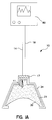
- Figs. 1A, 1B, 2, 5, and 7-23 illustrate a non-invasive fetal probe 10 constructed in accordance with the present invention.
- the reference numerals in this application correspond to like elements having the same reference numerals in the figures of U.S. Patent No. 5,474,065.
- the words "carried by” define the structural relationship between specified elements as being established by attaching the elements to each other by either direct or indirect means.
- the various elements of the drawing are not to scale. On the contrary, the width, length, and thickness of the various elements are arbitrarily expanded or reduced for clarity.
- Fetal probe 10 is inserted through the birth canal and is attached to the presenting part (typically the head) of the fetus 100 as shown in Fig. 1B. Fetus 100 may have hair 102. Because it is "non-invasive," fetal probe 10 does not penetrate the fetal skin. Once attached, fetal probe 10 can continuously sense, depending upon the sensors incorporated in fetal probe 10, such fetal parameters as heart rate, blood gas composition, temperature, and pH levels during labor and delivery. These fetal parameters are received by monitor 80 as shown in Fig. 1A. Other types of sensors and test equipment or combinations of sensors and test equipment could be incorporated into fetal probe 10.
- An important aspect of the present invention is the enhanced electrical and mechanical separation of the fetal and maternal sensors.
- Shown in Figs. 1, 2, and 5-19 are non-invasive fetal probes having a polymer shell 34, preferably made of a thermoplastic polymer, coated with a gel 30 which is preferably a hydrogel having conductive and/or adhesive properties.
- the gel 30 may also be covered with an insulating barrier 21 to enhance the electrical isolation of the fetal sensor from electrical signals in the maternal environment.
- the insulating barrier 21 also provides enhanced electrical isolation between the fetal sensor 11 or 12 and the maternal sensor 16 or 17.
- fetal probe 10 has a fetal brush sensor 11 or fetal disk sensor 12 with an attached fetal connector 14.
- a maternal sensor 17 and attached maternal connector 18 may also be provided.
- Fetal sensors 11 or 12 and maternal sensor 17 are electrodes, and fetal connector 14 and maternal connector 18 are conductors, when fetal probe 10 is a fetal heart rate probe.
- Maternal sensor 17 can be positioned on the top surface of probe 10 or may extend down the sides of probe 10, so long as maternal sensor 17 does not contact the fetus to which probe 10 is adhered.
- Gel 30 provides the base to which fetal sensor 11, 12 and maternal sensor 17 are attached and secures fetal probe 10 to the presenting part of fetus 100.
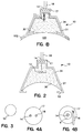
- Shell 34 can be formed by a number of processes understood by those skilled in the art, including molding. Suitable dimensions for shell 34 are a total height of about 6.2 mm and a diameter of about 15 mm. By using shell 34, the gel 30 and fetal sensor 11, 12 may be isolated from external fluids. Figs. 6D and 6E show two variations in the configuration of wall 50 of shell 34.
- Fig. 6D shows a ledge 52, which is a radially inward extension of the wall 50 in a plane which is approximately at a 30-degree angle to outer wall surface 48 of shell 34.
- a ledge extension 53 is provided which maximizes the electrical and physical separation between the fetal and maternal sensing elements.
- the angle of ledge 52 could be adjusted (for example, to angles between 15 and 60 degrees) to achieve a desired holding strength.
- Ledge 52 has a side surface 54; a bottom, horizontal surface 44; and a top surface 56.
- Fig. 6G shows rim extension 51, which is a radially outward extension of the wall 50 in a plane which is approximately at a 30-degree angle to outer wall surface 48 of shell 34.
- the angle of rim extension 51 could be adjusted (for example, to angles between 15 and 60 degrees) to achieve a desired holding strength.
- Fig. 6F shows an embodiment having both a rim extension 51 and a ledge extension 53 used in combination which provides increased electrical and physical separation between the fetal and maternal sensing elements than that provided by either extension alone.
- Fig. 6E shows a "straight" inner wall surface 36, without deviations or additions, entirely parallel to the outer wall surface 48 of wall 50 of shell 34.
- An outer wall extension 49 is provided which maximizes the electrical and physical separation between the fetal and maternal sensing elements.
- the ledge extension 53 of Fig. 6D, outer wall extension 49 of Fig. 6E, rim extension 51 of Fig. 6G when used alone or in combination, e.g., as in Fig. 6F, help to isolate the fetal sensor thereby allowing a monitor 80, as shown in Fig. 1A, to more easily isolate the fetal heart rate signal from maternal signals.
- the ledge extension 53, outer wall extension 49, and rim extension 51 further ensure the electrical isolation of the fetal and maternal sensors by forming a seal between the gel 30 and external fluids.
- this increased separation may be provided by limiting the amount of gel 30 so that it does not extend completely to the edge of ledge 52 of polymer shell 34 which serves to insulate the gel 30 from the maternal sensor 17.
- Various thermoplastic resins are preferred materials for forming shell 34.
- Exemplary materials include Pellethane® resin (a polytetramethylene glycol ether resin available from Dow Chemical Company, such as Pellethane® 2363), Pebax® resin (a polyether block amide resin available from Atochem, Inc., such as Pebax® 2533), PVCs, polyurethanes, and polyethylenes.
- Pellethane® resin a polytetramethylene glycol ether resin available from Dow Chemical Company, such as Pellethane® 2363
- Pebax® resin a polyether block amide resin available from Atochem, Inc., such as Pebax® 2533
- PVCs polyurethanes
- polyethylenes polyethylenes.
- Isolation of the fetal electrode from the maternal electrode may also be further enhanced by providing additional insulation which may be in the form of insulating barriers, non-conductive coatings, squeegees, and air gaps which are discussed below and may be used alone, in combination with one another, and in conjunction with the ledge extension 53 or outer wall extension 49 or rim extension 51.
- Shown in Figs. 1A and 1B is an insulating barrier 21 which is disposed between shell 34 and gel 30.
- Figs. 5 and 6A show a non-conductive coating 29 disposed on bottom surface 44 of ledge 52.
- FIGS. 6B and 6C show disposed on bottom surface 44 of ledge 52 a squeegee 22 having grooves 23 which may employ specially selected coatings such as an absorbant coating 24, a non-conductive adhesive coating 25, or both as shown, for example, in Fig. 6C.
- Figs. 7 and 8 show an air gap 33 provided between ledge 52 and gel 30.
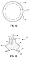
- a non-conductive rim 31 is shown in Fig. 6H which helps to isolate the fetal sensor 11 from the maternal environment.
- the non-conductive rim 31 can be a non-conductive coating, preferably a non-conductive hydrogel adhesive coating.
- the non-conductive rim 31 also helps in the attachment of the fetal probe 10 to a fetus having a particularly large amount of hair. This is accomplished by the ability of the non-conductive rim 31 to envelope fetal hair and, if a hydrogel adhesive is employed, through its wet adhesive properties.
- This non-conductive rim 31 is particularly useful when wall 50 of shell 34 has no ledge, rim, or other flange-like surface present to place against the fetal presenting surface.
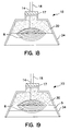
- a non-invasive fetal probe is provided as shown in Fig. 8.
- This embodiment of fetal probe 10 has a deeper, more elongated shell 34 in addition to air gap 33.
- This tapered design provides an increased path between and, thus, enhanced electrical separation of, the fetal sensor 11 and maternal sensor 17.
- the tapered design also has the advantage of providing a fetal probe 10 having a small enough diameter for ease of application early in labor. For example, a V-shaped concave cup with a diameter of 15 to 20 mm, or which can be collapsed into a dispenser or guide tube of this diameter or less, is preferred. Such a size permits application of fetal probe 10 when cervical dilation is 1 cm or less.
- the non-invasive fetal probes according to the present invention can have gel 30 with a non-conductive rim 31.
- fetal probes 10 having gel 30, which may be a conductive hydrogel, with a non-conductive rim 31 around the perimeter.
- the non-conductive rim 31 can be a non-conductive coating, preferably a non-conductive hydrogel adhesive coating, which helps to electrically isolate the fetal sensor 11, 12 from electrical signals in the maternal environment.
- the non-conductive rim 31 also helps to attach the fetal probe 10 to a fetus having a particularly large amount of hair.
- non-conductive rim 31 This is accomplished by the ability of the non-conductive rim 31 to envelope fetal hair and, if a hydrogel adhesive is employed, through its wet adhesive properties.
- This non-conductive rim 31 is particularly useful in isolating the fetal sensor 11, 12 from the maternal environment when no ledge, rim, or other flange-like surface is present to place against the fetal presenting surface.
- Figs. 9 and 10 show the non-conductive rim 31 extending only partially up from the fetal attachment surface, the non-conductive rim is not limited to this region but can extend further toward the maternal sensor 17 as shown in Fig. 11.
- Fig. 12 shows a bottom view of the fetal attachment surface of the probes 10 shown in Figs. 9-11.
- a non-invasive probe 10 having a shell extension configuration shown in Fig. 13 is provided which also maximizes the separation between the maternal and fetal environments.
- gel 30 has a bellows configuration with a shell 34 and a shell extension 37 located around the perimeter of the fetal attachment surface which supports a non-conductive adhesive 35 which may be a hydrogel material.
- fetal probe 10 is covered with a shell 34 and, in the embodiment shown in Fig. 13, an additional shell extension 37 which prevents the gel 30 from contacting the maternal environment in order to help electrically isolate the fetal sensor from electrical signals in the maternal environment.
- the shell 34 and shell extension 37 also provide electrical isolation between the fetal sensor 11, 12 and the maternal sensor 17.
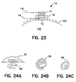
- a non-invasive fetal probe having low-profile designs is provided as shown in Figs. 20-23.
- consideration must be given to the attachment force which holds fetal probe 10 in place on a fetus. This attachment force must be sufficient to hold fetal probe 10 in place during labor which may require attachment times of ten hours or more.
- a fetal probe is subjected to tipping forces which are generated by movement of the head of the fetus, the cervix of the mother, or both.
- the low-profile designs according to the present invention help decrease the tendency of the fetal probes to detach caused by these fetal and cervical forces generated upon the probe. This enables the non-invasive probe to be securely attached to the fetus during labor and delivery.
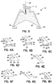
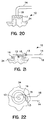
- fetal probe designs shown in Figs. 20-23 achieve these low profiles by reducing the overall height of probe 10 by either forming fetal connector lead 14 and maternal connector lead 18 to include a substantially perpendicular bend (Fig. 20) or connecting these leads to the probe 10 in a radial direction (Figs. 21 and 23).
- radial attachment of the fetal connector lead 14 can be accomplished by a fetal circumferential sensor 13 shown in Figs. 21 and 22.
- the maternal sensor 16 of Figs. 21 and 22 is preferably a steel band which is connected to maternal connector 18.
- a fetal probe 10 having a low-profile is provided which has substantially flat fetal and maternal electrodes.
- a preferred embodiment, shown attached to fetus 100 in Fig. 23, is a fetal probe 10 having a maternal sensor 17 and a fetal sensor 12 both of which are flat disks and separated by insulating layer 19.
- insulating layer 19 extends over fetal sensor 12 to enhance separation from maternal sensor 17.
- Fetal probe 10 may be used to monitor fetal heart rate without penetration of the fetal epidermis. At least four criteria affect the quality of the fetal heart rate signal monitored: (1) symmetry between the fetal and maternal (reference) electrodes, (2) maximum separation between the fetal and reference sensors, (3) maximum surface contact area to minimize impedance, and (4) stabilized connections. Application of fetal probe 10 to monitor fetal heart rate meets these criteria well.
- a fetal sensor detects the electrical fetal heart rate signal transcutaneously.
- the fetal sensor must maintain contact with the presenting part of a fetus, either directly or through the gel 30.
- gel 30 is a conductive hydrogel in the case where electrical contact between the fetus and the fetal sensor is established indirectly through gel 30 as shown, for example, in Figs. 1A, 1B, 2, 5, 7, 8, 9-11, and 13.
- gel 30 may optionally include such a conductive material in order to enhance the electrical contact and transmission of electrical fetal signals.
- the fetal sensor itself must provide a sufficiently large surface area and be electrically conductive to sense the electrical fetal heart rate signal.
- the present invention includes various types of fetal sensors which are illustrative of, but not limited to, the types which may be employed. Discussed above are fetal brush sensor 11 (shown in Figs. 1A, 1B, 5, 7-11, and 13) and fetal disk sensor 12 (shown in Figs. 2, 3, and 23).
- a suitable material for construction of fetal brush sensor 11 is multi-strand carbon fiber wire.
- Carbon wires are light weight, flexible, and radiolucent (i.e., they are partially or wholly transparent to X-Rays). Moreover, they provide good electrical conductivity and do not react with gel components or saline solutions.
- Suitable carbon wire can be obtained from Minnesota Wire and Cable Company in St. Paul, Minnesota and can replace stainless steel in forming fetal sensor 11.
- Fetal brush sensors 11 could also be formed from multi-strand stainless steel wire, such as a Teflon®-coated steel wire sold by Cooner Wire in Chatsworth, California. Such wire is very fine and light weight; therefore, it adds little mass to fetal probe 10.
- Teflon® jacket is removed to expose the stainless steel wire. The exposed wires are then spread to form a brush.
- Alternative insulating jacket materials for the wire, other than Teflon® could also be used.
- Fetal circumferential sensor 13 can be formed from conductive wire (e.g., stainless steel) which does not react with gel components or saline solutions.
- the conductive wire used typically has an isolating jacket 15 which protects the wire portion of the fetal circumferential sensor 13 extending outside of shell 34 from the maternal environment. See Figs. 21 and 22.
- the isolating jacket 15 is stripped to expose the wire portion of the fetal circumferential sensor 13 on the inside of shell 34 so that it may contact the conductive gel 30.
- Fetal disk sensor 12 in this application can be the silver-silver chloride sensor commonly used in ECG monitoring electrodes. Additional fetal sensors are also provided which are useful in the present invention, namely, a loop array 6 (Figs. 14-15), a conductive fiber "wool” or mesh 7 (Figs. 16-17), and a conductive "fan strand” configuration 8 (Figs. 18-19).
- the materials used for these various fetal sensors should be conductive and unreactive with gel components or saline solutions.
- the materials incorporated in fetal sensors should also not react with the fetal tissue or in the fetal environment (e.g., with the vernix or meconium).
- exemplary materials particularly useful for the fetal sensors of the present invention are carbon fibers, stainless steel, silver, gold, silver-silver chloride, and combinations thereof.
- fetal sensor collectively refers to fetal brush 11, fetal disk 12, fetal circumferential sensor 13, loop array 6, conductive fiber "wool” or mesh 7, and conductive "fan strand” 8 configurations.
- the fetal sensors shown in Figs. 14, 16, and 18 are shown partially embedded in and partially protruding out of gel 30 in order to provide direct contact, and thus enhance conductivity, between the fetal sensor and the fetal tissue when the fetal probe is attached to the presenting part of the fetus.
- the fetal sensors may also be covered with an adhesion-promoting layer 5 as shown in Figs. 15, 17, and 19.
- the adhesion-promoting layer 5 can include a lubricating jelly (e.g., K-Y® jelly) or a conductive medium (e.g., a conductive migrating hydrogel) which can flow around hair to improve adhesion.
- a lubricating jelly e.g., K-Y® jelly
- a conductive medium e.g., a conductive migrating hydrogel
- the additional advantage of lower impedance at the skin-sensor interface is also realized.
- spring contact 58 shown in Fig. 24A which is partially embedded in gel 30 and provides for direct contact, and thus enhanced conductivity, between the fetal sensor and the fetal tissue when the fetal probe is attached to the presenting part of the fetus.
- Spring contact 58 is shown in a perspective view in Fig. 24B and in a top view in Fig. 24C.
- fetal connector 14 is an insulated lead wire suitable for conducting the electrical signals from the fetal sensor to a monitoring signal-processing unit without being susceptible to interference from the maternal environment and maternal sensor.
- Fetal connector 14 passes through shell 34 into gel 30 and maternal sensor 16 or 17 and is ultimately connected (perhaps through other wires and electrical connections) to a fetal heart rate monitor 80 shown in Fig. 1A.
- the environment in which fetal probe 10 is used, namely inside the uterus, requires insulation of the lead wire.
- the connection between fetal connector 14 and fetal sensor 12 is embedded in the gel 30.
- the fetal sensors of the present invention may be entirely embedded in gel 30 (as shown in Figs. 1A, 1B, 2, 5, 7-11, and 13), partially embedded in or positioned on the surface of gel 30 (as shown in Figs. 14, 16, and 18) or otherwise carried by gel 30.
- the fetal sensors may also be either partially (Figs. 15 and 19) or totally (Fig. 17) embedded in the adhesion-promoting layer 5.
- fetal sensor connector 14 and maternal sensor connector 18 connect the fetal and maternal sensors, respectively, to an external monitor 80.
- maternal sensors 16 and 17 must provide a sufficiently large surface area and be electrically conductive to sense the electrical maternal heart rate signal and other muscular or electrical activity.
- maternal sensors 16 and 17 must be inert to chemical reaction with biological fluids and tissues.
- Multi-strand stainless steel or carbon fiber wires, described above for use as fetal sensor 11, can also be used for maternal sensor 16.
- maternal sensor 16 may be an electrically conducting material such as a metal foil (e.g., silver, aluminum, or stainless steel) or metallized film (e.g., aluminum metallized polyester) which covers the upper surface of insulating barrier 21.
- Other electrically conductive, non-metallic films and coatings such as conductive carbon and conductive graphite may also be used.
- Maternal sensor 16 (Figs. 21 and 22) and maternal sensor 17 (Figs. 1, 2, 4, 5, 7-11, 13-20, and 23) may be a band, washer, wire loop, or plate which fits on top of or is otherwise carried by shell 34 or insulating layer 19.
- stainless steel disk 17 can be approximately 3.5 to 4.0 mm wide (preferably 3.7 mm) and 5.0 to 5.5 mm in diameter (preferably 5.4 mm) and may contain an enlarged hole 9 (Fig. 4B) to facilitate passage of the fetal connector 14 therethrough.
- Maternal connector 18 is a lead wire which can be a sheathed multi-strand wire connected to maternal sensor 17 for communicating received electrical signals. Maternal connector 18 can be attached perpendicular to (Fig. 4A) or in the plane of maternal sensor 17. Typically, the wire(s) of maternal connector 18 is (are) attached to maternal sensor 17 by a spot welding operation.
- Maternal sensors 16 and 17 detect primarily the maternal electrical environment while fetal sensors 11, 12, and 13 detect primarily the fetal electrical environment on the surface of the fetus.
- the signal received by each sensor has a minor component signal from the other environment (i.e., the fetal sensor detects signals from the maternal environment while the maternal sensor detects signals from the fetus).
- the signal detected by maternal sensors 16 and 17 as a reference signal, any maternal heart signals and other muscular or electrical signals which pass through the fetus and are detected by the fetal sensor can be filtered electronically in the fetal monitor 80 to provide a differential signal which is an accurate fetal heart rate measurement. This is accomplished by isolating the fetal R-waves from the other signals.
- Non-invasive fetal probe 10 has two leads 14, 18--similar to conventional fetal scalp electrodes--for the fetal sensor and maternal sensor. Both the fetal and maternal leads 14 and 18 could be replaced by a non-wired connecting system, such as a radio transmission system, to communicate information from the fetal sensor and maternal sensors 16 or 17 to monitor 80.
- a radio system would require supporting hardware as is understood by those skilled in the art.
- Insulating barrier 21 (Figs. 1A, 1B, and 2) and non-conductive coating 29 (Figs. 5 and 6A), these elements serve to maintain electrical insulation between the fetal and maternal sensors.
- Pebax® a polyether block amide
- Insulating barrier 21 and non-conductive coating 29 should be flexible so as not to impede the flexibility of gel 30.
- non-conductive coating 29 (Figs. 5 and 6A), non-conductive adhesive 35 (Fig. 13), and adhesion-promoting layer 5 (Figs. 15, 17, and 19)
- these elements contact the fetus
- they preferably incorporate adhesive materials which are conformable and used in amounts and in thicknesses sufficient to envelop fetal hair and other surface irregularities and to promote attachment to a fetus having a large amount of hair.
- the adhesive materials used should be capable of enhancing the electrical isolation of the fetal probe from the maternal environment. An adhesive with "wet" tack capabilities is particularly preferred.
- gel compositions and production techniques useful in the present invention is within the purview of those skilled in the art, with examples being given, but not limited to, the hydrogels in U.S. Patent No. 5,474,065.
- the hydrogel compositions disclosed therein can be molded or formed to provide almost any desired size and shape.
- Gel 30 can be formed by dispensing the gel composition into a mold of the desired shape then polymerizing the monomers of the gel in the mold.
- suction cup-shaped probes can be prepared by spin casting, similar to the procedure used to prepare soft contact lenses.
- gel 30 is preferred for constructing gel 30 in order to enhance or establish electrical contact between the fetal sensor and the presenting part of the fetus, it is recognized that gel 30 need not be a conductive material in cases where the fetal sensor establishes direct contact with the fetus.
- fetal probe 10 of the present invention has been described in detail for attachment of non-invasive fetal heart rate sensors, fetal probe 10 could also be used for other sensors such as blood gas analyzers and oximetry sensors. If so, fetal connector 14 and maternal connector 18 may constitute fiber optics rather than lead wires.
Landscapes
- Health & Medical Sciences (AREA)
- Life Sciences & Earth Sciences (AREA)
- Biomedical Technology (AREA)
- Molecular Biology (AREA)
- Veterinary Medicine (AREA)
- Physics & Mathematics (AREA)
- Public Health (AREA)
- Biophysics (AREA)
- Pathology (AREA)
- Engineering & Computer Science (AREA)
- General Health & Medical Sciences (AREA)
- Heart & Thoracic Surgery (AREA)
- Medical Informatics (AREA)
- Animal Behavior & Ethology (AREA)
- Surgery (AREA)
- Cardiology (AREA)
- Pediatric Medicine (AREA)
- Gynecology & Obstetrics (AREA)
- Pregnancy & Childbirth (AREA)
- Reproductive Health (AREA)
- Chemical & Material Sciences (AREA)
- Dispersion Chemistry (AREA)
- Measurement And Recording Of Electrical Phenomena And Electrical Characteristics Of The Living Body (AREA)
Applications Claiming Priority (2)
| Application Number | Priority Date | Filing Date | Title |
|---|---|---|---|
| US08/741,942 US5833622A (en) | 1994-04-04 | 1996-10-31 | Non-invasive fetal probe having improved mechanical and electrical properties |
| US741942 | 2000-12-21 |
Publications (1)
| Publication Number | Publication Date |
|---|---|
| EP0839496A1 true EP0839496A1 (de) | 1998-05-06 |
Family
ID=24982873
Family Applications (1)
| Application Number | Title | Priority Date | Filing Date |
|---|---|---|---|
| EP97118170A Withdrawn EP0839496A1 (de) | 1996-10-31 | 1997-10-20 | Nicht-invasive Foetussonde mit verbesserten mechanischen und elektrischen Eigenschaften |
Country Status (3)
| Country | Link |
|---|---|
| US (1) | US5833622A (de) |
| EP (1) | EP0839496A1 (de) |
| CA (1) | CA2217666A1 (de) |
Cited By (1)
| Publication number | Priority date | Publication date | Assignee | Title |
|---|---|---|---|---|
| WO2003088837A1 (en) * | 2002-03-04 | 2003-10-30 | Medexa Diagnostisk Service Ab | A device and a method for monitoring a foetus |
Families Citing this family (55)
| Publication number | Priority date | Publication date | Assignee | Title |
|---|---|---|---|---|
| US6185442B1 (en) * | 1995-06-08 | 2001-02-06 | Ilan Zadik Samson | Valve controlled flow into a tube |
| US6456865B2 (en) | 1995-06-08 | 2002-09-24 | Ilan Zadik Samson | Non-invasive medical probe |
| US6285896B1 (en) | 1998-07-13 | 2001-09-04 | Masimo Corporation | Fetal pulse oximetry sensor |
| US6751498B1 (en) * | 1999-03-15 | 2004-06-15 | The Johns Hopkins University | Apparatus and method for non-invasive, passive fetal heart monitoring |
| CA2345720C (en) * | 1999-03-15 | 2005-01-04 | The Johns Hopkins University | Apparatus and method for non-invasive, passive fetal heart monitoring |
| WO2001050953A1 (de) * | 2000-01-07 | 2001-07-19 | Agilent Technologies Inc. | Sensor mit saugkontakt, insbesondere für nicht-invasive fetale messungen wie der pulsrate |
| GB2370776B (en) * | 2000-09-13 | 2004-10-13 | Neoventa Medical Ab | Fetal scalp electrode |
| JP2004535861A (ja) * | 2001-06-05 | 2004-12-02 | バーネフ リミテッド | 出産モニタリングシステム |
| US6922586B2 (en) * | 2002-05-20 | 2005-07-26 | Richard J. Davies | Method and system for detecting electrophysiological changes in pre-cancerous and cancerous tissue |
| US7630759B2 (en) | 2002-05-20 | 2009-12-08 | Epi-Sci, Llc | Method and system for detecting electrophysiological changes in pre-cancerous and cancerous breast tissue and epithelium |
| US8262575B2 (en) * | 2002-05-20 | 2012-09-11 | Epi-Sci, Llc | Method and system for detecting electrophysiological changes in pre-cancerous and cancerous tissue |
| EP1876950A4 (de) * | 2005-04-21 | 2014-01-22 | Epi Sci Llc | Verfahren und system zum nachweis elektrophysiologischer veränderungen in vorkanzerösem und kanzerösem gewebe und epithel |
| US8784336B2 (en) | 2005-08-24 | 2014-07-22 | C. R. Bard, Inc. | Stylet apparatuses and methods of manufacture |
| US7794407B2 (en) | 2006-10-23 | 2010-09-14 | Bard Access Systems, Inc. | Method of locating the tip of a central venous catheter |
| US8388546B2 (en) | 2006-10-23 | 2013-03-05 | Bard Access Systems, Inc. | Method of locating the tip of a central venous catheter |
| US20080221420A1 (en) * | 2007-03-08 | 2008-09-11 | Nonin Medical, Inc. | Fetal Pulse Oximetry and ECG Sensor |
| WO2009060431A2 (en) * | 2007-11-08 | 2009-05-14 | Association For Public Health Services | Vacuum delivery extractor |
| US9649048B2 (en) | 2007-11-26 | 2017-05-16 | C. R. Bard, Inc. | Systems and methods for breaching a sterile field for intravascular placement of a catheter |
| US9521961B2 (en) | 2007-11-26 | 2016-12-20 | C. R. Bard, Inc. | Systems and methods for guiding a medical instrument |
| US10751509B2 (en) | 2007-11-26 | 2020-08-25 | C. R. Bard, Inc. | Iconic representations for guidance of an indwelling medical device |
| US10449330B2 (en) | 2007-11-26 | 2019-10-22 | C. R. Bard, Inc. | Magnetic element-equipped needle assemblies |
| US8781555B2 (en) | 2007-11-26 | 2014-07-15 | C. R. Bard, Inc. | System for placement of a catheter including a signal-generating stylet |
| US8849382B2 (en) | 2007-11-26 | 2014-09-30 | C. R. Bard, Inc. | Apparatus and display methods relating to intravascular placement of a catheter |
| US10524691B2 (en) | 2007-11-26 | 2020-01-07 | C. R. Bard, Inc. | Needle assembly including an aligned magnetic element |
| ES2557084T3 (es) | 2007-11-26 | 2016-01-21 | C. R. Bard, Inc. | Sistema integrado para la colocación intravascular de un catéter |
| US8478382B2 (en) | 2008-02-11 | 2013-07-02 | C. R. Bard, Inc. | Systems and methods for positioning a catheter |
| WO2010022370A1 (en) | 2008-08-22 | 2010-02-25 | C.R. Bard, Inc. | Catheter assembly including ecg sensor and magnetic assemblies |
| US8437833B2 (en) | 2008-10-07 | 2013-05-07 | Bard Access Systems, Inc. | Percutaneous magnetic gastrostomy |
| US20100234713A1 (en) * | 2009-03-11 | 2010-09-16 | Sheraton Sr David A | Silver-silver chloride needle electrode system |
| CN102802514B (zh) | 2009-06-12 | 2015-12-02 | 巴德阿克塞斯系统股份有限公司 | 导管末端定位设备 |
| US9532724B2 (en) | 2009-06-12 | 2017-01-03 | Bard Access Systems, Inc. | Apparatus and method for catheter navigation using endovascular energy mapping |
| EP2464407A4 (de) | 2009-08-10 | 2014-04-02 | Bard Access Systems Inc | Vorrichtungen und verfahren für endovaskuläre elektrographie |
| EP2517622A3 (de) | 2009-09-29 | 2013-04-24 | C. R. Bard, Inc. | Stillete zur Verwendung mit Vorrichtungen zur intravaskulären Positionierung eines Katheters |
| US11103213B2 (en) | 2009-10-08 | 2021-08-31 | C. R. Bard, Inc. | Spacers for use with an ultrasound probe |
| WO2011097312A1 (en) | 2010-02-02 | 2011-08-11 | C.R. Bard, Inc. | Apparatus and method for catheter navigation and tip location |
| EP2913000B1 (de) | 2010-05-28 | 2020-02-12 | C.R. Bard, Inc. | Vorrichtung zur Verwendung mit einem Nadeleinsatz-Führungssystem |
| EP2912999B1 (de) | 2010-05-28 | 2022-06-29 | C. R. Bard, Inc. | Vorrichtung zur Verwendung mit einem Nadeleinsatz-Führungssystem |
| AU2011289513B2 (en) | 2010-08-09 | 2014-05-29 | C.R. Bard, Inc. | Support and cover structures for an ultrasound probe head |
| WO2012024577A2 (en) | 2010-08-20 | 2012-02-23 | C.R. Bard, Inc. | Reconfirmation of ecg-assisted catheter tip placement |
| WO2012058461A1 (en) | 2010-10-29 | 2012-05-03 | C.R.Bard, Inc. | Bioimpedance-assisted placement of a medical device |
| WO2013006817A1 (en) | 2011-07-06 | 2013-01-10 | C.R. Bard, Inc. | Needle length determination and calibration for insertion guidance system |
| USD724745S1 (en) | 2011-08-09 | 2015-03-17 | C. R. Bard, Inc. | Cap for an ultrasound probe |
| USD699359S1 (en) | 2011-08-09 | 2014-02-11 | C. R. Bard, Inc. | Ultrasound probe head |
| US9211107B2 (en) | 2011-11-07 | 2015-12-15 | C. R. Bard, Inc. | Ruggedized ultrasound hydrogel insert |
| WO2013188833A2 (en) | 2012-06-15 | 2013-12-19 | C.R. Bard, Inc. | Apparatus and methods for detection of a removable cap on an ultrasound probe |
| ES2811323T3 (es) | 2014-02-06 | 2021-03-11 | Bard Inc C R | Sistemas para el guiado y la colocación de un dispositivo intravascular |
| WO2016009424A1 (en) * | 2014-07-13 | 2016-01-21 | Nibs Neuroscience Technologies Ltd. | Electrode headset grid and use thereof in the non-invasive brain stimulation and monitoring |
| US10123718B2 (en) * | 2014-10-30 | 2018-11-13 | University Of Tenessee Research Foundation | Methods, systems, and assemblies for measuring bioelectrical signals of intra-abdominal organs |
| US10973584B2 (en) | 2015-01-19 | 2021-04-13 | Bard Access Systems, Inc. | Device and method for vascular access |
| EP3253445A4 (de) | 2015-02-03 | 2018-02-21 | Nibs Neuroscience Technologies Ltd. | Frühdiagnose und die behandlung von morbus alzheimer und leichter kognitiver beeinträchtigung |
| WO2016210325A1 (en) | 2015-06-26 | 2016-12-29 | C.R. Bard, Inc. | Connector interface for ecg-based catheter positioning system |
| US11000207B2 (en) | 2016-01-29 | 2021-05-11 | C. R. Bard, Inc. | Multiple coil system for tracking a medical device |
| US10653331B2 (en) * | 2016-05-24 | 2020-05-19 | Konan Medical Usa, Inc. | Electrode sensor |
| US11096626B2 (en) * | 2017-05-22 | 2021-08-24 | Maurice-Andre Recanati | Fetal scalp monitor |
| CN112867443B (zh) | 2018-10-16 | 2024-04-26 | 巴德阿克塞斯系统股份有限公司 | 用于建立电连接的安全装备连接系统及其方法 |
Citations (6)
| Publication number | Priority date | Publication date | Assignee | Title |
|---|---|---|---|---|
| US3910271A (en) * | 1973-06-04 | 1975-10-07 | Theodore C Neward | Method of making a bipolar electrode structure |
| FR2569976A1 (fr) * | 1984-09-11 | 1986-03-14 | Applic Bio Medicales Sa | Capteur pour la mesure de la p02 et de l'electrocardiogramme foetal, en particulier pour l'usage obstetrical ou pediatrique |
| DE3446115A1 (de) * | 1984-12-18 | 1986-06-19 | Drägerwerk AG, 2400 Lübeck | Klebbare messsonde mit einem messwertaufnehmer |
| WO1990004352A1 (en) * | 1988-10-28 | 1990-05-03 | Nellcor Incorporated | Improved perinatal pulse oximetry sensor |
| GB2274995A (en) * | 1993-02-15 | 1994-08-17 | John Mccune Anderson | Biomedical electrode device |
| EP0676170A1 (de) * | 1994-04-04 | 1995-10-11 | Graphic Controls Corporation | Nicht-invasive Foetussonde |
Family Cites Families (54)
| Publication number | Priority date | Publication date | Assignee | Title |
|---|---|---|---|---|
| US3590810A (en) * | 1968-05-27 | 1971-07-06 | Honeywell Inc | Biomedical body electrode |
| US3750650A (en) * | 1970-12-15 | 1973-08-07 | Hewlett Packard Gmbh | Double spiral electrode for intra-cavity attachment |
| US3827428A (en) * | 1971-01-20 | 1974-08-06 | R Hon | Bipolar electrode structure for monitoring fetal heartbeat and the like |
| DE2152808A1 (de) * | 1971-10-22 | 1973-04-26 | Siemens Ag | Selbsthaftende elektrode fuer die ekgdiagnostik |
| USRE28990E (en) * | 1972-12-04 | 1976-10-05 | Corometrics Medical Systems, Inc. | Bipolar electrode structure for monitoring fetal heartbeat and the like |
| US3958564A (en) * | 1975-05-09 | 1976-05-25 | The United States Of America As Represented By The Secretary Of The Navy | EKG contact |
| US4180080A (en) * | 1977-10-03 | 1979-12-25 | Hewlett-Packard Company | Electrode assembly for sensing heart activity |
| US4149528A (en) * | 1977-10-03 | 1979-04-17 | Hewlett-Packard Company | Electrode assembly for sensing heart activity |
| US4308873A (en) * | 1978-03-16 | 1982-01-05 | National Research Development Corporation | Electroencephalograph monitoring |
| US4314044A (en) * | 1979-01-22 | 1982-02-02 | Rohm And Haas Company | Process for preparing low molecular weight water-soluble polymers |
| US4299232A (en) * | 1979-06-19 | 1981-11-10 | Mario Zilianti | Bipolar electrodes for fetal heart-rate recording during labor |
| JPS5933361Y2 (ja) * | 1980-03-14 | 1984-09-18 | 日東電工株式会社 | 電極パッド |
| US4301806A (en) * | 1980-04-14 | 1981-11-24 | American Home Products Corporation | Rotating mechanism for introducing a fetal electrode |
| US4320764A (en) * | 1980-06-13 | 1982-03-23 | American Home Products Corporation | Fetal electrode |
| US4469105A (en) * | 1981-06-18 | 1984-09-04 | Clinton Meyering | Medical electrode apparatus and kit of components therefor |
| US4437467A (en) * | 1981-12-11 | 1984-03-20 | American Home Products Corporation | Apparatus for monitoring fetal heartbeat and the like |
| US4501276A (en) * | 1982-07-16 | 1985-02-26 | Illinois Tool Works Inc. | Fetal electrode apparatus |
| US4458695A (en) * | 1982-07-16 | 1984-07-10 | Cordis Corporation | Multipolar electrode assembly for pacing lead |
| US4658825A (en) * | 1982-09-24 | 1987-04-21 | International Biomedics, Inc. | Spiral probe for simultaneous electrical and chemical monitoring of a fetus |
| AU558083B2 (en) * | 1982-12-30 | 1987-01-15 | Rocket Of London Ltd. | Foetal scalp electrode with torque limit |
| US5109849A (en) * | 1983-08-30 | 1992-05-05 | Nellcor, Inc. | Perinatal pulse oximetry sensor |
| DE3473989D1 (en) * | 1983-10-13 | 1988-10-20 | Morgenstern Jurgen | Physiological sensor |
| US5217013A (en) * | 1983-10-14 | 1993-06-08 | Somanetics Corporation | Patient sensor for optical cerebral oximeter and the like |
| SE454941B (sv) * | 1983-10-28 | 1988-06-13 | Astra Tech Ab | Elektrod, fastsugbar med vacuum, och en elektrodplatta for en elektrod, avsedd for t ex ekg-metningar |
| JPS6185925A (ja) * | 1984-10-01 | 1986-05-01 | 日東電工株式会社 | 生体用電極 |
| SE8502048D0 (sv) * | 1985-04-26 | 1985-04-26 | Astra Tech Ab | Vakuumfixerad hallare for medicinskt bruk |
| US4731078A (en) * | 1985-08-21 | 1988-03-15 | Kingston Technologies Limited Partnership | Intraocular lens |
| GB8613687D0 (en) * | 1986-06-05 | 1986-07-09 | Rocket Of London Ltd | Instrument for monitoring foetal heart rate |
| US4706680A (en) * | 1986-06-30 | 1987-11-17 | Nepera Inc. | Conductive adhesive medical electrode assemblies |
| CA1328123C (en) * | 1986-10-08 | 1994-03-29 | Nigel John Randall | Intrauterine probe |
| US5184619A (en) * | 1986-11-10 | 1993-02-09 | Peritronics Medical, Inc. | Intrauterine pressure and fetal heart rate sensor |
| US4825879A (en) * | 1987-10-08 | 1989-05-02 | Critkon, Inc. | Pulse oximeter sensor |
| US4934371A (en) * | 1988-02-09 | 1990-06-19 | American Home Products Corporation | Fetal electrode product |
| US5002792A (en) * | 1988-08-11 | 1991-03-26 | Medtronic, Inc. | Process for making biomedical devices utilizing thermoplastic hydrophilic gels |
| US4921904A (en) * | 1988-12-19 | 1990-05-01 | Nalco Chemical Company | Superabsorbent polymers |
| DE3906074A1 (de) * | 1989-02-27 | 1990-08-30 | Schmid Walter | Verfahren zur herstellung einer koerperelektrode |
| US5143071A (en) * | 1989-03-30 | 1992-09-01 | Nepera, Inc. | Non-stringy adhesive hydrophilic gels |
| US4989607A (en) * | 1989-03-30 | 1991-02-05 | Preston Keusch | Highly conductive non-stringy adhesive hydrophilic gels and medical electrode assemblies manufactured therefrom |
| US5139023A (en) * | 1989-06-02 | 1992-08-18 | Theratech Inc. | Apparatus and method for noninvasive blood glucose monitoring |
| US4956170A (en) * | 1989-06-28 | 1990-09-11 | S. C. Johnson & Son, Inc. | Skin moisturizing/conditioning antimicrobial alcoholic gels |
| US5224478A (en) * | 1989-11-25 | 1993-07-06 | Colin Electronics Co., Ltd. | Reflecting-type oxymeter probe |
| KR100213554B1 (ko) * | 1989-11-28 | 1999-08-02 | 제이슨 오토 가도시 | 태아용 탐침 |
| US5183599A (en) * | 1990-01-19 | 1993-02-02 | Smuckler Jack H | Rapid curing, electrically conductive adhesive |
| US5124076A (en) * | 1990-01-19 | 1992-06-23 | Contour Electrodes, Inc. | Rapid, curing, electrically conductive adhesive |
| DE69030513T2 (de) * | 1990-02-15 | 1997-07-24 | Hewlett Packard Gmbh | Gerät und Verfahren zur nichtinvasiven Messung der Sauerstoffsättigung |
| JP3046346B2 (ja) * | 1990-03-12 | 2000-05-29 | 昭和電工株式会社 | 外用剤基剤又は補助剤とそれを含有する人又は動物の外用剤 |
| GB9008764D0 (en) * | 1990-04-19 | 1990-06-13 | Egnell Ameda Ltd | A resilient suction cup |
| AU8653291A (en) * | 1990-09-26 | 1992-04-15 | Marius Van Der Merwe | Monitoring device |
| US5154175A (en) * | 1991-03-04 | 1992-10-13 | Gunther Ted J | Intrauterine fetal EKG-oximetry cable apparatus |
| US5247932A (en) * | 1991-04-15 | 1993-09-28 | Nellcor Incorporated | Sensor for intrauterine use |
| US5183841A (en) * | 1991-12-24 | 1993-02-02 | Avery Dennison Corporation | Removable pressure-sensitive adhesives for recyclable substrates |
| DE4304693C2 (de) * | 1993-02-16 | 2002-02-21 | Gerhard Rall | Sensoreinrichtung zum Messen von vitalen Parametern eines Feten während der Geburt |
| US5377673A (en) * | 1993-03-22 | 1995-01-03 | Van Dell; Peter | Intrauterine monitoring device |
| DE9316259U1 (de) * | 1993-10-25 | 1994-02-10 | Arbo-Robotron Medizin-Technologie GmbH, 38820 Halberstadt | Biomedizinische Elektrode |
-
1996
- 1996-10-31 US US08/741,942 patent/US5833622A/en not_active Expired - Fee Related
-
1997
- 1997-10-07 CA CA002217666A patent/CA2217666A1/en not_active Abandoned
- 1997-10-20 EP EP97118170A patent/EP0839496A1/de not_active Withdrawn
Patent Citations (7)
| Publication number | Priority date | Publication date | Assignee | Title |
|---|---|---|---|---|
| US3910271A (en) * | 1973-06-04 | 1975-10-07 | Theodore C Neward | Method of making a bipolar electrode structure |
| FR2569976A1 (fr) * | 1984-09-11 | 1986-03-14 | Applic Bio Medicales Sa | Capteur pour la mesure de la p02 et de l'electrocardiogramme foetal, en particulier pour l'usage obstetrical ou pediatrique |
| DE3446115A1 (de) * | 1984-12-18 | 1986-06-19 | Drägerwerk AG, 2400 Lübeck | Klebbare messsonde mit einem messwertaufnehmer |
| WO1990004352A1 (en) * | 1988-10-28 | 1990-05-03 | Nellcor Incorporated | Improved perinatal pulse oximetry sensor |
| GB2274995A (en) * | 1993-02-15 | 1994-08-17 | John Mccune Anderson | Biomedical electrode device |
| EP0676170A1 (de) * | 1994-04-04 | 1995-10-11 | Graphic Controls Corporation | Nicht-invasive Foetussonde |
| US5474065A (en) * | 1994-04-04 | 1995-12-12 | Graphic Controls Corporation | Non-invasive fetal probe |
Cited By (1)
| Publication number | Priority date | Publication date | Assignee | Title |
|---|---|---|---|---|
| WO2003088837A1 (en) * | 2002-03-04 | 2003-10-30 | Medexa Diagnostisk Service Ab | A device and a method for monitoring a foetus |
Also Published As
| Publication number | Publication date |
|---|---|
| US5833622A (en) | 1998-11-10 |
| CA2217666A1 (en) | 1998-04-30 |
| MX9708380A (es) | 1998-08-30 |
Similar Documents
| Publication | Publication Date | Title |
|---|---|---|
| US5833622A (en) | Non-invasive fetal probe having improved mechanical and electrical properties | |
| US5474065A (en) | Non-invasive fetal probe | |
| US12016702B2 (en) | Single radio-transparent connector for multi-functional reference patch | |
| EP2389860B1 (de) | Einwegpflaster und wiederverwendbare Sensoranordnung zur Verwendung in Systemen zur Ortung und Abbildung medizinischer Vorrichtungen | |
| FI126093B (fi) | Järjestely ja menetelmä elektrodimittausten suorittamiseksi | |
| US4685466A (en) | Measuring sensor for the non-invasive detection of electro-physiological quantities | |
| CN109091135B (zh) | 基于mems技术的心音心电微型原位同步检测传感器 | |
| CA2729056A1 (en) | Crystalline form of (2s)-(-)-n-(6-chloro-2,3-dihydro-benzo[1,4]dioxin-2-ylmethyl)-sulfamide | |
| JP2008511396A (ja) | 結合されたセンサアセンブリ | |
| CN103462601A (zh) | 医用电极贴及其制备方法 | |
| JP2579657B2 (ja) | 子宮内プローブ | |
| KR20170019033A (ko) | 생체신호 측정용 센서 | |
| WO2001062147A1 (en) | Apparatus for evaluating cardiac functions via esophagus | |
| EP4111972A1 (de) | Elektrodensegment und elektrodenmatrix für elektrokardiographische und/oder bioimpedanzmessungen | |
| AU2014202559B2 (en) | Disposable patch and reusable sensor assembly for use in medical device localization and mapping systems | |
| JP2002512545A (ja) | 心機能を評価する装置および方法 | |
| MXPA97008380A (en) | Non-invasive fetal probe that has mechanical and electric properties improves | |
| Neuman | Physical measurements | |
| CN214342353U (zh) | 一种心电监测电极装置以及心电监测系统 | |
| CN112089411A (zh) | 干电极 | |
| PL223541B1 (pl) | Elektroda do holtera EKG |
Legal Events
| Date | Code | Title | Description |
|---|---|---|---|
| PUAI | Public reference made under article 153(3) epc to a published international application that has entered the european phase |
Free format text: ORIGINAL CODE: 0009012 |
|
| AK | Designated contracting states |
Kind code of ref document: A1 Designated state(s): AT BE CH DE DK ES FI FR GB IT LI NL SE |
|
| 17P | Request for examination filed |
Effective date: 19981103 |
|
| AKX | Designation fees paid |
Free format text: AT BE CH DE DK ES FI FR GB IT LI NL SE |
|
| RBV | Designated contracting states (corrected) |
Designated state(s): AT BE CH DE DK ES FI FR GB IT LI NL SE |
|
| 17Q | First examination report despatched |
Effective date: 20020904 |
|
| STAA | Information on the status of an ep patent application or granted ep patent |
Free format text: STATUS: THE APPLICATION IS DEEMED TO BE WITHDRAWN |
|
| 18D | Application deemed to be withdrawn |
Effective date: 20030315 |