EP0723555B1 - Chimeric proteins which block complement activation - Google Patents
Chimeric proteins which block complement activation Download PDFInfo
- Publication number
- EP0723555B1 EP0723555B1 EP94931763A EP94931763A EP0723555B1 EP 0723555 B1 EP0723555 B1 EP 0723555B1 EP 94931763 A EP94931763 A EP 94931763A EP 94931763 A EP94931763 A EP 94931763A EP 0723555 B1 EP0723555 B1 EP 0723555B1
- Authority
- EP
- European Patent Office
- Prior art keywords
- protein
- chimeric protein
- mcp
- soluble
- cab
- Prior art date
- Legal status (The legal status is an assumption and is not a legal conclusion. Google has not performed a legal analysis and makes no representation as to the accuracy of the status listed.)
- Expired - Lifetime
Links
- 102000037865 fusion proteins Human genes 0.000 title claims abstract description 94
- 108020001507 fusion proteins Proteins 0.000 title claims abstract description 94
- 230000024203 complement activation Effects 0.000 title claims abstract description 29
- 108090000765 processed proteins & peptides Proteins 0.000 claims abstract description 40
- 102000004196 processed proteins & peptides Human genes 0.000 claims abstract description 38
- 229920001184 polypeptide Polymers 0.000 claims abstract description 37
- 238000000034 method Methods 0.000 claims abstract description 27
- 150000007523 nucleic acids Chemical class 0.000 claims abstract description 25
- 108020004707 nucleic acids Proteins 0.000 claims abstract description 24
- 102000039446 nucleic acids Human genes 0.000 claims abstract description 24
- 206010061218 Inflammation Diseases 0.000 claims abstract description 6
- 230000004054 inflammatory process Effects 0.000 claims abstract description 6
- 108090000623 proteins and genes Proteins 0.000 claims description 114
- 102000004169 proteins and genes Human genes 0.000 claims description 99
- 102000050019 Membrane Cofactor Human genes 0.000 claims description 73
- 101710146216 Membrane cofactor protein Proteins 0.000 claims description 67
- 210000004027 cell Anatomy 0.000 claims description 67
- 108010009575 CD55 Antigens Proteins 0.000 claims description 65
- 150000001413 amino acids Chemical class 0.000 claims description 50
- 239000013604 expression vector Substances 0.000 claims description 37
- 239000012634 fragment Substances 0.000 claims description 34
- 230000014509 gene expression Effects 0.000 claims description 31
- 238000004519 manufacturing process Methods 0.000 claims description 20
- 230000027455 binding Effects 0.000 claims description 16
- 239000013598 vector Substances 0.000 claims description 14
- 102000016574 Complement C3-C5 Convertases Human genes 0.000 claims description 13
- 108010067641 Complement C3-C5 Convertases Proteins 0.000 claims description 13
- 239000011347 resin Substances 0.000 claims description 13
- 229920005989 resin Polymers 0.000 claims description 13
- 239000006228 supernatant Substances 0.000 claims description 12
- 102000007056 Recombinant Fusion Proteins Human genes 0.000 claims description 11
- 108010008281 Recombinant Fusion Proteins Proteins 0.000 claims description 11
- 239000002158 endotoxin Substances 0.000 claims description 10
- 238000011282 treatment Methods 0.000 claims description 10
- 230000004913 activation Effects 0.000 claims description 9
- 239000000356 contaminant Substances 0.000 claims description 9
- 102000006834 complement receptors Human genes 0.000 claims description 8
- 230000001105 regulatory effect Effects 0.000 claims description 8
- 208000023275 Autoimmune disease Diseases 0.000 claims description 7
- 239000003814 drug Substances 0.000 claims description 7
- 208000027866 inflammatory disease Diseases 0.000 claims description 7
- 102000006912 Complement C4b-Binding Protein Human genes 0.000 claims description 6
- 108010047548 Complement C4b-Binding Protein Proteins 0.000 claims description 6
- 102000016550 Complement Factor H Human genes 0.000 claims description 6
- 239000000203 mixture Substances 0.000 claims description 6
- 206010001052 Acute respiratory distress syndrome Diseases 0.000 claims description 5
- 208000013616 Respiratory Distress Syndrome Diseases 0.000 claims description 5
- 208000011341 adult acute respiratory distress syndrome Diseases 0.000 claims description 5
- 201000000028 adult respiratory distress syndrome Diseases 0.000 claims description 5
- 125000000484 butyl group Chemical group [H]C([*])([H])C([H])([H])C([H])([H])C([H])([H])[H] 0.000 claims description 5
- 230000003993 interaction Effects 0.000 claims description 5
- 239000003550 marker Substances 0.000 claims description 5
- 125000001997 phenyl group Chemical group [H]C1=C([H])C([H])=C(*)C([H])=C1[H] 0.000 claims description 5
- 230000008569 process Effects 0.000 claims description 5
- 108010053085 Complement Factor H Proteins 0.000 claims description 4
- 208000006011 Stroke Diseases 0.000 claims description 4
- 206010052779 Transplant rejections Diseases 0.000 claims description 4
- 230000002612 cardiopulmonary effect Effects 0.000 claims description 4
- 230000002209 hydrophobic effect Effects 0.000 claims description 4
- 201000006417 multiple sclerosis Diseases 0.000 claims description 4
- 208000025962 Crush injury Diseases 0.000 claims description 3
- 241000238631 Hexapoda Species 0.000 claims description 3
- 206010063837 Reperfusion injury Diseases 0.000 claims description 3
- 206010040070 Septic Shock Diseases 0.000 claims description 3
- 108010022394 Threonine synthase Proteins 0.000 claims description 3
- 230000001580 bacterial effect Effects 0.000 claims description 3
- 210000004978 chinese hamster ovary cell Anatomy 0.000 claims description 3
- 238000012258 culturing Methods 0.000 claims description 3
- 108020002326 glutamine synthetase Proteins 0.000 claims description 3
- 239000006166 lysate Substances 0.000 claims description 3
- 210000004962 mammalian cell Anatomy 0.000 claims description 3
- 206010039073 rheumatoid arthritis Diseases 0.000 claims description 3
- 230000036303 septic shock Effects 0.000 claims description 3
- 230000000451 tissue damage Effects 0.000 claims description 3
- 231100000827 tissue damage Toxicity 0.000 claims description 3
- 101710186862 Factor H binding protein Proteins 0.000 claims description 2
- 102100039373 Membrane cofactor protein Human genes 0.000 claims description 2
- 239000003957 anion exchange resin Substances 0.000 claims description 2
- 239000013592 cell lysate Substances 0.000 claims description 2
- 102000004419 dihydrofolate reductase Human genes 0.000 claims description 2
- 102000005396 glutamine synthetase Human genes 0.000 claims description 2
- 208000028867 ischemia Diseases 0.000 claims description 2
- 229910052751 metal Inorganic materials 0.000 claims description 2
- 239000002184 metal Substances 0.000 claims description 2
- 208000010125 myocardial infarction Diseases 0.000 claims description 2
- 210000000440 neutrophil Anatomy 0.000 claims description 2
- 230000001737 promoting effect Effects 0.000 claims description 2
- 238000003259 recombinant expression Methods 0.000 claims description 2
- 230000007115 recruitment Effects 0.000 claims description 2
- 210000005253 yeast cell Anatomy 0.000 claims description 2
- 206010025135 lupus erythematosus Diseases 0.000 claims 1
- 230000009885 systemic effect Effects 0.000 claims 1
- 235000018102 proteins Nutrition 0.000 description 95
- 235000001014 amino acid Nutrition 0.000 description 38
- 230000000694 effects Effects 0.000 description 38
- 230000000295 complement effect Effects 0.000 description 36
- 229940024606 amino acid Drugs 0.000 description 34
- FAPWRFPIFSIZLT-UHFFFAOYSA-M Sodium chloride Chemical compound [Na+].[Cl-] FAPWRFPIFSIZLT-UHFFFAOYSA-M 0.000 description 24
- -1 MCP-MCP Proteins 0.000 description 23
- 238000010276 construction Methods 0.000 description 21
- 230000037361 pathway Effects 0.000 description 19
- 101000901154 Homo sapiens Complement C3 Proteins 0.000 description 17
- 239000002299 complementary DNA Substances 0.000 description 17
- 239000013612 plasmid Substances 0.000 description 17
- 102100022133 Complement C3 Human genes 0.000 description 16
- 102100021943 C-C motif chemokine 2 Human genes 0.000 description 15
- 239000012228 culture supernatant Substances 0.000 description 14
- 238000001727 in vivo Methods 0.000 description 14
- 210000002966 serum Anatomy 0.000 description 14
- 210000003743 erythrocyte Anatomy 0.000 description 13
- 239000000499 gel Substances 0.000 description 13
- 238000003752 polymerase chain reaction Methods 0.000 description 13
- 239000011780 sodium chloride Substances 0.000 description 13
- 238000002965 ELISA Methods 0.000 description 12
- HEMHJVSKTPXQMS-UHFFFAOYSA-M Sodium hydroxide Chemical compound [OH-].[Na+] HEMHJVSKTPXQMS-UHFFFAOYSA-M 0.000 description 12
- 238000003556 assay Methods 0.000 description 12
- 238000006243 chemical reaction Methods 0.000 description 12
- LOKCTEFSRHRXRJ-UHFFFAOYSA-I dipotassium trisodium dihydrogen phosphate hydrogen phosphate dichloride Chemical compound P(=O)(O)(O)[O-].[K+].P(=O)(O)([O-])[O-].[Na+].[Na+].[Cl-].[K+].[Cl-].[Na+] LOKCTEFSRHRXRJ-UHFFFAOYSA-I 0.000 description 12
- 239000002609 medium Substances 0.000 description 12
- 239000002953 phosphate buffered saline Substances 0.000 description 12
- 101710155857 C-C motif chemokine 2 Proteins 0.000 description 11
- FFEARJCKVFRZRR-SCSAIBSYSA-N D-methionine Chemical compound CSCC[C@@H](N)C(O)=O FFEARJCKVFRZRR-SCSAIBSYSA-N 0.000 description 11
- 230000002401 inhibitory effect Effects 0.000 description 11
- 230000005764 inhibitory process Effects 0.000 description 11
- 239000000872 buffer Substances 0.000 description 10
- 230000006870 function Effects 0.000 description 10
- 238000002347 injection Methods 0.000 description 10
- 239000007924 injection Substances 0.000 description 10
- 230000001404 mediated effect Effects 0.000 description 10
- 238000000746 purification Methods 0.000 description 10
- DHMQDGOQFOQNFH-UHFFFAOYSA-N Glycine Chemical compound NCC(O)=O DHMQDGOQFOQNFH-UHFFFAOYSA-N 0.000 description 9
- 241001465754 Metazoa Species 0.000 description 9
- BTCSSZJGUNDROE-UHFFFAOYSA-N gamma-aminobutyric acid Chemical compound NCCCC(O)=O BTCSSZJGUNDROE-UHFFFAOYSA-N 0.000 description 9
- FTOAOBMCPZCFFF-UHFFFAOYSA-N 5,5-diethylbarbituric acid Chemical compound CCC1(CC)C(=O)NC(=O)NC1=O FTOAOBMCPZCFFF-UHFFFAOYSA-N 0.000 description 8
- GLUUGHFHXGJENI-UHFFFAOYSA-N Piperazine Chemical compound C1CNCCN1 GLUUGHFHXGJENI-UHFFFAOYSA-N 0.000 description 8
- 238000001994 activation Methods 0.000 description 8
- 230000015572 biosynthetic process Effects 0.000 description 8
- 230000013595 glycosylation Effects 0.000 description 8
- 238000006206 glycosylation reaction Methods 0.000 description 8
- 239000000523 sample Substances 0.000 description 8
- UCSJYZPVAKXKNQ-HZYVHMACSA-N streptomycin Chemical compound CN[C@H]1[C@H](O)[C@@H](O)[C@H](CO)O[C@H]1O[C@@H]1[C@](C=O)(O)[C@H](C)O[C@H]1O[C@@H]1[C@@H](NC(N)=N)[C@H](O)[C@@H](NC(N)=N)[C@H](O)[C@H]1O UCSJYZPVAKXKNQ-HZYVHMACSA-N 0.000 description 8
- 108091003079 Bovine Serum Albumin Proteins 0.000 description 7
- 102100030886 Complement receptor type 1 Human genes 0.000 description 7
- 101000727061 Homo sapiens Complement receptor type 1 Proteins 0.000 description 7
- DCXYFEDJOCDNAF-REOHCLBHSA-N L-asparagine Chemical compound OC(=O)[C@@H](N)CC(N)=O DCXYFEDJOCDNAF-REOHCLBHSA-N 0.000 description 7
- 229920002684 Sepharose Polymers 0.000 description 7
- 239000012091 fetal bovine serum Substances 0.000 description 7
- 238000004191 hydrophobic interaction chromatography Methods 0.000 description 7
- 238000011534 incubation Methods 0.000 description 7
- 229960005322 streptomycin Drugs 0.000 description 7
- 108020004414 DNA Proteins 0.000 description 6
- ZDXPYRJPNDTMRX-VKHMYHEASA-N L-glutamine Chemical compound OC(=O)[C@@H](N)CCC(N)=O ZDXPYRJPNDTMRX-VKHMYHEASA-N 0.000 description 6
- AYFVYJQAPQTCCC-GBXIJSLDSA-N L-threonine Chemical compound C[C@@H](O)[C@H](N)C(O)=O AYFVYJQAPQTCCC-GBXIJSLDSA-N 0.000 description 6
- TWRXJAOTZQYOKJ-UHFFFAOYSA-L Magnesium chloride Chemical compound [Mg+2].[Cl-].[Cl-] TWRXJAOTZQYOKJ-UHFFFAOYSA-L 0.000 description 6
- 108700031312 Membrane Cofactor Proteins 0.000 description 6
- 241000283973 Oryctolagus cuniculus Species 0.000 description 6
- 229910019142 PO4 Inorganic materials 0.000 description 6
- DBMJMQXJHONAFJ-UHFFFAOYSA-M Sodium laurylsulphate Chemical compound [Na+].CCCCCCCCCCCCOS([O-])(=O)=O DBMJMQXJHONAFJ-UHFFFAOYSA-M 0.000 description 6
- AYFVYJQAPQTCCC-UHFFFAOYSA-N Threonine Natural products CC(O)C(N)C(O)=O AYFVYJQAPQTCCC-UHFFFAOYSA-N 0.000 description 6
- 230000004071 biological effect Effects 0.000 description 6
- 210000004369 blood Anatomy 0.000 description 6
- 239000008280 blood Substances 0.000 description 6
- 230000006037 cell lysis Effects 0.000 description 6
- 238000003776 cleavage reaction Methods 0.000 description 6
- 238000005516 engineering process Methods 0.000 description 6
- 239000001963 growth medium Substances 0.000 description 6
- SXTAYKAGBXMACB-UHFFFAOYSA-N methionine sulfoximine Chemical compound CS(=N)(=O)CCC(N)C(O)=O SXTAYKAGBXMACB-UHFFFAOYSA-N 0.000 description 6
- 239000002773 nucleotide Substances 0.000 description 6
- 125000003729 nucleotide group Chemical group 0.000 description 6
- NBIIXXVUZAFLBC-UHFFFAOYSA-K phosphate Chemical compound [O-]P([O-])([O-])=O NBIIXXVUZAFLBC-UHFFFAOYSA-K 0.000 description 6
- 239000010452 phosphate Substances 0.000 description 6
- 210000002381 plasma Anatomy 0.000 description 6
- 229920002401 polyacrylamide Polymers 0.000 description 6
- 230000007017 scission Effects 0.000 description 6
- 238000006467 substitution reaction Methods 0.000 description 6
- 238000003786 synthesis reaction Methods 0.000 description 6
- 238000001890 transfection Methods 0.000 description 6
- AGPKZVBTJJNPAG-RFZPGFLSSA-N D-Isoleucine Chemical compound CC[C@@H](C)[C@@H](N)C(O)=O AGPKZVBTJJNPAG-RFZPGFLSSA-N 0.000 description 5
- ROHFNLRQFUQHCH-RXMQYKEDSA-N D-leucine Chemical compound CC(C)C[C@@H](N)C(O)=O ROHFNLRQFUQHCH-RXMQYKEDSA-N 0.000 description 5
- KZSNJWFQEVHDMF-SCSAIBSYSA-N D-valine Chemical compound CC(C)[C@@H](N)C(O)=O KZSNJWFQEVHDMF-SCSAIBSYSA-N 0.000 description 5
- KCXVZYZYPLLWCC-UHFFFAOYSA-N EDTA Chemical compound OC(=O)CN(CC(O)=O)CCN(CC(O)=O)CC(O)=O KCXVZYZYPLLWCC-UHFFFAOYSA-N 0.000 description 5
- 241000588724 Escherichia coli Species 0.000 description 5
- 239000007760 Iscove's Modified Dulbecco's Medium Substances 0.000 description 5
- XUJNEKJLAYXESH-REOHCLBHSA-N L-Cysteine Chemical compound SC[C@H](N)C(O)=O XUJNEKJLAYXESH-REOHCLBHSA-N 0.000 description 5
- WHUUTDBJXJRKMK-VKHMYHEASA-N L-glutamic acid Chemical compound OC(=O)[C@@H](N)CCC(O)=O WHUUTDBJXJRKMK-VKHMYHEASA-N 0.000 description 5
- 241001494479 Pecora Species 0.000 description 5
- 241000700159 Rattus Species 0.000 description 5
- 238000010367 cloning Methods 0.000 description 5
- 108010047295 complement receptors Proteins 0.000 description 5
- 230000004154 complement system Effects 0.000 description 5
- 230000009089 cytolysis Effects 0.000 description 5
- 238000010790 dilution Methods 0.000 description 5
- 239000012895 dilution Substances 0.000 description 5
- 239000006167 equilibration buffer Substances 0.000 description 5
- 108091006104 gene-regulatory proteins Proteins 0.000 description 5
- 102000034356 gene-regulatory proteins Human genes 0.000 description 5
- 238000007912 intraperitoneal administration Methods 0.000 description 5
- 238000011084 recovery Methods 0.000 description 5
- 108091008146 restriction endonucleases Proteins 0.000 description 5
- 230000002441 reversible effect Effects 0.000 description 5
- MTCFGRXMJLQNBG-REOHCLBHSA-N (2S)-2-Amino-3-hydroxypropansäure Chemical compound OC[C@H](N)C(O)=O MTCFGRXMJLQNBG-REOHCLBHSA-N 0.000 description 4
- 208000000104 Arthus reaction Diseases 0.000 description 4
- 241000700199 Cavia porcellus Species 0.000 description 4
- 108090000955 Complement C2 Proteins 0.000 description 4
- 102100031609 Complement C2 Human genes 0.000 description 4
- 108090000056 Complement factor B Proteins 0.000 description 4
- DCXYFEDJOCDNAF-UWTATZPHSA-N D-Asparagine Chemical compound OC(=O)[C@H](N)CC(N)=O DCXYFEDJOCDNAF-UWTATZPHSA-N 0.000 description 4
- CKLJMWTZIZZHCS-UHFFFAOYSA-N D-OH-Asp Natural products OC(=O)C(N)CC(O)=O CKLJMWTZIZZHCS-UHFFFAOYSA-N 0.000 description 4
- CKLJMWTZIZZHCS-UWTATZPHSA-N D-aspartic acid Chemical compound OC(=O)[C@H](N)CC(O)=O CKLJMWTZIZZHCS-UWTATZPHSA-N 0.000 description 4
- WHUUTDBJXJRKMK-GSVOUGTGSA-N D-glutamic acid Chemical compound OC(=O)[C@H](N)CCC(O)=O WHUUTDBJXJRKMK-GSVOUGTGSA-N 0.000 description 4
- ZDXPYRJPNDTMRX-GSVOUGTGSA-N D-glutamine Chemical compound OC(=O)[C@H](N)CCC(N)=O ZDXPYRJPNDTMRX-GSVOUGTGSA-N 0.000 description 4
- AYFVYJQAPQTCCC-STHAYSLISA-N D-threonine Chemical compound C[C@H](O)[C@@H](N)C(O)=O AYFVYJQAPQTCCC-STHAYSLISA-N 0.000 description 4
- 239000006144 Dulbecco’s modified Eagle's medium Substances 0.000 description 4
- 102000004190 Enzymes Human genes 0.000 description 4
- 108090000790 Enzymes Proteins 0.000 description 4
- 108010010803 Gelatin Proteins 0.000 description 4
- WHUUTDBJXJRKMK-UHFFFAOYSA-N Glutamic acid Natural products OC(=O)C(N)CCC(O)=O WHUUTDBJXJRKMK-UHFFFAOYSA-N 0.000 description 4
- 239000004471 Glycine Substances 0.000 description 4
- 108010001336 Horseradish Peroxidase Proteins 0.000 description 4
- 101710091439 Major capsid protein 1 Proteins 0.000 description 4
- 101100008641 Mus musculus Cd55b gene Proteins 0.000 description 4
- 230000004989 O-glycosylation Effects 0.000 description 4
- 229930182555 Penicillin Natural products 0.000 description 4
- JGSARLDLIJGVTE-MBNYWOFBSA-N Penicillin G Chemical compound N([C@H]1[C@H]2SC([C@@H](N2C1=O)C(O)=O)(C)C)C(=O)CC1=CC=CC=C1 JGSARLDLIJGVTE-MBNYWOFBSA-N 0.000 description 4
- 239000004473 Threonine Substances 0.000 description 4
- 206010053614 Type III immune complex mediated reaction Diseases 0.000 description 4
- 238000010171 animal model Methods 0.000 description 4
- 229960002319 barbital Drugs 0.000 description 4
- 238000004113 cell culture Methods 0.000 description 4
- 235000018417 cysteine Nutrition 0.000 description 4
- 230000006378 damage Effects 0.000 description 4
- 238000010586 diagram Methods 0.000 description 4
- 201000010099 disease Diseases 0.000 description 4
- 208000037265 diseases, disorders, signs and symptoms Diseases 0.000 description 4
- 239000008273 gelatin Substances 0.000 description 4
- 229920000159 gelatin Polymers 0.000 description 4
- 235000019322 gelatine Nutrition 0.000 description 4
- 235000011852 gelatine desserts Nutrition 0.000 description 4
- 238000000338 in vitro Methods 0.000 description 4
- 230000002779 inactivation Effects 0.000 description 4
- 230000002757 inflammatory effect Effects 0.000 description 4
- 239000003112 inhibitor Substances 0.000 description 4
- 230000002101 lytic effect Effects 0.000 description 4
- AEUKDPKXTPNBNY-XEYRWQBLSA-N mcp 2 Chemical group C([C@@H](C(=O)N[C@@H](CS)C(=O)N[C@@H](CCCNC(N)=N)C(=O)N[C@@H]([C@@H](C)CC)C(=O)N[C@@H](CCCNC(N)=N)C(=O)NCC(=O)N[C@@H](CCCNC(N)=N)C(=O)N[C@@H]([C@@H](C)CC)C(=O)N[C@@H](CC=1NC=NC=1)C(=O)N1[C@@H](CCC1)C(=O)N[C@@H](CC(C)C)C(=O)N[C@@H](CS)C(=O)N[C@@H](CS)C(=O)N[C@@H](CCCNC(N)=N)C(=O)N[C@@H](CCCNC(N)=N)C(O)=O)NC(=O)CNC(=O)[C@H](C)NC(=O)[C@H](CCCNC(N)=N)NC(=O)[C@H](CCCNC(N)=N)NC(=O)[C@H](CCC(O)=O)NC(=O)[C@H](CC(C)C)NC(=O)[C@H]1N(CCC1)C(=O)[C@H](CC(C)C)NC(=O)[C@H](CS)NC(=O)[C@H](CC(C)C)NC(=O)[C@H](C)NC(=O)[C@H](CCCNC(N)=N)NC(=O)[C@H](CCCNC(N)=N)NC(=O)[C@H](CS)NC(=O)[C@H](C)NC(=O)[C@H](CS)NC(=O)[C@@H](NC(=O)[C@@H](N)C(C)C)C(C)C)C1=CC=CC=C1 AEUKDPKXTPNBNY-XEYRWQBLSA-N 0.000 description 4
- 239000012528 membrane Substances 0.000 description 4
- 229940092253 ovalbumin Drugs 0.000 description 4
- 229940049954 penicillin Drugs 0.000 description 4
- IOLCXVTUBQKXJR-UHFFFAOYSA-M potassium bromide Chemical compound [K+].[Br-] IOLCXVTUBQKXJR-UHFFFAOYSA-M 0.000 description 4
- 230000036515 potency Effects 0.000 description 4
- DAEPDZWVDSPTHF-UHFFFAOYSA-M sodium pyruvate Chemical compound [Na+].CC(=O)C([O-])=O DAEPDZWVDSPTHF-UHFFFAOYSA-M 0.000 description 4
- 239000000243 solution Substances 0.000 description 4
- 230000014616 translation Effects 0.000 description 4
- 239000004475 Arginine Substances 0.000 description 3
- 241000700198 Cavia Species 0.000 description 3
- 101710172562 Cobra venom factor Proteins 0.000 description 3
- 108020004705 Codon Proteins 0.000 description 3
- XUJNEKJLAYXESH-UWTATZPHSA-N D-Cysteine Chemical compound SC[C@@H](N)C(O)=O XUJNEKJLAYXESH-UWTATZPHSA-N 0.000 description 3
- KDXKERNSBIXSRK-YFKPBYRVSA-N L-lysine Chemical compound NCCCC[C@H](N)C(O)=O KDXKERNSBIXSRK-YFKPBYRVSA-N 0.000 description 3
- KDXKERNSBIXSRK-UHFFFAOYSA-N Lysine Natural products NCCCCC(N)C(O)=O KDXKERNSBIXSRK-UHFFFAOYSA-N 0.000 description 3
- 230000004988 N-glycosylation Effects 0.000 description 3
- 239000012124 Opti-MEM Substances 0.000 description 3
- 206010033645 Pancreatitis Diseases 0.000 description 3
- 108010076504 Protein Sorting Signals Proteins 0.000 description 3
- MTCFGRXMJLQNBG-UHFFFAOYSA-N Serine Natural products OCC(N)C(O)=O MTCFGRXMJLQNBG-UHFFFAOYSA-N 0.000 description 3
- HMNZFMSWFCAGGW-XPWSMXQVSA-N [3-[hydroxy(2-hydroxyethoxy)phosphoryl]oxy-2-[(e)-octadec-9-enoyl]oxypropyl] (e)-octadec-9-enoate Chemical compound CCCCCCCC\C=C\CCCCCCCC(=O)OCC(COP(O)(=O)OCCO)OC(=O)CCCCCCC\C=C\CCCCCCCC HMNZFMSWFCAGGW-XPWSMXQVSA-N 0.000 description 3
- 238000001042 affinity chromatography Methods 0.000 description 3
- 230000003321 amplification Effects 0.000 description 3
- 239000000427 antigen Substances 0.000 description 3
- 108091007433 antigens Proteins 0.000 description 3
- 102000036639 antigens Human genes 0.000 description 3
- ODKSFYDXXFIFQN-UHFFFAOYSA-N arginine Natural products OC(=O)C(N)CCCNC(N)=N ODKSFYDXXFIFQN-UHFFFAOYSA-N 0.000 description 3
- 235000009697 arginine Nutrition 0.000 description 3
- 229960001230 asparagine Drugs 0.000 description 3
- QVGXLLKOCUKJST-UHFFFAOYSA-N atomic oxygen Chemical compound [O] QVGXLLKOCUKJST-UHFFFAOYSA-N 0.000 description 3
- 238000012512 characterization method Methods 0.000 description 3
- 239000013522 chelant Substances 0.000 description 3
- 238000004587 chromatography analysis Methods 0.000 description 3
- KRKNYBCHXYNGOX-UHFFFAOYSA-N citric acid Chemical compound OC(=O)CC(O)(C(O)=O)CC(O)=O KRKNYBCHXYNGOX-UHFFFAOYSA-N 0.000 description 3
- 239000003636 conditioned culture medium Substances 0.000 description 3
- XUJNEKJLAYXESH-UHFFFAOYSA-N cysteine Natural products SCC(N)C(O)=O XUJNEKJLAYXESH-UHFFFAOYSA-N 0.000 description 3
- 230000001419 dependent effect Effects 0.000 description 3
- 239000000032 diagnostic agent Substances 0.000 description 3
- 229940039227 diagnostic agent Drugs 0.000 description 3
- 238000010828 elution Methods 0.000 description 3
- 239000007789 gas Substances 0.000 description 3
- ZDXPYRJPNDTMRX-UHFFFAOYSA-N glutamine Natural products OC(=O)C(N)CCC(N)=O ZDXPYRJPNDTMRX-UHFFFAOYSA-N 0.000 description 3
- 235000004554 glutamine Nutrition 0.000 description 3
- 210000004408 hybridoma Anatomy 0.000 description 3
- 230000028709 inflammatory response Effects 0.000 description 3
- 230000000977 initiatory effect Effects 0.000 description 3
- 238000003780 insertion Methods 0.000 description 3
- 230000037431 insertion Effects 0.000 description 3
- 229910001629 magnesium chloride Inorganic materials 0.000 description 3
- 230000007246 mechanism Effects 0.000 description 3
- 230000004048 modification Effects 0.000 description 3
- 238000012986 modification Methods 0.000 description 3
- 238000003199 nucleic acid amplification method Methods 0.000 description 3
- 239000001301 oxygen Substances 0.000 description 3
- 229910052760 oxygen Inorganic materials 0.000 description 3
- 239000002244 precipitate Substances 0.000 description 3
- 239000000047 product Substances 0.000 description 3
- 229940124597 therapeutic agent Drugs 0.000 description 3
- 230000001225 therapeutic effect Effects 0.000 description 3
- 210000001519 tissue Anatomy 0.000 description 3
- SMWADGDVGCZIGK-AXDSSHIGSA-N (2s)-5-phenylpyrrolidine-2-carboxylic acid Chemical compound N1[C@H](C(=O)O)CCC1C1=CC=CC=C1 SMWADGDVGCZIGK-AXDSSHIGSA-N 0.000 description 2
- FUOOLUPWFVMBKG-UHFFFAOYSA-N 2-Aminoisobutyric acid Chemical compound CC(C)(N)C(O)=O FUOOLUPWFVMBKG-UHFFFAOYSA-N 0.000 description 2
- QFVHZQCOUORWEI-UHFFFAOYSA-N 4-[(4-anilino-5-sulfonaphthalen-1-yl)diazenyl]-5-hydroxynaphthalene-2,7-disulfonic acid Chemical compound C=12C(O)=CC(S(O)(=O)=O)=CC2=CC(S(O)(=O)=O)=CC=1N=NC(C1=CC=CC(=C11)S(O)(=O)=O)=CC=C1NC1=CC=CC=C1 QFVHZQCOUORWEI-UHFFFAOYSA-N 0.000 description 2
- 108010089414 Anaphylatoxins Proteins 0.000 description 2
- DCXYFEDJOCDNAF-UHFFFAOYSA-N Asparagine Natural products OC(=O)C(N)CC(N)=O DCXYFEDJOCDNAF-UHFFFAOYSA-N 0.000 description 2
- 208000035404 Autolysis Diseases 0.000 description 2
- 102100032366 C-C motif chemokine 7 Human genes 0.000 description 2
- 241000283707 Capra Species 0.000 description 2
- 206010057248 Cell death Diseases 0.000 description 2
- 108091026890 Coding region Proteins 0.000 description 2
- 241000699802 Cricetulus griseus Species 0.000 description 2
- AHLPHDHHMVZTML-SCSAIBSYSA-N D-Ornithine Chemical compound NCCC[C@@H](N)C(O)=O AHLPHDHHMVZTML-SCSAIBSYSA-N 0.000 description 2
- ONIBWKKTOPOVIA-SCSAIBSYSA-N D-Proline Chemical compound OC(=O)[C@H]1CCCN1 ONIBWKKTOPOVIA-SCSAIBSYSA-N 0.000 description 2
- MTCFGRXMJLQNBG-UWTATZPHSA-N D-Serine Chemical compound OC[C@@H](N)C(O)=O MTCFGRXMJLQNBG-UWTATZPHSA-N 0.000 description 2
- QNAYBMKLOCPYGJ-UWTATZPHSA-N D-alanine Chemical compound C[C@@H](N)C(O)=O QNAYBMKLOCPYGJ-UWTATZPHSA-N 0.000 description 2
- QNAYBMKLOCPYGJ-UHFFFAOYSA-N D-alpha-Ala Natural products CC([NH3+])C([O-])=O QNAYBMKLOCPYGJ-UHFFFAOYSA-N 0.000 description 2
- ODKSFYDXXFIFQN-SCSAIBSYSA-N D-arginine Chemical compound OC(=O)[C@H](N)CCCNC(N)=N ODKSFYDXXFIFQN-SCSAIBSYSA-N 0.000 description 2
- HNDVDQJCIGZPNO-RXMQYKEDSA-N D-histidine Chemical compound OC(=O)[C@H](N)CC1=CN=CN1 HNDVDQJCIGZPNO-RXMQYKEDSA-N 0.000 description 2
- KDXKERNSBIXSRK-RXMQYKEDSA-N D-lysine Chemical compound NCCCC[C@@H](N)C(O)=O KDXKERNSBIXSRK-RXMQYKEDSA-N 0.000 description 2
- COLNVLDHVKWLRT-MRVPVSSYSA-N D-phenylalanine Chemical compound OC(=O)[C@H](N)CC1=CC=CC=C1 COLNVLDHVKWLRT-MRVPVSSYSA-N 0.000 description 2
- 208000037487 Endotoxemia Diseases 0.000 description 2
- 206010018910 Haemolysis Diseases 0.000 description 2
- 101000797758 Homo sapiens C-C motif chemokine 7 Proteins 0.000 description 2
- WTDRDQBEARUVNC-LURJTMIESA-N L-DOPA Chemical compound OC(=O)[C@@H](N)CC1=CC=C(O)C(O)=C1 WTDRDQBEARUVNC-LURJTMIESA-N 0.000 description 2
- WTDRDQBEARUVNC-UHFFFAOYSA-N L-Dopa Natural products OC(=O)C(N)CC1=CC=C(O)C(O)=C1 WTDRDQBEARUVNC-UHFFFAOYSA-N 0.000 description 2
- ODKSFYDXXFIFQN-BYPYZUCNSA-P L-argininium(2+) Chemical compound NC(=[NH2+])NCCC[C@H]([NH3+])C(O)=O ODKSFYDXXFIFQN-BYPYZUCNSA-P 0.000 description 2
- CKLJMWTZIZZHCS-REOHCLBHSA-N L-aspartic acid Chemical compound OC(=O)[C@@H](N)CC(O)=O CKLJMWTZIZZHCS-REOHCLBHSA-N 0.000 description 2
- QEFRNWWLZKMPFJ-YGVKFDHGSA-N L-methionine S-oxide Chemical compound CS(=O)CC[C@H](N)C(O)=O QEFRNWWLZKMPFJ-YGVKFDHGSA-N 0.000 description 2
- 239000004472 Lysine Substances 0.000 description 2
- 241001529936 Murinae Species 0.000 description 2
- 241000699666 Mus <mouse, genus> Species 0.000 description 2
- 101100008639 Mus musculus Cd55 gene Proteins 0.000 description 2
- 241000699670 Mus sp. Species 0.000 description 2
- 206010030113 Oedema Diseases 0.000 description 2
- 101100182528 Oryza sativa subsp. japonica LECRKS7 gene Proteins 0.000 description 2
- 108010058846 Ovalbumin Proteins 0.000 description 2
- 240000004808 Saccharomyces cerevisiae Species 0.000 description 2
- 101100059702 Saccharomyces cerevisiae (strain ATCC 204508 / S288c) CLN3 gene Proteins 0.000 description 2
- 101100010189 Schizosaccharomyces pombe (strain 972 / ATCC 24843) dpb3 gene Proteins 0.000 description 2
- 108020005038 Terminator Codon Proteins 0.000 description 2
- 239000007983 Tris buffer Substances 0.000 description 2
- 208000027418 Wounds and injury Diseases 0.000 description 2
- 229920000392 Zymosan Polymers 0.000 description 2
- 206010000891 acute myocardial infarction Diseases 0.000 description 2
- 239000000654 additive Substances 0.000 description 2
- 230000000996 additive effect Effects 0.000 description 2
- 238000004458 analytical method Methods 0.000 description 2
- 235000009582 asparagine Nutrition 0.000 description 2
- 239000012131 assay buffer Substances 0.000 description 2
- 230000002358 autolytic effect Effects 0.000 description 2
- UCMIRNVEIXFBKS-UHFFFAOYSA-N beta-alanine Chemical compound NCCC(O)=O UCMIRNVEIXFBKS-UHFFFAOYSA-N 0.000 description 2
- 238000001574 biopsy Methods 0.000 description 2
- 230000000903 blocking effect Effects 0.000 description 2
- 230000037396 body weight Effects 0.000 description 2
- 150000001720 carbohydrates Chemical class 0.000 description 2
- 210000000170 cell membrane Anatomy 0.000 description 2
- 239000003795 chemical substances by application Substances 0.000 description 2
- 238000012136 culture method Methods 0.000 description 2
- 150000001945 cysteines Chemical group 0.000 description 2
- 238000012217 deletion Methods 0.000 description 2
- 230000037430 deletion Effects 0.000 description 2
- 238000000326 densiometry Methods 0.000 description 2
- 238000001514 detection method Methods 0.000 description 2
- 238000011026 diafiltration Methods 0.000 description 2
- 235000020776 essential amino acid Nutrition 0.000 description 2
- 239000003797 essential amino acid Substances 0.000 description 2
- DEFVIWRASFVYLL-UHFFFAOYSA-N ethylene glycol bis(2-aminoethyl)tetraacetic acid Chemical compound OC(=O)CN(CC(O)=O)CCOCCOCCN(CC(O)=O)CC(O)=O DEFVIWRASFVYLL-UHFFFAOYSA-N 0.000 description 2
- 210000002950 fibroblast Anatomy 0.000 description 2
- 230000004927 fusion Effects 0.000 description 2
- 108091008053 gene clusters Proteins 0.000 description 2
- 230000008588 hemolysis Effects 0.000 description 2
- 230000002949 hemolytic effect Effects 0.000 description 2
- 238000004128 high performance liquid chromatography Methods 0.000 description 2
- FDGQSTZJBFJUBT-UHFFFAOYSA-N hypoxanthine Chemical compound O=C1NC=NC2=C1NC=N2 FDGQSTZJBFJUBT-UHFFFAOYSA-N 0.000 description 2
- 238000001114 immunoprecipitation Methods 0.000 description 2
- 238000000099 in vitro assay Methods 0.000 description 2
- 208000014674 injury Diseases 0.000 description 2
- 238000001990 intravenous administration Methods 0.000 description 2
- 238000010253 intravenous injection Methods 0.000 description 2
- 230000000670 limiting effect Effects 0.000 description 2
- 239000000463 material Substances 0.000 description 2
- 239000002777 nucleoside Substances 0.000 description 2
- 125000003835 nucleoside group Chemical group 0.000 description 2
- 210000000056 organ Anatomy 0.000 description 2
- 210000001672 ovary Anatomy 0.000 description 2
- 229960001412 pentobarbital Drugs 0.000 description 2
- WEXRUCMBJFQVBZ-UHFFFAOYSA-N pentobarbital Chemical compound CCCC(C)C1(CC)C(=O)NC(=O)NC1=O WEXRUCMBJFQVBZ-UHFFFAOYSA-N 0.000 description 2
- 239000008194 pharmaceutical composition Substances 0.000 description 2
- 230000036470 plasma concentration Effects 0.000 description 2
- 239000013641 positive control Substances 0.000 description 2
- 230000003389 potentiating effect Effects 0.000 description 2
- 238000001556 precipitation Methods 0.000 description 2
- 238000012545 processing Methods 0.000 description 2
- 239000013587 production medium Substances 0.000 description 2
- 230000006337 proteolytic cleavage Effects 0.000 description 2
- 238000003127 radioimmunoassay Methods 0.000 description 2
- 230000028043 self proteolysis Effects 0.000 description 2
- 239000012090 serum-supplement Substances 0.000 description 2
- 238000002415 sodium dodecyl sulfate polyacrylamide gel electrophoresis Methods 0.000 description 2
- 229940054269 sodium pyruvate Drugs 0.000 description 2
- 238000010561 standard procedure Methods 0.000 description 2
- 208000011580 syndromic disease Diseases 0.000 description 2
- 201000000596 systemic lupus erythematosus Diseases 0.000 description 2
- 230000010474 transient expression Effects 0.000 description 2
- 238000002054 transplantation Methods 0.000 description 2
- LENZDBCJOHFCAS-UHFFFAOYSA-N tris Chemical compound OCC(N)(CO)CO LENZDBCJOHFCAS-UHFFFAOYSA-N 0.000 description 2
- JKMHFZQWWAIEOD-UHFFFAOYSA-N 2-[4-(2-hydroxyethyl)piperazin-1-yl]ethanesulfonic acid Chemical compound OCC[NH+]1CCN(CCS([O-])(=O)=O)CC1 JKMHFZQWWAIEOD-UHFFFAOYSA-N 0.000 description 1
- QKNYBSVHEMOAJP-UHFFFAOYSA-N 2-amino-2-(hydroxymethyl)propane-1,3-diol;hydron;chloride Chemical compound Cl.OCC(N)(CO)CO QKNYBSVHEMOAJP-UHFFFAOYSA-N 0.000 description 1
- FWMNVWWHGCHHJJ-SKKKGAJSSA-N 4-amino-1-[(2r)-6-amino-2-[[(2r)-2-[[(2r)-2-[[(2r)-2-amino-3-phenylpropanoyl]amino]-3-phenylpropanoyl]amino]-4-methylpentanoyl]amino]hexanoyl]piperidine-4-carboxylic acid Chemical compound C([C@H](C(=O)N[C@H](CC(C)C)C(=O)N[C@H](CCCCN)C(=O)N1CCC(N)(CC1)C(O)=O)NC(=O)[C@H](N)CC=1C=CC=CC=1)C1=CC=CC=C1 FWMNVWWHGCHHJJ-SKKKGAJSSA-N 0.000 description 1
- TVZGACDUOSZQKY-LBPRGKRZSA-N 4-aminofolic acid Chemical compound C1=NC2=NC(N)=NC(N)=C2N=C1CNC1=CC=C(C(=O)N[C@@H](CCC(O)=O)C(O)=O)C=C1 TVZGACDUOSZQKY-LBPRGKRZSA-N 0.000 description 1
- GUBGYTABKSRVRQ-XLOQQCSPSA-N Alpha-Lactose Chemical compound O[C@@H]1[C@@H](O)[C@@H](O)[C@@H](CO)O[C@H]1O[C@@H]1[C@@H](CO)O[C@H](O)[C@H](O)[C@H]1O GUBGYTABKSRVRQ-XLOQQCSPSA-N 0.000 description 1
- 206010002329 Aneurysm Diseases 0.000 description 1
- 206010003445 Ascites Diseases 0.000 description 1
- 102000004506 Blood Proteins Human genes 0.000 description 1
- 108010017384 Blood Proteins Proteins 0.000 description 1
- 102100034871 C-C motif chemokine 8 Human genes 0.000 description 1
- 102100037084 C4b-binding protein alpha chain Human genes 0.000 description 1
- 101710159767 C4b-binding protein alpha chain Proteins 0.000 description 1
- 108010072454 CTGCAG-specific type II deoxyribonucleases Proteins 0.000 description 1
- 101100098985 Caenorhabditis elegans cct-3 gene Proteins 0.000 description 1
- 101100275473 Caenorhabditis elegans ctc-3 gene Proteins 0.000 description 1
- UXVMQQNJUSDDNG-UHFFFAOYSA-L Calcium chloride Chemical compound [Cl-].[Cl-].[Ca+2] UXVMQQNJUSDDNG-UHFFFAOYSA-L 0.000 description 1
- 101710127489 Chlorophyll a-b binding protein of LHCII type 1 Proteins 0.000 description 1
- 101710184917 Chlorophyll a-b binding protein of LHCII type I, chloroplastic Proteins 0.000 description 1
- 108010078015 Complement C3b Proteins 0.000 description 1
- 102000000989 Complement System Proteins Human genes 0.000 description 1
- 108010069112 Complement System Proteins Proteins 0.000 description 1
- 102100025680 Complement decay-accelerating factor Human genes 0.000 description 1
- 108091035707 Consensus sequence Proteins 0.000 description 1
- 206010066973 Contrast media allergy Diseases 0.000 description 1
- 241000557626 Corvus corax Species 0.000 description 1
- 208000011231 Crohn disease Diseases 0.000 description 1
- 241000701022 Cytomegalovirus Species 0.000 description 1
- QIVBCDIJIAJPQS-SECBINFHSA-N D-tryptophane Chemical compound C1=CC=C2C(C[C@@H](N)C(O)=O)=CNC2=C1 QIVBCDIJIAJPQS-SECBINFHSA-N 0.000 description 1
- OUYCCCASQSFEME-MRVPVSSYSA-N D-tyrosine Chemical compound OC(=O)[C@H](N)CC1=CC=C(O)C=C1 OUYCCCASQSFEME-MRVPVSSYSA-N 0.000 description 1
- 108010017826 DNA Polymerase I Proteins 0.000 description 1
- 102000004594 DNA Polymerase I Human genes 0.000 description 1
- 206010013700 Drug hypersensitivity Diseases 0.000 description 1
- 208000030453 Drug-Related Side Effects and Adverse reaction Diseases 0.000 description 1
- 238000012286 ELISA Assay Methods 0.000 description 1
- 241000701959 Escherichia virus Lambda Species 0.000 description 1
- LFQSCWFLJHTTHZ-UHFFFAOYSA-N Ethanol Chemical compound CCO LFQSCWFLJHTTHZ-UHFFFAOYSA-N 0.000 description 1
- 208000001034 Frostbite Diseases 0.000 description 1
- 206010018364 Glomerulonephritis Diseases 0.000 description 1
- WQZGKKKJIJFFOK-GASJEMHNSA-N Glucose Natural products OC[C@H]1OC(O)[C@H](O)[C@@H](O)[C@@H]1O WQZGKKKJIJFFOK-GASJEMHNSA-N 0.000 description 1
- 108010031186 Glycoside Hydrolases Proteins 0.000 description 1
- 102000005744 Glycoside Hydrolases Human genes 0.000 description 1
- 239000007995 HEPES buffer Substances 0.000 description 1
- 208000032456 Hemorrhagic Shock Diseases 0.000 description 1
- 101000856022 Homo sapiens Complement decay-accelerating factor Proteins 0.000 description 1
- 101000961414 Homo sapiens Membrane cofactor protein Proteins 0.000 description 1
- 101000668170 Homo sapiens RNA-binding motif, single-stranded-interacting protein 2 Proteins 0.000 description 1
- 206010020751 Hypersensitivity Diseases 0.000 description 1
- 206010021138 Hypovolaemic shock Diseases 0.000 description 1
- UGQMRVRMYYASKQ-UHFFFAOYSA-N Hypoxanthine nucleoside Natural products OC1C(O)C(CO)OC1N1C(NC=NC2=O)=C2N=C1 UGQMRVRMYYASKQ-UHFFFAOYSA-N 0.000 description 1
- 208000022559 Inflammatory bowel disease Diseases 0.000 description 1
- 102000000588 Interleukin-2 Human genes 0.000 description 1
- 108010002350 Interleukin-2 Proteins 0.000 description 1
- 206010022680 Intestinal ischaemia Diseases 0.000 description 1
- ONIBWKKTOPOVIA-BYPYZUCNSA-N L-Proline Chemical compound OC(=O)[C@@H]1CCCN1 ONIBWKKTOPOVIA-BYPYZUCNSA-N 0.000 description 1
- QNAYBMKLOCPYGJ-REOHCLBHSA-N L-alanine Chemical compound C[C@H](N)C(O)=O QNAYBMKLOCPYGJ-REOHCLBHSA-N 0.000 description 1
- ODKSFYDXXFIFQN-BYPYZUCNSA-N L-arginine Chemical compound OC(=O)[C@@H](N)CCCN=C(N)N ODKSFYDXXFIFQN-BYPYZUCNSA-N 0.000 description 1
- 229930195714 L-glutamate Natural products 0.000 description 1
- 229930182816 L-glutamine Natural products 0.000 description 1
- AGPKZVBTJJNPAG-WHFBIAKZSA-N L-isoleucine Chemical compound CC[C@H](C)[C@H](N)C(O)=O AGPKZVBTJJNPAG-WHFBIAKZSA-N 0.000 description 1
- ROHFNLRQFUQHCH-YFKPBYRVSA-N L-leucine Chemical compound CC(C)C[C@H](N)C(O)=O ROHFNLRQFUQHCH-YFKPBYRVSA-N 0.000 description 1
- FFEARJCKVFRZRR-BYPYZUCNSA-N L-methionine Chemical compound CSCC[C@H](N)C(O)=O FFEARJCKVFRZRR-BYPYZUCNSA-N 0.000 description 1
- 125000000393 L-methionino group Chemical group [H]OC(=O)[C@@]([H])(N([H])[*])C([H])([H])C(SC([H])([H])[H])([H])[H] 0.000 description 1
- COLNVLDHVKWLRT-QMMMGPOBSA-N L-phenylalanine Chemical compound OC(=O)[C@@H](N)CC1=CC=CC=C1 COLNVLDHVKWLRT-QMMMGPOBSA-N 0.000 description 1
- 125000000174 L-prolyl group Chemical group [H]N1C([H])([H])C([H])([H])C([H])([H])[C@@]1([H])C(*)=O 0.000 description 1
- QIVBCDIJIAJPQS-VIFPVBQESA-N L-tryptophane Chemical compound C1=CC=C2C(C[C@H](N)C(O)=O)=CNC2=C1 QIVBCDIJIAJPQS-VIFPVBQESA-N 0.000 description 1
- 125000000510 L-tryptophano group Chemical group [H]C1=C([H])C([H])=C2N([H])C([H])=C(C([H])([H])[C@@]([H])(C(O[H])=O)N([H])[*])C2=C1[H] 0.000 description 1
- OUYCCCASQSFEME-QMMMGPOBSA-N L-tyrosine Chemical compound OC(=O)[C@@H](N)CC1=CC=C(O)C=C1 OUYCCCASQSFEME-QMMMGPOBSA-N 0.000 description 1
- KZSNJWFQEVHDMF-BYPYZUCNSA-N L-valine Chemical compound CC(C)[C@H](N)C(O)=O KZSNJWFQEVHDMF-BYPYZUCNSA-N 0.000 description 1
- GUBGYTABKSRVRQ-QKKXKWKRSA-N Lactose Natural products OC[C@H]1O[C@@H](O[C@H]2[C@H](O)[C@@H](O)C(O)O[C@@H]2CO)[C@H](O)[C@@H](O)[C@H]1O GUBGYTABKSRVRQ-QKKXKWKRSA-N 0.000 description 1
- 108090001090 Lectins Proteins 0.000 description 1
- 102000004856 Lectins Human genes 0.000 description 1
- ROHFNLRQFUQHCH-UHFFFAOYSA-N Leucine Natural products CC(C)CC(N)C(O)=O ROHFNLRQFUQHCH-UHFFFAOYSA-N 0.000 description 1
- 208000004852 Lung Injury Diseases 0.000 description 1
- 101710091437 Major capsid protein 2 Proteins 0.000 description 1
- 108010052285 Membrane Proteins Proteins 0.000 description 1
- 102000018697 Membrane Proteins Human genes 0.000 description 1
- 241000713333 Mouse mammary tumor virus Species 0.000 description 1
- 208000034486 Multi-organ failure Diseases 0.000 description 1
- 208000010718 Multiple Organ Failure Diseases 0.000 description 1
- 208000007201 Myocardial reperfusion injury Diseases 0.000 description 1
- 229920002274 Nalgene Polymers 0.000 description 1
- 239000000020 Nitrocellulose Substances 0.000 description 1
- 108091028043 Nucleic acid sequence Proteins 0.000 description 1
- 108091034117 Oligonucleotide Proteins 0.000 description 1
- 206010033647 Pancreatitis acute Diseases 0.000 description 1
- 208000000733 Paroxysmal Hemoglobinuria Diseases 0.000 description 1
- BELBBZDIHDAJOR-UHFFFAOYSA-N Phenolsulfonephthalein Chemical compound C1=CC(O)=CC=C1C1(C=2C=CC(O)=CC=2)C2=CC=CC=C2S(=O)(=O)O1 BELBBZDIHDAJOR-UHFFFAOYSA-N 0.000 description 1
- 102100036050 Phosphatidylinositol N-acetylglucosaminyltransferase subunit A Human genes 0.000 description 1
- 206010035226 Plasma cell myeloma Diseases 0.000 description 1
- 239000002202 Polyethylene glycol Substances 0.000 description 1
- ONIBWKKTOPOVIA-UHFFFAOYSA-N Proline Natural products OC(=O)C1CCCN1 ONIBWKKTOPOVIA-UHFFFAOYSA-N 0.000 description 1
- 239000012564 Q sepharose fast flow resin Substances 0.000 description 1
- 102100039690 RNA-binding motif, single-stranded-interacting protein 2 Human genes 0.000 description 1
- 108020004511 Recombinant DNA Proteins 0.000 description 1
- 108700005075 Regulator Genes Proteins 0.000 description 1
- 241000714474 Rous sarcoma virus Species 0.000 description 1
- 239000012722 SDS sample buffer Substances 0.000 description 1
- 238000012300 Sequence Analysis Methods 0.000 description 1
- 206010049771 Shock haemorrhagic Diseases 0.000 description 1
- VYPSYNLAJGMNEJ-UHFFFAOYSA-N Silicium dioxide Chemical compound O=[Si]=O VYPSYNLAJGMNEJ-UHFFFAOYSA-N 0.000 description 1
- 229930006000 Sucrose Natural products 0.000 description 1
- CZMRCDWAGMRECN-UGDNZRGBSA-N Sucrose Chemical compound O[C@H]1[C@H](O)[C@@H](CO)O[C@@]1(CO)O[C@@H]1[C@H](O)[C@@H](O)[C@H](O)[C@@H](CO)O1 CZMRCDWAGMRECN-UGDNZRGBSA-N 0.000 description 1
- QAOWNCQODCNURD-UHFFFAOYSA-N Sulfuric acid Chemical compound OS(O)(=O)=O QAOWNCQODCNURD-UHFFFAOYSA-N 0.000 description 1
- 108010006785 Taq Polymerase Proteins 0.000 description 1
- 101710136739 Teichoic acid poly(glycerol phosphate) polymerase Proteins 0.000 description 1
- 101710120037 Toxin CcdB Proteins 0.000 description 1
- 206010069363 Traumatic lung injury Diseases 0.000 description 1
- QIVBCDIJIAJPQS-UHFFFAOYSA-N Tryptophan Natural products C1=CC=C2C(CC(N)C(O)=O)=CNC2=C1 QIVBCDIJIAJPQS-UHFFFAOYSA-N 0.000 description 1
- 208000024780 Urticaria Diseases 0.000 description 1
- KZSNJWFQEVHDMF-UHFFFAOYSA-N Valine Natural products CC(C)C(N)C(O)=O KZSNJWFQEVHDMF-UHFFFAOYSA-N 0.000 description 1
- 206010047115 Vasculitis Diseases 0.000 description 1
- 238000002441 X-ray diffraction Methods 0.000 description 1
- JLCPHMBAVCMARE-UHFFFAOYSA-N [3-[[3-[[3-[[3-[[3-[[3-[[3-[[3-[[3-[[3-[[3-[[5-(2-amino-6-oxo-1H-purin-9-yl)-3-[[3-[[3-[[3-[[3-[[3-[[5-(2-amino-6-oxo-1H-purin-9-yl)-3-[[5-(2-amino-6-oxo-1H-purin-9-yl)-3-hydroxyoxolan-2-yl]methoxy-hydroxyphosphoryl]oxyoxolan-2-yl]methoxy-hydroxyphosphoryl]oxy-5-(5-methyl-2,4-dioxopyrimidin-1-yl)oxolan-2-yl]methoxy-hydroxyphosphoryl]oxy-5-(6-aminopurin-9-yl)oxolan-2-yl]methoxy-hydroxyphosphoryl]oxy-5-(6-aminopurin-9-yl)oxolan-2-yl]methoxy-hydroxyphosphoryl]oxy-5-(6-aminopurin-9-yl)oxolan-2-yl]methoxy-hydroxyphosphoryl]oxy-5-(6-aminopurin-9-yl)oxolan-2-yl]methoxy-hydroxyphosphoryl]oxyoxolan-2-yl]methoxy-hydroxyphosphoryl]oxy-5-(5-methyl-2,4-dioxopyrimidin-1-yl)oxolan-2-yl]methoxy-hydroxyphosphoryl]oxy-5-(4-amino-2-oxopyrimidin-1-yl)oxolan-2-yl]methoxy-hydroxyphosphoryl]oxy-5-(5-methyl-2,4-dioxopyrimidin-1-yl)oxolan-2-yl]methoxy-hydroxyphosphoryl]oxy-5-(5-methyl-2,4-dioxopyrimidin-1-yl)oxolan-2-yl]methoxy-hydroxyphosphoryl]oxy-5-(6-aminopurin-9-yl)oxolan-2-yl]methoxy-hydroxyphosphoryl]oxy-5-(6-aminopurin-9-yl)oxolan-2-yl]methoxy-hydroxyphosphoryl]oxy-5-(4-amino-2-oxopyrimidin-1-yl)oxolan-2-yl]methoxy-hydroxyphosphoryl]oxy-5-(4-amino-2-oxopyrimidin-1-yl)oxolan-2-yl]methoxy-hydroxyphosphoryl]oxy-5-(4-amino-2-oxopyrimidin-1-yl)oxolan-2-yl]methoxy-hydroxyphosphoryl]oxy-5-(6-aminopurin-9-yl)oxolan-2-yl]methoxy-hydroxyphosphoryl]oxy-5-(4-amino-2-oxopyrimidin-1-yl)oxolan-2-yl]methyl [5-(6-aminopurin-9-yl)-2-(hydroxymethyl)oxolan-3-yl] hydrogen phosphate Polymers Cc1cn(C2CC(OP(O)(=O)OCC3OC(CC3OP(O)(=O)OCC3OC(CC3O)n3cnc4c3nc(N)[nH]c4=O)n3cnc4c3nc(N)[nH]c4=O)C(COP(O)(=O)OC3CC(OC3COP(O)(=O)OC3CC(OC3COP(O)(=O)OC3CC(OC3COP(O)(=O)OC3CC(OC3COP(O)(=O)OC3CC(OC3COP(O)(=O)OC3CC(OC3COP(O)(=O)OC3CC(OC3COP(O)(=O)OC3CC(OC3COP(O)(=O)OC3CC(OC3COP(O)(=O)OC3CC(OC3COP(O)(=O)OC3CC(OC3COP(O)(=O)OC3CC(OC3COP(O)(=O)OC3CC(OC3COP(O)(=O)OC3CC(OC3COP(O)(=O)OC3CC(OC3COP(O)(=O)OC3CC(OC3COP(O)(=O)OC3CC(OC3CO)n3cnc4c(N)ncnc34)n3ccc(N)nc3=O)n3cnc4c(N)ncnc34)n3ccc(N)nc3=O)n3ccc(N)nc3=O)n3ccc(N)nc3=O)n3cnc4c(N)ncnc34)n3cnc4c(N)ncnc34)n3cc(C)c(=O)[nH]c3=O)n3cc(C)c(=O)[nH]c3=O)n3ccc(N)nc3=O)n3cc(C)c(=O)[nH]c3=O)n3cnc4c3nc(N)[nH]c4=O)n3cnc4c(N)ncnc34)n3cnc4c(N)ncnc34)n3cnc4c(N)ncnc34)n3cnc4c(N)ncnc34)O2)c(=O)[nH]c1=O JLCPHMBAVCMARE-UHFFFAOYSA-N 0.000 description 1
- 230000021736 acetylation Effects 0.000 description 1
- 238000006640 acetylation reaction Methods 0.000 description 1
- 201000003229 acute pancreatitis Diseases 0.000 description 1
- 239000002671 adjuvant Substances 0.000 description 1
- 238000001261 affinity purification Methods 0.000 description 1
- 235000004279 alanine Nutrition 0.000 description 1
- 229960003896 aminopterin Drugs 0.000 description 1
- 238000005349 anion exchange Methods 0.000 description 1
- 238000005571 anion exchange chromatography Methods 0.000 description 1
- 235000003704 aspartic acid Nutrition 0.000 description 1
- 238000000429 assembly Methods 0.000 description 1
- 230000000712 assembly Effects 0.000 description 1
- 238000000376 autoradiography Methods 0.000 description 1
- 230000008901 benefit Effects 0.000 description 1
- FCPVYOBCFFNJFS-LQDWTQKMSA-M benzylpenicillin sodium Chemical compound [Na+].N([C@H]1[C@H]2SC([C@@H](N2C1=O)C([O-])=O)(C)C)C(=O)CC1=CC=CC=C1 FCPVYOBCFFNJFS-LQDWTQKMSA-M 0.000 description 1
- OQFSQFPPLPISGP-UHFFFAOYSA-N beta-carboxyaspartic acid Natural products OC(=O)C(N)C(C(O)=O)C(O)=O OQFSQFPPLPISGP-UHFFFAOYSA-N 0.000 description 1
- 102000023732 binding proteins Human genes 0.000 description 1
- 108091008324 binding proteins Proteins 0.000 description 1
- 230000017531 blood circulation Effects 0.000 description 1
- 210000004556 brain Anatomy 0.000 description 1
- UDSAIICHUKSCKT-UHFFFAOYSA-N bromophenol blue Chemical compound C1=C(Br)C(O)=C(Br)C=C1C1(C=2C=C(Br)C(O)=C(Br)C=2)C2=CC=CC=C2S(=O)(=O)O1 UDSAIICHUKSCKT-UHFFFAOYSA-N 0.000 description 1
- 239000001110 calcium chloride Substances 0.000 description 1
- 229910001628 calcium chloride Inorganic materials 0.000 description 1
- 244000309466 calf Species 0.000 description 1
- 125000000837 carbohydrate group Chemical group 0.000 description 1
- 235000014633 carbohydrates Nutrition 0.000 description 1
- 230000021523 carboxylation Effects 0.000 description 1
- 238000006473 carboxylation reaction Methods 0.000 description 1
- 230000015556 catabolic process Effects 0.000 description 1
- 230000006041 cell recruitment Effects 0.000 description 1
- 210000002421 cell wall Anatomy 0.000 description 1
- 230000001413 cellular effect Effects 0.000 description 1
- 229920006217 cellulose acetate butyrate Polymers 0.000 description 1
- 230000002490 cerebral effect Effects 0.000 description 1
- 206010008118 cerebral infarction Diseases 0.000 description 1
- 208000026106 cerebrovascular disease Diseases 0.000 description 1
- 230000003196 chaotropic effect Effects 0.000 description 1
- 239000003153 chemical reaction reagent Substances 0.000 description 1
- 229960001231 choline Drugs 0.000 description 1
- OEYIOHPDSNJKLS-UHFFFAOYSA-N choline Chemical compound C[N+](C)(C)CCO OEYIOHPDSNJKLS-UHFFFAOYSA-N 0.000 description 1
- 238000011098 chromatofocusing Methods 0.000 description 1
- 238000011210 chromatographic step Methods 0.000 description 1
- 238000011097 chromatography purification Methods 0.000 description 1
- 210000000349 chromosome Anatomy 0.000 description 1
- 238000011281 clinical therapy Methods 0.000 description 1
- 230000002860 competitive effect Effects 0.000 description 1
- 238000005094 computer simulation Methods 0.000 description 1
- 239000012141 concentrate Substances 0.000 description 1
- 230000003750 conditioning effect Effects 0.000 description 1
- 238000013270 controlled release Methods 0.000 description 1
- 201000003278 cryoglobulinemia Diseases 0.000 description 1
- 239000013078 crystal Substances 0.000 description 1
- 230000022811 deglycosylation Effects 0.000 description 1
- 238000006731 degradation reaction Methods 0.000 description 1
- 229940009976 deoxycholate Drugs 0.000 description 1
- KXGVEGMKQFWNSR-LLQZFEROSA-N deoxycholic acid Chemical compound C([C@H]1CC2)[C@H](O)CC[C@]1(C)[C@@H]1[C@@H]2[C@@H]2CC[C@H]([C@@H](CCC(O)=O)C)[C@@]2(C)[C@@H](O)C1 KXGVEGMKQFWNSR-LLQZFEROSA-N 0.000 description 1
- 239000005547 deoxyribonucleotide Substances 0.000 description 1
- 125000002637 deoxyribonucleotide group Chemical group 0.000 description 1
- 238000001212 derivatisation Methods 0.000 description 1
- 239000008121 dextrose Substances 0.000 description 1
- 238000007865 diluting Methods 0.000 description 1
- 238000009826 distribution Methods 0.000 description 1
- VHJLVAABSRFDPM-QWWZWVQMSA-N dithiothreitol Chemical compound SC[C@@H](O)[C@H](O)CS VHJLVAABSRFDPM-QWWZWVQMSA-N 0.000 description 1
- 231100000673 dose–response relationship Toxicity 0.000 description 1
- 201000005311 drug allergy Diseases 0.000 description 1
- 230000009977 dual effect Effects 0.000 description 1
- 238000005538 encapsulation Methods 0.000 description 1
- 210000002889 endothelial cell Anatomy 0.000 description 1
- 238000011013 endotoxin removal Methods 0.000 description 1
- 230000002255 enzymatic effect Effects 0.000 description 1
- 238000011156 evaluation Methods 0.000 description 1
- 230000003203 everyday effect Effects 0.000 description 1
- 230000001747 exhibiting effect Effects 0.000 description 1
- 238000002474 experimental method Methods 0.000 description 1
- 210000001105 femoral artery Anatomy 0.000 description 1
- 210000003191 femoral vein Anatomy 0.000 description 1
- 239000012530 fluid Substances 0.000 description 1
- 238000001641 gel filtration chromatography Methods 0.000 description 1
- 238000010353 genetic engineering Methods 0.000 description 1
- 239000011521 glass Substances 0.000 description 1
- 235000013922 glutamic acid Nutrition 0.000 description 1
- 239000004220 glutamic acid Substances 0.000 description 1
- 229960002743 glutamine Drugs 0.000 description 1
- 150000004676 glycans Chemical class 0.000 description 1
- 108010085617 glycopeptide alpha-N-acetylgalactosaminidase Proteins 0.000 description 1
- 230000001279 glycosylating effect Effects 0.000 description 1
- 210000003714 granulocyte Anatomy 0.000 description 1
- 238000001631 haemodialysis Methods 0.000 description 1
- 238000003306 harvesting Methods 0.000 description 1
- 230000000322 hemodialysis Effects 0.000 description 1
- 208000007475 hemolytic anemia Diseases 0.000 description 1
- 102000057770 human C3 Human genes 0.000 description 1
- 238000001597 immobilized metal affinity chromatography Methods 0.000 description 1
- 238000002649 immunization Methods 0.000 description 1
- 230000003053 immunization Effects 0.000 description 1
- 239000012133 immunoprecipitate Substances 0.000 description 1
- 230000000937 inactivator Effects 0.000 description 1
- 210000004969 inflammatory cell Anatomy 0.000 description 1
- 238000001802 infusion Methods 0.000 description 1
- 238000007918 intramuscular administration Methods 0.000 description 1
- 238000004255 ion exchange chromatography Methods 0.000 description 1
- 208000037906 ischaemic injury Diseases 0.000 description 1
- 238000001155 isoelectric focusing Methods 0.000 description 1
- 229960000310 isoleucine Drugs 0.000 description 1
- AGPKZVBTJJNPAG-UHFFFAOYSA-N isoleucine Natural products CCC(C)C(N)C(O)=O AGPKZVBTJJNPAG-UHFFFAOYSA-N 0.000 description 1
- 239000008101 lactose Substances 0.000 description 1
- 238000011031 large-scale manufacturing process Methods 0.000 description 1
- 239000002523 lectin Substances 0.000 description 1
- 230000011268 leukocyte chemotaxis Effects 0.000 description 1
- 239000002502 liposome Substances 0.000 description 1
- 231100000515 lung injury Toxicity 0.000 description 1
- 210000004698 lymphocyte Anatomy 0.000 description 1
- 210000002540 macrophage Anatomy 0.000 description 1
- 238000005259 measurement Methods 0.000 description 1
- 108020004999 messenger RNA Proteins 0.000 description 1
- 229930182817 methionine Natural products 0.000 description 1
- 244000005700 microbiome Species 0.000 description 1
- 238000012544 monitoring process Methods 0.000 description 1
- 210000001616 monocyte Anatomy 0.000 description 1
- 208000029744 multiple organ dysfunction syndrome Diseases 0.000 description 1
- 206010028417 myasthenia gravis Diseases 0.000 description 1
- 201000000050 myeloid neoplasm Diseases 0.000 description 1
- 208000031225 myocardial ischemia Diseases 0.000 description 1
- 201000008383 nephritis Diseases 0.000 description 1
- 229920001220 nitrocellulos Polymers 0.000 description 1
- 210000004940 nucleus Anatomy 0.000 description 1
- 235000015097 nutrients Nutrition 0.000 description 1
- 229920001542 oligosaccharide Polymers 0.000 description 1
- 150000002482 oligosaccharides Chemical class 0.000 description 1
- 201000003045 paroxysmal nocturnal hemoglobinuria Diseases 0.000 description 1
- 239000000546 pharmaceutical excipient Substances 0.000 description 1
- 229940124531 pharmaceutical excipient Drugs 0.000 description 1
- 229960003531 phenolsulfonphthalein Drugs 0.000 description 1
- COLNVLDHVKWLRT-UHFFFAOYSA-N phenylalanine Natural products OC(=O)C(N)CC1=CC=CC=C1 COLNVLDHVKWLRT-UHFFFAOYSA-N 0.000 description 1
- 229920003023 plastic Polymers 0.000 description 1
- 239000004033 plastic Substances 0.000 description 1
- 229920001223 polyethylene glycol Polymers 0.000 description 1
- 229920001282 polysaccharide Polymers 0.000 description 1
- 239000005017 polysaccharide Substances 0.000 description 1
- 229920000136 polysorbate Polymers 0.000 description 1
- 230000004481 post-translational protein modification Effects 0.000 description 1
- 238000002360 preparation method Methods 0.000 description 1
- 230000002265 prevention Effects 0.000 description 1
- 125000002924 primary amino group Chemical group [H]N([H])* 0.000 description 1
- 125000001500 prolyl group Chemical group [H]N1C([H])(C(=O)[*])C([H])([H])C([H])([H])C1([H])[H] 0.000 description 1
- 238000003156 radioimmunoprecipitation Methods 0.000 description 1
- 239000011541 reaction mixture Substances 0.000 description 1
- 230000009257 reactivity Effects 0.000 description 1
- 102000005962 receptors Human genes 0.000 description 1
- 108020003175 receptors Proteins 0.000 description 1
- 230000009467 reduction Effects 0.000 description 1
- 230000009711 regulatory function Effects 0.000 description 1
- 230000004044 response Effects 0.000 description 1
- 238000003118 sandwich ELISA Methods 0.000 description 1
- 230000003248 secreting effect Effects 0.000 description 1
- 239000012679 serum free medium Substances 0.000 description 1
- 206010040560 shock Diseases 0.000 description 1
- 208000007056 sickle cell anemia Diseases 0.000 description 1
- 239000002356 single layer Substances 0.000 description 1
- 238000002741 site-directed mutagenesis Methods 0.000 description 1
- 238000012868 site-directed mutagenesis technique Methods 0.000 description 1
- 238000007390 skin biopsy Methods 0.000 description 1
- MFBOGIVSZKQAPD-UHFFFAOYSA-M sodium butyrate Chemical compound [Na+].CCCC([O-])=O MFBOGIVSZKQAPD-UHFFFAOYSA-M 0.000 description 1
- 241000894007 species Species 0.000 description 1
- 210000004989 spleen cell Anatomy 0.000 description 1
- 238000013223 sprague-dawley female rat Methods 0.000 description 1
- 238000010186 staining Methods 0.000 description 1
- 238000007920 subcutaneous administration Methods 0.000 description 1
- 239000000758 substrate Substances 0.000 description 1
- 239000005720 sucrose Substances 0.000 description 1
- 238000012360 testing method Methods 0.000 description 1
- 238000002560 therapeutic procedure Methods 0.000 description 1
- 238000012546 transfer Methods 0.000 description 1
- 230000007704 transition Effects 0.000 description 1
- OUYCCCASQSFEME-UHFFFAOYSA-N tyrosine Natural products OC(=O)C(N)CC1=CC=C(O)C=C1 OUYCCCASQSFEME-UHFFFAOYSA-N 0.000 description 1
- 238000000108 ultra-filtration Methods 0.000 description 1
- 241000701161 unidentified adenovirus Species 0.000 description 1
- 241000701447 unidentified baculovirus Species 0.000 description 1
- 239000004474 valine Substances 0.000 description 1
- 230000002792 vascular Effects 0.000 description 1
- 230000008728 vascular permeability Effects 0.000 description 1
- 238000001262 western blot Methods 0.000 description 1
- 239000011592 zinc chloride Substances 0.000 description 1
- JIAARYAFYJHUJI-UHFFFAOYSA-L zinc dichloride Chemical compound [Cl-].[Cl-].[Zn+2] JIAARYAFYJHUJI-UHFFFAOYSA-L 0.000 description 1
Images


Classifications
-
- C—CHEMISTRY; METALLURGY
- C07—ORGANIC CHEMISTRY
- C07K—PEPTIDES
- C07K14/00—Peptides having more than 20 amino acids; Gastrins; Somatostatins; Melanotropins; Derivatives thereof
- C07K14/435—Peptides having more than 20 amino acids; Gastrins; Somatostatins; Melanotropins; Derivatives thereof from animals; from humans
- C07K14/705—Receptors; Cell surface antigens; Cell surface determinants
-
- A—HUMAN NECESSITIES
- A61—MEDICAL OR VETERINARY SCIENCE; HYGIENE
- A61P—SPECIFIC THERAPEUTIC ACTIVITY OF CHEMICAL COMPOUNDS OR MEDICINAL PREPARATIONS
- A61P29/00—Non-central analgesic, antipyretic or antiinflammatory agents, e.g. antirheumatic agents; Non-steroidal antiinflammatory drugs [NSAID]
-
- A—HUMAN NECESSITIES
- A61—MEDICAL OR VETERINARY SCIENCE; HYGIENE
- A61K—PREPARATIONS FOR MEDICAL, DENTAL OR TOILETRY PURPOSES
- A61K38/00—Medicinal preparations containing peptides
-
- C—CHEMISTRY; METALLURGY
- C07—ORGANIC CHEMISTRY
- C07K—PEPTIDES
- C07K2319/00—Fusion polypeptide
Definitions
- This invention relates to complement inhibition and inflammation.
- the complement system includes a group of proteins in blood plasma which plays an integral role in immune and allergic reactions. Activation of complement can occur via at least two pathways: the classical pathway involving antigen-antibody complexes, and the alternative pathway involving cell wall polysaccharides of yeast and bacterial microorganisms. Regardless of which initiation pathway is used, the end result is the formation of activated fragments of complement proteins (e.g. C3a, C4a, and C5a anaphylatoxins and C5b-9 membrane attack complexes) which mediate several functions including leukocyte chemotaxis, activation of macrophages, vascular permeability and cellular lysis ( Frank, M. and Fries, L. Complement. In Paul, W. (ed.) Fundamental Immunology, Raven Press, 1989 ).
- activated fragments of complement proteins e.g. C3a, C4a, and C5a anaphylatoxins and C5b-9 membrane attack complexes
- Fig. 1 Several regulatory proteins of the complement system have been identified (Fig. 1). Their primary functions are to regulate the activity of C3/C5 convertases for prevention of excessive complement activation and autolytic destruction of host tissues.
- These complement regulators are either soluble plasma proteins or integral membrane proteins expressed on a variety of cell types.
- the former include C4b binding protein (C4bp) and Factor H.
- the latter include the C3b/C4b receptor (Complement receptor 1, CR1, CD35), membrane cofactor protein (MCP, CD46), and decay accelerating factor (DAF, CD55).
- C4bp C4b binding protein
- C4bp C3b/C4b receptor
- MCP membrane cofactor protein
- DAF decay accelerating factor
- erythrocyte CR1 In addition to its role in regulating complement activation, erythrocyte CR1 also functions as a receptor for circulating immune complexes to promote their clearance from plasma ( Cornacoff, J. et al, 1983, J. Clin. Invest. 71:236 ).
- MCP and DAF are important regulatory proteins of the complement system which function to prevent autolytic destruction of host tissues by complement activation.
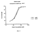
- MCP was initially purified and characterized by Seya and coworkers (J. Exp. Med. 1986, 163:837 ; Biochem. J., 1989, 264:581 ), who showed that it binds C3b and C4b and possesses Factor I cofactor activity. MCP therefore functions to irreversibly inactivate C3b and C4b by proteolytic cleavage to C3bi and C4bi (see Fig. 2). MCP has been shown to bind preferentially to C3b, thus making it a more potent inactivator of alternative pathway convertases ( Seya, T. et al, 1991, Mol. Immunol. 28:1137 ).
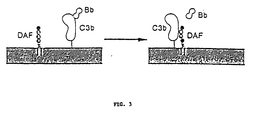
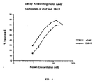
- DAF was first identified by Nicholson-Weller and coworkers (J. Immunol., 1982, 129:184 ) and characterized by Medof and coworkers (J. Exp. Med., 1984, 160:1558 ). DAF also binds to C3b and C4b and functions to dissociate these molecules from the C3 convertase, thus promoting the decay (inactivation) of the convertase (see Fig. 3). DAF similarly inactivates both alternative and classical convertases.
- MCP and DAF are composed of only four SCRs, making them the smallest of the complement regulatory proteins.
- MCP does not possess decay accelerating activity and DAF does not possess cofactor activity. Both proteins are expressed in a variety of cell types, including endothelial cells, fibroblasts, lymphocytes, granulocytes and monocytes ( Hourcade, D. et al, 1989, Adv. Immunol. 45:381 ; McNearny, T.et al, 1989, J. Clin. Invest. 84:538 ). MCP and DAF are considered to function, via different complementary mechanisms, as intrinsic inhibitors of complement activation to prevent complement-mediated autolysis of host cells.
- WO 91/11461 describes multimeric and hetero-multimeric fusion proteins in which C4 binding protein is linked to a functional moiety, such as CD4.
- EP-A-512733 describes altering the specificity of a regulator of complement activation protein to alter the specificity and affinity of the protein for C3b and/or C4b.
- the invention features a soluble chimeric protein comprising a first polypeptide, linked to a second polypeptide by a peptide bond, wherein said first and second polypeptides are different, and are selected from proteins of the regulators of complement activation (RCA) family and soluble fragments of membrane cofactor protein, decay accelerating factor, complement receptor 1, factor H and C4b binding protein and wherein each of said first polypeptide and said second polypeptide inhibits complement activation.
- RCA complement activation
- the first polypeptide is MCP or a soluble, biologically active fragment thereof, e.g., a fragment containing at least regions 2, 3, and 4 of the SCR of MCP
- the second polypeptide is DAF or a soluble, biologically active fragment thereof, e.g, a fragment containing at least regions 2, 3, and 4 of the SCR of DAF.
- fragment as applied to a polypeptide, will ordinarily be at least about 5 contiguous amino acids, typically at least about 10 contiguous amino acids, more typically at least about 20 contiguous amino acids, usually at least about 30 contiguous amino acids, preferably at least about 40 contiguous amino acids, more preferably at least about 50 contiguous amino acids, and most preferably at least about 60 to 80 or more contiguous amino acids in length.
- Such polypeptides can be generated by methods known to those skilled in the art, including proteolytic cleavage of the protein, de novo synthesis of the fragment, or genetic engineering.
- a biologically active fragment is defined as one which exhibits complement inhibitory activity. The activity of a fragment should be at least 1% of, is more preferably at least 10% of, yet more preferably at least 50% of, and is most preferably at least equal to, the biologically activity of the naturally occurring inhibitor of complement activation.
- the soluble chimeric molecules are more effective inhibitors of complement activation than the soluble MCP or DAF proteins, individually or in combination. Furthermore, the soluble chimeric proteins possess extrinsic complement regulatory activity (the ability to inactivate convertases not bound to the same cell membrane). In contrast, the membrane-associated forms of MCP and DAF possess intrinsic activity (the ability to inactivate convertases bound only to the same cell membrane).
- the chimeric proteins can be used as a therapeutic treatment for inflammatory and autoimmune diseases, and monoclonal antibodies produced against the chimeric proteins can be used as diagnostic or therapeutic agents.
- the invention also includes modifications of the chimeric proteins of the invention.
- Modifications include in vivo, or in vitro chemical derivatization of polypeptides, e.g., acetylation, or carboxylation.
- modifications of glycosylation e.g, changing glycosylation patterns, e.g., those made by modifying the glycosylation patterns of a polypeptide during its synthesis and processing or in further processing steps, e.g., by exposing the polypeptide to enzymes which affect glycosylation, e.g., mammalian glycosylating or deglycosylating enzymes.
- the first and second polypeptides of the chimera may be selected from the group consisting of MCP, DAF, complement receptor 1, factor H, C4b binding protein, and soluble biologically active fragments thereof. Because of their C3/C5 convertase-inhibiting activities, any of the complement regulatory proteins or polypeptides of the RCA family could be the first or second polypeptide of the chimera.
- the invention also includes a nucleic acid sequence encoding the chimeric protein in which the first and second polypeptides are linked by a peptide bond and a recombinant expression vector comprising a selectable marker, e.g., glutamine synthetase or dihydrofolate reductase, and a nucleic acid encoding the chimeric protein of the invention operably linked to regulatory sequences for expression of said protein, e.g., a mammalian promoter.
- a selectable marker e.g., glutamine synthetase or dihydrofolate reductase
- the invention also includes a process for preparing the chimeric proteins of the invention by culturing the chimeric protein-encoding expression vector in suitable host cells, e.g., bacterial cells, yeast cells, insect cells, or mammalian cells, under conditions which promote expression of the chimeric protein.
- suitable host cells e.g., bacterial cells, yeast cells, insect cells, or mammalian cells.
- the process is preferably carried out by expressing the chimeric protein in Chinese hamster ovary (CHO) cells.
- the chimeric protein may be prepared by collecting a cell culture supernatant or cell lysate of the host cells; removing acid-precipitable contaminants e.g., contaminants which precipitate below pH 7.0, e.g., contaminants which precipitate upon diluting the supernatant or lysate 1:1 with 25 mM piperazine at a pH 5.0, from the supernatant or lysate; collecting the chimeric protein which binds to an anion exchange resin; removing metal-binding contaminants; binding the chimeric protein to a phenyl hydrophobic interaction resin and then eluting said recombinant protein; binding the chimeric protein to a butyl hydrophobic interaction resin and then eluting said recombinant protein; and removing endotoxin from the chimeric protein.
- the last three steps of the inventive process- may be carried out in any order.
- the invention also includes the soluble chimeric protein for use in a method of inhibiting C3a and C5a generation by contacting a C3 convertase, e.g., the C3b and C4b subunits of the C3 convertase, and a C5 convertase, e.g., the C3b and C4b subunits of the C5 convertase, with the chimeric protein of the invention.
- the binding of the chimeric protein of the invention to the convertases inhibits the enzymatic activity of the convertases, thus inhibiting the generation of C3a and C5a.
- the invention features the soluble chimeric protein for use a method of reducing inflammation characterized by excessive complement activation by administering the chimeric protein of the invention to a patient afflicted with such a condition.
- the invention features an antibody which binds to the chimeric protein of the invention, but does not bind to the first polypeptide or second polypeptide of the chimera alone.
- Fig. 1 is a diagram showing the proteins of the complement system, their pathways of activation, and the proteins regulating their function.
- Fig. 2 is a diagram showing the mechanism of C3 convertase inactivation by MCP.
- Fig. 3 is a diagram showing the mechanism of C3 convertase inactivation by DAF.
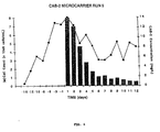
- Fig. 4 is a graph showing the time course of cell density and supernatant concentration of complement activation blocker-2 (CAB-2) protein in a 12-liter microcarrier culture of transfected CHO-K1 cells.
- Culture vessels were seeded with transfected cells at 8 x 10 4 cells/ml in Iscove's modified Dulbecco's medium (IMDM) containing 2.5 g/ml microcarriers and 10% fetal bovine serum. After several days of growth to reach peak cell density, the culture medium was switched to serum-free IMDM. Every day thereafter, 10 liters of culture supernatant was harvested and replaced with fresh serum-free medium.
- IMDM Iscove's modified Dulbecco's medium
- Fig. 5 is a photograph of purified soluble MCP (sMCP) and CAB-2 proteins separated on a polyacrylamide gel. Five ⁇ g aliquots of each purified protein was electrophoresed on a 10% polyacrylamide sodium dodecyl sulfate (SDS) gel. The gel was run under reducing conditions and stained with coomassie blue. Lane 1, molecular weight markers; Lane 2, sMCP; Lane 3, CAB-2. The molecular weight standards are 106, 80, 49.5, 32.5 and 27.5 kDa.
- Fig. 6 is a diagram depicting the seven-step purification process for microcarrier-produced CAB-2 protein.
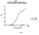
- Fig. 7 is a line graph showing the dual reactivity of CAB-2 protein with anti-MCP and anti-DAF antibodies as measured in an enzyme-linked immunosorbent assay (ELISA).
- the assay used a rabbit anti-MCP polyclonal antibody capture, murine anti-DAF monoclonal secondary, and horseradish peroxidase (HRPO)-conjugated goat anti-mouse IgG tertiary antibodies.
- HRPO horseradish peroxidase
- Fig. 8 is a line graph showing a comparison of sMCP and CAB-2 activity as measured in a cofactor assay. Cleavage of the potassium bromide-treated C3 with C3b-like properties (iC3) alpha chain was quantitated by scanning densitometry based on the relative proportions of the intact chain and its cleavage products in each sample.
- Fig. 9 is a line graph showing a comparison of sDAF and CAB-2 activity as measured in a decay accelerating factor assay.
- the Z values (number of lytic sites/cell) were determined, using a standard table, from the values of percentage maximum lysis of each sample.
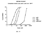
- Fig. 10 is a line graph showing a comparison of the inhibitory activities of sMCP, sDAF, a mixture of sMCP + sDAF, and CAB-2 in an assay of classical pathway dependent complement-mediated cell lysis.
- IgM-sensitized sheep red blood cells (RBC) were the stimulant and human serum (1:200 final dilution) was used as the complement source.
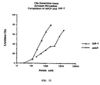
- Fig. 11 is a line graph showing a comparison of the inhibitory activities of sMCP and CAB-2 in an assay of alternative pathway dependent complement-mediated cell lysis.
- Unsensitized guinea pig RBC in a buffer containing EGTA (to chelate Ca +2 ) were the stimulant and human serum (1:4 final dilution) was the source of complement.
- Fig. 12 is a line graph showing the inhibition of C5a production by sMCP and CAB-2.
- Human serum diluted 1:8 was the source of complement and zymosan (1 mg/ml final concentration) was the stimulant for alternative pathway activation.
- C5a was quantitated by competitive radioimmunoassay with 125 I-C5a desArg.
- Fig. 13 is a line graph showing the pharmacokinetics of sMCP and CAB-2 in rats. Animals were injected intravenously with 1 mg/kg dose of purified protein and blood samples drawn at the indicated times post-injection. Plasma levels of sMCP and CAB-2 were determined by ELISA.
- Fig. 14 is an autoradiograph of serum samples recovered from rats injected intravenously (i.v.) with 125 I-labeled CAB-2. Serum samples were obtained from rats at various times after injection and electrophoresed on a 10% polyacrylamide SDS gel. The gel was dried and autoradiographed.
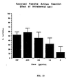
- Fig. 15 is a bar graph showing the in vivo inhibition of the reversed passive Arthus reaction in guinea pigs by CAB-2 protein.
- Animals were injected i.v. with 20 mg/kg ovalbumin and 1 ⁇ Ci 125 I-BSA, then challenged intradermally (i.d.) with 10 mg anti-ovalbumin polyclonal antibody containing the indicated amounts of CAB-2 protein. After 3 h, animals were sacrificed and skin biopsies counted to quantitate leakage of 125 I-BSA.
- CAB Complement Activation Blocker
- genes of the invention are constructed in a manner such that the encoded proteins possess at least two complement binding sites and both Factor I cofactor activity and decay accelerating activity.
- the chimeric molecules are more effective inhibitors of complement activation than the MCP or DAF proteins, individually or in combination.
- the chimeric proteins can be used to treat inflammatory and autoimmune diseases, and monoclonal antibodies produced against the chimeric proteins can be used as diagnostic or therapeutic agents.
- the invention includes recombinant genes which encode chimeric proteins which have both the Factor I cofactor and the decay accelerating factor regulatory activities for complement. By exhibiting both biological activities, the chimeric proteins are more potent in their abilities to inhibit complement activation than either membrane cofactor protein, decay accelerating factor, or both proteins in combination.
- Recombinant materials and methods used to construct and express the genes, methods used for its manufacture in useful quantities, pharmaceutical compositions containing the chimeric recombinant proteins, methods for their use in the treatment of inflammatory and autoimmune diseases are described below.
- Monoclonal antibodies raised against the chimeric complement regulatory proteins, and methods for their production and characterization are also described. Such monoclonal antibodies are useful as reagents for quantitation and monitoring of the chimeric proteins and as diagnostic and therapeutic agents for human diseases.
- MCP and DAF proteins described by Lublin, D. M. et al., 1989, J. Exp. Med. 168:181-194 , and Medof, M. E. et al., 1987, Proc. Natl. Acad. Sci. USA 84:2007-2011 , were used for the construction of expression vectors that direct the synthesis of MCP and DAF fusion proteins.
- the MCP and DAF proteins and/or their biologically active fragments or derivatives may be produced using known recombinant DNA techniques based on the cDNA sequences published by Lublin D. M. et al, supra and Medof, D. E., et al., supra .
- mRNA can be isolated from cells expressing MCP and DAF, cDNA synthesized using random primers, and the specific sequence for the corresponding gene amplified by polymerase chain reaction (PCR) with a pair of primers synthesized according to the published sequences.
- cDNA encoding other proteins which inhibit complement activations can also be isolated in the similar fashion, e.g, C4b-binding protein ( Chung et al., 1985, Biochem J. 230:133-141 ), e.g., factor H ( Ripoche et al., 1988, Biochem. J. 249:593-602 , and EMBL Accession Number Y00716), e.g., CR1 ( Vogelstein et al., 1988, J. Exp. Med. 168:1699 ).
- SCR region 3 and 4 confer the C3b and C4b binding sites of MCP protein ( Adams, et al., 1991, J. Immunol. 147:3005-3011 ).
- SCR 2 is also required in addition to SCR 3 and 4 to retain the membrane cofactor activity.
- the minimal length of cDNA to be used is that which encodes the amino acids corresponding to both SCR 3 and 4 of MCP.
- the SCR 2, 3 and 4 of DAF contribute to the decay accelerating activity ( Coyne, et al., 1992, J. Immunol. 149:2906-2913 ).
- a cDNA segment encoding the extracellular domain containing SCR1-4 and/or serine-threonine-proline rich (ST) regions of MCP and DAF proteins can be used for the production of the fusion protein.
- Genes other than MCP and DAF with C3b and C4b binding activities and membrane cofactor and/or decay accelerating activity can also be used for the construction of expression vectors that direct the synthesis of fusion proteins with membrane cofactor and decay accelerating activities.
- a linker segment of DNA can be added between two genes during the construction of expression vectors. However, insertion of a linker segment between the coding regions of the first and second proteins must maintain a proper reading frame at the 5' and 3' ends to insure continuous protein translation.
- the length of the linker can range from 0 to 1500 amino acids long and is preferably 0-60 amino acids long. As described below, no amino acids have been added at the junction for MCP-DAF construct but two newly added amino acids have been added at the junction for MCP-MCP, MCP-1/2MCP and DAF-MCP constructs. Since deletion of SCR 1 region does not impair the cofactor activity of MCP, the SCR 1 region in DAF-MCP construct which spans about 60 amino acids long, can be considered part of a linker in addition to the two newly added amino acids.
- Amino acids can be substituted from one SCR to another to convert the binding specificity of one SCR or to increase binding affinity to another.
- C3b binding can be converted to C4b binding or to both specificities in CR1 ( Krych et al., 1994, J. Biol. Chem. 269:13273-13278 ). These manipulations can be accomplished using site directed mutagenesis techniques known in the art.
- All members of the regulators of complement gene cluster RCA share basic structural similarity ( Hourcade, Holers and Atkinson, 1989, Adv. Immunol. 45:381-416 ). Specifically, they are composed of several SCRs of about 60 amino acids long with four conserved cysteines. All except MCP and DAF contain more than 4 SCR regions. For example, CR1 has 30 SCR regions. Fusion proteins retaining membrane cofactor activity and decay accelerating activity can be constructed with more than two genes, each with membrane cofactor or decay accelerating activity. The length for each cDNA segment to be used in the construct is discussed above.
- Fusion proteins can be produced with or without glycosylation.
- Most members of the RCA family have sites for glycosylation.
- MCP contains three N-linked and one O-linked glycosylation sites within the SCR and ST regions, respectively.
- DAF contains one N-linked and multiple 0-linked oligosaccharides ( Lublin, et al., 1986, J. Immunol. 137:1629 ).
- production of the protein in an eucaryotic expression system results in the expression of the corresponding protein in glycosylated form.
- non-glycosylated fusion proteins There are three possible ways to produce non-glycosylated fusion proteins: (1) deglycosylation by cleavage of carbohydrate groups enzymes such as Endo-peptide-N-galactosaminidase and O-glycanase to cleave N- and O-linked glycosylations, respectively; (2) procaryotic expression in bacillus and E. Coli to generate non-glycosylated fusion proteins; (3) site-directed mutagenesis to alter the recognition sites for N- or O-linked glycosylation.
- carbohydrate groups enzymes such as Endo-peptide-N-galactosaminidase and O-glycanase to cleave N- and O-linked glycosylations, respectively
- procaryotic expression in bacillus and E. Coli to generate non-glycosylated fusion proteins
- site-directed mutagenesis to alter the recognition sites for N- or O-linked glycosylation.
- Fusion proteins can be produced in procaryotic and eucaryotic systems each using different expression vectors that are appropriate for each host system.
- Chimeric proteins of the invention can be produced in an eucaryotic expression system such as the baculoviral or mammalian systems described below.
- the plasmid, pMSG uses the promoter from mouse mammary tumor virus long terminal repeat (MMTV).
- Suitable host cells for pMSG expression are chinese hamster ovary (CHO) cells, HeLa cells and mouse Lkt negative cells ( Lee, F., et al., 1981 Nature 294:228-232 ).
- the vector, pSVL uses the SV40 late promoter.
- suitable host cells for pSVL expression are Cos cells ( Sprague, J. et al., 1983, J. Virol. 45:773-781 ).
- the vector, pRSV uses Rous Sarcoma Virus promoter.
- Suitable host cells RSV expression are mouse fibroblast cells, lymphoblastoid cells and COS cells ( Gorman, Padmanabhan and Howard, 1983, Science 221:551-553 ).
- Baculovirus expression vectors can also be used. These vectors are stably expressed in insect cells such as sf9 ( Luckow, V. A.and Summers, M. D., 1988, Bio/Technology 6:47-55 ; Miller, L. K., 1988, Ann. Re. Microbiology 42:177-199 ).
- Chimeric proteins of the invention can also be produced in a procaryotic expression system.
- the following are examples of expression vectors which can be expressed in procaryotic expression systems.
- the pOX expression series using the oxygen-dependent promoter can be expressed in E. coli.
- E. coli Khosla, G., et al., 1990, Bio/Technology 8:554-558 .
- pRL vector which uses the strong pL promoter of lambda phage ( Reed, R. R., 1981, Cell 25:713-719 ; Mott, J. D., et al., 1985, Proc. Natl. Acad. Sci. USA 82:88-92 ) and the pKK223-3 vector which uses a hybrid promoter derived from the fusion between the promoters of the tryptophan and lactose operons of E. coli. ( Borsius, J. and Holy, A., 1984, Proc. Natl. Acad. Sci. USA 81:6929-6933 ) can be used for expression in E . coli.
- suitable vectors for yeast expression are also well known in the art, e.g. Sleep, D. Belfield, D. P. and Goodey, A. R., 1990, Bio/Technology 8:42-46 ; Sakai, A. et al., 1991, Bio/Technology 9:1382-1385 ; Kotula, L. and Curtis, P. J., 1991, Bio/Technology 9:1386-1389 .
- the secreted proteins must be identified and verified with regard to their predicted structure.
- Various methods can be used to identify and characterize the expressed chimeric proteins.
- the recombinant cell line can first be incubated with 35 S-methionine to endogenously label its expressed proteins.
- the presence of secreted chimeric proteins can be verified by radioimmunoprecipitation with monoclonal antibodies to one or the other protein of the chimera, e.g., anti-MCP or anti-DAF. Antibodies to both MCP and DAF are commercially available.
- metabolically 35 S-labeled culture supernatants are incubated with either anti-MCP or anti-DAF monoclonal antibodies.
- the immune complexes are precipitated by incubation with Protein A conjugated to Sepharose.
- SDS polyacrylamide gel electrophoresis of the immunoprecipitated proteins, followed by autoradiography, can be used to identify the secreted chimeric proteins. If a chimeric protein is expected to express both MCP and DAF domains, as is the case with CAB-2, one would expect both anti-DAF and anti-MCP antibodies to immunoprecipitate the protein.
- bispecific chimeric proteins composed of both MCP and DAF gene segments e.g., CAB-2
- a double immunoprecipitation using two monoclonal antibodies of different specificities in succession. Pre-clearance of culture supernatant with one antibody would result in a negative immunoprecipitation with the second antibody. This method would verify that a single protein expresses both MCP and DAF epitopes.
- a bispecific chimeric protein e.g, CAB-2
- CAB-2 can be identified by Western blot. For example, after SDS-PAGE and transfer to nitrocellulose, blots can be developed with either anti-MCP or anti-DAF monoclonal antibodies. The expressed bispecific recombinant protein would be reactive with both antibodies, again demonstrating the presence of both MCP and DAF epitopes on the chimera.
- Identification of a bispecific chimeric protein such as CAB-2 can also be accomplished by ELISA.
- a rabbit polyclonal antibody specific for either MCP or DAF can be used to coat plastic microtiter ELISA plates, followed by the addition of culture supernatant from the recombinant cell line expressing CAB-2 and incubation with the capture polyclonal antibody.
- a monoclonal anti-DAF or anti-MCP secondary antibody, the specificity of which is different from the capture antibody, can be subsequently used. A positive reaction would indicate the presence of both epitopes on the chimeric protein.
- An ELISA can also be used to quantitate the levels of CAB-2 in culture supernatants or any other unpurified solutions containing the chimeric protein by comparison to standard curve of known quantities of purified CAB-2 protein. Quantitation of CAB-2 would be useful for determination of production rates in recombinant cell lines, determination of protein concentration in partially purified preparations, and for determination of protein concentration in plasma for in vivo experiments.
- the chimeric CAB-2 protein can be purified from recombinant cell culture supernatant by a variety of standard chromatographic procedures, including but not limited to immunoaffinity chromatography, ion exchange chromatography, gel filtration chromatography, reverse-phase high pressure liquid chromatography (HPLC), lectin affinity chromatography, or chromatofocusing.
- standard chromatographic procedures including but not limited to immunoaffinity chromatography, ion exchange chromatography, gel filtration chromatography, reverse-phase high pressure liquid chromatography (HPLC), lectin affinity chromatography, or chromatofocusing.
- small quantities of culture supernatant containing serum supplement can be purified using immunoaffinity chromatography with e.g., anti-MCP or anti-DAF monoclonal antibodies.
- CAB-2 protein bound to the immobilized antibody can be eluted in purified form by use of a chaotropic solution.
- Recombinant CHO cells can be cultured without high concentrations of serum supplement for production of large quantities (100 liters) of supernatant containing CAB-2 protein.
- a description of a culture method which uses microcarriers in 12-liter vessels is presented in an example below.
- the CAB-2 protein in this serum-free culture supernatant can be purified by the chromatographic procedures detailed in the example below. This procedure results in hundreds of mg of CAB-2 protein which is greater than 90% pure.
- CAB-2 protein Once the CAB-2 protein is purified, its amino acid sequence can be deduced by direct protein sequence analysis using an automated system. The presence of N- and O-linked carbohydrates can be determined by use of specific endoglycosidase enzymes ( Chavira, R. et al, 1984, Anal. Biochem. 136:446 ). Further characterizations of its biochemical structure can also be performed, including but not limited to pI determination by isoelectric focusing, hydrophilicity analysis, X-ray crystallographic analysis, and computer modeling.
- the important characteristic for the chimeric recombinant proteins is their ability to function both as a cofactor for Factor I and as a decay accelerating factor.
- In vitro assays can be performed to measure these biological activities ( Medof, M. et al, 1984, J. Exp. Med. 160:1558 ; Masaki, T. et al, 1992, J. Biochem 111:573 ).
- assays for cofactor activity using purified C3 and factor I
- for decay accelerating activity using IgM, C1 and C4-sensitized sheep RBC and purified C2 are used to demonstrate both these complement regulatory functions for the CAB-2 chimeric protein.
- Monoclonal antibodies can be generated to purified chimeric proteins by standard procedures. Mice are immunized with the proteins mixed with a suitable adjuvant. Spleen cells are fused with a myeloma cell line using polyethylene glycol, and the hybridomas are selected with medium containing hypoxanthine and aminopterin. Hybridomas secreting the desired antibodies can be screened by ELISA and cloned. Specificities of the monoclonal antibodies can be determined by the use of different protein or peptide antigens in the ELISA. Useful quantities of antibodies can be produced by either the generation of ascites fluid in mice or by large scale in vitro culture of the cloned antibody-producing hybridoma cell line. Antibodies can be purified by various chromatographic procedures known in the art, such as affinity chromatography on either immobilized Protein A or Protein G.
- the Arthus reaction is an inflammatory response caused by the interaction of antigen in tissue with circulating antibody. It has been used as a classic example of a localized in vivo inflammatory response, and is characterized by the formation of immune complexes, complement activation, inflammatory cell recruitment, edema and tissue damage ( P. Bailey & A. Sturm, 1983, Biochem. Pharm 32:475 ). Experimentally, a reversed passive Arthus reaction can be established in an animal model by i.v. injection with antigen and subsequent challenge with antibody. Using guinea pigs as an animal model, the in vivo therapeutic efficacy of chimeric proteins of the invention can be evaluated (see example below).
- Additional animal models with relevance to various clinical human diseases can also be used to test the in vivo efficacy of complement activation blockers. These include, but are not limited to: myocardial ischemia/reperfusion injury (acute myocardial infarction; H.F. Weisman et al., 1990, Science 249:146 ); cerebral ischemic injury (stroke; L. Chang et al, 1992, J. Cerebr. Blood Flow Metab. 12:1030 ); lung injury (ARDS; S. Hosea et al, 1980, J. Clin. Invest. 66:375 ); xenograft rejection (transplants; J. Leventhal et al, 1993, Transplantation 55:857 ); burn injury ( F.
- the chimeric proteins of the invention can be combined with an appropriate pharmaceutical formulation and administered by a variety of routes, including, but not limited to, intravenous bolus injection, intravenous infusion, intraperitoneal, intradermal, intramuscular, subcutaneous, intranasal, and oral routes.
- routes including, but not limited to, intravenous bolus injection, intravenous infusion, intraperitoneal, intradermal, intramuscular, subcutaneous, intranasal, and oral routes.
- the administration of CAB-2 in vivo will enable the protein to bind endogenous C3/C5 convertases and inhibit the generation of additional C3b and C5b, of C3a and C5a anaphylatoxins, and of C5b-9 lytic complexes.
- CAB-2 can therefore function to inhibit in vivo complement activation and the inflammatory sequelae that accompany it, such as neutrophil recruitment and activation, autolysis of host cells, and edema.
- CAB-2 can be used for the therapy of diseases or conditions that are mediated by inordinate and/or excessive activation of the complement system. These include, but are not limited to: tissue damage due to ischemia-reperfusion following myocardial infarction, aneurysm, stroke, hemorrhagic shock, or crush injury; burns; endotoxemia and septic shock; adult respiratory distress syndrome (ARDS); hyperacute rejection of grafts; cardiopulmonary bypass and pancreatitis.
- Autoimmune disorders including, but not limited to, systemic lupus erythematosis, rheumatoid arthritis, and multiple sclerosis, can also be treated with the chimeric proteins of the invention (also see Table 1).
- TABLE 1 Potential Clinical Targets of Protein Chimeras of the Invention Alternative Pathway Classical Pathway Reperfusion injury Autoimmune diseases Cerebral infarction (stroke) Systemic lupus erythematosus Acute myocardial infarction Rheumatoid arthritis Hypovolemic shock Glomerulonephritis Multiple organ failure Hemolytic anemia Crush injury Myasthenia gravis Intestinal ischemia Multiple sclerosis Vasculitis Inflammatory bowel diseases Bullous diseases Urticaria Paroxysmal nocturnal hemoglobinuria Cryoglobulinemia Inflammatory disorders Inflammatory disorders Inflammatory disorders Inflammatory disorders Inner respiratory distress syndrome Septic shock & endotoxemia Thermal injury (burn & frostbite)
- chimeric proteins of the invention can be formulated in a pharmaceutical excipient in the range of approximately 10 ⁇ g/kg and 10 mg/kg body weight for in vivo or ex vivo treatment.
- EXAMPLE 1 Cloning and expression of a recombinant gene encoding a complement receptor fusion protein
- Described herein is the construction of various expression vectors that directed the expression of complement receptor fusion proteins, e.g., MCP-MCP, MCP-1/2MCP, MCP-DAF and DAF-MCP.
- complement receptor fusion proteins e.g., MCP-MCP, MCP-1/2MCP, MCP-DAF and DAF-MCP.
- pG3N pGEM-3fz(-) plasmid
- pII SK pBluescript II SK(+)
- COS-7 (ATCC No. CRL1651) was routinely maintained in Dulbecco's modified Eagle medium (DMEM) (GIBCO) supplemented with 10 mM glutamine, 20 mM HEPES, 50 ⁇ g/ml streptomycin, 50 units/ml penicillin, and 10% fetal bovine serum.
- DMEM Dulbecco's modified Eagle medium
- CHO-R1 (ATCC No. CCL61) was cultured in Ham's nutrient mixture F-12 (Sigma, St. Louis, MO) containing 10% fetal bovine serum, 50 ⁇ g/ml streptomycin and 50 units/ml penicillin.
- MCP-pG3N was used as the backbone for the construction of MCP-DAF, MCP-MCP and MCP-1/2MCP expression vectors.
- the construction of the MCP-pG3N plasmid was accomplished by the insertion of cDNA segment encoding amino acid sequence corresponding to the extracellular domain of MCP into pG3N plasmid.
- the cDNA encoding the extracellular domain of MCP was obtained by PCR.
- PCR reaction was carried out under conditions described in S. J. Scharf. 1990, PCR protocol: A guide to methods and applications, ed. M. A. Innis, D. H. Gelfrand, J. J. Sninsky and J. J. White, New York, New York, pp. 84-91 .
- the reaction mixture contained 50 mM KCl, 10 mM Tris-HCl pH 8.3, 1.5 mM MgCl 2 , 0.2 mM of all four deoxyribonucleotides, 1 ng of linearized template, 45 pmole of each of the paired primers, and 5 units of Taq polymerase.
- the amplification reaction was run for 30 cycles with each cycle consisting of an incubation at 94°C for 1 minute, 52°C for 1 minute and 72°C for 1 minute. At the end of the last cycle, a further incubation at 72°C for an additional 15 minutes was allowed in order to complete the amplification reaction.
- a plasmid containing cDNA sequence encoding amino acid sequence corresponding to the MCP protein was used as template in the PCR reaction described below.
- MCP-N1 primer 5' -- GGA ATT CGC ATG GAG CCT CCC GGC --3' (SEQUENCE ID NO:1) was used as the forward primer
- MCP-STB primer 5' -- CTA TGA GGC ACT GGA CGC TGG AGA TTT -- 3' (SEQUENCE ID NO:2) was used as the reverse primer.
- EcoRI restriction site added to the 5' end of the MCP-N1 primer and a termination codon added to the 5' end of the MCP-STB primer for the termination of protein translation.
- MCP1 cDNA sequence generated by this PCR reaction, MCP1 fragment, spans nucleotide nos. 41 to 943 (D. M. Lublin et al., supra ).
- MCP1 encodes amino acid sequence corresponding to the extracellular domain of the MCP protein. It contains regions for signal peptide (SP), short consensus repeat SCR 1-4 and portion of the serine and threonine rich region (ST).
- SP signal peptide
- SCR 1-4 short consensus repeat SCR 1-4
- ST serine and threonine rich region
- MCP1 fragment was cleaved with EcoRI and SalI restriction endonucleases, gel purified and subcloned into pG3N plasmid that was treated with the same set of restriction enzymes.
- the treatment of MCP1 fragment with SalI resulted in the cleavage of all of its ST coding sequence other than that encoding the first three amino acids.
- PCR was used to synthesize a cDNA segment, DAF2 fragment, encoding amino acid sequence corresponding to the extracellular domain of DAF protein for the construction of MCP-DAF pG3N plasmid.
- a plasmid containing cDNA (M.E. Medof et al., supra. ) encoding amino acid sequence corresponding to the DAF protein was used in the PCR reaction described below.
- the primers, DAF-N5, 5' -- CCT CTA GAG TCG ACT GAC TGT GGC CTT CCC CCA GAT GTA --3' (SEQUENCE ID NO:3) and DAF-C5, 5' -- TCT AGA GCA TGC GAA TTC TCA ACG GGT AGT ACC TGA AGT GGT TCC -- 3' (SEQUENCE ID NO:4) were used as the forward and the reverse primers respectively.
- a stop codon for protein translation was added to the 5' end of the DAF-C5 primer. After the stop codon, two restriction sites, EcoRI and SphI, were also introduced.
- DAF2 fragment is bracketed by SalI at 5' end and a EcoRI-SphI duet at 3' end.
- DAF2 fragment defines a region spanning nucleotide nos. 156 to 1124 (M. E. Medof et al., supra ) that encodes an amino acid sequence corresponding to the extracellular domain of the mature DAF protein containing SCR 1-4 and ST regions.
- MCP-DAF pG3N plasmid The construction of MCP-DAF pG3N plasmid was achieved by the ligation of DAF2 fragment and MCP-pG3N plasmid after both DNAs were cleaved with SalI and SphI restriction enzymes.
- the SalI site was the in-frame joint site between MCP and DAF DNA segment, thus ensuring a continuous protein translation from MCP into DAF.
- MCP-DAF cDNA segment was retrieved from MCP-DAF pG3N by the treatment with EcoRI restriction enzyme and gel purification. This cDNA fragment was subsequently cloned into the EcoRI site of the pEE14 which contains the glutamine synthetase gene as a selectable marker ( M.I. Cockett et al., 1990, Bio/Technology 8:662-667 ) and the p91203(B) vector which contains the dihydrofolate reductase gene as a selectable marker ( G.G. Wong et al., 1985, Science 228:810-815 ) vectors.
- MCP-DAF P91203(B) expression vector utilizes adenovirus major late promoter to direct the expression of MCP-DAF protein and is suitable for transient expression in COS-7 and permanent expression in CHO.
- Expression of MCP-DAF protein in the MCP-DAF pEE14 expression vector is under the control of cytomegalovirus promoter. This vector is suitable for the establishment of a permanent CHO cell line in which the expression of MCP-DAF can be amplified in the presence of high concentration of methionine sulfoximine (M.I. Cockett et al., supra )
- MCP-DAF expression vectors can direct the expression of a fusion protein with the MCP sequence at the amino-terminus and the DAF sequence at the carboxyl-terminus.
- the MCP portion of the fusion protein spans amino acid nos. 1 to 254 of the native mature MCP protein after the signal peptide of 34 amino acids is cleaved upon expression. It contains four SCR (1-4) regions and the first three amino acids of the ST region.
- the DAF portion encompasses the extracellular domain of the mature native DAF protein that ends right after the ST region (4 SCR and 1 ST regions; from amino acid nos. 1 to 324).
- the MCP-DAF protein is 578 amino acid long with a predicted molecular weight of about 70 KDa.
- CAB-2 The recombinant chimeric protein encoded by the MCP-DAF expression vector is referred to as CAB-2.
- MCP-MCP and MCP-1/2MCP expression vectors were similar to that for MCP-DAF expression vectors with some exceptions described as follows.
- a MCP2 fragment was synthesized by PCR technique using MCP-N4 primer, 5' -- TCG ACC TGC AGG TGT GAG GAG CCA CCA ACA TTT -- 3' (SEQUENCE ID N0:5) as the forward primer and MCP-UC1 primer, 5' -- GCG AAT TCC TCA CAA ACT GTC AAG TAT TCC TTC CTC -- 3' (SEQUENCE ID NO:6) as the reverse primer.
- MCP2 fragment was framed by a PstI site at the 5' end and an EcoRI site at the 3' end.
- MCP2 defines a region (nucleotide nos. 146-1024) that encodes an amino sequence corresponding to the extracellular domain of the mature MCP protein truncated right before the transmembrane region (amino acid nos. 1 to 293).
- the MCP-pG3N plasmid was cleaved with HindIII, end-filled with Klenow fragment, digested with PstI and finally ligated with MCP2 fragment which had been cleaved with PstI restriction enzyme.
- MCP-MCP cDNA fragment was retrieved by EcoRI digest, gel purified, and then subcloned into the EcoRI site of the expression vectors to complete the construction of MCP-MCP expression vectors.
- These expression vectors direct the expression of a MCP-MCP fusion protein of 547 amino acid long with a predicted molecular weight of about 70 kDa.
- the actual molecular weight for MCP-MCP protein is higher as it contains six N-linked and multiple O-linked gylcosylation sites. There were two new amino acids, cysteine and arginine, added at the junction of the two MCP fragments.
- MCP-1/2MCP expression vectors were identical to that for MCP-MCP expression vectors except that a different forward primer, MCP-N5, 5'-TCG ACC TGC AGG AAG GTT TTG TGT ACA CCA CCT --3'(SEQUENCE ID NO:7), was used for the synthesis of 1/2MCP DNA segment.
- 1/2MCP fragment spans nucleotide nos. 518 to 1024 and encodes amino acid sequence defining the SCR3, SCR4 and ST regions(amino acid nos. 124-293) of the MCP protein.
- MCP-1/2MCP expression vectors direct the expression of MCP-1/2MCP protein of 385 amino acids long.
- MCP-1/2MCP protein has four N-linked and multiple O-linked glycosylation sites, thus its actual molecular weight is higher than the predicted 46 kDa.
- DAF1 A DAF DNA fragment, DAF1, was synthesized by PCR technique for the construction of DAF-pII SK plasmid that was used for the engineering of recombinant DAF-MCP expression vectors.
- the following primers were used: forward primer, DAF-N2, 5' -- CGG AAT TCC ATG ACC GTC GCG CGG CCG AGC GTG -- 3' (SEQUENCE ID NO:8) and a reverse primer, DAF-C2R, 5' -- ACC TGC AGG TTT TCC TCT GCA TTC AGG TGG T -- 3'(SEQUENCE ID NO:9).
- Restriction sites, EcoRI and PstI were introduced to the 5' ends of DAF-N2 and DAF-C2R primers, respectively.
- DAF1 encompasses from nucleotide nos. 52 to 911 of the DAF gene and encodes the amino acid sequence corresponding to the extracellular domain of the mature DAF protein truncated right before the ST region.
- DAF-MCP pII SK plasmid was constructed by the ligation of PstI- and SmaI-treated DAF-pII SK vector and a MCP3 fragment that was treated with PstI.
- MCP3 fragment was the PCR product using a forward primer, MCP-4.2, 5' -- TCG ACC TGC AGA GGA GCC ACC AAC ATT TGA AGC T -- 3' (SEQUENCE ID NO:10), with a PstI at the 5' end, and a reverse primer, MCP-UC1, described earlier.
- DAF-MCP cDNA fragment was retrieved from DAF-MCP pII SK plasmid after cleavage with EcoRI, gel purified, and then subcloned into the EcoRI site of pEE14 and P91023(B) (Wong et al., supra ) expression vectors.
- DAF-MCP expression vectors direct the synthesis of a DAF-MCP protein of 546 amino acid long. There are two exogenous amino acids, serine and threonine introduced at the junction between DAF and MCP due to vector construction. This protein has a predicted molecular weight of about 66 kDa exclusive of the added weight contributed by four N-linked (one within DAF and three within MCP) and multiple 0-linked (located at ST region of MCP) glycosylations.
- COS-7 cells were transfectiond using lipofectin (GIBCO, Gaithersburg, MD) following the protocol recommended by the manufacturer. Briefly, the day before transfection, COS-7 cells were subcultured in new dishes (60mm) at a cell density which would give about 70 to 80% confluency the following day. Just before transfection, the COS-7 cells were washed twice with opti-MEM medium (GIBCO) without serum. Three ml of opti-MEM medium containing 5 to 10 ⁇ g of expression vector and 20 ⁇ g of lipofectin was added to the washed COS-7 monolayer. Tranfection was allowed to take place for 6 hours at 37°C in a CO 2 incubator.
- opti-MEM medium containing 5 to 10 ⁇ g of expression vector and 20 ⁇ g of lipofectin was added to the washed COS-7 monolayer. Tranfection was allowed to take place for 6 hours at 37°C in a CO 2 incubator.
- the transfection solution was then removed and replaced with fresh opti-MEM supplemented with 10% fetal bovine serum, 50 ⁇ g/ml streptomycin and 50 U/ml penicillin.
- the transfected COS-7 cells were then incubated without disturbance at 37°C for an additional three days.
- the culture supernatant was collected and briefly spun to remove dead cells before conducting evaluation of biological activity.
- CHO-K1 (ATCC No. CCL61) was used to establish permanent cell line expressing MCP-DAF, MCP-MCP, MCP-1/2MCP and DAF-MCP. Briefly, CHO-K1 cells were transfected with expression vectors using lipofectin as described above except that cell confluency was at about 20% before transfection and transfectants were maintained in GMEM-S medium (Glasgow MEN, without L-glutamine and tryptose phosphate broth, supplemented with 10% dialyzed fetal bovine serum, non-essential amino acids, 1 mM sodium pyruvate, 60 ⁇ g/ml L-glutamate, 60 ⁇ g/ml L-asparagine, 7 ⁇ g/ml of each of the nucleosides, 50 ⁇ g/ml streptomycin and 50 units/ml penicillin).
- GMEM-S medium Gibsgow MEN, without L-glutamine and tryptose phosphate
- Transfectants expressing the novel proteins were isolated and subcultured in GMEM-S medium with increasing concentration of methionine sulfoximine.
- CHO-K1 clones expressing the protein chimeras of the invention were then isolated by two rounds of limiting dilution cloning.
- Culture supernatants containing the product expression vector derived from the CHO-K1 transfectants, i.e., the CAB-2 protein, were used for purification by the methods described below.
- Example 2 Microcarrier culture for large scale production of CAB-2
- a microcarrier culture method was developed with the CHO-K1 transfectant cell line expressing the recombinant CAB-2 protein.
- DMEM Dulbecco's modified Eagle's medium
- FBS fetal bovine serum
- FBS fetal bovine serum
- Microcarrier cultures were prepared using 12-liter Nalgene vessels outfitted with Bellco overhead drive impeller assemblies, overlay gas addition ports, and sparge tubes.
- Cultisphere-G microcarriers were employed at 2.5 g/l, and washed three times with PBS and once with serum-free IMDM prior to use.
- Cells from twenty T150 flasks were washed with phosphate buffered saline (PBS), detached from the plates by trypsinization, and seeded into the 12-liter vessels at approximately 10 x 10 4 cells/ml.
- the medium used was IMDM with 10% bovine calf serum and penicillin-streptomycin (growth medium).
- Spinner vessels were fed every 2-3 days with growth medium by allowing the microcarriers to settle, removing 10 liters of medium, and replacing with 10 liters of fresh growth medium.
- Cell densities were monitored by counting nuclei in a sample of the cell culture, a process which involved staining microcarriers with a solution of 0.1 M citric acid, 0.1% crystal violet, followed by incubation for one hour at 37°C and enumeration using a hemocytometer. Values for pH, pCO 2 , and pO 2 were monitored with a Corning model 170 blood gas analyzer. Oxygen concentration was maintained using a 40% oxygen, 5% CO 2 , balance N 2 gas mixture at a rate of 10-50 ml/min through a glass sparger.
- Fig. 4 Results from a typical large-scale culture are presented in Fig. 4.
- the cell density was found to increase from 8 x 10 4 cells/ml to a density of 8 x 10 6 cells/ml in 15 days. Following the initiation of production phase, the cell counts dropped steadily over 12 days.
- Fig. 4 also showns the concentration of CAB-2 protein in each culture harvest.
- Conditioned medium was filtered using a Pelicon system (Millipore, Bedford, MA) using a 0.45 micron filter cartridge according to instructions supplied by the manufacturer. The medium was then concentrated 20-30 fold using the Pelicon ultrafiltration system with a 10,000 molecular weight cutoff membrane. Medium concentrates were stored at-70°C until used.
- CAB-2 protein Small scale purification of the CAB-2 protein was achieved by immunoaffinity chromatography using an anti-MCP monoclonal antibody, e.g., GB24. Antibody was immobilized via the carbohydrate moiety using Carbo-link Sepharose (Pierce, Cat. #20392G). Supernatants from cultures of the CAB-2-producing recombinant CHO line were passed through a 35 ml column of GB24-Sepharose resin. The column was washed with PBS to remove any unbound proteins. CAB-2 was eluted from the column with 0.1 M glycine pH 2.5. Fractions were collected and immediately neutralized with 1/10 volume of 1 M Tris buffer, pH 8.5.
- Concentrated conditioned media was diluted 1:1 with Q Sepharose equilibration buffer (25 mM piperazine pH 5.0) and the pH was adjusted to 5.0. A fine precipitate was formed and was filtered through a precipitate-adsorbing agent (Celite 621, Aldrich) using a Buchner funnel.
- Q Sepharose equilibration buffer 25 mM piperazine pH 5.0
- a fine precipitate was formed and was filtered through a precipitate-adsorbing agent (Celite 621, Aldrich) using a Buchner funnel.
- Chelate Sepharose FF (Pharmacia) was packed to a bed volume of 50 ml (2.2 x 13 cm). The resin was depyrogenated with 0.5 N NaOH and washed with dH 2 O. The resin was charged with 100 ml 0.3 M ZnCl 2 , washed with dH 2 O and equilibrated with 25 mM MES pH 6.0 containing 0.2 M NaCl. The Q Sepharose pool (the material eluted from the Q Sepharose column which contained the CAB-2 protein) was brought to 25 mM MES pH 6.0 and loaded at 20 mg TP per ml of resin onto the Chelate column at a rate of 150 cm/hr. The column was washed with equilibration buffer, and CAB-2 was collected in the flowthrough.
- Q Sepharose pool the material eluted from the Q Sepharose column which contained the CAB-2 protein
- a 110 ml column (3.2 x 13.7 cm) containing TosoHaas Butyl 650M HIC resin was depyrogenated with 0.5 N NaOH and equilibrated with 25 mM phosphate pH 7.0 containing 3 M NaCl.
- the elution pool from Step 4 was adjusted to 3 M NaCl and pH 7.0 and then loaded onto the column.
- the column was washed with equilibration buffer and the CAB-2 then eluted with 25 mM phosphate pH 7.0 containing 1 M NaCl.
- the elution pool from Step 5 was concentrated 5-6 fold using a Mini-ultrasette tangential flow system from Filtron containing a 10 kDa MWCO Omega membrane. The pool was then diafiltered with 4-5 sample volumes of PBS.
- Table 3 - CAB-2 Purification Run 062194 (started with 250 mg CAB-2 in conditioned media) Step Percent Purity mg CAB-2/mg TP (x100) Percent Overall Recovery* Endotoxin Level EU per mg CAB-2 Starting Media 1 - - 1-contaminant precipitation.
- the sample was further depyrogenated by eluting twice through a Pierce endotoxin affinity resin.
- the 5 ml column (1 x 6 cm) was first stripped of endotoxin with 1% deoxycholate, then equilibrated with endotoxin-free PBS.
- the CAB-2 was passed through the column in sterile PBS.
- the CAB-2 was concentrated with an Amicon Centriprep 10 (previously depyrogenated with 70% alcohol). The final endotoxin concentration was 0.045 EU per mg CAB-2.
- An ELISA assay was developed using antibodies directed against membrane cofactor protein (MCP) and decay accelerating factor (DAF).
- MCP membrane cofactor protein
- DAF decay accelerating factor
- a rabbit anti-MCP polyclonal antiserum was generated by immunization of rabbits with soluble MCP (sMCP).
- IgG was purified from the antiserum by immobilized Protein A affinity chromatography.
- An aliquot (50 ⁇ l) of polyclonal anti-MCP IgG at 2 ⁇ g/ml was added to 96-well ELISA plates, and the plates were incubated overnight at 4°C.
- sMCP or CAB-2 proteins were added at various concentrations in blocking buffer and incubated for 1 h at 37°C.
- a murine anti-DAF monoclonal antibody (BRIC 216, Harlan Bioproducts Cat. #MCA914) was added at 1 ⁇ g/ml and incubated for 1 h at 37°C.
- An HRPO-conjugated goat anti-mouse IgG tertiary antibody was added at 1:1000 dilution and incubated for 1 h at 37°C.
- An enzyme substrate (TMB, Pierce Chemical Co., Rockford, IL, Cat. #34021) was added and the reaction stopped with 2 M H 2 SO 4 .
- OD 450 values were determined on an ELISA plate reader. Data shown in Fig. 7 demonstrate that the CAB-2 protein was detected by the two antibody sandwich ELISA, indicating that the protein expresses both MCP and DAF domains. Soluble MCP, on the other hand, was not detected by this combination of antibodies.
- the measurement of decay accelerating activity of the chimeric CAB-2 protein was carried out as follows.
- Commercially available sheep RBC sensitized with IgM, C1 and C4 (EAC14, Diamedix Cat #789-053) were used as a source of cell membrane-deposited C4b.
- the RBC were diluted to 2.5 x 10 8 /ml in buffer (2.5 mM veronal, 75 mM NaCl, 0.15 mM CaCl 2 , 1 mM MgCl 2 , 0.1% gelatin, 2.5% dextrose) and preincubated at 30°C for 5 min.
- the samples were allowed to decay for 15 min at 30°C, after which the lytic sites were developed by adding 0.5 ml of guinea pig complement (1:50 diluted serum) in 40 mM EDTA buffer (2.5 mM veronal, 75 mM NaCl, 0.1% gelatin, 40 mM EDTA). After incubation for 30-40 min at 37°C, the cells were centrifuged and the OD 405 of the supernatants determined. The number of lytic sites/cell (Z) were determined from standard tables and the decay accelerating activity determined by the decrease of the Z values in comparison to a positive control (sample without DAF or CAB-2). Results, shown in Fig. 9, demonstrate that the CAB-2 protein possesses decay accelerating activity comparable to that of sDAF.
- chimeric CAB-2 protein to inhibit complement-mediated cell lysis via the classical pathway was determined by a hemolytic assay using IgM-sensitized sheep RBC.
- IgM-sensitized sheep RBC Commercially available IgM-sensitized sheep RBC (Diamedix, Cat. #789-001) in 100 ⁇ l aliquots were added to 50 ⁇ l of either purified sMCP, purified sDAF or CAB-2 protein at varying concentrations in gelatin veronal buffer (GVB +2 ) (Sigma, Cat. #G-6514).
- Human serum (50 ⁇ l) diluted 1:50 in GVB +2 was immediately added as the source of complement.
- CAB-2 which possesses both cofactor and decay accelerating activities, inhibited cell lysis (IC 50 of 30 nM) with a potency significantly greater than either sMCP alone (230-fold), sDAF alone (3-fold), or both factors in combination (6-fold).
- Complement-mediated cell lysis via the alternative pathway was determined by the use of unsensitized guinea pig RBC, which induce alternative pathway complement activation.
- Guinea pig blood was collected in a heparinized syringe and washed twice with PBS.
- RBC were resuspended at 2 x 10 8 /ml in assay buffer (GVB +2 containing 8 mM EGTA and 1.5 mM additional MgCl 2 ).
- Cells (100 ⁇ l) were mixed with 50 ⁇ l volumes of purified sMCP, sDAF or CAB-2 at various concentrations in assay buffer. Undiluted human serum (50 ⁇ l) was added as a source of complement.
- the complement inhibitory activity of CAB-2 was also tested by specifically assaying the inhibition of C5 convertase via the production of C5a.
- Human serum diluted 1:8 in GVB +2 , was used as the source of complement.
- Purified sMCP or CAB-2 protein were added to the serum at various concentrations.
- the classical pathway was initiated by the addition of heat aggregated rabbit IgG at 100 ⁇ g/ml. After incubation for 1 h at 37°C, the reaction was stopped by addition of 10 mM EDTA. Detection of C5a in the reaction was performed by competition radioimmunoassay using a commercially available C5desarg kit (Amersham, Cat #RPA520). Results for zymosan-induced C5a generation are shown in Fig. 12. CAB-2 more potently inhibits C5a production via the alternative pathway than does sMCP (IC 50 300 nM and 5000 nM respectively).
- the pharmacokinetic behavior of purified CAB-2 protein was determined in rats and compared to that of sMCP.
- the structural integrity of the CAB-2 protein after injection in vivo was determined by using 125 I-labeled CAB-2. After i.v. injection of 125 I-CAB-2, aliquots of serum were obtained from the rats at various times. The serum samples were electrophoresed on a 10% polyacrylamide SDS gel and autoradiographed. The CAB-2 protein showed no detectable degradation after 6 hours in vivo (Fig. 14).
- Example 8 In vivo efficacy of CAB-2 in the reversed passive Arthus model
- mice Male guinea pigs (300-350 g) were anesthetized by i.p. injection of 40 mg/kg of sodium pentobarbital. Animals were injected i.v. with a dose of 20 mg/kg of ovalbumin along with 1 ⁇ Ci of 125 I-labeled BSA. Animals were then immediately challenged by i.d. injection of 10 mg polyclonal anti-ovalbumin antibody, either alone or mixed with various doses of CAB-2, in the dorsal region. The total volume injected i.d. was 100 ml. After three hours, animals were sacrificed by CO 2 inhalation. The skin was removed, the antibody challenge sites isolated by a 5/8 inch biopsy punch, and the biopsies counted.
- Inhibition of the inflammatory response was measured by comparing the leakage of 125 I-BSA into the skin (CPM per challenge site) of sites co-injected with CAB-2 versus sites without CAB-2.
- Another set of animals were pretreated by i.p. injection of 200 U/kg cobra venom factor (CVF) 24 hours before initiation of the Arthus response.
- CVF cobra venom factor
- This treatment results in de-complementation of the animal, and is a positive control for the effect of complement inhibition in this model.
- injection of CAB-2 inhibited the reversed passive Arthus reaction in a dose-dependent manner. The maximum inhibition observed was comparable to that of CVF pretreatment.
- Preferred analogs include peptides whose sequences differ from the wild-type sequence (i.e., the sequence of the homologous portion of the naturally occurring peptide) only by conservative amino acid substitutions, preferably by only one, two, or three, substitutions, for example, substitution of one amino acid for another with similar characteristics (e.g., valine for glycine, arginine for lysine, etc.) or by one or more non-conservative amino acid substitutions, deletions, or insertions which do not abolish the polypeptide's biological activity.
- Table 2 lists a number of conservative amino acid substitutions.
Abstract
Description
- This invention relates to complement inhibition and inflammation.
- The complement system includes a group of proteins in blood plasma which plays an integral role in immune and allergic reactions. Activation of complement can occur via at least two pathways: the classical pathway involving antigen-antibody complexes, and the alternative pathway involving cell wall polysaccharides of yeast and bacterial microorganisms. Regardless of which initiation pathway is used, the end result is the formation of activated fragments of complement proteins (e.g. C3a, C4a, and C5a anaphylatoxins and C5b-9 membrane attack complexes) which mediate several functions including leukocyte chemotaxis, activation of macrophages, vascular permeability and cellular lysis (Frank, M. and Fries, L. Complement. In Paul, W. (ed.) Fundamental Immunology, Raven Press, 1989).
- Several regulatory proteins of the complement system have been identified (Fig. 1). Their primary functions are to regulate the activity of C3/C5 convertases for prevention of excessive complement activation and autolytic destruction of host tissues. These complement regulators are either soluble plasma proteins or integral membrane proteins expressed on a variety of cell types. The former include C4b binding protein (C4bp) and Factor H. The latter include the C3b/C4b receptor (
Complement receptor 1, CR1, CD35), membrane cofactor protein (MCP, CD46), and decay accelerating factor (DAF, CD55). These proteins possess many structural similarities. Each is composed of multiple short consensus repeats (SCRs) of approximately 60 amino acids in length having conserved cysteine, glycine and proline residues. The genes encoding these proteins have been localized tochromosome 1 and are collectively known as the regulators of complement activation (RCA) gene cluster (Hourcade, D. et al, 1989, Adv. Immunol. 45:381). In addition to its role in regulating complement activation, erythrocyte CR1 also functions as a receptor for circulating immune complexes to promote their clearance from plasma (Cornacoff, J. et al, 1983, J. Clin. Invest. 71:236). - MCP and DAF are important regulatory proteins of the complement system which function to prevent autolytic destruction of host tissues by complement activation.
- MCP was initially purified and characterized by Seya and coworkers (J. Exp. Med. 1986, 163:837; Biochem. J., 1989, 264:581), who showed that it binds C3b and C4b and possesses Factor I cofactor activity. MCP therefore functions to irreversibly inactivate C3b and C4b by proteolytic cleavage to C3bi and C4bi (see Fig. 2). MCP has been shown to bind preferentially to C3b, thus making it a more potent inactivator of alternative pathway convertases (Seya, T. et al, 1991, Mol. Immunol. 28:1137).
- DAF was first identified by Nicholson-Weller and coworkers (J. Immunol., 1982, 129:184) and characterized by Medof and coworkers (J. Exp. Med., 1984, 160:1558). DAF also binds to C3b and C4b and functions to dissociate these molecules from the C3 convertase, thus promoting the decay (inactivation) of the convertase (see Fig. 3). DAF similarly inactivates both alternative and classical convertases.
- MCP and DAF are composed of only four SCRs, making them the smallest of the complement regulatory proteins.
- MCP does not possess decay accelerating activity and DAF does not possess cofactor activity. Both proteins are expressed in a variety of cell types, including endothelial cells, fibroblasts, lymphocytes, granulocytes and monocytes (Hourcade, D. et al, 1989, Adv. Immunol. 45:381; McNearny, T.et al, 1989, J. Clin. Invest. 84:538). MCP and DAF are considered to function, via different complementary mechanisms, as intrinsic inhibitors of complement activation to prevent complement-mediated autolysis of host cells.
-
WO 91/11461 EP-A-512733 - The invention features a soluble chimeric protein comprising a first polypeptide, linked to a second polypeptide by a peptide bond, wherein said first and second polypeptides are different, and are selected from proteins of the regulators of complement activation (RCA) family and soluble fragments of membrane cofactor protein, decay accelerating factor,
complement receptor 1, factor H and C4b binding protein and wherein each of said first polypeptide and said second polypeptide inhibits complement activation. - In a preferred embodiment, the first polypeptide is MCP or a soluble, biologically active fragment thereof, e.g., a fragment containing at least
regions regions - The soluble chimeric molecules are more effective inhibitors of complement activation than the soluble MCP or DAF proteins, individually or in combination. Furthermore, the soluble chimeric proteins possess extrinsic complement regulatory activity (the ability to inactivate convertases not bound to the same cell membrane). In contrast, the membrane-associated forms of MCP and DAF possess intrinsic activity (the ability to inactivate convertases bound only to the same cell membrane). The chimeric proteins can be used as a therapeutic treatment for inflammatory and autoimmune diseases, and monoclonal antibodies produced against the chimeric proteins can be used as diagnostic or therapeutic agents.
- The invention also includes modifications of the chimeric proteins of the invention. Modifications (which do not normally alter primary sequence) include in vivo, or in vitro chemical derivatization of polypeptides, e.g., acetylation, or carboxylation. Also included are modifications of glycosylation, e.g, changing glycosylation patterns, e.g., those made by modifying the glycosylation patterns of a polypeptide during its synthesis and processing or in further processing steps, e.g., by exposing the polypeptide to enzymes which affect glycosylation, e.g., mammalian glycosylating or deglycosylating enzymes.
- In another embodiment, the first and second polypeptides of the chimera may be selected from the group consisting of MCP, DAF,
complement receptor 1, factor H, C4b binding protein, and soluble biologically active fragments thereof. Because of their C3/C5 convertase-inhibiting activities, any of the complement regulatory proteins or polypeptides of the RCA family could be the first or second polypeptide of the chimera. - The invention also includes a nucleic acid sequence encoding the chimeric protein in which the first and second polypeptides are linked by a peptide bond and a recombinant expression vector comprising a selectable marker, e.g., glutamine synthetase or dihydrofolate reductase, and a nucleic acid encoding the chimeric protein of the invention operably linked to regulatory sequences for expression of said protein, e.g., a mammalian promoter. The invention also includes a process for preparing the chimeric proteins of the invention by culturing the chimeric protein-encoding expression vector in suitable host cells, e.g., bacterial cells, yeast cells, insect cells, or mammalian cells, under conditions which promote expression of the chimeric protein. The process is preferably carried out by expressing the chimeric protein in Chinese hamster ovary (CHO) cells. The chimeric protein may be prepared by collecting a cell culture supernatant or cell lysate of the host cells; removing acid-precipitable contaminants e.g., contaminants which precipitate below pH 7.0, e.g., contaminants which precipitate upon diluting the supernatant or lysate 1:1 with 25 mM piperazine at a pH 5.0, from the supernatant or lysate; collecting the chimeric protein which binds to an anion exchange resin; removing metal-binding contaminants; binding the chimeric protein to a phenyl hydrophobic interaction resin and then eluting said recombinant protein; binding the chimeric protein to a butyl hydrophobic interaction resin and then eluting said recombinant protein; and removing endotoxin from the chimeric protein. The last three steps of the inventive process-may be carried out in any order.
- The invention also includes the soluble chimeric protein for use in a method of inhibiting C3a and C5a generation by contacting a C3 convertase, e.g., the C3b and C4b subunits of the C3 convertase, and a C5 convertase, e.g., the C3b and C4b subunits of the C5 convertase, with the chimeric protein of the invention. The binding of the chimeric protein of the invention to the convertases inhibits the enzymatic activity of the convertases, thus inhibiting the generation of C3a and C5a.
- In another aspect, the invention features the soluble chimeric protein for use a method of reducing inflammation characterized by excessive complement activation by administering the chimeric protein of the invention to a patient afflicted with such a condition.
- In a final aspect, the invention features an antibody which binds to the chimeric protein of the invention, but does not bind to the first polypeptide or second polypeptide of the chimera alone.
- Other features and advantages of the invention will be apparent from the following detailed description and from the claims.
- The drawings will first be briefly described.
- Fig. 1 is a diagram showing the proteins of the complement system, their pathways of activation, and the proteins regulating their function.
- Fig. 2 is a diagram showing the mechanism of C3 convertase inactivation by MCP.
- Fig. 3 is a diagram showing the mechanism of C3 convertase inactivation by DAF.
- Fig. 4 is a graph showing the time course of cell density and supernatant concentration of complement activation blocker-2 (CAB-2) protein in a 12-liter microcarrier culture of transfected CHO-K1 cells. Culture vessels were seeded with transfected cells at 8 x 10⁴ cells/ml in Iscove's modified Dulbecco's medium (IMDM) containing 2.5 g/ml microcarriers and 10% fetal bovine serum. After several days of growth to reach peak cell density, the culture medium was switched to serum-free IMDM. Every day thereafter, 10 liters of culture supernatant was harvested and replaced with fresh serum-free medium.
- Fig. 5 is a photograph of purified soluble MCP (sMCP) and CAB-2 proteins separated on a polyacrylamide gel. Five µg aliquots of each purified protein was electrophoresed on a 10% polyacrylamide sodium dodecyl sulfate (SDS) gel. The gel was run under reducing conditions and stained with coomassie blue.
Lane 1, molecular weight markers;Lane 2, sMCP;Lane 3, CAB-2. The molecular weight standards are 106, 80, 49.5, 32.5 and 27.5 kDa. - Fig. 6 is a diagram depicting the seven-step purification process for microcarrier-produced CAB-2 protein.
- Fig. 7 is a line graph showing the dual reactivity of CAB-2 protein with anti-MCP and anti-DAF antibodies as measured in an enzyme-linked immunosorbent assay (ELISA). The assay used a rabbit anti-MCP polyclonal antibody capture, murine anti-DAF monoclonal secondary, and horseradish peroxidase (HRPO)-conjugated goat anti-mouse IgG tertiary antibodies.
- Fig. 8 is a line graph showing a comparison of sMCP and CAB-2 activity as measured in a cofactor assay. Cleavage of the potassium bromide-treated C3 with C3b-like properties (iC3) alpha chain was quantitated by scanning densitometry based on the relative proportions of the intact chain and its cleavage products in each sample.
- Fig. 9 is a line graph showing a comparison of sDAF and CAB-2 activity as measured in a decay accelerating factor assay. The Z values (number of lytic sites/cell) were determined, using a standard table, from the values of percentage maximum lysis of each sample.
- Fig. 10 is a line graph showing a comparison of the inhibitory activities of sMCP, sDAF, a mixture of sMCP + sDAF, and CAB-2 in an assay of classical pathway dependent complement-mediated cell lysis. IgM-sensitized sheep red blood cells (RBC) were the stimulant and human serum (1:200 final dilution) was used as the complement source.
- Fig. 11 is a line graph showing a comparison of the inhibitory activities of sMCP and CAB-2 in an assay of alternative pathway dependent complement-mediated cell lysis. Unsensitized guinea pig RBC in a buffer containing EGTA (to chelate Ca+2) were the stimulant and human serum (1:4 final dilution) was the source of complement.
- Fig. 12 is a line graph showing the inhibition of C5a production by sMCP and CAB-2. Human serum diluted 1:8 was the source of complement and zymosan (1 mg/ml final concentration) was the stimulant for alternative pathway activation. C5a was quantitated by competitive radioimmunoassay with 125I-C5a desArg.
- Fig. 13 is a line graph showing the pharmacokinetics of sMCP and CAB-2 in rats. Animals were injected intravenously with 1 mg/kg dose of purified protein and blood samples drawn at the indicated times post-injection. Plasma levels of sMCP and CAB-2 were determined by ELISA.
- Fig. 14 is an autoradiograph of serum samples recovered from rats injected intravenously (i.v.) with 125I-labeled CAB-2. Serum samples were obtained from rats at various times after injection and electrophoresed on a 10% polyacrylamide SDS gel. The gel was dried and autoradiographed.
- Fig. 15 is a bar graph showing the in vivo inhibition of the reversed passive Arthus reaction in guinea pigs by CAB-2 protein. Animals were injected i.v. with 20 mg/kg ovalbumin and 1 µCi 125I-BSA, then challenged intradermally (i.d.) with 10 mg anti-ovalbumin polyclonal antibody containing the indicated amounts of CAB-2 protein. After 3 h, animals were sacrificed and skin biopsies counted to quantitate leakage of 125I-BSA.
- Described herein are the generation and use of novel chimeric genes and their encoded proteins which express the biological activities of both MCP and DAF. The term Complement Activation Blocker (CAB) is defined as a recombinant chimeric protein possessing two different complement inhibiting activities, such as Factor I cofactor activity and decay accelerating activity.
- The genes of the invention are constructed in a manner such that the encoded proteins possess at least two complement binding sites and both Factor I cofactor activity and decay accelerating activity.
- The chimeric molecules are more effective inhibitors of complement activation than the MCP or DAF proteins, individually or in combination. The chimeric proteins can be used to treat inflammatory and autoimmune diseases, and monoclonal antibodies produced against the chimeric proteins can be used as diagnostic or therapeutic agents.
- The invention includes recombinant genes which encode chimeric proteins which have both the Factor I cofactor and the decay accelerating factor regulatory activities for complement. By exhibiting both biological activities, the chimeric proteins are more potent in their abilities to inhibit complement activation than either membrane cofactor protein, decay accelerating factor, or both proteins in combination. Recombinant materials and methods used to construct and express the genes, methods used for its manufacture in useful quantities, pharmaceutical compositions containing the chimeric recombinant proteins, methods for their use in the treatment of inflammatory and autoimmune diseases are described below. Monoclonal antibodies raised against the chimeric complement regulatory proteins, and methods for their production and characterization are also described. Such monoclonal antibodies are useful as reagents for quantitation and monitoring of the chimeric proteins and as diagnostic and therapeutic agents for human diseases.
- Specific embodiments of the invention are described in the examples below which detail the construction, cloning and production of a specific chimeric protein, CAB-2. In addition, the examples detail assays which measure the in vitro biological activity of the CAB-2 protein, e.g., the enhanced inhibitory potency for classical and alternative complement activation of CAB-2. Further, the examples describe the in vivo pharmacokinetic behavior of the CAB-2 protein and its efficacy as a treatment for complement-induced inflammation in an animal model.
- cDNA clones encoding MCP and DAF proteins, described by Lublin, D. M. et al., 1989, J. Exp. Med. 168:181-194, and Medof, M. E. et al., 1987, Proc. Natl. Acad. Sci. USA 84:2007-2011, were used for the construction of expression vectors that direct the synthesis of MCP and DAF fusion proteins. The MCP and DAF proteins and/or their biologically active fragments or derivatives may be produced using known recombinant DNA techniques based on the cDNA sequences published by Lublin D. M. et al, supra and Medof, D. E., et al., supra. Specifically, mRNA can be isolated from cells expressing MCP and DAF, cDNA synthesized using random primers, and the specific sequence for the corresponding gene amplified by polymerase chain reaction (PCR) with a pair of primers synthesized according to the published sequences. cDNA encoding other proteins which inhibit complement activations can also be isolated in the similar fashion, e.g, C4b-binding protein (Chung et al., 1985, Biochem J. 230:133-141), e.g., factor H (Ripoche et al., 1988, Biochem. J. 249:593-602, and EMBL Accession Number Y00716), e.g., CR1 (Klickstein et al., 1988, J. Exp. Med. 168:1699).
- The short consensus repeats (SCR)
region SCR 2 is also required in addition toSCR SCR SCR - A linker segment of DNA can be added between two genes during the construction of expression vectors. However, insertion of a linker segment between the coding regions of the first and second proteins must maintain a proper reading frame at the 5' and 3' ends to insure continuous protein translation.
- The length of the linker can range from 0 to 1500 amino acids long and is preferably 0-60 amino acids long. As described below, no amino acids have been added at the junction for MCP-DAF construct but two newly added amino acids have been added at the junction for MCP-MCP, MCP-1/2MCP and DAF-MCP constructs. Since deletion of
SCR 1 region does not impair the cofactor activity of MCP, theSCR 1 region in DAF-MCP construct which spans about 60 amino acids long, can be considered part of a linker in addition to the two newly added amino acids. - All the SCR regions in the family of complement regulatory genes share a unique feature of a consensus sequence in which all four cysteines form two disulfide bonds within the region. Amino acids can be substituted from one SCR to another to convert the binding specificity of one SCR or to increase binding affinity to another. For example, C3b binding can be converted to C4b binding or to both specificities in CR1 (Krych et al., 1994, J. Biol. Chem. 269:13273-13278). These manipulations can be accomplished using site directed mutagenesis techniques known in the art.
- All members of the regulators of complement gene cluster RCA share basic structural similarity (Hourcade, Holers and Atkinson, 1989, Adv. Immunol. 45:381-416). Specifically, they are composed of several SCRs of about 60 amino acids long with four conserved cysteines. All except MCP and DAF contain more than 4 SCR regions. For example, CR1 has 30 SCR regions. Fusion proteins retaining membrane cofactor activity and decay accelerating activity can be constructed with more than two genes, each with membrane cofactor or decay accelerating activity. The length for each cDNA segment to be used in the construct is discussed above.
- Fusion proteins can be produced with or without glycosylation. Most members of the RCA family have sites for glycosylation. For example, MCP contains three N-linked and one O-linked glycosylation sites within the SCR and ST regions, respectively. DAF contains one N-linked and multiple 0-linked oligosaccharides (Lublin, et al., 1986, J. Immunol. 137:1629). In general, production of the protein in an eucaryotic expression system results in the expression of the corresponding protein in glycosylated form. There are three possible ways to produce non-glycosylated fusion proteins: (1) deglycosylation by cleavage of carbohydrate groups enzymes such as Endo-peptide-N-galactosaminidase and O-glycanase to cleave N- and O-linked glycosylations, respectively; (2) procaryotic expression in bacillus and E. Coli to generate non-glycosylated fusion proteins; (3) site-directed mutagenesis to alter the recognition sites for N- or O-linked glycosylation.
- Fusion proteins can be produced in procaryotic and eucaryotic systems each using different expression vectors that are appropriate for each host system.
- Chimeric proteins of the invention can be produced in an eucaryotic expression system such as the baculoviral or mammalian systems described below.
- The following are examples of expression vectors which may be used for gene expression in an eucaryotic expression system. The plasmid, pMSG, uses the promoter from mouse mammary tumor virus long terminal repeat (MMTV). Suitable host cells for pMSG expression are chinese hamster ovary (CHO) cells, HeLa cells and mouse Lkt negative cells (Lee, F., et al., 1981 Nature 294:228-232). The vector, pSVL, uses the SV40 late promoter. For high transient expression, suitable host cells for pSVL expression are Cos cells (Sprague, J. et al., 1983, J. Virol. 45:773-781). The vector, pRSV, uses Rous Sarcoma Virus promoter. Suitable host cells RSV expression are mouse fibroblast cells, lymphoblastoid cells and COS cells (Gorman, Padmanabhan and Howard, 1983, Science 221:551-553).
- Baculovirus expression vectors can also be used. These vectors are stably expressed in insect cells such as sf9 (Luckow, V. A.and Summers, M. D., 1988, Bio/Technology 6:47-55; Miller, L. K., 1988, Ann. Re. Microbiology 42:177-199).
- Chimeric proteins of the invention can also be produced in a procaryotic expression system. The following are examples of expression vectors which can be expressed in procaryotic expression systems.
- The pOX expression series using the oxygen-dependent promoter can be expressed in E. coli. (Khosla, G., et al., 1990, Bio/Technology 8:554-558). pRL vector which uses the strong pL promoter of lambda phage (Reed, R. R., 1981, Cell 25:713-719; Mott, J. D., et al., 1985, Proc. Natl. Acad. Sci. USA 82:88-92) and the pKK223-3 vector which uses a hybrid promoter derived from the fusion between the promoters of the tryptophan and lactose operons of E. coli. (Borsius, J. and Holy, A., 1984, Proc. Natl. Acad. Sci. USA 81:6929-6933) can be used for expression in E. coli.
- suitable vectors for yeast expression are also well known in the art, e.g. Sleep, D. Belfield, D. P. and Goodey, A. R., 1990, Bio/Technology 8:42-46; Sakai, A. et al., 1991, Bio/Technology 9:1382-1385; Kotula, L. and Curtis, P. J., 1991, Bio/Technology 9:1386-1389.
- Once a recombinant cell line that expresses a chimeric gene has been isolated, the secreted proteins must be identified and verified with regard to their predicted structure. Various methods can be used to identify and characterize the expressed chimeric proteins. The recombinant cell line can first be incubated with 35S-methionine to endogenously label its expressed proteins. The presence of secreted chimeric proteins can be verified by radioimmunoprecipitation with monoclonal antibodies to one or the other protein of the chimera, e.g., anti-MCP or anti-DAF. Antibodies to both MCP and DAF are commercially available.
- In an example of one method, metabolically 35S-labeled culture supernatants are incubated with either anti-MCP or anti-DAF monoclonal antibodies. The immune complexes are precipitated by incubation with Protein A conjugated to Sepharose. SDS polyacrylamide gel electrophoresis of the immunoprecipitated proteins, followed by autoradiography, can be used to identify the secreted chimeric proteins. If a chimeric protein is expected to express both MCP and DAF domains, as is the case with CAB-2, one would expect both anti-DAF and anti-MCP antibodies to immunoprecipitate the protein.
- Another method that could be used with bispecific chimeric proteins composed of both MCP and DAF gene segments (e.g., CAB-2) is a double immunoprecipitation, using two monoclonal antibodies of different specificities in succession. Pre-clearance of culture supernatant with one antibody would result in a negative immunoprecipitation with the second antibody. This method would verify that a single protein expresses both MCP and DAF epitopes.
- Alternatively, a bispecific chimeric protein, e.g, CAB-2, can be identified by Western blot. For example, after SDS-PAGE and transfer to nitrocellulose, blots can be developed with either anti-MCP or anti-DAF monoclonal antibodies. The expressed bispecific recombinant protein would be reactive with both antibodies, again demonstrating the presence of both MCP and DAF epitopes on the chimera.
- Identification of a bispecific chimeric protein such as CAB-2 can also be accomplished by ELISA. For example, a rabbit polyclonal antibody specific for either MCP or DAF can be used to coat plastic microtiter ELISA plates, followed by the addition of culture supernatant from the recombinant cell line expressing CAB-2 and incubation with the capture polyclonal antibody. A monoclonal anti-DAF or anti-MCP secondary antibody, the specificity of which is different from the capture antibody, can be subsequently used. A positive reaction would indicate the presence of both epitopes on the chimeric protein.
- An ELISA can also be used to quantitate the levels of CAB-2 in culture supernatants or any other unpurified solutions containing the chimeric protein by comparison to standard curve of known quantities of purified CAB-2 protein. Quantitation of CAB-2 would be useful for determination of production rates in recombinant cell lines, determination of protein concentration in partially purified preparations, and for determination of protein concentration in plasma for in vivo experiments.
- The chimeric CAB-2 protein can be purified from recombinant cell culture supernatant by a variety of standard chromatographic procedures, including but not limited to immunoaffinity chromatography, ion exchange chromatography, gel filtration chromatography, reverse-phase high pressure liquid chromatography (HPLC), lectin affinity chromatography, or chromatofocusing. For example, small quantities of culture supernatant containing serum supplement can be purified using immunoaffinity chromatography with e.g., anti-MCP or anti-DAF monoclonal antibodies. CAB-2 protein bound to the immobilized antibody can be eluted in purified form by use of a chaotropic solution.
- Recombinant CHO cells can be cultured without high concentrations of serum supplement for production of large quantities (100 liters) of supernatant containing CAB-2 protein. A description of a culture method which uses microcarriers in 12-liter vessels is presented in an example below. The CAB-2 protein in this serum-free culture supernatant can be purified by the chromatographic procedures detailed in the example below. This procedure results in hundreds of mg of CAB-2 protein which is greater than 90% pure.
- Once the CAB-2 protein is purified, its amino acid sequence can be deduced by direct protein sequence analysis using an automated system. The presence of N- and O-linked carbohydrates can be determined by use of specific endoglycosidase enzymes (Chavira, R. et al, 1984, Anal. Biochem. 136:446). Further characterizations of its biochemical structure can also be performed, including but not limited to pI determination by isoelectric focusing, hydrophilicity analysis, X-ray crystallographic analysis, and computer modeling.
- The important characteristic for the chimeric recombinant proteins is their ability to function both as a cofactor for Factor I and as a decay accelerating factor. In vitro assays can be performed to measure these biological activities (Medof, M. et al, 1984, J. Exp. Med. 160:1558; Masaki, T. et al, 1992, J. Biochem 111:573). As described in the examples, assays for cofactor activity (using purified C3 and factor I) and for decay accelerating activity (using IgM, C1 and C4-sensitized sheep RBC and purified C2) are used to demonstrate both these complement regulatory functions for the CAB-2 chimeric protein. The consequence of either cofactor or decay accelerating activity, or in the case of CAB-2, both activities in combination, is the inactivation of C3/C5 convertases. Another in vitro assay, as described in the examples below, demonstrates that CAB-2 is capable of inhibiting C5 convertase activity as measured by the production of C5a (Moran, P. et al, 1992, J. Immunol. 149:1736).
- Additional assays, as described in the examples below, demonstrate that CAB-2 inhibits the complement-induced lysis of cells via the classical and alternative pathways.
- Monoclonal antibodies can be generated to purified chimeric proteins by standard procedures. Mice are immunized with the proteins mixed with a suitable adjuvant. Spleen cells are fused with a myeloma cell line using polyethylene glycol, and the hybridomas are selected with medium containing hypoxanthine and aminopterin. Hybridomas secreting the desired antibodies can be screened by ELISA and cloned. Specificities of the monoclonal antibodies can be determined by the use of different protein or peptide antigens in the ELISA. Useful quantities of antibodies can be produced by either the generation of ascites fluid in mice or by large scale in vitro culture of the cloned antibody-producing hybridoma cell line. Antibodies can be purified by various chromatographic procedures known in the art, such as affinity chromatography on either immobilized Protein A or Protein G.
- The Arthus reaction is an inflammatory response caused by the interaction of antigen in tissue with circulating antibody. It has been used as a classic example of a localized in vivo inflammatory response, and is characterized by the formation of immune complexes, complement activation, inflammatory cell recruitment, edema and tissue damage (P. Bailey & A. Sturm, 1983, Biochem. Pharm 32:475). Experimentally, a reversed passive Arthus reaction can be established in an animal model by i.v. injection with antigen and subsequent challenge with antibody. Using guinea pigs as an animal model, the in vivo therapeutic efficacy of chimeric proteins of the invention can be evaluated (see example below).
- Additional animal models with relevance to various clinical human diseases can also be used to test the in vivo efficacy of complement activation blockers. These include, but are not limited to: myocardial ischemia/reperfusion injury (acute myocardial infarction; H.F. Weisman et al., 1990, Science 249:146); cerebral ischemic injury (stroke; L. Chang et al, 1992, J. Cerebr. Blood Flow Metab. 12:1030); lung injury (ARDS; S. Hosea et al, 1980, J. Clin. Invest. 66:375); xenograft rejection (transplants; J. Leventhal et al, 1993, Transplantation 55:857); burn injury (F. Caldwell et al, 1993, J. Burn Care Rehab. 14:420); acute pancreatitis (M. Steer, 1992, Yale J. Biol. Med. 65:421), nephritis (R. Pichler et al, 1994, Am. J. Pathol 144:915), cardiopulmonary bypass (L. Nilsson et al., 1990, Artif. Organs 14:46), and multiple sclerosis (C. Linington et al., 1989, Brain 112:895).
- The chimeric proteins of the invention, e.g., recombinant CAB-2 protein, can be combined with an appropriate pharmaceutical formulation and administered by a variety of routes, including, but not limited to, intravenous bolus injection, intravenous infusion, intraperitoneal, intradermal, intramuscular, subcutaneous, intranasal, and oral routes. The administration of CAB-2 in vivo will enable the protein to bind endogenous C3/C5 convertases and inhibit the generation of additional C3b and C5b, of C3a and C5a anaphylatoxins, and of C5b-9 lytic complexes. The complement regulatory activities of the CAB-2 protein can therefore function to inhibit in vivo complement activation and the inflammatory sequelae that accompany it, such as neutrophil recruitment and activation, autolysis of host cells, and edema. CAB-2 can be used for the therapy of diseases or conditions that are mediated by inordinate and/or excessive activation of the complement system. These include, but are not limited to: tissue damage due to ischemia-reperfusion following myocardial infarction, aneurysm, stroke, hemorrhagic shock, or crush injury; burns; endotoxemia and septic shock; adult respiratory distress syndrome (ARDS); hyperacute rejection of grafts; cardiopulmonary bypass and pancreatitis. Autoimmune disorders including, but not limited to, systemic lupus erythematosis, rheumatoid arthritis, and multiple sclerosis, can also be treated with the chimeric proteins of the invention (also see Table 1).
TABLE 1 Potential Clinical Targets of Protein Chimeras of the Invention Alternative Pathway Classical Pathway Reperfusion injury Autoimmune diseases Cerebral infarction (stroke) Systemic lupus erythematosus Acute myocardial infarction Rheumatoid arthritis Hypovolemic shock Glomerulonephritis Multiple organ failure Hemolytic anemia Crush injury Myasthenia gravis Intestinal ischemia Multiple sclerosis Vasculitis Inflammatory bowel diseases Bullous diseases Urticaria Paroxysmal nocturnal hemoglobinuria Cryoglobulinemia Inflammatory disorders Inflammatory disorders Adult respiratory distress syndrome Septic shock & endotoxemia Thermal injury (burn & frostbite) Post-pump syndrome (cardiopulmonary bypass & hemodialysis) Crohn's disease Sickle cell anemia pancreatitis Adverse drug reactions Transplant rejection Drug allergy Hyperacute allograft Radiographic contrast media allergy IL-2 induced vascular leakage syndrome Transplant rejection Hyperacute xenograft - Various delivery systems are known and can be used to deliver the chimeric proteins of the invention, such as encapsulation in liposomes, or controlled release devices. The chimeric proteins of the invention can also be administered extracorporeally, e.g., pre-conditioning donor organs prior to transplantation. The chimeric proteins of the invention can be formulated in a pharmaceutical excipient in the range of approximately 10 µg/kg and 10 mg/kg body weight for in vivo or ex vivo treatment.
- The following examples contained herein are intended to describe but not limit the invention.
- Described herein is the construction of various expression vectors that directed the expression of complement receptor fusion proteins, e.g., MCP-MCP, MCP-1/2MCP, MCP-DAF and DAF-MCP. These proteins were prepared as follows. A DNA fragment encoding an amino acid sequence corresponding to the extracellular domains of MCP and DAF were joined together in an expression vector that directed the expression of the fusion MCP and DAF.
- All restriction endonucleases were purchased from New England Biolabs, Beverly, MA. Tag polymerase was obtained from Cetus, Norwalk, CT. Custom synthesized oligo-nucleotides were purchased from National Biosciences Inc., Plymouth, MN. pGEM-3fz(-) plasmid (referred to herein as pG3N) was obtained from Promega, Madison, WI. pBluescript II SK(+) (referred to herein as pII SK) was procured from Stratagene, La Jolla, CA.
- COS-7 (ATCC No. CRL1651) was routinely maintained in Dulbecco's modified Eagle medium (DMEM) (GIBCO) supplemented with 10 mM glutamine, 20 mM HEPES, 50 µg/ml streptomycin, 50 units/ml penicillin, and 10% fetal bovine serum. CHO-R1 (ATCC No. CCL61) was cultured in Ham's nutrient mixture F-12 (Sigma, St. Louis, MO) containing 10% fetal bovine serum, 50 µg/ml streptomycin and 50 units/ml penicillin.
- MCP-pG3N was used as the backbone for the construction of MCP-DAF, MCP-MCP and MCP-1/2MCP expression vectors. The construction of the MCP-pG3N plasmid was accomplished by the insertion of cDNA segment encoding amino acid sequence corresponding to the extracellular domain of MCP into pG3N plasmid. The cDNA encoding the extracellular domain of MCP was obtained by PCR.
- The PCR reaction was carried out under conditions described in S. J. Scharf. 1990, PCR protocol: A guide to methods and applications, ed. M. A. Innis, D. H. Gelfrand, J. J. Sninsky and J. J. White, New York, New York, pp. 84-91. In a typical 100 µl reaction, the reaction mixture contained 50 mM KCl, 10 mM Tris-HCl pH 8.3, 1.5 mM MgCl2, 0.2 mM of all four deoxyribonucleotides, 1 ng of linearized template, 45 pmole of each of the paired primers, and 5 units of Taq polymerase. The amplification reaction was run for 30 cycles with each cycle consisting of an incubation at 94°C for 1 minute, 52°C for 1 minute and 72°C for 1 minute. At the end of the last cycle, a further incubation at 72°C for an additional 15 minutes was allowed in order to complete the amplification reaction.
- A plasmid containing cDNA sequence encoding amino acid sequence corresponding to the MCP protein (Lublin et al., 1988, J. Exp. Med. 168:181-94) was used as template in the PCR reaction described below.
- In the PCR, MCP-N1 primer, 5' -- GGA ATT CGC ATG GAG CCT CCC GGC --3' (SEQUENCE ID NO:1) was used as the forward primer, while MCP-STB primer, 5' -- CTA TGA GGC ACT GGA CGC TGG AGA TTT -- 3' (SEQUENCE ID NO:2), was used as the reverse primer. There was an EcoRI restriction site added to the 5' end of the MCP-N1 primer and a termination codon added to the 5' end of the MCP-STB primer for the termination of protein translation.
- MCP cDNA sequence generated by this PCR reaction, MCP1 fragment, spans nucleotide nos. 41 to 943 (D. M. Lublin et al., supra). MCP1 encodes amino acid sequence corresponding to the extracellular domain of the MCP protein. It contains regions for signal peptide (SP), short consensus repeat SCR 1-4 and portion of the serine and threonine rich region (ST).
- For the construction of the MCP-pG3N plasmid, MCP1 fragment was cleaved with EcoRI and SalI restriction endonucleases, gel purified and subcloned into pG3N plasmid that was treated with the same set of restriction enzymes. The treatment of MCP1 fragment with SalI resulted in the cleavage of all of its ST coding sequence other than that encoding the first three amino acids.
- PCR was used to synthesize a cDNA segment, DAF2 fragment, encoding amino acid sequence corresponding to the extracellular domain of DAF protein for the construction of MCP-DAF pG3N plasmid. A plasmid containing cDNA (M.E. Medof et al., supra.) encoding amino acid sequence corresponding to the DAF protein was used in the PCR reaction described below. The primers, DAF-N5, 5' -- CCT CTA GAG TCG ACT GAC TGT GGC CTT CCC CCA GAT GTA --3' (SEQUENCE ID NO:3) and DAF-C5, 5' -- TCT AGA GCA TGC GAA TTC TCA ACG GGT AGT ACC TGA AGT GGT TCC -- 3' (SEQUENCE ID NO:4) were used as the forward and the reverse primers respectively. There was a SalI restriction site added to the 5' end of the DAF-N5 primer. A stop codon for protein translation was added to the 5' end of the DAF-C5 primer. After the stop codon, two restriction sites, EcoRI and SphI, were also introduced. Thus, the DAF2 fragment is bracketed by SalI at 5' end and a EcoRI-SphI duet at 3' end. DAF2 fragment defines a region spanning nucleotide nos. 156 to 1124 (M. E. Medof et al., supra) that encodes an amino acid sequence corresponding to the extracellular domain of the mature DAF protein containing SCR 1-4 and ST regions.
- The construction of MCP-DAF pG3N plasmid was achieved by the ligation of DAF2 fragment and MCP-pG3N plasmid after both DNAs were cleaved with SalI and SphI restriction enzymes. The SalI site was the in-frame joint site between MCP and DAF DNA segment, thus ensuring a continuous protein translation from MCP into DAF. There was a transition of the nucleotides, ACT, into ACC at SalI site, nevertheless it still served as the codon for amino acid threonine.
- MCP-DAF cDNA segment was retrieved from MCP-DAF pG3N by the treatment with EcoRI restriction enzyme and gel purification. This cDNA fragment was subsequently cloned into the EcoRI site of the pEE14 which contains the glutamine synthetase gene as a selectable marker (M.I. Cockett et al., 1990, Bio/Technology 8:662-667) and the p91203(B) vector which contains the dihydrofolate reductase gene as a selectable marker (G.G. Wong et al., 1985, Science 228:810-815) vectors. MCP-DAF P91203(B) expression vector utilizes adenovirus major late promoter to direct the expression of MCP-DAF protein and is suitable for transient expression in COS-7 and permanent expression in CHO. Expression of MCP-DAF protein in the MCP-DAF pEE14 expression vector is under the control of cytomegalovirus promoter. This vector is suitable for the establishment of a permanent CHO cell line in which the expression of MCP-DAF can be amplified in the presence of high concentration of methionine sulfoximine (M.I. Cockett et al., supra)
- MCP-DAF expression vectors can direct the expression of a fusion protein with the MCP sequence at the amino-terminus and the DAF sequence at the carboxyl-terminus. The MCP portion of the fusion protein spans amino acid nos. 1 to 254 of the native mature MCP protein after the signal peptide of 34 amino acids is cleaved upon expression. It contains four SCR (1-4) regions and the first three amino acids of the ST region. The DAF portion encompasses the extracellular domain of the mature native DAF protein that ends right after the ST region (4 SCR and 1 ST regions; from amino acid nos. 1 to 324). Overall, the MCP-DAF protein is 578 amino acid long with a predicted molecular weight of about 70 KDa. However, its actual molecular weight is higher as it also contains four N-linked glycosylation sites (3 within the MCP and 1 within the DAF) and multiple O-linked (the ST region of DAF) glycosylaton site. The recombinant chimeric protein encoded by the MCP-DAF expression vector is referred to as CAB-2.
- The construction of MCP-MCP and MCP-1/2MCP expression vectors was similar to that for MCP-DAF expression vectors with some exceptions described as follows.
- A MCP2 fragment was synthesized by PCR technique using MCP-N4 primer, 5' -- TCG ACC TGC AGG TGT GAG GAG CCA CCA ACA TTT -- 3' (SEQUENCE ID N0:5) as the forward primer and MCP-UC1 primer, 5' -- GCG AAT TCC TCA CAA ACT GTC AAG TAT TCC TTC CTC -- 3' (SEQUENCE ID NO:6) as the reverse primer. There was a PstI site added to 5' end of the MCP-N4 primer and a termination codon followed by a newly added EcoRI site was introduced at the 5' end of the MCP-UC1 primer. Thus, MCP2 fragment was framed by a PstI site at the 5' end and an EcoRI site at the 3' end. MCP2 defines a region (nucleotide nos. 146-1024) that encodes an amino sequence corresponding to the extracellular domain of the mature MCP protein truncated right before the transmembrane region (amino acid nos. 1 to 293). To complete the construction of MCP-MCP pG3N plasmid, the MCP-pG3N plasmid was cleaved with HindIII, end-filled with Klenow fragment, digested with PstI and finally ligated with MCP2 fragment which had been cleaved with PstI restriction enzyme. The newly constructed MCP-MCP cDNA fragment was retrieved by EcoRI digest, gel purified, and then subcloned into the EcoRI site of the expression vectors to complete the construction of MCP-MCP expression vectors. These expression vectors direct the expression of a MCP-MCP fusion protein of 547 amino acid long with a predicted molecular weight of about 70 kDa. The actual molecular weight for MCP-MCP protein is higher as it contains six N-linked and multiple O-linked gylcosylation sites. There were two new amino acids, cysteine and arginine, added at the junction of the two MCP fragments.
- The construction for MCP-1/2MCP expression vectors was identical to that for MCP-MCP expression vectors except that a different forward primer, MCP-N5, 5'-TCG ACC TGC AGG AAG GTT TTG TGT ACA CCA CCT --3'(SEQUENCE ID NO:7), was used for the synthesis of 1/2MCP DNA segment. 1/2MCP fragment spans nucleotide nos. 518 to 1024 and encodes amino acid sequence defining the SCR3, SCR4 and ST regions(amino acid nos. 124-293) of the MCP protein. MCP-1/2MCP expression vectors direct the expression of MCP-1/2MCP protein of 385 amino acids long. MCP-1/2MCP protein has four N-linked and multiple O-linked glycosylation sites, thus its actual molecular weight is higher than the predicted 46 kDa.
- A DAF DNA fragment, DAF1, was synthesized by PCR technique for the construction of DAF-pII SK plasmid that was used for the engineering of recombinant DAF-MCP expression vectors. The following primers were used: forward primer, DAF-N2, 5' -- CGG AAT TCC ATG ACC GTC GCG CGG CCG AGC GTG -- 3' (SEQUENCE ID NO:8) and a reverse primer, DAF-C2R, 5' -- ACC TGC AGG TTT TCC TCT GCA TTC AGG TGG T -- 3'(SEQUENCE ID NO:9). Restriction sites, EcoRI and PstI, were introduced to the 5' ends of DAF-N2 and DAF-C2R primers, respectively. DAF1 encompasses from nucleotide nos. 52 to 911 of the DAF gene and encodes the amino acid sequence corresponding to the extracellular domain of the mature DAF protein truncated right before the ST region.
- DAF-MCP pII SK plasmid was constructed by the ligation of PstI- and SmaI-treated DAF-pII SK vector and a MCP3 fragment that was treated with PstI. MCP3 fragment was the PCR product using a forward primer, MCP-4.2, 5' -- TCG ACC TGC AGA GGA GCC ACC AAC ATT TGA AGC T -- 3' (SEQUENCE ID NO:10), with a PstI at the 5' end, and a reverse primer, MCP-UC1, described earlier. DAF-MCP cDNA fragment was retrieved from DAF-MCP pII SK plasmid after cleavage with EcoRI, gel purified, and then subcloned into the EcoRI site of pEE14 and P91023(B) (Wong et al., supra) expression vectors.
- DAF-MCP expression vectors direct the synthesis of a DAF-MCP protein of 546 amino acid long. There are two exogenous amino acids, serine and threonine introduced at the junction between DAF and MCP due to vector construction. This protein has a predicted molecular weight of about 66 kDa exclusive of the added weight contributed by four N-linked (one within DAF and three within MCP) and multiple 0-linked (located at ST region of MCP) glycosylations.
- Transfection of COS-7 cells was carried out using lipofectin (GIBCO, Gaithersburg, MD) following the protocol recommended by the manufacturer. Briefly, the day before transfection, COS-7 cells were subcultured in new dishes (60mm) at a cell density which would give about 70 to 80% confluency the following day. Just before transfection, the COS-7 cells were washed twice with opti-MEM medium (GIBCO) without serum. Three ml of opti-MEM medium containing 5 to 10 µg of expression vector and 20 µg of lipofectin was added to the washed COS-7 monolayer. Tranfection was allowed to take place for 6 hours at 37°C in a CO2 incubator. The transfection solution was then removed and replaced with fresh opti-MEM supplemented with 10% fetal bovine serum, 50 µg/ml streptomycin and 50 U/ml penicillin. The transfected COS-7 cells were then incubated without disturbance at 37°C for an additional three days. The culture supernatant was collected and briefly spun to remove dead cells before conducting evaluation of biological activity.
- CHO-K1 (ATCC No. CCL61) was used to establish permanent cell line expressing MCP-DAF, MCP-MCP, MCP-1/2MCP and DAF-MCP. Briefly, CHO-K1 cells were transfected with expression vectors using lipofectin as described above except that cell confluency was at about 20% before transfection and transfectants were maintained in GMEM-S medium (Glasgow MEN, without L-glutamine and tryptose phosphate broth, supplemented with 10% dialyzed fetal bovine serum, non-essential amino acids, 1 mM sodium pyruvate, 60 µg/ml L-glutamate, 60 µg/ml L-asparagine, 7 µg/ml of each of the nucleosides, 50 µg/ml streptomycin and 50 units/ml penicillin). Transfectants expressing the novel proteins were isolated and subcultured in GMEM-S medium with increasing concentration of methionine sulfoximine. CHO-K1 clones expressing the protein chimeras of the invention were then isolated by two rounds of limiting dilution cloning. Culture supernatants containing the product expression vector derived from the CHO-K1 transfectants, i.e., the CAB-2 protein, were used for purification by the methods described below.
- A microcarrier culture method was developed with the CHO-K1 transfectant cell line expressing the recombinant CAB-2 protein.
- After amplification of transfected CHO-K1 cells and cloning by limiting dilution, cells were initially cultured and expanded in T-flasks in Dulbecco's modified Eagle's medium (DMEM) supplemented with 10% fetal bovine serum (FBS), sodium pyruvate, non-essential amino acids, glutamine, asparagine, nucleosides, penicillin-streptomycin, and methionine sulfoximine (selection medium). Microcarrier cultures were prepared using 12-liter Nalgene vessels outfitted with Bellco overhead drive impeller assemblies, overlay gas addition ports, and sparge tubes. Cultisphere-G microcarriers were employed at 2.5 g/l, and washed three times with PBS and once with serum-free IMDM prior to use. Cells from twenty T150 flasks were washed with phosphate buffered saline (PBS), detached from the plates by trypsinization, and seeded into the 12-liter vessels at approximately 10 x 104 cells/ml. The medium used was IMDM with 10% bovine calf serum and penicillin-streptomycin (growth medium). Spinner vessels were fed every 2-3 days with growth medium by allowing the microcarriers to settle, removing 10 liters of medium, and replacing with 10 liters of fresh growth medium. Cell densities were monitored by counting nuclei in a sample of the cell culture, a process which involved staining microcarriers with a solution of 0.1 M citric acid, 0.1% crystal violet, followed by incubation for one hour at 37°C and enumeration using a hemocytometer. Values for pH, pCO2, and pO2 were monitored with a Corning model 170 blood gas analyzer. Oxygen concentration was maintained using a 40% oxygen, 5% CO2, balance N2 gas mixture at a rate of 10-50 ml/min through a glass sparger.
- When the culture attained stable cell density (after 15-20 days growth, at cell densities of 3-10
x 106 cells/ml), production phase was initiated. This was done by replacing the growth medium with protein-free production medium, which contained 1 mM sodium butyrate and penicillin-streptomycin. For the next 10-14 days, ten liters of culture medium was replaced with fresh production medium each day. Harvested supernatants were stored at 4°C until the culture was terminated. - Results from a typical large-scale culture are presented in Fig. 4. The cell density was found to increase from 8 x 10⁴ cells/ml to a density of 8 x 10⁶ cells/ml in 15 days. Following the initiation of production phase, the cell counts dropped steadily over 12 days. Fig. 4 also showns the concentration of CAB-2 protein in each culture harvest. Conditioned medium was filtered using a Pelicon system (Millipore, Bedford, MA) using a 0.45 micron filter cartridge according to instructions supplied by the manufacturer. The medium was then concentrated 20-30 fold using the Pelicon ultrafiltration system with a 10,000 molecular weight cutoff membrane. Medium concentrates were stored at-70°C until used.
- Small scale purification of the CAB-2 protein was achieved by immunoaffinity chromatography using an anti-MCP monoclonal antibody, e.g., GB24. Antibody was immobilized via the carbohydrate moiety using Carbo-link Sepharose (Pierce, Cat. #20392G). Supernatants from cultures of the CAB-2-producing recombinant CHO line were passed through a 35 ml column of GB24-Sepharose resin. The column was washed with PBS to remove any unbound proteins. CAB-2 was eluted from the column with 0.1 M glycine pH 2.5. Fractions were collected and immediately neutralized with 1/10 volume of 1 M Tris buffer, pH 8.5. Fractions containing protein were pooled, dialysed in PBS and concentrated. The protein was electrophoresed on a 10% polyacrylamide SDS gel (see Fig. 5). A single protein species of approximately 110 kDa was detected. This is the expected size of the CAB-2 protein, based on its deduced amino acid sequence and predicted glycosylation.
- A 7-step procedure for the purification of CAB-2 from serum-free culture supernatants was established and scaled-up to the 100 mg scale. Purification conditions are detailed, followed by the recovery results and the endotoxin reduction levels (see Fig. 6 and Table 3 which shows the degree of purification and percentage yield of CAB-2 protein after each chromatographic step). In the representative purification shown in Table 3, the purity level of the final sample was approximately 93% with an endotoxin level of 0.045 endotoxin units (EU)/mg CAB-2 and an overall CAB-2 recovery of 24%. The recovery in this run was low due to an unexplained loss of CAB-2 in the final step. Recoveries of CAB-2 are routinely 35-50%.
- Concentrated conditioned media was diluted 1:1 with Q Sepharose equilibration buffer (25 mM piperazine pH 5.0) and the pH was adjusted to 5.0. A fine precipitate was formed and was filtered through a precipitate-adsorbing agent (
Celite 621, Aldrich) using a Buchner funnel. - Q Sepharose FF resin (Pharmacia) was packed as a 1.1 liter column (9.0 x 17.3 cm). The resin was depyrogenated with 0.5 N NaOH and equilibrated with 25 mM piperazine pH 5.0. The filtered pool from
step 1 was loaded onto the column at 10 mg total protein (TP) per ml resin. The column was washed with equilibration buffer (25 mM piperazine pH 5.0) to remove contaminants. The column was then washed with 200 mM NaCl buffer, which resulted in the elution of two peaks, the first containing CAB-2 and the second containing the media color additive (phenol red). - Chelate Sepharose FF (Pharmacia) was packed to a bed volume of 50 ml (2.2 x 13 cm). The resin was depyrogenated with 0.5 N NaOH and washed with dH2O. The resin was charged with 100 ml 0.3 M ZnCl2, washed with dH2O and equilibrated with 25 mM MES pH 6.0 containing 0.2 M NaCl. The Q Sepharose pool (the material eluted from the Q Sepharose column which contained the CAB-2 protein) was brought to 25 mM MES pH 6.0 and loaded at 20 mg TP per ml of resin onto the Chelate column at a rate of 150 cm/hr. The column was washed with equilibration buffer, and CAB-2 was collected in the flowthrough.
- A 290 ml (4.4 x 19.1 cm)
TosoHaas Phenyl 650M HIC column was depyrogenated with 0.5 N NaOH and equilibrated with 25 mM phosphate pH 7.0 containing 3 M NaCl. The flowthrough pool fromStep 3 was adjusted to 25 mM phosphate pH 7.0 and 3 M NaCl, and loaded onto the column at 3.5 mg TP per ml of resin. Following an equilibration buffer wash, the CAB-2 was eluted with 25 mM phosphate pH 7.0 containing 1 M NaCl. - A 110 ml column (3.2 x 13.7 cm) containing
TosoHaas Butyl 650M HIC resin was depyrogenated with 0.5 N NaOH and equilibrated with 25 mM phosphate pH 7.0 containing 3 M NaCl. The elution pool fromStep 4 was adjusted to 3 M NaCl and pH 7.0 and then loaded onto the column. The column was washed with equilibration buffer and the CAB-2 then eluted with 25 mM phosphate pH 7.0 containing 1 M NaCl. - The elution pool from
Step 5 was concentrated 5-6 fold using a Mini-ultrasette tangential flow system from Filtron containing a 10 kDa MWCO Omega membrane. The pool was then diafiltered with 4-5 sample volumes of PBS.Table 3 - CAB-2 Purification Run 062194 (started with 250 mg CAB-2 in conditioned media) Step Percent Purity mg CAB-2/mg TP (x100) Percent Overall Recovery* Endotoxin Level EU per mg CAB-2 Starting Media 1 - - 1-contaminant precipitation. 1 110.1 72.34 2-anion exchange 22 56.7 73.95 3-IMAC 16 43.2 109.71 4-phenyl HIC 42 40.7 27.60 5-butyl HIC 49 38.9 7.61 6-diafiltration and concentration NA NA 4.10 7-post-detoxigel 93 23.7* 0.05 - Although the phenyl and butyl HIC steps significantly reduce the endotoxin levels in the sample (see Table 3), the sample was further depyrogenated by eluting twice through a Pierce endotoxin affinity resin. The 5 ml column (1 x 6 cm) was first stripped of endotoxin with 1% deoxycholate, then equilibrated with endotoxin-free PBS. The CAB-2 was passed through the column in sterile PBS. Finally, the CAB-2 was concentrated with an Amicon Centriprep 10 (previously depyrogenated with 70% alcohol). The final endotoxin concentration was 0.045 EU per mg CAB-2.
- An ELISA assay was developed using antibodies directed against membrane cofactor protein (MCP) and decay accelerating factor (DAF). A rabbit anti-MCP polyclonal antiserum was generated by immunization of rabbits with soluble MCP (sMCP). IgG was purified from the antiserum by immobilized Protein A affinity chromatography. An aliquot (50 µl) of polyclonal anti-MCP IgG at 2 µg/ml was added to 96-well ELISA plates, and the plates were incubated overnight at 4°C. After blocking the plates with 1% BSA, 0.1% Tween in PBS, purified sMCP or CAB-2 proteins were added at various concentrations in blocking buffer and incubated for 1 h at 37°C. A murine anti-DAF monoclonal antibody (BRIC 216, Harlan Bioproducts Cat. #MCA914) was added at 1 µg/ml and incubated for 1 h at 37°C. An HRPO-conjugated goat anti-mouse IgG tertiary antibody was added at 1:1000 dilution and incubated for 1 h at 37°C. An enzyme substrate (TMB, Pierce Chemical Co., Rockford, IL, Cat. #34021) was added and the reaction stopped with 2 M H2SO4. OD450 values were determined on an ELISA plate reader. Data shown in Fig. 7 demonstrate that the CAB-2 protein was detected by the two antibody sandwich ELISA, indicating that the protein expresses both MCP and DAF domains. Soluble MCP, on the other hand, was not detected by this combination of antibodies.
- An assay for determination of Factor I cofactor activity was performed as described by Seya et al. (J. Exp. Med., 1986, 163:837). Purified human C3 protein (Quidel, San Diego, CA, Cat. #A401) at 1 mg/ml was incubated with an equal volume of 4 M KBr for 1 h at 37°C to cleave the internal thioester bond. The resulting protein (iC3) was dialyzed overnight in phosphate buffered saline. Aliquots of iC3 (8 µl) were mixed with 2 µl of 66 µg/ml purified Factor I (Quidel, San Diego, CA, Cat #A411) and 6 µl volumes of varying concentrations of purified CAB-2 or sMCP protein in buffer (1:6 diluted PBS, 0.5% NP-40). The mixture was incubated for 1 h at 37°C and the reaction was stopped by adding an equal volume of SDS sample buffer (100 mM Tris pH 6.8, 20 mM dithiothreitol, 20% sucrose, 2% SDS, 0.01% Bromphenol blue). The samples were boiled for 5 min. and electrophoresed on a 10% polyacrylamide gel by standard procedures (Laemmli, U., 1970, Nature 227:680). The gels were stained with 0.05% Coomassie blue, destained and dried. Percentage cleavage of the alpha chain of iC3 was quantified by scanning densitometry (XRS OmniMedia scanner). Results shown in Fig. 8 indicate that the CAB-2 protein has factor I cofactor activity comparable to that of the soluble MCP recombinant protein.
- The measurement of decay accelerating activity of the chimeric CAB-2 protein was carried out as follows. Commercially available sheep RBC sensitized with IgM, C1 and C4 (EAC14, Diamedix Cat #789-053) were used as a source of cell membrane-deposited C4b. The RBC were diluted to 2.5 x 10⁸/ml in buffer (2.5 mM veronal, 75 mM NaCl, 0.15 mM CaCl2, 1 mM MgCl2, 0.1% gelatin, 2.5% dextrose) and preincubated at 30°C for 5 min. Purified C2 (Quidel, San Diego, CA, Cat #A403) diluted to 33 U/ml in the same buffer was added to the cells and incubated for 4 min at 30°C. The RBC were immediately washed in 10 mM EDTA buffer (2.5 mM veronal, 75 mM NaCl, 0.1% gelatin, 10 mM EDTA) and resuspended in the same buffer. Aliquots (50 µl) of the sensitized RBC were transferred to tubes containing varying concentrations (in 50 µl) of either soluble DAF or CAB-2 protein. The samples were allowed to decay for 15 min at 30°C, after which the lytic sites were developed by adding 0.5 ml of guinea pig complement (1:50 diluted serum) in 40 mM EDTA buffer (2.5 mM veronal, 75 mM NaCl, 0.1% gelatin, 40 mM EDTA). After incubation for 30-40 min at 37°C, the cells were centrifuged and the OD405 of the supernatants determined. The number of lytic sites/cell (Z) were determined from standard tables and the decay accelerating activity determined by the decrease of the Z values in comparison to a positive control (sample without DAF or CAB-2). Results, shown in Fig. 9, demonstrate that the CAB-2 protein possesses decay accelerating activity comparable to that of sDAF.
- The ability of chimeric CAB-2 protein to inhibit complement-mediated cell lysis via the classical pathway was determined by a hemolytic assay using IgM-sensitized sheep RBC. Commercially available IgM-sensitized sheep RBC (Diamedix, Cat. #789-001) in 100 µl aliquots were added to 50 µl of either purified sMCP, purified sDAF or CAB-2 protein at varying concentrations in gelatin veronal buffer (GVB+2) (Sigma, Cat. #G-6514). Human serum (50 µl) diluted 1:50 in GVB+2 was immediately added as the source of complement. Cells were incubated for 30 min at 37°C, centrifuged, and the supernatants transferred to multiwell plates. The OD405 of the supernatants was measured and the inhibition of hemolysis determined for each protein. The results shown in Fig. 10 indicate that both sMCP and sDAF can individually inhibit complement-mediated cell lysis via the classical pathway, although at different potencies (IC50 of 7000 nM and 100 nM respectively). When both sMCP and sDAF were added together on a equal molar basis, their additive potency did not increase (IC50 of 200 nM). In contrast, CAB-2, which possesses both cofactor and decay accelerating activities, inhibited cell lysis (IC50 of 30 nM) with a potency significantly greater than either sMCP alone (230-fold), sDAF alone (3-fold), or both factors in combination (6-fold).
- Complement-mediated cell lysis via the alternative pathway was determined by the use of unsensitized guinea pig RBC, which induce alternative pathway complement activation. Guinea pig blood was collected in a heparinized syringe and washed twice with PBS. RBC were resuspended at 2 x 10⁸/ml in assay buffer (GVB+2 containing 8 mM EGTA and 1.5 mM additional MgCl2). Cells (100 µl) were mixed with 50 µl volumes of purified sMCP, sDAF or CAB-2 at various concentrations in assay buffer. Undiluted human serum (50 µl) was added as a source of complement. Samples were incubated for 30 min at 37°C and centrifuged. Supernatants were transferred to microtiter plates and their OD405 measured. Results are shown in Fig. 11. It was observed that the ability of sMCP to inhibit alternative pathway-mediated hemolysis is greater than its activity in the classical hemolytic assay (IC50 of 350 nM and 7000 nM respectively). However, the inhibitory activity of CAB-2 protein in this assay is greater than that of sMCP and sDAF (
IC 50 100 nM). - The complement inhibitory activity of CAB-2 was also tested by specifically assaying the inhibition of C5 convertase via the production of C5a. Human serum, diluted 1:8 in GVB+2, was used as the source of complement. Purified sMCP or CAB-2 protein were added to the serum at various concentrations. The classical pathway was initiated by the addition of heat aggregated rabbit IgG at 100 µg/ml. After incubation for 1 h at 37°C, the reaction was stopped by addition of 10 mM EDTA. Detection of C5a in the reaction was performed by competition radioimmunoassay using a commercially available C5desarg kit (Amersham, Cat #RPA520). Results for zymosan-induced C5a generation are shown in Fig. 12. CAB-2 more potently inhibits C5a production via the alternative pathway than does sMCP (
IC 50 300 nM and 5000 nM respectively). - The pharmacokinetic behavior of purified CAB-2 protein was determined in rats and compared to that of sMCP. Female Sprague-Dawley rats, 250-300 g in weight, were anesthetized by intraperitoneal (i.p.) injection of a 40 g/kg dose of sodium pentobarbital. Animals were catheterized via the femoral artery and femoral vein. A dose of 1 mg/kg body weight of either sMCP or CAB-2 in saline was injected via the venous catheter. At various times after injection (T = 1, 5, 12, 30 min, and 1, 2, 3, 4 hr) an aliquot (0.2 - 0.3 ml) of blood was drawn via the arterial catheter into a heparinized syringe and immediately replaced with an equal volume of saline. Blood samples were immediately centrifuged and the plasma removed and frozen. Plasma levels of protein were determined by ELISA using a rabbit anti-MCP polyclonal antibody as capture antibody and a HRPO-conjugated anti-MCP monoclonal antibody (GB24) as secondary antibody. Standard curves using purified sMCP or CAB-2 were used to quantitate the concentrations of the respective proteins in the blood samples. The results are shown in Fig. 13. Clearance rates for CAB-2 were significantly slower compared to sMCP (T1/2 β of 560 min and 80 min, respectively). In addition, loss of recombinant protein from plasma in the distribution (a) phase, determined from the area under the clearance curves, was less in the case of CAB-2 (6%) than for sMCP (16%). Increased duration of CAB-2 in plasma makes this protein a better candidate for clinical therapy.
- The structural integrity of the CAB-2 protein after injection in vivo was determined by using 125I-labeled CAB-2. After i.v. injection of 125I-CAB-2, aliquots of serum were obtained from the rats at various times. The serum samples were electrophoresed on a 10% polyacrylamide SDS gel and autoradiographed. The CAB-2 protein showed no detectable degradation after 6 hours in vivo (Fig. 14).
- Male guinea pigs (300-350 g) were anesthetized by i.p. injection of 40 mg/kg of sodium pentobarbital. Animals were injected i.v. with a dose of 20 mg/kg of ovalbumin along with 1 µCi of 125I-labeled BSA. Animals were then immediately challenged by i.d. injection of 10 mg polyclonal anti-ovalbumin antibody, either alone or mixed with various doses of CAB-2, in the dorsal region. The total volume injected i.d. was 100 ml. After three hours, animals were sacrificed by CO2 inhalation. The skin was removed, the antibody challenge sites isolated by a 5/8 inch biopsy punch, and the biopsies counted. Inhibition of the inflammatory response was measured by comparing the leakage of 125I-BSA into the skin (CPM per challenge site) of sites co-injected with CAB-2 versus sites without CAB-2. Another set of animals were pretreated by i.p. injection of 200 U/kg cobra venom factor (CVF) 24 hours before initiation of the Arthus response. This treatment results in de-complementation of the animal, and is a positive control for the effect of complement inhibition in this model. As shown in Fig. 15, injection of CAB-2 inhibited the reversed passive Arthus reaction in a dose-dependent manner. The maximum inhibition observed was comparable to that of CVF pretreatment.
- Also within the invention are analogs of the chimeric proteins of the invention.
- Preferred analogs include peptides whose sequences differ from the wild-type sequence (i.e., the sequence of the homologous portion of the naturally occurring peptide) only by conservative amino acid substitutions, preferably by only one, two, or three, substitutions, for example, substitution of one amino acid for another with similar characteristics (e.g., valine for glycine, arginine for lysine, etc.) or by one or more non-conservative amino acid substitutions, deletions, or insertions which do not abolish the polypeptide's biological activity. Table 2 lists a number of conservative amino acid substitutions.
TABLE 2 CONSERVATIVE AMINO ACID REPLACEMENTS For Amino Acid Code Replace With Alanine A D-Ala, Gly, Aib, β-Ala, Acp, L-Cys, D-Cys Arginine R D-Arg, Lys, D-Lys, homo-Arg, D-homo-Arg, Met, Ile, D-Met, D-Ile, Orn, D-Orn Asparagine N D-Asn, Asp, D-Asp, Glu, D-Glu, GIn, D-Gln Aspartic Acid D D-Asp, D-Asn, Asn, Glu, D-Glu, Gln, D-Gln Cysteine C D-Cys, S-Me-Cys, Met, D-Met, Thr, D-Thr Glutamine Q D-Gln, Asn, D-Asn, Glu, D-Glu, Asp, D-Asp Glutamic Acid E D-Glu, D-Asp, Asp, Asn, D-Asn, Gln, D-Gln Glycine G Ala, D-Ala, Pro, D-Pro, Aib, β-Ala, Acp Isoleucine I D-Ile, Val, D-Val, AdaA, AdaG, Leu, D-Leu, Met, D-Met Leucine L D-Leu, Val, D-Val, AdaA, AdaG, Leu, D-Leu, Met, D-Met Lysine K D-Lys, Arg, D-Arg, homo-Arg, D-homo-Arg, Met, D-Met, Ile, D-Ile, Orn, D-Orn Methionine M D-Met, S-Me-Cys, Ile, D-Ile, Leu, D-Leu, Val, D-Val Phenylalanine F D-Phe, Tyr, D-Thr, L-Dopa, His, D-His, Trp, D-Trp, Trans-3,4, or 5-phenylproline, AdaA, AdaG, cis-3,4, or 5-phenylproline, Bpa, D-Bpa Proline P D-Pro, L-I-thioazolidine-4-carboxylic acid, D-or L-1-oxazolidine-4-carboxylic acid (Kauer, U.S. Patent (4,511,390 )Serine S D-Ser, Thr, D-Thr, allo-Thr, Met, D-Met, Met(O), D-Met(O), L-Cys, D-Cys Threonine T D-Thr, Ser, D-Ser, allo-Thr, Met, D-Met, Met(O), D-Met(O), Val, D-Val Tyrosine Y D-Tyr, Phe, D-Phe, L-Dopa, His, D-His Valine V D-Val, Leu, D-Leu, Ile, D-Ile, Met, D-Met, AdaA, AdaG -
- (1) GENERAL INFORMATION:
- (i) APPLICANT: Jone-Long Ko
Paul J. Higgins
C. Grace Yeh - (ii) TITLE OF INVENTION: CHIMERIC PROTEINS WHICH BLOCK COMPLEMENT ACTIVATION
- (iii) NUMBER OF SEQUENCES: 10
- (iv) CORRESPONDENCE ADDRESS:
- (A) ADDRESSEE: Fish & Richardson
- (B) STREET: 225 Franklin Street
- (C) CITY: Boston
- (D) STATE: Massachusetts
- (E) COUNTRY: U.S.A.
- (F) ZIP: 02110-2804
- (v) COMPUTER READABLE FORM:
- (A) MEDIUM TYPE: 3.5" Diskette, 1.44 Mb
- (B) COMPUTER: IBM PS/2 Model 50Z or 55SX
- (C) OPERATING SYSTEM: MS-DOS (Version 5.0)
- (D) SOFTWARE: WordPerfect (Version 5.1)
- (vi) CURRENT APPLICATION DATA:
- (A) APPLICATION NUMBER: Not yet available
- (B) FILING DATE: September 22, 1994
- (C) CLASSIFICATION:
- (vii) PRIOR APPLICATION DATA:
- (A) APPLICATION NUMBER: 08/126,596
- (B) FILING DATE: September 24, 1993
- (viii) ATTORNEY/AGENT INFORMATION:
- (A) NAME: Paul T. Clark
- (B) REGISTRATION NUMBER: 30,162
- (C) REFERENCE/DOCKET NUMBER:06180/005001
- (ix) TELECOMMUNICATION INFORMATION:
- (A) TELEPHONE: (617) 542-5070
- (B) TELEPAX: (617) 542-8906
- (C) TELEX: 200154
- (i) APPLICANT: Jone-Long Ko
- (2) INFORMATION FOR SEQUENCE IDENTIFICATION NUMBER: 1:
- (i) SEQUENCE CHARACTERISTICS:
- (A) LENGTH: 24
- (B) TYPE: nucleic acid
- (C) STRANDEDNESS: single
- (D) TOPOLOGY: linear
- (xi) SEQUENCE DESCRIPTION: SEQ ID NO: 1:
GGAATTCGCA TGGAGCCTCC CGGC 24
- (i) SEQUENCE CHARACTERISTICS:
- (2) INFORMATION FOR SEQUENCE IDENTIFICATION NUMBER: 2:
- (i) SEQUENCE CHARACTERISTICS:
- (A) LENGTH: 27
- (B) TYPE: nucleic acid
- (C) STRANDEDNESS: single
- (D) TOPOLOGY: linear
- (xi) SEQUENCE DESCRIPTION: SEQ ID NO: 2:
CTATGAGGCA CTGGACGCTG GAGTTT 27
- (i) SEQUENCE CHARACTERISTICS:
- (2) INFORMATION FOR SEQUENCE IDENTIFICATION NUMBER: 3:
- (i) SEQUENCE CHARACTERISTICS:
- (A) LENGTH: 39
- (B) TYPE: nucleic acid
- (C) STRANDEDNESS: single
- (D) TOPOLOGY: linear
- (xi) SEQUENCE DESCRIPTION: SEQ ID NO: 3:
CCTCTAGAGT CGACTGACTG TGGCCTTCCC CCAGATGTA 39
- (i) SEQUENCE CHARACTERISTICS:
- (2) INFORMATION FOR SEQUENCE IDENTIFICATION NUMBER: 4:
- (i) SEQUENCE CHARACTERISTICS:
- (A) LENGTH: 45
- (B) TYPE: nucleic acid
- (C) STRANDEDNESS: single
- (D) TOPOLOGY: linear
- (xi) SEQUENCE DESCRIPTION: SEQ ID NO: 4:
TCTAGAGCAT GCGAATTCTC AACGGGTAGT ACCTGAAGTG GTTCC 45
- (i) SEQUENCE CHARACTERISTICS:
- (2) INFORMATION FOR SEQUENCE IDENTIFICATION NUMBER: 5:
- (i) SEQUENCE CHARACTERISTICS:
- (A) LENGTH: 33
- (B) TYPE: nucleic acid
- (C) STRANDEDNESS: single
- (D) TOPOLOGY: linear
- (xi) SEQUENCE DESCRIPTION: SEQ ID NO: 5:
TCGACCTGCA GGTGTGAGGA GCCACCAACA TTT 33
- (i) SEQUENCE CHARACTERISTICS:
- (2) INFORMATION FOR SEQUENCE IDENTIFICATION NUMBER: 6:
- (i) SEQUENCE CHARACTERISTICS:
- (A) LENGTH: 36
- (B) TYPE: nucleic acid
- (C) STRANDEDNESS: single
- (D) TOPOLOGY: linear
- (xi) SEQUENCE DESCRIPTION: SEQ ID NO: 6:
GCGAATTCCT CACAAACTGT CAAGTATTCC TTCCTC 36
- (i) SEQUENCE CHARACTERISTICS:
- (2) INFORMATION FOR SEQUENCE IDENTIFICATION NUMBER: 7:
- (i) SEQUENCE CHARACTERISTICS:
- (A) LENGTH: 33
- (B) TYPE: nucleic acid
- (C) STRANDEDNESS: single
- (D) TOPOLOGY: linear
- (xi) SEQUENCE DESCRIPTION: SEQ ID NO: 7:
TCGACCTGCA GGAAGGTTTT GTGTACACCA CCT 33
- (i) SEQUENCE CHARACTERISTICS:
- (2) INFORMATION FOR SEQUENCE IDENTIFICATION NUMBER: 8:
- (i) SEQUENCE CHARACTERISTICS:
- (A) LENGTH: 33
- (B) TYPE: nucleic acid
- (C) STRANDEDNESS: single
- (D) TOPOLOGY: linear
- (xi) SEQUENCE DESCRIPTION: SEQ ID NO: 8:
CGGAATTCCA TGACCGTCGC GCGGCCGAGC GTG 33
- (i) SEQUENCE CHARACTERISTICS:
- (2) INFORMATION FOR SEQUENCE IDENTIFICATION NUMBER: 9:
- (i) SEQUENCE CHARACTERISTICS:
- (A) LENGTH: 31
- (B) TYPE: nucleic acid
- (C) STRANDEDNESS: single
- (D) TOPOLOGY: linear
- (xi) SEQUENCE DESCRIPTION: SEQ ID NO: 9:
ACCTGCAGGT TTTCCTCTGC ATTCAGGTGG T 31
- (i) SEQUENCE CHARACTERISTICS:
- (2) INFORMATION FOR SEQUENCE IDENTIFICATION NUMBER: 10:
- (i) SEQUENCE CHARACTERISTICS:
- (A) LENGTH: 34
- (B) TYPE: nucleic acid
- (C) STRANDEDNESS: single
- (D) TOPOLOGY: linear
- (xi) SEQUENCE DESCRIPTION: SEQ ID NO: 10:
TCGACCTGCA GAGGAGCCAC CAACATTTGA AGCT 34
- (i) SEQUENCE CHARACTERISTICS:
-
- (1) GENERAL INFORMATION:
- (i) APPLICANT:
- (A) NAME: Cytomed. Inc.
- (B) STREET: 840 Memorial Drive
- (C) CITY: Cambridge
- (D) STATE OR PROVINCE: MA
- (E) COUNTRY: US
- (F) POSTAL CODE: 02139
- (ii) TITLE OF THE INVENTION: CHIMERIC PROTEINS WHICH BLOCK COMPLEMENT ACTIVATION
- (iii) NUMBER OF SEQUENCES: 10
- (iv) COMPUTER-READABLE FORM:
- (A) MEDIUM TYPE: Diskette
- (B) COMPUTER: IBM Compatible
- (C) OPERATING SYSTEM: Windows95
- (D) SOFTWARE: FastSEQ for Windows Version 2.0
- (v) CURRENT APPLICATION DATA:
- (A) APPLICATION NUMBER: 94931763.0
- (i) APPLICANT:
- (2) INFORMATION FOR SEQ ID NO:1:
- (i) SEQUENCE CHARACTERISTICS:
- (A) LENGTH: 24 base pairs
- (B) TYPE: nucleic acid
- (C) STRANDEDNESS: single
- (D) TOPOLOGY: linear
- (xi) SEQUENCE DESCRIPTION: SEQ ID NO:1:
GGAATTCGCA TGGAGCCTCC CGGC 24
- (i) SEQUENCE CHARACTERISTICS:
- (2) INFORMATION FOR SEQ ID NO:2:
- (i) SEQUENCE CHARACTERISTICS:
- (A) LENGTH: 27 base pairs
- (B) TYPE: nucleic acid
- (C) STRANDEDNESS: single
- (D) TOPOLOGY: linear
- (xi) SEQUENCE DESCRIPTION: SEQ ID N0:2:
CTATGAGGCA CTGGACGCTG GAGATTT 27
- (i) SEQUENCE CHARACTERISTICS:
- (2) INFORMATION FOR SEQ ID N0:3:
- (i) SEQUENCE CHARACTERISTICS:
- (A) LENGTH: 39 base pairs
- (B) TYPE: nucleic acid
- (C) STRANDEDNESS: single
- (D) TOPOLOGY: linear
- (xi) SEQUENCE DESCRIPTION: SEQ ID NO:3:
CCTCTAGAGT CGACTGACTG TGGCCTTCCC CCAGATGTA 39
- (i) SEQUENCE CHARACTERISTICS:
- (2) INFORMATION FOR SEQ ID NO:4:
- (i) SEQUENCE CHARACTERISTICS:
- (A) LENGTH: 45 base pairs
- (B) TYPE: nucleic acid
- (C) STRANDEDNESS: single
- (D) TOPOLOGY: linear
- (xi) SEQUENCE DESCRIPTION: SEQ ID NO:4:
TCTAGAGCAT GCGAATTCTC AACGGGTAGT ACCTGAAGTG GTTCC 45
- (i) SEQUENCE CHARACTERISTICS:
- (2) INFORMATION FOR SEQ ID NO:5:
- (i) SEQUENCE CHARACTERISTICS:
- (A) LENGTH: 33 base pairs
- (B) TYPE: nucleic acid
- (C) STRANDEDNESS: single
- (D) TOPOLOGY: linear
- (xi) SEQUENCE DESCRIPTION: SEQ ID NO:5:
TCGACCTGCA GGTGTGAGGA GCCACCAACA TTT 33
- (i) SEQUENCE CHARACTERISTICS:
- (2) INFORMATION FOR SEQ ID NO:6:
- (i) SEQUENCE CHARACTERISTICS:
- (A) LENGTH: 36 base pairs
- (B) TYPE: nucleic acid
- (C) STRANDEDNESS: single
- (D) TOPOLOGY: linear
- (xi) SEQUENCE DESCRIPTION: SEQ ID NO:6:
GCGAATTCCT CACAAACTGT CAAGTATTCC TTCCTC 36
- (i) SEQUENCE CHARACTERISTICS:
- (2) INFORMATION FOR SEQ ID N0:7:
- (i) SEQUENCE CHARACTERISTICS:
- (A) LENGTH: 33 base pairs
- (B) TYPE: nucleic acid
- (C) STRANDEDNESS: single
- (D) TOPOLOGY: linear
- (xi) SEQUENCE DESCRIPTION: SEQ ID N0:7:
TCGACCTGCA GGAAGGTTTT GTGTACACCA CCT 33
- (i) SEQUENCE CHARACTERISTICS:
- (2) INFORMATION FOR SEQ ID NO:8:
- (i) SEQUENCE CHARACTERISTICS:
- (A) LENGTH: 33 base pairs
- (B) TYPE: nucleic acid
- (C) STRANDEDNESS: single
- (D) TOPOLOGY: linear
- (xi) SEQUENCE DESCRIPTION: SEQ ID NO:8:
CGGAATTCCA TGACCGTCGC GCGGCCGAGC GTG 33
- (i) SEQUENCE CHARACTERISTICS:
- (2) INFORMATION FOR SEQ ID N0:9:
- (i) SEQUENCE CHARACTERISTICS:
- (A) LENGTH: 31 base pairs
- (B) TYPE: nucleic acid
- (C) STRANDEDNESS: single
- (D) TOPOLOGY: linear
- (xi) SEQUENCE DESCRIPTION: SEQ ID N0:9:
ACCTGCAGGT TTTCCTCTGC ATTCAGGTGG T 31
- (i) SEQUENCE CHARACTERISTICS:
- (2) INFORMATION FOR SEQ ID N0:10:
- (i) SEQUENCE CHARACTERISTICS:
- (A) LENGTH: 34 base pairs
- (B) TYPE: nucleic acid
- (C) STRANDEDNESS: single
- (D) TOPOLOGY: linear
- (xi) SEQUENCE DESCRIPTION: SEQ ID N0:10:
TCGACCTGCA GAGGAGCCAC CAACATTTGA AGCT 34
- (i) SEQUENCE CHARACTERISTICS:
Claims (25)
- A soluble chimeric protein comprising a first polypeptide, linked to a second polypeptide by a peptide bond, wherein said first and second polypeptides are different, and are selected from proteins of the regulators of complement activation (RCA) family and soluble fragments of membrane cofactor protein, decay accelerating factor, complement receptor 1, factor H and C4b binding protein and wherein each of said first polypeptide and said second polypeptide inhibits complement activation.
- A soluble chimeric protein according to claim 1, wherein each said first and second polypeptides are selected from membrane cofactor protein, decay accelerating factor, complement receptor 1, factor H, C4b binding protein, and soluble fragments thereof.
- A soluble chimeric protein according to claim 2, wherein said first polypeptide is membrane cofactor protein or a soluble fragment thereof, and said second polypeptide is decay accelerating factor or a soluble fragment thereof.
- A soluble chimeric protein according to claim 3, wherein the first polypeptide comprises a soluble fragment of membrane cofactor protein and the second polypeptide comprises a soluble fragment of decay accelerating factor.
- A soluble chimeric protein according to claim 4, wherein said first polypeptide comprises at least regions 2, 3 and 4 of membrane cofactor protein short consensus repeats, and said second polypeptide comprises at least regions 2, 3 and 4 of decay accelerating factor short consensus repeats.
- A soluble chimeric protein according to any one of claims 3 to 5, wherein the first polypeptide comprises the four short consensus repeat regions and the first three amino acids of the ST region of MCP, and the second polypeptide comprises the four short consensus repeat regions and the ST region of DAF.
- An antibody which binds to the soluble chimeric protein of any one of claims 1 to 6, but does not bind to said first polypeptide or said second polypeptide alone.
- A pharmaceutical compositing comprising a soluble chimeric protein according to any one of claims 1 to 6.
- A nucleic acid encoding the chimeric protein of any of claims 1 to 6.
- A recombinant expression vector comprising a selectable marker and the nucleic acid of claim 9 operably linked to regulatory sequences for expression of said protein.
- A vector according to claim 10, wherein said regulatory sequences comprise a mammalian promoter.
- A vector according to claim 10 or 11, wherein said selectable marker comprises a gene encoding glutamine synthetase or a gene encoding dihydrofolate reductase.
- A process for preparing a recombinant chimeric protein, comprising culturing a suitable host cell comprising the vector of any of claims 10 to 12 under conditions promoting expression of the recombinant chimeric protein.
- A process according to claim 13, wherein said host cell is a bacterial cell, a yeast cell, an insect cell, or a mammalian cell.
- A process according to claim 14, wherein said mammalian cell is a chinese hamster ovary cell.
- A process according to any one of claims 13 to 15, further comprising, following said culturing step:(a) collecting a cell supernatant or a cell lysate of said host cell;(b) removing acid-precipitable contaminants from said supernatant or lysate to yield a partially-purified composition;(c) contacting said composition with an anion exchange resin to bind said chimeric portion thereto and then eluting said chimeric protein;(d) removing metal-binding contaminants from said chimeric protein;(e) binding said chimeric protein to a phenyl hydrophobic interaction resin and then eluting said chimeric protein;(f) binding said chimeric protein to a butyl hydrophobic interaction resin and then eluting said chimeric protein; and(g) removing endotoxin from said chimeric protein, wherein steps d.-f can be carried out in any order.
- A soluble chimeric protein according to any one of claims 1 to 6 for the treatment of inflammation.
- Use of the soluble chimeric protein according to any one of claims 1 to 6 in the manufacture of a medicament for the treatment of inflammation characterised by excessive complement activation.
- Use of the soluble chimeric protein according to any one of claims 1 to 6 for the manufacture of a medicament to bind C3 convertase and C5 convertase, and wherein binding of said protein to said C3 convertase and said C5 convertase inhibits the generation of C3a and C5a, respectively.
- Use of soluble chimeric protein according to claim 6 for the manufacture of a medicament to inhibit neutrophil recruitment and activation.
- Use of the soluble chimeric protein according to any one of the claims 1 to 6, for the manufacture of a medicament to treat tissue damage due to ischemic-reperfusion injury, an autoimmune disease, an inflammatory disorder and transplant rejection.
- The use according to claim 21, wherein said ischaemia-reperfusion injury follows stroke, myocardial infarction or crush injury.
- The use according to claim 21, for use in the treatment of transplant rejection.
- The use according to claim 21, wherein said autoimmune disease is rheumatoid arthritis, multiple sclerosis or systemic lupus erythromatosis.
- The use according to claim 21, wherein said inflammatory disorder is septic shock, adult respiratory distress syndrome or cardiopulmonary bypass.
Applications Claiming Priority (5)
| Application Number | Priority Date | Filing Date | Title |
|---|---|---|---|
| US12659693A | 1993-09-24 | 1993-09-24 | |
| US126596 | 1993-09-24 | ||
| US08/310,416 US5679546A (en) | 1993-09-24 | 1994-09-22 | Chimeric proteins which block complement activation |
| US310416 | 1994-09-22 | ||
| PCT/US1994/010786 WO1995008570A1 (en) | 1993-09-24 | 1994-09-23 | Chimeric proteins which block complement activation |
Publications (3)
| Publication Number | Publication Date |
|---|---|
| EP0723555A1 EP0723555A1 (en) | 1996-07-31 |
| EP0723555A4 EP0723555A4 (en) | 1999-04-28 |
| EP0723555B1 true EP0723555B1 (en) | 2007-12-12 |
Family
ID=26824842
Family Applications (1)
| Application Number | Title | Priority Date | Filing Date |
|---|---|---|---|
| EP94931763A Expired - Lifetime EP0723555B1 (en) | 1993-09-24 | 1994-09-23 | Chimeric proteins which block complement activation |
Country Status (9)
| Country | Link |
|---|---|
| US (2) | US5679546A (en) |
| EP (1) | EP0723555B1 (en) |
| JP (2) | JP3860605B2 (en) |
| CN (1) | CN1057097C (en) |
| AT (1) | ATE380875T1 (en) |
| AU (1) | AU697167B2 (en) |
| CA (1) | CA2172610A1 (en) |
| DE (1) | DE69435049D1 (en) |
| WO (1) | WO1995008570A1 (en) |
Families Citing this family (48)
| Publication number | Priority date | Publication date | Assignee | Title |
|---|---|---|---|---|
| US6482404B1 (en) | 1989-10-12 | 2002-11-19 | David James White | Transplantation of tissue comprising a DNA sequence encoding a homologous complement restriction factor |
| US5866402A (en) * | 1995-05-05 | 1999-02-02 | Chiron Corporation | Chimeric MCP and DAF proteins with cell surface localizing domain |
| FR2736916B1 (en) * | 1995-07-21 | 1997-09-19 | Univ Paris Curie | RECOMBINANT HETERO-MULTIMERIC PROTEINS OF THE ALPHA-BETA C4BP TYPE |
| WO1999022759A1 (en) * | 1997-10-31 | 1999-05-14 | Walter Reed Army Institute Of Research | Method of inhibiting side effects of pharmaceutical compositions containing amphiphilic vehicles or drug carrier molecules |
| US6956107B2 (en) * | 1998-02-20 | 2005-10-18 | Tanox, Inc. | Inhibitors of complement activation |
| US7112327B2 (en) * | 1998-02-20 | 2006-09-26 | Tanox, Inc. | Inhibition of complement activation |
| CA2321140C (en) * | 1998-02-20 | 2015-04-07 | Tanox, Inc. | Inhibitors of complement activation |
| US6297024B1 (en) | 1998-10-15 | 2001-10-02 | Cell Activation, Inc. | Methods for assessing complement activation |
| US6235494B1 (en) | 1999-02-08 | 2001-05-22 | The Scripps Research Institute | Substrates for assessing mannan-binding protein-associated serine protease activity and methods using the substrates |
| EP2278022A3 (en) * | 1999-11-01 | 2011-05-18 | Novartis Vaccines and Diagnostics, Inc. | Expression vectors, transfection systems, and method of use thereof |
| EP2896631A1 (en) | 2000-10-10 | 2015-07-22 | Genentech, Inc. | Inhibition of complement C5 activation for the treatment and prevention of delayed xenograft or acute vascular rejection |
| US6820011B2 (en) * | 2001-04-11 | 2004-11-16 | The Regents Of The University Of Colorado | Three-dimensional structure of complement receptor type 2 and uses thereof |
| ATE424893T1 (en) * | 2001-06-08 | 2009-03-15 | Novartis Pharma Gmbh | TREATMENT AND PREVENTION OF INSULIN-PRODUCING CELL TRANSPLANT REJECTION |
| CA2499075A1 (en) * | 2002-09-16 | 2004-03-25 | Elusys Therapeutics, Inc. | Production of bispecific molecules using polyethylene glycol linkers |
| AU2003298650B2 (en) * | 2002-11-15 | 2010-03-11 | Musc Foundation For Research Development | Complement receptor 2 targeted complement modulators |
| WO2005069726A2 (en) | 2004-01-21 | 2005-08-04 | Case Western Reserve University | Hybrid and chimeric polypeptides that regulate activation of complement |
| BRPI0506629A (en) * | 2004-02-10 | 2007-05-02 | Univ Colorado | factor b inhibition, the alternative complement system pathway and related methods |
| US8703693B2 (en) * | 2004-03-31 | 2014-04-22 | The Feinstein Institute For Medical Research | Adrenomedullin and adrenomedullin binding protein for ischemia/reperfusion treatment |
| ATE500836T1 (en) | 2004-03-31 | 2011-03-15 | The Feinstein Inst Medical Res | ADRENOMEDULLIN AND ADRENOMEDULLIN BINDING PROTEIN FOR ISCHEMIA/REPERFUSION TREATMENT |
| WO2006128006A1 (en) * | 2005-05-26 | 2006-11-30 | The Regents Of The University Of Colorado | Inhibition of the alternative complement pathway for treatment of traumatic brain injury, spinal cord injury and related conditions |
| UA99591C2 (en) * | 2005-11-04 | 2012-09-10 | Дженентек, Инк. | Use of complement pathway inhibitors to treat ocular diseases |
| EP2826788B1 (en) | 2006-06-21 | 2017-12-13 | MUSC Foundation for Research Development | Targeting complement factor h for treatment of diseases by the use of cr2-fh molecules |
| AR063760A1 (en) | 2006-11-02 | 2009-02-18 | Genentech Inc | ANTI-FACTOR D HUMANIZED ANTIBODIES AND USES OF THE SAME |
| EP2134173A4 (en) * | 2007-03-01 | 2010-11-10 | Wellstat Ophthalmics Corp | Treatment of diseases characterized by inflammation |
| CA2680344A1 (en) * | 2007-03-14 | 2008-11-20 | Woodruff Emlen | Humaneered anti-factor b antibody |
| CR20170001A (en) | 2008-04-28 | 2017-08-10 | Genentech Inc | ANTI FACTOR D HUMANIZED ANTIBODIES |
| WO2009151634A1 (en) * | 2008-06-12 | 2009-12-17 | The Board Of Trustees Of The Leland Stanford Junior University | Complement inhibitory agents as therapeutics in posttraumatic and degenerative arthritis |
| WO2011003098A1 (en) | 2009-07-02 | 2011-01-06 | Musc Foundation For Research Development | Methods of stimulating liver regeneration |
| CN102958535A (en) | 2009-11-05 | 2013-03-06 | 亚力史剑桥公司 | Treatment of paroxysmal nocturnal hemoglobinuria, hemolytic anemias and disease states involving intravascular and extravascular hemolysis |
| CN102234332B (en) * | 2010-04-26 | 2014-12-17 | 浙江海正药业股份有限公司 | Process for separating and purifying recombinant human serum albumin and fusion protein thereof |
| MX2012013233A (en) | 2010-05-14 | 2013-05-20 | Univ Colorado Regents | Improved complement receptor 2 (cr2) targeting groups. |
| USRE49339E1 (en) | 2010-06-22 | 2022-12-20 | The Regents Of The University Of Colorado, A Body Corporate | Antibodies to the C3D fragment of complement component 3 |
| US9458231B2 (en) | 2010-09-03 | 2016-10-04 | Stemcentrx, Inc. | Modulators and methods of use |
| CN103796667A (en) | 2011-06-22 | 2014-05-14 | 艾普莱斯制药公司 | Methods of treating chronic disorders with complement inhibitors |
| AU2013243570B2 (en) * | 2012-04-03 | 2017-12-14 | Novelmed Therapeutics, Inc. | Humanized and chimeric anti-factor Bb antibodies and uses thereof |
| US9803005B2 (en) | 2012-05-24 | 2017-10-31 | Alexion Pharmaceuticals, Inc. | Humaneered anti-factor B antibody |
| AU2013302441B2 (en) | 2012-08-17 | 2018-05-10 | The Regents Of The University Of Colorado, A Body Corporate | Compositions and methods for detecting complement activation |
| US10413620B2 (en) | 2012-08-17 | 2019-09-17 | The Regents Of The University Of Colorado, A Body Corporate | Light-emitting versions of the monoclonal antibody to C3D (MAB 3D29) for imaging |
| JP6463359B2 (en) | 2013-08-12 | 2019-01-30 | ジェネンテック, インコーポレイテッド | Compositions and methods for treating complement related conditions |
| CA2939586A1 (en) | 2014-02-27 | 2015-09-03 | Allergan, Inc. | Complement factor bb antibodies |
| CN106536561A (en) | 2014-05-01 | 2017-03-22 | 豪夫迈·罗氏有限公司 | Anti-factor D antibody variants and uses thereof |
| CA2966551A1 (en) | 2014-12-18 | 2016-06-23 | F. Hoffmann-La Roche Ag | Assay and method for determining cdc eliciting antibodies |
| CN115976105A (en) * | 2015-09-24 | 2023-04-18 | 宾夕法尼亚州大学信托人 | Compositions and methods for treating complement-mediated diseases |
| US10654932B2 (en) | 2015-10-30 | 2020-05-19 | Genentech, Inc. | Anti-factor D antibody variant conjugates and uses thereof |
| CN108289951A (en) | 2015-10-30 | 2018-07-17 | 豪夫迈·罗氏有限公司 | Anti- factor D antibody and conjugate |
| AU2016375183B2 (en) * | 2015-12-23 | 2021-01-21 | eleva GmbH | Polypeptides for inhibiting complement activation |
| GB201800620D0 (en) | 2018-01-15 | 2018-02-28 | Univ Manchester | C3b Binding Polypeptide |
| EP3956347A4 (en) * | 2019-04-13 | 2023-09-20 | National Centre For Cell Science | Daf-mcp chimeric proteins, process to manufacture the same and use of the chimeric protein for treating pathological conditions involving the complement system |
Family Cites Families (9)
| Publication number | Priority date | Publication date | Assignee | Title |
|---|---|---|---|---|
| US5212071A (en) * | 1988-04-01 | 1993-05-18 | The Johns Hopkins University | Nucleic acids encoding a human C3b/C4b receptor (CR1) |
| US4774180A (en) * | 1986-02-26 | 1988-09-27 | Massachusetts Institute Of Technology | Construction and application of polyproteins |
| US5264357A (en) * | 1985-05-24 | 1993-11-23 | Genentech, Inc. | Nucleic acids vectors and cells for the synthesis of membrane anchor fusion polypeptides |
| IL82390A0 (en) * | 1986-05-02 | 1987-10-30 | Genentech Inc | Nucleic acid and methods for the synthesis of novel daf compositions |
| EP0489116B1 (en) * | 1989-08-22 | 1994-04-06 | Immunex Corporation | Fusion proteins comprising gm-csf and il-3 |
| EP0465633A1 (en) * | 1990-01-26 | 1992-01-15 | Biogen, Inc. | C4 binding protein fusion proteins |
| EP0512733A2 (en) * | 1991-05-03 | 1992-11-11 | Washington University | Modified complement system regulator |
| US5252216A (en) * | 1992-03-24 | 1993-10-12 | Smithkline Beecham Corporation | Protein purification |
| US5627264A (en) * | 1994-03-03 | 1997-05-06 | Alexion Pharmaceuticals, Inc. | Chimeric complement inhibitor proteins |
-
1994
- 1994-09-22 US US08/310,416 patent/US5679546A/en not_active Expired - Lifetime
- 1994-09-23 JP JP50995795A patent/JP3860605B2/en not_active Expired - Fee Related
- 1994-09-23 EP EP94931763A patent/EP0723555B1/en not_active Expired - Lifetime
- 1994-09-23 AT AT94931763T patent/ATE380875T1/en not_active IP Right Cessation
- 1994-09-23 CN CN94194284A patent/CN1057097C/en not_active Expired - Fee Related
- 1994-09-23 WO PCT/US1994/010786 patent/WO1995008570A1/en active IP Right Grant
- 1994-09-23 CA CA002172610A patent/CA2172610A1/en not_active Abandoned
- 1994-09-23 AU AU80719/94A patent/AU697167B2/en not_active Ceased
- 1994-09-23 DE DE69435049T patent/DE69435049D1/en not_active Expired - Lifetime
-
1997
- 1997-07-03 US US08/888,171 patent/US5851528A/en not_active Expired - Lifetime
-
2004
- 2004-09-28 JP JP2004282827A patent/JP2005041882A/en not_active Withdrawn
Also Published As
| Publication number | Publication date |
|---|---|
| CA2172610A1 (en) | 1995-03-30 |
| JPH09502985A (en) | 1997-03-25 |
| JP3860605B2 (en) | 2006-12-20 |
| JP2005041882A (en) | 2005-02-17 |
| CN1057097C (en) | 2000-10-04 |
| AU8071994A (en) | 1995-04-10 |
| US5679546A (en) | 1997-10-21 |
| US5851528A (en) | 1998-12-22 |
| EP0723555A1 (en) | 1996-07-31 |
| CN1138863A (en) | 1996-12-25 |
| ATE380875T1 (en) | 2007-12-15 |
| DE69435049D1 (en) | 2008-01-24 |
| AU697167B2 (en) | 1998-10-01 |
| WO1995008570A1 (en) | 1995-03-30 |
| EP0723555A4 (en) | 1999-04-28 |
Similar Documents
| Publication | Publication Date | Title |
|---|---|---|
| EP0723555B1 (en) | Chimeric proteins which block complement activation | |
| WO1995008570A9 (en) | Chimeric proteins which block complement activation | |
| Rosa et al. | Cloning of glycoprotein IIIa cDNA from human erythroleukemia cells and localization of the gene to chromosome 17 | |
| FI100994B (en) | The method produces ICAM-2 and a recombinant DNA molecule | |
| US6316604B1 (en) | Human C3b/C4b receptor (CR1) | |
| US6218148B1 (en) | DNS encoding stem cell factor | |
| CA2143491C (en) | A novel peptide related to human programmed cell death and dna encoding it | |
| EP0502892B1 (en) | THE HUMAN C3b/C4b RECEPTOR (CR1) | |
| FI115528B (en) | Process for cleaning a recombinant soluble complement receptor type I molecule | |
| US20070071714A1 (en) | Stem cell factor | |
| HU218904B (en) | Method for producing recombinant dna coding icam-1, cells expressing icam-1, or pharmaceutical compositions for diagnosing diseases and treating inflammations derived from the answer of specific defense system | |
| AU676680B2 (en) | Isolation, characterization, and use of the human beta subunit of the high affinity receptor for immunoglobulin | |
| WO1991002002A1 (en) | Recombinantly produced human membrane cofactor protein (mcp) | |
| Fingeroth | Comparative structure and evolution of murine CR2. The homolog of the human C3d/EBV receptor (CD21). | |
| CA2106061C (en) | Human follicle stimulating hormone receptor | |
| WO1994009132A1 (en) | Human cd26 and methods for use | |
| JPH08502902A (en) | Human 5-HT (bottom 2) receptor | |
| HU217792B (en) | Method for preparing intercellular adhesion molecule (icam-1) and pharmaceutical compositions containing them | |
| KR0143570B1 (en) | Human C3b / C4b Receptor (CR1) Proteins, Methods for Purification thereof and Uses thereof | |
| WO1995002054A2 (en) | Platelet glycoprotein v gene and uses | |
| WO1995004756A1 (en) | Complement inhibitor proteins of non-human primates | |
| PT93394B (en) | PROCESS FOR THE PREPARATION OF HYBRIDONES CELLS PRODUCING ANTIBODIES CONNECTING TO INTERCELLULAR ADHESION MOLECULES |
Legal Events
| Date | Code | Title | Description |
|---|---|---|---|
| PUAI | Public reference made under article 153(3) epc to a published international application that has entered the european phase |
Free format text: ORIGINAL CODE: 0009012 |
|
| 17P | Request for examination filed |
Effective date: 19960409 |
|
| AK | Designated contracting states |
Kind code of ref document: A1 Designated state(s): AT BE CH DE DK ES FR GB GR IE IT LI LU MC NL PT SE |
|
| A4 | Supplementary search report drawn up and despatched |
Effective date: 19990315 |
|
| AK | Designated contracting states |
Kind code of ref document: A4 Designated state(s): AT BE CH DE DK ES FR GB GR IE IT LI LU MC NL PT SE |
|
| RHK1 | Main classification (correction) |
Ipc: C12N 15/62 |
|
| RAP1 | Party data changed (applicant data changed or rights of an application transferred) |
Owner name: MILLENNIUM PHARMACEUTICALS, INC. |
|
| 17Q | First examination report despatched |
Effective date: 20020422 |
|
| 17Q | First examination report despatched |
Effective date: 20020422 |
|
| GRAP | Despatch of communication of intention to grant a patent |
Free format text: ORIGINAL CODE: EPIDOSNIGR1 |
|
| GRAS | Grant fee paid |
Free format text: ORIGINAL CODE: EPIDOSNIGR3 |
|
| GRAA | (expected) grant |
Free format text: ORIGINAL CODE: 0009210 |
|
| AK | Designated contracting states |
Kind code of ref document: B1 Designated state(s): AT BE CH DE DK ES FR GB GR IE IT LI LU MC NL PT SE |
|
| REG | Reference to a national code |
Ref country code: GB Ref legal event code: FG4D |
|
| REG | Reference to a national code |
Ref country code: CH Ref legal event code: EP |
|
| REG | Reference to a national code |
Ref country code: IE Ref legal event code: FG4D |
|
| REF | Corresponds to: |
Ref document number: 69435049 Country of ref document: DE Date of ref document: 20080124 Kind code of ref document: P |
|
| PG25 | Lapsed in a contracting state [announced via postgrant information from national office to epo] |
Ref country code: SE Free format text: LAPSE BECAUSE OF FAILURE TO SUBMIT A TRANSLATION OF THE DESCRIPTION OR TO PAY THE FEE WITHIN THE PRESCRIBED TIME-LIMIT Effective date: 20080312 Ref country code: LI Free format text: LAPSE BECAUSE OF FAILURE TO SUBMIT A TRANSLATION OF THE DESCRIPTION OR TO PAY THE FEE WITHIN THE PRESCRIBED TIME-LIMIT Effective date: 20071212 Ref country code: CH Free format text: LAPSE BECAUSE OF FAILURE TO SUBMIT A TRANSLATION OF THE DESCRIPTION OR TO PAY THE FEE WITHIN THE PRESCRIBED TIME-LIMIT Effective date: 20071212 |
|
| PG25 | Lapsed in a contracting state [announced via postgrant information from national office to epo] |
Ref country code: NL Free format text: LAPSE BECAUSE OF FAILURE TO SUBMIT A TRANSLATION OF THE DESCRIPTION OR TO PAY THE FEE WITHIN THE PRESCRIBED TIME-LIMIT Effective date: 20071212 |
|
| NLV1 | Nl: lapsed or annulled due to failure to fulfill the requirements of art. 29p and 29m of the patents act | ||
| REG | Reference to a national code |
Ref country code: CH Ref legal event code: PL |
|
| PG25 | Lapsed in a contracting state [announced via postgrant information from national office to epo] |
Ref country code: AT Free format text: LAPSE BECAUSE OF FAILURE TO SUBMIT A TRANSLATION OF THE DESCRIPTION OR TO PAY THE FEE WITHIN THE PRESCRIBED TIME-LIMIT Effective date: 20071212 |
|
| PG25 | Lapsed in a contracting state [announced via postgrant information from national office to epo] |
Ref country code: ES Free format text: LAPSE BECAUSE OF FAILURE TO SUBMIT A TRANSLATION OF THE DESCRIPTION OR TO PAY THE FEE WITHIN THE PRESCRIBED TIME-LIMIT Effective date: 20080323 |
|
| PG25 | Lapsed in a contracting state [announced via postgrant information from national office to epo] |
Ref country code: BE Free format text: LAPSE BECAUSE OF FAILURE TO SUBMIT A TRANSLATION OF THE DESCRIPTION OR TO PAY THE FEE WITHIN THE PRESCRIBED TIME-LIMIT Effective date: 20071212 |
|
| EN | Fr: translation not filed | ||
| PG25 | Lapsed in a contracting state [announced via postgrant information from national office to epo] |
Ref country code: PT Free format text: LAPSE BECAUSE OF FAILURE TO SUBMIT A TRANSLATION OF THE DESCRIPTION OR TO PAY THE FEE WITHIN THE PRESCRIBED TIME-LIMIT Effective date: 20080512 |
|
| PLBE | No opposition filed within time limit |
Free format text: ORIGINAL CODE: 0009261 |
|
| STAA | Information on the status of an ep patent application or granted ep patent |
Free format text: STATUS: NO OPPOSITION FILED WITHIN TIME LIMIT |
|
| PG25 | Lapsed in a contracting state [announced via postgrant information from national office to epo] |
Ref country code: DE Free format text: LAPSE BECAUSE OF FAILURE TO SUBMIT A TRANSLATION OF THE DESCRIPTION OR TO PAY THE FEE WITHIN THE PRESCRIBED TIME-LIMIT Effective date: 20080313 Ref country code: FR Free format text: LAPSE BECAUSE OF FAILURE TO SUBMIT A TRANSLATION OF THE DESCRIPTION OR TO PAY THE FEE WITHIN THE PRESCRIBED TIME-LIMIT Effective date: 20080926 Ref country code: DK Free format text: LAPSE BECAUSE OF FAILURE TO SUBMIT A TRANSLATION OF THE DESCRIPTION OR TO PAY THE FEE WITHIN THE PRESCRIBED TIME-LIMIT Effective date: 20071212 |
|
| 26N | No opposition filed |
Effective date: 20080915 |
|
| PG25 | Lapsed in a contracting state [announced via postgrant information from national office to epo] |
Ref country code: GR Free format text: LAPSE BECAUSE OF FAILURE TO SUBMIT A TRANSLATION OF THE DESCRIPTION OR TO PAY THE FEE WITHIN THE PRESCRIBED TIME-LIMIT Effective date: 20080313 |
|
| PG25 | Lapsed in a contracting state [announced via postgrant information from national office to epo] |
Ref country code: MC Free format text: LAPSE BECAUSE OF NON-PAYMENT OF DUE FEES Effective date: 20080930 |
|
| GBPC | Gb: european patent ceased through non-payment of renewal fee |
Effective date: 20080923 |
|
| PG25 | Lapsed in a contracting state [announced via postgrant information from national office to epo] |
Ref country code: IE Free format text: LAPSE BECAUSE OF NON-PAYMENT OF DUE FEES Effective date: 20080923 |
|
| PG25 | Lapsed in a contracting state [announced via postgrant information from national office to epo] |
Ref country code: GB Free format text: LAPSE BECAUSE OF NON-PAYMENT OF DUE FEES Effective date: 20080923 |
|
| PG25 | Lapsed in a contracting state [announced via postgrant information from national office to epo] |
Ref country code: LU Free format text: LAPSE BECAUSE OF NON-PAYMENT OF DUE FEES Effective date: 20080923 |
|
| PG25 | Lapsed in a contracting state [announced via postgrant information from national office to epo] |
Ref country code: IT Free format text: LAPSE BECAUSE OF NON-PAYMENT OF DUE FEES Effective date: 20080930 |