EP0692533A1 - Behandlung von in Paraffin eingebettetem Gewebe zur Genanalyse - Google Patents
Behandlung von in Paraffin eingebettetem Gewebe zur Genanalyse Download PDFInfo
- Publication number
- EP0692533A1 EP0692533A1 EP95304717A EP95304717A EP0692533A1 EP 0692533 A1 EP0692533 A1 EP 0692533A1 EP 95304717 A EP95304717 A EP 95304717A EP 95304717 A EP95304717 A EP 95304717A EP 0692533 A1 EP0692533 A1 EP 0692533A1
- Authority
- EP
- European Patent Office
- Prior art keywords
- paraffin
- dna
- sample
- tissue sample
- solution
- Prior art date
- Legal status (The legal status is an assumption and is not a legal conclusion. Google has not performed a legal analysis and makes no representation as to the accuracy of the status listed.)
- Withdrawn
Links
- 108090000623 proteins and genes Proteins 0.000 title claims abstract description 89
- 238000004458 analytical method Methods 0.000 title claims abstract description 61
- 239000012188 paraffin wax Substances 0.000 title description 28
- 238000000034 method Methods 0.000 claims abstract description 174
- 238000010438 heat treatment Methods 0.000 claims abstract description 66
- 239000004094 surface-active agent Substances 0.000 claims abstract description 63
- 239000004365 Protease Substances 0.000 claims abstract description 60
- 238000006243 chemical reaction Methods 0.000 claims abstract description 57
- 102000039446 nucleic acids Human genes 0.000 claims abstract description 57
- 108020004707 nucleic acids Proteins 0.000 claims abstract description 57
- 150000007523 nucleic acids Chemical class 0.000 claims abstract description 57
- 108091005804 Peptidases Proteins 0.000 claims abstract description 49
- 102100037486 Reverse transcriptase/ribonuclease H Human genes 0.000 claims abstract description 49
- 239000007900 aqueous suspension Substances 0.000 claims abstract description 36
- 150000002894 organic compounds Chemical class 0.000 claims abstract description 23
- 230000001376 precipitating effect Effects 0.000 claims abstract description 11
- 238000002156 mixing Methods 0.000 claims abstract description 5
- 235000019419 proteases Nutrition 0.000 claims description 47
- DBMJMQXJHONAFJ-UHFFFAOYSA-M Sodium laurylsulphate Chemical group [Na+].CCCCCCCCCCCCOS([O-])(=O)=O DBMJMQXJHONAFJ-UHFFFAOYSA-M 0.000 claims description 17
- ISWSIDIOOBJBQZ-UHFFFAOYSA-N phenol group Chemical group C1(=CC=CC=C1)O ISWSIDIOOBJBQZ-UHFFFAOYSA-N 0.000 claims description 17
- 229940083575 sodium dodecyl sulfate Drugs 0.000 claims description 17
- 235000019333 sodium laurylsulphate Nutrition 0.000 claims description 17
- 108090000526 Papain Proteins 0.000 claims description 11
- 235000019834 papain Nutrition 0.000 claims description 11
- 229940055729 papain Drugs 0.000 claims description 11
- FJKROLUGYXJWQN-UHFFFAOYSA-N 4-hydroxybenzoic acid Chemical compound OC(=O)C1=CC=C(O)C=C1 FJKROLUGYXJWQN-UHFFFAOYSA-N 0.000 claims description 10
- 125000002887 hydroxy group Chemical group [H]O* 0.000 claims description 7
- 238000009835 boiling Methods 0.000 claims description 4
- 150000001875 compounds Chemical class 0.000 claims description 4
- 238000001556 precipitation Methods 0.000 claims description 4
- 230000001476 alcoholic effect Effects 0.000 claims description 3
- 239000000243 solution Substances 0.000 abstract description 80
- LFQSCWFLJHTTHZ-UHFFFAOYSA-N Ethanol Chemical compound CCO LFQSCWFLJHTTHZ-UHFFFAOYSA-N 0.000 abstract description 48
- 239000007864 aqueous solution Substances 0.000 abstract description 8
- 230000004544 DNA amplification Effects 0.000 abstract description 6
- 238000002360 preparation method Methods 0.000 abstract description 3
- 239000000523 sample Substances 0.000 description 173
- 210000001519 tissue Anatomy 0.000 description 106
- 239000000203 mixture Substances 0.000 description 49
- 238000012360 testing method Methods 0.000 description 41
- 239000006228 supernatant Substances 0.000 description 39
- XMGQYMWWDOXHJM-JTQLQIEISA-N (+)-α-limonene Chemical compound CC(=C)[C@@H]1CCC(C)=CC1 XMGQYMWWDOXHJM-JTQLQIEISA-N 0.000 description 32
- 239000000872 buffer Substances 0.000 description 31
- 239000002244 precipitate Substances 0.000 description 28
- KCXVZYZYPLLWCC-UHFFFAOYSA-N EDTA Chemical compound OC(=O)CN(CC(O)=O)CCN(CC(O)=O)CC(O)=O KCXVZYZYPLLWCC-UHFFFAOYSA-N 0.000 description 23
- WSFSSNUMVMOOMR-UHFFFAOYSA-N Formaldehyde Chemical compound O=C WSFSSNUMVMOOMR-UHFFFAOYSA-N 0.000 description 22
- KFZMGEQAYNKOFK-UHFFFAOYSA-N Isopropanol Chemical compound CC(C)O KFZMGEQAYNKOFK-UHFFFAOYSA-N 0.000 description 18
- FAPWRFPIFSIZLT-UHFFFAOYSA-M Sodium chloride Chemical compound [Na+].[Cl-] FAPWRFPIFSIZLT-UHFFFAOYSA-M 0.000 description 18
- HEDRZPFGACZZDS-UHFFFAOYSA-N Chloroform Chemical compound ClC(Cl)Cl HEDRZPFGACZZDS-UHFFFAOYSA-N 0.000 description 16
- 239000000126 substance Substances 0.000 description 16
- 239000012488 sample solution Substances 0.000 description 15
- 230000003321 amplification Effects 0.000 description 14
- 230000000052 comparative effect Effects 0.000 description 14
- 238000003199 nucleic acid amplification method Methods 0.000 description 14
- 239000003960 organic solvent Substances 0.000 description 14
- 239000008188 pellet Substances 0.000 description 14
- 102000004169 proteins and genes Human genes 0.000 description 14
- QKNYBSVHEMOAJP-UHFFFAOYSA-N 2-amino-2-(hydroxymethyl)propane-1,3-diol;hydron;chloride Chemical compound Cl.OCC(N)(CO)CO QKNYBSVHEMOAJP-UHFFFAOYSA-N 0.000 description 13
- 238000004925 denaturation Methods 0.000 description 9
- 230000036425 denaturation Effects 0.000 description 9
- 239000011780 sodium chloride Substances 0.000 description 9
- DNUYOWCKBJFOGS-UHFFFAOYSA-N 2-[[10-(2,2-dicarboxyethyl)anthracen-9-yl]methyl]propanedioic acid Chemical compound C1=CC=C2C(CC(C(=O)O)C(O)=O)=C(C=CC=C3)C3=C(CC(C(O)=O)C(O)=O)C2=C1 DNUYOWCKBJFOGS-UHFFFAOYSA-N 0.000 description 8
- 239000007984 Tris EDTA buffer Substances 0.000 description 8
- VHJLVAABSRFDPM-QWWZWVQMSA-N dithiothreitol Chemical compound SC[C@@H](O)[C@H](O)CS VHJLVAABSRFDPM-QWWZWVQMSA-N 0.000 description 8
- 238000005558 fluorometry Methods 0.000 description 8
- IJFXRHURBJZNAO-UHFFFAOYSA-N meta--hydroxybenzoic acid Natural products OC(=O)C1=CC=CC(O)=C1 IJFXRHURBJZNAO-UHFFFAOYSA-N 0.000 description 8
- VLTRZXGMWDSKGL-UHFFFAOYSA-N perchloric acid Chemical compound OCl(=O)(=O)=O VLTRZXGMWDSKGL-UHFFFAOYSA-N 0.000 description 8
- 239000008346 aqueous phase Substances 0.000 description 7
- 238000011109 contamination Methods 0.000 description 7
- 238000001514 detection method Methods 0.000 description 7
- 230000002401 inhibitory effect Effects 0.000 description 7
- 210000004185 liver Anatomy 0.000 description 7
- 239000000725 suspension Substances 0.000 description 7
- 102000006382 Ribonucleases Human genes 0.000 description 6
- 108010083644 Ribonucleases Proteins 0.000 description 6
- 239000008049 TAE buffer Substances 0.000 description 6
- HGEVZDLYZYVYHD-UHFFFAOYSA-N acetic acid;2-amino-2-(hydroxymethyl)propane-1,3-diol;2-[2-[bis(carboxymethyl)amino]ethyl-(carboxymethyl)amino]acetic acid Chemical compound CC(O)=O.OCC(N)(CO)CO.OC(=O)CN(CC(O)=O)CCN(CC(O)=O)CC(O)=O HGEVZDLYZYVYHD-UHFFFAOYSA-N 0.000 description 6
- 239000011543 agarose gel Substances 0.000 description 6
- ZMMJGEGLRURXTF-UHFFFAOYSA-N ethidium bromide Chemical compound [Br-].C12=CC(N)=CC=C2C2=CC=C(N)C=C2[N+](CC)=C1C1=CC=CC=C1 ZMMJGEGLRURXTF-UHFFFAOYSA-N 0.000 description 6
- 229960005542 ethidium bromide Drugs 0.000 description 6
- PHTQWCKDNZKARW-UHFFFAOYSA-N isoamylol Chemical compound CC(C)CCO PHTQWCKDNZKARW-UHFFFAOYSA-N 0.000 description 6
- 239000012071 phase Substances 0.000 description 6
- 238000005191 phase separation Methods 0.000 description 6
- 150000003839 salts Chemical class 0.000 description 6
- PIEPQKCYPFFYMG-UHFFFAOYSA-N tris acetate Chemical compound CC(O)=O.OCC(N)(CO)CO PIEPQKCYPFFYMG-UHFFFAOYSA-N 0.000 description 6
- 238000000246 agarose gel electrophoresis Methods 0.000 description 5
- 150000001298 alcohols Chemical class 0.000 description 5
- 238000005119 centrifugation Methods 0.000 description 5
- 230000000694 effects Effects 0.000 description 5
- 238000000605 extraction Methods 0.000 description 5
- -1 i.e Substances 0.000 description 5
- 239000003112 inhibitor Substances 0.000 description 5
- 239000003550 marker Substances 0.000 description 5
- 238000007400 DNA extraction Methods 0.000 description 4
- 108010067770 Endopeptidase K Proteins 0.000 description 4
- 101710163270 Nuclease Proteins 0.000 description 4
- 238000011088 calibration curve Methods 0.000 description 4
- 239000012634 fragment Substances 0.000 description 4
- 239000012535 impurity Substances 0.000 description 4
- 238000005259 measurement Methods 0.000 description 4
- 238000000926 separation method Methods 0.000 description 4
- UENRXLSRMCSUSN-UHFFFAOYSA-N 3,5-diaminobenzoic acid Chemical compound NC1=CC(N)=CC(C(O)=O)=C1 UENRXLSRMCSUSN-UHFFFAOYSA-N 0.000 description 3
- 208000034454 F12-related hereditary angioedema with normal C1Inh Diseases 0.000 description 3
- 108091005904 Hemoglobin subunit beta Proteins 0.000 description 3
- VMHLLURERBWHNL-UHFFFAOYSA-M Sodium acetate Chemical compound [Na+].CC([O-])=O VMHLLURERBWHNL-UHFFFAOYSA-M 0.000 description 3
- 239000003945 anionic surfactant Substances 0.000 description 3
- 239000002299 complementary DNA Substances 0.000 description 3
- 238000010908 decantation Methods 0.000 description 3
- 238000001962 electrophoresis Methods 0.000 description 3
- 208000016861 hereditary angioedema type 3 Diseases 0.000 description 3
- 238000007689 inspection Methods 0.000 description 3
- 239000001632 sodium acetate Substances 0.000 description 3
- 235000017281 sodium acetate Nutrition 0.000 description 3
- 238000003756 stirring Methods 0.000 description 3
- 238000005406 washing Methods 0.000 description 3
- YCCILVSKPBXVIP-UHFFFAOYSA-N 2-(4-hydroxyphenyl)ethanol Chemical compound OCCC1=CC=C(O)C=C1 YCCILVSKPBXVIP-UHFFFAOYSA-N 0.000 description 2
- YQUVCSBJEUQKSH-UHFFFAOYSA-N 3,4-dihydroxybenzoic acid Chemical compound OC(=O)C1=CC=C(O)C(O)=C1 YQUVCSBJEUQKSH-UHFFFAOYSA-N 0.000 description 2
- CWLKGDAVCFYWJK-UHFFFAOYSA-N 3-aminophenol Chemical compound NC1=CC=CC(O)=C1 CWLKGDAVCFYWJK-UHFFFAOYSA-N 0.000 description 2
- CSCPPACGZOOCGX-UHFFFAOYSA-N Acetone Chemical compound CC(C)=O CSCPPACGZOOCGX-UHFFFAOYSA-N 0.000 description 2
- 102000016911 Deoxyribonucleases Human genes 0.000 description 2
- 108010053770 Deoxyribonucleases Proteins 0.000 description 2
- DHMQDGOQFOQNFH-UHFFFAOYSA-N Glycine Chemical compound NCC(O)=O DHMQDGOQFOQNFH-UHFFFAOYSA-N 0.000 description 2
- QIGBRXMKCJKVMJ-UHFFFAOYSA-N Hydroquinone Chemical compound OC1=CC=C(O)C=C1 QIGBRXMKCJKVMJ-UHFFFAOYSA-N 0.000 description 2
- TWRXJAOTZQYOKJ-UHFFFAOYSA-L Magnesium chloride Chemical compound [Mg+2].[Cl-].[Cl-] TWRXJAOTZQYOKJ-UHFFFAOYSA-L 0.000 description 2
- CTQNGGLPUBDAKN-UHFFFAOYSA-N O-Xylene Chemical compound CC1=CC=CC=C1C CTQNGGLPUBDAKN-UHFFFAOYSA-N 0.000 description 2
- 238000010222 PCR analysis Methods 0.000 description 2
- 229920001213 Polysorbate 20 Polymers 0.000 description 2
- WCUXLLCKKVVCTQ-UHFFFAOYSA-M Potassium chloride Chemical compound [Cl-].[K+] WCUXLLCKKVVCTQ-UHFFFAOYSA-M 0.000 description 2
- YGSDEFSMJLZEOE-UHFFFAOYSA-N Salicylic acid Natural products OC(=O)C1=CC=CC=C1O YGSDEFSMJLZEOE-UHFFFAOYSA-N 0.000 description 2
- 239000002280 amphoteric surfactant Substances 0.000 description 2
- YCIMNLLNPGFGHC-UHFFFAOYSA-N catechol Chemical compound OC1=CC=CC=C1O YCIMNLLNPGFGHC-UHFFFAOYSA-N 0.000 description 2
- 239000003093 cationic surfactant Substances 0.000 description 2
- 230000003247 decreasing effect Effects 0.000 description 2
- DEFVIWRASFVYLL-UHFFFAOYSA-N ethylene glycol bis(2-aminoethyl)tetraacetic acid Chemical compound OC(=O)CN(CC(O)=O)CCOCCOCCN(CC(O)=O)CC(O)=O DEFVIWRASFVYLL-UHFFFAOYSA-N 0.000 description 2
- LHGVFZTZFXWLCP-UHFFFAOYSA-N guaiacol Chemical compound COC1=CC=CC=C1O LHGVFZTZFXWLCP-UHFFFAOYSA-N 0.000 description 2
- KWGKDLIKAYFUFQ-UHFFFAOYSA-M lithium chloride Chemical compound [Li+].[Cl-] KWGKDLIKAYFUFQ-UHFFFAOYSA-M 0.000 description 2
- 239000000463 material Substances 0.000 description 2
- 239000002736 nonionic surfactant Substances 0.000 description 2
- 238000003752 polymerase chain reaction Methods 0.000 description 2
- 239000000256 polyoxyethylene sorbitan monolaurate Substances 0.000 description 2
- 235000010486 polyoxyethylene sorbitan monolaurate Nutrition 0.000 description 2
- 235000010482 polyoxyethylene sorbitan monooleate Nutrition 0.000 description 2
- 229920000053 polysorbate 80 Polymers 0.000 description 2
- 230000013777 protein digestion Effects 0.000 description 2
- 238000011084 recovery Methods 0.000 description 2
- 230000035945 sensitivity Effects 0.000 description 2
- LENZDBCJOHFCAS-UHFFFAOYSA-N tris Chemical compound OCC(N)(CO)CO LENZDBCJOHFCAS-UHFFFAOYSA-N 0.000 description 2
- 239000008096 xylene Substances 0.000 description 2
- SWFNPENEBHAHEB-UHFFFAOYSA-N 2-amino-4-chlorophenol Chemical compound NC1=CC(Cl)=CC=C1O SWFNPENEBHAHEB-UHFFFAOYSA-N 0.000 description 1
- IGPLRBRBTUNCRT-UHFFFAOYSA-N 3,5-diaminobenzoic acid;dihydrochloride Chemical compound [Cl-].[Cl-].[NH3+]C1=CC([NH3+])=CC(C(O)=O)=C1 IGPLRBRBTUNCRT-UHFFFAOYSA-N 0.000 description 1
- 229940018563 3-aminophenol Drugs 0.000 description 1
- FAYGEALAEQKPDI-UHFFFAOYSA-N 4-(2-methoxyethyl)phenol Chemical compound COCCC1=CC=C(O)C=C1 FAYGEALAEQKPDI-UHFFFAOYSA-N 0.000 description 1
- 229940090248 4-hydroxybenzoic acid Drugs 0.000 description 1
- 239000011547 Bouin solution Substances 0.000 description 1
- LZZYPRNAOMGNLH-UHFFFAOYSA-M Cetrimonium bromide Chemical compound [Br-].CCCCCCCCCCCCCCCC[N+](C)(C)C LZZYPRNAOMGNLH-UHFFFAOYSA-M 0.000 description 1
- 206010009944 Colon cancer Diseases 0.000 description 1
- 208000035473 Communicable disease Diseases 0.000 description 1
- 101710088194 Dehydrogenase Proteins 0.000 description 1
- SXRSQZLOMIGNAQ-UHFFFAOYSA-N Glutaraldehyde Chemical compound O=CCCCC=O SXRSQZLOMIGNAQ-UHFFFAOYSA-N 0.000 description 1
- JZNWSCPGTDBMEW-UHFFFAOYSA-N Glycerophosphorylethanolamin Natural products NCCOP(O)(=O)OCC(O)CO JZNWSCPGTDBMEW-UHFFFAOYSA-N 0.000 description 1
- 239000004471 Glycine Substances 0.000 description 1
- 239000006173 Good's buffer Substances 0.000 description 1
- 101000899111 Homo sapiens Hemoglobin subunit beta Proteins 0.000 description 1
- 206010025323 Lymphomas Diseases 0.000 description 1
- 108010059712 Pronase Proteins 0.000 description 1
- 239000007983 Tris buffer Substances 0.000 description 1
- 239000013504 Triton X-100 Substances 0.000 description 1
- 229920004890 Triton X-100 Polymers 0.000 description 1
- 239000002253 acid Substances 0.000 description 1
- 239000012190 activator Substances 0.000 description 1
- SYHNZXHNEJUCJR-JGWLITMVSA-N aldehydo-D-glucose 3-phosphate Chemical compound OC[C@@H](O)[C@@H](O)[C@H](OP(O)(O)=O)[C@@H](O)C=O SYHNZXHNEJUCJR-JGWLITMVSA-N 0.000 description 1
- 210000004556 brain Anatomy 0.000 description 1
- 201000011510 cancer Diseases 0.000 description 1
- 230000003197 catalytic effect Effects 0.000 description 1
- 239000003795 chemical substances by application Substances 0.000 description 1
- 239000007979 citrate buffer Substances 0.000 description 1
- 239000000356 contaminant Substances 0.000 description 1
- 238000000354 decomposition reaction Methods 0.000 description 1
- 229960003964 deoxycholic acid Drugs 0.000 description 1
- 230000001419 dependent effect Effects 0.000 description 1
- 230000029087 digestion Effects 0.000 description 1
- 201000010099 disease Diseases 0.000 description 1
- 208000037265 diseases, disorders, signs and symptoms Diseases 0.000 description 1
- 238000004090 dissolution Methods 0.000 description 1
- DDXLVDQZPFLQMZ-UHFFFAOYSA-M dodecyl(trimethyl)azanium;chloride Chemical compound [Cl-].CCCCCCCCCCCC[N+](C)(C)C DDXLVDQZPFLQMZ-UHFFFAOYSA-M 0.000 description 1
- XJWSAJYUBXQQDR-UHFFFAOYSA-M dodecyltrimethylammonium bromide Chemical compound [Br-].CCCCCCCCCCCC[N+](C)(C)C XJWSAJYUBXQQDR-UHFFFAOYSA-M 0.000 description 1
- 229940079593 drug Drugs 0.000 description 1
- 239000003814 drug Substances 0.000 description 1
- 238000001035 drying Methods 0.000 description 1
- 238000001917 fluorescence detection Methods 0.000 description 1
- 239000008098 formaldehyde solution Substances 0.000 description 1
- 230000000855 fungicidal effect Effects 0.000 description 1
- 239000000417 fungicide Substances 0.000 description 1
- 230000005861 gene abnormality Effects 0.000 description 1
- 238000007429 general method Methods 0.000 description 1
- 229960001867 guaiacol Drugs 0.000 description 1
- 238000003780 insertion Methods 0.000 description 1
- 230000037431 insertion Effects 0.000 description 1
- 210000003734 kidney Anatomy 0.000 description 1
- 210000002429 large intestine Anatomy 0.000 description 1
- 239000007788 liquid Substances 0.000 description 1
- 210000004072 lung Anatomy 0.000 description 1
- 229910001629 magnesium chloride Inorganic materials 0.000 description 1
- 239000004292 methyl p-hydroxybenzoate Substances 0.000 description 1
- 235000010270 methyl p-hydroxybenzoate Nutrition 0.000 description 1
- LXCFILQKKLGQFO-UHFFFAOYSA-N methylparaben Chemical compound COC(=O)C1=CC=C(O)C=C1 LXCFILQKKLGQFO-UHFFFAOYSA-N 0.000 description 1
- 239000011259 mixed solution Substances 0.000 description 1
- 230000000877 morphologic effect Effects 0.000 description 1
- 210000003205 muscle Anatomy 0.000 description 1
- TVMXDCGIABBOFY-UHFFFAOYSA-N octane Chemical compound CCCCCCCC TVMXDCGIABBOFY-UHFFFAOYSA-N 0.000 description 1
- 229920002114 octoxynol-9 Polymers 0.000 description 1
- HEGSGKPQLMEBJL-RKQHYHRCSA-N octyl beta-D-glucopyranoside Chemical compound CCCCCCCCO[C@@H]1O[C@H](CO)[C@@H](O)[C@H](O)[C@H]1O HEGSGKPQLMEBJL-RKQHYHRCSA-N 0.000 description 1
- 210000000496 pancreas Anatomy 0.000 description 1
- 230000001575 pathological effect Effects 0.000 description 1
- 230000007170 pathology Effects 0.000 description 1
- 239000008363 phosphate buffer Substances 0.000 description 1
- 150000008104 phosphatidylethanolamines Chemical class 0.000 description 1
- 239000000244 polyoxyethylene sorbitan monooleate Substances 0.000 description 1
- 239000001103 potassium chloride Substances 0.000 description 1
- 235000011164 potassium chloride Nutrition 0.000 description 1
- 239000011541 reaction mixture Substances 0.000 description 1
- NRHMKIHPTBHXPF-TUJRSCDTSA-M sodium cholate Chemical compound [Na+].C([C@H]1C[C@H]2O)[C@H](O)CC[C@]1(C)[C@@H]1[C@@H]2[C@@H]2CC[C@H]([C@@H](CCC([O-])=O)C)[C@@]2(C)[C@@H](O)C1 NRHMKIHPTBHXPF-TUJRSCDTSA-M 0.000 description 1
- FHHPUSMSKHSNKW-SMOYURAASA-M sodium deoxycholate Chemical compound [Na+].C([C@H]1CC2)[C@H](O)CC[C@]1(C)[C@@H]1[C@@H]2[C@@H]2CC[C@H]([C@@H](CCC([O-])=O)C)[C@@]2(C)[C@@H](O)C1 FHHPUSMSKHSNKW-SMOYURAASA-M 0.000 description 1
- KSAVQLQVUXSOCR-UHFFFAOYSA-M sodium lauroyl sarcosinate Chemical compound [Na+].CCCCCCCCCCCC(=O)N(C)CC([O-])=O KSAVQLQVUXSOCR-UHFFFAOYSA-M 0.000 description 1
- 238000010186 staining Methods 0.000 description 1
- 230000001988 toxicity Effects 0.000 description 1
- 231100000419 toxicity Toxicity 0.000 description 1
- 210000003932 urinary bladder Anatomy 0.000 description 1
Images




Classifications
-
- C—CHEMISTRY; METALLURGY
- C12—BIOCHEMISTRY; BEER; SPIRITS; WINE; VINEGAR; MICROBIOLOGY; ENZYMOLOGY; MUTATION OR GENETIC ENGINEERING
- C12Q—MEASURING OR TESTING PROCESSES INVOLVING ENZYMES, NUCLEIC ACIDS OR MICROORGANISMS; COMPOSITIONS OR TEST PAPERS THEREFOR; PROCESSES OF PREPARING SUCH COMPOSITIONS; CONDITION-RESPONSIVE CONTROL IN MICROBIOLOGICAL OR ENZYMOLOGICAL PROCESSES
- C12Q1/00—Measuring or testing processes involving enzymes, nucleic acids or microorganisms; Compositions therefor; Processes of preparing such compositions
- C12Q1/68—Measuring or testing processes involving enzymes, nucleic acids or microorganisms; Compositions therefor; Processes of preparing such compositions involving nucleic acids
- C12Q1/6806—Preparing nucleic acids for analysis, e.g. for polymerase chain reaction [PCR] assay
Definitions
- This invention relates to a process for treating a paraffin-embedded tissue sample useful for a gene analysis, and a treating kit for practicing the process.
- a process for treating a paraffin-embedded tissue sample useful for a gene analysis such as extraction of nucleic acid
- a process which comprises treating tissue cells freed of paraffin with a solution containing a surfactant, a protease, etc. at room temperature to about 50°C for 4 to 48 hours to disrupt the tissue cells, removing impurities, i.e, substances other than nucleic acid by a two-phase separation method (i.e.
- a method comprising separation into an aqueous phase containing the nucleic acid and an organic solvent phase containing denatured protein and the like by addition of one or more organic solvents such as phenol, chloroform, etc.), and then adding an alcohol to the residue to precipitate the nucleic acid in the aqueous phase (Jikken Igaku, Vol. 8, No. 9, pp. 84-88, 1990, YODOSHA CO., LTD.).
- the above-mentioned process for treating a paraffin-embedded tissue sample useful for a gene analysis is disadvantageous in that since the disruption of the tissue cells freed of paraffin is carried out by simultaneous use of a surfactant and a protease under a relatively mild condition, more specifically, at room temperature to about 50°C, the denaturation and decomposition reaction should be carried out for a long period of time (usually 4 to 48 hours) for increasing the recovery of the nucleic acid.
- the removal of the impurities, i.e., the substances other than the nucleic acid by the two-phase separation method using phenol, chloroform, etc.
- Another process comprising treating a deparaffinized tissue sample in a solution containing a surfactant (e.g. sodium dodecyl sulfate) but not containing a protease, etc., at 37°C for 15 minutes, adding proteinase K, treating the resulting mixture at 37°C for 12 hours to disrupt the tissue cells, and adding phenol and chloroform to the resulting solution, followed by conventional procedures [Laboratory Medicine, vol. 22, No. 8, p 543-546 (1991)].
- the treating time of this process is considerably shortened compared with other prior art processes, but still requires 12 hours or longer.
- the present invention was made in view of such conditions and is intended to provide a process for treating a paraffin-embedded tissue sample useful for a gene analysis, which makes it possible to obtain nucleic acid rapidly and easily; a process for treating a paraffin-embedded tissue sample useful for a gene analysis, which makes it possible to obtain high-purity nucleic acid suitable for gene amplification by the PCR method rapidly and easily; and a kit for practicing any of these processes.
- the present invention provides a process for treating a paraffin-embedded tissue sample to be used for a gene analysis, which comprises heating an aqueous suspension containing a deparaffinized tissue sample obtained from a paraffin-embedded tissue sample and a surfactant having a protein-denaturational action at 60°C or higher.
- the present invention also provides a process for treating a paraffin-embedded tissue sample to be used for a gene analysis, which comprises heating an aqueous suspension containing a deparaffinized tissue sample obtained from a paraffin-embedded tissue sample and a surfactant having a protein-denaturational action at 60°C or higher, and then reacting the heat-treated aqueous suspension with a protease.
- the present invention provides a process for treating a paraffin-embedded tissue sample to be used for a gene analysis, which comprises heating an aqueous suspension containing a deparaffinized tissue sample obtained from a paraffin-embedded tissue sample and a surfactant having a protein-denaturational action, reacting the heat-treated aqueous suspension with a protease, and then precipitating nucleic acid from the resulting reaction solution.
- the present invention provides a process for treating a paraffin-embedded tissue sample to be used for a gene analysis, which comprises heating an aqueous suspension containing a deparaffinized tissue sample obtained from a paraffin-embedded tissue sample and a surfactant having a protein-denaturational action, reacting the heat-treated aqueous suspension with a protease, mixing the resulting reaction solution with a solution containing an organic compound having a protein-denaturational action other than the surfactant mentioned above, and then precipitating nucleic acid from the resulting reaction solution.
- the present invention provides a kit for treating a paraffin-embedded tissue sample to be used for a gene analysis, which comprises a container containing a surfactant having a protein-denaturational action, a container containing a protease, and a container containing a hydroxybenzoic acid solution.
- Fig. 1 is a photograph showing the results of 3% agarose gel electrophoresis of DNA sample obtained in each of Example 1 and Comparative Example 1 which was carried out after amplification of the DNA sample by the polymerase chain reaction method (PCR method).
- PCR method polymerase chain reaction method
- Fig. 2 is a photograph showing the results of 3% agarose gel electrophoresis of rat cDNA carried out after amplification of the cDNA by the PCR method, which were obtained in Referential Example 1.
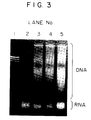
- Fig. 3 is a photograph showing the results of 3% agarose gel electrophoresis of each DNA sample obtained in Example 2.
- Fig. 4 is a photograph showing the results of 2.5% agarose gel electrophoresis of each DNA sample obtained in Example 4 which was carried out after amplification of the DNA sample by the PCR method.
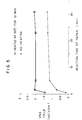
- Fig. 5 is a graph showing the relationship between the amount of DNA in each DNA sample and protease treatment time which was determined in Example 5.
- Fig. 6 is a photograph showing the results of 2.5% agarose gel electrophoresis of each DNA sample obtained in Example 6 which was carried out after amplification of the DNA sample by the PCR method.
- the present inventors earnestly investigated in order to achieve the above objects, and consequently found that in a process for treating a paraffin-embedded tissue sample useful for a gene analysis, either the heat treatment of an aqueous suspension containing a surfactant having a protein-denaturational action and a deparaffinized tissue sample obtained from a paraffin-embedded tissue sample, or the reaction of the heat-treated aqueous suspension with a protease is advantageous because when the heat treatment or the reaction is carried out in stead of the conventional treatment, i.e., the disruption of the deparaffinized tissue cells, i.e.
- the problem in the conventional process i.e., the requirement of a long period of time for the disruption of the tissue cells is solved, namely, the time required for the disruption can be greatly reduced.
- the present inventors found the following.
- a surfactant having a protein-denaturational action is added to a tissue sample after removal of paraffin from a paraffin-embedded tissue, and the resulting aqueous suspension is heated, after which the heat-treated aqueous suspension is reacted with a protease, and a protein denaturation and removal procedure is carried out by an one-phase method by use of a solution containing an organic compound having a protein-denaturational action, for example, an aqueous alcoholic solution containing an organic compound having one or more phenolic hydroxyl groups, instead of carrying out a protein denaturation and removal procedure by the two-phase separation method using phenol, chloroform, etc. as in the conventional process.
- paraffin-embedded tissue sample used in the present invention
- samples containing nucleic acid and usually called "paraffin-embedded tissue sample” in the art, for example, samples prepared by subjecting a collected tissue material (e.g. pancreas, large intestine, cancer of large intestine, muscle, urinary bladder, kidney, lung, brain, lymphoma, etc.) to fixing treatment with a liquid chemical having a protein-coagulative action (e.g.
- a collected tissue material e.g. pancreas, large intestine, cancer of large intestine, muscle, urinary bladder, kidney, lung, brain, lymphoma, etc.
- a liquid chemical having a protein-coagulative action e.g.
- any method may be used so long as it is usually employed in the art for removing paraffin from a paraffin-embedded tissue sample.
- a method comprising adding a water-insoluble organic solvent to the paraffin-embedded tissue sample, stirring and shaking the resulting mixture, centrifuging the mixture, and discarding the supernatant to remove paraffin.
- the water-insoluble organic solvent for dissolving and removing the paraffin which is used in such a method is not particularly limited, and any water-insoluble organic solvent may be used so long as it is usually used in the art for dissolving and removing paraffin.
- the water-insoluble organic solvent are xylene, D-limonene, octane, etc. Of these, D-limonene and the like are preferable in view of toxicity to a human body.
- the amount of the water-insoluble organic solvent used cannot be unequivocally determined because it is varied depending on the thickness of the tissue sample, etc., it may be an amount sufficient to immerse the tissue sample therein thoroughly.
- the surfactant used in the present invention is not particularly limited and may be any of cationic surfactants, anionic surfactants, nonionic surfactants and amphoteric surfactants so long as it is usually used in the art as an surfactant having a protein-denaturational action.
- Typical and specific examples of the surfactant are cationic surfactants such as dodecyltrimethylammonium bromide, dodecyltrimethylammonium chloride, cetyltrimethylammonium bromide and the like; anionic surfactants such as sodium dodecylsulfate (SDS), sodium N-lauroylsarcosine, sodium cholate, sodium deoxycholate and the like; nonionic surfactants such as polyoxyethylene octylphenyl ether [e.g. Triton X-100 (a trademark, Rohm and Haas Co.), etc.], polyoxyethylene sorbitan monolaurate [e.g.
- TWEEN 20 (a trademark, Kao Corp.), etc.] polyoxyethylene sorbitan monooleate [e.g. TWEEN 80 (a trademark, Kao Corp.), etc.], n-octyl- ⁇ -D-glucoside and the like; and amphoteric surfactants such as 3-[(cholamidopropyl)-dimethylammonio]-l-propane sulfonate, phosphatidylethanolamine and the like.
- the anionic surfactants such as SDS are particularly preferable because of their strong protein-denaturational action.
- the above-exemplified surfactants can be used singly or as a mixture thereof.
- One of the processes for treating a paraffin-embedded tissue sample useful for a gene analysis of the present invention can be practiced by preparing an aqueous suspension containing the above-mentioned deparaffinized tissue sample obtained from the paraffin-embedded tissue sample and the above-mentioned surfactant having a protein-denaturational action, and heating the aqueous suspension.
- a method for preparing the aqueous suspension containing deparaffinized tissue sample obtained from the paraffin-embedded tissue sample and the surfactant having a protein-denaturational action is not particularly limited and any method may be employed so long as it finally gives the aqueous suspension containing deparaffinized tissue sample obtained from the paraffin-embedded tissue sample and the surfactant having a protein-denaturational action.
- a preferable example of the most general method for the preparation is a method of adding an aqueous solution containing the surfactant having a protein-denaturational action to the deparaffinized tissue sample obtained from the paraffin-embedded tissue sample, to suspend the deparaffinized tissue sample.
- the concentration of the surfactant used in the present invention is varied a little depending on the kind of the surfactant, though for example, the concentration in the aqueous solution containing the surfactant having a protein-denaturational action is usually 0.01 to 10% (W/V), preferably 0.1 to 2% (W/V).
- the aqueous suspension containing the deparaffinized tissue sample obtained from the paraffin-embedded tissue sample and the surfactant having a protein-denaturational action may contain, for example, buffers such as phosphate buffers, citrate buffers, tris(hydroxymethyl)aminomethane (Tris) buffer, glycine buffer, Good's buffers, etc. and salts such as sodium chloride, potassium chloride, magnesium chloride, lithium chloride, etc.
- the aqueous suspension may, if necessary, properly contain nuclease inhibitors such as ethylenediaminetetraacetic acid (EDTA), ethylene glycol bis(2-aminoethyl ether) tetraacetic acid (EGTA), salts thereof, etc.
- buffers such as phosphate buffers, citrate buffers, tris(hydroxymethyl)aminomethane (Tris) buffer, glycine buffer, Good's buffers, etc.
- salts such as sodium chloride, potassium chloride, magnesium
- the surfactant having a protein-denaturational action has also nuclease-inhibitory action
- the above-exemplified nuclease inhibitors need not be used.
- concentrations of the buffers, salts and nuclease inhibitors used are not critical so long as they do not hinder the liberation of nucleic acid.
- concentration of the buffers in the aqueous solution containing the surfactant having a protein-denaturational action is usually 1 to 500 mM, preferably 5 to 200 mM.
- the salts are added to a concentration of 1 to 500 mM if necessary.
- the pH of the aqueous solution is not critical so long as it does not hinder the liberation of nucleic acid, it is properly chosen in the range of usually 2 to 12, preferably 5 to 9.
- the concentration of the nuclease inhibitors used cannot be unequivocally determined because it is varied depending on the kind of the inhibitor.
- the concentration of EDTA in the aqueous solution is usually 0.1 to 200 mM, preferably 1 to 10 mM.
- the heating conditions in the treating process of the present invention are as follows.
- the heating temperature is usually 60°C or higher, preferably 70°C or higher, preferably to a temperature of the boiling point of the suspension or lower, more preferably 80°C to the boiling point of the suspension.
- As the heating time usually 2 minutes or more, preferably 5 to 30 minutes, is sufficient.
- the boiling point of the suspension changes to a certain extent with a change of the external pressure and changes of the proportions and concentrations of the substances contained in the suspension.
- a slice (thickness: usually about 10 ⁇ m) of a paraffin-embedded tissue is placed in a test tube, and a proper amount (an amount sufficient to immerse the tissue slice) of a water-insoluble organic solvent such as xylene, linonene, etc. is added. After sufficient stirring and shaking, the resulting mixture is centrifuged at ordinary temperature and the supernatant is discarded to remove the paraffin. The above procedure is repeated once on the precipitate. Then, a proper amount of a volatile organic solvent such as ethanol is added to the thus obtained precipitate, and after stirring and shaking, the resulting mixture is centrifuged at ordinary temperature and the supernatant is discarded to remove the water-insoluble organic solvent.
- a proper amount of a volatile organic solvent such as ethanol
- sample A for gene analysis The sample for a gene analysis obtained by the treatment by the above-mentioned process (hereinafter referred to as "sample A for gene analysis”) can be used as it is as a sample for a gene analysis by the PCR method, or the like., or it can be used as a sample from which nucleic acid are extracted and purified by a method usually used in the art.
- a sample useful for a gene analysis by the PCR method, or the like can be prepared also by treating a paraffin-embedded tissue sample for a gene analysis by practicing the above-mentioned treating process (i.e. heating the aqueous suspension containing a surfactant having a protein-denaturational action and a de-paraffinized tissue sample obtained from a paraffin-embedded tissue sample), and then reacting the heat-treated aqueous suspension with a protease.
- a protease permits simplification of the steps of procedure and hence marked reduction of the operation time as compared with conventional processes.
- the treatment with a protease is effective, for example, in that the efficiency of extraction of nucleic acid from the paraffin-embedded tissue sample is further increased.
- the protease used in the present invention is not particularly limited so long as it is usually used in the art for the purpose of digesting a protein.
- the protease are papain, proteinase K, pronase, etc.
- the concentration in the reaction mixture in the protein digestion step is usually 0.01 to 10 mg/ml.
- the protease thermostable (heat-resistant) even at a high temperature of, for example, 70°C or higher which has catalytic activity under the heating conditions employed in the present invention
- the protease can be allowed to act simultaneously with the heat treatment of the aqueous suspension containing the deparaffinized tissue sample obtained from the paraffin-embedded tissue sample and the surfactant having a protein-denaturational action which is carried out in the present invention.
- the steps of procedure can be further simplified.
- the protease in an amount of 0.01 to 10 mg/ml, and the reaction is carried out at 37 - 70°C, preferably 40 - 60°C for 5 to 300 minutes.
- a thiol compound e.g. dithiothreitol (DTT)
- DTT dithiothreitol
- the thiol compound is preferably added.
- sample B for gene analysis The sample for a gene analysis obtained by the process described above (hereinafter referred to as "sample B for gene analysis”) can be used as it is as a sample for a gene analysis by the PCR method, or the like, or it can be used as a sample from which nucleic acid are extracted and purified by a method usually used in the art.
- a sample useful for a gene analysis by the PCR method, or the like can be prepared by practicing the above-mentioned process for treating a paraffin-embedded tissue sample for a gene analysis (i.e. heating the aqueous suspension containing a surfactant having a protein-denaturational action and a deparaffinized tissue sample obtained from a paraffin-embedded tissue sample and reacting the heat-treated aqueous suspension with a protease), followed by precipitating and collecting nucleic acid from the resulting reaction solution.
- a paraffin-embedded tissue sample to be used for a gene analysis is thus treated, simplification of the steps of procedure and hence marked reduction of the operation time can be achieved as compared with conventional processes.
- a precipitant used for precipitating nucleic acid in the present invention is not particularly limited so long as it is usually used in the art for precipitating nucleic acid.
- alcohols are used as the precipitant.
- the alcohols are not particularly limited so long as they are usually used in the art for precipitating nucleic acid.
- Preferable examples of the alcohols are ethanol, isopropanol, etc.
- the alcohol is used usually in a volume of 0.5 to 3 times that of a solution containing the extracted nucleic acid.
- the alcohol is added to the sample B for gene analysis obtained by the treatment by the treating process described above (i.e. the sample obtained by heating the aqueous suspension containing the deparaffinized tissue sample obtained from the paraffin-embedded tissue sample and the surfactant having a protein-denaturational action, and reacting the heat-treated aqueous suspension with a protease) in a volume of 0.5 to 3 times that of the sample B for gene analysis and mixed therewith, after which the resulting mixture is allowed to stand at room temperature for 10 minutes.
- a proper amount of a salt such as NaCl, sodium acetate or the like may be optionally added together with the alcohol.
- sample C for gene analysis After the standing, the mixture is centrifuged to precipitate the nucleic acid and the supernatant is discarded, and a proper volume of, for example, 70% ethanol is added to the precipitate, followed by washing and centrifugation. The supernatant is discarded and the thus obtained precipitate is dried, whereby there can be obtained a desired sample for a gene analysis (hereinafter referred to as "sample C for gene analysis").
- the sample C for gene analysis is used as a sample for a gene analysis by the PCR method, or the like, it is preferable to use purified DNA obtained by treating the sample C for gene analysis by a method usually employed in the art, for example, a method comprising redissolving the sample C for gene analysis in TE buffer (10 mM Tris-HCl buffer, containing 1 mM EDTA, pH 8.0), adding a suitable amount of RNase, and incubating the resulting solution at 37 - 55°C for 1 to 90 minutes to digest RNA.
- TE buffer 10 mM Tris-HCl buffer, containing 1 mM EDTA, pH 8.0
- a paraffin-embedded tissue sample to be used for a gene analysis is treated by practicing the above-mentioned process (i.e. heating the aqueous suspension containing a surfactant having a protein-denaturational action and a deparaffinized tissue sample obtained from a paraffin-embedded tissue sample, and reacting the heat-treated aqueous suspension with a protease), mixing the resulting reaction solution with a solution containing an organic compound having a protein-denaturational action other than the surfactant mentioned above, and then precipitating and collecting nucleic acid from the resulting reaction solution.
- the effects are as follows.
- Simplification of the steps of procedure and hence marked reduction of the operation time can be achieved as compared with conventional processes. Furthermore, there can be collected nucleic acid which are suitable for the PCR method because of their contamination with only a slight amount of substances inhibitory to gene amplification by the PCR method.
- the aforesaid organic compound having a protein-denaturational action other than the surfactant mentioned above which is used in the present invention is not particularly limited so long as it has the aforesaid action and is water-soluble to a certain extent.
- Preferable examples of the organic substance are organic compounds having one or more phenolic hydroxyl groups.
- organic compounds having one or more phenolic hydroxyl groups are phenol, o-, m- or p-hydroxybenzoic acid, 2-amino-4-chlorophenol, m-aminophenol, methyl p-hydroxybenzoate, catechol, guaiacol, hydroquinone, p-hydroxyphenethyl alcohol, protocatechuic acid, p-(methoxyethyl)phenol, etc.
- compounds not corrosive to human skin such as hydroxybenzoic acid, in particular, m-hydroxybenzoic acid are preferable.
- the concentration of the organic compound having one or more phenolic hydroxyl groups is varied depending on the kind of the compound, the concentration of the compound in the reaction solution in the protein denaturation step is usually less than 20% (W/V) and not less than 0.01% (W/V).
- concentration in said reaction solution is preferably 1 to 10% (W/V).
- the solution containing the organic compound used for denaturing protein in the present invention is not critical so long as it is a solution containing the above-exemplified organic compound having a protein-denaturational action.
- Said solution may contain water-soluble organic solvents such as alcohols (e.g. ethanol, isopropanol, etc.), acetone and the like for the dissolution of said organic compound.
- alcohols e.g. ethanol, isopropanol, etc.
- acetone acetone and the like for the dissolution of said organic compound.
- the alcohols such as ethanol, isopropanol, etc. are preferable because they have no undesirable influence on the precipitation of nucleic acid.
- the organic solvents are properly used so that their concentration in said solution may be usually 10 to 90% (W/V).
- the pH of the solution containing said organic compound cannot be unequivocally determined because it is varied depending on the kind of the organic compound, any pH may be employed so long as it does not hinder the protein denaturation.
- the pH is properly chosen in the range of 4 to 10.
- the pH of said solution is chosen in the range of 5 to 8. Needless to say, in every case, the organic compound should be in a uniformly dissolved state in said solution.
- the solution containing the organic compound having a protein-denaturational action is added to the sample B for gene analysis obtained by the treatment by the treating process described above (i.e. the sample obtained by heating the aqueous suspension containing the deparaffinized tissue sample obtained from the paraffin-embedded tissue sample and the surfactant having a protein-denaturational action, and reacting the heat-treated aqueous suspension with a protease). Then, the reaction is carried out at room temperature for several minutes to several hours to denature protein, after which the resulting reaction solution is centrifuged and the supernatant is transferred into another tube.
- sample D for gene analysis a desired sample for a gene analysis
- the sample D for gene analysis is used as a sample for a gene analysis by the PCR method, or the like, it is preferable to use purified DNA obtained by treating the sample D for gene analysis by a method usually employed in the art, for example, a method comprising redissolving the sample D for gene analysis in TE buffer (10 mM Tris-HCl buffer, containing 1 mM EDTA, pH 8.0), adding a suitable amount of RNase, and incubating the resulting solution at 37 - 55°C for 1 to 90 minutes to digest RNA.
- TE buffer 10 mM Tris-HCl buffer, containing 1 mM EDTA, pH 8.0
- the kit for treating a paraffin-embedded tissue sample using for a gene analysis of the present invention is used for practicing any of the above-mentioned processes for treating a paraffin-embedded tissue sample to be used for a gene analysis, and comprises a container containing a surfactant having a protein-denaturational action, a container containing a protease, and a container containing a hydroxybenzoic acid solution.
- a container containing a surfactant having a protein-denaturational action a container containing a protease
- a container containing a hydroxybenzoic acid solution Preferable properties, specific examples and the like of each of the components contained in the containers are as described above.
- a slice of formaldehyde fixed and paraffin embedded human tissue (liver, thickness 10 ⁇ m, about 15 mm x 25 mm) was placed in a 1.5-ml test tube, followed by adding thereto 1 ml of D-limonene (Lemosol, a trade name, mfd. by Wako Pure Chemical Industries, Ltd.), and the resulting mixture was stirred with a micro-tube mixer (MT-360, mfd. by Tomy Seiko Co., Ltd.) for 3 minutes. The mixture was then centrifuged at 12,000 rpm for 3 minutes at ordinary temperature, and the supernatant was discarded to remove the paraffin. The above procedure was repeated once on the precipitate.
- D-limonene Lemosol, a trade name, mfd. by Wako Pure Chemical Industries, Ltd.
- the obtained nucleic acid were redissolved in 20 ⁇ l of TE buffer (10 mM Tris-HCl buffer, containing 1 mM EDTA, pH 8.0), followed by adding thereto 1 ⁇ l of 10 mg/ml RNase, and the resulting solution was incubated at 50°C for 1 hour to obtain a DNA sample.
- the DNA yield in the DNA sample was determined by fluorometry and the ⁇ -globin gene was amplified by the PCR method by use of the DNA sample (conditions in the PCR method: 94°C for 1 minute, 55°C for 2 minutes, 72°C for 3 minutes; 35 cycles).
- the DNA quantitation by fluorometry was carried out by the following method.
- Fig. 1 shows the result of fluorometric detection of DNA carried out by means of a UV illuminator after electrophoresis of the DNA amplified by the PCR method, at a constant voltage of 100 V for 20 to 30 minutes on a 3% agarose gel prepared by use of 1 x TAE buffer (0.04 M Tris-acetate, 1 mM EDTA, pH 7.8) containing 0.5 ⁇ g/ml of ethidium bromide.
- lane 1 shows the result obtained for a molecular weight marker ( ⁇ x 174 phage DNA/HaeIII, mfd. by Nippon Gene Co., Ltd.).
- the mark ⁇ indicates the amplification region (110 bp) of the human ⁇ -globin gene.
- a slice of formaldehyde fixed and paraffin embedded human tissue (liver, thickness 10 ⁇ m, about 15 mm x 25 mm) was placed in a 1.5-ml test tube, followed by adding thereto 1 ml of D-limonene, and the resulting mixture was stirred with a micro-tube mixer (MT-360, mfd. by Tomy Seiko Co., Ltd.) for 3 minutes. The mixture was then centrifuged at 12,000 rpm for 3 minutes at room temperature, and the supernatant was discarded to remove the paraffin. The above procedure was repeated once on the precipitate.
- MT-360 micro-tube mixer
- reaction solution 50 ⁇ l of a 1 mg/ml proteinase K solution (or a 1 mg/ml papain solution) was added three times.
- the reaction solution was mixed with an equal volume of a phenol/chloroform/isoamyl alcohol mixture (25 : 24 : 1), and the resulting mixture was centrifuged at 6,000 rmp for 5 minutes, after which the aqueous phase, the upper layer was transferred into another tube by pipetting. This separating procedure was repeated once on the aqueous layer.
- aqueous phase was mixed with an equal volume of a chloroform/isoamyl alcohol mixture (24 : 1), and the resulting mixture was centrifuged at 6,000 rmp for 5 minutes, after which the upper layer was transferred into another tube. This procedure was repeated once. A 5 M sodium chloride solution and ethanol were added to the obtained upper layer solution in volumes of three-fiftieths and 2.5 times, respectively, as large as the upper layer solution and stirred, after which the resulting mixture was allowed to stand at -80°C for 15 minutes. Then, the mixture was centrifuged at 15,000 rpm for 20 minutes at 4°C and the supernatant was discarded.
- the pellet was mixed with 70% ethanol and the resulting mixture was centrifuged at 15,000 rpm for 5 minutes at 4°C, after which the supernatant was discarded and the precipitate was dried to obtain desired nucleic acid. Finally, the obtained nucleic acid were redissolved in 20 ⁇ l of TE buffer, followed by adding thereto 1 ⁇ l of 10 mg/ml RNase, and the resulting solution was incubated at 50°C for 1 hour to obtain a DNA sample.
- the DNA yield in the DNA sample was determined by fluorometry and the ⁇ -globin gene was amplified by the PCR method by use of the DNA sample under the same conditions as described in Example 1. The DNA quantitation by fluorometry was carried out by the following method.
- Table 1 shows the DNA yield in the DNA sample, etc. together with the results obtained in Example 1, and Fig. 1 (lane 3) shows the result of fluorescence detection for DNA carried out by means of a UV illuminator after electrophoresis of the DNA amplified by the PCR method, at a constant voltage of 100 V for 20 to 30 minutes on a 3% agarose gel prepared by use of 1 x TAE buffer (0.04 M Tris-acetate, 1 mM EDTA, pH 7.8) containing 0.5 ⁇ g/ml of ethidium bromide, together with the results obtained in Example 1.
- Table 1 Example 1 Comparative Example 1 Total operation time 3 hours 3 days Protein removal by two-phase method None Carried out DNA yield ( ⁇ g/slice) 3.2 3.1
- the process of the present invention permits rapid extraction of nucleic acid in substantially the same yield as in the known method because the same procedure as in the known method is carried out by the steps of the heat treatment for protein denaturation by use of a surfactant, the protein digestion by use of a protease, and then the protein denaturation and removal by an one-phase method using a solution containing an organic compound having a protein-denaturational action.
- Example 1 To 13 ⁇ l of each of the DNA samples obtained in Example 1 and Comparative Example 1 was added 70 U of DNase, and the reaction was carried out at 37°C for 1 hour to digest the human DNA.
- the reaction solution was mixed with an equal volume of a phenol/chloroform/ isoamyl alcohol mixture (25 : 24 : 1) and the resulting mixture was centrifuged at 6,000 rpm for 5 minutes, after which the aqueous layer, the upper layer was transferred into another tube, whereby the DNase was removed.
- a 5 M sodium chloride solution and ethanol were added to the obtained aqueous phase in volumes of three-fiftieths and 2.5 times, respectively, as large as the aqueous phase and stirred, after which the resulting mixture was allowed to stand at -80°C for 15 minutes. Then, the mixture was centrifuged at 15,000 rpm for 20 minutes at 4°C and the supernatant was discarded. To the pellet was added 1 ml of 70% ethanol, followed by washing and drying. The resulting pellet was dissolved in 13 ⁇ l of TE buffer. The thus obtained solutions were used as sample solutions for the following test.
- rat glucose-3-phosphate dehydrogenase (G3PDH) gene 983 bp region
- the amplified DNA was electrophoresed at a constant voltage of 100 V for 20 to 30 minutes on a 3% agarose gel prepared by use of 1 x TAE buffer (0.04 M Tris-acetate, 1 mM EDTA, pH 7.8) containing 0.5 ⁇ g/ml of ethidium bromide.
- fluorometric detection of objective bands was carried out by means of a UV illuminator. The results obtained are shown in Fig. 2.
- the mark ⁇ indicates the amplification region (983 bp) of the rat G3PDH gene.
- nucleic acid suitable for use as a sample in the PCR method can be obtained with substantially no contamination with the substances inhibitory to the reaction carried out in the PCR method.
- tissue slices were cut out of a formaldehyde fixed and paraffin embedded tissue sample (rat liver). Each tissue slice was placed in a 1.5-ml test tube, followed by adding thereto 1 ml of D-limonene, and the resulting mixture was stirred with a micro-tube mixer (MT-360, mfd. by Tomy Seiko Co., Ltd.) for 3 minutes. The mixture was then centrifuged at 12,000 rpm for 3 minutes at room temperature, and the supernatant was discarded to remove the paraffin. The above procedure was repeated once on the precipitate.
- MT-360 micro-tube mixer
- tissue slices were cut out of a Carnoy fixed and paraffin embedded human tissue sample (liver). Each tissue slice was placed in a 1.5-ml test tube, followed by adding thereto 1 ml of D-limonene, and the resulting mixture was stirred with a micro-tube mixer (MT-360, mfd. by Tomy Seiko Co., Ltd.) for 3 minutes. The mixture was then centrifuged at 12,000 rpm for 3 minutes at ordinary temperature, and the supernatant was discarded to remove the paraffin. The above procedure was repeated once on the precipitate.
- MT-360 micro-tube mixer
- the obtained nucleic acid were redissolved in 20 ⁇ l of TE buffer (10 mM Tris-HCl buffer, containing 1 mM EDTA, pH 8.0), followed by adding thereto 1 ⁇ l of 10 mg/ml RNase, and the resulting solution was incubated at 50°C for 1 hour to obtain a DNA sample.
- TE buffer 10 mM Tris-HCl buffer, containing 1 mM EDTA, pH 8.0
- Table 2 shows the DNA yield in each DNA sample.
- Heat treatment conditions DNA yield ( ⁇ g/slice) No heating (room temp.) 2 min 3.1 10 min 3.1 70°C 2 min 6.2 10 min 8.6 80°C 2 min 8.9 10 min 9.7 90°C 2 min 10.2 10 min 8.9 100°C 2 min 9.2 10 min 9.1
- the DNA yield attained by the heat treatment at 70°C for 2 minutes or 10 minutes is twice or more as high as that attained by the standing at room temperature (i.e. no heat treatment), and the DNA yield attained by the heat treatment at 80°C, 90°C or 100°C is about 3 times or more as high as that attained by the standing at room temperature.
- the heat treatment markedly increases the DNA yield.
- tissue slices Two tissue slices (thickness 10 ⁇ m, about 10 mm x 20 mm) were cut out of a formaldehyde fixed and paraffin embedded human tissue sample (liver). Each tissue slice was placed in a 1.5-ml test tube, followed by adding thereto 1 ml of D-limonene, and the resulting mixture was stirred with a micro-tube mixer (MT-360, mfd. by Tomy Seiko Co., Ltd.) for 3 minutes. The mixture was then centrifuged at 12,000 rpm for 3 minutes at ordinary temperature, and the supernatant was discarded to remove the paraffin. The above procedure was repeated once on the precipitate.
- MT-360 micro-tube mixer
- amplification reaction of the k-ras gene was carried out by the PCR method (conditions in the PCR method: 94°C for 1 minute, 55°C for 2 minutes, 72°C for 3 minutes; 45 cycles).
- Fig. 4 shows the result of fluorometric detection of DNA carried out by means of a UV illuminator after electrophoresis of the DNA amplified by the PCR method, at a constant voltage of 100 V for 20 to 30 minutes on a 2.5% agarose gel prepared by use of 1 x TAE buffer (0.04 M Tris-acetate, 1 mM EDTA, pH 7.8) containing 0.5 ⁇ g/ml of ethidium bromide.
- lane 1 shows the result obtained for a molecular weight marker ( ⁇ x 174 phage DNA/HaeIII, mfd. by Nippon Gene Co., Ltd.).
- Lane 2 shows the result obtained for the DNA sample obtained without heat treatment
- lane 3 shows the result obtained for the DNA sample obtained by the heat treatment.
- the mark ⁇ indicates the amplification region (108 bp) of the k-ras gene.
- Ten tissue slices were cut out of a formaldehyde fixed and paraffin embedded human tissue sample (liver). Each tissue slice was placed in a 1.5-ml test tube, followed by adding thereto 1 ml of D-limonene, and the resulting mixture was stirred with a micro-tube mixer (MT-360, mfd. by Tomy Seiko Co., Ltd.) for 3 minutes. The mixture was then centrifuged at 12,000 rpm for 3 minutes at ordinary temperature, and the supernatant was discarded to remove the paraffin. The above procedure was repeated once on the precipitate.
- MT-360 micro-tube mixer
- Each test tube was subjected to centrifugation at 12,000 rpm for 15 minutes to precipitate nucleic acid.
- the supenatant was discarded and 1 ml of 70% ethanol was added to the precipitate and stirred, after which the resulting mixture was centrifuged at 15,000 rpm for 5 minutes at 4°C. Then, the supernatant was discarded and the precipitate was dried to obtain the desired nucleic acid.
- the obtained nucleic acid were redissolved in 20 ⁇ l of TE buffer (10 mM Tris-HCl buffer, containing 1 mM EDTA, pH 8.0), followed by adding thereto 1 ⁇ l of 10 mg/ml RNase, and the resulting solution was incubated at 50°C for 1 hour to obtain a DNA sample.
- TE buffer 10 mM Tris-HCl buffer, containing 1 mM EDTA, pH 8.0
- Table 3 shows the DNA yield in each DNA sample.
- Fig. 5 shows the relationship between the protease treatment time and the DNA yield.
- - ⁇ - shows the results obtained when the heat treatment was carried out, followed by the protease treatment, and - + - the results obtained when the protease treatment was carried out without heat treatment.
- Table 3 Protease treatment time (hrs) DNA yield ( ⁇ g/slice) No heat treatment Heated at 90°C for 10 min. 0.5 0.16 1.45 1.5 0.71 2.06 5 1.16 2.01 23 1.27 2.03 31 1.30 2.09
- tissue slices were cut out of a formaldehyde fixed and paraffin embedded human tissue sample (liver). Each tissue slice was placed in a 0.5-ml test tube, followed by adding thereto 0.5 ml of D-limonene, and the resulting mixture was stirred with a micro-tube mixer (MT-360, mfd. by Tomy Seiko Co., Ltd.) for 3 minutes. The mixture was then centrifuged at 12,000 rpm for 3 minutes at ordinary temperature, and the supernatant was discarded to remove the paraffin.
- MT-360 micro-tube mixer
- each of the remaining two test tubes was placed 20 ⁇ l of 10 mM Tris-HCl buffer (pH 7.0, containing 0.55% SDS and 5.6 mM EDTA), and one of these two test tubes was allowed to stand at room temperature for 10 minutes. The other of them was heated at 90°C for 10 minutes. Each of the four test tubes was then subjected to centrifugation at 10,000 rpm for 5 minutes at ordinary temperature, and the resulting supernatant was used as a DNA sample.
- 10 mM Tris-HCl buffer pH 7.0, containing 0.55% SDS and 5.6 mM EDTA
- amplification reaction of the k-ras gene was carried out by the PCR method (conditions in the PCR method: 94°C for 1 minute, 55°C for 2 minutes, 72°C for 3 minutes; 45 cycles).
- the DNA amplified by the PCR method was electrophoresed at a constant voltage of 100 V for 20 to 30 minutes on a 2.5% agarose gel prepared by use of 1 x TAE buffer (0.04 M Tris-acetate, 1 mM EDTA, pH 7.8) containing 0.5 ⁇ g/ml of ethidium bromide. Thereafter, fluorometric detection of the DNA was carried out by means of a UV illuminator. The results obtained are shown in Fig. 6. In Fig. 6, lane 1 shows the result obtained for a molecular weight marker ( ⁇ x 174 phage DNA/Hae III, mfd. by Nippon Gene Co., Ltd.).
- Lane 2 shows the result obtained for the DNA sample obtained by use of the buffer containing no surfactant (SDS), without heat treatment.
- Lane 3 shows the result obtained for the DNA sample obtained by using the buffer containing no surfactant (SDS), and carrying out the heat treatment.
- Lane 4 shows the result obtained for the DNA sample obtained by use of the surfactant (SDS)-containing buffer without heat treatment.
- Lane 5 shows the result obtained for the DNA sample obtained by using the surfactant (SDS)-containing buffer and carrying out the heat treatment.
- the mark ⁇ indicates the amplification region (108 bp) of the k-ras gene.
- a sample for a gene analysis can easily be obtained by heat-treating a sample obtained from a paraffin-embedded tissue, by use of the surfactant.
- the present invention provides a process for treating a paraffin-embedded tissue sample useful for a gene analysis and a treating kit used for practicing said process.
- a paraffin-embedded tissue sample useful for a gene analysis is treated by utilizing the present invention, the following effects can be obtained: as compared with conventional treating processes, operations are simpler and the time required for practicing the process of the present invention is much shorter; and moreover contamination with impurities, in particular, contamination with substances inhibitory to the reaction carried out by the PCR method is slight, so that there can be obtained nucleic acid suitable as a sample for a pathological examination, a gene analysis, etc., which utilize the PCR method. Therefore, the present invention contributes greatly to the art.
- Hydroxybenzoic acid used in the present invention as one of the organic compounds having a protein-denaturational action is an organic compound having one or more phenolic hydroxyl groups, is widely used as a fungicide for food and drink, and is not corrosive to a human body. No attempt has been made at all to use hydroxybenzoic acid as a protein denaturing agent in a process for treating a paraffin-embedded tissue sample useful for a gene analysis in which the two-phase separation method is not employed.
Landscapes
- Chemical & Material Sciences (AREA)
- Organic Chemistry (AREA)
- Life Sciences & Earth Sciences (AREA)
- Analytical Chemistry (AREA)
- Zoology (AREA)
- Wood Science & Technology (AREA)
- Proteomics, Peptides & Aminoacids (AREA)
- Health & Medical Sciences (AREA)
- Engineering & Computer Science (AREA)
- Biophysics (AREA)
- Biochemistry (AREA)
- Microbiology (AREA)
- Molecular Biology (AREA)
- Biotechnology (AREA)
- Physics & Mathematics (AREA)
- Chemical Kinetics & Catalysis (AREA)
- Immunology (AREA)
- Bioinformatics & Cheminformatics (AREA)
- General Engineering & Computer Science (AREA)
- General Health & Medical Sciences (AREA)
- Genetics & Genomics (AREA)
- Measuring Or Testing Involving Enzymes Or Micro-Organisms (AREA)
- Investigating Or Analysing Biological Materials (AREA)
Applications Claiming Priority (2)
| Application Number | Priority Date | Filing Date | Title |
|---|---|---|---|
| JP177578/94 | 1994-07-06 | ||
| JP17757894 | 1994-07-06 |
Publications (1)
| Publication Number | Publication Date |
|---|---|
| EP0692533A1 true EP0692533A1 (de) | 1996-01-17 |
Family
ID=16033427
Family Applications (1)
| Application Number | Title | Priority Date | Filing Date |
|---|---|---|---|
| EP95304717A Withdrawn EP0692533A1 (de) | 1994-07-06 | 1995-07-05 | Behandlung von in Paraffin eingebettetem Gewebe zur Genanalyse |
Country Status (3)
| Country | Link |
|---|---|
| US (1) | US5672696A (de) |
| EP (1) | EP0692533A1 (de) |
| CA (1) | CA2153215A1 (de) |
Cited By (4)
| Publication number | Priority date | Publication date | Assignee | Title |
|---|---|---|---|---|
| GB2369822A (en) * | 2000-12-05 | 2002-06-12 | Genovar Diagnostics Ltd | Nucleic acid extraction method and kit |
| RU2219243C2 (ru) * | 2001-02-27 | 2003-12-20 | НИИ онкологии им. проф. Н.Н. Петрова | Способ восстановления днк, изолированной из архивных патоморфологических образцов тканей |
| EP1533604A1 (de) * | 2003-11-18 | 2005-05-25 | Bio Optica-Milano S.p.A. | Verfahren zur Vorbereitung einer histologischen Probe für die Färbung |
| EP1601450A4 (de) * | 2003-03-10 | 2007-10-10 | Expression Pathology Inc | Flüssiggewebepräparation aus histopathologisch bearbeiteten biologischen proben, geweben und zellen |
Families Citing this family (28)
| Publication number | Priority date | Publication date | Assignee | Title |
|---|---|---|---|---|
| KR20010023070A (ko) * | 1997-08-20 | 2001-03-26 | 더 유니버시티 오브 마이애미 | 고품질의 연속적인 재료 처리, 조직 고정-탈수-지방제거-함침 방법 |
| US6793890B2 (en) | 1997-08-20 | 2004-09-21 | The University Of Miami | Rapid tissue processor |
| US6855559B1 (en) * | 1998-09-03 | 2005-02-15 | Ventana Medical Systems, Inc. | Removal of embedding media from biological samples and cell conditioning on automated staining instruments |
| US7410753B2 (en) * | 1998-09-03 | 2008-08-12 | Ventana Medical Systems, Inc. | Removal of embedding media from biological samples and cell conditioning on automated staining instruments |
| US6544798B1 (en) * | 1999-02-26 | 2003-04-08 | Ventana Medical Systems, Inc. | Removal of embedding media from biological samples and cell conditioning on automated staining instruments |
| US6248535B1 (en) | 1999-12-20 | 2001-06-19 | University Of Southern California | Method for isolation of RNA from formalin-fixed paraffin-embedded tissue specimens |
| NZ546252A (en) * | 2000-12-01 | 2008-03-28 | Response Genetics Inc | Method of determining HER2-neu gene expression in paraffin embedded tissue samples |
| US7049059B2 (en) | 2000-12-01 | 2006-05-23 | Response Genetics, Inc. | Method of determining a chemotherapeutic regimen based on ERCC1 and TS expression |
| US6602670B2 (en) * | 2000-12-01 | 2003-08-05 | Response Genetics, Inc. | Method of determining a chemotherapeutic regimen based on ERCC1 expression |
| US7005278B2 (en) * | 2001-03-02 | 2006-02-28 | Danenberg Kathleen D | Method of determining dihydropyrimidine dehydrogenase gene expression |
| US20020142328A1 (en) * | 2000-12-01 | 2002-10-03 | Danenberg Kathleen D. | Method of determining a chemotherapeutic regimen by assaying gene expression in primary tumors |
| US6956111B2 (en) * | 2001-03-02 | 2005-10-18 | Response Genetics, Inc. | Method of determining dihydropyrimidine dehydrogenase gene expression |
| US6686155B2 (en) * | 2001-06-14 | 2004-02-03 | Response Genetics, Inc. | Method of determining a chemotherapeutic regimen based on glutathione-S-transferase pi expression |
| US7364846B2 (en) | 2002-10-11 | 2008-04-29 | Molecular Devices Corporation | Gene expression profiling from FFPE samples |
| US8062897B2 (en) | 2003-07-21 | 2011-11-22 | Aureon Laboratories, Inc. | Diagnostic histopathology using multiplex gene expression FISH |
| US7235140B1 (en) * | 2003-08-27 | 2007-06-26 | Steve Hayes | Method for cleaning tissue processing molds |
| ATE442576T1 (de) * | 2003-10-24 | 2009-09-15 | Univ Miami | Vereinfachte gewebeverarbeitung |
| US8026106B1 (en) * | 2003-12-18 | 2011-09-27 | Ventana Medical Systems, Inc. | Removal of mercuric chloride crystals from fixed tissues |
| WO2006127860A2 (en) * | 2005-05-25 | 2006-11-30 | Expression Pathology, Inc. | Multiplex method for increased proteomic coverage from histopathologically processed biological samples using liquid tissue preparations |
| WO2006130632A2 (en) * | 2005-05-31 | 2006-12-07 | Invitrogen Corporation | Separation and purification of nucleic acid from paraffin-containing samples |
| US20070054168A1 (en) * | 2005-09-06 | 2007-03-08 | Hao Chang | Zinc/air cell |
| WO2007117700A2 (en) | 2006-04-07 | 2007-10-18 | The Trustees Of The University Of Pennsylvania | In situ cloning from pathological tissue specimens |
| US20100075372A1 (en) * | 2006-09-28 | 2010-03-25 | Taka-Aki Sato | Method for deparaffinization of paraffin-embedded specimen and method for analysis of paraffin-embedded specimen |
| US20090298172A1 (en) * | 2008-05-28 | 2009-12-03 | Steven Paul Wheeler | Histological specimen treatment apparatus and method |
| EP2926658B1 (de) * | 2012-10-30 | 2018-08-29 | Kanazawa Medical University | Kit zur herstellung transparentierter biologischer proben und verfahren zur herstellung transparentierter biologischer proben |
| JP6716198B2 (ja) | 2015-03-27 | 2020-07-01 | シスメックス株式会社 | 検体分析方法および検体分析装置 |
| US10365189B2 (en) | 2015-05-07 | 2019-07-30 | Steven Wheeler | Histological specimen treatment |
| CN113755486A (zh) * | 2021-09-23 | 2021-12-07 | 翌圣生物科技(上海)股份有限公司 | 用于处理石蜡包埋样本的脱蜡剂及从石蜡包埋样本中提取核酸的方法 |
Family Cites Families (1)
| Publication number | Priority date | Publication date | Assignee | Title |
|---|---|---|---|---|
| CA2087105C (en) * | 1990-07-13 | 2000-09-12 | Jeffrey Van Ness | Non-corrosive composition and methods useful for the extraction of nucleic acids |
-
1995
- 1995-07-04 CA CA002153215A patent/CA2153215A1/en not_active Abandoned
- 1995-07-05 EP EP95304717A patent/EP0692533A1/de not_active Withdrawn
- 1995-07-05 US US08/498,775 patent/US5672696A/en not_active Expired - Fee Related
Non-Patent Citations (4)
| Title |
|---|
| CHEMICAL ABSTRACTS, Columbus, Ohio, US; * |
| LABORATORY MEDICIN, vol. 22, no. 8, 1991, pages 543 - 546 |
| S.E. GOELZ ET AL., BIOCHEM. BIOPHYS. RES. COMMUN., vol. 130, 1985, pages 118 - 126 |
| YODOSHA CO., LTD., JIKKEN IGAKU, vol. 8, no. 9, 1990, pages 84 - 88 |
Cited By (7)
| Publication number | Priority date | Publication date | Assignee | Title |
|---|---|---|---|---|
| GB2369822A (en) * | 2000-12-05 | 2002-06-12 | Genovar Diagnostics Ltd | Nucleic acid extraction method and kit |
| RU2219243C2 (ru) * | 2001-02-27 | 2003-12-20 | НИИ онкологии им. проф. Н.Н. Петрова | Способ восстановления днк, изолированной из архивных патоморфологических образцов тканей |
| EP1601450A4 (de) * | 2003-03-10 | 2007-10-10 | Expression Pathology Inc | Flüssiggewebepräparation aus histopathologisch bearbeiteten biologischen proben, geweben und zellen |
| US8455215B2 (en) | 2003-03-10 | 2013-06-04 | Expression Pathology, Inc. | Liquid tissue preparation from histopathologically processed biological samples, tissues and cells |
| US9163275B2 (en) | 2003-03-10 | 2015-10-20 | Expression Pathology, Inc. | Liquid tissue preparation from histopathologically processed biologically samples, tissues and cells |
| US10444126B2 (en) | 2003-03-10 | 2019-10-15 | Expression Pathology, Inc. | Liquid tissue preparation from histopathologically processed biological samples, tissues and cells |
| EP1533604A1 (de) * | 2003-11-18 | 2005-05-25 | Bio Optica-Milano S.p.A. | Verfahren zur Vorbereitung einer histologischen Probe für die Färbung |
Also Published As
| Publication number | Publication date |
|---|---|
| US5672696A (en) | 1997-09-30 |
| CA2153215A1 (en) | 1996-01-07 |
Similar Documents
| Publication | Publication Date | Title |
|---|---|---|
| US5672696A (en) | Treatment of paraffin embedded tissue for gene analysis | |
| JP3943601B2 (ja) | アルカリプロテアーゼを用いる核酸の単離方法 | |
| US6617170B2 (en) | Method and device for collecting and stabilizing a biological sample | |
| CA2428864C (en) | Method and device for collecting and stabilizing a biological sample | |
| US6218531B1 (en) | Method of isolating RNA | |
| JP3477205B2 (ja) | 酸プロテアーゼを用いた核酸の調製 | |
| Bauer et al. | Protamine mRNA as molecular marker for spermatozoa in semen stains | |
| EP2580348B1 (de) | Verfahren zur bestimmung von zielzellen oder -gewebe zur extraktion von biomolekülen aus nicht-formalin-fixierten biologischen proben | |
| US20040157218A1 (en) | Pretreatment method for extraction of nucleic acid from biological samples and kits therefor | |
| DE69423131T2 (de) | Verfahren zur Behandlung von Mycobakterien enthaltenden Auswürfen | |
| CA2169271A1 (en) | In situ extraction of microbial dna | |
| JP4471659B2 (ja) | 直接核酸増幅法 | |
| EP4186980A1 (de) | Automatisierbares verfahren zur nukleinsäureisolierung | |
| EP1945764B1 (de) | Verfahren zur isolierung von mrna aus mit formalin fixiertem und in paraffin eingebettetem gewebe | |
| CN115125288A (zh) | 在福尔马林固定石蜡包埋组织中降低宿主核酸比例的方法及试剂盒 | |
| EP1109899B1 (de) | Produkt und methode zur auftrennung einer probe enthaltend genetisches material aus multipeln quellen mittels eines festen mediums | |
| JP3125633B2 (ja) | 遺伝子分析用試料としてのパラフィン包埋組織標本の処理方法及び処理用キット | |
| WO2015046419A1 (ja) | 包埋組織からの核酸抽出方法、包埋剤除去用具、及び核酸抽出用キット | |
| EP4372083A1 (de) | Verfahren zur extraktion von nukleinsäure aus einer polyphenolhaltigen probe | |
| HK1119738B (en) | Method for the isolation of mrna from formalin fixed, paraffin-embedded tissue | |
| AU2002245018A1 (en) | Method and device for collecting and stabilizing a biological sample |
Legal Events
| Date | Code | Title | Description |
|---|---|---|---|
| PUAI | Public reference made under article 153(3) epc to a published international application that has entered the european phase |
Free format text: ORIGINAL CODE: 0009012 |
|
| AK | Designated contracting states |
Kind code of ref document: A1 Designated state(s): DE ES FR GB IT |
|
| 17P | Request for examination filed |
Effective date: 19960611 |
|
| 17Q | First examination report despatched |
Effective date: 20000323 |
|
| STAA | Information on the status of an ep patent application or granted ep patent |
Free format text: STATUS: THE APPLICATION IS DEEMED TO BE WITHDRAWN |
|
| 18D | Application deemed to be withdrawn |
Effective date: 20011117 |