EP0663014B1 - Dna encoding human alpha 1 adrenergic receptors and uses thereof - Google Patents
Dna encoding human alpha 1 adrenergic receptors and uses thereof Download PDFInfo
- Publication number
- EP0663014B1 EP0663014B1 EP93922758A EP93922758A EP0663014B1 EP 0663014 B1 EP0663014 B1 EP 0663014B1 EP 93922758 A EP93922758 A EP 93922758A EP 93922758 A EP93922758 A EP 93922758A EP 0663014 B1 EP0663014 B1 EP 0663014B1
- Authority
- EP
- European Patent Office
- Prior art keywords
- human
- adrenergic receptor
- cell
- nucleic acid
- chemical compound
- Prior art date
- Legal status (The legal status is an assumption and is not a legal conclusion. Google has not performed a legal analysis and makes no representation as to the accuracy of the status listed.)
- Revoked
Links
Images


Classifications
-
- C—CHEMISTRY; METALLURGY
- C12—BIOCHEMISTRY; BEER; SPIRITS; WINE; VINEGAR; MICROBIOLOGY; ENZYMOLOGY; MUTATION OR GENETIC ENGINEERING
- C12Q—MEASURING OR TESTING PROCESSES INVOLVING ENZYMES, NUCLEIC ACIDS OR MICROORGANISMS; COMPOSITIONS OR TEST PAPERS THEREFOR; PROCESSES OF PREPARING SUCH COMPOSITIONS; CONDITION-RESPONSIVE CONTROL IN MICROBIOLOGICAL OR ENZYMOLOGICAL PROCESSES
- C12Q1/00—Measuring or testing processes involving enzymes, nucleic acids or microorganisms; Compositions therefor; Processes of preparing such compositions
- C12Q1/68—Measuring or testing processes involving enzymes, nucleic acids or microorganisms; Compositions therefor; Processes of preparing such compositions involving nucleic acids
- C12Q1/6876—Nucleic acid products used in the analysis of nucleic acids, e.g. primers or probes
-
- A—HUMAN NECESSITIES
- A61—MEDICAL OR VETERINARY SCIENCE; HYGIENE
- A61P—SPECIFIC THERAPEUTIC ACTIVITY OF CHEMICAL COMPOUNDS OR MEDICINAL PREPARATIONS
- A61P13/00—Drugs for disorders of the urinary system
- A61P13/02—Drugs for disorders of the urinary system of urine or of the urinary tract, e.g. urine acidifiers
-
- A—HUMAN NECESSITIES
- A61—MEDICAL OR VETERINARY SCIENCE; HYGIENE
- A61P—SPECIFIC THERAPEUTIC ACTIVITY OF CHEMICAL COMPOUNDS OR MEDICINAL PREPARATIONS
- A61P15/00—Drugs for genital or sexual disorders; Contraceptives
-
- A—HUMAN NECESSITIES
- A61—MEDICAL OR VETERINARY SCIENCE; HYGIENE
- A61P—SPECIFIC THERAPEUTIC ACTIVITY OF CHEMICAL COMPOUNDS OR MEDICINAL PREPARATIONS
- A61P27/00—Drugs for disorders of the senses
- A61P27/02—Ophthalmic agents
-
- A—HUMAN NECESSITIES
- A61—MEDICAL OR VETERINARY SCIENCE; HYGIENE
- A61P—SPECIFIC THERAPEUTIC ACTIVITY OF CHEMICAL COMPOUNDS OR MEDICINAL PREPARATIONS
- A61P27/00—Drugs for disorders of the senses
- A61P27/16—Otologicals
-
- A—HUMAN NECESSITIES
- A61—MEDICAL OR VETERINARY SCIENCE; HYGIENE
- A61P—SPECIFIC THERAPEUTIC ACTIVITY OF CHEMICAL COMPOUNDS OR MEDICINAL PREPARATIONS
- A61P3/00—Drugs for disorders of the metabolism
- A61P3/08—Drugs for disorders of the metabolism for glucose homeostasis
-
- A—HUMAN NECESSITIES
- A61—MEDICAL OR VETERINARY SCIENCE; HYGIENE
- A61P—SPECIFIC THERAPEUTIC ACTIVITY OF CHEMICAL COMPOUNDS OR MEDICINAL PREPARATIONS
- A61P43/00—Drugs for specific purposes, not provided for in groups A61P1/00-A61P41/00
-
- A—HUMAN NECESSITIES
- A61—MEDICAL OR VETERINARY SCIENCE; HYGIENE
- A61P—SPECIFIC THERAPEUTIC ACTIVITY OF CHEMICAL COMPOUNDS OR MEDICINAL PREPARATIONS
- A61P9/00—Drugs for disorders of the cardiovascular system
- A61P9/06—Antiarrhythmics
-
- A—HUMAN NECESSITIES
- A61—MEDICAL OR VETERINARY SCIENCE; HYGIENE
- A61P—SPECIFIC THERAPEUTIC ACTIVITY OF CHEMICAL COMPOUNDS OR MEDICINAL PREPARATIONS
- A61P9/00—Drugs for disorders of the cardiovascular system
- A61P9/08—Vasodilators for multiple indications
-
- A—HUMAN NECESSITIES
- A61—MEDICAL OR VETERINARY SCIENCE; HYGIENE
- A61P—SPECIFIC THERAPEUTIC ACTIVITY OF CHEMICAL COMPOUNDS OR MEDICINAL PREPARATIONS
- A61P9/00—Drugs for disorders of the cardiovascular system
- A61P9/10—Drugs for disorders of the cardiovascular system for treating ischaemic or atherosclerotic diseases, e.g. antianginal drugs, coronary vasodilators, drugs for myocardial infarction, retinopathy, cerebrovascula insufficiency, renal arteriosclerosis
-
- A—HUMAN NECESSITIES
- A61—MEDICAL OR VETERINARY SCIENCE; HYGIENE
- A61P—SPECIFIC THERAPEUTIC ACTIVITY OF CHEMICAL COMPOUNDS OR MEDICINAL PREPARATIONS
- A61P9/00—Drugs for disorders of the cardiovascular system
- A61P9/12—Antihypertensives
-
- C—CHEMISTRY; METALLURGY
- C07—ORGANIC CHEMISTRY
- C07K—PEPTIDES
- C07K14/00—Peptides having more than 20 amino acids; Gastrins; Somatostatins; Melanotropins; Derivatives thereof
- C07K14/435—Peptides having more than 20 amino acids; Gastrins; Somatostatins; Melanotropins; Derivatives thereof from animals; from humans
- C07K14/705—Receptors; Cell surface antigens; Cell surface determinants
- C07K14/70571—Receptors; Cell surface antigens; Cell surface determinants for neuromediators, e.g. serotonin receptor, dopamine receptor
-
- C—CHEMISTRY; METALLURGY
- C12—BIOCHEMISTRY; BEER; SPIRITS; WINE; VINEGAR; MICROBIOLOGY; ENZYMOLOGY; MUTATION OR GENETIC ENGINEERING
- C12N—MICROORGANISMS OR ENZYMES; COMPOSITIONS THEREOF; PROPAGATING, PRESERVING, OR MAINTAINING MICROORGANISMS; MUTATION OR GENETIC ENGINEERING; CULTURE MEDIA
- C12N15/00—Mutation or genetic engineering; DNA or RNA concerning genetic engineering, vectors, e.g. plasmids, or their isolation, preparation or purification; Use of hosts therefor
- C12N15/09—Recombinant DNA-technology
- C12N15/63—Introduction of foreign genetic material using vectors; Vectors; Use of hosts therefor; Regulation of expression
- C12N15/79—Vectors or expression systems specially adapted for eukaryotic hosts
- C12N15/85—Vectors or expression systems specially adapted for eukaryotic hosts for animal cells
- C12N15/8509—Vectors or expression systems specially adapted for eukaryotic hosts for animal cells for producing genetically modified animals, e.g. transgenic
-
- C—CHEMISTRY; METALLURGY
- C12—BIOCHEMISTRY; BEER; SPIRITS; WINE; VINEGAR; MICROBIOLOGY; ENZYMOLOGY; MUTATION OR GENETIC ENGINEERING
- C12Q—MEASURING OR TESTING PROCESSES INVOLVING ENZYMES, NUCLEIC ACIDS OR MICROORGANISMS; COMPOSITIONS OR TEST PAPERS THEREFOR; PROCESSES OF PREPARING SUCH COMPOSITIONS; CONDITION-RESPONSIVE CONTROL IN MICROBIOLOGICAL OR ENZYMOLOGICAL PROCESSES
- C12Q1/00—Measuring or testing processes involving enzymes, nucleic acids or microorganisms; Compositions therefor; Processes of preparing such compositions
- C12Q1/68—Measuring or testing processes involving enzymes, nucleic acids or microorganisms; Compositions therefor; Processes of preparing such compositions involving nucleic acids
- C12Q1/6897—Measuring or testing processes involving enzymes, nucleic acids or microorganisms; Compositions therefor; Processes of preparing such compositions involving nucleic acids involving reporter genes operably linked to promoters
-
- G—PHYSICS
- G01—MEASURING; TESTING
- G01N—INVESTIGATING OR ANALYSING MATERIALS BY DETERMINING THEIR CHEMICAL OR PHYSICAL PROPERTIES
- G01N33/00—Investigating or analysing materials by specific methods not covered by groups G01N1/00 - G01N31/00
- G01N33/48—Biological material, e.g. blood, urine; Haemocytometers
- G01N33/50—Chemical analysis of biological material, e.g. blood, urine; Testing involving biospecific ligand binding methods; Immunological testing
- G01N33/94—Chemical analysis of biological material, e.g. blood, urine; Testing involving biospecific ligand binding methods; Immunological testing involving narcotics or drugs or pharmaceuticals, neurotransmitters or associated receptors
- G01N33/9406—Neurotransmitters
- G01N33/9433—(Nor)adrenaline
-
- A—HUMAN NECESSITIES
- A01—AGRICULTURE; FORESTRY; ANIMAL HUSBANDRY; HUNTING; TRAPPING; FISHING
- A01K—ANIMAL HUSBANDRY; CARE OF BIRDS, FISHES, INSECTS; FISHING; REARING OR BREEDING ANIMALS, NOT OTHERWISE PROVIDED FOR; NEW BREEDS OF ANIMALS
- A01K2207/00—Modified animals
- A01K2207/15—Humanized animals
-
- A—HUMAN NECESSITIES
- A01—AGRICULTURE; FORESTRY; ANIMAL HUSBANDRY; HUNTING; TRAPPING; FISHING
- A01K—ANIMAL HUSBANDRY; CARE OF BIRDS, FISHES, INSECTS; FISHING; REARING OR BREEDING ANIMALS, NOT OTHERWISE PROVIDED FOR; NEW BREEDS OF ANIMALS
- A01K2217/00—Genetically modified animals
-
- A—HUMAN NECESSITIES
- A01—AGRICULTURE; FORESTRY; ANIMAL HUSBANDRY; HUNTING; TRAPPING; FISHING
- A01K—ANIMAL HUSBANDRY; CARE OF BIRDS, FISHES, INSECTS; FISHING; REARING OR BREEDING ANIMALS, NOT OTHERWISE PROVIDED FOR; NEW BREEDS OF ANIMALS
- A01K2217/00—Genetically modified animals
- A01K2217/05—Animals comprising random inserted nucleic acids (transgenic)
-
- A—HUMAN NECESSITIES
- A01—AGRICULTURE; FORESTRY; ANIMAL HUSBANDRY; HUNTING; TRAPPING; FISHING
- A01K—ANIMAL HUSBANDRY; CARE OF BIRDS, FISHES, INSECTS; FISHING; REARING OR BREEDING ANIMALS, NOT OTHERWISE PROVIDED FOR; NEW BREEDS OF ANIMALS
- A01K2217/00—Genetically modified animals
- A01K2217/07—Animals genetically altered by homologous recombination
- A01K2217/075—Animals genetically altered by homologous recombination inducing loss of function, i.e. knock out
-
- A—HUMAN NECESSITIES
- A01—AGRICULTURE; FORESTRY; ANIMAL HUSBANDRY; HUNTING; TRAPPING; FISHING
- A01K—ANIMAL HUSBANDRY; CARE OF BIRDS, FISHES, INSECTS; FISHING; REARING OR BREEDING ANIMALS, NOT OTHERWISE PROVIDED FOR; NEW BREEDS OF ANIMALS
- A01K2227/00—Animals characterised by species
- A01K2227/10—Mammal
- A01K2227/105—Murine
-
- A—HUMAN NECESSITIES
- A01—AGRICULTURE; FORESTRY; ANIMAL HUSBANDRY; HUNTING; TRAPPING; FISHING
- A01K—ANIMAL HUSBANDRY; CARE OF BIRDS, FISHES, INSECTS; FISHING; REARING OR BREEDING ANIMALS, NOT OTHERWISE PROVIDED FOR; NEW BREEDS OF ANIMALS
- A01K2267/00—Animals characterised by purpose
- A01K2267/02—Animal zootechnically ameliorated
-
- A—HUMAN NECESSITIES
- A01—AGRICULTURE; FORESTRY; ANIMAL HUSBANDRY; HUNTING; TRAPPING; FISHING
- A01K—ANIMAL HUSBANDRY; CARE OF BIRDS, FISHES, INSECTS; FISHING; REARING OR BREEDING ANIMALS, NOT OTHERWISE PROVIDED FOR; NEW BREEDS OF ANIMALS
- A01K2267/00—Animals characterised by purpose
- A01K2267/03—Animal model, e.g. for test or diseases
- A01K2267/0393—Animal model comprising a reporter system for screening tests
-
- A—HUMAN NECESSITIES
- A61—MEDICAL OR VETERINARY SCIENCE; HYGIENE
- A61K—PREPARATIONS FOR MEDICAL, DENTAL OR TOILETRY PURPOSES
- A61K38/00—Medicinal preparations containing peptides
-
- C—CHEMISTRY; METALLURGY
- C12—BIOCHEMISTRY; BEER; SPIRITS; WINE; VINEGAR; MICROBIOLOGY; ENZYMOLOGY; MUTATION OR GENETIC ENGINEERING
- C12Q—MEASURING OR TESTING PROCESSES INVOLVING ENZYMES, NUCLEIC ACIDS OR MICROORGANISMS; COMPOSITIONS OR TEST PAPERS THEREFOR; PROCESSES OF PREPARING SUCH COMPOSITIONS; CONDITION-RESPONSIVE CONTROL IN MICROBIOLOGICAL OR ENZYMOLOGICAL PROCESSES
- C12Q2600/00—Oligonucleotides characterized by their use
- C12Q2600/158—Expression markers
-
- G—PHYSICS
- G01—MEASURING; TESTING
- G01N—INVESTIGATING OR ANALYSING MATERIALS BY DETERMINING THEIR CHEMICAL OR PHYSICAL PROPERTIES
- G01N2500/00—Screening for compounds of potential therapeutic value
- G01N2500/10—Screening for compounds of potential therapeutic value involving cells
Definitions
- Animal model systems which elucidate the physiological and behavioral roles of human ⁇ 1 adrenergic receptors are produced by creating transgenic animals in which the increased or decreased, or the amino acid sequence of the expressed ⁇ 1 adrenergic receptor is altered, by a variety of techniques.
- these techniques include, but are not limited to: 1) Insertion of normal or mutant versions of DNA encoding a human ⁇ 1 adrenergic receptor or homologous animal versions of these genes, by microinjection, retroviral infection or other means well known to those skilled in the art, into appropriate fertilized embryos in order to produce a transgenic animal (Hogan B et al., Manipulating the Mouse Embryo, A Laboratory Manual, Cold Spring Harbor.
- the physiological effects of expressing varying levels of a human ⁇ 1 adrenergic receptor can be determined by a method, which comprises producing a transgenic nonhuman animals whose levels of ⁇ 1 adrenergic receptor expression are varied by use of an inducible promoter which regulates human ⁇ 1 adrenergic receptor expression.
- the physiological effects of expressing varying levels of human ⁇ 1 adrenergic receptors can be determined by a method which comprise producing a panel of transgenic nonhuman animals each expressing a different amount of a human ⁇ 1 adrenergic receptor. Such animals may be produced by introducing different amounts of DNA encoding a human ⁇ 1 adrenergic receptor into the oocytes from which the transgenic animals are developed.
- This invention provides a method of preparing an isolated human ⁇ 1 adrenergic receptor which comprises inducing cells to express the human ⁇ 1 adrenergic receptor, recovering the ⁇ 1 adrenergic receptor from the resulting cells, and purifying the ⁇ 1 adrenergic
- An example of an isolated human ⁇ 1c adrenergic receptor is an isolated protein having substantially the same amino acid sequence shown in Figure 3A-3G.
- cells can be induced to express human ⁇ 1 adrenergic receptor by exposure to substances such as hormones.
- ⁇ phage clones Positive-hybridizing ⁇ phage clones were plaque-purified, analyzed by Southern blot analysis, subcloned and sequenced, as described above for ⁇ 1a.
- human cDNA libraries in ⁇ Zap II (Strategene, LaJolla, CA.) were screened by polymerase chain reaction as described above.
- [Ca 2+ ] i was measured with a Leitz Fluovert microscope equipped for UV-transmission epifluorescence. Fura-2 fluorescence was alternately excited at 340 and 380nm (0.25 sec), and a pair of readings (500nm long pass) was taken every two seconds, and recorded by a personal computer interfaced to a data acquisition and control unit from Kinetek (Yonkers, NY). To determine [Ca 2+ ] i from the experimental data the background fluorescence was subtracted, and the corrected ratios were converted to [Ca 2+ ] i by comparison with buffers containing saturating and low free calcium, assuming a K D of 400 nM (3).
- ⁇ 1a We screened a human genomic lymphocyte library with a rat PCR fragment that exhibited homology with the ⁇ 1-AR family. A total of six clones were isolated and characterized by Southern blot analysis. One clone, h13, contained a 4.0kb XbaI fragment which hybridized with the radiolabeled rat PCR fragment and was subsequently subcloned into pUC vector. DNA sequence analysis indicated greatest homology to human ⁇ 1a and rat ⁇ 1a ARs. This clone contained the initiating methionine through Tm6 with ⁇ 1.0-1.5kb 5' UT region.
- ⁇ 1b We screened a human genomic placenta library with probes derived from Tm3, 5 and 6 regions of serotonin 5HT1D B under low stringency. Out of several hundred positive clones pursued by Southern blot analysis, subcloning and sequencing, one resembled the ⁇ 1 adrenergic family of receptors. This genomic fragment contained Tm3 through Tm6 of a receptor which was most closely related to rat and hamster ⁇ 1b receptors. In order to obtain a full-length clone, several human cDNA libraries were screened by PCR using primers derived from the 5-6 loop region of the genomic clone (see Materials and Methods).
- ⁇ 1A Human Adrenergic Receptor The entire coding region of ⁇ 1A (1719 bp), including 150 basepairs of 5' untranslated sequence (5' UT) and 300 bp of 3' untranslated sequence (3' UT), was cloned into the BamHI and ClaI sites of the polylinker-modified eukaryotic expression vector pCEXV-3, called EXJ.HR.
Abstract
Description
- Throughout this application various publications are referred to by partial citations within parenthesis. Full citations for these publications may be found at the end of the specification immediately preceding the claims.
- Although adrenergic receptors (ARs) bind the same endogenous catecholamines (epinephrine and norepinephrine, NE) their physiological as well as pharmacological specificity is markedly diverse. This diversity is due primarily to the existence of at least nine different proteins encoding three distinct adrenergic receptors types (α1, α2, and β). These proteins belong to the super-family of G-protein coupled receptors, and are characterized by a single polypeptide chain which span the plasma membrane seven times, with an extracellular amino terminus, and a cytoplasmic carboxyl terminus. The molecular cloning of three genes encoding α1-ARs supports the existence of pharmacologically and anatomically distinct α1-receptor subtypes. The /α1b-receptor was originally cloned from a hamster smooth muscle cell line cDNA library, and encodes a 515 a.a. peptide that shows 42-47% homology with other ARs. The message for the α1b-receptor is abundant in rat liver, heart, cerebral cortex and kidney, and its gene was localized to human chromosome 5 (4). A second cDNA clone from a bovine brain library was found which encoded a 466-residue polypeptide with 72% homology to the α1b-AR gene. It was further distinguished from α1b by the finding that its expression was restricted to human hippocampus, and by its localization to
human chromosome 8 and it has been designated as the α1c-AR (20). The cloning of an α1a-AR has been reported recently. This gene, isolated from a rat brain cDNA library, encodes a 560-residue polypeptide that shows 73% homology with the hamster α1b-receptor. The message for this subtype is abundant in rat vas deferens, aorta, cerebral cortex and hippocampus, and its gene has been localized to human chromosome 5 (12). - Pharmacological studies have demonstrated the existence of two α1-adrenergic receptor subtypes. The studies of α1-AR-mediated responses in vascular tissue suggested the possible existence of receptor subtypes, based on the potency and efficacy of adrenergic agonists, as well as differential sensitivity of α1 receptor-mediated responses to extracellular calcium and calcium channel blockers (6, 24). Although radioligand binding studies of brain α1-ARs with either [3H]WB4101 and [3H]prazosin showed good agreement with the potency of α-adrenergic antagonists on vascular responses (23, 10), subsequent binding studies of rat brain α1-ARs provided strong evidence for the existence of receptor heterogeneity, based on the relative affinities for prazosin and WB4101 (15). These observations were supported by the finding that chloroethylclonidine (CEC) inactivated 50% of the α1 sites from rat cerebral cortex and 80% of the binding sites from liver or spleen (α1b), but did not inactivate α1-receptors from the hippocampus or vas deferens (α1a) (14). Taken together, these results suggested a classification of the α1a-subtype as high affinity for WB4101 and insensitive to alkylation by CEC, and α1b-subtype as 10 to 20 fold lower affinity for WB4101, but sensitive to inactivation by CEC. Consistent with this evidence the transfection of the hamster α1b gene into COS-7 cells induced the expression of an α1-receptor with high affinity for WB4101, 95% of which could be inactivated by CEC. Conversely, upon expression of the rat α1a receptor gene in COS-7 cells, it showed a 10-fold higher affinity for WB4101 than the α1b-receptor, and the binding site was resistant to inactivation by CEC. The existence of the α1c receptor was not predicted from pharmacological data and upon expression it showed 16 and 30 fold higher affinity for WB4101 and phentolamine respectively, than the α1b-receptor and was partially inactivated (65%) by CEC.
- Molecular cloning and pharmacological studies have demonstrated the existence of at least three α1-adrenergic receptor subtypes. However, it is not clear whether the pharmacological properties of these three cognates might be due also to species differences.. This caveat is particularly relevant in the case of the bovine α1c receptor, due to its restricted species and tissue expression. The cloning and expression of the human α1 adrenergic receptors will allow the further characterization of the pharmacology of the individual human α1 receptor subtypes.
- This invention provides and isolated nucleic acid molecule encoding a human α1 adrenergic receptor. This invention further provides an isolated nucleic acid molecule encoding a human α1c receptor. In one embodiment of this invention, the nucleic acid molecule comprises a plasmid pcEXV-α1c.
- This invention also provides vectors such as plasmids comprising a DNA molecule encoding a human α1c receptor, adapted for expression in a bacterial, a yeast cell, or a mammalian cell which additionally comprise regulatory elements necessary for expression of the DNA in the bacteria, yeast or mammalian cells so located relative to the DNA encoding the human α1c receptor as to permit expression thereof.
- This invention also provides a mammalian cell comprising a DNA molecule encoding a human α1c receptor. mRNA molecule encoding a human α1c receptor so as to prevent translation of the mRNA molecule.
- This invention provides a method for isolating a nucleic acid molecule encoding a receptor by nucleic acid sequence homology using a nucleic acid probe, the sequence of which is derived from the nucleic acid sequence encoding a human α1 adrenergic receptor.
- This invention provides a method for isolating a nucleic acid molecule encoding a human α1 adrenergic receptor which comprises the use of the polymerase chain reaction and oligonucleotide primers, the sequence of which are derived from the nucleic acid sequence encoding a human α1 adrenergic receptor.
- This invention provides a method for isolating a human α1 adrenergic receptor protein which comprises inducing cells to express the human α1 adrenergic receptor protein, recovering the human α1 adrenergic receptor from the resulting cells, and purifying the human α1 adrenergic receptor so recovered.
- This invention provides a transgenic non-human mammal whose genome comprises a nucleic acid molecule encoding a human α1 adrenergic receptor, the DNA molecule so placed as to be transcribed into antisense mRNA complementary to mRNA encoding a human α1 adrenergic receptor and which hybridizes to mRNA encoding a human α1 adrenergic receptor thereby reducing its translation.
- This invention provides a method for determining whether a ligand not known to be capable of specifically binding to a human α1 adrenergic receptor can bind to a human α1 adrenergic receptor, which comprises contacting a mammalian cell comprising a plasmid adapted for expression in a mammalian cell which further comprises a DNA molecule which expresses a human α1 adrenergic receptor on the cell surface with the ligand under conditions permitting binding of ligands known to bind to a human α1 adrenergic receptor, detecting the presence of any ligand bound to the human α1 adrenergic receptor, the presence of bound ligand thereby determining that the ligand binds to the human α1 adrenergic receptor.
- This invention provides a method for screening drugs to identify drugs which interact with, and specifically bind to, a human α1 adrenergic receptor on the surface of a cell, which comprises contacting a mammalian cell which comprises a plasmid adapted for expression in a mammalian cell which further comprises a DNA molecule which expresses a human α1 adrenergic receptor on the cell surface with a plurality of drugs, determining those drugs which bind to the human α1 adrenergic receptor expressed on the cell surface of the mammalian cell, and thereby identifying drugs which interact with, and bind to, the human α1 adrenergic receptor.
- This invention provides a method for identifying a ligand which binds to and activates or blocks the activation of, a human α1 adrenergic receptor expressed on the surface of a cell, which comprises contacting a mammalian cell which comprises a plasmid adapted for expression in a mammalian cell which further comprises a DNA molecule which expresses a human α1 adrenergic receptor on the cell surface with the ligand, determining whether the ligand binds to and activates or blocks the activation of the receptor using a bioassay such as a second messenger assays.
- This invention also provides a method for identifying a ligand which is capable of binding to and activating or inhibiting a human α1 adrenergic receptor, which comprises contacting a mammalian cell, wherein the membrane lipids have been labelled by prior incubation with a labelled lipid precursor molecule, the mammalian cell comprising a plasmid adapted for expression in a mammalian cell which further comprises a DNA molecule which expresses a human α1 adrenergic receptor with the ligand and identifying an inositol phosphate metabolite released from the membrane lipid as a result of ligand binding to and activating an α1 adrenergic receptor.
- This invention also provides a method for identifying a ligand that is capable of binding to and activating or inhibiting a human α1 adrenergic receptor, wherein the binding of ligand to the adrenergic receptor results in a physiological response, which comprises contacting a mammalian cell which comprises a plasmid adapted for expression in a mammalian cell which further comprises a DNA molecule which expresses a human α1 adrenergic receptor with a calcium sensitive fluorescent indicator, removing the indicator that has not been taken up by the cell, contacting the cells with the ligand and identifying an increase or decrease in intracellular Ca+2 as a result of ligand binding to and activating or inhibiting α1 adrenergic receptor activity.
-
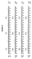
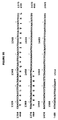
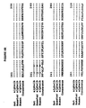
- Figures 1A-I. Nucleotide Sequence and Deduced Amino
Acid Sequence of Novel Human Alpha-la Adrenergic
Receptor.
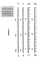
Nucleotides are presented in the 5' to 3'orientation and the coding region is numbered starting from the initiating methionine and ending in the termination codon. Deduced amino acid sequence by translation of a long open reading frame is shown, along with the 5' and 3' untranslated regions. Numbers in the left and right margins represent nucleotide (top line) and amino acid (bottom line) numberings, starting with the first position as the adenosine (A) and the initiating methionine (M), respectively. - Figures 2A-H. Nucleotide Sequence and Deduced Amino Acid Sequence of Novel Human Alpha-1b Adrenergic Receptor. Nucleotides are presented in the 5' to 3' orientation and the coding region is numbered starting from the initiating methionine and ending in the termination codon. Deduced amino acid sequence by translation of a long open reading frame is shown, along with the 5' and 3' untranslated regions. Numbers in the left and right margins represent nucleotide (top line) and amino acid (bottom line) numberings, starting with the first position as the adenosine (A) and the initiating methionine (M), respectively.
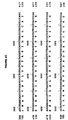
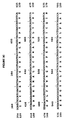
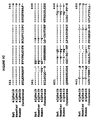
- Figures 3A-G. Nucleotide Sequence and Deduced Amino
Acid Sequence of Novel Human Alpha-1c Adrenergic
Receptor.
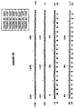
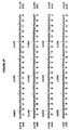
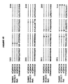
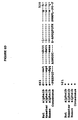
Nucleotides are presented in the 5' to 3' orientation and the coding region is numbered starting from the initiating methionine and ending in the termination codon. Deduced amino acid sequence by translation of a long open reading frame is shown, along with the 5' and 3' untranslated regions. Numbers in the left and right margins represent nucleotide (top line) and amino acid (bottom line) numberings, starting with the first position as the adenosine (A) and the initiating methionine (M), respectively. - Figures 4A-D. Alignment of the Human Alpha-1a, H318/3
Alpha-1a, and Rat Alpha-1a Adrenergic Receptors.
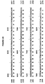
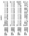
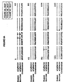
The deduced amino acid sequence of the human α1a receptor (first line), from the starting methionine (M) to the stop codon (*), is aligned with the previously published human "α1a" adrenergic receptor clone, H318/3 (2) (second line) and with the rat alphala (12) (third line). Also shown is a consensus amino acid sequence (fourth line), containing a hyphen at a particular position, when all receptors have the same amino acid or an amino acid at this position, when there is disparity in the three receptors. Dots indicate spaces corresponding to no amino acid at this position. Note that the human and rat α1a receptors have greater homology in the amino (positions 1-90) and carboxyl (positions 440-598) termini than do the previously published "α1a" (H318/3) and rat α1a receptors (see text). Dots indicate spaces corresponding to no amino acid at this position. Numbers above amino acid sequences correspond to amino acid positions, starting with the initiating methionine (M) and ending with the termination codon (*). - Figures 5A-D. Alignment of the Human Alpha-1b, Hamster
Alpha-1b, and Rat Alpha-1b Adrenergic Receptors.
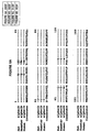
The deduced amino acid sequence of the human α1b receptor (third line), from the starting methionine (M) to the stop codon (*), is aligned with the previously published rat α1b adrenergic receptor clone (25) (first line) and with the hamster alpha-1b (4) (second line). Also shown is a consensus amino acid sequence (fourth line), containing a hyphen at a particular position, when all receptors have the same amino acid or an amino acid at this position, when there is disparity in the three receptors. Dots indicate spaces corresponding to no amino acid at this position. Numbers above amino acid sequences correspond to amino acid position, starting with the initiating methionine (M) and ending with the termination codon (*). - Figures 6A-C. Alignment of the Human Alpha-1c and
Bovine Alpha-1c Adrenergic Receptors.
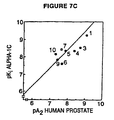
The deduced amino acid sequence of the human α1c receptor (first line), from the starting methionine (M) to the stop codon (*), is aligned with the previously published bovine α1b adrenergic receptor clone (13) (first line). Also shown is a consensus amino acid sequence (third line), containing a hyphen at a particular position, when all receptors have the same amino acid or an amino acid at this position, when there is disparity in the three receptors. Dots indicate spaces corresponding to no amino acid at this position. Numbers above amino acid sequences correspond to amino acid position, starting with the initiating methionine (M) and ending with the termination codon (*). - Figure 7. Illustrates the correlation of inhibition constants (pKi) for a series of α1 antagonists at the cloned human α1A, α1B, and α1C receptors with efficiency of blocking contraction of human prostate tissue (pA2).
-
- This invention also provides an isolated nucleic acid molecule encoding a human α1c adrenergic receptor. As used herein, the term "isolated nucleic acid molecule" means a non-naturally occurring nucleic acid molecule that is, a molecule in a form which does not occur in nature. Examples of such an isolated nucleic acid molecule are an RNA, cDNA, or an isolated genomic DNA molecule encoding a human α1c adrenergic receptor. As used herein, the term " α1c receptor" means a molecule which is a distinct member of a class of α1 adrenergic receptor molecules which under physiologic conditions, is substantially specific for the catecholamines epinephrine and norepinephrine, is saturable, and having high affinity for the catecholamines epinephrine and norepinephrine. The term "α1 adrenergic receptor subtype" refers to a distinct member of the class of human α1 adrenergic receptors, which may be α1c adrenergic receptors. The term "specific α1 adrenergic receptor" refers to a distinct member of the group or class of human α1 adrenergic receptors, which may be α1c adrenergic receptors.
- One embodiment of the invention is an isolated human nucleic acid molecule encoding a human α1c adrenergic receptor. Such a molecule may have coding sequences substantially the same as the coding sequence in Figures 3A-3G. The DNA molecule of Figures 3A-3G encodes the sequence of the human α1c adrenergic receptor. One means of isolating a nucleic acid molecule encoding a α1 adrenergic receptor is to screen a genomic DNA or cDNA library with a natural or artificially designed DNA probe, using methods well known in the art. In the preferred embodiment of this invention, α1 adrenergic receptors include the human α1c adrenergic receptors and the nucleic acid molecules encoding them were isolated by screening a human genomic DNA library and by further screening of a human cDNA library to obtain the sequence of the entire human α1c adrenergic receptor. To obtain a single nucleic acid molecule encoding the entire human α1c adrenergic receptor two or more DNA clones encoding portions of the same receptor were digested with DNA restriction endonuleases and ligated together with DNA ligase in the proper orientation using techniques known to one of skill in the art. DNA or cDNA molecules which encode a human α1c adrenergic receptor are used to obtain complementary genomic DNA, cDNA or RNA from human, mammalian or other animal sources, or to isolate related cDNA or genomic DNA clones by the screening of cDNA or genomic DNA libraries; by methods described in more detail below. Transcriptional regulatory elements from the 5' untranslated region of the isolated clone, and other stability, processing, transcription, translation, and tissue specificity determining regions from the 3' and 5' untranslated regions of the isolated gene are thereby obtained.
- This invention provides an isolated nucleic acid molecule which has been so mutated as to be incapable of encoding a molecule having normal human α1 adrenergic receptor activity, and not expressing native human α1 adrenergic receptor. An example of a mutated nucleic acid molecule provided by this invention is an isolated nucleic acid molecule which has an in-frame stop codon inserted into the coding sequence such that the transcribed RNA is not translated into protein.
- This invention provides a cDNA molecule encoding a human α1c adrenergic receptor, wherein the cDNA molecule has a coding sequence substantially the same as the coding sequence shown in Figures 3A-3G. These molecules and their equivalents were obtained by the means further described below.
- This invention provides an isolated protein which is a human α1 adrenergic receptor. In one embodiment of this invention, the protein is a human α1c adrenergic receptor having an amino acid sequence substantially similar to the amino acid sequence shown in Figures 3A-3G. As used herein, the term "isolated protein" is intended to encompass a protein molecule free of other cellular components. One means for obtaining an isolated human α1 adrenergic receptor is to express DNA encoding the α1 adrenergic receptor in a suitable host, such as a bacterial, yeast, or mammalian cell, using methods well known to those skilled in the art, and recovering the human α1 adrenergic receptor after it has been expressed in such a host, again using methods well known in the art. The human α1 adrenergic receptor may also be isolated from cells which express it, in particular from cells which have been transfected with the expression vectors described below in more detail.
- This invention provides a vector comprising an isolated nucleic acid molecule such as DNA, RNA, or cDNA, encoding a human α1c adrenergic receptor. Examples of vectors are viruses such as bacteriophages (such as phage lambda), cosmids, plasmids (such as pUC18, available from Pharmacia, Piscataway, NJ), and other recombination vectors. Nucleic acid molecules are inserted into vector genomes by methods well known to those skilled in the art. Examples of such plasmids are plasmids comprising cDNA having a coding sequence substantially the same as: the coding sequence shown in Figures 3A-3G. Alternatively, to obtain these vectors, insert and vector DNA can both be exposed to a restriction enzyme to create complementary ends on both molecules which base pair with each other and are then ligated together with a ligase. Alternatively, linkers can be ligated to the insert DNA which correspond to a restriction site in the vector DNA, which is then digested with the restriction enzyme which cuts at that site. Other means are also available.
- This invention also provides vectors comprising a DNA molecule encoding a human α1c adrenergic receptor adapted for expression in a bacterial cell, a yeast cell, or a mammalian cell which additionally comprise the regulatory elements necessary for expression of the DNA in the bacterial, yeast, or mammalian cells so located relative to the DNA encoding a human α1 adrenergic receptor as to permit expression thereof. DNA having coding sequences substantially the same as the coding sequence shown in Figures 3A-3G may be inserted into the vectors to express a human α1c adrenergic receptor. Regulatory elements required for expression include promoter sequences to bind RNA polymerase and transcription initiation sequences for ribosome binding. For example, a bacterial expression vector includes a promoter such as the lac promoter and for transcription initiation the Shine-Dalgarno sequence and the start codon AUG (Maniatis, et al., Molecular Cloning, Cold Spring Harbor Laboratory, 1982). Similarly, a eukaryotic expression vector includes a heterologous or homologous promoter for RNA polymerase II, a downstream polyadenylation signal, the start codon AUG, and a termination codon for detachment of the ribosome. Such vectors may be obtained commercially or assembled from the sequences described by methods well known in the art, for example the methods described above for constructing vectors in general. Expression vectors are useful to produce cells that express a human α1 adrenergic receptor. Certain uses for such cells are described in more detail below.
- In one embodiment of this invention a plasmid is adapted for expression in a bacterial, yeast, or, in particular, a mammalian cell wherein the plasmid comprises a DNA molecule encoding a human α1c adrenergic receptor and the regulatory elements necessary for expression of the DNA in the bacterial, yeast, or mammalian cell so located relative to the DNA encoding a human α1 adrenergic receptor as to permit expression thereof. Suitable plasmids may include, but are not limited to plasmids adapted for expression in a mammalian cell, e.g., pCEXV-3 derived expression vector. Examples of such plasmids adapted for expression in a mammalian cell are plasmids comprising cDNA having coding sequences substantially the same as the coding sequence shown in Figures 3A-3G and the regulatory elements necessary for expression of the DNA in the mammalian cell. This plasmid was pcEXV-α1c and deposited under ATCC Accession No. 75317. Those skilled in the art will readily appreciate that numerous plasmids adapted for expression in a mammalian cell which comprise DNA encoding human α1 adrenergic receptors and the regulatory elements necessary to express such DNA in the mammalian cell may be constructed utilizing existing plasmids and adapted as appropriate to contain the regulatory elements necessary to express the DNA in the mammalian cell. The plasmids may be constructed by the methods described above for expression vectors and vectors in general, and by other methods well known in the art.
- The deposits discussed supra were made pursuant to, and in satisfaction of, the provisions of the Budapest Treaty on the International Recognition of the Deposit of Microorganisms for the Purpose of Patent Procedure with the American Type Culture Collection (ATCC), 12301 Parklawn Drive, Rockville, Maryland 20852.
- This invention provides a mammalian cell comprising a DNA molecule encoding a human α1 adrenergic receptor, such as a mammalian cell comprising a plasmid adapted for expression in a mammalian cell, which comprises a DNA molecule encoding a human α1 adrenergic receptor and the regulatory elements necessary for expression of the DNA in the mammalian cell so located relative to the DNA encoding a human α1 adrenergic receptor as to permit expression thereof. Numerous mammalian cells may be used as hosts, including, but not limited to, the mouse fibroblast cell NIH3T3, CHO cells, HeLa cells, Ltk- cells, human embryonic kidney cells, Cos cells, etc. Expression plasmids such as that described supra may be used to transfect mammalian cells by methods well known in the art such as calcium phosphate precipitation, or DNA encoding these human α1 adrenergic receptors may be otherwise introduced into mammalian cells, e.g., by microinjection, to obtain mammalian cells which comprise DNA, e.g., cDNA or a plasmid, encoding a human α1 adrenergic receptor.
- As used herein, the phrase "specifically hybridizing" means the ability of a nucleic acid molecule to recognize a nucleic acid sequence complementary to its own and to form double-helical segments through hydrogen bonding between complementary base pairs. Nucleic acid probe technology is well known to those skilled in the art who will readily appreciate that such probes may vary greatly in length and may be labeled with a detectable label, such as a radioisotope or fluorescent dye, to facilitate detection of the probe. Detection of nucleic acid encoding a human α1 adrenergic receptor is useful as a diagnostic test for any disease process in which levels of expression of the corresponding human α1c adrenergic receptor are altered. DNA probe molecules are produced by insertion of a DNA molecule which encodes a human α1c adrenergic receptor or fragments thereof into suitable vectors, such as plasmids or bacteriophages, followed by insertion into suitable bacterial host cells and replication and harvesting of the DNA probes, all using methods well known in the art. For example, the DNA may be extracted from a cell lysate using phenol and ethanol, digested with restriction enzymes corresponding to the insertion sites of the DNA into the vector (discussed above), electrophoresed, and cut out of the resulting gel. Examples of such DNA molecules are shown in Figures 3A-3G. The probes are useful for "in situ" hybridization or in order to identify tissues which express this gene family, or for other hybridization assays for the presence of these genes or their mRNA in various biological tissues. In addition, synthesized oligonucleotides (produced by a DNA synthesizer) complementary to the sequence of a DNA molecule which encodes a human α1c adrenergic receptor are useful as probes for these genes, for their associated mRNA, or for the isolation of related genes by homology screening of genomic or cDNA libraries, or by the use of amplification techniques such as the Polymerase Chain Reaction.
- This invention also provides a method for detecting expression of a human α1c adrenergic receptor on the surface of a cell by detecting the presence of mRNA coding for a human α1c adrenergic receptor. These methods comprise obtaining total mRNA from the cell using methods well known in the art and contacting the mRNA so obtained with a nucleic acid probe as described hereinabove, under hybridizing conditions, detecting the presence of mRNA hybridized to the probe, and thereby detecting the expression of a specific human α1 adrenergic receptor by the cell. Hybridization of probes to target nucleic acid molecules such as mRNA molecules employs techniques well known in the art. However, in one embodiment of this invention, nucleic acids are extracted by precipitation from lysed cells and the mRNA is isolated from the extract using a column which binds the poly-A tails of the mRNA molecules (Maniatis, T. et al., Molecular Cloning; Cold Spring Harbor Laboratory, pp.197-98 (1982)). The mRNA is then exposed to radioactively labelled probe on a nitrocellulose membrane, and the probe hybridizes to and thereby labels complementary mRNA sequences. Binding may be detected by autoradiography or scintillation counting. However, other methods for performing these steps are well known to those skilled in the art, and the discussion above is merely an example.
- Antisense oligonucleotide drugs inhibit translation of mRNA encoding the human α1a, human alb or human α1c adrenergic receptors. Synthetic antisense oligonucleotides, or other antisense chemical structures are designed to bind to mRNA encoding the human α1c adrenergic receptor and inhibit translation of mRNA and are useful as drugs to inhibit expression of the human α1c adrenergic receptor in patients. Therapeutical alteration of levels of expression of the human α1c adrenergic receptor by the use of a synthetic antisense oligonucleotide drug (SAOD) which inhibits translation of mRNA encoding these α1 adrenergic receptors is possible. Synthetic antisense oligonucleotides, or other antisense chemical structures designed to recognize and selectively bind to mRNA, are constructed to be complementary to portions of the nucleotide sequences shown in Figures 3A-3G of DNA, RNA or of chemically modified, artificial nucleic acids. The SAOD is designed to be stable in the blood stream for administration to patients by injection, or in laboratory cell culture conditions, for administration to cells removed from the patient. The SAOD is designed to be capable of passing through cell membranes in order to enter the cytoplasm of the cell by virtue of physical nd chemical properties of the SAOD which render it capable of passing through cell membranes (e.g., by designing small, hydrophobic SAOD chemical structures) or by virtue of specific transport systems in the cell which recognize and transport the SAOD into the cell. In addition, the SAOD can be designed for administration only to certain selected cell populations by targeting the SAOD to be recognized by specific cellular uptake mechanisms which bind and take up the SAOD only within certain selected cell populations. For example, the SAOD may be designed to bind to a transporter found only in a certain cell type, as discussed above. The SAOD is also designed to recognize and selectively bind to the target mRNA sequence, which may correspond to a sequence contained within the sequences shown in Figures 3A-3G by virtue of complementary base pairing to the mRNA. Finally, the SAOD is designed to inactivate the target mRNA sequence by any of three mechanisms: 2) by binding to the target mRNA and thus inducing degradation of the mRNA by intrinsic cellular mechanisms such as mRNA target by interfering with the binding of translation-regulating factors or of other chemical structures, such as ribozyme sequences or reactive chemical groups. which either degrade or chemically modify the target mRNA. Synthetic antisense oligonucleotide drugs have been shown to be capable of the properties described above when directed against mRNA targets (J.S. Cohen, Trends in Pharm.
Sci 10, 435 (1989); H.M. Weintraub, Sci. AM. January (1990) p. 40). In addition, coupling of ribozymes to antisense oligonucleotides is a promising strategy for inactivating target mRNA (N. Sarver et al., Science 247, 1222 (1990)). An SAOD serves as an effective therapeutic agent if it is designed to be administered to a patient by injection, or if the patient's target cells are removed, treated with the SAOD in the laboratory, and replaced in the patient. In this manner, an SAOD serves as a therapy to reduce human α1 adrenergic receptor expression in particular target cells of a patient, in any clinical condition which may benefit from reduced expression of a specific human α1 adrenergic receptor. - This invention also provides a transgenic nonhuman mammal comprising DNA encoding a human α1c adrenergic receptor.
- This invention also provides a transgenic nonhuman mammal comprising DNA encoding a human α1c adrenergic receptor so mutated as to be incapable of normal human α1c adrenergic receptor activity, and not expressing native human α1c adrenergic receptor.
- This invention provides a transgenic non-human animal whose genome comprises DNA encoding a human α1c adrenergic receptor so placed as to be transcribed into antisense mRNA which is complementary to mRNA encoding a human α1c adrenergic receptor and which hybridizes to mRNA encoding the human α1c adrenergic receptor thereby reducing its translation. The DNA may additionally comprise an inducible promoter or additionally comprise tissue specific regulatory elements, so that expression can be induced, or restricted to specific cell types. Examples of DNA are DNA or cDNA molecules having a coding sequence substantially the same as the coding sequences shown in Figures 3A-3G. An example of a transgenic animal is a transgenic mouse. Examples of tissue specificity-determining regions are the metallothionein promoter (Low, M.J., Lechan, R.M., Hammer, R.E. et al. Science 231:1002-1004 (1986) and the L7 promoter (Oberdick, J., Smeyne, R.J., Mann, J.R., Jackson, S. and Morgan, J.I. Science 248:223-226 (1990)).
- Animal model systems which elucidate the physiological and behavioral roles of human α1 adrenergic receptors are produced by creating transgenic animals in which the increased or decreased, or the amino acid sequence of the expressed α1 adrenergic receptor is altered, by a variety of techniques. Examples of these techniques include, but are not limited to: 1) Insertion of normal or mutant versions of DNA encoding a human α1 adrenergic receptor or homologous animal versions of these genes, by microinjection, retroviral infection or other means well known to those skilled in the art, into appropriate fertilized embryos in order to produce a transgenic animal (Hogan B et al., Manipulating the Mouse Embryo, A Laboratory Manual, Cold Spring Harbor. Laboratory (1986)) or, 2) Homologous recombination (Capecchi M.R. Science 244:1288-1292 (1989); Zimmer, A. and Gruss, P. Nature 338:150-153 (1989)) of mutant or normal, human or animal version of the genes encoding α1 adrenergic receptors with the native gene locus in transgenic animals to alter the regulation of expression or the structure α1 of these α1 adrenergic receptors. The technique of homologous α1 adrenergic receptors. The technique of homologous recombination is well known in the art. It replaces the native gene with the inserted gene and so is useful for producing an animal that cannot express native α1 adrenergic receptor but does express, for example an inserted mutant human α1 adrenergic receptor, which has replaced the native α1 adrenergic receptor in the animal's genome by recombination, resulting in underexpression of the α1 adrenergic receptor. Microinjection adds genes to the genome, but does not remove them, and so is useful for producing an animal which expresses its own and added α1 adrenergic receptors, resulting in overexpression of the α1 adrenergic receptor.
- One means available for producing a transgenic animal, with a mouse as an example, is as follows: Female mice are mated, and the resulting fertilized eggs are dissected out of their oviducts. The eggs are stored in an appropriate medium such as M2 medium (Hogan B et al., Manipulating the Mouse Embryo, A Laboratory Manual, Cold Spring Harbor Laboratory (1986)). DNA or cDNA encoding a human α1 adrenergic receptor is purified from a vector (such as plasmids pCEXV-α1b, or pCEXV-α1c described above) by methods well known in the art. Inducible promoters may be fused with the coding region of the DNA to provide an experimental means to regulate expression of the trans-gene. Alternatively or in addition, tissue specific regulatory elements may be fused with the coding region to permit tissue-specific expression of the trans-gene. The DNA, in an appropriately buffered solution, is put into a microinjection needle (which may be made from capillary tubing using a pipet puller) and the egg to be injected is put in a depression slide. The needle is inserted into the pronucleus of the egg, and the DNA solution is injected. The injected egg is then transferred into the oviduct of a pseudopregnant mouse (a mouse stimulated by the appropriate hormones to maintain pregnancy but which is not actually pregnant), where it proceeds to the uterus, implants, and develops to term. As noted above, microinjection is not the only method for inserting DNA into the egg cell, and is used here only for exemplary purposes.
- Since the normal action of α1 adrenergic-specific drugs is to activate or to inhibit the α1 adrenergic receptor, the transgenic animal model systems described above are useful for testing the biological activity of drugs directed against specific human α1 adrenergic receptors even before such drugs become available. These animal model systems are useful for predicting or evaluating possible therapeutic applications of drugs which activate or inhibit these human α1 adrenergic receptors by inducing or inhibiting expression of the native or transgene and thus increasing or decreasing expression of normal or mutant human α1 adrenergic receptor in the living animal. Thus, a model system is produced in which the biological activity of drugs directed against these human α1 adrenergic receptors are evaluated before such drugs become available. The transgenic animals which over or under produce a specific human α1 adrenergic over or under produce a specific human α1 adrenergic over or under produce a specific human α1 adrenergic receptor indicate by their physiological state whether over or under production of the human α1 adrenergic receptor is therapeutically useful. It is therefore useful to evaluate drug action based on the transgenic model system. One use is based on the fact that it is well known in the art that a drug such as an antidepressant acts by blocking neurotransmitter uptake, and thereby increases the amount of neurotransmitter in the synaptic cleft. The physiological result of this action is to stimulate the production of less human α1 adrenergic receptor by the affected cells, leading eventually to underexpression. Therefore, an animal which underexpresses human α1 adrenergic receptor is useful as a test system to investigate whether the actions of such drugs which result in under expression are in fact therapeutic. Another use is that if overexpression is found to lead abnormalities, then a drug which down-regulates or acts as an antagonist to the human α1 adrenergic receptor is indicated as worth developing, and if a promising therapeutic application is uncovered by these animal model systems, activation or inhibition of the specific human α1 adrenergic receptor or antagonist drugs directed against these human α1 adrenergic receptors or by any method which increases or decreases the expression of these α1 adrenergic receptors in man.
- Due to this invention the physiological effects of expressing varying levels of a human α1 adrenergic receptor can be determined by a method, which comprises producing a transgenic nonhuman animals whose levels of α1 adrenergic receptor expression are varied by use of an inducible promoter which regulates human α1 adrenergic receptor expression. Due to this invention, the physiological effects of expressing varying levels of human α1 adrenergic receptors can be determined by a method which comprise producing a panel of transgenic nonhuman animals each expressing a different amount of a human α1 adrenergic receptor. Such animals may be produced by introducing different amounts of DNA encoding a human α1 adrenergic receptor into the oocytes from which the transgenic animals are developed.
- This invention also provides a method for identifying a substance capable of alleviating abnormalities resulting from overexpression of a human α1 adrenergic receptor comprising administering the substance to a transgenic nonhuman mammal expressing at least one artificially introduced DNA molecule encoding a human α1 adrenergic receptor and determining whether the substance alleviates the physical and behavioral abnormalities displayed by the transgenic nonhuman mammal as a result of overexpression of a human α1 adrenergic receptor. As used herein, the term "substance" means a compound or composition which may be natural, synthetic, or a product derived from screening. Examples of DNA molecules are DNA or cDNA molecules having a coding sequence substantially the same as the coding sequences shown in Figures 3A-3G.
- This invention provides a method for identifying a substance capable of alleviating the abnormalities resulting from underexpression of a human α1 adrenergic receptor comprising administering the substance to the transgenic nonhuman mammal described above which expresses only a nonfunctional human α1 adrenergic receptor and determining whether the substance alleviates the physical and behavioral abnormalities displayed by the transgenic nonhuman mammal as a result of underexpression of the human α1 adrenergic receptor.
- This invention provides a method of preparing an isolated human α1 adrenergic receptor which comprises inducing cells to express the human α1 adrenergic receptor, recovering the α1 adrenergic receptor from the resulting cells, and purifying the α1 adrenergic An example of an isolated human α1c adrenergic receptor is an isolated protein having substantially the same amino acid sequence shown in Figure 3A-3G. For example, cells can be induced to express human α1 adrenergic receptor by exposure to substances such as hormones. The cells can then be homogenized and the human α1 adrenergic receptor isolated from the homogenate using an affinity column comprising, for example, epinephrine, norepinephrine, or another substance which is known to bind to the human α1 adrenergic receptor. The resulting fractions can then be purified by contacting them with an ion exchange column, and determining which fraction contains human α1 adrenergic receptor activity or binds anti-human α1 adrenergic receptor activity or binds anti-human α1 adrenergic receptor antibodies.
- This invention also provides a method of preparing the isolated human α1 adrenergic receptor which comprises inserting nucleic acid encoding the human α1b adrenergic receptor in a suitable vector, inserting the resulting vector in a suitable host, recovering the α1 adrenergic receptor produced by the resulting cell, and purifying the α1 adrenergic receptor so recovered. These methods for preparing human α1 adrenergic receptor uses recombinant DNA technology methods well known in the art. For example, isolated nucleic acid encoding a human α1 adrenergic receptor is inserted in a suitable vector, such as an expression vector. A suitable host cell, such as a bacterial cell, or a eukaryotic cell such as a yeast cell is transfected with the vector. The human α1 adrenergic receptor is isolated from the culture medium by affinity purification or by chromatography or by other methods well known in the art.
- This invention provides a method of determining whether a ligand not known to be capable of binding to a human α1 adrenergic receptor can bind to a human α1 adrenergic receptor, which comprises contacting a mammalian cell comprising a plasmid adapted for expression in a mammalian cell which further comprises a DNA molecule which expresses a human α1 adrenergic receptor on the cell surface with the ligand under conditions permitting binding of ligands known to bind to the human α1 adrenergic receptor, detecting the presence of any ligand bound to the human α1 adrenergic receptor. The DNA in the cell may have a coding sequence substantially the same as the coding sequences shown in Figures 3A-3G, preferably, the mammalian cell is nonneuronal in origin. An example of a nonneuronal mammalian cell is a Cos7 cell. The preferred method for determining whether a ligand is capable of binding to the human α1 adrenergic receptor comprises contacting a transfected nonneuronal mammalian cell (i.e. a cell that does not naturally express any type of human α1 adrenergic receptor, thus will only express such human α1 adrenergic receptor if it is transfected into the cell) expressing a human α1 adrenergic receptor on it surface, or contacting a membrane preparation derived from such a transfected cell, with the ligand under conditions which are known to prevail, and thus be associated with in vivo binding of the substrates to a human α1 adrenergic receptor, detecting the presence of any of the ligand being tested bound to the human α1 adrenergic receptor on the surface of the cell, and thereby determining whether the ligand binds to the human α1 adrenergic receptor. This response system is obtained by transfection of isolated DNA into a suitable host cell. Such a host system might be isolated from pre-existing cell lines, or can be generated by inserting appropriate components into existing cell lines. Such a transfection system provides a complete response system for investigation or assay of the functional activity of human α1 adrenergic receptors with ligands as described above. Transfection systems are useful as living cell cultures for competitive binding assays between known or candidate drugs and substrates which bind to the human α1 adrenergic receptor and which are labeled by radioactive, spectroscopic or other reagents. Membrane preparations containing the transporter isolated from transfected cells are also useful for these competitive binding assays. A transfection system constitutes a "drug discovery system" useful for the identification of natural or synthetic compounds with potential for drug development that can be further modified or used directly as therapeutic compounds to activate or inhibit the natural functions of a specific human α1 adrenergic receptor. The transfection system is also useful for determining the affinity and efficacy of known drugs at human α1 adrenergic receptor binding sites.
- This invention provides a method for identifying a ligand which interacts with, and activates or blocks the activation of, a human α1 adrenergic receptor on the surface of the cell, which comprises contacting a mammalian cell which comprises a plasmid adapted for expression in a mammalian cell which further comprises a DNA molecule which expresses a human α1 adrenergic receptor on the cell surface with the ligand, determining whether the ligand activates or blocks the activation of the receptor using a bioassay such as a second messenger assays, and thereby identifying a ligand which interacts with, and activates or blocks the activation of, a human α1 adrenergic receptor.
- This invention provides functional assays for identifying ligands and drugs which bind to and activate or inhibit a specific human α1 adrenergic receptor activity.
- This invention provides a method for identifying a ligand which is capable of binding to and activating or inhibiting a human α1 adrenergic receptor, which comprises contacting a mammalian cell, wherein the membrane lipids have been labelled by prior incubation with a labelled myo-inositol phosphate molecule, the mammalian cell comprising a plasmid adapted for expression in a mammalian cell which further comprises a DNA molecule which expresses a human α1 adrenergic receptor with the ligand and identifying an inositol phosphate metabolite released from the membrane lipid as a result of ligand binding to and activating an α1 adrenergic receptor.
- This invention provides method for identifying a ligand that is capable of binding to and activating or inhibiting a human α1 adrenergic receptor, where in the binding of ligand to the adrenergic receptor results in a physiological response, which comprises contacting a mammalian cell which further comprises a DNA molecule which expresses a human α1 adrenergic receptor with a calcium sensitive fluorescent indicator, removing the indicator that has not been taken up by the cell, contacting the cells with the ligand and identifying an increase or decrease in intracellular Ca+2 as a result of ligand binding to and activating receptors.
- Transformed mammalian cells for identifying the ligands and drugs that affect the functional properties of the human α adrenergic receptor include C-α1c-7.
- This invention also provides a method of screening drugs to identify drugs which interact with, and bind to, a human α1 adrenergic receptor on the surface of a cell, which comprises contacting a mammalian cell which comprises a plasmid adapted for expression in a mammalian cell which further comprises a DNA molecule which expresses a human α1 adrenergic receptor on the cell surface with a plurality of drugs, determining those drugs which bind to the human α1 adrenergic receptor expressed on the cell surface of the mammalian cell, and thereby identifying drugs which interact with, and bind to, the human α1 adrenergic receptor. Various methods of detection may be employed. The drugs may be "labeled" by association with a detectable marker substance (e.g., radiolabel or a non-isotopic label such as biotin). The DNA in the cell may have a coding sequence substantially the same as the coding sequences shown in Figures 3A-3G. Preferably, the mammalian cell is nonneuronal in origin. An example of a nonneuronal mammalian cell is a Cos7 cell. Drug candidates are identified by choosing chemical compounds which bind with high affinity to the human α1 adrenergic receptor expressed on the cell surface in transfected cells, using radioligand binding methods well known in the art, examples of which are shown in the binding assays described herein. Drug candidates are also screened for selectivity by identifying compounds which bind with high affinity to one particular human α1 adrenergic receptor subtype but do not bind with high affinity to any other human α1 adrenergic receptor subtype or to any other known receptor site. Because selective, high affinity compounds interact primarily with the target human α1 adrenergic site after administration to the patient, the chances of producing a drug with unwanted side effects are minimized by this approach. This invention provides a pharmaceutical composition comprising a drug identified by the method described above and a pharmaceutically acceptable carrier. As used herein, the term "pharmaceutically acceptable carrier" encompasses any of the standard pharmaceutical carriers, such as a phosphate buffered saline solution, water, and emulsions, such as an oil/water or water/oil emulsion, and various types of wetting agents. Once the candidate drug has been shown to be adequately bio-available following a particular route of administration, for example orally or by injection (adequate therapeutic concentrations must be maintained at the site of action for an adequate period to gain the desired therapeutic benefit), and has been shown to be non-toxic and therapeutically effective in appropriate disease models, the drug may be administered to patients by that route of administration determined to make the drug bio-available, in an appropriate solid or solution formulation, to gain the desired therapeutic benefit.
- Applicants have identified individual human α1 adrenergic receptor subtypes and have described methods for the identification of pharmacological compounds for therapeutic treatments. Pharmacological compounds which are directed against a specific human adrenergic receptor subtype provide effective new therapies with minimal side effects.
- Substances as described supra may be useful for the preparation of a pharmaceutical composition for treating or alleviating abnormalities resulting from overexpression or increased activity of the human α1c adrenergic receptor. Examples of such abnormalities related to an excess of activity of a human α1 adrenergic receptor subtype include but are limited to benign prostatic hypertrophy, coronary heart disease, hypertension, urinary retention, insulin resistance, atherosclerosis, sympathetic dystrophy syndrome, glaucoma, cardiac arrythymias erectile dysfunction, and Renaud's syndrome.
- Substances as described supra may be useful for the preparation of a pharmaceutical composition for treating or alleviating abnormalities resulting from underexpression or decreased activity of the human α1c adrenergic receptor.
- Examples of such abnormalities related to a decrease in the activity of a specific human α1 adrenergic receptor include but are not limited to congestive heart failure, urinary incontinence, nasal congestion and hypotension.
- Elucidation of the molecular structures of the neuronal human α1 adrenergic receptors transporters is an important step in the understanding of α-adrenergic neurotransmission. This disclosure reports the isolation, the nucleic acid sequence, and functional expression of DNA clones isolated from human brain which encode human α1 adrenergic receptor. The identification of these human α1 adrenergic receptor will play a pivotal role in elucidating the molecular mechanisms underlying α-adrenergic transmission, and should also aid in the development of novel therapeutic agents.
- DNA clones encoding human α1 adrenergic receptor have been isolated from human brain, and their functional properties have been examined in mammalian cells.
- This invention identifies for the first time a new human α1 adrenergic receptor, its amino acid sequences, and its human genes. The information and experimental tools provided by this discovery are useful to generate new therapeutic agents, and new therapeutic or diagnostic assays for these new human receptors, their associated mRNA molecules or their associated genomic DNAs. The information and experimental tools provided by this discovery will be useful to generate new therapeutic agents, and new therapeutic or diagnostic assays for these new human receptors, their associates mRNA molecules, or their associated genomic DNAs.
- Specifically, this invention relates to the first isolation of human α1c-adrenergic receptor. In addition, the human α1c adrenergic receptor has been expressed in mammalian cells by transfecting the cells with the plasmid pcEXV-α1c. The pharmacological binding properties of these receptor proteins have been determined, and these binding properties classify these receptor proteins as α1 adrenergic receptor. Mammalian cell lines expressing the human α1 adrenergic receptor on the cell surface have been constructed, thus establishing the first well-defined, cultured cell lines with which to study human α1 adrenergic receptor. An Example of transformed mammalian cells, expressing human α1 adrenergic receptor is L-α1c expressing a human α1c adrenergic receptor. These cells are suitable for studying the pharmacological properties of the human α1 adrenergic receptor and for the screening of ligands and drugs that specifically bind to human α1 adrenergic receptor subtypes.
- The invention will be better understood by reference to the Experimental Details which follow, but those skilled in the art will readily appreciate that the specific experiments detailed are only illustrative, and are-not meant to limit the invention as described herein, which is defined by the claims which follow thereafter.
- α1a: A human lymphocyte genomic library in ζ dash II (≈1.5 x 10⁶ total recombinants; Stratagene, LaJolla, CA.) was screened using a cloned rat PCR fragment (RBNC2) as a probe. RBNC2 was obtained by amplifying randomly primed rat brain cDNA with degenerate primers designed to conserved regions of transmembrane (Tm)
regions - The probe was labeled with [32 P] by the method of random priming (5) (Prime-It Random Primer kit, Strategene, LaJolla, CA.). Hybridization was performed at 40°C. in a solution containing 50% formamide, 10% dextran sulfate, 5x SSC (1X SSC is 0.15M sodium choloride, 0.015M sodium citrate), 1x Denhardt's solution (0.02% polyvinylpyrrolidone, 0.02% Ficoll, 0.02% bovine serum albumin), and 200 µg/µl sonicated salmon sperm DNA. The filters were washed at 50°C. in 0.1x SSC containing 0.5% sodium dodecyl sulfate and exposed at -70°C to Kodak XAR film in the presence of an intensifying screen. Lambda phage clones hybridizing with the probe were plaque purified and DNA was prepared for Southern blot analysis (22, 17). For subcloning and further Southern blot analysis, DNA was cloned into pUC18 (Pharmacia, Piscataway, NJ) or pBluescript (Stratagene, LaJolla, Ca.). Nucleotide sequence analysis was accomplished by the Sanger dideoxy nucleotide chain termination method (18) on denatured double-stranded plasmid templates, using Sequenase (US Biochemcial Corp., Cleveland, OH), Bst DNA sequencing kit (Bio-Rad Laboratories, Richmond, CA.), or TaqTrack sequencing kit (Promega Corporation, Madison, WI.).
- In order to isolate a full-length clone, human cDNA libraries were screened by polymerase chain reaction (PCR) with 1µM each of specific oligonucleotide primers designed off the isolated genomic clone: from the sense strand (nucleotide 598-626), 5' CACTCAAGTACCCAGCCATCATGAC 3' and from the antisense stand (nucleotide 979-1003), 5' CGGAGAGCGAGCTGCGGAAGGTGTG 3' (see Figures 1A01I). The primers were from non-conserved portions of the receptor gene, specifically in the Tm3-Tm3 loop and in the Tm5-Tm6 loop regions for the upstream and downstream primers, respectively. One to 2 µl of phage DNA from cDNA libraries (ζ ZapII; Stratagene, LaJolla, CA.),representing ≈106-107 pfu, were amplified in 10mM Tris-HCl, pH 8.3, 50mM KCl, 1.5mM MgCl2, 0.01% gelatin, 200 µM each dATP, dCTP, dTTP, 2.5 units of Thermus aquaticus DNA polymerase (Taq polymerase; Perkin-Elmer-Cetus, Norwalk, CT.). The amplification profile was run for 30 cycles: a 5 min. initial (i.e. 1 cycle denaturation at 95°C., followed by 2 min. at 94°C., 2 min at 68°C., and 3 min at 72°C., with a 3 sec. extension, followed by a final 10 min. extension at 72°C. PCR products were analyzed by ethidium bromide (EtBr) stained agarose gels and any sample exhibiting a band on the EtBr stained gel was considered positive.
- A positive library was then plated and screened with overlapping 45-mer oligonucleotide probes, filled-in using [α-32P]dCTP and [α-32P]dATP and Klenow fragment of DNA polymerase. This probe was internal to the amplification primers discussed above from the sense strand (nucleotide 890 - 934) , 5' GCAAGGCCTCCGAGGTGGTGCTGCGCATCCACTGTCGCGGCGCGG 3', and from the anti-sense strand (nucleotide 915-961), 5' TGCCGTGCGCCCCGTCGGCGCCCGTGGCCGCGCCGCGACAGTGGATG 3' (see Figures 1A-1I). Positive cDNA phage clones were plaque certified and pBluescript recombinant DNAs were excision-rescued from ζ Zap II using helper phage R408, as described by manufacturer's protocol (Stratagene, LaJolla, CA.). Insert size was confirmed by restriction enzyme digest analysis and recombinants were sequences as described above.
- α1b: A human placenta genomic library in λ dash II (≈1.5 x 10⁶ total recombinants; Stratagene, LaJolla, CA.) was screened using overlapping 45-mer oligonucleotides radiolabeled as described above and directed to the third, fifth and sixth transmembrane regions of serotonin 5HT1Dβ receptor gene. Hybridization and washing conditions were identical to that described for α1a above except lower stringency hybridization nd washes were conducted; specifically, hybridization in 25% formamide and washes at 40°C.
- Positive-hybridizing λ phage clones were plaque-purified, analyzed by Southern blot analysis, subcloned and sequenced, as described above for α1a. In order to isolate full-length clones, human cDNA libraries in λ Zap II (Strategene, LaJolla, CA.) were screened by polymerase chain reaction as described above. The upstream and downstream PCR primers used were from the Tm40Tm5 loop and the Tm5-Tm6 loop, respectively: from the sense strand (nucleotide 567-593), 5' CAACGATGACAAGGA GTGCGGGGTCAC 3', and from the antisense strand (nucleotide 822-847), 5' TTTGACAGCTATGGAACTCCTGGGG 3' (see Fig. 2). PCR, library screen, plaque purification excision-rescue from λ Zap II, restriction digestions and sequencing were accomplished as described above for α1a. The internal probe was: from the sense strand (nucleotide 745-789), 5'AAGGAGCTGACCCTGAGGATCCATTCCAAGAACTTTC ACGAGGAC 3', and from the anti-sense strand (nucleotide 770-814), 5' CCTTGGCCTTGGTACTGCTAAGGGTGTCCTCGTGAAA GTTCTTGG 3' (see Figures 2A-2H).
- α1c: A human lymphocyte genomic library in λ dash II (≈1.5x10⁶ total recombinants; Stratagene, LaJolla, CA.) was screened using overlapping 45-mer oligonucleotides radiolabeled as described for α1a and directed to the third, fifth and sixth transmembrane regions of serotonin 5HT1A receptor gene. Hybridization and washing conditions were identical to that described for α1b. Positive-hybridizing λ phage clones were plaque-purified, analyzed by Southern blot analysis, subcloned and sequenced, as described above for α1a. Identification and isolation of full=length clones by PCR and screening cDNA libraries were accomplished as described for α1b. The upstream and downstream PCR primers used were from the Tm3-Tm4 loop and the Tm5-Tm6 loop, respectively: from the sense strand (nucleotide 403-425), 5' CCAACCATCGTCACCCAGAGGAG 3', and from the antisense strand (nucleotide 775-802), 5' TCTCCCGGG AGAACTTGAGGAGCCTCAC 3' (see Figures 3A-3G). The internal probe was: from the sense strand (nucleotide 711-745), 5' TCCGCATCCATCGGAAAAACGCCCCGGCAGGAGGC AGCGGGATGG 3', and from the anti-sense strand (nucleotide 726-771), 5' GAAGTGCGTCTTGGTCTTGGCGCT GGCCATCCCGCTGCCTCCTGCC 3' (see Figures 3A-3G). PCR, library screen, plaque purification excision-rescue from λ Zap II, restriction digestions and sequencing were accomplished as described above for α1a.
- α1a: The entire coding region of α1a (1719 bp), including 150 basepairs of 5' untranslated sequence (5' UT) and 300 bp of 3' untranslated sequence (3' UT), was cloned into the BamHI and ClaI sites of the polylinker-modified eukaryotic expression vector pCEXV-3 (13), called EXJ.HR (unpublished data). The construct involved the ligation of partial overlapping human lymphocyte genomic and hippocamppal cDNA clones: 5' sequences were contained on a 1.2 kb SmaI-XhoI genomic fragment (the vector-derived BamHI site was used for subcloning instead of the internal insert-derived SmaI site) and 3' sequences were contained on an 1.3 kb XhoI-ClaI cDNA fragment (the Clal site was from the vector polylinker). Stable cell lines were obtained by cotransfection with the plasmid α1a/EXJ (expression vector containing the α1a receptor gene) and the plasmid pGCcos3neo (plasmid containing the aminoglycoside transferase gene) into LM(tk-), CHO, NIH3T3 cells, and 293 cells using calcium phosphate technique. The cells were grown, in a controlled environment (37°C., 5% CO2), as monolayers in Dulbecco's modified Eagle's Medium (GIBCO, Grand Island, NY) containing 25mM glucose and supplemented with 10% bovine calf serum, 100 units/ml penicillin G, and 100 µg/ml streptomycin sulfate. Stable clones were then selected for resistance to the antibiotic G-418 (1 mg/ml) as described previously (26) and membranes were harvested and assayed for their ability to bind [3H]prazosin as described below (see "Radioligand Binding Assays").
- α1b: The entire coding region of α1b (1563 bp), including 200 basepairs of 5' untranslated sequence (5' UT) and 600 bp of 3' untranslated sequence (3' UT), was cloned into the EcoRI site of pCEXV-3 eukaryotic expression vector (13). The construct involved ligating the full-length containing EcoRI brainstem cDNA fragment from λ Zap II into the expression vector. Stable cell lines were selected as described above.
- α1c: The entire coding region of α1c (1401 bp), including 400 basepairs of 5' untranslated sequence (5' UT) and 200 bp of 3' untranslated sequence (3' UT), was cloned into the KpnI site of the polylinker-modified pCEXV-3-derived (13) eukaryotic expression vector, EXJ.RH (unpublished data). The construct involved ligating three partial overlapping fragments: a 5' 0.6kb HincII genomic clone, a central 1.8 EcoRI hippocamppal cDNA clone, and a 3' 0.6kb PstI genomic clone. The hippocamppal cDNA fragment overlaps with the 5' and 3' genomic clones so that the HincII and PstI sites at the 5' and 3' ends of the cDNA clones, respectively, were utilized for ligation. This full-length clone was cloned into the KpnI sites of the fragment, derived from vector (ie pBluescript) and 3' untranslated sequences, respectively. Stable cell lines were selected as described above.
- Transfected cells from culture flasks were scraped into 5ml of 5mM tris-HCl, 5mM EDTA, pH 7.5, and lysed by sonication. The cell lysates were centrifuged at 1000 rpm for 5 min at 4°C. The pellet was suspended in 50mM Tris-HCl, 1mM MgCl2, and 0.1% ascorbic acid at pH 7.5. Binding of the α1 antagonist [3H]prazosin (0.5 nM, specific activity 76.2 Ci/mmol) to membrane preparations of LM(tk-) cells was done in a final volume of 0.25 ml and incubated at 37°C for 20 min. Nonspecific binding was determined in the presence of 10 µM phentolamine. The reaction was stopped by filtration through GF/B filters using a cell harvester. Data were analyzed by a computerized non-linear regression program.
- Cells were suspended in Dulbecco's phosphate buffered saline (PBS) , and incubated with 5µCi/ml [3H]m-inositol for 60 min at 37°C, the reaction was stopped by adding CHCl3:Methanol: HCl (2/1/0.01 v/v). Total [3H]IP were separated by ion exchange chromatography and quantified as described by Forray and El-Fakahany (7).
- Intracellular calcium levels ([Ca2+]i) were determined with the calcium-sensitive dye fura-2, and microspectrofluorometry, essentially as previously described (1,3). Briefly, cells were plated into polylysine-coated coverslip bottom dishes (MatTek Corporation, Ashland MA). To lead with fura-2, cells were washed 3x with HEPES-buffered saline (HBS, in mM: HEPES, 20; NaCl, 150; KCl, 5; CaCl2, 1; MgCl2, 1; glucose, 10; pH 7.4) and incubated for 30 minutes at room temperature with fura-2 loading solution (5uM fura-2/AM, 0.03% pluronic F-127, and 2% heat-inactivated fetal calf serum, in HBS). After loading, cells were washed 3x with HBS, 1ml of HBS was added, and the dish was placed on the microscope for determination of [Ca2+]i. [Ca2+]i was measured with a Leitz Fluovert microscope equipped for UV-transmission epifluorescence. Fura-2 fluorescence was alternately excited at 340 and 380nm (0.25 sec), and a pair of readings (500nm long pass) was taken every two seconds, and recorded by a personal computer interfaced to a data acquisition and control unit from Kinetek (Yonkers, NY). To determine [Ca2+]i from the experimental data the background fluorescence was subtracted, and the corrected ratios were converted to [Ca2+]i by comparison with buffers containing saturating and low free calcium, assuming a KD of 400 nM (3).
- α1a: We screened a human genomic lymphocyte library with a rat PCR fragment that exhibited homology with the α1-AR family. A total of six clones were isolated and characterized by Southern blot analysis. One clone, h13, contained a 4.0kb XbaI fragment which hybridized with the radiolabeled rat PCR fragment and was subsequently subcloned into pUC vector. DNA sequence analysis indicated greatest homology to human α1a and rat α1a ARs. This clone contained the initiating methionine through Tm6 with ≈1.0-1.5kb 5' UT region. Subsequent Southern blot, analysis, subcloning and sequencing analysis indicated the presence of a SmaI site ≈150nts. 5' to the initiating methionine codon. The homology between h13 and rat ala adrenergic gene breaks just downstream of Tm6, indicating an intron which is located in an analogous region in the alb- and α1c-AR genes (4,20). In order to obtain a full-length clone, aliquots of human cDNA libraries totaling ≈1.5x10⁶ recombinants was screened by polymerase chain reaction using specific oligonucleotide primers from sequence determined off the genomic clone (see Materials and Methods). A positive- containing human hippocamppal cDNA library (Stratagene, LaJolla, CA.) in λ Zap II (≈1.5x10⁶ recombinants) was screened using traditional plaque hybridization with an internal probe (see Materials and Methods) and resulted in the isolation of two positive cDNA clones, one containing the upstream sequences (from 5' UT through the 5-6 loop; hH22) and the other containing downstream sequences (from within Tm5 through ≈200 nts. with a common XhoI site being present within this common region.
- The complete full-length gene was constructed by splicing together two restriction fragments, one being the 3' cDNA (hHl4) and the other being the 5' genomic clone (h13), using a unique restriction site (XhoI) present in the overlapping region. In addition, another construct was accomplished by ligating the two cDNA clones (hH14 and hH22), using the overlapping XhoI site; however, since this construct produced the same pharmacology as the genomic/cDNA construct, we will not discuss this recombinant (unpublished observation). The genomic/cDNA construct contains an open reading frame of 1719 bp and encoding a protein of 572 aa in length, having a relative molecular mass of ≈63,000 daltons. Hydropathy analysis of the protein is consistent with a putative topography of seven transmembrane domains, indicative of the G protein-coupled receptor family. Initial sequence analysis revealed that clone α1a/EXJ was most related to an AR since it contained a number of conserved structural features/residues found among the members of the adrenergic receptor family, including conserve cysteines in the second and third extracellular loops, a conserved glycine residue in Tm1, aspartic acid residues in Tm regions II and III, conserved valine residues in TmIII, the DRY sequence at the end of TmIII, the conserved proline residues of Tm regions II, IV, V, VI and VII, and the consensus D-V-L-X-X-T-X-S-I-X-X-L-C IN Tm3 and the consensus G-Y-X-N-S-X-X-N-P-X-I-Y in the Tm VII, both consensus unique to the adrenergic receptor family (2,26). Other features of this human α1a receptor gene are the presence of two potential sites for N-linked glycosylation in the amino terminus (
asparagine residues - α 1b: We screened a human genomic placenta library with probes derived from Tm3, 5 and 6 regions of serotonin 5HT1DB under low stringency. Out of several hundred positive clones pursued by Southern blot analysis, subcloning and sequencing, one resembled the α1 adrenergic family of receptors. This genomic fragment contained Tm3 through Tm6 of a receptor which was most closely related to rat and hamster α1b receptors. In order to obtain a full-length clone, several human cDNA libraries were screened by PCR using primers derived from the 5-6 loop region of the genomic clone (see Materials and Methods). A positive-containing human brainstem cDNA library (Stratagene, LaJolla, CA) in λ ZAPII (≈2 x 10⁶ recombinants) was screened using traditional plaque hybridization with an internal probe, resulting in the isolation of two identical cDNA clones, containing an insert size of 2.4 kb. Upon sequencing, this clone was found to contain the initiating MET aa, Tml through Tm7, and 5' and 3' UT sequences, suggesting a full-length clone on a single EcoRI fragment. This cDNA clone contains an open reading frame of 1563 bp and encodes a protein of 520 aa in length, having a relative molecular mass of ≈57,000 daltons. Hydropathy analysis of the protein is consistent with a putative topography of seven transmembrane domains, indicative of the G protein-coupled receptor family.
- Sequence analysis revealed that clone α1b/pCEXV was most related to adrenergic receptor since it contained a number of conserved structural features found among the adrenergic receptor family, as described for α1a receptor (see above). This human α1b receptor contains potential sites for N-linked glycosylation in the amino terminus (
asparagine residues 10, 24, 29, 34 in Fig. 2A-2H), consistent with the finding that the α1 AR is glycosylated (4,19). - α 1c: We screened a human genomic lymphocyte library with probes derived from the third, fifth and sixth transmembrane regions of serotonin 5HT1A under low stringency. Out of several hundred positive clones analyzed by Southern blot analysis, subcloning and sequencing (see Materials and Methods), one phage clone resembled a novel α1 AR. This genomic fragment contained Tm1 through Tm6 of a receptor with high homology to the bovine α1c receptor and thus suggesting the presence of an intron downstream of Tm6, as shown for the α1 receptor family (4,12,20). In order to obtain a full-length clone, several human cDNA libraries were screened by PCR, as described for α1b (also see Materials and Methods). A positive-containing human hippocamppal cDNA library (Stratagene, LaJolla, CA) in λ ZAPII (≈2 x 10⁶ recombinants) was screened, as described for α1b. A positive clone (hH 20) was identified which contained a 1.7kb EcoRI cDNA fragment insert. However, this cDNA clone lacked both the amino end of the receptor (the 5' end of the clone terminated at the 5' end of Tm2) and part of the carboxyl tail (the 3' end of the clone corresponded to 40 aa upstream from the "putative" stop codon). Since an alternative genomic subclone which contained the initiating MET codon in addition to Tm1 through Tm6 was available, we needed to obtain the complete 3' carboxyl tail in order to complete the construct of the full-length clone. This was accomplished by using overlapping 45-mer oligonucleotide primers (corresponding to nts. 1142-1212 in Fig. 3), designed within the carboxyl tail of the receptor (at the 3' end of the hH20 cDNA clone), to screen a human lymphocyte genomic library in order to isolate a genomic clone containing the carboxyl tail that includes the termination codon. Two identical positive human lymphocyte genomic clones were isolated from this library. A 0.6 kb PstI fragment was subcloned and shown to contain most of the carboxyl tail (≈20 aa downstream of Tm7) through the termination codon and ≈200 bp of 3' UT sequence.
- The complete full-length gene was constructed by splicing together three restriction fragments: A 0.6 kb HincII fragment from the genomic clone, containing ≈0.4 kb of 5' UT sequence and the initiating MET codon through Tm2; the 0.8 kb HincII-PstI fragment from the hH cDNA clone, which contains Tm2 through part of the carboxyl tail, overlapping with the 5' genomic clone by 20 nts. (sharing the unique HincII site at position 196 in Fig.3); and a 0.6 kb PstI fragment from the second h1 genomic clone, which contains the carboxyl tail, the stop codon and ≈0.2 kb of 3' UT sequence, and overlapping with the hH cDNA clone (sharing the unique Pst I site within the carboxyl tail at position 1038 in Figures 3A-3G).
- The resulting genomic/cDNA/genomic construct contains an open reading frame of 1401 bp and encoding a protein of 466 aa in length, having a molecular weight of ≈51,000 daltons. Hydropathy analysis of the protein is consistent with a putative topography of seven transmembrane domains, as indicated for the previously described human α1a and α1b receptors and indicative of the G protein-coupled receptor family. Sequence analysis revealed that clone α1c/EXJ was most related to adrenergic receptor because it contained the structural features commonly found among the adrenergic receptor family of receptors, as described for the α1a receptor above. Other features of this human α1c receptor gene is the presence of three potential sites for N-linked glycosylation in the amino terminus, at the same position described for the bovine α1c receptor (
asparagine residues Competition of adrenergic agonists and antagonists for the binding of [3H]prazosin to membranes prepared from LM(tk-) cells expressing the human α1a, α1b, and α1c-adrenergic receptor cDNA. Membrane preparations from stabily transfected cell lines increasing concentrations of various agonists or antagonists as described under "Materials and Methods". Data is shown as the mean ± S.E.M. of the binding parameters estimated by a computerized non-linear regression analysis obtained in three independent experiments each performed in triplicate. pKi α1a α1b α1c AGONISTS Norepinephrine 6.633 ± 0.12 5.614 ± 0.09 5.747 ± 0.18 Epinephrine 6.245 ± 0.10 5.297 ± 0.15 5.511 ± 0.13 Oxymetazoline 5.903 ± 0.16 5.919 ± 0.07 7.691 ± 0.10 Naphazoline 6.647 ± 0.18 6.155 ± 0.04 6.705 ± 0.22 Xylometazoline 5.913 ± 0.20 6.096 ± 0.30 7.499 ± 0.19 ANTAGONISTS Prazosin 9.479 ± 0.19 9.260 ± 0.23 9.234 ± 0.13 WB-4101 8.828 ± 0.12 7.909 ± 0.13 9.080 ± 0.09 (+) Niguldipine 6.643 ± 0.10 6.937 ± 0.12 8.693 ± 0.18 Indoramin 6.629 ± 0.09 7.347 ± 0.17 8.341 ± 0.25 5-Methyl Urapidil 7.795 ± 0.15 6.603 ± 0.09 8.160 ± 0.11 HEAT 7.857 ± 0.13 8.474 ± 0.10 8.617 ± 0.10 Urapidil 6.509 ± 0.18 5.932 ± 0.11 6.987 ± 0.14 Rauwolscine 5.274 ± 0.12 4.852 ± 0.08 4.527 ± 0.11 - Pharmacological Analysis: To further assess the functional identity of the cloned cDNA the coding regions were subcloned into the pCEXV-3 expression vector, and LM(tk-) cell lines stably expressing the human cDNA encoding each of the three α1-ARs were established. Membrane preparations of these cell lines showed high affinity binding of [3H]prazosin, with Kd values of 0.21 ± 0.03 nM (Bmax= 0.72 ± 0.04 pmol/mg prot), 0.88 ± 0.1 nM (Bmax= 4.59 ± 0.21 pmol/mg prot) and 0.39 ± 0.08 nM (Bmax= 1.9 ± 0.04 pmol/mg prot) for the cells expressing the α1a, α1b, and α1c-ARs respectively. In contrast in competition binding experiments rauwolscine showed extremely low affinity at the three cloned receptors (Table 1), consistent with their identity as α1-AR. The α-adrenergic agonists NE and epinephrine were found to be 6 and 5-fold respectively, more potent at the human α1a-AR, conversely the imidazoline derivatives such as oxymetazoline and xylometazoline showed 52-fold higher potency at the α1c-AR. Similarly, several antagonists showed marked differences in their potency to inhibit [3H]prazosin binding from the cloned human α1 receptors subtypes. The selective antagonists WB-4101 and 5-methyl-urapidil showed high affinity for the human α1c subtype (0.8 and 7 nM respectively), followed by less than 2-fold lower potency at the human α1a and at least an order of magnitude (15 and 36-fold respectively) lower potency at the human α1b-AR. Similarly, indoramin was 50 and 10-fold more potent at the α1c than at the α1a and α1b respectively. The calcium channel blocker (+)-niguldipine showed the highest selectivity for the three α1-AR subtypes, displacing [3H]prazosin 112 and 57-fold more potently from the α1c than from α1a and α1b transfected cells respectively.
Receptor-mediated formation of [3H]IP in cell lines transfected with the human α1-adrenergic receptors cDNA. Cell lines stably expressing the human α1-adrenergic receptors were obtained and the IP formation was measured in the absence or presence of 10µM norepinephrine (NE) in the presence of 10 mM LiCl as described under "Material and Methods". Data are shown as mean ± S.E.M. of three independent experiments performed in triplicate. Cell Line [3H]IP Fold Stimulation Receptor Density dpm/dish pmol/mg Prot 293 α1a 3.30 Control 288 ± 29 NE 3646 ± 144 13 CHO α1b 0.49 Control 1069 ± 26 NE 5934 ± 309 6 NIH3T3 α1c 0.24 Control 722 ± 61 NE 13929 ± 1226 19 - The formation of [3H]IP was measured in 293, CHO, and NIH3T3 cell stably expressing the cloned human α1a, α1b, α1c-ARs respectively, to assess the functional coupling of these receptors with the activation of phosphatidylinositol specific phospholipase C (PI-PLC). As shown in Table 2, the adrenergic agonist NE (10µM) activated the formation of IP by 13-fold in cells expressing the α1a receptor, and by 5 and 15-fold in cells expressing the α1a,α1b and α1c receptors respectively. Furthermore, when cells expressing α1b and α1c receptors were incubated in the presence of 10µM NE, a rapid increase of cytosolic calcium was observed. The response was characterized by an early peak, followed by a plateau that slowly declined towards resting calcium levels (Fig 7). The concentration of [Ca2+]i, was increased by 172 ± 33 (n=6), 170 ± 48 (n=6) and 224 ± 79 nM (n=6) in cell lines transfected with the α1a α1b and α1c receptors respectively. The changes in [Ca2+]i induced by NE were suppressed by preincubation of the cells with 10 nM prazosin, indicating that the calcium response was mediated by α1-ARs.
- We have cloned DNA representing three α1-ARs subtypes (α1a, α1b and α1c) from human brain cDNA and genomic DNA. Of all known G protein-coupled receptor sequences (EMBL/Genbank Data Base), the greatest homology was found between α1a/EXJ and the rat α1a AR (12), rat α1d AR (16) and a previously reported putative human "α1a" adrenergic receptor (H318/3)(2). Comparison of the human α1a deduced aa sequence with known α1a ARs indicates the greatest concentration of identical aa to be in the transmembrane domains. In these Tm regions, the percentage of identity for the human α1a AR is 98% compared to rat α1a AR (12) (this is approximately the same for rat α1d since rat α1d AR is the same as rat α1a AR, except for two amino acid differences), 100% with the previously reported H318/3, 78% with the human α1b receptor (see below), and 69% with the human α1c receptor (see below), which is typical among subtypes. When considering the full-length proteins, the percent identity drops and is only 50% for the human α1b and 49% for the human α1c receptor. Both the alignment (see Fig. 4) and percent identity of this human α1a sequence, relative to other members of the AR family strongly suggest that this is a new receptor and is the human species homolog of the rat α1a receptor.
- Figure 4 shows a comparison between the deduced aa sequence of α1a/EXJ and the sequences of rat α1a and HAR. An overall homology of 83.5% aa identity with rat α1a and 86.5% aa identity with the previously published H318/3 clone was observed, suggesting that our human α1a receptor is not any more related to the previously published putative human "α1a" than it is to the rat α1a receptor. In fact, in support of this conclusion, is the fact that the overall aa homology of rat α1a receptor with our human α1a receptor is 83.5% but is only 72% compared to the H318/3 receptor. The main differences between our human α1a receptor and the previously reported "α1a" receptor in relation to the rat α1a are indicated in Fig. 4. Most notably are the differences observed at both the amino and carboxyl ends of the receptor. Specifically, both our human α1a and rat α1a use the starting MET aa at position 1 (see Fig. 4) whereas the previously published H318/3 uses the starting MET 48 aa downstream. Also, the amino terminus of the H318/3 clone is completely divergent from either rat α1a or our human α1a receptor until about 12 aa upstream of Tml where significant homology begins. Similarly, in the carboxyl tail, the homology of H318/3 diverges ≈90 aa upstream from the stop codon of either rat or our human α1a receptor and instead, uses a
stop codon 30 aa upstream from the stop codon on either of these receptors. Finally, the H318/3 clone has an amino terminal extracellular region that does not contain potential sites for N-linked glycosylation (2), in contrast to the rat α1a or our human α1a receptor, which contains two potential sites (12, see also Fig. 1 and above). Thus, these data strongly suggest that our human α1a receptor is different in sequence from the previously reported putative human "α1a" (H318/3) but is more related to the previously published rat α1a receptor. Interestingly, the rat α1a aa sequence diverges from both human α1a receptors for ≈65 aa in the carboxyl tail (position 434-508 in Fig. 1); however, homology is seen again in our human α1a receptor but not with H318/3, downstream from this region. - The cloning of different α1 receptor subtypes permits analysis of both the pharmacological and functional properties of adrenergic receptors. The human α1b/pcEXV clone exhibited the greatest homology with the rat and hamster α1b receptors, out of all known G protein-coupled receptor clones (EMBL/Genbank Data Bank). Comparison of the human α1b deduced aa sequence with known α1 ARs indicates the greatest homology in the transmembrane regions. In these Tm regions, the percent identity for the human α1b AR is 99% compared to either rat (25) or hamster (4) αb receptor, 78% with human α1a receptor and 75% with human α1c receptor, which is typical among subtypes. When analyzing the full-length proteins, the percent identity slightly drops and is 94.5% compared to rat α1b, 95.5% compared to hamster α1b receptor, 50% compared to human α1a and 51% compared to human α1c receptor. Both the alignment (see Fig. 5) and percent identity of this human α1b sequence, relative to other members of the AR family, strongly suggest that this clone represents a new receptor and is the human species homologue of the rat/hamster α1b receptor. Figure 5 shows a comparison between the deduced amino acid sequence of α1b/pcEXV and the aa sequence of rat α1b and hamster α1b receptors.
- A third human adrenergic receptor clone, α1c/EXJ, showed the greatest homology with the bovine α1c AR gene (20), from all known G protein-coupled receptor sequences (EMBL/Genbank Data Bank). Comparison of the human α1c deduced aa sequence with the α1 ARs indicates the greatest homology to be in the transmembrane regions. In these Tm regions, the percent identity for the human α1c AR is 97% compared to the bovine α1c AR (20), 75% with human α1b receptor and 69% with human α1a receptor, which is typical among subtypes. When one examines the full-length proteins, the percent identity drops and is only 51% compared to either the human α1b or human α1a receptor. Figure 6 shows a comparison between the deduced amino acid sequence of α1c/EXJ and the aa sequence of bovine α1c. An overall homology of 92% aa identity with bovine α1c receptor was observed. Both the alignment (see Fig. 6) and percent identity of this human α1c sequence, relative to other members of the AR family, strongly suggest that this clone represents a new receptor and is the human species homologue of the bovine α1c receptor.
- The stable expression of the three cloned human α 1 receptors enabled the characterization of their pharmacological as well as their functional properties and allowed identification of certain unique features of the human receptors, not predicted from previous data. The rank-order of potency of known α-adrenergic agonists and antagonists to compete with [3H]prazosin in binding assays, confirmed that the cloned cDNAs encode three human receptors of the α1-AR family. Moreover, the potencies of selective antagonists such as WB-4101 and 5-methyl-urapidil at the three human α1-receptors were found to be in close agreement with the potencies of these antagonists at the cloned rat α1a, hamster α1b, and bovine α1c (4, 12, 20). These results suggest that the sequence homology between the three mammalian α1 receptors resulted in a conservation of their pharmacological properties across different species. In the past the pharmacological characterization of α1-adrenergic receptors took advantage of the existence of selective antagonists such as WB-4101 and 5-methyl-urapidil that bind with high affinity to a subset of α1-receptors classified as α1a (9, 15). Our results using these selective antagonists indicate that these antagonists bind with similar affinity to both human α1a and α1c-receptors, and that they can only discriminate between either of these two subtypes and the α1b receptor. The calcium channel blocker (+)-niguldipine was found to bind with high affinity to a subset of α1-receptors also labeled by [3H]5-methyl-urapidil in rat brain, thus defining this antagonist as α1a selective (8). The high affinity of the human α1c receptor for (+)-niguldipine and the fact that it binds to the human α1a and α1b subtypes, with at least an order of magnitude lower affinity, strongly supports the notion that the human α1c gene encodes the pharmacological α1a-receptor subtype. The possibility that this also holds true in the rat, is suggested by the fact that the potency of (+) niguldipine for the rat α1a clone is also at least an order of magnitude lower than that found for this antagonist in rat tissues. Moreover in spite of the earlier reports on the absence of the bovine α1c cognate in rat tissues (20), (24,21) pharmacological evidence suggests that this species express an α1 receptor similar to the cloned α1c receptor. These data altogether indicate that in trying to match the pharmacological subclassification of the α1-ARs with the evidence from molecular cloning studies, the initial assignment of the cloned rat α1a receptor with the α1a receptor subtype was inadequate. Recently, a rat cDNA clone 99.8% homologous to the rat α1a-receptor, was described as a novel α1d subtype (16); however, this incorrect classification was due to the poor correlation between the affinities of α1a-selective antagonists in tissue preparations versus the cloned rat α1a receptor.
- The three human α1 receptor subtypes were able to induce the formation of IP, consistent with the known functional coupling of α1-ARs, through a GTP-dependent protein to the activation of PI-PLC. In addition we demonstrated that upon receptor activation by adrenergic agonists, the human α1 subtypes induced transient changes three in [Ca2+]i. Consistent with the mobilization of calcium from intracellular stores by inositol-1,3,5 triphosphate, released by the receptor-mediated activation of PI-PLC.
- We have cloned and expressed three human cDNA that encode functional α1-ARs. These three transcripts display significant pharmacologic as well as molecular features to constitute distinct α1-AR subtypes. In sharp contrast with the restricted expression of the rat and bovine transcripts, our findings indicate that species homologs of the three α1-ARs are expressed in human tissues. These findings together with recent reports on the dissimilar tissue distribution of the α1b and α1c receptor cognates between animal species such as rat and rabbit (21), commonly used in the development of novel α1-adrenergic agents, emphasize the need to study the pharmacological properties of the human α1-receptors. In this regard, the results from this study on the selectivity of clinically effective antihypertensives such as indoramin, as well as vasoconstrictors such as oxymetazoline and xylometazoline for the human α1c-AR, suggest a potential role for this α1-receptor subtype in the physiological control of vascular tone in the human. Thus, the availability of cell lines expressing each of the human α1-receptor subtypes constitute a unique tool in the design of subtype specific agonists and antagonists, that can be targeted to selective therapeutic applications. Of specific interest for therapeutics are subtype selective alpha-1 antagonists for the treatment of Benign Prostatic Hypertrophy, coronary heart disease, insulin resistance, atherosclerosis, sympathetic dystrophy syndrome, glaucoma, cardiac arrythymias, erectile dysfunction, Reynaud's syndrome, hypertension and urinary retention (44,27,31,32,33,34,35,48). Further interest exists for subtype selective alpha-1 agonists for the treatment of congestive heart failure, nasal congestion, urinary incontinence and hypotension(45,46,47,48). In each case, a more selective drug is expected to reduce the side effects which presently limit this avenue of therapy.
- The following compounds were synthesized in order to evaluate their ability to act as antagonists of α1-receptor function in human prostrate. The synthetic methods used to synthesize are provided herein.
- The following Experimental Details are set forth to aid in an understanding of the invention, and are not intended, and should not be construed, to limit in any way the invention set forth in the claims which follow thereafter.
- Prazosin and 5-methylurapidil were obtained from Research Biochemicals, Inc. A30360 (4-fluoro-4-(8-fluoro-1,3,4,5-tetrahydro-2H-pyrido[4,3-b]indol-2-yl)butyrophenone hydrochloride) was obtained from Aldrich Chemical Co. Other compounds were prepared according to the examples which follow.
- This compound and its preparation has been described in Great Britain Patents 1,390,014 and 1,390,015. Piperazine hexahydrate (194 g, 1 mole) was dissolved in 250 ml H2O. The solution was acidified to pH 4.5 with 6 N HCl. Furoyl chloride (130.5 g, 1 mole, Aldrich) was added along with 10% NaOH solution at such a rate that the pH was maintained at 4.5. After 1 hour, the solution was made basic (pH = 8.5) with NaOH solution. The reaction mixture was continuously extracted with chloroform for 36 hours. The CHCl3 extract was dried over MgSO4, and filtered. Distillation gave 108.2 g product (60%), b.p. 132° - 138° C/0.6 mm Hg, m.p. 69° - 70 °C.
- The furoylpiperazine of Example 1 was converted to the hydrobromide salt (m.p. 173° - 175° C). This salt (39.0 g) in 250 ml methyl alcohol and 9.0 g Raney nickel was hydrogenated at 3 atm. After uptake of H2 ceased, the catalyst was filtered, the solvent concentrated, and the residue crystallized from isopropyl alcohol to give 35.2 g. tetrahydrofuroylpiperazine HBr, m.p. 152° - 156 °C. This was suspended in 20 ml H2O. Then 10.5
g 50%, NaOH solution was added slowly followed by 2.0 g solid Na2CO3. This was extracted with 4 x 100 ml portions of warm CHCl3. The CHCl3 extractions were distilled to give 22.5 g tetrahydrofurolylpiperazine, b.p. 120° - 125°C/0.2 mm Hg. - To 7.00 g 2-chloro-4-amino-6,7-dimethoxyquinazoline (Lancaster Synthesis) in 50 ml methoxyethanol was added 10.8 g, tetrahydrofurolylpiperazine, and the mixture refluxed 3 hours. The clear solution was concentrated and an aqueous solution of potassium bicarbonate was added. The resultant solid that formed was filtered and washed with water. It was then added to methanol and the resulting suspension was acidified with a solution of hydrogen chloride in isopropyl alcohol. The resulting solution was concentrated and the residue crystallized from isopropyl alcohol giving 8.12 g. of product, m.p. 278° - 279°C.
- A solution of 4-benzamidopyridine (1.98 g) and 3-(2-bromoethyl)indole (2.24 g) in EtOH (15 ml) was refluxed for 2 hours, and the crystallized product (3.13 g, mp 264 - 266°C) was collected by filtration from the hot reaction mixture. Recyrstallization gave the hydrate.
- 4-Benzamido-1-[2-(3-indolyl)ethyl]pyridinium bromide (3.0g) in 91% EtOH (300 ml) containing Et3N (0.8 g) was hydrogenated in the presence of freshly prepared W-7 Raney Ni catalyst (ca. 3 g) at 28.12 kg/cm2 and 50° for 4 hours. After filtering off the catalyst, the filtrate was evaporated and the residue was shaken with CHCl3 and 2 N NaOH. The resulting insoluble material (1.61 g, mp 203 - 206°C) was collected and dried. Recrystallization from EtOH gave the product (1.34 g), as colorless needles.
- A mixture of 4-chlorobutyrophenone (447 mg, 2.45 mmol), 4-benzamidopiperidine (500 mg, 2.45 mmol) and K2CO3 (338 mg, 2.45 mmol) was heated up in boiling water bath for 1 hour. The reaction mixture was portioned between water and CHCl3. The organic layer was separated and dried over Na2SO4. After filtration and removal of solvent, the residue was purified by chromatography (SiO2, MeOH:CHCl3, 5:95). Recrystallization from AcOEt/hexane gave a white powder (78 mg, 8.2%). mp 143-144°C; 1H NMR (CD3OD, 400MHz) δ 1.65 (dq, J1=3.16 Hz, J2=11.9 Hz, 2H), 1.90-2.00 (m, 4H), 2.18 (t, J=11.9 Hz, 2H), 2.48 (m, 2H), 3.00-3.10 (m, 4H), 3.88 (m, 1H), 7.40-8.00 (m, 10H); Mass spectrum (M+1)+ at m/z 351.
- A mixture of 3-(4-chlorobenzol)propyl bromide (640 mg, 2.45 mmol), 4-benzamidopiperidine (500 mg, 2.45 mmol) and K2CO3 (1.01 g, 7.34 mmol) in 50 ml of acetone was heated up to refluxing condition for 48 hours. The solid was removed by filtration. Concentration of filtrate in vacuo gave a yellowish solid, which was purified by chromatography (SiO2, MeOH:CHCl3, 5:95). 320 mg (33.9%) of white powder was obtained 1H NMR (CDCl3, 300 mHz) δ 1.46 (dq, J1=1.0 Hz, J2=8.4 Hz, 2H), 1.90-2.10 (m, 4H), 2.16 (m, 2H), 2.43 (t, J=6.9 Hz, 2H), 2.80-2.90 (m, 2H), 2.97 (t, J=6.9 Hz, 2H), 3.97 (m, 1H), 5.92 (d, J=7.8 Hz, 1H, N-H), 7.40-8.00 (m, 9H); Product was converted to HCl salt and recrystallized with MeOH/Et2O, mp 243-244°C; Calcd for C22H25ClN2O2·HCl·H2O: C 60.15, H 6.37, N 6.37; Found: C 60.18, H 6.34, N6.29.
- Chloroacetone (32.3 g, 0.347 mol) was added to a mixture of 4-chlorothiophenol (50 g, 0.347 mmol) and sodium hydroxide (14 g, 0.347 mol) in water (400 ml) and the mixture was stirred at 25°C for 1 hour. The mixture was extracted with ethyl ether and the organic phase was washed with water, dried with magnesium sulfate and concentrated to give 69 g (99%) of 1-[(4-chlorophenyl)thio]-2-propanone.
- 1-[(4-Cholorophenyl)thio}-2-propanone (50 g, 0.25 mol) was added to polyphosphoric acid (300 g) and the mixture was stirred as the temperature was gradually raised to 120°C as an exotherm started. The mixture was stirred at 130°C for 1 hour, diluted with water, extracted with ethyl ether and the organic phase was dried and concentrated. The residue was stirred in methanol (200 ml), filtered and the filtrate concentrated to give 17.5 g (40%) of 5-chloro-3-methylbenzo(b)thiophene:
bp 120°C (0.6 mm Hg). - n-Butyllithium in hexane (2.6 M, 2.3 ml) was added to a solution of 5-chloro-3-methylbenzo(b)thiophene (1,0 g, 6 mmol) in ethyl ether (20 ml) stirred at 0°C under argon. The mixture was stirred for 30 minutes and transferred slowly under argon pressure to a stirred solution of ethyl chloroformate (0.63 g, 6 mmol) in ethyl ether (20 ml), The mixture was stirred at 0°C for 30 minutes and at 25°C for 1.5 hours. The mixture was treated with water and the organic phase was dried, concentrated and triturated with hexane to give 1.0 g (67%) of ethyl 5-chloro-3-methylbenzo(b)thiophene-2-carboxylate: mp 92.5 - 94 °C.
- A mixture of ethyl 5-chloro-3-methylbenzo(b)thiophene-2-carboxylate (9.0 g, 0.035 mol), N-bromosuccinimide (6.53 g, 0.037 mol) and benzoyl peroxide (130 mg) in carbon tetrachloride (150 ml) was refluxed and illuminated with sunlamp for 2 hours. The resulting suspension was cooled, filtered and the filter cake was triturated with methanol to give 9.9 g, (85%) of the methanol-insoluble ethyl 3-bromomethyl-5-chlorobenzo(b)thiophene-2-carboxylate: mp 148-150°C.
- A mixture of ethyl 3-bromomethyl-5-chlorobenzo(b)thiophene-2-carboxylate (11 g, 0.033 mol), methylaminoacetaldehyde dimethyl acetal (4.76 g, 0.04 mol) and potassium carbonate (11.4 g, 0.8 mol) in dry acetone (200 ml) was stirred for 48 hours, filtered and the filtrate concentrated to give 11.8 g, (96%) of ethyl 5-chloro-3-(N-2,2-dimethoxyethyl)-N-methyl(aminomethyl)benzol(b)thiophene-2-carboxylate.
- Ethyl 5-chloro-3-[N-(2,2-dimethoxyethyl)-N-methyl(aminomethyl)]benzo[b]thiophene-2-carboxylate (3.0 g, 8.1 mmol) was added in portions to trifluoromethanesulfonic acid (10 ml) stirred at 0°C under argon. The mixture was stirred at 25°C for 45 minutes and diluted with water. The mixture was basified with aqueous sodium hydroxide and extracted with ethyl ether to give ethyl 7-chloro-3,4-dihydro-4-methylthieno-[4,3,2-ef][3]benzazepine-2-carboxylate.
- Diborane in tetrahydrofuaran (1 M, 40 ml) was added to a solution of ethyl 7-chloro-3,4-dihydro-4-methylthieno[4,3,2-ef][3]benzazepine-2-carboxylate (2.8 g) in tetrahydrofuran (30 ml) stirred at 0°C. The mixture was refluxed for 3 hours and stirred at 25°C for 18 hours, cooled, treated with methanol (50 ml), refluxed for 18 hours and concentrated. The residue was triturated with ethyl ether-hexane (3:1) to give 1.6 g (84%) of ethyl 7-chloro-3,4,5,6-tetrahydro-4-methylthieno[4,3,2-ef][3]benzazepine-2-carboxylate:mp 138-140 °C. The free base was treated with hydrogen chloride to give ethyl 7-chloro-3,4,5,6-tetrahydro-4-methylthieno[4,3,2-ef][3]benzazepine-2-carboxylate hydrochloride:
mp 240°C. - A solution of ethyl 7-chloro-3,4,5,6-tetrahydro-4-methylthieno[4.3.2-ef][3]benzazepine-2-carboxylate (4.0 g, 12.9 mmol), in ethyl ether (48 ml) was treated with lithium aluminum hydride (0.53 g, 14 mmol). The mixture was stirred for 1.5 hours, cooled and treated carefully with water (2.0 ml), 10% sodium hydroxide (1.0 ml) and water (2.0 ml). The resulting mixture was filtered and the solvent evaporated to give 1.9 g (57%) of 7-chloro-3,4,5,6-tetrahydro-4-methylthieno[4,3,2-ef][3]benzazepine-2-methanol: mp 184-185°C.
- A solution of 7-chloro-3,4,5,6-tetrahydro-4-methylthieno[4,3,2-ef][3]benzazepine-2-methanol (1.6 g, 6 mmol) in dichloromethane (150 ml) was stirred under argon with activated manganese dioxide (8.3 g) for 2 hours. The mixture was filtered through Celite™ and the filtrate was dried with magnesium sulfate and concentrated to give a 63% yield of 7-chloro-3,4,5,6-tetrahydro-4-methylthieno[4,3,2-ef[[3]benzazepine-2-carboxaldehyde.
- Sodium hydride (60 % dispersion in mineral oil. 3.8 mmol) was added to a stirred solution of methyltriphenylphosphonium bromide (1.35 g, 3.8 mmol) in dry tetrahydrofuran (30 ml) and stirred for 15 minutes. The mixture was treated with a solution of 7-chloro-3,4,5,6-tetrahydro-4-methylthieno[4,3,2-ef][3]-benzazepine-2-carboxaldehyde, prepared as in Example 3, (0.5 g, 1.9 mmol) in dimethylformamide (4 ml), stirred at 25°C for 16 hours, quenched with ice and extracted with ethyl acetate. The organic phase was washed, dried and concentrated and the residue was chromatographed on silica gel eluted with a gradient of methylene chloride to methanol-methylene chloride (3.5:96.5). The product was treated with hydrogen chloride to give 0.2 g (35%) of 7-chloro-2-ethenyl-3,4,5,6-tetrahydro-4-methylthieno[4,3,2-ef][3]benzazepine hydrochloride: mp 234-236°C.
- The following is an example of the use of the cloned Human α1 adrenergic receptors to identify the relevant α1-Receptor subtype for the therapy of Benign Prostatic Hypertrophy.
- The activity of compounds at the different human receptors was determined in vitro using cultured cell lines that selectively express the receptor of interest. These cell lines were prepared by transfecting the cloned cDNA or cloned genomic DNA or constructs containing both genomic DNA and cDNA encoding the human α-adrenergic, serotonin, histamine, and dopamine receptors as follows:
- α1A Human Adrenergic Receptor: The entire coding region of α1A (1719 bp), including 150 basepairs of 5' untranslated sequence (5' UT) and 300 bp of 3' untranslated sequence (3' UT), was cloned into the BamHI and ClaI sites of the polylinker-modified eukaryotic expression vector pCEXV-3, called EXJ.HR. The construct involved the ligation of partial overlapping human lymphocyte genomic and hippocampal cDNA clones: 5' sequence were contained on a 1.2 kb SmaI-XhoI genomic fragment (the vector-derived BamHI site was used for subcloning instead of the internal insert-derived SmaI site) and 3' sequences were contained on an 1.3 kb XhoI-ClaI cDNA fragment (the ClaI site was from the vector polylinker). Stable cell lines were obtained by cotransfection with the plasmid α1A/EXJ (expression vector containing the α1A receptor gene) and the plasmid pGCcos3neo (plasmid containing the aminoglycoside transferase gene) into LM(tk-), CHO, and NIH3T3 cells, using calcium phosphate technique. The cells were grown, in a controlled environment (37°C., 5% CO2), as monolayers in Dulbecco's modified Eagle's Medium (GIBCO, Grand Island, NY) containing 25mM glucose and supplemented with 10% bovine calf serum, 100 units/ml penicillin g, and 100 µg/ml streptomycin sulfate. Stable clones were then selected for resistance to the antibiotic G-418 (1 mg/ml), and membranes were harvested and assayed for their ability to bind [3H]prazosin as described below (see "Radioligand Binding assays").
- α1B Human Adrenergic Receptor: The entire coding region of α1B (1563 bp), including 200 basepairs and 5' untranslated sequence (5' UT) and 600 bp of 3' untranslated sequence (3' UT), was cloned into the EcoRI site of pCEXV-3 eukaryotic expression vector. The construct involved ligating the full-length containing EcoRI brainstem cDNA fragment from λ ZapII into the expression vector. Stable cell lines were selected as described above.
- α1C Human Adrenergic Receptor: The entire coding region of α1C (1401 bp), including 400 basepairs of 5' untranslated sequence (5' UT) and 200 bp of 3' untranslated sequence (3' UT), was cloned into the KpnI site of the polylinker-modified pCEXV-3-derived eukaryotic expression vector, EXJ.RH. The construct involved ligating three partial overlapping fragments: a 5' 0.6kb HincII genomic clone, a central 1.8 EcoRI hippocampal cDNA clone, and a 3' 0.6Kb PstI genomic clone. The hippocampal cDNA fragment overlaps with the 5' and 3' genomic clones so that the HincII and PstI sites at the 5' and 3' ends of the cDNA clone, respectively, were utilized for ligation. This full-length clone was cloned into the KpnI site of the expression vector, using the 5' and 3' KpnI sites of the fragment, derived from vector (i.e., pBluescript) and 3'-untranslated sequences, respectively. Stable cell lines were selected as described above.
- Radioligand Binding Assays: Transfected cells from culture flasks were scraped into 5ml of 5mM Tris-HCl, 5mM EDTA, pH 7.5, and lysed by sonication. The cell lysates were centrifuged at 1000 rpm for 5 min at 4°C, and the supernatant was centrifuged at 30,000 x g for 20 min at 4°C. The pellet was suspended in 50mM Tris-HCl, 1mM MgCl2, and 0.1% ascorbic acid at pH 7.5. Binding of the α1 antagonist [3H]prazosin (0.5 nM, specific activity 76.2 Ci/mmol) to membrane preparations of LM(tk-) cells was done in a final volume of 0.25 ml and incubated at 37°C for 20 min. Nonspecific binding was determined in the presence of 10 µM phentolamine. The reaction was stopped by filtration through GF/B filters using a cell harvester. Inhibition experiments, routinely consisting of 7 concentrations of the tested compounds, were analyzed using a non-linear regression curve-fitting computer program to obtain Ki values.
- The efficacy of α1 adrenergic antagonists for the treatment of benign prostatic hyperplasia (BPH) is related to their ability to elicit relaxation of prostate smooth muscle. An index of this efficacy can be obtained by determining the potency of α1 antagonists to antagonize the contraction of human prostatic tissue induced by an α1 agonist "in vitro". Furthermore, by comparing the potency of subtype selective α1 antagonists in binding assays using human α1 receptors with their potency to inhibit agonist-induced smooth muscle contraction, it is possible to determine which of the α1 adrenergic receptor subtypes is involved in the contraction of prostate smooth muscle.
- Methods: Prostatic adenomas were obtained at the time of surgery from patients with symptomatic BPH. These were cut into longitudinal strips of 15mm long and 2-4 mm wide, and suspended in 5ml organ baths containing Krebs buffer (pH 7.4). The baths were maintained at 37°C and continuously oxygenated with 5% CO2 and 95% O2. Isometric tension was measured with a Grass Instrument FT03 force transducer interfaced with a computer.
- Tissue strips were contracted with varying concentrations of phenylephrine after incubating for 20 minutes in the absence and presence of at least three different concentrations of antagonist. Dose-response curves for phenylephrine were constructed, and the antagonist potency (pA2) was estimated by the dose-ratio method. The concentration of some antagonists in the tissue bath was assessed by measuring the displacement of [3H]prazosin by aliquots of the bath medium, using membrane preparations of the cloned human α1C receptor. This control was necessary to account for losses of antagonist due to adsorption to the tissue bath and/or metabolism during the time the antagonists were equilibrated with the prostate tissue.
- Table 3 shows that the pA2 values measured for a series of α1 antagonists in human prostate tissue correlate closely (r=0.76) with the corresponding pKi values measured in the α1C receptor assays. In contrast, the human prostate pA2 values correlate poorly with the pKi values measured at the α1A (r=-0.06) and α1B (r=-0.24) adrenergic receptors. (See Figure 7.) Thus, antagonists which are more potent at blocking the α1C adrenergic receptor are more effective at blocking the contraction of the human prostate than antagonists which are more potent at the α1A or α1B adrenergic receptors. In addition, antagonists which are selective for the α1C receptor will have a better therapeutic ratio than nonselective α antagonists.

-
- 1.-
- Borden, L.A., Maxfield, F.R., Goldman, J.E., and Shelanski, M.L., Neurobiol. Aging., 13, 33-38, 1991.
- 2.-
- Bruno, J. F., J. Whittaker, J. Song, and M. Berelowitz. Biochem. Biophys. Res. Comm. 179, 1485-1490 (1991).
- 3.-
- Bush, A.W., Borden, L.A., Greene, L.A., and Maxfield, F.R., J. Neurochem. 57, 562-574, 1991.
- 4.-
- Cotecchia, S., Schwinn, D.A., Randall, R.R., Lefkowitz, R.J., Caron, M.G., and Kobilka, B.K., Proc. Natl. Acad. Sci. USA, 85, 7159-7163, 1988.
- 5.-
- Feinberg, A. P., and B. Vogelstein. Anal. Biochem. 132, 6-13 (1983).
- 6.-
- Flavahan, N.A. and Vanhoutte, P.M., In: The Alpha-1 Adrenergic Receptors, (ed. by R.R. Ruffolo, Jr., Humana Press, Clifton NJ) pp. 351-403, 1987.
- 7.-
- Forray, C., and El-Fakahany, E.E., Mol. Pharmacol., 37, 893-902, 1990.
- 8.-
- Graziadei, I., Zernig, G., Boer, R., and Glossman, H., Eur. J. Pharmacol. 172, 329-337, 1989.
- 9.-
- Gross, G., Hanft, G., and Rugevics, C., Eur. J. Pharmacol., 151,333-335, 1989.
- 10.-
- Hieble, J.P., Sarau, H.M., Foley, J.J., DeMarinis, R.M., and Pendleton, P.G., Naunyn-Schmiedeberg's Arch. Pharmacol., 318, 267-273, 1982.
- 11.-
- Langer, S.Z., Pharmacol. Rev., 32, 377-360, 1980.
- 12.-
- Lomasney, J.W., Cotecchia, S., Lorenz, W., Leung, W.-Y., Schwinn, D.A., Yang-Feng, T.L., Brownstein, M., Lefkowitz, R.J., and Caron, M., J. Biol. Chem., 266, 6365-6369, 1991.
- 13.-
- Miller, J. and R. N. Germain. J. Exp. Med. 164, 1478-1489 (1986).
- 14.-
- Minneman, K.P., Pharmacol. Rev., 40, 87-119, 1988.
- 15.-
- Morrow, A.L., and Creese, I., Mol. Pharmacol., 29, 321-330, 1986.
- 16.-
- Perez, D. M., M. T. Piascik, and R. M. Graham. Mol. Pharmacol. 40, 876-883 (1991).
- 17.-
- Sambrook, J., Fritsch, E.F., and Maniatis, T., In: Molecular Cloning: A Laboratory Manual, 2nd Edition (Cold Spring Harbor Lab., Cold Spring Harbor, N.Y.), 1989.
- 18.-
- Sanger, S. Proc. Natl. Acad. Sci. USA 74, 5463-5467 (1977).
- 19.-
- Sawutz, D. G., S. M. Lanier, C. D. Warren, and R. M. Graham. Mol. Pharmacol. 32, 565-571 (1987).
- 20.-
- Schwinn, D.A., Lomasney, J.W., Lorenz, W., Szklut, P.J., Fremeau, R.T., Yang-Feng, T.L., Caron, M.G., Lefkowitz, R.J. and Cotecchia, S., J. Biol. Chem., 265, 8183-8189, 1990.
- 21.-
- Schwinn, D.A., Page, S.A., Middleton, J.P., Lorenz, W., Liggett, S.B., Yamamoto, E., Lapetina, E.G., Caron, M.G., Lefkowitz, R.J., and Cotecchia, S., Mol. Pharmacol., 40, 619-626, 1991.
- 22.-
- Southern, E. M. J. Mol. Biol. 98,503-505 (1975).Starke, S., Rev. Physiol. Biochem. Pharmacol., 88, 199-236, 1981.
- 23.-
- Timmermans, P.B.M.W.M., Karamat Ali, F., Kwa, H.Y.,Schoop, A.M.C., Slothorst-Grisdijk, F.P., and van Zwieten, P.A., Mol. Pharmacol., 20, 295-301, 1981.
- 24.-
- Timmermans, P.B.M.W.M., and Thoolen. M.J.M.C., In: The Alpha-1 Adrenergic Receptors, (ed. by R.R. Ruffolo, Jr., Humana Press, Clifton NJ) pp. 113-187, 1987.
- 25.-
- Voigt, M. M., J. Kispert, and H. Chin. Nucleic Acid Res. 18, 1053 (1990).
- 26.-
- Weinshank, R.L., Zgombick, J.M., Macchi, M., Adham, N., Lichtblau, H., Branchek, T.A., and Hartig, P.R., Mol. OPharmacol., 38, 681-688, 1990.
- 27.-
- Cohen, J., (1993) J. Clin. Pharmacol., 33, 272-278.
- 28.-
- Manning, A.S. and Hearse, D.J., (1984) J. Mol. Cell Cardiol., 16: 497-518.
- 29.-
- Benfey, B.G., (1992) Can. J. Physiol. Pharmacol., 71: 103-111.
- 30.-
- Cubeddu, L. X., (1984) American Heart Journal, 116: 133-161.
- 31.-
- Nishimura, K., Kuwayama, Y., Matsugi, T., Sun, N., and Shirasawa, E., (1993) Investigative Ophthal. & Visual Sci., 34: 1761-1765.
- 32.-
- Kincaid-Smith, P., (1989) Journal of Human Hypertension, 2743: 75-83.
- 33.-
- Ames, R. P. and Kiyasu, J.Y., (1989) J. Clin. Pharmacol., 29: 123-127.
- 34.-
- Pool, J.L., (1991) Am. Heart J., 121: 251-260.
- 35.-
- Christ, G.J., Schwartz, C.B., Stone, B.A., Parker, M., Janis, M., Gondre, M., Valcic, M., and Melman, A., (1992) Amer. Physiological Soc., H15-H20.
- 36.-
- Rosenthal, J., (1989) Journal of Human Hypertension, 3: 85-91.
- 37.-
- Kowala, M.C. and Nicolosi, R.J., (1989) Journal of Cardiovascular Pharm., 13: 545-549.
- 38.-
- Nash, D.T., (1990) Clin. Cardiol., 13: 764-772.
- 39.-
- Waite, M.A., (1991) Journal of Internal Medicine, 229: 113-117.
- 40.-
- Achari, R. and Laddu, A., (1992) J. Clin. Pharm., 32: 520-523.
- 41.-
- Kowala, M.C., Nunnari, J.J., Durham, S.K., and Nicolosi, R.J., (1991) Athersclerosis, 91: 35-39.
- 42.-
- Krupp, M.N., Hoover, K.W., and Valentine, J.J., (1989) Journal of Cardiovascular Pharm., 13: 511-519.
- 43.-
- Jansen, H., Lammers, R., Baggen, M.G.A., and Birkenhäger, J.C., (1989) Journal of Cardiovascular Pharm., 13: S5-S10.
- 44.-
- Joint National Committee on Detection, Evaluation, and Treatment of High Blood Pressure, (1993) Archives of Internal Medicine, 163: 164-183.
- 45.-
- Sourander, L.B., (1990) Gerontology, 36: 19-25.
- 46.-
- Empey, D.W. and Medder, K.T., (1988) Drugs, 35: 438-443.
- 47.-
- Andersson, K.E., (1988) Drugs, 35: 477-494.
- 48.-
- MacDonald, E., Ruskoaho, H., Scheinen, M. and Virtanen, R., (1988) Annals of Clin. Res., 20: 298-310.
- 49.-
- Lepor, H., R. Tan, S. Meretyk, and E. Shapiro. Alpha, adrenoceptor subtypes in the human prostate. J. Urol. 149:640-642 (1993).
- 50.-
- Lepor, H., S. Auerbach, A Puras-Baez, P. Narayan, M. Soloway, F. Lowe, T. Moon, G. Leifer, and P. Madsen. A randomized, placebo-controlled multicenter study of the efficacy and safety of teazosin in the treatment of benign porstatic hyperplasia. J. Urol. 148:1467-4174 (1992).
- 51.-
- Price, D.T., R.J. Lefkowitz, M.G. Caron, and D.A. Schwinn. Alpha1-adrenergic receptor mRNA expression in human tissues. FASEB J. 7:A141 (1993).
Claims (37)
- A nucleic acid molecule encoding a human α1C adrenergic receptor, selected from the group consisting of:(a) a nucleic acid molecule encoding the amino acid sequence given in Figures 3A-3G;(b) the nucleic acid molecule that is given in Figures 3A-3G encoding the amino acid sequence that is given in Figures 3A-3G; and(c) the nucleic acid molecule encoding the human α1C adrenergic receptor expressed by plasmid pcEXV-α1C (ATCC Accession No. 75317).
- The nucleic acid molecule of claim 1, wherein the nucleic acid molecule is a DNA molecule.
- The DNA molecule of claim 2, which is a cDNA molecule.
- The DNA molecule of claim 2, which is a genomic DNA molecule.
- The nucleic acid molecule of claim 1, wherein the nucleic acid molecule is an RNA molecule.
- A vector comprising the nucleic acid molecule of any one of claims 1-5.
- The vector of claim 6 which is a plasmid.
- The vector of claim 6 or 7 adapted for expression in a bacterial cell which comprises the regulatory elements necessary for expression of the nucleic acid in the bacterial cell so located relative to the nucleic acid encoding the human α1C adrenergic receptor so as to permit expression thereof.
- The vector of claim 6 or 7 adapted for expression in a yeast cell which comprises the regulatory elements necessary for expression of the nucleic acid in the yeast cell so located relative to the nucleic acid encoding the human α1C adrenergic receptor so as to permit expression thereof.
- The vector of claim 6 or 7 adapted for expression in a mammalian cell which comprises the regulatory elements necessary for expression of the nucleic acid in the mammalian cell so located relative to the nucleic acid encoding the human α1C adrenergic receptor so as to permit expression thereof.
- The plasmid of claim 7 which is pcEXV-α1C (ATCC Accession No. 75317).
- A mammalian cell comprising a vector of any one of claims 6, 7 or 10.
- The mammalian cell of claim 12, which is an LM(tk-) cell.
- A membrane fraction isolated from a cell of claim 12 or 13.
- A human α1C adrenergic receptor protein encoded by a nucleic acid molecule according to claim 1.
- A method of preparing a human α1C adrenergic receptor protein of claim 15, which comprises inducing cells to express said human α1C adrenergic receptor protein, recovering said human α1C adrenergic receptor from the resulting cells, and purifying said human α1C adrenergic receptor so recovered.
- A method of preparing a human α1C adrenergic receptor protein of claim 15, which comprises inserting a nucleic acid molecule encoding said human α1C adrenergic receptor in a suitable vector, inserting the resulting vector in a suitable host cell, recovering said human α1C adrenergic receptor produced by the resulting cell, and purifying said human α1C adrenergic receptor so recovered.
- A transgenic nonhuman mammal which comprises a nucleic acid molecule of any one of claims 1 to 5.
- A process for identifying a chemical compound which specifically binds to a human α1C adrenergic receptor encoded by a nucleic acid molecule of any one of claims 1-5 which comprises contacting cells transfected with and expressing DNA encoding the human α1C adrenergic receptor, or a membrane fraction of such cells, wherein such cells do not normally express the human α1C adrenergic receptor, with the compound under conditions suitable for binding, and detecting specific binding of the chemical compound to the human α1C adrenergic receptor.
- A process involving competitive binding for identifying a chemical compound which specifically binds to a human α1C adrenergic receptor encoded by a nucleic acid molecule of any of claims 1-5 which comprises separately contacting cells transfected with and expressing DNA encoding the human α1C adrenergic receptor, or a membrane fraction of such cells, wherein such cells do not normally express the human α1C adrenergic receptor with both the chemical compound and a second chemical compound known to bind to the receptor, and with only the second chemical compound, under conditions suitable for binding of both compounds, and detecting specific binding of the chemical compound to the human α1C adrenergic receptor, a decrease in the binding of the second chemical compound to the human α1C adrenergic receptor in the presence of the chemical compound indicating that the chemical compound binds to the human α1C adrenergic receptor.
- A process for determining whether a chemical compound specifically binds to and activates a human α1C adrenergic receptor encoded by a nucleic acid molecule of any one of claims 1-5, which comprises contacting cells producing a second messenger response and transfected with and expressing DNA encoding the human α1C adrenergic receptor, or a membrane fraction of such cells, wherein such cells do not normally express the human α1C adrenergic receptor, with the chemical compound under conditions suitable for activation of the human α1C adrenergic receptor, and measuring the second messenger response in the presence and in the absence of the chemical compound, a change in the second messenger response in the presence of the chemical compound indicating that the compound activates the human α1C adrenergic receptor.
- A process for determining whether a chemical compound specifically binds to and inhibits activation of a human α1C adrenergic receptor encoded by a nucleic acid molecule of any one of claims 1-5, which comprises separately contacting cells producing a second messenger response and transfected with and expressing DNA encoding the human α1C adrenergic receptor, or a membrane fraction of such cells, wherein such cells do not normally express the human α1C adrenergic receptor, with both the chemical compound and a second chemical compound known to activate the human α1C adrenergic receptor, and with only the second chemical compound, under conditions suitable for activation of the human α1C adrenergic receptor, and measuring the second messenger response in the presence of only the second chemical compound and in the presence of both the second chemical compound and the chemical compound, a smaller change in the second messenger response in the presence of both the chemical compound and the second chemical compound than in the presence of only the second chemical compound indicating that the chemical compound inhibits activation of the human α1C adrenergic receptor.
- The process of claim 21, wherein the second messenger response comprises intracellular calcium levels and the change in second messenger response is an inc rease in intracellular calcium levels.
- The process of claim 21, wherein the second messenger response comprises inositol phospholipid hydrolysis and the change in second messenger response is an increase in inositol phospholipid hydrolysis.
- The process of claim 22, wherein the second messenger response comprises intracellular calcium levels, and the change in second messenger response is a smaller increase in the intracellular calcium levels in the presence of both the chemical compound and the second chemical compound than in the presence of only the second chemical compound.
- The process of claim 22, wherein the second messenger response comprises inositol phospholipid hydrolysis, and the change in second messenger response is a smaller increase in inositol phospholipid hydrolysis in the presence of both the chemical compound and the second chemical compound than in the presence of only the second chemical compound.
- The process of any one of claims 19 to 22, wherein the cell is a mammalian cell.
- The process of claim 27, wherein the mammalian cell is nonneuronal in origin.
- The process of claim 28, wherein the nonneuronal cell is a COS-7 cell, 293 human embryonic kidney cell, CHO cell, NIH-3T3 cell or LM(tk-) cell.
- A method of screening a plurality of chemical compounds not known to bind to a human α1C adrenergic receptor encoded by a nucleic acid molecule of any of claims 1-5, to identify a compound which specifically binds to the human α1C adrenergic receptor, which comprises:(a) contacting cells transfected with and expressing DNA encoding the human α1C adrenergic receptor, or a membrane fraction of such cells, with a compound known to bind specifically to the human α1C adrenergic receptor;(b) contacting the preparation of step (a) with the plurality of compounds not known to bind specifically to the human α1C adrenergic receptor, under conditions permitting binding of compounds known to bind the human α1C adrenergic receptor;(c) determining whether the binding of the compound known to bind to the human α1C adrenergic receptor is reduced in the presence of the compounds, relative to the binding of the compound in the absence of the plurality of compounds; and if so(d) separately determining the binding to the human α1C adrenergic receptor of each compound included in the plurality of compounds, so as to thereby identify the compound which specifically binds to the human α1C adrenergic receptor.
- A method of screening a plurality of chemical compounds not known to activate a human α1C adrenergic receptor encoded by a nucleic acid molecule of any of claims 1-5, to identify a compound which activates the human α1C adrenergic receptor which comprises:(a) contacting cells transfected with and expressing DNA encoding the human α1C adrenergic receptor with the plurality of compounds not known to activate the human α1C adrenergic receptor, or a membrane fraction of such cells, under conditions permitting activation of the human α1C adrenergic receptor;(b) determining whether the activity of the human α1C adrenergic receptor is increased in the presence of the compounds; and if so(c) separately determining whether the activation of the human α1C adrenergic receptor is increased by each compound included in the plurality of compounds, so as to thereby identify the compound which activates the human α1C adrenergic receptor.
- A method of screening a plurality of chemical compounds not known to inhibit the activation of a human α1C adrenergic receptor encoded by a nucleic acid molecule of any of claims 1-5, to identify a compound which inhibits the activation of the human α1C adrenergic receptor, which comprises:(a) contacting cells transfected with and expressing DNA encoding the human α1C adrenergic receptor, or a membrane fraction of such cells, with the plurality of compounds in the presence of a known human α1C adrenergic receptor agonist, under conditions permitting activation of the human α1C adrenergic receptor;(b) determining whether the activation of the human α1C adrenergic receptor is reduced in the presence of the plurality of compounds, relative to the activation of the human α1C adrenergic receptor in the absence of the plurality of compounds; and if so(c) separately determining the inhibition of activation of the human α1C adrenergic receptor for each compound included in the plurality of compounds, so as to thereby identify the compound which inhibits the activation of the human α1C adrenergic receptor.
- A method of any one of claims 31 or 32, wherein activation of the α1C adrenergic receptor is determined by a second messenger assay.
- The method of claim 33, wherein the second messenger assay is an intracellular calcium assay, or an inositol phospholipid assay.
- A method of any one of claims 31 or 32, wherein the cell is a mammalian cell.
- A method of claim 35, wherein the mammalian cell is non-neuronal in origin.
- The method of claim 36, wherein the non-neuronal cell is a COS-7 cell, a 293 human embryonic kidney cell, a CHO cell, a LM(tk-) cell or an NIH-3T3 cell.
Priority Applications (2)
| Application Number | Priority Date | Filing Date | Title |
|---|---|---|---|
| EP00119363A EP1063292A3 (en) | 1992-09-25 | 1993-09-24 | DNA encoding human alpha 1 adrenergic receptors and uses thereof |
| EP00119362A EP1063291A3 (en) | 1992-09-25 | 1993-09-24 | DNA encoding human alpha 1 adrenergic receptors and uses thereof |
Applications Claiming Priority (3)
| Application Number | Priority Date | Filing Date | Title |
|---|---|---|---|
| US95279892A | 1992-09-25 | 1992-09-25 | |
| US952798 | 1992-09-25 | ||
| PCT/US1993/009187 WO1994008040A1 (en) | 1992-09-25 | 1993-09-24 | Dna encoding human alpha 1 adrenergic receptors and uses thereof |
Related Child Applications (2)
| Application Number | Title | Priority Date | Filing Date |
|---|---|---|---|
| EP00119363A Division EP1063292A3 (en) | 1992-09-25 | 1993-09-24 | DNA encoding human alpha 1 adrenergic receptors and uses thereof |
| EP00119362A Division EP1063291A3 (en) | 1992-09-25 | 1993-09-24 | DNA encoding human alpha 1 adrenergic receptors and uses thereof |
Publications (3)
| Publication Number | Publication Date |
|---|---|
| EP0663014A1 EP0663014A1 (en) | 1995-07-19 |
| EP0663014A4 EP0663014A4 (en) | 1997-06-11 |
| EP0663014B1 true EP0663014B1 (en) | 2004-08-18 |
Family
ID=25493245
Family Applications (3)
| Application Number | Title | Priority Date | Filing Date |
|---|---|---|---|
| EP93922758A Revoked EP0663014B1 (en) | 1992-09-25 | 1993-09-24 | Dna encoding human alpha 1 adrenergic receptors and uses thereof |
| EP00119362A Withdrawn EP1063291A3 (en) | 1992-09-25 | 1993-09-24 | DNA encoding human alpha 1 adrenergic receptors and uses thereof |
| EP00119363A Withdrawn EP1063292A3 (en) | 1992-09-25 | 1993-09-24 | DNA encoding human alpha 1 adrenergic receptors and uses thereof |
Family Applications After (2)
| Application Number | Title | Priority Date | Filing Date |
|---|---|---|---|
| EP00119362A Withdrawn EP1063291A3 (en) | 1992-09-25 | 1993-09-24 | DNA encoding human alpha 1 adrenergic receptors and uses thereof |
| EP00119363A Withdrawn EP1063292A3 (en) | 1992-09-25 | 1993-09-24 | DNA encoding human alpha 1 adrenergic receptors and uses thereof |
Country Status (9)
| Country | Link |
|---|---|
| US (8) | US5861309A (en) |
| EP (3) | EP0663014B1 (en) |
| JP (1) | JPH08505044A (en) |
| AT (1) | ATE274065T1 (en) |
| AU (1) | AU677968B2 (en) |
| CA (1) | CA2145182C (en) |
| DE (2) | DE663014T1 (en) |
| ES (1) | ES2085247T1 (en) |
| GR (1) | GR950300067T1 (en) |
Families Citing this family (29)
| Publication number | Priority date | Publication date | Assignee | Title |
|---|---|---|---|---|
| US6100048A (en) * | 1992-04-10 | 2000-08-08 | Oregon Health Sciences University | Methods and reagents for discovering and using mammalian melanocortin receptor agonists and antagonists to modulate feeding behavior in animals |
| JPH08505044A (en) * | 1992-09-25 | 1996-06-04 | シナプティック・ファーマスーティカル・コーポレーション | DNA encoding human α-adrenergic receptor and use thereof |
| US5578611A (en) * | 1992-11-13 | 1996-11-26 | Synaptic Pharmaceutical Corporation | Use of α-1C specific compounds to treat benign prostatic hyperplasia |
| US6015819A (en) * | 1992-11-13 | 2000-01-18 | Synaptic Pharmaceutical Corporation | Use of alpha-1C specific compounds to treat benign prostatic hyperplasia |
| WO1997031945A1 (en) * | 1996-03-01 | 1997-09-04 | The University Of Mississippi Medical Center | Dna encoding a human imidazoline receptor |
| IL130880A (en) * | 1997-04-14 | 2004-03-28 | Arena Pharm Inc | Method of identifying modulators of cell surface membrane receptors useful in the treatment of disease |
| US6555339B1 (en) * | 1997-04-14 | 2003-04-29 | Arena Pharmaceuticals, Inc. | Non-endogenous, constitutively activated human protein-coupled receptors |
| US7119190B2 (en) * | 1997-04-14 | 2006-10-10 | Arena Pharmaceuticals, Inc. | Endogenous and non-endogenous versions of human G protein-coupled receptors |
| SG72827A1 (en) * | 1997-06-23 | 2000-05-23 | Hoffmann La Roche | Phenyl-and aminophenyl-alkylsulfonamide and urea derivatives |
| WO1999024454A1 (en) * | 1997-11-10 | 1999-05-20 | The Regents Of The University Of California | METHODS AND COMPOSITIONS FOR IDENTIFYING VARIATIONS IN HUMAN α1B- AND β2-ADRENERGIC RECEPTOR GENES |
| WO1999057131A1 (en) * | 1998-05-06 | 1999-11-11 | Duke University | Method of treating bladder and lower urinary tract syndromes |
| WO2000006696A2 (en) * | 1998-07-30 | 2000-02-10 | University Of South Florida | Method for the modulation of function of transcription factors |
| US20040137563A9 (en) * | 1998-10-13 | 2004-07-15 | Ruoping Chen | Endogenous and non-endogenous versions of human G protein-coupled receptors |
| US20030139588A9 (en) * | 1998-10-13 | 2003-07-24 | Ruoping Chen | Endogenous and non-endogenous versions of human G protein-coupled receptors |
| US6221660B1 (en) | 1999-02-22 | 2001-04-24 | Synaptic Pharmaceutical Corporation | DNA encoding SNORF25 receptor |
| US6733982B1 (en) | 1999-03-17 | 2004-05-11 | Syntex (U.S.A.) Llc | Method for screening compounds for alpha1B adrenergic receptor antagonist and analgesic activity |
| CA2387962A1 (en) | 1999-10-22 | 2001-04-26 | Oy Juvantia Pharma Ltd. | A dna molecule encoding a variant .alpha.2b-adrenoceptor protein, and uses thereof |
| NZ518662A (en) * | 1999-11-17 | 2004-10-29 | Arena Pharm Inc | Endogenous and non-endogenous versions of human G protein-coupled receptors |
| WO2001084919A1 (en) * | 2000-05-10 | 2001-11-15 | The Cleveland Clinic Foundation | Model systems for neurodegenerative and cardiovascular disorders |
| US7186812B2 (en) * | 2001-03-29 | 2007-03-06 | Applera Corporation | Isolated human G-protein coupled receptors, nucleic acid molecules encoding human GPCR proteins, and uses thereof |
| US6902902B2 (en) * | 2001-11-27 | 2005-06-07 | Arena Pharmaceuticals, Inc. | Human G protein-coupled receptors and modulators thereof for the treatment of metabolic-related disorders |
| US20030217372A1 (en) * | 2002-07-08 | 2003-11-20 | Susanna Cotecchia | Methods of using alpha 1b-adrenergic receptors |
| EP1567497B1 (en) * | 2002-12-06 | 2009-09-23 | Purdue Research Foundation | Pyridines for treating injured mammalian nerve tissue |
| US8729107B2 (en) * | 2002-12-06 | 2014-05-20 | Purdue Research Foundation | Pyridines for treating injured mammalian nerve tissue |
| WO2005101021A1 (en) * | 2004-04-13 | 2005-10-27 | Bayer Healthcare Ag | Diagnostics and therapeutics for diseases associated with g protein-coupled adrenergic alpha-1d-receptor (adra1d) |
| US20070270339A1 (en) * | 2004-04-23 | 2007-11-22 | Serono, Inc. | Use of GPCR54 Ligands for the Treatment of Infertility |
| WO2005117979A2 (en) * | 2004-05-28 | 2005-12-15 | Applied Research Systems Ars Holding N.V. | Use of il-17 in the treatment of fertility-related disorders |
| WO2007056174A2 (en) * | 2005-11-07 | 2007-05-18 | The Cleveland Clinic Foundation | Transgenic mouse for elucidating the function of alpha1-adrenergic receptors |
| EP2080519A1 (en) * | 2008-01-15 | 2009-07-22 | Max-Delbrück-Centrum für Molekulare Medizin (MDC) | Peptides having binding affinity to an antibody which recognizes an epitope on an alpha1 loop 2 or beta 2 loop 1 of an adrenoreceptor |
Family Cites Families (8)
| Publication number | Priority date | Publication date | Assignee | Title |
|---|---|---|---|---|
| US4873191A (en) * | 1981-06-12 | 1989-10-10 | Ohio University | Genetic transformation of zygotes |
| US4616017A (en) * | 1984-06-04 | 1986-10-07 | Merck & Co., Inc. | Aminohydroxypropoxy substituted aryl compounds |
| FR2582513B1 (en) * | 1985-05-28 | 1988-08-05 | Synthelabo | PHARMACEUTICAL COMPOSITIONS CONTAINING ALFUZOSINE |
| DE3831888A1 (en) * | 1988-09-20 | 1990-03-29 | Troponwerke Gmbh & Co Kg | MEDICINES FOR TREATING APOPLEXIA CEREBRI |
| US5155218A (en) * | 1990-05-08 | 1992-10-13 | Neurogenetic Corporation | Dna encoding human 5-ht1d receptors |
| US5418160A (en) * | 1991-11-01 | 1995-05-23 | The United States As Represented By The Secretary Of The Department Of Health And Human Services | Cloned cell line expressing rat β3A adrenergic receptor |
| JPH08505044A (en) * | 1992-09-25 | 1996-06-04 | シナプティック・ファーマスーティカル・コーポレーション | DNA encoding human α-adrenergic receptor and use thereof |
| EP0689547A4 (en) * | 1993-03-15 | 1998-10-28 | Merck & Co Inc | Cloned human alpha1c adrenergic receptor |
-
1993
- 1993-09-24 JP JP6509237A patent/JPH08505044A/en active Pending
- 1993-09-24 EP EP93922758A patent/EP0663014B1/en not_active Revoked
- 1993-09-24 ES ES93922758T patent/ES2085247T1/en active Pending
- 1993-09-24 EP EP00119362A patent/EP1063291A3/en not_active Withdrawn
- 1993-09-24 CA CA002145182A patent/CA2145182C/en not_active Expired - Lifetime
- 1993-09-24 US US08/406,855 patent/US5861309A/en not_active Expired - Lifetime
- 1993-09-24 DE DE0663014T patent/DE663014T1/en active Pending
- 1993-09-24 AU AU51656/93A patent/AU677968B2/en not_active Expired
- 1993-09-24 EP EP00119363A patent/EP1063292A3/en not_active Withdrawn
- 1993-09-24 DE DE69333594T patent/DE69333594D1/en not_active Expired - Fee Related
- 1993-09-24 AT AT93922758T patent/ATE274065T1/en not_active IP Right Cessation
-
1994
- 1994-11-04 US US08/334,698 patent/US5556753A/en not_active Expired - Lifetime
-
1995
- 1995-06-06 US US08/468,939 patent/US5714381A/en not_active Expired - Lifetime
-
1996
- 1996-01-31 GR GR950300067T patent/GR950300067T1/en unknown
-
1998
- 1998-12-07 US US09/206,899 patent/US6083705A/en not_active Expired - Lifetime
-
1999
- 1999-12-29 US US09/474,551 patent/US6156518A/en not_active Expired - Lifetime
-
2000
- 2000-10-16 US US09/688,415 patent/US6448011B1/en not_active Expired - Fee Related
-
2002
- 2002-09-10 US US10/238,129 patent/US20030113771A1/en not_active Abandoned
- 2002-09-10 US US10/238,667 patent/US20030113772A1/en not_active Abandoned
Non-Patent Citations (1)
| Title |
|---|
| SCHWINN DEBRA A. ET AL: "Cloning and pharmacological characterization of human Alpha-1 adrenergic receptors: Sequence corrections and direct comparison with other species homologues", JOURNAL OF PHARMACOLOGY AND EXPERIMENTAL THERAPEUTICS, vol. 272, 1995, pages 134 - 152 * |
Also Published As
| Publication number | Publication date |
|---|---|
| DE69333594D1 (en) | 2004-09-23 |
| GR950300067T1 (en) | 1996-01-31 |
| US20030113772A1 (en) | 2003-06-19 |
| EP1063291A3 (en) | 2001-04-25 |
| US5556753A (en) | 1996-09-17 |
| CA2145182A1 (en) | 1994-04-14 |
| EP1063292A3 (en) | 2001-04-25 |
| US6083705A (en) | 2000-07-04 |
| AU677968B2 (en) | 1997-05-15 |
| US5714381A (en) | 1998-02-03 |
| US5861309A (en) | 1999-01-19 |
| AU5165693A (en) | 1994-04-26 |
| EP1063292A2 (en) | 2000-12-27 |
| ES2085247T1 (en) | 1996-06-01 |
| US6448011B1 (en) | 2002-09-10 |
| ATE274065T1 (en) | 2004-09-15 |
| DE663014T1 (en) | 1996-10-10 |
| CA2145182C (en) | 2001-09-18 |
| EP1063291A2 (en) | 2000-12-27 |
| US20030113771A1 (en) | 2003-06-19 |
| JPH08505044A (en) | 1996-06-04 |
| EP0663014A4 (en) | 1997-06-11 |
| US6156518A (en) | 2000-12-05 |
| EP0663014A1 (en) | 1995-07-19 |
Similar Documents
| Publication | Publication Date | Title |
|---|---|---|
| EP0663014B1 (en) | Dna encoding human alpha 1 adrenergic receptors and uses thereof | |
| WO1994008040A1 (en) | Dna encoding human alpha 1 adrenergic receptors and uses thereof | |
| AU685076B2 (en) | DNA encoding 5-HT4 serotonin receptors and uses thereof | |
| US5652113A (en) | DNA encoding a human 5-HT 1F receptor and uses thereof | |
| US7371535B2 (en) | Processes for determining compounds that interact with snorf33 receptor | |
| US6376243B1 (en) | DNA encoding a human serotonin receptor (5-HT4b) and uses thereof | |
| AU718197B2 (en) | DNA encoding human alpha 1 adrenergic receptors and methods therefor | |
| US6300087B1 (en) | DNA encoding a human serotonin receptor (5-HT4B) and uses thereof | |
| WO1999038975A2 (en) | Polynucleotide and polypeptide sequences associated with cns depressant sensitivity and methods of use thereof | |
| US20030166066A1 (en) | DNA encoding a human serotonin receptor (5-HT4B) and uses thereof | |
| US20090048200A1 (en) | GPR 26 G-Protein Coupled Receptor | |
| US20020081661A1 (en) | DNA encoding 5-HT4 serotonin receptors and uses thereof | |
| Gerald et al. | DNA encoding 5-HT4A serotonin receptors |
Legal Events
| Date | Code | Title | Description |
|---|---|---|---|
| PUAI | Public reference made under article 153(3) epc to a published international application that has entered the european phase |
Free format text: ORIGINAL CODE: 0009012 |
|
| 17P | Request for examination filed |
Effective date: 19950421 |
|
| AK | Designated contracting states |
Kind code of ref document: A1 Designated state(s): AT BE CH DE DK ES FR GB GR IE IT LI LU MC NL PT SE |
|
| ITCL | It: translation for ep claims filed |
Representative=s name: JACOBACCI CASETTA & PERANI S.P.A. |
|
| RHK1 | Main classification (correction) |
Ipc: C12Q 1/00 |
|
| TCAT | At: translation of patent claims filed | ||
| TCNL | Nl: translation of patent claims filed | ||
| EL | Fr: translation of claims filed | ||
| REG | Reference to a national code |
Ref country code: ES Ref legal event code: BA2A Ref document number: 2085247 Country of ref document: ES Kind code of ref document: T1 |
|
| DET | De: translation of patent claims | ||
| RHK1 | Main classification (correction) |
Ipc: C12N 15/85 |
|
| A4 | Supplementary search report drawn up and despatched |
Effective date: 19970424 |
|
| AK | Designated contracting states |
Kind code of ref document: A4 Designated state(s): AT BE CH DE DK ES FR GB GR IE IT LI LU MC NL PT SE |
|
| 17Q | First examination report despatched |
Effective date: 20000124 |
|
| GRAP | Despatch of communication of intention to grant a patent |
Free format text: ORIGINAL CODE: EPIDOSNIGR1 |
|
| GRAS | Grant fee paid |
Free format text: ORIGINAL CODE: EPIDOSNIGR3 |
|
| GRAA | (expected) grant |
Free format text: ORIGINAL CODE: 0009210 |
|
| AK | Designated contracting states |
Kind code of ref document: B1 Designated state(s): AT BE CH DE DK ES FR GB GR IE IT LI LU MC NL PT SE |
|
| PG25 | Lapsed in a contracting state [announced via postgrant information from national office to epo] |
Ref country code: NL Free format text: LAPSE BECAUSE OF FAILURE TO SUBMIT A TRANSLATION OF THE DESCRIPTION OR TO PAY THE FEE WITHIN THE PRESCRIBED TIME-LIMIT Effective date: 20040818 Ref country code: LI Free format text: LAPSE BECAUSE OF FAILURE TO SUBMIT A TRANSLATION OF THE DESCRIPTION OR TO PAY THE FEE WITHIN THE PRESCRIBED TIME-LIMIT Effective date: 20040818 Ref country code: IT Free format text: LAPSE BECAUSE OF FAILURE TO SUBMIT A TRANSLATION OF THE DESCRIPTION OR TO PAY THE FEE WITHIN THE PRESCRIBED TIME-LIMIT;WARNING: LAPSES OF ITALIAN PATENTS WITH EFFECTIVE DATE BEFORE 2007 MAY HAVE OCCURRED AT ANY TIME BEFORE 2007. THE CORRECT EFFECTIVE DATE MAY BE DIFFERENT FROM THE ONE RECORDED. Effective date: 20040818 Ref country code: FR Free format text: LAPSE BECAUSE OF FAILURE TO SUBMIT A TRANSLATION OF THE DESCRIPTION OR TO PAY THE FEE WITHIN THE PRESCRIBED TIME-LIMIT Effective date: 20040818 Ref country code: CH Free format text: LAPSE BECAUSE OF FAILURE TO SUBMIT A TRANSLATION OF THE DESCRIPTION OR TO PAY THE FEE WITHIN THE PRESCRIBED TIME-LIMIT Effective date: 20040818 Ref country code: BE Free format text: LAPSE BECAUSE OF FAILURE TO SUBMIT A TRANSLATION OF THE DESCRIPTION OR TO PAY THE FEE WITHIN THE PRESCRIBED TIME-LIMIT Effective date: 20040818 Ref country code: AT Free format text: LAPSE BECAUSE OF FAILURE TO SUBMIT A TRANSLATION OF THE DESCRIPTION OR TO PAY THE FEE WITHIN THE PRESCRIBED TIME-LIMIT Effective date: 20040818 |
|
| REG | Reference to a national code |
Ref country code: GB Ref legal event code: FG4D |
|
| REG | Reference to a national code |
Ref country code: CH Ref legal event code: EP |
|
| REG | Reference to a national code |
Ref country code: IE Ref legal event code: FG4D |
|
| REF | Corresponds to: |
Ref document number: 69333594 Country of ref document: DE Date of ref document: 20040923 Kind code of ref document: P |
|
| PG25 | Lapsed in a contracting state [announced via postgrant information from national office to epo] |
Ref country code: LU Free format text: LAPSE BECAUSE OF NON-PAYMENT OF DUE FEES Effective date: 20040924 |
|
| PG25 | Lapsed in a contracting state [announced via postgrant information from national office to epo] |
Ref country code: MC Free format text: LAPSE BECAUSE OF NON-PAYMENT OF DUE FEES Effective date: 20040930 |
|
| PG25 | Lapsed in a contracting state [announced via postgrant information from national office to epo] |
Ref country code: SE Free format text: LAPSE BECAUSE OF FAILURE TO SUBMIT A TRANSLATION OF THE DESCRIPTION OR TO PAY THE FEE WITHIN THE PRESCRIBED TIME-LIMIT Effective date: 20041118 Ref country code: GR Free format text: LAPSE BECAUSE OF FAILURE TO SUBMIT A TRANSLATION OF THE DESCRIPTION OR TO PAY THE FEE WITHIN THE PRESCRIBED TIME-LIMIT Effective date: 20041118 Ref country code: DK Free format text: LAPSE BECAUSE OF FAILURE TO SUBMIT A TRANSLATION OF THE DESCRIPTION OR TO PAY THE FEE WITHIN THE PRESCRIBED TIME-LIMIT Effective date: 20041118 |
|
| PG25 | Lapsed in a contracting state [announced via postgrant information from national office to epo] |
Ref country code: ES Free format text: LAPSE BECAUSE OF FAILURE TO SUBMIT A TRANSLATION OF THE DESCRIPTION OR TO PAY THE FEE WITHIN THE PRESCRIBED TIME-LIMIT Effective date: 20041129 |
|
| NLV1 | Nl: lapsed or annulled due to failure to fulfill the requirements of art. 29p and 29m of the patents act | ||
| REG | Reference to a national code |
Ref country code: CH Ref legal event code: PL |
|
| PG25 | Lapsed in a contracting state [announced via postgrant information from national office to epo] |
Ref country code: DE Free format text: LAPSE BECAUSE OF NON-PAYMENT OF DUE FEES Effective date: 20050401 |
|
| RAP2 | Party data changed (patent owner data changed or rights of a patent transferred) |
Owner name: H. LUNDBECK A/S |
|
| PLAQ | Examination of admissibility of opposition: information related to despatch of communication + time limit deleted |
Free format text: ORIGINAL CODE: EPIDOSDOPE2 |
|
| PLBQ | Unpublished change to opponent data |
Free format text: ORIGINAL CODE: EPIDOS OPPO |
|
| PLBI | Opposition filed |
Free format text: ORIGINAL CODE: 0009260 |
|
| PLAQ | Examination of admissibility of opposition: information related to despatch of communication + time limit deleted |
Free format text: ORIGINAL CODE: EPIDOSDOPE2 |
|
| PLAR | Examination of admissibility of opposition: information related to receipt of reply deleted |
Free format text: ORIGINAL CODE: EPIDOSDOPE4 |
|
| PLBQ | Unpublished change to opponent data |
Free format text: ORIGINAL CODE: EPIDOS OPPO |
|
| PLAB | Opposition data, opponent's data or that of the opponent's representative modified |
Free format text: ORIGINAL CODE: 0009299OPPO |
|
| PLAX | Notice of opposition and request to file observation + time limit sent |
Free format text: ORIGINAL CODE: EPIDOSNOBS2 |
|
| PLAB | Opposition data, opponent's data or that of the opponent's representative modified |
Free format text: ORIGINAL CODE: 0009299OPPO |
|
| PLAQ | Examination of admissibility of opposition: information related to despatch of communication + time limit deleted |
Free format text: ORIGINAL CODE: EPIDOSDOPE2 |
|
| PLAR | Examination of admissibility of opposition: information related to receipt of reply deleted |
Free format text: ORIGINAL CODE: EPIDOSDOPE4 |
|
| PLBQ | Unpublished change to opponent data |
Free format text: ORIGINAL CODE: EPIDOS OPPO |
|
| 26 | Opposition filed |
Opponent name: PFIZER LIMITED Effective date: 20050518 |
|
| R26 | Opposition filed (corrected) |
Opponent name: PFIZER LIMITED Effective date: 20050518 |
|
| REG | Reference to a national code |
Ref country code: GB Ref legal event code: 732E |
|
| R26 | Opposition filed (corrected) |
Opponent name: PFIZER LIMITED Effective date: 20050518 |
|
| EN | Fr: translation not filed | ||
| PGFP | Annual fee paid to national office [announced via postgrant information from national office to epo] |
Ref country code: IE Payment date: 20060913 Year of fee payment: 14 |
|
| PGFP | Annual fee paid to national office [announced via postgrant information from national office to epo] |
Ref country code: GB Payment date: 20060920 Year of fee payment: 14 |
|
| RDAF | Communication despatched that patent is revoked |
Free format text: ORIGINAL CODE: EPIDOSNREV1 |
|
| RDAG | Patent revoked |
Free format text: ORIGINAL CODE: 0009271 |
|
| STAA | Information on the status of an ep patent application or granted ep patent |
Free format text: STATUS: PATENT REVOKED |
|
| 27W | Patent revoked |
Effective date: 20070128 |
|
| GBPR | Gb: patent revoked under art. 102 of the ep convention designating the uk as contracting state |
Free format text: 20070128 |
|
| PG25 | Lapsed in a contracting state [announced via postgrant information from national office to epo] |
Ref country code: PT Free format text: LAPSE BECAUSE OF NON-PAYMENT OF DUE FEES Effective date: 20050118 |