EP0649905B1 - Methods of detection of mutagens using luminescence gene - Google Patents
Methods of detection of mutagens using luminescence gene Download PDFInfo
- Publication number
- EP0649905B1 EP0649905B1 EP94307739A EP94307739A EP0649905B1 EP 0649905 B1 EP0649905 B1 EP 0649905B1 EP 94307739 A EP94307739 A EP 94307739A EP 94307739 A EP94307739 A EP 94307739A EP 0649905 B1 EP0649905 B1 EP 0649905B1
- Authority
- EP
- European Patent Office
- Prior art keywords
- gene
- sos
- luciferase
- enzyme
- substrate
- Prior art date
- Legal status (The legal status is an assumption and is not a legal conclusion. Google has not performed a legal analysis and makes no representation as to the accuracy of the status listed.)
- Expired - Lifetime
Links
Images


Classifications
-
- C—CHEMISTRY; METALLURGY
- C07—ORGANIC CHEMISTRY
- C07K—PEPTIDES
- C07K14/00—Peptides having more than 20 amino acids; Gastrins; Somatostatins; Melanotropins; Derivatives thereof
- C07K14/435—Peptides having more than 20 amino acids; Gastrins; Somatostatins; Melanotropins; Derivatives thereof from animals; from humans
- C07K14/46—Peptides having more than 20 amino acids; Gastrins; Somatostatins; Melanotropins; Derivatives thereof from animals; from humans from vertebrates
- C07K14/47—Peptides having more than 20 amino acids; Gastrins; Somatostatins; Melanotropins; Derivatives thereof from animals; from humans from vertebrates from mammals
-
- C—CHEMISTRY; METALLURGY
- C12—BIOCHEMISTRY; BEER; SPIRITS; WINE; VINEGAR; MICROBIOLOGY; ENZYMOLOGY; MUTATION OR GENETIC ENGINEERING
- C12Q—MEASURING OR TESTING PROCESSES INVOLVING ENZYMES, NUCLEIC ACIDS OR MICROORGANISMS; COMPOSITIONS OR TEST PAPERS THEREFOR; PROCESSES OF PREPARING SUCH COMPOSITIONS; CONDITION-RESPONSIVE CONTROL IN MICROBIOLOGICAL OR ENZYMOLOGICAL PROCESSES
- C12Q1/00—Measuring or testing processes involving enzymes, nucleic acids or microorganisms; Compositions therefor; Processes of preparing such compositions
- C12Q1/02—Measuring or testing processes involving enzymes, nucleic acids or microorganisms; Compositions therefor; Processes of preparing such compositions involving viable microorganisms
- C12Q1/025—Measuring or testing processes involving enzymes, nucleic acids or microorganisms; Compositions therefor; Processes of preparing such compositions involving viable microorganisms for testing or evaluating the effect of chemical or biological compounds, e.g. drugs, cosmetics
-
- C—CHEMISTRY; METALLURGY
- C12—BIOCHEMISTRY; BEER; SPIRITS; WINE; VINEGAR; MICROBIOLOGY; ENZYMOLOGY; MUTATION OR GENETIC ENGINEERING
- C12Q—MEASURING OR TESTING PROCESSES INVOLVING ENZYMES, NUCLEIC ACIDS OR MICROORGANISMS; COMPOSITIONS OR TEST PAPERS THEREFOR; PROCESSES OF PREPARING SUCH COMPOSITIONS; CONDITION-RESPONSIVE CONTROL IN MICROBIOLOGICAL OR ENZYMOLOGICAL PROCESSES
- C12Q1/00—Measuring or testing processes involving enzymes, nucleic acids or microorganisms; Compositions therefor; Processes of preparing such compositions
- C12Q1/68—Measuring or testing processes involving enzymes, nucleic acids or microorganisms; Compositions therefor; Processes of preparing such compositions involving nucleic acids
- C12Q1/6897—Measuring or testing processes involving enzymes, nucleic acids or microorganisms; Compositions therefor; Processes of preparing such compositions involving nucleic acids involving reporter genes operably linked to promoters
Definitions
- the present invention relates to methods for detection of mutagenic substances using a luminescence gene.
- the present method uses a luciferase gene
- the present method uses a luciferase gene and a gene expressing an enzyme catalyzing the production of a substrate for the luciferase.
- an SOS response caused by DNA damage is measured as an amount of ⁇ -galactosidase expressed by lacZ gene positioned in the downstream of one of SOS genes i.e., umu D,C genes or sfiA gene, to detect damages to a DNA by genotoxic substances such as mutagenic substances.
- Salmonella typhimurium (umu test) or E. coli (SOS chromo test) introducing the above-mentioned gene is cultured in the presence of a substance to be tested, and the cells were disrupted by the addition of toluene or chloroform, and an aqueous solution of a sulfonate type surfactant such as sodium dodecylbenzene sulfonate, to the culture broth, if necessary after dilution with a buffer.
- a sulfonate type surfactant such as sodium dodecylbenzene sulfonate
- ⁇ -galactosidase activity an aqueous solution of a substrate for ⁇ -galactosidase, i.e., 2-nitrophenyl- ⁇ -D-galactopyranoside is added thereon, an enzyme reaction is carried out at 28°C, and some ten minutes later, the reaction is terminated with sodium carbonate.
- An optical density of the enzyme reaction mixture is measured at two wave lengths (420 and 550 nm), and a ⁇ -galactosidase activity is calculated according to Miller's method (Miller. J.H., Experiments in molecular genetics, Cold Spring Harbor Laboratory, 1972).
- the above-mentioned umu test and SOS chromo test are characterized by simple and speedy detection in comparison with Ames test. However, sensitivity is low and it takes 7 to 8 hours for the detection. Especially, in these methods, sensitivity for detection of nitroarenes and polycyclic aromatic hydrocarbons is low. These methods include two steps of procedures for detection, i.e., the first step for inducing SOS response by culturing a host microorganism in the presence of a sample to be tested, and the second step for colorimetric detection of ⁇ -galactosidase expressed in microbial cells.
- the second step is more complicated than the first step in their operations, and needs various operations such as dilution, disruption of cells, addition of a substrate, proceeding and termination of coloring reaction, as well as measurement of optical absorption.
- colorimetric detection is not advantageous because of its low sensitivity for detection.
- cells are disrupted so as to enhance the reactivity of intracellularly expressed ⁇ -galactosidase with externally added cell lysis reagents, and this operation makes the test further complicated.
- test methods based on colorimetric detection are inadvantageous in the following points:
- the second step requests complicated operations; detection sensitivity is low; and the addition of expensive colorimetric reagent is essential.
- detection sensitivity is low; and the addition of expensive colorimetric reagent is essential.
- it is necessary to prolong the time of coloring reaction and carry out complicated cell disruption treatment, whereby the speedy detection is spoiled.
- a method for detection of genotoxic substances such as mutagenic substances using induction of SOS response is provided in which operations such as dilution, and proceeding and termination of coloring reaction are not necessary, and therefore operations are simplified, and detection speed, and detection sensitivity are improved.
- operations such as dilution, cell disruption treatment, proceeding and termination of coloring reaction, and the addition of substrate are not necessary, and therefore operations are remarkably simplified, and detection speed, detection sensitivity and economy are improved.
- the present inventors found that when a microorganism transformed with a recombinant gene in which a gene expressing a luciferase activity is positioned downstream of an SOS gene sensitive to a DNA damage is cultured in a medium containing a mutagenic substance, the gene expressing a luciferase activity is expressed simultaneously with the expression of said SOS gene, and a mutagenic substance can be detected or measured in a short time with high sensitivity by measuring the luminescence.
- the present invention provides a recombinant gene comprising an SOS gene and a gene expressing luciferase activity positioned downstream of an SOS gene.
- the present invention also provides a host microorganism transformed with the above-mentioned gene.
- the present invention further provides a method for determining the presence or absence of a mutagenic substance, or measuring an amount of a mutagenic substance present, in a test sample, comprising the steps of culturing said microorganism in a medium containing a sample to be tested, and then measuring the luminescence by expression of gene expressing said luciferase activity.
- the present inventor found that when a microorganism transformed with a recombinant gene in which genes expressing luciferase activity and genes expressing an enzyme catalyzing the production of a substrate for the luciferase are positioned downstream of an SOS gene sensitive to a DNA damage is cultured in a medium containing a mutagenic substance, the genes expressing luciferase activity and an enzyme catalyzing the production of a substrate for the luciferase are expressed simultaneously with the expression of the SOS gene, and as a result, a condition for continuously and intracellularly providing the substrate for the luciferase activity resulting in luminescence by luciferase is established, and a genotoxic substances such as mutagenic substances can be detected or measured in a short time with high sensitivity by measuring said luminescence.
- the present invention provides a recombinant gene comprising genes expressing luciferase activity and an enzyme catalyzing the production of a substrate for the luciferase positioned downstream of the SOS gene.
- the present invention also provides a host microorganism transformed with the above-mentioned genes.
- the present invention further provides a method for determining the presence or absence of a mutagenic substance, or measuring an amount of a mutagenic substance present, in a test sample, comprising the steps of culturing said microorganism in a medium containing a sample to be tested, and then measuring the luminescence by the expression of the genes expressing luciferase activity and an enzyme catalyzing the production of a substrate for the luciferase.
- Figure 1 shows a result of luminescence measurement method obtained in Example 1 (solid circles) and of colorimetric method obtained in Comparative Example 1 (open circles).
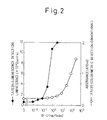
- Fig. 2 shows a result of luminescence measurement method obtained in Example 2 (solid circles) and of colorimetric method obtained in Comparative Example 2 (open circles).
- Fig. 3 is a graph wherein a ratio (0 to 20) obtained by dividing responses at different concentration of samples by response at sample-free condition are plotted against concentrations of the samples. The values were obtained in Example 3 and Comparative Example 3.
- Figs. 4 and 5 are graphs wherein a ratio (0 to 10) obtained by dividing responses at different concentrations of samples by response at sample-free condition are plotted against concentrations of the samples. The values were obtained in Example 3 and Comparative Example 3.
- Fig. 6 shows lines showing the relationship between an amount of luciferase or ⁇ -galactosidase and its activity, obtained in Example 4.
- Fig. 7 is a graph wherein amounts of luciferase or ⁇ -galactosidase induced by the addition of different concentrations of AF-2 are plotted against the concentrations of AF-2. The amounts of luciferase and ⁇ -galactosidase were calculated using the lines in Example 4.
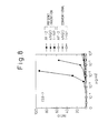
- Fig. 8 is a graph showing a ratio (0 to 100) obtained by dividing response obtained in Example 6 and Comparative Example 4 for AF-2, 4NQO and 1-NP at different concentrations, by response at sample-free condition.
- Fig. 9 is a graph showing ratio (0 to 10) obtained by dividing responses obtained in Example 6 and Comparative Example 4 for AF-2, 4NQO and 1-NP at different concentrations, by response at sample-free condition.
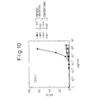
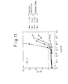
- Fig. 10 is a graph showing ratio (0 to 80) obtained by dividing responses obtained in Example 6 and Comparative Example 4 for 2-AA and BaP at different concentrations, by response at sample-free condition.
- Fig. 11 is a graph showing ratio (0 to 10) obtained by dividing responses obtained in Example 6 and Comparative Example 4 for 2-AA and BaP at different concentration, by response at sample-free condition.
- an SOS gene is used as a means for regulating the expression.
- the SOS gene may be any SOS gene which is expressed when a DNA is damaged and which contains so called SOS box.
- SOS gene expressed when a DNA is damaged is those containing the "SOS box".
- the SOS gene may be the SOS box per se, or DNA fragment containing the SOS box.
- the SOS gene includes umu gene, such as umu D,C gene, as well as sfiA gene, but the SOS gene is not limited thereto.
- umu D,C gene are described in Proc. Natl. Acad. Sci. USA Vol. 82 , 4336 - 4340 (1985), and therefore can be easily obtained according to the description in the reference.
- plasmid pSK1002 containing umuD gene as well as a part of umuC gene and lacZ gene is described in H. Shinagawa et al., Gene, 23 , 167 (1983), and from this plasmid, umu D,C gene can be easily obtained.
- luciferase gene derived from Photinus pyralis
- Pica GeneTM a cassette vector
- any genes which express both the proteins under the control of SOS gene can be used.
- the genes include bioluminescence genes of marine bacteria, but are not limited thereto. Marine bacteria having the above defined bioluminescence genes include Vibrio group, the genus Vibrio , such as Vibrio harveyi , V. fischeri , V. Splendidus , V. lendinus , V. cholerae , and the genus Photobacterium , such as P. phosphoreum , P. leiognathi etc.
- bioluminescence genes derived from V . ficsheri are modified by removing operator region of the bioluminescence genes, remaining the structural gene region (genes expressing luciferase activity and genes expressing fatty acid reductase which is an enzyme catalyzing the production of aldehyde which is a substrate for luciferase) so as to make the bioluminescence genes easy to be used as a reporter gene.
- the modified gene is used to prepare cassette vectors.
- the cassette vectors such as pUCD320, pUCD613, pUCD614, pUCD618, pUCD620, pUCD623, pUCD1111 etc. have been reported. These vectors are further described in Clarence I. Kado et al., Plant Molecular Biology Reporter, 5 , 225 (1987), and from these vectors, bioluminescence genes can be easily obtained.
- a substrate for luciferase is a long chain aldehyde or the like, and an enzyme catalyzing the production of said substrate is NAD(P)H:FMN reductase, fatty acid reductases, or the like.
- genes expressing a luciferase activity and genes expressing an enzyme catalyzing the production of a substrate for luciferase may be derived from the same source. However, these genes may be derived from different sources.
- Host microorganism is any microorganism which allows SOS gene being expressed when a DNA is damaged by a genotoxic substance such as a mutagenetic substance, in other word, host microorganism may be those having the components of SOS response.
- Escherichia coli Salmonella typhimurium such as TA1535, TA1538, Saccharomyces cerevisiae , or the like, may be listed.
- a recombinant gene comprising an SOS gene as well as genes for expressing a luciferase activity and an enzyme catalyzing the production of a substrate may be constructed by joining genes expressing luciferase activity and an enzyme catalyzing the production of a substrate for luciferase to the downstream of the SOS gene in a DNA fragment comprising at least a part of SOS box of SOS gene. Joining can be carried out according to a conventional procedure using DNA ligase. To introduce the recombinant DNA into a host microorganism, the recombinant DNA must be present in a vector. For example, as plasmids, pBR type plasmid, pUC type plasmid and the like may be used.
- a genotoxic substance such as a mutagenic substance is detected or measured by mixing a sample to be tested with a medium, and culturing said transformed host microorganism in said medium, usually for 1 to 3 hours.
- the cultured microbial cells are collected, and the cell wall is disrupted to release an expression product, i.e., luciferase from the cells.
- the disruption of the cells can be carried out according to a conventional procedure, for example, the use of cell wall lysis reagent represented by a surfactant such as Triton X, Sodium Dodecyl Sulfate etc., or the mechanical treatment represented by sonication such as ultrasonication.
- the lysate thus obtained is, for example, centrifuged to obtain a supernatant containing luciferase, and the supernatant is subjected to a treatment for measuring luminescence.
- the measurement of luminescence is carried out by adding a substrate for luciferase such as luciferine and coenzymes to the supernatant.
- the cultured medium per se generates the luminescence if the sample contained a genotoxic substance such as a mutagenic substance. Therefore, immediately after the culturing the luminescence can be measured. Since an amount of the luminescence increases in a concentration dependent manner if a genotoxic substance such as mutagenic substance is present in the sample, then the genotoxic substance such as a mutagenic substance in the sample can be detected or measured from an amount of the luminescence.
- the present method can test any genotoxic substances such as mutagenic substances.
- a genotoxic substance such as a mutagenic substance is usually introduced into a culture medium in form of a solution.
- solid and gas as an analyte can also be tested by the present method as far as they dissolve in a culture medium.
- the luminescence can be detected or measured by a conventional method.
- the sensitivity of detection of genotoxic substances such as mutagenic substances increases by about 100 times higher than that of the conventional method, because the sample volume required for detection in the present invention is smaller than that in the conventional method and therefore a trace amount of substances present in the environment can be detected or measured.
- the conventional method requires at least 30 minutes for coloring reaction, the present invention immediately gives sufficient luminescence for detection, and therefore the time for detection or measurement can be shortened.
- a ratio obtained by dividing a value obtained by an assay method in the presence of a genotoxic substance such as mutagenic substance by a value obtained by the same assay method in the absence of the genotoxic substance such as mutagenic substance is useful to assess a performance of the assay method.
- a commonly used criterion to classify a test substance into genotoxic group or non-genotoxic group is whether or not the test substance shows a response two times higher than a response where the test substance is not added.
- a concentration of a test substance which exhibits a response two times higher than that wherein the test substance is not added is defined as "minimum detectable concentration”.
- minimum detectable concentration obtained by the present luminescence method are at least 4 times lower, and especially for nitroarene and polycyclic aromatic hydrocarbon about 10 times lower than those obtained by the conventional colorimetric method. This means that the sensitivity of the present luminescence method is at least 4 times higher than that of the conventional colorimetric method.
- the dynamic range of measurement i.e., the highest value of the ratio
- a detectable concentration of a genotoxic substance such as a mutagenic substance is lowered by about 5 times at maximum relative to the conventional colorimetric method.
- an umu D,C gene and luciferase gene ( luc ) were ligated to produce umuC - luc fusion protein.
- the production of the fusion protein is not essential as far as a gene coding for a polypeptide having luciferase activity is under the control by SOS genes.
- an umu gene derived from plasmid pSK1002 having umu D,C - lacZ gene H. Shinagawa, T. Kato, et al., Gene, 23, 167 (1983) was used.
- a gene coding for a polypeptide having luciferase activity a luc gene of Photinus pyralis on Pica GeneTM cassette vector available from Toyo Ink MFG. Co., LTD., Japan was used.
- E. coli CSH26 "Miller, J.H., Experiments in Molecular Genetics, Cold Spring Harbor Laboratory, 1972”: F - ara del ( lac -pro)thi
- Salmonella Typhimurium TA1535" Ames, B. N., J. McCann, E. Yamasaki, Mutation Res., 31, 347 (1975)": hisG46, ⁇ gal, ⁇ chi, ⁇ bio, ⁇ uvrB, rfa - , SJ10002: r - , m + ) were used.
- a substrate solution and cell lysis reagent used for the luminescence method were of a luminescence kit "Pica GeneTM" of Toyo Ink. MFG. Co., LTD.
- the substrate solution comprised 20 mM Tricine, 1.07 mM (MgCO 3 ) 4 Mg(OH) 2 ⁇ 5H 2 O, 2.67 mM MgSO 4 , 0.1 mM EDTA, 33.3 mM DTT, 270 ⁇ M Co-enzyme A, 470 ⁇ M luciferin, and 530 ⁇ M ATP.
- the cell lysis reagent comprised 25 mM Tris ⁇ phosphate (pH 7.2), 2 mM DTT, 2 mM 1,2-diaminocyclohexane-N,N,N,N-tetraacetic acid, 10% glycerol, and 1% Triton X-100.
- a universal photon counting system manufactured by Hamamatsu Photonics Co., LTD. was used as an apparatus for measuring luminescence.
- Escherichia coli containing pSK1002 was-cultured in an LB medium (Bacto trypton 1%, Bacto yeast extract 0.5%, NaCl 1%) containing ampicillin, and a large amount of pSK1002 vector was prepared from the cells by alkaline-SDS method (Birnboim, H. C., Doly, J., Nucl. Acids Res. , 11 , 1513 (1979)).
- the vector was digested with restriction enzymes Hind III and Ava I to obtain DNA fragment(A) of about 7.2 Kb containing an umu gene.
- the fragment (A) was dephosphorylated with alkaline phosphatase ( E. coli C75).
- a vector containing a luc gene derived from Photinus pyralis was used to transform E. coli JM109 (Messing, J., Gene , 33 , 199 (1985)).
- the transformed E. coli was cultured in LB medium containing ampicillin, and a large amount of vector containing luc gene was prepared by alkaline SDS method.
- the vector was digested with restriction enzymes Hind III and Stu I to obtain a DNA fragment (B) of about 1.7 Kb containing luc gene.
- the expression vector constructed above was digested with a restriction enzyme Hind III, blunt-ended by a DNA Blunting Kit, and self-ligated using a DNA Ligation Kit so as to construct umuC - luc fusion protein-producing luminescence vector.
- E. coli CSH26 was cultured in an LB medium overnight. To the LB medium was added one hundredth volume of the culture broth, and cultivation was carried out until the turbidity at 600 nm (OD 600 ) reached about 0.4. Next, 5 ml of the culture broth was centrifuged, the precipitate fraction (microbial cells) was suspended in 5 ml of 30 mM CaCl 2 , and the suspension was allowed to stand for 45 minutes in an ice bath. Centrifugation was again carried out and the cells were suspended in 0.4 ml of 30 mM CaCl 2 .
- E. coli CSH26 cells transformed with a luminescence vector were cultured in an LB medium containing ampicillin 20 ⁇ g/ml.
- 29 ⁇ l of the culture broth was distributed into test tubes and to each tube was added 1 ⁇ l of 2-(2-furyl)-3-(5-nitro-2-furyl)-acrylamide (AF-2) having a predetermined concentration, and the mixture was incubated at 37°C for 2 hours.
- AF-2 2-(2-furyl)-3-(5-nitro-2-furyl)-acrylamide
- the cell suspension was centrifuged, and to the precipitate (cells) was added 20 ⁇ l of the cell lysis reagent.
- the suspension was treated with ultrosonic disruption apparatus (Tosho Denki) five times for 30 seconds, and the suspension was centrifuged.
- Tosho Denki ultrosonic disruption apparatus
- competent cells of S. typhimurium SJ10002 were prepared.
- S. typhimurium TA1535 was cultured in LB medium overnight.
- the culture broth was added to a fresh LB medium in an amount of one hundredth relative to the fresh medium, and cultured until a turbidity at 600 nm (OD 600 ) reached to about 0.4.
- 5 ml of the culture broth was centrifuged, the precipitate (microbial cells) was suspended in 5 ml of 10 mM CaCl 2 , 10 mM MnCl 2 , 10 mM MgCl 2 aqueous solution, and the mixture was allowed to stand for 45 minutes in an ice bath.
- the mixture was again centrifuged, the cells were suspended in 0.4 ml of 10 mM CaCl 2 , 10 mM MnCl 2 , 10 mM MgCl 2 aqueous solution so as to prepare competent cells.
- Luminescence vector was prepared from transformed S. typhimurium SJ10002 by alkaline-SDS method, and the vector was used to transform S. typhimurium TA1535 according to the same procedure as described above.
- E. coli CSH26/pSK1002 in which plasmid pSK1002 had been introduced was used. 0.75g of potassium chloride, 0.246g of magnesium sulfate, and 2 ml of 0.05 mol/L 2-mercaptoethanol were added to 1 L of 0.1 mol/L potassium phosphate buffer (pH 7.0) to prepare Z-Buffer.
- E. coli CSH26/pSK1002 was cultured in LB medium containing 20 ⁇ g/ml ampicillin overnight.
- 2.9 ml of the culture broth was distributed to each test tube, and after adding 0.1 ml of AF-2 having a predetermined concentration thereon, the mixture was incubated at 37°C for 2 hours.
- To 0.2 ml of the culture broth were added 1.8 ml of Z-Buffer, 50 ⁇ l of 0.1% SDS and 10 ⁇ l of chloroform, and the mixture was stirred for 5 seconds.
- Salmonella typhimurium TA1535/pSK1002 into which plasmid pSK1002 had been introduced was used, and the same procedure as described in Comparative Example 1 was repeated except that TGA medium (Trypton 1%, NaCl 0.5%, glucose 0.2%, ampicillin 20 ⁇ g/ml) was used in place of LB medium to measure ⁇ -galactosidase activity induced by AF-2. A result is shown in Fig. 2.
- E. coli CSH26 carrying a recombinant plasmid containing a luminescence gene provided a good dose-response curve over a range of about 1 ng/tube to about 10 ng/tube of AF-2.
- S. typhimurium TA1535 carrying a recombinant plasmid containing a luminescence gene provided a good dose-response curve over a range of about 0.1 ng/tube to 1 ng/tube.
- the sensitivity of the present method is higher about 100 times relative to that of the conventional method.
- S9 fraction manufactured by Oriental Yeast
- S9Mix which was prepared by adding Cofactor-1 (manufactured by Oriental Yeast) as a co-factor was used.
- the culture broth was inoculated to a fresh TGA medium in an amount of 1/50 volume ratio relative to the fresh medium, and cultured at 37°C for 1.5 hours.
- the resulting culture broth was diluted with TGA medium so that an absorbance at 600 nm was 0.1. 1.45 ml of the diluted culture broth was distributed to each test tube, and to the diluted culture broth was added 50 ⁇ l of a test sample having a predetermined concentration and cultivation was carried out at 37°C for 2 hours.
- a metabolic activation enzyme is necessary, to 1.2 ml of the diluted culture broth were added 0.25 ml of S9Mix and 50 ⁇ l of a test sample, and cultivation was carried out at 37°C for 2 hours.
- S. typhimurium TA1535/pSK1002 was cultured in TGA medium at 37°C overnight.
- the culture broth was inoculated to a fresh TGA medium in a volume ratio of 1/50 relative to the fresh medium, and cultured at 37°C for 1.5 hours.
- the resulting culture broth was diluted with TGA medium so that an absorbance at 600 nm was 0.1. 1.45 ml of the diluted culture broth was distributed to each test tube, and to the diluted culture broth was added 50 ⁇ l of a test sample having a predetermined concentration and cultivation was carried out at 37°C for 2 hours.
- the present luminescence measuring method provided a dynamic range of the measurement, i.e., the highest value of the ratio, which is expanded few times in comparison with that of conventional colorimetric method for each test sample.
- luciferase a predetermined amount of luciferase dissolved in 10 ⁇ l of cell lysis reagent, and 100 ⁇ l of a luminescence substrate were added to a 96-well microplate for microluminoreader, and an amount of luminescence for 10 seconds was measured.
- a result is shown in Fig. 6 wherein an amount of enzyme (luciferase) is plotted on the horizontal axis, and a corresponding activity (an amount of luminescence) is plotted on the vertical axis.
- FIG. 6 A result is shown in Fig. 6 wherein an amount of enzyme (an amount of ⁇ -galactosidase) is plotted on the horizontal axis, and an activity ( ⁇ -galuctosidase activity) is plotted on the vertical axis.
- an amount of enzyme an amount of ⁇ -galactosidase
- an activity ⁇ -galuctosidase activity
- luciferase An expression efficiency of luciferase is totally lower than that of ⁇ -galactosidase, and therefore in the case where a test sample is not added, luciferase is expressed only about 1/10 6 of ⁇ -galactosidase. Namely, a large amount of ⁇ -galactosidase is synthesized even when a test sample is not added. Probably under the influence of this phenomenon, response in induction of synthesis of ⁇ -galactosidase is slow, and a relative ratio of an amount of ⁇ -galactosidase produced by induction by a test sample to an amount of ⁇ -galactosidase produced without induction is small.
- response in induction of synthesis of luciferase is faster, and a relative ratio of an amount of luciferase produced by induction by a test sample to an amount of luciferase produced without induction is larger.
- the rapid response of luciferase gene in induction of synthesis of luciferase by the addition of a test sample means that a minimum detectable concentration is low, and the high ratio of an amount of luciferase synthesized by induction to an amount of luciferase synthesized without induction means that the dynamic range of a measurement, i.e., the highest value of the ratio, is wide.
- the above-mentioned phenomena and the advantages of the present invention are provided by the difference in expression of ⁇ -galactosidase gene and luciferase gene in the host strain Salmonella typhimurium TA1535, and are found, for the first time, by detailed research in the present invention.
- the low expression efficiency of reporter gene means that detection by conventional colorimetric method is difficult, and the present invention is characterized by, and the advantages thereof are provided by, the use of luciferase gene and measurement of the luminescence.
- coli (HB101: hsd20 (r ⁇ - , m ⁇ - ), recA13, ara-14, proA2, lacY1, galK2, rpsL20, xyl-5, mt1-1, supE44) was used.
- Salmonella typhimurium (TA1535, "Ames, B.N., J. McCann, E. Yamasaki, Mutation Res., 31 , 347, 1975”: hisG46, ⁇ gal, ⁇ chl, ⁇ bio, ⁇ uvrB, rfa - , SJ10002: r - , m + ) was used.
- Microluminoreader MLR-100 manufactured by Colona was used for the measurement of luminescence.
- E. coli CSH26 carrying the plasmid pSK1002 was cultured in LB medium (Bacto Trypton 1%, Bacto yeast extract 0.5%, NaCl 1%) containing ampicillin, and a large amount of the plasmid pSK1002 was prepared from the cultured cells by an alkaline extraction method described in Birnboim, H.C., Doly, J., Nucl. Acids Res., 11 , 1513, 1976.
- the plasmid pSK1002 was digested with Sal I and Sma I to obtain a Sal I- Sma I DNA fragment of about 7.5 kb containing a umu D,C gene. This DNA fragment was ligated with T4 DNA ligase, and used to transform E. coli DH5 according to a conventional procedure.
- plasmid was isolated by alkaline extraction method.
- the plasmid was digested with Mlu I, and blunt-ended with T4 DNA polymerase.
- a DNA linker (TAGGATCCTA) which provides a stop codon to the umuC and has Bam HI restriction site was chemically synthesized.
- This synthetic linker and said MluI DNA fragment were ligated with T4 DNA ligase, and the product was used to transform E. coli DH5.
- the plasmid was extracted by alkaline extraction method.
- the plasmid was digested with Bam HI and Sal I to obtain a Bam HI- Sal I DNA fragment of about 7.4 kb.
- E. coli HB101 carrying plasmid pUCD620 was cultured in LB medium containing ampicillin and a large amount of pUCD620 was prepared from the cultured cells by alkaline extraction method.
- the plasmid pUCD620 was digested with Bam HI and Sal I to obtain a Bam HI- Sal I DNA fragment of about 7.5 kb containing a group of luminescence genes. This DNA fragment was ligated to the downstream of said Bam HI- Sal I DNA fragment of about 7.4 kb with T4 DNA ligase to constract a luminescence vector.
- S. typhimurium SJ10002 was cultured in LB medium overnight.
- the culture broth was inoculated to a fresh LB medium in an amount of one hundredth volume of the fresh medium, and was cultured until a turbidity at 600 nm (OD 600 ) reached about 0.4.
- 5 ml of the culture broth was centrifuged, and the precipitated fraction was suspended in 5 ml of 30 mM CaCl 2 , and the suspension was allowed to stand for 45 minutes in an ice bath.
- the suspension was again centrifuged, and the precipitated cells were suspended in 0.4 ml of 30 mM CaCl 2 aqueous solution to prepare competent cells of S. typhimurium SJ10002.
- S. typhimurium TA1535 was cultured in LB medium overnight.
- the culture broth was inoculated to a fresh LB medium in an amount of one hundredth by volume of the fresh medium, and cultured until a turbidity at 600 nm (OD 600 ) reached about 0.4.
- 5 ml of the culture was centrifuged, and the precipitated fraction was suspended in 5 ml of 10 mM CaCl 2 , 10 mM MnCl 2 , 10 mM MgCl 2 aqueous solution, and the mixture was allowed to stand for 45 minutes in an ice bath.
- the suspension was again centrifuged, and the precipitated cells were suspended in 0.4 ml of 10 mM CaCl 2 , 10 mM MuCl 2 10 mM Mg aqueous solution to prepare competent cells of S. typhimurium TA1535.
- the luminescence vector was added to 100 ⁇ l of the competent cells of S. typhimurium SJ10002, and the suspension was allowed to stand for 30 minutes in an ice bath. After the treatment at 42°C for 2 minutes, the suspension was allowed to stand for 5 minutes at room temperature. 1 ml of LB medium was added thereon, and the suspension was incubated at 37°C for one hour, and inoculated to an LB plate containing 50 ⁇ g/ml ampicillin.
- the luminescence vector was prepared by alkaline extraction method and used to transform S. typhimurium TA1535 as described above.
- S9 fraction manufactured by Oriental Yeast
- S9 Mix which was prepared by adding Cofactor 1 (manufactured by Oriental Yeast) as a co-factor was used.
- S. typhimurium TA1535 transformed with a luminescence vector was cultured in TGA medium (Trypton 1%, NaCl 0.5%, glucose 0.2%, ampicillin 20 ⁇ g/ml) at 30°C overnight.
- the culture broth was inoculated to a fresh TGA medium in an amount of 1/50 by volume of the fresh medium, and cultured at 30°C for 1.5 hours.
- the culture broth was diluted with TGA medium so that absorbance at 600 nm was 0.1. 1.45 ml of the diluted culture broth was distributed to each test tube, and to the diluted culture broth was added 50 ⁇ l of a test sample having a predetermined concentration, and cultivation was carried out at 30°C for 2 hours.
- Salmonella typhimurium TA1535/pSK1002 into which plasmid pSK1002 had been introduced was used. 0.75g of potassium chloride, 0.246g of magnesium sulfate and 2 ml of 0.05 mol/L 2-mercaptoethanol were added to 1L of 0.1 mol/L potassium phosphate buffer (pH 7.0) to prepare Z-Buffer.
- the TA1535/pSK1002 was cultured in TGA medium at 37°C overnight.
- the culture broth was inoculated to a fresh TGA medium in an amount of 1/50 by volume of the fresh medium, and cultured at 37°C for 1.5 hours.
- the culture broth was diluted with TGA medium so-that an absorbance at 600 nm was 0.1. 1.45 ml of the diluted culture broth was distributed to each test tube, 50 ⁇ l of a test sample having a predetermined concentration was added thereon, and cultivation was carried out at 37°C for 2 hours.
- the present luminescence measuring method provided a dynamic range of a measurement, i.e., the highest value of the ratio, expanded by about 10 times at maximum.
Landscapes
- Chemical & Material Sciences (AREA)
- Health & Medical Sciences (AREA)
- Life Sciences & Earth Sciences (AREA)
- Organic Chemistry (AREA)
- Zoology (AREA)
- Engineering & Computer Science (AREA)
- Proteomics, Peptides & Aminoacids (AREA)
- Genetics & Genomics (AREA)
- Wood Science & Technology (AREA)
- Biochemistry (AREA)
- Bioinformatics & Cheminformatics (AREA)
- Molecular Biology (AREA)
- General Health & Medical Sciences (AREA)
- Biophysics (AREA)
- Physics & Mathematics (AREA)
- Medicinal Chemistry (AREA)
- Analytical Chemistry (AREA)
- Biotechnology (AREA)
- Immunology (AREA)
- Microbiology (AREA)
- Toxicology (AREA)
- General Engineering & Computer Science (AREA)
- Gastroenterology & Hepatology (AREA)
- Measuring Or Testing Involving Enzymes Or Micro-Organisms (AREA)
- Micro-Organisms Or Cultivation Processes Thereof (AREA)
Description
| Minimum detectable concentration (µg/ml) | ||
| Test Sample | Method of Present Invention | Conventional Method |
| AF-2 | 5 × 10⁻⁴ | 3 × 10⁻³ |
| | 1 × 10⁻² | 4 × 10⁻² |
| 1-NP | 3 × 10⁻² | 3 × 10⁻¹ |
| 2- | 4 × 10⁻² | 1 × 10⁻¹ |
| | 1 × 10⁻¹ | 1 |
| Minimum detectable concentration (µg/ml) | ||
| Test Sample | Method of Present Invention | Conventional Method |
| AF-2 | 5 × 10⁻⁴ | 3 × 10⁻³ |
| | 8 × 10⁻³ | 4 × 10⁻² |
| 1-NP | 5 × 10⁻² | 3 × 10⁻¹ |
| 2- | 2 × 10⁻² | 1 × 10⁻¹ |
| | 4 × 10⁻² | 1 |
Claims (30)
- A recombinant gene comprising an SOS gene espressed when a DNA is damaged and a gene expressing luciferase activity positioned downstream of the SOS gene.
- A recombinant gene according to claim 1, wherein the gene expressing luciferase activity is a luciferase gene derived from Photinus pyralis.
- A recombinant gene according to claim 1, wherein the SOS gene is umu D,C gene.
- A host microorganism transformed with a recombinant gene comprising an SOS gene expressed when a DNA is damaged and a gene expressing luciferase activity positioned downstream of the SOS gene.
- A host microorganism according to claim 4, wherein the gene expressing luciferase activity is a luciferase gene derived from Photinus pyralis.
- A host microorganism according to claim 4, wherein the SOS gene is umu D,C gene.
- A host microorganism according to claim 4, wherein the host microorganism is a bacterium.
- A host microorganism according to claim 7, wherein the bacterium is selected from the group consisting of the genera Escherichia, Salmonella and Bacillus.
- A host microorganism according to claim 8, wherein the bacterium is selected from the group consisting of Escherichia coli, Salmonella typhimurium, and Bacillus subtilis.
- A method for detecting or quantitating a mutagenic substance in a sample, comprising the steps of:culturing a host microorganism transformed with a recombinant gene comprising an SOS gene expressed when a DNA is damaged and a gene expressing luciferase activity positioned downstream of the SOS gene, in a medium to which the sample is added; andmeasuring a luminescence generated by expression of the gene expressing luciferase activity.
- A method according to claim 10, wherein the gene expressing luciferase activity is a luciferase gene derived from Photinus pyralis.
- A method according to claim 10, wherein the SOS gene is umu D,C gene.
- A method according to claim 10, wherein the host microorganism is a bacterium.
- A method according to claim 13, wherein the bacterium is selected from the genera Escherichia, Salmonella and Bacillus.
- A method according to claim 14, wherein the microorganism is selected from the group consisting of Escherichia coli, Salmonella typhimurium and Bacillus subtilis.
- A recombinant gene comprising an SOS gene expressed when a DNA is damaged and genes expressing luciferase activity and an enzyme which catalyzes the production of a substrate for the luciferase, positioned downstream of the SOS gene.
- A recombinant gene according to claim 16, wherein the genes expressing luciferase activity and an enzyme which catalyzes the production of a substrate for the luciferase is derived from a microorganism belonging to the genus Vibrio or Photobacterium.
- A recombinant gene according to claim 17, wherein the microorganism is selected from the group consisting of Vibrio harveyi, V. fischeri, V. splendidus, V. cholerae, Photobacterium phosphoreum and P. leiognathi.
- A recombinant gene according to claim 16, wherein the SOS gene is umu D,C gene.
- A recombinant gene according to claim 16, wherein the enzyme which catalyzes the production of a substrate for the luciferase is fatty acid reductase.
- A host microorganism transformed with a recombinant gene comprising an SOS gene expressed when a DNA is damaged and genes expressing luciferase activity and an enzyme which catalyzes the production-of-a substrate for the luciferase positioned downstream of the SOS gene.
- A host microorganism according to claim 21, wherein the genes expressing luciferase activity and an enzyme which catalyzes the production of a substrate for the luciferase is derived from a microorganism belonging to the genus Vibrio or Photobacterium.
- A host microorganism according to claim 22, wherein the microorganism is selected from the group consisting of Vibrio harveyi, V. fischeri, V. splendidus, V. cholerae, Photobacterium phosphoreum and P. leiognathi.
- A host microorganism according to claim 21, wherein the SOS gene is umu D,C gene.
- A host microorganism according to claim 21, wherein the enzyme which catalyzes the production of a substrate for luciferase is fatty acid reductase.
- A method for detecting or quantitating a mutagenic substance in a sample, comprising the steps of:culturing a host microorganism transformed with a recombinant gene comprising an SOS gene expressed when a DNA is damaged and genes expressing luciferase activity and an enzyme which catalyzes the production of a substrate for luciferase, positioned downstream of the SOS gene, in a medium to which the sample is added; andmeasuring a luminescence generated by expression of the genes expressing luciferase activity and an enzyme which catalyzes the production of a substrate for luciferase.
- A method according to claim 26, wherein the genes expressing luciferase activity and an enzyme which catalyzes the production of a substrate for luciferase is derived from a microorganism belonging to the genus Vibrio or Photobacterium.
- A method according to claim 26, wherein the microorganism is selected from the group consisting of Vibrio harveyi, V. fischeri, V. splendidus, V. cholerae, Photobacterium phosphoreum and P. leiognathi.
- A method according to claim 26, wherein the SOS gene is umu D,C gene.
- A method according to claim 26, wherein the enzyme which catalyzes the production of a substrate for luciferase is fatty acid reductase.
Applications Claiming Priority (6)
| Application Number | Priority Date | Filing Date | Title |
|---|---|---|---|
| JP26489493 | 1993-10-22 | ||
| JP264894/93 | 1993-10-22 | ||
| JP1845294A JP3277426B2 (en) | 1994-02-15 | 1994-02-15 | Reagent-free mutagen detection method |
| JP18452/94 | 1994-02-15 | ||
| JP22965994A JP3277436B2 (en) | 1993-10-22 | 1994-09-26 | Mutagen detection method using luminescent gene |
| JP229659/94 | 1994-09-26 |
Publications (2)
| Publication Number | Publication Date |
|---|---|
| EP0649905A1 EP0649905A1 (en) | 1995-04-26 |
| EP0649905B1 true EP0649905B1 (en) | 1998-09-23 |
Family
ID=27282214
Family Applications (1)
| Application Number | Title | Priority Date | Filing Date |
|---|---|---|---|
| EP94307739A Expired - Lifetime EP0649905B1 (en) | 1993-10-22 | 1994-10-21 | Methods of detection of mutagens using luminescence gene |
Country Status (3)
| Country | Link |
|---|---|
| US (1) | US5702883A (en) |
| EP (1) | EP0649905B1 (en) |
| DE (1) | DE69413491T2 (en) |
Families Citing this family (12)
| Publication number | Priority date | Publication date | Assignee | Title |
|---|---|---|---|---|
| BRPI9612617B8 (en) | 1996-04-25 | 2021-05-25 | Vlaamse Instelling Voor Tech Onderzoek Vito | recombinant vector, genetically modified host microorganism, method for determining the presence of genotoxic compounds in a sample, and method for determining both the genotoxicity and mutagenicity of a sample |
| AU722016B2 (en) * | 1996-07-29 | 2000-07-20 | Crc For Waste Management And Pollution Control Limited | Biosensors |
| AUPO128096A0 (en) * | 1996-07-29 | 1996-08-22 | Crc For Waste Management And Pollution Control Limited | Biosensors |
| WO1998021347A1 (en) * | 1996-11-15 | 1998-05-22 | E.I. Du Pont De Nemours And Company | A small volume, highly sensitive method for detecting environmental insults |
| WO1998038336A1 (en) * | 1997-02-28 | 1998-09-03 | E.I. Du Pont De Nemours And Company | A method for identifying the site of action of xenobiotic chemicals |
| EP0950717A1 (en) * | 1998-04-14 | 1999-10-20 | "VLAAMSE INSTELLING VOOR TECHNOLOGISCH ONDERZOEK", afgekort "V.I.T.O." | Diagnostic system and method for determining the presence of a genotoxic compound in a sample |
| US6602666B2 (en) * | 1998-12-02 | 2003-08-05 | Vlaamse Instelling Technologisch Onderzoek (V.I.T.O.) | Recombinant nucleic acid sequences and methods for determining both genotoxicity and mutagenicity of a sample and the kinetics of the genotoxicity |
| US6673554B1 (en) * | 1999-06-14 | 2004-01-06 | Trellie Bioinformatics, Inc. | Protein localization assays for toxicity and antidotes thereto |
| WO2001046694A2 (en) * | 1999-12-22 | 2001-06-28 | Biosignal Packard Inc. | A bioluminescence resonance energy transfer (bret) fusion molecule and method of use |
| US6667153B1 (en) | 2000-06-26 | 2003-12-23 | Susan Margaret Thomas | Composition and method for detecting mutagens |
| DE10132280A1 (en) * | 2001-07-04 | 2003-01-23 | Merck Patent Gmbh | Method for the detection of mutagenic substances |
| CA2464942A1 (en) * | 2001-11-14 | 2003-05-22 | John Cavendish Day | Signal system and elements used therein |
Family Cites Families (3)
| Publication number | Priority date | Publication date | Assignee | Title |
|---|---|---|---|---|
| FR2533584B1 (en) * | 1982-09-28 | 1986-03-21 | Pasteur Institut | METHOD FOR DETECTING THE MUTAGENIC POWER OF SUBSTANCES LIKELY TO INDUCE DETERIORATION OF CELL DNA, INVOLVING THE PRODUCTION OF AN SOS RESPONSE |
| EP0370813A3 (en) * | 1988-11-25 | 1991-06-19 | Exemplar Corporation | Rapid screening mutagenesis and teratogenesis assay |
| CA2044679A1 (en) * | 1990-06-22 | 1991-12-23 | Alexander Honigman | Detection of hiv infection by bioluminescence |
-
1994
- 1994-10-21 US US08/326,949 patent/US5702883A/en not_active Expired - Fee Related
- 1994-10-21 DE DE69413491T patent/DE69413491T2/en not_active Expired - Fee Related
- 1994-10-21 EP EP94307739A patent/EP0649905B1/en not_active Expired - Lifetime
Also Published As
| Publication number | Publication date |
|---|---|
| US5702883A (en) | 1997-12-30 |
| DE69413491D1 (en) | 1998-10-29 |
| DE69413491T2 (en) | 1999-03-11 |
| EP0649905A1 (en) | 1995-04-26 |
Similar Documents
| Publication | Publication Date | Title |
|---|---|---|
| Davidov et al. | Improved bacterial SOS promoter∷ lux fusions for genotoxicity detection | |
| Bechor et al. | Recombinant microorganisms as environmental biosensors: pollutants detection by Escherichia coli bearing fabA′:: lux fusions | |
| Frackman et al. | Cloning, organization, and expression of the bioluminescence genes of Xenorhabdus luminescens | |
| EP0649905B1 (en) | Methods of detection of mutagens using luminescence gene | |
| Zavilgelsky et al. | Action of 1, 1-dimethylhydrazine on bacterial cells is determined by hydrogen peroxide | |
| Berg et al. | Structural genes for nitrate-inducible formate dehydrogenase in Escherichia coli K-12. | |
| Lindow | The use of reporter genes in the study of microbial ecology. | |
| EP0793729B1 (en) | Lyophilized bioluminescent bacterial reagent for the detection of toxicants | |
| US5786162A (en) | Fused genes and their use for determining the presence of metals or of xenobiotic compounds | |
| US6329160B1 (en) | Biosensors | |
| Matsuno et al. | Metabolic imbalance and sporulation in an isocitrate dehydrogenase mutant of Bacillus subtilis | |
| TW200846469A (en) | Secreted MLuc7 luciferase and use thereof | |
| Rosen et al. | Microbial sensors of ultraviolet radiation based on recA’:: lux fusions | |
| EP0469021B1 (en) | Determination of factors affecting gene regulation and/or gene replication | |
| Shapiro et al. | Stress-activated bioluminescent Escherichia coli sensors for antimicrobial agents detection | |
| Dahlgren et al. | A novel mutation in ribosomal protein S4 thataffects the function of a mutated RF1 | |
| Østergaard et al. | The cda GenoTox assay: a new and sensitive method for detection of environmental genotoxins, including nitroarenes and aromatic amines | |
| JP4272265B2 (en) | Diagnostic system and method for determining the presence of genotoxic compounds in a sample | |
| Sato et al. | Observation of oscillation in bacterial luminescence | |
| EP0907748B1 (en) | Recombinant nucleic acid sequences and methods for determining both genotoxicity and mutagenicity of a sample and the kinetics of the genetoxicity | |
| Salehi-Sedeh et al. | Effect of mutation at positively charged residues (K329 and R330) in a flexible region of firefly luciferase on structure and kinetic properties | |
| KR101430685B1 (en) | Artificial biosensor for non-degradable harmful aromatic compound detection and manufacturing method thereof | |
| JP3277426B2 (en) | Reagent-free mutagen detection method | |
| Paoni et al. | Improved method for detection of glycosidases in bacterial colonies | |
| Blissett et al. | In vivo bioluminescent determination of apparent Km‘s for aldehyde in recombinant bacteria expressing luxA/B |
Legal Events
| Date | Code | Title | Description |
|---|---|---|---|
| PUAI | Public reference made under article 153(3) epc to a published international application that has entered the european phase |
Free format text: ORIGINAL CODE: 0009012 |
|
| AK | Designated contracting states |
Kind code of ref document: A1 Designated state(s): CH DE FR GB LI |
|
| 17P | Request for examination filed |
Effective date: 19950520 |
|
| GRAG | Despatch of communication of intention to grant |
Free format text: ORIGINAL CODE: EPIDOS AGRA |
|
| 17Q | First examination report despatched |
Effective date: 19971127 |
|
| GRAG | Despatch of communication of intention to grant |
Free format text: ORIGINAL CODE: EPIDOS AGRA |
|
| GRAH | Despatch of communication of intention to grant a patent |
Free format text: ORIGINAL CODE: EPIDOS IGRA |
|
| GRAH | Despatch of communication of intention to grant a patent |
Free format text: ORIGINAL CODE: EPIDOS IGRA |
|
| GRAA | (expected) grant |
Free format text: ORIGINAL CODE: 0009210 |
|
| AK | Designated contracting states |
Kind code of ref document: B1 Designated state(s): CH DE FR GB LI |
|
| REG | Reference to a national code |
Ref country code: CH Ref legal event code: EP |
|
| REF | Corresponds to: |
Ref document number: 69413491 Country of ref document: DE Date of ref document: 19981029 |
|
| ET | Fr: translation filed | ||
| REG | Reference to a national code |
Ref country code: CH Ref legal event code: NV Representative=s name: PATENTANWAELTE SCHAAD, BALASS, MENZL & PARTNER AG |
|
| PLBE | No opposition filed within time limit |
Free format text: ORIGINAL CODE: 0009261 |
|
| STAA | Information on the status of an ep patent application or granted ep patent |
Free format text: STATUS: NO OPPOSITION FILED WITHIN TIME LIMIT |
|
| 26N | No opposition filed | ||
| REG | Reference to a national code |
Ref country code: GB Ref legal event code: 746 Effective date: 20001003 |
|
| REG | Reference to a national code |
Ref country code: GB Ref legal event code: IF02 |
|
| REG | Reference to a national code |
Ref country code: FR Ref legal event code: D6 |
|
| PGFP | Annual fee paid to national office [announced via postgrant information from national office to epo] |
Ref country code: FR Payment date: 20041008 Year of fee payment: 11 |
|
| PGFP | Annual fee paid to national office [announced via postgrant information from national office to epo] |
Ref country code: DE Payment date: 20041014 Year of fee payment: 11 |
|
| PGFP | Annual fee paid to national office [announced via postgrant information from national office to epo] |
Ref country code: GB Payment date: 20041020 Year of fee payment: 11 |
|
| PGFP | Annual fee paid to national office [announced via postgrant information from national office to epo] |
Ref country code: CH Payment date: 20041027 Year of fee payment: 11 |
|
| PG25 | Lapsed in a contracting state [announced via postgrant information from national office to epo] |
Ref country code: GB Free format text: LAPSE BECAUSE OF NON-PAYMENT OF DUE FEES Effective date: 20051021 |
|
| PG25 | Lapsed in a contracting state [announced via postgrant information from national office to epo] |
Ref country code: LI Free format text: LAPSE BECAUSE OF NON-PAYMENT OF DUE FEES Effective date: 20051031 Ref country code: CH Free format text: LAPSE BECAUSE OF NON-PAYMENT OF DUE FEES Effective date: 20051031 |
|
| PG25 | Lapsed in a contracting state [announced via postgrant information from national office to epo] |
Ref country code: DE Free format text: LAPSE BECAUSE OF NON-PAYMENT OF DUE FEES Effective date: 20060503 |
|
| REG | Reference to a national code |
Ref country code: CH Ref legal event code: PL |
|
| GBPC | Gb: european patent ceased through non-payment of renewal fee |
Effective date: 20051021 |
|
| PG25 | Lapsed in a contracting state [announced via postgrant information from national office to epo] |
Ref country code: FR Free format text: LAPSE BECAUSE OF NON-PAYMENT OF DUE FEES Effective date: 20060630 |
|
| REG | Reference to a national code |
Ref country code: FR Ref legal event code: ST Effective date: 20060630 |