EP0616639B1 - Device and process for isolating and purifying nucleic acids - Google Patents
Device and process for isolating and purifying nucleic acids Download PDFInfo
- Publication number
- EP0616639B1 EP0616639B1 EP92924637A EP92924637A EP0616639B1 EP 0616639 B1 EP0616639 B1 EP 0616639B1 EP 92924637 A EP92924637 A EP 92924637A EP 92924637 A EP92924637 A EP 92924637A EP 0616639 B1 EP0616639 B1 EP 0616639B1
- Authority
- EP
- European Patent Office
- Prior art keywords
- nucleic acids
- buffer
- silica gel
- layer
- nucleic acid
- Prior art date
- Legal status (The legal status is an assumption and is not a legal conclusion. Google has not performed a legal analysis and makes no representation as to the accuracy of the status listed.)
- Expired - Lifetime
Links
Images


Classifications
-
- C—CHEMISTRY; METALLURGY
- C12—BIOCHEMISTRY; BEER; SPIRITS; WINE; VINEGAR; MICROBIOLOGY; ENZYMOLOGY; MUTATION OR GENETIC ENGINEERING
- C12N—MICROORGANISMS OR ENZYMES; COMPOSITIONS THEREOF; PROPAGATING, PRESERVING, OR MAINTAINING MICROORGANISMS; MUTATION OR GENETIC ENGINEERING; CULTURE MEDIA
- C12N15/00—Mutation or genetic engineering; DNA or RNA concerning genetic engineering, vectors, e.g. plasmids, or their isolation, preparation or purification; Use of hosts therefor
- C12N15/09—Recombinant DNA-technology
- C12N15/10—Processes for the isolation, preparation or purification of DNA or RNA
- C12N15/1003—Extracting or separating nucleic acids from biological samples, e.g. pure separation or isolation methods; Conditions, buffers or apparatuses therefor
- C12N15/1006—Extracting or separating nucleic acids from biological samples, e.g. pure separation or isolation methods; Conditions, buffers or apparatuses therefor by means of a solid support carrier, e.g. particles, polymers
- C12N15/101—Extracting or separating nucleic acids from biological samples, e.g. pure separation or isolation methods; Conditions, buffers or apparatuses therefor by means of a solid support carrier, e.g. particles, polymers by chromatography, e.g. electrophoresis, ion-exchange, reverse phase
-
- B—PERFORMING OPERATIONS; TRANSPORTING
- B01—PHYSICAL OR CHEMICAL PROCESSES OR APPARATUS IN GENERAL
- B01D—SEPARATION
- B01D39/00—Filtering material for liquid or gaseous fluids
-
- C—CHEMISTRY; METALLURGY
- C12—BIOCHEMISTRY; BEER; SPIRITS; WINE; VINEGAR; MICROBIOLOGY; ENZYMOLOGY; MUTATION OR GENETIC ENGINEERING
- C12N—MICROORGANISMS OR ENZYMES; COMPOSITIONS THEREOF; PROPAGATING, PRESERVING, OR MAINTAINING MICROORGANISMS; MUTATION OR GENETIC ENGINEERING; CULTURE MEDIA
- C12N15/00—Mutation or genetic engineering; DNA or RNA concerning genetic engineering, vectors, e.g. plasmids, or their isolation, preparation or purification; Use of hosts therefor
- C12N15/09—Recombinant DNA-technology
- C12N15/10—Processes for the isolation, preparation or purification of DNA or RNA
- C12N15/1003—Extracting or separating nucleic acids from biological samples, e.g. pure separation or isolation methods; Conditions, buffers or apparatuses therefor
- C12N15/1017—Extracting or separating nucleic acids from biological samples, e.g. pure separation or isolation methods; Conditions, buffers or apparatuses therefor by filtration, e.g. using filters, frits, membranes
-
- Y—GENERAL TAGGING OF NEW TECHNOLOGICAL DEVELOPMENTS; GENERAL TAGGING OF CROSS-SECTIONAL TECHNOLOGIES SPANNING OVER SEVERAL SECTIONS OF THE IPC; TECHNICAL SUBJECTS COVERED BY FORMER USPC CROSS-REFERENCE ART COLLECTIONS [XRACs] AND DIGESTS
- Y10—TECHNICAL SUBJECTS COVERED BY FORMER USPC
- Y10T—TECHNICAL SUBJECTS COVERED BY FORMER US CLASSIFICATION
- Y10T436/00—Chemistry: analytical and immunological testing
- Y10T436/25—Chemistry: analytical and immunological testing including sample preparation
- Y10T436/25375—Liberation or purification of sample or separation of material from a sample [e.g., filtering, centrifuging, etc.]
- Y10T436/255—Liberation or purification of sample or separation of material from a sample [e.g., filtering, centrifuging, etc.] including use of a solid sorbent, semipermeable membrane, or liquid extraction
Definitions
- the invention relates to a method for isolation and Purification of nucleic acids such as plasmid or genomic DNA from cells or other sources and a device to carry out the method according to the preamble of Claim 16.
- nucleic acids When preparing nucleic acids, the cells initially through the use of enzymes, such as Proteinase K, lysozyme and detergents such as SDS, Brij, Triton-X-100, Tween 20, DOC and chemicals like Sodium hydroxide, guanidine hydrochloride and guanidine isothiocyanate be unlocked. The experimenter the problem arises before cleaning the nucleic acids remove the cell debris and then out of the Cell lysate the nucleic acids or nucleic acid fractions isolate. Furthermore, when preparing Plasmid DNA or genomic DNA commonly used Detergents such as SDS (sodium dodecyl sulfate) removed will.
- enzymes such as Proteinase K, lysozyme and detergents such as SDS, Brij, Triton-X-100, Tween 20, DOC and chemicals like Sodium hydroxide, guanidine hydrochloride and guanidine isothiocyanate be unlocked.
- SDS
- DE-A 36 39 949 describes a method for isolation and Purification of long-chain nucleic acids from other substances from bacteria, viruses, animal and vegetable tissues and Cells and body fluids, especially cell contents and / or their degradation products and components of Body fluids that are not long chain nucleic acids.
- the long-chain nucleic acids after a gentle Disruption and removal of cell fragments and others undissolved components fixed to an anion exchanger, while the substances to be separated are washed out. Then the fixed nucleic acids with a buffer high ionic strength detached from the matrix.
- a method for separation is known from DE-A 37 17 211 and purification of biopolymers, such as nucleic acids, the Nucleic acids arranged in a special device Adsorbed matrix.
- the buffer conditions are adjusted so that the nucleic acids predominantly adsorb while interfering substances like proteins low molecular weight substances or cell debris, not bound will.
- EP 0 376 080 describes a method for extraction and Purification of DNA, starting from lambda phages, M13 phagemids, Plasmids, cosmids etc., the process involving several filtration steps includes.
- US 5,075,430 discloses a method for cleaning Plasmid and other DNA (single and double stranded) by Immobilization on diatomaceous earth in the presence of a chaotropic Agent and subsequent elution of the DNA with water or a weakly concentrated buffer. The so obtained purified DNA is biologically active.
- WO 91/05606 relates to a chromatographic support material, the cavities of which are 1 to 20 times the size of the largest dimension of the nucleic acids to be separated, which can be obtained by using a starting support material having a cavity size of 10 to 1,000 nm, a specific surface area of 5 to 800 m 2 / g and a grain size of 3 to 500 ⁇ m is reacted with a silanizing reagent, characterized in that the silanizing reagent has at least one reactive group already reacted with a primary or secondary hydroxyalkylamine.
- WO 91/07422 describes a method and a kit for Purification of nucleic acids, e.g. DNA, from lysed cells or tissue samples. The sample is then analyzed using anion exchange chromatography cleaned.
- nucleic acids e.g. DNA
- US 4,810,381 describes a device consisting of PTFE-containing fibril matrix, which with non-expandable Particles is networked.
- the ratio is non-inflatable Particles to PTFE in the range of 19: 1 to 4: 1 wt .-%.
- US 4,935,142 discloses a membrane unit consisting of a variable number of stacked, planar membrane elements as a filter for chromatography.
- EP 0 890 063 describes a method and a test kit for Isolation of nucleic acids from material containing nucleic acids like blood, urine, cell cultures and the like.
- Nucleic acid from yeast was determined according to Biochemistry 1972, 4848 separated on poly (L-lysine) coated Kieselguhr. Mitochondrial DNA has also been used on such chromatographic materials separated.
- Chromatographia 1984, 19, 236-9 describes the use of multidimensional chromatography to isolate synthetic oligodeoxyribonucleotides in the preparative Scale described.
- the first step is first a size exclusion chromatography on Sephadex G-15 performed, followed by size exclusion chromatography with an HPLC ion exchange column (Partisil-10 SAX). This is followed by a hydrophobic one Chromatography using HPLC (Nucleosil C18).
- a disadvantage of this representative state of the art is the fact that a centrifugation step for removal of the cell fragments and the undissolved components from the cell lysate is necessary.
- Another one The problem is that the nucleic acids by Elution in buffers of high ionic strength from those in large Concentration of existing salts frees up and simultaneously have to be concentrated. Most of the time are the further procedural operations with the so obtained Nucleic acids only possible with buffer conditions, which have lower ionic strengths.
- the removal of the Salts dissolved in high concentrations in the buffer can also done by dialysis, but this leads to more noticeable Degradation of the nucleic acids in the corresponding Rehearse. After dialysis, the desalted nucleic acid be concentrated by freeze drying.
- a Another type of concentration is through precipitation the nucleic acid with ethanol, isopropanol, polyethylene glycol (PEG).
- the nucleic acids are in this system not soluble and fall out.
- the failed nucleic acids but have to go through a centrifugation step be pelleted.
- the nucleic acid pellet is dried briefly and then in a small volume buffer very low salt concentrations solved to a concentrated to obtain a salt-free nucleic acid sample.
- This centrifugation and precipitation process is one simple and fast extraction of nucleic acids is not possible and automation is difficult carry out.
- the need for simple ones is increasing and automatic methods for the preparation of Nucleic acids through the advance of molecular biology in clinical diagnostics and sequencing of the human genome. There are large amounts of samples work up.
- the technical problem underlying the invention is to provide a method that enables Isolate and purify nucleic acids, without a centrifugation to remove the Cell fragments or undissolved components of the cell lysate would be necessary and without the nucleic acids in Buffer systems of high salt concentrations occur, whereby the nucleic acids a downstream desalination and Make concentration step necessary.
- the thing to be provided The method is said to be practically in the nucleic acids deliver a directly processable condition.
- a device with which the inventive method can be carried out in a particularly advantageous manner is characterized by the features of claim 16, 35, 37.
- the subclaims referring to this concern further preferred embodiments of the invention Contraption.
- the cells whose nucleic acid is to be isolated are disrupted in the usual way and the cell debris is removed. This can be done by filtration or centrifugation.
- the clear cell lysates are preferably obtained by filtration through a stepwise or asymmetrically constructed filter layer.
- the filtrate containing the nucleic acids can be treated immediately with anion exchangers.
- a commercially available material can be selected as the anion exchanger which allows the nucleic acid to be isolated to be bound under the respective preparation conditions.
- the anion exchangers are preferably surface-modified supports made of a matrix, preferably consisting of agarose, dextran, cellulose, acrylamide, polyvinyl alcohol, polystyrene, glass, aluminum oxide, titanium dioxide, zirconium dioxide or silica gel, such as DEAE-Sepharose R , Q-sepharose R , DEAE- sephadex R , DEAE-Toyopearl R , Amberlite R , Nucleogen R , Qiagen R.
- the anion exchangers can be porous carrier materials with a suitable inner surface of high capacity for interaction or non-porous carrier materials which only interact with the mixture to be separated on the outer surface.
- the anion exchanger is very particularly preferably a material based on silica gel which has a particle size of 1 to 250 ⁇ m, preferably 10 to 50 ⁇ m and very particularly preferably 15 to 25 ⁇ m and a pore diameter of 1 to 2500 nm, preferably 10 up to 500 nm, particularly preferably 100 to 400 nm.
- a material with a high surface charge and a high binding capacity for nucleic acids has proven to be an anion exchange material.
- the silica gel is preferably modified by silanizing the support material, as disclosed, for example, in EP-A 83 901 065, DE-A-39 35 098 and US-A-5,057,426.
- EP-A 83 901 065 for example, gamma-glycidyloxypropyltrimethoxysilane and N, N-dimethylaminoethanol are used to modify the carrier material.
- the adsorption of the nucleic acids takes place under conditions as they typically do at low salt concentrations available. These are preferably lower salt concentrations as such with which the nucleic acids of the column can be eluted. Depending on the used Ion exchange materials and pH values can affect the salt concentration be 0.25 to 1.5 sts.
- After the adsorption of the nucleic acids on the anion exchange material can be at least one wash connect with low ionic strength buffer.
- the ion exchange material is preferably located one in a predominantly cylindrical hollow body Pillar.
- the column is then washed with a saline solution whose ionic strength is as high as possible without that the desired nucleic acid is eluted. In order to become low molecular weight and weakly charged impurities and washed out proteins.
- adsorption step performs by using the highest possible ionic strength in particular in the area where the later adsorption the nucleic acid should take place under high salt conditions.
- a solution for Equilibration and conditioning are used an ionic strength of approximately 1.5 M sodium perchlorate corresponds to a pH of approximately 5.
- the material for binding the Nucleic acids under conditions of high ionic strength in a separate predominantly cylindrical hollow body is in the high salinity fraction in the cartridge or column with the nucleic acids underneath High salt absorbent material.
- a conditioning of the material that the nucleic acids can adsorb at high ionic strength is particularly particularly easy with this procedure.
- the Conditioning can already take place in that the Material that contains the nucleic acids at high ionic strength can bind with correspondingly highly concentrated salt solutions is pretreated.
- appropriately pretreated materials for example by absorbing the nucleic acid Material first with high ionic strength salt solutions is treated and then the solvent evaporated is so that in the nucleic acids under high Material with ionic strength absorbing very high salt concentrations adjust immediately if an aqueous Solution is associated with it.
- the conditioning due to the fact that the first Volume units that are determined by the elution of the nucleic acids detach from the anion exchanger from this material a possibly too low salt concentration have to be sufficiently firm to the following material to adsorb. Now hits a relatively thinned one Drops of elution from the anion exchanger to such a conditioned one Material that contains nucleic acid under conditions is able to bind high ionic strength, so it turns out immediately a high salt concentration and the nucleic acids are adsorbed on this material.
- the nucleic acid can be higher with a buffer Desorbed ionic strength from the anion exchange material and then directly in the elution buffer of high ionic strength to be bound with a mineral carrier.
- Nucleic acids can be present in the presence of chaotropic salts such as sodium iodide, sodium perchlorate on finely ground Glass or silica gel can be bound when looking at the Nucleic acids with the fine glass or silica gel suspension spiked and incubated for a long time to get a To allow binding of the nucleic acid to the silica gel (B. Vogelstein and D. Gillespie, 1979, Proc. Nat. Aca.
- the adsorption of the nucleic acids on the mineral Carrier can surprise by adding lower alcohols into the sample.
- Alcohols come into question preferably methanol, ethanol, propanol, isopropanol as well as butanol.
- the preferred quantity ranges in which the alcohols added to the sample are 1 - 50% (v / v), as far as they are in these areas at all in water are soluble.
- the adsorption of the Nucleic acids also achieved through polyethylene glycols will.
- the ethylene glycols that can be used have molecular weights from 1,000 to 100,000, especially 6,000 to 8,000. Polyethylene glycol can range from 1 - 30% of the sample can be added.
- the method according to the invention surprisingly shows that nucleic acid even when passing very thin Efficiently adsorb layers of glass or silica gel, although the dwell time is only 1 - 30 seconds. It it also shows that binding in high sodium chloride and lithium chloride concentrations occurs and chaotropic Salts are not necessary.
- the anion exchanger cleaning the Nucleic acid takes over and at the concentrations of 0.25 M - 1.5 M salt, although the impurities, such as metabolites, Proteins and partially RNA, polysaccharides removed be, but this under the given conditions do not adsorb onto the downstream silica gel layer can, and the silica gel layer desalination and Concentration task takes over when the nucleic acid in the following step with a salt concentration of Anion exchanger is eluted, which is high enough Can adsorb nucleic acid to the silica gel layer.
- buffer salts in the specified concentrations for the adsorption step on the mineral carrier salt concentration NaCl 3 - 5 sts NaClO4 5 - 7 sts Gu-HCl 5 - 7 sts Well 3 - 5 sts
- Treatment with the saline solution can be done simply by dripping on done on the filter and suction.
- the silica gel layer with a perchlorate solution pH 6.5 to 8.5, especially pH 7 to 8, treated. This is conveniently done by pipetting and sucking through.
- a perchlorate solution pH 6.5 to 8.5, especially pH 7 to 8, treated.
- This Solution containing 4 to 8 M / 1 NaClO4, 5 to 20 mM / l Tris-HCl, pH 7 to 8 and 0.5 to 2 mM / l EDTA contains used.
- the sodium perchlorate solution is preferably with aqueous Washed ethanol, for example with 50 to 90% ethanol.
- a dilute aqueous salt solution such as. B. in Anal. Biochem. 101 : 339-341 (1980).
- a preferred eluent is 0.5 to 2 mM / l Tris-HCl, pH 7 to 8, containing 0.05 to 0.2 mM / l EDTA, hereinafter referred to as TE.
- TE 0.05 to 0.2 mM / l EDTA
- a pH of 7.5 to 8.5 is particularly preferred.
- Another suitable eluent is dilute detergent solutions, such as 0.1% SDS, which are less preferred.
- silica gel mineral carrier for adsorbing nucleic acid are suitable.
- Silica gel of particle size 1 to 250 microns, preferably 1 to 50 ⁇ m, in particular 1 - 5 ⁇ m, used.
- the desalination layer can be used as a loosely poured layer, which is enclosed between two PE frits, in which Extraction column can be used.
- Another embodiment involves the application of mineral Support in membrane form according to EP 0 323 055 (07.12.1988, 3M, Composition Chromatographic Article).
- nucleic acids different origins can be separated and prepared. It does not matter whether the nucleic acids are off Bacteria, cell cultures, blood, tissue, urine, viruses or from amplification reactions, such as PCR (polymerase chain Reaction), SSSR (Self-Sustained-Sequence Replication), Ligase chain reaction and similar reactions or whether it's labeled nucleic acids, like in biotin labeled, fluorescent labeled or radioactive labeled Is nucleic acids. Nucleic acids come as nucleic acids in a size range from 10 nucleotides to 200,000 nucleotides into consideration.
- nucleic acids in For the purposes of the invention, oligonucleotides from 10 to 100 Nucleotides, RNA with 50 to 25,000 nucleotides, plasmid DNA with 2,500 to 25,000 base pairs, with cosmid DNA 5,000 to 60,000 base pairs or genomic DNA with 100 understood up to 200,000 base pairs.
- step d) of the process according to the invention Nucleic acid fraction or fractions are in Obtain low salt solutions. It is thus possible the necessary for further processing Subsequently set buffer conditions. In that is particularly advantageous on the silica glass bound nucleic acid already in the for further processing certain buffer eluted.
- the isolated nucleic acids are used for the most diverse Applications. Is done particularly often the enzymatic implementation with restriction enzymes, polymerases and ligases for restriction analysis, sequencing, Label with radioactivity or non-radioactive Markers such as biotin, FITC, digoxigenin and the Amplification using PCR, SSSR (Self-Sustained Sequence Replication) and ligase chain reaction.
- the inventive method is particularly for Isolation and preparation of plasmid DNA and genomic DNA suitable.
- the figures show preferred embodiments of the invention Device, the various Adsorption materials for the nucleic acids in one Device are united.
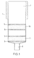
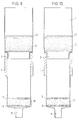
- Figure 1 shows a device for performing the Method according to the invention, which consists of a hollow body 1 with an inlet opening 7 and an outlet opening 8.
- the hollow body is preferably made of polypropylene (PP), polyethylene (PE), polymethyl methacrylate (PMMA), Polytetrafluoroethylene (PTEE), polyethylene terephthalate (PET) or polyacrylonitrile (PAN).
- PP polypropylene
- PE polyethylene
- PMMA polymethyl methacrylate
- PTEE Polytetrafluoroethylene
- PET polyethylene terephthalate
- PAN polyacrylonitrile
- In the hollow body 1 is between two fixing devices 5, 6 a powdery first Material made of a mineral carrier material 10 is arranged.
- a second powder Material 11 made of a mineral carrier material between the first material 10 and the outlet opening 8.
- the first and second materials 10, 11 have different adsorption characteristics for Nucleic acids.
- the differences in the adsorption characteristics are due to different adsorption behavior in high or low ionic strength buffers certainly. For example, if nucleic acids from first material 10 under conditions of low ionic strength bound, the second material 11 in the Be able to lower nucleic acids under buffer conditions Allowing ionic strength to pass unhindered the nucleic acid under conditions of high ionic strength desorbed from the first material 10 and on the second material 11 is adsorbed.
- the first powdery material 10 preferably consists from an anion exchanger from surface modified Carrier materials based on agarose, dextrans, Cellulose, acrylamide, polyvinyl alcohol, polystyrene, glass, Aluminum oxide, titanium oxide, zirconium dioxide or silica gel, especially anion exchangers of the type mentioned above based on silica gel.
- the preferably basic ion exchanger has a particle size of 1 to 250 microns, preferred from 10 to 40 ⁇ m, in particular 15 to 25 ⁇ m, and a pore diameter of 1 to 2500 nm, preferably 10 to 500 nm, in particular 200 - 400 nm.
- the second material 11 is a mineral carrier material, in particular from silica gel, glass, zeolite, Aluminum oxide, titanium dioxide, zirconium dioxide, kaolin, Diatoms, preferably a silica glass, if necessary in the form of a silica gel suspension.
- the second material 11 preferably has a particle size of 1 to 250 ⁇ m, in particular 1 to 30 ⁇ m, preferably 1 to 5 ⁇ m.
- the devices 5 and 6 are preferably made of sintered Glass (frits) or membranes made of plastic, such as polyethylene, PTFE, polypropylene, glass, ceramic, nylon or a fleece made of polypropylene, polyethylene, nylon.
- the porosity of the devices 5, 6 is preferably 10 up to 500 ⁇ m.
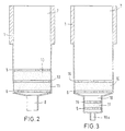
- FIG. 2 Another preferred embodiment of the invention
- the device is shown in FIG. 2.
- the first Material 10 and the second material 11 in the hollow body 1 arranged that the materials 10, 11 directly adjoin each other and in separate layers that together are held by the fixing devices 5, 6.
- the material through a separator 13 are separated, the separating device 13 being a porous disc, preferably made of sintered glass, or a plastic membrane, or fabric, preferably made of Nylon, is.
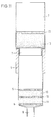
- FIG. 3 shows a further preferred embodiment the device according to the invention, the second material 11 in the outlet 18 forming a channel between the fixing devices 5, 15 is fixed.
- the one Channel-forming outlet opening 18 has a smaller one Cross section than the hollow body 1 and preferably opens in a channel 18a, the cross section of which is less than that of channel 18.
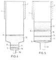
- the first material 10 is located in the lumen of the hollow body 1 in the area of larger diameter and is through the device 6, 16th fixed. It may be advantageous to use the first and to let the second material 10, 11 adjoin one another, so that these are separated only by a common device 17 are (see Figure 4).
- FIG. 5 describes a further preferred embodiment the device according to the invention in the hollow body in addition to the layers of a first and second Material 10, 11 has a further layer 12 which is arranged over the first material 10.
- the layer 12 is designed as a mechanical filter device.
- the third layer 12 is preferably an asymmetrical one Filter, the pore sizes of the filter in the flow direction the sample, that is from the feed opening 7 to the outlet opening 8 or 18 decreases. It can also be used in the rehearsal cell debris are removed without the There is a risk of the device becoming blocked.
- Materials 10 and 11 can be used in all embodiments the device according to the invention either be in powder form and / or as a compact. If the materials 10, 11 are in particulate form, it may be advisable to do this in a carrier network embedded from inert plastics, so that the layers in the form of a membrane according to US Pat. No. 4,810,381 and US-PS 4,699,717 and proposed in DE 41 27 276.
- the carrier network can consist of Teflon.
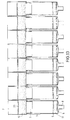
- FIG. 6 describes a further preferred embodiment the device according to the invention, eight single, separate devices according to FIG. 2 adjoin one another and form an eight unit.
- the advantage this embodiment that with any of those described in Figures 1-5 Individual forms is feasible parallel preparation of 8 samples with the help of multi-channel pipettes. This shape can also be put together 12 times are prepared, taking 96 samples become processable. The big advantage is then if the internationally standardized microtiter format is used.
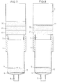
- FIG. 7 describes a device which is in a cylindrical hollow body 1 with inlet opening 7 and outlet opening 8 an anion exchange material 10 between contains two devices 6 and 5 fixed. On it another cylindrical hollow body plugged in whose lumens arranged different filter layers are.
- the filter layers 20, 21, 22 can be made of sintered Polyethylene, polypropylene, PTFE, glass, silica gel, Alumina or poured diatomaceous earth, e.g. B. Cellite or silica gel exist. But also woven, glued Fleece in the form of polypropylene, polyester, glass fibers and silica come into consideration.
- the porosity of the individual layers is preferably 15 microns to 500 microns in a thickness of 0.1 mm to 10 mm.
- the pore size of the Filter layer is seen by layer, as seen in the direction of flow to layer less.
- the size of the pores in layer 20 is approximately 100 up to 300 ⁇ m, in layer 21 30 to 100 ⁇ m and in third filter layer 5 to 30 microns.
- FIG. 8 shows a further preferred embodiment the device of Figure 7, being as, in the flow direction seen, top filter layer 23 is a hydrophobic layer is used.
- the hydrophobic separating layer 23 prevents the unwanted penetration of the raw Cell lysate in the filter layer before beginning actual filtration.
- the hydrophobic separating layer 23 consists preferably of spun or sintered Polypropylene, polyethylene, polyester or Polytetrafluoroetylene (PTFE) fibers, in a porosity from 10 ⁇ m to 500 ⁇ m and preferably a thickness of 0.1 up to 5 mm.
- PTFE Polytetrafluoroetylene
- FIG. 9 describes a filtration device which is constructed similarly to that in FIGS. 7 and 8 described, with the difference that different Filter layers with decreasing pore size in one single filter layer 12 with continuously decreasing Pore size are connected.
- the asymmetrical filter layer 12 is preferably with a hydrophobic filter layer 23 provided at the upper end, seen in the direction of flow.
- the asymmetrical filter layer 12 is preferably made made of spun polypropylene or polyester fibers; Profiles are commercially available, for example from Pall Filtertechnik, Dreieich, Frankfurt, with porosity gradations from 500 to 50 ⁇ m, 100 to 10 ⁇ m, 50 to 5 ⁇ m and 10 to 0.1 ⁇ m.
- the thickness of the asymmetrical filter layer should preferably be 1 mm to 10 mm.
- FIG. 10 describes filtration devices for separation of nucleic acids in the sense of the invention the filter configurations of FIG. 9 being used and is an asymmetrical filter layer is provided with a hydrophobic filter layer 23.
- a mineral carrier 11 capable is to add nucleic acids in highly concentrated salt solutions adsorb.
- FIG. 11 describes a configuration in a connection of Figures 9 and 10.
- the device which is described in Figure 2, only a filter attachment consisting of an asymmetrical filter 12 and assigned to a hydrophobic filter layer 23 approximately by Insert an appropriately designed cartridge.
- FIGS. 1 to 5 and 7 to 11 All of the individual devices shown in FIGS. 1 to 5 and 7 to 11 have been described in more detail in a microtiter strip consisting of 8 put together Arrange individual devices. Is exemplary this is shown again in FIGS. 12 to 14.
- FIG. 12 shows a filtration device with an anion exchanger being a microtiter strip or a Microtiter plate with 8 or 8 x 12 wells.
- an anion exchanger being a microtiter strip or a Microtiter plate with 8 or 8 x 12 wells.
- asymmetrical filtration device in a clip-on Cartridge on the cylindrical hollow body 1 the an anion exchange layer between the devices 5, 6 contains fixed.
- FIG. 13 relates to a filtration device which a mineral instead of the anion exchange material Has carrier material which is capable of nucleic acids adsorb in high salt concentrations.
- a silica gel layer 11 is arranged between two devices 5 and 6.
- FIG. 14 shows a combination of the arrangement according to Figure 2 and an asymmetrical filter layer with hydrophobic filter layer, which over the hollow body 1, in Seen the flow direction of the sample, is arranged.
- the device according to the invention in particular in Figure 3 or 4 devices explained in more detail are special advantageous that the elution of the nucleic acid from the second material 11 with only very small amounts of liquid guaranteed.
- the flow of the sample through the device according to the invention is basically caused by gravity, however, can speed up cleaning and Separation of the nucleic acids an overpressure at the opening 7 or a negative pressure is applied to the opening 8 or 18 will.
- Another preferred embodiment of the invention Device used as an asymmetrical Filters made of sintered glass with decreasing pore size or stacked plastic membranes with decreasing pore size in the flow direction of the sample through the hollow body.
- Nucleic acids from cells and other sources can be used without Centrifugation, phenol / chloroform extraction and without Alcohol precipitation can be obtained, the nucleic acid on End of the process in concentrated form in water or Buffer of low salt concentration is present and therefore Can be used directly for subsequent enzymatic reactions is. Another advantage is that the use expensive laboratory equipment can be avoided.
- the elution can be effected, for example, by gravity are and do not have to be by means of so-called HPLC devices be performed.
- silica gel anion exchanger / silica gel extraction column is preferably done by that a polypropylene tube fits into a commercially available 1.5 ml centrifuge tube, below with a 50 ⁇ m polyethylene frit (porous filter layer made of polyethylene, 1.5 mm thick) is closed and with 50 mg silica gel (Lichrosphere Si 100, 16 - 24 ⁇ m; Merck, Darmstadt, FRG) overlaid.
- This silica gel layer is covered with a second porous polyethylene frit closed and the second frit with 100 mg silica gel anion exchanger (Qiagen, Diagen, Düsseldorf, FRG), particle size 16 layered up to 23 ⁇ m and finally with a third porous polyethylene frit closed.
- the preparation of an agarose anion exchanger / silica gel extraction column is preferably done in that a Polypropylene tube below with a 50 ⁇ m polyethylene frit (porous filter layer made of PE; 1.5 mm thick) closed and with 50 mg silica gel (Lichrosphere Si 100, 16 - 24 ⁇ m) is overlaid.
- This layer of silica gel is closed with a second polyethylene frit and the second frit with 0.5 ml DEAE-Sepharose FF (Fa. Pharmacia, Freiburg, FRG), particle size 45 - 165 ⁇ m overlaid and finally with a third porous polyethylene frit locked.
- An anion exchanger membrane / silica gel membrane extraction column according to FIG. 3 is preferably produced by placing a 1 mm thick Empore R silica gel membrane (3) (3M Corp. St. Paul,) on a polyethylene frit in a polypropylene vessel. MN, USA), a 0.2 mm thick polypropylene fleece and 1 mm thick anion exchanger membrane consist of 16 - 23 ⁇ m Qiagen anion exchanger particles (Diagen GmbH, Düsseldorf, FRG).
- a microtiter strip with 8 or 96 positions is included a DEAE.Silica gel membrane and a silica gel membrane filled.
- a 0.75 mm thick silica gel membrane made of Sident 9 silica gel particles (Degussa, Frankfurt, FRG) is included in a hole in a microtiter strip .
- a 0.2 mm thick polypropylene fleece layer and made a 0.8 mm thick anion exchange membrane from Qiagen, 16 - 23 ⁇ m (Diagen, Düsseldorf, FRG) fitted.
- the extraction column is washed with 0.8 ml of 1 M NaCl, 15% ethanol, 50 mM MOPS, pH 7.0 and with 15% ethanol, 10 mM Na acetate pH 7.0, 0.8 ml of 1 M NaClO 4 , to remove RNA and proteins.
- the DNA is eluted with 7 M NaClO 4 , 15% ethanol, 10 mM Na acetate, pH 7.0 and bound directly to the silica gel layer.
- the extraction column is washed with 0.8 ml 70% ethanol, 100 mM NaCl, 10 mM Na acetate pH 7.0 and with 0.8 ml 90% ethanol / water. Traces of EtOH may be removed by further centrifugation.
- the DNA is then eluted with 50 ⁇ l 10 mM Tris-HCl, 1 mM EDTA, pH 8.0 by centrifugation and collected in new 1.5 ml tubes.
- the eluted DNA can then be used directly in an enzymatic reaction such as restriction cleavage, labeling, sequencing or amplification.
- 8 DEAE silica gel membrane / silica gel extraction columns are placed on a vacuum chamber. 8 x 1 ml of a plasmid DNA containing cell lysate are sucked through the extraction columns under vacuum (20 to 750 mbar). The extraction column is washed with 0.8 ml of 1 M NaCl, 15% ethanol, 50 mM MOPS, pH 7.0 and with 15% ethanol, 10 mM Na acetate pH 7.0, 0.8 ml of 1 M NaClO 4 , to remove RNA and proteins. The DNA is eluted from the anion exchange layer with 7 M NaClO 4 , 15% ethanol, 10 mM Na acetate, pH 7.0 and bound directly to the silica gel layer.
- the extraction column is washed with 0.8 ml of 70% ethanol, 100 mM NaCl, 10 mM Na acetate pH 7.0 and with 0.8 ml of 90% ethanol / water.
- the sample tubes are washed with 0.8 ml of 70% ethanol, 100 mM NaCl, 10 mM Na acetate, pH 7.0 and 0.8 ml of 90% ethanol / water to remove the highly concentrated saline solution.
- the ethanol-H 2 O residues present in the extraction layer are volatilized by sucking in room air through a vacuum for 1 - 2 minutes.
- the 8 samples are then eluted with 50 ⁇ l each of 1 mM Tris-HCl, 0.1 mM EDTA, pH 8.0.
- 1 ml of M13 phage suspension are mixed with 0.5 ml of 30% PEG 6000, 1.5 M NaCl and, after 10 minutes of incubation on ice, centrifuged at 15,000 g for 15 minutes.
- the phage pellet is resuspended in 0.5 ml of 0.5 M guanidine-HCl, 1% Triton X-100 and lysed at 70 ° C. for 10 minutes.
- the phage lysate is sucked on a vacuum chamber directly through an extraction column according to Example 3 and adsorbed.
- the extraction column is washed with 1 ml 0.75 M NaCl, 15% ethanol, 50 mM MOPS, pH 7.0, 1 ml 0.75 M NaClO 4 , 50 mM Tris-HCl, pH 7.0 and with 7 M guanidine, 15% ethanol, 50 mM Na acetate, pH 7.0 eluted from the anion exchange layer and adsorbed onto the SiO 2 layer.
- 1 ml of citrate-stabilized, human whole blood are mixed with 1 ml of 1% saponin for the lysis of the erythrocytes and immediately after mixing, centrifuged for 5 minutes at 2500 g.
- the leukocytes are resuspended in 1 ml PBS buffer and pelleted again.
- the washed leukocytes are dissolved in 1 ml of 500 mM guanidine-HCl, 50 mM Tris-HCl. 10 mM EDTA, pH 8.0 resuspended and the cells lysed by adding 0.1 ml Proteinase K (10 mg / ml) for 2 hours at 50 ° C.
- the leukocyte lysate is immediately pipetted onto the agarose / anion exchanger / silica gel / extraction column and with 1 ml 0.25 M NaCl, 10 mM Na acetate pH 7.0 and 1 ml 0.25 NaClO 4 , 10 mM Na acetate pH 7.0 washed.
- 1 ml of citrate-stabilized, human whole blood is sucked under vacuum through an anion exchange silica gel column.
- the leukocytes are captured in the matrix, whereas the much smaller erythrocytes migrate through the matrix.
- the extraction column is washed twice with 1 ml of PBS buffer.
- the captured leukocytes are lysed with 10% Tween for 10.15 minutes at room temperature.
- the cell fragments and proteins are washed out twice with 1 ml of 1 M guanidine-HCl, pH 7.0 and the DNA is eluted from the column with 7M NaClO 4 , 50 mM Na acetate, pH 7.0.
- the cells are 0.2 M by adding 0.25 ml each NaOH, 1% SDS for 5 minutes at room temperature with gentle Shake lysed. Then 0.25 ml of 3 M K-acetate, 2 M acetic acid, pH 5.5 - 6.0 neutralization buffer admittedly, the individual bowls with a cap closed and mixed. After an incubation of 10 Minute on ice, the sample is 30 minutes at 3,000 g centrifuged around the cell debris and the precipitated Pelletize SDS. The supernatant we with one 8-channel multichanel pipette carefully lifted off and in the 96-well microtiter plate with a DEAE silica gel membrane and pipetted silica gel membrane.
- the samples are created a vacuum to a filtering device through the microtiter plate sucked.
- the DNA is attached to the anion exchange layer adsorbed, whereas among these special conditions proteins, RNA and metabolites are not be adsorbed.
- the extraction column is washed with 0.8 ml of 1 M NaCl, 15% ethanol, 50 mM MOPS, pH 7.0 and with 15% ethanol, 10 mM Na acetate pH 7.0, 0.8 ml of 1 M NaClO 4 , to remove RNA and proteins.
- the DNA is eluted with 7 M NaClO 4 , 15% ethanol, 10 mM Na acetate, pH 7.0 and bound directly to the silica gel layer.
- the extraction column is washed with 0.0 ml 70% ethanol, 100 mM NaCl, 10 mM Na acetate pH 7.0 and with 0.8 ml 90% ethanol / water.
- the salt-freed DNA is then eluted from the silica gel layer into a further microtiter plate in concentrated form, each with 50 ⁇ l 1 mM Tris-HCl, 0.1 mM EDTA, pH 8.0.
- centrifugation The production of cell lysates using centrifugation is a lengthy and time-consuming process. The limitation is given when there are many Samples need to be prepared routinely. The centrifugation has the disadvantage that they do not automate leaves.
- Another object (and method) of the invention is an apparatus and a method for automatic Execution of the process without centrifugation in the form a filtration unit that does the actual cleaning preceded by the nucleic acid.
- the sample is then treated in a known manner with proteinases, Detergents and / or temperature or alkali lysed.
- This raw lysate is applied directly to the filtration attachment decanted, transferred or pipetted.
- the filter layer of the filtration attachment is constructed that clogging of the filters by the cell debris, failed Proteins or detergents is avoided.
- the Cell lysate is passed through the filter layer with a stamp or overpressure or by applying one Vacuum sucked through. In doing so, all undissolved components retained and the clear lysate drips directly on the adsorption layer.
- the suitable adsorption conditions is the nucleic acid adsorbed on the adsorption layer.
- the filtration unit with the filter cake is from the adsorption unit separated and / or discarded and for analysis of the filter cake.
- the adsorption unit will washed with suitable solvents or buffers, to remove unwanted components and the desired Finally, the sample is used with a suitable Eluent eluted.
- Plasmid DNA without a clear centrifugation in one Prepare the refrigerated centrifuge. 96 x 1 ml cultures of plasmid pBluescript in XL1 Blue E.coli cells, in 2 x YT medium in a microtiter plate for 18 hours at 37 ° C with 1.5 ml wells (Beckmann, Kunststoff). The cells are in a microtiter centrifuge Pelleted at 2,500 g for 10 minutes.
- the clear cell lysate containing the plasmid DNA drips through the filter layer onto the adsorption layer (Anion exchanger or silica gel) and the DNA is adsorbed, whereas proteins, RNA and other cellular Metabolites not under the given salt conditions tie.
- the filtration is after about 10 to 60 seconds completed.
- the filter attachment is removed and together discarded with the filter cake.
- the bound DNA is washed with 1 ml of 1 M NaCl, 15% ethanol, 50 mM Tris-HCl pH 7.0 and twice with 1 ml of 1.5 M NaClO 4 , 10 mM Na acetate, pH 6.5 and with 7 M NaClO 4 , 15% ethanol, 50 mM Tris-HCl, pH 7.0 eluted from the anion exchanger and, after passing through the separation layer made of a nylon mesh or PP fleece, immediately bound to the silica gel layer under the high salt concentrations.
- the proteins and RNA at 1 M - 2 M NaClO 4 do not bind to the silica gel layer and are washed out.
- the silica gel layer is washed with 1 ml of 7 M guanidine HCl, 10 mM Na acetate, pH 7.0 to remove the remaining traces of proteins.
- the high salt solution of 7 M NaClO 4 is expediently washed out with 1 ml 70% EtOH, 100 mM NaCl, 10 mM Na acetate, pH 7.0 and 1 ml 90% ethanol / water or 1 ml 90% acetone / water.
- After drying we eluted the plasmid DNA salt-free and in concentrated form with 50 ⁇ l 1 mM Tris-HCl, 0.1 mM EDTA, pH 8.5.
- the plasmid DNA can be shortened Time without centrifugation, phenol / chloroform extraction and without alcohol precipitation with a yield of 50% to Isolate 80% concentrated form.
- a described microtiter plate version can 96 plasmid minipreps from 1 - 2 ml E.coli cultures with a yield of 1 - 10 ⁇ m DNA in about 60 minutes prepare by one person. The previously known This takes 6 to 12 hours.
- Plasmid miniprep with a device according to FIG. 7 A 1.5 ml XL Blue E. coli culture with pUC 18 plasmid DNA centrifuged in LB medium at 10,000 g for 5 minutes, to pellet the cells. The cell pellet is in 0.25 ml 50 ml Tris-HCl, 10 mM EDTA, pH 8.0, 100 ⁇ g / ml RNAse A resuspended. For cell lysis, 0.25 ml 0.2 M NaOH, 1% SDS are added to the cell suspension, carefully mixed and left for 5 minutes at room temperature. Then 0.25 ml of 3M K acetate, 2 M acetic acid for neutralization added, mixed and 15 minutes on ice incubated.
- the lysate is placed in the filtration device transferred to Figure 7.
- the whole device is on a vacuum chamber is attached and the cell lysate with 20 mbar - 800 mbar sucked through the device.
- the sample with a piston ram or gauge pressure through the filtration layers.
- the filtration device is removed and the filter cake with the cell fragments, the denatured Proteins and the failed SDS discarded.
- the extraction column is washed twice with 0.8 ml of 1 M NaCl, 15% Ethanol, 50 mM MOPS, pH 7.0 washed to RNA and proteins
- the DNA is mixed with 1 ml 1.25 M NaCl, 15% Ethanol, 50 mM Tris-HCl, pH 8.5 eluted.
- the eluted DNA becomes desalination and concentration with alcohol and the alcohol pellet by centrifugation pelleted.
- the cell pellet is in 0.25 ml 50 ml Tris-HCl, 10 mM EDTA, pH 8.0, 100 ⁇ g / ml RNAse A resuspended and placed in the filtration device transferred.
- the DNA is mixed with 1 ml 1.25 M NaCl, 15% ethanol, 50 mM Tris-HCl, pH 8.5 eluted.
- the eluted DNA becomes Desalting and concentration with alcohol and the alcohol pellet by centrifugation pelleted.
- a 1.5 ml XL Blue E. coli culture with pUC 18 plasmid DNA in LB medium is centrifuged at 10,000 g for 5 minutes to pellet the cells.
- the cell pellet is resuspended in 0.25 ml 50 ml Tris-HCl, 10 m MEDTA, pH 8.0, 100 ⁇ g / ml RNAse A and transferred to the filtration device according to FIG. 10.
- 0.25 ml of 0.2 M NaOH, 1% SDS is added to the cell suspension in the filtration device, the device sealed with a stopper or an adhesive foil, mixed gently and left to stand for 5 minutes at room temperature.
- 0.5 ml of 5.5 M guanidine-HCl, 0.25 M K acetate, pH 5.5 is added for neutralization, mixed and incubated for 15 minutes on ice.
- the entire device according to Fig. 10 is placed on a vacuum chamber and the cell lysate is sucked through the device at 20 mbar - 800 mbar.
- the sample can be taken through the filtration layer with a piston plunger or overpressure and the filter cake with the cell fragments, the denatured proteins and the failed SDS can be discarded.
- the extraction column is washed twice with 1 ml of 7 M NaClO 4 , 10 mM Na acetate, pH 7.0 and washed with 0.8 ml of 90% ethanol / water and the traces of ethanol are sucked through. Finally, the DNA is eluted with 50 ⁇ l 10 mM Tris-HCl, 1 mM EDTA, pH 8.0 and collected in new 1.5 ml tubes.
- the eluted DNA can be directly in an enzymatic Reaction such as restriction cleavage, labeling, Sequencing or amplification can be used.
- E. coli cultures with pUC 18 plasmid DNA in LB medium are centrifuged at 10,000 g for 5 minutes to pellet the cells.
- the cell pellets are resuspended in 0.25 ml 50 ml Tris-HCl, 10 m MEDTA, pH 8.0, 100 ⁇ g / ml RNAse A and transferred to the device according to FIG. 14.
- 0.25 ml of 0.2 M NaOH, 1% SDS are added to the cell suspension in the filtration device, the device is closed with a stopper or an adhesive film, mixed gently and left to stand for 5 minutes at room temperature.
- the extraction column is washed with 0.8 ml of 1 M NaCl, 15% ethanol, 50 mM MOPS, pH 7.0 and with 0.8 ml of 1 M NaClO 4 , 15% ethanol, 10 mM Na acetate pH 7.0, to remove RNA and proteins.
- the DNA is eluted from the anion exchange layer 10 with 7 M NAClO 4 , 15% ethanol, 10 mM Na acetate, pH 7.0 and bound directly to the silica gel layer 11.
- the extraction column is washed with 0.8 ml of 70% ethanol, 100 mM NaCl, 10 mM Na acetate pH 7.0 and with 0.8 ml of 90% ethanol / water.
- the ethanol-H 2 O residues present in the extraction layer are volatilized by sucking in room air through a vacuum for 1-2 minutes.
- the 8 samples are then eluted with 50 ⁇ l each of 1 mM Tris-HCl, 0.1 mM EDTA, pH 8.0.
- the extraction column is washed with 0.0 ml 70% ethanol, 100 mM NaCl, 10 mM Na acetate pH 7.0 and with 0.8 ml 70% Ethanol, 100 mM NaCl, 100 mM Na acetate pH 7.0 and 0.8 ml 90% ethanol / water washed and for 1 - 2 minutes Air sucked through. Finally, the DNA with 50 ul 10 mM Tris-HCl, 1 mM EDTA, pH 8.0 eluted and in new 1.5 ml tubes collected.
- the eluted DNA can be directly in an enzymatic Reaction such as restriction cleavage, labeling, Sequencing or amplification used will.
- E. coli cultures with pUC 18 plasmid DNA in LB medium are centrifuged at 2,500 g for 5 minutes to pellet the cells.
- the cell pellets are resuspended in 0.25 ml 50 ml Tris-HCl, 10 m MEDTA, pH 8.0, 100 ⁇ g / ml RNAse A and sealed in the device with a stopper or an adhesive film, mixed gently and for 5 minutes Leave room temperature. Then 0.25 ml of 3 M K acetate 2 M acetic acid is added for neutralization, mixed and incubated for 15 minutes on ice.
- the entire device is placed on a vacuum chamber and the cell lysate is sucked through the device at 20 mbar - 800 mbar.
- the sample can be pressed through the filtration layers with excess pressure.
- the filtration device is removed and the filter cake with the cell fragments, the denatured proteins and the failed SDS is discarded.
- the extraction column is washed with 0.8 ml of 1 M NaCl, 15% ethanol, 50 mM MOPS, pH 7.0 and with 0.8 ml of 1 M NaClO 4 , 15% ethanol, 10 mM Na acetate pH 7.0 Remove RNA and proteins.
- the DNA is eluted from the anion exchange layer 10 with 7 M NaClO 4 , 15% ethanol, 10 mM Na acetate, pH 7.0 and thereby bound directly to the silica gel layer 11.
- the extraction column is washed with 0.8 ml of 70% ethanol, 100 mM NaCl, 10 mM Na acetate pH 7.0 and with 0.8 ml of 90% ethanol / water.
- the ethanol-H 2 O residues present in the extraction layer are volatilized by sucking in room air through a vacuum for 1-2 minutes. Then the 96 samples are eluted with 50 ⁇ l each 1 mM Tris-HCl, 0.1 mM EDTA pH 8.0 and collected in new 1.5 ml tubes.
- the eluted DNA can be used directly in an enzymatic reaction, such as restriction cleavage, labeling, sequencing or amplification.
- a 3 ml culture in LB-ampicillin medium with pUC 18 transformed HB 101 E. coli cells is centrifuged at 5,000 g for 10 minutes.
- the cell pellet is resuspended in 0.25 ml 50 ml Tris-HCl, 10 m MEDTA, pH 8.0, 100 ⁇ g / ml RNAse A.
- 0.25 ml of 0.2 M NaOH, 1% SDS are added to the cell suspension, mixed gently and left for 5 minutes at room temperature. Then 0.25 ml of 3 M K acetate, 2 M acetic acid is added for neutralization, mixed and incubated for 15 minutes on ice.
- the lysate is centrifuged at 10,000 g for 15 minutes and the supernatant is carefully removed.
- the clear cell lysate is pipetted onto a DEAE anion exchange extraction column and the sample is sucked through the exchange layer.
- the extraction column is washed with 0.8 ml 1 M NaCl, 15% ethanol, 50 mM MOPS, pH 7.0, 15% ethanol, 10 mM Na acetate pH 7.0 to remove RNA and proteins.
- the DNA is sucked onto an extraction column with a glass fiber membrane with 0.7 ml of 7 M NaClO 4 , 15% ethanol, 10 mM Na acetate, pH 7.0.
- the eluted DNA solution in 7 M NaClO 4 is sucked through the glass fiber membrane and bound directly to the silica gel layer.
- the extraction column is washed with 0.8 ml of 70% ethanol, 100 mM NaCl, 10 mM Na acetate pH 7.0 and with 0.8 ml of 90% ethanol / water. Traces of EtOH may be removed by sucking in room air.
- the DNA is eluted with 10 ⁇ l 10 mM Tris-HCl, 1 mM EDTA, pH 8.0 and collected in new 1.5 ml tubes.
- a 3 ml culture in LB-ampicillin medium with pUC 18 transformed HB 101 E. coli cells is centrifuged at 5,000 g for 10 minutes.
- the cell pellet is resuspended in 0.25 ml 50 ml Tris-HCl, 10 m MEDTA, pH 8.0, 100 ⁇ g / ml RNAse A.
- 0.25 ml of 1.2 M NaOH, 1% SDS are added to the cell suspension, mixed carefully and left for 5 minutes at room temperature. Then 0.25 ml of 3 M K acetate, 2 M acetic acid is added for neutralization, mixed and incubated for 15 minutes on ice.
- the lysate is centrifuged at 10,000 g for 15 minutes and the supernatant is carefully removed.
- the clear cell lysate is pipetted onto a DEAE anion exchange extraction column and the sample is sucked through the exchange layer.
- the extraction column is washed with 0.8 ml 1 M NaCl, 15% ethanol, 50 mM MOPS, pH 7.0 to remove RNA and proteins and with 0.8 ml 1 M NaClO 4 , 15 ethanol, 10 mM Na- Acetate pH 5.0 conditioned.
- the DNA is sucked onto an extraction column with a glass fiber membrane with 0.7 ml of 7 M NaClO 4 , 15% ethanol, 10 mM Na acetate, pH 7.0.
- This glass fiber membrane was previously conditioned with 0.2 ml 7 M NaClO 4 , 15% ethanol, 10 mM Na acetate, pH 7.0 in order to achieve better adsorption of the DNA and to avoid losses in the first drops.
- the eluted DNA solution in 7M NaClO 4 is then sucked through the glass fiber membrane on a vacuum device and bound directly to the silica gel layer.
- the extraction column is washed with 0.8 ml of 70% ethanol, 100 mM NaCl, 10 mM Na acetate pH .0 and with 0.8 ml of 90% ethanol / water. Traces n EtOH may be removed by sucking in room air.
- the DNA is eluted with 100 ul 10 mM Tris-HCl, 1 mM EDT, pH 8.0 and collected in new 1.5 ml tubes.
- the preconditioning can also be achieved by a membrane soaked and dried with 7 M NaClO 4 , 15% ethanol, 10 mM Na acetate, pH 7.0. Preconditioning reduces the adsorption losses from 30% to less than 5% and the overall yield of DNA increases from 50-60% to 80-90%.
Abstract
Description
Die Erfindung betrifft ein Verfahren zur Isolierung und
Reinigung von Nukleinsäuren wie Plasmid- oder genomischer
DNA aus Zellen oder anderen Quellen und eine Vorrichtung
zur Durchführung des Verfahrens gemäß Oberbegriff des
Patentanspruchs 16.The invention relates to a method for isolation and
Purification of nucleic acids such as plasmid or genomic
DNA from cells or other sources and a device
to carry out the method according to the preamble of
Bei der Präparation von Nukleinsäuren müssen die Zellen zunächst durch die Verwendung von Enzymen, wie zum Beispiel Proteinase K, Lysozym und Detergentien wie SDS, Brij, Triton-X-100, Tween 20,DOC und Chemikalien wie Natriumhydroxid, Guanidin-Hydrochlorid und Guanidin-Isothiocyanat aufgeschlossen werden. Dem Experimentator stellt sich das Problem, vor der Reinigung der Nukleinsäuren die Zelltrümmer zu entfernen und dann aus dem Zell-Lysat die Nukleinsäuren oder Nukleinsäurefraktionen zu isolieren. Weiterhin müssen bei der Präparation von Plasmid DNA oder genomischer DNA häufig verwendete Detergentien, wie SDS (Sodiumdodecylsulfat), entfernt werden. Dies erfolgt wie in den meisten Fällen bei Verwendung von SDS durch ein Ausfällen mit Kalziumacetat, da das Kaliumsalz von SDS schwer löslich ist. Die Zelltrümmer werden dann zusammen mit dem ausgefallenen SDS abzentrifugiert. Da die Bestandteile im Lysat ein sehr voluminöses und schmieriges, gelartiges Pellet ergeben, bereitet selbst die Abtrennung dieser Trümmer in einer hochtourigen Zentrifuge Schwierigkeiten. Üblicherweise erfolgt die Entfernung der Zelltrümmer durch eine Zentrifugation zwischen 5.000 g bis 20.000 g für 15 bis 60 Minuten. Dieses Verfahren hat den Nachteil, daß es sehr zeit- und arbeitsaufwendig ist und sich nicht automatisieren läßt. When preparing nucleic acids, the cells initially through the use of enzymes, such as Proteinase K, lysozyme and detergents such as SDS, Brij, Triton-X-100, Tween 20, DOC and chemicals like Sodium hydroxide, guanidine hydrochloride and guanidine isothiocyanate be unlocked. The experimenter the problem arises before cleaning the nucleic acids remove the cell debris and then out of the Cell lysate the nucleic acids or nucleic acid fractions isolate. Furthermore, when preparing Plasmid DNA or genomic DNA commonly used Detergents such as SDS (sodium dodecyl sulfate) removed will. As in most cases, this is done with Using SDS due to calcium acetate precipitation, because the potassium salt of SDS is sparingly soluble. The cell debris are then together with the failed SDS centrifuged. Because the ingredients in the lysate are very result in voluminous and greasy, gel-like pellet, prepares to separate these debris in one high-speed centrifuge difficulties. Usually the cell debris is removed by centrifugation between 5,000 g to 20,000 g for 15 to 60 Minutes. This method has the disadvantage that it is very is time-consuming and labor-intensive and does not automate leaves.
Die DE-A 36 39 949 beschreibt ein Verfahren zur Isolierung und Reinigung langkettiger Nukleinsäuren von anderen Substanzen aus Bakterien, Viren, tierischen und pflanzlichen Geweben und Zellen sowie Körperflüssigkeiten, insbesondere Zellinhaltsstoffen und/oder deren Abbauprodukten sowie Bestandteilen der Körperflüssigkeiten, die nicht langkettige Nukleinsäuren sind. Dabei werden die langkettigen Nukleinsäuren nach einem schonenden Aufschluß und Entfernung der Zellbruchstücke und anderer ungelöster Bestandteile an einem Anionenaustauscher fixiert, während die abzutrennenden Substanzen ausgewaschen werden. Danach werden die fixierten Nukleinsäuren mit einem Puffer hoher Ionenstärke von der Matrix wieder abgelöst.DE-A 36 39 949 describes a method for isolation and Purification of long-chain nucleic acids from other substances from bacteria, viruses, animal and vegetable tissues and Cells and body fluids, especially cell contents and / or their degradation products and components of Body fluids that are not long chain nucleic acids. The long-chain nucleic acids after a gentle Disruption and removal of cell fragments and others undissolved components fixed to an anion exchanger, while the substances to be separated are washed out. Then the fixed nucleic acids with a buffer high ionic strength detached from the matrix.
Aus der DE-A 37 17 211 ist ein Verfahren bekannt zur Trennung und Reinigung von Biopolymeren, wie Nukleinsäuren, wobei die Nukleinsäuren an einer in einer speziellen Vorrichtung angeordneten Matrix adsorbiert werden. Die Pufferbedingungen sind dabei so eingestellt, daß die Nukleinsäuren überwiegend adsorbiert werden, während störende Substanzen, wie Proteine, niedermolekulare Stoffe oder auch Zelltrümmer, nicht gebunden werden.A method for separation is known from DE-A 37 17 211 and purification of biopolymers, such as nucleic acids, the Nucleic acids arranged in a special device Adsorbed matrix. The buffer conditions are adjusted so that the nucleic acids predominantly adsorb while interfering substances like proteins low molecular weight substances or cell debris, not bound will.
Die EP 0 376 080 beschreibt ein Verfahren zur Extraktion und Reinigung von DNA, ausgehend von Lambda-Phagen, M13 Phagemiden, Plasmiden, Kosmiden etc., wobei das Verfahren mehrere Filtrationsschritte umfaßt.EP 0 376 080 describes a method for extraction and Purification of DNA, starting from lambda phages, M13 phagemids, Plasmids, cosmids etc., the process involving several filtration steps includes.
Die US 5,075,430 offenbart ein Verfahren zur Reinigung von Plasmid- und anderer DNA (einzel- und doppelsträngig) durch Immobilisierung auf Diatomeenerde in Anwesenheit eines chaotropischen Agens und anschließender Elution der DNA mit Wasser oder einem schwach konzentrierten Puffer. Die so erhaltene gereinigte DNA ist biologisch aktiv.US 5,075,430 discloses a method for cleaning Plasmid and other DNA (single and double stranded) by Immobilization on diatomaceous earth in the presence of a chaotropic Agent and subsequent elution of the DNA with water or a weakly concentrated buffer. The so obtained purified DNA is biologically active.
Die WO 91/05606 betrifft eine chromatographisches Trägermaterial, dessen Hohlräume die 1- bis 20fache Größe der größten Abmessung der zu trennenden Nukleinsäuren aufweisen, welches dadurch erhältlich ist, daß ein Ausgangsträgermaterial einer Hohlraumgröße von 10 bis 1.000 nm, einer spezifischen Oberfläche von 5 bis 800 m2/g und einer Korngröße von 3 bis 500 µm mit einem Silanisierungsreagens umgesetzt wird, daß dadurch gekennzeichnet ist, daß das Silanisierungsreagens mindestens eine bereits mit einem primären oder sekundären Hydroxyalkylamin umgesetzte reaktive Gruppe aufweist.WO 91/05606 relates to a chromatographic support material, the cavities of which are 1 to 20 times the size of the largest dimension of the nucleic acids to be separated, which can be obtained by using a starting support material having a cavity size of 10 to 1,000 nm, a specific surface area of 5 to 800 m 2 / g and a grain size of 3 to 500 µm is reacted with a silanizing reagent, characterized in that the silanizing reagent has at least one reactive group already reacted with a primary or secondary hydroxyalkylamine.
Die WO 91/07422 beschreibt ein Verfahren und ein Kit zur Reinigung von Nukleinsäuren, wie z.B. DNA, aus lysierten Zellen oder Gewebeproben. Die Probe wird dann mittels Anionenaustauscher-Chromatographie gereinigt.WO 91/07422 describes a method and a kit for Purification of nucleic acids, e.g. DNA, from lysed cells or tissue samples. The sample is then analyzed using anion exchange chromatography cleaned.
Die US 4,810,381 beschreibt eine Vorrichtung bestehend aus PTFE-haltiger fibriler Matrix, welche mit nicht-blähbaren Partikeln vernetzt ist. Dabei liegt das Verhältnis nichtblähbarer Partikel zu PTFE im Bereich von 19:1 bis 4:1 Gew.-%.US 4,810,381 describes a device consisting of PTFE-containing fibril matrix, which with non-expandable Particles is networked. The ratio is non-inflatable Particles to PTFE in the range of 19: 1 to 4: 1 wt .-%.
Die US 4,935,142 offenbart eine Membraneinheit bestehend aus einer variablen Anzahl von gestapelten, planaren Membranelementen als Filter für die Chromatographie.US 4,935,142 discloses a membrane unit consisting of a variable number of stacked, planar membrane elements as a filter for chromatography.
Die EP 0 890 063 beschreibt ein Verfahren und ein Testkit zur Isolierung von Nukleinsäuren aus nukleinsäurehaltigen Material wie Blut, Urin, Zellkulturen und ähnliches.EP 0 890 063 describes a method and a test kit for Isolation of nucleic acids from material containing nucleic acids like blood, urine, cell cultures and the like.
In "Isolierung, Fraktionierung und Hybridisierung von Nukleinsäuren, eine Einführung und methodische Anleitung", herausgegeben von Ulrich Wobus, Verlag Chemie, 1980 werden Methoden zur Isolierung von Nukleinsäuren beschrieben. Daraus geht hervor, daß hochmolekulare Ribonukleinsäuren in Salzlösungen > 1,5 M Natriumchlorid unlöslich sind und ausfallen. Diese Präzipitation wird jedoch als nicht effizient angesehen, so daß in dieser Monographie bereits mehrfache Wiederholungen der Präzipitationsschritte mit hoher Salzkonzentration empfohlen werden. In "Isolation, Fractionation and Hybridization of Nucleic Acids, an introduction and methodological instructions " by Ulrich Wobus, Verlag Chemie, 1980 Methods described for the isolation of nucleic acids. From this goes shows that high molecular weight ribonucleic acids in saline solutions > 1.5 M sodium chloride is insoluble and precipitates. This Precipitation is considered inefficient, however that in this monograph multiple repetitions of the Precipitation steps with high salt concentration recommended will.
Eine effiziente Trennung sowohl von DNA-Restriktionsfragmenten und amplifizierten Produkten der Polymerase-Kettenreaktion wird in J. Chromatogr., 1990, 512, 433 - 444 beschrieben. Als Chromatographiematerial wird ein Ionenaustauscher DEAD-NPR-Material mit 2,5 µm großen, nicht porösen Partikeln verwendet.An efficient separation of both DNA restriction fragments and amplified products of the polymerase chain reaction is in J. Chromatogr., 1990, 512, 433 - 444. A is used as the chromatography material Ion exchanger DEAD-NPR material with 2.5 µm large non-porous particles are used.
Nukleinsäure aus Hefen wurden gemäß Biochemistry 1972, 4848 an Poly(L-Lysine)-beschichtetem Kieselguhr getrennt. Ebenso wurde bereits mitochondriale DNA an solchen chromatographischen Materialien getrennt.Nucleic acid from yeast was determined according to Biochemistry 1972, 4848 separated on poly (L-lysine) coated Kieselguhr. Mitochondrial DNA has also been used on such chromatographic materials separated.
In Chromatographia, 1984, 19, 236 - 9 wird die Verwendung von mehrdimensionaler Chromatographie zur Isolierung von synthetischen Oligodeoxyribonukleotiden im präparativen Maßstab beschrieben. In einem ersten Schritt wird dabei zunächst eine Size-Exclusion-Chromatographie an Sephadex G-15 durchgeführt, gefolgt von einer Size-Exclusion-Chromatographie mit einer HPLC-Ionenaustauschersäule (Partisil-10 SAX). Daran schließt sich eine hydrophobe Chromatographie mittels HPLC (Nucleosil C18) an.Chromatographia, 1984, 19, 236-9 describes the use of multidimensional chromatography to isolate synthetic oligodeoxyribonucleotides in the preparative Scale described. The first step is first a size exclusion chromatography on Sephadex G-15 performed, followed by size exclusion chromatography with an HPLC ion exchange column (Partisil-10 SAX). This is followed by a hydrophobic one Chromatography using HPLC (Nucleosil C18).
Über die Eignung von hydrophob beschichteten Glaspartikeln zur Durchführung von adsorptions-chromatographischer Reinigung von Nukleinsäuren wird in J. Biochem. 94, 163 - 169 (1983) berichtet.About the suitability of hydrophobically coated glass particles to carry out adsorption-chromatographic Purification of nucleic acids is described in J. Biochem. 94, 163 - 169 (1983) reports.
Nachteilig an diesem stellvertretenden Stand der Technik ist die Tatsache, daß ein Zentrifugationsschritt zur Entfernung der Zellbruchstücke und der ungelösten Bestandteile aus dem Zell-Lysat notwendig ist. Ein weiteres Problem besteht darin, daß die Nukleinsäuren durch die Elution in Puffern hoher Ionenstärke von den in großer Konzentration vorhandenen Salzen befreit und gleichzeitig konzentriert werden müssen. In den allermeisten Fällen sind die weiteren Verfahrensoperationen mit den so gewonnenen Nukleinsäuren nur mit Pufferbedingungen möglich, die geringere Ionenstärken aufweisen. Die Entfernung der in hoher Konzentration im Puffer gelösten Salze kann auch durch Dialyse erfolgen, jedoch führt dies zu merklicher Degradation der Nukleinsäuren in den entsprechenden Proben. Nach der Dialyse muß die entsalzte Nukleinsäure durch eine Gefriertrocknung konzentriert werden. Eine andere Art der Konzentrierung erfolgt durch eine Fällung der Nukleinsäure mit Ethanol, Isopropanol, Polyethylenglykol (PEG). Die Nukleinsäuren sind in diesem System nicht löslich und fallen aus. Die ausgefallenen Nukleinsäuren müssen jedoch durch einen Zentrifugationsschritt pelletiert werden. Das Nukleinsäurepellet wird kurz getrocknet und anschließend in einem kleinen Volumenpuffer sehr niedriger Salzkonzentrationen gelöst, um eine konzentrierte salzfreie Nukleinsäureprobe zu erhalten. Durch diese Zentrifugations- und Fällungsverfahren ist eine einfache und schnelle Gewinnung von Nukleinsäuren nicht möglich und eine Automatisierung läßt sich nur schwer durchführen. Andererseits steigt der Bedarf nach einfachen und automatischen Verfahren zur Präparation von Nukleinsäuren durch das Vordringen der Molekularbiologie in die klinische Diagnostik sowie die Sequenzierung des menschlichen Genoms. Dabei sind jeweils große Probenmengen aufzuarbeiten.A disadvantage of this representative state of the art is the fact that a centrifugation step for removal of the cell fragments and the undissolved components from the cell lysate is necessary. Another one The problem is that the nucleic acids by Elution in buffers of high ionic strength from those in large Concentration of existing salts frees up and simultaneously have to be concentrated. Most of the time are the further procedural operations with the so obtained Nucleic acids only possible with buffer conditions, which have lower ionic strengths. The removal of the Salts dissolved in high concentrations in the buffer can also done by dialysis, but this leads to more noticeable Degradation of the nucleic acids in the corresponding Rehearse. After dialysis, the desalted nucleic acid be concentrated by freeze drying. A Another type of concentration is through precipitation the nucleic acid with ethanol, isopropanol, polyethylene glycol (PEG). The nucleic acids are in this system not soluble and fall out. The failed nucleic acids but have to go through a centrifugation step be pelleted. The nucleic acid pellet is dried briefly and then in a small volume buffer very low salt concentrations solved to a concentrated to obtain a salt-free nucleic acid sample. By this centrifugation and precipitation process is one simple and fast extraction of nucleic acids is not possible and automation is difficult carry out. On the other hand, the need for simple ones is increasing and automatic methods for the preparation of Nucleic acids through the advance of molecular biology in clinical diagnostics and sequencing of the human genome. There are large amounts of samples work up.
Das der Erfindung zugrundeliegende technische Problem besteht darin, ein Verfahren bereitzustellen, daß es ermöglicht, Nukleinsäuren zu isolieren und zu reinigen, ohne daß ein Zentrifugationsschrift zur Entfernung der Zellbruchstücke oder ungelöster Bestandteile des Zell-Lysats notwendig wäre und, ohne daß die Nukleinsäuren in Puffersystemen hoher Salzkonzentrationen anfallen, wobei die Nukleinsäuren einen nachgeschalteten Entsalzungs- und Konzentrierungsschritt notwendig machen. Das bereitzustellende Verfahren soll die Nukleinsäuren praktisch in einem direkt weiterverarbeitbaren Zustand liefern. Ein weiterer Aspekt des genannten technischen Problems besteht in der Schaffung einer Vorrichtung, mit der das Verfahren in besonders vorteilhafter Weise ausgeführt werden kann.The technical problem underlying the invention is to provide a method that enables Isolate and purify nucleic acids, without a centrifugation to remove the Cell fragments or undissolved components of the cell lysate would be necessary and without the nucleic acids in Buffer systems of high salt concentrations occur, whereby the nucleic acids a downstream desalination and Make concentration step necessary. The thing to be provided The method is said to be practically in the nucleic acids deliver a directly processable condition. A there is another aspect of the technical problem mentioned in the creation of a device with which the Process carried out in a particularly advantageous manner can be.
Das der Erfindung zugrundeliegende technische Problem
wird in überraschend einfacher Weise durch ein Verfahren
gelöst, daß durch die Merkmale des Anspruchs 1, 34, 36
charakterisiert ist. Die daran anschließenden Verfahrensansprüche
betreffen bevorzugte Ausführungsformen des erfindungsgemäßen
Verfahrens.The technical problem underlying the invention
is done in a surprisingly simple way by a method
solved that by the features of
Eine Vorrichtung, mit der das erfindungsgemäße Verfahren
in besonders vorteilhafter Weise ausgeführt werden kann,
ist durch die Merkmale des Anspruchs 16, 35, 37 charakterisiert.
Die darauf zurückbezogenen Unteransprüche betreffen
weitere bevorzugte Ausführungsformen der erfindungsgemäßen
Vorrichtung.A device with which the inventive method
can be carried out in a particularly advantageous manner,
is characterized by the features of
Zunächst werden die Zellen, deren Nukleinsäure isoliert werden sollen, in üblicher Weise aufgeschlossen und die Zelltrümmer werden entfernt. Dies kann mittels Filtration oder Zentrifugation geschehen. Vorzugsweise erfolgt die Gewinnung der klaren Zell-Lysate durch eine Filtration über eine stufenweise oder asymetrisch aufgebaute Filterschicht. Das die Nukleinsäuren enthaltende Filtrat kann sofort mit Anionenaustauschern behandelt werden. Als Anionenaustauscher kann ein handelsübliches Material ausgewählt werden, welches eine Bindung der zu isolierenden Nukleinsäure unter den jeweiligen Präparationsbedingungen erlaubt. Die Anionenaustauscher sind vorzugsweise oberflächenmodifizierte Träger aus einer Matrix, vorzugsweise bestehend aus Agarose, Dextran, Zellulose, Acrylamid, Polyvinylalkohol, Polystyrol, Glas, Aluminiumoxid, Titandioxid, Zirkondioxid oder Silicagel, wie zum Beispiel DEAE-SepharoseR, Q-sepharoseR, DEAE-sephadexR, DEAE-ToyopearlR, AmberliteR, NukleogenR, QiagenR. Die Anionenaustauscher können poröse Trägermaterialien mit einer zur Wechselwirkung geeigneten inneren Oberfläche hoher Kapazität oder nicht poröse Trägermaterialien sein, die nur auf der äußeren Oberfläche eine Wechselwirkung mit dem zu trennenden Gemisch eingeht. Ganz besonders bevorzugt handelt es sich bei dem Anionenaustauscher um ein Material auf Basis von Silicagel, das eine Partikelgröße von 1 bis 250 µm, vorzugsweise 10 bis 50 µm und ganz besonders bevorzugt 15 bis 25 µm und einen Porendurchmesser von 1 bis 2.500 nm, bevorzugt 10 bis 500 nm, besonders bevorzugt 100 bis 400 nm, aufweist. Als Anionenaustauschermaterial hat sich insbesondere ein Material mit hoher Oberflächenladung und hoher Bindungskapazität für Nukleinsäuren erwiesen. Die Modifizierung des Silicagels erfolgt vorzugsweise durch Silanisierung des Trägermaterials, wie beispielsweise in der EP-A 83 901 065, DE-A-39 35 098 und US-A-5,057,426 offenbart. In der EP-A 83 901 065 wird zum Beispiel gamma-Glycidyloxypropyltrimethoxysilan und N,N-Dimethylaminoethanol zur Modifizierung des Trägermaterials verwendet.First, the cells whose nucleic acid is to be isolated are disrupted in the usual way and the cell debris is removed. This can be done by filtration or centrifugation. The clear cell lysates are preferably obtained by filtration through a stepwise or asymmetrically constructed filter layer. The filtrate containing the nucleic acids can be treated immediately with anion exchangers. A commercially available material can be selected as the anion exchanger which allows the nucleic acid to be isolated to be bound under the respective preparation conditions. The anion exchangers are preferably surface-modified supports made of a matrix, preferably consisting of agarose, dextran, cellulose, acrylamide, polyvinyl alcohol, polystyrene, glass, aluminum oxide, titanium dioxide, zirconium dioxide or silica gel, such as DEAE-Sepharose R , Q-sepharose R , DEAE- sephadex R , DEAE-Toyopearl R , Amberlite R , Nucleogen R , Qiagen R. The anion exchangers can be porous carrier materials with a suitable inner surface of high capacity for interaction or non-porous carrier materials which only interact with the mixture to be separated on the outer surface. The anion exchanger is very particularly preferably a material based on silica gel which has a particle size of 1 to 250 μm, preferably 10 to 50 μm and very particularly preferably 15 to 25 μm and a pore diameter of 1 to 2500 nm, preferably 10 up to 500 nm, particularly preferably 100 to 400 nm. In particular, a material with a high surface charge and a high binding capacity for nucleic acids has proven to be an anion exchange material. The silica gel is preferably modified by silanizing the support material, as disclosed, for example, in EP-A 83 901 065, DE-A-39 35 098 and US-A-5,057,426. In EP-A 83 901 065, for example, gamma-glycidyloxypropyltrimethoxysilane and N, N-dimethylaminoethanol are used to modify the carrier material.
Die Adsorption der Nukleinsäuren erfolgt unter Bedingungen, wie sie typischerweise bei niedrigen Salzkonzentrationen vorliegen. Vorzugsweise sind dies niedrigere Salzkonzentrationen als solche mit der die Nukleinsäuren von der Säule eluiert werden können. Je nach verwendeten Ionenaustauschermaterialien und pH-Werten kann die Salzkonzentration dabei 0,25 bis 1,5 M betragen.The adsorption of the nucleic acids takes place under conditions as they typically do at low salt concentrations available. These are preferably lower salt concentrations as such with which the nucleic acids of the column can be eluted. Depending on the used Ion exchange materials and pH values can affect the salt concentration be 0.25 to 1.5 sts.
Nach der Adsorption der Nukleinsäuren an dem Anionenaustauschermaterial kann sich mindestens ein Waschschritt mit Puffer geringer Ionenstärke anschließen. After the adsorption of the nucleic acids on the anion exchange material can be at least one wash connect with low ionic strength buffer.
Vorzugsweise befindet sich das Ionenaustauschermaterial dabei in einem überwiegend zylindrischen Hohlkörper einer Säule. Die Säule wird dann mit einer Salzlösung gewaschen, deren Ionenstärke so hoch wie möglich ist, ohne daß die erwünschte Nukleinsäure eluiert wird. Damit werden niedermolekulare und schwach geladene Verunreinigungen und Proteine ausgewaschen.The ion exchange material is preferably located one in a predominantly cylindrical hollow body Pillar. The column is then washed with a saline solution whose ionic strength is as high as possible without that the desired nucleic acid is eluted. In order to become low molecular weight and weakly charged impurities and washed out proteins.
Um unnötige Ausbeuteverluste zu vermeiden, kann es vorteilhaft sein, zwischen dem Adsorptionsschritt und dem Ellutionsschritt oder als letzten Waschschritt eine Konditionierung der betreffenden Adsorptionsmaterialien durchzuführen, indem eine möglichst hohe Ionenstärke insbesondere in dem Bereich, in dem die spätere Adsorption der Nukleinsäure unter Hochsalzbedingungen erfolgen soll, durchgeführt wird. Dazu kann insbesondere eine Lösung zur Äquilibrierung und Konditionierung verwendet werden, die einer Ionenstärke von etwa 1,5 M Natriumperchlorat bei einem pH von ungefähr 5 entspricht.In order to avoid unnecessary loss of yield, it can be beneficial be between the adsorption step and the Elution step or as the last washing step Conditioning of the relevant adsorption materials perform by using the highest possible ionic strength in particular in the area where the later adsorption the nucleic acid should take place under high salt conditions, is carried out. In particular, a solution for Equilibration and conditioning are used an ionic strength of approximately 1.5 M sodium perchlorate corresponds to a pH of approximately 5.
Entsprechend befindet sich das Material zur Bindung der Nukleinsäuren unter Bedingungen hoher Ionenstärke in einem separaten überwiegend zylindrischen Hohlkörper. Die von dem Ionenaustauschermaterial desorbierte Nukleinsäurefraktion wird in der hoch salzhaltigen Fraktion in die Kartusche oder die Säule mit dem Nukleinsäuren unter Hochsalzbedingungen absorbierenden Material gegeben. In einer bevorzugten Ausführungsform sind die jeweiligen Vorrichtung mit den entsprechenden Adsorptionsmaterialien so aufeinander abgestimmt, daß der Behälter mit dem Anionenaustauscher auf den Behälter mit dem Nukleinsäure unter hohen Ionenstärken bindenden Material enthaltenden Behälter angeordnet werden kann.The material for binding the Nucleic acids under conditions of high ionic strength in a separate predominantly cylindrical hollow body. The nucleic acid fraction desorbed from the ion exchange material is in the high salinity fraction in the cartridge or column with the nucleic acids underneath High salt absorbent material. In a preferred embodiment are the respective Device with the appropriate adsorption materials matched so that the container with the Anion exchanger on the container with the nucleic acid material containing high ionic strength binding Container can be arranged.
Eine Konditionierung des Materials, das die Nukleinsäuren bei hoher Ionenstärke adsorbieren kann, ist insbesondere bei dieser Vorgehensweise besonders leicht möglich. Die Konditionierung kann bereits dadurch erfolgen, daß das Material, das die Nukleinsäuren bei hoher Ionenstärke binden kann, mit entsprechend hochkonzentrierten Salzlösungen vorbehandelt wird. Es ist jedoch auch möglich, entsprechend vorbehandelte Materialien zu verwenden, indem beispielsweise das Nukleinsäure absorbierende Material zunächst mit Salzlösungen hoher Ionenstärke behandelt wird und danach das Lösungsmittel verdampft wird, so daß sich in den Nukleinsäuren unter hohen Ionenstärken absorbierende Material sehr hohe Salzkonzentrationen unmittelbar einstellen, wenn eine wäßrige Lösung damit in Verbindung gebracht wird. Vorteilhaft ist die Konditionierung aufgrund der Tatsache, daß die ersten Volumeneinheiten, die sich durch die Elution der Nukleinsäuren vom Anionenaustauscher von diesem Material lösen noch eine möglicherweise zu geringe Salzkonzentration aufweisen, um hinreichend fest an dem folgenden Material zu adsorbieren. Trifft nun ein relativ verdünnter Elutionstropfen vom Anionenaustauscher auf ein so konditioniertes Material, das Nukleinsäure unter Bedingungen hoher Ionenstärke zu binden vermag, so stellt sich sofort eine hohe Salzkonzentration ein und die Nukleinsäuren werden an diesem Material adsorbiert.A conditioning of the material that the nucleic acids can adsorb at high ionic strength is particularly particularly easy with this procedure. The Conditioning can already take place in that the Material that contains the nucleic acids at high ionic strength can bind with correspondingly highly concentrated salt solutions is pretreated. However, it is also possible use appropriately pretreated materials, for example by absorbing the nucleic acid Material first with high ionic strength salt solutions is treated and then the solvent evaporated is so that in the nucleic acids under high Material with ionic strength absorbing very high salt concentrations adjust immediately if an aqueous Solution is associated with it. It is advantageous the conditioning due to the fact that the first Volume units that are determined by the elution of the nucleic acids detach from the anion exchanger from this material a possibly too low salt concentration have to be sufficiently firm to the following material to adsorb. Now hits a relatively thinned one Drops of elution from the anion exchanger to such a conditioned one Material that contains nucleic acid under conditions is able to bind high ionic strength, so it turns out immediately a high salt concentration and the nucleic acids are adsorbed on this material.
Danach kann die Nukleinsäure mit einem Puffer hoher Ionenstärke von dem Anionenaustauschermaterial desorbiert, um dann unmittelbar im Elutionspuffer hoher Ionenstärke mit einem mineralischen Träger gebunden zu werden. Nukleinsäuren können in Gegenwart von chaotropen Salzen wie Natriumiodid, Natriumperchlorat an feingemahlenem Glas oder Silicagel gebunden werden, wenn man die Nukleinsäuren mit der feinen Glas- bzw. Silicagelsuspension versetzt und längere Zeit inkubiert, um eine Bindung der Nukleinsäure an das Silicagel zu ermöglichen (B. Vogelstein und D. Gillespie, 1979, Proc. Nat. Aca. Sci USA, 76, 615 - 19; Preparative and analytical purification of DNA from agarose; R. Yang, J. Lis und B. Wu, 1979, Elution of DNA from agarose after gel electrophoresis, Methods Enzymol. 65, 176 - 182; M.A. Marko, R. Chipperfield und H.C. Birnboim, 1982, A procedure for the large scale isolation of highly purified plasmid DNA using alkaline extraction and binding to glass powder, Anal. Biochem, 121, 382 - 387).After that, the nucleic acid can be higher with a buffer Desorbed ionic strength from the anion exchange material and then directly in the elution buffer of high ionic strength to be bound with a mineral carrier. Nucleic acids can be present in the presence of chaotropic salts such as sodium iodide, sodium perchlorate on finely ground Glass or silica gel can be bound when looking at the Nucleic acids with the fine glass or silica gel suspension spiked and incubated for a long time to get a To allow binding of the nucleic acid to the silica gel (B. Vogelstein and D. Gillespie, 1979, Proc. Nat. Aca. Sci USA, 76, 615-19; Preparative and analytical purification of DNA from agarose; R. Yang, J. Lis and B. Wu, 1979, Elution of DNA from agarose after gel electrophoresis, Methods Enzymol. 65, 176-182; M.A. Marko, R. Chipperfield and H.C. Birnboim, 1982, A procedure for the large scale isolation of highly purified plasmid DNA using alkaline extraction and binding to glass powder, Anal. Biochem, 121, 382-387).
Die Adsorption der Nukleinsäuren an die mineralischen Träger kann überraschen derweise auch durch Zugabe von niederen Alkoholen in die Probe erfolgen. In Frage kommen vorzugsweise Methanol, Ethanol, Propanol, Isopropanol sowie Butanol. Die bevorzugten Mengenbereiche, in denen die Alkohole der Probe zugesetzt werden, betragen 1 - 50% (v/v), soweit sie überhaupt in diesen Bereichen in Wasser löslich sind. Desweiteren kann die Adsorption der Nukleinsäuren auch durch Polyethylenglykole erreicht werden. Die verwendbaren Ethylenglykole weisen Molekulargewichte von 1.000 bis 100.000, insbesondere 6.000 bis 8.000, auf. Polyethylenglykol kann in Bereichen von 1 - 30% der Probe zugesetzt werden.The adsorption of the nucleic acids on the mineral Carrier can surprise by adding lower alcohols into the sample. Come into question preferably methanol, ethanol, propanol, isopropanol as well as butanol. The preferred quantity ranges in which the alcohols added to the sample are 1 - 50% (v / v), as far as they are in these areas at all in water are soluble. Furthermore, the adsorption of the Nucleic acids also achieved through polyethylene glycols will. The ethylene glycols that can be used have molecular weights from 1,000 to 100,000, especially 6,000 to 8,000. Polyethylene glycol can range from 1 - 30% of the sample can be added.
Das erfindungsgemäße Verfahren zeigt überraschenderweise, daß Nukleinsäure auch beim Passieren von sehr dünnen Schichten von Glas oder Silicagel effizient adsorbieren, obwohl die Verweilzeit nur 1 - 30 Sekunden beträgt. Es zeigt sich auch, daß eine Bindung in hohen Natriumchlorid- und Lithiumchloridkonzentrationen erfolgt und chaotrope Salze nicht notwendig sind. Auch ist bisher eine Kombination aus Anionenaustauscher und Silicagel nicht beschrieben, wobei der Anionenaustauscher die Reinigung der Nukleinsäure übernimmt und bei den Konzentrationen von 0,25 M - 1,5 M Salz zwar die Verunreinigungen, wie Metaboliten, Proteine und teilweise RNA, Polysaccharide entfernt werden, aber diese unter den gegebenen Bedingungen nicht an die nachgeschaltete Silicagelschicht adsorbieren können, und die Silicagelschicht die Entsalzungs- und Konzentrationsaufgabe übernimmt, wenn die Nukleinsäure im folgenden Schritt mit einer Salzkonzentration vom Anionenaustauscher eluiert wird, die hoch genug ist die Nukleinsäure an die Silicagelschicht adsorbieren kann.The method according to the invention surprisingly shows that nucleic acid even when passing very thin Efficiently adsorb layers of glass or silica gel, although the dwell time is only 1 - 30 seconds. It it also shows that binding in high sodium chloride and lithium chloride concentrations occurs and chaotropic Salts are not necessary. So far is also a combination not described from anion exchanger and silica gel, the anion exchanger cleaning the Nucleic acid takes over and at the concentrations of 0.25 M - 1.5 M salt, although the impurities, such as metabolites, Proteins and partially RNA, polysaccharides removed be, but this under the given conditions do not adsorb onto the downstream silica gel layer can, and the silica gel layer desalination and Concentration task takes over when the nucleic acid in the following step with a salt concentration of Anion exchanger is eluted, which is high enough Can adsorb nucleic acid to the silica gel layer.
Als Puffersalze in den angegebenen Konzentrationen kommen
für den Adsorptionsschritt an den mineralischen Träger
folgende in Betracht:
Die Behandlung mit der Salzlösung kann einfach durch Auftropfen
auf den Filter und Absaugen erfolgen. In einer
bevorzugten Ausführungsform wird die Silicagelschicht mit
einer Perchloratlösung, pH 6,5 bis 8,5, insbesondere pH 7
bis 8, behandelt. Dies erfolgt zweckmäßig durch Pipettieren
und Durchsaugen. Besonders bevorzugt wird hierzu eine
Lösung, die 4 bis 8 M/1 NaClO4, 5 bis 20 mM/l Tris-HCl,
pH 7 bis 8 und 0,5 bis 2 mM/l EDTA enthält, verwendet.
Nach dem Entfernen der chaotropen Lösungen, insbesondere
der Natriumperchloratlösung, wird vorzugsweise mit wäßrigem
Ethanol nachgewaschen, zum Beispiel mit 50 bis
90%-igem Ethanol.Treatment with the saline solution can be done simply by dripping on
done on the filter and suction. In a
preferred embodiment, the silica gel layer with
a perchlorate solution, pH 6.5 to 8.5, especially
Nach dem Trocknen der Filter erfolgt dann die Elution in
üblicher Weise mit einer verdünnten wäßrigen Salzlösung,
wie z. B. in Anal. Biochem. 101, 339 - 341 (1980) beschrieben.
Ein bevorzugtes Elutionsmittel ist 0,5 bis 2
mM/l Tris-HCl, pH 7 bis 8, enthaltend 0,05 bis 0,2 mM/l
EDTA, im folgenden als TE bezeichnet. Besonders bevorzugt
wird ein pH-Wert von 7,5 bis 8,5. Ein anderes geeignetes
Elutionsmittel sind verdünnte Detergenslösungen, wie zum
Beispiel 0,1% SDS, die jedoch weniger bevorzugt werden.After drying the filter, the elution is then carried out in the usual way with a dilute aqueous salt solution, such as. B. in Anal. Biochem. 101 : 339-341 (1980). A preferred eluent is 0.5 to 2 mM / l Tris-HCl,
Es hat sich gezeigt, daß außer Silicagel auch andere
mineralische Träger zur Adsorption der Nukleinsäure
geeignet sind. In einer bevorzugten Form wird jedoch
Silicagel der Partikelgröße 1 bis 250 µm, bevorzugt 1 bis
50 µm, insbesondere 1 - 5 µm, eingesetzt. Die Entsalzungsschicht
kann als eine lose geschüttete Schicht,
die zwischen zwei PE-Fritten eingeschlossen ist, in der
Extraktionssäule eingesetzt werden. Eine andere Ausführungsform
beinhaltet die Anwendung der mineralischen
Träger in Membranform nach EP 0 323 055 (07.12.1988, 3M,
Composition Chromatographic Article).It has been shown that other than silica gel
mineral carrier for adsorbing nucleic acid
are suitable. In a preferred form, however
Silica gel of
Mit dem erfindungsgemäßen Verfahren können Nukleinsäuren verschiedenster Provenienz getrennt und präpariert werden. Dabei ist es gleichgültig, ob die Nukleinsäuren aus Bakterien, Zellkulturen, Blut, Gewebe, Urin, Viren oder aus Amplifikationsreaktionen, wie PCR (Polymerase Chain Reaction), SSSR (Self-Sustained-Sequence Replication), Ligase-Chain-Reaction und ähnlichen Reaktionen stammen, oder ob es sich um markierte Nukleinsäuren, wie in Biotin markierte, fluoreszens-markierte oder radioaktiv markierte Nukleinsäuren handelt. Als Nukleinsäure kommen Nukleinsäuren in einem Größenbereich vom 10 Nukleotiden bis 200.000 Nukleotiden in Betracht. Als Nukleinsäuren im Sinne der Erfindung werden Olegonukleotide von 10 bis 100 Nukleotiden, RNA mit 50 bis 25.000 Nukleotiden, Plasmid-DNA mit 2.500 bis 25.000 Basenpaaren, Cosmid-DNA mit 5.000 bis 60.000 Basenpaaren oder genomische DNA mit 100 bis 200.000 Basenpaaren verstanden. With the method according to the invention, nucleic acids different origins can be separated and prepared. It does not matter whether the nucleic acids are off Bacteria, cell cultures, blood, tissue, urine, viruses or from amplification reactions, such as PCR (polymerase chain Reaction), SSSR (Self-Sustained-Sequence Replication), Ligase chain reaction and similar reactions or whether it's labeled nucleic acids, like in biotin labeled, fluorescent labeled or radioactive labeled Is nucleic acids. Nucleic acids come as nucleic acids in a size range from 10 nucleotides to 200,000 nucleotides into consideration. As nucleic acids in For the purposes of the invention, oligonucleotides from 10 to 100 Nucleotides, RNA with 50 to 25,000 nucleotides, plasmid DNA with 2,500 to 25,000 base pairs, with cosmid DNA 5,000 to 60,000 base pairs or genomic DNA with 100 understood up to 200,000 base pairs.
Die nach Schritt d) des erfindungsgemäßen Verfahrens erhaltene Nukleinsäurefraktion oder -fraktionen werden in Lösungen mit geringer Salzbelastung erhalten. Es ist somit möglich, die für die weitere Prozessierung erforderlichen Pufferbedingungen nachträglich einzustellen. In besonders vorteilhafter Weise wird die an dem Silicaglas gebundene Nukleinsäure bereits in dem zur Weiterverarbeitung bestimmten Puffer eluiert.The one obtained after step d) of the process according to the invention Nucleic acid fraction or fractions are in Obtain low salt solutions. It is thus possible the necessary for further processing Subsequently set buffer conditions. In that is particularly advantageous on the silica glass bound nucleic acid already in the for further processing certain buffer eluted.
Die isolierten Nukleinsäuren werden für die unterschiedlichsten Anwendungen eingesetzt. Besonders häufig erfolgt die enzymatische Umsetzung mit Restriktionsenzymen, Polymerasen und Ligasen zur Restriktionsanalyse, Sequenzierung, Markierung mit Radioaktivität oder nicht radioaktiven Markern, wie Biotin, FITC, Digoxigenin und der Amplifikation mit Hilfe der PCR, SSSR (Self-Sustained-Sequence Replication) und Ligase-Chain-Reaction.The isolated nucleic acids are used for the most diverse Applications. Is done particularly often the enzymatic implementation with restriction enzymes, polymerases and ligases for restriction analysis, sequencing, Label with radioactivity or non-radioactive Markers such as biotin, FITC, digoxigenin and the Amplification using PCR, SSSR (Self-Sustained Sequence Replication) and ligase chain reaction.
Das erfindungsgemäße Verfahren ist insbesondere für die Isolierung und Präparation von Plasmid-DNA und genomischer DNA geeignet.The inventive method is particularly for Isolation and preparation of plasmid DNA and genomic DNA suitable.
Die Figuren zeigen bevorzugte Ausführungsformen der erfindungsgemäßen Vorrichtung, wobei die verschiedenen Adsorptionsmaterialien für die Nukleinsäuren in einer Vorrichtung vereint sind.The figures show preferred embodiments of the invention Device, the various Adsorption materials for the nucleic acids in one Device are united.
Die Figur 1 zeigt eine Vorrichtung zur Durchführung des
erfindungsgemäßen Verfahrens, die aus einem Hohlkörper 1
mit einer Einlaßöffnung 7 und einer Auslaßöffnung 8 besteht.
Der Hohlkörper besteht vorzugsweise aus Polypropylen
(PP), Polyethylen (PE), Polymethylmethacrylat (PMMA),
Polytetrafluorethylen (PTEE), Polyethylterephthalat (PET)
oder Polyacrylnitril (PAN). Im Hohlkörper 1 ist zwischen
zwei Fixiereinrichtungen 5, 6 ein pulverförmiges erstes
Material aus einem mineralischen Trägermaterial 10 angeordnet.
Im Hohlkörper 1 befindet sich ein zweites pulverförmiges
Material 11 aus einem mineralischen Trägermaterial
zwischen dem ersten Material 10 und der Auslaßöffnung
8. Die ersten und zweiten Materialien 10, 11
weisen unterschiedliche Adsorptionscharakteristika für
Nukleinsäuren auf. Die Unterschiede in den Adsorptionscharakteristika
werden durch unterschiedliches Adsorptionsverhalten
in Puffern hoher bzw. niedriger Ionenstärken
bestimmt. Werden zum Beispiel Nukleinsäuren vom
ersten Material 10 unter Bedingungen niedriger Ionenstärke
gebunden, so muß das zweite Material 11 in der
Lage sein, Nukleinsäuren unter Pufferbedingungen niedriger
Ionenstärke ungehindert passieren zu lassen, wohingegen
unter Bedingungen hoher Ionenstärke die Nukleinsäure
vom ersten Material 10 desorbiert und an dem
zweiten Material 11 adsorbiert wird.Figure 1 shows a device for performing the
Method according to the invention, which consists of a
Vorzugsweise besteht das erste pulverförmige Material 10
aus einem Anionenaustauscher aus oberflächenmodifizierten
Trägermaterialien auf Basis von Agarose, Dextranen,
Cellulose, Acrylamid, Polyvinylalkohol, Polystyrol, Glas,
Aluminiumoxid, Titanoxid, Zirkondioxid oder Silicagel,
insbesondere Anionenaustauscher der oben genannten Art
auf Silicagelbasis. Der vorzugsweise basische Ionenaustauscher
weist eine Partikelgröße von 1 bis 250 µm, bevorzugt
von 10 bis 40 µm, insbesondere 15 bis 25 µm, und
einem Porendurchmesser von 1 bis 2.500 nm, vorzugsweise
10 bis 500 nm, insbesondere 200 - 400 nm, auf.The first
Das zweite Material 11 ist ein mineralisches Trägermaterial,
insbesondere aus Silicagel, Glas, Zeolith,
Aluminiumoxid, Titandioxid, Zirkiondioxid, Kaolin,
Kieselalgen, vorzugsweise ein Silicaglas, gegebenenfalls
in Form einer Silicagelsuspension. Das zweite Material 11
weist vorzugsweise eine Partikelgröße von 1 - 250 µm,
insbesondere 1 bis 30 µm, bevorzugt 1 bis 5 µm, auf. The
Die Einrichtungen 5 und 6 bestehen vorzugsweise aus gesintertem
Glas (Fritten) oder Membranen aus Kunststoff,
wie Polyethylen, PTFE, Polypropylen, Glas, Keramik, Nylon
oder ein Vlies aus Polypropylen, Poylethylen, Nylon. Die
Porosität der Einrichtungen 5, 6 beträgt vorzugsweise 10
bis 500 µm.The
Eine weitere bevorzugte Ausführungsform der erfindungsgemäßen
Vorrichtung zeigt die Figur 2. Dort ist das erste
Material 10 und das zweite Material 11 so im Hohlkörper 1
angeordnet, daß die Materialien 10, 11 direkt aneinandergrenzen
und zwar in getrennten Schichten, die gemeinsam
von den Fixiereinrichtungen 5, 6 gehalten werden. Vorzugsweise
kann das Material durch eine Trenneinrichtung
13 getrennt werden, wobei die Trenneinrichtung 13 eine
poröse Scheibe, vorzugsweise aus gesintertem Glas, oder
eine Kunststoffmembran, oder Gewebe, vorzugsweise aus
Nylon, ist.Another preferred embodiment of the invention
The device is shown in FIG. 2. There is the
Die Figur 3 zeigt eine weitere bevorzugte Ausführungsform
der erfindungsgemäßen Vorrichtung, wobei das zweite Material
11 in dem einen Kanal bildenden Auslaß 18 zwischen
den Fixiereinrichtungen 5, 15 fixiert ist. Die einen
Kanal bildende Auslaßöffnung 18 weist einen geringeren
Querschnitt als der Hohlkörper 1 auf und mündet vorzugsweise
in einem Kanal 18a, dessen Querschnitt geringer als
derjenige des Kanals 18 ist. Das erste Material 10 befindet
sich im Lumen des Hohlkörpers 1 im Bereich des
größeren Durchmessers und ist durch die Einrichtung 6, 16
fixiert. Es kann dabei vorteilhaft sein, das erste und
zweite Material 10, 11 aneinandergrenzen zu lassen, so
daß diese nur durch eine gemeinsame Einrichtung 17 getrennt
sind (siehe Figur 4).FIG. 3 shows a further preferred embodiment
the device according to the invention, the
Die Figur 5 beschreibt eine weitere bevorzugte Ausführungsform
der erfindungsgemäßen Vorrichtung, die im Hohlkörper
neben den Schichten aus einem ersten und zweiten
Material 10, 11 eine weitere Schicht 12 aufweist, die
über dem ersten Material 10 angeordnet ist. Die Schicht
12 ist als mechanische Filtereinrichtung ausgebildet.
Vorzugsweise ist die dritte Schicht 12 ein asymmetrischer
Filter, wobei die Porengrößen des Filters in Fließrichtung
der Probe, also von Zuführungsöffnung 7 zur Auslaßöffnung
8 bzw. 18, abnimmt. Damit können auch noch in der Probe
befindliche Zelltrümmer entfernt werden, ohne daß die
Gefahr einer Verstopfung der Vorrichtung besteht.FIG. 5 describes a further preferred embodiment
the device according to the invention in the hollow body
in addition to the layers of a first and
Die Materialien 10 und 11 können in sämtlichen Ausführungsformen
der erfindungsgemäßen Vorrichtung entweder
pulverförmig und/oder als Presskörper ausgebildet sein.
Wenn die Materialien 10, 11 in Partikelform vorliegen,
kann es empfehlenswert sein, diese in einem Trägernetz
aus inerten Kunststoffen einzubetten, so daß die Schichten
in Form einer Membran vorliegen gemäß US-PS 4,810,381
und US-PS 4,699,717 sowie in der DE 41 27 276 vorgeschlagen.
Das Trägernetz kann aus Teflon bestehen.
Die Figur 6 beschreibt eine weitere bevorzugte Ausführungsform der erfindungsgemäßen Vorrichtung, wobei acht einmelne, getrennte Vorrichtungen gemäß Figur 2 aneinandergrenzen und eine Achtereinheit bilden. Der Vorteil dieser Ausführungsform, die mit jeder der in 1 - 5 beschriebenen Einzelformen durchführbar ist, liegt in der parallelen Präparation von 8 Proben unter Zuhilfenahme von Mehrkanalpipetten. Diese Form kann auch 12 mal aneinandergesetzt hergestellt werden, wobei 96 Proben prozessierbar werden. Der große Vorteil ist dann gegeben, wenn das international standardisierte Mikrotiter-Format verwendet wird.FIG. 6 describes a further preferred embodiment the device according to the invention, eight single, separate devices according to FIG. 2 adjoin one another and form an eight unit. The advantage this embodiment, that with any of those described in Figures 1-5 Individual forms is feasible parallel preparation of 8 samples with the help of multi-channel pipettes. This shape can also be put together 12 times are prepared, taking 96 samples become processable. The big advantage is then if the internationally standardized microtiter format is used.
Die Figur 7 beschreibt eine Vorrichtung, die in einem
zylindrischen Hohlkörper 1 mit Einlaßöffnung 7 und Auslaßöffnung
8 ein Anionenaustauschermaterial 10 zwischen
zwei Einrichtungen 6 und 5 fixiert enthält. Darauf ist
aufgesteckt ein weiterer zylindrischer Hohlkörper in
dessen Lumen verschiedene Filterschichten angeordnet
sind. Die Filterschichten 20, 21, 22 können aus gesintertem
Polyethylen, Polypropylen, PTFE, Glas, Silicagel,
Aluminiumoxid oder geschütteten Diatomenerde, z. B.
Cellit oder Silicagel bestehen. Aber auch verwebtes, verklebtes
Vlies in Form von Polypropylen, Polyester, Glasfasern
und Silica kommen in Betracht. Die Porosität der
einzelnen Schichten beträgt vorzugsweise 15 µm bis 500 µm
in einer Dicke von 0,1 mm bis 10 mm. Die Porengröße der
Filterschicht wird, in Fließrichtung gesehen, von Schicht
zu Schicht geringer. In einer typischen Ausführungsform
beträgt die Größe der Poren in der Schicht 20 etwa 100
bis 300 µm, in der Schicht 21 30 bis 100 µm und in der
dritten Filterschicht 5 bis 30 µm.FIG. 7 describes a device which is in a
cylindrical
Die Figur 8 zeigt eine weitere bevorzugte Ausführungsform
der Vorrichtung nach Figur 7, wobei als, in Fließrichtung
gesehen, oberste Filterschicht 23 eine hydrophobe Schicht
eingesetzt wird. Die hydrophobe Trennschicht 23
verhindert die unerwünschte Penetration des rohen
Zell-Lysats in die Filterschicht vor Beginn der
eigentlichen Filtration. Die hydrophobe Trennschicht 23
besteht vorzugsweise aus versponnenem oder gesintertem
Polypropylen, Polyethylen, Polyester oder
Polytetrafluoroetylen(PTFE)-Fasern, in einer Porosität
von 10 µm bis 500 µm und vorzugsweise eine Dicke von 0,1
bis 5 mm.FIG. 8 shows a further preferred embodiment
the device of Figure 7, being as, in the flow direction
seen,
Die Figur 9 beschreibt eine Filtrationsvorrichtung, die
ähnlich aufgebaut ist, wie die in den Figuren 7 und 8
beschriebenen, mit dem Unterschied, daß verschiedene
Filterschichten mit abnehmender Porengröße in einer
einzigen Filterschicht 12 mit kontinuierlich abnehmender
Porengröße verbunden sind. Die asymmetrische Filterschicht
12 ist vorzugsweise mit einer hydrophoben Filterschicht
23 am oberen Ende, in Fließrichtung gesehen, versehen.
Die asymmetrische Filterschicht 12 besteht vorzugsweise
aus versponnenem Polypropylen oder Polyesterfasern;
kommerziell erhältlich sind Profile, beispielsweise von
Pall Filtertechnik, Dreieich, Frankfurt, mit Porositätsabstufungen
von 500 bis 50 µm, 100 bis 10 µm, 50 bis 5 µm
sowie 10 bis 0,1 µm. Die Dicke der asymmetrischen Filterschicht
sollte vorzugsweise 1 mm bis 10 mm betragen.FIG. 9 describes a filtration device which
is constructed similarly to that in FIGS. 7 and 8
described, with the difference that different
Filter layers with decreasing pore size in one
Die Figur 10 beschreibt Filtrationseinrichtungen zur Abtrennung
von Nukleinsäuren im erfindungsgemäßen Sinne
wobei auf die Filterkonfigurationen der Figur 9 zurückgegriffen
wird und wobei eine asymmetrische Filterschicht
mit einer hydrophoben Filterschicht 23 versehen ist. Im
Hohlkörper 1 befindet sich anstelle des Anionenaustauschers
10 ein mineralischer Träger 11, der in der Lage
ist, Nukleinsäuren in hochkonzentrierten Salzlösungen zu
adsorbieren.FIG. 10 describes filtration devices for separation
of nucleic acids in the sense of the invention
the filter configurations of FIG. 9 being used
and is an asymmetrical filter layer
is provided with a
Die Figur 11 beschreibt eine Konfiguration in einer Verbindung
der Figuren 9 und 10. Dabei wird der Vorrichtung,
die in Figur 2 beschrieben wird, lediglich ein Filteraufsatz
bestehend aus einem asymmetrischen Filter 12 und
einer hydrophoben Filterschicht 23 zugeordnet etwa durch
Einstecken einer entsprechend ausgebildeten Kartusche.FIG. 11 describes a configuration in a connection
of Figures 9 and 10. The device,
which is described in Figure 2, only a filter attachment
consisting of an
Sämtliche Einzelvorrichtungen, die in den Figuren 1 bis 5 und 7 bis 11 näher beschrieben worden sind, lassen sich in einem Mikrotiterstreifen bestehend aus 8 aneinandergesetzten Einzelvorrichtungen anordnen. Beispielhaft ist dies nocheinmal in den Figuren 12 bis 14 dargestellt.All of the individual devices shown in FIGS. 1 to 5 and 7 to 11 have been described in more detail in a microtiter strip consisting of 8 put together Arrange individual devices. Is exemplary this is shown again in FIGS. 12 to 14.
Die Figur 12 zeigt eine Filtrationsvorrichtung mit Anionenaustauscher
wobei ein Mikrotiterstrip oder eine
Mikrotiterplatte mit 8 bzw. 8 x 12 Vertiefungen. In der
Vorrichtung gemäß Abbildung 12 befindet sich eine
asymmetrische Filtrationseinrichtung in einer aufsteckbaren
Kartusche auf dem zylindrischen Hohlkörper 1, der
eine Anionenaustauscherschicht zwischen den Einrichtungen
5, 6 fixiert enthält.FIG. 12 shows a filtration device with an anion exchanger
being a microtiter strip or a
Microtiter plate with 8 or 8 x 12 wells. In the
There is a device according to Figure 12
asymmetrical filtration device in a clip-on
Cartridge on the cylindrical
Die Figur 13 betrifft eine Filtrationsvorrichtung, die
anstelle des Anionenaustauschermaterials ein mineralisches
Trägermaterial besitzt, welches in der Lage ist, Nukleinsäuren
in hohen Salzkonzentrationen zu adsorbieren. Vorzugsweise
befindet sich eine Silicagelschicht 11 angeordnet
zwischen zwei Einrichtungen 5 und 6.FIG. 13 relates to a filtration device which
a mineral instead of the anion exchange material
Has carrier material which is capable of nucleic acids
adsorb in high salt concentrations. Preferably
a
Die Figur 14 zeigt eine Kombination der Anordnung gemäß
Figur 2 sowie einer asymmetrischen Filterschicht mit
hydrophober Filterschicht, die über dem Hohlkörper 1, in
Fließrichtung der Probe gesehen, angeordnet ist.FIG. 14 shows a combination of the arrangement according to
Figure 2 and an asymmetrical filter layer with
hydrophobic filter layer, which over the
Die erfindungsgemäße Vorrichtung, insbesondere die in
Figur 3 oder 4 näher erläuterte Vorrichtungen, sind besonders
vorteilhaft, das die Elution der Nukleinsäure aus
dem zweiten Material 11 mit nur sehr geringen Flüssigkeitsmengen
gewährleistet.The device according to the invention, in particular in
Figure 3 or 4 devices explained in more detail are special
advantageous that the elution of the nucleic acid from
the
Der Durchfluß der Probe durch die erfindungsgemäße Vorrichtung
wird grundsätzlich durch die Schwerkraft bewirkt,
jedoch kann zur Beschleunigung der Reinigung und
Trennung der Nukleinsäuren ein Überdruck an der öffnung 7
bzw. ein Unterdruck an der öffnung 8 bzw. 18 angelegt
werden. Eine weitere bevorzugte Ausführungsform der erfindungsgemäßen
Vorrichtung verwendet als asymmetrische
Filter solche aus gesintertem Glas mit abnehmender Porengröße
oder übereinandergeschichtete Kunststoffmembranen
mit abnehmender Porengröße in Fließrichtung der Probe
durch den Hohlkörper. The flow of the sample through the device according to the invention
is basically caused by gravity,
however, can speed up cleaning and
Separation of the nucleic acids an overpressure at the
Nukleinsäuren aus Zellen und anderen Quellen können ohne Zentrifugation, Phenol/Chloroform-Extraktion und ohne Alkoholfällung erhalten werden, wobei die Nukleinsäure am Ende des Verfahrens in konzentrierter Form in Wasser oder Puffer niedriger Salzkonzentration vorliegt und somit direkt für anschließende enzymatische Reaktionen einsetzbar ist. Ein weiterer Vorteil besteht darin, daß der Einsatz von teuren Laboreinrichtungen vermieden werden kann. Die Elution kann beispielsweise durch Schwerkraft bewirkt werden und muß nicht mittels sogenannter HPLC-Geräte durchgeführt werden.Nucleic acids from cells and other sources can be used without Centrifugation, phenol / chloroform extraction and without Alcohol precipitation can be obtained, the nucleic acid on End of the process in concentrated form in water or Buffer of low salt concentration is present and therefore Can be used directly for subsequent enzymatic reactions is. Another advantage is that the use expensive laboratory equipment can be avoided. The elution can be effected, for example, by gravity are and do not have to be by means of so-called HPLC devices be performed.
Die Herstellung einer Silicagel-Anionenaustauscher/Silicagel-Extraktions-Säule
erfolgt vorzugsweise dadurch,
daß ein Polypropylen-Gefäß passend in ein handelsübliches
1,5 ml Zentrifugengefäß, unten mit einer 50 µm Polyethylen-Fritte
(poröse Filterschicht aus Polyethylen, 1,5
mm dick) verschlossen wird und mit 50 mg Silicagel
(Lichrosphere Si 100, 16 - 24 µm; Merck, Darmstadt, FRG)
überschichtet. Diese Silicagelschicht wird mit einer
zweiten porösen Polyethylen-Fritte verschlossen und die
zweite Fritte mit 100 mg Silicagel-Anionenaustauscher
(Qiagen, Fa. Diagen, Düsseldorf, FRG), Partikelgröße 16
bis 23 µm überschichtet und abschließend mit einer
dritten porösen Polyethylen-Fritte verschlossen.The preparation of a silica gel anion exchanger / silica gel extraction column
is preferably done by
that a polypropylene tube fits into a commercially available
1.5 ml centrifuge tube, below with a 50 µm polyethylene frit
(porous filter layer made of polyethylene, 1.5
mm thick) is closed and with 50 mg silica gel
(Lichrosphere Si 100, 16 - 24 µm; Merck, Darmstadt, FRG)
overlaid. This silica gel layer is covered with a
second porous polyethylene frit closed and the
second frit with 100 mg silica gel anion exchanger
(Qiagen, Diagen, Düsseldorf, FRG),
Die Herstellung einer Agarose-Anionaustauscher/Silicagel-Extraktions-Säule erfolgt vorzugsweise dadurch, daß ein Polypropylen-Gefäß unten mit einer 50 µm Polyethylen-Fritte (poröse Filterschicht aus PE; 1,5 mm dick) verschlossen und mit 50 mg Silicagel (Lichrosphere Si 100, 16 - 24 µm) überschichtet wird. Diese Silicagelschicht wird mit einer zweiten Polyethylen-Fritte verschlossen und die zweite Fritte mit 0,5 ml DEAE-Sepharose FF (Fa. Pharmacia, Freiburg, FRG), Partikelgröße 45 - 165 µm überschichtet und abschließend mit einer dritten porösen Polyethylen-Fritte verschlossen. The preparation of an agarose anion exchanger / silica gel extraction column is preferably done in that a Polypropylene tube below with a 50 µm polyethylene frit (porous filter layer made of PE; 1.5 mm thick) closed and with 50 mg silica gel (Lichrosphere Si 100, 16 - 24 µm) is overlaid. This layer of silica gel is closed with a second polyethylene frit and the second frit with 0.5 ml DEAE-Sepharose FF (Fa. Pharmacia, Freiburg, FRG), particle size 45 - 165 µm overlaid and finally with a third porous polyethylene frit locked.
Die Herstellung einer Anionenaustauscher-Membrane/Silicagel-Membran-Extraktions-Säule nach Figur 3 erfolgt vorzugsweise dadurch, daß in ein Polypropylen-Gefäß auf eine Polyethylen-Fritte eine 1 mm dicke EmporeR Silicagelmembrane (3) (3M Corp. St. Paul, MN, USA), ein 0,2 mm dickes Polypropylen-Vlies und 1 mm dicke Anionenaustauscher-Membrane bestehen aus 16 - 23 µm Qiagen Anionenaustauscher Partikel (Diagen GmbH, Düsseldorf, FRG) plaziert wird.An anion exchanger membrane / silica gel membrane extraction column according to FIG. 3 is preferably produced by placing a 1 mm thick Empore R silica gel membrane (3) (3M Corp. St. Paul,) on a polyethylene frit in a polypropylene vessel. MN, USA), a 0.2 mm thick polypropylene fleece and 1 mm thick anion exchanger membrane consist of 16 - 23 µm Qiagen anion exchanger particles (Diagen GmbH, Düsseldorf, FRG).
Die Herstellung einer Anionenaustauscher/Silicagel-Mikrotiterstreifen-Extrations-Säule erfolgt wie beschrieben: Ein Mikrotiterstreifen mit 8 oder 96 Positionen wird mit einer DEAE.Silicagel-Membrane und einer Silicagel-Membrane gefüllt. In eine Bohrung eines Mikrotiterstreifens werden eine 0,75 mm dicke Silicagelmembrane, hergestellt aus Sident 9 Silicagelpartikeln (fa. Degussa, Frankfurt, FRG), eine 0,2 mm dicke Polypropylen-Vlies-Schicht und eine 0,8 mm dicke Anionenaustauscher-Membrane hergestellt aus Qiagen, 16 - 23 µm (Fa. Diagen, Düsseldorf, FRG) eingepaßt.The preparation of an anion exchanger / silica gel microtiter strip extraction column takes place as described: A microtiter strip with 8 or 96 positions is included a DEAE.Silica gel membrane and a silica gel membrane filled. In a hole in a microtiter strip a 0.75 mm thick silica gel membrane made of Sident 9 silica gel particles (Degussa, Frankfurt, FRG), a 0.2 mm thick polypropylene fleece layer and made a 0.8 mm thick anion exchange membrane from Qiagen, 16 - 23 µm (Diagen, Düsseldorf, FRG) fitted.
Die Erfindung wird anhand der folgenden Beispiele weiter erläutert.The invention is further illustrated by the following examples explained.
Eine 100 ml Kultur in LB-Ampicillin Medium mit pUC 18
transformierten HB 101 E. coli Zellen wird 10 Minuten bei
5.000g zentrifugiert. Das Zellpellet wird in 10 ml 50 ml
Tris-HCl, 10 mM EDTA, pH 8,0, 100 µg/ml RNAse A resuspendiert.A 100 ml culture in LB-Ampicillin medium with
Um die Zelle zu lysieren werden 10 ml 0,2 M NaOH, 1% SDS
werden zur Zellsuspension gegeben, vorsichtig gemischt
und 5 Minuten bei Raumtemperatur stehen gelassen. Danach
wird mit 10 ml 3M K-Acetat, 2 M Essigsäure neutralisiert,
gemischt und 15 Minuten auf Eis inkubiert. Das Lysat wird
30 Minuten bei 15.000 g zentrifugiert und der Überstand
vorsichtig abgehoben. 1 ml klares Zell-Lysat wird auf
eine DEAE-Anionenaustauscher/Silicagel-Zentrifugations-Extraktions-Säule
pipettiert und die Probe durch die Austauscherschicht
1 Minute bei 2.500 g zentrifugiert. Die
Extraktionssäule wird mit 0,8 ml 1 M NaCl, 15% Ethanol,
50 mM MOPS, pH 7,0 und mit 15% Ethanol, 10 mM Na-Acetat
pH 7,0, 0,8 ml 1 M NaClO4 gewaschen, um RNA und Proteine
zu entfernen. Die DNA wird mit 7 M NaClO4, 15% Ethanol,
10 mM Na-Acetat, pH 7,0 eluiert und dabei direkt an die
Silicagel-Schicht gebunden. Die Extraktionssäule wird mit
0,8 ml 70% Ethanol, 100mM NaCl, 10 mM Na-Acetat pH 7,0
und mit 0,8 ml 90% Ethanol/Wasser gewaschen. Spuren an
EtOH werden eventuell durch eine weitere Zentrifugation
entfernt. Anschließend wird die DNA mit 50 µl 10 mM
Tris-HCl, 1 mM EDTA, pH 8,0 durch Zentrifugieren eluiert
und in neuen 1,5 ml Röhrchen aufgefangen. Die eluierte
DNA kann dann direkt in einer enzymatischen Reaktion wie
zum Beispiel Restriktionsspaltung, Markierung, Sequenzierung
oder Amplifikation eingesetzt werden.In order to lyse the cell, 10 ml of 0.2 M NaOH, 1% SDS are added to the cell suspension, mixed carefully and left to stand for 5 minutes at room temperature. The mixture is then neutralized with 10 ml of 3M K acetate, 2 M acetic acid, mixed and incubated on ice for 15 minutes. The lysate is centrifuged at 15,000 g for 30 minutes and the supernatant is carefully removed. 1 ml of clear cell lysate is pipetted onto a DEAE anion exchanger / silica gel centrifugation extraction column and the sample is centrifuged through the exchanger layer at 2500 g for 1 minute. The extraction column is washed with 0.8 ml of 1 M NaCl, 15% ethanol, 50 mM MOPS, pH 7.0 and with 15% ethanol, 10 mM Na acetate pH 7.0, 0.8 ml of 1 M NaClO 4 , to remove RNA and proteins. The DNA is eluted with 7 M NaClO 4 , 15% ethanol, 10 mM Na acetate, pH 7.0 and bound directly to the silica gel layer. The extraction column is washed with 0.8 ml 70% ethanol, 100 mM NaCl, 10 mM Na acetate pH 7.0 and with 0.8 ml 90% ethanol / water. Traces of EtOH may be removed by further centrifugation. The DNA is then eluted with 50
8 DEAE-Silicagel-Membrane/Silicagel-Extraktions-Säulen werden auf einer Vakuumkammer aufgesetzt. 8 x je 1 ml eines Plasmid DNA enthaltenen Zell-Lysates werden unter Vakuum (20 bis 750 mbar) durch die Extraktionssäulen gesaugt. Die Extraktionssäule wird mit 0,8 ml 1 M NaCl, 15% Ethanol, 50 mM MOPS, pH 7,0 und mit 15% Ethanol, 10 mM Na-Acetat pH 7,0, 0,8 ml 1 M NaClO4 gewaschen, um RNA und Proteine zu entfernen. Die DNA wird mit 7 M NaClO4, 15% Ethanol, 10 mMNa-Acetat, pH 7,0 von der Anionenaustauscher-Schicht eluiert und dabei direkt an die Silicagel-Schicht gebunden. Die Extraktionssäule wird mit 0,8 ml 70% Ethanol, 100 mM NaCl, 10 mM Na-Acetat pH 7,0 und mit 0,8 ml 90% Ethanol/Wasser gewaschen. Die Probenröhrchen werden zur Entfernung der hochkonzentrierten Salzlösung mit 0,8 ml 70% Ethanol, 100 mM NaCl, 10 mM Na-Acetat, pH 7,0 und 0,8 ml 90% Ethanol/Wasser gewaschen. Die in der Extraktionsschicht vorhandenen Ethanol-H2O-Reste werden durch ei nDurchsaugen von Raumluft durch ein Vakuum für 1 - 2 Minuten verflüchtigt. Anschließend werden die 8 Proben mit je 50 µl 1 mM Tris-HCl, 0,1 mM EDTA, pH 8,0 eluiert.8 DEAE silica gel membrane / silica gel extraction columns are placed on a vacuum chamber. 8 x 1 ml of a plasmid DNA containing cell lysate are sucked through the extraction columns under vacuum (20 to 750 mbar). The extraction column is washed with 0.8 ml of 1 M NaCl, 15% ethanol, 50 mM MOPS, pH 7.0 and with 15% ethanol, 10 mM Na acetate pH 7.0, 0.8 ml of 1 M NaClO 4 , to remove RNA and proteins. The DNA is eluted from the anion exchange layer with 7 M NaClO 4 , 15% ethanol, 10 mM Na acetate, pH 7.0 and bound directly to the silica gel layer. The extraction column is washed with 0.8 ml of 70% ethanol, 100 mM NaCl, 10 mM Na acetate pH 7.0 and with 0.8 ml of 90% ethanol / water. The sample tubes are washed with 0.8 ml of 70% ethanol, 100 mM NaCl, 10 mM Na acetate, pH 7.0 and 0.8 ml of 90% ethanol / water to remove the highly concentrated saline solution. The ethanol-H 2 O residues present in the extraction layer are volatilized by sucking in room air through a vacuum for 1 - 2 minutes. The 8 samples are then eluted with 50 μl each of 1 mM Tris-HCl, 0.1 mM EDTA, pH 8.0.
1 ml M13 Phagensuspension werden mit 0,5 ml 30% PEG 6000, 1,5 M NaCl versetzt und nach 10 Minuten Inkubation auf Eis 15 Minuten bei 15.000 g abzentrifugiert. Das Phagenpellet wird in 0,5 ml 0,5 M Guanidin-HCl, 1% Triton X-100 resuspendiert und 10 Minuten bei 70°C lysiert. Das Phagenlysat wird auf einer Vakuumkammer direkt durch eine Extraktionssäule nach Beispiel 3 gesaugt und adsorbiert. Die Extraktionssäule wird mit 1 ml 0,75 M NaCl, 15% Ethanol, 50 mM MOPS, pH 7,0, lml 0,75 M NaClO4, 50 mM Tris-HCl, pH 7,0 gewaschen und mit 7 M Guanidin, 15% Ethanol, 50 mM Na-Acetat, pH 7,0 von der Anionenaustauscherschicht eluiert und an die SiO2-Schicht adsorbiert.1 ml of M13 phage suspension are mixed with 0.5 ml of 30% PEG 6000, 1.5 M NaCl and, after 10 minutes of incubation on ice, centrifuged at 15,000 g for 15 minutes. The phage pellet is resuspended in 0.5 ml of 0.5 M guanidine-HCl, 1% Triton X-100 and lysed at 70 ° C. for 10 minutes. The phage lysate is sucked on a vacuum chamber directly through an extraction column according to Example 3 and adsorbed. The extraction column is washed with 1 ml 0.75 M NaCl, 15% ethanol, 50 mM MOPS, pH 7.0, 1 ml 0.75 M NaClO 4 , 50 mM Tris-HCl, pH 7.0 and with 7 M guanidine, 15% ethanol, 50 mM Na acetate, pH 7.0 eluted from the anion exchange layer and adsorbed onto the SiO 2 layer.
1 ml citrat-stabilisiertes, humanes Vollblut werden zur Lyse der Erythrozyten mit 1 ml 1% Saponin versetzt und sofort nach dem Mischen, 5 Minuten bei 2.500 g abzentrifugiert. Die Leukozyten werden in 1 ml PBS-Buffer resuspendiert und nochmals pelletiert. Die gewaschenen Leukozyten werden in 1 ml 500 mM Guanidin-HCl, 50 mM Tris-HCl. 10 mM EDTA, pH 8,0 resuspendiert und die Zellen durch Zugabe von 0,1 ml Proteinase K (10 mg/ml) 2 Stunden bei 50°C lysiert. Das Leukozyten-Lysat wird sofort auf die Agarose/Anionenaustauscher/Silicagel/Extraktionssäule pipettiert und mit 1 ml 0,25 M NaCl, 10 mM Na-Acetat pH 7,0 und 1 ml 0,25 NaClO4, 10 mM Na-Acetat pH 7,0 gewaschen. 1 ml citrat-stabilisiertes, humanes Vollblut wird unter Vakuum, durch eine Anionentauscher-Silicagel-Säule gesaugt. Die Leukozyten werden dabei in der Matrix eingefangen, wogegen die wesentlich kleineren Erythrozyten durch die Matrix durchwandern. Die Extraktionssäule wird zweimal mit 1 ml PBS-Puffer nachgewaschen. Die eingefangenen Leukozyten werden mit 10 % Tween 10, 15 Minuten bei Raumtemperatur lysiert. Die Zellbruchstücke und Proteine werden mit zweimal 1 ml 1 M Guanidin-HCl, pH 7,0 ausgewaschen und die DNA mit 7M NaClO4, 50mM Na-Acetat, pH 7,0 von der Säule eluiert.1 ml of citrate-stabilized, human whole blood are mixed with 1 ml of 1% saponin for the lysis of the erythrocytes and immediately after mixing, centrifuged for 5 minutes at 2500 g. The leukocytes are resuspended in 1 ml PBS buffer and pelleted again. The washed leukocytes are dissolved in 1 ml of 500 mM guanidine-HCl, 50 mM Tris-HCl. 10 mM EDTA, pH 8.0 resuspended and the cells lysed by adding 0.1 ml Proteinase K (10 mg / ml) for 2 hours at 50 ° C. The leukocyte lysate is immediately pipetted onto the agarose / anion exchanger / silica gel / extraction column and with 1 ml 0.25 M NaCl, 10 mM Na acetate pH 7.0 and 1 ml 0.25 NaClO 4 , 10 mM Na acetate pH 7.0 washed. 1 ml of citrate-stabilized, human whole blood is sucked under vacuum through an anion exchange silica gel column. The leukocytes are captured in the matrix, whereas the much smaller erythrocytes migrate through the matrix. The extraction column is washed twice with 1 ml of PBS buffer. The captured leukocytes are lysed with 10% Tween for 10.15 minutes at room temperature. The cell fragments and proteins are washed out twice with 1 ml of 1 M guanidine-HCl, pH 7.0 and the DNA is eluted from the column with 7M NaClO 4 , 50 mM Na acetate, pH 7.0.
96 x 1 ml Kulturen von Plasmid pBluescript in XL 1 Blue
E.coli Zellen, werden im 2 x YT Medium 18 Stunden bei
37°C in einer Mikrotiterplatte mit 1,5 ml Vertiefungen
(Fa. Beckmann, München) kultiviert. Die Zellen werden in
einer Mikrotiterzentrifuge für 10 Minuten bei 2.500 g
pelletiert. Mit einer 8-Kanal Multichanel-Pipette (Fa.
Matrix Technologies, Lowell, MA, USA) werden je 0,25 ml
50 mM Tris-HCl, 10 mM EDTA, 100 µg/ml RNAglA in die
Mikrotiterplattenvertiefungen pipettiert und die Zellen 5
Minuten auf einen Vibrations-Schüttler resuspendiert.96 x 1 ml cultures of plasmid pBluescript in
Die Zellen werden durch die Zugabe von je 0,25 ml 0,2 M NaOH, 1% SDS 5 Minuten bei Raumtemperatur unter leichtem Schütteln lysiert. Anschließend werden je 0,25 ml 3 M K-Acetat, 2 M Esigsäure, pH 5,5 - 6,0 Neutralisationspuffer zugegeben, die einzelnen Näpfe mit einer Kappe verschlossen und gemischt. Nach einer Inkubation von 10 Minute auf Eis wird die Probe 30 Minuten bei 3.000 g zentrifugiert, um die Zellbruchstücke und das präzipiterte SDS zu pelletieren. Der Überstand wir mit einer 8-Kanal-Multichanel-Pipette vorsichtig abgehoben und in die 96er Mikrotiterplatte mit einer DEAE-Silicagelmembrane und Silicagelmembrane pipettiert. Nach der Uberführung aller 96 Proben werden die Proben durch Anlegen eines Vakuums an eine Filtrierapparatur durch die Mikrotiterplatte gesaugt. Die DNA wird dabei an die Anionenaustauscher-Schicht adsorbiert, wohingegen unter diesen speziellen Bedingungen Proteine, RNA und Metabolite nicht adsorbiert werden.The cells are 0.2 M by adding 0.25 ml each NaOH, 1% SDS for 5 minutes at room temperature with gentle Shake lysed. Then 0.25 ml of 3 M K-acetate, 2 M acetic acid, pH 5.5 - 6.0 neutralization buffer admittedly, the individual bowls with a cap closed and mixed. After an incubation of 10 Minute on ice, the sample is 30 minutes at 3,000 g centrifuged around the cell debris and the precipitated Pelletize SDS. The supernatant we with one 8-channel multichanel pipette carefully lifted off and in the 96-well microtiter plate with a DEAE silica gel membrane and pipetted silica gel membrane. After the transfer of all 96 samples, the samples are created a vacuum to a filtering device through the microtiter plate sucked. The DNA is attached to the anion exchange layer adsorbed, whereas among these special conditions proteins, RNA and metabolites are not be adsorbed.
Die Extraktionssäule wird mit 0,8 ml 1 M NaCl, 15% Ethanol,
50 mM MOPS, pH 7,0 und mit 15% Ethanol, 10 mM
Na-Acetat pH 7,0, 0,8 ml 1 M NaClO4 gewaschen, um RNA und
Proteine zu entfernen. Die DNA wird mit 7 M NaClO4, 15%
Ethanol, 10 mM Na-Acetat, pH 7,0 eluiert und dabei direkt
an die Silicagel-Schicht gebunden. Die Extraktionssäule
wird mit 0,0 ml 70% Ethanol, 100 mM NaCl, 10 mM Na-Acetat
pH 7,0 und mit 0,8 ml 90% Ethanol/Wasser gewaschen. Anschließend
wird die von Salz befreite DNA in konzentrierter
Form mit je 50 µl 1 mM Tris-HCl, 0,1 mM EDTA, pH 8,0
von der Silicagel-Schicht in eine weitere Mikrotiterplatte
eluiert. The extraction column is washed with 0.8 ml of 1 M NaCl, 15% ethanol, 50 mM MOPS, pH 7.0 and with 15% ethanol, 10 mM Na acetate pH 7.0, 0.8 ml of 1 M NaClO 4 , to remove RNA and proteins. The DNA is eluted with 7 M NaClO 4 , 15% ethanol, 10 mM Na acetate, pH 7.0 and bound directly to the silica gel layer. The extraction column is washed with 0.0 ml 70% ethanol, 100 mM NaCl, 10 mM Na acetate pH 7.0 and with 0.8 ml 90% ethanol / water. The salt-freed DNA is then eluted from the silica gel layer into a further microtiter plate in concentrated form, each with 50
Die Herstellung der Zell-Lysate mit Hilfe der Zentrifugation ist ein langwieriges und aufwendiges Verfahren. Die Limitierung ist vor allem dann gegeben, wenn viele Proben routinemäßig präpariert werden müssen. Die Zentrifugation hat den Nachteil, daß sie sich nicht automatisieren läßt.The production of cell lysates using centrifugation is a lengthy and time-consuming process. The limitation is given when there are many Samples need to be prepared routinely. The centrifugation has the disadvantage that they do not automate leaves.
Ein weiterer Gegenstand (und Verfahren) der Erfindung ist eine Vorrichtung und ein Verfahren zur automatischen Durchführung des Verfahrens ohne Zentrifugation in Form einer Filtrationseinheit, die der eigentlichen Reinigung der Nukleinsäure vorgeschaltet ist.Another object (and method) of the invention is an apparatus and a method for automatic Execution of the process without centrifugation in the form a filtration unit that does the actual cleaning preceded by the nucleic acid.
Dabei wird die Probe nach bekannter Weise mit Proteinasen, Detergentien und/oder Temperatur oder Alkali lysiert. Dieses rohe Lysat wird direkt auf den Filtrationsaufsatz dekantiert, überführt oder pipettiert. Die Filterschicht des Filtrationsaufsatzes ist so aufgebaut, daß ein Verstopfen der Filter durch die Zelltrümmer, ausgefallene Proteine oder Detergentien vermieden wird. Das Zell-Lysat wird durch die Filterschicht mit einem Stempel oder Überdruck durchgedrückt oder unter Anlegen eines Vakuums durchgesaugt. Dabei werden alle ungelösten Bestandteile zurückgehalten und das klare Lysat tropft direkt auf die Adsorptionsschicht. Durch die Wahl der geeigneten Adsorptionsbedingungen wird die Nukleinsäure an der Adsorptionsschicht adsorbiert. Die Filtrationseinheit mit dem Filterkuchen wird von der Adsorptionseinheit abgetrennt und/oder verworfen und für die Analyse des Filterkuchens aufgehoben. Die Adsorptionseinheit wird mit geeigneten Lösungsmitteln oder Puffern nachgewaschen, um unerwünschte Bestandteile zu entfernen und die erwünschte Probe wird zum Schluß mit einem geeigneten Elutionsmittel eluiert. The sample is then treated in a known manner with proteinases, Detergents and / or temperature or alkali lysed. This raw lysate is applied directly to the filtration attachment decanted, transferred or pipetted. The The filter layer of the filtration attachment is constructed that clogging of the filters by the cell debris, failed Proteins or detergents is avoided. The Cell lysate is passed through the filter layer with a stamp or overpressure or by applying one Vacuum sucked through. In doing so, all undissolved components retained and the clear lysate drips directly on the adsorption layer. By choosing the suitable adsorption conditions is the nucleic acid adsorbed on the adsorption layer. The filtration unit with the filter cake is from the adsorption unit separated and / or discarded and for analysis of the filter cake. The adsorption unit will washed with suitable solvents or buffers, to remove unwanted components and the desired Finally, the sample is used with a suitable Eluent eluted.
Beispielsweise läßt sich nach dem erfindungsgemäßen Verfahren Plasmid DNA ohne eine Klar-Zentrifugation in einer Kühlzentrifuge präparieren. 96 x 1 ml Kulturen von Plasmid pBluescript in XL1 Blue E.coli Zellen, werden im 2 x YT Medium 18 Stunden bei 37 °C in einer Mikrotiterplatte mit 1,5 ml Vertiefungen (Fa. Beckmann, München) kultiviert. Die Zellen werden in einer Mikrotiterzentrifuge für 10 Minuten bei 2.500 g pelletiert.For example, according to the method of the invention Plasmid DNA without a clear centrifugation in one Prepare the refrigerated centrifuge. 96 x 1 ml cultures of plasmid pBluescript in XL1 Blue E.coli cells, in 2 x YT medium in a microtiter plate for 18 hours at 37 ° C with 1.5 ml wells (Beckmann, Munich). The cells are in a microtiter centrifuge Pelleted at 2,500 g for 10 minutes.
Mit einer 8-Kanal Multichanel-Pipette (Fa. Matrix Technologies, Lowell, MA, USA) werden je 0,25 ml 50 mM Tris-HCl, 10 mM EDTA, 100 µg/ml RNAglA in die Mikrotitervertiefungen pipettiert und die Zellen 5 Minuten auf einem Vibrations-Schüttler resuspendiert. Die resuspendierten Zellen werden in das Probenreservoir des Filtrationsaufsatezs überführt und mit 0,25 ml 0,2 M NaOH/1% SDS versetzt. Die Probe wird 5 Minuten auf einen Vibrations-schüttler geschüttet oder mit einem Stopfen oder einer Klebefolie verschlossen und gemischt, oder durch mehrmaliges Auf- und Abpipettieren gemischt.With an 8-channel multichanel pipette (from Matrix Technologies, Lowell, MA, USA) 0.25 ml of 50 mM Tris-HCl, 10 mM EDTA, 100 µg / ml RNAglA in the microtiter wells pipetted and the cells on a 5 minutes Vibration shaker resuspended. The resuspended Cells are placed in the sample reservoir of the filtration attachment transferred and mixed with 0.25 ml of 0.2 M NaOH / 1% SDS. The sample is placed on a vibratory shaker for 5 minutes poured or with a stopper or one Adhesive film sealed and mixed, or by repeated Pipette up and down mixed.
Nach 5 Minuten Inkubation bei Raumtemperatur zur Lyse wird zur Neutralisation der NaOH und Präzipitation des SDS 0,25 ml 3M K-Acetat, 2 M Essigsäure zugegeben und nach einem der oben beschriebenen Verfahren gemischt. Dieses rohe Zell-Lysat wird nun statt einer Zentrifugation auf einer Vakuumkammer bei 10 mbar - 800 mbar Vakuum durch die Filtrationsschicht gesaugt. Eine asymmetrische oder eine stufenweise Porosität im Bereich 200 µm bis 5 µm aufweisende Filterschicht mit einer Dicke von 2 - 10 mm hält die Zellbruchstücke und anderen ungelösten bzw. präzipitierten Bestandteile zurück ohne zu verstopfen. Das Plasmid DNA enthaltende, klare Zell-Lysat tropft durch die Filterschicht auf die Adsorptionsschicht (Anionenaustauscher oder Silicagel) und die DNA wird adsorbiert, wogegen Proteine, RNA und andere zelluläre Metabolite unter den gegebenen Salzbedingungen nicht binden. Die Filtration ist nach ca 10 bis 60 Sekunden beendet. Der Filteraufsatz wird abgenommen und zusammen mit dem Filterkuchen verworfen.After 5 minutes of incubation at room temperature for lysis is used to neutralize the NaOH and precipitate the SDS 0.25 ml 3M K acetate, 2 M acetic acid added and mixed by one of the methods described above. This crude cell lysate is now used instead of a centrifugation on a vacuum chamber at 10 mbar - 800 mbar Vacuum sucked through the filtration layer. A asymmetrical or a gradual porosity in the area 200 µm to 5 µm filter layer with a thickness 2 - 10 mm holds the cell fragments and other undissolved or precipitated components back without clog. The clear cell lysate containing the plasmid DNA drips through the filter layer onto the adsorption layer (Anion exchanger or silica gel) and the DNA is adsorbed, whereas proteins, RNA and other cellular Metabolites not under the given salt conditions tie. The filtration is after about 10 to 60 seconds completed. The filter attachment is removed and together discarded with the filter cake.
Die gebundene DNA wird mit 1 ml 1 M NaCl, 15% Ethanol, 50
mM Tris-HCl pH 7,0 und 2 mal mit 1 ml 1,5 M NaClO4, 10 mM
Na-Acetat, pH 6,5 gewaschen und mit 7 M NaClO4, 15%
Ethanol, 50 mM Tris-HCl, pH 7,0 von Anionenaustauscher
eluiert und nach Passieren der Trennschicht aus einem
Nylonnetz oder PP-Vlies unter den hohen Salzkonzentrationen
sofort an die Silicagelschicht gebunden. Dabei
binden die Proteine und RNA bei 1 M - 2 M NaClO4 nicht an
die Silicagelschicht und werden ausgewaschen. Die Silicagelschicht
wird zum Entfernen der restlichen Spuren an
Proteinen mit 1 ml 7 M Guanidin HCl, 10 mM Na-Acetat, pH
7,0 gewaschen. Die Hochsalzlösung an 7 M NaClO4 wird
zweckmäßigerweise mit 1 ml 70% EtOH, 100 mM NaCl, 10 mM
Na-Acetat, pH 7,0 und 1 ml 90% Ethanol/Wasser oder 1 ml
90% Aceton/Wasser ausgewaschen. Nach dem Trocknen wir die
Plasmid DNA salzfrei und inkonzentrierter Form mit 50 µl
1 mM Tris-HCl, 0,1 mM EDTA, pH 8,5 eluiert.The bound DNA is washed with 1 ml of 1 M NaCl, 15% ethanol, 50 mM Tris-HCl pH 7.0 and twice with 1 ml of 1.5 M NaClO 4 , 10 mM Na acetate, pH 6.5 and with 7 M NaClO 4 , 15% ethanol, 50 mM Tris-HCl, pH 7.0 eluted from the anion exchanger and, after passing through the separation layer made of a nylon mesh or PP fleece, immediately bound to the silica gel layer under the high salt concentrations. The proteins and RNA at 1 M - 2 M NaClO 4 do not bind to the silica gel layer and are washed out. The silica gel layer is washed with 1 ml of 7 M guanidine HCl, 10 mM Na acetate, pH 7.0 to remove the remaining traces of proteins. The high salt solution of 7 M NaClO 4 is expediently washed out with 1 ml 70% EtOH, 100 mM NaCl, 10 mM Na acetate, pH 7.0 and 1 ml 90% ethanol / water or 1 ml 90% acetone / water. After drying we eluted the plasmid DNA salt-free and in concentrated form with 50
Auf diese Weise läßt sich die Plasmid DNA in kürzester Zeit ohne Zentrifugation, Phenol/Chloroform-Extraktion und ohne Alkoholfällung mit einer Ausbeute von 50% bis 80% inkonzentrierter Form isolieren. Bei der Verwendung einer beschriebenen Mikrotiterplattenversion lassen sich 96 Plasmid-Minipreps von 1 - 2 ml E.coli Kulturen mit einer Ausbeute von 1 - 10 µm DNA in ca. 60 Minuten präparieren von einer Person. Die bisher bekannten Verfahren benötigen dazu 6 bis 12 Stunden.In this way, the plasmid DNA can be shortened Time without centrifugation, phenol / chloroform extraction and without alcohol precipitation with a yield of 50% to Isolate 80% concentrated form. When using a described microtiter plate version can 96 plasmid minipreps from 1 - 2 ml E.coli cultures with a yield of 1 - 10 µm DNA in about 60 minutes prepare by one person. The previously known This takes 6 to 12 hours.
Plasmid Miniprep mit einer Vorrichtung nach Figur 7
Eine 1,5 ml XL Blue E.coli Kultur mit pUC 18 Plasmid DNA
in LB-Medium wird 5 Minuten bei 10.000 g zentrifugiert,
um die Zellen zu pelletieren. Das Zell-Pellet wird in
0,25 ml 50 ml Tris-HCl, 10 mM EDTA, pH 8,0, 100 µg/ml
RNAse A resuspendiert. Zur Zell-Lyse werden 0,25 ml 0,2 M
NaOH, 1% SDS werden zur Zellsuspension gegeben, vorsichtig
gemischt und 5 Minuten bei Raumtemperatur stehen gelassen.
Danach wird 0,25 ml 3M K-Acetat, 2 M Essigsäure zur Neutralisation
zugegeben, gemischt und 15 Minuten auf Eis
inkubiert. Das Lysat wird in die Filtrationsvorrichtung
nach Figur 7 überführt. Die ganze Vorrichtung wird auf
eine Vakuum-Kammer aufgesetzt und das Zell-Lysat mit 20
mbar - 800 mbar durch die Vorrichtung gesaugt. Alternativ
kann die Probe mit einem Kolbenstempel oder Uberdruck
durch die Filtrationsschichten gedrückt werden. Nach der
Filtration wird die Filtrationsvorrichtung abgenommen und
der Filterkuchen mit den Zellbrucstücken, den denaturierten
Proteinen und dem ausgefallenen SDS verworfen.Plasmid miniprep with a device according to FIG. 7
A 1.5 ml XL Blue E. coli culture with
Die Extraktionssäule wird 2 mal mit 0,8 ml 1 M NaCl, 15% Ethanol, 50 mM MOPS, pH 7,0 gewaschen, um RNA und Proteine zu entfernen.Die DNA wird mit 1 ml 1,25 M NaCl, 15% Ethanol, 50 mM Tris-HCl, pH 8,5 eluiert. Die eluierte DNA wird zur Entsalzung und zur Konzentrierung mit Alkohol gefällt und das Alkohol-Pellet durch eine Zentrifugation pelletiert.The extraction column is washed twice with 0.8 ml of 1 M NaCl, 15% Ethanol, 50 mM MOPS, pH 7.0 washed to RNA and proteins The DNA is mixed with 1 ml 1.25 M NaCl, 15% Ethanol, 50 mM Tris-HCl, pH 8.5 eluted. The eluted DNA becomes desalination and concentration with alcohol and the alcohol pellet by centrifugation pelleted.
Eine 1,5 ml XL Blue E.coli Kultur mit pUC 18 Plasmid DNA
in LB-Medium wird 5 Minuten bei 10.000 g zentrifugiert,
um die Zellen zu pelletieren. Das Zell-Pellet wird in
0,25 ml 50 ml Tris-HCl, 10 mM EDTA, pH 8,0, 100 µg/ml
RNAse A resuspendiert und in die Filtrationsvorrichtung
überführt. Zur Zell-Lyse werden 0,25 ml 0,2 M NaOH, 1%
SDS zur Zellsuspension in die Filtrationsvorrichtung nach
Figur 8 gegeben, die Vorrichtung mit einem Stopfen oder
einer Klebefolie verschlossen, vorsichtig gemischt und 5
Minuten bei Raumtemperatur stehen gelassen. Danach wird
0,25 ml 3 M K-Acetat, 2 M Essigsäure zur Neutralisation
zugegeben, gemischt und 15 Minuten auf Eis inkubiert. Die
ganze Vorrichtung wird auf eine Vakuum-Kammer aufgesetzt
und das Zell-Lysat mit 20 mbar - 800 mbar durch die Vorrichtung
gesaugt. Alternativ kann die Probe mit einem
Kolbenstempel oder Überdruck durch die Filtrationsschichten
gedrückt werden. Nach der Filtration wird die
Filtrationsvorrichtung abgenommen und der Filterkuchen
mit den Zellbruchstücken, den denaturierten Proteinen und
dem ausgefallenen SDS verworfen. Die Extraktionssäule
wird 2 mal mit 0,8 ml 1 M NaCl, 15% Ethanol, 50 mM MOPS,
pH 7,0 gewaschen, um RNA und Proteine zu entfernen. Die
DNA wird mit 1 ml 1,25 M NaCl, 15% Ethanol, 50 mM
Tris-HCl, pH 8,5 eluiert. Die eluierte DNA wird zur
Entsaltzung und zur Konzentrierung mit Alkohol gefällt
und das Alkohol-Pellet durch eine Zentrifugation
pelletiert.A 1.5 ml XL Blue E. coli culture with
Eine 1,5 ml XL Blue E.coli Kultur mit pUC 18 Plasmid DNA
in LB-Medium wird 5 Minuten bei 10.000 g zentrifugiert,
um die Zellen zu pelletieren. Das Zell-Pellet wird in
0,25 ml 50 ml Tris-HCl, 10 m MEDTA, pH 8,0, 100 µg/ml
RNAse A resuspendiert und in die Filtrationsvorrichtung
nach Figur 10 überführt. Zur Zell-Lyse werden 0,25 ml 0,2
M NaOH, 1% SDS zur Zellsuspension in die Filtrationsvorrichtung
gegeben, die Vorrichtung mit einem Stopfen oder
einerKlebefolie verschlossen, vorsichtig gemischt und 5
Minuten bei Raumtemperatur stehen gelasen. Danach wird
0,5 ml 5,5 M Guanidin-HCl, 0,25 M K-Acetat, pH 5,5 zur
Neutralisation zugegeben, gemischt und 15 Minuten auf Eis
inkubiert. Die ganze Vorrichtung nach Abb. 10 wird auf
eine Vakuum-Kammer aufgesetzt und das Zell-Lysat mit 20
mbar - 800 mbar durch die Vorrichtung gesaugt. Alternativ
kann die Probe mit einem Kolbenstempfel oder Überdruck
durch die Filtrationsschicht abgenommen und der Filterkuchen
mit den Zellbruchstücken, den denaturierten Proteinen
und dem ausgefallenen SDS verworfen. Die Extraktionssäule
wird 2 mal mit 1 ml 7 M NaClO4, 10 mM
Na-Acetat, pH 7,0 gewaschen und mit 0,8 ml 90%
Ethanol/Wasser gewaschen und die Ethanolspuren durchgesaugt.
Zum Schluß wird die DNA mit 50 µl 10 mM Tris-HCl,
1 mM EDTA, pH 8,0 eluiert und in neuen 1,5 ml Röhrchen
aufgefangen.A 1.5 ml XL Blue E. coli culture with
Die eluierte DNA kann direkt in einer enzymatischen Reaktion wie zum Beispiel Restriktionsspaltung, Markierung, Sequenzierung oder Amplifikation eingesetzt werden.The eluted DNA can be directly in an enzymatic Reaction such as restriction cleavage, labeling, Sequencing or amplification can be used.
8 mal 1,5 ml XL Blue E.coli Kulturen mit pUC 18 Plasmid
DNA in LB-Medium werden 5 Minuten bei 10.000 g zentrifugiert,
um die Zellen zu pelletieren. Die Zell-Pellets
werden in 0,25 ml 50 ml Tris-HCl,10 m MEDTA, pH 8,0, 100
µg/ml RNAse A resuspendiert und in die vorrichtung nach
Figur 14 überführt. Zur Zell-Lyse werden 0,25 ml 0,2 M
NaOH, 1% SDS zur Zellsuspension in die Filtrationsvorrichtung
gegeben, die Vorrichtung mit einem Stopfen oder
einer Klebefolie verschlossen, vorsichtig gemischt und 5
Minuten bei Raumtemperatur stehen gelassen. Danach wird
0,25 ml 3 M K-Acetat, 2 M Essigsäure zur Neutralisation
zugegeben, gemischt und 15 Minuten auf Eis inkubiert. Die
ganze Vorrichtung wird auf eine Vakuum-Kammer aufgesetzt
und das Zell-Lysat mit 20mbar - 800 mbar durch die Vorrichtung
esaugt. Alternativ kann die Probe mit Uberdruck
durch die Filtrationsschichten gedrückt werden. Nach der
Filtration wird die Filtrationsvorrichtung abgenommen und
der Filterkuchen mit den Zellbruchstücken, den danaturierten
Proteinen und dem ausgefallenen SDS verworfen.
Die Extraktionssäule wird mit 0,8 ml 1 M NaCl, 15%
Ethanol, 50 mM MOPS, pH 7,0 und mit 0,8 ml 1 M NaClO4,
15% Ethanol, 10 mM Na-Acetat pH 7,0 gewaschen, um RNA und
Proteine zu entfernen. Die DNA wird mit 7 M NAClO4, 15%
Ethanol, 10 mM Na-Acetat, pH 7,0 von der Anionenaustauscher-Schicht
10 eluiert und dabei direkt an die
Silicagel-Schicht 11 gebunden. Die Extraktionssäule wird
mit 0,8 ml 70% Ethanol, 100 mM NaCl, 10 mM Na-Acetat pH
7,0 und mit 0,8 ml 90% Ethanol/Wasser gewaschen. Die in
der Extraktionsschicht vorhandenen Ethanol-H2O-Reste
werden durch ein Durchsaugen von Raumluft durch ein
Vakuum für 1 - 2 Minuten verflüchtigt. Anschließend
werden die 8 Proben mit je 50 µl 1 mM Tris-HCl, 0,1 mM
EDTA, pH 8,0 eluiert.8 x 1.5 ml XL Blue E. coli cultures with
8 x 1 ml M13 Phagensuspension werden mit 0,5 ml 30% PEG
6000, 1,5 M NaCl versetzt und 10 Minuten auf Eis inkubiert.
Die Proben werden auf eine Vorrichtung nach Abb.
13 überführt und das Phagenlysat wird auf einer Vakuum-kammer
direkt durch eine Vorrichtung nach Figur 13
gesaugt und filtriert. Das Phagenpellet wird durch das
Durchsaugen von 7M Guanidin-HCl, pH 7,0 lysiert und die
DNA gleichzeitig an die Silicagelschicht 11 adsorbiert.
Die Extraktionssäule wird 2 mal mit 1 ml 7 M Guanidin-HCl,
10 mM Na-Acetat, pH 7,0 gewaschen,um Proteine zu entfernen.
Die Extraktionssäule wird mit 0,0 ml 70% Ethanol,
100 mM NaCl, 10 mM Na-Acetat pH 7,0 und mit 0,8 ml 70%
Ethanol, 100 mM NaCl, 100 mM Na-Acetat pH 7,0 und mit 0,8
ml 90% Ethanol/Wasser gewaschen und für 1 - 2 Minuten
Luft durchgesaugt. Zum Schluß wird die DNA mit 50 µl 10
mM Tris-HCl, 1 mM EDTA, pH 8,0 eluiert und in neuen 1,5
ml Röhrchen aufgefangen.8 x 1 ml M13 phage suspension are mixed with 0.5 ml 30% PEG
6000, 1.5 M NaCl were added and incubated on ice for 10 minutes.
The samples are placed on a device according to Fig.
13 transferred and the phage lysate is in a vacuum chamber
directly by a device according to FIG. 13
sucked and filtered. The phage pellet is
Suck through 7M guanidine-HCl, pH 7.0 and the
DNA adsorbed onto the
Die eluierte DNA kann direkt in einer enzymatischen Reaktion wie zum Beispiel Restriktionsspaltung, Markierung, Sequenzierung oder Amplifiation eingesetzt werden.The eluted DNA can be directly in an enzymatic Reaction such as restriction cleavage, labeling, Sequencing or amplification used will.
96 mal 1,5 ml XL Blue E.coli Kulturen mit pUC 18 Plasmid
DNA in LB-Medium werden 5 Minuten bei 2.500 g zentrifugiert,
um die Zellen zu pelletieren. Die Zell-Pellets
werden in 0,25 ml 50 ml Tris-HCl, 10 m MEDTA, pH 8,0, 100
µg/ml RNAse A resuspendiert und in die Vorrichtung mit
einem Stopfen oder einer Klebefolie verschlossen, vorsichtig
gemischt und 5 Minuten bei Raumtemperatur stehen
gelassen. Danach wird 0,25 ml 3 M K-Acetat 2 M Essigsäure
zur Neutralisation zugegeben, gemischt und 15 Minuten auf
Eis inkubiert. Die ganze Vorrichtung wird auf eine Vakuum-Kammer
aufgesetzt und das Zell-Lysat mit 20 mbar - 800
mbar durch die Vorrichtung gesaugt. Alternativ kann die
Probe mit Überdruck durch die Filtrationsschichten gedrückt
werden. Nach der Filtration wird die Filtrationsvorrichtung
abgenommen und der Filterkuchen mit den Zellbruchstücken,
den denaturierten Proteinen und dem ausgefallenen
SDS verworfen. Die Extraktionssäule wird 0,8 ml
1 M NaCl, 15% Ethanol, 50 mM MOPS, pH 7,0 und mit 0,8 ml
1 M NaClO4, 15% Ethanol, 10 mM Na-Acetat pH 7,0 gewaschen,
um RNA und Proteine zu entfernen. Die DNA wird
mit 7 M NaClO4, 15% Ethanol, 10 mM Na-Acetat, pH 7,0 von
der Anionenaustauscher-Schicht 10 eluiert und dabei direkt
an die Silicagel-Schicht 11 gebunden. Die Extraktionssäule
wird mit 0,8 ml 70% Ethanol, 100 mM NaCl, 10 mM Na-Acetat
pH 7,0 und mit 0,8 ml 90% Ethanol/Wasser gewaschen.96 x 1.5 ml XL Blue E. coli cultures with
Die in der Extraktionsschicht vorhandenen Ethanol-H2O-Reste werden durch ein Durchsaugen von Raumluft durch ein Vakuum für 1 - 2 Minuten verflüchtigt. Anschließend werden die 96 Proben mit je 50 µl 1 mM Tris-HCl, 0,1 mM EDTA pH 8,0 eluiert und in neuen 1,5 ml Röhrchen aufgefangen. Die eluierte DNA kann direkt in einer enzymatischen Reaktion, wie zum Beispiel Restriktionsspaltung, Markierung, Sequenzierung oder Amplifikation eingesetzt werden.The ethanol-H 2 O residues present in the extraction layer are volatilized by sucking in room air through a vacuum for 1-2 minutes. Then the 96 samples are eluted with 50 µl each 1 mM Tris-HCl, 0.1 mM EDTA pH 8.0 and collected in new 1.5 ml tubes. The eluted DNA can be used directly in an enzymatic reaction, such as restriction cleavage, labeling, sequencing or amplification.
Eine 3 ml Kultur in LB-Ampicillin-Medium mit pUC 18 transformierten
HB 101 E. coli Zellen wird 10 Minuten bei
5.000 g zentrifugiert. Das Zellpellet wird in 0,25 ml 50
ml Tris-HCl, 10 m MEDTA, pH 8,0, 100 µg/ml RNAse A resuspendiert.
Zur Zell-Lyse werden 0,25 ml 0,2 M NaOH, 1%
SDS werden zur Zellsuspenison gegeben, vorsichtig gemischt
und 5 Minuten bei Raumtemperatur stehen gelassen.
Danach wird 0,25 ml 3 M K-Acetat, 2 M Essigsäure zur
Neutralisation zugegeben, gemischt und 15 Minuten auf Eis
inkubiert. Das Lysat wird 15 Minuten bei 10.000 g zentrifugiert
und der Überstand vorsichtig abgehoben. Das klare
Zell-Lysat wird auf eine DEAE-Anionenaustauscher-Extraktionssäule
pipettiert und die Probe durch die Austauscherschicht
durchgesaugt. Die Extraktionssäule wird
mit 0,8 ml 1 M NaCl, 15% Ethanol, 50 mM MOPS, pH 7,0, 15%
Ethanol, 10 mM Na-Acetat pH 7,0 gewaschen, um RNA und
Proteine zu entfernen. Die DNA wird mit 0,7 ml 7 M
NaClO4, 15% Ethanol, 10 mM Na-Acetat, pH 7,0 auf eine
Extraktionssäule mit einer Glasfasermembran gesaugt. Die
eluierte DNA-Lösung in 7 M NaClO4 wird durch die Glasfasermembran
gesaugt und dabei direkt an die Silicagel-Schicht
gebunden. Die Extraktionssäule wird mit 0,8 ml
70% Ethanol, 100 mM NaCl, 10 mM Na-Acetat pH 7,0 und mit
0,8 ml 90% Ethanol/ Wasser gewaschen. Spuren an EtOH
werden eventuell durch ein Durchsaugen von Raumluft entfernt.
Zum Schluß wird die DNA mit 10 µl 10 mM Tris-HCl,
1 mM EDTA, pH 8,0 eluiert und in neuen 1,5 ml Röhrchen
aufgefangen.A 3 ml culture in LB-ampicillin medium with
Eine 3 ml Kultur in LB-Ampicillin-Medium mit pUC 18
transformierten HB 101 E. coli Zellen wird 10 Minuten bei
5.000 g zentrifugiert. Das Zellpellet wird in 0,25 ml
50 ml Tris-HCl, 10 m MEDTA, pH 8,0, 100 µg/ml RNAse A
resuspendiert. Zur Zell-Lyse werden 0,25 ml 1,2 M NaOH,
1% SDS werden zur Zellsuspension gegeben, vorsichtig
gemischt und 5 Minuten bei Raumtemperatur stehen gelassen.
Danach wird 0,25 ml 3 M K-Acetat, 2 M Essigsäure
zur Neutralisation zugegeben, gemischt und 15 Minuten auf
Eis inkubiert. Das Lysat wird 15 Minuten bei 10.000 g
zentrifugiert und der Überstand vorsichtig abgehoben. Das
klare Zell-Lysat wird auf eine DEAE-Anionenaustauscher-Extraktionssäule
pipettiert und die Probe durch die Austauscherschicht
durchgesaugt. Die Extraktionssäule wird
mit 0,8 ml 1 M NaCl, 15% Ethanol, 50 mM MOPS, pH 7,0 gewaschen,
um RNA und Proteine zu entfernen und mit 0,8 ml
1 M NaClO4, 15 Ethanol, 10 mM Na-Acetat pH 5,0 konditioniert.
Die DNA wird mit 0,7 ml 7 M NaClO4, 15% Ethanol,
10 mM Na-Acetat, pH 7,0 auf eine Extraktionssäule mit
einer Glasfasermembran gesaugt. Diese Glasfasermembran
wurde vorher mit 0,2 ml 7 M NaClO4, 15% Ethanol, 10 mM
Na-Acetat, pH 7,0 konditioniert, um eine bessere Adsorption
der DNA zu erreichen und Verluste in den ersten
Tropfen zu vermeiden. Die eluierte DNA-Lösung in 7M
NaClO4 wird anschließend auf einer Vakuumvorrichtung
durch die Glasfasermembran gesaugt und dabei direkt an
die Silicagelschicht gebunden. Die Extraktionssäule wird
mit 0,8 ml 70% Ethanol, 100 mM NaCl, 10 mM Na-Acetat pH
.0 und mit 0,8 ml 90% Ethanol/ Wasser gewaschen. Spuren
n EtOH werden eventuell durch ein Durchsaugen von Raumluft
entfernt. Zum Schluß wird die DNA mit 100 µl 10 mM
Tris-HCl, 1 mM EDT, pH 8,0 eluiert und in neuen 1,5 ml
Rörhchen aufgefangen.A 3 ml culture in LB-ampicillin medium with
Die Vorkonditionierung kann auch durch eine mit 7 M NaClO4, 15% Ethanol, 10 mM Na-Acetat, pH 7,0 getränkte und getrocknete Membran erreicht werden. Durch die Vorkonditionierung werden die Adsorptionsverluste von 30 % auf unter 5% reduziert und die Gesamtausbeute der DNA erhöht sich von 50 - 60% auf 80 - 90%.The preconditioning can also be achieved by a membrane soaked and dried with 7 M NaClO 4 , 15% ethanol, 10 mM Na acetate, pH 7.0. Preconditioning reduces the adsorption losses from 30% to less than 5% and the overall yield of DNA increases from 50-60% to 80-90%.
Claims (38)
- A method for the isolation and purification of nucleic acids, especially plasmid or genomic DNA, from cells or other sources whereina) the cells containing the nucleic acids are lysed, and the cell debris removed, or other samples containing nucleic acids are treated with anion exchangers, in buffer solutions having a low ionic strength;b) thereafter, the nucleic acids are desorbed from the anion exchanger with a buffer having a high ionic strength, and thenc) treated with a mineral support material in said buffer having a high ionic strength or in presence of lower alcohols and/or polyethylene glycol to adsorb the nucleic acid to the surface of the mineral support material, followed byd) desorption of the nucleic acid with water or a buffer solution having a low ionic strength.
- The method according to claim 1, characterized in that process steps b) and c) are performed in immediate succession.
- The method according to claim 1 and/or 2, wherein centrifugation or filtration steps are interposed prior to step a) to separate non-dissolved components mechanically.
- The method according to at least one of claims 1 to 3, wherein one or more washing steps with buffer solutions having low or increasing, from one washing step to the next, ionic strengths are performed between steps a) and b).
- The method according to at least one of claims 1 to 4, wherein one or more washing steps with a buffer solution having a high ionic strength are performed between steps c) and d).
- The method according to at least one of claims 1 to 5, wherein at least one washing step with an aqueous/alcoholic solution is performed between steps c) and d).
- The method according to at least one of claims 1 to 6, wherein a washing step with somewhat higher ionic strength, corresponding to a 1.5 M sodium perchlorate solution, at a pH value of about 5 is performed between steps c) and d) of claim 1.
- The method according to at least one of claims 1 to 7, wherein an anion exchanger having a high surface charge is preferably used.
- The method according to at least one of claims 1 to 8, wherein said nucleic acid is derived from a PCR (polymerase chain reaction), SSSR (self-sustained sequence replication), or ligase chain reaction.
- The method according to at least one of claims 1 to 9, wherein said nucleic acid comprises from 10 to 200,000 nucleotides.
- The method according to at least one of claims 1 to 10, wherein said nucleic acids are derived from bacteria, cell cultures, blood, tissues, urine, viruses or other biological sources.
- The method according to at least one of claims 1 to 11, wherein labeled nucleic acids, especially biotin-labeled nucleic acids, fluorescence-labeled nucleic acids, such as fluorescein isothiocyanate labeled nucleic acids, or radiolabeled nucleic acids are employed.
- The method according to at least one of claims 1 to 12, wherein silica gel, glass, zeolites, alumina, titania, zirconia, china clay and/or diatoms are used as said mineral supports.
- The method according to at least one of claims 1 to 13, wherein porous or non-porous matrixes having a particle size of from 1 µm to 250 µm, preferably from 10 to 30 µm, are used.
- The method according to at least one of claims 1 to 14, wherein a silica gel suspension having a particle size of from 1 to 250 µm, preferably from 1 to 5 µm, is used.
- The method according to at least one of claims 1 to 15, wherein said anion exchanger has a particle size of from 1 to 250 µm, preferably from 10 to 100 µm, and a pore diameter of from 1 to 2,500 nm, preferably from 100 to 400 nm.
- A device for the isolation and purification of nucleic acids, comprising a hollow body (1) having an inlet (7) and an outlet (8), wherein a powdery first material based on silica gel (10) is disposed in said hollow body (1) between two fixing means (5, 6), characterized in that a second material (11) is disposed between said first material (10) and said outlet (8), wherein said first and second materials (10, 11) have different adsorption characteristics for nucleic acids.
- The device according to claim 17, characterized in that said fixing means (5, 6) are porous disks of sintered glass or ceramics (frits) or plastic membranes, such as polyethylene, polypropylene, polytetrafluoroethylene or nylon.
- The device according to claim 17 and/or 18, characterized in that said materials (10, 11) are immediately adjacent in separate layers and are commonly held in place by fixing means (5, 6).
- The device according to at least one of claims 17 to 19, characterized in that said materials (10, 11) are separated by a separating means (13).
- The device according to claim 20, characterized in that said separating means (13) is a porous disk, preferably of sintered glass (frit), or a plastic membrane, preferably nylon.
- The device according to at least one of claims 17 to 21, characterized in that said second material (11) is fixed in an outlet tube (18) forming a channel and having a smaller cross-sectional area than that of hollow body (1), between said fixing means (5, 15).
- The device according to claim 22, wherein said second material (11) is separated from said first material (10) only by one common means (17).
- The device according to at least one of claims 17 to 23, characterized in that said first material (10) consists of an anion exchanger based on silica gel whereas said second material (11) consists of a silica glass.
- The device according to at least one of claims 17 to 24, characterized in that said materials (10, 11) are powders and/or pressed pellets.
- The device according to at least one of claims 17 to 24, wherein the particles of said materials (10, 11) are embedded in a supporting network of inert plastic materials.
- The device according to claim 26, characterized in that said supporting network is made of Teflon.
- The device according to at least one of claims 24 to 27, characterized in that another layer (12) which acts as a mechanical filtering means is disposed in hollow body (1) between inlet (7) and said first material (10).
- The device according to claim 28, characterized in that said third layer (12) is an asymmetrical filter, the pore sizes of said filter decreasing in the direction of flow.
- The device according to any of claims 28 and/or 29, wherein said asymmetrical filter consists of sintered glass with decreasing pore size or layered plastic membranes with decreasing pore sizes.
- Use of the device according to any of claims 17 to 30 in a method according to at least one of claims 1 to 15 for removing proteins from a sample containing nucleic acids, avoiding extractions with phenol, phenol/chloroform, or chloroform.
- Use of a washing or adsorption buffer for performing the method according to any of claims 1 to 16, characterized in that the solution contains from 1 to 7 M sodium perchlorate, from 1 to 7 M guanidine hydrochloride, from 1 to 5 M sodium chloride, from 1 to 6 M sodium iodide, 1 M sodium chloride/20% ethanol or lower alcohols, such as methanol, ethanol, propanol, isopropanol, butanol and/or polyethylene glycol.
- Use of a buffer system for eluting the adsorbed nucleic acids in a method according to at least one of claims 1 to 16, said buffer containing water, Tris at a pH value of from 5 to 9.
- Use of the nucleic acids obtained by the method according to any of claims 1 to 16 in one of the following enzymatic reactions, such as restriction digestion, sequencing, amplification, labeling.
- A method for the isolation and purification of nucleic acids from cells or other sources whereina) the cell debris or other particles are removed by a filter layer with decreasing pore size as seen in the direction of sample flow;b) the effluent then being treated with an anion exchanger in buffer solutions having low ionic strengths.
- A device for performing the method according to claim 35, in which at least one filter layer (12, 20, 21 or 22) is disposed in the lumen of an essentially cylindrical hollow body (1) with inlet (7), upstream from a layer (10) having anion exchange properties which is fixed between two means (5, 6).
- A method for the isolation and purification of nucleic acids from cells or other sources whereina) the cell debris or other particles are removed by a filter layer with decreasing filter pore sizes as seen in the direction of sample flow;b) the effluent then being treated with a mineral support in buffer solutions having high ionic strengths.
- A device for performing the method according to claim 37, in which at least one filter layer (12, 20, 21 or 22) is disposed in the lumen of an essentially cylindrical hollow body (1) with inlet (7), upstream from a layer (11) capable of binding nucleic acids at a high ionic strength of the corresponding solution which is fixed between two means (5, 6).
Priority Applications (2)
| Application Number | Priority Date | Filing Date | Title |
|---|---|---|---|
| EP98107576A EP0875271B1 (en) | 1991-12-02 | 1992-12-01 | Process for the isolation and purification of nucleic acids |
| DK98107576T DK0875271T3 (en) | 1991-12-02 | 1992-12-01 | Process for Isolation and Purification of Nucleic Acids |
Applications Claiming Priority (3)
| Application Number | Priority Date | Filing Date | Title |
|---|---|---|---|
| DE4139664 | 1991-12-02 | ||
| DE4139664A DE4139664A1 (en) | 1991-12-02 | 1991-12-02 | DEVICE AND METHOD FOR ISOLATING AND CLEANING NUCLEIC ACIDS |
| PCT/EP1992/002775 WO1993011221A1 (en) | 1991-12-02 | 1992-12-01 | Device and process for isolating and purifying nucleic acids |
Related Child Applications (1)
| Application Number | Title | Priority Date | Filing Date |
|---|---|---|---|
| EP98107576A Division EP0875271B1 (en) | 1991-12-02 | 1992-12-01 | Process for the isolation and purification of nucleic acids |
Publications (2)
| Publication Number | Publication Date |
|---|---|
| EP0616639A1 EP0616639A1 (en) | 1994-09-28 |
| EP0616639B1 true EP0616639B1 (en) | 1998-11-04 |
Family
ID=6446064
Family Applications (3)
| Application Number | Title | Priority Date | Filing Date |
|---|---|---|---|
| EP92924636A Expired - Lifetime EP0616638B1 (en) | 1991-12-02 | 1992-12-01 | Method and device for the isolation of cell components, such as nucleic acids, from natural sources |
| EP92924637A Expired - Lifetime EP0616639B1 (en) | 1991-12-02 | 1992-12-01 | Device and process for isolating and purifying nucleic acids |
| EP98107576A Revoked EP0875271B1 (en) | 1991-12-02 | 1992-12-01 | Process for the isolation and purification of nucleic acids |
Family Applications Before (1)
| Application Number | Title | Priority Date | Filing Date |
|---|---|---|---|
| EP92924636A Expired - Lifetime EP0616638B1 (en) | 1991-12-02 | 1992-12-01 | Method and device for the isolation of cell components, such as nucleic acids, from natural sources |
Family Applications After (1)
| Application Number | Title | Priority Date | Filing Date |
|---|---|---|---|
| EP98107576A Revoked EP0875271B1 (en) | 1991-12-02 | 1992-12-01 | Process for the isolation and purification of nucleic acids |
Country Status (6)
| Country | Link |
|---|---|
| US (3) | US6277648B1 (en) |
| EP (3) | EP0616638B1 (en) |
| JP (3) | JP3115324B2 (en) |
| DE (5) | DE4143639C2 (en) |
| DK (2) | DK0616639T3 (en) |
| WO (2) | WO1993011218A1 (en) |
Families Citing this family (210)
| Publication number | Priority date | Publication date | Assignee | Title |
|---|---|---|---|---|
| DE4143639C2 (en) | 1991-12-02 | 2002-10-24 | Qiagen Gmbh | Process for the isolation and purification of nucleic acids |
| DE4321904B4 (en) * | 1993-07-01 | 2013-05-16 | Qiagen Gmbh | Method for chromatographic purification and separation of nucleic acid mixtures |
| GB9314249D0 (en) * | 1993-07-09 | 1993-08-18 | Proofname Ltd | Purification method and apparatus |
| WO1995006652A1 (en) * | 1993-08-30 | 1995-03-09 | Promega Corporation | Nucleic acid purification compositions and methods |
| EP0733099B1 (en) | 1994-01-07 | 1998-05-27 | QIAGEN GmbH | Process for reducing high-molecular structures |
| US5990301A (en) * | 1994-02-07 | 1999-11-23 | Qiagen Gmbh | Process for the separation and purification of nucleic acids from biological sources |
| CA2182388C (en) * | 1994-02-07 | 2007-08-07 | Metin Colpan | Process for producing endotoxin-free or endotoxin-poor nucleic acids and/or oligonucleotides for gene therapy |
| DE4432654C2 (en) * | 1994-09-14 | 1998-03-26 | Qiagen Gmbh | Process for the isolation of nucleic acids from natural sources |
| EP0743948B2 (en) * | 1994-02-07 | 2003-10-08 | QIAGEN GmbH | Separation of nucleic acids by chromatography |
| US6180778B1 (en) * | 1994-02-11 | 2001-01-30 | Qiagen Gmbh | Process for the separation of double-stranded/single-stranded nucleic acid structures |
| DE4422040A1 (en) * | 1994-06-14 | 1995-12-21 | Invitek Gmbh | Isolation and purificn. of PCR prods. |
| DE4422044A1 (en) * | 1994-06-14 | 1995-12-21 | Invitek Gmbh | Simple isolation and purificn. of nucleic acid from various samples |
| JP3761573B2 (en) * | 1994-06-14 | 2006-03-29 | インヴィテーク ゲーエムベーハー | A general method for the isolation and purification of nucleic acids from a very wide variety of starting materials that are extremely small and very strongly contaminated |
| DE4420732A1 (en) | 1994-06-15 | 1995-12-21 | Boehringer Mannheim Gmbh | Device for the treatment of nucleic acids from a sample |
| DE4420900A1 (en) | 1994-06-15 | 1995-12-21 | Boehringer Mannheim Gmbh | Procedure for the repeated removal of liquids |
| US5834303A (en) * | 1994-11-04 | 1998-11-10 | Tomy Seiko Co., Ltd. | Method and device for extraction and purification of DNA |
| US5660984A (en) * | 1994-12-09 | 1997-08-26 | Davis; Thomas E. | DNA isolating apparatus comprising a non-porous DNA binding, anion exchange resin and methods of use thereof |
| US5472600A (en) * | 1995-02-01 | 1995-12-05 | Minnesota Mining And Manufacturing Company | Gradient density filter |
| DE19512369A1 (en) * | 1995-04-01 | 1996-10-02 | Boehringer Mannheim Gmbh | Device for isolating nucleic acids |
| US5955351A (en) * | 1995-07-13 | 1999-09-21 | Gerdes; John C. | Self-contained device integrating nucleic acid extraction amplification and detection |
| US5783686A (en) * | 1995-09-15 | 1998-07-21 | Beckman Instruments, Inc. | Method for purifying nucleic acids from heterogenous mixtures |
| DE19610354C1 (en) * | 1996-03-15 | 1997-11-20 | Innova Gmbh | Device, method and device for isolating nucleic acids |
| DE69719409T2 (en) * | 1996-06-18 | 2004-01-15 | Rikagaku Kenkyusho | Procedure for purifying DNA |
| US7026468B2 (en) * | 1996-07-19 | 2006-04-11 | Valentis, Inc. | Process and equipment for plasmid purification |
| DE19731670C2 (en) * | 1997-07-23 | 2000-06-29 | Dorothea Waschk | Process for the purification and optionally analysis of nucleic acids from biological samples |
| US6495195B2 (en) | 1997-02-14 | 2002-12-17 | Arcturus Engineering, Inc. | Broadband absorbing film for laser capture microdissection |
| FR2761373B1 (en) * | 1997-03-28 | 1999-05-28 | Univ Angers | PROCESS AND KIT FOR THE EXTRACTION OF NUCLEIC ACIDS FROM A COMPLEX BIOLOGICAL SAMPLE |
| JP2002507121A (en) * | 1997-06-25 | 2002-03-05 | プロメガ コーポレイション | RNA isolation method |
| WO1999000168A1 (en) * | 1997-06-27 | 1999-01-07 | Life Technologies, Inc. | One step device and process for concentration and purification of biological molecules |
| US7075640B2 (en) * | 1997-10-01 | 2006-07-11 | Arcturus Bioscience, Inc. | Consumable for laser capture microdissection |
| JP4025399B2 (en) * | 1997-10-28 | 2007-12-19 | 株式会社日立製作所 | Nucleic acid recovery method and apparatus |
| US7473401B1 (en) * | 1997-12-04 | 2009-01-06 | Mds Analytical Technologies (Us) Inc. | Fluidic extraction of microdissected samples |
| US6844158B1 (en) * | 1997-12-22 | 2005-01-18 | Hitachi Chemical Co., Ltd. | Direct RT-PCR on oligonucleotide-immobilized PCR microplates |
| JP5300112B2 (en) * | 1998-02-02 | 2013-09-25 | キアジエン・ノース・アメリカン・ホールデイングス・インコーポレーテツド | Compositions and methods for using a lysis matrix for isolating DNA |
| AU2574299A (en) * | 1998-02-02 | 1999-08-16 | Gentra Systems, Inc. | Eluting reagents, methods and kits for isolating dna |
| CA2322176C (en) * | 1998-02-27 | 2008-02-19 | Pall Corporation | Devices and methods for test sample preparation |
| JPH11266864A (en) * | 1998-03-19 | 1999-10-05 | Hitachi Ltd | Purification of nucleic acid and device for purification |
| CN1299413A (en) * | 1998-04-02 | 2001-06-13 | 李璟日 | Glass microfiber column and method for the prepn. and purification of plasmid DNA using the same |
| US6177009B1 (en) | 1998-04-03 | 2001-01-23 | Macherey, Nagel Gmbh & Co. | Apparatus for treating biomolecules |
| US7078224B1 (en) | 1999-05-14 | 2006-07-18 | Promega Corporation | Cell concentration and lysate clearance using paramagnetic particles |
| US6268492B1 (en) * | 1998-08-10 | 2001-07-31 | Chiron Corporation | Methods for purifying non-chromosomal nucleic acid molecules from cells |
| JP2000166556A (en) * | 1998-12-10 | 2000-06-20 | Hitachi Ltd | Method and device for recovering nucleic acid |
| DE19856064C2 (en) * | 1998-12-04 | 2000-11-30 | Invitek Gmbh | Universal method for the isolation of DNA from any starting material |
| US6951762B2 (en) * | 1998-12-23 | 2005-10-04 | Zuk Jr Peter | Apparatus comprising a disposable device and reusable instrument for synthesizing chemical compounds, and for testing chemical compounds for solubility |
| DE19900681B4 (en) * | 1999-01-04 | 2007-04-12 | Sartorius Ag | Use of anion exchange membranes to remove endotoxins from nucleic acid containing solutions |
| AU2862100A (en) * | 1999-01-27 | 2000-08-18 | Folim G. Halaka | Materials and methods for the purification of polyelectrolytes |
| DE19903507A1 (en) | 1999-01-29 | 2000-08-10 | Roche Diagnostics Gmbh | Process for the preparation of endotoxin-free or endotoxin-depleted nucleic acids and their use |
| US6528248B2 (en) | 1999-04-29 | 2003-03-04 | Arcturus Engineering, Inc. | Processing technology for LCM samples |
| DE19937187A1 (en) * | 1999-08-06 | 2001-03-01 | Qiagen Gmbh | Automated protein purification in multiwell format through vacuum filtration |
| US20080260593A1 (en) * | 2000-03-22 | 2008-10-23 | Dewalch Norman Binz | Method and apparatus for processing substances in a single container |
| EP1272628A2 (en) * | 2000-04-11 | 2003-01-08 | Alex M. Garvin | Method of nucleic acid recovery |
| WO2001079486A2 (en) * | 2000-04-13 | 2001-10-25 | Millipore Corporation | Method and apparatus for plasmid recovery involving ultrafiltration |
| EP1279020B1 (en) | 2000-04-26 | 2016-03-16 | Life Technologies Corporation | Laser capture microdissection (lcm) extraction device and device carrier and method for post-lcm fluid processing |
| JP2001333763A (en) * | 2000-05-30 | 2001-12-04 | Hitachi Koki Co Ltd | Automatic separation and extraction apparatus and extraction method using the apparatus |
| CA2415713A1 (en) * | 2000-07-13 | 2002-01-24 | Invitrogen Corporation | Methods and compositions for rapid protein and peptide extraction and isolation using a lysis matrix |
| US6670128B2 (en) * | 2000-08-16 | 2003-12-30 | Whatman, Inc. | Transfusion medicine leukodepletion filter devices as a source of genetic material for genotyping studies |
| WO2002016580A2 (en) * | 2000-08-24 | 2002-02-28 | Bio-Rad Laboratories, Inc. | Flow-through system for lysate clarification and nucleic acid binding in a single step |
| AU2002225942B2 (en) * | 2000-11-13 | 2007-05-10 | Promega Corporation | Lysate clearance and nucleic acid isolation using silanized silica matrices |
| US20020127587A1 (en) * | 2001-02-13 | 2002-09-12 | Domenica Simms | Methods and compositions for isolation of biological macromolecules |
| JP2002345465A (en) * | 2001-05-24 | 2002-12-03 | Fuji Photo Film Co Ltd | Unit for purifying nucleic acid and method for purifying the nucleic acid |
| US6869532B2 (en) * | 2001-06-04 | 2005-03-22 | Cuno Incorporated | Nucleic acid binding matrix |
| US7229595B2 (en) | 2001-06-15 | 2007-06-12 | Molecular Devices Corporation | Filtration column devices and methods of filtering therewith |
| US7556733B2 (en) | 2001-06-15 | 2009-07-07 | Mds Analytical Technologies (Us) Inc. | Low volume filtration column devices and methods of filtering therewith |
| US7749388B2 (en) | 2001-06-15 | 2010-07-06 | Life Technologies Corporation | Low volume filtration column devices and methods of filtering therewith |
| JPWO2003006650A1 (en) * | 2001-07-09 | 2004-11-04 | 旭化成株式会社 | Nucleic acid purification method and detection method using nonwoven fabric |
| SE0102922D0 (en) * | 2001-08-31 | 2001-08-31 | Astrazeneca Ab | Method and apparatus for sample preparation |
| KR100816528B1 (en) * | 2001-09-14 | 2008-04-10 | (주)바이오니아 | Multi-layer filter for purification of nucleic acid |
| JP2002191351A (en) * | 2001-10-19 | 2002-07-09 | Hitachi Ltd | Apparatus for purifying nucleic acid and chip for capturing nucleic acid |
| US20030087293A1 (en) * | 2001-10-23 | 2003-05-08 | Decode Genetics Ehf. | Nucleic acid isolation method and apparatus for performing same |
| AU2002359522A1 (en) | 2001-11-28 | 2003-06-10 | Applera Corporation | Compositions and methods of selective nucleic acid isolation |
| CA2477017A1 (en) * | 2002-02-19 | 2003-08-28 | Choicepoint Asset Company | Selective extraction of dna from groups of cells |
| DE10207170A1 (en) | 2002-02-20 | 2003-09-04 | Qiagen Gmbh | Process for the rough clarification of cell disruption from microorganisms |
| JP2005533652A (en) * | 2002-07-26 | 2005-11-10 | アプレラ コーポレイション | Microfluidic size exclusion device, system, and method |
| US7214348B2 (en) | 2002-07-26 | 2007-05-08 | Applera Corporation | Microfluidic size-exclusion devices, systems, and methods |
| DE10238630A1 (en) * | 2002-08-19 | 2004-03-04 | Macherey, Nagel Gmbh & Co. Handelsgesellschaft | Process for isolating biological macromolecules and device for carrying out this process |
| US7314746B2 (en) | 2002-09-13 | 2008-01-01 | Valentis, Inc. | Apparatus and method for preparative scale purification of nucleic acids |
| JP3644641B2 (en) * | 2003-03-07 | 2005-05-11 | 株式会社日立ハイテクノロジーズ | Nucleic acid recovery method |
| US8501402B2 (en) | 2003-03-24 | 2013-08-06 | Boehringer Ingelheim Rcv Gmbh & Co Kg | Methods and devices for producing biomolecules |
| EP1462519A1 (en) * | 2003-03-24 | 2004-09-29 | Boehringer Ingelheim Austria GmbH | Method and devices for producing biomolecules |
| US20040259162A1 (en) * | 2003-05-02 | 2004-12-23 | Sigma-Aldrich Co. | Solid phase cell lysis and capture platform |
| US20040241656A1 (en) * | 2003-05-29 | 2004-12-02 | Ming-Shiung Jan | High-performance bio-tube for purifying DNA and method thereof |
| EP1631668A1 (en) * | 2003-06-06 | 2006-03-08 | Applera Corporation | Purification device for ribonucleic acid in large volumes, and method |
| US20050032097A1 (en) * | 2003-06-17 | 2005-02-10 | Garvin Alex M. | Method for processing samples containing sperm and non-sperm cells for subsequent analysis of the sperm DNA |
| US8377715B2 (en) * | 2003-07-14 | 2013-02-19 | Phynexus, Inc. | Method and device for sample preparation |
| GB0317044D0 (en) * | 2003-07-21 | 2003-08-27 | Dna Res Innovations Ltd | Nucleic acid isolation |
| US20050042660A1 (en) * | 2003-07-31 | 2005-02-24 | Hall Gerald Edward | Devices and methods for isolating RNA |
| EP2216415B2 (en) * | 2003-08-01 | 2017-01-04 | Life Technologies Corporation | Methods for preparing short RNA molecules |
| US7031802B2 (en) * | 2003-08-13 | 2006-04-18 | Hewlett-Packard Development Company, L.P. | Semi-autonomous operation of a robotic device |
| US20070161491A1 (en) * | 2003-09-30 | 2007-07-12 | Kabushiki Kaisha Kitazato Supply | Centrifuging settling tube and organic cell collection tube |
| EP1526176A3 (en) * | 2003-10-24 | 2005-05-11 | Agilent Technologies Inc. (a Delaware Corporation) | Devices and methods for isolating RNA |
| EP1529840A1 (en) * | 2003-11-04 | 2005-05-11 | Qiagen GmbH | A rapid and low cost method for isolating nucleic acid |
| JP4076978B2 (en) * | 2003-11-20 | 2008-04-16 | ミリポア・コーポレイション | Plasmid DNA purification |
| US7846333B2 (en) | 2003-11-24 | 2010-12-07 | Effendorf AG | Porous media |
| US7897378B2 (en) | 2004-03-18 | 2011-03-01 | Roche Molecular Systems, Inc. | Method and device for purifying nucleic acids |
| JP4080462B2 (en) * | 2004-07-09 | 2008-04-23 | 株式会社日立製作所 | Nucleic acid recovery method |
| DE102004034474A1 (en) * | 2004-07-15 | 2006-02-16 | Qiagen Gmbh | Device and method for more efficient isolation of nucleic acids |
| DE102004034433A1 (en) | 2004-07-15 | 2006-02-02 | Qiagen Gmbh | Process for purifying and isolating nucleic acids using cationic detergents |
| US20060099605A1 (en) * | 2004-11-11 | 2006-05-11 | Hall Gerald E Jr | Devices and methods for isolating RNA |
| WO2006026248A1 (en) * | 2004-08-25 | 2006-03-09 | Sigma-Aldrich Co. | Compositions and methods employing zwitterionic detergent combinations |
| JP4810164B2 (en) * | 2004-09-03 | 2011-11-09 | 富士フイルム株式会社 | Nucleic acid separation and purification method |
| US20060057738A1 (en) * | 2004-09-16 | 2006-03-16 | Hall Gerald E Jr | Device, method, system and kit, for collecting components from a biological sample |
| US20060223071A1 (en) * | 2005-04-01 | 2006-10-05 | Wisniewski Michele E | Methods, compositions, and kits for detecting nucleic acids in a single vessel |
| US20060270843A1 (en) * | 2005-05-26 | 2006-11-30 | Hall Gerald E Jr | Methods for isolation of nucleic acids |
| US20060269929A1 (en) * | 2005-05-26 | 2006-11-30 | Hall Gerald E Jr | Methods and kits for DNA purification on polymeric membranes at low ionic strength |
| KR100673393B1 (en) | 2005-06-10 | 2007-01-24 | (주)진올바이오테크놀러지 | Double filter for purification of DNA |
| EP1767274B1 (en) | 2005-09-26 | 2015-09-09 | QIAGEN GmbH | Method for processing a fluid |
| US8932542B2 (en) | 2005-09-26 | 2015-01-13 | Qiagen Gmbh | Apparatus for processing biological material |
| DE102005051645B4 (en) * | 2005-10-26 | 2018-10-18 | Rapid Sampling Technologies Ag | Device for fractionation of particle-laden liquids |
| JP4699868B2 (en) * | 2005-11-04 | 2011-06-15 | 株式会社日立ハイテクノロジーズ | Nucleic acid purification method and nucleic acid purification instrument |
| DE102005053463A1 (en) * | 2005-11-06 | 2007-05-10 | Aj Innuscreen Gmbh | Apparatus and method for the automated isolation and purification of nucleic acids from any complex starting materials |
| MY153249A (en) | 2005-11-14 | 2015-01-29 | Rinat Neuroscience Corp | Antagonist antibodies directed against calcitonin gene-related peptide and methods using same |
| EP1963526A4 (en) | 2005-12-09 | 2009-11-18 | Promega Corp | Nucleic acid purification with a binding matrix |
| US7666686B2 (en) * | 2006-05-15 | 2010-02-23 | United Chemical Technologies, Inc. | Method for sample preparation by solid phase extraction |
| US20080102493A1 (en) * | 2006-06-29 | 2008-05-01 | Millipore Corporation | Isolation of RNA and DNA from a biological sample |
| US7858363B2 (en) * | 2006-07-22 | 2010-12-28 | Zymo Research Corporation | Plasmid DNA isolation |
| GB2445441B (en) | 2006-09-26 | 2010-06-30 | Ge Healthcare Bio Sciences | Nucleic acid purification method |
| GB2443505B (en) | 2006-09-26 | 2008-12-31 | Ge Healthcare Bio Sciences | Nucleic acid purification method |
| GB2445442A (en) | 2006-09-26 | 2008-07-09 | Ge Healthcare Bio Sciences | Nucleic acid purification using anion exchange |
| US9163229B2 (en) | 2006-10-10 | 2015-10-20 | Trovagene, Inc. | Compositions, methods and kits for isolating nucleic acids from body fluids using anion exchange media |
| US20080121591A1 (en) * | 2006-11-29 | 2008-05-29 | Canon U.S. Life Sciences, Inc. | Device for nucleic acid preparation |
| DE102007013099A1 (en) | 2007-03-14 | 2008-09-18 | Aj Innuscreen Gmbh | Method and test kit for the rapid detection of specific nucleic acid sequences, in particular for the detection of mutations or SNPs |
| JP5680405B2 (en) * | 2007-05-08 | 2015-03-04 | アジレント・テクノロジーズ・インクAgilent Technologies, Inc. | Sample preparation apparatus and method using polyamide tube |
| WO2008150826A1 (en) * | 2007-05-31 | 2008-12-11 | Ge Healthcare Uk Limited | Modified spin column for simple and rapid plasmid dna extraction |
| US20090036665A1 (en) * | 2007-07-11 | 2009-02-05 | Gonzalo Domingo | Disposable Sample Processing Unit |
| TW200907066A (en) * | 2007-08-13 | 2009-02-16 | Taigen Bioscience Corp | Method for washing a column and method for extracting membrane-bound target molecules |
| US20090048438A1 (en) * | 2007-08-13 | 2009-02-19 | Taigen Bioscience Corporation. | Method for washing a column and method for extracting membrane-bound target molecules |
| JP4097687B2 (en) * | 2007-09-21 | 2008-06-11 | 株式会社日立製作所 | Nucleic acid recovery method |
| US8357296B2 (en) | 2007-09-24 | 2013-01-22 | Emd Millipore Corporation | Centrifugal filter |
| ATE468917T1 (en) | 2007-10-31 | 2010-06-15 | Hoffmann La Roche | METHOD AND DEVICE FOR PURIFYING NUCLEIC ACIDS |
| US7759112B2 (en) * | 2007-10-31 | 2010-07-20 | Akonni Biosystems, Inc. | Apparatus, system, and method for purifying nucleic acids |
| EP2250182B1 (en) | 2008-02-07 | 2015-12-02 | GE Healthcare Bio-Sciences Corp. | Isolation of dna, rna and protein from a single sample |
| EP2087934A1 (en) | 2008-02-07 | 2009-08-12 | Qiagen GmbH | Method and device for processing a sample automatically |
| EP2088196A1 (en) | 2008-02-08 | 2009-08-12 | Boehringer Ingelheim RCV GmbH & Co KG | Methods and devices for producing biomolecules |
| DE102008009920A1 (en) | 2008-02-15 | 2009-08-20 | Aj Innuscreen Gmbh | Mobile device for nucleic acid isolation |
| EP2163239A1 (en) | 2008-05-27 | 2010-03-17 | Qiagen GmbH | Products containing bioparticles, method for their manufacture |
| EP2128169A1 (en) | 2008-05-30 | 2009-12-02 | Qiagen GmbH | Method for isolating short chain nucleic acids |
| KR101005924B1 (en) | 2008-06-27 | 2011-01-06 | 포항공과대학교 산학협력단 | Nucleic acid extraction apparatus |
| EP2329257B1 (en) * | 2008-08-01 | 2018-03-07 | BioVentures, Inc., | Devices and methods for the purification, isolation, desalting or buffer/solvent exchange of substances |
| EP2163621A1 (en) * | 2008-09-03 | 2010-03-17 | Qiagen GmbH | Method for isolating and cleaning nucleic acids |
| DE102008047790A1 (en) * | 2008-09-17 | 2010-04-15 | Qiagen Gmbh | Method for normalizing the content of biomolecules in a sample |
| DE102008055120A1 (en) | 2008-12-23 | 2010-07-01 | Qiagen Gmbh | Preparation and amplification of nucleic acids using magnetic particles |
| US20110269138A1 (en) | 2008-12-30 | 2011-11-03 | Qiagen Hamburg Gmbh | Method for detecting methicillin-resistant staphylococcus aureus (mrsa) strains |
| US9447461B2 (en) | 2009-03-24 | 2016-09-20 | California Institute Of Technology | Analysis devices, kits, and related methods for digital quantification of nucleic acids and other analytes |
| US10196700B2 (en) | 2009-03-24 | 2019-02-05 | University Of Chicago | Multivolume devices, kits and related methods for quantification and detection of nucleic acids and other analytes |
| JP5766178B2 (en) | 2009-03-24 | 2015-08-19 | ザ・ユニバーシティ・オブ・シカゴThe University Of Chicago | Slipchip apparatus and method |
| US9464319B2 (en) | 2009-03-24 | 2016-10-11 | California Institute Of Technology | Multivolume devices, kits and related methods for quantification of nucleic acids and other analytes |
| US8039613B2 (en) | 2009-08-28 | 2011-10-18 | Promega Corporation | Methods of purifying a nucleic acid and formulation and kit for use in performing such methods |
| US8222397B2 (en) | 2009-08-28 | 2012-07-17 | Promega Corporation | Methods of optimal purification of nucleic acids and kit for use in performing such methods |
| CN105973686A (en) * | 2009-10-02 | 2016-09-28 | 生命科技公司 | Sample preparation devices and methods |
| WO2011124708A1 (en) * | 2010-04-08 | 2011-10-13 | Qiagen Gmbh | Method for precipitating anionic surfactant ions in the presence of nucleic acids |
| EP2395082A1 (en) * | 2010-06-14 | 2011-12-14 | QIAGEN GmbH | Extraction of nucleic acids from wax-embedded samples |
| JP6052384B2 (en) * | 2010-07-07 | 2016-12-27 | ソニー株式会社 | Nucleic acid concentration and recovery cartridge, nucleic acid concentration and recovery method, and method of manufacturing the cartridge |
| CN103168222A (en) * | 2010-07-14 | 2013-06-19 | 恰根有限公司 | Device for isolation and/or purification of biomolecules |
| DE102010031401A1 (en) | 2010-07-15 | 2012-01-19 | Aj Innuscreen Gmbh | Method for enriching bacteria, viruses and cells and subsequent nucleic acid isolation |
| DE102010031404A1 (en) | 2010-07-15 | 2012-01-19 | Aj Innuscreen Gmbh | Method for rapid delivery of ingredients from biological samples |
| US8816064B2 (en) | 2010-09-30 | 2014-08-26 | Phynexus, Inc. | Purification of nucleic acids |
| GB201020095D0 (en) | 2010-11-26 | 2011-01-12 | Invitrogen Dynal As | Nucleic acid preparation method |
| US8808552B2 (en) * | 2010-12-16 | 2014-08-19 | Zenpure (Hangzhou) Co., Ltd. | Stackable filter cup apparatus and method |
| WO2012122088A1 (en) * | 2011-03-09 | 2012-09-13 | 3M Innovative Properties Company | Apparatus and method for processing a sample |
| KR101193765B1 (en) * | 2011-05-16 | 2012-10-24 | (주)나노헬릭스 | Rapid nucleic acid purification |
| CN102250876B (en) | 2011-05-18 | 2014-08-13 | 李学敬 | Method for separating and purifying RNA in biological material |
| WO2013004710A2 (en) | 2011-07-04 | 2013-01-10 | Qiagen Gmbh | Reagent usable for the isolation and/or purification of nucleic acids |
| US9304070B2 (en) | 2011-07-13 | 2016-04-05 | Emd Millipore Corporation | All-in-one sample preparation device and method |
| DE102011080853B4 (en) | 2011-08-11 | 2014-03-27 | Axagarius Gmbh & Co. Kg | Method for isolating RNA from whole blood samples |
| US10073012B2 (en) | 2011-09-04 | 2018-09-11 | Agilent Technologies, Inc. | Debris filter for fluidic measurement with recess size decreasing in fluid flow direction |
| US9084994B2 (en) * | 2011-09-09 | 2015-07-21 | Orochem Technologies, Inc. | Apparatus and method for parallel collection and analysis of the proteome and complex compositions |
| US20130122496A1 (en) * | 2011-09-30 | 2013-05-16 | Blood Cell Storage, Inc. | Storage of nucleic acid |
| US9222115B2 (en) * | 2011-12-30 | 2015-12-29 | Abbott Molecular, Inc. | Channels with cross-sectional thermal gradients |
| WO2013116693A1 (en) * | 2012-02-03 | 2013-08-08 | Microsonic Systems Inc. | Apparatus for automation of fluid sample processing using ultrasonic waves |
| US10717022B2 (en) | 2012-03-14 | 2020-07-21 | Chromologic Llc | Integrated membrane device |
| US9933343B2 (en) * | 2012-03-14 | 2018-04-03 | Chromologic Llc | Integrated membrane for preservation of biomolecules |
| US20130264286A1 (en) * | 2012-04-06 | 2013-10-10 | Cells Scientific Corporation | Biological sample filtering system and method for filtering biological samples |
| US9700887B2 (en) | 2012-06-05 | 2017-07-11 | 3M Innovative Properties Company | Multiwell plate |
| US9428787B2 (en) | 2012-06-05 | 2016-08-30 | 3M Innovative Properties Company | Apparatus and method for processing a sample |
| EP2677024B1 (en) * | 2012-06-22 | 2019-11-20 | Human Med AG | Device for separating adult stem cells |
| US9677981B2 (en) | 2013-03-15 | 2017-06-13 | 3M Innovative Properties Company | Sample concentrator and method of use |
| USD717468S1 (en) * | 2013-06-24 | 2014-11-11 | Bioptic, Inc. | Microwell strip |
| US10302534B2 (en) * | 2013-09-05 | 2019-05-28 | Merck Patent Gmbh | Filter device for filtering complex fluid samples |
| WO2015140326A1 (en) * | 2014-03-21 | 2015-09-24 | Biotage Ab | Method and apparatus for the equilibration of a packed chromatography column |
| SG11201608575QA (en) | 2014-04-18 | 2016-11-29 | Toppan Printing Co Ltd | Method for recovering short-chain nucleic acids |
| WO2016186737A1 (en) * | 2015-05-20 | 2016-11-24 | Speware Corporation | Sample extraction apparatus with micro elution bed design |
| US10094746B2 (en) * | 2014-05-20 | 2018-10-09 | Tecan Sp, Inc. | Sample extraction apparatus with micro elution bed design |
| JP6444615B2 (en) * | 2014-05-20 | 2018-12-26 | 株式会社ビケンバイオミクス | Separation container and virus concentration / purification method |
| US9976993B2 (en) * | 2014-05-20 | 2018-05-22 | Tecan Sp, Inc. | Sample extraction apparatus with micro elution bed design |
| AU2015274367B2 (en) * | 2014-06-13 | 2020-11-26 | Beth Israel Deaconess Medical Center, Inc. | Products and methods to isolate mitochondria |
| CA2953755C (en) * | 2014-07-03 | 2022-05-03 | Abionic Sa | Capsule for rapid molecular quantification of a fluid sample such as whole blood |
| DE102014216016A1 (en) * | 2014-08-13 | 2016-02-18 | Axagarius Gmbh & Co. Kg | Device for the purification of nucleic acids |
| JP6247657B2 (en) * | 2015-04-06 | 2017-12-13 | 株式会社メニコン | Method and apparatus for purifying polymerizable unsaturated monomer |
| DE102015211394B4 (en) | 2015-06-19 | 2022-07-28 | Ist Innuscreen Gmbh | Device and method for extracting nucleic acids |
| EP3286313A1 (en) | 2015-04-23 | 2018-02-28 | AJ Innuscreen GmbH | Device and process for automated extraction of nucleic acids |
| DE102015211393A1 (en) | 2015-06-19 | 2016-12-22 | Aj Innuscreen Gmbh | Device and method for the automated extraction of nucleic acids |
| DE102015216558A1 (en) | 2015-04-23 | 2016-10-27 | Aj Innuscreen Gmbh | METHOD AND TESTKIT FOR THE QUICK INSULATION OF NUCLEIC ACIDS FROM SMOKING SURFACES |
| CA3109854C (en) | 2015-09-01 | 2023-07-25 | Becton, Dickinson And Company | Depth filtration device for separating specimen phases |
| US20180256136A1 (en) * | 2015-09-03 | 2018-09-13 | Brian James MCCORMACK | Stool collection device and stool sampling device |
| PL3372669T3 (en) * | 2015-11-03 | 2021-01-25 | Jointechcell, Limited Liability Company (Jtc Llc) | Device for isolating cell fractions from human and animal tissues and method for the use thereof |
| RU2620304C2 (en) * | 2015-11-03 | 2017-05-24 | Общество с ограниченной ответственностью "ДжоинТекСэлл" | Device for cell fraction isolation from human and animal tissues, and method of its application |
| KR101760726B1 (en) | 2015-12-18 | 2017-07-24 | 대한민국 (식품의약품안전처장) | Method for isolation of metagenomic DNA from animal food with detaching of bacteria and phenol-chloroform |
| US20180135040A1 (en) | 2016-02-16 | 2018-05-17 | Life Magnetics, Inc. | Methods for separating nucleic acids with graphene coated magnetic beads |
| RU173796U1 (en) * | 2017-03-02 | 2017-09-11 | Общество с ограниченной ответственностью "ДжоинТекСэлл" | DEVICE FOR FACTION OF FAT TISSUE AND ISOLATION FROM HER STROMAL-VASCULAR FACTION FOR APPLICATION IN REGENERATIVE MEDICINE |
| US20200047096A1 (en) * | 2017-03-10 | 2020-02-13 | Hitachi Chemical Co., Ltd. | Filtering Device, Capturing Device, and Uses Thereof |
| DE102017204267B4 (en) | 2017-03-14 | 2021-05-27 | Aj Innuscreen Gmbh | METHOD OF ENRICHING CELLS FROM A SAMPLE AND THE SUBSEQUENT NUCLEIC ACID ISOLATION FROM THESE CELLS |
| KR102136695B1 (en) * | 2018-08-22 | 2020-07-22 | 주식회사 인퓨전텍 | Method for pathogen enrichment and nucleic acid extract using device for point of care testing |
| JP7424055B2 (en) | 2018-10-31 | 2024-01-30 | 東レ株式会社 | Nucleic acid recovery column |
| GB2581489B (en) * | 2019-02-15 | 2021-02-24 | Revolugen Ltd | Purification method |
| GB2584562B (en) * | 2019-02-15 | 2021-07-21 | Revolugen Ltd | Purification method |
| US11618917B2 (en) * | 2019-07-19 | 2023-04-04 | Pathogendx, Inc. | Methods for microbial DNA analysis |
| EP3906982B1 (en) | 2020-05-08 | 2022-03-16 | AXAGARIUS GmbH & Co. KG | Plasma cleaning method with simultaneous depletion of endotoxins |
| WO2022046655A1 (en) * | 2020-08-24 | 2022-03-03 | Fluorome, Inc. | Methods and compositions for exosome-based diagnostics and diagnosis of disease |
| DE102021130283B4 (en) | 2021-11-19 | 2024-03-21 | Ist Innuscreen Gmbh | METHOD AND TEST KIT FOR THE INEXPENSIVE AND RESOURCE-SAVING EXTRACTION OF NUCLEIC ACIDS |
| CN115508475A (en) * | 2022-11-01 | 2022-12-23 | 未名环境分子诊断(广东)有限公司 | Method for detecting content of diphenoxylate in sewage |
Family Cites Families (64)
| Publication number | Priority date | Publication date | Assignee | Title |
|---|---|---|---|---|
| US956832A (en) | 1909-01-12 | 1910-05-03 | Theobald Friedrich Seitz | Filter. |
| US2114748A (en) * | 1934-05-09 | 1938-04-19 | Firm Jenaer Glaswerk Schott & | Method of making porous filter bodies of particles of glass |
| DE1260470B (en) * | 1962-07-24 | 1968-02-08 | Roussy Inst Gustave | Process for the production of high polymer deoxyribonucleic acid |
| US3433782A (en) * | 1965-12-27 | 1969-03-18 | Miles Lab | Separation and recovery of oligonucleotides |
| BE757291A (en) * | 1969-10-10 | 1971-04-09 | Smith Kline French Lab | ANTIVIRAL RNA AND POLYSACCHARIDE COMPLEX |
| US3935111A (en) * | 1973-04-06 | 1976-01-27 | Bentley Laboratories, Inc. | Device for removing blood microemboli |
| US4033881A (en) * | 1975-01-06 | 1977-07-05 | Pall Corporation | Multilayer paper sheet filter cartridges |
| NO760938L (en) * | 1975-03-22 | 1976-09-23 | Biotest Serum Institut Gmbh | |
| DE2624373C2 (en) * | 1976-05-31 | 1983-02-03 | Arnold Dr. 8782 Karlstadt Seufert | Process for the production of sterile filtered cryoprecipitate with an enrichment of factor VIII |
| JPS5312900A (en) * | 1976-07-23 | 1978-02-04 | Yamasa Shoyu Co Ltd | Extraction of natural ribonucleic acid |
| SU638599A1 (en) * | 1977-07-11 | 1978-12-25 | Московский Ордена Ленина И Ордена Трудового Красного Знамени Государственный Университет Им. М.В.Ломоносова | Method of obtaining nucleic acid fractions |
| US4303530A (en) * | 1977-10-26 | 1981-12-01 | Medical Incorporated | Blood filter |
| US4259187A (en) | 1979-06-07 | 1981-03-31 | Baxter Travenol Laboratories, Inc. | Intravenous fluid filter |
| DE3211309A1 (en) * | 1982-03-26 | 1983-09-29 | Metin Dipl.-Ing. 6100 Darmstadt Colpan | CHROMATOGRAPHIC METHOD FOR INSULATING MACROMOLECULES |
| US4431545A (en) | 1982-05-07 | 1984-02-14 | Pall Corporation | Microporous filter system and process |
| US4734192A (en) * | 1982-07-01 | 1988-03-29 | Millipore Corporation | Multiwell membrane filtration apparatus |
| DE3308932A1 (en) * | 1983-03-12 | 1984-09-13 | Hoechst Ag, 6230 Frankfurt | METHOD FOR SEPARATING RIBONUCLEIC ACIDS FROM A SOLUTION CONTAINING DESOXYRIBONUCLEIC ACIDS |
| US4643981A (en) | 1983-11-09 | 1987-02-17 | Akzo N.V. | Pressure filtration system |
| GB8604497D0 (en) * | 1986-02-24 | 1986-04-03 | Connaught Lab | Separation of islets of langerhans |
| GB8613476D0 (en) * | 1986-06-04 | 1986-07-09 | London Hospital Med Coll | Method of sequencing nucleic acids |
| US4678545A (en) | 1986-06-12 | 1987-07-07 | Galik George M | Printed circuit board fine line plating |
| US4828705A (en) * | 1986-10-31 | 1989-05-09 | Kingston Technologies, Inc. | Pressure-dependent anisotropic-transport membrane system |
| DE3639949A1 (en) * | 1986-11-22 | 1988-06-09 | Diagen Inst Molekularbio | METHOD FOR SEPARATING LONG CHAIN NUCLEIC ACIDS |
| US4935342A (en) * | 1986-12-01 | 1990-06-19 | Syngene, Inc. | Method of isolating and purifying nucleic acids from biological samples |
| US4797260A (en) * | 1987-01-27 | 1989-01-10 | V-Tech, Inc. | Antibody testing system |
| CA1325980C (en) | 1987-04-22 | 1994-01-11 | Sho Kikyotani | Apparatus for the treatment of biological samples and treatment methods using the same |
| DE3717211A1 (en) * | 1987-05-22 | 1988-12-01 | Diagen Inst Molekularbio | DEVICE AND METHOD FOR SEPARATING AND CLEANING MOLECULES |
| DE3717209A1 (en) * | 1987-05-22 | 1988-12-01 | Diagen Inst Molekularbio | Medium for the selective adsorption of biopolymers |
| US4976861A (en) * | 1987-10-20 | 1990-12-11 | Pall Corporation | Method for determining the wetting characteristic of a porous medium |
| US4925572A (en) | 1987-10-20 | 1990-05-15 | Pall Corporation | Device and method for depletion of the leukocyte content of blood and blood components |
| US4923978A (en) * | 1987-12-28 | 1990-05-08 | E. I. Du Pont De Nemours & Company | Process for purifying nucleic acids |
| US4810381A (en) * | 1987-12-28 | 1989-03-07 | Minnesota Mining And Manufacturing Company | Composite chromatographic article |
| US4921952A (en) * | 1988-01-21 | 1990-05-01 | The United States Of America As Represented By The United States Department Of Energy | Nucleic acid isolation process |
| CH674713A5 (en) | 1988-05-13 | 1990-07-13 | Agrogen Stiftung | Filtration device for sepg. cell components from suspension - comprises modular elements, e.g. of PTFE, each retaining filter cloth and stacked together to form a column |
| GB8823706D0 (en) | 1988-10-10 | 1988-11-16 | Alcan Int Ltd | Microfilter device |
| US5075430A (en) * | 1988-12-12 | 1991-12-24 | Bio-Rad Laboratories, Inc. | Process for the purification of DNA on diatomaceous earth |
| IT1226210B (en) | 1988-12-22 | 1990-12-21 | Claudio Schneider | PROCEDURE FOR DNA EXTRACTION AND PURIFICATION |
| DE3913814A1 (en) | 1989-01-13 | 1990-07-19 | Max Planck Gesellschaft | Electro-elution of charged cpds. bonded to sepn. gel - in appts. comprising two electrode chambers connected by gel receiving containers, for recovering nucleic acid, proteins, etc. |
| NL8900725A (en) * | 1989-03-23 | 1990-10-16 | Az Univ Amsterdam | METHOD AND COMBINATION OF AGENTS FOR INSULATING NUCLEIC ACID. |
| US4935142A (en) * | 1989-06-23 | 1990-06-19 | Memtek Corporation | Apparatus for retaining variable number of sheet membrane elements and a method for the use thereof |
| EP0406485A1 (en) | 1989-07-03 | 1991-01-09 | NPBI Nederlands Produktielaboratorium voor Bloedtransfusieapparatuur en Infusievloeistoffen B.V. | A method for the removal of leukocytes from a leukocyte-containing suspension and filter unit for use with the method |
| US5185127A (en) * | 1989-09-21 | 1993-02-09 | Becton, Dickinson And Company | Test device including flow control means |
| US5009759A (en) * | 1989-09-22 | 1991-04-23 | Board Of Regents, The University Of Texas System | Methods for producing agarose gels having variable pore sizes |
| NO894161L (en) * | 1989-10-19 | 1991-04-22 | Flowtech A S | LIQUID LIQUID SYSTEM FOR LIQUID FLOW CYT PHOTOMETER. |
| DE3935098C2 (en) * | 1989-10-21 | 1995-05-24 | Macherey Nagel & Co Chem | Chromatographic support material and its use in a method for the chromatographic separation of nucleic acids |
| WO1991007648A1 (en) * | 1989-11-08 | 1991-05-30 | Fmc Corporation | Combined centrifuge tube and porous selection means for separation and recovery of biological materials |
| US4997932A (en) * | 1989-11-13 | 1991-03-05 | Boehringer Mannheim Corporation | Method and kit for purifying nucleic acids |
| JP2978518B2 (en) * | 1989-12-06 | 1999-11-15 | 東ソー株式会社 | Method for purifying phage DNA |
| US5221483A (en) * | 1990-06-29 | 1993-06-22 | Coulter Corporation | Process and apparatus for removal of DNA, viruses and endotoxins |
| US5076933A (en) * | 1990-06-29 | 1991-12-31 | Coulter Corporation | Process and apparatus for removal of dna and viruses |
| JPH04100505A (en) * | 1990-08-20 | 1992-04-02 | Toto Ltd | Preparation of ceramic filter |
| US5256294A (en) * | 1990-09-17 | 1993-10-26 | Genentech, Inc. | Tangential flow filtration process and apparatus |
| ATE234621T1 (en) * | 1990-09-25 | 2003-04-15 | Asahi Medical Co | METHOD AND FILTER SYSTEM FOR REMOVAL OF LEUKOCYTES |
| US5135627A (en) * | 1990-10-15 | 1992-08-04 | Soane Technologies, Inc. | Mosaic microcolumns, slabs, and separation media for electrophoresis and chromatography |
| DE4034036C2 (en) * | 1990-10-26 | 1994-03-03 | Diagen Inst Molekularbio | Device and method for isolating nucleic acids from cell suspensions |
| US5187083A (en) * | 1990-11-13 | 1993-02-16 | Specialty Laboratories, Inc. | Rapid purification of DNA |
| US5078853A (en) * | 1991-03-22 | 1992-01-07 | Assay Technology, Inc. | Segmented electrophoretic separation system useful for lipid profiling |
| US5466781A (en) * | 1991-05-24 | 1995-11-14 | Chiron Therapeutics | Process for purifying bacterially produced M-CSF |
| DE4143639C2 (en) | 1991-12-02 | 2002-10-24 | Qiagen Gmbh | Process for the isolation and purification of nucleic acids |
| EP0555798B1 (en) * | 1992-02-13 | 1999-05-06 | Becton, Dickinson and Company | Hydrated celite and purification of DNA |
| EP0561379B1 (en) * | 1992-03-17 | 1998-07-08 | ASAHI MEDICAL Co., Ltd. | Filter medium having a limited surface negative charge for treating a blood material |
| US5427664A (en) * | 1993-07-22 | 1995-06-27 | Stoev; Stoyan V. | Free solution electrophoresis-membrane filters trapping assay apparatus and method |
| US5578459A (en) * | 1993-11-24 | 1996-11-26 | Abbott Laboratories | Method and apparatus for collecting a cell sample from a liquid specimen |
| US5660984A (en) * | 1994-12-09 | 1997-08-26 | Davis; Thomas E. | DNA isolating apparatus comprising a non-porous DNA binding, anion exchange resin and methods of use thereof |
-
1991
- 1991-12-02 DE DE4143639A patent/DE4143639C2/en not_active Expired - Lifetime
- 1991-12-02 DE DE4139664A patent/DE4139664A1/en not_active Withdrawn
-
1992
- 1992-12-01 DE DE59209997T patent/DE59209997D1/en not_active Expired - Lifetime
- 1992-12-01 DK DK92924637T patent/DK0616639T3/en active
- 1992-12-01 EP EP92924636A patent/EP0616638B1/en not_active Expired - Lifetime
- 1992-12-01 DE DE59205979T patent/DE59205979D1/en not_active Expired - Lifetime
- 1992-12-01 DE DE59209549T patent/DE59209549D1/en not_active Expired - Lifetime
- 1992-12-01 DK DK98107576T patent/DK0875271T3/en active
- 1992-12-01 EP EP92924637A patent/EP0616639B1/en not_active Expired - Lifetime
- 1992-12-01 WO PCT/EP1992/002774 patent/WO1993011218A1/en active IP Right Grant
- 1992-12-01 EP EP98107576A patent/EP0875271B1/en not_active Revoked
- 1992-12-01 WO PCT/EP1992/002775 patent/WO1993011221A1/en active IP Right Grant
- 1992-12-01 JP JP05509824A patent/JP3115324B2/en not_active Expired - Lifetime
- 1992-12-01 JP JP50982393A patent/JP3329813B2/en not_active Expired - Lifetime
-
1994
- 1994-06-02 US US08/253,152 patent/US6277648B1/en not_active Expired - Lifetime
-
1997
- 1997-02-05 US US08/796,040 patent/US8247545B1/en not_active Expired - Fee Related
-
2000
- 2000-07-25 JP JP2000223370A patent/JP3527884B2/en not_active Expired - Lifetime
-
2001
- 2001-07-09 US US09/900,199 patent/US6609618B2/en not_active Expired - Fee Related
Also Published As
| Publication number | Publication date |
|---|---|
| JP3329813B2 (en) | 2002-09-30 |
| EP0616639A1 (en) | 1994-09-28 |
| US8247545B1 (en) | 2012-08-21 |
| WO1993011221A1 (en) | 1993-06-10 |
| EP0875271B1 (en) | 2004-04-21 |
| DE59205979D1 (en) | 1996-05-15 |
| DE4139664A1 (en) | 1993-06-03 |
| EP0875271A3 (en) | 2001-04-25 |
| JP3527884B2 (en) | 2004-05-17 |
| DK0616639T3 (en) | 1999-07-19 |
| EP0875271A2 (en) | 1998-11-04 |
| JPH07501223A (en) | 1995-02-09 |
| EP0616638B1 (en) | 1996-04-10 |
| DE4143639C2 (en) | 2002-10-24 |
| DK0875271T3 (en) | 2004-08-09 |
| DE59209997D1 (en) | 2004-05-27 |
| JP2001095572A (en) | 2001-04-10 |
| DE59209549D1 (en) | 1998-12-10 |
| US6277648B1 (en) | 2001-08-21 |
| JPH07501222A (en) | 1995-02-09 |
| EP0616638A1 (en) | 1994-09-28 |
| WO1993011218A1 (en) | 1993-06-10 |
| JP3115324B2 (en) | 2000-12-04 |
| US20010047966A1 (en) | 2001-12-06 |
| US6609618B2 (en) | 2003-08-26 |
Similar Documents
| Publication | Publication Date | Title |
|---|---|---|
| EP0616639B1 (en) | Device and process for isolating and purifying nucleic acids | |
| DE602005001156T2 (en) | METHOD AND DEVICE FOR CLEANING NUCLEIC ACIDS | |
| EP1121460B1 (en) | Methods and means for isolating and purifying nucleic acids on surfaces | |
| DE4321904B4 (en) | Method for chromatographic purification and separation of nucleic acid mixtures | |
| EP1146049B1 (en) | Process for separating double-stranded/single-stranded nucleic acid structures | |
| DE3717211C2 (en) | ||
| EP0733099B1 (en) | Process for reducing high-molecular structures | |
| DE60012549T2 (en) | METHOD OF ISOLATING NUCLEIC ACIDS AND KIT | |
| DE3639949A1 (en) | METHOD FOR SEPARATING LONG CHAIN NUCLEIC ACIDS | |
| EP0781291A1 (en) | Process and device for isolating cellular substances such as nucleic acids from natural sources | |
| EP0738733A2 (en) | Method for isolating nucleic acids | |
| EP1771242B1 (en) | Method for efficient isolation of nucleic acids | |
| EP2985062B1 (en) | Device for purifying nucleic acids | |
| DE69817818T2 (en) | UNIFORM DEVICE AND METHOD FOR CONCENTRATING AND CLEANING BIOLOGICAL MOLECULES | |
| EP1242816B1 (en) | Chromatography material and a method using same | |
| DE10006590B4 (en) | Use of functionalized membranes or matrices for the purification of nucleic acids and corresponding methods | |
| DE102015216558A1 (en) | METHOD AND TESTKIT FOR THE QUICK INSULATION OF NUCLEIC ACIDS FROM SMOKING SURFACES | |
| EP1394269B1 (en) | Method for the isolation of biological macromolecules and apparatus for conducting said method | |
| DE202004006675U1 (en) | Apparatus to purify and isolate nucleic acids, for diagnosis from small samples taken from e.g. blood, has two interconnected hollow bodies with a nucleic acid bonding material at the inlet to the second body | |
| DE10201858A1 (en) | Separating nucleic acids from a cell lysate, useful for recovering plasmids from bacteria, comprises passing the lysate through a filter material covering the whole inside surface of a holder |
Legal Events
| Date | Code | Title | Description |
|---|---|---|---|
| PUAI | Public reference made under article 153(3) epc to a published international application that has entered the european phase |
Free format text: ORIGINAL CODE: 0009012 |
|
| 17P | Request for examination filed |
Effective date: 19940601 |
|
| AK | Designated contracting states |
Kind code of ref document: A1 Designated state(s): BE CH DE DK FR GB IT LI LU NL SE |
|
| 17Q | First examination report despatched |
Effective date: 19960305 |
|
| GRAG | Despatch of communication of intention to grant |
Free format text: ORIGINAL CODE: EPIDOS AGRA |
|
| GRAG | Despatch of communication of intention to grant |
Free format text: ORIGINAL CODE: EPIDOS AGRA |
|
| GRAH | Despatch of communication of intention to grant a patent |
Free format text: ORIGINAL CODE: EPIDOS IGRA |
|
| GRAH | Despatch of communication of intention to grant a patent |
Free format text: ORIGINAL CODE: EPIDOS IGRA |
|
| GRAA | (expected) grant |
Free format text: ORIGINAL CODE: 0009210 |
|
| AK | Designated contracting states |
Kind code of ref document: B1 Designated state(s): BE CH DE DK FR GB IT LI LU NL SE |
|
| PG25 | Lapsed in a contracting state [announced via postgrant information from national office to epo] |
Ref country code: IT Free format text: LAPSE BECAUSE OF FAILURE TO SUBMIT A TRANSLATION OF THE DESCRIPTION OR TO PAY THE FEE WITHIN THE PRE;WARNING: LAPSES OF ITALIAN PATENTS WITH EFFECTIVE DATE BEFORE 2007 MAY HAVE OCCURRED AT ANY TIME BEFORE 2007. THE CORRECT EFFECTIVE DATE MAY BE DIFFERENT FROM THE ONE RECORDED.SCRIBED TIME-LIMIT Effective date: 19981104 |
|
| REG | Reference to a national code |
Ref country code: CH Ref legal event code: NV Representative=s name: SCHMAUDER & PARTNER AG PATENTANWALTSBUERO Ref country code: CH Ref legal event code: EP |
|
| PG25 | Lapsed in a contracting state [announced via postgrant information from national office to epo] |
Ref country code: LU Free format text: LAPSE BECAUSE OF NON-PAYMENT OF DUE FEES Effective date: 19981201 |
|
| REF | Corresponds to: |
Ref document number: 59209549 Country of ref document: DE Date of ref document: 19981210 |
|
| ET | Fr: translation filed | ||
| GBT | Gb: translation of ep patent filed (gb section 77(6)(a)/1977) |
Effective date: 19990129 |
|
| REG | Reference to a national code |
Ref country code: DK Ref legal event code: T3 |
|
| PLBE | No opposition filed within time limit |
Free format text: ORIGINAL CODE: 0009261 |
|
| STAA | Information on the status of an ep patent application or granted ep patent |
Free format text: STATUS: NO OPPOSITION FILED WITHIN TIME LIMIT |
|
| 26N | No opposition filed | ||
| REG | Reference to a national code |
Ref country code: GB Ref legal event code: IF02 |
|
| PGFP | Annual fee paid to national office [announced via postgrant information from national office to epo] |
Ref country code: DK Payment date: 20081211 Year of fee payment: 17 |
|
| PGFP | Annual fee paid to national office [announced via postgrant information from national office to epo] |
Ref country code: BE Payment date: 20090130 Year of fee payment: 17 |
|
| REG | Reference to a national code |
Ref country code: CH Ref legal event code: PCAR Free format text: SCHMAUDER & PARTNER AG PATENT- UND MARKENANWAELTE VSP;ZWAENGIWEG 7;8038 ZUERICH (CH) |
|
| BERE | Be: lapsed |
Owner name: *QIAGEN G.M.B.H. Effective date: 20091231 |
|
| REG | Reference to a national code |
Ref country code: DK Ref legal event code: EBP |
|
| PG25 | Lapsed in a contracting state [announced via postgrant information from national office to epo] |
Ref country code: BE Free format text: LAPSE BECAUSE OF NON-PAYMENT OF DUE FEES Effective date: 20091231 |
|
| PG25 | Lapsed in a contracting state [announced via postgrant information from national office to epo] |
Ref country code: DK Free format text: LAPSE BECAUSE OF NON-PAYMENT OF DUE FEES Effective date: 20100104 |
|
| PGFP | Annual fee paid to national office [announced via postgrant information from national office to epo] |
Ref country code: GB Payment date: 20101221 Year of fee payment: 19 |
|
| PGFP | Annual fee paid to national office [announced via postgrant information from national office to epo] |
Ref country code: SE Payment date: 20111223 Year of fee payment: 20 Ref country code: CH Payment date: 20111227 Year of fee payment: 20 Ref country code: FR Payment date: 20120105 Year of fee payment: 20 Ref country code: NL Payment date: 20111228 Year of fee payment: 20 |
|
| PGFP | Annual fee paid to national office [announced via postgrant information from national office to epo] |
Ref country code: DE Payment date: 20111222 Year of fee payment: 20 |
|
| REG | Reference to a national code |
Ref country code: DE Ref legal event code: R071 Ref document number: 59209549 Country of ref document: DE |
|
| REG | Reference to a national code |
Ref country code: DE Ref legal event code: R071 Ref document number: 59209549 Country of ref document: DE |
|
| REG | Reference to a national code |
Ref country code: NL Ref legal event code: V4 Effective date: 20121201 |
|
| REG | Reference to a national code |
Ref country code: CH Ref legal event code: PL |
|
| REG | Reference to a national code |
Ref country code: GB Ref legal event code: PE20 Expiry date: 20121130 |
|
| REG | Reference to a national code |
Ref country code: SE Ref legal event code: EUG |
|
| PG25 | Lapsed in a contracting state [announced via postgrant information from national office to epo] |
Ref country code: GB Free format text: LAPSE BECAUSE OF EXPIRATION OF PROTECTION Effective date: 20121130 |