EP0538393B1 - Fibrinogenbestimmungen unter verwendung von chemischen trockenreagenzien, die magnetische teilchen enthalten - Google Patents
Fibrinogenbestimmungen unter verwendung von chemischen trockenreagenzien, die magnetische teilchen enthalten Download PDFInfo
- Publication number
- EP0538393B1 EP0538393B1 EP91914171A EP91914171A EP0538393B1 EP 0538393 B1 EP0538393 B1 EP 0538393B1 EP 91914171 A EP91914171 A EP 91914171A EP 91914171 A EP91914171 A EP 91914171A EP 0538393 B1 EP0538393 B1 EP 0538393B1
- Authority
- EP
- European Patent Office
- Prior art keywords
- sample
- fibrinogen
- reaction chamber
- measure
- reagent
- Prior art date
- Legal status (The legal status is an assumption and is not a legal conclusion. Google has not performed a legal analysis and makes no representation as to the accuracy of the status listed.)
- Expired - Lifetime
Links
- 108010049003 Fibrinogen Proteins 0.000 title claims abstract description 138
- 102000008946 Fibrinogen Human genes 0.000 title claims abstract description 138
- 229940012952 fibrinogen Drugs 0.000 title claims abstract description 138
- 239000003153 chemical reaction reagent Substances 0.000 title claims abstract description 100
- 238000003556 assay Methods 0.000 title claims abstract description 65
- 239000006249 magnetic particle Substances 0.000 title claims abstract description 49
- 238000006243 chemical reaction Methods 0.000 claims abstract description 110
- 238000000034 method Methods 0.000 claims abstract description 57
- 210000004369 blood Anatomy 0.000 claims abstract description 51
- 239000008280 blood Substances 0.000 claims abstract description 51
- 239000011159 matrix material Substances 0.000 claims abstract description 22
- 239000007788 liquid Substances 0.000 claims abstract description 12
- 239000012530 fluid Substances 0.000 claims abstract description 8
- 238000004891 communication Methods 0.000 claims abstract description 6
- 230000035602 clotting Effects 0.000 claims description 49
- 206010053567 Coagulopathies Diseases 0.000 claims description 46
- 239000002245 particle Substances 0.000 claims description 35
- 108090000190 Thrombin Proteins 0.000 claims description 32
- 229960004072 thrombin Drugs 0.000 claims description 32
- 230000010355 oscillation Effects 0.000 claims description 30
- 108010027612 Batroxobin Proteins 0.000 claims description 20
- 239000000872 buffer Substances 0.000 claims description 19
- 108090000790 Enzymes Proteins 0.000 claims description 15
- 102000004190 Enzymes Human genes 0.000 claims description 15
- 229940088598 enzyme Drugs 0.000 claims description 15
- 238000012544 monitoring process Methods 0.000 claims description 13
- 241000271064 Calloselasma rhodostoma Species 0.000 claims description 8
- 230000008859 change Effects 0.000 claims description 8
- 108091005804 Peptidases Proteins 0.000 claims description 7
- 239000004365 Protease Substances 0.000 claims description 7
- 230000009471 action Effects 0.000 claims description 7
- 229940057326 agkistrodon contortrix venom Drugs 0.000 claims description 7
- 239000000179 crotalid venom Substances 0.000 claims description 7
- 239000003085 diluting agent Substances 0.000 claims description 5
- XLYOFNOQVPJJNP-UHFFFAOYSA-N water Chemical compound O XLYOFNOQVPJJNP-UHFFFAOYSA-N 0.000 claims description 5
- 102000035195 Peptidases Human genes 0.000 claims description 4
- 241000283690 Bos taurus Species 0.000 claims description 3
- 102100037486 Reverse transcriptase/ribonuclease H Human genes 0.000 claims description 3
- 239000012491 analyte Substances 0.000 claims description 3
- 239000012153 distilled water Substances 0.000 claims description 2
- 238000006116 polymerization reaction Methods 0.000 claims description 2
- 229960003766 thrombin (human) Drugs 0.000 claims description 2
- 230000003381 solubilizing effect Effects 0.000 claims 1
- 239000000523 sample Substances 0.000 description 99
- 238000012360 testing method Methods 0.000 description 24
- 239000003998 snake venom Substances 0.000 description 11
- PGOHTUIFYSHAQG-LJSDBVFPSA-N (2S)-6-amino-2-[[(2S)-5-amino-2-[[(2S)-2-[[(2S)-2-[[(2S)-2-[[(2S)-4-amino-2-[[(2S)-2-[[(2S)-2-[[(2S)-2-[[(2S)-2-[[(2S)-5-amino-2-[[(2S)-5-amino-2-[[(2S)-2-[[(2S)-2-[[(2S)-2-[[(2S,3R)-2-[[(2S)-5-amino-2-[[(2S)-2-[[(2S)-2-[[(2S,3R)-2-[[(2S)-2-[[(2S)-2-[[(2S)-2-[[(2S)-2-[[(2S)-5-amino-2-[[(2S)-1-[(2S,3R)-2-[[(2S)-2-[[(2S)-2-[[(2R)-2-[[(2S)-2-[[(2S)-2-[[2-[[(2S)-2-[[(2S)-2-[[(2S)-2-[[(2S)-1-[(2S)-2-[[(2S)-2-[[(2S)-2-[[(2S)-2-amino-4-methylsulfanylbutanoyl]amino]-3-(1H-indol-3-yl)propanoyl]amino]-5-carbamimidamidopentanoyl]amino]propanoyl]pyrrolidine-2-carbonyl]amino]-3-methylbutanoyl]amino]-4-methylpentanoyl]amino]-4-methylpentanoyl]amino]acetyl]amino]-3-hydroxypropanoyl]amino]-4-methylpentanoyl]amino]-3-sulfanylpropanoyl]amino]-4-methylsulfanylbutanoyl]amino]-5-carbamimidamidopentanoyl]amino]-3-hydroxybutanoyl]pyrrolidine-2-carbonyl]amino]-5-oxopentanoyl]amino]-3-hydroxypropanoyl]amino]-3-hydroxypropanoyl]amino]-3-(1H-imidazol-5-yl)propanoyl]amino]-4-methylpentanoyl]amino]-3-hydroxybutanoyl]amino]-3-(1H-indol-3-yl)propanoyl]amino]-5-carbamimidamidopentanoyl]amino]-5-oxopentanoyl]amino]-3-hydroxybutanoyl]amino]-3-hydroxypropanoyl]amino]-3-carboxypropanoyl]amino]-3-hydroxypropanoyl]amino]-5-oxopentanoyl]amino]-5-oxopentanoyl]amino]-3-phenylpropanoyl]amino]-5-carbamimidamidopentanoyl]amino]-3-methylbutanoyl]amino]-4-methylpentanoyl]amino]-4-oxobutanoyl]amino]-5-carbamimidamidopentanoyl]amino]-3-(1H-indol-3-yl)propanoyl]amino]-4-carboxybutanoyl]amino]-5-oxopentanoyl]amino]hexanoic acid Chemical compound CSCC[C@H](N)C(=O)N[C@@H](Cc1c[nH]c2ccccc12)C(=O)N[C@@H](CCCNC(N)=N)C(=O)N[C@@H](C)C(=O)N1CCC[C@H]1C(=O)N[C@@H](C(C)C)C(=O)N[C@@H](CC(C)C)C(=O)N[C@@H](CC(C)C)C(=O)NCC(=O)N[C@@H](CO)C(=O)N[C@@H](CC(C)C)C(=O)N[C@@H](CS)C(=O)N[C@@H](CCSC)C(=O)N[C@@H](CCCNC(N)=N)C(=O)N[C@@H]([C@@H](C)O)C(=O)N1CCC[C@H]1C(=O)N[C@@H](CCC(N)=O)C(=O)N[C@@H](CO)C(=O)N[C@@H](CO)C(=O)N[C@@H](Cc1cnc[nH]1)C(=O)N[C@@H](CC(C)C)C(=O)N[C@@H]([C@@H](C)O)C(=O)N[C@@H](Cc1c[nH]c2ccccc12)C(=O)N[C@@H](CCCNC(N)=N)C(=O)N[C@@H](CCC(N)=O)C(=O)N[C@@H]([C@@H](C)O)C(=O)N[C@@H](CO)C(=O)N[C@@H](CC(O)=O)C(=O)N[C@@H](CO)C(=O)N[C@@H](CCC(N)=O)C(=O)N[C@@H](CCC(N)=O)C(=O)N[C@@H](Cc1ccccc1)C(=O)N[C@@H](CCCNC(N)=N)C(=O)N[C@@H](C(C)C)C(=O)N[C@@H](CC(C)C)C(=O)N[C@@H](CC(N)=O)C(=O)N[C@@H](CCCNC(N)=N)C(=O)N[C@@H](Cc1c[nH]c2ccccc12)C(=O)N[C@@H](CCC(O)=O)C(=O)N[C@@H](CCC(N)=O)C(=O)N[C@@H](CCCCN)C(O)=O PGOHTUIFYSHAQG-LJSDBVFPSA-N 0.000 description 10
- 108010000499 Thromboplastin Proteins 0.000 description 10
- 102000002262 Thromboplastin Human genes 0.000 description 10
- 230000015271 coagulation Effects 0.000 description 10
- 238000005345 coagulation Methods 0.000 description 10
- 239000003146 anticoagulant agent Substances 0.000 description 9
- 238000005259 measurement Methods 0.000 description 9
- 238000012216 screening Methods 0.000 description 9
- 238000002560 therapeutic procedure Methods 0.000 description 9
- 239000000203 mixture Substances 0.000 description 8
- 102000009123 Fibrin Human genes 0.000 description 7
- 108010073385 Fibrin Proteins 0.000 description 7
- BWGVNKXGVNDBDI-UHFFFAOYSA-N Fibrin monomer Chemical compound CNC(=O)CNC(=O)CN BWGVNKXGVNDBDI-UHFFFAOYSA-N 0.000 description 7
- 239000002202 Polyethylene glycol Substances 0.000 description 7
- 238000013459 approach Methods 0.000 description 7
- 230000000875 corresponding effect Effects 0.000 description 7
- 229950003499 fibrin Drugs 0.000 description 7
- 229920001223 polyethylene glycol Polymers 0.000 description 7
- 239000000243 solution Substances 0.000 description 7
- 208000032843 Hemorrhage Diseases 0.000 description 6
- 208000034158 bleeding Diseases 0.000 description 6
- 230000000740 bleeding effect Effects 0.000 description 6
- 230000023555 blood coagulation Effects 0.000 description 6
- 238000010790 dilution Methods 0.000 description 6
- 239000012895 dilution Substances 0.000 description 6
- 230000002537 thrombolytic effect Effects 0.000 description 6
- 108091003079 Bovine Serum Albumin Proteins 0.000 description 5
- 230000015572 biosynthetic process Effects 0.000 description 5
- 229940098773 bovine serum albumin Drugs 0.000 description 5
- 230000000694 effects Effects 0.000 description 5
- 239000011521 glass Substances 0.000 description 4
- 238000002360 preparation method Methods 0.000 description 4
- 206010051055 Deep vein thrombosis Diseases 0.000 description 3
- 108010088842 Fibrinolysin Proteins 0.000 description 3
- 108010094028 Prothrombin Proteins 0.000 description 3
- 102100027378 Prothrombin Human genes 0.000 description 3
- 208000010378 Pulmonary Embolism Diseases 0.000 description 3
- 108010023197 Streptokinase Proteins 0.000 description 3
- 206010047249 Venous thrombosis Diseases 0.000 description 3
- 239000000654 additive Substances 0.000 description 3
- 229940127219 anticoagulant drug Drugs 0.000 description 3
- 230000008901 benefit Effects 0.000 description 3
- 210000004556 brain Anatomy 0.000 description 3
- 230000007423 decrease Effects 0.000 description 3
- 239000003527 fibrinolytic agent Substances 0.000 description 3
- 230000002101 lytic effect Effects 0.000 description 3
- 229940012957 plasmin Drugs 0.000 description 3
- 229920000642 polymer Polymers 0.000 description 3
- 229940039716 prothrombin Drugs 0.000 description 3
- 229960005202 streptokinase Drugs 0.000 description 3
- JKMHFZQWWAIEOD-UHFFFAOYSA-N 2-[4-(2-hydroxyethyl)piperazin-1-yl]ethanesulfonic acid Chemical compound OCC[NH+]1CCN(CCS([O-])(=O)=O)CC1 JKMHFZQWWAIEOD-UHFFFAOYSA-N 0.000 description 2
- 239000007995 HEPES buffer Substances 0.000 description 2
- 239000007983 Tris buffer Substances 0.000 description 2
- 239000011230 binding agent Substances 0.000 description 2
- 210000000601 blood cell Anatomy 0.000 description 2
- 239000003795 chemical substances by application Substances 0.000 description 2
- 230000002950 deficient Effects 0.000 description 2
- 238000011161 development Methods 0.000 description 2
- 238000004090 dissolution Methods 0.000 description 2
- 238000001035 drying Methods 0.000 description 2
- 238000005516 engineering process Methods 0.000 description 2
- 230000004992 fission Effects 0.000 description 2
- 238000009472 formulation Methods 0.000 description 2
- 238000004108 freeze drying Methods 0.000 description 2
- 230000001900 immune effect Effects 0.000 description 2
- 238000000338 in vitro Methods 0.000 description 2
- 239000003112 inhibitor Substances 0.000 description 2
- 238000002156 mixing Methods 0.000 description 2
- BASFCYQUMIYNBI-UHFFFAOYSA-N platinum Chemical compound [Pt] BASFCYQUMIYNBI-UHFFFAOYSA-N 0.000 description 2
- 230000008569 process Effects 0.000 description 2
- 238000000926 separation method Methods 0.000 description 2
- LENZDBCJOHFCAS-UHFFFAOYSA-N tris Chemical compound OCC(N)(CO)CO LENZDBCJOHFCAS-UHFFFAOYSA-N 0.000 description 2
- 239000002435 venom Substances 0.000 description 2
- 210000001048 venom Anatomy 0.000 description 2
- 231100000611 venom Toxicity 0.000 description 2
- 241000271510 Agkistrodon contortrix Species 0.000 description 1
- 108010058207 Anistreplase Proteins 0.000 description 1
- 101000772006 Bombus ignitus Venom serine protease Bi-VSP Proteins 0.000 description 1
- 241000271511 Bothrops atrox Species 0.000 description 1
- 208000005189 Embolism Diseases 0.000 description 1
- HTTJABKRGRZYRN-UHFFFAOYSA-N Heparin Chemical compound OC1C(NC(=O)C)C(O)OC(COS(O)(=O)=O)C1OC1C(OS(O)(=O)=O)C(O)C(OC2C(C(OS(O)(=O)=O)C(OC3C(C(O)C(O)C(O3)C(O)=O)OS(O)(=O)=O)C(CO)O2)NS(O)(=O)=O)C(C(O)=O)O1 HTTJABKRGRZYRN-UHFFFAOYSA-N 0.000 description 1
- 241000283973 Oryctolagus cuniculus Species 0.000 description 1
- 108010001014 Plasminogen Activators Proteins 0.000 description 1
- 102000001938 Plasminogen Activators Human genes 0.000 description 1
- 206010051077 Post procedural haemorrhage Diseases 0.000 description 1
- 238000012274 Preoperative evaluation Methods 0.000 description 1
- LSNNMFCWUKXFEE-UHFFFAOYSA-N Sulfurous acid Chemical compound OS(O)=O LSNNMFCWUKXFEE-UHFFFAOYSA-N 0.000 description 1
- 208000007536 Thrombosis Diseases 0.000 description 1
- 108090000373 Tissue Plasminogen Activator Proteins 0.000 description 1
- 102000003978 Tissue Plasminogen Activator Human genes 0.000 description 1
- 108090000435 Urokinase-type plasminogen activator Proteins 0.000 description 1
- 102000003990 Urokinase-type plasminogen activator Human genes 0.000 description 1
- 230000002159 abnormal effect Effects 0.000 description 1
- 230000005856 abnormality Effects 0.000 description 1
- 230000004913 activation Effects 0.000 description 1
- 230000001154 acute effect Effects 0.000 description 1
- 230000000996 additive effect Effects 0.000 description 1
- 238000004458 analytical method Methods 0.000 description 1
- 229960000983 anistreplase Drugs 0.000 description 1
- 238000011094 buffer selection Methods 0.000 description 1
- 238000004364 calculation method Methods 0.000 description 1
- 238000011088 calibration curve Methods 0.000 description 1
- 230000000747 cardiac effect Effects 0.000 description 1
- 238000005119 centrifugation Methods 0.000 description 1
- 238000007820 coagulation assay Methods 0.000 description 1
- 238000012790 confirmation Methods 0.000 description 1
- 230000002596 correlated effect Effects 0.000 description 1
- 230000001351 cycling effect Effects 0.000 description 1
- 230000003247 decreasing effect Effects 0.000 description 1
- 239000002274 desiccant Substances 0.000 description 1
- 238000003745 diagnosis Methods 0.000 description 1
- 239000012470 diluted sample Substances 0.000 description 1
- 238000007865 diluting Methods 0.000 description 1
- 208000009190 disseminated intravascular coagulation Diseases 0.000 description 1
- 229940079593 drug Drugs 0.000 description 1
- 239000003814 drug Substances 0.000 description 1
- 238000002474 experimental method Methods 0.000 description 1
- 238000011049 filling Methods 0.000 description 1
- 238000002825 functional assay Methods 0.000 description 1
- 229960002897 heparin Drugs 0.000 description 1
- 229920000669 heparin Polymers 0.000 description 1
- 230000006872 improvement Effects 0.000 description 1
- 238000011534 incubation Methods 0.000 description 1
- 230000000977 initiatory effect Effects 0.000 description 1
- 230000010354 integration Effects 0.000 description 1
- 238000007917 intracranial administration Methods 0.000 description 1
- 238000001990 intravenous administration Methods 0.000 description 1
- 238000011835 investigation Methods 0.000 description 1
- 210000004185 liver Anatomy 0.000 description 1
- 208000019423 liver disease Diseases 0.000 description 1
- 238000004519 manufacturing process Methods 0.000 description 1
- 230000002107 myocardial effect Effects 0.000 description 1
- 230000003287 optical effect Effects 0.000 description 1
- 230000035515 penetration Effects 0.000 description 1
- 229940127126 plasminogen activator Drugs 0.000 description 1
- 229910052697 platinum Inorganic materials 0.000 description 1
- 239000002861 polymer material Substances 0.000 description 1
- 238000001556 precipitation Methods 0.000 description 1
- 108090000623 proteins and genes Proteins 0.000 description 1
- 102000004169 proteins and genes Human genes 0.000 description 1
- 238000004445 quantitative analysis Methods 0.000 description 1
- 230000004044 response Effects 0.000 description 1
- 239000011369 resultant mixture Substances 0.000 description 1
- 230000035945 sensitivity Effects 0.000 description 1
- 239000007787 solid Substances 0.000 description 1
- 238000005063 solubilization Methods 0.000 description 1
- 230000007928 solubilization Effects 0.000 description 1
- 125000006850 spacer group Chemical group 0.000 description 1
- 239000012086 standard solution Substances 0.000 description 1
- 230000009885 systemic effect Effects 0.000 description 1
- 230000001225 therapeutic effect Effects 0.000 description 1
- 230000002345 thrombinlike Effects 0.000 description 1
- 229960000187 tissue plasminogen activator Drugs 0.000 description 1
- 238000012546 transfer Methods 0.000 description 1
- 230000007704 transition Effects 0.000 description 1
- 239000013026 undiluted sample Substances 0.000 description 1
- 229960005356 urokinase Drugs 0.000 description 1
Images


Classifications
-
- B—PERFORMING OPERATIONS; TRANSPORTING
- B01—PHYSICAL OR CHEMICAL PROCESSES OR APPARATUS IN GENERAL
- B01L—CHEMICAL OR PHYSICAL LABORATORY APPARATUS FOR GENERAL USE
- B01L3/00—Containers or dishes for laboratory use, e.g. laboratory glassware; Droppers
- B01L3/50—Containers for the purpose of retaining a material to be analysed, e.g. test tubes
- B01L3/502—Containers for the purpose of retaining a material to be analysed, e.g. test tubes with fluid transport, e.g. in multi-compartment structures
-
- G—PHYSICS
- G01—MEASURING; TESTING
- G01N—INVESTIGATING OR ANALYSING MATERIALS BY DETERMINING THEIR CHEMICAL OR PHYSICAL PROPERTIES
- G01N33/00—Investigating or analysing materials by specific methods not covered by groups G01N1/00 - G01N31/00
- G01N33/48—Biological material, e.g. blood, urine; Haemocytometers
- G01N33/483—Physical analysis of biological material
- G01N33/487—Physical analysis of biological material of liquid biological material
- G01N33/49—Blood
- G01N33/4905—Determining clotting time of blood
-
- G—PHYSICS
- G01—MEASURING; TESTING
- G01N—INVESTIGATING OR ANALYSING MATERIALS BY DETERMINING THEIR CHEMICAL OR PHYSICAL PROPERTIES
- G01N33/00—Investigating or analysing materials by specific methods not covered by groups G01N1/00 - G01N31/00
- G01N33/48—Biological material, e.g. blood, urine; Haemocytometers
- G01N33/50—Chemical analysis of biological material, e.g. blood, urine; Testing involving biospecific ligand binding methods; Immunological testing
- G01N33/86—Chemical analysis of biological material, e.g. blood, urine; Testing involving biospecific ligand binding methods; Immunological testing involving blood coagulating time or factors, or their receptors
-
- G—PHYSICS
- G01—MEASURING; TESTING
- G01N—INVESTIGATING OR ANALYSING MATERIALS BY DETERMINING THEIR CHEMICAL OR PHYSICAL PROPERTIES
- G01N35/00—Automatic analysis not limited to methods or materials provided for in any single one of groups G01N1/00 - G01N33/00; Handling materials therefor
- G01N35/0098—Automatic analysis not limited to methods or materials provided for in any single one of groups G01N1/00 - G01N33/00; Handling materials therefor involving analyte bound to insoluble magnetic carrier, e.g. using magnetic separation
-
- G—PHYSICS
- G01—MEASURING; TESTING
- G01N—INVESTIGATING OR ANALYSING MATERIALS BY DETERMINING THEIR CHEMICAL OR PHYSICAL PROPERTIES
- G01N2333/00—Assays involving biological materials from specific organisms or of a specific nature
- G01N2333/435—Assays involving biological materials from specific organisms or of a specific nature from animals; from humans
- G01N2333/745—Assays involving non-enzymic blood coagulation factors
- G01N2333/75—Fibrin; Fibrinogen
Definitions
- the present invention relates to methods and to analytical systems for performing fibrinogen assays.
- Blood clotting reactions in general, employed as clinical assays, measure the time required for the formation of a fibrin clot. Blood clotting assays are principally used for screening, diagnosis, and for monitoring patients receiving anticoagulant therapy.
- coagulation assays There are many types of coagulation assays. These include: prothrombin time (PT); partial thromboplastin time (PTT); activated partial thromboplastin time (APTT); fibrinogen assay (i.e., the measurement of the concentration of clottable fibrinogen in a sample); thrombin time, also known as thrombin clotting time (TCT); activated clotting time (ACT); etc. The most frequently performed of these assays is prothrombin time.
- PT prothrombin time
- PTT partial thromboplastin time
- APTT activated partial thromboplastin time
- fibrinogen assay i.e., the measurement of the concentration of clottable fibrinogen in a sample
- TCT thrombin clotting time
- ACT activated clotting time
- the determination of the concentration of clottable fibrinogen in plasma is important for the investigation of coagulation disturbances in patients. Both immunological methods and coagulation tests have been used for the determination of fibrinogen. The immunological methods display severe diagnostic disadvantages and have consequently not achieved practical importance.
- a diluted plasma i.e., a weak fibrinogen solution
- a concentrated thrombin solution the amount of thrombin being about 550 U ml -1 of plasma.
- the fibrinogen content of the sample is correlated to the time taken for the visible appearance of a coagulum.
- Coagulation tests in which one records photometrically the formation of turbidity during the course of coagulation are also known. See, e.g., Ratge et al , Clin. Chem. (1987) 33(3) : 420.
- Fibrinogen assays wherein fibrinogen is clotted in the presence of thrombin are also disclosed in the document : A-Z of Clinical Chemistry (1980), MTP Press Ltd by W. Hood, page 136, and in the Sigma Catalog 1990, page 2024.
- Becker et al disclose a method for the simultaneous determination of fibrinogen and of fibrinogen fission products in plasma. This method uses a snake venom enzyme with thrombin-like activity. In this method, the period of time between the addition of the enzyme and commencement of turbidity formation, which is a measure of the amount of fibrinogen fission products, is measured. The speed of turbidity formation is subsequently measured to determine the amount of fibrinogen present in the sample.
- the prothrombin time test and the activated partial thromboplastin time test are each commonly used clinical tests to determine a patient's ability to form clots. These tests, and the other tests noted above are extensively used by hospitals, clinics, and laboratories for preoperative evaluations and for anticoagulant therapy administered to cardiac patients, among other patients. These tests are each based upon time measurements, and for the most part measure what is called an end point or clotting time, which occurs when fibrinogen is being polymerized to fibrin.
- fibrinogen can be assayed by the coagulation rate as in the Clauss method modified by Vermylen et al (Clin. Chem. Acta (1963) 8 :418-424), or by sulfite precipitation, Rampling et al. (Clin. Chem. Acta (1976) 67 :43), or by the total coagulable fibrinogen method of Ratnoff et al ( J. Lab. Cin. Med.
- Vermylen et al method uses a glass hook or platinum loop which is continuously moved in and out of the clotting mixture until the appearance of a fibrin web as the end-point.
- Fibrinogen measurement while difficult to achieve at the bedside accurately and conveniently, is an important parameter in thrombolytic therapy, particularly with regard to assessment of bleeding risk and therapeutic management of bleeding once it occurs.
- the measurement of initial fibrinogen drop even that associated with fibrin selective agents, such as recombinant tissue plasminogen activator (rt-PA), would also be useful confirmation that the lytic process has begun. This is equally important for other fibrin selective agents such as streptokinase, urokinase, and anistreplase, since these drugs work by means of a systemic lytic effect.
- a kinetic method for determining the clottable fibrinogen level in a blood or blood-derived sample (a fibrinogen assay) is used.
- This method comprises, in its basic elements, the following steps:
- At least B is used to measure the concentration of clottable fibrinogen in said sample.
- said start time and said stop time are used to measure the concentration of clottable fibrinogen in the sample.
- an end-point method of performing a fibrinogen assay is used. Making reference to Figure 1, this method comprises, in its basic elements, the following steps:
- the blood-derived samples which may be used in this invention are defined as anticoagulated (i.e., citrated) blood or plasma samples or buffer diluted anticoagulated blood or plasma samples.
- reaction slide described by Oberhardt in U.S. Patent No. 4,849,340 and in U.S. Patent 5 110 727, filed May 10, 1988, is used.
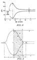
- FIGURE 1 is an exploded perspective of an assembled reaction slide which can be used in performing the present, end-point or kinetic, methods for measuring the clottable fibrinogen level in a blood or blood-derived sample.
- FIGURE 2 is a longitudinal vertical cross-section of a reaction slide together with apparatus for using magnetic particles to measure the reaction.
- FIGURE 3 illustrates the clotting curve for the fibrinogen assay, with A designating the maximum amplitude of particle oscillation and B designating the subsequent residual post peak minimum amplitude of particle oscillation.
- FIGURE 4 shows additional features of the kinetic clotting waveform which may be utilized as parameters to construct specific algorithms to measure fibrinogen.
- FIGURES 5, 6 and 7 are comparisons of the feature-based algorithms for the fibrinogen assay.
- the present invention provides a novel fibrinogen assay based on monitoring the movement of magnetic particles incorporated in the assay reaction.
- a dry reagent matrix in which is embedded a plurality of magnetic particles, distributed homogeneously therethrough is subjected to an oscillating magnetic field.
- This oscillating magnetic field may be obtained by using (1) a stationary oscillating magnet, or (2) a moving permanent magnet, or (3) a combination of an oscillating magnet and a stationary permanent magnet. (See U.S. Patent 5,110,727 for a detailed discusion.)
- the whole blood or blood-derived sample is then added to the reagent, causing it to become solubilized simultaneously, thereby freeing the particles to move in an oscillating pattern induced by the oscillating magnetic field.
- the freed magnetic particles form columnar structures or stacks, which, under the influence of the oscillating magnetic field, create a flicker phenomena due to a change in the orientation of these columnar structures or stacks.
- the oscillation of the particles is optically monitored by subjecting the particles to incident light and detecting reflected (scattered) light rays.
- the magnetic particles which are entrapped in the dry reagent matrix are incapable of oscillation.
- a maximum number of particle-based columnar structures or stacks can quickly be observed to oscillate to the greatest degree, providing a maximum oscillation amplitude, shown as A in FIGURE 3.
- a in FIGURE 3 As the reaction progresses, a coagulum forms restricting the degree of oscillation of an increasing number of particle-based columnar structures or stacks. This gradual decrease in the flicker pattern produces a residual post-peak minimum amplitude, shown as B in FIGURE 3.
- either the degree of particle movement relative to said oscillating magnetic field is monitored to measure the start time and stop time of the assay, or one or more features of the kinetic curve, other than clotting time, is/are used to measure fibrinogen concentration in the sample.
- the clotting curve is complex. Starting at 1, the first indication of particle oscillation is apparent. The magnitude of magnetic particle oscillation is apparent. The magnitude of magnetic particle oscillation as monitored optically increases from the start of the assay at 1 and peaks at 2 and 2' on the upper and lower portions of the wave envelope, respectively.
- the oscillation signal amplitude at the peak is designated as A and shown in FIGURE 4 as a signal difference between 2 and 2'.
- the time at which A occurs, t A is the clotting time. Tracing along the waveform envelope at the top, the amplitude decreases after t A .
- Points m 1 , m 2 , and m 3 are shown. Points m 1 and m 3 are chosen at arbitrary but fixed times. Point m 2 is chosen as an inflection point. The slope of the curve taken at m 1 , m 2 or m 3 can be utilized as a measure of fibrinogen, since these slopes are steepest (most negative) at the highest fibrinogen levels and become less steep with decreasing fibrinogen.
- Fibrinogen concentration is proportional to A/B and to (A-B)/A. Fibrinogen concentration is also inversely proportional to B and directly proportional to A-B, but these parameters alone are generally less precise than A/B or (A-B)/A.
- ⁇ is equal to the area of the rectangle with sides of magnitude A and opposite sides of magnitude t B' minus the area designated as ⁇ .
- ⁇ is shown in FIGURE 4 as consisting of two portions: an upper portion, ⁇ 1 and a lower portion, ⁇ 2 .
- the line extending through amplitude B' and intersecting parallel lines 4 and 4' constructed as perpendiculars to the line of magnitude A at points 2 and 2' helps to define the right boundary of ⁇ .
- ⁇ is directly proportional to fibrinogen concentration and is very precise.
- ⁇ and a may also be used in combination, i.e., taken as a ratio or difference to indicate fibrinogen level.
- ⁇ and/or ⁇ could be calculated independently of time, for example by always using t B as the horizontal measure for area calculation (see Figure 4).
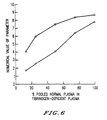
- FIGURE 5 shows how some of the kinetic parameters of the clotting curve vary with concentration of fibrinogen for a dry reagent slide incorporating reptilase and a citrated plasma sample consisting of pooled normal plasma diluted in varying amounts in fibrinogen deficient plasma.
- the present invention thus provides fibrinogen measurement using magnetic particle dry chemistry technology. Using this approach fibrinogen can be measured with each of the approaches listed below.
- the dry reagent be prepared such that it is rapidly dissolved upon the addition of the blood or blood-derived sample. Freeze-drying on a surface, or even better, between two surfaces closely apposed at a capillary or near capillary distance works best. This produces a mass of low matter content which enables rapid sample penetration and dissolution.
- freeze drying provides excellent results for preparation of the dry magnetic particle-containing reagent
- room temperature, vacuum, desiccant, convective, or other types of drying can also be used to achieve good results.
- room temperature drying of reagent on the base of a reaction slide (with spacer in place) followed by attachment of the cover can be used to obtain a self-metering dry reagent containing element.
- FIGURE 1 is an exploded view showing the relative position of the cover (10), overlay (20) and base (30) components of the slide.
- Cover (10) comprises a thin glass or polymer sheet transparent to light, having formed therein a sample receiving opening (14) and an elongate opening (12) proximate to distal end (16) of the cover.
- Overlay (20) comprises a thin glass or polymer sheet, typically transparent, having formed therein a cut-out, the cut-out having a geometry essentially as shown to form a sample well (22), a reaction chamber (24) and an optional conduit (26) communicating the reaction space and the sample receiving opening.
- the cut-out of overlay (20) can either have a geometry forming sample well (22) and reaction chamber (24) in communication with each other, or a geometry comprising sample well (22), reaction chamber (24), and conduit (26) essentially as shown.
- Reaction chamber (24) becomes a reaction volume upon assembly of the cover, overlay and base.
- tapering walls (25) form a transition between conduit (26), and sample well (22) or if conduit (26) is not used, and reaction chamber (24).
- the distal end of the overlay is closed as shown at (29).
- the base (30) comprises a sheet of glass or polymer material, which is typically somewhat thicker than either the cover (10) or overlay (20).
- cover (10) is approximately the same as that of overlay (20), and the width (top to bottom in the drawing) of cover (10) and overlay (20) are about the same and typically less than that of base (30).
- base (30) When the cover, overlay and base are assembled, the bottom surface of cover (10), facing base (30), is spaced from the top surface of base (30) by a distance that is sufficiently small to cause a volume of sample corresponding to the volume of the reaction chamber to be drawn simultaneously into the reaction volume by capillary action. This action is made possible by the presence of vent (12).
- reaction chamber (24) is charged with the dry reagent containing the magnetic particles homogeneously dispersed therethrough.
- FIGURE 2 provides a longitudinal vertical crosssection of the reaction slide together with an apparatus for using the magnetic particles to measure the assay.
- reaction slide 1 is disposed and in close proximity to a permanent magnet (195).
- Beneath the permanent magnet (195) is an electromagnet (196) which is driven by power supply (199) for cycling voltage on and off at a desired frequency.
- the permanent magnet above without the electromagnet, creating an oscillating magnetic field with the permanent magnet by moving the permanent magnet back and forth along a plane essentially parallel to the plane of the reaction slide.
- the electromagnet above is used to generate an oscillating magnetic field.
- both of these magnets are used essentially as shown in the figure.
- a light source such as an infrared light emitting diode is appropriately situated for providing incident light on the reaction chamber and a detector positioned for detecting light rays reflected from the sample within the reaction volume 66.
- the reflected rays illustrated as rays (198), are detected by detector (400).
- Detector (400) can be positioned at any position which will permit it to detect the reflected (scattered) rays, but a position between the 90° and the 10° position, inclusively, is preferable, with a position between the 90° and 45° position being preferred, and between 90° and 75° being most preferred.
- Clot formation kinetic approaches Along with buffers sufficient to control pH in the ranges known in the art (typically 7.0 to 7.4), the reagent used in this embodiment contains (a) thromboplastin present in an amount sufficient to cause coagulation of normal plasma with near minimal B values (PT test) plus magnetic particles, typically 8.3 mg ml -1 but variable over a wide range (see U.S.
- thrombin plus magnetic particles in amounts similar to that used in (a) above defined functionally; or (c) a snake venom, e.g., reptilase, copperhead venom enzyme, or Malayan pit viper venom enzyme in amounts similar to (a) above, as defined functionally for the venom plus magnetic particles in amounts described in (a) above.
- the blood sample does not normally require dilution and may be added directly to the dry chemistry assay mixture. Some dilution, to within 1:2 (by volume) and most preferably not more than 1:4 (by volume) is, however, possible with this embodiment, and may be used if desired. In some cases, dilutions as great as 1:10 (by volume) or even 1:20 (by volume) could be employed advantageously, for example at very high fibrinogen levels.
- thrombin preparations exist. Actual purity and activity may vary depending upon the source. In addition to human thrombin, bovine thrombin is often used. In addition to source and activity variations, lot-to-lot variability also occurs. The same is true for the snake venoms. Here, lot-to-lot activity can vary considerably. Thromboplastins are also quite different, depending upon the source: rabbit brain, ox brain, human brain, etc. In addition, lot-to-lot variability of thromboplastin from the same source is well known in the blood coagulation diagnostics field.
- the exact amounts of the various clotting reagents employed in formulating suitable dry reagents for use in combination with magnetic particles cannot be specified, but rather the quantities of these clotting reagents are determined functionally as described for each embodiment.
- the buffer type and concentration may be less important than the pH. Buffers which are commonly employed in blood coagulation reactions may generally be used. Owrens, HEPES, and Tris are among these.
- a reptilase based reagent at pH 7.3 for example, 25 mM HEPES performs better (gives a steeper response curve) than 25 mM Tris.
- optimum pH and buffer selection may depend somewhat upon the type of clotting reagent (fibrinogen assay reagent) and possibly upon the particular lot of reagent chosen. It is generally useful to employ a pH in the range of 7.0 to 7.4 and optimize from there. It is also expedient to use certain additives to improve dissolution characteristics of the dry reagent and to better disperse the magnetic particles upon solubilization.
- Bovine serum albumin and/or polyethylene glycol in amounts less than 1 wt.% in the reagent formulation work well. The exact amounts may depend upon the properties of the clotting reagent and may differ with each lot number.
- clotting time is preferably not used. Instead, kinetic parameters reflecting features in the clotting curve (shown in FIGURES 3 and 4) are utilized. It should also be noted that whereas thromboplastin based reagent may be utilized to measure fibrinogen in a blood sample using kinetic parameters, this type of reagent is generally not suitable for use with clotting time (e.g., Clauss type) methods.
- A is the peak height maximum (maximum oscillation amplitude)
- B is the minimum (residual post peak minimum) which is observed after A is reached.
- Either A and B, or B only, are used to determine a factor proportional to fibrinogen concentration in the sample.
- the following measures may be employed: (i) B alone; (ii) A/B (or B/A ); (iii) A-B; or (iv) (A-B) ⁇ A or A ⁇ (A-B).
- the negative slope e.g., at or near the inflection point
- the area enclosed by the clotting curve This area may be bounded by vertical axes at the peak (A) and at a set time after A, e.g., 45 seconds, where: A is normalized and thus made equivalent for each separate assay curve.
- Clotting time approaches (Clauss type) : Along with buffers in amounts sufficient to control pH the reagent used contains (a) thrombin plus magnetic particles, where the thrombin concentration is sufficient to cause coagulation of normal plasma with B values somewhat above the absolute minimum obtained at ever increasing thrombin concentration and the magnetic particle concentration is typically 8.3 mg ml -1 , but can be varied over a wide range (see U.S.
- Patent 5,110,727 or (b) a snake venom enzyme in place of thrombin under essentially the same conditions and in amounts sufficient to produce the same functional effects (e.g., reptilase, (a thrombin-like enzyme isolated from the venom of Bothrops atrox), copperhead venom enzyme, ( Agkistrodon contortrix) or Malayan Pit Viper venom enzyme ( Agkistrodon rhodostoma) (see Becker et al, Thromb.
- reptilase a thrombin-like enzyme isolated from the venom of Bothrops atrox
- copperhead venom enzyme Agkistrodon contortrix
- Malayan Pit Viper venom enzyme Agkistrodon rhodostoma
- the blood sample to be analyzed is first diluted with a buffer (typically Owens buffer) and then added to the dry chemistry assay mixture in an appropriate analyzer. Clotting time is then measured.
- a buffer typically Owens buffer
- sample well (22) For example, 1 to 3 microliters of sample is carefully pipetted into sample well (22) near the opening of conduit (26) to deliver the entire sample into conduit (26). Actually, once delivered into the appropriate region of sample well (22) by the pipette, the sample will automatically be taken up into conduit (26) via capillary action.
- this type of reaction slide there must not be any clotting reagent (thrombin, etc.) in conduit (26).
- the dry reagent must be confined to reaction volume (24). This is easily achieved in manufacture of the dry chemistry reaction slide in a number of ways, such as by filling from the vent area forward.
- a fixed volume of buffer diluent is added to the sample well in a volume equal to or preferably in excess of the volume required to fill conduit (26) plus reaction chamber (24).
- a volume of 25 to 50 microliters of buffer diluent is effective, with a volume of 30 to 40 microliters preferred. Less than 25 microliters could give rise to inadequate flow, particularly if the reaction volume does not completely fill. More than 50 microliters of diluent may overflow the sample well in a reaction slide of this size.
- the range of dilutions achieved would be from approximately 1 in 25 (by volume) for 1 microliter of sample to 3 in 25 (by volume) for a 3 microliter sample. Over this range, it is possible to select a concentration of clotting reagent and appropriate dilution factor to provide a good standard curve which is essentially linear on log-log paper for a plot of clotting time (t A ) versus the concentration of fibrinogen.
- This curve may then be utilized as a reference, as in the conventional Clauss assays to quantify unknown samples on the basis of clotting time. It is also possible to select clotting endpoints other than t A . For example, if an adjusted endpoint amplitude of, for example, 0.9 A is selected (after the peak amplitude A is reached) good results may be obtained by using the time at which 0.9 A occurs as clotting time. The same is true for 0.85 A, however, this cannot be pushed too far, otherwise sensitivity at low fibrinogen levels will be lost, especially if the lowest level of fibrinogen which is desired to be detected produces a B value equivalent or nearly equivalent to the adjusted endpoint amplitude.
- One advantage of utilizing adjusted endpoint amplitudes is that the slope of the straight line logarithmic fibrinogen standard curve may be made steeper.
- reaction slide containing dried reagent consisting of magnetic particles and buffer and a binding agent such as less than 1 wt.% bovine serum albumin or polyethylene glycol (molecular weights (M w) of polyethylene glycol ranging from approximately 3,400 to 10,000 daltons have been successfully employed) or a combination of both.
- a 1 to 3 microliter sample is added to conduit (26) of the reaction slide via sample well (22).
- a diluted thrombin or snake venom is employed to wash the sample as before, into reaction volume (24) diluting it on the way.
- the dry chemistry in this case the magnetic particles plus other reagents, becomes solubilized and provides an adequate clotting endpoint.
- This variation of the methodology is, however less convenient because it requires reagent preparation by the user and may not provide as precise a starting point.
- reaction slide containing clotting reagent (thrombin, snake venom, or thromboplastin) mixed with magnetic particles, buffer, and a binding agent, such as less than 1 wt.% bovine serum albumin or polyethylene glycol, wherein the mixture is in dry form and at greater concentration (4 to 8-fold) than typically used but contained in a proportionally smaller volume such that this volume is situated in conduit (26) of the reaction slide and the remaining portions of the reaction slide are empty.
- clotting reagent thrombin, snake venom, or thromboplastin
- a binding agent such as less than 1 wt.% bovine serum albumin or polyethylene glycol
- reaction volume (24) After a brief incubation period (typically 5 to 30 seconds, depending upon the concentration and types of reagents employed) a volume of previously diluted sample which is sufficient to fill the remainder of the reaction volume (24) is applied to sample well (22).
- This sample either plasma or whole blood typically diluted 1:10 (by volume) in buffer
- mixes with the reagent solution and magnetic particles in conduit (26) mixes with the reagent solution and magnetic particles in conduit (26), and the resultant mixture rapidly enters reaction volume (24) whereupon a signal is detected initiating the reaction monitoring sequence.
- B alone, A/B or its reciprocal, (A-B) ⁇ A or its inverse, A-B, negative slope, normalized area, and in the case of thrombin or snake venoms, also clotting time, may be measured to provide a fibrinogen assay.
- Fibrinogen screening tests (clotting time based): Along with buffers, the reagents used in this embodiment contain (a) thrombin plus magnetic particles or (b) a snake venom (e.g., reptilase copperhead venom enzyme, or Malayan pit viper venom enzyme) plus magnetic particles.
- the blood sample should not be diluted and is added directly to the dry chemistry mixture.
- Clotting time is then measured. When thrombin is used, the result is reported as a thrombin time (TT) or thrombin clotting time (TCT). When reptilase is used, the result is reported as reptilase time.
- the present method for measuring the clottable fibrinogen level in a blood or blood-derived sample comprises the following steps (i) to (iv).
- the dry reagent matrix comprises thromboplastin, thrombin or reptilase.
- the fibrinogen assay may be based on clotting time using a thrombin or reptilase based reagent and start and stop time for the assay.
- the method of performing a fibrinogen assay comprises the following steps (i') to (iv').
- FIGURE 5 provides five curves, each curve representing a different kinetic parameter obtained from the clotting curve produced by a reptilase based dry reagent.
- the legend indicates which curve corresponds to each parameter.
- FIGURE 4 can be used to help in understanding the relationship of the parameters to clotting kinetics.
- the numerical value of the parameter is the ordinate.
- the percent of pooled normal plasma (PNP) diluted in fibrinogen deficient plasma used to obtain the clotting kinetics from which the parameters were determined is the abscissa.
- (A-B)/A and A/B have generally been the most precise parameters, with A/B showing better precision at the lowest fibrinogen concentrations measurable with this system.
- (A-B)/A is generally the more precise parameter of higher fibrinogen levels. It should be recognized that (A-B)/A is equal to 1-(B/A).
- FIGURE 6 shows the A/B and (A-B)/A curves on an expanded scale.
- FIGURE 7 shows ⁇ and ⁇ values obtained in a different set of experiments.
- Reptilase is generally preferred over thrombin for kinetic clotting based fibrinogen measurements because it is not affected by heparin, a commonly employed anticoagulant drug.
- reptilase in buffer is combined with magnetic particles and polyethylene glycol of molecular weight (M w) 3400 to 10,000.
- M w molecular weight
- the dry reagent mix is prepared by dissolving the polyethylene glycol in water, and then adding the reptilase reagent and mixing gently. This solution is then mixed with magnetic particles and applied to reaction slides.
- Another preferred dry reagent matrix is prepared from buffered reptilase, magnetic particles and bovine serum albumin. This is prepared by dissolving the bovine serum albumin in water, and then adding the reptilase reagent with gentle mixing. This solution is mixed with magnetic particles and applied to reaction slides.
- the first embodiment which is based on clot formation kinetics, could typically utilize a lower concentration of clot forming reagent (thrombin or snake venom) than the second embodiment which is based on clotting time.
- concentrations of reagents employed however would vary with the type and source of the reagent (e.g., bovine, human, etc.) and potency as noted supra.
- the first embodiment can also utilize thromboplastin as a clot forming reagent.
- thromboplastin cannot be used as a clot forming reagent for the second end-point embodiment, because its use in this embodiment would not provide results specific for fibrinogen.
- Yet another embodiment is to utilize a reagent concentration (of thrombin or snake venom) comparable to that in the first embodiment but to measure clotting time (as in the second embodiment) on an undiluted sample instead of clot kinetic parameters (as in the first embodiment).
- This approach provides a quantitative screening test which is sensitive over the most clinically useful range of fibrinogen levels (low normal to below normal) and can be useful in rapid assessment of fibrinogen abnormalities from an undiluted whole blood or plasma sample.
- Either thrombin or an appropriate snake venom e.g., reptilase, copperhead venom or Malayan pit viper venom
- This embodiment provides results analogous to a thrombin time, reptilase time, or equivalent test or a fibrinogen value when used with a standard curve for interpretation.
Landscapes
- Health & Medical Sciences (AREA)
- Life Sciences & Earth Sciences (AREA)
- Engineering & Computer Science (AREA)
- Chemical & Material Sciences (AREA)
- Hematology (AREA)
- Immunology (AREA)
- Biomedical Technology (AREA)
- Physics & Mathematics (AREA)
- Analytical Chemistry (AREA)
- General Health & Medical Sciences (AREA)
- Molecular Biology (AREA)
- Urology & Nephrology (AREA)
- Pathology (AREA)
- General Physics & Mathematics (AREA)
- Biochemistry (AREA)
- Medicinal Chemistry (AREA)
- Food Science & Technology (AREA)
- Biotechnology (AREA)
- Microbiology (AREA)
- Cell Biology (AREA)
- Ecology (AREA)
- Biophysics (AREA)
- Clinical Laboratory Science (AREA)
- Chemical Kinetics & Catalysis (AREA)
- Investigating Or Analysing Biological Materials (AREA)
- Measuring Or Testing Involving Enzymes Or Micro-Organisms (AREA)
- Investigating Or Analyzing Materials By The Use Of Magnetic Means (AREA)
- Centrifugal Separators (AREA)
Claims (19)
- Verfahren zum Durchführen einer Fibrinogenbestimmung, umfassend:(i) Einwirkenlassen eines oszillierenden Magnetfeldes auf einen Reaktionsobjektträger, welcher (1) eine Probenvertiefung zur Aufnahme einer flüssigen Probe und (2) eine Reaktionskammer aufweist, welche eine Trockenreagenzmatrix enthält, in die eine Vielzahl von magnetischen Teilchen eingebettet ist, die darin homogen verteilt sind, wobei das Reagenz eine Protease ist, die direkt auf Fibrinogen wirkt und die Polymerisation von Fibrinogen herbeiführt;
wobei die Probenvertiefung und Reaktionskammer durch eine Transportzone mit einer derartigen Geometrie, daß ein Volumen einer flüssigen Analytprobe, das in die Probenvertiefung gegeben wird und dem Volumen der Reaktionskammer entspricht, gleichzeitig von der Probenvertiefung in die Reaktionskammer transportiert wird, in fluider Verbindung stehen;(ii) Zugeben einer Vollblutprobe oder einer aus Blut gewonnenen Probe in die Probenvertiefung, wodurch die Probe gleichzeitig in die Reaktionskammer eingeführt wird, das Reagenz löslich gemacht wird und die Teilchen freigesetzt werden, so daß sie sich gemäß einem oszillierenden Muster bewegen, das durch das oszillierende Magnetfeld hervorgerufen wird;(iii) optisches Überwachen der Reaktionskammer, um entweder (iiia) eine Startzeit und eine Stoppzeit für die Fibrinogenbestimmung, die einer Änderung des Ausmaßes der Teilchenbewegung relativ zu dem oszillierenden Magnetfeld entspricht, oder (iiib) die maximale Amplitude der Teilchenoszillation A und die anschließende restliche minimale Nachpeak-Amplitude B der Teilchenoszillation, oder (iiic) den Anstieg der Teilchenoszillationskurve oder die Fläche, die durch diese Kurve im Bereich zwischen A und B definiert ist, zu messen; und(iv) Verwenden von entweder der Startzeit und der Stoppzeit, oder von mindestens B, oder von dem Anstieg, oder von der Fläche zum Messen der Konzentration von gerinnungsfähigem Fibrinogen in der Probe. - Verfahren nach Anspruch 1, worin die Protease ein Bestandteil, ausgewählt aus der Gruppe bestehend aus Human-Thrombin, Rinderthrombin, Reptilase, dem Enzym des Gifts der Mokassinschlange (copperhead) oder dem Enzym des Gifts der Malaiischen Grubenotter (Malayan pit viper), ist.
- Verfahren zum Durchführen einer Fibrinogenbestimmung wie in Anspruch 1 beansprucht, worin in Schritt (iv) die Startzeit und die Stoppzeit verwendet werden, um die Konzentration von gerinnungsfähigem Fibrinogen in der Probe zu messen.
- Verfahren nach Anspruch 3, worin das Trockenreagenz Thrombin umfaßt.
- Verfahren nach Anspruch 3, worin das Trockenreagenz Reptilase, das Enzym des Gifts der Mokassinschlange oder das Enzym des Gifts der Malaiischen Grubenotter umfaßt.
- Verfahren zum Durchführen einer Fibrinogenbestimmung, umfassend:(i) Einwirkenlassen eines oszillierenden Magnetfelds auf einen Reaktionsobjektträger, der (1) eine Probenvertiefung zur Aufnahme der flüssigen Probe, (2) eine Reaktionskammer und (3) eine Leitungseinrichtung aufweist, die sich zwischen der Probenvertiefung und der Reaktionskammer befindet und damit in fluider Verbindung steht;
wobei die Reaktionskammer eine Trockenreagenzmatrix enthält, in die eine Vielzahl von magnetischen Teilchen eingebettet ist, die darin homogen verteilt sind, wobei die Trockenreagenzmatrix nur in der Reaktionskammer vorhanden ist, und wobei das Reagenz ein Bestandteil ist, ausgewählt aus der Gruppe bestehend aus Fibrinogenbestimmungsreagenzien, ausgewählt aus Proteasen, welche direkt auf Fibrinogen wirken;(ii) Zugeben eines Volumens einer Vollblutprobe oder einer aus Blut gewonnenen Probe in die Probenvertiefung, wobei das Volumen der Probe ausreicht, um im wesentlichen die Leitungseinrichtung zu füllen, ohne in die Reaktionskammer einzutreten oder die Trockenreagenzmatrix zu berühren;(iii) Zugeben eines Volumens eines Pufferverdünnungsmittels zu der Probenvertiefung, wobei das Volumen des Puffers ausreicht, um die Probe in die Reaktionskammer zu spülen, wodurch bewirkt wird, daß das Reagenz löslich gemacht wird und die Teilchen freigesetzt werden, so daß sie sich gemäß einem oszillierenden Muster bewegen, das durch das oszillierende Magnetfeld hervorgerufen wird;(iv) optisches Überwachen der Oszillation der magnetischen Teilchen zum Messen einer Startzeit und einer Stoppzeit für die Fibrinogenbestimmung, die einer Änderung des Ausmaßes der Teilchenbewegung relativ zu dem Magnetfeld entspricht; und(v) Verwenden der Startzeit und der Stoppzeit zum Messen der Fibrinogenkonzentration in der Probe. - Verfahren zum Durchführen einer Fibrinogenbestimmung wie in Anspruch 1 beansprucht, worin Schritt (iv) mindestens B verwendet, um die Konzentration von gerinnungsfähigem Fibrinogen in der Probe zu messen.
- Verfahren nach Anspruch 7, worin B verwendet wird, um die Fibrinogenkonzentration in der Probe zu messen.
- Verfahren nach Anspruch 7, worin das Verhältnis A/B oder B/A verwendet wird, um die Fibrinogenkonzentration in der Probe zu messen.
- Verfahren nach Anspruch 7, worin A-B verwendet wird, um die Fibrinogenkonzentration in der Probe zu messen.
- Verfahren nach Anspruch 7, worin (A-B) ÷ A oder A ÷ (A-B) verwendet wird, um die Fibrinogenkonzentration in der Probe zu messen.
- Verfahren nach Anspruch 7, worin A/B und (A-B) ÷ A verwendet werden, um die Fibrinogenkonzentration der Probe zu messen.
- Verfahren nach Anspruch 7, worin der Anstieg der Gerinnungskurve, welcher in einem Bereich zwischen A und B abgelesen wird, verwendet wird, um die Fibrinogenkonzentration der Probe zu bestimmen.
- Verfahren nach Anspruch 7, worin die Fläche entweder über oder unter der Gerinnungskurve, die durch den Bereich zwischen A und B definiert ist, oder ein Teil davon verwendet wird, um die Fibrinogenkonzentration der Probe zu bestimmen.
- Verfahren nach Anspruch 7, worin das Trockenreagenz Thrombin umfaßt.
- Verfahren nach Anspruch 7, worin das Trockenreagenz Reptilase, das Enzym des Gifts der Mokassinschlange oder das Enzym des Gifts der Malaiischen Grubenotter umfaßt.
- Verfahren zum Durchführen einer Fibrinogenbestimmung, umfassend:(i) Einwirkenlassen eines oszillierenden Magnetfelds auf einen Reaktionsobjektträger, der (1) eine Probenvertiefung zur Aufnahme einer flüssigen Probe, (2) eine Reaktionskammer und (3) eine Leitungseinrichtung aufweist, die sich zwischen der Probenvertiefung und der Reaktionskammer befindet und damit in fluider Verbindung steht; wobei die Leitungseinrichtung eine Trokkenreagenzmatrix enthält, in die eine Vielzahl von magnetischen Teilchen eingebettet ist, die darin homogen verteilt sind, wobei das Trockenreagenz nur in der Leitung vorhanden ist, und wobei das Reagenz ein Bestandteil ist, ausgewählt aus der Gruppe, bestehend aus Fibrinogenbestimmungsreagenzien, ausgewählt aus Proteasen, welche direkt auf Fibrinogen wirken;(ii) Zugeben einer Vollblutprobe oder einer aus Blut gewonnenen Probe in die Probenvertiefung, wobei das Volumen der Probe ausreicht, um im wesentlichen die Leitungseinrichtung und die Reaktionskammer zu füllen und sich mit dem Reagenz zu vermischen und das Reagenz in die Reaktionskammer zu spülen, wodurch die magnetischen Teilchen freigesetzt werden, so daß sie sich gemäß einem oszillierenden Muster bewegen, das durch das oszillierende Magnetfeld hervorgerufen wird;(iv) optisches Überwachen der Reaktionskammer, um entweder (iva) eine Startzeit und eine Stoppzeit für die Fibrinogenbestimmung, die einer Änderung des Ausmaßes der Teilchenbewegung relativ zu dem oszillierenden Magnetfeld entspricht, oder (ivb) die maximale Amplitude der Teilchenoszillation A und die anschließende restliche minimale Nachpeak-Amplitude B der Teilchenoszillation, oder (ivc) den Anstieg der Teilchenoszillationskurve oder die Fläche, die durch diese Kurve im Bereich zwischen A und B definiert ist, zu messen; und(v) Verwenden von entweder der Startzeit und der Stoppzeit, oder von mindestens B, oder von dem Anstieg, oder von der Fläche zum Messen der Konzentration von gerinnungsfähigem Fibrinogen in der Probe.
- Verfahren nach Anspruch 17, worin vor dem Schritt (ii) ein Volumen von destilliertem Wasser in die Probenvertiefung zugegeben wird, welches ausreicht, um von der Leitung durch Kapillarwirkung aufgenommen zu werden, wodurch vor der Zugabe der Vollblutprobe oder der aus Blut gewonnenen Probe das Trockenreagenz löslich gemacht und die magnetischen Teilchen freigesetzt werden.
- Verfahren zum Durchführen einer Fibrinogenbestimmung, umfassend:(i) Zugeben einer Vollblutprobe oder aus Blut gewonnenen Probe in die Probenvertiefung eines Reaktionsobjektträgers, wobei der Reaktionsobjektträger (1) die Probenvertiefung zur Aufnahme einer flüssigen Probe, (2) eine Reaktionskammer und (3) eine Leitungseinrichtung aufweist, die sich zwischen der Probenvertiefung und der Reaktionskammer befindet und damit in fluider Verbindung steht;
wobei die Leitungseinrichtung eine Trockenreagenzmatrix enthält, in die eine Vielzahl von magnetischen Teilchen eingebettet ist, die darin homogen verteilt sind, wobei die Trockenreagenzmatrix nur in der Leitung vorhanden ist, und wobei das Reagenz ein Bestandteil ist, ausgewählt aus der Gruppe bestehend aus Fibrinogenbestimmungsreagenzien, ausgewählt aus Proteasen oder Kofaktoren, welche direkt auf Fibrinogen wirken;
worin das Volumen der Vollblutprobe oder der aus Blut gewonnenen Probe, das in die Probenvertiefung gegeben wird, ausreicht, um im wesentlichen die Leitungseinrichtung und die Reaktionskammer zu füllen und das Reagenz in die Reaktionskammer zu spülen, wodurch die magnetischen Teilchen freigesetzt werden;(ii) Einwirkenlassen eines oszillierenden Magnetfelds auf den Reaktionsobjektträger entweder zum Zeitpunkt der Zugabe der Vollblutprobe oder der aus Blut gewonnenen Probe in die Probenvertiefung oder kurz danach, wobei das oszillierende Magnetfeld bewirkt, daß die freigesetzten magnetischen Teilchen sich gemäß einem oszillierenden Muster bewegen; und(iii) optisches Überwachen der Reaktionskammer, um entweder (iiia) mindestens die minimale Nachpeak-Amplitude der magnetischen Teilchenoszillation (B) oder (iiib) den Anstieg der Amplitude der Teilchenoszillation oder (iiic) die Fläche, die durch die Kurve der Amplitude zwischen der maximalen Amplitude der Teilchenoszillation A und B definiert ist, zu messen, um die Konzentration von gerinnungsfähigem Fibrinogen in der Probe zu messen.
Priority Applications (1)
| Application Number | Priority Date | Filing Date | Title |
|---|---|---|---|
| EP98201760A EP0867723A3 (de) | 1990-07-10 | 1991-06-28 | Fibrinogenbestimmungen unter Verwendung von trockenen chemischen Reagenzien die magnetische Teilchen enthalten |
Applications Claiming Priority (3)
| Application Number | Priority Date | Filing Date | Title |
|---|---|---|---|
| US55057090A | 1990-07-10 | 1990-07-10 | |
| US550570 | 1990-07-10 | ||
| PCT/US1991/004472 WO1992001065A1 (en) | 1990-07-10 | 1991-06-28 | Fibrinogen assays using dry chemical reagents containing magnetic particles |
Related Child Applications (1)
| Application Number | Title | Priority Date | Filing Date |
|---|---|---|---|
| EP98201760A Division EP0867723A3 (de) | 1990-07-10 | 1991-06-28 | Fibrinogenbestimmungen unter Verwendung von trockenen chemischen Reagenzien die magnetische Teilchen enthalten |
Publications (3)
| Publication Number | Publication Date |
|---|---|
| EP0538393A1 EP0538393A1 (de) | 1993-04-28 |
| EP0538393A4 EP0538393A4 (de) | 1994-01-26 |
| EP0538393B1 true EP0538393B1 (de) | 1998-12-16 |
Family
ID=24197737
Family Applications (2)
| Application Number | Title | Priority Date | Filing Date |
|---|---|---|---|
| EP91914171A Expired - Lifetime EP0538393B1 (de) | 1990-07-10 | 1991-06-28 | Fibrinogenbestimmungen unter verwendung von chemischen trockenreagenzien, die magnetische teilchen enthalten |
| EP98201760A Withdrawn EP0867723A3 (de) | 1990-07-10 | 1991-06-28 | Fibrinogenbestimmungen unter Verwendung von trockenen chemischen Reagenzien die magnetische Teilchen enthalten |
Family Applications After (1)
| Application Number | Title | Priority Date | Filing Date |
|---|---|---|---|
| EP98201760A Withdrawn EP0867723A3 (de) | 1990-07-10 | 1991-06-28 | Fibrinogenbestimmungen unter Verwendung von trockenen chemischen Reagenzien die magnetische Teilchen enthalten |
Country Status (10)
| Country | Link |
|---|---|
| US (1) | US5350676A (de) |
| EP (2) | EP0538393B1 (de) |
| JP (1) | JP2649608B2 (de) |
| AT (1) | ATE174633T1 (de) |
| AU (1) | AU660624B2 (de) |
| CA (1) | CA2087033C (de) |
| DE (1) | DE69130644T2 (de) |
| ES (1) | ES2128321T3 (de) |
| TW (1) | TW221493B (de) |
| WO (1) | WO1992001065A1 (de) |
Families Citing this family (67)
| Publication number | Priority date | Publication date | Assignee | Title |
|---|---|---|---|---|
| US6114135A (en) * | 1991-11-08 | 2000-09-05 | Goldstein; Sheldon | Multiple coagulation test system and method of using a multiple coagulation test system |
| DK203191D0 (da) * | 1991-12-19 | 1991-12-19 | Novo Nordisk As | Fremgangsmaade og apparat til bestemmelse af relevante blodparametre |
| ES2113492T3 (es) * | 1992-09-09 | 1998-05-01 | Tokuyama Corp | Metodo de valoracion de fibrinogeno, reactivo seco para dicha valoracion, y procedimiento para su preparacion. |
| US5443959A (en) * | 1992-09-09 | 1995-08-22 | Tokuyama Corporation | Method of assaying fibrinogen, dry reagent therefor, and process for the preparation thereof |
| US5605798A (en) | 1993-01-07 | 1997-02-25 | Sequenom, Inc. | DNA diagnostic based on mass spectrometry |
| EP0685069A4 (de) * | 1993-02-17 | 2002-01-23 | Cardiovascular Diagnostics Inc | Immunoassay und affinitätstest auf der grundlage einer trockenchemischen kaskade |
| US5670329A (en) * | 1993-05-28 | 1997-09-23 | Cardiovascular Diagnostics, Inc. | Method and analytical system for performing fibrinogen assays accurately, rapidly and simply using a rotating magnetic field |
| US5841023A (en) * | 1993-08-31 | 1998-11-24 | Boehringer Mannheim Corporation | Magnet for medical instrument |
| US5526111A (en) * | 1993-08-31 | 1996-06-11 | Boehringer Mannheim Corporation | Method and apparatus for calculating a coagulation characteristic of a sample of blood a blood fraction or a control |
| US5522255A (en) * | 1993-08-31 | 1996-06-04 | Boehringer Mannheim Corporation | Fluid dose, flow and coagulation sensor for medical instrument |
| WO1995007050A2 (en) * | 1993-08-31 | 1995-03-16 | Boehringer Mannheim Corporation | Power supply control for medical instrument |
| US5502651A (en) * | 1994-05-02 | 1996-03-26 | Jackson; R. David | Potentiophotometric fibrinogen determination |
| US5504011A (en) * | 1994-10-21 | 1996-04-02 | International Technidyne Corporation | Portable test apparatus and associated method of performing a blood coagulation test |
| AU689143B2 (en) * | 1994-12-02 | 1998-03-26 | Cardiovascular Diagnostics, Inc. | Improved method and analytical system for performing fibrinogen assays accurately, rapidly and simply |
| US5567596A (en) * | 1994-12-29 | 1996-10-22 | Research Foundation Of State University Of New York | Rapid assay of activators and inhibitors of clotting |
| US5830655A (en) | 1995-05-22 | 1998-11-03 | Sri International | Oligonucleotide sizing using cleavable primers |
| EP0886681A1 (de) * | 1996-03-04 | 1998-12-30 | Genetrace Systems, Inc. | Verfahren zur suche von nukleinsäuren mit massenspektroskopie |
| US5965363A (en) | 1996-09-19 | 1999-10-12 | Genetrace Systems Inc. | Methods of preparing nucleic acids for mass spectrometric analysis |
| EP1457496B1 (de) | 1996-11-06 | 2014-01-15 | Sequenom, Inc. | Immobilisierung mit hoher Dichte von Nukleinsäuren |
| US7285422B1 (en) | 1997-01-23 | 2007-10-23 | Sequenom, Inc. | Systems and methods for preparing and analyzing low volume analyte array elements |
| AU5794498A (en) | 1996-12-10 | 1998-07-03 | Genetrace Systems, Inc. | Releasable nonvolatile mass-label molecules |
| US5998224A (en) * | 1997-05-16 | 1999-12-07 | Abbott Laboratories | Magnetically assisted binding assays utilizing a magnetically responsive reagent |
| US6046051A (en) * | 1997-06-27 | 2000-04-04 | Hemosense, Inc. | Method and device for measuring blood coagulation or lysis by viscosity changes |
| HU222809B1 (hu) * | 1997-10-03 | 2003-10-28 | 77 Elektronika Műszeripari Kft. | Eljárás és készülék kémiai összetevőnek anyagmintából, különösen vér glükóztartalmának vérmintából történő meghatározásához |
| US6165795A (en) * | 1998-06-25 | 2000-12-26 | Cardiovascular Diagnostics, Inc. | Methods for performing fibrinogen assays using dry chemical reagents containing ecarin and magnetic particles |
| US6417004B1 (en) * | 1999-04-29 | 2002-07-09 | Helena Laboratories Corporation | Enhancing clot detection in activated clotting time and other fibrin endpoint based tests |
| US6294342B1 (en) | 1999-09-29 | 2001-09-25 | Abbott Laboratories | Magnetically assisted binding assays utilizing a magnetically responsive reagent |
| CA2408850A1 (en) * | 2000-05-09 | 2001-11-15 | Pharmanetics Incorporated | Platelet function assay and reagent therefor |
| AU2001268468A1 (en) | 2000-06-13 | 2001-12-24 | The Trustees Of Boston University | Use of nucleotide analogs in the analysis of oligonucleotide mixtures and in highly multiplexed nucleic acid sequencing |
| US20020142483A1 (en) | 2000-10-30 | 2002-10-03 | Sequenom, Inc. | Method and apparatus for delivery of submicroliter volumes onto a substrate |
| US6759009B2 (en) | 2001-05-04 | 2004-07-06 | Portascience Incorporated | Method and device for clotting time assay |
| DE10140699A1 (de) * | 2001-08-24 | 2003-03-13 | Roche Diagnostics Gmbh | Anordnung und Verfahren zur Untersuchung der Fließfähigkeit einer physiologischen Flüssigprobe |
| US6902904B2 (en) * | 2001-08-27 | 2005-06-07 | Pharmanetics Incorporated | Coagulation assay reagents containing lanthanides |
| US6680177B2 (en) * | 2001-12-07 | 2004-01-20 | Cardiovascular Diagnostics, Inc. | Low molecular weight heparin assay, system and reagent therefor |
| DE10325173B4 (de) * | 2003-06-04 | 2007-11-08 | Stief, Thomas, Dr.med. | Nachweisverfahren für Fibrinogen und/oder Fibrinogen-Derivate |
| GB0313015D0 (en) * | 2003-06-06 | 2003-07-09 | Hall Effect Technologies Ltd | Method and device for analysing a liquid |
| EP1544596B1 (de) * | 2003-12-17 | 2016-11-23 | Boehringer Ingelheim microParts GmbH | Verfahren und Vorrichtung zur Bestimmung der Viskosität |
| US7439069B2 (en) | 2004-02-27 | 2008-10-21 | Nippoldt Douglas D | Blood coagulation test cartridge, system, and method |
| US7422905B2 (en) | 2004-02-27 | 2008-09-09 | Medtronic, Inc. | Blood coagulation test cartridge, system, and method |
| US7399637B2 (en) | 2004-04-19 | 2008-07-15 | Medtronic, Inc. | Blood coagulation test cartridge, system, and method |
| US9267167B2 (en) | 2004-06-28 | 2016-02-23 | Becton, Dickinson And Company | Dissolvable films and methods including the same |
| US8152305B2 (en) * | 2004-07-16 | 2012-04-10 | The University Of North Carolina At Chapel Hill | Methods, systems, and computer program products for full spectrum projection |
| US20060024776A1 (en) * | 2004-08-02 | 2006-02-02 | Mcmillian Ray | Magnetic particle capture of whole intact organisms from clinical samples |
| EP1845381A4 (de) * | 2005-06-23 | 2012-03-14 | Arkray Inc | Analytisches werkzeug |
| GB2438669A (en) * | 2006-03-07 | 2007-12-05 | Inverness Medical Switzerland | Sensing apparatus having a pair of coils and an oscillating member |
| WO2008103430A2 (en) * | 2007-02-22 | 2008-08-28 | The University Of North Carolina At Chapel Hill | Methods and systems for multiforce high throughput screening |
| US20080297169A1 (en) * | 2007-05-31 | 2008-12-04 | Greenquist Alfred C | Particle Fraction Determination of A Sample |
| WO2009039122A2 (en) | 2007-09-17 | 2009-03-26 | Sequenom, Inc. | Integrated robotic sample transfer device |
| JP5547639B2 (ja) | 2007-10-16 | 2014-07-16 | コーニンクレッカ フィリップス エヌ ヴェ | 診断マーカーの推定 |
| JP5145426B2 (ja) * | 2007-11-02 | 2013-02-20 | メドプラスト ソシエテ アノニム | 空間的な線維素凝塊形成を監視するための方法及び装置 |
| ES2688183T3 (es) * | 2008-03-31 | 2018-10-31 | Sysmex Corporation | Analizador de coagulación de sangre y método de análisis de coagulación de sangre |
| US20110229498A1 (en) | 2008-05-08 | 2011-09-22 | The Johns Hopkins University | Compositions and methods for modulating an immune response |
| US9720003B2 (en) | 2008-12-22 | 2017-08-01 | Koninklijke Philips Electronics N.V. | Assay for Troponin I using magnetic labels |
| US9347931B2 (en) | 2009-04-23 | 2016-05-24 | Dublin City University | Lateral flow assay device for coagulation monitoring and method thereof |
| WO2010151780A2 (en) | 2009-06-25 | 2010-12-29 | The University Of North Carolina At Chapel Hill | Methods and systems for using actuated surface-attached posts for assessing biofluid rheology |
| DE102011001952B8 (de) * | 2011-04-11 | 2012-12-20 | Johann-Wolfgang-Goethe Universität Frankfurt am Main | Vorrichtung und Verfahren zur Bestimmung der Koagulationszeit von Blut |
| EP2713878B1 (de) * | 2011-05-26 | 2021-07-07 | The General Hospital Corporation | Optisches thromboelastographiesystem und verfahren zur bewertung von blutgerinnungsmetriken |
| EP2926115B1 (de) | 2012-11-30 | 2021-05-26 | The University of North Carolina At Chapel Hill | Verfahren und systeme zur bestimmung der physikalischer eigenschaften einer probe in einer tragbaren point-of-care-diagnosevorrichtung |
| WO2014100378A1 (en) | 2012-12-19 | 2014-06-26 | The General Hospital Corporation | Optical blood-coagulation sensor |
| TWI499779B (zh) | 2013-07-04 | 2015-09-11 | Ind Tech Res Inst | 檢測晶片及其使用方法 |
| US10823743B1 (en) | 2013-10-28 | 2020-11-03 | Ifirst Medical Technologies, Inc. | Methods of measuring coagulation of a biological sample |
| KR20160073016A (ko) * | 2014-12-16 | 2016-06-24 | 삼성전자주식회사 | 광학분석용 구조체 및 광학분석용 구조체 제조용 잉크 조성물 |
| KR102094241B1 (ko) * | 2018-04-17 | 2020-03-27 | 고려대학교산학협력단 | 혈액 샘플 내 피브리노겐 농도 측정 방법 및 이를 위한 나노입자 |
| WO2020051761A1 (zh) * | 2018-09-11 | 2020-03-19 | 深圳迈瑞生物医疗电子股份有限公司 | 凝血分析仪及其检测样本的方法、存储介质 |
| CN109856410A (zh) * | 2019-02-15 | 2019-06-07 | 王振华 | 一种血凝测试装置 |
| CN113976192B (zh) * | 2021-08-23 | 2023-08-15 | 上海汉原生物科技有限公司 | 微球标记微流控芯片以及微球蛋白标记方法 |
| US20230314405A1 (en) * | 2022-03-30 | 2023-10-05 | Instrumentation Laboratory Company | Clinical decision support system |
Family Cites Families (6)
| Publication number | Priority date | Publication date | Assignee | Title |
|---|---|---|---|---|
| BE792113A (fr) * | 1971-12-13 | 1973-05-30 | Technicon Instr | Melangeur de liquides contenant des particules magnetiques |
| US3861197A (en) * | 1973-06-29 | 1975-01-21 | Technicon Instr | Method and apparatus for determining the viscosity of a liquid sample |
| US4756884A (en) * | 1985-08-05 | 1988-07-12 | Biotrack, Inc. | Capillary flow device |
| US5110727A (en) * | 1987-04-03 | 1992-05-05 | Cardiovascular Diagnostics, Inc. | Method for performing coagulation assays accurately, rapidly and simply, using dry chemical reagents and paramagnetic particles |
| US4849340A (en) * | 1987-04-03 | 1989-07-18 | Cardiovascular Diagnostics, Inc. | Reaction system element and method for performing prothrombin time assay |
| US5156974A (en) * | 1988-05-27 | 1992-10-20 | Biodata Corporation | Method for determining the fibrinogen level of a blood sample |
-
1990
- 1990-07-31 TW TW079106328A patent/TW221493B/zh active
-
1991
- 1991-06-28 CA CA002087033A patent/CA2087033C/en not_active Expired - Fee Related
- 1991-06-28 DE DE69130644T patent/DE69130644T2/de not_active Expired - Fee Related
- 1991-06-28 EP EP91914171A patent/EP0538393B1/de not_active Expired - Lifetime
- 1991-06-28 ES ES91914171T patent/ES2128321T3/es not_active Expired - Lifetime
- 1991-06-28 WO PCT/US1991/004472 patent/WO1992001065A1/en active IP Right Grant
- 1991-06-28 JP JP3513307A patent/JP2649608B2/ja not_active Expired - Fee Related
- 1991-06-28 AT AT91914171T patent/ATE174633T1/de not_active IP Right Cessation
- 1991-06-28 AU AU83119/91A patent/AU660624B2/en not_active Ceased
- 1991-06-28 EP EP98201760A patent/EP0867723A3/de not_active Withdrawn
-
1993
- 1993-05-17 US US08/062,334 patent/US5350676A/en not_active Expired - Fee Related
Non-Patent Citations (2)
| Title |
|---|
| N/A: "SIGMA catalog", 1990, SIGMA, USA * |
| W.HOOD: "A-Z of clinical chemistry", 1980, MTP PRESS LTD., LANCASTER, U.K. * |
Also Published As
| Publication number | Publication date |
|---|---|
| AU8311991A (en) | 1992-02-04 |
| JPH06500463A (ja) | 1994-01-20 |
| US5350676A (en) | 1994-09-27 |
| CA2087033A1 (en) | 1992-01-11 |
| WO1992001065A1 (en) | 1992-01-23 |
| TW221493B (de) | 1994-03-01 |
| CA2087033C (en) | 2003-06-24 |
| DE69130644T2 (de) | 1999-07-29 |
| EP0867723A2 (de) | 1998-09-30 |
| EP0538393A1 (de) | 1993-04-28 |
| EP0538393A4 (de) | 1994-01-26 |
| AU660624B2 (en) | 1995-07-06 |
| EP0867723A3 (de) | 2003-10-08 |
| ATE174633T1 (de) | 1999-01-15 |
| JP2649608B2 (ja) | 1997-09-03 |
| DE69130644D1 (de) | 1999-01-28 |
| ES2128321T3 (es) | 1999-05-16 |
Similar Documents
| Publication | Publication Date | Title |
|---|---|---|
| EP0538393B1 (de) | Fibrinogenbestimmungen unter verwendung von chemischen trockenreagenzien, die magnetische teilchen enthalten | |
| US5670329A (en) | Method and analytical system for performing fibrinogen assays accurately, rapidly and simply using a rotating magnetic field | |
| US6165795A (en) | Methods for performing fibrinogen assays using dry chemical reagents containing ecarin and magnetic particles | |
| CA1326883C (en) | Method for performing coagulation assays accurately, rapidly and simply, using dry chemical reagents and paramagnetic particles | |
| EP0727044B1 (de) | Vorrichtung zur feststellung von viskositätsänderungen in fluidproben, sowie ein verfahren dazu | |
| US6902904B2 (en) | Coagulation assay reagents containing lanthanides | |
| Kapiotis et al. | Evaluation of the new method COAGUCHEK® for the determination of prothrombin time from capillary blood: comparison with THROMBOTEST® on KC-1 | |
| CA2408850A1 (en) | Platelet function assay and reagent therefor | |
| MXPA00012813A (en) | Methods for performing fibrinogen assays using dry chemical reagents containing ecarin and magnetic particles | |
| US20030211551A1 (en) | Platelet function assay and reagent therefor | |
| Dejanov et al. | A method of heparin therapy control with Vipera venom and diluted tissue thromboplastin preparations | |
| JPH06331628A (ja) | 抗リン脂質抗体の測定方法 |
Legal Events
| Date | Code | Title | Description |
|---|---|---|---|
| PUAI | Public reference made under article 153(3) epc to a published international application that has entered the european phase |
Free format text: ORIGINAL CODE: 0009012 |
|
| 17P | Request for examination filed |
Effective date: 19930104 |
|
| AK | Designated contracting states |
Kind code of ref document: A1 Designated state(s): AT BE CH DE DK ES FR GB GR IT LI LU NL SE |
|
| A4 | Supplementary search report drawn up and despatched |
Effective date: 19931207 |
|
| AK | Designated contracting states |
Kind code of ref document: A4 Designated state(s): AT BE CH DE DK ES FR GB GR IT LI LU NL SE |
|
| 17Q | First examination report despatched |
Effective date: 19950411 |
|
| GRAG | Despatch of communication of intention to grant |
Free format text: ORIGINAL CODE: EPIDOS AGRA |
|
| GRAG | Despatch of communication of intention to grant |
Free format text: ORIGINAL CODE: EPIDOS AGRA |
|
| GRAH | Despatch of communication of intention to grant a patent |
Free format text: ORIGINAL CODE: EPIDOS IGRA |
|
| GRAH | Despatch of communication of intention to grant a patent |
Free format text: ORIGINAL CODE: EPIDOS IGRA |
|
| GRAA | (expected) grant |
Free format text: ORIGINAL CODE: 0009210 |
|
| AK | Designated contracting states |
Kind code of ref document: B1 Designated state(s): AT BE CH DE DK ES FR GB GR IT LI LU NL SE |
|
| PG25 | Lapsed in a contracting state [announced via postgrant information from national office to epo] |
Ref country code: NL Free format text: LAPSE BECAUSE OF FAILURE TO SUBMIT A TRANSLATION OF THE DESCRIPTION OR TO PAY THE FEE WITHIN THE PRESCRIBED TIME-LIMIT Effective date: 19981216 Ref country code: GR Free format text: LAPSE BECAUSE OF NON-PAYMENT OF DUE FEES Effective date: 19981216 |
|
| REF | Corresponds to: |
Ref document number: 174633 Country of ref document: AT Date of ref document: 19990115 Kind code of ref document: T |
|
| REG | Reference to a national code |
Ref country code: CH Ref legal event code: EP |
|
| REF | Corresponds to: |
Ref document number: 69130644 Country of ref document: DE Date of ref document: 19990128 |
|
| ITF | It: translation for a ep patent filed | ||
| PG25 | Lapsed in a contracting state [announced via postgrant information from national office to epo] |
Ref country code: SE Free format text: LAPSE BECAUSE OF FAILURE TO SUBMIT A TRANSLATION OF THE DESCRIPTION OR TO PAY THE FEE WITHIN THE PRESCRIBED TIME-LIMIT Effective date: 19990316 Ref country code: DK Free format text: LAPSE BECAUSE OF FAILURE TO SUBMIT A TRANSLATION OF THE DESCRIPTION OR TO PAY THE FEE WITHIN THE PRESCRIBED TIME-LIMIT Effective date: 19990316 |
|
| ET | Fr: translation filed | ||
| REG | Reference to a national code |
Ref country code: CH Ref legal event code: NV Representative=s name: BOVARD AG PATENTANWAELTE |
|
| REG | Reference to a national code |
Ref country code: ES Ref legal event code: FG2A Ref document number: 2128321 Country of ref document: ES Kind code of ref document: T3 |
|
| NLV1 | Nl: lapsed or annulled due to failure to fulfill the requirements of art. 29p and 29m of the patents act | ||
| PG25 | Lapsed in a contracting state [announced via postgrant information from national office to epo] |
Ref country code: LU Free format text: LAPSE BECAUSE OF NON-PAYMENT OF DUE FEES Effective date: 19990628 |
|
| PLBE | No opposition filed within time limit |
Free format text: ORIGINAL CODE: 0009261 |
|
| STAA | Information on the status of an ep patent application or granted ep patent |
Free format text: STATUS: NO OPPOSITION FILED WITHIN TIME LIMIT |
|
| 26N | No opposition filed | ||
| REG | Reference to a national code |
Ref country code: GB Ref legal event code: IF02 |
|
| PGFP | Annual fee paid to national office [announced via postgrant information from national office to epo] |
Ref country code: FR Payment date: 20030610 Year of fee payment: 13 |
|
| PGFP | Annual fee paid to national office [announced via postgrant information from national office to epo] |
Ref country code: AT Payment date: 20030611 Year of fee payment: 13 |
|
| PGFP | Annual fee paid to national office [announced via postgrant information from national office to epo] |
Ref country code: ES Payment date: 20030619 Year of fee payment: 13 |
|
| PGFP | Annual fee paid to national office [announced via postgrant information from national office to epo] |
Ref country code: GB Payment date: 20030625 Year of fee payment: 13 |
|
| PGFP | Annual fee paid to national office [announced via postgrant information from national office to epo] |
Ref country code: CH Payment date: 20030702 Year of fee payment: 13 |
|
| PGFP | Annual fee paid to national office [announced via postgrant information from national office to epo] |
Ref country code: DE Payment date: 20030710 Year of fee payment: 13 |
|
| PGFP | Annual fee paid to national office [announced via postgrant information from national office to epo] |
Ref country code: BE Payment date: 20030902 Year of fee payment: 13 |
|
| PG25 | Lapsed in a contracting state [announced via postgrant information from national office to epo] |
Ref country code: GB Free format text: LAPSE BECAUSE OF NON-PAYMENT OF DUE FEES Effective date: 20040628 Ref country code: AT Free format text: LAPSE BECAUSE OF NON-PAYMENT OF DUE FEES Effective date: 20040628 |
|
| PG25 | Lapsed in a contracting state [announced via postgrant information from national office to epo] |
Ref country code: ES Free format text: LAPSE BECAUSE OF NON-PAYMENT OF DUE FEES Effective date: 20040629 |
|
| PG25 | Lapsed in a contracting state [announced via postgrant information from national office to epo] |
Ref country code: LI Free format text: LAPSE BECAUSE OF NON-PAYMENT OF DUE FEES Effective date: 20040630 Ref country code: CH Free format text: LAPSE BECAUSE OF NON-PAYMENT OF DUE FEES Effective date: 20040630 Ref country code: BE Free format text: LAPSE BECAUSE OF NON-PAYMENT OF DUE FEES Effective date: 20040630 |
|
| BERE | Be: lapsed |
Owner name: *CARDIOVASCULAR DIAGNOSTICS INC. Effective date: 20040630 |
|
| PG25 | Lapsed in a contracting state [announced via postgrant information from national office to epo] |
Ref country code: DE Free format text: LAPSE BECAUSE OF NON-PAYMENT OF DUE FEES Effective date: 20050101 |
|
| REG | Reference to a national code |
Ref country code: CH Ref legal event code: PL |
|
| GBPC | Gb: european patent ceased through non-payment of renewal fee |
Effective date: 20040628 |
|
| PG25 | Lapsed in a contracting state [announced via postgrant information from national office to epo] |
Ref country code: FR Free format text: LAPSE BECAUSE OF NON-PAYMENT OF DUE FEES Effective date: 20050228 |
|
| REG | Reference to a national code |
Ref country code: FR Ref legal event code: ST |
|
| PG25 | Lapsed in a contracting state [announced via postgrant information from national office to epo] |
Ref country code: IT Free format text: LAPSE BECAUSE OF NON-PAYMENT OF DUE FEES;WARNING: LAPSES OF ITALIAN PATENTS WITH EFFECTIVE DATE BEFORE 2007 MAY HAVE OCCURRED AT ANY TIME BEFORE 2007. THE CORRECT EFFECTIVE DATE MAY BE DIFFERENT FROM THE ONE RECORDED. Effective date: 20050628 |
|
| REG | Reference to a national code |
Ref country code: ES Ref legal event code: FD2A Effective date: 20040629 |