EP0499693A2 - Verfahren zur Durchflusszytometrie-Unterscheidung der Erythroblasten von anderen Zellen - Google Patents
Verfahren zur Durchflusszytometrie-Unterscheidung der Erythroblasten von anderen Zellen Download PDFInfo
- Publication number
- EP0499693A2 EP0499693A2 EP91119259A EP91119259A EP0499693A2 EP 0499693 A2 EP0499693 A2 EP 0499693A2 EP 91119259 A EP91119259 A EP 91119259A EP 91119259 A EP91119259 A EP 91119259A EP 0499693 A2 EP0499693 A2 EP 0499693A2
- Authority
- EP
- European Patent Office
- Prior art keywords
- fluid
- erythroblasts
- dye
- fluorescence
- scattered light
- Prior art date
- Legal status (The legal status is an assumption and is not a legal conclusion. Google has not performed a legal analysis and makes no representation as to the accuracy of the status listed.)
- Granted
Links
Images



Classifications
-
- G—PHYSICS
- G01—MEASURING; TESTING
- G01N—INVESTIGATING OR ANALYSING MATERIALS BY DETERMINING THEIR CHEMICAL OR PHYSICAL PROPERTIES
- G01N15/00—Investigating characteristics of particles; Investigating permeability, pore-volume or surface-area of porous materials
- G01N15/10—Investigating individual particles
- G01N15/14—Optical investigation techniques, e.g. flow cytometry
- G01N15/1456—Optical investigation techniques, e.g. flow cytometry without spatial resolution of the texture or inner structure of the particle, e.g. processing of pulse signals
-
- G—PHYSICS
- G01—MEASURING; TESTING
- G01N—INVESTIGATING OR ANALYSING MATERIALS BY DETERMINING THEIR CHEMICAL OR PHYSICAL PROPERTIES
- G01N15/00—Investigating characteristics of particles; Investigating permeability, pore-volume or surface-area of porous materials
- G01N15/02—Investigating particle size or size distribution
- G01N15/0205—Investigating particle size or size distribution by optical means
-
- G—PHYSICS
- G01—MEASURING; TESTING
- G01N—INVESTIGATING OR ANALYSING MATERIALS BY DETERMINING THEIR CHEMICAL OR PHYSICAL PROPERTIES
- G01N21/00—Investigating or analysing materials by the use of optical means, i.e. using sub-millimetre waves, infrared, visible or ultraviolet light
- G01N21/62—Systems in which the material investigated is excited whereby it emits light or causes a change in wavelength of the incident light
- G01N21/63—Systems in which the material investigated is excited whereby it emits light or causes a change in wavelength of the incident light optically excited
- G01N21/64—Fluorescence; Phosphorescence
- G01N21/6428—Measuring fluorescence of fluorescent products of reactions or of fluorochrome labelled reactive substances, e.g. measuring quenching effects, using measuring "optrodes"
- G01N2021/6439—Measuring fluorescence of fluorescent products of reactions or of fluorochrome labelled reactive substances, e.g. measuring quenching effects, using measuring "optrodes" with indicators, stains, dyes, tags, labels, marks
-
- Y—GENERAL TAGGING OF NEW TECHNOLOGICAL DEVELOPMENTS; GENERAL TAGGING OF CROSS-SECTIONAL TECHNOLOGIES SPANNING OVER SEVERAL SECTIONS OF THE IPC; TECHNICAL SUBJECTS COVERED BY FORMER USPC CROSS-REFERENCE ART COLLECTIONS [XRACs] AND DIGESTS
- Y10—TECHNICAL SUBJECTS COVERED BY FORMER USPC
- Y10T—TECHNICAL SUBJECTS COVERED BY FORMER US CLASSIFICATION
- Y10T436/00—Chemistry: analytical and immunological testing
- Y10T436/10—Composition for standardization, calibration, simulation, stabilization, preparation or preservation; processes of use in preparation for chemical testing
- Y10T436/101666—Particle count or volume standard or control [e.g., platelet count standards, etc.]
Definitions
- This invention relates to a method of differentiating and counting erythroblasts with a flow cytometer.
- Unexamined Published Japanese Patent Application No. 134958/1988 discloses a two-step staining process that uses a hypotonic acidic fluorescent dye solution and a solution that will changes its osmolarity and pH.
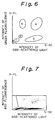
- a blood sample stained with those solutions is loaded in a flow cytometer to obtain signal for fluorescence and side-scattered light and on the basis of those signals, a two-dimensional plot of fluorescence intensity vs the intensity of side-scattered light is constructed as shown in Fig. 6, whereby the leukocytes in the sample are classified into five types.
- Numerals 1 - 6 in Fig. 6 signify the populations of lymphocytes, monocytes, neutrophils, eosinophils, basophils and a noise component, respectively.
- Erythroblasts are cells ancestral to red corpuscles and they normally occur in the bone marrow but not in circulating blood. However, in certain diseases such as anemia and leukemia, erythroblasts also occur in circulating blood. Therefore, it is of clinical importance to measure erythroblasts.
- the present invention has been accomplished in order to improve the method described in Unexamined Published Japanese Patent Application No. 134958/1988 and has as an object providing a method that is capable of differentiating erythroblasts from other cells.
- Erythrocytic cells have the capability of adjusting their own osmolarity by allowing water to come in or go out through the cell membranes in accordance with the osmolarity of the cell suspension. If the cell suspension has a lower osmolarity than the cells, they let water in through the cell membranes; if, on the other hand, the cell suspension has a higher osmolarity than the cells, they let water out through the cell membranes.
- the method of the present invention takes advantage of that character of erythroblasts and has a specified dye taken up by erythrocytic nucleated cells so that their nuclei are stained and can be readily differentiated from other cells by measurement with a flow cytometer.
- the method of the present invention for differentiating erythroblasts from other cells relies basically upon flow cytometry by utilizing a two-step staining method using a first fluid that is an acidic hypotensive fluorescent dye solution, and a second fluid that is a solution that changes the osmorality and pH of the first fluid.
- the first fluid further contains a fluorescent dye that diffuses into erythroblasts to specifically stain their nuclei, and a group of erythroblasts is separated from other cell groups on a two-dimensional plot constructed with the flow cytometer, whereby the results of erythroblast differentiation are computed.
- the present invention relates to a method of differentiating erythroblasts from other cells by flow cytometry which comprises the following steps (a) to (e):
- the dyes that can be used in the first fluid in accordance with the present invention are listed below:
- the first and second fluids to be used in the method of the present invention preferably satisfy the following conditions:
- Dyes need to be contained in the first fluid at such concentrations that their final concentrations obtained by mixing the first and second fluids will lie within the ranges set forth below.
- Basic Yellow 11 and Astrazon Orange R are commercial products that are available from Aldrich Chemical Company, Inc. and Tokyo Kasei K.K., respectively.
- the concentration of Basic Yellow 11 may be adjusted to 70 - 100 ppm and higher.
- Astrazon Orange R concentrations of 100 ppm and above, and good separation can be achieved at concentrations of 200 ppm and above.
- concentrations of 300 ppm and above the efficiency of separation substantially levels off and the intensities of fluorescence from basophils and eosinophils will decrease slightly. Therefore, the concentration of Astrazon Orange R is preferably at 300 ppm or in its neighborhood for achieving efficient separation of leukocytes from noise.
- Propidium iodide is a commercial product available from Sigma Chemical Co. If the concentration of Propidium iodide is 2.5 ⁇ g/ml (2.5 ppm) and below, its staining power is so small that efficient separation between erythroblasts and leukocytes cannot be achieved in two-dimensional plots. If the concentration of Propidium iodide is 100 ⁇ g/ml and above, the S/N ratio of signal detection decreases (the difference between a fluorescence signal and the background becomes so small as to prevent effective signal detection). The same discussion may apply to the concentration of Etidium bromide.
- the pH of the first fluid is preferably set at 5.0 and below but in order to prevent coagulation of platelets, the pH of the first fluid must be at least 3.5.
- any buffer that has a pKa value of approximately 4.5 such as citrate, maleate and diglycolate, may be employed in the first fluid.
- diglycolic acid the intensity of fluorescence from basophils will decrease slightly if the concentration of diglycolic acid is 5 mM and below. On the other hand, the lysing of erythrocytes will be insufficient if the concentration of diglycolic acid is 50 mM and above. An optimum concentration of diglycolic acid is about 10 mM.
- the osmolarity of the dye solution is varied from 167 to 387 mOsm/kg by changing the amount of an osmolarity compensating agent (e.g. sodium chloride) added to the second fluid. It is recommended that the osmolarity of the second fluid be adjusted in such a way that the final osmolarity of the dye solution is at an approximately isotonic value (280 mOsm/kg).
- an osmolarity compensating agent e.g. sodium chloride
- any buffer that has a pKa value in an approximate range of 8.5 to 9.0 such as borate, Tris and tricin, may be employed in the second fluid. If tricin is used, the intensity of fluorescence from basophils and eosinophils decreases at a tricin concentration of 50 mM and below. A preferred concentration of tricin is ca. 300 mM.
- a fluorescence signal reflects the cytochemical characters of leukocytes and depending on the interaction between stains and individual leukocyte types, signals of different intensities are produced from the leukocytes.
- the side-scattering light signal reflects the structural information of an individual white cell. The larger the nucleus of a white blood cell and the more granules that are present in it, the greater the light reflection which will occur in the cell to produce a more intense side-scattered light.
- a lymphocyte contains very few or no granules, so the scattered light produced from the lymphocyte is the weakest of all leukocytes.
- a neutrophil contains many granules and has a large nucleus, so it produces more intense scattered light. Eosinophils produce scattered light the intensity of which is comparable to that produced from neutrophils. Monocytes and basophils produce scattered light having an intensity intermediate between those of scattered light from lymphocytes and neutrophils.
- leukocytes can be classified into five types in accordance with the present invention.
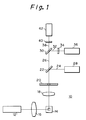
- FIG. 1 A specific example of the optics of a flow cytometer employed in the present invention is hereunder described with reference to Fig. 1.
- the optics shown in Fig. 1 is used in a flow cytometer designed for measuring side-scattered light, red fluorescence and green fluorescence.
- the optics generally indicated by 10 uses an argon ion laser 12 as a light source and it operates at a wavelength of 488 nm, producing an output of 10 mW.
- Light emitted from the laser 12 is converged by a cylindrical lens 16 and illuminates a blood sample flowing through a flow cell 14.
- the stained leukocytes in the sample When the stained leukocytes in the sample are irradiated by the laser light, they produce scattered light and fluorescence.
- the side scattered light and the fluorescence are converged with a condenser lens 18 and pass through an aperture 20 to fall upon a dichroic mirror 22.
- the dichroic mirror 22 reflects the side scattered light 24 and transmits the fluorescence 26.
- the side-scattered light 24 reflected from the dichroic mirror 22 is detected in a photomultiplier tube 28.
- red fluorescence 32 is reflected by a dichroic mirror 30 and green fluorescence 38 is transmitted through that mirror.
- the reflected red fluorescence 32 passes through a color filter 34 and is detected in a photomultiplier tube 36.
- the transmitted green fluorescence 38 passes through a color filter 40 and is detected in a photomultiplier tube 42.
- Erythrocytes in the blood sample emit only fluorescence of a very low intensity, so if it is only required to measure the intensity of fluorescence, erythrocytes will not interfere with the counting of leukocytes even if coincidence of erythrocytes and leukocytes occurs (i.e., erythrocytes and leukocytes pass through the detecting portion simultaneously). However, erythrocytes which produce scattered light having an intensity comparable to that of the scattered light emitted from leukocytes will interfere with the counting of leukocytes. In this case, one may measure fluorescence and scattered light simultaneously and regard as leukocytes only the corpuscles that emit fluorescence having an intensity greater than a certain level.
- a blood sample is permitted to flow through the flow cell 14 after it has been diluted by, for example, 20 fold so that the probability of coincidence of erythrocytes and leukocytes is reduced and the potential interference by erythrocytes is decreased to a level that can be disregarded for practical purposes.
- the first fluid to be used in the method of the present invention has a low osmorality. Hence, if a blood sample is suspended in the first fluid, the erythrocytic cells in the sample will swell upon absorbing water. As a result, the tissue of their cell membrane becomes loose thus causing the contents to leak out. Since mature erythrocytes have no nuclei, only their membrane is left to yield "ghosty" erythrocytes. Erythroblasts will swell in the same manner and their contents will leak out. However, erythroblasts have nuclei which remain intact in the membrane. The third dye diffuses into erythroblasts to stain their nuclei.
- white blood cells do not permit the entrance of the third dye.
- the second fluid is subsequently added to the mixture of blood sample and the first fluid, whereby the sample solution becomes isotonic and suitable for staining.
- the cells thus stained are measured with a flow cytometer and the group of erythroblasts is separated from the other cell groups on the resulting two-dimensional plot and the number of erythroblasts is counted.
- the first fluid may also contain the first and second dyes.
- the first and second dyes In this case, not only the differentiation of erythroblasts but also the classification of leukocytes can also be performed since the first and second dyes diffuse into leukocytes to stain their cells.
- the method of the present invention is an improvement of the invention described in Unexamined Published Japanese Patent Application No. 282698/1988 (Example 2, 2: 2-step method) and it is illustrated below with reference to working examples. It should, however, be noted that the following examples are in no way intended to limit the present invention.
- the first dye in the first fluid was a fluorescent dye that would selectively stain both eosinophils and basophils; the second dye in the first fluid was a fluorescent dye that would stain the nuclei of leukocytes; and the third dye in the first fluid was a fluorescent dye that would diffuse into erythroblasts to stain their nuclei.
- First fluid pH, 4.0; osmorality, 30 mOsm/kg First dye Basic Yellow 11 385 mg/l Second dye Astrazon Orange 330 mg/l Buffer Diglycolic acid-sodium hydroxide 10 mM Third dye Propidium iodide 10.5 ⁇ g/ml (advantageously 2.5 - 100 ⁇ g/ml) Second fluid : pH, 9.8; osmolarity, 2000 mOsm/kg Buffer Tricin-sodium hydroxide 300 mM Osmolarity adjusting agent Sodium chloride 750 mM
- the blood of a healthy person was made nonclotting and 100 ⁇ l of the thus anti-coagulated blood was mixed uniformly with 1800 ⁇ l of the first fluid. After agitation, the mixture was incubated at 25°C for ca. 20 seconds. Thereafter, 200 ⁇ l of the second fluid was added and the mixture was agitated, followed by incubation at 25°C for ca. 40 seconds.
- the finally obtained sample solution had a pH of 8.6 - 8.7 and an osmolarity of ca. 280 mOsm/kg (isotonic).
- sample solutions were prepared with the erythrocytes lysed and the various types of leukocytes stained. Those sample solutions were loaded in a flow cytometer for measuring the intensities of fluorescence and scattered light from the leukocytes.
- Example 1 A measurement was conducted as in Example 1 except that the first fluid contained neither the first nor the second dye. The results of the measurement are shown below. All the samples for measurement were taken from healthy persons. Results of measurement on specimens Distribution Pattern Peripheral blood Bone marrow punctured fluid Method of Example 1 Figs. 2 and 3 Fig. 4 Fig. 5 Prior art Method Fig. 6 Fig. 7 Fig. 8 Method of Example 2 Fig. 9 Fig. 10 Fig. 11
- Fig. 6 is a two-dimensional plot of the intensity of green fluorescence vs the intensity of side-scattered light as obtained when measurements were conducted using the prior art reagent, namely the first fluid not containing the third dye.
- Numerals 1 - 6 in the plot signify the distribution zones of lymphocytes, monocytes, neutrophils, eosinophils, basophils and erythrocyte ghost, respectively.
- a two-dimensional plot of the intensity of red fluorescence vs the intensity of side-scattered light was found to show the same pattern of distribution.
- Figs. 7 and 8 are two-dimensional plots of the intensity of red-fluorescence vs the intensity of side-scattered light actually obtained when measurements were conducted using the prior art reagent, that is a reagent not containing the third dye.
- the sample used in Fig. 7 comprised peripheral blood not containing erythroblasts and that of Fig. 8 was bone marrow punctured fluid containing erythroblasts.
- Fig. 7 nor Fig. 8 indicates plots of erythroblasts. Even if the intensity of green fluorescence used in these figures were replaced by that of red fluorescence, erythroblasts cannot be detected as long as the prior art reagent is used.
- erythroblasts Detection of erythroblasts is possible only when a reagent containing the third dye is used and the intensities of red fluorescence are measured (Figs. 4, 5, 10 and 11). In other words, even if a reagent containing the third dye is used, erythroblasts cannot be detected by measuring the intensity of green fluorescence.
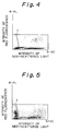
- Figs. 2 and 3 are two-dimensional plots obtained when measurements were conducted on the same sample using the reagents prepared in Example 1; the respective graphs plot the intensities of green and red fluorescence on the vertical axis. Shown by 9 in each graph is the distribution zone of erythroblasts.
- Figs. 4 and 5 are graphs showing the actual results of measurement that correspond to Figs. 2 and 3, respectively.
- Fig. 4 Example 1
- Fig. 7 the prior art method
- Figs. 5 and 8 are two-dimensional plots as obtained when measurements were conducted on a sample fluid obtained from bone marrow.
- the bone marrow fluid contains many erythroblasts.
- erythrocytes in the blood are lysed whereas the nucleated erythroblasts are stained with the third dye to emit red fluorescence.
- the leukocytes are stained with the first and second dyes to emit red and green fluorescence.
- the erythroblasts and leukocytes appear in different zones of the two-dimensional plot. Hence, there is no difficulty in separating the erythroblasts from the leukocytes on the two-dimensional plot. It is also possible to classify the leukocytes into five different types.
- the method of the present invention allowed erythroblasts to appear in zone 7 of red fluorescence, which was impossible in the prior art method (Fig. 8).
- Example 2 Basic Yellow 11 (first dye) and Astrazon Orange R (second dye) were eliminated from the first fluid, which hence contained only the third dye.
- the method of preparation of the first fluid was otherwise the same as in Example 1.
- Fig. 9 is a two-dimensional plot showing the selective staining of erythroblasts 8 with the third dye to emit red fluorescence. Shown by 9 are the other corpuscles.
- Figs. 10 and 11 are two-dimensional plots for the intensity of red fluorescence vs the intensity of side-scattered light, as obtained when peripheral blood and bone marrow fluid were respectively used as samples. In the absence of erythroblasts from the sample (peripheral blood), no signal for red fluorescence appeared in Fig. 10.
- Fig. 11 shows that only erythroblasts emitted fluorescence when the sample was a bone marrow punctured fluid. As in Example 1, erythroblasts appeared in zone 7 but there was no appearance of leukocytes. Hence, the erythroblasts count can be obtained by counting the number of corpuscles in zone 7. In other words, it was possible to differentiate and count erythroblasts with the aid of the third dye alone.
- Example 1 When the first and second dyes were also contained in the first fluid as in Example 1, not only could erythroblasts be differentiated but also the leukocytes could be classified into five different types as described in Example 1.
- the method of the present invention permits erythroblasts to be distributed on a two-dimensional plot in a different zone than are the other cell types and this enables the relative content and count of erythroblasts to be determined with high precision.
- the classification of leukocytes can be performed simultaneously with the differentiation of erythroblasts.
Landscapes
- Chemical & Material Sciences (AREA)
- Dispersion Chemistry (AREA)
- Physics & Mathematics (AREA)
- Health & Medical Sciences (AREA)
- Life Sciences & Earth Sciences (AREA)
- Analytical Chemistry (AREA)
- Biochemistry (AREA)
- General Health & Medical Sciences (AREA)
- General Physics & Mathematics (AREA)
- Immunology (AREA)
- Pathology (AREA)
- Investigating Or Analysing Biological Materials (AREA)
- Investigating Or Analysing Materials By The Use Of Chemical Reactions (AREA)
Applications Claiming Priority (2)
| Application Number | Priority Date | Filing Date | Title |
|---|---|---|---|
| JP3028521A JP2927979B2 (ja) | 1991-02-22 | 1991-02-22 | フローサイトメトリーによる赤芽球の分類方法 |
| JP28521/91 | 1991-02-22 |
Publications (3)
| Publication Number | Publication Date |
|---|---|
| EP0499693A2 true EP0499693A2 (de) | 1992-08-26 |
| EP0499693A3 EP0499693A3 (en) | 1993-02-03 |
| EP0499693B1 EP0499693B1 (de) | 1997-02-05 |
Family
ID=12250978
Family Applications (1)
| Application Number | Title | Priority Date | Filing Date |
|---|---|---|---|
| EP91119259A Expired - Lifetime EP0499693B1 (de) | 1991-02-22 | 1991-11-12 | Verfahren zur Durchflusszytometrie-Unterscheidung der Erythroblasten von anderen Zellen |
Country Status (5)
| Country | Link |
|---|---|
| US (1) | US5298426A (de) |
| EP (1) | EP0499693B1 (de) |
| JP (1) | JP2927979B2 (de) |
| AU (1) | AU648838B2 (de) |
| DE (1) | DE69124595T2 (de) |
Cited By (4)
| Publication number | Priority date | Publication date | Assignee | Title |
|---|---|---|---|---|
| WO2002088669A1 (en) * | 2001-04-26 | 2002-11-07 | Guava Technologies, Inc. | Cell viability assay reagent |
| EP1004880A3 (de) * | 1998-11-27 | 2003-02-05 | Sysmex Corporation | Erythroblasten-diagnostische Durchflusszytometrie-Methode und Reagenzien |
| US9063127B1 (en) | 2013-12-11 | 2015-06-23 | Analiza, Inc. | Devices and methods for determining and/or isolating cells such as circulating cancer or fetal cells |
| US10928405B2 (en) | 2013-12-11 | 2021-02-23 | Analiza, Inc. | Devices and methods for determining and/or isolating cells such as circulating cancer or fetal cells |
Families Citing this family (26)
| Publication number | Priority date | Publication date | Assignee | Title |
|---|---|---|---|---|
| US6509192B1 (en) * | 1992-02-24 | 2003-01-21 | Coulter International Corp. | Quality control method |
| DE69434942T2 (de) * | 1993-02-25 | 2007-11-29 | Abbott Laboratories, Abbott Park | Mehrzweckreagenzsystem zur schnellen lysierung von vollblutproben |
| US5879900A (en) * | 1994-12-15 | 1999-03-09 | Abbott Laboratories | Method for simultaneous analysis of cell viability, nucleated red blood cells and white blood cell differentials |
| US5559037A (en) * | 1994-12-15 | 1996-09-24 | Abbott Laboratories | Method for rapid and simultaneous analysis of nucleated red blood cells |
| US5917584A (en) * | 1997-11-21 | 1999-06-29 | Coulter International Corp. | Method for differentiation of nucleated red blood cells |
| US5874310A (en) * | 1997-11-21 | 1999-02-23 | Coulter International Corp. | Method for differentiation of nucleated red blood cells |
| US6911313B2 (en) * | 1998-02-06 | 2005-06-28 | Sysmex Corporation | Process for discriminating and counting erythroblasts |
| JP4566299B2 (ja) * | 1998-04-09 | 2010-10-20 | シスメックス株式会社 | 赤芽球の分類計数方法 |
| US6828157B1 (en) * | 1999-05-04 | 2004-12-07 | Dan A. Pankowsky | Products and methods for single parameter and multiparameter phenotyping of cells |
| US6682940B2 (en) | 1999-05-04 | 2004-01-27 | Dan A. Pankowsky | Products and methods for single parameter and multiparameter phenotyping of cells |
| JP4464534B2 (ja) * | 2000-06-20 | 2010-05-19 | シスメックス株式会社 | 破砕赤血球の測定方法 |
| US7049093B2 (en) * | 2000-11-08 | 2006-05-23 | Sysmex Corporation | Method of classifying and counting nucleated bone marrow cells |
| CN1276252C (zh) | 2001-07-27 | 2006-09-20 | 贝克曼库尔特有限公司 | 有核红细胞的测量方法 |
| US6573102B2 (en) | 2001-07-27 | 2003-06-03 | Coulter International Corp. | Lytic reagent composition for determination of nucleated blood cells |
| US6472215B1 (en) | 2001-07-27 | 2002-10-29 | Coulter International Corp. | Method of analyzing nucleated red blood cells in a blood sample |
| US6916658B2 (en) * | 2001-07-27 | 2005-07-12 | Beckman Coulter, Inc. | Method for measurement of immature granulocytes |
| US6410330B1 (en) | 2001-07-27 | 2002-06-25 | Coulter International Corp. | Method for measurement of nucleated red blood cells |
| EP1714146B1 (de) * | 2004-02-10 | 2022-08-03 | Beckman Coulter, Inc. | Verfahren zur messung kernhaltiger roter blutzellen |
| US7208319B2 (en) * | 2004-02-10 | 2007-04-24 | Beckman Coulter, Inc. | Method of measurement of nucleated red blood cells |
| US8148101B2 (en) | 2008-07-22 | 2012-04-03 | Abbott Laboratories | Method for classifying and counting bacteria in body fluids |
| CN101723874B (zh) * | 2008-10-31 | 2013-09-11 | 深圳迈瑞生物医疗电子股份有限公司 | 花菁类化合物及其在生物样品染色中的用途 |
| CN101750476B (zh) * | 2008-12-08 | 2015-06-03 | 深圳迈瑞生物医疗电子股份有限公司 | 血液分析试剂及其使用方法 |
| CN102115456B (zh) * | 2009-12-30 | 2014-08-20 | 深圳迈瑞生物医疗电子股份有限公司 | 花菁类化合物,包含所述化合物的组合物及其在细胞检测中的用途 |
| CN102331397A (zh) * | 2011-07-08 | 2012-01-25 | 无锡荣兴科技有限公司 | 一种用于血液细胞统计分析的光电传感器 |
| CN106687810B (zh) * | 2014-12-31 | 2019-10-22 | 深圳迈瑞生物医疗电子股份有限公司 | 一种非诊断目的的有核红细胞报警方法、装置及流式细胞分析仪 |
| EP3859425B1 (de) * | 2015-09-17 | 2024-04-17 | S.D. Sight Diagnostics Ltd. | Verfahren und vorrichtung zur detektion einer entität in einer körperprobe |
Family Cites Families (9)
| Publication number | Priority date | Publication date | Assignee | Title |
|---|---|---|---|---|
| US3916205A (en) * | 1973-05-31 | 1975-10-28 | Block Engineering | Differential counting of leukocytes and other cells |
| DE3238353A1 (de) * | 1982-10-15 | 1984-04-19 | Max Planck Gesellschaft zur Förderung der Wissenschaften e.V., 3400 Göttingen | Verfahren zur simultanen quantitativen bestimmung der blutzellen und reagenz hierfuer |
| US4499183A (en) * | 1982-12-21 | 1985-02-12 | Ortho Diagnostic Systems Inc. | Detecting intracellular antigens in swelled and fixed cells with labeled antibody |
| US4485175A (en) * | 1983-01-03 | 1984-11-27 | Coulter Electronics, Inc. | Method for three-volume differential determination of lymphocyte, monocyte and granulocyte populations of leukocytes |
| WO1986001293A1 (en) * | 1984-08-09 | 1986-02-27 | Lawrence Kass | Differentiation, identification and enumeration of neuthrophils, erythroblast and sub-populations of lymphopcytes with basic blue 141 |
| US4978624A (en) * | 1985-09-06 | 1990-12-18 | Technicon Instruments Corporation | Reagent for the determination of a differential white blood cell count |
| US5039613A (en) * | 1986-11-27 | 1991-08-13 | Toa Medical Electronics Co., Ltd. | Reagents used in a method of classifying leukocytes by flow cytometry |
| US5047321A (en) * | 1988-06-15 | 1991-09-10 | Becton Dickinson & Co. | Method for analysis of cellular components of a fluid |
| JP3048260B2 (ja) * | 1991-07-29 | 2000-06-05 | シスメックス株式会社 | 白血球分類計数用試料調製方法 |
-
1991
- 1991-02-22 JP JP3028521A patent/JP2927979B2/ja not_active Expired - Lifetime
- 1991-11-07 US US07/790,598 patent/US5298426A/en not_active Expired - Lifetime
- 1991-11-11 AU AU87740/91A patent/AU648838B2/en not_active Ceased
- 1991-11-12 DE DE69124595T patent/DE69124595T2/de not_active Expired - Fee Related
- 1991-11-12 EP EP91119259A patent/EP0499693B1/de not_active Expired - Lifetime
Cited By (6)
| Publication number | Priority date | Publication date | Assignee | Title |
|---|---|---|---|---|
| EP1004880A3 (de) * | 1998-11-27 | 2003-02-05 | Sysmex Corporation | Erythroblasten-diagnostische Durchflusszytometrie-Methode und Reagenzien |
| US6664110B1 (en) | 1998-11-27 | 2003-12-16 | Sysmex Corporation | Erythroblast diagnostic flow-cytometry method and reagents |
| WO2002088669A1 (en) * | 2001-04-26 | 2002-11-07 | Guava Technologies, Inc. | Cell viability assay reagent |
| US9063127B1 (en) | 2013-12-11 | 2015-06-23 | Analiza, Inc. | Devices and methods for determining and/or isolating cells such as circulating cancer or fetal cells |
| US9709556B2 (en) | 2013-12-11 | 2017-07-18 | Analiza, Inc. | Devices and methods for determining and/or isolating circulating cancer cells |
| US10928405B2 (en) | 2013-12-11 | 2021-02-23 | Analiza, Inc. | Devices and methods for determining and/or isolating cells such as circulating cancer or fetal cells |
Also Published As
| Publication number | Publication date |
|---|---|
| JP2927979B2 (ja) | 1999-07-28 |
| US5298426A (en) | 1994-03-29 |
| DE69124595T2 (de) | 1997-06-12 |
| EP0499693A3 (en) | 1993-02-03 |
| AU648838B2 (en) | 1994-05-05 |
| AU8774091A (en) | 1992-08-27 |
| DE69124595D1 (de) | 1997-03-20 |
| EP0499693B1 (de) | 1997-02-05 |
| JPH04268453A (ja) | 1992-09-24 |
Similar Documents
| Publication | Publication Date | Title |
|---|---|---|
| EP0499693B1 (de) | Verfahren zur Durchflusszytometrie-Unterscheidung der Erythroblasten von anderen Zellen | |
| US5434081A (en) | Method of classifying leukocytes by flow cytometry | |
| US4882284A (en) | Method for quantitating and differentiating white blood cells | |
| US5175109A (en) | Reagent for classifying leukocytes by flow cytometry | |
| EP0763201B1 (de) | Nachweis von Retikulozyten mit Coriphosphin O | |
| US4336029A (en) | Method and reagents for quantitative determination of reticulocytes and platelets in whole blood | |
| EP0545315B1 (de) | Reagenszusammensetzungen und ihre Verwendung bei der Identifizierung und Charakterisierung von Retikulozyten in Vollblut | |
| EP1004880B1 (de) | Erythroblasten-diagnostische Durchflusszytometrie-Methode und Reagenzien | |
| US5264369A (en) | Method of preparing specimen for counting subpopulations of leukocytes and erythroblasts | |
| James et al. | Proliferation of mitochondria during the cell cycle of the human cell line (HL-60). | |
| CA2077790C (en) | Reagent compositions and their use in sphering cells | |
| US6060322A (en) | Method for identification of reticulated cells | |
| US5438003A (en) | Reagent compositions for use in the identification and characterization of reticulocytes in whole blood | |
| EP1542008B1 (de) | Verfahren zur klassifizierung und zählung von leukozyten | |
| EP0268766B1 (de) | Durchfluss-Zytometrieverfahren zum Klassifizieren von Leukozyten und dazu angewandte Reagenzien | |
| EP1668353B1 (de) | Referenzkontrolle zur optischen messung kernhaltiger roter blutzellen einer blutprobe | |
| US4985174A (en) | Reticulocyte quantitating reagent for flow cytometry | |
| US5179026A (en) | Method of classifying leukocytes by flow cytometry and reagents used in the method | |
| JP2009080122A (ja) | 赤芽球の分類計数方法 | |
| CN101078720B (zh) | 一种改进的用于对白细胞进行分类的试剂及方法 | |
| CA1309327C (en) | Reagent and method for classifying leukocytes by flow cytometry | |
| Subel | An Evaluation of Reticulocyte Counting by Flow Cytometry |
Legal Events
| Date | Code | Title | Description |
|---|---|---|---|
| PUAI | Public reference made under article 153(3) epc to a published international application that has entered the european phase |
Free format text: ORIGINAL CODE: 0009012 |
|
| AK | Designated contracting states |
Kind code of ref document: A2 Designated state(s): DE FR GB IT |
|
| PUAL | Search report despatched |
Free format text: ORIGINAL CODE: 0009013 |
|
| AK | Designated contracting states |
Kind code of ref document: A3 Designated state(s): DE FR GB IT |
|
| 17P | Request for examination filed |
Effective date: 19930728 |
|
| 17Q | First examination report despatched |
Effective date: 19950227 |
|
| GRAG | Despatch of communication of intention to grant |
Free format text: ORIGINAL CODE: EPIDOS AGRA |
|
| GRAH | Despatch of communication of intention to grant a patent |
Free format text: ORIGINAL CODE: EPIDOS IGRA |
|
| GRAH | Despatch of communication of intention to grant a patent |
Free format text: ORIGINAL CODE: EPIDOS IGRA |
|
| ITF | It: translation for a ep patent filed | ||
| GRAA | (expected) grant |
Free format text: ORIGINAL CODE: 0009210 |
|
| AK | Designated contracting states |
Kind code of ref document: B1 Designated state(s): DE FR GB IT |
|
| ET | Fr: translation filed | ||
| REF | Corresponds to: |
Ref document number: 69124595 Country of ref document: DE Date of ref document: 19970320 |
|
| ET | Fr: translation filed |
Free format text: CORRECTIONS |
|
| PGFP | Annual fee paid to national office [announced via postgrant information from national office to epo] |
Ref country code: GB Payment date: 19971103 Year of fee payment: 7 |
|
| PLBE | No opposition filed within time limit |
Free format text: ORIGINAL CODE: 0009261 |
|
| STAA | Information on the status of an ep patent application or granted ep patent |
Free format text: STATUS: NO OPPOSITION FILED WITHIN TIME LIMIT |
|
| 26N | No opposition filed | ||
| PG25 | Lapsed in a contracting state [announced via postgrant information from national office to epo] |
Ref country code: GB Free format text: LAPSE BECAUSE OF NON-PAYMENT OF DUE FEES Effective date: 19981112 |
|
| GBPC | Gb: european patent ceased through non-payment of renewal fee |
Effective date: 19981112 |
|
| PG25 | Lapsed in a contracting state [announced via postgrant information from national office to epo] |
Ref country code: IT Free format text: LAPSE BECAUSE OF NON-PAYMENT OF DUE FEES;WARNING: LAPSES OF ITALIAN PATENTS WITH EFFECTIVE DATE BEFORE 2007 MAY HAVE OCCURRED AT ANY TIME BEFORE 2007. THE CORRECT EFFECTIVE DATE MAY BE DIFFERENT FROM THE ONE RECORDED. Effective date: 20051112 |
|
| PGFP | Annual fee paid to national office [announced via postgrant information from national office to epo] |
Ref country code: DE Payment date: 20081107 Year of fee payment: 18 |
|
| PGFP | Annual fee paid to national office [announced via postgrant information from national office to epo] |
Ref country code: FR Payment date: 20081112 Year of fee payment: 18 |
|
| REG | Reference to a national code |
Ref country code: FR Ref legal event code: ST Effective date: 20100730 |
|
| PG25 | Lapsed in a contracting state [announced via postgrant information from national office to epo] |
Ref country code: FR Free format text: LAPSE BECAUSE OF NON-PAYMENT OF DUE FEES Effective date: 20091130 |
|
| PG25 | Lapsed in a contracting state [announced via postgrant information from national office to epo] |
Ref country code: DE Free format text: LAPSE BECAUSE OF NON-PAYMENT OF DUE FEES Effective date: 20100601 |