EP0459525B1 - Gene, die kodieren für ein Acylierungsenzym der 3-Stelle von Makrolid-Antibiotika - Google Patents
Gene, die kodieren für ein Acylierungsenzym der 3-Stelle von Makrolid-Antibiotika Download PDFInfo
- Publication number
- EP0459525B1 EP0459525B1 EP91109033A EP91109033A EP0459525B1 EP 0459525 B1 EP0459525 B1 EP 0459525B1 EP 91109033 A EP91109033 A EP 91109033A EP 91109033 A EP91109033 A EP 91109033A EP 0459525 B1 EP0459525 B1 EP 0459525B1
- Authority
- EP
- European Patent Office
- Prior art keywords
- streptomyces
- macrolide antibiotic
- dna fragment
- acylated
- macrolide
- Prior art date
- Legal status (The legal status is an assumption and is not a legal conclusion. Google has not performed a legal analysis and makes no representation as to the accuracy of the status listed.)
- Expired - Lifetime
Links
Images


Classifications
-
- C—CHEMISTRY; METALLURGY
- C07—ORGANIC CHEMISTRY
- C07H—SUGARS; DERIVATIVES THEREOF; NUCLEOSIDES; NUCLEOTIDES; NUCLEIC ACIDS
- C07H17/00—Compounds containing heterocyclic radicals directly attached to hetero atoms of saccharide radicals
- C07H17/04—Heterocyclic radicals containing only oxygen as ring hetero atoms
- C07H17/08—Hetero rings containing eight or more ring members, e.g. erythromycins
-
- C—CHEMISTRY; METALLURGY
- C12—BIOCHEMISTRY; BEER; SPIRITS; WINE; VINEGAR; MICROBIOLOGY; ENZYMOLOGY; MUTATION OR GENETIC ENGINEERING
- C12N—MICROORGANISMS OR ENZYMES; COMPOSITIONS THEREOF; PROPAGATING, PRESERVING, OR MAINTAINING MICROORGANISMS; MUTATION OR GENETIC ENGINEERING; CULTURE MEDIA
- C12N15/00—Mutation or genetic engineering; DNA or RNA concerning genetic engineering, vectors, e.g. plasmids, or their isolation, preparation or purification; Use of hosts therefor
- C12N15/09—Recombinant DNA-technology
- C12N15/63—Introduction of foreign genetic material using vectors; Vectors; Use of hosts therefor; Regulation of expression
- C12N15/74—Vectors or expression systems specially adapted for prokaryotic hosts other than E. coli, e.g. Lactobacillus, Micromonospora
- C12N15/76—Vectors or expression systems specially adapted for prokaryotic hosts other than E. coli, e.g. Lactobacillus, Micromonospora for Actinomyces; for Streptomyces
-
- C—CHEMISTRY; METALLURGY
- C12—BIOCHEMISTRY; BEER; SPIRITS; WINE; VINEGAR; MICROBIOLOGY; ENZYMOLOGY; MUTATION OR GENETIC ENGINEERING
- C12N—MICROORGANISMS OR ENZYMES; COMPOSITIONS THEREOF; PROPAGATING, PRESERVING, OR MAINTAINING MICROORGANISMS; MUTATION OR GENETIC ENGINEERING; CULTURE MEDIA
- C12N9/00—Enzymes; Proenzymes; Compositions thereof; Processes for preparing, activating, inhibiting, separating or purifying enzymes
- C12N9/10—Transferases (2.)
- C12N9/1025—Acyltransferases (2.3)
- C12N9/1029—Acyltransferases (2.3) transferring groups other than amino-acyl groups (2.3.1)
-
- C—CHEMISTRY; METALLURGY
- C12—BIOCHEMISTRY; BEER; SPIRITS; WINE; VINEGAR; MICROBIOLOGY; ENZYMOLOGY; MUTATION OR GENETIC ENGINEERING
- C12P—FERMENTATION OR ENZYME-USING PROCESSES TO SYNTHESISE A DESIRED CHEMICAL COMPOUND OR COMPOSITION OR TO SEPARATE OPTICAL ISOMERS FROM A RACEMIC MIXTURE
- C12P19/00—Preparation of compounds containing saccharide radicals
- C12P19/44—Preparation of O-glycosides, e.g. glucosides
- C12P19/60—Preparation of O-glycosides, e.g. glucosides having an oxygen of the saccharide radical directly bound to a non-saccharide heterocyclic ring or a condensed ring system containing a non-saccharide heterocyclic ring, e.g. coumermycin, novobiocin
- C12P19/62—Preparation of O-glycosides, e.g. glucosides having an oxygen of the saccharide radical directly bound to a non-saccharide heterocyclic ring or a condensed ring system containing a non-saccharide heterocyclic ring, e.g. coumermycin, novobiocin the hetero ring having eight or more ring members and only oxygen as ring hetero atoms, e.g. erythromycin, spiramycin, nystatin
-
- Y—GENERAL TAGGING OF NEW TECHNOLOGICAL DEVELOPMENTS; GENERAL TAGGING OF CROSS-SECTIONAL TECHNOLOGIES SPANNING OVER SEVERAL SECTIONS OF THE IPC; TECHNICAL SUBJECTS COVERED BY FORMER USPC CROSS-REFERENCE ART COLLECTIONS [XRACs] AND DIGESTS
- Y10—TECHNICAL SUBJECTS COVERED BY FORMER USPC
- Y10S—TECHNICAL SUBJECTS COVERED BY FORMER USPC CROSS-REFERENCE ART COLLECTIONS [XRACs] AND DIGESTS
- Y10S435/00—Chemistry: molecular biology and microbiology
- Y10S435/8215—Microorganisms
- Y10S435/822—Microorganisms using bacteria or actinomycetales
Definitions
- the present invention relates to DNA fragments containing a gene endowing host microorganism with an enzymatic activity for acylating the 3-position of macrolide antibiotics (this gene being referred to as "acyA"), to recombinant DNA plasmids containing such a DNA fragment, and to microorganisms transformed with such a recombinant plasmid, as well as to a method of producing 3-acylated macrolide antibiotics using such a transformed microorganism.
- acyA macrolide antibiotics
- Macrolide antibiotics (14- or 16-membered cycles) are useful antibiotics widely used as a medicine and an animal medicine (for domestic animals or fishery).
- various macrolide derivatives have been studied. It has been reported that acylation of the OH groups at the 3- and 4"-positions of 16-membered cyclic macrolide antibiotics results in increase in their antibiotic activities [cf. R. Okamoto, Journal of Antibiotics , 27 , 524-544 (1979)].
- 3-acylated and 4"-acylated tylosin derivatives were synthesized and their activities on various bacteria having resistance to macrolide antibiotics were examined to reveal that 3-acylated and 4"-acylated tylosin, particularly 4"-acylated tylosin, have pharmacological effects on the resistant bacteria more potent than tylosin itself.
- tylosin-producing strains produce only tylosin derivatives which have -OH groups at the 3- and 4"-positions.
- no method has been known other than chemical or biological methods.
- EP-A-0 345 546 discloses a DNA fragment containing acyB gene coding for an enzyme capable of acylating the 4"-position of a macrolide antibiotic, said DNA fragment being derived from a microorganism of the genus Streptomyces, having a size of about 3.1 kb and being characterized by the restriction endonuclease map shown in Figure 1, and a DNA restriction fragment resulting from digestion with a restriction endonuclease.
- the present invention provides an industrially useful method which makes it possible to convert bacterial strains which are unable to directly produce a 3-acylated product of a macrolide antibiotic to one which can produce the 3-acylated macrolide using genetic engineering.
- Examples of known macrolide antibiotics include the followings.
- 3- and/or 4''-OH type macrolide antibiotics are added as a substrate to microorganisms cells such as Streptomyces thermotolerans ATCC 11416, Streptomyces hygroscopicus ATCC 21582, and Streptomyces kitasatoensis IFO 13686, culture media thereof or cell extracts therefrom for conversion reaction.
- this method involves different microorganisms, i.e., a macrolide antibiotics-producing bacterium and a microorganism having an acylation activity, which is uneconomical from a point of view of production on an industrial scale.
- the present inventors have made intensive investigation with view to isolating a gene encoding a 3- and/or 4"-acylation enzyme for acylating 3- and/or 4"-position of a macrolide antibiotic as described above from a microorganism having such an enzyme.
- the present inventors have been successful in isolating a 3-acylation enzyme-coding gene (acyA) from a certain strain belonging to the genus of Streptomyces, thus completing the present invention.
- acyA 3-acylation enzyme-coding gene
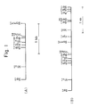
- a DNA fragment containing a gene acyA encoding a 3-acylation enzyme for macrolide antibiotics, characterized by being derived from a strain belonging to the genus Streptomyces, having a size of about 1.8 kb, and having the characteristics shown in the restriction enzyme map shown in Fig 1(A) in the attached drawing or a restriction enzyme-digested DNA fragment thereof which expresses an acylation enzyme activity.
- a DNA fragment containing a gene acyA, encoding a 3-acylation enzyme for macrolide antibiotics characterized by being derived from a strain belonging to the genus Streptomyces, having a size of about 3.2 kb, and having the characteristics shown in the restriction enzyme map shown in Fig. 1(B) in the attached drawing or a restriction enzyme-digested DNA fragment thereof which expresses an acylation enzyme activity.
- restriction enzyme-digested DNA fragment various sizes of DNA fragments obtained by digestion with a restriction enzyme to lengths necessary for expressing an acylation enzyme activity.
- the DNA fragment or digested DNA fragment containing the gene acyA of the present invention is useful for commercially producing the objective macrolide antibiotic derivatives, for example, 3-acylated tylosin, and is particularly useful in that it enables commercial utilization of recombinant DNA technology for antibiotic -producing microorganisms such as Streptomyces and other genera.
- the strain belonging to the genus Streptomyces which is a source from which the DNA fragment containing the gene acyA of the present invention is derived may be any strain that has an ability for producing a 3-acylation enzyme for macrolide antibiotics.
- Examples of such a strain include Streptomyces ambofaciens, Streptomyces kitasatoensis, Streptomyces narbonensis var josamyceticus, Streptomyces hygroscopicus, Streptomyces platensis, Streptomyces albireticuli, Streptomyces cinerochromogenes, Streptomyces dja Too Toosis, Streptomyces furdicidicus, Streptomyces macrosporeus, Streptomyces tendae, Streptomyces thermotolerans, and Streptomyces deltae.
- preferred is Streptomyces thermotolerans.
- Streptomyces thermotolerans a donor of the DNA fragment containing the gene acyA of the present invention has been deposited, e.g., at The American Type Culture Collection under deposition number ATCC 11416, and is available without difficulty.
- This strain is incubated at 28°C aerobically to obtain bacterial cells at a logarithmic growth phase, and processed by the method of Hopwood et al. [Genetic Manipulation of Streptomyces: A laboratory Manual, John Innes Foundation, Norwich, United Kingdom (1985)] to prepare an acyA-containing genome DNA, as will be described in greater detail in Example 1 described hereinbelow.
- the gene acyA of the present invention encodes an enzyme acylating the hydroxyl group at the 3-position of carbomycin, one of macrolide antibiotics, produced by S.
- thermotolerans are inserted in BamHI site of a cosmid vector pHC79 of E . coli and packaged in vitro in ⁇ -phage to prepare a gene library of S. thermotolerans.
- the preparation of gene library utilizing such a cosmid can be carried out using general genetic engineering techniques frequently used on various organisms for the purpose of isolating a specific gene (cf., e.g., Maniatis et al., Molecular Cloning , 295-307, Cold String Harbour Laboratory).
- the procurement of the clone containing the carbomycin biosynthesis-related gene region from the gene library can be achieved by preparing a probe from a gene known to belong to the carbomycin biosynthesis-related gene region or DNA fragment near the gene, and performing colony hybridization of the probe with a gene library prepared using E. coli HB101 as a host (i.e., many clones obtained by transduction of packaged cosmid DNA having the genome DNA fragment of S. thermotolerans). By this operation, there can be obtained clones hybridizing with the probe, i.e., clones having cosmids containing the carbomycin biosynthesis-related gene region.
- cosmids derived from these clones have inserted fragments of up to about 45 kb containing as a probe a part or all of the DNA fragment and extending to neighboring regions.
- one of the recombinant cosmids above celled "pSE26" by the present inventors has an inserted fragment of about 45 kb containing an acyB fragment near a terminal on one side of the insert portion (cf. Example 2, C below).
- recombinant cosmids can be isolated which have a region partially overlapping pSE26 and a region which is considerably remote from the acyB-containing DNA fragment used as the first probe as an inserted fragment, for example, pBM73 shown in Example 2, C, below.
- cloned region can be expanded and consequently all of the carbomycin biosynthesis-related genes can be cloned from the gene library of S . thermotolerans.
- the cloning of a region containing the objective gene within the range of several tens kb to several hundreds kb by preparing, from a clone obtained by hybridization with a probe, a new probe in a region remote from the original probe followed by obtaining a clone which hybridizes with the new probe or repeating this procedure several times as the case may be, can be carried out using a method generally known to one skilled in the art as "gene walking" [cf. e.g., Tashiro et al., Zoku Seikagaku Jikken Koza (Second Series: Corse of Biochemical Experiments), Vol. 1, Idenshi Kenkyusho (Methods of Gene Study) II, 83-104 (1986), ed. by Japan Biochemistry Society).
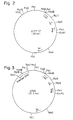
- Shuttle cosmid pIHY-17 can be constructed as a vector for cloning and expressing the gene acyA of the present invention (cf. Fig. 2).
- This vector is a vector which has both functions of actinomycete vector pIJ922 and E. coli cosmid pHC79 simultaneously, and is constructed concretely by the method described in Example 3 below.
- vectors having advantages that they can be introduced and propagated in both of E. coli and actinomycetes, i.e., shuttle vectors can be constructed with ease by connecting an E. coli vector to an actinomycete vector.
- respective replicons of actinomycete vector pIJ922 and E. coli vector pHC79 are incorporated and respective selective markers are given.
- the instant vector is endowed with packaging function derived from the cos site of pHC79.
- This function makes it possible to introduce even a large plasmid having a length of up to about 50 kb inclusive of the length of the vector into E. coli as a host through in vitro packaging of ⁇ -phase with ease.
- the above-described function of pIHY-17 as a shuttle cosmid is suitable for efficient closing and expression of acyA.
- cloned fragment derived from the carbomycin biosynthesis-related gene region can be ligated with the instant vector and packaged in vitro in ⁇ -phase making the use of the cos site thereof.
- a number of clones obtained by the transduction of the reconstructed cosmids into E . coli HB101 are mixed and the cosmids can be extracted.
- the pooled cosmids can be transformed into actinomycetes, S. lividans and the like making the use of the function which pIJ922 has. From the transformants thus obtained, strains to which the gene acyA of the invention has been donated, that is strains having an activity of acylating the 3-position of macrolides, can be selected by thin layer chromatography tests, resulting in that acyA is cloned.
- the above-described method can provide in the S. lividans transformation DNA's securely cyclized and amplified by the operations of in vitro packaging and transduction.
- Strains having the activity of the gene acyA of the invention can be separated by the above-described method. Also, by treating the strains as described in Example 4 below, plasmids, for example, p53A, can be isolated. Whether or not the gene acyA of the invention is present in the inserted fragment of the plasmid can be made clear by examining a strain obtained by retransformation of S. lividans with the plasmid to see if the retransformed strain has an activity of acylating the 3-position of macrolides. The inserted fragment can be cut out of the original plasmid utilizing suitable restriction sites, and then subcloned in E .
- Plasmids thus obtained for example, pMAA2 and pMAA3 (cf. Fig. 4) can be prepared rapidly and easily in large amounts and hence they can be used as a material suitable for the analysis of the structure and expression function of the gene acyA of the invention.
- 3-Acylation enzyme for macrolide antibiotics expressed using the gene acyA of the invention is useful for the acylation of the 3-position of, particularly, tylosin but its utility is not limited thereto and has also a 3-acylation ability for other macrolide antibiotics such as spiramycin, angolamycin, leucomycin and nidamycin.
- 3-acylated enzyme produced by the transformed strains can of course be used as a catalyst in an enzymatic reaction between macrolide antibiotics and acylating agents.
- acyB 4''-acylation enzyme-coding gene
- the gene acyA facilitates the production of 3-O-acetyl-4''-O-isovaleryltyrosine, an industrially useful acylation derivative of tylosin, by fermentation.
- DNA fragments containing the gene (acyA) encoding 3-acylation enzyme for macrolide antibiotics provided by the present invention has a wide utility.
- Plasmids having incorporated therein the gene acyA of the invention can be introduced into suitable host microorganisms depending on the vector plasmid used.
- the host microorganisms may be either those having an ability of producing macrolide antibiotics or those having no such ability.
- Examples of the host microorganism which can be used include Streptomyces kitasatoensis, Streptomyces narbonensis var josamyceticus, Streptomyces hygroscopicus, Streptomyces platensis, Streptomyces albireticuli, Streptomyces cinerochromogenes, Streptomyces dja Actuallysis, Streptomyces macrosporeus, Streptomyces tendae, Streptomyces deltae, Streptomyces fradiae, Streptomyces eurythermus, Streptomyces ambofaciens, Streptomyces kasugaensis, Streptomyces erythreus, Streptomyces kanamyceticus.
- macrolide antibiotics-producing bacteria which produce substantially no 3-acylation enzyme for macrolide antibiotics, for example, Streptomyces eurythermus ATCC 14975 which is an angolamycin-producing strain, and Streptomyces fradiae ATCC 19609 which is a tylosin-producing strain are suitable.
- the transformation of the host microorganisms with the recombinant plasmids containing the gene acyA can be performed by a method known per se, e.g., the method of Hopwood (cf. Genetic Manipulation of Streptomyces: A Laboratory Manual (1985), The John Innes Institute).
- 3-acylated macrolide antibiotics can be produced by cultivating the transformed microorganisms even in media containing no macrolide antibiotics when the transformed microorganisms used have an ability for producing macrolide antibiotics.
- 3-acylated macrolide antibiotics can also be produced by cultivating the transformed microorganisms, and reacting macrolide antibiotics with an acylating agent such as acetyl coenzyme A, propionyl coenzyme A, or a precursor of biosynthesis of an acyl group-donor, e.g., leucine, in the presence of the cultivated bacterial cells themselves or their processed products (for example, cell-free extracts obtained by supersonication of cultivated cells).
- an acylating agent such as acetyl coenzyme A, propionyl coenzyme A, or a precursor of biosynthesis of an acyl group-donor, e.g., leucine
- macrolide antibiotics of which the 3-position is acylated with an alkanoyl group having 1 to 3 carbon atoms for example, an acetyl group, a propionyl group, etc., such as 3-O-acetyl tylosin, 3-O-propionyltylosin, 3-O-acetylangolamycin, 3-O-propionylangolamycin, 3-O-acetylspiramycin, and 3-O-propionylspiramycin, can be produced.
- macrolide antibiotics of which the 3- and 4"-positions are acylated simultaneously such as 3-O-acetyl-4"-isovaleryltylosin, 3-O-acetyl-4"-O-n-butyryltylosin, 3-O-acetyl-4"-O-propionyltylosin, 3-O-acetyl-4"-O-acetyltylosin, and 3-O-propionyl-4"-O-isovaleryltylosin, can be produced by using plasmids having in corporated therein acyA and 4"-acylating enzyme coding gene (acyB) as described above, or by using different plasmid having incorporated therein acyB together with the above-described plasmid having incorporated therein acyA.
- acyB 4"-acylating enzyme coding gene
- the host microorganisms transformed with the plasmid containing the DNA fragment containing the gene acyA of the invention can be cultivated by many methods using any media of different types.
- Preferred examples of carbon source which can be added to the culture media include molasses, glucose, starch, oils, fats, glycerin and the like.
- Examples of the nitrogen source include soybean powder, amino acid mixtures, dry yeast, peptones, and the like.
- the media may contain nutrient inorganic salts, which contain usually used salts that can release potassium, sodium, magnesium, calcium, phosphoric acid, chlorine and sulfuric ions.
- Indispensable minor components for example, vitamins may be added to the media, if desired.
- the minor components may be supplied in the form of impurities accompanying other components of the culture media.
- the media may contain macrolide antibiotics to be acylated, if desired.
- the transformed microorganism belonging to the genus Streptomyces can be cultivated under aerobic conditions in media over a relatively wide pH range of pH within the range of pH about 5 to 9 and within the temperature range of about 20 to 40°C.
- a condition necessary for maintaining the stability of plasmids for example a chemical such as thiostreptone, may be added to the media as a selection pressure.
- a slant medium containing 0.4 % of glucose, 0.4 % of yeast extract, 1.0 % of malt extract, and 1.5 % of agar was cultivated the aforementioned strain at 29°C for 2 weeks, and one platinum loopful of the strain was taken out and inoculated in 25 ml of a seed medium containing 2 % of soluble starch, 2 % of soybean powder.
- the seed medium was used one which had been placed in a 250 ml Erlenmeyer flask and sterilized at 120°C for 15 minutes.
- the inoculated flask was incubated by shaking culture at 28°C for 48 hours to obtain a seed.
- One (1) ml of the seed was inoculated in 25 ml of a TSB medium (trypticase soy broth medium) ⁇ and thereafter the medium was incubated at 28°C for 48 hours.
- ⁇ TSB medium was prepared to a concentration of 30 g/liter.
- TSB medium was available from BBL Microbiology Systems Maryland, U.S.A.
- Mycelia were collected and washed once with a 10.3 % sucrose solution. Subsequently, 25 % sucrose/Tris-HCl (50 mM, pH 8) was added to the mycelia in an amount of 5 ml per g of wet cell. After well dispersing the mycelia, 0.6 ml of lysozyme (Grade I prepared by Sigma Chemical Co.) solution (10 mg/ml) was added to the mixture and well mixed.
- lysozyme Gibde I prepared by Sigma Chemical Co.
- the solution was fractionated by centrifugation (10,000 rpm, 10 minutes) to obtain about 7 ml of an aqueous layer.
- the mixture was centrifuged (10,000 rpm, 10 minutes) to obtain about 7 ml of an aqueous layer.
- To the aqueous layer was added 30 ⁇ l of ribonuclease A type I-AS prepared by Sigma Chemical Co.) solution (10 mg/ml solution heated at 90°C for 10 minutes), and the mixture was incubated at 37°C for 1 hour.
- a single colony of E . coli HB101 transformed with pHC79 (Boehringer Mannheim) (Transformation Kit, produced by Nippon Gene Co., Ltd.) was inoculated in 2 ml of L-broth (1 % bactotryptone, 0.5 % yeast extract, 0.5 % NaCl, pH 7.4) containing ampicillin in a final concentration of 50 mg/ml, and cultivated by shaking culture at 37°C overnight.
- the culture medium was added to 500 ml of ampicillin (50 ⁇ g/ml)-added L-broth, and cultivated again by shaking culture at 37°C overnight. After the cultivation, the medium was centrifuged at 5,000 rpm, at 4°C for 10 minutes.
- the resulting pellet was suspended in 20 ml of a solution A composed of 25 mM Tris-HCl, pH 8, 10 mM EDTA, and 60 mM glucose, on ice. Then, a solution of 40 mg of lysozyme (Seikagaku Kogyo Co., Ltd.) dissolved in 0.5 ml of water was added thereto, and mixed mildly. The resulting mixture was left to stand at room temperature, and when cells initiated to be lysed (usually, 2 to 5 minutes after the mixing), 40 ml of fresh solution B (1 % SDS, 0.2 N NaOH) which had been prepared in advance and cooled was added quickly and mixed mildly, and the resulting mixture was left to stand for 5 minutes on ice.
- a solution A composed of 25 mM Tris-HCl, pH 8, 10 mM EDTA, and 60 mM glucose
- a solution of 40 mg of lysozyme (Seikagaku Kogyo Co., Ltd.) dissolved in
- the precipitates thus dried were dissolved in 8.2 ml of TE (10 mM Tris-HCl, pH 8, 1 mM EDTA) and 8.4 g of cesium chloride and 0.2 ml of a 10 mg/ml ethidium bromide solution were added thereto sequentially.
- the solution obtained were poured into two centrifugation tube "quick seal" (Beckmann Co.), and centrifuged at 65,000 rpm for 4 hours, or at 5,000 rpm for 15 hours, using a vTi65 rotor. Thereafter, a band containing plasmids which appeared in the tube was recovered in a 5 ml syringe (Thermo).
- the solution was extracted with equivalent volume of n-butanol 3 to 4 times to remove ethidium bromide, and have plasmid DNA precipitated with 3 times the volume of 70 % ethanol, and further the pellet was washed 3 times with 70 % ethanol. The pellet thus washed was finally dissolved 0.5 to 1 ml of TE buffer.
- 300 ⁇ g of pHC79 plasmid DNA was obtained from 500 ml of L-broth.
- pHC79 The preparation of such a library using pHC79 was carried out substantially according to the method taught by Ishi-Horowicz, Burke, Nucleic Acids Research , vol. 9, 2987 (1981). That is, two aliquots (50 ⁇ g each) of pHC79 were linearized one with HindIII and the other with SalI, and dephosphorylated with calf intestine-derived alkaline phosphatase (Boehringer Mannheim Yamanouchi Co., Ltd.). Then the both DNA's were digested with BamHI to form arms on both sides which were able to be ligated at only one terminal thereof. The both side arms (1.5 ⁇ g each) and 4 ⁇ g of a partial digest of the genome DNA of S.
- thermotolerans with Sau3AI decomposate obtained by digesting 50 ⁇ g of genome DNA of S. thermotolerans with Sau3AI 4 ⁇ in 200 ⁇ l of Sau3AI buffer at 37°C for 2 minutes
- T 4 DNA ligase Nippon Gene
- DNA was concentrated by precipitation with ethanol, and a portion (equivalent to 4.6 ⁇ g) thereof was subjected to in vitro packaging using an in vitro packaging kit, Gigapack Gold (Stratagene) according to the protocol attached to the kit.
- transduction into E. coli MB101 was conducted under the conditions described in the protocol. Upon examination, the titer was 2.8 x 10 5 colony forming unit (cfu).
- Streptomyces lividans TK64/PIJ922 obtained from the John Innes Streptomyces Culture Collection, John Innes Institute, Colney Lane, Norwich NR4 7UH, England
- 500 ml of YEME + 34 % suclose medium 0.3 % yeast extract, 0.5 % bactopeptone, 0.3 % malt extract, 1 % glucose, 34 % sucrose, and after sterilization 1 ml of 2.5 M MgCl 2 solution was added thereto
- thiopeptine* cultivated by shaking culture at 28°C for 48 hours.
- the mycelia were collected and washed once with a 10.3 % suclose solution. Then, the mycelia were suspended in 45 ml of a 10.3 % sucrose/Tris-HCl (25 mM, pH 8)/EDTA (21 mM, pH 8) solution. After adding 5 ml of a lysozyme solution (10 mg/ml, dissolved in the same solution as used for suspending the mycelia) and 250 ⁇ l of a ribonuclease type I-AS solution to the cell suspension and mixing it well, the suspension was incubated at 37°C for 30 minutes.
- Example 1 (B) 20 ml was added to the suspension and well mixed therewith.
- the suspension was centrifuged (15,000 rpm, 15 minutes) to separate into layers, and about 70 ml of an aqueous layer was obtained.
- To the aqueous layer were added 7 ml of a sodium acetate solution (3 M, pH unadjusted) and 70 ml of isopropanol, and after well mixing the mixture was left to stand at room temperature for 10 minutes.
- the mixture was centrifuged (15,000 rpm, 15 minutes) to separate into layers, and about 10 ml of an upper layer was obtained.
- the upper layer was mixed with 1 ml of a sodium acetate solution (3 M, pH 6) and 10 ml of isopropanol and mixed well. After centrifugation (15,000 rpm, 15 minutes), precipitates were collected and washed with 1 ml of ethanol and dried. After dissolving the precipitates in 11.9 ml of TE buffer, 12.6 g of cesium chloride, and then 0.6 ml of an ethidium bromide solution (10 mg/ml) were added thereto.
- the mixture was centrifuged at 36,000 rpm for 60 hours, and the fraction containing a plasmid band was extracted with TE buffer and ispropanol saturated with cesium chloride 5 times to remove ethidium bromide. Thereafter, the fraction was charged in a tube for dialysis and dialyzed against TE buffer for 24 hours. Thus, about 50 ⁇ g of plasmid pIJ922 were obtained.
- E. coli cosmid pHC79 (2 ⁇ g) prepared as in Example 2 was digested with EcoRI and HindIII, extracted, purified and dissolved in 20 ⁇ l of TE buffer.
- the decomposate was subjected to electrophoresis in TAE buffer/0.8 % ararose gel (40 ml Tris-HCl, 20 mM sodium acetate, 1 mM EDTA, (adjusted to pH with acetic acid), 0.8 % agarose) at 6 v/cm.
- the fragment (0.6 ⁇ g) and the aforementioned EcoRI-HindIII-decomposate of pHC79 [0.2 ⁇ g (2 ⁇ l)] were incubated in 20 ⁇ l of a T 4 DNA ligase (Nippon Gene) solution (300 units/20 ⁇ l) at 16°C overnight.
- the reaction mixture (5 ⁇ l) was added to 150 ⁇ l of E . coli HB101 competent cell to perform transformation.
- the plasmid thus obtained (5 ⁇ g) was made linear with EcoRI, and dephosphorylated with calf intestine-derived alkaline phosphatase (Boehringer Mannheim).
- the resulting DNA (1.25 ⁇ g) and EcoRI-linearized DNA of pIJ922 (4 ⁇ g) wre reacted in 7 ⁇ l of a T 4 DNA ligase (Nippon Gene) solution (200 units/7 ⁇ l) at 16°C for 4 hours.
- the reaction mixture (4 ⁇ l) was subjected to in vitro packaging using Gigapack Gold (Stratagene), followed by transduction into E . coli HB101.
- E . coli HB101 transduced strains were selected 6 strains, and plasmids were inspected to reveal that 4 strains among the 6 stains had the objective plasmid pIHY-17 (cf. Fig. 2).
- a plasmid was prepared from one of the 4 strains on a large scale in the same manner as in the method described in Example 2 (A), and as a result 1.65 mg of plasmid DNA was obtained from 1 1 of L-broth.
- pIHY-17 prepared in Example 3 above was divided into two portions each in an amount of 25 ⁇ g, and one was digested with HindIII and the other with EcoRI, followed by dephosphorylation with alkaline phosphatase.
- Each fragment (1 ⁇ g), XhoI-complete decomposate (1 ⁇ g) of and SalI-imcomplete decomposate (2 ⁇ g) of cosmid clone mixture (pSE26 and pBM73, etc.) containing the carbomycin biosynthesis gene region prepared by the method descried in Example 2 were mixed and incubated in 25 ⁇ l of a T 4 DNA ligase (Nippon Gene) solution (750 units/25 ⁇ l) at 16°C overnight.
- T 4 DNA ligase Nippon Gene
- Mycelia were collected by centrifugation and washed with 10 ml of a 10.3 % sucrose solution. Thereafter, the mycelia were well suspended in 4 ml of P medium containing 1 mg/ml of lysozyme.
- P medium was prepared by mixing 10.3 g of sucrose, 0.025 g of K 2 SO 4 , 0.202 g of MgCl 2 .6H 2 O, 0.2 ml of a trace metal solution*, and deionized water to make 80 ml, and sterilizing the resulting mixture at 120°C for 15 minutes, and then adding thereto 1 ml of 0.5 % KH 2 PO 4 , 10 ml of 3.68 % CaCl 2 .2H 2 O and 10 ml of TES ⁇ 2-[tris(hydroxymethyl) methyl]aminoethanesulfonic acid ⁇ buffer (0.25 M, pH 7.2), each having had been sterilized separately in advance.
- the transformed protoplasts were suspended again in 1 ml of P medium.
- a portion (0.1 ml) of the suspension was inoculated on an R2YE agar plate, and cultivated at 28°C for 16 hours.
- a soft agar medium containing thiopeptine was overlayed on the plate 30 that the final concentration of thiopeptine was 50 ⁇ g/ml, and cultivation was continued at 28°C for 72 hours.
- R2YE agar plate was prepared by dissolving 10.3 g of sucrose, 0.025 g of K 2 SO 4 , 1.012 g of MgCl 2 .6H 2 O, 1 g of glucose.
- the dishes were dried in a clean bench for about 2 hours.
- the soft agar medium a medium prepared by mixing 8 g of Nutrient broth, 3 g of bactoagar and distilled water to make 1 liter, and sterilizing the mixture at 120°C for 15 minutes.
- strains Of about 1,000 transformed strains, about 200 strains were selected at random and inoculated on an agar medium composed of 1 % glucose, 0.5 % yeast extract, 1 % malt extract, pH 7.2, 1.5 % bacto-agar, and 50 ⁇ g/ml of thiopeptine, and cultivated at 28°C for 4 days.
- agar medium composed of 1 % glucose, 0.5 % yeast extract, 1 % malt extract, pH 7.2, 1.5 % bacto-agar, and 50 ⁇ g/ml of thiopeptine, and cultivated at 28°C for 4 days.
- a soft agar medium containing leucomycin A 1 was overlayed on the agar medium so that the final concentration of leucomycin A 1 became 500 ⁇ l/ml, and cultivation was continued for 48 hours.
- the center of a growing colony was punched out with a cork borer having a diameter of 6 mm, and the agar piece thus obtained was placed on silica gel TLC plate (produced by Whatman, LK6DF), and air-dried.
- strain 53A Streptomyces lividans 53A strain (hereafter, referred to as "strain 53A”) produced leucomycin A 3 , i.e., 3-O-acetyl leucomycin A 1 .
- the leucomycin produced by the strain 53A was identified to be leucomycin A 3 from results of analyses of TLC, HPLC, UV spectrum, NMR spectrum, antibacterial spectrum and the like.
- This strain 53A was internationally deposited under Budapest Treaty at Fermentation Research Institute, Agency of Industrial Science and Technology, Ministry of International Trade and Industry, Japan, at 1-3, Higashi 1-Chome, Tsukuba City, Ibaragi Prefecture, Japan under International Deposition No. FERM BP-2893.
- a plasmid was isolated from the strain 53A substantially by the method described in Example 3 to obtain plasmid p53A. This plasmid had inserted therein about 3.2 kb foreign DNA.
- Fig. 3 shows restriction enzyme cleavage sites and function map. The cleavage sites shown do not cover all the cleavage sites but as may as possible a site were indicated so far as they were able to be characterized.
- Strain 53A or pA53-retransformed strain was transplanted in a circle of 2 cm in diameter on an agar medium (1 % glucose, 0.5 % bacto-yeast extract, 1 % malt extract, and 1.5 % bacto-agar to adjust pH at 7.2 and adding thereto 5 ⁇ g/ml of thiopeptine), and cultivated at 28°C for 4 days. Then, 2 ml of soft agar containing leucomycin A 1 was overlayed on the agar medium so that the final concentration of leucomycin A 1 became 500 ⁇ g/ml.
- TLC thin layer chromatography
- Leucomycin A 1 had an Rf (relative mobility) value of 0.49.
- Rf relative mobility
- leucomycin A 3 a derivative of leucomycin A 1 whose 3-OH is acetylated
- Rf 0.60 in addition to that of unmodified leucomycin A 1 .
- No production of 3-acylated derivative of leucomycin A 1 was observed in control strains which had not been transformed with plasmid p53A (for example, a transformed strain with vector pIHY-17).
- S. lividans 53A strain was cultivated by shaking culture in 1 liter of TSB (trypticase soy broth) medium containing 5 ⁇ g/ml of thiopeptine at 28°C (using then 500 ml-flasks each containing 100 ml of medium, each flask being inoculated with a platinum loop-ful of strain 53A), and on day 3 leucomycin A 1 was added thereto (final concentration: 200 ⁇ g/ml).
- TSB trypticase soy broth
- the culture medium was extracted with 500 ml of ethyl acetate at pH 9.0, and dissolved again in 500 ml of a 0.01 M KH 2 PO 4 solution (adjusted to pH 3.0 with HCl), NaHCO 3 was added to adjust the solution to pH 9.0 again, followed by extracting with 500 ml of ethyl acetate. After evaporating it to dryness in a vacuum evaporator, the extract was dissolved in 2 ml of methanol. The whole solution was injected into HPLC.
- HPLC was conducted using column YMC-Pack S-343I-15 ODS (produced by Yamamura Kagaku Co.) under the conditions of room temperature and flow rate of 5 ml/min, with the mobile phase being composed of 1 volume of a mixture of 0.1 M NaH 2 PO 4 and 0.3 M NaClO 4 , adjusted to pH 2.5 with phosphoric acid, 2 volumes of methanol.
- Fig. 4 shows 1 H-NMR spectral diagram.
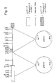
- p53A (cf. Fig. 3) (50 ⁇ g) was digested with SphI and subjected to electrophoresis on TAE buffer/0.8 % agarose gel, and two fragments near 2 kb were extracted and purified as a mixture using GENE CLEAN KIT (Biol0l).
- the mixture of the fragments (0.6 ⁇ g) and 0.2 ⁇ g of SphI-linearized DNA of E. coli vector pUC19 were dissolved in 18 ⁇ l of TE buffer, and 2 ⁇ l of 10 x ligation buffer and 300 units of T4DNA ligase (Nippon Gene) were added thereThe mixture was incubated at 16°C for 4 hours.
- coli JM103 competent cells were transformed with 10 ⁇ l of the reaction mixture and the cells were inoculated on an ampicillin-Xgal medium (1 % bacto-tryptone, 0.5 % bacto-yest extract, 1 % NaCl, 1.5 g of bacto-agar, pH 7.4, after autoclaving, 50 mg/ml of ampicillin, 0.5 mM IPTG, and 0.01 % Xgal being added), and six white colonies, i.e., six strains which caused insertional inactivation of the lacZ gene in vector pUC19, were picked up, and plasmids were extracted on a small scale.
- ampicillin-Xgal medium (1 % bacto-tryptone, 0.5 % bacto-yest extract, 1 % NaCl, 1.5 g of bacto-agar, pH 7.4, after autoclaving, 50 mg/ml of ampicillin, 0.5 mM IPTG, and 0.01 % Xgal being added
- six white colonies i
- each plasmid was digested with restriction enzymes and digested fragments were subjected to electrophoresis, and electrophoretic patterns were examined to confirmation which of the two types of fragments was inserted therein. More specifically, each plasmid was reacted with Bg1II, and the type of the fragment contained was judged whether linearization occurred or the plasmid remained intact. As a result, it revealed that two strains contained the former type of plasmid (having one Bg1II cleavage site in the inserted fragment), and four strains had the latter type of plasmid (having no Bg1II cleavage site in the inserted fragment). One of the former type of the strains was selected and its plasmid was named pMAA2 while one of the latter type of the strains was selected and its plasmid was named pMAA3 (cf. Fig. 5).
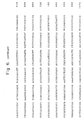
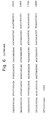
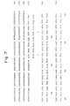
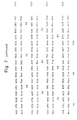
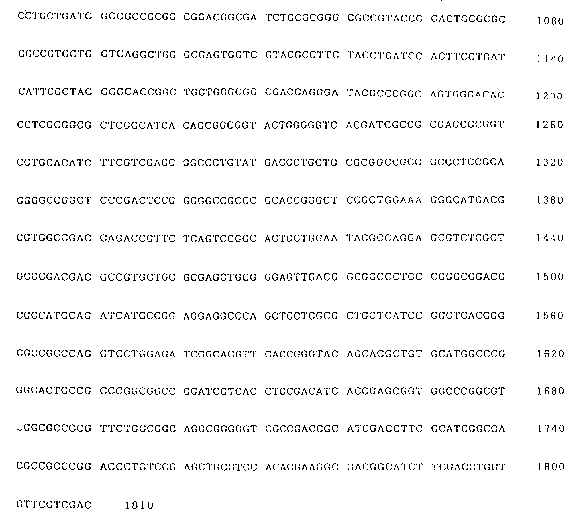
- the 3-acylation enzyme-coding gene acyA were cut out readily from plasmid p53A with restriction enzymes BamHI and SalI, and the base sequence (1810 bp) was determined using 7-Deazasequenase version 2.0 (United Biochemical Corporation/Cleveland, Ohio, U.S.A.), a commercially available sequence kit. The sequence obtained is shown in Fig. 6.
- the code region of acyA was found to range from 209th to 1,375th bases. Near upstream of the 5'-terminus of ATG, which is an initiation codon of the code region, ribosome bonding sites each composed of AAGGA were found to range from 194th to 198th bases and from 201st to 205th bases. Upstream thereof were present promoter region, i.e., TTGCCG (120th to 125th bases) corresponding to the -35 region, and CAGGAT (143rd to 148th bases) corresponding to the -10 region.
- Fig. 7 shows the base sequence of acyA and the amino acid sequence coded thereby.
- nuclease S1 buffer 330 mM sodium acetate (pH 4.5), 500 mM NaCl, 0.3 mM ZnSO 4 ) and 2 ml (20 units) of nuclease S1 (Boehringer Mannheim Yamanouchi Co., Ltd.) were added to the mixture, and reacted at room temperature for 15 minutes. After completion of the reaction, DNA was purified by the extraction with phenol/chloroform and precipitation with ethanol in the same manner as described above, and the purified DNA was dissolved in 23 ⁇ l of TE buffer.
- M buffer having a concentration by 10 times as thick as usual (500 mM NACl, 100 mM Tris-HCl (pH 7.5), 100 mM MgCl 2 , 10 mM dithiothreitol) and 3 ⁇ l of dNTP mixed solution (an aqueous mixed solution containing each 0.5 mM of dGTP, dCTP, dATP, and dTTP) as well as 1 ⁇ l (4 units) of DNA polymerase (large fragment, Takara Shuzo Co., Ltd.), followed by reaction at 37°C for 30 minutes to obtain DNA with blunt ends.
- dNTP mixed solution an aqueous mixed solution containing each 0.5 mM of dGTP, dCTP, dATP, and dTTP
- DNA polymerase large fragment, Takara Shuzo Co., Ltd.
- the DNA thus obtained was dissolved in 10 ⁇ l of TE buffer to prepare a vector solution.
- 100 ⁇ g of the recombinant plasmid p53A obtained in Example 4 was digested with restriction enzymes SalI and BamHI and 1.8 kb DNA fragment containing acyA was separated and purified using GENE CLEAN (Biol0l) to obtain 4 ⁇ g of the objective fragment.
- the DNA fragment thus obtained was treated under the same conditions as above in order to obtain DNA with blunt ends, and purified by extraction with phenol/chloroform and precipitation with ethanol.
- the DNA thus obtained was dissolved in TE buffer to obtain a TE solution (0.2 ⁇ g/ ⁇ l).
- a TE solution 0.2 ⁇ g/ ⁇ l
- To 2.5 ⁇ l (0.5 ⁇ g) of the TE solution were sequentially added 2.5 ⁇ l (0.5 ⁇ g) of the aforementioned vector solution, 13 ⁇ l of purified water, 2 ⁇ l of a 10 times-concentrated ligation buffer (500 mM Tris-HCl (pH 7.9), 100 mM MgCl 2 , 200 mM dithiothreitol, 100 mM ATP) and 1 ⁇ l (300 units) of T 4 DNA ligase (Nippon Gene Co., Ltd.), and the resulting mixture was reacted at 16°C for 15 hours.
- a portion (2 ⁇ l) of the reaction mixture was used to transform 100 ml of E . coli JM103 competent cells according to the method described by Maniatis et al., Molecular Cloning ,
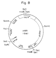
- Plasmids obtained from the transformants were examined by electrophoresis on agarose gel. As a result, plasmid pAB5 having an inserted fragment of about 1.8 kb and a structure shown in Fig. 8 was obtained.
- Plasmid pAB5 (10 ⁇ g) was completely digested with restriction enzyme SacI, and extracted with phenol/ chloroform and precipitated with ethanol to purify the resulting DNA fragments.
- the DNA fragments were partially digested with restriction enzyme PstI to obtain various fragments, from which about 9.2 kb DNA fragment formed as a result of cutting off only the pUC18-derived DNA fragment was separated by electrophoresis on 0.8 % agarose gel, and recovered and purified using GENE CLEAN (BiolOl). The DNA fragment thus obtained was dissolved in 22 ⁇ l of TE buffer.
- T 4 -polymerase buffer 700 mM Tris-HCl (pH 7.4), 100 mM MgCl 2 , 50 mM dithiothreitol
- dNTP mixed solution an aqueous mixed solution containing each 0.5 mM of dGTP, dCTP, dATP, and dTTP
- 2 ⁇ l (4.8 units) of T 4 DNA polymerase Toyobo Co., Ltd.
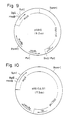
- pAB10 having a structure shown in Fig. 9
- pAB10 ⁇ B1 having a structure shown in Fig. 10.
- the both plasmids are common in that they correspond to pAB5 of which pUC18 region is deleted but in the latter (pAB10 ⁇ /B1) deletion proceeded to the region of the 4"-acylation enzyme-coding gene acyB as a result of side reaction by the enzyme, resulting in that the transformed strains transformed therewith cannot produce 4"-acylation enzyme.
- a platinum loop-ful of a slant culture medium of Streptomyces fradiae ATCC 19609 was inoculated in a 250 ml-Erlenmeyer flask containing spring) containing 20 ml of GPY medium (1.0 % glucose, 0.4 % polypeptone, 0.4 % yeast extract, 0.05 % MgSO 4 .7H 2 O, 0.1 % K 2 HPO 4 , and 0.05 % glycine, pH 7.2), and cultivated by shaking culture 28°C for 48 hours.
- GPY medium 1.0 % glucose, 0.4 % polypeptone, 0.4 % yeast extract, 0.05 % MgSO 4 .7H 2 O, 0.1 % K 2 HPO 4 , and 0.05 % glycine, pH 7.2
- the culture medium (0.5 ml) was inoculated in a 250 ml-Erlenmeyer flask (containing spring) containing 25 ml of YEME + 15 % sucrose medium (containing 5 mM MgCl 2 and 0.5 % glycine) described in Example 4, and cultivated by shaking culture at 28°C for 40 hours. After collecting mycelia by centrifugation (3,000 rpm, 15 minutes), and washing it with 10 ml of 10.3 % sucrose, the mycelia were suspended well in 4 ml of P medium containing 1 mg/ml of lysozyme, and reaction was performed at 30°C for 1 hour. The composition of P medium itself was described in Example 4.
- a portion (0.1 ml) thereof was inoculated on R3 medium (13.5 g of sucrose, 0.05 g of KCl, 1.0 g of glucose, 0.4 g of polypeptone, 0.4 g of yeast extract, 0.573 g of TES buffer. 6.1 ml of 10 % MgCl 2 solution, 2.0 g of bacto-agar, and 100 ml of distilled water; after sterilization by autoclaving, 1.0 ml of KH 2 PO 4 (0.5 %), 0.3 ml of CaCl 2 (5 M), and 1.8 ml of NaOH (1 N) being added thereto), and cultivated at 28°C for 16 hours.
- soft agar medium containing thiopeptine was overlayed on the culture medium so that the final concentration of thiopeptine became 5 ⁇ g/ml or 25 ⁇ g/ml, and cultivation was continued at 28°C for 7 days.
- the soft agar medium was prepared by mixing 8 g of Nutrient broth, 3 g of bacto-agar and 1 liter of distilled water, and sterilizing the mixture by autoclaving at 120°C for 15 minutes.
- a production medium 0.5 % soluble starch, 5 % glucose, 0.5 % yeast extract, 1.0 % malt extract, and 5 ⁇ g/ml of thiopeptine
- a production medium 0.5 % soluble starch, 5 % glucose, 0.5 % yeast extract, 1.0 % malt extract, and 5 ⁇ g/ml of thiopeptine
- the toluene layer which is an upper layer, was taken out and subjected to thin layer chromatography.
- the thin layer chromatography plate was used Art 5715 (produced buy Merck Co.) and the developer used was ethyl acetate (75 ml) : diethylamine (1.5 ml) : water (1.5 ml) : methanol (0.75 ml).
- the plate was analyzed at 280 nm using Chromato Scanner (CS-930, produced by Shimazu Seisakusho), and the positions of spots were confirmed by color development by immersing the plate in 10 % H 2 SO 4 solution and heating it at 105°C for 5 minutes.
- control strain which had not been transformed with plasmid pAB10 ⁇ B1 produced no 3-acetyltylosin.
- the aforementioned culture medium (1 liter, i.e., 20 tubes each containing 50 ml of the culture medium) were extracted with ethyl acetate at pH 8.5, and transferred in 200 ml of acidic water (0.01N HCl) and adjusted again to pH 8.5, followed by extraction with 50 ml of ethyl acetate.
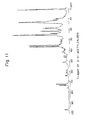
- Fig. 11 shows a 1 H-NMR chart.
- the protoplasts thus transformed were washed with P medium, and then suspended again in 1 ml of P medium.
- a portion (0.1 ml) of the suspension was inoculated on R3 medium (13.5 g of sucrose, 0.05 g of KCl, 1.0 g of glucose, 0.4 g of polypeptone, 0.4 g of yeast extract, 0.573 g TES buffer, 6.1 ml of 10 % MgCl 2 solution, 2.0 g of bacto-agar, and 100 ml of distilled water; after sterilization by autoclaving, 1.0 ml of KH 2 PO 4 (0.5 %), 0.3 ml of CaCl 2 (5 M), and 1.8 ml of NzOH (1N) being added thereto), and cultivated at 28°C for 16 hours.
- soft agar medium containing thiopeptine was overlayed on the culture medium so that the final concentration of thiopeptine became 5 ⁇ m/ml or 25 ⁇ g/ml, and cultivation was continued at 28°C for 7 days.
- the soft agar medium was prepared by mixing 8 g of Nutrient broth, 3 g of bacto-agar and 1 liter of distilled water, and sterilizing the mixture by autoclaving at 120°C for 15 minutes.
- AB10 strain Streptomyces fradiae AB10 strain
- a production medium 0.5 % soluble starch, 5 % glucose, 0.5 % yeast extract, 1.0 % malt extract, and 5 ⁇ g/ml of thiopeptine
- a production medium 0.5 % soluble starch, 5 % glucose, 0.5 % yeast extract, 1.0 % malt extract, and 5 ⁇ g/ml of thiopeptine
- the toluene layer which is an upper layer, was taken out and subjected to thin layer chromatography.
- the thin layer chromatography plate was used Art 5715 (produced buy Merck Co.) and the developer used was ethyl acetate (75 ml) : diethylamine (1.5 ml) : water (1.5 ml) : methanol (0.75 ml).
- the plate was analyzed at 280 nm using Chromato Scanner (CS-930, produced by Shimazu Seisakusho), and the positions of spots were confirmed by color development by immersing the plate in 10 % H 2 SO 4 solution and heating it at 105°C for 5 minutes.
- the S. fradiae AB10 strain was deposited internationally deposited under Budapest Treaty at Fermentation Research Institute, Agency of Industrial Science and Technology, Ministry of International Trade and Industry, Japan, at 1-3, Higashi 1-Chome, Tsukuba City, Ibaragi Prefecture, Japan under International Deposition No. FERM BP-3212.
- control strain which had not been transformed with plasmid pAB10 produced no 3-O-acetyl-4"-O-isovaleryltylosin.
- the aforementioned culture medium (1 liter, i.e., 20 tubes each containing 50 ml of the culture medium)were extracted with ethyl acetate at pH 8.5, and transferred in 200 ml of acidic water (0.01N HCl) and adjusted again to pH 8.5, followed by extraction with 50 ml of ethyl acetate.
- Fig. 12 shows a 1 H-NMR chart.
Landscapes
- Chemical & Material Sciences (AREA)
- Life Sciences & Earth Sciences (AREA)
- Health & Medical Sciences (AREA)
- Organic Chemistry (AREA)
- Engineering & Computer Science (AREA)
- Genetics & Genomics (AREA)
- Zoology (AREA)
- Wood Science & Technology (AREA)
- Biotechnology (AREA)
- Bioinformatics & Cheminformatics (AREA)
- General Health & Medical Sciences (AREA)
- General Engineering & Computer Science (AREA)
- Biochemistry (AREA)
- Microbiology (AREA)
- Molecular Biology (AREA)
- Biomedical Technology (AREA)
- General Chemical & Material Sciences (AREA)
- Chemical Kinetics & Catalysis (AREA)
- Medicinal Chemistry (AREA)
- Physics & Mathematics (AREA)
- Biophysics (AREA)
- Plant Pathology (AREA)
- Micro-Organisms Or Cultivation Processes Thereof (AREA)
- Saccharide Compounds (AREA)
Claims (26)
- DNA-Fragment, umfassend ein acyA-Gen, das für ein 3-Acylierungsenzym für Makrolidantibiotika codiert, dadurch gekennzeichnet, daß es aus einem Stamm, der zur Gattung der Streptomyces gehört, abgeleitet wird, eine Größe von etwa 1,8 kb hat, und die Eigenschaften der in Figur 1 (A) der beigefügten Zeichnung aufgezeigten Restriktionsenzymkarte oder ein daraus erhaltenes Restriktionsenzym-gespaltenes DNA-Fragment, das eine Acylierungsenzymaktivität exprimiert, aufweist.
- DNA-Fragment oder Restriktionsenzym-gespaltenes DNA-Fragment nach Anspruch 1, worin der Stamm, der zur Gattung der Streptomyces gehört, ausgewählt ist aus der Gruppe, bestehend aus Streptomyes ambofaciens, Streptomyces kitasatoensis, Streptomyces narbonensis var josamyceticus, Streptomyces hygroscopicus, Streptomyces platensis, Streptomyces albireticuli, Streptomyces cinerochromogenes, Streptomyces djakartensis, Streptomyces furdicidicus, Streptomyces macrosporeus, Streptomyces tendae, Streptomyces thermotolerans, und Streptomyces deltae.
- DNA-Fragment oder Restriktionsenzym-gespaltenes DNA-Fragment nach Anspruch 2, worin der Stamm, der zur Gattung der Streptomyces gehört, Streptomyces thermotolerans ATCC 11416 ist.
- DNA-Fragment oder Restriktionsenzym-gespaltenes DNA-Fragment nach Anspruch 1, worin das Makrolidantibiotikum ausgewählt ist aus der Gruppe, bestehend aus Tylosin, Spiramycin, Leukomycin und Nidamycin.
- DNA-Fragment oder Restriktionsenzym-gespaltenes DNA-Fragment nach Anspruch 4, worin das Makrolidantibiotikum Tylosin ist.
- DNA-Fragment oder Restriktionsenzym-gespaltenes DNA-Fragment nach Anspruch 1, worin das DNA-Fragment einen DNA-Anteil enthält, der für die folgende Aminosäuresequenz codiert.

- DNA-Fragment oder Restriktionsenzym-gespaltenes DNA-Fragment nach Anspruch 1, worin das DNA-Fragment die folgende Basensequenz hat.

- DNA-Fragment, umfassend ein acyA-Gen, das für ein 3-Acylierungsenzym für Makrolidantibiotika codiert, dadurch gekennzeichnet, daß es aus einem Stamm, der zur Gattung der Streptomyces gehört, abgeleitet wird, eine Größe von etwa 3,2 kb hat, und die Eigenschaften der in Figur 1 (B) der beigefügten Zeichnung aufgezeigten Restriktionsenzymkarte oder ein daraus erhaltenes Restriktionsenzym-gespaltenes DNA-Fragment, das eine Acylierungsenzymaktivität exprimiert, aufweist.
- Plasmid, umfassend die DNA nach Anspruch 1 oder 8.
- Plasmid nach Anspruch 9, worin das rekombinante Plasmid p53A ist gemäß Figur 3.
- Wirtsmikroorganismus, transformiert mit dem Plasmid nach Anspruch 9.
- Mikroorganismus nach Anspruch 11, worin der Wirtsmikroorganismus ein Makrolidantibiotikum-produzierendes Bakterium ist, das im wesentlichen kein Enzym zur 3-Acylierung von Makrolidantibiotika produziert.
- Mikroorganismus nach Anspruch 12, worin das Makrolidantibiotikum-produzierende Bakterium Streptomyces fradiae ATCC 19609 ist.
- Verfahren zur Herstellung eines 3-acylierten Makrolidantibiotikums, umfassend die Stufen:Züchten eines der in Anspruch 11 aufgeführten Mikroorganismen in einem Kulturmedium, das ein Makrolidantibiotikum enthält, zur Produktion eines 3-acylierten Makrolidantibiotikums undIsolieren des 3-acylierten Makrolidantibiotikums.
- Verfahren zur Herstellung 3-acylierter Makrolidantibiotika, umfassend die Stufen:Züchten eines der in Anspruch 12 aufgeführten Mikroorganismen in einem Kulturmedium, das ein Makrolidantibiotikum enthält, zur Produktion eines 3-acylierten Makrolidantibiotikums undIsolieren des 3-acylierten Makrolidantibiotikums.
- Verfahren zur Herstellung 3-acylierter Makrolidantibiotika, umfassend die Stufen:Züchten eines der in Anspruch 13 aufgeführten Mikroorganismen in einem Kulturmedium, das ein Makrolidantibiotikum enthält, zur Produktion eines 3-acylierten Makrolidantibiotikums undIsolieren des 3-acylierten Makrolidantibiotikums.
- Verfahren zur Herstellung eines 3-acylierten Makrolidantibiotikums, umfassend die Umsetzung eines Makrolidantibiotikums mit einem Acylierungsreagens in Anwesenheit des in Anspruch 11 angeführten Mikroorganismus oder dem daraus verarbeiteten Produkt.
- Rekombinantes Plasmid, umfassend ein Vektorplasmid, in das ein DNA-Fragment nach Anspruch 1 oder 8 insertiert ist, und das ein DNA-Fragment, das für ein 4"-Acylierungsenzym codiert, enthält.
- Rekombinantes Plasmid nach Anspruch 18, worin das rekombinante Plasmid pAB10 ist gemäß Figur 9.
- Wirtsmikroorganismus, der mit dem Plasmid nach Anspruch 18 transformiert ist.
- Mikroorganismus nach Anspruch 20, worin der Wirtsmikroorganismus ein Makrolidantibiotikum-produzierendes Bakterium ist, das im wesentlichen kein Enzym zur 3- und/oder 4"-Acylierung von Makrolidantibiotika produziert.
- Mikroorganismus nach Anspruch 21, worin das Makrolidantibiotikum-produzierende Bakterium Streptomyces fradiae ATCC 19609 ist.
- Verfahren zur Herstellung eines 3- und 4"-acylierten Makrolidantibiotikums, umfassend die Stufen:Züchten eines der in Anspruch 20 aufgeführten Mikroorganismen in einem Kulturmedium, das ein Makrolidantibiotikum enthält, zur Produktion eines 3- und 4"-acylierten Makrolidantibiotikums undIsolieren des 3- und 4"-acylierten Makrolidantibiotikums.
- Verfahren zur Herstellung eines 3- und 4"-acylierten Makrolidantibiotikums, umfassend die Stufen:Züchten eines der in Anspruch 21 oder 22 aufgeführten Mikroorganismen in einem Kulturmedium, das ein Makrolidantibiotikum enthält, zur Produktion eines 3- und 4"-acylierten Makrolidantibiotikums undIsolieren des 3- und 4"-acylierten Makrolidantibiotikums.
- Verfahren zur Herstellung eines 3- und 4"-acylierten Makrolidantibiotikums, umfassend die Stufen:Züchten eines der in Anspruch 22 aufgeführten Mikroorganismen in einem Kulturmedium, das ein Makrolidantibiotikum enthält, zur Produktion eines 3- und 4"-acylierten Makrolidantibiotikums undIsolieren des 3- und 4"-acylierten Makrolidantibiotikums.
- Verfahren zur Herstellung eines 3- und 4"-acylierten Makrolidantibiotikums, umfassend die Umsetzung eines Makrolidantibiotikums mit einem Acylierungsreagens in Anwesenheit des in Anspruch 20 angeführten Mikroorganismus oder einem daraus verarbeiteten Produkt.
Applications Claiming Priority (4)
| Application Number | Priority Date | Filing Date | Title |
|---|---|---|---|
| JP141625/90 | 1990-06-01 | ||
| JP14162590 | 1990-06-01 | ||
| JP4875391A JP2988735B2 (ja) | 1990-06-01 | 1991-02-22 | マクロライド抗生物質の3位アシル化酵素をコードする遺伝子 |
| JP48753/91 | 1991-02-22 |
Publications (3)
| Publication Number | Publication Date |
|---|---|
| EP0459525A2 EP0459525A2 (de) | 1991-12-04 |
| EP0459525A3 EP0459525A3 (en) | 1992-04-01 |
| EP0459525B1 true EP0459525B1 (de) | 1996-11-13 |
Family
ID=26389059
Family Applications (1)
| Application Number | Title | Priority Date | Filing Date |
|---|---|---|---|
| EP91109033A Expired - Lifetime EP0459525B1 (de) | 1990-06-01 | 1991-06-03 | Gene, die kodieren für ein Acylierungsenzym der 3-Stelle von Makrolid-Antibiotika |
Country Status (3)
| Country | Link |
|---|---|
| US (1) | US5322937A (de) |
| EP (1) | EP0459525B1 (de) |
| DE (1) | DE69123081T2 (de) |
Cited By (1)
| Publication number | Priority date | Publication date | Assignee | Title |
|---|---|---|---|---|
| EP1111148A1 (de) | 1999-12-23 | 2001-06-27 | Coserv & Management S.A. | Verfahren und Vorrichtung zur Herstellung von Mauertafeln |
Families Citing this family (3)
| Publication number | Priority date | Publication date | Assignee | Title |
|---|---|---|---|---|
| US7579167B2 (en) * | 2002-10-08 | 2009-08-25 | Aventis Pharma S. | Polypeptides involved in the biosynthesis of spiramycins, nucleotide sequences encoding these polypeptides and applications thereof |
| EP1905833B1 (de) * | 2002-10-08 | 2015-08-12 | Aventis Pharma S.A. | Polypeptide, die an der Biosynthese von Spiramycinen beteiligt sind, Nukleotidsequenzen, die diese Polypetide kodieren, und ihre Anwendungen |
| MXPA05002852A (es) * | 2002-10-08 | 2005-10-05 | Aventis Pharma Sa | Polipeptidos implicados en la biosintesis de espiramicinas, secuencias de nucleotidos que codifican estos polipeptidos y sus aplicaciones. |
Family Cites Families (2)
| Publication number | Priority date | Publication date | Assignee | Title |
|---|---|---|---|---|
| US4201843A (en) * | 1975-08-01 | 1980-05-06 | Sanraku Ocean Co., Ltd. | Process for manufacturing tylosin derivatives |
| JP2749616B2 (ja) * | 1988-05-24 | 1998-05-13 | メルシャン株式会社 | マクロライド抗生物質の4”位アシル化酵素をコードする遺伝子 |
-
1991
- 1991-05-31 US US07/708,866 patent/US5322937A/en not_active Expired - Fee Related
- 1991-06-03 EP EP91109033A patent/EP0459525B1/de not_active Expired - Lifetime
- 1991-06-03 DE DE69123081T patent/DE69123081T2/de not_active Expired - Fee Related
Cited By (1)
| Publication number | Priority date | Publication date | Assignee | Title |
|---|---|---|---|---|
| EP1111148A1 (de) | 1999-12-23 | 2001-06-27 | Coserv & Management S.A. | Verfahren und Vorrichtung zur Herstellung von Mauertafeln |
Also Published As
| Publication number | Publication date |
|---|---|
| EP0459525A2 (de) | 1991-12-04 |
| DE69123081T2 (de) | 1997-03-20 |
| DE69123081D1 (de) | 1996-12-19 |
| US5322937A (en) | 1994-06-21 |
| EP0459525A3 (en) | 1992-04-01 |
Similar Documents
| Publication | Publication Date | Title |
|---|---|---|
| US4874748A (en) | Cloning vectors for streptomyces and use thereof in macrolide antibiotic production | |
| EP2271666B1 (de) | Nrps-pks gencluster und dessen manipulation und verwendung | |
| US5149639A (en) | Biologically pure cultures of streptomyces and use thereof in macrolide antibiotic production | |
| EP0042250B1 (de) | Makrolide Antibiotika | |
| WO1993013663A1 (en) | Method of directing biosynthesis of specific polyketides | |
| JP2000515390A (ja) | 新規ポリケチド誘導体およびそれを製造するための組換え方法 | |
| EP0791656A2 (de) | Platenolid-Synthase-Gen | |
| JPH0239889A (ja) | ストレプトマイセスおよびその他の生物で使用するためのマクロライド生合成遺伝子 | |
| JP2001511349A (ja) | Saccharopolysporaerythraea及びStreptomycesantibioticusにおける6−デオキシヘキソースの生合成及び移送のための遺伝子 | |
| Petkovic et al. | A novel erythromycin, 6-desmethyl erythromycin D, made by substituting an acyltransferase domain of the erythromycin polyketide synthase | |
| EP0459525B1 (de) | Gene, die kodieren für ein Acylierungsenzym der 3-Stelle von Makrolid-Antibiotika | |
| US7285653B1 (en) | Nucleic acids which code for the enzyme activities of the spinosyn biosynthesis | |
| US5240849A (en) | Dna coding for enzyme capable of acylating the 4"-position of macrolide antibiotic | |
| EP0052005B1 (de) | Makrolide Antibiotika | |
| US20080044860A1 (en) | Polyketides and Their Synthesis | |
| JP2988735B2 (ja) | マクロライド抗生物質の3位アシル化酵素をコードする遺伝子 | |
| US5753501A (en) | Acarbose biosynthesis genes from actinoplanes sp., process for the isolation thereof and the use thereof | |
| US7807418B2 (en) | Method for producing hybrid polyketide synthases | |
| OHTA et al. | Cloning and analysis of a gene (SMS13) encoding sannamycin B-glycyltransferase from Streptomyces sannanensis and its distribution among actinomycetes | |
| US5776735A (en) | Process for dienone macrolides | |
| JPH0638750A (ja) | マクロライド抗生物質の3位アシル化酵素およびそれをコードする遺伝子 | |
| AU665526B2 (en) | Method of directing biosynthesis of specific polyketides | |
| WO2003014352A2 (en) | Nucleic acids for aminocoumarin biosynthesis | |
| Hsieh | Cloning, characterization and disruption of malonyl-CoA decarboxylase gene from Saccharopolyspora erythraea and heterologous expression in Escherichia coli | |
| EP0626806A1 (de) | Verfahren zur lenkung der biosynthese spezifischer polyketide |
Legal Events
| Date | Code | Title | Description |
|---|---|---|---|
| PUAI | Public reference made under article 153(3) epc to a published international application that has entered the european phase |
Free format text: ORIGINAL CODE: 0009012 |
|
| AK | Designated contracting states |
Kind code of ref document: A2 Designated state(s): DE FR GB IT |
|
| PUAL | Search report despatched |
Free format text: ORIGINAL CODE: 0009013 |
|
| AK | Designated contracting states |
Kind code of ref document: A3 Designated state(s): DE FR GB IT |
|
| 17P | Request for examination filed |
Effective date: 19920825 |
|
| 17Q | First examination report despatched |
Effective date: 19940425 |
|
| GRAG | Despatch of communication of intention to grant |
Free format text: ORIGINAL CODE: EPIDOS AGRA |
|
| GRAH | Despatch of communication of intention to grant a patent |
Free format text: ORIGINAL CODE: EPIDOS IGRA |
|
| GRAH | Despatch of communication of intention to grant a patent |
Free format text: ORIGINAL CODE: EPIDOS IGRA |
|
| GRAA | (expected) grant |
Free format text: ORIGINAL CODE: 0009210 |
|
| ITF | It: translation for a ep patent filed | ||
| AK | Designated contracting states |
Kind code of ref document: B1 Designated state(s): DE FR GB IT |
|
| REF | Corresponds to: |
Ref document number: 69123081 Country of ref document: DE Date of ref document: 19961219 |
|
| ET | Fr: translation filed | ||
| PLBE | No opposition filed within time limit |
Free format text: ORIGINAL CODE: 0009261 |
|
| STAA | Information on the status of an ep patent application or granted ep patent |
Free format text: STATUS: NO OPPOSITION FILED WITHIN TIME LIMIT |
|
| 26N | No opposition filed | ||
| REG | Reference to a national code |
Ref country code: GB Ref legal event code: IF02 |
|
| PGFP | Annual fee paid to national office [announced via postgrant information from national office to epo] |
Ref country code: GB Payment date: 20040602 Year of fee payment: 14 |
|
| PGFP | Annual fee paid to national office [announced via postgrant information from national office to epo] |
Ref country code: DE Payment date: 20040610 Year of fee payment: 14 |
|
| PGFP | Annual fee paid to national office [announced via postgrant information from national office to epo] |
Ref country code: FR Payment date: 20040616 Year of fee payment: 14 |
|
| PG25 | Lapsed in a contracting state [announced via postgrant information from national office to epo] |
Ref country code: IT Free format text: LAPSE BECAUSE OF NON-PAYMENT OF DUE FEES;WARNING: LAPSES OF ITALIAN PATENTS WITH EFFECTIVE DATE BEFORE 2007 MAY HAVE OCCURRED AT ANY TIME BEFORE 2007. THE CORRECT EFFECTIVE DATE MAY BE DIFFERENT FROM THE ONE RECORDED. Effective date: 20050603 Ref country code: GB Free format text: LAPSE BECAUSE OF NON-PAYMENT OF DUE FEES Effective date: 20050603 |
|
| PG25 | Lapsed in a contracting state [announced via postgrant information from national office to epo] |
Ref country code: DE Free format text: LAPSE BECAUSE OF NON-PAYMENT OF DUE FEES Effective date: 20060103 |
|
| PG25 | Lapsed in a contracting state [announced via postgrant information from national office to epo] |
Ref country code: FR Free format text: LAPSE BECAUSE OF NON-PAYMENT OF DUE FEES Effective date: 20060228 |
|
| GBPC | Gb: european patent ceased through non-payment of renewal fee |
Effective date: 20050603 |
|
| REG | Reference to a national code |
Ref country code: FR Ref legal event code: ST Effective date: 20060228 |