EP0452915B1 - Apparatus for adjusting read-out conditions and/or image processing conditions for radiation images - Google Patents
Apparatus for adjusting read-out conditions and/or image processing conditions for radiation images Download PDFInfo
- Publication number
- EP0452915B1 EP0452915B1 EP19910106174 EP91106174A EP0452915B1 EP 0452915 B1 EP0452915 B1 EP 0452915B1 EP 19910106174 EP19910106174 EP 19910106174 EP 91106174 A EP91106174 A EP 91106174A EP 0452915 B1 EP0452915 B1 EP 0452915B1
- Authority
- EP
- European Patent Office
- Prior art keywords
- image
- image signal
- radiation
- read
- conditions
- Prior art date
- Legal status (The legal status is an assumption and is not a legal conclusion. Google has not performed a legal analysis and makes no representation as to the accuracy of the status listed.)
- Expired - Lifetime
Links
- 230000005855 radiation Effects 0.000 title claims description 82
- 238000012545 processing Methods 0.000 title claims description 56
- OAICVXFJPJFONN-UHFFFAOYSA-N Phosphorus Chemical compound [P] OAICVXFJPJFONN-UHFFFAOYSA-N 0.000 claims description 53
- 238000013528 artificial neural network Methods 0.000 claims description 51
- 238000000034 method Methods 0.000 claims description 19
- 230000004936 stimulating effect Effects 0.000 claims description 5
- 206010073306 Exposure to radiation Diseases 0.000 claims description 4
- 230000008569 process Effects 0.000 claims description 3
- 230000009467 reduction Effects 0.000 claims description 3
- 230000001131 transforming effect Effects 0.000 claims 4
- 230000009466 transformation Effects 0.000 claims 2
- 230000006870 function Effects 0.000 description 23
- 230000035945 sensitivity Effects 0.000 description 13
- 210000000323 shoulder joint Anatomy 0.000 description 10
- 238000012937 correction Methods 0.000 description 5
- NAWXUBYGYWOOIX-SFHVURJKSA-N (2s)-2-[[4-[2-(2,4-diaminoquinazolin-6-yl)ethyl]benzoyl]amino]-4-methylidenepentanedioic acid Chemical compound C1=CC2=NC(N)=NC(N)=C2C=C1CCC1=CC=C(C(=O)N[C@@H](CC(=C)C(O)=O)C(O)=O)C=C1 NAWXUBYGYWOOIX-SFHVURJKSA-N 0.000 description 4
- 230000003321 amplification Effects 0.000 description 4
- 239000000463 material Substances 0.000 description 4
- 238000003199 nucleic acid amplification method Methods 0.000 description 4
- 238000006467 substitution reaction Methods 0.000 description 4
- 238000004458 analytical method Methods 0.000 description 3
- 230000008859 change Effects 0.000 description 3
- 238000004364 calculation method Methods 0.000 description 2
- 238000006243 chemical reaction Methods 0.000 description 2
- 230000003287 optical effect Effects 0.000 description 2
- 238000003530 single readout Methods 0.000 description 2
- 210000000225 synapse Anatomy 0.000 description 2
- 210000001015 abdomen Anatomy 0.000 description 1
- 230000002159 abnormal effect Effects 0.000 description 1
- NIXOWILDQLNWCW-UHFFFAOYSA-N acrylic acid group Chemical group C(C=C)(=O)O NIXOWILDQLNWCW-UHFFFAOYSA-N 0.000 description 1
- 230000002411 adverse Effects 0.000 description 1
- 238000007796 conventional method Methods 0.000 description 1
- 230000001186 cumulative effect Effects 0.000 description 1
- 238000013461 design Methods 0.000 description 1
- 238000003745 diagnosis Methods 0.000 description 1
- 230000000694 effects Effects 0.000 description 1
- 230000001747 exhibiting effect Effects 0.000 description 1
- 230000001537 neural effect Effects 0.000 description 1
- 230000002093 peripheral effect Effects 0.000 description 1
- 238000002601 radiography Methods 0.000 description 1
- 238000011160 research Methods 0.000 description 1
- 230000004044 response Effects 0.000 description 1
- 229910052709 silver Inorganic materials 0.000 description 1
- 239000004332 silver Substances 0.000 description 1
- -1 silver halide Chemical class 0.000 description 1
- 230000000638 stimulation Effects 0.000 description 1
Images


Classifications
-
- G—PHYSICS
- G01—MEASURING; TESTING
- G01T—MEASUREMENT OF NUCLEAR OR X-RADIATION
- G01T1/00—Measuring X-radiation, gamma radiation, corpuscular radiation, or cosmic radiation
- G01T1/16—Measuring radiation intensity
- G01T1/20—Measuring radiation intensity with scintillation detectors
- G01T1/2012—Measuring radiation intensity with scintillation detectors using stimulable phosphors, e.g. stimulable phosphor sheets
- G01T1/2014—Reading out of stimulable sheets, e.g. latent image
Definitions
- This invention relates to an apparatus for adjusting read-out conditions and/or image processing conditions for a radiation image, wherein read-out conditions, under which a radiation image is to be read out, and/or image processing conditions, under which an image signal representing the radiation image is to be processed, are adjusted on the basis of the image signal representing the radiation image.
- phosphors when certain kinds of phosphors are exposed to radiation such as X-rays, ⁇ -rays, ⁇ -rays, ⁇ -rays, cathode rays or ultraviolet rays, they store part of the energy of the radiation. Then, when the phosphor which has been exposed to the radiation is exposed to stimulating rays such as visible light, light is emitted by the phosphor in proportion to the amount of energy stored thereon during its exposure to the radiation. A phosphor exhibiting such properties is referred to as a stimulable phosphor.
- a sheet provided with a layer of the stimulable phosphor (hereinafter referred to as a stimulable phosphor sheet) is first exposed to radiation which has passed through an object, such as the human body. A radiation image of the object is thereby stored on the stimulable phosphor sheet. The stimulable phosphor sheet is then scanned with stimulating rays, such as a laser beam, which cause it to emit light in proportion to the amount of energy stored thereon during its exposure to the radiation.
- the light emitted by the stimulable phosphor sheet, upon stimulation thereof, is photoelectrically detected and converted into an electric image signal.
- the image signal is then used during the reproduction of the radiation image of the object as a visible image on a recording material such as photographic film, on a display device such as a cathode ray tube (CRT) display device, or the like.
- a recording material such as photographic film
- a display device such as a cathode ray tube (CRT) display device, or the like.
- CTR cathode ray tube
- Radiation image recording and reproducing systems which use stimulable phosphor sheets are advantageous over conventional radiography using silver halide photographic materials, in that images can be recorded even when the energy intensity of the radiation to which the stimulable phosphor sheet is exposed varies over a wide range. More specifically, since the amount of light which the stimulable phosphor sheet emits when being stimulated varies over a wide range and is proportional to the amount of energy stored thereon during its exposure to the radiation, it is possible to obtain an image having a desirable density regardless of the energy intensity of the radiation to which the stimulable phosphor sheet was exposed.
- an appropriate read-out gain is set when the emitted light is being detected and converted into an electric signal to be used in the reproduction of a visible image on a recording material, such as photographic film, or on a display device, such as a CRT display device.
- novel radiation image recording and reproducing systems which accurately detect an image signal have been proposed.
- the proposed radiation image recording and reproducing systems are constituted such that a preliminary read-out operation (hereinafter simply referred to as the "preliminary readout") is carried out in order approximately to ascertain the radiation image stored on the stimulable phosphor sheet.
- the stimulable phosphor sheet is scanned with a light beam having a comparatively low energy level, and a preliminary read-out image signal obtained during the preliminary readout is analyzed.
- a final read-out operation (hereinafter simply referred to as the "final readout") is carried out to obtain the image signal, which is to be used during the reproduction of a visible image.
- the stimulable phosphor sheet is scanned with a light beam having an energy level higher than the energy level of the light beam used in the preliminary readout, and the radiation image is read out with the factors affecting the image signal adjusted to appropriate values on the basis of the results of an analysis of the preliminary read-out image signal.
- read-out conditions means a group of various factors, which are adjustable and which affect the relationship between the amount of light emitted by the stimulable phosphor sheet during image readout and the output of a read-out means.
- read-out conditions may refer to a read-out gain and a scale factor which define the relationship between the input to the read-out means and the output therefrom, or to the power of the stimulating rays used when the radiation image is read out.
- the term "energy level of a light beam” as used herein means the level of energy of the light beam to which the stimulable phosphor sheet is exposed per unit area.
- the term "energy level of a light beam” means the weighted energy level which is calculated by weighting the energy level of the light beam, to which the stimulable phosphor sheet is exposed per unit area, with the sensitivity of the stimulable phosphor sheet to the wavelength.
- light beams of different wavelengths may be used, the intensity of the light beam produced by a laser beam source or the like may be changed, or the intensity of the light beam may be changed by moving an ND filter or the like into and out of the optical path of the light beam.
- the diameter of the light beam may be changed in order to alter the scanning density, or the speed at which the stimulable phosphor sheet is scanned with the light beam may be changed.
- image processing conditions means a group of various factors, which are adjustable and set when an image signal is subjected to processing, which affect the gradation, sensitivity, or the like, of a visible image reproduced from the image signal.
- the proposed method is applicable to cases where an image signal is obtained from a radiation image recorded on a recording medium such as conventional X-ray film, as well as to systems using stimulable phosphor sheets.
- the algorithms which have heretofore been employed are designed such that a probability density function of an image signal is created, and characteristic values are found from the probability density function.
- the characteristic values include, for example, the maximum value of the image signal, the minimum value of the image signal, or the value of the image signal at which the probability density function is maximum, i.e. the value which occurs most frequently.
- the read-out conditions for the final readout and/or the image processing conditions are determined on the basis of the characteristic values.
- Methods for determining the read-out conditions for the final readout and/or the image processing conditions on the basis of the results of an analysis of the probability density function of an image signal can be classified into the following:
- the neural network is provided with a learning function by back propagation method. Specifically, when information (an instructor signal), which represents whether an output signal obtained when an input signal is given is or is not correct, is fed into the neural network, the weight of connections between units in the neural network (i.e. the weight of synapse connections) is corrected. By repeating the learning of the neural network, the probability that a correct answer will be obtained in response to a new input signal can be kept high.
- uch functions are described in, for example, "Learning representations by back-propagating errors” by D. E. Rumelhart, G. E. Hinton and R. J. Williams, Nature, 323-9,533-356, 1986a; "Back-propagation” by Hideki Aso, Computrol, No. 24, pp. 53-60; and "Neural Computer” by Kazuyuki Aihara, the publishing bureau of Tokyo Denki University).
- the neural network is also applicable when the read-out conditions for the final readout and/or the image processing conditions are to be adjusted. By feeding an image signal, or the like, into the neural network, outputs representing the values of the read-out conditions for the final readout and/or the image processing conditions can be obtained from the neural network.
- the read-out conditions for the final readout and/or the image processing conditions appropriate for a specific radiation image can be determined.
- various types of image signals are obtained which represent various radiation images, such as the images of the right shoulder and the left shoulder (reversed images), an enlarged image and a reduced image, an erect image and a side image and an inverted image, and images shifted from each other.
- a very large number of units should be incorporated in the neural network.
- a storage means should be used which has a very large capacity for storing information representing the weight of connections between units in the neural network. Additionally, the learning of the neural network should be repeated very many times.
- EP-A-0 409 206 discloses an apparatus for adjusting image processing conditions in a digital image display apparatus, wherein digital image data are obtained from an MRI or a CT system. The frequency distribution of the pixel values of the image data is calculated. Moreover, a plurality of image qualities is calculated for indicating clarity of a display image in accordance with the plurality of pixel value-to-brightness conversion modes. Then, the most suitable conversion mode is selected on the basis of the image qualities.
- US-A-4 887 305 discloses a method for adjusting read-out conditions for a radiation image in accordance with the maximum and the minimum image signal level.
- the image signal level is measured by carrying out a preliminary read-out step.
- the primary object of the present invention is to provide an apparatus for adjusting read-out conditions and/or image processing conditions for a radiation image wherein, even if various image signals representing various radiation images are obtained, the read-out conditions for the final readout and/or the image processing conditions appropriate for each of the various radiation images are determined by a neural network provided with a comparatively small number of units.
- a neural network provided with a comparatively small number of units is utilized, and characteristic measures representing characteristics of a radiation image, such as read-out conditions under which the radiation image is to be read out, image processing conditions under which the image signal representing the radiation image is to be processed, and the portion of an object the image of which was recorded, are found accurately from an image signal representing the radiation image.
- a subdivision pattern of radiation images, the shape and location of an irradiation field, an orientation in which the object was placed when the image of the object was recorder, and/or a portion of an object the image of which was recorded is determined accurately from an image signal representing a radiation images.
- An apparatus in accordance with the present invention is applicable when a stimulable phosphor sheet is used and the preliminary readout is carried out.
- the standard pattern may be selected in accordance with the concept behind the design of the apparatuses, or the like.
- the image signal or the first image signal representing the radiation image is transformed into a transformed image signal representing the radiation image, which has been transformed into the standard pattern.
- the image signal or the first image signal representing the radiation image may be transformed into a transformed image signal representing the radiation image, which has been reversed, rotated, adjusted for the position, enlarged, or reduced.
- the information representing the standard pattern of radiation images is stored in the storage means.
- the image signal (or the first image signal) is obtained, from which the read-out conditions for the final readout and/or the image processing conditions are to be determined
- the image signal representing the radiation image is transformed into a transformed image signal representing the radiation image, which has been transformed into the standard pattern.
- the transformed image signal is fed into the neural network. Therefore, a neural network having a small scale may be used, and a storage means having a small storage capacity may be used to store the weight of connections between units of the neural network.
- the level of accuracy with which the read-out conditions for the final readout and/or the image processing conditions are to be determined, is kept the same
- the learning of the neural network may be repeated only a fewer times than when a conventional technique is used.
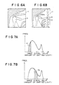
- Figures 6A and 6B show radiation images in which a pattern of a shoulder joint 9 is embedded.
- the two radiation images differ from each other in that the radiation image of Figure 6B includes the patterns of vertebral bodies 10, and the radiation image of Figure 6A does not include them.
- Figures 7A and 7B show probability density functions of image signals representing the radiation images shown in Figures 6A and 6B.
- the two probability density functions are approximately identical with each other.
- the two radiation images have the difference described above. Therefore, the image signal components representing the pattern of the shoulder joint 9, which is taken as a region of interest, fall within the range K1 in the probability density function shown in Figure 7A and within the range K2 in the probability density function shown in Figure 7B.
- the read-out conditions for the final readout and/or the image processing conditions are determined from each of the two probability density functions, and visible images of the radiation images shown in Figures 6A and 6B are reproduced from the image signals obtained under these conditions, because the two probability density functions are approximately identical with each other, approximately the same values are calculated as the read-out conditions for the final readout and/or the image processing conditions.
- two reproduced visible images are obtained which have approximately the same image density and contrast. Therefore, the image density of the pattern of the shoulder joint, which is taken as the region of interest, cannot be kept appropriate.
- the pattern of the region of interest is not illustrated clearly in the reproduced visible image. Also, for example, when a plurality of reproduced visible images are compared with each other in order for the course of an abnormal part of an object to be investigated, a correct diagnosis cannot be made.
- the present invention eliminates the problems described above.
- the neural network is used to determine the read-out conditions for the final readout and/or the image processing conditions. By repeating the learning of the neural network, appropriate read-out conditions for the final readout and/or appropriate image processing conditions can be determined.
- the learning of the neural network is carried out by utilizing an image signal representing a radiation image, in which a pattern of a specific region of interest in an object is embedded, and the read-out conditions for the final readout and/or the image processing conditions, which have been determined as being optimum for the pattern of the region of interest. Therefore, even if the radiation image includes the pattern of the region of interest and other patterns, the read-out conditions for the final readout and/or the image processing conditions can be set to values appropriate for the pattern of the region of interest.
- the shape and location of the pattern of the region of interest can be determined accurately.
- the read-out conditions for the final readout and/or the image processing conditions are then determined on the basis of the image signal components of the first image signal representing the pattern of the region of interest. Therefore, the read-out conditions for the final readout and/or the image processing conditions are basically free of adverse effects of the image information other than the pattern of the region of interest and can be set to be appropriate for the pattern of the region of interest.
- image signal components representing the picture elements located in the irradiation field may be fed into the neural network.
- these image signal components may be sampled alternately, and only the sampled components may be fed into the neural network.
- a storage means having an even smaller storage capacity may be employed.
- an X-ray image of the shoulder of a human body is stored on a stimulable phosphor sheet.
- Figure 4 is a schematic view showing an example of an X-ray image recording apparatus.
- X-rays 3 are produced by an X-ray source 2 of an X-ray image recording apparatus 1 and irradiated to the shoulder 4a of a human body 4.
- X-rays 3a which have passed through the human body 4, impinge upon a stimulable phosphor sheet 11. In this manner, an X-ray image of the shoulder 4a of the human body 4 is stored on the stimulable phosphor sheet 11.
- Figures 1A and 1B are explanatory views showing examples of X-ray images of the shoulders stored on stimulable phosphor sheets.
- Figures 1A and 1B show the X-ray images of the right and left shoulders.
- Each of the X-ray images comprises an object image region 5, in which the pattern of the human body is stored, and a background region 6, upon which the X-rays impinged directly without passing through the object 4.
- Figure 5 is a perspective view showing an example of an X-ray image read-out apparatus and an example of a computer system, in which an embodiment of the first apparatus in accordance with the present invention is employed.
- a stimulable phosphor sheet is used, and a preliminary readout is carried out.
- the stimulable phosphor sheet 11, on which the X-ray image has been stored, is placed at a predetermined position in a preliminary read-out means 100 which carries out a preliminary readout by scanning the stimulable phosphor sheet 11 with a light beam having a low energy level, thereby releasing only part of the energy from the stimulable phosphor sheet 11, which energy was stored during its exposure to radiation.
- the stimulable phosphor sheet 11 is conveyed in a sub-scanning direction indicated by the arrow Y by a sheet conveyance means 13 which is constituted of an endless belt on the like and which is operated by a motor 12.
- a laser beam 15 which has a low energy level is produced by a laser beam source 14, and is reflected and deflected by a rotating polygon mirror 16 which is quickly rotated by a motor 23 in the direction indicated by the arrow.
- the laser beam 15 then passes through a converging lens 17 constituted of an f ⁇ lens or the like.
- the direction of the optical path of the laser beam 15 is then changed by a mirror 18, and the laser beam 15 impinges upon the stimulable phosphor sheet 11 and scans it in a main scanning direction indicated by the arrow X, which direction is approximately normal to the sub-scanning direction indicated by the arrow Y.
- the exposed portion of the stimulable phosphor sheet 11 emits light 19 in an amount proportional to the amount of energy stored thereon during its exposure to radiation.
- the emitted light 19 is guided by a light guide member 20 and photoelectrically detected by a photomultiplier 21.
- the light guide member 20 is made from a light guiding material such as an acrylic plate and has a linear light input face 20a, positioned so that it extends along the main scanning line on the stimulable phosphor sheet 11, and a ring-shaped light output face 20b, positioned so that it is in close contact with a light receiving face of the photomultiplier 21.
- An analog output signal S generated by the photomultiplier 21 is logarithmically amplified by a logarithmic amplifier 26, and digitized by an A/D converter 27 into a preliminary read-out image signal SP.
- the preliminary read-out image signal SP takes a value proportional to the logarithmic value of the amount of the light 19, which was emitted from each of picture elements in the X-ray image stored on the stimulable phosphor sheet 11.
- read-out conditions i.e. the voltage applied to the photomultiplier 21 and the amplification factor of the logarithmic amplifier 26, are adjusted so that image information can be detected accurately even if the amount of energy stored on the stimulable phosphor sheet 11 during its exposure to radiation varies over a wide range.
- the preliminary read-out image signal SP obtained in the manner described above is fed into a computer system 40.
- the computer system 40 is provided with an embodiment of the first apparatus in accordance with the present invention.
- the computer system 40 comprises a main body 41 in which a CPU and an internal memory are incorporated, a disk drive unit 42 which operates a floppy disk serving as a subsidiary memory, a keyboard 43 from which necessary instructions, or the like, are fed into the computer system 40, and a CRT display device 44 which displays necessary information.
- the read-out conditions for the final readout i.e. the sensitivity and the contrast during the final readout, are determined in the manner described later.
- the voltage applied to a photomultiplier 21' and the amplification factor of a logarithmic amplifier 26' are controlled in accordance with the sensitivity and the contrast.
- the contrast corresponds to the ratio of the largest amount of emitted light, which is capable of being accurately converted into an image signal during the final readout, to the smallest amount of emitted light, which is capable of being accurately converted into an image signal during the final readout.
- the sensitivity corresponds to the photoelectric conversion factor, which represents to what image signal level a predetermined amount of emitted light is to be converted.
- a stimulable phosphor sheet 11' on which the preliminary readout has been finished is placed at a predetermined position in the final read-out means 100' and scanned with a laser beam 15' having an energy level higher than that of the laser beam 15 used during the preliminary readout. In this manner, an image signal is detected under the read-out conditions which have been determined on the basis of the preliminary read-out image signal.
- the configuration of the final read-out means 100' is nearly the same as that of the preliminary read-out means 100, and therefore elements corresponding to those constituting the preliminary read-out means 100 are numbered with corresponding primed reference numerals in Figure 5.
- the resulting image signal SQ is fed into the computer system 40, which carries out appropriate image processing on the image signal SQ.
- the image signal is fed into a reproducing apparatus (not shown), which reproduces a visible image from the image signal.
- Figures 2A and 2B are explanatory views showing a standard pattern and a reversed pattern, which are represented by information stored in the computer system 40.
- the standard pattern is composed of a first region 7, which is represented by a mean-level value of the preliminary read-out image signal SP corresponding to the object image region in the X-ray image shown in Figure 1A, and a second region 8, which is represented by a mean-level value of the preliminary read-out image signal SP detected from the background region 6 in the X-ray image shown in Figure 1A.
- the reversed pattern is composed of a first region 7, which is represented by a mean-level value of the preliminary read-out image signal SP corresponding to the object image region in the X-ray image shown in Figure 1B, and a second region 8, which is represented by a mean-level value of the preliminary read-out image signal SP detected from the background region 6 in the X-ray image shown in Figure 1B.
- pattern matching is carried out between the preliminary read-out image signal SP and each of the image signal SS representing the standard pattern shown in Figure 2A and the image signal SR representing the reversed pattern shown in Figure 2B. In this manner, a judgment is made as to whether the preliminary read-out image signal SP represents the X-ray image of the right shoulder or of the left shoulder.
- calculations are made to find square values of differences between the image signal components of the preliminary read-out image signal SP and each of the image signals SS and SR, which image signal components represent corresponding picture elements in the preliminary read-out image signal SP and each of the image signals SS and SR, i.e.
- the preliminary read-out image signal SP is processed such that the image represented by the preliminary read-out image signal SP is reversed.
- an image signal corresponding to the image of the right shoulder shown in Figure 1A, or an image signal corresponding to the image of the left shoulder is always fed into a neural network, which will be described below.
- the image signal is processed such that the processed image signal represents the predetermined standard pattern (i.e. the pattern of the right shoulder in this embodiment).
- the processed image signal is then fed into the neural network. Therefore, the number of units constituting the neural network can be reduced, and the requirement of the storage capacity of a storage means for storing the weight coefficients, which represent the degrees of connections between the units, can be kept small. Also, the learning of the neural network can be finished quickly.
- FIG. 3 is an explanatory view showing an example of the neural network which is provided with a learning function by back propagation method.
- learning function by back propagation method means the learning algorithms in a neural network, with which the output of the neural network is compared with a correct answer (an instructor signal), and the weight of connections (i.e. the weight of synapse connections) is corrected sequentially from the output side to the input side of the neural network.
- the neural network comprises a first layer (an input layer), a second layer (an intermediate layer), and a third layer (an output layer).
- the first, second, and third layers are composed respectively of n1 number of units, n2 number of units, and two units.
- Signals F1, F2, ..., Fn1 fed into the first layer (the input layer) are the image signal components of the preliminary read-out image signal SP representing the picture elements in the X-ray image (the reversed image in the cases of the images of the left shoulder).
- Two outputs y 3 / 1 and y 3 / 2 obtained from the third layer (the output layer) are the signals corresponding to the sensitivity and the contrast during the final readout.
- An i'th unit of a k'th layer is indicated by u k / i.
- the total input into the unit u k / i is indicated by x k / i, and the total output therefrom is indicated by y k / i.
- the weight of connection from the unit u k / i to a unit u k+1 / j is indicated by W k k+1 / i j.
- f (x) 1 1 - e x
- y k j f (x k j )
- the n1 number of signals F1, F2, ..., Fn1 are weighted with the weights of connection W k kj+1 / i j, and transmitted to the ultimate outputs y 3 / 1 and y 3 / 2. In this manner, the read-out conditions for the final readout (i.e. the sensitivity and the contrast) are obtained.
- initial values of the weights of connection W k kj+1 / i j are given by random numbers.
- the range of the random numbers should preferably be limited such that, even when the values of the inputs F1, F2, ..., Fn1 fluctuate to the largest extent, the outputs y 3 / 1 and y 3 / 2 may take values falling within a predetermined range or values close to said predetermined range.
- preliminary read-out image signals are obtained in the manner described above from a plurality of stimulable phosphor sheets storing X-ray images of the right or left shoulder, for which the appropriate read-out conditions for the final readout are known.
- the preliminary read-out image signals are reversed.
- the n1 number of inputs F1, F2, ..., Fn1 are obtained.
- the n1 number of inputs F1, F2, ..., Fn1 are fed into the neural network shown in Figure 3, and the outputs y k / i of the respective units u k / i are monitored.
- the weights of connection W k k+1 / i j can be converged to predetermined values by using Formulas (16) and (22), using a sufficiently small learning coefficient ⁇ and carrying out the learning operations very many times.
- a sufficiently small learning coefficient ⁇ is used, the speed with which the learning operations are effected will become low.
- a very large learning coefficient ⁇ is used, "vibration" will occur in the learning operations (i.e. the weights of connection do not converge to predetermined values). Therefore, actually, the vibration is prevented by employing an inertia term, which is expressed in Formula (23), in the calculations of the correction amounts for the weights of connection, and the learning coefficient ⁇ is set to a slightly large value.
- ⁇ W k k+1 i j (t + 1) ⁇ • ⁇ W k k+1 i j (t) + ⁇ • ⁇ E 1 ⁇ x k+1 j ⁇ y k i
- ⁇ denotes the coefficient referred to as the inertia term
- ⁇ W k k+1 / i j (t) denotes the correction amount, which is used during the t'th learning operation and which is obtained by subtracting a weight of connection W k k+1 / i j which has not been corrected, from a weight of connection W k k+1 / i j which has been corrected.
- the inertia term ⁇ is set to 0.9
- the learning coefficient ⁇ is set to 0.25
- 200,000 times of corrections (learning operations) are carried out for each of the weights of correction W k k+1 / i j. Thereafter, each of the weights of correction W k k+1 / i j is fixed at a final value.
- the two outputs y 3 / 1 and y 3 / 2 represents the appropriate sensitivity and the appropriate contrast during the final readout.
- a preliminary read-out image signal SP representing an X-ray image is fed into the neural network shown in Figure 3.
- the outputs y 3 / 1 and y 3 / 2 obtained from the neural network are utilized as signals representing the read-out conditions (i.e. the sensitivity and the contrast) for the final readout appropriate for the X-ray image. Because the learning operations have been carried out in the manner described above, the signals accurately represent the appropriate read-out conditions for the final readout.
- the number of layers of the neural network is not limited to three. Also, no limitation is imposed on the number of the units of each layer. The number of the units of each layer may be determined in accordance with the number of the picture elements represented by the preliminary read-out image signal SP, which is fed into the neural network, the accuracy, with which the read-out conditions for the final readout are to be obtained, or the like.
- the voltage applied to the photomultiplier 21' of the final read-out means 100', the amplification factor of the logarithmic amplifier 26', and the like, are controlled in accordance with the read-out conditions for the final readout, which have been adjusted by the neural network.
- the final readout is carried out under the controlled conditions.
- the preliminary read-out image signal SP representing an X-ray image of the shoulder is fed into the neural network.
- pattern matching is effected with respect to the patterns shown in Figures 2A and 2B.
- a judgment is thereby made as to whether the X-ray image represented by the preliminary read-out image signal SP is the standard image (i.e. the image of the right shoulder) or the reversed image (i.e. the image of the left shoulder).
- the preliminary read-out image signal SP is processed such that the processed image signal represents the standard image (i.e. the image of the right shoulder).
- the first apparatus in accordance with the present invention is not limited to the processing of images of the shoulder.
- the first apparatus in accordance with the present invention is also applicable when images of the right and left hands, images of the right and left sides of the head or the abdomen, and the like, are processed.
- the first apparatus in accordance with the present invention is not limited to the processing of images reversed horizontally.
- the first apparatus in accordance with the present invention is also applicable when an image is to be rotated into a normal orientation in cases where an image signal representing an inclined image is obtained due to oblique setting of a stimulable phosphor sheet during the image recording operation, or an image signal representing a laterally inclined image or an inverted image is obtained due to setting of a stimulable phosphor sheet in an incorrect direction during the image read-out operation.
- the first apparatus in accordance with the present invention is also applicable when images having different scales of enlargement (or reduction), which are obtained from, for example, a direct image recording operation and fluorography, are to be corrected.
- the first apparatus in accordance with the present invention is further applicable when position adjustment is to be carried out such that an object image region may be located at the center area of an image in cases where the object image pattern was recorded at a peripheral part of the image. Additionally, the first apparatus in accordance with the present invention is applicable when a combination of the aforesaid processes is to be carried out.
- the preliminary read-out means 100 and the final read-out means 100' are separate from each other.
- a single read-out means may be utilized for performing both the preliminary readout and the final readout.
- the stimulable phosphor sheet 11 may be moved back to the position at which image readout is started. Thereafter, the final readout may be carried out.
- the read-out conditions for the final readout are adjusted by the computer system 40.
- predetermined read-out conditions may be used when the final readout is carried out regardless of the characteristics of the preliminary read-out image signal SP.
- the computer system 40 may adjust the image processing conditions to be used in carrying out image processing of the image signal SQ.
- the computer system 40 may also adjust both the read-out conditions and the image processing conditions.
- the aforesaid embodiment is applied to the radiation image read-out apparatus wherein the preliminary readout is carried out.
- the first apparatus in accordance with the present invention is also applicable to radiation image read-out apparatuses wherein no preliminary read-out operations are carried out, and only the aforesaid final read-out operations are carried out.
- an image signal is obtained by use of predetermined read-out conditions.
- image processing conditions are calculated by the computer system 40. The image signal is processed under the calculated image processing conditions.
- a stimulable phosphor sheet is used, and an X-ray image having a pattern of the shoulder joint of a human body as a region of interest is processed.
- Figures 6A and 6B are explanatory views showing X-ray images of the shoulder joint, which images have been stored on stimulable phosphor sheets 11 in the X-ray image recording apparatus shown in Figure 4 in the manner described above.
- the read-out conditions for the final readout are adjusted on the basis of the preliminary read-out image signal SP in the manner described below.
- preliminary read-out image signals are obtained in the manner described above from a plurality of stimulable phosphor sheets storing X-ray images having a shoulder joint pattern 9 as shown in Figures 6A and 6B, for which the appropriate read-out conditions for the final readout are known.
- the n1 number of inputs F1, F2, ..., Fn1 are obtained.
- an image signal is obtained which represents an X-ray image such that the pattern of the shoulder joint 9 may have an appropriate image density.
- the n1 number of inputs F1, F2, ..., Fn1 are fed into the neural network shown in Figure 3, and the learning operations of the neural network are carried out in the same manner as that described above.
- the two outputs y 3 / 1 and y 3 / 2 represents the appropriate sensitivity and the appropriate contrast during the final readout (i.e. such that the pattern of the shoulder joint 9 may have an appropriate image density in a reproduced X-ray image).
- a preliminary read-out image signal SP representing an X-ray image is fed into the neural network shown in Figure 3.
- the outputs y 3 / 1 and y 3 / 2 obtained from the neural network are utilized as signals representing the read-out conditions (i.e. the sensitivity and the contrast) for the final readout appropriate for the X-ray image. Because the learning operations have been carried out in the manner described above, the signals accurately represent the appropriate read-out conditions for the final readout.
- the voltage applied to the photomultiplier 21' of the final read-out means 100', the amplification factor of the logarithmic amplifier 26', and the like, are controlled in accordance with the read-out conditions for the final readout, which have been adjusted by the neural network.
- the final readout is carried out under the controlled conditions.
- the read-out conditions for the final readout are adjusted by the computer system 40.
- predetermined read-out conditions may be used when the final readout is carried out regardless of the characteristics of the preliminary read-out image signal SP.
- the computer system 40 may adjust the image processing conditions to be used in carrying out image processing of the image signal SQ.
- the computer system 40 may also adjust both the read-out conditions and the image processing conditions.
Landscapes
- Physics & Mathematics (AREA)
- Health & Medical Sciences (AREA)
- Life Sciences & Earth Sciences (AREA)
- General Physics & Mathematics (AREA)
- High Energy & Nuclear Physics (AREA)
- Molecular Biology (AREA)
- Spectroscopy & Molecular Physics (AREA)
- Radiography Using Non-Light Waves (AREA)
- Apparatus For Radiation Diagnosis (AREA)
- Image Analysis (AREA)
- Image Processing (AREA)
Priority Applications (1)
| Application Number | Priority Date | Filing Date | Title |
|---|---|---|---|
| EP96106224A EP0726542B1 (en) | 1990-04-18 | 1991-04-17 | Method and apparatus for adjusting read-out conditions and/or image processing conditions for radiation images, radiation image read-out apparatus, and radiation image analyzing method and apparatus |
Applications Claiming Priority (22)
| Application Number | Priority Date | Filing Date | Title |
|---|---|---|---|
| JP102015/90 | 1990-04-18 | ||
| JP2102015A JP2896799B2 (ja) | 1990-04-18 | 1990-04-18 | 放射線画像読取条件及び/又は画像処理条件決定装置 |
| JP103392/90 | 1990-04-19 | ||
| JP2103392A JP2952422B2 (ja) | 1990-04-19 | 1990-04-19 | 放射線画像解析装置 |
| JP203070/90 | 1990-07-31 | ||
| JP2203070A JP3013095B2 (ja) | 1990-07-31 | 1990-07-31 | 放射線画像読取条件及び/又は画像処理条件決定方法 |
| JP21848390 | 1990-08-20 | ||
| JP218483/90 | 1990-08-20 | ||
| JP2244193A JP2678815B2 (ja) | 1990-09-14 | 1990-09-14 | 放射線画像読取装置 |
| JP244193/90 | 1990-09-14 | ||
| JP275584/90 | 1990-10-15 | ||
| JP27558490 | 1990-10-15 | ||
| JP277996/90 | 1990-10-17 | ||
| JP27799690 | 1990-10-17 | ||
| JP2282801A JPH04156532A (ja) | 1990-10-19 | 1990-10-19 | 放射線画像読取条件及び/又は画像処理条件決定方法及び装置 |
| JP282801/90 | 1990-10-19 | ||
| JP48362/91 | 1991-03-13 | ||
| JP3048362A JP2739385B2 (ja) | 1990-10-17 | 1991-03-13 | 放射線画像読取条件及び/又は画像処理条件決定方法および装置 |
| JP3051132A JP2739386B2 (ja) | 1990-10-15 | 1991-03-15 | 放射線画像読取条件及び/又は画像処理条件決定方法および装置 |
| JP51132/91 | 1991-03-15 | ||
| JP73268/91 | 1991-04-05 | ||
| JP3073268A JPH04261649A (ja) | 1990-08-20 | 1991-04-05 | 放射線画像解析方法および装置 |
Related Child Applications (1)
| Application Number | Title | Priority Date | Filing Date |
|---|---|---|---|
| EP96106224A Division EP0726542B1 (en) | 1990-04-18 | 1991-04-17 | Method and apparatus for adjusting read-out conditions and/or image processing conditions for radiation images, radiation image read-out apparatus, and radiation image analyzing method and apparatus |
Publications (3)
| Publication Number | Publication Date |
|---|---|
| EP0452915A2 EP0452915A2 (en) | 1991-10-23 |
| EP0452915A3 EP0452915A3 (enExample) | 1994-03-23 |
| EP0452915B1 true EP0452915B1 (en) | 1999-01-07 |
Family
ID=27581999
Family Applications (1)
| Application Number | Title | Priority Date | Filing Date |
|---|---|---|---|
| EP19910106174 Expired - Lifetime EP0452915B1 (en) | 1990-04-18 | 1991-04-17 | Apparatus for adjusting read-out conditions and/or image processing conditions for radiation images |
Country Status (2)
| Country | Link |
|---|---|
| EP (1) | EP0452915B1 (enExample) |
| DE (2) | DE69130716T2 (enExample) |
Families Citing this family (1)
| Publication number | Priority date | Publication date | Assignee | Title |
|---|---|---|---|---|
| CN111413724B (zh) * | 2020-02-28 | 2022-03-25 | 广东工业大学 | 一种cr-39测量氡气辐射浓度的方法、系统及设备 |
Family Cites Families (2)
| Publication number | Priority date | Publication date | Assignee | Title |
|---|---|---|---|---|
| JPH0671300B2 (ja) * | 1987-03-20 | 1994-09-07 | 富士写真フイルム株式会社 | 放射線画像情報の読取処理条件決定装置 |
| US5305204A (en) * | 1989-07-19 | 1994-04-19 | Kabushiki Kaisha Toshiba | Digital image display apparatus with automatic window level and window width adjustment |
-
1991
- 1991-04-17 DE DE1991630716 patent/DE69130716T2/de not_active Expired - Lifetime
- 1991-04-17 EP EP19910106174 patent/EP0452915B1/en not_active Expired - Lifetime
- 1991-04-17 DE DE1991631630 patent/DE69131630T2/de not_active Expired - Lifetime
Also Published As
| Publication number | Publication date |
|---|---|
| DE69130716D1 (de) | 1999-02-18 |
| DE69131630T2 (de) | 1999-12-30 |
| EP0452915A3 (enExample) | 1994-03-23 |
| DE69131630D1 (de) | 1999-10-21 |
| EP0452915A2 (en) | 1991-10-23 |
| DE69130716T2 (de) | 1999-05-20 |
Similar Documents
| Publication | Publication Date | Title |
|---|---|---|
| US5157733A (en) | Radiation image processing apparatus, determination apparatus, and radiation image read-out apparatus | |
| EP0726542B1 (en) | Method and apparatus for adjusting read-out conditions and/or image processing conditions for radiation images, radiation image read-out apparatus, and radiation image analyzing method and apparatus | |
| US5502775A (en) | Method and apparatus for adjusting read-out and processing conditions for radiation images | |
| US4955067A (en) | Radiation image read-out apparatus | |
| EP0467087B1 (en) | Method for adjusting conditions in radiation image recording, read-out, and reproducing systems | |
| US5060081A (en) | Method of adjusting read-out condition and/or image processing condition for radiation image | |
| US5278755A (en) | Method for determining image points in object images using neural networks | |
| EP0340553B1 (en) | Method for determining an image point in an object image | |
| US4994662A (en) | Radiation image read-out apparatus and method for operating the same | |
| US5553159A (en) | Radiation image processing method utilizing neural networks | |
| EP0452915B1 (en) | Apparatus for adjusting read-out conditions and/or image processing conditions for radiation images | |
| US5828775A (en) | Method and apparatus for adjusting read-out conditions and/or image processing conditions for radiation images , radiation image read-out apparatus, and radiation image analyzing method and apparatus | |
| US4963739A (en) | Radiation image read-out apparatus | |
| US5490164A (en) | Apparatus for classifying and storing connection coefficients for a multi-layer neural network | |
| JPH04261649A (ja) | 放射線画像解析方法および装置 | |
| US5533142A (en) | Method for adjusting read-out and processing conditions for Magen images | |
| US4928011A (en) | Radiation image read-out apparatus | |
| US5187752A (en) | Method for determining image points in object images | |
| JP2739386B2 (ja) | 放射線画像読取条件及び/又は画像処理条件決定方法および装置 | |
| JP2896799B2 (ja) | 放射線画像読取条件及び/又は画像処理条件決定装置 | |
| JP3013095B2 (ja) | 放射線画像読取条件及び/又は画像処理条件決定方法 | |
| JP2678815B2 (ja) | 放射線画像読取装置 | |
| JP2981677B2 (ja) | ニューラルネットワーク学習方法 | |
| EP0802428A2 (en) | Method for setting read-out conditions and/or image processing conditions | |
| JP2952422B2 (ja) | 放射線画像解析装置 |
Legal Events
| Date | Code | Title | Description |
|---|---|---|---|
| PUAI | Public reference made under article 153(3) epc to a published international application that has entered the european phase |
Free format text: ORIGINAL CODE: 0009012 |
|
| AK | Designated contracting states |
Kind code of ref document: A2 Designated state(s): DE FR NL |
|
| PUAL | Search report despatched |
Free format text: ORIGINAL CODE: 0009013 |
|
| AK | Designated contracting states |
Kind code of ref document: A3 Designated state(s): DE FR NL |
|
| 17P | Request for examination filed |
Effective date: 19940426 |
|
| 17Q | First examination report despatched |
Effective date: 19941102 |
|
| GRAG | Despatch of communication of intention to grant |
Free format text: ORIGINAL CODE: EPIDOS AGRA |
|
| GRAG | Despatch of communication of intention to grant |
Free format text: ORIGINAL CODE: EPIDOS AGRA |
|
| GRAH | Despatch of communication of intention to grant a patent |
Free format text: ORIGINAL CODE: EPIDOS IGRA |
|
| GRAH | Despatch of communication of intention to grant a patent |
Free format text: ORIGINAL CODE: EPIDOS IGRA |
|
| GRAA | (expected) grant |
Free format text: ORIGINAL CODE: 0009210 |
|
| AK | Designated contracting states |
Kind code of ref document: B1 Designated state(s): DE FR NL |
|
| DX | Miscellaneous (deleted) | ||
| REF | Corresponds to: |
Ref document number: 69130716 Country of ref document: DE Date of ref document: 19990218 |
|
| ET | Fr: translation filed | ||
| PLBE | No opposition filed within time limit |
Free format text: ORIGINAL CODE: 0009261 |
|
| STAA | Information on the status of an ep patent application or granted ep patent |
Free format text: STATUS: NO OPPOSITION FILED WITHIN TIME LIMIT |
|
| 26N | No opposition filed | ||
| REG | Reference to a national code |
Ref country code: FR Ref legal event code: TP Ref country code: FR Ref legal event code: CD |
|
| PGFP | Annual fee paid to national office [announced via postgrant information from national office to epo] |
Ref country code: FR Payment date: 20100521 Year of fee payment: 20 |
|
| PGFP | Annual fee paid to national office [announced via postgrant information from national office to epo] |
Ref country code: NL Payment date: 20100416 Year of fee payment: 20 Ref country code: DE Payment date: 20100325 Year of fee payment: 20 |
|
| REG | Reference to a national code |
Ref country code: DE Ref legal event code: R071 Ref document number: 69130716 Country of ref document: DE |
|
| REG | Reference to a national code |
Ref country code: NL Ref legal event code: V4 Effective date: 20110417 |
|
| PG25 | Lapsed in a contracting state [announced via postgrant information from national office to epo] |
Ref country code: NL Free format text: LAPSE BECAUSE OF EXPIRATION OF PROTECTION Effective date: 20110417 |
|
| PG25 | Lapsed in a contracting state [announced via postgrant information from national office to epo] |
Ref country code: DE Free format text: LAPSE BECAUSE OF EXPIRATION OF PROTECTION Effective date: 20110417 |