EP0393334A2 - Determination of lymphocyte reactivity to specific antigens in blood - Google Patents
Determination of lymphocyte reactivity to specific antigens in blood Download PDFInfo
- Publication number
- EP0393334A2 EP0393334A2 EP90103895A EP90103895A EP0393334A2 EP 0393334 A2 EP0393334 A2 EP 0393334A2 EP 90103895 A EP90103895 A EP 90103895A EP 90103895 A EP90103895 A EP 90103895A EP 0393334 A2 EP0393334 A2 EP 0393334A2
- Authority
- EP
- European Patent Office
- Prior art keywords
- blood
- lymphocytes
- antigen
- patient
- mixture
- Prior art date
- Legal status (The legal status is an assumption and is not a legal conclusion. Google has not performed a legal analysis and makes no representation as to the accuracy of the status listed.)
- Granted
Links
- 210000004698 lymphocyte Anatomy 0.000 title claims abstract description 65
- 239000000427 antigen Substances 0.000 title claims abstract description 60
- 102000036639 antigens Human genes 0.000 title claims abstract description 59
- 108091007433 antigens Proteins 0.000 title claims abstract description 59
- 239000008280 blood Substances 0.000 title claims abstract description 53
- 210000004369 blood Anatomy 0.000 title claims abstract description 51
- 230000009257 reactivity Effects 0.000 title 1
- 239000012141 concentrate Substances 0.000 claims abstract description 8
- 239000003086 colorant Substances 0.000 claims abstract description 6
- 238000000034 method Methods 0.000 claims description 31
- 238000012360 testing method Methods 0.000 claims description 27
- 239000000203 mixture Substances 0.000 claims description 18
- 210000001744 T-lymphocyte Anatomy 0.000 claims description 12
- 210000001616 monocyte Anatomy 0.000 claims description 12
- 210000004027 cell Anatomy 0.000 claims description 9
- 239000013642 negative control Substances 0.000 claims description 6
- 230000003287 optical effect Effects 0.000 claims description 5
- 239000013641 positive control Substances 0.000 claims description 5
- 102000005962 receptors Human genes 0.000 claims description 4
- 108020003175 receptors Proteins 0.000 claims description 4
- 239000000126 substance Substances 0.000 claims description 3
- 239000007788 liquid Substances 0.000 claims 2
- 239000012190 activator Substances 0.000 claims 1
- 230000000890 antigenic effect Effects 0.000 claims 1
- 239000000463 material Substances 0.000 claims 1
- 238000005065 mining Methods 0.000 claims 1
- 230000004044 response Effects 0.000 abstract description 13
- 201000010099 disease Diseases 0.000 abstract description 11
- 208000037265 diseases, disorders, signs and symptoms Diseases 0.000 abstract description 11
- 230000004913 activation Effects 0.000 abstract description 4
- 208000016604 Lyme disease Diseases 0.000 abstract description 3
- 239000007850 fluorescent dye Substances 0.000 abstract description 3
- 208000015181 infectious disease Diseases 0.000 abstract description 3
- OYPRJOBELJOOCE-UHFFFAOYSA-N Calcium Chemical compound [Ca] OYPRJOBELJOOCE-UHFFFAOYSA-N 0.000 abstract description 2
- 206010017533 Fungal infection Diseases 0.000 abstract description 2
- 206010018612 Gonorrhoea Diseases 0.000 abstract description 2
- 208000031888 Mycoses Diseases 0.000 abstract description 2
- 208000030852 Parasitic disease Diseases 0.000 abstract description 2
- 241000606651 Rickettsiales Species 0.000 abstract description 2
- 206010039438 Salmonella Infections Diseases 0.000 abstract description 2
- 239000013566 allergen Substances 0.000 abstract description 2
- 239000011575 calcium Substances 0.000 abstract description 2
- 229910052791 calcium Inorganic materials 0.000 abstract description 2
- 238000006243 chemical reaction Methods 0.000 abstract description 2
- 208000001786 gonorrhea Diseases 0.000 abstract description 2
- 230000003834 intracellular effect Effects 0.000 abstract description 2
- 206010039447 salmonellosis Diseases 0.000 abstract description 2
- 201000008827 tuberculosis Diseases 0.000 abstract description 2
- 102000006354 HLA-DR Antigens Human genes 0.000 abstract 1
- 108010058597 HLA-DR Antigens Proteins 0.000 abstract 1
- 102000007238 Transferrin Receptors Human genes 0.000 abstract 1
- 108010033576 Transferrin Receptors Proteins 0.000 abstract 1
- 238000005119 centrifugation Methods 0.000 abstract 1
- 239000000470 constituent Substances 0.000 abstract 1
- 238000003556 assay Methods 0.000 description 6
- 230000004520 agglutination Effects 0.000 description 3
- 239000000975 dye Substances 0.000 description 3
- 238000011534 incubation Methods 0.000 description 3
- 239000012678 infectious agent Substances 0.000 description 3
- 238000005259 measurement Methods 0.000 description 3
- 241000894007 species Species 0.000 description 3
- BHPQYMZQTOCNFJ-UHFFFAOYSA-N Calcium cation Chemical compound [Ca+2] BHPQYMZQTOCNFJ-UHFFFAOYSA-N 0.000 description 2
- 102000000588 Interleukin-2 Human genes 0.000 description 2
- 108010002350 Interleukin-2 Proteins 0.000 description 2
- IQFYYKKMVGJFEH-XLPZGREQSA-N Thymidine Chemical compound O=C1NC(=O)C(C)=CN1[C@@H]1O[C@H](CO)[C@@H](O)C1 IQFYYKKMVGJFEH-XLPZGREQSA-N 0.000 description 2
- 239000000654 additive Substances 0.000 description 2
- 230000000996 additive effect Effects 0.000 description 2
- 230000001464 adherent effect Effects 0.000 description 2
- 230000005875 antibody response Effects 0.000 description 2
- 208000010668 atopic eczema Diseases 0.000 description 2
- 210000003719 b-lymphocyte Anatomy 0.000 description 2
- 238000009534 blood test Methods 0.000 description 2
- 238000003745 diagnosis Methods 0.000 description 2
- 238000002955 isolation Methods 0.000 description 2
- 239000004816 latex Substances 0.000 description 2
- 229920000126 latex Polymers 0.000 description 2
- 239000002502 liposome Substances 0.000 description 2
- 201000004792 malaria Diseases 0.000 description 2
- 230000002285 radioactive effect Effects 0.000 description 2
- 201000005404 rubella Diseases 0.000 description 2
- 239000000758 substrate Substances 0.000 description 2
- IQFYYKKMVGJFEH-OFKYTIFKSA-N 1-[(2r,4s,5r)-4-hydroxy-5-(tritiooxymethyl)oxolan-2-yl]-5-methylpyrimidine-2,4-dione Chemical compound C1[C@H](O)[C@@H](CO[3H])O[C@H]1N1C(=O)NC(=O)C(C)=C1 IQFYYKKMVGJFEH-OFKYTIFKSA-N 0.000 description 1
- IRPGOXJVTQTAAN-UHFFFAOYSA-N 2,2,3,3,3-pentafluoropropanal Chemical compound FC(F)(F)C(F)(F)C=O IRPGOXJVTQTAAN-UHFFFAOYSA-N 0.000 description 1
- KLZUFWVZNOTSEM-UHFFFAOYSA-K Aluminum fluoride Inorganic materials F[Al](F)F KLZUFWVZNOTSEM-UHFFFAOYSA-K 0.000 description 1
- 208000023275 Autoimmune disease Diseases 0.000 description 1
- 241000589969 Borreliella burgdorferi Species 0.000 description 1
- 201000004624 Dermatitis Diseases 0.000 description 1
- 206010012442 Dermatitis contact Diseases 0.000 description 1
- 102000004190 Enzymes Human genes 0.000 description 1
- 108090000790 Enzymes Proteins 0.000 description 1
- 206010020751 Hypersensitivity Diseases 0.000 description 1
- 108060003951 Immunoglobulin Proteins 0.000 description 1
- 241001465754 Metazoa Species 0.000 description 1
- 206010028980 Neoplasm Diseases 0.000 description 1
- 240000004808 Saccharomyces cerevisiae Species 0.000 description 1
- 206010070834 Sensitisation Diseases 0.000 description 1
- FAPWRFPIFSIZLT-UHFFFAOYSA-M Sodium chloride Chemical compound [Na+].[Cl-] FAPWRFPIFSIZLT-UHFFFAOYSA-M 0.000 description 1
- 102000004338 Transferrin Human genes 0.000 description 1
- 108090000901 Transferrin Proteins 0.000 description 1
- 208000036142 Viral infection Diseases 0.000 description 1
- 231100000569 acute exposure Toxicity 0.000 description 1
- 230000000172 allergic effect Effects 0.000 description 1
- 230000007815 allergy Effects 0.000 description 1
- 230000005809 anti-tumor immunity Effects 0.000 description 1
- 230000001580 bacterial effect Effects 0.000 description 1
- 230000003115 biocidal effect Effects 0.000 description 1
- 230000015572 biosynthetic process Effects 0.000 description 1
- 210000000601 blood cell Anatomy 0.000 description 1
- 239000007975 buffered saline Substances 0.000 description 1
- 229910001424 calcium ion Inorganic materials 0.000 description 1
- 230000007969 cellular immunity Effects 0.000 description 1
- 230000008859 change Effects 0.000 description 1
- 239000003153 chemical reaction reagent Substances 0.000 description 1
- 239000003795 chemical substances by application Substances 0.000 description 1
- 208000010247 contact dermatitis Diseases 0.000 description 1
- 210000000805 cytoplasm Anatomy 0.000 description 1
- 238000001514 detection method Methods 0.000 description 1
- 238000011161 development Methods 0.000 description 1
- 229960003983 diphtheria toxoid Drugs 0.000 description 1
- 229940079593 drug Drugs 0.000 description 1
- 239000003814 drug Substances 0.000 description 1
- YFHXZQPUBCBNIP-UHFFFAOYSA-N fura-2 Chemical compound CC1=CC=C(N(CC(O)=O)CC(O)=O)C(OCCOC=2C(=CC=3OC(=CC=3C=2)C=2OC(=CN=2)C(O)=O)N(CC(O)=O)CC(O)=O)=C1 YFHXZQPUBCBNIP-UHFFFAOYSA-N 0.000 description 1
- 230000036039 immunity Effects 0.000 description 1
- 238000010166 immunofluorescence Methods 0.000 description 1
- 102000018358 immunoglobulin Human genes 0.000 description 1
- 238000010348 incorporation Methods 0.000 description 1
- 239000012528 membrane Substances 0.000 description 1
- 230000000877 morphologic effect Effects 0.000 description 1
- 230000004660 morphological change Effects 0.000 description 1
- 239000002245 particle Substances 0.000 description 1
- 150000003904 phospholipids Chemical class 0.000 description 1
- 239000004033 plastic Substances 0.000 description 1
- 230000008569 process Effects 0.000 description 1
- 102000004169 proteins and genes Human genes 0.000 description 1
- 108090000623 proteins and genes Proteins 0.000 description 1
- 238000000746 purification Methods 0.000 description 1
- 238000003127 radioimmunoassay Methods 0.000 description 1
- 230000001105 regulatory effect Effects 0.000 description 1
- 238000011160 research Methods 0.000 description 1
- 238000005070 sampling Methods 0.000 description 1
- 239000007787 solid Substances 0.000 description 1
- 230000002269 spontaneous effect Effects 0.000 description 1
- 238000003786 synthesis reaction Methods 0.000 description 1
- 229960000814 tetanus toxoid Drugs 0.000 description 1
- 238000002560 therapeutic procedure Methods 0.000 description 1
- 239000012581 transferrin Substances 0.000 description 1
- 230000007306 turnover Effects 0.000 description 1
- 208000027930 type IV hypersensitivity disease Diseases 0.000 description 1
- 230000009385 viral infection Effects 0.000 description 1
- 230000003612 virological effect Effects 0.000 description 1
Images
Classifications
-
- G—PHYSICS
- G01—MEASURING; TESTING
- G01N—INVESTIGATING OR ANALYSING MATERIALS BY DETERMINING THEIR CHEMICAL OR PHYSICAL PROPERTIES
- G01N33/00—Investigating or analysing materials by specific methods not covered by groups G01N1/00 - G01N31/00
- G01N33/48—Biological material, e.g. blood, urine; Haemocytometers
- G01N33/50—Chemical analysis of biological material, e.g. blood, urine; Testing involving biospecific ligand binding methods; Immunological testing
- G01N33/5005—Chemical analysis of biological material, e.g. blood, urine; Testing involving biospecific ligand binding methods; Immunological testing involving human or animal cells
- G01N33/5091—Chemical analysis of biological material, e.g. blood, urine; Testing involving biospecific ligand binding methods; Immunological testing involving human or animal cells for testing the pathological state of an organism
-
- G—PHYSICS
- G01—MEASURING; TESTING
- G01N—INVESTIGATING OR ANALYSING MATERIALS BY DETERMINING THEIR CHEMICAL OR PHYSICAL PROPERTIES
- G01N33/00—Investigating or analysing materials by specific methods not covered by groups G01N1/00 - G01N31/00
- G01N33/48—Biological material, e.g. blood, urine; Haemocytometers
- G01N33/50—Chemical analysis of biological material, e.g. blood, urine; Testing involving biospecific ligand binding methods; Immunological testing
- G01N33/53—Immunoassay; Biospecific binding assay; Materials therefor
- G01N33/569—Immunoassay; Biospecific binding assay; Materials therefor for microorganisms, e.g. protozoa, bacteria, viruses
- G01N33/56966—Animal cells
- G01N33/56972—White blood cells
-
- Y—GENERAL TAGGING OF NEW TECHNOLOGICAL DEVELOPMENTS; GENERAL TAGGING OF CROSS-SECTIONAL TECHNOLOGIES SPANNING OVER SEVERAL SECTIONS OF THE IPC; TECHNICAL SUBJECTS COVERED BY FORMER USPC CROSS-REFERENCE ART COLLECTIONS [XRACs] AND DIGESTS
- Y02—TECHNOLOGIES OR APPLICATIONS FOR MITIGATION OR ADAPTATION AGAINST CLIMATE CHANGE
- Y02A—TECHNOLOGIES FOR ADAPTATION TO CLIMATE CHANGE
- Y02A50/00—TECHNOLOGIES FOR ADAPTATION TO CLIMATE CHANGE in human health protection, e.g. against extreme weather
- Y02A50/30—Against vector-borne diseases, e.g. mosquito-borne, fly-borne, tick-borne or waterborne diseases whose impact is exacerbated by climate change
-
- Y—GENERAL TAGGING OF NEW TECHNOLOGICAL DEVELOPMENTS; GENERAL TAGGING OF CROSS-SECTIONAL TECHNOLOGIES SPANNING OVER SEVERAL SECTIONS OF THE IPC; TECHNICAL SUBJECTS COVERED BY FORMER USPC CROSS-REFERENCE ART COLLECTIONS [XRACs] AND DIGESTS
- Y10—TECHNICAL SUBJECTS COVERED BY FORMER USPC
- Y10S—TECHNICAL SUBJECTS COVERED BY FORMER USPC CROSS-REFERENCE ART COLLECTIONS [XRACs] AND DIGESTS
- Y10S435/00—Chemistry: molecular biology and microbiology
- Y10S435/975—Kit
-
- Y—GENERAL TAGGING OF NEW TECHNOLOGICAL DEVELOPMENTS; GENERAL TAGGING OF CROSS-SECTIONAL TECHNOLOGIES SPANNING OVER SEVERAL SECTIONS OF THE IPC; TECHNICAL SUBJECTS COVERED BY FORMER USPC CROSS-REFERENCE ART COLLECTIONS [XRACs] AND DIGESTS
- Y10—TECHNICAL SUBJECTS COVERED BY FORMER USPC
- Y10T—TECHNICAL SUBJECTS COVERED BY FORMER US CLASSIFICATION
- Y10T436/00—Chemistry: analytical and immunological testing
- Y10T436/25—Chemistry: analytical and immunological testing including sample preparation
- Y10T436/25375—Liberation or purification of sample or separation of material from a sample [e.g., filtering, centrifuging, etc.]
Definitions
- This invention relates to a procedure for determining whether a patient has been previously exposed to certain antigens, and more particularly, to antigens indicative of diseases, infections, allergies or the like maladies.
- the physician When diagnosing patients for possible illness or the like, the physician will typically perform blood tests to determine the presence or absence of agents in the blood which are indicative of prior exposure to various diseases or other maladies.
- An example of such tests include blood tests to detect: immunity to Rubella (German measles); and a multitude of other viral and bacterial antigens.
- the means of detecting the antibody are many and include: latex agglutination in which the agglutination or lack of agglutination of antigen-coated latex particles is observed; enzyme linked immuno substrate assay (ELISA) in which the development of a color is observed either visually or spectrophotometrically; radioimmunoassay in which the measurement of radioactivity is measured; liposome technology in which the presence and intensity (or absence) of a color or fluorescent dye contained in the liposomes is detected; radio immuno sorbant assay (RAST) in which the measurement of radioactivity adherent to a solid substrate is measured; and indirect immunofluorescence in which the immobilized antigen to which the antibody to be detected is allowed to react with the specimen being tested, the antigen being washed to remove non-specifically adherent antibodies, and a fluorescently tagged antibody directed against the class of the antibody being tested for is applied, and the presence or absence of fluorescence is determined; as well as other methodologies.
- ELISA enzyme linked
- Antibodies when present, can be titered, and their immune globulin class (of most interest are: IgM, signifying acute exposure; IgG, signifying past exposure; and IgE, signifying an allergic state) can be determined thereby determining the subject's previous exposure to the given antigen. All of the above tests are measurements of the function of a group of lymphocytes called B-lymphocytes. In cases, or disease processes, where little or no antibody response is made, the above tests will fail to determine the subject's previous exposure.
- T-lymphocyte response examples include many parasitic diseases, tuberculosis, salmonellosis, gonorrhea, fungal infections, rickettsial infections, and Lyme disease.
- Dattwyler et al New England Journal; of Medicine, Vol. 319, at 1441, 1988
- a significant proportion of patients, including some who have received some antibiotic therapy early in the course of the disease do not produce detectable antibodies to the Lyme disease spirochete, but do have a measurable T-lymphocyte response.
- T-lymphocyte response There are two general ways for determining whether a T-lymphocyte response has taken place in the blood due to prior exposure to diseases, infectious agents, or the like.
- the first procedure relies on the presence of morphologic cell changes which arise from prior exposure.
- Previously exposed T-lymphocytes will assume blast-like characteristics when re-exposed to the particular antigens. For example, the T-lymphocytes will develop a larger than normal nucleus and an abundant basophilic cytoplasm.
- Biochemical changes indicative of activation will also occur, such as: increased turnover of membrane phospholipids; increased synthesis of RNA and proteins; changes in intra-cellular Ca++ concentration; expression of surface receptor for T-cell growth factor interleukin-2; and increase in the incorporation 3H-thymidine, as well as other events summarized in a recent article by Chatila, T. et al (New England Medical Journal, Vol. 320, page 696, 1989).
- the method most used in the past to assay T-lymphoblasts, and the one used by Dattwyler et al in their study of Lyme disease, to detect the presence or absence of a specific T-lymphocyte response to a given antigen is the tritiated thymidine uptake assay (3H-thymidine uptake assay).
- This assay is cumbersome, requires the use of radioactive isotopes, and requires the isolation of relatively pure populations of lymphocytes, and also takes several days to complete.
- the invention relates to a simple technique for detecting the presence or absence of previously exposed T-lymphocytes in a patient's blood.
- the patient's whole blood is incubated with the antigen for the suspected malady being diagnosed, which antigen is dispersed in a sutable pH-regulated medium, such as a buffered saline solution, for example.
- a sutable pH-regulated medium such as a buffered saline solution, for example.
- a fluorescent dye such as the acetomethylester of the calcium-sensitive dye fura-2, or other colorant having an affinity for activated lymphocytes or lymphoblasts is added to the blood sample.
- the dye can obtain its attraction to the activated lymphocytes or resultant lymphoblasts through chemical means, ie, by being combinable with the excess calcium ions, or by other means, such as by being tagged with a fluorescently-tagged antibody specific to the transferrin, Leu-23, the surface receptor for T-cell growth factor interleukin-2, or the like.
- the addition of the dye, colorant, or fluorescently tagged antibody will differentially highlight any responding lymphocytes or lymphoblasts in the blood sample. It should be noted that the highlighting additive may bind to responding lymphocytes, either before or after they have become true lymphoblasts. Pre-sensitized lymphocytes may therefore exhibit the characteristics which attract the additive before complete morphological changes occur.
- the treated blood is then centrifuged in a transparent tube having a float therein which will concentrate all of the lymphocytes, including any lymphoblasts, in a small area in the tube.
- the latter cell concentrating technique is disclosed in U.S. Patent No. 4,027,660 granted June 7, 1977 to Stephen Clark Wardlaw et al.
- the tube can be a capillary tube, or a pre-evacuated larger tube.
- the lymphocyte layer is examined to detect coloration or flourescence levels which are indicative of the presence of lymphoblasts in the lymphocyte layer.
- the detection of such color or fluorescence characteristics can be performed with an instrument such as those disclosed in U.S. Patents Nos. 4,156,570 granted May, 1978 and 4,558,947, granted December, 1985 both to Stephen C. Wardlaw.
- the invention admits to the inclusion of negative and positive controls.
- a negative control is used to determine that the T-lymphcytes do not respond to non-specific stimuli.
- the positive control is used to determine whether the patient's lymphocytes are capable of responding to antigens either singly or in combination that the physician knows that the patient has been exposed to in the past such as tetanus toxoid, diphtheria toxoid, monslia, or chemicals known to activate lymphocytes, such as phytohemaggluttimins, aluminum fluoride, or the like.
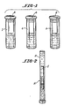
- the patient's blood can be placed in three separate reagent wells, or containers, a negative test control container 2, a sampling container 4, and a positive test control container 6, each of which has a stoppered closure 8 through which the blood mayu be transferred.
- the stain can be added separately or may be precoated into the interior of the three containers.
- the negative test container 2 has the antigen medium in it, but no antigens.
- the blood is added to the antigen medium, incubated therein, the same as with the test sample, and the colorant or fluorescent is added.
- the blood sample is then centrifuged in a centrifuge tube 10 containing a plastic float 12 which restricts the space acailable for the lymphocyte/monocyte cell band 14. This band is then examined for evidence of highlighting. If any is observed, the veracity of a like result in the test sample will be open to question, since lymphocyte activation can spontaneously occur in a blood sample. If no color change is seen, then the veracity of the sample results are confirmed, ie, it is confirmed that no spontaneous lymphoblasts are present in the blood sample.
- the positive test container 6 is used to mix the blood sample with either an antigen (as described above) from infectious agents, which are known to activate lymphocytes in the vast majority of normal patients.
- the positive incubation mixture is then centrifuged as shown in FIG. 2with a colorant or fluorescent that has an affinity for the yeast-induced lymphoblasts which will form in the incubated blood sample.
- the presence or absence of highlighted lymphoblasts in the cell band 14 will then be noted. If they are present, then the susceptability of the patient's blood to the test is confirmed.
- the physician can confirm that the patient is not susceptable to the test, and a protocol maybe used to determine the reason for the lack of a positive response to the disease antigens being tested for, and the negative response to the tested antigen will not be considered indicative of lack of previous exposure and sensitisation to the same.
- the utility of the test on the patient can thus be confirmed by the patient's positive response to the positive control and negative response to the negative control.
- This invention differs from previous procedure in the following ways:
- the invention's testing methodology may be useful in the following conditions: diagnosing the cause of delayed hypersensitivity reactions such as eczema or contact dermatitis; diagnosing certain viral infections during the window period prior to measurable antibody response; following the course of autoimmune disease; and in the diagnosis of the presence of tumor antigens or as a measure of anti-tumor immunity.
- the test can also be performed with a panel of antigens for example, antigens from several species of malaria. If a positive reaction is seen, then individual species tests can be performed to identify the species of malaria involved. Panel testing of allergens can also be performed with subsequent specific tests.
Landscapes
- Health & Medical Sciences (AREA)
- Life Sciences & Earth Sciences (AREA)
- Immunology (AREA)
- Engineering & Computer Science (AREA)
- Hematology (AREA)
- Chemical & Material Sciences (AREA)
- Molecular Biology (AREA)
- Cell Biology (AREA)
- Urology & Nephrology (AREA)
- Biomedical Technology (AREA)
- Analytical Chemistry (AREA)
- Biochemistry (AREA)
- Biotechnology (AREA)
- Pathology (AREA)
- Tropical Medicine & Parasitology (AREA)
- Food Science & Technology (AREA)
- Medicinal Chemistry (AREA)
- Physics & Mathematics (AREA)
- General Physics & Mathematics (AREA)
- Microbiology (AREA)
- General Health & Medical Sciences (AREA)
- Virology (AREA)
- Zoology (AREA)
- Physiology (AREA)
- Investigating Or Analysing Biological Materials (AREA)
- Measuring Or Testing Involving Enzymes Or Micro-Organisms (AREA)
- Peptides Or Proteins (AREA)
- Investigating Or Analysing Materials By The Use Of Chemical Reactions (AREA)
- Medicines Containing Antibodies Or Antigens For Use As Internal Diagnostic Agents (AREA)
- Steroid Compounds (AREA)
Abstract
Description
- This invention relates to a procedure for determining whether a patient has been previously exposed to certain antigens, and more particularly, to antigens indicative of diseases, infections, allergies or the like maladies.
- When diagnosing patients for possible illness or the like, the physician will typically perform blood tests to determine the presence or absence of agents in the blood which are indicative of prior exposure to various diseases or other maladies. Traditional tests to determine a patient's previous exposure to antigens, generally infectious agents, have usually relied upon tests for the presence or absence of a circulating humoral antibody directed against the proposed antigen. An example of such tests include blood tests to detect: immunity to Rubella (German measles); and a multitude of other viral and bacterial antigens. The means of detecting the antibody are many and include: latex agglutination in which the agglutination or lack of agglutination of antigen-coated latex particles is observed; enzyme linked immuno substrate assay (ELISA) in which the development of a color is observed either visually or spectrophotometrically; radioimmunoassay in which the measurement of radioactivity is measured; liposome technology in which the presence and intensity (or absence) of a color or fluorescent dye contained in the liposomes is detected; radio immuno sorbant assay (RAST) in which the measurement of radioactivity adherent to a solid substrate is measured; and indirect immunofluorescence in which the immobilized antigen to which the antibody to be detected is allowed to react with the specimen being tested, the antigen being washed to remove non-specifically adherent antibodies, and a fluorescently tagged antibody directed against the class of the antibody being tested for is applied, and the presence or absence of fluorescence is determined; as well as other methodologies. Antibodies, when present, can be titered, and their immune globulin class (of most interest are: IgM, signifying acute exposure; IgG, signifying past exposure; and IgE, signifying an allergic state) can be determined thereby determining the subject's previous exposure to the given antigen. All of the above tests are measurements of the function of a group of lymphocytes called B-lymphocytes. In cases, or disease processes, where little or no antibody response is made, the above tests will fail to determine the subject's previous exposure.
- Many diseases and clinical states, however, may not result in detectable humoral antibodies, ie, a B-lymphocyte response, but do in fact produce cellular immunity, ie, a T-lymphocyte response. Examples of such diseases and clinical conditions where the T-lymphocyte response predominates include many parasitic diseases, tuberculosis, salmonellosis, gonorrhea, fungal infections, rickettsial infections, and Lyme disease. As recently reported by Dattwyler et al (New England Journal; of Medicine, Vol. 319, at 1441, 1988) a significant proportion of patients, including some who have received some antibiotic therapy early in the course of the disease, do not produce detectable antibodies to the Lyme disease spirochete, but do have a measurable T-lymphocyte response.
- There are two general ways for determining whether a T-lymphocyte response has taken place in the blood due to prior exposure to diseases, infectious agents, or the like. The first procedure relies on the presence of morphologic cell changes which arise from prior exposure. Previously exposed T-lymphocytes will assume blast-like characteristics when re-exposed to the particular antigens. For example, the T-lymphocytes will develop a larger than normal nucleus and an abundant basophilic cytoplasm. Biochemical changes indicative of activation will also occur, such as: increased turnover of membrane phospholipids; increased synthesis of RNA and proteins; changes in intra-cellular Ca⁺⁺ concentration; expression of surface receptor for T-cell growth factor interleukin-2; and increase in the incorporation 3H-thymidine, as well as other events summarized in a recent article by Chatila, T. et al (New England Medical Journal, Vol. 320, page 696, 1989).
- The method most used in the past to assay T-lymphoblasts, and the one used by Dattwyler et al in their study of Lyme disease, to detect the presence or absence of a specific T-lymphocyte response to a given antigen is the tritiated thymidine uptake assay (3H-thymidine uptake assay). This assay is cumbersome, requires the use of radioactive isotopes, and requires the isolation of relatively pure populations of lymphocytes, and also takes several days to complete.
- The invention relates to a simple technique for detecting the presence or absence of previously exposed T-lymphocytes in a patient's blood. The patient's whole blood is incubated with the antigen for the suspected malady being diagnosed, which antigen is dispersed in a sutable pH-regulated medium, such as a buffered saline solution, for example. This incubation will cause previously exposed lymphocytes to become activated and eventually to assume the blast morphology, ie, to become lymphoblasts. After a suitable incubation period, a fluorescent dye, such as the acetomethylester of the calcium-sensitive dye fura-2, or other colorant having an affinity for activated lymphocytes or lymphoblasts is added to the blood sample. The dye can obtain its attraction to the activated lymphocytes or resultant lymphoblasts through chemical means, ie, by being combinable with the excess calcium ions, or by other means, such as by being tagged with a fluorescently-tagged antibody specific to the transferrin, Leu-23, the surface receptor for T-cell growth factor interleukin-2, or the like. The addition of the dye, colorant, or fluorescently tagged antibody will differentially highlight any responding lymphocytes or lymphoblasts in the blood sample. It should be noted that the highlighting additive may bind to responding lymphocytes, either before or after they have become true lymphoblasts. Pre-sensitized lymphocytes may therefore exhibit the characteristics which attract the additive before complete morphological changes occur. The treated blood is then centrifuged in a transparent tube having a float therein which will concentrate all of the lymphocytes, including any lymphoblasts, in a small area in the tube. The latter cell concentrating technique is disclosed in U.S. Patent No. 4,027,660 granted June 7, 1977 to Stephen Clark Wardlaw et al. The tube can be a capillary tube, or a pre-evacuated larger tube. After the cell concentration has been performed, the lymphocyte layer is examined to detect coloration or flourescence levels which are indicative of the presence of lymphoblasts in the lymphocyte layer. The detection of such color or fluorescence characteristics can be performed with an instrument such as those disclosed in U.S. Patents Nos. 4,156,570 granted May, 1978 and 4,558,947, granted December, 1985 both to Stephen C. Wardlaw.
- It is therefore an object of this invention to provide an improved precedure and paraphenalia for diagnosing prior patient exposure to a suspect antigen which sensitizes lymphocytes in the patient's blood.
- It is a further object of this invention to provide a procedure of the character described wherein an optically detectable indicator is added to the blood sample to optically highlight responding lymphocytes in the blood sample.
- It is an additional object of this invention to provide a procedure of the character described wherein the sensitized lymphocytes are T-lymphocytes.
- It is another object of this invention to provide a procedure of the character described wherein the blood sample is centrifuged in a transparent tube to concentrate the lymphocytes in a band which is then optically analysed for indication of highlighting.
- These and other objects and advantages will become more readily apparent from the following detailed description of a preferred embodiment of the invention when considered in connection with the accompanying drawings, in which:
- FIG. 1 is a perspective view of three sample tubes used in a kit for performing the procedure of the invention; and
- FIG. 2 is a perspective view of a centrifuge tube showing layering out of the blood cells for examination for optical highlighting.
- The invention admits to the inclusion of negative and positive controls. A negative control is used to determine that the T-lymphcytes do not respond to non-specific stimuli. The positive control is used to determine whether the patient's lymphocytes are capable of responding to antigens either singly or in combination that the physician knows that the patient has been exposed to in the past such as tetanus toxoid, diphtheria toxoid, monslia, or chemicals known to activate lymphocytes, such as phytohemaggluttimins, aluminum fluoride, or the like.
- The patient's blood can be placed in three separate reagent wells, or containers, a negative
test control container 2, asampling container 4, and a positive test control container 6, each of which has a stopperedclosure 8 through which the blood mayu be transferred. The stain can be added separately or may be precoated into the interior of the three containers. - The
negative test container 2 has the antigen medium in it, but no antigens. The blood is added to the antigen medium, incubated therein, the same as with the test sample, and the colorant or fluorescent is added. The blood sample is then centrifuged in acentrifuge tube 10 containing aplastic float 12 which restricts the space acailable for the lymphocyte/monocyte cell band 14. This band is then examined for evidence of highlighting. If any is observed, the veracity of a like result in the test sample will be open to question, since lymphocyte activation can spontaneously occur in a blood sample. If no color change is seen, then the veracity of the sample results are confirmed, ie, it is confirmed that no spontaneous lymphoblasts are present in the blood sample. - The positive test container 6 is used to mix the blood sample with either an antigen (as described above) from infectious agents, which are known to activate lymphocytes in the vast majority of normal patients. The positive incubation mixture is then centrifuged as shown in FIG. 2with a colorant or fluorescent that has an affinity for the yeast-induced lymphoblasts which will form in the incubated blood sample. The presence or absence of highlighted lymphoblasts in the
cell band 14 will then be noted. If they are present, then the susceptability of the patient's blood to the test is confirmed. If none is noted, then the physician can confirm that the patient is not susceptable to the test, and a protocol maybe used to determine the reason for the lack of a positive response to the disease antigens being tested for, and the negative response to the tested antigen will not be considered indicative of lack of previous exposure and sensitisation to the same. - The utility of the test on the patient can thus be confirmed by the patient's positive response to the positive control and negative response to the negative control.
- This invention differs from previous procedure in the following ways:
- 1. It, unlike the individual cell counters, which determine the fluorescent signal from each individual cell, utilizes the integrated fluorescence of the entire population of packed lymphocytes (several hundred thousands contained in the lymphocyte/monocyte band of a centrifuged capillary tube containing 110 micro liters of blood);
- 2. It does not require prior purification and isolation of the lymphocytes and may therefore be performed on whole blood (anticoagulated);
- 3. It does not require the use of any radioactive isotopes; and
- 4. The use of an integral negative and positive control together with a predetermined amount of antigen in a combined test-pack, together with software interpreting the result, permit its easy use by non-research trained personnel.
- The potential utility of an easily performed test of T-lymphocyte response to an antigen cannot be over-emphasized. In addition to possible aiding in the diagnosis of the diseases listed previously, the invention's testing methodology may be useful in the following conditions: diagnosing the cause of delayed hypersensitivity reactions such as eczema or contact dermatitis; diagnosing certain viral infections during the window period prior to measurable antibody response; following the course of autoimmune disease; and in the diagnosis of the presence of tumor antigens or as a measure of anti-tumor immunity.
- The test can also be performed with a panel of antigens for example, antigens from several species of malaria. If a positive reaction is seen, then individual species tests can be performed to identify the species of malaria involved. Panel testing of allergens can also be performed with subsequent specific tests.
- The invention has been described in conjunction with human patients, but it could also be used on animals.
- Since many changes and variations in the disclosed embodiment of the invention may be made without departing from the inventive concept, it is not intended to limit the invention otherwise than as required by the appended claims.
Claims (20)
Applications Claiming Priority (2)
| Application Number | Priority Date | Filing Date | Title |
|---|---|---|---|
| US34024889A | 1989-04-19 | 1989-04-19 | |
| US340248 | 1999-07-01 |
Publications (3)
| Publication Number | Publication Date |
|---|---|
| EP0393334A2 true EP0393334A2 (en) | 1990-10-24 |
| EP0393334A3 EP0393334A3 (en) | 1991-09-11 |
| EP0393334B1 EP0393334B1 (en) | 1996-07-24 |
Family
ID=23332522
Family Applications (1)
| Application Number | Title | Priority Date | Filing Date |
|---|---|---|---|
| EP90103895A Expired - Lifetime EP0393334B1 (en) | 1989-04-19 | 1990-02-28 | Determination of lymphocyte reactivity to specific antigens in blood |
Country Status (16)
| Country | Link |
|---|---|
| US (2) | US5360719A (en) |
| EP (1) | EP0393334B1 (en) |
| JP (1) | JPH0812194B2 (en) |
| CN (1) | CN1046600A (en) |
| AT (1) | ATE140796T1 (en) |
| AU (1) | AU624221B2 (en) |
| BR (1) | BR9001808A (en) |
| CA (1) | CA2011099A1 (en) |
| DE (1) | DE69027882T2 (en) |
| DK (1) | DK0393334T3 (en) |
| ES (1) | ES2091771T3 (en) |
| FI (1) | FI901791A0 (en) |
| GR (1) | GR3021147T3 (en) |
| NO (1) | NO178647C (en) |
| RU (1) | RU2061241C1 (en) |
| UA (1) | UA24003A (en) |
Cited By (2)
| Publication number | Priority date | Publication date | Assignee | Title |
|---|---|---|---|---|
| WO1994014064A1 (en) * | 1992-12-09 | 1994-06-23 | University Of Guelph | Methodology for developing a superior line of domesticated animals |
| CN103344639A (en) * | 2013-07-11 | 2013-10-09 | 山东大学 | Detector tube for quickly detecting pyrrole adduct in urine |
Families Citing this family (15)
| Publication number | Priority date | Publication date | Assignee | Title |
|---|---|---|---|---|
| GB9503406D0 (en) * | 1995-02-21 | 1995-04-12 | Haaheim Lars R | Detection of antibody production |
| CA2253965C (en) * | 1997-11-22 | 2003-01-21 | Robert A. Levine | Method for the detection, identification, enumeration and confirmation of circulating cancer cells and/or hematologic progenitor cells in whole blood |
| US5948686A (en) * | 1998-03-07 | 1999-09-07 | Robert A. Leuine | Method for performing blood cell counts |
| GB9913819D0 (en) | 1999-06-14 | 1999-08-11 | Plasmacute As | Assay |
| US7074577B2 (en) * | 2002-10-03 | 2006-07-11 | Battelle Memorial Institute | Buffy coat tube and float system and method |
| US20130084579A1 (en) * | 2002-10-03 | 2013-04-04 | Battelle Memorial Institute | Drug susceptibility using rare cell detection system |
| US20130084594A1 (en) * | 2002-10-03 | 2013-04-04 | Battelle Memorial Institute | Drug susceptibility using rare cell detection system |
| GB0325483D0 (en) * | 2003-10-31 | 2003-12-03 | Plasmacute As | Assay |
| CA2565981C (en) * | 2004-05-13 | 2015-02-17 | Advanced Animal Diagnostics | Microfluidic device and leucocyte antigen mediated microfluidic assay |
| EP2005155B1 (en) * | 2006-03-24 | 2021-06-30 | Advanced Animal Diagnostics, Inc. | Mastitis assay employing a microfluidic chamber assembly |
| EP2274612B1 (en) * | 2008-04-02 | 2016-10-26 | Abbott Point Of Care, Inc. | Virtual separation of bound and free label in a ligand assay for performing immunoassays of biological fluids, including whole blood |
| US20090258371A1 (en) * | 2008-04-09 | 2009-10-15 | Abbott Point Of Care, Inc. | Method of detecting very low levels of analyte within a thin film fluid sample contained in a thin thickness chamber |
| US20110097816A1 (en) * | 2009-10-23 | 2011-04-28 | Goodwin Paul C | Methods for changing densities of non-target particles of a suspension |
| CA2818173C (en) | 2010-11-30 | 2022-05-03 | Genentech, Inc. | Low affinity blood brain barrier receptor antibodies and uses therefor |
| RU2609839C1 (en) * | 2016-03-30 | 2017-02-06 | Федеральное государственное бюджетное учреждение "Государственный научный центр Институт иммунологии" Федерального медико-биологического агентства России (ФГБУ "ГНЦ Институт иммунологии" ФМБА России) | Method of differential diagnostics of hypersensitivity to bee poison (apis mellifera) |
Citations (8)
| Publication number | Priority date | Publication date | Assignee | Title |
|---|---|---|---|---|
| US3988115A (en) * | 1975-09-18 | 1976-10-26 | Modabber Farrokh Z | Diagnostic method for determining pathological condition by antigen-combining capacity of lymphocytes |
| US4027660A (en) * | 1976-04-02 | 1977-06-07 | Wardlaw Stephen C | Material layer volume determination |
| SU1237979A1 (en) * | 1981-02-11 | 1986-06-15 | Киевский Научно-Исследовательский Рентгенорадиологический И Онкологический Институт | Method of quantitative determining of activation of lymphoid cells |
| WO1986005188A1 (en) * | 1985-03-05 | 1986-09-12 | Dana-Farber Cancer Institute, Inc. | Monoclonal antibodies and assay |
| EP0221768A2 (en) * | 1985-10-29 | 1987-05-13 | Dana-Farber Cancer Institute, Inc. | Detection of T-cell activation |
| WO1988001385A1 (en) * | 1986-08-12 | 1988-02-25 | Anderson Jeffrey E | Method and apparatus for automated assessment of the immunoregulatory status of the mononuclear leukocyte immune system |
| WO1988007549A1 (en) * | 1987-03-26 | 1988-10-06 | Yale University | T-cell membrane protein |
| EP0325489A1 (en) * | 1988-01-21 | 1989-07-26 | Becton, Dickinson and Company | Leu 23: Monoclonal antibody for monitoring leukocyte activation |
Family Cites Families (15)
| Publication number | Priority date | Publication date | Assignee | Title |
|---|---|---|---|---|
| US4252538A (en) * | 1979-03-02 | 1981-02-24 | Engineering & Research Associates, Inc. | Apparatus and method for antibody screening, typing and compatibility testing of red blood cells |
| JPS5715698A (en) * | 1980-06-30 | 1982-01-27 | Nippon Electric Co | Method of boring ceramic green sheet |
| US4436824A (en) * | 1981-06-09 | 1984-03-13 | Ortho Diagnostic Systems, Inc. | Leukocyte migration through antigen containing agaroses for immunocompetence testing |
| US4717660A (en) * | 1984-01-26 | 1988-01-05 | Becton, Dickinson And Company | Detection of bacteria by fluorescent staining in an expanded buffy coat |
| US4727020A (en) * | 1985-02-25 | 1988-02-23 | Becton, Dickinson And Company | Method for analysis of subpopulations of blood cells |
| US4689309A (en) * | 1985-09-30 | 1987-08-25 | Miles Laboratories, Inc. | Test device, method of manufacturing same and method of determining a component in a sample |
| US4695553A (en) * | 1985-11-01 | 1987-09-22 | Becton Dickinson And Co., Inc. | Method for increasing agglutination of groups of cells to produce improved cell layer interface in centrifuged blood sample using antibodies |
| NZ214400A (en) * | 1985-12-02 | 1989-05-29 | Univ Otago | Detecting infection in ruminant including conducting assay of mononuclear cell function |
| JP2642112B2 (en) * | 1986-03-06 | 1997-08-20 | コモンウエルス サイエンテイフイック アンド インダストリアル リサーチ オーガナイゼイシヨン | In vitro assays for detecting cellular immune responses |
| US5006459A (en) * | 1986-03-31 | 1991-04-09 | T Cell Sciences, Inc. | Therapeutic and diagnostic methods using soluble T cell surface molecules |
| EP0294216A3 (en) * | 1987-06-05 | 1990-05-30 | Interferon Sciences, Inc. | A functional assay for t-lymphocytes |
| SU1691751A1 (en) * | 1988-11-09 | 1991-11-15 | Ленинградский педиатрический медицинский институт | Method for diagnosis of allergy |
| SU1649445A1 (en) * | 1988-12-09 | 1991-05-15 | Научно-исследовательский институт физико-химической медицины | Method for estimation of organism sensitivity for pollen allergens |
| CA2074720C (en) * | 1990-01-26 | 2002-07-23 | Martin A. Cheever | Immune reactivity to expressed activated oncogenes for diagnosis and treatment of malignancy |
| US5215102A (en) * | 1991-10-25 | 1993-06-01 | La Mina Ltd. | Capillary blood antigen testing apparatus |
-
1990
- 1990-02-26 CA CA002011099A patent/CA2011099A1/en not_active Abandoned
- 1990-02-27 AU AU50178/90A patent/AU624221B2/en not_active Ceased
- 1990-02-28 DK DK90103895.0T patent/DK0393334T3/en active
- 1990-02-28 EP EP90103895A patent/EP0393334B1/en not_active Expired - Lifetime
- 1990-02-28 ES ES90103895T patent/ES2091771T3/en not_active Expired - Lifetime
- 1990-02-28 DE DE69027882T patent/DE69027882T2/en not_active Expired - Fee Related
- 1990-02-28 AT AT90103895T patent/ATE140796T1/en active
- 1990-03-06 JP JP2054789A patent/JPH0812194B2/en not_active Expired - Lifetime
- 1990-04-09 FI FI901791A patent/FI901791A0/en not_active Application Discontinuation
- 1990-04-11 CN CN90102029A patent/CN1046600A/en active Pending
- 1990-04-18 UA UA4743719A patent/UA24003A/en unknown
- 1990-04-18 NO NO901700A patent/NO178647C/en unknown
- 1990-04-18 BR BR909001808A patent/BR9001808A/en not_active Application Discontinuation
- 1990-04-18 RU SU904743719A patent/RU2061241C1/en active
-
1992
- 1992-09-30 US US07/954,440 patent/US5360719A/en not_active Expired - Lifetime
-
1993
- 1993-01-21 US US08/006,766 patent/US5480778A/en not_active Expired - Fee Related
-
1996
- 1996-09-25 GR GR960402513T patent/GR3021147T3/en unknown
Patent Citations (8)
| Publication number | Priority date | Publication date | Assignee | Title |
|---|---|---|---|---|
| US3988115A (en) * | 1975-09-18 | 1976-10-26 | Modabber Farrokh Z | Diagnostic method for determining pathological condition by antigen-combining capacity of lymphocytes |
| US4027660A (en) * | 1976-04-02 | 1977-06-07 | Wardlaw Stephen C | Material layer volume determination |
| SU1237979A1 (en) * | 1981-02-11 | 1986-06-15 | Киевский Научно-Исследовательский Рентгенорадиологический И Онкологический Институт | Method of quantitative determining of activation of lymphoid cells |
| WO1986005188A1 (en) * | 1985-03-05 | 1986-09-12 | Dana-Farber Cancer Institute, Inc. | Monoclonal antibodies and assay |
| EP0221768A2 (en) * | 1985-10-29 | 1987-05-13 | Dana-Farber Cancer Institute, Inc. | Detection of T-cell activation |
| WO1988001385A1 (en) * | 1986-08-12 | 1988-02-25 | Anderson Jeffrey E | Method and apparatus for automated assessment of the immunoregulatory status of the mononuclear leukocyte immune system |
| WO1988007549A1 (en) * | 1987-03-26 | 1988-10-06 | Yale University | T-cell membrane protein |
| EP0325489A1 (en) * | 1988-01-21 | 1989-07-26 | Becton, Dickinson and Company | Leu 23: Monoclonal antibody for monitoring leukocyte activation |
Non-Patent Citations (1)
| Title |
|---|
| DATABASE WPIL / DERWENT, Abstr.no. 87-036032 & SU-A-1237979 (GRINEVICH Y.A. et al.) 15 JUNE 1986 * |
Cited By (3)
| Publication number | Priority date | Publication date | Assignee | Title |
|---|---|---|---|---|
| WO1994014064A1 (en) * | 1992-12-09 | 1994-06-23 | University Of Guelph | Methodology for developing a superior line of domesticated animals |
| CN103344639A (en) * | 2013-07-11 | 2013-10-09 | 山东大学 | Detector tube for quickly detecting pyrrole adduct in urine |
| CN103344639B (en) * | 2013-07-11 | 2015-10-28 | 山东大学 | The detector tube of pyrroles's adduct in a kind of quick detection urine |
Also Published As
| Publication number | Publication date |
|---|---|
| NO178647C (en) | 1996-05-02 |
| AU5017890A (en) | 1990-10-25 |
| RU2061241C1 (en) | 1996-05-27 |
| UA24003A (en) | 1998-08-31 |
| US5360719A (en) | 1994-11-01 |
| BR9001808A (en) | 1991-06-11 |
| ATE140796T1 (en) | 1996-08-15 |
| ES2091771T3 (en) | 1996-11-16 |
| EP0393334B1 (en) | 1996-07-24 |
| DE69027882D1 (en) | 1996-08-29 |
| FI901791A0 (en) | 1990-04-09 |
| EP0393334A3 (en) | 1991-09-11 |
| DK0393334T3 (en) | 1996-08-26 |
| AU624221B2 (en) | 1992-06-04 |
| NO901700L (en) | 1990-10-22 |
| JPH02296150A (en) | 1990-12-06 |
| DE69027882T2 (en) | 1996-12-12 |
| CA2011099A1 (en) | 1990-10-19 |
| NO178647B (en) | 1996-01-22 |
| CN1046600A (en) | 1990-10-31 |
| NO901700D0 (en) | 1990-04-18 |
| JPH0812194B2 (en) | 1996-02-07 |
| GR3021147T3 (en) | 1996-12-31 |
| US5480778A (en) | 1996-01-02 |
Similar Documents
| Publication | Publication Date | Title |
|---|---|---|
| US5480778A (en) | Determination of lymphocyte reactivity to specific antigens in blood | |
| Brown et al. | Simultaneous determination of total IgE and allergen-specific IgE in serum by the MAST chemiluminescent assay system. | |
| Voller et al. | Enzyme immunoassays in diagnostic medicine: theory and practice | |
| Naot et al. | IgM enzyme-linked immunosorbent assay test for the diagnosis of congenital Toxoplasma infection | |
| US4770853A (en) | Device for self contained solid phase immunodiffusion assay | |
| EP0216191B1 (en) | Immunoassay for htlv-iii antigens | |
| US5759794A (en) | Assay of blood or other biologic samples for target analytes | |
| US4639419A (en) | Immunological color change test involving two differently colored reagent spots | |
| US4499183A (en) | Detecting intracellular antigens in swelled and fixed cells with labeled antibody | |
| AU621310B2 (en) | Immunometric assay kit and method applicable to whole cells | |
| US5773232A (en) | Methods for measurement of lymphocyte function | |
| EP0194156B1 (en) | Method of measuring the amount of immune antibody in serum | |
| US6046013A (en) | Process for identifying specific antibodies associated with HLA | |
| US6461825B1 (en) | Immunometric assay kit and method applicable to whole cells | |
| Rossi | A simple, rapid enzyme-linked immunosorbent assay for evaluating immunoglobulin G antibody avidity in toxoplasmosis | |
| Luger | Diagnosis of syphilis | |
| CA1083956A (en) | Microbial antibodies immunoassay | |
| US7169571B2 (en) | Methods for measurement of lymphocyte function | |
| Stone et al. | Capture-S, a nontreponemal solid-phase erythrocyte adherence assay for serological detection of syphilis | |
| Cook et al. | Evaluation of latex-based heterophile antibody assay for diagnosis of acute infectious mononucleosis | |
| US3887697A (en) | Hemagglutination method for australia antigen and antibody thereto | |
| US3733398A (en) | Determining and reversing anticomplementary activity in complement fixation test for australia antigen | |
| KR20220018149A (en) | Detection method of latent tuberculosis infection using inline microtube set | |
| SU1425549A1 (en) | Method of radioimmunological analysis of antigens | |
| Friedman et al. | Comparison of complement fixation and fluorescent immunoassay (FIAX) for measuring antibodies to cytomegalovirus and herpes simplex virus |
Legal Events
| Date | Code | Title | Description |
|---|---|---|---|
| PUAI | Public reference made under article 153(3) epc to a published international application that has entered the european phase |
Free format text: ORIGINAL CODE: 0009012 |
|
| AK | Designated contracting states |
Kind code of ref document: A2 Designated state(s): AT BE CH DE DK ES FR GB GR IT LI LU NL SE |
|
| PUAL | Search report despatched |
Free format text: ORIGINAL CODE: 0009013 |
|
| AK | Designated contracting states |
Kind code of ref document: A3 Designated state(s): AT BE CH DE DK ES FR GB GR IT LI LU NL SE |
|
| 17P | Request for examination filed |
Effective date: 19920210 |
|
| 17Q | First examination report despatched |
Effective date: 19940112 |
|
| GRAH | Despatch of communication of intention to grant a patent |
Free format text: ORIGINAL CODE: EPIDOS IGRA |
|
| GRAH | Despatch of communication of intention to grant a patent |
Free format text: ORIGINAL CODE: EPIDOS IGRA |
|
| GRAA | (expected) grant |
Free format text: ORIGINAL CODE: 0009210 |
|
| AK | Designated contracting states |
Kind code of ref document: B1 Designated state(s): AT BE CH DE DK ES FR GB GR IT LI LU NL SE |
|
| REF | Corresponds to: |
Ref document number: 140796 Country of ref document: AT Date of ref document: 19960815 Kind code of ref document: T |
|
| REG | Reference to a national code |
Ref country code: CH Ref legal event code: NV Representative=s name: BUECHEL, VON REVY & PARTNER |
|
| REG | Reference to a national code |
Ref country code: DK Ref legal event code: T3 |
|
| REF | Corresponds to: |
Ref document number: 69027882 Country of ref document: DE Date of ref document: 19960829 |
|
| ET | Fr: translation filed | ||
| ITF | It: translation for a ep patent filed | ||
| REG | Reference to a national code |
Ref country code: ES Ref legal event code: FG2A Ref document number: 2091771 Country of ref document: ES Kind code of ref document: T3 |
|
| REG | Reference to a national code |
Ref country code: GR Ref legal event code: FG4A Free format text: 3021147 |
|
| PG25 | Lapsed in a contracting state [announced via postgrant information from national office to epo] |
Ref country code: LU Free format text: LAPSE BECAUSE OF NON-PAYMENT OF DUE FEES Effective date: 19970228 |
|
| PLBE | No opposition filed within time limit |
Free format text: ORIGINAL CODE: 0009261 |
|
| STAA | Information on the status of an ep patent application or granted ep patent |
Free format text: STATUS: NO OPPOSITION FILED WITHIN TIME LIMIT |
|
| 26N | No opposition filed | ||
| PGFP | Annual fee paid to national office [announced via postgrant information from national office to epo] |
Ref country code: DK Payment date: 19980123 Year of fee payment: 9 |
|
| PGFP | Annual fee paid to national office [announced via postgrant information from national office to epo] |
Ref country code: GR Payment date: 19980130 Year of fee payment: 9 |
|
| PGFP | Annual fee paid to national office [announced via postgrant information from national office to epo] |
Ref country code: SE Payment date: 19990203 Year of fee payment: 10 Ref country code: AT Payment date: 19990203 Year of fee payment: 10 |
|
| PGFP | Annual fee paid to national office [announced via postgrant information from national office to epo] |
Ref country code: NL Payment date: 19990218 Year of fee payment: 10 |
|
| PG25 | Lapsed in a contracting state [announced via postgrant information from national office to epo] |
Ref country code: GR Free format text: LAPSE BECAUSE OF NON-PAYMENT OF DUE FEES Effective date: 19990228 |
|
| PG25 | Lapsed in a contracting state [announced via postgrant information from national office to epo] |
Ref country code: DK Free format text: LAPSE BECAUSE OF NON-PAYMENT OF DUE FEES Effective date: 19990301 |
|
| REG | Reference to a national code |
Ref country code: DK Ref legal event code: EBP |
|
| PG25 | Lapsed in a contracting state [announced via postgrant information from national office to epo] |
Ref country code: AT Free format text: LAPSE BECAUSE OF NON-PAYMENT OF DUE FEES Effective date: 20000228 |
|
| PG25 | Lapsed in a contracting state [announced via postgrant information from national office to epo] |
Ref country code: SE Free format text: LAPSE BECAUSE OF NON-PAYMENT OF DUE FEES Effective date: 20000301 |
|
| PG25 | Lapsed in a contracting state [announced via postgrant information from national office to epo] |
Ref country code: NL Free format text: LAPSE BECAUSE OF NON-PAYMENT OF DUE FEES Effective date: 20000901 |
|
| NLV4 | Nl: lapsed or anulled due to non-payment of the annual fee |
Effective date: 20000901 |
|
| EUG | Se: european patent has lapsed |
Ref document number: 90103895.0 |
|
| REG | Reference to a national code |
Ref country code: GB Ref legal event code: IF02 |
|
| PGFP | Annual fee paid to national office [announced via postgrant information from national office to epo] |
Ref country code: FR Payment date: 20020130 Year of fee payment: 13 |
|
| PGFP | Annual fee paid to national office [announced via postgrant information from national office to epo] |
Ref country code: DE Payment date: 20020131 Year of fee payment: 13 |
|
| PGFP | Annual fee paid to national office [announced via postgrant information from national office to epo] |
Ref country code: GB Payment date: 20020201 Year of fee payment: 13 Ref country code: CH Payment date: 20020201 Year of fee payment: 13 |
|
| PGFP | Annual fee paid to national office [announced via postgrant information from national office to epo] |
Ref country code: BE Payment date: 20020307 Year of fee payment: 13 |
|
| PGFP | Annual fee paid to national office [announced via postgrant information from national office to epo] |
Ref country code: ES Payment date: 20020311 Year of fee payment: 13 |
|
| PG25 | Lapsed in a contracting state [announced via postgrant information from national office to epo] |
Ref country code: LI Free format text: LAPSE BECAUSE OF NON-PAYMENT OF DUE FEES Effective date: 20030228 Ref country code: GB Free format text: LAPSE BECAUSE OF NON-PAYMENT OF DUE FEES Effective date: 20030228 Ref country code: CH Free format text: LAPSE BECAUSE OF NON-PAYMENT OF DUE FEES Effective date: 20030228 Ref country code: BE Free format text: LAPSE BECAUSE OF NON-PAYMENT OF DUE FEES Effective date: 20030228 |
|
| PG25 | Lapsed in a contracting state [announced via postgrant information from national office to epo] |
Ref country code: ES Free format text: LAPSE BECAUSE OF NON-PAYMENT OF DUE FEES Effective date: 20030301 |
|
| PG25 | Lapsed in a contracting state [announced via postgrant information from national office to epo] |
Ref country code: DE Free format text: LAPSE BECAUSE OF NON-PAYMENT OF DUE FEES Effective date: 20030902 |
|
| GBPC | Gb: european patent ceased through non-payment of renewal fee | ||
| REG | Reference to a national code |
Ref country code: CH Ref legal event code: PL |
|
| PG25 | Lapsed in a contracting state [announced via postgrant information from national office to epo] |
Ref country code: FR Free format text: LAPSE BECAUSE OF NON-PAYMENT OF DUE FEES Effective date: 20031031 |
|
| REG | Reference to a national code |
Ref country code: FR Ref legal event code: ST |
|
| REG | Reference to a national code |
Ref country code: ES Ref legal event code: FD2A Effective date: 20030301 |
|
| PG25 | Lapsed in a contracting state [announced via postgrant information from national office to epo] |
Ref country code: IT Free format text: LAPSE BECAUSE OF NON-PAYMENT OF DUE FEES;WARNING: LAPSES OF ITALIAN PATENTS WITH EFFECTIVE DATE BEFORE 2007 MAY HAVE OCCURRED AT ANY TIME BEFORE 2007. THE CORRECT EFFECTIVE DATE MAY BE DIFFERENT FROM THE ONE RECORDED. Effective date: 20050228 |