EP0390528A1 - Automated biopsy instrument - Google Patents
Automated biopsy instrument Download PDFInfo
- Publication number
- EP0390528A1 EP0390528A1 EP90303293A EP90303293A EP0390528A1 EP 0390528 A1 EP0390528 A1 EP 0390528A1 EP 90303293 A EP90303293 A EP 90303293A EP 90303293 A EP90303293 A EP 90303293A EP 0390528 A1 EP0390528 A1 EP 0390528A1
- Authority
- EP
- European Patent Office
- Prior art keywords
- cannula
- stylet
- guide
- casing
- instrument
- Prior art date
- Legal status (The legal status is an assumption and is not a legal conclusion. Google has not performed a legal analysis and makes no representation as to the accuracy of the status listed.)
- Granted
Links
Images


Classifications
-
- A—HUMAN NECESSITIES
- A61—MEDICAL OR VETERINARY SCIENCE; HYGIENE
- A61B—DIAGNOSIS; SURGERY; IDENTIFICATION
- A61B10/00—Other methods or instruments for diagnosis, e.g. instruments for taking a cell sample, for biopsy, for vaccination diagnosis; Sex determination; Ovulation-period determination; Throat striking implements
- A61B10/02—Instruments for taking cell samples or for biopsy
- A61B10/0233—Pointed or sharp biopsy instruments
- A61B10/0266—Pointed or sharp biopsy instruments means for severing sample
- A61B10/0275—Pointed or sharp biopsy instruments means for severing sample with sample notch, e.g. on the side of inner stylet
-
- A—HUMAN NECESSITIES
- A61—MEDICAL OR VETERINARY SCIENCE; HYGIENE
- A61B—DIAGNOSIS; SURGERY; IDENTIFICATION
- A61B10/00—Other methods or instruments for diagnosis, e.g. instruments for taking a cell sample, for biopsy, for vaccination diagnosis; Sex determination; Ovulation-period determination; Throat striking implements
- A61B10/02—Instruments for taking cell samples or for biopsy
- A61B10/0233—Pointed or sharp biopsy instruments
- A61B10/0283—Pointed or sharp biopsy instruments with vacuum aspiration, e.g. caused by retractable plunger or by connected syringe
-
- A—HUMAN NECESSITIES
- A61—MEDICAL OR VETERINARY SCIENCE; HYGIENE
- A61B—DIAGNOSIS; SURGERY; IDENTIFICATION
- A61B10/00—Other methods or instruments for diagnosis, e.g. instruments for taking a cell sample, for biopsy, for vaccination diagnosis; Sex determination; Ovulation-period determination; Throat striking implements
- A61B10/02—Instruments for taking cell samples or for biopsy
- A61B2010/0208—Biopsy devices with actuators, e.g. with triggered spring mechanisms
Definitions
- This invention relates generally to a biopsy instrument; viz., a device for removing a sample of tissue from a human being or an animal.
- biopsy has been for many years a favored method for the nonsurgical diagnosis of tissue.
- biopsy has been performed in a two-step manual technique employing a needle in which an outer hollow cutting cannula telescopically receives an inner stylet which is slidable between retracted and extended positions relative to the cannula.
- the physician places the tip of the needle (with the stylet retracted inside the cannula) against the tissue mass to be sampled and manually drives the stylet forward into the tissue mass.
- the stylet extends rearwardly therefrom and is fitted with a push-knob to facilitate the driving of the stylet in the forward direction.
- the physician manually drives the cutting cannula forwardly over the stylet, thereby severing a tissue sample and retaining it within the cannula's hollow interior.
- U.S. Pat. 4,667,684 issued May 26, 1987 to H. G. Leigh for "Biopsy Device" discloses a movable stylet telescopically received in a hollow movable cannula, both the stylet and the cannula being mounted to hubs within a pistol-style grip.
- the stylet is first manually advanced into the tissue and the cannula is then driven over the stylet by depressing a trigger.
- U.S. Pat. 4,699,154 issued October 13, 1987 to one Lindgren for "Tissue Sampling Device" discloses a complicated biasing mechanism in which a release button is depressed to cause a spring-loaded stylet to be advanced into the tissue mass. The forward movement of the stylet also triggers the delayed release of a spring-loaded outer cannula, which slides over the stylet to sever the tissue sample.
- the Roth Biopsy Needle marketed by Cook Urological, Inc., Spencer, Indiana, is a stylet which is manually adjustable between extended and retracted positions for reasons which will be apparent from the following description of its various methods of operation.
- the stylet is loaded from the rear of the instrument and extends behind the instrument at all times.
- the stylet is mounted to a U-shaped handle which is engageable with the body of the instrument in two positions corresponding to the extended and retracted stylet positions.
- a spring-loaded cannula is also movable between a charged or rear position and a discharged or advanced position.
- the physician first adjusts the stylet manually to its retracted position and moves the cannula manually to the charged position to expose the sharpened tip of the stylet. He then penetrates the tissue mass with the stylet and depresses a button-release to cause the cannula to be driven by the spring forwardly over the tip of the stylet to sever the tissue sample and retain it within the hollow tip of the cannula.
- the stylet of the Roth needle is movable, as mentioned above, and that the button-release is exposed at all times and therefore subject to inadvertent actuation.
- stylet and the associated U-shaped handle extend behind the instrument, the stylet and handle are always exposed and are therefore vulnerable to accidental impact or unintended forces, thereby creating a risk that the stylet may be inadvertently advanced into the body of the patient with possibly deleterious consequences.
- the physician proceeds as before, but after penetrating the tissue mass he manually advances the stylet to its extended position to expose the transverse slot or gap therein described above.
- the cannula is advanced, the tissue sample is severed and captured in the pocket formed by the slot.
- the physician may begin the procedure with the stylet in the extended position and the cannula in the discharged mode. Once the tissue has been penetrated, the cannula is manually retracted to expose the transverse slot.
- all stylets formed with such a tissue pocket become unstable when tooled to gauge sizes larger than 20 gauge (i.e., 21, 25 gauge).
- the bridge joining the stylet's proximal and distal ends at the tissue pocket becomes increasingly flexible, and therefore the stylet becomes physically unstable and its direction impossible to control with any precision when the slot or gap is exposed during penetration of the tissue mass by the stylet.
- certain areas of the body such as the pancreas, thyroid, and nonpleural-based intrapulmonary lesions are routinely biopsied with needles smaller than 20 gauge (i.e., 21 gauge) to reduce the incidence of hemorrhage and pneumothorax.
- the various automated biopsy instruments presently known tend to be heavy, difficult to manipulate, and incorporate biasing mechanisms which are either complicated in construction or require undue force to operate. Such limitations diminish the physician's control over the instrument and the precision with which biopsies may be performed. These instruments may be subject to inadvertent movement or torque which may, in turn, subject the patient to unnecessary trauma and risk. This is especially true of instruments which permit or require adjustment of the relative positions of any of their elements before the cannula is moved forward to sever the tissue sample. Similarly, the length of time required to perform a biopsy increases as the physician's degree of control of the instrument decreases, further elevating the risk to which the patient may be exposed. Finally, both physician and patient are exposed to the risk of inadvertent advancement of the cannula when the instrument is in its charged condition.
- the present invention is particularly concerned with an automated biopsy instrument in which a spring-loaded outer cannula is driven over an inner stationary stylet to obtain the tissue sample.
- the present invention provides a biopsy instrument which comprises stationary means for mounting a stylet on the instrument in fixed relation thereto so that the stylet, once mounted on the instrument, cannot be moved relative thereto, inadvertently or otherwise.
- a guide means is provided for mounting a cannula on the instrument and is reciprocably movable relative to the stationary means between a charged position, in which the cannula is retracted in a direction away from the distal end of the stylet, and a discharged position, in which the cannula is displaced from the charged position in the direction of the distal end of the stylet.
- Biasing means such as a coil spring urge the guide means toward the discharged position.
- the instrument according to the invention also includes manually operable charging means for moving the guide means to the charged position against the urging of the biasing means, and manually actuable release means for releasably retaining the guide means in the charged position against the urging of the biasing means.
- distal is used herein in its ordinary sense to mean “remote from the point of attachment”
- proximal is used hereinafter in its meaning of "at, adjacent to or near the point of attachment.”
- a shield means is provided which is disposed to block or prevent inadvertent actuation of the release means. More particularly, the charging means preferably includes the shield means and is movable between the safety position and a charged-ready position in which the shield means is displaced from the safety position to expose the release means for actuation.
- an automated biopsy instrument according to the invention is designated generally by the reference numeral 10.
- the instrument 10 comprises seven principal elements, which will be more fully described hereinbelow: an outer casing 12; an inner support rod 14; a coil spring 16; a biopsy spring guide 18; a safety cap 20; a release lever 22; and a needle 24.
- the outer casing 12 comprises an elongated hollow cylindrical tube 26 open at a rear end 28 thereof and substantially closed by an end wall 30 which is formed integrally with the tube 26 at the opposite or forward end 32 thereof.
- the open end 28 is closed by a cap 31.
- a cannula slot 34 in the end wall 30 communicates with a hollow interior 35 of the tube 26.
- the slot 34 extends radially from a center point of the end wall 30 to the periphery thereof.
- a cone-shaped depression 34a is formed in the interior face of the end wall 30 at the closed end of the slot 34, the broader end of the depression 34a opening toward the interior 35 of the tube 26.
- Access port 36a circumscribes an arc of approximately 90 degrees about the circumference of the outer casing 12, communicates with the cannula slot 34 at an intersection designated by the reference character 33, and extends symmetrically in either direction from the intersection 33.
- Access port 36b is diametrically opposite from port 36a and is identical therewith except that it does not communicate with the cannula slot 34.
- the outer casing 12 includes a release lever seat 38 provided by a rectangular aperture which is formed in the tube 26 and communicates with the interior 35 thereof.
- Seat 38 is positioned approximately at the longitudinally central portion of the tube 26 and in line with the cannula slot 34.
- the outer casing 12 is additionally provided with a pair of longitudinal guide slots 40 which are formed in the tube 26 and extend from the rear end 28 thereof toward the opposite or forward end 32.
- the guide slots 40 are disposed diametrically oppositely from one another and approximately midway between, or 90 degrees from, the access ports 36a, 36b.
- the internal support rod 14 comprises an elongated solid cylindrical shaft 42 joined to a circular or disc-shaped base 44.
- An anchor socket or clevis 46 is formed in the distal end of the shaft.
- the biopsy spring guide 18 is hollow and open at its opposite ends and comprises a forward cylinder 48 and a rear cylinder 50 joined coaxially therewith.
- the outside diameter of the forward cylinder 48 is smaller than that of the rear cylinder 50, the forward cylinder 48 communicating with and extending from the rear cylinder 50.
- An annular internal shoulder 52 is formed in the rear cylinder 50.
- the walls of the two cylinders are continuous, thereby forming an annular external shoulder 54.
- the exterior surface of the distal end of the forward cylinder is formed with a quarter-turn male thread 56.
- the needle 24 comprises a cannula mount 58 and a stylet 60.
- the cannula mount 58 comprises a cylindrical collar 62, a conical head 64, and a cutting cannula 66.
- the collar 62 is open at one end thereof and is closed at its opposite end by an end wall 68 pierced by a centrally disposed, narrow axial bore 69 (FIG. 2).
- An outer surface 70 of the collar 62 is provided with knurling 71 for improved grip, while an inner surface 72 is formed with a quarter-turn female thread 74 adapted to mate with the thread 56 of spring guide 18.
- the conical head 64 is secured to the end wall 68 over the axial bore 69, and is itself axially bored to communicate with the interior of the cylindrical collar 62 by way of bore 69, whereby the cutting cannula 66 is received by the head 64 and end wall 68 and is there secured in any suitable manner.
- the cutting cannula 66 comprises a hollow tube which is beveled to an appropriate angle at its distal end 76 (the particular angle depending on the style of cannula employed) and sharpened about its circumference at the distal end 76.
- the biopsy instrument 10 may interchangeably employ a needle having any one of several configurations well known in the art, including but not limited to those variously known by or marketed under the names Turner, Chiba, Franseen, Tip-cut, Menghini, and Bernardino/Sones.
- the biopsy instrument 10 may also employ a TRU-CUT needle, as discussed more fully hereinafter.
- the stylet 60 is sharpened to a point at its distal end 78 and is formed with an anchor 80 at its proximal end 82. It will be apparent that the stylet need not be formed with a tissue pocket or sampling gap; that is, a transverse slot located adjacent to the sharpened distal end 78, as will be discussed with greater particularity hereinafter.
- the safety cap 20 or shield means is a roughly bell-shaped hollow element open at both ends and surrounding the cylindrical tube 26.
- a web 84 thereof joins a narrow, hollow cylindrical forward section 86 to a hollow cylindrical rear section 88 of greater diameter than the forward section 86.
- the rear section 88 is formed with a series of circumferential ribs 90 for improved grip, and it will be noted that configuration of the safety cap 20 is such that it may be encirclingly engaged or grasped by the thumb and fingers of one hand.
- Integrally formed on an interior surface 92 of the forward section 86 of the safety cap, at a point where the forward section 86 and the web 84 merge, are a pair of oppositely disposed, elongated guide pins 94 of square cross section.
- the release lever 22 comprises a finger rest 96 and a mounting section 98 maintained in spaced parallel planes by a connecting web 100.
- the finger contacting surface of the finger rest 96 may be appropriately textured for improved grip.
- Mounting section 98 is formed with a latching projection 102 at one end of the release lever and is flexibly secured to the outer casing 12 by a spot weld 104 (FIG. 2).
- a biasing means in the form of the coil spring 16 is coaxially received over the cylindrical shaft 42 of the internal support rod 14 with a first end 16a of the spring bearing against the base 44 to which the shaft 42 is mounted.
- the rod 14 and the spring 16 are in turn telescopically received within the biopsy spring guide 18 with a second or opposite end 16b of the spring 16 bearing against the internal shoulder 52 of the spring guide.
- the support rod 14, the coil spring 16, and the spring guide 18 are telescopically received within the outer casing 12, and the subassembly so formed is then received within the safety cap 20 with the guide pins 94 thereof registering with the guide slots 40 in the outer casing 12.
- the safety cap 20 is slidable over the outer casing 12, the extent of forward movement thereof being limited by the length of the guide slots 40.
- the rear end 28 of the tube 26 is closed by the cap 31.
- the stylet 60 is telescopically or coaxially received within the cannula mount 58 to assemble the needle 24.
- the needle is then positioned within the hollow interior 35 of the tube 26 with the collar 62 of the cannula mount 58 aligned with the access ports 36a, 36b and the cannula 66 resting in the cannula slot 34.
- the anchor 80 of the stylet 60 is received and detachably engaged within the clevis 46 of the spring guide 18.
- the thread 74 on the inner surface 72 of the collar 62 is engaged with the thread 56 on the spring guide 18 to secure the cannula mount 58 to the spring guide.
- the fully assembled biopsy instrument 10 has three operating modes: namely, the discharged mode, the charged-safety-on mode, and the charged-ready mode.
- the discharged mode best shown in FIG. 4A, the safety cap 20 is positioned approximately at the forward end 32 of the tube 26 so that the forward section 84 of the safety cap covers the access ports 36a, 36b.
- the spring guide 18 is disposed at the forward end 32 of the tube 26 with the head 64 of the cannula mount 58 resting within the complementary cone-shaped depression 34a.
- the instrument 10 may be converted from the discharged mode to the charged-safety-on mode, best shown in FIG. 2, by retracting the safety cap 20 toward the rear end 28 of the tube 26.
- the configuration of the safety cap permits this to be done with equitable distribution of the retracting force applied by the hand of the physician: that is, unlike examples of the prior art, the retracting force is not concentrated at the tip of the thumb or one or two fingers.
- This action causes the guide pins 94 (FIG. 3) to engage the annular external shoulder 54 of the spring guide 18 to retract the spring guide toward the base 44 of the support rod 14 and compress the coil spring 16.
- the cannula 66 is retracted to expose the distal end portion of the stylet 60.
- a stationary means for mounting the stylet on the instrument 10 in fixed relation thereto has been provided; both the stylet 60 and the support rod 14 remain stationary.
- the safety cap 20 and the spring guide 18 are retracted in this manner until the latching projection 102 on the release lever 22 engages the annular external shoulder 54 of the spring guide, as depicted in FIG. 2.
- the rear section 88 of the safety cap 20 covers the release lever 22 to prevent accidental actuation of the instrument 10.
- the biopsy instrument 10 is loaded by registering the anchor 80 on the stylet 60 with the clevis 46.
- the cannula mount 58, sheathing the stylet 60, is secured to the spring guide 18 by mating the thread 56 on the spring guide 18 with the thread 74 on the inner surface 72 of the collar 62 and executing a quarter-turn to secure the mating engagement of the threads.
- the safety cap 20 is moved forward until the guide pins 94 have traversed the full extent of the guide slots 40 with the safety cap covering the access ports 36a, 36b, as in the discharged mode, and exposing the release lever 22.
- the safety cap 20 may be formed with a thumb hole, not shown in the drawings, positioned over the release lever 22. In this arrangement the safety cap need not be moved forward to expose the release lever 22 but rather the release lever may be depressed, as described below, by applying sufficient force to the area of the thumb hole to overcome the natural resilience of the material of construction).
- the biopsy instrument 10 is now ready for use. To obtain a biopsy, the location of the tissue to be sampled is located by one or more of a variety of methods, such as fluoroscopy, sonography, computed tomography, magnetic resonance imaging, or palpation. With the instrument 10 in the charged-safety-on-mode, the needle 24 is positioned in the tissue to be biopsied. If the operator has not already done so, he slides the safety cap 20 forward and actuates the instrument by depressing the finger rest 96 on the release lever 22, which is flexibly welded to the outer casing 12 as described hereinabove. Manipulation of the release lever raises the latching projection 102, disengaging it from the external shoulder 54 of the spring guide 18.
- This movement of the cannula causes its sharpened distal end 76 to cut and isolate a sample plug or core of tissue from the mass and retain it within the hollow interior of the cannula 66.
- the instrument 10 is now in the discharged mode (FIG. 4A), as described hereinabove, and the instrument 10 may now be withdrawn from the patient.
- the biopsy sample is extracted from the cannula by returning the instrument to the charged-safety-on mode, thereby retracting the cutting cannula 66 from over the end of the stylet 60 to eject the sample.
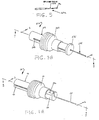
- stylet 60 is detachably affixed within the clevis 46, it may be interchanged with an alternative stylet 60a, shown fragmentarily in FIG. 9.
- the stylet 60a is identical to the stylet 60 except that the overall length of the stylet 60a is approximately one to two centimeters greater than that of the stylet 60, and the stylet 60a is provided with a transverse slot or sampling gap creating a tissue pocket 60b adjacent to the stylet's sharpened distal end 78a.
- This stylet is substantially similar to the TRU-CUT Needle marketed by Travenol Laboratories of Deerfield, Illinois.
- the stylet 60a may be used to perform the style of biopsy commonly referred to as the TRU-CUT method.
- the physician penetrates the mass of tissue to be sampled with the instrument 10 in the discharged mode. Because the stylet 60a is slightly longer than the stylet 60, the distal end 78a of the stylet 60a, unlike the distal end 78 of the stylet 60, projects slightly beyond the distal end of the cutting cannula 66 in the discharged mode. As the second step of the biopsy, the instru ment 10 is converted to the charged-safety-on mode by retracting the cannula 66 in the manner described hereinabove. This exposes the tissue pocket 60b of the stylet 60a.
- the physician actuates the instrument 10 by depressing the finger rest 96 of the release lever 22, which permits the energy of the compressed coil spring 16 to drive the cannula 66 forward over the stylet 60a to sever a tissue sample and retain it in the tissue pocket 60b until the instrument can be withdrawn from the patient.
- FIGS. 5 to 8A illustrate an alternative embodiment of the invention in the form of a biopsy instrument 210 which is intended to be disposed of after a single use or a limited number of uses during a single procedure (perhaps three to five), whereas the embodiment illustrated in FIGS. 1 to 4A, excluding the needle 24, is suitable to be reused indefinitely.
- FIGS. 1 to 4A excluding the needle 24, is suitable to be reused indefinitely.
- an automated biopsy instrument 210 comprises an outer casing 212, an inner support rod 214, a coil spring 216, a biopsy spring guide 218, a safety cap 220, a release lever 222, and a needle 224.
- the cylindrical tube 226 is preferably formed of materials such as polycarbonate, delrin, nylon, polyethylene, polyvinyl chloride, acrylic, or acrylonitrile-butadiene-styrene, and is open at its forward end 232 and closed at its rear end 228 by an end wall 230 formed integrally therewith.
- a circular cap 231 snap-fits over the tube 226 to close the open forward end 232.
- a cylindrical depression 234a (FIG. 6) is formed about the center of the interior face of the cap 231. An axial bore 234 through the center of the cap communicates with the depression 234a.
- the longitudinal guide slots 240 extend from the forward end 232 of the tube 226 toward the rear end 228, a recess 246 is formed in the distal end of the internal support rod 214, and the forward cylinder 248 is formed with an end wall 255 pierced by a bore 255a.
- the cannula mount 258 comprises a narrow base 262; a narrow cylindrical head 264 and a midsection 265 integrally joining the base and the head.
- the base 262, the head 264, and the midsection 265 are axially bored to receive the cutting cannula 266, which may there be secured by means of adhesive (not shown) and which comprises a hollow cylindrical tube beveled at its distal end 276 to an appropriate angle (the particular angle depending on the style of cannula employed) and sharpened about the circumference of the distal end.
- the mounting section 298 of the release lever 222 is flexibly solvent-bonded to the outer casing 212 at 304.
- Assembly of instrument 210 is quite similar to assembly of instrument 10, as described hereinabove. However, in this instance, the proximal end 282 of the stylet 262 is adhesively bonded within the recess 246, and the base 262 of the cannula mount 258 is similarly secured within the bore 255a.
- the internal support rod 214, the coil spring 216, the spring guide 218, and the needle 224 are telescopically received within the safety cap 220 and the outer casing 212, the guide pins 294 being received in the guide slots 240, and the cutting cannula 266 and the stylet 260 being received in the axial bore 234.
- the safety cap 220 is now slidable over the outer casing 212, the extent of forward movement being limited by the cap 231 which is applied to the tube 226 to close the forward end 232 thereof as well as the open ends of the guide slots 240.
- the assembled biopsy instrument 210 is operated in a discharged mode, a charged-safety-on mode, and a charged-ready mode.
- the discharged mode FIG. 8A
- the safety cap 220 is positioned near the forward end 232 of the tube 226 in engagement with the cap 231.
- the spring guide 218 is also disposed near the forward end of the tube 226 with the cylindrical head 264 of the cannula mount 258 resting within the complementary cylindrical depression 234a.
- the instrument 210 is converted to the charged-safety-on mode (FIG. 6) by retracting the safety cap 220 toward the rear end 228 of the outer casing 212, causing the guide pins 294 to engage the external shoulder 254 of the spring guide 218 and thus retract the spring guide toward the base 244 of the support rod 214 and compress the spring 216 (FIG. 7).
- the cannula 266 is simultaneously retracted to expose the distal end 278 of the stylet 260, both the stylet 260 and the support rod 214 remaining stationary, and the latching projection 302 of the release lever 222 engages the external shoulder 254 of the biopsy spring guide 218.
- the rear section 288 of the safety cap 220 conceals the release lever 222 to prevent accidental actuation of the biopsy instrument 210.
- the safety cap 220 In converting to the charged-ready mode (FIG. 8), the safety cap 220 is slid forward into engagement with the cap 231, as in the discharged position, thereby exposing the release lever 222.
- the instrument 210 is now ready to be used to obtain a tissue sample in precisely the same procedure described above in connection with instrument 10 (and alternatively, the safety cap 220 may be formed with a thumb hole as described hereinabove in connection with the embodiment of FIGS. 1 to 4A).
- the instrument 210 may be provided with the stylet 60a, shown fragmentarily in FIG. 9, to replace the stylet 260. In the event the stylet 260 is replaced by the stylet 60a, the distal end 78a of the stylet 60a projects slightly beyond the distal end of the cannula 266 in the discharged mode, as described hereinabove.
- biopsy instruments may be selected so that the instruments will be suitable for use in conjunction with magnetic resonance imaging (MRI), a medical examination technique which requires the use of instruments comprised entirely of nonferromagnetic materials.
- MRI magnetic resonance imaging
- the stylets 60 and 260 and the cutting cannulas 66 and 266, which are otherwise preferably formed of a suitable grade of stainless steel, would be replaced by stylets and cutting cannulas formed of nonferromagnetic metals typically having a high nickel content.
- the coil spring 16 or 216 also normally formed of steel, would be replaced by a spring formed from an alternative material having appropriate resilience and shape-retention characteristics, such as a nonferromagnetic metal having a high nickel content or the synthetic resinous material marketed by E.I. du Pont de Nemours & Co. of Wilmington, Delaware under the name Hytrel.
- the outer guide cannula is an elongated narrow cylindrical tube open at both ends and telescopically receivable over the needle 24, 224. It is inserted in the incision made in the tissue mass during the biopsy. If a subsequent biopsy of the same region of tissue is called for, the outer guide cannula may be allowed to remain in place in the incision when the biopsy instrument is removed to retrieve the tissue sample from the previous biopsy.
- the instrument is used as described hereinabove but the needle is inserted into the outer guide cannula to be conducted thereby to the correct location to perform each subsequent biopsy. This eliminates any need to leave the entire instrument projecting from the body of a patient while the tissue sample is retrieved, as must be done in such procedures when using certain known biopsy instruments from which the stylet must be removed in order to retrieve the sample.
- an easily manipulated and manually compatible instrument 10, 210 is provided.
- the outer casing 12, 212 is of a size, shape and weight such that it can be readily held and gripped by a single hand.
- the instrument 10, 210 is easily converted from the discharged to the charged mode by retracting the safety cap 20, 220 and the spring guide 18, 218.
- the pair of guide pins 94, 294 equitably distribute the force of retraction about the circumference of the annular external shoulder 54, 254 of the spring guide 218 thereby enabling the physician to readily charge the instrument.
- the safety cap 20, 220 provides an important degree of protection for the patient.
- the physician must manually advance the safety cap 20, 220 forward to expose the release lever 22, 222 or, alternatively, must supply sufficient force to the area defining the thumb hole to overcome the natural resilience of the material comprising the safety cap to depress the release lever 22, 222. In either event, the likelihood that the instrument 10, 210 may be accidentally actuated has been substantially reduced.
- the compact size and shape of the instrument 10, 210 affords the physician great control over the operation and manipulation of the same. Thus, the patient is less likely to sustain serious injury which could result from unwanted instrument movement or torque.
Applications Claiming Priority (2)
| Application Number | Priority Date | Filing Date | Title |
|---|---|---|---|
| US07/330,230 US5025797A (en) | 1989-03-29 | 1989-03-29 | Automated biopsy instrument |
| US330230 | 1989-03-29 |
Publications (2)
| Publication Number | Publication Date |
|---|---|
| EP0390528A1 true EP0390528A1 (en) | 1990-10-03 |
| EP0390528B1 EP0390528B1 (en) | 1996-06-12 |
Family
ID=23288851
Family Applications (1)
| Application Number | Title | Priority Date | Filing Date |
|---|---|---|---|
| EP90303293A Expired - Lifetime EP0390528B1 (en) | 1989-03-29 | 1990-03-28 | Automated biopsy instrument |
Country Status (3)
| Country | Link |
|---|---|
| US (2) | US5025797A (es) |
| EP (1) | EP0390528B1 (es) |
| DE (1) | DE69027361T2 (es) |
Cited By (8)
| Publication number | Priority date | Publication date | Assignee | Title |
|---|---|---|---|---|
| EP0448222A1 (en) * | 1990-03-16 | 1991-09-25 | Ryder International Corporation | Instrument for tissue sampling |
| WO1991016002A1 (de) * | 1989-03-23 | 1991-10-31 | Feinwerkbau Westinger & Altenburger Gmbh & Co. Kg | Einhand-biopsie-vorrichtung |
| DE4206566A1 (de) * | 1991-06-04 | 1992-12-10 | Chiou Rei Kwen | Biopsie-verfahren und vorrichtung zur durchfuehrung des verfahrens |
| EP0561732A1 (en) * | 1992-03-16 | 1993-09-22 | Du-Kedem Projects (1987) Ltd. | Pistol-grip vacuum soft tissue biopsy device |
| EP0682915A1 (en) * | 1994-05-16 | 1995-11-22 | Hakko Electric Machine Works Co. Ltd. | Biopsy needles |
| US5472426A (en) * | 1991-09-12 | 1995-12-05 | B.E.I. Medical | Cervical discectomy instruments |
| EP0868882A2 (en) * | 1997-04-03 | 1998-10-07 | Alberto Bauer | Biopsy surgical appliance |
| EP1212982A2 (en) * | 1997-04-03 | 2002-06-12 | Allegiance Healthcare Corporation | Biopsy surgical appliance |
Families Citing this family (144)
| Publication number | Priority date | Publication date | Assignee | Title |
|---|---|---|---|---|
| US5025797A (en) * | 1989-03-29 | 1991-06-25 | Baran Gregory W | Automated biopsy instrument |
| US5415169A (en) * | 1989-11-21 | 1995-05-16 | Fischer Imaging Corporation | Motorized mammographic biopsy apparatus |
| US5111828A (en) * | 1990-09-18 | 1992-05-12 | Peb Biopsy Corporation | Device for percutaneous excisional breast biopsy |
| US5353804A (en) * | 1990-09-18 | 1994-10-11 | Peb Biopsy Corporation | Method and device for percutaneous exisional breast biopsy |
| US5282476A (en) * | 1990-11-07 | 1994-02-01 | Terwilliger Richard A | Biopsy apparatus with tapered vacuum chamber |
| US5183052A (en) * | 1990-11-07 | 1993-02-02 | Terwilliger Richard A | Automatic biopsy instrument with cutting cannula |
| IL96352A (en) * | 1990-11-14 | 1994-11-11 | Du Kedem Tech Ltd | Hard tissue biopsy device |
| US5645076A (en) * | 1991-08-14 | 1997-07-08 | Yoon; Inbae | Automatic retractable safety penetrating instrument |
| US5284156A (en) * | 1991-08-30 | 1994-02-08 | M3 Systems, Inc. | Automatic tissue sampling apparatus |
| US5331972A (en) * | 1992-12-03 | 1994-07-26 | Baxter International Inc. | Bone marrow biopsy, aspiration and transplant needles |
| US5376094A (en) * | 1993-08-19 | 1994-12-27 | Boston Scientific Corporation | Improved actuating handle with pulley system for providing mechanical advantage to a surgical working element |
| US5526822A (en) * | 1994-03-24 | 1996-06-18 | Biopsys Medical, Inc. | Method and apparatus for automated biopsy and collection of soft tissue |
| US5649547A (en) | 1994-03-24 | 1997-07-22 | Biopsys Medical, Inc. | Methods and devices for automated biopsy and collection of soft tissue |
| US5449001A (en) * | 1994-04-14 | 1995-09-12 | Terwilliger; Richard A. | Biopsy needle |
| US5476099A (en) * | 1994-08-31 | 1995-12-19 | Boston Scientific Corporation | High velocity tissue sample cutter |
| DE69534233T2 (de) | 1994-09-16 | 2005-10-27 | Ethicon Endo-Surgery, Inc., Cincinnati | Vorrichtungen zum bestimmen und markieren von gewebe |
| JP3678752B2 (ja) * | 1994-10-31 | 2005-08-03 | ボストン・サイエンティフィック・コーポレーション | バイオプシー針 |
| US5779647A (en) | 1995-06-07 | 1998-07-14 | Chau; Sonny | Automated biopsy instruments |
| US5906623A (en) * | 1995-12-11 | 1999-05-25 | Boston Scientific Corporation | Lithotripsy system |
| US5823970A (en) * | 1996-03-22 | 1998-10-20 | Medical Device Technologies, Inc. | Biopsy needle set |
| US5752923A (en) * | 1996-06-24 | 1998-05-19 | Medical Device Technologies, Inc. | Biopsy instrument with handle and needle set |
| US5902310A (en) * | 1996-08-12 | 1999-05-11 | Ethicon Endo-Surgery, Inc. | Apparatus and method for marking tissue |
| US6017316A (en) * | 1997-06-18 | 2000-01-25 | Biopsys Medical | Vacuum control system and method for automated biopsy device |
| US6142955A (en) | 1997-09-19 | 2000-11-07 | United States Surgical Corporation | Biopsy apparatus and method |
| US8668737B2 (en) | 1997-10-10 | 2014-03-11 | Senorx, Inc. | Tissue marking implant |
| US7637948B2 (en) * | 1997-10-10 | 2009-12-29 | Senorx, Inc. | Tissue marking implant |
| US6161034A (en) * | 1999-02-02 | 2000-12-12 | Senorx, Inc. | Methods and chemical preparations for time-limited marking of biopsy sites |
| US6106484A (en) * | 1998-05-12 | 2000-08-22 | Medical Device Technologies, Inc. | Reusable automated biopsy needle handle |
| US6083176A (en) * | 1998-08-11 | 2000-07-04 | Medical Device Technologies, Inc. | Automated biopsy needle handle |
| US6283925B1 (en) | 1998-05-12 | 2001-09-04 | Medical Device Technologies, Inc. | Biopsy needle handle |
| US6077230A (en) * | 1998-05-14 | 2000-06-20 | Ethicon Endo-Surgery, Inc. | Biopsy instrument with removable extractor |
| US5964716A (en) * | 1998-05-14 | 1999-10-12 | Ethicon Endo-Surgery, Inc. | Method of use for a multi-port biopsy instrument |
| US5944673A (en) * | 1998-05-14 | 1999-08-31 | Ethicon Endo-Surgery, Inc. | Biopsy instrument with multi-port needle |
| US6110129A (en) * | 1998-07-13 | 2000-08-29 | Medical Device Technologies, Inc. | Biopsy needle and surgical instrument |
| US7651505B2 (en) * | 2002-06-17 | 2010-01-26 | Senorx, Inc. | Plugged tip delivery for marker placement |
| US6725083B1 (en) | 1999-02-02 | 2004-04-20 | Senorx, Inc. | Tissue site markers for in VIVO imaging |
| US20090216118A1 (en) | 2007-07-26 | 2009-08-27 | Senorx, Inc. | Polysaccharide markers |
| US9820824B2 (en) | 1999-02-02 | 2017-11-21 | Senorx, Inc. | Deployment of polysaccharide markers for treating a site within a patent |
| US8498693B2 (en) * | 1999-02-02 | 2013-07-30 | Senorx, Inc. | Intracorporeal marker and marker delivery device |
| US7983734B2 (en) | 2003-05-23 | 2011-07-19 | Senorx, Inc. | Fibrous marker and intracorporeal delivery thereof |
| US6862470B2 (en) * | 1999-02-02 | 2005-03-01 | Senorx, Inc. | Cavity-filling biopsy site markers |
| US8361082B2 (en) | 1999-02-02 | 2013-01-29 | Senorx, Inc. | Marker delivery device with releasable plug |
| US6575991B1 (en) | 1999-06-17 | 2003-06-10 | Inrad, Inc. | Apparatus for the percutaneous marking of a lesion |
| SE0002281D0 (sv) * | 2000-06-16 | 2000-06-16 | Ascendia Ab | Flergångs biopsianordning och motsvarande engångs biopsiinstrument |
| US6712773B1 (en) | 2000-09-11 | 2004-03-30 | Tyco Healthcare Group Lp | Biopsy system |
| US6602203B2 (en) * | 2000-10-13 | 2003-08-05 | Ethicon Endo-Surgery, Inc. | Remote thumbwheel for a surgical biopsy device |
| AU2002239290A1 (en) * | 2000-11-20 | 2002-06-03 | Senorx, Inc. | Tissue site markers for in vivo imaging |
| AU2003223876A1 (en) | 2002-03-19 | 2003-09-29 | Bard Dublin Itc Limited | Vacuum biopsy device |
| MXPA04008781A (es) * | 2002-03-19 | 2005-12-15 | Bard Dublin Itc Ltd | Dispositivo para biopsia y modulo de aguja para biopsia que puede insertarse en el dispositivo para biopsia. |
| US8668698B2 (en) | 2002-05-31 | 2014-03-11 | Vidacare Corporation | Assembly for coupling powered driver with intraosseous device |
| US7951089B2 (en) | 2002-05-31 | 2011-05-31 | Vidacare Corporation | Apparatus and methods to harvest bone and bone marrow |
| WO2008033873A2 (en) | 2006-09-12 | 2008-03-20 | Vidacare Corporation | Medical procedures trays and related methods |
| US20070049945A1 (en) | 2002-05-31 | 2007-03-01 | Miller Larry J | Apparatus and methods to install, support and/or monitor performance of intraosseous devices |
| US9314228B2 (en) | 2002-05-31 | 2016-04-19 | Vidacare LLC | Apparatus and method for accessing the bone marrow |
| US6918881B2 (en) * | 2002-05-31 | 2005-07-19 | Promex Technologies, Llc | Biopsy needle with integrated guide pin |
| US9072543B2 (en) | 2002-05-31 | 2015-07-07 | Vidacare LLC | Vascular access kits and methods |
| US10973545B2 (en) | 2002-05-31 | 2021-04-13 | Teleflex Life Sciences Limited | Powered drivers, intraosseous devices and methods to access bone marrow |
| CA2898210C (en) | 2002-05-31 | 2018-11-13 | Vidacare Corporation | Apparatus and method to access bone marrow |
| US11298202B2 (en) | 2002-05-31 | 2022-04-12 | Teleflex Life Sciences Limited | Biopsy devices and related methods |
| US8142365B2 (en) | 2002-05-31 | 2012-03-27 | Vidacare Corporation | Apparatus and method for accessing the bone marrow of the sternum |
| US10973532B2 (en) | 2002-05-31 | 2021-04-13 | Teleflex Life Sciences Limited | Powered drivers, intraosseous devices and methods to access bone marrow |
| US11337728B2 (en) | 2002-05-31 | 2022-05-24 | Teleflex Life Sciences Limited | Powered drivers, intraosseous devices and methods to access bone marrow |
| US7811260B2 (en) | 2002-05-31 | 2010-10-12 | Vidacare Corporation | Apparatus and method to inject fluids into bone marrow and other target sites |
| US8641715B2 (en) | 2002-05-31 | 2014-02-04 | Vidacare Corporation | Manual intraosseous device |
| WO2008033871A2 (en) | 2006-09-12 | 2008-03-20 | Vidacare Corporation | Apparatus and methods for biopsy and aspiration of bone marrow |
| US8690791B2 (en) | 2002-05-31 | 2014-04-08 | Vidacare Corporation | Apparatus and method to access the bone marrow |
| CA2497919C (en) | 2002-09-10 | 2015-11-03 | Curay Medical, Inc. | Brachtherapy apparatus |
| US20060036158A1 (en) * | 2003-11-17 | 2006-02-16 | Inrad, Inc. | Self-contained, self-piercing, side-expelling marking apparatus |
| US7063672B2 (en) * | 2003-01-31 | 2006-06-20 | Inter-V Manan | Integrated biopsy needle assembly |
| US7229419B2 (en) * | 2003-02-11 | 2007-06-12 | Promex/U.S. Biosy Llc | Single-handed biopsy system |
| DE20305093U1 (de) | 2003-03-29 | 2003-09-11 | Heske Norbert F | Koaxialkanüle mit Dichtelement |
| DE10314240A1 (de) * | 2003-03-29 | 2004-10-07 | Bard Dublin Itc Ltd., Crawley | Druckerzeugungseinheit |
| US7877133B2 (en) | 2003-05-23 | 2011-01-25 | Senorx, Inc. | Marker or filler forming fluid |
| US9504477B2 (en) | 2003-05-30 | 2016-11-29 | Vidacare LLC | Powered driver |
| US7811303B2 (en) | 2003-08-26 | 2010-10-12 | Medicine Lodge Inc | Bodily tissue dilation systems and methods |
| US7198625B1 (en) * | 2003-10-01 | 2007-04-03 | Stryker Corporation | Surgical instrument with retractable sheath |
| US7988642B2 (en) * | 2003-10-14 | 2011-08-02 | Suros Surgical Systems, Inc. | Vacuum assisted biopsy device |
| EP1673015B1 (en) * | 2003-10-14 | 2014-03-19 | Suros Surgical Systems, Inc. | Vacuum assisted biopsy needle set |
| US8357103B2 (en) | 2003-10-14 | 2013-01-22 | Suros Surgical Systems, Inc. | Vacuum assisted biopsy needle set |
| US8048003B2 (en) | 2003-10-14 | 2011-11-01 | Suros Surgical Systems, Inc. | Vacuum assisted biopsy device |
| US20050273002A1 (en) * | 2004-06-04 | 2005-12-08 | Goosen Ryan L | Multi-mode imaging marker |
| US7815642B2 (en) | 2004-01-26 | 2010-10-19 | Vidacare Corporation | Impact-driven intraosseous needle |
| DE602005013355D1 (de) | 2004-01-26 | 2009-04-30 | Vidacare Corp | Manuelle interossäre vorrichtung |
| US20080281226A1 (en) * | 2004-05-11 | 2008-11-13 | Inrad, Inc. | Core Biopsy Device with Specimen Length Adjustment |
| US8052615B2 (en) * | 2004-07-09 | 2011-11-08 | Bard Peripheral Vascular, Inc. | Length detection system for biopsy device |
| ITBO20040532A1 (it) * | 2004-08-26 | 2004-11-26 | Aticarta S P A | Incarto rigido per articoli da fumo con coperchio incernierato collegato mediante incollatura |
| US7662082B2 (en) * | 2004-11-05 | 2010-02-16 | Theragenics Corporation | Expandable brachytherapy device |
| US8998848B2 (en) | 2004-11-12 | 2015-04-07 | Vidacare LLC | Intraosseous device and methods for accessing bone marrow in the sternum and other target areas |
| US8419656B2 (en) * | 2004-11-22 | 2013-04-16 | Bard Peripheral Vascular, Inc. | Post decompression marker introducer system |
| US7517321B2 (en) | 2005-01-31 | 2009-04-14 | C. R. Bard, Inc. | Quick cycle biopsy system |
| US10357328B2 (en) | 2005-04-20 | 2019-07-23 | Bard Peripheral Vascular, Inc. and Bard Shannon Limited | Marking device with retractable cannula |
| ES2539578T3 (es) * | 2005-08-10 | 2015-07-02 | C.R. Bard, Inc. | Dispositivo de biopsia de múltiples muestras e inserción única con diversos sistemas de transporte |
| EP2196153B1 (en) | 2005-08-10 | 2015-07-01 | C.R.Bard, Inc. | Single-insertion, multiple sample biopsy device |
| CA2616647C (en) * | 2005-08-10 | 2014-09-16 | C.R. Bard, Inc. | Single-insertion, multiple sampling biopsy device with linear drive |
| US8052658B2 (en) | 2005-10-07 | 2011-11-08 | Bard Peripheral Vascular, Inc. | Drug-eluting tissue marker |
| WO2007053823A2 (en) | 2005-10-31 | 2007-05-10 | Biolucent, Inc. | Brachytherapy apparatus and methods of using same |
| US7862496B2 (en) | 2005-11-10 | 2011-01-04 | Cianna Medical, Inc. | Brachytherapy apparatus and methods for using them |
| US7887476B2 (en) | 2005-11-10 | 2011-02-15 | Cianna Medical, Inc. | Helical brachytherapy apparatus and methods of using same |
| WO2007121383A2 (en) * | 2006-04-13 | 2007-10-25 | Solopower, Inc. | Method and apparatus to form thin layers of materials on a base |
| JP5213851B2 (ja) | 2006-06-02 | 2013-06-19 | シアンナ・メディカル・インコーポレイテッド | 拡張可能な小線源療法装置 |
| US7945307B2 (en) * | 2006-08-04 | 2011-05-17 | Senorx, Inc. | Marker delivery system with obturator |
| US20090171198A1 (en) * | 2006-08-04 | 2009-07-02 | Jones Michael L | Powdered marker |
| EP3417792B1 (en) * | 2006-08-21 | 2022-03-02 | C. R. Bard, Inc. | Self-contained handheld biopsy needle |
| ES2612955T3 (es) | 2006-09-12 | 2017-05-19 | Vidacare LLC | Dispositivos de aspiración de médula ósea |
| US8944069B2 (en) | 2006-09-12 | 2015-02-03 | Vidacare Corporation | Assemblies for coupling intraosseous (IO) devices to powered drivers |
| EP2073728B1 (en) | 2006-09-12 | 2018-11-07 | Teleflex Medical Devices S.à.r.l. | Biopsy device |
| WO2008040812A1 (en) | 2006-10-06 | 2008-04-10 | Sonion Roskilde A/S | Tissue handling system with reduced operator exposure |
| US7862498B2 (en) | 2006-10-08 | 2011-01-04 | Cianna Medical, Inc. | Expandable brachytherapy apparatus and methods for using them |
| US8064987B2 (en) * | 2006-10-23 | 2011-11-22 | C. R. Bard, Inc. | Breast marker |
| EP2644105B1 (en) | 2006-10-24 | 2020-07-01 | C.R. Bard, Inc. | Large sample low aspect ratio biopsy needle |
| US8974410B2 (en) | 2006-10-30 | 2015-03-10 | Vidacare LLC | Apparatus and methods to communicate fluids and/or support intraosseous devices |
| EP3542748B1 (en) | 2006-12-12 | 2023-08-16 | C. R. Bard, Inc. | Multiple imaging mode tissue marker |
| ES2432572T3 (es) * | 2006-12-18 | 2013-12-04 | C.R. Bard, Inc. | Marcador de biopsia con propiedades de obtención de imágenes generadas in situ |
| WO2008124463A2 (en) | 2007-04-04 | 2008-10-16 | Vidacare Corporation | Powered drivers, intraosseous devices and methods to access bone marrow |
| WO2009079170A2 (en) | 2007-12-16 | 2009-06-25 | Cianna Medical, Inc. | Expandable brachytherapy apparatus and methods for using them |
| US8241225B2 (en) | 2007-12-20 | 2012-08-14 | C. R. Bard, Inc. | Biopsy device |
| US7854706B2 (en) | 2007-12-27 | 2010-12-21 | Devicor Medical Products, Inc. | Clutch and valving system for tetherless biopsy device |
| WO2009099767A2 (en) * | 2008-01-31 | 2009-08-13 | C.R. Bard, Inc. | Biopsy tissue marker |
| WO2010022103A1 (en) | 2008-08-18 | 2010-02-25 | Cianna Medical, Inc. | Brachytherapy apparatus, systems, and methods for using them |
| US9327061B2 (en) * | 2008-09-23 | 2016-05-03 | Senorx, Inc. | Porous bioabsorbable implant |
| EP4215147A3 (en) | 2008-12-30 | 2023-10-18 | C. R. Bard, Inc. | Marker delivery device for tissue marker placement |
| US20100204570A1 (en) * | 2009-02-06 | 2010-08-12 | Paul Lubock | Anchor markers |
| CA2751273A1 (en) | 2009-03-16 | 2010-09-23 | C.R. Bard, Inc. | Biopsy device having rotational cutting |
| EP3034008B1 (en) | 2009-04-15 | 2018-09-12 | C.R. Bard Inc. | Fluid management |
| US8206316B2 (en) | 2009-06-12 | 2012-06-26 | Devicor Medical Products, Inc. | Tetherless biopsy device with reusable portion |
| EP2464294B1 (en) | 2009-08-12 | 2019-10-02 | C.R. Bard Inc. | Biopsy appaparatus having integrated thumbwheel mechanism for manual rotation of biopsy cannula |
| US8485989B2 (en) * | 2009-09-01 | 2013-07-16 | Bard Peripheral Vascular, Inc. | Biopsy apparatus having a tissue sample retrieval mechanism |
| US8430824B2 (en) | 2009-10-29 | 2013-04-30 | Bard Peripheral Vascular, Inc. | Biopsy driver assembly having a control circuit for conserving battery power |
| US8597206B2 (en) * | 2009-10-12 | 2013-12-03 | Bard Peripheral Vascular, Inc. | Biopsy probe assembly having a mechanism to prevent misalignment of components prior to installation |
| US20110105946A1 (en) * | 2009-10-31 | 2011-05-05 | Sorensen Peter L | Biopsy system with infrared communications |
| US10278676B2 (en) * | 2012-06-27 | 2019-05-07 | Michael J. Vaillancourt | Safety shield for a needle assembly |
| KR102283240B1 (ko) | 2012-11-21 | 2021-07-30 | 씨. 알. 바드, 인크. | 코어 바늘 생검 디바이스 |
| ES2875575T3 (es) | 2013-03-20 | 2021-11-10 | Bard Peripheral Vascular Inc | Dispositivo de biopsia |
| USD715942S1 (en) | 2013-09-24 | 2014-10-21 | C. R. Bard, Inc. | Tissue marker for intracorporeal site identification |
| USD716451S1 (en) | 2013-09-24 | 2014-10-28 | C. R. Bard, Inc. | Tissue marker for intracorporeal site identification |
| USD715442S1 (en) | 2013-09-24 | 2014-10-14 | C. R. Bard, Inc. | Tissue marker for intracorporeal site identification |
| USD716450S1 (en) | 2013-09-24 | 2014-10-28 | C. R. Bard, Inc. | Tissue marker for intracorporeal site identification |
| NZ719786A (en) | 2013-11-05 | 2019-06-28 | Bard Inc C R | Biopsy device having integrated vacuum |
| EP3981335B1 (en) | 2015-05-01 | 2023-11-08 | C. R. Bard, Inc. | Biopsy device |
| EP3442429A2 (en) * | 2016-04-29 | 2019-02-20 | Devicor Medical Products, Inc. | Mri guided biopsy targeting set with firing obturator |
| US11116483B2 (en) | 2017-05-19 | 2021-09-14 | Merit Medical Systems, Inc. | Rotating biopsy needle |
| EP3624698A4 (en) | 2017-05-19 | 2021-06-09 | Merit Medical Systems, Inc. | SEMI-AUTOMATIC BIOPSY NEEDLE DEVICE AND METHODS OF USE |
| WO2018213611A1 (en) | 2017-05-19 | 2018-11-22 | Merit Medical Systems, Inc. | Biopsy needle devices and methods of use |
| US11272908B2 (en) | 2018-11-12 | 2022-03-15 | Will Richardson, M.D., P.A. | Handheld biopsy punch pen |
Citations (4)
| Publication number | Priority date | Publication date | Assignee | Title |
|---|---|---|---|---|
| EP0173653A2 (en) * | 1984-07-31 | 1986-03-05 | Roberto Zambelli | Instrument for carrying out biopsies |
| US4735215A (en) * | 1987-01-05 | 1988-04-05 | Goto David S | Soft tissue biopsy instrument |
| DE8802580U1 (es) * | 1988-02-27 | 1988-08-18 | Poertener, Juergen, Dr.Med. | |
| EP0325426A1 (en) * | 1988-01-19 | 1989-07-26 | Robert Burnham Lufkin | Magnetic resonance needle |
Family Cites Families (27)
| Publication number | Priority date | Publication date | Assignee | Title |
|---|---|---|---|---|
| US2496111A (en) * | 1947-09-26 | 1950-01-31 | Turkel Henry | Biopsy needle |
| US3256874A (en) * | 1962-11-20 | 1966-06-21 | Marco John J De | Surgical instrument |
| US3342175A (en) * | 1964-11-23 | 1967-09-19 | Robert T Bulloch | Cardiac biopsy instrument |
| US3391690A (en) * | 1965-04-05 | 1968-07-09 | Armao Thomas Anthony | Biopsy instrument including tissue heating or cooling means and method of use |
| US3394699A (en) * | 1965-07-22 | 1968-07-30 | Panto Entpr Inc | Instrument for obtaining a biopsy specimen |
| US3353531A (en) * | 1965-10-22 | 1967-11-21 | Armao Thomas Anthony | Biopsy instrument with specimen lifting means |
| US3452741A (en) * | 1966-05-27 | 1969-07-01 | Richard C Schaffer | Conetome |
| US3477423A (en) * | 1967-01-09 | 1969-11-11 | Baxter Laboratories Inc | Biopsy instrument |
| US3561429A (en) * | 1968-05-23 | 1971-02-09 | Eversharp Inc | Instrument for obtaining a biopsy specimen |
| US3608539A (en) * | 1968-11-06 | 1971-09-28 | Daniel G Miller | Method for the biopsy of subcutaneous masses |
| US3630192A (en) * | 1969-07-14 | 1971-12-28 | Khosrow Jamshidi | Instrument for internal organ biopsy |
| EP0010321A1 (de) * | 1978-10-19 | 1980-04-30 | Renzo Dr. Brun Del Re | Vorrichtung zur einhändigen Bedienung eines Biopsiegerätes |
| US4396021A (en) * | 1980-12-15 | 1983-08-02 | Baumgartner George C | Surgical instrument and process |
| US4776346A (en) * | 1984-02-10 | 1988-10-11 | Dan Beraha | Biopsy instrument |
| US4600014A (en) * | 1984-02-10 | 1986-07-15 | Dan Beraha | Transrectal prostate biopsy device and method |
| US4667684A (en) * | 1985-02-08 | 1987-05-26 | Bio-Medical Resources, Inc. | Biopsy device |
| US4681103A (en) * | 1985-03-11 | 1987-07-21 | Diasonics, Inc. | Ultrasound guided surgical instrument guide and method |
| DE3518547C2 (de) * | 1985-05-23 | 1994-04-14 | Angiomed Ag | Hohlnadel eines Biopsiebestecks |
| SE456886B (sv) * | 1986-02-19 | 1988-11-14 | Radiplast Ab | Anordning foer vaevnadsprovtagning med hjaelp av ett naalaggregat |
| US4893635A (en) * | 1986-10-15 | 1990-01-16 | Groot William J De | Apparatus for performing a biopsy |
| US4791371A (en) * | 1986-11-17 | 1988-12-13 | Memorial Hospital For Cancer And Allied Diseases | Apparatus useful in magnetic resonance imaging |
| US4781202A (en) * | 1987-08-31 | 1988-11-01 | Janese Woodrow W | Biopsy cannula |
| US4893351A (en) * | 1987-09-02 | 1990-01-09 | Motorola, Inc. | Communication receiver having a remote alert device |
| US4907599A (en) * | 1988-02-01 | 1990-03-13 | Hart Enterprises, Inc. | Soft tissue core biopsy instrument |
| US4881551A (en) * | 1988-02-01 | 1989-11-21 | Hart Enterprises, Inc. | Soft tissue core biopsy instrument |
| US4924878A (en) * | 1988-11-07 | 1990-05-15 | Nottke James E | Actuating mechanism for biopsy needle |
| US5025797A (en) * | 1989-03-29 | 1991-06-25 | Baran Gregory W | Automated biopsy instrument |
-
1989
- 1989-03-29 US US07/330,230 patent/US5025797A/en not_active Expired - Lifetime
-
1990
- 1990-03-28 EP EP90303293A patent/EP0390528B1/en not_active Expired - Lifetime
- 1990-03-28 DE DE69027361T patent/DE69027361T2/de not_active Expired - Lifetime
-
1991
- 1991-04-17 US US07/686,785 patent/US5125413A/en not_active Expired - Lifetime
Patent Citations (4)
| Publication number | Priority date | Publication date | Assignee | Title |
|---|---|---|---|---|
| EP0173653A2 (en) * | 1984-07-31 | 1986-03-05 | Roberto Zambelli | Instrument for carrying out biopsies |
| US4735215A (en) * | 1987-01-05 | 1988-04-05 | Goto David S | Soft tissue biopsy instrument |
| EP0325426A1 (en) * | 1988-01-19 | 1989-07-26 | Robert Burnham Lufkin | Magnetic resonance needle |
| DE8802580U1 (es) * | 1988-02-27 | 1988-08-18 | Poertener, Juergen, Dr.Med. |
Non-Patent Citations (1)
| Title |
|---|
| ABSTRACT OF THE SOCIETY OF MAGNETIC RESONANCE IN MEDICINE, vol. 4, 22nd August 1986, pages 1329-1330; J.T. FERRUCCI et al.: "A non-ferromagnetic needle for MR guided aspiration biopsy: Design and clinical trials" * |
Cited By (11)
| Publication number | Priority date | Publication date | Assignee | Title |
|---|---|---|---|---|
| WO1991016002A1 (de) * | 1989-03-23 | 1991-10-31 | Feinwerkbau Westinger & Altenburger Gmbh & Co. Kg | Einhand-biopsie-vorrichtung |
| EP0448222A1 (en) * | 1990-03-16 | 1991-09-25 | Ryder International Corporation | Instrument for tissue sampling |
| DE4206566A1 (de) * | 1991-06-04 | 1992-12-10 | Chiou Rei Kwen | Biopsie-verfahren und vorrichtung zur durchfuehrung des verfahrens |
| US5472426A (en) * | 1991-09-12 | 1995-12-05 | B.E.I. Medical | Cervical discectomy instruments |
| EP0561732A1 (en) * | 1992-03-16 | 1993-09-22 | Du-Kedem Projects (1987) Ltd. | Pistol-grip vacuum soft tissue biopsy device |
| EP0682915A1 (en) * | 1994-05-16 | 1995-11-22 | Hakko Electric Machine Works Co. Ltd. | Biopsy needles |
| US5505211A (en) * | 1994-05-16 | 1996-04-09 | Hakko Electric Machine Works Co., Ltd. | Biopsy needles |
| EP0868882A2 (en) * | 1997-04-03 | 1998-10-07 | Alberto Bauer | Biopsy surgical appliance |
| EP0868882A3 (en) * | 1997-04-03 | 1999-12-01 | Corporation Allegiance | Biopsy surgical appliance |
| EP1212982A2 (en) * | 1997-04-03 | 2002-06-12 | Allegiance Healthcare Corporation | Biopsy surgical appliance |
| EP1212982A3 (en) * | 1997-04-03 | 2006-02-08 | Allegiance Healthcare Corporation | Biopsy surgical appliance |
Also Published As
| Publication number | Publication date |
|---|---|
| US5125413A (en) | 1992-06-30 |
| US5025797A (en) | 1991-06-25 |
| DE69027361D1 (de) | 1996-07-18 |
| DE69027361T2 (de) | 1997-01-16 |
| EP0390528B1 (en) | 1996-06-12 |
Similar Documents
| Publication | Publication Date | Title |
|---|---|---|
| US5025797A (en) | Automated biopsy instrument | |
| US5400798A (en) | Automated biopsy instrument | |
| US5617874A (en) | Automated biopsy instrument | |
| US8758369B2 (en) | Biopsy devices and methods | |
| CA2331444C (en) | Reusable automated biopsy needle handle | |
| US4907599A (en) | Soft tissue core biopsy instrument | |
| US6083176A (en) | Automated biopsy needle handle | |
| US4881551A (en) | Soft tissue core biopsy instrument | |
| US6283925B1 (en) | Biopsy needle handle | |
| US6273861B1 (en) | Pneumatically actuated tissue sampling device | |
| EP0805651B1 (en) | Instrument and apparatus for biopsy | |
| EP2838435B1 (en) | Biopsy device | |
| US7794411B2 (en) | Methods and devices for automated biopsy and collection of soft tissue | |
| US20220338850A1 (en) | A biopsy arrangement | |
| WO1993004629A1 (en) | Biopsy instrument with radius ground cutting edge |
Legal Events
| Date | Code | Title | Description |
|---|---|---|---|
| PUAI | Public reference made under article 153(3) epc to a published international application that has entered the european phase |
Free format text: ORIGINAL CODE: 0009012 |
|
| AK | Designated contracting states |
Kind code of ref document: A1 Designated state(s): DE FR GB NL |
|
| 17P | Request for examination filed |
Effective date: 19910402 |
|
| 17Q | First examination report despatched |
Effective date: 19930526 |
|
| GRAH | Despatch of communication of intention to grant a patent |
Free format text: ORIGINAL CODE: EPIDOS IGRA |
|
| GRAH | Despatch of communication of intention to grant a patent |
Free format text: ORIGINAL CODE: EPIDOS IGRA |
|
| GRAA | (expected) grant |
Free format text: ORIGINAL CODE: 0009210 |
|
| AK | Designated contracting states |
Kind code of ref document: B1 Designated state(s): DE FR GB NL |
|
| REF | Corresponds to: |
Ref document number: 69027361 Country of ref document: DE Date of ref document: 19960718 |
|
| ET | Fr: translation filed | ||
| PLBE | No opposition filed within time limit |
Free format text: ORIGINAL CODE: 0009261 |
|
| STAA | Information on the status of an ep patent application or granted ep patent |
Free format text: STATUS: NO OPPOSITION FILED WITHIN TIME LIMIT |
|
| 26N | No opposition filed | ||
| REG | Reference to a national code |
Ref country code: GB Ref legal event code: IF02 |
|
| PGFP | Annual fee paid to national office [announced via postgrant information from national office to epo] |
Ref country code: NL Payment date: 20090303 Year of fee payment: 20 |
|
| PGFP | Annual fee paid to national office [announced via postgrant information from national office to epo] |
Ref country code: GB Payment date: 20090325 Year of fee payment: 20 |
|
| PGFP | Annual fee paid to national office [announced via postgrant information from national office to epo] |
Ref country code: DE Payment date: 20090327 Year of fee payment: 20 |
|
| PGFP | Annual fee paid to national office [announced via postgrant information from national office to epo] |
Ref country code: FR Payment date: 20090316 Year of fee payment: 20 |
|
| REG | Reference to a national code |
Ref country code: NL Ref legal event code: V4 Effective date: 20100328 |
|
| REG | Reference to a national code |
Ref country code: GB Ref legal event code: PE20 Expiry date: 20100327 |
|
| PG25 | Lapsed in a contracting state [announced via postgrant information from national office to epo] |
Ref country code: GB Free format text: LAPSE BECAUSE OF EXPIRATION OF PROTECTION Effective date: 20100327 |
|
| PG25 | Lapsed in a contracting state [announced via postgrant information from national office to epo] |
Ref country code: NL Free format text: LAPSE BECAUSE OF EXPIRATION OF PROTECTION Effective date: 20100328 |
|
| PG25 | Lapsed in a contracting state [announced via postgrant information from national office to epo] |
Ref country code: DE Free format text: LAPSE BECAUSE OF EXPIRATION OF PROTECTION Effective date: 20100328 |