EP0348973A2 - Monoklonaler Antikörper gegen menschliches Lungenadenokarzinom - Google Patents
Monoklonaler Antikörper gegen menschliches Lungenadenokarzinom Download PDFInfo
- Publication number
- EP0348973A2 EP0348973A2 EP89111857A EP89111857A EP0348973A2 EP 0348973 A2 EP0348973 A2 EP 0348973A2 EP 89111857 A EP89111857 A EP 89111857A EP 89111857 A EP89111857 A EP 89111857A EP 0348973 A2 EP0348973 A2 EP 0348973A2
- Authority
- EP
- European Patent Office
- Prior art keywords
- pulmonary adenocarcinoma
- monoclonal antibody
- cells
- human
- antibody
- Prior art date
- Legal status (The legal status is an assumption and is not a legal conclusion. Google has not performed a legal analysis and makes no representation as to the accuracy of the status listed.)
- Granted
Links
Images

Classifications
-
- C—CHEMISTRY; METALLURGY
- C07—ORGANIC CHEMISTRY
- C07K—PEPTIDES
- C07K16/00—Immunoglobulins [IG], e.g. monoclonal or polyclonal antibodies
- C07K16/18—Immunoglobulins [IG], e.g. monoclonal or polyclonal antibodies against material from animals or humans
- C07K16/28—Immunoglobulins [IG], e.g. monoclonal or polyclonal antibodies against material from animals or humans against receptors, cell surface antigens or cell surface determinants
- C07K16/30—Immunoglobulins [IG], e.g. monoclonal or polyclonal antibodies against material from animals or humans against receptors, cell surface antigens or cell surface determinants from tumour cells
- C07K16/3023—Lung
Definitions
- This invention relates to a monoclonal antibody characterized by its reactivity to human pulmonary adenocarcinoma, its ability to recognize antigens which are proteins, and to a pathologic or serologic diagnostic method for monitoring pulmonary adenocarcinoma using the same.
- the anti-human pulmonary squamous cell carcinoma monoclonal antibody SLC-454 (hereinafter briefly referred to as SLC-454) is a monoclonal antibody isolated by the present inventors. In a serodiagnostic system which uses this antibody, the percentage of positive reactions among pulmonary adenocarcinoma cases is 25%; see EP-A-0252769.
- Serodiagnostic systems for pulmonary adenocarcinoma which use SLC-454 are already of great clinical importance.
- the reactivity of SLC-454 to pulmonary adenocarcinoma is not sufficiently high so that it often gives a high incidence of false-positive reactions with pulmonary benign diseases.
- a monoclonal antibody reacting specifically with pulmonary adenocarcinoma would be useful in the diagnosis and treatment of pulmonary adenocarcinoma. While some monoclonal antibodies to pulmonary adenocarcinoma are known, the advent of better antibodies is waited for.
- An object of the present invention is to provide a monoclonal antibody useful in pathologic or serologic diagnosis and/or monitoring of pulmonary adenocarcinoma.
- the present inventors found that a monoclonal antibody produced by hybridomas derived, by cell fusion, from spleen cells of an antibody-producing mouse immunized with a high-molecular fraction of the pleural exudate of a human pulmonary adenocarcinoma patient and mouse myeloma cells has good reactivity to pulmonary adenocarcinoma and is useful in the serodiagnosis, pathologic diagnosis and treatment of pulmonary adenocarcinoma.
- the present inventors further found that a sandwich-technique enzyme immunoassay comprising using the above-mentioned monoclonal antibody as the first antibody and the anti-human pulmonary squamous cell carcinoma monoclonal antibody SLC-454 labeled with an enzyme or a radioisotope as the second antibody provides a highly accurate and reliable serodiagnosis which gives a high percentage of positive reactions with pulmonary adenocarcinoma.
- the invention provides an anti-human pulmonary adenocarcinoma monoclonal antibody obtained by producing hybridomas by cell fusion of spleen cells of a mouse immunized with a high-molecular fraction of the pleural exudate of a human pulmonary adenocarcinoma patient and mouse myeloma cells, selecting a monoclonal antibody having specificity to human pulmonary adenocarcinoma, cultivating the corresponding hybridoma cell line in a medium or administering the same to mice to thereby cause accumulation of the ascitic fluid in said mice and recovering said monoclonal antibody from the culture or ascitic fluid.
- the monoclonal antibody according to the invention belongs to the class IgG1, reacts with pulmonary adenocarcinoma cells but not with normal human cells and recognizes proteins as antigens.
- ALC-864 which is produced by the hybridoma cell line ALC-864 (deposited at the Fermentation Research Institute, Agency of Industrial Science and Technology of Japan as of March 9, 1988 under the deposit number FERM BP-1783 in accordance with the Budapest Treaty).
- mice 3-10 weeks of age, preferably 8-week-old mice, are immunized with a high-molecular weight fraction of the pleural exudate of a human pulmonary adenocarcinoma patient to prepare antibody-producing cells in the spleen, lymph nodes and peripheral blood of the animals.
- Mice that have immunological tolerance as a result of pretreatment with normal human lung cells should preferably be used as the mice to be immunized.
- the immunization is performed generally by administering a high-molecular weight fraction of the pleural exudate of a human pulmonary adenocarcinoma patient (10-500 ⁇ g /animal) derived together with an appropriate adjuvant (e.g.
- membrane components of normal or tumor cells or tissues are destributed into wells of a 96-well plate for EIA (Flow Laboratories) (100-200 ⁇ l per well). After standing at 4°C for 15 to 40 hours, each well is deprived of the supernatant and, then, washed well with deionized water or phosphate-buffered saline (PBS; 1.83 g of disodium phosphate, 0.21 g of monopotassium phosphate and 7.65 g of sodium chloride in 1 liter of distilled water, pH 7.2).
- PBS phosphate-buffered saline
- BSA bovine serum albumin
- BSA-PBS bovine serum albumin-containing PBS solution
- a 100-fold dilution of a rabbit anti-mouse immunoglobulin IgG-peroxidase conjugate (produced by DAKO and distributed by Kyowa Medex; used as the second antibody) is distributed into the wells (100 ⁇ l per well). The plate is then allowed to stand at room temperature for 2 hours.
- an ABTS substrate solution prepared by dissolving 550 mg of 2,2′-azinobis(3-ethylbenzothiazoline-6-sulfonic acid) diammonium salt in 1 liter of 0.1 M citrate buffer (pH 4.2) and adding, just prior to use, hydrogen peroxide to a concentration of 1 ⁇ l/ml] is applied and the color development is determined by mesuring the absorbance at 415nm (OD 415nm ).
- mice that strongly react with the pulmonary adenocarcinoma cells, tissues, membrane components thereof or high-molecular weight fractions of the pleural exudate of human pulmonary adenocarcinoma patients are used as human pulmonary adenocarcinoma-immunized mice, namely as sources of supply of antibody-producing cells for hybridoma production.
- the target cells are cultured on a Falcon 3072 plate, 0.25% glutaraldehyde-PBS is added and, after allowing it to stand at room temperature for 1-2 hours, the plate is washed well with PBS. Then, 100-200 ⁇ l of 1% BSA-PBS is added and, after 2 hours of standing, the plate is washed well with deionized water or PBS and submitted to antibody titer determination, which is conducted in the same manner as the case where an ordinary antigen-coated plate is used.
- a high-molecular weight fraction of the pleural exudate of a human pulmonary adenocarcinoma patient (10-400 ⁇ g/animal) is intraperitoneally administered to mice to be immunized 3-4 days prior to the fusion treatment.
- the spleen is excised, cut into fragments in Eagle's minimal essential medium (MEM; Nissui Pharmaceutical), loosened up with a pair of forceps, and centrifuged at 1,200 rpm for 5 minutes.
- MEM Eagle's minimal essential medium
- the supernatant is discarded, and the cells obtained as the sediment are deprived of erythrocytes by 1- to 2-minute treatment with Tris-ammonium chloride buffer (pH 7.65), washed with three portions of MEM, and used as the spleen cells for fusion.
- Tris-ammonium chloride buffer pH 7.65
- the high-molecular weight fraction of pleural exudate of a human pulmonary adenocarcinoma patient used as an immunogen is prepared in the following manner.
- the pleural exudate of a human pulmonary adenocarcinoma patient stored at -80°C is thawed, and then, centrifuged at 3,000 rpm for 10 minutes to remove solids.
- the resulting supernatant is passed through a Cellulofine GCL-2000SF (manufactured by Seikagaku Kogyo) column and high-molecular weight (1,000,000 or more) fractions were collected for use as the high-molecular weight fraction of pleural exudate of a human pulmonary adenocarcinoma patient.
- a mouse-derived established myeloma cell line is used. Suitable examples of such cell lines are the 8-azaguanine resistant (BALB/c-derived) mouse myeloma cell lines P3-X63Ag8-U1 (P3-U1) [Current Topics in Microbiology and Immunology-1] [European J. Immunology, 6 , 511-519 (1976)], SP2/0-Ag14 (SP-2) [Nature, 276 , 269-270 (1978)], P3-X63-Ag8653 (653) [J. Immunology, 123 , 1548-1550 (1979)] and P3-X63-Ag8 (X63) [Nature, 256 , 495-497 (1975)].
- P3-X63Ag8-U1 P3-X63Ag8-U1
- SP-U1 8-azaguanine resistant mouse myeloma cell lines
- SP-2 SP2/0-Ag14
- 8-azaguanine medium normal medium prepared by adding, to RPMI-1640 medium, glutamine (1.5 mM), 2-mercaptoethanol (5 ⁇ 10 ⁇ 5 M), gentamicin (10 ⁇ g/ml) and fetal calf serum (FCS; produced by CSL) (10%), further supplemented with 8-azaguanine (15 ⁇ g/ml)].
- the cell line selected for cell fusion should be transferred to normal medium 3-4 days before cell fusion to ensure the cell count of not less than 2 ⁇ 107 on the day of fusion.
- the suspension obtained is distributed, in 100 ⁇ l-portions, into each well of a 96-well incubation plate. Incubation is carried out in a 5% CO2 incubator at 37°C for 24 hours.
- HAT medium normal medium supplemented with hypoxanthine (10 ⁇ 4 M), thymidine (1.5 ⁇ 10 ⁇ 5 M) and aminopterine (4 ⁇ 10 ⁇ 7 M)
- HAT medium normal medium supplemented with hypoxanthine (10 ⁇ 4 M), thymidine (1.5 ⁇ 10 ⁇ 5 M) and aminopterine (4 ⁇ 10 ⁇ 7 M)
- HAT medium normal medium supplemented with hypoxanthine (10 ⁇ 4 M), thymidine (1.5 ⁇ 10 ⁇ 5 M) and aminopterine (4 ⁇ 10 ⁇ 7 M)
- HAT medium normal medium supplemented with hypoxanthine (10 ⁇ 4 M), thymidine (1.5 ⁇ 10 ⁇ 5 M) and aminopterine (4 ⁇ 10 ⁇ 7 M)
- HAT medium normal medium supplemente
- the reactivities to normal human cells or tissues or membrane components thereof, among others are also determined in the same manner, and those wells for which specific reactivity is found to human pulmonary adenocarcinoma cells or tissues, membrane components thereof or high-molecular weight fractions of the pleural exudate of human pulmonary adenocarcinoma patients are selected.
- those clones for which high antibody titer values are stably obtainable relative to human pulmonary adenocarcinoma cells or tissues, membrane components thereof or high-molecular weight fractions of the pleural exudate of human pulmonary adenocarcinoma patients are selected as anti-human pulmonary adenocarcinoma monoclonal antibody-producing hybridoma cell lines.
- mice treated with pristane [intraperitoneally administered with 0.5 ml of 2,6,10,14-tetramethylpentadecane (pristane) and fed for 2-weeks] are intraperitoneally injected with the anti-human pulmonary adenocarcinoma monoclonal antibody-producing hybridomas obtained in (3) at a dose of 2-4 x 10⁶ cells per animal. In 10-21 days, the hybridomas cause ascites tumor in the mice.
- the ascitic fluid is collected from such a mouse, centrifuged at 3,000 rpm for 5 minutes to remove solids, subjected to salting out with 50% ammonium sulfate, dialyzed against 0.04 M phosphate buffer (pH 8.0) supplemented with 0.03 M NaCl, and applied to DE52 (Whatman) column (bed volume: 50 ml) at a flow rate of 20 to 30 ml/hr. An IgG fraction is collected and used as a purified monoclonal antibody.
- the isotype of the antibody is determined by Ouchterlony's method (double immunodiffusion) [Seibutsukagaku Jikkenho (Methods in Experimental Biochemistry), vol. 15, Introduction to Experimental Immunology, p.74, Gakkai Shuppan Center, 1981].
- the protein quantity is estimated by the Folin method, followed by calculation based on the absorbance at 280 nm [1.4 (OD280) is approximately equal to 1 mg of immunoglobulin per milliliter].
- the monoclonal antibodies thus obtained are evaluated for specificity characteristics based on the reactivities to normal and tumor tissues and membrane components thereof derived from a variety of human organs obtained from a plurality of subjects, the reactivities to a variety of cultured human normal or tumor cell lines or cultured human fetal cell lines or membrane components derived therefrom, the reactivity to the hitherto known carcinoembryonic antigen (CEA), and the reactivity to serum from healthy persons and patients as determined by the enzyme immunoassay method, fluorescent antibody method, immunohistological evaluation method (ABC method), etc.
- CEA carcinoembryonic antigen
- the thus-obtained monoclonal antibody which reacts specifically with human pulmonary adenocarcinoma is expected to be useful in the diagnosis of pulmonary cancer, which is to be performed in the manner of serodiagnosis, histologic diagnosis or imaging, among others, and further in the treatment of pulmonary cancer which comprises administering to pulmonary cancer patients said antibody either as such or in the form of the so-called immunotoxin, namely a form of the antibody bound to an anticancer agent or a toxin. It is also expected that by an affinity column chromatography in which this tumor-specific monoclonal antibody is used, the antigen reactive with the monoclonal antibody is collected and purified, and then the purified antigen is used in development of a pulmonary cancer vaccine.
- the serodiagnosis by the enzyme immunoassay method is performed as follows:
- a first antibody preparetion (10-100 ⁇ g/ml) is distributed into the wells of a 96-well plate for EIA (50-200 ⁇ l per well).
- the plate is allowed to stand at 4°C for 15 to 40 hours or at room temperature for 2 to 4 hours.
- 200 ⁇ l of 1% BSA-PBS is added to each well, followed by further standing at 4°C for 15 hours or at room temperature for 2 hours.
- the plate is then washed well with PBS, and 50-100 ⁇ l of a 1-to 100-fold dilution of a serum sample is added to each well. After allowing it to stand at 4°C for 15 hours or at room temperature for 2 hours, the plate is washed well with PBS.
- a biotinylated or peroxidase-labeled second antibody (10-100 ⁇ g/ ⁇ l) is added to the wells (50-100 ⁇ l per well) and the plate is further allowed to stand at 4°C for 15 hours or at room temperature for 2 to 4 hours.
- a biotinylated antibody is used as the second antibody, the plate is washed well with PBS, avidin-biotin-peroxidase (10 ⁇ g/ml) is added to the wells (50-100 ⁇ l per well), and the plate is allowed to stand at room temperature for 30 minutes and then washed well with PBS.
- an ABTS substrate solution is added, as the substrate solution, in an amount of 50-100 ⁇ l per well.
- the reaction is terminated by adding 5% SDS (sodium dodecyl sulfate) solution in an amount of 50-100 ⁇ l per well.
- SDS sodium dodecyl sulfate
- the OD 415nm value is measured for each well and the antigen quantity in the serum sample is calculated based on the intensity of the color developed.
- the serodiagnosis by the radioimmunoassay method can be carried out in accordance with the method described in Cancer Res. 45 , 435 (1985).
- the serodiagnostic method described above can be used also in evaluating the effects of cancer therapy employed or for predicting the recurrence of cancer by measuring at appropriate time intervals (i.e. monitoring) the serum cancer antigen level in a patient.
- the antigens pulmonary adenocarcinoma cell membrane components, cultured lung cancer cell lines, lung cancer tissues
- reagents such as enzymes (e.g., neuraminidase, protease) or periodic acid
- the subsequent comparison for diffirences in reactivity to the monoclonal antibodies between the original antigens without such pretreatment and the antigens pretreated in the above manner can shed light on the chemical characteristics of the antigens, that is the chemical characteristics of the antigenic sites which the monoclonal antibodies recognize.
- the antigenicity disappears upon treatment with neuraminidase, it is estimated that sialic acids are associated with the antigenic determinants. If the antigenicity disappears upon protease treatment, the involvement of proteins in the antigenic determinants is probable. If the antigenicity disappears upon periodic acid treatment, sugar chains are presumably associated with the antigenic determinants.
- the pathologic diagnosis using the monoclonal antibody according to the invention is performed by the immunohistochemical staining method (the ABC method).
- a method of determining the presence of the pulmonary adenocarcinoma antigen by a sandwich enzyme immunoassay comprising using a ALC-864 as the first antibody and SLC-454 as the second antibody are described in detail below.
- Monoclonal antibody SLC-454 is disclosed in EP-A-0252769 (the disclosure of which is hereby incorporated by reference), the inventors being the same as in the instant application, and can be produced as described in these documents using the hybridoma cell line SLC-454 which can produce this monoclonal antibody.
- Hybridoma cell line SLC-454 has been deposited at the European Collection of Animal Cell Culture (Great Britain) under the deposit number 86070306 under the Budapest Treaty.
- the serodiagnostic method is performed in accordance with the above-described enzyme immunoassay.
- Enzyme-labeling of the monoclonal antibody SLC-454 is performed using peroxidase, urease, alkaline phosphatase and ⁇ -galactosidase, as the enzyme, for instance by the periodic acid-aided crosslinking method [J. Histochem. Cytochem., 22 , 1084-1091 (1971)].
- the periodic acid-aided crosslinking with peroxidase is carried out, for example, as follows.
- Peroxidase is dissolved in 1 ml of 0.3 M sodium bicarbonate buffer (pH 8.1), 0.1 ml of a 1% solution of fluoro-2,4-dinitrobenzene in ethanol and, to the mixture, 1 ml of 0.06 M periodic acid is added, and the reaction is allowed to proceed at room temperature for 30 minutes.
- the reaction mixture is dialyzed against 0.01 M carbonate buffer (pH 9.5) to give a dialyzate containing peroxidase having dinitrophenyl (DNP)-modified amino groups and aldehyde group-introduced sugar moieties.
- DNP dinitrophenyl
- the antibody (5 mg of protein) is added to the dialyzate, the reaction is allowed to proceed at room temperature for 2 hours, the reaction mixture is dialyzed against 0.01 M phosphate buffer (pH 7.2), and the dialyzate is subjected to Sephadex G100 column chromatography for gel filtration. An active fraction recovered is used as the enzyme-labeled antibody.
- an ABTS substrate solution is used and the color development is determined by mesuring the absorbance at 415 nm (OD415 nm).
- a solution containing 8 mg of bromocresol purple, 100 mg of urea and 0.2 mM EDTA per 100 ml thereof and having a pH of 4.8 is used as the substrate and the measurement is performed by the method described in J. Immunol. Methods, 53 , 187-194 (1982).
- alkaline phosphatase a solution of 1 mg/ml of p-nitrophenyl phosphate in 0.1 M diethanol-amine buffer is used as the substrate.
- the enzyme labeling can also be effected through biotin-avidin binding. Labeling with biotin can be conducted by the method described in J. Histochem. Cytochem., 27 , 1131-1139 (1979).
- the pulmonary adenocarcinoma antigen can be determined in the same manner as described above except that SLC-454 is used as the first antibody and biotinylated ALC-864 is used as the second antibody.
- a simplified and efficient serodiagnosis of human pulmonary adenocarcinoma can be effected.
- a high-molecular weight fraction of pleural exudate of a human pulmonary adenocarcinoma patient for use as an immunogen was prepared as follows.
- the pleural exudate from a human pulmonary adenocarcinoma patient stored at -80°C was thawed and centrifuged at 3,000 rpm for 10 minutes to remove solids.
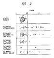
- the supernatant thus obtained (8 ml) was applied to a Cellulofine GCL-2000SF (Seikagaku Kogyo) column (bed volume : 750 ml) which had been equilibrated with 10 mM phosphate buffer (pH 7.2) containing 0.5 M NaCl to collect a high-molecular weight (1,000,000 or more) fraction (void fraction; fraction Nos. 41-50) for use as the high-molecular weight fraction of the pleural exudate of a human pulmonary adenocarcinoma patient.
- Fig. 1 shows the elution pattern of the pleural exudate using Cellulofine GCL-2000SF.
- mice Normal human lung tissue membrane components were administered intravenously to new-born BALB/c mice within 24 postnatal hours at a dose of 100 ⁇ g of protein per animal. After a lapse of 8 weeks, the mice were intraperitoneally administered with the high-molecular weight fraction of the pleural exudate of a human pulmonary adenocarcinoma patient (100 ⁇ g of protein per animal) together with aluminum hydroxide gel (2 mg per animal) and killed pertussis vaccine (1 ⁇ 109 cells per animal), followed by 3-5 immunizations with the same antigen at a dose of 100 ⁇ g per animal on the protein basis at 1- to 2-week intervals.
- mice From among these immunized mice, those mice whose antisera intensely reacted with human pulmonary adenocarcinoma cells or tissues, membrane fragments derived therefrom or high-molecular weight fractions of the pleural exudate of human pulmonary adenocarcinoma patients were selected as the desired immunized mice, and spleen cells were prepared from such mice and submitted to cell fusion.
- the 8-azaguanine-resistant mouse myeloma cell line P3-U1 was cultivated in normal medium to thereby secure not less than 2 ⁇ 107 cells at the time of cell fusion, and submitted to cell fusion as a parent strain.
- the spleen cells and myeloma cells obtained in (2) and (3), respectively, were used in a ratio of 5:1 and subjected to fusion following the procedure mentioned hereinabove. After cultivation in HAT medium at 37°C under 5% CO2 for 14 days, fused cells were selected and, after changing the medium to HT medium, cultivation was continued. Based on the results of anti-human pulmonary adenocarcinoma antibody titer determination, active wells were selected and, after changing the medium to normal medium, cloning was repeated twice.
- hybridoma cell line ALC-864 capable of producing a monoclonal antibody having no reactivity to any of normal human cells or tissues but having specific reactivity to human pulmonary adenocarcinoma, as determined by enzyme immunoassay or immunohistological evaluation (ABC method).
- Pristane-treated 8-week-old female BALB/c mice were intraperitoneally injected with the hybridoma cell line ALC-864 obtained in (4) at a dose of 4 ⁇ 106 cells per animal. In 10-21 days, the hybridoma caused ascites tumors.
- the ascitic fluid was collected from ascitic fluid-bearing mice (5-10 ml per animal), deprived of solids by centrifugation at 3,000 rpm for 5 minutes, subjected to salting out with 50% ammonium sulfate, dialyzed against 0.04M phosphate buffer (pH 8.0) supplemented with 0.03 M NaCl, and applied to a DE52 (Whatman) column (bed volume: 50 ml) at a flow rate of 20 to 30 ml/hr. An IgG fraction was collected and used as a purified monoclonal antibody. The isotype of the thus-obtained anti-human pulmonary adenocarcinoma monoclonal antibody was determined by the Ouchterlony method, and it was found to belong to the class IgG1.
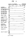
- Table 1 Antigen Reactivity Tissue pulmonary squamous cell carcinoma 3/12 pulmonary adenocarcinoma 10/12 normal lung 0/6 normal tissues other than lung 3/12* Antigen CEA 0/1 Cultured cell line pulmonary squamous cell carcinoma 3/4 pulmonary adenocarcinoma 2/5 fetal lung cancer 0/1 other cancers 11/21** Normal cells 0/3 Note: *: weakly reactive with three samples of kidney tissues. **: reactive with 6 samples of breast cancer cell lines, 3 samples of pancreatic cancer cell lines and 2 samples of uterine cancer cell lines.
- Formalin-fixed, paraffin-embedded tissue sections (sliced to a thickness of 5 ⁇ m with a microtome) of cancer tissues were each fixed on an egg albumin-coated slide glass, deparaffinized with xylene and rendered hydrophilic stepwise with alcohol-water. Each section was rinsed with deionized water for 5 minutes and then allowed to stand in methanol containing 0.3% H2O2 at room temperature for 30 minutes for blocking endogeneous peroxidases. The section was then washed with PBS for 20 minutes and allowed to stand in diluted normal mouse serum at room temperature for 20 minutes.
- the excess serum was sucked off from the section, and the section was reacted with the first antibody (anti-human pulmonary adenocarcinoma monoclonal antibody ALC-864, 10 ⁇ g/ml) for 30 minutes. After washing, the section was then reacted with a diluted biotinylated antibody (biotinylated rabbit anti-IgG antibody, product of Vector) for 30 minutes, washed further, and reacted with an avidin-biotin-peroxidase complex (Vector) for 30 minutes.
- the first antibody anti-human pulmonary adenocarcinoma monoclonal antibody ALC-864, 10 ⁇ g/ml
- a diluted biotinylated antibody biotinylated rabbit anti-IgG antibody, product of Vector
- the section was reacted with a peroxidase substrate [0.1% diaminobenzidine tetrahydrochloride prepared by using 0.1 M Tris-hydrochloride buffer (pH 7.2) containing 0.02% H2O2] for 2 minutes. The reaction was then terminated by cooling in ice. The section was stained with hematoxylin, dehydrated with alcohol-water and xylene, fixed with Canada balsam (product of Merck), and observed under a microscope.
- a peroxidase substrate 0.1% diaminobenzidine tetrahydrochloride prepared by using 0.1 M Tris-hydrochloride buffer (pH 7.2) containing 0.02% H2O2] for 2 minutes. The reaction was then terminated by cooling in ice. The section was stained with hematoxylin, dehydrated with alcohol-water and xylene, fixed with Canada balsam (product of Merck), and observed under a microscope.
- a suspension of ALC-864 (10 ⁇ g/ml) was distributed as a first antibody in 50- ⁇ l portions into each well of a 96-well plate for EIA (Flow Laboratories). After standing at 4°C for 15 hours, the plate was washed with PBS. Then, 1% BSA-PBS was added (200 ⁇ l per well). After standing for 15 hours, the plate was washed well with PBS. To the plate were added 4-fold dilutions of normal human-derived sera (44 samples) or of lung cancer patient-derived sera (92 samples) in an amount of 40 ⁇ l per well. After standing at 4°C for 15 hours, the plate was washed well with PBS.
- biotinylated anti-pulmonary adenocarcinoma monoclonal antibody ALC-864 (10 ⁇ g/ml) was added as the second antibody (100 ⁇ l per well).
- the plate was allowed to stand at 4°C for 15 hours and, then, washed well with PBS.
- Avidin-biotin-peroxidase complex (10 ⁇ g/ml) was distributed in 100- ⁇ l portions into each well, and the plate was allowed to stand at room temperature for 1 hour and then washed with PBS. Thereafter, the ABTS substrate solution was added in an amount of 100 ⁇ l per well and the reaction was allowed to proceed at room temperature for 30 minutes and then terminated by adding 5% SDS solution (100 ⁇ l per well). For each well, the color development was determined by measuring the absorbance at 415 nm (OD 415nm ).
- the positive results were obtained in 8 (24%) out of 33 samples of pulmonary squamous cell carcinoma patients, 2 (9.1%) out of 22 samples of pulmonary small cell carcinoma patients, 2 (50%) out of 4 samples of pulmonary large cell carcinoma patients and 6 (37.5%) out of 16 samples of pulmonary benign disease patients.
- pulmonary adenocarcinoma tissue-derived membrane components were treated with the enzymes and reagents described below and then examined for the reactivity with ALC-864.
- the pulmonary adenocarcinoma tissue membrane components (100 ⁇ g of proteins per ml) were distributed, in 50- ⁇ l portions, into each well of a plate for EIA (Linbro). After standing at 4°C for 15 hours, the plate was washed three times with PBS. Then, 1% BSA-PBS was distributed into the wells (200 ⁇ l per well). The plate was allowed to stand at room temperature for 30 minutes to 2 hours and then washed three times with PBS. One of the above enzyme solutions or reagent soluiton was distributed into the wells (50 ⁇ l per well) and the reaction was carried out at 37°C for 1 hour. Then, the plate was washed five times with PBS and the monoclonal antibody ALC-864 (10 ⁇ g/ml) was distributed into the wells (50 ⁇ l per well), followed by allowing it to stand at 4°C for 15 hours.
- Tween-20-PBS PBS containig 0.05% Tween-20; Tween-20 being a product of Wako Pure Chemical Industries
- peroxidase-labeled rabbit anti-mouse IgG product of DAKO, distributed by Kyowa-Medex: 400-fold dilution
- the plate was washed five times with Tween-20-PBS, the ABTS substrate solution was added (100 ⁇ l), the reaction was conducted for 30 minutes, and the absorbance was measured at 415 nm.
- a suspension of ALC-864 (10 ⁇ g/ml) prepared in Example 1 was distributed as a first antibody in 100- ⁇ l portions into each well of a 96-well plate for EIA (Flow Laboratories). After standing at 4°C for 15 hours, the plate was washed with PBS. Then, 1% BSA-PBS was added (200 ⁇ l per well). After standing at 4°C for 15 hours, the plate was washed well with PBS.
- biotinylated SLC-454 (10 ⁇ g/ml) was added as the second antibody (100 ⁇ l per well).
- the plate was allowed to stand at 4°C for 15 hours, and then washed well with PBS.
- Avidin-biotin-peroxidase (10 ⁇ g/ml) was distributed in 100- ⁇ l portions into each well, and the plate was allowed to stand at room temperature for 1 hour and then washed with PBS. Thereafter, the ABTS substrate solution was added in an amount of 100 ⁇ l per well and the reaction was allowed to proceed at room temperature for 30 minutes and then terminated by adding 5% SDS solution (100 ⁇ l per well). For each well, the color development was determined by measuring the absorbance at 415 nm (OD 415nm ).
- pulmonary adenocarcinoma can be detected in a high percentage of positive reaction and in a large antigen quantitiy by the serodiagnosis of the present invention.
- Positive results were observed in serum samples derived from pulmonary squamous cell carcinoma patients, pulmonary large cell carcinoma patients, gastric cancer patients, large intestinal cancer patients, pancreatic cancer patients, gallbladder or bile duct cancer patients, but the antigen quantity thereof was comparatively low. No positive results were obtained in serum samples from healthy subjects and pulmonary, gastric or pancreatic benign disease patients.
Landscapes
- Health & Medical Sciences (AREA)
- Chemical & Material Sciences (AREA)
- Organic Chemistry (AREA)
- Immunology (AREA)
- Life Sciences & Earth Sciences (AREA)
- Biophysics (AREA)
- Biochemistry (AREA)
- General Health & Medical Sciences (AREA)
- Genetics & Genomics (AREA)
- Medicinal Chemistry (AREA)
- Molecular Biology (AREA)
- Proteomics, Peptides & Aminoacids (AREA)
- Cell Biology (AREA)
- Preparation Of Compounds By Using Micro-Organisms (AREA)
- Peptides Or Proteins (AREA)
- Medicines Containing Antibodies Or Antigens For Use As Internal Diagnostic Agents (AREA)
Applications Claiming Priority (4)
| Application Number | Priority Date | Filing Date | Title |
|---|---|---|---|
| JP63163433A JPH0213393A (ja) | 1988-06-30 | 1988-06-30 | 抗ヒト肺腺癌単クローン性抗体 |
| JP63163434A JPH0212063A (ja) | 1988-06-30 | 1988-06-30 | 肺腺癌抗原の定量法 |
| JP163434/88 | 1988-06-30 | ||
| JP163433/88 | 1988-06-30 |
Publications (3)
| Publication Number | Publication Date |
|---|---|
| EP0348973A2 true EP0348973A2 (de) | 1990-01-03 |
| EP0348973A3 EP0348973A3 (en) | 1990-07-25 |
| EP0348973B1 EP0348973B1 (de) | 1994-03-09 |
Family
ID=26488874
Family Applications (1)
| Application Number | Title | Priority Date | Filing Date |
|---|---|---|---|
| EP89111857A Expired - Lifetime EP0348973B1 (de) | 1988-06-30 | 1989-06-29 | Monoklonaler Antikörper gegen menschliches Lungenadenokarzinom |
Country Status (4)
| Country | Link |
|---|---|
| US (1) | US5081032A (de) |
| EP (1) | EP0348973B1 (de) |
| CA (1) | CA1337049C (de) |
| DE (1) | DE68913601T2 (de) |
Families Citing this family (2)
| Publication number | Priority date | Publication date | Assignee | Title |
|---|---|---|---|---|
| US5250297A (en) * | 1989-10-20 | 1993-10-05 | Hybritech Incorporated | Tumor-associated antigen, antibodies, compositions and uses therefor |
| EP1728210A2 (de) * | 2004-02-27 | 2006-12-06 | Aureon Laboratories, Inc. | Verfahren und system zur vorhersage des auftretens eines ereignisses |
Family Cites Families (5)
| Publication number | Priority date | Publication date | Assignee | Title |
|---|---|---|---|---|
| ATE56046T1 (de) * | 1983-03-04 | 1990-09-15 | Health Research Inc | Monoklonale antikoerper gegen humane brustkarzinomzellen und ihre verwendung in der diagnose und therapie. |
| JPS60190722A (ja) * | 1984-03-12 | 1985-09-28 | Kyowa Hakko Kogyo Co Ltd | 抗ヒト肺癌単クロ−ン性抗体 |
| JPS62238465A (ja) * | 1986-04-09 | 1987-10-19 | Kyowa Hakko Kogyo Co Ltd | 腺癌抗原の定量法 |
| JPS6319561A (ja) * | 1986-07-11 | 1988-01-27 | Kyowa Hakko Kogyo Co Ltd | 抗ヒト肺癌単クロ−ン性抗体 |
| JPH0813840B2 (ja) * | 1987-03-31 | 1996-02-14 | 協和醗酵工業株式会社 | 抗ヒト肺腺癌単クロ−ン性抗体 |
-
1989
- 1989-06-27 US US07/371,808 patent/US5081032A/en not_active Expired - Fee Related
- 1989-06-28 CA CA000604273A patent/CA1337049C/en not_active Expired - Fee Related
- 1989-06-29 DE DE68913601T patent/DE68913601T2/de not_active Expired - Fee Related
- 1989-06-29 EP EP89111857A patent/EP0348973B1/de not_active Expired - Lifetime
Also Published As
| Publication number | Publication date |
|---|---|
| CA1337049C (en) | 1995-09-19 |
| DE68913601D1 (de) | 1994-04-14 |
| EP0348973A3 (en) | 1990-07-25 |
| EP0348973B1 (de) | 1994-03-09 |
| DE68913601T2 (de) | 1994-10-13 |
| US5081032A (en) | 1992-01-14 |
Similar Documents
| Publication | Publication Date | Title |
|---|---|---|
| EP0252769B1 (de) | Monoklonaler Antikörper gegen humanes Lungenkarzinom | |
| EP0207963B1 (de) | Monoklonale antikörper und antigen für menschliche lungenkarzinome vom nichtkleinen zellentyp | |
| Rosen et al. | Analysis of human small cell lung cancer differentiation antigens using a panel of rat monoclonal antibodies | |
| EP0268279A2 (de) | Hybridome, die monoklonale Antikörper gegen neue Mucin-Epitope produzieren | |
| US5185432A (en) | Monoclonal antibodies and antigen for human non-small cell lung carcinoma and other certain human carcinomas | |
| US4960716A (en) | Monoclonal antibodies specific for 330 KD breast tumor antigen and assay using said monoclonal antibodies | |
| US5591830A (en) | Detection of complexes which include basement membrane components as diagnostic of cancer and other diseases | |
| FI85814C (fi) | Foerfarande foer producering av en monoklonal antikropp foer humana icke-litencellslungcancer. | |
| EP0160446A2 (de) | Brusttumorassoziiertes Antigen und monoklonale Antikörper dazu | |
| EP0218257B1 (de) | Spezifischer monoklonaler Antikörper gegen menschliches Lungenadenokarzinom | |
| EP0203552B1 (de) | Monoklonale Antikörper für humane Lungenkarzinome des "Nicht-kleine-Zellen"-Typs | |
| US5051355A (en) | Anti-human gastric cancer monoclonal antibody | |
| CA1320689C (en) | Monoclonal antibody specific for human basal cell surface antigen | |
| EP0348973B1 (de) | Monoklonaler Antikörper gegen menschliches Lungenadenokarzinom | |
| US5552291A (en) | Anti-human pulmonary adenocarcinoma specific monoclonal antibody | |
| EP0339633A2 (de) | Monoklonaler Antikörper gegen menschlichen Magenkrebs | |
| US4886745A (en) | Monoclonal antibody specific for human basal cell surface antigen | |
| EP0272113A2 (de) | Monoklonale Antikörper gegen menschlichen Krebs | |
| EP0235817A2 (de) | Monoklonaler Antikörper gegen menschlichen Magenkrebs | |
| US4894327A (en) | Anti-human mesothelial cell monoclonal antibody | |
| CA1294905C (en) | Anti-lafora body monoclonal antibody | |
| EP0285143B1 (de) | Monoclonaler Antikörper gegen menschliches Lungenadenokarzinom | |
| US5580740A (en) | Antihuman pulmonary adenocarcinoma monoclonal antibody | |
| JPH08208698A (ja) | モノクローナル抗体 | |
| EP0339632A2 (de) | Testmethode für menschliche Krebsantigene |
Legal Events
| Date | Code | Title | Description |
|---|---|---|---|
| PUAI | Public reference made under article 153(3) epc to a published international application that has entered the european phase |
Free format text: ORIGINAL CODE: 0009012 |
|
| AK | Designated contracting states |
Kind code of ref document: A2 Designated state(s): DE FR GB |
|
| PUAL | Search report despatched |
Free format text: ORIGINAL CODE: 0009013 |
|
| AK | Designated contracting states |
Kind code of ref document: A3 Designated state(s): DE FR GB |
|
| 17P | Request for examination filed |
Effective date: 19910102 |
|
| 17Q | First examination report despatched |
Effective date: 19921216 |
|
| GRAA | (expected) grant |
Free format text: ORIGINAL CODE: 0009210 |
|
| AK | Designated contracting states |
Kind code of ref document: B1 Designated state(s): DE FR GB |
|
| REF | Corresponds to: |
Ref document number: 68913601 Country of ref document: DE Date of ref document: 19940414 |
|
| ET | Fr: translation filed | ||
| PLBE | No opposition filed within time limit |
Free format text: ORIGINAL CODE: 0009261 |
|
| STAA | Information on the status of an ep patent application or granted ep patent |
Free format text: STATUS: NO OPPOSITION FILED WITHIN TIME LIMIT |
|
| 26N | No opposition filed | ||
| PGFP | Annual fee paid to national office [announced via postgrant information from national office to epo] |
Ref country code: GB Payment date: 19950619 Year of fee payment: 7 |
|
| PGFP | Annual fee paid to national office [announced via postgrant information from national office to epo] |
Ref country code: FR Payment date: 19950629 Year of fee payment: 7 |
|
| PGFP | Annual fee paid to national office [announced via postgrant information from national office to epo] |
Ref country code: DE Payment date: 19950824 Year of fee payment: 7 |
|
| PG25 | Lapsed in a contracting state [announced via postgrant information from national office to epo] |
Ref country code: GB Effective date: 19960629 |
|
| GBPC | Gb: european patent ceased through non-payment of renewal fee |
Effective date: 19960629 |
|
| PG25 | Lapsed in a contracting state [announced via postgrant information from national office to epo] |
Ref country code: FR Effective date: 19970228 |
|
| PG25 | Lapsed in a contracting state [announced via postgrant information from national office to epo] |
Ref country code: DE Effective date: 19970301 |
|
| REG | Reference to a national code |
Ref country code: FR Ref legal event code: ST |