EP0347210B1 - Method for analysis of cellular components of a fluid - Google Patents
Method for analysis of cellular components of a fluid Download PDFInfo
- Publication number
- EP0347210B1 EP0347210B1 EP89306036A EP89306036A EP0347210B1 EP 0347210 B1 EP0347210 B1 EP 0347210B1 EP 89306036 A EP89306036 A EP 89306036A EP 89306036 A EP89306036 A EP 89306036A EP 0347210 B1 EP0347210 B1 EP 0347210B1
- Authority
- EP
- European Patent Office
- Prior art keywords
- sample
- cells
- methyl
- cell
- blood
- Prior art date
- Legal status (The legal status is an assumption and is not a legal conclusion. Google has not performed a legal analysis and makes no representation as to the accuracy of the status listed.)
- Expired - Lifetime
Links
Images


Classifications
-
- G—PHYSICS
- G01—MEASURING; TESTING
- G01N—INVESTIGATING OR ANALYSING MATERIALS BY DETERMINING THEIR CHEMICAL OR PHYSICAL PROPERTIES
- G01N33/00—Investigating or analysing materials by specific methods not covered by groups G01N1/00 - G01N31/00
- G01N33/48—Biological material, e.g. blood, urine; Haemocytometers
- G01N33/50—Chemical analysis of biological material, e.g. blood, urine; Testing involving biospecific ligand binding methods; Immunological testing
- G01N33/53—Immunoassay; Biospecific binding assay; Materials therefor
- G01N33/569—Immunoassay; Biospecific binding assay; Materials therefor for microorganisms, e.g. protozoa, bacteria, viruses
- G01N33/56966—Animal cells
- G01N33/56972—White blood cells
-
- G—PHYSICS
- G01—MEASURING; TESTING
- G01N—INVESTIGATING OR ANALYSING MATERIALS BY DETERMINING THEIR CHEMICAL OR PHYSICAL PROPERTIES
- G01N15/00—Investigating characteristics of particles; Investigating permeability, pore-volume or surface-area of porous materials
- G01N15/10—Investigating individual particles
- G01N15/14—Optical investigation techniques, e.g. flow cytometry
- G01N15/1456—Optical investigation techniques, e.g. flow cytometry without spatial resolution of the texture or inner structure of the particle, e.g. processing of pulse signals
- G01N15/1459—Optical investigation techniques, e.g. flow cytometry without spatial resolution of the texture or inner structure of the particle, e.g. processing of pulse signals the analysis being performed on a sample stream
-
- G—PHYSICS
- G01—MEASURING; TESTING
- G01N—INVESTIGATING OR ANALYSING MATERIALS BY DETERMINING THEIR CHEMICAL OR PHYSICAL PROPERTIES
- G01N33/00—Investigating or analysing materials by specific methods not covered by groups G01N1/00 - G01N31/00
- G01N33/48—Biological material, e.g. blood, urine; Haemocytometers
- G01N33/50—Chemical analysis of biological material, e.g. blood, urine; Testing involving biospecific ligand binding methods; Immunological testing
- G01N33/53—Immunoassay; Biospecific binding assay; Materials therefor
- G01N33/569—Immunoassay; Biospecific binding assay; Materials therefor for microorganisms, e.g. protozoa, bacteria, viruses
- G01N33/56966—Animal cells
- G01N33/56977—HLA or MHC typing
-
- G—PHYSICS
- G01—MEASURING; TESTING
- G01N—INVESTIGATING OR ANALYSING MATERIALS BY DETERMINING THEIR CHEMICAL OR PHYSICAL PROPERTIES
- G01N33/00—Investigating or analysing materials by specific methods not covered by groups G01N1/00 - G01N31/00
- G01N33/48—Biological material, e.g. blood, urine; Haemocytometers
- G01N33/50—Chemical analysis of biological material, e.g. blood, urine; Testing involving biospecific ligand binding methods; Immunological testing
- G01N33/53—Immunoassay; Biospecific binding assay; Materials therefor
- G01N33/575—Immunoassay; Biospecific binding assay; Materials therefor for cancer
- G01N33/5758—Immunoassay; Biospecific binding assay; Materials therefor for cancer involving compounds serving as markers for tumours, cancers or neoplasias, e.g. cellular determinants, receptors, heat shock/stress proteins, A-protein, oligosaccharides or metabolites
- G01N33/5759—Immunoassay; Biospecific binding assay; Materials therefor for cancer involving compounds serving as markers for tumours, cancers or neoplasias, e.g. cellular determinants, receptors, heat shock/stress proteins, A-protein, oligosaccharides or metabolites involving compounds localised on the membrane of tumour or cancer cells
-
- G—PHYSICS
- G01—MEASURING; TESTING
- G01N—INVESTIGATING OR ANALYSING MATERIALS BY DETERMINING THEIR CHEMICAL OR PHYSICAL PROPERTIES
- G01N15/00—Investigating characteristics of particles; Investigating permeability, pore-volume or surface-area of porous materials
- G01N15/10—Investigating individual particles
- G01N2015/1006—Investigating individual particles for cytology
-
- G—PHYSICS
- G01—MEASURING; TESTING
- G01N—INVESTIGATING OR ANALYSING MATERIALS BY DETERMINING THEIR CHEMICAL OR PHYSICAL PROPERTIES
- G01N15/00—Investigating characteristics of particles; Investigating permeability, pore-volume or surface-area of porous materials
- G01N15/10—Investigating individual particles
- G01N15/14—Optical investigation techniques, e.g. flow cytometry
- G01N2015/1402—Data analysis by thresholding or gating operations performed on the acquired signals or stored data
-
- G—PHYSICS
- G01—MEASURING; TESTING
- G01N—INVESTIGATING OR ANALYSING MATERIALS BY DETERMINING THEIR CHEMICAL OR PHYSICAL PROPERTIES
- G01N2333/00—Assays involving biological materials from specific organisms or of a specific nature
- G01N2333/435—Assays involving biological materials from specific organisms or of a specific nature from animals; from humans
- G01N2333/705—Assays involving receptors, cell surface antigens or cell surface determinants
-
- Y—GENERAL TAGGING OF NEW TECHNOLOGICAL DEVELOPMENTS; GENERAL TAGGING OF CROSS-SECTIONAL TECHNOLOGIES SPANNING OVER SEVERAL SECTIONS OF THE IPC; TECHNICAL SUBJECTS COVERED BY FORMER USPC CROSS-REFERENCE ART COLLECTIONS [XRACs] AND DIGESTS
- Y10—TECHNICAL SUBJECTS COVERED BY FORMER USPC
- Y10S—TECHNICAL SUBJECTS COVERED BY FORMER USPC CROSS-REFERENCE ART COLLECTIONS [XRACs] AND DIGESTS
- Y10S435/00—Chemistry: molecular biology and microbiology
- Y10S435/973—Simultaneous determination of more than one analyte
-
- Y—GENERAL TAGGING OF NEW TECHNOLOGICAL DEVELOPMENTS; GENERAL TAGGING OF CROSS-SECTIONAL TECHNOLOGIES SPANNING OVER SEVERAL SECTIONS OF THE IPC; TECHNICAL SUBJECTS COVERED BY FORMER USPC CROSS-REFERENCE ART COLLECTIONS [XRACs] AND DIGESTS
- Y10—TECHNICAL SUBJECTS COVERED BY FORMER USPC
- Y10S—TECHNICAL SUBJECTS COVERED BY FORMER USPC CROSS-REFERENCE ART COLLECTIONS [XRACs] AND DIGESTS
- Y10S436/00—Chemistry: analytical and immunological testing
- Y10S436/80—Fluorescent dyes, e.g. rhodamine
-
- Y—GENERAL TAGGING OF NEW TECHNOLOGICAL DEVELOPMENTS; GENERAL TAGGING OF CROSS-SECTIONAL TECHNOLOGIES SPANNING OVER SEVERAL SECTIONS OF THE IPC; TECHNICAL SUBJECTS COVERED BY FORMER USPC CROSS-REFERENCE ART COLLECTIONS [XRACs] AND DIGESTS
- Y10—TECHNICAL SUBJECTS COVERED BY FORMER USPC
- Y10S—TECHNICAL SUBJECTS COVERED BY FORMER USPC CROSS-REFERENCE ART COLLECTIONS [XRACs] AND DIGESTS
- Y10S436/00—Chemistry: analytical and immunological testing
- Y10S436/805—Optical property
-
- Y—GENERAL TAGGING OF NEW TECHNOLOGICAL DEVELOPMENTS; GENERAL TAGGING OF CROSS-SECTIONAL TECHNOLOGIES SPANNING OVER SEVERAL SECTIONS OF THE IPC; TECHNICAL SUBJECTS COVERED BY FORMER USPC CROSS-REFERENCE ART COLLECTIONS [XRACs] AND DIGESTS
- Y10—TECHNICAL SUBJECTS COVERED BY FORMER USPC
- Y10T—TECHNICAL SUBJECTS COVERED BY FORMER US CLASSIFICATION
- Y10T436/00—Chemistry: analytical and immunological testing
- Y10T436/14—Heterocyclic carbon compound [i.e., O, S, N, Se, Te, as only ring hetero atom]
- Y10T436/142222—Hetero-O [e.g., ascorbic acid, etc.]
- Y10T436/143333—Saccharide [e.g., DNA, etc.]
Definitions
- This invention relates to the multi-parameter analysis of cellular components in a body fluid, and more particularly, it relates to the discrimination and quantification of the cellular components of blood or bone marrow aspirates through analysis of five parameters by flow cytometry.
- ⁇ blood counts and leukocyte differentials Two of the most important measures of the hematologic status of an individual are the whole blood count and leukocyte differential.
- Whole blood counts and leukocyte differentials involve the discrimination and then counting of the various cellular components of blood.
- the various components of blood that may appear in any one sample include red blood cells (RBC), platelets and nucleated cells.
- RBC red blood cells
- platelets and nucleated cells.
- a number of different types of leukocytes exist which include lymphocytes (both B and T cells, Natural Killer (or "NK") cells and various subsets thereof), monocytes and granulocytes (including neutrophils, eosinophils and basophils in all maturational stages).
- lymphocytes both B and T cells, Natural Killer (or "NK") cells and various subsets thereof
- monocytes and granulocytes including neutrophils, eosinophils and basophils in all maturational stages).
- whole blood counts i.e. , number of cells per standard unit of volume
- leukocyte differentials i.e. , number of cells of a given type per standard unit of volume
- Fluorescence may be measured from cells that incorporate a nucleic acid stain or may be measured from cells bearing surface markers which are labelled with monoclonal antibodies (MAbs) conjugated directly or indirectly to fluorochromes, as described, for example, in U.S. Pat. No. 4,520,110.
- MAbs monoclonal antibodies
- Separate channels within the flow cytometer sense and record each of the various cell measurements. By combining and comparing these parameters, the various leukocyte components may be distinguished.
- U.S. Pat. No. 4,727,020 provides one example of how a flow cytometer may be used in this method to obtain leukocyte differentials from blood. This approach, however, is limited to leukocyte differentials and only those obtained from blood.
- Leukocyte characterisation by flow cytometric systems is included in the review article on flow cytometry by Steinkamp in Review of Scientific Instruments, 55 , 1375 (1984).
- the present invention comprises a method for the simultaneous, multi-parameter analysis of cells in a body fluid, wherein the fluid, comprises either spinal, peritoneal or brain fluid, urine or whole blood and additionally may comprise a tissue cell suspension, wherein the tissue comprises bone marrow aspirates, lymph node, spleen or liver cells such as from a biopsy, and wherein the parameters comprise at least two measures of light scattering and at least three measures of fluorescence emmission or activity from each cell examined.
- the method comprises the steps of taking a sample of fluid derived from an individual, combining the fluid sample with at least two dyes and with at least one labelled cell surface marker, wherein said dyes will independently assess different characteristics of the cells in the sample and wherein said marker recognizes an antigen that is differentially expressed on cells of different lineages, to form a labelled solution.
- Each of the dyes and label is fluorescent and has a peak emission spectra that is distinguishable from the others.
- the labelled solution then is passed through a detection instrument, wherein each cell in the solution is examined substantially one at a time, and measurements of fluorescence intensity and light scattering are taken for each cell examined.
- the measurements (or parameters) taken for each cell may be stored in a data storage and analysis system and recombined and analyzed in real time or at a later time. By analyzing these several parameters, the cell types and lineages that comprise blood and bone marrow samples may be distinguished and identified.
- the preferred method comprises the steps of taking a body fluid sample derived from an individual, combining the fluid sample with nucleic acid dyes, such as an RNA dye, a DNA dye, and a labelled monoclonal antibody (MAb), which recognizes a cell surface antigen that is differentially expressed on different cell lineages, to form a labelled solution, passing the labelled solution through a flow cytometer, measuring fluorescence intensity, OLS and FLS and storing the data recorded for analyses.
- nucleic acid dyes such as an RNA dye, a DNA dye, and a labelled monoclonal antibody (MAb), which recognizes a cell surface antigen that is differentially expressed on different cell lineages
- MAb monoclonal antibody
- Each of the nucleic acid dyes and immuno-label is fluorescent, is excitable at the same wavelength and has a peak emission spectra that is distinguishable from the others.
- kits containing the nucleic acid dyes and labelled cell surface markers useful herein also is included within the scope of the present invention.
- All six figures represent log scale dot plots containing approximately 22,000 cells from a fluid sample labelled with the nucleic acid dyes LDS-751 and Thiazole-Orange and with a CD45 MAb such as HLe-1(PE). Labelled cells were analyzed on a FACScan Tm flow cytometer equipped with a single argon laser tuned to 488nm (Becton Dickinson Immunocytometry Systems (or BDIS)), and measurements of OLS and FLS and fluorescence intensity were taken on each cell. Data acquisition of the five parameters digitized and stored in list-mode was performed using a Consort 30 computer (BDIS). Data analyses and printouts were performed using Paint-A-GateTM software (BDIS).
- the present invention comprises a method for the simultaneous, multi-parameter analysis of cells in a body fluid, wherein the fluid comprises either spinal, peritoneal or brain fluid, urine or whole blood and may additionally comprise a tissue cell suspension, wherein said cell suspension is prepared from bone marrow aspirates, liver, spleen or lymph node, such as from a biopsy, and wherein the parameters comprise at least two measures of the light scattered in more than one direction by each cell examined and at least three measures of fluorescence activity from each cell examined.
- the method comprises the steps of taking a sample of body fluid from an individual, combining the fluid sample with at least two dyes and with at least one labelled cell surface marker, wherein each of said dyes will independently assess different characteristics of the cells in the sample, and wherein said marker recognizes an antigen that is differentially expressed on cells of different lineages, to form a labelled solution.
- Each of the dyes and label is fluorescent and has a peak emission spectra that is distinguishable from the others.
- the labelled solution then is passed through a detection instrument, wherein each cell in the solution is examined substantially one at a time, and measurements of fluorescence intensity and light scattering are taken for each cell examined.
- the measurements (or parameters) taken for each cell may be stored in a data storage means and- recombined and re-analyzed in real time or at a later time.
- the present invention will allow for the discrimination and identification of cells in a fluid sample by the analyses of at least five parameters.
- the method of the invention comprises the steps of: 1) taking a cell containing fluid sample derived from an individual where, preferentially, the body fluid comprises either whole blood or bone marrow aspirates; 2) adding two fluorescent nucleic acid dyes to said sample to differentially label the DNA and RNA in each cell, wherein each dye has a fluorescent peak emission spectra different from the other; 3) adding a fluorescent labelled cell surface marker to said sample, wherein said marker recognizes an antigen that is differentially expressed on said cells in said fluid and wherein said fluorescent immuno-label has a peak emission spectra different from said fluorescent dyes; and 4) analyzing said cells in said sample in an automated instrument capable of detecting and recording both fluorescence of individual cells at or near said peak emission spectra and OLS and FLS of individual cells.
- the detection instrument comprises a flow cytometer, such as a FACScan Tm (BDIS).
- a flow cytometer such as a FACScan Tm (BDIS).
- the flow cytometer is equipped with a single laser for excitation of said fluorescent labelled cells such as an argon ion laser and is tuned at or near 488nm.
- the fluorescent nucleic acid dyes and fluorescent label therefore, must be excitable at 488nm.
- 2- ⁇ 2-[4-(dimethylamino)phenyl]ethenyl ⁇ -1-ethylquinolinium iodide, LDS-751 (Exciton) is used as a DNA dye and 1-methyl-4- ⁇ [3-methyl-2(3H)-benzothiazolylidene]methyl ⁇ quinolinium 4-methylbenzenesulfonate, Thiazole-Orange (BDIS), is used as an RNA dye.
- LDS-751 has a peak emission spectra at 670nm
- Thiazole-Orange has a peak emission spectra at 530nm.
- Other fluorescent nucleic acid dyes that may be suitable for practice in this invention include those described in U.S. Pat. No. 4,544,546.
- the fluorescent dyes and/or fluorescent label may be excited at different wavelengths.
- the only requirement is that the peak emission spectra for the dyes and immunofluorescent label all differ.
- the cell surface marker is a monoclonal antibody that will selectively attach to a cell surface antigen that is differently expressed on the surface of hematopoietic cells.
- the antigen will be expressed in different amounts on the various types and maturational stages of nucleated cells.
- One such antigen is CD45.
- This antigen has a molecular weight of approximately 180-220kD and is expressed in different levels on all lymphocytes, monocytes and granulocytes but is absent from mature platelets or RBC.
- Monoclonal antibodies that react specifically with the CD45 antigen include HLe-1 (also referred to an Anti-Leukocyte, BDIS) and LCA (Dako Corp.).
- HLe-1 also referred to an Anti-Leukocyte, BDIS
- LCA Deako Corp.
- HLe-1 is used.
- the CD45 MAb is tagged directly with a fluorescent label such as a fluorochrome.
- a fluorescent label such as a fluorochrome.
- the MAb is conjugated directly to a fluorescent label by means known to those skilled in the art.
- U.S. Pat. No. 4,520,110 provides one method for conjugating a fluorescent label to a MAb.
- the fluorochrome is a phycobiliprotein, such as phycoerythrin (PE), which can be excited at 488nm and has an emission peak at 575nm.
- PE phycoerythrin
- the combination of HLe-1 and PE is generally referred to as HLe-1(PE).
- fluorochromes also may be used in this invention.
- the only requirements are that the fluorochromes excite at 488nm, if an argon laser is used, or at some other wavelength, if a dual laser source is used ( e.g. , 633nm for a He/Ne laser), and that the fluorochrome have a peak emission spectra that does not overlap with the nucleic acid dyes.
- Means to record and analyze the data may include a personal computer equipped with appropriate software.
- the software should be capable of analyzing at least five parameters for each cell, of discriminating between or identifying populations of like cells in five-dimensional space and of displaying at least two of the parameters in two dimensions.
- the software should be capable of gating on one or more parameters and displaying combination of the other parameters in two dimensions.
- Paint-A-Gate Tm software (BDIS) is used in connection with a Consort 30 data storage and analysis system (BDIS).
- BDIS Paint-A-Gate Tm software
- the software is fully described in U.S. Patent 4845653 which is commonly assigned to the assignee hereof.
- the present invention also comprises a kit for multi-parameter analysis of cells in a body fluid.
- the kit may comprise a first container containing at least two nucleic acid dyes to label the DNA and RNA in each cell, wherein the dyes have peak emission spectra that are distinguishable from the others, and a second container containing a fluorescently labelled cell surface marker, wherein the cell surface marker will specifically react with a cell surface antigen that is differentially expressed on the different cells that comprise the sample.
- the dyes are nucleic acid dyes which will differentially label DNA and RNA.
- the nucleic acid dyes are LDS-751 and Thiazole-Orange, and the fluorescently labelled cell surface marker is HLe-1(PE). It will be appreciated by those skilled in the art that other of the nucleic acid dye and/or cell surface marker combinations disclosed herein may comprise the kit. Similarly, it will be appreciated that the nucleic acid dyes and/or cell surface markers may be separately contained in the containers.
- Peripheral blood from normal volunteers was collected by venipuncture into Vacutainer (Trade Mark) tubes containing EDTA(k3) as anticoagulant (Becton Dickinson). Blood also was collected from a patient with B Cell Chronic Lymphocytic Leukemia (B-CLL) graded stage IV according to RAI. Blood cell counts determined on an automated blood cell analyzer (H1, Technicon were: RBC 3.3 x 10⁹ /ml; leukocytes 81.9 x 10⁶/ml; and platelets 35 x 106/ml corrected after manual counting to 20 x 106/ml. Bone marrow aspirates were obtained from normal volunteers. The aspirates were transferred into Vacutainer (Trade Mark) tubes and diluted 1:1 with RPMI 1640 (GIBCO).
- Flow cytometric measurements were performed on a FACScan Tm flow cytometer equipped with a single argon ion laser. Data acquisition of the five parameters digitized and stored in list-mode was performed with the FACScan Tm research software using a Consort 30 Tm computer. For each sample, 22,000 cells were analyzed. For discrimination within the nucleated cells, the acquisition was performed while gating on the fluorescence intensity of LDS-751 and Thiazole-Orange. Data analysis was performed using the Paint-A-Gate Tm software program which allows visualization of multi-parameter data.
- analysis of a sample may be performed in two separate procedures. Initially, a split-sample of body fluid is taken, one or more of which is diluted as above and one of which is not diluted or is diluted less. Then, the above-described method is followed using one split-sample to identify and discriminate RBC, platelets and reticulocytes while gating on but not counting nucleated cells. The method also is followed for identifying and disriminating the nucleated cells while gating on but not counting the non-nucleated cells. Splitting the sample and examining the nucleated cells in less diluted format will allow the relatively rarer nucleated cells in the sample to be counted in a more rapid fashion.
- Cell sorting was performed on a FACS 440 Tm which was adapted to detect two light scattering signals and three fluorescence signals using a single argon ion laser tuned to 488nm.
- the cells were sorted into RPMI 1640 containing 10% fetal calf serum (FCS).
- FCS fetal calf serum
- the sorted cells were centrifuged for 5 minutes at 200g and resuspended in 100 ul RPMI containing 10% FCS.
- Microscope slide preparations were made using a Shandon Cytocentrifuge (Southern Product Ltd., England). The slides were stained with Wright Stain and examined with a light microscope. For reticulocyte identification, 100 ul of a 1% new Methylene blue solution was added to the resuspended sorted cells. The cells were transferred to a slide, covered and examined with a light microscope.
- Analyses of the five parameter list-modes files were performed with a computer program which utilized color to identify clusters of dots in multiple dimensions. Different combinations of the five parameters were displayed at one time. Clusters of cells identified by one combination of parameters were painted a color which then appeared simultaneously in the other projections of the data.
- the identified cell clusters are labelled in the accompanying black and white figures on the basis of the painted colours in the originals.
- a large range of light scattering signals is obtained from unlysed blood samples as a result of the presence of small platelets (1-4u), erythrocytes (6-8u) and larger leukocytes (6-20u).
- small platelets 1-4u
- erythrocytes (6-8u)
- larger leukocytes (6-20u).
- the forward and orthogonal light scattering signals were acquired using four decade logarithmic amplifiers. Under these conditions two major populations of cells were identified in whole blood. See FIG. 1a. Further discrimination between the cell populations required additional parameters.
- Thiazole-Orange has a higher affinity or preference towards RNA as compared with DNA and undergoes fluorescence enhancement upon binding. The peak emission of the bound dye occurs at 530nm.
- LDS-751 has a higher affinity or preference for DNA as compared with RNA. This dye also undergoes fluorescence enhancement upon binding, but with maximal emission at 670nm. Although, it is recognized that platelets do not have a nucleus, LDS-751 will selectively stain such cells. Thus, it is expected that dyes other than LDS-751 also will selectively bind to platelets and other nucleated cells to distinguish platelets from the non-nucleated RBC.
- Thiazole-Orange and LDS-751 in combination provide large separation between erythrocytes, which lack both RNA and DNA, and nucleated leukocytes which have both nucleic acids.
- FIG. 1b the erythroyctes and the leukocytes, (in the upper right hand corner) are separated by almost three orders of magnitude in their fluorescence signals. No significant interference between these dyes was observed for nucleated cells.
- Peripheral blood was incubated with LDS-751, Thiazole-Orange and HLe-1(PE).
- the blood preparation was diluted 1:100 with PBS before measurement.
- the identification of the different blood cell populations is described separately.
- the identity of the cell subpopulations referred to were verified by microscopic examination of the sorted cells.
- the purity of the sorted cell fractions was greater than 90%, with the majority of contaminating cells being erythrocytes.
- the high frequency of the erythrocytes allowed them to be deflected together with the gated cell population as a result of the setting of the threshold during the sorting of the leukocyte populations.
- RBC erythrocytes
- the reticulocytes (“Ret") were distinguished from the mature erythrocytes primarily by their reactivity with Thiazole-Orange (FIG. 1b). The reticulocytes have a slightly enhanced fluorescence intensity with LDS-751 as compared with erythrocytes. Neither RBC nor reticulocytes express surface antigens recognized by CD45 (FIG. 1c,d). The reticulocytes could not be distinguished from leukocytes and RBC based on their light scattering characteristics (FIG. 1a).
- Platelets were characterized by their relative low forward and orthogonal light scattering (FIG. 1a) as well as their staining for LDS-751 (FIG. 1b). The platelets were clearly separated from the RBC, reticulocytes and leukocytes ("L”) (FIG. 1b) using these three parameters. As with the erythroid cells, the platelets did not express the antigens recognized by CD45 (FIG. 1c,d). Using multi-parameter analysis, "normal" platelets can be characterized as particles with relatively low light scattering signals, a larger fluorescence intensity with LDS-751 than that of RBC, no reactivity with Thiazole-Orange, and an absence of CD45 binding.
- Leukocytes were characterized by a relatively large orthogonal and forward light scattering--signal (FIG. 1a). Although their light scattering characteristics were similar, the leukocytes were clearly separated from the erythroid cells by their large fluorescence signals generated by both LDS-751 and Thiazole-Orange (FIG. 1b). This resolution provides an accurate means of counting white blood cells in the presence of an overwhelming number of RBC. A further distinction of leukocytes from platelets, RBC and reticulocytes was obtained by their reactivity with CD45 (FIG. 1c,d).
- a gate was set on Thiazole-Orange and LDS-751 as indicated in FIG. 1b.
- a leukocyte differential was obtained by correlating the light scattering properties of the nucleated cells with the fluorescence intensity of CD45. See FIG. 2a. Lymphocytes ("Lym”) were characterized by the brightest expression of CD45 antigen and the lowest orthogonal light scattering signals. Monocytes (“M”) were identified as binding slightly less CD45 than lymphocytes but showing a larger forward and orthogonal light scattering signal. Neutrophilic granulocytes (“N”) dimly expressed CD45 antigen and had large light scattering signals. Eosinophilic granulocytes (“E o ”) bound the same amount of CD45 as monocytes but were distinguishable from these cells by larger orthogonal light scattering signal and lower forward light scattering signal.
- the nucleated bone marrow cells were identified by their large fluorescence signals with LDS-751 as well as Thiazole-Orange (FIG. 3b). A gate was set on LDS-751 and Thiazole-Orange intensity, as indicated in Fig. 3b, to collect more nucleated cell events. The light scattering properties and their correlation with the immunofluorescence signals with CD45 of these nucleated cells is shown in FIG. 4.
- the blood from the B-CLL patient was incubated with LDS-751, Thiazole-Orange and HLe-1(PE) as was done for normal blood.
- the data are displayed in the same manner as was chosen for normal peripheral blood (FIG. 1) and bone marrow (FIG. 3).
- FIG. 5a The frequency of cells occurring in the light scattering region typical for platelets (FIG. 5a) was low compared with normal peripheral blood ( compare FIG. 1a,b with FIG. 5a,b). Only a few of the dots identified as platelets with light scattering in FIG. 5a show the fluorescence intensity with LDS-751 typical for platelets in FIG. 5b and can be considered as normal platelets. The discrimination of platelets from reticulocytes is still excellent (FIG. 5b).
- the separation of the leukocytes and reticulocytes obtained with LDS-751 and Thiazole-Orange is less clear.
- the leukocytes can be clearly separated from the reticulocytes based on their expression of the CD45 antigen (FIG. 5c,d).
- FIG. 6a,b The light scattering properties and CD45 antigen expression of the nucleated cells (gated in FIG. 5b) is shown in FIG. 6a,b.
- the majority of cells dimly express CD45 antigen and exhibit low forward and orthogonal light scattering signals characteristics of immature lymphocytes. Only a few leukocytes were detected as normal, lymphocytes, granulocytes and monocytes, although the separation was not distinct as a result of the overwhelming number of immature lymphoid cells. Note the few cells which did not express CD45 antigen and which had a low orthogonal light scattering signal. See FIG. 6B. These cells were identified as nucleated erythroid cells consistent with the large increase in reticulocytes in this patient.
- blood may be introduced into the aspirate as a contaminant to the sample.
- the leukocyte differentials could be in error.
- the.percentages of white blood cell types in the blood may be calculated.
- the expected percentages of white blood cells in the blood sample may be subtracted from the bone marrow sample to give a more accurate reading free from blood contaminants (assuming that in fact all the contaminants are blood derived).
- lymphocytes, granulocytes and monocytes comprise 1.0, 1.0 and 0.2 percent of total cells in a blood'sample respectively, with the remaining 97.8% of the cells being essentially RBC.
- lymphocytes, granulocytes and monocytes there are approximately one lymphocyte and granulocyte and 0.2 monocytes per 100 RBC in the blood sample.
- the absolute counts of the same cells in a bone marrow sample is 800, 490 and 165 respectively in a 20,000 cell sample with the remainder of the cells being essentially RBC ( i.e. , 18,545).
- the number of lymphocytes, granulocytes and monocytes expected to be contaminating the bone marrow sample would be 185 lymphocytes ( i.e.
- the corrected counts therefore, would be 615 lymphocytes, 305 granulocytes and 128 monocytes. By this method, more accurate counts may be obtained.
Landscapes
- Health & Medical Sciences (AREA)
- Life Sciences & Earth Sciences (AREA)
- Immunology (AREA)
- Engineering & Computer Science (AREA)
- Chemical & Material Sciences (AREA)
- Hematology (AREA)
- Urology & Nephrology (AREA)
- Molecular Biology (AREA)
- Cell Biology (AREA)
- Biomedical Technology (AREA)
- General Health & Medical Sciences (AREA)
- Physics & Mathematics (AREA)
- General Physics & Mathematics (AREA)
- Pathology (AREA)
- Biochemistry (AREA)
- Analytical Chemistry (AREA)
- Food Science & Technology (AREA)
- Medicinal Chemistry (AREA)
- Microbiology (AREA)
- Biotechnology (AREA)
- Virology (AREA)
- Zoology (AREA)
- Tropical Medicine & Parasitology (AREA)
- Dispersion Chemistry (AREA)
- Investigating Or Analysing Biological Materials (AREA)
- Sampling And Sample Adjustment (AREA)
- Investigating Or Analysing Materials By Optical Means (AREA)
- Measuring Or Testing Involving Enzymes Or Micro-Organisms (AREA)
- Measurement Of The Respiration, Hearing Ability, Form, And Blood Characteristics Of Living Organisms (AREA)
- Optical Measuring Cells (AREA)
Abstract
Description
- This invention relates to the multi-parameter analysis of cellular components in a body fluid, and more particularly, it relates to the discrimination and quantification of the cellular components of blood or bone marrow aspirates through analysis of five parameters by flow cytometry.
- Two of the most important measures of the hematologic status of an individual are the whole blood count and leukocyte differential. Whole blood counts and leukocyte differentials involve the discrimination and then counting of the various cellular components of blood. The various components of blood that may appear in any one sample include red blood cells (RBC), platelets and nucleated cells. In the latter category, a number of different types of leukocytes exist which include lymphocytes (both B and T cells, Natural Killer (or "NK") cells and various subsets thereof), monocytes and granulocytes (including neutrophils, eosinophils and basophils in all maturational stages). Because of the wide possible range of cell types and the range of maturational stages of any one cell type, obtaining whole blood counts and leukocyte differentials often is a difficult and complex procedure in the normal individual but it is more difficult and complex in an abnormal individual. This complexity is emphasized even more when the analysis of bone marrow samples is attempted.
- Traditionally, whole blood counts (i.e., number of cells per standard unit of volume) and leukocyte differentials (i.e., number of cells of a given type per standard unit of volume) have been performed manually by counting a small volume of cells using a light microscope and then multiplying the number counted by a factor to account for the volume. Because of both the sample size measured and the level of skill required to distinguish among cell types, a high and often unacceptable degree of variability may be introduced by this method.
- Recently, instrumentation has been devised to obtain whole blood counts or leukocyte differentials without having to resort to manual microscopic examination. Currently available instrumentation defect cells either electronically by aperture impedance (e.g., Coulter CounterTm, described in Coulter, Proc. Nat. Electronics Conf., 12:1034 (1956)) or optically by light scattering and absorption (e.g., Technicon H-6000Tm, described in Breakell et al., Blood Cells, 11:257 (1985)). In these instruments, the red blood cell fraction must be separated from the leukocytes and each measurement is taken independently of the other. This must be done because RBC generally outnumber leukocytes by at least 1000:1 in the normal patient. In the abnormal patient (e.g., leukopenic patent), this ratio may be substantially higher.
- Other means to obtain leukocyte differentials also exist. Flow cytometers, which are generally described in U.S. Pat. Nos. 4,661,913, 4,284,412 and 3,826,364, and in an article by Herzenberg et al., Sci. Am., 234:108 (1976), have been used to identify different populations of leukocytes in a heterogeneous sample by detecting multiple independent parameters on individual cells that pass through the sensing region. Typically, these parameters include forward light scatter (FLS which is a measure of relative particle size), orthogonal light scatter (OLS which is a measure of relative granularity) and fluorescence. Fluorescence may be measured from cells that incorporate a nucleic acid stain or may be measured from cells bearing surface markers which are labelled with monoclonal antibodies (MAbs) conjugated directly or indirectly to fluorochromes, as described, for example, in U.S. Pat. No. 4,520,110. Separate channels within the flow cytometer sense and record each of the various cell measurements. By combining and comparing these parameters, the various leukocyte components may be distinguished. U.S. Pat. No. 4,727,020 provides one example of how a flow cytometer may be used in this method to obtain leukocyte differentials from blood. This approach, however, is limited to leukocyte differentials and only those obtained from blood.
- Leukocyte characterisation by flow cytometric systems is included in the review article on flow cytometry by Steinkamp in Review of Scientific Instruments, 55, 1375 (1984).
- Taken together, there is no single, standard, accurate instrumentation or methodology currently available that allows a single sample of blood or bone marrow to be analysed such that all the various cell types present in blood and bone marrow may be discriminated and then counted.
- The present invention comprises a method for the simultaneous, multi-parameter analysis of cells in a body fluid, wherein the fluid, comprises either spinal, peritoneal or brain fluid, urine or whole blood and additionally may comprise a tissue cell suspension, wherein the tissue comprises bone marrow aspirates, lymph node, spleen or liver cells such as from a biopsy, and wherein the parameters comprise at least two measures of light scattering and at least three measures of fluorescence emmission or activity from each cell examined. The method comprises the steps of taking a sample of fluid derived from an individual, combining the fluid sample with at least two dyes and with at least one labelled cell surface marker, wherein said dyes will independently assess different characteristics of the cells in the sample and wherein said marker recognizes an antigen that is differentially expressed on cells of different lineages, to form a labelled solution. Each of the dyes and label is fluorescent and has a peak emission spectra that is distinguishable from the others. The labelled solution then is passed through a detection instrument, wherein each cell in the solution is examined substantially one at a time, and measurements of fluorescence intensity and light scattering are taken for each cell examined. The measurements (or parameters) taken for each cell may be stored in a data storage and analysis system and recombined and analyzed in real time or at a later time. By analyzing these several parameters, the cell types and lineages that comprise blood and bone marrow samples may be distinguished and identified.
- More specifically, the preferred method comprises the steps of taking a body fluid sample derived from an individual, combining the fluid sample with nucleic acid dyes, such as an RNA dye, a DNA dye, and a labelled monoclonal antibody (MAb), which recognizes a cell surface antigen that is differentially expressed on different cell lineages, to form a labelled solution, passing the labelled solution through a flow cytometer, measuring fluorescence intensity, OLS and FLS and storing the data recorded for analyses. Each of the nucleic acid dyes and immuno-label is fluorescent, is excitable at the same wavelength and has a peak emission spectra that is distinguishable from the others.
- A kit containing the nucleic acid dyes and labelled cell surface markers useful herein also is included within the scope of the present invention.
- All six figures represent log scale dot plots containing approximately 22,000 cells from a fluid sample labelled with the nucleic acid dyes LDS-751 and Thiazole-Orange and with a CD45 MAb such as HLe-1(PE). Labelled cells were analyzed on a FACScanTm flow cytometer equipped with a single argon laser tuned to 488nm (Becton Dickinson Immunocytometry Systems (or BDIS)), and measurements of OLS and FLS and fluorescence intensity were taken on each cell. Data acquisition of the five parameters digitized and stored in list-mode was performed using a Consort 30 computer (BDIS). Data analyses and printouts were performed using Paint-A-Gate™ software (BDIS).
- FIG. 1 comprises several dot plots of cells from a normal peripheral blood sample analyzed by various combinations of the five parameters;
- FIG. 2 comprises several dot plots only of the nucleated cells of the blood sample analyzed in FIG. 1 and gated on LDS-751 and Thiazole-Orange fluorescence (FIG. 1b);
- FIG. 3 comprises several dot plots of cells from a normal bone marrow aspirate analyzed by various combinations of the five parameters as in FIG. 1;
- FIG. 4 comprises several dot plots only of the nucleated cells from the bone marrow sample analyzed in FIG. 3 and gated on LDS-751 and Thiazole-Orange fluorescence;
- FIG. 5 comprises several dot plots of peripheral blood cells from a thrombocytopenic B-CLL patient analyzed by various combinations of the five parameters as in FIG. 1; and
- FIG. 6 comprises several dot plots only of the nucleated cells from the blood sample analyzed in FIG. 5 and gated on LDS-751 and Thiazole-Orange fluorescence as in FIG. 4.
- The present invention comprises a method for the simultaneous, multi-parameter analysis of cells in a body fluid, wherein the fluid comprises either spinal, peritoneal or brain fluid, urine or whole blood and may additionally comprise a tissue cell suspension, wherein said cell suspension is prepared from bone marrow aspirates, liver, spleen or lymph node, such as from a biopsy, and wherein the parameters comprise at least two measures of the light scattered in more than one direction by each cell examined and at least three measures of fluorescence activity from each cell examined. The method comprises the steps of taking a sample of body fluid from an individual, combining the fluid sample with at least two dyes and with at least one labelled cell surface marker, wherein each of said dyes will independently assess different characteristics of the cells in the sample, and wherein said marker recognizes an antigen that is differentially expressed on cells of different lineages, to form a labelled solution. Each of the dyes and label is fluorescent and has a peak emission spectra that is distinguishable from the others. The labelled solution then is passed through a detection instrument, wherein each cell in the solution is examined substantially one at a time, and measurements of fluorescence intensity and light scattering are taken for each cell examined. The measurements (or parameters) taken for each cell may be stored in a data storage means and- recombined and re-analyzed in real time or at a later time.
- The present invention will allow for the discrimination and identification of cells in a fluid sample by the analyses of at least five parameters. Desirably, the method of the invention comprises the steps of: 1) taking a cell containing fluid sample derived from an individual where, preferentially, the body fluid comprises either whole blood or bone marrow aspirates; 2) adding two fluorescent nucleic acid dyes to said sample to differentially label the DNA and RNA in each cell, wherein each dye has a fluorescent peak emission spectra different from the other; 3) adding a fluorescent labelled cell surface marker to said sample, wherein said marker recognizes an antigen that is differentially expressed on said cells in said fluid and wherein said fluorescent immuno-label has a peak emission spectra different from said fluorescent dyes; and 4) analyzing said cells in said sample in an automated instrument capable of detecting and recording both fluorescence of individual cells at or near said peak emission spectra and OLS and FLS of individual cells.
- In the preferred embodiment, the detection instrument comprises a flow cytometer, such as a FACScanTm (BDIS). Preferably, the flow cytometer is equipped with a single laser for excitation of said fluorescent labelled cells such as an argon ion laser and is tuned at or near 488nm. The fluorescent nucleic acid dyes and fluorescent label, therefore, must be excitable at 488nm. Preferably, 2-{2-[4-(dimethylamino)phenyl]ethenyl}-1-ethylquinolinium iodide, LDS-751 (Exciton), is used as a DNA dye and 1-methyl-4-{[3-methyl-2(3H)-benzothiazolylidene]methyl}quinolinium 4-methylbenzenesulfonate, Thiazole-Orange (BDIS), is used as an RNA dye. LDS-751 has a peak emission spectra at 670nm, and Thiazole-Orange has a peak emission spectra at 530nm. Other fluorescent nucleic acid dyes that may be suitable for practice in this invention include those described in U.S. Pat. No. 4,544,546.
- It will be appreciated by those skilled in the art that if the flow cytometer is equipped with more than one laser, the fluorescent dyes and/or fluorescent label may be excited at different wavelengths. In such an embodiment, the only requirement is that the peak emission spectra for the dyes and immunofluorescent label all differ. Flow cytometers equipped with helium/neon and argon ion lasers tuned to 633nm and 488nm respectively include FACStar PlusTm (BDIS).
- Also in the preferred embodiment, the cell surface marker is a monoclonal antibody that will selectively attach to a cell surface antigen that is differently expressed on the surface of hematopoietic cells. Desirably, the antigen will be expressed in different amounts on the various types and maturational stages of nucleated cells. One such antigen is CD45. This antigen has a molecular weight of approximately 180-220kD and is expressed in different levels on all lymphocytes, monocytes and granulocytes but is absent from mature platelets or RBC. Monoclonal antibodies that react specifically with the CD45 antigen (i.e., CD45 MAbs) include HLe-1 (also referred to an Anti-Leukocyte, BDIS) and LCA (Dako Corp.). Preferably, HLe-1 is used.
- The CD45 MAb is tagged directly with a fluorescent label such as a fluorochrome. In the direct method, the MAb is conjugated directly to a fluorescent label by means known to those skilled in the art. U.S. Pat. No. 4,520,110 provides one method for conjugating a fluorescent label to a MAb.
- In the preferred embodiment, the fluorochrome is a phycobiliprotein, such as phycoerythrin (PE), which can be excited at 488nm and has an emission peak at 575nm. The combination of HLe-1 and PE is generally referred to as HLe-1(PE).
- Other fluorochromes also may be used in this invention. The only requirements are that the fluorochromes excite at 488nm, if an argon laser is used, or at some other wavelength, if a dual laser source is used (e.g., 633nm for a He/Ne laser), and that the fluorochrome have a peak emission spectra that does not overlap with the nucleic acid dyes.
- Further, it is desirable to have the flow cytometer coupled to means to record and analyze the data collected on each individual cell. Means to record and analyze the data may include a personal computer equipped with appropriate software. The software should be capable of analyzing at least five parameters for each cell, of discriminating between or identifying populations of like cells in five-dimensional space and of displaying at least two of the parameters in two dimensions. In addition, the software should be capable of gating on one or more parameters and displaying combination of the other parameters in two dimensions.
- In the preferred embodiment, Paint-A-GateTm software (BDIS) is used in connection with a Consort 30 data storage and analysis system (BDIS). The software is fully described in U.S. Patent 4845653 which is commonly assigned to the assignee hereof.
- In addition to the method described herein, the present invention also comprises a kit for multi-parameter analysis of cells in a body fluid. The kit may comprise a first container containing at least two nucleic acid dyes to label the DNA and RNA in each cell, wherein the dyes have peak emission spectra that are distinguishable from the others, and a second container containing a fluorescently labelled cell surface marker, wherein the cell surface marker will specifically react with a cell surface antigen that is differentially expressed on the different cells that comprise the sample. Desirably, the dyes are nucleic acid dyes which will differentially label DNA and RNA. In the preferred embodiment, the nucleic acid dyes are LDS-751 and Thiazole-Orange, and the fluorescently labelled cell surface marker is HLe-1(PE). It will be appreciated by those skilled in the art that other of the nucleic acid dye and/or cell surface marker combinations disclosed herein may comprise the kit. Similarly, it will be appreciated that the nucleic acid dyes and/or cell surface markers may be separately contained in the containers.
- Peripheral blood from normal volunteers was collected by venipuncture into Vacutainer (Trade Mark) tubes containing EDTA(k₃) as anticoagulant (Becton Dickinson). Blood also was collected from a patient with B Cell Chronic Lymphocytic Leukemia (B-CLL) graded stage IV according to RAI. Blood cell counts determined on an automated blood cell analyzer (H1, Technicon were: RBC 3.3 x 10⁹ /ml; leukocytes 81.9 x 10⁶/ml; and platelets 35 x 10⁶/ml corrected after manual counting to 20 x 10⁶/ml. Bone marrow aspirates were obtained from normal volunteers. The aspirates were transferred into Vacutainer (Trade Mark) tubes and diluted 1:1 with RPMI 1640 (GIBCO).
- For each test, 10 ul of a blood or bone marrow sample was used. The samples were incubated for 30 minutes with 10 ul of a LDS-751 solution (0.1 mg in 5 ml PBS), 10 ul of a Thiazole-Orange solution (0.1 mg in 1 ml PBS) and 10 ul of HLe-1(PE). The sample was diluted with 1 ml phosphate buffered saline (PBS) before measurement.
- Spectral compensation was used to correct for PE emission entering the LDS-751 channel (5% subtraction), LDS-751 entering the PE channel (11% subtraction), PE entering the Thiazole-Orange channel (23% subtraction) and Thiazole-Orange entering the PE channel (0.4% subtraction). No compensation was required between Thiazole-Orange and LDS-751 channels.
- Flow cytometric measurements were performed on a FACScanTm flow cytometer equipped with a single argon ion laser. Data acquisition of the five parameters digitized and stored in list-mode was performed with the FACScanTm research software using a Consort 30Tm computer. For each sample, 22,000 cells were analyzed. For discrimination within the nucleated cells, the acquisition was performed while gating on the fluorescence intensity of LDS-751 and Thiazole-Orange. Data analysis was performed using the Paint-A-GateTm software program which allows visualization of multi-parameter data.
- It will be appreciated by those skilled in the art that analysis of a sample may be performed in two separate procedures. Initially, a split-sample of body fluid is taken, one or more of which is diluted as above and one of which is not diluted or is diluted less. Then, the above-described method is followed using one split-sample to identify and discriminate RBC, platelets and reticulocytes while gating on but not counting nucleated cells. The method also is followed for identifying and disriminating the nucleated cells while gating on but not counting the non-nucleated cells. Splitting the sample and examining the nucleated cells in less diluted format will allow the relatively rarer nucleated cells in the sample to be counted in a more rapid fashion.
- Cell sorting was performed on a FACS 440Tm which was adapted to detect two light scattering signals and three fluorescence signals using a single argon ion laser tuned to 488nm. The cells were sorted into RPMI 1640 containing 10% fetal calf serum (FCS). The sorted cells were centrifuged for 5 minutes at 200g and resuspended in 100 ul RPMI containing 10% FCS. Microscope slide preparations were made using a Shandon Cytocentrifuge (Southern Product Ltd., England). The slides were stained with Wright Stain and examined with a light microscope. For reticulocyte identification, 100 ul of a 1% new Methylene blue solution was added to the resuspended sorted cells. The cells were transferred to a slide, covered and examined with a light microscope.
- Analyses of the five parameter list-modes files were performed with a computer program which utilized color to identify clusters of dots in multiple dimensions. Different combinations of the five parameters were displayed at one time. Clusters of cells identified by one combination of parameters were painted a color which then appeared simultaneously in the other projections of the data.
- The identified cell clusters are labelled in the accompanying black and white figures on the basis of the painted colours in the originals.
- A large range of light scattering signals is obtained from unlysed blood samples as a result of the presence of small platelets (1-4u), erythrocytes (6-8u) and larger leukocytes (6-20u). In order to assess the whole range of light scattering signals, the forward and orthogonal light scattering signals were acquired using four decade logarithmic amplifiers. Under these conditions two major populations of cells were identified in whole blood. See FIG. 1a. Further discrimination between the cell populations required additional parameters.
- Thiazole-Orange has a higher affinity or preference towards RNA as compared with DNA and undergoes fluorescence enhancement upon binding. The peak emission of the bound dye occurs at 530nm. In contrast, LDS-751 has a higher affinity or preference for DNA as compared with RNA. This dye also undergoes fluorescence enhancement upon binding, but with maximal emission at 670nm. Although, it is recognized that platelets do not have a nucleus, LDS-751 will selectively stain such cells. Thus, it is expected that dyes other than LDS-751 also will selectively bind to platelets and other nucleated cells to distinguish platelets from the non-nucleated RBC.
- Thiazole-Orange and LDS-751 in combination provide large separation between erythrocytes, which lack both RNA and DNA, and nucleated leukocytes which have both nucleic acids. In FIG. 1b, the erythroyctes and the leukocytes, (in the upper right hand corner) are separated by almost three orders of magnitude in their fluorescence signals. No significant interference between these dyes was observed for nucleated cells.
- Although Thiazole-Orange and LDS-751 could be used to distinguish between erythrocytes, reticulocytes and platelets, there was an interaction between these dyes observed in both the platelets and reticulocytes. For platelets, the order in which both dyes were added did affect fluorescence intensity. Addition of LDS-751 before or together with Thiazole-Orange inhibited the staining of the platelets with Thiazole-Orange. Platelets were stained with Thiazole-Orange only when the dye was added alone or if it preceded the addition of LDS-751. Among the reticulocytes, the fluorescence intensity with Thiazole-Orange was slightly less in the presence of LDS-751. By incubating the samples simultaneously with Thiazole-Orange and LDS-751, maximal separation between platelets and reticulocytes was obtained while maintaining good separation of these populations with respect to the erythrocytes.
- The separation of emission spectra between Thiazole-Orange and LDS-751 permitted the binding of a MAb conjugated with PE to be assessed simultaneously. CD45 was selected since the antigen recognized by this antibody has been shown to occur in different densities on cells of different lineages. Combining the immunofluorescence intensity with the forward and orthogonal light scattering allowed an extensive differential analysis of nucleated cells as described in the following sections.
- Peripheral blood was incubated with LDS-751, Thiazole-Orange and HLe-1(PE). The blood preparation was diluted 1:100 with PBS before measurement.
- In view of the complexity of the data, the identification of the different blood cell populations is described separately. The identity of the cell subpopulations referred to were verified by microscopic examination of the sorted cells. The purity of the sorted cell fractions was greater than 90%, with the majority of contaminating cells being erythrocytes. The high frequency of the erythrocytes allowed them to be deflected together with the gated cell population as a result of the setting of the threshold during the sorting of the leukocyte populations.
- The predominant cell population in blood is composed of mature erythrocytes (RBC). RBC were identified by their relatively large forward and orthogonal light scattering signal, lack of fluorescence with LDS-751 and Thiazole-Orange (FIG. 1b) and absence of anti-CD45 binding (FIG. 1c,d).
- The reticulocytes ("Ret") were distinguished from the mature erythrocytes primarily by their reactivity with Thiazole-Orange (FIG. 1b). The reticulocytes have a slightly enhanced fluorescence intensity with LDS-751 as compared with erythrocytes. Neither RBC nor reticulocytes express surface antigens recognized by CD45 (FIG. 1c,d). The reticulocytes could not be distinguished from leukocytes and RBC based on their light scattering characteristics (FIG. 1a).
- Nucleated red cells, not normally found in peripheral blood, are discussed later as part of the analysis of bone marrow.
- Platelets ("P") were characterized by their relative low forward and orthogonal light scattering (FIG. 1a) as well as their staining for LDS-751 (FIG. 1b). The platelets were clearly separated from the RBC, reticulocytes and leukocytes ("L") (FIG. 1b) using these three parameters. As with the erythroid cells, the platelets did not express the antigens recognized by CD45 (FIG. 1c,d). Using multi-parameter analysis, "normal" platelets can be characterized as particles with relatively low light scattering signals, a larger fluorescence intensity with LDS-751 than that of RBC, no reactivity with Thiazole-Orange, and an absence of CD45 binding.
- Leukocytes were characterized by a relatively large orthogonal and forward light scattering--signal (FIG. 1a). Although their light scattering characteristics were similar, the leukocytes were clearly separated from the erythroid cells by their large fluorescence signals generated by both LDS-751 and Thiazole-Orange (FIG. 1b). This resolution provides an accurate means of counting white blood cells in the presence of an overwhelming number of RBC. A further distinction of leukocytes from platelets, RBC and reticulocytes was obtained by their reactivity with CD45 (FIG. 1c,d).
- In order to analyze the nucleated cells in more detail, a gate was set on Thiazole-Orange and LDS-751 as indicated in FIG. 1b. A leukocyte differential was obtained by correlating the light scattering properties of the nucleated cells with the fluorescence intensity of CD45. See FIG. 2a. Lymphocytes ("Lym") were characterized by the brightest expression of CD45 antigen and the lowest orthogonal light scattering signals. Monocytes ("M") were identified as binding slightly less CD45 than lymphocytes but showing a larger forward and orthogonal light scattering signal. Neutrophilic granulocytes ("N") dimly expressed CD45 antigen and had large light scattering signals. Eosinophilic granulocytes ("Eo") bound the same amount of CD45 as monocytes but were distinguishable from these cells by larger orthogonal light scattering signal and lower forward light scattering signal.
- Five dimensional analysis used on blood also was applied to the quantitative characterization of bone marrow. Aspirated bone marrow was incubated with LDS-751, Thiazole-Orange and HLe-1(PE) as was used for the peripheral blood. The acquisition of the data was performed with the same instrument settings that were used for analysis of peripheral blood. A typical experiment (using the same displays as was used in the analysis of peripheral blood) is shown in FIG. 3. The RBC, reticulocytes, platelets and nucleated cells occupy similar locations in five dimensional space as do their peripheral blood cells counterparts (compare FIG. 1 and 3).
- There are interesting differences between the blood and marrow populations. In the analysis of the platelets, the light scattering region typical for these cellular particles was painted in FIG. 3 as it was in the analysis of the blood sample (FIG. 1). In contrast with peripheral blood, a majority of these events from bone marrow did not stain with LDS-751. Therefore, these events were not identified as normal platelets. Cell sorting of this population showed mainly debris, including bone marrow spicules and cellular particles which has not been identified.
- The nucleated bone marrow cells were identified by their large fluorescence signals with LDS-751 as well as Thiazole-Orange (FIG. 3b). A gate was set on LDS-751 and Thiazole-Orange intensity, as indicated in Fig. 3b, to collect more nucleated cell events. The light scattering properties and their correlation with the immunofluorescence signals with CD45 of these nucleated cells is shown in FIG. 4.
- For three different bone marrow aspirates, differential morphological counts were made of the nucleated cells sorted by orthogonal light scattering and CD45 expression. The results were present in Table I as average percentage of the nucleated cells found in the three experiments.
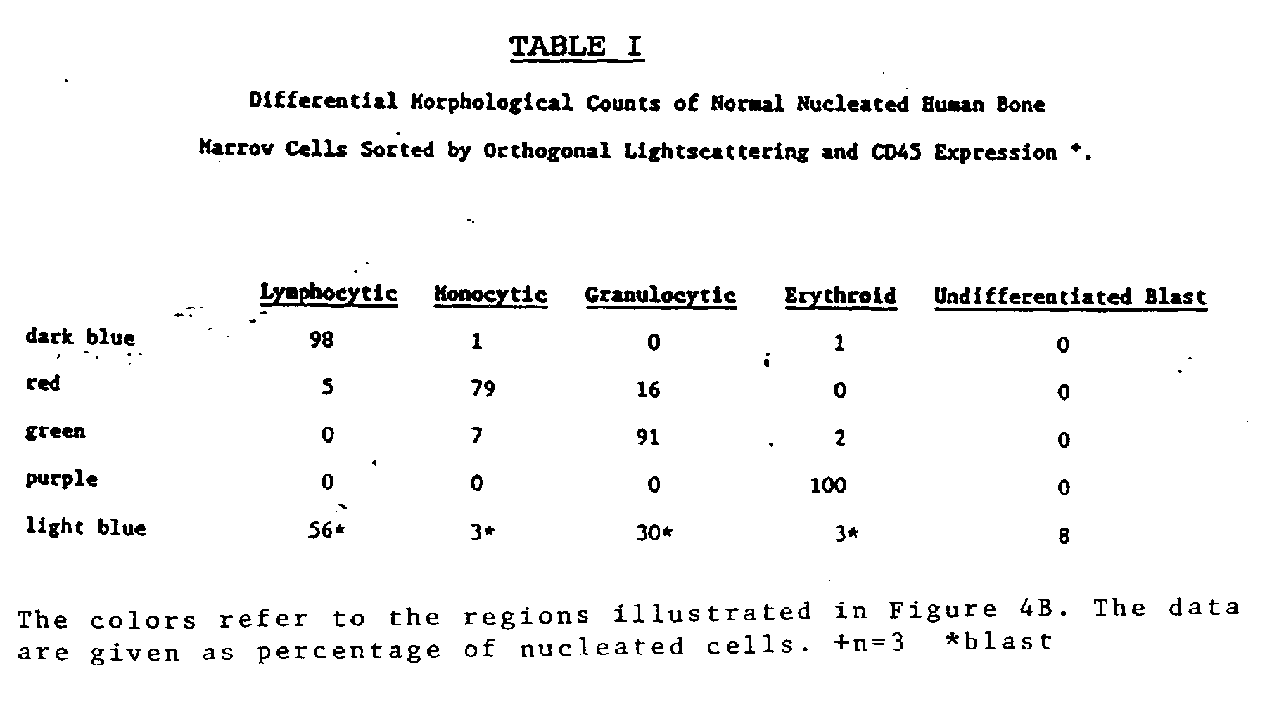
- Light microscopic examination of the sorted populations confirmed the lineage classification of these cells, however, not all of the cells were mature. Within the neutrophil population, in addition to mature neutrophils, blasts and metamyelocytes were also found. Approximately 5% of the lymphocytes were lymphoblast and 20% of the monocytes were immature monomyeloid cells.
- In contrast with peripheral blood, two populations of bone marrow cells were identified having low orthogonal light scattering signals and dimly expressing CD45 antigen. Light microscopic examination of the population ("Nuc E") of cells which expressed the lowest level of CD45 antigen revealed that it consisted exclusively of normoblasts and erythroblasts (Table I). The population ("B") expressing slightly higher amounts of CD45 antigen contained the most immature cells including monomyeloid and lymphoid precursors and a few erythroblasts (Table I).
- The utility of this technique is demonstrated in the analysis of blood from a B-CLL patient. A stage IV of RAI B-CLL patient was chosen because abnormal counts are commonly found for all blood cell lineages. The 10 to 100 fold increase in the number of lymphocytes makes the distinction of these cells from residual normal leukocytes and reticulocytes more difficult. Furthermore, the low platelet counts in these patients makes platelet enumeration less reliable.
- The blood from the B-CLL patient was incubated with LDS-751, Thiazole-Orange and HLe-1(PE) as was done for normal blood. The data are displayed in the same manner as was chosen for normal peripheral blood (FIG. 1) and bone marrow (FIG. 3).
- The frequency of cells occurring in the light scattering region typical for platelets (FIG. 5a) was low compared with normal peripheral blood (compare FIG. 1a,b with FIG. 5a,b). Only a few of the dots identified as platelets with light scattering in FIG. 5a show the fluorescence intensity with LDS-751 typical for platelets in FIG. 5b and can be considered as normal platelets. The discrimination of platelets from reticulocytes is still excellent (FIG. 5b).
- As a consequence of the large increase in frequency of nucleated cells, the separation of the leukocytes and reticulocytes obtained with LDS-751 and Thiazole-Orange is less clear. However, the leukocytes can be clearly separated from the reticulocytes based on their expression of the CD45 antigen (FIG. 5c,d).
- The light scattering properties and CD45 antigen expression of the nucleated cells (gated in FIG. 5b) is shown in FIG. 6a,b. The majority of cells dimly express CD45 antigen and exhibit low forward and orthogonal light scattering signals characteristics of immature lymphocytes. Only a few leukocytes were detected as normal, lymphocytes, granulocytes and monocytes, although the separation was not distinct as a result of the overwhelming number of immature lymphoid cells. Note the few cells which did not express CD45 antigen and which had a low orthogonal light scattering signal. See FIG. 6B. These cells were identified as nucleated erythroid cells consistent with the large increase in reticulocytes in this patient.
- Often during the removal of bone marrow aspirate, blood may be introduced into the aspirate as a contaminant to the sample. Thus, if one were to analyze the sample without correction for the blood contaminants, the leukocyte differentials could be in error.
- By running a blood sample as described in Example I above, the.percentages of white blood cell types in the blood may be calculated. Once the marrow sample is run as described in Example II, the expected percentages of white blood cells in the blood sample may be subtracted from the bone marrow sample to give a more accurate reading free from blood contaminants (assuming that in fact all the contaminants are blood derived).
- Thus, for example, lymphocytes, granulocytes and monocytes comprise 1.0, 1.0 and 0.2 percent of total cells in a blood'sample respectively, with the remaining 97.8% of the cells being essentially RBC. As a result, there are approximately one lymphocyte and granulocyte and 0.2 monocytes per 100 RBC in the blood sample. The absolute counts of the same cells in a bone marrow sample is 800, 490 and 165 respectively in a 20,000 cell sample with the remainder of the cells being essentially RBC (i.e., 18,545). Thus, for the purpose of example only, the number of lymphocytes, granulocytes and monocytes expected to be contaminating the bone marrow sample would be 185 lymphocytes (i.e., 1% x 18,545), 185 granulocytes (i.e., 1% x 18,545) and 37 monocytes (i.e., 0.2% x 18,545). The corrected counts, therefore, would be 615 lymphocytes, 305 granulocytes and 128 monocytes. By this method, more accurate counts may be obtained.
- All publications and patent applications mentioned in this specification are indicative of the level of skill of those skilled in the art to which this invention pertains.
Claims (17)
- A method for multi-parameter analysis of cells in a body fluid wherein said method comprises the steps of:1. taking a fluid sample derived from an individual;2. adding two fluorescent nucleic acid dyes to said sample to differentially label the DNA and RNA in each cell, wherein each dye has a fluorescent peak emission spectrum different from the other;3. adding a fluorescently labelled cell surface marker to said sample, wherein said marker recognizes an antigen that is differentially expressed on said cells in said fluid sample and wherein said fluorescent label has a peak emission spectrum different from said fluorescent dyes; and4. analyzing said cells in said sample in an instrument capable of detecting and recording three channels of fluorescence and two channels of light scattering for each of the cells in said sample.
- The method of Claim 1 wherein the body fluid comprises peritoneal, spinal or brain fluid, urine, whole blood or a cell suspension of bone marrow, lymph node, liver or speen.
- The method of Claim 2 wherein the body fluid is whole blood.
- The method of Claim 2 wherein the body fluid is bone marrow.
- The method of Claim 1 wherein one of the nucleic acid dyes is LDS-751 (2-{2-[4-(dimethylamino)phenyl]ethenyl}-1-ethylquinolinium iodide).
- The method of Claim 1 wherein one of the nucleic acid dyes is Thiazole-Orange (1-methyl-4-{[3-methyl-2(3H)-benzothiazolylidene]methyl}quinolinium 4-methylbenzenesulfonate).
- The method of Claim 1 wherein the cell surface marker is a MAb conjugated to a fluorochrome.
- The method of Claim 7 wherein the MAb is a CD45 MAb
- The method of Claim 8 wherein the CD45 MAb is HLe-1.
- The method of Claim 1 wherein the fluorochrome is phycoerythrin.
- The method of Claim 1 wherein the instrument is a flow cytometer.
- A method for five-parameter analysis of cells in a blood sample wherein said method comprises the steps of:1. taking a blood sample derived from an individual;2. adding LDS-751 (2-{2-[4-(dimethylamino)phenyl)ethenyl})-1-ethylquinolinium iodide) and Thiazole-Orange (1-methyl-4-{[3-methyl-2(3H)-benzothiazolylidene]methyl}quinolinium 4-methylbenzenesulfonate) to said sample;3. adding phycoerythrin-labelled HLe-1 to said sample;4. passing said sample through a flow cytometer having single laser source tuned to 488nm and capable of recording and storing measurements o orthogonal and forward light scattering and of LDS-751 (2-{2-[4-(dimethylamino)phenyl]ethenyl}-1-ethylquinolinium iodide), Thiazole-Orange 1-methyl-4-{[3-methyl-2(3H)-benzothiazolylidene)methyl)quinolinium 4-methylbenzenesulfonate and phycoerythrin fluorescence emissions for each cell in said sample.
- A method for five-parameter analysis of cells in a bone marrow sample wherein said method comprises the steps of:1. taking a bone marrow sample derived from an individual;2. adding LDS-751 (2-{2-[4-(dimethylamino)phenyl]ethenyl}-1-ethylquinolinium iodide) and Thiazole-Orange (1-methyl-4-{[3-methyl-2(3H)-benzothiazolylidene)methyl)quinolinium 4-methylbenzenesulfonate) to said sample;3. adding phycoerythrin-labelled HLe-1 to said sample;4. passing said sample through a flow cytometer having single laser source turned to 488nm and capable of recording and storing measurements of orthogonal and forward light scattering and of LDS-751 (2-{2-[4-(dimethylamino)phenyl]ethenyl}-1-ethylquinolinium iodide), Thiazole-Orange (1-methyl-4-{[3-methyl-2(3H)-benzothiazolylidene)methyl)quinolinium 4-methylbenzenesulfonate), and phycoerythrin fluorescence emissions for each cell in said sample.
- A kit for multi-parameter analysis of cells in a body fluid sample which comprises a first container containing at least two nucleic acid dyes to differentially label the DNA and RNA in each cell and wherein each dye has a fluorescent peak emission spectrum different from the other, and a second container containing at least one fluorescently labelled cell surface marker, wherein said marker recognises an antigen that is differently expressed on said cells in said fluid sample and wherein said fluorescent label has a peak emission spectrum different from said fluorescent dyes, for use in a method for multi-parameter analysis of cells in a body fluid wherein said method comprises the steps of:1. taking a fluid sample derived from an individual;2. adding two fluorescent nucleic acid dyes to said sample to differentially label the DNA and RNA in each cell, wherein each dye has a fluorescent peak emission spectrum different from the other;3. adding a fluorescently labelled cell surface marker to said sample, wherein said marker recognizes an antigen that is differentially expressed on said cells in said fluid sample and wherein said fluorescent label has a peak emission spectrum different from said fluorescent dyes; and4. analyzing said cells in said sample in an instrument capable of detecting and recording three channels of fluorescence and two channels of light scattering for each of the cells in said sample.
- The kit according to Claim 14 wherein said nucleic acid dyes comprise LDS-751 (2-{2-[4-(dimethylamino)phenyl]ethenyl}-1-ethylquinolinium iodide) and Thiazole-Orange (1-methyl-4-{[3-methyl-2(3H)-benzothiazolylidene)methyl)quinolinium 4-methylbenzenesulfonate).
- The kit according to Claim 14 wherein the fluorescently labelled cell surface marker comprises phycoerythrin-labelled HLe-1.
- A method for correcting a multi-parameter analysis of a bone marrow sample to adjust for increased white blood cell levels due to blood contamination of the bone marrow sample which comprises the steps of:1. performing the steps of the method of Claim 1 on whole blood;2. determining the percentages of various white blood cell components in the blood;3. performing the steps of the method of Claim 8;4. determining the absolute counts of various white blood cell components in the bone marrow; and5. multiplying each of the percentages obtained in step (2) by the total number of cells in the bone marrow sample and subtracting that number from the respective absolute counts obtained in step (4).
Applications Claiming Priority (2)
| Application Number | Priority Date | Filing Date | Title |
|---|---|---|---|
| US207099 | 1988-06-15 | ||
| US07/207,099 US5047321A (en) | 1988-06-15 | 1988-06-15 | Method for analysis of cellular components of a fluid |
Publications (3)
| Publication Number | Publication Date |
|---|---|
| EP0347210A2 EP0347210A2 (en) | 1989-12-20 |
| EP0347210A3 EP0347210A3 (en) | 1990-10-17 |
| EP0347210B1 true EP0347210B1 (en) | 1994-09-07 |
Family
ID=22769201
Family Applications (1)
| Application Number | Title | Priority Date | Filing Date |
|---|---|---|---|
| EP89306036A Expired - Lifetime EP0347210B1 (en) | 1988-06-15 | 1989-06-14 | Method for analysis of cellular components of a fluid |
Country Status (11)
| Country | Link |
|---|---|
| US (1) | US5047321A (en) |
| EP (1) | EP0347210B1 (en) |
| JP (1) | JPH0726954B2 (en) |
| AT (1) | ATE111227T1 (en) |
| AU (1) | AU613197B2 (en) |
| CA (1) | CA1340170C (en) |
| DE (1) | DE68918004T2 (en) |
| DK (1) | DK296889A (en) |
| ES (1) | ES2063820T3 (en) |
| FI (1) | FI94180C (en) |
| NO (1) | NO175506C (en) |
Cited By (1)
| Publication number | Priority date | Publication date | Assignee | Title |
|---|---|---|---|---|
| US7329496B2 (en) | 1990-12-06 | 2008-02-12 | Affymetrix, Inc. | Sequencing of surface immobilized polymers utilizing microflourescence detection |
Families Citing this family (100)
| Publication number | Priority date | Publication date | Assignee | Title |
|---|---|---|---|---|
| NO164622C (en) * | 1988-05-11 | 1990-10-24 | Tore Lindmo | BINAER IMMUNOMETRIC PARTICLE-BASED METHOD FOR MEASURING SPECIFIC SERUM ANTIGENS USING LIQUID FLOW MICROPHOTOMETRY AND A PREPARED TARGET SET UP THEREOF. |
| US4963478A (en) * | 1988-07-05 | 1990-10-16 | Immucor, Inc. | Article for preforming immunological assays utilizing organic dyes and method for producing and utilizing same |
| FR2638848B1 (en) * | 1988-11-04 | 1993-01-22 | Chemunex Sa | METHOD OF DETECTION AND / OR DETERMINATION IN A LIQUID OR SEMI-LIQUID MEDIUM OF AT LEAST ONE ORGANIC, BIOLOGICAL OR MEDICINAL SUBSTANCE, BY AN AGGLUTINATION METHOD |
| FR2653447B1 (en) * | 1989-10-20 | 1991-12-27 | Bio Merieux | METHOD AND REAGENTS FOR THE DETECTION OF MICROORGANISMS. |
| EP0448931B1 (en) * | 1990-01-26 | 1996-04-03 | Canon Kabushiki Kaisha | Method for measuring a specimen by the use of fluorescence light |
| US5229265A (en) * | 1990-03-13 | 1993-07-20 | Litron Laboratories | Process for analyzing clastogenic agents |
| US5401847A (en) * | 1990-03-14 | 1995-03-28 | Regents Of The University Of California | DNA complexes with dyes designed for energy transfer as fluorescent markers |
| US5622853A (en) * | 1990-05-01 | 1997-04-22 | Becton Dickinson And Company | T lymphocyte precursor |
| US5650275A (en) * | 1990-06-11 | 1997-07-22 | Nexstar Pharmacueticals Inc | Target detection method using spectroscopically detectable nucleic acid ligands |
| US5641629A (en) * | 1990-06-11 | 1997-06-24 | Nexstar Pharmacueticals Inc | Spectroscopically detectable nucleic acid ligands |
| IE76732B1 (en) * | 1990-08-07 | 1997-11-05 | Becton Dickinson Co | One step test for absolute counts |
| EP0834575B1 (en) * | 1990-12-06 | 2001-11-28 | Affymetrix, Inc. (a Delaware Corporation) | Identification of nucleic acids in samples |
| JP2927979B2 (en) * | 1991-02-22 | 1999-07-28 | シスメックス株式会社 | Classification of erythroblasts by flow cytometry |
| US5776709A (en) * | 1991-08-28 | 1998-07-07 | Becton Dickinson And Company | Method for preparation and analysis of leukocytes in whole blood |
| CA2087086A1 (en) | 1992-01-22 | 1993-07-23 | Leon Wmm Terstappen | Multidimensional cell differential analysis |
| DE69326731T2 (en) * | 1992-09-04 | 2000-05-18 | Becton Dickinson And Co., Franklin Lakes | Control particles for cell counting and instrument linearity |
| EP0608987B1 (en) * | 1993-01-26 | 2001-10-10 | Becton, Dickinson and Company | Method for detecting rare events |
| US5294799A (en) * | 1993-02-01 | 1994-03-15 | Aslund Nils R D | Apparatus for quantitative imaging of multiple fluorophores |
| EP0685994B1 (en) * | 1993-02-25 | 2007-03-21 | Abbott Laboratories | Multipurpose reagent system for rapid lysis of whole blood samples |
| JP2680931B2 (en) * | 1993-11-12 | 1997-11-19 | ベクトン・ディッキンソン・アンド・カンパニー | How to count the absolute number of rare cells |
| GB9326238D0 (en) * | 1993-12-23 | 1994-02-23 | Sinvent As | Method of assay |
| EP0684316B1 (en) * | 1994-04-29 | 2004-10-20 | Johnson & Johnson Clinical Diagnostics, Inc. | Homogeneous method for assay of double-stranded nucleic acids using fluorescent dyes and kit useful therein |
| US5891734A (en) * | 1994-08-01 | 1999-04-06 | Abbott Laboratories | Method for performing automated analysis |
| US5656499A (en) * | 1994-08-01 | 1997-08-12 | Abbott Laboratories | Method for performing automated hematology and cytometry analysis |
| US5631165A (en) * | 1994-08-01 | 1997-05-20 | Abbott Laboratories | Method for performing automated hematology and cytometry analysis |
| US5879900A (en) * | 1994-12-15 | 1999-03-09 | Abbott Laboratories | Method for simultaneous analysis of cell viability, nucleated red blood cells and white blood cell differentials |
| US5559037A (en) * | 1994-12-15 | 1996-09-24 | Abbott Laboratories | Method for rapid and simultaneous analysis of nucleated red blood cells |
| US6329158B1 (en) * | 1995-09-15 | 2001-12-11 | Becton Dickinson And Company | Use of dimly fluorescing nucleic acid dyes in the identification of nucleated cells |
| DE69638209D1 (en) * | 1995-12-15 | 2010-08-12 | Abbott Lab | METHOD FOR SIMULTANEOUS ANALYSIS OF CELL LIFE, NUCLEAR RED BLOOD BODIES, AND LEUKOCYTE DIFFERENTIAL |
| TW438973B (en) * | 1995-12-19 | 2001-06-07 | Sysmex Corp | Apparatus and method for analyzing solid components in urine |
| JP3308441B2 (en) * | 1995-12-19 | 2002-07-29 | シスメックス株式会社 | Urine particle analyzer |
| JP3783808B2 (en) * | 1997-05-19 | 2006-06-07 | シスメックス株式会社 | Leukocyte classification and counting reagent |
| DE19756782A1 (en) * | 1997-12-19 | 1999-06-24 | Dade Behring Marburg Gmbh | Detection and determination of solid phase associated factors |
| US6911313B2 (en) * | 1998-02-06 | 2005-06-28 | Sysmex Corporation | Process for discriminating and counting erythroblasts |
| DE19857692C1 (en) | 1998-12-14 | 2000-08-24 | Evotec Biosystems Ag | Method and device for cell trace-based cell examination |
| JP3817091B2 (en) * | 1999-07-02 | 2006-08-30 | テルモ株式会社 | Cell number measuring device |
| JP3929283B2 (en) * | 2000-11-08 | 2007-06-13 | シスメックス株式会社 | Bone marrow nucleated cell classification and counting method |
| US6403378B1 (en) * | 2001-04-26 | 2002-06-11 | Guava Technologies, Inc. | Cell viability assay reagent |
| GB0118995D0 (en) * | 2001-08-03 | 2001-09-26 | Univ Wales Medicine | Detection of mutations in nucleic acids |
| WO2003016866A2 (en) * | 2001-08-16 | 2003-02-27 | Litron Laboratories Ltd | Method for the enumeration of mammalian micronucleated erythrocyte populations with a single-laser flow cytometer |
| PT1363126E (en) * | 2002-05-14 | 2006-05-31 | Univ Salamanca | DIFFERENTIAL MULTIDIMENSIONAL LEUKOCYTE ANALYSIS |
| JP4299597B2 (en) | 2002-07-29 | 2009-07-22 | シスメックス株式会社 | Hematology analyzer and method |
| US7507548B2 (en) | 2003-03-04 | 2009-03-24 | University Of Salamanca | Multidimensional detection of aberrant phenotypes in neoplastic cells to be used to monitor minimal disease levels using flow cytometry measurements |
| US7674598B2 (en) * | 2004-05-21 | 2010-03-09 | Beckman Coulter, Inc. | Method for a fully automated monoclonal antibody-based extended differential |
| US7625712B2 (en) | 2004-05-21 | 2009-12-01 | Beckman Coulter, Inc. | Method for a fully automated monoclonal antibody-based extended differential |
| US7321843B2 (en) * | 2005-09-30 | 2008-01-22 | Universidad De Salamanca | Method for generating new flow cytometry data files containing an infinite number of dimensions based on data estimation |
| US8958990B2 (en) * | 2005-10-03 | 2015-02-17 | Sysmex Corporation | Sample analyzer and sample analyzing method |
| US20090170091A1 (en) * | 2006-01-17 | 2009-07-02 | Kenneth Giuliano | Method For Predicting Biological Systems Responses |
| EP2027465A2 (en) | 2006-05-17 | 2009-02-25 | Cellumen, Inc. | Method for automated tissue analysis |
| WO2008060483A2 (en) * | 2006-11-10 | 2008-05-22 | Cellumen, Inc. | Protein-protein interaction biosensors and methods of use thereof |
| EP2215219A4 (en) * | 2007-10-29 | 2011-04-13 | Beckman Coulter Inc | Method for a rapid antibody-based analysis of platelet populations |
| US8159670B2 (en) * | 2007-11-05 | 2012-04-17 | Abbott Laboratories | Method and apparatus for rapidly counting and identifying biological particles in a flow stream |
| DE102008030515A1 (en) * | 2008-06-27 | 2009-12-31 | Christian Seliger | Flow cytometric method for the determination of total leukocyte count and platelet count as well as leukocyte differentiation in blood samples from birds |
| WO2010056732A1 (en) | 2008-11-12 | 2010-05-20 | Marv Enterprises Llc | Utilization of stents for the treatment of blood borne carcinomas |
| EP2359164A4 (en) * | 2008-12-10 | 2012-05-30 | Abqmr Inc | Nuclear magnetic resonance apparatus, methods and associated technology |
| US9216386B2 (en) | 2009-03-17 | 2015-12-22 | Marv Enterprises, LLC | Sequential extracorporeal treatment of bodily fluids |
| JP5452058B2 (en) * | 2009-03-31 | 2014-03-26 | シスメックス株式会社 | Blood analyzer |
| EP2259065A1 (en) | 2009-06-03 | 2010-12-08 | Erasmus University Medical Center Rotterdam | Methods, reagents and kits for flow cytometric immunophenotyping |
| JP2011092104A (en) * | 2009-10-30 | 2011-05-12 | Hitachi Engineering & Services Co Ltd | Test method and test apparatus for microorganism or the like |
| SG174650A1 (en) * | 2010-03-31 | 2011-10-28 | Agency Science Tech & Res | A method of monitoring parasite development in blood |
| US9428547B2 (en) | 2010-04-21 | 2016-08-30 | Dna Electronics, Inc. | Compositions for isolating a target analyte from a heterogeneous sample |
| US8841104B2 (en) | 2010-04-21 | 2014-09-23 | Nanomr, Inc. | Methods for isolating a target analyte from a heterogeneous sample |
| US20110262989A1 (en) | 2010-04-21 | 2011-10-27 | Nanomr, Inc. | Isolating a target analyte from a body fluid |
| US9476812B2 (en) | 2010-04-21 | 2016-10-25 | Dna Electronics, Inc. | Methods for isolating a target analyte from a heterogeneous sample |
| US10018631B2 (en) | 2011-03-17 | 2018-07-10 | Cernostics, Inc. | Systems and compositions for diagnosing Barrett's esophagus and methods of using the same |
| CN103917868B (en) * | 2011-05-04 | 2016-08-24 | 雅培制药有限公司 | Basophilic granulocyte analyzes system and method |
| US20150027950A1 (en) * | 2012-03-27 | 2015-01-29 | Marv Enterprises, LLC | Treatment for atherosclerosis |
| WO2013177099A1 (en) * | 2012-05-21 | 2013-11-28 | Felder Mitchell S | Method for treating infectious diseases using emissive energy |
| US9995742B2 (en) | 2012-12-19 | 2018-06-12 | Dnae Group Holdings Limited | Sample entry |
| US9599610B2 (en) | 2012-12-19 | 2017-03-21 | Dnae Group Holdings Limited | Target capture system |
| US10000557B2 (en) | 2012-12-19 | 2018-06-19 | Dnae Group Holdings Limited | Methods for raising antibodies |
| US9551704B2 (en) | 2012-12-19 | 2017-01-24 | Dna Electronics, Inc. | Target detection |
| US9804069B2 (en) | 2012-12-19 | 2017-10-31 | Dnae Group Holdings Limited | Methods for degrading nucleic acid |
| US9434940B2 (en) | 2012-12-19 | 2016-09-06 | Dna Electronics, Inc. | Methods for universal target capture |
| WO2016089521A1 (en) | 2014-12-04 | 2016-06-09 | Becton, Dickinson And Company | Flow cytometry cell sorting systems and methods of using the same |
| WO2016093970A1 (en) | 2014-12-10 | 2016-06-16 | Becton, Dickinson And Company | Methods for optically aligning light collection components and optically aligned light collection systems thereof |
| WO2016099538A1 (en) | 2014-12-19 | 2016-06-23 | Captl Llc | Flow cytometry using hydrodynamically planar flow |
| US10900885B2 (en) | 2014-12-19 | 2021-01-26 | Captl Llc | Flow cytometry using hydrodynamically planar flow |
| EP3259574B1 (en) | 2015-02-18 | 2024-03-27 | Becton, Dickinson and Company | Optical detection systems and methods of using the same |
| CN107923836B (en) | 2015-06-17 | 2020-12-01 | 贝克顿·迪金森公司 | Optical detector diffuser cap assembly with removable diffuser rod and method of use |
| GB201601073D0 (en) * | 2016-01-20 | 2016-03-02 | Ucb Biopharma Sprl | Antibodies |
| KR102600559B1 (en) | 2015-10-13 | 2023-11-10 | 벡톤 디킨슨 앤드 컴퍼니 | Multimodal fluorescence imaging flow cytometry system |
| WO2017070315A1 (en) * | 2015-10-23 | 2017-04-27 | Geosyntec Consultants, Inc. | Use of visibly detectable compounds as performance reference compounds in passive sampling devices |
| EP3426296A4 (en) | 2016-03-10 | 2019-11-20 | Becton, Dickinson and Company | Methods of evaluating a cellular sample for her-2/neu expression and compositions for practicing the same |
| KR102527830B1 (en) | 2016-03-17 | 2023-05-02 | 벡톤 디킨슨 앤드 컴퍼니 | Cell sorting using a high throughput fluorescence flow cytometer |
| US11408812B2 (en) | 2016-04-22 | 2022-08-09 | Becton, Dickinson And Company | High density deposition for array production |
| KR102381176B1 (en) | 2016-05-12 | 2022-04-01 | 비디 바이오사이언시스 | Fluorescence Imaging Flow Cytometry with Improved Image Resolution |
| CN109477785B (en) | 2016-09-13 | 2022-09-27 | 贝克顿·迪金森公司 | Flow cytometer with optical equalization |
| CN109073530B (en) | 2016-10-03 | 2022-04-26 | 贝克顿·迪金森公司 | Method and system for determining drop delay of fluid flow in flow cytometer |
| US10684275B2 (en) | 2016-12-14 | 2020-06-16 | Becton, Dickinson And Company | Methods and compositions for obtaining a tuberculosis assessment in a subject |
| CN110337331B (en) | 2017-02-08 | 2022-04-26 | 贝克顿·迪金森公司 | Dry dye reagent device and methods of making and using the same |
| EP3586104A4 (en) | 2017-02-27 | 2020-12-16 | Becton, Dickinson and Company | LIGHT DETECTION SYSTEMS AND METHOD OF USING THEM |
| KR102838795B1 (en) | 2018-01-23 | 2025-07-25 | 벡톤 디킨슨 앤드 컴퍼니 | Systems for dynamic light detection obscuration and methods for using thereof |
| EP3775052B1 (en) | 2018-03-30 | 2024-06-05 | Becton, Dickinson and Company | Water-soluble polymeric dyes having pendant chromophores |
| EP3785017B1 (en) | 2018-04-27 | 2025-09-10 | Becton, Dickinson And Company | Collection systems for flow cytometrically sorted samples and methods of using the same |
| JP7416729B2 (en) | 2018-06-19 | 2024-01-17 | ベクトン・ディキンソン・アンド・カンパニー | Variable multiplexing switch, system, and method of use for detector arrays |
| CN113785187B (en) | 2019-03-22 | 2025-01-28 | 贝克顿·迪金森公司 | Spectral unmixing of fluorescence imaging using radiofrequency multiplexed excitation data |
| JP2024518755A (en) | 2021-04-23 | 2024-05-02 | ベクトン・ディキンソン・アンド・カンパニー | Fluid management system for an analyzer and/or classifier type flow particle analyzer - Patent Application 20070123333 |
| US20250137905A1 (en) | 2023-10-27 | 2025-05-01 | Becton, Dickinson And Company | Fluid Supply Systems Having a Flow Control Circuit and Single Fluidic Connection, and Methods of Use Thereof |
| BG113932A (en) | 2024-07-19 | 2026-01-30 | Медицински университет – Пловдив | PANELS AND REAGENT KITS FOR MEASURING MINIMAL RESIDUAL DISEASE IN CHILDHOOD B-CELL PRECURSOR ACUTE LYMPHOBLAST LEUKEMIA THROUGH IMMUNOPHEOTYPING |
Family Cites Families (11)
| Publication number | Priority date | Publication date | Assignee | Title |
|---|---|---|---|---|
| US3826364A (en) * | 1972-05-22 | 1974-07-30 | Univ Leland Stanford Junior | Particle sorting method and apparatus |
| US4284412A (en) * | 1979-07-13 | 1981-08-18 | Ortho Diagnostics, Inc. | Method and apparatus for automated identification and enumeration of specified blood cell subclasses |
| US4544546A (en) * | 1980-04-21 | 1985-10-01 | Abbott Laboratories | Fluorescent nucleic acid stains |
| US4520110A (en) * | 1981-10-06 | 1985-05-28 | The Board Of Trustees Of The Leland Stanford Junior University | Fluorescent immunoassay employing a phycobiliprotein labeled ligand or receptor |
| CA1254134A (en) * | 1984-03-28 | 1989-05-16 | Mount Sinai School Of Medicine Of The City University Of New York | Specific binding flow cytometry method |
| US4661913A (en) * | 1984-09-11 | 1987-04-28 | Becton, Dickinson And Company | Apparatus and method for the detection and classification of articles using flow cytometry techniques |
| US4727020A (en) * | 1985-02-25 | 1988-02-23 | Becton, Dickinson And Company | Method for analysis of subpopulations of blood cells |
| CA1279008C (en) * | 1985-10-11 | 1991-01-15 | Smith Kline & French Canada Ltd. | Methods and reagents for performing subset analysis |
| ZA867698B (en) * | 1985-10-11 | 1987-07-29 | Smithkline Beckman Corp | Methods and reagents for performing subset analysis |
| US4883867A (en) * | 1985-11-01 | 1989-11-28 | Becton, Dickinson And Company | Detection of reticulocytes, RNA or DNA |
| US5016283A (en) * | 1985-11-04 | 1991-05-14 | Cell Analysis Systems, Inc. | Methods and apparatus for immunoploidy analysis |
-
1988
- 1988-06-15 US US07/207,099 patent/US5047321A/en not_active Expired - Lifetime
-
1989
- 1989-06-01 AU AU35961/89A patent/AU613197B2/en not_active Ceased
- 1989-06-01 CA CA000601444A patent/CA1340170C/en not_active Expired - Fee Related
- 1989-06-05 NO NO892302A patent/NO175506C/en unknown
- 1989-06-14 AT AT89306036T patent/ATE111227T1/en not_active IP Right Cessation
- 1989-06-14 DE DE68918004T patent/DE68918004T2/en not_active Expired - Lifetime
- 1989-06-14 FI FI892926A patent/FI94180C/en not_active IP Right Cessation
- 1989-06-14 EP EP89306036A patent/EP0347210B1/en not_active Expired - Lifetime
- 1989-06-14 ES ES89306036T patent/ES2063820T3/en not_active Expired - Lifetime
- 1989-06-15 DK DK296889A patent/DK296889A/en not_active Application Discontinuation
- 1989-06-15 JP JP1153564A patent/JPH0726954B2/en not_active Expired - Lifetime
Cited By (2)
| Publication number | Priority date | Publication date | Assignee | Title |
|---|---|---|---|---|
| US7329496B2 (en) | 1990-12-06 | 2008-02-12 | Affymetrix, Inc. | Sequencing of surface immobilized polymers utilizing microflourescence detection |
| US7459275B2 (en) | 1990-12-06 | 2008-12-02 | Affymetrix, Inc. | Sequencing of surface immobilized polymers utilizing microfluorescence detection |
Also Published As
| Publication number | Publication date |
|---|---|
| AU613197B2 (en) | 1991-07-25 |
| DK296889A (en) | 1989-12-16 |
| US5047321A (en) | 1991-09-10 |
| NO892302L (en) | 1989-12-18 |
| AU3596189A (en) | 1989-12-21 |
| EP0347210A3 (en) | 1990-10-17 |
| FI892926L (en) | 1989-12-16 |
| FI94180B (en) | 1995-04-13 |
| ATE111227T1 (en) | 1994-09-15 |
| ES2063820T3 (en) | 1995-01-16 |
| FI892926A0 (en) | 1989-06-14 |
| EP0347210A2 (en) | 1989-12-20 |
| NO175506B (en) | 1994-07-11 |
| JPH0273157A (en) | 1990-03-13 |
| DE68918004D1 (en) | 1994-10-13 |
| DE68918004T2 (en) | 1995-01-05 |
| FI94180C (en) | 1995-07-25 |
| JPH0726954B2 (en) | 1995-03-29 |
| DK296889D0 (en) | 1989-06-15 |
| NO175506C (en) | 1994-10-19 |
| CA1340170C (en) | 1998-12-08 |
| NO892302D0 (en) | 1989-06-05 |
Similar Documents
| Publication | Publication Date | Title |
|---|---|---|
| EP0347210B1 (en) | Method for analysis of cellular components of a fluid | |
| EP0347039B1 (en) | Method for discriminating between intact and damaged cells in a sample | |
| EP2267443B1 (en) | Determination of white blood cell differential and reticulocyte counts | |
| EP1250585B1 (en) | Method and apparatus for analyzing cells in a whole blood sample | |
| Terstappen et al. | Five‐dimensional flow cytometry as a new approach for blood and bone marrow differentials | |
| US7625712B2 (en) | Method for a fully automated monoclonal antibody-based extended differential | |
| US5776709A (en) | Method for preparation and analysis of leukocytes in whole blood | |
| CA2207396C (en) | Method for rapid and simultaneous analysis of nucleated red blood cells | |
| EP0552707B1 (en) | Multidimensional cell differential analysis | |
| JPH04252957A (en) | One-step test for absolute count | |
| JP2005506525A (en) | Methods for measuring nucleated red blood cells | |
| EP0586183B1 (en) | Control particles for cell counting and instrument linearity | |
| EP0559208B1 (en) | Method for preparation and analysis of leukocytes in whole blood utilizing flow cytometry | |
| Terstappen et al. | Multidimensional flow cvtometric blood cell differentiation lVithoutErvthrocvteLvsis | |
| EP0866960B1 (en) | Method for simultaneous analysis of cell viability, nucleated red blood cells and white blood cell differential | |
| WO1997021994A9 (en) | Method for simultaneous analysis of cell viability, nucleated red blood cells and white blood cell differential | |
| EP0259833A2 (en) | Reagent and method for classifying leukocytes by flow cytometry | |
| Weaver et al. | Flow cytometry in the biomedical arena |
Legal Events
| Date | Code | Title | Description |
|---|---|---|---|
| PUAI | Public reference made under article 153(3) epc to a published international application that has entered the european phase |
Free format text: ORIGINAL CODE: 0009012 |
|
| AK | Designated contracting states |
Kind code of ref document: A2 Designated state(s): AT BE CH DE ES FR GB GR IT LI LU NL SE |
|
| PUAL | Search report despatched |
Free format text: ORIGINAL CODE: 0009013 |
|
| AK | Designated contracting states |
Kind code of ref document: A3 Designated state(s): AT BE CH DE ES FR GB GR IT LI LU NL SE |
|
| 17P | Request for examination filed |
Effective date: 19901205 |
|
| 17Q | First examination report despatched |
Effective date: 19920821 |
|
| GRAA | (expected) grant |
Free format text: ORIGINAL CODE: 0009210 |
|
| AK | Designated contracting states |
Kind code of ref document: B1 Designated state(s): AT BE CH DE ES FR GB GR IT LI LU NL SE |
|
| PG25 | Lapsed in a contracting state [announced via postgrant information from national office to epo] |
Ref country code: GR Free format text: LAPSE BECAUSE OF FAILURE TO SUBMIT A TRANSLATION OF THE DESCRIPTION OR TO PAY THE FEE WITHIN THE PRESCRIBED TIME-LIMIT Effective date: 19940907 Ref country code: CH Effective date: 19940907 Ref country code: LI Effective date: 19940907 Ref country code: AT Effective date: 19940907 |
|
| REF | Corresponds to: |
Ref document number: 111227 Country of ref document: AT Date of ref document: 19940915 Kind code of ref document: T |
|
| ITF | It: translation for a ep patent filed | ||
| REF | Corresponds to: |
Ref document number: 68918004 Country of ref document: DE Date of ref document: 19941013 |
|
| ET | Fr: translation filed | ||
| PG25 | Lapsed in a contracting state [announced via postgrant information from national office to epo] |
Ref country code: SE Effective date: 19941207 |
|
| REG | Reference to a national code |
Ref country code: CH Ref legal event code: PL |
|
| REG | Reference to a national code |
Ref country code: ES Ref legal event code: FG2A Ref document number: 2063820 Country of ref document: ES Kind code of ref document: T3 |
|
| PG25 | Lapsed in a contracting state [announced via postgrant information from national office to epo] |
Ref country code: LU Free format text: LAPSE BECAUSE OF NON-PAYMENT OF DUE FEES Effective date: 19950630 |
|
| PLBE | No opposition filed within time limit |
Free format text: ORIGINAL CODE: 0009261 |
|
| STAA | Information on the status of an ep patent application or granted ep patent |
Free format text: STATUS: NO OPPOSITION FILED WITHIN TIME LIMIT |
|
| 26N | No opposition filed | ||
| PGFP | Annual fee paid to national office [announced via postgrant information from national office to epo] |
Ref country code: ES Payment date: 19980619 Year of fee payment: 10 |
|
| PGFP | Annual fee paid to national office [announced via postgrant information from national office to epo] |
Ref country code: NL Payment date: 19980629 Year of fee payment: 10 |
|
| PGFP | Annual fee paid to national office [announced via postgrant information from national office to epo] |
Ref country code: BE Payment date: 19980813 Year of fee payment: 10 |
|
| PG25 | Lapsed in a contracting state [announced via postgrant information from national office to epo] |
Ref country code: ES Free format text: LAPSE BECAUSE OF NON-PAYMENT OF DUE FEES Effective date: 19990615 |
|
| PG25 | Lapsed in a contracting state [announced via postgrant information from national office to epo] |
Ref country code: BE Free format text: LAPSE BECAUSE OF NON-PAYMENT OF DUE FEES Effective date: 19990630 |
|
| BERE | Be: lapsed |
Owner name: BECTON DICKINSON AND CY Effective date: 19990630 |
|
| PG25 | Lapsed in a contracting state [announced via postgrant information from national office to epo] |
Ref country code: NL Free format text: LAPSE BECAUSE OF NON-PAYMENT OF DUE FEES Effective date: 20000101 |
|
| NLV4 | Nl: lapsed or anulled due to non-payment of the annual fee |
Effective date: 20000101 |
|
| REG | Reference to a national code |
Ref country code: ES Ref legal event code: FD2A Effective date: 20010503 |
|
| REG | Reference to a national code |
Ref country code: GB Ref legal event code: IF02 |
|
| PGFP | Annual fee paid to national office [announced via postgrant information from national office to epo] |
Ref country code: IT Payment date: 20080626 Year of fee payment: 20 |
|
| PGFP | Annual fee paid to national office [announced via postgrant information from national office to epo] |
Ref country code: DE Payment date: 20080731 Year of fee payment: 20 |
|
| PGFP | Annual fee paid to national office [announced via postgrant information from national office to epo] |
Ref country code: FR Payment date: 20080617 Year of fee payment: 20 |
|
| PGFP | Annual fee paid to national office [announced via postgrant information from national office to epo] |
Ref country code: GB Payment date: 20080627 Year of fee payment: 20 |
|
| REG | Reference to a national code |
Ref country code: GB Ref legal event code: PE20 Expiry date: 20090613 |
|
| PG25 | Lapsed in a contracting state [announced via postgrant information from national office to epo] |
Ref country code: GB Free format text: LAPSE BECAUSE OF EXPIRATION OF PROTECTION Effective date: 20090613 |