EP2686688B1 - Systems and compositions for diagnosing barrett's esophagus and methods of using the same - Google Patents
Systems and compositions for diagnosing barrett's esophagus and methods of using the same Download PDFInfo
- Publication number
- EP2686688B1 EP2686688B1 EP12757085.1A EP12757085A EP2686688B1 EP 2686688 B1 EP2686688 B1 EP 2686688B1 EP 12757085 A EP12757085 A EP 12757085A EP 2686688 B1 EP2686688 B1 EP 2686688B1
- Authority
- EP
- European Patent Office
- Prior art keywords
- percentile
- ratio
- cytoplasm
- cell
- subject
- Prior art date
- Legal status (The legal status is an assumption and is not a legal conclusion. Google has not performed a legal analysis and makes no representation as to the accuracy of the status listed.)
- Active
Links
- 208000023514 Barrett esophagus Diseases 0.000 title claims description 172
- 208000023665 Barrett oesophagus Diseases 0.000 title claims description 172
- 238000000034 method Methods 0.000 title claims description 112
- 239000000203 mixture Substances 0.000 title description 13
- 210000004027 cell Anatomy 0.000 claims description 355
- 239000000523 sample Substances 0.000 claims description 287
- 239000000090 biomarker Substances 0.000 claims description 253
- 210000004940 nucleus Anatomy 0.000 claims description 122
- 210000000805 cytoplasm Anatomy 0.000 claims description 113
- 206010058314 Dysplasia Diseases 0.000 claims description 95
- 210000000170 cell membrane Anatomy 0.000 claims description 80
- 210000001519 tissue Anatomy 0.000 claims description 78
- -1 Ki-67 Proteins 0.000 claims description 68
- 102100025064 Cellular tumor antigen p53 Human genes 0.000 claims description 62
- 101000721661 Homo sapiens Cellular tumor antigen p53 Proteins 0.000 claims description 61
- 102100032700 Keratin, type I cytoskeletal 20 Human genes 0.000 claims description 59
- 102100038280 Prostaglandin G/H synthase 2 Human genes 0.000 claims description 58
- 102100025136 Macrosialin Human genes 0.000 claims description 57
- 101000934372 Homo sapiens Macrosialin Proteins 0.000 claims description 56
- 102000015735 Beta-catenin Human genes 0.000 claims description 49
- 108060000903 Beta-catenin Proteins 0.000 claims description 49
- 102100022875 Hypoxia-inducible factor 1-alpha Human genes 0.000 claims description 48
- 101001046870 Homo sapiens Hypoxia-inducible factor 1-alpha Proteins 0.000 claims description 45
- 239000012528 membrane Substances 0.000 claims description 33
- 102000003945 NF-kappa B Human genes 0.000 claims description 32
- 108010057466 NF-kappa B Proteins 0.000 claims description 32
- 238000001574 biopsy Methods 0.000 claims description 28
- RNAMYOYQYRYFQY-UHFFFAOYSA-N 2-(4,4-difluoropiperidin-1-yl)-6-methoxy-n-(1-propan-2-ylpiperidin-4-yl)-7-(3-pyrrolidin-1-ylpropoxy)quinazolin-4-amine Chemical class N1=C(N2CCC(F)(F)CC2)N=C2C=C(OCCCN3CCCC3)C(OC)=CC2=C1NC1CCN(C(C)C)CC1 RNAMYOYQYRYFQY-UHFFFAOYSA-N 0.000 claims description 26
- 210000000633 nuclear envelope Anatomy 0.000 claims description 26
- 210000000130 stem cell Anatomy 0.000 claims description 12
- 238000001514 detection method Methods 0.000 claims description 11
- 101000994460 Homo sapiens Keratin, type I cytoskeletal 20 Proteins 0.000 claims description 9
- 108050003267 Prostaglandin G/H synthase 2 Proteins 0.000 claims description 9
- WSFSSNUMVMOOMR-UHFFFAOYSA-N Formaldehyde Chemical compound O=C WSFSSNUMVMOOMR-UHFFFAOYSA-N 0.000 claims description 8
- 206010030155 Oesophageal carcinoma Diseases 0.000 claims description 8
- 208000000461 Esophageal Neoplasms Diseases 0.000 claims description 7
- 201000004101 esophageal cancer Diseases 0.000 claims description 7
- 239000012188 paraffin wax Substances 0.000 claims description 6
- 101000605127 Homo sapiens Prostaglandin G/H synthase 2 Proteins 0.000 claims description 5
- 230000000694 effects Effects 0.000 claims description 5
- 101000725401 Homo sapiens Cytochrome c oxidase subunit 2 Proteins 0.000 claims description 4
- 230000001680 brushing effect Effects 0.000 claims description 4
- 238000002271 resection Methods 0.000 claims description 4
- LFQSCWFLJHTTHZ-UHFFFAOYSA-N Ethanol Chemical compound CCO LFQSCWFLJHTTHZ-UHFFFAOYSA-N 0.000 claims description 3
- 102100027456 Cytochrome c oxidase subunit 2 Human genes 0.000 claims 1
- 101710183663 Keratin, type I cytoskeletal 20 Proteins 0.000 claims 1
- 102100040410 Alpha-methylacyl-CoA racemase Human genes 0.000 description 90
- 108010044434 Alpha-methylacyl-CoA racemase Proteins 0.000 description 71
- 102100024458 Cyclin-dependent kinase inhibitor 2A Human genes 0.000 description 61
- 108010066370 Keratin-20 Proteins 0.000 description 48
- 102100031358 Urokinase-type plasminogen activator Human genes 0.000 description 48
- 230000014509 gene expression Effects 0.000 description 46
- 108010037462 Cyclooxygenase 2 Proteins 0.000 description 45
- 108010083123 CDX2 Transcription Factor Proteins 0.000 description 39
- 238000004458 analytical method Methods 0.000 description 39
- 238000003384 imaging method Methods 0.000 description 34
- 108090000623 proteins and genes Proteins 0.000 description 34
- 102100027581 Forkhead box protein P3 Human genes 0.000 description 32
- 101000861452 Homo sapiens Forkhead box protein P3 Proteins 0.000 description 32
- 102100031988 Tumor necrosis factor ligand superfamily member 6 Human genes 0.000 description 32
- 206010028980 Neoplasm Diseases 0.000 description 31
- 238000009826 distribution Methods 0.000 description 31
- 208000036764 Adenocarcinoma of the esophagus Diseases 0.000 description 29
- 206010030137 Oesophageal adenocarcinoma Diseases 0.000 description 29
- 208000037265 diseases, disorders, signs and symptoms Diseases 0.000 description 29
- 208000028653 esophageal adenocarcinoma Diseases 0.000 description 29
- 230000006870 function Effects 0.000 description 29
- 102100024165 G1/S-specific cyclin-D1 Human genes 0.000 description 28
- 101000738771 Homo sapiens Receptor-type tyrosine-protein phosphatase C Proteins 0.000 description 28
- 102100023832 Prolyl endopeptidase FAP Human genes 0.000 description 28
- 102100037422 Receptor-type tyrosine-protein phosphatase C Human genes 0.000 description 28
- 102100036011 T-cell surface glycoprotein CD4 Human genes 0.000 description 28
- 210000000981 epithelium Anatomy 0.000 description 28
- 108090001005 Interleukin-6 Proteins 0.000 description 27
- 108010039471 Fas Ligand Protein Proteins 0.000 description 26
- 101001012157 Homo sapiens Receptor tyrosine-protein kinase erbB-2 Proteins 0.000 description 26
- 102100030086 Receptor tyrosine-protein kinase erbB-2 Human genes 0.000 description 26
- 108010072257 fibroblast activation protein alpha Proteins 0.000 description 26
- 108010058546 Cyclin D1 Proteins 0.000 description 25
- 101100044298 Drosophila melanogaster fand gene Proteins 0.000 description 25
- 101150064015 FAS gene Proteins 0.000 description 25
- 101000716102 Homo sapiens T-cell surface glycoprotein CD4 Proteins 0.000 description 25
- 102000000380 Matrix Metalloproteinase 1 Human genes 0.000 description 25
- 101100335198 Pneumocystis carinii fol1 gene Proteins 0.000 description 25
- 239000003550 marker Substances 0.000 description 25
- 102100031671 Homeobox protein CDX-2 Human genes 0.000 description 24
- 101000638886 Homo sapiens Urokinase-type plasminogen activator Proteins 0.000 description 24
- 102100026019 Interleukin-6 Human genes 0.000 description 24
- 108010016113 Matrix Metalloproteinase 1 Proteins 0.000 description 24
- 102000052116 epidermal growth factor receptor activity proteins Human genes 0.000 description 24
- 108700015053 epidermal growth factor receptor activity proteins Proteins 0.000 description 24
- 210000004907 gland Anatomy 0.000 description 24
- YOHYSYJDKVYCJI-UHFFFAOYSA-N n-[3-[[6-[3-(trifluoromethyl)anilino]pyrimidin-4-yl]amino]phenyl]cyclopropanecarboxamide Chemical compound FC(F)(F)C1=CC=CC(NC=2N=CN=C(NC=3C=C(NC(=O)C4CC4)C=CC=3)C=2)=C1 YOHYSYJDKVYCJI-UHFFFAOYSA-N 0.000 description 24
- 108090000435 Urokinase-type plasminogen activator Proteins 0.000 description 23
- 101150029707 ERBB2 gene Proteins 0.000 description 22
- 108010046722 Thrombospondin 1 Proteins 0.000 description 22
- 102100036034 Thrombospondin-1 Human genes 0.000 description 22
- 108010031154 Transcription Factor RelA Proteins 0.000 description 22
- 210000002919 epithelial cell Anatomy 0.000 description 22
- 208000018522 Gastrointestinal disease Diseases 0.000 description 21
- 102000005747 Transcription Factor RelA Human genes 0.000 description 21
- 229940100601 interleukin-6 Drugs 0.000 description 21
- 210000004379 membrane Anatomy 0.000 description 21
- 208000010643 digestive system disease Diseases 0.000 description 20
- 208000018685 gastrointestinal system disease Diseases 0.000 description 20
- 230000001413 cellular effect Effects 0.000 description 19
- 238000003745 diagnosis Methods 0.000 description 19
- 201000010099 disease Diseases 0.000 description 19
- 238000013500 data storage Methods 0.000 description 18
- 102000006277 CDX2 Transcription Factor Human genes 0.000 description 17
- 230000003287 optical effect Effects 0.000 description 17
- 102000004169 proteins and genes Human genes 0.000 description 17
- 239000012099 Alexa Fluor family Substances 0.000 description 15
- 230000000877 morphologic effect Effects 0.000 description 15
- 150000001413 amino acids Chemical class 0.000 description 14
- 238000005259 measurement Methods 0.000 description 14
- 238000010191 image analysis Methods 0.000 description 13
- 102100035100 Transcription factor p65 Human genes 0.000 description 12
- 238000012545 processing Methods 0.000 description 12
- 201000011510 cancer Diseases 0.000 description 11
- 238000011282 treatment Methods 0.000 description 11
- 210000000349 chromosome Anatomy 0.000 description 10
- 102000039446 nucleic acids Human genes 0.000 description 10
- 108020004707 nucleic acids Proteins 0.000 description 10
- 150000007523 nucleic acids Chemical class 0.000 description 10
- 230000008569 process Effects 0.000 description 10
- 239000000427 antigen Substances 0.000 description 9
- 108091007433 antigens Proteins 0.000 description 9
- 102000036639 antigens Human genes 0.000 description 9
- 230000002596 correlated effect Effects 0.000 description 9
- 230000004043 responsiveness Effects 0.000 description 9
- 238000012360 testing method Methods 0.000 description 9
- 230000004069 differentiation Effects 0.000 description 8
- 208000035475 disorder Diseases 0.000 description 8
- 210000002175 goblet cell Anatomy 0.000 description 8
- 238000013517 stratification Methods 0.000 description 8
- 101710124574 Synaptotagmin-1 Proteins 0.000 description 7
- 239000012620 biological material Substances 0.000 description 7
- 210000002230 centromere Anatomy 0.000 description 7
- 238000004891 communication Methods 0.000 description 7
- 210000004443 dendritic cell Anatomy 0.000 description 7
- 239000000975 dye Substances 0.000 description 7
- 230000004807 localization Effects 0.000 description 7
- 238000002560 therapeutic procedure Methods 0.000 description 7
- 238000012549 training Methods 0.000 description 7
- FWBHETKCLVMNFS-UHFFFAOYSA-N 4',6-Diamino-2-phenylindol Chemical compound C1=CC(C(=N)N)=CC=C1C1=CC2=CC=C(C(N)=N)C=C2N1 FWBHETKCLVMNFS-UHFFFAOYSA-N 0.000 description 6
- 239000012103 Alexa Fluor 488 Substances 0.000 description 6
- IGAZHQIYONOHQN-UHFFFAOYSA-N Alexa Fluor 555 Substances C=12C=CC(=N)C(S(O)(=O)=O)=C2OC2=C(S(O)(=O)=O)C(N)=CC=C2C=1C1=CC=C(C(O)=O)C=C1C(O)=O IGAZHQIYONOHQN-UHFFFAOYSA-N 0.000 description 6
- 239000012114 Alexa Fluor 647 Substances 0.000 description 6
- 102000004889 Interleukin-6 Human genes 0.000 description 6
- 210000001744 T-lymphocyte Anatomy 0.000 description 6
- 210000003719 b-lymphocyte Anatomy 0.000 description 6
- 210000002889 endothelial cell Anatomy 0.000 description 6
- 238000011156 evaluation Methods 0.000 description 6
- 210000002950 fibroblast Anatomy 0.000 description 6
- 238000002372 labelling Methods 0.000 description 6
- 238000007477 logistic regression Methods 0.000 description 6
- 210000002540 macrophage Anatomy 0.000 description 6
- 238000000386 microscopy Methods 0.000 description 6
- 210000004877 mucosa Anatomy 0.000 description 6
- 210000000440 neutrophil Anatomy 0.000 description 6
- 230000035945 sensitivity Effects 0.000 description 6
- 241000271566 Aves Species 0.000 description 5
- 108010009392 Cyclin-Dependent Kinase Inhibitor p16 Proteins 0.000 description 5
- WZUVPPKBWHMQCE-UHFFFAOYSA-N Haematoxylin Natural products C12=CC(O)=C(O)C=C2CC2(O)C1C1=CC=C(O)C(O)=C1OC2 WZUVPPKBWHMQCE-UHFFFAOYSA-N 0.000 description 5
- 101000980932 Homo sapiens Cyclin-dependent kinase inhibitor 2A Proteins 0.000 description 5
- 101000733249 Homo sapiens Tumor suppressor ARF Proteins 0.000 description 5
- 206010061218 Inflammation Diseases 0.000 description 5
- 241000283973 Oryctolagus cuniculus Species 0.000 description 5
- 102100040403 Tumor necrosis factor receptor superfamily member 6 Human genes 0.000 description 5
- 230000005856 abnormality Effects 0.000 description 5
- 208000009956 adenocarcinoma Diseases 0.000 description 5
- 239000000872 buffer Substances 0.000 description 5
- 230000003915 cell function Effects 0.000 description 5
- 210000003979 eosinophil Anatomy 0.000 description 5
- 238000002073 fluorescence micrograph Methods 0.000 description 5
- 208000021302 gastroesophageal reflux disease Diseases 0.000 description 5
- 210000001035 gastrointestinal tract Anatomy 0.000 description 5
- 210000003630 histaminocyte Anatomy 0.000 description 5
- 210000002865 immune cell Anatomy 0.000 description 5
- 230000004054 inflammatory process Effects 0.000 description 5
- 230000004608 intestinal differentiation Effects 0.000 description 5
- 210000004698 lymphocyte Anatomy 0.000 description 5
- 239000002207 metabolite Substances 0.000 description 5
- 210000000822 natural killer cell Anatomy 0.000 description 5
- 230000002093 peripheral effect Effects 0.000 description 5
- 208000024891 symptom Diseases 0.000 description 5
- 108020004414 DNA Proteins 0.000 description 4
- 241000282412 Homo Species 0.000 description 4
- 101000638161 Homo sapiens Tumor necrosis factor ligand superfamily member 6 Proteins 0.000 description 4
- 101000611023 Homo sapiens Tumor necrosis factor receptor superfamily member 6 Proteins 0.000 description 4
- 241001465754 Metazoa Species 0.000 description 4
- 102100024219 T-cell surface glycoprotein CD1a Human genes 0.000 description 4
- 101710102923 Transcription factor p65 Proteins 0.000 description 4
- 230000000903 blocking effect Effects 0.000 description 4
- 210000004369 blood Anatomy 0.000 description 4
- 239000008280 blood Substances 0.000 description 4
- 210000001124 body fluid Anatomy 0.000 description 4
- 230000001086 cytosolic effect Effects 0.000 description 4
- 239000012634 fragment Substances 0.000 description 4
- 230000000762 glandular Effects 0.000 description 4
- 229920002521 macromolecule Polymers 0.000 description 4
- 230000003211 malignant effect Effects 0.000 description 4
- 230000003562 morphometric effect Effects 0.000 description 4
- 238000013425 morphometry Methods 0.000 description 4
- 210000003463 organelle Anatomy 0.000 description 4
- 102000004196 processed proteins & peptides Human genes 0.000 description 4
- 108090000765 processed proteins & peptides Proteins 0.000 description 4
- 238000004393 prognosis Methods 0.000 description 4
- 230000035755 proliferation Effects 0.000 description 4
- 210000002536 stromal cell Anatomy 0.000 description 4
- 108091032973 (ribonucleotides)n+m Proteins 0.000 description 3
- 206010003694 Atrophy Diseases 0.000 description 3
- 102000001301 EGF receptor Human genes 0.000 description 3
- 102100032742 Histone-lysine N-methyltransferase SETD2 Human genes 0.000 description 3
- 101000980756 Homo sapiens G1/S-specific cyclin-D1 Proteins 0.000 description 3
- 101000654725 Homo sapiens Histone-lysine N-methyltransferase SETD2 Proteins 0.000 description 3
- 101001076408 Homo sapiens Interleukin-6 Proteins 0.000 description 3
- 101000980827 Homo sapiens T-cell surface glycoprotein CD1a Proteins 0.000 description 3
- 108050009527 Hypoxia-inducible factor-1 alpha Proteins 0.000 description 3
- 102100038895 Myc proto-oncogene protein Human genes 0.000 description 3
- 108700020796 Oncogene Proteins 0.000 description 3
- 229920001213 Polysorbate 20 Polymers 0.000 description 3
- 230000033115 angiogenesis Effects 0.000 description 3
- 238000003556 assay Methods 0.000 description 3
- 230000037444 atrophy Effects 0.000 description 3
- 210000002469 basement membrane Anatomy 0.000 description 3
- 210000004204 blood vessel Anatomy 0.000 description 3
- 230000034303 cell budding Effects 0.000 description 3
- 230000008859 change Effects 0.000 description 3
- 239000003153 chemical reaction reagent Substances 0.000 description 3
- 230000002380 cytological effect Effects 0.000 description 3
- 238000011161 development Methods 0.000 description 3
- 230000018109 developmental process Effects 0.000 description 3
- 238000001839 endoscopy Methods 0.000 description 3
- YQGOJNYOYNNSMM-UHFFFAOYSA-N eosin Chemical compound [Na+].OC(=O)C1=CC=CC=C1C1=C2C=C(Br)C(=O)C(Br)=C2OC2=C(Br)C(O)=C(Br)C=C21 YQGOJNYOYNNSMM-UHFFFAOYSA-N 0.000 description 3
- 210000003236 esophagogastric junction Anatomy 0.000 description 3
- 239000007850 fluorescent dye Substances 0.000 description 3
- 238000001215 fluorescent labelling Methods 0.000 description 3
- 230000002895 hyperchromatic effect Effects 0.000 description 3
- 230000008595 infiltration Effects 0.000 description 3
- 238000001764 infiltration Methods 0.000 description 3
- 210000001365 lymphatic vessel Anatomy 0.000 description 3
- 239000011159 matrix material Substances 0.000 description 3
- 230000035800 maturation Effects 0.000 description 3
- 210000004400 mucous membrane Anatomy 0.000 description 3
- 210000004126 nerve fiber Anatomy 0.000 description 3
- 238000010606 normalization Methods 0.000 description 3
- 230000007170 pathology Effects 0.000 description 3
- 230000035515 penetration Effects 0.000 description 3
- 230000026731 phosphorylation Effects 0.000 description 3
- 238000006366 phosphorylation reaction Methods 0.000 description 3
- 210000002381 plasma Anatomy 0.000 description 3
- 239000000256 polyoxyethylene sorbitan monolaurate Substances 0.000 description 3
- 235000010486 polyoxyethylene sorbitan monolaurate Nutrition 0.000 description 3
- 230000002265 prevention Effects 0.000 description 3
- 239000000092 prognostic biomarker Substances 0.000 description 3
- 102000005962 receptors Human genes 0.000 description 3
- 108020003175 receptors Proteins 0.000 description 3
- 230000011218 segmentation Effects 0.000 description 3
- 238000003860 storage Methods 0.000 description 3
- 230000004960 subcellular localization Effects 0.000 description 3
- 239000000126 substance Substances 0.000 description 3
- 239000003656 tris buffered saline Substances 0.000 description 3
- PRDFBSVERLRRMY-UHFFFAOYSA-N 2'-(4-ethoxyphenyl)-5-(4-methylpiperazin-1-yl)-2,5'-bibenzimidazole Chemical compound C1=CC(OCC)=CC=C1C1=NC2=CC=C(C=3NC4=CC(=CC=C4N=3)N3CCN(C)CC3)C=C2N1 PRDFBSVERLRRMY-UHFFFAOYSA-N 0.000 description 2
- 102100028914 Catenin beta-1 Human genes 0.000 description 2
- 102000019034 Chemokines Human genes 0.000 description 2
- 108010012236 Chemokines Proteins 0.000 description 2
- 206010008479 Chest Pain Diseases 0.000 description 2
- RGJOEKWQDUBAIZ-IBOSZNHHSA-N CoASH Chemical compound O[C@@H]1[C@H](OP(O)(O)=O)[C@@H](COP(O)(=O)OP(O)(=O)OCC(C)(C)[C@@H](O)C(=O)NCCC(=O)NCCS)O[C@H]1N1C2=NC=NC(N)=C2N=C1 RGJOEKWQDUBAIZ-IBOSZNHHSA-N 0.000 description 2
- 102000004127 Cytokines Human genes 0.000 description 2
- 108090000695 Cytokines Proteins 0.000 description 2
- 101000802895 Dendroaspis angusticeps Fasciculin-1 Proteins 0.000 description 2
- 208000007882 Gastritis Diseases 0.000 description 2
- 101000916173 Homo sapiens Catenin beta-1 Proteins 0.000 description 2
- 101000851181 Homo sapiens Epidermal growth factor receptor Proteins 0.000 description 2
- 101001030211 Homo sapiens Myc proto-oncogene protein Proteins 0.000 description 2
- 101000585703 Homo sapiens Protein L-Myc Proteins 0.000 description 2
- 201000004029 Immune dysregulation-polyendocrinopathy-enteropathy-X-linked syndrome Diseases 0.000 description 2
- 241000124008 Mammalia Species 0.000 description 2
- 206010027476 Metastases Diseases 0.000 description 2
- 206010061309 Neoplasm progression Diseases 0.000 description 2
- 108091034117 Oligonucleotide Proteins 0.000 description 2
- KPKZJLCSROULON-QKGLWVMZSA-N Phalloidin Chemical compound N1C(=O)[C@@H]([C@@H](O)C)NC(=O)[C@H](C)NC(=O)[C@H](C[C@@](C)(O)CO)NC(=O)[C@H](C2)NC(=O)[C@H](C)NC(=O)[C@@H]3C[C@H](O)CN3C(=O)[C@@H]1CSC1=C2C2=CC=CC=C2N1 KPKZJLCSROULON-QKGLWVMZSA-N 0.000 description 2
- 102000004005 Prostaglandin-endoperoxide synthases Human genes 0.000 description 2
- 108090000459 Prostaglandin-endoperoxide synthases Proteins 0.000 description 2
- 102100030128 Protein L-Myc Human genes 0.000 description 2
- 108090001066 Racemases and epimerases Proteins 0.000 description 2
- 102000004879 Racemases and epimerases Human genes 0.000 description 2
- 101710191252 T-cell surface glycoprotein CD4 Proteins 0.000 description 2
- 108010078814 Tumor Suppressor Protein p53 Proteins 0.000 description 2
- JLCPHMBAVCMARE-UHFFFAOYSA-N [3-[[3-[[3-[[3-[[3-[[3-[[3-[[3-[[3-[[3-[[3-[[5-(2-amino-6-oxo-1H-purin-9-yl)-3-[[3-[[3-[[3-[[3-[[3-[[5-(2-amino-6-oxo-1H-purin-9-yl)-3-[[5-(2-amino-6-oxo-1H-purin-9-yl)-3-hydroxyoxolan-2-yl]methoxy-hydroxyphosphoryl]oxyoxolan-2-yl]methoxy-hydroxyphosphoryl]oxy-5-(5-methyl-2,4-dioxopyrimidin-1-yl)oxolan-2-yl]methoxy-hydroxyphosphoryl]oxy-5-(6-aminopurin-9-yl)oxolan-2-yl]methoxy-hydroxyphosphoryl]oxy-5-(6-aminopurin-9-yl)oxolan-2-yl]methoxy-hydroxyphosphoryl]oxy-5-(6-aminopurin-9-yl)oxolan-2-yl]methoxy-hydroxyphosphoryl]oxy-5-(6-aminopurin-9-yl)oxolan-2-yl]methoxy-hydroxyphosphoryl]oxyoxolan-2-yl]methoxy-hydroxyphosphoryl]oxy-5-(5-methyl-2,4-dioxopyrimidin-1-yl)oxolan-2-yl]methoxy-hydroxyphosphoryl]oxy-5-(4-amino-2-oxopyrimidin-1-yl)oxolan-2-yl]methoxy-hydroxyphosphoryl]oxy-5-(5-methyl-2,4-dioxopyrimidin-1-yl)oxolan-2-yl]methoxy-hydroxyphosphoryl]oxy-5-(5-methyl-2,4-dioxopyrimidin-1-yl)oxolan-2-yl]methoxy-hydroxyphosphoryl]oxy-5-(6-aminopurin-9-yl)oxolan-2-yl]methoxy-hydroxyphosphoryl]oxy-5-(6-aminopurin-9-yl)oxolan-2-yl]methoxy-hydroxyphosphoryl]oxy-5-(4-amino-2-oxopyrimidin-1-yl)oxolan-2-yl]methoxy-hydroxyphosphoryl]oxy-5-(4-amino-2-oxopyrimidin-1-yl)oxolan-2-yl]methoxy-hydroxyphosphoryl]oxy-5-(4-amino-2-oxopyrimidin-1-yl)oxolan-2-yl]methoxy-hydroxyphosphoryl]oxy-5-(6-aminopurin-9-yl)oxolan-2-yl]methoxy-hydroxyphosphoryl]oxy-5-(4-amino-2-oxopyrimidin-1-yl)oxolan-2-yl]methyl [5-(6-aminopurin-9-yl)-2-(hydroxymethyl)oxolan-3-yl] hydrogen phosphate Polymers Cc1cn(C2CC(OP(O)(=O)OCC3OC(CC3OP(O)(=O)OCC3OC(CC3O)n3cnc4c3nc(N)[nH]c4=O)n3cnc4c3nc(N)[nH]c4=O)C(COP(O)(=O)OC3CC(OC3COP(O)(=O)OC3CC(OC3COP(O)(=O)OC3CC(OC3COP(O)(=O)OC3CC(OC3COP(O)(=O)OC3CC(OC3COP(O)(=O)OC3CC(OC3COP(O)(=O)OC3CC(OC3COP(O)(=O)OC3CC(OC3COP(O)(=O)OC3CC(OC3COP(O)(=O)OC3CC(OC3COP(O)(=O)OC3CC(OC3COP(O)(=O)OC3CC(OC3COP(O)(=O)OC3CC(OC3COP(O)(=O)OC3CC(OC3COP(O)(=O)OC3CC(OC3COP(O)(=O)OC3CC(OC3COP(O)(=O)OC3CC(OC3CO)n3cnc4c(N)ncnc34)n3ccc(N)nc3=O)n3cnc4c(N)ncnc34)n3ccc(N)nc3=O)n3ccc(N)nc3=O)n3ccc(N)nc3=O)n3cnc4c(N)ncnc34)n3cnc4c(N)ncnc34)n3cc(C)c(=O)[nH]c3=O)n3cc(C)c(=O)[nH]c3=O)n3ccc(N)nc3=O)n3cc(C)c(=O)[nH]c3=O)n3cnc4c3nc(N)[nH]c4=O)n3cnc4c(N)ncnc34)n3cnc4c(N)ncnc34)n3cnc4c(N)ncnc34)n3cnc4c(N)ncnc34)O2)c(=O)[nH]c1=O JLCPHMBAVCMARE-UHFFFAOYSA-N 0.000 description 2
- 230000004913 activation Effects 0.000 description 2
- 208000021841 acute erythroid leukemia Diseases 0.000 description 2
- 201000000673 alpha-methylacyl-CoA racemase deficiency Diseases 0.000 description 2
- 230000004075 alteration Effects 0.000 description 2
- 239000012491 analyte Substances 0.000 description 2
- 230000006907 apoptotic process Effects 0.000 description 2
- 235000014633 carbohydrates Nutrition 0.000 description 2
- 150000001720 carbohydrates Chemical class 0.000 description 2
- 229910052799 carbon Inorganic materials 0.000 description 2
- 210000002939 cerumen Anatomy 0.000 description 2
- 230000002759 chromosomal effect Effects 0.000 description 2
- RGJOEKWQDUBAIZ-UHFFFAOYSA-N coenzime A Natural products OC1C(OP(O)(O)=O)C(COP(O)(=O)OP(O)(=O)OCC(C)(C)C(O)C(=O)NCCC(=O)NCCS)OC1N1C2=NC=NC(N)=C2N=C1 RGJOEKWQDUBAIZ-UHFFFAOYSA-N 0.000 description 2
- 239000005516 coenzyme A Substances 0.000 description 2
- 229940093530 coenzyme a Drugs 0.000 description 2
- 230000000875 corresponding effect Effects 0.000 description 2
- 238000013075 data extraction Methods 0.000 description 2
- 239000008367 deionised water Substances 0.000 description 2
- 229910021641 deionized water Inorganic materials 0.000 description 2
- KDTSHFARGAKYJN-UHFFFAOYSA-N dephosphocoenzyme A Natural products OC1C(O)C(COP(O)(=O)OP(O)(=O)OCC(C)(C)C(O)C(=O)NCCC(=O)NCCS)OC1N1C2=NC=NC(N)=C2N=C1 KDTSHFARGAKYJN-UHFFFAOYSA-N 0.000 description 2
- 239000000104 diagnostic biomarker Substances 0.000 description 2
- 238000012326 endoscopic mucosal resection Methods 0.000 description 2
- 238000005516 engineering process Methods 0.000 description 2
- 239000003623 enhancer Substances 0.000 description 2
- 210000003238 esophagus Anatomy 0.000 description 2
- 230000002496 gastric effect Effects 0.000 description 2
- 230000003862 health status Effects 0.000 description 2
- 210000002216 heart Anatomy 0.000 description 2
- 238000003709 image segmentation Methods 0.000 description 2
- 230000009545 invasion Effects 0.000 description 2
- 150000002500 ions Chemical class 0.000 description 2
- 208000002551 irritable bowel syndrome Diseases 0.000 description 2
- 210000000265 leukocyte Anatomy 0.000 description 2
- 238000007726 management method Methods 0.000 description 2
- 239000000463 material Substances 0.000 description 2
- 230000009401 metastasis Effects 0.000 description 2
- 230000011987 methylation Effects 0.000 description 2
- 238000007069 methylation reaction Methods 0.000 description 2
- 230000004048 modification Effects 0.000 description 2
- 238000012986 modification Methods 0.000 description 2
- 230000035772 mutation Effects 0.000 description 2
- 230000007498 myristoylation Effects 0.000 description 2
- 108091027963 non-coding RNA Proteins 0.000 description 2
- 102000042567 non-coding RNA Human genes 0.000 description 2
- 210000004976 peripheral blood cell Anatomy 0.000 description 2
- 229920001184 polypeptide Polymers 0.000 description 2
- 230000004481 post-translational protein modification Effects 0.000 description 2
- 210000004909 pre-ejaculatory fluid Anatomy 0.000 description 2
- 230000002250 progressing effect Effects 0.000 description 2
- 230000006337 proteolytic cleavage Effects 0.000 description 2
- 238000007388 punch biopsy Methods 0.000 description 2
- 238000007674 radiofrequency ablation Methods 0.000 description 2
- 230000022983 regulation of cell cycle Effects 0.000 description 2
- 238000011160 research Methods 0.000 description 2
- 238000010186 staining Methods 0.000 description 2
- 238000006467 substitution reaction Methods 0.000 description 2
- 230000009466 transformation Effects 0.000 description 2
- 230000005945 translocation Effects 0.000 description 2
- 210000004881 tumor cell Anatomy 0.000 description 2
- 230000005751 tumor progression Effects 0.000 description 2
- 230000003612 virological effect Effects 0.000 description 2
- XLYOFNOQVPJJNP-UHFFFAOYSA-N water Chemical compound O XLYOFNOQVPJJNP-UHFFFAOYSA-N 0.000 description 2
- WZUVPPKBWHMQCE-XJKSGUPXSA-N (+)-haematoxylin Chemical compound C12=CC(O)=C(O)C=C2C[C@]2(O)[C@H]1C1=CC=C(O)C(O)=C1OC2 WZUVPPKBWHMQCE-XJKSGUPXSA-N 0.000 description 1
- 108700005319 4 congenital Bile acid synthesis defect Proteins 0.000 description 1
- AXDJCCTWPBKUKL-UHFFFAOYSA-N 4-[(4-aminophenyl)-(4-imino-3-methylcyclohexa-2,5-dien-1-ylidene)methyl]aniline;hydron;chloride Chemical compound Cl.C1=CC(=N)C(C)=CC1=C(C=1C=CC(N)=CC=1)C1=CC=C(N)C=C1 AXDJCCTWPBKUKL-UHFFFAOYSA-N 0.000 description 1
- NALREUIWICQLPS-UHFFFAOYSA-N 7-imino-n,n-dimethylphenothiazin-3-amine;hydrochloride Chemical compound [Cl-].C1=C(N)C=C2SC3=CC(=[N+](C)C)C=CC3=NC2=C1 NALREUIWICQLPS-UHFFFAOYSA-N 0.000 description 1
- 208000004998 Abdominal Pain Diseases 0.000 description 1
- 206010060921 Abdominal abscess Diseases 0.000 description 1
- 101710145634 Antigen 1 Proteins 0.000 description 1
- 206010003011 Appendicitis Diseases 0.000 description 1
- 108091023037 Aptamer Proteins 0.000 description 1
- 101500016899 Arabidopsis thaliana C-terminally encoded peptide 8 Proteins 0.000 description 1
- 101100082489 Arabidopsis thaliana CEP9 gene Proteins 0.000 description 1
- 101100067974 Arabidopsis thaliana POP2 gene Proteins 0.000 description 1
- 208000004300 Atrophic Gastritis Diseases 0.000 description 1
- 102100022005 B-lymphocyte antigen CD20 Human genes 0.000 description 1
- 241000283690 Bos taurus Species 0.000 description 1
- 108010041397 CD4 Antigens Proteins 0.000 description 1
- 101100027969 Caenorhabditis elegans old-1 gene Proteins 0.000 description 1
- 101100537311 Caenorhabditis elegans tkr-1 gene Proteins 0.000 description 1
- 241000282465 Canis Species 0.000 description 1
- OKTJSMMVPCPJKN-UHFFFAOYSA-N Carbon Chemical group [C] OKTJSMMVPCPJKN-UHFFFAOYSA-N 0.000 description 1
- 208000017897 Carcinoma of esophagus Diseases 0.000 description 1
- 206010050337 Cerumen impaction Diseases 0.000 description 1
- 206010009895 Colitis ischaemic Diseases 0.000 description 1
- 206010009900 Colitis ulcerative Diseases 0.000 description 1
- 206010009944 Colon cancer Diseases 0.000 description 1
- 206010010774 Constipation Diseases 0.000 description 1
- 208000011231 Crohn disease Diseases 0.000 description 1
- 102000009508 Cyclin-Dependent Kinase Inhibitor p16 Human genes 0.000 description 1
- 101710154003 Cyclin-dependent kinase inhibitor 2A Proteins 0.000 description 1
- 206010012735 Diarrhoea Diseases 0.000 description 1
- 206010061818 Disease progression Diseases 0.000 description 1
- 206010013554 Diverticulum Diseases 0.000 description 1
- KCXVZYZYPLLWCC-UHFFFAOYSA-N EDTA Chemical compound OC(=O)CN(CC(O)=O)CCN(CC(O)=O)CC(O)=O KCXVZYZYPLLWCC-UHFFFAOYSA-N 0.000 description 1
- 238000002965 ELISA Methods 0.000 description 1
- 206010058838 Enterocolitis infectious Diseases 0.000 description 1
- 101710181478 Envelope glycoprotein GP350 Proteins 0.000 description 1
- 241000283073 Equus caballus Species 0.000 description 1
- 102000010834 Extracellular Matrix Proteins Human genes 0.000 description 1
- 108010037362 Extracellular Matrix Proteins Proteins 0.000 description 1
- 241000282324 Felis Species 0.000 description 1
- 208000036495 Gastritis atrophic Diseases 0.000 description 1
- 208000012671 Gastrointestinal haemorrhages Diseases 0.000 description 1
- 206010017993 Gastrointestinal neoplasms Diseases 0.000 description 1
- 206010051066 Gastrointestinal stromal tumour Diseases 0.000 description 1
- 102000009465 Growth Factor Receptors Human genes 0.000 description 1
- 108010009202 Growth Factor Receptors Proteins 0.000 description 1
- 101150054472 HER2 gene Proteins 0.000 description 1
- 206010019909 Hernia Diseases 0.000 description 1
- 101000897405 Homo sapiens B-lymphocyte antigen CD20 Proteins 0.000 description 1
- 101100118549 Homo sapiens EGFR gene Proteins 0.000 description 1
- 101000777812 Homo sapiens Homeobox protein CDX-2 Proteins 0.000 description 1
- 101001013150 Homo sapiens Interstitial collagenase Proteins 0.000 description 1
- 101000741967 Homo sapiens Presequence protease, mitochondrial Proteins 0.000 description 1
- 101000945496 Homo sapiens Proliferation marker protein Ki-67 Proteins 0.000 description 1
- 101000684208 Homo sapiens Prolyl endopeptidase FAP Proteins 0.000 description 1
- 101001096030 Homo sapiens Proto-oncogene c-Rel Proteins 0.000 description 1
- 108060003951 Immunoglobulin Proteins 0.000 description 1
- 102000008394 Immunoglobulin Fragments Human genes 0.000 description 1
- 108010021625 Immunoglobulin Fragments Proteins 0.000 description 1
- 206010021639 Incontinence Diseases 0.000 description 1
- 208000005016 Intestinal Neoplasms Diseases 0.000 description 1
- 108010020437 Ki-67 Antigen Proteins 0.000 description 1
- 102000009875 Ki-67 Antigen Human genes 0.000 description 1
- 208000005553 Levator syndrome Diseases 0.000 description 1
- 108090001030 Lipoproteins Proteins 0.000 description 1
- 102000004895 Lipoproteins Human genes 0.000 description 1
- 101710156482 Macrosialin Proteins 0.000 description 1
- 101100220211 Mesocricetus auratus CDX2 gene Proteins 0.000 description 1
- 206010054949 Metaplasia Diseases 0.000 description 1
- 241001529936 Murinae Species 0.000 description 1
- 101710135898 Myc proto-oncogene protein Proteins 0.000 description 1
- 108020004711 Nucleic Acid Probes Proteins 0.000 description 1
- 108091028043 Nucleic acid sequence Proteins 0.000 description 1
- 208000008469 Peptic Ulcer Diseases 0.000 description 1
- 108010009711 Phalloidine Proteins 0.000 description 1
- 108010076039 Polyproteins Proteins 0.000 description 1
- 102100038632 Presequence protease, mitochondrial Human genes 0.000 description 1
- 102100034836 Proliferation marker protein Ki-67 Human genes 0.000 description 1
- 101710129873 Prolyl endopeptidase FAP Proteins 0.000 description 1
- 102000002727 Protein Tyrosine Phosphatase Human genes 0.000 description 1
- 102100037894 Proto-oncogene c-Rel Human genes 0.000 description 1
- 101100397619 Rattus norvegicus Krt20 gene Proteins 0.000 description 1
- 101710138747 Receptor-type tyrosine-protein phosphatase C Proteins 0.000 description 1
- 101100123851 Saccharomyces cerevisiae (strain ATCC 204508 / S288c) HER1 gene Proteins 0.000 description 1
- 102000039471 Small Nuclear RNA Human genes 0.000 description 1
- 108020004688 Small Nuclear RNA Proteins 0.000 description 1
- 208000005718 Stomach Neoplasms Diseases 0.000 description 1
- 208000033809 Suppuration Diseases 0.000 description 1
- 208000002847 Surgical Wound Diseases 0.000 description 1
- 101710127799 T-cell surface glycoprotein CD1a Proteins 0.000 description 1
- 102000040945 Transcription factor Human genes 0.000 description 1
- 108091023040 Transcription factor Proteins 0.000 description 1
- 101710150448 Transcriptional regulator Myc Proteins 0.000 description 1
- 239000007983 Tris buffer Substances 0.000 description 1
- 108700025716 Tumor Suppressor Genes Proteins 0.000 description 1
- 102000044209 Tumor Suppressor Genes Human genes 0.000 description 1
- 102000015098 Tumor Suppressor Protein p53 Human genes 0.000 description 1
- 108050002568 Tumor necrosis factor ligand superfamily member 6 Proteins 0.000 description 1
- 208000025865 Ulcer Diseases 0.000 description 1
- 201000006704 Ulcerative Colitis Diseases 0.000 description 1
- 230000002159 abnormal effect Effects 0.000 description 1
- 230000001154 acute effect Effects 0.000 description 1
- 230000006978 adaptation Effects 0.000 description 1
- 210000000577 adipose tissue Anatomy 0.000 description 1
- 125000001931 aliphatic group Chemical group 0.000 description 1
- 125000003277 amino group Chemical group 0.000 description 1
- 210000004381 amniotic fluid Anatomy 0.000 description 1
- 230000003321 amplification Effects 0.000 description 1
- 210000003484 anatomy Anatomy 0.000 description 1
- 230000003466 anti-cipated effect Effects 0.000 description 1
- 239000002246 antineoplastic agent Substances 0.000 description 1
- 230000001640 apoptogenic effect Effects 0.000 description 1
- 210000001742 aqueous humor Anatomy 0.000 description 1
- 125000003118 aryl group Chemical group 0.000 description 1
- 230000001363 autoimmune Effects 0.000 description 1
- 230000008901 benefit Effects 0.000 description 1
- 210000000941 bile Anatomy 0.000 description 1
- 230000008827 biological function Effects 0.000 description 1
- 210000000601 blood cell Anatomy 0.000 description 1
- 239000010839 body fluid Substances 0.000 description 1
- 210000001185 bone marrow Anatomy 0.000 description 1
- 238000000339 bright-field microscopy Methods 0.000 description 1
- 125000003178 carboxy group Chemical group [H]OC(*)=O 0.000 description 1
- 230000022131 cell cycle Effects 0.000 description 1
- 239000002771 cell marker Substances 0.000 description 1
- 210000003855 cell nucleus Anatomy 0.000 description 1
- 230000036755 cellular response Effects 0.000 description 1
- 210000003850 cellular structure Anatomy 0.000 description 1
- 210000001175 cerebrospinal fluid Anatomy 0.000 description 1
- 238000012512 characterization method Methods 0.000 description 1
- 238000006243 chemical reaction Methods 0.000 description 1
- 208000016644 chronic atrophic gastritis Diseases 0.000 description 1
- 210000001268 chyle Anatomy 0.000 description 1
- 210000004913 chyme Anatomy 0.000 description 1
- 206010009887 colitis Diseases 0.000 description 1
- 208000029742 colonic neoplasm Diseases 0.000 description 1
- 239000002299 complementary DNA Substances 0.000 description 1
- 150000001875 compounds Chemical class 0.000 description 1
- 201000000534 congenital bile acid synthesis defect 4 Diseases 0.000 description 1
- 230000001054 cortical effect Effects 0.000 description 1
- 230000009260 cross reactivity Effects 0.000 description 1
- 229940127089 cytotoxic agent Drugs 0.000 description 1
- 239000007857 degradation product Substances 0.000 description 1
- 230000003111 delayed effect Effects 0.000 description 1
- 230000001419 dependent effect Effects 0.000 description 1
- 206010012601 diabetes mellitus Diseases 0.000 description 1
- 230000035487 diastolic blood pressure Effects 0.000 description 1
- 238000011496 digital image analysis Methods 0.000 description 1
- 230000009266 disease activity Effects 0.000 description 1
- 230000005750 disease progression Effects 0.000 description 1
- 238000002224 dissection Methods 0.000 description 1
- 238000010494 dissociation reaction Methods 0.000 description 1
- 230000005593 dissociations Effects 0.000 description 1
- 229940079593 drug Drugs 0.000 description 1
- 239000003814 drug Substances 0.000 description 1
- 201000006549 dyspepsia Diseases 0.000 description 1
- 230000008030 elimination Effects 0.000 description 1
- 238000003379 elimination reaction Methods 0.000 description 1
- 108700020302 erbB-2 Genes Proteins 0.000 description 1
- 210000003743 erythrocyte Anatomy 0.000 description 1
- 201000005619 esophageal carcinoma Diseases 0.000 description 1
- 230000029142 excretion Effects 0.000 description 1
- 210000003722 extracellular fluid Anatomy 0.000 description 1
- 210000002744 extracellular matrix Anatomy 0.000 description 1
- 230000004129 fatty acid metabolism Effects 0.000 description 1
- MHMNJMPURVTYEJ-UHFFFAOYSA-N fluorescein-5-isothiocyanate Chemical compound O1C(=O)C2=CC(N=C=S)=CC=C2C21C1=CC=C(O)C=C1OC1=CC(O)=CC=C21 MHMNJMPURVTYEJ-UHFFFAOYSA-N 0.000 description 1
- 238000012757 fluorescence staining Methods 0.000 description 1
- 210000000232 gallbladder Anatomy 0.000 description 1
- 206010017758 gastric cancer Diseases 0.000 description 1
- 208000030304 gastrointestinal bleeding Diseases 0.000 description 1
- 201000011243 gastrointestinal stromal tumor Diseases 0.000 description 1
- 239000011521 glass Substances 0.000 description 1
- PCHJSUWPFVWCPO-UHFFFAOYSA-N gold Chemical compound [Au] PCHJSUWPFVWCPO-UHFFFAOYSA-N 0.000 description 1
- 239000010931 gold Substances 0.000 description 1
- 229910052737 gold Inorganic materials 0.000 description 1
- 210000003714 granulocyte Anatomy 0.000 description 1
- 239000003102 growth factor Substances 0.000 description 1
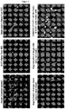
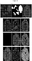
- 238000007490 hematoxylin and eosin (H&E) staining Methods 0.000 description 1
- 230000002962 histologic effect Effects 0.000 description 1
- 238000013427 histology analysis Methods 0.000 description 1
- 210000004251 human milk Anatomy 0.000 description 1
- 235000020256 human milk Nutrition 0.000 description 1
- 208000008384 ileus Diseases 0.000 description 1
- 230000028993 immune response Effects 0.000 description 1
- 102000018358 immunoglobulin Human genes 0.000 description 1
- 238000002991 immunohistochemical analysis Methods 0.000 description 1
- 230000002055 immunohistochemical effect Effects 0.000 description 1
- 238000011532 immunohistochemical staining Methods 0.000 description 1
- 239000003018 immunosuppressive agent Substances 0.000 description 1
- 229940125721 immunosuppressive agent Drugs 0.000 description 1
- 208000027139 infectious colitis Diseases 0.000 description 1
- 208000015181 infectious disease Diseases 0.000 description 1
- 230000002458 infectious effect Effects 0.000 description 1
- 230000004968 inflammatory condition Effects 0.000 description 1
- 230000002757 inflammatory effect Effects 0.000 description 1
- 230000003993 interaction Effects 0.000 description 1
- 201000002313 intestinal cancer Diseases 0.000 description 1
- 208000003243 intestinal obstruction Diseases 0.000 description 1
- 230000003834 intracellular effect Effects 0.000 description 1
- 210000002977 intracellular fluid Anatomy 0.000 description 1
- 238000011835 investigation Methods 0.000 description 1
- 201000008222 ischemic colitis Diseases 0.000 description 1
- 210000003734 kidney Anatomy 0.000 description 1
- 238000012417 linear regression Methods 0.000 description 1
- 150000002632 lipids Chemical class 0.000 description 1
- 210000004185 liver Anatomy 0.000 description 1
- 238000005461 lubrication Methods 0.000 description 1
- 210000004072 lung Anatomy 0.000 description 1
- 210000002751 lymph Anatomy 0.000 description 1
- 210000004324 lymphatic system Anatomy 0.000 description 1
- 238000004519 manufacturing process Methods 0.000 description 1
- 210000004914 menses Anatomy 0.000 description 1
- 108020004999 messenger RNA Proteins 0.000 description 1
- 230000002503 metabolic effect Effects 0.000 description 1
- 230000004060 metabolic process Effects 0.000 description 1
- 230000015689 metaplastic ossification Effects 0.000 description 1
- 108091070501 miRNA Proteins 0.000 description 1
- 238000013508 migration Methods 0.000 description 1
- 230000005012 migration Effects 0.000 description 1
- 210000003470 mitochondria Anatomy 0.000 description 1
- 238000012544 monitoring process Methods 0.000 description 1
- 210000003097 mucus Anatomy 0.000 description 1
- 210000003205 muscle Anatomy 0.000 description 1
- 210000000944 nerve tissue Anatomy 0.000 description 1
- 210000000653 nervous system Anatomy 0.000 description 1
- VOFUROIFQGPCGE-UHFFFAOYSA-N nile red Chemical compound C1=CC=C2C3=NC4=CC=C(N(CC)CC)C=C4OC3=CC(=O)C2=C1 VOFUROIFQGPCGE-UHFFFAOYSA-N 0.000 description 1
- 238000003199 nucleic acid amplification method Methods 0.000 description 1
- 239000002853 nucleic acid probe Substances 0.000 description 1
- 238000005457 optimization Methods 0.000 description 1
- 210000000056 organ Anatomy 0.000 description 1
- 230000008520 organization Effects 0.000 description 1
- 230000002018 overexpression Effects 0.000 description 1
- 210000002741 palatine tonsil Anatomy 0.000 description 1
- 210000000496 pancreas Anatomy 0.000 description 1
- 238000003909 pattern recognition Methods 0.000 description 1
- 208000011906 peptic ulcer disease Diseases 0.000 description 1
- 210000002824 peroxisome Anatomy 0.000 description 1
- DHRLEVQXOMLTIM-UHFFFAOYSA-N phosphoric acid;trioxomolybdenum Chemical compound O=[Mo](=O)=O.O=[Mo](=O)=O.O=[Mo](=O)=O.O=[Mo](=O)=O.O=[Mo](=O)=O.O=[Mo](=O)=O.O=[Mo](=O)=O.O=[Mo](=O)=O.O=[Mo](=O)=O.O=[Mo](=O)=O.O=[Mo](=O)=O.O=[Mo](=O)=O.OP(O)(O)=O DHRLEVQXOMLTIM-UHFFFAOYSA-N 0.000 description 1
- 210000004910 pleural fluid Anatomy 0.000 description 1
- 102000054765 polymorphisms of proteins Human genes 0.000 description 1
- 102000040430 polynucleotide Human genes 0.000 description 1
- 108091033319 polynucleotide Proteins 0.000 description 1
- 239000002157 polynucleotide Substances 0.000 description 1
- 238000002203 pretreatment Methods 0.000 description 1
- 230000000750 progressive effect Effects 0.000 description 1
- XJMOSONTPMZWPB-UHFFFAOYSA-M propidium iodide Chemical compound [I-].[I-].C12=CC(N)=CC=C2C2=CC=C(N)C=C2[N+](CCC[N+](C)(CC)CC)=C1C1=CC=CC=C1 XJMOSONTPMZWPB-UHFFFAOYSA-M 0.000 description 1
- 150000003180 prostaglandins Chemical class 0.000 description 1
- 108020000494 protein-tyrosine phosphatase Proteins 0.000 description 1
- 210000004915 pus Anatomy 0.000 description 1
- 230000005855 radiation Effects 0.000 description 1
- 210000003289 regulatory T cell Anatomy 0.000 description 1
- 230000000284 resting effect Effects 0.000 description 1
- 230000000717 retained effect Effects 0.000 description 1
- 238000012552 review Methods 0.000 description 1
- 210000003296 saliva Anatomy 0.000 description 1
- 238000007790 scraping Methods 0.000 description 1
- 238000012216 screening Methods 0.000 description 1
- 210000002374 sebum Anatomy 0.000 description 1
- 210000000582 semen Anatomy 0.000 description 1
- 210000002966 serum Anatomy 0.000 description 1
- 230000011664 signaling Effects 0.000 description 1
- 210000003491 skin Anatomy 0.000 description 1
- 208000014680 small intestine neoplasm Diseases 0.000 description 1
- 238000000638 solvent extraction Methods 0.000 description 1
- 241000894007 species Species 0.000 description 1
- 210000000952 spleen Anatomy 0.000 description 1
- 206010041823 squamous cell carcinoma Diseases 0.000 description 1
- 238000007619 statistical method Methods 0.000 description 1
- 150000003431 steroids Chemical class 0.000 description 1
- 210000002784 stomach Anatomy 0.000 description 1
- 201000011549 stomach cancer Diseases 0.000 description 1
- 210000004243 sweat Anatomy 0.000 description 1
- 210000001179 synovial fluid Anatomy 0.000 description 1
- 238000003786 synthesis reaction Methods 0.000 description 1
- 230000009885 systemic effect Effects 0.000 description 1
- 230000035488 systolic blood pressure Effects 0.000 description 1
- 210000001138 tear Anatomy 0.000 description 1
- 230000002123 temporal effect Effects 0.000 description 1
- JGVWCANSWKRBCS-UHFFFAOYSA-N tetramethylrhodamine thiocyanate Chemical compound [Cl-].C=12C=CC(N(C)C)=CC2=[O+]C2=CC(N(C)C)=CC=C2C=1C1=CC=C(SC#N)C=C1C(O)=O JGVWCANSWKRBCS-UHFFFAOYSA-N 0.000 description 1
- 230000001225 therapeutic effect Effects 0.000 description 1
- 230000007838 tissue remodeling Effects 0.000 description 1
- 229950003937 tolonium Drugs 0.000 description 1
- HNONEKILPDHFOL-UHFFFAOYSA-M tolonium chloride Chemical compound [Cl-].C1=C(C)C(N)=CC2=[S+]C3=CC(N(C)C)=CC=C3N=C21 HNONEKILPDHFOL-UHFFFAOYSA-M 0.000 description 1
- 238000000844 transformation Methods 0.000 description 1
- LENZDBCJOHFCAS-UHFFFAOYSA-N tris Chemical compound OCC(N)(CO)CO LENZDBCJOHFCAS-UHFFFAOYSA-N 0.000 description 1
- 239000000107 tumor biomarker Substances 0.000 description 1
- 231100000397 ulcer Toxicity 0.000 description 1
- 210000003932 urinary bladder Anatomy 0.000 description 1
- 210000002700 urine Anatomy 0.000 description 1
- 238000010200 validation analysis Methods 0.000 description 1
- 238000012800 visualization Methods 0.000 description 1
- 210000004916 vomit Anatomy 0.000 description 1
- 230000008673 vomiting Effects 0.000 description 1
- 238000005406 washing Methods 0.000 description 1
Images


Classifications
-
- G—PHYSICS
- G01—MEASURING; TESTING
- G01N—INVESTIGATING OR ANALYSING MATERIALS BY DETERMINING THEIR CHEMICAL OR PHYSICAL PROPERTIES
- G01N33/00—Investigating or analysing materials by specific methods not covered by groups G01N1/00 - G01N31/00
- G01N33/48—Biological material, e.g. blood, urine; Haemocytometers
- G01N33/50—Chemical analysis of biological material, e.g. blood, urine; Testing involving biospecific ligand binding methods; Immunological testing
- G01N33/53—Immunoassay; Biospecific binding assay; Materials therefor
- G01N33/574—Immunoassay; Biospecific binding assay; Materials therefor for cancer
- G01N33/57407—Specifically defined cancers
-
- C—CHEMISTRY; METALLURGY
- C12—BIOCHEMISTRY; BEER; SPIRITS; WINE; VINEGAR; MICROBIOLOGY; ENZYMOLOGY; MUTATION OR GENETIC ENGINEERING
- C12Q—MEASURING OR TESTING PROCESSES INVOLVING ENZYMES, NUCLEIC ACIDS OR MICROORGANISMS; COMPOSITIONS OR TEST PAPERS THEREFOR; PROCESSES OF PREPARING SUCH COMPOSITIONS; CONDITION-RESPONSIVE CONTROL IN MICROBIOLOGICAL OR ENZYMOLOGICAL PROCESSES
- C12Q1/00—Measuring or testing processes involving enzymes, nucleic acids or microorganisms; Compositions therefor; Processes of preparing such compositions
- C12Q1/68—Measuring or testing processes involving enzymes, nucleic acids or microorganisms; Compositions therefor; Processes of preparing such compositions involving nucleic acids
- C12Q1/6876—Nucleic acid products used in the analysis of nucleic acids, e.g. primers or probes
- C12Q1/6883—Nucleic acid products used in the analysis of nucleic acids, e.g. primers or probes for diseases caused by alterations of genetic material
- C12Q1/6886—Nucleic acid products used in the analysis of nucleic acids, e.g. primers or probes for diseases caused by alterations of genetic material for cancer
-
- G—PHYSICS
- G01—MEASURING; TESTING
- G01N—INVESTIGATING OR ANALYSING MATERIALS BY DETERMINING THEIR CHEMICAL OR PHYSICAL PROPERTIES
- G01N2800/00—Detection or diagnosis of diseases
- G01N2800/14—Disorders of ear, nose or throat
-
- G—PHYSICS
- G01—MEASURING; TESTING
- G01N—INVESTIGATING OR ANALYSING MATERIALS BY DETERMINING THEIR CHEMICAL OR PHYSICAL PROPERTIES
- G01N2800/00—Detection or diagnosis of diseases
- G01N2800/56—Staging of a disease; Further complications associated with the disease
Definitions
- the invention relates to a method of determining a risk of progression of Barrett's esophagus (BE) in a subject and to the use of a kit in the method.
- BE Barrett's esophagus
- Barrett's esophagus results from Gastroesophageal Reflux Disease (GERD) and affects approximately 3 million patients in the United States, with 86,000 new cases being diagnosed each year.
- GSD Gastroesophageal Reflux Disease
- Aldulaimi et al., Eur J Gastroenterol Hepatol. 2005 Sep; 17(9):943-50 discloses that a diagnosis of Barrett's esophagus predisposes patients to develop esophageal adenocarcinoma with a risk calculated as 30-125 times more as compared to patients without a diagnosis.
- Esophageal adenocarcinoma develops in a defined sequence of changes from benign, to low grade dysplasia, to high-grade dysplasia, and to malignant cancer.
- Herman van Dekken et al "Immunohistochemical Evaluation of a Panel of Tumor Cell markers during Malignant Progression in Barrett Esophagus", American Journal of Clinical Pathology, vol. 130, no. 5, 14 October 2008, pages 745-753 ; and Hormi-Carver K et al: "Molecular Markers and Genetics in Cancer Development", Surgical Oncology Clinics of North America, vol. 18, no. 3, 1 July 2009, pages 453-467 .
- the screening process has many limitations. For instance, diagnosis of Barrett's esophagus can be characterized in different stages such as low-grade dysplasia, high-grade dysplasia, and reactive atypia. These "stages" of BE share histological features and are difficult to distinguish using current H&E-based analysis. Frequently, the diagnosis results in "indeterminate/indefinite,” misdiagnosis, delayed diagnosis or inappropriate treatment. Furthermore, the current form of histology analysis is insufficient to diagnose the various stages of the BE disease accurately and to predict progression to higher disease stages.
- the invention relates to a system, an apparatus, a composition, a device and method of using the same to extract specific biomarker information from cell samples to improve the accuracy of diagnosis, to enable predictions of disease progression and cancer development, to predict responsiveness to therapeutic interventions, and to improve management of BE or any cancer derived from tissue diagnosed as BE.
- the invention relates to a method of determining a risk of progression of Barrett's esophagus in a subject, comprising:
- the method further comprises detecting at least one or more biomarkers selected from the group consisting of AMACR, CD1a, CD45RO, CD68, CK-20, Ki-67, NF- ⁇ B, and p16, wherein the descriptive features are selected from:
- the subject has an increased risk of progression to low grade dysplasia, high grade dysplasia or esophageal cancer.
- the subject is diagnosed with no dysplasia, reactive atypia, indefinite for dysplasia, low grade dysplasia, or high grade dysplasia.
- the method further comprises detecting the subset of biomarkers using probes that specifically bind to each of said biomarkers.
- the method further comprises determining at least 10, at least 20, at least 30, at least 40, at least 50, or 60 descriptive features from Table 4.
- the method further comprises determining at least 10, at least 20, at least 30, at least 40, at least 50, at least 60, at least 70, at least 80, or 89 descriptive features from Table 5.
- the sample comprises a brushing, biopsy, or surgical resection of cells and/or tissue from the subject.
- the descriptive features are identified in subcellular and/or tissue compartments.
- the sample is at room temperature or frozen; optionally wherein the sample is freshly obtained, formalin fixed, alcohol fixed, or paraffin embedded.
- the probes are fluorescent and/or comprise a fluorescent tag, preferably wherein each probe is labeled with a different fluorophore.
- the subset of biomarkers comprises at least 3 biomarkers and wherein the 3 biomarkers are an epithelial biomarker, immune biomarker and/or a stromal biomarker; optionally further detecting a stem cell biomarker.
- the detection of 2 or more, 3 or more, 4 or more, 5 or more, 8 or more, or 12 or more biomarkers are determined simultaneously.
- the subject is a human.
- kits in the above method in determining a risk for progression of Barrett's esophagus in a subject
- the kit comprising one or more probes that is capable of detecting at least two or more biomarkers from the group consisting of p53, HIP-1alpha, beta-catenin, and COX-2 and instructions for using the probes to determine one or more descriptive features to generate a risk score from a cell and/or tissue sample of a subject.
- the score is predictive of the clinical outcome of Barrett's esophagus in the subject.
- the score is predictive of diagnostic of the subclass of Barrett's esophagus in the subject.
- the probes comprise antibody probes that specifically bind to said biomarkers.
- the probes are fluorescent and/or comprise a fluorescent tag.
- At least 10, at least 20, at least 30, at least 40, at least 50, at least 60, at least 70, at least 80, or 89 descriptive features are from Table 4 or Table 5.
- the term "about” as used herein when referring to a measurable value such as an amount, a temporal duration, and the like, is meant to encompass variations of ⁇ 20%, ⁇ 10%, ⁇ 5%, ⁇ 1%, or ⁇ 0.1% from the specified value, as such variations are appropriate to perform the disclosed methods.
- the terms “increase” and “decrease” mean, respectively, to cause a statistically significantly (i.e., p ⁇ 0.15) increase or decrease of at least 1%, 2%, or 5%.
- variable As used herein, the recitation of a numerical range for a variable is intended to convey that the invention may be practiced with the variable equal to any of the values within that range. Thus, for a variable which is inherently discrete, the variable is equal to any integer value within the numerical range, including the endpoints of the range. Similarly, for a variable which is inherently continuous, the variable is equal to any real value within the numerical range, including the endpoints of the range.
- a variable which is described as having values between 0 and 2 takes the values 0, 1 or 2 if the variable is inherently discrete, and takes the values 0.0, 0.1, 0.01, 0.001, 10 -12 , 10 -11 , 10 -10 , 10 -9 , 10 -8 , 10 -7 , 10 -6 , 10 -5 , 10 -4 or any other real values ⁇ 0 and ⁇ 2 if the variable is inherently continuous.
- amino acid refers to a molecule containing both an amino group and a carboxyl group bound to a carbon which is designated the ⁇ -carbon.
- Suitable amino acids include, without limitation, both the D- and L-isomers of the naturally occurring amino acids, as well as non-naturally occurring amino acids prepared by organic synthesis or other metabolic routes.
- a single “amino acid” might have multiple sidechain moieties, as available per an extended aliphatic or aromatic backbone scaffold.
- amino acid as used herein, is intended to include amino acid analogs, naturally occurring amino acids, and non-naturally amino acids.
- antibody refers to an immunoglobulin molecule or fragment thereof having a specific structure that interacts or binds specifically with a molecule comprising an antigen.
- antibody broadly includes full-length antibodies and may include certain antibody fragments thereof. Also included are monoclonal and polyclonal antibodies, multivalent and monovalent antibodies, multispecific antibodies (for example bi-specific antibodies), chimeric antibodies, human antibodies, humanized antibodies and antibodies that have been affinity matured.
- an antibody binds selectively or specifically to a biomarker of a gastrointestinal disorder if the antibody binds preferentially to an antigen expressed by a cell and has less than 25%, or less than 10%, or less than 1% or less than 0.1% cross-reactivity with a polypeptide expressed by a cell within the gastrointestinal tissue or cells derived from another tissue that migrates from one tissue to the gastrointestinal tissue.
- the antibody will have a binding affinity (dissociation constant (Kd) value), for the antigen or epitope of no more than 10 -6 M, or 10 -7 M, or less than about 10 -8 M, or 10 -9 M, or 10 -10 M, or 10 -11 M or 10 -12 M. Binding affinity may be assessed using any method known by one of ordinary skill in the art, such as surface plasma resonance, immunoaffinity assays, or ELISAs.
- biomarker means any analyte, metabolite, nucleic acid, amino acid sequence or fragments thereof, polyprotein, protein complex, molecule, or chemical compound that is produced, metabolized, catabolized, secreted, phagocytosed, or expressed by a cell or tissue and that provides a useful measure of the presence, absence, or quantity of a certain cell type or descriptive feature indicative of, characteristic of, or suggestive of a diagnosis of a particular disease or disorder.
- epithelial biomarker means any marker of epithelial cell subset, e.g. normal gland or surface epithelium cell, metaplastic gland or surface epithelium cell, dysplastic gland or surface epithelium cell, cancer cell of epithelial origin, or marker of epithelial cell function, e.g. proliferation, cell cycle control, tumor suppressor gene, oncogene, adhesion, migration, fatty acid metabolism, apoptosis, inflammation.
- stromal biomarker means any marker of stromal cell type, e.g. endothelial cell, fibroblast, or stromal cell function, e.g. angiogenesis, tissue remodeling.
- immune biomarker means any marker of immune cell subset, e.g. T lymphocyte, B lymphocyte, supressor cell, regulatory T cell, dendritic cell, macrophage, granulocyte, or immune cell function, e.g. cytokines, chemokines, activation, cell-cell contact, proliferation, inflammation.
- nucleic acid biomarker means any specific locus of a gene or DNA sequence measured with locus-specific probes, e.g. 9p21, 8q24.12-13, 17q11.2-q12. It also includes centromeres measured with centromere enumeration probes, e.g. chromosome 8, 9, 17. It also includes anything that binds to nucleic acid and can aid in the visualization of the nucleus (e.g. Hoechst, 4',6-diamidino-2-phenylindole (DAPI)).
- DAPI Hoechst, 4',6-diamidino-2-phenylindole
- morphometric marker means any measurement of structures, shapes, parts, sizes and textures of cells and tissues.
- examples of morphometric markers include nuclear area, nuclear equivalent diameter, nuclear solidity, nuclear eccentricity, gland to stroma ratio, nuclear area to cytoplasmic area ratio, glandular nuclear size, glandular nuclear size and intensity gradient, and nuclear texture.
- nuclear equivalent diameter means a scalar that specifies the diameter of a circle with the same area as the nuclear region and can be computed as sqrt(4*Area/pi). It is an estimate of the diameter of nuclei, which are non-circular, irregularly-shaped objects.
- nuclear solidity means a scalar specifying the proportion of the pixels in the convex hull that are also in the nuclear region. It is equal to the ratio of nuclear area: convex area of nuclei based on fluorescent labeling of nuclei.
- nuclear eccentricity is a scalar that specifies the eccentricity of the nuclear ellipse that has the same second-moments as the nuclear region. The eccentricity is the ratio of the distance between the foci of the ellipse and its major axis length. The value is between 0 and 1.
- nuclear texture is the spatial arrangements of fluorescently-labeled pixels in nuclei area.
- stem cell biomarker means any marker to distinguish stem cells from non-stem cells or marker of stem cell function.
- the disease is a gastrointestinal disorder.
- the biomarker is chosen from one or more of the molecules identified in Table 1.
- the biomarkers can be the measure of receptor expression levels, transcription factor activation; location or amount or activity of a protein, polynucleotide, organelle, and the like; the phosphorylation status of a protein, etc.
- a biomarker is a nucleic acid (e.g., DNA, RNA, including micro RNAs, snRNAs, mRNA, rRNA, etc.), a receptor, a cell membrane antigen, an intracellular antigen, and extracellular antigen, a signaling molecule, a protein, and the like without limitation, lipids, lipoproteins, proteins, cytokines, chemokines, growth factors, peptides, nucleic acids, genes, and oligonucleotides, together with their related complexes, metabolites, mutations, variants, polymorphisms, modifications, fragments, subunits, degradation products, elements, and other analytes or sample-derived measures.
- nucleic acid e.g., DNA, RNA, including micro RNAs, snRNAs, mRNA, rRNA, etc.
- a receptor e.g., DNA, RNA, including micro RNAs, snRNAs, mRNA,
- a biomarker can also include a mutated protein or proteins, a mutated nucleic acid or mutated nucleic acids, variations in copy numbers, and/or transcript variants, in circumstances in which such mutations, variations in copy number and/or transcript variants are useful for generating a predictive model, or are useful in predictive models developed using related markers (e.g., non-mutated versions of the proteins or nucleic acids, alternative transcripts, etc.).
- biomaterial or biomaterials means any protein, tissue, molecule, extracellular matrix component, biostructure, membrane, subcellualr compartment or any combination of the above that is derived from a cell and/or is spatially positioned outside of cell in a cell sample.
- a biomarker expression profile means a collection of data collected by a user related to the quantity, intensity, presence, absence, or spatial distribution of a biomarker or set of biomarkers assigned to a cell or biomaterial or subcellular compartment, each within a cell sample.
- the term "cell sample” means a composition comprising an isolated cell or plurality of cells.
- the cell sample comprises an individual cell.
- the cell sample is a composition comprising a plurality of cells.
- the cell sample is a tissue sample taken from a subject with a gastrointestinal disorder.
- the cell sample is a tissue sample.
- the cell sample comprises a plurality of cells from the gastrointestinal tract.
- the cell sample is a plurality of esophageal cells.
- the cell sample is freshly obtained, formalin fixed, alcohol-fixed and/or paraffin embedded.
- the cell sample is a biopsy isolated from a subject who has been diagnosed or is suspected or identified as having one or more gastrointestinal disorders.
- the cell sample a biopsy isolated from a subject who has been diagnosed or is suspected or identified as having Barrett's esophagus.
- the cell sample comprises a tissue from a brushing, punch biopsy, or surgical resection of a subject.
- the one or more tissue samples are isolated from one or more animals.
- the one or more animals are one or more humans.
- one or more cell samples are isolated from a human patient at one or more time points, such that at least one tissue sample is isolated from each time point from the same patient.
- a cell sample can include a single cell or multiple cells or fragments of cells or an aliquot of body fluid, taken from a subject, by means including venipuncture, excretion, ejaculation, massage, biopsy, needle aspirate, lavage sample, scraping, surgical incision, or intervention or other means known in the art.
- the invention includes obtaining a cell sample associated with a subject, where the sample includes one or more biomarkers.
- the sample can be obtained by the subject or by a third party, e.g., a medical professional.
- a sample can include peripheral blood cells, isolated leukocytes, or RNA extracted from peripheral blood cells or isolated leukocytes.
- the sample can be obtained from any bodily fluid, for example, amniotic fluid, aqueous humor, bile, lymph, breast milk, interstitial fluid, blood, blood plasma, cerumen (earwax), Cowper's fluid (preejaculatory fluid), chyle, chyme, female ejaculate, menses, mucus, saliva, urine, vomit, tears, vaginal lubrication, sweat, serum, semen, sebum, pus, pleural fluid, cerebrospinal fluid, synovial fluid, intracellular fluid, and vitreous humour.
- the sample is obtained by a blood draw, where the medical professional draws blood from a subject, such as by a syringe.
- the bodily fluid can then be tested to determine the value of one or more descriptive features using the interpretation function or methods described herein.
- the value of the one or more descriptive features can then be evaluated by the same party that performed the method using the methods of the invention or sent to a third party for evaluation using the methods of the invention.
- the method comprises obtaining or isolating at least two cell samples from one or more subjects.
- the method comprises obtaining or isolating at least three cell samples from one or more subjects.
- the method comprises obtaining or isolating at least four cell samples from one or more subjects. Any suitable tissue sample can be used in the methods described herein.
- the tissue can be epithelium, muscle, organ tissue, nerve tissue, tumor tissue, and combinations thereof.
- the cell sample is not derived from blood, sera, or blood cells.
- the cell sample is not derived from cells of the liver, pancreas, gallbladder, bladder, skin, heart, lungs, kidneys, spleen, bone marrow, adipose tissue, nervous system, circulatory system, or lymphatic system. Samples of tissue can be obtained by any standard means (e.g., biopsy, core puncture, dissection, and the like, as will be appreciated by a person of skill in the art).
- At least one cell sample is labeled with a histological stain, to produce a histologically stained cell sample.
- histological stains can be any standard stain as appreciated in the art, including but not limited to, alcian blue, Fuchsin, haematoxylin and eosin (H&E), Masson trichrome, toluidine blue, Wright's/Giemsa stain, and combinations thereof.
- traditional histological stains are not fluorescent.
- At least one other section is labeled with a panel of fluorescently labeled reagents to produce a fluorescently labeled section.
- the panel of fluorescently labeled reagents comprises a number of reagents, such as fluorescently labeled antibodies, fluorescently labeled peptides, fluorescently labeled polypeptides, fluorescently labeled aptamers, fluorescently labeled oligonucleotides (e.g. nucleic acid probes, DNA, RNA, cDNA, PNA, and the like), fluorescently labeled chemicals and fluorescent chemicals (e.g., Hoechst 33342, propidium iodide, Draq- 5, Nile Red, fluorescently labeled phalloidin, 4',6-diamidino-2-phenylindole (DAPI)), and combinations thereof.
- fluorescently labeled antibodies such as fluorescently labeled antibodies, fluorescently labeled peptides, fluorescently labeled polypeptides, fluorescently labeled aptamers, fluorescently labeled oligonucleotides (e.g. nucleic acid probes, DNA,
- Cellular systems biology is defined as the of the interacting cellular and molecular networks of normal, tumor, immune, stromal and stem cells in tissues and bodily fluids that give rise to normal function and disease.
- Cells in tissues exhibit properties that are not anticipated from the analysis of individual components, known as emergent properties that require analysis of many factors to characterize cellular and molecular states.
- correlation between measurements in individual cells is required to identify and interpret cellular responses to drug treatment.
- a cellular systems biological profile can be utilized to capture or compile a set of epidemiological data about a patient or subject population.
- the subject population is a patient population at an elevated risk for developing Barrett's esophagus, suffering from Barrett's esophagus, or having been diagnosed with Barrett's esophagus. All kits and methods of the present invention may also be used to compile data around epidemiological data about a patient or subject population. All kits and methods of the present invention may also be used to acquire and track the progression of a particular disease or disorder of a subject.
- the subject population is a patient population at an elevated risk for developing Barrett's esophagus, suffering from Barrett's esophagus, or having been diagnosed with Barrett's esophagus.
- particular expression levels of biomarkers are tracked and patient histories are compiled in order to more finely characterize a patient's disease as falling into a particular subclass of Barrett's esophagus.
- Clinical factor is defined as a measure of a condition of a subject, e.g., disease activity or severity.
- “Clinical factor” encompasses all biomarkers of a subject's health status, including non-sample markers, and/or other characteristics of a subject, such as, without limitation, age and gender, and clinical history related to other ailments, disorders, diseases, or the risk associated with developing such ailment, disorder, or disease.
- a clinical factor can be a score, a value, or a set of values that can be obtained from evaluation of a cell sample (or plurality of samples) from a subject or a subject under a determined condition.
- a clinical factor can also be predicted by biomarkers and/or other parameters such as gene expression surrogates.
- diagnostic subcategories include:
- control means healthy esophageal tissue, Barrett's esophagus tissue with no dysplasia, Barrett's esophagus tissue from a subject that did not progress to low grade or high grade dysplasia, or esophageal carcinoma.
- the term "converting" means subjecting the one or more descriptive features to an interpretation function or algorithm for a predictive model of disease.
- the disease is Barrett's esophagus or a subclass of Barrett's esophagus.
- the interpretation function can also be produced by a plurality of predictive models.
- the predictive model would include a regression model and a Bayesian classifier or score.
- an interpretation function comprises one or more terms associated with one or more biomarker or sets of biomarkers.
- an interpretation function comprises one or more terms associated with the presence or absence or spatial distribution of the specific cell types disclosed herein.
- an interpretation function comprises one or more terms associated with the presence, absence, quantity, intensity, or spatial distribution of the morphological features of a cell in a cell sample. In one embodiment, an interpretation function comprises one or more terms associated with the presence, absence, quantity, intensity, or spatial distribution of descriptive features of a cell in a cell sample.
- transmutes said digital imaging data into a digital imaging signal means the process of a data processor that receives digital imaging data and converts the digital code of said digital imaging data into a code compatible with the software used to create an image of a cell sample visible to a user.
- descriptive features are defined as values associated with data measurements, a series of data measurements, observations, or a series of observations about a cell sample, typically evidenced by the presence, absence, quantity, localization or spatial proximity to other descriptive features or biomarkers relative space within a cell sample.
- descriptive features include values calculated through an image interpretation function, measured or quantified by standard or known microscopy techniques, but are not limited to values associated with the presence, absence, localization, or spatial distribution of one or more biomarkers.
- descriptive features include values calculated through an image interpretation function, measured or quantified by standard or known microscopy techniques, but are not limited to values associated with the presence, absence, localization, or spatial distribution of one or more biomarkers chosen from: protein post-translational modifications such as phosphorylation, proteolytic cleavage, methylation, myristoylation, and attachment of carbohydrates; translocations of ions, metabolites, and macromolecules between compartments within or between cells; changes in the structure and activity of organelles; and alterations in the expression levels of macromolecules such as coding and noncoding RNAs and proteins.
- biomarkers chosen from: protein post-translational modifications such as phosphorylation, proteolytic cleavage, methylation, myristoylation, and attachment of carbohydrates; translocations of ions, metabolites, and macromolecules between compartments within or between cells; changes in the structure and activity of organelles; and alterations in the expression levels of macromolecules such as coding and noncoding RNAs and proteins.
- descriptive features comprise values associated with one or a combination of more than one of the following morphological features of a cell or cell sample chosen from: the presence of goblet cells; the presence of cytological and architectural abnormalities; the presence of cell stratification; the presence of multilayered epithelium; the maturation of the surface epithelium; the degree of budding, irregularity, branching, and atrophy in crypts; the proportion of low grade crypts to high grade crypts; the presence of splaying and duplication of the muscularis mucosa; the presence, number and size of thin-walled blood vessels, lymphatic vessels, and nerve fibers; the frequency of mitoses; the presence of atypical mitoses; the size and chromicity of nuclei; the presence of nuclear stratification; the presence of pleomorphism; the nucleus:cytoplasm volume ratio; the presence of villiform change; the presence of the squamocolumnar junction (Z-line) and its location in relation to the gastroes
- the descriptive feature may represent a numerical value estimated by an operator of the apparati or compositions disclosed herein using methods of quantifying such biomarkers as it is known in the art.
- the descriptive feature comprises a value or values associated with the presence, absence, proximity, localization relative to one or more biomarkers, or quantity of one or more of the following cell types: epithelial cells, multilayered-epithelial cells, endothelial cells, peripheral mononuclear lymphocytes, T cells, B cells, natural killer cells, eosinophils, mast cells, macrophages, dendritic cells, neutrophils, fibroblasts, goblet cells, dysplastic cells, and non-goblet columnar epithelial cells.
- the descriptive feature comprises value related to the presence, absence, localization or relative proximity to other descriptive features or biomarkers, or quantity of one or more of the following biomarkers inside or outside a cell: p16, p53, Ki-67, beta-catenin, alpha-methylacyl-CoA racemase (AMACR, P504S), matrix metalloproteinase 1, CD1a, NF-kappa-B p65, cyclo-oxygenase-2, CD68, CD4, forkhead box P3, CD45, thrombospondin-1, C-myc, cytokeratin-20, fibroblast activation protein alpha, cyclin D1, HER2/neu, EGFR, Interleukin-6, PLAU plasminogen activator urokinase (uPA), CDX2, Fas, FasL and HIF-1alpha.
- AMACR, P504S alpha-methylacyl-CoA racemase
- AMACR, P504S alpha-methyl
- a biomarker in one or more sections is a read-out of one or more descriptive features of a cellular systems biology profile.
- a "descriptive feature” refers to a characteristic and/or a value which relates to a measurement or series of measurements related to a particular biomarker (which can indicate the location, function, spatial distribution, presence or absence of the biomarker made within a cell sample.
- Bio functions include, but are not limited to: protein posttranslational modifications such as phosphorylation, proteolytic cleavage, methylation, myristoylation, and attachment of carbohydrates; translocations of ions, metabolites, and macromolecules between compartments within or between cells; changes in the structure and activity of organelles; and alterations in the expression levels of macromolecules such as coding and noncoding RNAs and proteins, morphology, state of differentiation, and the like.
- a single biomarker can provide a read-out of more than one feature.
- Hoechst dye detects DNA, which is an example of a biomarker.
- a number of features can be identified by the Hoechst dye in the cell sample such as nucleus size, cell cycle stage, number of nuclei, presence of apoptotic nuclei, etc.
- the term "derived from” in the context of the relationship between a cell or amino acid sequence and a related biomarker or related amino acid sequence describes a biomarker or related amino acid sequence that may be homologous to or structurally similar to the related chemical structure or related amino acid sequence.
- digitally addressable means an image that can be viewed, manipulated, or accessed by the user with software.
- gastrointestinal disorder refers to any disease or abnormality related to the alimentary canal including but not necessarily limited to one or more of the following conditions: abdominal pain, gastroesophageal reflux disease (GERD), constipation, diarrhea, diverticulosis, gastrointestinal bleeding, stomach cancer, esophageal cancer, intestinal cancer, colon cancer, Barrett's esophagus, irritable bowel disease, infectious colitis, ulcerative colitis, Crohn's disease, ischemic colitis, radiation colitis, irritable bowel syndrome, acute perforation, ileus, appendicitis, intra-abdominal abscesses, intestinal obstruction, gastritis, autoimmune metaplastic atrophic gastritis, ulcers in the stomach, peptic ulcer disease, dyspepsia, gastrointestinal stromal tumors, small bowel tumors, levator syndrome, pilonidal disease, proctits, fistulkas, fissures, incontinence
- highly correlated gene expression or “highly correlated marker expression” refer to biomarker expression values that have a sufficient degree of correlation to allow their interchangeable use in a predictive model of Barrett's esophagus.
- highly correlated marker or “highly correlated substitute marker” refer to markers that can be substituted into and/or added to a predictive model based on, for instance, the above criteria.
- a highly correlated marker can be used in at least two ways: (1) by substitution of the highly correlated biomarker(s) for the original biomarker(s) and generation of a new model for predicting Barrett's esophagus risk; or (2) by substitution of the highly correlated biomarker(s) for the original biomarker(s) in the existing model for predicting a subject's propensity to develop, risk to develop, or diagnosis or Barrett's esophagus or a subclass of Barrett's esophagus.
- the term "instructions” refers to materials and methods for staining tissue slides with the probes, imaging the probes on the tissue slides, analyzing the images to extract the biomarker data and/or the processing the data into a score.
- location refers to a subcellular compartment or tissue compartment.
- Subcellular compartments include the nucleus, cytoplasm, plasma membrane, and nuclear membrane.
- Tissue compartments include the surface epithelium, glands, stroma, and tumor.
- the term "probe” refers to any molecule that binds or intercalates to a biomarker, either covalently or non-covalently.
- the probes include probe sets which include one or more probes that bind a single biomarker.
- the term "probe set" is sometime interchangeable for a panel of two or more probes that allow the detection of one or more biomarkers.
- the probe or probes are fluorescently labeled.
- each fluorescently labeled probe is specific for at least one biomarker.
- the panel of fluorescently labeled probes detects at least about two different biomarkers.
- the panel of fluorescently labeled probes detects at least about three different biomarkers. In one embodiment of the invention, the panel of fluorescently labeled probes detects at least about four different biomarkers. In one embodiment of the invention, the panel of fluorescently labeled probes detects at least about five different biomarkers. In another embodiment of the invention, the panel of fluorescently labeled probes detects at least about four to about six, to about ten, to about twelve different biomarkers or more. In another embodiment of the invention, the panel of fluorescently labeled probes detects at least about three different biomarkers. In a further embodiment, each fluorescently labeled probe has different fluorescent properties, which are sufficient to distinguish the different fluorescently labeled probes in the panel.
- ratio means the ratio of one biomarker's quantity to a different biomarker's quantity in the same or different subcellular compartment or tissue compartment. It can also mean the ratio of one biomarker's quantity in a subcellular compartment to quantity of same biomarker in another subcellular compartment within the same cell. It can also mean the ratio of one biomarker's quantity in a tissue compartment to quantity of same biomarker in another tissue compartment within the same biopsy.
- risk of progression means the probability of progressing to low grade dysplasia, high grade dysplasia, or esophageal adenocarcinoma.
- the term “score” refers to a single value that can be used as a component in a predictive model for the diagnosis, prognosis, or clinical treatment plan for a subject, wherein the single value is calculated by combining the values of descriptive features through an interpretation function or algorithm.
- the subject is suspected of having, is at risk of developing, or has been diagnosed with a gastrointestinal disorder.
- the subject is suspected of having or is at risk of developing Barrett's esophagus or a subclass of Barrett's esophagus.
- Risk scores are scores of 1-100, with 1 indicating lowest risk of progression and 100 indicating highest risk of progression. Risk classes are below, intermediate and high.
- subclass of Barrett's esophagus refers to any presentation of Barrett's esophagus classified as having any common combination of one or more descriptive features.
- a subclass of Barrett's esophagus refers to one of the following conditions: Barrett's esophagus, no dysplasia, no progression in 5 years; Barrett's esophagus, no dysplasia, progression to low/high grade dysplasia in 5 years; Barrett's esophagus, indefinite for dysplasia, no progression in 5 years; Barrett's esophagus, indefinite for dysplasia, progression to low/high grade dysplasia or adenocarcinoma in 5 years; Barrett's esophagus, reactive atypia; Barrett's esophagus, low grade dysplasia, no progression in 5 years; Barrett's esophagus, low grade dysplasia, progression to high grade dysplasia or
- a subclass of Barrett's esophagus refers to one of the following conditions: low-grade dysplasia, high-grade dysplasia, reactive atypia, or indeterminate Barrett's esophagus.
- the compositions and systems described herein are designed to stratify patient groups more precisely and diagnose the different subclasses of Barrett's esophagus more accurately.
- optical scanner is used throughout the specification to describe any device or series of devices that generates image data from a cell sample or set of cell samples.
- optical scanner is used to describe any device or series of devices that generates digital image data from a cell sample or set of cell samples.
- the optical scanner may be a microscope attached to a optical device that generates digital image data, which, when sent to image forming apparatus such as a laser printer, a barcode reader, a confocal scanning laser microscope, or an imaging display (monitor), can produce an image visible to a user.
- the term "subject” is used throughout the specification to describe an animal from which a cell sample is taken.
- the animal is a human.
- the term “patient” may be interchangeably used.
- the term “patient” will refer to human patients suffering from a particular disease or disorder.
- the subject may be a human suspected of having or being identified as at risk to develop a gastrointestinal disorder.
- the subject may be a human suspected of having or being identified as at risk to develop Barrett's esophagus.
- the subject may be a mammal which functions as a source of the isolated cell sample.
- the subject may be a non-human animal from which a cell sample is isolated or provided.
- the term “mammal” encompasses both humans and non-humans and includes but is not limited to humans, non-human primates, canines, felines, murines, bovines, equines, and porcines.
- treating and “to treat”, mean to alleviate symptoms, eliminate the causation either on a temporary or permanent basis, or to prevent or slow the appearance of symptoms.
- treatment includes alleviation, elimination of causation (temporary or permanent) of, or prevention of symptoms and disorders associated with any condition.
- the treatment may be a pre-treatment as well as a treatment at the onset of symptoms.
- cellular systems biology also referred to herein as systems cell biology
- systems cell biology is the investigation of the integrated and interacting networks of genes, proteins, and metabolites that are responsible for normal and abnormal cell functions.
- a cellular systems biology profile refers to a systemic characterization of cells in the context of a cell sample architecture such that the cells have particular characteristics dependent upon the relationships of different cells within a cell sample and the biological or medical state of the tissue when isolated from a subject. It is the interactions, relationships, and spatial orientation of the biomarkers of or biomaterials derived from a cell or cells from a cell sample that gives rise to the descriptive features that are used to construct a profile.
- a cellular systems biology profile defines the interrelationships between a combination of at least about two descriptive features collected from a cell or cells within a cell sample.
- a cellular systems biology profile defines the interrelationships between a combination of at least about three descriptive features collected from a cell or cells within a cell sample.
- a cellular systems biology profile defines the interrelationships between a combination of at least about four descriptive features collected from a cell or cells within a cell sample. In a particular embodiment, a cellular systems biology profile defines the interrelationships between a combination of at least about five descriptive features collected from a cell or cells within a cell sample. In another embodiment, a cellular systems biology profile is the combination of at least about six, seven, eight, nine, ten, eleven, twelve, or more descriptive features or values assigned to the descriptive features.
- a value can be one or more numerical values resulting from evaluation of a cell sample under a condition.
- the values can be obtained, for example, by experimentally obtaining measurements from a cell sample by using one of the systems or compositions disclosed herein.
- the values can be obtained, for example, by experimentally obtaining digital imaging data from a cell sample by using one of the systems or compositions disclosed herein.
- the values can be obtained, for example, by experimentally obtaining measurements from a cell sample by performing one of the methods described herein.
- a service provider such as a laboratory, or from a database or a server on which the digital imaging data has been stored, e.g., on a storage memory.
- a system comprising: a cell sample; a plurality of probes and/or stains; one or more optical scanners; one or more data processors; one or more data storage units; one or more monitors; wherein the one or more optical scanners, the one or more data processors, the one or more monitors, and the one or more data storage units are in digital communication with each other by a means to transmit digital data.
- the system comprises a cell sample isolated from a subject with a gastrointestinal disorder.
- the system comprises a cell sample isolated from a subject with Barrett's esophagus or a subclass of Barrett's esophagus.
- the cell sample is isolated from a subject suspected of having, being at risk for developing, or diagnosed with a gastrointestinal disorder. In some embodiments, the cell sample is isolated from a subject suspected of having, being at risk for developing, or diagnosed with Barrett's esophagus or a subclass of Barrett's esophagus.
- a system for predicting Barrett's esophagus in a subject including: a data storage unit or memory for storing one or more descriptive features associated with a cell sample obtained from the subject, wherein the descriptive features including quantitative expression or spatial distribution data for at least one biomarker set selected from the group consisting of the biomarker sets in term 1, term 2, and term 3; wherein terms 1 through 3 include any combination of one or more biomarkers p16, p53, Ki-67, beta-catenin, alpha-methylacyl-CoA racemase (AMACR, P504S), matrix metalloproteinase 1, CD1a, NF-kappa-B p65, cyclo-oxygenase-2, CD68, CD4, forkhead box P3, CD45, thrombospondin-1, C-myc, cytokeratin-20, fibroblast activation protein alpha, cyclin D1, HER2/neu, EGFR, Interleukin-6, PLAU plasmin
- a system for predicting Barrett's esophagus in a subject including: a data storage unit or memory for storing one or more descriptive features associated with a cell sample obtained from the subject, wherein the descriptive features including quantitative expression or spatial distribution data for at least one biomarker sets selected from the group consisting of the biomarker sets in term 1, term 2, term 3, and term 4; wherein terms 1 through 4 include any combination of one or more biomarkers p16, p53, Ki-67, beta-catenin, alpha-methylacyl-CoA racemase (AMACR, P504S), matrix metalloproteinase 1, CD1a, NF-kappa-B p65, cyclo-oxygenase-2, CD68, CD4, forkhead box P3, CD45, thrombospondin-1, C-myc, cytokeratin-20, fibroblast activation protein alpha, cyclin D1, HER2/neu, EGFR, Interleukin-6, PLAU
- a system for predicting Barrett's esophagus in a subject including: a data storage unit or memory for storing one or more descriptive features associated with a cell sample obtained from the subject, wherein the descriptive features including quantitative expression or spatial distribution data for at least one biomarker set selected from the group consisting of the biomarker sets in term 1, term 2, and term 3; wherein term 1 includes Ki-67, term 2 includes beta-catenin, and term 3 includes the presence of nuclei stained by Hoescht stain, and wherein terms 1, 2, and 3 optionally include any one or more of the following biomarkers: alpha-methylacyl-CoA racemase (AMACR, P504S), matrix metalloproteinase 1, CD1a, NF-kappa-B p65, cyclo-oxygenase-2, CD68, CD4, forkhead box P3, CD45, thrombospondin-1, C-myc, cytokeratin-20, fibroblast activation protein alpha,
- any of the methods described herein may contain a descriptive feature identified through computer recognition of the presence, absence, quantity, intensity, or spatial distribution of morphological components of the cell.
- the terms of the biomarker sets may be added to calculate the score.
- a system for predicting Barrett's esophagus in a subject including: a data storage unit or memory for storing one or more descriptive features associated with a cell sample obtained from the subject, wherein the descriptive features including quantitative expression or spatial distribution data for at least one biomarker sets selected from the group consisting of the biomarker sets in term 1, term 2, term 3, and term 4; wherein terms 1 through 4 include any combination of one or more biomarkers p16, p53, Ki-67, beta-catenin, alpha-methylacyl-CoA racemase (AMACR, P504S), matrix metalloproteinase 1, CD la, NF-kappa-B p65, cyclo-oxygenase-2, CD68, CD4, forkhead box P3, CD45, thrombospondin-1, C-myc, cytokeratin-20, fibroblast activation protein alpha, cyclin D1, HER2/neu, EGFR, Interleukin-6, PLAU plasm
- a system for predicting Barrett's esophagus in a subject including: a data storage unit or memory for storing one or more descriptive features associated with a cell sample obtained from the subject, wherein the descriptive features include quantitative expression or spatial distribution data for at least one biomarker sets selected from the group consisting of the marker sets in term 1, term 2, term 3, term 4, and term 5; wherein terms 1 through 5 include any combination of one or more biomarkers p16, p53, Ki-67, beta-catenin, alpha-methylacyl-CoA racemase (AMACR, P504S), matrix metalloproteinase 1, CD1a, NF-kappa-B p65, cyclo-oxygenase-2, CD68, CD4, forkhead box P3, CD45, thrombospondin-1, C-myc, cytokeratin-20, fibroblast activation protein alpha, cyclin D1, HER2/neu, EGFR, Interleukin-6, PLAU
- a system for predicting Barrett's esophagus in a subject including: a data storage unit or memory for storing one or more descriptive features associated with a cell sample obtained from the subject, wherein the descriptive features include quantitative expression or spatial distribution data for at least one biomarker sets selected from the group consisting of the marker sets in term 1, term 2, term 3, term 4, term 5, and term 6; wherein terms 1 through 6 include any combination of one or more biomarkers p16, p53, Ki-67, beta-catenin, alpha-methylacyl-CoA racemase (AMACR, P504S), matrix metalloproteinase 1, CD1a, NF-kappa-B p65, cyclo-oxygenase-2, CD68, CD4, forkhead box P3, CD45, thrombospondin-1, C-myc, cytokeratin-20, fibroblast activation protein alpha, cyclin D1, HER2/neu, EGFR, Interleukin-6, PLA
- a system for predicting Barrett's esophagus in a subject including: a data storage unit or memory for storing one or more descriptive features associated with a cell sample obtained from the subject, wherein the descriptive features include quantitative expression or spatial distribution data for at least one biomarker sets selected from the group consisting of the marker sets in term 1, term 2, term 3, term 4, term 5, term 6, and term 7; wherein terms 1 through 7 include any combination of one or more biomarkers p16, p53, Ki-67, beta-catenin, alpha-methylacyl-CoA racemase (AMACR, P504S), matrix metalloproteinase 1, CD1a, NF-kappa-B p65, cyclo-oxygenase-2, CD68, CD4, forkhead box P3, CD45, thrombospondin-1, C-myc, cytokeratin-20, fibroblast activation protein alpha, cyclin D1, HER2/neu, EGFR, Interleukin-6
- a system for predicting Barrett's esophagus in a subject including: a data storage unit, units or memory for storing one or more descriptive features associated with a cell sample obtained from the subject, wherein the descriptive features include quantitative expression data for at least one biomarker sets selected from the group consisting of the marker sets in term 1, term 2, term 3, term 4, optionally term 5, optionally term 6, and optionally term 7; wherein terms 1 through 7 include any combination of one or more biomarkers p16, p53, Ki-67, beta-catenin, alpha-methylacyl-CoA racemase (AMACR, P504S), matrix metalloproteinase 1, CD la, NF-kappa-B p65, cyclo-oxygenase-2, CD68, CD4, forkhead box P3, CD45, thrombospondin-1, C-myc, cytokeratin-20, fibroblast activation protein alpha, cyclin D1, HER2/neu, EGFR, Inter
- Also described herein is a computer-readable storage medium storing computer-executable program code, the program code including: program code for storing a dataset of descriptive features associated with a cell sample obtained from the subject, wherein the first dataset includes quantitative expression data for at least one marker set selected from the group consisting of the marker sets in term 1, term 2, term 3, optionally term 4, optionally term 5, optionally term 6, and optionally term 7; wherein wherein terms 1 through 7 include any combination of one or more biomarkers p16, p53, Ki-67, beta-catenin, alpha-methylacyl-CoA racemase (AMACR, P504S), matrix metalloproteinase 1, CD1a, NF-kappa-B p65, cyclo-oxygenase-2, CD68, CD4, forkhead box P3, CD45, thrombospondin-1, C-myc, cytokeratin-20, fibroblast activation protein alpha, cyclin D1, HER2/neu, EGFR, Interleukin-6
- Also described herein is software on an electronic medium or system comprising such software used to correlate the cluster groups of biomarker, morphologic and clinical data features into indices useful to distinguish one or more particular cell types from a mixture of cell types in a cell sample automatically.
- Also described herein is software on an electronic medium or system comprising such software used. to correlate the cluster groups of biomarker, morphologic and clinical data features into indices useful to predict the responsiveness a patient to a particular therapy.
- Also described herein is software on an electronic medium or system comprising such software used to correlate the cluster groups of biomarker, morphologic and clinical data features into indices useful to predict one or more clinical treatment schedules for a patient automatically.
- Also described herein is software on an electronic medium or system comprising such software used to correlate the cluster groups of biomarker, morphologic and clinical data features into indices useful to predict the risk of developing one or more diseases or conditions automatically.
- the quantity of one or more biomarkers of the invention can be indicated as a value.
- a value can be one or more numerical values resulting from evaluation of a sample under a condition.
- the values can be obtained, for example, by experimentally obtaining measurements from a cell sample by performing an assay in a laboratory, or alternatively, obtaining a dataset from a service provider such as a laboratory, or from a database or a server on which the dataset has been stored, e.g., on a storage memory.
- a cell sample is obtained or provided by a subject.
- the methods or compositions comprising the cell sample or set of cell samples comprise the identification of the subject as having a gastrointestinal disorder.
- methods of generating or analyzing the cell sample or set of cell samples comprise the identification of the subject as having an increased risk to develop a gastrointestinal disorder. In some embodiments, methods of generating or analyzing the cell sample or set of cell samples comprise the identification of the subject as having a reduced risk to develop a gastrointestinal disorder as compared to the general population. In some embodiments, methods of generating or analyzing the cell sample or set of cell samples comprise the identification of the subject as not having a gastrointestinal disorder. In some embodiments, methods of generating or analyzing the cell sample or set of cell samples comprise the identification of the subject as having a Barrett's esophagus.
- the cell samples are provided to perform an analysis of the cell or plurality of cells, biomaterial or biomaterials within the cell sample.
- a cell sample or a dataset of descriptive features derived from the cell sample are analyzed by one or more data processors that either, individually or collectively: (i) analyzes the digital image data to identify, measure, or quantify one or more descriptive features from the plurality of probes and/or stains; and (ii) converts the one or more descriptive features into a score, wherein (ii) optionally comprises integrating stored data about a subject or group of subjects to convert the one or more descriptive features into a score.
- Described herein is a system that comprises: (a) a cell sample; (b) a plurality of probes and/or stains that bind to biomarkers of the cell sample; and (c) Datasets associated with descriptive features one or more data processors that either, individually or collectively: (i) analyzes the digital image data to identify, measure, or quantify one or more descriptive features from the plurality of probes and/or stains; and (ii) converts the one or more descriptive features into a score, wherein (ii) optionally comprises integrating stored data about a subject or group of subjects to convert the one or more descriptive features into a score.
- Descriptive features of the tissue are determined by performing microscopy on a cell sample or set of cell samples in parallel or in sequence.
- the descriptive features may be imaged and quantified by brightfield microscopy or fluorescent microscopy or a device that performs both brightfield and fluorescent microscopy by use of one or more wavelength filters.
- the microscope is in operable communication to one or more data processors.
- a system or apparatus that comprises one or more data processors, each in operable communication with at least one optical scanner, that: (a) receives digital image data; and (b) may optionally transmute said digital imaging data into a digital imaging signal, which can be create a digital image of the cell sample; and (c) analyzes the digital image data to identify, measure, or quantify one or more descriptive features from a plurality of probes and/or stains that bind to biomarkers of the cell sample.
- the system or apparatus optionally comprises one or more data storage units, each in operable communication with at least one processor. The analysis of the digital image data is performed by the one or more data processors that creates datasets associated with the presence, absence, quantity or spatial distribution of two or more biomarkers.
- a descriptive feature can include one clinical factor or a plurality of clinical factors.
- a clinical factor can be included within a dataset.
- a dataset can include one or more, two or more, three or more, four or more, five or more, six or more, seven or more, eight or more, nine or more, ten or more, eleven or more, twelve or more, thirteen or more, fourteen or more, fifteen or more, sixteen or more, seventeen or more, eighteen or more, nineteen or more, twenty or more, twenty-one or more, twenty-two or more, twenty-three or more, twenty-four or more, twenty-five or more, twenty-six or more, twenty-seven or more, twenty-eight or more, twenty-nine or more, or thirty or more overlapping or distinct clinical factor(s).
- a clinical factor can be, for example, the condition of a subject in the presence of a disease or in the absence of a disease.
- a clinical factor can be the health status of a subject.
- a clinical factor can be age, gender, chest pain type, neutrophil count, ethnicity, disease duration, diastolic blood pressure, systolic blood pressure, a family history parameter, a medical history parameter, a medical symptom parameter, height, weight, a body-mass index, resting heart rate, and smoker/non-smoker status.
- Clinical factors can include whether the subject has stable chest pain, whether the subject has been diagnosed with a hiatial hernia, whether the subject has GERD, whether the subject has an gastritis, whether the subject has been previously diagnosed with Barrett's esophagus, whether the subject has had a gastorintestinal procedure, whether the subject has diabetes, whether the subject has an inflammatory condition, whether the subject has an infectious condition, whether the subject is taking a steroid, whether the subject is taking an immunosuppressive agent, and/or whether the subject is taking a chemotherapeutic agent.
- the compiled dataset is converted into a score or scores, which then can be used to correlate the descriptive features of the cell sample into a biological profile or predictive outcome for the subject.
- the quantity of one or more biomarkers used in the method of the invention can be indicated as a descriptive feature, provided in terms of a value.
- the quantity is the amount of any specific biomarker in a cellular or tissue compartment.
- the signal intensity for each biomarker in the tissue images is directly proportional to the biomarker quantity.
- a value can be one or more numerical values resulting from analysis of a cell sample under a condition. The values can be obtained, for example, by obtaining an image of a cell sample.
- the quantity of one or more markers can be one or more numerical values associated with expression levels of: p16, p53, Ki-67, beta-catenin, alpha-methylacyl-CoA racemase (AMACR, P504S), matrix metalloproteinase 1, CD1a, NF-kappa-B p65, cyclo-oxygenase-2, CD68, CD4, forkhead box P3, CD45, thrombospondin-1, C-myc, cytokeratin-20, fibroblast activation protein alpha, cyclin D1, HER2/neu, EGFR, Interleukin-6, PLAU plasminogen activator urokinase (uPA), CDX2, Fas, FasL and HIF-1alpha; resulting from evaluation of a cell sample under a condition.
- AMACR, P504S alpha-methylacyl-CoA racemase
- AMACR alpha-methylacyl-CoA racemase
- AMACR alpha-
- HGNC Human Genome Organization
- accession number(s) and aliases can be found by entering the gene name into the search page on the HGNC Search genenames.org website. For example, entering the term "CD45" into the Simple Search field of the HGNC website on Feb 14, 2011 returns the approved gene name of PTPRC (protein tyrosine phosphatase, receptor type C, the sequence accession IDs of Y00062 and NM_002838 and the previous symbols of CD45.
- PTPRC protein tyrosine phosphatase, receptor type C, the sequence accession IDs of Y00062 and NM_002838 and the previous symbols of CD45.
- Also described herein is a computer-implemented method for scoring a cell sample or plurality of cell samples obtained from a subject, including: obtaining a first dataset associated with the first sample, wherein the first dataset includes quantitative expression data for at least two markers selected from the group consisting of p16, p53, Ki-67, beta-catenin, alpha-methylacyl-CoA racemase (AMACR, P504S), matrix metalloproteinase 1, CD1a, NF-kappa-B p65, cyclo-oxygenase-2, CD68, CD4, forkhead box P3, CD45, thrombospondin-1, C-myc, cytokeratin-20, fibroblast activation protein alpha, cyclin D1, HER2/neu, EGFR, Interleukin-6, PLAU plasminogen activator urokinase (uPA), CDX2, Fas, FasL and HIF-1alpha; and determining, by a computer processor, a first score from
- a biomarker's associated value can be included in a digital imaging data associated with a sample obtained from a subject.
- a dataset can include the marker expression value or quantity of two or more, three or more, four or more, five or more, six or more, seven or more, eight or more, nine or more, ten or more, eleven or more, twelve or more, thirteen or more, fourteen or more, fifteen or more, sixteen or more, seventeen or more, eighteen or more, nineteen or more, twenty or more, twenty-one or more, twenty-two or more, twenty-three or more, twenty-four or more, twenty-five or more, twenty-six or more, twenty-seven or more, twenty-eight or more, twenty-nine or more, or thirty or more marker(s).
- a dataset can include values corresponding to the presence, absence, quantity, location, or spatial relationship between and among: 9p21, 8q24.12-13, 17q11.2-q12 or centromeres.
- one or more markers can be divided into terms. Terms can include one marker, but generally include three or more markers. Terms can be included in a digital imaging data associated with a cell sample obtained or isolated from a subject. The dataset can include one or more terms, two or more terms, three or more terms, four or more terms, five or more terms, six or more terms, seven or more terms, eight or more terms, nine or more terms, or ten or more terms.
- a term can include one or more, two or more, three or more, four or more, five or more, six or more, seven or more, eight or more, nine or more, ten or more, eleven or more, twelve or more, thirteen or more, fourteen or more, fifteen or more, sixteen or more, seventeen or more, eighteen or more, nineteen or more, twenty or more, twenty-one or more, twenty-two or more, twenty-three or more, twenty-four or more, twenty-five or more, twenty-six or more, twenty-seven or more, twenty-eight or more, twenty-nine or more, or thirty or more marker(s).
- the markers are divided into distinct terms: term 1, term 2, term 3, term 4, term 5, term 6, and term 7.
- certain terms correspond to certain biomarkers.
- One of several image analysis environments can be used to extract biomarkers and descriptive features from digital images as described in Mulrane, L., Rexhepaj, E., Penney, S., Callanan, J.J., Gallagher, W.M. (2008) Automated image analysis in histopathology: a valuable tool in medical diagnostics. Expert Review of Molecular Diagnostics, 8, 707-725 .
- HIF-1 ⁇ intensity of HIF-1 ⁇ 1.25-100 fold higher versus normal Adhesion, Invasion, Metastasis uPA 5328, PLAU plasminogen activator, urokinase ATF; UPA; URK; u-PA; PLAU 1-99% cells positive for uPA, intensity of IL-6 1.25-100 fold higher versus normal Adhesion, Invasion, Metastasis Matrix metalloproteinas e 1 (MMP1) 4312, matrix metallopeptidase 1 (interstitial collagenase) Fibroblast collagenase; interstitial collagenase; matrix metalloprotease 1 1-99% cells positive for MMP1, average intensity 1.25-100 fold higher versus normal Beta-catenin 1499, catenin (cadherin-associated protein), beta 1, 88kDa CTNNB; FLJ25606; FLJ37923; DKFZp686D02253; CTNNB1 1 - 99% cells positive for MMP1, average intensity
- optical scanning equipment digital imaging equipment, or other scanner that generates digital imaging data about the presence, absence, location, quantity, and/or intensity of at least one probe or stain that binds a biomarker of the cell sample; and one or more data processors that, either individually or collectively: (i) receives the digital image data from the optical scanner and, optionally, transmutes said digital imaging data into a digital imaging signal; and (ii) analyzes the digital image data to identify, measure, or quantify one or more descriptive features from at least one probe and/or stain.
- the data processor comprises optical scanning equipment and digital imaging equipment that generates digital imaging data about the presence, absence, location, quantity, and/or intensity of at least one probe or stain that binds a biomarker of the cell sample; and one or more data processors that, either individually or collectively: (i) receives the digital image data from the optical scanner and, optionally, transmutes said digital imaging data into a digital imaging signal; and (ii) analyzes the digital image data to identify, measure, or quantify one or more descriptive features from the plurality of probes and/or stains.
- the invention in another embodiment, relates to a single device that comprises digital imaging equipment such as an optical scanner and a data processor that collectively: (a) generate digital imaging data about the presence, absence, location, quantity, and/or intensity of at least one probe or stain that binds a biomarker of the cell sample; (b) receive the digital image data from the optical scanner and, optionally, transmutes said digital imaging data into a digital imaging signal which becomes projected on a monitor for viewing by an operator; and (c) analyze the digital image data to identify, measure, or quantify one or more descriptive features from the plurality of probes and/or stains.
- digital imaging equipment such as an optical scanner and a data processor that collectively: (a) generate digital imaging data about the presence, absence, location, quantity, and/or intensity of at least one probe or stain that binds a biomarker of the cell sample; (b) receive the digital image data from the optical scanner and, optionally, transmutes said digital imaging data into a digital imaging signal which becomes projected on a monitor for
- the analysis of the digital image data is performed by algorithms developed by devices that perform known algorithms and, optionally create an image by progressive scan, line scan, area transference or optical matrix scan.
- the analysis of the digital image data is performed by one of many commercially available devices in the art such as: Scan Scope Systems (Aperio Technologies Inc.), Aphelion (ADCIS), Aureon Pathomatrix or Aureon DiscoveryPath (Aureon Laboratories), the BLISS workstation (Bacus Laboratories), TMAx (Beecher Instruments), GenoMx VISION (Biogenex), PATHIAM or TissueAnalytics System (BioImagene, Inc.), Automated Cellular Imagain System III (Dako), CELLENGER (Definiens), AQUA (HistoRx), Disovery-1 or Discovery TMA (Molecular Devices, Corp), VisioMorph (Visopharm), HistoQuant (3DHistech), algorithms designed by SlidePath.
- analysis of a digital image may be performed by any one of the methods described in U.S. Patent No. 7,893,988 , U.S. Patent No. 7,860,292 , U.S. Patent No. 7,844,125 , U.S. Patent No. 7,826,649 , U.S. Patent No. 7,787,674 , U.S. Patent No. 7,738,688 , U.S. Patent No. 7,689,024 , U.S. Patent No. 7,668,362 , U.S. Patent No. 7,646,495 , U.S. Patent No. 7,602,524 , U.S. Patent No. 7,518,652 , U.S. Patent No.
- analysis of a digital image may be performed by any one of the methods described in US Patent Publication No. 2008/0154145 A1 , US Application Publication No. 2009/0310424 A1 , or US Publication Application No. 2009/0135380 A1 .
- the analysis of the digital image data is performed by measuring patterns present in the pixel values of digital images using a computer-implemented network structure.
- the network structure includes a process hierarchy, a class network and a data network.
- the data network represents information associated with each pixel location in the form of image layers, thematic layers and object networks.
- the analysis system performs both pixel-oriented processing and object-oriented processing by using that combination of data representations that yields the fastest result. Pixel-oriented and object-oriented processing is combined so that fewer computations and less memory are used to analyze an acquired digital image.
- the data network includes image layers of pixel values associated with pixel locations that are linked to objects of object networks. Each object network has various layers of objects (also called object "levels").
- the objects of the data network are classified into classes of the class network.
- the data network also includes thematic layers.
- Thematic layers are used in combination with the image layers and the object networks to analyze digital images.
- the analysis system can also analyze digital images without using thematic layers.
- the user of the analysis system specifies the class network and the process hierarchy.
- the classes of the class network describe categories of objects that the user expects to find in the digital image.
- the user also specifies thematic classes that describe categories of pixel values.
- the process hierarchy describes how the digital image is to be analyzed in order to find a target object.
- the process hierarchy defines the process steps performed on the pixel values and objects.
- the user also specifies types of links that are to connect process steps, classes and objects of the data network to each other. A link between two nodes describes the relationship between the two nodes.
- the analysis system performs the process steps on the acquired pixel values.
- pixel locations associated with particular pixel values are linked to objects, and the objects are categorized as belonging to specific classes of the class network. Pixel locations associated with particular pixel values are also categorized as belonging to one of thematic classes.
- the analysis system links the process steps, classes and objects to each other in a manner that enables the analysis system to detect a target object that is defined by a class. For example, the analysis system can recognize where a predefined pattern occurs in the digital image.
- Object-oriented image analysis can better recognize patterns in complex digital images than can pure pixel-oriented statistical processing.
- object-oriented processing is computationally more intensive and therefore slower than pure statistical processing.
- the more accurate pattern recognition of object-oriented image analysis can be retained, while at the same time reducing the amount of computations required, by combining object-oriented and pixel-oriented processing.
- an object in a digital image can be analyzed by performing statistical processing only on pixel values associated with pixel locations that are linked to specific objects of an object network.
- a user of the analysis system specifies class network by defining the likelihood that objects of data network will belong to each particular class of class network.
- the user of the analysis system is, for example, a research doctor who is applying his expert knowledge to train the analysis system in the specification mode.
- step two the user specifies process hierarchy.
- the user specifies not only the individual process steps, but also the order in which the process steps are to be executed in the execution mode.
- step three the user specifies a filter and the user specifies the parameters of the filter.
- An example of a filter parameter is the size of the object to be filtered. The size can be defined as the border length of the object or the diameter of the object, measured in pixel units.
- step four the analysis system acquires the pixel values of first image layer.
- step five the analysis system runs in the execution mode and generates a data network by selectively linking pixel locations to objects according to the class network and the process hierarchy. Each object is generated by linking to the object pixel locations associated with pixel values having similar characteristics.
- step six a new image layer is generated by performing pixel-oriented processing only on those pixel values of first image layer whose pixel locations are linked to specific objects of first object network. In this manner, the computations required to analyze target patterns in the digital image are reduced, and the speed at which the patterns are recognized and measured is increased.
- Morphological analysis of the tissue may be conducted by either visualizing the tissue or using an algorithm to measure the biomarker expression and other measurements from the analysis above to the morphology of the tissue consider importance of such measurements with respect to their spatial distribution.
- a cell sample is provided from a healthy subject or a subject that has been identified as not having or having a low risk of developing Barrett's esophagus.
- Another cell sample is provided from a subject suspected as having Barrett's esophagus or identified as having Barrett's esophagus.
- any of the methods provided herein comprise providing a cell sample taken from a subject identified as having Barrett's esophagus.
- the morphological aspects of the two cell samples are compared so that the relative frequency of biomarkers are assessed.
- at least one or more of the following morphological aspects of the cell samples are compared: the presence of goblet cells; the presence of cytological and architectural abnormalities; the presence of cell stratification; the presence of multilayered epithelium; the maturation of the surface epithelium; the degree of budding, irregularity, branching, and atrophy in crypts; the proportion of low grade crypts to high grade crypts; the presence of splaying and duplication of the muscularis mucosa; the presence, number and size of thin-walled blood vessels, lymphatic vessels, and nerve fibers; the frequency of mitoses; the presence of atypical mitoses; the size and chromicity of nuclei; the presence of nuclear stratification; the presence of pleomorphism; the nucleus:cytoplasm volume ratio; the presence of villiform change; the presence of the squamocolumnar junction (Z
- the spatial relationships among certain morphological aspects are compared.
- a cell sample taken from a healthy subject or a subject identified as not having Barrett's esophagus may have very limited or completely absent intestinal differentiation in nongoblet columnar epithelial cells.
- a cell sample taken from a subject suspected as having or having been identified as having Barrett's esophagus will have a moderate or high degree of intestinal differentiation in nongoblet columnar epithelial cells in spatially clustered positions among points in the tissue as compared to the cell sample from the healthy subject or the subject identified as not having Barrett's esophagus.
- the scores will be determined based upon the presence, absence, relative quantity, or spatial distribution of one of the following morphological features in the cell sample provided as compares to a cell sample taken from a subject having been identified as being at an increased risk of developing Barrett's esophagus or another gastrointestinal disorder.
- the scores will be determined based upon the presence, absence, or spatial distribution, or relative quantity of one of the following morphological features as compared to a cell sample taken from a subject having been identified having Barrett's esophagus or another gastrointestinal disorder: the presence of goblet cells; the presence of cytological and architectural abnormalities; the presence of cell stratification; the presence of multilayered epithelium; the maturation of the surface epithelium; the degree of budding, irregularity, branching, and atrophy in crypts; the proportion of low grade crypts to high grade crypts; the presence of splaying and duplication of the muscularis mucosa; the presence, number and size of thin-walled blood vessels, lymphatic vessels, and nerve fibers; the frequency of mitoses; the presence of atypical mitoses; the size and chromicity of nuclei; the presence of nuclear stratification; the presence of pleomorphism; the nucleus:cytoplasm volume ratio; the presence of villiform change;
- the operator of the system, devices, apparatuses and compositions described herein are used to identify one or more scores which can be correlated with clinical data from a subject to predict a clinical outcome, a clinical treatment, a responsiveness to a particular treatment, or a diagnosis of a subclass of a disease.
- a subject or set of subjects is diagnosed with a particular subclass of Barrett's esophagus. After patterns are measured, a score or scores is assigned to the intensity or quantity of the identified patterns depending upon what descriptive features are identified in one or more cell samples provided.
- spatial distribution of biomarkers and their relation to cell samples taken from subject or subject identified as not having Barrett's esophagus or other gastrointestinal disorder are reviewed to determine a score.
- the method may comprises predicting whether a subject identified as having Barrett's esophagus may have Barrett's esophagus, no dysplasia, no progression in 5 years; Barrett's esophagus, no dysplasia, progression to low/high grade dysplasia in 5 years; Barrett's esophagus, indefinite for dysplasia, no progression in 5 years; Barrett's esophagus, indefinite for dysplasia, progression to low/high grade dysplasia or adenocarcinoma in 5 years; Barrett's esophagus, reactive atypia; Barrett's esophagus, low grade dysplasia, no progression in 5 years; Barrett's eso
- the function used to correlate a score to a particular diagnosis of a gastrointestinal disorder is based on a predictive model.
- the predictive model is selected from the group consisting of a partial least squares model, a logistic regression model, a linear regression model, a linear discriminant analysis model, a ridge regression model, and a tree-based recursive partitioning model.
- the predictive model performance is characterized by an area under the curve (AUC) ranging from 0.68 to 0.70.
- the predictive model performance is characterized by an AUC ranging from 0.70 to 0.79.
- the predictive model performance is characterized by an AUC ranging from 0.80 to 0.89.
- the predictive model performance is characterized by an AUC ranging from 0.90 to 0.99.
- Described herein is the use of the system, devices, apparatuses, kits and compositions to perform one or more steps of all of the methods discloses herein.
- Described herein is a method of determining a risk of progression of Barrett's esophagus in a subject, comprising: a) detecting a subset of biomarkers in a sample from the subject, wherein two or more biomarkers in said subset are selected from the group consisting p53, HIF-1alpha, beta-catenin, and COX-2; and b) determining at least one or more descriptive features listed in Table 4 or 5 associated with said biomarkers, wherein the presence, absence, location, ratio, or quantity of descriptive features determines a score, relative to a control, wherein the score correlates to the risk of progression of Barrett's esophagus in the subject.
- At least one or more biomarkers selected from the group consisting of p16, Ki-67, alpha-methylacyl-CoA racemase (AMACR, P504S), matrix metalloproteinase 1, CD1a, NF-kappa-B, CD68, CD4, forkhead box P3, CD45, thrombospondin-1, C-myc, cytokeratin-20, fibroblast activation protein alpha, cyclin D1, HER2/neu, EGFR, Interleukin-6, PLAU plasminogen activator urokinase (uPA), CDX2, Fas, and FasL.
- At least one or more biomarkers selected from the group consisting of AMACR, CD1a, CD45RO, CD68, CK-20, Ki-67, NF- ⁇ B, and p16.
- the subject has an increased risk of progression to low grade dysplasia, high grade dysplasia or esophageal cancer.
- the subject is diagnosed with no dysplasia, reactive atypia, indefinite for dysplasia, low grade dysplasia, or high grade dysplasia.
- the method further comprises detecting the subset of biomarkers using probes that specifically bind to each of said biomarkers.
- At least 10, at least 20, at least 30, at least 40, at least 50, or 60 descriptive features are determined from Table 4. In another embodiment, at least 10, at least 20, at least 30, at least 40, at least 50, at least 60, at least 70, at least 80, or 89 descriptive features are determined from Table 5.
- Also described herein is a method of classifying Barrett's esophagus in a subject, comprising: a) detecting a subset of biomarkers in a sample from the subject, wherein two or more biomarkers are selected from the group consisting of HIF-1alpha, p53, CD45RO, p16, AMACR, CK-20, CDX-2, HER2/neu, CD1a, COX-2, NF- ⁇ B, and a nucleic acid biomarker; and b) determining at least one or more descriptive features listed in Table 6 associated with said biomarkers, wherein the presence, absence, location, ratio, or quantity of descriptive features determines a score, relative to a control, wherein the score correlates to the classification of Barrett's esophagus.
- At least one or more biomarkers selected from the group consisting of Ki-67, beta-catenin, matrix metalloproteinase 1, CD68, CD4, forkhead box P3, thrombospondin-1, C-myc, fibroblast activation protein alpha, cyclin D1, EGFR, Interleukin-6, PLAU plasminogen activator urokinase (uPA), Fas, and FasL.
- the classification of Barrett's esophagus comprises no dysplasia, reactive atypia, low grade dysplasia, and high grade dysplasia.
- the method further comprises detecting the subset of biomarkers using probes that specifically bind to each of said biomarkers.
- at least 10, at least 20, at least 30, at least 40, at least 50, at least 60, at least 70, or 71 descriptive features are determined from Table 6.
- the methods further comprise the sample comprising a brushing, biopsy, or surgical resection of cells and/or tissue from the subject.
- the methods further comprise descriptive features that are identified in subcellular and/or tissue compartments.
- the methods further comprise descriptive features that further comprise one or more morphometric markers selected from the group consisting of nuclear area, nuclear equivalent diameter, nuclear solidity, nuclear eccentricity, gland to stroma ratio, nuclear area to cytoplasmic area ratio, glandular nuclear size, glandular nuclear size and intensity gradient, and nuclear texture.
- the methods further comprise the sample that is at room temperature or frozen.
- the methods further comprise the sample that is freshly obtained, formalin fixed, alcohol fixed, or paraffin embedded.
- the methods further comprise probes that are fluorescent and/or comprise a fluorescent tag, preferably wherein each probe is labeled with a different fluorophore.
- the methods further comprise the subset of biomarkers that comprise at least 3 biomarkers and wherein the 3 biomarkers are an epithelial biomarker, immune biomarker and/or a stromal biomarker.
- the methods further detect a stem cell biomarker.
- the methods further comprise the detection of 2 or more, 3 or more, 4 or more, 5 or more, 8 or more, or 12 or more biomarkers simultaneously.
- the methods further comprise that the subject is a human.
- Also described herein is a method of quantifying one or more biomarkers in a cell sample comprising: providing a cell sample, contacting a plurality of probes and or stains with cell sample either serially or simultaneously, and determining relative quantity of probes bound to a plurality of biomarkers using the system comprising: (a) a cell sample; (b) a plurality of probes and/or stains that bind to biomarkers of the cell sample; (c) one or more optical scanners that generates digital imaging data about the presence, absence, location, quantity, and/or intensity of at least one probe or stain that binds a biomarker of the cell sample; (d) one or more data processors, each in operable communication with at least one optical scanner, that, either individually or collectively:
- Also described herein is a method of diagnosing Barrett's esophagus comprising: (a) providing a cell sample of tissue; (b) contacting a plurality of probes with cell sample; (c) identifying one or more descriptive features; (d) determining one or more scores based upon the presence, absence, or quantity of descriptive features; and (e) correlating the score to a subclass of Barrett's esophagus.
- one or more steps of the method is/are performed using any one or more of the compositions, apparatuses, devices, kits or systems disclosed herein.
- Cell samples are obtained from a biopsy (such as a punch biopsy), cut and fixed onto a slide or slides, and then each slide or slides is digitally imaged and digitally analyzed by on or more of the methods described herein to identify the presence, absence, relative quantity and/or spatial distribution of biomarkers in the cell sample.
- a biopsy such as a punch biopsy
- each slide or slides is digitally imaged and digitally analyzed by on or more of the methods described herein to identify the presence, absence, relative quantity and/or spatial distribution of biomarkers in the cell sample.
- Also described herein is a method of determining patient responsiveness to a therapy for gastrointestinal tract disorders comprising: (a) providing a plurality of cell samples; (b) contacting a plurality of probes with each cell sample; (c) identifying one or more descriptive features of each cell sample; (d) determining one or more scores of each cell sample based upon the presence, absence, or quantity of descriptive features; and (e) predicting patient responsiveness to a therapy to treat or prevent a gastrointestinal disorder based upon the score.
- Also described herein is a method of compiling a cellular systems biological profile of a subject or set of subjects comprising: (a) providing one or more cell samples from a set of subjects; (b) contacting a plurality of probes with the one or more cell samples; (c) identifying one or more descriptive features for each cell sample; (d) determining one or more scores for each cell sample based upon the presence, absence, or quantity of descriptive features; and (e) compiling the scores for each subject.
- a method of classifying gastrointestinal tract tissues comprising: determining, testing, calculating, or assessing a biomarker expression profile of each cell sample; and classifying the cells in clusters determined by similarity of biomarker expression profile.
- the method of classifying gastrointestinal tract tissues comprises determining a biomarker expression profile by using a kit described herein.
- a method of determining, testing, calculating, or assessing patient responsiveness to a therapy for gastrointestinal tract disorders comprising:
- Also described herein is a method of monitoring differentiation, morphology, or tumor progression of subject comprising: providing two or more cell samples from said subject; determining an expression profile of each of the cell samples; classifying the cell samples into clusters determined by similarity of biomarker expression profile; ordering the clusters by similarity of biomarker expression profile; and determining a time course of biomarker expression levels for each of the plurality of biomarkers at different stages of differentiation, morphology, or tumor progression in the cell samples.
- the method of comprises determining an expression profile using a kit described herein.
- Also described herein is a method for identifying differentially expressed biomarkers, comprising: determining a biomarker expression profile of each of a set of cell samples at different differentiation, morphology, or tumor stages; classifying the cells in clusters determined by similarity of biomarker expression profile; ordering the clusters by similarity of biomarker expression profile; and determining a time course of biomarker levels for each of the plurality of biomarkers at different stages of differentiation, morphology, or tumor stages in the cell samples; and identifying differentially expressed biomarkers.
- the method of identifying differentially expressed biomarkers comprises using a kit described herein.
- Also described herein is a method of identifying a specific cell type within a cell sample that contains a plurality of cells comprising: determining a biomarker expression profile of a plurality of cells; classifying the plurality of cells in clusters determined by similarity of biomarker expression profile; and determining the nature and function of the plurality of cells.
- the method of identifying a specific cell type within a cell sample that contains a plurality of cells comprises using a kit described herein.
- Also described herein is a method for predicting Barrett's esophagus in a subject, including: obtaining a cell sample from the subject, wherein the sample includes a plurality of analytes; contacting the cell sample with a probe and/or dye or a probe set; generating a plurality of complexes between the probe and/or probe set and the plurality of analytes; detecting the presence, absence, quantity, or spatially distribution of the plurality of complexes to obtain a dataset of descriptive features associated with the cell sample, wherein the first dataset includes quantitative expression data for at least one biomarker set selected from the group consisting of the marker sets in term 1, term 2, term 3, and optionally term 4, and optionally term 5, and optionally term 6, and optionally term 7; wherein terms 1 through terms 7 any combination of one or more biomarkers selected from the following: p16, p53, Ki-67, beta-catenin, alpha-methylacyl-CoA racemase (AMACR, P504S), matrix metalloprotein
- kits comprising (a) a set of probes including one or a plurality of probes for determining a dataset associated with descriptive feature or features for at least one biomarker from a cell sample obtained from the subject; and (b) instructions for using the one probe or plurality of probes to determine one or more descriptive features from the cell sample; and, optionally, (c) software stored in a computer-readable format (such as a hard drive, flash drive, CD, DVD, disk, diskette, etc.) to convert any one or more descriptive features into a score.
- a computer-readable format such as a hard drive, flash drive, CD, DVD, disk, diskette, etc.
- kits comprising (a) a set of probes including one or a plurality of probes for determining a dataset associated with descriptive feature or features for at least one biomarker from a cell sample obtained from the subject; and (b) software stored in a computer-readable format to convert any one or more descriptive features into a score.
- kits for the prognosis of a particular clinical outcome of Barrett's esophagus comprising: (a) a set of probes including one or a plurality of probes for determining a dataset associated with descriptive feature or features for at least one biomarker from a cell sample obtained from the subject; and (b) instructions for using the one probe or plurality of probes to determine one or more descriptive features from the cell sample, wherein the instructions include instructions for determining a score from the dataset wherein the score is predictive of a particular clinical outcome of Barrett's esophagus in the subject.
- kits for the prognosis of a particular clinical outcome of Barrett's esophagus comprising: (a) a set of probes including one or a plurality of probes for determining a dataset associated with descriptive feature or features for at least one biomarker from a cell sample obtained from the subject and chosen from the following: p16, p53, Ki-67, beta-catenin, alpha-methylacyl-CoA racemase (AMACR, P504S), matrix metalloproteinase 1, CD1a, NF-kappa-B p65, cyclo-oxygenase-2, CD68, CD4, forkhead box P3, CD45, thrombospondin-1, C-myc, cytokeratin-20, fibroblast activation protein alpha, cyclin D1, HER2/neu, EGFR, Interleukin-6, PLAU plasminogen activator urokinase (uPA), CDX2, Fas, FasL, HIF
- kits for the prognosis of a particular clinical outcome of Barrett's esophagus comprising: (a) a set of probes including one or a plurality of probes for determining a dataset associated with descriptive feature or features for at least one biomarker from a cell sample obtained from the subject and chosen from the following: p16, p53, Ki-67, beta-catenin, alpha-methylacyl-CoA racemase (AMACR, P504S), matrix metalloproteinase 1, CD1a, NF-kappa-B p65, cyclo-oxygenase-2, CD68, CD4, forkhead box P3, CD45, thrombospondin-1, C-myc, cytokeratin-20, fibroblast activation protein alpha, cyclin D1, HER2/neu, EGFR, Interleukin-6, PLAU plasminogen activator urokinase (uPA), CDX2, Fas, FasL, HIF
- kits for determining a risk of progression of Barrett's esophagus in a subject comprising: a) one or more probes that is capable of detecting at least two or more biomarkers from the group consisting of p53, HIF-1alpha, beta-catenin, and COX-2; and b) instructions for using the probes to determine one or more descriptive features to generate a score from a cell and/or tissue sample of a subject.
- the kit further comprises probes that are capable of detecting at least one or more biomarkers detected are selected from the group consisting of p16, Ki-67, alpha-methylacyl-CoA racemase (AMACR, P504S), matrix metalloproteinase 1, CD1a, NF-kappa-B, CD68, CD4, forkhead box P3, CD45, thrombospondin-1, C-myc, cytokeratin-20, fibroblast activation protein alpha, cyclin D1, HER2/neu, EGFR, Interleukin-6, PLAU plasminogen activator urokinase (uPA), CDX2, Fas, and FasL.
- AMACR alpha-methylacyl-CoA racemase
- AMACR alpha-methylacyl-CoA racemase
- AMACR alpha-methylacyl-CoA racemase
- AMACR alpha-methylacyl-CoA racemase
- AMACR alpha-methylacyl-Co
- the kit further comprises probes that are capable of detecting at least one or more biomarkers selected from the group consisting of AMACR, CD1a, CD45RO, CD68, CK-20, Ki-67, NF- ⁇ B, and p16.
- the score is predictive of the clinical outcome of Barrett's esophagus in the subject and/or diagnostic of the subclass of Barrett's esophagus in the subject.
- the probes comprise antibody probes that specifically bind to said biomarkers.
- the probes are fluorescent and/or comprise a fluorescent tag.
- at least 10, at least 20, at least 30, at least 40, at least 50, or 60 descriptive features are determined from Tables 4.
- At least 10, at least 20, at least 30, at least 40, at least 50, at least 60, at least 70, at least 80, or 89 descriptive features are determined from Table 5.
- the score is predictive of the clinical outcome of Barrett's esophagus in the subject and/or diagnostic of the subclass of Barrett's esophagus in the subject.
- the probes comprise antibody probes that specifically bind to said biomarkers.
- the probes are fluorescent and/or comprise a fluorescent tag.
- kits for the diagnosis of a particular subclass of Barrett's esophagus comprising: (a) a set of probes including one or a plurality of probes for determining a dataset associated with descriptive feature or features for at least one biomarker from a cell sample obtained from the subject; and (b) instructions for using the one probe or plurality of probes to determine one or more descriptive features from the cell sample, wherein the instructions include instructions for determining a score from the dataset wherein the score is predictive of a diagnosis of the subject for a subclass of Barrett's esophagus.
- kits for the diagnosis of a particular subclass of Barrett's esophagus comprising: (a) a set of probes including one or a plurality of probes for determining a dataset associated with descriptive feature or features for at least one biomarker from a cell sample obtained from the subject and chosen from the following: p16, p53, Ki-67, beta-catenin, alpha-methylacyl-CoA racemase (AMACR, P504S), matrix metalloproteinase 1, CD1a, NF-kappa-B p65, cyclo-oxygenase-2, CD68, CD4, forkhead box P3, CD45, thrombospondin-1, C-myc, cytokeratin-20, fibroblast activation protein alpha, cyclin D1, HER2/neu, EGFR, Interleukin-6, PLAU plasminogen activator urokinase (uPA), CDX2, Fas, FasL, HIF-1alpha
- uPA PLA
- kits for the diagnosis of a particular clinical outcome are kits for the diagnosis of a particular clinical outcome.
- the score is predictive of the clinical outcome of Barrett's esophagus in the subject and/or diagnostic of the subclass of Barrett's esophagus in the subject.
- the probes comprise antibody probes that specifically bind to said biomarkers.
- the probes are fluorescent and/or comprise a fluorescent tag.
- kits for classifying Barrett's esophagus in a subject comprising: a) one or more probes that is capable of detecting at least two or more biomarkers from the group consisting of HIF-1alpha, p53, CD45RO, p16, AMACR, CK-20, CDX-2, HER2, CD1a, COX-2, NF- ⁇ B, Ki-67, CD-68, Beta-catenin, and nucleic acid; and b) instructions for using the probes to determine one or more descriptive features to generate a score from a cell and/or tissue sample of a subject.
- the kit further comprises probes that are capable of detecting at least one or more biomarkers selected from the group consisting of Ki-67, beta-catenin, matrix metalloproteinase 1, CD68, CD4, forkhead box P3, thrombospondin-1, C-myc, fibroblast activation protein alpha, cyclin D1, EGFR, Interleukin-6, PLAU plasminogen activator urokinase (uPA), Fas, and FasL.
- at least 10, at least 20, at least 30, at least 40, at least 50, at least 60, at least 70, or 71 descriptive features are determined from Table 6.
- the score is predictive of the clinical outcome of Barrett's esophagus in the subject and/or diagnostic of the subclass of Barrett's esophagus in the subject.
- the probes comprise antibody probes that specifically bind to said biomarkers.
- the probes are fluorescent and/or comprise a fluorescent tag.
- kits for predicting the responsiveness to a therapy for treating or preventing Barrett's esophagus or a gastrointestinal disorder in a subject comprising: (a) a set of probes including a plurality of probes for determining a dataset from a cell sample obtained from the subject for at least two biomarkers selected from the group consisting of p16, p53, Ki-67, beta-catenin, alpha-methylacyl-CoA racemase (AMACR, P504S), matrix metalloproteinase 1, CD1a, NF-kappa-B p65, cyclo-oxygenase-2, CD68, CD4, forkhead box P3, CD45, thrombospondin-1, C-myc, cytokeratin-20, fibroblast activation protein alpha, cyclin D1, HER2/neu, EGFR, Interleukin-6, PLAU plasminogen activator urokinase (uPA), CDX2, Fas,
- uPA PLAU
- kits may optionally comprise any probe, dye, or set of probes and/or dyes specific for an analyte that corresponds to the presence, absence, quantity, or spatial distribution of the of one or more of the following cell types: epithelial cells, multilayered-epithelial cells, endothelial cells, peripheral mononuclear lymphocytes, T cells, B cells, natural killer cells, eosinophils, stem cells, mast cells, macrophages, dendritic cells, neutrophils, fibroblasts, goblet cells, dysplastic cells, and non-goblet columnar epithelial cells.
- Biomarkers or analytes associated to each cell type are known throughout the art.
- kits may optionally comprise any probe, dye, or set of probes and/or dyes specific for a chromosomal feature that corresponds to the presence, absence, quantity, or spatial distribution of one or more of the following chromosomal features: 9p21, 8q24.12-13, 17q11.2-q12, or centromeres.
- kits may optionally comprise software stored in a computer-readable format (such as a hard drive, flash drive, CD, DVD, disk, diskette, etc.) to convert any one or more descriptive features into a score.
- a computer-readable format such as a hard drive, flash drive, CD, DVD, disk, diskette, etc.
- the present invention has been described in connection with certain specific embodiments for instructional purposes, the present invention is not limited thereto.
- the analysis system and network structure can equally be applied to detecting and analyzing target patterns in digital imagery of other spatially positioned objects on a digital image.
- the analysis system can be used to detect and analyze anatomical regions of subcellular compartments, as well as the frequency of different spatially positioned regions of an image.
- thematic classes can be assigned to pixel locations that represent probed or un-probed cellular structures or biomaterials.
- Project Goal Develop a diagnostic and prognostic test or tests for Barrett's Esophagus predicting risk of developing esophageal cancer.
- Indicated Use for Test Patients undergoing endoscopy suspected of having Barrett's Esophagus, and for which biopsy material will be available for analysis.
- Actionable Result Classifier will identify patients at low, high, or intermediate risk for esophageal cancer. Physician responsible for care can determine if procedure such as Radio Frequency Ablation (RFA), Endoscopic Mucosal Resection (EMR), or other treatment method should be applied.
- RFID Radio Frequency Ablation
- EMR Endoscopic Mucosal Resection
- the multiplexed fluorescence staining conditions that produce optimal signal:noise and correct staining pattern for 14 protein biomarkers were determined.
- Image analysis algorithms were developed to i) identify individual biopsy sections on slides containing multiple biopsies, ii) remove autofluorescence from digital images of esophageal tissue sections, iii) segment individual nuclei, cytoplasms and plasma membranes in digital images of esophageal tissue sections, iv) segment surface epithelium and glands from stroma in digital images of esophageal tissue sections and v) extract quantitative biomarker features from subcellular compartments (nuclei, cytoplasm, plasma membrane) and tissue compartments (epithelium, glands, stroma).
- Example images of biomarkers in esophageal tissues and image analysis masks are shown in Phase 2, below. Table 2. Diagnostic-Prognostic Biomarker Panel Biomarker Category Biomarker Epithelial / Tumor Biomarkers Cytokeratin-20 (CK-20) CDX-2 p53 p16 Ki-67 Beta-catenin ⁇ -methylacyl coenzyme A racemase (AMACR) HER/neu Immune Biomarkers CD68 CD45RO CD1a Stromal / Inflammatory Biomarkers HIF-1 alpha Nuclear factor kappa B p65 (NF- ⁇ B) Cyclooxygenase 2 (COX-2)
- the training cohort analyzed so far is described in Table 3.
- the training cohort is being expanded to include cases from approximately 200 cases.
- Table 3. Summary of Training Cohort Diagnostic Subcategory Number of Cases Prognostic Subcategory Number of Cases Barrett's esophagus, no dysplasia 17 No progression 7 Progression to LGD 8 Progression to HGD/EAC 2 Barrett's esophagus, reactive atypia 14 No progression 6 Progression to LGD 4 Progression to HGD/EAC 4 Barrett's esophagus, indefinite for dysplasia 14 No progression 5 Progression to LGD 5 Progression to HGD/EAC 4 Barrett's esophagus, low grade dysplasia 16 No progression 7 Progression to LGD 1 Progression to HGD/EAC 8 Barrett's esophagus, high grade dysplasia 11 n/a 11 Esophageal Adenocarcinoma 6 n/a 6 Total Number of Cases 78 No progression: patients who did not progress from no
- Blocking buffer was then replaced with a cocktail of 3 primary antibody cocktails as follows for each subpanel:
- Blocking buffer was replaced with a fluorophore-conjugated species-specific, isotype-specific secondary antibody cocktail for each subpanel as follows: Subpanel 1: Alexa Fluor 488-goat anti-rabbit IgG, Alexa Fluor 555-goat anti-mouse IgG2a, Alexa Fluor 647-goat anti-mouse IgGl; Subpanel 2: Alexa Fluor 488-goat anti-mouse IgG2a, Alexa Fluor 555-goat anti-rabbit IgG, Alexa Fluor 647-goat anti-mouse IgG2b; Subpanel 3: Alexa Fluor 488-goat anti-mouse IgG3, Alexa Fluor 555-goat anti-mouse IgG1, Alexa Fluor 647-goat anti-rabbit I
- Slides were washed thrice for 4 minutes each wash in tris-buffered saline and then 10mg/ml Hoechst 33342 (diluted in deionized water) was applied to the slides for 3 minutes followed by washing in deionized water for 3 minutes. Slides were then air-dried and mounted with coverslips using Prolong Gold Antifade medium (Invitrogen). Additional serial sections were also stained with Hematoxylin and Eosin using standard histology methods.
- Fluorescently-stained slides were scanned at 20x magnification on a ScanScope FL with a DAPI/FITC/TRITC/Cy5 quadband filter (Aperio Technologies, Vista, CA). Optimal exposure times were determined for each biomarker panel and the same exposure settings for each biomarker panel were applied to all slide scans. Example digital images of each fluorescent channel for biomarker subpanels 1-5 are shown in Figures 4-8 . Hematoxylin and Eosin-stained slides were scanned at 20x on NanoZoomer Digital Pathology slide scanner (Hamamatsu Corporation, K.K., Japan).
- Image analysis was performed on whole slide digital images of Barrett' esophagus biopsies using Matlab software to develop specific image analysis algorithms. These algorithms were developed by Cernostics.
- the Cernostics image processing workflow consists of the following components: image detection, image validation, low order image object segmentation, feature measurement, and high order image object segmentation.
- a screenshot of Cernostics' dashboard for image processing, segmentation and data extraction is shown in Figure 9 .
- Image detection consists of an algorithm for automatic detection of tissue sections in the whole slide image. Each tissue section has auto-fluorescence from erythrocytes removed by an automated detection algorithm. Each tissue section is then submitted to a nuclei detection algorithm, which is in turn used to estimate cell cytoplasm. A plasma membrane mask is calculated for markers known to express in the plasma membrane.
- Cell, nuclei, and plasma membrane image masks are then used to calculate image object features which consist of morphological shape measurements, marker expressions in the different cell compartments, and ratios of marker expressions in the different cell compartments.
- the x-y coordinates of each image object feature is recorded to enable spatial analyses of biomarker expression.
- For each tissue section higher order masks to identify gland, epithelium, stroma, and inflammation are calculated. Patterns of marker expression are then localized to these higher order image objects.
- the image analysis calculates the mean intensity of each biomarker in each cell or cell compartment.
- the single cell distribution is summarized for each patient case in the indicated percentiles. Comparison of quantiles between diagnostic classes and risk classes is more sensitive than comparing means in detecting samples with over-expression or loss of expression of biomarkers in small numbers of cells.
- Example image analysis masks are shown in Figure 10 .
- FIG 10 an esophageal biopsy slide stained for Subpanel 1 (Hoechst, Ki-67-Alexa Fluor 488, CK-20-Alexa Fluor 555, Beta-catenin-Alexa Fluor 647) was scanned at 20x magnification (A). Image analysis was applied to identify and segment individual biopsies on the slide (B). Whole biopsy images of the upper right biopsy are shown in the four fluorescence channels C: Hoechst, D: Ki-67, E: CK-20, F: Beta-catenin. Image analysis was used to identify and remove autofluorescence (G), apply a nuclear edge mask (H), nuclear area mask (I), cell mask (J), plasma membrane mask (K) and gland and stroma masks (L).
- G nuclear edge mask
- I nuclear area mask
- J cell mask
- K plasma membrane mask
- L gland and stroma masks
- Table 5 89 features with a p-value ⁇ 0.05 in the comparison of No Progression cases versus Progression to LGD and Progression to HGD/EAC case are summarized in Table 5.
- the statistically significant features in Table 5 are derived from the following biomarkers: AMACR, Beta-catenin, CD1a, CD45RO, CD68, CK-20, COX2, HIF1alpha, Ki-67, NF- ⁇ B, p16, p53.
- the top 50 features were selected and entered into a stepwise logistic regression procedure.
- the best model was chosen using Akaike's information criterion.
- the resulting linear predictor utilizes the following features:
- the features represent tumor/epithelial, inflammation and angiogenesis processes in the Barrett's esophagus tissue system.
- ROC Receiver Operating Characteristics
- the larger circle and dotted line and the table insert show the result of an example decision analysis optimizing the trade-off between false-positives and false-negatives.
- the cost ratio is 1 and the prevalence odds ratio is 1.
- the plot on the right shows box plots for the linear predictor with the dotted line at an example cutoff that produces 90.9% specificity and 88.2% sensitivity.
- the no progression group consists of patients who did not progress to any type of dysplasia or cancer.
- the Progression to HGD/EAC group consists of Barrett's esophagus with low grade dysplasia, no dysplasia, reactive atypia or indefinite for dysplasia who progressed to high grade dysplasia or esophageal adenocarcinoma.
- ROC curves and box plots for the top two features by univariate ranking are shown in Figure 12 .
- the ROC plots show the sensitivity and specificity for p53 (A) and HIF1-alpha:CD1a ratio (C) as a function of predictive threshold.
- the larger circle and dotted line and the table insert show the result of an example decision analysis optimizing the trade-off between false-positives and false-negatives.
- the cost ratio is 1 and the prevalence odds ratio is 1.
- Plots B and D show box plots for the linear predictors with the dotted line at an example cutoff that produces 93.9% specificity and 58.8% sensitivity for p53 and 85.3% specificity and 82.4% sensitivity for HIF1-alpha:CD1a ratio.
- the no progression group consists of patients who did not progress to any type of dysplasia or cancer.
- the Progression to HGD/EAC group consists of Barrett's esophagus with low grade dysplasia, no dysplasia, reactive atypia or indefinite for dysplasia who progressed to high grade dysplasia or esophageal adenocarcinoma.
- P values after Bonferroni adjustment are 0.0128 for p53 and 0.0017 for HIF1alpha-CD1a.
- the statistically significant features described in Table 6 are derived from the following biomarkers and morphometrics: nuclei area, nuclei equivalent diameter, nuclei solidity, nuclei eccentricity, DNA (Hoechst) intensity, HIF1alpha, p53, CD45RO, p16, AMACR, CK-20, CDX-2, HER2, CD1a, COX-2, NF- ⁇ B.
- Table 7 lists significant diagnostic and prognostic biomarker features and subcellular localizations.
- Table 6. Univariate Ranking of Features by P Values from Logistic Regression of Barrett's esophagus no dysplasia and Barrett's esophagus reactive atypia cases versus Barrett's esophagus low grade dysplasia and Barrett's esophagus high grade dysplasia cases.
- Cytokeratin-20 Mean intensity Glands, surface epithelium, tumor Quantile intensity Ratio CK-20:Ki-67 Ratio CK-20:beta-catenin Plasma membrane Ratio CK-20:CDX-2 Cytoplasm Ratio CK-20:HER2 Whole cell Ratio Cytoplasm:plasma membrane CDX-2 Mean intensity Glands, surface epithelium, tumor Quantile intensity Nucleus Ratio CDX-2:HER2 Nuclear membrane Ratio CDX-2: CK-20 Whole cell Ratio Nuclear membrane:nucleus p53 Mean intensity Glands, surface epithelium, tumor Quantile intensity Nucleus Ratio p53:AMACR Nuclear membrane Ratio Cytoplasm:nuclear Whole cell membrane p16 Mean intensity Cytoplasm Glands, surface epithelium, tumor, stroma Ratio Cytoplasm: membrane Plasma membrane Ratio P16
Landscapes
- Health & Medical Sciences (AREA)
- Life Sciences & Earth Sciences (AREA)
- Chemical & Material Sciences (AREA)
- Immunology (AREA)
- Engineering & Computer Science (AREA)
- Molecular Biology (AREA)
- Pathology (AREA)
- Analytical Chemistry (AREA)
- Biomedical Technology (AREA)
- Hematology (AREA)
- Urology & Nephrology (AREA)
- Proteomics, Peptides & Aminoacids (AREA)
- Organic Chemistry (AREA)
- Microbiology (AREA)
- Physics & Mathematics (AREA)
- Biotechnology (AREA)
- Biochemistry (AREA)
- General Health & Medical Sciences (AREA)
- Oncology (AREA)
- Hospice & Palliative Care (AREA)
- Zoology (AREA)
- Wood Science & Technology (AREA)
- Genetics & Genomics (AREA)
- Cell Biology (AREA)
- General Physics & Mathematics (AREA)
- Medicinal Chemistry (AREA)
- Food Science & Technology (AREA)
- Biophysics (AREA)
- Bioinformatics & Cheminformatics (AREA)
- General Engineering & Computer Science (AREA)
- Investigating Or Analysing Biological Materials (AREA)
- Measuring Or Testing Involving Enzymes Or Micro-Organisms (AREA)
- Medicines Containing Antibodies Or Antigens For Use As Internal Diagnostic Agents (AREA)
- Apparatus Associated With Microorganisms And Enzymes (AREA)
- Investigating, Analyzing Materials By Fluorescence Or Luminescence (AREA)
Description
- The invention relates to a method of determining a risk of progression of Barrett's esophagus (BE) in a subject and to the use of a kit in the method.
- Barrett's esophagus results from Gastroesophageal Reflux Disease (GERD) and affects approximately 3 million patients in the United States, with 86,000 new cases being diagnosed each year. Aldulaimi et al., Eur J Gastroenterol Hepatol. 2005 Sep; 17(9):943-50, discloses that a diagnosis of Barrett's esophagus predisposes patients to develop esophageal adenocarcinoma with a risk calculated as 30-125 times more as compared to patients without a diagnosis. Esophageal adenocarcinoma develops in a defined sequence of changes from benign, to low grade dysplasia, to high-grade dysplasia, and to malignant cancer.
- Patients with Barrett's esophagus are frequently screened (every 3 months to every 3 years depending on stage of disease) by endoscopy and biopsies are taken for histopathology. Biopsies are analyzed by manual microscopy analysis with traditional Hematoxylin and Eosin-staining of tissue sections. Diagnosis of Barrett's esophagus is based on established histologic criteria and a minimal set of biomarkers measured singly to detect abnormalities.
- Examples of publications which disclose epithelial biomarkers having diagnostic and/or prognostic significance in Barrett's esophagus (BE) and detected/analyzed using traditional immunohistochemical staining with manual scoring are:
WO 2007/045896 ; Allameh A et al: "Immunohistochemical analysis of selected molecular markers in esophagus precancerous, adenocarcinoma and squamous cell carcinoma in Iranian subjects", Cancer Epidemiology, vol. 33, no. 1, 1 July 2009, pages 79-84; Kyrgidis A et al: "New Molecular Concepts of Barrett's Esophagus: Clinical Implications and Biomarkers", Journal of Surgical Research, vol. 125, no. 2, 15 May 2005, pages 189-212; Herman van Dekken et al: "Immunohistochemical Evaluation of a Panel of Tumor Cell markers during Malignant Progression in Barrett Esophagus", American Journal of Clinical Pathology, vol. 130, no. 5, 14 October 2008, pages 745-753; and Hormi-Carver K et al: "Molecular Markers and Genetics in Cancer Development", Surgical Oncology Clinics of North America, vol. 18, no. 3, 1 July 2009, pages 453-467. - The screening process has many limitations. For instance, diagnosis of Barrett's esophagus can be characterized in different stages such as low-grade dysplasia, high-grade dysplasia, and reactive atypia. These "stages" of BE share histological features and are difficult to distinguish using current H&E-based analysis. Frequently, the diagnosis results in "indeterminate/indefinite," misdiagnosis, delayed diagnosis or inappropriate treatment. Furthermore, the current form of histology analysis is insufficient to diagnose the various stages of the BE disease accurately and to predict progression to higher disease stages.
- There is a need for pathology informatics/descriptive features of BE to integrate biomarker data, morphological data around a tissue sample, and clinical data into decision-making indices. There is also a need for more accurate diagnostic, prognostic and predictive testing to guide clinical management and prevention of malignant forms of gastrointestinal cancer. There is a need for increased surveillance and stratification BE staging and prediction of effective treatments or prevention of malignant cancer. The invention relates to a system, an apparatus, a composition, a device and method of using the same to extract specific biomarker information from cell samples to improve the accuracy of diagnosis, to enable predictions of disease progression and cancer development, to predict responsiveness to therapeutic interventions, and to improve management of BE or any cancer derived from tissue diagnosed as BE.
- The invention relates to a method of determining a risk of progression of Barrett's esophagus in a subject, comprising:
- a) detecting a subset of biomarkers in a sample from the subject, wherein two or more biomarkers in said subset are selected from the group consisting of p53, HIF-1alpha, beta-catenin, and COX-2; and
- b) determining at least one or more descriptive features associated with each of said two or more biomarkers, wherein the descriptive features are selected from:
- p53 cell
mean intensity nuclear membrane ratio 1st percentile; - HIF-1alpha nucleic mean intensity 50th, 99th percentile; HIF-1alpha cytoplasm mean intensity 99th percentile; HIF-1alpha cell mean intensity 50th, 99th percentile; HIF-
lalpha membrane ratio 1st percentile; HIF-1alpha cytoplasm:plasma membrane ratio 5th percentile; - beta-catenin cell
mean intensity mean intensity mean intensity cytoplasm ratio 5th percentile; - COX-2 plasma membrane:nucleus ratio 50th, 95th, 99th percentile; COX-2 membrane:nucleus ratio 50th, 95th, 99th percentile; COX-2 cytoplasm:
plasma membrane ratio cytoplasm ratio 5th percentile;
- p53 cell
- In some embodiments of the invention, the method further comprises detecting at least one or more biomarkers selected from the group consisting of AMACR, CD1a, CD45RO, CD68, CK-20, Ki-67, NF-κB, and p16, wherein the descriptive features are selected from:
- AMACR cell plasma nuclei ratio 99th percentile; AMACR membrane:p16 nucleus ratio 50th percentile; AMACR membrane:nucleus ratio 50th percentile; AMACR cell
mean intensity plasma membrane ratio mean intensity 5th, 50th and 99th percentile; AMACR cytoplasm:plasma membrane ratio 1st percentile; - CD1a cell
mean intensity mean intensity mean intensity CD45RO membrane plasma membrane ratio 1st percentile; - CD45RO cytoplasm:HIF-
1alpha membrane ratio 1st percentile; CD45RO membrane:HIF-1alpha nucleus ratio 99th percentile; CD45RO cytoplasm:HIF-1alphaplasma membrane ratio 5th percentile; - CD68 nuclei mean intensity 99th percentile; CD68 cytoplasm mean intensity 0.99; CD68 cell mean intensity 99th percentile; CD68 cytoplasm:
membrane ratio plasma membrane ratio 50th and 5th percentile; CD68 cytoplasmplasma membrane ratio - CK20 cytoplasm:Ki-67
nuclear membrane ratio 99th and 1st percentile; CK20 plasma membrane:Ki-67 nucleus ratio 99th and 95th percentile; CK20 cytoplasm:Ki-67 nuclear membrane ratio 95th percentile; - Ki-67 nuclei mean intensity 99th percentile; Ki-67 nuclear membrane:beta-catenin nucleus ratio 50th percentile; Ki-67 nuclear membrane:total nucleus ratio 50th and 99th percentile; Ki-67 cytoplasm:
nuclear membrane ratio 1st percentile; Ki-67 nuclear membrane:nucleus ratio 95th percentile; - NF-κB cytoplasm:
CD68 membrane ratio plasma membrane ratio plasma membrane ratio 1st, 50th and 95th percentile; NF-κB cytoplasm:membrane ratio 5th percentile; NF-κB membrane:CD68 nucleus ratio 50th percentile; and - p16 cell
mean intensity membrane ratio 5th percentile; p16 cytoplasm:plasma membrane ratio mean intensity 5th, 50th, 99th percentile; p16 plasma membrane:p53 nucleus 95th percentile; p16 cell mean intensity 50th percentile; p16 cytoplasm mean intensity 50th percentile; p16 cytoplasm:p53nuclear membrane ratio 1st percentile; p16 plasma membrane:nucleus ratio 50th percentile; p16 plasma membrane:p53 nucleus ratio 50th percentile; p16 plasma:p53 nucleus ratio 99th percentile. - In some embodiments of the invention, the subject has an increased risk of progression to low grade dysplasia, high grade dysplasia or esophageal cancer.
- In some embodiments of the invention, the subject is diagnosed with no dysplasia, reactive atypia, indefinite for dysplasia, low grade dysplasia, or high grade dysplasia.
- In some embodiments of the invention, the method further comprises detecting the subset of biomarkers using probes that specifically bind to each of said biomarkers.
- In some embodiments of the invention, the method further comprises determining at least 10, at least 20, at least 30, at least 40, at least 50, or 60 descriptive features from Table 4.
- In some embodiments of the invention, the method further comprises determining at least 10, at least 20, at least 30, at least 40, at least 50, at least 60, at least 70, at least 80, or 89 descriptive features from Table 5.
- In some embodiments of the invention, the sample comprises a brushing, biopsy, or surgical resection of cells and/or tissue from the subject.
- In some embodiments of the invention, the descriptive features are identified in subcellular and/or tissue compartments.
- In some embodiments of the invention, the sample is at room temperature or frozen; optionally wherein the sample is freshly obtained, formalin fixed, alcohol fixed, or paraffin embedded.
- In some embodiments of the invention, the probes are fluorescent and/or comprise a fluorescent tag, preferably wherein each probe is labeled with a different fluorophore.
- In some embodiments of the invention, the subset of biomarkers comprises at least 3 biomarkers and wherein the 3 biomarkers are an epithelial biomarker, immune biomarker and/or a stromal biomarker; optionally further detecting a stem cell biomarker.
- In some embodiments of the invention, the detection of 2 or more, 3 or more, 4 or more, 5 or more, 8 or more, or 12 or more biomarkers are determined simultaneously.
- In some embodiments of the invention, the subject is a human.
- Another aspect of the invention relates to the use of a kit in the above method in determining a risk for progression of Barrett's esophagus in a subject, the kit comprising one or more probes that is capable of detecting at least two or more biomarkers from the group consisting of p53, HIP-1alpha, beta-catenin, and COX-2 and instructions for using the probes to determine one or more descriptive features to generate a risk score from a cell and/or tissue sample of a subject.
- In some embodiments of the use of the kit, the score is predictive of the clinical outcome of Barrett's esophagus in the subject.
- In some embodiments of the use of the kit, the score is predictive of diagnostic of the subclass of Barrett's esophagus in the subject.
- In some embodiments of the use of the kit, the probes comprise antibody probes that specifically bind to said biomarkers.
- In some embodiments of the use of the kit, the probes are fluorescent and/or comprise a fluorescent tag.
- In some embodiments of the use of the kit, at least 10, at least 20, at least 30, at least 40, at least 50, at least 60, at least 70, at least 80, or 89 descriptive features are from Table 4 or Table 5.
- The foregoing will be apparent from the following more particular description of example embodiments of the invention, as illustrated in the accompanying drawings in which like reference characters refer to the same parts throughout the different views. The drawings are not necessarily to scale, emphasis instead being placed upon illustrating embodiments of the present invention.
-
Figure 1 depicts a multiplexed fluorescence labeling and digital imaging of biomarkers in sections of various tissues including tonsil tissue. -
Figure 2 depicts a multiplexed fluorescence labeling and digital imaging of nuclei and biomarkers in sections of dysplastic Barrett's esophagus biopsies. -
Figure 3 depicts digital image analysis to segment nuclei (dark grey nuclei masks) and cells as individual objects (dark grey cell masks) and to identify Ki-67-positive (white masks) and Ki-67-negative cells (dark grey masks) within a tissue sample. -
Figure 4 depicts Digital Fluorescence Images of Barrett's Esophagus with High Grade Dysplasia Biopsy Tissue Section Stained withBiomarker Subpanel 1. A: Hoechst labeling (nuclei), B: Ki-67-Alexa Fluor 488, C: CK-20-Alexa Fluor 555, D: Beta-catenin-Alexa Fluor 647. -
Figure 5 depicts Digital Fluorescence Images of Esophageal Adenocarcinoma in a Background of Barrett's Esophagus Biopsy Tissue Section Stained withBiomarker Subpanel 2. A: Hoechst labeling (nuclei), B: p16-Alexa Fluor 488, C: AMACR-Alexa Fluor 555, D: p53-Alexa Fluor 647. -
Figure 6 depicts Digital Fluorescence Images of a Barrett's Esophagus with Low Grade Dysplasia Biopsy Tissue Section Stained with Biomarker Subpanel 3. A: Hoechst labeling (nuclei), B: CD68-Alexa Fluor 488, C: NP-κB-Alexa Fluor 555, D: COX-2-Alexa Fluor 647. -
Figure 7 depicts Digital Fluorescence Images of a Barrett's Esophagus Without Dysplasia Biopsy Tissue Section Stained with Biomarker Subpanel 4. A: Hoechst labeling (nuclei), B: HIF1-alpha-Alexa Fluor 488, C: CD45RO-Alexa Fluor 555, D: CD1a-Alexa Fluor 647. -
Figure 8 depicts Digital Fluorescence Images of a Barrett's Esophagus With High Grade Dysplasia Biopsy Tissue Section Stained withBiomarker Subpanel 5. A: Hoechst labeling (nuclei), B: HER2/neu-Alexa Fluor 488, C: CK-20-Alexa Fluor 555, D: CDX-2-Alexa Fluor 647. -
Figure 9 depicts a Dashboard for Digital Tissue Image Segmentation and Data Extraction. -
Figure 10 depicts Four Channel Fluorescence Biomarker Images and Image Segmentation for Quantitative Biomarker and Morphology Analysis. -
Figure 11 depicts Receiver Operator Characteristics Curve Plot and Box Plot for Multivariate Predictive Classifier to Stratify No Progressors from Progressors to HGD/EAC. -
Figure 12 depicts Receiver Operator Characteristics Curves and Box Plots for Example Univariate Predictive Features to Stratify No Progressors from Progressors to HGD/EAC. - A description of example embodiments of the invention follows.
- Various terms relating to the methods and other aspects of the present invention are used throughout the specification and claims. Such terms are to be given their ordinary meaning in the art unless otherwise indicated. Other specifically defined terms are to be construed in a manner consistent with the definition provided herein.
- As used in this specification and the appended claims, the singular forms "a," "an," and "the" include plural referents unless the content clearly dictates otherwise.
- The term "about" as used herein when referring to a measurable value such as an amount, a temporal duration, and the like, is meant to encompass variations of ±20%, ±10%, ±5%, ±1%, or ±0.1% from the specified value, as such variations are appropriate to perform the disclosed methods. As used herein, the terms "increase" and "decrease" mean, respectively, to cause a statistically significantly (i.e., p < 0.15) increase or decrease of at least 1%, 2%, or 5%.
- As used herein, the recitation of a numerical range for a variable is intended to convey that the invention may be practiced with the variable equal to any of the values within that range. Thus, for a variable which is inherently discrete, the variable is equal to any integer value within the numerical range, including the endpoints of the range. Similarly, for a variable which is inherently continuous, the variable is equal to any real value within the numerical range, including the endpoints of the range. As an example, and without limitation, a variable which is described as having values between 0 and 2 takes the
values - As used herein, unless specifically indicated otherwise, the word "or" is used in the inclusive sense of "and/or" and not the exclusive sense of "either/or."
- The term "amino acid" refers to a molecule containing both an amino group and a carboxyl group bound to a carbon which is designated the α-carbon. Suitable amino acids include, without limitation, both the D- and L-isomers of the naturally occurring amino acids, as well as non-naturally occurring amino acids prepared by organic synthesis or other metabolic routes. In some embodiments, a single "amino acid" might have multiple sidechain moieties, as available per an extended aliphatic or aromatic backbone scaffold. Unless the context specifically indicates otherwise, the term amino acid, as used herein, is intended to include amino acid analogs, naturally occurring amino acids, and non-naturally amino acids.
- The term "antibody" refers to an immunoglobulin molecule or fragment thereof having a specific structure that interacts or binds specifically with a molecule comprising an antigen. As used herein, the term "antibody" broadly includes full-length antibodies and may include certain antibody fragments thereof. Also included are monoclonal and polyclonal antibodies, multivalent and monovalent antibodies, multispecific antibodies (for example bi-specific antibodies), chimeric antibodies, human antibodies, humanized antibodies and antibodies that have been affinity matured. An antibody binds selectively or specifically to a biomarker of a gastrointestinal disorder if the antibody binds preferentially to an antigen expressed by a cell and has less than 25%, or less than 10%, or less than 1% or less than 0.1% cross-reactivity with a polypeptide expressed by a cell within the gastrointestinal tissue or cells derived from another tissue that migrates from one tissue to the gastrointestinal tissue. Usually, the antibody will have a binding affinity (dissociation constant (Kd) value), for the antigen or epitope of no more than 10-6M, or 10-7M, or less than about 10-8M, or 10-9M, or 10-10M, or 10-11M or 10-12M. Binding affinity may be assessed using any method known by one of ordinary skill in the art, such as surface plasma resonance, immunoaffinity assays, or ELISAs.
- As used herein, the term "biomarker" means any analyte, metabolite, nucleic acid, amino acid sequence or fragments thereof, polyprotein, protein complex, molecule, or chemical compound that is produced, metabolized, catabolized, secreted, phagocytosed, or expressed by a cell or tissue and that provides a useful measure of the presence, absence, or quantity of a certain cell type or descriptive feature indicative of, characteristic of, or suggestive of a diagnosis of a particular disease or disorder.
- As used herein, the term "epithelial biomarker" means any marker of epithelial cell subset, e.g. normal gland or surface epithelium cell, metaplastic gland or surface epithelium cell, dysplastic gland or surface epithelium cell, cancer cell of epithelial origin, or marker of epithelial cell function, e.g. proliferation, cell cycle control, tumor suppressor gene, oncogene, adhesion, migration, fatty acid metabolism, apoptosis, inflammation.
- As used herein, the term "stromal biomarker" means any marker of stromal cell type, e.g. endothelial cell, fibroblast, or stromal cell function, e.g. angiogenesis, tissue remodeling.
- As used herein, the term "immune biomarker" means any marker of immune cell subset, e.g. T lymphocyte, B lymphocyte, supressor cell, regulatory T cell, dendritic cell, macrophage, granulocyte, or immune cell function, e.g. cytokines, chemokines, activation, cell-cell contact, proliferation, inflammation.
- As used herein, the term "nucleic acid biomarker" means any specific locus of a gene or DNA sequence measured with locus-specific probes, e.g. 9p21, 8q24.12-13, 17q11.2-q12. It also includes centromeres measured with centromere enumeration probes, e.g. chromosome 8, 9, 17. It also includes anything that binds to nucleic acid and can aid in the visualization of the nucleus (e.g. Hoechst, 4',6-diamidino-2-phenylindole (DAPI)).
- As used herein, the term "morphometric marker" means any measurement of structures, shapes, parts, sizes and textures of cells and tissues. Examples of morphometric markers include nuclear area, nuclear equivalent diameter, nuclear solidity, nuclear eccentricity, gland to stroma ratio, nuclear area to cytoplasmic area ratio, glandular nuclear size, glandular nuclear size and intensity gradient, and nuclear texture. The term "nuclear equivalent diameter" means a scalar that specifies the diameter of a circle with the same area as the nuclear region and can be computed as sqrt(4*Area/pi). It is an estimate of the diameter of nuclei, which are non-circular, irregularly-shaped objects. The term "nuclear solidity" means a scalar specifying the proportion of the pixels in the convex hull that are also in the nuclear region. It is equal to the ratio of nuclear area: convex area of nuclei based on fluorescent labeling of nuclei. The term "nuclear eccentricity" is a scalar that specifies the eccentricity of the nuclear ellipse that has the same second-moments as the nuclear region. The eccentricity is the ratio of the distance between the foci of the ellipse and its major axis length. The value is between 0 and 1. The term "nuclear texture" is the spatial arrangements of fluorescently-labeled pixels in nuclei area.
- As used herein, the term "stem cell biomarker" means any marker to distinguish stem cells from non-stem cells or marker of stem cell function.
- In some embodiments, the disease is a gastrointestinal disorder. In some embodiments, the biomarker is chosen from one or more of the molecules identified in Table 1. In some embodiments, the biomarkers can be the measure of receptor expression levels, transcription factor activation; location or amount or activity of a protein, polynucleotide, organelle, and the like; the phosphorylation status of a protein, etc. In one embodiment, a biomarker is a nucleic acid (e.g., DNA, RNA, including micro RNAs, snRNAs, mRNA, rRNA, etc.), a receptor, a cell membrane antigen, an intracellular antigen, and extracellular antigen, a signaling molecule, a protein, and the like without limitation, lipids, lipoproteins, proteins, cytokines, chemokines, growth factors, peptides, nucleic acids, genes, and oligonucleotides, together with their related complexes, metabolites, mutations, variants, polymorphisms, modifications, fragments, subunits, degradation products, elements, and other analytes or sample-derived measures. A biomarker can also include a mutated protein or proteins, a mutated nucleic acid or mutated nucleic acids, variations in copy numbers, and/or transcript variants, in circumstances in which such mutations, variations in copy number and/or transcript variants are useful for generating a predictive model, or are useful in predictive models developed using related markers (e.g., non-mutated versions of the proteins or nucleic acids, alternative transcripts, etc.).
- The term "biomaterial or biomaterials" means any protein, tissue, molecule, extracellular matrix component, biostructure, membrane, subcellualr compartment or any combination of the above that is derived from a cell and/or is spatially positioned outside of cell in a cell sample.
- As used herein, the terms "a biomarker expression profile" means a collection of data collected by a user related to the quantity, intensity, presence, absence, or spatial distribution of a biomarker or set of biomarkers assigned to a cell or biomaterial or subcellular compartment, each within a cell sample.
- As used herein, the term "cell sample" means a composition comprising an isolated cell or plurality of cells. In some embodiments, the cell sample comprises an individual cell. In some embodiments, the cell sample is a composition comprising a plurality of cells. In some embodiments, the cell sample is a tissue sample taken from a subject with a gastrointestinal disorder. In one embodiment, the cell sample is a tissue sample. In some embodiments, the cell sample comprises a plurality of cells from the gastrointestinal tract. In some embodiments, the cell sample is a plurality of esophageal cells. In some embodiments, the cell sample is freshly obtained, formalin fixed, alcohol-fixed and/or paraffin embedded. In some embodiments, the cell sample is a biopsy isolated from a subject who has been diagnosed or is suspected or identified as having one or more gastrointestinal disorders. In one embodiment, the cell sample a biopsy isolated from a subject who has been diagnosed or is suspected or identified as having Barrett's esophagus. In another embodiment, the cell sample comprises a tissue from a brushing, punch biopsy, or surgical resection of a subject. In one embodiment of the invention, the one or more tissue samples are isolated from one or more animals. For example, in one embodiment, the one or more animals are one or more humans. In a particular embodiment, one or more cell samples are isolated from a human patient at one or more time points, such that at least one tissue sample is isolated from each time point from the same patient. In some embodiments, a cell sample can include a single cell or multiple cells or fragments of cells or an aliquot of body fluid, taken from a subject, by means including venipuncture, excretion, ejaculation, massage, biopsy, needle aspirate, lavage sample, scraping, surgical incision, or intervention or other means known in the art. In another embodiment, the invention includes obtaining a cell sample associated with a subject, where the sample includes one or more biomarkers. The sample can be obtained by the subject or by a third party, e.g., a medical professional. Examples of medical professionals include physicians, emergency medical technicians, nurses, first responders, psychologists, medical physics personnel, nurse practitioners, surgeons, dentists, and any other obvious medical professional as would be known to one skilled in the art. A sample can include peripheral blood cells, isolated leukocytes, or RNA extracted from peripheral blood cells or isolated leukocytes. The sample can be obtained from any bodily fluid, for example, amniotic fluid, aqueous humor, bile, lymph, breast milk, interstitial fluid, blood, blood plasma, cerumen (earwax), Cowper's fluid (preejaculatory fluid), chyle, chyme, female ejaculate, menses, mucus, saliva, urine, vomit, tears, vaginal lubrication, sweat, serum, semen, sebum, pus, pleural fluid, cerebrospinal fluid, synovial fluid, intracellular fluid, and vitreous humour. In an example, the sample is obtained by a blood draw, where the medical professional draws blood from a subject, such as by a syringe. The bodily fluid can then be tested to determine the value of one or more descriptive features using the interpretation function or methods described herein. The value of the one or more descriptive features can then be evaluated by the same party that performed the method using the methods of the invention or sent to a third party for evaluation using the methods of the invention. In one embodiment of the invention, the method comprises obtaining or isolating at least two cell samples from one or more subjects. In one embodiment of the invention, the method comprises obtaining or isolating at least three cell samples from one or more subjects. In one embodiment of the invention, the method comprises obtaining or isolating at least four cell samples from one or more subjects. Any suitable tissue sample can be used in the methods described herein. For example, the tissue can be epithelium, muscle, organ tissue, nerve tissue, tumor tissue, and combinations thereof. In one embodiment, the cell sample is not derived from blood, sera, or blood cells. In one embodiment, the cell sample is not derived from cells of the liver, pancreas, gallbladder, bladder, skin, heart, lungs, kidneys, spleen, bone marrow, adipose tissue, nervous system, circulatory system, or lymphatic system. Samples of tissue can be obtained by any standard means (e.g., biopsy, core puncture, dissection, and the like, as will be appreciated by a person of skill in the art). In some embodiments, at least one cell sample is labeled with a histological stain, to produce a histologically stained cell sample. As used in the invention described herein, histological stains can be any standard stain as appreciated in the art, including but not limited to, alcian blue, Fuchsin, haematoxylin and eosin (H&E), Masson trichrome, toluidine blue, Wright's/Giemsa stain, and combinations thereof. In some embodiment, as will be appreciated by a person of skill in the art, traditional histological stains are not fluorescent. At least one other section is labeled with a panel of fluorescently labeled reagents to produce a fluorescently labeled section. As used in the invention described herein, the panel of fluorescently labeled reagents comprises a number of reagents, such as fluorescently labeled antibodies, fluorescently labeled peptides, fluorescently labeled polypeptides, fluorescently labeled aptamers, fluorescently labeled oligonucleotides (e.g. nucleic acid probes, DNA, RNA, cDNA, PNA, and the like), fluorescently labeled chemicals and fluorescent chemicals (e.g., Hoechst 33342, propidium iodide, Draq- 5, Nile Red, fluorescently labeled phalloidin, 4',6-diamidino-2-phenylindole (DAPI)), and combinations thereof.
- "Cellular systems biology" is defined as the of the interacting cellular and molecular networks of normal, tumor, immune, stromal and stem cells in tissues and bodily fluids that give rise to normal function and disease. Cells in tissues, as complex systems, exhibit properties that are not anticipated from the analysis of individual components, known as emergent properties that require analysis of many factors to characterize cellular and molecular states. In some embodiments, correlation between measurements in individual cells is required to identify and interpret cellular responses to drug treatment. A cellular systems biological profile can be utilized to capture or compile a set of epidemiological data about a patient or subject population. In some embodiments, the subject population is a patient population at an elevated risk for developing Barrett's esophagus, suffering from Barrett's esophagus, or having been diagnosed with Barrett's esophagus. All kits and methods of the present invention may also be used to compile data around epidemiological data about a patient or subject population. All kits and methods of the present invention may also be used to acquire and track the progression of a particular disease or disorder of a subject. In some embodiments, the subject population is a patient population at an elevated risk for developing Barrett's esophagus, suffering from Barrett's esophagus, or having been diagnosed with Barrett's esophagus. In some embodiments, particular expression levels of biomarkers are tracked and patient histories are compiled in order to more finely characterize a patient's disease as falling into a particular subclass of Barrett's esophagus.
- "Clinical factor" is defined as a measure of a condition of a subject, e.g., disease activity or severity. "Clinical factor" encompasses all biomarkers of a subject's health status, including non-sample markers, and/or other characteristics of a subject, such as, without limitation, age and gender, and clinical history related to other ailments, disorders, diseases, or the risk associated with developing such ailment, disorder, or disease. A clinical factor can be a score, a value, or a set of values that can be obtained from evaluation of a cell sample (or plurality of samples) from a subject or a subject under a determined condition. A clinical factor can also be predicted by biomarkers and/or other parameters such as gene expression surrogates.
- As used herein, the term "classifying Barrett's esophagus" means assigning a diagnostic subcategory or risk score to a subject. Diagnostic subcategories include:
- Barrett's esophagus, no dysplasia
- Barrett's esophagus, reactive atypia
- Barrett's esophagus, indefinite for dysplasia
- Barrett's esophagus, low grade dysplasia
- Barrett's esophagus, high grade dysplasia
- Esophageal adenocarcinoma
- As used herein, the term "control" means healthy esophageal tissue, Barrett's esophagus tissue with no dysplasia, Barrett's esophagus tissue from a subject that did not progress to low grade or high grade dysplasia, or esophageal carcinoma.
- As used herein, the term "converting" means subjecting the one or more descriptive features to an interpretation function or algorithm for a predictive model of disease. In some embodiments, the disease is Barrett's esophagus or a subclass of Barrett's esophagus. In some embodiments, the interpretation function can also be produced by a plurality of predictive models. In one of the possible embodiments, the predictive model would include a regression model and a Bayesian classifier or score. In one embodiment, an interpretation function comprises one or more terms associated with one or more biomarker or sets of biomarkers. In one embodiment, an interpretation function comprises one or more terms associated with the presence or absence or spatial distribution of the specific cell types disclosed herein. In one embodiment, an interpretation function comprises one or more terms associated with the presence, absence, quantity, intensity, or spatial distribution of the morphological features of a cell in a cell sample. In one embodiment, an interpretation function comprises one or more terms associated with the presence, absence, quantity, intensity, or spatial distribution of descriptive features of a cell in a cell sample.
- As used herein, "transmutes said digital imaging data into a digital imaging signal" means the process of a data processor that receives digital imaging data and converts the digital code of said digital imaging data into a code compatible with the software used to create an image of a cell sample visible to a user.
- As used herein, "descriptive features" are defined as values associated with data measurements, a series of data measurements, observations, or a series of observations about a cell sample, typically evidenced by the presence, absence, quantity, localization or spatial proximity to other descriptive features or biomarkers relative space within a cell sample. Examples of descriptive features include values calculated through an image interpretation function, measured or quantified by standard or known microscopy techniques, but are not limited to values associated with the presence, absence, localization, or spatial distribution of one or more biomarkers. Examples of descriptive features include values calculated through an image interpretation function, measured or quantified by standard or known microscopy techniques, but are not limited to values associated with the presence, absence, localization, or spatial distribution of one or more biomarkers chosen from: protein post-translational modifications such as phosphorylation, proteolytic cleavage, methylation, myristoylation, and attachment of carbohydrates; translocations of ions, metabolites, and macromolecules between compartments within or between cells; changes in the structure and activity of organelles; and alterations in the expression levels of macromolecules such as coding and noncoding RNAs and proteins. In some embodiments, descriptive features comprise values associated with one or a combination of more than one of the following morphological features of a cell or cell sample chosen from: the presence of goblet cells; the presence of cytological and architectural abnormalities; the presence of cell stratification; the presence of multilayered epithelium; the maturation of the surface epithelium; the degree of budding, irregularity, branching, and atrophy in crypts; the proportion of low grade crypts to high grade crypts; the presence of splaying and duplication of the muscularis mucosa; the presence, number and size of thin-walled blood vessels, lymphatic vessels, and nerve fibers; the frequency of mitoses; the presence of atypical mitoses; the size and chromicity of nuclei; the presence of nuclear stratification; the presence of pleomorphism; the nucleus:cytoplasm volume ratio; the presence of villiform change; the presence of the squamocolumnar junction (Z-line) and its location in relation to the gastroesophageal junction; the presence of ultra-short segment Barrett's esophagus; the intestinal differentiation in nongoblet columnar epithelial cells; the presence of longated, crowded, hyperchromatic, mucin-depleted epithelial cells; the degree of loss of cell polarity; the penetration of cells through the original muscularis mucosa; the infiltration of dysplastic cells beyond the basement membrane into the lamina propria. In some embodiments, the descriptive feature may represent a numerical value estimated by an operator of the apparati or compositions disclosed herein using methods of quantifying such biomarkers as it is known in the art. In some embodiments, the descriptive feature comprises a value or values associated with the presence, absence, proximity, localization relative to one or more biomarkers, or quantity of one or more of the following cell types: epithelial cells, multilayered-epithelial cells, endothelial cells, peripheral mononuclear lymphocytes, T cells, B cells, natural killer cells, eosinophils, mast cells, macrophages, dendritic cells, neutrophils, fibroblasts, goblet cells, dysplastic cells, and non-goblet columnar epithelial cells. In some embodiments, the descriptive feature comprises value related to the presence, absence, localization or relative proximity to other descriptive features or biomarkers, or quantity of one or more of the following biomarkers inside or outside a cell: p16, p53, Ki-67, beta-catenin, alpha-methylacyl-CoA racemase (AMACR, P504S),
matrix metalloproteinase 1, CD1a, NF-kappa-B p65, cyclo-oxygenase-2, CD68, CD4, forkhead box P3, CD45, thrombospondin-1, C-myc, cytokeratin-20, fibroblast activation protein alpha, cyclin D1, HER2/neu, EGFR, Interleukin-6, PLAU plasminogen activator urokinase (uPA), CDX2, Fas, FasL and HIF-1alpha. The detection of a biomarker in one or more sections is a read-out of one or more descriptive features of a cellular systems biology profile. In some embodiments, a "descriptive feature" refers to a characteristic and/or a value which relates to a measurement or series of measurements related to a particular biomarker (which can indicate the location, function, spatial distribution, presence or absence of the biomarker made within a cell sample. Biological functions include, but are not limited to: protein posttranslational modifications such as phosphorylation, proteolytic cleavage, methylation, myristoylation, and attachment of carbohydrates; translocations of ions, metabolites, and macromolecules between compartments within or between cells; changes in the structure and activity of organelles; and alterations in the expression levels of macromolecules such as coding and noncoding RNAs and proteins, morphology, state of differentiation, and the like. A single biomarker can provide a read-out of more than one feature. For example, Hoechst dye detects DNA, which is an example of a biomarker. A number of features can be identified by the Hoechst dye in the cell sample such as nucleus size, cell cycle stage, number of nuclei, presence of apoptotic nuclei, etc. - As used herein, the term "derived from" in the context of the relationship between a cell or amino acid sequence and a related biomarker or related amino acid sequence describes a biomarker or related amino acid sequence that may be homologous to or structurally similar to the related chemical structure or related amino acid sequence.
- As used herein, the term "digitally addressable" means an image that can be viewed, manipulated, or accessed by the user with software.
- As used herein, the terms "gastrointestinal disorder" refers to any disease or abnormality related to the alimentary canal including but not necessarily limited to one or more of the following conditions: abdominal pain, gastroesophageal reflux disease (GERD), constipation, diarrhea, diverticulosis, gastrointestinal bleeding, stomach cancer, esophageal cancer, intestinal cancer, colon cancer, Barrett's esophagus, irritable bowel disease, infectious colitis, ulcerative colitis, Crohn's disease, ischemic colitis, radiation colitis, irritable bowel syndrome, acute perforation, ileus, appendicitis, intra-abdominal abscesses, intestinal obstruction, gastritis, autoimmune metaplastic atrophic gastritis, ulcers in the stomach, peptic ulcer disease, dyspepsia, gastrointestinal stromal tumors, small bowel tumors, levator syndrome, pilonidal disease, proctits, fistulkas, fissures, incontinence
- The terms "highly correlated gene expression" or "highly correlated marker expression" refer to biomarker expression values that have a sufficient degree of correlation to allow their interchangeable use in a predictive model of Barrett's esophagus. For example, if gene x having expression value X is used to construct a predictive model, highly correlated gene y having expression value Y can be substituted into the predictive model in a straightforward way readily apparent to those having ordinary skill in the art and the benefit of the instant disclosure. Assuming an approximately linear relationship between the expression values of genes x and y such that Y=a+bX, then X can be substituted into the predictive model with (Y-a)/b. For non-linear correlations, similar mathematical transformations can be used that effectively convert the expression value of gene y into the corresponding expression value for gene x. The terms "highly correlated marker" or "highly correlated substitute marker" refer to markers that can be substituted into and/or added to a predictive model based on, for instance, the above criteria. A highly correlated marker can be used in at least two ways: (1) by substitution of the highly correlated biomarker(s) for the original biomarker(s) and generation of a new model for predicting Barrett's esophagus risk; or (2) by substitution of the highly correlated biomarker(s) for the original biomarker(s) in the existing model for predicting a subject's propensity to develop, risk to develop, or diagnosis or Barrett's esophagus or a subclass of Barrett's esophagus.
- As used herein, the term "instructions" refers to materials and methods for staining tissue slides with the probes, imaging the probes on the tissue slides, analyzing the images to extract the biomarker data and/or the processing the data into a score.
- As used herein, the term "location" refers to a subcellular compartment or tissue compartment. Subcellular compartments include the nucleus, cytoplasm, plasma membrane, and nuclear membrane. Tissue compartments include the surface epithelium, glands, stroma, and tumor.
- As used herein, the term "probe" refers to any molecule that binds or intercalates to a biomarker, either covalently or non-covalently. In some embodiments, the probes include probe sets which include one or more probes that bind a single biomarker. The term "probe set" is sometime interchangeable for a panel of two or more probes that allow the detection of one or more biomarkers. In some embodiments the probe or probes are fluorescently labeled. In some embodiments, each fluorescently labeled probe is specific for at least one biomarker. In one embodiment of the invention, the panel of fluorescently labeled probes detects at least about two different biomarkers. In one embodiment of the invention, the panel of fluorescently labeled probes detects at least about three different biomarkers. In one embodiment of the invention, the panel of fluorescently labeled probes detects at least about four different biomarkers. In one embodiment of the invention, the panel of fluorescently labeled probes detects at least about five different biomarkers. In another embodiment of the invention, the panel of fluorescently labeled probes detects at least about four to about six, to about ten, to about twelve different biomarkers or more. In another embodiment of the invention, the panel of fluorescently labeled probes detects at least about three different biomarkers. In a further embodiment, each fluorescently labeled probe has different fluorescent properties, which are sufficient to distinguish the different fluorescently labeled probes in the panel.
- As used herein, the term "ratio" means the ratio of one biomarker's quantity to a different biomarker's quantity in the same or different subcellular compartment or tissue compartment. It can also mean the ratio of one biomarker's quantity in a subcellular compartment to quantity of same biomarker in another subcellular compartment within the same cell. It can also mean the ratio of one biomarker's quantity in a tissue compartment to quantity of same biomarker in another tissue compartment within the same biopsy.
- As used herein, the term "risk of progression" means the probability of progressing to low grade dysplasia, high grade dysplasia, or esophageal adenocarcinoma.
- The term "score" refers to a single value that can be used as a component in a predictive model for the diagnosis, prognosis, or clinical treatment plan for a subject, wherein the single value is calculated by combining the values of descriptive features through an interpretation function or algorithm. In some embodiments, the subject is suspected of having, is at risk of developing, or has been diagnosed with a gastrointestinal disorder. In another embodiment the subject is suspected of having or is at risk of developing Barrett's esophagus or a subclass of Barrett's esophagus. Risk scores are scores of 1-100, with 1 indicating lowest risk of progression and 100 indicating highest risk of progression. Risk classes are below, intermediate and high.
- The term "subclass of Barrett's esophagus" refers to any presentation of Barrett's esophagus classified as having any common combination of one or more descriptive features. In some embodiments, a subclass of Barrett's esophagus refers to one of the following conditions: Barrett's esophagus, no dysplasia, no progression in 5 years; Barrett's esophagus, no dysplasia, progression to low/high grade dysplasia in 5 years; Barrett's esophagus, indefinite for dysplasia, no progression in 5 years; Barrett's esophagus, indefinite for dysplasia, progression to low/high grade dysplasia or adenocarcinoma in 5 years; Barrett's esophagus, reactive atypia; Barrett's esophagus, low grade dysplasia, no progression in 5 years; Barrett's esophagus, low grade dysplasia, progression to high grade dysplasia or adenocarcinoma in 5 years; Barrett's esophagus, high grade dysplasia; Esophageal adenocarcinoma arising in a background of Barrett's esophagus.
- In some embodiments, a subclass of Barrett's esophagus refers to one of the following conditions: low-grade dysplasia, high-grade dysplasia, reactive atypia, or indeterminate Barrett's esophagus. In some embodiments of the invention, the compositions and systems described herein are designed to stratify patient groups more precisely and diagnose the different subclasses of Barrett's esophagus more accurately.
- The term "optical scanner" is used throughout the specification to describe any device or series of devices that generates image data from a cell sample or set of cell samples. In some embodiments, optical scanner is used to describe any device or series of devices that generates digital image data from a cell sample or set of cell samples. In some embodiments, the optical scanner may be a microscope attached to a optical device that generates digital image data, which, when sent to image forming apparatus such as a laser printer, a barcode reader, a confocal scanning laser microscope, or an imaging display (monitor), can produce an image visible to a user.
- The term "subject" is used throughout the specification to describe an animal from which a cell sample is taken. In some embodiment, the animal is a human. For diagnosis of those conditions which are specific for a specific subject, such as a human being, the term "patient" may be interchangeably used. In some instances in the description of the present invention, the term "patient" will refer to human patients suffering from a particular disease or disorder. In some embodiments, the subject may be a human suspected of having or being identified as at risk to develop a gastrointestinal disorder. In some embodiments, the subject may be a human suspected of having or being identified as at risk to develop Barrett's esophagus. In some embodiments, the subject may be a mammal which functions as a source of the isolated cell sample. In some embodiments, the subject may be a non-human animal from which a cell sample is isolated or provided. The term "mammal" encompasses both humans and non-humans and includes but is not limited to humans, non-human primates, canines, felines, murines, bovines, equines, and porcines.
- The terms "treating" and "to treat", mean to alleviate symptoms, eliminate the causation either on a temporary or permanent basis, or to prevent or slow the appearance of symptoms. The term "treatment" includes alleviation, elimination of causation (temporary or permanent) of, or prevention of symptoms and disorders associated with any condition. The treatment may be a pre-treatment as well as a treatment at the onset of symptoms.
- Described herein is a method for producing a cellular systems biology profile of one or more cell samples. As used herein, "cellular systems biology" (also referred to herein as systems cell biology), is the investigation of the integrated and interacting networks of genes, proteins, and metabolites that are responsible for normal and abnormal cell functions. In some embodiments, a cellular systems biology profile refers to a systemic characterization of cells in the context of a cell sample architecture such that the cells have particular characteristics dependent upon the relationships of different cells within a cell sample and the biological or medical state of the tissue when isolated from a subject. It is the interactions, relationships, and spatial orientation of the biomarkers of or biomaterials derived from a cell or cells from a cell sample that gives rise to the descriptive features that are used to construct a profile. The interrelationships within a cellular systems biology profile are defined or calculated, for example, either arithmetically (e.g., ratios, sums, or differences between descriptive features) or statistically (e.g., hierarchical clustering methods or principal component analyses of combinations of descriptive values). In a particular embodiment, a cellular systems biology profile defines the interrelationships between a combination of at least about two descriptive features collected from a cell or cells within a cell sample. In a particular embodiment, a cellular systems biology profile defines the interrelationships between a combination of at least about three descriptive features collected from a cell or cells within a cell sample. In a particular embodiment, a cellular systems biology profile defines the interrelationships between a combination of at least about four descriptive features collected from a cell or cells within a cell sample. In a particular embodiment, a cellular systems biology profile defines the interrelationships between a combination of at least about five descriptive features collected from a cell or cells within a cell sample. In another embodiment, a cellular systems biology profile is the combination of at least about six, seven, eight, nine, ten, eleven, twelve, or more descriptive features or values assigned to the descriptive features.
- The presence, absence, localization or spatial distribution of, proximity to other biomarkers, or quantity of one or more biomarkers of the invention can be indicated as a value. A value can be one or more numerical values resulting from evaluation of a cell sample under a condition. The values can be obtained, for example, by experimentally obtaining measurements from a cell sample by using one of the systems or compositions disclosed herein. The values can be obtained, for example, by experimentally obtaining digital imaging data from a cell sample by using one of the systems or compositions disclosed herein. The values can be obtained, for example, by experimentally obtaining measurements from a cell sample by performing one of the methods described herein. Alternatively, one of ordinary skill in the art can obtain a digital imaging data from a service provider such as a laboratory, or from a database or a server on which the digital imaging data has been stored, e.g., on a storage memory.
- Described herein is a system comprising: a cell sample; a plurality of probes and/or stains; one or more optical scanners; one or more data processors; one or more data storage units; one or more monitors; wherein the one or more optical scanners, the one or more data processors, the one or more monitors, and the one or more data storage units are in digital communication with each other by a means to transmit digital data. In some embodiments the system comprises a cell sample isolated from a subject with a gastrointestinal disorder. In some embodiments, the system comprises a cell sample isolated from a subject with Barrett's esophagus or a subclass of Barrett's esophagus. In some embodiments, the cell sample is isolated from a subject suspected of having, being at risk for developing, or diagnosed with a gastrointestinal disorder. In some embodiments, the cell sample is isolated from a subject suspected of having, being at risk for developing, or diagnosed with Barrett's esophagus or a subclass of Barrett's esophagus.
- Also described herein is a system for predicting Barrett's esophagus in a subject, the system including: a data storage unit or memory for storing one or more descriptive features associated with a cell sample obtained from the subject, wherein the descriptive features including quantitative expression or spatial distribution data for at least one biomarker set selected from the group consisting of the biomarker sets in
term 1,term 2, and term 3; whereinterms 1 through 3 include any combination of one or more biomarkers p16, p53, Ki-67, beta-catenin, alpha-methylacyl-CoA racemase (AMACR, P504S),matrix metalloproteinase 1, CD1a, NF-kappa-B p65, cyclo-oxygenase-2, CD68, CD4, forkhead box P3, CD45, thrombospondin-1, C-myc, cytokeratin-20, fibroblast activation protein alpha, cyclin D1, HER2/neu, EGFR, Interleukin-6, PLAU plasminogen activator urokinase (uPA), CDX2, Fas, FasL and HIF-1alpha. - Also described herein is a system for predicting Barrett's esophagus in a subject, the system including: a data storage unit or memory for storing one or more descriptive features associated with a cell sample obtained from the subject, wherein the descriptive features including quantitative expression or spatial distribution data for at least one biomarker sets selected from the group consisting of the biomarker sets in term 1, term 2, term 3, and term 4; wherein terms 1 through 4 include any combination of one or more biomarkers p16, p53, Ki-67, beta-catenin, alpha-methylacyl-CoA racemase (AMACR, P504S), matrix metalloproteinase 1, CD1a, NF-kappa-B p65, cyclo-oxygenase-2, CD68, CD4, forkhead box P3, CD45, thrombospondin-1, C-myc, cytokeratin-20, fibroblast activation protein alpha, cyclin D1, HER2/neu, EGFR, Interleukin-6, PLAU plasminogen activator urokinase (uPA), CDX2, Fas, FasL and HIF-1alpha; and one or more data processors are operably coupled to the data storage unit, units, or memory for determining a score with an interpretation function wherein the score is predictive of a risk of developing or being diagnosed with Barrett's esophagus in the subject; and wherein at least one of the terms relates to the spatial distribution of one or more biomarkers. In some embodiments, the one or more data processors are remotely operated over a network. In some embodiments, the one or more data processors are remotely operated over a digital network.
- Also described herein is a system for predicting Barrett's esophagus in a subject, the system including: a data storage unit or memory for storing one or more descriptive features associated with a cell sample obtained from the subject, wherein the descriptive features including quantitative expression or spatial distribution data for at least one biomarker set selected from the group consisting of the biomarker sets in
term 1,term 2, and term 3; whereinterm 1 includes Ki-67,term 2 includes beta-catenin, and term 3 includes the presence of nuclei stained by Hoescht stain, and whereinterms matrix metalloproteinase 1, CD1a, NF-kappa-B p65, cyclo-oxygenase-2, CD68, CD4, forkhead box P3, CD45, thrombospondin-1, C-myc, cytokeratin-20, fibroblast activation protein alpha, cyclin D1, HER2/neu, EGFR, Interleukin-6, PLAU plasminogen activator urokinase (uPA), CDX2, Fas, FasL and HIF-1alpha. In some embodiments, any of the methods described herein may contain a descriptive feature identified through computer recognition of the presence, absence, quantity, intensity, or spatial distribution of morphological components of the cell. In some embodiments, the terms of the biomarker sets may be added to calculate the score. - Also described herein is a system for predicting Barrett's esophagus in a subject, the system including: a data storage unit or memory for storing one or more descriptive features associated with a cell sample obtained from the subject, wherein the descriptive features including quantitative expression or spatial distribution data for at least one biomarker sets selected from the group consisting of the biomarker sets in term 1, term 2, term 3, and term 4; wherein terms 1 through 4 include any combination of one or more biomarkers p16, p53, Ki-67, beta-catenin, alpha-methylacyl-CoA racemase (AMACR, P504S), matrix metalloproteinase 1, CD la, NF-kappa-B p65, cyclo-oxygenase-2, CD68, CD4, forkhead box P3, CD45, thrombospondin-1, C-myc, cytokeratin-20, fibroblast activation protein alpha, cyclin D1, HER2/neu, EGFR, Interleukin-6, PLAU plasminogen activator urokinase (uPA), CDX2, Fas, FasL and HIF-1alpha; and one or more data processors are operably coupled to the data storage unit, units, or memory for determining a score with an interpretation function wherein the score is predictive of a risk of developing or being diagnosed with Barrett's esophagus in the subject; and wherein at least one of the terms relates to the spatial distribution of one or more biomarkers.
- Also described herein is a system for predicting Barrett's esophagus in a subject, the system including: a data storage unit or memory for storing one or more descriptive features associated with a cell sample obtained from the subject, wherein the descriptive features include quantitative expression or spatial distribution data for at least one biomarker sets selected from the group consisting of the marker sets in term 1, term 2, term 3, term 4, and term 5; wherein terms 1 through 5 include any combination of one or more biomarkers p16, p53, Ki-67, beta-catenin, alpha-methylacyl-CoA racemase (AMACR, P504S), matrix metalloproteinase 1, CD1a, NF-kappa-B p65, cyclo-oxygenase-2, CD68, CD4, forkhead box P3, CD45, thrombospondin-1, C-myc, cytokeratin-20, fibroblast activation protein alpha, cyclin D1, HER2/neu, EGFR, Interleukin-6, PLAU plasminogen activator urokinase (uPA), CDX2, Fas, FasL and HIF-1alpha; and a data processor communicatively coupled to the data storage unit, units, or memory for determining a score with an interpretation function wherein the score is predictive of Barrett's esophagus in the subject; and wherein at least one of the terms relates to the spatial distribution of one or more biomarkers.
- Also described herein is a system for predicting Barrett's esophagus in a subject, the system including: a data storage unit or memory for storing one or more descriptive features associated with a cell sample obtained from the subject, wherein the descriptive features include quantitative expression or spatial distribution data for at least one biomarker sets selected from the group consisting of the marker sets in term 1, term 2, term 3, term 4, term 5, and term 6; wherein terms 1 through 6 include any combination of one or more biomarkers p16, p53, Ki-67, beta-catenin, alpha-methylacyl-CoA racemase (AMACR, P504S), matrix metalloproteinase 1, CD1a, NF-kappa-B p65, cyclo-oxygenase-2, CD68, CD4, forkhead box P3, CD45, thrombospondin-1, C-myc, cytokeratin-20, fibroblast activation protein alpha, cyclin D1, HER2/neu, EGFR, Interleukin-6, PLAU plasminogen activator urokinase (uPA), CDX2, Fas, FasL and HIF-1alpha; and a data processor communicatively coupled to the data storage unit, units, or memory for determining a score with an interpretation function wherein the score is predictive of Barrett's esophagus in the subject; and wherein at least one of the terms relates to the spatial distribution of one or more biomarkers.
- Also described herein is a system for predicting Barrett's esophagus in a subject, the system including: a data storage unit or memory for storing one or more descriptive features associated with a cell sample obtained from the subject, wherein the descriptive features include quantitative expression or spatial distribution data for at least one biomarker sets selected from the group consisting of the marker sets in term 1, term 2, term 3, term 4, term 5, term 6, and term 7; wherein terms 1 through 7 include any combination of one or more biomarkers p16, p53, Ki-67, beta-catenin, alpha-methylacyl-CoA racemase (AMACR, P504S), matrix metalloproteinase 1, CD1a, NF-kappa-B p65, cyclo-oxygenase-2, CD68, CD4, forkhead box P3, CD45, thrombospondin-1, C-myc, cytokeratin-20, fibroblast activation protein alpha, cyclin D1, HER2/neu, EGFR, Interleukin-6, PLAU plasminogen activator urokinase (uPA), CDX2, Fas, FasL and HIF-1alpha; and a data processor communicatively coupled to the data storage unit, units, or memory for determining a score with an interpretation function wherein the score is predictive of Barrett's esophagus in the subject; and wherein at least one of the terms relates to the spatial distribution of one or more biomarkers.
- Also described herein is a system for predicting Barrett's esophagus in a subject, the system including: a data storage unit, units or memory for storing one or more descriptive features associated with a cell sample obtained from the subject, wherein the descriptive features include quantitative expression data for at least one biomarker sets selected from the group consisting of the marker sets in
term 1,term 2, term 3, term 4,optionally term 5, optionally term 6, and optionally term 7; whereinterms 1 through 7 include any combination of one or more biomarkers p16, p53, Ki-67, beta-catenin, alpha-methylacyl-CoA racemase (AMACR, P504S),matrix metalloproteinase 1, CD la, NF-kappa-B p65, cyclo-oxygenase-2, CD68, CD4, forkhead box P3, CD45, thrombospondin-1, C-myc, cytokeratin-20, fibroblast activation protein alpha, cyclin D1, HER2/neu, EGFR, Interleukin-6, PLAU plasminogen activator urokinase (uPA), CDX2, Fas, FasL and HIF-1alpha; and a data processor communicatively coupled to the data storage unit, units, or memory for determining a score with an interpretation function wherein the score is predictive of Barrett's esophagus in the subject. - Also described herein is a computer-readable storage medium storing computer-executable program code, the program code including: program code for storing a dataset of descriptive features associated with a cell sample obtained from the subject, wherein the first dataset includes quantitative expression data for at least one marker set selected from the group consisting of the marker sets in
term 1,term 2, term 3, optionally term 4,optionally term 5, optionally term 6, and optionally term 7; wherein whereinterms 1 through 7 include any combination of one or more biomarkers p16, p53, Ki-67, beta-catenin, alpha-methylacyl-CoA racemase (AMACR, P504S),matrix metalloproteinase 1, CD1a, NF-kappa-B p65, cyclo-oxygenase-2, CD68, CD4, forkhead box P3, CD45, thrombospondin-1, C-myc, cytokeratin-20, fibroblast activation protein alpha, cyclin D1, HER2/neu, EGFR, Interleukin-6, PLAU plasminogen activator urokinase (uPA), CDX2, Fas, FasL and HIF-1 alpha; and program code for determining a score with an interpretation function wherein the score is predictive of Barrett's esophagus in the subject. - Also described herein is software on an electronic medium or system comprising such software used to correlate the cluster groups of biomarker, morphologic and clinical data features into indices useful to distinguish one or more particular cell types from a mixture of cell types in a cell sample automatically.
- Also described herein is software on an electronic medium or system comprising such software used. to correlate the cluster groups of biomarker, morphologic and clinical data features into indices useful to predict the responsiveness a patient to a particular therapy.
- Also described herein is software on an electronic medium or system comprising such software used to correlate the cluster groups of biomarker, morphologic and clinical data features into indices useful to predict one or more clinical treatment schedules for a patient automatically.
- Also described herein is software on an electronic medium or system comprising such software used to correlate the cluster groups of biomarker, morphologic and clinical data features into indices useful to predict the risk of developing one or more diseases or conditions automatically.
- The quantity of one or more biomarkers of the invention can be indicated as a value. A value can be one or more numerical values resulting from evaluation of a sample under a condition. The values can be obtained, for example, by experimentally obtaining measurements from a cell sample by performing an assay in a laboratory, or alternatively, obtaining a dataset from a service provider such as a laboratory, or from a database or a server on which the dataset has been stored, e.g., on a storage memory. In an embodiment, a cell sample is obtained or provided by a subject. In some embodiments, the methods or compositions comprising the cell sample or set of cell samples comprise the identification of the subject as having a gastrointestinal disorder. In some embodiments, methods of generating or analyzing the cell sample or set of cell samples comprise the identification of the subject as having an increased risk to develop a gastrointestinal disorder. In some embodiments, methods of generating or analyzing the cell sample or set of cell samples comprise the identification of the subject as having a reduced risk to develop a gastrointestinal disorder as compared to the general population. In some embodiments, methods of generating or analyzing the cell sample or set of cell samples comprise the identification of the subject as not having a gastrointestinal disorder. In some embodiments, methods of generating or analyzing the cell sample or set of cell samples comprise the identification of the subject as having a Barrett's esophagus. Once identified, the cell samples are provided to perform an analysis of the cell or plurality of cells, biomaterial or biomaterials within the cell sample. In one embodiment, a cell sample or a dataset of descriptive features derived from the cell sample are analyzed by one or more data processors that either, individually or collectively: (i) analyzes the digital image data to identify, measure, or quantify one or more descriptive features from the plurality of probes and/or stains; and (ii) converts the one or more descriptive features into a score, wherein (ii) optionally comprises integrating stored data about a subject or group of subjects to convert the one or more descriptive features into a score. Described herein is a system that comprises: (a) a cell sample; (b) a plurality of probes and/or stains that bind to biomarkers of the cell sample; and (c) Datasets associated with descriptive features one or more data processors that either, individually or collectively: (i) analyzes the digital image data to identify, measure, or quantify one or more descriptive features from the plurality of probes and/or stains; and (ii) converts the one or more descriptive features into a score, wherein (ii) optionally comprises integrating stored data about a subject or group of subjects to convert the one or more descriptive features into a score.
- Descriptive features of the tissue are determined by performing microscopy on a cell sample or set of cell samples in parallel or in sequence. In some embodiments, the descriptive features may be imaged and quantified by brightfield microscopy or fluorescent microscopy or a device that performs both brightfield and fluorescent microscopy by use of one or more wavelength filters. In some embodiments, the microscope is in operable communication to one or more data processors.
- Described herein is a system or apparatus that comprises one or more data processors, each in operable communication with at least one optical scanner, that: (a) receives digital image data; and (b) may optionally transmute said digital imaging data into a digital imaging signal, which can be create a digital image of the cell sample; and (c) analyzes the digital image data to identify, measure, or quantify one or more descriptive features from a plurality of probes and/or stains that bind to biomarkers of the cell sample. In some embodiments, the system or apparatus optionally comprises one or more data storage units, each in operable communication with at least one processor. The analysis of the digital image data is performed by the one or more data processors that creates datasets associated with the presence, absence, quantity or spatial distribution of two or more biomarkers.
- In an embodiment, a descriptive feature can include one clinical factor or a plurality of clinical factors. In an embodiment, a clinical factor can be included within a dataset. A dataset can include one or more, two or more, three or more, four or more, five or more, six or more, seven or more, eight or more, nine or more, ten or more, eleven or more, twelve or more, thirteen or more, fourteen or more, fifteen or more, sixteen or more, seventeen or more, eighteen or more, nineteen or more, twenty or more, twenty-one or more, twenty-two or more, twenty-three or more, twenty-four or more, twenty-five or more, twenty-six or more, twenty-seven or more, twenty-eight or more, twenty-nine or more, or thirty or more overlapping or distinct clinical factor(s). A clinical factor can be, for example, the condition of a subject in the presence of a disease or in the absence of a disease. Alternatively, or in addition, a clinical factor can be the health status of a subject. Alternatively, or in addition, a clinical factor can be age, gender, chest pain type, neutrophil count, ethnicity, disease duration, diastolic blood pressure, systolic blood pressure, a family history parameter, a medical history parameter, a medical symptom parameter, height, weight, a body-mass index, resting heart rate, and smoker/non-smoker status. Clinical factors can include whether the subject has stable chest pain, whether the subject has been diagnosed with a hiatial hernia, whether the subject has GERD, whether the subject has an gastritis, whether the subject has been previously diagnosed with Barrett's esophagus, whether the subject has had a gastorintestinal procedure, whether the subject has diabetes, whether the subject has an inflammatory condition, whether the subject has an infectious condition, whether the subject is taking a steroid, whether the subject is taking an immunosuppressive agent, and/or whether the subject is taking a chemotherapeutic agent.
- The compiled dataset is converted into a score or scores, which then can be used to correlate the descriptive features of the cell sample into a biological profile or predictive outcome for the subject. Biological outcomes Biomarkers and Descriptive Features
- The quantity of one or more biomarkers used in the method of the invention can be indicated as a descriptive feature, provided in terms of a value. The quantity is the amount of any specific biomarker in a cellular or tissue compartment. The signal intensity for each biomarker in the tissue images is directly proportional to the biomarker quantity. A value can be one or more numerical values resulting from analysis of a cell sample under a condition. The values can be obtained, for example, by obtaining an image of a cell sample.
- In an embodiment, the quantity of one or more markers can be one or more numerical values associated with expression levels of: p16, p53, Ki-67, beta-catenin, alpha-methylacyl-CoA racemase (AMACR, P504S),
matrix metalloproteinase 1, CD1a, NF-kappa-B p65, cyclo-oxygenase-2, CD68, CD4, forkhead box P3, CD45, thrombospondin-1, C-myc, cytokeratin-20, fibroblast activation protein alpha, cyclin D1, HER2/neu, EGFR, Interleukin-6, PLAU plasminogen activator urokinase (uPA), CDX2, Fas, FasL and HIF-1alpha; resulting from evaluation of a cell sample under a condition. This nomenclature is used to refer to human genes in accordance with guidelines provided by the Human Genome Organization (HUGO) Gene Nomenclature Committee (HGNC). Further information about each human gene, such as accession number(s) and aliases, can be found by entering the gene name into the search page on the HGNC Search genenames.org website. For example, entering the term "CD45" into the Simple Search field of the HGNC website on Feb 14, 2011 returns the approved gene name of PTPRC (protein tyrosine phosphatase, receptor type C, the sequence accession IDs of Y00062 and NM_002838 and the previous symbols of CD45. - Also described herein is a computer-implemented method for scoring a cell sample or plurality of cell samples obtained from a subject, including: obtaining a first dataset associated with the first sample, wherein the first dataset includes quantitative expression data for at least two markers selected from the group consisting of p16, p53, Ki-67, beta-catenin, alpha-methylacyl-CoA racemase (AMACR, P504S),
matrix metalloproteinase 1, CD1a, NF-kappa-B p65, cyclo-oxygenase-2, CD68, CD4, forkhead box P3, CD45, thrombospondin-1, C-myc, cytokeratin-20, fibroblast activation protein alpha, cyclin D1, HER2/neu, EGFR, Interleukin-6, PLAU plasminogen activator urokinase (uPA), CDX2, Fas, FasL and HIF-1alpha; and determining, by a computer processor, a first score from the first dataset using an interpretation function, wherein the first score is predictive of Barrett's esophagus in the subject or class of subjects. - In an embodiment, a biomarker's associated value can be included in a digital imaging data associated with a sample obtained from a subject. A dataset can include the marker expression value or quantity of two or more, three or more, four or more, five or more, six or more, seven or more, eight or more, nine or more, ten or more, eleven or more, twelve or more, thirteen or more, fourteen or more, fifteen or more, sixteen or more, seventeen or more, eighteen or more, nineteen or more, twenty or more, twenty-one or more, twenty-two or more, twenty-three or more, twenty-four or more, twenty-five or more, twenty-six or more, twenty-seven or more, twenty-eight or more, twenty-nine or more, or thirty or more marker(s). For example, a dataset can include values corresponding to the presence, absence, quantity, location, or spatial relationship between and among: 9p21, 8q24.12-13, 17q11.2-q12 or centromeres.
- In an embodiment, one or more markers can be divided into terms. Terms can include one marker, but generally include three or more markers. Terms can be included in a digital imaging data associated with a cell sample obtained or isolated from a subject. The dataset can include one or more terms, two or more terms, three or more terms, four or more terms, five or more terms, six or more terms, seven or more terms, eight or more terms, nine or more terms, or ten or more terms. In an embodiment, a term can include one or more, two or more, three or more, four or more, five or more, six or more, seven or more, eight or more, nine or more, ten or more, eleven or more, twelve or more, thirteen or more, fourteen or more, fifteen or more, sixteen or more, seventeen or more, eighteen or more, nineteen or more, twenty or more, twenty-one or more, twenty-two or more, twenty-three or more, twenty-four or more, twenty-five or more, twenty-six or more, twenty-seven or more, twenty-eight or more, twenty-nine or more, or thirty or more marker(s). In an embodiment, the markers are divided into distinct terms:
term 1,term 2, term 3, term 4,term 5, term 6, and term 7. In another embodiment, certain terms correspond to certain biomarkers. One of several image analysis environments can be used to extract biomarkers and descriptive features from digital images as described in Mulrane, L., Rexhepaj, E., Penney, S., Callanan, J.J., Gallagher, W.M. (2008) Automated image analysis in histopathology: a valuable tool in medical diagnostics. Expert Review of Molecular Diagnostics, 8, 707-725. - In some embodiments, the system or apparatus comprises a plurality of probes and/or stains that bind one or more of the biomarkers in Table 1. All of the combinations of the biomarkers in Table 1 are contemplated by the invention.
Table 1. Table of TissueCipher Biomarkers (Normal = normal esophageal tissue without Barrett's metaplasia). Biomarkers are categorized according to their major functions. Biomarker Categories Specific Biomarkers NCBI Gene ID, full name Other designations/also known as Biomarker measurements and relevant ranges MKI67 (Ki-67) 4288, antigen recognized by monoclonal antibody Ki-67 Antigen KI-67 1-99% cells positive for Ki-67, measure proliferation index of epithelial, immune and stromal cells, average intensity 1.25 - 100 fold higher versus normal Proliferation Cell Type Masks CD45 5788, Protein tyrosine phosphatase, receptor type, C LCA; LY5; B220; CD45; T200; CD45R; GP180; PTPRC 1-99% cells positive for CD45, provide mask of immune cells within tissue Cytokeratin-20 (CK-20) 54474, Keratin 20 K20; CD20; CK20; CK-20; KRT21; MGC35423; KRT20 1-99% positive for cytokeratin-20, provide mask of epithelial cells or dysplastic cells within tissue Differentiation CDX2 1045, CDX2 caudal type homeobox 2 CDX3; CDX-3; CDX2 1 - 99% cells positive for CDX2, average intensity 1.25-100 fold higher versus normal Apoptosis p53 7157, tumor protein p53 OTTHUMP000002213 40; antigen NY-CO-13; cellular tumor antigen p53; p53 tumor suppressor; phosphoprotein p53; transformation-related protein 53 1 - 99% cells positive for p53, average intensity 1.25-100 fold higher versus normal Fas 355, FAS Fas (TNF receptor superfamily, member 6) APT1; CD95; FAS1; APO-1; FASTM; ALPS1A; TNFRSF6; FAS 1 - 99% cells positive for Fas, average intensity 1.25-100 fold higher versus normal, ratio of Fas:FasL across tissue and in single cells FasL 356, FASLG Fas ligand (TNF superfamily, member 6) FASL; CD178; CD95L; CD95-L; TNFSF6; APT1LG1; FASLG 1 - 99% cells positive for FasL, average intensity 1.25-100 fold higher versus normal, ratio of Fas:FasL across tissue and in single cells Cell Cycle Control p16 1029, cyclin-dependent kinase inhibitor 2A (melanoma, p16, inhibits CDK4) ARF; MLM; P14; P16; P19; CMM2; INK4; MTS1; TP16; CDK4I; CDKN2; INK4A; MTS-1; P14ARF; P19ARF; P16INK4; P16INK4A; P16-INK4A; CDKN2A 1 - 99% cells negative for p16, average intensity 1.25 -100 fold less than normal Cyclin D1 595, CCND1 cyclin D1 BCL1; PRAD1; U21B31; D11S287E; CCND1 1 - 99% cells positive for Cyclin D1, average intensity 1.25-100 fold higher versus normal C-MYC v-myc myelocytomatosis viral oncogene homolog (avian) MRTL; c-Myc; bHLHe39; MYC 1 - 99% cells positive for C-MYC, average intensity 1.25-100 fold higher versus normal Growth Factor Receptors HER2/neu 2064, ERBB2 v-erb-b2 erythroblastic leukemia viral oncogene homolog 2, neuro/glioblastoma derived oncogene homolog (avian) NEU; NGL; HER2; TKR1; CD340; HER-2; MLN 19; HER-2/neu; ERBB2 1 - 99% cells positive for HER2/neu, average intensity 1.25-100 fold higher versus normal EGFR 1956, EGFR epidermal growth factor receptor ERBB; HER1; mENA; ERBB1; PIG61; EGFR 1 - 99% cells positive for EGFR, average intensity 1.25-100 fold higher versus normal Metabolism Alpha-methylacyl-CoA racemase (AMACR) 23600, Alpha-methylacyl-CoA racemase RM; RACE; CBAS4; AMACR; p504s 1 - 99% cells positive for AMACR, average intensity 1.25-100 fold higher versus normal Inflammation Nuclear factor-kappa-B p65 subunit (NF-kB p65) 5970, v-rel reticuloendotheliosis viral oncogene homolog A (avian) Nuclear factor NF-kappa-B p65 subunit; nuclear factor of kappa light polypeptide gene enhancer in B-cells 3; transcription factor p65; v-rel avian reticuloendotheliosis viral oncogene homolog Ratio of nuclear:cytoplasmic/n on-nuclear NF-κB p65 0.1 - 100 A (nuclear factor of kappa light polypeptide gene enhancer in B-cells 3 (p65)); v-rel reticuloendotheliosis viral oncogene homolog A, nuclear factor of kappa light polypeptide gene enhancer in B-cells 3, p65 Ratio of nuclear:cytoplasmic/n on-nuclear NF-κB p65 0.1 - 100 Cyclo-oxygenase 2 (COX-2) 5743, Prostaglandin-endoperoxide synthase 2 (prostaglandin G/H synthase and cyclooxygenase) PGH synthase 2; PHS II; cyclooxygenase 2b; cyclooxygenase-2; prostaglandin G/H synthase 2; prostaglandin G/H synthase and cyclooxygenase; prostaglandin H2 synthase 2 1 - 99% cells positive for COX-2, average intensity 1.25-100 fold higher versus normal Immune Responses CD68 968, CD68 molecule CD68 antigen; macrophage antigen CD68; macrosialin; scavenger receptor class D, member 1 1 - 99% cells positive, ratio of CD68+ cells to CK-20+ cells or p53+ cells CD1a 9 09, CD1a molecule CD1A antigen, a polypeptide; T-cell surface antigen T6/Leu-6; T-cell surface glycoprotein CD1a; cluster of differentiation 1 A; cortical thymocyte antigen CD1A; differentiation antigen CD1-alpha-3; epidermal dendritic cell marker CD1a; hTal thymocyte antigen 1 - 50 % cells positive for CD1a CD4 920, CD4 molecule CD4 antigen (p55); CD4 receptor; T-cell surface antigen T4/Leu-3; T-cell surface glycoprotein CD4 1 - 50% cells positive for CD4, 1-50% cells positive for both CD4 and FOXP3 Forkhead box P3 (FOXP3) 50943, Forkhead box P3 JM2; AIID; IPEX; PIDX; XPID; DIETER; MGC141961; MGC141963; FOXP3 1-50% cells positive for FOXP3, 1-50% cells positive for both FOXP3 and CD4 IL-6 3569, IL6 interleukin 6 (interferon, beta 2) HGF; HSF; BSF2; IL-6; IFNB2; IL6 1-99% cells positive for IL-6, intensity of IL-6 1.25-100 fold higher versus normal Angiogenesis HIF-1α 3091, HIF1A hypoxia inducible factor 1, alpha subunit (basic helix-loop-helix transcription factor) HIF1; MOP1; PASD8; bHLHe78; HIF-1alpha; HIF1-ALPHA; HIF1A. 1-99% cells positive for HIF-1α, intensity of HIF-1α 1.25-100 fold higher versus normal Adhesion, Invasion, Metastasis uPA 5328, PLAU plasminogen activator, urokinase ATF; UPA; URK; u-PA; PLAU 1-99% cells positive for uPA, intensity of IL-6 1.25-100 fold higher versus normal Adhesion, Invasion, Metastasis Matrix metalloproteinas e 1 (MMP1) 4312, matrix metallopeptidase 1 (interstitial collagenase) Fibroblast collagenase; interstitial collagenase; matrix metalloprotease 1 1-99% cells positive for MMP1, average intensity 1.25-100 fold higher versus normal Beta-catenin 1499, catenin (cadherin-associated protein), beta 1, 88kDa CTNNB; FLJ25606; FLJ37923; DKFZp686D02253; CTNNB1 1 - 99% cells positive for beta-catenin, ratio of nuclear:non-nuclear signal 0.1-100, average intensity 1.25-100 fold higher versus normal Fibroblast activation protein alpha (FAPα) 2191, fibroblast activation protein, alpha 170 kDa melanoma membrane-bound gelatinase; OTTHUMP000002073 04; integral membrane serine protease; seprase 1-99% cells positive for FAPα, intensity of FAPα 1.25-100 fold higher versus normal Stromal Processes Thrombospondi n-1 (TSP1) 7057, Thrombospondin-1 Thrombospondin-1, p180 1-99% cells positive for TSP1, intensity of TSP1 1.25-100 fold higher versus normal 9p21 1029, cyclin-dependent kinase inhibitor 2A (melanoma, p16, inhibits CDK4) P16 (CDKN2A) gene loci on chromosome 9 0 - 2 signals per nuclei Amplification, gains and losses of gene loci 8q24.12-13 4609, v-myc myelocytomatosis viral oncogene homolog (avian) C-MYC gene loci on chromosome 8 0 - 200 signals per nuclei 17q11.2-q12 2064, RBB2 v-erb-b2 erythroblastic leukemia viral oncogene homolog 2, neuro/glioblastoma derived oncogene homolog (avian) HER2 gene loci on chromosome 17 0-100 signals per nuclei Chromosome enumeration probe 9 n/a CEP9 0 - 4 signals per nuclei, identification and enumeration of chromosome 9, used for normalization of 9p21 signals Chromosome enumeration probe 8 n/a CEP8 0 - 4 signals per nuclei, identification and enumeration of chromosome 8, used for normalization of 8q24.12-13 signals Chromosome enumeration probe 17 n/a CEP17 0-4 signals per nuclei, identification and enumeration of chromosome 17, used for normalization of 17q11.2-q12 signals - Described herein is optical scanning equipment, digital imaging equipment, or other scanner that generates digital imaging data about the presence, absence, location, quantity, and/or intensity of at least one probe or stain that binds a biomarker of the cell sample; and one or more data processors that, either individually or collectively: (i) receives the digital image data from the optical scanner and, optionally, transmutes said digital imaging data into a digital imaging signal; and (ii) analyzes the digital image data to identify, measure, or quantify one or more descriptive features from at least one probe and/or stain. In some embodiments, the data processor comprises optical scanning equipment and digital imaging equipment that generates digital imaging data about the presence, absence, location, quantity, and/or intensity of at least one probe or stain that binds a biomarker of the cell sample; and one or more data processors that, either individually or collectively: (i) receives the digital image data from the optical scanner and, optionally, transmutes said digital imaging data into a digital imaging signal; and (ii) analyzes the digital image data to identify, measure, or quantify one or more descriptive features from the plurality of probes and/or stains. In another embodiment, the invention relates to a single device that comprises digital imaging equipment such as an optical scanner and a data processor that collectively: (a) generate digital imaging data about the presence, absence, location, quantity, and/or intensity of at least one probe or stain that binds a biomarker of the cell sample; (b) receive the digital image data from the optical scanner and, optionally, transmutes said digital imaging data into a digital imaging signal which becomes projected on a monitor for viewing by an operator; and (c) analyze the digital image data to identify, measure, or quantify one or more descriptive features from the plurality of probes and/or stains.
- In some embodiments, the analysis of the digital image data is performed by algorithms developed by devices that perform known algorithms and, optionally create an image by progressive scan, line scan, area transference or optical matrix scan. In some embodiments, the analysis of the digital image data is performed by one of many commercially available devices in the art such as: Scan Scope Systems (Aperio Technologies Inc.), Aphelion (ADCIS), Aureon Pathomatrix or Aureon DiscoveryPath (Aureon Laboratories), the BLISS workstation (Bacus Laboratories), TMAx (Beecher Instruments), GenoMx VISION (Biogenex), PATHIAM or TissueAnalytics System (BioImagene, Inc.), Automated Cellular Imagain System III (Dako), CELLENGER (Definiens), AQUA (HistoRx), Disovery-1 or Discovery TMA (Molecular Devices, Corp), VisioMorph (Visopharm), HistoQuant (3DHistech), algorithms designed by SlidePath.
- In some embodiments analysis of a digital image may be performed by any one of the methods described in
U.S. Patent No. 7,893,988 ,U.S. Patent No. 7,860,292 ,U.S. Patent No. 7,844,125 ,U.S. Patent No. 7,826,649 ,U.S. Patent No. 7,787,674 ,U.S. Patent No. 7,738,688 ,U.S. Patent No. 7,689,024 ,U.S. Patent No. 7,668,362 ,U.S. Patent No. 7,646,495 ,U.S. Patent No. 7,602,524 ,U.S. Patent No. 7,518,652 ,U.S. Patent No. 7,502,519 ,U.S. Patent No. 7,463,761 ,U.S. Patent No. 7,457,446 ,U.S. Patent No. 7,428,324 ,U.S. Patent No. 7,257,268 ,U.S. Patent No. 7,116,440 ,U.S. Patent No. 7,035,478 . - In some embodiments analysis of a digital image may be performed by any one of the methods described in
US Patent Publication No. 2008/0154145 A1 ,US Application Publication No. 2009/0310424 A1 , orUS Publication Application No. 2009/0135380 A1 . - In some embodiments, the analysis of the digital image data is performed by measuring patterns present in the pixel values of digital images using a computer-implemented network structure. The network structure includes a process hierarchy, a class network and a data network. The data network represents information associated with each pixel location in the form of image layers, thematic layers and object networks. The analysis system performs both pixel-oriented processing and object-oriented processing by using that combination of data representations that yields the fastest result. Pixel-oriented and object-oriented processing is combined so that fewer computations and less memory are used to analyze an acquired digital image. The data network includes image layers of pixel values associated with pixel locations that are linked to objects of object networks. Each object network has various layers of objects (also called object "levels"). The objects of the data network are classified into classes of the class network. The data network also includes thematic layers. Thematic layers are used in combination with the image layers and the object networks to analyze digital images. There is a one-to-one relationship between a pixel location and the thematic class of a thematic layer. For example, in one application, operations are performed on the pixel values associated with an object depending on the thematic class linked to each pixel location that is linked to the object. However, the analysis system can also analyze digital images without using thematic layers.
- In a specification mode and before the pixel values are acquired, the user of the analysis system specifies the class network and the process hierarchy. The classes of the class network describe categories of objects that the user expects to find in the digital image. The user also specifies thematic classes that describe categories of pixel values. The process hierarchy describes how the digital image is to be analyzed in order to find a target object. The process hierarchy defines the process steps performed on the pixel values and objects. In the specification mode, the user also specifies types of links that are to connect process steps, classes and objects of the data network to each other. A link between two nodes describes the relationship between the two nodes.
- In an execution mode, the analysis system performs the process steps on the acquired pixel values. By performing the process steps, pixel locations associated with particular pixel values are linked to objects, and the objects are categorized as belonging to specific classes of the class network. Pixel locations associated with particular pixel values are also categorized as belonging to one of the thematic classes. The analysis system links the process steps, classes and objects to each other in a manner that enables the analysis system to detect a target object that is defined by a class. For example, the analysis system can recognize where a predefined pattern occurs in the digital image.
- Object-oriented image analysis can better recognize patterns in complex digital images than can pure pixel-oriented statistical processing. However, object-oriented processing is computationally more intensive and therefore slower than pure statistical processing. The more accurate pattern recognition of object-oriented image analysis can be retained, while at the same time reducing the amount of computations required, by combining object-oriented and pixel-oriented processing. For example, an object in a digital image can be analyzed by performing statistical processing only on pixel values associated with pixel locations that are linked to specific objects of an object network. In step one, a user of the analysis system specifies class network by defining the likelihood that objects of data network will belong to each particular class of class network. The user of the analysis system is, for example, a research doctor who is applying his expert knowledge to train the analysis system in the specification mode. In step two, the user specifies process hierarchy. The user specifies not only the individual process steps, but also the order in which the process steps are to be executed in the execution mode. In step three, the user specifies a filter and the user specifies the parameters of the filter. An example of a filter parameter is the size of the object to be filtered. The size can be defined as the border length of the object or the diameter of the object, measured in pixel units. In step four, the analysis system acquires the pixel values of first image layer. In step five, the analysis system runs in the execution mode and generates a data network by selectively linking pixel locations to objects according to the class network and the process hierarchy. Each object is generated by linking to the object pixel locations associated with pixel values having similar characteristics. In step six, a new image layer is generated by performing pixel-oriented processing only on those pixel values of first image layer whose pixel locations are linked to specific objects of first object network. In this manner, the computations required to analyze target patterns in the digital image are reduced, and the speed at which the patterns are recognized and measured is increased.
- Morphological analysis of the tissue may be conducted by either visualizing the tissue or using an algorithm to measure the biomarker expression and other measurements from the analysis above to the morphology of the tissue consider importance of such measurements with respect to their spatial distribution. For instance, in one embodiment, a cell sample is provided from a healthy subject or a subject that has been identified as not having or having a low risk of developing Barrett's esophagus. Another cell sample is provided from a subject suspected as having Barrett's esophagus or identified as having Barrett's esophagus. In one embodiment, any of the methods provided herein comprise providing a cell sample taken from a subject identified as having Barrett's esophagus. The morphological aspects of the two cell samples are compared so that the relative frequency of biomarkers are assessed. In some embodiments, at least one or more of the following morphological aspects of the cell samples are compared: the presence of goblet cells; the presence of cytological and architectural abnormalities; the presence of cell stratification; the presence of multilayered epithelium; the maturation of the surface epithelium; the degree of budding, irregularity, branching, and atrophy in crypts; the proportion of low grade crypts to high grade crypts; the presence of splaying and duplication of the muscularis mucosa; the presence, number and size of thin-walled blood vessels, lymphatic vessels, and nerve fibers; the frequency of mitoses; the presence of atypical mitoses; the size and chromicity of nuclei; the presence of nuclear stratification; the presence of pleomorphism; the nucleus:cytoplasm volume ratio; the presence of villiform change; the presence of the squamocolumnar junction (Z-line) and its location in relation to the gastroesophageal junction; the presence of ultra-short segment Barrett's esophagus; the intestinal differentiation in nongoblet columnar epithelial cells; the presence of longated, crowded, hyperchromatic, mucin-depleted epithelial cells; the degree of loss of cell polarity; the penetration of cells through the original muscularis mucosa; the infiltration of dysplastic cells beyond the basement membrane into the lamina propria. In some embodiments, the spatial relationships among certain morphological aspects are compared. For example, a cell sample taken from a healthy subject or a subject identified as not having Barrett's esophagus may have very limited or completely absent intestinal differentiation in nongoblet columnar epithelial cells. In contrast, a cell sample taken from a subject suspected as having or having been identified as having Barrett's esophagus will have a moderate or high degree of intestinal differentiation in nongoblet columnar epithelial cells in spatially clustered positions among points in the tissue as compared to the cell sample from the healthy subject or the subject identified as not having Barrett's esophagus.
- In some embodiments, the scores will be determined based upon the presence, absence, relative quantity, or spatial distribution of one of the following morphological features in the cell sample provided as compares to a cell sample taken from a subject having been identified as being at an increased risk of developing Barrett's esophagus or another gastrointestinal disorder. In some embodiments, the scores will be determined based upon the presence, absence, or spatial distribution, or relative quantity of one of the following morphological features as compared to a cell sample taken from a subject having been identified having Barrett's esophagus or another gastrointestinal disorder: the presence of goblet cells; the presence of cytological and architectural abnormalities; the presence of cell stratification; the presence of multilayered epithelium; the maturation of the surface epithelium; the degree of budding, irregularity, branching, and atrophy in crypts; the proportion of low grade crypts to high grade crypts; the presence of splaying and duplication of the muscularis mucosa; the presence, number and size of thin-walled blood vessels, lymphatic vessels, and nerve fibers; the frequency of mitoses; the presence of atypical mitoses; the size and chromicity of nuclei; the presence of nuclear stratification; the presence of pleomorphism; the nucleus:cytoplasm volume ratio; the presence of villiform change; the presence of the squamocolumnar junction (Z-line) and its location in relation to the gastroesophageal junction; the presence of ultra-short segment Barrett's esophagus; the intestinal differentiation in nongoblet columnar epithelial cells; the presence of longated, crowded, hyperchromatic, mucin-depleted epithelial cells; the degree of loss of cell polarity; the penetration of cells through the original muscularis mucosa; the infiltration of dysplastic cells beyond the basement membrane into the lamina propria.
- The operator of the system, devices, apparatuses and compositions described herein are used to identify one or more scores which can be correlated with clinical data from a subject to predict a clinical outcome, a clinical treatment, a responsiveness to a particular treatment, or a diagnosis of a subclass of a disease. In some embodiments, a subject or set of subjects is diagnosed with a particular subclass of Barrett's esophagus. After patterns are measured, a score or scores is assigned to the intensity or quantity of the identified patterns depending upon what descriptive features are identified in one or more cell samples provided. In some embodiments, spatial distribution of biomarkers and their relation to cell samples taken from subject or subject identified as not having Barrett's esophagus or other gastrointestinal disorder are reviewed to determine a score. An algorithm is then used to compile each score or set of scores for each cell sample and output the likelihood that a cell sample taken from a subject having been indentified with a gastrointestinal disorder may have a particular subclass of Barrett's esophagus. In some embodiments, the method may comprises predicting whether a subject identified as having Barrett's esophagus may have Barrett's esophagus, no dysplasia, no progression in 5 years; Barrett's esophagus, no dysplasia, progression to low/high grade dysplasia in 5 years; Barrett's esophagus, indefinite for dysplasia, no progression in 5 years; Barrett's esophagus, indefinite for dysplasia, progression to low/high grade dysplasia or adenocarcinoma in 5 years; Barrett's esophagus, reactive atypia; Barrett's esophagus, low grade dysplasia, no progression in 5 years; Barrett's esophagus, low grade dysplasia, progression to high grade dysplasia or adenocarcinoma in 5 years; Barrett's esophagus, high grade dysplasia; or Esophageal adenocarcinoma arising in a background of Barrett's esophagus.
- In one embodiment, the function used to correlate a score to a particular diagnosis of a gastrointestinal disorder is based on a predictive model. In an embodiment, the predictive model is selected from the group consisting of a partial least squares model, a logistic regression model, a linear regression model, a linear discriminant analysis model, a ridge regression model, and a tree-based recursive partitioning model. In an embodiment, the predictive model performance is characterized by an area under the curve (AUC) ranging from 0.68 to 0.70. In an embodiment, the predictive model performance is characterized by an AUC ranging from 0.70 to 0.79. In an embodiment, the predictive model performance is characterized by an AUC ranging from 0.80 to 0.89. In an embodiment, the predictive model performance is characterized by an AUC ranging from 0.90 to 0.99.
- An example of a formula for a 4 feature classifier is:


- Pprogression = probability of progression to low grade dysplasia, high grade dysplasia or esophageal adenocarcinoma
- χ = a feature
- χ1 = 0.99 quantile fo p53 cell mean intensity
- χ2 = 0.99 quantile of HIF1alpha cell mean intensity
- χ3 = 0.05 quantile of beta-catenin cell mean intensity
- χ4 = 0.5 quantile of COX-2 plasma membrane:nucleus ratio
- β = regression coefficient for each biomarker feature obtained via fitting a generalized linear model using a logit link function
- Described herein is the use of the system, devices, apparatuses, kits and compositions to perform one or more steps of all of the methods discloses herein.
- Described herein is a method of determining a risk of progression of Barrett's esophagus in a subject, comprising: a) detecting a subset of biomarkers in a sample from the subject, wherein two or more biomarkers in said subset are selected from the group consisting p53, HIF-1alpha, beta-catenin, and COX-2; and b) determining at least one or more descriptive features listed in Table 4 or 5 associated with said biomarkers, wherein the presence, absence, location, ratio, or quantity of descriptive features determines a score, relative to a control, wherein the score correlates to the risk of progression of Barrett's esophagus in the subject. In another embodiment, at least one or more biomarkers selected from the group consisting of p16, Ki-67, alpha-methylacyl-CoA racemase (AMACR, P504S),
matrix metalloproteinase 1, CD1a, NF-kappa-B, CD68, CD4, forkhead box P3, CD45, thrombospondin-1, C-myc, cytokeratin-20, fibroblast activation protein alpha, cyclin D1, HER2/neu, EGFR, Interleukin-6, PLAU plasminogen activator urokinase (uPA), CDX2, Fas, and FasL. In another embodiment, at least one or more biomarkers selected from the group consisting of AMACR, CD1a, CD45RO, CD68, CK-20, Ki-67, NF-κB, and p16. In another embodiment, the subject has an increased risk of progression to low grade dysplasia, high grade dysplasia or esophageal cancer. In another embodiment, the subject is diagnosed with no dysplasia, reactive atypia, indefinite for dysplasia, low grade dysplasia, or high grade dysplasia. In another embodiment, the method further comprises detecting the subset of biomarkers using probes that specifically bind to each of said biomarkers. In another embodiment, at least 10, at least 20, at least 30, at least 40, at least 50, or 60 descriptive features are determined from Table 4. In another embodiment, at least 10, at least 20, at least 30, at least 40, at least 50, at least 60, at least 70, at least 80, or 89 descriptive features are determined from Table 5. - Also described herein is a method of classifying Barrett's esophagus in a subject, comprising: a) detecting a subset of biomarkers in a sample from the subject, wherein two or more biomarkers are selected from the group consisting of HIF-1alpha, p53, CD45RO, p16, AMACR, CK-20, CDX-2, HER2/neu, CD1a, COX-2, NF-κB, and a nucleic acid biomarker; and b) determining at least one or more descriptive features listed in Table 6 associated with said biomarkers, wherein the presence, absence, location, ratio, or quantity of descriptive features determines a score, relative to a control, wherein the score correlates to the classification of Barrett's esophagus. In another embodiment, at least one or more biomarkers selected from the group consisting of Ki-67, beta-catenin,
matrix metalloproteinase 1, CD68, CD4, forkhead box P3, thrombospondin-1, C-myc, fibroblast activation protein alpha, cyclin D1, EGFR, Interleukin-6, PLAU plasminogen activator urokinase (uPA), Fas, and FasL. In another embodiment, the classification of Barrett's esophagus comprises no dysplasia, reactive atypia, low grade dysplasia, and high grade dysplasia. In another embodiment, the method further comprises detecting the subset of biomarkers using probes that specifically bind to each of said biomarkers. In another embodiment, at least 10, at least 20, at least 30, at least 40, at least 50, at least 60, at least 70, or 71 descriptive features are determined from Table 6. - In another embodiment, the methods further comprise the sample comprising a brushing, biopsy, or surgical resection of cells and/or tissue from the subject. In another embodiment, the methods further comprise descriptive features that are identified in subcellular and/or tissue compartments. In another embodiment, the methods further comprise descriptive features that further comprise one or more morphometric markers selected from the group consisting of nuclear area, nuclear equivalent diameter, nuclear solidity, nuclear eccentricity, gland to stroma ratio, nuclear area to cytoplasmic area ratio, glandular nuclear size, glandular nuclear size and intensity gradient, and nuclear texture. In another embodiment, the methods further comprise the sample that is at room temperature or frozen. In another embodiment, the methods further comprise the sample that is freshly obtained, formalin fixed, alcohol fixed, or paraffin embedded. In another embodiment, the methods further comprise probes that are fluorescent and/or comprise a fluorescent tag, preferably wherein each probe is labeled with a different fluorophore. In other embodiment, the methods further comprise the subset of biomarkers that comprise at least 3 biomarkers and wherein the 3 biomarkers are an epithelial biomarker, immune biomarker and/or a stromal biomarker. In another embodiment, the methods further detect a stem cell biomarker. In another embodiment, the methods further comprise the detection of 2 or more, 3 or more, 4 or more, 5 or more, 8 or more, or 12 or more biomarkers simultaneously. In another embodiment, the methods further comprise that the subject is a human.
- Also described herein is a method of quantifying one or more biomarkers in a cell sample comprising: providing a cell sample, contacting a plurality of probes and or stains with cell sample either serially or simultaneously, and determining relative quantity of probes bound to a plurality of biomarkers using the system comprising: (a) a cell sample; (b) a plurality of probes and/or stains that bind to biomarkers of the cell sample; (c) one or more optical scanners that generates digital imaging data about the presence, absence, location, quantity, and/or intensity of at least one probe or stain that binds a biomarker of the cell sample; (d) one or more data processors, each in operable communication with at least one optical scanner, that, either individually or collectively:
- (i) receives the digital image data from the optical scanner and, optionally, transmutes said digital imaging data into a digital imaging signal; and (ii) analyzes the digital image data to identify, measure, or quantify one or more descriptive features from the plurality of probes and/or stains; and (iii) converts the one or more descriptive features into a score, wherein (iii) optionally comprises integrating stored data about a subject or group of subjects to convert the one or more descriptive features into a score; (e) one or more monitors, each in operable communication with at least one data processor, that comprises a screen and that receives a component of the digital images, or, optionally, receives the digital imaging signal from the data processor and projects a digitally addressable image onto its screen; and (f) one or more data storage units, each in operable communication with at least one processor.
- Also described herein is a method of diagnosing Barrett's esophagus comprising: (a) providing a cell sample of tissue; (b) contacting a plurality of probes with cell sample; (c) identifying one or more descriptive features; (d) determining one or more scores based upon the presence, absence, or quantity of descriptive features; and (e) correlating the score to a subclass of Barrett's esophagus. In some embodiments one or more steps of the method is/are performed using any one or more of the compositions, apparatuses, devices, kits or systems disclosed herein.
- Cell samples are obtained from a biopsy (such as a punch biopsy), cut and fixed onto a slide or slides, and then each slide or slides is digitally imaged and digitally analyzed by on or more of the methods described herein to identify the presence, absence, relative quantity and/or spatial distribution of biomarkers in the cell sample.
- Also described herein is a method of determining patient responsiveness to a therapy for gastrointestinal tract disorders comprising: (a) providing a plurality of cell samples; (b) contacting a plurality of probes with each cell sample; (c) identifying one or more descriptive features of each cell sample; (d) determining one or more scores of each cell sample based upon the presence, absence, or quantity of descriptive features; and (e) predicting patient responsiveness to a therapy to treat or prevent a gastrointestinal disorder based upon the score.
- Also described herein is a method of compiling a cellular systems biological profile of a subject or set of subjects comprising: (a) providing one or more cell samples from a set of subjects; (b) contacting a plurality of probes with the one or more cell samples; (c) identifying one or more descriptive features for each cell sample; (d) determining one or more scores for each cell sample based upon the presence, absence, or quantity of descriptive features; and (e) compiling the scores for each subject.
- A method of classifying gastrointestinal tract tissues, comprising: determining, testing, calculating, or assessing a biomarker expression profile of each cell sample; and classifying the cells in clusters determined by similarity of biomarker expression profile. In some embodiments, the method of classifying gastrointestinal tract tissues comprises determining a biomarker expression profile by using a kit described herein.
- A method of determining, testing, calculating, or assessing patient responsiveness to a therapy for gastrointestinal tract disorders comprising:
- (a) providing a plurality of a cell sample;
- (b) contacting a plurality of probes with the cell sample;
- (c) identifying one or more descriptive features;
- (d) determining one or more scores based upon the presence, absence, or quantity of descriptive features; and
- (e) predicting patient responsiveness to a therapy to treat or prevent a gastrointestinal disorder based upon the score.
- Also described herein is a method of monitoring differentiation, morphology, or tumor progression of subject comprising: providing two or more cell samples from said subject; determining an expression profile of each of the cell samples; classifying the cell samples into clusters determined by similarity of biomarker expression profile; ordering the clusters by similarity of biomarker expression profile; and determining a time course of biomarker expression levels for each of the plurality of biomarkers at different stages of differentiation, morphology, or tumor progression in the cell samples. In some embodiments, the method of comprises determining an expression profile using a kit described herein.
- Also described herein is a method for identifying differentially expressed biomarkers, comprising: determining a biomarker expression profile of each of a set of cell samples at different differentiation, morphology, or tumor stages; classifying the cells in clusters determined by similarity of biomarker expression profile; ordering the clusters by similarity of biomarker expression profile; and determining a time course of biomarker levels for each of the plurality of biomarkers at different stages of differentiation, morphology, or tumor stages in the cell samples; and identifying differentially expressed biomarkers. In some embodiments, the method of identifying differentially expressed biomarkers comprises using a kit described herein.
- Also described herein is a method of identifying a specific cell type within a cell sample that contains a plurality of cells comprising: determining a biomarker expression profile of a plurality of cells; classifying the plurality of cells in clusters determined by similarity of biomarker expression profile; and determining the nature and function of the plurality of cells. In some embodiments, the method of identifying a specific cell type within a cell sample that contains a plurality of cells comprises using a kit described herein.
- Also described herein is a method for predicting Barrett's esophagus in a subject, including: obtaining a cell sample from the subject, wherein the sample includes a plurality of analytes; contacting the cell sample with a probe and/or dye or a probe set; generating a plurality of complexes between the probe and/or probe set and the plurality of analytes; detecting the presence, absence, quantity, or spatially distribution of the plurality of complexes to obtain a dataset of descriptive features associated with the cell sample, wherein the first dataset includes quantitative expression data for at least one biomarker set selected from the group consisting of the marker sets in term 1, term 2, term 3, and optionally term 4, and optionally term 5, and optionally term 6, and optionally term 7; wherein terms 1 through terms 7 any combination of one or more biomarkers selected from the following: p16, p53, Ki-67, beta-catenin, alpha-methylacyl-CoA racemase (AMACR, P504S), matrix metalloproteinase 1, CD1a, NF-kappa-B p65, cyclo-oxygenase-2, CD68, CD4, forkhead box P3, CD45, thrombospondin-1, C-myc, cytokeratin-20, fibroblast activation protein alpha, cyclin D1, HER2/neu, EGFR, Interleukin-6, PLAU plasminogen activator urokinase (uPA), CDX2, Fas, FasL and HIF-1alpha; and determining a score from the dataset using an interpretation function, wherein the score is predictive of Barrett's esophagus in the subject.
- Described herein is a kit comprising (a) a set of probes including one or a plurality of probes for determining a dataset associated with descriptive feature or features for at least one biomarker from a cell sample obtained from the subject; and (b) instructions for using the one probe or plurality of probes to determine one or more descriptive features from the cell sample; and, optionally, (c) software stored in a computer-readable format (such as a hard drive, flash drive, CD, DVD, disk, diskette, etc.) to convert any one or more descriptive features into a score.
- Also described herein is a kit comprising (a) a set of probes including one or a plurality of probes for determining a dataset associated with descriptive feature or features for at least one biomarker from a cell sample obtained from the subject; and (b) software stored in a computer-readable format to convert any one or more descriptive features into a score.
- Also described herein is a kit for the prognosis of a particular clinical outcome of Barrett's esophagus comprising: (a) a set of probes including one or a plurality of probes for determining a dataset associated with descriptive feature or features for at least one biomarker from a cell sample obtained from the subject; and (b) instructions for using the one probe or plurality of probes to determine one or more descriptive features from the cell sample, wherein the instructions include instructions for determining a score from the dataset wherein the score is predictive of a particular clinical outcome of Barrett's esophagus in the subject. Also described herein is a kit for the prognosis of a particular clinical outcome of Barrett's esophagus comprising: (a) a set of probes including one or a plurality of probes for determining a dataset associated with descriptive feature or features for at least one biomarker from a cell sample obtained from the subject and chosen from the following: p16, p53, Ki-67, beta-catenin, alpha-methylacyl-CoA racemase (AMACR, P504S), matrix metalloproteinase 1, CD1a, NF-kappa-B p65, cyclo-oxygenase-2, CD68, CD4, forkhead box P3, CD45, thrombospondin-1, C-myc, cytokeratin-20, fibroblast activation protein alpha, cyclin D1, HER2/neu, EGFR, Interleukin-6, PLAU plasminogen activator urokinase (uPA), CDX2, Fas, FasL, HIF-1alpha; epithelial cells, multilayered-epithelial cells, endothelial cells, peripheral mononuclear lymphocytes, T cells, B cells, natural killer cells, eosinophils, stem cells, mast cells, macrophages, dendritic cells, neutrophils, fibroblasts, goblet cells, dysplastic cells, non-goblet columnar epithelial cells, 9p21, 8q24.12-13, or centromeres; and (b) instructions for using the one probe or plurality of probes to determine one or more descriptive features from the cell sample, wherein the instructions include instructions for determining a score from the dataset wherein the score is predictive of a particular clinical outcome of Barrett's esophagus in the subject.
- Also described herein is a kit for the prognosis of a particular clinical outcome of Barrett's esophagus comprising: (a) a set of probes including one or a plurality of probes for determining a dataset associated with descriptive feature or features for at least one biomarker from a cell sample obtained from the subject and chosen from the following: p16, p53, Ki-67, beta-catenin, alpha-methylacyl-CoA racemase (AMACR, P504S), matrix metalloproteinase 1, CD1a, NF-kappa-B p65, cyclo-oxygenase-2, CD68, CD4, forkhead box P3, CD45, thrombospondin-1, C-myc, cytokeratin-20, fibroblast activation protein alpha, cyclin D1, HER2/neu, EGFR, Interleukin-6, PLAU plasminogen activator urokinase (uPA), CDX2, Fas, FasL, HIF-lalpha; epithelial cells, multilayered-epithelial cells, endothelial cells, peripheral mononuclear lymphocytes, T cells, B cells, natural killer cells, eosinophils, stem cells, mast cells, macrophages, dendritic cells, neutrophils, fibroblasts, goblet cells, dysplastic cells, non-goblet columnar epithelial cells, 9p21, 8q24.12-13, 17q11.2-q12, or centromeres; and (b) instructions for using the one probe or plurality of probes to determine one or more descriptive features from the cell sample, wherein the instructions include instructions for determining a score from the dataset wherein the score is predictive of a particular clinical outcome of Barrett's esophagus in the subject.
- Also described herein is a kit for determining a risk of progression of Barrett's esophagus in a subject comprising: a) one or more probes that is capable of detecting at least two or more biomarkers from the group consisting of p53, HIF-1alpha, beta-catenin, and COX-2; and b) instructions for using the probes to determine one or more descriptive features to generate a score from a cell and/or tissue sample of a subject. In another embodiment, the kit further comprises probes that are capable of detecting at least one or more biomarkers detected are selected from the group consisting of p16, Ki-67, alpha-methylacyl-CoA racemase (AMACR, P504S),
matrix metalloproteinase 1, CD1a, NF-kappa-B, CD68, CD4, forkhead box P3, CD45, thrombospondin-1, C-myc, cytokeratin-20, fibroblast activation protein alpha, cyclin D1, HER2/neu, EGFR, Interleukin-6, PLAU plasminogen activator urokinase (uPA), CDX2, Fas, and FasL. In another embodiment, the kit further comprises probes that are capable of detecting at least one or more biomarkers selected from the group consisting of AMACR, CD1a, CD45RO, CD68, CK-20, Ki-67, NF-κB, and p16. In another embodiment, the score is predictive of the clinical outcome of Barrett's esophagus in the subject and/or diagnostic of the subclass of Barrett's esophagus in the subject. In another embodiment, the probes comprise antibody probes that specifically bind to said biomarkers. In another embodiment, the probes are fluorescent and/or comprise a fluorescent tag. In another embodiment, at least 10, at least 20, at least 30, at least 40, at least 50, or 60 descriptive features are determined from Tables 4. In another embodiment, at least 10, at least 20, at least 30, at least 40, at least 50, at least 60, at least 70, at least 80, or 89 descriptive features are determined from Table 5. In another embodiment, the score is predictive of the clinical outcome of Barrett's esophagus in the subject and/or diagnostic of the subclass of Barrett's esophagus in the subject. In another embodiment, the probes comprise antibody probes that specifically bind to said biomarkers. In another embodiment, the probes are fluorescent and/or comprise a fluorescent tag. - Also described herein is a kit for the diagnosis of a particular subclass of Barrett's esophagus comprising: (a) a set of probes including one or a plurality of probes for determining a dataset associated with descriptive feature or features for at least one biomarker from a cell sample obtained from the subject; and (b) instructions for using the one probe or plurality of probes to determine one or more descriptive features from the cell sample, wherein the instructions include instructions for determining a score from the dataset wherein the score is predictive of a diagnosis of the subject for a subclass of Barrett's esophagus.
- Also described herein is a kit for the diagnosis of a particular subclass of Barrett's esophagus comprising: (a) a set of probes including one or a plurality of probes for determining a dataset associated with descriptive feature or features for at least one biomarker from a cell sample obtained from the subject and chosen from the following: p16, p53, Ki-67, beta-catenin, alpha-methylacyl-CoA racemase (AMACR, P504S), matrix metalloproteinase 1, CD1a, NF-kappa-B p65, cyclo-oxygenase-2, CD68, CD4, forkhead box P3, CD45, thrombospondin-1, C-myc, cytokeratin-20, fibroblast activation protein alpha, cyclin D1, HER2/neu, EGFR, Interleukin-6, PLAU plasminogen activator urokinase (uPA), CDX2, Fas, FasL, HIF-1alpha; epithelial cells, multilayered-epithelial cells, endothelial cells, peripheral mononuclear lymphocytes, T cells, B cells, natural killer cells, eosinophils, stem cells, mast cells, macrophages, dendritic cells, neutrophils, fibroblasts, goblet cells, dysplastic cells, non-goblet columnar epithelial cells, 9p21, 8q24.12-13, 17q11.2-q12, or centromeres; and (b) instructions for using the one probe or plurality of probes to determine one or more descriptive features from the cell sample, wherein the instructions include instructions for determining a score from the dataset wherein the score is predictive of a diagnosis of the subject for a subclass of Barrett's esophagus. Described herein are kits for the diagnosis of a particular clinical outcome. In another embodiment, the score is predictive of the clinical outcome of Barrett's esophagus in the subject and/or diagnostic of the subclass of Barrett's esophagus in the subject. In another embodiment, the probes comprise antibody probes that specifically bind to said biomarkers. In another embodiment, the probes are fluorescent and/or comprise a fluorescent tag.
- Also described herein is a kit for classifying Barrett's esophagus in a subject, comprising: a) one or more probes that is capable of detecting at least two or more biomarkers from the group consisting of HIF-1alpha, p53, CD45RO, p16, AMACR, CK-20, CDX-2, HER2, CD1a, COX-2, NF-κB, Ki-67, CD-68, Beta-catenin, and nucleic acid; and b) instructions for using the probes to determine one or more descriptive features to generate a score from a cell and/or tissue sample of a subject.
- In another embodiment, the kit further comprises probes that are capable of detecting at least one or more biomarkers selected from the group consisting of Ki-67, beta-catenin,
matrix metalloproteinase 1, CD68, CD4, forkhead box P3, thrombospondin-1, C-myc, fibroblast activation protein alpha, cyclin D1, EGFR, Interleukin-6, PLAU plasminogen activator urokinase (uPA), Fas, and FasL. In another embodiment, at least 10, at least 20, at least 30, at least 40, at least 50, at least 60, at least 70, or 71 descriptive features are determined from Table 6. In another embodiment, the score is predictive of the clinical outcome of Barrett's esophagus in the subject and/or diagnostic of the subclass of Barrett's esophagus in the subject. In another embodiment, the probes comprise antibody probes that specifically bind to said biomarkers. In another embodiment, the probes are fluorescent and/or comprise a fluorescent tag. - Also described herein is a kit for predicting the responsiveness to a therapy for treating or preventing Barrett's esophagus or a gastrointestinal disorder in a subject, comprising: (a) a set of probes including a plurality of probes for determining a dataset from a cell sample obtained from the subject for at least two biomarkers selected from the group consisting of p16, p53, Ki-67, beta-catenin, alpha-methylacyl-CoA racemase (AMACR, P504S),
matrix metalloproteinase 1, CD1a, NF-kappa-B p65, cyclo-oxygenase-2, CD68, CD4, forkhead box P3, CD45, thrombospondin-1, C-myc, cytokeratin-20, fibroblast activation protein alpha, cyclin D1, HER2/neu, EGFR, Interleukin-6, PLAU plasminogen activator urokinase (uPA), CDX2, Fas, FasL and HIF-1alpha; and (b) instructions for using the plurality of probes to determine the dataset from the sample, wherein the instructions include instructions for determining a score from the dataset, wherein the score is predictive of a subject's responsiveness a therapy. - All of the aforementioned kits may optionally comprise any probe, dye, or set of probes and/or dyes specific for an analyte that corresponds to the presence, absence, quantity, or spatial distribution of the of one or more of the following cell types: epithelial cells, multilayered-epithelial cells, endothelial cells, peripheral mononuclear lymphocytes, T cells, B cells, natural killer cells, eosinophils, stem cells, mast cells, macrophages, dendritic cells, neutrophils, fibroblasts, goblet cells, dysplastic cells, and non-goblet columnar epithelial cells. Biomarkers or analytes associated to each cell type are known throughout the art.
- All of the aforementioned kits may optionally comprise any probe, dye, or set of probes and/or dyes specific for a chromosomal feature that corresponds to the presence, absence, quantity, or spatial distribution of one or more of the following chromosomal features: 9p21, 8q24.12-13, 17q11.2-q12, or centromeres.
- All of the aforementioned kits may optionally comprise software stored in a computer-readable format (such as a hard drive, flash drive, CD, DVD, disk, diskette, etc.) to convert any one or more descriptive features into a score.
- Although the present invention has been described in connection with certain specific embodiments for instructional purposes, the present invention is not limited thereto. For example, although embodiments of the analysis system and computer-implemented network structure have been described above in relation to the computer-aided detection of certain biomarkers or subcellular organelles, the analysis system and network structure can equally be applied to detecting and analyzing target patterns in digital imagery of other spatially positioned objects on a digital image. For example, the analysis system can be used to detect and analyze anatomical regions of subcellular compartments, as well as the frequency of different spatially positioned regions of an image. When analyzing cell samples depicted in digital images captured from photographic microscopes, thematic classes can be assigned to pixel locations that represent probed or un-probed cellular structures or biomaterials. Accordingly, various modifications, adaptations, and combinations of various features of the described embodiments can be practiced without departing from the scope of the invention as set forth in the claims.
- Project Goal: Develop a diagnostic and prognostic test or tests for Barrett's Esophagus predicting risk of developing esophageal cancer.
- Clinical Need for Test: More than 339,000 upper GI biopsies are performed in the US annually, and risk stratification is difficult for clinicians. Approximately 50% of patients first diagnosed with Esophageal Cancer (13,000 new cases/year in the US) are negative for dysplasia on their previous endoscopy procedure, which means many patients at risk for cancer are simply missed by current endoscopic surveillance. Furthermore, many biopsy results for Barrett's are reported as indefinite, leading to uncertainty in risk for developing cancer.
- Indicated Use for Test: Patients undergoing endoscopy suspected of having Barrett's Esophagus, and for which biopsy material will be available for analysis. Actionable Result: Classifier will identify patients at low, high, or intermediate risk for esophageal cancer. Physician responsible for care can determine if procedure such as Radio Frequency Ablation (RFA), Endoscopic Mucosal Resection (EMR), or other treatment method should be applied.
- The multiplexed fluorescence staining conditions that produce optimal signal:noise and correct staining pattern for 14 protein biomarkers (Table 2) were determined. Image analysis algorithms were developed to i) identify individual biopsy sections on slides containing multiple biopsies, ii) remove autofluorescence from digital images of esophageal tissue sections, iii) segment individual nuclei, cytoplasms and plasma membranes in digital images of esophageal tissue sections, iv) segment surface epithelium and glands from stroma in digital images of esophageal tissue sections and v) extract quantitative biomarker features from subcellular compartments (nuclei, cytoplasm, plasma membrane) and tissue compartments (epithelium, glands, stroma). Example images of biomarkers in esophageal tissues and image analysis masks are shown in
Phase 2, below.Table 2. Diagnostic-Prognostic Biomarker Panel Biomarker Category Biomarker Epithelial / Tumor Biomarkers Cytokeratin-20 (CK-20) CDX-2 p53 p16 Ki-67 Beta-catenin α-methylacyl coenzyme A racemase (AMACR) HER/neu Immune Biomarkers CD68 CD45RO CD1a Stromal / Inflammatory Biomarkers HIF-1 alpha Nuclear factor kappa B p65 (NF-κB) Cyclooxygenase 2 (COX-2) - The training cohort analyzed so far is described in Table 3. The training cohort is being expanded to include cases from approximately 200 cases.
Table 3. Summary of Training Cohort Diagnostic Subcategory Number of Cases Prognostic Subcategory Number of Cases Barrett's esophagus, no dysplasia 17 No progression 7 Progression to LGD 8 Progression to HGD/ EAC 2 Barrett's esophagus, reactive atypia 14 No progression 6 Progression to LGD 4 Progression to HGD/EAC 4 Barrett's esophagus, indefinite for dysplasia 14 No progression 5 Progression to LGD 5 Progression to HGD/EAC 4 Barrett's esophagus, low grade dysplasia 16 No progression 7 Progression to LGD 1 Progression to HGD/EAC 8 Barrett's esophagus, high grade dysplasia 11 n/a 11 Esophageal Adenocarcinoma 6 n/a 6 Total Number of Cases 78 No progression: patients who did not progress from no dysplasia, reactive atypia or indefinite for dysplasia to low grade dysplasia (LGD), high grade dysplasia (HGD) or esophageal adenocarcinoma (EAC) Progression to LGD: patients who progressed from no dysplasia, reactive atypia or indefinite for dysplasia to low grade dysplasia and patients who had multiple diagnoses of low grade dysplasia Progression to HGD/EAC: patients who presented with high grade dysplasia and patients who progressed from no dysplasia, reactive atypia, indefinite for dysplasia or low grade dysplasia to high grade dysplasia or esophageal adenocarcinoma - The 14 protein biomarkers and morphology described in Table 2 have been evaluated in the initial training cohort of 78 patients in Table 3.
- Glass slides were prepared with 5 micrometer thick sections of formalin-fixed, paraffin-embedded esophageal biopsies. Slides were baked at 60°C for 30 minutes to melt paraffin and immersed in Aqua DePar (Biocare Medical) for 10 minutes at 75°C to remove paraffin from tissue sections. Slides were then immersed in antigen retrieval buffer (1mM EDTA 10mM Tris 0.05
% Tween 20, pH9) at 99°C for 20 minutes followed by room temperature for 20 minutes. Slides were washed twice for 5 minutes each wash in tris-buffered saline 0.025% Tween 20 at room temperature and then Image-iT FX signal enhancer (Invitrogen) was applied for 30 minutes at room temperature. The signal enhancer was replaced with blocking buffer and slides were incubated for 30 minutes at room temperature. - Blocking buffer was then replaced with a cocktail of 3 primary antibody cocktails as follows for each subpanel:
- Subpanel 1: rabbit IgG anti-Ki-67, mouse IgG2a anti-cytokeratin-20, mouse IgG1 anti-beta-catenin;
- Subpanel 2: rabbit IgG anti-AMACR, mouse IgG2a anti-p16, mouse IgG2b p53;
- Subpanel 3: rabbit IgG anti-COX2, mouse IgG3 anti-CD68, mouse IgG1 anti-NFkB p65;
- Subpanel 4: rabbit IgG anti-HIF-1alpha, mouse IgG2a anti-CD45RO, mouse IgG1 anti-CD1a;
- Subpanel 5: rabbit IgG anti-HER2, mouse IgG2a anti-cytokeratin-20, mouse IgG1 anti-CDX-2.
- Slides were incubated with the primary antibody cocktails for 1 hour at room temperature.
- Slides were then washed thrice for 4 minutes each wash in tris-buffered saline 0.025
% Tween 20 and blocking buffer was re-applied. Blocking buffer was replaced with a fluorophore-conjugated species-specific, isotype-specific secondary antibody cocktail for each subpanel as follows: Subpanel 1: Alexa Fluor 488-goat anti-rabbit IgG, Alexa Fluor 555-goat anti-mouse IgG2a, Alexa Fluor 647-goat anti-mouse IgGl; Subpanel 2: Alexa Fluor 488-goat anti-mouse IgG2a, Alexa Fluor 555-goat anti-rabbit IgG, Alexa Fluor 647-goat anti-mouse IgG2b; Subpanel 3: Alexa Fluor 488-goat anti-mouse IgG3, Alexa Fluor 555-goat anti-mouse IgG1, Alexa Fluor 647-goat anti-rabbit IgG; Subpanel 4: Alexa Fluor 488-goat anti-rabbit IgG, Alexa Fluor 555-goat anti-mouse IgG2a, Alexa Fluor 647-goat anti-mouse IgG1; Subpanel 5: Alexa Fluor 488-goat anti-rabbit IgG, Alexa Fluor 555-goat anti-mouse IgG2a, Alexa Fluor 647-goat anti-mouse IgG1. Slides were incubated with the secondary antibody cocktails for 1 hour at room temperature. - Slides were washed thrice for 4 minutes each wash in tris-buffered saline and then 10mg/ml Hoechst 33342 (diluted in deionized water) was applied to the slides for 3 minutes followed by washing in deionized water for 3 minutes. Slides were then air-dried and mounted with coverslips using Prolong Gold Antifade medium (Invitrogen). Additional serial sections were also stained with Hematoxylin and Eosin using standard histology methods.
- Fluorescently-stained slides were scanned at 20x magnification on a ScanScope FL with a DAPI/FITC/TRITC/Cy5 quadband filter (Aperio Technologies, Vista, CA). Optimal exposure times were determined for each biomarker panel and the same exposure settings for each biomarker panel were applied to all slide scans. Example digital images of each fluorescent channel for biomarker subpanels 1-5 are shown in
Figures 4-8 . Hematoxylin and Eosin-stained slides were scanned at 20x on NanoZoomer Digital Pathology slide scanner (Hamamatsu Corporation, K.K., Japan). - Image analysis was performed on whole slide digital images of Barrett' esophagus biopsies using Matlab software to develop specific image analysis algorithms. These algorithms were developed by Cernostics. The Cernostics image processing workflow consists of the following components: image detection, image validation, low order image object segmentation, feature measurement, and high order image object segmentation. A screenshot of Cernostics' dashboard for image processing, segmentation and data extraction is shown in
Figure 9 . Image detection consists of an algorithm for automatic detection of tissue sections in the whole slide image. Each tissue section has auto-fluorescence from erythrocytes removed by an automated detection algorithm. Each tissue section is then submitted to a nuclei detection algorithm, which is in turn used to estimate cell cytoplasm. A plasma membrane mask is calculated for markers known to express in the plasma membrane. Cell, nuclei, and plasma membrane image masks are then used to calculate image object features which consist of morphological shape measurements, marker expressions in the different cell compartments, and ratios of marker expressions in the different cell compartments. The x-y coordinates of each image object feature is recorded to enable spatial analyses of biomarker expression. For each tissue section higher order masks to identify gland, epithelium, stroma, and inflammation are calculated. Patterns of marker expression are then localized to these higher order image objects. The image analysis calculates the mean intensity of each biomarker in each cell or cell compartment. The single cell distribution is summarized for each patient case in the indicated percentiles. Comparison of quantiles between diagnostic classes and risk classes is more sensitive than comparing means in detecting samples with over-expression or loss of expression of biomarkers in small numbers of cells. Example image analysis masks are shown inFigure 10 . - In
Figure 10 , an esophageal biopsy slide stained for Subpanel 1 (Hoechst, Ki-67-Alexa Fluor 488, CK-20-Alexa Fluor 555, Beta-catenin-Alexa Fluor 647) was scanned at 20x magnification (A). Image analysis was applied to identify and segment individual biopsies on the slide (B). Whole biopsy images of the upper right biopsy are shown in the four fluorescence channels C: Hoechst, D: Ki-67, E: CK-20, F: Beta-catenin. Image analysis was used to identify and remove autofluorescence (G), apply a nuclear edge mask (H), nuclear area mask (I), cell mask (J), plasma membrane mask (K) and gland and stroma masks (L). - 276 features (mean intensity in cells or cell compartments, ratios of one biomarker intensity between two cell compartments, ratios of two biomarkers between one or two cell compartments, nuclear size, shape and intensity) were screened one at a time by logistic regression to produce a univariate ranking of features that are significantly different between non-progressors and progressors. 60 features with a p-value ≤ 0.05 in the comparison of No Progression cases versus Progression to HGD/EAC cases are summarized in Table 4. The statistically significant features in Table 4 are derived from the following biomarkers: AMACR, Beta-catenin, CD1a, CD45RO, CD68, COX2, HIF1alpha, Ki-67, NF-κB, p16, p53. 89 features with a p-value ≤ 0.05 in the comparison of No Progression cases versus Progression to LGD and Progression to HGD/EAC case are summarized in Table 5. The statistically significant features in Table 5 are derived from the following biomarkers: AMACR, Beta-catenin, CD1a, CD45RO, CD68, CK-20, COX2, HIF1alpha, Ki-67, NF-κB, p16, p53.
- The top 50 features were selected and entered into a stepwise logistic regression procedure. The best model was chosen using Akaike's information criterion. The resulting linear predictor utilizes the following features:
- p53 cellular mean intensity 99th percentile
- HIF-1alpha cellular mean intensity 99th percentile
- Beta-catenin cell mean intensity 5th percentile
- COX2 plasma membrane: nucleus ratio 50th percentile
- The features represent tumor/epithelial, inflammation and angiogenesis processes in the Barrett's esophagus tissue system.
- A Receiver Operating Characteristics (ROC) curve for the multivariate predictor and box plots are shown in
Figure 11 with an example cutoff that produces 90.9% specificity and 88.2% sensitivity in stratifying the no progression group and the progression to HGD/EAC group. InFigure 11 , the top 50 features from a univariate ranking of features to discriminate "no progression" cases from "progression to HGD/EAC" cases were selected and entered into a stepwise logistic regression procedure. The best model was chosen using Akaike's information criterion. The ROC plot (left) shows the sensitivity and specificity as a function of prognostic threshold. The larger circle and dotted line and the table insert show the result of an example decision analysis optimizing the trade-off between false-positives and false-negatives. The cost ratio is 1 and the prevalence odds ratio is 1. The plot on the right shows box plots for the linear predictor with the dotted line at an example cutoff that produces 90.9% specificity and 88.2% sensitivity. The no progression group consists of patients who did not progress to any type of dysplasia or cancer. The Progression to HGD/EAC group consists of Barrett's esophagus with low grade dysplasia, no dysplasia, reactive atypia or indefinite for dysplasia who progressed to high grade dysplasia or esophageal adenocarcinoma. - ROC curves and box plots for the top two features by univariate ranking (p53 and HIF1-alpha:CD1a) are shown in
Figure 12 . InFigure 12 , the ROC plots show the sensitivity and specificity for p53 (A) and HIF1-alpha:CD1a ratio (C) as a function of predictive threshold. The larger circle and dotted line and the table insert show the result of an example decision analysis optimizing the trade-off between false-positives and false-negatives. The cost ratio is 1 and the prevalence odds ratio is 1. Plots B and D show box plots for the linear predictors with the dotted line at an example cutoff that produces 93.9% specificity and 58.8% sensitivity for p53 and 85.3% specificity and 82.4% sensitivity for HIF1-alpha:CD1a ratio. The no progression group consists of patients who did not progress to any type of dysplasia or cancer. The Progression to HGD/EAC group consists of Barrett's esophagus with low grade dysplasia, no dysplasia, reactive atypia or indefinite for dysplasia who progressed to high grade dysplasia or esophageal adenocarcinoma. P values after Bonferroni adjustment are 0.0128 for p53 and 0.0017 for HIF1alpha-CD1a. - 205 features (mean intensity in cells or cell compartments, ratios of one biomarker intensity between two cell compartments, ratios of two biomarkers between one or two cell compartments, nuclear size, shape and intensity) were screened one at a time by logistic regression to produce a univariate ranking of features that are significantly different between Barrett's esophagus cases with no dysplasia or reactive atypia versus Barrett's esophagus cases with low grade dysplasia or high grade dysplasia. Table 6 summarizes the 71 features that had a p value of ≤ 0.05 in this analysis. The statistically significant features described in Table 6 are derived from the following biomarkers and morphometrics: nuclei area, nuclei equivalent diameter, nuclei solidity, nuclei eccentricity, DNA (Hoechst) intensity, HIF1alpha, p53, CD45RO, p16, AMACR, CK-20, CDX-2, HER2, CD1a, COX-2, NF-κB.
- Table 7 lists significant diagnostic and prognostic biomarker features and subcellular localizations.


Table 6. Univariate Ranking of Features by P Values from Logistic Regression of Barrett's esophagus no dysplasia and Barrett's esophagus reactive atypia cases versus Barrett's esophagus low grade dysplasia and Barrett's esophagus high grade dysplasia cases. Feature p value Nuclei area 0.000242 Nuclei area 0.000261 Nuclei equivalent diameter 0.000302 Nuclei equivalent diameter 0.000353 Hoechst cell mean intensity 0.000436 Nuclei area 0.000736 HIF1alpha cytoplasm:membrane ratio 0.001293 Hoechst cell mean intensity 0.001588 Hoechst cell quantile intensity 0.001975 Hoechst nuclei mean intensity 0.002379 p53 cytoplasm:nuclear membrane intensity 0.002448 HIFlalpha cytoplasm:CD45RO plasma membrane intensity 0.002557 Hoechst cell mean intensity 0.002619 Nuclei equivalent diameter 0.002762 Nuclei area 0.003403 p16 cytoplasm:plasma membrane ratio 0.003594 Hoechst cell quantile intensity 0.003991 Nuclei area 0.00409 Hoechst nuclei mean intensity 0.004273 AMACR cytoplasm:p53 nuclear membrane ratio 0.004531 Nuclei equivalent diameter 0.004562 Hoechst cell quantile intensity 0.005236 CD45RO cytoplasm:plasma membrane ratio 0.005248 CK-20 plasma membrane:CDX-2 nucleus ratio 0.006393 CDX-2 nuclear membrane:nucleus ratio 0.006651 Hoechst nuclei quantile intensity 0.006928 Hoechst nuclei quantile intensity 0.008597 Nuclei equivalent diameter 0.0092 Hoechst nuclei mean intensity 0.009652 AMACR cytoplasm mean intensity 0.00988 CDX-2 cytoplasm: HER2 plasma membrane ratio 0.009952 AMACR cell mean intensity 0.01121 CDX-2 cell mean intensity 0.011466 AMACR nuclear mean intensity 0.013133 Hoechst nuclei quantile intensity 0.014573 CDX-2 cytoplasm mean intensity 0.016699 HER2 plasma membrane: CK-20 nuclear ratio 0.017772 Nuclei solidity 0.018268 Nuclei solidity 0.018759 CD1a cytoplasm:plasma membrane ratio 0.019828 HER2 cytoplasm:CK-20 plasma membrane ratio 0.020837 HER2 cytoplasm:plasma membrane ratio 0.021122 p53 cell quantile intensity 0.023041 p53 nuclei quantile intensity 0.023621 HER2 nuclei mean intensity 0.024433 p16 cytoplasm:AMACR membrane ratio 0.024943 p53 cell mean intensity 0.025204 CDX-2 nuclear mean intensity 0.025682 HER2 cytoplasm mean intensity 0.025711 CD45RO cytoplasm: CD1a plasma membrane ratio 0.026059 CK-20 cytoplasm:plasma membrane ratio 0.026579 COX-2 cytoplasm:plasma membrane ratio 0.027833 CDX-2 cell quantile intensity 0.028296 p53 cytoplasm:p16 membrane ratio 0.028584 HER2 nuclei quantile intensity 0.028831 p53 nuclei mean intensity 0.029849 HER2 cell mean intensity 0.030829 p53 cytoplasm mean intensity 0.031419 CDX-2 nuclei quantile intensity 0.032277 AMACR nuclear quantile intensity 0.034435 HER2 cell quantile intensity 0.040346 p53 nuclear membrane:p16 nuclear ratio 0.042366 p16 plasma membrane:nucleus intensity ratio 0.043742 NF-κB cytoplasm:COX-2 plasma membrane ratio 0.044321 NF-κB cytoplasm: COX-2 nucleus ratio 0.045511 AMACR cytoplasm: membrane ratio 0.046059 Hoechst cell mean intensity 0.048906 HIF1alpha cell mean intensity 0.050217 HIFlalpha cytoplasm mean intensity 0.051545 CD45RO cell mean intensity 0.05373 Nuclei eccentricity 0.058986 Table 7. Significant Diagnostic and Prognostic Biomarker Features and Subcellular Localizations Biomarker Significant Features Subcellular Localizations Tissue Localizations Cytokeratin-20 (CK-20) Mean intensity Glands, surface epithelium, tumor Quantile intensity Ratio CK-20:Ki-67 Ratio CK-20:beta-catenin Plasma membrane Ratio CK-20:CDX-2 Cytoplasm Ratio CK-20:HER2 Whole cell Ratio Cytoplasm:plasma membrane CDX-2 Mean intensity Glands, surface epithelium, tumor Quantile intensity Nucleus Ratio CDX-2:HER2 Nuclear membrane Ratio CDX-2: CK-20 Whole cell Ratio Nuclear membrane:nucleus p53 Mean intensity Glands, surface epithelium, tumor Quantile intensity Nucleus Ratio p53:AMACR Nuclear membrane Ratio Cytoplasm:nuclear Whole cell membrane p16 Mean intensity Cytoplasm Glands, surface epithelium, tumor, stroma Ratio Cytoplasm: membrane Plasma membrane Ratio P16:AMACR Whole cell Ki-67 Mean intensity Glands, surface epithelium, tumor, stroma Ratio Ki-67:CK-20 Nucleus Ratio Ki-67:Beta-catenin Nuclear membrane Ratio nuclear membrane:total Whole cell nucleus Beta-catenin Mean intensity Glands, surface epithelium, tumor Ratio Beta-catenin:Ki-67 Plasma membrane Ratio Beta-catenin:CK-20 Cytoplasm Ratio plasma membrane:cytoplasm Nucleus Ratio cytoplasm:nucleus Whole cell Ratio plasma membrane:nucleus α-methylacyl coenzyme A racemase (AMACR) Mean intensity Cytoplasm Glands, surface epithelium, tumor, stroma Quantile intensity Mitochondria Ratio cytoplasm: membrane Peroxisomes Ratio membrane:nucleus Nucleus Ratio AMACR:p53 Plasma membrane Ratio AMACR:p16 Whole cell HER/neu Mean intensity Glands, surface epithelium, tumor Quantile intensity Plasma membrane Ratio cytoplasm:plasma membrane Cytoplasm Ratio HER2:CDX-2 Whole cell Ratio HER2:CK-20 CD68 Mean intensity Plasma membrane Stroma, glands, surface epithelium, tumor Ratio cytoplasm:plasma membrane Cytoplasm Ratio CD68:NF-kB Nuclei Ratio CD68:COX-2 Whole cell CD45RO Mean intensity Stroma, glands, surface epithelium, tumor Ratio CD45RO:CD1a Plasma membrane Ratio CD45RO:HIF1alpha Cytoplasm Ratio cytoplasm:plasma membrane Whole cell CD1a Mean intensity Stroma, glands, surface epithelium, tumor Ratio cytoplasm:plasma membrane Plasma membrane Ratio CD1a:HIF-1alpha Cytoplasm Ratio CD1a:CD45RO Whole cell HIF-1 alpha Mean intensity Nucleus Stroma, glands, surface epithelium, tumor Ratio HIF 1alpha: CD1a Cytoplasm Ratio HIF1alpha:CD45RO Plasma membrane Ratio cytoplasm:membrane Whole cell Nuclear factor kappa B p65 (NF-κB) Mean intensity Stroma, glands, surface epithelium, tumor Ratio cytoplasm:plasma membrane Cytoplasm Ratio plasma membrane:nucleus Nucleus Ratio NF-kB:COX-2 Plasma membrane Ratio NF:kB:CD68 Whole cell Cyclooxygenase 2 (COX-2) Mean intensity Stroma, glands, surface epithelium, tumor Ratio plasma membrane:nucleus Plasma membrane Ratio cytoplasm:plasma membrane Nucleus Ratio COX-2:NF-kB Cytoplasm Ratio COX-2:CD68 Whole cell
Claims (15)
- A method of determining a risk of progression of Barrett's esophagus in a subject, comprising:a) detecting a subset of biomarkers in a sample from the subject, wherein two or more biomarkers in said subset are selected from the group consisting of p53, HIF-1alpha, beta-catenin, and COX-2; andb) determining at least one or more descriptive features associated with each of said two or more biomarkers, wherein the descriptive features are selected from:p53 cell mean intensity 1st, 5th, 95th, 99th percentile; p53 nucleic mean intensity 95th, 99th percentile; p53 cytoplasm mean intensity 95th, 99th percentile; p53 cytoplasm:nuclear membrane ratio 1st percentile;HIF-1alpha nucleic mean intensity 50th, 99th percentile; HIF-1alpha cytoplasm mean intensity 99th percentile; HIF-1alpha cell mean intensity 50th, 99th percentile; HIF-1alpha membrane ratio 1st percentile; HIF-1alpha cytoplasm:plasma membrane ratio 5th percentile;beta-catenin cell mean intensity 1st, 5th percentile; beta-catenin nucleic mean intensity 1st, 5th percentile; beta-catenin cytoplasm mean intensity 1st, 5th percentile; beta-catenin cell:cytoplasm ratio 5th percentile;COX-2 plasma membrane:nucleus ratio 50th, 95th, 99th percentile; COX-2 membrane:nucleus ratio 50th, 95th, 99th percentile; COX-2 cytoplasm:plasma membrane ratio 1st, 5th and 50th percentile; COX-2 cell:cytoplasm ratio 5th percentile;and wherein the descriptive feature and its presence, absence, location, ratio, or quantity determines a numerical value, relative to a control, where the numerical value for each descriptive feature and its presence, absence, location, ratio, or quantity is provided in Table 4 or Table 5 as Effect; combining the determined numerical values from each of the two or more selected biomarkers through an interpretation function or algorithm for determining a risk score that correlates to the risk of progression of Barrett's esophagus in the subject, wherein the risk score is a score of 1-100, where 1 indicates lowest risk of progression and 100 indicates highest risk of progression of Barrett's esophagus in the subject.
- The method of claim 1, further detecting at least one or more biomarkers selected from the group consisting of AMACR, CD1a, CD45RO, CD68, CK-20, Ki-67, NF-κB, and p16, wherein the descriptive features are selected from:AMACR cell plasma nuclei ratio 99th percentile; AMACR membrane: P16 nucleus ratio 50th percentile; AMACR membrane:nucleus ratio 50th percentile; AMACR cell mean intensity 1st, 5th, 50th percentile; AMACR cytoplasm:pl6 plasma membrane ratio 1st and 5th percentile; AMACR nuclei mean intensity 5th, 50th and 99th percentile; AMACR cytoplasm:plasma membrane ratio 1st percentile;CD1a cell mean intensity 1st, 5th, 50th, 95th, 99th percentile; CD1a cytoplasm mean intensity 1st, 5th, 50th, 95th percentile; CD1a nuclei mean intensity 1st, 5th, 50th, 95th, 99th percentile; CD1a cytoplasm: CD45RO membrane 1st and 5th percentile; CD1a cytoplasm;plasma membrane ratio 1st percentile;CD45RO cytoplasm: HIF1alpha membrane ratio 1st percentile; CD45RO membrane: HIF1alpha nucleus ratio 99th percentile; CD45RO cytoplasm:HIF1alpha plasma membrane ratio 5th percentile;CD68 nuclei mean intensity 99th percentile; CD68 cytoplasm mean intensity 0.99; CD68 cell mean intensity 99th percentile; CD68 cytoplasm:membrane ratio 1st and 5th percentile; CD68 cytoplasm:COX2 plasma membrane ratio 50th and 5th percentile; CD68 cytoplasm plasma membrane ratio 1st and 5th percentile;CK20 cytoplasm:Ki67 nuclear membrane ratio 99th and 1st percentile; CK20 plasma membrane:Ki67 nucleus ratio 99th and 95th percentile; CK20 cytoplasm:Ki67 nuclear membrane ratio 95th percentile;Ki-67 nuclei mean intensity 99th percentile; Ki-67 nuclear membrane:beta-catenin nucleus ratio 50th percentile; Ki-67 nuclear membrane:total nucleus ratio 50th and 99th percentile; Ki-67 cytoplasm:nuclear membrane ratio 1st percentile; Ki67 nuclear membrane:nucleus ratio 95th percentile;NF-κB cytoplasm:CD68 membrane ratio 1st and 5th percentile; NF-κB cytoplasm:CD68 plasma membrane ratio 1st and 5th percentile; NF-κB cytoplasm:plasma membrane ratio 1st, 50th and 95th percentile; NF-κB cytoplasm:membrane ratio 5th percentile; NF-κB membrane:CD68 nucleus ratio 50th percentile; andp16 cell mean intensity 1st, 5th, 50th percentile; p16 cytoplasm:membrane ratio 5th percentile; p16 cytoplasm:plasma membrane ratio 1st and 5th percentile; p16 plasma membrane:nucleus ratio 95th percentile; p16 nuclei mean intensity 5th, 50th, 99th percentile; p16 plasma membrane:p53 nucleus 95th percentile; p16 cell mean intensity 50th percentile; p16 cytoplasm mean intensity 50th percentile; p16 cytoplasm:p53 nuclear membrane ratio 1st percentile; p16 plasma membrane:nucleus ratio 50th percentile; p16 plasma membrane:p53 nucleus ratio 50th percentile; p16 plasma:p53 nucleus ratio 99th percentile.
- The method of claim 1, wherein the subject has an increased risk of progression to low grade dysplasia, high grade dysplasia or esophageal cancer.
- The method of claim 1, wherein the subject is diagnosed with no dysplasia, reactive atypia, indefinite for dysplasia, low grade dysplasia, or high grade dysplasia.
- The method of any one of the preceding claims, further comprising detecting the subset of biomarkers using probes that specifically bind to each of said biomarkers.
- The method of any one of the preceding claims, comprising determining at least 10, at least 20, at least 30, at least 40, at least 50, or 60 descriptive features from Table 4.
- The method of any one of claims 1 to 6, comprising determining at least 10, at least 20, at least 30, at least 40, at least 50, at least 60, at least 70, at least 80, or 89 descriptive features from Table 5.
- The method of claim 1, wherein the sample comprises a brushing, biopsy, or surgical resection of cells and/or tissue from the subject.
- The method of claim 1, wherein (a) the descriptive features are identified in subcellular and/or tissue compartments; or (b) the sample is at room temperature or frozen; optionally wherein the sample is freshly obtained, formalin fixed, alcohol fixed, or paraffin embedded.
- The method of claim 5, wherein the probes are fluorescent and/or comprise a fluorescent tag, preferably wherein each probe is labeled with a different fluorophore.
- The method of any one of claims 1 to 10, wherein the subset of biomarkers comprises at least 3 biomarkers and wherein the 3 biomarkers are an epithelial biomarker, immune biomarker and/or a stromal biomarker; optionally further detecting a stem cell biomarker.
- The method of any one of the claims 1 to 11, wherein (a) the detection of 2 or more, 3 or more, 4 or more, 5 or more, 8 or more, or 12 or more biomarkers are determined simultaneously.
- The method of any one of claims 1 to 11, wherein the subject is a human.
- Use of a kit in a method of claim 1 in determining a risk of progression of Barrett's esophagus in a subject, the kit comprising:a) one or more probes that is capable of detecting at least two or more biomarkers from the group consisting of p53, HIF-1alpha, beta-catenin, and COX-2; andb) instructions for using the probes to determine one or more descriptive features to generate a risk score from a cell and/or tissue sample of a subject.
- The use of the kit of claim 14, wherein the score is predictive of the clinical outcome of Barrett's esophagus in the subject and/or diagnostic of the subclass of Barrett's esophagus in the subject; and/or wherein the probes comprise antibody probes that specifically bind to said biomarkers; and/or wherein the probes are fluorescent and/or comprise a fluorescent tag; and/or wherein at least 10, at least 20, at least 30, at least 40, at least 50, at least 60, at least 70, at least 80, or 89 descriptive features are from Table 4 or Table 5.
Priority Applications (1)
| Application Number | Priority Date | Filing Date | Title |
|---|---|---|---|
| EP19155319.7A EP3553527A1 (en) | 2011-03-17 | 2012-03-15 | Systems and compositions for diagnosing barrett's esophagus and methods of using the same |
Applications Claiming Priority (2)
| Application Number | Priority Date | Filing Date | Title |
|---|---|---|---|
| US201161453929P | 2011-03-17 | 2011-03-17 | |
| PCT/US2012/029198 WO2012125807A2 (en) | 2011-03-17 | 2012-03-15 | Systems and compositions for diagnosing barrett's esophagus and methods of using the same |
Related Child Applications (2)
| Application Number | Title | Priority Date | Filing Date |
|---|---|---|---|
| EP19155319.7A Division-Into EP3553527A1 (en) | 2011-03-17 | 2012-03-15 | Systems and compositions for diagnosing barrett's esophagus and methods of using the same |
| EP19155319.7A Division EP3553527A1 (en) | 2011-03-17 | 2012-03-15 | Systems and compositions for diagnosing barrett's esophagus and methods of using the same |
Publications (3)
| Publication Number | Publication Date |
|---|---|
| EP2686688A2 EP2686688A2 (en) | 2014-01-22 |
| EP2686688A4 EP2686688A4 (en) | 2015-04-08 |
| EP2686688B1 true EP2686688B1 (en) | 2019-05-08 |
Family
ID=46831344
Family Applications (2)
| Application Number | Title | Priority Date | Filing Date |
|---|---|---|---|
| EP19155319.7A Withdrawn EP3553527A1 (en) | 2011-03-17 | 2012-03-15 | Systems and compositions for diagnosing barrett's esophagus and methods of using the same |
| EP12757085.1A Active EP2686688B1 (en) | 2011-03-17 | 2012-03-15 | Systems and compositions for diagnosing barrett's esophagus and methods of using the same |
Family Applications Before (1)
| Application Number | Title | Priority Date | Filing Date |
|---|---|---|---|
| EP19155319.7A Withdrawn EP3553527A1 (en) | 2011-03-17 | 2012-03-15 | Systems and compositions for diagnosing barrett's esophagus and methods of using the same |
Country Status (6)
| Country | Link |
|---|---|
| US (3) | US10018631B2 (en) |
| EP (2) | EP3553527A1 (en) |
| AU (1) | AU2012229102B2 (en) |
| CA (2) | CA2830501C (en) |
| ES (1) | ES2739623T3 (en) |
| WO (1) | WO2012125807A2 (en) |
Families Citing this family (16)
| Publication number | Priority date | Publication date | Assignee | Title |
|---|---|---|---|---|
| TW201302793A (en) | 2010-09-03 | 2013-01-16 | Glaxo Group Ltd | Novel antigen binding proteins |
| EP3553527A1 (en) | 2011-03-17 | 2019-10-16 | Cernostics, Inc. | Systems and compositions for diagnosing barrett's esophagus and methods of using the same |
| GB201303078D0 (en) * | 2013-02-21 | 2013-04-10 | Cambridge Entpr Ltd | Methods |
| US20170067912A1 (en) * | 2014-03-13 | 2017-03-09 | The Penn State Research Foundation | Compositions and methods for diagnosing barrett's esophagus stages |
| WO2015165973A1 (en) * | 2014-04-29 | 2015-11-05 | Apogenix Gmbh | Diagnostic anti-cd95l antibody |
| US20170315110A1 (en) * | 2014-10-21 | 2017-11-02 | The Trustees Of Princeton University | Systems and methods for personalized sample analysis |
| CN108140425A (en) * | 2015-09-28 | 2018-06-08 | 皇家飞利浦有限公司 | For the challenging value icon of radiological report selection |
| EP4152001A1 (en) * | 2015-11-25 | 2023-03-22 | Cernostics, Inc. | Methods of predicting progression of barrett's esophagus |
| KR102269009B1 (en) | 2016-03-14 | 2021-06-28 | 소마로직, 인크. | Compounds and methods for the synthesis of 5-(N-protected-tryptaminocarboxyamide)-2''-deoxyuridine phosphoramidite for incorporation into nucleic acids |
| WO2017156627A1 (en) * | 2016-03-14 | 2017-09-21 | Proteocyte Diagnostics Inc. | Automated method for assessing cancer risk using tissue samples, and system therefor |
| JP6646552B2 (en) * | 2016-09-13 | 2020-02-14 | 株式会社日立ハイテクノロジーズ | Image diagnosis support apparatus, image diagnosis support method, and sample analysis system |
| RU2655807C1 (en) * | 2017-08-24 | 2018-05-29 | Елизавета Александровна Смирнова | Method of diagnostics of the barrett esophagus in patients with complications from gastroesophageal reflux disease |
| US11568657B2 (en) * | 2017-12-06 | 2023-01-31 | Ventana Medical Systems, Inc. | Method of storing and retrieving digital pathology analysis results |
| WO2020180424A1 (en) | 2019-03-04 | 2020-09-10 | Iocurrents, Inc. | Data compression and communication using machine learning |
| US11908130B2 (en) * | 2020-01-03 | 2024-02-20 | Protein Metrics Llc | Apparatuses and methods for digital pathology |
| GB2609987B (en) * | 2021-08-20 | 2023-08-30 | Cyted Ltd | Diagnostic method |
Family Cites Families (200)
| Publication number | Priority date | Publication date | Assignee | Title |
|---|---|---|---|---|
| US5047321A (en) | 1988-06-15 | 1991-09-10 | Becton Dickinson & Co. | Method for analysis of cellular components of a fluid |
| WO1992017853A2 (en) | 1991-04-05 | 1992-10-15 | Pattern Recognition, L.P. | Direct data base analysis, forecasting and diagnosis method |
| US5733721A (en) | 1992-11-20 | 1998-03-31 | The Board Of Regents Of The University Of Oklahoma | Cell analysis method using quantitative fluorescence image analysis |
| US5995645A (en) | 1993-08-18 | 1999-11-30 | Applied Spectral Imaging Ltd. | Method of cancer cell detection |
| US6025128A (en) | 1994-09-29 | 2000-02-15 | The University Of Tulsa | Prediction of prostate cancer progression by analysis of selected predictive parameters |
| FI101829B1 (en) | 1995-03-07 | 1998-08-31 | Erkki Juhani Soini | Biospecific assay method |
| DK0851874T3 (en) | 1995-09-22 | 2000-03-06 | Bioimage A S | Novel variants of green fluorescent protein, GFP |
| US5981180A (en) | 1995-10-11 | 1999-11-09 | Luminex Corporation | Multiplexed analysis of clinical specimens apparatus and methods |
| CA2227895C (en) | 1995-10-11 | 2012-07-17 | Luminex Corporation | Multiplexed analysis of clinical specimens apparatus and methods |
| WO1997034015A1 (en) | 1996-03-15 | 1997-09-18 | The Penn State Research Foundation | Detection of extracellular tumor-associated nucleic acid in blood plasma or serum using nucleic acid amplification assays |
| WO1997045730A1 (en) | 1996-05-30 | 1997-12-04 | Biodx | Miniaturized cell array methods and apparatus for cell-based screening |
| US5989835A (en) | 1997-02-27 | 1999-11-23 | Cellomics, Inc. | System for cell-based screening |
| US20060141539A1 (en) | 1996-05-30 | 2006-06-29 | Taylor D L | Miniaturized cell array methods and apparatus for cell-based screening |
| US6103479A (en) | 1996-05-30 | 2000-08-15 | Cellomics, Inc. | Miniaturized cell array methods and apparatus for cell-based screening |
| DE19634873A1 (en) | 1996-08-29 | 1998-03-12 | Boehringer Mannheim Gmbh | System for the differentiation of fluorescent groups of molecules by time-resolved fluorescence measurement |
| US7062219B2 (en) | 1997-01-31 | 2006-06-13 | Odyssey Thera Inc. | Protein fragment complementation assays for high-throughput and high-content screening |
| US6294330B1 (en) | 1997-01-31 | 2001-09-25 | Odyssey Pharmaceuticals Inc. | Protein fragment complementation assays for the detection of biological or drug interactions |
| CA2196496A1 (en) | 1997-01-31 | 1998-07-31 | Stephen William Watson Michnick | Protein fragment complementation assay for the detection of protein-protein interactions |
| US6897017B1 (en) | 1997-01-31 | 2005-05-24 | Odyssey Thera Inc. | Vivo library-versus-library selection of optimized protein-protein interactions |
| US7306914B2 (en) | 1997-01-31 | 2007-12-11 | Odyssey Thera Inc. | Protein fragment complementation assays in whole animals applications to drug efficacy, ADME, cancer biology, immunology, infectious disease and gene therapy |
| US5885840A (en) | 1997-02-10 | 1999-03-23 | Compucyte Corp. | Multiple assays of cell specimens |
| US6059724A (en) | 1997-02-14 | 2000-05-09 | Biosignal, Inc. | System for predicting future health |
| US7853411B2 (en) | 1997-02-27 | 2010-12-14 | Cellomics, Inc. | System for cell-based screening |
| US6416959B1 (en) | 1997-02-27 | 2002-07-09 | Kenneth Giuliano | System for cell-based screening |
| US6756207B1 (en) | 1997-02-27 | 2004-06-29 | Cellomics, Inc. | System for cell-based screening |
| US6759206B1 (en) | 1997-02-27 | 2004-07-06 | Cellomics, Inc. | System for cell-based screening |
| US7117098B1 (en) | 1997-02-27 | 2006-10-03 | Cellomics, Inc. | Machine-readable storage medium for analyzing distribution of macromolecules between the cell membrane and the cell cytoplasm |
| JP3683591B2 (en) | 1997-02-27 | 2005-08-17 | セロミックス インコーポレイテッド | Cell-based screening system |
| US6727071B1 (en) | 1997-02-27 | 2004-04-27 | Cellomics, Inc. | System for cell-based screening |
| US6342345B1 (en) | 1997-04-02 | 2002-01-29 | The Board Of Trustees Of The Leland Stanford Junior University | Detection of molecular interactions by reporter subunit complementation |
| DK1435519T3 (en) | 1997-04-07 | 2007-06-11 | Fisher Bioimage Aps | Method of screening substances for effect on cAMP levels based on intracellular translocation of PKA |
| US6259807B1 (en) | 1997-05-14 | 2001-07-10 | Applied Imaging Corp. | Identification of objects of interest using multiple illumination schemes and finding overlap of features in corresponding multiple images |
| US7160687B1 (en) | 1997-05-29 | 2007-01-09 | Cellomics, Inc. | Miniaturized cell array methods and apparatus for cell-based screening |
| US6548263B1 (en) | 1997-05-29 | 2003-04-15 | Cellomics, Inc. | Miniaturized cell array methods and apparatus for cell-based screening |
| US5876946A (en) | 1997-06-03 | 1999-03-02 | Pharmacopeia, Inc. | High-throughput assay |
| US6322973B1 (en) | 1997-11-07 | 2001-11-27 | Iconix Pharmaceuticals, Inc. | Surrogate genetics target characterization method |
| WO1999036564A1 (en) | 1998-01-16 | 1999-07-22 | Luminex Corporation | Multiplexed analysis of clinical specimens apparatus and methods |
| US20050221280A1 (en) | 1998-02-02 | 2005-10-06 | Odyssey Thera, Inc. | Protein-protein interactions for pharmacological profiling |
| US7166424B2 (en) | 1998-02-02 | 2007-01-23 | Odyssey Thera Inc. | Fragments of fluorescent proteins for protein fragment complementation assays |
| US7282350B2 (en) | 1998-02-12 | 2007-10-16 | Immunivest Corporation | Labeled cell sets for use as functional controls in rare cell detection assays |
| JP4374139B2 (en) | 1998-02-25 | 2009-12-02 | ザ ユナイテッド ステイツ オブ アメリカ リプレゼンティッド バイ ザ シークレタリー デパートメント オブ ヘルス アンド ヒューマン サービシーズ | Tumor tissue microarray for rapid molecular profiling |
| WO1999055356A1 (en) | 1998-04-24 | 1999-11-04 | Yale University | A method of detecting drug-receptor and protein-protein interactions |
| US5965352A (en) | 1998-05-08 | 1999-10-12 | Rosetta Inpharmatics, Inc. | Methods for identifying pathways of drug action |
| US6324479B1 (en) | 1998-05-08 | 2001-11-27 | Rosetta Impharmatics, Inc. | Methods of determining protein activity levels using gene expression profiles |
| US6218122B1 (en) | 1998-06-19 | 2001-04-17 | Rosetta Inpharmatics, Inc. | Methods of monitoring disease states and therapies using gene expression profiles |
| DK1095277T3 (en) | 1998-07-13 | 2003-04-22 | Cellomics Inc | Cell-based screening system |
| WO2000017643A2 (en) | 1998-09-18 | 2000-03-30 | Cellomics, Inc. | A system for cell-based screening |
| JP2002525600A (en) | 1998-09-22 | 2002-08-13 | セロミックス インコーポレイテッド | Miniaturized cell array method and device for cell-based screening |
| US6146830A (en) | 1998-09-23 | 2000-11-14 | Rosetta Inpharmatics, Inc. | Method for determining the presence of a number of primary targets of a drug |
| AU5790199A (en) | 1998-10-05 | 2000-04-26 | Duke University | Method and apparatus for detecting binding interactions (in vivo) |
| US6950752B1 (en) | 1998-10-27 | 2005-09-27 | Rosetta Inpharmatics Llc | Methods for removing artifact from biological profiles |
| US6203987B1 (en) | 1998-10-27 | 2001-03-20 | Rosetta Inpharmatics, Inc. | Methods for using co-regulated genesets to enhance detection and classification of gene expression patterns |
| DE69903337T2 (en) | 1998-10-30 | 2003-08-21 | Cellomics, Inc. | A CELL-BASED SCREENING SYSTEM |
| AU2483900A (en) | 1998-12-23 | 2000-07-31 | Rosetta Inpharmatics, Inc. | Methods for robust discrimination of profiles |
| US6801859B1 (en) | 1998-12-23 | 2004-10-05 | Rosetta Inpharmatics Llc | Methods of characterizing drug activities using consensus profiles |
| US6453241B1 (en) | 1998-12-23 | 2002-09-17 | Rosetta Inpharmatics, Inc. | Method and system for analyzing biological response signal data |
| US6222093B1 (en) | 1998-12-28 | 2001-04-24 | Rosetta Inpharmatics, Inc. | Methods for determining therapeutic index from gene expression profiles |
| US6370478B1 (en) | 1998-12-28 | 2002-04-09 | Rosetta Inpharmatics, Inc. | Methods for drug interaction prediction using biological response profiles |
| DE60002562T2 (en) | 1999-02-26 | 2004-04-15 | Cellomics, Inc. | A SYSTEM FOR CELL-BASED SERIES EXAMINATIONS |
| AU4063700A (en) | 1999-04-01 | 2000-10-23 | Cellomics, Inc. | Miniaturized cell array methods and apparatus for cell-based screening |
| US20030228565A1 (en) | 2000-04-26 | 2003-12-11 | Cytokinetics, Inc. | Method and apparatus for predictive cellular bioinformatics |
| AU4847100A (en) | 1999-05-14 | 2000-12-05 | Cytokinetics, Inc. | Method and apparatus for predictive cellular bioinformatics |
| WO2000070342A2 (en) | 1999-05-14 | 2000-11-23 | Cellomics, Inc. | Optical system analysis of cells |
| WO2000079241A2 (en) | 1999-06-21 | 2000-12-28 | Cellomics, Inc. | A system for cell-based screening |
| WO2001007891A2 (en) | 1999-07-27 | 2001-02-01 | Cellomics, Inc. | Miniaturized cell array methods and apparatus for cell-based screening |
| WO2001007889A2 (en) | 1999-07-27 | 2001-02-01 | Cellomics, Inc. | Miniaturized cell array methods and apparatus for cell-based screening |
| WO2001011341A2 (en) | 1999-08-05 | 2001-02-15 | Cellomics, Inc. | A system for cell-based screening |
| EP1203214B1 (en) | 1999-08-05 | 2005-11-09 | Cellomics, Inc. | Optical system analysis of cells |
| US6986993B1 (en) | 1999-08-05 | 2006-01-17 | Cellomics, Inc. | System for cell-based screening |
| ATE412184T1 (en) | 1999-08-17 | 2008-11-15 | Luminex Corp | METHOD FOR ANALYZING A PLURALITY OF SAMPLES OF DIFFERENT ORIGINS FOR ONE ANALYTE |
| US6753413B1 (en) | 1999-08-30 | 2004-06-22 | The Hong Kong University Of Science & Technology | P35NCK5A binding proteins |
| EP1250675A2 (en) | 1999-09-15 | 2002-10-23 | Luminex Corporation | Creation of a database of biochemical data and methods of use |
| US6312956B1 (en) | 1999-10-01 | 2001-11-06 | Vanderbilt University | Nuclear targeted peptide nucleic acid oligomer |
| WO2001033228A2 (en) | 1999-11-03 | 2001-05-10 | Oncotech, Inc. | Cancer diagnosis employing cell-sorting and gene-profiles |
| AU1656401A (en) | 1999-11-09 | 2001-06-06 | Cellomics, Inc. | A system for cell-based screening |
| WO2001042786A2 (en) | 1999-12-09 | 2001-06-14 | Cellomics, Inc. | System for cell based screening : cell spreading |
| WO2001053450A1 (en) * | 2000-01-21 | 2001-07-26 | Thomas Jefferson University | Nipple aspirate fluid specific microarrays |
| US6763307B2 (en) | 2000-03-06 | 2004-07-13 | Bioseek, Inc. | Patient classification |
| US6656695B2 (en) | 2000-03-06 | 2003-12-02 | Bioseek, Inc. | Biomap characterization of biologically active agents |
| US7266458B2 (en) | 2000-03-06 | 2007-09-04 | Bioseek, Inc. | BioMAP analysis |
| JP2001327296A (en) | 2000-03-15 | 2001-11-27 | Japan Science & Technology Corp | Method for detecting interaction between protein and protein |
| US20020012931A1 (en) | 2000-03-27 | 2002-01-31 | Waldman Scott A. | High specificity marker detection |
| WO2001074405A1 (en) | 2000-03-31 | 2001-10-11 | Gene Logic, Inc. | Gene expression profiles in esophageal tissue |
| KR20030007535A (en) | 2000-04-20 | 2003-01-23 | 풀모소닉스 피티와이 리미티드 | Method and apparatus for determining conditions of biological tissues |
| US7738688B2 (en) | 2000-05-03 | 2010-06-15 | Aperio Technologies, Inc. | System and method for viewing virtual slides |
| US7668362B2 (en) | 2000-05-03 | 2010-02-23 | Aperio Technologies, Inc. | System and method for assessing virtual slide image quality |
| US6711283B1 (en) | 2000-05-03 | 2004-03-23 | Aperio Technologies, Inc. | Fully automatic rapid microscope slide scanner |
| US7518652B2 (en) | 2000-05-03 | 2009-04-14 | Aperio Technologies, Inc. | Method and apparatus for pre-focus in a linear array based slide scanner |
| CA2384561A1 (en) | 2000-05-12 | 2001-11-22 | Yale University | Methods of detecting interactions between proteins, peptides or libraries thereof using fusion proteins |
| EP1301796A2 (en) | 2000-07-04 | 2003-04-16 | BioImage A/S | A method for extracting quantitative information relating to interactions between cellular components |
| WO2002018537A2 (en) | 2000-08-29 | 2002-03-07 | Yeda Research And Development Co. Ltd. | Methods of isolating genes encoding proteins of specific function and of screening for pharmaceutically active agents |
| WO2002027031A2 (en) | 2000-09-28 | 2002-04-04 | Cellomics, Inc. | Methods and reagents for live-cell gene expression quantification |
| CA2425488A1 (en) | 2000-10-12 | 2002-04-18 | Iconix Pharmaceuticals, Inc | Interactive correlation of compound information and genomic information |
| US20020102614A1 (en) | 2000-11-17 | 2002-08-01 | Davis Roger J. | Use of ZPR1 as a molecular probe for spinal muscular atrophy |
| US20060148732A1 (en) | 2000-11-17 | 2006-07-06 | Gutterman Jordan U | Inhibition of NF-kappaB by triterpene compositions |
| US6599694B2 (en) | 2000-12-18 | 2003-07-29 | Cytokinetics, Inc. | Method of characterizing potential therapeutics by determining cell-cell interactions |
| AU2002238479A1 (en) | 2000-12-23 | 2002-07-08 | Evotec Oai Ag | Technique and screening method for detecting reversible protein-protein interactions |
| US7016787B2 (en) | 2001-02-20 | 2006-03-21 | Cytokinetics, Inc. | Characterizing biological stimuli by response curves |
| US6956961B2 (en) | 2001-02-20 | 2005-10-18 | Cytokinetics, Inc. | Extracting shape information contained in cell images |
| WO2002073200A1 (en) | 2001-03-12 | 2002-09-19 | Cellomics, Inc. | Methods to increase the capacity of high content cell-based screening assays |
| WO2002077903A2 (en) | 2001-03-26 | 2002-10-03 | Cellomics, Inc. | Methods for determining the organization of a cellular component of interest |
| US7219016B2 (en) | 2001-04-20 | 2007-05-15 | Yale University | Systems and methods for automated analysis of cells and tissues |
| US20050277116A1 (en) | 2001-04-20 | 2005-12-15 | Mckeon Frank | Compositions and methods for the identification of protein interactions in vertebrate cells |
| US20030044847A1 (en) | 2001-05-15 | 2003-03-06 | Sidney Pestka | Methods for anlyzing interactions between proteins in live and intact cells |
| US8000949B2 (en) | 2001-06-18 | 2011-08-16 | Genego, Inc. | Methods for identification of novel protein drug targets and biomarkers utilizing functional networks |
| EP1499885B1 (en) | 2001-08-01 | 2009-10-21 | Cellomics, Inc. | Novel fusion proteins and assays for molecular binding |
| AU2002365071A1 (en) | 2001-08-06 | 2003-06-30 | Vanderbilt University | Device and methods for monitoring the status of at least one cell |
| DE10143757A1 (en) | 2001-09-06 | 2003-03-27 | Werner M | In situ determination of tissue characteristics, useful e.g. for diagnosis of tumors, by using specific detection agents that are transiently labeled |
| US20040043436A1 (en) | 2001-09-21 | 2004-03-04 | Antonia Vlahou | Biomarkers of transitional cell carcinoma of the bladder |
| GB0123352D0 (en) | 2001-09-28 | 2001-11-21 | Koninkl Philips Electronics Nv | Image display |
| EP1440319A2 (en) | 2001-10-01 | 2004-07-28 | BioImage A/S | An improved method to detect interactions between cellular components in intact living cells, and to extract quantitative information relating to those interactions by fluorescence redistribution |
| DE10211653A1 (en) | 2002-03-15 | 2003-10-02 | Klaus Pfizenmaier | Filament recruitment of fluorescent proteins for analysis and identification of protein-protein interactions: FIT (Filament-based Interaction Trap) analysis |
| US7274809B2 (en) | 2002-08-29 | 2007-09-25 | Perceptronix Medical, Inc. And British Columbia Cancer Agency | Computerized methods and systems related to the detection of malignancy-associated changes (MAC) to detect cancer |
| US7865534B2 (en) | 2002-09-30 | 2011-01-04 | Genstruct, Inc. | System, method and apparatus for assembling and mining life science data |
| US7801361B2 (en) | 2002-10-15 | 2010-09-21 | Definiens Ag | Analyzing pixel data using image, thematic and object layers of a computer-implemented network structure |
| US11082664B2 (en) | 2004-07-06 | 2021-08-03 | Tseng-Lu Chien | Multiple functions LED night light |
| EP1565570A4 (en) | 2002-11-12 | 2005-12-28 | Becton Dickinson Co | Diagnosis of sepsis or sirs using biomarker profiles |
| US20050059153A1 (en) | 2003-01-22 | 2005-03-17 | George Frank R. | Electromagnetic activation of gene expression and cell growth |
| US7691580B2 (en) | 2003-01-29 | 2010-04-06 | Corning Incorporated | Reverse protein delivery into cells on coded microparticles |
| JP4593557B2 (en) | 2003-02-27 | 2010-12-08 | ベリデックス・リミテッド・ライアビリティ・カンパニー | Circulating tumor cells (CTC): early assessment of time to progression, survival and response to therapy in patients with metastatic cancer |
| US7257268B2 (en) | 2003-02-28 | 2007-08-14 | Aperio Technologies, Inc. | Systems and methods for image pattern recognition |
| US7116440B2 (en) | 2003-02-28 | 2006-10-03 | Aperio Technologies, Inc. | Image processing and analysis framework |
| WO2004094992A2 (en) | 2003-04-23 | 2004-11-04 | Bioseek, Inc. | Methods for analysis of biological dataset profiles |
| EP1620722A4 (en) | 2003-04-23 | 2008-01-30 | Bioseek Inc | Methods for characterizing signaling pathways and compounds that interact therewith |
| US7235353B2 (en) | 2003-07-18 | 2007-06-26 | Cytokinetics, Inc. | Predicting hepatotoxicity using cell based assays |
| US20050014217A1 (en) | 2003-07-18 | 2005-01-20 | Cytokinetics, Inc. | Predicting hepatotoxicity using cell based assays |
| US7461048B2 (en) | 2003-07-21 | 2008-12-02 | Aureon Laboratories, Inc. | Systems and methods for treating, diagnosing and predicting the occurrence of a medical condition |
| US7505948B2 (en) | 2003-11-18 | 2009-03-17 | Aureon Laboratories, Inc. | Support vector regression for censored data |
| AU2004271183A1 (en) | 2003-09-03 | 2005-03-17 | Bioseek, Inc. | Cell-based assays for determining drug action |
| US7269517B2 (en) | 2003-09-05 | 2007-09-11 | Rosetta Inpharmatics Llc | Computer systems and methods for analyzing experiment design |
| US20050136549A1 (en) | 2003-10-30 | 2005-06-23 | Bioimagene, Inc. | Method and system for automatically determining diagnostic saliency of digital images |
| US7760927B2 (en) | 2003-09-10 | 2010-07-20 | Bioimagene, Inc. | Method and system for digital image based tissue independent simultaneous nucleus cytoplasm and membrane quantitation |
| WO2005027015A2 (en) | 2003-09-10 | 2005-03-24 | Bioimagene, Inc. | Method and system for quantitatively analyzing biological samples |
| WO2005050563A2 (en) | 2003-11-17 | 2005-06-02 | Aureon Biosciences Corporation | Pathological tissue mapping |
| EP1692506A4 (en) | 2003-11-17 | 2008-01-09 | Janssen Pharmaceutica Nv | Modeling of systemic inflammatory response to infection |
| WO2005055113A2 (en) | 2003-11-26 | 2005-06-16 | Genstruct, Inc. | System, method and apparatus for causal implication analysis in biological networks |
| US20050154535A1 (en) | 2004-01-09 | 2005-07-14 | Genstruct, Inc. | Method, system and apparatus for assembling and using biological knowledge |
| WO2005075669A1 (en) | 2004-02-09 | 2005-08-18 | Universität Bonn | Reverse transcription based method for detecting gene expression in a cell |
| CA2557438C (en) | 2004-02-19 | 2017-01-17 | Yale University | Identification of cancer protein biomarkers using proteomic techniques |
| JP2007527239A (en) | 2004-02-27 | 2007-09-27 | バイオシーク インコーポレイティッド | Biological data set profiling method for asthma and atopy |
| CA2559241C (en) | 2004-03-12 | 2015-10-13 | Olivier Saidi | Systems and methods for treating, diagnosing and predicting the occurrence of a medical condition |
| JP4545189B2 (en) | 2004-03-24 | 2010-09-15 | トライパス イメイジング インク | Methods and compositions for detecting cervical disease |
| US7294466B2 (en) | 2004-05-07 | 2007-11-13 | Cepheid | Multiplexed detection of biological agents |
| JP2005323573A (en) | 2004-05-17 | 2005-11-24 | Sumitomo Pharmaceut Co Ltd | Method for analyzing gene expression data and, method for screening disease marker gene and its utilization |
| JP5134365B2 (en) | 2004-05-27 | 2013-01-30 | アペリオ・テクノロジーズ・インコーポレイテッド | System and method for generating and visualizing a three-dimensional virtual slide |
| AU2005265309A1 (en) | 2004-07-09 | 2006-01-26 | Tripath Imaging, Inc. | Methods and compositions for the detection of ovarian cancer |
| US20090131270A1 (en) | 2004-08-02 | 2009-05-21 | Cellumen, Inc.A Corporation | Methods for the detection of molecular interactions within cells |
| EP1789923A1 (en) | 2004-08-11 | 2007-05-30 | Aureon Laboratories, Inc. | Systems and methods for automated diagnosis and grading of tissue images |
| US20060040338A1 (en) | 2004-08-18 | 2006-02-23 | Odyssey Thera, Inc. | Pharmacological profiling of drugs with cell-based assays |
| US20060094059A1 (en) | 2004-09-22 | 2006-05-04 | Odyssey Thera, Inc. | Methods for identifying new drug leads and new therapeutic uses for known drugs |
| JP2008513032A (en) | 2004-09-22 | 2008-05-01 | トリパス イメージング, インコーポレイテッド | Methods and compositions for assessing the prognosis of breast cancer |
| EP1645881A1 (en) | 2004-10-05 | 2006-04-12 | DKFZ Deutsches Krebsforschungszentrum | Screening process for the detection and characterization of protein-protein-interactions in vivo by fluorescence cross correlation spectroscopy |
| US20060160109A1 (en) | 2004-11-22 | 2006-07-20 | Odyssey Thera, Inc. | Harnessing network biology to improve drug discovery |
| US20060159325A1 (en) | 2005-01-18 | 2006-07-20 | Trestle Corporation | System and method for review in studies including toxicity and risk assessment studies |
| US7787674B2 (en) | 2005-01-27 | 2010-08-31 | Aperio Technologies, Incorporated | Systems and methods for viewing three dimensional virtual slides |
| CA2596640A1 (en) | 2005-02-04 | 2006-08-10 | Rosetta Inpharmatics Llc | Methods of predicting chemotherapy responsiveness in breast cancer patients |
| US7537894B2 (en) * | 2005-03-02 | 2009-05-26 | The University Of Chicago | Methods and kits for monitoring Barrett's metaplasia |
| WO2006105147A2 (en) | 2005-03-28 | 2006-10-05 | Bioseek, Inc. | Biological dataset profiling of cardiovascular disease and cardiovascular inflammation |
| US7767395B2 (en) | 2005-04-15 | 2010-08-03 | Becton, Dickinson And Company | Diagnosis of sepsis |
| US20070019854A1 (en) | 2005-05-10 | 2007-01-25 | Bioimagene, Inc. | Method and system for automated digital image analysis of prostrate neoplasms using morphologic patterns |
| US7410763B2 (en) | 2005-09-01 | 2008-08-12 | Intel Corporation | Multiplex data collection and analysis in bioanalyte detection |
| WO2007038792A2 (en) | 2005-09-28 | 2007-04-05 | H. Lee Moffitt Cancer Center | Individualized cancer treatments |
| US7599893B2 (en) | 2005-10-13 | 2009-10-06 | Aureon Laboratories, Inc. | Methods and systems for feature selection in machine learning based on feature contribution and model fitness |
| PT1934607E (en) | 2005-10-13 | 2013-11-13 | Fundacao D Anna Sommer Champalimaud E Dr Carlos Montez Champalimaud | Multiplex in situ immunohistochemical analysis |
| GB0521521D0 (en) | 2005-10-21 | 2005-11-30 | Medical Res Council | Diagnostic methods and kits |
| GB2433986A (en) | 2006-01-09 | 2007-07-11 | Cytokinetics Inc | Granularity analysis in cellular phenotypes |
| US9133506B2 (en) | 2006-01-10 | 2015-09-15 | Applied Spectral Imaging Ltd. | Methods and systems for analyzing biological samples |
| GB2434225A (en) | 2006-01-13 | 2007-07-18 | Cytokinetics Inc | Random forest modelling of cellular phenotypes |
| JP2009526519A (en) | 2006-01-17 | 2009-07-23 | セルーメン、インコーポレイテッド | Methods for predicting biological system response |
| BRPI0707249A2 (en) | 2006-01-27 | 2011-04-26 | Tripath Imaging Inc | methods to identify patients most likely to have ovarian cancer and compositions for the same |
| ES2420834T3 (en) | 2006-01-30 | 2013-08-27 | The Scripps Research Institute | Methods of detection of circulating tumor cells and methods of diagnosis of cancer in a mammalian subject |
| US7790463B2 (en) | 2006-02-02 | 2010-09-07 | Yale University | Methods of determining whether a pregnant woman is at risk of developing preeclampsia |
| WO2007103374A2 (en) | 2006-03-06 | 2007-09-13 | Ceetox, Inc | Toxicity screening methods |
| GB2435925A (en) | 2006-03-09 | 2007-09-12 | Cytokinetics Inc | Cellular predictive models for toxicities |
| WO2007103535A2 (en) | 2006-03-09 | 2007-09-13 | Cytokinetics, Inc. | Cellular predictive models for toxicities |
| WO2007103531A2 (en) | 2006-03-09 | 2007-09-13 | Cytokinetics, Inc. | Cellular predictive models for toxicities |
| GB2435923A (en) | 2006-03-09 | 2007-09-12 | Cytokinetics Inc | Cellular predictive models for toxicities |
| WO2007103492A2 (en) | 2006-03-09 | 2007-09-13 | Cytokinetics, Inc. | Cellular predictive models for toxicities |
| US20070225956A1 (en) | 2006-03-27 | 2007-09-27 | Dexter Roydon Pratt | Causal analysis in complex biological systems |
| CA2650776A1 (en) | 2006-05-05 | 2007-11-15 | Yale University | Immunohistochemical methods for determining signal transduction activity in tumors |
| CN101484806A (en) | 2006-05-17 | 2009-07-15 | 协乐民公司 | Method for automated tissue analysis |
| WO2007139895A2 (en) | 2006-05-24 | 2007-12-06 | Cellumen, Inc. | Method for modeling a disease |
| CN101528222A (en) | 2006-06-19 | 2009-09-09 | 范德比尔特大学 | Methods and compositions for diagnostic and therapeutic targeting of COX-2 |
| US7706591B2 (en) | 2006-07-13 | 2010-04-27 | Cellomics, Inc. | Neuronal profiling |
| WO2008008500A2 (en) | 2006-07-13 | 2008-01-17 | Yale University | Methods for making cancer prognoses based on the subcellular localization of biomarkers |
| US7811778B2 (en) | 2006-09-06 | 2010-10-12 | Vanderbilt University | Methods of screening for gastrointestinal cancer |
| EP2095119A2 (en) | 2006-11-10 | 2009-09-02 | Cellumen, Inc. | Protein-protein interaction biosensors and methods of use thereof |
| US8019134B2 (en) | 2006-11-16 | 2011-09-13 | Definiens Ag | Automatic image analysis and quantification for fluorescence in situ hybridization |
| WO2008115420A2 (en) | 2007-03-15 | 2008-09-25 | Cellumen, Inc. | Methods for detecting molecular interactions within cells using combination of inducible promoters and biosensors |
| US8989468B2 (en) | 2007-05-25 | 2015-03-24 | Definiens Ag | Generating an anatomical model using a rule-based segmentation and classification process |
| EP2171443A1 (en) | 2007-06-26 | 2010-04-07 | Cellumen, Inc. | Method for predicting biological systems responses in hepatocytes |
| US9758833B2 (en) | 2008-02-19 | 2017-09-12 | The Johns Hopkins University | Methods for predicting esophageal adenocarcinoma (EAC) |
| US7796443B2 (en) | 2008-06-12 | 2010-09-14 | Fs Semiconductor Corp., Ltd. | Method of erasing a flash EEPROM memory |
| JP2012524079A (en) | 2009-04-18 | 2012-10-11 | ザ リージェンツ オブ ザ ユニバーシティ オブ ミシガン | Target detection of dysplasia in Barrett's esophagus using a novel fluorescently labeled polypeptide |
| WO2011088226A2 (en) | 2010-01-13 | 2011-07-21 | Caris Life Sciences Luxembourg Holdings | Detection of gastrointestinal disorders |
| WO2011127219A1 (en) | 2010-04-06 | 2011-10-13 | Caris Life Sciences Luxembourg Holdings | Circulating biomarkers for disease |
| CA2808417A1 (en) | 2010-08-18 | 2012-02-23 | Caris Life Sciences Luxembourg Holdings, S.A.R.L. | Circulating biomarkers for disease |
| EP3553527A1 (en) | 2011-03-17 | 2019-10-16 | Cernostics, Inc. | Systems and compositions for diagnosing barrett's esophagus and methods of using the same |
| EP2758544B1 (en) | 2011-09-23 | 2017-10-18 | Academisch Medisch Centrum | Materials and methods for prognosis of progression of barrett's esophagus |
| EP4152001A1 (en) | 2015-11-25 | 2023-03-22 | Cernostics, Inc. | Methods of predicting progression of barrett's esophagus |
-
2012
- 2012-03-15 EP EP19155319.7A patent/EP3553527A1/en not_active Withdrawn
- 2012-03-15 WO PCT/US2012/029198 patent/WO2012125807A2/en active Application Filing
- 2012-03-15 EP EP12757085.1A patent/EP2686688B1/en active Active
- 2012-03-15 ES ES12757085T patent/ES2739623T3/en active Active
- 2012-03-15 AU AU2012229102A patent/AU2012229102B2/en active Active
- 2012-03-15 CA CA2830501A patent/CA2830501C/en active Active
- 2012-03-15 CA CA3212002A patent/CA3212002A1/en active Pending
- 2012-03-15 US US14/005,409 patent/US10018631B2/en active Active
-
2018
- 2018-06-07 US US16/002,098 patent/US11221333B2/en active Active
-
2021
- 2021-11-18 US US17/529,598 patent/US20220291219A1/en active Pending
Non-Patent Citations (4)
| Title |
|---|
| GAO HONGKUN ET AL: "P53 tumor suppressor gene mutation in early esophageal precancerous lesions and carcinoma among high-risk populations in Henan, China", CANCER RESEARCH, vol. 54, no. 16, 1994, pages 4342 - 4346, ISSN: 0008-5472 * |
| PRICHARD JEFFREY W ET AL: "TissueCypher((TM)): A systems biology approach to anatomic pathology.", JOURNAL OF PATHOLOGY INFORMATICS 2015, vol. 6, 2015, pages 48, ISSN: 2229-5089 * |
| RUDNICK JENNY A ET AL: "Stromal biomarkers in breast cancer development and progression", CLINICAL & EXPERIMENTAL METASTASIS, vol. 29, no. 7, October 2012 (2012-10-01), pages 663 - 672, ISSN: 0262-0898(print) * |
| SELLIN JOSEPH H ET AL: "Increased beta-catenin expression and nuclear translocation accompany cellular hyperproliferation in vivo", CANCER RESEARCH, vol. 61, no. 7, 1 April 2001 (2001-04-01), pages 2899 - 2906, ISSN: 0008-5472 * |
Also Published As
| Publication number | Publication date |
|---|---|
| US20140141988A1 (en) | 2014-05-22 |
| CA3212002A1 (en) | 2012-09-20 |
| AU2012229102B2 (en) | 2016-02-04 |
| WO2012125807A3 (en) | 2012-12-27 |
| CA2830501C (en) | 2023-10-17 |
| US20220291219A1 (en) | 2022-09-15 |
| US20190137497A1 (en) | 2019-05-09 |
| EP2686688A2 (en) | 2014-01-22 |
| EP2686688A4 (en) | 2015-04-08 |
| WO2012125807A2 (en) | 2012-09-20 |
| US10018631B2 (en) | 2018-07-10 |
| ES2739623T3 (en) | 2020-02-03 |
| EP3553527A1 (en) | 2019-10-16 |
| AU2012229102A1 (en) | 2013-10-31 |
| CA2830501A1 (en) | 2012-09-20 |
| US11221333B2 (en) | 2022-01-11 |
Similar Documents
| Publication | Publication Date | Title |
|---|---|---|
| US20220291219A1 (en) | Systems and compositions for diagnosing barrett's esophagus and methods of using the same | |
| US20140113310A9 (en) | Cancer detection markers | |
| Barr Fritcher et al. | Correlating routine cytology, quantitative nuclear morphometry by digital image analysis, and genetic alterations by fluorescence in situ hybridization to assess the sensitivity of cytology for detecting pancreatobiliary tract malignancy | |
| US20220034894A1 (en) | Methods of predicting progression of barrett's esophagus | |
| US11441189B2 (en) | Image acquisition methods for simultaneously detecting genetic rearrangement and nuclear morphology | |
| US12072336B2 (en) | Methods for single-cell prostate tissue classification and prediction of cancer progression | |
| Laokulrath et al. | Invasive breast cancer reporting guidelines: ICCR, CAP, RCPath, RCPA datasets and future directions | |
| Lin et al. | Multi-modal digital pathology for colorectal cancer diagnosis by high-plex immunofluorescence imaging and traditional histology of the same tissue section | |
| US11448650B2 (en) | Methods for diagnosing high-risk cancer using polysialic acid and one or more tissue-specific biomarkers | |
| US10598664B2 (en) | Epithelial quantitation of histologically normal prostate tissue predicts the presence of cancer in tissue biopsies | |
| RU2763839C1 (en) | Method for multivariate prognosis of breast cancer | |
| Sabo | Multiplexed High-Resolution Imaging Approach to Decipher the Cellular Heterogeneity of the Kidney and Its Alteration in Kidney Disease and Nephrolithiasis | |
| CN118318153A (en) | Method of predicting response to immunotherapy |
Legal Events
| Date | Code | Title | Description |
|---|---|---|---|
| PUAI | Public reference made under article 153(3) epc to a published international application that has entered the european phase |
Free format text: ORIGINAL CODE: 0009012 |
|
| 17P | Request for examination filed |
Effective date: 20131017 |
|
| AK | Designated contracting states |
Kind code of ref document: A2 Designated state(s): AL AT BE BG CH CY CZ DE DK EE ES FI FR GB GR HR HU IE IS IT LI LT LU LV MC MK MT NL NO PL PT RO RS SE SI SK SM TR |
|
| DAX | Request for extension of the european patent (deleted) | ||
| RIC1 | Information provided on ipc code assigned before grant |
Ipc: G01N 33/53 20060101ALI20141112BHEP Ipc: G01N 33/52 20060101ALI20141112BHEP Ipc: G01N 33/574 20060101ALI20141112BHEP Ipc: G01N 33/68 20060101AFI20141112BHEP |
|
| A4 | Supplementary search report drawn up and despatched |
Effective date: 20150306 |
|
| RIC1 | Information provided on ipc code assigned before grant |
Ipc: G01N 33/574 20060101ALI20150302BHEP Ipc: G01N 33/52 20060101ALI20150302BHEP Ipc: G01N 33/68 20060101AFI20150302BHEP Ipc: G01N 33/53 20060101ALI20150302BHEP |
|
| 17Q | First examination report despatched |
Effective date: 20151218 |
|
| STAA | Information on the status of an ep patent application or granted ep patent |
Free format text: STATUS: EXAMINATION IS IN PROGRESS |
|
| GRAP | Despatch of communication of intention to grant a patent |
Free format text: ORIGINAL CODE: EPIDOSNIGR1 |
|
| STAA | Information on the status of an ep patent application or granted ep patent |
Free format text: STATUS: GRANT OF PATENT IS INTENDED |
|
| INTG | Intention to grant announced |
Effective date: 20181008 |
|
| RIN1 | Information on inventor provided before grant (corrected) |
Inventor name: THORNE, REBECCA, JANE Inventor name: CAMPBELL, BRUCE, B. |
|
| GRAS | Grant fee paid |
Free format text: ORIGINAL CODE: EPIDOSNIGR3 |
|
| GRAA | (expected) grant |
Free format text: ORIGINAL CODE: 0009210 |
|
| STAA | Information on the status of an ep patent application or granted ep patent |
Free format text: STATUS: THE PATENT HAS BEEN GRANTED |
|
| AK | Designated contracting states |
Kind code of ref document: B1 Designated state(s): AL AT BE BG CH CY CZ DE DK EE ES FI FR GB GR HR HU IE IS IT LI LT LU LV MC MK MT NL NO PL PT RO RS SE SI SK SM TR |
|
| REG | Reference to a national code |
Ref country code: GB Ref legal event code: FG4D |
|
| REG | Reference to a national code |
Ref country code: CH Ref legal event code: EP Ref country code: AT Ref legal event code: REF Ref document number: 1130993 Country of ref document: AT Kind code of ref document: T Effective date: 20190515 |
|
| REG | Reference to a national code |
Ref country code: DE Ref legal event code: R096 Ref document number: 602012059895 Country of ref document: DE Ref country code: IE Ref legal event code: FG4D |
|
| REG | Reference to a national code |
Ref country code: CH Ref legal event code: NV Representative=s name: HEPP WENGER RYFFEL AG, CH |
|
| REG | Reference to a national code |
Ref country code: NL Ref legal event code: FP |
|
| REG | Reference to a national code |
Ref country code: LT Ref legal event code: MG4D |
|
| PG25 | Lapsed in a contracting state [announced via postgrant information from national office to epo] |
Ref country code: SE Free format text: LAPSE BECAUSE OF FAILURE TO SUBMIT A TRANSLATION OF THE DESCRIPTION OR TO PAY THE FEE WITHIN THE PRESCRIBED TIME-LIMIT Effective date: 20190508 Ref country code: LT Free format text: LAPSE BECAUSE OF FAILURE TO SUBMIT A TRANSLATION OF THE DESCRIPTION OR TO PAY THE FEE WITHIN THE PRESCRIBED TIME-LIMIT Effective date: 20190508 Ref country code: FI Free format text: LAPSE BECAUSE OF FAILURE TO SUBMIT A TRANSLATION OF THE DESCRIPTION OR TO PAY THE FEE WITHIN THE PRESCRIBED TIME-LIMIT Effective date: 20190508 Ref country code: AL Free format text: LAPSE BECAUSE OF FAILURE TO SUBMIT A TRANSLATION OF THE DESCRIPTION OR TO PAY THE FEE WITHIN THE PRESCRIBED TIME-LIMIT Effective date: 20190508 Ref country code: PT Free format text: LAPSE BECAUSE OF FAILURE TO SUBMIT A TRANSLATION OF THE DESCRIPTION OR TO PAY THE FEE WITHIN THE PRESCRIBED TIME-LIMIT Effective date: 20190908 Ref country code: NO Free format text: LAPSE BECAUSE OF FAILURE TO SUBMIT A TRANSLATION OF THE DESCRIPTION OR TO PAY THE FEE WITHIN THE PRESCRIBED TIME-LIMIT Effective date: 20190808 Ref country code: HR Free format text: LAPSE BECAUSE OF FAILURE TO SUBMIT A TRANSLATION OF THE DESCRIPTION OR TO PAY THE FEE WITHIN THE PRESCRIBED TIME-LIMIT Effective date: 20190508 |
|
| PG25 | Lapsed in a contracting state [announced via postgrant information from national office to epo] |
Ref country code: GR Free format text: LAPSE BECAUSE OF FAILURE TO SUBMIT A TRANSLATION OF THE DESCRIPTION OR TO PAY THE FEE WITHIN THE PRESCRIBED TIME-LIMIT Effective date: 20190809 Ref country code: BG Free format text: LAPSE BECAUSE OF FAILURE TO SUBMIT A TRANSLATION OF THE DESCRIPTION OR TO PAY THE FEE WITHIN THE PRESCRIBED TIME-LIMIT Effective date: 20190808 Ref country code: LV Free format text: LAPSE BECAUSE OF FAILURE TO SUBMIT A TRANSLATION OF THE DESCRIPTION OR TO PAY THE FEE WITHIN THE PRESCRIBED TIME-LIMIT Effective date: 20190508 Ref country code: RS Free format text: LAPSE BECAUSE OF FAILURE TO SUBMIT A TRANSLATION OF THE DESCRIPTION OR TO PAY THE FEE WITHIN THE PRESCRIBED TIME-LIMIT Effective date: 20190508 |
|
| REG | Reference to a national code |
Ref country code: AT Ref legal event code: MK05 Ref document number: 1130993 Country of ref document: AT Kind code of ref document: T Effective date: 20190508 |
|
| REG | Reference to a national code |
Ref country code: DE Ref legal event code: R082 Ref document number: 602012059895 Country of ref document: DE Representative=s name: STRAUS, ALEXANDER, DIPL.-CHEM.UNIV. DR.PHIL., DE Ref country code: DE Ref legal event code: R082 Ref document number: 602012059895 Country of ref document: DE Representative=s name: 2K PATENT- UND RECHTSANWAELTE PARTNERSCHAFT MB, DE |
|
| PG25 | Lapsed in a contracting state [announced via postgrant information from national office to epo] |
Ref country code: EE Free format text: LAPSE BECAUSE OF FAILURE TO SUBMIT A TRANSLATION OF THE DESCRIPTION OR TO PAY THE FEE WITHIN THE PRESCRIBED TIME-LIMIT Effective date: 20190508 Ref country code: AT Free format text: LAPSE BECAUSE OF FAILURE TO SUBMIT A TRANSLATION OF THE DESCRIPTION OR TO PAY THE FEE WITHIN THE PRESCRIBED TIME-LIMIT Effective date: 20190508 Ref country code: DK Free format text: LAPSE BECAUSE OF FAILURE TO SUBMIT A TRANSLATION OF THE DESCRIPTION OR TO PAY THE FEE WITHIN THE PRESCRIBED TIME-LIMIT Effective date: 20190508 Ref country code: RO Free format text: LAPSE BECAUSE OF FAILURE TO SUBMIT A TRANSLATION OF THE DESCRIPTION OR TO PAY THE FEE WITHIN THE PRESCRIBED TIME-LIMIT Effective date: 20190508 Ref country code: CZ Free format text: LAPSE BECAUSE OF FAILURE TO SUBMIT A TRANSLATION OF THE DESCRIPTION OR TO PAY THE FEE WITHIN THE PRESCRIBED TIME-LIMIT Effective date: 20190508 Ref country code: SK Free format text: LAPSE BECAUSE OF FAILURE TO SUBMIT A TRANSLATION OF THE DESCRIPTION OR TO PAY THE FEE WITHIN THE PRESCRIBED TIME-LIMIT Effective date: 20190508 |
|
| REG | Reference to a national code |
Ref country code: ES Ref legal event code: FG2A Ref document number: 2739623 Country of ref document: ES Kind code of ref document: T3 Effective date: 20200203 |
|
| REG | Reference to a national code |
Ref country code: DE Ref legal event code: R097 Ref document number: 602012059895 Country of ref document: DE |
|
| PG25 | Lapsed in a contracting state [announced via postgrant information from national office to epo] |
Ref country code: SM Free format text: LAPSE BECAUSE OF FAILURE TO SUBMIT A TRANSLATION OF THE DESCRIPTION OR TO PAY THE FEE WITHIN THE PRESCRIBED TIME-LIMIT Effective date: 20190508 |
|
| PLBE | No opposition filed within time limit |
Free format text: ORIGINAL CODE: 0009261 |
|
| STAA | Information on the status of an ep patent application or granted ep patent |
Free format text: STATUS: NO OPPOSITION FILED WITHIN TIME LIMIT |
|
| PG25 | Lapsed in a contracting state [announced via postgrant information from national office to epo] |
Ref country code: TR Free format text: LAPSE BECAUSE OF FAILURE TO SUBMIT A TRANSLATION OF THE DESCRIPTION OR TO PAY THE FEE WITHIN THE PRESCRIBED TIME-LIMIT Effective date: 20190508 |
|
| 26N | No opposition filed |
Effective date: 20200211 |
|
| PG25 | Lapsed in a contracting state [announced via postgrant information from national office to epo] |
Ref country code: PL Free format text: LAPSE BECAUSE OF FAILURE TO SUBMIT A TRANSLATION OF THE DESCRIPTION OR TO PAY THE FEE WITHIN THE PRESCRIBED TIME-LIMIT Effective date: 20190508 |
|
| PG25 | Lapsed in a contracting state [announced via postgrant information from national office to epo] |
Ref country code: SI Free format text: LAPSE BECAUSE OF FAILURE TO SUBMIT A TRANSLATION OF THE DESCRIPTION OR TO PAY THE FEE WITHIN THE PRESCRIBED TIME-LIMIT Effective date: 20190508 |
|
| PG25 | Lapsed in a contracting state [announced via postgrant information from national office to epo] |
Ref country code: MC Free format text: LAPSE BECAUSE OF FAILURE TO SUBMIT A TRANSLATION OF THE DESCRIPTION OR TO PAY THE FEE WITHIN THE PRESCRIBED TIME-LIMIT Effective date: 20190508 |
|
| PG25 | Lapsed in a contracting state [announced via postgrant information from national office to epo] |
Ref country code: MT Free format text: LAPSE BECAUSE OF FAILURE TO SUBMIT A TRANSLATION OF THE DESCRIPTION OR TO PAY THE FEE WITHIN THE PRESCRIBED TIME-LIMIT Effective date: 20190508 Ref country code: CY Free format text: LAPSE BECAUSE OF FAILURE TO SUBMIT A TRANSLATION OF THE DESCRIPTION OR TO PAY THE FEE WITHIN THE PRESCRIBED TIME-LIMIT Effective date: 20190508 |
|
| PG25 | Lapsed in a contracting state [announced via postgrant information from national office to epo] |
Ref country code: MK Free format text: LAPSE BECAUSE OF FAILURE TO SUBMIT A TRANSLATION OF THE DESCRIPTION OR TO PAY THE FEE WITHIN THE PRESCRIBED TIME-LIMIT Effective date: 20190508 Ref country code: IS Free format text: LAPSE BECAUSE OF FAILURE TO SUBMIT A TRANSLATION OF THE DESCRIPTION OR TO PAY THE FEE WITHIN THE PRESCRIBED TIME-LIMIT Effective date: 20190908 |
|
| PGFP | Annual fee paid to national office [announced via postgrant information from national office to epo] |
Ref country code: LU Payment date: 20240220 Year of fee payment: 13 |
|
| PGFP | Annual fee paid to national office [announced via postgrant information from national office to epo] |
Ref country code: NL Payment date: 20240220 Year of fee payment: 13 Ref country code: IE Payment date: 20240222 Year of fee payment: 13 |
|
| PGFP | Annual fee paid to national office [announced via postgrant information from national office to epo] |
Ref country code: DE Payment date: 20240220 Year of fee payment: 13 Ref country code: GB Payment date: 20240220 Year of fee payment: 13 |
|
| PGFP | Annual fee paid to national office [announced via postgrant information from national office to epo] |
Ref country code: IT Payment date: 20240220 Year of fee payment: 13 Ref country code: FR Payment date: 20240220 Year of fee payment: 13 Ref country code: BE Payment date: 20240220 Year of fee payment: 13 |
|
| PGFP | Annual fee paid to national office [announced via postgrant information from national office to epo] |
Ref country code: CH Payment date: 20240401 Year of fee payment: 13 |
|
| PGFP | Annual fee paid to national office [announced via postgrant information from national office to epo] |
Ref country code: ES Payment date: 20240402 Year of fee payment: 13 |
|
| REG | Reference to a national code |
Ref country code: DE Ref legal event code: R082 Ref document number: 602012059895 Country of ref document: DE Representative=s name: STRAUS, ALEXANDER, DIPL.-CHEM.UNIV. DR.PHIL., DE |