EP0061138A2 - Angiotropine aus Leukozyten und entzündeten Geweben: eine neue Klasse von natürlichen chemotropischen und mitogenen Proteinen für die spezifische Induktion des gesteuerten Wachstums von Blutgefässen, für neue Gefässbildung von Geweben und für die Morphogenese von Blutgefässverzweigungen; Verfahren zu ihrer biotechnischen Herstellung und pharmazeutische Zusammensetzungen - Google Patents
Angiotropine aus Leukozyten und entzündeten Geweben: eine neue Klasse von natürlichen chemotropischen und mitogenen Proteinen für die spezifische Induktion des gesteuerten Wachstums von Blutgefässen, für neue Gefässbildung von Geweben und für die Morphogenese von Blutgefässverzweigungen; Verfahren zu ihrer biotechnischen Herstellung und pharmazeutische Zusammensetzungen Download PDFInfo
- Publication number
- EP0061138A2 EP0061138A2 EP82102159A EP82102159A EP0061138A2 EP 0061138 A2 EP0061138 A2 EP 0061138A2 EP 82102159 A EP82102159 A EP 82102159A EP 82102159 A EP82102159 A EP 82102159A EP 0061138 A2 EP0061138 A2 EP 0061138A2
- Authority
- EP
- European Patent Office
- Prior art keywords
- leukocytes
- protein
- angiotropins
- angiotropin
- proteins
- Prior art date
- Legal status (The legal status is an assumption and is not a legal conclusion. Google has not performed a legal analysis and makes no representation as to the accuracy of the status listed.)
- Granted
Links
Images


Classifications
-
- C—CHEMISTRY; METALLURGY
- C07—ORGANIC CHEMISTRY
- C07K—PEPTIDES
- C07K14/00—Peptides having more than 20 amino acids; Gastrins; Somatostatins; Melanotropins; Derivatives thereof
- C07K14/435—Peptides having more than 20 amino acids; Gastrins; Somatostatins; Melanotropins; Derivatives thereof from animals; from humans
- C07K14/475—Growth factors; Growth regulators
- C07K14/515—Angiogenesic factors; Angiogenin
-
- C—CHEMISTRY; METALLURGY
- C07—ORGANIC CHEMISTRY
- C07K—PEPTIDES
- C07K16/00—Immunoglobulins [IGs], e.g. monoclonal or polyclonal antibodies
- C07K16/18—Immunoglobulins [IGs], e.g. monoclonal or polyclonal antibodies against material from animals or humans
- C07K16/22—Immunoglobulins [IGs], e.g. monoclonal or polyclonal antibodies against material from animals or humans against growth factors ; against growth regulators
-
- A—HUMAN NECESSITIES
- A61—MEDICAL OR VETERINARY SCIENCE; HYGIENE
- A61K—PREPARATIONS FOR MEDICAL, DENTAL OR TOILETRY PURPOSES
- A61K38/00—Medicinal preparations containing peptides
Definitions
- mediators are formed either by limited and regulated proteolysis of plasma and serum protein factors as humoral mediators; or they are liberated by active secretion and/or cell lysis from cells and tissues as cellular mediators.
- mediators and hormones are important as specific carriers of chemical information which are formed and secreted by leukocytes in the course of cell proliferation processes (mitosis processes). They are com ponents of the body's defence system whose systemic and local activation they regulate.
- the mediators contribute to the removal and detoxification of destroyed body's own components and/or intruded foreign components. In addition, by regulation of cell proliferation and tissue growth processes in wound-healing, they contribute to the restoration of physiological functions of the organism.
- inflammatory mediators are trace components of tissues or blood and are present in very minute concentrations only. Experimental evidence shows that only up to 5,000 of such mediator protein molecules can be maintained in a steady state equilibrium by a cell in the mitotic cycle in its surrounding medium.
- Chemotropism is a reaction by which the direction of hyperplastic or hypertropic growth of tissues or organisms is determined by chemical substances in the cellular environment. The growth can occur in direction to the substance along its increasing concentration gradient; or it can occur away from the substance along its decreasing concentration gradient. Accordingly, directional growth is called positive and negative chemotropism, respectively; see W. G. Rosen, Quart. Rev., Biol., vol. 37 (1962) p. 242 to 259 with further references.
- ngiogenesis is a common characterstic of most inflammation, tissue regeneration and tissue growth processes, such as those caused by bacterial infections, in tumors and heart muscle infarction.
- the first inducing steps are endogenous. or exogenous tissue injury processes, such as ischemic and immunological tissue injury processes, respectively.
- angiotropinogens include known tissue damaging substances, such as silver nitrate, sodium hydroxide. and endotoxins; see C.H. Fromer et al., Amer. J. Pathol.. vol. 82, (1976), p. 157 to 170.
- prostaglanidins form a class of complex unsaturated fatty acid derivatives which are known to cause non-specific inflammatory processes, and thus possibly indirectly angiogenesis; see G. Weissman, B. Samuelsson and R. Pao- letti (eds.): Adv. Inflammation Res., Raven Press, New York 1980.
- phagocytizing macrophages can produce blood vessel growth-stimulating activities of unknown nature.
- granulocytes and lymphocytes may be also a source of such blood vessel growth-stimulating activities; see C.H. Fromer et al., loc. cit.; Peterson et al. loc. cit.
- Chemotropism of blood vessel sprout is measured by-chemotropic neovascularization of the cornea of rabbits or guinea pigs after focal adminsitration of the substance to be investigated.
- the rabbit cornea is a physiologically avascular, transparent tissue.
- Another assay to test blood vessel growth, vascularization of tissue and morphogenesis of blood vessel patterns is the chorioallantoic membrane assay by which the sprouting and formation of blood vessel patters in chicken embryos is investigated.
- a third assay investigates the mitogenic activity of the substance assayed on cultured endothelial cells.
- angiotropins of leukocytes and inflamed tissues characterized by the following properties:
- the angiotropins derived from leukocytes and inflamed tissue have been evaluated and obtained in highly purified form for the first time by this invention.
- the angiotropins of the invention are further characterized by the fact that they are substantially free of other biological effects. More particularly the angiotropins of the invention do not show:
- the angiotropins of the invention have typical protein properties and protein reactions (folin and biuret reactions). Their melting point is approximately at 200°C (decomposition in an air and oxygen-free atmosphere).
- the angiotropins of the invention are cell-derived inflammatory mediators with topobiochemically and biologically specific activity. Their biological task is the induction and regulation of the directional growth of blood vessels and the morphogenesis of blood vessel patterns in vivo. This can lead to the neovascularization of tissue.
- the angiotropins are no normal and independent components of the blood and of blood-serum. They are formed together with a variety of other hormones and mediators in vitro by the culture of leukocytes or in vivo in the course of the accumulation of leukocytes at the reaction site of inflammation.
- the angiotropins of the invention differ in all their biological, chemical and physico-chemical properties from structural and functional properties of bacterial endotoxins.
- the other molecular properties of the angiotropins of the invention, especially their low activity threshold, also intimate the similarity of these inflammatory mediators with hormones.
- the active threshold doses are in the fmol range. A value for LD 50 cannot be measured, since no lethal effects have been observed even with doses 10,000 times the amount of the physiologically active threshold dose.
- the angiotropin substances of the invention are exemplified below by a monocyte-derived angiotropin which, accordingly, is called “monocyto-angiotropin” (MAT) and by a granulocyte- derived angiotropin which, accordingly, is termed “granulocyto-angiotropin” (GAT).
- MAT monocyte-derived angiotropin
- GAT granulocyte- derived angiotropin
- This nomenclature is in line with that suggested by hormone nomenclature commissions: Firstly, new substances are termed in sequence by the cell type which forms them; secondly by the cell or tissue type on which they act, i.e. the target cell; and thirdly, by the action itself;
- MAT has the following (special) properties:
- GAT has the following (special)properties:
- the angiotropins of the invention have neither chemotactic nor chemokinetic, nor phagocytosis or mitosis-stimulating nor chalone activities on neutrophil, easionóphil and mononuclear leukocytes of man, rabbit, pig, dog, guinea pig or rat. Furthermore, they have no spasmogenic activity on smooth muscles of the guinea pig ileum and no capillary permeability enhancing activity in the guinea pig skin test using Evans blue as intravenously applied dye marker.
- angiotropins have no other apparent systemic biological activity when intravenously applied in a single high dose of about 10 nmol/kg to guinea pigs or rabbits.
- they have no pyrogenic activity in rabbits, as shown by the standardized method by measurement of rectal temperature according to Europ. Pharmacopoeia, vol. II (1975), p. 56 to 59.
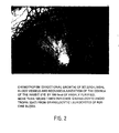
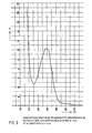
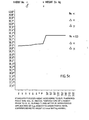
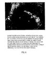
- Figures 1, 2 and 6 show the induction of neovascularization of the cornea of the rabbit by directional growth of blood vessels following focal intracorneal administration of angiotropins.
- Figs. 1 and 2 show the aciton of highly purified MAT and GAT protein substances which have been prepared from leukocyte cultures after a more than 100,000 fold purification from the crude supernatant culture solution.
- Fig. 6 shows the action of highly purified MAT protein substance prepared from inflamed (infarcted) heart muscle tissue.
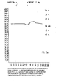
- a standard-pyrogen assay is represented according. to Europ. Pharmacopoeia, vol. II (1975): Rectal temperature of 3 rabbits.having an average weight of 3 kg is measured prior (V,A), during ( * ) and 30-180 minutes after (P) intravenous application of 5 ⁇ m MAT (about 0.3 nmol MAT/kg animal) in 1 ml 0.9 (w/v)% physiological saline. This corresponds to about the 300,000-fold (focal) amount of the biologically (angiotropically) active threshold.
- the leukocyte-derived angiotropins prepared and obtained according to the invention are valuable, endogenous and active protein substances. They can be used for influencing the vascular state of tissue, lungs, or in general, in tissue regeneration processes, such as reproductive cycles, embryogenesis and transplantations.
- a further possible mode of application is the preparation of inhibitors for suppression of undesired angiogenesis and neovascularization reactions in tissue. These pathological angiogenesis reactions are known and appear in the course of various processes, such as in tuberculosis, diabetes, tumors, reproductive cycles and tissue graft reactions. Thus it is well known that medical care of undesired pathological vascularization processes of normally avascular ocular tissues is hardly achieved.
- the use of angiotropins is also possible for the production of anti-angiotropin immunoglobulins and. their fractions.
- the angiotropins of the invention are locally applied alone or in admixture in the form of usual pharmaceutical compositions to mammals, for instance to man in amounts higher than 1 fmol or in concentrations of >10 pmol/l.
- Another subject matter of the invention is a process for the biotechnical preparation and isolation of angiotropins from leukocytes and from inflamed tissue sites. It is characterized in that either the leukocytes or the inflamed tissue are homogenized; or that leukocytes are cultured and the angiotropins formed or liberated are isolated from the homogenates or from the supernatant culture solution.
- the leukocytes can be cultured in any leukocyte-compatible medium.
- culture media For the culture of different cell types, such as bone marrow cells, heart muscle cells or leukocytes, different culture media are known. These media normally are aqueous solutions which contain numerous different compounds. Main constituents of these culture media are salts, sugars and metabolites, amino acids and derivatives, nucleosides, vitamins, vitaminoids, coenzymes, steroids and other additives, such as tensides, heavy metal salts and indicator dyes. Special examples of known culture media are named “HAM”,”MEDIUM 199" and “NCTC”, see H.J. Morton, In Vitro 6 (1970) p. 89 to 108.
- serum e.g. fetal calf serum or horse serum
- the serum constituents are said to be favourable for the maintenance of cellular functions.
- the serum-containing culture solution is to be subjected to processes for isolating proteins (mediators) which are formed by culturing cells, the preparation of trace protein products is difficult for reasons of the multipilicity of compounds making up the complex mixture of serum added to the culture.
- it is difficult upon addition of serum to a cell culture medium, it is difficult .
- the mediator may be derived from the species whose cells have been cultured; or , alternatively, it may be derived from the species from which the added (mostly heterologous) serum stems.
- a new, fully synthetic chemically defined culture medium is preferably used. It provides favourable conditions for cell culture and facilitates the preparation and isolation of the cellular angiotropin proteins from the culture supernatant.
- the fully synthetic, chemically defined cell culture medium preferably used in this invention contains the normal groups of compounds, such as salts, sugars, polyols, uronic acids, and derivatives, amino acids and derivatives, nucleosides and nucleoside bases, vitamins, vitaminoids, phytyl derivatives, coenzymes and steroids in aqueous solution. It is characterized in that it additionally- contains one or a mixture of several compounds which so far have not been considered for use in cell culture media. These are especially valuable for expression of the life functions, for the proliferation of leukocytes'and for promoting their capability to produce mediators. These substances include unsaturated fatty acids, flavanoids, ubiquinone, vitamin U, mevalolactone and L-carnosine.
- the cell culture medium is preferably used without addition of serum. Instead, it contains at least one defined protein.
- the synthetic, serum-free cell culture medium used in this invention may contain additional compounds, e.g. polyhydroxy compounds and sugars, amino acids, nucleosides, anionic compounds and/or vitamins which are not common in the known culture media. These compounds are useful in culturing. leukocytes.
- the constituents in the culture medium used in this invention are equilibrated in their ratios so that their concentrations mainly correspond to the natural concentration ranges of the plasma; see Ciba-Geigy AG (editor) (1969) in Documenta Geigy,tician- liche Tabellen seventh edition, Geigy S.A. Basle.
- the cell culture medium is free of tensides, heavy metal salts and dye indicators which can damage the cells and may have a detrimental effect on the isolation of the desired cell products.
- the cell culture medium with the composition given in Table III below is especially preferred in the process of the invention for culturing leukocytes.
- the medium is prepared with water of ASTM-1-quality; see ASTM D-1193-70 Standard Specification for Reagent Water 1970; Annual Book of ASTM -Standards, Easton, Maryland, ASTM 1970. In addition, it is freed from possible endotoxin-contaminations by ultrafiltration on tenside-free membranes with an exclusion limit of 10,000 dalton. The resulting medium is sterilized by filtration on tenside-free membranes with a pore size of 0.2 ⁇ m.
- Dependent on the type of desired product either mixed populations of leukocytes or homogenous leukocyte types are cultured. The preparation and culture of leukocytes must be performed under sterile conditions. Culturing is performed for a period sufficiently long to obtain a satisfactory medator level.
- a suitable period of time is 10 to 50 hours. Shorter periods result in lower mediator yields and the process is thus not economical.
- the medium is used up after a culture period of 50 hours and the cells begin to die.. An increase of the yield can therefore not be obtained in this case, except in the case of subculturing of cells and renewal of the culture medium.
- the leukocytes are cultured at a temperature of about 30 to 42°C, preferably at about 37°C. At lower temperatures the culture process is not satisfactory, while at temperatures of above 42°C the leukocytes are damaged.
- Culturing is carried out at a concentration of about 10 6 to 5 x 10 8 cells/ml, preferably 10 to 10 8 cells/ml.
- the mediator yield per volume unit of the culture solution is too low. With too large culture volumes, the process is not economical.
- cell concentrations of above 5 x 10 cells/ml nutrition of the cells in the medium becomes rapidly inefficient.
- Culturing can-be carried out in normal atmosphere. Preferably increased carbon dioxide partial pressure is maintained during culturing. This presssure can amount to about 10 vol%. 2 vol% are preferred.
- the oxygen supply to the culture is of great importance. Oxygen can be supplied e.g. by bubbling air through the culture. To avoid contamination of the culture, the air is preferably sterilized and heat-decontaminated, i.e. it is freed of endotoxins and other organic constituents.
- the cell suspension is stirred or agitated during culturing.
- angiotropins are already obtained in satisfactory yields by normal culture of leukocytes or certain leukocyte types.
- the GAT for instance, is obtained in high yields by culturing mixed populations of leukocytes or homogenous populations of granulocytes under the above-indicated conditions.
- the leukocytes are centrifuged from the supernatant culture solution which is subsequently processed for the resulting angiotropins .
- the culture is centrifuged at relatively low speed, i.e. at about 300 to 400 x g. After removal of the major part of the cells from the supernatant, it is expedient to centrifuge the latter again at a higher speed. In this way, the remaining floating particles are removed.
- the separated leukocytes can either be again subcultured, cryo-preserved or used for other biotechnical purposes.
- the supernatant culture solution freed from the cells contains the secretion products of the cultured leukocytes. These include the angiotropins of the invention and a number of other proteins and other substances. Their concentration in the culture solution is approximately within the nanomolar range. Consequently, a yield of about 1 to 10 mg of a defined mediator requires a culture solution volume of about 1,000 1 with respect to a 10% recovery after pruification. As regards the number of cells to be used, it can be calculated that in view of the molecular efficiency of the cells, about 10 14 leukocytes are necessary for obtaining a quantity of about 100 nmol proteins.
- the angiotropins of the invention can also be obtained from inflamed tissue sites. There, they are formed by the accumulation of leukocytes in the course of inflammatory processes induced by tissue injuries.
- the inflamed tissue can be obtained in the usual manner and used for the preparation of the angiotropins . Inflamed tissues are homogenized in buffer solution and soluble constituents or exudates are separated from insoluble structural components by means of centrifugation.
- inflamed, infarcted heart mucle tissue is used which was formed by ligation of 24 hours of the left anterior descendent branch of the left coronary artery by a transfemural catheter technique.
- the leukocyte-containing inflamed heart muscle site is,separated at 0 to 4°C from the remaining non-infracted tissue.
- the preparation and isolation of the angiotropins of the invention requires the processing of a very large culture solution volume. Therefore, at the beginning of the purification process effective reduction of the solution volume to be processed is necessary.
- the culture solution contains the mixture of the components of the medium.
- a separation of the formed proteins from the medium components with a concomitant reduction of the large volume of aqueous solution is achieved. This can be effected by selective salting-out precipitaton of the proteins from the supernatant culture solution, for instance by adding a sulfate or a phosphate.
- the salting-out preciepitation of proteins is exemplified by adding ammonium sulfate to the culture solution.
- the proteins formed By saturation of the supernatant culture solution with ammonium sulfate, a major portion of the proteins formed is precipitated together with serum albumin present as medium component.
- the proteins precipitated are recovered e.g. by centrigution. They are then separated into the individual components of the mixture as described below. Thereby, some angiotropins are obtained.
- some other angiotropins are salt-soluble and remain in the supernatant solution of the salting-out precipitation process. This supernatant also contains all soluble components of the medium. It is concentrated and the proteins obtained are processed in the manner described below.
- the protein mixture of the supernatant culture solution is already separated into several fractions by the salting-out precipitation step.
- the separation into several crude protein fractions is.possible, since groups of individual proteins precipitate at different ammonium sulfate concentrations.
- ammonium sulfate is therefore added stepwise to the culture solution up to a specific degree of saturation.
- Each fraction contains a group of proteins, the solubility product of which corresponds to the range of salt saturation.
- the supernatant culture solution is first brought to a 35% saturation with ammonium sulfate.
- the protein precipitate obtained is separated off.
- the 35% saturation of the supernatant solution is then increased to 45% by further addition of ammonium sulfate.
- a protein precipitate is again formed which is separated off.
- the 45% salt-saturated supernatant solution is brought to a 90% ammonium sulfate saturation.
- the protein precipitate formed is again separated off.
- the supernatant solution of this precipitate is concentrated e.g. by dehydration dialysis or ultrafiltration. The process is schematically shown in figure 8.
- the salting-out precipitation of proteins is preferably carried out at a temperature of about 0 to 10°C, especially of about 0 to 4°C.
- the subsequent purification steps are performed under the same conditions.
- the sol- utions used for the purification have a pH value of between 5 and 9, in particular between 6 and 8.
- a strong buffer for instance 0.1 mol/l of phosphate buffer is preferably added prior to the salting-out precipitation.
- cysteine is preferably added in an amount of 0.001 mol/1 to all solutions throughout the process.
- the protein purification does not require sterile conditions.
- the proteins obtained by salting-out precipitation can be directly subjected to purification and separation in the manner described below.
- the 90% salt-saturated supernatant.of the last precipitation step is concentrated. For instance, by dehydration dialysis or ultrafiltration, all compounds having a molecular weight higher than about 300 to 500 dalton are obtained as a retentate fraction. They can also be further processed for purfication of salt-soluble chemorecruitins.
- the protein fractions obtained in the step described above contain the angiotropins of the invention in admixture with numerous foreign proteins, e.g. other secreted proteins, in part serum albumins and in part CON. These foreign proteins form the major part of the constituents of this mixture.
- the angiotropinsmust be further purified by a sequence of further purification steps. Foreign proteins must be removed to avoid interference with the molecular-biological specifity.of angiotropins
- angiotropins themselves form a class of protein compounds which must be separated into individual, specifically acting structures.
- purification processes for proteins and other natural substances comprise sequences of combined separation techniques. Subtle differences in molecular size, charge, form, structure stability and nature of the molecular surfaces between the desired natural substance and the accompanying inactive foreign materials are used in such purification steps for their separation. Accordingly, a large number of combinations of various modifications of preparation techniques can be devised for the purification of a protein.
- the nature and the conditions of the preparation steps used, but also their sequential combination, are of paramount significance for operational properties, technical practicability, possibility of optional automatization and for the economical.performance of a purification process and also for the yield and mole- ' cular quality of a natural product investigated.
- purification steps For the purification of the individual protein fractions, a plurality of purification steps so far known in biochemistry can be used. Examples of such purification steps are: Preparative and analytical molecular sieve chromatography, anion and cation exchange chromatography and batch adsorption techniques, chromatography on hydroxyapatite, zone precipitation chromatography and recycling or cascade molecular sieve filtration.
- a particularly preferred embodiment of the process in accordance with the invention uses three of the mentioned purification steps in sequence for the purification of angiotropin activity from the protein fractions.
- Molecular sieve filtration achieves separation of proteins according to their molecular weights. Since the bulk of the foreign proteins have molecular weights different from those of angiotropins they can be separated off in this manner.
- a hydrophilic water-swelling molecular sieve as matrix is used for separation of the proteins by molecular weight.
- suitable molecular sieve matrices are dextrans cross-linked with epichlorohydrin (Sephadex), agaroses cross-linked with acrylamides (Ultrogels), and three-dimensionally cross-linked acrylamides (Biogels). The exclusion limits of the matrices used are higher than the separation limits. ,
- the molecular sieve filtration is preferably carried out as one of the first :: separation steps.
- molecular sieve filtration is termed "preparative” or “analytical”.
- a molecular sieve filtration is “preparative” when the chromatography is performed on columns with a length-to-diameter ratio of up to 10: 1 and a charge of the column of up to 1/3 of its capacity in terms of the total separation volume of the matrix.
- "Analytical" molecular sieve filtration means a length-to-diameter ratio larger than 10:1, and preferably about 50:1, and a maximum charge of the column of up to 3% of its capacity.
- gel matrices with the largest possible particle size are used for maximum flow-through rates of mostly viscous protein solutions applied at reasonably low pressures.
- particle size ranges of the gel matrix are selected as small as possible, to obtain a maximum number of theoretical plates, a flow rate of the mobile phase in the range of 2 to 4 cm/h combined with a pressure which is limited to technical and sefety aspects.
- the proteins are applied to the molecular sieve after dissolution in a protein-compatible liquid.
- a special example of a suitable solvent is 0.003 mol/l sodium-potassium phosphate solution containing 0.3 mol/l NaCl and 0.001 mol/l cysteine and having a pH of 7.4.
- the angiotropin -containing fractions are concentrated in the manner described below and optionally subjected to a further purification step.
- anion exchangers examples include dextran matrices cross-linked with epichlorohydrin (Sephadex) or cellulose matrices carrying functional groups with anion exchanger capacity. These exchangers can. be regenerated for repeated further use. It is preferable to use a weak anion exchanger in the Cl form such as DEAE-Sephadex A-50, pre-swollen and equilibrated in a buffer. Swelling and equilibration is preferably carried out at a pH of 8 to 10. A special example of such a buffer solution is 0.01 mol/l tris-HCl containing 0.04 mol/l NaCl.and 0.001 mol/l. cysteine and having a pH value of 8.0.
- the anion exchanger is added to the protein fraction in an amount sufficient for complete adsorption of the angiotropins and of the other positively adsorbing accompanying proteins. Two volume parts of swollen anion exchanger per volume of concentrated protein solution are normally sufficient.
- the reaction can be carried out either as chromatographic process or as an easy and fast batch adsorption technique. In the latter case, the supernatant liquid containing negatively adsorbed proteins is separated from the anion exchanger which is charged with the positively adsorbed angiotropins or other proteins, e.g. by filtration in a. chromatographic column, by decantation or centrifugation.
- the charged anion exchanger is freed from adhering negatively adsorbing compounds by washing with water or a salt solution having a maximum ionic strength equivalent to 0.04 mol/1 NaCl, preferably at a pH of 8 to 10.
- the maximum preferred temperatur is about 15°C.
- a special example of salt solution suitable for the washing-out process is the said tris-HCl buffer of pH 8.0.
- the anion exchanger on which ana i o t ro pi ns and other proteins are adsorbed and which is freed from the negatively adsorbed compounds is eluted with a protein-compatible aqueous salt solution having an ionic strength higher than 0.04 mol/l NaCl and a pH of between 4.0 and 10.0.
- a salt solution of high ionic strength and a pH of between 5.0 and 7.0 is preferably used.
- a special example of such a salt solution is a 2.0 mol/l NaCl solution buffered to a pH of 6.5 with 0.01 mol/1 piperazine-HCl and containing 0.001 mol/l cysteine.
- anion exchange reaction is carried out as a chromatographic process
- elution of the angiotropins and other positively adsorbed proteins can also be done by a linear NaCl concentration gradient.
- cation exchange matrices suitable for the purification of the protein fraction are dextrans crosslinked with epichlorohydrin (Sephadex) or cellulose matrices carrying functional groups with cation exchange capacity. These can be readily regenerated after use and employed again. It is preferable to use a weakly acidic cation exchanger such as CM-Sephadex C-50 having Na as-mobile counter-ion,. and to perform the exchange reaction at a pH between 4 and 6. To facilitate the charge process and to approach more ideal equilibria conditions prior to treatment with the cation exchanger the protein fractions should be diluted with a protein-compatible salt solution having a maximum ionic strength equivalent to 0.04 mol/1 NaCl.
- This salt solution can be used at the same time to adjust the pH.
- a special example of a salt solution for this purpose is a 0.001 mol/1 potassium phosphate-acetate buffer containing 0.04 mol/1 NaCl and 0.001 mol/l cysteine and having a pH of 4 to 6. This cation-exchange reaction may be performed as a chromatographic process, or technically easier, as a batch process.
- the swollen cation exchanger is added to the protein fraction in a quantity sufficient to adsorb it. As a rule, about 2 volume parts of swollen ion exchanger per volume part of protein solution is sufficient for this purpose.
- the supernatant is then separated from the cation exchanger charged with proteins, for example by decantation or centrifugation.
- the charged cation exchanger is freed from adhereing, negatively adsorbed compounds by washing with water or a salt solution, having a maximum ionic strength equivalent to 0.04 mol/l NaCl.
- a pH of about 4 to 6 and a maximum temperature of about 15°C is used.
- a special example of a salt solution suitable for the washing out process is the mentioned potassium phosphate-acetate buffer having a pH of 5.0.
- the washed protein-charged cation exchanger is now eluted with a protein-compatible aqueous salt solution.
- a salt solution of high ionic strength with a pH of about 4 to 10 is preferably used for-this purpose.
- Special examples of such salt solutions are aqueous 0.5 mol/l potassium phosphate. with a pH of 6.5 to-7.5 or a 2 to 5 mol/l NaCl with the same pH.
- salts e.g. ammonium sulfate and especially phosphates
- phosphates possibly present from preceding steps are removed from the protein solution, preferably by dialysis or ultrafiltration at membranes with an exclusion limit of 500 dalton prior to the application of the proteins to hydroxyapatite.
- the phosphate concentration of the protein solution is critical for the chromatography on hydroxyapatite.
- the an g iotropins are eluted by a potassium phosphate concentration gradient which is preferably linear.
- the angiotropins containing fractions are collected and then concentrated in the manner described below.
- hydroxyapatite is of essential significance for the structure-conserving isolation of pure angiotropins.
- considerable difficulties arise from chromatography of larger volumes of protein solutions on hydroxyapatite columns.
- larger protein amounts contribute to the strong tendency of hydroxyapatite to clog, thus becoming unusable as stationary matrix in chromatography.
- hydroxyapatite is very expensive. Its use on larger scales is not economical.
- the separation of a large part-of the accompanying foreign proteins by appropriate biotechnical purification steps from the angiotropin -containing protein fractions is preferred for considerably reducing the volume of the protein solution prior to its chromatography on hydroxyapatite.
- zone precipitation chromatography In-the zone precipitation chromatography (cf. J. Porath, Nature, vol. 196 (1962); p. 47-48), residual protein.contaminations in the angiotropins are separated by salting-out fractionation of the proteins by means and along a salt concentration gradient.
- the basic principle of separation of proteins in zone precipitation chromatography are different, structure-related, reversible solubility characteristics of proteins. They belong to the most sensitve molecular separation criteria.and are often used for demonstration of molecular homogeneity of a protein.
- Two variants of this technique for development of the chromatogram Two variants of this technique for development of the chromatogram are known: Fractional precipitation zone chromatography and fractional elution zone chromatography. Both types of techniques may have selective advantages in specific cases as described for fractional precipitation . and fractional elution methods in protein separation. Temperature and pH, column characteristics can all be varied within relatively wide limits.
- the temperature for zone precipitation chromatography can be betweeen 0 and 40°C.
- a temperature range from about 0 to 10°C is used, especially from about 4 to 6°C.
- the pH can be between 4 and 10; preferably, a pH range of 6 to 8 is used, especially a pH of about 7.
- the length-to-diameter ratio of the column used should be greater than about 10:1.
- a ratio of 30 to 100:1 and especially of about 50:1 is preferred. All protein-compatible salts having salting-out properties for proteins are suitable.
- salts examples include sodium-potassium phosphate, ammonium sulfate, and sodium sulfate. Ammonium sulfate is preferred.
- the salt concentration gradient can have any desired shape provided that salting-out criteria of proteins achieve protein separation. Linear concentration gradients are preferred, especailly an ascendent linear concentration gradient from 25 to 100% ammonium sulfate saturation.
- the maximum column charge is about 5% and preferably about 1% of total column volume.
- the recycling or cascade molecular sieve filtration can be performed under the.conditions described above for the analytical molecular sieve filtration.
- the same molecular sieves and the same column conditions can be used.
- Sephadex G 50 as stationary matrix is preferred in a column of a length-to-diameter ratio of at least about 50:1 and a maximum charge of about 3% of the column volume.
- the solvents used in the analytical molecular sieve filtration are also preferred as solvents for the elution in this method.
- concentration separation of a major portion of aqueous salt solution of the protein
- concentration can be achieved in different ways.
- Dehydration dialysis or ultrafiltration against protein-compatible liquid, preferably a sodium potassium phosphate buffer, are such methods.
- Dehydration dialysis is carrried out preferably against polyethylene glycol (molecular weight 20,000 dalton) at membranes with exclusion limits of preferably 500 dalton.
- Ultrafiltration is preferably achieved at membranes with an exclusion limit of about 500 dalton.
- cystein is preferably added to protein solutions throughout.
- ammonium sulfate is preferably added to the protein solution.
- this salt at this concentration ammonium sulfate exerts a strong salting-ineffect on proteins.
- proteins are better kept in solution during the molecular sieve filtration.
- ammonium sulfate prevents growth of microorganisms and inhibits certain enzymes. Hence, it contributes to stabilization of the angiotropin structure which is important when chromatography is performed at higher temperature (above about 20°C) and under non- sterile conditions.
- a ngiotropins which can be salted out are preferably completely precipitated alone or together with accompanying proteins by adding ammonium sulfate up to a concentration of about 3.25 to 3.7 mol/l (80 to 90% saturation). For this purpose 630 g/l ammonium sulfate are added (about 90% saturation).
- the pH value is preferably kept between 4 and 9 and the temperature up to 40°C, preferably between 0 and 8°C.
- the angiotropin -containing protein precipitate is separated from the protein-free supernatant solution by filtration, decantation or centrifugation.
- centrifugation is preferably carried out at least at 10,000 x g for a minimum of 45 min, and preferably for 1 h, in a one-step process. Or it can be .carried out in two stages, at lower forces in the first stage for removal of the bulk of precipitated proteins; and then, for the supernatant of the first stage containing residual fine protein particles at higher forces, e.g. 20,000 to 50,000 x g,. by flow-through centrifugation.
- the temperature and pH conditions during performance of the purification steps are not particularly critical. If the native conformation of the protein is to be preserved, an optimum temperature range is about 0 to 8°C, and preferably about 0 to 4°C. Moreover, the separation and purification steps must be carried out under essentially physiological. pH and salt conditions. An essential advantage of the process of the invention consists in that these conditions are for the first time easy to adhere to.
- the angiotropins obtained can be stored in a buffered physiological saline, e.g. in 0.0015 mol/l sodium-potassium phosphate solution containing 0.15 mol/1 (0.9 w/v%) NaCl, 0.001 mol/l cysteine and having a pH of 7.4.
- a buffered physiological saline e.g. in 0.0015 mol/l sodium-potassium phosphate solution containing 0.15 mol/1 (0.9 w/v%) NaCl, 0.001 mol/l cysteine and having a pH of 7.4.
- the protein preparation After usual sterilization by filtration (pore diameter 0.2 ⁇ m), the protein preparation remains native and biologically active at room temperature for at least 200 h or frozen at -25°C for at least 5 years.
- This stability of the protein can be considered, among others, to be one of the criteria of molecular homogeneity.
- g iotro p in solutions are safely stored at temperatures of between -20 and +50°C in the presence of 2.0 to 3.6 mol/l ammonium sulfate (50 to 90 % saturation).
- angiotropin solutions are protected against infection and degradation by microorganisms and bacterial growth.
- the angiotropins are again freed from salts by dia - lysis or ultrafiltration against an appropriate saline as described above.
- Leukocytes or inflamed tissue is homogenized or leukocytes are cultured and the resultant angiotropins - are isolated from the homogenates or the supernatant culture solution. Culturing may be performed with a mixed leukocyte population or with a specific leukocyte type.
- the leukocytes are preferably cultured in a fully synthetic cell culture medium containing serum albumin as the only protein.
- the mitosis of the leukocytes is optionally induced during culture.
- a polyvalent mitogen or endotoxin-mitogen may be added or an immune reaction is prompted on the cell surface so as to induce the mitosis of the leukocytes.
- the leukocytes are cultured in a cell culture medium having the composition given in Table III for approximately 40 hours at about 37°C and a concentration of about 10 7 to 10 8 cells/ml culture solution at a C0 2 -partial pressure of about 1% while sufficient oxygen is supplied to the culture.
- the protein portion contained in the culture solution which becomes insoluble upon salt addition is obtained by salting ,out from the solution and the protein portion which is soluble in the saturated salt solution is obtained by concentrating this solution.
- Ammonium sulfate is preferably used for salting out the proteins.
- the ammonium sulfate concentration of the culture solution is stepwise increased; the proteins precipitated are separated after each ammonium sulfate addition and several crude protein fractions having graduated solubility at different ammonium sulfate concentrations are thus obtained.
- the ammonium sulfate concentration of the culture solution is adjusted stepwise to 35%, 45% and 90% saturation.
- the supernatant of the salting out-precipitation is concentrated after separation of the protein precipitate by ultrafiltration or dialysis.
- the crude protein fractions isolated by stepwise salting out and the concentrated supernatant of the salting-out precipitation are processed sepa- ra t el y to obtain the angiotropins.
- the processing of the crude protein fractions and the isolation of the angiotropins is performed by preparative and analytical molecular sieve filtration, anion and cation exchange chromatography and batch adsorption processes, respectively, chromatography on hydroxyapatite, zone precipitation chromatography -and/or recycling or cascade molecular sieve filtration.
- At least two in particular at least three of the said purification steps are performed in sequence.
- the soluble portion of a leukocyte or inflamed tissue homogenate may be used for preparing and isolating the angiotropins.
- the invention will now be given in detail by examples describing the isolation of the angiotropin protein preparation starting from leukocytes of porcine.blood.
- the invention is not restricted to this embodiment. Leukocytes and inflamedtissues of other mammalians can be used too.
- angiotropins in a culture solution . of a mixed population of leukocytes and the separation of monocyto-angiotropin (MAT) and granulocyto-angiotropin (GAT) from the other components of the culture ' supernatant are described. All process steps are carried out at 0 to 8°C in the presence of 0.001 mol/l cysteine, unless otherwise specified.
- the centrifugation is carried out in the manner described , either as a one or two step procedure (as flow-through centrifugation).
- 50 kg (about 10 14 ) leukocytes are isolated as mixed cell population of physiological composition from 10,000 1 of porcine blood and cultured in 20 batches of 2.5 kg (about 5 x 10 12 cells) under sterile conditions.
- the medium indicated in table III is used as culture solution.
- 50 1 of culture medium are used per batch. Culturing is performed in glass vessels (Duran 50 or Pyrex glass). Initially, the cell density is about 10 cells/ml. The culture is maintained at 37°C in an atmosphere of 1 v/v % C0 2 over 40 hours. During this period, the cell suspension is slowly stirred (to r.p.m.) and flooded with sterile, water- washed and heat-decontaminated air bubbles ( ⁇ 1mm).
- the heat-decontamination of air is performed at about 500°C by flowing through. a silica tube.
- the pH value (7.1) and the D-glucose level are measured and maintained constant.
- the cells are induced to mitosis by the polyvalent mitogen content (CON) of the culture medium.
- CON polyvalent mitogen content
- the number, differential and morphological viability (dye exclusion test) of the cells are continously determined by usual methods of hematology and cell culture techniques.
- the functional viability of cells is measured by their motility and their ability to respond to chemokinetic and chemotactic proteins. Mitoses are determined by chromosome count.
- the morphological viability of the cells after their biotechnical culturing is 95%.
- the entire loss in cells (mainly granulocytes) during culturing is at most 20% which is normal for primary cell cultures.
- the culture is terminated by separating the cells from the supernatant solution by centrifugation for 10 minutes at 400 x g and 10°C.
- the cells are washed twice in a salt solution containing 0.15 mol/l NaCl, 0.0015 mol/l sodium potassium phosphate and having the pH-value 7.1. They can be used for another purpose.
- the culture supernatant solution is then centrifuged again for 1 hour at 10,000 x g and at 4°C to remove suspended particles.
- the resultant clear supernatant culture solution which has a total volume of 1000 liters and contains about 1,400 g protein as well as other macromolecules and salts is directly subjected to salting-out fractionation with ammonium sulfate (A2.). Unless otherwise stated, all further steps are carried out at 0-4°C.
- This buffered supernatant culture solution is then adjusted to 35% saturation of ammonium sulfate by addition of 199 g of ammonium sulfate/l solution.
- the pH-value of the protein solution is continuously controlled and maintained at 6.7 by the addition of 2 n ammonia.
- Part of the proteins is precipitated from the solution.
- the protein precipitate formed is separated from the supernatant containing salt-soluble proteins by centrifugation for 1 hour at 10,000 x g.
- the precipitated crude protein fraction I is obtained as ammonium sulfate-containing protein sludge which contains about 100 g protein.
- This crude protein concentrate fraction I contains GAT and is separately processed for GAT according to the procedure described below.
- the precipitated crude protein concentrate fraction II is obtained as ammonium sulfate-containing protein sludge, the protein content of which is about 60 g.
- This crude protein concentrate fraction II may be processed separately for its constituents , according to the procedure described below for the crude protein concentrate fraction III.
- the 45% salt-saturated supernatant culture solution is then adjusted to 90% saturation of ammonium sulfate by adding 323 g of ammonium sulfate/1 of solution.
- the pH-value of the protein solution is again.continuously controlled and maintained constant at 6.7 by 2 n ammonia.
- Another portion of the proteins is precipitated from the solution.
- the protein precipitate is separated from the supernatant containing salt-soluble proteins by centrifugation for 1 hour at 10,000 x g.
- the precipitated crude protein concentrate fraction III is obtained as ammonium sulfate-containing protein sludge the protein content of which is approximately 1,080 g. This fraction also contains the bulk of the serum albumin as component of the culture medium.
- This crude protein concentrate fraction III may be processed for its constituents according to the procedure described below.
- the 90% salt saturated supernatant fraction IV of the crude fraction III contains 160 g of salt-soluble proteins and other macro molecules (> 500 daltons).This supernatant contains the MAT.
- This salt-soluble protein-containing supernatant fraction IV is diluted with the same volume of the buffer solution A ( 0.15 mol/l NaCl, 0.0015 mol/l sodium-potassium phosphate, 0.001 mol/1 L-cysteine, pH 7.4) to 45% saturation of ammonium sulfate and a maximum phosphate concentration of 0.05 mol/l.
- This solution is concentrated and desalted by ultrafiltration at a membrane with an exclusion limit of 500 dalton as a maximum.
- the salt-soluble proteins of this solution are obtained as crude retentate fraction IV in a volume of 13 1 (about 100-fold cencentration).
- the crude protein concentrate fractions I, II and III and the retentate fraction IV are further purified.
- the fine purification of fraction I is described below under A 3 and applies to all crude protein concentrate fractions.
- the fine purification of the retentate fraction is mentioned below under A 4.
- the crude protein concentrate fraction I obtained above (A 2) is dissolved in a minimum volume of buffer solution B (0.01 mol/l of tris-HCl solution containing 0.04 mol/l NaCl and 0.001 mol/l cysteine and having a pH value of 8.0).
- the resultant slightly turbid solution (20 1) is clarified by centrifugation and then freed of salts by dialysis at a membrane with the exclusion limit of 500 dalton against buffer solution B until no sulfate ions are detectable.
- the clear solution obtained is then applied to a column of a swollen regenerated anion exchanger (Cl - as mobile exchangeable ion). It has a dextran matrix cross-linked with epichlorohydrin (DEAE-Sephadex A 50) which is equilibrated in the above-mentioned buffer system B.
- the column has four times the volume of the protein. solution and a length-to-diameter ratio of 10 : 1.
- the gel column is then washed with the above-mentioned adsorption buffer solution B until the extinction of the filtrate at 280 nm is ⁇ 1.0.
- the charged ion exchanger gel is eluted with a NaCl-concentration gradient during 2 days.
- the gradient is linearly ascending from 0.04 to 2.0 mol/l NaCl, whereas the pH value, the tris/HCl and the cysteine concen-. trations are maintained constant.
- the same shape of gradient is then used for lowering the pH from 8 to 6.5 for further elution of the compounds. It is made up by 0.01 mol/l piperacine-HCl-buffer containing 2.0 mol/1 NaCl and 0.001 mol/l cysteine and having the pH 6.5.
- the angiotropin-containing fractions are collected and processed in further purification steps described below (A.3.2. - A.3.6).
- the protein precipitate is dissolved in a minimum volume of buffer solution C (0.003 mol/l sodium-potassium phosphate containing 0.3 mol/1 NaCl and 0.001 mol/l cysteine and having a p H value of 7.4). After removal of a small amount of insoluble compounds by centrifugation, the solution is applied to a column of a molecular sieve matrix of agarose cross- linked with acrylamide (Ultrogel AcA 34,. particle size 60 to 160 ⁇ m) for preparative molecular sieve filtration.
- buffer solution C 0.003 mol/l sodium-potassium phosphate containing 0.3 mol/1 NaCl and 0.001 mol/l cysteine and having a p H value of 7.4
- the column has 10 times the volume of the protein solution and a length-to-diameter ratio of 20:1.
- the column is then eluted with an upward flow (3 cm/h) of the mentioned buffer solution C.
- the fraction with the separation limits of 20,000 and 45,000 dalton is. collected.
- the fraction is lyophilized, ultrafiltrated at a membrane with the exclusion limit of 500 dalton or adjusted to an ammonium sulfate concentration of 3.7 mol/l.
- the protein precipitates are separated from the supernatant by centrifugation and farther processed as described below (A.3.3)
- the resultant GAT-containing protein precipitate (A 3.2) is dissolved in 1.5 volume parts of buffer solution D (0.01 mol/l sodium-potassium phosphate, 0.04 mol/l NaCl, 0.001 mol/l cysteine, pH 6.0). The solutions are centrifuged at 10,000 x g for 1 hour for removal of a small amount of insoluble material.
- the clear solution is dialyzed against the buffer solution D at a membrane with the exlusion limit of 500 dalton until no sulfate ions are detectable.
- the clear solution obtained is then applied to a column of swollen, regenerated cation exchanger. based on a dextran matrix cross-linked with epichlorohydrin (CM-Sephadex C 50).
- CM-Sephadex C 50 epichlorohydrin
- the exchanger is equilibrated in the above-mentioned buffer system D (Na + as mobile exchangeable ion).
- the column has four times the volume of theprotein solution and a length-to-diameter ratio of 10 : 1.
- the gel column is then washed with the above-mentioned adsorption buffer solution D, until the extinction of the filtrate at 280 nm is ⁇ 1 . 0 .
- the GAT is eluted in this step.
- the GAT-containing fraction is collected and concentrated in the usual manner and further processed as described below (A.3.4).
- the GAT -containing protein precipitate (A.3.3) is dissolved in a minimum volume of 0.0001 mol/l sodium-potassium phosphate buffer solution E containing 0.001 mol/l cysteine and having a pH of 7.20.
- the solutions are then desalted with this buffer by molecular sieve filtration, ultrafiltration or dialysis (exclusion limit 500 dalton), until no sulfate is detectable in the dialysis buffer. Thereafter, a small portion of insoluble material is removed by centrifugation at 10,000 x g for 1 hour.
- the clear GAT -containing protein solution obtained is. separately applied to a column of hydroxyapatite.
- the length-to-diameter ratio of the column is 10 : 1 and it has four times the volume of the protein volume to be applied.
- the column has been equilibrated with the mentioned buffer E used in an amount five times the column volume (flow 3 cm/h).
- the negatively adsorbed proteins are washed out with the buffer solution E used for equilibrating the column.
- the elution of the GAT -containing fractions is carried out with a phosphate concentration gradient for 4 days.
- the gradient is linearly ascending from 0.0001 mol/l to 0.5 mol/l sodium-potassium phosphate having a constant pH value of 7.4 constant cysteine concentration .
- G AT is eluted at an average phosphate concentration of about 0.15 mol/l.
- the elution gradient is measured and controlled by.means of conductivity.
- the GAT-containing fractions are concentrated in the usual manner and further processed as described below (A.3.5).
- the GAT-containing fractions (A.3.4.) are dissolved in 0.1 mol/l sodium-potassium phosphate solution F containing 0.1 mol/1 NaCl, 0.001 mol/1 cysteine and 1 mol/l ammonium sulfate and having a pH value of 7.4.
- the resultant solution is applied at a temperature of 4°C to a column of swollen molecular sieve matrix of dextran cross-linked with epichlorhydrin (Sephadex G-25).
- an ascendent, linear ammonium sulfate concentration gradient is established with the mobile buffer phase from 1.0 to 4.0 mol/l ammonium sulfate (20 to 100% saturation).
- the slope of the gradient is +2% of the ammonium sulfate saturation/cm of column height (0.08 mol/l (NH 4 ) 2 SO 4 /cm).
- the range of the gradient extends over approximately half the length of the column.
- the length-to-diameter ratio of the column is 50 : 1, the column volume is 100 times higher than the protein solution volume to be applied.
- the flow rate is 2 cm/h.
- the elution is carried out with the above-mentioned sodium-potassium phosphate solution F containing 1 mol/l of ammonium sulfate.
- the GAT-containing fractions which are eluted at 33% ammonium sulfate saturation are collected.
- the proteins are concentrated in the usual manner and further processed as described below. (A.3.6).
- the GAT -containing fractions (A.3.5.) are dissolved in buffer C (0.003 mol/l sodium-potassium phosphate containing 0.3 mol/l NaCl and 0.001 mol/I casteine and having a pH value of 7.4). Removal of a small portion of insoluble substances is achieved by centrifugation for 30 minutes at 48,000 x g.
- buffer C 0.003 mol/l sodium-potassium phosphate containing 0.3 mol/l NaCl and 0.001 mol/I casteine and having a pH value of 7.4
- the resultant clear solution is then subjected to analytical recycling molecular sieve chromatography.
- the solution is applied at a temperature of 4°C to a column of Ultrogel AcA 44 having a particle size of 60 to 140 ⁇ m.
- the column has 50 times the volume of the protein solution and a length-to-diameter ratio of 50 : 1.
- the elution is carried out with the mentioned buffer C.
- the eluates are recycled three times at a separation limit of 40,000 dalton.
- approximately 8 mg of GAT are obtained having a molecular homogeneity of > 95%, as indicated by conventional methods.
- the retentate fraction IV (A. 2) is purified in the manner described above for the crude protein concentrate fraction I. However, the sequence of the steps of preparative molecular sieve filtration and anion exchange chromatography is exchanged. Moreover, in the preparative molecular sive filtration and in the analytical recycling molecular sieve filtration, the Ultrogel AcA is replaced by a molecular sieve matrix of dextran which is cross- linked with epichlorohydrin (Sephadex G-50) and has a particle size of 40 to 120 and 20 to 80 ⁇ m, respectively.
- the separation limits are 7,000 to 3,000 dalton.
- the MAT is eluted at an average phosphate concentration of 0.001 mol/l.
- the MAT is eluted in the front distribution.
- the eluate is recycled at a separation limit of 7, 000 dalton.
- the MAT yield is about 8 mg and has a molecular homogeneity of > 95%, as shown by conventional methods.
- monocytes obtained from porcine blood are cultured under the conditions described in example A.
- the polyvalent mitogen (CON) in the medium induces the mitosis of the cells.
- the angiotropin MAT secreted to the culture solution is isolated according to the procedure described in example A. It is thereby obtained in a highly purified state. The yield obtained is comparable to that of example A.
- angiotropins from inflamed tissue are described.
- 500 g of infarcted, inflamed canine heart muscle tissue are used.
- the heart muscle tissue is ground at 0-4°C.
- 0.05 mol/1 sodium potassium phosphate buffer solution containing 0.001 mol/l cystein and having a pH of 6.8 is added in a quantity three times the amount of the tissue.
- the resultant suspension is homogenized in a homogenizer (ultraturax).
- the supernatant containing the soluble compounds of the inflamed tissue is separated from the insoluble constituents by cen- triguation at 10,000 x g and 4°C.
- the resultant supernatant solution is then centrifuged for 3 hours at 100,000 x g.
- the clear supernatant solution obtained is siphoned off from the flotating lipid layer.
- angiotropin -containing clear supernatant protein solution is then subjected to fractional salting-out. precipitation with ammonium sulfate according to example A.
- the resultant protein fraction I and the concentrated retentate fraction IV are processed as described in example A. From the 500 g tissue, angiotropins are obtained in a yield of approximately 0.03 mg of MAT, and about 0.02 mg of GAT.
- Leukocytes are prepared from blood according to example A.
- a homogenate of 500 g of leukocytes is prepared as shown in example C. for muscle tissue.
- the isolation of the angiotropins contained in the leukocytes is performed according to example A.
- the leukocytes cultured without stimulation contain only relatively small (about 1%) amounts of monocyte-angiotropins (MAT).
- the yields are approximately 5 ⁇ g of GAT and 1 ⁇ g of MAT.
Landscapes
- Chemical & Material Sciences (AREA)
- Health & Medical Sciences (AREA)
- Organic Chemistry (AREA)
- Life Sciences & Earth Sciences (AREA)
- Molecular Biology (AREA)
- Proteomics, Peptides & Aminoacids (AREA)
- Medicinal Chemistry (AREA)
- Biochemistry (AREA)
- Biophysics (AREA)
- General Health & Medical Sciences (AREA)
- Genetics & Genomics (AREA)
- Zoology (AREA)
- Toxicology (AREA)
- Gastroenterology & Hepatology (AREA)
- Vascular Medicine (AREA)
- Immunology (AREA)
- Medicines Containing Material From Animals Or Micro-Organisms (AREA)
- Peptides Or Proteins (AREA)
- Micro-Organisms Or Cultivation Processes Thereof (AREA)
- Investigating Or Analysing Biological Materials (AREA)
- Preparation Of Compounds By Using Micro-Organisms (AREA)
- Immobilizing And Processing Of Enzymes And Microorganisms (AREA)
- Medicines That Contain Protein Lipid Enzymes And Other Medicines (AREA)
- Medicines Containing Antibodies Or Antigens For Use As Internal Diagnostic Agents (AREA)
Priority Applications (1)
| Application Number | Priority Date | Filing Date | Title |
|---|---|---|---|
| AT82102159T ATE17858T1 (de) | 1981-03-18 | 1982-03-17 | Angiotropine aus leukozyten und entzuendeten geweben: eine neue klasse von natuerlichen chemotropischen und mitogenen proteinen fuer die spezifische induktion des gesteuerten wachstums von blutgefaessen, fuer neue gefaessbildung von geweben und fuer die morphogenese von blutgefaessverzweigungen; verfahren zu ihrer biotechnischen herstellung und pharmazeutische zusammensetzungen. |
Applications Claiming Priority (2)
| Application Number | Priority Date | Filing Date | Title |
|---|---|---|---|
| DE3110560 | 1981-03-18 | ||
| DE3110560A DE3110560A1 (de) | 1981-03-18 | 1981-03-18 | "angiotropine der leukozyten und des entzuendungsgewebes: eine neue klasse natuerlicher chemotropischer mitogene fuer das richtungswachstum von blutgefaessen und zur neovaskularisierung von geweben" |
Publications (3)
| Publication Number | Publication Date |
|---|---|
| EP0061138A2 true EP0061138A2 (de) | 1982-09-29 |
| EP0061138A3 EP0061138A3 (en) | 1983-09-07 |
| EP0061138B1 EP0061138B1 (de) | 1986-02-05 |
Family
ID=6127640
Family Applications (1)
| Application Number | Title | Priority Date | Filing Date |
|---|---|---|---|
| EP82102159A Expired EP0061138B1 (de) | 1981-03-18 | 1982-03-17 | Angiotropine aus Leukozyten und entzündeten Geweben: eine neue Klasse von natürlichen chemotropischen und mitogenen Proteinen für die spezifische Induktion des gesteuerten Wachstums von Blutgefässen, für neue Gefässbildung von Geweben und für die Morphogenese von Blutgefässverzweigungen; Verfahren zu ihrer biotechnischen Herstellung und pharmazeutische Zusammensetzungen |
Country Status (8)
| Country | Link |
|---|---|
| US (1) | US4465669A (de) |
| EP (1) | EP0061138B1 (de) |
| JP (1) | JPS57206621A (de) |
| AT (1) | ATE17858T1 (de) |
| AU (1) | AU550434B2 (de) |
| CA (1) | CA1188244A (de) |
| DE (2) | DE3110560A1 (de) |
| DK (1) | DK118682A (de) |
Cited By (4)
| Publication number | Priority date | Publication date | Assignee | Title |
|---|---|---|---|---|
| EP0226181A3 (en) * | 1985-12-17 | 1989-03-08 | Synergen, Inc. | Human placenta angiogenic factor capable of stimulating capillary endothelial cell protease synthesis, dna synthesis and migration |
| US4994559A (en) * | 1985-12-17 | 1991-02-19 | Synergen, Inc. | Human basic fibroblast growth factor |
| US5026839A (en) * | 1985-12-17 | 1991-06-25 | Synergen, Inc | DNA encoding a basic fibroblast growth factor |
| WO1999047563A3 (de) * | 1998-03-13 | 1999-11-11 | Fraunhofer Ges Forschung | Metallhaltige ribonukleotidpolypeptide |
Families Citing this family (21)
| Publication number | Priority date | Publication date | Assignee | Title |
|---|---|---|---|---|
| HUT42329A (en) * | 1984-11-29 | 1987-07-28 | Curatech Inc | Process for producing compositions for curing wounds |
| US5165938A (en) * | 1984-11-29 | 1992-11-24 | Regents Of The University Of Minnesota | Wound healing agents derived from platelets |
| US4727137A (en) * | 1985-04-17 | 1988-02-23 | President And Fellows Of Harvard College | Purified protein having angiogenic activity and methods of preparation |
| AU597323B2 (en) * | 1985-11-22 | 1990-05-31 | Teijin Limited | Angiogenic factor derived from blood vessel endothelial cells |
| US4808402A (en) * | 1987-05-29 | 1989-02-28 | Northwestern University | Method and compositions for modulating neovascularization |
| US4853219A (en) * | 1987-08-06 | 1989-08-01 | President And Fellows Of Harvard College | Antibodies to angiogenin: immunotherapeutic agents |
| US6077823A (en) * | 1991-03-11 | 2000-06-20 | Creative Biomolecules, Inc. | Method for reducing tissue damage associated with ischemia-reperfusion or hypoxia injury |
| US6395883B1 (en) * | 1991-03-11 | 2002-05-28 | Curis, Inc. | Soluble morphogenic protein complex compositions of matter |
| US5494808A (en) * | 1994-09-15 | 1996-02-27 | Merck & Co., Inc. | Defined medium OMPC fermentation process |
| KR20000064752A (ko) | 1996-03-22 | 2000-11-06 | 더 제네랄 호스피탈 코포레이션 | 중추신경계허혈또는외상의발현후폴리펩티드성장인자를투여하는방법 |
| US6979307B2 (en) * | 1997-06-24 | 2005-12-27 | Cascade Medical Enterprises Llc | Systems and methods for preparing autologous fibrin glue |
| US7745106B2 (en) * | 1997-06-24 | 2010-06-29 | Cascade Medical Enterprises, Llc | Methods and devices for separating liquid components |
| US20080199513A1 (en) * | 1997-06-24 | 2008-08-21 | Cascade Medical Enterprises, Llc | Systems and methods for preparing autologous fibrin glue |
| MXPA00000009A (es) | 1998-06-22 | 2005-09-08 | Autologous Wound Therapy Inc | Solicitud para patente de utilidad para curativo de heridas mejorado enriquecido con plaquetas. |
| US6783987B2 (en) * | 1999-12-09 | 2004-08-31 | Tsukasa Matsumoto | Method for obtaining components from cultured leucocyte |
| AU2770601A (en) * | 2000-01-07 | 2001-07-24 | Bioenergy Inc. | Compositions for enhancing the immune response |
| AUPR177300A0 (en) * | 2000-11-29 | 2000-12-21 | Centre For Molecular Biology And Medicine | Therapeutic methods |
| US7481790B2 (en) * | 2000-12-27 | 2009-01-27 | Advanced Cardiovascular Systems, Inc. | Vessel enlargement by arteriogenic factor delivery |
| NL1019368C2 (nl) * | 2001-11-14 | 2003-05-20 | Nutricia Nv | Preparaat voor het verbeteren van receptorwerking. |
| WO2006102488A1 (en) * | 2005-03-22 | 2006-09-28 | Cascade Medical Enterprises, Llc | Systems and methods of producing membranes |
| AU2007292253A1 (en) * | 2006-09-08 | 2008-03-13 | Wyeth | Arginine wash in protein purification using affinity chromatography |
Family Cites Families (2)
| Publication number | Priority date | Publication date | Assignee | Title |
|---|---|---|---|---|
| DE2722769C3 (de) * | 1977-05-20 | 1981-08-06 | Max-Planck-Gesellschaft zur Förderung der Wissenschaften e.V., 3400 Göttingen | Verwendung von Fibrinogen |
| US4229531A (en) * | 1978-12-29 | 1980-10-21 | Monsanto Company | Production of tumor angiogenesis factor by cell culture |
-
1981
- 1981-03-18 DE DE3110560A patent/DE3110560A1/de not_active Withdrawn
-
1982
- 1982-03-15 US US06/357,814 patent/US4465669A/en not_active Expired - Fee Related
- 1982-03-17 DK DK118682A patent/DK118682A/da not_active Application Discontinuation
- 1982-03-17 EP EP82102159A patent/EP0061138B1/de not_active Expired
- 1982-03-17 AT AT82102159T patent/ATE17858T1/de not_active IP Right Cessation
- 1982-03-17 AU AU81633/82A patent/AU550434B2/en not_active Ceased
- 1982-03-17 CA CA000398616A patent/CA1188244A/en not_active Expired
- 1982-03-17 DE DE8282102159T patent/DE3268915D1/de not_active Expired
- 1982-03-18 JP JP57043667A patent/JPS57206621A/ja active Pending
Non-Patent Citations (4)
| Title |
|---|
| "Circu ation", AMER. PHYSIOL. SOC. WASHINGTON, vol. 2, 1965, pages 1251 - 1276 |
| "Pathophysiology of Myocardial Perfusion", 1979, ELSEVIER-NORTH HOLLAND |
| "Tumor Blood Circulation", 1979, CRC PRESS |
| A.A. LIEBOW: "Handbook of Physiology", article "Blood vessels go where they are needed" |
Cited By (4)
| Publication number | Priority date | Publication date | Assignee | Title |
|---|---|---|---|---|
| EP0226181A3 (en) * | 1985-12-17 | 1989-03-08 | Synergen, Inc. | Human placenta angiogenic factor capable of stimulating capillary endothelial cell protease synthesis, dna synthesis and migration |
| US4994559A (en) * | 1985-12-17 | 1991-02-19 | Synergen, Inc. | Human basic fibroblast growth factor |
| US5026839A (en) * | 1985-12-17 | 1991-06-25 | Synergen, Inc | DNA encoding a basic fibroblast growth factor |
| WO1999047563A3 (de) * | 1998-03-13 | 1999-11-11 | Fraunhofer Ges Forschung | Metallhaltige ribonukleotidpolypeptide |
Also Published As
| Publication number | Publication date |
|---|---|
| DK118682A (da) | 1982-09-19 |
| US4465669A (en) | 1984-08-14 |
| EP0061138B1 (de) | 1986-02-05 |
| DE3268915D1 (en) | 1986-03-20 |
| ATE17858T1 (de) | 1986-02-15 |
| DE3110560A1 (de) | 1982-10-14 |
| AU8163382A (en) | 1982-09-23 |
| EP0061138A3 (en) | 1983-09-07 |
| CA1188244A (en) | 1985-06-04 |
| AU550434B2 (en) | 1986-03-20 |
| JPS57206621A (en) | 1982-12-18 |
Similar Documents
| Publication | Publication Date | Title |
|---|---|---|
| EP0061138B1 (de) | Angiotropine aus Leukozyten und entzündeten Geweben: eine neue Klasse von natürlichen chemotropischen und mitogenen Proteinen für die spezifische Induktion des gesteuerten Wachstums von Blutgefässen, für neue Gefässbildung von Geweben und für die Morphogenese von Blutgefässverzweigungen; Verfahren zu ihrer biotechnischen Herstellung und pharmazeutische Zusammensetzungen | |
| EP0061141B1 (de) | Chemokinesine und Chemotaxine aus Leukozyten und entzündeten Geweben: natürliche Mediationsproteine für umkehrbare Förderung von willkürlichen und gerichteten Bewegungen (Chemokinesis und Chemotaxis) und für Akkumulation von spezifischen Leukozytarten; Verfahren zu ihrer biotechnischen Herstellung und pharmazeutische Zusammensetzungen | |
| Gherardi et al. | Purification of scatter factor, a fibroblast-derived basic protein that modulates epithelial interactions and movement. | |
| DE3686927T2 (de) | Neovaskularisierungsinhibitoren und deren herstellung. | |
| DE69635661T2 (de) | Methode zur reinigung von thrombopoietin | |
| KR20040071212A (ko) | 생물학적 활성 과립구 콜로니 자극 인자의 정제 및(또는)단리 방법 | |
| EP0061139B1 (de) | Mitogene aus Leukozyten und entzündeten Geweben: natürliche leukopoietische Proteine für die spezifische Induktion der Vermehrung und der Differenzierung von Leukozyten; Verfahren zu ihrer biotechnischen Herstellung und pharmazeutische Zusammensetzungen | |
| EP0061140B1 (de) | Chemorekruitine aus Leukozyten und entzündeten Geweben: eine neue Klasse von natürlichen Leukopoietinproteinen für die chemische Rekrutierung spezifischer Leukozytarten aus dem Knochenmark nach dem Blutkreislauf (Leukozytosis und Umstellungsreaktionen nach links); Verfahren zu ihrer biotechnischen Herstellung und pharmazeutische Zusammensetzungen | |
| US6087123A (en) | Metal-containing ribonucleotide polypeptides | |
| DE69029040T2 (de) | Megakaryocytopoietischer faktor | |
| EP0042560B1 (de) | Verfahren zum Herstellen und Erhalten von Anaphylatoxin und Cocytotaxin enthaltenden Leukotaxin-Präparaten und von Anaphylatoxin- und Cocytotaxin-Proteinen in einer molekular homogenen und biologisch aktiven Form | |
| DE3421731A1 (de) | Humaner-tumor-nekrose-faktor | |
| US6770455B1 (en) | Metal-containing ribonucleotide polypeptides | |
| JPH02502640A (ja) | Gm‐csfの精製 | |
| CA1188245A (en) | Chemokinesins and chemotaxins of leukocytes and inflamed tissues and process of their preparation | |
| US4452735A (en) | Leukorecruitin | |
| DE3426049A1 (de) | Humaner-tumor-nekrose-faktor | |
| JPH01221324A (ja) | ヒト単球−マクロファージコロニー刺激因子を有効成分とする造血器疾患治療剤 | |
| Ciaglowski | High-and low-affinity heparin binding proteins from bovine platelets | |
| JPS6191129A (ja) | 抗腫瘍剤 | |
| JPS62108818A (ja) | 新規蛋白性物質の製造法 |
Legal Events
| Date | Code | Title | Description |
|---|---|---|---|
| PUAI | Public reference made under article 153(3) epc to a published international application that has entered the european phase |
Free format text: ORIGINAL CODE: 0009012 |
|
| AK | Designated contracting states |
Designated state(s): AT BE CH DE FR GB IT LU NL SE |
|
| PUAL | Search report despatched |
Free format text: ORIGINAL CODE: 0009013 |
|
| AK | Designated contracting states |
Designated state(s): AT BE CH DE FR GB IT LI LU NL SE |
|
| 17P | Request for examination filed |
Effective date: 19831117 |
|
| GRAA | (expected) grant |
Free format text: ORIGINAL CODE: 0009210 |
|
| AK | Designated contracting states |
Designated state(s): AT BE CH DE FR GB IT LI LU NL SE |
|
| REF | Corresponds to: |
Ref document number: 17858 Country of ref document: AT Date of ref document: 19860215 Kind code of ref document: T |
|
| REF | Corresponds to: |
Ref document number: 3268915 Country of ref document: DE Date of ref document: 19860320 |
|
| PG25 | Lapsed in a contracting state [announced via postgrant information from national office to epo] |
Ref country code: LU Free format text: LAPSE BECAUSE OF NON-PAYMENT OF DUE FEES Effective date: 19860331 |
|
| ITF | It: translation for a ep patent filed | ||
| ET | Fr: translation filed | ||
| PLBE | No opposition filed within time limit |
Free format text: ORIGINAL CODE: 0009261 |
|
| STAA | Information on the status of an ep patent application or granted ep patent |
Free format text: STATUS: NO OPPOSITION FILED WITHIN TIME LIMIT |
|
| 26N | No opposition filed | ||
| PGFP | Annual fee paid to national office [announced via postgrant information from national office to epo] |
Ref country code: AT Payment date: 19870327 Year of fee payment: 6 |
|
| PGFP | Annual fee paid to national office [announced via postgrant information from national office to epo] |
Ref country code: NL Payment date: 19870331 Year of fee payment: 6 |
|
| PG25 | Lapsed in a contracting state [announced via postgrant information from national office to epo] |
Ref country code: AT Effective date: 19880317 |
|
| PG25 | Lapsed in a contracting state [announced via postgrant information from national office to epo] |
Ref country code: SE Effective date: 19880318 |
|
| PG25 | Lapsed in a contracting state [announced via postgrant information from national office to epo] |
Ref country code: LI Effective date: 19880331 Ref country code: CH Effective date: 19880331 |
|
| BERE | Be: lapsed |
Owner name: MAX-PLANCK-G ZUR FORDERUNG DER WISSENSCHAFTEN E.V Effective date: 19880331 |
|
| PG25 | Lapsed in a contracting state [announced via postgrant information from national office to epo] |
Ref country code: NL Effective date: 19881001 |
|
| NLV4 | Nl: lapsed or anulled due to non-payment of the annual fee | ||
| PG25 | Lapsed in a contracting state [announced via postgrant information from national office to epo] |
Ref country code: GB Free format text: LAPSE BECAUSE OF NON-PAYMENT OF DUE FEES Effective date: 19881121 |
|
| GBPC | Gb: european patent ceased through non-payment of renewal fee | ||
| PG25 | Lapsed in a contracting state [announced via postgrant information from national office to epo] |
Ref country code: FR Free format text: LAPSE BECAUSE OF NON-PAYMENT OF DUE FEES Effective date: 19881130 |
|
| REG | Reference to a national code |
Ref country code: CH Ref legal event code: PL |
|
| PG25 | Lapsed in a contracting state [announced via postgrant information from national office to epo] |
Ref country code: DE Effective date: 19881201 |
|
| REG | Reference to a national code |
Ref country code: FR Ref legal event code: ST |
|
| PG25 | Lapsed in a contracting state [announced via postgrant information from national office to epo] |
Ref country code: BE Effective date: 19890331 |
|
| EUG | Se: european patent has lapsed |
Ref document number: 82102159.9 Effective date: 19881206 |