EP0048989A2 - Process for determining inhibitor-enzyme complexes - Google Patents
Process for determining inhibitor-enzyme complexes Download PDFInfo
- Publication number
- EP0048989A2 EP0048989A2 EP81107730A EP81107730A EP0048989A2 EP 0048989 A2 EP0048989 A2 EP 0048989A2 EP 81107730 A EP81107730 A EP 81107730A EP 81107730 A EP81107730 A EP 81107730A EP 0048989 A2 EP0048989 A2 EP 0048989A2
- Authority
- EP
- European Patent Office
- Prior art keywords
- enzyme
- inhibitor
- antibody
- trypsin
- complex
- Prior art date
- Legal status (The legal status is an assumption and is not a legal conclusion. Google has not performed a legal analysis and makes no representation as to the accuracy of the status listed.)
- Granted
Links
- 238000000034 method Methods 0.000 title claims abstract description 52
- 230000008569 process Effects 0.000 title description 6
- 102000004190 Enzymes Human genes 0.000 claims abstract description 77
- 108090000790 Enzymes Proteins 0.000 claims abstract description 77
- 229940088598 enzyme Drugs 0.000 claims abstract description 70
- 230000027455 binding Effects 0.000 claims abstract description 32
- 239000003112 inhibitor Substances 0.000 claims abstract description 27
- 239000002532 enzyme inhibitor Substances 0.000 claims abstract description 25
- 229940125532 enzyme inhibitor Drugs 0.000 claims abstract description 24
- 102000035195 Peptidases Human genes 0.000 claims abstract description 21
- 108091005804 Peptidases Proteins 0.000 claims abstract description 21
- 239000013060 biological fluid Substances 0.000 claims abstract description 21
- 239000011159 matrix material Substances 0.000 claims abstract description 20
- 241000282414 Homo sapiens Species 0.000 claims abstract description 18
- 239000012634 fragment Substances 0.000 claims abstract description 9
- 239000000758 substrate Substances 0.000 claims description 28
- 239000012530 fluid Substances 0.000 claims description 8
- 239000000463 material Substances 0.000 claims description 8
- 238000001514 detection method Methods 0.000 claims description 5
- 239000011543 agarose gel Substances 0.000 claims description 2
- 108010093096 Immobilized Enzymes Proteins 0.000 claims 2
- 210000001124 body fluid Anatomy 0.000 abstract description 5
- 239000010839 body fluid Substances 0.000 abstract description 5
- 239000007787 solid Substances 0.000 abstract description 5
- 229940024999 proteolytic enzymes for treatment of wounds and ulcers Drugs 0.000 abstract description 4
- 239000012588 trypsin Substances 0.000 description 112
- 239000000499 gel Substances 0.000 description 48
- 108090000631 Trypsin Proteins 0.000 description 42
- 102000004142 Trypsin Human genes 0.000 description 32
- 239000007790 solid phase Substances 0.000 description 31
- 239000000203 mixture Substances 0.000 description 29
- 230000003024 amidolytic effect Effects 0.000 description 23
- 238000011534 incubation Methods 0.000 description 23
- 230000000694 effects Effects 0.000 description 22
- FAPWRFPIFSIZLT-UHFFFAOYSA-M Sodium chloride Chemical compound [Na+].[Cl-] FAPWRFPIFSIZLT-UHFFFAOYSA-M 0.000 description 18
- 241000283973 Oryctolagus cuniculus Species 0.000 description 17
- 238000003556 assay Methods 0.000 description 13
- 238000006243 chemical reaction Methods 0.000 description 11
- 235000019833 protease Nutrition 0.000 description 11
- 102100032491 Serine protease 1 Human genes 0.000 description 10
- 102000004169 proteins and genes Human genes 0.000 description 10
- 108090000623 proteins and genes Proteins 0.000 description 10
- 238000004458 analytical method Methods 0.000 description 9
- 210000004369 blood Anatomy 0.000 description 9
- 239000008280 blood Substances 0.000 description 9
- 239000011780 sodium chloride Substances 0.000 description 9
- 239000000872 buffer Substances 0.000 description 7
- 239000012071 phase Substances 0.000 description 7
- 238000005406 washing Methods 0.000 description 7
- QTBSBXVTEAMEQO-UHFFFAOYSA-N Acetic acid Chemical compound CC(O)=O QTBSBXVTEAMEQO-UHFFFAOYSA-N 0.000 description 6
- 102000004506 Blood Proteins Human genes 0.000 description 6
- 108010017384 Blood Proteins Proteins 0.000 description 6
- 239000004365 Protease Substances 0.000 description 6
- 230000002255 enzymatic effect Effects 0.000 description 6
- 239000000427 antigen Substances 0.000 description 5
- 102000036639 antigens Human genes 0.000 description 5
- 108091007433 antigens Proteins 0.000 description 5
- 239000001110 calcium chloride Substances 0.000 description 5
- 229910001628 calcium chloride Inorganic materials 0.000 description 5
- 238000005119 centrifugation Methods 0.000 description 5
- 238000004587 chromatography analysis Methods 0.000 description 5
- 239000003593 chromogenic compound Substances 0.000 description 5
- 230000003301 hydrolyzing effect Effects 0.000 description 5
- 239000008188 pellet Substances 0.000 description 5
- 238000002360 preparation method Methods 0.000 description 5
- 210000002966 serum Anatomy 0.000 description 5
- FVAUCKIRQBBSSJ-UHFFFAOYSA-M sodium iodide Chemical compound [Na+].[I-] FVAUCKIRQBBSSJ-UHFFFAOYSA-M 0.000 description 5
- 239000006228 supernatant Substances 0.000 description 5
- 230000008859 change Effects 0.000 description 4
- 230000007062 hydrolysis Effects 0.000 description 4
- 238000006460 hydrolysis reaction Methods 0.000 description 4
- 238000002156 mixing Methods 0.000 description 4
- 108010088842 Fibrinolysin Proteins 0.000 description 3
- VEXZGXHMUGYJMC-UHFFFAOYSA-N Hydrochloric acid Chemical compound Cl VEXZGXHMUGYJMC-UHFFFAOYSA-N 0.000 description 3
- 241001465754 Metazoa Species 0.000 description 3
- 108090000190 Thrombin Proteins 0.000 description 3
- 238000002835 absorbance Methods 0.000 description 3
- 230000004913 activation Effects 0.000 description 3
- 230000003197 catalytic effect Effects 0.000 description 3
- 230000008878 coupling Effects 0.000 description 3
- 238000010168 coupling process Methods 0.000 description 3
- 238000005859 coupling reaction Methods 0.000 description 3
- 238000002474 experimental method Methods 0.000 description 3
- 230000006870 function Effects 0.000 description 3
- 238000001641 gel filtration chromatography Methods 0.000 description 3
- 238000002523 gelfiltration Methods 0.000 description 3
- XMBWDFGMSWQBCA-UHFFFAOYSA-N hydrogen iodide Chemical compound I XMBWDFGMSWQBCA-UHFFFAOYSA-N 0.000 description 3
- 229910052740 iodine Inorganic materials 0.000 description 3
- 238000005259 measurement Methods 0.000 description 3
- 239000000137 peptide hydrolase inhibitor Substances 0.000 description 3
- 239000000047 product Substances 0.000 description 3
- 230000002285 radioactive effect Effects 0.000 description 3
- 229960004072 thrombin Drugs 0.000 description 3
- 239000002753 trypsin inhibitor Substances 0.000 description 3
- HZAXFHJVJLSVMW-UHFFFAOYSA-N 2-Aminoethan-1-ol Chemical compound NCCO HZAXFHJVJLSVMW-UHFFFAOYSA-N 0.000 description 2
- QKNYBSVHEMOAJP-UHFFFAOYSA-N 2-amino-2-(hydroxymethyl)propane-1,3-diol;hydron;chloride Chemical compound Cl.OCC(N)(CO)CO QKNYBSVHEMOAJP-UHFFFAOYSA-N 0.000 description 2
- 102000002260 Alkaline Phosphatase Human genes 0.000 description 2
- 108020004774 Alkaline Phosphatase Proteins 0.000 description 2
- 102100033312 Alpha-2-macroglobulin Human genes 0.000 description 2
- BTBUEUYNUDRHOZ-UHFFFAOYSA-N Borate Chemical compound [O-]B([O-])[O-] BTBUEUYNUDRHOZ-UHFFFAOYSA-N 0.000 description 2
- UXVMQQNJUSDDNG-UHFFFAOYSA-L Calcium chloride Chemical compound [Cl-].[Cl-].[Ca+2] UXVMQQNJUSDDNG-UHFFFAOYSA-L 0.000 description 2
- 108090000317 Chymotrypsin Proteins 0.000 description 2
- 206010053567 Coagulopathies Diseases 0.000 description 2
- 102000014702 Haptoglobin Human genes 0.000 description 2
- 101001078385 Homo sapiens Haptoglobin Proteins 0.000 description 2
- 102000008100 Human Serum Albumin Human genes 0.000 description 2
- 108091006905 Human Serum Albumin Proteins 0.000 description 2
- QZRGKCOWNLSUDK-UHFFFAOYSA-N Iodochlorine Chemical compound ICl QZRGKCOWNLSUDK-UHFFFAOYSA-N 0.000 description 2
- 108010067372 Pancreatic elastase Proteins 0.000 description 2
- 102000016387 Pancreatic elastase Human genes 0.000 description 2
- 108010015078 Pregnancy-Associated alpha 2-Macroglobulins Proteins 0.000 description 2
- 108010034278 S 2160 Proteins 0.000 description 2
- 208000007536 Thrombosis Diseases 0.000 description 2
- 238000010521 absorption reaction Methods 0.000 description 2
- 102000003801 alpha-2-Antiplasmin Human genes 0.000 description 2
- 108090000183 alpha-2-Antiplasmin Proteins 0.000 description 2
- 229940019748 antifibrinolytic proteinase inhibitors Drugs 0.000 description 2
- 238000013459 approach Methods 0.000 description 2
- 239000011324 bead Substances 0.000 description 2
- PXXJHWLDUBFPOL-UHFFFAOYSA-N benzamidine Chemical compound NC(=N)C1=CC=CC=C1 PXXJHWLDUBFPOL-UHFFFAOYSA-N 0.000 description 2
- 230000023555 blood coagulation Effects 0.000 description 2
- 210000004027 cell Anatomy 0.000 description 2
- 229960002376 chymotrypsin Drugs 0.000 description 2
- 230000035602 clotting Effects 0.000 description 2
- 238000010790 dilution Methods 0.000 description 2
- 239000012895 dilution Substances 0.000 description 2
- 201000010099 disease Diseases 0.000 description 2
- 208000037265 diseases, disorders, signs and symptoms Diseases 0.000 description 2
- 230000009977 dual effect Effects 0.000 description 2
- 230000002401 inhibitory effect Effects 0.000 description 2
- 210000000265 leukocyte Anatomy 0.000 description 2
- 238000012417 linear regression Methods 0.000 description 2
- 238000004519 manufacturing process Methods 0.000 description 2
- 239000002808 molecular sieve Substances 0.000 description 2
- IQFRQVCWHMZHOA-KCHLEUMXSA-N n-[(2s)-1-[[(2s)-1-[[(2s)-5-(diaminomethylideneamino)-2-(4-nitroanilino)pentanoyl]amino]-3-methyl-1-oxobutan-2-yl]amino]-1-oxo-3-phenylpropan-2-yl]benzamide Chemical compound C([C@@H](C(=O)N[C@@H](C(C)C)C(=O)NC(=O)[C@H](CCCN=C(N)N)NC=1C=CC(=CC=1)[N+]([O-])=O)NC(=O)C=1C=CC=CC=1)C1=CC=CC=C1 IQFRQVCWHMZHOA-KCHLEUMXSA-N 0.000 description 2
- YFCUZWYIPBUQBD-ZOWNYOTGSA-N n-[(3s)-7-amino-1-chloro-2-oxoheptan-3-yl]-4-methylbenzenesulfonamide;hydron;chloride Chemical compound Cl.CC1=CC=C(S(=O)(=O)N[C@@H](CCCCN)C(=O)CCl)C=C1 YFCUZWYIPBUQBD-ZOWNYOTGSA-N 0.000 description 2
- 229940012957 plasmin Drugs 0.000 description 2
- 235000019419 proteases Nutrition 0.000 description 2
- 238000000746 purification Methods 0.000 description 2
- 230000009257 reactivity Effects 0.000 description 2
- URGAHOPLAPQHLN-UHFFFAOYSA-N sodium aluminosilicate Chemical compound [Na+].[Al+3].[O-][Si]([O-])=O.[O-][Si]([O-])=O URGAHOPLAPQHLN-UHFFFAOYSA-N 0.000 description 2
- MTCFGRXMJLQNBG-REOHCLBHSA-N (2S)-2-Amino-3-hydroxypropansäure Chemical compound OC[C@H](N)C(O)=O MTCFGRXMJLQNBG-REOHCLBHSA-N 0.000 description 1
- CFOQGBUQTOGYKI-UHFFFAOYSA-N (4-nitrophenyl) 4-(diaminomethylideneamino)benzoate Chemical compound C1=CC(N=C(N)N)=CC=C1C(=O)OC1=CC=C([N+]([O-])=O)C=C1 CFOQGBUQTOGYKI-UHFFFAOYSA-N 0.000 description 1
- LKDMKWNDBAVNQZ-UHFFFAOYSA-N 4-[[1-[[1-[2-[[1-(4-nitroanilino)-1-oxo-3-phenylpropan-2-yl]carbamoyl]pyrrolidin-1-yl]-1-oxopropan-2-yl]amino]-1-oxopropan-2-yl]amino]-4-oxobutanoic acid Chemical compound OC(=O)CCC(=O)NC(C)C(=O)NC(C)C(=O)N1CCCC1C(=O)NC(C(=O)NC=1C=CC(=CC=1)[N+]([O-])=O)CC1=CC=CC=C1 LKDMKWNDBAVNQZ-UHFFFAOYSA-N 0.000 description 1
- 102000009027 Albumins Human genes 0.000 description 1
- 108010088751 Albumins Proteins 0.000 description 1
- 241000894006 Bacteria Species 0.000 description 1
- 241000283690 Bos taurus Species 0.000 description 1
- 241000283707 Capra Species 0.000 description 1
- 102000004225 Cathepsin B Human genes 0.000 description 1
- 108090000712 Cathepsin B Proteins 0.000 description 1
- 102100025975 Cathepsin G Human genes 0.000 description 1
- 108090000617 Cathepsin G Proteins 0.000 description 1
- KRKNYBCHXYNGOX-UHFFFAOYSA-K Citrate Chemical compound [O-]C(=O)CC(O)(CC([O-])=O)C([O-])=O KRKNYBCHXYNGOX-UHFFFAOYSA-K 0.000 description 1
- 102100030556 Coagulation factor XII Human genes 0.000 description 1
- 102000029816 Collagenase Human genes 0.000 description 1
- 108060005980 Collagenase Proteins 0.000 description 1
- GUBGYTABKSRVRQ-WFVLMXAXSA-N DEAE-cellulose Chemical compound OC1C(O)C(O)C(CO)O[C@H]1O[C@@H]1C(CO)OC(O)C(O)C1O GUBGYTABKSRVRQ-WFVLMXAXSA-N 0.000 description 1
- 206010063045 Effusion Diseases 0.000 description 1
- 241000196324 Embryophyta Species 0.000 description 1
- 108010080865 Factor XII Proteins 0.000 description 1
- 108010074860 Factor Xa Proteins 0.000 description 1
- 235000010469 Glycine max Nutrition 0.000 description 1
- 244000068988 Glycine max Species 0.000 description 1
- 108050005077 Haptoglobin Proteins 0.000 description 1
- 241000282412 Homo Species 0.000 description 1
- DGAQECJNVWCQMB-PUAWFVPOSA-M Ilexoside XXIX Chemical compound C[C@@H]1CC[C@@]2(CC[C@@]3(C(=CC[C@H]4[C@]3(CC[C@@H]5[C@@]4(CC[C@@H](C5(C)C)OS(=O)(=O)[O-])C)C)[C@@H]2[C@]1(C)O)C)C(=O)O[C@H]6[C@@H]([C@H]([C@@H]([C@H](O6)CO)O)O)O.[Na+] DGAQECJNVWCQMB-PUAWFVPOSA-M 0.000 description 1
- 108060003951 Immunoglobulin Proteins 0.000 description 1
- 102000001399 Kallikrein Human genes 0.000 description 1
- 108060005987 Kallikrein Proteins 0.000 description 1
- 102000005741 Metalloproteases Human genes 0.000 description 1
- 108010006035 Metalloproteases Proteins 0.000 description 1
- 108090000113 Plasma Kallikrein Proteins 0.000 description 1
- 102100034869 Plasma kallikrein Human genes 0.000 description 1
- 229940124158 Protease/peptidase inhibitor Drugs 0.000 description 1
- 239000012506 Sephacryl® Substances 0.000 description 1
- 229920005654 Sephadex Polymers 0.000 description 1
- 239000012507 Sephadex™ Substances 0.000 description 1
- MTCFGRXMJLQNBG-UHFFFAOYSA-N Serine Natural products OCC(N)C(O)=O MTCFGRXMJLQNBG-UHFFFAOYSA-N 0.000 description 1
- 102000005686 Serum Globulins Human genes 0.000 description 1
- 108010045362 Serum Globulins Proteins 0.000 description 1
- 101000693530 Staphylococcus aureus Staphylokinase Proteins 0.000 description 1
- 229940122618 Trypsin inhibitor Drugs 0.000 description 1
- 101710162629 Trypsin inhibitor Proteins 0.000 description 1
- 239000002671 adjuvant Substances 0.000 description 1
- 238000001042 affinity chromatography Methods 0.000 description 1
- 238000007818 agglutination assay Methods 0.000 description 1
- 102000015395 alpha 1-Antitrypsin Human genes 0.000 description 1
- 108010050122 alpha 1-Antitrypsin Proteins 0.000 description 1
- 229940024142 alpha 1-antitrypsin Drugs 0.000 description 1
- 239000004019 antithrombin Substances 0.000 description 1
- 210000003567 ascitic fluid Anatomy 0.000 description 1
- 230000000903 blocking effect Effects 0.000 description 1
- 125000003178 carboxy group Chemical group [H]OC(*)=O 0.000 description 1
- 238000006555 catalytic reaction Methods 0.000 description 1
- 239000007979 citrate buffer Substances 0.000 description 1
- 238000003776 cleavage reaction Methods 0.000 description 1
- 229960002424 collagenase Drugs 0.000 description 1
- 230000000295 complement effect Effects 0.000 description 1
- 230000009918 complex formation Effects 0.000 description 1
- 150000001875 compounds Chemical class 0.000 description 1
- 239000000470 constituent Substances 0.000 description 1
- 238000012937 correction Methods 0.000 description 1
- ATDGTVJJHBUTRL-UHFFFAOYSA-N cyanogen bromide Chemical compound BrC#N ATDGTVJJHBUTRL-UHFFFAOYSA-N 0.000 description 1
- 230000001419 dependent effect Effects 0.000 description 1
- 238000009795 derivation Methods 0.000 description 1
- 230000001066 destructive effect Effects 0.000 description 1
- 238000011161 development Methods 0.000 description 1
- 238000003745 diagnosis Methods 0.000 description 1
- 238000000502 dialysis Methods 0.000 description 1
- 238000009792 diffusion process Methods 0.000 description 1
- 208000009190 disseminated intravascular coagulation Diseases 0.000 description 1
- 239000012153 distilled water Substances 0.000 description 1
- MOTZDAYCYVMXPC-UHFFFAOYSA-N dodecyl hydrogen sulfate Chemical compound CCCCCCCCCCCCOS(O)(=O)=O MOTZDAYCYVMXPC-UHFFFAOYSA-N 0.000 description 1
- 229940043264 dodecyl sulfate Drugs 0.000 description 1
- 238000001962 electrophoresis Methods 0.000 description 1
- 238000010828 elution Methods 0.000 description 1
- 238000006911 enzymatic reaction Methods 0.000 description 1
- 230000020764 fibrinolysis Effects 0.000 description 1
- 239000012467 final product Substances 0.000 description 1
- 235000013305 food Nutrition 0.000 description 1
- -1 for example Proteins 0.000 description 1
- 238000007429 general method Methods 0.000 description 1
- 210000000224 granular leucocyte Anatomy 0.000 description 1
- 230000023597 hemostasis Effects 0.000 description 1
- 229960002897 heparin Drugs 0.000 description 1
- 229920000669 heparin Polymers 0.000 description 1
- 102000050796 human HP Human genes 0.000 description 1
- 230000001900 immune effect Effects 0.000 description 1
- 238000003312 immunocapture Methods 0.000 description 1
- 102000018358 immunoglobulin Human genes 0.000 description 1
- 238000001114 immunoprecipitation Methods 0.000 description 1
- 230000004968 inflammatory condition Effects 0.000 description 1
- 108091006086 inhibitor proteins Proteins 0.000 description 1
- 238000002347 injection Methods 0.000 description 1
- 239000007924 injection Substances 0.000 description 1
- 230000003993 interaction Effects 0.000 description 1
- 150000002500 ions Chemical class 0.000 description 1
- 238000002955 isolation Methods 0.000 description 1
- 239000004816 latex Substances 0.000 description 1
- 229920000126 latex Polymers 0.000 description 1
- 210000004962 mammalian cell Anatomy 0.000 description 1
- 230000007246 mechanism Effects 0.000 description 1
- 230000001404 mediated effect Effects 0.000 description 1
- 230000002906 microbiologic effect Effects 0.000 description 1
- 244000005700 microbiome Species 0.000 description 1
- 238000012986 modification Methods 0.000 description 1
- 230000004048 modification Effects 0.000 description 1
- 238000011587 new zealand white rabbit Methods 0.000 description 1
- 230000001575 pathological effect Effects 0.000 description 1
- 239000008363 phosphate buffer Substances 0.000 description 1
- 239000002244 precipitate Substances 0.000 description 1
- 238000012545 processing Methods 0.000 description 1
- 230000000750 progressive effect Effects 0.000 description 1
- 238000011002 quantification Methods 0.000 description 1
- 238000004445 quantitative analysis Methods 0.000 description 1
- 238000003127 radioimmunoassay Methods 0.000 description 1
- 239000000376 reactant Substances 0.000 description 1
- 230000009467 reduction Effects 0.000 description 1
- KUIXZSYWBHSYCN-UHFFFAOYSA-L remazol brilliant blue r Chemical compound [Na+].[Na+].C1=C(S([O-])(=O)=O)C(N)=C2C(=O)C3=CC=CC=C3C(=O)C2=C1NC1=CC=CC(S(=O)(=O)CCOS([O-])(=O)=O)=C1 KUIXZSYWBHSYCN-UHFFFAOYSA-L 0.000 description 1
- 230000003252 repetitive effect Effects 0.000 description 1
- 230000000717 retained effect Effects 0.000 description 1
- 230000007017 scission Effects 0.000 description 1
- 238000004062 sedimentation Methods 0.000 description 1
- 238000000926 separation method Methods 0.000 description 1
- 238000013207 serial dilution Methods 0.000 description 1
- 239000003998 snake venom Substances 0.000 description 1
- 229910052708 sodium Inorganic materials 0.000 description 1
- 239000011734 sodium Substances 0.000 description 1
- 238000001179 sorption measurement Methods 0.000 description 1
- 230000009870 specific binding Effects 0.000 description 1
- 238000001228 spectrum Methods 0.000 description 1
- 230000008925 spontaneous activity Effects 0.000 description 1
- 238000003860 storage Methods 0.000 description 1
- 239000000126 substance Substances 0.000 description 1
- 238000012360 testing method Methods 0.000 description 1
- 238000002560 therapeutic procedure Methods 0.000 description 1
- 150000003573 thiols Chemical class 0.000 description 1
- 208000037816 tissue injury Diseases 0.000 description 1
- 238000004448 titration Methods 0.000 description 1
- 230000009466 transformation Effects 0.000 description 1
- 238000000844 transformation Methods 0.000 description 1
- 210000002700 urine Anatomy 0.000 description 1
- 235000013311 vegetables Nutrition 0.000 description 1
- XLYOFNOQVPJJNP-UHFFFAOYSA-N water Chemical compound O XLYOFNOQVPJJNP-UHFFFAOYSA-N 0.000 description 1
Images


Classifications
-
- G—PHYSICS
- G01—MEASURING; TESTING
- G01N—INVESTIGATING OR ANALYSING MATERIALS BY DETERMINING THEIR CHEMICAL OR PHYSICAL PROPERTIES
- G01N33/00—Investigating or analysing materials by specific methods not covered by groups G01N1/00 - G01N31/00
- G01N33/48—Biological material, e.g. blood, urine; Haemocytometers
- G01N33/50—Chemical analysis of biological material, e.g. blood, urine; Testing involving biospecific ligand binding methods; Immunological testing
- G01N33/53—Immunoassay; Biospecific binding assay; Materials therefor
- G01N33/531—Production of immunochemical test materials
- G01N33/532—Production of labelled immunochemicals
- G01N33/535—Production of labelled immunochemicals with enzyme label or co-enzymes, co-factors, enzyme inhibitors or enzyme substrates
-
- C—CHEMISTRY; METALLURGY
- C12—BIOCHEMISTRY; BEER; SPIRITS; WINE; VINEGAR; MICROBIOLOGY; ENZYMOLOGY; MUTATION OR GENETIC ENGINEERING
- C12Q—MEASURING OR TESTING PROCESSES INVOLVING ENZYMES, NUCLEIC ACIDS OR MICROORGANISMS; COMPOSITIONS OR TEST PAPERS THEREFOR; PROCESSES OF PREPARING SUCH COMPOSITIONS; CONDITION-RESPONSIVE CONTROL IN MICROBIOLOGICAL OR ENZYMOLOGICAL PROCESSES
- C12Q1/00—Measuring or testing processes involving enzymes, nucleic acids or microorganisms; Compositions therefor; Processes of preparing such compositions
- C12Q1/34—Measuring or testing processes involving enzymes, nucleic acids or microorganisms; Compositions therefor; Processes of preparing such compositions involving hydrolase
- C12Q1/37—Measuring or testing processes involving enzymes, nucleic acids or microorganisms; Compositions therefor; Processes of preparing such compositions involving hydrolase involving peptidase or proteinase
-
- C—CHEMISTRY; METALLURGY
- C12—BIOCHEMISTRY; BEER; SPIRITS; WINE; VINEGAR; MICROBIOLOGY; ENZYMOLOGY; MUTATION OR GENETIC ENGINEERING
- C12Q—MEASURING OR TESTING PROCESSES INVOLVING ENZYMES, NUCLEIC ACIDS OR MICROORGANISMS; COMPOSITIONS OR TEST PAPERS THEREFOR; PROCESSES OF PREPARING SUCH COMPOSITIONS; CONDITION-RESPONSIVE CONTROL IN MICROBIOLOGICAL OR ENZYMOLOGICAL PROCESSES
- C12Q1/00—Measuring or testing processes involving enzymes, nucleic acids or microorganisms; Compositions therefor; Processes of preparing such compositions
- C12Q1/56—Measuring or testing processes involving enzymes, nucleic acids or microorganisms; Compositions therefor; Processes of preparing such compositions involving blood clotting factors, e.g. involving thrombin, thromboplastin, fibrinogen
-
- G—PHYSICS
- G01—MEASURING; TESTING
- G01N—INVESTIGATING OR ANALYSING MATERIALS BY DETERMINING THEIR CHEMICAL OR PHYSICAL PROPERTIES
- G01N33/00—Investigating or analysing materials by specific methods not covered by groups G01N1/00 - G01N31/00
- G01N33/48—Biological material, e.g. blood, urine; Haemocytometers
- G01N33/50—Chemical analysis of biological material, e.g. blood, urine; Testing involving biospecific ligand binding methods; Immunological testing
- G01N33/53—Immunoassay; Biospecific binding assay; Materials therefor
- G01N33/573—Immunoassay; Biospecific binding assay; Materials therefor for enzymes or isoenzymes
-
- G—PHYSICS
- G01—MEASURING; TESTING
- G01N—INVESTIGATING OR ANALYSING MATERIALS BY DETERMINING THEIR CHEMICAL OR PHYSICAL PROPERTIES
- G01N33/00—Investigating or analysing materials by specific methods not covered by groups G01N1/00 - G01N31/00
- G01N33/48—Biological material, e.g. blood, urine; Haemocytometers
- G01N33/50—Chemical analysis of biological material, e.g. blood, urine; Testing involving biospecific ligand binding methods; Immunological testing
- G01N33/86—Chemical analysis of biological material, e.g. blood, urine; Testing involving biospecific ligand binding methods; Immunological testing involving blood coagulating time or factors, or their receptors
-
- G—PHYSICS
- G01—MEASURING; TESTING
- G01N—INVESTIGATING OR ANALYSING MATERIALS BY DETERMINING THEIR CHEMICAL OR PHYSICAL PROPERTIES
- G01N2333/00—Assays involving biological materials from specific organisms or of a specific nature
- G01N2333/81—Protease inhibitors
-
- G—PHYSICS
- G01—MEASURING; TESTING
- G01N—INVESTIGATING OR ANALYSING MATERIALS BY DETERMINING THEIR CHEMICAL OR PHYSICAL PROPERTIES
- G01N2333/00—Assays involving biological materials from specific organisms or of a specific nature
- G01N2333/81—Protease inhibitors
- G01N2333/8107—Endopeptidase (E.C. 3.4.21-99) inhibitors
- G01N2333/811—Serine protease (E.C. 3.4.21) inhibitors
Definitions
- At least eight distinct blood proteins have been characterized as inhibitors of protein cleaving enzymes. These inhibitors are also found in a variety of body fluids. The most important of these inhibitors appear to be alpha 2-plasmin inhibitor, alpha 2-macroglobulin, alpha 1-antitrypsin, Cl inhibitor and antithrombin-heparin cofactor. These inhibitors have a broad specificity in that each of these inhibitor proteins can form a complex with a variety of proteolytic enzymes. The ability to measure these enzyme-inhibitor complexes in fluids would aid in the diagnosis and therapy of diseases involving enzyme activation. Methods to identify.and to quantitate the enzyme in complex with its inhibitor have not been satisfactory developed.
- alpha 2-macroglobulin proteinase complexes are a particularly useful group of complexes within the scope of this invention.
- the ⁇ 2 M is a plasma protein with a wide spectrum of proteinase inhibiting activity /Harpel et al., Progress in hemostasis and Thrombosis, T.H.Spaet, Ed., Grune & Stratton, Inc., New York, (1976) 3: 145 - 1897. It has been shown by a variety of investigators, as reviewed by Harpel, supra and Starkey et al., Proteinases in Mammalian Cells and Tissues, A.J.Barrett, ed., Elsevier, Amsterdam, (1977) pp.
- This invention relates to methods for detecting, identifying and quantifying enzymes, for example, proteolytic enzymes.
- the method broadly comprises forming an insoluble complex (E-I-anti I) comprising enzyme (E), enzyme inhibitor (I), and then detecting and identifying and preferably quantitating.one or more enzymes bound to the complex.
- a matrix e.g. solid or semisolid surface or permeable matrix has affixed thereto an enzyme inhibitor-antibody or enzyme inhibitor-antibody immunologically active (inhibitor- binding) fragment of such an antibody (anti I).
- This insoluble matrix is then contacted with biological fluid, e.g. body fluid, to bind one or more predetermined or suspected enzyme inhibitor-enzyme complexes (I-E) in the biological fluid.
- the bound enzyme is then detected, identified and preferably quantified.
- Enzyme inhibitor (I) as defined for the purpose of this invention and useful in this invention includes not only materials which complex with the enzyme to essentially inhibit the primary functioning of the enzyme in a biological system, but also includes inhibitors such as ⁇ 2 M which form enzyme complexes wherein the enzymatic activity of the enzyme is restricted, but not completely prevented.
- ⁇ 2 M proteinase complexes have been shown to retain hydrolytic activity against low molecular weight substrates, but are almost totally enzymatically nonreactive to large substrates such as proteins.
- the first method comprises a reaction with a detection facilitating material which reacts with a site specific to the bound enzyme or enzymes or the bound E-I complex sought and which either has a detectable group or atom measurable by an analytical technique such as a radioactive tag atom, a I.R. or U.V. light absorbing group, or which causes a visible color change; or which generates a detectable cleavage product, a leaving compound or ion, which in turn is measurable by an analytical technique.
- a detection facilitating material which reacts with a site specific to the bound enzyme or enzymes or the bound E-I complex sought and which either has a detectable group or atom measurable by an analytical technique such as a radioactive tag atom, a I.R. or U.V. light absorbing group, or which causes a visible color change; or which generates a detectable cleavage product, a leaving compound or ion, which in turn is measurable by an analytical technique.
- the class of reaction is not dependent on the enzymatic action of the enzyme which may by biologically inactive in the E-I complex.
- the second method comprises an enzymatic reaction of the bound enzyme in cases such as ⁇ 2 M where the bound enzyme retains activity against selected substrates.
- the immobilized E-I-anti I complex is contacted with a substrate reactive with the bound enzyme, which substrate upon reaction, by color change or the generation of a readily measurable system-unique clevage product susceptable to analytic detection, allows analytical determination.
- an anti I in the preparation of an anti I, as in the case in the Example where the IgG fraction of rabbit antihuman ⁇ 2 M antiserum displayed activity toward an enzyme detecting substrate, it may be necessary to react, as in the example or to further purify, as through the use of chromatographic technique, the anti I to prevent interference with subsequent analysis. Alternatively, at least in some instances, this spurios activity can be measured and compensated for in the analytical determination.
- inhibitors are capable of binding more than one particular enzyme. Since the immobilized E-I-anti I complexes are stable, it is possible to employ seriatim several techniques which are capable of distinguishing between individual bound enzymes in the immobilized E-I-anti I complex. This is -especially true where the detection mechanism generates a measurable leaving product rather than binding or blocking reactive sites. Again this is especially applicable to the second of the above described detecting reactions, since the substrate choosen seriatim may be substrates which have reactivity to only one or more specific group of enzymes potentially present in the enzymatically active E-I-anti I complex.
- At least one such method comprises quantitating I bound to the E-I-anti I complex so that final quantitation can be expressed as a function of bound inhibitor (I). This can be accomplished by adding a small amount of labeled inhibitor to the biological fluid. The amount of label in the E-I-anti I complex is then measured. This is proportional to the binding of native inhibitor.
- the identification and quantification of the enzyme complexed in the immobilized inhibitor, enzyme complex is carried out in one of two ways.
- the complex is enzymatically active, as eG2M
- reaction with substrates is employed.
- enzyme complexes which are not enzymatically active antibodies are prepared against the specific enzyme of interest.
- antibodies may be prepared in rabbits or goats against the human blood proteinases plasmin, thrombin, kallikrein, Cl (the first component of complement), factor Xa, Hage- man factor; or against human pancreatic enzymes trypsin, elastase or chymotrypsin; against human polymorphonuclear leukocyte (.white blood cell) neutral proteinases such as elastase, collagenase and cathepsin G; or against proteinases released from damaged tissues such as cathepsin B.
- Such antibodies have been produced in a number of laboratories.
- the immunoglobulin fraction of the antisera or the specific antibody molecules are then labeled with alkaline phosphatase or other detecting enzyme by methods detailed by Engvall et al., Immunochemistry, 8: 871 (1971) and others, or alternatively by use of a radioactive tag- g ing material such as NaI 125 .
- the insoluble immunocaptured enzyme, inhibitor complex is then incubated with a specific antienzyme antibody such as above, and after a suitable time the unbound antibody is removed by washing.
- the anti-enzyme antibody remains bound to the enzyme in the immobilized inhibitor complex.
- the anti-enzyme antibody is then measured for example by determining the activity of the alkaline phosphatase or radioactivity linked to the detecting anti-enzyme antibody.
- the amount of enzyme bound is quantitated, for example, using standardized curves consisting of purified inhibitor, enzyme complexes.
- the use of a detecting antibody per se is a known technique which functions well in the context of the immunocapture technique of the invention.
- the biological fluids being surveyed are dynamic systems in which the enzyme balance or content may change due to influences present after the state sought to be measured. For example, if the study of enzymes in a human blood sample is undertaken, it is noted the clotting grossly changes the enzymatic or E-I complex content of the blood. Thus, care must be taken in that case to prevent clotting. Likewise, to the extent possible, all enzyme or E-I complex altering factors should be avoided by the appropriate selection of reactants and equipment materials. Alternatively where possible the extraneous enzymatic changes caused during handling and processing can be accounted for and the analytical results appropriately considered or corrected.
- the immobilized or insoluble complex of the invention comprises (E-I-anti I) i.e. enzyme, enzyme inhibitor and inhibitor antibody or active fragment thereof.
- the complex is affixed to a solid or semi-solid matrix.
- E-I-anti I i.e. enzyme, enzyme inhibitor and inhibitor antibody or active fragment thereof.
- the complex is affixed to a solid or semi-solid matrix.
- the only essential requirement is that the ultimate E-I-anti I complex be immobilized. This may first result only upon the ultimate combination of E or E-I complex with the anti I to form for example a precipitate. But preferably an immobilized matrix-anti I entity is performed and then contacted with biological fluid containing E-I complex.
- Another possible mode is the addition of anti-I to E-I complex containing biological fluid with the subsequent immobilization, e.g. affixation to a matrix, of the preformed mobile E-I-anti I entity.
- the precise nature of the matrix is not critical beyond the fact that there can be affixed thereto anti I in a manner such that the anti I moiety can be affixed thereto by some means, for example, chemical e.g. covalent bonding or absorption, adsorption, or the like, in a manner so that the anti I remains reactive with E-I complex.
- the matrix may be solid or permeable and may in particulate form, for example, or may comprise, for example, for at least a portion of the structure of a container or be in a unit removably associated with a container.
- the biological fluid surveyed can be virtually any human, animal, microbiological, or vegetable fluid containing enzymes or E-I complexes.
- Body fluids such as blood, urine, or pleural, joint and peritoneal fluids can be analyzed by this technique.
- ⁇ 2 -Macroglobulin was isolated from fresh human plasma in-the presence of soybean trypsin inhibitor as previously described; Harpel, Methods Enzymol; 45: 639 - 652, (1976).
- ⁇ 2 M was labeled with 125 I by the method of McFar- lane, Biochem.J. (London); 62: 135 - 143 (1956).
- 0.5 mCi of carrier-free sodium 125 I iodide was added to 1.7 mg ⁇ 2 M in 1.0 ml borate buffer (0.2 M).pH 8.0, containing NaCl (0.16 M).
- Iodine monochloride (0.05 ml containing 0.007 micromoles IC1) was.added with mixing. After a 10-min incubation at room temperature, the unbound iodide was removed by Sephadex G-25 (Pharmacia) gel filtration chromatography. The specific activity of the radiolabeled ⁇ 2 M was 0.2 ⁇ Ci/g.
- ⁇ -Trypsin was prepared from crystallized, dialyzed salt-free lyophilized bovine trypsin (Worthington) as described by Yung and Trowbridge; Biochem.Biohphys.Res.Com- mun., 65: 927 - 930 (1975).
- the specific activity of the final product was 94 % as determined by active site titration with p-nitrophenyl-p'-guanidinobenzoate HC1; Chase et al., Methods Enzymol., 19: 20 - 27 (1970).
- This preparation following reduction, consisted of a single protein band as identified by dodecyl-sulfate gel (9 %- acrylamide) electrophoresis / W eber et al., Methods Enzymol., 26: 3 - 27 (1972) 7 indicating that the single chain ⁇ -form had been isolated from the original mixture of ⁇ -and ⁇ -trypsin and inactive material.
- the trypsin was stored at -70°C in HC1 (1 mM) containing CaCl 2 (10 mM) and NaCl (0.1 M).
- ⁇ -Trypsin was labeled with 131 I by the method of McFar- lane, supra.
- ⁇ -Trypsin was dialyzed against borate buffer (0.2 M), pH 8.0 containing benzamidine (0.01 M, Aldrich Chemical Co.), and NaCl (0.16 M).
- 0.5 mCi of carrier-free 131 I-sodium iodide was added to 1.5 mg ⁇ -trypsin in a total volume of 1.0 ml borate-benzamidine buffer.
- Iodine monochloride 0.05 ml containing 0.1 micromole ICI
- New Zealand white rabbits were immunized by intradermal injection of ⁇ 2 M mixed with Freund's adjuvant.
- the antisera produced one immunoprecipitation arc on double diffusion analysis against human plasma and demonstrated a reaction of identity with the starting antigen.
- the IgG fraction of the antisera was prepared by chromatography on DEAE-cellulose.
- This IgG fraction was found to hydrolyze N-benzoyl-L-phenylalanyl-L-valyl-L-arginine-p-nitroanilide HC1 (BZPhe-Val-Arg-NHNp; S-2160), however the activity was inhibited by treatment of the IgG material with ⁇ -N-p-tosyl-L-lysine chloromethyl ketone HCl (TLCK) (0.01 M) for 3 days at 4 0 C followed by extensive dialysis.
- TLCK ⁇ -N-p-tosyl-L-lysine chloromethyl ketone HCl
- the TLCK treated IgG was coupled to Bio-Gel A-5m (Bio-Rad Laboratories) by the cyanogen-bromide method as detailed by March et al., Anal.Biochem., 60: 149 - 152 (1974). Coupling to the activated gel was carried out in citrate buffer (0.2 M), pH 6.5, using 6.0 mg IgG per ml activated gel. Coupling efficiency was greater than 90 %. After the coupling procedure, the gel was incubated 1 h in 1.0 M ethanolamine pH 8.0 to neutralize any remaining protein binding groups. The IgG fractions of normal rabbit serum, and rabbit antisera directed against human albumin or human haptoglobin were also prepared and coupled to Bio-Gel A-5m as detailed above.
- the gel containing the bound ⁇ 2 M antibody was diluted twofold (V/V) with phosphate buffer (0.05 M), pH 7.2, containing NaCl (0.1 M) (PBS).
- This coupled antibody was incubated with 125 I- ⁇ 2 M, 125 I- ⁇ 2 M- ⁇ -trypsin complexes, or with plasma containing a trace quantity of 125 I- ⁇ 2 M and varying concentrations of 131 I- ⁇ -trypsin as indicated in the figure legends. All incubations were at room temperature, in volumes of 1.0 ml or less, with constant mixing by end over end inversion using a Labquake R mixer (Labindustries, Berkeley, CA).
- the incubation was terminated by centrifugation and the pelleted insoluble ⁇ 2 M antibody gel washed repeatedly with 1.0 ml portions of PBS. The pellets were counted for associated 125 1 or 131 I radioactivity in a Searle 1185 dual channel counter.
- Soluble trypsin or ⁇ 2 M-trypsin complexes were assayed by methods similar to those previously detailed; Svend- sen et al, Thromb.Res., 1: 267 - 278 (1972).
- B-Trypsin, or ⁇ 2 M-trypsin complexes were made to a volume of 0.4 ml with Tris-HCl (0.1 M), pH 8.3, containing CaCl 2 (1.25 mM). Substrate (0.2 ml) was added and the mixtures were incubated at room temperature. The reaction was terminated at varying intervals by the addition of 30 % acetic acid (0.2 ml) followed by the addition of 0.6 ml of the Tris-CaC12 buffer to achieve a final volume of 1.4 ml. The absorbance at 405 nm was measured with a Gilford 240 spectrophotometer. The concentration of p-nitroanalide released was determined using a molar ab- sorbancy of 10,500; Aurell et al., Thromb.Res., 11: 595 - 609 (1977).
- Tris-CaCl 2 buffer (0.4 ml) and BzPhe-Val-Arg-NHNp (0.2 ml) were added to the immobilized antibody gels that had been incubated with ⁇ 2 M-trypsin complexes. After incubation for varying time periods at room temperature, the antibody containing gel was removed by centrifugation. 30 % acetic acid (0.2 ml) was added to the supernatant, followed by the addition of 0.6 ml Tris-CaCl 2 buffer.
- the absorbance at 405 nm was measured, and the results expressed (following correction for the substrate bland) either as ⁇ moles p-nitroanilide released/liter/min, or when the amount of ⁇ 2 M bound was determined, as ⁇ moles/liter/min/ mg ⁇ 2 M.
- B-trypsin (0.25 ⁇ g/ml), ⁇ 2 M (50 ⁇ g/ml), and a trace quantity of 125 I- ⁇ 2 M were incubated at 0°C in PBS.
- Assay of this incubation mixture for free B-trypsin using the high molecular weight particulate substrate Remazol- brilliant blue hide / H ayes et al., The Physiological Inhibitors of Blood Coagulation and Fibrinolysis, Collen et al., eds., Elsevier, Amsterdam, 1979, pp.
- Portions of the original solubleo ⁇ 2 M- ⁇ -trypsin mixtures, containing an amount of radioactivity equivalent to that bound to the antibody gels described above were assayed in duplicate at varying substrate concentrations.
- production of p-nitroanilide was followed continuously at 405 nm in.a Gilford 240 recording spectrophotometer equipped with a Honeywell 1800 recorder.
- Km and Vmax for the soluble and immobilized ⁇ 2 M- ⁇ -trypsin complexes were obtained from Lineweaver-Burk linear transformations of the initial reaction velocities and substrate concentrations.
- the concentration of native ⁇ 2 M in plasma, or in plasma supernatants following incubation with immobilized oL2M antibody was determined by the electroimmunoassay, rocket technique described by Laurell: Scand.J.Clin.Lab. Invest., 29 (Suppl. 124): 21 - 37 (1972).
- the hydrolytic activity of oL 2 M-trypsin complexes were assessed using the chromogenic substrate BzPh-Val-Arg-NHNp.
- B-Trypsin was incubated with a mixture of unlabeled and 125 I-labeled ⁇ 2 M at trypsin: ⁇ 2 M molar ratios of 0.6, 0.3, and 0.15, well below the 2 moles of ⁇ -trypsin per mole oC 2 M binding capacity of ⁇ 2 M established in a previous study.
- the amidolytic activity of these mixtures was found to be linear with time and proportional to the concentration of B-trypsin in the system.
- the ⁇ -trypsin- ⁇ 2 M mixtures were also incubated with the IgG fraction of rabbit anti-human ⁇ 2 M antiserum coupled to Bio-Gel A-5m. After extensive washing the gel pellets were counted in a counter for 1251 and were found to have bound 88 % of the 125 I- ⁇ 2 M radioactivity of the original incubation mixture indicating that the majority of the 125 I- ⁇ 2 M in the incubation mixture has been bound to the immobilized antibody.
- the solid-phase antibody in the absence of ⁇ 2 M-trypsin mixture did not hydrolyze BzPhe-Val-Arg-NHNp, but the solid-phase ⁇ 2 M antibody which was incubated with trypsin- ⁇ 2 M complexes had gained amidolytic activity that was proportional to the amount of trypsin in the original fluid-phase incubation mixture.
- Incubation of the solid phase antibody with B-trypsin in the absence of ⁇ 2 M did not confer amidolytic activity upon the gel nor did the immobilized IgG fraction derived from normal rabbit serum bind either ⁇ 2 M or ⁇ 2 M-trypsin complexes.
- the activity gained when immobilized anti- human ⁇ 2 M antibody was incubated with the ⁇ 2 M-trypsin mixtures was a reflection of the specific binding of the ⁇ 2 M-trypsin complexes.
- Both soluble a 2 M-trypsin complexes and solid-phase antibody-bound ⁇ 2 M-trypsin complexes demonstrated a similar peak of amidolytic activity between pH 8.0 and 9.0. At higher pH's, there was a significant decrease in substrate hydrolysis.
- the effect of variations in sodium chloride concentration (0 to 0.5 M) on the catalytic activity of the immobilized c( 2 M-trypsin complex was studied. A broad unchanging peak of activity from 0.05 to 0.3 M NaCl was observed with a decrease in amidolytic activity at both 0 and 0.5 M NaCl. Both soluble and immobilized ⁇ 2 M-trypsin complexes behaved in a similar manner.
- Fig. 3 The effect of time of incubation of plasma with solid-phase ⁇ 2 M antibody on the binding of ⁇ 2 M to the gel was investigated.
- the antibody gel was incubated for varying time periods with three different dilutions of plasma which contained 12, 60 and 192 / ug ⁇ 2 M respectively. There was a progressive increase in binding throughout the six hour incubation period at all concentrations of ⁇ 2 M. At the lowest concentration of ⁇ 2 M (12 ⁇ g system), 91.7 % of the ⁇ 2 M was bound after 6 hours. At the highest concentration of ⁇ 2 M, 49 % or 84/ug of ⁇ 2 M was bound. This indicates that 1.0 ml of the ⁇ 2 M antibody gel has the capacity to bind at least 9.4 mg ⁇ 2 M.
- ⁇ 2 M in plasma could be quantitatively removed by immobilized ⁇ 2 M antibody made it possible to study the binding of exogenous 131 I- ⁇ -trypsin to ⁇ 2 M in a plasma system.
- 131 I- ⁇ -trypsin was added to plasma containing 1.5 mg/ml ⁇ 2 M to give trypsin concentrations of 0.10 to 1.65 / ug/ml plasma. This represents a trypsin/ ⁇ 2 M molar ratio of 0.002 to 0.035.
- the plasma also contained a trace amount of 125 I- ⁇ 2 M. Portions of plasma were incubated with solid-phase ⁇ 2 M antibody for two hours.
- the pellets were washed, counted for both 125 I and 131 I radioactivity and the amidolytic activity measured ' ig. 4). Whereas 86.7 % of the 125I- ⁇ 2 M in the incubation mixture was bound to the antibody containing gel, only 50.3 % of 131 I- ⁇ -trypsin was similarly bound suggesting that trypsin also binds to other plasma proteins. The percent binding of both ⁇ 2 M and trypsin was unaffected by the varying concentrations of trypsin added to the plasma. The 131 1 radioactivity bound to the solid-phase antibody was proportional to the amount of 131 I-trypsin in the original mixture.
- the solid-phase ⁇ 2 M antibody was incubated with a plasma 125 I - ⁇ 2 M mixture to which no 1 3 1 I- trypsin had been added.
- the resulting ⁇ 2 M antibody gel hydrolyzed 2.4/umoles sub- strate/L/min/mg ⁇ 2 M corresponding to the addition to plasma of approximately 142 ng 8-trypsin/ml.
- This background activity was subtracted from the amidolytic activity of the gels incubated with the plasma, trypsin mixtures.
- the amidolytic activity of the ⁇ 2 M antibody gels incubated with the 131 I-trypsin, plasma mixtures was proportional to the initial concentration of trypsin added, and parallel to the 131 I counts bound to the gel (Fig. 4).
- the major 131 I-trypsin peak containing 57.4 % of the total 131 I counts, co-eluted with the 125I- ⁇ 2 M in the first, high molecular weight protein peak.
- the second 131 I-trypsin peak contained 40.3 % of the total 131 I counts and eluted earlier than the albumin peak at a position consistent with complex formation between trypsin and a plasma protein.
- the process of the invention make possible the study of enzymes and inhibitor-enzyme complexes present in biological fluid, broadly and specifically presents a new approach to the study of the physiologic function of human plasma ⁇ 2 -macroglobulin.
- the method of the invention makes it possible to further characterize inhibitor-enzyme complexes and particularly the protease binding capacity of ⁇ 2 M in both purified systems and in complex biological fluids.
- the solid-phase antibody-bound ⁇ 2 M- ⁇ -trypsin complexes retain their ability to hydrolyze the tripeptide chromogenic substrate BzPhe-Val-Arg-NHNp with Michaelis-Menton constants identical to those of the soluble complexes.
- the electroimmunoassay technique of Laurell was used to establish that the binding of 125 1- ⁇ 2 M to the solid-phase ⁇ 2 M antibody was proportional to the amount of unlabeled native ⁇ 2 M bound.
- the addition of the radiolabeled antigen to plasma made it-possible, therefore, to quantitate the binding of plasma d 2 M to the solid-phase antibody.
- the binding of plasma ⁇ 2 M to the insoluble gel containing the antibody was specific since other immobilized antibodies such as anti-albumin, anti- haptoglobin, or the IgG fraction of normal rabbit serum did not bind significant amounts of plasma ⁇ 2 M.
- a 2 M is a unique plasma protease inhibitor in that it forms a complex with an exceptionally wide variety of proteases including serine, thiol, carboxyl and metallopro- teases derived from plasma, cells, or from microorganism; [Harpel et al. (1976, supra, Starkey et al., supra7. Complexes between ⁇ 2 M and several different proteases have been identified in plasma; /Harpel et al., J.Clin.Invest. 52: 2175 - 21.84 (1973); Nilehn et al., Scand.J.Clin.Lab.
Landscapes
- Health & Medical Sciences (AREA)
- Life Sciences & Earth Sciences (AREA)
- Chemical & Material Sciences (AREA)
- Engineering & Computer Science (AREA)
- Immunology (AREA)
- Hematology (AREA)
- Molecular Biology (AREA)
- Urology & Nephrology (AREA)
- Biomedical Technology (AREA)
- Organic Chemistry (AREA)
- Biochemistry (AREA)
- Physics & Mathematics (AREA)
- Analytical Chemistry (AREA)
- Microbiology (AREA)
- General Health & Medical Sciences (AREA)
- Biotechnology (AREA)
- Proteomics, Peptides & Aminoacids (AREA)
- Zoology (AREA)
- Wood Science & Technology (AREA)
- Cell Biology (AREA)
- Food Science & Technology (AREA)
- Pathology (AREA)
- General Physics & Mathematics (AREA)
- Medicinal Chemistry (AREA)
- General Engineering & Computer Science (AREA)
- Bioinformatics & Cheminformatics (AREA)
- Biophysics (AREA)
- Genetics & Genomics (AREA)
- Neurosurgery (AREA)
- Measuring Or Testing Involving Enzymes Or Micro-Organisms (AREA)
- Medicines That Contain Protein Lipid Enzymes And Other Medicines (AREA)
- Medicines Containing Antibodies Or Antigens For Use As Internal Diagnostic Agents (AREA)
- Peptides Or Proteins (AREA)
Abstract
Description
- Many processes in the biological systems, e.g. animals and humans are mediated by the activation of proteinases. Some of these processes are constructive processes such as the blood clotting system; others are destructive, such as the food digesting enzymes in the gastrointensti- nal tract. Indirect evidence suggests that inappropriate or uncontrolled activation of proteolytic enzymes such as that which occurs in thrombosis or disseminated intravascular coagulation or other inflammatory conditions leads to tissue injury and human disease. The ability to identify and quantitate active proteinases in the blood and other body fluids has been limited by the fact that these enzymes when activated in blood or released by cells into fluids become complexed with various naturally occuring inhibitors.
- At least eight distinct blood proteins have been characterized as inhibitors of protein cleaving enzymes. These inhibitors are also found in a variety of body fluids. The most important of these inhibitors appear to be alpha 2-plasmin inhibitor, alpha 2-macroglobulin, alpha 1-antitrypsin, Cl inhibitor and antithrombin-heparin cofactor. These inhibitors have a broad specificity in that each of these inhibitor proteins can form a complex with a variety of proteolytic enzymes. The ability to measure these enzyme-inhibitor complexes in fluids would aid in the diagnosis and therapy of diseases involving enzyme activation. Methods to identify.and to quantitate the enzyme in complex with its inhibitor have not been satisfactory developed.
- Approaches utilized by other investigators involve the production of an antibody directed against antigens in the enzyme inhibitor complex that are not shared by either one of the constituents alone. These antibodies have been produced against the alpha 2-plasmin inhibitor, plasmin complex by Plow et al. /.j.Lab.Clin.Med., 93: 199 - 209 (1979)7; and against the antithrombin, thrombin complex by several investigators /Lau et al., J.Biol.Chem., 254, 8751 - 8761 (1980)7. The former technique employes the detection of the plasmin-antiplasmin complex by latex agglutination assay, and the later by a radioimmunoassay. These methods are promising, however, the development of an antibody in animals directed against new antigens within the enzyme-inhibitor complex has proven to be difficult.
- As pointed out hereinafter alpha 2-macroglobulin (α2M), proteinase complexes are a particularly useful group of complexes within the scope of this invention. The majority of proteinase complexed to α2M loose most of their reactivity with detecting antibodies. Thus an immunologic detecting system would not permit quantitation of the bound enzyme.
- The α2M is a plasma protein with a wide spectrum of proteinase inhibiting activity /Harpel et al., Progress in hemostasis and Thrombosis, T.H.Spaet, Ed., Grune & Stratton, Inc., New York, (1976) 3: 145 - 1897. It has been shown by a variety of investigators, as reviewed by Harpel, supra and Starkey et al., Proteinases in Mammalian Cells and Tissues, A.J.Barrett, ed., Elsevier, Amsterdam, (1977) pp. 663 - 696, that α2M binds with a remarkably varied goup of protein cleaving enzymes derived from blood, circulating white blood cells, tissues, invading bacteria, plants and snake venoms. Schultz et al., Z.Naturforsch., 10b(8): 463 - 473 (1955), isolated 2M from human serum and Wallenius et al., J.Biol.Chem., 225: 253 - 267 (1957), distinguished α2M from IgM, the other serum globulin with a 19S sedimentation constant. Haverback et al., J.Clin.Invest., 41: 972 - 980 (1962), first demonstrated the α2M bound trypsin or chymotrypsin, and observed that the hydrolytic activity of the complexed enzyme was retained against low molecular weight substrates but almost totally inhibited against large substrates such as proteins; /
s ee also Harpel et al., J.Clin.Invest., 52: 2175 - 2184 (1973)7. The ability of α2M to bind proteinases without completely inhibiting the active enzymatic site of its bound enzyme is distinctive as compared to other blood proteinase inhibitors that completely inactivate the active site of the proteinases. - The identification of α2M-proteinase complexes in biologic fluids has been accomplished by Ohlssen et al., Clin.Chim.Acta, 66: 1 - 7 (1976), by his observation that the α2M enzyme complex has a different isoelectric point than does the free uncomplexed α2M. Other investigators, including the inventor, have isolated α2M from blood by chromatographic techniques and have measured the activity of the putative α2M enzyme complex using small molecular weight proteinase substrates.
-
- Figure 1 is a Lineweaver-Burk plot of the hydrolysis of BzPhe-Val-Arg-NHNp by fluid-phase or by solid-phase antibody-bound α2M-β-trypsin complexes. The hydrolytic activity of α2M-β-trypsin complexes was determined as indicated in the Example. All determinations are in duplicate and the points are fitted by linear regression analysis.
- Figure 2 is a graphic representation of the binding of α2M in plasma by solid-phase α2M antibody. Plasma containing 1.5 mg/ml α2M, as determined by electroimmunoassay, was diluted with PBS. A constant quantity of 125I-α2M was added to each dilution (0.1/ug). The amount of plasma added per incubation system is indicated at the top of the figure. Portions (0.2 ml) were incubated in duplicate for 2 hours with solid-phase α2M antibody (10/ul packed gel). Following separation of the gel by centrifugation the supernatant was assayed for α2M antigen concentration by electroimmunoassay, and 125I-α2M bound to the beads measured. The unlabeled α2M from the plasma that was bound to the immobilized antibody was determined as that amount of α2M originally added to the antibody gel minus the remaining α2M in the supernatant as measured by electroimmunoassay. The counts bound are expressed as a percent of the total counts originally added to the immobilized antibody.
- Figure 3 is a graphic representation of the effect of time in incubation on the binding of α2M in plasma by solid-phase α2M antibody. Plasma containing 1.5 mg/ml α2M was diluted to 4, 20, and 64 % with PBS containing a trace amount of 125I-α2M (0.1/ug/system). Portions (0.2 ml) were incubated in duplicate with immobilized α2M antibody (10 µl packed gel) and the reaction stopped by centrifugation at the intervals indicated on the abscissa. After washing four.times in 1.0 ml PBS, the pellets were counted for 1251 in a gamma counter and the counts bound expressed as a percent of the total counts originally added to the insoluble antibody. The amount of α2M contained in the plasma that was added to each incubation mixture is indicated.
- Figure 4 is a graphic representation of the binding of plasma α2M-131I-trypsin complexes by solid-phase α2M antibody. Varying concentrations of 131I-trypsin (as indicated on the abscissa) were added to a mixture of 125I-α2M and plasma (diluted 1/4). Following a 10 min incubation, 0.2 ml portions were mixed for 2 hours with solid-phase α2M antibody (50µl packed gel). The pellets were harvested by centrifugation, washed 4 times with 1.0 ml PBS, and counted for 125I and 131I in a dual channel gamma counter. The gels were then assayed for amidolytic activity utilizing the substrate BzPhe-Val-Arg-NHNp as indicated in the Example. The activity is corrected for the spontaneous activity associated with the plasma to which no trypsin was added. The points are fitted by linear regression analysis.
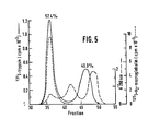
- Figure 5 is a graphic representation of gel filtration chromatography of a mixture of 131I-β-trypsin 125I-α2M, and plasma. To 1.0 ml plasma, 125I-α2M (36 µg) and 131I-β-trypsin (13/ug) were added and incubated 5 min at room temperature. The mixture was applied to a 1.4 x 70 cm column of Sephacryl S-200 gel filtration material (Pharmacia) maintained at 40C in 0.05 M Tris-HCl, pH 8.0, containing 0.16 M citrate and 0.1 M NaCl. 1.6 ml fractions were collected, counted for I and I in a counter, and the absorbance at 280 nm measured.
- This invention relates to methods for detecting, identifying and quantifying enzymes, for example, proteolytic enzymes. The method broadly comprises forming an insoluble complex (E-I-anti I) comprising enzyme (E), enzyme inhibitor (I), and then detecting and identifying and preferably quantitating.one or more enzymes bound to the complex. In a preferred embodiment, a matrix (M), e.g. solid or semisolid surface or permeable matrix has affixed thereto an enzyme inhibitor-antibody or enzyme inhibitor-antibody immunologically active (inhibitor- binding) fragment of such an antibody (anti I). This insoluble matrix is then contacted with biological fluid, e.g. body fluid, to bind one or more predetermined or suspected enzyme inhibitor-enzyme complexes (I-E) in the biological fluid. The bound enzyme is then detected, identified and preferably quantified.
- Enzyme inhibitor (I) as defined for the purpose of this invention and useful in this invention includes not only materials which complex with the enzyme to essentially inhibit the primary functioning of the enzyme in a biological system, but also includes inhibitors such as α2M which form enzyme complexes wherein the enzymatic activity of the enzyme is restricted, but not completely prevented. For example, α2M, proteinase complexes have been shown to retain hydrolytic activity against low molecular weight substrates, but are almost totally enzymatically nonreactive to large substrates such as proteins.
- Methods suitable for detecting and quantifying the bound enzyme are generally known. Generally, after the immobilized E-I-anti-I complex has been removed from contact with the biological fluid the bound enzyme can be reacted in one of two general methods. The first method comprises a reaction with a detection facilitating material which reacts with a site specific to the bound enzyme or enzymes or the bound E-I complex sought and which either has a detectable group or atom measurable by an analytical technique such as a radioactive tag atom, a I.R. or U.V. light absorbing group, or which causes a visible color change; or which generates a detectable cleavage product, a leaving compound or ion, which in turn is measurable by an analytical technique. The class of reaction is not dependent on the enzymatic action of the enzyme which may by biologically inactive in the E-I complex. The second method comprises an enzymatic reaction of the bound enzyme in cases such as α2M where the bound enzyme retains activity against selected substrates. In such a case the immobilized E-I-anti I complex is contacted with a substrate reactive with the bound enzyme, which substrate upon reaction, by color change or the generation of a readily measurable system-unique clevage product susceptable to analytic detection, allows analytical determination.
- In some instances in the preparation of an anti I, as in the case in the Example where the IgG fraction of rabbit antihuman α2M antiserum displayed activity toward an enzyme detecting substrate, it may be necessary to react, as in the example or to further purify, as through the use of chromatographic technique, the anti I to prevent interference with subsequent analysis. Alternatively, at least in some instances, this spurios activity can be measured and compensated for in the analytical determination.
- It is further noted that many inhibitors are capable of binding more than one particular enzyme. Since the immobilized E-I-anti I complexes are stable, it is possible to employ seriatim several techniques which are capable of distinguishing between individual bound enzymes in the immobilized E-I-anti I complex. This is -especially true where the detection mechanism generates a measurable leaving product rather than binding or blocking reactive sites. Again this is especially applicable to the second of the above described detecting reactions, since the substrate choosen seriatim may be substrates which have reactivity to only one or more specific group of enzymes potentially present in the enzymatically active E-I-anti I complex.
- Given the process of the invention, quantitating the analytical results is generally within the skill of the art. At least one such method comprises quantitating I bound to the E-I-anti I complex so that final quantitation can be expressed as a function of bound inhibitor (I). This can be accomplished by adding a small amount of labeled inhibitor to the biological fluid. The amount of label in the E-I-anti I complex is then measured. This is proportional to the binding of native inhibitor.
- Preferably the identification and quantification of the enzyme complexed in the immobilized inhibitor, enzyme complex is carried out in one of two ways. Where the complex is enzymatically active, as eG2M, reaction with substrates is employed. For enzyme complexes which are not enzymatically active antibodies are prepared against the specific enzyme of interest. Thus for example, antibodies may be prepared in rabbits or goats against the human blood proteinases plasmin, thrombin, kallikrein, Cl (the first component of complement), factor Xa, Hage- man factor; or against human pancreatic enzymes trypsin, elastase or chymotrypsin; against human polymorphonuclear leukocyte (.white blood cell) neutral proteinases such as elastase, collagenase and cathepsin G; or against proteinases released from damaged tissues such as cathepsin B. Such antibodies have been produced in a number of laboratories.
- If desired, the immunoglobulin fraction of the antisera or the specific antibody molecules, isolated for example by affinity chromatography, are then labeled with alkaline phosphatase or other detecting enzyme by methods detailed by Engvall et al., Immunochemistry, 8: 871 (1971) and others, or alternatively by use of a radioactive tag- ging material such as NaI 125 .
- The insoluble immunocaptured enzyme, inhibitor complex is then incubated with a specific antienzyme antibody such as above, and after a suitable time the unbound antibody is removed by washing. The anti-enzyme antibody remains bound to the enzyme in the immobilized inhibitor complex. The anti-enzyme antibody is then measured for example by determining the activity of the alkaline phosphatase or radioactivity linked to the detecting anti-enzyme antibody. The amount of enzyme bound is quantitated, for example, using standardized curves consisting of purified inhibitor, enzyme complexes. The use of a detecting antibody per se is a known technique which functions well in the context of the immunocapture technique of the invention.
- It is pointed out that the biological fluids being surveyed are dynamic systems in which the enzyme balance or content may change due to influences present after the state sought to be measured. For example, if the study of enzymes in a human blood sample is undertaken, it is noted the clotting grossly changes the enzymatic or E-I complex content of the blood. Thus, care must be taken in that case to prevent clotting. Likewise, to the extent possible, all enzyme or E-I complex altering factors should be avoided by the appropriate selection of reactants and equipment materials. Alternatively where possible the extraneous enzymatic changes caused during handling and processing can be accounted for and the analytical results appropriately considered or corrected.
- The immobilized or insoluble complex of the invention comprises (E-I-anti I) i.e. enzyme, enzyme inhibitor and inhibitor antibody or active fragment thereof. In the preferred embodiment the complex is affixed to a solid or semi-solid matrix. The only essential requirement is that the ultimate E-I-anti I complex be immobilized. This may first result only upon the ultimate combination of E or E-I complex with the anti I to form for example a precipitate. But preferably an immobilized matrix-anti I entity is performed and then contacted with biological fluid containing E-I complex. Another possible mode is the addition of anti-I to E-I complex containing biological fluid with the subsequent immobilization, e.g. affixation to a matrix, of the preformed mobile E-I-anti I entity.
- The precise nature of the matrix is not critical beyond the fact that there can be affixed thereto anti I in a manner such that the anti I moiety can be affixed thereto by some means, for example, chemical e.g. covalent bonding or absorption, adsorption, or the like, in a manner so that the anti I remains reactive with E-I complex. The matrix may be solid or permeable and may in particulate form, for example, or may comprise, for example, for at least a portion of the structure of a container or be in a unit removably associated with a container.
- The biological fluid surveyed can be virtually any human, animal, microbiological, or vegetable fluid containing enzymes or E-I complexes. Body fluids such as blood, urine, or pleural, joint and peritoneal fluids can be analyzed by this technique.
-
- This example demonstrates that purified α2M as well as α2M-trypsin complexes are quantitatively bound to rabbit anti-human α2M antibody that is immobilized on a gel matrix. This example further demonstrates that the antibody bound α2M-trypsin complexes posses amidolytic activity that can be readily assayed while attached to the particulate gel. The enzymatic activity of these insolubilized α2M-trypsin complexes is identical to that of fluid phase α2M-enzyme complexes. These techniques, utilizing purified systems, have been extended to human plasma to which radiolabeled trypsin and a trace amount of radiolabeled α2M are added. Using the insoluble antibody technique of the invention, α2M-trypsin complexes are quantitatively recovered from plasma as measured both by bound radioactivity and by the capacity of the antibody-bound complex to hydrolyze a synthetic tripeptide chromogenic substrate.
- α2-Macroglobulin was isolated from fresh human plasma in-the presence of soybean trypsin inhibitor as previously described; Harpel, Methods Enzymol; 45: 639 - 652, (1976). α2M was labeled with 125I by the method of McFar- lane, Biochem.J. (London); 62: 135 - 143 (1956). 0.5 mCi of carrier-free sodium 125I iodide was added to 1.7 mg α2M in 1.0 ml borate buffer (0.2 M).pH 8.0, containing NaCl (0.16 M). Iodine monochloride (0.05 ml containing 0.007 micromoles IC1) was.added with mixing. After a 10-min incubation at room temperature, the unbound iodide was removed by Sephadex G-25 (Pharmacia) gel filtration chromatography. The specific activity of the radiolabeled α2M was 0.2µCi/g.
- β-Trypsin was prepared from crystallized, dialyzed salt-free lyophilized bovine trypsin (Worthington) as described by Yung and Trowbridge; Biochem.Biohphys.Res.Com- mun., 65: 927 - 930 (1975). The specific activity of the final product was 94 % as determined by active site titration with p-nitrophenyl-p'-guanidinobenzoate HC1; Chase et al., Methods Enzymol., 19: 20 - 27 (1970). This preparation, following reduction, consisted of a single protein band as identified by dodecyl-sulfate gel (9 %- acrylamide) electrophoresis /
W eber et al., Methods Enzymol., 26: 3 - 27 (1972) 7 indicating that the single chain β-form had been isolated from the original mixture of α-and β-trypsin and inactive material. The trypsin was stored at -70°C in HC1 (1 mM) containing CaCl2 (10 mM) and NaCl (0.1 M). - β-Trypsin was labeled with 131I by the method of McFar- lane, supra. β-Trypsin was dialyzed against borate buffer (0.2 M), pH 8.0 containing benzamidine (0.01 M, Aldrich Chemical Co.), and NaCl (0.16 M). 0.5 mCi of carrier-free 131I-sodium iodide was added to 1.5 mg β-trypsin in a total volume of 1.0 ml borate-benzamidine buffer. Iodine monochloride (0.05 ml containing 0.1 micromole ICI) was added with mixing. After a 10-min incubation at room temperature the free iodide and benzamidine were removed by gel filtration chromatography. The labeled preparation was dialyzed against HC1 (1mM) containing NaCl (0.1 M) prior to storage at -70°C. The specific activity of the radiolabeled β-trypsin was 0.2µCi/µg.
- New Zealand white rabbits were immunized by intradermal injection of α2M mixed with Freund's adjuvant. The antisera produced one immunoprecipitation arc on double diffusion analysis against human plasma and demonstrated a reaction of identity with the starting antigen. The IgG fraction of the antisera was prepared by chromatography on DEAE-cellulose. This IgG fraction was found to hydrolyze N-benzoyl-L-phenylalanyl-L-valyl-L-arginine-p-nitroanilide HC1 (BZPhe-Val-Arg-NHNp; S-2160), however the activity was inhibited by treatment of the IgG material with α-N-p-tosyl-L-lysine chloromethyl ketone HCl (TLCK) (0.01 M) for 3 days at 40C followed by extensive dialysis. The TLCK treated IgG was coupled to Bio-Gel A-5m (Bio-Rad Laboratories) by the cyanogen-bromide method as detailed by March et al., Anal.Biochem., 60: 149 - 152 (1974). Coupling to the activated gel was carried out in citrate buffer (0.2 M), pH 6.5, using 6.0 mg IgG per ml activated gel. Coupling efficiency was greater than 90 %. After the coupling procedure, the gel was incubated 1 h in 1.0 M ethanolamine pH 8.0 to neutralize any remaining protein binding groups. The IgG fractions of normal rabbit serum, and rabbit antisera directed against human albumin or human haptoglobin were also prepared and coupled to Bio-Gel A-5m as detailed above.
- The gel containing the bound α2M antibody was diluted twofold (V/V) with phosphate buffer (0.05 M), pH 7.2, containing NaCl (0.1 M) (PBS). This coupled antibody was incubated with 125I-α2M, 125I- α2M-β-trypsin complexes, or with plasma containing a trace quantity of 125I-α2M and varying concentrations of 131I-β-trypsin as indicated in the figure legends. All incubations were at room temperature, in volumes of 1.0 ml or less, with constant mixing by end over end inversion using a LabquakeR mixer (Labindustries, Berkeley, CA). The incubation was terminated by centrifugation and the pelleted insoluble α2M antibody gel washed repeatedly with 1.0 ml portions of PBS. The pellets were counted for associated 1251 or 131I radioactivity in a Searle 1185 dual channel counter.
- Soluble trypsin or α2M-trypsin complexes were assayed by methods similar to those previously detailed; Svend- sen et al, Thromb.Res., 1: 267 - 278 (1972). The substrate N-benzoyl-L-phenylalanyl-L-valyl-L-arginine-p-nitroanilide hydrochloride (BzPhe-Val-Arg-NHNp=S-2160, obtained from Ortho Diagnostics, Inc., or from Vega Biochemicals) was dissolved in distilled water (0.7 mg/ml). B-Trypsin, or α2M-trypsin complexes were made to a volume of 0.4 ml with Tris-HCl (0.1 M), pH 8.3, containing CaCl2 (1.25 mM). Substrate (0.2 ml) was added and the mixtures were incubated at room temperature. The reaction was terminated at varying intervals by the addition of 30 % acetic acid (0.2 ml) followed by the addition of 0.6 ml of the Tris-CaC12 buffer to achieve a final volume of 1.4 ml. The absorbance at 405 nm was measured with a Gilford 240 spectrophotometer. The concentration of p-nitroanalide released was determined using a molar ab- sorbancy of 10,500; Aurell et al., Thromb.Res., 11: 595 - 609 (1977).
- To determine the amidolytic activity of α2M-trypsin complexes bound to gel-coupled rabbit anti-human α2-trypsin complexes bound to gel-coupled rabbit anti-human α2M IgG, Tris-CaCl2 buffer (0.4 ml) and BzPhe-Val-Arg-NHNp (0.2 ml) were added to the immobilized antibody gels that had been incubated with α2M-trypsin complexes. After incubation for varying time periods at room temperature, the antibody containing gel was removed by centrifugation. 30 % acetic acid (0.2 ml) was added to the supernatant, followed by the addition of 0.6 ml Tris-CaCl2 buffer. The absorbance at 405 nm was measured, and the results expressed (following correction for the substrate bland) either as µ moles p-nitroanilide released/liter/min, or when the amount of α2M bound was determined, as µ moles/liter/min/ mg α2M.
- For the derivation of Michaelis-Menton constants, B-trypsin (0.25 µg/ml), α2M (50 µg/ml), and a trace quantity of 125I- α2M were incubated at 0°C in PBS. Assay of this incubation mixture for free B-trypsin, using the high molecular weight particulate substrate Remazol- brilliant blue hide /
H ayes et al., The Physiological Inhibitors of Blood Coagulation and Fibrinolysis, Collen et al., eds., Elsevier, Amsterdam, 1979, pp. 273 - 2807, proved negative and indicated that all of the β-trypsin had been bound by the α2M. Portions (0.2 ml) of the α2M-β-trypsin incubation mixture were then incubated in duplicate with rabbit anti α2M-IgG coupled to Bio-Gel A-5m (10/ul packed gel). The duplicate gel incubation mixtures were washed four times each with 1.0 ml PBS and the 125I, a measure of α2M bound to the gel, was determined. The insolubilized α2M-β-trypsin complexes were then assayed in duplicate for the ability to hydrolyze varying concentrations of BzPhe-Val-Arg-NHNp. Due to the fact that continuous kinetic measurements could not be made with the insolubilized α2M-enzyme complexes, duplicate assays were terminated at discrete time intervals of 5, 10, and 15 minutes at each substrate concentration and the concentration of liberated p-nitroanilide determined. - Portions of the original solubleo α2M-β-trypsin mixtures, containing an amount of radioactivity equivalent to that bound to the antibody gels described above were assayed in duplicate at varying substrate concentrations. In this case, production of p-nitroanilide was followed continuously at 405 nm in.a Gilford 240 recording spectrophotometer equipped with a Honeywell 1800 recorder. Km and Vmax for the soluble and immobilized α2M-β-trypsin complexes were obtained from Lineweaver-Burk linear transformations of the initial reaction velocities and substrate concentrations.
- The concentration of native α2M in plasma, or in plasma supernatants following incubation with immobilized oL2M antibody was determined by the electroimmunoassay, rocket technique described by Laurell: Scand.J.Clin.Lab. Invest., 29 (Suppl. 124): 21 - 37 (1972).
- The hydrolytic activity of oL2M-trypsin complexes were assessed using the chromogenic substrate BzPh-Val-Arg-NHNp. B-Trypsin was incubated with a mixture of unlabeled and 125I-labeled α2M at trypsin: α2M molar ratios of 0.6, 0.3, and 0.15, well below the 2 moles of β-trypsin per mole oC 2M binding capacity of α2M established in a previous study. The amidolytic activity of these mixtures was found to be linear with time and proportional to the concentration of B-trypsin in the system. The β-trypsin-α2M mixtures were also incubated with the IgG fraction of rabbit anti-human α2M antiserum coupled to Bio-Gel A-5m. After extensive washing the gel pellets were counted in a counter for 1251 and were found to have bound 88 % of the 125I-α2M radioactivity of the original incubation mixture indicating that the majority of the 125I-α2M in the incubation mixture has been bound to the immobilized antibody. The solid-phase antibody in the absence of α2M-trypsin mixture did not hydrolyze BzPhe-Val-Arg-NHNp, but the solid-phase α2M antibody which was incubated with trypsin- α2M complexes had gained amidolytic activity that was proportional to the amount of trypsin in the original fluid-phase incubation mixture. Incubation of the solid phase antibody with B-trypsin in the absence of α2M did not confer amidolytic activity upon the gel nor did the immobilized IgG fraction derived from normal rabbit serum bind either α2M or α2M-trypsin complexes. Thus, the activity gained when immobilized anti- human α2M antibody was incubated with the α2M-trypsin mixtures was a reflection of the specific binding of the α2M-trypsin complexes.
- Analysis of the kinetics of the hydrolysis of substrate in the fluid-phase by α2M-trypsin complexes as . compared to the solid-phase complexes demonstrated no significant differences (Fig. 1). The Michaelis-Menton constants for the fluid-phase complexes as determined by Lineweaver-Burk plots were Km = 0 mM and Vmax -1707 min-1, and of the solid phase α2M-trypsin complexes, Km = 0.078 mM and Vmax = 1700 min . This indicates that under the experimental conditions detailed for these assays, the binding of the oC2M-trypsin complex to a surface did not affect its amidolytic activity.
- Both soluble a2M-trypsin complexes and solid-phase antibody-bound α2M-trypsin complexes demonstrated a similar peak of amidolytic activity between pH 8.0 and 9.0. At higher pH's, there was a significant decrease in substrate hydrolysis. In other experiments, the effect of variations in sodium chloride concentration (0 to 0.5 M) on the catalytic activity of the immobilized c(2M-trypsin complex was studied. A broad unchanging peak of activity from 0.05 to 0.3 M NaCl was observed with a decrease in amidolytic activity at both 0 and 0.5 M NaCl. Both soluble and immobilized α2M-trypsin complexes behaved in a similar manner.
- In order to assess the stability of the immobilized antibody-bound α2M-trypsin complexes, four portions of the immobilized α2M antibody were incubated with an α2M trypsin mixture containing a trace amount of 125I- α2M. After repeated washings, the gels were counted to determine the amount of bound α2M. The insolubilized antibody α2M-trypsin complex was then assayed for BzPhe-Val-Arg-NHNp hydrolytic activity. This procedure was repeated four times. As illustrated in Table I, the specific activity of the α2M-trypsin complex, bound to the immobilized antibody, did not change with repeated assays indicating that trypsin itself did not dissociate from the complex in the presence of the chromogenic substrate. There was a small equivalent loss of both α2M and amidolytic activity with repeated assays reflecting either loss of gel during the washings or elution of the α2M-trypsin complex.

-
- a Portions (0.2 ml) of a mixture of α2M and 125I- α2M (50/ug/ml) and β-trypsin (300 ng) were incubated in quadruplicate for 2 hours with rabbit anti-humanc α2M-IgG coupled to Bio-Gel A-5m (10/ul packed gel). Following washings of each sample four times with PBS, it was subsequently assayd for amidolytic activity by adding Tris-CaCl2 buffer, pH 8.3 (0.4 ml) and BzPhe-Val-Arg-NHNp (0.7 µg/ml, 0.2 ml) for 15 min at room temperature with mixing by inversion. After each assay, the samples were washed four times in succession and the amidolytic activity redetermined. This assay procedure was repeated a total of four times.
- b The µg α2M bound to the beads was calculated as the counts bound/total counts x µgα2M in the original incubation mixture.
- ° All values in the table represent the mean of four determinations.
- Experiments were designed to test whether the binding of purified α2M or α2M-trypsin complexes by solid-phase anti-human α2M antibody could be extended to a complex biological fluid such as plasma. In the study shown in Fig. 2, the solid-phase α2M antibody was incubated with serial dilutions of plasma to which 125I-α2M was added. The radioactivity bound to the insoluble antibody was measured. The native α2M in the starting plasma and the residual α2M in the plasma supernatant following absorption was also-quntitated by electroimmunoassay. The results demonstrate that the binding of the 125I-α2M added to plasma by the immobilized antibody parallels the binding of native plasma α2M.
- Specificity of binding of 125I-α2M added to plasma by the solid-phase α2M antibody was also tested by incubating plasma containing 125I-α2M with unmodified Bio-Gel A-5m, or this same gel to which the purified IgG fraction of normal rabbit serum, or the IgG fraction of rabbit antisera against either human albumin or haptoglobin was coupled. Under conditions in which greater than 95 % of the radioactivity became bound to insoluble α2M antibody, less than 2 % of the 125I-α2M radioactivity was associated with these other gel preparations.
- The effect of time of incubation of plasma with solid-phase α2M antibody on the binding of α2M to the gel was investigated (Fig. 3). The antibody gel was incubated for varying time periods with three different dilutions of plasma which contained 12, 60 and 192/ug α2M respectively. There was a progressive increase in binding throughout the six hour incubation period at all concentrations of α2M. At the lowest concentration of α2M (12 µg system), 91.7 % of the α2M was bound after 6 hours. At the highest concentration of α2M, 49 % or 84/ug of α2M was bound. This indicates that 1.0 ml of the α2M antibody gel has the capacity to bind at least 9.4 mg α2M.
- The demonstration that α2M in plasma could be quantitatively removed by immobilized α2M antibody made it possible to study the binding of exogenous 131I-β-trypsin to α2M in a plasma system. 131I-β-trypsin was added to plasma containing 1.5 mg/ml α2M to give trypsin concentrations of 0.10 to 1.65/ug/ml plasma. This represents a trypsin/ α2M molar ratio of 0.002 to 0.035. The plasma also contained a trace amount of 125I-α2M. Portions of plasma were incubated with solid-phase α2M antibody for two hours. The pellets were washed, counted for both 125 I and 131I radioactivity and the amidolytic activity measured 'ig. 4). Whereas 86.7 % of the 125I- α2M in the incubation mixture was bound to the antibody containing gel, only 50.3 % of 131I-β-trypsin was similarly bound suggesting that trypsin also binds to other plasma proteins. The percent binding of both α2M and trypsin was unaffected by the varying concentrations of trypsin added to the plasma. The 1311 radioactivity bound to the solid-phase antibody was proportional to the amount of 131I-trypsin in the original mixture.
- To control for endogenous amidolytic activity, the solid-phase α2M antibody was incubated with a plasma 125 I- α2M mixture to which no 131 I-trypsin had been added. The resulting α2M antibody gel hydrolyzed 2.4/umoles sub- strate/L/min/mg α2M corresponding to the addition to plasma of approximately 142 ng 8-trypsin/ml. This background activity was subtracted from the amidolytic activity of the gels incubated with the plasma, trypsin mixtures. The amidolytic activity of the α2M antibody gels incubated with the 131I-trypsin, plasma mixtures was proportional to the initial concentration of trypsin added, and parallel to the 131I counts bound to the gel (Fig. 4).
- From a comparison of the binding of α2M to the solid-phase α2M antibody in the experiment illustrated in Fig. 4 (86.7 %), and the binding of 131I-trypsin (50.3 %), it can be calculated that the total α2M in plasma should have bound 58.0 % of the trypsin added to the plasma. Thus it seemed likely that approximately 42 % of the trypsin may have been complexed to other plasma proteins and therefore unavailable to the α2M. To verify this possibility, a mixture of 131I-trypsin, 125I- α2M and plasma was fractionated by molecular sieve chromatography (Fig. 5). The major 131I-trypsin peak, containing 57.4 % of the total 131I counts, co-eluted with the 125I- α2M in the first, high molecular weight protein peak. The second 131I-trypsin peak contained 40.3 % of the total 131I counts and eluted earlier than the albumin peak at a position consistent with complex formation between trypsin and a plasma protein.
- To further confirm the specificity of the insoluble α2M antibody, α2M-trypsin interaction and to identify the nature of the 131I-trypsin peaks from the gel filtration column, 0.6 ml of each peak fraktion of 131I-trypsin was incubated with the solid phase α2M antibody (0.1 ml packed gel). Following an 18 h incubation, 99 % of the 125I- α2M and 94 % of the 131I-trypsin of the α2M peak fraction 36 was bound to the immobilized antibody. Less than 5 % of 131I-trypsin was bound to the antibody gel in the incubation mixture of the second 131I-trypsin peak (fraction 46). Therefore most of the 131I-trypsin in the first high molecular weight peak was complexed with α2M. The failure of 131I-trypsin in the second radioactive peak to bind to the α2M antibody gel provides further evidence that trypsin had reacted with a protein other than d2M.
- The process of the invention make possible the study of enzymes and inhibitor-enzyme complexes present in biological fluid, broadly and specifically presents a new approach to the study of the physiologic function of human plasma α2-macroglobulin. The method of the invention makes it possible to further characterize inhibitor-enzyme complexes and particularly the protease binding capacity of α2M in both purified systems and in complex biological fluids. As exemplified the solid-phase antibody-bound α2M-β-trypsin complexes retain their ability to hydrolyze the tripeptide chromogenic substrate BzPhe-Val-Arg-NHNp with Michaelis-Menton constants identical to those of the soluble complexes. This indicates that the binding of the α2M-trypsin complex to an immobilized antibody does not appreciably effect the amidolytic activity of the bound protease. The antibody-bound α2M-trypsin complexes proved to be remarkably stable since they remain associated with the agarose gel matrix through repeated washings and assays of amidolytic activity. The finding that trypsin remains bound to α2M in the presence of substrate provides further evidence that trypsin binds to c4 2M at a site different from its catalytic site.
- Studies of the conditions required to optimize substrate hydrolysis as in the Example showed that the optimum pH of the reaction for both the solubled2M-trypsin complex as well as the solid-phase, antibody-bound complex was between 8 and 9. In contrast, Rindernecht, et al., Biochem.Biophys.Acta, 377: 158 - 165 (1975), using a different chromogenic substrate, N-carbobenzoxy-glycyl-glycyl-L-arginine-2-naphthyl-amide HCl, found that catalysis was greatest at pH 10.0. They postulated that this degree of alkalinity was necessary to alter the microviron- ment of the trypsin active site within the α2M-trypsin complex so that the substrate could be hydrolyzed at a rate similar to that of free trypsin. It is not clear whether the difference in pH optimum between their study and our reflects the behaviour of the purified B-trypsin used in the present study, the difference in substrate, or other factors.
- We have demonstrated that the binding of α2M-trypsin complexes by immobilized α2M antibody also occurs when the assay system is applied to human plasma. Prior studies by Haverback et al., J.Clin.Invest., 41: 972 - 980 (1962), was formed between trypsin and a serum protein that possessed an α2 electrophoretic mobility. Mehl et al., Science, 145: 821 - 822 (1964) provided direct evidence that α2M was the protein responsible for the enzyme binding activity. In the present study, when plasma contain- ing a trace quantity of 125I- α2M was incubated with immobilized α2M antibody, both radiolabeled and native α2M were bound. The electroimmunoassay technique of Laurell was used to establish that the binding of 1251- α2M to the solid-phase α2M antibody was proportional to the amount of unlabeled native α2M bound. The addition of the radiolabeled antigen to plasma made it-possible, therefore, to quantitate the binding of plasma d2M to the solid-phase antibody. The binding of plasma α2M to the insoluble gel containing the antibody was specific since other immobilized antibodies such as anti-albumin, anti- haptoglobin, or the IgG fraction of normal rabbit serum did not bind significant amounts of plasma α2M.
- To establish that the solid-phase antibody technique detailed in this study could detect α2M-enzyme complexes in a biological fluid such as plasma, varying concentra- tions of 131I-β-trypsin were added to plasma that contained a trace quantity of 125I- α2M. Subsequent assay demonstrated that B-trypsin was found to the immobilized α2M antibody and that the amount of 131I-trypsin bound and the amidolytic activity associated with the solid-phase antibody was linearly related to the concentration of trypsin originally added to the plasma. Since control studies established that 131I-β-trypsin itself did not bind the antibody gel, the trypsin activity that we observed was a specific measure of α2-trypsin complexes. At each concentration of trypsin added to plasma, 58 % was bound to α2M, a finding explained by gel filtration studies that demonstrated that the remainder of the trypsin was associated with another protein, possibly a1-antitrypsin. These results parallel the findings of Ganrot, Arkiv For Kemi Bd 26, No. 50: 577 - 582 (1967), who demosntrated that the affinity of trypsin fork 2M was greater than that for other trypsin inhibitors in plasma.
- a2M is a unique plasma protease inhibitor in that it forms a complex with an exceptionally wide variety of proteases including serine, thiol, carboxyl and metallopro- teases derived from plasma, cells, or from microorganism; [Harpel et al. (1976, supra, Starkey et al., supra7. Complexes between α2M and several different proteases have been identified in plasma; /Harpel et al., J.Clin.Invest. 52: 2175 - 21.84 (1973); Nilehn et al., Scand.J.Clin.Lab. Invest., 20: 113 - 121 (1967)7, in peritoneal, [Ohlsson, Bayer Symp.V., Proteinase Inhibitors; Fritz et al., eds. Springer Verlag, Berlin (1974) pp. 96 - 1057, pleural [Bieth et al., Clin.Chim.Acta, 22: 639 - 642 (1968); Bieth et al., Clin.Chim.Acta, 30: 621 - 626 (1970)7, and in synovial effusions /Shtacher et al., Biochim.Biophys.Acta, 303: 138 - 147 (1973); Abe et al., J.Biochem. (Tokyo), 71: 919 - 922 (1973); Abe et al., J.Biochem. (Tokyo), 73: 897 - 900 (1973)7, however a convenient method suitable for the quantitative analysis of these complexes has not previously been available. The solid-phase α2M antibody technique described in this study facilitates the rapid isolation and concentration of enzyme-enzyme inhibitor complexes generally of α2M-protease complexes specifically and the measurement of their presence and/or catalytic capacity in biological fluids in various physiologic and pathologic states. Modifications of the assay conditions will permit the measurement of the activity of physiologically important enzymes and enzyme inhibitors, particularly proteases such as thrombin, plasmin and plasma kallikrein when bound to α2M.
- While there is described above the invention and what is now considered its preferred embodiment, it should be understood that the invention can be practiced with other matrixes, immobilized E-I-anti I complexes, enzymes, inhibitors and inhibitor antibodies or reactive fragments thereof, as well as employing varied analytical techniques and reaction conditions within the scope of the appended claims within the skill of the art.
Claims (14)
Priority Applications (1)
| Application Number | Priority Date | Filing Date | Title |
|---|---|---|---|
| AT81107730T ATE15387T1 (en) | 1980-09-30 | 1981-09-29 | METHODS FOR DETERMINING ENZYME INHIBITOR COMPLEXES. |
Applications Claiming Priority (2)
| Application Number | Priority Date | Filing Date | Title |
|---|---|---|---|
| US19216680A | 1980-09-30 | 1980-09-30 | |
| US192166 | 1994-02-04 |
Publications (3)
| Publication Number | Publication Date |
|---|---|
| EP0048989A2 true EP0048989A2 (en) | 1982-04-07 |
| EP0048989A3 EP0048989A3 (en) | 1982-05-12 |
| EP0048989B1 EP0048989B1 (en) | 1985-09-04 |
Family
ID=22708529
Family Applications (1)
| Application Number | Title | Priority Date | Filing Date |
|---|---|---|---|
| EP81107730A Expired EP0048989B1 (en) | 1980-09-30 | 1981-09-29 | Process for determining inhibitor-enzyme complexes |
Country Status (5)
| Country | Link |
|---|---|
| EP (1) | EP0048989B1 (en) |
| JP (1) | JPS5794660A (en) |
| AT (1) | ATE15387T1 (en) |
| CA (1) | CA1176560A (en) |
| DE (1) | DE3172143D1 (en) |
Cited By (9)
| Publication number | Priority date | Publication date | Assignee | Title |
|---|---|---|---|---|
| EP0080279A1 (en) * | 1981-11-02 | 1983-06-01 | James Walter Ryan | Method for assaying proteases with tagged proteinaceous inhibitors |
| WO1984002778A1 (en) * | 1982-12-30 | 1984-07-19 | Marius Constantin Teodorescu | Test for autoimmune rheumatoid arthritis |
| EP0216179A1 (en) * | 1985-09-06 | 1987-04-01 | BEHRINGWERKE Aktiengesellschaft | Method for determining protease inhibitors |
| WO1993005397A1 (en) * | 1991-09-07 | 1993-03-18 | Biosyn Ltd | Immunoassay for proteases |
| WO1995000847A1 (en) * | 1993-06-18 | 1995-01-05 | Hafslund Nycomed A.S | Immunoassay |
| NL9301134A (en) * | 1993-06-29 | 1995-01-16 | Inst Voor Agrotech Onderzoek | Method for detecting and, if necessary, quantifying the activity of at least one protease inhibitor in a mixture to be analysed, and also application of the method for determining the (residual) activity of one (or more) separate (iso)type(s) of protease inhibitor(s) and application of the method for determining the total (residual) activity of a mixture, possibly after a processing process such as a process for the removal or inactivation of protease inhibitor(s). |
| WO1996007104A1 (en) * | 1994-08-26 | 1996-03-07 | Stenman Ulf Haakan | Assay of free and complexed trypsinogen-2 |
| EP0759556A2 (en) * | 1995-07-24 | 1997-02-26 | BEHRINGWERKE Aktiengesellschaft | Method for quantification of activated factors |
| WO2005017138A2 (en) * | 2003-08-19 | 2005-02-24 | Novozymes A/S | Active-site titration of glycosyl hydrolases |
Families Citing this family (4)
| Publication number | Priority date | Publication date | Assignee | Title |
|---|---|---|---|---|
| JPS6134465A (en) * | 1984-07-26 | 1986-02-18 | Teijin Ltd | Immunoassay reagent and kit using monoclonal antibody against human alpha2-plasmin inhibitor |
| JPS6488368A (en) * | 1987-09-30 | 1989-04-03 | Shigeru Hayashi | Method for measuring complex of urokinase and urokinase inhibitor |
| EP2946206B1 (en) * | 2013-01-20 | 2019-01-09 | Dyax Corp. | Evaluation, assays and treatment of pkal-mediated disorders |
| AU2020258869A1 (en) | 2019-04-16 | 2021-11-25 | Takeda Pharmaceutical Company Limited | Methods for quantitation of functional C1 esterase inhibitor (fC1-INH) |
Citations (1)
| Publication number | Priority date | Publication date | Assignee | Title |
|---|---|---|---|---|
| US4180437A (en) * | 1976-03-08 | 1979-12-25 | Leuven Research & Development V.Z.W. | Purification of antiserum for thrombosis test |
Family Cites Families (4)
| Publication number | Priority date | Publication date | Assignee | Title |
|---|---|---|---|---|
| DE2836046A1 (en) * | 1978-08-17 | 1980-02-28 | Behringwerke Ag | IMMUNOLOGICAL DETERMINATION PROCEDURE |
| JPS5646465A (en) * | 1979-09-25 | 1981-04-27 | Seikagaku Kogyo Co Ltd | Measuring method for man pancreas secretory inhibiter in humor |
| JPS56118671A (en) * | 1980-02-22 | 1981-09-17 | Amano Pharmaceut Co Ltd | Measuring method of specific enzyme immunity of hybrid type protein |
| DE3016575A1 (en) * | 1980-04-30 | 1981-11-05 | Merck Patent Gmbh, 6100 Darmstadt | METHOD FOR THE IMMUNOLOGICAL DETERMINATION OF ENZYMES, MEANS FOR CARRYING OUT THE METHOD AND ITS RELATIONSHIP |
-
1981
- 1981-09-29 AT AT81107730T patent/ATE15387T1/en not_active IP Right Cessation
- 1981-09-29 EP EP81107730A patent/EP0048989B1/en not_active Expired
- 1981-09-29 DE DE8181107730T patent/DE3172143D1/en not_active Expired
- 1981-09-29 CA CA000386866A patent/CA1176560A/en not_active Expired
- 1981-09-30 JP JP56154047A patent/JPS5794660A/en active Granted
Patent Citations (1)
| Publication number | Priority date | Publication date | Assignee | Title |
|---|---|---|---|---|
| US4180437A (en) * | 1976-03-08 | 1979-12-25 | Leuven Research & Development V.Z.W. | Purification of antiserum for thrombosis test |
Non-Patent Citations (4)
| Title |
|---|
| CHEMICAL ABSTRACTS, vol. 84, no. 11, March 15, 1976, page 168, abstract 70902d, Columbus, Ohio, (US) K. OHLSSON et al: "Demonstration and semiquantitative determination of complexes between various proteases and human alpha2-macroglobulin"; & CLIN. CHIM. ACTA 1976, 66 (1) 1-7 * |
| CHEMICAL ABSTRACTS, vol. 94, no. 1, January 5, 1981, page 163, abstract 1563p, Columbus, Ohio, (US) P.C. HARPEL et al: "Immunoimmobilization of alpha2-macroglobulin-beta-trypsin complexes: a novel approach for the biochemical characterization of modulator-protease interactions"; & ANAL. BIOCHEM. 1980, 108(1), 166-75 * |
| CLINICAL CHEMISTRY, vol. 26, September 1980, no. 10, Easton, Pa, (US) J. FAREED et al: "New perspectives in coagulation testing", pages 1380-1391 * |
| THE JOURNAL OF IMMUNOLOGY, vol. 127, no. 1, July 1981. F.C. McDUFFIE et al: "Antigenic changes produced by complex formation between thrombin and antithrombin-III", pages 239-244 * |
Cited By (14)
| Publication number | Priority date | Publication date | Assignee | Title |
|---|---|---|---|---|
| EP0080279A1 (en) * | 1981-11-02 | 1983-06-01 | James Walter Ryan | Method for assaying proteases with tagged proteinaceous inhibitors |
| WO1984002778A1 (en) * | 1982-12-30 | 1984-07-19 | Marius Constantin Teodorescu | Test for autoimmune rheumatoid arthritis |
| GB2142723A (en) * | 1982-12-30 | 1985-01-23 | Marius Constantin Teodorescu | Test for autoimmune rheumatoid arthritis |
| US4499186A (en) * | 1982-12-30 | 1985-02-12 | University Of Illinois, Board Of Trustees | Diagnosing autoimmune rheumatoid arthritis by measuring proteolytic activity of α2 -macroglobulin |
| EP0216179A1 (en) * | 1985-09-06 | 1987-04-01 | BEHRINGWERKE Aktiengesellschaft | Method for determining protease inhibitors |
| WO1993005397A1 (en) * | 1991-09-07 | 1993-03-18 | Biosyn Ltd | Immunoassay for proteases |
| WO1995000847A1 (en) * | 1993-06-18 | 1995-01-05 | Hafslund Nycomed A.S | Immunoassay |
| NL9301134A (en) * | 1993-06-29 | 1995-01-16 | Inst Voor Agrotech Onderzoek | Method for detecting and, if necessary, quantifying the activity of at least one protease inhibitor in a mixture to be analysed, and also application of the method for determining the (residual) activity of one (or more) separate (iso)type(s) of protease inhibitor(s) and application of the method for determining the total (residual) activity of a mixture, possibly after a processing process such as a process for the removal or inactivation of protease inhibitor(s). |
| WO1996007104A1 (en) * | 1994-08-26 | 1996-03-07 | Stenman Ulf Haakan | Assay of free and complexed trypsinogen-2 |
| US5976809A (en) * | 1994-08-26 | 1999-11-02 | Ulf-Hakan Stenman | Assay of free and complexed trypsinogen-2 |
| EP0759556A2 (en) * | 1995-07-24 | 1997-02-26 | BEHRINGWERKE Aktiengesellschaft | Method for quantification of activated factors |
| EP0759556A3 (en) * | 1995-07-24 | 1998-05-20 | BEHRINGWERKE Aktiengesellschaft | Method for quantification of activated factors |
| WO2005017138A2 (en) * | 2003-08-19 | 2005-02-24 | Novozymes A/S | Active-site titration of glycosyl hydrolases |
| WO2005017138A3 (en) * | 2003-08-19 | 2005-05-06 | Novozymes As | Active-site titration of glycosyl hydrolases |
Also Published As
| Publication number | Publication date |
|---|---|
| DE3172143D1 (en) | 1985-10-10 |
| JPS5794660A (en) | 1982-06-12 |
| EP0048989B1 (en) | 1985-09-04 |
| CA1176560A (en) | 1984-10-23 |
| JPH041304B2 (en) | 1992-01-10 |
| ATE15387T1 (en) | 1985-09-15 |
| EP0048989A3 (en) | 1982-05-12 |
Similar Documents
| Publication | Publication Date | Title |
|---|---|---|
| EP0221927B1 (en) | Assays with surface-bound fibrinogen and soluble, labeled fibrinogen | |
| US6017723A (en) | Method for isolating inhibitors of protease activity | |
| EP0048989B1 (en) | Process for determining inhibitor-enzyme complexes | |
| Mahmoud et al. | Bioimmunoassay (BIA) of tissue plasminogen activator (t-PA) and its specific inhibitor (t-PA/INH) | |
| Nuijens et al. | Detection of activation of the contact system of coagulation in vitro and in vivo: quantitation of activated Hageman factor-C1-Inhibitor and kallikrein-C1-Inhibitor complexes by specific radioimmunoassays | |
| US4629694A (en) | Detecting and distinguishing between plasminogen activators | |
| US4849353A (en) | Immunocapture of enzyme inhibitor, enzyme complexes and uses thereof | |
| CA2255255A1 (en) | Improved thrombin-based assay for antithrombin-iii | |
| US6242173B1 (en) | Immunoassays for catalytically-active, serine proteases | |
| EP0080279B1 (en) | Method for assaying proteases with tagged proteinaceous inhibitors | |
| McDuffie et al. | Prothrombin, thrombin and prothrombin fragments in plasma of normal individuals and of patients with laboratory evidence of disseminated intravascular coagulation | |
| Holvoet et al. | Measurement of free, one-chain tissue-type plasminogen activator in human plasma with an enzyme-linked immunosorbent assay based on an active site-specific murine monoclonal antibody | |
| US4753875A (en) | Method for assaying proteases with tagged proteinaceous inhibitors | |
| US4882272A (en) | High molecular weight kininogen assay | |
| Harpel et al. | Immunoimmobilization of α2-macroglobulin-β-trypsin complexes: A novel approach for the biochemical characterization of modulator-protease interactions | |
| JPH0767396B2 (en) | Fibrinolytic method | |
| Delk et al. | Radioimmunoassay of active pancreatic enzymes in sera from patients with acute pancreatitis. I. Active carboxypeptidase B. | |
| EP0123265A1 (en) | Zymogen activation, cascade amplification immunoassay | |
| Alving et al. | Plasma prekallikrein: quantitative determination by direct activation with Hageman factor fragment (β-XIIa) | |
| Nishikawa et al. | Effect of neurotropin® on the activation of the plasma kallikrein-kinin system | |
| US20030186345A1 (en) | Macromolecular enzyme substrates | |
| EP0396692A1 (en) | A method of assaying an enzyme and kit and substances suitable for that method | |
| WO2002022852A2 (en) | Method for measuring antithrombin activity | |
| Jesty et al. | Measurement of human activated factor X-antithrombin complex by an enzyme-linked differential-antibody immunosorbent assay | |
| US5631136A (en) | Process for the detection of complexed cathepsin G and α-1-antichymotrypsin |
Legal Events
| Date | Code | Title | Description |
|---|---|---|---|
| PUAI | Public reference made under article 153(3) epc to a published international application that has entered the european phase |
Free format text: ORIGINAL CODE: 0009012 |
|
| PUAL | Search report despatched |
Free format text: ORIGINAL CODE: 0009013 |
|
| AK | Designated contracting states |
Designated state(s): AT BE CH DE FR GB IT LI LU NL SE |
|
| RHK1 | Main classification (correction) |
Ipc: C12Q 1/38 |
|
| AK | Designated contracting states |
Designated state(s): AT BE CH DE FR GB IT LI LU NL SE |
|
| 17P | Request for examination filed |
Effective date: 19821015 |
|
| RAP1 | Party data changed (applicant data changed or rights of an application transferred) |
Owner name: CORNELL RESEARCH FOUNDATION, INC. |
|
| ITF | It: translation for a ep patent filed | ||
| GRAA | (expected) grant |
Free format text: ORIGINAL CODE: 0009210 |
|
| AK | Designated contracting states |
Designated state(s): AT BE CH DE FR GB IT LI LU NL SE |
|
| REF | Corresponds to: |
Ref document number: 15387 Country of ref document: AT Date of ref document: 19850915 Kind code of ref document: T |
|
| PG25 | Lapsed in a contracting state [announced via postgrant information from national office to epo] |
Ref country code: LU Free format text: LAPSE BECAUSE OF NON-PAYMENT OF DUE FEES Effective date: 19850930 |
|
| REF | Corresponds to: |
Ref document number: 3172143 Country of ref document: DE Date of ref document: 19851010 |
|
| ET | Fr: translation filed | ||
| PLBE | No opposition filed within time limit |
Free format text: ORIGINAL CODE: 0009261 |
|
| STAA | Information on the status of an ep patent application or granted ep patent |
Free format text: STATUS: NO OPPOSITION FILED WITHIN TIME LIMIT |
|
| 26N | No opposition filed | ||
| PGFP | Annual fee paid to national office [announced via postgrant information from national office to epo] |
Ref country code: LU Payment date: 19890705 Year of fee payment: 9 |
|
| PGFP | Annual fee paid to national office [announced via postgrant information from national office to epo] |
Ref country code: AT Payment date: 19890830 Year of fee payment: 9 |
|
| PGFP | Annual fee paid to national office [announced via postgrant information from national office to epo] |
Ref country code: SE Payment date: 19890906 Year of fee payment: 9 |
|
| ITTA | It: last paid annual fee | ||
| PGFP | Annual fee paid to national office [announced via postgrant information from national office to epo] |
Ref country code: NL Payment date: 19890930 Year of fee payment: 9 |
|
| PGFP | Annual fee paid to national office [announced via postgrant information from national office to epo] |
Ref country code: BE Payment date: 19891219 Year of fee payment: 9 |
|
| PG25 | Lapsed in a contracting state [announced via postgrant information from national office to epo] |
Ref country code: AT Effective date: 19900929 |
|
| PG25 | Lapsed in a contracting state [announced via postgrant information from national office to epo] |
Ref country code: SE Effective date: 19900930 Ref country code: BE Effective date: 19900930 |
|
| BERE | Be: lapsed |
Owner name: CORNELL RESEARCH FOUNDATION INC. Effective date: 19900930 |
|
| PG25 | Lapsed in a contracting state [announced via postgrant information from national office to epo] |
Ref country code: NL Effective date: 19910401 |
|
| NLV4 | Nl: lapsed or anulled due to non-payment of the annual fee | ||
| PGFP | Annual fee paid to national office [announced via postgrant information from national office to epo] |
Ref country code: GB Payment date: 19910802 Year of fee payment: 11 |
|
| PGFP | Annual fee paid to national office [announced via postgrant information from national office to epo] |
Ref country code: FR Payment date: 19910918 Year of fee payment: 11 |
|
| PGFP | Annual fee paid to national office [announced via postgrant information from national office to epo] |
Ref country code: CH Payment date: 19910923 Year of fee payment: 11 |
|
| PG25 | Lapsed in a contracting state [announced via postgrant information from national office to epo] |
Ref country code: GB Effective date: 19920929 |
|
| PG25 | Lapsed in a contracting state [announced via postgrant information from national office to epo] |
Ref country code: LI Effective date: 19920930 Ref country code: CH Effective date: 19920930 |
|
| GBPC | Gb: european patent ceased through non-payment of renewal fee |
Effective date: 19920929 |
|
| PG25 | Lapsed in a contracting state [announced via postgrant information from national office to epo] |
Ref country code: FR Effective date: 19930528 |
|
| REG | Reference to a national code |
Ref country code: CH Ref legal event code: PL |
|
| REG | Reference to a national code |
Ref country code: FR Ref legal event code: ST |
|
| PGFP | Annual fee paid to national office [announced via postgrant information from national office to epo] |
Ref country code: DE Payment date: 19930928 Year of fee payment: 13 |
|
| EUG | Se: european patent has lapsed |
Ref document number: 81107730.4 Effective date: 19910527 |
|
| PG25 | Lapsed in a contracting state [announced via postgrant information from national office to epo] |
Ref country code: DE Effective date: 19950601 |