DE69918260T2 - Methods for the identification and validation of functional targets by intramers or in vivo selection - Google Patents
Methods for the identification and validation of functional targets by intramers or in vivo selection Download PDFInfo
- Publication number
- DE69918260T2 DE69918260T2 DE69918260T DE69918260T DE69918260T2 DE 69918260 T2 DE69918260 T2 DE 69918260T2 DE 69918260 T DE69918260 T DE 69918260T DE 69918260 T DE69918260 T DE 69918260T DE 69918260 T2 DE69918260 T2 DE 69918260T2
- Authority
- DE
- Germany
- Prior art keywords
- rna
- promoter
- cells
- nucleic acid
- nucleic acids
- Prior art date
- Legal status (The legal status is an assumption and is not a legal conclusion. Google has not performed a legal analysis and makes no representation as to the accuracy of the status listed.)
- Expired - Fee Related
Links
- 238000000034 method Methods 0.000 title claims abstract description 67
- 238000001727 in vivo Methods 0.000 title claims description 8
- 238000010200 validation analysis Methods 0.000 title abstract description 7
- 150000007523 nucleic acids Chemical class 0.000 claims abstract description 174
- 102000039446 nucleic acids Human genes 0.000 claims abstract description 171
- 108020004707 nucleic acids Proteins 0.000 claims abstract description 171
- 230000027455 binding Effects 0.000 claims abstract description 65
- 230000003834 intracellular effect Effects 0.000 claims abstract description 58
- 150000001875 compounds Chemical class 0.000 claims abstract description 8
- 230000014509 gene expression Effects 0.000 claims description 111
- 108091032973 (ribonucleotides)n+m Proteins 0.000 claims description 88
- 239000013598 vector Substances 0.000 claims description 46
- 230000001086 cytosolic effect Effects 0.000 claims description 37
- 108010002350 Interleukin-2 Proteins 0.000 claims description 25
- 239000000203 mixture Substances 0.000 claims description 25
- 102000006601 Thymidine Kinase Human genes 0.000 claims description 16
- 108020004440 Thymidine kinase Proteins 0.000 claims description 16
- 108700012359 toxins Proteins 0.000 claims description 16
- 239000003550 marker Substances 0.000 claims description 15
- 230000007246 mechanism Effects 0.000 claims description 14
- 102000006495 integrins Human genes 0.000 claims description 12
- 108010044426 integrins Proteins 0.000 claims description 12
- 239000003053 toxin Substances 0.000 claims description 12
- 231100000765 toxin Toxicity 0.000 claims description 12
- 230000001939 inductive effect Effects 0.000 claims description 11
- 108700008625 Reporter Genes Proteins 0.000 claims description 10
- 238000010367 cloning Methods 0.000 claims description 9
- 102000040650 (ribonucleotides)n+m Human genes 0.000 claims description 8
- 230000000087 stabilizing effect Effects 0.000 claims description 8
- 230000000692 anti-sense effect Effects 0.000 claims description 7
- 230000001965 increasing effect Effects 0.000 claims description 7
- 230000004913 activation Effects 0.000 claims description 6
- 238000012163 sequencing technique Methods 0.000 claims description 6
- 238000012360 testing method Methods 0.000 claims description 5
- 230000002441 reversible effect Effects 0.000 claims description 4
- FGUUSXIOTUKUDN-IBGZPJMESA-N C1(=CC=CC=C1)N1C2=C(NC([C@H](C1)NC=1OC(=NN=1)C1=CC=CC=C1)=O)C=CC=C2 Chemical compound C1(=CC=CC=C1)N1C2=C(NC([C@H](C1)NC=1OC(=NN=1)C1=CC=CC=C1)=O)C=CC=C2 FGUUSXIOTUKUDN-IBGZPJMESA-N 0.000 claims description 2
- 238000003780 insertion Methods 0.000 claims description 2
- 230000037431 insertion Effects 0.000 claims description 2
- 230000000694 effects Effects 0.000 abstract description 12
- 239000003814 drug Substances 0.000 abstract description 9
- 238000001415 gene therapy Methods 0.000 abstract description 8
- 238000002360 preparation method Methods 0.000 abstract description 5
- 210000004027 cell Anatomy 0.000 description 160
- 108091023037 Aptamer Proteins 0.000 description 141
- 108090000623 proteins and genes Proteins 0.000 description 120
- 102000004169 proteins and genes Human genes 0.000 description 65
- 239000013612 plasmid Substances 0.000 description 35
- 238000000338 in vitro Methods 0.000 description 29
- 230000001413 cellular effect Effects 0.000 description 28
- 239000003446 ligand Substances 0.000 description 26
- 102000000588 Interleukin-2 Human genes 0.000 description 24
- 230000006870 function Effects 0.000 description 23
- 230000019491 signal transduction Effects 0.000 description 23
- 108020004414 DNA Proteins 0.000 description 20
- 241000700618 Vaccinia virus Species 0.000 description 20
- 230000003993 interaction Effects 0.000 description 20
- 238000002474 experimental method Methods 0.000 description 18
- 230000006698 induction Effects 0.000 description 18
- 239000002299 complementary DNA Substances 0.000 description 16
- 108090000765 processed proteins & peptides Proteins 0.000 description 16
- 238000013518 transcription Methods 0.000 description 16
- 230000035897 transcription Effects 0.000 description 16
- 101000935040 Homo sapiens Integrin beta-2 Proteins 0.000 description 15
- 102100025390 Integrin beta-2 Human genes 0.000 description 15
- 210000001744 T-lymphocyte Anatomy 0.000 description 15
- PHEDXBVPIONUQT-RGYGYFBISA-N phorbol 13-acetate 12-myristate Chemical compound C([C@]1(O)C(=O)C(C)=C[C@H]1[C@@]1(O)[C@H](C)[C@H]2OC(=O)CCCCCCCCCCCCC)C(CO)=C[C@H]1[C@H]1[C@]2(OC(C)=O)C1(C)C PHEDXBVPIONUQT-RGYGYFBISA-N 0.000 description 15
- PHEDXBVPIONUQT-UHFFFAOYSA-N Cocarcinogen A1 Natural products CCCCCCCCCCCCCC(=O)OC1C(C)C2(O)C3C=C(C)C(=O)C3(O)CC(CO)=CC2C2C1(OC(C)=O)C2(C)C PHEDXBVPIONUQT-UHFFFAOYSA-N 0.000 description 14
- 101710173438 Late L2 mu core protein Proteins 0.000 description 13
- 101710188315 Protein X Proteins 0.000 description 13
- 239000000499 gel Substances 0.000 description 13
- 210000003819 peripheral blood mononuclear cell Anatomy 0.000 description 13
- 108090000994 Catalytic RNA Proteins 0.000 description 12
- 102000053642 Catalytic RNA Human genes 0.000 description 12
- 230000008827 biological function Effects 0.000 description 12
- 108091092562 ribozyme Proteins 0.000 description 12
- 241000700605 Viruses Species 0.000 description 11
- 238000013459 approach Methods 0.000 description 11
- 230000005764 inhibitory process Effects 0.000 description 11
- 229920002684 Sepharose Polymers 0.000 description 10
- 108020004999 messenger RNA Proteins 0.000 description 10
- 239000013642 negative control Substances 0.000 description 10
- 230000008569 process Effects 0.000 description 10
- 230000002829 reductive effect Effects 0.000 description 10
- PEDCQBHIVMGVHV-UHFFFAOYSA-N Glycerine Chemical compound OCC(O)CO PEDCQBHIVMGVHV-UHFFFAOYSA-N 0.000 description 9
- 108091008103 RNA aptamers Proteins 0.000 description 9
- 101710137500 T7 RNA polymerase Proteins 0.000 description 9
- 238000004458 analytical method Methods 0.000 description 9
- 230000021164 cell adhesion Effects 0.000 description 9
- 210000000805 cytoplasm Anatomy 0.000 description 9
- 230000001404 mediated effect Effects 0.000 description 9
- 230000004048 modification Effects 0.000 description 9
- 238000012986 modification Methods 0.000 description 9
- 239000002953 phosphate buffered saline Substances 0.000 description 9
- 102000004196 processed proteins & peptides Human genes 0.000 description 9
- 238000010399 three-hybrid screening Methods 0.000 description 9
- 238000010276 construction Methods 0.000 description 8
- 230000001419 dependent effect Effects 0.000 description 8
- 201000010099 disease Diseases 0.000 description 8
- 208000037265 diseases, disorders, signs and symptoms Diseases 0.000 description 8
- IRSCQMHQWWYFCW-UHFFFAOYSA-N ganciclovir Chemical compound O=C1NC(N)=NC2=C1N=CN2COC(CO)CO IRSCQMHQWWYFCW-UHFFFAOYSA-N 0.000 description 8
- 229960002963 ganciclovir Drugs 0.000 description 8
- 238000011534 incubation Methods 0.000 description 8
- 208000015181 infectious disease Diseases 0.000 description 8
- 239000002502 liposome Substances 0.000 description 8
- 102000005962 receptors Human genes 0.000 description 8
- 108020003175 receptors Proteins 0.000 description 8
- 238000010396 two-hybrid screening Methods 0.000 description 8
- 108010059108 CD18 Antigens Proteins 0.000 description 7
- 102000053602 DNA Human genes 0.000 description 7
- 241000701044 Human gammaherpesvirus 4 Species 0.000 description 7
- 241000710961 Semliki Forest virus Species 0.000 description 7
- 239000003112 inhibitor Substances 0.000 description 7
- 239000000047 product Substances 0.000 description 7
- 150000003384 small molecules Chemical class 0.000 description 7
- 102100034025 Cytohesin-1 Human genes 0.000 description 6
- 102000004190 Enzymes Human genes 0.000 description 6
- 108090000790 Enzymes Proteins 0.000 description 6
- 102000015271 Intercellular Adhesion Molecule-1 Human genes 0.000 description 6
- 108010064593 Intercellular Adhesion Molecule-1 Proteins 0.000 description 6
- 102000014450 RNA Polymerase III Human genes 0.000 description 6
- 108010078067 RNA Polymerase III Proteins 0.000 description 6
- 102000016266 T-Cell Antigen Receptors Human genes 0.000 description 6
- 206010046865 Vaccinia virus infection Diseases 0.000 description 6
- 239000000872 buffer Substances 0.000 description 6
- 239000013592 cell lysate Substances 0.000 description 6
- 108010036401 cytohesin-1 Proteins 0.000 description 6
- 229940079593 drug Drugs 0.000 description 6
- 230000010502 episomal replication Effects 0.000 description 6
- 230000002401 inhibitory effect Effects 0.000 description 6
- 238000002955 isolation Methods 0.000 description 6
- 230000002018 overexpression Effects 0.000 description 6
- 230000001105 regulatory effect Effects 0.000 description 6
- 230000001177 retroviral effect Effects 0.000 description 6
- 230000000638 stimulation Effects 0.000 description 6
- 238000013519 translation Methods 0.000 description 6
- 208000007089 vaccinia Diseases 0.000 description 6
- 108090000626 DNA-directed RNA polymerases Proteins 0.000 description 5
- 208000009889 Herpes Simplex Diseases 0.000 description 5
- 102000019040 Nuclear Antigens Human genes 0.000 description 5
- 108010051791 Nuclear Antigens Proteins 0.000 description 5
- 240000004808 Saccharomyces cerevisiae Species 0.000 description 5
- 241000700584 Simplexvirus Species 0.000 description 5
- 108091008874 T cell receptors Proteins 0.000 description 5
- 239000002647 aminoglycoside antibiotic agent Substances 0.000 description 5
- 238000005119 centrifugation Methods 0.000 description 5
- 230000008859 change Effects 0.000 description 5
- 238000009396 hybridization Methods 0.000 description 5
- 239000008194 pharmaceutical composition Substances 0.000 description 5
- 230000010076 replication Effects 0.000 description 5
- 238000000926 separation method Methods 0.000 description 5
- 239000000243 solution Substances 0.000 description 5
- 239000006228 supernatant Substances 0.000 description 5
- 230000008685 targeting Effects 0.000 description 5
- 102000004163 DNA-directed RNA polymerases Human genes 0.000 description 4
- 241000588724 Escherichia coli Species 0.000 description 4
- 241001529936 Murinae Species 0.000 description 4
- 229930193140 Neomycin Natural products 0.000 description 4
- 108091028043 Nucleic acid sequence Proteins 0.000 description 4
- 108091034117 Oligonucleotide Proteins 0.000 description 4
- FAPWRFPIFSIZLT-UHFFFAOYSA-M Sodium chloride Chemical compound [Na+].[Cl-] FAPWRFPIFSIZLT-UHFFFAOYSA-M 0.000 description 4
- 230000004071 biological effect Effects 0.000 description 4
- 230000003197 catalytic effect Effects 0.000 description 4
- 230000030833 cell death Effects 0.000 description 4
- 230000000295 complement effect Effects 0.000 description 4
- 238000001784 detoxification Methods 0.000 description 4
- 238000004520 electroporation Methods 0.000 description 4
- 238000005516 engineering process Methods 0.000 description 4
- 239000013604 expression vector Substances 0.000 description 4
- 230000002068 genetic effect Effects 0.000 description 4
- 210000000265 leukocyte Anatomy 0.000 description 4
- 238000001638 lipofection Methods 0.000 description 4
- FDZZZRQASAIRJF-UHFFFAOYSA-M malachite green Chemical compound [Cl-].C1=CC(N(C)C)=CC=C1C(C=1C=CC=CC=1)=C1C=CC(=[N+](C)C)C=C1 FDZZZRQASAIRJF-UHFFFAOYSA-M 0.000 description 4
- 229940107698 malachite green Drugs 0.000 description 4
- 239000012528 membrane Substances 0.000 description 4
- 229960004927 neomycin Drugs 0.000 description 4
- 239000002773 nucleotide Substances 0.000 description 4
- 125000003729 nucleotide group Chemical group 0.000 description 4
- 210000000056 organ Anatomy 0.000 description 4
- 108020001580 protein domains Proteins 0.000 description 4
- 230000006916 protein interaction Effects 0.000 description 4
- 238000012216 screening Methods 0.000 description 4
- 238000010187 selection method Methods 0.000 description 4
- 238000010561 standard procedure Methods 0.000 description 4
- 241001430294 unidentified retrovirus Species 0.000 description 4
- 239000013603 viral vector Substances 0.000 description 4
- 102000004631 Calcineurin Human genes 0.000 description 3
- 108010042955 Calcineurin Proteins 0.000 description 3
- 208000003322 Coinfection Diseases 0.000 description 3
- 102000016607 Diphtheria Toxin Human genes 0.000 description 3
- 108010053187 Diphtheria Toxin Proteins 0.000 description 3
- LFQSCWFLJHTTHZ-UHFFFAOYSA-N Ethanol Chemical compound CCO LFQSCWFLJHTTHZ-UHFFFAOYSA-N 0.000 description 3
- 241000713813 Gibbon ape leukemia virus Species 0.000 description 3
- 241000701024 Human betaherpesvirus 5 Species 0.000 description 3
- 108010025815 Kanamycin Kinase Proteins 0.000 description 3
- 108091026898 Leader sequence (mRNA) Proteins 0.000 description 3
- 101710141454 Nucleoprotein Proteins 0.000 description 3
- 102000007079 Peptide Fragments Human genes 0.000 description 3
- 108010033276 Peptide Fragments Proteins 0.000 description 3
- 241000288906 Primates Species 0.000 description 3
- 102000044126 RNA-Binding Proteins Human genes 0.000 description 3
- 241000714474 Rous sarcoma virus Species 0.000 description 3
- 108010090804 Streptavidin Proteins 0.000 description 3
- 239000000853 adhesive Substances 0.000 description 3
- 230000001070 adhesive effect Effects 0.000 description 3
- 238000003314 affinity selection Methods 0.000 description 3
- 102000006646 aminoglycoside phosphotransferase Human genes 0.000 description 3
- -1 aptazymes Proteins 0.000 description 3
- 230000015572 biosynthetic process Effects 0.000 description 3
- 230000015556 catabolic process Effects 0.000 description 3
- 210000000170 cell membrane Anatomy 0.000 description 3
- 108091092330 cytoplasmic RNA Proteins 0.000 description 3
- 238000006731 degradation reaction Methods 0.000 description 3
- 238000011161 development Methods 0.000 description 3
- 230000018109 developmental process Effects 0.000 description 3
- 239000003596 drug target Substances 0.000 description 3
- 239000000839 emulsion Substances 0.000 description 3
- 230000002255 enzymatic effect Effects 0.000 description 3
- 210000003527 eukaryotic cell Anatomy 0.000 description 3
- 239000012634 fragment Substances 0.000 description 3
- 238000001502 gel electrophoresis Methods 0.000 description 3
- PCHJSUWPFVWCPO-UHFFFAOYSA-N gold Chemical compound [Au] PCHJSUWPFVWCPO-UHFFFAOYSA-N 0.000 description 3
- 239000010931 gold Substances 0.000 description 3
- 229910052737 gold Inorganic materials 0.000 description 3
- 239000003102 growth factor Substances 0.000 description 3
- 239000005556 hormone Substances 0.000 description 3
- 229940088597 hormone Drugs 0.000 description 3
- 230000004807 localization Effects 0.000 description 3
- 238000004519 manufacturing process Methods 0.000 description 3
- 125000002496 methyl group Chemical group [H]C([H])([H])* 0.000 description 3
- 210000004940 nucleus Anatomy 0.000 description 3
- 229920003023 plastic Polymers 0.000 description 3
- 229920002401 polyacrylamide Polymers 0.000 description 3
- 238000010839 reverse transcription Methods 0.000 description 3
- 238000012552 review Methods 0.000 description 3
- 241000894007 species Species 0.000 description 3
- 230000006641 stabilisation Effects 0.000 description 3
- 238000011105 stabilization Methods 0.000 description 3
- 239000000126 substance Substances 0.000 description 3
- 238000006467 substitution reaction Methods 0.000 description 3
- 238000001890 transfection Methods 0.000 description 3
- 238000012546 transfer Methods 0.000 description 3
- 102000035160 transmembrane proteins Human genes 0.000 description 3
- 108091005703 transmembrane proteins Proteins 0.000 description 3
- 230000003612 virological effect Effects 0.000 description 3
- XLYOFNOQVPJJNP-UHFFFAOYSA-N water Substances O XLYOFNOQVPJJNP-UHFFFAOYSA-N 0.000 description 3
- YBJHBAHKTGYVGT-ZKWXMUAHSA-N (+)-Biotin Chemical compound N1C(=O)N[C@@H]2[C@H](CCCCC(=O)O)SC[C@@H]21 YBJHBAHKTGYVGT-ZKWXMUAHSA-N 0.000 description 2
- QKNYBSVHEMOAJP-UHFFFAOYSA-N 2-amino-2-(hydroxymethyl)propane-1,3-diol;hydron;chloride Chemical compound Cl.OCC(N)(CO)CO QKNYBSVHEMOAJP-UHFFFAOYSA-N 0.000 description 2
- HVCOBJNICQPDBP-UHFFFAOYSA-N 3-[3-[3,5-dihydroxy-6-methyl-4-(3,4,5-trihydroxy-6-methyloxan-2-yl)oxyoxan-2-yl]oxydecanoyloxy]decanoic acid;hydrate Chemical compound O.OC1C(OC(CC(=O)OC(CCCCCCC)CC(O)=O)CCCCCCC)OC(C)C(O)C1OC1C(O)C(O)C(O)C(C)O1 HVCOBJNICQPDBP-UHFFFAOYSA-N 0.000 description 2
- 241000894006 Bacteria Species 0.000 description 2
- YQYJSBFKSSDGFO-UHFFFAOYSA-N Epihygromycin Natural products OC1C(O)C(C(=O)C)OC1OC(C(=C1)O)=CC=C1C=C(C)C(=O)NC1C(O)C(O)C2OCOC2C1O YQYJSBFKSSDGFO-UHFFFAOYSA-N 0.000 description 2
- 241000206602 Eukaryota Species 0.000 description 2
- 108090000379 Fibroblast growth factor 2 Proteins 0.000 description 2
- 229930186217 Glycolipid Natural products 0.000 description 2
- 102000003886 Glycoproteins Human genes 0.000 description 2
- 108090000288 Glycoproteins Proteins 0.000 description 2
- 108010019372 Heterogeneous-Nuclear Ribonucleoproteins Proteins 0.000 description 2
- 102000006479 Heterogeneous-Nuclear Ribonucleoproteins Human genes 0.000 description 2
- 101710085938 Matrix protein Proteins 0.000 description 2
- 101710127721 Membrane protein Proteins 0.000 description 2
- 108060004795 Methyltransferase Proteins 0.000 description 2
- 108010085220 Multiprotein Complexes Proteins 0.000 description 2
- 102000007474 Multiprotein Complexes Human genes 0.000 description 2
- ISWSIDIOOBJBQZ-UHFFFAOYSA-N Phenol Chemical compound OC1=CC=CC=C1 ISWSIDIOOBJBQZ-UHFFFAOYSA-N 0.000 description 2
- 108091000080 Phosphotransferase Proteins 0.000 description 2
- 108010026552 Proteome Proteins 0.000 description 2
- 230000014632 RNA localization Effects 0.000 description 2
- 108700020471 RNA-Binding Proteins Proteins 0.000 description 2
- 239000012979 RPMI medium Substances 0.000 description 2
- 239000006146 Roswell Park Memorial Institute medium Substances 0.000 description 2
- 108020004682 Single-Stranded DNA Proteins 0.000 description 2
- 241000251131 Sphyrna Species 0.000 description 2
- JLCPHMBAVCMARE-UHFFFAOYSA-N [3-[[3-[[3-[[3-[[3-[[3-[[3-[[3-[[3-[[3-[[3-[[5-(2-amino-6-oxo-1H-purin-9-yl)-3-[[3-[[3-[[3-[[3-[[3-[[5-(2-amino-6-oxo-1H-purin-9-yl)-3-[[5-(2-amino-6-oxo-1H-purin-9-yl)-3-hydroxyoxolan-2-yl]methoxy-hydroxyphosphoryl]oxyoxolan-2-yl]methoxy-hydroxyphosphoryl]oxy-5-(5-methyl-2,4-dioxopyrimidin-1-yl)oxolan-2-yl]methoxy-hydroxyphosphoryl]oxy-5-(6-aminopurin-9-yl)oxolan-2-yl]methoxy-hydroxyphosphoryl]oxy-5-(6-aminopurin-9-yl)oxolan-2-yl]methoxy-hydroxyphosphoryl]oxy-5-(6-aminopurin-9-yl)oxolan-2-yl]methoxy-hydroxyphosphoryl]oxy-5-(6-aminopurin-9-yl)oxolan-2-yl]methoxy-hydroxyphosphoryl]oxyoxolan-2-yl]methoxy-hydroxyphosphoryl]oxy-5-(5-methyl-2,4-dioxopyrimidin-1-yl)oxolan-2-yl]methoxy-hydroxyphosphoryl]oxy-5-(4-amino-2-oxopyrimidin-1-yl)oxolan-2-yl]methoxy-hydroxyphosphoryl]oxy-5-(5-methyl-2,4-dioxopyrimidin-1-yl)oxolan-2-yl]methoxy-hydroxyphosphoryl]oxy-5-(5-methyl-2,4-dioxopyrimidin-1-yl)oxolan-2-yl]methoxy-hydroxyphosphoryl]oxy-5-(6-aminopurin-9-yl)oxolan-2-yl]methoxy-hydroxyphosphoryl]oxy-5-(6-aminopurin-9-yl)oxolan-2-yl]methoxy-hydroxyphosphoryl]oxy-5-(4-amino-2-oxopyrimidin-1-yl)oxolan-2-yl]methoxy-hydroxyphosphoryl]oxy-5-(4-amino-2-oxopyrimidin-1-yl)oxolan-2-yl]methoxy-hydroxyphosphoryl]oxy-5-(4-amino-2-oxopyrimidin-1-yl)oxolan-2-yl]methoxy-hydroxyphosphoryl]oxy-5-(6-aminopurin-9-yl)oxolan-2-yl]methoxy-hydroxyphosphoryl]oxy-5-(4-amino-2-oxopyrimidin-1-yl)oxolan-2-yl]methyl [5-(6-aminopurin-9-yl)-2-(hydroxymethyl)oxolan-3-yl] hydrogen phosphate Polymers Cc1cn(C2CC(OP(O)(=O)OCC3OC(CC3OP(O)(=O)OCC3OC(CC3O)n3cnc4c3nc(N)[nH]c4=O)n3cnc4c3nc(N)[nH]c4=O)C(COP(O)(=O)OC3CC(OC3COP(O)(=O)OC3CC(OC3COP(O)(=O)OC3CC(OC3COP(O)(=O)OC3CC(OC3COP(O)(=O)OC3CC(OC3COP(O)(=O)OC3CC(OC3COP(O)(=O)OC3CC(OC3COP(O)(=O)OC3CC(OC3COP(O)(=O)OC3CC(OC3COP(O)(=O)OC3CC(OC3COP(O)(=O)OC3CC(OC3COP(O)(=O)OC3CC(OC3COP(O)(=O)OC3CC(OC3COP(O)(=O)OC3CC(OC3COP(O)(=O)OC3CC(OC3COP(O)(=O)OC3CC(OC3COP(O)(=O)OC3CC(OC3CO)n3cnc4c(N)ncnc34)n3ccc(N)nc3=O)n3cnc4c(N)ncnc34)n3ccc(N)nc3=O)n3ccc(N)nc3=O)n3ccc(N)nc3=O)n3cnc4c(N)ncnc34)n3cnc4c(N)ncnc34)n3cc(C)c(=O)[nH]c3=O)n3cc(C)c(=O)[nH]c3=O)n3ccc(N)nc3=O)n3cc(C)c(=O)[nH]c3=O)n3cnc4c3nc(N)[nH]c4=O)n3cnc4c(N)ncnc34)n3cnc4c(N)ncnc34)n3cnc4c(N)ncnc34)n3cnc4c(N)ncnc34)O2)c(=O)[nH]c1=O JLCPHMBAVCMARE-UHFFFAOYSA-N 0.000 description 2
- 238000009825 accumulation Methods 0.000 description 2
- 230000009471 action Effects 0.000 description 2
- 239000004480 active ingredient Substances 0.000 description 2
- 230000002730 additional effect Effects 0.000 description 2
- 238000001042 affinity chromatography Methods 0.000 description 2
- 238000005904 alkaline hydrolysis reaction Methods 0.000 description 2
- 125000003275 alpha amino acid group Chemical group 0.000 description 2
- 230000003321 amplification Effects 0.000 description 2
- 238000000137 annealing Methods 0.000 description 2
- 230000006907 apoptotic process Effects 0.000 description 2
- 125000000637 arginyl group Chemical group N[C@@H](CCCNC(N)=N)C(=O)* 0.000 description 2
- 238000003556 assay Methods 0.000 description 2
- 230000001580 bacterial effect Effects 0.000 description 2
- 239000012148 binding buffer Substances 0.000 description 2
- 108091092328 cellular RNA Proteins 0.000 description 2
- 239000003153 chemical reaction reagent Substances 0.000 description 2
- HVYWMOMLDIMFJA-DPAQBDIFSA-N cholesterol Chemical compound C1C=C2C[C@@H](O)CC[C@]2(C)[C@@H]2[C@@H]1[C@@H]1CC[C@H]([C@H](C)CCCC(C)C)[C@@]1(C)CC2 HVYWMOMLDIMFJA-DPAQBDIFSA-N 0.000 description 2
- 210000000349 chromosome Anatomy 0.000 description 2
- 238000003776 cleavage reaction Methods 0.000 description 2
- 238000001246 colloidal dispersion Methods 0.000 description 2
- 238000012937 correction Methods 0.000 description 2
- YPHMISFOHDHNIV-FSZOTQKASA-N cycloheximide Chemical compound C1[C@@H](C)C[C@H](C)C(=O)[C@@H]1[C@H](O)CC1CC(=O)NC(=O)C1 YPHMISFOHDHNIV-FSZOTQKASA-N 0.000 description 2
- 230000034994 death Effects 0.000 description 2
- 230000003831 deregulation Effects 0.000 description 2
- 239000003937 drug carrier Substances 0.000 description 2
- 241001493065 dsRNA viruses Species 0.000 description 2
- 238000001962 electrophoresis Methods 0.000 description 2
- 239000003623 enhancer Substances 0.000 description 2
- 108020001507 fusion proteins Proteins 0.000 description 2
- 102000037865 fusion proteins Human genes 0.000 description 2
- 210000002443 helper t lymphocyte Anatomy 0.000 description 2
- 230000006801 homologous recombination Effects 0.000 description 2
- 238000002744 homologous recombination Methods 0.000 description 2
- 230000002779 inactivation Effects 0.000 description 2
- 208000032839 leukemia Diseases 0.000 description 2
- 150000002632 lipids Chemical class 0.000 description 2
- 239000006166 lysate Substances 0.000 description 2
- 238000012423 maintenance Methods 0.000 description 2
- 239000011159 matrix material Substances 0.000 description 2
- 230000035772 mutation Effects 0.000 description 2
- 229920005615 natural polymer Polymers 0.000 description 2
- 239000002547 new drug Substances 0.000 description 2
- 238000003199 nucleic acid amplification method Methods 0.000 description 2
- 108700020942 nucleic acid binding protein Proteins 0.000 description 2
- 102000044158 nucleic acid binding protein Human genes 0.000 description 2
- 125000003835 nucleoside group Chemical group 0.000 description 2
- 239000012071 phase Substances 0.000 description 2
- YBYRMVIVWMBXKQ-UHFFFAOYSA-N phenylmethanesulfonyl fluoride Chemical compound FS(=O)(=O)CC1=CC=CC=C1 YBYRMVIVWMBXKQ-UHFFFAOYSA-N 0.000 description 2
- 239000002644 phorbol ester Substances 0.000 description 2
- 150000004713 phosphodiesters Chemical class 0.000 description 2
- 150000003904 phospholipids Chemical class 0.000 description 2
- 102000020233 phosphotransferase Human genes 0.000 description 2
- 108010025221 plasma protein Z Proteins 0.000 description 2
- 238000011002 quantification Methods 0.000 description 2
- 230000009467 reduction Effects 0.000 description 2
- 230000024155 regulation of cell adhesion Effects 0.000 description 2
- 239000000523 sample Substances 0.000 description 2
- 230000007017 scission Effects 0.000 description 2
- 230000011664 signaling Effects 0.000 description 2
- 239000011780 sodium chloride Substances 0.000 description 2
- 239000000758 substrate Substances 0.000 description 2
- 238000003786 synthesis reaction Methods 0.000 description 2
- 210000001519 tissue Anatomy 0.000 description 2
- 238000005406 washing Methods 0.000 description 2
- 210000005253 yeast cell Anatomy 0.000 description 2
- KZMAWJRXKGLWGS-UHFFFAOYSA-N 2-chloro-n-[4-(4-methoxyphenyl)-1,3-thiazol-2-yl]-n-(3-methoxypropyl)acetamide Chemical compound S1C(N(C(=O)CCl)CCCOC)=NC(C=2C=CC(OC)=CC=2)=C1 KZMAWJRXKGLWGS-UHFFFAOYSA-N 0.000 description 1
- GNFTZDOKVXKIBK-UHFFFAOYSA-N 3-(2-methoxyethoxy)benzohydrazide Chemical compound COCCOC1=CC=CC(C(=O)NN)=C1 GNFTZDOKVXKIBK-UHFFFAOYSA-N 0.000 description 1
- 101710159080 Aconitate hydratase A Proteins 0.000 description 1
- 101710159078 Aconitate hydratase B Proteins 0.000 description 1
- 101100165225 Arabidopsis thaliana BETA-OHASE 2 gene Proteins 0.000 description 1
- 206010003445 Ascites Diseases 0.000 description 1
- 241000714235 Avian retrovirus Species 0.000 description 1
- 206010006187 Breast cancer Diseases 0.000 description 1
- 208000026310 Breast neoplasm Diseases 0.000 description 1
- 241001598984 Bromius obscurus Species 0.000 description 1
- 101150075764 CD4 gene Proteins 0.000 description 1
- 241000282465 Canis Species 0.000 description 1
- 241000283707 Capra Species 0.000 description 1
- 108010022366 Carcinoembryonic Antigen Proteins 0.000 description 1
- 102100025475 Carcinoembryonic antigen-related cell adhesion molecule 5 Human genes 0.000 description 1
- 102000014914 Carrier Proteins Human genes 0.000 description 1
- 229930105110 Cyclosporin A Natural products 0.000 description 1
- PMATZTZNYRCHOR-CGLBZJNRSA-N Cyclosporin A Chemical compound CC[C@@H]1NC(=O)[C@H]([C@H](O)[C@H](C)C\C=C\C)N(C)C(=O)[C@H](C(C)C)N(C)C(=O)[C@H](CC(C)C)N(C)C(=O)[C@H](CC(C)C)N(C)C(=O)[C@@H](C)NC(=O)[C@H](C)NC(=O)[C@H](CC(C)C)N(C)C(=O)[C@H](C(C)C)NC(=O)[C@H](CC(C)C)N(C)C(=O)CN(C)C1=O PMATZTZNYRCHOR-CGLBZJNRSA-N 0.000 description 1
- 108010036949 Cyclosporine Proteins 0.000 description 1
- 102000004127 Cytokines Human genes 0.000 description 1
- 108090000695 Cytokines Proteins 0.000 description 1
- SHZGCJCMOBCMKK-UHFFFAOYSA-N D-mannomethylose Natural products CC1OC(O)C(O)C(O)C1O SHZGCJCMOBCMKK-UHFFFAOYSA-N 0.000 description 1
- 241000450599 DNA viruses Species 0.000 description 1
- 102100021238 Dynamin-2 Human genes 0.000 description 1
- 101150059079 EBNA1 gene Proteins 0.000 description 1
- KCXVZYZYPLLWCC-UHFFFAOYSA-N EDTA Chemical compound OC(=O)CN(CC(O)=O)CCN(CC(O)=O)CC(O)=O KCXVZYZYPLLWCC-UHFFFAOYSA-N 0.000 description 1
- 241000196324 Embryophyta Species 0.000 description 1
- 102000010834 Extracellular Matrix Proteins Human genes 0.000 description 1
- 108010037362 Extracellular Matrix Proteins Proteins 0.000 description 1
- 108050001049 Extracellular proteins Proteins 0.000 description 1
- 102000003974 Fibroblast growth factor 2 Human genes 0.000 description 1
- 102100024785 Fibroblast growth factor 2 Human genes 0.000 description 1
- PNNNRSAQSRJVSB-SLPGGIOYSA-N Fucose Natural products C[C@H](O)[C@@H](O)[C@H](O)[C@H](O)C=O PNNNRSAQSRJVSB-SLPGGIOYSA-N 0.000 description 1
- 241001295925 Gegenes Species 0.000 description 1
- 108700028146 Genetic Enhancer Elements Proteins 0.000 description 1
- 229930182566 Gentamicin Natural products 0.000 description 1
- CEAZRRDELHUEMR-URQXQFDESA-N Gentamicin Chemical compound O1[C@H](C(C)NC)CC[C@@H](N)[C@H]1O[C@H]1[C@H](O)[C@@H](O[C@@H]2[C@@H]([C@@H](NC)[C@@](C)(O)CO2)O)[C@H](N)C[C@@H]1N CEAZRRDELHUEMR-URQXQFDESA-N 0.000 description 1
- 229920002527 Glycogen Polymers 0.000 description 1
- 208000031886 HIV Infections Diseases 0.000 description 1
- 241000238631 Hexapoda Species 0.000 description 1
- 101000817607 Homo sapiens Dynamin-2 Proteins 0.000 description 1
- 101001060261 Homo sapiens Fibroblast growth factor 7 Proteins 0.000 description 1
- 241000713772 Human immunodeficiency virus 1 Species 0.000 description 1
- 241000282620 Hylobates sp. Species 0.000 description 1
- 108060003951 Immunoglobulin Proteins 0.000 description 1
- 108700005091 Immunoglobulin Genes Proteins 0.000 description 1
- SHZGCJCMOBCMKK-DHVFOXMCSA-N L-fucopyranose Chemical compound C[C@@H]1OC(O)[C@@H](O)[C@H](O)[C@@H]1O SHZGCJCMOBCMKK-DHVFOXMCSA-N 0.000 description 1
- 108010054278 Lac Repressors Proteins 0.000 description 1
- GDBQQVLCIARPGH-UHFFFAOYSA-N Leupeptin Natural products CC(C)CC(NC(C)=O)C(=O)NC(CC(C)C)C(=O)NC(C=O)CCCN=C(N)N GDBQQVLCIARPGH-UHFFFAOYSA-N 0.000 description 1
- 241000713869 Moloney murine leukemia virus Species 0.000 description 1
- 101100365384 Mus musculus Eefsec gene Proteins 0.000 description 1
- 206010028980 Neoplasm Diseases 0.000 description 1
- 108700019961 Neoplasm Genes Proteins 0.000 description 1
- 102000048850 Neoplasm Genes Human genes 0.000 description 1
- 239000000020 Nitrocellulose Substances 0.000 description 1
- 238000000636 Northern blotting Methods 0.000 description 1
- 101710163270 Nuclease Proteins 0.000 description 1
- 108700020796 Oncogene Proteins 0.000 description 1
- 241000283973 Oryctolagus cuniculus Species 0.000 description 1
- 206010033128 Ovarian cancer Diseases 0.000 description 1
- 101800005149 Peptide B Proteins 0.000 description 1
- OAICVXFJPJFONN-UHFFFAOYSA-N Phosphorus Chemical compound [P] OAICVXFJPJFONN-UHFFFAOYSA-N 0.000 description 1
- 108010038512 Platelet-Derived Growth Factor Proteins 0.000 description 1
- 102000010780 Platelet-Derived Growth Factor Human genes 0.000 description 1
- 102000003923 Protein Kinase C Human genes 0.000 description 1
- 108090000315 Protein Kinase C Proteins 0.000 description 1
- 101710150344 Protein Rev Proteins 0.000 description 1
- CZPWVGJYEJSRLH-UHFFFAOYSA-N Pyrimidine Chemical compound C1=CN=CN=C1 CZPWVGJYEJSRLH-UHFFFAOYSA-N 0.000 description 1
- 102000009572 RNA Polymerase II Human genes 0.000 description 1
- 108010009460 RNA Polymerase II Proteins 0.000 description 1
- 229940076189 RNA modulator Drugs 0.000 description 1
- 239000013614 RNA sample Substances 0.000 description 1
- 230000004570 RNA-binding Effects 0.000 description 1
- 101710105008 RNA-binding protein Proteins 0.000 description 1
- 108010000605 Ribosomal Proteins Proteins 0.000 description 1
- 102000002278 Ribosomal Proteins Human genes 0.000 description 1
- 206010039491 Sarcoma Diseases 0.000 description 1
- 108010082545 Secretory Leukocyte Peptidase Inhibitor Proteins 0.000 description 1
- 108091027544 Subgenomic mRNA Proteins 0.000 description 1
- 108010092262 T-Cell Antigen Receptors Proteins 0.000 description 1
- 101150003725 TK gene Proteins 0.000 description 1
- 108091023040 Transcription factor Proteins 0.000 description 1
- 102000040945 Transcription factor Human genes 0.000 description 1
- 108091023045 Untranslated Region Proteins 0.000 description 1
- 108010073929 Vascular Endothelial Growth Factor A Proteins 0.000 description 1
- 102000005789 Vascular Endothelial Growth Factors Human genes 0.000 description 1
- 108010019530 Vascular Endothelial Growth Factors Proteins 0.000 description 1
- 230000002159 abnormal effect Effects 0.000 description 1
- 239000012190 activator Substances 0.000 description 1
- 230000029936 alkylation Effects 0.000 description 1
- 238000005804 alkylation reaction Methods 0.000 description 1
- 230000003281 allosteric effect Effects 0.000 description 1
- 230000008848 allosteric regulation Effects 0.000 description 1
- 150000001413 amino acids Chemical class 0.000 description 1
- 229940126575 aminoglycoside Drugs 0.000 description 1
- 239000000074 antisense oligonucleotide Substances 0.000 description 1
- 238000012230 antisense oligonucleotides Methods 0.000 description 1
- PYMYPHUHKUWMLA-WDCZJNDASA-N arabinose Chemical compound OC[C@@H](O)[C@@H](O)[C@H](O)C=O PYMYPHUHKUWMLA-WDCZJNDASA-N 0.000 description 1
- PYMYPHUHKUWMLA-UHFFFAOYSA-N arabinose Natural products OCC(O)C(O)C(O)C=O PYMYPHUHKUWMLA-UHFFFAOYSA-N 0.000 description 1
- 210000001367 artery Anatomy 0.000 description 1
- 230000006399 behavior Effects 0.000 description 1
- 230000008901 benefit Effects 0.000 description 1
- SRBFZHDQGSBBOR-UHFFFAOYSA-N beta-D-Pyranose-Lyxose Natural products OC1COC(O)C(O)C1O SRBFZHDQGSBBOR-UHFFFAOYSA-N 0.000 description 1
- 239000011230 binding agent Substances 0.000 description 1
- 108091008324 binding proteins Proteins 0.000 description 1
- 230000003115 biocidal effect Effects 0.000 description 1
- 230000031018 biological processes and functions Effects 0.000 description 1
- 230000033228 biological regulation Effects 0.000 description 1
- 229920001222 biopolymer Polymers 0.000 description 1
- 229960002685 biotin Drugs 0.000 description 1
- 235000020958 biotin Nutrition 0.000 description 1
- 239000011616 biotin Substances 0.000 description 1
- 230000006287 biotinylation Effects 0.000 description 1
- 238000007413 biotinylation Methods 0.000 description 1
- 230000000903 blocking effect Effects 0.000 description 1
- 210000000601 blood cell Anatomy 0.000 description 1
- 230000023555 blood coagulation Effects 0.000 description 1
- 210000000481 breast Anatomy 0.000 description 1
- 201000008275 breast carcinoma Diseases 0.000 description 1
- 244000309466 calf Species 0.000 description 1
- 201000011510 cancer Diseases 0.000 description 1
- 239000000969 carrier Substances 0.000 description 1
- 230000001364 causal effect Effects 0.000 description 1
- 230000024245 cell differentiation Effects 0.000 description 1
- 230000012292 cell migration Effects 0.000 description 1
- 210000003855 cell nucleus Anatomy 0.000 description 1
- 239000002458 cell surface marker Substances 0.000 description 1
- 230000033077 cellular process Effects 0.000 description 1
- 230000004715 cellular signal transduction Effects 0.000 description 1
- 230000004700 cellular uptake Effects 0.000 description 1
- 238000012512 characterization method Methods 0.000 description 1
- 238000006243 chemical reaction Methods 0.000 description 1
- 235000012000 cholesterol Nutrition 0.000 description 1
- 229960001265 ciclosporin Drugs 0.000 description 1
- 239000000084 colloidal system Substances 0.000 description 1
- 210000001072 colon Anatomy 0.000 description 1
- 230000009918 complex formation Effects 0.000 description 1
- 230000000536 complexating effect Effects 0.000 description 1
- 108091036078 conserved sequence Proteins 0.000 description 1
- 230000008878 coupling Effects 0.000 description 1
- 238000010168 coupling process Methods 0.000 description 1
- 238000005859 coupling reaction Methods 0.000 description 1
- 238000004132 cross linking Methods 0.000 description 1
- 239000000287 crude extract Substances 0.000 description 1
- 239000012228 culture supernatant Substances 0.000 description 1
- ATDGTVJJHBUTRL-UHFFFAOYSA-N cyanogen bromide Chemical compound BrC#N ATDGTVJJHBUTRL-UHFFFAOYSA-N 0.000 description 1
- 210000004292 cytoskeleton Anatomy 0.000 description 1
- 230000002950 deficient Effects 0.000 description 1
- 230000006735 deficit Effects 0.000 description 1
- 238000013461 design Methods 0.000 description 1
- 238000001514 detection method Methods 0.000 description 1
- 229960000633 dextran sulfate Drugs 0.000 description 1
- 238000003745 diagnosis Methods 0.000 description 1
- 229910003460 diamond Inorganic materials 0.000 description 1
- 239000010432 diamond Substances 0.000 description 1
- 230000009274 differential gene expression Effects 0.000 description 1
- 238000006471 dimerization reaction Methods 0.000 description 1
- 125000000118 dimethyl group Chemical group [H]C([H])([H])* 0.000 description 1
- LOKCTEFSRHRXRJ-UHFFFAOYSA-I dipotassium trisodium dihydrogen phosphate hydrogen phosphate dichloride Chemical compound P(=O)(O)(O)[O-].[K+].P(=O)(O)([O-])[O-].[Na+].[Na+].[Cl-].[K+].[Cl-].[Na+] LOKCTEFSRHRXRJ-UHFFFAOYSA-I 0.000 description 1
- MNQDKWZEUULFPX-UHFFFAOYSA-M dithiazanine iodide Chemical compound [I-].S1C2=CC=CC=C2[N+](CC)=C1C=CC=CC=C1N(CC)C2=CC=CC=C2S1 MNQDKWZEUULFPX-UHFFFAOYSA-M 0.000 description 1
- 239000012636 effector Substances 0.000 description 1
- 238000010828 elution Methods 0.000 description 1
- 230000002708 enhancing effect Effects 0.000 description 1
- 230000009088 enzymatic function Effects 0.000 description 1
- 239000013613 expression plasmid Substances 0.000 description 1
- 210000002744 extracellular matrix Anatomy 0.000 description 1
- 238000000684 flow cytometry Methods 0.000 description 1
- 239000012530 fluid Substances 0.000 description 1
- MHMNJMPURVTYEJ-UHFFFAOYSA-N fluorescein-5-isothiocyanate Chemical compound O1C(=O)C2=CC(N=C=S)=CC=C2C21C1=CC=C(O)C=C1OC1=CC(O)=CC=C21 MHMNJMPURVTYEJ-UHFFFAOYSA-N 0.000 description 1
- 238000011010 flushing procedure Methods 0.000 description 1
- 230000004927 fusion Effects 0.000 description 1
- 150000004676 glycans Chemical class 0.000 description 1
- 229940096919 glycogen Drugs 0.000 description 1
- 230000013595 glycosylation Effects 0.000 description 1
- 238000006206 glycosylation reaction Methods 0.000 description 1
- ZRALSGWEFCBTJO-UHFFFAOYSA-O guanidinium Chemical compound NC(N)=[NH2+] ZRALSGWEFCBTJO-UHFFFAOYSA-O 0.000 description 1
- 230000036541 health Effects 0.000 description 1
- 102000057239 human FGF7 Human genes 0.000 description 1
- 230000002209 hydrophobic effect Effects 0.000 description 1
- 238000003384 imaging method Methods 0.000 description 1
- 230000028993 immune response Effects 0.000 description 1
- 102000018358 immunoglobulin Human genes 0.000 description 1
- 239000003018 immunosuppressive agent Substances 0.000 description 1
- 229940124589 immunosuppressive drug Drugs 0.000 description 1
- 230000008676 import Effects 0.000 description 1
- 238000007901 in situ hybridization Methods 0.000 description 1
- 230000000415 inactivating effect Effects 0.000 description 1
- 238000010348 incorporation Methods 0.000 description 1
- 230000002458 infectious effect Effects 0.000 description 1
- 230000028709 inflammatory response Effects 0.000 description 1
- 239000004615 ingredient Substances 0.000 description 1
- 230000008606 intracellular interaction Effects 0.000 description 1
- 238000007918 intramuscular administration Methods 0.000 description 1
- 238000007912 intraperitoneal administration Methods 0.000 description 1
- 238000001990 intravenous administration Methods 0.000 description 1
- 231100000636 lethal dose Toxicity 0.000 description 1
- 230000023404 leukocyte cell-cell adhesion Effects 0.000 description 1
- GDBQQVLCIARPGH-ULQDDVLXSA-N leupeptin Chemical compound CC(C)C[C@H](NC(C)=O)C(=O)N[C@@H](CC(C)C)C(=O)N[C@H](C=O)CCCN=C(N)N GDBQQVLCIARPGH-ULQDDVLXSA-N 0.000 description 1
- 108010052968 leupeptin Proteins 0.000 description 1
- 239000012139 lysis buffer Substances 0.000 description 1
- 108010026228 mRNA guanylyltransferase Proteins 0.000 description 1
- 229920002521 macromolecule Polymers 0.000 description 1
- 210000004962 mammalian cell Anatomy 0.000 description 1
- 238000013507 mapping Methods 0.000 description 1
- 239000000463 material Substances 0.000 description 1
- 239000002609 medium Substances 0.000 description 1
- 230000015654 memory Effects 0.000 description 1
- 238000006263 metalation reaction Methods 0.000 description 1
- 239000000693 micelle Substances 0.000 description 1
- 239000004005 microsphere Substances 0.000 description 1
- 238000010369 molecular cloning Methods 0.000 description 1
- 239000000178 monomer Substances 0.000 description 1
- 238000010995 multi-dimensional NMR spectroscopy Methods 0.000 description 1
- 229940105132 myristate Drugs 0.000 description 1
- ZIUHHBKFKCYYJD-UHFFFAOYSA-N n,n'-methylenebisacrylamide Chemical compound C=CC(=O)NCNC(=O)C=C ZIUHHBKFKCYYJD-UHFFFAOYSA-N 0.000 description 1
- 239000002088 nanocapsule Substances 0.000 description 1
- 230000011234 negative regulation of signal transduction Effects 0.000 description 1
- 229920001220 nitrocellulos Polymers 0.000 description 1
- 108091008104 nucleic acid aptamers Proteins 0.000 description 1
- 239000002777 nucleoside Substances 0.000 description 1
- 238000010397 one-hybrid screening Methods 0.000 description 1
- 239000002245 particle Substances 0.000 description 1
- 230000037361 pathway Effects 0.000 description 1
- 230000002093 peripheral effect Effects 0.000 description 1
- 239000008177 pharmaceutical agent Substances 0.000 description 1
- 150000004633 phorbol derivatives Chemical class 0.000 description 1
- 229910052698 phosphorus Inorganic materials 0.000 description 1
- 239000011574 phosphorus Substances 0.000 description 1
- 231100000614 poison Toxicity 0.000 description 1
- 102000040430 polynucleotide Human genes 0.000 description 1
- 108091033319 polynucleotide Proteins 0.000 description 1
- 239000002157 polynucleotide Substances 0.000 description 1
- 229920001184 polypeptide Polymers 0.000 description 1
- 229920001282 polysaccharide Polymers 0.000 description 1
- 239000005017 polysaccharide Substances 0.000 description 1
- 150000004032 porphyrins Chemical class 0.000 description 1
- 230000029279 positive regulation of transcription, DNA-dependent Effects 0.000 description 1
- 230000004481 post-translational protein modification Effects 0.000 description 1
- 125000001844 prenyl group Chemical group [H]C([*])([H])C([H])=C(C([H])([H])[H])C([H])([H])[H] 0.000 description 1
- 210000001236 prokaryotic cell Anatomy 0.000 description 1
- 150000003212 purines Chemical group 0.000 description 1
- 125000000561 purinyl group Chemical group N1=C(N=C2N=CNC2=C1)* 0.000 description 1
- 150000003230 pyrimidines Chemical group 0.000 description 1
- 230000005855 radiation Effects 0.000 description 1
- 230000006798 recombination Effects 0.000 description 1
- 238000005215 recombination Methods 0.000 description 1
- 210000000664 rectum Anatomy 0.000 description 1
- 230000022983 regulation of cell cycle Effects 0.000 description 1
- 238000009877 rendering Methods 0.000 description 1
- 238000011160 research Methods 0.000 description 1
- 230000004044 response Effects 0.000 description 1
- 108091008146 restriction endonucleases Proteins 0.000 description 1
- 230000000717 retained effect Effects 0.000 description 1
- 229920006395 saturated elastomer Polymers 0.000 description 1
- 230000009919 sequestration Effects 0.000 description 1
- 210000002966 serum Anatomy 0.000 description 1
- 239000002002 slurry Substances 0.000 description 1
- 239000007787 solid Substances 0.000 description 1
- 238000010532 solid phase synthesis reaction Methods 0.000 description 1
- 239000008174 sterile solution Substances 0.000 description 1
- 150000003431 steroids Chemical class 0.000 description 1
- 239000011550 stock solution Substances 0.000 description 1
- 238000007920 subcutaneous administration Methods 0.000 description 1
- 230000004083 survival effect Effects 0.000 description 1
- 229920001059 synthetic polymer Polymers 0.000 description 1
- 230000009897 systematic effect Effects 0.000 description 1
- TUNFSRHWOTWDNC-UHFFFAOYSA-N tetradecanoic acid Chemical compound CCCCCCCCCCCCCC(O)=O TUNFSRHWOTWDNC-UHFFFAOYSA-N 0.000 description 1
- 229940104230 thymidine Drugs 0.000 description 1
- 230000000699 topical effect Effects 0.000 description 1
- 231100000167 toxic agent Toxicity 0.000 description 1
- 239000003440 toxic substance Substances 0.000 description 1
- 238000010361 transduction Methods 0.000 description 1
- 230000026683 transduction Effects 0.000 description 1
- 230000009466 transformation Effects 0.000 description 1
- 230000001052 transient effect Effects 0.000 description 1
- GPRLSGONYQIRFK-MNYXATJNSA-N triton Chemical compound [3H+] GPRLSGONYQIRFK-MNYXATJNSA-N 0.000 description 1
- 241000701161 unidentified adenovirus Species 0.000 description 1
- 241001529453 unidentified herpesvirus Species 0.000 description 1
- 238000011144 upstream manufacturing Methods 0.000 description 1
- 210000002700 urine Anatomy 0.000 description 1
- 125000000391 vinyl group Chemical group [H]C([*])=C([H])[H] 0.000 description 1
- 229920002554 vinyl polymer Polymers 0.000 description 1
- 238000001262 western blot Methods 0.000 description 1
- 239000000080 wetting agent Substances 0.000 description 1
- 238000001086 yeast two-hybrid system Methods 0.000 description 1
Classifications
-
- C—CHEMISTRY; METALLURGY
- C12—BIOCHEMISTRY; BEER; SPIRITS; WINE; VINEGAR; MICROBIOLOGY; ENZYMOLOGY; MUTATION OR GENETIC ENGINEERING
- C12Q—MEASURING OR TESTING PROCESSES INVOLVING ENZYMES, NUCLEIC ACIDS OR MICROORGANISMS; COMPOSITIONS OR TEST PAPERS THEREFOR; PROCESSES OF PREPARING SUCH COMPOSITIONS; CONDITION-RESPONSIVE CONTROL IN MICROBIOLOGICAL OR ENZYMOLOGICAL PROCESSES
- C12Q1/00—Measuring or testing processes involving enzymes, nucleic acids or microorganisms; Compositions therefor; Processes of preparing such compositions
- C12Q1/68—Measuring or testing processes involving enzymes, nucleic acids or microorganisms; Compositions therefor; Processes of preparing such compositions involving nucleic acids
- C12Q1/6897—Measuring or testing processes involving enzymes, nucleic acids or microorganisms; Compositions therefor; Processes of preparing such compositions involving nucleic acids involving reporter genes operably linked to promoters
-
- C—CHEMISTRY; METALLURGY
- C12—BIOCHEMISTRY; BEER; SPIRITS; WINE; VINEGAR; MICROBIOLOGY; ENZYMOLOGY; MUTATION OR GENETIC ENGINEERING
- C12N—MICROORGANISMS OR ENZYMES; COMPOSITIONS THEREOF; PROPAGATING, PRESERVING, OR MAINTAINING MICROORGANISMS; MUTATION OR GENETIC ENGINEERING; CULTURE MEDIA
- C12N15/00—Mutation or genetic engineering; DNA or RNA concerning genetic engineering, vectors, e.g. plasmids, or their isolation, preparation or purification; Use of hosts therefor
- C12N15/09—Recombinant DNA-technology
- C12N15/10—Processes for the isolation, preparation or purification of DNA or RNA
- C12N15/1034—Isolating an individual clone by screening libraries
-
- C—CHEMISTRY; METALLURGY
- C12—BIOCHEMISTRY; BEER; SPIRITS; WINE; VINEGAR; MICROBIOLOGY; ENZYMOLOGY; MUTATION OR GENETIC ENGINEERING
- C12Q—MEASURING OR TESTING PROCESSES INVOLVING ENZYMES, NUCLEIC ACIDS OR MICROORGANISMS; COMPOSITIONS OR TEST PAPERS THEREFOR; PROCESSES OF PREPARING SUCH COMPOSITIONS; CONDITION-RESPONSIVE CONTROL IN MICROBIOLOGICAL OR ENZYMOLOGICAL PROCESSES
- C12Q1/00—Measuring or testing processes involving enzymes, nucleic acids or microorganisms; Compositions therefor; Processes of preparing such compositions
- C12Q1/68—Measuring or testing processes involving enzymes, nucleic acids or microorganisms; Compositions therefor; Processes of preparing such compositions involving nucleic acids
- C12Q1/6809—Methods for determination or identification of nucleic acids involving differential detection
Landscapes
- Chemical & Material Sciences (AREA)
- Life Sciences & Earth Sciences (AREA)
- Health & Medical Sciences (AREA)
- Organic Chemistry (AREA)
- Engineering & Computer Science (AREA)
- Zoology (AREA)
- Wood Science & Technology (AREA)
- Genetics & Genomics (AREA)
- Biotechnology (AREA)
- General Engineering & Computer Science (AREA)
- Bioinformatics & Cheminformatics (AREA)
- Proteomics, Peptides & Aminoacids (AREA)
- Molecular Biology (AREA)
- Biophysics (AREA)
- Analytical Chemistry (AREA)
- Biochemistry (AREA)
- Microbiology (AREA)
- Physics & Mathematics (AREA)
- General Health & Medical Sciences (AREA)
- Biomedical Technology (AREA)
- Immunology (AREA)
- Crystallography & Structural Chemistry (AREA)
- Plant Pathology (AREA)
- Measuring Or Testing Involving Enzymes Or Micro-Organisms (AREA)
- Medicines That Contain Protein Lipid Enzymes And Other Medicines (AREA)
- Pharmaceuticals Containing Other Organic And Inorganic Compounds (AREA)
- Investigating Or Analysing Biological Materials (AREA)
- Micro-Organisms Or Cultivation Processes Thereof (AREA)
- Medicines Containing Antibodies Or Antigens For Use As Internal Diagnostic Agents (AREA)
Abstract
Description
Die vorliegende Erfindung betrifft ein Verfahren zum Identifizieren eines Intramers, das in der Lage ist, an ein funktionales intrazelluläres Zielmolekül zu binden und dessen Funktion zu modifizieren, und die intrazelluläre Anwendung von funktionalen Nukleinsäuren, die als „Intramere" bezeichnet werden, die die biologische Funktion einer intrazellulären Komponente beeinflussen (z. B. ihre Funktion innerhalb des Kontextes einer lebenden Zelle inhibieren, indem die Komponente spezifisch komplexiert wird). Es konnte gezeigt werden, dass a) Intramere innerhalb von Zellen in einen funktionellen Kontext plaziert werden können, unabhängig davon, ob das Zielmolekül natürlicherweise Nukleinsäuren bindet oder nicht; b) Intramere Wirkungen auf intrazelluläre Stellen vermitteln können, an denen eine Nukleinsäure normalerweise nicht gefunden wird. Dieses Verfahren ist nützlich, um die Aufklärung der biologischen Rolle einer großen Vielzahl von intrazellulären Komponenten zu ermöglichen. Die vorliegende Erfindung betrifft weiterhin Verfahren für die Expression von randomisierten Nukleinsäurebibliotheken innerhalb von Zellen, um funktionale Intramere zu identifizieren, die den Phänotyp der Zelle ändern, in der sie exprimiert werden. Dieses Verfahren ist, z. B., dafür nützlich, ein intrazelluläres Zielmolekül als für einen bestimmten zellulären Phänotyp funktional verantwortlich oder daran beteiligt zu identifizieren, ohne dass vorher die biologische Funktion der Komponente bekannt ist. Schließlich betrifft die vorliegende Erfindung eine T7-RNA-Expressionskassette, die einen T7-Promotor umfasst.The The present invention relates to a method of identification an intramers capable of binding to a functional intracellular target molecule and to modify its function, and intracellular application of functional nucleic acids, which are called "intramers", which affect the biological function of an intracellular component (eg their function within the context of a living cell inhibit by specifically complexing the component). It could be shown that a) intramers within cells in a functional context can be placed, regardless of whether the target molecule naturally nucleic acids binds or not; b) Intramural effects on intracellular sites can convey where a nucleic acid normally not found. This method is useful for the Enlightenment the biological role of a wide variety of intracellular components to enable. The present invention further relates to methods for expression of randomized nucleic acid libraries within cells to identify functional intramers, the the phenotype change the cell, in which they are expressed. This method is, for. B., useful for an intracellular target molecule as for a specific cellular phenotype functionally responsible or involved in identifying without first knowing the biological function of the component is. After all the present invention relates to a T7 RNA expression cassette, which comprises a T7 promoter.
In den vergangenen Jahren ist ein erheblicher Fortschritt bei der Identifizierung des vollständigen Satzes der genetischen Information verschiedener Organismen erzielt worden. Das Genom von Prokaryonten, wie beispielsweise E. coli, oder Eukaryonten, wie beispielsweise S. cerevisiae (Goffeau et al., Science 274 (1996), 546–567) und C. elegans (The C. Elegans Sequencing Consortium, Science 282 (1998); 2012–2018) ist vollständig sequenziert worden. Man hat geschätzt, dass das vollständige humane Genom in drei bis fünf Jahren bekannt sein wird. Die Herausforderung, der man sich nach diesen Erfolgen gegenübersieht, besteht darin, ein jedes Gen mit einer Funktion zu versehen. Zum Beispiel besteht ein wesentliches Ziel darin, jene Genprodukte als potentielle Wirkstoffzielmoleküle zu identifizieren, die Schlüsselrollen in dem komplexen Netzwerk aus Proteinwechselwirkungen spielen, die letztendlich zu Erkrankungen führen (siehe z. B. Friedrich, Nat. Biotechnol. 14 (1996), 1234-7). Um diese Schlüsselmoleküle unter den 100 000 humanen Genen zu identifizieren, wird die Anzahl möglicher Kandidatenmoleküle definiert, indem die Proteinzusammensetzung verschiedener Entwicklungszustände oder Erkrankungszustände einer Zelle durch differenzielle Genexpression verglichen wird. Der tatsächliche Status der Proteinzusammensetzung einer Zelle kann erhalten werden, indem die mRNA einer Zelle analysiert wird (für Übersichten der verschiedenen Verfahren siehe Wan et al., Nat. Biotechnol. 14 (1996), 1685–1691). Dieses Verfahren liefert jedoch nur ein indirektes und nicht sehr genaues Maß für den gegenwärtigen Proteomstatus der Zelle, da die mRNA schon lange abgebaut sein kann, während das Protein noch vorhanden ist, oder eine große Menge an mRNA transkribiert worden ist, aber aus irgendwelchen Gründen nicht translatiert werden kann. Es ist auch nicht möglich, posttranslationale Modifikationen der exprimierten Proteine abzudecken. Ein alternativer Weg, um den Proteomstatus einer Zelle vermittels „proteomics" abzudecken, besteht in der 2D-Gelelektrophorese. Unter Verwendung dieses Verfahrens kann man versuchen, die Zusammensetzung aller exprimierten Proteine in einer Zelle zu einem gegebenen Zeitpunkt in der Form von getrennten Spots auf einem Gel abzudecken, wobei jeder Spot einem einzelnen Protein entspricht. Indem verschiedene Gele verglichen werden, können verschiedene Proteine oder Mengen an Proteinen identifiziert werden. Dieses Verfahren erlaubt jedoch derzeit bestenfalls, etwa 20 % der exprimierten Proteine in einem höheren Eukaryonten zu identifizieren. Der Nachweis von Proteinen mit einer geringen Kopienzahl ist dabei problematisch. Zusätzlich erlauben beide Verfahren lediglich, die Anwesenheit oder Abwesenheit von Genprodukten in einem speziellen Entwicklungszustand oder Erkrankungszustand zu betrachten. Sie erlauben nicht, festzustellen, ob oder ob nicht die identifizierten Unterschiede in dem Expressionsmuster den zellulären Status verursachen oder nur eine Konsequenz daraus sind.In The past few years have seen significant progress in identification of the complete Set of genetic information of various organisms scored Service. The genome of prokaryotes, such as E. coli, or eukaryotes, such as S. cerevisiae (Goffeau et al., Science 274 (1996), 546-567) and C. elegans (The C. Elegans Sequencing Consortium, Science 282 (1998); 2012-2018) Completely been sequenced. It has been estimated that the complete humane Genome in three to five years will be known. The challenge to look after these Sees successes, is to provide each gene with a function. To the An example is an essential goal in that gene products as potential drug target molecules to identify the key roles play in the complex network of protein interactions, the ultimately lead to illnesses (see, for example, Friedrich, Nat. Biotechnol. 14 (1996), 1234-7). Around these key molecules under To identify the 100,000 human genes, the number of possible candidate molecules defined by the protein composition of different developmental states or disease states a cell is compared by differential gene expression. The actual Status of the protein composition of a cell can be obtained by analyzing the mRNA of a cell (for overviews of the different See Wan et al., Nat. Biotechnol. 14 (1996), 1685-1691). However, this method provides only an indirect and not very accurate measure of the current proteome status the cell, since the mRNA may have been degraded for a long time, while the Protein is still present, or transcribed a large amount of mRNA has not been translated for some reason can. It is also not possible cover posttranslational modifications of the expressed proteins. An alternative way to cover the proteome status of a cell by means of proteomics is in 2D gel electrophoresis. Using this method one can try the composition of all expressed proteins in a cell at a given time in the form of separate spots on a gel, wherein each spot corresponds to a single protein. By different Gels can be compared different proteins or amounts of proteins are identified. However, at best, this procedure currently allows about 20% of the expressed proteins in a higher To identify eukaryotes. The detection of proteins with a low copy number is problematic. In addition, both methods allow only, the presence or absence of gene products in a special stage of development or disease state consider. They do not allow to determine whether or not the differences identified in the expression pattern indicate cellular status cause or just a consequence of it.
Die direkte Analyse der Funktion, die ein bestimmter Proteinkomplex oder eine seiner Untereinheiten oder Domänen in einem zellulären Prozess aufweist, kann vermittels des genetischen „Knock-out" (homologe Rekombination, Antisense-Technologien), oder vermittels Überexpression oder Mutation des Proteins erreicht werden. Dies führt jedoch immer zu einer Änderung der genetischen Information eines Organismus, was die Interpretation der Ergebnisse schwierig macht. Z. B. erlaubt der Knock-out eines Gens nicht, Rückschlüsse zu ziehen, welcher Teil oder welche Domäne eines Proteins für seine Funktion wichtig ist. Zusätzlich ist die Expression von anderen Genen in den meisten Fällen beeinflusst (für eine Übersicht siehe: Proteom Research: New Functions in Functional Genomics, Springer-Verlag (1997), 1–30).The direct analysis of the function of a particular protein complex or one of its subunits or domains in a cellular process by means of the genetic "knock-out" (homologous recombination, antisense technologies), or by overexpression or mutation of the protein can be achieved. However, this leads always to a change the genetic information of an organism, what the interpretation makes the results difficult. For example, the knockout allows one Gens not to draw conclusions, which Part or which domain a protein for his function is important. additionally The expression of other genes is influenced in most cases (for an overview See: Proteom Research: New Functions in Functional Genomics, Springer-Verlag (1997), 1-30).
Es besteht somit ein großer Bedarf für spezifische intrazelluläre Inhibitoren oder Modulatoren, die in einem bestimmten Zeitfenster angewandt werden können und die die Analyse eines bestimmten unveränderten Proteins innerhalb seines natürlichen Expressionsstatus erlauben [Spencer et al., Science 262 (1993), 1019–1024; Huang & Schreiber, Proc. Natl. Acad. Sci. USA 94 (1997), 13396–13401]. Die meisten der derzeit bekannten Inhibitoren oder Modulatoren basieren auf membrangängigen kleinen organischen Molekülen, die oft eine beschränkte Spezifität aufweisen und nur für bestimmte Proteine verfügbar sind. Diese Beschränkung kann überwunden werden durch intrazelluläre Antikörper („Intrabodies"; siehe Richardson, Tibtech 13 (1995), 306–10) oder Peptidaptamere (siehe Colas et al., Nature 380 (1996) 548 –550). Das Problem mit intrazellulären Antikörpern besteht jedoch darin, dass, beispielsweise, der extrazelluläre Antikörper auf das reduktive intrazelluläre Kompartiment angepasst werden muss. Verfahren, wie beispielsweise Dimerisierung von schwerer und leichter Kette und die Stabilisierung durch Disulfidbrücken oder Glykosylierung erfolgen intrazellulär mit einer sehr geringen Effizienz oder gar nicht und müssen durch kostenintensive Konstruktion der Polypeptidketten ausgeglichen werden.Thus, there is a great need for specific intracellular inhibitors or modulators that can be applied in a particular time window and allow the analysis of a particular unaltered protein within its natural expression status [Spencer et al., Science 262 (1993), 1019-1024; Huang & Schreiber, Proc. Natl. Acad. Sci. USA 94 (1997), 13396-13401]. Most of the currently known inhibitors or modulators are based on membrane-permeable small organic molecules, which often have limited specificity and are only available for certain proteins. This limitation can be overcome by intracellular antibodies ("Intrabodies", see Richardson, Tibtech 13 (1995), 306-10) or peptidaptamers (see Colas et al., Nature 380 (1996) 548-550). The problem with intracellular antibodies however, is that, for example, the extracellular antibody must be adapted to the reductive intracellular compartment, processes such as dimerization of heavy and light chain and stabilization by disulfide bridges or glycosylation occur intracellularly with very little or no efficiency and must costly construction of the polypeptide chains are compensated.
Funktionale Nukleinsäuren, die innerhalb des Kontextes einer lebenden Zelle funktionieren, können dabei helfen, diesen Bedarf für spezifische intrazelluläre Inhibitoren oder Modulatoren zu befriedigen, die in einem bestimmten Zeitfenster angewandt werden können und die die Analyse eines bestimmten nicht veränderten Proteins innerhalb seines natürlichen Expressionsstatus erlauben.functional nucleic acids, that work within the context of a living cell, can do it help this need for specific intracellular To satisfy inhibitors or modulators that are in a given Time windows can be applied and the analysis of a particular unaltered protein within its natural Allow expression status.
Funktionale Nukleinsäuren sind eine einzelsträngige DNA (ssDNA) oder RNA (ssRNA) oder chemisch modifizierte Nukleinsäuren (ssNAmod), doppelsträngige DNA (dsDNA) oder RNA (dsRNA) oder chemisch modifizierte Formen davon (dsNAmod), die in der Lage sind, ein intrazelluläres Zielmolekül zu binden, zu modulieren oder katalytisch zu modifizieren, wodurch seine biologische Funktion beeinflusst wird. Eine funktionale Nukleinsäure kann beispielsweise durch in vitro-Selektion oder „SELEX" (Systematic Evolution of Ligands by Exponential Enrichment; siehe Tuerk und Gold, Science 249 (1990), 505–510; Ellington und Szostak, Nature 346 (1990), 818–822) oder durch Ribozyme, die allosterisch aktiviert wer den können (Aptazyme; siehe Robertson und Ellington, Nat. Biotechnol. 17, (1999), 62–66) identifiziert werden. Es ist wohl bekannt, dass funktionale Nukleinsäuren, beispielsweise Aptamere, die durch in vitro-Selektion erzeugt wurden, an eine große Vielzahl von Liganden binden können, die von kleinen Molekülen und biologischen Cofaktoren bis hin zu natürlichen und synthetischen Polymeren reichen, einschließlich Proteinen, Polysacchariden, Glykoproteinen, Hormonen, Rezeptoren und Zelloberflächen. Im Allgemeinen können Nukleinsäuren in Zellen eingeführt werden durch Verfahren, die den Fachleuten üblicherweise bekannt sind, wie beispielsweise Lipofektion, Elektroporation oder vermittels Vektoren, die hohe Transkriptionsraten erlauben, z. B. Plasmide mit RNA-Polymerase III-Promotoren, T7-RNA-Polymerase/Vakzinia-Virus-basierten Systemen oder Replikation, z. B. Semliki-Forst-Virus.functional nucleic acids are a single stranded one DNA (ssDNA) or RNA (ssRNA) or chemically modified nucleic acids (ssNAmod), double DNA (dsDNA) or RNA (dsRNA) or chemically modified forms thereof (dsNAmod) capable of binding an intracellular target molecule, to modulate or catalytically modify, thereby reducing its biological Function is affected. A functional nucleic acid can for example, by in vitro selection or "SELEX" (Systematic Evolution of Ligands by Exponential Enrichment; see Tuerk and Gold, Science 249 (1990), 505-510; Ellington and Szostak, Nature 346 (1990), 818-822) or by ribozymes, which can be activated allosterically (Aptazyme, see Robertson and Ellington, Nat. Biotechnol. 17, (1999), 62-66). It is well known that functional nucleic acids, for example aptamers, produced by in vitro selection in a wide variety can bind ligands, those of small molecules and biological cofactors to natural and synthetic polymers rich, including Proteins, polysaccharides, glycoproteins, hormones, receptors and cell surfaces. In general, you can nucleic acids introduced into cells are determined by methods commonly known to those skilled in the art, such as lipofection, electroporation or med Vectors that allow high transcription rates, e.g. B. plasmids with RNA polymerase III promoters, T7 RNA polymerase / vaccinia virus-based Systems or replication, e.g. B. Semliki Forest Virus.
Beispiele für kleine, intrazellulär verabreichte Nukleinsäuren sind bisher auf Antisense-Nukleinsäuren, katalytische Antisense-Nukleinsäuren, wie beispielsweise Hammerhead-Ribozyme (Birikh et al., Eur. J. Biochem. 245 (1997), 1–16; Bramlage et al., Trends Biotechnol. 16 (1998), 434–438) und Nukleinsäureliganden beschränkt, die vermittels eines Aptamermechanismus an bestimmte Zielmoleküle binden, die auf natürlicherweise Nukleinsäure bindende Proteine beschränkt sind, wie beispielsweise das Rev-Protein von HIV-1 (Good et al., Gene Ther. 4 (1997), 45–54; Symensma et al., J. Virol. 70 (1996), 179–187), RNA-Polymerase II von Hefe (Thomas et al., J. Biol. Chem. 272 (1997), 27980–27986), das SelB-Protein von E. coli (Klug et al., Proc. Natl. Acad. Sci. USA 94 (1997), 6676–6681). Diese Beispiele erfüllen jedoch nicht die Forderung nach spezifischen intrazellulären Inhibitoren oder Modulatoren, die routinemäßig in bestimmten Zeitfenstern angewendet werden können und die die Analyse einer bestimmten unveränderten zellulären Komponente erlauben, wie beispielsweise ein Protein innerhalb seines natürlichen Expressionsstatus. Bisher ist nur festgestellt worden, dass in vitro selektierte Aptamere, die an ein natürlicherweise Nukleinsäure bindendes Protein binden und üblicherweise eng verwandt sind mit entsprechenden natürlichen Nukleinsäuresequenzen, dies noch innerhalb der Zelle machen. Es ist noch nicht festgestellt worden, dass, beispielsweise, ein Aptamer allgemein verwendet werden kann, um die biologische Funktion von intrazellulären Zielmolekülen zu beeinflussen, einschließlich, beispielsweise, Hormone, biologischer Cofaktoren, nicht-Nukleinsäure bindender Proteine, Biopolymere etc.Examples for small, intracellularly administered nucleic acids are so far on antisense nucleic acids, catalytic Antisense nucleic acids, such as hammerhead ribozymes (Birikh et al., Eur. J. Biochem 245 (1997), 1-16; Bramlage et al., Trends Biotechnol. 16 (1998), 434-438) and nucleic acid ligands limited bind to specific target molecules by means of an aptamer mechanism, the on naturally nucleic acid limited binding proteins such as the Rev protein of HIV-1 (Good et al. Gene Ther. 4 (1997), 45-54; Symensma et al., J. Virol. 70 (1996), 179-187), RNA polymerase II from yeast (Thomas et al., J. Biol. Chem. 272 (1997), 27980-27986), the SelB protein of E. coli (Klug et al., Proc. Natl. Acad Sci., USA 94 (1997), 6676-6681). Meet these examples but not the requirement for specific intracellular inhibitors or modulators that routinely work in particular Time windows can be applied and the analysis of a particular unaltered cellular component allow, such as a protein within its natural Expression status. So far it has only been found that in vitro selected aptamers that bind to a naturally nucleic acid binding Protein bind and usually are closely related to corresponding natural nucleic acid sequences, do this inside the cell. It has not been determined yet For example, an aptamer has been commonly used can affect the biological function of intracellular target molecules, including, for example, hormones, biological cofactors, non-nucleic acid binding Proteins, biopolymers etc.
Die internationale Patentanmeldung PCT/DK96/00231 beschreibt ein Verfahren zum Identifizieren von biologisch aktiven Peptiden und Nukleinsäuren. Dieses Dokument offenbart die grundsätzlichen Schritte, wie beispielsweise das Herstellen eines Pools von geeigneten Vektoren, die jeweils vollständig oder teilweise zufällige DNA-Sequenzen enthalten, die wirksame Transduktion der Vektoren in eine Anzahl von identischen eukaryontischen Zellen, das Screenen der transduzierten Zellen, um festzustellen, ob eine von ihnen ein geändertes bestimmtes phänotypisches Merkmal aufweist, das Auswählen und Klonieren der veränderten Zellen, das Isolieren und Sequenzieren der Vektor-DNA in den phänotypisch veränderten Zellen und das Ableiten der RNA-Sequenz aus der DNA-Sequenz. Die besagten Vektoren werden jedoch nicht weiter hinsichtlich der verwendeten Expressionskassetten charakterisiert.The International Patent Application PCT / DK96 / 00231 describes a method for identifying biologically active peptides and nucleic acids. This Document reveals the fundamental Steps, such as creating a pool of appropriate Vectors, each completely or partly random DNA sequences contain the efficient transduction of the vectors in a number of identical eukaryotic cells, screening the transduced cells to determine if one of them is one modified certain phenotypic Feature, selecting and cloning the changed ones Cells, isolating and sequencing the vector DNA into the phenotypic changed Cells and deriving the RNA sequence from the DNA sequence. The However, said vectors are not further in terms of the used Characterized expression cassettes.
Die der vorliegenden Erfindung somit zugrundeliegende technische Aufgabe besteht darin, Mittel bereitzustellen zum Identifizieren und Validieren von intrazellulären Zielmolekülen, einschließlich Zielmolekülen, die natürlicherweise nicht an Nukleinsäuren binden, und zum Modifizierung der Funktion des Zielmoleküls.The The present invention thus underlying technical task is to provide means for identifying and validating of intracellular Target molecules, including Target molecules that naturally not on nucleic acids bind, and to modify the function of the target molecule.
Die Lösung dieser technischen Aufgabe wird erreicht, indem die in den Ansprüchen gekennzeichneten Ausführungsformen bereitgestellt werden.The solution This technical problem is solved by the embodiments characterized in the claims to be provided.
Die vorliegende Erfindung basiert auf der einzigartigen Einsicht, dass Nukleinsäureliganden gegen praktisch jegliche zelluläre Komponente gerichtet und leicht intrazellulär verwendet werden können, wobei
- a) sie noch ihr Zielmolekül in der intrazellulären Umgebung erkennen;
- b) sie dies selbst mit Zielmolekülen machen können, die von Natur aus keine Nukleinsäure binden;
- c) sie ihr Zielmolekül in subzellulären Kompartimenten, wie beispielsweise der zytoplasmatischen Phase der Zellmembran, lokalisieren können, wo Nukleinsäuren normalerweise nicht gefunden werden;
- d) sie die biologische Funktion des Zielmoleküls modulieren können, was Rückschlüsse hinsichtlich dessen biologischer Rolle erlaubt; und
- e) sie den Phänotyp der Zelle ändern können.
- a) they still recognize their target molecule in the intracellular environment;
- b) they can do this themselves with target molecules that do not naturally bind nucleic acid;
- c) they can locate their target molecule in subcellular compartments, such as the cytoplasmic phase of the cell membrane, where nucleic acids are not normally found;
- d) they can modulate the biological function of the target molecule, allowing conclusions about its biological role; and
- e) they can change the phenotype of the cell.
Dies erlaubt die allgemeine Anwendung von intrazellulären funktionalen Nukleinsäuren zum Validieren der Funktion von intrazellulären Zielmolekülen, insbesondere für die Auswahl oder Validierung von neuen Wirkstoff-Zielmolekülen. Sofern erwünscht, kann die funktionale Nukleinsäure innerhalb einer Expressionskassette oder eines anderen Sequenzkontextes plaziert werden, der nützlich sein kann, um
- a) die Stabilität der funktionalen Nukleinsäure innerhalb des zellulären Kompartiments zu erhöhen oder Signale bereitzustellen, z. B. für die richtige Termination einer funktionalen RNA-Sequenz, die unter der Kontrolle von RNA-Polymerasen exprimiert wird; und
- b) zusätzliche Sequenzinformation bereitzustellen, die für die richtige Lokalisierung der funktionalen Nukleinsäure erforderlich ist, beispielsweise, um die Nukleinsäure von dem Nukleus in das Zytoplasma zu überführen.
- a) increase the stability of the functional nucleic acid within the cellular compartment or provide signals, e.g. For the proper termination of a functional RNA sequence expressed under the control of RNA polymerases; and
- b) provide additional sequence information necessary for the proper localization of the functional nucleic acid, for example, to transfer the nucleic acid from the nucleus to the cytoplasm.
Nachdem sie einmal in die Zelle eingebracht sind, werden diese Nukleinsäuren ihre biologischen Wirkungen entfalten, die studiert werden können. Das Verfahren könnte somit einen erheblichen Beitrag auf dem Gebiet des „functional Genomics" leisten.After this Once introduced into the cell, these nucleic acids become theirs develop biological effects that can be studied. The Procedure could thus a significant contribution in the field of "functional Genomics ".
Entsprechend betrifft in einem ersten Aspekt die vorliegende Erfindung ein Verfahren zum Identifizieren eines Intramers, das in der Lage ist, ein funktionales intrazelluläres Zielmolekül zu binden und dessen Funktion zu modifizieren, welches umfasst:
- a) Herstellen einer Kandidatenmischung aus Nukleinsäuren;
- b) Kontaktieren der Kandidatenmischung aus Nukleinsäuren mit dem intrazellulären Zielmolekül oder einem Teil davon;
- c) Auswählen und Isolieren von Nukleinsäuren mit einer verglichen mit der Kandidatenmischung erhöhten Affinität zu dem Zielmolekül;
- d) reverses Transkribieren, sofern die Kandidatenmischung RNAs umfasst und Amplifizieren der in Schritt c) erhaltenen Nukleinsäuren;
- e) optional Wiederholen der Schritte b) bis d);
- f) Isolieren und Sequenzieren der Klone (Intramere), die in Schritt e) erhalten werden; und
- g) Testen, ob das Expressionsprodukt des Inserts des in Schritt
f) erhaltenen Klons an das intrazelluläre Zielmolekül in vivo
bindet und dessen Funktion beeinflusst, wobei eine T7-RNA-Expressionskassette
als ein Zytoplasma-Expressionssystem
verwendet wird, das einen T7-Promotor, eine stabilisierende 5'-Stamm-Schleife und
einen 3'-Terminator
Tϕ umfasst, wobei die Sequenz der T7-RNA-Expressionskassette
ohne Insert wie folgt ist: wobei das Insert zwischen der 5'-Stamm-Schleife und dem Terminator Tϕ vermittels der XmaI- und PacI-Restriktionsstellen eingeführt ist.

- a) preparing a candidate mixture of nucleic acids;
- b) contacting the candidate mixture of nucleic acids with the intracellular target molecule or a portion thereof;
- c) selecting and isolating nucleic acids having an increased affinity for the target molecule compared to the candidate mixture;
- d) reverse transcription, if the candidate mixture comprises RNAs and amplifying the nucleic acids obtained in step c);
- e) optionally repeating steps b) to d);
- f) isolating and sequencing the clones (intramers) obtained in step e); and
- g) testing whether the expression product of the insert of the clone obtained in step f) binds to the intracellular target molecule in vivo and influences its function, using a T7 RNA expression cassette as a cytoplasmic expression system comprising a T7 promoter, a stabilizing 5 'strain loop and a 3' terminator Tφ, wherein the sequence of the T7 RNA expression cassette without insert is as follows: wherein the insert is inserted between the 5 'stem loop and the terminator Tφ via the XmaI and PacI restriction sites.
Bevorzugterweise wird Schritt g) ausgeführt, indem ein zytoplasmatisches Expressionssystem verwendet wird.preferably, step g) is carried out by using a cytoplasmic expression system.
In einer bevorzugteren Ausführungsform umfasst das Verfahren der vorliegenden Erfindung weiterhin
- h) Kartieren der Bindungsstelle des Aptamers auf dem Zielmolekül.
- h) mapping the binding site of the aptamer on the target molecule.
Die vorliegende Erfindung betrifft weiterhin Verfahren für die Expression randomisierter Nukleinsäurebibliotheken innerhalb von Zellen für die Identifizierung von funktionalen Intrameren, die den Phänotyp der Zelle ändern, in der sie exprimiert werden. Auf der Grund lage der Erkenntnis des Erfinders hinsichtlich der allgemeinen Anwendbarkeit von funktionalen Nukleinsäuren, um intrazelluläre Zielmoleküle zu beeinflussen, ist es auch denkbar, Bibliotheken aus randomisierten RNA-Sequenzen innerhalb von Zellen zu exprimieren. Diese komplexen Bibliotheken aus randomisierten Nukleinsäuren enthalten funktionale Moleküle, die an zelluläre Komponenten, wie beispielsweise Enhancer oder Repressoren von Signaltransduktionswegen, binden und ihre biologische Funktion modulieren, was zu einem geänderten Phänotyp der Zelle führt. Diese Moleküle können mit geeigneten Selektionsverfahren identifiziert werden. Die Modulation umfasst nicht notwendigerweise Mechanismen, die lediglich auf einer Bindung beruhen, sondern können auch, beispielsweise, katalytische Verfahren umfassen, wie beispielsweise ribozymatische Modifikation eines intrazellulären Zielmoleküls.The The present invention further relates to methods for expression randomized nucleic acid libraries within cells for the identification of functional intramers that alter the phenotype of Change cell, in which they are expressed. On the basis of the knowledge of the Inventor regarding the general applicability of functional nucleic acids, to intracellular targets It is also possible to influence randomized libraries Expressing RNA sequences within cells. This complex Libraries of randomized nucleic acids contain functional molecules the cellular components, such as enhancers or repressors of signal transduction pathways, bind and modulate their biological function, resulting in an altered phenotype the cell leads. These molecules can be identified with suitable selection methods. The modulation does not necessarily include mechanisms that are limited to one Binding are based but can Also, for example, catalytic processes include, for example ribozymatic modification of an intracellular target molecule.
Nachdem einmal ein bestimmter Phänotyp beobachtet wird, ist es möglich, das/die zellulären Zielmolekül(e), die für die phänotypische Veränderung verantwortlich sind, zu identifizieren. Dies kann durch Variationen verschiedener Technologien erfolgen, wie beispielsweise das „Drei-Hybrid-System" (SenGupta et al., Proc. Natl. Acad. Sci. USA, 93 (1996), 8496–8501) oder andere in den nachfolgenden Beispielen beschriebene Verfahren. Im Allgemeinen ist es möglich, das zelluläre Zielmolekül dadurch zu identifizieren, dass die Wechselwirkung zwischen der aktiven RNA aus der Bibliothek und dem Zielmolekül ausgenutzt wird. Dieses System ist für die targetierte Identifizierung von Schlüsselmolekülen nützlich, die an einem bestimmten Phänotyp beteiligt sind, bevorzugterweise einem Phänotyp, der mit bestimmten Erkrankungen verbunden ist. Es ist anderen, derzeit verfügbaren Verfahren überlegen, da es die Notwendigkeit vermeidet, eine große Anzahl von potentiellen Kandidaten zu screenen, die indirekt identifiziert worden sind, beispielsweise durch Proteomics, es identifiziert vielmehr die Schlüsselmoleküle in einem direkten phänotypischen Selektionsverfahren. Die identifizierten Schlüsselmoleküle sind potentielle Wirkstoffziehnoleküle; die Nukleinsäwemodulatoren sind nicht nur Zielmolekül-Validatoren, sondern auch potentielle Wirkstoff-Leads, die selbst direkt als Wirkstoffe, beispielsweise bei gentherapeutischen Ansätzen, verwendet werden könnten.After this once a certain phenotype is observed, it is possible the cellular Target molecule (s), the for the phenotypic change are responsible to identify. This can be done through variations various technologies, such as the "three-hybrid system" (SenGupta et al., Proc. Natl. Acad. Sci. USA, 93 (1996), 8496-8501) or others in the following Examples described methods. In general, it is possible that cellular target molecule to identify that the interaction between the active RNA from the library and the target molecule is exploited. This system is for the Targeted identification of key molecules useful in a particular phenotype involved, preferably a phenotype associated with certain diseases connected is. It is superior to other currently available methods, as it avoids the need for a large number of potential Screen candidates that have been identified indirectly, For example, by proteomics, it identifies the key molecules in one direct phenotypic Selection methods. The identified key molecules are potential drug-pulling molecules; the Nukleinsäwemodulatoren are not just target validators, but also potential drug leads, which themselves as directly Active ingredients, for example in gene therapy approaches used could become.
Entsprechend betrifft die vorliegende Erfindung in einem weiteren Aspekt ein Verfahren für die Identifizierung eines funktionalen intrazellulären Zielmoleküls, das mit einem besonderen Phänotyp verbunden ist und des entsprechenden Intramers, das in der Lage ist, an das Zielmolekül zu binden und dessen Funktion zu modifizieren, umfassend:
- a) Herstellen einer Kandidatenmischung aus Nukleinsäuren;
- b) Klonieren der Kandidatenmischung aus Nukleinsäuren in
einen Vektor unter der Kontrolle eines geeigneten Promotors, der
optional einen selektierbaren Marker enthält, wobei der Vektor ein zytoplasmatisches Expressionssystem
ist, das eine T7-RNA-Expressionskassette enthält, wobei die T7-RNA-Expressionskassette
einen T7-Promotor, eine stabilisierende 5'-Stamm-Schleife und einen 3'-Terminator Tϕ umfasst, wobei die
Sequenz der T7-RNA-Expressionskassette
ohne Insert wie folgt lautet: wobei das Insert zwischen der 5'-Stamm-Schleife und dem Terminator Tϕ vermittels der XmaI- und PacI-Restriktionsstellen eingefügt ist,
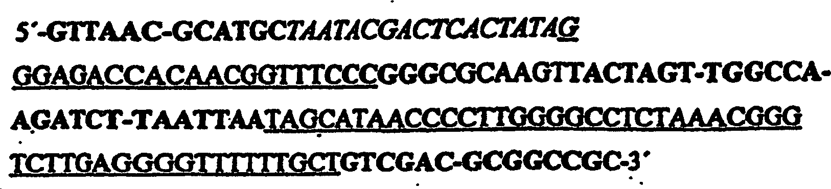
- c) Einführen des in Schritt b) erhaltenen Vektors in eine Reporter-Zelllinie, die positive oder negative phänotypische Selektion erlaubt;
- d) Auswählen von Zellen mit einem geänderten Phänotyp; und
- e) Bestimmen der Sequenz der in den Vektor von Schritt b) eingeführten Nukleinsäure (Intramer) und der Komponente, an die sie bindet.
- a) preparing a candidate mixture of nucleic acids;
- b) cloning the candidate mixture of nucleic acids into a vector under the control of a suitable promoter optionally containing a selectable marker, the vector being a cytoplasmic expression system containing a T7 RNA expression cassette, the T7 RNA expression cassette comprising a T7 Promoter, a stabilizing 5 'strain loop and a 3' terminator Tφ, wherein the sequence of the T7 RNA expression cassette without insert is as follows: the insert being inserted between the 5 'stem loop and the terminator Tφ via the XmaI and PacI restriction sites,
- c) introducing the vector obtained in step b) into a reporter cell line which allows positive or negative phenotypic selection;
- d) selecting cells with a changed phenotype; and
- e) determining the sequence of the nucleic acid (intramer) introduced into the vector of step b) and the component to which it binds.
Kurze Beschreibung der FigurenShort description of characters
A: Aufeinanderfolgende
Kurzdarstellung der
B: Aufeinanderfolgende
Kurzdarstellung er
Die Kandidaten-RNAs oder DNAs umfassen funktionale Nukleinsäuren, z.B. einzelsträngige DNA (ssDNA) oder RNA (ssRNA) oder chemisch modifizierte Formen davon (ssNAmod), doppelsträngige DNA (dsDNA) oder RNA (dsRNA) oder davon chemisch modifizierte Formen davon (dsNAmod), die in der Lage sind ein intrazelluläres Zielmolekül zu binden, zu modulieren oder katalytisch zu modifizieren und dadurch seine biologische Funktion zu beeinflussen. Die funktionalen Nukleinsäuren können beispielsweise durch das „Screening" großer, kombinatorischer Nukleinsäurebibliotheken (in vitro Selektion von Ribozymen, Aptazymen, Aptameren usw.) identifiziert werden. Kombinatorische Nukleinsäurebibliotheken, ob DNA oder RNA, enthalten gewöhnlich eine randomisierte Sequenz, die von feststehenden Regionen flankiert sind, die als Primer-Anellierungsstellen für die enzymatische Amplifikation über PCR oder im Fall von RNA über reverse Transkription und PCR dienen. Die Bibliotheken können beispielsweise als ssDNA an einem automatisierten DNA-Synthesiser unter Verwendung von 3'-Phosphoramidit-Chemie synthetisiert werden, wobei die randomisierte Region mit 3'-Phosphoramiditlösungen synthetisiert wird, die Mischungen der vier Basen A,C,G,T enthalten (Hermes et al. Proc. Natl. Acad. Sci. U.S.A. 87 (1990), 696–700. Die Verhältnisse zwischen den vier Monomeren können in Abhängigkeit von der Anwendung variiert werden. Die feststehenden Regionen enthalten für die Präparation der RNA einen RNA-Polymerase-Promotor, z.B. den T7-RNA-Polymerase-Promotor, der die Transkription der Templat-DNA in RNA ermöglicht. Falls nötig kann die ssDNA unter Verwendung enzymatischer Protokolle, z.B. der PCR, die einem Fachmann wohl bekannt sind, doppelsträngig gemacht werden. Für einen Überblick vergleiche Abelson, Methods in Enzymology 267 (1996), Seiten 275–426, Academic Press, San Diego. Bevorzugterweise enthält die Bibliothek 8 bis 200 randomisierte Nukleotide (Tuerk und Gold, Science 249 (1990), 505–510; Bartel und Szostak, Science 261 (1993), 1411–1418) mit Komplexitäten von 6 × 10⁵ bis 1 × 10¹⁶, welche in erfolgreichen Selektionsexperimenten eingesetzt wurden. Falls gewünscht kann die funktionale Nukleinsäure an andere Moleküle wie zum Beispiel chemisch reaktive Gruppen, Lipide, Peptide, Proteine usw., die der funktionalen Nukleinsäure zusätzliche Eigenschaften hinzufügen, kovalent angehängt oder nicht-kovalent gebunden werden. Funktionale Nukleinsäuren umfassen nicht Nukleinsäuren, die durch Nukleinsäurewechselwirkungen wirken, wie zum Beispiel Antisensemechanismen oder natürliche Ribozyme oder modifizierte und mutierte Formen davon, die Antisenseerkennung mit Phosphodiesterspaltungsaktivität kombinieren und beispielsweise in Gentherapieansätzen angewendet werden. Eine funktionale Nukleinsäure kann Teil eines Konstrukts mit zusätzlichen Sequenzen sein, ob natürlich oder nicht natürlich, die zusätzliche Funktionen hinzufügen, z.B. erhöhte Stabilität, Lokalisierungssignale oder enzymatische Aktivitäten. Darüber hinaus kann die funktionale Nukleinsäure selbst nach Bindung an ihr intrazelluläres Zielmolekül in einer zweiten Funktion, ob intrinsisch oder in einem Konstrukt mit zusätzlichen Sequenzen hinzugefügt, moduliert werden, z.B. Bindung eines dritten Liganden oder katalytisch aktiv zu sein.The candidate RNAs or DNAs include functional nucleic acids, eg single-stranded DNA (ssDNA) or RNA (ssRNA) or chemically modified forms thereof (ssNAmod), double-stranded DNA (dsDNA) or RNA (dsRNA) or chemically modified forms thereof (dsNAmod), which are able to bind, modulate or catalytically modify an intracellular target molecule and thereby influence its biological function. For example, the functional nucleic acids can be identified by the screening of large combinatorial nucleic acid libraries (in vitro selection of ribozymes, aptazymes, aptamers, etc.) Combinatorial nucleic acid libraries, whether DNA or RNA, usually contain a randomized sequence flanked by fixed regions used as primer annealing sites for enzymatic amplification via PCR or in the case of RNA via reverse transcription and PCR serve. For example, the libraries can be synthesized as ssDNA on an automated DNA synthesizer using 3'-phosphoramidite chemistry, whereby the randomized region is synthesized with 3'-phosphoramidite solutions containing mixtures of four bases A, C, G, T ( Acad Sci USA 87 (1990), 696-700 The ratios between the four monomers can be varied depending on the application The fixed regions contain an RNA polymerase for the preparation of the RNA Promoter, for example the T7 RNA polymerase promoter, which allows the transcription of the template DNA into RNA If necessary, the ssDNA can be made double-stranded using enzymatic protocols, eg PCR, which are well known to a person skilled in the art for a review, see Abelson, Methods in Enzymology 267 (1996), pages 275-426, Academic Press, San Diego Preferably, the library contains 8 to 200 randomized nucleotides (Tuerk and Gold, Science 249 (1990), 505-510; Bartel and Szostak, Science 261 (1993), 1411-1418) with complexities of 6 × 10 5 to 1 × 10 16 , which were used in successful selection experiments. If desired, the functional nucleic acid may be covalently attached or non-covalently attached to other molecules, such as chemically reactive groups, lipids, peptides, proteins, etc., which add additional properties to the functional nucleic acid. Functional nucleic acids do not include nucleic acids that act by nucleic acid interactions, such as antisense mechanisms or natural ribozymes, or modified and mutated forms thereof that combine antisense recognition with phosphodiester cleavage activity and are used, for example, in gene therapy approaches. A functional nucleic acid may be part of a construct with additional sequences, whether natural or not natural, that add additional functions, eg, increased stability, localization signals, or enzymatic activities. In addition, even after binding to its intracellular target molecule, the functional nucleic acid may be modulated in a second function, whether intrinsic or in a construct with additional sequences, eg binding of a third ligand or catalytically active.
Aptamere sind spezifische ligandenbindende Nukleinsäurerezeptoren (ssDNA, RNA, modD-NA or modRNA), die durch „Screening" des „Formraums" von enormen kombinatorischen Bibliotheken einzelsträngiger Nukleinsäuren durch in vitro Selektion isoliert werden können.aptamers are specific ligand-binding nucleic acid receptors (ssDNA, RNA, modD-NA or modRNA), by "screening" the "form space" of enormous combinatorial Libraries of single-stranded nucleic acids can be isolated by in vitro selection.
Die in vitro Selektion von Aptameren kann erreicht werden durch in Kontakt bringen einer Nukleinsäurebibliothek mit einem Zielmolekül, z.B. einem Protein, und dem Abtrennen der gebundenen Nukleinsäuren vom Rest der Nukleinsäuremischung. Die Abtrennung kann erreicht werden durch Zurückhalten der Nukleinsäure/Protein-Komplexe auf Nitrozellulosefiltern (Tuerk und Gold, Science 249 (1990), 505–510), Immobilisierung des Zielmoleküls auf einem festen Träger vor der Inkubation mit der Bibliothek und Entfernung nicht-gebundener Spezies durch Spülungen mit einem geeigneten Puffer (Nieuwielandt et al., Biochemistry 34 (1995), 5651–5659) oder jeder anderen Methode, die die Abtrennung der Zielmolekül/Nukleinsäure-Komplexe von ungebundenen Spezies erlaubt. Nach Isolierung der gebundenen Spezies kann die DNA unter Verwendung von PCR amplifiziert werden oder im Fall von RNA durch reverse Transkription, PCR und Transkription. Dieser Zyklus kann mehrere Male wiederholt werden, um eine Bibliothek mir erhöhter Affinität für das Zielmolekül hervorzubringen. Darauffolgend können individuelle Aptamersequenzen durch Klonierung und Sequenzierung der PCR-Produkte unter Verwendung von Standardmethoden identifiziert werden.The In vitro selection of aptamers can be achieved by contacting bring a nucleic acid library with a target molecule, e.g. a protein, and separating the bound nucleic acids from Remainder of the nucleic acid mixture. The separation can be achieved by retaining the nucleic acid / protein complexes on nitrocellulose filters (Tuerk and Gold, Science 249 (1990), 505-510), immobilization of the target molecule on a solid carrier before incubation with the library and removal of unbound Species by flushing with a suitable buffer (Nieuwielandt et al., Biochemistry 34 (1995), 5651-5659) or any other method involving the separation of the target molecule / nucleic acid complexes allowed by unbound species. After isolation of the bound Species, the DNA can be amplified using PCR or in the case of RNA by reverse transcription, PCR and transcription. This cycle can be repeated several times to create a library me heightened affinity for the target molecule produce. Subsequently, you can individual aptamer sequences by cloning and sequencing the PCR products identified using standard methods become.
Nukleinsäureaptamere sind für weit mehr als hundert verschiedene Zielmoleküle beschrieben worden, was zeigt, dass Aptamere jetzt routinemäßig für beinahe jedes gewünschte Zielmolekül erhalten werden können (für Übersichtsartikel vergleiche z.B.: Osborne und Ellington, Chem. Rev. 97 (1997), 349–370; Famulok und Jenne, Curr. Opin. Chem. Biol. 2 (1998), 320–327.Nukleinsäureaptamere are for far more than a hundred different target molecules have been described shows that aptamers are now routinely given to almost any desired target molecule can be (for reviews See, for example: Osborne and Ellington, Chem. Rev. 97 (1997), 349-370; Famulok and Jenne, Curr. Opin. Chem. Biol. 2 (1998), 320-327.
Der Ausdruck Ribozyme wird in der vorliegenden Erfindung für katalytisch aktive Nukleinsäuren verwendet, die auf ein intrazelluläres Zielmolekül wirken, wodurch sie seine Funktion modifizieren. Nicht eingeschlossen sind in der vorliegenden Erfindung natürlich vorkommende Ribozyme, wie zum Beispiel Hammerhead-Ribozyme, Hairpin-Ribozyme oder Derivate davon, die durch Watson-Crick-Bindung an Nukleinsäuresubstrate und Phosphodiesterspaltung wirken und beispielsweise in Gentherapieansätzen verwendet werden (Bramlage, Tibtech 16 (1998), 435–438). Ribozyme mit neuen katalytischen Eigenschaften können unter Verwendung von in vitro Selektionsexperimenten isoliert werden. Zum Beispiel sind Ribozyme isoliert worden, die in der Lage sind RNA-Alkylierung, Bildung von Amidbindungen oder Porphyrinmetallierung zu katalysieren (Breaker, Chem. Rev. 97 (1997), 371–390).Of the Expression Ribozyme is considered to be catalytic in the present invention active nucleic acids used, which act on an intracellular target molecule, whereby they modify its function. Not included naturally occurring ribozymes in the present invention, such as hammerhead ribozymes, hairpin ribozymes or derivatives thereof, by Watson-Crick binding to nucleic acid substrates and phosphodiester cleavage and used, for example, in gene therapy approaches (Bramlage, Tibtech 16 (1998), 435-438). Ribozymes with new catalytic Properties can be isolated using in vitro selection experiments. For example, ribozymes that are capable of being isolated have been isolated RNA alkylation, formation of amide bonds or porphyrin metallation to catalyze (Breaker, Chem. Rev. 97 (1997), 371-390).
Aptazyme können als Chimären zwischen einer katalytischen RNA und einem Aptamer betrachtet werden. Diese Chimären können rationell konstruiert werden in vitro von bestimmten kombinatorischen Bibliotheken selektiert werden (vergleiche Robertson und Ellington, Nature Biotechnol. 17 (1999), 62–66). Gewöhnlich wird die Aptamerdomäne eines Aptazyms als regulatorisches Modul für die allosterische Ribozymaktivierung oder- inhibition eingesetzt (Tang und Breaker, Chem. Biol. 4 (1997), 453–459; Tang und Breaker, RNA 3 (1997), 914–925, Tang und Breaker, Nucleic Acids Res. 26 (1998), 4214–4221; Araki et al., Nucleic Acids Res. 26 (1998), 3379–3384). Ebenso und konsequenterweise enthält ein Aptazym, wenn es selektiert wird, um von einem regulatorischen Molekül beeinflusst zu werden, eine Bindungsdomäne für dieses Molekül und kann sich daher auch wie ein Aptamer verhalten.Aptazymes can be considered as chimeras between a catalytic RNA and an aptamer. These chimeras can be efficiently engineered in vitro from certain combinatorial libraries (see Robertson and Ellington, Nature Biotechnol 17 (1999), 62-66). Usually, the aptamer domain of an aptazyme becomes the regulatory module for the allosteric ribozyme Tiberation or inhibition (Tang and Breaker, Chem. Biol. 4 (1997), 453-459; Tang and Breaker, RNA 3 (1997), 914-925, Tang and Breaker, Nucleic Acids Res. 26 (1998), 4214-4221; Araki et al., Nucleic Acids Res. 26 (1998), 3379-3384). Likewise, and consistently, an aptazyme, when selected to be influenced by a regulatory molecule, contains a binding domain for that molecule, and thus may behave like an aptamer.
Intramere sind funktionale Nukleinsäuren (Aptamere, Aptazyme usw.), die exogen oder endogen in prokaryotische oder eukaryotische Zellen eingebracht werden und ein intrazelluläres Zielmolekül binden, modulieren oder enzymatisch modifizieren.intramers are functional nucleic acids (Aptamers, aptazyms, etc.) that are exogenous or endogenous in prokaryotic or eukaryotic cells are introduced and bind an intracellular target molecule, modulate or enzymatically modify.
Modulation eines intrazellulären Zielmoleküls kann zum Beispiel die Inhibition oder Stabilisierung einer Protein/Protein-Interaktion oder Proteindomäne/Protein-Interaktion oder Protein/Nukleinsäure-Interaktion oder Protein/"Kleinmolekül"-Interaktionen usw. sein. Modulierung kann auch die Aktivierung oder Inhibition von Enzymen über verschiedene Mechanismen umfassen wie zum Beispiel Sequestrierung des Substrat (der Substrate), Bindung an die aktive Stelle, Induktion von Konformationsänderungen, allosterische Regulierung usw. Modulation eines intrazellulären Zielmoleküls kann durch beides, nicht-kovalente Interaktionen oder kovalente Modifikationen wie zum Beispiel Biotinylierung, erfolgen (Wecker et al., RNA 2 (1996), 982–94). Weitere Methoden, die zum Beispiel für auf Antikörpern basierende Ansätze entwickelt wurden, könnten auf das funktionale Nukleinsäuresystem adaptiert werden, wobei das Aptamer mit einem Molekül verbunden werden könnte, das in der Lage ist das gebundene Zielmolekül chemisch zu inaktivieren. Beispielsweise werden die intrazellulären, funktionalen Nukleinsäuren über eine kurze „Annealing"-Sequenz, die kovalent an dem Farbstoff Malachitgrün (MG) angehängt ist, markiert. Diese kurzen Oligonukleotide könnten exogen angewendet werden. MG absorbiert Licht bei einer sichtbaren Wellenlänge, die nicht signifikant durch Zellen absorbiert wird. Nach der Bestrahlung mit einem Laser wird das MG aktiviert und erzeugt kurzlebige Hydroxylradikale, die Proteine höchstwahrscheinlich durch Modifizierung der Aminosäureseitenketten inaktivieren [„Chromophore assisted laser inactivation (CALI)", vergleiche: Liao et al., Proc. Natl. Aad. Sci. 91 (1994), 2659–63]. Der Inakti vierungsprozess tritt nur innerhalb einer kurzen Distanz von der Farbstoffgruppe auf. Daher kann das als Ziel gesetzte Protein oder die Untereinheit generell geschädigt werden ohne seine Nachbarn zu beeinflussen. Dies hat zwei Vorteile: Erstens würde das Aptamer nicht notwendigerweise an die aktive Stelle des Proteins binden müssen, sondern könnte mit einem benachbarten Epitop interagieren, das besser für die Bindung an eine Nukleinsäure geeignet sein könnte. Sogar obwohl das gebundene Epitop entfernt von der aktiven Stelle ist, wird das Protein immer noch spezifisch inaktiviert. Zweitens könnten mehrere Bestrahlungspulse Proteine mit hoher Abundanz, die anfangs in höheren Kopienzahlen als das spezifische Aptamer vorhanden sind, inaktiveren, da das Protein irreversibel inaktiviert wird und das Aptamer konstitutiv exprimiert werden kann.modulation of an intracellular target molecule For example, the inhibition or stabilization of a protein / protein interaction or protein domain / protein interaction or protein / nucleic acid interaction or protein / "small molecule" interactions, etc. be. Modulating can also be the activation or inhibition of Enzymes over Various mechanisms include, for example, sequestration of the substrate (s), binding to the active site, induction conformational changes, allosteric regulation, etc. modulation of an intracellular target molecule can by both, non-covalent interactions or covalent modifications such as biotinylation (Wecker et al., RNA 2 (1996), 982-94). Other methods developed, for example, for antibody-based approaches could, could on the functional nucleic acid system be adapted, with the aptamer connected to a molecule that could be is able to chemically inactivate the bound target molecule. For example, the intracellular, functional nucleic acids become via a short "annealing" sequence that is covalent on the dye malachite green (MG) attached is marked. These short oligonucleotides could be used exogenously. MG absorbs light at a visible wavelength that is not significant is absorbed by cells. After irradiation with a laser The MG is activated and generates short-lived hydroxyl radicals, the Proteins most likely by modification of the amino acid side chains inactivate ["chromophores assisted laser inactivation (CALI) ", compare: Liao et al., Proc. Natl. Aad. Sci. 91 (1994), 2659-63]. The inactivation process occurs only within a short distance from the dye group. Therefore, the target protein can be or the subunit generally be harmed without its neighbors to influence. This has two advantages: First, that would Aptamer does not necessarily bind to the active site of the protein but have to bind could interact with an adjacent epitope, which is better for binding suitable for a nucleic acid could be. Even though the bound epitope is away from the active site is, the protein is still specifically inactivated. Secondly could multiple radiation pulses proteins with high abundance, the initial in higher Copy numbers are present as the specific aptamer, more inactive, since the protein is irreversibly inactivated and the aptamer constitutive can be expressed.
Intrazelluläre Zielmoleküle umfassen kleine Moleküle, biologische Kofaktoren, natürliche Polymere wie zum Beispiel Proteine, Proteindomänen, Peptide und Nukleinsäuren, Glykoproteine, Hormone, Rezeptoren, Proteinkomplexe, Protein/Nukleinsäure-Komplexe, Toxine, Viren und andere Substanzen, die natürlicherweise in Zellen gefunden werden.Include intracellular target molecules small molecules, biological cofactors, natural Polymers such as proteins, protein domains, peptides and nucleic acids, glycoproteins, Hormones, receptors, protein complexes, protein / nucleic acid complexes, Toxins, viruses and other substances found naturally in cells become.
Phänotyp kann beispielsweise die Induktion oder Repression der Transkription eines bestimmten Gens, Prozesse, die mit Signaltransduktionskaskaden verbunden sind wie zum Beispiel Adhäsion als Antwort auf bestimmte Reize, Zelldifferenzierung, Apoptose usw. sein.Phenotype can For example, the induction or repression of transcription of a specific gene, processes associated with signal transduction cascades are like adhesion in response to certain stimuli, cell differentiation, apoptosis, etc. be.
Phänotypische Selektion ist ein Prozess, in dem Nukleinsäure-Bibliotheken durch verschiedene Methoden, die dem Fachmann bekannt sind (Elektroporation, Llipofektion, Expression mit viralen Vektorsystemen, Expression von RNA-Polymerase-Promotoren enthaltenden Plasmiden usw.), in Zellen eingeführt werden. Diejenigen Zellen, die eine funktionale Nukleinsäure enthalten, die auf ein Zielmolekül wirkt, das in einen bestimmten Phänotyp involviert ist, werden auf der Basis ihres veränderten Phänotyps von dem Großteil zellulären Materials, das keinen veränderten Phänotyp zeigt, abgetrennt. Zellen, die in der phänotypischen Selektion verwendet werden umfassen Prokaryoten, Hefe, Insekten- oder Pflanzenzellen und bevorzugterweise Säugerzellen (z.B. CHO, HeLa, COS, MDCK, 293, WI38 und Jurkat E6-Zellen). Falls eine klare Trennung auf Basis des veränderten Phänotyps nicht möglich oder schwierig ist, können Reporterkonstrukte angewendet werden, die eine deutlichere oder bessere Unterscheidung erlauben. In einem positiven phänotypischen Selektionsexperiment, zum Beispiel der Induktion eines zellulären Promotor- oder Enhancer/Promotor-Konstrukts, wird ein Reportergen, das die Identifizierung des veränderten Phänotyps (hier die Induktion des Promo tors) erlaubt, unter der Kontrolle eines induzierbaren Promotors in dem Reporterkonstrukt eingefügt. Reportergene, die in dieser Art von Selektion verwendet werden, sind beispielsweise Resistenzgene wie die Aminoglykosidphosphotransferase (Southern und Berg, J. Mol. Appl. Genet. 1 (1982), 327–341), die Aminoglykosidantibiotika (Neomycin, G418) inaktiviert und Zellen resistent gegen diesen Wirkstoff macht. Folglich werden die Zellen, die das Gen für die funktionale Nukleinsäure beinhalten in Anwesenheit des Wirkstoffs überleben, wenn der induzierbare Promotor durch die Wirkung einer funktionalen Nukleinsäure (fNS) angeschaltet wird, und die Gene für die fNS können identifiziert werden. Beispielsweise könnten Säugerpromotoren, die von der Aktivierung durch den Transkriptionsfaktor NF B abhängig sind, durch die Inhibition des I B Proteins, das normalerweise den Import von NF B in den Zellkern verhindert, aktiviert werden (Rolfe et al., J. Mol. Med. 75 (1997), 5–17; Fuchs et al., Oncogene 17 (1998), 1483–90. Andere Beispiele von Promotoren, die durch einen inhibitorischen Mechanismus reguliert werden, sind bakterielle Transkriptionseinheiten, zum Beispiel das E.coli lac-Operon, das durch den lac-Repressor reguliert wird (Gilbert und Muller-Hill, Proc. Nat. Acad. Sci. 56 (1966), 1891–1898).Phenotypic selection is a process in which nucleic acid libraries are introduced into cells by various methods known to those skilled in the art (electroporation, lipofection, expression with viral vector systems, expression of plasmid containing RNA polymerase promoters, etc.). Those cells containing a functional nucleic acid that acts on a target molecule involved in a particular phenotype are separated on the basis of their altered phenotype from the majority of cellular material that does not show an altered phenotype. Cells used in phenotypic selection include prokaryotes, yeast, insect or plant cells, and preferably mammalian cells (eg, CHO, HeLa, COS, MDCK, 293, WI38, and Jurkat E6 cells). If a clear separation based on the altered phenotype is not possible or difficult, reporter constructs can be used which allow a clearer or better distinction. In a positive phenotypic selection experiment, for example, the induction of a cellular promoter or enhancer / promoter construct, a reporter gene that allows the identification of the altered phenotype (here promoter induction) is under the control of an inducible promoter in the reporter construct inserted. Reporter genes used in this type of selection include, for example, resistance genes such as aminoglycoside phosphotransferase (Southern and Berg, J. Mol. Appl. Genet 1 (1982), 327-341), inactivating aminoglycoside antibiotics (neomycin, G418), and cells makes it resistant to this ingredient. Consequently the cells containing the functional nucleic acid gene survive in the presence of the drug when the inducible promoter is turned on by the action of a functional nucleic acid (fNS), and the genes for the fNS can be identified. For example, mammalian promoters that are dependent on activation by the transcription factor NF B could be activated by the inhibition of the IB protein, which normally prevents the import of NF B into the nucleus (Rolfe et al., J. Mol. Med. 75 (1997), 5-17, Fuchs et al., Oncogene 17 (1998) 1483-90 Other examples of promoters regulated by an inhibitory mechanism are bacterial transcription units, for example the E.coli lac operon, which is regulated by the lac repressor (Gilbert and Muller-Hill, Proc. Nat. Acad. Sci. 56 (1966), 1891-1898).
Negative phänotypische Selektion kann beispielsweise durch die Induktion eines Promotors stattfinden. In diesem Fall könnte das Reportergen ein Toxin sein [z.B. Herpes-Simplex-Virus-Thymidinkinase (Fife et al., Gene Ther. 5 (1998), 614–620] oder Diphtherie-Toxin (Massuda et al., Proc. Natl. Acad. Sci. U.S.A. 94 (1997), 14701–14706), das den Tod von Zellen fördert, wo keine funktionale Nukleinsäure den zellulären Signaltransduktionsweg inhibiert, der den Promotor induziert. Induzierbare Gene umfassen bakterielle, virale und eukaryontische Transkriptionseinheitem, z.B. im Arabinose-Operon (Wilcox et al., J. Biol. Chem. 249 (1974), 2946–2952), die frühen und späten Gene des Vaccinia Virus (Moss. Annu. Rev. Biochem. 59 (1990), 661–688) oder Promotoren der Immunglobulingene (Staudt et al., Ann. Rev. Immunol. 10 (1991), 373–398). Die in phänotypischen Selektionen verwendeten Promotoren können natürlich vorkommende Promotor/Enhancer-Elemente oder jegliche Kombinationen oder Modifikationen davon sein.negative phenotypic Selection can be achieved, for example, by the induction of a promoter occur. In this case could the reporter gene is a toxin [e.g. Herpes simplex virus thymidine kinase (Fife et al., Gene Ther. 5 (1998), 614-620] or diphtheria toxin (Massuda et al., Proc Natl Acad Sci U.S.A. 94 (1997), 14701-14706), that promotes the death of cells, where no functional nucleic acid the cellular Signal transduction pathway that induces the promoter. inducible Genes include bacterial, viral and eukaryotic transcription units, e.g. in the arabinose operon (Wilcox et al., J. Biol. Chem. 249 (1974), 2946-2952), the early ones and late Genes of vaccinia virus (Moss. Annu., Rev. Biochem., 59 (1990), 661-688) or Promoters of immunoglobulin genes (Staudt et al., Ann. Rev. Immunol. 10 (1991), 373-398). The in phenotypical Selections used promoters can naturally occurring promoter / enhancer elements or any combinations or modifications thereof.
Reporterkonstrukte können positive und negative phänotypische Selektionen umfassen. Zum Beispiel kann eine negative phänotypische Selektion über die Expression eines Toxins (wie zum Beispiel dem Diphtherie-Toxin) unter der Kontrolle eines induzierbaren zellulären Promotors, wie dem Interleukin-2-Promotor in T-Lymphozyten, entstehen. Diese Genkonstrukte können in Zellen z.B. über Plasmide eingebracht werden und die Expression des Toxingens wird von der natürlichen, den Promotor induzierenden Maschinerie der Zelle vorangetrieben. Nur in denjenigen Zellen, in denen eine funktionale Nukleinsäure die Induktion des Toxin-Promotors inhibiert, wird das Toxin nicht exprimiert werden und die Zellen überleben und können selektiert werden.reporter constructs can positive and negative phenotypic Include selections. For example, a negative phenotypic Selection via the expression of a toxin (such as the diphtheria toxin) under the control of an inducible cellular promoter, such as the interleukin-2 promoter in T lymphocytes, arise. These gene constructs can be found in Cells e.g. above Plasmids are introduced and the expression of the toxin gene is from the natural, promoted the promoter-inducing machinery of the cell. Only in those cells, in which a functional nucleic acid the Induction of the toxin promoter inhibits the toxin will not be expressed and the cells survive and can be selected.
Die Vektoren (Vektorelemente) und Protokolle für das Klonieren, das Transfizieren, die transiente Genexpression und das Erhalten stabiler transfizierter Zelllinien sind gut bekannt und in der Literatur beschrieben (Maniatis et al., (1989) Molecular Cloning: A Laboratory Manual, Cold Spring Harbor Laboratory Press, Cold Spring Harbor, New York; Spector et al., Cells: A Laboratory Manual (1998), Cold Spring Harbor Laboratory Press, Cold Spring Harbor, New York). Vektoren für die Expression der Nukleinsäure-Bibliotheken können jegliche allgemein verwendeten Konstrukte sein um fremde Gene in Zellen einzuschleusen, zum Beispiel lineare PCR-Fragmente, Plasmide, DNA-Viren wie M13 oder Vaccinia, RNA-Vieren wie Retroviren oder das Semliki Forest Virus. Die Gene für die Nukleinsäure-Bibliothek werden unter die Kontrolle geeigneter Promotoren gestellt, bevorzugt solche, die eine hohe Transkription der Nukleinsäuren ermöglichen.The Vectors (vector elements) and protocols for cloning, transfecting, transient gene expression and obtaining stable transfected ones Cell lines are well known and described in the literature (Maniatis et al., (1989) Molecular Cloning: A Laboratory Manual, Cold Spring Harbor Laboratory Press, Cold Spring Harbor, New York; Spector et al., Cells: A Laboratory Manual (1998), Cold Spring Harbor Laboratory Press, Cold Spring Harbor, New York). Vectors for the expression of the nucleic acid libraries can Any commonly used constructs to be foreign genes in Introduce cells, for example linear PCR fragments, plasmids, DNA viruses such as M13 or vaccinia, RNA viruses such as retroviruses or the Semliki Forest Virus. The genes for the nucleic acid library are placed under the control of suitable promoters, preferably those that allow high transcription of the nucleic acids.
Beispiele, die aber nicht darauf beschränkt sind, umfassen Polymerase-III-abhängige Promotoren, wie zum Beispiel der Hefe-RNAse-P-Promotor (Good und Engelke, Gene 151 (1994), 209–214), Säuger-U6- oder -tRNAmet-Promotoren (Bertrand et al., RNA 3 (1997), 75–88) retrovirale „long terminal repeats" (Rossi, Tibtech 13 (1995), 301–306), T7-RNA-Polymerase-Promotoren, wie in Beispiel 1 beschrieben, oder der subgenomische Promotor für die Semliki-Forest-Virus-Replikase (Tubulekas et al., Gene 190 (1997), 191–95).Examples include, but are not limited to, polymerase III-dependent promoters, such as the yeast RNAse P promoter (Good and Engelke, Gene 151 (1994), 209-214), mammalian U6 or tRNA met promoters (Bertrand et al., RNA 3 (1997), 75-88) retroviral "long terminal repeats" (Rossi, Tibtech 13 (1995), 301-306), T7 RNA polymerase promoters, as in Example 1, or the subgenomic promoter for the Semliki Forest virus replicase (Tubulekas et al., Gene 190 (1997), 191-95).
Vektoren, die als Reporterkonstrukte benutzt werden, können Plasmide (z.B. pUC18, pYES2, pCDM8) oder virale Genome, wie zum Beispiel Adenivirus, AAV oder retrovirale Vektoren (Moloney Murine Leukemia Virus (MoMuLV), Gibbon Ape Leukemia Virus (GaLV) usw.) sein. Die Verwendung von stabil transfizierten Reporterzelllinien ist lediglich optional, sie stellt jedoch ein zuverlässiges System dar, um Zellen zu selektieren, welche die negative Selektion in Gegenwart eines Toxins überleben.vectors used as reporter constructs, plasmids (e.g., pUC18, pYES2, pCDM8) or viral genomes, such as Adenivirus, AAV or retroviral vectors (Moloney Murine Leukemia Virus (MoMuLV), Gibbon Ape Leukemia Virus (GaLV), etc.). The usage of stably transfected reporter cell lines is only optional, but it is a reliable one System to select cells showing the negative selection survive in the presence of a toxin.
Unter Zielmolekülvalidierung versteht man die Identifizierung eines bestimmten zellulären Zielmoleküls als das für einen bestimmten Phänotyp verantwortliche Schlüsselmolekül. Sie umfasst die intrazelluläre Anwendung eines Nukleinsäuremodulators, der das Zielmolekül beeinflusst, um dessen Rolle in dem komplexen Netzwerk zellulärer Komponenten zu untersuchen. Das bevorzugte Ziel ist es zu untersuchen, ob das Zielmolekül für einen bestimmten zellulären Status oder Krankheitszustand verantwortlich ist und daher ein gutes Zielmolekül für Medikamente ist oder ob dies nicht der Fall ist.Under Target validation one understands the identification of a particular cellular target molecule as the for one certain phenotype responsible key molecule. it includes the intracellular application a nucleic acid modulator, which influences the target molecule, to investigate its role in the complex network of cellular components. The preferred goal is to examine if the target molecule is for a certain cellular Status or condition is responsible and therefore a good drug target or if this is not the case.
Unter Zielmolekülidentifizierung versteht man die Verbindung einer zellulären Komponente mit einem bestimmten Phänotyp, gleichgültig ob seine biologische Funktion bekannt ist oder nicht. Die Zielmolekülidentifizierung kann mittels eines funktionalen Nukleinsäwemodulators erfolgen, der durch phänotypische Selektion identifiziert worden ist. Das Zielmolekül, dass durch solch einen Modulator beeinflusst wird, stellt wahrscheinlich das eigentliche Schlüsselmolekül oder ein Schlüsselmolekül, dar das für einen bestimmten Phänotyp verantwortlich ist. Das Zielmolekül kann durch seine Interaktion mit der funktionalen Nukleinsäure identifiziert werden. Beispielsweise wird das Hefe-Zwei-Hybrid-System, eine Modifikation des Zwei-Hybrid-Systems, verwendet, um RNA-Protein-Interaktionen nachzuweisen. Das Drei-Hybrid-System benutzt ein RNA-Hybrid als Zwischenglied zwischen zwei Hybrid-Proteinen. Die Interaktion der drei Komponenten dient zur Rekonstitution der transkriptionellen Aktivierung von Reportergenen durch einen Mechanismus ähnlich dem Zwei-Hybrid-System (SenGupta et al., Proc Natl. Acad: Sci. 93 (1996), 8496–8501). Die funktionale Nukleinsäure, die in das RNA-Hybrid kloniert ist wird mit ihrem Zielprotein interagieren, das als Teil einer Protein-Hybrid-Bibliothek von zellulären Proteinen exprimiert wird. Daher werden die Reportergene in denjenigen Hefezellen exprimiert werden, welche beides enthalten, die funktionale Nukleinsäure und ihr Zielmolekül, und die Plasmide mit den Zielgenen können isoliert werden.By target molecule identification is meant the association of a cellular component with a particular phenotype, whether its biological function is known or not. The target molecules tification may be by means of a functional nucleic acid modulator identified by phenotypic selection. The target molecule that is affected by such a modulator is likely to be the key molecule or key molecule responsible for a particular phenotype. The target molecule can be identified by its interaction with the functional nucleic acid. For example, the yeast two-hybrid system, a modification of the two-hybrid system, is used to detect RNA-protein interactions. The three-hybrid system uses an RNA hybrid as an intermediate between two hybrid proteins. The interaction of the three components serves to reconstitute the transcriptional activation of reporter genes by a mechanism similar to the two-hybrid system (SenGupta et al., Proc Natl. Acad: Sci. 93 (1996), 8496-8501). The functional nucleic acid cloned into the RNA hybrid will interact with its target protein, which is expressed as part of a protein hybrid library of cellular proteins. Therefore, the reporter genes will be expressed in those yeast cells that contain both the functional nucleic acid and its target molecule, and the plasmids with the target genes can be isolated.
Ein anderer Ansatz ist es, das Zielmolekül über Komplementations-Klonierung zu identifizieren, wie in Beispiel 2 dargestellt. Diese Technik wurde erfolgreich angewandt, um Proteine zu isolieren, die Zelllinien, welche mutierte endogene Gene enthalten, funktional zu komplementieren, z.B. die Komplementations-Klonierung von NEMO, einer Komponente des I B-Kinase-Komplexes (Yamaoko et al., Cell 93 (1998), 1231–1240).One Another approach is to target the molecule via complementation cloning to identify, as shown in Example 2. This technique has been successfully used to isolate proteins that which contain mutated endogenous genes to functionally complement, e.g. the complementation cloning of NEMO, a component of the I B kinase complex (Yamaoko et al., Cell 93 (1998), 1231-1240).
Hochspezifische Nukleinsäwemodulatoren zellulärer Zielmoleküle können beispielsweise routinemäßig durch Anwendung der Technologien SELEX, Aptazyme, Zwei- und Drei-Hybnd-Technologien oder durch die hier beschriebenen phänotypischen Selektionsmethode usw. erhalten werden. Die resultierenden Nukleinsäuremodulatoren können exo- oder endo gen durch Methoden wie Elektroporation, Lipofektion, Expression mit viralen Vektorsystemen, Expression von Plasmiden, die RNA-Polymerase-Promotoren enthalten, usw. in Zellen eingebracht werden. Funktionale Nukleinsäuren können auf ihr Zielmolekül über verschiedene Mechanismen wirken; sie können ihr Zielmolekül entweder in Antikörper-ähnlicher Weise binden, wodurch sie seine biologische Funktion modulieren. Zum Beispiel sind chemisch modifizierte, nukleaseresistente Aptamere für die Bindung an extrazelluläre Wachstumsfaktoren wie den „basic fibroblast growth factor" selektiert worden (bFGF; vergleiche: [Jellinek et al., Proc. Natl. Acad. Sci USA 90 (1993), 11227–11231; Jellinek et al., Biochemistry 34 (1995), 11363–11372.], „human keratinocyte growth factor" (hKGF; vergleiche: [Pagratis et al., Nature Biotech. 15 (1997), 68–73]), "platelet derived growth factor" (PDGF; vergleiche: [Green et al., Biochemistry 35 (1996), 14413–14424]) oder "vascular endothelial growth factor" (VEGF; vergleiche: [Green et al., Chem. Biol. 2 (1995), 683–695; Jellinek et al., Biochemistry 33 (1994), 10450–10456; Ruckman et al., J. Biol. Chem. 273 (1998), 20556–0567]). Diese Aptamere blockierten die Bindung ihres extrazellulären Zielmoleküls an seinen natürlichen Proteinrezeptor und modellierten dadurch die biologische Funktion des Wachstumsfaktors.highly specific Nukleinsäwemodulatoren cellular targets can for example, routinely Application of the technologies SELEX, Aptazyme, two- and three-hybrid technologies or by the phenotypic selection method described here etc. are obtained. The resulting nucleic acid modulators can exo- or endogenous by methods such as electroporation, lipofection, Expression with viral vector systems, expression of plasmids, containing the RNA polymerase promoters, etc. introduced into cells become. Functional nucleic acids can on their target molecule via different Mechanisms work; you can their target molecule either in antibody-like Bind way, thereby modulating its biological function. For example, chemically modified, nuclease resistant aptamers for the Binding to extracellular Growth factors such as the "basic fibroblast growth factor " (bFGF; compare: [Jellinek et al., Proc. Natl. Acad. Sci USA 90 (1993), 11227-11231; Jellinek et al., Biochemistry 34 (1995), 11363-11372.], "Human keratinocyte growth factor "(hKGF; compare: [Pagratis et al., Nature Biotech. 15 (1997), 68-73]), platelet derived growth factor (PDGF; [Green et al., Biochemistry 35 (1996), 14413-14424]) or "vascular endothelial growth factor "(VEGF; compare: [Green et al., Chem. Biol. 2 (1995), 683-695; Jellinek et al., Biochemistry 33 (1994), 10450-10456; Ruckman et al., J. Biol. Chem. 273 (1998), 20556-0567]). These aptamers blocked the binding of their extracellular target molecule to its natural one Protein receptor and thereby modeled the biological function of the growth factor.
Während es
Stand der Technik ist, die biologische Funktion eines extrazellulären Proteins
zu modulieren, ist ein zentraler Aspekt der vorliegenden Erfindung,
das enorme Potential von hochaffinen und hochspezifischen Nukleinsäurereagenzien
zu nutzen, um ein gegebenes Zielmolekül in intrazelluläre Anwendungen
zu modulieren. Dass dies in der Tat gemacht werden kann, wurde repräsentativ
in dem System gezeigt, das beispielhaft in Beispiel 1 erläutert wurde:
Es
wird ein Ansatz beschrieben, der auf der Erkennung zytoplasmatischer
Integrindomänen
durch synthetische, ligandenbindende RNA-Aptamere, die aus einer
kombinatorischen RNA-Bibliothek
isoliert wurden, beruht. Ein auf Vaccinia Virus basierendes System
ermöglichte
hohe zytoplasmatische Expression von RNA-Aptameren (Intrameren),
die gegen die intrazelluläre
Domäne
des β2-Integrins
LFA-1 gerichtet waren, ein Transmembranprotein, das die Zelladhäsion an
intazelluläre
Adhäsionsmolekül 1 (ICAM-1)
vermittelt. Die zytoplasmatische Expression von Integrin bindenden
Intrameren reduzierte die induzierbare Zelladhäsion an ICAM-1 in zwei verschiedenen
Zelltypen. Die Aptamere zielen spezifisch auf eine funktionale zytoplasmatische
Subdomäne
und definieren diese dadurch als wichtig für die Regulation der Zelladhäsion in
Leukozyten.While it is state of the art to modulate the biological function of an extracellular protein, a central aspect of the present invention is to capitalize on the enormous potential of high affinity and highly specific nucleic acid reagents to modulate a given target molecule into intracellular applications. That this can indeed be done was shown representatively in the system exemplified in Example 1:
An approach based on the recognition of cytoplasmic integrin domains by synthetic, ligand-binding RNA aptamers isolated from a combinatorial RNA library is described. A vaccinia virus based system enabled high cytoplasmic expression of RNA aptamers (intramers) directed against the intracellular domain of the β2 integrin LFA-1, a transmembrane protein that mediates cell adhesion to intacellular adhesion molecule 1 (ICAM-1). Cytoplasmic expression of integrin-binding intramers reduced inducible cell adhesion to ICAM-1 in two different cell types. The aptamers specifically target a functional cytoplasmic subdomain, thereby defining it as important for the regulation of cell adhesion in leukocytes.
Integrine sind vielseitige, heterodimere Transmembranproteine, die adhäsive Interaktionen mit der extrazellulären Matrix und zellspezifischen Gegenrezeptoren vermitteln. Sie sind in diverse biologische Prozesse eingebunden, die Apoptose, Zellzyklusregulierung, Zellwanderung, Blutgerinnung, Gedächtnis und Leukozytenfunktion umfassen (Hynes, Cell 69 (1992), 11–25). Die zytoplasmatischen Domänen der Integrine spielen wahrscheinlich durch die Vernetzung von Signalereignissen innerhalb der Zelle zu den extrazellulären Domänen eine wichtige Rolle in der Regulation der Zelladhäsion, die genauen Mechanismen sind jedoch unklar (Diamond und Springer, Curr. Biol. 4 (1994), 506–517; Dedhar und Hannigan, Curr. Opin. Cell Biol. 8 (1996), 657–669). Das beta 2-Integrin LFA-1 (αL/β2, Cdl1a/CD18) vermittelt die Adhäsion von Leukozyten in Immun- und Entzündungsantworten durch Bindung an zelluläre Liganden (Lub et al., Immunol. Today 16 (1995), 479–483), die inteazellulären Adhäsionsmoleküle ICAM-1, -2, -3, -4. Eine große Datenmenge legt nahe, daß die zytoplasmatischen Domänen der beta 2-Kette wahrscheinlich durch ihre Interaktionen mit intrazellulären Proteinen und/oder dem membrannahen Zytoskelett (Stewart etal., J. Cell Biol. 140 (1998), 699–707) mit der Regulierung der durch LFA-1 vermittelten Haftfähigkeit in Verbindung steht; einige Kandidaten für die zytoplasmatischen Liganden sind kürzlich identifiziert worden (Hibbs et al., J. Exp. Med. 174 (1991), 1227–1238; Pavalko und LaRoche, J. Immunol. 151 (1993), 3795–3807; Sharma et al., J. Immunol. 154 (1995), 3461–3470 ; Kolanus et al., Cell 86 (1996), 233–242). Um weitere Einsichten in diese Mechanismen zu erlangen, wurde ein System entwickelt, das die spezifische Inhibition der von LFA-1 vermittelten „Inside-out"-Signaltransduktion in vivo erlaubt. Die Ziele waren die Selektion von Aptameren, die die zytoplasmatische Domäne von CD18 binden, die Entwicklung und Anwendung eines Systems, das hohe Expression von Aptameren im Zytoplasma von Leukozyten erlaubt und die Untersuchungen ihrer biologischen Effekte im Kontext lebender Zellen.Integrins are versatile, heterodimeric transmembrane proteins that mediate adhesive interactions with the extracellular matrix and cell-specific counterreceptors. They are involved in various biological processes involving apoptosis, cell cycle regulation, cell migration, blood coagulation, memory and leukocyte function (Hynes, Cell 69 (1992), 11-25). The cytoplasmic domains of the integrins probably play an important role in the regulation of cell adhesion through the cross-linking of signal events within the cell to the extracellular domains, but the exact mechanisms are unclear (Diamond and Springer, Curr. Biol. 4 (1994), 506- 517; Dedhar and Hannigan, Curr. Opin. Cell Biol. 8 (1996), 657-669). The beta 2-integrin LFA-1 (αL / β2, Cdl1a / CD18) mediates the adhesion of leukocytes in immune and inflammatory responses by binding to cellular ligands (Lub et al., Immunol., Today 16: 479-483 (1995)). , the intracellular adhesion molecules ICAM-1, -2, -3, -4. A large amount of data suggests that the cytoplasmic domains of the beta 2 chain, probably through their interactions with intracellular proteins and / or the membrane near cytoskeleton (Stewart et al., J. Cell Biol. 140 (1998), 699-707) with the regulation of LFA-1 mediated adhesiveness communicates; Some candidates for the cytoplasmic ligands have recently been identified (Hibbs et al., J. Exp. Med., 174 (1991), 1227-1238; Pavalko and LaRoche, J. Immunol. 151 (1993), 3795-3807; Sharma et al al., J. Immunol., 154 (1995), 3461-3470; Kolanus et al., Cell 86 (1996), 233-242). To gain further insights into these mechanisms, a system was developed that allows the specific inhibition of LFA-1-mediated "inside-out" signal transduction in vivo, with the aim of selecting aptamers that bind the cytoplasmic domain of CD18 , the development and application of a system that allows high expression of aptamers in the cytoplasm of leukocytes and the study of their biological effects in the context of living cells.
Kurzum, wie in Beispiel 1 unten genau beschrieben, waren die Autoren in der Lage
- a) RNA-Aptamere zu isolieren, die an ein immobilisiertes Peptid binden, welches der zytoplasmatischen Domäne des integralen Transmembranproteins LFA-1 (CD18, beta 2-Integrin) entspricht.
- b) zu zeigen, dass die Aptamere ihr Zielmolekül spezifisch erkennen, sogar wenn sie in einen andern Sequenzkontext in einer RNA-Expressionskassette kloniert sind. Diese Kassette wurde von einem T7-RNA-Polymerase-abhängigen Expressionssystem (Fuerst und Moss, J. Mol. Biol. (1989), 333–348) abgeleitet und enthielt eine 5'-„Stern-Loop-Struktur", von der vorher gezeigt werden konnte, dass sie RNA-Moleküle im Zytoplasma durch Verhinderung ihres Abbaus stabilisiert. Des weiteren enthielt das 3'-Ende das T-Terminator-„Hairpin-Loop-Motiv" als einen Terminator für die von der T7-RNA-Polymerase abhängigen Transkription. Ein anderes Beispiel für ein zytoplasmatisches System, das für RNA-Expression geeignet sein dürfte, basiert aus der RNA-abhängigen RNA-Replikase viraler Vektoren, wie dem Semliki Forest Virus (SFV) (Tubulekas et al. Gene 190 (1997), 191–195). Am bedeutsamsten war, dass gezeigt werden konnte, dass die von der T7-RNA-Expressionskassette entstehenden Aptamertranskripte richtig terminiert waren und ihre Bindungsaktivität für CD18cyt vollständig behielten. Nicht richtig terminierte Aptamersequenzen, die zusätzliche Sequenzinformation aus dem viralen Genom enthielten, wurden ebenfalls auf Bindung an CD18cyt getestet. Es wurde festgestellt, dass diese unerwünschte zusätzliche Sequenzinformation in vitro wahrscheinlich wegen der Störung der korrekten Aptamerfaltung zu einer drastisch reduzierten Bindungsaktivität dieser Aptamervarianten an CD18cyt führte. Dieses Ergebnis etabliert zwei sehr wichtige und überraschende Punkte. Es zeigt, dass es möglich ist, (i) das Aptamer im Kontext zusätzlicher Sequenzinformation, wie zum Beispiel von stabilisierenden Sequenzmotiven oder von RNA-Lokalisierungssignalen usw., zu exprimieren, was entscheidend für die breite Anwendung von endogener Aptamerexpression ist und (ii) kleine kompakte Aptamere, die in vitro selektiert wurden, auf modulare Art zu konstruieren, um die Aptamere mit zusätzlichen Eigenschaften zu versehen, was die angemessene und effiziente intrazelluläre Anwendbarkeit sicher stellt, ohne die Aptamerbindung an das Zielmolekül zu beeinflussen.
- c) zu zeigen, dass die selektierten Aptamere endogenes LFA-1 spezifisch in Rohextrakten von Jurkat-E6-Zellen erkennen. Sie tun dies auch im Kontext der RNA-Expressionskassette.
- d) ein auf Vaccinia Viren basierendes RNA-Expressionssystem zu entwickeln, das hohe zytoplasmatische Expression von RNA-Aptameren, die gegen die zytoplasmatische Domäne des Integrins LFA-1 gerichtet sind, ermöglichte.
- e) den Phänotyp von zwei verschiedenen Zelltypen, Jurkat E6 und periphären mononukleären Blutzellen (PBMC), durch die zytoplasmatische Expression von Aptameren, die in diesen Zellen die zytoplasmatische Domäne von LFA-1 binden, zu verändern. In diesem Fall reduzierte die zytoplasmatische Aptamerexpression spezifisch die induzierbare Zelladhäsion an das interzelluläre Adhäsionsmolekül 1 (ICAM 1). Dieses Ergebnis ist unerwartet und repräsentiert aus mehreren Gründen einen der zentralen Aspekte dieser Erfindung: i) Zum ersten Mal ist das intrazelluläre Zielmolekül ein Protein oder eine Proteindomäne ohne jede bekannte Affinität für Nukleinsäuren. Der neue und unerwartete Aspekt dieses Befunds ist, dass das in vitro selektierte Aptamer das Zielmolekül, für das es selektiert wurde, spezifisch in einem intrazellulären Kompartiment findet, das sehr verschieden von anderen Kompartimenten ist, weil es eine große Zahl von professionellen, RNA-bindenden Proteinen enthält (wie zum Beispiel Helikasen, ribosomale Proteine, hnRNP-Proteine usw.), die in hohen Kopienzahlen von mehreren Millionen pro Zelle vorkommen. Diese professionellen, RNA-bindenden Proteine können mit sehr hoher Affinität (Kd 10–9 – 10–8) an spezielle RNA-Motive binden, aber sie können auch mit RNAs in einer mehr von der Sequenz unabhängigen Art mit immer noch relativ hoher Affinität interagieren [z. B. die hnRNP-Proteine A1 und C1); Kd 10–7 – 10–6 M; vergleiche: Nagai und Mattaj (Eds.), RNA-Protein Interactions (1994), Oxford University Press, 138–140]. Trotz der Erwartung, dass diese professionellen, RNA-bindenden Proteine mit dem kognaten Aptamerziehnolekül um die Bindung an das Aptamer kompetitieren, wurde hier gezeigt, dass das Aptamer sein Zielmolekül innerhalb der Zelle finden kann und den Phänotyp der Zelle ändern kann. ii) Obwohl die in vitro Affinität des Aptamers für seine kognate CD18cyt-Proteindomäne verhältnismäßig niedrig war (Kd 10–6 – 5 × 10⁻⁷ M), erkennt es sein Zielmolekül mit unerwarteter Spezifität. Dies zeigt, dass dieses System nicht auf Zielmoleküle beschränkt ist, die durch ein Aptamer mit Affinitäten erkannt werden, die signifikant höher sind, als die unspezifischen Interaktionen zwischen einem professionellen, RNA-bindenden Protein und einer Nukleinsäure. Dies eröffnet die hoch interessante Möglichkeit, Aptamere generell anzuwenden, sogar wenn sie ihre Zielmoleküle mit relativ niedriger Affinität binden. iii) Es wurde zu ersten mal gezeigt, dass funktionale Nukleinsäureliganden in zellulären Subkompartimenten wirken können, in denen Nukleinsäuren normalerweise nicht vorkommen. In diesem speziellen Fall findet das Aptamer sein Zielmolekül, obwohl es sogar in der negativ geladenen Plasmamembran inklusive dessen kompakten zytoskelettalen Proteinkortex verankert ist. Es konnte im Voraus nicht erwartet werden, dass das Zielmolekül für das Aptamer an diesen Stellen zugänglich sein würde.
- f) die Bindungsstelle an die zytoplasmatische Domäne von LFA-1 (CD18cyt) für das Aptamer unter Verwendung synthetischer Peptide, die überlappende Regionen der zytoplasmatischen Domäne von LFA-1 enthalten, zu entschlüsseln. Dadurch wurde die Aptamerbindungsstelle auf CD18cyt als funktional wichtig für die stimulierte Zelladhäsion von LFA-1 an ICAM-1 bestimmt.
- a) to isolate RNA aptamers which bind to an immobilized peptide corresponding to the cytoplasmic domain of the integral transmembrane protein LFA-1 (CD18, beta 2-integrin).
- b) to show that the aptamers recognize their target molecule specifically, even when cloned into another sequence context in an RNA expression cassette. This cassette was derived from a T7 RNA polymerase-dependent expression system (Fuerst and Moss, J. Mol. Biol. (1989), 333-348) and contained a 5 '"star-loop" structure, previously described It was shown to stabilize RNA molecules in the cytoplasm by preventing their degradation, and the 3 'end contained the T-terminator "hairpin loop motif" as a terminator for those dependent on the T7 RNA polymerase Transcription. Another example of a cytoplasmic system likely to be suitable for RNA expression is based on the RNA-dependent RNA replicase of viral vectors, such as Semliki Forest Virus (SFV) (Tubulekas et al., Gene 190 (1997), 191 195). Most significantly, it was shown that the aptamer transcripts produced by the T7 RNA expression cassette were correctly terminated and retained their CD18cyt binding activity completely. Poorly terminated aptamer sequences containing additional sequence information from the viral genome were also tested for binding to CD18cyt. It was found that this undesired additional sequence information in vitro, probably due to the disruption of correct aptamer folding, led to a drastically reduced binding activity of these aptamer variants to CD18cyt. This result establishes two very important and surprising points. It demonstrates that it is possible to (i) express the aptamer in the context of additional sequence information, such as stabilizing sequence motifs or RNA localization signals, etc., which is critical to the broad application of endogenous aptamer expression and (ii) small compact aptamers selected in vitro can be engineered in a modular fashion to provide additional properties to the aptamers, ensuring adequate and efficient intracellular usability without affecting the aptamer binding to the target molecule.
- c) to show that the selected aptamers recognize endogenous LFA-1 specifically in crude extracts of Jurkat E6 cells. They also do this in the context of the RNA expression cassette.
- d) to develop a vaccinia virus-based RNA expression system that allowed high cytoplasmic expression of RNA aptamers directed against the cytoplasmic domain of the integrin LFA-1.
- e) to alter the phenotype of two different cell types, Jurkat E6 and peripheral blood mononuclear cells (PBMC), by the cytoplasmic expression of aptamers that bind the cytoplasmic domain of LFA-1 in these cells. In this case, cytoplasmic aptamer expression specifically reduced the inducible cell adhesion to the intercellular adhesion molecule 1 (ICAM 1). This result is unexpected and represents one of the central aspects of this invention for several reasons: i) For the first time, the intracellular target molecule is a protein or a protein domain without any known affinity for nucleic acids. The new and unexpected aspect of this finding is that the in vitro selected aptamer finds the target molecule for which it has been selected specifically in an intracellular compartment which is very different from other compartments because it has a large number of professional, RNA-binding Contains proteins (such as helicases, ribosomal proteins, hnRNP proteins, etc.) that occur in high copy numbers of several millions per cell. These professional, RNA-binding proteins can bind to specific RNA motifs with very high affinity (K d 10 -9-10 -8 ), but they can also interact with RNAs in a more sequence-independent manner, still with relatively high affinity interact [eg. HnRNP proteins A1 and C1); Kd 10 -7 - 10 -6 M; see: Nagai and Mattaj (Eds.), RNA-Protein Interactions (1994), Oxford University Press, 138-140]. Despite the expectation that these professional, RNA-binding proteins compete with the cognate aptamer-pulling molecule for binding to the aptamer, it has been shown that the aptamer can find its target within the cell and alter the cell's phenotype. ii) Although the in vitro affinity of the aptamer for its cognate CD18cyt protein domain was relatively low (K d 10 -6 - 5 x 10 -7 M), it recognizes its target molecule with unexpected specificity. This shows that this system is not based on target molecules that are recognized by an aptamer with affinities that are significantly higher than the non-specific interactions between a professional, RNA-binding protein and a nucleic acid. This opens up the highly interesting possibility of generally using aptamers, even if they bind their target molecules with relatively low affinity. iii) It has been demonstrated for the first time that functional nucleic acid ligands can act in cellular subcompartments in which nucleic acids are not normally present. In this particular case, the aptamer finds its target molecule, although it is anchored even in the negatively charged plasma membrane, including its compact cytoskeletal protein cortex. It could not be expected in advance that the target molecule would be accessible to the aptamer at these sites.
- f) to decode the binding site to the cytoplasmic domain of LFA-1 (CD18cyt) for the aptamer using synthetic peptides containing overlapping regions of the cytoplasmic domain of LFA-1. Thus, the aptamer binding site on CD18cyt was determined to be functionally important for the stimulated cell adhesion of LFA-1 to ICAM-1.
Das meiste unseres derzeitigen Wissens über die Faktoren und Mechanismen, welche die Integrinaktivierung kontrollieren, stammt bis heute von Mutationsanalysen der Integrindomänen. Diese Studie zeigt, das es nunmehr möglich ist, dieses Thema unter Erhaltung der genetischen Integrität der Wildtyp-CD18-Domäne zu adressieren. Zum ersten Mal wurde gezeigt, dass die intrazelluläre Expression von CD18cyt-spezifischen Aptameren den funktionalen Zustand endogener LFA-1-Moleküle beeinflussen kann. Des weitern wird der Nachweis erbracht, dass diese „Intramere" auf eine spezifische Subdomäne des zytoplasmatischen β2-Rest abzielen, wodurch die Wichtigkeit dieser Region für die korrekte Funktion des endogenen LFA-1-Rezeptors hinsichtlich seiner Adhäsion an ICAM-1 nahegelegt wird.The most of our current knowledge about the factors and mechanisms which control the integrin activation originates from today Mutation analyzes of the integrin domains. This study shows it now possible is to address this issue while preserving the genetic integrity of the wild-type CD18 domain. For the first time it was shown that intracellular expression of CD18cyt-specific aptamers the functional state endogenous LFA-1 molecules can influence. Further evidence is provided that these "intramers" to a specific one subdomain target the cytoplasmic β2 residue, whereby the importance of this region for the correct functioning of the endogenous LFA-1 receptor with respect to its adhesion ICAM-1 is suggested.
Der gegenwärtige Stand der Technik ist in der Lage, Nukleinsäureliganden hervorzubringen, die beinahe jedes gegebene Zielmolekül mit hoher Affinität und Spezifität binden. Die Ausweitung auf die Automatisierung der Selektionsprozeduren dürfte die Herstellung solcher Verbindungen sogar beschleunigen. Obwohl der Prozess der in vitro Selektion von Nukleinsäureaptameren derzeit weitgehend manuell bewerkstelligt wird, ist von dem ersten voll automatisierten Selektionsprotokoll berichtet worden und es kann vermutet werden, dass es in Zukunft möglich sein wird, Hunderte Nukleinsäureliganden innerhalb von wenigen Tage zu isolieren, was den Prozess viel effizienter machen würde, als Verbindungsbibliotheken kleiner Moleküle nach möglichen Effektoren zu durchsuchen (vergleiche Cox. et al., Biotechnol. Prog. 14 (1998), 845–50).Of the current The prior art is capable of producing nucleic acid ligands, which bind almost any given target molecule with high affinity and specificity. The extension to the automation of selection procedures might even speed up the production of such compounds. Although the Process of in vitro selection of nucleic acid aptamers currently largely Manually done is from the first fully automated Selection protocol has been reported and it may be suspected that it possible in the future will be hundreds of nucleic acid ligands within a few days to isolate, making the process much more efficient would make as compound libraries of small molecules to search for possible effectors (See Cox et al., Biotechnol Prog. 14 (1998), 845-50).
Die oben beschriebene Methode wendet diese spezifischen Nukleinsäuremodulatoren an, um die Rolle einer intrazellulären Komponente in dem komplexen, intrazellulären, regulatorischen Netzwerk zu bestimmen oder herauszufinden, ob es der kausale Grund für einen gegebenen Phänotyp ist. Des weiteren ist es ein leistungsfähiges System, das die Validierung potentieller Medikamentenzielmoleküle ermöglicht.The The method described above uses these specific nucleic acid modulators to understand the role of an intracellular component in the complex, intracellular, to determine regulatory network or find out if it the causal reason for a given phenotype is. Furthermore, it is a powerful system that requires validation potential drug target molecules.
Die intrazelluläre Anwendung von funktionalen Nukleinsäureliganden kann leicht erreicht werden. In Abhängigkeit von der Eignung und den Fragestellungen kann die funktionale Nukleinsäure entweder endogen oder exogen verabreicht werden. In letzterem Fall werden bevorzugt chemisch modifizierte Formen angewendet, die den Abbau der funktionalen Nukleinsäu re verhindern. Stabilisierende Modifizierungen umfassen beispielsweise RNA-Moleküle, in denen die 2'-Hydroxy-Funktion der Pyrimidinbasen durch 2'-Amino- oder 2'-Fluoro-Reste ersetzt ist. Endogene Transkriptionssysteme, wie zum Beispiel das in Beispiel 1 beschriebene, ermöglichen die effiziente intrazelluläre Expression funktionaler Nukleinsäuren. In Abhängigkeit von der Anwendung und der Wirkungsstelle werden geeignete Vektoren und Promotoren gewählt, welche dem Fachmann bekannt sind. Die funktionalen Nukleinsäuren können mit zusätzlichen Nukleinsäuredomänen konstruiert werden um die korrekte Faltung und Lokalisierung sicherzustellen.The intracellular Application of functional nucleic acid ligands can be achieved easily become. Dependent on the functional nucleic acid can be of the suitability and the questions either be administered endogenously or exogenously. In the latter case will be preferably applied chemically modified forms that reduce the degradation the functional nucleic acid prevent. Stabilizing modifications include, for example RNA molecules, in which the 2'-hydroxy function the pyrimidine bases by 2'-amino or 2'-fluoro radicals is replaced. Endogenous transcription systems, such as the described in Example 1, allow the efficient intracellular Expression of functional nucleic acids. Dependent on the application and the site of action become suitable vectors and promoters are chosen, which are known in the art. The functional nucleic acids can interact with additional Constructed nucleic acid domains to ensure proper folding and localization.
In einem zweiten Aspekt betrifft die vorliegende Erfindung Verfahren für die Expression von randomisierten Nukleinsäurebibliotheken in Zellen für die Identifizierung von funktionalen Intrameren, die den Phänotyp der Zelle ändern, in der sie exprimiert werden. Dass dies möglich ist, ist eine direkte Konsequenz der Wirksamkeit des oben beschriebenen Systems. Dieser Zielmolekül-Identifizierungsansatz ist nützlich bei der direkten Identifizierung einer zellulären Komponente als für einen besonderen Phänotyp wichtig oder als daran beteiligt, wie beispielsweise ein bestimmter Zustand, der mit einer Erkrankung verbunden ist. Auf der Grundlage des derzeitigen Wissens kann abgeschätzt werden, dass eine Nukleinsäurebibliothek, die in Zellen exprimiert wird, einzelne funktionale Nukleinsäuren enthält, die in der Lage sind, spezifisch eine zelluläre Komponente zu erkennen. Auf der Grundlage dieser Feststellungen, die im ersten Teil dieser Erfindung beschrieben worden sind, kann auch erwartet werden, dass dieses spezifische Erkennen beibehalten wird oder allgemein innerhalb des zellulären Kompartiments möglich ist. Zum Beispiel sollte die Bibliothek Moleküle enthalten, die in der Lage sind, Aktivatoren oder Inhibitoren von Signaltransduktionswegen zu binden oder zu modifizieren, wodurch der Phänotyp moduliert wird, der mit der Kaskade verbunden ist. In einem derartigen Fall kann die Zelle, die die funktionale Nukleinsäure enthält, durch phänotypische Selektion (siehe Definition oben) isoliert werden. Sofern eine klare Abtrennung auf der Grundlage des geänderten Phänotyps nicht möglich oder schwierig ist, können Reporter-Konstrukte verwendet werden, die eine genauere oder bessere Unterscheidung erlauben. Dies kann erreicht werden ohne vorherige Kenntnis der Natur des Zielmoleküls oder seiner biologischen Funktion. Somit ist der erste Schritt dieses Verfahrens die phänotypische Selektion. Die Nukleinsäurebibliothek wird in die Zelle beispielsweise vermittels Expressionsvektoren eingeführt. Eine phänotypische Selektion kann dann bevorzugterweise vermittels Reporterkonstrukte durch entweder positive oder negative Selektion erreicht werden. Positive phänotypische Selektion könnte, beispielsweise, durch Expression eines geeigneten Zelloberflä chenmarkers und nachfolgender Affinitätsisolierung der Zelle mit einem Oberflächenmarkerspezifischen Antikörper erfolgen. Negative phänotypische Selektion kann erfolgen vermittels der Expression eines Toxins (wie beispielsweise des Diphtherie-Toxins oder Herpes simplex-Virus-Thymidin-Kinase) unter der Kontrolle eines induzierbaren zellulären Promotors. Diese Genkonstrukte können in Zellen beispielsweise durch Plasmide eingeführt werden und die Expression der Toxin-Gene wird durch die natürliche Promotor-induzierende Maschinerie der Zelle angetrieben werden. Nur bei jenen Zellen, in denen eine funktionale Nukleinsäure die Induktion des Toxin-Promotors inhibiert wird, wird das Toxin nicht exprimiert werden und die Zellen überleben und können selektiert werden. Nach der Anreicherung des selektierten Phänotyps werden die funktionalen Nukleinsäuremodulatoren durch Sequenzieren identifiziert und einzeln hinsichtlich ihrer Funktionalität getestet. Nachdem die funktionale Nukleinsäure identifiziert und hergestellt worden ist, kann sie wiederum verwendet werden, um ihren intrazellulären Wechselwirkungspartner zu identifizieren. Dies kann, beispielsweise, durch Standardverfahren zur Untersuchung von Protein/Nukleinsäure-Wechselwirkungen erreicht werden, wie beispielsweise das Screenen von Proteinen, die von cDNA-Bibliotheken gemäß dem Hefe-Drei-Hybrid-System exprimiert werden, um RNA/Protein-Wechselwirkungen in vivo nachzuweisen (siehe: SenGupta et al., Proc. Natl. Acad. Sci. 93 (1996), 8496–8501). Bevorzugterweise erfolgt die Identifizierung vermittels Komplementierung in homologen Zellen. Durch Expression von cDNA-Bibliotheken in homologen Zellsystemen können Klone isoliert werden, die die Signaltransduktionskaskade komplementieren, die durch die funktionale Nukleinsäure moduliert worden ist. Zur gleichen Zeit können Reportersysteme in Zellen konstruiert werden, bei denen die Aptamer/Zielmolekül-Wechselwirkung durch Systeme bestätigt wurde, wie beispielsweise das Drei-Hybrid-System (siehe: SenGupta et al., Proc. Natl. Acad. Sci. 93 (1996), 8496–8501) oder den in Beispiel 2 beschriebenen Ansatz (siehe unten) für die positive Selektion, die negatives Selektionskorrekturlesen umfasst. Dies liefert ein zweifaches Ablesergebnis für die verlässliche Identifizierung jener Proteine, die eine Schlüsselrolle für einen bestimmten Phänotypen in einem Wildtyp-Kontext spielen.In a second aspect, the present invention relates to methods for the expression of randomized nucleic acid libraries in cells for the identification of functional intramers that alter the phenotype of the cell in which they are expressed. That this is possible is a direct consequence of the effectiveness of the system described above. This target molecule identification approach is useful in the direct identification of a cellular component as important for or as involved in a particular phenotype, such as a particular condition associated with a disease. Based on current knowledge, it can be estimated that a nucleic acid library expressed in cells contains single functional nucleic acids capable of specifically recognizing a cellular component. Based on these findings, described in the first part of this invention, it can also be expected that this specific recognition will be maintained or generally possible within the cellular compartment. For example, the library should contain molecules capable of binding or modifying activators or inhibitors of signal transduction pathways, thereby rendering the library Phenotype associated with the cascade. In such a case, the cell containing the functional nucleic acid can be isolated by phenotypic selection (see definition above). Unless clear separation based on the altered phenotype is possible or difficult, reporter constructs may be used to allow more accurate or better discrimination. This can be achieved without prior knowledge of the nature of the target molecule or its biological function. Thus, the first step of this procedure is phenotypic selection. The nucleic acid library is introduced into the cell, for example by means of expression vectors. Phenotypic selection can then be achieved, preferably via reporter constructs, by either positive or negative selection. For example, positive phenotypic selection could be accomplished by expression of a suitable cell surface marker and subsequent affinity isolation of the cell with a surface marker specific antibody. Negative phenotypic selection may be by expression of a toxin (such as the diphtheria toxin or herpes simplex virus thymidine kinase) under the control of an inducible cellular promoter. These gene constructs can be introduced into cells, for example, by plasmids, and expression of the toxin genes will be driven by the cell's natural promoter-inducing machinery. Only in those cells in which a functional nucleic acid is inhibited the induction of the toxin promoter will the toxin be unexpressed and the cells survive and can be selected. After enrichment of the selected phenotype, the functional nucleic acid modulators are identified by sequencing and individually tested for functionality. Once the functional nucleic acid has been identified and produced, it can in turn be used to identify its intracellular interaction partner. This can be accomplished, for example, by standard methods for assaying protein / nucleic acid interactions, such as screening proteins expressed from yeast three-hybrid cDNA libraries for RNA / protein interactions in vivo (See: SenGupta et al., Proc. Natl. Acad. Sci. 93 (1996), 8496-8501). Preferably, the identification is by complementation in homologous cells. By expression of cDNA libraries in homologous cell systems, clones can be isolated that complement the signal transduction cascade that has been modulated by the functional nucleic acid. At the same time, reporter systems can be constructed in cells in which the aptamer / target molecule interaction has been confirmed by systems such as the three-hybrid system (see: SenGupta et al., Proc. Natl. Acad. Sci. 93 (1996) ), 8496-8501) or the approach described in Example 2 (see below) for the positive selection comprising negative selection correction reading. This provides a two-fold reading for the reliable identification of those proteins that play a key role in a particular phenotype in a wild-type context.
Zusätzlich identifizieren die phänotypischen Selektionssysteme auch Nukleinsäureinhibitoren, die einfach für die Targetvalidierung verwendet werden könnten (siehe erster Teil dieser Erfindung). Dieses Verfahren liefert somit einen erheblichen Beitrag für das Gebiet des functional genomics/proteomics, da es Zielmoleküle direkt in einem unveränderten zellulären Status durch ihre Funktion und nicht ihre Gene oder das Vorhandensein in einer Zelle identifiziert. Deshalb ist das Verfahren der vorliegenden Erfindung dem Stand der Technik überlegen.Additionally identify the phenotypic Selection systems also nucleic acid inhibitors, the easy for the target validation could be used (see first part of this Invention). This method thus makes a considerable contribution for the Functional genomics / proteomics field as it targets directly in an unchanged cellular Status by their function and not their genes or the presence identified in a cell. Therefore, the method of the present Invention superior to the prior art.
Die identifizierten funktionalen Nukleinsäuren können als Wirkstoff-Leads dienen oder können selbst als Therapeutika verwendet werden, insbesondere bei gentherapeutischen Ansätzen, oder in einer pharmazeutischen Zusammensetzung, die eine funktionale Nukleinsäure umfasst, die durch die Verfahren der vorliegenden Erfindung identifiziert worden und die in der Lage ist, an funktionale intrazelluläre Zielmoleküle zu binden und deren Funktion zu modifizieren, optional in Kombination mit einem geeigneten pharmazeutischen Träger.The Identified functional nucleic acids can serve as drug leads or can themselves be used as therapeutics, especially in gene therapy approaches, or in a pharmaceutical composition containing a functional nucleic acid which is identified by the methods of the present invention which is able to bind to functional intracellular target molecules and to modify their function, optionally in combination with a suitable pharmaceutical carrier.
Beispiele für geeignete pharmazeutische Träger sind in der Technik gut bekannt und umfassen Phosphat-gepufferte Salinelösungen, Wasser, Emulsionen, wie beispielsweise Öl/Wasser-Emulsionen, verschiedene Arten von Benetzungsagenzien, sterile Lösungen etc. Derartige Träger können durch herkömmliche Verfahren formuliert und können einem Lebewesen in einer geeigneten Dosis verabreicht werden. Die Verabreichung von geeigneten Zusammensetzungen kann auf verschiedenen Wegen erfolgen, beispielsweise durch intravenöse, intraperitoneale, subkutane, intramuskuläre, topische oder intradermale Verabreichung. Der Verabreichungsweg hängt natürlich von der Art der Erkrankung und der Art der in der pharmazeutischen Zusammensetzung enthaltenen Verbindung ab. Das Dosierungsregime wird durch den behandelnden Arzt oder durch andere klinische Faktoren bestimmt. Wie in der Medizin gut bekannt, hängt die Dosierung für einen Patienten von vielen Faktoren ab, einschließlich der Größe des Patienten, der Körperoberfläche, des Alters, des Geschlechts, der speziell zu verabreichenden Verbindung, der Zeit und Art der Verabreichung, der Art der Erkrankung, des allgemeinen Gesundheitszustandes und anderen gleichzeitig verabreichten pharmazeutischen Wirkstoffen.Examples for suitable pharmaceutical carriers are well known in the art and include phosphate-buffered Saline solutions Water, emulsions, such as oil / water emulsions, various types of wetting agents, sterile solutions etc. Such carriers can by conventional Procedures formulated and can be administered to a subject in a suitable dose. The Administration of suitable compositions may be at various levels Due to, for example, intravenous, intraperitoneal, subcutaneous, intramuscular, topical or intradermal administration. The route of administration depends of course the nature of the disease and the nature of the pharmaceutical composition contained connection. The dosage regimen is determined by the treating Doctor or determined by other clinical factors. As in medicine well known, depends the dosage for a patient on many factors, including the Size of the patient, the body surface, the Age, sex, the particular compound to be administered, the time and route of administration, the nature of the disease, general health and other conditions pharmaceutical agents.
Bevorzugterweise wird die funktionale Nukleinsäure (z. B. Intramer) direkt an die Zielstelle, beispielsweise durch biolistische Abgabe, wie beispielsweise ein kolloidales Dispersionssystem, oder durch Katheter an eine Stelle in einer Arterie verabreicht. Die kolloidalen Dispersionssysteme, die zur Abgabe der pharmazeutischen Zusammensetzung verwendet werden können, umfassen Makromolekülkomplexe, Nanokapseln, Mikrosphären, Kügelchen und Lipid-basierte Systeme einschließlich Öl-in-Wasser-Emulsionen, (gemischte) Mizellen, Liposomen und Lipoplexe. Das bevorzugte kolloidale System ist ein Liposom.preferably, becomes the functional nucleic acid (eg intramer) directly to the target site, for example by biolistic delivery, such as a colloidal dispersion system, or administered by catheter to a site in an artery. The colloidal dispersion systems used to deliver the pharmaceutical Composition may include macromolecule complexes, Nanocapsules, microspheres, globule and lipid-based systems including oil-in-water emulsions, (mixed) Micelles, liposomes and lipoplexes. The preferred colloidal system is a liposome.
Die Zusammensetzung des Liposoms ist gewöhnlicherweise eine Kombination aus Phospholipiden und Steroiden, insbesondere Cholesterin. Der Fachmann ist in der Lage, derartige Li posomen auszuwählen, die für die Abgabe des erwünschten Nukleinsäuremoleküls geeignet sind. Organspezifische oder Zell-spezifische Liposomen können verwendet werden, um eine Abgabe nur an der erwünschten Stelle zu erreichen. Das Targetieren von Liposomen kann von den Fachleuten durchgeführt werden, indem allgemein bekannte Verfahren angewendet werden. Dieses Targetieren umfasst passives Targetieren (unter Verwendung der natürlichen Tendenz der Liposomen, sich auf Zellen des RES in Organen zu verteilen, die sinusoidale Kapillaren enthalten) oder aktives Targetieren (z. B. durch das Koppeln der Liposomen mit einem spezifischen Liganden, z. B. einem Antikörper, einem Rezeptor, Zucker, Glykolipid, Protein etc., durch gut bekannte Verfahren). Bei der vorliegenden Erfindung werden monoklonale Antikörper bevorzugterweise verwendet, um Liposomen an spezifische Stellen vermittels spezifischer Zell-Oberflächenliganden zu targetieren.The Composition of the liposome is usually a combination from phospholipids and steroids, especially cholesterol. Of the One skilled in the art is able to select such Li posomen, the for the Delivery of the desired Nucleic acid molecule suitable are. Organ-specific or cell-specific liposomes can be used to achieve delivery only at the desired location. Targeting liposomes may be performed by those skilled in the art, by using well-known methods. This targeting includes passive targeting (using the natural tendency the liposomes to spread to cells of the RES in organs, containing sinusoidal capillaries) or active targeting (e.g. By coupling the liposomes with a specific ligand, z. An antibody, a receptor, sugar, glycolipid, protein etc., by well known Method). In the present invention, monoclonal antibodies are preferred used to target liposomes to specific sites Cell-surface ligands to target.
In einer noch bevorzugteren Ausführungsform ist das funktionale Nukleinsäuremolekül (z. B. Intramer) in einen rekombinanten Vektor integriert, bevorzugterweise einen Expressionsvektor. Um die Expression nur in dem Zielorgan zu erreichen, wird das funktionale Nukleinsäuremolekül, das DNA codiert, funktionsfähig mit einem Gewebe-spezifischen Promotor verbunden und für die Gentherapie verwendet. Zum Beispiel kann es mit dem CD4-Gen-Promotor (Zhao-Emonet et al., Biochim. Biophys. Acta. 1442 (1998), 109–119), sekretorischen Leukoprotease-Inhibitorgen-Promotor (Robertson et al., Cancer Gene Ther. 5 (1998), 331–336) oder dem karzinoembryonischen Antigen-Promotor (Fichera et al., Dis. Colon. Rectum. 41 (1998), 747–754) verbunden sein. Andere Gewebe-spezifische Promotoren sind den Fachleuten auf dem Gebiet gut bekannt.In a more preferred embodiment is the functional nucleic acid molecule (eg. Intramer) integrated into a recombinant vector, preferably an expression vector. To the expression only in the target organ To achieve this, the functional nucleic acid molecule encoding DNA becomes functional with a Tissue-specific promoter linked and used for gene therapy. For example, it may be linked to the CD4 gene promoter (Zhao-Emonet et al., Biochim. Biophys. Acta. 1442 (1998), 109-119), secretory leukoprotease inhibitor gene promoter (Robertson et al., Cancer Gene Ther. 5 (1998), 331-336) or carcinoembryonic Antigen promoter (Fichera et al., Dis. Colon Rectum 41 (1998), 747-754) be connected. Other tissue-specific promoters are those of skill in the art Well known in the field.
Bevorzugte rekombinante Vektoren sind virale Vektoren, beispielsweise Adenovirus, Herpesvirus, Vakzinia oder bevorzugtererweise ein RNA-Virus, wie beispielseweise ein Retrovirus. Noch bevorzugterer ist der retrovirale Vektor ein Derivat eines murinen oder eines Vogel-Retrovirus. Beispiele für derartige retrovirale Vektoren, die bei der vorliegenden Erfindung verwendet werden können, sind murines Moloney-Leukämie-Virus (MoMuLV), murines Harvey-Sarkom-Virus (HaMuSV), marines Säugetiervirus (MuMtV) und Rous-Sarkoma-Virus (RSF). Am bevorzugtesten wird ein nicht humaner retroviraler Primatenvektor verwendet, wie beispielsweise der Gibbon Ape Leakemia Virus (GaLV), der einen breiteren Wirtsbereich bereitstellt verglichen mit murinen Vektoren. Da rekombinante Retroviren defekt sind, ist eine Unterstützung erforderlich, um infektiöse Partikel zu produzieren. Eine derartige Unterstützung kann, beispielsweise, durch Verwendung von Helferzelllinien bereitgestellt wer den, die Plasmide enthalten, die alle Strukturgene des Retrovirus unter der Kontrolle von regulatorischen Sequenzen innerhalb des LTR enthalten. Geeignete Helferzelllinien sind den Fachleuten auf dem Gebiet gut bekannt. Diese Vektoren können zusätzlich ein Gen enthalten, das für einen selektierbaren Marker codiert, so dass die transduzierten Zellen identifiziert werden können. Darüber hinaus können die retroviralen Vektoren so modifiziert sein, dass sie Target-spezifisch werden. Dies kann, beispielsweise, erreicht werden, indem ein Polynukleotid eingeführt wird, das für einen Zucker, ein Glyolipid oder ein Protein, bevorzugterweise einen Antikörper, codiert. Die Fachleute kennen zusätzliche Verfahren zum Erzeugen von Ziel-spezifischen Vektoren. Weitere geeignete Vektoren und Verfahren für in vitro- oder in vivo-Gentherapie sind in der Literatur beschrieben und sind den Fachleuten auf dem Gebiet bekannt; siehe, beispielsweise, WO 94/29469 oder WO 97/00957.preferred recombinant vectors are viral vectors, for example adenovirus, Herpesvirus, vaccinia or, more preferably, an RNA virus such as for example, a retrovirus. Even more preferred is the retroviral Vector is a derivative of a murine or avian retrovirus. Examples for such retroviral vectors used in the present invention can be are murine Moloney leukemia virus (MoMuLV), murine Harvey sarcoma virus (HaMuSV), marine mammalian virus (MuMtV) and Rous sarcoma virus (RSF). Most preferred is a non-human retroviral primate vector used, such as the Gibbon Ape Leakemia Virus (GaLV), which provides a broader host range compared to murine Vectors. Because recombinant retroviruses are defective, support is needed around infectious Produce particles. Such support can, for example, provided by the use of helper cell lines who, who Plasmids containing all structural genes of the retrovirus under the Control of regulatory sequences contained within the LTR. Suitable helper cell lines are well suited to those skilled in the art known. These vectors can additionally contain a gene for encodes a selectable marker so that the transduced Cells can be identified. About that In addition, the retroviral vectors may be modified to be target-specific become. This can, for example, be achieved by using a polynucleotide introduced that will be for a sugar, a glycolipid or a protein, preferably one Antibody, coded. Those skilled in the art will be aware of additional methods for generating of target-specific vectors. Other suitable vectors and methods for in In vitro or in vivo gene therapy are described in the literature and are known to those skilled in the art; see, for example, WO 94/29469 or WO 97/00957.
Eine pharmazeutische Zusammensetzung kann eine Verbindung enthalten, die die gleiche oder sogar eine verglichen mit der wsprünglichen funktionalen Nukleinsäure (Intramer) erhöhte Wirkung aufweist, die durch die Verfahren der vorliegenden Erfindung identifiziert wurde. Eine Strategie zur Erhöhung der Stabilität und der Verbesserung der Bindungsspezifitäten der funktionalen Nukleinsäuren, die durch die Verfahren hierin identifiziert worden sind, ist als post-SELEX-Modifikation oder Entwicklung von post-SELEX-modifizierten Aptameren (Eaton, Cur. Opin. Chem. Biol. 1 (1997), 10–16) beschrieben worden. Nach der Identifizierung der funktionalen Nukleinsäure wird eine zweite Nukleinsäurebibliothek von der Elternsequenz durch kombinatorische chemische Synthese und zufälligen Einbau von modifizierten Nukleosiden abgeleitet. Z. B. könnten die Purin-Reste mit Mischungen aus 2'-OH- und 2'-O-Methyl-substituierten Nukleosidphosphoramiditen synthetisiert werden. Die Verhältnisse können variiert werden und werden von der erwünschten Anwendung abhängen. Modifizierte Nukleoside, die in der chemischen Synthese enthalten sein können, sind den Fachleuten auf dem Gebiet ebenso bekannt und schließen, beispielsweise, Modifikationen an der 5'-Position von Pyrimidinen, 8'-Position von Purinen oder der 2'-Position von allen Nukleotiden ein. Modifikationen schließen ein, sind aber nicht beschränkt auf Vinyl-, Prenyl-, Fukose-, Biotin- oder Arginin-Gruppen, die den Nukleinsäuren zusätzliche chemische Eigenschaften verleihen. In einem zweiten SELEX-Experiment wird die modifizierte zweite Bibliothek mit dem Liganden in Kontakt gebracht und jene Klone identifiziert, die die gleichen oder verbesserte Bindungseigenschaften verglichen mit der Ausgangssequenz aufweisen. Die Identifizierung der modifizierten Positionen in den gewinnenden Oligonukleotiden kann erreicht werden, beispielsweise, durch alkalische Hydrolyseinterferenzexperimente, da die 2'-O- Methyl-Substitutionen die benachbarte Phosphodiesterbindung gegenüber alkalischer Hydrolyse schützen. Unter Verwendung dieses Verfahrens sind Sekundäraptamere gegen den vaskulären endothelialen Wachstumsfaktor (VEGF) identifiziert worden, die VEGF mit einer 17-fach erhöhten Affinität binden, wahrscheinlich infolge zusätzlicher hydrophober Kontakte, die durch Dimethylsubstitutionen vermittelt werden, und eine 8-fach erhöhte Stabilität gegenüber Abbau (t1/2 = 171 h) in unverdünntem Rattenurin (Green et al., Cur. Biol. 2 (1995), 683–695) aufweisen. Die solchermaßen erhöhte Wirksamkeit (z. B. bessere Affinitäten oder bessere zelluläre Aufnahme) kann den Nukleinsäuren durch chemische Substitution zugefügt werden und die stabilisierten Formen könnten für die exogene Abgabe der funktionalen Nukleinsäuren als pharmazeutische Formulierungen nützlich sein, was die Notwendigkeit beseitigen würde, die Gene für die funktionalen Nukleinsäuren in die Zielzellen einzuführen.A pharmaceutical composition may contain a compound that has the same or even an enhanced activity as compared to the original functional nucleic acid (intramer) identified by the methods of the present invention. One strategy for increasing the stability and enhancing the binding specificities of the functional nucleic acids identified by the methods herein is as a post-SELEX modification or development of post-SELEX modified aptamers (Eaton, Cur. Opin. Chem. Biol 1 (1997), 10-16). After identification of the functional nucleic acid, a second nucleic acid library is derived from the parent sequence by combinatorial chemical synthesis and random incorporation of modified nucleosides. For example, the purine residues could be synthesized with mixtures of 2'-OH and 2'-O-methyl substituted nucleoside phosphoramidites. The ratios can be varied and will depend on the application desired. Modified nucleosides which may be included in the chemical synthesis are also known to those skilled in the art and include, for example, modifications to the 5'-position of pyrimidines. 8 'position of purines or the 2' position of all nucleotides. Modifications include, but are not limited to, vinyl, prenyl, fucose, biotin, or arginine groups that confer additional chemical properties on the nucleic acids. In a second SELEX experiment, the modified second library is contacted with the ligand and those clones are identified which have the same or improved binding properties as compared to the starting sequence. The identification of the modified positions in the winning oligonucleotides can be accomplished, for example, by alkaline hydrolysis interference experiments, since the 2'-O-methyl substitutions protect the adjacent phosphodiester bond from alkaline hydrolysis. Using this method, secondary vaso-endothelial growth factor (VEGF) aptamers have been identified which bind VEGF with a 17-fold increased affinity, probably due to additional hydrophobic contacts mediated by dimethyl substitution and an 8-fold increased stability to degradation (FIG. t 1/2 = 171 h) in undiluted rat urine (Green et al., Cur. Biol. 2 (1995), 683-695). The thus increased efficacy (eg, better affinities or better cellular uptake) may be added to the nucleic acids by chemical substitution, and the stabilized forms could be useful for the exogenous delivery of the functional nucleic acids as pharmaceutical formulations, which would eliminate the need for the genes for introducing the functional nucleic acids into the target cells.
Darüber hinaus könnten die funktionalen Nukleinsäuren als funktionale Leads verwendet werden, um Bibliotheken kleiner Moleküle auf neue Wirkstoffe zu screenen. Wenn beispielsweise die funktionale Nukleinsäure als ein Inhibitor für ein Enzym durch die Bindung der aktiven Stelle wirkt, kann diese Wechselwirkung verwendet werden, um auf kleine Moleküle zu screenen, die an der gleichen Stelle binden und als ein Mimetikum der funktionalen Nukleinsäure wirken. Ein weiteres Binden an die selbe Stelle legt nahe, dass das Molekül in der gleichen Art und Weise wie die funktionale Nukleinsäure wirken kann, nämlich durch Inhibieren der Enzymfunktion. Diese Mimetika aus kleinen Moleküle könnten, beispielsweise, identifiziert werden durch Variation des Drei-Hybrid-Systems und des reversen Zwei-Hybrid-Systems. Wie in dem Zielidentifizierungsverfahren beschrieben, bilden in dem Drei-Hybrid-System zwei Hybrid-Proteine und eine Hybrid-RNA einen Komplex und aktivieren Gentranskription in Hefe. In dem reverse Zwei-Hybrid-System, das von Leanna und Hannik, Nucleic Acid Res. 24 (1996), 3341–3347, vorgestellt worden ist, wird das Reportergen CHY2 die Aktivierung der Transkription transkribiert, die durch die Wechselwirkung der zwei Hybrid-Proteine vermittelt wird. Dies führt zum Zelltod von Hefezellen in Gegenwart von Cycloheximid. In einem Drei-Hybrid-System mit ähnlichen Reporterkonstruktionen, die für die Wechselwirkung der funktionalen Nukleinsäure und in diesem Beispiel nur dem Zielenzym konstruiert sind, wird die Inhibierung der Anordnung der drei Hybridkomponenten das Überleben der Zelle fördern. Entsprechend kann dieser Test verwendet werden, um Bibliotheken aus kleinen Molekülen auf Mimetika zu screenen, die mit der funktionalen Nukleinsäure um die Bindung an dem Zielenzym im Wettbewerb stehen und deshalb die Bildung des transkriptionsaktivierenden Komplexes verhindern. Diese Verbindungen können weiter auf ihre Fähigkeit getestet werden, das Enzym funktional zu inhibieren und können als Wirkstoffe oder Wirkstoff-Leads dienen.Furthermore could the functional nucleic acids can be used as functional leads to make libraries smaller molecules to screen for new drugs. If, for example, the functional nucleic acid as an inhibitor of An enzyme acting through the binding of the active site can do this Interaction can be used to screen for small molecules, which bind in the same place and as a mimetic of the functional nucleic acid Act. Another tying to the same spot suggests that the molecule in the same way as the functional nucleic acid works can, namely by inhibiting the enzyme function. These small molecule mimetics could, for example, be identified by variation of the three-hybrid system and of the reverse two-hybrid system. As described in the target identification method, form in the three-hybrid system Two hybrid proteins and one hybrid RNA complex and activate Gene transcription in yeast. In the reverse two-hybrid system, the by Leanna and Hannik, Nucleic Acid Res. 24 (1996), 3341-3347 has been the reporter gene CHY2 activation of transcription transcribed by the interaction of the two hybrid proteins is taught. this leads to for cell death of yeast cells in the presence of cycloheximide. In one Three-hybrid system with similar Reporter constructions for the interaction of the functional nucleic acid and in this example only the target enzyme are constructed, the inhibition of the arrangement of the three hybrid components survival promote the cell. Accordingly, this test can be used to libraries from small molecules to screen for mimetics that interact with the functional nucleic acid around the Binding to the target enzyme compete and therefore education prevent the transcription-activating complex. These connections can continue on their ability be tested to functionally inhibit the enzyme and can be considered Active ingredients or drug leads serve.
Die funktionalen Nukleinsäuren der Erfindung oder die oben beschriebenen Verbindungen sind besonders nützlich für die Herstellung eines Medikaments für die Gentherapie. Sie könnten neben anderen Beispielen für die Behandlung von Erkrankungen verwendet werden, von denen vorgeschlagen wurde, dass sie Ziele für die Anwendung von intrazellulären Antikörpern wären, beispielsweise HIV-Infektionen (Chen et al., Human Gene Ther. 5 (1994), 595–601) oder Leukämien, Brust- und Ovarialkarzinoma (Richardson und Marasco, Tibtech 13 (1995), 306–310).The functional nucleic acids The invention or compounds described above are particular useful for the Preparation of a drug for the gene therapy. You could among other examples of the treatment of diseases are used, of which proposed was that she goals for the application of intracellular antibodies would, for example HIV infections (Chen et al., Human Gene Ther. 5 (1994), 595-601) or leukemia, Breast and ovarian carcinoma (Richardson and Marasco, Tibtech 13 (1995), 306-310).
Schließlich können die funktionalen Nukleinsäuren, die durch das Verfahren gemäß der vorliegenden Erfindung identifiziert worden sind, in einem Kit für die Diagnose von Erkrankungen enthalten sein, die mit einem speziellen intrazellulären Ziel, wie oben diskutiert, verbunden sind. Ein derartiger Kit kann verwendet werden, um zu bestimmen, ob dieses Ziel vorhanden ist, eine anormale Konzentration etc. aufweist.Finally, the functional nucleic acids, by the method according to the present invention have been identified in a kit for the diagnosis of diseases be included with a special intracellular target, as discussed above. Such a kit can be used In order to determine if this target exists, it is an abnormal one Concentration etc. has.
Beispiel 1: Cytoplasmatische RNA-Modulatoren einer inside-out SignaltransductionskaskadeExample 1: Cytoplasmic RNA modulators of an inside-out signal transduction cascade
(a) In vitro Selektion von CD18-bindenden RNA Aptameren(a) In vitro selection of CD18-binding RNA aptamers
Eine
RNA Bibliothek mit einer Komplexität von 5 × 10¹⁴ unterschiedlichen
Molekülen
wurde synthetisiert durch in vitro T7-Transkription von dem PCR-amplifizierten
DNA Pool MF76.1: 5'-
TCTAATACGACTCACTATAGGGCGCTA AGTCCTCGCTCA-N40-ACGCGCGACTCGGATCCT-3'; Primer MF39.1: 5'-TCTAATACGACTCACTATAGGGCGCTAA
GTCCTCGCTCA-3' (kursiv:
T7-Promotor, fett: BamHI Restriktionsschnittstelle); Primer Mic20.1:
5'-GTAGGATTCGAGTCGCGCGT-3' (fett: BamHI Restriktionsschnittstelle)
wie zuvor beschrieben (Klug et al., Proc. Natl. Acad. Sci. USA 94
(1997), 6676–6681).
CNBr-aktivierte Sepharose wurde mit synthetischen Peptiden derivatisiert
(CD18cyt, MF2G) oder mit Tris-HCl (pH 8.0) alleine geblockt, gemäß dem Protokoll
des Herstellers (Pharmacia Biotech AB, Uppsala, Schweden). Für die Präselektion wurden
entweder 50–75 μl Tris-blockierte
oder MF2G-derivatisierte Sepharose mit 2–3nmol -32P-markierter
RNA in 150 μl Bindungspuffer
(Puffer B: 4,3 mM K2HPO4,
1,4 mM NaH2PO4,
150 mM NaCl, 1,0 mM MgCl2, 0,1 μM CaCl2) für 1
h bei 23° C
inkubiert. Die Aufschlämmung
wurde in eine Plastiksäule überführt und
in kleinen Fraktionen (75 μl)
mit Puffer B eluiert. Für
die eigentliche Selektion wurden die initialen vier Fraktionen gefällt und
mit 25–50 μl CD18cyt-Sepharose
(4,5 mg/ml Gel) in 100 μl
Puffer B für
1 h bei 23° C
inkubiert. Die Entfernung von nicht-bindenden RNAs wurde erreicht
durch Waschen mit 200 Sepharose-Volumen von Bindungspuffer (5–10 ml).
Gebundene RNAs wurden durch Waschen mit auf 6M Guanidinium-HCl eingestellten
Puffer B eluiert. Die eluierten Fraktionen wurden durch „Cherenkov-Zählung" quantifiziert, mit
Ethanol in Anwesenheit von 10–20 μg Glycogen
gefällt
und wie zuvor beschrieben revers transkribiert (Famulok, J. Am.
Chem. Soc. 116 (1994), 1698–1706);
die cDNA wurde PCR-amplifiziert, gevolgt von in vitro Transkription.
Für den
ersten Zyklus wurden 29 nmol RNA und 120 μl CD18cyt-Sepharose in einem
Gesamtvolumen von 475 μl
benutzt. Die RNA-Bibliothek wurde durch Bindung an ein auf einer
Sepharosematrix immobilisiertes 46-mer Peptid, welches der kompletten
zytoplasmatischen Domäne
von CD18 (CD18cyt) entspricht, 11 Zyklen der in vitro Selektion
unterworfen (
(b) Konstruktion der zytoplasmatischen Antamer-Expressionskasette(b) Construction of the cytoplasmic Antamer-expression cassette
Vier
Sequenzen wurden für
die zytoplasmatischen Expressionsstudien ausgewählt. Drei von ihnen, D20, D28,
und D31 sind CD18-Binder, die vierte Sequenz, D42, ist eine nicht-bindende, negative
Kontrollsequenz der selben Länge
und mit Primerbindungsstellen identisch zu den spezifischen Aptameren
(
(c) In vitro Bindung von Aptameren und TR-Aptameren(c) in vitro binding of Aptamers and TR aptamers
In
vitro Bindung an das CD18cyt-Peptid, immobilisiert auf einer Sepharose
Matrix, wurde für
mehrere Aptamere bestätigt
(
(d) Zytoplasmatische Expression der funktionalen Nukleinsäuren(d) cytoplasmic expression of functional nucleic acids
Zytoplasmatische RNA-Expression beruht auf der Koinfektion der Zellen mit zwei rekombinanten Vaccinia Viren (Fuerst und Moss, J. Mol. Biol. 206 (1989), 333–348). Ein Virus, Vaccinia-T7 (vT7), kodiert für die Baktieriophage T7 RNA Polymerase; das andere, vTR-Aptamer, kodiert die Sequenz des TR-Aptamer und ist hergeleitet aus der homologe Rekombination zwischen Vaccinia Virus und dem TR-Aptamer Vektor. Die Vaccinia-Expressionskonstrukte (vTR-Aptamer) wurden hergeleitet über die Rekombination zwischen dem TR-Vektor und dem Wildtyp-Vaccinia-Virus (WR Stamm), hochtitrige Virus-Stocklösungen wurden wie beschrieben (Asubel et al. Cunent protocols in molecular biology (1987), Wiley, New York) hergestellt und Doppelinfektionen mit rekombinanten Vaccinia Viren und einem Vaccinia Virus kodierend für T7-RNA-Polymerase (vT7) wurden durchgeführt wie zuvor beschrieben (Kolanus et al., Cell 86 (1996), 233–242). In Kürze wurden 2 × 10⁶ Zellen mittels Zentrifugation gesammelt, mit PBS gewaschen und in 300μl RPMI-Medium resuspendiert. 100–150μl von jedem Virus-Stock (MOI von ~10pfu/Zelle, beschallt für 10s) wurde zugegeben und für 2 h bei 37° C inkubiert, gefolgt von der Zugabe von 4 ml RPMI/10% Kälberserum und zusätzlicher Inkubation zwischen 1 bis 7 h.Cytoplasmic RNA expression is based on the coinfection of the cells with two recombinant vaccinia viruses (Fuerst and Moss, J. Mol. Biol. 206 (1989), 333-348). One virus, Vaccinia T7 (vT7), encodes the Bactieriophage T7 RNA polymerase; the other, vTR aptamer, encodes the sequence of the TR aptamer and is derived from homologous recombination between vaccinia virus and the TR aptamer vector. The vaccinia expression constructs (vTR aptamer) were derived via recombination between the TR vector and the wild-type vaccinia virus (WR strain), high titer virus stock solutions were as described (Asubel et al., Cunent protocols in molecular biology (1987 ), Wiley, New York) and duplicate infections with recombinant vaccinia virus and a vaccinia virus encoding T7 RNA polymerase (vT7) were performed as previously described (Kolanus et al., Cell 86 (1996), 233-242). Briefly, 2 × 10 6 cells were collected by centrifugation, washed with PBS, and resuspended in 300 μl RPMI medium. 100-150μl of each virus stock (MOI of ~ 10pfu / cell, sonicated for 10s) was added and incubated for 2h at 37 ° C, followed by the addition of 4ml RPMI / 10% calf serum and additional incubation between 1 to 7 h.
Der
Verlauf der RNA-Expression nach Koinfektion einer Jurkat E6-Zelllinie
mit vT7 und vTR-D31 ist in
(e) Spezifität der TR-Aptamere gegenüber endogenem CD18/beta 2-Integrin(e) Specificity of TR aptamers across from endogenous CD18 / beta 2 integrin
Bevor der biologische Effekt der TR-Aptamer-Expression untersucht wurde, musste bestätigt werden, dass die Aptamere ebenso in der Lage sind spezifisch an endogenes LFA-1 zubinden. Dies wurde gezeigt, indem radioaktiv markierte, in vitro vom TR-Aptamer Vektor transkribierte Aptamere mit zytoplasmatischem Rohlysat von Jurkat E6-Zellen inkubiert wurden und anschließender Gelelektrophorese unter nativen Bedingunge. 4 × 10⁷ Jurkat E6-Zellen wurden in 400 ml Lysepuffer (PBS, 0,5% Triton × 100, 0,5 μg/ml Aprotenin, 0,5 μg/ml Leupeptin, 1mM PMSF, 1mM MgCl2) lysiert und bei 0° C für die Dauer von 20 min inkubiert. Die Zellkerne wurden durch 10 min Zentrifugation bei 3000 g entfernt und der Überstand wurde durch 10 min Zentrifugation bei 10000 g geklärt. Zu 3 ml dieses Überstandes wurden 2 mg von gereinigtem monoklonalem Antikörper (MHM23, OKZ3) oder 2 ml Ascites-Flüssigkeit (MEM170) gegeben und auf 1x PBS in einem Gesamtvolumen von 7 μl eingestellt. Nach Inkubation bei 0° C für 2–3 h, wurden 5 μl PBS, das 3 mM DTT, 1 mM MgCl2, 30 U Rnasin, 75–120 μM tRNA, 20% Glycerol und 7,5 mg BSA enthielt, zugegeben und für 15 min bei 0° C inkubiert. Nach der Inkubation wurden 30–60 fmol 5'-32P-markierte TR-Aptamere in 3 μl PBS, 1mM MgCl2, für 30s auf 95° C erhitzt und für 10 min auf 23° C abgekühlt, zugegeben und bei 23° C für 30 min inkubiert. Das Lysat/Aptamer/Antikörper-Gemisch wurde auf ein natives, 2% Glycerin enthaltendes 4.5% Polyacrylamidgel geladen (Acryamid/Bis-Acrylamid 60:1) und bei 150 V für 2,5 h in 0.,5 × TAE Elektrophoresepuffer der Elektrophorese unterzogen.Before examining the biological effect of TR aptamer expression, it had to be confirmed that the aptamers are also capable of binding specifically to endogenous LFA-1. This was demonstrated by incubating radiolabelled aptamers transcribed in vitro from the TR-aptamer vector with cytoplasmic crude lysate from Jurkat E6 cells, followed by gel electrophoresis under native conditions. 4 × 10 7 Jurkat E6 cells were lysed and lysed in 400 ml lysis buffer (PBS, 0.5% Triton × 100, 0.5 μg / ml Aprotenin, 0.5 μg / ml Leupeptin, 1 mM PMSF, 1 mM MgCl 2 ) 0 ° C for a period of 20 min incubated. The cell nuclei were removed by centrifugation at 3000 g for 10 minutes, and the supernatant was clarified by centrifugation at 10,000 g for 10 minutes. To 3 ml of this supernatant was added 2 mg of purified monoclonal antibody (MHM23, OKZ3) or 2 ml of ascites fluid (MEM170) and adjusted to 1x PBS in a total volume of 7 μl. After incubation at 0 ° C for 2-3 h, 5 μl of PBS containing 3 mM DTT, 1 mM MgCl 2 , 30 U Rnasin, 75-120 μM tRNA, 20% glycerol and 7.5 mg BSA were added and incubated for 15 min at 0 ° C. After incubation, 30-60 fmoles of 5'- 32 P-labeled TR aptamers in 3 μl of PBS, 1 mM MgCl 2 , were heated to 95 ° C. for 30 seconds and cooled to 23 ° C. for 10 minutes, and added at 23 ° C. incubated for 30 min. The lysate / aptamer / antibody mixture was loaded on a native 4.5% polyacrylamide gel containing 2% glycerol (acryamide / bis-acrylamide 60: 1) and at 150 V for 2.5 h in 0., 5x TAE electrophoresis electrophoresis buffer subjected.
Wie
in
(f) Bestimmung der Bindungsstelle der TR-Aptamere an der intrzellulären Domäne von CD18(f) Determination of the binding site the TR aptamer at the intrinsic domain of CD18
Zur
Bestimmung der Bindungsstelle der Aptamers auf CD18cyt wurden Gel-„Shift"-Experimente mit drei synthetischen CD18cyt
Peptidfragmenten durchgeführt
(
Bindung an die Carboxy-terminale Hälfte von CD18cyt (Peptid A23) wurde bei keinem der Aptamere beobachtet, jedoch banden TR-D20, TR-D28 und schwächer TR-D31 alle sowohl an das N-terminale Peptid B 16 und den mittleren Teil C17. B16 und C17 überlappen um 8 Aminosäuren inklusive einer Anhäufung von drei basischen Argininresten. Das diese hoch positiv geladene Anhäufung von den Aptameren als Bindungsstelle auf CD18cyt ausgewählt wurde ist nicht überraschend, da ein Aptamer ein stark negativ geladenes Molekül darstellt. Wie erwartet zeigte die Negativkontrolle Aptamer TR-D42 keinne Bindung an jedes der drei Fragmente, was bestätigte, dass die Peptidfragmenterkennung aptamer-spezifisch ist.binding to the carboxy-terminal half CD18cyt (peptide A23) was not observed in any of the aptamers however, TR-D20, TR-D28, and weaker TR-D31 all both bound the N-terminal peptide B 16 and middle part C17. B16 and Overlap C17 around 8 amino acids including an accumulation of three basic arginine residues. This highly positively charged accumulation was selected by the aptamers as a binding site on CD18cyt is not surprising because an aptamer is a strongly negatively charged molecule. As expected showed the negative control aptamer TR-D42 does not bind to any of the three fragments, which confirmed that the peptide fragment recognition is aptamer-specific.
(g) Effekte der intrazellulären TR-Aptamere auf die integrin-vermittelte zelluläre Adhäsion an das interzelluläre Adhäsionsmolekül 1 (ICAM1)(g) Effects of intracellular TR aptamers on Integrin-mediated Cellular Adhesion to Intercellular Adhesion Molecule 1 (ICAM1)
Um
zu überprüfen, ob
die zytoplasmatische Expression von CD18-spezifischen Aptameren
einen Effekt auf die biologische Aktivität dieses Integrins hat, wurden
Adhäsionsexperimente
mit Jurkat E6-Zellen oder mit periphären mononukleären Blutzellen
(PBMC) durchgeführt
(Coligan et al., (1994) Current protocols in immunology (John Wiley & Sons, New York).
Die Zellen wurden mit Vaccinia Viren, die CD18-spezifischen Aptamere
oder unterschiedlichen Kontrollsequenzen kodierenden, infiziert
und die Bindung dieser Zellen an einen wichtigen Liganden von LFA-1,
das ICAM-1-Molekül,
wurde in vitro festgestellt. Dieses System hat einen zusätzlichen
Kontrolllevel, da bekannt war, dass die TR-Aptamer-Expression gänzlich von
der Ko-Infektion von Zellen mit einer Virus exprimierenden T7-Polymerase
abhängig
ist (
Die
Reproduzierbarkeit und die Erweiterung der Methode auf andere Systeme
wurde mittels Durchführung
von ähnlichen
Studien mit humanen peripheren mononukleären Blutzellen (PBMC) getestet.
PBMC, die normalerweise schwer zu transfizieren sind, erwiesen sich
als hervorragende Ziele für
die durch rekombinate Vaccinia Viren vermittelte Genexpression.
Eine durchflusszytometrische Analyse von mit Vaccinia exprimierten
Kontrollproteinen, Derivaten von Subdomänen von Cytohesin-1, zeigt
eindeutig, dass alle, Ig-Fusionsproteine mit der Plextrin-Homlogiedomäne (PH)
von Cytohesin-1 (c-Ig-PH) und den PH+c-Domänen (cIg-PH+c) oder der isolierter
Ig-Anteil selbst (cIg), auf gleich hohen Niveaus in humanen PBMC
exprimiert wurden (
Beispiel 2: Funktionale Zielmolekül-Identifizierung von intrazellulären Komponenten, die in die Induktion des IL2-Promotors involviert sind.Example 2: Functional Target identification of intracellular Components involved in the induction of the IL2 promoter.
Beispiel 2 veranschaulicht ein positives Selektionsschema, das eine Korrektur durch negative Selektion einschließt um intrazelluläre Zielmoleküle zu isolieren, die an Signaltransduktionse reignissen teilnehmen, welche zur Induktion von Genen unter der Kontrolle des Interleukin-2-Promotors (I1-2 P) in T-Zellen führen. Nach Stimulierung des T-Zell-Rezeptors (TCR) werden Gene durch eine Signaltransduktionskaskade unter der Kontrolle des Interleukin-2-Promotors induziert. An der Signaltransduktionskaskade nehmen unter anderen Proteine teil wie zum Beispiel Proteinkinase C, Calcineurin, NF B usw. (vergleiche: Abbas et al. (Eds.), Cellular and molecular immunology 2nd ed. (1994), p. 162, W.B. Saunders Company). Die Blockade von Calcineurin durch immunsupprimierende Pharmazeutika wie zum Beispiel Cyclosporin A, FK 506 kann beispielsweise die Induktion des Promotors abschalten und folglich die Produktion zellulärer Zytokine. Dies wird in der Klinik genutzt um die Abstoßung von Organtransplantaten zu verhindern. Der I1-2-Promotor ist ein hervorragendes Modellsystem mit Calcineurin als einem Beispiel für klinisch relevante, funktionale Ziel- oder Schlüsselmoleküle, die identifiziert werden sollten.example Figure 2 illustrates a positive selection scheme showing a correction by negative selection to isolate intracellular target molecules which participate in signal transduction events, which contribute to induction of genes under the control of the interleukin-2 promoter (I1-2P) in T cells. To Stimulation of the T cell receptor (TCR) will be genes through a signal transduction cascade induced under the control of the interleukin-2 promoter. At the signal transduction cascade participate among other proteins such as protein kinase C, calcineurin, NF B, etc. (compare: Abbas et al. (Eds.), Cellular and molecular immunology 2nd ed. (1994), p. 162, W.B. Saunders Company). The blockade of calcineurin by immunosuppressive drugs For example, cyclosporin A, FK 506 may be induction of the promoter and consequently the production of cellular cytokines. This is used in the clinic for the rejection of organ transplants to prevent. The I1-2 promoter is an excellent model system with calcineurin as an example of clinically relevant, functional Target or key molecules that should be identified.
a) Phänotypische Selektiona) Phenotypic selection
(1) Konstruktion der IL-2-Promotor Renorterzelllinie(1) Construction of the IL-2 promoter Renorterzelllinie
Ein
Reportergen, kodierend für
ein Toxin (Herpes Simplex Virus Thymidin Kinase, HSV-TK) (Fife et al., Gene
Ther. 5 (1998), 614–620)
unter der Kontrolle des IL-2-Promotors ist in einen Vector geklont,
der einen selektierbaren Marker (Aminoglycosid-Phosphotransesterferase, Neo) (Southern
und Berg, J. Mol. Appl. Genet. 1 (1982), 327–341) enthält, der es erlaubt, diejenigen
Zellen durch Detoxifikation der Aminoglycosid Antibiotica (Neomycin,
G418) zu selektieren, welche die betreffenden Gene in ihren Chromosomen
insertiert haben (
Nach Transfektion einer Zelllinie (Jurkat E6 Zellen) und deren Kultivierung in Medium (RPMI, 10 % FCS, Gentamycin 50 μl/ml), welches (in Abhängigkeit von der minimalen letalen Konzentration für die verwendete Zelllinie) 0,1–1,5 mg/ml Neomycin enthält, sowie Isolierung von einzelnen Transfektanten werden die Zellen auf die Anwesenheit und Expression des Reportergens mittels Standardmethoden (z.B. Hybridisierung nach Northern, „Western Blotting") untersucht. To Transfection of a cell line (Jurkat E6 cells) and their cultivation in medium (RPMI, 10% FCS, gentamycin 50 μl / ml) which (depending on of the minimum lethal concentration for the cell line used) 0.1-1.5 contains mg / ml neomycin, as well as isolation of individual transfectants become the cells on the presence and expression of the reporter gene by standard methods (e.g., Northern hybridization, Western blotting).
(2) Konstruktion der RNA Expressionsvektoren(2) Construction of the RNA expression vectors
Eine Bibliothek einzelsträngiger DNA (ssDNA) wird z.B. durch Standard-Festphasensynthese synthetisiert, wobei die Region der Zufallsequenz von unveränderlichen Sequenzen, die als Primer-Hybridisierungsstellen dienen und Restriktionsschnittstellen für Endonukleasen zur Klonierung in die Expressionsvektoren enthalten, flankiert wird. Die dopplelsträngige DNA (dsDNA) wird beispielsweise enzymatisch durch PCR oder Klenow-Protokolle hergestellt. Dies entspricht den Standartprotokollen zur Herstellung von Nukleinsäure-Bibliotheken für in vitro Selektionsexperimente (siehe z. B. Klug et al., Proc. Natl. Acad. Sci. 94 (1997), 6676–81; Abelson, Methods in Enzymology 267 (1996), 275–81, Academic Press, San Diego).For example, a library of single stranded DNA (ssDNA) is synthesized by standard solid phase synthesis, where the region of the random sequence of invariant sequences serving as primer hybridization sites and restriction endonuclease sites for cloning into the expression vectors hold, flanked. For example, the double-stranded DNA (dsDNA) is produced enzymatically by PCR or Klenow protocols. This corresponds to the standard protocols for the preparation of nucleic acid libraries for in vitro selection experiments (see, for example, Klug et al., Proc Natl Acad Sci., 94: 6676-81 (1997); Abelson, Methods in Enzymology 267 (1996) ), 275-81, Academic Press, San Diego).
Die
Bibliothek der Zufallssequenzgene ist unter der Kontrolle eines
RNA-Polymerase-III-Promotors (Pol
III P) kloniert, welcher hohe Level an RNA-Expression erlaubt (
Teil der RNA-Bibliothek in das Zytoplasma exportiert, wo sich die meisten Zielmoleküle befinden, die an der Induktion des IL-2-Promotors beteiligt sind.part The RNA library exported to the cytoplasm, where most of the targets which are involved in the induction of the IL-2 promoter.
Das
Plasmid (
(3) Selektion(3) selection
Nach
Transfektion und Expression der RNA-Bibliothek werden die Reporterzellen
(stabil transfizierte Jurkat-E6-Zellen) nun für den Phänotyp der inhibierten IL-2
Promotorinduktion selektiert. Die Zellen werden stimuliert mit Phorbol-12-myristat-13-acetat
(PMA) (50 ng/ml) (Kolanus et al., Cell 86 (1996), 233–242) und
in der Anwesenheit von Ganciclovir (Obaru et al., Hum. Gene Ther.
7 (1996), 2203–08)
inkubiert. Nur diejenigen Zellen, die eine funktionale Nukleinsäure aus
der Bibliothek, welche die Induktion oder Expression des HSV-TK-Gens
(siehe
(c) Identifizierung von funktionalen zellulären Zielmolekülen(c) identification of functional cellular target molecules
(1) Konstruktion des Vektors für die Identifizierung des funktionalen Zielmoleküls:(1) Construction of the vector for the Identification of the functional target molecule:
Die
wesentlichen funktionalen Elemente des geeigneten Plasmids sind
in
Plasmid pF1:Plasmid pF1:
- – Promotor-Elemente: CMV P: humaner Cytomegalovirus sehr früher ("immediate early") Promotor. Beispiel eines konstitutiven Promotors, der in einer Vielzahl von Zelltypen aktiv ist. In Abhängigkeit von Zelltyp, Anwendung etc. können andere häufig verwendete Promotoren oder Kombinationen von Promotorelementen (z.B. SV 40 früher Promotor, Rous Sarcoma Virus sehr früher Promotor etc.) benutzt werden (Spector et al., Celle: A Laboratory Manual (1998), Cold Spring Harbor Laboratory Press, Cold Spring Harbor, New York, 67.2–67.3) IL-2 P: IL-2-Promotor.- promoter elements: CMV P: human cytomegalovirus very early ("immediate early") promoter. Example of a constitutive Promoter, which is active in a variety of cell types. Dependent on of cell type, application etc. others often promoters or combinations of promoter elements (e.g. SV 40 earlier Promoter, Rous sarcoma virus very early promoter, etc.) (Spector et al., Celle: A Laboratory Manual (1998), Cold Spring Harbor Laboratory Press, Cold Spring Harbor, New York, 67.2-67.3) IL-2 P: IL-2 promoter.
- – Episomale Replikation: Der Ursprung der Replikation (OriP) und das nucleare Antigen (EBNA-1) des Epstein Barr Virus (EBV) ermöglichen die episomale Replikation (außerhalb des Chromosoms) in hoher Kopienzahl und Erhaltung in Primär- und Kanninchenzelllinien (Yates et al., Nature 63 (1989), 3016–25). Dies ist ein optionales Element, was es jedoch ermöglicht, die Selektion, wenn notwendig, über einen längeren Zeitraum fortzusetzen.- Episomal replication: The Origin of Replication (OriP) and the Nuclear Antigen (EBNA-1) of Epstein Barr virus (EBV) episomal replication (outside of the chromosome) in high copy number and preserved in primary and rabbit cell lines (Yates et al., Nature 63 (1989), 3016-25). This is an optional one Element, which, however, makes it possible the selection, if necessary, over a longer one Period to continue.
- – cDNA-Bibliothek: Die cDNA-Bibliothek in diesem Beispiel ist von T-Zellen abgeleitet. Die Bibliothek wird verwendet, um auf die Komplementierung der inhibierten Signaltransduktionskaskade zu selektieren. Die Überexpression des Faktors, der durch die funktionale Nukleinsäure inhibiert wird, sollte die Hemmung der Signalweiterleitung bewirken und der IL-2-Promotor sollte wieder in Zellen, die das betreffende Zielgen beinhalten, induziert werden. Die Konstruktion und Expression der cDNA-Bibliotheken ist dem Fachmann gut gekannt.CDNA library: The cDNA library in this example is derived from T cells. The library is used to inhibit the complementation of the Select signal transduction cascade. Overexpression of the factor, which is inhibited by the functional nucleic acid should cause the inhibition of signal transduction and the IL-2 promoter should return to cells that contain the target gene in question, be induced. The construction and expression of the cDNA libraries is well known to those skilled in the art.
- – fNS-hsv Thymidine-Kinase: Dieses Konstrukt erlaubt die Identifizierung des Zielgenprodukts durch die Spezifität der Nukleinsäure-Protein-Interaktion. Das Gen der funktionalen Nukleinsäure (fNS) ist in die 5'-untranslatierte Führungssequenz („leader") der Thymidin Kinase des Herpes Simplex Virus kloniert. Werstuck und Green konnten zeigen, dass Konstrukte, die Aptamersequenzen in der 5'-untranslatierten Führungssequenz enthalten, sehr nützlich für die Kontrolle der Genexpression sind (Science 282 (1998), 296–298). Infolge Bindung des Aptamer-Liganden (funktionale Nukleinsäure) ist die Expression wahrscheinlich aufgrund der Hemmung der Translation unterbunden.- fNS-hsv Thymidine kinase: This construct allows identification of the target gene product by the specificity of the nucleic acid-protein interaction. The functional nucleic acid (fNS) gene is inserted into the 5 'untranslated leader of the thymidine kinase of the herpes simplex virus cloned. Werstuck and Green were able to show that constructs containing aptamer sequences in the 5 'untranslated leader sequence are very useful for the control of gene expression (Science 282 (1998), 296-298). As a result of binding of the Aptamer ligands (functional nucleic acid), expression is likely inhibited due to the inhibition of translation.
- – Selektierbare Marker: Neo: gebräuchliches Resistenzgen (Aminoglycosid-Phosphotransesterferase), um mittels Detoxifikation der Aminoglycosid-Antibiotika Zellen zu selektieren, die das transfizierte Plasmid enthalten (Southern und Berg, J. Mol. Appl. Genet. 1 (1982), 327–341).- Selectable Marker: Neo: in use Resistance gene (aminoglycoside-phosphotransesterferase) in order to Detoxification of aminoglycoside antibiotics to select cells containing the transfected plasmid (Southern and Berg, J. Mol. Appl. Genet. 1 (1982), 327-341).
Plasmid pF2:Plasmid pF2:
- – Promotorelemente: siehe pF1. Zusätzlich RNA-Polymerase-III-Promotor-(Pol III P)-VAI/fNS/VAI-Konstrukt. Dieses Konstrukt ermöglicht, wie im funktionalen Selektionsschritt beschrieben, die Transkription hoher Level und den Export von funktionalen Nukleinsäuren ins Cytoplasma.Promoter elements: please refer pF1. additionally RNA polymerase III promoter (Pol III P) -VAI / fNS / VAI construct. This Construct allows as described in the functional selection step, the transcription high level and the export of functional nucleic acids into Cytoplasm.
- – Episomale Replikation: siehe pF1.- Episomal replication: see pF1.
- – Selektierbare Marker: Die Resistenz gegenüber Hygromycin kann genutzt werden, um Zellen co-zuselektieren, die zwei Plasmide (z.B. ein zusätzliches Plasmid mit der Resistenz gegenüber Neo) beinhalten.- Selectable Marker: The resistance to Hygromycin can be used to co-elect cells that two plasmids (e.g., an additional Plasmid with resistance to Neo).
- – mIg: Dies ist ein Oberflächen-Marker, der die Affinitätsselektion von transformierten Zellen erlaubt. In diesem Beispiel sind die konstanten CH2- und CH3-Domänen von humanem IgG1 als Transmembranversion (mIg) exprimiert, was zu deren Präsentation an der Zelloberfläche führt. Die Zellen können beispielsweise mit Hilfe von immobilisierten, käuflichen Antikörpern (z.B. Affinipure Ziegen-Anti-humanIgG, Fc γ-Fragment, Dianova, Hamburg) isoliert werden. Fusionsproteine mit konstanten CH2- und CH3-Domänen von humanen Ig-Molekülen sind gut etablierte Techniken zur Expression von Proteinen (Kolanus, et al., Cell 86 (1996), 233–242).- mIg: This is a surface marker, the affinity selection allowed by transformed cells. In this example, the constant CH2 and CH3 domains of human IgG1 expressed as transmembrane version (mIg) resulting in their presentation at the cell surface leads. The cells can for example with the aid of immobilized, commercially available antibodies (e.g. Affinipure goat anti-human IgG, Fc γ fragment, Dianova, Hamburg) be isolated. Fusion proteins with constant CH2 and CH3 domains of human Ig molecules are well established techniques for the expression of proteins (Kolanus, et al., Cell 86 (1996), 233-242).
(2) Der Selektionsprozess:(2) The selection process:
Nach
Transformation der Plasmide pF1 und pF2 in eine T-Zelllinie (z.B
Jurkat-E6-Zellen) werden die Zellen in Gegenwart von Neomycin und
Hygromycin co-selektiert. Die Pol-III-Transkripte der funktionalen Nukleinsäuren (fNS)
werden die Signaltransduktionskaskade (schematische Proteine X,
Y, Z) durch Bindung an das endogene Protein X hemmen. Die Überexpression
der meisten Proteine (schematisch Protein M) aus der cDNA-Bibliothek
wird keinen Effekt auf die Hemmung der Signaltransduktionskaskade
haben und die IL2-Promotor-Konstrukte
werden nicht induziert (
Die
Eleganz dieses Systems liegt in dessen zweifacher Auslesung. Das
Zielgen wird durch zwei verschiedene Eigenschaften identifiziert.
Sein Genprodukt muss in der Lage sein, die Funktion der Signaltrasduktion
in einem homologen System zur Affinitätsselektion der Zellen mittels
mIg wieder herzustellen. Darüber
hinaus schützt
das Zielprotein die Zellen davor, ab getötet zu werden, da es spezifisch
an die funktionale Nukleinsäure
(RNA-Aptamer) bindet, welche in die 5'-UTR der HSV-TK-mRNA kloniert ist, welche
wiederum durch das Zielprotein selbst induziert wird. Dieser Ansatz
bedient sich alternativen Konformationen von RNA-Aptameren, die in Abhängigkeit
von der An- oder Abwesenheit des betreffenden Aptamer-Liganden kontrolliert
werden. Dieses ist Allgemeinwissen; detaillierte Strukturuntersuchungen
von vielen Aptamer/Ligand-Komplexen mittels multi-dimensionaler
NMR-Spektroskopie haben gezeigt, dass die RNA/Ligand-Komplexbildung
stets begleitet wird von drastischen Konformationsänderungen
und der konformellen Stabilisierung des bindenden RNA-Rezeptors, die nur
in Gegenwart des betreffenden Liganden erfolgt (Patel et al., J.
Mol. Biol. 272 (1997), 645–664;
Partel, Curr. Opin. Chem. Biol. 1 (1997), 32–46; Fan et al., J. Mol. Biol.
258 (1996), 480–500;
Dieckmann et al., RNA 2 (1996), 628–640; Yang et al., Science
272 (1996), 1343–47).
Darüber
hinaus haben Werstuck und Green (Science 282 (1998), 296–298) gezeigt,
dass die Translation eines Gens verhindert werden kann, wenn eine
Aptamersequenz in die untranslatierte Region eines Expressionsplasmids
insertiert ist und wenn der betreffende Ligand des Aptamers zugegen
ist, jedoch in der Abwesenheit des Liganden normal weiter läuft. Die
Notwendigkeit, funktional und spezifisch in zwei unabhängigen Systemen
zu sein, vermindert die Gefahr, falsch-positive cDNA-Klone zu erhalten.
Klone, die den IL2-Promotor durch Deregulation der Signalweiterleitung
aktivieren (z.B. Protein N, welches Protein Z beinflußt,
Tabelle
1 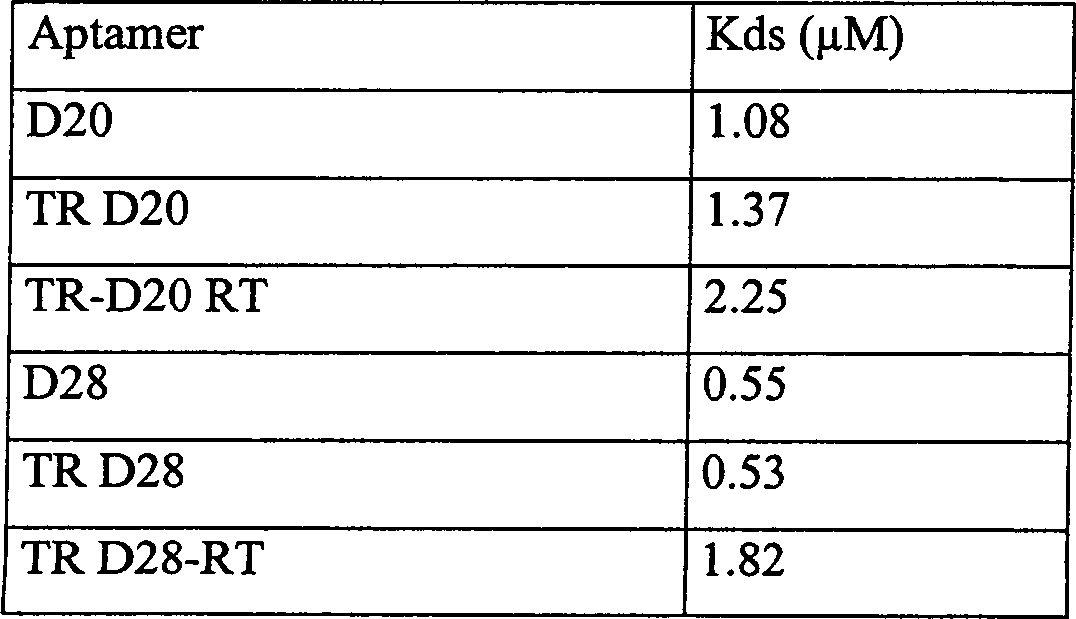
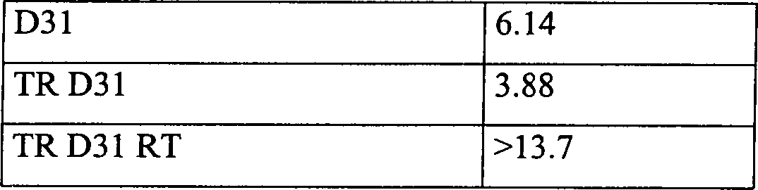
Tabelle 1: Kds (μM) von CD 18cyt-bindenden Aptameren an die cytoplasmatische Domäne von CD18. TR zeigt die richtig terminierten TR-Aptamere im Kontext der TR-Vektor-Expressionskassette an. RT: Abschrift der nicht richtig terminierten TR-Aptamere mit zusätzlichen 166 Nukleotiden.table 1: Kds (μM) of CD18cyt-binding aptamers to the cytoplasmic domain of CD18. TR shows the properly terminated TR aptamers in the context of the TR vector expression cassette at. RT: Transcript of the incorrectly terminated TR aptamers with additional 166 nucleotides.
Claims (14)
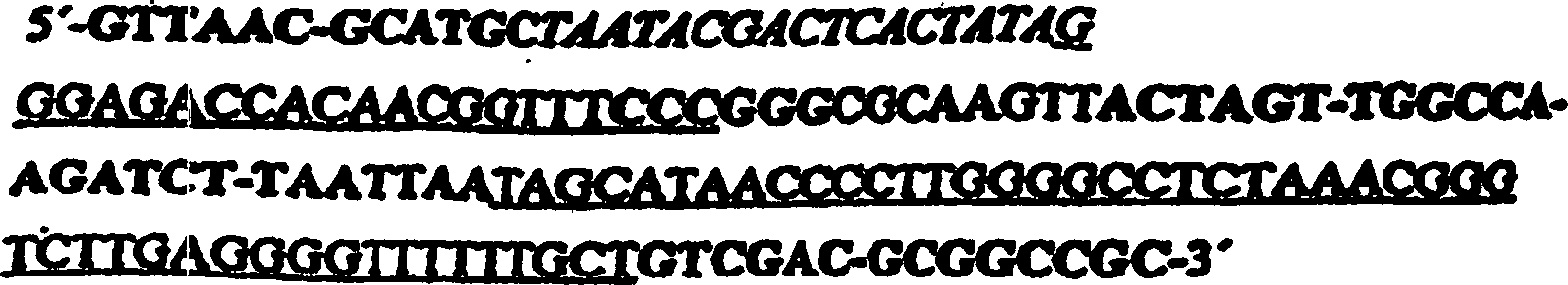


Applications Claiming Priority (1)
| Application Number | Priority Date | Filing Date | Title |
|---|---|---|---|
| EP99106453A EP1041144B1 (en) | 1999-03-29 | 1999-03-29 | Methods for the identification and validation of functional intracellular targets with intramers or in vivo selection |
Publications (2)
| Publication Number | Publication Date |
|---|---|
| DE69918260D1 DE69918260D1 (en) | 2004-07-29 |
| DE69918260T2 true DE69918260T2 (en) | 2005-07-14 |
Family
ID=8237883
Family Applications (1)
| Application Number | Title | Priority Date | Filing Date |
|---|---|---|---|
| DE69918260T Expired - Fee Related DE69918260T2 (en) | 1999-03-29 | 1999-03-29 | Methods for the identification and validation of functional targets by intramers or in vivo selection |
Country Status (8)
| Country | Link |
|---|---|
| US (1) | US20070287153A1 (en) |
| EP (2) | EP1041144B1 (en) |
| JP (1) | JP2002539825A (en) |
| AT (1) | ATE269897T1 (en) |
| AU (1) | AU3558000A (en) |
| CA (1) | CA2365297A1 (en) |
| DE (1) | DE69918260T2 (en) |
| WO (1) | WO2000058455A2 (en) |
Families Citing this family (2)
| Publication number | Priority date | Publication date | Assignee | Title |
|---|---|---|---|---|
| EP1223216A1 (en) * | 2000-12-30 | 2002-07-17 | NascaCell GmbH | Intracellular nucleic acid inhibitors of small guanine-nucleotide-exchange factors |
| JP5804520B2 (en) * | 2010-04-01 | 2015-11-04 | Necソリューションイノベータ株式会社 | Nucleic acid construct, method for producing complex using the same, and screening method |
Family Cites Families (6)
| Publication number | Priority date | Publication date | Assignee | Title |
|---|---|---|---|---|
| US5707796A (en) * | 1990-06-11 | 1998-01-13 | Nexstar Pharmaceuticals, Inc. | Method for selecting nucleic acids on the basis of structure |
| ATE196312T1 (en) * | 1995-06-02 | 2000-09-15 | M & E Biotech A S | METHOD FOR IDENTIFYING BIOLOGICALLY ACTIVE PEPTIDES AND NUCLEIC ACIDS |
| US6187757B1 (en) * | 1995-06-07 | 2001-02-13 | Ariad Pharmaceuticals, Inc. | Regulation of biological events using novel compounds |
| DE69722176T2 (en) * | 1996-01-23 | 2004-03-18 | The Board Of Trustees Of The Leland Stanford Junior University, Palo Alto | METHOD FOR SEARCHING TRANSDOMINANT EFFECTOR PEPTIDES AND RNA MOLECULES |
| WO1997039722A2 (en) * | 1996-04-25 | 1997-10-30 | T Cell Sciences, Inc. | Method of isolating regulators of t cell activation |
| AU5927598A (en) * | 1997-01-23 | 1998-08-18 | Immusol Incorporated | Gene functional analysis and discovery using randomized or target-specific ribozyme gene vector libraries |
-
1999
- 1999-03-29 DE DE69918260T patent/DE69918260T2/en not_active Expired - Fee Related
- 1999-03-29 EP EP99106453A patent/EP1041144B1/en not_active Expired - Lifetime
- 1999-03-29 AT AT99106453T patent/ATE269897T1/en not_active IP Right Cessation
-
2000
- 2000-03-28 JP JP2000608736A patent/JP2002539825A/en active Pending
- 2000-03-28 WO PCT/EP2000/002727 patent/WO2000058455A2/en not_active Ceased
- 2000-03-28 AU AU35580/00A patent/AU3558000A/en not_active Abandoned
- 2000-03-28 CA CA002365297A patent/CA2365297A1/en not_active Abandoned
- 2000-03-28 EP EP00914168A patent/EP1165770A2/en not_active Withdrawn
-
2006
- 2006-05-25 US US11/440,907 patent/US20070287153A1/en not_active Abandoned
Also Published As
| Publication number | Publication date |
|---|---|
| ATE269897T1 (en) | 2004-07-15 |
| WO2000058455A3 (en) | 2001-01-04 |
| WO2000058455A2 (en) | 2000-10-05 |
| AU3558000A (en) | 2000-10-16 |
| DE69918260D1 (en) | 2004-07-29 |
| CA2365297A1 (en) | 2000-10-05 |
| JP2002539825A (en) | 2002-11-26 |
| US20070287153A1 (en) | 2007-12-13 |
| EP1165770A2 (en) | 2002-01-02 |
| EP1041144B1 (en) | 2004-06-23 |
| EP1041144A1 (en) | 2000-10-04 |
Similar Documents
| Publication | Publication Date | Title |
|---|---|---|
| DE69808060T2 (en) | METHOD FOR IDENTIFYING NUCLEIC ACIDS ENCODING AGENTS THAT INFLUENCE CELLULAR PHENOTYPES | |
| DE69925253T2 (en) | QUALITATIVE DIFFERENTIAL SCREENING | |
| DE69535428T2 (en) | Method for finding differentially expressed genes | |
| US6100035A (en) | Method of identifying cis acting nucleic acid elements | |
| WO2019210268A2 (en) | Sequencing-based proteomics | |
| JP2004515219A (en) | Tunable catalytically active nucleic acids | |
| JP2022113773A (en) | High throughput cell-based screening for aptamers | |
| DE19957065B4 (en) | Screening procedure for drugs | |
| EP1115734A1 (en) | Method of measuring protein-protein interactions in living cells | |
| DE60123819T2 (en) | METHOD FOR IN VIVO IDENTIFICATION OF INTRA-CELLULAR EPITOPES | |
| DE69432120T2 (en) | GENETIC SUPPRESSOR ELEMENTS ASSOCIATED WITH CHEMOTHERAPEUTIC COMPOSITIONS | |
| Mirabella et al. | Yeast telomeric sequences function as chromosomal anchorage points in vivo | |
| DE69918260T2 (en) | Methods for the identification and validation of functional targets by intramers or in vivo selection | |
| DE60009530T2 (en) | GENETIC TOXICITY MARKERS, MANUFACTURE AND USE | |
| EP0902092A2 (en) | Method for the identification of target genes for transcription factors | |
| US20230078089A1 (en) | Regulating Activation of Fibroblasts to Prevent Fibrosis | |
| Vaishali | Characterisation of RNA binding proteins and their roles in the Drosophila germline | |
| Sankaranarayanan | The mRNA stability Regulator Khd4 determines infectious hyphae development in Ustilago maydis | |
| WO1999033973A2 (en) | Cloned octapamine-receptor-producing cells | |
| WO2000071749A2 (en) | Detection system for analyzing molecular interactions, production and utilization thereof | |
| DE10018464A1 (en) | In vivo system for testing slice modulators, useful e.g. for diagnosis, comprises cellular system containing spliceable nucleic acid that generates detectable signal when spliced | |
| DE19964386B4 (en) | Screening drugs disrupting macrophage migration inhibitory factor-cJun activation domain binding protein1 interaction involves comparing MIF-Jab1 interaction, in presence and absence of drug in MIF and Jab1 system | |
| DE60127687T2 (en) | ASSAY PROCEDURE FOR DETERMINING THE ALTERNATIVE EXPANSION OF TELOMERS IN HUMAN CELLS | |
| Zagore | The Molecular Function of the RNA Binding Protein DAZL in Male Germ Cell Survival | |
| EP1356095A2 (en) | Method for identifying metastatic tumor cells |
Legal Events
| Date | Code | Title | Description |
|---|---|---|---|
| 8364 | No opposition during term of opposition | ||
| 8339 | Ceased/non-payment of the annual fee |