DE60214029T2 - MILKY ACID BACTERIA AND ITS USES FOR TREATMENT AND PREVENTION AGAINST CANCER - Google Patents
MILKY ACID BACTERIA AND ITS USES FOR TREATMENT AND PREVENTION AGAINST CANCER Download PDFInfo
- Publication number
- DE60214029T2 DE60214029T2 DE2002614029 DE60214029T DE60214029T2 DE 60214029 T2 DE60214029 T2 DE 60214029T2 DE 2002614029 DE2002614029 DE 2002614029 DE 60214029 T DE60214029 T DE 60214029T DE 60214029 T2 DE60214029 T2 DE 60214029T2
- Authority
- DE
- Germany
- Prior art keywords
- apoptosis
- cells
- cancer
- cell
- lactic acid
- Prior art date
- Legal status (The legal status is an assumption and is not a legal conclusion. Google has not performed a legal analysis and makes no representation as to the accuracy of the status listed.)
- Expired - Lifetime
Links
Classifications
-
- A—HUMAN NECESSITIES
- A61—MEDICAL OR VETERINARY SCIENCE; HYGIENE
- A61K—PREPARATIONS FOR MEDICAL, DENTAL OR TOILETRY PURPOSES
- A61K45/00—Medicinal preparations containing active ingredients not provided for in groups A61K31/00 - A61K41/00
- A61K45/06—Mixtures of active ingredients without chemical characterisation, e.g. antiphlogistics and cardiaca
-
- A—HUMAN NECESSITIES
- A61—MEDICAL OR VETERINARY SCIENCE; HYGIENE
- A61K—PREPARATIONS FOR MEDICAL, DENTAL OR TOILETRY PURPOSES
- A61K31/00—Medicinal preparations containing organic active ingredients
-
- A—HUMAN NECESSITIES
- A61—MEDICAL OR VETERINARY SCIENCE; HYGIENE
- A61K—PREPARATIONS FOR MEDICAL, DENTAL OR TOILETRY PURPOSES
- A61K35/00—Medicinal preparations containing materials or reaction products thereof with undetermined constitution
- A61K35/66—Microorganisms or materials therefrom
- A61K35/74—Bacteria
- A61K35/741—Probiotics
- A61K35/744—Lactic acid bacteria, e.g. enterococci, pediococci, lactococci, streptococci or leuconostocs
- A61K35/747—Lactobacilli, e.g. L. acidophilus or L. brevis
-
- A—HUMAN NECESSITIES
- A61—MEDICAL OR VETERINARY SCIENCE; HYGIENE
- A61P—SPECIFIC THERAPEUTIC ACTIVITY OF CHEMICAL COMPOUNDS OR MEDICINAL PREPARATIONS
- A61P35/00—Antineoplastic agents
Landscapes
- Health & Medical Sciences (AREA)
- Life Sciences & Earth Sciences (AREA)
- Chemical & Material Sciences (AREA)
- Medicinal Chemistry (AREA)
- Pharmacology & Pharmacy (AREA)
- Animal Behavior & Ethology (AREA)
- General Health & Medical Sciences (AREA)
- Public Health (AREA)
- Veterinary Medicine (AREA)
- Epidemiology (AREA)
- Microbiology (AREA)
- Mycology (AREA)
- Molecular Biology (AREA)
- Chemical Kinetics & Catalysis (AREA)
- General Chemical & Material Sciences (AREA)
- Nuclear Medicine, Radiotherapy & Molecular Imaging (AREA)
- Organic Chemistry (AREA)
- Medicines That Contain Protein Lipid Enzymes And Other Medicines (AREA)
- Medicines Containing Material From Animals Or Micro-Organisms (AREA)
- Micro-Organisms Or Cultivation Processes Thereof (AREA)
- Pharmaceuticals Containing Other Organic And Inorganic Compounds (AREA)
- Peptides Or Proteins (AREA)
Abstract
Description
Gebiet der ErfindungField of the invention
Die vorliegende Erfindung bezieht sich darauf, den Nutzen des Milchsäurestamms Lactobacillus acidophilus I-1492, der bei der CNCM hinterlegt ist, zur Vorbeugung und Behandlung von Krebs aufzuzeigen. Insbesondere betrifft die vorliegende Erfindung die Verwendung dieser Milchsäurebakterien zur Erleichterung der Induktion der Zellapoptose einer Krebsart.The The present invention relates to the utility of the lactic acid strain Lactobacillus acidophilus I-1492, deposited with the CNCM, for the prevention and treatment of cancer. Especially The present invention relates to the use of these lactic acid bacteria for facilitating the induction of cell apoptosis of a cancer.
Beschreibung des Standes der TechnikDescription of the state of the technique
Die Bakterien Lactobacillus acidophilus I-1492, die im Produkt Bio-K+ vorliegen und Gegenstand der Patentanmeldung WO 98/23727 sind, sind dafür bekannt, dass sie einen vorteilhaften Effekt auf den Blut-Cholesteringehalt bei Säugern ausüben.The Bacteria Lactobacillus acidophilus I-1492, found in the product Bio-K + and are the subject of the patent application WO 98/23727, are known for that it has a beneficial effect on the blood cholesterol content in mammals exercise.
Die Internationale Patentanmeldung WO 98/23727 betrifft ein Milchsäureferment, das einen Stamm von Lactobacillus acidophilus I-1492 umfasst. Auch diese Bakterien werden hauptsächlich für die Herstellung von Milchsäureferment verwendet, um den Blut-Cholesteringehalt bei Säugern zu reduzieren.The International Patent Application WO 98/23727 relates to a lactic acid ferment, which comprises a strain of Lactobacillus acidophilus I-1492. Also These bacteria are mainly for the Production of lactic acid fermentation used to reduce the blood cholesterol content in mammals.
Zudem sind diese Bakterien auch dafür bekannt, dass sie Eigenschaften aufweisen, die eine Verstärkung des Immunsystems bewirken, die Resorption von Nahrungsmitteln erleichtern und die Darmflora anregen. Es ist bekannt, dass die Milchsäurebakterien eine positive Auswirkung auf die Darmflora und auch auf das Immunsystem haben. Tatsächlich ermöglichen die Milchsäurebakterien eine Anregung des Immunsystems, wodurch erreicht wird, dass dem Verdauungssystem eine bessere Abwehr verliehen wird. Diese Bakterien sind auch dafür bekannt, dass sie die Nebenwirkungen neutralisieren, die durch verschiedene Antibiotika verursacht werden. moreover These bacteria are also for that known to have properties that enhance the Immune system, facilitate the absorption of food and stimulate the intestinal flora. It is known that the lactic acid bacteria a positive effect on the intestinal flora and also on the immune system to have. Indeed enable the lactic acid bacteria stimulation of the immune system, which achieves that Digestive system is given a better defense. These bacteria are also for that known to neutralize the side effects caused by various Antibiotics are caused.
In Kanada sterben jedes Jahr zahlreiche Personen an Colonkrebs. Krebs steht an dritter Stelle der Krankheiten, die die größte Anzahl an Todesfällen pro Jahr verursachen.In Every year, many people die of colon cancer in Canada. cancer is the third largest number of diseases at deaths cause each year.
In Kanada betrug im Jahr 2000 die durch Krebs verursachte Sterblichkeitsrate 6500 und es liegen mehr als 17 000 neue Fälle vor.In In 2000, Canada was the cancer mortality rate 6500 and there are more than 17 000 new cases.
Die Verwendung eines pharmazeutisch wirkenden Nahrungsmittels (Nutrazeutikum), einschließlich der Verabreichung von Joghurt und/oder fermentierter Milch, als Ergänzungsbehandlung gegen Krebs ist ebenfalls bekannt.The Use of a pharmaceutically active food (nutraceutical), including the Administration of yoghurt and / or fermented milk, as a supplementary treatment against cancer is also known.
Die beim Menschen bereits als Therapie verwendete Behandlung ist die Ablation der Krebsmasse durch Chirurgie, und anschließend kann eine Bestrahlung der Zone erfolgen, in der sich der Krebs befand, um zu vermeiden, dass Spuren von unerwünschten Zellen zurückbleiben.The in humans already used as therapy treatment is the Ablation of the cancerous mass through surgery, and subsequently can irradiation of the zone in which the cancer was located, to prevent traces of unwanted cells from remaining.
Gleichfalls existiert die Chemotherapie. Diese Behandlung umfasst die Verabreichung von Antikrebsmitteln, wie 5-Fluoruracil (5FU). Diese Verbindung wird mit einem Hilfsmittel kombiniert, um die negativen Auswirkungen der Chemotherapie einzuschränken. 5-Fluoruracil ist ein Medikament, das häufig zur Behandlung von Colonkrebs verwendet wird. Dieses Medikament kann angesichts seiner großen Instabilität in Serum oral oder intraperitoneal (am nächsten zum kanzerogenen Ziel) verabreicht werden. Es ist dafür bekannt, den Tod von Colon-Krebszellen zu verursachen, und zwar auf unterschiedliche Weisen.Likewise there is chemotherapy. This treatment includes administration anticancer agents such as 5-fluorouracil (5FU). This connection is combined with an adjunct to the negative effects restrict chemotherapy. 5-fluorouracil is a drug commonly used to treat colon cancer is used. This drug may be given its great instability in serum oral or intraperitoneal (the next to the carcinogenic target). It is known the death of colon cancer cells in different ways.
Der Tod einer Zelle, der auch als Apoptose bezeichnet wird, kann sich mit Hilfe unterschiedlicher Proteine vollziehen. Z.B. kann sich die Zellapoptose aus der Aktivierung eines Membranrezeptors oder aus der zytoplasmatischen Expression verschiedener Proteine ergeben, die dieses Phänomen begünstigen. Diesbe züglich ist bekannt, dass 5FU die Expression des Proteins p53 sowie die Expression des Membranrezeptors Fas erhöht.Of the Death of a cell, also referred to as apoptosis, can manifest itself using different proteins. For example, can be the cell apoptosis from the activation of a membrane receptor or result from the cytoplasmic expression of various proteins, the this phenomenon favor. Diesbe free It is known that 5FU the expression of the protein p53 and the Expression of the membrane receptor Fas increases.
1. Allgemeines über die Apoptose1. General about the apoptosis
1.1 Definition der Apoptose1.1 Definition of apoptosis
Der programmierte Zelltod, der von unterschiedlichen Zytologen und Biologen mehrere Male entdeckt und wieder entdeckt wurde, hat im Laufe der zwei letzten Jahrhunderte verschiedene Namen erhalten. 1972 wurde schließlich der Begriff Apoptose übernommen und von Currie und seinen Kollegen gewählt, um ein häufiges Modell des programmierten Zelltodes zu beschreiben, den die Autoren wiederholt bei mehreren Geweben und Zelltypen beobachteten. Es wurde beobachtet, dass diese sterbenden Zellen mehrere morphologische Eigenschaften gemeinsam haben, die von den Eigenschaften verschieden sind, die bei Zellen, die von einer Pathologie befallen sind, und bei nekrotisierten Zellen beobachtet wurden, und sie schlugen vor, dass diese gemeinsamen morphologischen Eigenschaften das Ergebnis eines üblichen und konservierten Pragramms des Zelltodes sein könnten.Programmed cell death, which has been discovered and rediscovered several times by different cytologists and biologists, has received various names over the past two centuries. 1972 Finally, the term apoptosis was adopted and chosen by Currie and his colleagues to describe a common model of programmed cell death, which the authors have repeatedly observed in several tissues and cell types. It has been observed that these dying cells share several morphological characteristics that are distinct from the characteristics observed in cells affected by pathology and necrotized cells, and they suggested that these common morphological characteristics were the Result of a common and conserved program of cell death.
1.2 Rolle der Apoptose1.2 Role of apoptosis
Forscher haben gefunden, dass die Zellen unseres Körpers sich selbst töten können, und sie wissen dass der Zellen-"Selbstmord", der "Apoptose" genannt wird, für den Organismus unerlässlich ist. Die Apoptose ist auch für die Physiologie von Zellen und Geweben wesentlich, wie die Zellteilung und -differenzierung. Die Apoptose ist die üblichste Form des physiologischen Zelltodes, der sich zu verschiedenen Zeitpunkten ereignet, z.B. während der embryonalen Entwicklung, bei der Gewebeumorganisation, bei Immunitätsregulationen und der Tumorregression. Somit ist der physiologische Zelltod ein Prozess der spontanen Zelleliminierung, der die Gewährleistung der Zellerneuerung ermöglicht und der in die Aufrechterhaltung der Zell- und Gewebe-Homöostase in entgegengesetzter Weise zur Mitose eingreift. Es ist der angeborene Mechanismus, durch den der Organismus unerwünschte Zellen eliminiert. Jede Zelle trägt in sich den genetischen Mechanismus ihrer eigenen Zerstörung. Die Zellen bleiben nur unter der Bedingung am Leben, dass sie Signale zum Überleben erhalten, die aus ihrer Umgebung stammen. Wenn die Zelle Signale wahrnimmt, die ihr befehlen, Selbstmord zu begehen, dann löst sie das Programm des Sterbens aus. Eine Störung des Gleichgewichts zwischen den Proteinen, die die Zellen am Leben halten, und den Proteinen, die zum Tode der Zellen führen, kann mit einem großen Spektrum an Krankheiten verbunden sein, die Krebs, Nervendegenerierung, Autoimmunkrankheiten, Diabetes und andere Erkrankungen umfassen.researcher have found that the cells of our body can kill themselves, and they know that the cell "suicide" called "apoptosis" is for the organism imperative is. Apoptosis is also for the physiology of cells and tissues, such as cell division and differentiation. Apoptosis is the most common form of physiological Cell death occurring at different times, e.g. while embryonic development, tissue reorganization, immunity regulation, and the tumor regression. Thus, physiological cell death is a process spontaneous cell elimination, ensuring cell renewal allows and in in the maintenance of cell and tissue homeostasis in opposite way to mitosis intervenes. It is the innate Mechanism by which the organism eliminates unwanted cells. each Cell carries in itself the genetic mechanism of their own destruction. The Cells remain alive only on condition that they receive signals to survive, that come from their environment. When the cell perceives signals, If you order her to commit suicide, she will solve the program of dying out. A disturbance the balance between the proteins that keep the cells alive and the proteins that cause cell death with a great Spectrum of diseases linked to cancer, nerve degeneration, Autoimmune diseases, diabetes and other disorders.
2. Morphologische Eigenschaften von Apoptose und Nekrose2. Morphological properties of apoptosis and necrosis
2.1 Apoptose2.1 Apoptosis
Eine der Schlüsseleigenschaften der Apoptose ist die Zellschrumpfung. Während die Zellschrumpfung erfolgt, zieht sich das Zytoplasma zusammen und das Kerngerüst verdichtet sich und bildet Aggregate im Zellkern, die sich dann an den Membrankern anlagern. Die Zellorganellen, darunter das Mitochondrium, scheinen demgegenüber relativ unverändert zu bleiben. Anschließend wird der Zellkern gespalten. Die Bildung und Emission von Ausstülpungen wird auf der Oberfläche der Zeile beobachtet. Die Integrität der Plasmamembran wird jedoch während der ganzes Verfahrens beibehalten, selbst wenn die Permeabilität zunimmt. Während des abschließenden Schritts der Apoptose zerbricht die Zelle in mehrere Vesikel, die eine Vielzahl von intakten Organellen und Kernfragmenten enthalten. Die Fragmente von apoptotischen Zellen werden durch die umgebenden phagozytischen Zellen, wie z.B. Makrophagen, schnell verschlungen. Die Apoptose stellt eine so genannte "saubere" Tötung dar, da die Zellfragmente schnell entfernt werden. Es gibt weder eine inflammatorische Phase, noch eine Schädigung des umgebenden Gewebes, und dies teilweise deswegen, weil dessen Zellmembranen intakt bleiben. Zusammenfassend lässt sich sagen, dass die morphologischen Änderungen, die für die Apoptose charakteristisch sind, die Schrumpfung des Zytoplasmas, die Verdichtung und Fragmentierung der DNA und schließlich die Bildung apoptotischer Körper sind, die die Kernfragmente umfassen, die vom Zytoplasma und der Zellmembran umgeben sind.A the key features Apoptosis is cell shrinkage. While the cell shrinkage occurs, The cytoplasm contracts and the core framework is compressed and forms aggregates in the nucleus, which then attach to the membrane nuclei attach. The cell organelles, including the mitochondrion, seem relatively relative unchanged to stay. Subsequently the nucleus is split. The formation and emission of protuberances will be on the surface watched the line. However, the integrity of the plasma membrane will while maintained throughout the process, even as the permeability increases. While of the final one The apoptosis step breaks the cell into several vesicles contain a variety of intact organelles and nuclear fragments. The fragments of apoptotic cells are surrounded by the surrounding phagocytic cells, e.g. Macrophages, quickly devoured. Apoptosis represents a so-called "clean" killing because the cell fragments are removed quickly. There is neither an inflammatory phase, damage to the surrounding tissue, and this in part because its cell membranes remain intact. In summary, lets say that the morphological changes responsible for apoptosis characteristic, the shrinkage of the cytoplasm, the compression and fragmentation of the DNA and finally the formation of apoptotic body which include the nuclear fragments of the cytoplasm and the Cell membrane are surrounded.
2.2 Nekrose2.2 Necrosis
Die Nekrose betrifft einen plötzlichen Tod, der sich nach extremem physischen oder chemischen Stress ereignet. Sie ist durch unterschiedliche morphologische Kriterien gekennzeichnet. Bei der Nekrose, diesem unkontrollierten Zelltod, liegt ein schneller Verlust der Steuerung des Ionenstroms vor, der das Eindringen von Wasser und eine Erhöhung des Einströmens von Ionen zur Folge hat, wobei sich die Zellen ebenso wie ihre Organellen wie z.B. das Mitochondrium und das endoplasmatische Retikulum aufblähen, bis die Membranen platzen und die DNA im Zellkern unspezifisch gespalten wird. Die Freisetzung des zytoplasmatischen Inhalts an der Außenseite unter Beteiligung anderer Vorgänge ruft am häufigsten Schädigungen in den Geweben hervor, die in der Nähe vorliegen, und löst eine sehr ausgeprägte lokale inflammatorische Reaktion aus.The Necrosis affects a sudden Death that occurs after extreme physical or chemical stress. It is characterized by different morphological criteria. Necrosis, this uncontrolled cell death, is faster Loss of control of the ion current, which prevents the penetration of Water and an increase of inflow of ions, the cells as well as their organelles such as. inflict the mitochondrion and endoplasmic reticulum until the membranes burst and the DNA in the nucleus nonspecifically cleaved becomes. The release of cytoplasmic content on the outside involving other events calls most often damage in the tissues that are in the vicinity, and dissolves one very pronounced local inflammatory reaction.
Tabelle
1: Die prinzipiellen Unterschiede zwischen Nekrose und Apoptose 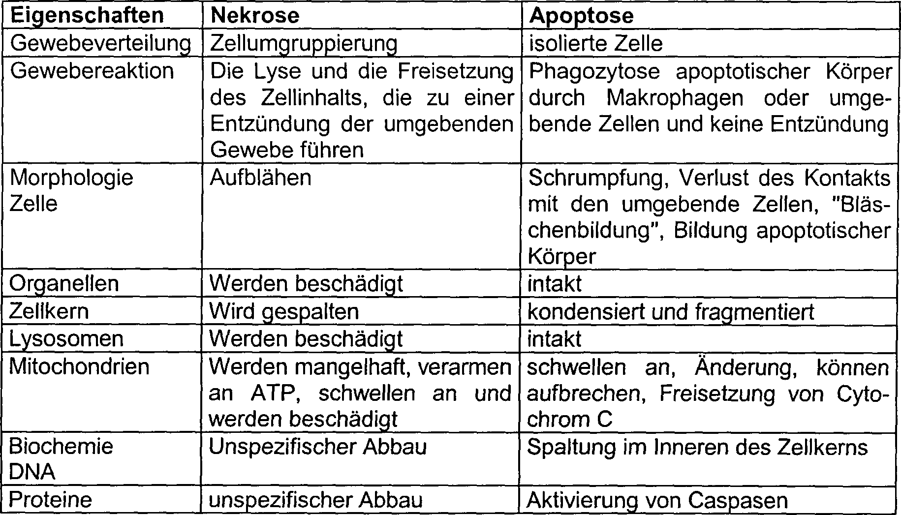
3. Verschiedene Schritte des apoptotischen Prozesses3. Different steps of the apoptotic process
Die
Apoptose kann drei verschiedene Schritte umfassen (
3.1 Schritt des Auslösens der Apoptose3.1 step of triggering the apoptosis
Eine Vielfalt von Stimuli, sowohl innere als auch äußere, können die Zellen aktivieren, um apoptotisch zu werden. Unter den verschiedenen Stimuli kann man biologische Reagenzien (Membranrezeptoren, Transkriptionsfaktoren, Onkoproteine, virale, toxische, bakterielle Infektion usw.), die Unterdrückung der essentiellen Faktoren des Zellwachstums (Cytokine, Wachstums- und Nahrungsfaktoren usw.), genomische Läsionen der DNA (spontan oder provoziert), die Einwirkung chemischer Produkte (Antikrebsmittel), die Einwirkung auslösender physikalischer Mittel (UV-Strahlung, Röntgenstrahlung, Mikrowellen, Wärme usw.) aufführen. Diese Phase erfolgt gemäß zahlreichen biochemischen Modifikationen.A Variety of stimuli, both internal and external, can activate the cells, to become apoptotic. Among the various stimuli one can biological reagents (membrane receptors, transcription factors, Oncoproteins, viral, toxic, bacterial infection, etc.), the suppression the essential factors of cell growth (cytokines, growth factors) and dietary factors, etc.), genomic lesions of the DNA (spontaneous or provoked), the action of chemical products (anti-cancer drugs), the action triggering physical means (UV radiation, X-rays, microwaves, Heat, etc.) list. This phase is done according to numerous biochemical modifications.
3.2 Schritt der Entscheidung oder Ausführung der Apoptose3.2 step of decision or execution of apoptosis
Im Anschluss an diese verschiedenen Reize erhält die Zelle unterschiedliche Signale und entscheidet, ob sie apoptotisch wird oder nicht. Dieser Schritt umfasst unterschiedliche Signaltransduktionswege, u.a. die Aktivierung (oder Inaktivierung) der Serin/Threonin- und Tyrosinkinasen und Phosphatasen, die Synthese des zweiten Messengers, die Modifikation der Genexpression und die Aktivierung der spezialisierten Proteasen, die unter dem Namen Caspasen bekannt sind. Die endgültige Entscheidung, um apoptotisch zu werden, hängt von mehreren Faktoren ab, die das Gleichgewicht zwischen den apoptotischen und anti-apoptotischen Proteinen (die Bcl-2-Proteinfamilie), den metabolischen Zustand der Zellen und auch die Stufe des Zellzyklus, in der sich die Zelle befindet, einschließen. Mehrere Argumente legen es nahe, dass der Ablauf dieses zweiten Schrittes durch die Proteinfamilie Bcl-2 gesteuert wird, die man im Allgemeinen assoziiert mit der äußeren Membran der Mitochondrien, dem endoplasmatischen Retikulum und dem Zellkern antrifft. Die Proteinfamilie Bcl-2 ist in zwei Gruppen von Proteinen aufgeteilt, wobei die einen die Apoptose hemmen (Bcl-2, Bcl-XL, Bcl-w, CED-9 usw.) und die anderen die Apoptose begünstigen (Bax, Bid, Bad, Bak, Bcl-XS usw.). Unabhängig davon, ob sie pro- oder antiapoptotisch sind, haben sie die Fähigkeit, den Ionenstrom zwischen unterschiedlichen Zellkompartimenten zu steuern, insbesondere zwischen den Mitochondrien und dem Zytoplasma. In diesem Schritt gibt es eine Aktivierung der Caspasen in Form des Amplifikationssystems durch Autoaktivierung durch dieselben und zwischen denselben, aber auch durch die Freisetzung von Aktivierungsfaktoren der Apoptose, wie AIF (Apoptose-induzierender Faktor) und des Cytochrom C durch die Mitochondrien. Der Zwischenmembranbereich des Mitochondriums umfasst mehrere Proteine, die an der Aktivierung der Apoptose teilnehmen, wie die Procaspase-2, -3, -7 und -9, AIF und Cytochrom C. Die Aktivierung dieser Caspasen führt zu einem Punkt, an dem es kein Zurück mehr gibt, da sie durch ihre Aktivierung ihre verschiedenen Ziele spalten, u.a. die Proteine, die für das Überleben der Zelle notwendig sind.Following these different stimuli, the cell receives different signals and decides whether or not it will become apoptotic. This step involves different signal transduction pathways, including the activation (or inactivation) of the serine / threonine and tyrosine kinases and phosphatases, the synthesis of the second messenger, the modification of gene expression, and the activation of the specialized proteases known as caspases. The final decision to become apoptotic depends on several factors, including the balance between the apoptotic and anti-apoptotic proteins (the Bcl-2 protein family), the metabolic state of the cells, and also the stage of the cell cycle the cell is enclose. Several arguments suggest that the course of this second step is controlled by the Bcl-2 protein family, which is generally associated with the mitochondrial outer membrane, the endoplasmic reticulum, and the nucleus. The protein family Bcl-2 is divided into two groups of proteins, one inhibiting apoptosis (Bcl-2, Bcl-X L , Bcl-w, CED-9, etc.) and the others favoring apoptosis (Bax, Bid, Bad, Bak, Bcl-X S , etc.). Whether they are pro- or anti-apoptotic, they have the ability to control the ion current between different cell compartments, especially between the mitochondria and the cytoplasm. In this step, there is an activation of the caspases in the form of the amplification system by autoactivation by and between them, but also by the release of activation factors of apoptosis, such as AIF (apoptosis-inducing factor) and cytochrome C by the mitochondria. The Zwischenmem The mitochondrial domain includes several proteins involved in the activation of apoptosis, such as procaspase-2, -3, -7, and -9, AIF, and cytochrome C. The activation of these caspases leads to a point where it can no longer return because they divide their different targets by their activation, including the proteins that are necessary for the survival of the cell.
3.3 Schritt des apoptotischen Abbaus3.3 step of apoptotic degradation
Die Zelle ist jetzt in irreversibler Weise in das Zelltod-Programm verwickelt, das aus der Präsentation von morphologischen Eigenschaften der apoptotischen Kennzeichnung besteht. Demzufolge werden durch Aktivierung verschiedener Caspasen mehrere Proteine, die für das Überleben der Zelle notwendig sind, gespalten und werden nicht-funktionell, wie z.B. die Polymerase Poly(DNA-Ribose). Andere Ziele von Caspasen können durch dieselben aktiviert werden, u.a. DNasen, die von sich aus das Chromatin in Fragmente hoher Molmassen schneiden.The Cell is now irreversibly involved in the cell death program, that from the presentation of morphological characteristics of apoptotic labeling. As a result, several caspases are activated by activating different caspases Proteins for the survival the cell are necessary, split and become non-functional, such as. the polymerase poly (DNA ribose). Other goals of Caspasen can activated by them, i.a. DNases, by themselves Chromatin cut into fragments of high molecular weight.
4. Proteine, die an den Mechanismen der Apoptoseregulation beteiligt sind4. Proteins attached to the Mechanisms of apoptosis regulation are involved
4.1.1 Die Proteinfamilie Bcl-24.1.1 The protein family Bcl-2
Das Protein Bcl-2 und diejenigen der gleichen Familie sind wichtige Modulatoren der Apoptose. In dieser Familie von Proteinen existieren zwei Klassen, die anti-apoptotische Proteine (Bcl-2, Bcl-XL, Mcl-1, Bcl-w, Bfl-1/A1, Brag-1) und pro-apoptotische Proteine (Bax, Bak, Bad, Bid, Bim, Bcl-XS) (Reed 1996) einschließen. Die Vertreter der Familie Bcl-2 sind gemäß ihrer Anzahl der Domänen-Homologie mit Bcl-2 (BH: Bcl-2-Homologie) klassifiziert. Das Protein Bcl-2 enthält 4 Domänen. Alle anti-apoptotischen Proteine besitzen die vier Domänen, während die pro-apoptotischen Proteine in drei Kategorien eingeteilt werden können. Eine Gruppe enthält die Domänen BH1, BH2 und BH3 (Bax, Bak), während die andere Gruppe nur die Domäne BH3 (Bad, Bid, Bik) enthält. Bcl-XS bildet seine eigene Gruppe, die die Domänen BH3 und BH4 enthält. Eine kristallographische Untersuchung hat die Bestimmung der Struktur des Proteins Bcl-XL ermöglicht. Das Protein wird demnach aus zwei zentralen α-Helices gebildet, die von fünf "amphipathischen" α-Helices umgeben sind. Es ist interessant festzustellen, dass die dreidimensionale Struktur Bakterientoxinen homolog ist, die Poren in den Membranen bilden, wie das Diphtherietoxin und Colicin, wodurch auf einen möglichen Wirkungsmechanismus für diese Proteine im Bereich des Mitochondriums geschlossen werden könnte. Eine andere Struktureigenschaft wäre die Fähigkeit dieser Proteine, eine Homo- oder Heterodimerisation miteinander durch ihre Domäne BH3 durchzuführen; auf diese Weise könnten sie ihre Funktionen untereinander begünstigen oder denselben entgegenwirken.The protein Bcl-2 and those of the same family are important modulators of apoptosis. In this family of proteins there are two classes of anti-apoptotic proteins (Bcl-2, Bcl-X L , Mcl-1, Bcl-w, Bfl-1 / A1, Brag-1) and pro-apoptotic proteins (Bax, Bak, Bad, Bid, Bim, Bcl-X S ) (Reed 1996). The members of the family Bcl-2 are classified according to their number of domain homology with Bcl-2 (BH: Bcl-2 homology). The protein Bcl-2 contains 4 domains. All anti-apoptotic proteins have the four domains, while the pro-apoptotic proteins can be grouped into three categories. One group contains the domains BH1, BH2 and BH3 (Bax, Bak), while the other group contains only the domain BH3 (Bad, Bid, Bik). Bcl-X S forms its own group containing the domains BH3 and BH4. A crystallographic study has enabled the determination of the structure of the Bcl-X L protein. The protein is thus formed from two central α helices surrounded by five "amphipathic" α helices. It is interesting to note that the three-dimensional structure is homologous to bacterial toxins that form pores in the membranes, such as diphtheria toxin and colicin, suggesting a possible mode of action for these proteins in the mitochondrial region. Another structural feature would be the ability of these proteins to homo- or heterodimerize with each other through their domain BH3; In this way they could favor or counteract their functions.
4.1.2. Anti- und pro-apoptotische Faktoren4.1.2. Anti-and pro-apoptotic factors
A. Über das MitochondriumA. About the mitochondrion
Zunächst eine Beschreibung der Modifikationen, die das Mitochondrium in der Apoptose-Situation erleidet. Die Mitochondrien, die in einer Apoptose-Situation isoliert sind, erleiden das, was als mitochondrialer Permeabilitätsübergang (MPT) bezeichnet wird. Experimentell ist der MPT durch eine sehr schnelle Erhöhung der Permeabilität der Innenmembran für Teilchen mit Molmassen von < 1500 Da gekennzeichnet. Dieser Permeabilitätsübergang hat mehrere Konsequenzen, die das Ablaufen von ΔΨm, osmotisches Anschwellen, Freisetzung des Ca2+ der Matrix, die Bildung von sauerstoffreaktiven Spezies und das Aufbrechen der äußeren Membran des Mitochondriums einschließen, was zu einer Freisetzung von Cytochrom C des Zwischenmembranbereichs des Mitochondriums führt. Eine biochemische Eigenschaft des Mitochondriums hat es ermöglicht, die Poren zu identifizieren, die auf eine elektrische Spannung und Ca2+ reagieren, das die MPT steuert, welche als PT-Poren bezeichnet werden. Die Poren sind an der Verbindungsstelle der inneren und äußeren Membranen des Mitochondriums lokalisiert, und somit erlaubt die Öffnung der Poren die direkte Kommunikation zwischen der Matrize des Mitochondriums und seiner Umgebung. Der "spannungsabhängige Anionenkanal" (VDAC) und der "Adeninnucleotid-Translokator" (ANT) machen einen Teil der PT-Pore aus.First, a description of the modifications that the mitochondrion undergoes in the apoptosis situation. The mitochondria isolated in an apoptotic situation suffer what is called mitochondrial permeability transition (MPT). Experimentally, the MPT is characterized by a very rapid increase in the permeability of the inner membrane for particles with molecular weights of <1500 Da. This permeability transition has several consequences, including the draining of ΔΨ m, osmotic swelling, release of Ca 2+ of the matrix, the formation of oxygen-reactive species and the disruption of the outer membrane of the mitochondrion, which leads to a release of cytochrome C of the intermediate membrane region of the mitochondrion leads. A biochemical property of the mitochondrion has made it possible to identify the pores that react to an electrical voltage and Ca 2+ , which controls the MPT, which are called PT pores. The pores are located at the junction of the inner and outer membranes of the mitochondrion, and thus, the opening of the pores allows direct communication between the matrix of the mitochondrion and its surroundings. The "voltage dependent anion channel" (VDAC) and the "adenine nucleotide translocator" (ANT) constitute part of the PT pore.
Die Proteine der Familie Bcl-2 haben unterschiedliche zytoplasmatische Verteilungen. Die Proteine Bci-2 und Bcl-XL besitzen einen hydrophoben Schwanz am C-terminalen Ende, der eine Insertionssequenz der Membran enthält, und es ist bekannt, dass der Hauptteil dieser Proteine mit den Membranen der Mitochondrien, dem endoplasmatischen Retikulum und der Kernmembran assoziiert ist. In ihrer inaktiven Form weisen die pro-apoptotischen Proteine Bad, Bax und Bid einen Primärort im Zytoplasma auf. Demgegenüber befinden sie sich bei ihrer Aktivierung wieder auf den Mitochondrien. Wenn Bid gespalten wird, befindet sich sein terminaler COOH-Teil wieder auf der Oberfläche des Mitochondriums. Die Wirkung dieser Proteine, sei es um der Apoptose vorzubeugen oder die Apoptose zu initiieren, befindet sich in den Mitochondrien, demgegenüber bleibt der Mechanismus dieser Wirkungen kontrovers und unbestimmt. Es wurde gezeigt, dass beim Hinzufügen des Proteins Bax – ein pro-apoptotisches Protein – zu isolierten Mitochondrien eine Induktion der Freisetzung von Cytochrom C erfolgt, während die Überexpression der Proteine Bcl-2 oder Bcl-XL die Freisetzung von Cytochrom C verhütet und somit die Apoptose blockiert wird. Es ist klar, dass die Familie Bcl-3 in direkter Weise an die Freisetzung von Cytochrom C, dem Elektronen-Transportprotein im Inneren des Mitochondriums, beteiligt ist. Indem es darüber hinaus an der oxidativen Phosphorylierung im Mitochondrium beteiligt ist, ist Cytochrom C einer der Bestandteile (wie das Adaptorprotein Apaf-1), die für die Aktivierung von Caspase-9 im Zytosol erforderlich sind. Zur Art und Weise, wie die Membranen der Familie Bcl-2 die Freisetzung von Cytochrom C regulieren, sind mehrere Hypothesen aufgestellt wurden, aber keine hat sich als endgültig erwiesen. Es gibt drei Basismodelle, die vorgeschlagen werden können.
- 1. Die Vertreter der Familie Bcl-2 bilden einen Kanal, der den Transport von Proteinen erleichtert. Basierend auf der Ähnlichkeit der Struktur von Bcl-XL mit der Untereinheit des Diphtherietoxins, das Poren bildet, wurde angeregt, dass die Proteine Bcl-2 sich in die äußere Membran des Mitochondriums einfügen könnten, wo sie einen Kanal oder sogar ein großes Loch bilden könnten. Die Vertreter der Familie Bcl-2 können sich tatsächlich in eine synthetische Lipid-Doppelschicht einfügen, oligomerisieren und einen Kanal mit einer diskreten Leitfähigkeit bilden. Die Proteine Bid und Bik können das Mitochondrium direkt induzieren, um Cytochrom C freizusetzen, ohne mit VDAC oder ANT in Wechselwirkung zu treten, was darauf schließen lässt, dass sie außerhalb der PT-Poren agieren.
- 2. Die Vertreter der Familie Bcl-2 treten mit anderen Proteinen in Wechselwirkung, um Kanäle zu bilden. Die Vertreter der Familie Bcl-2 interagieren mit mehreren Proteinen. Eine Möglichkeit wäre, dass die Vertreter der Familie des apoptotischen Proteins andere Proteine der äußeren Membran des Mitochondriums rekrutieren, um eine Pore zu bilden, die ausreichend groß ist, um einen Kanal zu erzeugen. Ein besonders interessanter Kandidat für ein solches Protein wäre der spannungsabhängige Anionenkanal (VDAC), mehrere Vertreter der Familie Bcl-2 können sich mit ihm verbinden und seine Kanalaktivität regulieren. Da die Größe der charakteristischen Pore des VDAC-Kanals zu gering ist, um Proteine durchzulassen, soll gemäß diesem Modell angenommen werden, dass der VDAC eine Konformationsänderung nach der Bindung von Vertretern der Familie Bcl-2 erleidet. Es wurde gezeigt, dass die Proteine Bcl-2 und Bcl-XL das Schließen der PT-Poren begünstigen, während das pro-apoptotische Protein Bax den entgegengesetzten Effekt hat, es tritt mit ANT und VDAC in Wechselwirkung, um die Öffnung dieser Poren und die Freisetzung von Cytochrom C zu begünstigen.
- 3. Die Vertreter der Familie Bcl-2 induzieren ein Aufbrechen der äußeren Membran des Mitochondriums. Es ist möglich, dass die Familie Bcl-2 die Homöostase des Mitochondriums steuert. In diesem Modell würde das apoptotische Signal die Physiologie des Mitochondriums verändern (z.B. Ionenaustausch oder oxidative Phosphorylierung), so dass sich die Organelle aufbläht, was ein physi sches Aufbrechen der äußeren Membran und eine Freisetzung von Proteinen ergibt, die sich zwischen den Membranen des Mitochondriums, im Zytosol, befinden. Die Notwendigkeit zur Bildung eines ausreichend großen Kanals, um das Cytochrom C passieren zu lassen, ist jetzt noch dringender erforderlich, da die Proteine einfach durch Risse in der Lipid-Doppelschicht diffundieren würden.
- 1. The representatives of the Bcl-2 family form a channel that facilitates the transport of proteins. Based on the similarity of the structure of Bcl-X L with the subunit of the diphtheria toxin that forms pores, it was suggested that the proteins Bcl-2 could fit into the outer membrane of the mitochondrion where they form a channel or even a large hole could. The members of the Bcl-2 family can actually insert into a synthetic lipid bilayer, oligomerize, and form a channel with a discrete conductivity. The Bid and Bik proteins can directly induce mitochondrion to release cytochrome C without interfering with VDAC or ANT, suggesting that they act outside the PT pores.
- 2. The members of the Bcl-2 family interact with other proteins to form channels. Representatives of the Bcl-2 family interact with several proteins. One possibility would be that members of the apoptotic protein family recruit other mitochondrial outer membrane proteins to form a pore that is sufficiently large to create a channel. A particularly interesting candidate for such a protein would be the voltage-gated anion channel (VDAC), several members of the family Bcl-2 can connect to it and regulate its channel activity. Since the size of the characteristic pore of the VDAC channel is too small to pass proteins through, it should be assumed according to this model that the VDAC suffers a conformational change after binding of members of the family Bcl-2. It has been shown that the proteins Bcl-2 and Bcl-X L favor the closure of PT pores, while the pro-apoptotic protein Bax has the opposite effect, it interacts with ANT and VDAC to open these pores and promote the release of cytochrome C.
- 3. The members of the family Bcl-2 induce a rupture of the outer membrane of the mitochondrion. It is possible that the Bcl-2 family controls mitochondrial homeostasis. In this model, the apoptotic signal would alter the physiology of the mitochondrion (eg, ion exchange or oxidative phosphorylation), causing the organelle to swell, resulting in physiological disruption of the outer membrane and release of proteins extending between the mitochondrial membranes, in the cytosol. The need to form a large enough channel to pass cytochrome C is now even more urgent, as the proteins would simply diffuse through cracks in the lipid bilayer.
Die pro-apoptotischen Proteine (Bid) können unter Bildung einer Pore homodimerisieren, um das Cytochrom C austreten zu lassen. Die anti-apoptotischen Proteine (Bcl-2) haben die Fähigkeit, sich mit den PT-Poren zu verbinden und somit die Freisetzung von Intermembran-Proteinen zu verhindern, wohingegen die pro-apoptotischen Proteine (Bax) die Öffnung der PT-Poren erlauben.The Pro-apoptotic proteins (Bid) may form a pore homodimerize to allow cytochrome C to escape. The anti-apoptotic Proteins (Bcl-2) have the ability to to connect with the PT pores and thus the release of In contrast, the pro-apoptotic proteins prevent intermembrane proteins Proteins (Bax) the opening allow the PT pores.
Das AIF-Protein (für Apoptose-induzierender Faktor), das identifiziert wurde und dessen Gen kloniert wurde, kann von sich aus die Apoptose in isolierten Zellkernen induzieren. Dieses Molekül wird im Zytosol in Form eines Vorläufers synthetisiert, dann wird es in das Mitochondrium eingeführt. Wie beim Cytochrom C handelt es sich um ein phylogenetisch altes Molekül mit einer doppelten Funktion: Oxidation-Reduktion und apoptotischer Faktor. Jedoch im Gegensatz zum Weg des Cytochroms C, der die Aktivierung anderer Faktoren benötigt, um die Apoptose zu induzieren, ist der Weg von AIF umgekehrt von Caspasen unabhängig und benötigt keine Zwischenstufe, um die Apoptose auszulösen. Er würde u.a. einen Prototyp von Apoptose-Wegen darstellen, die von Caspasen unabhängig sind.The AIF protein (for Apoptosis-inducing factor) that has been identified and its Gene has been cloned by itself, which can isolate apoptosis in isolation Induce cell nuclei. This molecule is in the cytosol in the form of a precursor synthesized, then it is introduced into the mitochondrion. As Cytochrome C is a phylogenetically old molecule with a dual function: oxidation-reduction and apoptotic factor. However, unlike the pathway of cytochrome C, which activates other factors needed In order to induce apoptosis, the path of AIF is reversed by Caspases independently and needed no intermediate to trigger apoptosis. He would u.a. a prototype of Represent apoptosis pathways that are independent of caspases.
Die Hypothesen in Bezug auf die Mechanismen der Apoptose-Hemmung und insbesondere der Maskierung von Apaf-1 durch Bcl-2 und seiner anti-apoptotischen Agonisten scheinen noch ausführlich diskutiert zu werden. Apaf-1 ist wahrscheinlich ein wichtiges Ziel der Vertreter der Bcl-2-Familie, da die Apaf-1-defizienten Zellen gegenüber verschiedenen pro-apoptotischen Signalen unempfindlich sind und von sich aus durch Bcl-2 inhibiert werden. Zudem hat sich bei Überexpression von Apaf-1 gezeigt, dass dieses Protein mit den Überlebensproteinen wie Bcl-XL und Bcl-2 assoziiert war. Demgegenüber wurde gezeigt, dass keine Coimmunpräzipitation zwischen den Membranen der Familie Bcl-2 und Apaf-1 existiert. Gleichfalls wurde Apaf-1 an Orten gefunden, wo die Überlebensproteine wie Bcl-2 und Bcl-XL vorliegen, etwa in den äußeren Membranen des Mitochondriums, in der Kernhülle und im endoplasmatischen Retikulum.The hypotheses regarding the mechanisms of apoptosis inhibition and in particular the masking of Apaf-1 by Bcl-2 and its anti-apoptotic agonists appear to be discussed in detail. Apaf-1 is likely to be an important target for members of the Bcl-2 family because the Apaf-1 deficient cells are insensitive to various pro-apoptotic signals and inherently inhibited by Bcl-2. In addition, overexpression of Apaf-1 has been shown to associate this protein with survival proteins such as Bcl-X L and Bcl-2. In contrast, it was shown that no Coim munpräzipitation between the membranes of the family Bcl-2 and Apaf-1 exists. Similarly, Apaf-1 was found in locations where the survival proteins such as Bcl-2 and Bcl-X L are present, such as in the outer mitochondrial membranes, in the nuclear envelope, and in the endoplasmic reticulum.
4.1.3 Mechanismus der Modulation von Proteinen der Familie Bcl-24.1.3 Mechanism of Modulation of proteins of the Bcl-2 family
Es existieren mehrere unterschiedliche Mechanismen, um die Funktionen von pro- und anti-apoptotischen Proteinen zu modulieren. Zunächst beeinflusst der Dimerisierungszustand der Vertreter der Familie Bcl-2 ihre Aktivität. Eine der Funktionen der anti-apoptotischen Proteine Bcl-2 und Bcl-XL besteht darin, mit dem pro-apoptotischen Protein Bax zu dimerisieren, um dessen Aktivität zu neutralisieren. Wenn es ein Heterodimer ist, ist Bax inaktiv; nachdem es aber frei ist, um mit sich selbst zu dimerisieren, kann Bax die Apoptose induzieren. Bid, Bik und Bad können dahingehend wirken, dass sie die anti-apoptotische Wirkung von Bcl-2 und Bcl-XL unter Bildung von Heterodimeren hemmen. Zweitens kann durch Änderung des Expressionsstärke der pro- und anti-apoptotischen Vertreter der Familie Bcl-2 die Apoptose initiiert oder inhibiert werden. Wenn z.B. die Anzahl von Bcl-2 größer ist als die von Bax oder derselben gleich ist, werden die in Frage kommenden Zellen vor Apoptose geschützt. Wenn demgegenüber die Anzahl von Bax diejenige von Bcl-2 übersteigt, ist die Zelle stärker dafür anfällig, apoptotisch zu werden. Drittens können die Proteine der Familie Bcl-2 durch Phosphorylierung modifiziert werden. Das beste Beispiel für diesen Vorschlag wäre das pro-apoptotische Protein Bad. In seinem nicht phosphorylierten Zustand dimerisiert es mit Bcl-2 und Bcl-XL, wodurch deren anti-apoptotische Aktivität neutralisiert wird. Ist demgegenüber Bad phosphoryliert, so ist es maskiert und kann deswegen nicht mit Bcl-2 und Bcl-XL in Wechselwirkung treten und dieselben neutralisieren. Viertens kann die Familie Bcl-2 durch Spaltung modifi ziert werden. Bei einer durch Fas verursachten Apoptose wurde gezeigt, dass die Caspasen Bcl-2 und Bcl-XL spalten würden und die Spaltprodukte keine Schutzmittel mehr sind und selbst pro-apoptotisch werden. Bid ist ein anderes Protein der Familie Bcl-2, das durch die Spaltung von Caspasen aktiviert wird. Während das Protein in seiner vollen Länge inaktiv ist, induziert Bid als Folge der durch die Caspase-8 verursachten Spaltung die Freisetzung von Cytochrom C durch das Mitochondrium. Schließlich modifiziert die Konformation der Proteine Bcl-2 ihre Aktivität. Der beste Beweis für diesen Mechanismus stammt aus Untersuchungen, die über Bax durchgeführt wurden. In seinem inaktiven Zustand existiert Bax in einer Konformation, in der es proteolytischen Spaltungen widersteht. Demgegenüber wird nach seiner Aktivierung und seiner erneuten Lokalisierung auf dem Mitochondrium der N-terminale Bereich des Proteins für Spaltungen anfällig, was darauf schließen lässt, dass sich wohl eine Änderung der Konformation ergeben hat.Several different mechanisms exist to modulate the functions of pro- and anti-apoptotic proteins. First, the dimerization state of the Bcl-2 family members affects their activity. One of the functions of the anti-apoptotic proteins Bcl-2 and Bcl-X L is to dimerize with the pro-apoptotic protein Bax to neutralize its activity. If it is a heterodimer, Bax is inactive; however, once it is free to dimerize with itself, Bax can induce apoptosis. Bid, Bik and Bad may act to inhibit the anti-apoptotic effects of Bcl-2 and Bcl-X L to form heterodimers. Second, by altering the level of expression of the pro- and anti-apoptotic members of the Bcl-2 family, apoptosis can be initiated or inhibited. For example, if the number of Bcl-2 is greater than or equal to Bax, the cells of interest are protected from apoptosis. In contrast, when the number of Bax exceeds that of Bcl-2, the cell is more susceptible to becoming apoptotic. Third, the proteins of the Bcl-2 family can be modified by phosphorylation. The best example of this proposal would be the pro-apoptotic protein bath. In its non-phosphorylated state, it dimerizes with Bcl-2 and Bcl-X L , thereby neutralizing their anti-apoptotic activity. On the other hand, if Bad is phosphorylated, it is masked and therefore can not interact with Bcl-2 and Bcl-X L and neutralize them. Fourth, the family Bcl-2 can be modified by cleavage. In the case of apoptosis caused by Fas, it was shown that the caspases cleave Bcl-2 and Bcl-X L and that the cleavage products are no longer protective agents and themselves become pro-apoptotic. Bid is another protein of the family Bcl-2, which is activated by the cleavage of caspases. While the protein is inactive in its full length, Bid induces the release of cytochrome C by the mitochondrion as a result of cleavage caused by caspase-8. Finally, the conformation of the proteins Bcl-2 modifies their activity. The best evidence for this mechanism comes from studies conducted on Bax. In its inactive state, Bax exists in a conformation in which it resists proteolytic cleavage. In contrast, upon its activation and its re-localization on the mitochondrion, the N-terminal region of the protein becomes susceptible to cleavage, suggesting that a conformational change may have occurred.
Zusammenfassend lässt sich sagen, dass die Mitochondrien eine wichtige Rolle beim Auslösen der Apoptose annehmen. Ihr Zwischenmembran-Bereich enthält mehrere Proteine (Cytochrom C, Caspase-2, -3, -7 und -9, AIF), die, nachdem sie im Zytoplasma freigesetzt sind, an der Abbauphase der Apoptose teilnehmen. Das Rätsel der Mechanismen der Induktion und Steuerung der Apoptose durch das Mitochondrium beruht auf vier wesentlichen Punkten: nämlich den Molekülen der Familie Bcl-2/Bcl-XL, die zur Bildung von Ionenkanälen an den intrazellulären Membranen beitragen könnten. Die pro-apoptotischen Vertreter dieser Familie (wie Bax, Bid usw.) könnten auch in die Permeabilität der PT-Poren der mitochondrialen Membran eingreifen, insbesondere als aktivierende Proteine der Apoptose. Schließlich könnten auch die anti-apoptotischen Moleküle der Familie Bcl-2 agieren, indem sie die endogenen Aktivatoren (wie Apaf-1) der Apoptose titern. Ein letzter Punkt ist derjenige, dass bestimmte Procaspasen auch eine mitochondriale Lokalisation aufweisen. Somit regulieren die apoptotischen Promotoren oder die Inhibitoren der Familie Bcl-2 die Apoptose, und zwar aufgrund mehrfacher Einwirkungen auf die Aktivierungskaskaden von Caspasen, auf das Redoxpotential und auf die Sperrfunktion der Permeabilität mitochondrialer Membranen. Insgesamt weisen diese Beobachtungen somit auf eine Einbeziehung des Permeabilitätsübergangs in die Regulierung der durch Mitochondrien induzierten Apoptose hin.In summary, mitochondria play an important role in inducing apoptosis. Its inter-membrane region contains several proteins (cytochrome C, caspase-2, -3, -7 and -9, AIF), which, after being released in the cytoplasm, participate in the degradation phase of apoptosis. The mystery of the mechanisms of induction and control of mitochondrial apoptosis is based on four key points: the Bcl-2 / Bcl-X L families, which could contribute to the formation of ion channels on the intracellular membranes. The pro-apoptotic members of this family (such as Bax, Bid, etc.) could also interfere with the permeability of the PT pores of the mitochondrial membrane, particularly as activating apoptosis proteins. Finally, the anti-apoptotic molecules of the Bcl-2 family could also act by tapping the endogenous activators (such as Apaf-1) of apoptosis. One last point is that certain procaspases also have mitochondrial localization. Thus, the apoptotic promoters or inhibitors of the Bcl-2 family regulate apoptosis due to multiple actions on caspase activation cascades, redox potential, and mitochondrial membrane permeability barrier function. Overall, these observations thus suggest that the permeability transition is involved in the regulation of mitochondrial-induced apoptosis.
4.2 Rolle der Caspasen bei der Apoptose4.2 Role of caspases in apoptosis
4.2.1 Definition und Klassifizierung der Caspasen4.2.1 Definition and classification the caspases
Die Caspasen sind spezialisierte Proteasen, die für die Apoptose wesentlich sind. Sie sind von anderen Proteasen verschieden, weil sie ein Cystein für die Katalyse verwenden und nur nach den Asparaginsäureresten spalten. Diese ungewöhnliche Spezifität, ein Aspartat als Substrat aufzuweisen, wird nur bei einer anderen Protease, dem Granzym B, gefunden; dieses Enzym verwendet jedoch ein Serin als aktive Stelle. Die Caspasen werden wie eine einfache Polypeptidkette synthetisiert und sind inaktive Zymogene. Diese Zymogene bestehen aus drei Domänen: einer N-terminalen Pro-Domäne und zwei anderen Domänen, p10 und p20, die wieder im reifen Enzym gefunden werden. Bei ihrer Aktivierung wird jede Polypeptidkette in zwei Untereinheiten gespalten, eine große (p20) und eine kleine (p10), die in der Folge dimerisieren. Folglich sind die reifen Enzyme, die beobachtet wurden, Heterotetramere, die aus zwei Heterodimeren p20/p10 und zwei aktiven Stellen bestehen. Das N-terminale Peptid wird gespalten und bei der Aktivierung freigesetzt. Dieses N-terminale Peptid ist nicht für die enzymatische Aktivität erforderlich, seine Rolle für die Caspasen 8 und 10 ist bekannt, wo es als Wechselwirkungsdomäne mit anderen Proteinen agiert, um deren Aktivierung zu modulieren. Die Caspasen 8 und 10 enthalten eine "Todeffektor-Domäne" (DED), während die Caspasen 2 und 9 eine "Caspasen-Aktivierungs- und Rekrutierungsdomäne" (CARD) enthalten.The caspases are specialized proteases that are essential for apoptosis. They are different from other proteases because they use a cysteine for catalysis and only cleave after the aspartic acid residues. This unusual specificity of having one aspartate as substrate is found only in another protease, granzyme B; however, this enzyme uses a serine as the active site. The caspases are synthesized like a simple polypeptide chain and are inactive zymogens. These zymogens consist of three domains: one N-terminal pro domain and two other domains, p10 and p20, which are again found in the mature enzyme. Upon activation, each polypeptide chain is cleaved into two subunits, one large (p20) and one small (p10), which subsequently dimerize. Thus, the mature enzymes observed are heterotetramers consisting of two p20 / p10 heterodimers and two active sites exist. The N-terminal peptide is cleaved and released upon activation. This N-terminal peptide is not required for enzymatic activity, its role for caspases 8 and 10 is known where it acts as an interaction domain with other proteins to modulate their activation. Caspases 8 and 10 contain a "death effector domain" (DED), while caspases 2 and 9 contain a "caspase activation and recruitment domain" (CARD).
Es gibt wenigstens 14 unterschiedliche Caspasen, die bisher in den Geweben von Säugern identifiziert wurden. Es ist möglich, die Caspasen in drei Gruppen einzuteilen, die sich in ihrer Substratspezifität unterscheiden, d.h. durch ihr Erkennen von drei Aminosäuren, die der Asparaginsäure vorangehen. Die erste Gruppe enthält Caspasen, die am Entzündungsprozess beteiligt sind, also an der Aktivierung von Procytokinen, und umfasst die Caspasen 1, 4 und 5. Diese Enzyme sind auch als "ICE-ähnliche" Caspasen bekannt, weil ein anderer Name für Caspase-1 "Interleukin-1-umwandelndes Enzym" (ICE) ist. Ihr Tetrapeptid-Muster, das sie erkennen und bevorzugen, ist WEHD, demgegenüber sind die "ICE-ähnlichen" Caspasen in Bezug auf die Aminosäure-Substitution die tolerantesten, verglichen mit den Signal- und Effektor-Caspasen. Die zweite Gruppe von Caspasen enthält die Caspasen 6, 8, 9 und 10. Diese Enzyme werden als Signal-Caspasen angesehen, weil sie andere Caspasen aktivieren können und somit die Kaskade beginnen können. Ihr Erkennungsmuster ist (LV)EXD. Diese letzte Gruppe enthält die Caspasen 2, 3 und 7. Diese Enzyme sind unter dem Namen Effektoren bekannt, weit sie mehrere zelluläre Ziele spalten, was als Ergebnis den morphologischen Eindruck der Apoptose ergibt. Die Aktivierung dieser Caspasen führt im Allgemeinen zu einem Punkt beim Zelltod, an dem es kein Zurück mehr gibt. Die Effektor-Enzyme sind die spezifischsten Enzyme, wobei es notwendig ist, dass sie eine Asparaginsäure an der ersten und vierten Position haben müssen, die der Spaltungsstelle vorangehen. Ihr Erkennungsmuster ist DEXD. Die neuesten Caspasen, Caspasen 12–14, sind noch nicht ausreichend charakterisiert worden, als dass sie in eine der drei Gruppen eingeteilt werden könnten.It There are at least 14 different caspases that have been in the Tissues of mammals were identified. It is possible, classify caspases into three groups that differ in their substrate specificity, i.e. by recognizing three amino acids that precede aspartic acid. The first group contains Caspases involved in the inflammatory process involved in the activation of Procytokinen, and includes caspases 1, 4 and 5. These enzymes are also known as "ICE-like" caspases, because another name for Caspase-1 "interleukin-1 converting Enzyme "(ICE) is. Your tetrapeptide pattern, that they recognize and prefer is WEHD, in contrast the "ICE-like" caspases in terms on the amino acid substitution the most tolerant, compared to the signal and effector caspases. The second group of caspases contains caspases 6, 8, 9 and 10. These enzymes are considered signal caspases because they can activate other caspases and thus the cascade can begin. Their recognition pattern is (LV) EXD. This last group contains the caspases 2, 3 and 7. These enzymes are known under the name of effectors, far they have multiple cellular Goals split, which as a result the morphological impression of Apoptosis results. Activation of these caspases generally results to a point in cell death where there is no way back. The effector enzymes are the most specific enzymes, it being necessary for them an aspartic acid at the first and fourth position must have the cleavage site precede. Your recognition pattern is DEXD. The latest caspases, Caspases 12-14, have not yet been sufficiently characterized as they could be divided into one of the three groups.
4.2.2 Aktivierung der Caspasen4.2.2 Activation of the caspases
Es existieren drei unterschiedliche Mechanismen zur Aktivierung der Caspasen. Der erste Mechanismus ist die Aktivierung der Caspase durch eine andere Caspase, die zuvor aktiviert wurde. Der größte Teil der Caspasen wird nach einer proteolytischen Spaltung des Zymogens zwischen den Domänen p20 und p10 und üblicherweise einer anderen Spaltung zwischen der Pro-Domäne und der Domäne p20 aktiviert. Es ist interessant festzustellen, dass alle diese Spaltungs stellen nach einem Aspartat, dem Substrat der Caspasen, vorliegen, was auf die Möglichkeit einer Aktivierung durch Autokatalyse schließen lässt. Tatsächlich besteht die einfachste Art zur Aktivierung einer Caspase darin, eine andere bereits aktivierte Caspase anzubieten. Diese Kaskadenstrategie von Caspasen wird in auseichendem Maße von der Zelle für die Aktivierung der drei wichtigen Caspasen Caspase-3, -6 und -7 verwendet. Diese drei Effektor-Caspasen werden als die regesten der Familie der Caspasen angesehen, und sie sind üblicherweise zahlreicher und aktiver als die anderen.It There are three different mechanisms for activating the Caspases. The first mechanism is activation of caspase through another caspase that was previously activated. The biggest part the caspase is after a proteolytic cleavage of the zymogen between the domains p20 and p10 and usually another cleavage between the pro domain and the domain p20 activated. It is interesting to note that all of these are splitting after an aspartate, the substrate of the caspases, what's up the possibility activation by autocatalysis. In fact, the simplest is Type for activating a caspase in it, another already activated Caspase offer. This cascade strategy of Caspasen is in adequate dimensions from the cell for the activation of the three major caspases caspase-3, -6 and -7 used. These three effector caspases are called the regesten the family of Caspasen, and they are common more numerous and active than the others.
Wie
in der
Die Caspasen-Kaskade ist ein sehr nützliches Verfahren, um das pro-apoptotische Signal zu amplifizieren, sie kann aber nicht erklären, wie die erste, die am weitesten stromabwärts gelegene der Caspasen aktiviert wird. Es gibt wenigstens zwei andere Modelle, die die Aktivierung aller ersten Caspasen erklären könnten. Das erste Modell ist die Induktion der Aktivierung durch Annäherung. Es ist bekannt, dass die Caspase-8 die Initiierungscaspase bei der Apoptose ist, die durch die Todesrezeptoren induziert wird. Bei der Bindung des Liganden an seinen Rezeptor trimerisiert der Todesrezeptor CD95/Fas und bildet Signalkomplexe, die mit der Membran verbunden sind. Diese Komplexe rekrutieren durch die Adaptorproteine mehrere Moleküle von Procaspase-8, was als Ergebnis eine große lokale Konzentration an Zymogenen ergibt. Dieses Modell der Aktivierung durch Annäherung bedingt, dass unter dieser Massenbedingung die schwache proteolytische Aktivität, die der Procaspase-8 eigen ist, ausreichend ist, um eine gegenseitige Spaltung der Proenzyme und eine gegenseitige Aktivierung derselben zu ermöglichen. Das letzte Modell der Aktivierung der Caspa sen ist die Assoziierung der Procaspase mit einer regulierenden Untereinheit. Nehmen wir als Beispiel die Caspase-9, für deren Aktivierung eine Assoziierung mit den Cofaktoren notwendig ist. Der Cofaktor "apoptotischer Protease-aktivierender Faktor-1" (Apaf-1) wurde dergestalt durch einen biochemischen Versuch identifiziert, dass er eines der zwei Proteine ist, die für die Aktivierung der Caspase-9 notwendig sind, wobei das andere das Cytochrom C ist. Der Komplex, den diese drei Proteine bilden, wobei ATP notwendig ist, ergibt die aktive Form der Caspase-9, die häufig als Apoptosom bezeichnet wird. Somit ist Apaf-1 nicht nur ein Aktivator-Protein der Caspase-9, sondern ist eine wesentliche Untereinheit für das Funktionieren derselben. Zusammengefasst lässt sich sagen, dass die Effektor-Caspasen im Allgemeinen durch vorhergehende Caspasen aktiviert werden, während die Initiator-Caspasen durch die regulierten Protein-Protein-Wechselwirkungen aktiviert werden.The caspase cascade is a very useful method for amplifying the pro-apoptotic signal, but it does not explain how the first, most downstream, caspase is activated. There are at least two other models that could explain the activation of all first caspases. The first model is the induction of activation by approach. It is known that caspase-8 is the initiation caspase in apoptosis induced by the death receptors. Upon binding of the ligand to its receptor, the death receptor trimerizes CD95 / Fas and forms signal complexes that are linked to the membrane. These complexes recruit multiple molecules of procaspase-8 through the adapter proteins, resulting in a large local concentration of zymogens as a result. This model of activation by approximation implies that, under this mass condition, the weak proteolytic activity inherent in procaspase-8 is sufficient to allow mutual cleavage of the proenzymes and mutual activation thereof. The last model of caspase activation is the association of procaspase with a regulatory subunit. Take, for example, caspase-9, whose activation requires association with the cofactors. The cofactor "apoptotic protease activating factor-1" (Apaf-1) has been identified by a biochemical attempt to be one of the two proteins necessary for the activation of caspase-9, the other being cytochrome C. , The complex formed by these three proteins, which requires ATP, yields the active form of caspase-9, often referred to as the apoptosome. Thus, Apaf-1 is not only an activator protein of caspase-9, but is an essential subunit for its functioning. Summarized In general, the effector caspases are activated by preceding caspases, whereas the initiator caspases are activated by the regulated protein-protein interactions.
4.2.3 Die Opfer der Caspasen4.2.3 The victims of caspases
Die Caspasen spalten eine große Anzahl von Zellproteinen, und das proteolytische Verfahren ist begrenzt, da es eine kleine Anzahl von Schnitten gibt, die realisiert werden. Manchmal haben die Spaltungen die Aktivierung des Proteins zur Folge, und ein anderes Mal die Inaktivierung, aber niemals den Abbau, da ihre Substratspezifität die Caspasen dahingehend auszeichnet, dass sie die striktesten Endopeptidasen sind. Die Caspasen spalten mehrere Zellproteine, deren Anzahl fortlaufend zunimmt. Die Strukturproteine, Kernproteine, Signalproteine sind alles Ziele von Caspasen (Tabelle 1). Es gibt unterschiedliche Proteine des Zytoskeletts, die durch Caspasen gespalten werden, wie z.B. Lamin, α-Fodrin und Actin. Die Spaltung dieser Proteine ist wahrscheinlich für morphologische Veränderungen verantwortlich, die während der Apoptose beobachtet werden; z.B. ist die Spaltung der Kernlamine für die Schrumpfung und Ausstülpungsbildung des Kerns notwendig. Die Fragmentierung der DNA ist auf die Aktivierung der "Caspasen-aktivierten DNase" (CAD) durch die Caspase-3 zurückzuführen. Diese DNase existiert in Form eines inaktiven Komplexes mit der hemmenden Untereinheit ICAD. Die Aktivierung von CAD erfolgt somit durch die Spaltung der hemmenden Untereinheit durch die Caspase-3, was die Freisetzung und katalytische Aktivierung der Untereinheit ergibt.The Caspases split a big one Number of cell proteins, and the proteolytic process is limited, because there are a small number of cuts that are realized. Sometimes, the cleavages result in activation of the protein, and at another time the inactivation, but never the degradation, since their substrate specificity the caspases are characterized by being the strictest endopeptidases are. The caspases split several cell proteins, the number of which is continuous increases. The structural proteins, nuclear proteins, signaling proteins are all goals of Caspasen (Table 1). There are different proteins of the cytoskeleton which are cleaved by caspases, e.g. Lamin, α-fodrin and actin. The cleavage of these proteins is likely for morphological changes responsible during the of apoptosis are observed; e.g. is the splitting of the nuclear lamina for the Shrinkage and protuberance formation of the core necessary. Fragmentation of DNA is due to activation the "caspase-activated DNase "(CAD) attributed to caspase-3. These DNase exists in the form of an inactive complex with the inhibitory Subunit ICAD. The activation of CAD is done by the Cleavage of the inhibitory subunit by the caspase-3, causing the Release and catalytic activation of the subunit yields.
Tabelle
2: Einige Beispiele von Opfern der Caspasen 
4.3 Das Protein p534.3 The protein p53
Das Protein p53 ist ein Transkriptionsfaktor, der eine entscheidende Rolle bei der Vorbeugung von Krebs spielt. Das Protein p53 wird als "Wächter des Genoms" angesehen. Dieses Protein ist ein gutes Beispiel dafür, wie die Entscheidung zwischen der Apoptose oder dem Leben an einem aktivierten Verifizierungspunkt getroffen werden kann, wenn die DNA beschädigt ist. Alles hängt vom Stimulus und der Phase ab, in der sich die Zelle befindet; die Aktivierung von p53 kann zu einem Anhalten der Zellproliferation und zur Reparatur der DNA oder auch zur Apoptose führen. Während der erste Stimulus für die Aktivierung von p53 die beschädigte DNA ist, kann ein anderer Zellstress, wie Abreicherung des Metaboliten, eine physische Schädigung, Wärme und Sauerstoffzutritt, ebenfalls p53 aktivieren.The protein p53 is a transcription factor that plays a crucial role in the prevention of cancer. The protein p53 is considered a "guardian of the genome". This protein is a good example of how the decision between apoptosis or life at an activated verification point can be made if the DNA is damaged. Everything depends on the stimulus and phase in which the cell is located; Activation of p53 may lead to cell proliferation arrest and DNA repair or even apoptosis. While the first stimulus for activation of p53 is the damaged DNA, another cell stress, such as depletion of the metabolite, can cause physical damage, heat and oxygen access, also activate p53.
Der p53-Gehalt erhöht sich dramatisch in einigen Minuten nach der Schädigung, die die Zelle erlitten hat. Diese Erhöhung wird durch posttranslationale Modifikationen des Polypeptids von p53 ermöglicht, ohne eine offensichtliche dramatische Induktion des mRNA-Gehalts von p53 nach der der DNA zugefügten Beschädigung. Die Modifikation, die an dem Polypeptid von p53 nach einer DNA-Beschädigung bewirkt wird, äußert sich durch die Phosphorylierung. In einer Zelle, die irgendeinem Stress ausgesetzt wurde, besitzt das Protein p53 eine extrem kurze Halbwertszeit, es wird aber sehr viel stabiler nach einer der Zelle zugefügten Schädigung. Die Instabilität des Proteins p53 unter normalen Bedingungen für die Zelle ist mit der Tatsache verbunden, dass p53 das Ziel einer Proteolyse ist, die durch kleine Peptide, Ubiquitine, induziert wird. Folglich wird p53 wieder durch die Ubiquitine mit Hilfe eines Proteins markiert, das den Namen Mdm2 hat; ein Protein, das eine Rolle bei der negativen Regulation von p53 spielt. Es wurde gezeigt, dass das Protein Mdm2 mit p53 interagiert, damit es das perfekte Ziel von Proteasen durch die Ubiquitine wird. Das Protein Mdm2 verursacht ebenfalls die Translokation von p53 des Zellkerns im Zytoplasma, wo es eine Proteolyse erleidet, die durch die Ubiquitine induziert wird. Folglich wurde durch die Phosphorylierung der Regulationsdomäne am C-terminalen Ende von p53 gezeigt, dass die Aktivierung seiner Bindung mit der DNA gemäß spezifischen Sequenzen stattfindet. Zudem verursacht die Phosphorylierung von Serin-15 und Serin-20 am N-Terminus von p53 die Hemmung der Wechselwirkung zwischen p53 und Mdm2, wodurch demgemäß der Gehalt an p53 erhöht und dasselbe in eine Form überführt wird, die einer Transkriptionsaktivität fähig ist. Eine große Anzahl von Kinasen phosphorylieren p53, einschließlich der Kinase Casein, der Kinasen, die mit extrazellulären Signalen verbunden sind, der Proteinkinase C und der Kinase Raf-1. Nachdem p53 phosphoryliert ist, wirkt es wie ein Transkriptionsfaktor, um die Transkription mehrerer Gene, die an der Apoptose beteiligt sind, zu erhöhen oder zu reduzieren.Of the p53 content increased dramatically in a few minutes after the injury that the cell has suffered. This increase is characterized by posttranslational modifications of the polypeptide of p53 allows without an obvious dramatic induction of mRNA content from p53 to that of the DNA added Damage. The modification which causes the polypeptide of p53 to undergo DNA damage becomes, expresses itself through the phosphorylation. In a cell that is causing some stress protein p53 has an extremely short half-life, but it becomes much more stable after damage inflicted on the cell. The instability of the protein p53 under normal conditions for the cell is with the fact linked that p53 is the target of proteolysis by small Peptides, ubiquitins, is induced. Consequently, p53 will go through again the ubiquitins are labeled with the help of a protein that is the name Mdm2 has; a protein that plays a role in negative regulation from p53 plays. It was shown that the protein Mdm2 with p53 interacts to make it the perfect target of proteases through the Ubiquitins will. The protein Mdm2 also causes the translocation p53 of the nucleus in the cytoplasm, where it undergoes proteolysis, which is induced by the ubiquitins. Consequently, by the Phosphorylation of the regulatory domain at the C-terminal end of p53 has been shown to activate its binding with the DNA according to specific Sequences takes place. In addition, the phosphorylation of Serine-15 and serine-20 at the N-terminus of p53 inhibit the interaction between p53 and Mdm2, thus increasing the content of p53 and the same is transformed into a form that of a transcriptional activity is capable. A big Number of kinases phosphorylate p53, including the Kinase casein, the kinases associated with extracellular signals, protein kinase C and kinase Raf-1. After p53 phosphorylates it acts like a transcription factor to transcription increase several genes involved in apoptosis or to reduce.
Mehrere regulierende Proteine des Zellzyklus werden durch p53 induziert, z.B. p21, GADD45 und die Vertreter der Familie 14-3-3. Die Fähigkeit von p53, einen Stillstand des Zellzyklus in der Phase G1 nach einer Beschädigung der DNA zu induzieren, ist wohlbekannt und kann durch die Tatsache erklärt werden, dass das Protein p53, sobald es angeregt ist, eine Transkriptionsaktivität besitzt, die es erlaubt, das Inhibierungsgen der von Cyclinen abhängigen Kinase (Cdk), das Protein p21, zu transkribieren. Eine erhöhte Anzahl von p21 hemmt dann die Kinasen Cyclin E/cdk2 und Cyclin A/cdk2, wodurch verhindert wird, dass diese Kinasen die Progression des Zellzyklus fördern. Zudem ist das Protein p53 auch am Stillstand des Zellzyklus in Phase G2 beteiligt, teilweise weil p53 die Expression des Proteins Sigma 14-3-3 induziert, das die Maskierung des Cyclin-Komplexes B/Cdc2 verursacht. Das Protein p53 kann durch Aktivierung der Transkription unterschiedlicher Gene, die Proteine ergeben, welche in den apoptotischen Prozess verwickelt sind, zur Apoptose führen. Die Proteine, die induziert werden, sind das Protein Bax, der Rezeptor Fas und DR5 (Rezeptor für den Todesliganden TRAIL), die alle am Prozess der Apoptose beteiligt sind. Es verursacht auch die Verringerung der mRNA-Expression von Bcl-2 und begünstigt somit die Apoptose. Es scheint ein apoptotischer Weg zu existieren, der durch p53 induziert wird, bei dem nicht die Freisetzung von Cytochrom C notwendig ist, für den aber immer die Aktivierung von Caspasen notwendig ist. Trotzdem die Expression des Proteins Bax erhöht ist, befindet es sich eher im Zytosol, und es ist keine Translokation auf das Mitochondrium nachweisbar. Somit könnte ein anderer Weg existieren, durch den das Protein p53 die Apoptose induzieren würde, ohne dass eine Freisetzung von Cytochrom C erfolgt.Several regulatory proteins of the cell cycle are induced by p53, eg p21, GADD45 and the members of family 14-3-3. The ability of p53 to induce cell cycle arrest in the G 1 phase following DNA damage is well known and can be explained by the fact that the p53 protein, once stimulated, has a transcriptional activity that allows it to to transcribe the cyclin-dependent kinase (Cdk) inhibitory gene, the p21 protein. An increased number of p21 then inhibits the cyclin E / cdk2 and cyclin A / cdk2 kinases, preventing these kinases from promoting progression of the cell cycle. In addition, the p53 protein is also involved in the arrest of the cell cycle in phase G 2 , partly because p53 induces the expression of the protein sigma 14-3-3, which causes the masking of the cyclin complex B / Cdc2. The p53 protein can lead to apoptosis by activating the transcription of different genes that yield proteins implicated in the apoptotic process. The proteins that are induced are the protein Bax, the receptor Fas and DR5 (receptor for the death ligand TRAIL), which are all involved in the process of apoptosis. It also causes the reduction of mRNA expression of Bcl-2 and thus favors apoptosis. There appears to be an apoptotic pathway induced by p53 that does not require the release of cytochrome C, but always requires the activation of caspases. Although the expression of the protein Bax is increased, it is more in the cytosol, and no translocation to the mitochondrion is detectable. Thus, another pathway might exist whereby the p53 protein would induce apoptosis without cytochrome C release.
Die
Endergebnisse nach einer Schädigung
der DNA können
Stillstand des Zellzyklus sein, somit des Wachstums, oder auch die
Apoptose. Eine Schädigung
der DNA ergibt eine Akkumulierung und Aktivierung des Proteins p53
(
4.4 Todesrezeptoren4.4 Death Receptors
Die Todesrezeptoren sind Rezeptoren, die sich auf der Oberfläche der Zelle befinden, und sie werden so bezeichnet, weil sie durch die Bindung an einen Liganden den Apoptose-Prozess verstärken können. Diese Rezeptoren sind Teil der Familie des TNF-Rezeptors, insbesondere des TNF-R1-Rezeptors selbst, des Fas-Rezeptors (wird auch als CD95 oder Apo-1 bezeichnet) und auch der Rezeptoren DR-3, DR-4 und DR-5. Diese Rezeptoren werden durch ihren Liganden aktiviert, der löslich oder membranartig ist wie der "Tumornekrosefaktor-α" (TNF-α), Fas-L und der "TNF-verwandte Apoptose-induzierende Ligand" (TRAIL). Die Liganden der Todesrezeptoren sind Teil der Familie des Cytokins TNF-α und sind homotrimere Moleküle. Kristallographische Analysen weisen darauf hin, dass jedes Monomer des Liganden an einen Rezeptor bindet, was darauf hinweist, dass die Bindung eines Liganden die Trimerisierung dieser Rezeptoren zur Folge hat. Die Wechselwirkung von Ligand-Rezeptor induziert die Trimerisierung des Rezeptors, die die physikalische Assoziierung von Adaptorproteinen mit den Domänen ermöglicht, die mit der Protein-Todesdomäne (RIP- DD) interagieren, wodurch die Rekrutierung und Aktivierung von proximalen Caspasen, wie die Procaspasen-8, -10 und -2, begünstigt wird, die nun befähigt sind, das Todessignal in das Innere der Zelle zu übertragen. Nehmen wir als Beispiel die Aktivierung des TNF-R1-Rezeptors. Durch die Bindung von TNF-α an den TNF-R1-Rezeptor trimerisiert der letztere und ergibt als Resultat die Aggregation von Todesdomänen, die die Rekrutierung von TRADD ermöglichen, das seinerseits ein adaptives Molekül, TRAF-2 "TNF-Rezeptor-assoziierter Faktor 2", rekrutiert, das zur Aktivierung der Wege JNK und NF-κB führt. TRADD kann auch FADD und RIP rekrutieren, was zum apoptotischen Prozess bzw. zur Aktivierung von NF-κB führt. RIP kann gleichermaßen RAIDD, "RIP-assoziiertes ICH-1/CED-3-homologes Protein mit einer Todesdomäne", rekrutieren, das dann die Caspase-2 rekrutiert und die Apoptose induziert. Wenn man den Rezeptor Fas als Aktivierungsmodell der Apoptose nimmt, rekrutiert der Komplex von Fas und FADD die Caspase-8, die den Komplex bildet, der das Todessignal, "Todinduzierender Signalkomplex" (DISC), induziert. Sobald er zusammengefügt ist, verursacht der DISC eine schnelle Autoaktivierung der Caspase-8, die die Caspase-3 aktiviert und die Apoptose der Zelle verursacht. Somit ist der erste Wirkungsweg des Fas-Rezeptors ein schneller Weg, der das Mitochondrium kurzschließt und keinen Beitrag von neuen Molekülen benötigt, denn er basiert auf der Wechselwirkung von bereits existierenden Molekülen.The death receptors are receptors located on the surface of the cell, and they are so called because they can enhance the apoptosis process by binding to a ligand. These receptors are part of the TNF receptor family, in particular the TNF-R1 receptor itself, the Fas receptor (also referred to as CD95 or Apo-1) and also the DR-3, DR-4 and DR-5 receptors , These receptors are activated by their ligand, which is soluble or membranous, such as the "tumor necrosis factor-α" (TNF-α), Fas-L and the "TNF-related apoptosis-inducing ligand" (TRAIL). The ligands of the death receptors are part of the family of the cytokine TNF-α and are homotrimeric molecules. Crystallographic analyzes indicate that each monomer of the ligand binds to a receptor, indicating that binding of a ligand results in trimerization of these receptors. Ligand receptor interaction induces receptor trimerization, which allows the physical association of adapter proteins with the domains that interact with the protein death domain (RIPDD), thereby promoting the recruitment and activation of proximal caspases, such as procaspases-8 , -10 and -2, which are now capable of transmitting the death signal to the inside of the cell. Take as an example the activation of the TNF-R1 receptor. By binding TNF-α to the TNF-R1 receptor, the latter trimerizes and results in the aggregation of death domains that enable the recruitment of TRADD, which in turn is an adaptive molecule, TRAF-2 "TNF receptor-associated factor 2 , which leads to the activation of JNK and NF-κB pathways. TRADD can also recruit FADD and RIP, leading to the apoptotic process or activation of NF-κB. RIP can equally recruite RAIDD, "RIP-associated ICH-1 / CED-3 homologous protein with a death domain," which then recruits caspase-2 and induces apoptosis. Using the Fas receptor as the activation model of apoptosis, the complex of Fas and FADD recruits caspase-8, which forms the complex that induces the death signal, "death-inducing signaling complex" (DISC). Once assembled, the DISC causes rapid autoactivation of caspase-8, which activates caspase-3 and causes cell apoptosis. Thus, the first mode of action of the Fas receptor is a fast pathway that short-circuits the mitochondrion and does not require the contribution of new molecules, because it is based on the interaction of pre-existing molecules.
Jedoch wurde kürzlich gezeigt, dass die Aktivierung der Caspase-8 nach der Trimerisierung des Fas-Rezeptors auch die Spaltung von Bid, einem proapoptotischen Protein der Familie Bcl-2, herbeiführen könnte. Diese Spaltung bringt das Eindringen einer abgeschnittenen Form von Bid in das Mitochondriums mit sich, mit der Konsequenz des Austretens von Cytochrom C, dann der Depolarisierung des mitochondrialen Membranpotentials und der Apoptose. Es wurde gleichermaßen gezeigt, dass, wenn eine Aktivierung von DR-4 und DR-5 durch TRAIL vorliegt, die Caspase-8 aktiviert wird. Der verwendete Weg ist noch nicht sicher aufgeklärt, doch es wird von verschiedenen Seiten bestätigt, dass es eine Aktivierung der Caspasen-3 und -9 nach der Aktivierung der Caspase-8 gibt, und beobachtet, dass Bid nach der Aktivierung der Caspase-8 gespalten wird. Es ist möglich, dass durch die Bindung von TRAIL an die Rezeptoren DR4 oder DR5 eine Aktivierung der Caspase-8 durch Rekrutierung mit Hilfe eines Adaptormoleküls erfolgt, das zugleich die Caspase-3 aktiviert und Bid spaltet, damit das letztere aktiv wird und die Freisetzung von Cytochrom C ermöglicht, was die Aktivierung der Caspase-9 zur Folge hätte, die ihrerseits die Caspase-3 aktivieren würde, was eine Amplifikation der Kaskade von Caspasen verursachen und den Tod der Zelle ergeben würde.however was recently demonstrated activation of caspase-8 after trimerization the Fas receptor also involves the cleavage of Bid, a pro-apoptotic Protein of the family Bcl-2. This split brings the penetration of a truncated form of Bid into the mitochondrion with it, with the consequence of the escape of cytochrome C, then the depolarization of the mitochondrial membrane potential and the Apoptosis. It became alike demonstrated that when activating DR-4 and DR-5 through TRAIL is present, the caspase-8 is activated. The way used is still not sure but it is confirmed by various parties that there is an activation caspases-3 and -9 after activation of caspase-8, and observed that bid was cleaved after activation of caspase-8 becomes. It is possible, that by binding TRAIL to the receptors DR4 or DR5 activation of caspase-8 by recruitment with the help of a adapter molecule which simultaneously activates caspase-3 and splits bid so that the latter becomes active and allows the release of cytochrome C, which would result in the activation of caspase-9, which in turn causes caspase-3 would activate which cause amplification of the cascade of caspases and would result in the death of the cell.
Zusammenfassend lässt sich sagen, dass es wenigstens zwei Wege der Übertragung der apoptotischen Signals durch bestimmte Todesrezeptoren gibt, einen direkten schnellen Weg und einen langsameren, der das mitochondriale Relais einsetzt. Deshalb klassifizieren einige Autoren die Zellen (Typ I oder II) gemäß ihrem Induktionsmodus der Apoptose durch Fas. Z.B. führt die Aktivierung von Fas in bestimmten Zellen quasi ausschließlich über den Caspasen-Weg (Zellen des Typs I). Diese Zellen weisen üblicherweise keine Beteiligung des Teils der Mitochondrien auf, und der Zelltod wird nicht durch Bcl-2 oder Bcl-XL inhibiert. In anderen Zellen wird zur Aktivierung von Fas ein Weg eingeschlagen, bei dem in hohem Maße Mitochondrien verwendet werden, und dies nach der Aktivierung der Caspase-8. Die anti-apoptotischen Proteine Bcl-2 oder Bcl-XL können die Apoptose nur in den Zellen des Typs II hemmen, und zwar durch ihre Wirkung, die Freisetzung von Cytochrom C im Zytosol zu verhindern.In summary, there are at least two ways of transmitting the apoptotic signal through certain death receptors, a direct fast path and a slower one using the mitochondrial relay. Therefore, some authors classify the cells (type I or II) according to their induction mode of apoptosis by Fas. For example, the activation of Fas in certain cells leads almost exclusively via the caspase pathway (Type I cells). These cells usually have no involvement of the mitochondrial portion and cell death is not inhibited by Bcl-2 or Bcl-X L. In other cells, activation of Fas is followed by a pathway that uses high levels of mitochondria after activation of caspase-8. The anti-apoptotic proteins Bcl-2 or Bcl-X L can inhibit apoptosis only in the type II cells by their action to prevent the release of cytochrome C in the cytosol.
Mehrere Reize können die Apoptose initiieren, andererseits werden häufige morphologische und biochemische Änderungen beobachtet, und zwar unabhängig vom anfänglichen Stimulus. Untersuchungen lassen darauf schließen, dass der größte Teil der apoptotischen Signale sich auf eine beschränkte Anzahl von Wegen konzentriert, die zur Apoptose führen.Several Stimuli can On the other hand, frequent morphological and biochemical changes will initiate apoptosis observed, independently from the initial one Stimulus. Research suggests that the largest part the apoptotic signals are concentrated in a limited number of ways, that lead to apoptosis.
Die
Für den Apoptose-Prozess
ist die Beteiligung mehrerer Wege erforderlich, um die Caspasen
zu aktivieren (
4.5 Proteinkinase C und Nurr 774.5 Protein kinase C and Nurr 77
Die Proteinkinase C (PKC) ist Teil einer Familie der Serin/Threonin-Kinasen. Es gibt wenigstens 11 unterschiedliche Isoenzyme von PKC, die man in drei Untergruppen einteilen kann, indem man sich auf ihre Struktur und ihren Mechanismus der Antwort auf Regulationsfaktoren bezieht. Die konventionellen PKC (α, βI, βII, γ) hängen von Ca2+ ab und werden durch Diacylglycerin (DAG) oder durch 12-o-Tetradecanoylphorbol-3-acetat (PMA) in vivo aktiviert. Die zweite Untergruppe (δ, ε, η, θ, μ), die neuen Isotypen, reagieren nicht auf Ca2+, werden aber durch DAG und PMA aktiviert. Die letzte Untergruppe (λ, ζ, ι), die atypischen PKC, sind sowohl gegenüber Ca2+ als auch DAG unempfindlich. Die PKC sind für die Übertragung mehrerer Zellsignale bei einer Vielzahl von Zellprozessen verantwortlich, wie zelluläres Wachstum, Differenzierung, maligne Transformation und Apoptose. Die PKC sind auch dafür bekannt, dass sie die Aktivität verschiedener Membranproteine modulieren, wie die Transportproteine, die Kanäle und die Membranproteine, die mit dem Zytoskelett verbunden sind. Da sie unterschiedliche Funktionen besitzen, wurde festgestellt, dass ihre Aktivierung unterschiedliche Ergebnisse ergeben kann, die selbst entgegengesetzte Ergebnisse sein können. Es wurde gezeigt, dass die Aktivierung von PKC die Apoptose bei einer mit PMA behandelten gastritischen Krebszelllinie induziert, und zwar über die Caspase-3 und die Serinproteasen. Gleichermaßen wurde gezeigt, dass PKC die durch den Fas-Rezeptor induzierte Apoptose hemmt, und zwar durch Modulierung des Verlustes an Kalium (K+) und Hemmung der Aktivität des Caspasen-8 und -3. Durch verschiedene Forscher wurde gezeigt, dass die PCK eine Rolle spielen bei der Transkriptionsregulierung der Expression der Gene Fas und Fasl. Die Arbeitsgruppe von Park hat aufgezeigt, dass die Fähigkeit der PCK zum Induzieren der Expression von Fas aufgrund des Gens TDAG51 (T-cell death-associated gene) in den Zellen möglich ist, die nur den Wildtyp aufweisen. Es existiert ein anderer Mediator bei der Expression des Systems Fas/FasL, der ein Vertreter der Superfamilie von steroiden, nukleären Waisen-Rezeptoren Nurr77 ist. Nurr77 spielt eine Rolle beim Zellwachstum durch seine Rolle als Kerntranskriptionsfaktor. Es wurde gezeigt, dass durch Zugabe von Nurr77 in transgener Form zu Thymozyten eine Erhöhung des Fas-Liganden erhalten wird, was nahelegt, dass Nurr77 die Zellen dazu bringen kann, durch Induktion der Expression des Fas-Liganden apoptotisch zu werden. Eine weitere Aufgabe wurde für Nurr identifiziert. Nurr77 könnte befähigt sein, die Apoptose durch ein Mittel zu regulieren, das von seiner Aktivität der Transkriptionsregulierung unabhängig ist, u.a. durch seine erneute Lokalisierung des Zellkerns zu den Mitochondrien hin, was die Freisetzung von Cytochrom C verursacht. Durch seine Rolle als Transkriptionsfaktor und seine Rolle als Protein, das die Freisetzung von Cytochrom C verursachen kann, kann Nurr77 somit eine Zelle dazu bringen, dass sie apoptotisch wird.Protein kinase C (PKC) is part of a family of serine / threonine kinases. There are at least 11 distinct isoenzymes of PKC that can be divided into three subgroups by referring to their structure and mechanism of response to regulatory factors. The conventional PKC (α, βI, βII, γ) depend on Ca 2+ and are activated by diacylglycerol (DAG) or by 12-o-tetradecanoylphorbol-3-acetate (PMA) in vivo. The second subgroup (δ, ε, η, θ, μ), the new isotypes, do not react with Ca 2+ but are activated by DAG and PMA. The last subgroup (λ, ζ, ι), the atypical PKC, are insensitive to both Ca 2+ and DAG. PKCs are responsible for transmitting multiple cell signals in a variety of cellular processes, such as cellular growth, differentiation, malignant transformation, and apoptosis. PKCs are also known to modulate the activity of various membrane proteins, such as the transport proteins, the channels, and the membrane proteins associated with the cytoskeleton. Because they have different functions, it has been found that their activation can give different results, which may themselves be opposite results. PKC activation has been shown to induce apoptosis in a PMA-treated gastric cancer cell line via caspase-3 and serine proteases. Similarly, PKC has been shown to inhibit Fas receptor-induced apoptosis by modulating the loss of potassium (K + ) and inhibiting caspase-8 and -3 activity. Various researchers have shown that PCK play a role in transcriptional regulation of the expression of Fas and Fasl genes. The Park working group has demonstrated that the ability of PCK to induce the expression of Fas due to the TDAG51 (T-cell death-associated gene) gene is possible in the cells that have only the wild-type. There is another mediator in the expression of the Fas / FasL system, which is a representative of the superfamily of steroid nuclear orphan receptors Nurr77. Nurr77 plays a role in cell growth through its role as a nuclear transcription factor. The addition of Nurr77 in transgenic form to thymocytes has been shown to increase Fas ligand, suggesting that Nurr77 may induce cells to become apoptotic upon induction of Fas ligand expression. Another task has been identified for Nurr. Nurr77 might be able to regulate apoptosis by an agent that is independent of its transcriptional regulation activity, including by re-localizing the cell nucleus to the mitochondria, causing the release of cytochrome C. Through its role as a transcription factor and its role as a protein that can cause the release of cytochrome C, Nurr77 can thus make a cell become apoptotic.
5. Colonkrebs und Mittel zu seiner Abwehr5. Colon cancer and remedies to his defense
Im Inneren eines Gewebes wird die Homöostase durch das Gleichgewicht zwischen dem Zellwachstum und dem programmierten Zelltod aufrechterhalten. Eine Krebszelle kann als eine Zelle definiert werden, die den Apoptose-Prozess überlebt hat und jetzt einen Teil der Zellen ausmacht, die zur Tumorbildung beitragen können. Mehrere Gründe können zur Krebsbildung führen, sowohl eine Schwachstelle im Wachstumsprozess als auch eine Schwachstelle im Apoptose-Prozess. Diese Schwachstellen sind für zahlreiche Krankheiten verantwortlich, einschließlich Krebs. Eine Zellakkumulierung kann erzeugt werden, wenn die Sterblichkeitsrate normal ist, aber die Wachstumsrate anomal hoch ist oder auch wenn die Wachstumsrate normal ist, aber die Sterblichkeitsrate anomal niedrig ist. Die maligne Transformation und die Tumorprogression sind komplizierte Vorgänge, für die eine große Anzahl genetischer Veränderungen notwendig sind.in the Inside a tissue, homeostasis is due to the balance between cell growth and programmed cell death. A cancer cell can be defined as a cell that survives the apoptosis process and now makes up some of the cells responsible for tumor formation can contribute. Multiple reasons can lead to cancer, both a weak spot in the growth process and a weak spot in the apoptosis process. These vulnerabilities are for responsible for numerous diseases, including cancer. A cell accumulation can be generated if the mortality rate is normal, but the growth rate is abnormally high or even if the growth rate is normal, but the mortality rate is abnormally low. The Malignant transformation and tumor progression are complicated processes for which one size Number of genetic changes necessary.
Normalerweise werden Tumorzellen beseitigt, indem an der Membranoberfläche Antigene für zytotoxische T-Lymphozyten (CTL) und für "natürliche Killerzellen" (NK) durch den Haupthistokompatibilitätskomplex Klasse I (MHC I) an ihrer Oberfläche präsentiert werden, was die Aktivierung der Immunantwort ermöglicht. Die zytotoxischen Zellen können die Tumorzellen erkennen, indem an ihrer Oberfläche Virusantigene (nicht eigene), Neo-Antigene (vom mutierten eigenen Protein abgegeben), nicht mutierte, aber eigene überexprimierte Antigene, on kofetale Antigene (Gen, das im Laufe der Embryogenese ausgelöscht wird, das spontan re-transkribiert werden würde) exprimiert werden.Normally, tumor cells are cleared by presenting antigens for cytotoxic T lymphocytes (CTL) and for "natural killer cells" (NK) through the major histocompatibility complex class I (MHC I) on their surface, enabling activation of the immune response. The cytotoxic cells can recognize the tumor cells by virus antigens (not own), neo-antigens (released from the mutated own protein), non-mutated, but self-overexpressed antigens, expressed on cofetal antigens (gene that is eradicated in the course of embryogenesis that would be spontaneously re-transcribed).
Zuweilen entwickeln die Tumorzellen Alternativen, um dem Immunsystem zu entkommen. Mehrere Mechanismen sind für die Tumorzelle aufgestellt worden, einige verursachen eine Abberation der Antigen-Präsentation an der Oberfläche der Krebszelle: die Verringerung der Expression von MHC I (diese Tumorzelle ist nicht mehr durch CTL erkennbar, sie kann aber durch NK zerstört werden) und Veränderungen der Struktur von MHC I (weder CTL noch NK werden erkannt). Somit solche, die eine schlechte Wechselwirkung zwischen zytotoxischen Zellen und der Krebszelle verursachen: die Verringerung der Sekretion von costimulierenden oder Adhäsionsmolekülen (wesentlich bei der antigenen Präsentation, kann Anergie verursachen), die Verringerung oder Mutation des Membranrezeptors Fas und des Rezeptors DR4 oder DR5 (Tumorzelle, die gegenüber einem Angriff von CTL oder NK weniger empfindlich ist). Die Tumorzellen können auch Cytokine sezernieren, die ihr Wachstum begünstigen. Eine Untersuchung zeigt, dass ein bestimmter Colonkrebs, wie die Produktion von IL-10 (das den Effekt hat, die Produktion der Zelle T CD4+ des Typs Th1 zu hemmen und die Funktionen von Makrophagen zu hemmen), durch die lokale Produktion von pro-inflammatorischen Cytokinen wie IL-6 und IFN-γ gesteuert wird. Eine zweite Untersuchung, die mit 9 Colonkrebs-Zelllinien durchgeführt wurde, zeigt, dass diese Immunsuppressionsfaktoren erzeugen, die die Proliferation von T-Zellen hemmen.At times, the tumor cells develop alternatives to escape the immune system. Several mechanisms have been established for the tumor cell, some cause aberration of the antigen presentation on the surface of the cancer cell: the reduction in the expression of MHC I (this tumor cell is no longer recognizable by CTL, but it can be destroyed by NK) and alterations the structure of MHC I (neither CTL nor NK are recognized). Thus those which cause a poor interaction between cytotoxic cells and the cancerous cell: the reduction of secretion of costimulatory or adhesion molecules (essential in antigenic presentation, may cause anergy), the reduction or mutation of the membrane receptor Fas and the receptor DR4 or DR5 (tumor cell which is less sensitive to attack by CTL or NK). The tumor cells can also secrete cytokines that promote their growth. An investigation shows that a particular colon cancer, such as the production of IL-10 (which has the effect of inhibiting the production of T 1 cell type T CD4 + and inhibiting the functions of macrophages), is affected by the local production of pro- inflammatory cytokines such as IL-6 and IFN-γ is controlled. A second study, conducted on 9 colon cancer cell lines, shows that they produce immunosuppressive factors that inhibit T cell proliferation.
Das Konzept der Zellimmunität basiert auf der Fähigkeit von zytotoxischen T-Lymphozyten zur Beseitigung von Tumorzellen. Die Wirkungsweise von CTL besteht darin, die Apoptose bei Krebszellen durch zwei Mechanismen zu induzieren: durch die Todesrezeptoren und über Perforin/Granzym B. Demgegenüber entwickeln die Tumorzellen in bestimmten Fällen Mittel, um einen Gegenangriff gegen die Überwachung des Immunsystems zu führen und verursachen auf diese Weise, dass der Apoptose-Prozess nicht ausgelöst wird. Gruppen von Forschern haben gezeigt, dass sich Colonkrebszellen gegen die CTL verteidigen können, und zwar durch Expression des Fas-Liganden auf ihrer Membranoberfläche, wodurch der Tod von CTL durch Bindung des Fas-Rezeptors von CTL und des Fas-Liganden von Krebszellen verursacht wird. Demgegenüber haben widersprüchliche Ergebnisse diese Hypothese dementiert. Es lässt sich also feststellen, dass diese Hypothese ein ziemlich kontroverser Forschungsgegenstand bleibt. Ein anderer Mechanismus kann durch Krebszellen entwickelt werden, um die Apoptose zu vermeiden und die CTL zu neutralisieren: die Sekretion in löslicher Form des Fas-Liganden. In diesem Handlungsschema sezerniert die Krebszelle den Fas-Liganden in löslicher Form, wodurch eine Mutation in der Transmembran-Domäne des Proteins verursacht wird, folglich dieses keinen Verankerungspunkt mehr an der Zellmembran besitzt und sich somit der Fas-Ligand mit seinem Liganden verbindet, der an der Oberfläche der CTL vorliegt, und somit die Apoptose bei den CTL verursacht. Es wurde gezeigt, dass in einem bestimmten Fall die CTL die Apoptose bei Krebszellen, die den Fas-Liganden exprimieren, durch den Mechanismus Perforin/Granzym B induzieren können. Demgegenüber konnten in einem anderen Fall diese Tumorzellen einen Mechanismus entwickeln, der die Wirksamkeit des Mechanismus Perforin/Granzym B durch Überexpression eines Inhibitors von Serinprotease, PI-9, behindert.The Concept of cell immunity based on the ability of cytotoxic T lymphocytes for elimination of tumor cells. The mode of action of CTL is apoptosis in cancer cells by inducing two mechanisms: by the death receptors and over Perforin / granzyme B. In contrast In certain cases, the tumor cells develop agents to counterattack against the surveillance of the immune system and in this way do not cause the apoptosis process triggered becomes. Groups of researchers have shown that colon cancer cells defend against the CTL, by expression of Fas ligand on its membrane surface, thereby the death of CTL by binding of the Fas receptor of CTL and the Fas ligands caused by cancer cells. In contrast, have contradictory Results denied this hypothesis. It can therefore be stated that this hypothesis is a rather controversial subject of research remains. Another mechanism may be developed by cancer cells to avoid apoptosis and to neutralize the CTL: the secretion in soluble Form of the Fas ligand. In this scheme of action the secretes Cancer cell the Fas ligand in soluble Form, causing a mutation in the transmembrane domain of the protein therefore, this no anchorage point anymore the cell membrane has and thus the Fas ligand with its ligand that connects to the surface CTL is present, causing apoptosis in the CTL. It It has been shown that in a particular case, the CTL's apoptosis in the case of cancer cells expressing the Fas ligand, by the mechanism Perforin / granzyme B can induce. In contrast, In another case, these tumor cells could have a mechanism develop the effectiveness of the mechanism perforin / granzyme B by overexpression inhibitor of serine protease, PI-9.
Tumorzellen können eine große Anzahl von Mechanismen entwickeln, um den Tod zu vermeiden, manchmal können diese Mechanismen aber dazu führen, dass die Zelle sich selbst tötet oder auch ein Fratrizid (Tod einer benachbarten Krebszelle) verursacht wird. Wenn sich die Zelle z.B. anschickt, den Fas-Liganden an ihrer Oberfläche oder in löslicher Form zu sezernieren, könnte sich derselbe mit dem Fas-Rezeptor einer benachbarten Zelle oder sogar mit einem Fas-Rezeptor, der sich an der Oberfläche der gleichen Zelle befindet, verbinden.tumor cells can a big Develop number of mechanisms to avoid death, sometimes can but these mechanisms cause that the cell kills itself or even a fratrizide (death of an adjacent cancer cell) becomes. If the cell is e.g. prepare the Fas ligand on their surface or in soluble Could secrete form the same with the Fas receptor of a neighboring cell or even with a Fas receptor that attaches to the surface of the same cell is connected.
Es bestehen andere Arten für eine Krebszelle, der Apoptose zu entgehen, u.a. durch Mutation eines bestimmten Proteins, das am Apoptose-Prozess beteiligt ist, wie z.B. das Protein p53. Dieses Protein ist eines der Ziele des größten Teils der Colonkrebsarten. Durch Mutation, die der Zelle ihre Wächterfunktionen des Aktivierungsgenoms des Apoptose-Prozesses entzieht, selbst wenn die Zelle im Bereich ihrer DNA durch unterschiedliche Behandlungen (Strahlentherapie und/oder Chemotherapie) beschädigt ist, riskiert sie somit trotzdem, der Apoptose zu entgehen. Es gibt auch die Überexpression des Proteins c-FLIP; dieses Protein hemmt den Bindungsschritt zwischen rekrutierenden Proteinen. FADD der Caspase-8 durch seine Bindung an FADD. Es wurde gezeigt, dass die Überexpression dieses Proteins bei unterschiedlichen Colonkrebsarten häufig ist, was zur Tumor-Transformation in vivo beitragen könnte.It exist other types for a cancer cell that escapes apoptosis, i.a. by mutating a particular one Protein involved in the apoptosis process, e.g. the protein p53. This protein is one of the targets of the majority of colon cancers. By mutating the cells their guardian functions of the activation genome deprives the apoptosis process, even if the cell in the area their DNA through different treatments (radiotherapy and / or chemotherapy) Nevertheless, she still risks avoiding apoptosis. There is also overexpression of the protein c-FLIP; this protein inhibits the binding step between recruiting proteins. FADD the caspase-8 by its binding at FADD. It has been shown that overexpression of this protein is common among different colon cancers, leading to tumor transformation could contribute in vivo.
Natürlich kann die Liste der Umgehungsmechanismen lang sein, denn die Krebszellen scheinen Strategien zu entwickeln, die es ihnen erlauben, mehrere Hindernisse zu umgehen, die fortlaufend gefunden werden.Of course you can the list of bypass mechanisms be long, because the cancer cells seem to develop strategies that allow them to do more To avoid obstacles that are constantly being found.
5.1 Therapien gegen Colonkrebs5.1 therapies for colon cancer
Die Behandlungen von Colonkrebs erhalten eine hohe Erfolgsaussicht, wenn der Krebs im Colon lokalisiert ist und wenn er nicht die Colonwände durchdrungen hat. Man führt diese hohe Erfolgsaussicht auf die Diagnostik zurück, die früh bei der Entwickelung der Krankheit erhalten wurde. Gemäß der kanadischen Statistik steht der Colonkrebs an dritter Stelle der Krebsarten, die die größte Anzahl an Todesfällen in Kanada pro Jahr verursachen, 6500 Tote im Jahr 2000 und 17 000 neue Fälle. Deshalb sind zur Zeit mehrere Behandlungen verfügbar, um dieser Krankheit entgegenzuwirken.The Treatments of colon cancer have a high chance of success, if the cancer is localized in the colon and if it does not penetrate the colon walls Has. Man leads this high chance of success back to the diagnostics, the early at the development of the disease was preserved. According to the Canadian Statistics, the colon cancer is in third place of the cancers, the largest number at deaths per year in Canada, 6,500 deaths in 2000 and 17,000 new cases. Therefore, several treatments are currently available to counteract this disease.
5.1.1 Stadien von Colonkrebs nach Dukes5.1.1 Stages of Colon Cancer to Dukes
Die Behandlungen erfolgen gemäß dem Stadium der Krankheit, es existieren zwei Systeme, um zu klassifizieren, in welchem Stadium sich der Colonkrebs eines Patienten befindet: die Klassifizierung von Duke und das System TNM ("tumor characteristics, nodal involvement and amount of metastasis"). Die Klas sifizierung von Duke wird zur Zeit am häufigsten verwendet, somit hier eine Zusammenfassung: Das Stadium A stellt den Schritt dar, in dem der Colonkrebs auf die Schleimhaut oder die Unterschleimhaut des Colon beschränkt ist. Die Behandlungsmöglichkeiten in diesem Stadium sind entweder die Colosektomie bei einer Oberflächenläsion, die durch den Krebs verursacht wird, oder eine Exzision des betroffenen Teils bei einer tieferen Läsion. Die Überlebensrate nach der Operation beträgt 90%. Das Stadium B richtet sich nach dem Grad des Befalls von Organen oder von Geweben, die in der Nähe des Tumors vorliegen. Die in diesem Fall zu verabreichende Behandlung ist natürlich die Exzision des Tumors und das Erwägen des Einsatzes einer Chemotherapie und/oder Strahlentherapie. Die Überlebensrate beträgt 70–80%. Das Stadium C umfasst den Befall von Lymphknoten und die Bildung von Metastasen in den Hauptblutgefäßen. Die vorzuschreibenden Behandlungen sind die Exzision erkrankter Teile, die Chemotherapie in Kombination mit einem Hilfsmittel. Im Stadium D, dem abschließenden, liegen entfernte Metastasen vor. Die Behandlungen sind die Ablation unterschiedlicher isolierter Metastasen (Leber, Lunge, Eierstöcke), sowie die Chemotherapie und/oder die palliative Strahlentherapie. Die Überlebensrate nach der Operation beträgt im Allgemeinen weniger als 5 Jahre.The Treatments are made according to the stage the disease, there are two systems to classify at which stage the colon cancer of a patient is located: the classification of Duke and the system TNM ("tumor characteristics, nodal involvement and amount of metastasis "). The classification of Duke becomes currently the most common Thus, here is a summary: The A stage represents the Step in which the colon cancer is on the mucosa or the Lower mucous membrane of the colon is limited. The treatment options at this stage, either the colosectomy in a surface lesion, the caused by the cancer, or an excision of the affected one Part of a deeper lesion. The survival rate after the operation is 90%. Stage B depends on the degree of involvement of organs or of tissues that are nearby of the tumor. The treatment to be administered in this case is natural excision of the tumor and consideration of the use of chemotherapy and / or Radiotherapy. The survival rate is 70-80%. Stage C includes the involvement of lymph nodes and the formation of metastases in the main blood vessels. The prescribable Treatments include excision of diseased parts, chemotherapy in combination with an aid. In stage D, the final, There are distant metastases. The treatments are the ablation different isolated metastases (liver, lungs, ovaries), as well as chemotherapy and / or palliative radiotherapy. The survival rate after the operation is generally less than 5 years.
5.1.2 Chemotherapie5.1.2 Chemotherapy
Das am häufigsten bei der Chemotherapie verwendete Medikament ist 5-Fluoruracil (5FU), es wird in intravenöser Form verabreicht. Untersuchungen haben gezeigt, dass die Verwendung von 5FU nach der Exzision des Tumors für Patienten in der Phase C gemäß Dukes vorteilhafter ist als die alleinige Exzision. Mit dem Wunsch, die besten Ergebnisse zum Besiegen des Colonkrebs zu erhalten, werden dann verschiedenen Patienten, die vom Colonkrebs in der Phase B und C betroffen sind, kombinierte Behandlungen verschrieben. Mehrere Untersuchungen zeigen, dass bei der Kombination von 5FU mit einem Hilfsmittel, Levamisol oder Leucovorin, bessere Ergebnisse für das Stadium C von Dukes erhalten werden. Levamisol ist dafür bekannt, dass es die Wirksamkeit von 5FU verbessert; durch diese Kombination wurde gezeigt, dass die Fälle eines erneuten Auftretens verringert werden und der Mechanismus wahrscheinlich mit der Aktivierung der Makrophagen verbunden ist, die die verbliebenen Tumorzellen zerstören, da dies das Immunsystem positiv zu modulieren scheint. Leucovorin ist eine Folsäure, die verabreicht wird, um negative hämatologische Effekte zu vermeiden, es ist somit behilflich, die Zellen gesund und lebendig zu halten und ermöglicht, dass die Krebszellen der zytotoxischen Wirkung von 5FU ausgesetzt werden. Im Allgemeinen bringen die Untersuchungen zum Ausdruck, dass die Behandlungen in Kombination mit dem Hilfsmittel im Vergleich zur alleinigen Verwendung von 5FU einen positiven Effekt auf die Überlebensrate sowie auf die Zeitspanne des erneuten Auftretens des Tumors nach der Ablation des Krebs haben. Der Wirkungsmechanismus von 5FU besteht darin, sich mit einem Enzym im Inneren der Zelle zu verbinden, das die Synthese von Thymin bei der DNA-Replikation ermöglicht, und dieselbe zu hemmen. Demgemäß wird die zur Teilung unfähige Zelle sterben. Andere Medikamente können gegen Colonkrebs wirken und befinden sich zur Zeit in der klinischen Prüfung oder werden es bald sein. Die unterschiedlichen Medikamente sind die Folgenden: Irinotecan (Camptosar, CPT-11), Oxaliplatin und Ralitrexed, Xeloda (Capecitabin). Irinotecan wirkt dahingehend, dass es die Topoisomerase I hemmt, die notwendig ist, um der DNA bei der Translation, Transkription und der Replikation eine bestimmte Form zu geben. Oxaliplatin wirkt auf die DNA ein, indem es Brücken in der DNA bildet, wodurch ihre Synthese und Replikation gehemmt wird. Ralitrexed spielt eine ähnliche Rolle wie 5FU, da es in die Synthesephase der DNA eingreift, indem es das Enzym blockiert, das Thymin synthetisiert. Xeloda ist eine orale Tablette, die sich nach der Verdauung in 5FU umwandelt.The most frequently drug used in chemotherapy is 5-fluorouracil (5FU), it gets into intravenous Administered form. Research has shown that the use of 5FU after excision of the tumor for patients in phase C. according to Dukes is more advantageous than the sole excision. With the desire, the to get best results for defeating the colon cancer then different patients suffering from colon cancer in phase B and C are affected, combined treatments prescribed. Several Investigations show that in the combination of 5FU with a Aid, levamisole or leucovorin, better results for the stage C be obtained from Dukes. Levamisole is known to increase its effectiveness improved by 5FU; through this combination was shown that the cases recurrence and the mechanism probably associated with activation of macrophages, which destroy the remaining tumor cells as this is the immune system seems to modulate positively. Leucovorin is a folic acid that is administered to negative hematologic Thus, it helps to keep the cells healthy and keeping alive and allowing that the cancer cells are exposed to the cytotoxic effects of 5FU become. In general, the research expresses that compared the treatments in combination with the aid for sole use of 5FU a positive effect on the survival rate and the period of tumor recurrence have the ablation of the cancer. The mechanism of action of 5FU exists to connect to an enzyme inside the cell, the allows the synthesis of thymine in DNA replication, and to inhibit the same. Accordingly, the unable to divide Cell die. Other medicines can work against colon cancer and are currently in clinical trials or will be soon. The different medications are the following: Irinotecan (Camptosar, CPT-11), oxaliplatin and Ralitrexed, Xeloda (capecitabine). irinotecan acts to inhibit topoisomerase I, which is necessary is to the DNA in translation, transcription and replication to give a certain shape. Oxaliplatin acts on the DNA, by building bridges forms in the DNA, thereby inhibiting their synthesis and replication becomes. Ralitrexed plays a similar one Role like 5FU, because it interferes with the synthetic phase of DNA by: it blocks the enzyme that synthesizes thymine. Xeloda is one oral tablet, which turns into 5FU after digestion.
5.1.3 Mechanismus der Zerstörung von Krebszellen durch Chemotherapie5.1.3 Mechanism of destruction of cancer cells by chemo
Wenn die Wirkung des Medikaments auf die Zellen bekannt ist, ist es möglich, den Mechanismus zu verstehen, der die Zelle dazu bringt, apoptotisch zu werden. Da 5FU ein Inhibitor der Thymin-Synthetase ist, verursacht es eine Beschä digung der DNA bei der Zellteilung, indem es derselben eine der vier Pyrimidinbasen entzieht, aus denen die DNA besteht. Durch diese der DNA zugefügten Beschädigung ist das 5FU für die Aktivierung des Wächterproteins des Genoms, das p53, verantwortlich. Es wurde gezeigt, dass dieses Protein durch seine Transkriptionsaktivität die Expression verschiedener Proteine modulieren kann, die am Apoptose-Prozess beteiligt sind, wie z.B. das Protein Bax, Bak und Bcl-2. Wenn die Expression von Bax erhöht und die Expression von Bcl-2 verringert wird, werden somit die Chancen, dass das mitochondriale Membranpotential verloren geht, erhöht, was dazu führt, dass der Apoptose-Prozess über den mitochondrialen Weg ausgelöst wird. Diese Proteine, indem sie durch das Protein p53 reguliert werden, bestimmen die Empfindlichkeit der Zelle gegenüber einer Chemotherapie, weil bei den hauptsächlichen Colonkrebsarten p53 mutiert ist. Trotz Mutation von p53 ist es möglich, den Tod dieser Zellen nach Hinzufügen von 5FU zu beobachten.If the effect of the drug on the cells is known, it is possible to use the mechanism too understanding that makes the cell become apoptotic. Because 5FU is an inhibitor of thymine synthetase, it causes damage to the DNA during cell division by depriving it of one of the four pyrimidine bases making up the DNA. By causing this damage to the DNA, the 5FU is responsible for the activation of the guardian protein of the genome, the p53. Its transcriptional activity has been shown to modulate the expression of various proteins involved in the apoptosis process, such as the protein Bax, Bak and Bcl-2. Thus, as the expression of Bax is increased and the expression of Bcl-2 is decreased, the chances of losing mitochondrial membrane potential are increased, causing the apoptosis process to be triggered via the mitochondrial pathway. These proteins, regulated by the p53 protein, determine the sensitivity of the cell to chemotherapy because p53 is mutated in the major colon cancers. Despite mutation of p53, it is possible to observe the death of these cells after addition of 5FU.
Es wurde gleichermaßen gezeigt, dass die Behandlung von Krebszellen mit 5FU die Expression des Fas-Rezeptors und des Fas-Liganden auf der Oberfläche von Krebszellen induziert. Die Induktion der Expression des Fas-Rezeptors ermöglicht somit eine bessere Chance, die Krebszelle durch Immunzellen zu beseitigen. Es wurde auch vorgeschlagen, dass durch Induktion der Expression des Fas-Rezeptors und des Fas-Liganden auf der Oberfläche von behandelten Krebszellen ein autokriner, parakriner oder fratrizider Tod erfolgen kann. Da die Zellen den Rezeptor und Liganden exprimieren, könnte es eine Überkreuzbindung zwischen dem Rezeptor und dem Liganden der gleichen Zelle (autokrin) oder dem Rezeptor einer Zelle und dem Liganden einer anderen Zelle (parakrin) geben. Unterschiedliche Forschungsgruppen haben gezeigt, dass die Apoptose, die durch die Chemotherapie-Behandlung induziert wurde, die Aktivierung der Caspasen-3, und -8 impliziert, egal ob es eine Zelle vom Typ I oder II ist. Es wurde vorgeschlagen, dass in den Zellen vom Typ I die beiden Wege der Initiierung der Apoptose bei der Chemotherapie übernommen werden. Somit begün stigt die Behandlung die Aggregation des Fas-Rezeptors, was die Aktivierung der Caspase-8 verursacht, die die Caspase-3 direkt aktiviert, und die Caspase-8 kann ebenfalls das Protein Bid durch Spaltung aktivieren, was die mitochondrialen Wege der Apoptose aktiviert. Bei den Zellen des Typs II wird die Apoptose durch den mitochondrialen Weg gesteuert, da bei diesem Zelltyp die Verwendung des Inhibitors von FADD nicht die durch Chemotherapie verursachte Apoptose verringert. Somit findet die Aktivierung der Caspase-8 in den Zellen des Typs II nach Signalvorgängen des Mitochondriums statt.It was alike demonstrated that the treatment of cancer cells with 5FU expression of Fas receptor and Fas ligand on the surface of Induced cancer cells. Induction of the expression of the Fas receptor allows thus a better chance to eliminate the cancer cell by immune cells. It has also been suggested that by induction of expression of Fas receptor and Fas ligand on the surface of treated cancer cells an autocrine, paracrine or fratrizider Death can take place. Because the cells express the receptor and ligand, could it's a crossover bond between the receptor and the ligand of the same cell (autocrine) or the receptor of one cell and the ligand of another cell (parakrin). Different research groups have shown that apoptosis induced by the chemotherapy treatment was the activation of caspases-3, and -8 implied, whether it is a type I or II cell. It has been suggested that in type I cells, the two ways of initiating apoptosis taken during chemotherapy become. Thus favored the treatment the aggregation of the Fas receptor, causing activation caspase-8, which directly activates caspase-3, and caspase-8 can also activate the protein Bid by cleavage, what activates the mitochondrial pathways of apoptosis. With the cells of type II, apoptosis is controlled by the mitochondrial route, because in this cell type, the use of the inhibitor of FADD is not reduces chemotherapy-induced apoptosis. Thus finds the activation of caspase-8 in type II cells after signaling events of the Mitochondrions take place.
Andere Untersuchungen haben gezeigt, dass die der DNA durch Chemotherapie oder Strahlung zugefügte Beschädigung die Expression des Todesrezeptors DR5 erhöht, und zwar auf eine von p53 abhängige und unabhängige Weise.Other Studies have shown that the DNA is through chemo or radiation added damage increased the expression of the death receptor DR5 to one of p53 dependent and independent Wise.
Man beobachtet nach einer Chemotherapie-Behandlung, dass mehrere Proteine beteiligt sind. Es ist wichtig, die Mechanismen gut zu verstehen, die bei dieser Behandlung genutzt werden, weil mehrere Krebsarten unterschiedliche Tricks entwickeln, um den Tod zu vermeiden.you observed after a chemotherapy treatment that has multiple proteins involved. It is important to understand the mechanisms well, which are used in this treatment because of several cancers develop different tricks to avoid death.
6. Milchsäurebakterien6. Lactic acid bacteria
Der Wissenschaftler E. Metchnikoff (1845–1919) hat in Betracht gezogen, dass die Langlebigkeit und Gesundheit des bulgarischen Volkes auf den Verzehr eines fermentierten Milchprodukts zurückzuführen sei. Es war wohlbekannt, dass bestimmte Bakterien Krankheitserreger für den Organismus sind, somit wurde vorgeschlagen, diese Bakterien durch Joghurt-Bakterien zu ersetzen, da sie seit langem ohne irgendwelche Befürchtungen verwendet wurden. Mehrere Eigenschaften existieren, um gute Milchsäurebakterien zu definieren: sie müssen ihre Aktivität und Lebensfähigkeit vor ihrem Verzehr bewahren, sie müssen den Magen-Darm-Trakt überleben, sie müssen befähigt sein, im Intestinum zu überleben und zu wachsen und möglicherweise vorteilhafte Effekte erzeugen. Zudem dürfen die Mikroorganismen weder pathologisch noch toxisch sein.Of the Scientist E. Metchnikoff (1845-1919) has considered that the longevity and health of the Bulgarian people up attributable to the consumption of a fermented milk product. It It was well known that certain bacteria are pathogens for the organism Thus, it has been suggested that these bacteria are made by yogurt bacteria as they have long been without any fears were used. Several properties exist to produce good lactic acid bacteria to define: they have to their activity and viability save them from consumption, they must survive the gastrointestinal tract, You need to capable be to survive in the intestine and to grow and possibly produce beneficial effects. In addition, the microorganisms must neither pathologically still be toxic.
Seitdem sind mehrere Versuche unternommen worden, um die Gesundheit durch Modifizierung der Darmflora durch lebende Milchsäurebakterien zu verbessern. Heute sind die vorteilhaften Wirkungen dieser Bakterien gut identifiziert, und man versucht jetzt, den Mechanismus (die Mechanismen) zu erklären, der (die) mit diesen wohltuenden Wirkungen verbunden ist (sind). Die Gruppe von Salminen hat die wichtigsten wohltuenden Wirkungen zusammengefasst, unterstützt von wissenschaftlichen Beweisen, wie die Immunmodulation und die Verstärkung der Schleimhautbarriere des Intestinums. Andere Gruppen haben bei der Maus gezeigt, dass das Wachstum sowie die Metastasen von Krebs durch den Stamm Lactobacillus casei gehemmt werden können. Unterschiedliche Mechanismen sind vorgeschlagen worden, um zu erklären, worauf diese wohltuenden Wirkungen zurückzuführen sind: die Modifizierung der Darmflora, die Haftung an der Darmschleimhaut mit der Fähigkeit, die Haftung pathogener Bakterien oder die Aktivierung von Pathogenen zu verhüten, die Modifizierung von Nahrungsproteinen durch die Darmmikroflora, die Modifizierung der enzymatischen bakteriellen Fähigkeit; insbesondere wurden solche vorgeschlagen, die sich mit der Induktion von Krebs verbinden lassen, und schließlich der Einfluss auf die Permeabilität der Darmschleimhaut.Since then, several attempts have been made to improve health by modifying the intestinal flora by living lactic acid bacteria. Today, the beneficial effects of these bacteria are well identified, and one now tries to explain the mechanism (s) associated with these beneficial effects. The group of salminums has summarized the main beneficial effects, supported by scientific evidence, such as immunomodulation and enhancement of the intestinal mucosal barrier. Other groups have shown in mice that growth and metastasis of cancer can be inhibited by the Lactobacillus casei strain. Various mechanisms have been proposed to explain what causes these beneficial effects: the modification of intestinal flora, the adhesion to the intestinal mucosa with the ability to prevent the adhesion of pathogenic bacteria or the activation of pathogens, the modification of dietary proteins by the intestinal microflora , the modification of enzymatic bacterial Ability; in particular, those that can be linked to the induction of cancer, and finally, the influence on the permeability of the intestinal mucosa have been proposed.
Der größte Teil der Untersuchungen weist auf ein therapeutisches Potential von Milchsäurebakterien und des Joghurts hin, das hauptsächlich auf die Änderung der gastrointestinalen Mikroökologie zurückzuführen ist. Die Wirksamkeit von Milchsäurebakterien vergrößert sich durch ihre Fähigkeit, an der Darmwand zu haften, da die anhaftenden Stämme einen konkurrenzfähigen Vorteil haben, der wichtig ist, um ihren Platz im Magen-Darm-Trakt beizubehalten. Demgegenüber wurden noch keine Stämme aufgezeigt, die auf permanente Weise haften können. Indem man die Menge von Milchsäurebakterien im Intestinum erhöht, ist es möglich, das Wachstum von pathogenen Bakterien zu unterdrücken, die im Gegenzug zu einer Reduktion von Infektionen beitragen. Ein intaktes Darmepithel mit einer optimalen Darmflora stellt eine Barriere dar gegen das Eindringen oder die Kolonisation von pathogenen Mikroorganismen, Antigenen und verhängnisvollen Verbindungen für den Darmtrakt.Of the biggest part The research points to a therapeutic potential of lactic acid bacteria and of the yogurt, the main one on the change of gastrointestinal microecology is due. The effectiveness of lactic acid bacteria increases by their ability to adhere to the intestinal wall, as the adhering stems have a competitive advantage which is important to maintain their place in the gastrointestinal tract. In contrast, were not yet tribes which can adhere in a permanent way. By taking the amount of Lactic acid bacteria increased in the intestine, is it possible that Suppress growth of pathogenic bacteria in return to a Contribute to reduction of infections. An intact intestinal epithelium with an optimal intestinal flora is a barrier to penetration or the colonization of pathogenic microorganisms, antigens and fateful Connections for the intestinal tract.
Im Allgemeinen wirkt sich der Verzehr von Milchsäurebakterien durch eine Verstärkung der nicht spezifischen Immunantwort aus oder wirkt in Form eines Hilfsmittels bei der Antigen-spezifischen Immunantwort. Untersuchungen bei Tieren haben gezeigt, dass das lymphoide Gewebe, das mit dem Darm assoziiert ist, durch lebende Milchsäurebakterien angeregt wird, was eine Produktion von Cytokinen und Antikörpern (IgA) und eine Erhöhung der mitogenen Aktivität von Zellen zur Folge hat, die die Peyer-Plaques und die Splenozyten bilden. Bei den Untersuchungen über menschliche Zellen werden die Produktion von Cytokin, die phagozytische Aktivität, die Produktion von Antikörpern, die Funktionen in den T-Zellen und die Aktivität von NK-Zellen durch den Verzehr von Joghurt oder beim Einwirkenlassen von Milchsäurebakterien in vitro auf die Zellen erhöht.in the In general, the consumption of lactic acid bacteria through an increase in the non-specific immune response or acts as an adjuvant in the antigen-specific immune response. Investigations in animals have shown that the lymphoid tissue that is associated with the intestine is by living lactic acid bacteria what stimulates production of cytokines and antibodies (IgA) and an increase the mitogenic activity cells, the Peyer's plaques and the splenocytes form. In the investigations of human cells are the production of cytokine, the phagocytic Activity, the production of antibodies, the functions in the T cells and the activity of NK cells by consumption yoghurt or when lactic acid bacteria are allowed to in vitro on the Cells increased.
Bestimmte Befunde deuten darauf hin, dass der das Immunsystem anregende Joghurt mit einer Verringerung pathologischer Ereignisse wie Krebs, gastrointestinale Störungen und allergische Symptome in Verbindung gebracht werden kann.Certain Findings suggest that the immune system stimulating yogurt with a reduction in pathological events such as cancer, gastrointestinal disorders and allergic symptoms can be associated.
6.1 Antikrebs-Eigenschaften6.1 Anticancer properties
Milchsäurebakterien haben angeblich antineoplastische Eigenschaften in einer Vielzahl von Krebszelllinien menschlichen oder tierischen Ursprungs. Kurzum, die Milchsäurebakterien reduzieren die Lebensfähigkeit von Tumorzellen, verringern die Karzinogenese, die durch das Colon und die Leber induziert wird, hemmen die mutagene Aktivität und binden an potentiell mutagene Verbindungen. Trotzdem kein Mechanismus bekannt ist, wird angenommen, dass die Inaktivierung oder Hemmung der Krebsbildung im Intestinaltrakt induziert wird.Lactic acid bacteria supposedly have antineoplastic properties in a variety of cancer cell lines of human or animal origin. In short, the lactic acid bacteria reduce the viability of tumor cells, reduce carcinogenesis by the colon and the liver is induced to inhibit mutagenic activity and bind potentially mutagenic compounds. Nevertheless, no mechanism known is, it is believed that inactivation or inhibition of cancer formation is induced in the intestinal tract.
Es besteht ein erhebliches Interesse an der metabolischen Aktivität der Darmmikroflora, insbesondere in Bezug auf die Ätiologie des Colonkrebs. Die Untersuchungen wurden u.a. hinsichtlich der Schlüsselenzyme durchgeführt: β-Glucuronidase, Azoreduktase und Nitroreduktase. Diese Enzyme katalysieren die Umwandlung von indirekten Karzinogenen in Karzinogene im Darm. Durch Resorption von Milchsäurebakterien reduzieren diese die Aktivität dieser Schlüsselenzyme und beugen somit der Bildung von Tumoren vor. Eine orale Ergänzung von Milchsäurebakterien (L. acidophilus) menschlichen Ursprungs verursacht eine signifikante Reduktion dieser drei Schlüsselenzyme. Diese Ergebnisse wurden teilweise durch die Gruppe von Marteau bestätigt, die eine Verringerung allein der Nitroreduktase bei 9 Versuchspersonen festgestellt hatte, die Milchsäurebakterien (L. acidophilus, B. bifidum) während einer Zeitspanne von 3 Wochen eingenommen hatten. Die Forscher haben ihre Studien mit einem Modell eines tierischen Colonkrebses fortgesetzt, der durch 1,2-Dimethylhydrazindihydrochlorid (DMH) chemisch induziert wurde. Die Aktivierung von DMH erfolgt im Dickdarm, und es ist das Bakterienenzym β-Glucurodinase, das DMH in ein potentielles Karzinogen umwandelt. Die Unterdrückung dieses Enzyms kann die Aktivierung von DMH und deshalb die Tumorbildung reduzieren. Diese Untersuchungen zeigen, dass die Zugabe von Milchsäurebakterien die Bildung von Colonkrebs verzögern kann, indem die Induktion verlängert wird, was darauf hinweist, dass Lactobacillus die Entwicklung des Tumors im tierischen Versuchsmodell verlangsamen kann.It there is considerable interest in the metabolic activity of the intestinal microflora, especially in terms of aetiology of the colon cancer. The investigations were u.a. with regard to the Key enzymes performed: β-glucuronidase, Azoreductase and nitroreductase. These enzymes catalyze the transformation of indirect carcinogens in carcinogens in the intestine. By absorption of lactic acid bacteria these reduce the activity of these key enzymes and thus prevent the formation of tumors. An oral supplement of Lactic acid bacteria (L. acidophilus) of human origin causes a significant Reduction of these three key enzymes. These results were partially confirmed by the group of Marteau, the a reduction of nitroreductase alone in 9 subjects had determined the lactic acid bacteria (L. acidophilus, B. bifidum) during a period of 3 weeks. The researchers have their studies continued with a model of animal colon cancer, which is chemically induced by 1,2-dimethylhydrazine dihydrochloride (DMH) has been. Activation of DMH occurs in the colon and it is Bacterial enzyme β-glucurodinase, converts DMH into a potential carcinogen. The suppression of this Enzyme can be the activation of DMH and therefore tumor formation to reduce. These studies show that the addition of lactic acid bacteria can delay the formation of colon cancer, by lengthening the induction which indicates that Lactobacillus is responsible for the development of Tumors can slow down in the animal experimental model.
Zusammengefasst lässt sich sagen, dass mehrere Schlussfolgerungen vorgeschlagen wurden, die die inhibierenden Funktionen von Milchsäurebakterien gegenüber Colonkrebs attraktiv gemacht haben. Unter anderem könnte die Erhöhung oder Stimulierung der Immunfunktionen dazu beitragen, das Risiko der Entwicklung oder des erneuten Auftretens von Krebs zu verringern. Ebenso könnten die Bakterien den Platz von pathogenen Bakterien einnehmen, die die Ursache der Bildung von mutagenen Verbindungen sein könnten, welche Colonkrebs verursachen.Summarized let yourself say that several conclusions have been proposed that the inhibitory functions of lactic acid bacteria against colon cancer have made attractive. Among other things, the increase could or Stimulating immune functions help reduce the risk of Development or recurrence of cancer. Likewise could the bacteria take the place of pathogenic bacteria that could be the cause of the formation of mutagenic compounds which Cause colon cancer.
Somit besteht die Notwendigkeit, wirksamere Methoden zu finden oder solche, die weniger Nebenwirkungen haben, als die Behandlungen, die bereits zur Behandlung von Krebs verfügbar sind.Consequently there is a need to find more effective methods or methods which have fewer side effects than the treatments already available for the treatment of cancer are.
Kurzbeschreibung der ErfindungBrief description of the invention
Wie vorstehend erwähnt, wurden neue Eigenschaften dieser Bakterien gefunden. Tatsächlich wurde überraschend gefunden, dass sich durch die Wirkung von Milchsäurebakterien, insbesondere solcher, die in dem Produkt enthalten sind, das unter dem Handelsnamen Bio-K + International verkauft wird, in Kombination mit einem Antikrebsmittel Zellapoptose herbeiführen lässt.As mentioned above, new properties of these bacteria were found. In fact, it became surprising found that by the action of lactic acid bacteria, in particular those contained in the product, that under the trade name Bio-K + International, in combination with an anticancer drug Induce cell apoptosis leaves.
Mehrere Mechanismen können die Ursache eines solchen Phänomens sein. Z.B. können die Milchsäurebakterien die Mutation verhindern, die die Ursache von Krebs ist. Sie können auch der Progression von Tumoren vorbeugen, indem sie das Immunsystem stärken.Several Mechanisms can the cause of such a phenomenon be. For example, can the lactic acid bacteria prevent the mutation that is the cause of cancer. You can also Prevent the progression of tumors by stimulating the immune system strengthen.
Die Anmelderin hat überraschend gefunden, dass der Verzehr von Milchsäurebakterien den Effekt hat, der Bildung von Krebs, insbesondere von Colonkrebs, vorzubeugen.The Applicant has been surprising found that the consumption of lactic acid bacteria has the effect of Prevent cancer, especially colon cancer.
Die Anmelderin hat ebenso überraschend gefunden, dass die Verwendung von Milchsäurebakterien in Kombination mit einem Antikrebsmittel den Effekt hat, die Anfälligkeit von Krebszellen gegenüber der Apoptose zu erhöhen.The Applicant is equally surprising found that the use of lactic acid bacteria in combination with an anticancer agent has the effect of susceptibility of cancer cells to increase apoptosis.
Somit betrifft die vorliegende Erfindung einen Milchsäurebakterienstamm zur Verstärkung der Immunantwort bei einem Säuger, um Krebs vorzubeugen oder zu behandeln, wobei der Stamm Lactobacillus acidophilus I-1492 ist, der bei der CNCM hinterlegt ist.Consequently The present invention relates to a lactic acid bacterial strain for enhancing the Immune response in a mammal, to prevent or treat cancer, the strain Lactobacillus acidophilus I-1492 deposited with the CNCM.
Ein anderes Ziel ist die Verwendung eines Milchsäurebakterienstamms zum Erleichtern der Induktion der Apoptose von Krebszellen, wobei es sich bei dem Stamm um Lactobacillus acidophilus I-1492 handelt, der bei der CNCM hinterlegt ist.One Another aim is to use a strain of lactic acid bacteria to facilitate the induction of apoptosis of cancer cells, wherein the Strain of Lactobacillus acidophilus I-1492, which is at the CNCM is deposited.
Ein anderes Ziel ist die Verwendung eines Milchsäurebakterienstamms zur Herstellung eines Medikaments, das zur Behandlung oder Vorbeugung von Krebs bestimmt ist, wobei es sich bei dem Stamm um Lactobacillus acidophilus I-1492 handelt, der bei der CNCM hinterlegt ist.One Another object is the use of a lactic acid bacterial strain for production a drug used to treat or prevent cancer wherein the strain is Lactobacillus acidophilus I-1492 deposited with the CNCM.
Gemäß einer bevorzugten Ausführungsform ist der Milchsäurebakterienstamm ein Stamm, der in lebender oder bestrahlter Form vorliegt.According to one preferred embodiment is the lactic acid bacterial strain a strain that exists in live or irradiated form.
Ein anderes Ziel der vorliegenden Erfindung besteht darin, eine Zusammensetzung zur Behandlung oder Vorbeugung von Krebs wie Colonkrebs bereitzustellen. Die Zusammensetzung der vorliegenden Erfindung umfasst eine wirksame Menge eines wie oben definierten Milchsäurebakterienstamms und einen pharmazeutisch annehmbaren Träger. Gemäß einer bevorzugten Ausführungsform der Erfindung umfasst die Zusammensetzung auch ein Antikrebsmittel wie 5-Fluoruracil.One Another object of the present invention is to provide a composition to provide for the treatment or prevention of cancer such as colon cancer. The composition of the present invention comprises an effective Amount of a lactic acid bacteria strain as defined above and a pharmaceutically acceptable carrier. According to one preferred embodiment In accordance with the invention, the composition also comprises an anticancer agent such as 5-fluorouracil.
Ein anderes Ziel der vorliegenden Erfindung betrifft ein Verfahren zum Erleichtern der Apoptose von Krebszellen bei einem Säuger, dadurch gekennzeichnet, dass es die Verabreichung einer wie oben definierten Zusammensetzung an den Säuger umfasst.One Another object of the present invention relates to a method for Facilitating apoptosis of cancer cells in a mammal thereby characterized in that it is the administration of a composition as defined above to the mammal includes.
Ein anderes Ziel der vorliegenden Erfindung betrifft die Verwendung von Milchsäurebakterien zum Erhöhen der Apoptose von Krebszellen, wobei es sich bei den Milchsäurebakterien um Lactobacillus acidophilus I-1492 handelt, der bei der CNCM hinterlegt ist.One Another object of the present invention relates to the use of lactic acid bacteria to increase the apoptosis of cancer cells, it being the lactic acid bacteria is Lactobacillus acidophilus I-1492 deposited with the CNCM is.
Ein anderes Ziel der vorliegenden Erfindung betrifft die kombinierte Verwendung von Milchsäurebakterien und eines Antikrebsmittels wie 5 FU zur Behandlung von Colonkrebs, wobei es sich bei den Milchsäurebakterien um Lactobacillus acidophilus I-1492 handelt, der bei der CNCM hinterlegt ist.One Another object of the present invention relates to the combined Use of lactic acid bacteria and an anticancer agent such as 5 FU for the treatment of colon cancer, which are the lactic acid bacteria is Lactobacillus acidophilus I-1492 deposited with the CNCM is.
Ein anderes Ziel der vorliegenden Erfindung betrifft die Bereitstellung eines Kits zur Vorbeugung oder Behandlung von Krebs bei einem Säuger, dadurch gekennzeichnet, dass es einen Behälter aufweist, der eine wie oben definierte Zusammensetzung enthält.One Another object of the present invention relates to the provision a kit for the prevention or treatment of cancer in a mammal thereby characterized in that it comprises a container having a like contains above defined composition.
Kurzbeschreibung der ZeichnungenBrief description of the drawings
Die
Ausführliche Beschreibung der ErfindungDetailed description of the invention
Die vorliegende Erfindung zielt somit darauf ab, die Verwendung neuer Eigenschaften des Milchsäurestamms Lactobacillus acidophilus I-1492, der bei der CNCM hinterlegt ist, zur Vorbeugung oder Behandlung von Krebs aufzuzeigen. Insbesondere soll die Verwendung dieser Milchsäurebakterien die Induktion der Apoptose von Krebszellen erleichtern.The The present invention thus seeks to improve the use of new ones Properties of the lactic acid strain Lactobacillus acidophilus I-1492, deposited with the CNCM, for the prevention or treatment of cancer. Especially The use of these lactic acid bacteria is said to be induction facilitate the apoptosis of cancer cells.
Die Erfindung betrifft auch das Einbeziehen dieser Milchsäurebakterienstämme in die Verfahren und Zusammensetzungen, die für die Behandlung oder Vorbeugung von Krebs wie Colonkrebs brauchbar sind.The The invention also relates to the inclusion of these lactic acid bacterial strains in the Methods and compositions for treatment or prevention of cancer like colon cancer are useful.
Gemäß einer ersten Ausführungsform bezweckt die vorliegende Erfindung die Verwendung des Milchsäurebakterienstamms Lactobacillus acidophilus I-1492, der bei der CNCM hinterlegt ist, zur Verstärkung der Immunantwort bei einem Säuger, mit dem Ziel, Krebs vorzubeugen oder zu behandeln.According to one first embodiment The purpose of the present invention is to use the lactic acid bacterial strain Lactobacillus acidophilus I-1492, deposited with the CNCM, for reinforcement the immune response in a mammal, with the goal of preventing or treating cancer.
Gemäß einer zweiten Ausführungsform bezweckt die vorliegende Erfindung die Verwendung dieses Milchsäurebakterienstamms Lactobacillus acidophilus I-1492, der bei der CNCM hinterlegt ist, zur Erleichterung der Induktion der Apoptose von Krebszellen. Unter " Induktion der Apoptose zu erleichtern" versteht man ein Verfahren, durch das das Vorliegen von Milchsäurebakterienstämmen der Erfindung den Zelltod eines Tumors und vorzugsweise eines Colontumors positiv moduliert.According to one second embodiment The purpose of the present invention is to use this strain of lactic acid bacteria Lactobacillus acidophilus I-1492, deposited with the CNCM, to facilitate the induction of apoptosis of cancer cells. Under "induction of apoptosis to facilitate "understands one method by which the presence of lactic acid bacterial strains of the Invention, the cell death of a tumor and preferably a colon tumor positively modulated.
Unter "Säuger" versteht man jeden lebenden Organismus, der sich eine Krebsart zuziehen kann, und dieser schließt Wirbeltiere, wie insbesondere den Menschen, Haustiere und wilde Tiere ein.By "mammal" is meant any living organism, who can contract a cancer, and this includes vertebrates, especially humans, pets and wild animals.
Unter "Behandeln" versteht man ein Verfahren, durch das die Krebssymptome und insbesondere diejenigen des Colons, abgeschwächt oder vollständig eliminiert werden.By "treating" one understands one Procedure by which the cancer symptoms and especially those of the colon, toned down or completely be eliminated.
Unter "Vorbeugen" versteht man ein Verfahren, durch das Krebs und insbesondere Colonkrebs eingedämmt oder verzögert wird.By "prevent" is meant a method by which cancer, and in particular colon cancer, is inserted insulates or retards.
Gemäß einer bevorzugten Ausführungsform der Erfindung liegt der Bakterienstamm in lebender oder bestrahlter Form vor.According to one preferred embodiment invention, the bacterial strain is living or irradiated Form before.
Gemäß einer dritten Ausführungsform zielt die vorliegende Erfindung auf die Verwendung dieses Milchsäurebakterienstamms zur Herstellung von Zusammensetzungen ab, die für die Behandlung oder Vorbeugung von Krebs wie Colonkrebs brauchbar sind. Eine Zusammensetzung der vorliegenden Erfindung umfasst eine wirksame Menge dieses Milchsäurebakterienstamms und einen pharmazeutisch brauchbaren Träger. Die Erfindung umfasst eine Mischung des Milchsäurebakterienstamms Lactobacillus acidophilus und eines Stamms der Spezies L. casei.According to one third embodiment The present invention is directed to the use of this strain of lactic acid bacteria for the preparation of compositions suitable for treatment or prevention of cancer like colon cancer are useful. A composition of present invention comprises an effective amount of this strain of lactic acid bacteria and a pharmaceutically acceptable carrier. The invention includes a mixture of the lactic acid bacteria strain Lactobacillus acidophilus and a strain of the species L. casei.
Unter "pharmazeutisch annehmbar" versteht man einen Träger, der ohne Risiko einem Säuger, insbesondere einem Menschen, verabreicht werden kann, und zwar mit geringen oder keinen negativen oder toxischen Nebenwirkungen. Ein solcher Träger kann für unterschiedliche Funktionen verwendet werden. Z.B. kann er als Konservierungsmittel, Solubilisierungsmittel, Stabilisator, Emulgator, Erweichungsmittel, Färbemittel, Geruchsstoff oder auch als Antioxidationsmittel verwendet werden. Diese Typen von Trägern können durch dem Fachmann wohlbekannte Verfahren leicht hergestellt werden.By "pharmaceutically acceptable" is meant one Carrier, the without risk to a mammal, especially a human, can be administered, with little or no negative or toxic side effects. One such carrier can for different functions are used. For example, can it as a preservative, Solubilizer, stabilizer, emulsifier, emollient, Colorant, odorant or used as an antioxidant. These guys of carriers can by methods well known to those skilled in the art.
Gemäß einer bevorzugten Ausführungsform umfasst die Zusammensetzung der vorliegenden Erfindung zudem ein Antikrebsmittel. Zu diesem Zweck ist jedes Antikrebsmittel, das im vorliegenden Zusammenhang nützlich sein kann, im Umfang der vorliegenden Erfindung eingeschlossen. 5-Fluoruracil wird jedoch vorzugsweise als solches Antikrebsmittel verwendet. Die Zusammensetzung der vorliegenden Erfindung kann auch Teil einer komplizierteren therapeutischen Zubereitung sein, die für die Behandlung und Vorbeugung von Krebs brauchbar ist.According to one preferred embodiment also includes the composition of the present invention Anticancer agents. For this purpose, any anticancer agent that is useful in the present context may be included within the scope of the present invention. However, 5-fluorouracil is preferably used as such anticancer agent used. The composition of the present invention may also Be part of a more complicated therapeutic preparation that for the Treatment and prevention of cancer is useful.
Die Menge oder Konzentration der Milchsäurebakterien, die in der erfindungsgemäßen Zusammensetzung vorliegt, ist eine therapeutisch wirksame Menge. Eine therapeutisch wirksame Menge an Milchsäurebakterien ist die Menge, die notwendig ist, um positive Ergebnisse zu erhalten, ohne übermäßig negative Nebenwirkungen beim Wirt zu verursachen, dem die Milchsäurebakterien oder die Zusammensetzung verabreicht wird. Im Übrigen ist eine wirksame Menge an Milchsäurebakterien zur Behandlung einer spezifischen Krebsart eine Menge, die ausreichend ist, um auf irgendeine Weise die mit Krebs verbundenen Symptome abzuschwächen oder zu reduzieren. Ein solche Menge kann in einer einzigen Dosis verabreicht werden oder sie kann durch eine Vorschrift verabreicht werden, durch die sie wirksam ist. Die Menge der Milchsäurebakterien gemäß der vorliegenden Erfindung kann Krebs behandeln, typischerweise wird sie aber verabreicht, um Krebssymptome abzuschwächen. Die exakte Menge der Milchsäurebakterien oder jedes der Inhaltsstoffe der Zusammensetzung und die zu verabreichende Menge der Zusammensetzung wird gemäß Faktoren wie dem Typ des zu behandelnden Krebses, den anderen Inhaltsstoffen in der Zusammensetzung, der Verabreichungsart, dem Alter und Gewicht des Säugers usw. variieren.The Amount or concentration of lactic acid bacteria present in the composition of the invention is present, is a therapeutically effective amount. A therapeutic effective amount of lactic acid bacteria is the amount necessary to get positive results without being overly negative To cause side effects in the host, the lactic acid bacteria or the composition is administered. Incidentally, an effective amount on lactic acid bacteria to treat a specific cancer, an amount that is sufficient is, in some way, the cancerous symptoms mitigate or reduce. Such an amount can be in a single dose or administered by prescription become effective. The amount of lactic acid bacteria according to the present Invention can treat cancer, but is typically administered to alleviate cancer symptoms. The exact amount of lactic acid bacteria or each of the ingredients of the composition and the one to be administered Quantity of the composition is determined according to factors such as the type of cancer to be treated, the other ingredients in the composition, the mode of administration, the age and weight of the mammal, etc. vary.
Die Zusammensetzungen gemäß der vorliegenden Erfindung können in irgendeiner festen oder flüssigen Form vorliegen, die zur pharmazeutischen Verabreichung gebräuchlich ist, d.h. z.B. flüssige Verabreichungsformen, Gelformen oder jede andere dem Fachmann bekannte Trägerform. Unter den brauchbaren Zusammensetzungen lassen sich insbesondere die Zusammensetzungen auffüh ren, die auf oralem Weg verabreicht werden. In diesem speziellen Fall kann die erfindungsgemäße Zusammensetzung in Form von Nahrungsmitteln oder Nahrungsmittelzusatzstoffen verabreicht werden. Man kann auch injizierbare Zusammensetzungen aufführen, insbesondere solche, die für Injektionen in den Blutkreislauf im Menschen bestimmt sind.The Compositions according to the present invention Invention can in any solid or liquid Form that are used for pharmaceutical administration is, i. e.g. liquid Administration forms, gel forms or any other known to those skilled Support form. Among the useful compositions can be in particular list the compositions, which are administered by the oral route. In this special case can the composition of the invention in the form of food or food additives become. It is also possible to list injectable compositions, in particular those for Injections into the bloodstream in humans are determined.
Ein Fachmann wird in der Lage sein, pharmazeutisch annehmbare Zusammensetzungen herzustellen und in Abhängigkeit von mehreren Faktoren die bevorzugte Verabreichungsart und die zu verabreichende Menge zu bestimmen. Unter den Faktoren, die seine Auswahl beeinflussen können, sind die Art der Behandlung, die exakte Art der gegebenenfalls aktiven Inhaltsstoffe, die in die Zusammensetzung eingebracht werden, das Krankheitsstadium, der Zustand, das Alter und das Gewicht des Patienten usw..One One skilled in the art will be able to devise pharmaceutically acceptable compositions produce and depending Of several factors the preferred mode of administration and the to determine the amount administered. Among the factors that his Can influence selection, are the type of treatment, the exact type of optionally active Ingredients that are incorporated into the composition, the Stage of illness, condition, age and weight of the patient etc..
Die vorliegende Erfindung umfasst auch pharmazeutisch brauchbare Kits, z.B. zur Vorbeugung oder Behandlung von Krebs wie Colonkrebs. Die Kits umfassen ein oder mehrere Gefäße, die zudem eine Zusammensetzung gemäß der vorliegenden Erfindung enthalten. Solche Kits können zudem – falls es erwünscht ist – eine oder mehrere konventionelle pharmazeutische Komponenten einschließen, wie z.B. Behälter, die ein oder mehrere pharmazeutisch annehmbare Träger enthalten, oder irgendeine andere zusätzliche Komponente, die dem Fachmann geläufig sein wird. Ein Kit gemäß der vorliegenden Erfindung kann vorteilhaft Erläuterungen in Form einer Broschüre oder irgendeiner anderen Drucksache einschließen, in der die zu verabreichenden Mengen der Komponenten, die Anweisungen zur Verabreichung und/oder die Anweisungen zum Vermischen der Komponenten angegeben sind.The present invention also includes pharmaceutically acceptable kits, for example, for the prevention or treatment of cancer, such as colon cancer. The kits comprise one or more vessels which also contain a composition according to the present invention. Such kits may also include, if desired, one or more conventional pharmaceutical components, such as containers containing one or more pharmaceutically acceptable carriers, or any other additional component which will be familiar to those skilled in the art. A kit according to the present invention may be advantageous in the form of a booklet or any other printed matter specifying the quantities of components to be administered, the instructions for administration and / or the instructions for mixing the components.
Das nachstehende Beispiel erlaubt es, andere Eigenschaften und Vorteile der vorliegenden Erfindung aufzuzeigen.The The example below allows other features and benefits of the present invention.
Beispielexample
Das folgende Beispiel dient dazu, den Anwendungsbereich der vorliegenden Erfindung zu erläutern, nicht aber den Umfang derselben einzuschränken. Modifikationen und Variationen können durchgeführt werden, ohne vom Erfindungsgedanken abzuweichen. Obwohl man andere Verfahren oder Produkte verwenden kann, die denjenigen äquivalent sind, welche nachstehend aufgeführt sind, um die vorliegenden Erfindung zu testen oder zu realisieren, werden das bevorzugte Material und die bevorzugten Verfahren beschrieben.The The following example serves the purpose of the present To explain invention but to limit its scope. Modifications and variations can carried out without departing from the spirit of the invention. Although you others Use procedures or products that are equivalent to those are listed below are to test or realize the present invention, For example, the preferred material and methods are described.
Einführungintroduction
Um zu bestimmen, wie die Milchsäurebakterien die Apoptose von Krebs erleichtern, wurden im Zusammenhang mit der vorliegenden Erfindung Versuche mit der menschlichen Colonkrebs-Zelllinie LS-513 durchgeführt. Die verwendeten Milchsäurebakterien stellen eine Mischung aus Lactobacillus acidophilus und Lactobacillus casei dar. Das Antikrebsmittel ist 5-Fluoruracil (5FU). Diese Verbindung wirkt als Inhibitor des Enzyms, das Thymin synthetisiert.Around to determine how the lactic acid bacteria Facilitating apoptosis of cancer were associated with the present invention Experiments with the human colon cancer cell line LS-513 performed. The used lactic acid bacteria Make a mixture of Lactobacillus acidophilus and Lactobacillus casei. The anticancer agent is 5-fluorouracil (5FU). This connection acts as an inhibitor of the enzyme that synthesizes thymine.
Material und VerfahrenMaterial and process
1. Milchsäurebakterien1. Lactic acid bacteria
1.1 Herkunft1.1 Origin
Die für die verschiedenen Versuche verwendete Bakterien-Mischung wird von der Firma Bio-K (Laval, Qc, Kanada) geliefert. Die Mischung umfasst eine Kombination von Lactobacillus acidophilus I-1492, der Gegenstand der Internationalen Patentanmeldung WO 98/23727 ist, und Lactobacillus casei.The for the Bacterial mixture used in various experiments is used by the Company Bio-K (Laval, Qc, Canada) delivered. The mixture includes a combination of Lactobacillus acidophilus I-1492, the subject International Patent Application WO 98/23727, and Lactobacillus casei.
1.2. Herstellung1.2. manufacturing
Die Bakterien, die in 9 ml des Komplexmediums MRS (Difco Laboratories, Detroit, USA) erhalten wurden, werden sogleich in 100 ml des gleichen Mediums vermehrt, indem man 100 μl der Bakteriensuspension entnimmt. Nach einer Inkubation von 18 Stunden in einem Inkubator bei 37°C werden 10 ml Glycerin zur Mischung von 100 ml gegeben, die anschließend in aliquote Anteile von 1 ml in mehrere sterile Kunststofffläschchen, die 1,5 ml enthalten können, aufgeteilt wird. Diese Fläschchen werden in einem Gefrierschrank bei einer Temperatur von –80°C aufbewahrt.The Bacteria found in 9 ml of the complex medium MRS (Difco Laboratories, Detroit, USA) are immediately in 100 ml of the same Medium increased by adding 100 ul the bacterial suspension takes. After an incubation of 18 hours in an incubator at 37 ° C 10 ml of glycerol are added to the mixture of 100 ml, which is then added to aliquots of 1 ml in several sterile plastic vials, which can contain 1.5 ml, is split. These vials are kept in a freezer at a temperature of -80 ° C.
Für die verschiedenen Stimulierungsprotokolle wird ein Fläschchen aufgetaut und der Inhalt wird in 9 ml MRS gegeben und 18 Stunden lang bei 37°C inkubiert. Nach dieser Inkubationszeit wird eine Passage durchgeführt. Ein Volumen von 100 μl wird entnommen und zu 9 ml MRS gegeben und ebenfalls 18 Stunden lang bei 37°C inkubiert. Nach diesen Inkubationen werden die Bakterien zweimal in sterilem PBS gewaschen und durch Zentrifugieren bei 3500 U/min während einer Zeitspanne von 10 Minuten geerntet. Sie werden dann in einem Endvolumen von 9 ml sterilem RPMI, das 10% fetales Rinderserum enthält, suspendiert und sind dann zur Verwendung für eine Zellstimulierung bereit. Bei den unterschiedlichen Versuchen werden die Bakterien in erhitzter, bestrahlter und lebender Form verwendet.For the different ones Stimulation protocols will thaw a vial and the contents is added to 9 ml of MRS and incubated at 37 ° C for 18 hours. After this incubation period, a passage is made. One Volume of 100 μl is removed and added to 9 ml of MRS and also for 18 hours at 37 ° C incubated. After these incubations, the bacteria will be twice washed in sterile PBS and centrifuged at 3500 rpm while harvested in a period of 10 minutes. They will be in one Final volume of 9 ml of sterile RPMI containing 10% fetal bovine serum is suspended and then are for use for a cell stimulation ready. In the different experiments The bacteria are heated, irradiated and live used.
1.3 Erhitzen1.3 heating
Die Bakterien werden 40 Sekunden lang bei Einstellung "erhöht" in einem Mikrowellenofen (General Électrique, Turntable Microwave Oven, 700 Watt) erhitzt, um eine Temperatur von 100°C zu erreichen und eine Mischung von Bakterien zu erzeugen, die als erhitzte Bakterien verwendet wird. Dieser Schritt erfolgt in einem geschlossenen Glasbehälter.The Bacteria become "elevated" in a microwave oven for 40 seconds (General Électrique, Turntable Microwave Oven, 700 watts) heated to a temperature from 100 ° C to achieve and produce a mixture of bacteria called as heated bacteria is used. This step is done in a closed Glass container.
1.4 Bestrahlung1.4 Irradiation
Um eine Mischung von bestrahlten Bakterien zu erzeugen, werden Röhrchen mit lebenden Bakterien mit einer minimalen Dosis bestrahlt, d.h. 5 kGy, um eine Letalität von 100% zu erhalten. Die Mischungen werden in einer Gammacell-220 (MDS Nordion, Laval, Qc, Kanada) unter Verwendung von Cobalt-60 (60Co) als Emissionsquelle von Gammastrahlung bestrahlt.To produce a mixture of irradiated bacteria, tubes of live bacteria are irradiated with a minimum dose, ie 5 kGy, to obtain a lethality of 100%. The mixtures are irradiated in a Gammacell 220 (MDS Nordion, Laval, Qc, Canada) using Cobalt-60 ( 60 Co) as the emission source of gamma radiation.
Zur Durchführung einer Bakterienzählung, um eine Konzentration an Bakterien pro 1 ml dieser Suspension zu erhalten, werden 9 ml peptisiertes Wasser zugegeben (eine isotonische Lösung, die 0,1% Bactopepton [Difco Laboratories, Detroit, USA] enthält). Es werden Verdünnungsreihen aufgestellt. Dann wird 1 ml dieser Verdünnungen entnommen und in eine Petrischale gegeben, der man 10 ml MRS an 1,5% Agar (Difco Laboratories, Detroit, USA) zufügt, um eine Zählung nach einer Inkubation von 48 Stunden in einem Inkubator bei 37°C durchführen zu können. Jede Probe wird zweifach ausgeführt.to execution a bacteria count, to a concentration of bacteria per 1 ml of this suspension too 9 ml of peptized water are added (an isotonic Solution, containing 0.1% Bactopeptone [Difco Laboratories, Detroit, USA]). It be dilution series established. Then 1 ml of these dilutions is removed and placed in a Petri dish containing 10 ml of MRS on 1.5% agar (Difco Laboratories, Detroit, USA), for a count after incubation for 48 hours in an incubator at 37 ° C can. Each sample is run twice.
2. Krebszellen2. Cancer cells
2.1 Herkunft2.1 Origin
Die Zelllinie LS 153 (ATCC, Rockville, MD, USA) ist eine kontinuierliche Linie von Colonkrebs menschlichen Ursprungs.The Cell line LS 153 (ATCC, Rockville, MD, USA) is a continuous one Line of colon cancer of human origin.
2.2 Kultur2.2 Culture
Da die Zelllinie adhäsiv ist, ist es notwendig, die Zellen mit Hilfe einer Trypsin-EDTA-Lösung (Gibco, Burlington, ON, Kanada) abzulösen, um sie dann in mit L-Glutamin ergänztem RPMI und 10% SBF, was man somit als "vollständiges RPMI" bezeichnen kann, erneut zu suspendieren. Die Platten werden am Tag vor der Stimulierung hergestellt, damit die Zellen an den Platten haften können. Am Tag der Stimulierung werden die unterschiedlichen Produkte in erwünschten Konzentrationen zugegeben, und dann werden die Zellen während einer Zeitspanne, die für jeden Versuchstyp bestimmt ist, in einem feuchtigkeitsgesättigten Inkubator bei 37°C und 5% CO2 inkubiert.Since the cell line is adhesive, it is necessary to detach the cells using a trypsin-EDTA solution (Gibco, Burlington, ON, Canada), then place them in RPMI supplemented with L-glutamine and 10% SBF, which is thus called "full RPMI" may mean resuspending. The plates are prepared the day before stimulation so that the cells can adhere to the plates. On the day of stimulation, the different products are added at desired concentrations, and then the cells are incubated in a humidity-saturated incubator at 37 ° C and 5% CO 2 for a period of time determined for each type of experiment.
3. Co-Kulturen von Krebszellen und Bakterien3. Co-cultures of cancer cells and bacteria
3.1 Zugabe von Bakterien3.1 Addition of bacteria
Nachdem die Zellplatten zur Anwendung bereit sind, d.h. nachdem die Zellen die Zeit hatten, zu haften und die vorbereiteten Bakterien zur Verwendung bereit sind, werden dieselben gemäß dem Stimulierungsprotokoll in die Vertiefungen gegeben, die die adhäsiven Zellen und das "vollständige RPMI"-Medium enthalten. Eine Inkubation wird während einer Zeitspanne durchgeführt, die durch den durchzuführenden Versuch bestimmt ist.After this the cell plates are ready for use, i. after the cells had the time to stick and use the prepared bacteria for use are ready, they become the same according to the stimulation protocol into the wells containing the adhesive cells and the "complete RPMI" medium. A Incubation is during a period of time, those to be performed by the Trial is determined.
3.2 Versuch unter Zugabe von Butyrat3.2 experiment with addition of butyrate
Die Krebszellenplatte, die 3·105 Zellen pro Vertiefung enthält, wird eine Nacht lang inkubiert, um ein Anhaften der Zellen zu ermöglichen. Dann werden unterschiedliche Konzentrationen an Buttersäure (Sigma, St-Louis, USA) in die Vertiefungen gegeben, die die adhäsiven Zellen und das vollständige RPMI-Kulturmedium enthalten. Nach dieser Zugabe wird eine Inkubation von 48 Stunden durchgeführt, um den Prozentsatz Apoptose gemäß einer später beschriebenen Technik zu messen.The cancer cell plate, containing 3 x 10 5 cells per well, is incubated for one night to allow the cells to adhere. Then, different concentrations of butyric acid (Sigma, St-Louis, USA) are added to the wells containing the adhesive cells and the complete RPMI culture medium. After this addition, an incubation of 48 hours is performed to measure the percentage apoptosis according to a technique described later.
3.3 Gewinnung und Untersuchungen der Überstände3.3 Extraction and investigations the supernatants
Das Ziel dieses Versuchs besteht darin, zu verifizieren, ob das Vorliegen von Bakterien die pharmakologische Präsentation von 5FU modifiziert. Eine erste Stimulierung erfolgt mit adhäsiven Zellen während einer Zeitspanne von 48 Stunden in Gegenwart unterschiedlicher Stimulierungsprodukte. Nach dieser Inkubation werden die Überstände gewonnen und zu einer neuen Kultur von frisch haftenden Zellen gegeben. Dann wird eine zweite Inkubation während 48 Stunden durchgeführt, um dann die Zellen zu ernten und die prozentuale Apoptose zu messen.The The aim of this experiment is to verify that it is present of bacteria modified the pharmacological presentation of 5FU. Initial stimulation is with adhesive cells for a period of time 48 hours in the presence of different stimulation products. After this incubation, the supernatants are recovered and made into a new Given culture of fresh adherent cells. Then a second Incubation during 48 hours, then harvest the cells and measure the percentage of apoptosis.
4. Messung der Lebensfähigkeit der Krebszellen4. Measurement of viability the cancer cells
4.1 Proliferation4.1 Proliferation
4.1.1 MTT4.1.1 MTT
Die Zellen werden hergestellt wie oben erwähnt. Der Test wird mit einer Platte mit 96 Vertiefungen durchgeführt. Ein Volumen von 100 μl einer Konzentration von 3,3·105 Zellen pro ml wird in jede Vertiefung gegeben, außer in der ersten Reihe, die als "Blindversuch" dient. Nachdem die Stimulierungsprodukte zuge fügt sind, wird eine Inkubation von 48 Stunden durchgeführt. Nach der Inkubation werden die Überstände durch Absaugen entfernt, und die Zellschicht wird gewaschen, indem man 200 μl PBS vorsichtig in jede Vertiefung gibt und das zugefügte PBS schnell in das Spülbecken dekantiert. Dann wird eine Lösung von MTT (Sigma, St-Louis, USA) in fünffacher Verdünnung in vollständigem RPMI in die Vertiefungen gegeben und eine Inkubation während 5 Stunden bei 37°C durchgeführt. Danach wird die Lösung dekantiert, und eine andere Lösung wird mit dem Ziel zugefügt, die in den lebenden Zellen gebildeten Kristalle zu lösen. Diese Lösung umfasst 50% Dimethylformamid und 12% Natriumdodecylsulfat (SDS). Eine 18-stündige Inkubation ist erforderlich, um alle Kristalle zu lösen. Nach dieser Inkubation wird die Platte in einem Spektrophotometer (Mandel Scientific Company, Bio-Tek Instruments, Microplate EL309 Autoreader) bei 540 nm abgelesen. Jede Probe wird dreifach durchgeführt und ein Mittelwert wird aus den erhaltenen Werten gebildet. Der Mittelwert des Kontrollversuchs ist "100%"lebende Zellen. Um den Prozentgehalt anderer Proben zu erhalten, genügt es, ein Überkreuzprodukt auszuführen.The cells are prepared as mentioned above. The test is performed on a 96-well plate. A volume of 100 μl of a concentration of 3.3 x 10 5 cells per ml is added to each well except in the first row, which serves as a "blank" test. After the stimulation products are added, an incubation of 48 hours is performed. After incubation, the supernatants are removed by aspiration and the cell layer is washed by gently adding 200 μl of PBS to each well and decanting the added PBS quickly into the sink. Then, a solution of MTT (Sigma, St-Louis, USA) is added to the wells at 5-fold dilution in complete RPMI and incubated for 5 hours at 37 ° C. Thereafter, the solution is decanted and another solution is added with the aim of dissolving the crystals formed in the living cells. This solution comprises 50% dimethylformamide and 12% sodium dodecylsulfate (SDS). An 18-hour incubation is required to dissolve all the crystals. After this incubation, the plate is read in a spectrophotometer (Mandel Scientific Company, Bio-Tek Instruments, Microplate EL309 Autoreader) at 540 nm. Each sample is triplicated and an average is formed from the obtained values. The mean value of the control experiment is "100%" living cells. In order to obtain the percentage of other samples, it is sufficient to carry out a crossover product.
4.2 Apoptose4.2 Apoptosis
4.2.1 Durchflusszytometrie4.2.1 Flow cytometry
Eine Konzentration von 5·105 Zellen pro ml, à 6 ml pro Vertiefung, wird verwendet, um 3·105 Zellen pro Probe zu erhalten. Die Zellen werden hergestellt wie oben angegeben. Die unterschiedlichen Produkte werden in die Vertiefungen gegeben, und es wird eine Inkubation von 48 Stunden durchgeführt.A concentration of 5 x 10 5 cells per ml, 6 ml per well, is used to obtain 3 x 10 5 cells per sample. The cells are prepared as indicated above. The different products are added to the wells and an incubation of 48 hours is performed.
Nach
der Inkubation werden die Überstände der
Zellen in verschiedenen 15 ml-Röhrchen aufgefangen und
5 Minuten lang bei 1500 U/min zentrifugiert. Die Zellsedimente werden
dann durch Dekantieren der Überstände gewonnen
und auf Eis beiseite gelegt. Dieser Schritt besteht darin, die Zellen
in Suspension zu gewinnen. Danach wird die adhäsive Zellschicht mit 0,5 ml
Trypsin-EDTA gewaschen, dann werden 0,2 ml Trypsin-EDTA in jeden
Vertiefung gegeben, um ein Voneinanderlösen der Zellen zu ermöglichen,
während
sie etwa 8 Minuten lang in einem Inkubator bei 37°C inkubiert
werden. Die Zellen werden in 3 ml vollständigem RPMI, 10% SVF suspendiert,
um die enzymatische Aktivität
des Trypsins zum Stillstand zu bringen. Die Zellsuspension wird
5 Minuten lang bei 1500 U/min in den Röhrchen zentrifugiert, die für die Durchflusszytometrie
dienen. Dann werden die zwei Zellsedimente (Zellen in Suspension
und adhäsive
Zellen) vereinigt und zweimal mit kaltem PBS, ergänzt mit
0,25% EDTA, gewaschen, um die Bildung von Zellagglomeraten zu vermeiden.
Nach den Waschvorgängen
werden 0,5 ml einer Propidiumiodid-Lösung zum Zellsediment gegeben.
Die Lösung
besteht aus 0,1 % Natriumcitrat (Fisher Scientific, New Jersey,
USA), 0,1% Triton X-100 (Sigma, St-Louis, USA), 50 μg/ml RNase
(Sigma, St-Louis,
USA) und 20 μg/ml
Propidiumiodid (Sigma, St-Louis, USA). Eine Inkubation von 15 Minuten
bei 4°C
wird durchgeführt,
um dann die Proben durch Durchflusszytometrie (Coulter Epics XL-MCL)
zu analysieren. Die
5. Messung der Expression von Proteinen die an der Apoptose beteiligt sind (p53, p21, Caspase-3, Bax)5. Measurement of expression of proteins involved in apoptosis (p53, p21, caspase-3, Bax)
5.1. Western-Blot5.1. Western blot
5.1.1 Stimulierung und Extraktion von Proteinen5.1.1 Stimulation and Extraction of proteins
Insgesamt etwa 6·105 Zellen pro Probe werden verwendet. Am nächsten Tag, nachdem die die Zellen adhäsiv geworden sind, werden die Stimulierungsprodukte zugefügt. Die unterschiedlichen Stimulierungsprodukte sind 5-Fluoruracil (100 μg/ml) und die lebenden oder erhitzten Bakterien in einer Konzentration von 1·108 Bakterien pro ml. Diese Produkte führen dazu, dass die Zellen durch Modulation von unterschiedlichen Proteinen, die an dem Vorgang beteiligt sind, apoptotisch werden. Die Technik ermöglicht die Verwirklichung dieser Modulation, Erhöhung der Expression oder Aktivierung des Proteins. Nach unterschiedlichen Stimulierungszeiten (da es sich um eine Kinetik handelt) werden die Zellen geerntet und bei 1500 U/min während einer Zeitspanne von 5 Minuten zentrifugiert. Anschließend werden 50 μl Lysepuffer, bestehend aus 50 mM Tris-HCl, pH 7,5, 150 mM NaCl, 1 % Nonidet P-40 (Roche Diagnostics, Laval, Qc), sowie eine Tablette CompleteTM (enthält Proteasehemmer) (Roche Diagnostics, Laval, Qc) zu dem Zellsediment gegeben, das dann 30 Minuten lang auf Eis inkubiert wird. Ein Volumen von 50 μl wird entnommen und in ein 1,5 ml-Mikroröhrchen gegeben. Eine zehnminütige Zentrifugation bei 15 000 U/min wird durchgeführt, um die Zelltrümmer auszufällen. Der Überstand wird gewonnen, und ein Volumen, das mit demjenigen des "Probepuffers" identisch ist, wird zugefügt. Der "Probepuffer" besteht aus 100 mM Tris-HCl pH 6,8, 2% SDS, 20% Glycerin und 0,006% Bromphenolblau. Zum Schluss werden die Proben in gleiche Volumenanteile von 20 μl aufgeteilt und bei einer Temperatur von –20°C gehalten.A total of about 6 x 10 5 cells per sample are used. The next day, after the cells have become adhesive, the stimulation products are added. The different stimulation products are 5-fluorouracil (100 μg / ml) and the live or heated bacteria at a concentration of 1 × 10 8 Bacteria per ml. These products cause the cells to become apoptotic by modulating different proteins involved in the process. The technique allows for the realization of this modulation, increasing the expression or activation of the protein. After different stimulation times (since it is kinetics), the cells are harvested and centrifuged at 1500 rpm for a period of 5 minutes. Subsequently, 50 μl of lysis buffer consisting of 50 mM Tris-HCl, pH 7.5, 150 mM NaCl, 1% Nonidet P-40 (Roche Diagnostics, Laval, Qc) and a Complete TM tablet (containing protease inhibitor) (Roche Diagnostics , Laval, Qc) were added to the cell pellet, which was then incubated on ice for 30 minutes. A volume of 50 μl is withdrawn and placed in a 1.5 ml microtube. Centrifugation at 15,000 rpm for ten minutes is performed to precipitate cell debris. The supernatant is recovered and a volume identical to that of the "sample buffer" is added. The "sample buffer" consists of 100 mM Tris-HCl pH 6.8, 2% SDS, 20% glycerol and 0.006% bromophenol blue. Finally, the samples are divided into equal volumes of 20 μl and kept at a temperature of -20 ° C.
5.2 Abtrennung und Identifizierung der Proteine5.2 Separation and identification the proteins
Die Proteinproben werden auf einem Polyacrylamid-SDS-Gel von 4% und 12% getrennt, wobei man das Gerät "mini-PROTEAN" von Bio-Rad verwendet. Die Proteine wandern in einem elektrischen Strom von 200 Volt während einer Zeitspanne von 45 Minuten. Die Wanderung erfolgt in einer "Puffer-Elektrode" bei pH 8,3, bestehend aus 1,5 % Tris-Base, 7,2% Glycin und 0,5% SDS in deionisiertem Wasser. Anschließend werden die Proteine auf eine Membran aus Nitrocellulose "Hybond ECL" (Amersham Pharmacia Biotech Inc., Bale d'Urfe, Quebec) mit Hilfe einer Übertragungsmaschine von Bio-Rad übertragen, und zwar während einer Stunde bei 100 Volt in einem Übertragungspuffer, bestehend aus 0,58% Tris-Base, 0,29% Glycin, 0,037% SDS und 20% Methanol. Nach der Übertragung wird die Membran mit einer Blockierungslösung, bestehend aus 0,1% PBS-Tween 80 sowie 5% entrahmtem Milchpulver, während einer Stunde bei Raumtemperatur und unter Rühren "blockiert". Dann wird die erste Markierung mit dem Antikörper durchgeführt, der das gesuchte Protein erkennt. Der Antikörper wird in der Blockierungslösung auf eine angegebene Konzentration verdünnt, die vom Hersteller angegeben ist. Nach einer einstündigen Markierung wird ein 15-minütiges Waschen, gefolgt von zwei Waschvorgängen zu je 5 Minuten mit der Blockierungslösung durchgeführt. Die zweite Markierung wird mit einem zweiten Antikörper durchgeführt, der den ersten Antikörper erkennt und an Peroxidase gekuppelt wird. Eine einstündige Inkubation mit dem zweiten Antikörper wird durchgeführt, der ebenfalls in einer Blockierungslösung auf eine Konzentration verdünnt wird, die vom Hersteller angegeben ist. Sobald diese zweite Inkubation beendet ist, werden ein 15-minütiges Waschen sowie zwei Waschvorgänge zu je 5 Minuten mit einer 0,1% Lösung von PBS-Tween 80 ohne entrahmtes Milchpulver durchgeführt. Zum Nachweis der unterschiedlichen Proteine wird eine Lösung von ECL (Amersham Pharmacia Biotech Inc, Baie d'Urfe, Qc. Canada), die zur Aktivierung der Peroxidase führt, gemäß den Anweisungen des Herstellers auf die Membran gegeben. Anschließend werden die Markierungen auf photographischem Papier (Hyperfilm ECL, Amersham Pharmacia Biotech Inc, Baie d'Urfe, Qc. Canada) sichtbar gemacht, das durch die aktivierte Peroxidase markiert ist. Das photographische Papier wird anschließend entwickelt. Die Tabelle 1 fasst die gesuchten Proteine und die verwendeten Reagenzien zusammen.The Protein samples are run on a 4% polyacrylamide SDS gel and 12% separated, using the "mini-PROTEAN" device from Bio-Rad. The proteins migrate in an electric current of 200 volts during one Time span of 45 minutes. The migration takes place in a "buffer electrode" at pH 8.3, consisting of 1.5% Tris-base, 7.2% glycine and 0.5% SDS in deionized water. Subsequently The proteins are transferred to a nitrocellulose membrane "Hybond ECL" (Amersham Pharmacia Biotech Inc., Bale d'Urfe, Quebec) with the help of a transmission machine transmitted by Bio-Rad, while one hour at 100 volts in a transmission buffer, consisting of 0.58% Tris base, 0.29% glycine, 0.037% SDS, and 20% methanol. After the transfer The membrane is filled with a blocking solution consisting of 0.1% PBS-Tween 80 and 5% skimmed milk powder, for one hour at room temperature and "blocked" while stirring. Then the first Labeling with the antibody carried out, which recognizes the protein you are looking for. The antibody will be in the blocking solution diluted concentration specified by the manufacturer is. After a one-hour Mark becomes a 15-minute Wash, followed by two 5 minute washes with the blocking solution carried out. The second label is performed with a second antibody that the first antibody recognized and coupled to peroxidase. A one-hour incubation with the second antibody is carried out, which also in a blocking solution to a concentration dilute specified by the manufacturer. Once this second incubation is finished, a 15-minute Washing and two washes for 5 minutes with a 0.1% solution of PBS-Tween 80 without skimmed milk powder. To the Detection of different proteins becomes a solution of ECL (Amersham Pharmacia Biotech Inc, Baie d'Urfe, Qc. Canada) for activation the peroxidase leads, according to the instructions given by the manufacturer on the membrane. Then be the markings on photographic paper (Hyperfilm ECL, Amersham Pharmacia Biotech Inc, Baie d'Urfe, Qc. Canada) visualized by the activated peroxidase is marked. The photographic paper is then developed. Table 1 summarizes the proteins and the reagents used together.
Tabelle
1: Antikörper,
die für
die verschiedenen zu identifizierenden Proteine verwendet wurden 
6. Messung der Expression von Markern auf der Zellmembran (Fas, Fas L)6. Measurement of expression of markers on the cell membrane (Fas, Fas L)
6.1 Durchflusszytometrie6.1 Flow cytometry
Insgesamt etwa 5·105 Zellen werden pro Probe verwendet. Die Zellen werden hergestellt wie oben erwähnt. Eine Inkubation von 24 Stunden wird nach dem Hinzufügen der verschiedenen Stimulierungsprodukte (5FU, lebende oder erhitzte und andere Bakterien gemäß den durchgeführten Versuchen) durchgeführt.A total of about 5 x 10 5 cells are used per sample. The cells are prepared as mentioned above. An incubation of 24 hours is performed after adding the various stimulation products (5FU, live or heated and other bacteria according to the experiments performed).
Nach der Inkubation werden die Zellschichten mit 0,5 ml Trypsin-EDTA gewaschen, dann werden 0,2 ml Trypsin-EDTA zu den Zellschichten gegeben, die dann etwa 10 Minuten lang in einen Inkubator bei 37°C gelegt werden. Sobald die Zellen abgelöst sind, werden 3 ml vollständiges RPMI zugegeben, und die Zellsuspension wird dann 5 Minuten lang mit 1500 U/min zentrifugiert. Die Überstände werden dekantiert, und die Zellen werden auf Eis gelegt. Dann werden 20 μl der Antikörperlösung gegen Fas-Rezeptor (BD PharMingen, Mississauga, Ontario) oder Fas-Liganden (BD PharMingen, Mississauga, Ontario) zum Zellsediment gegeben, zu dem man vorher 50 μl "Puffer für die Durchflusszytometrie", bestehend aus PBS 1X, 1% BSA, 0,02% Natriumazid und 0,25% EDTA, gegeben hatte. Eine halbstündige Inkubation auf Eis im Dunklen ist notwendig. Dann werden zwei Waschvorgänge mit 4 ml "Puffer für die Durchflusszytometrie" durchgeführt, indem man 5 Minuten lang bei 1500 U/min zentrifugiert. Zu den durch die Antikörper Fas-Ligand markierten Zellen wird nach der ersten Inkubation und zwei Waschvorgängen eine Menge von 0,25 μl pro Röhrchen Streptavidin-PE (BD PharMingen, Mississauga, Ontario) gegeben. Eine zweite Inkubation von 30 Minuten auf Eis im Dunklen wird durchgeführt, gefolgt von zwei weiteren Waschvorgängen. Am Ende einer Markierung werden 250 μl Paraformaldehyd-Lösung (PBS 1x, 2% Paraformaldehyd) und 250 μl "Puffer für die Durchflusszytometrie" zugegeben, um die Markierungen zu fixieren. Die Röhrchen werden dann in Aluminiumpapier eingewickelt und bei 4°C bis zur Analyse der Proben aufbewahrt.To incubation, the cell layers with 0.5 ml of trypsin-EDTA washed, then 0.2 ml of trypsin-EDTA to the cell layers then placed in an incubator at 37 ° C for about 10 minutes become. Once the cells are detached are 3 ml complete RPMI is added and the cell suspension is then left for 5 minutes centrifuged at 1500 rpm. The supernatants are decanted, and the cells are put on ice. Then 20 .mu.l of the antibody solution against Fas receptor (BD PharMingen, Mississauga, Ontario) or Fas ligands (BD PharMingen, Mississauga, Ontario) to the cell sediment, to the previously 50 .mu.l "buffer for flow cytometry", consisting of PBS 1X, 1% BSA, 0.02% sodium azide and 0.25% EDTA. A half-hour Incubation on ice in the dark is necessary. Then there are two washes with 4 ml "buffer for flow cytometry" performed by centrifuged for 5 minutes at 1500 rpm. To those through the antibody Fas ligand labeled cells will after the first incubation and two washes an amount of 0.25 μl per tube Streptavidin-PE (BD PharMingen, Mississauga, Ontario). A second incubation of 30 minutes on ice in the dark is carried out of two more washes. At the end of a mark 250 μl of paraformaldehyde solution (PBS 1x, 2% paraformaldehyde) and 250 μl of "Buffer for flow cytometry" added to the To fix markings. The tubes are then wrapped in aluminum paper and at 4 ° C until analysis stored the samples.
7. Messung der Expression des Gens Nurr777. Measurement of expression of the gene Nurr77
7.1. Extraktion der RNA7.1. Extraction of the RNA
Eine Anzahl von 3·105 Zellen wird pro Probe verwendet. Eine dreistündige Inkubation wird nach der Zugabe verschiedener Stimulierungsprodukte durchgeführt. Die Zellen werden dann durch einen Schaber geerntet, dann 5 Minuten lang bei 1500 U/min zentrifugiert. Die gesamte RNA jeder Probe wird extrahiert und gereinigt, indem man das Kit High Pure RNA von Roche Diagnostics (Laval, Qc, Canada) verwendet wie vom Hersteller angegeben. Die Konzentration der RNA wird dann mit dem Gerät (Pharmacia Biotech, Gene Quant RNA/DNA Calculator) gemessen, dann auf 92 ng/μl eingestellt.A number of 3 x 10 5 cells is used per sample. A three-hour incubation is performed after the addition of various stimulation products. The cells are then harvested by a scraper, then centrifuged for 5 minutes at 1500 rpm. The total RNA of each sample is extracted and purified using the Roche Diagnostics High Pure RNA kit (Laval, Qc, Canada) as indicated by the manufacturer. The concentration of RNA is then measured with the instrument (Pharmacia Biotech, Gene Quant RNA / DNA Calculator), then set to 92 ng / μl.
7.2 RT-PCR7.2 RT-PCR
Das Kit LightCycler RNA Amplification SYBR Green 1 von Roche Diagnostics (Laval, Qc, Canada) wird verwendet, um die reverse Transkriptionsreaktion (RT) und die Polymerase-Kettenreaktion (PCR) durchzuführen. Das Prinzip von LightCycler ist demjenigen von Thermocycleur sehr ähnlich. Der Hauptunterschied besteht in der Möglichkeit, mit dem LightCycler die Amplifikation bei jedem Zyklus beobachten zu können, und zwar aufgrund eines fluoreszierenden Moleküls, das als SYBR Green 1 bezeichnet wird, das sich in jeden gebildeten Doppelstrang einfügt. Je mehr Doppelstränge gebildet werden, desto größer ist die Fluoreszenzerhöhung, die man mit Hilfe des im LightCycler eingeschlossenen Programms beobachtet. Die beiden RT-PCR-Reaktionen erfolgen in einer Kapillare, die speziell für den LightCycler (Laval, Qc, Canada) konstruiert wurde, und es genügt, eine einzige Mischung der in dem Kit enthaltenen Produkte zu bilden und dieses Gemisch gemäß den Anweisungen des Herstellers zu verwenden.The Kit LightCycler RNA Amplification SYBR Green 1 from Roche Diagnostics (Laval, Qc, Canada) is used to reverse the transcription reaction (RT) and the polymerase chain reaction (PCR). The The principle of LightCycler is very similar to that of Thermocycleur. The main difference is the possibility of using the LightCycler to be able to observe the amplification on each cycle, namely due to a fluorescent molecule called SYBR Green 1 which fits into every double strand formed. The more duplexes be formed, the greater the fluorescence increase, the one with the help of the program included in the LightCycler observed. The two RT-PCR reactions take place in a capillary, specially for the LightCycler (Laval, Qc, Canada), and it is enough to have one to form the only mixture of the products contained in the kit and this mixture according to the instructions used by the manufacturer.
Vor der Durchführung einer Amplifikation ist es notwendig, bestimmte Bedingungen zu erfüllen; z.B. die Konzentration an MgCl2, die Temperaturen und die Zeitspannen. Eine ideale Konzentration an MgCl2 muss bestimmt werden. Für die vorliegende Amplifikation hat sich eine Konzentration von 7 mM als am besten erwiesen. Die Konzentration der Primer ist 0,5 mM wie vom Hersteller vorgeschlagen. Im Falle des Gens Nurr77 ist die Sequenz der verwendeten Primer für den positiven Strang 5'-CGACCCCCTGACCCCTGAGTT-3' und diejenige für den negativen Strang 5'-GCCCTCAAGGTGTTGGAGAAGT-3' (Kang J.H., Biol.Pharm.Bull.). Die Amplifikation durch dieses Primerpaar ergibt 658 Basenpaare. Die Programmierung der Maschine LightCycler wird im Handbuch beschrieben, das vom Hersteller geliefert wird. Zwei andere Parameter variieren im Amplifikationsprogramm: u.a. variiert im Hybridisierungssegment die zu verwendende Temperatur in Abhängigkeit von den Primern. Die Temperatur (eingestellt auf eine 5°C niedrigere Temperatur als die Temperatur der Hybridisierung der Primer (Tm)), berechnet sich aus der folgenden Formel: Tm = 2°C (A + T) + 4°C (C + G). Für die verwendeten Primer beträgt Tm 64°C. Der zweite Parameter ist die Inkubationszeit für die Verlängerung, die sich immer im Amplifikationsprogramm befindet; sie wird gemäß der folgenden Formel bestimmt: t = (Anzahl von Basenpaaren des Amplifikationsprodukts dividiert durch 25) Sekunden. In unserem Fall erhält man, da die Anzahl der Basenpaare 658 ist, somit 26 Sekunden. Nach 35 Zyklen, die zur Amplifikation des erwünschten Teils des Gens notwendig ist, wird eine Kurve des Fusionspunkts erstellt, wobei man eine Temperatur verwendet, die 10°C höher ist als die Hybridisierungstemperatur. Somit werden die Amplifikationen einer progressiven Temperaturerhöhung unterzogen, und bei jedem Grad wird die Fluoreszenz gemessen und aufgezeichnet. Indem die Temperatur auf progressive Weise erhöht wird, lösen sich die gebildeten Doppelstränge voneinander, wenn die Temperatur ausreichend hoch wird, und bei dieser Temperatur beobachtet man eine Verringerung der Fluoreszenz. Das Programm erstellt eine Kurve des Fusionspunktes mit den aufgezeichneten Fluoreszenzen und dann misst es die Ableitung der Fluoreszenz in Abhängigkeit von der Temperatur. Wenn man die theoretische Fusionstemperatur des Amplifikationsproduktes kennt, ist es möglich, den Peakflächenwert unter der Kurve des Peaks zu erhalten, der durch Trennung der Stränge des Amplifikationsprodukts bei dieser Temperatur gebildet wird. Die Maschine ermöglicht es nicht, die Anzahl der amplifizierten Basenpaare sichtbar zu machen; eine Migration auf 2% Agarosegel mit einem Basenpaar-Marker ermöglicht die Überprüfung nach Anfärben mit Ethidiumbromid einer Konzentration von 0,05 μg/ml während 15 Minuten.Before performing an amplification, it is necessary to meet certain conditions; eg the concentration of MgCl 2 , the temperatures and the time periods. An ideal concentration of MgCl 2 must be determined. For the present amplification, a concentration of 7mM has been found to be best. The concentration of primers is 0.5 mM as suggested by the manufacturer. In the case of the Nurr77 gene, the sequence of the positive strand primers used is 5'-CGACCCCCTGACCCCTGAGTT-3 'and that for the negative strand is 5'-GCCCTCAAGGTGTTGGAGAAGT-3' (Kang JH, Biol.Pharm.Bull.). The amplification by this primer pair gives 658 base pairs. The programming of the LightCycler machine is described in the manual supplied by the manufacturer. Two other parameters vary in the amplification program: among other things, in the hybridization segment, the temperature to be used varies depending on the primers. The temperature (set at a 5 ° C lower temperature than the primer hybridization temperature (Tm)) is calculated from the following formula: Tm = 2 ° C (A + T) + 4 ° C (C + G). Tm is 64 ° C for the primers used. The second parameter is the incubation time for the extension, which is always in the amplification program; it is determined according to the following formula: t = (number of base pairs of the amplification product divided by 25) seconds. In our case, since the number of base pairs is 658, we get 26 seconds. After 35 cycles necessary to amplify the desired portion of the gene, a curve of the fusion point is made using a temperature 10 ° C higher than the hybridization temperature. Thus, the amplifications are subjected to a progressive increase in temperature, and at each level the fluorescence is measured and recorded. By increasing the temperature in a progressive manner, the double strands formed separate from one another when the temperature becomes sufficiently high, and at this temperature a reduction in fluorescence is observed. The program creates a curve of the fusion point with the recorded fluorescence and then measures the derivative of the fluorescence as a function of the temperature. Knowing the theoretical fusion temperature of the amplification product, it is possible to obtain the peak area value below the curve of the peak formed by separating the strands of the amplification product at that temperature. The machine does not make it possible to visualize the number of amplified base pairs; a migration to 2% agarose gel with a base pair marker allows verification after staining with ethidium bromide at a concentration of 0.05 μg / ml for 15 minutes.
8. Behandlungen durch Inhibitoren oder Stimulatoren von PKC8. Treatments by Inhibitors or stimulators of PKC
Die mit den Inhibitoren und Stimulatoren von PKC durchgeführten Stimulierungen werden auf die gleiche Weise durchgeführt wie die Stimulierungen durch Bakterien und 5FU. Zuerst werden die Zellen am Vortag hergestellt, um ihr Anhaften zu ermöglichen, und am Tag der Stimulierung werden die verschiedenen Stimulierungsprodukte gleichzeitig mit dem Inhibitor GÖ 6976 (Sigma) und/oder den Stimulatoren von PKC, Ionomycin (Sigma) und PMA (Phorbol-12-myristat-13-acetat) (Sigma) zugegeben. Dann wird eine Inkubation während 48 Stunden durchgeführt, die Zellen werden geerntet und der Prozentsatz der Apoptose wird gemessen.The stimulations performed with inhibitors and stimulators of PKC are performed in the same way as the stimuli by bacteria and 5FU. First, the cells are made the day before, to enable her to adhere and on the day of stimulation, the various stimulation products simultaneously with the inhibitor GÖ 6976 (Sigma) and / or the stimulators of PKC, ionomycin (Sigma) and PMA (phorbol-12-myristate-13-acetate) (Sigma) added. Then an incubation is carried out for 48 hours, the Cells are harvested and the percentage of apoptosis is measured.
9. Messung von Cytokin9. Measurement of cytokine
9.1 Messung des TNF durch Bioassay9.1 Measurement of the TNF by bioassay
Es ist möglich, die Menge an TNF in einem Überstand mit Hilfe der Zelllinie L929 zu messen, die eine Fibroblastenlinie von Mäusen ist, die gegenüber der zytotoxischen Wirkung von TNF empfindlich sind. Das Prinzip dieses Bioassays ist einfach: je mehr TNF im Überstand vorliegt, der der Schicht der Zellen L929 zugefügt wird, desto größer ist der Zelltod. Man kann dann den Gehalt an Zellen messen, die lebend geblieben sind. Zuerst kultiviert man die Zellen in vollständigem RPMI 1640 + 5% SVF. Die Zellen lösen sich mit Hilfe von Trypsin-EDTA (Gibco, Burlington, ON, Canada) vom Fläschchen ab, indem man 1 Minute lang bei 37°C inkubiert. Eine Zellenzählung wird durchgeführt, um eine Suspension von 3,3·105 Zellen/ml für den Bioassay herzustellen.It is possible to measure the amount of TNF in a supernatant using the L929 cell line, which is a fibroblast line of mice susceptible to the cytotoxic effect of TNF. The principle of this bioassay is simple: the more TNF present in the supernatant added to the layer of cells L929, the greater the cell death. One can then measure the content of cells that have remained alive. First, cultivate the cells in complete RPMI 1640 + 5% SVF. The cells are detached from the vial by trypsin-EDTA (Gibco, Burlington, ON, Canada) by incubation at 37 ° C for 1 minute biert. A cell count is performed to produce a suspension of 3.3 x 10 5 cells / ml for the bioassay.
Ein Volumen von 75 μl wird in jeden Vertiefung einer Platte mit 96 Vertiefungen gegeben. Alle Reihen erhalten dieses Volumen, außer der ersten, die als Kontrollvergleich verwendet wird. Nach einer 24stündigen Inkubation wird ein Volumen von 25 μl Actinomycin D in einer Konzentration von 2 μg/ml in alle Vertiefungen aller Reihen, außer der ersten Reihe gegeben, die als Kontrolle von Actinomycin D dient, um das Zellwachstum zum Stillstand zu bringen. Dann werden die verschiedenen Proben ausgehend von der vierten Reihe mit 100 μl pro Vertiefung zugegeben, und dies auf dreifache Weise. Die dritte Reihe bleibt leer, da sie als positive Kontrolle dient, d.h. sie stellt die maximale Anzahl von Zellen dar, da kein zytotoxisches Mittel zugegeben wurde. Mit den zugefügten Proben werden fortlaufende Verdünnungen ausgehend von 100 μl durchgeführt, die man in Reihe in den folgenden 8 Bereichen verdünnt. Wenn die Proben verdünnt sind, inkubiert man 16 bis 24 Stunden bei 37°C + 5% CO2. Nach dieser Inkubation werden die Überstände verworfen, und die Zellen werden am Boden der Vertiefungen mit einer 5% Formaldehyd-Lösung mit 100 μl pro Vertiefung während 5 Minuten fixiert. Die Platten werden entleert und dreimal mit fließendem Wasser gespült. Die fixiert gebliebenen Zellen werden dann mit Kristallviolett mit 50 μl pro Vertiefung während 5 Minuten angefärbt. Dann werden die Platten entleert, und der Überschuss an Färbemittel wird durch 3 Spülungen mit fließendem Wasser entfernt. Sobald die Platten gut getrocknet sind, werden 100 μl einer 33% Essigsäure-Lösung in jede Vertiefung gegeben, um das von den fixierten Zellen absorbierte Kristallviolett zu lösen. Die Extinktion wird bei einer Wellenlänge von 540 nm in einem Spektrophotometer für Platten abgelesen, indem man die Kolonne 1 als Referenz ("weiß") verwendet.A volume of 75 μl is added to each well of a 96-well plate. All rows receive this volume, except the first, which is used as a control match. After a 24-hour incubation, a volume of 25 μl actinomycin D at 2 μg / ml is added to all wells of all rows, except the first row, which serves as control of actinomycin D to arrest cell growth. Then the various samples are added starting from the fourth row with 100 μl per well, and this in triplicate. The third row remains empty because it serves as a positive control, ie it represents the maximum number of cells since no cytotoxic agent has been added. With the added samples, continuous dilutions are made starting from 100 μl, which are diluted in series in the following 8 ranges. When the samples are diluted, incubate for 16 to 24 hours at 37 ° C + 5% CO 2 . After this incubation, the supernatants are discarded and the cells are fixed to the bottom of the wells with a 5% formaldehyde solution at 100 μl per well for 5 minutes. The plates are emptied and rinsed three times with running water. The fixed cells are then stained with crystal violet at 50 μl per well for 5 minutes. Then the plates are emptied and the excess of colorant is removed by 3 rinses with running water. Once the plates are well dried, 100 μl of a 33% acetic acid solution is added to each well to dissolve the crystal violet absorbed by the fixed cells. The absorbance is read at a wavelength of 540 nm in a plate spectrophotometer using Column 1 as a reference ("white").
Zur
Berechnung der Anzahl der TNF-Einheiten berücksichtigt man eine TNF-Menge, die dem Kehrwert
des Verdünnungsfaktors
entspricht, der 50% Zytotoxizität
ergibt. Zur Berechnung der prozentualen Zytotoxizität für jede Probe
wird die folgende Gleichung verwendet: 
Somit ist die optische Dichte der Probe der Mittelwert der drei Extinktionen, die für eine Verdünnung nach dem Ablesen der Platten erhalten wurden. Die optische Dichte der positiven Kontrolle ist der Mittelwert der Vertiefungen der Reihe 3. Die prozentuale Zytotoxizität wird für jede Verdünnung berechnet. Dann wird eine Gerade mit linearer Regression für die Verdünnungen einer Probe gezogen, der Verdünnungsfaktor (= x) und die % Zytotoxizität (= y) und der 50%-Punkt werden mit Hilfe der Gleichung der Geraden gefunden. Der Kehrwert der Ver dünnung (2x) entspricht der Anzahl der TNF-Einheiten in der anfänglichen nicht verdünnten Probe. Die Ergebnisse werden in U/ml ausgedrückt.Thus, the optical density of the sample is the average of the three extinctions obtained for dilution after reading the plates. The optical density of the positive control is the mean of the wells of row 3. The percent cytotoxicity is calculated for each dilution. Then a line with linear regression for the dilutions of a sample is drawn, the dilution factor (= x) and the% cytotoxicity (= y) and the 50% point are found using the equation of the line. The reciprocal of the dilution (2 x ) corresponds to the number of TNF units in the initial undiluted sample. The results are expressed in U / ml.
ErgebnisseResults
1) Suche nach der optimalen Dosis von 5-Fluoruracil1) Finding the optimal one Dose of 5-fluorouracil
Die
Die
2) Wirkung der Kombination von 5-Fluoruracil und lebenden Bakterien auf die Lebensfähigkeit der Zellen LS 5132) effect of the combination of 5-fluorouracil and live bacteria on viability the cells LS 513
Die
Die
Die
3) Wirkung des Bakterienzustandes3) Effect of the bacterial state
3.1) Lebende Bakterien gegenüber bestrahlten Bakterien3.1) Living bacteria across from irradiated bacteria
Die
3.2 Lebende Bakterien gegenüber erhitzten Bakterien3.2 Living bacteria across from heated bacteria
Die
Die
Die
4) Mögliche Mechanismen, die Bakterienkulturen innewohnen4) Possible mechanisms, the bacterial cultures inherent
Die
Die
Die
5) Möglicher Mechanismus, der Tumorzellen innewohnt5) Possible mechanism of tumor cells inherent
Die
Die
Die
Die
Die
Die
6) Auswirkung des Überstandes von Milchsäurebakterien der Erfindung auf die Apoptose von Darmtumorzellen LS 5136) Effect of the supernatant of lactic acid bacteria of the invention on the apoptosis of colon tumor cells LS 513
Die
apoptotische Leistungsfähigkeit
von Bakterien-Überständen in
Bezug auf die Linie LS 513, die Tumorlinie, wurde analysiert. Die
hinsichtlich der Aktivität
der Caspase-3 erhaltenen Ergebnisse zeigen, dass die Überstände selbst
in Gegenwart von 5-Fluoruracil (2,5 μg/ml und 100 μg/ml) diese
Caspase nicht signifikant aktivieren (
Diskussiondiscussion
Die Auswirkung von intakten Bakterien, d.h. solchen, die lebend und bestrahlt sind, auf Krebs ist die gleiche. Demgegenüber scheinen nicht intakte Bakterien, z.B. solche, die durch Hitze zerstört sind, eine den intakten Bakterien entgegengesetzte Wirkung zu haben.The Effect of intact bacteria, i. those who live and are irradiated, cancer is the same. In contrast, seem non-intact bacteria, e.g. those that are destroyed by heat, to have an opposite effect on the intact bacteria.
Darüber hinaus wurde gemäß den im Zusammenhang mit der vorliegenden Erfindung durchgeführten Arbeiten festgestellt, dass die Wirksamkeit eines Antikrebsmittels wie 5FU in Gegenwart von intakten Bakterien beträchtlich erhöht wird. Diese Wirksamkeit wäre dosisabhängig. Eine schnellere Apoptose in Gegenwart von lebenden Bakterien wurde festgestellt.Furthermore was made according to the im Work carried out in connection with the present invention found that the effectiveness of an anticancer agent such as 5FU is increased considerably in the presence of intact bacteria. This effectiveness would be dose-dependent. A faster apoptosis in the presence of live bacteria was detected.
Das Vorliegen von erhitzten Bakterien zusammen mit 5FU würde die Expression des Proteins p21 erhöhen. Es könnte somit eine Modulation über einen unbekannten Rezeptor von Regulator-Proteinen der Apoptose/des Zellzyklus (p21) vorliegen. Eine Erhöhung des Proteins p21 wurde festgestellt, wenn weniger Apoptose vorliegt, und eine Reduktion des Proteins wurde festgestellt, wenn mehr Apoptose vorliegt.The Presence of heated bacteria along with 5FU would be the Increase expression of the protein p21. It could thus a modulation over an unknown receptor of apoptosis / regulator regulatory proteins Cell cycle (p21) are present. An increase in the protein p21 was detected when there is less apoptosis, and a reduction of the protein was detected when there is more apoptosis.
Die Buttersäure ist ein Produkt von Milchsäurebakterien und sie liegt im Darm vor. Dieses Produkt verursacht Apoptose in Colon-Krebszellen in vitro. Es ergäbe sich ein synergistischer Effekt zwischen dem Butyrat und dem 5FU auf Colon krebs. Ebenfalls wurde festgestellt, dass Buttersäure das in vivo-Wachstum von menschlichem Colonkrebs bei Mäusen hemmt.The butyric is a product of lactic acid bacteria and she is in the gut. This product causes apoptosis in Colon cancer cells in vitro. It would be a synergistic Effect between the butyrate and the 5FU on colon cancer. Also was found to be butyric acid inhibits the in vivo growth of human colon cancer in mice.
Aufgrund des Vorhergehenden weisen die lebenden Milchsäurebakterien einen synergistischen Effekt mit 5FU auf, um die Anzahl von Krebszellen in Kultur (MTT) zu verringern oder auch die Apoptose bei den letzteren zu erhöhen (Durchflusszytometrie).by virtue of of the foregoing, the living lactic acid bacteria have a synergistic one Effect with 5FU on to increase the number of cancer cells in culture (MTT) or to increase apoptosis in the latter (flow cytometry).
Die bestrahlten Milchsäurebakterien haben die gleiche Wirkung wie die lebenden Bakterien, während die erhitzten Bakterien eine entgegengesetzte Wirkung haben. Somit ist die intakte Form der Milchsäurebakterien für ihre Wirkung gegen Tumorzellen notwendig. Darüber hinaus hängt die Wirkung der Bakterien auch von der Dosis ab und ist derselben proportional. Die Eigenschaft, die Apoptose durch Milchsäurebakterien der Erfindung zu induzieren oder zu modulieren, wurde durch Versuche erhärtet, die den nicht-apoptotischen Effekt des Überstandes dieser Bakterien zeigen.The irradiated lactic acid bacteria have the same effect as the living bacteria while the heated bacteria have an opposite effect. Thus is the intact form of lactic acid bacteria for your Action against tumor cells necessary. In addition, the hangs Effect of bacteria also on the dose and is proportional to the same. The property of apoptosis by lactic acid bacteria of the invention to induce or modulate was confirmed by experiments that the non-apoptotic effect of the supernatant of these bacteria demonstrate.
Die Expression von Caspase-3 sowie von Protein 21 wird durch lebende Milchsäurebakterien auf die gleiche Weise wie die Apoptose moduliert.The Expression of caspase-3 as well as of protein 21 is by living Lactic acid bacteria modulated in the same way as apoptosis.
Claims (16)
Applications Claiming Priority (3)
| Application Number | Priority Date | Filing Date | Title |
|---|---|---|---|
| CA002364249A CA2364249A1 (en) | 2001-11-27 | 2001-11-27 | Effect of lactic bacteria on tumour cell death |
| CA2364249 | 2001-11-27 | ||
| PCT/CA2002/001826 WO2003045405A2 (en) | 2001-11-27 | 2002-11-27 | Lactic acid bacteria and their use for treating and preventing cancer |
Publications (2)
| Publication Number | Publication Date |
|---|---|
| DE60214029D1 DE60214029D1 (en) | 2006-09-28 |
| DE60214029T2 true DE60214029T2 (en) | 2007-03-29 |
Family
ID=4170739
Family Applications (1)
| Application Number | Title | Priority Date | Filing Date |
|---|---|---|---|
| DE2002614029 Expired - Lifetime DE60214029T2 (en) | 2001-11-27 | 2002-11-27 | MILKY ACID BACTERIA AND ITS USES FOR TREATMENT AND PREVENTION AGAINST CANCER |
Country Status (9)
| Country | Link |
|---|---|
| US (1) | US7887794B2 (en) |
| EP (1) | EP1450832B1 (en) |
| AT (1) | ATE336258T1 (en) |
| AU (1) | AU2002349226A1 (en) |
| CA (1) | CA2364249A1 (en) |
| DE (1) | DE60214029T2 (en) |
| HK (1) | HK1067985A1 (en) |
| MX (1) | MXPA04005002A (en) |
| WO (1) | WO2003045405A2 (en) |
Families Citing this family (17)
| Publication number | Priority date | Publication date | Assignee | Title |
|---|---|---|---|---|
| CA2448843A1 (en) | 2003-11-13 | 2005-05-13 | Bio-K Plus International Inc. | Effects of different supernatants of bio-k plus lactic acid bacteria on endothelial cell lines |
| TW200700074A (en) | 2005-03-04 | 2007-01-01 | Calpis Co Ltd | Inducer of t cell apoptosis |
| EP2015760A1 (en) * | 2006-05-10 | 2009-01-21 | Institut National de la Recherche Agronomique | Use of cells containing a specific dna molecule as cytopathic agents to inhibit the proliferation of cells |
| US20080124355A1 (en) | 2006-09-22 | 2008-05-29 | David Gordon Bermudes | Live bacterial vaccines for viral infection prophylaxis or treatment |
| GB0624745D0 (en) * | 2006-12-11 | 2007-01-17 | Univ Sheffield | Acetylation |
| KR20090092315A (en) | 2006-12-21 | 2009-08-31 | 칼피스가부시키가이샤 | Iga production promoter |
| US8241623B1 (en) | 2009-02-09 | 2012-08-14 | David Bermudes | Protease sensitivity expression system |
| US9597379B1 (en) | 2010-02-09 | 2017-03-21 | David Gordon Bermudes | Protease inhibitor combination with therapeutic proteins including antibodies |
| US8524220B1 (en) | 2010-02-09 | 2013-09-03 | David Gordon Bermudes | Protease inhibitor: protease sensitivity expression system composition and methods improving the therapeutic activity and specificity of proteins delivered by bacteria |
| US8771669B1 (en) | 2010-02-09 | 2014-07-08 | David Gordon Bermudes | Immunization and/or treatment of parasites and infectious agents by live bacteria |
| US9476056B2 (en) | 2010-05-12 | 2016-10-25 | Universite De Rennes 1 | Recombinant vector for producing and secreting peptide or protein of interest by propionibacteria and applications thereof |
| FR2960001A1 (en) * | 2010-05-12 | 2011-11-18 | Univ Rennes | RECOMBINANT VECTOR FOR THE PRODUCTION AND SECRETION OF AMINO ACID SEQUENCES OF INTEREST BY PROPIONIC BACTERIA AND ITS APPLICATIONS |
| KR20120015644A (en) * | 2010-08-12 | 2012-02-22 | 박진형 | Lactobacillus having anticancer activity and composition comprising the lcatobacillus |
| US9593339B1 (en) | 2013-02-14 | 2017-03-14 | David Gordon Bermudes | Bacteria carrying bacteriophage and protease inhibitors for the treatment of disorders and methods of treatment |
| US11180535B1 (en) | 2016-12-07 | 2021-11-23 | David Gordon Bermudes | Saccharide binding, tumor penetration, and cytotoxic antitumor chimeric peptides from therapeutic bacteria |
| US11129906B1 (en) | 2016-12-07 | 2021-09-28 | David Gordon Bermudes | Chimeric protein toxins for expression by therapeutic bacteria |
| CN114028432A (en) * | 2021-11-05 | 2022-02-11 | 上海市第十人民医院 | Novel application of bifidobacterium in medicament for improving sensitivity of platinum chemotherapeutic medicament |
Family Cites Families (1)
| Publication number | Priority date | Publication date | Assignee | Title |
|---|---|---|---|---|
| ES2283027T3 (en) | 1996-11-29 | 2007-10-16 | Bio K + International Inc. | LACTIC FERMENT THAT INCLUDES A PARTICULAR CEPA OF LACTOBACILUS ACIDOFILUS AND USE OF THE SAME. |
-
2001
- 2001-11-27 CA CA002364249A patent/CA2364249A1/en not_active Abandoned
-
2002
- 2002-11-27 US US10/496,924 patent/US7887794B2/en not_active Expired - Fee Related
- 2002-11-27 AT AT02781021T patent/ATE336258T1/en not_active IP Right Cessation
- 2002-11-27 MX MXPA04005002A patent/MXPA04005002A/en active IP Right Grant
- 2002-11-27 EP EP20020781021 patent/EP1450832B1/en not_active Expired - Lifetime
- 2002-11-27 WO PCT/CA2002/001826 patent/WO2003045405A2/en active IP Right Grant
- 2002-11-27 DE DE2002614029 patent/DE60214029T2/en not_active Expired - Lifetime
- 2002-11-27 AU AU2002349226A patent/AU2002349226A1/en not_active Abandoned
-
2005
- 2005-02-23 HK HK05101525A patent/HK1067985A1/en not_active IP Right Cessation
Also Published As
| Publication number | Publication date |
|---|---|
| HK1067985A1 (en) | 2005-04-22 |
| EP1450832A2 (en) | 2004-09-01 |
| AU2002349226A1 (en) | 2003-06-10 |
| CA2364249A1 (en) | 2003-05-27 |
| US7887794B2 (en) | 2011-02-15 |
| WO2003045405A3 (en) | 2003-10-09 |
| DE60214029D1 (en) | 2006-09-28 |
| MXPA04005002A (en) | 2005-09-08 |
| EP1450832B1 (en) | 2006-08-16 |
| US20050208033A1 (en) | 2005-09-22 |
| ATE336258T1 (en) | 2006-09-15 |
| WO2003045405A2 (en) | 2003-06-05 |
Similar Documents
| Publication | Publication Date | Title |
|---|---|---|
| DE60214029T2 (en) | MILKY ACID BACTERIA AND ITS USES FOR TREATMENT AND PREVENTION AGAINST CANCER | |
| Kawabata et al. | Evaluation of the bioactivities of water-soluble extracts from twelve deep-sea jellyfish species | |
| EP1244462B1 (en) | Pharmaceutical composition comprised of spider venoms, the production thereof, and its use for treating tumor diseases | |
| DE69831231T2 (en) | MODULATORS OF INTRA-CELLULAR INFLAMMATORY, CELL-DATE, AND CELL-LIFE PATHS THAT CONTAIN CARD AND KINASE DOMAINS | |
| KR20170103140A (en) | Nanovesicles derived from Akkermansia muciniphila and Use thereof | |
| An et al. | Toxicological evaluation of a probiotic-based delivery system for P8 protein as an anti-colorectal cancer drug | |
| DE19754938B4 (en) | TGC method for induction of targeted, somatic transgenicity | |
| Mady | Antitumor and biochemical effects of Echis coloratus crude venom on Ehrlich ascites carcinoma cells in vivo | |
| KR100537437B1 (en) | Anticancer drug composition containing swellfish extract | |
| KR100583051B1 (en) | Extracts of cinnamon and zizyphus zuzuba for the prevention and treatment of cancers | |
| EP0019167B1 (en) | Medicinal preparation for the treatment of carcinoma and process for preparing it | |
| EP0998938B1 (en) | Antimicrobials, methods of their manufacture and use thereof | |
| RU2731105C2 (en) | Agent causing in vivo experiments in substitution of tumor connective tissue and transferring tumor cells from g1 phase of cell cycle into rest state g0 | |
| DE3319360A1 (en) | PEPSIN-LIKE HUMAN LEUCOCYTE ENZYME, METHOD FOR PRODUCING THE ENZYME AND THERAPEUTIC COMPOSITION, WHICH CONTAINS THE SAME AS AN ACTIVE SUBSTANCE, AND USE OF THE COMPOSITION TO COMBAT, EMERGENCY, AND ALMOTHERALLY IMMUNE | |
| DE2822731A1 (en) | VACCINE FOR IMMUNOTHERAPY NEOPLASTIC DISEASES | |
| DE4236064C2 (en) | Selective toxic agent and its use | |
| Hunde et al. | An In Vitro Evaluation of the Antibacterial Activity of Aloe barbadensis on Selected Bacterial Strains | |
| EP2945645A1 (en) | Selective cell-death-inducing binary enzyme system | |
| CA2467449C (en) | Lactic acid bacteria and their use for treating and preventing cancer | |
| US20060240117A1 (en) | Snake powder extract for treatment of cancer | |
| KR20220120474A (en) | Composition for anti-inflammation comprising an exosome derived from Aureobasidium pullulans as an active ingredient | |
| DE19745792C1 (en) | Isolation of wound healing regulatory factors | |
| KR20150060892A (en) | Halobacteria extracts composition for tumor reduction | |
| DE202019105088U1 (en) | Suppository and pharmaceutical preparation | |
| WOLFF et al. | Current Research with |
Legal Events
| Date | Code | Title | Description |
|---|---|---|---|
| 8364 | No opposition during term of opposition |