DE3856592T2 - Adhäsionsvarianten - Google Patents
Adhäsionsvarianten Download PDFInfo
- Publication number
- DE3856592T2 DE3856592T2 DE3856592T DE3856592T DE3856592T2 DE 3856592 T2 DE3856592 T2 DE 3856592T2 DE 3856592 T DE3856592 T DE 3856592T DE 3856592 T DE3856592 T DE 3856592T DE 3856592 T2 DE3856592 T2 DE 3856592T2
- Authority
- DE
- Germany
- Prior art keywords
- fragment
- immunoglobulin
- cells
- plasmid
- heavy chain
- Prior art date
- Legal status (The legal status is an assumption and is not a legal conclusion. Google has not performed a legal analysis and makes no representation as to the accuracy of the status listed.)
- Expired - Lifetime
Links
Classifications
-
- C—CHEMISTRY; METALLURGY
- C07—ORGANIC CHEMISTRY
- C07K—PEPTIDES
- C07K14/00—Peptides having more than 20 amino acids; Gastrins; Somatostatins; Melanotropins; Derivatives thereof
- C07K14/435—Peptides having more than 20 amino acids; Gastrins; Somatostatins; Melanotropins; Derivatives thereof from animals; from humans
- C07K14/705—Receptors; Cell surface antigens; Cell surface determinants
- C07K14/70503—Immunoglobulin superfamily
- C07K14/70514—CD4
-
- A—HUMAN NECESSITIES
- A61—MEDICAL OR VETERINARY SCIENCE; HYGIENE
- A61K—PREPARATIONS FOR MEDICAL, DENTAL OR TOILETRY PURPOSES
- A61K38/00—Medicinal preparations containing peptides
-
- C—CHEMISTRY; METALLURGY
- C07—ORGANIC CHEMISTRY
- C07K—PEPTIDES
- C07K2319/00—Fusion polypeptide
-
- C—CHEMISTRY; METALLURGY
- C07—ORGANIC CHEMISTRY
- C07K—PEPTIDES
- C07K2319/00—Fusion polypeptide
- C07K2319/01—Fusion polypeptide containing a localisation/targetting motif
- C07K2319/02—Fusion polypeptide containing a localisation/targetting motif containing a signal sequence
-
- C—CHEMISTRY; METALLURGY
- C07—ORGANIC CHEMISTRY
- C07K—PEPTIDES
- C07K2319/00—Fusion polypeptide
- C07K2319/30—Non-immunoglobulin-derived peptide or protein having an immunoglobulin constant or Fc region, or a fragment thereof, attached thereto
-
- C—CHEMISTRY; METALLURGY
- C07—ORGANIC CHEMISTRY
- C07K—PEPTIDES
- C07K2319/00—Fusion polypeptide
- C07K2319/32—Fusion polypeptide fusions with soluble part of a cell surface receptor, "decoy receptors"
-
- C—CHEMISTRY; METALLURGY
- C07—ORGANIC CHEMISTRY
- C07K—PEPTIDES
- C07K2319/00—Fusion polypeptide
- C07K2319/55—Fusion polypeptide containing a fusion with a toxin, e.g. diphteria toxin
Landscapes
- Health & Medical Sciences (AREA)
- Life Sciences & Earth Sciences (AREA)
- Chemical & Material Sciences (AREA)
- Immunology (AREA)
- Organic Chemistry (AREA)
- Zoology (AREA)
- Medicinal Chemistry (AREA)
- Toxicology (AREA)
- Biochemistry (AREA)
- Biophysics (AREA)
- General Health & Medical Sciences (AREA)
- Genetics & Genomics (AREA)
- Gastroenterology & Hepatology (AREA)
- Molecular Biology (AREA)
- Proteomics, Peptides & Aminoacids (AREA)
- Cell Biology (AREA)
- Peptides Or Proteins (AREA)
- Medicines Containing Antibodies Or Antigens For Use As Internal Diagnostic Agents (AREA)
- Preparation Of Compounds By Using Micro-Organisms (AREA)
- Medicines That Contain Protein Lipid Enzymes And Other Medicines (AREA)
- Steroid Compounds (AREA)
Abstract
Description
Hintergrund der Erfindungbackground the invention
Diese Anmeldung betrifft Zusammensetzungen für antivirale oder immunmodulatorische Therapieverfahren. Im Besonderen betrifft sie Zusammensetzungen, die bei der Behandlung von Infektionen mit dem „Human Immunodeficiency Virus" (HIV) nützlich sind.These Application relates to compositions for antiviral or immunomodulatory Therapies. In particular, it relates to compositions, which are useful in the treatment of human immunodeficiency virus (HIV) infections.
Die immunologische Hauptanomalie, die aus einer Infektion mit HIV resultiert, ist die progressive Verarmung und funktionelle Schädigung der das CD4-Zelloberflächen-Glykoprotein exprimierenden T-Lymphozyten (H. Lane et al., Ann. Rev. Immunol. 3, 477 [1985]). CD4 ist ein nicht polymorphes Glykoprotein mit einer Homologie mit der Immunglobulingenüberfamilie (P. Maddon et al., Cell 42, 93 [1985]). Zusammen mit dem CD8-Oberflächen-Antigen definiert CD4 zwei verschiedene Untergruppen reifer peripherer T-Zellen (E. Reinherz et al., Cell 19, 821 [1980]), die sich jeweils durch ihre Fähigkeit, mit nominellen Antigen-Targets wechselzuwirken, im Kontext der Klasse-I- bzw. Klasse-II-Haupthistokompatibilitätskomplex-(MHC-) Antigene unterscheiden (S. Swain, Proc. Natl. Acad. Sci. 78, 7101 [1981]; E. Engleman et al., J. Immunol. 127, 2124 [1981]; H. Spitz et al., J. Immunol. 129; 1563 [1982]; W. Biddison et al., J. Exp. Med. 156, 1065 [1982], und D. Wilde et al., J. Immunol. 131, 2178 [1983]). Größtenteils weisen CD4-T-Zellen den Helfer/Induktor-T-Zellen-Phänotyp auf (E. Reinherz, s.o.), obwohl als cytotoxische/Suppressor-T-Zellen identifizierte CD4-T-Zellen ebenso identifiziert wurden (Y. Thomas et al., J. Exp. Med. 154, 459 [1981]; S. Meuer et al., Proc. Natl. Acad. Sci. USA 79, 4395 [1982], und A. Krensky et al., Proc. Natl. Acad. Sci. USA 79, 2365 [1982]). Der Verlust von CD4-Helfer/Induktor-T-Zellfunktionen liegt wahrscheinlich den tiefgreifenden Defekten der zellulären und humoralen Immunität zugrunde, die zu den opportunistischen Infektionen und Malignomen führen, die für das Erworbene Immundefektsyndrom (AIDS) charakteristisch sind (H. Lane, s.o.).The major immunological anomaly resulting from infection with HIV, is the progressive depletion and functional damage of the expressing the CD4 cell surface glycoprotein T lymphocytes (H. Lane et al., Ann., Rev. Immunol., 3, 477 [1985]). CD4 is a nonpolymorphic glycoprotein with homology with the immunoglobulin superfamily (P. Maddon et al., Cell 42, 93 [1985]). Together with the CD8 surface antigen CD4 defines two distinct subsets of mature peripheral T cells (Reinherz, E., et al., Cell 19, 821 [1980]), each by their ability interact with nominal antigenic targets in the context of Class I or class II major histocompatibility complex (MHC) antigens Swain, Proc Natl Acad Sci., 78, 7101 [1981]; E. Engleman et al., J. Immunol. 127, 2124 [1981]; H. Spitz et al., J. Immunol. 129; 1563 [1982]; W. Biddison et al., J. Exp. Med. 156, 1065 [1982], and D. Wilde et al., J. Immunol. 131, 2178 [1983]). Mostly CD4 T cells have the helper / inducer T cell phenotype (Reinherz, supra), although as cytotoxic / suppressor T cells identified CD4 T cells were also identified (Y. Thomas et al., J. Exp. Med. 154, 459 [1981]; S. Meuer et al., Proc. Natl. Acad. Sci. USA 79, 4395 [1982], and A. Krensky et al., Proc. Natl. Acad. Sci. USA 79, 2365 [1982]). The loss of CD4 helper / inducer T cell functions probably lies the profound defects of the cellular and humoral immunity underlying the opportunistic infections and malignancies to lead, the for the Acquired Immune Deficiency Syndrome (AIDS) are characteristic (H. Lane, supra).
Studien über eine HIV-I-Infektion fraktionierter CD4- und CD8-T-Zellen von normalen Spendern und AIDS-Patienten haben gezeigt, dass die Verarmung der CD4-T-Zellen das Resultat der Fähigkeit von HIV-I ist, diese T-Lymphozyten-Untergruppe selektiv zu infizieren, in diesen zu replizieren und sie schließlich zu zerstören (D. Klatzmann et al., Science 225, 59 [1984]). Auf die Möglichkeit, dass CD4 selbst eine essentielle Komponente des zellulären Rezeptors für HIV-I ist, wurde erstmals durch die Beobachtung, dass monoklonale, gegen CD4 gerichtete Antikörper eine HIV-I-Infektion und Synzytiainduktion blockieren, hingewiesen (A. Dalgleish et al., Nature 312, 767 [London] [1984]; J. McDougal et al., J. Immunol. 135, 3151 [1985]). Diese Hypothese wurde durch die Demonstration der Bildung eines Molekularkomplexes zwischen CD4 und gp120, dem Haupt-Hüllglykoprotein von HIV-I, bestätigt (J. McDougal et al., Science 231, 382 [1986]) sowie durch die Entdeckung, dass HIV-I-Tropismus nach der stabilen Expression einer CD4-cDNA auf normale, nichtpermissive menschliche Zellen übertragen werden kann (P. Maddon et al., Cell 47, 333 [1986]). Weiters scheinen die neurotropen Eigenschaften von HIV-I, die sich im häufigen Auftreten von Dysfunktionen des Zentralnervensystems bei HIV-I-infizierten Individuen widerspiegeln (W. Snider et al., Ann. Neurol. 14, 403 [1983]), und die Fähigkeit, HIV-I im Gewebe des Gehirns sowie in der Zerebrospinalflüssigkeit von AIDS-Patienten nachzuweisen (G. Shaw et al., Science 227, 177 [1985]; L. Epstein, AIDS Res. 1, 447 [1985]; S. Koenig, Science 233, 1089 [1986]; D. Ho et al., N. Engl. J. Med. 313, 1498 [1985]; J. Levy et al., Lancet II, 586 [1985]), ihre Erklärung in der Expression von CD4 in Neuronen-, Glia- und Monozyten-/Makrophagen-Zellen zu haben (P. Maddon, Cell 47, 444 [1986]; I. Funke et al., J. Exp. Med. 165, 1230 [1986]; B. Tourvieille et al., Science 234, 610 [1986]).Studies on one HIV-I infection of fractionated CD4 and CD8 T cells from normal Donors and AIDS patients have shown that the impoverishment of CD4 T cells are the result of the ability HIV-I is to selectively infect this T-lymphocyte subgroup, to replicate in them and eventually destroy them (D. Klatzmann et al., Science 225, 59 [1984]). On the possibility that CD4 itself is an essential component of the cellular receptor for HIV-I was, for the first time, by the observation that monoclonal, against CD4-targeted antibodies to block HIV-I infection and syncytia induction (Dalgleish, Dal., Et al., Nature 312, 767 [London] [1984]; J. McDougal et al., J. Immunol. 135, 3151 [1985]). This hypothesis was through the demonstration of the formation of a molecular complex between CD4 and gp120, the major envelope glycoprotein of HIV-I, confirmed (McDougal, J., et al., Science 231, 382 [1986]) and by the discovery, that HIV-I tropism after the stable expression of a CD4 cDNA can be transmitted to normal, non-permissive human cells (P. Maddon et al., Cell 47, 333 [1986]). Furthermore, the neurotropic properties appear of HIV-I, which is frequent Occurrence of dysfunction of the central nervous system in HIV-I infected Individuals (W. Snider et al., Ann. Neurol. 14, 403 [1983]), and the ability to HIV-I in the tissues of the brain as well as in the cerebrospinal fluid of AIDS patients (G. Shaw et al., Science 227, 177 [1985]; Epstein, AIDS Res. 1, 447 [1985]; S. Koenig, Science 233, 1089 [1986]; Ho et al., N. Engl. J. Med. 313, 1498 [1985]; J. Levy et al., Lancet II, 586 [1985]), their statement in the Expression of CD4 in neuronal, glial and monocyte / macrophage cells Maddon, Cell 47, 444 [1986]; I. Funke et al., J. Exp. Med. 165, 1230 [1986]; Tourvieille et al., Science 234, 610 [1986]).
Zusätzlich zur Bestimmung der Anfälligkeit für eine HIV-I-Infektion scheint das Auftreten von zytopathischen Wirkungen im infizierten Wirt CD4 zu involvieren. Von Antikörpern für CD4 wurde herausgefunden, dass sie in vitro die Fusion uninfizierter CD4-T-Zellen mit HIV-I-infizierten Zellen hemmten; weiters sterben die großen vielkernigen Zellen, die durch dieses Vorkommnis entstehen, kurz nach ihrer Entstehung, was zu einer Verarmung der Population von CD4-Zellen führt (J. Lifson et al., Science 232, 1123 [1986]). Die Bildung von Synzytien erfordert ebenso eine gp120-Expression und kann durch Co-Kultivieren von CD4-positiven Zelllinien mit Zelllinien, die das HIV-I-env-Gen in Abwesenheit anderer viraler struktureller oder regulatorischer Proteine exprimieren, ausgelöst werden (J. Sodroski et al., Nature 322, 470 [1986]; J. Lifson et al., Nature 323, 725 [1986]). Beim Vermitteln zwischen der initialen Infektion durch HIV-I und einem schlussendlichen Zelltod stellt die Wechselwirkung zwischen gp120 und CD4 einen von mehreren kritischen Eintrittspunkten im viralen Lebenszyklus dar, der sich für therapeutisches Einschreiten eignet (H. Mitsuya et al., Nature 325, 773 [1987]).In addition to Determination of susceptibility for one HIV-I infection seems to be the cause of cytopathic effects to involve CD4 in the infected host. From antibodies to CD4 was found in vitro to fuse uninfected CD4 T cells inhibited with HIV-I infected cells; furthermore, the big multinucleated ones die Cells that arise as a result of this occurrence, shortly after their formation, resulting in depletion of the population of CD4 cells (J. Lifson et al., Science 232, 1123 [1986]). The formation of syncytia also requires gp120 expression and can be obtained by co-culturing CD4-positive cell lines with cell lines, the HIV-I env gene in the absence of other viral structural or express regulatory proteins (J. Sodroski et al., Nature 322, 470 [1986]; J. Lifson et al., Nature 323, 725 [1986]). When mediating between the initial infection by HIV-I and a terminal cell death represents the interaction between gp120 and CD4 one of several critical entry points in the viral life cycle, which stands for therapeutic intervention is suitable (H. Mitsuya et al., Nature 325, 773 [1987]).
Die
bekannte Sequenz des CD4-Vorläufers
prognostiziert ein hydrophobes Signalpeptid, eine extrazelluläre Region
von etwa 370 Aminosäuren,
einen hochgradig hydrophoben Abschnitt mit einer signifikanten Identität mit der
membrandurchdringenden Domäne
der Klasse-II-MHC-β-Kette
sowie eine stark geladene intrazelluläre Sequenz von 40 Resten (P.
Madden, Cell 42, 93 [1985]). Die extrazelluläre Domäne von CD4 besteht aus vier
zusammenhängenden
Regionen, die jede eine Aminosäuren-
und strukturelle Ähnlichkeit
mit den variablen und den Verbindungs-(V-J-) Domänen der Immunglobulin-Leichtketten
aufweisen sowie aus verwandten Regionen in anderen Mitgliedern der
Immunglobulingenüberfamilie
(eine Unterklasse, die hierin durch den Begriff "Adhesone" definiert wird). Diese strukturell ähnlichen
Regionen von CD4 werden die V1-, V2-, V3- und V4-Domänen genannt
(in
Eine erfolgreiche Strategie in der Entwicklung von Arzneimitteln für die Behandlung vieler rezeptorvermittelter Anomalien war die Identifikation von Antagonisten, welche die Bindung des natürlichen Liganden blockieren. Da das CD4-Adheson normalerweise an die Erkennungsstellen der HIV-Hülle bindet, scheint es ein Kandidat für das therapeutische Maskieren dieser HIV-Stellen zu sein, wodurch die virale Infektiosität blockiert wird. CD4 voller Länge und andere Adhesone sind jedoch Zellmembranproteine, die in der Lipiddoppelschicht der Zellen verankert sind. Die Gegenwart von Membrankomponenten ist vom Standpunkt der Herstellung und der Reinigung unerwünscht. Zusätzlich wäre es wünschenswert, Adhesone in einer Form zu produzieren, die in der Zirkulation stabiler ist, da diese normalerweise nur auf Zelloberflächen vorhanden sind. Weiters kann lösliches CD4-Adheson, sogar in trunkierter Form, (im Allgemeinen als CD4T bekannt) als Therapeutikum nicht optimal wirksam sein, da es eine relativ kurze biologische Halbwertszeit aufweist, an HIV nicht besser als Zell oberflächen-CD4 bindet, die Plazentabarriere oder andere biologische Barrieren nicht überschreiten kann und da es die HIV-Erkennungsstellen lediglich maskiert, ohne selbst eine Funktion zum Töten von infizierten Zellen oder zum Töten von Viren in sich zu tragen.A successful strategy in the development of drugs for treatment Many receptor-mediated abnormalities were the identification of Antagonists that block the binding of the natural ligand. Since the CD4-Adheson normally binds to the recognition sites of the HIV envelope, it seems a candidate for therapeutic masking of these HIV sites viral infectivity is blocked. CD4 full length and other Adhesones, however, are cell membrane proteins found in the Lipid bilayer of cells are anchored. The presence of Membrane components is from the point of view of manufacture and purification undesirable. additionally would it be desirable, To produce Adhesone in a form that is more stable in the circulation is, since these are normally only available on cell surfaces. Furthermore, can be soluble CD4-Adheson, even in truncated form, (generally as CD4T known) as a therapeutic agent not be optimally effective, as it is a relatively short biological half-life does not improve on HIV as cell surface CD4 binds, does not exceed the placental barriers or other biological barriers can and because it only masks the HIV recognition sites, without even a killing function infected cells or to kill viruses.
Dementsprechend ist es ein Ziel dieser Erfindung, lösliche, sekretierte Adhesone zu produzieren. Ein weiteres Ziel ist es, CD4-Derivate herzustellen, die bei der Behandlung von AIDS und verwandten Erkrankungen nützlich sind und im Wesentlichen vom extrem Ausmaß der genetischen Variation, die unter verschiedenen HIV-I-Isolaten und ihren jeweiligen env-Polypeptiden beobachtet wurde, unbeeinflusst bleiben (J. Coffin, Cell 46, 1 [1986]). Wiederum ein anderes Ziel ist es, an andere Polypeptide fusionierte Adhesone herzustellen, um Moleküle mit neuen Funktionalitäten, wie z.B. jene, die oben stehend für therapeutische Zwecke beschrieben wurden, oder aber diagnostische Reagenzien für den In-Vitro-Test für Adhesone oder ihre Liganden bereitzustellen. Im Besonderen ist es ein Ziel, Moleküle herzustellen, um Toxine oder Effektormoleküle (z.B. die Fc-Domäne eines Immunglobulins) zu Zellen zu dirigieren, die Rezeptoren für die Adhesone in sich tragen, z.B. HIV-gp120 im Fall von CD4, sowie zur Verwendung bei der Vereinfachung der Reinigung der Adhesone. Ein weiteres Ziel ist es, stabile hochgereinigte Adheson-Präparate herzustellen.Accordingly It is an object of this invention to provide soluble, secreted adhesons to produce. Another goal is to produce CD4 derivatives, which are useful in the treatment of AIDS and related diseases and essentially the extreme extent of genetic variation, the among different HIV-I isolates and their respective env polypeptides was unaffected remain (J. Coffin, Cell 46, 1 [1986]). Again another goal is to produce adhesives fused to other polypeptides in order to molecules with new functionalities, such as. those described above for therapeutic purposes or diagnostic reagents for the in vitro test for Adhesone or to provide their ligands. In particular, it is a goal molecules to produce toxins or effector molecules (e.g., the Fc domain of a Immunoglobulins) to cells that are receptors for the adhesons in themselves, e.g. HIV-gp120 in the case of CD4, as well as for use in simplifying the cleaning of Adhesone. Another goal is to make stable highly purified Adheson preparations.
ZusammenfassungSummary
Gemäß der vorliegenden Erfindung wird ein Immunglobulinschwerkettendimer, in dem die extrazelluläre Domänensequenz eines Zelloberflächenmitglieds der Immunglobulingenüberfamilie, deren Mitglied der Genüberfamilie kein Klasse-I- oder Klasse-II-Haupthistokompatibilitäts-Antigen, ein Immunglobulin oder eine T-Zellen-Rezeptor-α-, -β-, -γ- oder -δ-Kette ist, anstelle der variablen Region einer Immunglobulin-Schwerkette substituiert wird, wobei das Dimer ein Polypeptid umfasst, das die extrazelluläre Domäne umfasst, die an eine konstante Region einer Immunglobulin-Schwerkette fusioniert ist, und wobei in diesem Dimer die extrazelluläre Domäne einen Liganden des Mitglieds der Immunglobulingenüberfamilie bindet, zur Verwendung in einem therapeutischen Behandlungsverfahren eines Patienten bereitgestellt.According to the present The invention will be an immunoglobulin heavy chain dimer in which the extracellular domain sequence a cell surface member the immunoglobulin superfamily, their member of the gene superfamily no class I or class II major histocompatibility antigen, is an immunoglobulin or a T cell receptor α, β, γ or δ chain instead of the variable Region of an immunoglobulin heavy chain is substituted, wherein the dimer comprises a polypeptide comprising the extracellular domain, which is fused to a constant region of an immunoglobulin heavy chain, and wherein in this dimer the extracellular domain one Ligand of the member of the immunoglobulin gene family binds for use provided in a therapeutic method of treatment of a patient.
In einer bevorzugten Ausführungsform wird ein Polypeptid, das eine gp120-bindende Domäne des CD4-Adhesons umfasst, an seinem C-Terminus an eine konstante Domäne eines Immunglobulins fusioniert.In a preferred embodiment is a polypeptide comprising a gp120 binding domain of the CD4 adheson, fused at its C-terminus to a constant domain of an immunoglobulin.
Die hierin bereitgestellten CD4-Adheson-Varianten werden gereinigt und in pharmakologisch akzeptablen Trägermitteln zur Verabreichung an Patienten formuliert, die eine antivirale, neuromodulatorische oder immunmodulatorische Therapie benötigen, insbesondere Patienten, die mit HIV infiziert sind, sowie zur Verwendung in der Modulierung der Zelladhäsion.The CD4-Adheson variants provided herein are purified and in pharmacologically acceptable carriers for administration formulated on patients who have an antiviral, neuromodulatory or immunomodulatory therapy, especially patients, infected with HIV, as well as for use in modulating cell adhesion.
Kurzbeschreibung der ZeichnungenSummary the drawings
Die
Die
Die
Ausführliche BeschreibungFull description
Adhesone sind Zelloberflächen-Polypeptide mit einer extrazellulären Domäne, welche die Sequenz eines Mitglieds der Immunglobulingenüberfamilie aufweist, jedoch unter Ausschluss hochgradig polymorpher Mitglieder dieser Überfamilie, die aus der Gruppe der Klasse-I- und Klasse-II-Haupthistokompatibilitäts-Antigene, Immunglobuline und T-Zelllen-Rezeptor-α-, -β-, -γ- oder -δ-Kette ausgewählt wurden. Beispiele für Adhesone umfassen CD1, CD2, CD4, CD8, CD28, die γ-, δ- und ε-Ketten von CD3, OX-2, Thy-1, die intrazellulären oder neuralen Zelladhäsionsmoleküle (I-CAM oder N-CAM), das lymphozytenfunktionsassoziierte Antigen-3 (LFA-3), das neurocytoplasmatische Protein (NCP-3), den poly-Ig-Rezeptor, das myelinassoziierte Glykoprotein (MAG), den Hochaffinitäts-IgE-Rezeptor, das Hauptglykoprotein von peripherem Myelin (Po), den Rezeptor des aus Blutplättchen gewonnenen Wachstumsfaktors, den Rezeptor des das Koloniewachstum stimulierenden Faktors 1, den Makrophagen-Fc-Rezeptor, Fc-Gamma-Rezeptoren und das karzinoembryonale Antigen. Bevorzugte Adhesone sind CD4, CD8 und der Hochaffinitäts-IgE-Fc-Rezeptor.adhesons are cell surface polypeptides with an extracellular Domain, which is the sequence of a member of the immunoglobulin superfamily but excluding highly polymorphic members this superfamily, those from the group of class I and class II major histocompatibility antigens, Immunoglobulins and T-cell receptor α, β, γ or δ chain were selected. examples for Adhesons include CD1, CD2, CD4, CD8, CD28, the γ, δ and ε chains of CD3, OX-2, Thy-1, the intracellular or neural cell adhesion molecules (I-CAM or N-CAM), the lymphocyte function-associated antigen-3 (LFA-3), the neurocytoplasmic protein (NCP-3), the poly-Ig receptor, the myelin-associated glycoprotein (MAG), the high-affinity IgE receptor, the main glycoprotein of peripheral myelin (Po), the receptor of the from platelets gained growth factor, the receptor of the colony growth stimulating factor 1, the macrophage Fc receptor, Fc-gamma receptors and the carcinoembryonic antigen. Preferred adhesons are CD4, CD8 and the high affinity IgE Fc receptor.
Hierin werden Aminosäuresequenzvarianten von Adhesonen beschrieben. Aminosäuresequenzvarianten von Adhesonen werden mit dem Gedanken an verschiedene Ziele hergestellt, dazu gehören unter anderem eine Erhöhung der Affinität des Adhesons für seinen Bindungspartner, eine erleichterte Stabilität, Reinigung und Herstellung des Adhesons, eine Erhöhung seiner Plasmahalbwertszeit, eine Verbesserung der therapeutischen Wirksamkeit, wie oben im Abschnitt "Hintergrund" beschrieben, das Einführen zusätzlicher Funktionalitäten sowie eine Verringerung des Schweregrads oder des Auftretens von Nebenwirkungen während der therapeutischen Verwendung des Adhesons. Aminosäuresequenzvarianten von Adhesonen sind einer oder einer Kombination der folgenden Klassen zuzuordnen: Insertionsvarianten, Substitutionsvarianten oder Deletionsvarianten.Here in become amino acid sequence variants described by Adhesonen. Amino Acid Sequence Variants of Adhesones are made with the idea of different goals, in addition belong including an increase the affinity of the Adheson for its binding partner, facilitated stability, cleaning and production of the Adheson, an increase in its plasma half-life, an improvement in therapeutic efficacy as described in the Background section above Introduce additional functionalities and a reduction in the severity or occurrence of Side effects during the therapeutic use of the Adheson. Amino acid sequence variants Adhesones are one or a combination of the following classes assign: insertion variants, substitution variants or deletion variants.
Insertionsaminosäuresequenzvarianten sind jene, bei denen ein oder mehrere Aminosäurerest(e), der/die dem Adheson fremd ist/sind, an einer vorweg festgelegten Stelle in das die C- oder N-Termini umfassende Adheson eingeführt wird/werden. Solche Varianten werden als Fusionen des Adhesons und eines anderen Polypeptids bezeichnet. Diese anderen Polypeptide enthalten andere Sequenzen als die, die normalerweise in dem Adheson an der insertierten Position zu finden sind. Es werden mehrere Gruppen von Fusionen beschrieben. Immunologisch aktive Adheson-Fusionen umfassen ein Adheson und ein Polypeptid, das ein nichtadhesonartiges Epitop umfasst. Das nichtadhesonartige Epitop ist ein immunologisch kompetentes Polyeptid, d.h. ein beliebiges Polypeptid, das fähig ist, eine Immunantwort in dem Tier auszulösen, dem die Fusion verabreicht werden soll, oder das fähig ist, sich durch einen Antikörper, der gegen das nichtadhesonartige Polypeptid hervorgebracht wurde, binden zu lassen. Typische nichtadhesonartige Epitope sind jene, die von Allergenen, Autoimmun-Epitopen oder anderen wirksamen Immunogenen oder Antigenen in sich getragen werden, die von bereits existierenden Antikörpern im Fusionsrezipienten erkannt werden, unter anderem bakterielle Polypeptide, wie etwa trpLE, β-Galactosidase, virale Polypeptide, wie etwa Herpes-gD-Protein, und dergleichen. Immunogene Fusionen werden in vitro durch Vernetzung oder durch mit DNA, die für ein immunogenes Polypeptid kodiert, transformierter rekombinanter Zellkultur hergestellt. Vorzugsweise ist die immunogene Fusion eine, in der die immunogene Sequenz an das Adheson-Antigen oder ein Fragment davon durch eine/mehrere Peptidbindung(en) bindet oder in dieses insertiert wird. Diese Produkte bestehen daher aus einer linearen Polypeptidkette, die Adheson-Epitope und zumindest ein dem Adheson fremdes Epitop enthält. Solche Fusionen sind in rekombinanten Wirtszellen oder unter Verwendung bifunktioneller Vernetzungsmittel leicht herzustellen. Die Verwendung eines Vernetzungsmittels, um das Adheson an das immunogene Polypeptid zu fusionieren, ist als lineare Fusion nicht erwünscht, da die vernetzten Produkte in einer strukturell homogenen Form nicht so leicht zu synthetisieren sind.Insertionsaminosäuresequenzvarianten are those in which one or more amino acid residue (s) are present in the Adheson is / are foreign, in a predetermined place in which the C or N-termini comprehensive Adheson is / are introduced. Such variants are referred to as fusions of the Adheson and another polypeptide. These other polypeptides contain sequences other than those that normally found in the Adheson at the inserted position are. Several groups of mergers are described. immunological active adheson fusions include an adheson and a polypeptide that is a non-adheonoid Epitope includes. The non-adheon epitope is an immunological competent polyeptide, i. any polypeptide that is capable of to elicit an immune response in the animal to which the fusion is administered should be, or that capable is, by an antibody, which was raised against the non-adheson-like polypeptide, to be bound. Typical non-adheson epitopes are those those of allergens, autoimmune epitopes or other effective immunogens or antigens that are carried by already existing ones antibodies be recognized in the fusion recipient, including bacterial Polypeptides such as trpLE, β-galactosidase, viral Polypeptides such as herpes gD protein and the like. immunogenic Fusions are made in vitro by cross-linking or by using DNA, the for a immunogenic polypeptide encoded, transformed recombinant cell culture produced. Preferably, the immunogenic fusion is one in which the immunogenic sequence to the Adheson antigen or a fragment of which binds by one or more peptide bonds or in this is inserted. These products therefore consist of a linear one Polypeptidkette, the Adheson epitopes and at least one the Adheson contains foreign epitope. Such fusions are in recombinant host cells or using bifunctional crosslinking agent easy to produce. The usage a crosslinking agent to the Adheson to the immunogenic polypeptide to merge is not desirable as a linear merger, as the cross-linked products in a structurally homogeneous form, not so easy to synthesize are.
Diese immunogenen Insertionen sind besonders nützlich, wenn sie in einen pharmakologisch annehmbaren Träger formuliert und einem Subjekt verabreicht werden, um Antikörper gegen das Adheson zu erzeugen, die wiederum in der Diagnostik oder bei der Reinigung von Adhesonen durch per se bekannte Immunaffinitätsverfahren nützlich sind. Alternativ dazu werden bei der Reinigung von Adhesonen Bindungspartner für das fusionierte nichtadhesonartige Polypeptid, z.B. Antikörper, Rezeptoren oder Liganden, verwendet, um die Fusion aus unreinen Beimengungen zu adsorbieren, wonach die Fusion eluiert wird und das Adheson, falls gewünscht, aus der Fusion gewonnen wird, z.B. durch enzymatische Spaltung.These immunogenic insertions are particularly useful when formulated into a pharmacologically acceptable carrier and administered to a subject to produce antibodies to the adheson, which in turn are useful in the diagnosis or purification of adhesons by per se known immunoaffinity methods. Alternatively, in the purification of adhesons, binding partners are used for the fused non-adheonoid polypeptide, eg antibodies, receptors or ligands, to adsorb the fusion of impure admixtures, after which the fusion is eluted and the adheson, if desired, is recovered from the fusion, eg by enzymatic cleavage.
Andere Fusionen, die ebenso immunologisch aktiv sein können oder auch nicht, umfassen Fusionen der Adhesonsequenz mit einer Signalsequenz, die mit dem Adheson heterolog ist, Fusionen transmembranmodifizierter CD4-Adhesone, z.B. an Polypeptide mit einer verbesserten Plasmahalbwertszeit (normalerweise > etwa 20 Stunden), wie etwa Immunglobulinketten oder Fragmente davon, sowie Fusionen mit cytotoxischen Funktionalitäten. Signalsequenzfusionen werden verwendet, um die Sekretion des Adhesons schneller zu steuern. Das heterologe Signal ersetzt das native Adhesonsignal, und wenn die resultierende Fusion erkannt, d.h. von der Wirtszelle bearbeitet und gespalten wird, wird das Adheson sekretiert. Signale werden basierend auf der intendierten Wirtszelle ausgewählt und können bakterielle Hefe-, Säugetier- und virale Sequenzen umfassen. Das Herpes-gD-Glykoprotein-Signal ist zur Verwendung in Säugetierexpressionssystemen geeignet.Other Fusions that may or may not be immunologically active include Fusions of the Adhesonsequenz with a signal sequence, which coincides with the Adheson heterologous, fusions of transmembrane modified CD4 adhesons, e.g. polypeptides with an improved plasma half-life (usually> about 20 hours), such as immunoglobulin chains or fragments thereof, as well as fusions with cytotoxic functionalities. Signal sequence fusions are used to control the secretion of the adheson faster to control. The heterologous signal replaces the native adhesion signal, and when the resulting fusion is detected, i. from the host cell is processed and cleaved, the Adheson is secreted. signals are selected based on the intended host cell and can bacterial yeast, mammalian and viral sequences. The herpes gD glycoprotein signal is for use in mammalian expression systems suitable.
Plasmaproteine, die eine verlängerte Plasmahalbwertszeit haben, die länger als die von transmembranmodifiziertem CD4 ist, umfassen Serumalbumin, Immunglobuline, Apolipoproteine und Transferrin. Vorzugsweise ist die Adheson-Plasma-Proteinfusion in dem Tier, in dem sie verwendet wird, nicht signifikant immunogen, und das Plasmaprotein führt in Patienten durch seine normale biologische Aktivität nicht zu unerwünschten Nebenwirkungen.Plasma proteins, the one extended Plasma half life have longer than that of transmembrane-modified CD4, serum albumin, Immunoglobulins, apolipoproteins and transferrin. Preferably the Adheson plasma protein fusion in the animal in which it is used is not significantly immunogenic, and the plasma protein results in patients not undesirable due to its normal biological activity Side effects.
In
einer speziellen Ausführungsform
wird die extrazelluläre
Domäne
des Adhesons mit einer konstanten Region einer Immunglobulinsequenz
verbunden. Die daraus resultierenden Produkte werden hierin als
Immunoadhesone bezeichnet. Immunglobuline und gewisse Varianten
davon sind bekannt, und es wurden viele in rekombinanter Zellkultur
hergestellt. Siehe z.B. U.S.-Patent 4.745.055;
Normalerweise sind die Domänen von Adhesonen, die homolog zu Immunglobulinen und extrazellulär in ihrem nativen Umfeld sind, C-terminal an den N-Terminus der konstanten Region der Immunglobuline anstelle der variablen Region(en) dieser fusioniert, wodurch zumindest funktionell aktive Gelenk-CH2- und CH3-Domänen der konstanten Region einer Immunglobulin-Schwerkette beibehalten werden. Dies wird normalerweise durch Konstruieren der geeigneten DNA-Sequenz und durch deren Expression in Rekombinationszellkultur erreicht. Immunglobuline und andere Polypeptide mit erhöhter Plasmahalbwertszeit werden auf dieselbe Art und Weise an die extrazellulären oder ligandenbindenden Domänen anderer Adhesone fusioniert.Usually are the domains of Adhesones homologous to immunoglobulins and extracellular in theirs native environments are C-terminal to the N-terminus of the constant Region of immunoglobulins instead of the variable region (s) of this fused, whereby at least functionally active hinge CH2 and CH3 domains of constant region of an immunoglobulin heavy chain. This is usually done by constructing the appropriate DNA sequence and reached by their expression in recombination cell culture. Immunoglobulins and other polypeptides with increased plasma half-life in the same way to the extracellular or ligand-binding domains other Adhesone fused.
Die
Grenzdomänen
für die
CD4-V-artigen Regionen (V1-V4) sind etwa 100-109, etwa 175-184,
etwa 289-298 bzw. etwa 360-369 (basierend auf der Vorläufer-CD4-Aminosäuresequenz,
in der das initiierende Met-25 ist; 
- * ND = nicht nachgewiesen
- * ND = not proven
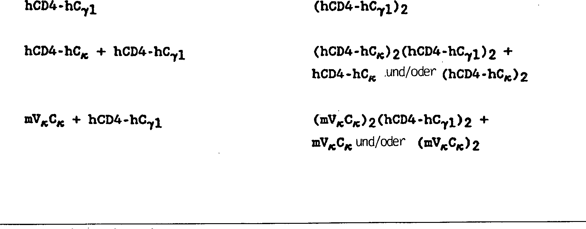
Aus dieser Tabelle geht interessanterweise hervor, dass das menschliche CD4-Schwerketten-Immunadheson als ein Dimer sekretiert wurde, während die analoge murine Konstruktion nicht nachgewiesen wurde (was jedoch nicht die extrazelluläre Akkumulation des Proteins ausschließt). Die Fähigkeit der hCD4-hCγ1-Transformanten, ein Schwerkettendimer zu produzieren, kam unerwartet, da vorausgegangene Arbeiten nahe gelegt hatten, Immunglobulin-Schwerketten würden nicht sekretiert, es sei denn, die Wirte werden mit sowohl für Schwer- als auch für Leichtketten kodierender Nucleinsäure co-transformiert (Valle et al., Nature 241, 338 [1981]). Gemäß dieser Erfindung werden CD4-IgG-Immunadheson-Chimären leicht sekretiert, worin das CD4-Epitop in Schwerkettendimeren, Leichtkettenmonomeren oder – dimeren und Schwer- und Leichtkettenheterotetrameren vorhanden ist, worin das CD4-Epitop in einer an eine oder mehrere Leicht- oder Schwerketten fusionierten Form, umfassend Heterotetramere, in denen bis zu und einschließlich alle vier Analoga der variablen Region von CD4 abstammen, vorhanden ist. Wo eine variable nicht-CD4-Leicht-Schwerketten-Domäne vorhanden ist, wird ein heterofunktioneller Antikörper bereitgestellt.Out Interestingly enough, this table shows that the human CD4 heavy chain Immunadheson as a dimer was secreted while the analogous murine construction was not detected (which, however, is not the extracellular accumulation excludes the protein). The ability the hCD4-hCγ1 transformant, producing a heavy-chain bucket came unexpectedly, as previous works immunoglobulin heavy chains would not be secreted, it was because, the hosts are with both heavy and light chains encoding nucleic acid co-transformed (Valle et al., Nature 241, 338 [1981]). According to this In the present invention, CD4-IgG immunoadhesin chimeras are readily secreted in which the CD4 epitope in heavy chain dimers, light chain monomers or dimers and heavy and light chain heterotetramers, wherein the CD4 epitope in one on one or more light or heavy chains fused form, comprising heterotetramers in which up to and including all four CD4 variable region analogs are present is. Where a variable non-CD4 light heavy chain domain exists is provided a heterofunctional antibody.
Geeignete Begleit-Immunglobulin-Kombinationsstellen und Fusionspartner werden aus IgG-1-, -2-, -3- oder -4-Subtypen, IgA, IgE, IgD oder IgM, jedoch vorzugsweise IgG-1, erhalten.suitable Accompanying immunoglobulin combination sites and fusion partners from IgG-1, -2, -3 or -4 subtypes, IgA, IgE, IgD or IgM, however preferably IgG-1.
Eine bevorzugte Ausführungsform ist eine Fusion eines N-terminalen Abschnitts von CD4, der die Bindungsstelle für das gp120-Hüllprotein von HIV enthält, an den C-terminalen Fc-Abschnitt eines Antikörpers, der die Effektorfunktionen des Immunglobulins G1 enthält. Von dieser Art gibt es zwei bevorzugte Ausführungsformen; in einer ist die gesamte konstante Region der schweren Kette an einen Abschnitt von CD4 fusioniert; in einer anderen ist eine Sequenz, die in der Gelenkregion unmittelbar stromauf der Papain-Spaltstelle beginnt, die IgG-Fc chemisch definiert (Rest 216, wobei 114 als erster Rest der konstanten Region der schweren Kette angenommen wird [Kobat et al., Sequences of Proteins of Immunological Interest, 4. Auflage (1987)], oder analoge Stellen anderer Immunglobuline), an einen Abschnitt von CD4 fusioniert. Diese Ausführungsformen werden in den Beispielen beschrieben.A preferred embodiment is a fusion of an N-terminal portion of CD4 containing the binding site for the gp120 envelope protein of HIV to the C-terminal Fc portion of an antibody containing the effector functions of the immunoglobulin G 1 . Of this type, there are two preferred embodiments; in one, the entire heavy chain constant region is fused to a portion of CD4; in another, a sequence beginning in the hinge region just upstream of the papain cleavage site chemically defines the IgG Fc (residue 216, where 114 is assumed to be the first residue of the heavy chain constant region [Kobat et al., Sequences of Proteins of Immunological Interest, 4th Edition (1987)], or analogous sites of other immunoglobulins), fused to a section of CD4. These embodiments are described in the examples.
Im
Besonderen wird von jenen Varianten, in denen die variable Region
einer Immunglobulinkette durch eine oder mehrere immunglobulinähnliche
Domänen
eines Adhesons substituiert sind, angenommen, sie hätten in
vivo eine verbesserte Plasmahalbwertszeit. Diese Chimären sind
auf eine ähnliche
Art und Weise wie chimäre
Antikörper
aufgebaut, in denen die variable Domäne eines Antikörpers einer
Spezies für
eine variable Domäne
einer anderen Spezies substituiert wird. Siehe z.B
Für die chimäre(n) Kette(n) des Immunglobulins oder Immunadhesons kodierende DNA wird zur Expression in eine Wirtszelle transfiziert. Falls die Wirtszelle vor der Transfektion ein Immunglobulin produziert, so muss nur mit dem an Leicht- oder Schwerketten fusionierten Adheson transfiziert werden, um einen Heteroantikörper herzustellen. Die zuvor erwähnten Immunglobuline, die einen oder mehrere Arme aufweisen, die Adhesondomänen tragen, und einen oder mehrere Arme aufweisen, die variable Begleiterregionen tragen, führen zu einer dualen Spezifität für den Adheson-Liganden und für ein Antigen. Sie werden durch die oben beschriebenen Rekombinationsverfahren oder durch In-vitro-Verfahren hergestellt. In letzterem Fall werden z.B. F(ab')2-Fragmente einer Adheson-Fusion und ein Immunglobulin hergestellt, die F(ab')2-Fragmente werden durch Reduktion unter milden Reduktionsbedingungen in Fab'-Fragmente konvertiert und dann jeweils in Gegenwart des anderen unter sauren Bedingungen gemäß per se bekannter Verfahren reoxidiert. Siehe auch U.S.-Patent 4.444.878.DNA encoding the chimeric chain (s) of the immunoglobulin or immunoadhesone is transfected into a host cell for expression. If the host cell produces an immunoglobulin prior to transfection, then only Adheson fused to light or heavy chains must be transfected to produce a heteroantibody. The aforementioned immunoglobulins, having one or more arms bearing Adheson domains, and having one or more arms bearing variable companion regions, result in dual specificity for the Adheson ligand and for an antigen. They are prepared by the recombination methods described above or by in vitro methods. In the latter case, for example, F (ab ') 2 fragments of an Adheson fusion and an immunoglobulin are produced, the F (ab') 2 fragments are converted into Fab 'fragments by reduction under mild reduction conditions and then in each case in the presence of the other reoxidized under acidic conditions according to methods known per se. See also U.S. Patent 4,444,878.
Zusätzlich sind Verfahren zur Herstellung intakter Heteroantikörper aus Immunglobulinen mit verschiedenen Spezifitäten bekannt. Diese Verfahren werden zur In-vitro-Herstellung von heterochimären Antikörpern durch einfaches Substituieren eines der zuvor verwendeten Immunglobuline durch die Immunadhesonketten verwendet.In addition are Process for the preparation of intact heteroantibodies from immunoglobulins with different specificities known. These methods are used for in vitro production of heterochimeric antibodies simple substitution of one of the previously used immunoglobulins used by the immunoadhesone chains.
In
einem alternativen Verfahren zur Herstellung eines heterofunktionellen
Antikörpers
werden Wirtszellen, die eine Adheson-Immunglobulinfusion produzieren,
z.B. transfizierte Myelome, ebenso mit B-Zellen oder Hybridomen
fusioniert, die Antikörper sekretieren,
welche die gewünschte
Begleitspezifität
für ein
Antigen aufweisen. Heterobifunktionelle Antikörper werden aus dem Kulturmedium
solcher Hybridome gewonnen und sind daher etwas leichter zu produzieren
als durch herkömmliche
In-vitro-Anwendungsverfahren
(
Eine weitere Gruppe an Fusionen sind jene, in denen ein Adheson mit einer toxischen Substanz, z.B. einem Polpypeptid, wie etwa Ricin (umfassend eine deglykosylierte Ricin-A-Kette), Diphtherietoxin A oder ein nichtpeptidylartiges Zytotoxin, konjugiert ist. Wenn es sich bei dem Toxin um ein Polypeptid handelt, ist es zweckdienlich, das Polypeptid durch herkömmliche In-vitro-Proteinvernetzungsmittel mit dem Adheson oder seiner transmembrandeletierten Variante zu vernetzen (für geeignete Verfahren zur Bindung einer Ricin-A-Kette oder einer deglykosylierten A-Kette an CD4 siehe z.B. Duncan et al., Analy. Biochem. 132, 68-73 [1983]; Thorpe et al., Cancer Res. 47, 5924 [1987], und Ghotie et al., Cancer Res. 48, 2610 [1988]) oder aber dies durch Rekombinationssynthese als Fusion zu tun (siehe z.B. U.S.-Patent 4.765.382). Alternativ dazu werden in Fällen, in denen Begleitantikörper variable Immunglobulindomänen von Anti-Ricin-Antikörpern sind, solche Immunglobulin-Heteroantikörper verwendet, um, gemäß dem allgemeinen Verfahren von Raso et al., Cancer Research 41, 2073 (1981), Ricin an HIV-infizierte Zellen zu liefern.Another group of fusions are those in which an adheson with a toxic substance, for example a polypeptide, such as ricin (comprising a deglycosylated ricin A chain), diphtheria toxin A or a non-peptidyl-like cytotoxin conjugated. When the toxin is a polypeptide, it is convenient to cross-link the polypeptide to the adheson or its transmembrane-variant by conventional in vitro protein crosslinking agents (for suitable methods for attaching a ricin A chain or a deglycosylated A- See, for example, Duncan et al., Analyt Biochem 132, 68-73 [1983]; Thorpe et al., Cancer Res. 47: 5924 [1987] and Ghotie et al., Cancer Res. 48: 2610 [1988]) or by recombinant synthesis as a fusion (see, for example, US Pat. No. 4,765,382). Alternatively, in cases where companion antibodies are immunoglobulin variable domains of anti-ricin antibodies, such immunoglobulin heteroantibodies are used to deliver ricin to HIV, according to the general procedure of Raso et al., Cancer Research 41, 2073 (1981). to deliver infected cells.
Eine
weitere Klasse von Adhesonvarianten sind Deletionsvarianten. Deletionen
sind durch das Entfernen von einem oder mehreren Aminosäurerest(en)
aus einer Adhesonsequenz charakterisiert. Typischerweise sind die
Transmembran- und die zytoplasmatischen Domänen von Adhesonen deletiert.
Im Fall von CD4 sind zumindest die Reste 368 bis 395 (die Transmembranregion)
und normalerweise ebenso 396-433
(die zytoplasmatische Domäne)
deletiert, um sekretierte Formen dieses Adhesons zu erhalten. Ebenso
ist anzumerken, dass die Aminosäurereste
den Nummern folgen, die für
reifes CD4 gegeben werden, wie dies z.B. in den
Substitutionsvarianten
sind jene, in denen zumindest ein Rest in der Adhesonsequenz entfernt
wurde und ein anderer Rest stattdessen insertiert wurde. Der native
N-terminale Rest für
reifes CD4 ist, wie nun bekannt ist, Lysin. Die in 
Substantielle Veränderungen der Funktion oder der immunologischen Identität werden durch Auswählen von Substitutionen erreicht, die weniger konservativ als die in Tabelle 2 angeführten sind, d.h. durch Auswählen von Resten, die sich auf signifikantere Art und Weise in ihrer Wirkung auf den Erhalt von (a) der Struktur der Polypeptidhauptkette im Bereich der Substitution, z.B. als Faltblatt oder Helixkonformation, (b) der Ladung oder der Hydrophobizität des Moleküls an der Targetstelle oder (c) der Sperrigkeit der Seitenkette unterscheiden. Die Substitutionen, von denen im Allgemeinen erwartet wird, dass sie die größten Veränderungen der Adhesoneigenschaften hervorrufen, sind jene, in denen (a) ein hydrophiler Rest, z.B. Seryl oder Threonyl, für (oder durch) einen hydrophoben Rest, z.B. Leucyl, Isoleucyl, Phenylalanyl, Valyl oder Alanyl, substituiert wird; (b) ein Cysteinyl oder Prolyl für (oder durch) einen beliebigen anderen Rest substituiert wird; (c) ein Rest mit einer elektropositiven Seitenkette, z.B. Lysyl, Arginyl oder Histidyl, für (oder durch) einen elektronegativen Rest, z.B. Glutamyl oder Aspartyl, substituiert wird oder (d) ein Rest mit einer sperrigen Seitenkette, z.B. Phenylalanyl, für (oder durch) einen Rest ohne Seitenkette, z.B. Glycyl, substituiert wird.substantial changes function or immunological identity are selected by selecting Substitutions achieved that are less conservative than those in Table 2 quoted are, i. by selecting of residues that are more significant in their effect to obtain (a) the structure of the polypeptide backbone in the Range of substitution, e.g. as a leaflet or helix conformation, (b) the charge or hydrophobicity of the molecule at the target site or (c) distinguish the bulkiness of the side chain. The substitutions, which are generally expected to make the biggest changes of the adhesive properties are those in which (a) a hydrophilic residue, e.g. Seryl or threonyl, for (or through) a hydrophobic Remainder, e.g. Leucyl, isoleucyl, phenylalanyl, valyl or alanyl substituted becomes; (b) a cysteinyl or prolyl for (or by) any one other radical is substituted; (c) a remainder with an electropositive Side chain, e.g. Lysyl, arginyl or histidyl, for (or by) an electronegative moiety, e.g. Glutamyl or aspartyl, or (d) a radical having a bulky side chain, e.g. Phenylalanyl, for (or by) a residue without side chain, e.g. Glycyl, substituted becomes.
Eine bevorzugte Klasse von Substitutions- oder Deletionsvarianten sind jene, die die Transmembranregion des Adhesons involvieren. Die Transmembranregion des Adhesons ist eine hochgradig hydrophobe oder lipophile Domäne, die die richtige Größe aufweist, um die Lipiddoppelschicht der Zellmembran zu umspannen. Es wird angenommen, sie würde das Adehson in der Zellmembran verankern.A preferred class of substitution or deletion variants those involving the transmembrane region of the adheson. The transmembrane region of Adheson is a highly hydrophobic or lipophilic domain that the right size, to span the lipid bilayer of the cell membrane. It will suppose she would anchor the adehson in the cell membrane.
Eine Deletion oder Substitution der Transmembrandomäne erleichtert die Gewinnung und stellt durch die Reduktion ihrer Zell- oder Membranlipidaffinität und die Verbesserung ihrer Wasserlöslichkeit eine lösliche Form des Adhesons bereit. Falls die Transmembran- und die zytoplasmatischen Domänen deletiert werden, wird die Einführung von potentiell immunogenen Epitopen vermieden, entweder durch die Exposition von andernfalls intrazellulären Polypeptiden, die vom Körper als fremdartig erkannt werden könnten, oder durch Einführung heterologer Polypeptide, die potentiell immunogen sind. Ein Hauptvorteil des transmembrandeletierten Adhesons ist die Tatsache, dass es in das Kulturmedium rekombinanter Wirte sekretiert wird. Diese Variante ist wasserlöslich und besitzt keine nennenswerte Affinität zu Zellmembranlipiden, was ihre Gewinnung aus der Rekombinationszellkultur bedeutend vereinfacht.A Deletion or substitution of the transmembrane domain facilitates recovery and represents by the reduction of their cell or membrane lipid affinity and the Improving their water solubility a soluble form of the Adheson ready. If the transmembrane and the cytoplasmic domains will be deleted, the introduction avoided by potentially immunogenic epitopes, either by the Exposure of otherwise intracellular polypeptides released by the body as could be recognized in an alien way, or by introduction heterologous polypeptides that are potentially immunogenic. A main advantage The transmembrated adheson is the fact that it is in the culture medium of recombinant hosts is secreted. This variant is water soluble and has no significant affinity for cell membrane lipids, which their recovery from recombination cell culture significantly simplified.
Aus der vorhergehenden Diskussion wird klar und deutlich ersichtlich, dass Substitutionen, Deletionen, Insertionen oder eine beliebige Kombination dieser eingeführt werden, um ein Endkonstrukt zu erhalten. Als allgemeine Behauptung werden alle Varianten keine funktionelle Transmembrandomäne und vorzugsweise keine funktionelle zytoplasmatische Sequenz aufweisen. Dies wird im Allgemeinen durch Deletion der relevanten Domäne erreicht, obwohl adäquate Insertions- oder Substitutionsmutagene zu diesem Zweck ebenso wirksam sein können. Die Transmembrandomäne wird z.B. durch eine beliebige Aminosäuresequenz, z.B. eine Zufalls- oder Homopolynucleinsequenz von etwa 5 bis 50 Serin-, Threonin-, Lysin-, Arginin-, Glutamin-, Asparaginsäure- und ähnlichen hydrophilen Resten, die insgesamt ein hydrophiles Hydropathie-Diagramm aufweisen, ersetzt, sodass sie in das Kulturmedium rekombinanter Wirte sekretiert wird. Diese Variante sollte ebenso als Adhesonvariante angesehen werden.Out the previous discussion becomes clear and clear that substitutions, deletions, insertions or any Combination of these introduced to obtain a final construct. As a general statement all variants will not be a functional transmembrane domain and preferably have no functional cytoplasmic sequence. this will generally achieved by deletion of the relevant domain, although adequate insertion or substitution mutagens may be equally effective for this purpose. The Transmembrane domain is e.g. by any amino acid sequence, e.g. a random or homopolynuclein sequence of about 5 to 50 serine, threonine, Lysine, arginine, glutamine, aspartic acid and similar hydrophilic residues, which have a total of a hydrophilic hydropathy diagram replaced, so that it is secreted into the culture medium of recombinant hosts. This variant should also be considered as an Adhesonvariante.
Diese
Varianten werden normalerweise durch stellenspezifische Mutagenese
von Nucleotiden in der für
das Adheson kodierenden DNA hergestellt, wodurch für die Variante
kodierende DNA produziert wird und wonach die DNA in Rekombinationszellkultur
exprimiert wird. Variantenadhesone werden jedoch auch durch In-vitro-Synthese hergestellt.
Natürlich
dürfen
Variationen in der für
die Variantenadhesone kodierenden DNA die Sequenz nicht außerhalb
des Leserasters platzieren und erzeugen vorzugsweise keine komplementären Regionen,
die sekundäre
mRNA-Strukturen
produzieren könnten,
die für
die Expression schädlich
sind (
Während die Stelle zur Einführung einer Aminosäuresequenzvariation im Vorhinein festgelegt ist, muss die Mutation per se nicht im Vorhinein festgelegt sein. Um z.B. die Leistung einer Mutation an einer bestimmten Stelle zu optimieren, kann eine Zu fallsmutagenese am Target-Codon oder der Target-Region durchgeführt werden, und die exprimierten Adhesonvarianten können auf die optimale Kombination der gewünschten Aktivitäten gescreent werden. Verfahren zur Herstellung von Substitutionsmutationen an im Vorhinein festgelegten Stellen in DNA mit einer bekannten Sequenz sind wohlbekannt, z.B. M13-Primer-Mutagenese.While the Place for introduction an amino acid sequence variation is set in advance, the mutation per se does not have to be in advance be set. For example, the power of a mutation at a given Optimizing site may lead to coincidence at the target codon or the target region can be, and the expressed Adhesonvarianten can on the optimal combination the desired activities be screened. Process for the preparation of substitution mutations at pre-determined sites in DNA with a known Sequences are well known, e.g. M13 primer mutagenesis.
Adhesonvarianten, die nicht fähig sind, HIV-gp120 zu binden, sind trotzdem als Immunogene zur Produktion von Antikörpern gegen das Adheson oder als Immuntest-Set-Komponenten von Nutzen (markiert als kompetitives Reagens für den gp120-Test oder unmarkiert als Standard für einen Adhesontest), solange zumindest ein Adheson-Epitop aktiv bleibt.adheson variants, not capable are to bind HIV gp120, are still as immunogens for production of antibodies against the Adheson or as an immunoassay set components of use (marked as a competitive reagent for the gp120 test or unmarked as default for an Adheson test) as long as at least one Adheson epitope remains active.
Die für Adhesone kodierende DNA wird durch bekannte Verfahren erhalten. Siehe Williams, Immunol. Today 8, 298-303 (1987), und die darin enthaltenen Zitate. Im Allgemeinen werden Prakaryoten zum Klonieren von DNA-Sequenzen von CD4-Varianten verwendet. So sind z.B. der E.-coli-Stamm SR101 (zum Vermehren von m13-Phagen, einem λ-resistenten Stamm von JM 101; Messing et al., Nucl. Acids. Res. 9(2), 309-321 [1981]) und der E.-coli-K12-Stamm 294 (ATCC Nr. 31446) besonders nützlich. Andere Mikrobenstämme, die verwendet werden können, umfassen E. coli B, UM101 und E.coli X1776 (ATCC Nr. 31537). Diese Beispiele dienen der Veranschaulichung und sollen keine Einschränkung darstellen.The Adhesone-encoding DNA is obtained by known methods. See Williams, Immunol. Today 8, 298-303 (1987), and the citations contained therein. In general, prokaryotes are used to clone DNA sequences of CD4 variants. For example, E. coli strain SR101 (for propagating m13 phage, a λ resistant strain of JM 101, Messing et al., Nucl. Acids. Res. 9 (2), 309-321 [1981]. ) and E. coli K12 strain 294 (ATCC No. 31446) are particularly useful. Other microbial strains that may be used include E. coli B, UM101 and E. coli X 1776 (ATCC No. 31537). These examples are illustrative and not intended to be limiting.
Für die Variantenadhesone kodierende DNA wird zur Expression in Vektoren eingeführt, die Promotoren und Kontrollsequenzen enthalten, die von mit der beabsichtigten Wirtszelle kompatiblen Spezies abstammen. Der Vektor trägt normalerweise, muss dies jedoch nicht tun, eine Replikationsstelle sowie eine oder mehrere Markersequenzen in sich, die fähig sind, in transformierten Zellen für eine phänotypische Selektion zu sorgen. E. coli wird z.B. typischerweise unter Verwendung eines Derivats von pBR322, einem von einer E.-coli-Spezies abstammenden Plasmid, transformiert (Bolivar et al., Gene 2, 95 [1977]). pBR322 enthält Gene für Ampicillin- und Tetracyclinresistenz und stellt daher ein einfaches Mittel zur Identifikation transfor mierter Zellen bereit. Das pBR322-Plasmid oder ein anderes Mikrobenplasmid muss ebenso Promotoren und andere Kontrollelemente, die in rekombinanten DNA-Konstruktionen häufig verwendet werden, umfassen oder es muss modifiziert werden, um diese zu enthalten.For the variant anesthesia Coding DNA is introduced into vectors for expression, which Promoters and control sequences included with the intended Derived host cell compatible species. The vector usually carries, However, this does not have to do a replication site as well as a or several marker sequences capable of being transformed into Cells for a phenotypic To ensure selection. E. coli is e.g. typically using a derivative of pBR322, one derived from an E. coli species Plasmid, transformed (Bolivar et al., Gene 2, 95 [1977]). pBR322 contains Genes for ampicillin and tetracycline resistance and therefore provides a simple means to Identification of transformed cells ready. The pBR322 plasmid or another microbial plasmid also needs promoters and others Control elements commonly used in recombinant DNA constructions include or it needs to be modified to contain it.
Für die Verwendung mit prokaryotischen Wirten geeignete Promotoren umfassen veranschaulichenderweise die β-Lactamase- und Lactose-Promotor-Systeme (Chang et al., Nature 275, 615 [1978]; und Goeddel et al., Nature 281, 544 [1979]), alkalische Phosphatase, das Tryptophan-(trp-) Promotor-System (Goeddel, Nucleic Acids Res. 8, 4057 [1980], und EPO Appln. Publ. Nr. 36.776) und Hybridpromotoren, wie etwa den tac-Promotor (H. de Boer et al., Proc. Natl. Acad. Sci. USA 80, 21-25 [1983]). Andere funktionelle bakterielle Promotoren sind jedoch ebenso geeignet. Ihre Nucleotidsequenzen sind im Allgemeinen bekannt, wodurch ein Fachmann sie unter Verwendung von Linkern oder Apatoren operabel an für die Adhesonvariante kodierende DNA ligieren kann, um jegliche erforderliche Restriktionsstelle bereitzustellen (Siebenlist et al., Cell 20, 269 [1980]). Promotoren zur Verwendung in bakteriellen Systemen umfassen ebenso eine Shine-Dalgarno-(S.D.-) Sequenz, die an die für das Antigen kodierende DNA operabel gebunden ist.For the use Promoters suitable with prokaryotic hosts illustratively include the β-lactamase and lactose promoter systems (Chang et al., Nature 275, 615 [1978]; and Goeddel et al., Nature 281, 544 [1979]), alkaline phosphatase, the tryptophan (trp) promoter system (Goeddel, Nucleic Acids Res. 8, 4057 [1980], and EPO Appln. Publ. No. 36,776) and hybrid promoters, such as the tac promoter (H. de Boer et al., Proc. Natl. Acad. Sci. USA 80, 21-25 [1983]). Other functional bacterial promoters however, are equally suitable. Their nucleotide sequences are in general a person skilled in the art using linkers or Apators operable on for the Adhesonvariante DNA can ligate to any required Restriction site (Siebenlist et al., Cell 20, 269 [1980]). Promoters for use in bacterial systems also include a Shine-Dalgarno (S.D.) sequence, which is attached to the for the Antigen-encoding DNA is operably linked.
Zusätzlich zu Prokaryoten sind eukaryotische Mikroben, wie z.B. Hefekulturen, ebenso als Klonierungs- oder Expressionswirte nützlich. Saccharomyces cerevisiae oder Gemeine Bäckerhefe ist der am häufigsten verwendete eukaryotische Mikroorganismus, obwohl eine Reihe anderer Stämme leicht erhältlich sind. Zur Expression in Saccharomyces wird z.B. das Plasmid YRp7 (Stinchcomb et al., Nature 282, 39 [1979]; Kingsman et al., Gene 7, 141 [1979]; Tschemper et al., Gene 10, 157 [1980]) häufig verwendet. Dieses Plasmid umfasst bereits das trp1-Gen, das einen Selektionsmarker für einen mutierten Hefestamm bereitstellt, dem die Fähigkeit, in Tryptophan zu wachsen, fehlt, z.B. ATCC Nr. 44076 oder PEP4-1 (Jones, Genetics 85, 12 [977]). Die Gegenwart der trp1-Läsion als Merkmal des Hefewirtszellengenoms stellt dann ein wirksames Mittel der Selektion durch Wachstum in Abwesenheit von Tryptophan bereit.In addition to Prokaryotes are eukaryotic microbes, e.g. yeast cultures also useful as cloning or expression hosts. Saccharomyces cerevisiae or common baker's yeast is the most common used eukaryotic microorganism, although a number of others strains easily available are. For expression in Saccharomyces, e.g. the plasmid YRp7 (Stinchcomb et al., Nature 282, 39 [1979]; Kingsman et al., Gene 7, 141 [1979]; Chemper et al., Gene 10, 157 [1980]). This plasmid already includes the trp1 gene, which is a selection marker for one mutant yeast strain that has the ability to grow in tryptophan, is missing, e.g. ATCC No. 44076 or PEP4-1 (Jones, Genetics 85, 12 [977]). The Presence of the trp1 lesion as a feature of the yeast host cell genome then provides an effective Means of selection by growth in the absence of tryptophan ready.
Geeignete Promotorsequenzen zur Verwendung mit Hefewirten umfassen die Promotoren für 3-Phosphoglyceratkinase (Hitzeman et al., J. Biol. Chem. 255, 2073 [1980]) oder andere glykolytische Enzyme (Hess et al., J. Adv. Enzyme Reg. 7, 149 [1968]; und Holland, Biochemistry 17, 4900 [1978]), wie z.B. Enolase, Glyceraldehyd-3-phosphatdehydrogenase, Hexokinase, Pyruvatdecarboxylase, Phosphofructokinase, Glucose-6-phosphatisomerase, 3-Phosphoglyceratmutase, Pyruvatkinase, Triosephosphatisomerase, Phosphoglucoseisomerase und Glucokinase.suitable Promoter sequences for use with yeast hosts include the promoters for 3-phosphoglycerate kinase (Hitzeman et al., J. Biol. Chem. 255, 2073 [1980]) or other glycolytic Enzymes (Hess et al., J. Adv. Enzymes Reg. 7, 149 [1968]; and Holland, Biochemistry 17, 4900 [1978]), e.g. Enolase, glyceraldehyde-3-phosphate dehydrogenase, Hexokinase, pyruvate decarboxylase, phosphofructokinase, glucose-6-phosphate isomerase, 3-phosphoglycerate mutase, pyruvate kinase, triosephosphate isomerase, Phosphoglucose isomerase and glucokinase.
Weitere Hefepromotoren, die induzierbare Promotoren mit dem zusätzlichen Vorteil einer von Wachstumsbedingungen kontrollierten Transkription sind, umfassen die Promotorregionen für Alkoholdehydrogenase 2, Isocytochrom C, saure Phosphatase, abbauende Enzyme, die mit dem Stickstoffstoffwechsel assoziiert sind, Metallothionein, Glyceraldehyd-3-phosphatdehydrogenase sowie Enzyme, die für die Maltose- und Galactoseverwertung verantwortlich sind. Geeignete Vektoren und Promotoren für die Verwendung in der Hefeexpression werden weiter in R. Hitzeman et al., EP-Veröffentlichung Nr. 73.657A, beschrieben. Hefe-Enhancer werden ebenso mit Hefepromotoren vorteilhaft verwendet.Further Yeast promoters, the inducible promoters with the additional Advantage of a growth condition controlled transcription For example, the promoter regions for alcohol dehydrogenase 2 include isocytochrome C, acid phosphatase, degrading enzymes associated with nitrogen metabolism metallothionein, glyceraldehyde-3-phosphate dehydrogenase as well as enzymes for the maltose and Galactose utilization are responsible. Suitable vectors and Promoters for its use in yeast expression will be further described in R. Hitzeman et al., EP publication No. 73.657A. Yeast enhancers are also using yeast promoters used advantageously.
Promotoren, um die Transkription aus Vektoren in Säugetierwirtszellen zu kontrollieren, können aus verschiedenen Quellen erhalten werden, z.B. den Genomen von Viren, wie z.B.: Polyoma, Simian-Virus 40 (SV40), Adenovirus, Retroviren, Hepatitis-B-Virus und insbesondere Zytomegalievirus, oder aus heterologen Säugetierpromotoren, z.B. dem β-Actin-Promotor. Die frühen und späten Promotoren des SV40-Virus sind leicht als ein SV40-Restriktionsfragment zu erhalten, das auch den viralen SV40-Replikationsstartpunkt umfasst. Fiers et al., Nature 273, 113 (1978). Der unmittelbare frühe Promotor des menschlichen Zytomegalievirus ist leicht als ein HindIII-E-Restriktionsfragment zu erhalten. P.J. Greenaway et al., Gene 18, 355-360 (1982). Natürlich sind auch Promotoren aus der Wirtszelle oder verwandten Spezies hierin von Nutzen.Promoters to control transcription from vectors in mammalian host cells can be obtained from a variety of sources, such as the genomes of viruses such as: Polyoma, Simian virus 40 (SV40), adenovirus, retroviruses, hepatitis B virus, and especially cytomegalovirus , or from heterologous mammalian promoters, eg the β-actin promoter. The early and late promoters of the SV40 virus are readily conserved as an SV40 restriction fragment that also transduces the SV40 viral origin of replication summarizes. Fiers et al., Nature 273, 113 (1978). The immediate early promoter of human cytomegalovirus is readily available as a HindIII E restriction fragment. PJ Greenaway et al., Gene 18, 355-360 (1982). Of course, promoters from the host cell or related species are also useful herein.
Die DNA-Transkription in höheren Eukaryoten wird durch Insertieren einer Enhancer-Sequenz in den Vektor verstärkt. Enhancer sind cis-wirkende DNA-Elemente, norma lerweise von etwa 10 bis 300 bp, die die Transkriptionsinitiationsfähigkeit eines Promotors verstärken. Enhancer sind relativ orientierungs- und positionsunabhängig, da sie 5' (L. Laimins et al., Proc. Natl. Acad. Sci 78, 993 [1981]) und 3' (M.L. Lusky et al., Mol. Cell Bio. 3, 1108 [1983]) zu der Transkriptionseinheit gefunden wurden, in einem Intron (J.L. Banerji et al., Cell 33, 729 [1983]) sowie in der kodierenden Sequenz selbst (T.F. Osborne et a., Mol. Cell Bio. 4, 1293 [1984]). Viele Enhancer-Sequenzen von Säugetiergenen sind nun bekannt (Globin, Elastase, Albumin, α-Fötoprotein und Insulin). Typischerweise wird jedoch ein Enhancer aus einem eukaryotischen Zellvirus verwendet. Beispiele umfassen den SV40-Enhancer auf der späten Seite des Replikationsstartpunktes (bp 100-270), den Enhancer des frühen Promotors des Zytomegalievirus, den Polyoma-Enhancer auf der späten Seite des Replikationsstartpunkts und Adenovirus-Enhancer.The DNA transcription in higher Eukaryotes are amplified by inserting an enhancer sequence into the vector. enhancer are cis-acting DNA elements, usually from about 10 to 300 bp, which enhance the transcriptional initiation ability of a promoter. enhancer are relatively orientational and position independent since they are 5 '(L. Laimins et al. Proc. Natl. Acad. Sci 78, 993 [1981]) and 3 '(M. L. Lusky et al., Mol. Cell Bio. 3, 1108 [1983]) to the transcriptional unit, in an intron (J.L. Banerji et al., Cell 33, 729 [1983]) and in the coding sequence itself (T.F. Osborne et al., Mol. Cell Bio. 4, 1293 [1984]). Many enhancer sequences of mammalian genes are now known (Globin, elastase, albumin, α-fetoprotein and Insulin). Typically, however, an eukaryotic enhancer becomes Cell virus used. Examples include the SV40 enhancer on the late Side of the origin of replication (bp 100-270), the enhancer of the early Promoter of cytomegalovirus, the polyoma enhancer on the late side of the origin of replication and adenovirus enhancers.
Expressionsvektoren, die in eukaryotischen Wirtszellen (Hefe-, Pilz-, Insekten-, Pflanzen-, Tier-, menschliche oder kernhaltige Zellen) verwendet werden, können ebenso Sequenzen umfassen, die für die Termination der Transkription erforderlich sind und die die mRNA-Expression beeinflussen können. Diese Regionen werden als polyadenylierte Segmente im untranslatierten Teil der für das Adheson kodierenden mRNA transkribiert.Expression vectors in eukaryotic host cells (yeast, fungus, insect, plant, Animal, human or nucleated cells) can be used as well Include sequences for the termination of transcription is required and the the can influence mRNA expression. These regions are called polyadenylated segments in untranslated Part of for transcribing the adheson-encoding mRNA.
Expressionsvektorsysteme umfassen im Allgemeinen ein Selektionsgen, auch selektierbarer Marker genannt. Beispiele für geeignete selektierbare Marker für Säugetierzellen sind Dihydrofolatreduktase (DHFR), Thymidinkinase oder Neomycin. Werden diese selektierbaren Marker erfolgreich in eine Säugetierwirtszelle transferiert, so kann die transformierte Säugetierwirtszelle überleben, wenn sie Selektionsdruck ausgesetzt wird. Es gibt zwei weit verbreitete, verschiedene Kategorien von Selektionsegimen. Die erste Kategorie basiert auf dem Stoffwechsel einer Zelle und der Verwendung einer mutierten Zelllinie, der die Fähigkeit fehlt, unabhängig von einem angereichertem Medium zu wachsen. Zwei Beispiele sind CHO-DHFR--Zellen sowie Maus-LTK--Zellen. Diesen Zellen fehlt die Fähigkeit, ohne den Zusatz solcher Nährstoffe wie Thymidin oder Hypoxanthin zu wachsen. Da diesen Zellen gewisse, für einen vollständigen Nucleotidsyntheseweg notwendige Gene fehlen, können sie nicht überleben, wenn die fehlenden Nucleotide nicht in einem angereicherten Medium bereitgestellt werden. Eine Alternative zum Anreichern des Mediums ist die Einführung eines intakten DHFR- oder TK-Gens in jene Zellen, denen die entsprechenden Gene fehlen, wodurch ihre Wachstumsbedingungen verändert werden. Einzelne Zellen, die nicht mit dem DHFR- oder TK-Gen transformiert wurden, sind nicht fähig, in einem nichtangereicherten Medium zu überleben.Expression vector systems generally comprise a selection gene, also called a selectable marker. Examples of suitable mammalian cell selectable markers are dihydrofolate reductase (DHFR), thymidine kinase or neomycin. When these selectable markers are successfully transferred to a mammalian host cell, the transformed mammalian host cell can survive when exposed to selection pressure. There are two widely used, different categories of selection regimens. The first category is based on the metabolism of a cell and the use of a mutant cell line that lacks the ability to grow independently of an enriched medium. Two examples are CHO-DHFR - cells and mouse LTK - cells. These cells lack the ability to grow without the addition of such nutrients as thymidine or hypoxanthine. Since these cells lack certain genes necessary for a complete nucleotide synthesis pathway, they can not survive if the missing nucleotides are not provided in an enriched medium. An alternative to accumulating the medium is to introduce an intact DHFR or TK gene into those cells lacking the corresponding genes, thereby altering their growth conditions. Single cells that have not been transformed with the DHFR or TK gene are unable to survive in a non-enriched medium.
Die zweite Kategorie ist die der dominanten Selektion, die sich auf ein Selektionsschema bezieht, das in jedem beliebigen Zelltyp verwendet wird und nicht die Verwendung einer mutierten Zelllinie erforderlich macht. Diese Schemen verwenden typischerweise ein Arzneimittel, um das Wachstum einer Wirtszelle zum Stillstand zu bringen. Jene Zellen, die ein neues Gen aufweisen, würden ein Protein exprimieren, das eine Arzneimittelresistenz verleiht, und würden die Selektion überleben. In Beispielen für diese dominante Selektion werden die Wirkstoffe Neomycin, P. Southern & P. Berg, J. Molec. Appl. Genet. 1, 327 (1982), Mycophenolsäure, R.C. Mulligan & P. Berg, Science 209, 1422 (1980), oder Hygromycin, B. Sugden et al., Mol. Cell. Biol. 5, 410-413 (1985), verwendet. Die drei oben angeführten Beispiele verwenden bakterielle Gene unter eukaryotischer Kontrolle, um eine Resistenz gegen den jeweiligen Wirkstoff G418 oder Neomycin (Geneticin), Xgpt (Mycophenolsäure) bzw. Hygromycin zu verleihen.The second category is that of dominant selection, focusing on refers to a selection scheme used in any cell type and does not require the use of a mutated cell line. These schemes typically use a drug to do this Stopping growth of a host cell. Those cells, which would have a new gene express a protein that confers drug resistance, and would survive the selection. In examples of this dominant selection will be the drugs neomycin, P. Southern & P. Berg, J. Molec. Appl. Genet. 1, 327 (1982), mycophenolic acid, R.C. Mulligan & P. Berg, Science 209, 1422 (1980), or hygromycin, B. Sugden et al., Mol. Cell. Biol. 5, 410-413 (1985). The three examples given above use bacterial genes under eukaryotic control to create a Resistance to the respective active substance G418 or neomycin (geneticin), Xgpt (mycophenolic acid) or hygromycin.
"Amplifikation" bezieht sich auf die Verstärkung oder Replikation einer isolierten Region in der chomosomalen DNA einer Zelle. Die Amplifikation wird unter Verwendung eines Selektionsmittels, z.B. Methotrexat (MTX), das DHFR deaktiviert, erreicht. Die Amplifikation oder die Herstellung von aufeinander folgenden Kopien des DHFR-Gens führt angesichts größerer Mengen von MTX zu einer Produktion von größeren Mengen von DHFR. Der Amplifikationsdruck wird trotz der Gegenwart endogener DHFR durch Zusatz von noch größeren Mengen an MTX zu dem Medium angewandt. Die Amplifikation eines gewünschten Gens kann durch Co-Transfektion einer Säugetierwirtszelle mit einem Plasmid mit einer für ein gewünschtes Protein kodierenden DNA und dem DHFR- oder Amplifikationsgen, das die Co-Integration ermöglicht, erreicht werden. Es wird sichergestellt, dass die Zelle mehr DHFR benötigt, ei ne Anforderung, der durch Replikation des Selektionsgens nachgekommen wird, indem nur nach Zellen selektiert wird, die in Gegenwart von ansteigenden MTX-Konzentrationen wachsen können. So lange das für ein gewünschtes heterologes Protein kodierende Gen mit dem Selektionsgen cointegriert hat, führt die Replikation dieses Gens zur Replikation des für das gewünschte Protein kodierenden Gens. Das Ergebnis ist, dass mehr Kopien des Gens, d.h. ein amplifiziertes Gen, das für das gewünschte heterologe Protein kodiert, mehr des gewünschten heterologen Proteins exprimieren."Amplification" refers to the amplification or replication of an isolated region in the chomosomal DNA of a cell. The amplification is achieved using a selection agent, eg methotrexate (MTX), which deactivates DHFR. The amplification or production of successive copies of the DHFR gene results in the production of higher levels of DHFR in view of larger amounts of MTX. The amplification pressure is applied to the medium despite the presence of endogenous DHFR by adding even greater amounts of MTX. Amplification of a desired gene can be accomplished by co-transfecting a mammalian host cell with a plasmid having a DNA encoding a desired protein and the DHFR or amplification gene that allows co-integration. It is ensured that the cell requires more DHFR, a requirement met by replication of the selection gene by selecting only for cells that can grow in the presence of increasing MTX concentrations. As long as the gene coding for a desired heterologous protein has cointegrated with the selection gene, the replication of this gene leads to the replication of the gene for the ge desired protein coding gene. The result is that more copies of the gene, ie an amplified gene encoding the desired heterologous protein, express more of the desired heterologous protein.
Bevorzugte Wirtszellen zur Expression der CD-Antigenvarianten dieser Erfindung sind Säugetierzelllinien, wobei die Beispiele folgende umfassen: Affennierenlinie CV1, transformiert durch SV40 (COS-7, ATCC CRL 1651); menschliche embryonale Nierenlinie (293, F.L. Graham et al., J. Gen Virol 36, 59 [1977], und 293S-Zellen [293-Subklone, die aufgrund von besserem Suspensionswachstum ausgewählt wurden]); Babyhamster-Nierenzellen (BHK, ATCC CCL 10); Chinahamster-Eierstockzellen-DHFR (CHO, Urlaub & Chasin, Proc. Natl. Acad. Sci. (USA) 77, 4216 [1980]); Maus-Sertoli-Zellen (TM4, J.P. Mather, Biol. Reprod. 23, 243-251 [1980]); Affennierenzellen (CV1 ATCC CCL 70); AGMK-Zellen (VERO-76, ATCC CRL-1587); menschliche Zervixkarzinomzellen (HELA, ATCC CCL 2); Hundenierenzellen (MDCK, ATCC 34); Büffelratten-Leberzellen (BRL 3A, ATCC CRL 1442), menschliche Lungenzellen (W138, ATCC CCL 75); menschliche Leberzellen (Hep G2, HB 8065); Maus-Mammatumor (MMT 060562, ATCC CCL51-Zellen); und TRI-Zellen (J.P. Mather et al., Annals N.Y. Acad. Sci. 383, 44-68 [1982]).preferred Host cells for expression of the CD antigen variants of this invention are mammalian cell lines, wherein the examples include monkey kidney CV1, transformed by SV40 (COS-7, ATCC CRL 1651); human embryonic kidney line (293, F.L. Graham et al., J. Gen Virol 36, 59 [1977], and 293S cells [293 subclones selected for better suspension growth]); Baby hamster kidney cells (BHK, ATCC CCL 10); Chinese hamster ovary cells-DHFR (CHO, Urlaub & Chasin, Proc. Natl. Acad. Sci. (USA) 77, 4216 (1980)); Mouse Sertoli cells (TM4, J.P. Mather, Biol. Reprod. 23, 243-251 [1980]); Monkey kidney cells (CV1 ATCC CCL 70); AGMK cells (VERO-76, ATCC CRL-1587); human Cervical carcinoma cells (HELA, ATCC CCL 2); Dog kidney cells (MDCK, ATCC 34); Buffalo rat liver cells (BRL 3A, ATCC CRL 1442), human lung cells (W138, ATCC CCL 75); human liver cells (Hep G2, HB 8065); Mouse mammary tumor (MMT 060562, ATCC CCL51 cells); and TRI cells (J.P. Mather et al., Annals N.Y. Acad. Sci. 383, 44-68 [1982]).
"Transformation" bedeutet das Einführen von DNA in einen Organismus, sodass die DNA entweder als extrachomosomales Element oder durch chromosomale Integration replizierbar ist. Ein Verfahren, das zur Transformation der Wirtszellen geeignet ist, ist jenes von F. Graham und A. van der Eb, Virology 52, 456-457 (1973). Es können jedoch auch andere Verfahren zum Einführen von DNA in Zellen verwendet werden, z.B. durch Kerninjektion oder durch Protoplastenfusion. Falls prokaryotische Zellen oder Zellen, die substantielle Zellwände enthalten, als Wirte verwendet werden, so ist Calciumbehandlung unter Verwendung von Calciumchlorid, wie von F.N. Cohen et al., Proc. Natl. Acad. Sci. (USA) 69, 2110 (1972), beschrieben, das bevorzugte Transfektionsverfahren."Transformation" means the introduction of DNA into an organism, so the DNA is called either extrachomosomal Element or by chromosomal integration is replicable. One Method suitable for transforming the host cells, is that of F. Graham and A. van der Eb, Virology 52, 456-457 (1973). It can however, other methods of introducing DNA into cells have also been used be, e.g. by nuclear injection or by protoplast fusion. If prokaryotic cells or cells containing substantial cell walls, are used as hosts, so calcium treatment is using of calcium chloride as described by F.N. Cohen et al., Proc. Natl. Acad. Sci. (USA) 69, 2110 (1972), the preferred transfection method.
Die Konstruktion geeigneter Vektoren, die die gewünschten kodierenden und Kontrollsequenzen enthalten, erfolgt unter Anwendung von Standard- und Manipulations-Ligationsverfahren. Isolierte Plasmide oder DNA-Fragmente werden gespalten, nach Maß zusammengeschnitten und in der gewünschten Form erneut ligiert, um die geforderten Plasmide zu bilden. Geeignete Verfahren sind für die hierin beschriebene Konstruktion wohlbekannt. Siehe z.B. T. Maniatis et al., Molecular Cloning, 133-134, Cold Spring Harbor [1982]; Current Protocols in Molecular Biology, Greene Publishing Associates & Wiley-Interscience, Ausubel et al. (Hrsg.) [1987].The Construction of suitable vectors containing the desired coding and control sequences contained using standard and manipulation ligation techniques. Isolated plasmids or DNA fragments are cleaved, cut to size and in the desired Re-ligated form to form the required plasmids. suitable Procedures are for the construction described herein is well known. See, e.g. T. Maniatis et al., Molecular Cloning, 133-134, Cold Spring Harbor [1982]; Current Protocols in Molecular Biology, Greene Publishing Associates & Wiley-Interscience, Ausubel et al. (Ed.) [1987].
Korrekte Plasmidsequenzen werden durch Transformieren des E.-coli-K12-Stamms 294 (ATCC 31446) mit Ligationsgemischen bestätigt, erfolgreiche Transformanten werden durch Ampicillin- oder Tetracyclin-Resistenz ausgewählt, wo dies angebracht ist, danach werden Plasmide der Transformaten hergestellt und durch Restriktionsenzymverdau analysiert und/oder durch das Verfahren von Messing et al., Nucleic Acids Res. 9, 309 (1981), oder das Verfahren von Maxam et al., Methods in Enzymology 65, 499 (1980), sequenziert.correct Plasmid sequences are made by transforming the E. coli K12 strain 294 (ATCC 31446) with ligation mixtures confirms successful transformants are selected by ampicillin or tetracycline resistance where this is appropriate, then plasmids of the transformants are prepared and analyzed by restriction enzyme digestion and / or by the method Messing et al., Nucleic Acids Res. 9, 309 (1981) or the method by Maxam et al., Methods in Enzymology 65, 499 (1980).
Die Wirtszellen werden mit den Expressionsvektoren transformiert. Danach werden sie in geeignetem Kulturmedium, z.B. umfassend Substanzen zur Induktion von Promotoren, zur Selektion von Transformanten oder zur Amplifikation von Genen, kultiviert. Die Kulturbedingungen, wie z.B. Temperatur, pH und dergleichen, sind jene, die zuvor für die zur Expression ausgewählte Wirtszelle verwendet wurden, und sind dem durchschnittlichen Fachmann bekannt.The Host cells are transformed with the expression vectors. After that they are incubated in suitable culture medium, e.g. comprising substances for the induction of promoters, for the selection of transformants or for the amplification of genes, cultured. The culture conditions, such as. Temperature, pH and the like, are those previously used for Expression selected Host cell were used, and are the average expert known.
Die sekretierten Adhesonvarianten werden gewonnen und von den Kulturüberständen oder Lysaten rekombinanter Wirte gereinigt. Typischerweise werden die Überstände durch Ultrafiltration eingeengt, mit einer Ligandaffinitäts- oder Immunaffinitätsmatrix kontaktiert, sodass die Adhesonvariante adsorbiert wird, und aus der Mat rix eluiert. Gegebenenfalls wird das Adheson durch Ionenaustauschchromatographie gereinigt.The secreted Adhesonvarianten be recovered and from the culture supernatants or Purified lysates of recombinant hosts. Typically, the supernatants are through Ultrafiltration concentrated, with a ligand affinity or Immune affinity matrix contacted, so that the Adhesonvariante is adsorbed, and from the Mat rix elutes. Optionally, the adheson is by ion exchange chromatography cleaned.
Überraschenderweise war die Reinigung von löslichem CD4-Adheson aus dem Kulturmedium unerwartet schwierig. Trotz der Deletion der hydrophoben Transmembranregion des Antigens wies das Antigen eine starke Tendenz auf, Aggregate zu bilden, die aus der Suspension leicht durch Zentrifugieren bei 1000 × g zu entfernen waren und die Oberflächen, wie z.B. Ultrafiltrationsmembranen, schnell beschichteten. Dies scheint das Resultat der Reduktion der Albuminkonzentration oder der Konzentration anderer Serumproteine (die normalerweise im Präparat im Rohzustand vorhanden sind) auf ein bestimmtes Niveau zu sein, unter dem das trunkierte Antigen nicht mehr löslich bleibt. Dieses Phänomen scheint durch eine Exposition des CD4-Adhesons gegenüber niedrigen pH-Werten (< etwa pH 4) verstärkt zu werden. Als ein Ergebnis sollten Trennungsverfahren (besonders jene, die eine Säureelution verwenden, wie z.B. Immunaffinität) modifiziert werden, sodass das Eluat neutral gehalten wird oder sofort wieder neutral gemacht wird. Weiters sollte ein Tensid, z.B. ein Detergens wie etwa Tween 80, mit dem Antigen während des Trennungsverfahrens inkludiert werden. Das gereinigte Endprodukt wird mit einem vorher festgelegten Protein, wie z.B. Albumin, und/oder einem Detergens stabilisiert.Surprisingly, the purification of soluble CD4-Adheson from the culture medium was unexpectedly difficult. Despite the deletion of the hydrophobic transmembrane region of the antigen, the antigen had a strong tendency to form aggregates which were easily removed from the suspension by centrifugation at 1000 x g and rapidly coated surfaces such as ultrafiltration membranes. This appears to be the result of reducing the albumin concentration or the concentration of other serum proteins (which are normally present in the preparation in the crude state) to a certain level below which the truncated antigen no longer remains soluble. This phenomenon appears to be enhanced by exposure of the CD4 adheson to low pH (<about pH 4). As a result, separation methods (especially those that use acid elution, such as immunoaffinity) should be modified so that the eluate is kept neutral or is immediately neutralized again. Furthermore, a surfactant, for example, a detergent such as Tween 80, which includes antigen during the separation process. The purified final product is stabilized with a predetermined protein such as albumin and / or a detergent.
Das gereinigte Adheson wird in herkömmliche, pharmakologisch annehmbare Arzneimittelträger formuliert.The purified Adheson is used in conventional, formulated pharmacologically acceptable excipients.
Es wird Patienten mit einer HIV-Infektion in einer Dosis verabreicht, durch die eine höhere Konzentration als etwa 100 ng lösliches CD4-Adheson/ml Plasma beibehalten werden kann. Für CD4-Adhesonvarianten mit unterschiedlichem Molekulargewicht werden anfänglich etwa 2 Picomol löslicher Rezeptor pro ml Plasma klinisch evaluiert, um ein stöchiometrisches Gleichgewicht mit nativem (membrangebundenem) und löslichem Rezeptor zu schaffen. Die normale Dosis von löslichem CD4 liegt bei 100 μg/kg Körpergewicht des Patienten pro Tag.It is administered to patients with HIV infection in one dose, through the one higher Concentration as about 100 ng soluble CD4-Adheson / ml plasma can be maintained. For CD4-Adhesonvarianten with different molecular weight will initially be about 2 picomoles more soluble Receptor per ml of plasma evaluated clinically to a stoichiometric Equilibrium with native (membrane-bound) and soluble receptor to accomplish. The normal dose of soluble CD4 is 100 μg / kg body weight of the patient per day.
Die therapeutischen CD4-Varianten werden mit anderen Therapien und Agenzien zur Behandlung von AIDS, unter anderem AZT, neutralisierende Antikörper und Immuncytotoxine, gp120-Fragmente und Vakzinen, verwendet.The Therapeutic CD4 variants are combined with other therapies and agents for the treatment of AIDS, including AZT, neutralizing antibodies and Immunocytotoxins, gp120 fragments and vaccines.
Um das Verständnis der folgenden Beispiele zu erleichtern, werden einige, häufig verwendete Verfahren und/oder Begriffe beschrieben.Around the understanding To facilitate the following examples, some are used frequently Methods and / or terms described.
"Plasmide" werden durch ein klein geschriebenes p mit vorangehenden und/oder darauf folgenden Großbuchstaben und/oder Nummern bezeichnet. Die hierin genannten Startplasmide sind entweder im Handel erhältlich, öffentlich auf unangeschränkter Basis erhältlich oder können gemäß veröffentlichter Verfahren aus erhältlichen Plasmiden konstruiert werden. Zusätzlich sind nach dem Stand der Technik Plasmide bekannt, die mit den beschriebenen äquivalent und dem durchschnittlichen Fachmann bekannt sind."Plasmids" are by a lower case p with preceding and / or following capital letters and / or numbers. The starting plasmids mentioned herein are either commercially available, public on unrestricted Base available or can according to published Process from available Plasmids are constructed. In addition, according to the state known in the art plasmids that are equivalent to those described and the average person skilled in the art.
"Verdau" von DNA bezieht sich auf die katalytische Spaltung von DNA mit einem Restriktionsenzym, das lediglich an bestimmten Sequenzen in der DNA agiert. Die verschiedenen hierin verwendeten Restriktionsenzyme sind im Handel erhältlich, und ihre Reaktionsbedingungen, Co-Faktoren und anderen Bedingungen wurden so angewandt, wie dies dem durchschnittlichen Fachmann nach dem Stand der Technik bekannt wäre. Für Analysezwecke wird typischerweise 1 μg Plasmid- oder DNA-Fragment mit etwa 2 Enzymeinheiten in etwa 20 μl Pufferlösung verwendet. Um DNA-Fragmente für die Plasmidkonstruktion zu isolieren, werden typischerweise 5 bis 50 μg DNA mit 20 bis 250 Enzymeinheiten in einer größeren Menge verdaut. Geeignete Puffer- und Substratmengen für bestimmte Restriktionsenzyme werden von Hersteller spezifiziert. Normalerweise werden Inkubationszeiten von etwa 1 Stunde bei 37 °C angewandt, können jedoch gemäß den Anweisungen des Herstellers variieren. Nach dem Verdau wird die Reaktion direkt auf einem Polyacrylamidgel der Elektrophorese unterzogen, um das gewünschte Fragment zu isolieren.Refers to "digestion" of DNA on the catalytic cleavage of DNA with a restriction enzyme, the only acts on certain sequences in the DNA. The different Restriction enzymes used herein are commercially available, and their reaction conditions, co-factors and other conditions were as applied to the average person skilled in the art Prior art would be known. For analysis purposes is typically 1 μg Plasmid or DNA fragment with about 2 units of enzyme in about 20 μl of buffer solution. To DNA fragments for the Isolating plasmid construction will typically involve 5 to 50 μg of DNA 20 to 250 enzyme units digested in a larger amount. suitable Buffer and substrate quantities for Certain restriction enzymes are specified by the manufacturer. Normally, incubation times of about 1 hour are applied at 37 ° C, can however, according to the instructions of the Vary manufacturer. After digestion, the reaction becomes direct subjected to electrophoresis on a polyacrylamide gel to the desired Isolate fragment.
"Gewinnung" oder "Isolierung" eines bestimmten DNA-Fragments aus einem Restriktionsverdau bedeutet eine Trennung des Verdaus auf Polyacrylamid- oder Agarosegel durch Elektrophorese, Identifikation des Fragments von Interesse durch Vergleich seiner Mobilität in Gegenüberstellung mit jener der Marker-DNA-Fragmente mit bekanntem Molekulargewicht, Entfernen der Gelsektion, die das gewünschte Fragment enthält, und Trennung des Gels von der DNA. Dieses Verfahren ist allgemein bekannt (R. Lawn et al., Nucleic Acids Res. 9, 6103-6114 [1981], und D. Goeddel et al., Nucleic Acids Res. 8, 4057 [1980])."Extraction" or "isolation" of a particular DNA fragments from a restriction digest means a separation the digestion on polyacrylamide or agarose gel by electrophoresis, Identify the fragment of interest by comparing it mobility in juxtaposition with that of the marker DNA fragments of known molecular weight, Removing the gel section containing the desired fragment, and Separation of the gel from the DNA. This method is well known (Lawn, La et al., Nucleic Acids Res. 9, 6103-6114 [1981], and D. Goeddel et al., Nucleic Acids Res. 8, 4057 (1980)).
"Dephosphorylierung" bezieht such auf die Entfernung der terminalen 5'-Phosphate durch Behandlung mit bakterieller alkalischer Phosphatase (BAP). Dieses Verfahren verhindert, dass die zwei durch Restriktionsverdau gespaltene Enden eines DNA-Fragments "zirkularisieren" oder eine geschlossene Schleife bilden, die die Insertion eines anderen DNA-Fragments an der Restriktionsstelle verhindern würde. Verfahren und Reagenzien zur Dephosphorylierung sowie andere rekombinante Manipulationen sind die herkömmlichen. Reaktionen unter Verwendung von BAP werden in 50 mM Tris bei 68 °C durchgeführt, um die Aktivität jeglicher Exonucleasen zu unterdrücken, die in den Enzympräparaten vorhanden sein können. Die Reaktionen wurden 1 Stunde lang laufen gelassen. Nach der Reaktion wird das DNA-Fragment gelgereinigt."Dephosphorylation" refers to such the removal of the terminal 5'-phosphates by treatment with bacterial alkaline phosphatase (BAP). This procedure prevents the two from digesting by restriction split ends of a DNA fragment "circularize" or a closed Loop forming the insertion of another DNA fragment would prevent the restriction site. Methods and reagents for dephosphorylation as well as other recombinant manipulations are the conventional ones. Reactions using BAP are performed in 50 mM Tris at 68 ° C the activity any exonucleases present in the enzyme preparations can be present. The reactions were run for 1 hour. After the reaction the DNA fragment is gel purified.
"Ligation" bezieht sich auf den Prozess der Bildung von Phosphodiesterbindungen zwischen zwei doppelsträngigen Nucleinsäurefragmenten (T. Maniatis et al., id. auf 146). Falls nicht anders bereitgestellt, kann die Ligation unter Verwendung bekannter Puffer und Bedingungen mit 10 Einheiten T4-DNA-Ligase ("Ligase") pro 0,5 μg an ungefähr äquimolaren Mengen der zu ligierenden DNA-Fragmente durchgeführt werden."Ligation" refers to the process of forming phosphodiester bonds between two ds nucleic acid fragments (T. Maniatis et al., Id. At 146). Unless otherwise provided, The ligation can be done using known buffers and conditions with 10 units of T4 DNA ligase ("ligase") per 0.5 μg at approximately equimolar Amounts of the DNA fragments to be ligated are performed.
"Auffüllen" oder "Abstumpfen" bezieht sich auf die Verfahren, durch die das einzelsträngige Ende im Kohäsionsterminus einer durch ein Restriktionsenzym gespaltenen Nucleinsäure in einen Doppelstrang konvertiert wird. Dadurch wird der Kohäsionsterminus eliminiert und ein stumpfes Ende gebildet. Dieses Verfahren ist ein viel seitig verwendbares Werkzeug, um ein restriktionsgeschnittenes Ende, das mit den von lediglich einem oder einigen anderen Restriktionsenzymen geschaffenen Enden kohäsiv sein kann, in einen Terminus zu konvertieren, der mit jeder stumpfschneidenden Restriktions-Endonuclease oder anderen gefüllten Kohäsionstermini kompatibel ist. Typischerweise wird das Abstumpfen durch Inkubieren von 2-15 μg der Target-DNA in 10 mM MgCl2, 1 mM Dithiothreit, 50 mM NaCl, 10 Mm Tris-Puffer (pH 7,5) bei etwa 37 °C in Gegenwart von 8 Einheiten des Klenow-Fragments von DNA-Polymerase I und 250 μM von jedem der vier Desoxynucleosidtriphosphate erreicht. Die Inkubation ist im Allgemeinen nach 30 Minuten Phenol- und Chloroformextraktion und Ethanolfällung beendet."Filling" or "blunting" refers to the methods by which the single-stranded end in the cohesion terminus of a nucleic acid cleaved by a restriction enzyme is converted to a double strand. This eliminates the cohesion term and forms a blunt end. This method is a versatile tool for converting a restriction-cut end, which may be cohesive with the ends created by only one or several other restriction enzymes, into a terminus compatible with any butt-cutting restriction endonuclease or other filled cohesion termini. Typically, blunting is accomplished by incubating 2-15 μg of the target DNA in 10 mM MgCl 2 , 1 mM dithiothreitol, 50 mM NaCl, 10 mM Tris buffer (pH 7.5) at about 37 ° C in the presence of 8 units of the Klenow fragment of DNA polymerase I and 250 μM of each of the four deoxynucleoside triphosphates. The incubation is generally complete after 30 minutes of phenol and chloroform extraction and ethanol precipitation.
Die folgenden Beispiele veranschaulichen lediglich die besten, zur Zeit zur praktischen Umsetzung der Erfindung in Betracht gezogenen Ausführungsart, sollten aber nicht als Einschränkung der Erfindung interpretiert werden.The The following examples illustrate only the best, at the moment Embodiment contemplated for practicing the invention, but should not be considered as a restriction of the invention are interpreted.
Beispiel 1example 1
Konstruktion von Vektoren zur Expression von nativem CD4 und sekretierten DerivatenConstruction of vectors for the expression of native CD4 and secreted derivatives
Abschnitt 1Part 1
Das
für die
rekombinante Synthese von menschlichem CD4 verwendete Plasmid war
pSVeCD4DHFR. Das Plasmid wurde wie folgt konstruiert:
λCD4P1, das
die meisten der kodierenden Sequenzen von menschlichem CD4 enthielt
(aus einer menschlichen Plazenta-cDNA-Bibliothek unter Verwendung
von Oligonucleotid-Sonden auf Basis der veröffentlichten Sequenz erhalten
[Maddon et al. 1985]), wurde mit EcoRI verdaut, um das cDNA-Insert
herzustellen. Dieses Fragment wurde mittels Polyacrylamid-Gelelektrophorese
gewonnen (Fragment 1).The plasmid used for the recombinant synthesis of human CD4 was pSVeCD4DHFR. The plasmid was constructed as follows:
λCD4P1, containing most of the human CD4 coding sequences (obtained from a human placental cDNA library using oligonucleotide probes based on the published sequence [Maddon et al., 1985]), was digested with EcoRI to yield the cDNA Inserts produce. This fragment was recovered by polyacrylamide gel electrophoresis (fragment 1).
pUC18 wurde mit EcoRI verdaut, und das Einzelfragment wurde mittels Polyacrylamid-Gelelektrophorese (Fragment 2) gewonnen. Fragment 1 wurde an Fragment 2 ligiert, und das Ligationsgemisch wurde in den E.-coli-Stamm 294 transformiert. Die transformierte Kultur wurde auf Ampicillin-Mediumplatten ausplattiert, und es wurden die resistenten Kolonien ausgewählt. Plasmid-DNA wurde aus Transformanten hergestellt und mittels Restriktionsanalyse auf die Gegenwart der korrekten DNA-Fragmente untersucht. Dieses Plasmid wird als pUCCD4 bezeichnet.pUC18 was digested with Eco RI and the single fragment was analyzed by polyacrylamide gel electrophoresis (Fragment 2). Fragment 1 was ligated to fragment 2, and the ligation mixture was transformed into E. coli strain 294. The transformed culture was plated on ampicillin medium plates, and the resistant colonies were selected. Plasmid DNA was knocked out And prepared by restriction analysis on the Presence of the correct DNA fragments examined. This plasmid is called pUCCD4.
pSVeE'DHFR (Muesing et al., Cell 48, 691-701 [1987]) wurde mit KpnI und BamHI verdaut und mit E.-coli-DNA-Polymerase-I (Klenow-Fragment) und den vier dNTPs abgestumpft. Fragment 3, das die pML-Ampr-Region, den frühen SV40-Promotor, den HIV-LTR und das Maus-DHFR-Gen enthielt, wurde mittels Gelelektrophorese gewonnen, ligiert, und das Ligationsgemisch wurde in den E.-coli-Stamm 294 transformiert. Die transformierte Kultur wurde auf Ampicillin-Mediumplatten ausplattiert, und danach wurden die resistenten Kolonien auswählt. Aus Transformanten wird Plasmid-DNA hergestellt und mittels Restriktionsanalyse auf die Gegenwart der BamHI-Restriktionsstelle und die Abwesenheit der KpnI-Restriktionsstelle untersucht. Dieses Plasmid wird pSVeΔBKDHFR genannt und ermöglicht nach der Transfektion in eine geeignete Zelllinie die Insertion von EcoRI-BamHI-Fragmenten nach dem frühen SV40-Promotor sowie eine Transkription unter seiner Kontrolle.pSVeE'DHFR (Muesing et al., Cell 48, 691-701 [1987]) was digested with KpnI and BamHI and blunt-ended with E. coli DNA polymerase I (Klenow fragment) and the four dNTPs. Fragment 3 containing the pML Amp r region, the SV40 early promoter, the HIV LTR and the mouse DHFR gene was recovered by gel electrophoresis, ligated, and the ligation mixture was added to the E. coli strain 294 transformed. The transformed culture was plated on ampicillin medium plates and then the resistant colonies were selected. From transformants, plasmid DNA is prepared and analyzed by restriction analysis for the presence of the BamHI restriction site and the absence of the KpnI restriction site. This plasmid is called pSVeΔBKDHFR and after transfection into a suitable cell line allows the insertion of EcoRI-BamHI fragments downstream of the SV40 early promoter and transcription under its control.
Synthetische Oligonucleotide (Adaptoren 1-8, unten stehend) wurden so behandelt, dass sie sich nun vom 76 bp 5' zum Initiationscodon der CD4-Translation bis zur RsaI-Restriktionsstelle bei 121 bp 3' zum Initiator erstrecken, wobei die Sequenz AATT am 5'-Ende des kodierenden Strangs ein Ende generiert, das an ein Eco-RI-Restriktionsfragment ligieren konnte. Diese Oligonucleotide wurden ligiert, und das 204-bp-Fragment, das die gesamte Sequenz enthielt, wurde mittels Gelelektrophorese gewonnen (Fragment 4).synthetic Oligonucleotides (adapters 1-8, below) were treated as that they are now from 76 bp 5 'to Initiation codon of CD4 translation to the RsaI restriction site at 121 bp 3 'to Initiator, wherein the sequence AATT at the 5 'end of the coding Strangs generated an end that could ligate to an Eco RI restriction fragment. These oligonucleotides were ligated and the 204 bp fragment, the the entire sequence was recovered by gel electrophoresis (Fragment 4).


pUCCD4 wurde mit RsaI und SstI verdaut, und das 401-bp-Fragment, das einen Teil der CD4-Kodiersequenz enthielt, wurde mittels Gelelektrophorese gewonnen (Fragment 5). pUC18 wurde mit EcoRI und SstI verdaut, und das Fragment, das den Hauptteil des Plasmids umfasste, wurde mittels Gelelektrophorese gewonnen (Fragment 6). Die Fragmente 4 und 5 wurden an Fragment 6 ligiert, und das Ligationsgemisch wurde in den E.-coli-Stamm 294 transformiert. Die transformierte Kultur wurde auf Ampicillin-Mediumplatten ausplattiert, und es wurden die resistenten Kolonien ausgewählt. Aus Transformanten wurde Plasmid-DNA hergestellt und mittels Restriktionsanalyse auf die Gegenwart des korrekten Fragments untersucht. Die Sequenz der insertierten synthetischen DNA wurde durch Herausschneiden der 605-bp-EcoRI-SstI-Fragmente aus mehreren Transformanten und Ligieren dieser an M13mp19, das mit den selben Enzymen verdaut wurde, überprüft. Nach der Transformation in den E.-coli-Stamm JM101 wurde einzelsträngige DNA hergestellt und sequenziert. Ein Plasmid, das die korrekte Sequenz enthielt, wurde ausgewählt und wird als pCD4int bezeichnet.pUCCD4 was digested with RsaI and SstI, and the 401 bp fragment containing one Part of the CD4 coding sequence was by gel electrophoresis recovered (fragment 5). pUC18 was digested with EcoRI and SstI, and the fragment comprising the majority of the plasmid was analyzed by means of Gel electrophoresis recovered (fragment 6). Fragments 4 and 5 were ligated to fragment 6, and the ligation mixture was added to the E. coli strain 294 transformed. The transformed culture was on ampicillin medium plates plated and the resistant colonies were selected. Out Transformants were prepared plasmid DNA and by restriction analysis examined for the presence of the correct fragment. The sequence The inserted synthetic DNA was prepared by excising the 605 bp EcoRI-SstI fragments from several transformants and ligating these to M13mp19, the was digested with the same enzymes, checked. After the transformation Single-stranded DNA was prepared and sequenced in E. coli strain JM101. One Plasmid containing the correct sequence was selected and is called pCD4int.
pCD4int wurde mit EcoRI und SstI verdaut, und das Fragment 7, das das 5'-Ende der CD4-Kodierregion enthielt, wurde mittels Gelelektrophorese gewonnen. pUCCD4 wurde mit SstI und BamHI verdaut, und das 1139-bp-Fragment, das den Rest der CD4-Kodierregion enthielt (Fragment 8), wurde mittels Gelelektrophorese gewonnen.pCD4int was digested with EcoRI and SstI, and fragment 7, which is the 5 'end of the CD4 coding region was recovered by gel electrophoresis. pUCCD4 was digested with SstI and BamHI, and the 1139 bp fragment containing the rest the CD4 coding region contained (fragment 8) was determined by gel electrophoresis won.
pSVeΔBKDHFR wurde mit EcoRI und BamHI verdaut, und Fragment 9, das den Hauptteil des Plasmids umfasste, wurde isoliert. Die Fragmente 7, 8 und 9 wurden ligiert, und das Ligationsgemisch wurde in den E.-coli-Stamm 294 transformiert. Die transformierte Kultur wurde auf Ampicillin-Mediumplatten ausplattiert, und es wurden die resistenten Kolonien ausgewählt. Aus Transformanten wurde Plasmid-DNA hergestellt und mittels Restriktionsanalyse auf die Gegenwart des korrekten Fragments untersucht. Dieses Plasmid wird als pSVeCD4DHFR bezeichnet und wurde für die Steuerung der Synthese rekombinanter intakter CD4 verwendet.pSVeΔBKDHFR was digested with EcoRI and BamHI, and fragment 9, which is the major part of the Plasmids was isolated. Fragments 7, 8 and 9 were ligated and the ligation mixture was added to E. coli strain 294 transformed. The transformed culture was on ampicillin medium plates plated and the resistant colonies were selected. Out Transformants were prepared plasmid DNA and by restriction analysis examined for the presence of the correct fragment. This plasmid is referred to as pSVeCD4DHFR and has been used for the control of the synthesis recombinant intact CD4.
Abschnitt 2Section 2
Es
wurde ein Plasmid konstruiert, um die Synthese eines CD4-Derivats
zu steuern, dem die mutmaßliche
Transmembrandomäne
und der Großteil
der mutmaßlichen
zytoplasmatischen Domäne
fehlt (Madden et al.). Dies wurde mit der Intention getan, eine
sekretierte Form von CD4 zu schaffen, basierend auf der Annahme,
dass diese Domänen
das CD4-Glykoprotein an der Zellmembran verankern und dass ihre
Deletion zu der Sekretion des Produkts führen würde. Dieses Plasmid wird als
pSVeCD4ΔNIaDHFR
bezeichnet und wurde wie folgt konstruiert:
pUCCD4 wurde mit
SstI und TaqI verdaut, und das 531-bp-Fragment wurde gewonnen (Fragment
10). pUCCD4 wurde mit NlaIII und TaqI verdaut, und das 112-bp-Fragment wurde gewonnen
(Fragment 11). pUCCD4 wurde mit BamHI und NIaIII verdaut, und das
301-bp-Fragment wurde gewonnen (Fragment 12). pCD4int wurde mit
SstI und BamHI verdaut, und Fragment 13, das den Hauptteil des Plasmids
umfasst, wurde gewonnen. Die Fragmente 10, 11 und 12 wurden zusammen
mit Fragment 13 ligiert, und das Ligationsgemisch wurde in den E.-coli-Stamm
294 transformiert. Die transformierte Kultur wurde auf Ampicillin-Mediumplatten ausplattiert,
und es wurden die resistenten Kolonien ausgewählt. Aus Transformanten wurde
Plasmid-DNA hergestellt
und mittels Restriktionsanalyse auf die Gegenwart des korrekten
Fragments untersucht. Plasmid-DNA aus mehreren Transformanten wurde
sequenziert, um sicherzustellen, dass das 195-bp-NIaIII-Fragment
deletiert wurde und dass der korrekte Leseraster wiederhergestellt
wurde. Das daraus resultierende Plasmid wird als pCD4ΔNIa bezeichnet.A plasmid was constructed to direct the synthesis of a CD4 derivative lacking the putative transmembrane domain and most of the putative cytoplasmic domain (Madden et al.). This was done with the intention of creating a secreted form of CD4 based on the assumption that these domains would anchor the CD4 glycoprotein to the cell membrane and that their deletion would lead to the secretion of the product. This plasmid is called pSVeCD4ΔNIaDHFR and was constructed as follows:
pUCCD4 was digested with SstI and TaqI and the 531 bp fragment was recovered (fragment 10). pUCCD4 was digested with NlaIII and TaqI, and the 112 bp fragment was recovered (fragment 11). pUCCD4 was digested with BamHI and NIaIII and the 301 bp fragment was recovered (fragment 12). pCD4int was digested with SstI and BamHI, and fragment 13, which comprises the bulk of the plasmid, was recovered. Fragments 10, 11 and 12 were ligated together with fragment 13 and the ligation mixture was transformed into E. coli strain 294. The transformed culture was plated on ampicillin medium plates and the resistant colonies were selected. From transformants, plasmid DNA was prepared and analyzed by restriction analysis for the presence of the correct fragment. Multiple transformant plasmid DNA was sequenced to ensure that the 195 bp NIaIII fragment was deleted and that the correct reading frame was restored. The resulting plasmid is referred to as pCD4ΔNIa.
pCD4ΔNIa wurde mit EcoRI und BamHI verdaut, und das 1541-bp-Fragment, das die Sequenz eines CD4-Derivats enthielt, dem die Transmembran- und die zytoplasmati schen Domänen fehlten, wurde gewonnen (Fragment 14) und an Fragment 9 ligiert, und das Ligationsgemisch wurde dann in den E.-coli-Stamm 294 transformiert. Die transformierte Kultur wurde auf Ampicillin-Mediumplatten ausplattiert, und es wurden resistente Kolonien ausgewählt. Aus Transformanten wurde Plasmid-DNA hergestellt und mittels Restriktionsanalyse auf die Gegenwart des korrekten Fragments untersucht. Dieses Plasmid wird als pSVeCD4ΔNIaDHFR bezeichnet.pCD4ΔNIA was digested with EcoRI and BamHI, and the 1541 bp fragment containing the sequence a CD4 derivative containing the transmembrane and cytoplasmic domains were missing, was recovered (fragment 14) and ligated to fragment 9, and the ligation mixture was then transformed into E. coli strain 294. The transformed culture was plated on ampicillin medium plates, and resistant colonies were selected. From transformants was Plasmid DNA prepared and by restriction analysis on the Presence of the correct fragment examined. This plasmid will as pSVeCD4ΔNIaDHFR designated.
Sowohl pSVeCD4DHFR als auch pSVeCD4ΔNIaDHFR wurden mittels desselben Verfahrens, das verwendet wurde, um Zelllinien zu schaffen, die HIV-I-Polypeptide stabil exprimieren, in CHO-Zellen transfiziert (Muesing, Smith & Capon, Cell 48, 6910701 [1987]). Diese Zellen wurden mittels Radioimmunfällung wie unten stehend beschrieben auf ihre Produktion getestet. Während in den anfänglichen Experimenten keine Produkte nachgewiesen wurden, zeigten darauf folgende Experimente, dass das oben beschriebene kodierende Segment tatsächlich die Synthese einer löslichen CD4-Adhesonvariante sowohl in CHO- als auch in 293-Zellen steuern konnte.Either pSVeCD4DHFR as well as pSVeCD4ΔNIaDHFR were purified by cell line using the same procedure used to stably express HIV-I polypeptides in CHO cells transfected (Muesing, Smith & Capon, Cell 48, 6910701 [1987]). These cells were treated by radioimmunoprecipitation described below tested on their production. While in the initial one Experiments no products were detected, pointed to it following experiments that the coding segment described above indeed the synthesis of a soluble Control CD4 adheson variant in both CHO and 293 cells could.
Abschnitt 3Section 3
Ein
anderes Expressionssystem wurde anfänglich für die Synthese und die Expression
einer CD4-Variante verwendet, der die zytoplasmatischen und Transmembrandomänen vollständig fehlten.
Dieses System verwendet den Zytomegalieviruspromotor und kann in
kultivierten Zellen menschlichen Ursprungs verwendet werden. Das
erste zur Verwendung in diesem System konstruierte Plasmid enthielt
die gesamte kodierende Region für
CD4 und war als Kontrolle in den folgenden Studien gedacht. Es wird
als pRKCD4 bezeichnet und wurde wie folgt konstruiert:
pSVeCD4DHFR
wurde mit EcoRI und BamHI verdaut, und das Fragment 15, das die
gesamte CD4-kodierende Region enthielt, wurde isoliert. pRK5 (U.S.S.N.
97.472, eingereicht am 11. September 1987) wurde mit EcoRI und BamHI
verdaut, und das Fragment 16, das den Hauptteil des mittels Gelelektrophorese
gewonnenen Plasmids enthielt, wurde an Fragment 15 ligiert, und
das Ligationsgemisch wurde in den E.-coli-Stamm 294 transformiert.
Die transformierte Kultur wurde auf Ampicillin- Mediumplatten ausplattiert, und es wurden
resistente Kolonien ausgewählt.
Aus Transformanten wurde Plasmid-DNA hergestellt und mittels Restriktionsanalyse
auf die Gegenwart des korrekten Fragments untersucht. Dieses Plasmid
wird als pRKCD4 bezeichnet.Another expression system was initially used for the synthesis and expression of a CD4 variant that completely lacked the cytoplasmic and transmembrane domains. This system uses the cytomegalovirus promoter and can be used in cultured cells of human origin. The first plasmid constructed for use in this system contained the entire coding region for CD4 and was intended as a control in the following studies. It is referred to as pRKCD4 and was constructed as follows:
pSVeCD4DHFR was digested with EcoRI and BamHI and fragment 15 containing the entire CD4 coding region was isolated. pRK5 (USSN 97,472, filed September 11, 1987) was digested with EcoRI and BamHI, and fragment 16, which contained the majority of the plasmid obtained by gel electrophoresis, was ligated to fragment 15 and the ligation mixture was transformed into E. coli Strain 294 transformed. The transformed culture was plated on ampicillin medium plates and resistant colonies were selected. From transformants, plasmid DNA was prepared and analyzed by restriction analysis for the presence of the correct fragment. This plasmid is called pRKCD4.
Abschnitt 4Section 4
Das
nächste
konstruierte Plasmid wurde geschaffen, um die Expression des oben
angeführten
(Abschnitt 3) sekretierten Derivats von CD4 zu steuern. Die kodierende
Region von CD4 wurde nach dem Aminosäurerest 368 des reifen CD4
an eine Sequenz von pBR322 fusioniert, die für 9 weitere Reste vor einem
Translationsterminationscodon kodiert. Dadurch werden die mutmaßlichen
Transmembran- und zytoplasmatischen Domänen von CD4, von denen angenommen
wird, sie würden
CD4 an der Zelloberfläche
verankern, entfernt. Dieses Plasmid wird als pRKCD4T bezeichnet
(und produziert ein Protein, das CD4T genannt wird) und wurde wie
folgt konstruiert:
pSVeCD4DHFR wurde mit HpaII verdaut, mit
dem Klenow-Fragment und den vier dNTPs abgestumpft und mit BstEII
verdaut. Das 382-bp-Fragment (Fragment 17), das einen Teil der CD4-Kodiersequenz
enthielt, wurde mittels Gelelektrophorese gewonnen. pSVeCD4DHFR
wurde mit EcoRI und BstEII verdaut, und das 874-bp-Fragment (Fragment
18) wurde gewonnen. pBR322 wurde mit HindIII verdaut, mit dem Klenow-Fragment
und den vier dNTPs abgestumpft und mit EcoRI verdaut. Fragment 19,
das den Hauptteil des Plasmids enthielt, wurde isoliert und an die
Fragmente 17 und 18 ligiert, und das Ligationsgemisch wurde in den
E.-coli-Stamm 294 transformiert. Die transformierte Kultur wurde
auf Ampicillin-Mediumplatten ausplattiert, und es wurden resistente
Kolonien ausgewählt.
Aus Transformanten wurde Plasmid-DNA hergestellt und mittels Restriktionsanalyse
auf die Gegenwart des korrekten Fragments untersucht. Dieses Plasmid
wird als pCD4Tint bezeichnet.The next plasmid constructed was designed to direct the expression of the above-mentioned (section 3) secreted derivative of CD4. The coding region of CD4 was fused to amino acid residue 368 of mature CD4 to a sequence of pBR322 encoding 9 more residues before a translation termination codon. This removes the putative transmembrane and cytoplasmic domains of CD4, which are believed to anchor CD4 to the cell surface. This plasmid is called pRKCD4T (and produces a protein called CD4T) and was constructed as follows:
pSVeCD4DHFR was digested with HpaII, blunt-ended with the Klenow fragment and the four dNTPs and digested with BstEII. The 382 bp fragment (fragment 17) containing part of the CD4 coding sequence was recovered by gel electrophoresis. pSVeCD4DHFR was digested with EcoRI and BstEII and the 874 bp fragment (fragment 18) was recovered. pBR322 was digested with HindIII, blunt-ended with the Klenow fragment and the four dNTPs and digested with EcoRI. Fragment 19, which contained the majority of the plasmid, was isolated and ligated to fragments 17 and 18, and the ligation mixture was transformed into E. coli strain 294. The transformed culture was plated on ampicillin medium plates and resistant colonies were selected. From transformants, plasmid DNA was prepared and analyzed by restriction analysis for the presence of the correct fragment. This plasmid is called pCD4Tint.
pRK5 wurde mit EcoRI und SmaI verdaut, und das Fragment 20, das den Hauptteil des Plasmids enthielt, wurde isoliert. pCD4Tint wurde mit EcoRI und EcoRV verdaut, und das 1410-bp-Fragment, das die CD4-Kodiersequenz bis zu der HpaII-Stelle bei 1176 bp 3' zum Initiationscodon enthielt, sowie das 154-bp-HindIII-EcoRV-Fragment von pBR322 wurden gewonnen (Fragment 21). Die Fragmente 20 und 21 wurden ligiert, und das Ligationsgemisch wurde in den E.-coli-Stamm 294 transformiert. Die transformierte Kultur wurde auf Ampicillin-Mediumplatten ausplattiert, und es wurden resistente Kolonien ausgewählt. Aus Transformanten wurde Plasmid-DNA hergestellt und mittels Restriktionsanalyse auf die Gegenwart des korrekten Fragments untersucht. Dieses Plasmid wird als pRKCD4T bezeichnet.pRK5 was digested with EcoRI and SmaI, and fragment 20, which is the major part of the plasmid was isolated. pCD4Tint was digested with EcoRI and EcoRV, and the 1410 bp fragment containing the CD4 coding sequence to the HpaII site at 1176 bp 3 'to the initiation codon, as well the 154 bp HindIII-EcoRV fragment of pBR322 was recovered (fragment 21). Fragments 20 and 21 were ligated and the ligation mixture was transformed into E. coli strain 294. The transformed Culture was plated on ampicillin medium plates and there were resistant colonies selected. From transformants plasmid DNA was prepared and by restriction analysis examined for the presence of the correct fragment. This plasmid is called pRKCD4T.
Abschnitt 5aSection 5a
Um
eine sekretierte Form von CD4 zu schaffen, die mit einem auf das
Typ-I-Glykoprotein-D
des Herpes-Virus gerichteten Antikörper gereinigt werden konnte,
wurde ein Plasmid konstruiert, um ein Derivat von CD4T zu exprimieren,
in dem die für
das reife, aufbereitete CD4T-Polypeptid kodierende Region an eine
Sequenz fusioniert war, die für
das Signalpeptid und die ersten 27 Reste des reifen Typ-I-gD-Glykoproteins des Herpes-Simplex-Virus
kodiert. Dieses Plasmid wird als pRKGDCD4T bezeichnet und wurde
wie folgt konstruiert:
pgDTrunc.DHFR wurde mit EcoRI und PvuII
verdaut, und das Fragment, das die kodierende Region für das Signalpeptid
und die ersten 27 Reste des reifen HSV-I-gD-Glykoproteins enthielt, wurde isoliert
(Fragment 22). pRKCD4T wurde mit EcoRI und BstEII verdaut, und das
Fragment 23, das das 3'-Ende
der CD4-Kodiersequenz und die pRK5-Region enthielt, wurde isoliert.In order to provide a secreted form of CD4 which could be purified with an antibody directed to the type I glycoprotein D of the herpes virus, a plasmid was constructed to express a derivative of CD4T in which the mature The purified CD4T polypeptide coding region was fused to a sequence encoding the signal peptide and the first 27 residues of the herpes simplex virus mature type I gD glycoprotein. This plasmid is called pRKGDCD4T and was constructed as follows Furnished:
pgDTrunc.DHFR was digested with EcoRI and PvuII, and the fragment containing the coding region for the signal peptide and the first 27 residues of the mature HSV-I gD glycoprotein was isolated (fragment 22). pRKCD4T was digested with EcoRI and BstEII, and fragment 23, which contained the 3 'end of the CD4 coding sequence and the pRK5 region, was isolated.
Die synthetischen Oligonucleotide GD (Adaptoren 1-2, unten), die die kodierende Sequenz von CD4 vom Codon für den aminoterminalen Rest des reifen CD4 bis zur Rsa-Stelle bei 121 bp 3' zu der Translationsinitiation sowie die Sequenz CTGCTCGAG am 5'-Ende des kodierenden Strangs enthielten, wurden hergestellt (Fragment 24). pRKCD4 wurde mit RsaI und BstEII verdaut, und das 665-bp-Fragment, das einen Teil der kodierenden Region für CD4 enthielt, wurde gewonnen (Fragment 25) und an Fragment 24 ligiert. Nach dem Verdau mit BstEII wurde, um sicherzustellen, dass nur monomere Fragmente vorhanden waren, das 724-bp-Fragment, das beide Sequenzen enthielt, mittels Gelelektrophorese gewonnen (Fragment 26).The synthetic oligonucleotides GD (adapters 1-2, below) containing the coding sequence of CD4 from the codon for the amino-terminal residue of the mature CD4 to the Rsa site at 121 bp 3 'to the translation initiation as well the sequence CTGCTCGAG at the 5 'end of the coding strand were prepared (fragment 24). pRKCD4 was digested with RsaI and BstEII and the 665 bp fragment containing a Part of the coding region for CD4 was recovered (fragment 25) and ligated to fragment 24. After digestion with BstEII was to ensure that only monomeric Fragments were present, the 724 bp fragment containing both sequences, obtained by gel electrophoresis (fragment 26).
Die Fragmente 22, 23 und 26 wurden ligiert, und das Ligationsgemisch wurde in den E.-coli-Stamm 294 transformiert. Die transformierte Kultur wurde auf Ampicillin-Mediumplatten ausplattiert, und es wurden resistente Kolonien ausgewählt. Aus Transformanten wurde Plasmid-DNA hergestellt und mittels Restriktionsanalyse auf die Gegenwart des korrekten Fragments untersucht. Die Sequenz einiger Transformanten wurde untersucht, um sicherzustellen, dass das synthetische Insert korrekt war und dass der Leseraster erhalten blieb. Dieses Plasmid wird als pRKGDCD4T bezeichnet.The Fragments 22, 23 and 26 were ligated and the ligation mixture was transformed into E. coli strain 294. The transformed Culture was on ampicillin medium plates plated and resistant colonies were selected. Out Transformants were prepared plasmid DNA and by restriction analysis examined for the presence of the correct fragment. The sequence of some transformants has been studied to ensure that the synthetic insert was correct and the reading frame was preserved. This plasmid is called pRKGDCD4T.
Diese
von pRK5 abstammenden Plasmide wurden für eine stabile Expression gemäß Muesing
et al., Cell 48, 691 (1987), vorzugsweise in 293S-Zellen transfiziert,
jedoch mit der Ausnahme, dass zusätzlich zu dem Plasmid von Interesse
ein das Neomycin-Resistenzgen
pRSV neo exprimierendes Plasmid (Gorman et al., Science 221, 553-555 (1985)) co-transfiziert
wurde. 293-Zellen werden ebenfalls zufriedenstellend als Wirtszellen
verwendet. 2 Tage nach der Transfektion wurden die Zellen mit 0,5
mg/ml G418 (Genticinsulfat; Gibco) zur Selektion von stabilen Zelllinien
in ein Standardmedium (1:1 F12/DME, ergänzt mit L-Glutamin, Penicillin-Streptomycin
und 10 FBS) passagiert, anstatt in ein methotrexathältiges Medium,
wie dies von Muesing et al. gezeigt wurde. Zellen wurden mittels
Radioimmunfällung
auf die Produktion von CD4 oder CD4-Analoga getestet. In Bindungsstudien
(Abschnitt 5c) wurden konditionierte Überstände dieser Zellen in dem 1:1-F12/DME-Medium
verwendet. Materialien, die in Infektiositätstests (Abschnitt 5b) verwendet
wurden, wurden wie unten stehend in Abschnitt 8 beschrieben erhalten.
gDCD4-Adaptor
1:
CTGCTCGAGCAGGGAAACAAAGTGGTGCTGGGCAAAAAAGGGGATACAGTGGAACTGAC
gDCD4-Adaptor
2:
pACAGGTCAGTTCCACTGTATCCCCTTTTTTGCCCAGCACCACTTTGTTTCCCTGCTCGA These pRK5-derived plasmids were transfected, preferably into 293S cells, for stable expression according to Muesing et al., Cell 48, 691 (1987), except that in addition to the plasmid of interest, the neomycin resistance gene pRSV neo expressing plasmid (Gorman et al., Science 221, 553-555 (1985)). 293 cells are also used satisfactorily as host cells. 2 days after transfection, cells were challenged with 0.5 mg / ml G418 (genticin sulfate, Gibco) to select stable cell lines in a standard medium (1: 1 F12 / DME supplemented with L-glutamine, penicillin-streptomycin and 10 FBS). instead of into a methotrexate-containing medium, as described by Muesing et al. was shown. Cells were assayed for the production of CD4 or CD4 analogs by radioimmunoprecipitation. Binding studies (section 5c) used conditioned supernatants of these cells in the 1: 1 F12 / DME medium. Materials used in infectivity tests (section 5b) were obtained as described in Section 8 below.
gDCD4 adapter 1:
CTGCTCGAGCAGGGAAACAAAGTGGTGCTGGGCAAAAAAGGGGATACAGTGGAACTGAC
gDCD4 adapter 2:
pACAGGTCAGTTCCACTGTATCCCCTTTTTTGCCCAGCACCACTTTGTTTCCCTGCTCGA
Abschnitt 5bSection 5b
Im Folgenden wird eine Studie über die Neutralisierung der HIV-I-Infektiosität durch lösliche CD4-Analoga dargelegt. Darauf folgte eine Modifikation des Neutralisierungsverfahrens von Robert-Guroff et al., Nature 316, 72 (1985). Gleiche Volumina an Inhibitorüberstand und Virus (60 Mikroliter) wurden bei 4 °C 1 Stunde lang inkubiert, dann wurde dasselbe Volumen H9 (Gallo et al., Science 224, 500 (1984)) bei 5 × 10⁶/ml hinzugefügt, und die Inkubation wurde bei 37 °C 1 Stunde lang fortgesetzt. Nach der Absorption wurden 2,5 × 10⁵ Zellen in 150 Mikroliter in 2 ml Inkubationsmedium transferiert. Nach 4 Tagen bei 37 °C wurden die Kulturen 1:2 mit frischem Medium geteilt und für 3 weitere Tage inkubiert. Die Kulturen wurden geerntet, die Aktivität der reversen Transkriptase wurde gemessen (Groopman et al., AIDS Research and Human Retroviruses 3,71 (1987)), und die Immunfluoreszenzreaktivität mit HIV-1-positivem Serum wurde wie beschrieben nachgewiesen (Poiesz et al., Proc. Acad. Nat. Sci. USA 77, 7415 (1980)).The following is a study on the neutralization of HIV-1 infectivity by soluble CD4 analogues. This was followed by a modification of the neutralization method of Robert-Guroff et al., Nature 316, 72 (1985). Equal volumes of inhibitor supernatant and virus (60 microliters) were incubated at 4 ° C for 1 hour, then the same volume of H9 (Gallo et al., Science 224, 500 (1984)) was added at 5x10 6 / ml and incubation was continued at 37 ° C for 1 hour. After absorption, 2.5 x 10 5 cells were transferred to 150 microliters in 2 ml of incubation medium. After 4 days at 37 ° C, the cultures were split 1: 2 with fresh medium and incubated for 3 more days. Cultures were harvested, reverse transcriptase activity was measured (Groopman et al., AIDS Research and Human Retroviruses 3.71 (1987)), and immunofluorescent reactivity with HIV-1 positive serum was detected as described (Poiesz et al. Proc. Acad. Nat. Sci., USA 77, 7415 (1980)).
Inhibitorüberstände wurden
aus konfluenten Plattenkulturen von 293S/CDT4-, 293S/gDCD4T-Zellen oder
nicht transfizierten 293S-Zellen erhalten, und zwar durch Ersetzen
der Wachstumsmedium-Inkubationsmedien sowie durch Ernten der Überstände 24 Stunden
später.
Der Inhibitorüberstand
ersetzte einen Teil des Inkubationsmediums oder das gesamte Inkubationsmedium
während
der ersten drei Kulturtage, wie dies in der zweiten Spalte von Tabelle
3 angegeben ist. Die Challenge-Dosis
des Virus lag bei 100 TCID50 (Groopman et al.,
s.o.) des HIV-1-Stamms HTLV-IIIB,
der in H9-Zellen kultiviert wurde, die im selben System getestet
wurden. Das Inkubationsmedium bestand aus einem RPMI-1640-Medium,
das 2 mM L-Glutamin, 100 Einheiten/ml Penicillin, 100 Mikrogramm/ml
Streptomycin, 2 Mikrogramm/ml Polybren und 20 % fötales Kälberserum
(M.A. Bioproducts) enthielt. Tabelle
3 
Beide Formen des löslichen CD4 brachten bei einer Inkubation mit virusinfizierten Zellen ohne vorherige Verdünnung das Wachstum von HIV-1 beinahe zum Stillstand (Tabelle 2). Bei einer Verdünnung von 1:4 waren die löslichen CD4-Präparate lediglich teilweise wirksam bei der Inhibierung des Viruswachstums, die Menge an fluoreszenz-positiven Zellen und reverser Transkriptase war jedoch immer noch signifikant geringer als bei Kulturen, die mock-transfizierte Zellüberstände erhielten (Tabelle 2). Da es beim Viruswachstum keinen signifikanten Unterschied zwischen verdünnten und unverdünnten Kontrollüberständen gegeben hat und keiner der Überstände das Wachstum der nicht infizierten H9-Zellen (Daten nicht dargestellt) beeinflusst hat, wurde angenommen, die in diesen Überständen vorhandenen löslichen CD4-Proteine wären für die Neutralisierung der HIV-1-Infektion der H9-Zellen verantwortlich.Both Forms of soluble CD4 did not incubate with virus-infected cells previous dilution Growth of HIV-1 almost arrested (Table 2). At a dilution of 1: 4 were the soluble ones CD4 preparations only partially effective in inhibiting virus growth, the amount of fluorescence positive cells and reverse transcriptase however, was still significantly lower than in cultures that mock-transfected cell supernatants were obtained (Table 2). Because there is no significant difference in virus growth between diluted and undiluted Control supernatants given has and none of the supernatants that Growth of uninfected H9 cells (data not shown) was considered to be present in these supernatants soluble CD4 proteins would be for the Neutralization of HIV-1 infection of H9 cells.
Abschnitt 5cSection 5c
Um die Affinitätskonstante für Wechselwirkungen zwischen gp120 und CD4 oder CD4-Varianten zu bestimmen, wurde eine Sättigungs-Bindungsanalyse mit löslichem CD4 (s.o.) durchgeführt, und ein Detergens solubilisierte intaktes CD4 (Lasky et al., Cell 50, 957 [1987]) unter Verwendung von radioiodiertem gp120, das mit Lactoperoxidase markiert war. Die Bindungsreaktionen bestanden aus 125I-gp120 (3 ng bis 670 ng, 2,9 nCi/ng), das 1 Stunde lang bei 0 °C mit Zelllysaten, die intaktes CD4 enthiel ten (Lasky et al., s.o.), oder mit Zellüberständen, die unmarkiertes CD4T oder gDCD4T enthielten, das wie in Abschnitt 5a beschrieben hergestellt wurde, inkubiert wurde. Die Reaktionen (0,2 ml) besaßen eine Endzusammensetzung von 0,5X McDougal-Lysepuffer (McDLB) (1 × McDLB enthält 0,5 % Nonidet NP-40, 0,2 % Na-Desoxycholat, 0,12 M NaCl, 0,02 M Tris-HCl, pH 8,0) und wurden zweifach durchgeführt, beide in Gegenwart oder in Abwesenheit von 50 Mikrogramm unmarkiertem gereinigtem gp120 (74facher oder größerer Überschuss). Nach der Inkubation wurde das gebundene gp120 mittels Immunfällung quantifiziert und in einem Gammazähler gezählt. Für die Immunfällung wurden Bindungsreaktionslösungen mit 5 Mikrolitern normalem Kaninchenserum 1 Stunde lang bei 0 °C präabsorbiert und mit 40 Mikrolitern Pansorbin (10 % (Gew./Vol.), Calbiochem) 30 Minuten lang bei 0 °C geklärt. Die Proben wurden dann über Nacht bei 0 °C mit 2 Mikrolitern normalem Serum oder 5 Mikrolitern (0,25 Mikrogramm) monoklonalem OKT4-Antikörper (Ortho) inkubiert, gefolgt vom Sammeln der Immunkomplexe mit 10 Mikrolitern Pansorbin. Die Präzipitate wurden zwei Mal in 1X McDLB und einmal in Wasser gewaschen, danach durch Eluieren bei 100 °C 2 Minuten lang in Probenpuffer (0,12 M Tris-HCl, pH 6,8, 4 % SDS, 0,7 M Mercaptoethanol, 20 % Glycerin und 0,1 % Bromphenolblau) eluiert. Die CD4-Moleküle wurden durch gp120 sättigungsgebunden und ergaben eine einfache Massenwirkungsbindungskurve.To determine the affinity constant for interactions between gp120 and CD4 or CD4 variants, saturation binding analysis was performed with soluble CD4 (see above) and a detergent solubilized intact CD4 (Lasky et al., Cell 50, 957 [1987]) Use of radioiodinated gp120 labeled with lactoperoxidase. The binding reactions consisted of 125 I-gp120 (3 ng to 670 ng, 2.9 nCi / ng) incubated for 1 hour at 0 ° C with cell lysates containing intact CD4 (Lasky et al., Supra), or with Cell supernatants containing unlabeled CD4T or gDCD4T, prepared as described in Section 5a, were incubated. The reactions (0.2 ml) had a final composition of 0.5X McDougal lysis buffer (McDLB) (1 x McDLB contains 0.5% Nonidet NP-40, 0.2% Na desoxycholate, 0.12 M NaCl, 0 , 02 M Tris-HCl, pH 8.0) and were performed in duplicate, both in the presence or absence of 50 micrograms of unlabelled purified gp120 (74 fold or greater excess). After incubation, the bound gp120 was quantitated by immunoprecipitation and counted in a gamma counter. For immunoprecipitation, binding reaction solutions were preabsorbed with 5 microliters of normal rabbit serum for 1 hour at 0 ° C and clarified with 40 microliters of pansorbin (10% (w / v), Calbiochem) for 30 minutes at 0 ° C. The samples were then incubated overnight at 0 ° C with 2 microliters of normal serum or 5 microliters (0.25 micrograms) of OKT4 monoclonal antibody (Ortho), followed by collecting the immunocomplexes with 10 microliters of pansorbin. The precipitates were washed twice in 1X McDLB and once in water, then eluted at 100 ° C for 2 minutes in sample buffer (0.12M Tris-HCl, pH 6.8, 4% SDS, 0.7M mercaptoethanol, 20% glycerol and 0.1% bromophenol blue). The CD4 molecules were saturation bound by gp120 to give a simple mass-action binding curve.
Die Überstände von mock-transfizierten Zellen ergaben eine Menge an spezifisch gebundenem gp120, die unter 1 % jenes Wertes lag, der für lösliches CD4 enthaltende Überstände gefunden wurde. Die Scatchard-Analyse ergab eine einzige Klasse an Bindungsstellen auf jedem Molekül, mit erkennbaren Dissoziationskonstanten (Kd) von 1,3 × 10⁻⁹ M, 0,83 × 10⁻⁹ M und 0,72 × 10⁻⁹ M für intaktes CD4, CD4T bzw. gDCD4T. Die Werte, die für eine CD4-gp120-Bindung in einer Lösung erhalten wurden, sind mit der zuvor für eine gp120-Bindung an CD4 auf ganzen Zellen (Kd=4,0 × 10⁻⁹, M. Lasky, Cell, s.o.) gemessenen Affinität vergleichbar.The supernatants from mock-transfected cells gave an amount of specifically bound gp120 that was below 1% of that found for soluble CD4-containing supernatants. Scatchard analysis revealed a single class of binding sites on each molecule, with apparent dissociation constants (Kd) of 1.3 x 10 -9 M, 0.83 x 10 -9 M and 0.72 x 10 -9 M for intact CD4 , CD4T or gDCD4T. Values obtained for CD4-gp120 binding in solution are similar to those previously reported for gp120 binding to CD4 on whole cells (Kd = 4.0 x 10 -9 , M. Lasky, Cell, supra). measured affinity comparable.
Abschnitt 6Section 6
Um
sekretierte Derivate von CD4 zu produzieren, die frei von fremden
Aminosäureresten
sind, wurden zwei Plasmide für
die Expression in 293-Zellen konstruiert. Die Plasmide enthalten
CD4-Gene, die trunkiert wurden ohne das Hinzufügen von zusätzlichen Resten und pRKCD4ΔNIa und pRKCD4TP
genannt werden (und Proteine, die CD4TP und CD4ΔNIa genannt werden, produzieren),
und wurden wie folgt konstruiert:
Das Fragment 14, das das
CD4-Gen mit dem deletierten 195-bp-NIaIII-Restriktionsfragment enthielt,
wurde an Fragment 16 ligiert, das mit EcoRI und BamHI verdautes
pRK5 ist. Das Ligationsgemisch wurde in den E.-coli-Stamm 294 transformiert,
die transformierte Kultur wurde auf Ampicillin-Mediumplatten ausplattiert,
und es wurden resistente Kolonien ausgewählt. Aus Transformanten wurde
Plasmid-DNA hergestellt und mittels Restriktionsanalyse auf die
Gegenwart des korrekten Fragments untersucht. Das resultierende
Plasmid wird als pRKCD4ΔNIa
bezeichnet.To produce secreted derivatives of CD4 that are free of foreign amino acid residues constructed two plasmids for expression in 293 cells. The plasmids contain CD4 genes that have been truncated without the addition of additional residues called pRKCD4ΔNIa and pRKCD4TP (and produce proteins called CD4TP and CD4ΔNIa), and were constructed as follows:
Fragment 14, containing the CD4 gene with the deleted 195 bp NIaIII restriction fragment, was ligated to fragment 16, which is pRK5 digested with EcoRI and BamHI. The ligation mixture was transformed into E. coli strain 294, the transformed culture plated on ampicillin medium plates, and resistant colonies were selected. From transformants, plasmid DNA was prepared and analyzed by restriction analysis for the presence of the correct fragment. The resulting plasmid is referred to as pRKCD4ΔNIa.
Synthetische DNA (5' CGT GAT AGA AGC TTT CTA GAG 3') wurde so bearbeitet, dass sie an die HpaII-Stelle bei 1176 bp bindet und bei einer solchen Bindung die Translation nach dem Aminosäurerest 368 des reifen CD4 (Fragment 27) zu Ende bringen würde. Das andere Ende dieses Fragments wurde so geschaffen, dass es an BamHI-Restriktionsfragmente ligiert. pUCCD4 wurde mit BstEII und HpaII verdaut, und das 382-bp-Fragment, das einen Teil des CD4-Gens enthielt, wurde gewonnen (Fragment 28). Die Fragmente 27 und 28 wurden ligiert und danach mit BstEII verdaut, um dimerisierte Fragmente zu Monomeren zu reduzieren, und das resultierende 401-bp-Fragment wurde gewonnen (Fragment 29).synthetic DNA (5 'CGT GAT AGA AGC TTT CTA GAG 3 ') was processed to bind to the HpaII site at 1176 bp and in such binding, translation after the amino acid residue 368 of mature CD4 (fragment 27). The the other end of this fragment was created to bind to BamHI restriction fragments ligated. pUCCD4 was digested with BstEII and HpaII, and the 382 bp fragment, which contained part of the CD4 gene was recovered (fragment 28). Fragments 27 and 28 were ligated and then digested with BstEII, to reduce dimerized fragments to monomers, and the resulting 401 bp fragment was recovered (fragment 29).
pRKCD4 wurde mit BstII und BamHI verdaut, und das Fragment, das den Großteil des Plasmids (Fragment 30) umfasste, wurde isoliert und an Fragment 29 ligiert. Das Ligationsgemisch wurde in den E.-coli-Stamm 294 transformiert, die transformierte Kultur wurde auf Ampicillin-Mediumplatten ausplattiert, und es wurden resistente Kolonien ausgewählt. Aus Transformanten wurde Plasmid-DNA hergestellt und mittels Restriktionsanalyse auf die Gegenwart des korrekten Fragments untersucht. Das resultierende Plasmid wird als pRKCD4TP bezeichnet. Beide Plasmide werden in 293-Zellen transfiziert, um, wie oben beschrieben, stabile, Varianten-CD4-exprimierende Zelllinien zu generieren.pRKCD4 was digested with BstII and BamHI, and the fragment containing most of the Plasmids (fragment 30) was isolated and fragment 29 ligated. The ligation mixture was transformed into E. coli strain 294 transformed, the transformed culture was on ampicillin medium plates plated and resistant colonies were selected. Out Transformants were prepared plasmid DNA and by restriction analysis examined for the presence of the correct fragment. The resulting Plasmid is referred to as pRKCD4TP. Both plasmids are transfected into 293 cells, as described above, stable, variant CD4-expressing cell lines to generate.
Abschnitt 7Section 7
Es
wurden zwei Plasmide konstruiert, um die Expression von sekretiertem
CD4, dem fremde Aminosäurereste
fehlten, in CHO-Zellen zu steuern. Diese werden als pSVeCD4ΔNIaSVDHFR
und pSVeCD4TPSVDHFR bezeichnet (und kodieren für Proteine, die die Hauptsequenz
von CD4ΔNIa
und CD4TP aufweisen) und wurden wie folgt konstruiert:
pE348HBV.E400D22
wurde mit PvuI und EcoRI verdaut, und das Fragment, das den frühen Promotor
von SV40 und einen Teil des β-Lactamase-Gens
enthielt, wurde gewonnen (Fragment 31). pE348HBV.E400D22 wurde mit
PvuI und BamHI verdaut, und das große Fragment, das den Rest des β-Lactamase-Gens
sowie den frühen
Promotor von SV40 und das DHFR-Gen enthielt, wurde isoliert (Fragment
32).Two plasmids were constructed to direct the expression of secreted CD4 lacking foreign amino acid residues in CHO cells. These are termed pSVeCD4ΔNIaSVDHFR and pSVeCD4TPSVDHFR (and encode proteins having the major sequence of CD4ΔNIa and CD4TP) and were constructed as follows:
pE348HBV.E400D22 was digested with PvuI and EcoRI, and the fragment containing the SV40 early promoter and a part of the β-lactamase gene was recovered (fragment 31). pE348HBV.E400D22 was digested with PvuI and BamHI and the large fragment containing the remainder of the β-lactamase gene as well as the SV40 early promoter and the DHFR gene was isolated (fragment 32).
Die Fragmente 31 und 32 wurden zusammen mit dem Fragment 14 ligiert und in den E.-coli-Stamm 294 transformiert. Die transformierte Kultur wurde auf Ampicillin-Mediumplatten ausplattiert, und es wurden resistente Kolonien ausgewählt. Aus Transformanten wurde Plasmid-DNA hergestellt und mittels Restriktionsanalyse auf die Gegenwart des korrekten Fragments untersucht.The Fragments 31 and 32 were ligated together with fragment 14 and transformed into E. coli strain 294. The transformed culture was on ampicillin medium plates plated and resistant colonies were selected. Out Transformants were prepared plasmid DNA and by restriction analysis examined for the presence of the correct fragment.
Das resultierende Plasmid wird als pSVECD4ΔNIaSVDHFR bezeichnet. Dieses Plasmid enthält dasselbe DNA-Fragment, das für das lösliche CD4-Molekül kodiert, das im oben genannten Plasmid pSVeCD4ΔNIaDHFR zu finden ist (Abschnitt 2).The resulting plasmid is referred to as pSVECD4ΔNIaSVDHFR. This Contains plasmid the same DNA fragment used for the soluble CD4 molecule which can be found in the abovementioned plasmid pSVeCD4ΔNIaDHFR (Section 2).
pRKCD4TP wurde mit EcoRI und BamHI verdaut, und das Fragment, das die trunkierte kodierende Region von CD4 enthielt, wurde isoliert und an die Fragmente 31 und 32 ligiert. Das Ligationsgemisch wurde in den E.-coli-Stamm 294 transformiert, die transformierte Kultur wurde auf Ampicillin-Mediumplatten ausplattiert, und es wurden resistente Kolonien ausgewählt. Aus Transformanten wurde Plasmid-DNA hergestellt und mittels Restriktionsanalyse auf die Gegenwart des korrekten Fragments untersucht. Das resultierende Plasmid wird als pSVeCD4TPSVDHFR bezeichnet. Beide dieser Plasmide werden in CHO-Zellen transfiziert, und amplifizierte Transfektanten werden durch Methotrexat unter Verwendung herkömmlicher Verfahren ausgewählt.pRKCD4TP was digested with EcoRI and BamHI, and the fragment that truncated the coding region of CD4 was isolated and to the fragments 31 and 32 ligated. The ligation mixture was added to the E. coli strain 294 transformed, the transformed culture was on ampicillin medium plates plated and resistant colonies were selected. Out Transformants were prepared plasmid DNA and by restriction analysis examined for the presence of the correct fragment. The resulting Plasmid is referred to as pSVeCD4TPSVDHFR. Both of these plasmids are transfected into CHO cells and amplified transfectants are selected by methotrexate using conventional methods.
Beispiel 2Example 2
Fusionen
der V-Region des CD4-Gens, das zu der variablen Region von Immunglobulin-Genen
homolog ist (siehe Maddon et al. (1985)), mit der konstanten (C-)
Region von menschlichen Immunglobulin-κ- und -γ2-Ketten werden wie folgt konstruiert:
Synthetische
DNA wird, basierend auf der von Morin et al., Proc. Natl. Acad.
Sci. 82, 7025-7029, publizierten Sequenz so behandelt, dass sie
für die
C-Region einer menschlichen κ-Kette
(Reste 109-214) kodiert, mit der Addition am 5'-Ende des kodierenden Strangs der Sequenz
GGGG, was eine Ligation dieses Fragments an die BspMI-Stelle am
Ende der mutmaßlichen
V-artigen Region von CD4 ermöglicht.
Am 3'-Ende der kodierenden
Region wird ein Translationsstopcodon sowie eine Sequenz, die eine
Ligation dieses Endes an BamHI-Restriktionsfragmente ermöglicht,
addiert. Die synthetische DNA wird in 8 Fragmenten hergestellt,
4 für jeden Strang,
70-90 Basen lang. Diesen wird dann die Möglichkeit zum Annealing gegeben,
und sie werden vor der Isolation auf einem Polyacrylamidgel ligiert
(Fragment 33).Fusions of the V region of the CD4 gene homologous to the variable region of immunoglobulin genes (see Maddon et al., 1985) with the constant (C) region of human immunoglobulin κ and γ2. Chains are constructed as follows:
Synthetic DNA, based on the method described by Morin et al., Proc. Natl. Acad. Sci. 82, 7025-7029, published Sequence treated so as to encode the C region of a human κ chain (residues 109-214), with the addition at the 5 'end of the coding strand of sequence GGGG, resulting in a ligation of this fragment to the BspMI site on the End of the alleged V-like region of CD4. At the 3 'end of the coding region, a translation stop codon and a sequence enabling ligation of this end to BamHI restriction fragments are added. The synthetic DNA is prepared in 8 fragments, 4 for each strand, 70-90 bases long. These are then allowed to anneal and are ligated on a polyacrylamide gel prior to isolation (fragment 33).
pRKCD4 wird mit EcoRI und BspMI verdaut, und das 478-bp-Fragment, das die für die mutmaßliche V-artige Domäne von CD4 kodiert, wird gewonnen (Fragment 34). Die Fragmente 33 und 34 werden mit Fragment 16 (aus dem Expressionsvektor pRK5) zusammen ligiert. Das Ligationsgemisch wird in den E.-coli-Stamm 294 transformiert, die transformierte Kultur wurde auf Ampicillin-Mediumplatten ausplattiert, und es wurden resistente Kolonien ausgewählt. Aus Transformanten wird Plasmid-DNA hergestellt und mittels Restriktionsanalyse auf die Gegenwart des korrekten Fragments untersucht. Das resultierende Plasmid wird als pRKCD4Ck bezeichnet.pRKCD4 is digested with EcoRI and BspMI, and the 478 bp fragment containing the for the presumed V-like domain encoded by CD4 is recovered (fragment 34). Fragments 33 and 34 are combined with fragment 16 (from the expression vector pRK5) ligated. The ligation mixture is transformed into E. coli strain 294, the transformed culture was plated on ampicillin medium plates, and Resistant colonies were selected. Transforms Plasmid DNA produced and by restriction analysis for the presence of the correct Fragments examined. The resulting plasmid is called pRKCD4Ck designated.
Ein Plasmid, das für eine Fusion der V-artigen Domäne von CD4 mit der menschlichen Immunglobulin-Cγ2-Region kodiert, wird auf eine ähnliche Art und Weise konstruiert und wird als pRKCD4Cγ2 bezeichnet. Beide dieser Plasmide werden in 293-Zellen, Myelomzellen oder andere kompetente Zellen transfiziert, um Zelllinien zu erhalten, die, wie oben beschrieben, unterschiedliche CD4-Moleküle exprimieren.One Plasmid for a fusion of the V-like domain encoded by CD4 with the human immunoglobulin Cγ2 region is attributed to a similar Way constructed and is referred to as pRKCD4Cγ2. Both of these plasmids be in 293 cells, Myeloma cells or other competent cells are transfected to cell lines which express different CD4 molecules as described above.
Beispiel 3Example 3
Das durch das Verfahren aus Beispiel 1 sekretierte gDCD4T wurde vom Zellkulturfluid, das entweder 10 % FBS (Fetales Rinderserum) oder kein zugesetztes FBS enthielt, gereinigt. Das konditionierte Zellkulturfluid wurde erst durch Ultrafiltration eingeengt und dann mittels Immunaffinitätschromatographie gereinigt. Die Immunaffinitätssäule wurde durch Binden monoklonaler muriner Antikörper 5B6 (deren Epitop sich auf dem HSV-1-gD-Teil des gDCD4T-Moleküls befindet) an glycerylbeschichtetes Glas mit definierter Porengröße mittels des Verfahrens von Roy et al. (1984) hergestellt. Das konzentrierte Zellkulturfluid wird direkt auf die Säule aufgebracht, und die verunreinigenden Proteine werden mit einem neutralen pH-Puffer weggewaschen. Die Säule wird dann mit neutralem Puffer, der Tetramethylammoniumchlorid enthält, gewaschen, gefolgt von einem neutralen Puffer, der Tween 80 enthält. Das gebundene gDCD4T wird aus der Säule mit einem Puffer bei einem pH von 3, der Tween 80 enthält (0,1 % (Gew./Vol.)), eluiert und wird sofort nach dem Eluieren neutralisiert. Das eluierte, neutralisierte gDCD4T wird dann durch Ultrafiltration konzentriert und dialysiert/diafiltriert, um den Puffer gegen eine physiologische Salzlösung, die Tween 80 zu etwa 0,1 % (Gew./Vol.) enthält, auszutauschen.The gDCD4T secreted by the method of Example 1 was used by Cell culture fluid containing either 10% FBS (fetal bovine serum) or no added FBS was purified. The conditioned cell culture fluid was first concentrated by ultrafiltration and then by immunoaffinity chromatography cleaned. The immunoaffinity column was by binding monoclonal murine antibody 5B6 (whose epitope is itself located on the HSV-1 gD portion of the gDCD4T molecule) glyceryl coated Glass with defined pore size by means of the method of Roy et al. (1984). The concentrated Cell culture fluid is applied directly to the column, and the contaminating Proteins are washed away with a neutral pH buffer. The pillar will then washed with neutral buffer containing tetramethylammonium chloride, followed by a neutral buffer containing Tween 80. The bound gDCD4T gets out of the column with a buffer at pH 3 containing Tween 80 (0.1 % (W / v)) and neutralized immediately after elution. The eluted, neutralized gDCD4T is then purified by ultrafiltration concentrated and dialyzed / diafiltered to the buffer against a physiological saline solution, replace the Tween 80 at about 0.1% (w / v).
Falls das Detergens nicht vorhanden ist, bildet das gDCD4T Aggregate, wie dies durch die Fähigkeit der Zentrifugierung bei etwa 10.000 × g 2 Minuten lang gezeigt wird, um das gDCD4T aus der Lösung zu entfernen. Eine Inkubation von gDCD4T bei 4 °C in 0,1 M Natriumacetat, 0,5 M NaCl und 0,25 M Tris bei einem pH von 7 zusammen mit BSA, Tween 80 oder Glycerin als Kandidatenstabilisatoren zeigte, dass gDCD4T in Abwesenheit eines Stabilisators über einen Zeitraum von 12 Tagen nach und nach bis zu dem Punkt aggregierte, an dem lediglich etwa 60-70 % des Proteins löslich waren. Die Verwendung von 0,1 % (Gew./Vol.) Tween 80 oder 0,5 mg/ml BSA stellte jedoch sicher, dass etwa 100 % bzw. 80 % des gDCD4T über diesen Zeitraum löslich blieben. Überraschenderweise war Glycerin als Stabilisator nicht wirksam und ergab sogar schlechtere Resultate als die Kontrollgruppe – nach 8 Tagen waren etwa 80 % des gDCD4T aggregiert, wenn dieses in Gegenwart von Glycerin aufgewahrt wurde.If the detergent is absent forms the gDCD4T aggregates, as determined by the ability of Centrifugation at about 10,000 x g for 2 minutes, around the gDCD4T out of the solution to remove. Incubation of gDCD4T at 4 ° C in 0.1 M sodium acetate, 0.5 M NaCl and 0.25 M Tris at pH 7 together with BSA, Tween 80 or glycerol as candidate stabilizers showed that gDCD4T in the absence of a stabilizer over a period of 12 days gradually aggregated to the point where only about 60-70% of the protein soluble were. The use of 0.1% (w / v) Tween 80 or 0.5 mg / ml However, BSA ensured that about 100% and 80% of gDCD4T over that time period soluble remained. Surprisingly Glycerine was not effective as a stabilizer and even made worse Results as the control group - after 8 days were about 80 % of gDCD4T aggregates when stored in the presence of glycerol has been.
Beispiel 4Example 4
Es wurden Plasmide konstruiert, um die Expression von Proteinen zu steuern, die verschiedene Längen der extrazellulären aminoterminalen Domäne von CD4 enthielten, die an die konstante Region von menschlichem Immunglobulin γ1 fusioniert war. Diese Plasmide werden als pRKCD42γ1, pRKCD4e4γ1, pRKCD42γ1, pRKCD4e2γ1, pRKCD41γ1 und pRKCD4e1γ1 bezeichnet.Plasmids were constructed to direct the expression of proteins containing different lengths of the extracellular amino-terminal domain of CD4 fused to the human immunoglobulin γ1 constant region. These plasmids are referred to as pRKCD4 2 γ 1 , pRKCD4 e4 γ 1 , pRKCD4 2 γ 1 , pRKCD4 e2 γ 1 , pRKCD4 1 γ 1 and pRKCD4 e1 γ 1 .
Das Plasmid pRKCD44γ1 enthält den Teil des CD4-Gens vom Initiationscodon bis zur Fusionsstelle nach dem Codon für den Serin-Rest 366 des reifen CD4-Polypeptids, unmittelbar gefolgt von der für die konstante Region des menschlichen Immunglobulins γ1 kodierenden Sequenz, beginnend beim Codon für den Serin-Rest 114 des reifen menschlichen Immunglobulins γ1 (Kabat et al.).Plasmid pRKCD4 4 γ 1 contains the portion of the CD4 gene from the initiation codon to the fusion site after the codon for serine residue 366 of the mature CD4 polypeptide, immediately followed by the γ constant region of the human immunoglobulin 1 coding sequence beginning at the codon for serine residue 114 of mature human immunoglobulin γ 1 (Kabat et al.).
Das Plasmid pRKCD4e4γ1 enthält den Teil des CD4-Gens vom Initiationscodon bis zur Fusionsstelle nach dem Codon für den Lysin-Rest 360 des reifen CD4-Polypeptids, unmittelbar gefolgt von der für die konstante Region des menschlichen Immunglobulins γ1 kodierenden Sequenz, beginnend beim Codon für den Serin-Rest 114 des reifen menschlichen Immunglobulins γ1 (Kabat et al.).Plasmid pRKCD4 e4 γ 1 contains the portion of the CD4 gene from the initiation codon to the fusion site after the codon for lysine residue 360 of the mature CD4 polypeptide, immediately followed by the γ constant region of the human immunoglobulin 1 coding sequence beginning at the codon for serine residue 114 of mature human immunoglobulin γ 1 (Kabat et al.).
Das Plasmid pRKCD42γ1 enthält den Teil des CD4-Gens vom Initiationscodon bis zur Fusionsstelle nach dem Codon für den Glutamin-Rest 180 des reifen CD4-Polypeptids, unmittelbar gefolgt von der für die konstante Region des menschlichen Immunglobulins γ1 kodierenden Sequenz, beginnend beim Codon für den Serin-Rest 114 des reifen menschlichen Immunglobulins γ1 (Kabat et al.).The plasmid pRKCD4 2 γ 1 contains the portion of the CD4 gene from the initiation codon to the fusion site after the codon for the glutamine residue 180 of the mature CD4 polypeptide, immediately followed by the sequence coding for the human immunoglobulin γ 1 constant region at the codon for serine residue 114 of mature human immunoglobulin γ 1 (Kabat et al.).
Das Plasmid pRKCD4e2γ1 enthält den Teil des CD4-Gens vom Initiationscodon bis zur Fusionsstelle nach dem Codon für den Leucin-Rest 177 des reifen CD4-Polypeptids, unmittelbar gefolgt von der für die konstante Region des menschlichen Immunglobulins γ1 kodierenden Sequenz, beginnend beim Codon für den Serin-Rest 114 des reifen menschlichen Immunglobulins γ1 (Kabat et al.).Plasmid pRKCD4 e2 γ 1 contains the portion of the CD4 gene from the initiation codon to the fusion site after the codon for leucine residue 177 of the mature CD4 polypeptide, immediately followed by the γ constant region of the human immunoglobulin 1 coding sequence beginning at the codon for serine residue 114 of mature human immunoglobulin γ 1 (Kabat et al.).
Das Plasmid pRKCD41γ1 enthält den Teil des CD4-Gens vom Initiationscodon bis zur Fusionsstelle nach dem Codon für den Asparaginsäure-Rest 105 des reifen CD4-Polypeptids, unmittelbar gefolgt von der für die konstante Region des menschlichen Immunglobulins γ1 kodierenden Sequenz, beginnend beim Codon für den Serin-Rest 114 des reifen menschlichen Immunglobulins γ1 (Kabat et al.).The plasmid pRKCD4 1 γ 1 contains the portion of the CD4 gene from the initiation codon to the fusion site after the codon for the aspartic acid residue 105 of the mature CD4 polypeptide, immediately followed by the sequence coding for the human immunoglobulin γ 1 constant region at the codon for serine residue 114 of mature human immunoglobulin γ 1 (Kabat et al.).
Das Plasmid pRKCD4e1γ1 enthält den Teil des CD4-Gens vom Initiationscodon bis zur Fusionsstelle nach dem Codon für den Leucin-Rest 100 des reifen CD4-Polypeptids, unmittelbar gefolgt von der für die konstante Region des menschlichen Immunglobulins γ1 kodierenden Sequenz, beginnend beim Codon für den Serin-Rest 114 des reifen menschlichen Immunglobulins γ1 (Kabat et al.).Plasmid pRKCD4 e1 γ 1 contains the portion of the CD4 gene from the initiation codon to the fusion site after the codon for leucine residue 100 of the mature CD4 polypeptide, immediately followed by the constant for the region of the human immunoglobulin γ 1 coding sequence beginning at the codon for serine residue 114 of mature human immunoglobulin γ 1 (Kabat et al.).
Die
Konstruktion dieser Plasmide erforderte die vorausgehende Konstruktion
des Plasmids pRKCD4TP/γ1.
Dieses wurde wie folgt konstruiert:
Ein cDNA-Klon, der für das menschliche
Immunglobulin γ1
kodiert, wurde aus einer menschlichen Milz-cDNA-Bibliothek (Clontech
Laboratories, Inc.) unter Verwendung von Oligonucleotiden erhalten,
die auf der veröffentlichten
Sequenz basierten (Ellison et al., Nucl. Acids Res. 10, 4071-4079
[1982]), und ein EcoRI-EagI-Fragment (die EcoRI-Stelle wurde von
einem Linker beigesteuert; siehe
A cDNA clone encoding human immunoglobulin γ1 was obtained from a human spleen cDNA library (Clontech Laboratories, Inc.) using oligonucleotides based on the published sequence (Ellison et al., Nucl. Acids Res. 10, 4071-4079 [1982]), and an EcoRI-EagI fragment (the EcoRI site was contributed by a linker;
Das
Plasmid pRKCD4TP-kk, das für
eine Substitutionsvariante eines löslichen CD4 (Reste 1-368) kodiert,
das einen Lysin-Rest anstelle eines Asparagins an Position 1 des
reifen Polypeptids trägt,
wurde mittels ortspezifischer Mutagenese aus dem Plasmid pRKCD4TP
konstruiert. Ein synthetisches Oligonucleotid wurde als ein Primer
für eine
Mutagenesereaktion hergestellt, um die gewünschte kodierende Sequenz zu
erhalten. Diese wurde als ein 51-mer synthetisiert, das zwei stille
Mutationen aus der natürlichen
Sequenz zusätzlich
zu der Substitutionsmutation sowie 21 Basen auf jeder Seite der
mutierten Codons enthielt:
5'-CCC TTT TTT GCC CAG CAC CAC CTT CTT
GCC CTG-
AGT GGC TGC TGG GAG GAG-3'The plasmid pRKCD4TP-kk, which codes for a substitution variant of a soluble CD4 (residues 1-368) carrying a lysine residue in place of an asparagine at position 1 of the mature polypeptide, was constructed by site-directed mutagenesis from the plasmid pRKCD4TP. A synthetic oligonucleotide was prepared as a primer for a mutagenesis reaction to obtain the desired coding sequence. This was synthesized as a 51-mer containing two silent mutations from the natural sequence in addition to the substitution mutation and 21 bases on each side of the mutated codons:
5'-CCC TTT TTT GCC CAG CAC CAC CTT CTT GCC CTG-
AGT GGC TGC TGG GAG GAG-3 '
Das
Plasmid pRKCD4TP wurde in den E.-coli-Stamm SR101 transformiert,
und die transformierten Kolonien wurden auf Ampicillin-Mediumplatten
ausplattiert. Es wurden resistente Kolonien ausgewählt und
in Gegenwart von m13K07-Helfer-Bakteriophagen
kultiviert, um sekretierte, enkapsidierte, einzelsträngige Matrizen
von pRKCD4TP zu erhalten. Die einzelsträngige Plasmid-DNA wurde isoliert
und mit den oben beschriebenen synthetischen Oligonucleotiden als
Primer als Matrize für
Mutagenesereaktionen verwendet. Die Mutagenesereaktion wurde in
E. coli SR101 transformiert, und die transformierte Kultur wurde
auf Ampicillin-Mediumplatten ausplattiert. Die Transformanten wurden
mittels Koloniehybridisierung (siehe Grunstein-Hogness) unter Verwendung des folgenden
16-mers als Sonde auf die Gegenwart der geeigneten Sequenz gescreent.
5'-C CAC CTT CTT GCC
CTG-3'The plasmid pRKCD4TP was transformed into the E. coli strain SR101, and the transformed colonies were plated on ampicillin medium plates. Resistant colonies were selected and cultured in the presence of m13K07 helper bacteriophage to obtain secreted, encapsidated, single-stranded matrices of pRKCD4TP. The single-stranded plasmid DNA was isolated and used with the synthetic oligonucleotides described above as a primer as a template for mutagenesis reactions. The mutagenesis reaction was transformed into E. coli SR101, and the transformed culture was plated on ampicillin medium plates. Transformants were screened by colony hybridization (see Grunstein-Hogness) using the following 16-mer probe for the presence of the appropriate sequence.
5'-C CAC CTT CTT GCC CTG-3 '
Die ausgewählten Hybridisierungsbedingungen waren stringent genug, sodass die Sonde lediglich das korrekt fusionierte Produkt nachweist. Als positiv identifizierte Kolonien wurden ausgewählt, und die Plasmid-DNA wurde isoliert und in den E.-coli-Stamm SR101 transformiert. Die transformierten Kulturen wurden auf Ampicillin-Mediumplatten ausplattiert, und es wurden resistente Kolonien ausgewählt und in Gegenwart von m13K07-Bakteriophagen kultiviert. Matrizen wurden wie oben beschrieben hergestellt und mittels Sequenzierung gescreent.The chosen Hybridization conditions were stringent enough that the probe only the correctly fused product proves. As positive identified colonies were selected and the plasmid DNA became isolated and in the E. coli strain SR101 transformed. The transformed cultures were plated on ampicillin medium plates, and resistant colonies were selected and in the presence of m13K07 bacteriophage cultured. Matrices were prepared as described above and screened by sequencing.
Das Plasmid pRKCD4TP-kk wurde mit Xbal verdaut und mit dem Klenow-Enzym behandelt, und das Fragment a2, das das linearisierte Plasmid enthielt, wurde mittels Gelelektrophorese gewonnen und mit dem Fragment a1 ligiert. Das Ligationsgemisch wurde in den E.-coli-Stamm 294 transformiert, die transformierte Kultur wurde auf Ampicillin-Mediumplatten ausplattiert, und es wurden resistente Kolonien ausgewählt. Aus den Transformanten wurde Plasmid-DNA hergestellt und mittels Restriktionsanalyse auf die Gegenwart des korrekten Fragments in der korrekten Ausrichtung (d.h. die kodierende Region des Immunglobulins in der gleichen Ausrichtung wie die kodierende Region von CD4 sowie am 3'-Ende der kodierenden Region von CD4) untersucht. Dieses Plasmid wird als pRKCD4TP/γ1 bezeichnet.The Plasmid pRKCD4TP-kk was digested with XbaI and with the Klenow enzyme treated, and the fragment a2, which contained the linearized plasmid, was obtained by gel electrophoresis and fragment a1 ligated. The ligation mixture was transformed into E. coli strain 294, the transformed culture was plated on ampicillin medium plates, and resistant colonies were selected. From the transformants Plasmid DNA was prepared and analyzed by restriction analysis the presence of the correct fragment in the correct orientation (i.e., the coding region of the immunoglobulin in the same orientation as the coding region of CD4 as well as at the 3 'end of the coding region of CD4). This plasmid is called pRKCD4TP / γ1 designated.
Synthetische Oligonucleotide wurden als Primer für Deletionsmutagenesereaktionen hergestellt, um die geeigneten kodierenden Sequenzen von IgG1 und CD4 wie oben beschrieben zu fusionieren. Diese wurden als 48-mere, die 24 Nucleotide auf jeder Seite der gewünschten Fusionsstelle umfassen (d.h. entsprechend den 8 COOH-terminalen Resten der gewünschten CD4-Gruppierung sowie den 8 NH2-terminalen Resten der gewünschten Immunglobulin-Gruppierung), synthetisiert. Das Plasmid pRKCD4TP/γ1 wurde in den E.-coli-Stamm SR101 transformiert, und die transformierten Kulturen wurden auf Ampicillin-Mediumplatten ausplattiert. Es wurden resistente Kolonien ausgewählt und in Gegenwart von m13K07-Helfer-Bakteriophagen kultiviert, um sekretierte, enkapsidierte, einzelsträngige Matrizen von pRKCD4TP/γ1 zu erhalten. Die einzelsträngige Plasmid-DNA wurde isoliert und mit den oben beschriebenen synthetischen Oligonucleotiden als Primer als Matrize für Mutagenesereaktionen verwendet. Die Mutagenesereaktionen wurden in E. coli SR101 transformiert, und die transformierte Kultur wurde auf Ampicillin-Mediumplatten ausplattiert.Synthetic oligonucleotides were prepared as primers for deletion mutagenesis reactions to fuse the appropriate coding sequences of IgG1 and CD4 as described above. These were synthesized as 48-mers comprising 24 nucleotides on each side of the desired fusion site (ie corresponding to the 8 COOH-terminal residues of the desired CD4 moiety as well as the 8 NH 2 -terminal residues of the desired immunoglobulin moiety). The plasmid pRKCD4TP / γ1 was transformed into the E. coli strain SR101, and the transformed cultures were plated on ampicillin medium plates. Resistant colonies were selected and cultured in the presence of m13K07 helper bacteriophage to obtain secreted, encapsidated, single-stranded matrices of pRKCD4TP / γ1. The single-stranded plasmid DNA was isolated and used with the synthetic oligonucleotides described above as a primer as a template for mutagenesis reactions. The mutagenesis reactions were transformed into E. coli SR101 and the transformed culture plated on ampicillin medium plates.
Die Transformanten wurden mittels Koloniehybridisierung (siehe Grunstein-Hogness) unter Verwendung von 16-meren als Sonden auf die Gegenwart der geeigneten Fusionsstelle gescreent. Die 16-mere umfassen 8 Basen an jeder Seite der Fusionsstelle, und die ausgewählten Hybridisierungsbedingungen waren stringent genug, sodass die Sonden lediglich das korrekt fusionierte Produkt nachweisen. Als positiv identifizierte Kolonien wurden ausgewählt, und die Plasmid-DNA wurde isoliert und in den E.-coli-Stamm SR101 transformiert. Die transformierten Kulturen wurden auf Ampicillin-Mediumplatten ausplattiert, und es wurden resistente Kolonien ausgewählt und in Gegenwart von m13K07-Bakteriophagen kultiviert. Matrizen wurden wie oben beschrieben hergestellt und mittels Sequenzierung gescreent.The Transformants were isolated by colony hybridization (see Grunstein-Hogness) using 16-mers as probes for the presence of the appropriate Screened fusion site. The 16-mers include 8 bases on each side the fusion site, and the selected hybridization conditions were stringent enough that the probes fused only correctly Prove product. Positively identified colonies were selected, and the plasmid DNA was isolated and transformed into E. coli strain SR101. The transformed cultures were grown on ampicillin medium plates plated and resistant colonies were selected and cultured in the presence of m13K07 bacteriophage. Matrices were prepared as described above and screened by sequencing.
Die Plasmide wurden unter Verwendung von Standardverfahren in 293-Zellen transfiziert und wie oben beschrieben auf Expression und Produktion getestet.The Plasmids were prepared using standard procedures in 293 cells transfected and for expression and production as described above tested.
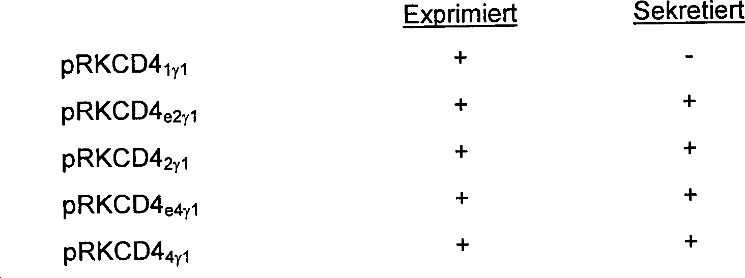
Es wurden ebenfalls Plasmide konstruiert, um die Expression von Fusionsproteinen zu steuern, die verschiedene Längen der aminoterminalen extrazellulären Domäne von CD4 enthielten, die an den trunkierten Teil der konstanten Region des menschlichen Immunglobulins γ1 fusioniert sind, und lediglich die Gelenkregion und die konstanten Domänen CH2 und CH3 umfassen.Plasmids were also constructed to direct the expression of fusion proteins containing different lengths of the amino-terminal extracellular domain of CD4 fused to the truncated portion of the human immunoglobulin γ1 constant region, and only the hinge region and the constant domains CH 2 and CH 3 include.
Synthetische Oligonucleotide wurden als Primer für Mutagenesereaktionen hergestellt, um die Immunglobulinsequenz von Ser114 bis Cys215 (inklusive) zu deletieren (Kabat et al.). Diese wurden als 48-mere, die 24 Nucleotide auf jeder Seite der ge wünschten Fusionsstelle umfassen (d.h. entsprechend den 8 COOH-terminalen Resten der gewünschten CD4-Gruppierung sowie den 8 NH2-terminalen Resten der gewünschten Immunglobulin-Gruppierung), synthetisiert. Die Plasmide pRKCD44γ1, pRKCD42γ1 und pRKCD41γ1 wurden getrennt voneinander in den E.-coli-Stamm SR101 transformiert, und die transformierte Kultur wurde auf Ampicillin-Mediumplatten ausplattiert. Es wurden resistente Kolonien ausgewählt und in Gegenwart von m13K07-Helfer-Bakteriophagen kultiviert, um sekretierte, enkapsidierte, einzelsträngige Matrizen dieser Plasmide zu erhalten. Die einzelsträngige Plasmid-DNA wurde isoliert und mit den oben beschriebenen synthetischen Oligonucleotiden als Primer als Matrize für Mutagenesereaktionen verwendet. Die Mutagenesereaktionen wurden in E. coli SR101 transformiert, und die transformierte Kultur wurde auf Ampicillin-Mediumplatten ausplattiert. Die Transformanten wurden mittels Koloniehybridisierung (Grunstein-Hogness) unter Verwendung von 16-meren als Sonden auf die Gegenwart der geeigneten Fusionsstelle gescreent. Diese 16-mere umfassen 8 Basen an jeder Seite der Fusionsstelle, und die ausgewählten Hybridisierungsbedingungen waren stringent genug, sodass die Sonden lediglich das korrekt fusionierte Produkt nachweisen. Als positiv identifizierte Kolonien wurden ausgewählt, und die Plasmid-DNA wurde isoliert und in den E.-coli-Stamm SR101 transformiert. Die transformierten Kulturen wurden auf Ampicillin-Mediumplatten ausplattiert, und es wurden resistente Kolonien ausgewählt und in Gegenwart von m13K07-Bakteriophagen kultiviert. Matrizen wurden wie oben beschrieben hergestellt und mittels Sequenzierung gescreent.Synthetic oligonucleotides were prepared as primers for mutagenesis reactions to delete the immunoglobulin sequence from Ser114 to Cys215 (inclusive) (Kabat et al.). These were synthesized as 48-mers comprising 24 nucleotides on each side of the desired fusion site (ie corresponding to the 8 COOH-terminal residues of the desired CD4 moiety as well as the 8 NH 2 -terminal residues of the desired immunoglobulin moiety). The plasmids pRKCD4 4 γ 1 , pRKCD4 2 γ 1 and pRKCD4 1 γ 1 were separately transformed into the E. coli strain SR101, and the transformed culture was plated on ampicillin medium plates. Resistant colonies were selected and cultured in the presence of m13K07 helper bacteriophage to obtain secreted, encapsidated, single-stranded templates of these plasmids. The single-stranded plasmid DNA was isolated and used with the synthetic oligonucleotides described above as a primer as a template for mutagenesis reactions. The mutagenesis reaction were transformed into E. coli SR101 and the transformed culture plated on ampicillin medium plates. The transformants were screened by colony hybridization (Grunstein-Hogness) using 16-mers as probes for the presence of the appropriate fusion site. These 16-mers comprise 8 bases on either side of the fusion site, and the hybridization conditions chosen were stringent enough that the probes detect only the correctly fused product. Positively identified colonies were selected and the plasmid DNA was isolated and transformed into E. coli strain SR101. The transformed cultures were plated on ampicillin media plates, and resistant colonies were selected and cultured in the presence of m13K07 bacteriophage. Matrices were prepared as described above and screened by sequencing.
Das vom Plasmid pRKCD44γ1 abstammende Plasmid wird als pRKCD44Fc1 bezeichnet, jenes, das vom Plasmid pRKCD42γ1 abstammt, wird als pRKCD42Fc1 bezeichnet und jenes, das vom Plasmid pRKCD41γ1 abstammt, wird als pRKCD41Fc1 bezeichnet.The plasmid pRKCD4 4 γ 1 derived plasmid is referred to as pRKCD4 4Fc1 , that which is derived from the plasmid pRKCD4 2γ1 is referred to as pRKCD4 2Fc1 and that derived from the plasmid pRKCD4 1γ1 , is referred to as pRKCD4 1Fc1 .
pRKCD42Fc1, pRKCD41Fc1 und pRKCD44Fc1 werden auf dieselbe Art und Weise wie oben beschrieben kultiviert, und die CH1-deletierten CD4-Immunadhesone werden wie hierin anderorts beschrieben gewonnen.pRKCD4 2Fc1 , pRKCD4 1Fc1 and pRKCD4 4Fc1 are cultured in the same manner as described above, and the CH1-deleted CD4 immunoadhesones are recovered as described elsewhere herein.
LeichtkettenfusionenLight chain mergers
Es wurden Plasmide konstruiert, um die Expression von Proteinen zu steuern, die verschiedene Längen der aminoterminalen extrazellulären Domäne von CD4 enthielten, die an die konstante Region des menschlichen Immunglobulins κ fusioniert ist. Diese Plasmide werden als pRKCD44κ und pRKCD4e4κ bezeichnet.Plasmids were constructed to direct the expression of proteins containing different lengths of the amino-terminal extracellular domain of CD4 fused to the human immunoglobulin constant region κ. These plasmids are referred to as pRKCD4 4 κ and pRKCD4 e4 κ.
Das Plasmid pRKCD44κ enthält den Abschnitt des CD4-Gens vom Initiationscodon bis zur Fusionsstelle nach dem Codon für den Serin-Rest 366 des reifen CD4-Polypeptids, unmittelbar gefolgt von der Sequenz für die konstante Region des menschlichen Immunglobulins κ, beginnend beim Codon für den Threonin-Rest 109 des reifen menschlichen Immunglobulins κ (Kabat et al.).The plasmid pRKCD4 4 κ contains the portion of the CD4 gene from the initiation codon to the fusion site after the codon for the serine residue 366 of the mature CD4 polypeptide, immediately followed by the sequence for the human immunoglobulin constant region κ, starting at the codon for the threonine residue 109 of the mature human immunoglobulin κ (Kabat et al.).
Das Plasmid pRKCD4e4κ enthält den Teil des CD4-Gens vom Initiationscodon bis zur Fusionsstelle nach dem Codon für den Lysin-Rest 360 des reifen CD4-Polypeptids, unmittelbar gefolgt von der Sequenz für die konstante Region des menschlichen Immunglobulins κ, beginnend beim Codon für den Threonin-Rest 109 des reifen menschlichen Immunglobulins κ (Kabat et al.).The plasmid pRKCD4 e4 κ contains the portion of the CD4 gene from the initiation codon to the fusion site after the codon for the lysine residue 360 of the mature CD4 polypeptide, immediately followed by the sequence for the human immunoglobulin constant region κ, starting at the codon for the threonine residue 109 of the mature human immunoglobulin κ (Kabat et al.).
Diese
Plasmide wurden analog zu den oben beschriebenen Plasmiden pRKCD44γ1 und pRKCD4e4γ1 konstruiert,
jedoch mit folgender Ausnahme:
Die kodierende Sequenz des menschlichen
Immunglobulins κ (
The coding sequence of the human immunoglobulin κ (
Expression in CHO-Zellenexpression in CHO cells
Plasmide wurden oder werden konstruiert, um die Expression der oben beschriebenen Immunadhesone in CHO-Zellen zu steuern. Sie werden als pSVeCD44γ1SVDHFR, pSVeCD42γ1SVDHFR, pSVeCD41γ1SVDHFR, pSVeCD4e4γ1SVDHFR, pSVeCD4e2γ1SVDHFR, pSVeCD4e1γ1SVDHFR, pSVeCD44Fc1SVDHFR, pSVeCD42Fc1SVDHFR, pSVeCD41Fc1SVDHFR, pSVeCD44κSVDHFR und pSVeCD42κSVDHFR bezeichnet.Plasmids have been or are being constructed to direct the expression of the above-described immunoadhesons in CHO cells. They are called pSVeCD4 4 γ 1 SVDHFR, pSVeCD4 2 γ 1 SVDHFR, pSVeCD4 1 γ 1 SVDHFR, pSVeCD4 e4 γ 1 SVDHFR, pSVeCD4 e2 γ 1 SVDHFR, pSVeCD4 e1 γ 1 SVDHFR, pSVeCD4 4Fc1 SVDHFR, pSVeCD4 2Fc1 SVDHFR, pSVeCD4 1Fc1 SVDHFR , pSVeCD4 4 κSVDHFR and pSVeCD4 2 κSVDHFR.
Fragment 31 wurde wie oben beschrieben hergestellt. Fragment 32a wurde durch den Verdau des Plasmids pE348HBV.E400 D22 mit BamHI, Abstumpfen mit dem Klenow-Fragment und den vier dNTPs und darauf folgendem Verdau mit PvuI und Isolation des großen Fragments hergestellt, das den Rest des β-Lactamase-Gens und den frühen Promotor von SV40 sowie das DHFR-Gen enthielt. Die Plasmide pRKCD44γ1, pRKCD42γ1, pRKCD41γ1, pRKCD4e4γ1, pRKCD4e2γ1, pRKCD4e1γ1, pRKCD44Fc1, pRKCD42Fc1, pRKCD41Fc1, pRKCD44κ und pRKCD42κ wurden getrennt voneinander mit HindIII verdaut, mit dem Klenow-Fragment und den vier dNTPs abgestumpft, dann mit EcoRI verdaut, und die für das CD4-Ig-Fusionsprotein kodierenden Fragmente wurden isoliert. Die resultierenden DNA-Fragmente wurden mit den Fragmenten 31 und 32a zusammen ligiert und in den E.-coli-Stamm 294 transformiert. Kolonien wurden ausgewählt und wie oben auf die Gegenwart des korrekten Plasmids untersucht, dann in CHO-Zellen transfiziert und durch Methotrexatselektion unter Verwendung konventioneller Verfahren amplifiziert.Fragment 31 was prepared as described above. Fragment 32a was prepared by digestion of the plasmid pE348HBV.E400 D22 with BamHI, blunting with the Klenow fragment and the four dNTPs and subsequent digestion with PvuI and isolation of the large fragment containing the remainder of the β-lactamase gene and the early Promoter of SV40 and the DHFR gene. Plasmids pRKCD4 4 γ 1, pRKCD4 2 γ 1, pRKCD4 1 γ 1, pRKCD4 e4 γ 1, pRKCD4 e2 γ 1, pRKCD4 e1 γ 1, pRKCD4 4Fc1, pRKCD4 2Fc1, pRKCD4 1Fc1, pRKCD4 4κ and pRKCD4 2κ were separately with HindIII digested, blunted with the Klenow fragment and the four dNTPs, then digested with EcoRI, and fragments encoding the CD4-Ig fusion protein were isolated. The resulting DNA fragments were ligated together with fragments 31 and 32a and transformed into E. coli strain 294. Colonies were selected and as described above Presence of the correct plasmid, then transfected into CHO cells and amplified by methotrexate selection using conventional procedures.
Beispiel 5Example 5
Kultivierung, Reinigung und Formulierung von CD4-VariantenCultivation, purification and formulation of CD4 variants
Für lösliche CD4-Adhesone, wie z.B. CD4T, CD4TP, oder lösliche CD4-Immunadhesone kodierende Plasmide wurden in CHO-DP7 (eine proinsulintransformierte, autokrine Wirtszelle, die von CHO abstammt; U.S.S.N. 97.472) calci umphosphat-transfiziert, und die Transformanten wurden in selektivem Medium (1:1 HAM F12/DMEM GHT-, das 1–10 % diafiltriertes oder dialysiertes Rinderserum enthielt) kultiviert. Andere geeignete Wirtszellen sind CHO-Zellen oder menschliche embryonale 293S-Nierenzellen. Die Transformanten wurden durch Methotrexatselektion im selben Medium, das jedoch 500 nm Methotrexat enthielt, amplifiziert. Es wurde ein Subklon ausgewählt, der fähig war, CD4TP, CD4tp 500 b zu sekretieren. CD4tp 500 b wird in einem DMEM/HAM-F12-Medium bei etwa 37 °C kultiviert, bis das CD4TP in der Kultur akkumuliert, wonach das Medium von den Zellen und von unlöslichen Stoffen durch Zentrifugieren getrennt wird.Plasmids encoding soluble CD4 adhesons such as CD4T, CD4TP, or soluble CD4-immunoadhesins were transfected into CHO-DP7 (a proinsulin-transformed autocrine host cell derived from CHO; USSN 97,472) and the transformants were transfected into selective Medium (1: 1 HAM F12 / DMEM GHT - containing 1-10% diafiltered or dialyzed bovine serum). Other suitable host cells are CHO cells or 293S human embryonic kidney cells. The transformants were amplified by methotrexate selection in the same medium but containing 500 nm methotrexate. A subclone was selected that was able to secrete CD4TP, CD4tp 500b. CD4tp 500b is cultured in a DMEM / HAM-F12 medium at about 37 ° C until the CD4TP accumulates in the culture, after which the medium is separated from the cells and insoluble matter by centrifugation.
Kulturfluid von CD4TP-Transformanten wurde konzentriert und diafiltriert, um die Ionenstärke zu reduzieren. Das Konzentrat wurde durch eine große Menge an Q-Sepharose-Anionenaustauschharz (zuvor mit 25 mM NaCl, pH 8,5, äquilibriert) laufen gelassen, um verunreinigende Stoffe aus der Kulturflüssigkeit zu adsorbieren. Der isoelektrische Punkt von CD4TP liegt bei etwa 9,5, was eine Unterscheidung zwischen den trunkierten Formen von CD4 und den meisten Verunreinigungen aufgrund von unterschiedlicher Adsorption auf einem Kationenaustauschharz, wie z.B. Carboxymethyl- oder Sulfonylsepharose, bzw. einem Anionenaustauschharz, wie z.B. quaternäre Ammoniumsepharose, ermöglicht. Zusätzlich wird, da hochgradig elektropositive Domänen im extrazellulären Segment von CD4 vorhanden sind, jede CD4-haltige Variante auf dieselbe Art und Weise wie CD4TP gereinigt. Die nicht adsorbierte Kulturflüssigkeit aus dem Anionenaustauschharz involvierenden Schritt wurde dann durch ein Kationenaustauschharz (zuvor mit 25 mM NaCl, pH 8,5, äquilibriert) laufen gelassen, wodurch CD4TP an das Harz adsorbiert wurde. Das CD4TP wurde mit einem NaCl-Gradienten bei einem pH von 8,5 eluiert, wobei diese CD4-Variante bei etwa 0,2 M NaCl eluiert. Ammoniumsulfat wurde zu dem Eluat bis zu einer Konzentration von 1,7 M hinzugefügt, und die Lösung wurde durch eine Säule aus hydrophobem Wechselwirkungschromatographieharz (Phenyl- oder Butylsepharose) laufen gelassen. Das CD4TP wurde aus der hydrophoben Wechselwirkungssäule mit einem Gradienten aus Ammoniumsulfat eluiert, wobei das CD4TP bei etwa 0,7 M Ammoniumsulfat austrat. Das Eluat wurde konzentriert, und der Puffer wurde auf einer G-25-Säule unter Verwendung von phosphatgepufferter Salzlösung, die 0,02 % (Gew./Vol.) Tween 20 oder Tween 80 enthielt, ausgetauscht. Das CD4TP war löslich und stabil in dieser Lösung, die sterilfiltriert war und als wässrige Formulierung in Phiolen gefüllt wurde. Andere polymere, nichtionische Tenside werden geeigneterweise mit den CD4-Formulierungen verwendet, unter anderem Pluronic-Block-Copolymere oder Polyethylenglykol.Culture fluid CD4TP transformants were concentrated and diafiltered to the ionic strength to reduce. The concentrate was made by a large amount on Q-sepharose anion exchange resin (previously equilibrated with 25 mM NaCl, pH 8.5,) run to contaminants from the culture fluid to adsorb. The isoelectric point of CD4TP is about 9.5, which makes a distinction between the truncated forms of CD4 and most impurities due to different Adsorption on a cation exchange resin, e.g. carboxymethyl or sulfonylsepharose, or an anion exchange resin, e.g. quaternary Ammonium Sepharose, allows. additionally is because highly electropositive domains in the extracellular segment of CD4 are present, each variant containing CD4 in the same way and how to clean CD4TP. The unadsorbed culture fluid The step involving the anion exchange resin was then carried out a cation exchange resin (previously equilibrated with 25 mM NaCl, pH 8.5) leaving CD4TP adsorbed to the resin. The CD4TP was eluted with a NaCl gradient at pH 8.5, with this CD4 variant eluted at about 0.2 M NaCl. Ammonium sulfate became the eluate until added to a concentration of 1.7M, and the solution became through a pillar hydrophobic interaction chromatography resin (phenyl or butyl sepharose) run. The CD4TP was isolated from the hydrophobic interaction column with eluted from a gradient of ammonium sulfate, wherein the CD4TP at about 0.7 M ammonium sulfate emerged. The eluate was concentrated and the buffer was loaded on a G-25 column using phosphate buffered Saline, containing 0.02% (w / v) of Tween 20 or Tween 80. The CD4TP was soluble and stable in this solution, which was sterile filtered and used as an aqueous formulation in vials filled has been. Other polymeric, nonionic surfactants will suitably used with the CD4 formulations, including Pluronic block copolymers or Polyethylene glycol.
Es ist ebenso möglich, eine Immunaffinitätsreinigung von löslichem CD4 durchzuführen, wobei das CD4 an einen immobilisierten Antikörper gegen CD4 adsorbiert wird. Dieses Verfahren leidet unter dem Nachteil, dass eine Elution des löslichen CD4 unter sauren Bedingungen zu einer Proteinaggregation führt, die lediglich bei relativ hohen Tensidmengen weitgehend zurückgeht. Das vorhergehende Verfahren ermöglicht die Verwendung von viel geringeren Tensidmengen, etwa von 0,01 bis 0,10 (Gew./Vol.) Tensid.It is also possible an immunoaffinity purification of soluble Perform CD4, wherein the CD4 is adsorbed to an immobilized antibody to CD4. This process suffers from the disadvantage that elution of the soluble CD4 under acidic conditions leads to protein aggregation, the only largely decreases at relatively high amounts of surfactant. The previous procedure allows the use of much lower levels of surfactant, such as from 0.01 to 0.10 (w / v) surfactant.
Das angewandte Verfahren für die Reinigung von CD4-Fusionen mit Immunglobulin-Schwerketten bestand in der Konzentration rekombinanter Überstände mittels Ultrafiltration und darauf folgender Adsorption der Fusion an harz-immobilisiertem Staphylokokken-Protein A. Die Fusion wurde mit 0,1 M Citratpuffer mit einem pH von 3 ohne Salz oder Detergens eluiert. Dieses Präparat wird in Tris-Puffer bei einem pH von 7,5 gepuffert. Die Immunglobulin-Fusionen mit V1-V4 von CD4 werden gegebenenfalls durch das oben für nicht fusionierte CD4-Varianten beschriebene Verfahren weiter gereinigt. CD4-Immunglobulin-Fusionen mit V1-V4 von CD4 können ebenfalls durch das oben beschriebene Verfahren gereinigt werden, jedoch mit der Ausnahme, dass nicht erwartet wird, dass der isoelektrische Punkt dieser Molekülklasse so alkalisch ist wie jener der Arten, die alle vier V-Regionen von CD4 enthalten.The applied method for the purification of CD4 fusions with immunoglobulin heavy chains was in the concentration recombinant supernatants by means of Ultrafiltration and subsequent adsorption of the resin-immobilized fusion Staphylococcal protein A. The fusion was done with 0.1 M citrate buffer eluted with a pH of 3 without salt or detergent. This preparation will buffered in Tris buffer at pH 7.5. The immunoglobulin fusions with V1-V4 of CD4 are optionally not through the above for fused CD4 variants described further purified. CD4 immunoglobulin fusions with V1-V4 of CD4 can also be made by the above be cleaned, but with the exception that that is not expected to be the isoelectric point of this class of molecules as alkaline as that of the species, all four V regions of CD4 included.
Beispiel 6Example 6
Es
wurden die Merkmale mehrerer Adhesonvarianten bestimmt. Wie in Tabelle
4 gezeigt wird, zeigen die Immunadhesone CD44γ1 und
CD42γ1 eine verbesserte Plasmahalbwertszeit in
Kaninchen, gekoppelt mit hochaffiner gp120-Bindung und einer Affi nität zum Fcγ-Rezeptor
(bestimmt durch U937-Zellen), die mit der des Hauptteils des menschlichen
IgG1 vergleichbar ist. Tabelle
4 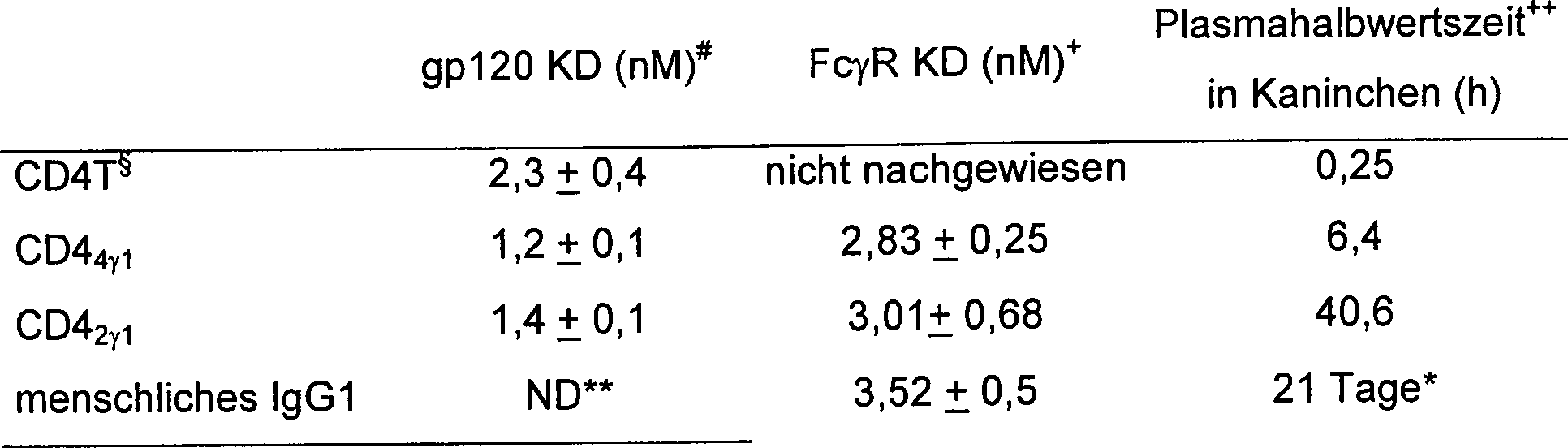
- * in Menschen bestimmt
- + KD wurde durch das Verfahren von Anderson et al., J. Immunol. 125, 2735-2741 (1980), bestimmt.
- # durch das Verfahren von Smith et al., Science 238, 1704-07 (1987), bestimmt.
- § nur Reste 1-368
- ++ Die Adhesonvariante wurde Kaninchen intravenös injiziert, und es wurden in regelmäßigen Abständen Blutproben genommen und auf die Gegenwart der Adhesonvariante getestet.
- ** nicht durchgeführt
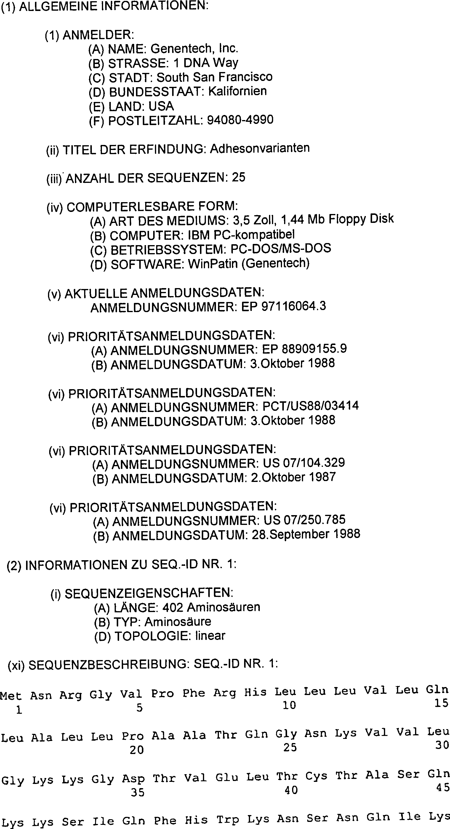
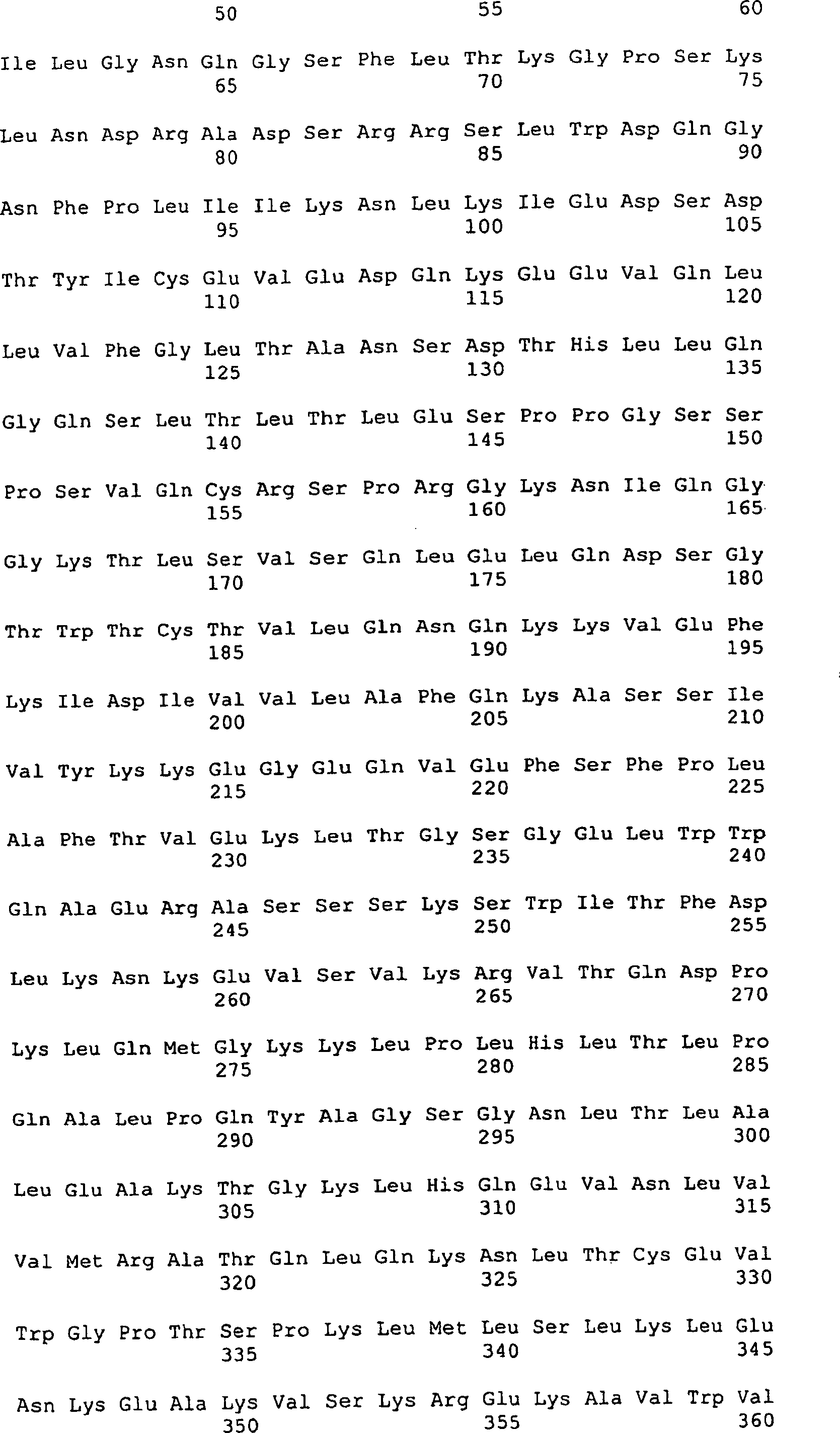


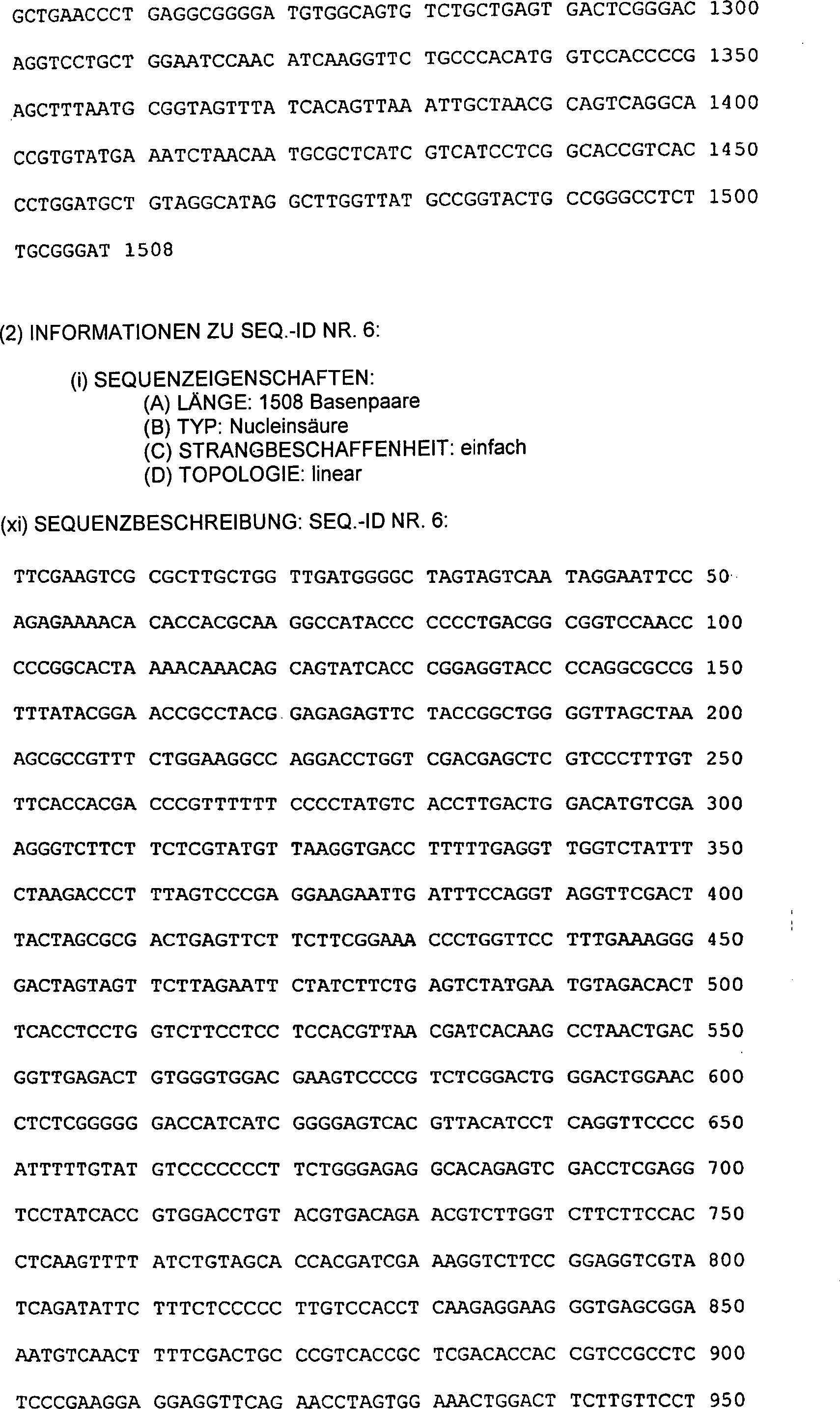
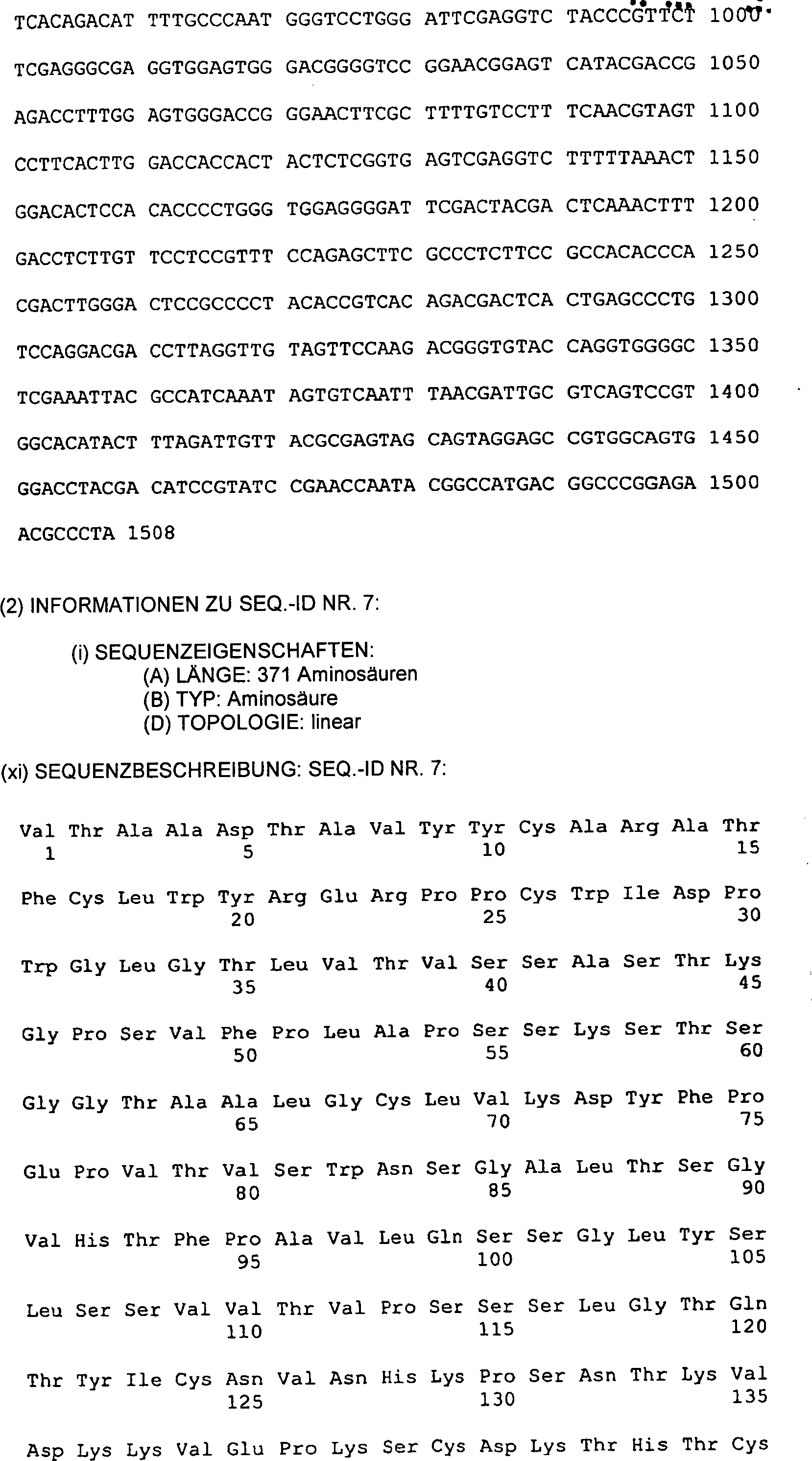


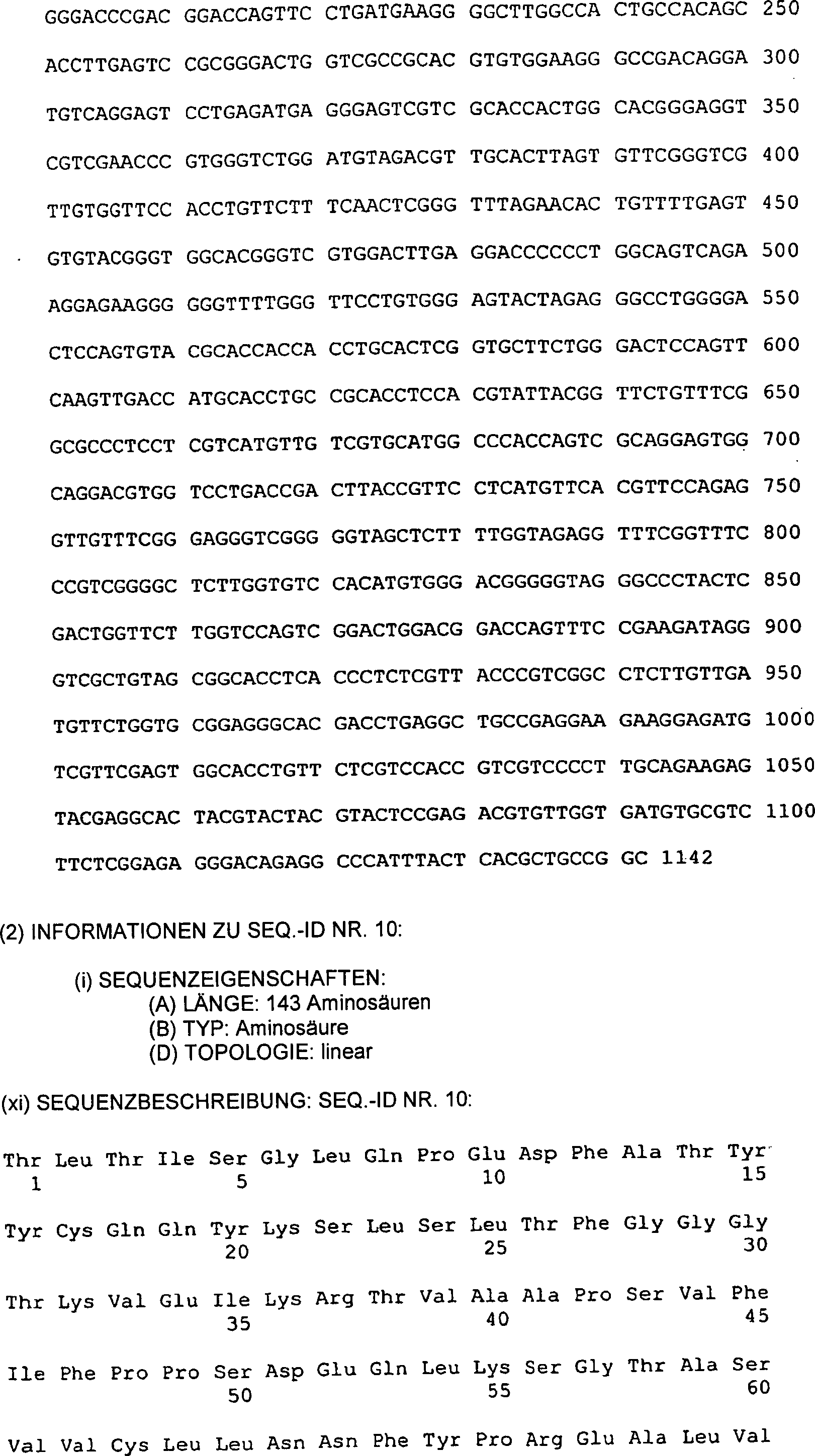


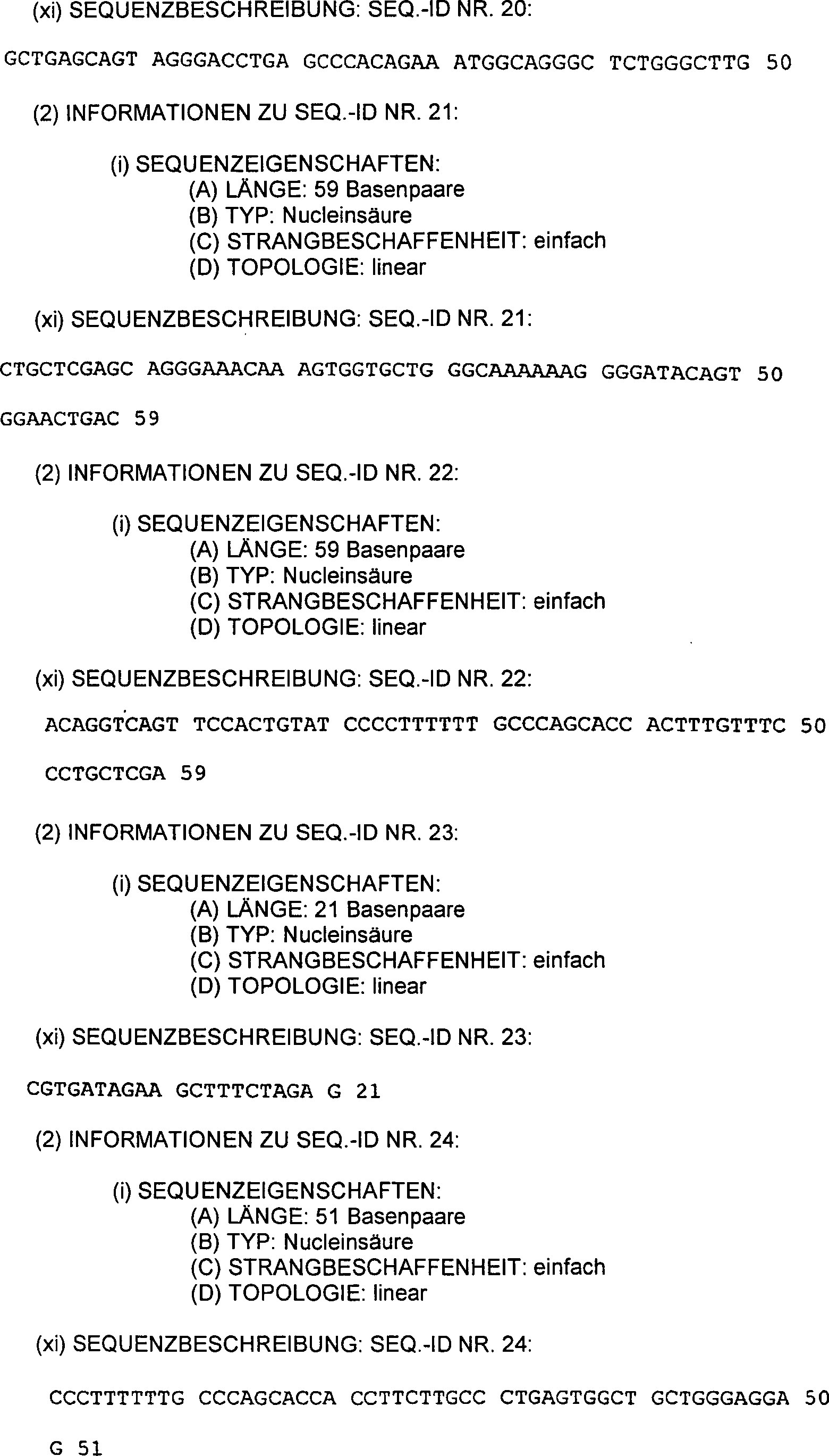

- * determined in humans
- + KD was synthesized by the method of Anderson et al., J. Immunol. 125, 2735-2741 (1980).
- # by the method of Smith et al., Science 238, 1704-07 (1987).
- § only residues 1-368
- ++ The Adheson variant was injected intravenously into rabbits and blood samples were taken at regular intervals and tested for the presence of the Adheson variant.
- ** not done
Claims (7)
Applications Claiming Priority (2)
| Application Number | Priority Date | Filing Date | Title |
|---|---|---|---|
| US10432987A | 1987-10-02 | 1987-10-02 | |
| US104329 | 1987-10-02 |
Publications (2)
| Publication Number | Publication Date |
|---|---|
| DE3856592D1 DE3856592D1 (en) | 2006-09-21 |
| DE3856592T2 true DE3856592T2 (en) | 2007-10-04 |
Family
ID=22299914
Family Applications (1)
| Application Number | Title | Priority Date | Filing Date |
|---|---|---|---|
| DE3856592T Expired - Lifetime DE3856592T2 (en) | 1987-10-02 | 1988-10-03 | Adhäsionsvarianten |
Country Status (4)
| Country | Link |
|---|---|
| EP (1) | EP0832971B1 (en) |
| AT (1) | ATE335817T1 (en) |
| DE (1) | DE3856592T2 (en) |
| ZA (1) | ZA887365B (en) |
Families Citing this family (4)
| Publication number | Priority date | Publication date | Assignee | Title |
|---|---|---|---|---|
| US20070071744A1 (en) | 2002-06-07 | 2007-03-29 | Gotz Munch | Agents which bind to epitopes of glycoprotein VI |
| US7531178B2 (en) | 2002-06-07 | 2009-05-12 | Trigen Gmbh | Immunoadhesin comprising a glycoprotein VI domain |
| EP1369128A1 (en) | 2002-06-07 | 2003-12-10 | Procorde GmbH | Inhibitors of glycoprotein VI and their therapeutic use |
| EP2612868B1 (en) | 2007-11-01 | 2018-08-15 | Astellas Pharma Inc. | Immunosuppressive polypeptides and nucleic acids |
Family Cites Families (9)
| Publication number | Priority date | Publication date | Assignee | Title |
|---|---|---|---|---|
| US5110906A (en) * | 1986-08-21 | 1992-05-05 | The Trustees Of Columbia University In The City Of New York | Derivatives of soluble T-4 |
| DE3751751T2 (en) * | 1986-08-21 | 1996-10-17 | Univ Columbia | DNA ENCODING THE T CELL SURFACE PROTEIN T4 AND USE OF T4 FRAGMENTS IN THE TREATMENT OF AIDS |
| WO1988007376A1 (en) * | 1987-03-27 | 1988-10-06 | Hiroshi Okajima | Remedy for viral diseases |
| JPH02501192A (en) * | 1987-09-04 | 1990-04-26 | バイオジェン インコーポレイテッド | DNA sequences, recombinant DNA molecules and methods for producing soluble T4 protein |
| IL87902A0 (en) * | 1987-10-08 | 1989-03-31 | Dana Farber Cancer Inst Inc | Soluble human cd4 fragments and applications thereof |
| WO1989003221A1 (en) * | 1987-10-13 | 1989-04-20 | Genelabs Incorporated | Antisyncytial agent |
| PT88783B (en) * | 1987-10-23 | 1993-01-29 | Smithkline Beckman Corp | PROCESS OF PURIFICATION OF PROTEIN T4 SOLUABLE AND OF PRODUCTION OF THE SAME |
| PT89484B (en) * | 1988-01-22 | 1994-03-31 | Gen Hospital Corp | GENE CLONED CODES OF FUSAO PROTEINS IG-CD4 AND ITS UTILIZATION |
| AU3006789A (en) * | 1988-02-24 | 1989-08-24 | Smithkline Beckman Corporation | Expression of hiv binding proteins |
-
1988
- 1988-09-30 ZA ZA887365A patent/ZA887365B/en unknown
- 1988-10-03 DE DE3856592T patent/DE3856592T2/en not_active Expired - Lifetime
- 1988-10-03 AT AT97116064T patent/ATE335817T1/en not_active IP Right Cessation
- 1988-10-03 EP EP97116064A patent/EP0832971B1/en not_active Expired - Lifetime
Also Published As
| Publication number | Publication date |
|---|---|
| EP0832971A1 (en) | 1998-04-01 |
| ZA887365B (en) | 1990-05-30 |
| ATE335817T1 (en) | 2006-09-15 |
| EP0832971B1 (en) | 2006-08-09 |
| DE3856592D1 (en) | 2006-09-21 |
Similar Documents
| Publication | Publication Date | Title |
|---|---|---|
| DE3856189T3 (en) | ADHÄSIONSVARIANTEN | |
| US5565335A (en) | Adhesion variants | |
| Zettlmeissl et al. | Expression and characterization of human CD4: immunoglobulin fusion proteins | |
| AU684073B2 (en) | Synergistic composition of CD4-based protein and anti-HIV-1 antibody, and methods of using same | |
| DE3856580T2 (en) | Process and deoxyribonucleic acid for the production of tissue factor protein | |
| DE69534589T2 (en) | TARGETED CELL LYSIS OF HIV-INFECTED CELLS WITH CHIMERIC CD4 RECEPTOR-CARRYING CELLS | |
| DE69634855T2 (en) | NEUORIENTATION OF CELLULAR IMMUNITY BY RECEPTOR CHIMERIC | |
| DE69033705T3 (en) | HYBRID IMMUNOGLOBULINE | |
| DE69936927T2 (en) | POLY-SPECIFIC BINDING MOLECULES AND THEIR USE | |
| WO1993008829A1 (en) | Compositions that mediate killing of hiv-infected cells | |
| US5605689A (en) | Treatment of HIV-associated immune thrombocytopenic purpura | |
| DE69631331T2 (en) | CD40L MUTEIN | |
| DE69233480T2 (en) | CD4 gamma2 and CD4 IgG2 chimeras | |
| US20030104535A1 (en) | Adheson variants | |
| DE3856592T2 (en) | Adhäsionsvarianten | |
| AU618969B2 (en) | Adheson variants | |
| US7070991B2 (en) | Cells expressing a CD4-IgG2 chimeric heterotetramer |
Legal Events
| Date | Code | Title | Description |
|---|---|---|---|
| 8364 | No opposition during term of opposition |