CN1455873A - MRI method involving the use of a hyperpolarized contrast agent - Google Patents
MRI method involving the use of a hyperpolarized contrast agent Download PDFInfo
- Publication number
- CN1455873A CN1455873A CN01815595A CN01815595A CN1455873A CN 1455873 A CN1455873 A CN 1455873A CN 01815595 A CN01815595 A CN 01815595A CN 01815595 A CN01815595 A CN 01815595A CN 1455873 A CN1455873 A CN 1455873A
- Authority
- CN
- China
- Prior art keywords
- atomic nucleus
- magnetic resonance
- compound
- described method
- sample
- Prior art date
- Legal status (The legal status is an assumption and is not a legal conclusion. Google has not performed a legal analysis and makes no representation as to the accuracy of the status listed.)
- Pending
Links
Classifications
-
- A—HUMAN NECESSITIES
- A61—MEDICAL OR VETERINARY SCIENCE; HYGIENE
- A61B—DIAGNOSIS; SURGERY; IDENTIFICATION
- A61B5/00—Measuring for diagnostic purposes; Identification of persons
- A61B5/05—Detecting, measuring or recording for diagnosis by means of electric currents or magnetic fields; Measuring using microwaves or radio waves
- A61B5/055—Detecting, measuring or recording for diagnosis by means of electric currents or magnetic fields; Measuring using microwaves or radio waves involving electronic [EMR] or nuclear [NMR] magnetic resonance, e.g. magnetic resonance imaging
-
- G—PHYSICS
- G01—MEASURING; TESTING
- G01R—MEASURING ELECTRIC VARIABLES; MEASURING MAGNETIC VARIABLES
- G01R33/00—Arrangements or instruments for measuring magnetic variables
- G01R33/20—Arrangements or instruments for measuring magnetic variables involving magnetic resonance
- G01R33/44—Arrangements or instruments for measuring magnetic variables involving magnetic resonance using nuclear magnetic resonance [NMR]
- G01R33/48—NMR imaging systems
- G01R33/54—Signal processing systems, e.g. using pulse sequences ; Generation or control of pulse sequences; Operator console
- G01R33/56—Image enhancement or correction, e.g. subtraction or averaging techniques, e.g. improvement of signal-to-noise ratio and resolution
- G01R33/5601—Image enhancement or correction, e.g. subtraction or averaging techniques, e.g. improvement of signal-to-noise ratio and resolution involving use of a contrast agent for contrast manipulation, e.g. a paramagnetic, super-paramagnetic, ferromagnetic or hyperpolarised contrast agent
-
- G—PHYSICS
- G01—MEASURING; TESTING
- G01R—MEASURING ELECTRIC VARIABLES; MEASURING MAGNETIC VARIABLES
- G01R33/00—Arrangements or instruments for measuring magnetic variables
- G01R33/20—Arrangements or instruments for measuring magnetic variables involving magnetic resonance
- G01R33/44—Arrangements or instruments for measuring magnetic variables involving magnetic resonance using nuclear magnetic resonance [NMR]
- G01R33/48—NMR imaging systems
- G01R33/54—Signal processing systems, e.g. using pulse sequences ; Generation or control of pulse sequences; Operator console
- G01R33/56—Image enhancement or correction, e.g. subtraction or averaging techniques, e.g. improvement of signal-to-noise ratio and resolution
- G01R33/563—Image enhancement or correction, e.g. subtraction or averaging techniques, e.g. improvement of signal-to-noise ratio and resolution of moving material, e.g. flow contrast angiography
- G01R33/56308—Characterization of motion or flow; Dynamic imaging
-
- G—PHYSICS
- G01—MEASURING; TESTING
- G01R—MEASURING ELECTRIC VARIABLES; MEASURING MAGNETIC VARIABLES
- G01R33/00—Arrangements or instruments for measuring magnetic variables
- G01R33/20—Arrangements or instruments for measuring magnetic variables involving magnetic resonance
- G01R33/44—Arrangements or instruments for measuring magnetic variables involving magnetic resonance using nuclear magnetic resonance [NMR]
- G01R33/48—NMR imaging systems
- G01R33/54—Signal processing systems, e.g. using pulse sequences ; Generation or control of pulse sequences; Operator console
- G01R33/56—Image enhancement or correction, e.g. subtraction or averaging techniques, e.g. improvement of signal-to-noise ratio and resolution
- G01R33/567—Image enhancement or correction, e.g. subtraction or averaging techniques, e.g. improvement of signal-to-noise ratio and resolution gated by physiological signals, i.e. synchronization of acquired MR data with periodical motion of an object of interest, e.g. monitoring or triggering system for cardiac or respiratory gating
- G01R33/5673—Gating or triggering based on a physiological signal other than an MR signal, e.g. ECG gating or motion monitoring using optical systems for monitoring the motion of a fiducial marker
Abstract
The present invention provides a method of contrast enhanced magnetic resonance imaging of a sample, said method comprising: a) administering a hyperpolarised MR contrast agent comprising non-zero nuclear spin nuclei int o said sample for fluid dynamic investigations of the vasculature, b) exposing said sample or part of the sample to radiation of a frequency selected to excite nuclear spin transitions in said non-zero nuclear spin nuclei, c) detecting MR signals from said sample using any suitable manipulation method including pulse sequences. The invention also provides novel compounds.
Description
The present invention relates to MR imaging method (MRI), in particular for magnetic resonance angiography (MRA) be used for the fluid dynamics research and the novel hyperpolarized contrast agents application therein of vascular system.
Because magnetic resonance imaging is non-invasive, and needn't allow patient be exposed in the radiation that potential hazard is arranged such as the X-ray in the diagnosis research process, so magnetic resonance imaging becomes the diagnostic techniques to clinician's very attractive.
Magnetic resonance signal intensity depends on the population difference between the nuclear nuclear spin state of imaging.For the effective contrast between the magnetic resonance image (MRI) that obtains histological types, people just know the patient are used mr contrast agent (as the paramagnetic metal material) for a long time, and the mr contrast agent influence is being used the zone or gathered relaxation time in zone.
The MRA that contrast now strengthens is based on and injects the paramagnetism contrast medium is present in the hydrogen atom of blood vessel with shortening relaxation time.By using short imaging pulse sequence of repetition time (TR), background has obtained inhibition.But T
2The relaxation time weak point can cause that acquisition time is short, sampling rate is high and signal to noise ratio (snr) reduces.
Angiography can also be utilized " in-flow " technology not need any contrast medium and realize.This method also relies on uses short sequence of repetition time to suppress to want the fixedly spin that exists in the imaging volume.Therefore, will cause sampling rate height and SNR to reduce.
The MRA of enhancing contrast ratio and " in-flow " method both can utilize maximum intensity projection (MIP) software engineering to generate angiogram.The similar projection image of the angiogram that this method generates generation and X-ray mode becomes possibility.But, the exigent contrast noise ratio of image quality (CNR) of utilizing this method to generate; Because surrounding tissue is not had enough inhibition, therefore not having undesired signal is to be difficult to realize.
Therefore, one aspect of the present invention relates to a kind of MRA method that addresses the aforementioned drawbacks.By utilizing external nuclear spin polarization and the mr contrast agent of using nuclear spin polarization, so just can improve the MRA measuring method.These reagent comprise its structure can in uniform magnetic field, launch magnetic resonance signal (for example
1H,
13C,
15N,
19F,
29Si with
31The P atomic nucleus) and have a long T
1Relaxation time, and preferably have other long T
2Relaxation time.
The advantage of in-vitro method is to avoid with whole or whole in fact polarization agent administration to the sample that is studied, and still can obtain desirable nuclear spin polarization simultaneously in the magnetic resonance imaging agent.Therefore so less restriction that is subjected to physiologic factor of method, for example in vivo in the technology by the ability of using of reagent, biodegrability restriction with the toxicity generation.
When using the mr contrast agent of hyperpolarization, not hydrogen if detect atomic nucleus, then can eliminate background signal fully.Therefore might be not limited only to use the short pulse train of TR (this moment is in order to gather angiogram).Can utilize alternative sequence and linear sweep method, alternative sequence can more effectively be used available polarization, such as multiecho sequence (for example RARE, EPI, GREASE), full equilibrium gradient sequence (for example true FISP), stable state gradient sequence.The invention has the advantages that the extraction of having simplified the microcosmic flowing information.
The present invention utilizes the contrast medium of hyperpolarization to carry out magnetic resonance angiography (MRA), its some advantages are as described below :-the image that obtains does not have any background signal,-do not need to utilize the pulse train technology to suppress fixing spin,-show the projected image of blood vessel with any direction,-high s/n ratio, can be used for coronary angiography and T
1Relaxation time is long, so can strengthen the image away from the blood vessel of decanting point.
Developed be included in use comprise the non-zero nuclear spin atomic nucleus (for example
3He) contrast medium and measuring before the magnetic resonance signal, the technology of the external nuclear spin polarization that described contrast medium is carried out.
Also verified is, external can be to for example comprising
13C and
15The compound hyperpolarization of N, to produce the contrast medium of injectable polarization, for example by polarization transfer by inert gas, by " brute force ", by dynamical nuclear polarization (DNP) or contrary hydrogen methods (referring to, for example, the present patent application people's patent disclosure text WO-99/35508 and WO99/24080, the content of its disclosure comprises in this manual by reference).In these technology some used polarization transfer reagent, and it is defined as any agent that is suitable for causing the external polarization of mr contrast agent.
To generally speaking of the present invention, can use the hyperpolarization methods of any appropriate.In fact, it does not depend on employed hyperpolarization methods.But, under many situations, preferably use against the hyperpolarization methods of hydrogen and DNP.
After having carried out external hyperpolarization step, preferably polarization transfer reagent is separated from the potpourri that comprises the mr contrast agent that polarizes arbitrarily.Mr contrast agent is administered to health and injection enters patient to utilize any appropriate induction system to polarize then, to carry out the angiographic and/or fluid dynamics research of vascular system.
Therefore one aspect of the present invention relates to the magnetic sample resonant imaging method that a kind of contrast strengthens, sample is human body or inhuman animal body preferably, described method comprises: a) for example will comprise the nuclear hyperpolarization mr contrast agent of non-zero nuclear spin by injection and use and enter the described sample that is used for angiogram research, b) described sample or the described sample of part are exposed in the radiation of selected frequency, to excite the nuclear spin transition in described non-zero nuclear spin atomic nucleus, c) utilize the method for operating that comprises pulse train of any appropriate to detect the magnetic resonance signal of described sample, d) randomly guarantee to carry out pulse train and/or use contrast medium according to the rhythm of the heart and/or the breath rhythm gate of health, e) randomly, the signal that is obtained by described detection generates image, spectroscopic data, dynamic flow data or physiological data.
In some research and according to have zero background signal of the present invention one preferred aspect, can carry out projection along the desired directions of study discussion blood vessel and generate angiogram by utilizing.Owing to do not have background signal, therefore reduced the risk of the non-natural sign of appearance " back of the body fold ".It is particularly useful that this promptly carries out coronary angiography to another preferred aspect of the present invention.Can be used to generate the projection of whole heart to the image of stack piles such as heart work section with any assigned direction.This method has been imitated the implementation of X-ray angiography.
Study vascular system in being used for of using now and for example be used for the flow conventional fluid dynamic method of (perfusion) of microcosmic, method be based on the contrast pill by during tracer signal decline or by utilizing mark tracing tracer signal to decline to realize.This mark tracing has utilized the blood from the marked region to the imaging region to flow into and measure signal intensity changes as the foundation of calculating perfusion figure.Perfusion figure and the figure of RCBV (rCBV) that this method produces have limited signal to noise ratio (S/N ratio).
In traditional velocity survey situation, method is blood or comprises for example blood of the paramagnetism contrast medium of the contrast medium of Gd-base based on signal phase data and signal medium.But, this phase error sensitivity of velocity survey of utilizing phase method to causing by environmental organization.
When the mr contrast agent of hyperpolarization is used for method provided by the invention, not hydrogen if detect atomic nucleus, then can all eliminate background signal.Therefore, except the pulse train that can use those TR times weak points, can also use other pulse train.Can utilize alternative sequence and linear sweep method, alternative sequence can more effectively be used available polarization, such as multiecho sequence (RARE, EPI, GREASE), full equilibrium gradient sequence (for example trueFISP), stable state gradient sequence.The invention has the advantages that the extraction of having simplified the microcosmic flowing information.
Therefore by another aspect, the invention provides a kind of hydrodynamic force science study method, overcome above-mentioned defective thus vascular system.Be used to obtain and flow and measured value that microcosmic flows and/or the method for quantitative data are preferred.Especially preferred is to be used to obtain perfusion, flow velocity, flow curve, tissue perfusion figure and the RBV method of (comprising RCBV (rCBV) data).
Therefore the present invention relates to the MR imaging method to a kind of contrast enhancing of sample in yet another aspect, sample is human body or inhuman animal body preferably, described method comprises: a) for example will comprise the nuclear hyperpolarization mr contrast agent of non-zero nuclear spin by injection and use and enter the described sample that is used for vascular system fluid dynamics research, b) described sample or the described sample of part are exposed in the radiation of selected frequency, to excite the nuclear spin transition in described non-zero nuclear spin atomic nucleus, c) utilize the method for operating that comprises pulse train of any appropriate to detect the magnetic resonance signal of described sample, d) randomly guarantee to carry out pulse train and/or use contrast medium according to the rhythm of the heart and/or the breath rhythm gate of health, e) randomly, the signal that is obtained by described detection generates image, spectroscopic data, the dynamic flow data, the perfusion data, blood volume data and/or any other suitable physiological data.
According to a preferred embodiment of the present invention, the specific sequence of pulses of use depends on the flowing velocity in the vascular group for the treatment of imaging.In some cases,, monopulse sequence (for example EPI, RARE, GREASE, BURST, QUEST) preferably quick for the imaging coronarius of heart.
Any diffusion that the method that utilization is proposed by people such as Stajskal can be measured the hyperpolarized contrast agents molecule, this method is called Stajskal-Tanner (ST) method in standard NMR and MRI document.The ST sequence is gone phase place by two equidimension gradient pulses that utilize 180 ° of pulses at interval to proton and is carried out the phase place reunion subsequently and work.This gradient/rf pulse train can be formed the leading phase before the real data collecting part of pulse train.There are several different pulse trains (for example spin echo, EPI, STEAM, RARE) to revise in order to be used in combination the ST method.During the application of the ST of diffusion sequence part, the NMR-signal of proton is because T
2Relaxation and decaying.Effectively TE (echo time) often reaches 60ms or longer.Therefore the influence of relaxation is very strong.This relaxation will cause signal attenuation and cause signal to noise ratio (S/N ratio) to reduce.When use has long T
1/ T
2The hyperpolarization contrast medium time, the signal attenuation that causes owing to relaxation will reduce, the pulse train that use this moment has a long TE.
There is not background signal also to simplify the miniflow data computing of perfusion figure and the figure of RCBV (rCBV).Therefore this method is preferred aspect of the present invention.
Because the T of hyperpolarized contrast agents
1Relaxation time is long, therefore can show the blood vessel away from injection point, comprise cerebral blood vessel and lung blood vessel, and this is another preferred aspect of the present invention.
As the front in optional step of the present invention (step d) mentions, and, be necessary to carry out pulse train and/or use (for example injection) hyperpolarized contrast agents according to patient's heart rhythm and/or breath rhythm gate in order to optimize the visual window that is used for angiogram or the fluid dynamics of vascular system is studied.This gate can also be used to guaranteeing that organ/imaging capacity is in same position during gathering a series of images.For capacity/organ imaging to studying, the gate step can be before contrast medium medium pill passes through or during carry out.
Aspect all, preferably use a kind of mark or saturation technique of the present invention.This technology is used in and only shows in the image that obtains at last that those are by particular blood vessel or the hyperpolarization spin that entered imaging region by the given flow direction.It can also be used to removing the signal that is produced by the hyperpolarization spin of giving at imaging volume (for example in need profile shows heart coronarius) in the certain portions.
Preferred usage flag and saturation technique when gathering miniflow/data perfusion.This technology can be carried out by all hyperpolarization that terminates, and termination is to be used to realize from the influx of waiting to study the saturation pulse of volume and cause by the observation miniflow.Observation utilizes volume selectivity image pulse train to carry out.Can also utilize the spot scan method that the influx of the little volume unit of any inflow (voxel) is measured.The measurement of carrying out can comprise gathers spectral information and/or physiologic information, so that distinguish different types of organizations or/and flow velocity.
The present invention another preferred aspect, " intrinsic image " (image that promptly obtained before using the hyperpolarization mr contrast agent or resemble in traditional magnetic resonance experiment use formerly the image that obtains behind the mr contrast agent of handling without polarization) that can produce health is to provide structure (for example dissecting) information, the image that can superpose and obtain according to the inventive method in view of the above.Because
13C or
15The abundance of N in health is lower, so " intrinsic image " is not generally available to
13C or
15N is the nuclear situation of imaging.In this case, can provide anatomic information, can superpose in view of the above by production proton magnetic resonance (PMR) image
13C or
15The N image is for example referring to accompanying drawing 1c.
Utilize the phase-contrast technique of standard and/or extra gradient/rf pulse that spatial information or mobile message are encoded, can measurement flow rate.Utilization runs through the plane sequence can also measure velocity profile.
By " angiography " speech, we represent all researchs that are artery and capillary system about any radiography blood vessel.In some cases, the present invention also can be applicable to the vein measurement.Preferred aspect of the present invention has provided the MRA imaging of artery.
By " vascular system " speech, we represent that by containing the blood vessels pipe be all systems that artery, vein and kapillary constitute.
By " hyperpolarization " speech, we refer to be polarised on room temperature and following getable level of 1T, and degree of polarization preferably surpasses 0.1%, more preferably surpass 1%, more preferably surpass 10%.
Hyperpolarized contrast agents preferably has long T
2Relaxation time was preferably greater than 1 second, more preferably greater than 5 seconds.
According to the suitable magnetic resonance imaging agent of the present invention, can comprise such as
3Li,
13C,
15N,
19F,
29Si or
31P and
1The atomic nucleus of H, preferably
1H,
13C,
15N,
19F and
31The P atomic nucleus more preferably is
1H,
13C,
15N and
31The P atomic nucleus.Particularly preferably be
13The C atomic nucleus.
As mentioned above,
1H,
13C,
15N and
31The P atomic nucleus is highly suitable for the inventive method, wherein particularly preferably is
13The C atomic nucleus.
1H atomic nucleus advantage is concentration height in the natural abundance, and has the highest sensitivity in the atomic nucleus of all kinds.
13C atomic nucleus advantage is to come from hyperpolarization
13The nuclear background signal of C is very low, and ratio is as being derived from
1H is nuclear much lower.
19F atomic nucleus advantage is to have high sensitivity.Comprise
31The hyperpolarization of the nuclear contrast medium of P allows to use endogenous substance.
When the magnetic resonance imaging atomic nucleus is that atomic nucleus except proton (for example is
13C or
15N) time, (for example, do not have background signal basically
13C and
15The natural abundance of N can be ignored) interference, and benefit thus to make image contrast very high.It is especially true when mr contrast agent self enrichment is higher than the nuclear natural abundance of magnetic resonance imaging.Therefore be to provide significant spatial weighting to generate image according to method advantage of the present invention.
Preferably with manual type with having long T
1The atomic nucleus in relaxation time (for example,
15N and/or
13The C atomic nucleus) mr contrast agent being carried out enrichment handles.
Some
13C and
15The nuclear long T of N
1Relaxation time is particularly advantageous, and therefore comprises
13C and
15Some mr contrast agents of N are preferably used for the inventive method.Preferably, the mr contrast agent of polarization has effective degree of polarization greater than 0.1% atomic nucleus
13C, more preferably degree of polarization more preferably greater than 10%, is preferably greater than 25% greater than 1.0% especially, is preferably greater than 50% especially especially, and most preferably greater than 95%.
Mr contrast agent more preferably is to be enriched in carbonyl or quaternary carbon is locational
13C is known in carbonyl group or in some quaternary carbons
13The T that the C atomic nucleus can have
1Relaxation time is preferably more than 5 seconds usually greater than 2 seconds, especially is preferably more than 30 seconds.Preferably, to being enriched with
13The compound of C carries out deuterium-labeled, especially adjacency
13The C atomic nucleus.Preferably be enriched with
13The compound of C is wherein
13Around the C nuclear is one or more such as O, S, such non-magnetic resonance active nucleus or two key or the triple bond of C.
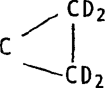
The mr contrast agent that uses in the inventive method has molecular formula (I):
CX
4(I) wherein each X is D, CD independently
3, CD
2OR
1, SO
3H, SO
2H, SO
2NH
2, CONR
1 2, CO
2H and OCHO, wherein R
1Be H or Me independently, perhaps two 3 yuan of rings of C atomic building that the X group is connected with them
Or 4 yuan of ring C
 Wherein Y is D or CD
2OR
1And Z is CD
2, CD (CD
2OR
1) or O.
Wherein Y is D or CD
2OR
1And Z is CD
2, CD (CD
2OR
1) or O.
The compound 1-17 that is hereinafter listed is the particular instance of the reagent that is suitable for using among the present invention.These reagent are water miscible, and are nontoxic, are easy to synthesize and have in water long T
1Value is for example above 60 seconds.
For example, the T that has respectively of compound 1 and 2
1Value is 95 seconds and 133 seconds.
Compound hereinafter is except 1-3 is known (they are disclosed among applicant's oneself the publication text WO-A-99/35508), and these reagent itself all are novel, and have constituted another aspect of the present invention.Example as compound 4-17 is presented at hereinafter.These reagent can carry out
13The C enrichment.
Another aspect of the present invention provides the magnetic resonance imaging agent that can tolerate on physiology composition, it comprises the carrier or the inert matter that can tolerate on magnetic resonance imaging agent and one or more physiology, described preparation is selected from the compound in the above-mentioned general formula (I), the compound of preferably following listed label 1-17, the compound of the label 4-17 that for example hereinafter lists.
Another aspect of the present invention provides in a method of the present invention uses above-mentioned general formula (I) compound, the compound of preferably following listed label 1-17, the compound of the label 4-17 that for example hereinafter lists.
The present invention another aspect in addition provides use above-mentioned general formula (I) compound, the compound of preferably following listed label 1-17, the compound of the label 4-17 shown in hereinafter for example is manufactured on and comprises by to the people or inhumanly carry out magnetic resonance imaging and generate the magnetic resonance imaging agent of using in the diagnostic method of magnetic resonance image (MRI) step.
Mr contrast agent should be certainly on physiology, can tolerate or can the tolerance form provide with physiology, but administration form have conventional medicament or doctor livestock carrier or inert matter.Preferred mr contrast agent dissolves in the water-bearing media (for example water) and that yes is avirulent.
Said preparation preferably comes down to isotonicly, can be easily use with the concentration that is enough to produce 1 micromole-10 volumetric molar concentration mr contrast agent in imaging area; But precise concentrations and dosage will depend on the series of factors such as toxicity and route of administration certainly.
The pattern that can use without stomach should be disinfected certainly and is not unacceptable reagent on the physiology, and should have lower Morie osmolarity reducing to stimulate or other bad reaction when using, and therefore said preparation should ooze by isotonic or slight height preferably.
Can inject simultaneously in a series of application points easily, thereby before polarization disappears by relaxation, can see the vascular tree in bigger zone.
The mr contrast agent dosage that uses according to the inventive method can change according to definite character employed mr contrast agent and measuring equipment.Preferred this dosage is in that can reach should be low as far as possible when can detect contrast effect.Usually, maximum dose depends on the restriction of toxicity.
After the polarization, the hyperpolarization mr contrast agent can be at low temperatures for example to freeze the form storage.Usually, polarization can keep the longer time at low temperatures and therefore can easily the contrast medium that polarizes be stored in the liquid nitrogen.Before using, can utilize conventional art such as infrared radiation or microwave radiation with the mr contrast agent physiological temp that is rapidly heated.
All documents of quoting in this article are combined in herein by the reference mode.
Further describe embodiment of the present invention with reference to following non-limiting example and accompanying drawing.
Embodiment 1
The maleic acid dimethyl esters of utilization mark in carbonyl
13C carries out as the utilization (PPh described at WO99/24080 (Nycomed Imaging AS)
3) RhCl is as the contrary hydrogen polarization transfer method (referring to accompanying drawing Fig. 2) of catalyzer.After polarization, the compound of polarization agent injection as a comparison enters the afterbody blood vessel of mouse.
Inject the pill of mouse
13Nuclear concentration of C and degree of polarization are respectively 150mM and about 0.3%, and carry out imaging, referring to accompanying drawing Fig. 1.
Image shown in Fig. 1 is to utilize the BioMed animal scanner of working under 2.4 teslas to generate.Image shown in Fig. 1 a is that proton images and its generation have been used the self-rotary echo-pulse series of standard and do not used any contrast medium.α=3.3ms/1.4ms/5 ° of pulse train parameter TR/TE/ and total scanning time is 4:23min.Generate the hyperpolarization contrast medium of doses then.Resonant frequency is changed into and is carried out
13The frequency that the C imaging is required and fill order's pulse RARE sequence.Time to full scan is 0.9 second, and the echo of use is 28ms and matrix size is 128 * 32 interval time.What show among Fig. 1 b is the image that obtains.Clearly showing background signal is all eliminated.This image that generates is the projection that thoroughly penetrates whole animal, demonstrates the possibility that generates angiogram in mode same when using the X-ray.In Fig. 1 c
13The C doubling of the image is on the hydrogen image.
Claims (15)
1. the magnetic sample resonant imaging method that strengthens of a contrast, described method comprises:
A) use and enter the described sample that is used for vascular system fluid dynamics research comprising the nuclear hyperpolarization mr contrast agent of non-zero nuclear spin,
B) described sample or sample segment are exposed in the radiation of selected frequency, exciting the nuclear spin transition in described non-zero nuclear spin atomic nucleus,
C) utilize the magnetic resonance signal of the method for operating detection that comprises pulse train of any appropriate from described sample,
D) randomly guarantee to come gate execution pulse train and/or use contrast medium according to the rhythm of the heart and/or the breath rhythm of health,
E) randomly, the signal that is obtained by described detection generates image, spectroscopic data, dynamic flow data, data perfusion, blood volume data and/or other suitable physiological data arbitrarily.
2. the described method of claim 1, wherein said fluid dynamics research to vascular system comprises angiogram research.
3. the described method of claim 1, wherein said data obtain the Stajskal-Tanner method of having utilized.
4. each described method of aforementioned claim also comprises and has used mark or saturation technique.
5. each described method of aforementioned claim, wherein said non-zero nuclear spin atomic nucleus is selected from
1H,
3Li,
13C,
15N,
19F,
29Si and
31P.
6. each described method of aforementioned claim, wherein said non-zero nuclear spin atomic nucleus is selected from
1H,
13C,
15N and
31P, preferably wherein said atomic nucleus is
13The C atomic nucleus.
7. the described method of claim 6, the wherein effective atomic nucleus that has of mr contrast agent
13The C degree of polarization is preferably greater than 95% greater than 1%.
8. the described method of claim 6, wherein mr contrast agent is to be enriched in carbonyl or quaternary carbon is locational
13C.
9. the described method of claim 8 is wherein to described enrichment
13The compound of C is in abutting connection with described
13The C atomic nucleus carries out deuterium-labeled.
10. the described method of each of claim 6-9 is wherein said
13The C atomic nucleus is surrounded by one or more nonactive atomic nucleus or the entity that is selected from O, S, C or two key or triple bond.
11. a compound, its molecular formula (I) is:
CX
4(I) wherein each X is D, CD independently
3, CD
2OR
1, SO
3H, SO
2H, SO
2NH
2, CONR
1 2, CO
2H and OCHO, wherein R
1Be H or Me independently, perhaps 3 yuan of rings of two connected C atomic buildings of X group
Or 4 yuan of rings
 Wherein Y is D or CD
2OR
1And Z is CD
2, CD (CD
2OR
1) or O, qualifications be this compound do not comprise following any one:
Wherein Y is D or CD
2OR
1And Z is CD
2, CD (CD
2OR
1) or O, qualifications be this compound do not comprise following any one:

12. the described compound of claim 11, it is selected from following molecular formula:

13. the application of formula (I) compound in each described method of claim 1-10:
CX
4(I) wherein each X is D, CD independently
3, CD
2OR
1, SO
3H, SO
2H, SO
2NH
2, CONR
1 2, CO
2H and OCHO, wherein R
1Be H or Me independently, perhaps 3 yuan of rings of two connected C atomic buildings of X group
 Or 4 yuan of rings
 Wherein Y is D or CD
2OR
1And Z is CD
2, CD (CD
2OR
1) or O.
Wherein Y is D or CD
2OR
1And Z is CD
2, CD (CD
2OR
1) or O.
14., be used for comprising by to the people or inhumanly carry out the manufacturing that magnetic resonance imaging generates the magnetic resonance imaging agent that the diagnostic method of magnetic resonance image (MRI) step uses as molecular formula (I) application of compound that limits in the claim 13.
15. the magnetic resonance imaging agent complex that can tolerate on physiology, it comprises the magnetic resonance imaging agent together with carrier that can tolerate on one or more physiology or inert matter, and described preparation comprises the compound as the molecular formula (I) that limits in the claim 13.
Applications Claiming Priority (2)
| Application Number | Priority Date | Filing Date | Title |
|---|---|---|---|
| NO20004561A NO20004561D0 (en) | 2000-09-13 | 2000-09-13 | Method for magnetic resonance imaging |
| NO20004561 | 2000-09-13 |
Publications (1)
| Publication Number | Publication Date |
|---|---|
| CN1455873A true CN1455873A (en) | 2003-11-12 |
Family
ID=19911571
Family Applications (1)
| Application Number | Title | Priority Date | Filing Date |
|---|---|---|---|
| CN01815595A Pending CN1455873A (en) | 2000-09-13 | 2001-09-12 | MRI method involving the use of a hyperpolarized contrast agent |
Country Status (10)
| Country | Link |
|---|---|
| US (1) | US20030157020A1 (en) |
| EP (1) | EP1354214A2 (en) |
| JP (1) | JP2004508857A (en) |
| KR (1) | KR20030029983A (en) |
| CN (1) | CN1455873A (en) |
| AU (1) | AU2001286084A1 (en) |
| CA (1) | CA2417716A1 (en) |
| NO (1) | NO20004561D0 (en) |
| RU (1) | RU2297179C2 (en) |
| WO (1) | WO2002023209A2 (en) |
Cited By (2)
| Publication number | Priority date | Publication date | Assignee | Title |
|---|---|---|---|---|
| CN107843861A (en) * | 2013-03-15 | 2018-03-27 | 米利开尔文科技有限公司 | Improved technology, system and machine readable program for magnetic resonance |
| CN109477872A (en) * | 2016-03-10 | 2019-03-15 | 纪念斯隆凯特琳癌症中心 | Hyperpolarization micronucleus magnetic resonance system and method |
Families Citing this family (14)
| Publication number | Priority date | Publication date | Assignee | Title |
|---|---|---|---|---|
| GB0122049D0 (en) * | 2001-09-12 | 2001-10-31 | Nycomed Imaging As | Method |
| US8377419B2 (en) | 2005-09-28 | 2013-02-19 | The President And Fellows Of Harvard College | Hyperpolarized solid materials with long spin relaxation times for use as imaging agents in magnetic resonance imaging |
| EP1933884B1 (en) * | 2005-10-11 | 2017-09-06 | Huntington Medical Research Institutes | Imaging agents and methods of use thereof |
| WO2007149454A2 (en) * | 2006-06-19 | 2007-12-27 | Beth Isreal Deaconess Medical Center, Inc. | Imaging agents for use in magnetic resonance blood flow/perfusion imaging |
| US20100092391A1 (en) * | 2007-01-11 | 2010-04-15 | Huntington Medical Research Institutes | Imaging agents and methods of use thereof |
| US20100233089A1 (en) * | 2007-10-05 | 2010-09-16 | Huntington Medical Research Institutes | Imaging of genetic material with magnetic resonance |
| WO2009129265A1 (en) * | 2008-04-14 | 2009-10-22 | Huntington Medical Research Institutes | Methods and apparatus for pasadena hyperpolarization |
| KR100971458B1 (en) * | 2008-04-18 | 2010-07-22 | 한국과학기술원 | Apparatus And Method For Measuring Vascular Functionalities Using Pharmacokinetic Analysis |
| KR100949460B1 (en) * | 2008-06-19 | 2010-03-29 | 한국과학기술원 | Modeling based Pharmacokinetic Feature Extraction For Monitoring Peripheral Tissue Perfusion |
| US20110274626A1 (en) | 2008-12-10 | 2011-11-10 | University Of York | Pulse sequencing with hyperpolarisable nuclei |
| BRPI1013677A2 (en) | 2009-04-02 | 2016-04-26 | Ge Healthcare Ltd | a method for detecting inflammation or infection, and uses a hyperpolarized 13c-pyruvate, and an imaging medium. |
| US9874622B2 (en) | 2013-09-27 | 2018-01-23 | General Electric Company | Hyperpolarized media transport vessel |
| WO2015172100A1 (en) * | 2014-05-09 | 2015-11-12 | The Regents Of The University Of California | Cardiac phase-resolved non-breath-hold 3-dimensional magnetic resonance angiography |
| US20150335070A1 (en) * | 2014-05-20 | 2015-11-26 | R.J. Reynolds Tobacco Company | Electrically-powered aerosol delivery system |
Family Cites Families (4)
| Publication number | Priority date | Publication date | Assignee | Title |
|---|---|---|---|---|
| US5352979A (en) * | 1992-08-07 | 1994-10-04 | Conturo Thomas E | Magnetic resonance imaging with contrast enhanced phase angle reconstruction |
| US5492123A (en) * | 1993-08-13 | 1996-02-20 | Siemens Medical Systems, Inc. | Diffusion weighted magnetic resonance imaging |
| US6278893B1 (en) * | 1998-01-05 | 2001-08-21 | Nycomed Imaging As | Method of magnetic resonance imaging of a sample with ex vivo polarization of an MR imaging agent |
| JP2002507438A (en) * | 1998-03-18 | 2002-03-12 | メディ−フィジックス・インコーポレイテッド | MR method for imaging lung and cardiac vasculature and increasing blood flow using dissolved polarized 129Xe |
-
2000
- 2000-09-13 NO NO20004561A patent/NO20004561D0/en unknown
-
2001
- 2001-09-12 CN CN01815595A patent/CN1455873A/en active Pending
- 2001-09-12 CA CA002417716A patent/CA2417716A1/en not_active Abandoned
- 2001-09-12 AU AU2001286084A patent/AU2001286084A1/en not_active Abandoned
- 2001-09-12 KR KR10-2003-7003619A patent/KR20030029983A/en not_active Application Discontinuation
- 2001-09-12 JP JP2002527803A patent/JP2004508857A/en not_active Withdrawn
- 2001-09-12 RU RU2003103093/14A patent/RU2297179C2/en not_active IP Right Cessation
- 2001-09-12 WO PCT/GB2001/004085 patent/WO2002023209A2/en not_active Application Discontinuation
- 2001-09-12 EP EP01965443A patent/EP1354214A2/en not_active Withdrawn
-
2003
- 2003-03-11 US US10/386,060 patent/US20030157020A1/en not_active Abandoned
Cited By (3)
| Publication number | Priority date | Publication date | Assignee | Title |
|---|---|---|---|---|
| CN107843861A (en) * | 2013-03-15 | 2018-03-27 | 米利开尔文科技有限公司 | Improved technology, system and machine readable program for magnetic resonance |
| CN107843861B (en) * | 2013-03-15 | 2020-09-15 | 米利开尔文科技有限公司 | Improved techniques, systems, and machine-readable programs for magnetic resonance |
| CN109477872A (en) * | 2016-03-10 | 2019-03-15 | 纪念斯隆凯特琳癌症中心 | Hyperpolarization micronucleus magnetic resonance system and method |
Also Published As
| Publication number | Publication date |
|---|---|
| NO20004561D0 (en) | 2000-09-13 |
| RU2297179C2 (en) | 2007-04-20 |
| JP2004508857A (en) | 2004-03-25 |
| WO2002023209A2 (en) | 2002-03-21 |
| KR20030029983A (en) | 2003-04-16 |
| US20030157020A1 (en) | 2003-08-21 |
| AU2001286084A1 (en) | 2002-03-26 |
| CA2417716A1 (en) | 2002-03-21 |
| WO2002023209A3 (en) | 2002-08-22 |
| EP1354214A2 (en) | 2003-10-22 |
Similar Documents
| Publication | Publication Date | Title |
|---|---|---|
| Prince et al. | A pilot investigation of new superparamagnetic iron oxide (ferumoxytol) as a contrast agent for cardiovascular MRI | |
| CN1455873A (en) | MRI method involving the use of a hyperpolarized contrast agent | |
| US6574495B1 (en) | Para-hydrogen labelled agents and their use in magnetic resonance imaging | |
| Viale et al. | Current concepts on hyperpolarized molecules in MRI | |
| US7346384B2 (en) | Method of magnetic resonance investigation of a sample using a nuclear spin polarised MR imaging agent | |
| JP2003529420A (en) | Magnetic resonance imaging of the lung | |
| JP4965020B2 (en) | Contrast-enhanced magnetic resonance imaging of tissue perfusion | |
| AU2001286093A1 (en) | Method of magnetic resonance investigation of a sample using a nuclear spin polarised MR imaging agent | |
| AU2002326217B2 (en) | A method of using spectral-spatial exitation at magnetic resonance imaging | |
| JP5160008B2 (en) | MR method for in vivo measurement of temperature or pH value using hyperpolarized contrast agent | |
| Dewey et al. | Myocardial viability: assessment with three-dimensional MR imaging in pigs and patients | |
| Meaney et al. | Pulmonary magnetic resonance angiography | |
| AU2002326217A1 (en) | A method of using spectral-spatial exitation at magnetic resonance imaging | |
| Darçot et al. | A characterization of ABL‐101 as a potential tracer for clinical fluorine‐19 MRI | |
| US4893627A (en) | Method for nuclear magnetic resonance imaging using deuterum as a contrast agent | |
| Herborn et al. | MR coronary angiography with SH L 643 A: initial experience in patients with coronary artery disease | |
| Moriarty et al. | Contrast agents used in cardiovascular magnetic resonance imaging: Current issues and future directions | |
| US20140079641A1 (en) | Imaging agents for use in magnetic resonance blood flow/perfusion imaging | |
| Heverhagen et al. | Kinetic evaluation of an iv bolus of MR contrast media | |
| JP2013255586A (en) | Method for operating mri apparatus in imaging cerebral blood flow | |
| Wielopolski | Magnetic resonance pulmonary angiography | |
| Vymazal | Cardiovascular MRI: Angiography and Perfusion Studies with I Molar Gadolinium-Based Contrast Agent | |
| Small et al. | Real time scanning of babies by NMR (EPI) | |
| Chawla | Hyperpolarized helium-3 microspheres as a novel vascular signal source for magnetic resonance imaging | |
| Caruthers | Non-invasive measurement of regional pulmonary edema and blood flow using magnetic resonance imaging |
Legal Events
| Date | Code | Title | Description |
|---|---|---|---|
| C06 | Publication | ||
| PB01 | Publication | ||
| C12 | Rejection of a patent application after its publication | ||
| RJ01 | Rejection of invention patent application after publication |