CN116322363A - Plant-derived aerogels, hydrogels and foams, and methods and uses thereof - Google Patents
Plant-derived aerogels, hydrogels and foams, and methods and uses thereof Download PDFInfo
- Publication number
- CN116322363A CN116322363A CN202180074346.4A CN202180074346A CN116322363A CN 116322363 A CN116322363 A CN 116322363A CN 202180074346 A CN202180074346 A CN 202180074346A CN 116322363 A CN116322363 A CN 116322363A
- Authority
- CN
- China
- Prior art keywords
- aerogel
- cells
- plant
- foam
- tissue
- Prior art date
- Legal status (The legal status is an assumption and is not a legal conclusion. Google has not performed a legal analysis and makes no representation as to the accuracy of the status listed.)
- Pending
Links
- 239000004964 aerogel Substances 0.000 title claims abstract description 391
- 239000006260 foam Substances 0.000 title claims abstract description 259
- 238000000034 method Methods 0.000 title claims abstract description 248
- 239000000017 hydrogel Substances 0.000 title claims abstract description 180
- 230000002538 fungal effect Effects 0.000 claims abstract description 233
- 210000004027 cell Anatomy 0.000 claims abstract description 203
- 210000003537 structural cell Anatomy 0.000 claims abstract description 139
- 239000000463 material Substances 0.000 claims abstract description 108
- 239000000203 mixture Substances 0.000 claims abstract description 81
- 238000004108 freeze drying Methods 0.000 claims abstract description 50
- 230000001413 cellular effect Effects 0.000 claims abstract description 20
- 150000007523 nucleic acids Chemical class 0.000 claims abstract description 17
- 102000039446 nucleic acids Human genes 0.000 claims abstract description 17
- 108020004707 nucleic acids Proteins 0.000 claims abstract description 17
- 238000002156 mixing Methods 0.000 claims abstract description 12
- HEMHJVSKTPXQMS-UHFFFAOYSA-M Sodium hydroxide Chemical compound [OH-].[Na+] HEMHJVSKTPXQMS-UHFFFAOYSA-M 0.000 claims description 432
- 210000001519 tissue Anatomy 0.000 claims description 358
- 241000196324 Embryophyta Species 0.000 claims description 267
- MHAJPDPJQMAIIY-UHFFFAOYSA-N Hydrogen peroxide Chemical compound OO MHAJPDPJQMAIIY-UHFFFAOYSA-N 0.000 claims description 150
- 229920002678 cellulose Polymers 0.000 claims description 146
- 235000010980 cellulose Nutrition 0.000 claims description 146
- 239000001913 cellulose Substances 0.000 claims description 146
- 235000011121 sodium hydroxide Nutrition 0.000 claims description 142
- 239000000243 solution Substances 0.000 claims description 128
- 230000002500 effect on skin Effects 0.000 claims description 103
- 239000000945 filler Substances 0.000 claims description 103
- LFQSCWFLJHTTHZ-UHFFFAOYSA-N Ethanol Chemical compound CCO LFQSCWFLJHTTHZ-UHFFFAOYSA-N 0.000 claims description 82
- 235000013305 food Nutrition 0.000 claims description 80
- 238000007710 freezing Methods 0.000 claims description 66
- 230000008014 freezing Effects 0.000 claims description 66
- 238000011282 treatment Methods 0.000 claims description 64
- FHVDTGUDJYJELY-UHFFFAOYSA-N 6-{[2-carboxy-4,5-dihydroxy-6-(phosphanyloxy)oxan-3-yl]oxy}-4,5-dihydroxy-3-phosphanyloxane-2-carboxylic acid Chemical compound O1C(C(O)=O)C(P)C(O)C(O)C1OC1C(C(O)=O)OC(OP)C(O)C1O FHVDTGUDJYJELY-UHFFFAOYSA-N 0.000 claims description 63
- 229940072056 alginate Drugs 0.000 claims description 63
- 229920000615 alginic acid Polymers 0.000 claims description 63
- 235000010443 alginic acid Nutrition 0.000 claims description 63
- 238000005517 mercerization Methods 0.000 claims description 61
- XLYOFNOQVPJJNP-UHFFFAOYSA-N water Chemical compound O XLYOFNOQVPJJNP-UHFFFAOYSA-N 0.000 claims description 58
- KRKNYBCHXYNGOX-UHFFFAOYSA-N citric acid Chemical compound OC(=O)CC(O)(C(O)=O)CC(O)=O KRKNYBCHXYNGOX-UHFFFAOYSA-N 0.000 claims description 57
- 239000000499 gel Substances 0.000 claims description 53
- 239000002585 base Substances 0.000 claims description 52
- 238000004132 cross linking Methods 0.000 claims description 50
- 229920001817 Agar Polymers 0.000 claims description 43
- 235000010419 agar Nutrition 0.000 claims description 43
- 239000008272 agar Substances 0.000 claims description 43
- KWGKDLIKAYFUFQ-UHFFFAOYSA-M lithium chloride Chemical compound [Li+].[Cl-] KWGKDLIKAYFUFQ-UHFFFAOYSA-M 0.000 claims description 33
- 241000251468 Actinopterygii Species 0.000 claims description 32
- UIIMBOGNXHQVGW-UHFFFAOYSA-M Sodium bicarbonate Chemical compound [Na+].OC([O-])=O UIIMBOGNXHQVGW-UHFFFAOYSA-M 0.000 claims description 32
- 235000019688 fish Nutrition 0.000 claims description 32
- GWEVSGVZZGPLCZ-UHFFFAOYSA-N Titan oxide Chemical compound O=[Ti]=O GWEVSGVZZGPLCZ-UHFFFAOYSA-N 0.000 claims description 31
- 235000010987 pectin Nutrition 0.000 claims description 31
- 229920001277 pectin Polymers 0.000 claims description 31
- 239000001814 pectin Substances 0.000 claims description 31
- 241000972773 Aulopiformes Species 0.000 claims description 28
- 229920000609 methyl cellulose Polymers 0.000 claims description 28
- 235000010981 methylcellulose Nutrition 0.000 claims description 28
- 239000001923 methylcellulose Substances 0.000 claims description 28
- 150000002978 peroxides Chemical class 0.000 claims description 28
- 235000019515 salmon Nutrition 0.000 claims description 28
- 108010010803 Gelatin Proteins 0.000 claims description 27
- 239000008273 gelatin Substances 0.000 claims description 27
- 229920000159 gelatin Polymers 0.000 claims description 27
- 235000019322 gelatine Nutrition 0.000 claims description 27
- 235000011852 gelatine desserts Nutrition 0.000 claims description 27
- 235000013372 meat Nutrition 0.000 claims description 27
- 238000006386 neutralization reaction Methods 0.000 claims description 26
- 210000000130 stem cell Anatomy 0.000 claims description 25
- 239000002245 particle Substances 0.000 claims description 24
- 239000002253 acid Substances 0.000 claims description 22
- 239000013078 crystal Substances 0.000 claims description 22
- 239000002608 ionic liquid Substances 0.000 claims description 22
- 239000002904 solvent Substances 0.000 claims description 22
- BVKZGUZCCUSVTD-UHFFFAOYSA-M Bicarbonate Chemical compound OC([O-])=O BVKZGUZCCUSVTD-UHFFFAOYSA-M 0.000 claims description 21
- FXHOOIRPVKKKFG-UHFFFAOYSA-N N,N-Dimethylacetamide Chemical compound CN(C)C(C)=O FXHOOIRPVKKKFG-UHFFFAOYSA-N 0.000 claims description 21
- IAZDPXIOMUYVGZ-UHFFFAOYSA-N Dimethylsulphoxide Chemical compound CS(C)=O IAZDPXIOMUYVGZ-UHFFFAOYSA-N 0.000 claims description 20
- 238000010438 heat treatment Methods 0.000 claims description 20
- DHMQDGOQFOQNFH-UHFFFAOYSA-N Glycine Chemical compound NCC(O)=O DHMQDGOQFOQNFH-UHFFFAOYSA-N 0.000 claims description 19
- 238000006243 chemical reaction Methods 0.000 claims description 19
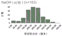
- 238000009826 distribution Methods 0.000 claims description 19
- 229920001223 polyethylene glycol Polymers 0.000 claims description 19
- FAPWRFPIFSIZLT-UHFFFAOYSA-M Sodium chloride Chemical compound [Na+].[Cl-] FAPWRFPIFSIZLT-UHFFFAOYSA-M 0.000 claims description 18
- 210000000988 bone and bone Anatomy 0.000 claims description 17
- 229920003171 Poly (ethylene oxide) Polymers 0.000 claims description 16
- 239000007864 aqueous solution Substances 0.000 claims description 16
- 239000011230 binding agent Substances 0.000 claims description 16
- 239000003086 colorant Substances 0.000 claims description 16
- 210000000663 muscle cell Anatomy 0.000 claims description 16
- 229920001983 poloxamer Polymers 0.000 claims description 16
- 229920001451 polypropylene glycol Polymers 0.000 claims description 16
- 235000017557 sodium bicarbonate Nutrition 0.000 claims description 16
- 229910000030 sodium bicarbonate Inorganic materials 0.000 claims description 16
- 229920002134 Carboxymethyl cellulose Polymers 0.000 claims description 15
- -1 alkyl imidazolium Chemical compound 0.000 claims description 15
- 235000010948 carboxy methyl cellulose Nutrition 0.000 claims description 15
- 239000001768 carboxy methyl cellulose Substances 0.000 claims description 15
- 239000008112 carboxymethyl-cellulose Substances 0.000 claims description 15
- 239000000975 dye Substances 0.000 claims description 15
- 238000002347 injection Methods 0.000 claims description 15
- 239000007924 injection Substances 0.000 claims description 15
- 239000004408 titanium dioxide Substances 0.000 claims description 15
- KIUKXJAPPMFGSW-DNGZLQJQSA-N (2S,3S,4S,5R,6R)-6-[(2S,3R,4R,5S,6R)-3-Acetamido-2-[(2S,3S,4R,5R,6R)-6-[(2R,3R,4R,5S,6R)-3-acetamido-2,5-dihydroxy-6-(hydroxymethyl)oxan-4-yl]oxy-2-carboxy-4,5-dihydroxyoxan-3-yl]oxy-5-hydroxy-6-(hydroxymethyl)oxan-4-yl]oxy-3,4,5-trihydroxyoxane-2-carboxylic acid Chemical compound CC(=O)N[C@H]1[C@H](O)O[C@H](CO)[C@@H](O)[C@@H]1O[C@H]1[C@H](O)[C@@H](O)[C@H](O[C@H]2[C@@H]([C@@H](O[C@H]3[C@@H]([C@@H](O)[C@H](O)[C@H](O3)C(O)=O)O)[C@H](O)[C@@H](CO)O2)NC(C)=O)[C@@H](C(O)=O)O1 KIUKXJAPPMFGSW-DNGZLQJQSA-N 0.000 claims description 14
- 108010035532 Collagen Proteins 0.000 claims description 14
- 102000008186 Collagen Human genes 0.000 claims description 14
- VSCWAEJMTAWNJL-UHFFFAOYSA-K aluminium trichloride Chemical compound Cl[Al](Cl)Cl VSCWAEJMTAWNJL-UHFFFAOYSA-K 0.000 claims description 14
- 238000005119 centrifugation Methods 0.000 claims description 14
- 229920001436 collagen Polymers 0.000 claims description 14
- 229920002674 hyaluronan Polymers 0.000 claims description 14
- 229960003160 hyaluronic acid Drugs 0.000 claims description 14
- 239000003513 alkali Substances 0.000 claims description 13
- 210000002569 neuron Anatomy 0.000 claims description 13
- XSQUKJJJFZCRTK-UHFFFAOYSA-N Urea Chemical compound NC(N)=O XSQUKJJJFZCRTK-UHFFFAOYSA-N 0.000 claims description 12
- BLFLLBZGZJTVJG-UHFFFAOYSA-N benzocaine Chemical compound CCOC(=O)C1=CC=C(N)C=C1 BLFLLBZGZJTVJG-UHFFFAOYSA-N 0.000 claims description 12
- 230000018044 dehydration Effects 0.000 claims description 12
- 238000006297 dehydration reaction Methods 0.000 claims description 12
- 239000012528 membrane Substances 0.000 claims description 12
- 230000001537 neural effect Effects 0.000 claims description 12
- 230000008439 repair process Effects 0.000 claims description 12
- 108010073385 Fibrin Proteins 0.000 claims description 11
- 102000009123 Fibrin Human genes 0.000 claims description 11
- BWGVNKXGVNDBDI-UHFFFAOYSA-N Fibrin monomer Chemical compound CNC(=O)CNC(=O)CN BWGVNKXGVNDBDI-UHFFFAOYSA-N 0.000 claims description 11
- 210000001789 adipocyte Anatomy 0.000 claims description 11
- 210000002889 endothelial cell Anatomy 0.000 claims description 11
- 229950003499 fibrin Drugs 0.000 claims description 11
- 229920005610 lignin Polymers 0.000 claims description 11
- 229920002451 polyvinyl alcohol Polymers 0.000 claims description 11
- NNJVILVZKWQKPM-UHFFFAOYSA-N Lidocaine Chemical compound CCN(CC)CC(=O)NC1=C(C)C=CC=C1C NNJVILVZKWQKPM-UHFFFAOYSA-N 0.000 claims description 10
- 230000010261 cell growth Effects 0.000 claims description 10
- 210000002950 fibroblast Anatomy 0.000 claims description 10
- 229960004194 lidocaine Drugs 0.000 claims description 10
- 238000000465 moulding Methods 0.000 claims description 10
- 238000003825 pressing Methods 0.000 claims description 10
- 230000037303 wrinkles Effects 0.000 claims description 10
- 229920001661 Chitosan Polymers 0.000 claims description 9
- 239000004471 Glycine Substances 0.000 claims description 9
- 230000003444 anaesthetic effect Effects 0.000 claims description 9
- 239000002537 cosmetic Substances 0.000 claims description 9
- 125000004386 diacrylate group Chemical group 0.000 claims description 9
- 239000012530 fluid Substances 0.000 claims description 9
- 239000003292 glue Substances 0.000 claims description 9
- 238000009499 grossing Methods 0.000 claims description 9
- 210000003205 muscle Anatomy 0.000 claims description 9
- 229920001296 polysiloxane Polymers 0.000 claims description 9
- 229920002101 Chitin Polymers 0.000 claims description 8
- 102000016942 Elastin Human genes 0.000 claims description 8
- 108010014258 Elastin Proteins 0.000 claims description 8
- 102000010834 Extracellular Matrix Proteins Human genes 0.000 claims description 8
- 108010037362 Extracellular Matrix Proteins Proteins 0.000 claims description 8
- 102000008946 Fibrinogen Human genes 0.000 claims description 8
- 108010049003 Fibrinogen Proteins 0.000 claims description 8
- 102000016359 Fibronectins Human genes 0.000 claims description 8
- 108010067306 Fibronectins Proteins 0.000 claims description 8
- 229920001479 Hydroxyethyl methyl cellulose Polymers 0.000 claims description 8
- 229920002153 Hydroxypropyl cellulose Polymers 0.000 claims description 8
- WHNWPMSKXPGLAX-UHFFFAOYSA-N N-Vinyl-2-pyrrolidone Chemical compound C=CN1CCCC1=O WHNWPMSKXPGLAX-UHFFFAOYSA-N 0.000 claims description 8
- 229920000463 Poly(ethylene glycol)-block-poly(propylene glycol)-block-poly(ethylene glycol) Polymers 0.000 claims description 8
- 235000010418 carrageenan Nutrition 0.000 claims description 8
- 229920001525 carrageenan Polymers 0.000 claims description 8
- 239000000679 carrageenan Substances 0.000 claims description 8
- 229940113118 carrageenan Drugs 0.000 claims description 8
- 229920002549 elastin Polymers 0.000 claims description 8
- 229940012952 fibrinogen Drugs 0.000 claims description 8
- 235000010977 hydroxypropyl cellulose Nutrition 0.000 claims description 8
- 239000001863 hydroxypropyl cellulose Substances 0.000 claims description 8
- 235000010979 hydroxypropyl methyl cellulose Nutrition 0.000 claims description 8
- 239000001866 hydroxypropyl methyl cellulose Substances 0.000 claims description 8
- 229920003088 hydroxypropyl methyl cellulose Polymers 0.000 claims description 8
- UFVKGYZPFZQRLF-UHFFFAOYSA-N hydroxypropyl methyl cellulose Chemical compound OC1C(O)C(OC)OC(CO)C1OC1C(O)C(O)C(OC2C(C(O)C(OC3C(C(O)C(O)C(CO)O3)O)C(CO)O2)O)C(CO)O1 UFVKGYZPFZQRLF-UHFFFAOYSA-N 0.000 claims description 8
- 239000008104 plant cellulose Substances 0.000 claims description 8
- 229920001606 poly(lactic acid-co-glycolic acid) Polymers 0.000 claims description 8
- 239000002243 precursor Substances 0.000 claims description 8
- 238000004080 punching Methods 0.000 claims description 8
- 229920001285 xanthan gum Polymers 0.000 claims description 8
- UHVMMEOXYDMDKI-JKYCWFKZSA-L zinc;1-(5-cyanopyridin-2-yl)-3-[(1s,2s)-2-(6-fluoro-2-hydroxy-3-propanoylphenyl)cyclopropyl]urea;diacetate Chemical compound [Zn+2].CC([O-])=O.CC([O-])=O.CCC(=O)C1=CC=C(F)C([C@H]2[C@H](C2)NC(=O)NC=2N=CC(=CC=2)C#N)=C1O UHVMMEOXYDMDKI-JKYCWFKZSA-L 0.000 claims description 8
- POKOASTYJWUQJG-UHFFFAOYSA-M 1-butylpyridin-1-ium;chloride Chemical compound [Cl-].CCCC[N+]1=CC=CC=C1 POKOASTYJWUQJG-UHFFFAOYSA-M 0.000 claims description 7
- LFTLOKWAGJYHHR-UHFFFAOYSA-N N-methylmorpholine N-oxide Chemical compound CN1(=O)CCOCC1 LFTLOKWAGJYHHR-UHFFFAOYSA-N 0.000 claims description 7
- 229910002651 NO3 Inorganic materials 0.000 claims description 7
- NHNBFGGVMKEFGY-UHFFFAOYSA-N Nitrate Chemical compound [O-][N+]([O-])=O NHNBFGGVMKEFGY-UHFFFAOYSA-N 0.000 claims description 7
- 210000004102 animal cell Anatomy 0.000 claims description 7
- 238000004061 bleaching Methods 0.000 claims description 7
- 239000004202 carbamide Substances 0.000 claims description 7
- 210000001612 chondrocyte Anatomy 0.000 claims description 7
- 238000012258 culturing Methods 0.000 claims description 7
- ZOOODBUHSVUZEM-UHFFFAOYSA-N ethoxymethanedithioic acid Chemical compound CCOC(S)=S ZOOODBUHSVUZEM-UHFFFAOYSA-N 0.000 claims description 7
- 230000003472 neutralizing effect Effects 0.000 claims description 7
- ZNNZYHKDIALBAK-UHFFFAOYSA-M potassium thiocyanate Chemical compound [K+].[S-]C#N ZNNZYHKDIALBAK-UHFFFAOYSA-M 0.000 claims description 7
- 239000011780 sodium chloride Substances 0.000 claims description 7
- 208000020431 spinal cord injury Diseases 0.000 claims description 7
- 210000002435 tendon Anatomy 0.000 claims description 7
- FPGGTKZVZWFYPV-UHFFFAOYSA-M tetrabutylammonium fluoride Chemical compound [F-].CCCC[N+](CCCC)(CCCC)CCCC FPGGTKZVZWFYPV-UHFFFAOYSA-M 0.000 claims description 7
- 239000012991 xanthate Substances 0.000 claims description 7
- IXPNQXFRVYWDDI-UHFFFAOYSA-N 1-methyl-2,4-dioxo-1,3-diazinane-5-carboximidamide Chemical compound CN1CC(C(N)=N)C(=O)NC1=O IXPNQXFRVYWDDI-UHFFFAOYSA-N 0.000 claims description 6
- 229920002307 Dextran Polymers 0.000 claims description 6
- WSFSSNUMVMOOMR-UHFFFAOYSA-N Formaldehyde Chemical compound O=C WSFSSNUMVMOOMR-UHFFFAOYSA-N 0.000 claims description 6
- 210000003050 axon Anatomy 0.000 claims description 6
- 229960005274 benzocaine Drugs 0.000 claims description 6
- 238000004090 dissolution Methods 0.000 claims description 6
- XLYOFNOQVPJJNP-UHFFFAOYSA-M hydroxide Chemical compound [OH-] XLYOFNOQVPJJNP-UHFFFAOYSA-M 0.000 claims description 6
- 210000002901 mesenchymal stem cell Anatomy 0.000 claims description 6
- 210000003098 myoblast Anatomy 0.000 claims description 6
- 210000000651 myofibroblast Anatomy 0.000 claims description 6
- 210000001178 neural stem cell Anatomy 0.000 claims description 6
- 210000000440 neutrophil Anatomy 0.000 claims description 6
- 210000000963 osteoblast Anatomy 0.000 claims description 6
- 210000002997 osteoclast Anatomy 0.000 claims description 6
- 210000000229 preadipocyte Anatomy 0.000 claims description 6
- 230000001172 regenerating effect Effects 0.000 claims description 6
- 230000008929 regeneration Effects 0.000 claims description 6
- 238000011069 regeneration method Methods 0.000 claims description 6
- 210000001057 smooth muscle myoblast Anatomy 0.000 claims description 6
- 235000010413 sodium alginate Nutrition 0.000 claims description 6
- 239000000661 sodium alginate Substances 0.000 claims description 6
- 229940005550 sodium alginate Drugs 0.000 claims description 6
- 210000000278 spinal cord Anatomy 0.000 claims description 6
- 210000003594 spinal ganglia Anatomy 0.000 claims description 6
- 238000010186 staining Methods 0.000 claims description 6
- 230000001954 sterilising effect Effects 0.000 claims description 6
- 238000004659 sterilization and disinfection Methods 0.000 claims description 6
- 238000010254 subcutaneous injection Methods 0.000 claims description 6
- 239000007929 subcutaneous injection Substances 0.000 claims description 6
- KDYFGRWQOYBRFD-UHFFFAOYSA-N succinic acid Chemical compound OC(=O)CCC(O)=O KDYFGRWQOYBRFD-UHFFFAOYSA-N 0.000 claims description 6
- 229960002372 tetracaine Drugs 0.000 claims description 6
- GKCBAIGFKIBETG-UHFFFAOYSA-N tetracaine Chemical compound CCCCNC1=CC=C(C(=O)OCCN(C)C)C=C1 GKCBAIGFKIBETG-UHFFFAOYSA-N 0.000 claims description 6
- 239000000230 xanthan gum Substances 0.000 claims description 6
- 235000010493 xanthan gum Nutrition 0.000 claims description 6
- 229940082509 xanthan gum Drugs 0.000 claims description 6
- 235000014443 Pyrus communis Nutrition 0.000 claims description 5
- 108060008539 Transglutaminase Proteins 0.000 claims description 5
- 210000001608 connective tissue cell Anatomy 0.000 claims description 5
- 238000001816 cooling Methods 0.000 claims description 5
- 239000003431 cross linking reagent Substances 0.000 claims description 5
- 238000005520 cutting process Methods 0.000 claims description 5
- 238000000502 dialysis Methods 0.000 claims description 5
- 210000002919 epithelial cell Anatomy 0.000 claims description 5
- 102000003601 transglutaminase Human genes 0.000 claims description 5
- 229920000936 Agarose Polymers 0.000 claims description 4
- 210000002449 bone cell Anatomy 0.000 claims description 4
- 210000003321 cartilage cell Anatomy 0.000 claims description 4
- 238000010382 chemical cross-linking Methods 0.000 claims description 4
- 238000004040 coloring Methods 0.000 claims description 4
- 230000012010 growth Effects 0.000 claims description 4
- 235000018102 proteins Nutrition 0.000 claims description 4
- 102000004169 proteins and genes Human genes 0.000 claims description 4
- 108090000623 proteins and genes Proteins 0.000 claims description 4
- UCTWMZQNUQWSLP-VIFPVBQESA-N (R)-adrenaline Chemical compound CNC[C@H](O)C1=CC=C(O)C(O)=C1 UCTWMZQNUQWSLP-VIFPVBQESA-N 0.000 claims description 3
- 229930182837 (R)-adrenaline Natural products 0.000 claims description 3
- HYKGUEIYMKVUSR-NPULLEENSA-N 2-(diethylamino)-n-(2,6-dimethylphenyl)acetamide;4-[(1r)-1-hydroxy-2-(methylamino)ethyl]benzene-1,2-diol Chemical compound CNC[C@H](O)C1=CC=C(O)C(O)=C1.CCN(CC)CC(=O)NC1=C(C)C=CC=C1C HYKGUEIYMKVUSR-NPULLEENSA-N 0.000 claims description 3
- SXRSQZLOMIGNAQ-UHFFFAOYSA-N Glutaraldehyde Chemical compound O=CCCCC=O SXRSQZLOMIGNAQ-UHFFFAOYSA-N 0.000 claims description 3
- 150000001412 amines Chemical class 0.000 claims description 3
- 210000004748 cultured cell Anatomy 0.000 claims description 3
- 229960005139 epinephrine Drugs 0.000 claims description 3
- 230000002934 lysing effect Effects 0.000 claims description 3
- WSFSSNUMVMOOMR-NJFSPNSNSA-N methanone Chemical compound O=[14CH2] WSFSSNUMVMOOMR-NJFSPNSNSA-N 0.000 claims description 3
- 238000004806 packaging method and process Methods 0.000 claims description 3
- XYJRXVWERLGGKC-UHFFFAOYSA-D pentacalcium;hydroxide;triphosphate Chemical compound [OH-].[Ca+2].[Ca+2].[Ca+2].[Ca+2].[Ca+2].[O-]P([O-])([O-])=O.[O-]P([O-])([O-])=O.[O-]P([O-])([O-])=O XYJRXVWERLGGKC-UHFFFAOYSA-D 0.000 claims description 3
- 229920001992 poloxamer 407 Polymers 0.000 claims description 3
- 229920001432 poly(L-lactide) Polymers 0.000 claims description 3
- 238000002278 reconstructive surgery Methods 0.000 claims description 3
- 210000004872 soft tissue Anatomy 0.000 claims description 3
- 210000004003 subcutaneous fat Anatomy 0.000 claims description 3
- 239000001384 succinic acid Substances 0.000 claims description 3
- GJCOSYZMQJWQCA-UHFFFAOYSA-N 9H-xanthene Chemical compound C1=CC=C2CC3=CC=CC=C3OC2=C1 GJCOSYZMQJWQCA-UHFFFAOYSA-N 0.000 claims description 2
- 230000003278 mimic effect Effects 0.000 claims description 2
- 210000003061 neural cell Anatomy 0.000 claims description 2
- 235000021537 Beetroot Nutrition 0.000 claims 3
- 125000000896 monocarboxylic acid group Chemical group 0.000 claims 2
- 108010084695 Pea Proteins Proteins 0.000 claims 1
- 108010064851 Plant Proteins Proteins 0.000 claims 1
- 210000000497 foam cell Anatomy 0.000 claims 1
- 210000005003 heart tissue Anatomy 0.000 claims 1
- 238000009413 insulation Methods 0.000 claims 1
- 235000019702 pea protein Nutrition 0.000 claims 1
- 235000021118 plant-derived protein Nutrition 0.000 claims 1
- 238000001802 infusion Methods 0.000 abstract 1
- 239000001124 (E)-prop-1-ene-1,2,3-tricarboxylic acid Substances 0.000 description 127
- 229940091181 aconitic acid Drugs 0.000 description 127
- GTZCVFVGUGFEME-UHFFFAOYSA-N trans-aconitic acid Natural products OC(=O)CC(C(O)=O)=CC(O)=O GTZCVFVGUGFEME-UHFFFAOYSA-N 0.000 description 127
- GTZCVFVGUGFEME-HNQUOIGGSA-N trans-aconitic acid Chemical compound OC(=O)C\C(C(O)=O)=C/C(O)=O GTZCVFVGUGFEME-HNQUOIGGSA-N 0.000 description 125
- 244000070406 Malus silvestris Species 0.000 description 39
- 239000012620 biological material Substances 0.000 description 39
- 235000011430 Malus pumila Nutrition 0.000 description 27
- 235000015103 Malus silvestris Nutrition 0.000 description 27
- 230000008569 process Effects 0.000 description 27
- 235000019441 ethanol Nutrition 0.000 description 26
- QTBSBXVTEAMEQO-UHFFFAOYSA-N Acetic acid Chemical compound CC(O)=O QTBSBXVTEAMEQO-UHFFFAOYSA-N 0.000 description 24
- DBMJMQXJHONAFJ-UHFFFAOYSA-M Sodium laurylsulphate Chemical compound [Na+].CCCCCCCCCCCCOS([O-])(=O)=O DBMJMQXJHONAFJ-UHFFFAOYSA-M 0.000 description 24
- 239000004627 regenerated cellulose Substances 0.000 description 22
- 238000004519 manufacturing process Methods 0.000 description 16
- 239000000047 product Substances 0.000 description 16
- 235000021016 apples Nutrition 0.000 description 14
- 238000010411 cooking Methods 0.000 description 14
- 238000002360 preparation method Methods 0.000 description 13
- 235000002639 sodium chloride Nutrition 0.000 description 13
- KWYUFKZDYYNOTN-UHFFFAOYSA-M Potassium hydroxide Chemical compound [OH-].[K+] KWYUFKZDYYNOTN-UHFFFAOYSA-M 0.000 description 12
- 239000003518 caustics Substances 0.000 description 12
- 229910052739 hydrogen Inorganic materials 0.000 description 12
- 239000007788 liquid Substances 0.000 description 12
- 241000233866 Fungi Species 0.000 description 11
- 239000011550 stock solution Substances 0.000 description 11
- 241000208140 Acer Species 0.000 description 10
- 239000012670 alkaline solution Substances 0.000 description 10
- 239000003599 detergent Substances 0.000 description 10
- 238000002513 implantation Methods 0.000 description 10
- 229920000642 polymer Polymers 0.000 description 10
- 239000000523 sample Substances 0.000 description 10
- 238000001157 Fourier transform infrared spectrum Methods 0.000 description 9
- PEDCQBHIVMGVHV-UHFFFAOYSA-N Glycerine Chemical compound OCC(O)CO PEDCQBHIVMGVHV-UHFFFAOYSA-N 0.000 description 9
- VEXZGXHMUGYJMC-UHFFFAOYSA-N Hydrochloric acid Chemical compound Cl VEXZGXHMUGYJMC-UHFFFAOYSA-N 0.000 description 9
- 238000009472 formulation Methods 0.000 description 9
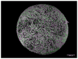
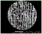
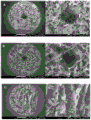
- 238000001000 micrograph Methods 0.000 description 9
- 150000003839 salts Chemical class 0.000 description 9
- 240000007087 Apium graveolens Species 0.000 description 8
- 240000008100 Brassica rapa Species 0.000 description 8
- 241000219357 Cactaceae Species 0.000 description 8
- 244000270200 Citrullus vulgaris Species 0.000 description 8
- 229960000583 acetic acid Drugs 0.000 description 8
- LOKCTEFSRHRXRJ-UHFFFAOYSA-I dipotassium trisodium dihydrogen phosphate hydrogen phosphate dichloride Chemical compound P(=O)(O)(O)[O-].[K+].P(=O)(O)([O-])[O-].[Na+].[Na+].[Cl-].[K+].[Cl-].[Na+] LOKCTEFSRHRXRJ-UHFFFAOYSA-I 0.000 description 8
- 239000002953 phosphate buffered saline Substances 0.000 description 8
- 210000002421 cell wall Anatomy 0.000 description 7
- 230000008859 change Effects 0.000 description 7
- IQFVPQOLBLOTPF-HKXUKFGYSA-L congo red Chemical compound [Na+].[Na+].C1=CC=CC2=C(N)C(/N=N/C3=CC=C(C=C3)C3=CC=C(C=C3)/N=N/C3=C(C4=CC=CC=C4C(=C3)S([O-])(=O)=O)N)=CC(S([O-])(=O)=O)=C21 IQFVPQOLBLOTPF-HKXUKFGYSA-L 0.000 description 7
- 235000019197 fats Nutrition 0.000 description 7
- 238000012360 testing method Methods 0.000 description 7
- 238000005406 washing Methods 0.000 description 7
- 241000234282 Allium Species 0.000 description 6
- 244000291564 Allium cepa Species 0.000 description 6
- 235000005255 Allium cepa Nutrition 0.000 description 6
- 235000002732 Allium cepa var. cepa Nutrition 0.000 description 6
- 240000007124 Brassica oleracea Species 0.000 description 6
- WMFOQBRAJBCJND-UHFFFAOYSA-M Lithium hydroxide Chemical compound [Li+].[OH-] WMFOQBRAJBCJND-UHFFFAOYSA-M 0.000 description 6
- 230000015572 biosynthetic process Effects 0.000 description 6
- 239000002131 composite material Substances 0.000 description 6
- 230000007547 defect Effects 0.000 description 6
- 230000000670 limiting effect Effects 0.000 description 6
- 238000012545 processing Methods 0.000 description 6
- 238000003756 stirring Methods 0.000 description 6
- 239000000126 substance Substances 0.000 description 6
- 241001133760 Acoelorraphe Species 0.000 description 5
- 238000001727 in vivo Methods 0.000 description 5
- 238000011534 incubation Methods 0.000 description 5
- 210000002379 periodontal ligament Anatomy 0.000 description 5
- 238000007920 subcutaneous administration Methods 0.000 description 5
- 240000006108 Allium ampeloprasum Species 0.000 description 4
- 235000005254 Allium ampeloprasum Nutrition 0.000 description 4
- QGZKDVFQNNGYKY-UHFFFAOYSA-N Ammonia Chemical compound N QGZKDVFQNNGYKY-UHFFFAOYSA-N 0.000 description 4
- 235000002764 Apium graveolens Nutrition 0.000 description 4
- 235000015849 Apium graveolens Dulce Group Nutrition 0.000 description 4
- 235000010591 Appio Nutrition 0.000 description 4
- 244000003416 Asparagus officinalis Species 0.000 description 4
- 235000005340 Asparagus officinalis Nutrition 0.000 description 4
- 235000000832 Ayote Nutrition 0.000 description 4
- 241000335053 Beta vulgaris Species 0.000 description 4
- 235000011303 Brassica alboglabra Nutrition 0.000 description 4
- 235000011293 Brassica napus Nutrition 0.000 description 4
- 235000011302 Brassica oleracea Nutrition 0.000 description 4
- 235000011292 Brassica rapa Nutrition 0.000 description 4
- 235000000540 Brassica rapa subsp rapa Nutrition 0.000 description 4
- 241000238366 Cephalopoda Species 0.000 description 4
- 235000012828 Citrullus lanatus var citroides Nutrition 0.000 description 4
- 235000012840 Citrullus vulgaris Nutrition 0.000 description 4
- 240000004244 Cucurbita moschata Species 0.000 description 4
- 235000009854 Cucurbita moschata Nutrition 0.000 description 4
- 235000009804 Cucurbita pepo subsp pepo Nutrition 0.000 description 4
- 244000000626 Daucus carota Species 0.000 description 4
- 235000002767 Daucus carota Nutrition 0.000 description 4
- 240000006927 Foeniculum vulgare Species 0.000 description 4
- 235000004204 Foeniculum vulgare Nutrition 0.000 description 4
- 235000016330 Lysimachia nummularia Nutrition 0.000 description 4
- 244000028339 Lysimachia nummularia Species 0.000 description 4
- MZRVEZGGRBJDDB-UHFFFAOYSA-N N-Butyllithium Chemical compound [Li]CCCC MZRVEZGGRBJDDB-UHFFFAOYSA-N 0.000 description 4
- 240000002853 Nelumbo nucifera Species 0.000 description 4
- 241000233855 Orchidaceae Species 0.000 description 4
- 235000008331 Pinus X rigitaeda Nutrition 0.000 description 4
- 235000011613 Pinus brutia Nutrition 0.000 description 4
- 241000018646 Pinus brutia Species 0.000 description 4
- 241000209049 Poa pratensis Species 0.000 description 4
- 240000001416 Pseudotsuga menziesii Species 0.000 description 4
- 241000220324 Pyrus Species 0.000 description 4
- 241000700159 Rattus Species 0.000 description 4
- CDBYLPFSWZWCQE-UHFFFAOYSA-L Sodium Carbonate Chemical compound [Na+].[Na+].[O-]C([O-])=O CDBYLPFSWZWCQE-UHFFFAOYSA-L 0.000 description 4
- PXIPVTKHYLBLMZ-UHFFFAOYSA-N Sodium azide Chemical compound [Na+].[N-]=[N+]=[N-] PXIPVTKHYLBLMZ-UHFFFAOYSA-N 0.000 description 4
- 241000722921 Tulipa gesneriana Species 0.000 description 4
- 241000863480 Vinca Species 0.000 description 4
- 238000004026 adhesive bonding Methods 0.000 description 4
- 235000011399 aloe vera Nutrition 0.000 description 4
- 239000001387 apium graveolens Substances 0.000 description 4
- 239000000872 buffer Substances 0.000 description 4
- 239000003795 chemical substances by application Substances 0.000 description 4
- 150000001875 compounds Chemical class 0.000 description 4
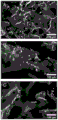
- 238000002073 fluorescence micrograph Methods 0.000 description 4
- 238000000338 in vitro Methods 0.000 description 4
- 238000002386 leaching Methods 0.000 description 4
- ZCSHNCUQKCANBX-UHFFFAOYSA-N lithium diisopropylamide Chemical compound [Li+].CC(C)[N-]C(C)C ZCSHNCUQKCANBX-UHFFFAOYSA-N 0.000 description 4
- 238000005259 measurement Methods 0.000 description 4
- INWLQCZOYSRPNW-UHFFFAOYSA-N mepivacaine Chemical compound CN1CCCCC1C(=O)NC1=C(C)C=CC=C1C INWLQCZOYSRPNW-UHFFFAOYSA-N 0.000 description 4
- 229940092917 polocaine Drugs 0.000 description 4
- 235000015136 pumpkin Nutrition 0.000 description 4
- 230000035939 shock Effects 0.000 description 4
- 239000008223 sterile water Substances 0.000 description 4
- 239000002699 waste material Substances 0.000 description 4
- HDTRYLNUVZCQOY-UHFFFAOYSA-N α-D-glucopyranosyl-α-D-glucopyranoside Natural products OC1C(O)C(O)C(CO)OC1OC1C(O)C(O)C(O)C(CO)O1 HDTRYLNUVZCQOY-UHFFFAOYSA-N 0.000 description 3
- 241000283690 Bos taurus Species 0.000 description 3
- UXVMQQNJUSDDNG-UHFFFAOYSA-L Calcium chloride Chemical compound [Cl-].[Cl-].[Ca+2] UXVMQQNJUSDDNG-UHFFFAOYSA-L 0.000 description 3
- 229920002261 Corn starch Polymers 0.000 description 3
- FBPFZTCFMRRESA-KVTDHHQDSA-N D-Mannitol Chemical compound OC[C@@H](O)[C@@H](O)[C@H](O)[C@H](O)CO FBPFZTCFMRRESA-KVTDHHQDSA-N 0.000 description 3
- WQZGKKKJIJFFOK-GASJEMHNSA-N Glucose Natural products OC[C@H]1OC(O)[C@H](O)[C@@H](O)[C@@H]1O WQZGKKKJIJFFOK-GASJEMHNSA-N 0.000 description 3
- 229920002488 Hemicellulose Polymers 0.000 description 3
- 229930195725 Mannitol Natural products 0.000 description 3
- 240000008790 Musa x paradisiaca Species 0.000 description 3
- 235000006508 Nelumbo nucifera Nutrition 0.000 description 3
- 239000004372 Polyvinyl alcohol Substances 0.000 description 3
- CZMRCDWAGMRECN-UGDNZRGBSA-N Sucrose Chemical compound O[C@H]1[C@H](O)[C@@H](CO)O[C@@]1(CO)O[C@@H]1[C@H](O)[C@@H](O)[C@H](O)[C@@H](CO)O1 CZMRCDWAGMRECN-UGDNZRGBSA-N 0.000 description 3
- 229930006000 Sucrose Natural products 0.000 description 3
- HDTRYLNUVZCQOY-WSWWMNSNSA-N Trehalose Natural products O[C@@H]1[C@@H](O)[C@@H](O)[C@@H](CO)O[C@@H]1O[C@@H]1[C@H](O)[C@@H](O)[C@@H](O)[C@@H](CO)O1 HDTRYLNUVZCQOY-WSWWMNSNSA-N 0.000 description 3
- HDTRYLNUVZCQOY-LIZSDCNHSA-N alpha,alpha-trehalose Chemical compound O[C@@H]1[C@@H](O)[C@H](O)[C@@H](CO)O[C@@H]1O[C@@H]1[C@H](O)[C@@H](O)[C@H](O)[C@@H](CO)O1 HDTRYLNUVZCQOY-LIZSDCNHSA-N 0.000 description 3
- 238000004458 analytical method Methods 0.000 description 3
- 230000008901 benefit Effects 0.000 description 3
- WQZGKKKJIJFFOK-VFUOTHLCSA-N beta-D-glucose Chemical compound OC[C@H]1O[C@@H](O)[C@H](O)[C@@H](O)[C@@H]1O WQZGKKKJIJFFOK-VFUOTHLCSA-N 0.000 description 3
- 238000009835 boiling Methods 0.000 description 3
- 235000014121 butter Nutrition 0.000 description 3
- 239000001110 calcium chloride Substances 0.000 description 3
- 229910001628 calcium chloride Inorganic materials 0.000 description 3
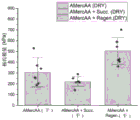
- 238000012669 compression test Methods 0.000 description 3
- 238000002591 computed tomography Methods 0.000 description 3
- 239000008120 corn starch Substances 0.000 description 3
- 239000012043 crude product Substances 0.000 description 3
- 238000006073 displacement reaction Methods 0.000 description 3
- 238000001035 drying Methods 0.000 description 3
- 238000002474 experimental method Methods 0.000 description 3
- 238000000605 extraction Methods 0.000 description 3
- 238000001125 extrusion Methods 0.000 description 3
- 239000008103 glucose Substances 0.000 description 3
- 235000011187 glycerol Nutrition 0.000 description 3
- 238000003384 imaging method Methods 0.000 description 3
- 125000005647 linker group Chemical group 0.000 description 3
- 238000003754 machining Methods 0.000 description 3
- 239000000594 mannitol Substances 0.000 description 3
- 235000010355 mannitol Nutrition 0.000 description 3
- 235000013622 meat product Nutrition 0.000 description 3
- 230000010355 oscillation Effects 0.000 description 3
- 239000007800 oxidant agent Substances 0.000 description 3
- 239000008363 phosphate buffer Substances 0.000 description 3
- 230000000704 physical effect Effects 0.000 description 3
- 239000011148 porous material Substances 0.000 description 3
- 210000003625 skull Anatomy 0.000 description 3
- 239000011734 sodium Substances 0.000 description 3
- 238000012453 sprague-dawley rat model Methods 0.000 description 3
- 239000005720 sucrose Substances 0.000 description 3
- 239000006228 supernatant Substances 0.000 description 3
- 230000001225 therapeutic effect Effects 0.000 description 3
- 230000017423 tissue regeneration Effects 0.000 description 3
- 235000001674 Agaricus brunnescens Nutrition 0.000 description 2
- 235000001270 Allium sibiricum Nutrition 0.000 description 2
- 241001116389 Aloe Species 0.000 description 2
- 235000002961 Aloe barbadensis Nutrition 0.000 description 2
- 244000144927 Aloe barbadensis Species 0.000 description 2
- 241000123643 Asparagaceae Species 0.000 description 2
- 235000016068 Berberis vulgaris Nutrition 0.000 description 2
- 235000021533 Beta vulgaris Nutrition 0.000 description 2
- 235000003899 Brassica oleracea var acephala Nutrition 0.000 description 2
- 235000011299 Brassica oleracea var botrytis Nutrition 0.000 description 2
- 235000011301 Brassica oleracea var capitata Nutrition 0.000 description 2
- 235000017647 Brassica oleracea var italica Nutrition 0.000 description 2
- 235000001169 Brassica oleracea var oleracea Nutrition 0.000 description 2
- 235000012905 Brassica oleracea var viridis Nutrition 0.000 description 2
- 244000064816 Brassica oleracea var. acephala Species 0.000 description 2
- 240000003259 Brassica oleracea var. botrytis Species 0.000 description 2
- 244000025254 Cannabis sativa Species 0.000 description 2
- PTHCMJGKKRQCBF-UHFFFAOYSA-N Cellulose, microcrystalline Chemical compound OC1C(O)C(OC)OC(CO)C1OC1C(O)C(O)C(OC)C(CO)O1 PTHCMJGKKRQCBF-UHFFFAOYSA-N 0.000 description 2
- RYGMFSIKBFXOCR-UHFFFAOYSA-N Copper Chemical compound [Cu] RYGMFSIKBFXOCR-UHFFFAOYSA-N 0.000 description 2
- 241000238557 Decapoda Species 0.000 description 2
- 235000014466 Douglas bleu Nutrition 0.000 description 2
- 244000085625 Equisetum Species 0.000 description 2
- 241000195955 Equisetum hyemale Species 0.000 description 2
- 241000287828 Gallus gallus Species 0.000 description 2
- 235000011201 Ginkgo Nutrition 0.000 description 2
- 235000008100 Ginkgo biloba Nutrition 0.000 description 2
- 244000194101 Ginkgo biloba Species 0.000 description 2
- 235000018481 Hylocereus undatus Nutrition 0.000 description 2
- 244000157072 Hylocereus undatus Species 0.000 description 2
- 244000081841 Malus domestica Species 0.000 description 2
- 241001465754 Metazoa Species 0.000 description 2
- 229920000168 Microcrystalline cellulose Polymers 0.000 description 2
- 235000003805 Musa ABB Group Nutrition 0.000 description 2
- AFCARXCZXQIEQB-UHFFFAOYSA-N N-[3-oxo-3-(2,4,6,7-tetrahydrotriazolo[4,5-c]pyridin-5-yl)propyl]-2-[[3-(trifluoromethoxy)phenyl]methylamino]pyrimidine-5-carboxamide Chemical compound O=C(CCNC(=O)C=1C=NC(=NC=1)NCC1=CC(=CC=C1)OC(F)(F)F)N1CC2=C(CC1)NN=N2 AFCARXCZXQIEQB-UHFFFAOYSA-N 0.000 description 2
- 235000006510 Nelumbo pentapetala Nutrition 0.000 description 2
- 241000283973 Oryctolagus cuniculus Species 0.000 description 2
- 229910019142 PO4 Inorganic materials 0.000 description 2
- 235000010678 Paulownia tomentosa Nutrition 0.000 description 2
- 240000002834 Paulownia tomentosa Species 0.000 description 2
- 241001494479 Pecora Species 0.000 description 2
- 241000237503 Pectinidae Species 0.000 description 2
- 241000227425 Pieris rapae crucivora Species 0.000 description 2
- 235000015266 Plantago major Nutrition 0.000 description 2
- 235000008572 Pseudotsuga menziesii Nutrition 0.000 description 2
- 235000005386 Pseudotsuga menziesii var menziesii Nutrition 0.000 description 2
- 241000510091 Quadrula quadrula Species 0.000 description 2
- 240000004980 Rheum officinale Species 0.000 description 2
- 235000008081 Rheum officinale Nutrition 0.000 description 2
- 235000009411 Rheum rhabarbarum Nutrition 0.000 description 2
- 244000193032 Rheum rhaponticum Species 0.000 description 2
- 235000011449 Rosa Nutrition 0.000 description 2
- 241000220317 Rosa Species 0.000 description 2
- 241001174121 Silene suecica Species 0.000 description 2
- KEAYESYHFKHZAL-UHFFFAOYSA-N Sodium Chemical compound [Na] KEAYESYHFKHZAL-UHFFFAOYSA-N 0.000 description 2
- 229910000831 Steel Inorganic materials 0.000 description 2
- 241000282887 Suidae Species 0.000 description 2
- QAOWNCQODCNURD-UHFFFAOYSA-L Sulfate Chemical compound [O-]S([O-])(=O)=O QAOWNCQODCNURD-UHFFFAOYSA-L 0.000 description 2
- 235000004220 Tradescantia virginiana Nutrition 0.000 description 2
- 240000000958 Tradescantia virginiana Species 0.000 description 2
- 239000000654 additive Substances 0.000 description 2
- 238000013019 agitation Methods 0.000 description 2
- 229910021529 ammonia Inorganic materials 0.000 description 2
- 238000001574 biopsy Methods 0.000 description 2
- 239000012267 brine Substances 0.000 description 2
- AXCZMVOFGPJBDE-UHFFFAOYSA-L calcium dihydroxide Chemical compound [OH-].[OH-].[Ca+2] AXCZMVOFGPJBDE-UHFFFAOYSA-L 0.000 description 2
- 239000000920 calcium hydroxide Substances 0.000 description 2
- 229910001861 calcium hydroxide Inorganic materials 0.000 description 2
- 150000005323 carbonate salts Chemical class 0.000 description 2
- 239000012876 carrier material Substances 0.000 description 2
- 238000005266 casting Methods 0.000 description 2
- 210000003850 cellular structure Anatomy 0.000 description 2
- 239000003153 chemical reaction reagent Substances 0.000 description 2
- 235000013330 chicken meat Nutrition 0.000 description 2
- 239000012141 concentrate Substances 0.000 description 2
- 238000011109 contamination Methods 0.000 description 2
- 230000009089 cytolysis Effects 0.000 description 2
- 238000011161 development Methods 0.000 description 2
- 230000018109 developmental process Effects 0.000 description 2
- 238000010790 dilution Methods 0.000 description 2
- 239000012895 dilution Substances 0.000 description 2
- 238000002845 discoloration Methods 0.000 description 2
- 235000021186 dishes Nutrition 0.000 description 2
- 239000012153 distilled water Substances 0.000 description 2
- 235000011869 dried fruits Nutrition 0.000 description 2
- 235000013399 edible fruits Nutrition 0.000 description 2
- 230000006862 enzymatic digestion Effects 0.000 description 2
- 210000003195 fascia Anatomy 0.000 description 2
- 238000005187 foaming Methods 0.000 description 2
- 239000000576 food coloring agent Substances 0.000 description 2
- 239000000989 food dye Substances 0.000 description 2
- ZZUFCTLCJUWOSV-UHFFFAOYSA-N furosemide Chemical compound C1=C(Cl)C(S(=O)(=O)N)=CC(C(O)=O)=C1NCC1=CC=CO1 ZZUFCTLCJUWOSV-UHFFFAOYSA-N 0.000 description 2
- 239000007789 gas Substances 0.000 description 2
- 239000001963 growth medium Substances 0.000 description 2
- 239000001257 hydrogen Substances 0.000 description 2
- 238000001764 infiltration Methods 0.000 description 2
- 230000008595 infiltration Effects 0.000 description 2
- 239000004615 ingredient Substances 0.000 description 2
- 229930013686 lignan Natural products 0.000 description 2
- 235000009408 lignans Nutrition 0.000 description 2
- 150000005692 lignans Chemical class 0.000 description 2
- VTHJTEIRLNZDEV-UHFFFAOYSA-L magnesium dihydroxide Chemical compound [OH-].[OH-].[Mg+2] VTHJTEIRLNZDEV-UHFFFAOYSA-L 0.000 description 2
- 239000000347 magnesium hydroxide Substances 0.000 description 2
- 229910001862 magnesium hydroxide Inorganic materials 0.000 description 2
- 238000003760 magnetic stirring Methods 0.000 description 2
- 239000011159 matrix material Substances 0.000 description 2
- 239000002609 medium Substances 0.000 description 2
- 239000000693 micelle Substances 0.000 description 2
- 235000019813 microcrystalline cellulose Nutrition 0.000 description 2
- 239000008108 microcrystalline cellulose Substances 0.000 description 2
- 229940016286 microcrystalline cellulose Drugs 0.000 description 2
- 230000007935 neutral effect Effects 0.000 description 2
- 150000002823 nitrates Chemical class 0.000 description 2
- 229920002113 octoxynol Polymers 0.000 description 2
- 230000003204 osmotic effect Effects 0.000 description 2
- 230000000149 penetrating effect Effects 0.000 description 2
- NBIIXXVUZAFLBC-UHFFFAOYSA-K phosphate Chemical compound [O-]P([O-])([O-])=O NBIIXXVUZAFLBC-UHFFFAOYSA-K 0.000 description 2
- 239000010452 phosphate Substances 0.000 description 2
- 235000015277 pork Nutrition 0.000 description 2
- 238000011552 rat model Methods 0.000 description 2
- 239000001044 red dye Substances 0.000 description 2
- 230000002829 reductive effect Effects 0.000 description 2
- 230000000717 retained effect Effects 0.000 description 2
- 235000020637 scallop Nutrition 0.000 description 2
- 230000001953 sensory effect Effects 0.000 description 2
- 238000007493 shaping process Methods 0.000 description 2
- 239000002002 slurry Substances 0.000 description 2
- ODZPKZBBUMBTMG-UHFFFAOYSA-N sodium amide Chemical compound [NH2-].[Na+] ODZPKZBBUMBTMG-UHFFFAOYSA-N 0.000 description 2
- 229910000033 sodium borohydride Inorganic materials 0.000 description 2
- 239000012279 sodium borohydride Substances 0.000 description 2
- 229910000029 sodium carbonate Inorganic materials 0.000 description 2
- 239000012312 sodium hydride Substances 0.000 description 2
- 229910000104 sodium hydride Inorganic materials 0.000 description 2
- PFUVRDFDKPNGAV-UHFFFAOYSA-N sodium peroxide Chemical compound [Na+].[Na+].[O-][O-] PFUVRDFDKPNGAV-UHFFFAOYSA-N 0.000 description 2
- HPALAKNZSZLMCH-UHFFFAOYSA-M sodium;chloride;hydrate Chemical compound O.[Na+].[Cl-] HPALAKNZSZLMCH-UHFFFAOYSA-M 0.000 description 2
- 238000007711 solidification Methods 0.000 description 2
- 230000008023 solidification Effects 0.000 description 2
- 230000007928 solubilization Effects 0.000 description 2
- 238000005063 solubilization Methods 0.000 description 2
- 239000007858 starting material Substances 0.000 description 2
- 239000010959 steel Substances 0.000 description 2
- 238000000859 sublimation Methods 0.000 description 2
- 230000008022 sublimation Effects 0.000 description 2
- 235000000346 sugar Nutrition 0.000 description 2
- 239000000725 suspension Substances 0.000 description 2
- 230000009772 tissue formation Effects 0.000 description 2
- 238000012546 transfer Methods 0.000 description 2
- 230000002792 vascular Effects 0.000 description 2
- 239000013598 vector Substances 0.000 description 2
- 235000013311 vegetables Nutrition 0.000 description 2
- UGZADUVQMDAIAO-UHFFFAOYSA-L zinc hydroxide Chemical compound [OH-].[OH-].[Zn+2] UGZADUVQMDAIAO-UHFFFAOYSA-L 0.000 description 2
- 229940007718 zinc hydroxide Drugs 0.000 description 2
- 229910021511 zinc hydroxide Inorganic materials 0.000 description 2
- 238000010146 3D printing Methods 0.000 description 1
- 241001529821 Agastache Species 0.000 description 1
- 241000238017 Astacoidea Species 0.000 description 1
- 241000271566 Aves Species 0.000 description 1
- 241000282472 Canis lupus familiaris Species 0.000 description 1
- 241000283707 Capra Species 0.000 description 1
- 241000700198 Cavia Species 0.000 description 1
- 241000938605 Crocodylia Species 0.000 description 1
- 239000004971 Cross linker Substances 0.000 description 1
- 241000238424 Crustacea Species 0.000 description 1
- 241000252233 Cyprinus carpio Species 0.000 description 1
- 229920000875 Dissolving pulp Polymers 0.000 description 1
- 241000282324 Felis Species 0.000 description 1
- 241000276438 Gadus morhua Species 0.000 description 1
- 241000238631 Hexapoda Species 0.000 description 1
- 241000282412 Homo Species 0.000 description 1
- DGAQECJNVWCQMB-PUAWFVPOSA-M Ilexoside XXIX Chemical compound C[C@@H]1CC[C@@]2(CC[C@@]3(C(=CC[C@H]4[C@]3(CC[C@@H]5[C@@]4(CC[C@@H](C5(C)C)OS(=O)(=O)[O-])C)C)[C@@H]2[C@]1(C)O)C)C(=O)O[C@H]6[C@@H]([C@H]([C@@H]([C@H](O6)CO)O)O)O.[Na+] DGAQECJNVWCQMB-PUAWFVPOSA-M 0.000 description 1
- 241000283986 Lepus Species 0.000 description 1
- 241000699670 Mus sp. Species 0.000 description 1
- 241000234295 Musa Species 0.000 description 1
- 235000018290 Musa x paradisiaca Nutrition 0.000 description 1
- 235000005807 Nelumbo Nutrition 0.000 description 1
- 206010028980 Neoplasm Diseases 0.000 description 1
- 241000283903 Ovis aries Species 0.000 description 1
- 241000337661 Parada Species 0.000 description 1
- 241000286209 Phasianidae Species 0.000 description 1
- 241000700157 Rattus norvegicus Species 0.000 description 1
- 241000283984 Rodentia Species 0.000 description 1
- 241000277331 Salmonidae Species 0.000 description 1
- 241001125046 Sardina pilchardus Species 0.000 description 1
- 241000269821 Scombridae Species 0.000 description 1
- 241001494106 Stenotomus chrysops Species 0.000 description 1
- 241000282898 Sus scrofa Species 0.000 description 1
- 238000009825 accumulation Methods 0.000 description 1
- 230000002378 acidificating effect Effects 0.000 description 1
- 150000007513 acids Chemical class 0.000 description 1
- MBLBDJOUHNCFQT-LXGUWJNJSA-N aldehydo-N-acetyl-D-glucosamine Chemical compound CC(=O)N[C@@H](C=O)[C@@H](O)[C@H](O)[C@H](O)CO MBLBDJOUHNCFQT-LXGUWJNJSA-N 0.000 description 1
- 229930013930 alkaloid Natural products 0.000 description 1
- 150000003797 alkaloid derivatives Chemical class 0.000 description 1
- 125000003277 amino group Chemical group 0.000 description 1
- 239000012062 aqueous buffer Substances 0.000 description 1
- 239000007900 aqueous suspension Substances 0.000 description 1
- 235000015191 beet juice Nutrition 0.000 description 1
- 230000004071 biological effect Effects 0.000 description 1
- 229920001222 biopolymer Polymers 0.000 description 1
- 230000000903 blocking effect Effects 0.000 description 1
- 230000036770 blood supply Effects 0.000 description 1
- 230000010072 bone remodeling Effects 0.000 description 1
- 238000000339 bright-field microscopy Methods 0.000 description 1
- 239000013590 bulk material Substances 0.000 description 1
- 239000000969 carrier Substances 0.000 description 1
- 241001233037 catfish Species 0.000 description 1
- 238000004113 cell culture Methods 0.000 description 1
- 230000004709 cell invasion Effects 0.000 description 1
- 239000006285 cell suspension Substances 0.000 description 1
- PBAYDYUZOSNJGU-UHFFFAOYSA-N chelidonic acid Natural products OC(=O)C1=CC(=O)C=C(C(O)=O)O1 PBAYDYUZOSNJGU-UHFFFAOYSA-N 0.000 description 1
- 239000007795 chemical reaction product Substances 0.000 description 1
- 210000003763 chloroplast Anatomy 0.000 description 1
- 239000011248 coating agent Substances 0.000 description 1
- 238000000576 coating method Methods 0.000 description 1
- 239000000084 colloidal system Substances 0.000 description 1
- 238000011960 computer-aided design Methods 0.000 description 1
- 229910052802 copper Inorganic materials 0.000 description 1
- 239000010949 copper Substances 0.000 description 1
- 239000011243 crosslinked material Substances 0.000 description 1
- 238000002425 crystallisation Methods 0.000 description 1
- 230000008025 crystallization Effects 0.000 description 1
- 238000001446 dark-field microscopy Methods 0.000 description 1
- 230000029087 digestion Effects 0.000 description 1
- 238000005553 drilling Methods 0.000 description 1
- 239000003814 drug Substances 0.000 description 1
- 238000004520 electroporation Methods 0.000 description 1
- 239000000835 fiber Substances 0.000 description 1
- 239000012467 final product Substances 0.000 description 1
- 239000000796 flavoring agent Substances 0.000 description 1
- 235000013355 food flavoring agent Nutrition 0.000 description 1
- 239000003517 fume Substances 0.000 description 1
- 125000000524 functional group Chemical class 0.000 description 1
- 235000021474 generally recognized As safe (food) Nutrition 0.000 description 1
- 235000021473 generally recognized as safe (food ingredients) Nutrition 0.000 description 1
- 238000010362 genome editing Methods 0.000 description 1
- 210000004209 hair Anatomy 0.000 description 1
- 210000005260 human cell Anatomy 0.000 description 1
- 230000036571 hydration Effects 0.000 description 1
- 238000006703 hydration reaction Methods 0.000 description 1
- 230000002706 hydrostatic effect Effects 0.000 description 1
- 239000007943 implant Substances 0.000 description 1
- 238000005470 impregnation Methods 0.000 description 1
- 230000003834 intracellular effect Effects 0.000 description 1
- 230000002427 irreversible effect Effects 0.000 description 1
- 239000012978 lignocellulosic material Substances 0.000 description 1
- 150000002632 lipids Chemical class 0.000 description 1
- 239000006193 liquid solution Substances 0.000 description 1
- 241000238565 lobster Species 0.000 description 1
- 238000012792 lyophilization process Methods 0.000 description 1
- 235000020640 mackerel Nutrition 0.000 description 1
- 230000014759 maintenance of location Effects 0.000 description 1
- 210000004962 mammalian cell Anatomy 0.000 description 1
- 229910052751 metal Inorganic materials 0.000 description 1
- 239000002184 metal Substances 0.000 description 1
- 210000003470 mitochondria Anatomy 0.000 description 1
- 230000004048 modification Effects 0.000 description 1
- 238000012986 modification Methods 0.000 description 1
- 238000010899 nucleation Methods 0.000 description 1
- 235000020660 omega-3 fatty acid Nutrition 0.000 description 1
- 238000012634 optical imaging Methods 0.000 description 1
- 238000005457 optimization Methods 0.000 description 1
- 210000003463 organelle Anatomy 0.000 description 1
- 239000003960 organic solvent Substances 0.000 description 1
- 210000004409 osteocyte Anatomy 0.000 description 1
- 210000003455 parietal bone Anatomy 0.000 description 1
- 239000008188 pellet Substances 0.000 description 1
- 230000035515 penetration Effects 0.000 description 1
- 244000144977 poultry Species 0.000 description 1
- 235000013594 poultry meat Nutrition 0.000 description 1
- 239000000843 powder Substances 0.000 description 1
- 239000003755 preservative agent Substances 0.000 description 1
- 230000002335 preservative effect Effects 0.000 description 1
- 230000001737 promoting effect Effects 0.000 description 1
- 230000035484 reaction time Effects 0.000 description 1
- 238000011160 research Methods 0.000 description 1
- 235000019512 sardine Nutrition 0.000 description 1
- 238000009394 selective breeding Methods 0.000 description 1
- 238000007873 sieving Methods 0.000 description 1
- 238000004088 simulation Methods 0.000 description 1
- 210000003491 skin Anatomy 0.000 description 1
- 238000002791 soaking Methods 0.000 description 1
- 229910052708 sodium Inorganic materials 0.000 description 1
- 235000015424 sodium Nutrition 0.000 description 1
- 239000007787 solid Substances 0.000 description 1
- 241000894007 species Species 0.000 description 1
- 229910001220 stainless steel Inorganic materials 0.000 description 1
- 239000010935 stainless steel Substances 0.000 description 1
- 150000008163 sugars Chemical class 0.000 description 1
- 239000004094 surface-active agent Substances 0.000 description 1
- 230000008961 swelling Effects 0.000 description 1
- 229920001059 synthetic polymer Polymers 0.000 description 1
- JGVWCANSWKRBCS-UHFFFAOYSA-N tetramethylrhodamine thiocyanate Chemical compound [Cl-].C=12C=CC(N(C)C)=CC2=[O+]C2=CC(N(C)C)=CC=C2C=1C1=CC=C(SC#N)C=C1C(O)=O JGVWCANSWKRBCS-UHFFFAOYSA-N 0.000 description 1
- 238000010257 thawing Methods 0.000 description 1
- 238000002560 therapeutic procedure Methods 0.000 description 1
- 238000009210 therapy by ultrasound Methods 0.000 description 1
- 238000004448 titration Methods 0.000 description 1
- 210000003462 vein Anatomy 0.000 description 1
- 125000000391 vinyl group Chemical group [H]C([*])=C([H])[H] 0.000 description 1
- 229920002554 vinyl polymer Polymers 0.000 description 1
- 230000003442 weekly effect Effects 0.000 description 1
- 230000002087 whitening effect Effects 0.000 description 1
Images


Classifications
-
- C—CHEMISTRY; METALLURGY
- C08—ORGANIC MACROMOLECULAR COMPOUNDS; THEIR PREPARATION OR CHEMICAL WORKING-UP; COMPOSITIONS BASED THEREON
- C08J—WORKING-UP; GENERAL PROCESSES OF COMPOUNDING; AFTER-TREATMENT NOT COVERED BY SUBCLASSES C08B, C08C, C08F, C08G or C08H
- C08J3/00—Processes of treating or compounding macromolecular substances
- C08J3/02—Making solutions, dispersions, lattices or gels by other methods than by solution, emulsion or suspension polymerisation techniques
- C08J3/03—Making solutions, dispersions, lattices or gels by other methods than by solution, emulsion or suspension polymerisation techniques in aqueous media
- C08J3/075—Macromolecular gels
-
- A—HUMAN NECESSITIES
- A23—FOODS OR FOODSTUFFS; TREATMENT THEREOF, NOT COVERED BY OTHER CLASSES
- A23L—FOODS, FOODSTUFFS, OR NON-ALCOHOLIC BEVERAGES, NOT COVERED BY SUBCLASSES A21D OR A23B-A23J; THEIR PREPARATION OR TREATMENT, e.g. COOKING, MODIFICATION OF NUTRITIVE QUALITIES, PHYSICAL TREATMENT; PRESERVATION OF FOODS OR FOODSTUFFS, IN GENERAL
- A23L19/00—Products from fruits or vegetables; Preparation or treatment thereof
- A23L19/01—Instant products; Powders; Flakes; Granules
-
- A—HUMAN NECESSITIES
- A23—FOODS OR FOODSTUFFS; TREATMENT THEREOF, NOT COVERED BY OTHER CLASSES
- A23L—FOODS, FOODSTUFFS, OR NON-ALCOHOLIC BEVERAGES, NOT COVERED BY SUBCLASSES A21D OR A23B-A23J; THEIR PREPARATION OR TREATMENT, e.g. COOKING, MODIFICATION OF NUTRITIVE QUALITIES, PHYSICAL TREATMENT; PRESERVATION OF FOODS OR FOODSTUFFS, IN GENERAL
- A23L29/00—Foods or foodstuffs containing additives; Preparation or treatment thereof
- A23L29/20—Foods or foodstuffs containing additives; Preparation or treatment thereof containing gelling or thickening agents
- A23L29/206—Foods or foodstuffs containing additives; Preparation or treatment thereof containing gelling or thickening agents of vegetable origin
-
- A—HUMAN NECESSITIES
- A23—FOODS OR FOODSTUFFS; TREATMENT THEREOF, NOT COVERED BY OTHER CLASSES
- A23L—FOODS, FOODSTUFFS, OR NON-ALCOHOLIC BEVERAGES, NOT COVERED BY SUBCLASSES A21D OR A23B-A23J; THEIR PREPARATION OR TREATMENT, e.g. COOKING, MODIFICATION OF NUTRITIVE QUALITIES, PHYSICAL TREATMENT; PRESERVATION OF FOODS OR FOODSTUFFS, IN GENERAL
- A23L31/00—Edible extracts or preparations of fungi; Preparation or treatment thereof
-
- A—HUMAN NECESSITIES
- A23—FOODS OR FOODSTUFFS; TREATMENT THEREOF, NOT COVERED BY OTHER CLASSES
- A23P—SHAPING OR WORKING OF FOODSTUFFS, NOT FULLY COVERED BY A SINGLE OTHER SUBCLASS
- A23P30/00—Shaping or working of foodstuffs characterised by the process or apparatus
- A23P30/40—Foaming or whipping
-
- A—HUMAN NECESSITIES
- A61—MEDICAL OR VETERINARY SCIENCE; HYGIENE
- A61L—METHODS OR APPARATUS FOR STERILISING MATERIALS OR OBJECTS IN GENERAL; DISINFECTION, STERILISATION OR DEODORISATION OF AIR; CHEMICAL ASPECTS OF BANDAGES, DRESSINGS, ABSORBENT PADS OR SURGICAL ARTICLES; MATERIALS FOR BANDAGES, DRESSINGS, ABSORBENT PADS OR SURGICAL ARTICLES
- A61L27/00—Materials for grafts or prostheses or for coating grafts or prostheses
- A61L27/36—Materials for grafts or prostheses or for coating grafts or prostheses containing ingredients of undetermined constitution or reaction products thereof, e.g. transplant tissue, natural bone, extracellular matrix
- A61L27/38—Materials for grafts or prostheses or for coating grafts or prostheses containing ingredients of undetermined constitution or reaction products thereof, e.g. transplant tissue, natural bone, extracellular matrix containing added animal cells
- A61L27/3804—Materials for grafts or prostheses or for coating grafts or prostheses containing ingredients of undetermined constitution or reaction products thereof, e.g. transplant tissue, natural bone, extracellular matrix containing added animal cells characterised by specific cells or progenitors thereof, e.g. fibroblasts, connective tissue cells, kidney cells
- A61L27/3826—Muscle cells, e.g. smooth muscle cells
-
- A—HUMAN NECESSITIES
- A61—MEDICAL OR VETERINARY SCIENCE; HYGIENE
- A61L—METHODS OR APPARATUS FOR STERILISING MATERIALS OR OBJECTS IN GENERAL; DISINFECTION, STERILISATION OR DEODORISATION OF AIR; CHEMICAL ASPECTS OF BANDAGES, DRESSINGS, ABSORBENT PADS OR SURGICAL ARTICLES; MATERIALS FOR BANDAGES, DRESSINGS, ABSORBENT PADS OR SURGICAL ARTICLES
- A61L27/00—Materials for grafts or prostheses or for coating grafts or prostheses
- A61L27/36—Materials for grafts or prostheses or for coating grafts or prostheses containing ingredients of undetermined constitution or reaction products thereof, e.g. transplant tissue, natural bone, extracellular matrix
- A61L27/38—Materials for grafts or prostheses or for coating grafts or prostheses containing ingredients of undetermined constitution or reaction products thereof, e.g. transplant tissue, natural bone, extracellular matrix containing added animal cells
- A61L27/3804—Materials for grafts or prostheses or for coating grafts or prostheses containing ingredients of undetermined constitution or reaction products thereof, e.g. transplant tissue, natural bone, extracellular matrix containing added animal cells characterised by specific cells or progenitors thereof, e.g. fibroblasts, connective tissue cells, kidney cells
- A61L27/383—Nerve cells, e.g. dendritic cells, Schwann cells
-
- A—HUMAN NECESSITIES
- A61—MEDICAL OR VETERINARY SCIENCE; HYGIENE
- A61L—METHODS OR APPARATUS FOR STERILISING MATERIALS OR OBJECTS IN GENERAL; DISINFECTION, STERILISATION OR DEODORISATION OF AIR; CHEMICAL ASPECTS OF BANDAGES, DRESSINGS, ABSORBENT PADS OR SURGICAL ARTICLES; MATERIALS FOR BANDAGES, DRESSINGS, ABSORBENT PADS OR SURGICAL ARTICLES
- A61L27/00—Materials for grafts or prostheses or for coating grafts or prostheses
- A61L27/50—Materials characterised by their function or physical properties, e.g. injectable or lubricating compositions, shape-memory materials, surface modified materials
- A61L27/52—Hydrogels or hydrocolloids
-
- A—HUMAN NECESSITIES
- A61—MEDICAL OR VETERINARY SCIENCE; HYGIENE
- A61L—METHODS OR APPARATUS FOR STERILISING MATERIALS OR OBJECTS IN GENERAL; DISINFECTION, STERILISATION OR DEODORISATION OF AIR; CHEMICAL ASPECTS OF BANDAGES, DRESSINGS, ABSORBENT PADS OR SURGICAL ARTICLES; MATERIALS FOR BANDAGES, DRESSINGS, ABSORBENT PADS OR SURGICAL ARTICLES
- A61L27/00—Materials for grafts or prostheses or for coating grafts or prostheses
- A61L27/50—Materials characterised by their function or physical properties, e.g. injectable or lubricating compositions, shape-memory materials, surface modified materials
- A61L27/54—Biologically active materials, e.g. therapeutic substances
-
- A—HUMAN NECESSITIES
- A61—MEDICAL OR VETERINARY SCIENCE; HYGIENE
- A61L—METHODS OR APPARATUS FOR STERILISING MATERIALS OR OBJECTS IN GENERAL; DISINFECTION, STERILISATION OR DEODORISATION OF AIR; CHEMICAL ASPECTS OF BANDAGES, DRESSINGS, ABSORBENT PADS OR SURGICAL ARTICLES; MATERIALS FOR BANDAGES, DRESSINGS, ABSORBENT PADS OR SURGICAL ARTICLES
- A61L27/00—Materials for grafts or prostheses or for coating grafts or prostheses
- A61L27/50—Materials characterised by their function or physical properties, e.g. injectable or lubricating compositions, shape-memory materials, surface modified materials
- A61L27/56—Porous materials, e.g. foams or sponges
-
- C—CHEMISTRY; METALLURGY
- C08—ORGANIC MACROMOLECULAR COMPOUNDS; THEIR PREPARATION OR CHEMICAL WORKING-UP; COMPOSITIONS BASED THEREON
- C08J—WORKING-UP; GENERAL PROCESSES OF COMPOUNDING; AFTER-TREATMENT NOT COVERED BY SUBCLASSES C08B, C08C, C08F, C08G or C08H
- C08J3/00—Processes of treating or compounding macromolecular substances
- C08J3/24—Crosslinking, e.g. vulcanising, of macromolecules
-
- C—CHEMISTRY; METALLURGY
- C08—ORGANIC MACROMOLECULAR COMPOUNDS; THEIR PREPARATION OR CHEMICAL WORKING-UP; COMPOSITIONS BASED THEREON
- C08L—COMPOSITIONS OF MACROMOLECULAR COMPOUNDS
- C08L1/00—Compositions of cellulose, modified cellulose or cellulose derivatives
- C08L1/02—Cellulose; Modified cellulose
-
- C—CHEMISTRY; METALLURGY
- C08—ORGANIC MACROMOLECULAR COMPOUNDS; THEIR PREPARATION OR CHEMICAL WORKING-UP; COMPOSITIONS BASED THEREON
- C08L—COMPOSITIONS OF MACROMOLECULAR COMPOUNDS
- C08L1/00—Compositions of cellulose, modified cellulose or cellulose derivatives
- C08L1/08—Cellulose derivatives
- C08L1/26—Cellulose ethers
- C08L1/28—Alkyl ethers
-
- C—CHEMISTRY; METALLURGY
- C08—ORGANIC MACROMOLECULAR COMPOUNDS; THEIR PREPARATION OR CHEMICAL WORKING-UP; COMPOSITIONS BASED THEREON
- C08L—COMPOSITIONS OF MACROMOLECULAR COMPOUNDS
- C08L5/00—Compositions of polysaccharides or of their derivatives not provided for in groups C08L1/00 or C08L3/00
- C08L5/04—Alginic acid; Derivatives thereof
-
- C—CHEMISTRY; METALLURGY
- C08—ORGANIC MACROMOLECULAR COMPOUNDS; THEIR PREPARATION OR CHEMICAL WORKING-UP; COMPOSITIONS BASED THEREON
- C08L—COMPOSITIONS OF MACROMOLECULAR COMPOUNDS
- C08L5/00—Compositions of polysaccharides or of their derivatives not provided for in groups C08L1/00 or C08L3/00
- C08L5/06—Pectin; Derivatives thereof
-
- C—CHEMISTRY; METALLURGY
- C08—ORGANIC MACROMOLECULAR COMPOUNDS; THEIR PREPARATION OR CHEMICAL WORKING-UP; COMPOSITIONS BASED THEREON
- C08L—COMPOSITIONS OF MACROMOLECULAR COMPOUNDS
- C08L89/00—Compositions of proteins; Compositions of derivatives thereof
- C08L89/04—Products derived from waste materials, e.g. horn, hoof or hair
- C08L89/06—Products derived from waste materials, e.g. horn, hoof or hair derived from leather or skin, e.g. gelatin
-
- A—HUMAN NECESSITIES
- A23—FOODS OR FOODSTUFFS; TREATMENT THEREOF, NOT COVERED BY OTHER CLASSES
- A23P—SHAPING OR WORKING OF FOODSTUFFS, NOT FULLY COVERED BY A SINGLE OTHER SUBCLASS
- A23P30/00—Shaping or working of foodstuffs characterised by the process or apparatus
-
- A—HUMAN NECESSITIES
- A61—MEDICAL OR VETERINARY SCIENCE; HYGIENE
- A61L—METHODS OR APPARATUS FOR STERILISING MATERIALS OR OBJECTS IN GENERAL; DISINFECTION, STERILISATION OR DEODORISATION OF AIR; CHEMICAL ASPECTS OF BANDAGES, DRESSINGS, ABSORBENT PADS OR SURGICAL ARTICLES; MATERIALS FOR BANDAGES, DRESSINGS, ABSORBENT PADS OR SURGICAL ARTICLES
- A61L2300/00—Biologically active materials used in bandages, wound dressings, absorbent pads or medical devices
- A61L2300/40—Biologically active materials used in bandages, wound dressings, absorbent pads or medical devices characterised by a specific therapeutic activity or mode of action
- A61L2300/402—Anaestetics, analgesics, e.g. lidocaine
-
- A—HUMAN NECESSITIES
- A61—MEDICAL OR VETERINARY SCIENCE; HYGIENE
- A61L—METHODS OR APPARATUS FOR STERILISING MATERIALS OR OBJECTS IN GENERAL; DISINFECTION, STERILISATION OR DEODORISATION OF AIR; CHEMICAL ASPECTS OF BANDAGES, DRESSINGS, ABSORBENT PADS OR SURGICAL ARTICLES; MATERIALS FOR BANDAGES, DRESSINGS, ABSORBENT PADS OR SURGICAL ARTICLES
- A61L2400/00—Materials characterised by their function or physical properties
- A61L2400/06—Flowable or injectable implant compositions
-
- A—HUMAN NECESSITIES
- A61—MEDICAL OR VETERINARY SCIENCE; HYGIENE
- A61L—METHODS OR APPARATUS FOR STERILISING MATERIALS OR OBJECTS IN GENERAL; DISINFECTION, STERILISATION OR DEODORISATION OF AIR; CHEMICAL ASPECTS OF BANDAGES, DRESSINGS, ABSORBENT PADS OR SURGICAL ARTICLES; MATERIALS FOR BANDAGES, DRESSINGS, ABSORBENT PADS OR SURGICAL ARTICLES
- A61L2430/00—Materials or treatment for tissue regeneration
- A61L2430/02—Materials or treatment for tissue regeneration for reconstruction of bones; weight-bearing implants
-
- A—HUMAN NECESSITIES
- A61—MEDICAL OR VETERINARY SCIENCE; HYGIENE
- A61L—METHODS OR APPARATUS FOR STERILISING MATERIALS OR OBJECTS IN GENERAL; DISINFECTION, STERILISATION OR DEODORISATION OF AIR; CHEMICAL ASPECTS OF BANDAGES, DRESSINGS, ABSORBENT PADS OR SURGICAL ARTICLES; MATERIALS FOR BANDAGES, DRESSINGS, ABSORBENT PADS OR SURGICAL ARTICLES
- A61L2430/00—Materials or treatment for tissue regeneration
- A61L2430/34—Materials or treatment for tissue regeneration for soft tissue reconstruction
-
- A—HUMAN NECESSITIES
- A61—MEDICAL OR VETERINARY SCIENCE; HYGIENE
- A61L—METHODS OR APPARATUS FOR STERILISING MATERIALS OR OBJECTS IN GENERAL; DISINFECTION, STERILISATION OR DEODORISATION OF AIR; CHEMICAL ASPECTS OF BANDAGES, DRESSINGS, ABSORBENT PADS OR SURGICAL ARTICLES; MATERIALS FOR BANDAGES, DRESSINGS, ABSORBENT PADS OR SURGICAL ARTICLES
- A61L2430/00—Materials or treatment for tissue regeneration
- A61L2430/38—Materials or treatment for tissue regeneration for reconstruction of the spine, vertebrae or intervertebral discs
-
- C—CHEMISTRY; METALLURGY
- C08—ORGANIC MACROMOLECULAR COMPOUNDS; THEIR PREPARATION OR CHEMICAL WORKING-UP; COMPOSITIONS BASED THEREON
- C08J—WORKING-UP; GENERAL PROCESSES OF COMPOUNDING; AFTER-TREATMENT NOT COVERED BY SUBCLASSES C08B, C08C, C08F, C08G or C08H
- C08J2205/00—Foams characterised by their properties
- C08J2205/02—Foams characterised by their properties the finished foam itself being a gel or a gel being temporarily formed when processing the foamable composition
- C08J2205/022—Hydrogel, i.e. a gel containing an aqueous composition
-
- C—CHEMISTRY; METALLURGY
- C08—ORGANIC MACROMOLECULAR COMPOUNDS; THEIR PREPARATION OR CHEMICAL WORKING-UP; COMPOSITIONS BASED THEREON
- C08J—WORKING-UP; GENERAL PROCESSES OF COMPOUNDING; AFTER-TREATMENT NOT COVERED BY SUBCLASSES C08B, C08C, C08F, C08G or C08H
- C08J2205/00—Foams characterised by their properties
- C08J2205/02—Foams characterised by their properties the finished foam itself being a gel or a gel being temporarily formed when processing the foamable composition
- C08J2205/026—Aerogel, i.e. a supercritically dried gel
-
- C—CHEMISTRY; METALLURGY
- C08—ORGANIC MACROMOLECULAR COMPOUNDS; THEIR PREPARATION OR CHEMICAL WORKING-UP; COMPOSITIONS BASED THEREON
- C08J—WORKING-UP; GENERAL PROCESSES OF COMPOUNDING; AFTER-TREATMENT NOT COVERED BY SUBCLASSES C08B, C08C, C08F, C08G or C08H
- C08J2205/00—Foams characterised by their properties
- C08J2205/04—Foams characterised by their properties characterised by the foam pores
-
- C—CHEMISTRY; METALLURGY
- C08—ORGANIC MACROMOLECULAR COMPOUNDS; THEIR PREPARATION OR CHEMICAL WORKING-UP; COMPOSITIONS BASED THEREON
- C08J—WORKING-UP; GENERAL PROCESSES OF COMPOUNDING; AFTER-TREATMENT NOT COVERED BY SUBCLASSES C08B, C08C, C08F, C08G or C08H
- C08J2205/00—Foams characterised by their properties
- C08J2205/06—Flexible foams
-
- C—CHEMISTRY; METALLURGY
- C08—ORGANIC MACROMOLECULAR COMPOUNDS; THEIR PREPARATION OR CHEMICAL WORKING-UP; COMPOSITIONS BASED THEREON
- C08J—WORKING-UP; GENERAL PROCESSES OF COMPOUNDING; AFTER-TREATMENT NOT COVERED BY SUBCLASSES C08B, C08C, C08F, C08G or C08H
- C08J2301/00—Characterised by the use of cellulose, modified cellulose or cellulose derivatives
- C08J2301/02—Cellulose; Modified cellulose
-
- C—CHEMISTRY; METALLURGY
- C08—ORGANIC MACROMOLECULAR COMPOUNDS; THEIR PREPARATION OR CHEMICAL WORKING-UP; COMPOSITIONS BASED THEREON
- C08J—WORKING-UP; GENERAL PROCESSES OF COMPOUNDING; AFTER-TREATMENT NOT COVERED BY SUBCLASSES C08B, C08C, C08F, C08G or C08H
- C08J2301/00—Characterised by the use of cellulose, modified cellulose or cellulose derivatives
- C08J2301/08—Cellulose derivatives
- C08J2301/26—Cellulose ethers
- C08J2301/28—Alkyl ethers
-
- C—CHEMISTRY; METALLURGY
- C08—ORGANIC MACROMOLECULAR COMPOUNDS; THEIR PREPARATION OR CHEMICAL WORKING-UP; COMPOSITIONS BASED THEREON
- C08J—WORKING-UP; GENERAL PROCESSES OF COMPOUNDING; AFTER-TREATMENT NOT COVERED BY SUBCLASSES C08B, C08C, C08F, C08G or C08H
- C08J2305/00—Characterised by the use of polysaccharides or of their derivatives not provided for in groups C08J2301/00 or C08J2303/00
- C08J2305/12—Agar-agar; Derivatives thereof
-
- C—CHEMISTRY; METALLURGY
- C08—ORGANIC MACROMOLECULAR COMPOUNDS; THEIR PREPARATION OR CHEMICAL WORKING-UP; COMPOSITIONS BASED THEREON
- C08J—WORKING-UP; GENERAL PROCESSES OF COMPOUNDING; AFTER-TREATMENT NOT COVERED BY SUBCLASSES C08B, C08C, C08F, C08G or C08H
- C08J2389/00—Characterised by the use of proteins; Derivatives thereof
- C08J2389/04—Products derived from waste materials, e.g. horn, hoof or hair
- C08J2389/06—Products derived from waste materials, e.g. horn, hoof or hair derived from leather or skin
-
- C—CHEMISTRY; METALLURGY
- C08—ORGANIC MACROMOLECULAR COMPOUNDS; THEIR PREPARATION OR CHEMICAL WORKING-UP; COMPOSITIONS BASED THEREON
- C08J—WORKING-UP; GENERAL PROCESSES OF COMPOUNDING; AFTER-TREATMENT NOT COVERED BY SUBCLASSES C08B, C08C, C08F, C08G or C08H
- C08J9/00—Working-up of macromolecular substances to porous or cellular articles or materials; After-treatment thereof
- C08J9/28—Working-up of macromolecular substances to porous or cellular articles or materials; After-treatment thereof by elimination of a liquid phase from a macromolecular composition or article, e.g. drying of coagulum
Abstract
Provided herein are aerogels and foams comprising: single-structure cells and/or groups of structural cells derived from plant or fungal tissue, the single-structure cells having a decellularized 3D structure lacking cellular material and nucleic acid of the plant or fungal tissue; the single construct cells and/or the population of structural cells are distributed within a carrier derived from a dehydrated, lyophilized or freeze-dried hydrogel. Also provided herein are methods for preparing an aerogel or foam comprising the steps of: providing decellularized plant or fungal tissue; obtaining single structure cells and/or groups of structure cells from decellularized plant or fungal tissue by performing an infusion; mixing or distributing single structure cells and/or groups of structure cells in a hydrogel to provide a mixture; and dehydrating, lyophilizing or freeze-drying the mixture to provide an aerogel or foam. Related methods and uses are also provided.
Description
Technical Field
The present invention relates generally to aerogels, hydrogels, and foams. More particularly, the present invention relates to aerogels, hydrogels and foams derived from and/or comprising decellularized plant or fungal tissue or structural cells thereof.
Background
Stent materials are highly sought after in many different fields, particularly those providing homogenous and/or reproducible three-dimensional structures. Biocompatible and/or edible scaffolding materials are particularly touted in the pharmaceutical, medical device, therapeutic and food fields, and those materials capable of supporting cell growth are highly desirable.
A variety of scaffold materials have been developed, many of which are based on synthetic polymers or other such materials. Some of which are known to be biocompatible and/or bioinert, additional scaffold materials remain of great significance for various applications.
Scaffold biomaterials comprising decellularized plant or fungal tissue have been developed and are described in PCT patent publication WO2017/136950 entitled "Decellularised Cell Wall Structures From Plants and Fungus and Use Thereof as Scaffold Materials". Significant biocompatibility is described, as well as use in various therapeutic applications. These scaffold biomaterials are of great importance for a variety of different applications.
However, additional scaffolding materials, particularly those that provide aerogel, hydrogel and/or foam, are desirable in a variety of fields. Aerogels, hydrogels, and/or foams provide tunable or customizable physical/mechanical properties and/or micro/macro architecture are particularly sought after.
There is a need for alternative, additional, and/or improved aerogels, hydrogels, and/or foams, and methods and/or uses thereof.
Disclosure of Invention
Provided herein are aerogels, hydrogels, and foams derived from and/or comprising decellularized plant or fungal tissue or structural cells thereof. Aerogels, hydrogels, and foams have now been developed, as described in detail below, which may be derived from and/or may include decellularized plant or fungal tissue or structural cells thereof, and which may include plant or fungal microstructures and/or architecture of interest; can be produced by a production method which is easy to scale; a wide range of scaffold microstructures and/or macrostructures and/or biochemistry can be provided; can provide adjustable mechanical properties; can provide adjustable porosity; may be biocompatible in vitro and/or in vivo; can be stable to various conditions (e.g., in the case of food products, under cooking conditions); or any combination thereof. Various aerogels, hydrogels, and foams having desirable properties have now been developed and prepared by using single-structure cells, structural cell groups, or both derived from plant or fungal tissue (single-structure cells or structural cell groups having a decellularized three-dimensional structure lacking the cellular material and nucleic acid of plant or fungal tissue) distributed within a carrier derived from one or more dehydrated, lyophilized (or freeze-dried) hydrogels. In certain embodiments, using the mercerizing treatments described herein, single structural cells, groups of structural cells, or both, can be derived from plant or fungal tissue (typically decellularized plant or fungal tissue), which allows for reproducible and scalable production. Related methods and uses and methods of production are also described in detail herein.
In one embodiment, provided herein is an aerogel or foam comprising:
a single construct cell, a set of structural cells, or both derived from a plant or fungal tissue, the single construct cell or set of structural cells having a decellularized three-dimensional structure lacking cellular material and nucleic acid of the plant or fungal tissue;
the single construct cells, the population of structural cells, or both are distributed within a carrier derived from a dehydrated, lyophilized, or freeze-dried hydrogel.
In another embodiment of the above aerogel or foam, the aerogel or foam has been rehydrated.
In yet another embodiment of any of the one or more aerogels or one or more foams described above, the plant or fungal tissue from which the single structure cell or group of structure cells is derived may comprise decellularized plant or fungal tissue.
In yet another embodiment of any of the one or more aerogels or one or more foams described above, SDS and optionally CaCl may be used 2 The plant or fungal tissue is decellularized.
In another embodiment of any of the one or more aerogels or one or more foams described above, the single construct cells, the set of construct cells, or both can be derived from plant or fungal tissue by mercerization. In certain embodiments, the plant or fungal tissue may be decellularized plant or fungal tissue.
In yet another embodiment of any of the one or more aerogels or one or more foams described above, the mercerizing can include treating the plant or fungal tissue with sodium hydroxide and hydrogen peroxide and heat.
In yet another embodiment of any of the one or more aerogels or one or more foams described above, the aerogel or foam can include a particle size distribution of single-structure cells having an average feret diameter (feret diameter) in the range of about 1 μm to about 1000 μm, such as about 100 to about 500 μm, such as about 100 to about 300 μm.
In another embodiment of any of the one or more aerogels or one or more foams described above, the hydrogel may include alginate, pectin, gelatin, methylcellulose, carboxymethylcellulose, hydroxypropyl cellulose, hydroxypropyl methylcellulose, hydroxyethyl methylcellulose, agar, pluronic acid (pluronic acid), a triblock PEO-PPO-PEO copolymer of poly (ethylene oxide) (PEO) and poly (propylene oxide) (PPO), dissolved or regenerated plant cellulose, dissolved cellulose-based hydrogels, hyaluronic acid, extracellular matrix proteins (e.g., collagen, gelatin, or fibronectin, or any combination thereof), monoacrylate poly (ethylene glycol) diacrylate, poly (ethylene glycol) diacrylate (PEGDA) -co-PEGMA, poly (vinyl alcohol), poly (vinylpyrrolidone), poly (lactic acid-co-glycolic acid), chitosan, chitin, xanthan gum, elastin, fibrin, fibrinogen, cellulose derivatives, carrageenan, or cellulose, or any combination thereof; wherein the hydrogel is optionally crosslinked.
In yet another embodiment of any of the one or more aerogels or one or more foams described above, the aerogel or foam can include templated or aligned microchannels created by: directional freezing; or by molding using a mold having microscale features; forming geometric patterns, depressions, holes, channels, grooves, ridges, wells, or other structural features by punching, pressing, stamping, or otherwise forming a geometric pattern in and/or on at least one surface; or any combination thereof.
In yet another embodiment of any of the one or more aerogels or one or more foams above, the plant tissue can comprise apple tissue or pear tissue.
In another embodiment of any of the one or more aerogels or one or more foams described above, when the aerogel or foam is in a hydrated form, the aerogel or foam can comprise from about 5% to about 95% m/m single structure cells, groups of structure cells, or both.
In yet another embodiment of any of the one or more aerogels or one or more foams described above, the hydrogel can include alginate, pectin, or both, and the aerogel or foam can be used to provide cross-linked CaCl 2 The solution was rehydrated.
In yet another embodiment of any of the one or more aerogels or one or more foams described above, the aerogel or foam can have a bulk modulus in the range of about 0.1 to about 500kPa, such as about 1 to about 200 kPa.
In another embodiment of any of the one or more aerogels or one or more foams described above, the aerogel or foam can be rehydrated and can further comprise one or more animal cells.
In yet another embodiment of any of the one or more aerogels or one or more foams described above, at least some of the cellulose and/or cellulose derivative(s) of the aerogel or foam can be crosslinked by physical crosslinking (e.g., using glycine) and/or chemical crosslinking (e.g., using citric acid in the presence of heat); wherein at least some of the cellulose and/or cellulose derivative(s) of the aerogel or foam are functionalized with a linker (e.g., succinic acid) to which one or more functional moieties are optionally attached (e.g., amine-containing groups, wherein crosslinking may further optionally be achieved with one or more protein crosslinking agents such as formalin, formaldehyde, glutaraldehyde, and/or transglutaminase); or any combination thereof.
In yet another embodiment, provided herein are single-structure cells, structural cell groups, or both derived from decellularized plant or fungal tissue by mercerization of the decellularized plant or fungal tissue, the single-structure cells or structural cell groups having a decellularized three-dimensional structure lacking cellular material and nucleic acid of the plant or fungal tissue and lacking one or more alkali-soluble lignin components of the plant or fungal tissue.
In yet another embodiment, provided herein is a method of preparing an aerogel or foam comprising:
providing decellularized plant or fungal tissue;
obtaining single structure cells, structural cell groups, or both from decellularized plant or fungal tissue by mercerizing the decellularized plant or fungal tissue and collecting the resulting single structure cells or structural cell groups having a decellularized three-dimensional structure;
mixing or distributing single construct cells, a set of structural cells, or both in a hydrogel to provide a mixture; and
the mixture is dehydrated, lyophilized or freeze-dried to provide an aerogel or foam.
In yet another embodiment of the above method, the mercerizing may comprise treating the decellularized plant or fungal tissue with a base and a peroxide, preferably sodium hydroxide or another hydroxide base as the base and hydrogen peroxide as the peroxide.
In yet another embodiment of any one or more of the methods above, the mercerizing may comprise treating the decellularized plant or fungal tissue with aqueous sodium hydroxide and hydrogen peroxide while heating.
In yet another embodiment of any one or more of the methods above, the decellularized plant or fungal tissue can be treated with aqueous sodium hydroxide for a first period of time prior to adding hydrogen peroxide to the reaction.
In another embodiment of any one or more of the methods above, the hydrogen peroxide may be added as a 30% aqueous hydrogen peroxide solution.
In yet another embodiment of any one or more of the methods described above, the hydrogen peroxide used for the mercerization may be used in the following proportions:
about 20mL to about 5mL of a 30% hydrogen peroxide solution: about 100g of decellularized plant or fungal tissue: about 500mL of a 1M NaOH solution;
for example:
about 20mL of 30% hydrogen peroxide solution: about 100g of decellularized plant or fungal tissue: about 500mL of a 1M NaOH solution;
about 10mL of 30% hydrogen peroxide solution: about 100g of decellularized plant or fungal tissue: about 500mL of a 1M NaOH solution; or (b)
About 5mL of 30% hydrogen peroxide solution: about 100g of decellularized plant or fungal tissue: about 500mL of a 1M NaOH solution;
In yet another embodiment of any one or more of the methods above, the method may further comprise the step of neutralizing the pH with one or more neutralization treatments.
In another embodiment of any one or more of the methods above, the neutralization treatment may comprise treatment with an acid solution, preferably aqueous HCl.
In yet another embodiment of any one or more of the methods above, the mercerizing can be performed with heating to about 80 ℃.
In yet another embodiment of any one or more of the methods above, for a 1M aqueous sodium hydroxide solution, about 1:5, a decellularized plant or fungal tissue: the ratio of aqueous sodium hydroxide solution (m: v, in g: L) is used for the caustic soda, or for another aqueous sodium hydroxide solution concentration, the caustic soda may be used in an equivalent ratio.
In another embodiment of any one or more of the methods above, the mercerizing can be carried out for at least about 30 minutes, preferably for about 1 hour.
In yet another embodiment of any one or more of the methods above, the resulting single structure cells or groups of structure cells having a decellularized three-dimensional structure can be collected by centrifugation.
In yet another embodiment of any one or more of the methods described above, the single-structure cells, the groups of structural cells, or both may be mixed or distributed in a hydrogel comprising alginate, pectin, gelatin, methylcellulose, carboxymethylcellulose, hydroxypropyl cellulose, hydroxypropyl methylcellulose, hydroxyethyl methylcellulose, agar, pluronic acid, triblock PEO-PPO-PEO copolymers of poly (ethylene oxide) (PEO) and poly (propylene oxide) (PPO), solubilized or regenerated plant cellulose, solubilized cellulose-based hydrogel, hyaluronic acid, extracellular matrix proteins (e.g., collagen, gelatin, or fibronectin, or any combination thereof), monoacrylate poly (ethylene glycol), poly (ethylene glycol) diacrylate (PEGDA) -co-PEGMA, poly (vinyl alcohol), poly (vinyl pyrrolidone), poly (lactic acid-co-glycolic acid), chitosan, cellulose, xanthan gum, elastin, fibrin, fibrinogen, cellulose derivatives, carrageenan, or any combination thereof; wherein the hydrogel is optionally crosslinked.
In another embodiment of any one or more of the methods above, the method may further comprise the step of directionally freezing the mixture to introduce templated or aligned microchannels on the surface of the mixture, inside the mixture, or both; a step of molding the mixture using a mold having microscale features that contacts one or more surfaces of the mixture and/or aerogel or foam resulting from dehydration, lyophilization or freeze drying of the mixture to introduce templated or aligned microchannels; a step of punching, pressing, stamping or otherwise forming geometric patterns, depressions, holes, channels, grooves, ridges, wells or other structural features in and/or on at least one surface of the mixture and/or aerogel or foam prior to, during or after dehydration, lyophilization or freeze drying of the mixture; or any combination thereof.
In yet another embodiment of any one or more of the methods above, the directional freezing may be performed by creating a thermal gradient on the mixture from one or more directions so as to form aligned ice crystals starting from the cold side(s) of the thermal gradient.
In yet another embodiment of any one or more of the methods above, the microstructure of the microchannels produced by directional freezing can be controlled by producing a mixture comprising solvents containing varying amounts of one or more other dissolved compounds, such as sucrose, glucose, trehalose, corn starch, glycerol, ethanol, mannitol, sodium chloride, caCl 2 Gelatin, citric acid, PVA, PEG, dextran, naF, naBr, naI, phosphate buffer or other such agents, which alters the structural properties of aligned ice crystals grown from the cold side of the thermal gradient.
In yet another embodiment of any one or more of the methods above, the mixture may be directionally frozen for a period of at least about 30 minutes, preferably for a period of about 2 hours.
In another embodiment of any one or more of the methods above, the mixture may be directionally frozen by cooling to a temperature of from about-190 ℃ to about 0 ℃, such as at least about-15 ℃, preferably about-25 ℃.
In yet another embodiment of any one or more of the methods above, the step of dehydrating, lyophilizing or freeze-drying the mixture to provide an aerogel or foam can comprise freezing the mixture and then lyophilizing or freeze-drying the mixture.
In yet another embodiment of any one or more of the methods described above, the method can include the additional step of crosslinking the hydrogel (e.g., crosslinking the hydrogel before or after freezing/lyophilizing), rehydrating the aerogel or foam, or both; caCl optionally in the presence of alginate or pectin or agar hydrogels 2 The solution provides crosslinking.
In another embodiment of any one or more of the methods above, the method may include the additional step of culturing the animal cells on or in the aerogel or foam.
In another embodiment, provided herein are aerogels or foams produced by any one or more of the methods described herein.
In yet another embodiment, provided herein is the use of any one of the one or more aerogels or one or more foams described herein for bone tissue engineering.
In yet another embodiment, provided herein is the use of any of the one or more aerogels or one or more foams described herein for templated or aligned growth of cells.
In another embodiment of the above use, the cells may comprise muscle cells.
In another embodiment of the above use, the cell may comprise a neural cell.
In yet another embodiment, provided herein is the use of any one of the one or more aerogels or one or more foams described herein for repairing spinal cord injury.
In another embodiment, provided herein is the use of any one of the one or more aerogels or one or more foams described herein as an insulating or packaging foam.
In yet another embodiment, provided herein is a method for bone tissue engineering or repair in a subject in need thereof, comprising:
implanting any of the one or more aerogels or one or more foams described herein into an affected site of a subject in need thereof;
so that the aerogel or foam promotes bone tissue formation or repair.
In yet another embodiment, provided herein is a method for templating or aligning cell growth, comprising:
culturing cells on any one of the one or more aerogels or one or more foams described herein, wherein the aerogel or foam comprises templated or aligned microchannels on at least one surface of the aerogel or foam, within the aerogel or foam, or both, optionally formed by directional freezing; molding by using a mold having microscale features; by punching, pressing, stamping or otherwise forming geometric patterns, depressions, holes, channels, grooves, ridges, wells or other structural features in and/or on at least one surface; or any combination thereof;
Such that the cultured cells are aligned along the microchannel.
In another embodiment of the above use, the cell may comprise a muscle cell or a nerve cell.
In yet another embodiment, provided herein is a method for repairing spinal cord injury in a subject in need thereof, comprising:
implanting at an affected site of a subject in need thereof any one of one or more aerogels or one or more foams as defined herein, wherein the aerogel or foam comprises optionally formed directionally frozen templated or aligned micro-channels;
such that the aerogel or foam promotes spinal cord repair by aligned growth of nerve cells along the templated or aligned microchannels.
In another embodiment, provided herein is a food product comprising any one of the one or more aerogels or one or more foams described herein.
In yet another embodiment of the above-described food products, food products for the cell-based or plant-based meat industry can be produced, and cytoagricultural techniques can be utilized to produce cultured or vegetal meat products including or using aerogels and/or foams as described herein, such as those including materials derived from decellularized plants or fungal tissue. The examples included below support that the aerogel and/or foam described herein can be cooked, can support mammalian cell growth, can be colored, and form vegetable and/or cellular meat products. The embodiments described below include detailed embodiments of plant-based tuna mimics as illustrative embodiments.
In yet another embodiment of the above food product, the food product may comprise a dye or colorant.
In yet another embodiment of any one or more of the above foods, the food product may comprise two or more aerogel or foam subunits glued together.
In another embodiment of any one or more of the above foods, the gum may include agar.
In yet another embodiment of any one of the one or more foods described above, the aerogel or foam can include templated or aligned microchannels, optionally formed by directional freezing, and wherein the aerogel or foam includes muscle cells, fibroblasts, myofibroblasts, neurons, dorsal root ganglion cells, neuronal structures such as axons, neural precursor cells, neural stem cells, glial cells, endogenous stem cells, neutrophils, mesenchymal stem cells, satellite cells, myoblasts, myotubes, muscle progenitor cells, adipocytes, preadipocytes, chondrocytes, osteoblasts, osteoclasts, preannocytes, tendinous progenitor cells, periodontal ligament stem cells, or endothelial cells, or any combination thereof, aligned along the templated or aligned microchannels; preferably, wherein the aerogel or foam comprises templated or aligned micro-channels, optionally formed by directional freezing, and wherein the aerogel or foam comprises muscle cells, fat cells, connective tissue cells (e.g., fibroblasts), cartilage, bone, epithelial or endothelial cells, or any combination thereof, aligned along the templated or aligned micro-channels.
In another embodiment, provided herein is the use of any one of the one or more aerogels or one or more foams described herein in food products.
In yet another embodiment, provided herein is a method of preparing a single structural cell, a set of structural cells, or both from a decellularized plant or fungal tissue, comprising:
providing decellularized plant or fungal tissue;
single structure cells, structural cell groups, or both are obtained from decellularized plant or fungal tissue by mercerizing the decellularized plant or fungal tissue and collecting the resulting single structure cells or structural cell groups having a decellularized three-dimensional structure.
In yet another embodiment of the above method, the mercerizing may comprise treating the decellularized plant or fungal tissue with a base and a peroxide, preferably sodium hydroxide or another hydroxide base as the base and hydrogen peroxide as the peroxide.
In another embodiment of any one or more of the methods above, the mercerizing may comprise treating the decellularized plant or fungal tissue with aqueous sodium hydroxide and hydrogen peroxide while heating.
In yet another embodiment of any one or more of the methods above, the decellularized plant or fungal tissue can be treated with aqueous sodium hydroxide for a first period of time prior to adding hydrogen peroxide to the reaction.
In yet another embodiment of any one or more of the methods above, the hydrogen peroxide may be added as a 30% aqueous hydrogen peroxide solution.
In yet another embodiment of any one or more of the methods described above, the hydrogen peroxide used for the mercerization may be used in the following proportions:
about 20mL to about 5mL of a 30% hydrogen peroxide solution: about 100g of decellularized plant or fungal tissue: about 500mL of a 1M NaOH solution;
for example:
about 20mL of 30% hydrogen peroxide solution: about 100g of decellularized plant or fungal tissue: about 500mL of a 1M NaOH solution;
about 10mL of 30% hydrogen peroxide solution: about 100g of decellularized plant or fungal tissue: about 500mL of a 1M NaOH solution; or (b)
About 5mL of 30% hydrogen peroxide solution: about 100g of decellularized plant or fungal tissue: about 500mL of 1M NaOH solution.
In yet another embodiment of any one or more of the methods above, the method may further comprise the step of neutralizing the pH with one or more neutralization treatments.
In another embodiment of any one or more of the methods above, the neutralization treatment may comprise treatment with an acid solution, preferably aqueous HCl.
In yet another embodiment of any one or more of the methods above, the mercerizing can be performed with heating to about 80 ℃.
In yet another embodiment of any one or more of the methods above, for a 1M aqueous sodium hydroxide solution, about 1:5, a decellularized plant or fungal tissue: the ratio of aqueous sodium hydroxide solution (m: v, in g: L) is used for the caustic soda, or for another aqueous sodium hydroxide solution concentration, the caustic soda may be used in an equivalent ratio.
In yet another embodiment of any one or more of the methods above, the mercerizing can be carried out for at least about 30 minutes, preferably for about 1 hour.
In yet another embodiment of any one or more of the methods above, the resulting single structure cells or groups of structure cells having a decellularized three-dimensional structure can be collected by centrifugation.
In yet another embodiment, provided herein are single construct cells, structural cell groups, or both, prepared by any one or more of the methods described herein.
In another embodiment, provided herein is a cellulose-based hydrogel comprising cellulose derived from decellularized plant or fungal tissue by dissolution with dimethylacetamide and lithium chloride, followed by regeneration with ethanol.
In yet another embodiment, provided herein is a method of preparing a cellulose-based hydrogel comprising:
Providing decellularized plant or fungal tissue;
lysing the cellulose of the decellularized plant or fungal tissue by treatment with dimethylacetamide (DMAc) and lithium chloride (LiCl); and
regenerating the cellulose-based hydrogel from the dissolved cellulose by solvent exchange with ethanol,
thereby providing a cellulose-based hydrogel.
In another embodiment of the above method, the solvent exchange with ethanol may be performed using a dialysis membrane, or the solvent exchange may be facilitated by adding ethanol to the upper portion of the dissolved cellulose.
In yet another embodiment of any one or more of the methods above, the method may further comprise bleaching the cellulose-based hydrogel with hydrogen peroxide.
In another embodiment, provided herein is a cellulose-based hydrogel comprising a polymer prepared by reacting a cellulose-based hydrogel with dimethylacetamide and lithium chloride, liClO 4 Xanthate, EDA/KSCN, H 3 PO 4 NaOH/urea, znCl 2 TBAF/DMSO, NMMO, ionic Liquids (IL) (e.g., 1-butylpyridinium chloride and aluminum chloride, alkyl imidazolium associated with nitrate, preferably room temperature ionic liquids), or any combination thereof.
In yet another embodiment, provided herein is a method of preparing a cellulose-based hydrogel comprising:
Providing decellularized plant or fungal tissue;
by using dimethylacetamide and lithium chloride, liClO 4 Xanthate, EDA/KSCN, H 3 PO 4 NaOH/urea, znCl 2 Cellulose treated to solubilize decellularized plant or fungal tissue, TBAF/DMSO, NMMO, ionic Liquids (IL) (e.g., 1-butylpyridinium chloride and aluminum chloride, alkyl imidazolium associated with nitrate, preferably room temperature ionic liquids), or any combination thereof;
the solubilized cellulose is obtained and the cellulose-based hydrogel is prepared using the solubilized cellulose.
In another embodiment, provided herein is a cellulose-based hydrogel prepared by any one or more of the methods described herein.
In another embodiment of any of the one or more aerogels or one or more foams described herein, the hydrogel can comprise any of the one or more cellulose-based hydrogels described herein.
In yet another embodiment, provided herein is a food product comprising any of one or more aerogels, or one or more foams, or one or more structural cells described herein, wherein the food product is a meat analog and comprises a plurality of lines that provide a fat white line appearance found in tuna, salmon, or another fish meat.
In yet another embodiment of the above food product, the food product may be a tuna, salmon or another fish-based analog.
In another embodiment of any one of the one or more foods described above, the food product may contain one or more dyes or colorants that provide the color of tuna, salmon, or another fish flesh.
In yet another embodiment of any one or more of the above foods, the plurality of lines may be formed in cuts or channels formed in the aerogel or foam.
In yet another embodiment of any one or more of the food products described above, the plurality of wires may comprise titanium dioxide, optionally in combination with an agar-binding agent or another such binding agent.
In another embodiment of one or more of the foods described above, titanium dioxide optionally in combination with an agar binder may be applied in cuts or channels formed in the aerogel or foam to provide a fat white line appearance found in tuna, salmon or another fish meat.
In another embodiment, provided herein is a method for preparing a food product that is a tuna, salmon or other fish mimic, comprising:
Providing any one of the one or more aerogels or one or more foams described herein;
optionally, staining or coloring the aerogel to the color of tuna, salmon or other fish flesh;
cutting or otherwise treating the aerogel to form cuts or channels along the surface of the aerogel; and
a dye or colorant is applied to the cut or channel to provide a fat white appearance characteristic of tuna, salmon or other fish flesh.
In another embodiment of the above method, the dye or colorant applied to the incision or passageway may comprise titanium dioxide.
In another embodiment of any one or more of the methods above, the dye or colorant applied to the incision or passageway may be combined with a binding agent.
In yet another embodiment of any one or more of the methods above, the binding agent may comprise agar.
In another embodiment, provided herein is a food product prepared by any one or more of the methods described herein.
In another embodiment, provided herein is a non-absorbable dermal filler comprising an aerogel, foam, structural cell, or cellulose-based hydrogel as described herein; or any combination thereof.
In yet another embodiment, provided herein is a dermal filler comprising a single construct cell, a set of structural cells, or both derived from plant or fungal tissue, the single construct cell or set of structural cells having a decellularized three-dimensional structure lacking cellular material and nucleic acid of the plant or fungal tissue, the single construct cell or set of structural cells or both derived from plant or fungal tissue by mercerization.
In another embodiment of any one of the one or more dermal fillers described above, the dermal filler can further include a carrier fluid or gel.
In yet another embodiment of any of the one or more dermal fillers described above, the carrier fluid or gel can include water, an aqueous solution, or a hydrogel.
In yet another embodiment of any of the one or more dermal fillers described above, the carrier fluid or gel can include an aqueous saline solution, or collagen, hyaluronic acid, methylcellulose, and/or a solubilized plant-derived decellularized cellulose-based hydrogel.
In another embodiment of any of the one or more dermal fillers described above, the dermal filler can further include an anesthetic.
In yet another embodiment of any of the one or more dermal fillers described above, the anesthetic can include lidocaine, benzocaine, tetracaine, polocaine (polocaine), epinephrine, or any combination thereof.
In another embodiment of any one of the above one or more dermal fillers, the dermal filler can include PBS (saline), hyaluronic acid (crosslinked or uncrosslinked), alginate, collagen, pluronic acid (e.g., pluronic F127), agar, agarose or fibrin, calcium hydroxyapatite, poly-L-lactic acid, autologous fat, silicone, dextran, methylcellulose, or any combination thereof.
In another embodiment of any one of the above one or more dermal fillers, the dermal filler can include at least one of the following: 2% lidocaine gel; triple anesthetic gels (BLT gels) comprising 20% benzocaine, 6% lidocaine and 4% tetracaine; 3% of bolocarpine; or a mixture of 2% lidocaine and epinephrine.
In another embodiment of any of the one or more dermal fillers described above, the structural cells can have a size, diameter, or minimum feret diameter of at least about 20 μm.
In another embodiment of any one or more of the dermal fillers described above, the structural cells can have a size, diameter, or maximum feret diameter of less than about 1000 μm.
In yet another embodiment of any of the one or more dermal fillers described above, the structural cells can have a size, diameter, or feret diameter distribution in the range of about 20 μm to about 1000 μm.
In yet another embodiment of any of the one or more dermal fillers described above, the structural cells can have a particle size, diameter, or feret diameter distribution that peaks between about 200-300 μm.
In another embodiment of any one or more of the dermal fillers described above, the structural cells can have an average particle size, diameter, or feret diameter in the range of about 200 μm to about 300 μm.
In another embodiment of any of the one or more dermal fillers described above, the structural cells can have a size of between about 30,000 and about 75,000 μm 2 An average projected particle area within the range of (2).
In yet another embodiment of any of the one or more dermal fillers described above, the dermal filler can be sterilized.
In yet another embodiment of any of the one or more dermal fillers described above, the sterilization can be by gamma sterilization.
In yet another embodiment of any of the one or more dermal fillers described above, the dermal filler can be formulated for true subcutaneous injection, deep dermal injection, subcutaneous injection (e.g., subcutaneous fat injection), or any combination thereof.
In another embodiment of any one of the one or more dermal fillers described above, the dermal filler can be provided in a syringe or injection device.
In another embodiment, provided herein is the use of any one of the one or more dermal fillers described herein as a soft tissue filler, for reconstructive surgery, or both.
In another embodiment, provided herein is a use of any one of the one or more dermal fillers described herein for improving the cosmetic appearance of a subject in need thereof.
In another embodiment, provided herein is the use of any one of the one or more dermal fillers described herein for increasing tissue volume, smoothing wrinkles, or both in a subject in need thereof.
In another embodiment, provided herein is a method for improving the cosmetic appearance, increasing tissue volume, smoothing wrinkles, or any combination thereof, of a subject in need thereof, the method comprising:
Administering or injecting any of the one or more dermal fillers described herein to a region in need thereof;
thereby improving the cosmetic appearance of the subject, increasing tissue volume, smoothing wrinkles, or any combination thereof.
In another embodiment of one or more of the uses or one or more methods described above, the subject's primordial cells may infiltrate the dermal filler.
In yet another embodiment of one or more of the uses or one or more methods described above, the dermal filler may be non-absorbable such that the decellularized plant or fungal tissue remains substantially intact in the subject.
Drawings
These and other features will be further appreciated from the following description and drawings, in which:
figure 1 shows the results of AA (apple) mercerization and discoloration in the smaller AA sample described in example 1 (100 g in the image). 100g of decellularized AA (apple) material was mercerized in 500mL 1M NaOH at 80℃for 1 hour. A total of 75mL H was added throughout the caustic soda process 2 O 2 To discolor the sample (react to form Na 2 O 2 (sodium peroxide), which is a strong oxidizing agent). Fig. 1 (a) shows AA samples in NaOH (t=0 min). FIG. 1 (B) shows that 25mL of H was just added 2 O 2 After this, AA samples in NaOH. The color change process starts immediately after the addition. (t=2 minutes). In fig. 1 (C), the sample is yellow (t=10 minutes). In FIG. 1 (D), AA samples were taken at NaOH and H 2 O 2 Soaking in the alkali for 60 minutes, and then taking off-white;
FIG. 2 (A) shows the decellularized AA tissue used as starting material for the mercerization process, and FIG. 2 (B) shows the product obtained after the mercerization described in example 1. The product after subsequent neutralization and centrifugation is shown. The resulting product material shown in fig. 2 (B) is very viscous, similar to an apple "paste";
figure 3 shows images of apple derived decellularized single structure cells (and some groups of structure cells comprising a small fraction of single structure cells linked together) obtained/isolated after the mercerization described in example 1. In fig. 3, the cells were stained with congo red dye diluted and fluorescent, and the microarchitecture of the cells was found to be intact;
FIG. 4 shows the addition of H as described in example 1 2 O 2 Particle size distribution of decellularized AA of mercerized (1M NaOH) (n=10 images analyzed). The average size confirms the presence of intact single-structure cells that retain their microarchitectural features;
FIG. 5 shows the color change of the AA-NaOH solution during the entire 60-minute caustic digestion of all three proportioning conditions (i.e., 20g, 50g and 100g AA in 100mL 1M NaOH) as described in example 1;
FIG. 6 shows that after mercerization in various solutions, isolated individual AA cells were imaged and their Ferrett diameters were measured as described in example 1. The results show that under each condition, there is no significant difference in the average size, number and distribution of the isolated mercerized cells;
FIG. 7 shows a 5% alginate aerogel described in example 1. The diameter of the bracket is 6cm, and the thickness is 0.7cm;
fig. 8 shows a microscopic image of 50% alginate aerogel described in example 1 (scale bar = 500 um);
FIG. 9 shows a cross-linked 50% alginate aerogel that has been rehydrated as described in example 1 (aerogel diameter about 1cm, thickness about 4 mm);
fig. 10 shows an example of the addition of hydrated aerogel of butter (alginate-based in this example) to a frying pan at the start of cooking, as described in example 1;
FIG. 11 shows the same aerogel shown in FIG. 10, but after cooking for several minutes, the aerogel is observed to retain its shape and integrity and form a crust;
FIG. 12 shows a comparison of the "green" (left) and cooked (right) aerogels described in example 1;
FIG. 13 shows a custom directed freezing apparatus for use in example 1;
FIG. 14 shows a schematic view of the directional freezer apparatus of FIG. 13;
FIG. 15 shows a syringe mixing apparatus for mixing an alginate hydrogel with a gel comprising structural cells obtained from the mercerization of decellularized apple tissue, as described in example 1;
FIG. 16 shows a top view of the aerogel described in example 1 still in the falcon tube, wherein a porous structure can be observed;
FIG. 17 shows the aerogel after removal from the falcon tube as described in example 1;
FIG. 18 shows that aerogel foam prepared in a freezer at-20℃without additional freezing time, the aerogel foam collapsed during lyophilization (left); and aerogel foam (right) placed in a freezer (-20 ℃) overnight prior to lyophilization; as described in example 1. The height of each stand is about 3cm;
FIG. 19 shows the reflected light image (1 x concentrator, 0.75 x magnification) of the entire aerogel cross section described in example 1;
FIG. 20 shows the bright field cross section perpendicular to the aerogel cylinder axis described in example 1 (2 x condenser, 1.25 x zoom of a stereo microscope);
FIG. 21 shows a bright field cross section (2 x condenser, 1.25 x zoom of a stereo microscope) parallel to the aerogel cylinder axis as described in example 1;
FIG. 22 shows a dark field cross section perpendicular to the aerogel cylinder axis (2 x condenser, 1.25 x zoom of a stereo microscope) described in example 1;
FIG. 23 shows a dark field cross section parallel to the aerogel cylinder axis (2 x condenser, 1.25 x zoom of a stereo microscope) as described in example 1;
FIG. 24 shows an SEM cross-section perpendicular to the axis of the aerogel cylinder described in example 1, revealing the microchannels;
FIG. 25 shows an SEM cross-section perpendicular to the axis of the aerogel cylinder described in example 1, revealing the microchannels;
FIG. 26 shows an SEM cross section perpendicular to the cylinder axis as described in example 1;
FIG. 27 shows an SEM cross section perpendicular to the aerogel cylinder axis;
FIG. 28 shows an SEM cross section parallel to the axis of the aerogel cylinder described in example 1, revealing remote alignment;
FIG. 29 shows an SEM cross-section parallel to the axis of an aerogel cylinder as described in example 1;
FIG. 30 shows an SEM cross-section parallel to the axis of an aerogel cylinder as described in example 1;
FIG. 31 shows an SEM cross-section parallel to the axis of an aerogel cylinder as described in example 1;
FIG. 32 shows the dried aerogel fraction (left) and 0.1M CaCl as described in example 1 2 Images of the treated rehydrated aerogel fraction (right). The images are acquired at approximately the same height and magnification. The aerogel portion remains intact, retains its microstructure, and can be picked up and handled. In this case CaCl 2 Rehydration in solution crosslinks and stabilizes the alginate of the rehydrated aerogel (right);
FIG. 33 depicts a styrofoamFreezing apparatus in vinyl box, wherein LN is added just before taking a picture 2 Bottom boiling can be seen as described in example 1;
fig. 34 shows three formulations of the prepared hydrogel mixtures, where the solvent was a) PBS, B) 0.9% saline or C) water, to assess whether the salt would alter ice crystal formation and channel alignment/architecture during directional freezing (scale = 2mm, applicable in all cases). In all cases, the material freezes so fast that no significant ice crystal formation is observed, so that no aligned channels are observed. The process as described in example 1 gives a very dense and soft foam;
FIG. 35 shows (A) LN of FIG. 33 2 Frozen aqueous alginate mixtures were oriented on the system (scale bar = 2 mm). The scaffold is very dense and soft and appears to the naked eye to be uniform. This is in sharp contrast to scaffolds created on peltier-based directional freezing platforms where the channel architecture is clearly visible to the naked eye. However, as shown in (B), at high resolution (scale bar = 200 um), the small pore size of the scaffold becomes visible, which creates opportunities for cell invasion and several potential other applications in tissue engineering and food science, for example, as described in example 1;
FIG. 36 shows a 5% alginate and pectin stock solution as described in example 2;
FIG. 37 shows the preparation of a pluronic stock solution as described in example 2;
FIG. 38 shows the preparation of gelatin AA aerogel described in example 2;
FIG. 39 shows a syringe-based mixing apparatus for mixing hydrogels with the mercerized structural cells described in example 2;
FIG. 40 depicts a schematic of different aerogel formulations prepared as described in example 2, before and after freeze-drying of the samples;
fig. 41 shows the results of seeding GFP 3T3 cells (green) onto certain aerogels (shown) stained with congo red (red) described in example 2. Agar, alginate, pectin and gelatin hydrogels were used in combination with 1.5g of decellularized mercerized apples (10%) or 7.5g of decellularized mercerized apples (50%) (scale = 200 μm). Images were taken on a BX53 upright microscope at 10 x magnification, with GFP filters for cells and TXRED filters for scaffolds;
FIG. 42 shows the stress-strain curve of a dry agar-based gel containing 1.5g of mercerized AA described in example 2;
FIG. 43 shows the stress-strain curve of a dry agar-based gel containing 7.5g of mercerized AA described in example 2;
FIG. 44 shows the stress-strain curve of the dry alginate-based gel described in example 2 containing 1.5g of mercerized AA;
FIG. 45 shows the stress-strain curve of the dry alginate-based gel described in example 2 containing 7.5g of mercerized AA;
FIG. 46 shows the stress-strain curve for the dried fruit gum base gel described in example 2 containing 1.5g of mercerized AA;
FIG. 47 shows the stress-strain curve for the dried fruit gum base gel described in example 2 containing 7.5g of mercerized AA;
FIG. 48 shows the stress-strain curve of the dry gelatin-based gel described in example 2 containing 1.5g of mercerized AA;
FIG. 49 shows the stress-strain curve of the dry gelatin-based gel described in example 2 containing 7.5g of mercerized AA;
FIG. 50 shows the stress-strain curve of the dry methylcellulose-based gel described in example 2 containing 1.5g of mercerized AA;
FIG. 51 shows the stress-strain curve of the dry methylcellulose-based gel described in example 2 containing 7.5g of mercerized AA;
FIG. 52 shows the stress-strain curve of the dry pluronic-based gel described in example 2 containing 1.5g of mercerized AA;
FIG. 53 shows the stress-strain curves of the dried pluronic and alginate-based gels containing 7.5g of mercerized AA described in example 2;
Fig. 54 shows young's modulus of dried samples with hydrate counterparts. The volume of the mercerized AA is represented by 1.5 and 7.5; both in grams and correspond to 10% and 50% solutions, respectively. A base hydrogel of 1% agar, alginate and pectin was used. Gelatin is a 5% final solution, as described in example 2;
FIG. 55 shows the stress-strain curve of a hydrated agar-based gel containing 1.5g of mercerized AA described in example 2;
FIG. 56 shows the stress-strain curve of a hydrated agar-based gel containing 7.5g of mercerized AA described in example 2;
FIG. 57 shows the stress-strain curve of hydrated alginate-based gel containing 1.5g of mercerized AA described in example 2;
FIG. 58 shows the stress-strain curve for a hydrated alginate-based gel containing 7.5g of mercerized AA described in example 2;
FIG. 59 shows the stress-strain curve of a hydrated pectin-based gel containing 1.5g of mercerized AA described in example 2;
FIG. 60 shows the stress-strain curve of a hydrated pectin-based gel containing 7.5g of mercerized AA described in example 2;
FIG. 61 shows the stress-strain curve of a hydrated gelatin-based gel containing 1.5g of mercerized AA described in example 2;
FIG. 62 shows the stress-strain curve of a hydrated gelatin-based gel containing 7.5g of mercerized AA described in example 2;
FIG. 63 shows the stress-strain curves of hydrated pluronic and alginate-based gels containing 7.5g of mercerized AA described in example 2;
FIG. 64 shows Young's modulus of hydrated samples. The volume of the mercerized AA is represented by 1.5 and 7.5; both in grams and correspond to 10% and 50% solutions, respectively. A base hydrogel of 1% agar, alginate and pectin was used. Gelatin is a 5% final solution, as described in example 2;
FIG. 65 shows an SEM of an alginate-based aerogel having 1.5g (10%) and 7.5g (50%) decellularized and mercerized AA described in example 2;
FIG. 66 shows an SEM of a pectin-based aerogel having 1.5g (10%) and 7.5g (50%) decellularized and mercerized AA as described in example 2;
fig. 67 shows the maximum intensity z projection of confocal images of alginate foam described in example 2 with 7.5g of mercerized AA (50%). Red is congo red stained scaffold. Green is GFP of stably transfected 3T3 cells, blue is the nucleus of GFP 3T3 cells;
FIG. 68 shows a solubilization solution of DMAc and LiCl with decellularized apples after 72 hours of reaction as described in example 3;
FIG. 69 shows the solubilization solution of DMAc and LiCl with decellularized apples after centrifugation to remove undissolved material as described in example 3;
fig. 70 shows cellulose membrane regeneration. The dissolved cellulose was poured into a 60mm petri dish to cover the bottom surface. 95% ethanol was poured on top of the dissolution solution to promote solution exchange and regenerate the cellulose. As described in example 3, wrinkles were observed upon film formation;
FIG. 71 shows that within 5 minutes of ethanol addition, the film can be pushed and taped with a spatula, as described in example 3;
FIG. 72 shows a regenerated cellulose gel collected as described in example 3;
FIG. 73 shows regenerated cellulose membrane without interference as described in example 3;
FIG. 74 shows regenerated cellulose membrane tilted to show slides in Petri dishes, as described in example 3;
fig. 75 shows regenerated cellulose with a density gradient. Solvent exchange was accomplished with dialysis membranes. Regeneration occurs in 50mL falcon tubes. The cylindrical end is in contact with the membrane and has the greatest amount of solution exchange. It is stiffer and retains its shape compared to the less stiff and less dense tail region described in example 3;
FIG. 76 shows a regenerated cellulose membrane arrangement, the dialysis membrane being held by a cover with a hole in the center of the cover, as described in example 3;
FIG. 77 shows a lyophilized slice of the dense region of FIG. 76. As described in example 3, lyophilization resulted in stent collapse;
FIG. 78 shows the use of H 2 O 2 (30%) regenerated cellulose of bleached regenerated film 5mm perforations. These materials are light brown before treatment and transparent after treatment with peroxide. In fact, due to their transparency, they are difficult to see, as described in example 3;
FIG. 79 shows the use of H for imaging by dark field imaging 2 O 2 (30%) regenerated cellulose 5mm perforations of bleached regenerated film, as described in example 3;
FIG. 80 shows staining with Congo red to visualize the microstructure with H 2 O 2 (30%) regenerated cellulose 5mm perforations of bleached regenerated film; the surface is very flat and has small holes. This is a fluorescence image with TRITC, as described in example 3;
FIG. 81 shows DMAc LiCl dissolved cellulose mixed with mercerized AA (color from DMAc LiCl dissolved cellulose solution; mercerized material is white), as described in example 3;
fig. 82 shows dissolved cellulose in which mercerizing AA is mixed. The membrane was regenerated by coating with a layer of 95% ethanol overnight. As described in example 3, a composite film was obtained;
Fig. 83 shows a fluorescence microscope image of regenerated cellulose with mercerizing material mixed therein. Apple structural cells from the mercerized material can be seen to be closely packed together. As described in example 3, this morphology is different from the smooth material obtained from pure regenerated cellulose;
FIG. 84 shows the reaction arrangement described in example 3. The reaction was carried out in a small beaker with a magnetic stirring bar. These beakers were covered with parafilm and placed in a larger beaker containing an ice bath;
fig. 85 shows methylcellulose and mercerizing AA. Methylcellulose mixed with glycine (in the weigh boat, upper portion) and mercerized AA (in the petri dish, lower portion). As described in example 3, 1g of methylcellulose is more viscous (two images on the right) than 0.5g (two images on the left);
fig. 86 shows methylcellulose gels with mercerized AA (apples) and glycine (AA introduced after glycine addition) after overnight cross-linking at room temperature. The gel can be removed from the petri dish and its shape maintained. 1g of methylcellulose gel is harder, as described in example 3;
fig. 87 shows methylcellulose and mercerized AA gel. 1g of methylcellulose, 1g of Aa are mixed in 10mL of 2M NaOH by magnetic stirring in an ice bath for 1 hour, then 5mL of a 30% glycine solution in 2M NaOH are added on ice and stirred for a further 1 hour. Cross-linking was performed overnight at room temperature in a 60mm Petri dish. The gel may be treated and retain its shape as described in example 3;
Fig. 88 shows the same gel of fig. 87 cut in half with a surgical blade. One was retained and the other was used to test neutralization as described in example 3. Neutralized with 5% acetic acid for 1 hour and then washed with 10 water. It was also tested whether there was a slow release of NaOH after doing so, which would lead to an increase in pH. This does occur. As a result, the half aerogel was washed 70 times and also neutralized with 30% acetic acid;
FIG. 89 shows that the "half aerogel" over-washed in FIG. 88 was frozen at-20℃and then lyophilized at-46℃and 0.050mbar (upper panel). The dried material appears to be very fragile but in practice quite hard to the touch. Directional freezing was also observed. The sections were then torn off and immersed in dH 2 In O (lower image). As described in example 3, it remains intact and has a soft, tacky nature;
FIG. 90 shows the second half of the aerogel cut from FIG. 88 being neutralized. Neutralization was immediately performed with 30% acetic acid. This produces similar but opposite results: over time, the pH will drift to an acidic value, and slow release of acetic acid causes the pH to drift to a lower value. This was corrected by slow titration with 1M NaOH. However, this suggests that the optimal neutralization step between 5% and 30% acetic acid may be a faster, more efficient process. As described in example 3, the neutral sample was retained for future staining tests;
Fig. 91 shows methyl cellulose and mercerized AA (1:1) semi-aerogel neutralized with 15% acetic acid. Methylcellulose gels (with and without AA) were also found to swell very much. This can also occur during freezing and freeze-drying, as described in example 3;
fig. 92 shows methyl cellulose and mercerized AA (1:1) semi-aerogel neutralized with 15% acetic acid. The aerogel shown in fig. 92 was neutralized to a half aerogel (fig. 91). During the freezing process, they expand to fill a 60mm petri dish. Once freeze-dried, they produce a white foam, are easy to handle, and are relatively hard. Once hydrated, they expand and if they continue to expand, become a loose material with a viscous consistency, as described in example 3;
fig. 93 shows methylcellulose with mercerization AA (1:1) swelling. The semi-aerogel was placed on its original 60mm petri dish for comparison, as described in example 3;
FIG. 94 shows continuous expansion of methylcellulose with caustic soda AA (1:1) into bulk material as described in example 3;
FIG. 95 shows glycine crystallized from a 40% solution at reduced temperature (about 4 ℃ C.) as described in example 3;
FIG. 96 shows that carboxymethyl cellulose gel produces a similar physically cross-linked material in the absence of glycine;
FIG. 97 shows alginate (left) and pectin (right) aerogel scaffolds prior to implantation in a trephine defect as described in example 4;
FIG. 98 shows alginate (left) and pectin (right) aerogel biomaterials in trephine defects implanted in the parietal bone as described in example 4;
FIG. 99 shows an alginate aerogel implant in the rat calvaria prior to excision described in example 4;
figure 100 shows the resected rat calvaria described in example 4.
Figure 101 shows rat calvaria with trephine defects excised after 8 weeks and scanned with Computed Tomography (CT). Alginate biomaterial (left) and pectin biomaterial (right). The results indicate that aerogel biomaterials support in vivo cell infiltration and regeneration, as described in example 4;
FIG. 102 shows bleaching during mercerization with 20mL hydrogen peroxide over a period of 1 hour, as described in example 5;
FIG. 103 shows bleaching during mercerization with 10mL hydrogen peroxide over a period of 1 hour, as described in example 5;
FIG. 104 shows bleaching during caustic soda with 5mL hydrogen peroxide over a period of 1 hour, as described in example 5;
FIG. 105 shows (A) that after 1 hour of mercerization with different amounts of peroxide, the color is slightly more clear for higher peroxide concentrations; (B) After neutralization, slight color changes disappear, and all three have clear/off-white color; and (C) for three hydrogen peroxide ratios, the final concentrated products are comparable, as described in example 5;
fig. 106 shows three different AA described in example 6: fluorescence microscopy images of NaOH proportional conditions (i.e. caustic conditions). (A) -1: 5. (B) -1:2 and (C) -1:1. images were captured with an Olympus SZX16 microscope at 2.5 x magnification using BV filters and congo red staining;
FIG. 107 shows particle size distribution histograms of different ratios of decellularized AA to 1M NaOH mercerization described in example 6;
FIG. 108 shows an example of alginate aerogel biomaterial cut from a 60mm Petri dish after lyophilization as described in example 7;
FIG. 109 shows 10mm biopsy punch chisel-axial measurements of compressed dry (left) and crosslinked/wet (right) alginate biomaterials, as described in example 7;
fig. 110 shows the results of the CMC cross-linking described with citric acid. CMC control is a transparent gel, while CMC with mercerized material (structural cells) is a translucent white gel, as described in example 8;
Fig. 111 shows the results of CMC crosslinked with citric acid film. CMC control (left) is a transparent film, while CMC with mercerizing material (structural cells) is a translucent white film (which is harder) with shrimp shell texture, as described in example 8;
FIG. 112 shows the cellulose after the reaction described in example 8 was completed;
FIG. 113 shows the cellulose after the completion of the intensive washing with water described in example 8;
FIG. 114 shows FTIR spectra showing the FTIR spectra of the chemical binding complex of decellularized scaffold (2 AP-DECEL) and succinylated plant derived cellulose (5 AP-AS), AS described in example 8;
FIG. 115 shows the lyophilized aerogel (samples P1, P2, P3, P4, P5, P6) produced with the formulation described in example 3, about 1cm in diameter;
FIG. 116 shows larger scale lyophilized (3 cm diameter) aerogels (P2 (left), P7 (middle), P3 (right) images) produced with the formulation described in example 3;
FIG. 117 shows a "tuna" sushi mimetic (red) made by layering aerogel (crosslinked 50% alginate) stent sheets and gluing with more alginate. The construct was then cut into 3 x 2cm pieces (approximately) and stained with red food dye to simulate real tuna. As described in example 9, small diagonal slices were cut along their length to simulate the interface between muscle layers;
FIG. 118 shows a "tuna" sushi mimetic (red) by layering previously dyed aerogel (crosslinked 50% alginate) flakes and using titanium dioxide (TiO 2 ) Dyed agar "glue" is glued, titanium dioxide is a common white food colorant. As described in example 9, this construct allows a more convincing simulation of fascia present between different muscle tissue layers in real tuna;
FIG. 119 shows a "tuna" (red) produced by layering previously dyed aerogel (crosslinked 50% alginate) flakes and using titanium dioxide (TiO) 2 ) Dyed agar "glue" is glued, titanium dioxide is a common white food colorant. The agarose may be placed between layers, or in thin grooves cut along the aerogel surface, to create presenceLinear pattern of fascia between muscle layers, as described in example 9;
figure 120 shows the needle blocking test described in example 10 using mercerized AA. In (A), a 27G needle and syringe are shown. (B) shows the extrusion of the mercerized AA. (C) Examples are shown, for example, for 3D printing or controlled injection/extrusion;
Fig. 121 shows the force-displacement curve of water expressed from a 1cc syringe (n=10 times) described in example 10;
fig. 122 shows the force-displacement curve of 20% mercerized AA in brine mixture extruded from a 1cc syringe (n=10 times) as described in example 10;
fig. 123 shows the force-displacement curve of undiluted caustic soda AA extruded from a 1cc syringe (n=10 times) described in example 10;
FIG. 124 shows the maximum extrusion force of water alone, a 20% caustic AA solution diluted in 0.9% brine, and undiluted caustic material described in example 10;
fig. 125 shows a generation II dermal filler. (A) shows MER, (B) shows MER20SAL80, (C) shows MER20COL80, and (D) shows MER20REG80. As described in example 10, the injection contains 0.3% lidocaine, prepared as 600 μl of injection in a 1cc syringe;
FIG. 126 shows the results of a group II dermal filler used as dermal filler in a rat model. (A) Shows before injection and (B) shows after injection, as described in example 10. The black profile is used to track the implantation site weekly. The tumor under the skin was measured. The mass size was measured using a vernier caliper. Ellipsoid estimation is used to estimate the area and volume of the injectate;
Fig. 127 shows the dermal filler size measurement results of the rat model injection described in example 10. (a) shows normalized height, (B) shows normalized ellipsoid area, (C) shows normalized ellipsoid volume;
FIG. 128 shows a flow chart depicting an illustrative example of the preparation of an aerogel/foam using cross-linking before or after lyophilization;
FIG. 129 shows aerogel stents (A) cut using a 5mm biopsy punch and then removed (B) using thin wires to give the final stents (C and D);
FIG. 130 shows an aerogel produced with crosslinked regenerated cellulose (D1) and succinylated cellulose;
FIG. 131 shows an aerogel produced with cross-linked alkali impregnated cellulose (AS 4) and succinylated cellulose;
FIG. 132 shows bright field microscopy images of the round bottom surface of an aerogel bottom layer prepared from crosslinked regenerated cellulose (AD 1 CLS);
FIG. 133 shows a bright field microscope image of the rounded top surface of the aerogel blanket of FIG. 132;
FIG. 134 shows a bright field microscope image of the circular bottom surface of an aerogel top layer prepared from crosslinked alkali impregnated cellulose (AS 4);
FIG. 135 shows a bright field microscope image of the rounded top surface of the aerogel blanket of FIG. 134;
FIG. 136 shows aerogels AS6, AS9, and AS10 prepared from cross-linked alkali-impregnated cellulose (samples S6, S9, and S10) mixed with succinylated alkali-impregnated cellulose;
FIG. 137 shows microscopic images of the bottom surface of the bottom layer of each aerogel AS6, AS9, and AS10;
fig. 138 shows the stability of each aerogel AS6, AS9 and AS10 after 45 minutes in PBS compared to the aerogel at t=0;
FIG. 139 shows hydrogel mixed in two 50mL syringes connected to an f/f luer lock connector (A) and inserted into steel tubing prior to directional freezing (B and C);
FIG. 140 shows aerogels Merc.AA, D1A, and Merc.AA+D1A after directional freezing and before crosslinking;
FIG. 141 shows the aerogels Merc.AA, D1A and Merc.AA+D1A after crosslinking;
FIG. 142 shows a microscope image of the merc.AA aerogel of FIG. 141;
FIG. 143 shows a microscope image of the D1A aerogel of FIG. 141;
FIG. 144 shows a microscope image of the merc.AA+D1A aerogel of FIG. 141;
FIG. 145 shows a microscopic image of the merc.AA+ succinylated aerogel of FIG. 141;
FIG. 146 shows aerogels prepared with post-lyophilization crosslinked merc.AA after incubation in PBS for 5 minutes (A and B) and 6 hours (C);
FIG. 147 shows aerogels prepared with post-lyophilization crosslinked D1A after incubation in PBS for 5 min (A and B) and 6 hr (C);
FIG. 148 shows aerogels prepared with post-lyophilization crosslinked merc.AA+D1A after incubation in PBS for 5 min (A and B) and 6 hr (C);
FIG. 149 shows an aerogel prepared with post-lyophilization crosslinked merc.AA+ succinylated cellulose after 24 hours incubation in PBS;
FIG. 150 shows a microscopic image of an aerogel prepared by crosslinking merc.AA with citric acid for 2 hours;
FIG. 151 shows microscopic images of aerogels prepared by crosslinking regenerated cellulose (D1A) with citric acid for 2 hours;
FIG. 152 shows microscopic images of aerogels prepared by crosslinking merc. AA+ regenerated cellulose (D1A) with citric acid for 2 hours;
FIG. 153 shows a silicone mold and needle (30G) for optimizing pore formation in an aerogel prepared as described above;
FIG. 154 shows an aerogel prepared from Merc.AA using a silicone mold needle before crosslinking with citric acid (A, B) and after crosslinking (C, D);
FIG. 155 shows aerogels prepared from Merc.AA+ regenerated cellulose using silicone mold pins before crosslinking with citric acid (A, B, C) and after crosslinking (D);
FIG. 156 shows aerogels prepared from merc.AA+ succinylated cellulose using silicone mold pins after lyophilization (left) and after removal from the pin mold (right);
FIG. 157 shows the cross-linked aerogel of FIG. 156 (left) cut into thin sheets (right) for subsequent imaging;
fig. 158 shows microscopic images of aerogels prepared by merc.aa cross-linking with citric acid, scale bars = 2000 μm (a), 1000 μm (B) and 500 μm (C, D, E);
FIG. 159 shows a Scanning Electron Microscope (SEM) image of a top view (A) of the aerogel of FIG. 158, showing cross sections perpendicular to (B) and parallel to the (C) aerogel axis;
fig. 160 shows microscopic images of aerogels prepared by cross-linking merc.aa+ regenerated cellulose (D1A) with citric acid, scale bars = 2000 μm (a), 1000 μm (B) and 500 μm (C);
FIG. 161 shows a Scanning Electron Microscope (SEM) image of a top view (A) of the aerogel of FIG. 160, showing cross sections perpendicular to (B) and parallel to the (C) aerogel axis;
fig. 162 shows microscopic images of aerogels prepared by merc.aa+succinylated cellulose cross-linked with citric acid, scale bars = 1000 μm (a) and 500 μm (B);
FIG. 163 shows a Scanning Electron Microscope (SEM) image of a top view (A) of the aerogel of FIG. 162, showing cross sections perpendicular to (B) and parallel to (C) the aerogel axis;
FIG. 164 shows Fourier transform infrared spectra (FTIR) of mercerized succinylated cellulose crosslinked with varying concentrations of citric acid;
FIG. 165 shows the Fourier transform infrared spectra (FTIR) of aerogels prepared from Merc.AA, merc.AA+ regenerated cellulose and Merc.AA+ succinylated cellulose crosslinked with 10% citric acid and compared to mercerized cellulose (Merc.AA 151);
FIG. 166 shows aerogels prepared from merc.AA, merc.AA+ succinylated cellulose and merc.AA+ regenerated cellulose in 60mm TC plates, then crosslinked at 110℃for 1.5 hours;
FIG. 167 shows the 5mm moisture gel sample of FIG. 166 immersed in saline for 30 minutes prior to mechanical testing;
FIG. 168 shows dry Merc.AA+ regenerated cellulose (A) and wet Merc.AA+ regenerated cellulose (B) scaffolds before (left) and after (right) compression test;
FIG. 169 shows the mechanical properties of dried aerogels calculated using the linear part slope of the stress-strain curve obtained using uniaxial compression test;
FIG. 170 shows the mechanical properties of wet gas gels calculated using the linear portion slope of stress-strain curves obtained using uniaxial compression test;
Fig. 171 shows each aerogel formulation plated along a row (n=6) of 24-well TC plates;
FIG. 172 shows a lyophilized aerogel prior to cross-linking;
FIG. 173 shows a cross-linked lyophilized aerogel;
FIG. 174 shows the change in color of the growth medium from red to yellow within 10 minutes of incubation with aerogel;
fig. 175 shows that after neutralization and subsequent water washing, the aerogel had no color change when incubated in MEM alpha (left) for 24 hours. No color change was observed relative to the stock medium tube (right);
FIG. 176 shows aerogels prepared from merc.AA, merc.AA+succinylated cellulose and merc.AA+regenerated cellulose;
FIG. 177 shows the aerogel of FIG. 176, on which 100uL of the final cell suspension was plated and incubated for 2.5 hours, then 1.5mL of growth medium was topped up per well;
fig. 178 shows GFP-NIH3T3 cells stained with Hoechst on (a) MercAA aerogel, (B) mercaa+succinylated cellulose aerogel and (C) mercaa+regenerated cellulose aerogel, scale bar = 100 μm. Purple = scaffold, yellow dot = nucleus;
FIG. 179 shows a one hour caustic soda dip using a 10% bicarbonate solution at 80 ℃;
FIG. 180 shows a comparison of bicarbonate-mercerized apples (bottom) with sodium hydroxide-mercerized apples (top);
FIG. 181 shows a five day mercerization reaction using 10% bicarbonate solution at room temperature;
FIG. 182 shows a bicarbonate-mercerized apple (mer AA) product;
FIG. 183 shows caustic soda AA, bicarbonate at room temperature for 5 days (A), bicarbonate at 80℃for 1 hour (B) and NaOH at 80℃for 1 hour (control);
FIG. 184 shows 1% alginate ice ball of mercerized AA using bicarbonate for 5 days at room temperature (A), bicarbonate for 1 hour at 80℃ (B) and NaOH for 1 hour at 80℃ (control);
FIG. 185 shows dark field microscopy images of mercerized AA after lyophilization (6.3 times), with bicarbonate at room temperature for 5 days (A), bicarbonate at 80℃for 1 hour (B) and NaOH at 80℃for 1 hour (control);
FIG. 186 shows FTIR of mercerized AA using bicarbonate for 5 days at room temperature (red), bicarbonate for 1 hour at 80℃ (yellow) and NaOH for 1 hour at 80℃ (blue);
FIG. 187 shows fluorescence microscopy images of single particles of mercerized AA using bicarbonate for 5 days at room temperature (A), bicarbonate for 1 hour at 80℃for 1 hour (B) and NaOH for 1 hour at 80℃for C;
FIG. 188 shows a particle size distribution histogram of mercerized AA using bicarbonate for 5 days at room temperature;
FIG. 189 shows a particle size distribution histogram of caustic soda AA at 80℃for 1 hour using bicarbonate;
FIG. 191 shows a MacIntosh apple processed in the kitchen using a food processor prior to decellularization;
FIG. 192 shows that AA 136 uses 10% bicarbonate and 15% H at 80℃ 2 O 2 Stock solution, alkali leaching is carried out once every 15 minutes for 60 minutes in total;
FIG. 193 shows the use of bicarbonate for caustic soda and 30% H 2 O 2 Particle size distribution histogram of stock solution bleached mercerized AA;
FIG. 194 shows the use of NaOH for caustic soda and 30% H 2 O 2 Particle size distribution histogram of stock solution bleached mercerized AA;
FIG. 195 shows the use of bicarbonate for caustic soda and 15% H 2 O 2 Particle size distribution histogram of stock solution bleached mercerized AA;
FIG. 196 shows a sample of the steel at 10 times magnificationFluorescence microscopy image of single cells of red stained mercerized AA, mercerized with bicarbonate and 30% H 2 O 2 (A) And 15% H 2 O 2 (B) Bleaching the stock solution;
FIG. 197 shows an alkali pick up with NaOH and 30% H 2 O 2 Bleached mercerizing AA vs. mercerizing with bicarbonate and 15% H 2 O 2 Or 30% H 2 O 2 FTIR of bleached mercerized AA;
FIG. 198 shows the use of bicarbonate for caustic soda and 15% H 2 O 2 Bleached mercerizing AA or FTIR using NaOH mercerizing and using decellularized or raw apple mercerizing AA;
FIG. 199 shows raw apple processing in a large Hob vertical mixing bowl;
FIG. 201 shows the CaCl at 0.1M 2 Apples processed in the solution;
FIG. 202 mercerizing decellularized apples on a cooktop;
FIG. 203 shows sieving of decellularized apples using a 25 μl stainless steel sieve;
FIG. 204 shows a 2% alginate solution prepared on a cooktop;
FIG. 205 shows a mixture of mercerized apples and 2% alginate obtained by a stand mixer;
FIG. 206 shows a biomaterial deposited into a silicone mold;
FIG. 207 shows a silicone mold containing frozen biological material in a lyophilizer;
FIG. 208 shows a cooked biomaterial;
FIG. 209 shows a 60mm alginate/merAA puck cooked by vacuum low temperature cooking (A), frying (b) and baking (C);
FIG. 210 shows apple (AA 138) processing;
FIG. 211 shows decellularization and mercerization of processed apples (Mer 138);
FIG. 212 illustrates stent fabrication;
FIG. 213 shows frying biological material (A) and squid (B);
FIG. 214 shows a vacuum cryogenically cooked biomaterial (A) and cod (B);
FIG. 215 shows color tests of Raw Biomaterial (RB), cooked Biomaterial (CB), raw Cod (RC), cooked Cod (CC), raw Squid (RS), cooked Squid (CS);
FIG. 216 shows the scent station for 6 samples and ground coffee;
FIG. 217 shows a texture comparison station of raw and cooked biological material with cod and squid;
FIG. 218 shows apple chopping and decellularization of AA 139;
FIG. 219 shows the mercerization of decellularized AA 139;
FIG. 220 illustrates stent fabrication;
FIG. 221 shows pre-frozen bleached MeraA139 (left) and unbleached 1% alginate/AA 139 biomaterial (right);
FIG. 222 shows the sensory result of taste-word frequency;
FIG. 223 shows the sensory result of texture/mouthfeel-word frequency;
FIG. 224 shows one-way freezing of 1% alginate treatment;
FIG. 225 shows top-side microscopic images of 1% alginate biomaterial after unidirectional freezing at 0.7 (left) and 1.6 (right) magnifications;
FIG. 226 shows bottom surface microscopy images of 1% alginate biomaterial after unidirectional freezing at 0.7 (left) and 1.25 (right) magnifications;
Fig. 227 shows Mer AA in petri dishes: unidirectional freezing of 2% sodium alginate (1:1);
figure 228 shows Mer AA in petri dish "after unidirectional freezing: microscopic images of the edges (left) and center (right) of 2% sodium alginate (1:1) "biological material;
fig. 229 shows Mer AA in petri dish "after unidirectional freezing" at 0.7 x magnification: microscopic images of the edges (left) and center (right) of 2% sodium alginate (1:1) "biological material;
FIG. 230 shows the biological material preparation for treatment A (left), UF treatment (middle) and lyophilized biological material (right);
FIG. 231 shows a microscope image of a longitudinal incision using treatment A at 1X;
FIG. 232 shows the preparation of biological material of treatment B;
FIG. 233 shows one-way freezing of treatment B;
FIG. 234 shows the lyophilized biological material of treatment B;
FIG. 235 shows microscopic images of lyophilized treatment B at 1.6 (left) and 0.7 (right) magnification;
FIG. 236 shows microscopic images of cross-linked treatment B at 0.7 (left) and 1.6 (right) magnifications;
FIG. 237 shows an mercerized/decellularized palm core blend in a metal mold;
FIG. 238 shows lyophilized biological material of decellularized and mercerized palm heart prior to crosslinking;
FIG. 239 shows the raw cross-linked biological materials "fish-strips" (left) and "scallops" (right) of decellularized and mercerized palm hearts;
FIG. 240 shows cooked cross-linked biological materials "fish-strips" (left) and "scallops" (right) of decellularized and mercerized palm hearts;
FIG. 241 shows decortication of cooked palm heart biomaterial;
FIG. 242 shows the preparation of biological material and the layers of treatment C;
fig. 243 shows the gluing process of treatment B and the manufacture of two different sheets;
FIG. 244 shows the gluing process of treatment C and the manufacture of two different sheets;
FIG. 245 shows 1% CaCl 2 A crosslinking step at room temperature for 1 hour or in a refrigerator for 24 hours;
FIG. 246 shows that treatment B was crosslinked for 1 hour at room temperature;
fig. 247 shows cross-linked (left) and flat bottom pan cooking treat C;
FIG. 248 shows a pan cooking process and pan cooking treat B;
FIG. 249 shows treatment B crosslinked in a refrigerator for 24 hours;
FIG. 250 shows the boiling process and boiling treatment B;
FIG. 251 shows ingredient mixing and product manufacturing-fish A and fish B;
FIG. 252 shows fish A after a vacuum low temperature cooking process;
fig. 253 shows a pan-cooked and boiled fish a;
FIG. 254 shows a cross section of a pan cooked fish A;
FIG. 255 shows fish B placed in an inox mold;
FIG. 256 shows a lyophilized fish B;
FIG. 257 shows crosslinked fish B;
fig. 258 shows vacuum sealed fish B prior to and during sous vide (left);
fig. 259 shows a cross section of a pan-cooked and pan-cooked fish B;
figure 260 shows high throughput continuous crosslinking of injectable composites. A: pectin and MerAA mixtures can be injected. B: the hydrogel material was loaded into a platen extruded with a perforated plate. C: extruded into a crosslinking bath. D: the resulting crosslinked hydrogel having a predetermined shape. E: the physical properties can be adjusted; where the material can be handled easily. F: collect and prepare for lyophilization (if needed);
FIG. 261 shows a schematic representation of continuous feed crosslinking;
FIG. 262 shows directional cryostents-HE (A, B) and MT (C, D) excised 4-fold and 10-fold after 4 weeks of subcutaneous implantation;
FIG. 263 shows directional cryostents-HE (A, B) and MT (C, D) excised 4-fold and 10-fold after 12 weeks of subcutaneous implantation;
FIG. 264 shows aerogel material prior to surgical subcutaneous implantation in a 0.9% sterile saline solution;
FIG. 265 shows that Sprague Dawley rats had aerogel material subcutaneously implanted at their respective sites prior to suturing;
FIG. 266 shows non-directional frozen aerogel scaffolds-HE (A, B) and MT (C, D) excised 4-fold and 10-fold after 4 weeks of subcutaneous implantation;
FIG. 267 shows non-directional frozen aerogel scaffolds-HE (A, B) and MT (C, D) excised 4-fold and 10-fold after 12 weeks of subcutaneous implantation;
FIG. 268 shows a directional freezing stent prior to implantation in a 0.9% sterile saline solution;
FIG. 269 shows a directional freezing stent implanted in Sprague Dawley rat spinal cord;
FIG. 270 shows aerogel biomaterial prior to surgical implantation into a skull defect;
FIG. 271 shows Sprague Dawley rats implanted with aerogel material crosslinked with alginate and calcium chloride; and
figure 272 shows a CT scan of resected skull-companion skull defects of resected Sprague Dawley rats after 8 weeks prior to implantation of aerogel material.
Detailed Description
Aerogels, hydrogels, and foams derived from and/or comprising decellularized plant or fungal tissue or structural cells thereof are described herein. It should be understood that the embodiments and examples are provided for illustrative purposes intended to be by those skilled in the art and are not meant to be limiting in any way.
Provided herein are aerogels, hydrogels, and foams derived from and/or comprising decellularized plant or fungal tissue or structural cells thereof. Aerogels, hydrogels, and foams have now been developed, as described in detail below, which may be derived from and/or may include decellularized plant or fungal tissue or structural cells thereof, and may include plant or fungal microstructures and/or architecture of interest; can be produced by a production method which is easy to scale; a variety of scaffold microstructures and/or macrostructures and/or biochemistry can be provided; can provide adjustable mechanical properties; adjustable porosity, density, architecture (amorphous, aligned, channeled, etc.), and/or orientation may be provided; may be biocompatible in vitro and/or in vivo; can be stable to various conditions (e.g., cooking conditions in the case of food products); can be mass produced and control micro and/or macro structural properties; may allow control over density, remote architecture, and/or mass production; may be scalable in terms of the amount of material produced, as well as the size and/or shape of the product; can be produced with GRAS components to maintain edibility; can be freeze-dried to provide shelf stability and/or transportability; or any combination thereof. Various aerogels, hydrogels, and foams having desirable properties have now been developed and prepared by using single-structure cells, structural cell groups, or both derived from plant or fungal tissue (single-structure cells or structural cell groups having a decellularized three-dimensional structure lacking the cellular material and nucleic acid of plant or fungal tissue) distributed within a carrier derived from one or more dehydrated, lyophilized (or freeze-dried) hydrogels. In certain embodiments, using the mercerizing treatments described herein, single structural cells, groups of structural cells, or both, can be derived from plant or fungal tissue (typically decellularized plant or fungal tissue), which allows for reproducible and scalable production. Related methods and uses and production methods (some or all of which may be automated) are also described in detail herein.
In one embodiment, provided herein is an aerogel or foam comprising:
a single construct cell, a set of structural cells, or both derived from a plant or fungal tissue, the single construct cell or set of structural cells having a decellularized three-dimensional structure lacking cellular material and nucleic acid of the plant or fungal tissue;
the single construct cells, the population of structural cells, or both are distributed within a carrier derived from a dehydrated, lyophilized, or freeze-dried hydrogel.
It will be appreciated that aerogels or foams can generally include any three-dimensional scaffold or matrix. Typically, the aerogels and foams described herein are highly porous and lightweight (low density), although as also described herein, the porosity and density can be adjusted as desired. Aerogels and foams are generally hydrophilic and may be provided as dry aerogels or foams, or as rehydrated or wetted aerogels or foams (sometimes also referred to herein as hydrogels), which additionally comprise water, an aqueous solution (e.g., cell culture buffer, saline solution, buffer, or another aqueous solution), or another liquid (e.g., an alcohol, such as ethanol, or a non-aqueous liquid).
It is understood that a plant or fungal tissue may include a plurality of linked plant cells formed into an expanded 3D structure. Such plant or fungal tissue may be decellularized (e.g., by using the decellularization method described in WO2017/136950, entitled "Decellularised Cell Wall Structures From Plants and Fungus and Use Thereof as Scaffold Materials", the entire contents of which are incorporated herein by reference) in order to provide decellularized plant or fungal tissue lacking cellular material and nucleic acid of the plant or fungal cells, but leaving the three-dimensional structure substantially intact. Such decellularized plant or fungal tissue (e.g., decellularized cup or pulp tissue, or any other suitable plant or fungal tissue/structure/component of interest) may include an expanded 3D structure (which may be composed of any one or more of cellulose, hemicellulose, pectin, lignin, etc.; typically, the expanded 3D structure may include a lignocellulosic structure/material) that may include a plurality of linked structural cells. As described herein, a single construct cell, a structural cell group (comprising a plurality of linked single construct cells), or both, can be derived from an expanded 3D structure, the single construct cell or structural cell group having a decellularized three-dimensional structure lacking cellular material and nucleic acid of a plant or fungal tissue. In certain embodiments, a single construct cell or a group of construct cells may comprise an isolated construct cell, or a group of small clustered construct cells, having a substantially complete three-dimensional structure, generally similar to the hollow cells or pockets shown in fig. 3. It will be appreciated that such structures may generally include lignocellulosic material, such as cellulose and/or lignin-based structures. It will be appreciated that in certain embodiments, such structures may include other structural units, such as chitin and/or pectin.
In certain embodiments of one or more aerogels or one or more foams, the plant or fungal tissue from which the single structure cells or groups of structure cells are derived may comprise decellularized plant or fungal tissue.
As described herein, the single construct cells, the structural cell groups, or both may preferably be derived from decellularized plant or fungal tissue, and may even more preferably be derived from decellularized plant or fungal tissue treated with the mercerization described in detail herein. However, it should be understood that in certain embodiments, for example, a single construct cell, a set of structural cells, or both may instead be derived from plant or fungal tissue, followed by decellularization, or may be derived from plant or fungal tissue in a manner that simultaneously provides decellularization. In certain embodiments, the structural cells may comprise decellularized structural cells comprising a cell wall that previously contained one or more plant cells prior to decellularization.
In certain embodiments, aerogels, foams, hydrogels, and other such materials described herein may include cell wall architecture and/or vascular structures found in the plant and/or fungal kingdom to create 3D scaffolds that may promote cell infiltration, cell growth, bone tissue repair, bone remodeling, regenerative therapy, spinal cord repair, and the like. It will be appreciated that the biological material described herein may be produced from any suitable part of a plant or fungal organism. The biological material may include, for example, one or more substances such as cellulose, chitin, lignin, lignan, hemicellulose, pectin, lignocellulose, and/or any other suitable biochemicals/biopolymers naturally occurring in these organisms.
It will be appreciated that the meaning/definition of the kingdom of plants and fungi as used herein is based on the Cavalier-Smith classification (1998), unless otherwise indicated.
In certain embodiments, the plant or fungal tissue may generally include any suitable plant or fungal tissue or portion suitable for a particular application. In some embodiments of the present invention, in some embodiments, the plant or fungal tissue may include apple tree cup (Malus pumila) tissue, fern (Monilophins) tissue, turnip (Brassica rapa) root tissue, ginkgo branch tissue, horsetail (Equisetum) tissue, hermocallis hybrid leaf tissue, collard (Brassica oleracea) stem tissue, needle-leaf douglas fir (Pseudotsuga menziesii)) tissue, cactus fruit (Dragon fruit) pulp tissue, vinca (Maculophylla Vinca) tissue, aquatics Lotus (Nelumbo nufera) tissue, tulip (Tulipa gesneriana) petal tissue, plantain (Musa paradisiaca) tissue broccoli (cabbage (Brassica oleracea)) stem tissue, maple leaf (tung She Qi (Acer psuedoplatanus)) stem tissue, beet (Beta vulgaris) primary root tissue, green onion (Allium cepa)) tissue, orchid (Orchidaceae) stem tissue, turnip (Brassica rapa) stem tissue, leek (chives (Allium ampeloprasum)) tissue, maple (Acer)) branch tissue, celery (Apium graveolens) tissue, green onion (Allium cepa) stem tissue, pine tissue, aloe tissue, watermelon (Citrullus lanatus var.lanatus) tissue, meadow grass (Lysimachia nummularia) tissue, cactus tissue, mao Jianqiu rosis (Lychnis Alpina) tissue, maple (Acer) branch tissue, celery (Apium graveolens) tissue, green onion (Allium cepa) stem tissue, pine tissue, aloe vera tissue, watermelon (Citrullus lanatus var.lanatus) tissue, meadow grass (Lysimachia nummularia) tissue, cactus tissue, rheum officinale (Rheum rhabarbarum)) tissue, pumpkin (pumpkin) tissue, ground cactus (Asparagaceae) stem tissue, purple grass (Tradescantia virginiana)) stem tissue, asparagus (Asparagus officinalis)) stem tissue, mushroom (fungi) tissue, fennel (Foeniculum vulgare) tissue, rose (Rosa) tissue, carrot (Daucus carota) tissue, or pear (pomace) tissue. Other examples of plant and fungal tissue are described in WO2017/136950, example 18 titled "Decellularised Cell Wall Structures from Plants and Fungus and Use Thereof as Scaffold Materials", the entire contents of which are incorporated herein by reference.
In certain embodiments, the decellularized plant or fungal tissue may be cellulose-based, chitin-based, chitosan-based, lignin-based, lignan-based, hemicellulose-based, or pectin-based, or any combination thereof. In some embodiments of the present invention, in some embodiments, the plant or fungal tissue may include tissue from apple Touchi (Malus pumila), fern (Monilophins) tissue, turnip (Brassica rapa) root tissue, ginkgo branch tissue, horsetail (Equisetum) tissue, hermocallis hybrid leaf tissue, collard (Brassica oleracea) stem tissue, needle-leaf douglas fir (Pseudotsuga menziesii)) tissue, cactus fruit (Dragon fruit) pulp tissue, vinca (Maculosa Vinca) tissue, aquatics Lotus (Nelumbo nucifera) tissue, tulip (Tulipa gesneriana) petal tissue, plantain (Musa parada) tissue broccoli (cabbage (Brassica oleracea)) stem tissue, maple leaf (tung She Qi (Acer psuedoplatanus)) stem tissue, beet (Beta vulgaris) primary root tissue, green onion (Allium cepa)) tissue, orchid (Orchidaceae) stem tissue, turnip (Brassica rapa) stem tissue, leek (chives (Allium ampeloprasum)) tissue, maple (Acer)) branch tissue, celery (Apium graveolens) tissue, green onion (Allium cepa) stem tissue, pine tissue, aloe tissue, watermelon (Citrullus lanatus var.lanatus) tissue, meadow grass (Lysimachia nummularia) tissue, cactus tissue, mao Jianqiu rosis (Lychnis Alpina) tissue, maple (Acer) branch tissue, celery (Apium graveolens) tissue, green onion (Allium cepa) stem tissue, pine tissue, aloe vera tissue, watermelon (Citrullus lanatus var.lanatus) tissue, meadow grass (Lysimachia nummularia) tissue, cactus tissue, rheum officinale (Rheum rhabarbarum)) tissue, pumpkin (pumpkin) tissue, ground cactus (Asparagaceae) stem tissue, purple grass (agastache fumosoroseus (Tradescantia virginiana)) stem tissue, asparagus (Asparagus officinalis)) stem tissue, mushroom (fungi) tissue, fennel (Foeniculum vulgare) tissue, rose (Rosa) tissue, carrot (Daucus carota) tissue, or pear (pomace) tissue, or genetically altered tissue produced by direct genomic modification or by selective breeding, or any combination thereof.
It will also be appreciated that the cellular material and nucleic acids of plant or fungal tissue may include intracellular content, such as organelles (e.g., chloroplasts, mitochondria), nuclei, cellular nucleic acids, and/or cellular proteins. These may be substantially removed, partially removed or completely removed from plant or fungal tissue and/or from structural cells. It will be appreciated that trace amounts of such components may still be present in decellularized plant or fungal tissue and/or structural cells, as described herein. It will also be understood that references herein to decellularized plant or fungal tissue are intended to reflect that such cellular material found in the plant or fungal source of the tissue has been substantially removed, which does not preclude the possibility that the decellularized plant or fungal tissue or structural cells may in certain embodiments contain or include cells, cellular material, and/or generally any kind of nucleic acid, such as animal or human cells, e.g., bone or bone progenitor cells/tissue, that is subsequently introduced or reintroduced.
As described herein, various methods can be used to produce decellularized plant or fungal tissue. For example, in certain embodiments, the decellularized plant or fungal tissue may include plant or fungal tissue(s) that have been decellularized by thermal shock, treatment with a detergent (e.g., SDS, triton X, EDA, alkaline treatment, acid, ionic detergent, nonionic detergent, and zwitterionic detergent), osmotic shock, lyophilization, physical lysis (e.g., hydrostatic pressure), electrical disruption (e.g., non-thermal irreversible electroporation), or enzymatic digestion, or any combination thereof. In certain embodiments, the biological materials described herein may be obtained from plants and/or fungi by employing a decellularization process that may include any of several methods (alone or in combination), including, but not limited to, thermal shock (e.g., rapid freeze thawing), chemical treatment (e.g., detergents), osmotic shock (e.g., distilled water), lyophilization, physical lysis (e.g., pressure treatment), electrical disruption, and/or enzymatic digestion.
In certain embodiments, the decellularized plant or fungal tissue may include plant or fungal tissue that has been decellularized by treatment with a detergent or surfactant. Examples of detergents may include, but are not limited to, sodium Dodecyl Sulfate (SDS), triton X, EDA, alkaline treatments, acids, ionic detergents, nonionic detergents, and zwitterionic detergents. In a preferred embodiment, SDS and CaCl may be used 2 Decellularizing the plant or fungal tissue.
In further embodiments, the decellularized plant or fungal tissue may include that has been passedPlant or fungal tissue decellularized by treatment with SDS. In another embodiment, residual SDS may be removed from plant or fungal tissue by washing with a divalent saline solution. The divalent salt aqueous solution can be used to precipitate/collide salt residues containing SDS micelles out of solution/scaffold, and dH 2 O, acetic acid or dimethyl sulfoxide (DMSO) treatment or ultrasonic treatment can be used to remove salt residues or SDS micelles. In certain embodiments, the divalent salt of the divalent salt aqueous solution may comprise, for example, mgCl 2 Or CaCl 2 。
In another embodiment, plant or fungal tissue may be decellularized by treatment with 0.01% to 10%, for example about 0.1% to about 1%, or for example about 0.1% SDS or about 1% SDS solution in a solvent (e.g., water, ethanol) or another suitable organic solvent, and CaCl at a concentration of about 100mM may be used 2 The aqueous solution was freed of residual SDS, and then purified in dH 2 Incubate in O. In certain embodiments, the concentration of SDS solution may be greater than 0.1%, which may promote decellularization and may be accompanied by increased washing to remove residual SDS. In particular embodiments, plant or fungal tissue may be decellularized by treatment with an SDS solution of about 0.1% SDS in water, and CaCl at a concentration of about 100mM may be used 2 The aqueous solution was freed of residual SDS, and then purified in dH 2 Incubate in O.
Further examples of decellularization protocols that may be suitable for producing the decellularized material described herein can be found in WO2017/136950, entitled "Decellularised Cell Wall Structures from Plants and Fungus and Use Thereof as Scaffold Materials," which is incorporated herein by reference in its entirety.
In certain embodiments, the aerogels, foams, and/or hydrogels described herein can include single structure cells, groups of structure cells, or both distributed within a carrier. In certain embodiments, the carrier may be derived from a dehydrated, lyophilized or freeze-dried hydrogel. It is to be understood that the carrier can generally include any suitable carrier material, structure, or matrix for providing support and/or structure to the aerogel, foam, and/or hydrogel, and can be used to support, carry, connect, or retain single structure cells, groups of structure cells, or both of the aerogel, foam, and/or hydrogel. Those skilled in the art will recognize a variety of different vectors that may be used in light of the teachings herein and may select these vectors to suit the particular application(s) of interest. In certain embodiments, the carrier may comprise a hydrogel in which the single-structure cells, the set of structural cells, or both are mixed, or the carrier may be derived from a dehydrated, lyophilized, or freeze-dried hydrogel in which the single-structure cells or the set of structural cells, or both, are distributed/mixed. A variety of different hydrogels may be used to provide a carrier such as, but not limited to, hydrogel(s) including any one or more of alginate, pectin, gelatin, methylcellulose, carboxymethylcellulose, hydroxypropyl cellulose, hydroxypropyl methylcellulose, hydroxyethyl methylcellulose, agar, pluronic acid, poly (ethylene oxide) (PEO) and triblock PEO-PPO-PEO copolymers of poly (propylene oxide) (PPO), dissolved or regenerated plant cellulose, dissolved cellulose-based hydrogels, hyaluronic acid, extracellular matrix proteins (e.g., collagen, gelatin, or fibronectin, or any combination thereof), monoacrylate poly (ethylene glycol), poly (ethylene glycol) diacrylates (PEGDA) -co-PEGMA, poly (vinyl alcohol), poly (vinylpyrrolidone), poly (lactic acid-co-glycolic acid), chitosan, chitin, xanthan gum, elastin, fibrin, fibrinogen, cellulose derivatives, carrageenan, or cellulose, or any combination thereof. In certain embodiments, the hydrogel/carrier may be optionally crosslinked.
In certain embodiments of any of the one or more aerogels or one or more foams described herein, the hydrogel may include alginate, pectin, gelatin, methylcellulose, carboxymethylcellulose, hydroxypropyl cellulose, hydroxypropyl methylcellulose, hydroxyethyl methylcellulose, agar, pluronic acid, triblock PEO-PPO-PEO copolymers of poly (ethylene oxide) (PEO) and poly (propylene oxide) (PPO), dissolved or regenerated plant cellulose, dissolved cellulose-based hydrogels, hyaluronic acid, extracellular matrix proteins (e.g., collagen, gelatin, or fibronectin, or any combination thereof), monoacrylate poly (ethylene glycol), poly (ethylene glycol) diacrylate (PEGDA) -co-PEGMA, poly (vinyl alcohol), poly (vinylpyrrolidone), poly (lactic acid-co-glycolic acid), chitosan, chitin, xanthan gum, elastin, fibrin, fibrinogen, cellulose derivatives, carrageenan or microcrystalline cellulose, or any combination thereof; wherein the hydrogel is optionally crosslinked.
In certain embodiments of the aerogels or foams described herein, the aerogel or foam can optionally be rehydrated with water, an aqueous solution, a buffer, a cell buffer, an alcohol (e.g., ethanol), or another aqueous or non-aqueous liquid or solution suitable for the application(s) of interest. Alternatively, the aerogel or foam may be provided in dry form.
In preferred embodiments of one or more aerogels or one or more foams described herein, the single construct cells, the set of structural cells, or both, may be derived from plant or fungal tissue, preferably decellularized plant or fungal cell tissue, by mercerization. Other methods for obtaining single structure cells, groups of structure cells, or both, such as mercerizing or other liquid-based extraction are also contemplated; however, as described herein, mercerization is preferred.
It will be appreciated that the mercerizing may comprise any suitable method for treating plant or fungal tissue (preferably decellularized plant or fungal cell tissue) to obtain single structure cells, groups of structure cells or both, typically using a liquid extraction solution with a base and preferably further with peroxide. In general, the mercerization of plant or fungal tissue (preferably decellularized plant or fungal tissue) breaks down the plant or fungus into tissue/cell components (including single structure cells, groups of structure cells, or both). In certain embodiments, caustic soda may use alkaline/alkaline solutions and peroxides. In certain embodiments, more than one treatment or solution may be used simultaneously or sequentially.
In certain embodiments, the caustic treatment can comprise at least one treatment with a caustic solution. It will be appreciated that the alkaline solution may generally comprise any suitable base, such as any suitable base capable of penetrating through to impact and/or disrupt hydrogen bonds and/or polymer crystal structure to extract the intact tissue structure. It will be appreciated that, particularly for food and/or medical applications, the base may be selected as desired to suit the particular application, and may be selected, for example, as physiologically present, readily washable, harmless, and/or selected based on various factors associated with the particular application. In certain embodiments, the base may comprise NaOH, KOH, or a combination thereof. In one embodiment, the base may be dissolved/mixed in a suitable solvent to form an alkaline solution. Typically, the solvent may comprise water, although other solvents or combinations of solvents (such as, for example, a mixture of water and ethanol) are also contemplated. The alkali concentration in the alkali solution may be tailored to the particular application of interest. Typically, the base solution may comprise a base concentration of about 0.1 to 10M, or any concentration therebetween (optionally rounded to the nearest 0.1), or any subrange between any two of these concentrations. In certain embodiments, the base concentration may be about 0.5M to 3M, or any value therebetween (optionally rounded to the nearest 0.1), or span any subrange between any two of these concentrations. For example, in certain embodiments, the alkaline solution may comprise an aqueous NaOH solution having a concentration of about 0.5M to 3M. It will be appreciated that the alkaline solution and the treatment conditions (i.e. heating, agitation) may be adjusted as required to suit the particular application, the desired structure to be extracted, the plant or fungal tissue being used, etc.
In certain embodiments, the base may comprise a base selected from the group consisting of: a carbonate salt; nitrate salts; phosphate; a sulfate; ammonia; sodium hydroxide; calcium hydroxide; magnesium hydroxide; potassium hydroxide; lithium hydroxide; zinc hydroxide; sodium carbonate; sodium bicarbonate; butyl lithium; sodium azide; sodium amide; sodium hydride; sodium borohydride; or lithium diisopropylamide. For example, depending on the intended use of the base and/or product, neutralization and/or washing may be performed to remove residual base and other reagents, thereby preventing unwanted contamination.
In a preferred embodiment, the mercerizing may comprise treating the plant or fungal tissue (preferably decellularized plant or fungal tissue) by heating using sodium hydroxide and hydrogen peroxide.
In yet another embodiment of any of the one or more aerogels or one or more foams described herein, the aerogel or foam can include a particle size distribution of single-structure cells having an average feret diameter in the range of about 1 μm to about 1000 μm, such as about 100 to about 500 μm, such as about 100 to about 300 μm.
In yet another embodiment of any of the one or more aerogels or one or more foams described herein, the plant tissue can comprise apple tissue or pear tissue.
In another embodiment of any one of the one or more aerogels or one or more foams described herein, when the aerogel or foam is in a hydrated form, the aerogel or foam can comprise from about 5% to about 95% m/m, such as about 10-50% m/m (or greater) of single structural cells, groups of structural cells, or both.
In yet another embodiment of any of the one or more aerogels or one or more foams described herein, the hydrogel can include alginate, pectin, or both, and the aerogel or foam can be used to provide crosslinked CaCl 2 The solution was rehydrated.
In yet another embodiment of any of the one or more aerogels or one or more foams described herein, the aerogel or foam can have a bulk modulus in the range of about 0.1 to about 500kPa, such as about 1 to about 200 kPa.
In another embodiment of any of the one or more aerogels or one or more foams described herein, the aerogel or foam can be rehydrated and can further comprise one or more animal cells.
In another embodiment of any one of the one or more aerogels or one or more foams described herein, the aerogel or foam can be rehydrated and can further comprise one or more cells selected from the group consisting of fibroblasts, myofibroblasts, neurons, dorsal root ganglion cells, neuronal structures such as axons, neural precursor cells, neural stem cells, glial cells, endogenous stem cells, neutrophils, mesenchymal stem cells, satellite cells, myoblasts, myotubes, muscle progenitor cells, adipocytes, preadipocytes, chondrocytes, osteoblasts, osteoclasts, preannocytes, tendon progenitor cells, tendon cells, periodontal ligament stem cells, endothelial cells, or any combination thereof. Cells suitable for the particular application(s) of interest may be selected. In certain embodiments in which food applications are of interest, for example, the one or more cells may include muscle cells, fat cells, connective tissue cells (i.e., fibroblasts), cartilage, bone, epithelial or endothelial cells, or any combination thereof.
In yet another embodiment of any of the one or more aerogels or one or more foams described herein, at least some of the cellulose and/or cellulose derivative(s) of the aerogel or foam can be crosslinked by physical crosslinking (e.g., using glycine) and/or chemical crosslinking (e.g., using citric acid in the presence of heat); wherein at least some of the cellulose and/or cellulose derivative(s) of the aerogel or foam are functionalized with a linker (e.g., succinic acid) to which one or more functional moieties are optionally attached (e.g., amine-containing groups, wherein crosslinking may further optionally be achieved with one or more protein crosslinking agents such as formalin, formaldehyde, glutaraldehyde, and/or transglutaminase); or any combination thereof. In certain embodiments, it is contemplated that crosslinking can impart additional structural integrity to the aerogels, foams, and/or hydrogels described herein, and the degree of crosslinking can be controlled to adjust the physical properties of the resulting product. In certain embodiments, the cellulose or cellulose derivative or other material of the structural cells of the aerogel or foam may be crosslinked; the carrier material (typically derived from a hydrogel) may be crosslinked; or a combination thereof. Illustrative examples of physical and chemical crosslinking methods are provided in example 8 below, including those using linkers. Those skilled in the art, with the benefit of the teachings herein and in view of the structural cells and carriers/hydrogels used in a particular aerogel/foam, will recognize suitable methods of achieving cross-linking.
In certain embodiments, the carrier of one or more aerogels or one or more foams described herein may or may not be crosslinked. In certain embodiments, the carrier may be crosslinked prior to dehydration, lyophilization or freeze drying; crosslinking after dehydration, lyophilization or freeze drying; or both. In embodiments wherein the carrier is crosslinked, the carrier may be crosslinked generally after mixing or distributing the single structure cells, the group of structure cells, or both therein, and before or after dehydration, lyophilization, or freeze drying of the mixture. FIG. 128 shows a flow chart depicting illustrative examples of the preparation of aerogels/foams using cross-linking before or after freezing and lyophilization.
In yet another embodiment of any of the one or more aerogels or one or more foams described herein, the aerogel or foam can include templated or aligned microchannels created by: directional freezing; or by molding using a mold having microscale and/or macroscale features; forming geometric patterns, depressions, holes, channels, grooves, ridges, wells, or other structural features by punching, pressing, stamping, or otherwise forming a geometric pattern in and/or on at least one surface; or any combination thereof.
The method of directional freezing is described in detail in the examples below. In short, by creating a larger thermal gradient on one side of the hydrogel, linear and highly aligned ice crystals can be formed from the cold side. This may force the surrounding hydrogel polymer to form around the ice crystals, forming aligned microscale channels. After the resulting material is lyophilized, a scaffold having a plurality of microchannels may be formed. It is to be appreciated that directional freezing can be used to template or form channels and/or other structural features within the aerogel and/or foam, on the surface of the aerogel and/or foam, or both.
In yet another embodiment of any one or more of the methods above, the microarchitecture of the microchannels created by directional freezing may be created by creating a mixtureTo control, the mixture comprising solvents containing varying amounts of one or more other dissolved compounds, such as sucrose, glucose, trehalose, corn starch, glycerol, ethanol, mannitol, sodium chloride, caCl 2 Gelatin, citric acid, PVA, PEG, dextran, naF, naBr, naI, phosphate buffer, another sugar or salt or other such agent, which may alter the structural properties of aligned ice crystals grown from the cold side of the thermal gradient.
In certain embodiments, directional freezing (also known as freeze casting, ice templating) may include a process by which well-controlled microscale features (channels, pores, etc.) can be produced in aerogels including hydrogels, polymers, biomacromolecules, etc. In certain embodiments, the process may generally involve controlled solidification of an aqueous solution, suspension or sol-gel, followed by sublimation in a lyophilizer. The controlled freezing solution can typically be placed on a cold plate, which creates a non-uniform thermal gradient, typically starting from one side. Ice crystals form from the cold side and grow linearly away from the cold surface. As the ice crystals grow, they replace the solution components (polymers, hydrogels, colloids, single structure cells, etc.) and they aggregate between the growing ice crystals. After complete freezing, sublimation can typically be performed in a lyophilizer, which can remove the remaining ice crystals of the aerogel or foam with anisotropically templated nano-to microscale features (e.g., aligned channels). Thus, the final structural properties of these features may depend on the structure of the ice crystals formed in the solution. Thus, other solutes that can affect ice crystallization can control the final architecture of the aerogel. In this case, it is conceivable that dissolving other salts, lipids, sugars and/or other additives into the aqueous solution may affect ice crystal formation. In certain embodiments, such compounds may include any of the following, alone or in combination: sucrose, glucose, trehalose, corn starch, glycerol, ethanol, mannitol, sodium chloride, caCl 2 Gelatin, citric acid, PVA, PEG, dextran, naF, naBr, naI, phosphate buffer, and the like. In addition to additives in solution, it is also contemplated that, in certain embodimentsTemperature and freezing rate can also affect ice crystal geometry. In certain embodiments, temperatures of-195 ℃ to 0 ℃ may be used, and in certain embodiments, operating temperatures of-30 ℃ to-10 ℃ are more typically used to produce aligned directional freezing scaffolds.
The shaping of aerogels, foams and hydrogels as described herein is also described in detail in the examples below. In certain embodiments, the aerogel/foam/hydrogel precursor mixture can be introduced into a container or mold, followed by dehydration, lyophilization or freeze drying. In preferred embodiments, the aerogel/foam/hydrogel precursor mixture can be frozen in a container or mold prior to dehydration, lyophilization or freeze drying. In certain embodiments, the mold or container can be designed to provide an aerogel/foam/hydrogel having a desired shape and/or size. In certain embodiments, the container or mold may be designed to present micro and/or macro features to the surface of the aerogel/foam/hydrogel contained therein (e.g., the mold may have structural features, such as protrusions/depressions, on its interior walls to form structures on and/or within the surface of the aerogel and/or foam), and/or may be designed to extend micro-scale and/or macro-scale features into the aerogel/foam/hydrogel contained therein to impart the desired structure to the aerogel/foam/hydrogel by molding. Examples of microscale and/or macroscale features may include geometric patterns, channels, recesses, tunnels, or holes, or any other microscale and/or macroscale feature that is desired or suitable for the particular application(s) of interest.
In certain embodiments, the macro-scale and/or micro-scale structural features may be imparted to the aerogel/foam/hydrogel by machining, for example by perforating, pressing, stamping, drilling or otherwise forming geometric patterns, depressions, holes, channels, grooves, ridges, wells, or other structural features in and/or on at least one surface of the aerogel/foam/hydrogel. In some embodiments, it is contemplated that such machining may be computer-aided design (e.g., through digital control) using automated machinery, for example. For example, the mechanical processing may be performed before, during and/or after freezing and/or lyophilizing or freeze drying.
In yet another embodiment, provided herein are single-structure cells, structural cell groups, or both derived from decellularized plant or fungal tissue by mercerization of the decellularized plant or fungal tissue, the single-structure cells or structural cell groups having a decellularized three-dimensional structure lacking cellular material and nucleic acid of the plant or fungal tissue and lacking one or more alkali-soluble lignin components of the plant or fungal tissue. It will be appreciated that the use of alkali (e.g., naOH, and optionally additionally in the presence of hydrogen peroxide, for example) mercerization may remove one or more alkali-soluble lignin components, however, as shown in the examples below, the resulting single structure cell, group of structure cells, or both derived from decellularized plant or fungal tissue may remain substantially intact in their overall three-dimensional structure. The resulting structural cells from the mercerization may be different from structural cells obtained in alternative ways, e.g. structural cells obtained by means of acid-mercerization, e.g. the use of acid-mercerization may remove certain acid-soluble lignin components instead of alkali-soluble lignin components and thus provide structural cells with different lignin content. In certain embodiments, the single construct cells, the population of structural cells, or both, may be provided in dry form, or suspended in an aqueous or non-aqueous liquid or solution, such as, but not limited to, water, an aqueous buffer, or ethanol.
In certain embodiments, any of the one or more aerogels or one or more foams described herein can additionally comprise one or more cells cultured or positioned therein/thereon. In certain embodiments, the one or more cells may include any one or more of muscle cells, adipocytes, connective tissue cells, fibroblasts, myofibroblasts, neurons, dorsal root ganglion cells, neuronal structures such as axons, neural precursor cells, neural stem cells, glial cells, endogenous stem cells, neutrophils, mesenchymal stem cells, satellite cells, myoblasts, myotubes, muscle progenitor cells, adipocytes, preadipocytes, chondrocytes, osteocytes, epithelial cells, endothelial cells, chondrocytes, osteoblasts, osteoclasts, prearthogonal cells, tendon progenitor cells, tendon cells, periodontal ligament stem cells, or endothelial cells, or any combination thereof.
In certain embodiments, the aerogels and foams described herein can be generally biocompatible. For example, in certain embodiments, it is contemplated that the aerogels and foams described herein may be compatible with a variety of different cell types associated with tissue engineering and/or food applications, and may be biocompatible with cells from many different species and kingdoms, including, but not limited to, humans, rodents (e.g., mice, rats, guinea pigs), rabbits (e.g., rabbits, hares), karl-in (carpine) (goats), sheep (e.g., sheep, lambs, mutton), pigs (e.g., pigs, wild boars), cattle (e.g., cows, buffalo), felines, dogs, fish (e.g., salmon, tuna, porgy, mackerel, cod, trout, carp, catfish, and sardine), animals, crustaceans (e.g., crabs, lobster, crayfish, shrimp, birds (e.g., chickens, turkeys), reptiles, amphibians, insects, and/or plant species. It will be appreciated that the cells may be selected based on the particular application(s) of interest, which may include, but are not limited to, therapeutic (human or veterinary), food, or other such applications.
In yet another embodiment, provided herein is a method of preparing an aerogel or foam comprising:
providing decellularized plant or fungal tissue;
obtaining single structure cells, structural cell groups, or both from decellularized plant or fungal tissue by mercerizing the decellularized plant or fungal tissue and collecting the resulting single structure cells or structural cell groups having a decellularized three-dimensional structure;
mixing or distributing single construct cells, a set of structural cells, or both in a hydrogel to provide a mixture; and
the mixture is dehydrated, lyophilized or freeze-dried to provide an aerogel or foam.
Aerogels, foams, plant or fungal tissues, decellularizes, structural cells and structural cell groups, and hydrogels have been described in detail above.
It will be appreciated that by carrying out the mercerization, single structural cells, groups of structural cells or both may be obtained from plant or fungal tissue, preferably from decellularized plant or fungal tissue. The mercerization may comprise any suitable method for treating plant or fungal tissue (preferably decellularized plant or fungal cell tissue) to obtain single structure cells, groups of structure cells, or both, typically using a liquid extraction solution with a base and preferably further with peroxide. In general, the mercerization of plant or fungal tissue (preferably decellularized plant or fungal tissue) breaks down the plant or fungus into tissue/cell components (including single structure cells, groups of structure cells, or both). In certain embodiments, caustic soda may use alkaline/alkaline solutions and peroxides. In certain embodiments, more than one treatment or solution may be used simultaneously or sequentially. In certain embodiments, it is contemplated that the mercerization may be performed on plant or fungal tissue and then the decellularization may be performed, or the mercerization may be performed on plant or fungal tissue and the mercerization conditions may be selected to provide for simultaneous decellularization. However, as described herein, it is preferred to mercerize plant or fungal tissue that has been previously decellularized.
In certain embodiments, the caustic treatment can comprise at least one treatment with a caustic solution. It will be appreciated that the alkaline solution may generally comprise any suitable base, such as any suitable base capable of penetrating through to impact and/or disrupt hydrogen bonds and/or polymer crystal structure to extract the intact tissue structure. It will be appreciated that, particularly for food and/or medical applications, the base may be selected as desired to suit the particular application, and may be selected, for example, as physiologically present, readily washable, harmless, and/or selected based on various factors associated with the particular application. In certain embodiments, the base may comprise NaOH, KOH, or a combination thereof. In one embodiment, the base may be dissolved/mixed in a suitable solvent to form an alkaline solution. Typically, the solvent may comprise water, although other solvents or combinations of solvents (such as, for example, a mixture of water and ethanol) are also contemplated. The alkali concentration in the alkali solution may be tailored to the particular application of interest. Typically, the base solution may comprise a base concentration of about 0.1 to 10M, or any concentration therebetween (optionally rounded to the nearest 0.1), or any subrange between any two of these concentrations. In certain embodiments, the base concentration may be about 0.5M to 3M, or any value therebetween (optionally rounded to the nearest 0.1), or span any subrange between any two of these concentrations. For example, in certain embodiments, the alkaline solution may comprise an aqueous NaOH solution having a concentration of about 0.5M to 3M. It will be appreciated that the alkaline solution and the treatment conditions (i.e. heating, agitation) may be adjusted as required to suit the particular application, the desired structure to be extracted, the plant or fungal tissue being used, etc.
In certain embodiments, the base may comprise a base selected from the group consisting of: a carbonate salt; nitrate salts; phosphate; a sulfate; ammonia; sodium hydroxide; calcium hydroxide; magnesium hydroxide; potassium hydroxide; lithium hydroxide; zinc hydroxide; sodium carbonate; sodium bicarbonate; butyl lithium; sodium azide; sodium amide; sodium hydride; sodium borohydride; or lithium diisopropylamide. For example, depending on the intended use of the base and/or product, neutralization and/or washing may be performed to remove residual base and other reagents, thereby preventing unwanted contamination.
In a preferred embodiment, the mercerizing may comprise treating the plant or fungal tissue (preferably decellularized plant or fungal tissue) by heating using sodium hydroxide and hydrogen peroxide.
Single structure cells or groups of structure cells (having decellularized three-dimensional structure) resulting from the mercerization can be collected. The resulting single structure cells or groups of structure cells may be provided in dry form, or in paste or gel form, or in another suitable form as desired.
Mercerization processes in other industries, such as pulp and paper industry, exfoliate into cellulose polymers/fibers (i.e., completely destroy plant structures). In contrast, the mercerization process described herein (whether plant or fungal tissue is decellularized before, during, or after mercerization) can provide retention of intact single structural cells or groups of structural cells having a three-dimensional structure. Although the mercerization may be performed prior to decellularization of the plant or fungal tissue, this is not preferred as longer times are expected to be required, less efficient, and may result in less pure resulting material being decellularized. Thus, it is preferred to alkaloid the plant or fungal tissue that has been decellularized. The mercerizing process described herein can be used to obtain decellularized but intact single structure cells and/or plant tissue structures of interest (e.g., parenchyma, basal tissue, epidermal tissue, vascular bundles, sieve tubes, petioles, veins, roots, root hairs, etc.) as desired to suit the particular application(s) of interest.
In embodiments of the methods described herein, single structure cells or groups of structure cells (having decellularized three-dimensional structures) produced by the mercerization can be mixed or distributed in the hydrogel to provide a mixture.
In certain embodiments, the hydrogel into which the single-structure cells, groups of structural cells, or both are mixed or distributed may include any one or more of alginate, pectin, gelatin, methylcellulose, carboxymethylcellulose, hydroxypropyl cellulose, hydroxypropyl methylcellulose, hydroxyethyl methylcellulose, agar, pluronic acid, triblock PEO-PPO-PEO copolymers of poly (ethylene oxide) (PEO) and poly (propylene oxide) (PPO), solubilized or regenerated plant cellulose, solubilized cellulose-based hydrogels, hyaluronic acid, extracellular matrix proteins (e.g., collagen, gelatin, or fibronectin, or any combination thereof), monoacrylate poly (ethylene glycol), poly (ethylene glycol) diacrylates (PEGDA) -co-PEGMA, poly (vinyl alcohol), poly (vinylpyrrolidone), poly (lactic-co-glycolic acid), chitosan, chitin, xanthan, elastin, fibrin, fibrinogen, cellulose derivatives, carrageenan, or microcrystalline cellulose hydrogel(s), or any combination thereof. In certain embodiments, the hydrogel/carrier may be optionally crosslinked.
In further embodiments of the methods described herein, a mixture of single construct cells, groups of structural cells, or both with a hydrogel can be dehydrated, lyophilized, or freeze-dried to provide an aerogel or foam. Those skilled in the art, having the benefit of the teachings described herein (including the examples provided below), will appreciate various suitable techniques and apparatus for dehydrating, lyophilizing or freeze-drying, or otherwise drying or removing at least some of the liquid/solvent from the mixture to provide the aerogel and/or foam described herein.
In certain embodiments of the above methods, the mercerizing may comprise treating decellularized plant or fungal tissue with a base and a peroxide, preferably sodium hydroxide or another hydroxide base as the base and hydrogen peroxide as the peroxide. Mercerization can chemically break down decellularized plant or fungal tissue into single structure cells, groups of structure cells, or both, without disrupting the lignocellulosic structure that produces the three-dimensional structure of the single structure cells.
In yet another embodiment of any one or more of the methods above, the mercerizing may comprise treating the decellularized plant or fungal tissue with aqueous sodium hydroxide and hydrogen peroxide while heating.
In yet another embodiment of any one or more of the methods above, the mercerizing can be performed with heating to about 80 ℃. In certain embodiments, such heating may allow for reduced reaction times, particularly, for example, when sodium hydroxide is used.
In yet another embodiment of any one or more of the methods above, the decellularized plant or fungal tissue can be treated with aqueous sodium hydroxide for a first period of time prior to adding hydrogen peroxide to the reaction. In certain embodiments, the first period of time may be about 1 minute to about 24 hours, or any point in time therebetween, or span any subrange between any two such points in time.
In certain embodiments, peroxide may be added to the reaction at intervals, for example at intervals of about 15 minutes. An illustrative, non-limiting example of such a peroxide spacing method may be performed as follows:
scheme of peroxide addition every 15 minutes:
the volumes provided herein were used for 4L beaker reaction vessels. It will be appreciated that the volume may be adjusted for different settings.
A. The workstation was cleaned using Accel TB solution, then 70% ethanol.
B. The water in the decellularized material was pressed out using a sterile sieve placed on top of the waste beaker. They were divided into 500g groups with a balance. All processing is accomplished using aseptic techniques in a biosafety cabinet with sterile surgical gloves.
C. 500g of material was placed in a clean and sterile 4L beaker.
D. 2.5L of 1M NaOH was added to the beaker. The temperature was raised to 80 ℃.
E. 125mL of 30% hydrogen peroxide was added. Note that: the 30% hydrogen peroxide solution was the stock solution concentration. A 30% stock solution as received was added. Throughout the reaction for 1 hour, starting at t=0, hydrogen peroxide was added in 25mL aliquots every 15 minutes.
F. A clean, sterile magnetic stir bar was placed in a beaker.
G. Stirred at 80℃for 1 hour. Ensure that the stirring is sufficient to move the material but not splash.
H. Check to ensure that the color is clear or off-white. If it is still yellow, the reaction is allowed to proceed until the color has disappeared.
I. The heating was turned off and the beaker was removed from the heat source. The solution was cooled to room temperature.
J. The solution was neutralized with stock hydrochloric acid until the pH was 6.8-7.2.
K. The material was centrifuged at 8000rpm for 15 minutes using an Avanti J-26XPI centrifuge. Ensure proper balance of the centrifuge and use the correct rotor. A clean, sterile 1L centrifuge vessel matched to the rotor was used. They were determined as the highest rotational speed (8000 rpm).
L. the supernatant was removed by pouring the liquid into a clean waste beaker. The particles were broken up with a sterile spatula. For each centrifuge vessel (1L vessel), the material was resuspended in 0.75L of water. The lid of the centrifuge vessel was sealed and shaken to re-suspend the material. Pour back into a 4L beaker for neutralization.
M. repeat neutralization and centrifugation repeat until pH is maintained within 6.8-7.2 for continuous (back-to-back) measurements after centrifugation and re-suspension.
Record final pH and cycle number.
And O. centrifuging the material for the last time to concentrate the mercerized material.
P. samples were stored in a refrigerator at 4 ℃.
In another embodiment of any one or more of the methods above, the hydrogen peroxide may be added as a 30% aqueous hydrogen peroxide solution.
In yet another embodiment of any one or more of the methods described above, the hydrogen peroxide used for the mercerization may be used in the following proportions:
about 20mL to about 5mL of a 30% hydrogen peroxide solution: about 100g of decellularized plant or fungal tissue: about 500mL of a 1M NaOH solution;
for example:
about 20mL of 30% hydrogen peroxide solution: about 100g of decellularized plant or fungal tissue: about 500mL of a 1M NaOH solution;
About 10mL of 30% hydrogen peroxide solution: about 100g of decellularized plant or fungal tissue: about 500mL of a 1M NaOH solution; or (b)
About 5mL of 30% hydrogen peroxide solution: about 100g of decellularized plant or fungal tissue: about 500mL of 1M NaOH solution.
It will be appreciated that these ratios may be scaled up or down as required to suit a particular application, the quantities being provided to show relative proportions rather than absolute quantities.
In yet another embodiment of any one or more of the methods above, the method may further comprise the step of neutralizing the pH with one or more neutralization treatments. In another embodiment of any one or more of the methods above, the neutralization treatment may comprise treatment with an acid solution, preferably aqueous HCl.
In yet another embodiment of any one or more of the methods above, for a 1M aqueous sodium hydroxide solution, about 1:5, a decellularized plant or fungal tissue: the ratio of aqueous sodium hydroxide solution (m: v, in g: L) is used for the caustic soda, or for another aqueous sodium hydroxide solution concentration, the caustic soda may be used in an equivalent ratio. It will be appreciated that these ratios may be scaled up or down as required to suit a particular application, the quantities being provided to show relative proportions rather than absolute quantities.
In another embodiment of any one or more of the methods above, the mercerizing can be carried out for at least about 30 minutes, preferably for about 1 hour.
In yet another embodiment of any one or more of the methods above, the resulting single structure cells or groups of structure cells having a decellularized three-dimensional structure can be collected by centrifugation.
In yet another embodiment of any one or more of the methods described above, the single-structure cells, the groups of structural cells, or both may be mixed or distributed in a hydrogel comprising alginate, pectin, gelatin, methylcellulose, carboxymethylcellulose, hydroxypropyl cellulose, hydroxypropyl methylcellulose, hydroxyethyl methylcellulose, agar, pluronic acid, triblock PEO-PPO-PEO copolymers of poly (ethylene oxide) (PEO) and poly (propylene oxide) (PPO), solubilized or regenerated plant cellulose, solubilized cellulose-based hydrogel, hyaluronic acid, extracellular matrix proteins (e.g., collagen, gelatin, or fibronectin, or any combination thereof), monoacrylate poly (ethylene glycol), poly (ethylene glycol) diacrylate (PEGDA) -co-PEGMA, poly (vinyl alcohol), poly (vinyl pyrrolidone), poly (lactic acid-co-glycolic acid), chitosan, cellulose, xanthan gum, elastin, fibrin, fibrinogen, cellulose derivatives, carrageenan, or hydrogels, or any combination thereof; wherein the hydrogel is optionally crosslinked.
In another embodiment of any one or more of the methods above, the method may further comprise the step of directionally freezing the mixture to introduce templated or aligned microchannels on the surface of the mixture, inside the mixture, or both; a step of molding the mixture using a mold having microscale features that contacts one or more surfaces of the mixture and/or aerogel or foam resulting from dehydration, lyophilization or freeze drying of the mixture to introduce templated or aligned microchannels; a step of punching, pressing, stamping or otherwise forming geometric patterns, depressions, holes, channels, grooves, ridges, wells or other structural features in and/or on at least one surface of the mixture and/or aerogel or foam prior to, during or after dehydration, lyophilization or freeze drying of the mixture; or any combination thereof.
The directional freezing, shaping, and machining for imparting microscale and/or macroscale structures to aerogels, foams, and/or hydrogels have been described in detail hereinabove and are further described in the examples set forth below.
In yet another embodiment of any one or more of the methods above, the directional freezing may be performed by creating a thermal gradient on the mixture from one or more directions so as to form aligned ice crystals starting from the cold side(s) of the thermal gradient.
In yet another embodiment of any one or more of the methods above, the mixture may be directionally frozen for a period of at least about 30 minutes, preferably for a period of about 2 hours.
In another embodiment of any one or more of the methods above, the mixture may be directionally frozen by cooling to a temperature of from about-190 ℃ to about 0 ℃, for example, a temperature of at least about-15 ℃, preferably about-25 ℃.
In yet another embodiment of any one or more of the methods above, the step of dehydrating, lyophilizing or freeze-drying the mixture to provide an aerogel or foam can comprise freezing the mixture and then lyophilizing or freeze-drying the mixture.
Yet another of any one or more of the methods aboveIn embodiments, the method may include the additional step of crosslinking the hydrogel, rehydrating the aerogel or foam, or both; caCl optionally in the presence of alginate or pectin or agar hydrogels 2 The solution provides crosslinking.
It will be appreciated that the crosslinking can be performed using a variety of techniques, if desired, and can be selected based on the particular aerogel/foam/hydrogel and/or application(s) of interest. In certain embodiments, the structural cells may be mixed with a single hydrogel or a combination of hydrogels, and may or may not be crosslinked. In certain embodiments, the mixture may then be frozen and then lyophilized or freeze-dried to form an aerogel or foam.
In embodiments where optional crosslinking is desired, for illustrative and non-limiting purposes to those skilled in the art, an illustrative list of hydrogels and a corresponding list of potential crosslinking agents are provided below. In certain embodiments, such a crosslinking method may be used if desired, but it will be appreciated that crosslinking may be optional and that a determination may be made as to whether to proceed to suit the application(s) of interest and the specific details or requirements thereof. In certain embodiments, a combination of hydrogels may be used, with or without additional crosslinking. Various illustrative embodiments are provided in the following examples, particularly in example 2.
Table 1: examples of hydrogels and corresponding latent crosslinkers


In another embodiment of any one or more of the methods above, the method may include the additional step of culturing the animal cells on or in the aerogel or foam. In certain embodiments, the method may include the additional step of culturing muscle cells, fibroblasts, myofibroblasts, neurons, dorsal root ganglion cells, neuronal structures such as axons, neural precursor cells, neural stem cells, glial cells, endogenous stem cells, neutrophils, mesenchymal stem cells, satellite cells, myoblasts, myotubes, muscle progenitor cells, adipocytes, preadipocytes, chondrocytes, osteoblasts, osteoclasts, prearthogonal cells, tendon progenitor cells, tendon cells, periodontal ligament stem cells or endothelial cells, or any combination thereof, on or in an aerogel or foam.
In another embodiment, provided herein are aerogels or foams produced by any one or more of the methods described herein.
It is to be understood that the aerogels, foams, and/or hydrogels described herein can be configured and/or used in a variety of different applications. As a non-limiting example, it is contemplated that in certain embodiments, provided herein is the use of any one of the one or more aerogels or one or more foams described herein for bone tissue engineering; use for templating or aligning cell growth; use for regenerative medicine; use for spinal cord injury repair; use for preparing a food product; or any combination thereof.
In another embodiment, provided herein is the use of any of the one or more aerogels or one or more foams described herein for templated or aligned growth of cells. In certain embodiments, the cells may include muscle cells, nerve cells, or both.
In yet another embodiment, provided herein is the use of any one of the one or more aerogels or one or more foams described herein for repairing spinal cord injury.
In another embodiment, provided herein is the use of any one of the one or more aerogels or one or more foams described herein as an insulating or packaging foam.
In yet another embodiment, provided herein is a method for bone tissue engineering or repair in a subject in need thereof, comprising:
implanting any of the one or more aerogels or one or more foams described herein into an affected site of a subject in need thereof;
so that the aerogel or foam promotes bone tissue formation or repair.
In yet another embodiment, provided herein is a method for templating or aligning cell growth, comprising:
culturing cells on any one of the one or more aerogels or one or more foams described herein, wherein the aerogel or foam comprises templated or aligned microchannels on at least one surface of the aerogel or foam, within the aerogel or foam, or both, optionally formed by directional freezing; molding by using a mold having microscale features; by punching, pressing, stamping or otherwise forming geometric patterns, depressions, holes, channels, grooves, ridges, wells or other structural features in and/or on at least one surface; or any combination thereof;
Such that the cultured cells are aligned along the microchannel.
In another embodiment of the above method, the cells may comprise muscle cells or nerve cells or both.
In yet another embodiment, provided herein is a method for repairing spinal cord injury in a subject in need thereof, comprising:
implanting at an affected site of a subject in need thereof any one of one or more aerogels or one or more foams as defined herein, wherein the aerogel or foam comprises optionally formed directionally frozen templated or aligned micro-channels;
such that the aerogel or foam promotes spinal cord repair by promoting and/or aligning the growth of nerve cells along the templated or aligned microchannels.
In another embodiment, provided herein is a food product comprising one or more aerogels or foam of one or more foams described herein, the aerogel(s) or foam(s) being designed/selected to be food safe and edible. In yet another embodiment, the food product may additionally include a dye or colorant; a preservative; a flavoring agent; a salt; curing; or other food related ingredients or agents of interest.
In yet another embodiment of any one or more of the above foods, the food product may comprise two or more aerogel or foam subunits glued together. In certain embodiments, the glue may comprise agar.
In certain embodiments, the food product may be designed or configured to simulate a traditional meat product. For example, tuna, salmon and the like are characterized by the presence of interpenetrated lines between meat slices. These lines are due to the presence of fat (omega-3). The white line of wild salmon is typically less and finer because wild salmon typically burns more calories than farmed salmon. Also, their meat became redder due to the increased blood supply. Thus, the presence of these white lines, as well as their appearance and thickness, will depend on the desired appearance of the meat to be achieved. Illustrative and non-limiting examples of schemes for producing these lines in aerogel biomaterials as described herein can be performed as follows:
A. aerogel biomaterials are produced with a set concentration of mercerized apple material (structural cells) and a binder carrier such as alginate, as described in detail herein.
B. The materials were thoroughly mixed and then poured into containers of the desired size and frozen overnight.
The material was then lyophilized until dry.
D. The material is then immersed and fully hydrated by the addition of sufficient calcium chloride for at least 30 minutes to 1 hour, and crosslinked with calcium chloride if combined with alginate, or pectin or the like.
E. The material is then excised from its container and prepared. This requires cutting the material to the desired dimensions.
F. To achieve a tuna or salmon "line" appearance, the resulting material is cut using a sharp knife, scalpel, or slicer blade.
a. For salmon fillet mimics, the biological material is cut into rectangular pieces and the remaining material is cut.
b. Then, a slight diagonal cut is made on the material at different penetration lengths, without cutting through the material (about 3/4 depth) in increments of 5mm-1cm along the length of the sheet.
G. Once the cut is made across the material, white lines are created by combining another binder (e.g., 2% agar) with the titanium dioxide powder. The ratio may depend on the desired white appearance. Less than 0.1g of titanium dioxide in 100mL of agar is sufficient to achieve a white color.
H. The pre-mixed agar and titanium dioxide may then be lacquered or gently pipetted between the incisions into the wells formed by the pre-cut lines formed in step (F).
I. The material was allowed to set briefly (about 3-5 minutes).
J. Once dried, even during drying, the wire can be corrected, shaped or cut if there is agar overflow or insufficient agar.
a. Care should be taken in terms of the moisture content of the original scaffold material. Samples that are too wet may absorb a large portion of the agar, or may prevent the agar from drying out quickly. The sample is preferably hydrated but not overly wet.
In yet another embodiment of any one of the one or more food products, the aerogel or foam can include templated or aligned micro-channels, optionally formed by directional freezing.
In certain embodiments, the aerogel or foam can include muscle cells, fibroblasts, myofibroblasts, neurons, dorsal root ganglion cells, neuronal structures such as axons, neural precursor cells, neural stem cells, glial cells, endogenous stem cells, neutrophils, mesenchymal stem cells, satellite cells, myoblasts, myotubes, muscle progenitor cells, adipocytes, preadipocytes, chondrocytes, osteoblasts, osteoclasts, prearthogonal cells, tendinoblast cells, periodontal ligament stem cells, or endothelial cells, or any combination thereof, optionally aligned along the templated or aligned microchannels; preferably, wherein the aerogel or foam comprises templated or aligned micro-channels, optionally formed by directional freezing, and wherein the aerogel or foam comprises muscle cells, fat cells, connective tissue cells (e.g., fibroblasts), cartilage, bone, epithelial or endothelial cells, or any combination thereof, aligned along the templated or aligned micro-channels.
In another embodiment, provided herein is the use of one or more aerogels or one or more foams as described herein in a food product, the aerogel(s) and/or foam(s) being designed/selected to be food safe and edible.
In yet another embodiment, provided herein is a method of preparing a single structural cell, a set of structural cells, or both from a decellularized plant or fungal tissue, comprising:
providing decellularized plant or fungal tissue;
single structure cells, structural cell groups, or both are obtained from decellularized plant or fungal tissue by mercerizing the decellularized plant or fungal tissue and collecting the resulting single structure cells or structural cell groups having a decellularized three-dimensional structure.
In yet another embodiment of the above method, the mercerizing may comprise treating the decellularized plant or fungal tissue with a base and a peroxide, preferably sodium hydroxide or another hydroxide base as the base and hydrogen peroxide as the peroxide.
In another embodiment of any one or more of the methods above, the mercerizing may comprise treating the decellularized plant or fungal tissue with aqueous sodium hydroxide and hydrogen peroxide while heating.
In yet another embodiment of any one or more of the methods above, the decellularized plant or fungal tissue can be treated with aqueous sodium hydroxide for a first period of time prior to adding hydrogen peroxide to the reaction.
In yet another embodiment of any one or more of the methods above, the hydrogen peroxide may be added as a 30% aqueous hydrogen peroxide solution.
In yet another embodiment of any one or more of the methods described above, the hydrogen peroxide used for the mercerization may be used in the following proportions:
about 20mL to about 5mL of a 30% hydrogen peroxide solution: about 100g of decellularized plant or fungal tissue: about 500mL of a 1M NaOH solution;
for example:
about 20mL of 30% hydrogen peroxide solution: about 100g of decellularized plant or fungal tissue: about 500mL of a 1M NaOH solution;
about 10mL of 30% hydrogen peroxide solution: about 100g of decellularized plant or fungal tissue: about 500mL of a 1M NaOH solution; or (b)
About 5mL of 30% hydrogen peroxide solution: about 100g of decellularized plant or fungal tissue: about 500mL of a 1M NaOH solution;
in yet another embodiment of any one or more of the methods above, the method may further comprise the step of neutralizing the pH with one or more neutralization treatments.
In another embodiment of any one or more of the methods above, the neutralization treatment may comprise treatment with an acid solution, preferably aqueous HCl.
In yet another embodiment of any one or more of the methods above, the mercerizing can be performed with heating to about 80 ℃.
In yet another embodiment of any one or more of the methods above, for a 1M aqueous sodium hydroxide solution, about 1:5, a decellularized plant or fungal tissue: the ratio of aqueous sodium hydroxide solution (m: v, in g: L) is used for the caustic soda, or for another aqueous sodium hydroxide solution concentration, the caustic soda may be used in an equivalent ratio.
In yet another embodiment of any one or more of the methods above, the mercerizing can be carried out for at least about 30 minutes, preferably for about 1 hour.
In yet another embodiment of any one or more of the methods above, the resulting single structure cells or groups of structure cells having a decellularized three-dimensional structure can be collected by centrifugation.
In yet another embodiment, provided herein are single construct cells, structural cell groups, or both, prepared by any one or more of the methods described herein.
Also provided herein are cellulose-based hydrogels that may have a variety of different applications. In certain embodiments, as a non-limiting example, such cellulose-based hydrogels described herein can be used as hydrogels for preparing the aerogels and/or foams described herein.
In one embodiment, provided herein is a cellulose-based hydrogel comprising cellulose derived from decellularized plant or fungal tissue by dissolution with dimethylacetamide and lithium chloride, followed by regeneration with ethanol. Illustrative examples of such dissolution are described in further detail in example 3 below.
In yet another embodiment, provided herein is a method of preparing a cellulose-based hydrogel comprising:
providing decellularized plant or fungal tissue;
lysing the cellulose of the decellularized plant or fungal tissue by treatment with dimethylacetamide (DMAc) and lithium chloride (LiCl); and
regenerating the cellulose-based hydrogel from the dissolved cellulose by solvent exchange with ethanol,
thereby providing a cellulose-based hydrogel.
It is understood that cellulose-based hydrogels may include hydrogels comprising one or more cellulose or cellulose derivatives. In general, cellulose and/or cellulose derivatives may be obtained by dissolving cellulose and/or cellulose derivatives from decellularized plant or fungal tissue. It will be appreciated that cellulose and/or cellulose derivatives may alternatively be obtained by solubilising plants in certain embodiments of fungal tissue that has not been decellularized, but as described herein, it is preferred to solubilise cellulose and/or cellulose derivatives from the decellularized plants or fungal tissue. The preparation of decellularized plant or fungal tissue has been described in detail above and is further described in the examples below.
Cellulose and/or cellulose derivatives of decellularized plant or fungal tissue can be solubilized by treatment with DMAc and LiCl. An illustrative example of such a dissolution process is described in further detail in example 3 below.
In another embodiment of the above method, the solvent exchange with ethanol may be performed using a dialysis membrane, or the solvent exchange may be facilitated by adding ethanol to the upper portion of the dissolved cellulose.
In yet another embodiment of any one or more of the methods above, the method may further comprise bleaching the cellulose-based hydrogel with hydrogen peroxide.
In another embodiment, provided herein is a cellulose-based hydrogel comprising a polymer prepared by reacting a cellulose-based hydrogel with dimethylacetamide and lithium chloride, liClO 4 Xanthate, EDA/KSCN, H 3 PO 4 NaOH/urea, znCl 2 TBAF/DMSO, NMMO, ionic Liquids (IL) (e.g., 1-butylpyridinium chloride and aluminum chloride, alkyl imidazolium associated with nitrate, preferably room temperature ionic liquids), or any combination thereof.
In yet another embodiment, provided herein is a method of preparing a cellulose-based hydrogel comprising:
Providing decellularized plant or fungal tissue;
by using dimethylacetamide and lithium chloride, liClO 4 Xanthate, EDA/KSCN, H 3 PO 4 NaOH/urea, znCl 2 TBAF/DMSO, NMMO, ionic Liquids (IL) (e.g. 1-butylpyridinium chloride and aluminum chloride, alkylimidazolium associated with nitrate, preferably room temperature ionic liquids; estimated 10) 12 Ionic liquids), or any combination thereof to solubilize the cellulose of decellularized plant or fungal tissue;
the solubilized cellulose is obtained and the cellulose-based hydrogel is prepared using the solubilized cellulose.
The treatment of cellulose and/or cellulose derivatives used to lyse the decellularized plant or fungal tissue can be designed or selected to suit the particular application(s) of interest. Examples of agents that can be used to solubilize cellulose and/or cellulose derivatives of decellularized plant or fungal tissue can include, but are not limited to, dimethylacetamide and lithium chloride, liClO 4 Xanthate, EDA/KSCN, H 3 PO 4 NaOH/urea, znCl 2 TBAF/DMSO, NMMO, ionic Liquids (IL) (e.g., 1-butylpyridinium chloride and aluminum chloride, alkyl imidazolium associated with nitrate, preferably room temperature ionic liquids), or any combination thereof.
In another embodiment, provided herein is a cellulose-based hydrogel prepared by any one or more of the methods described herein.
In another embodiment of any of the one or more aerogels or one or more foams described herein, the hydrogel can comprise any of the one or more cellulose-based hydrogels described herein.
In yet another embodiment, provided herein is a food product comprising any of one or more aerogels, or one or more foams, or one or more structural cells described herein, wherein the food product is a meat analog and comprises a plurality of lines that provide a fat white line appearance found in tuna, salmon, or another fish meat.
In yet another embodiment of the above food product, the food product may be a tuna, salmon or another fish-based analog.
In yet another embodiment, provided herein is a food product comprising any of one or more aerogels, or one or more foams, or one or more structural cells described herein, wherein the food product is a meat analog, and may optionally comprise a plurality of lines or other patterns that provide the appearance of fatty substances or fatty deposits found in natural meat.
In yet another embodiment of the above food product, the food product may be poultry, cattle, fish or pork or any other suitable meat analogue. In certain embodiments, for example, the food product may simulate steak, chicken, pork, or another such meat.
In another embodiment of any one of the one or more foods described above, the food product may contain one or more dyes or colorants that provide the color of tuna, salmon, or another fish or another meat.
In yet another embodiment of any one or more of the above foods, the plurality of lines may be formed in cuts or channels formed in the aerogel or foam.
In yet another embodiment of any one or more of the food products described above, the plurality of wires may comprise titanium dioxide, optionally in combination with an agar-binding agent or another such binding agent.
In another embodiment of one or more of the foods described above, titanium dioxide optionally in combination with an agar binder may be applied in cuts or channels formed in the aerogel or foam to provide the appearance of a fat white line found in tuna, salmon or another fish meat or another meat.
In another embodiment, provided herein is a method for preparing a food product that is a tuna, salmon or other fish meat analog or another meat analog, the method comprising:
providing any one of the one or more aerogels or one or more foams described herein;
optionally, staining or coloring the aerogel to the color of tuna, salmon or other fish or other meat;
cutting or otherwise treating the aerogel to form cuts or channels along the surface of the aerogel; and
a dye or colorant is applied to the cut or channel to provide a fat white appearance characteristic of tuna, salmon or other fish or another meat.
In another embodiment of the above method, the dye or colorant applied to the incision or passageway may comprise titanium dioxide.
In another embodiment of any one or more of the methods above, the dye or colorant applied to the incision or passageway may be combined with a binding agent.
In yet another embodiment of any one or more of the methods above, the binding agent may comprise agar.
In another embodiment, provided herein is a food product prepared by any one or more of the methods described herein.
In another embodiment, provided herein is a non-absorbable dermal filler comprising an aerogel, foam, structural cell, or cellulose-based hydrogel as described herein; or any combination thereof.
In yet another embodiment, provided herein is a dermal filler comprising a single construct cell, a set of structural cells, or both derived from plant or fungal tissue, the single construct cell or set of structural cells having a decellularized three-dimensional structure lacking cellular material and nucleic acid of the plant or fungal tissue, the single construct cell or set of structural cells or both derived from plant or fungal tissue by mercerization.
In another embodiment of any one of the one or more dermal fillers described above, the dermal filler can further include a carrier fluid or gel.
In yet another embodiment of any of the one or more dermal fillers described above, the carrier fluid or gel can include water, an aqueous solution, or a hydrogel.
In yet another embodiment of any of the one or more dermal fillers described above, the carrier fluid or gel can include an aqueous saline solution, or collagen, hyaluronic acid, methylcellulose, and/or a solubilized plant-derived decellularized cellulose-based hydrogel.
In another embodiment of any of the one or more dermal fillers described above, the dermal filler can further include an anesthetic.
In yet another embodiment of any of the one or more dermal fillers described above, the anesthetic can include lidocaine, benzocaine, tetracaine, polocaine (polocaine), epinephrine, or any combination thereof.
In another embodiment of any one of the above one or more dermal fillers, the dermal filler can include PBS (saline), hyaluronic acid (crosslinked or uncrosslinked), alginate, collagen, pluronic acid (e.g., pluronic F127), agar, agarose or fibrin, calcium hydroxyapatite, poly-L-lactic acid, autologous fat, silicone, dextran, methylcellulose, or any combination thereof.
In another embodiment of any one of the above one or more dermal fillers, the dermal filler can include at least one of the following: 2% lidocaine gel; triple anesthetic gels (BLT gels) comprising 20% benzocaine, 6% lidocaine and 4% tetracaine; 3% of bolocarpine; or a mixture of 2% lidocaine and epinephrine.
In another embodiment of any of the one or more dermal fillers described above, the structural cells can have a size, diameter, or minimum feret diameter of at least about 20 μm.
In another embodiment of any one or more of the dermal fillers described above, the structural cells can have a size, diameter, or maximum feret diameter of less than about 1000 μm.
In yet another embodiment of any of the one or more dermal fillers described above, the structural cells can have a size, diameter, or feret diameter distribution in the range of about 20 μm to about 1000 μm.
In yet another embodiment of any of the one or more dermal fillers described above, the structural cells can have a particle size, diameter, or feret diameter distribution that peaks between about 200-300 μm.
In another embodiment of any one or more of the dermal fillers described above, the structural cells can have an average particle size, diameter, or feret diameter in the range of about 200 μm to about 300 μm.
In another embodiment of any of the one or more dermal fillers described above, the structural cells can have a size of between about 30,000 and about 75,000 μm 2 An average projected particle area within the range of (2).
In yet another embodiment of any of the one or more dermal fillers described above, the dermal filler can be sterilized.
In yet another embodiment of any of the one or more dermal fillers described above, the sterilization can be by gamma sterilization.
In yet another embodiment of any of the one or more dermal fillers described above, the dermal filler can be formulated for true subcutaneous injection, deep dermal injection, subcutaneous injection (e.g., subcutaneous fat injection), or any combination thereof.
In another embodiment of any one of the one or more dermal fillers described above, the dermal filler can be provided in a syringe or injection device.
In another embodiment, provided herein is the use of any one of the one or more dermal fillers described herein as a soft tissue filler, for reconstructive surgery, or both.
In another embodiment, provided herein is a use of any one of the one or more dermal fillers described herein for improving the cosmetic appearance of a subject in need thereof.
In another embodiment, provided herein is the use of any one of the one or more dermal fillers described herein for increasing tissue volume, smoothing wrinkles, or both in a subject in need thereof.
In another embodiment, provided herein is a method for improving the cosmetic appearance, increasing tissue volume, smoothing wrinkles, or any combination thereof, of a subject in need thereof, the method comprising:
Administering or injecting any of the one or more dermal fillers described herein to a region in need thereof;
thereby improving the cosmetic appearance of the subject, increasing tissue volume, smoothing wrinkles, or any combination thereof.
In another embodiment of one or more of the uses or one or more methods described above, the subject's primordial cells may infiltrate the dermal filler.
In yet another embodiment of one or more of the uses or one or more methods described above, the dermal filler may be non-absorbable such that the decellularized plant or fungal tissue remains substantially intact in the subject.
Additional details regarding Dermal Fillers of vegetable origin are described in U.S. provisional patent application No. 63/036,126, entitled "Dermal Fillers," which is incorporated herein by reference in its entirety.
EXAMPLE 1 preparation of aerogel from decellularized plant tissue
Plant derived scaffolds can provide desirable biological/physical properties (e.g., in vitro/in vivo biocompatibility), are easy to produce, and can provide fixed mechanical/structural properties. However, for example, many plant-derived scaffolds do not provide a significant degree of control over parameters such as surface biochemistry, tunable mechanical properties, tunable microscale/macroscale architecture, and/or scalable production methods.
This example describes the development of plant-derived scaffolds, with emphasis on providing one or more (or all) of the following features: plant material derived from decellularization; the ability to retain the desired plant microstructure; is scalable to mass production process (es); providing the ability of a variety of scaffold biochemistry; capable of providing adjustable mechanical properties; an ability to provide adjustable porosity; in vitro biocompatibility; in vivo biocompatibility; and/or stable during cooking conditions (in cases where a food application is required).
This example describes the development of a stent that meets all of the features described above. These scaffold materials are in this example prepared by first decellularizing the plant material followed by an alkaline treatment, wherein the decellularized material is treated under alkaline conditions at elevated temperature to separate the plant tissue into individual intact decellularized plant structural cells (or groups of structural cells comprising small clusters of linked structural cells). Then a strong oxidizing agent is introduced to make the resulting cell slurry white. Whitening is performed to produce an end product that provides a blank canvas for various applications (e.g., in food products, etc.) that may require coloring. The slurry is then neutralized and centrifuged to obtain a thick paste containing a high concentration of decellularized plant structural cells. The resulting product is then mixed with various hydrogels having different biochemical properties to produce a composite hydrogel mixture(s). At this point, the hydrogel can be placed in a large mold and lyophilized to produce a final product in the form of an aerogel or foam of light, stable and large size format. In this example, libraries of these aerogels or foams are created, having different mechanical, structural and biochemical properties, which can be used in a variety of different applications. Furthermore, upon (re) hydration, aerogels and foams (also referred to herein as hydrogels upon rehydration in a liquid (most commonly water or aqueous solution)) can be further crosslinked and/or further modified for downstream use.
Materials and methods:
the procedure used to produce the aerogel formulation of this example was as follows:
decellularization:
the decellularization of plant tissue is described in PCT patent publication WO2017/136950, entitled "Decellularised Cell Wall Structures From Plants and Fungus and Use Thereof as Scaffold Materials," which is incorporated herein by reference in its entirety. The 5 day protocol used was as follows:
day 1: apples were peeled and then sliced on mandolin (mandolin) to a thickness of 1 mm. The sections were added to a 4L beaker containing 0.1% SDS solution and shaken at 130 RPM. An appropriate lot number is assigned and a production lot number record is maintained.
Day 2: the 0.1% SDS solution was removed from the beaker and replaced with fresh 0.1% SDS solution. The oscillation continues at 130 RPM.
Day 3: the 0.1% SDS solution was removed from the beaker and replaced with fresh 0.1% SDS solution. The oscillation continues at 130 RPM.
Day 4: the 0.1% SDS solution was removed from the beaker and used with fresh 0.1M CaCl 2 And (5) replacing the solution. The oscillation continues at 130 RPM.
Day 5: take out 0.1M CaCl 2 The solution was washed 3 times with 1L of sterile water. After removal of the water, the beaker was filled with 1L of 70% ethanol and incubated for 30 minutes. Finally, the ethanol was removed and the contents were washed 3 times with 1L of sterile water.
Alkali leaching:
caustic leaching is most commonly performed after the last washing with sterile water on day 5 of the above protocol. However, the product obtained in the 5 th day step may alternatively be stored in a refrigerator until needed. Typically, the decellularized plant material used in these studies is stored in a refrigerator for no more than 2 weeks; however, it is contemplated that such plant material may be stored for longer periods of time if needed or desired. Freezing the decellularized plant material or lyophilizing it is also an intended step of preserving the day 5 product; however, this step is not generally performed in these studies.
In this example, the mercerization is performed as follows:
day 5 (continuation): the caustic soda is carried out as follows:
key chemicals and solutions:
sterile water (Baxter, catalog number JF 7624)
Sodium hydroxide (Acros Organics 259860010)
Hydrochloric acid (Fisher, CAS 7732-18-5)
Hydrogen peroxide (30% aqueous solution, fisher chemical, CAS 7722-84-1)
Tools and materials:
fume hood
4L beaker
PH meter
Label paster
Centrifuge: 1L capacity, 8000rpm.
Sterile sieves for initial dehydration.
The procedure is as follows: the following scheme is followed:
A. the workstation was cleaned using Accel TB solution, then 70% ethanol.
The water in the decellularized material was manually pressed out using a sterile sieve placed on top of the waste beaker. They were divided into 500g groups with a balance. All processing is accomplished using aseptic techniques in a biosafety cabinet with sterile surgical gloves.
500g of material was placed in a clean and sterile 4L beaker.
2.5L of 1M NaOH was added to the beaker. The temperature was raised to 80 ℃.
125mL of 30% hydrogen peroxide was added. Note that: the 30% hydrogen peroxide solution was the stock solution concentration. Only the 30% stock solution as received was added.
A clean, sterile magnetic stir bar was placed in a beaker.
Stirred at 80℃for 1 hour. Ensure that the stirring is sufficient to move the material but not splash.
Check to ensure that the color is clear or off-white. If it is still yellow, the reaction is allowed to proceed until the color has disappeared.
The heating was turned off and the beaker was removed from the heat source. The solution was cooled to room temperature.
The solution was neutralized with stock HCl using a pH meter until the pH was 6.8-7.2. The pH was recorded.
The material was centrifuged at 8000rpm for 15 minutes. Ensure proper balance of the centrifuge and use the correct rotor. A clean, sterile 1L centrifuge vessel matched to the rotor was used. They were determined as the highest rotational speed (8000 rpm).
The supernatant was removed by pouring the liquid into a clean waste beaker. The particles were broken up manually with a sterile spatula. For each centrifuge vessel (1L vessel), the material was resuspended in 0.75L of water. The lid of the centrifuge vessel was sealed and shaken to re-suspend the material. Pour back into a 4L beaker for neutralization.
The neutralization and centrifugation are repeated until the pH is maintained within 6.8-7.2 for continuous (back-to-back) measurements after centrifugation and re-suspension.
The final pH and cycle number were recorded.
The material is centrifuged a final time to concentrate the mercerized material.
The supernatant was discarded and the mercerized material pellet was transferred to sterile 50mL falcon tubes.
The container contents are appropriately marked and identified.
P. samples were stored in a refrigerator at 4 ℃.
Results:
the results are shown in FIGS. 1-4.
Figure 1 shows the results of AA (apple) mercerization and discoloration in a smaller AA sample (100 g in the image). 100g of decellularized AA (apple) material was mercerized in 500mL 1M NaOH at 80℃for 1 hour. A total of 75mL H was added throughout the caustic soda process 2 O 2 To discolor the sample (react to form Na 2 O 2 (sodium peroxide), which is a strong oxidizing agent). Fig. 1 (a) shows AA samples in NaOH (t=0 min). FIG. 1 (B) shows that 25mL of H was just added 2 O 2 After this, AA samples in NaOH. The color change process starts immediately after the addition. (t=2 minutes). In fig. 1 (C), the sample is yellow (t=10 minutes). In FIG. 1 (D), AA samples were taken at NaOH and H 2 O 2 The white color appears after the addition of the medium caustic for 60 minutes.
FIG. 2 (A) shows decellularized AA tissue used as starting material for the mercerization process. Fig. 2 (B) shows the product obtained after mercerization. The product after subsequent neutralization and centrifugation is shown. The resulting product material shown in fig. 2 (B) is very viscous, similar to an apple "paste".
Figure 3 shows an image of apple derived decellularized single structure cells (and some groups of structure cells comprising a small fraction of single structure cells linked together) obtained/isolated after mercerization. In fig. 3, the cells were stained with congo red dye diluted and fluorescent, and the microarchitecture of the cells was found to be intact.
FIG. 4 shows the addition of H 2 O 2 Particle size distribution of decellularized AA of mercerized (1M NaOH) (n=10 images analyzed). The average size confirms the presence of intact single-structure cells that retain their microarchitectural features.
A study was conducted to examine the yield of the dried material, in which 7 samples each having an initial mass of 1g were prepared from the alkali-impregnated material prepared as described immediately above. After lyophilization, the dry matter was collected and weighed to an average mass of 0.052±0.005g or about 5%. This indicates that the mercerized paste contains about 95% water.
Alkali leaching: optimization
Based on a number of experiments, observations and analysis of the results, several preparation steps in the procedure described above were determined/optimized.
First, in a conventional mercerizing/impregnating scheme for other applications, H 2 O 2 Typically with the base/acid (respectively the caustic/impregnation) at the beginning of the process. In this study it was now found that early use of peroxides may cause highly exothermic reactions which may make the solution more difficult to handle, importantly, the reactions may produce significantly more foam. By leaving the peroxide step late in the mercerization process, it has now been found that less foam is produced. In addition, during the subsequent neutralization and centrifugation steps, less foaming and gas accumulation occurs when peroxide remains at the end of the processing step.
Next, a study was conducted to investigate the optimal amount of NaOH used in the reaction, which is currently set to AA: naOH ratio is 1:5 (mass: volume, e.g. 500g: 2.5L). To evaluate these ratios, a series of experiments were performed to determine the minimum ratio of AA material to sodium hydroxide solution (NaOH), which would result in an appropriate mercerized AA. Three different ratios (i.e., 1:1, 1:2, and 1:5) were tested using the mercerization protocol; all samples were mercerized at 80 ℃ for 60 minutes, then neutralized and centrifuged until a stable pH was reached. The scheme is as follows:
1. Three decellularized AA were weighed: 20g, 50g, and 100g. All AA (apple) material was pressed to remove excess moisture.
2. Three 1L beakers were filled with 100mL of 1M NaOH and placed on a magnetic stir plate.
3. All solutions were heated to 80 ℃; then, a stirring bar and a portion of decellularized AA were added to each solution and appropriately labeled corresponding to the weight of AA added.
4. To each beaker was added 5mL of 30% hydrogen peroxide (H 2 O 2 )。
5. All three solutions were stirred at 80 ℃ for 1 hour.
6. After the mercerization, all beakers were removed from their respective hotplates and cooled completely.
7. After cooling, all three solutions were separately neutralized and centrifuged until a neutral and stable pH was obtained.
8. The three conditions tested (i.e., 20g of AA in 100mL of NaOH, 50g of AA in 100mL of NaOH, and 100g of AA in 100mL of NaOH) were stored in their respective 50mL falcon tubes and stored in a refrigerator for microscopic examination.
9. For particle analysis, each of the three AA for NaOH conditions was resuspended in distilled water and stained with congo red for 10 minutes before being mounted on a microscope slide.
10. Slides were imaged using a SZX16 Olympus microscope, using a fluorescence microscope (BV filter), and saved for future analysis using ImageJ software.
FIG. 5 shows the color change of the AA-NaOH solution during the entire 60 minute caustic dip at all three proportioning conditions (i.e., 20g, 50g and 100g AA in 100mL 1M NaOH). Figure 6 shows that after mercerization in various solutions, isolated individual AA cells were imaged and their feret diameters were measured. The results show that under each condition, there is no significant difference in the average size, number and distribution of the isolated mercerized cells.
Although AA: 1 of NaOH: solution 1 can handle the most amount of decellularized AA and is therefore the most effective, but this is not the optimal method under the conditions tested. At 1:1 dilution, it was found that the solution became very viscous and more peroxide was required, resulting in foaming difficulties. Thus, a viable and safely scalable solution is preferred, and 1: dilution 5 is the preferred method of producing the caustic soda material in these studies.
Aerogel, foam and hydrogel manufacture
(results of experiments relating to aerogel, foam and hydrogel preparation are also described in example 2 below)
It is contemplated that the process of producing a mercerized apple "paste" as described herein may provide a number of advantages. First, such a crude product may be produced completely through a liquid-based step from start to end. All other steps can be carried out on a large scale in liquid solution if desired, except for the initial apple peeling and decellularization preparation, a process that can use automated industrial equipment. This can provide a large number of crude products of validated decellularized plant tissue derived structural cells with intact microarchitecture (single cell units) rather than fully solubilized cellulose. Second, a protocol for mixing the crude product with other hydrogels to produce a composite biomaterial with controlled structural properties is formulated and described herein.
Once mixed or distributed into the hydrogel, the freezing and lyophilization processes are formulated and described herein, allowing control of the architecture of materials that can be used to produce highly porous and ultra-light dry "foams" and "aerogels". Aerogels and foam formats are convenient and desirable because they may be highly stable, may be stored under vacuum, may be very light, and may also have mechanical properties associated with tissue engineering (e.g., 10-100 kPa). Furthermore, for use in biological environments, it is contemplated that they may be subsequently rehydrated to hydrogel form, while maintaining their structural integrity.
Other materials for aerogel, foam and hydrogel preparation are described in example 2 below. Figures 7-9 show the results of some of the aerogel formulations prepared in this example. Fig. 7 shows an image of an aerogel comprising single structural cells, groups of structural cells, or both, derived from decellularized apple tissue by their mercerization, the single structural cells or groups of structural cells, or both, being distributed within a carrier derived from a lyophilized hydrogel, in this case a 5% alginate hydrogel. In this example, single structure cells, groups of structure cells, or both, were mixed with 5% alginate hydrogel, and the mixture was then frozen in a mold, followed by lyophilization to provide the described aerogel, which was 6cm in diameter and 0.7cm thick. Fig. 8 shows a microscopic image of a similarly prepared aerogel, this time using 50% alginate hydrogel (scale bar = 500 μm). Figure 9 shows a cross-linked and hydrated form of an aerogel-like prepared using a 50% alginate hydrogel, the hydrated aerogel (also referred to herein as a hydrogel or hydrogel composite) having a diameter of about 1cm and a thickness of about 4mm.
Aerogel, foam and hydrogel application examples
Bone tissue engineering
Given the high porosity of aerogels, foams and hydrogels, and their similarity to bone tissue, it is contemplated that the aerogels, foams and hydrogels described herein can be used in bone tissue engineering. Ongoing small-scale skull defect studies evaluate the effectiveness of alginate-and pectin-based rehydrated aerogels (including the decellularized single structure cells and/or structure cell groups) in bone tissue engineering, supporting these applications. SEM and optical imaging of aerogel scaffolds are further described in example 2 below. The production and short term stability of scaffolds for bone repair are also described in example 12 below.
Food application and cooking
Various food formulations were also developed and subjected to cooking tests. The results of ongoing research indicate that rehydrated aerogels can be dyed with beet juice and/or food dyes.
The results also show that the rehydrated aerogel can be fried with butter. The alginate-containing formulations have been tested (additional tests are in progress) and the results obtained so far indicate that the shape is stable, a crispy crust is produced and a visually hard/solid interior is observed.
In addition, it is also contemplated that the composite material can be produced by "gluing" aerogel supports together to make larger structures. Agar has been tested as a glue and as a result is advantageous.
It is also contemplated that these materials may be modified to add amine groups to cellulose and/or cellulose derivatives. The initial glycine-based modification chemistry is described in example 3 below. It is conceivable that one purpose of adding this functional group to the material might be to use transglutaminase (also known as "meat glue"), which might provide the possibility to glue aerogel scaffolds to each other or to large-size real meat slices using edible meat glue (transglutaminase), and possibly to control remote structure, mechanical properties and/or other related properties.
Fig. 10 shows an example of the hydrated aerogel described herein (which in this example is alginate-based) with butter added to a frying pan at the start of cooking. Figure 11 shows the same aerogel, after cooking for several minutes, it was observed that the aerogel maintained its shape and integrity and formed a crust. Fig. 12 shows a comparison of "green" (left) and cooked (right) aerogels.
Directional freezing creates structural features in aerogels, foams and hydrogels
The directional freezing method will be described in further detail below. For example, these methods can provide for templating of muscle cells to grow aligned myotubes on an aerogel scaffold. It is contemplated that directional freezing can be used to create structural features in aerogels, foams, and hydrogels, which can be used in a variety of applications, including, for example, in spinal cord repair. Directional freezing is described hereinafter primarily in terms of directional freezing in one direction, however it should be understood that multidirectional directional freezing may also be used as desired to provide various arrangements of structural features. Generally, directional freezing can be achieved by placing a container containing the solution to be frozen on a cold plate to ensure ice crystal formation at one edge and linear growth away from the cold edge. However, it is also conceivable to start the freeze-casting process in this way and to slowly move the container so that the position of the cold side changes over time. Instead, the cold plate itself may be moved to a different location on the container to nucleate another set of ice crystals that grow in a different direction than the initial solidification. In another embodiment, the container may have two or more cold plates attached thereto, which may be opened simultaneously during the freezing process, or at separate points, to create a highly complex but controllable architecture in the aerogel.
Directional freezing methods have been applied in polymer science applications and are contemplated herein as strategies to create aligned biological materials, for example, for tissue engineering applications. By creating a larger thermal gradient on one side of the hydrogel, linear and highly aligned ice crystals can be formed from the cold side. This may force the surrounding hydrogel polymer to form around the ice crystals, forming aligned microscale channels. After the resulting material is lyophilized, a scaffold having a plurality of microchannels may be formed.
To achieve directional freezing in this embodiment, a custom device is designed around the peltier module. Briefly, a Phnteks CPU cooler (PH-TC 14 PE) with a 140mm fan was used to transfer heat and oriented in an inverted configuration (any similar large CPU cooler and fan could be used). The hot side of the peltier element (TEC-12706) is placed down on the CPU block and the cold side is up. Finally, a 4x4 "copper plate is then mounted on top of the peltier element, becoming an effective cooling surface. A thermal compound (Arctic MX-4) was placed between each interface to ensure efficient heat transfer.
The peltier element is derived from the AEP part collection. Based on its power usage (12V/4.2A), it is assumed that the element is a TEC-12706 element; however, the element itself has no code. Finally, a k-type thermocouple was embedded in the bottom side of the copper plate as close as possible to the peltier element to track the temperature and the freezing rate.
To power the device, 12V is supplied directly to the peltier element and is also fed to the buck converter. The buck converter is used to provide 12V to the fan. This allows the peltier to be driven with a higher voltage at the end, while only providing 12V to the fan.
Fig. 13 shows an image of a custom directional freezer, and fig. 14 shows a schematic of a directional freezer. For the purpose of this study, the device itself is operated in a refrigerator, since the peltier element will be able to reach a lower temperature when the ambient temperature is lower. The device was cooled and equilibrated for several hours. The copper plate reached an initial temperature of about 5 ℃. As an initial test (no material on the copper plate), a 12V/10A power supply was used for power supply. The temperature of the copper plate reached about-20 c within about 15 minutes. After one hour, the plates reached equilibrium at about-25 ℃.
Mixing or distributing single structure cells, groups of structure cells, or both (obtained from the mercerized decellularized plant tissue) with a hydrogel to provide a mixture:
by dH at a concentration of 5% (w/v) 2 The alginate powder was autoclaved in O to prepare the alginate hydrogel. The final concentration of alginate was 1%. A composite biomaterial gel was prepared comprising 7.5g of mercerized apples (i.e., single structure cells, groups of structure cells, or both obtained from mercerized decellularized apple tissue), 3mL of 1% alginate, and 4.5mL of water. The alginate hydrogel and the composite biomaterial gel were mixed using two 50mL syringes connected with f/f luer lock connectors. The mixture was passed back and forth 30 times. Syringe mixing is shown in figure 15.
Freezing and dehydrating, lyophilizing or freeze-drying the mixture to provide an aerogel or foam:
to create a container for directional freezing, the tip of a 15mL falcon tube was cut away and the cap end tightly sealed with double layer parafilm. This enables the hydrogel mixture prepared above to be delivered from the top open end, while the bottom end remains on top of the copper plate. The parafilm layer ensures that the boundary between the cold surface and the gel is very thin. In this case, about 3-4mL of the hydrogel mixture was delivered into a test tube (about 3cm high when standing upright). The tubes were first allowed to cool on a copper plate in a refrigerator for at least one hour before the power was turned on. After cooling, the device was energized for two hours to allow time for the entire sample to freeze. Upon freezing, linear features can be seen in the frozen material; however, it is difficult to photograph them. Immediately after freezing, the tube was placed in a lyophilizer operating for 36 hours. After removal of the resulting aerogel sample, a porous biomaterial was observed. A plurality of images are taken.
It was subsequently determined that it was beneficial to perform additional freezing prior to lyophilization. Specifically, after freezing on a directional freezer, the samples were placed in a freezer at-20 ℃ overnight to ensure that the entire sample had been frozen. The next day, the samples were then lyophilized. This method ensures that the produced aerogel and foam do not collapse.
Fig. 16-18 show images of the resulting aerogel produced after lyophilization. FIG. 16 shows a top view of an aerogel still in the falcon tube, wherein a porous structure can be observed; fig. 17 shows images of two aerogels after removal from the falcon tube. Figure 18 shows the aerogel obtained without additional freezing after directional freezing and before lyophilization (left) (where the aerogel collapsed during lyophilization) and after directional freezing and before lyophilization in a refrigerator at-20 ℃ for additional freezing overnight (where no collapse was observed). The stent depicted is approximately 3cm high.
Results
Optical microscopy and SEM were performed. Two scaffolds were sectioned perpendicular or parallel to their long axis, followed by sectioning at 0.1M CaCl 2 Cross-linking/rehydration and imaging with an optical microscope or SEM. Fig. 19 shows a reflected light image (1 x condenser, 0.75 x magnification) of the entire aerogel cross section. Fig. 20 shows a bright field cross section perpendicular to the cylinder axis (2 x condenser, 1.25 x zoom of a stereo microscope). Fig. 21 shows a bright field cross section parallel to the cylinder axis (2 x condenser, 1.25 x zoom of a stereo microscope). Fig. 22 shows a dark field cross section perpendicular to the cylinder axis (2 x condenser, 1.25 x zoom of a stereo microscope). Fig. 23 shows a dark field cross section parallel to the cylinder axis (2 x condenser, 1.25 x zoom of a stereo microscope). Fig. 24 shows an SEM cross section perpendicular to the cylinder axis, exposing the micro-channels. Fig. 25 shows an SEM cross section perpendicular to the cylinder axis, exposing the micro-channels. Fig. 26 shows an SEM cross section perpendicular to the cylinder axis. Fig. 27 shows an SEM cross section perpendicular to the cylinder axis. Fig. 28 shows an SEM cross section parallel to the cylinder axis, revealing the remote alignment. Fig. 29 shows an SEM cross section parallel to the cylinder axis. FIG. 30 shows an SEM cross section parallel to the cylinder axis And (5) a surface. Fig. 31 shows an SEM cross section parallel to the cylinder axis.
FIG. 32 shows the dry aerogel fraction (left) and 0.1M CaCl 2 Images of the treated rehydrated aerogel fraction (right). The images are acquired at approximately the same height and magnification. The aerogel portion remains intact, retains its microstructure, and can be picked up and handled. In this case CaCl 2 Rehydration in solution crosslinks and stabilizes the alginate of the rehydrated aerogel (right).
Other directional freezing methods have also been attempted. The other device is customized in much the same way as the device described above. However, the peltier element is removed along with all electronics and fans. The passive directional freezer can then be placed in a styrofoam box and liquid nitrogen poured into the box, covering only the CPU cooler fins. In the present embodiment, LN 2 Heat is carried away from the copper surface on which the sample is mounted by the heat fins and heat pipes of the CPU cooler. The plate temperature reached about-130 ℃ in a few minutes and the samples were frozen in about 15 minutes as compared to 2 hours on the original apparatus. The refrigerating apparatus is shown in fig. 33.
Figure 34 shows three formulations of prepared aerogels, where the solvent is a) PBS, B) 0.9% brine or C) water to assess whether the salt would alter ice crystal formation and channel alignment/architecture during directional freezing (scale = 2mm, applicable in all cases). In all cases, the material freezes so fast that no significant ice crystal formation is observed, so that no aligned channels are observed. From this process a very dense and soft foam is obtained.
FIG. 35 shows (A) the position of LN 2 Frozen aqueous alginate mixtures were oriented on the system (scale bar = 2 mm). The scaffold is very dense and soft and appears to the naked eye to be uniform. This is in sharp contrast to the scaffolds created on the peltier-based directional freezing platform described above, where the channeled architecture is clearly visible to the naked eye. However, for example, as shown in (B), at high resolution (scale bar=200 um), the small pore size of the scaffold becomes visible, which is several potential in cell invasion and tissue engineering and food scienceOther applications create opportunities.
The results show that due to LN use 2 The very rapid freezing resulting from the reached temperature prevents the generation of large-scale, remote ice crystals, and thus prevents the organization of the hydrogel mixture into a channel-like, aligned structure. As a result, a dense, highly uniform aerogel support is obtained. It is contemplated that amorphous and uniform scaffolds produced in this manner, for example, may be useful for tissue engineering and food applications. These results further expand the catalog of potential scaffold architectures and provide additional tunability options and provide material properties that can be used in a variety of applications.
From the results selected herein, it is contemplated that directional freezing can be used to impart microstructures to aerogels, foams, and hydrogels described herein, which can provide a variety of beneficial properties for a variety of different applications.
For example, directional freezing can be used to provide aerogels, foams, and/or hydrogels for spinal cord repair. In certain embodiments, it is contemplated that the alignment structures created by controlled directional freezing (as in the first method using peltier above) may create a scaffold that may be particularly well suited for spinal repair by providing a biocompatible scaffold with directional micro-channels for aligning/guiding spinal cord cells after implantation of aerogel to promote healing. Such aerogel scaffolds can be produced in a scalable and controllable manner.
As another example, directional freezing can be used to provide aerogels, foams, and/or hydrogels for various food applications. It was further observed that when the same hydrogel mixture described above was placed in a larger sized container that was shallower and wider than the falcon tube (e.g., a petri dish with a diameter of 60 mm), remote alignment tended to occur parallel to the surface of the cryopanel. This is an unexpected result, which may be desirable for many applications. For example, in such a case, for example, for meat analogue and plant meat applications, it may be desirable to create a large flat "sheet" of material with remote alignment parallel to the sheet plane. In these applications, it is contemplated that the cells will be aligned with structures in the aerogel/hydrogel scaffold to produce artificial muscle tissue that more closely resembles real tissue. In addition, these highly structured scaffolds may also have structural and mechanical properties similar to real meat, and/or may be of value in pure vegetable meat. Furthermore, in certain embodiments, it is contemplated that artificial and pure plant scaffolds may be produced in which they are combined with real meat to provide novel alternative meat products, for example, based in part on plants and in part on animals.
As another example, it is contemplated that fine tuning of the formulation used in directional freezing can provide additional control over the resulting structural features in the aerogel/foam. For example, it is contemplated that the inclusion of various salts in the formulation may alter and potentially control the microarchitecture of alignment features by increasing ice crystal formation. It is also contemplated that the channeling mold may be used to form an aligned structure around the post, which may then be removed from the bracket to impart a larger sized array of channels. Alternatively or in addition, it is contemplated that depressing the needle array, for example, through a stent, may be used to create an alternate channel size that complements the alignment structure of the directional freezing.
In this embodiment, decellularized AA (apple) was produced according to a 4-day process and used as starting material. The wet decellularized AA was then broken down into a slurry of individual intact AA structural cells in a 1 day liquid-based mercerization process. After the final centrifugation step, the clean, wet paste is separated and used for further processing steps. The paste is malleable but will retain its own shape (not easily settle or flow, being very viscous). The material is white. 30 apples produced about 150g of a moist paste (95% water). The concentration of chemical used is known as GRAS (SDS, naOH, HCl, H 2 O 2 、CaCl 2 、H 2 O). Due to the large number of washing and neutralization steps, the final product is designed to contain zero or only a small amount of any of these chemicals. Although not as widely analyzed as apples, in the above process, pears and asparagus also react in a manner similar to decellularized AA. It is conceivable that the following can be madeWith many different plant types.
A variety of different aerogel, foam, and hydrogel products and precursors thereof can be prepared according to the methods described herein.
For example, the resulting product obtained from the mercerization of decellularized plant or fungal tissue, which product comprises single structure cells, groups of structure cells, or both, as described herein, may be provided as a paste or gel, or may be provided as a dry, viscous powder (when lyophilized or otherwise dried in the absence of a carrier hydrogel). Such products may be generally stable, may be sterilized with EtOH, or it is contemplated that such products may be sterilized by gamma sterilization in certain embodiments. In certain embodiments, such products may be mixed with other liquids or gels. Typically, such products are not readily soluble in aqueous or alcohol-based solutions.
Aerogel products or aerogel precursors can also be provided. For example, in certain embodiments, a paste, gel, dry viscous powder, or other such product described in the preceding paragraph may be mixed with one or more (optionally food grade) hydrogels, such as, but not limited to, gelatin, agar, pluronic acid, alginate, pectin, methylcellulose (MC), and/or carboxymethylcellulose (CMC) hydrogels, to provide an aerogel precursor. In certain embodiments, the aerogel precursor product can comprise from about 10% to about 50% (e.g., about 10%, about 20%, or about 50%) (m/m) of the paste, gel, dry-stick powder, or other such product described in the preceding paragraph, although other concentrations are contemplated as this can be controlled over a full range. The aerogel precursor can then be placed into any suitable size container or mold, which will determine its final size and thickness. Aerogel precursor products can be frozen (typically overnight at-20 ℃) and then lyophilized (typically for at least about 24 hours) to give a highly porous dry aerogel or foam product. Control of the freezing temperature (e.g., -20 ℃, -80 ℃, -130 ℃) and formulation% (m/m) can control the porosity of the resulting aerogels and foams. The results indicate that the control of porosity can be derived from A level comparable to or lower than the original AA scaffold starts to be achieved. The 10% (m/m) formulation has very low porosity but is very fragile and therefore can be reserved for applications where vulnerability is not a concern. The 50% formulation provides the user with the best experience for most applications. The freezing method (directional versus non-directional) can be used to provide control over the micro-architecture geometry (aligned porosity versus uniform porosity) depending on the needs of a particular application or product. Preliminary studies have shown that solvents (e.g., DMSO with H 2 O) may also allow control of porosity and microarchitecture. Such products can be sterilized with EtOH, and it is conceivable that gamma sterilization is also possible.
Other products are also contemplated, such as the rehydrated aerogels or foams described herein, in which a liquid, such as water or an aqueous solution (e.g., a cell buffer) or another liquid or solution (e.g., an alcohol) is introduced. In the studies described herein, rehydration of aerogels and foams produced stable hydrogels with intact microarchitectures. Aerogels and foams based on alginate and pectin can be made in CaCl 2 Is re-hydrated to provide cross-linking. Rehydrated aerogels and foams are stable with shaking in aqueous and ethanol-based solutions for hours/days. Pectin-based aerogels and foams are unstable in 0.9% saline and undergo rapid degradation, however they are stable in PBS, H 2 The O and EtOH are stable. In ongoing research, such rehydrated aerogels and foams have also been validated with NIH3T3 and C2C12 cells for cell culture for up to at least 2 weeks. In fact, the results demonstrate that the rehydrated aerogels and foams described herein are expected to behave in cell culture similar to the decellularized plant derived scaffold biomaterials described in WO 2017/136950. Under the conditions tested, alginate and gelatin based rehydrated aerogels and foams are superior to pectin based aerogels and foams (which decompose over time). Rehydrated aerogels and foams based on alginate and pectin are expected to be well suited for in vivo implantation (e.g., for bone tissue engineering applications). Such products may be sterilized with EtOH (e.g., shaking in EtOH for 60 minutes), and it is contemplated that gamma sterilization is also possible.
The results described herein demonstrate that aerogels and foams, as well as rehydrated aerogels and foams, can allow control of surface biochemistry, particularly as aerogels and foams can be formulated with defined biochemistry (gelatin, alginate, pectin, MC, CMC, etc.) as well as rehydrated aerogels and foams, as desired. Various "plant-based" hydrocarbon polymers consisting essentially of sugar may be used as hydrogels or carriers. The results also demonstrate that mechanical control of aerogels, foams, rehydrated aerogels, and rehydrated foams can also be achieved. Aerogels, foams, rehydrated aerogels, and rehydrated foams described herein may have controlled mechanical properties that may vary as a function of% formulation. Typically, under the conditions tested, mechanical properties were observed to vary from about 1 to about 200 kPa. The exact value may depend on the type of hydrogel and the dry/wet form/state of the aerogel or foam. One rule of thumb observed is that rehydrated aerogels and foams are about 10 times softer than their equivalent dry aerogel or foam. Control of porosity may also be achieved. The results indicate that porosity can be controlled by varying the formulation, freezing temperature, freezing method and/or solvent used. One rule of thumb observed is that under the conditions tested, the porosity is equal to the original AA scaffold (after decellularization, before further processing by mercerization, etc.), or can be reduced thereby (i.e., higher density, lower pore size). The results show that control of the dimensions can also be achieved. The freezing vessel/mold of the hydrogel mixture prior to lyophilization and cross-linking determines the final dimensions of the resulting aerogel or foam product. It is also contemplated that sterilization may be readily accomplished. It is conceivable that EtOH sterilization is generally possible for all product types, and that gamma sterilization is also possible. The results further demonstrate that the rehydrated aerogels and foams described herein may be suitable for in vitro cell culture. Cell cultures were successfully performed on alginate, pectin and gelatin based rehydrated aerogels and foams. Under the conditions tested so far, rehydrated aerogels and foams based on agar and pluronic acid appear to be incompatible with cell cultures, however, for example, this may also be advantageous for applications where cell growth is undesirable. Under the conditions tested, alginate-based rehydrated aerogels and foams are the best performing products for in vitro cell culture applications to date.
Example 2 preparation of other aerogels and foams from decellularized plant tissue
In this example, various hydrogels and single structure cell and/or structure cell groups obtained from decellularized plant tissue by mercerization were used to prepare and test different aerogel and foam libraries.
Materials and methods:
stock solution:
5% alginate stock solution:
10g sodium alginate (Modernist Pantry; LOT#14933)
200ml distilled water
A. 10g of sodium alginate was weighed.
B. The powder was mixed into 200mL of distilled water.
C. The autoclave solution was sterilized for about 1 hour.
D. Before use, the mixture is incubated in a water bath (37-50 ℃).
5% pectin stock solution:
10g low methoxyl pectin (Modernist Panry; LOT#14896)
200ml distilled water
A. 10g of low methoxyl pectin is weighed.
B. The powder was mixed into 200mL of distilled water.
C. The autoclave solution was sterilized for about 1 hour.
D. Before use, the mixture is incubated in a water bath (37-50 ℃).
The final 5% alginate and pectin stock solution is shown in figure 36.
5% agar stock solution:
10g super agar (Modernist Panry; LOT#13198)
200ml distilled water
A. 10g of low methoxyl pectin is weighed.
B. The powder was mixed into 200mL of distilled water.
C. The autoclave solution was sterilized for about 1 hour.
D. Before use, the mixture is incubated in a water bath (37-50 ℃).
40% pluronic stock solution:
40g pluronic F-127 (Sigma Aldrich; LOT#BCCC2327)
100ml distilled water
Ice bath (about 0 ℃ C.)
A. 40g of pluronic F-127 were weighed out.
B. 100mL of distilled water was added to a 300mL beaker and placed in an ice bath until the water temperature was between 4-10deg.C.
C. Taking the beaker out of the ice bath and placing the beaker on a stirring plate with a stirring rod; while stirring, 40g of pluronic was slowly poured.
D. Stirring was continued until the pluronic powder was completely mixed into the solution (no lumps were visible); by frequently putting the beaker back in the ice bath, it is ensured that the solution is kept at a low temperature.
E. The temperature is kept low (about 4-10 ℃) before use.
The procedure for preparation of pluronic stock solutions is shown in figure 37.
Stock solution of methylcellulose:
methylcellulose and process for producing the same
NaOH solution (sodium hydroxide, ultrapure, 50% by weight aqueous solution, acros Organics, CAS 1310-73-2, LOT#A0408208)
Glycine (glycine, fisher, BP381-1, CAS 56-40-6, LOT # 121382)
A. 4g of methylcellulose was weighed out and mixed with 80mL of 2M NaOH in a beaker.
B. The beaker with the mixture was placed in an ice bath and stirred for one hour.
C. 40mL of a 10% w/v glycine solution prepared in 2M NaOH was added to the sample.
B. The beaker was again placed in an ice bath and stirred for one hour.
E. Samples were collected.
20% gelatin stock solution:
40g fish gelatin powder (250 jelly strength (Bloom); modernist Pantry; LOT # 14048)
200ml distilled water
A. 40g of low methoxyl pectin are weighed.
B. The powder was mixed into 200mL of distilled water.
C. The autoclave solution was sterilized for about 1 hour.
D. Before use, the mixture is incubated in a water bath (37-50 ℃).
20% pluronic stock solution:
20g pluronic F-127 (Sigma Aldrich; LOT#BCCC2327)
100ml distilled water
Ice bath (about 0 ℃ C.)
A. 20g of pluronic F-127 were weighed out.
B. 100mL of distilled water was added to a 300mL beaker and placed in an ice bath until the water temperature was between 4-10deg.C.
C. Taking the beaker out of the ice bath and placing the beaker on a stirring plate with a stirring rod; while stirring, 20g of pluronic was slowly poured.
D. Stirring was continued until the pluronic powder was completely mixed into the solution (no lumps were visible); by frequently putting the beaker back in the ice bath, it is ensured that the solution is kept at a low temperature.
E. The temperature is kept low (about 4-10 ℃) before use.
Alkaline leaching and neutralization of AA (apple):
NaOH solution (sodium hydroxide, ultrapure, 50% by weight aqueous solution, acros Organics, CAS1310-73-2, LOT#A0408208)
Hydrogen peroxide solution (30% aqueous solution, fisher chemical, CAS 7722-84-1, LOT#197718)
Decellularized AA material
Neutralization with HCl solution (hydrochloric acid solution 6N,Fisher scientific,CAS 7732-18-5, LOT#116660, exp: 11 months 2013, with dH) 2 O diluted to 1N)
A. Excess water was removed from the decellularized AA sample by pressing the sample and 100 grams of this material was weighed.
B. 500mL of 1M NaOH was prepared and heated to 80℃at 300 RPM.
C. AA sample was added to NaOH solution and 25mL of H was added 2 O 2 (the solution was maintained at 80 ℃ C. Throughout the mercerization process).
After D.20 minutes, an additional 25mL of H was added 2 O 2 。
D. After a further 20 minutes, an additional 25mL of H was added 2 O 2 。
F. After 1 hour of caustic soda, the sample was allowed to cool. At this point, the AA sample was mercerized and the solution should be off-white.
G. The sample was neutralized (pH 7) with 1M HCl solution.
H. The solution was centrifuged at 5000RPM for 15 minutes to granulate the mercerized AA.
I. The supernatant was removed. By combining the granulated sample with dH 2 O was mixed and pH was verified to remain at pH 7 using a pH meter. If the sample is not neutral, it is re-neutralized and step H is repeated.
J. Steps H and I are repeated until the sample is neutralized. The sample was kept at 4 ℃ until further use in aerogel preparation.
Aerogel preparation-mixing, freeze drying, processing:
alginate aerogel:
A. the appropriate mass of mercerized AA (according to table 2 below) was weighed and mixed in an appropriate volume of alginate stock solution and distilled water (final volume 15 mL).
B. Each AA sample solution (6: 3X1.5 g AA and 3X7.5 g AA total) was placed directly into a 60mm dish.
C. Thoroughly mix to bind AA with gel using syringe connected with luer lock connector
D. The samples were frozen in a freezer at-20 ℃.
E. The samples were mounted on a LabConco lyophilizer and lyophilized at 55mBar and-47℃for 48 hours.
After f.48 hours, the samples were removed from the freeze dryer.
G. Samples were cut from the frozen aerogel using a 10mm biopsy punch.
H. Placing a punching chisel at 0.1M CaCl 2 For one hour to crosslink.
I. The samples were sterilized in 70% ethanol for 30 minutes.
J. The aerogel was washed 3 times with PBS and then placed in DMEM medium (10% FBS,1% P/S). Aerogels for wet mechanical testing were placed in 60mm dishes (5-6 samples) and samples for cell seeding were placed in 24 well plates (6 samples).
Pectin aerogel:
A. the appropriate mass of mercerized AA (according to table 2 below) was weighed and mixed in an appropriate volume of pectin stock solution and distilled water (final volume 15 mL).
B. Each AA sample solution (6: 3X1.5 g AA and 3X7.5 g AA total) was placed directly into a 60mm dish.
C. The AA was combined with the gel using a syringe with a luer lock connector attached.
D. The samples were frozen in a freezer at-20 ℃.
E. The samples were mounted on a LabConco lyophilizer and lyophilized at 55mBar and-47℃for 48 hours.
After f.48 hours, the samples were removed from the freeze dryer.
G. Samples were cut from the frozen aerogel using a 10mm biopsy punch.
H. Placing a punching chisel at 0.1M CaCl 2 For one hour to crosslink.
I. The samples were sterilized in 70% ethanol for 30 minutes.
J. The aerogel was washed 3 times with PBS and then placed in DMEM medium (10% FBS,1% P/S). Aerogels for wet mechanical testing were placed in 60mm dishes (5-6 samples) and samples for cell seeding were placed in 24 well plates (6 samples).
Agar aerogel:
A. the appropriate mass of mercerized AA (according to table 2 below) was weighed and mixed in an appropriate volume of agar stock solution and distilled water (final volume 15 mL).
B. Each AA sample solution (6: 3X1.5 g AA and 3X7.5 g AA total) was placed directly into a 60mm dish.
C. The AA was combined with the gel using a syringe with a luer lock connector attached.
D. The samples were frozen in a freezer at-20 ℃.
E. The samples were mounted on a LabConco lyophilizer and lyophilized at 55mBar and-47℃for 48 hours.
After f.48 hours, the samples were removed from the freeze dryer.
G. Samples were cut from the frozen aerogel using a 10mm biopsy punch.
H. Placing a punching chisel at 0.1M CaCl 2 For one hour to crosslink.
I. The samples were sterilized in 70% ethanol for 30 minutes.
J. The aerogel was washed 3 times with PBS and then placed in DMEM medium (10% FBS,1% P/S). Aerogels for wet mechanical testing were placed in 60mm dishes (5-6 samples) and samples for cell seeding were placed in 24 well plates (6 samples).
Pluronic aerogel:
A. 40g of pluronic F-127 were weighed out to achieve a final concentration of 40% (w/v) pluronic solution.
B. The appropriate mass of mercerized AA (according to table 2 below) was weighed and mixed in an appropriate volume of distilled water.
C. 100mL of distilled water was poured into a beaker and placed in an ice bath until the temperature reached 4-10 ℃.
D. Once the appropriate temperature was reached, the beaker was removed from the ice bath and placed on an agitation plate; 40g of pluronic powder was mixed into distilled water and stirred well until there was no large block of pluronic, ensuring that the pluronic solution was maintained at low temperature.
E. The AA was combined with the gel using a syringe with a luer lock connector attached.
F. After the solutions were thoroughly mixed, 40% (w/v) of the desired volume of pluronic (as described in table 2 below) was poured into each of the 6 dishes.
G. All plates were stored in an incubator or oven at a temperature of about 37 ℃ for 1 hour.
H. Finally, all dishes were stored in a freezer at-20 ℃ for 1 hour and then mounted on a freeze dryer for lyophilization.
I. Note that: the final freeze-dried foam was not used for cell culture because it could not be sterilized due to disintegration in 70% EtOH.
Methylcellulose aerogel:
A. the appropriate mass of mercerized AA (according to table 2 below) was weighed and mixed in the appropriate volume of methylcellulose gel (final volume of about 15 mL).
B. The AA was combined with the gel using a syringe with a luer lock connector attached.
C. Each AA sample solution (6: 3X1.5 g AA and 3X7.5 g AA total) was placed directly into a 60mm dish.
D. The samples were incubated at room temperature for 24 hours.
E. The samples were frozen in a freezer at-20 ℃.
F. The samples were mounted on a LabConco lyophilizer and lyophilized at 55mBar and-47℃for 48 hours.
After g.48 hours, the samples were removed from the freeze dryer.
H. Samples were cut from the frozen aerogel using a 10mm biopsy punch.
I. Note that: the final freeze-dried foam was not used for cell culture because it could not be sterilized due to disintegration in 70% EtOH.
Gelatin aerogel:
A. the appropriate mass of mercerized AA (according to table 2 below) was weighed and mixed in a corresponding volume of distilled water in 15mL falcon tubes.
B. Next, the AA solution was transferred to a 50mL plastic syringe and crosslinked by adding an appropriate volume of 20% gelatin stock solution (see table 2) and about 150uL Glutaraldehyde (GA).
C. A second 50mL empty syringe was connected to the first syringe using a luer lock connector and the total volume (about 15 mL) of AA solution was transferred back and forth (about 30 times) between the two syringes, thoroughly mixed.
D. The final AA solution (total volume 15 mL) was dispensed into 60mm dishes and stored in a 4℃freezer for 24 hours.
E. Incubate with 10mg/mL sodium borohydride on ice for 1 hour, then wash 3 times with PBS.
F. Stored in a freezer at-20℃for 4 hours.
G. The samples were mounted on a LabConco lyophilizer and lyophilized at 55mBar and-47℃for 48 hours.
After h.48 hours, samples were taken from the freeze dryer.
I. Samples were cut from the frozen aerogel using a 10mm biopsy punch.
J. The samples were sterilized in 70% ethanol for 30 minutes.
K. The aerogel was washed 3 times with PBS and then placed in DMEM medium (10% FBS,1% P/S). Aerogels for wet mechanical testing were placed in 60mm dishes (5-6 samples) and samples for cell seeding were placed in 24 well plates (6 samples).
FIG. 38 shows the preparation of a gelatin-AA aerogel as described above.
Pluronic + pectin aerogel:
preparing AA sample solution:
A. 7.5g of mercerized AA are weighed out; repeat until a total of 3 parts of 7.5g AA.
B. A stock solution of 5% pectin (w/v) and a second stock solution of 20% pluronic were prepared.
C. To the syringe was added 4.5mL of 5% pectin.
D. Next, 7.5mL of a 20% stock solution of pluronic was added in a total volume of 19.5mL.
E. The AA was combined with the gel using a syringe with a luer lock connector attached.
Preparation of pluronic-AA gel:
G. once all components were mixed together in their respective dishes, all dishes were stored in an incubator or oven and left at 37 ℃ for 1 hour.
H. The plates were removed from the incubator and 2mL of 0.1M CaCl was added to each plate 2 Crosslinking is carried out; mix well and let all dishes gel for 1 hour at room temperature.
I. Finally, all dishes were stored in a freezer at-80℃for 2-3 days and then mounted on a freeze dryer for lyophilization.
J. After freeze-drying, the foam was removed from the freeze-dryer and the small foam aerogel was cut with a 10mm biopsy punch for dry mechanical testing.
K. Note that: the final freeze-dried foam was not used for cell culture because it could not be sterilized due to disintegration in 70% EtOH.
Pluronic + alginate aerogel:
preparing AA sample solution:
A. 7.5g of mercerized AA are weighed out; repeat until a total of 3 parts of 7.5g AA.
B. A stock solution of 5% alginate (w/v) and a second stock solution of 20% pluronic were prepared.
C. 7.5g of AA per serving was placed in a syringe.
D. 4.5mL of 5% alginate was added.
E. Next, 7.5mL of a 20% stock solution of pluronic was added in a total volume of 19.5mL.
F. The AA was combined with the gel using a syringe with a luer lock connector attached.
Preparation of pluronic-AA gel:
G. Once all components were mixed together in their respective dishes, all dishes were stored in an incubator or oven and left at 37 ℃ for 1 hour.
H. The plates were removed from the incubator and 2mL of 0.1M CaCl was added to each plate 2 Crosslinking is carried out; mix well and let all dishes gel for 1 hour at room temperature.
I. Finally, all dishes were stored in a freezer at-80℃for 2-3 days and then mounted on a Labconco freeze dryer. The samples were left at 55mBar and-47℃for 48 hours.
After J.48 hours, the samples were removed from the freeze dryer.
K. Samples were cut from the frozen aerogel using a 10mm biopsy punch.
L. placing a punch chisel at 0.1M CaCl 2 For one hour to crosslink.
M. samples were sterilized in 70% ethanol for 30 min.
The aerogel was washed 3 times with PBS and then placed in DMEM medium (10% fbs,1% p/S). Aerogels for wet mechanical testing were placed in 60mm dishes (5-6 samples) and samples for cell seeding were placed in 24 well plates (6 samples).
Mixing
To prepare a homogeneous mixture of the mercerizing material and the basic hydrogel, the components are loaded into a syringe connected via an F/F luer lock connector. The solution was passed through syringe 30X. A syringe-based mixing device is shown in fig. 39.
Cell culture
The sterilized and hydrated aerogel was placed in 24-well plates (1 sample per well) with 2mL DMEM. GFP 3T3 cells in DMEM medium (10% FBS,1% P/S) at 37℃and 5% CO in 100mm Petri dishes 2 And (5) culturing. Cells were washed with PBS and trypsinized with 0.25% trypsin. The cells were pelleted and treated at 2x10 6 The individual cells/ml concentration was resuspended in DMEM. mu.L of the cell resuspension was pipetted onto each sterilized and hydrated aerogel, which means that 50,000 cells were seeded per aerogel. At 37℃and 5% CO 2 After incubation for 4 hours, 2mL of DMEM was added to each well containing the seeded aerogel and the plate was returned to the incubator.
After incubation for 7 days, the aerogel was re-seeded with GFP 3T3 cells using the same method as described above. After a total of two weeks from the first inoculation (two inoculations, day 1 and day 7), the cells were fixed on an aerogel. The samples were washed twice with 1mL of PBS. Cells were incubated in 3.5% paraformaldehyde for 10 min for fixation. The sample was again washed twice with 1mL of PBS and stained with 0.1% congo red for 10 minutes. Finally, the samples were washed with PBS and stored in 2mL of PBS at 4 ℃.
Mechanical testing
All 10mm biopsy punch for different aerogel formulations have been mechanically tested (dry samples) using the universal instrument of CellScale. In addition, formulations seeded with GFP 3T3 cells were also mechanically tested (wet) using the same setup/compression cycle.
During 20 seconds (stretch duration), the sample was compressed to 90% of its height (1 repetition). 5 or 6 samples were mechanically tested for each formulation.
Results:
AA mercerizing
About 30-40g of mercerized AA material was obtained from 100g of wet decellularized AA mercerized.
The use of hydrogen peroxide in the mercerization process, rather than the separate mercerization and decolorization steps, saves time (the same amount of mercerized AA material is obtained in about 4 times less time). When combining both steps, it takes about one hour to obtain an off-white mercerized material. Figures 1 and 2, described in example 1 above, show AA mercerization and discoloration, as well as starting materials and products resulting from the mercerization.
Table 2 shows the various aerogel formulations prepared for the libraries in this example. FIG. 40 depicts a schematic of different aerogel formulations prepared as part of the library produced in this example. Aerogels before and after sample freeze drying are shown. Table 3 shows a summary of the cell culture results for the aerogels tested in this example.
Table 2: various aerogel formulations prepared for libraries


Table 3: cell culture results summary


*5 puck imaging
*6 ice hockey imaging
* Imaging of 1 puck
* Imaging of 3 puck
Agar and pectin (1.5) aerogels are very fragile after hydration, they break down into smaller pieces.
Some formulations are not well suited for cell growth because they are either too fragile; they break upon hydration; or they disintegrate during the sterilization step. This is the case for pluronic, methylcellulose and pluronic + pectin aerogels. Thus, these main products were not seeded with GFP 3T3 cells.
Fig. 41 shows the results of seeding GFP 3T3 cells (green) onto certain aerogel aerogels stained with congo red (red) (as shown). Agar, alginate, pectin and gelatin hydrogels were used in combination with 1.5g of decellularized mercerized apples (10%) or 7.5g of decellularized mercerized apples (50%) (scale = 200 μm). Images were taken on a BX53 upright microscope at 10 x magnification with GFP filters for cells and TXRED filters for scaffolds.
Mechanical test results for dry aerogel samples are shown in fig. 42-54, and mechanical test results for hydrated aerogel samples are shown in fig. 55-64, as follows:
FIG. 42 shows the stress-strain curve of a dry agar-based gel containing 1.5g of mercerized AA;
FIG. 43 shows the stress-strain curve of a dry agar-based gel containing 7.5g of mercerized AA;
FIG. 44 shows the stress-strain curve of a dry alginate-based gel containing 1.5g of mercerized AA;
FIG. 45 shows the stress-strain curve of a dry alginate-based gel containing 7.5g of mercerized AA;
FIG. 46 shows the stress-strain curve of a dry fruit gum base gel containing 1.5g of mercerized AA;
FIG. 47 shows the stress-strain curve of a dry fruit gum base gel containing 7.5g of mercerized AA;
FIG. 48 shows the stress-strain curve of a dry gelatin-based gel containing 1.5g of mercerized AA;
FIG. 49 shows the stress-strain curve of a dry gelatin-based gel containing 7.5g of mercerized AA;
FIG. 50 shows the stress-strain curve of a dry methylcellulose-based gel containing 1.5g of mercerized AA;
FIG. 51 shows the stress-strain curve of a dry methylcellulose-based gel containing 7.5g of mercerized AA;
FIG. 52 shows the stress-strain curve of a dry pluronic-based gel containing 1.5g of mercerized AA;
FIG. 53 shows the stress-strain curves of dry pluronic and alginate-based gels containing 7.5g of mercerized AA;
fig. 54 shows young's modulus of dried samples with hydrate counterparts. The volume of the mercerized AA is represented by 1.5 and 7.5; both in grams and correspond to 10% and 50% solutions, respectively. A base hydrogel of 1% agar, alginate and pectin was used. Gelatin is a 5% final solution;
FIG. 55 shows the stress-strain curve of a hydrated agar-based gel containing 1.5g of mercerized AA;
FIG. 56 shows the stress-strain curve of a hydrated agar-based gel containing 7.5g of mercerized AA;
FIG. 57 shows the stress-strain curve of a hydrated alginate-based gel containing 1.5g of mercerized AA;
FIG. 58 shows the stress-strain curve of a hydrated alginate-based gel containing 7.5g of mercerized AA;
FIG. 59 shows the stress-strain curve of a hydrated pectin-based gel containing 1.5g of mercerized AA;
FIG. 60 shows the stress-strain curve of a hydrated pectin-based gel containing 7.5g of mercerized AA;
FIG. 61 shows the stress-strain curve of a hydrated gelatin-based gel containing 1.5g of mercerized AA;
FIG. 62 shows the stress-strain curve of a hydrated gelatin-based gel containing 7.5g of mercerized AA;
FIG. 63 shows stress-strain curves for hydrated pluronic and alginate-based gels containing 7.5g of mercerized AA; and
FIG. 64 shows Young's modulus of hydrated samples. The volume of the mercerized AA is represented by 1.5 and 7.5; both in grams and correspond to 10% and 50% solutions, respectively. A base hydrogel of 1% agar, alginate and pectin was used. Gelatin is a 5% final solution.
The results in fig. 42-64 demonstrate that the mechanical properties of the material can be controlled. These results also show bimodal mechanical properties, where stiffness increases between lower values at low strains and higher values at high strains (i.e. the mechanical properties change during compression). The ability to have different states is of interest. Linear elastic states are of interest; however, the mechanism of different plastic states and decomposition points may be of greater interest for certain applications, such as for food applications and custom mouthfeel.
Swelling
To determine the importance of swelling on aerogel size, the approximate diameter of the aerogel for dry and wet mechanical testing was measured. After storage of the foamed aerogel in DMEM in an incubator for several days, the effect of liquid retention was observed. Due to some aerogel types (i.e., methylcellulose and pluronic) that disintegrate during sterilization, no diameter data was obtained for all aerogel formulations. Dry and wet diameter measurements of the aerogels of the agar, alginate and pectin AA formulations were obtained. From these measurements, the approximate area, height, and volume of the AA aerogel were determined as shown in tables 4, 5, and 6 below.
For statistical analysis, a t-test was performed between the dry and wet measurements for each aerogel type. Except for alginate and gelatin aerogels (both 1.5g and 7.5g conditions), the size of all aerogel area measurements decreased after wetting, probably due to some decomposition of the sample in the liquid and loss of stability. Analysis of variance (ANOVA) was also performed to determine the significance of the interaction between gel type and AA concentration (i.e., 1.5g AA and 7.5g AA). In general, no significant difference was observed between AA concentrations for the aerogel. However, there was a significant difference when comparing gel types.
Alginate and gelatin formulations are the only AA aerogel types that demonstrate a significant increase in aerogel height due to swelling of the aerogel after immersion in DMEM. The increase in volume was also observed after hydration of alginate and gelatin aerogels; the remaining aerogel formulations demonstrated that once wetted, the aerogel volume was significantly reduced, possibly due to some aerogel degradation. Furthermore, analysis of the variance of the heights reveals a significant difference between the different concentrations, indicating that variability in the freeze-drying process or filling method may affect aerogel height in some way. Furthermore, gel type and interaction effects were also significant at a level of 0.05. These findings can be observed in tables 4, 5 and 6 below.
Table 4: approximate area measurements of several AA aerogel types for dry and wet mechanical testing. The T-test results are shown below as p-values, with a significance threshold <0.05


Table 5: approximate height measurements of several AA aerogel types for dry and wet mechanical testing. The T-test results are shown below as p-values, with a significance threshold <0.05


Table 6: approximate volume measurements of several AA aerogel types for dry and wet mechanical testing. The T-test results are shown below as p-values, with a significance threshold <0.05


FIG. 65 shows an SEM of an alginate-based aerogel having 1.5g (10%) and 7.5g (50%) decellularized and mercerized AA. FIG. 66 shows an SEM of a pectin-based aerogel having 1.5g (10%) and 7.5g (50%) decellularized and mercerized AA.
An alginate aerogel containing 7.5g AA was imaged with a confocal microscope. Fig. 67 shows the maximum intensity z projection of confocal images with an alginate foam of 7.5g mercerized AA (50%). Red is congo red stained scaffold. Green is GFP of stably transfected 3T3 cells, blue is the nucleus of GFP 3T3 cells.
The data provided in this example demonstrates that different formulation arrays of aerogels and foams with different properties can be prepared. The leading of aerogels and foams for various applications is 50% decellularized and mercerized AA in alginate and gelatin gels, followed by pectin gels. In addition, most of these gels are mixed manually by manual stirring, except gelatin gels. The gelatin samples were more thoroughly mixed with a luer lock connection system with two syringes. It is expected that application of this technique to other formulations will result in less sample variation and more uniform gel.
EXAMPLE 3 solubilized cellulose hydrogels prepared from decellularized plant tissue and/or other cellulose sources
This example describes various methods for producing solubilized cellulose-based hydrogels from decellularized plant tissue and other synthetic cellulose sources. In the following examples, the objective was to combine the mercerized plant cellulose material (such as the structural cells described above) with newly developed cellulose-based hydrogels to produce composite aerogels, foams and other scaffolds. This may be desirable in many different applications, as the resulting aerogel (e.g., both structural cells and carrier/hydrogel) will be produced entirely from decellularized plant tissue.
Cellulose dissolution and regeneration:
this example describes the use of dimethylacetamide and lithium chloride to solubilize cellulose from decellularized apple scaffolds and its regeneration by solvent exchange using 95% ethanol.
Materials and methods:
solvent preparation:
dimethylacetamide (DMAc) was dried at 115℃for 15 min.
LiCl was dried at 180 ℃ for 48 hours and then kept at 80 ℃.
Solvent exchange:
50g of decellularized apples were obtained.
Transfer with acetone into 50mL falcon tube.
Placed in an ultrasonic bath for 15 minutes.
Removal of acetone
Repeating 3 times
Remove acetone and add DMAc
Placed in an ultrasonic bath for 15 minutes.
Centrifugation to exchange acetone and DMAc
Removal of acetone
Repeating 3 times
LiCl addition:
proportion: 6g LiCl was added per 50mL DMAc
Transfer the scaffold into dry DMAc in Du Lanping with a magnetic stir bar.
Hold at 100 ℃ for 1 hour and then drop to 50 ℃ with continuous stirring for 72 hours.
Cellulose regeneration:
exchange with 95% ethanol
Centrifuging the dissolution solution to remove undissolved material. The dissolved cellulose was then poured into a 60mm petri dish to cover the bottom surface. Then 95% ethanol was poured on top and thin wafers began to form.
Small wrinkles and disturbances can be seen in the film.
The membrane may be pushed with a spatula and then stacked into gel blocks.
If undisturbed, a flat disc form is formed which can be manipulated.
This material is soft but gel-like.
Alternatively, the dissolved cellulose is placed in the falcon tube and a hole is made in the lid. The dialysis membrane was placed over the tube opening and then the cut-out cap was secured to the top. The tube was inverted in 95% ethanol for solvent exchange.
Results
The proposed mechanism of dissolution of cellulose in DMAc/LiCl by McCormic et al (a) and Morgenster et al (b) is as follows:

Possible reaction schemes for dissolution of cellulose with DMAc and LiCl can also be performed as follows (when cellulose is dissolved in the DMAc/LiCl system, li is shown + Cations, cl - Interaction between anion and DMAc):

FIG. 68 shows the solubilization solution of DMAc and LiCl with decellularized apples after 72 hours of reaction. FIG. 69 shows a solution of DMAc and LiCl and decellularized apple dissolved after centrifugation to remove undissolved material. Fig. 70 shows cellulose membrane regeneration. The dissolved cellulose was poured into a 60mm petri dish to cover the bottom surface. 95% ethanol was poured on top of the dissolution solution to promote solution exchange and regenerate the cellulose. Wrinkles are observed when the film is formed. Figure 71 shows that within 5 minutes of ethanol addition, the film can be pushed and taped with a spatula. Fig. 72 shows the collected regenerated cellulose gel. Fig. 73 shows regenerated cellulose membrane when undisturbed. Fig. 74 shows regenerated cellulose membrane, tilted to show slides in petri dishes. Fig. 75 shows regenerated cellulose with a density gradient. Solvent exchange was accomplished with dialysis membranes. Regeneration occurs in 50mL falcon tubes. The cylindrical end is in contact with the membrane and has the greatest amount of solution exchange. It is stiffer and retains its shape compared to the less stiff and less dense tail region. Fig. 76 shows a regenerated cellulose membrane arrangement, the dialysis membrane being held by a cap with a hole in the centre. Fig. 77 shows a lyophilized slice of the dense region of fig. 76. Lyophilization resulted in stent collapse. FIG. 78 shows the use of H 2 O 2 (30%) regenerated cellulose of bleached regenerated film 5mm perforations. These materials are light brown before treatment and transparent after treatment with peroxide. In fact, they are difficult to see due to their transparency. FIG. 79 shows the use of H for imaging by dark field imaging 2 O 2 (30%) regenerated cellulose of bleached regenerated film 5mm perforations. FIG. 80 shows staining with Congo red to visualize the microstructure with H 2 O 2 (30%) regenerated cellulose of bleached regenerated film 5mm perforations. The surface is very flat and has small holes. This is a fluorescence image with TRITC.
The mercerizing material (i.e., single structure cells, groups of structure cells, or both, obtained from the mercerizing treatment of decellularized apple tissue as described above) is mixed with the regenerated cellulose obtained in this example, described above. To avoid premature regeneration, the mercerized material (1 g) was mixed with acetone and sonicated for 5 minutes. The material was then centrifuged at 5000rpm for 7 minutes. The mercerized cellulose (water to acetone) was mixed with a DMAc LiCl dissolved cellulose mixture (5 mL). The combination was mixed in a dual syringe, luer lock connector device. Fig. 81 shows DMAc LiCl dissolved cellulose mixed with mercerized AA (color from DMAc LiCl dissolved cellulose solution; mercerized material is white).
Fig. 82 shows dissolved cellulose in which mercerizing AA is mixed. The membrane was regenerated by coating with a layer of 95% ethanol overnight. A composite film was obtained. Fig. 83 shows a fluorescence microscope image of regenerated cellulose with mercerizing material mixed therein. Apple structural cells from the mercerized material can be seen to be closely packed together. This morphology is different from the smooth material obtained from pure regenerated cellulose.
A lyophilized composite aerogel produced from a mixture of regenerated cellulose and mercerizing material:
after producing the mixture of regenerated cellulose and mercerizing material, the hydrogel is frozen and lyophilized (as described above) to produce an aerogel/foam that can remain dry or rehydrated. Several formulations were prepared as follows:


FIG. 115 shows the lyophilized aerogels (samples P1, P2, P3, P4, P5, P6) produced with the formulations listed above, having a diameter of about 1cm. Figure 116 shows larger scale lyophilized (3 cm diameter) aerogels (P2 (left), P7 (middle), P3 (right) images) produced with the formulations listed above.
Methylcellulose-based gels were also prepared. In a next example, the holocellulose material is prepared using methylcellulose and a mercerizing material (i.e., single structure cells, groups of structure cells, or both, obtained from the mercerizing process of decellularized apple tissue as described above).
Preparation of mercerized decellularized material
Decellularized apples were mercerized in 1M NaOH for 1 hour. To lighten the color, the color was changed at 1 hourDuring the mercerization process, hydrogen peroxide (30% stock solution) is added to the mixture (5% v/v of 30% stock solution). The solution was then neutralized with HCl and centrifuged to collect the material. To ensure that the pH remains stable, the particles are resuspended in dH 2 O, and again neutralizing the solution. This process was repeated until the pH remained between 6.8 and 7.2 for subsequent cycles.
Gel formulation
The gelation process involved dissolving methylcellulose in 10mL of 2M NaOH for 1 hour while stirring on ice. Glycine solution was also prepared by dissolving glycine in 2M NaOH. After 1 hour, 5mL of glycine solution was added and the mixture was stirred on ice for another hour. The mercerized apples are introduced in one of two different stages. One method of introduction includes mixing the mercerized apples with a viscous solution after the second hour of glycine treatment. This particular method of mixing involves the use of a syringe connected to an F/F luer lock system. For higher methylcellulose concentrations (1 g), mixing with a syringe is extremely difficult. Thus, a second preparation method was developed. In this process, the mercerized apples were added directly to 2M NaOH together with methylcellulose at the beginning of the reaction. The mixing is accomplished by magnetic stirring. The formulations tested are shown in table 7. It was observed that the viscosity of the mixture increased when glycine was added; however, this appears to be mainly due to physical crosslinking caused by an increase in temperature. The gel was left overnight at room temperature for crosslinking.
Table 7: gel formulation of apple structurant using methylcellulose and mercerizing

Results:
FIG. 84 shows a reaction arrangement. The reaction was carried out in a small beaker with a magnetic stirring bar. These beakers were covered with parafilm and placed in a larger beaker containing an ice bath. Fig. 85 shows methylcellulose and mercerizing AA. Methyl cellulose mixed with glycine (in the upper part of the weighing boat) and mercerized AA (in the lower part of the petri dish). 1g of methylcellulose is more viscous (two images on the right) than 0.5g (two images on the left). Fig. 86 shows methylcellulose gels with mercerized AA (apples) and glycine (AA introduced after glycine addition) after overnight cross-linking at room temperature. The gel can be removed from the petri dish and its shape maintained. 1g of methylcellulose gel is more rigid. Fig. 87 shows methylcellulose and mercerized AA gel. 1g of methylcellulose, 1g of Aa are mixed in 10mL of 2M NaOH by magnetic stirring in an ice bath for 1 hour, then 5mL of a 30% glycine solution in 2M NaOH are added on ice and stirred for a further 1 hour. Cross-linking was performed overnight at room temperature in a 60mm Petri dish. The gel may be treated and retain its shape. Fig. 88 shows the same gel of fig. 87 cut in half with a surgical blade. One is reserved and the other is used for test neutralization. Neutralized with 5% acetic acid for 1 hour and then washed with 10 water. It was also tested whether there was a slow release of NaOH after doing so, which would lead to an increase in pH. This does occur. As a result, the half aerogel was washed 70 times and also neutralized with 30% acetic acid.
FIG. 89 shows that the "half aerogel" over-washed in FIG. 88 was frozen at-20℃and then lyophilized at-46℃and 0.050mbar (upper panel). The dried material appears to be very fragile but in practice quite hard to the touch. Directional freezing was also observed. The sections were then torn off and immersed in dH 2 In O (lower image). It remains intact and has a soft, tacky nature. FIG. 90 shows the second half of the aerogel cut from FIG. 88 being neutralized. Neutralization was immediately performed with 30% acetic acid. This produces similar but opposite results: over time, the pH will drift to an acidic value, and slow release of acetic acid causes the pH to drift to a lower value. This was corrected by slow titration with 1M NaOH. However, this suggests that the optimal neutralization step between 5% and 30% acetic acid may be a faster, more efficient process. The neutral sample was retained for future staining tests.
Neutralization of aerogels of this size has been found to be a challenge because of the slow release of NaOH or neutralizing solutions. 5% acetic acid was not sufficient to fully neutralize NaOH (as indicated above). This is surprising. In contrast, the addition of 30% acetic acid produces the opposite effect: the acid used for neutralization is slowly released from the aerogel, requiring additional alkali treatment. It was found that 12-15% acetic acid was a suitable concentration for neutralization without changing the pH too much to one extreme.
Fig. 91 shows methyl cellulose and mercerized AA (1:1) semi-aerogel neutralized with 15% acetic acid. Methylcellulose gels (with and without AA) were also found to swell very much. This can also occur during freezing and freeze-drying. Fig. 92 shows methyl cellulose and mercerized AA (1:1) semi-aerogel neutralized with 15% acetic acid. The aerogel shown in fig. 92 was neutralized to a half aerogel (fig. 91). During the freezing process, they expand to fill a 60mm petri dish. Once freeze-dried, they produce a white foam, are easy to handle, and are relatively hard. Once hydrated, they expand and if they continue to expand, they become a loose material with a viscous consistency.
Fig. 93 shows methylcellulose with mercerization AA (1:1) swelling. The semi-aerogels were compared on their original 60mm petri dishes. Fig. 94 shows that methylcellulose and mercerized AA (1:1) continuously swell into bulk material.
In order to control swelling, an attempt was made to increase glycine concentration. It was found that 30% glycine solution was near the saturation point. At 40%, heating is required to dissolve glycine. Because of the temperature sensitivity of the gel, glycine needs to be cooled before adding the methylcellulose and caustic AA mixture; however, upon cooling the solution, glycine is flushed from the solution. Figure 95 shows glycine crystallized from a 40% solution at a reduced temperature (about 4 ℃). The dramatic effect of temperature on methylcellulose and the increase in hydrophobicity compared to microcrystalline cellulose led to the testing of glycine action. Glycine can crosslink microcrystalline cellulose; however, we tested whether a similar gel could be obtained simply by the temperature effect. This is achieved.
Fig. 96 shows that carboxymethyl cellulose gel produces a similar physically cross-linked material in the absence of glycine.
EXAMPLE 4 use of aerogels and foams in bone tissue engineering
This example describes the use of the aerogels and foams described herein (e.g., those prepared in examples 1 and 2) for bone tissue engineering.
Content
This example describes the standard procedure for implantation and excision of decellularized biological material into a trephine skull defect. This study was conducted to assess the potential of the aerogels and foams described herein for bone regeneration applications in a rat critical dimension bilateral defect model. Biological materials (alginate and pectin based aerogels) were implanted in rats for a period of 4 and 8 weeks. A bilateral circular defect of 5mm was created in the rat calvaria. Once the bone defect was excised, aerogel (alginate or pectin aerogel formulation, table 2 provides a formulation of 5% alginate aerogel and 5% pectin aerogel used in the bone tissue engineering example) biomaterial (5 mm diameter x 1mm thickness) was placed within the defect. Covered skin was sutured and rats were allowed to recover for a period of 4 to 8 weeks. Specimens were collected at each time point and subjected to computed tomography (CT scan), mechanical test for implant dislocation, and histological examination.
Critical chemicals and solutions
1. Mercerized apple paste, neutralized 2.5% alginate, prepared from decellularized McIntosh apples
3.5% pectin
4. Calcium chloride
Table 2 provides formulations of 5% alginate aerogel and 5% pectin aerogel used in this example of bone tissue engineering.
Surgical-implant
1. Rats were prepared for anesthesia and isoflurane was administered until unconsciousness was observed
2. The rats were then transferred to the preparation area, physiological saline was administered by syringe, and tear gel was administered on the eyes to reduce corneal dryness.
3. From the bridge of the nose between the eyes to the tail of the skull, the top of the head is shaved, and the pelt is then sucked off with a dust collector.
4. The rats were then transferred to the surgical field and fixed on a stereotactic device.
5. The skin was rinsed with water and sterilized with chlorhexidine.
6. Biological material was photographed in sterile saline alongside the ruler.
7. The trephine is secured to the drill bit and placed beside the surgical field.
8. Once the investigator wears the sterile gown and glove, the tail from the nasal bone to the midsagittal ridge is cut along the periosteum on the scalp with a surgical knife.
9. The skin was exposed to the underlying bone using a 5.5mm alm retractor.
10. Periosteum is resected down the sagittal midline.
11. The bone is cleaned with a sterile cotton swab,
12. scoring the left parietal bone with a 5mm trephine at 1500rpm under continuous flushing with sterile physiological saline
13. As the elevator blade moves circumferentially around the edge of the defect, the defect is completed by applying gentle pressure
14. Elevator blade for sliding down to remove bone
15. The right bone is also removed
16. A non-sterile researcher brought biological materials to the surgical field and placed them as indicated
17. Each biomaterial was carefully placed over the defect of each parietal bone.
18. The biological material is photographed by placing it beside the ruler.
19. The alm retractor is removed and the incision is closed using the interrupted suture.
20. Bupivacaine was applied to the suture and the rats were transferred to a recovery station.
Fig. 97 shows alginate (left) and pectin (right) aerogel scaffolds prior to implantation in a defect of a trephine. Fig. 98 shows alginate (left) and pectin (right) aerogel biomaterials in trephine defects implanted in the parietal bone.
Excision surgery:
after allowing the rats to recover for the required time (e.g., 8 weeks), samples are collected and scanned with Computed Tomography (CT). Histological examination was then also performed.
1. Transfer of animals to CO 2 In an euthanasia box and set a proper flow rate
2. After at least 5 minutes, and it has been determined that the rats have stopped breathing for at least one minute, they are removed from the box.
3. Checking vital signs, performing open chest operation, and then bleeding
4. Placing the mouse bellyband upwards, lifting the skin above the skull, shearing off with scissors, and exposing the implant
5. Muscle on both sides of skull is resected by surgical knife
6. The anterior part of the skull cap is then severed from the remainder of the skull with a drill
7. The skullcap is then lifted with forceps and the tissue is excised from below.
8. After removal, a small incision is made in the lower left corner of the skullcap to indicate the directionality of the sample, and a photograph of the implant in the trephine area is taken
9. The calvaria was then placed in a tube containing formalin solution for 72 hours, then 70% ethanol was added, and then stored at 4C.
10. Once in ethanol, the sample is transported for CT scanning. Each sample was rotated 180 deg., imaged once every 0.7 deg.
Figure 99 shows an alginate aerogel implant in rat calvaria prior to excision. Figure 100 shows excised rat calvaria. Figure 101 shows rat calvaria with trephine defects excised after 8 weeks and scanned with Computed Tomography (CT). Alginate biomaterial (left) and pectin biomaterial (right). The results indicate that aerogel biomaterials support cell infiltration and regeneration in vivo.
EXAMPLE 5 peroxide ratio of mercerizing
This example describes studies of peroxide ratios of the mercerization process described herein, such as those used to prepare structural cells of aerogels and foams described herein, such as those in examples 1 and 2.
To test for peroxide in different proportions, a constant ratio of decellularized apple to NaOH was used. This was 500mL 1M NaOH using 100g of decellularized apple. The amounts of 30% hydrogen peroxide stock solution added at this constant ratio were 20mL, 10mL and 5mL. These amounts correspond to final concentrations (relative to the concentration in 1M NaOH) of 1.15%, 0.58% and 0.30%, respectively. It is apparent that the volume of apples slightly changes these concentrations when added to the solution, as shown in fig. 102-105.
Fig. 102 shows bleaching during mercerization with 20mL hydrogen peroxide over a period of 1 hour. Fig. 103 shows bleaching during mercerization with 10mL hydrogen peroxide over a period of 1 hour. Fig. 104 shows bleaching during caustic soda with 5mL hydrogen peroxide over a period of 1 hour.
It should be noted that peroxide treatment may also be performed after caustic digestion; however, the high temperature and alkaline conditions of mercerization were observed to accelerate the luminescence process.
FIG. 105 shows (A) that after 1 hour of mercerization with different amounts of peroxide, the color is slightly more clear for higher peroxide concentrations; (B) After neutralization, slight color changes disappear, and all three have clear/off-white color; and (C) the final concentrated product is comparable for the three hydrogen peroxide ratios.
The results show that different peroxide ratios can be used to obtain similar end products. Reducing the peroxide concentration from 1.15% to 0.3% does not affect bleaching of the final product after neutralization.
EXAMPLE 6 mass ratio of structural cells in aerogel and foam
This example describes studies of the mass ratios of structural cells of aerogels and foams described herein, such as those in examples 1 and 2. As described herein, in certain embodiments, when the aerogel or foam is in hydrated form, the aerogel or foam can include about 10-50% m/m (or more) of single structure cells, groups of structure cells, or both.
It will be appreciated that the quality of aerogels and foams when dry will be significantly different from the quality of rehydrated (or wet) aerogels and foams. In some experimentally measured examples, the dry mass of the prepared aerogel is about 5.18% of the wet mass of the same aerogel.
As will be further appreciated, the structural cells of the aerogels and foams described herein can have a wide range of sizes and can be substantially uniform or can be a mixture of different sizes. In certain embodiments, the size of the structural cells may be in the range of about 20 μm to about 1000 μm. If desired, it is contemplated that larger particles may be obtained, for example, by reducing the time of mercerization or altering the feedstock of plant cells having different size ranges. Typically, for apple decellularized tissue, the structural cells provide an average particle size of about 200 μm, however it is understood that other sizes are also contemplated.
Fig. 106 shows three different AA: fluorescence microscopy images of NaOH proportional conditions (i.e. caustic conditions). (A) -1: 5. (B) -1:2 and (C) -1:1. images were captured with an Olympus SZX16 microscope at 2.5 x magnification using BV filters and congo red staining.
Particle size analysis:
the image was thresholded and segmented using Fiji ImageJ. After converting the image into a binary image, a cutwater is applied to the image. The feret diameter was calculated using the Analyze Particles plug-in. The particle size histograms in each case are very similar (fig. 107). FIG. 107 shows particle size distribution histograms of different proportions of decellularized AA and 1M NaOH caustic soda.
To further investigate granularity, analysis of variance and Tukey post hoc analysis were performed at a significance level of 0.05. The data are summarized in tables 8 and 9.
Table 8: descriptive statistics of particle size distribution

Table 9: analysis of variance of particle size distribution


These results also indicate that different ratios of decellularized apples to NaOH can result in similar end products. This flexibility in the production process allows for practical adjustments during the amplification process, for example.
Example 7 mechanical test protocol for aerogel and foam
This example describes protocols for mechanical testing of aerogels and foams described herein, such as those in examples 1 and 2. In certain embodiments, an aerogel or foam as described herein can have a bulk modulus in the range of about 0.1 to about 500kPa, for example about 1 to about 200kPa, as determined by the protocol described in this example.
Sample preparation
1. The samples were made of aerogel foam as shown in figure 108.
2. Cutting sample disks using biopsy punch chisel of desired size (e.g., 10mm biopsy punch chisel)
3. The samples either remained dry (left panel of FIG. 109) or were crosslinked with calcium chloride (right panel of FIG. 109)
4. The sample is then loaded onto a mechanical tester and compressed to a desired size or percentage of the sample size.
Mechanical test procedure
1. Samples for mechanical testing were prepared and all dimensions were measured
2. The sample is arranged on a pressing table
3. Before contact is made, the actuator is moved to a specified dimension directly above the sample
4. At the beginning of each test, the load cell is zeroed to ensure accurate measurement
5. The sample is placed as flat as possible so as to be in full contact with the platen
6. The test is started and completed after the required number of compressions. Depending on the mechanical properties of interest, the sample may be compressed from 5-100% of its initial thickness at a user-specified rate (typically <0.1mm/sec to >10 mm/sec). Force data is measured during compression using load cells with appropriate maxima (typically between 0.1-100N).
7. After completion, the sample is discarded
8. Data were collected and bulk modulus was assessed in the initial elastic region of the curve.
Bulk modulus extraction
To calculate the bulk modulus, raw data is provided as a force elongation relationship curve that is converted to a stress-strain diagram based on the measured dimensions of the sample. The linear portion of the compression curve was evaluated and the slope was determined in kPa.
Example 8 hydrogel crosslinking in aerogels and foams
This example describes the method of cross-linking hydrogels in aerogels and foams described herein, such as in examples 1 and 2 and elsewhere herein. In certain embodiments, at least some of the cellulose and/or cellulose derivative(s) of the aerogel or foam can be crosslinked by:
physical crosslinking (e.g., using glycine); or (b)
Chemical crosslinking (e.g., using citric acid in the presence of heat); or (b)
Wherein at least some of the cellulose and/or cellulose derivative(s) of the aerogel or foam are functionalized with a linker (e.g., succinic acid), to which one or more functional moieties are optionally attached (e.g., amine-containing groups, wherein crosslinking may further optionally be accomplished with one or more protein crosslinking agents such as formalin, formaldehyde, glutaraldehyde, and/or transglutaminase);
or any combination thereof.
The cellulose-based material may be crosslinked in a variety of ways. In a broad sense, crosslinking may be by physical crosslinking or chemical crosslinking, or both.
Physical crosslinking:
an example of physical crosslinking is the use of glycine, which by way of illustrative example can be implemented as follows:
gel formulation
The gelation process may include dissolving methylcellulose in 10mL of 2M NaOH for 1 hour while stirring on ice. Glycine solution was also prepared by dissolving glycine in 2M NaOH. After 1 hour, 5mL of glycine solution was added and the mixture was stirred on ice for another hour. The mercerized apple structure cells are introduced in one of two different stages. One method of introduction includes mixing the mercerized apples with a viscous solution after the second hour of glycine treatment. This particular method of mixing involves the use of a syringe connected to an F/F luer lock system. It should be noted that for higher methylcellulose concentrations (1 g), mixing with a syringe is extremely difficult. Thus, a second preparation method was developed. In this process, the mercerized apples were added directly to 2M NaOH together with methylcellulose at the beginning of the reaction. The mixing is accomplished by magnetic stirring. The formulations tested are shown in table 7. It was observed that the viscosity of the mixture increased when glycine was added. The gel was left overnight at room temperature for crosslinking.
This physical cross-linking with glycine has been described in detail in example 3 above with reference to table 7 and figures 84-90.
Chemical crosslinking
One example of chemical crosslinking is the use of citric acid and heat, wherein carboxylic acid groups can react with carboxymethyl cellulose to form a chemically crosslinked matrix, which by way of illustrative example can be implemented as follows:
preparation of carboxymethyl cellulose gel crosslinked with citric acid in combination with mercerized apple material (e.g., structural cells):
CMC preparation
2% CMC in water (w/w)
20% citric acid (by weight of polymer)
For the combination with the mercerizing material, adding a mass equal to CMC
Stirred at room temperature to be transparent
Heating at 80℃for 5, 8 and 16 hours
Results
After 5 hours, small gels began to form; however, most of them remain liquid.
After 8 hours, a suitable amount of gel was formed. CMC controls were transparent, while CMC with mercerized material was translucent with a white tint. These gels have the consistency of low concentration collagen gels commonly used in cell culture experiments.
Fig. 110 shows the results of the CMC cross-linking described with citric acid. CMC controls were transparent gels, while CMC with mercerized material (structural cells) was translucent white gels.
After 16 hours, a hard "glass or epoxy" like material was obtained. Rehydrating it and forming a film material.
Fig. 111 shows the results of CMC crosslinked with citric acid film. CMC control (left) is a transparent film, while CMC with an mercerizing material (structural cells) is a translucent white film (which is harder) with shrimp shell texture.
Expanding on chemical crosslinking, it is also contemplated herein that the cellulose structure may be functionalized with linker molecules, which may then be used to add specific moieties to the cellulose chain for crosslinking purposes (among other purposes). For example, the use of succinic acid to add carboxylic acid groups to cellulosic structures is conceivable. Succinylated materials can be used to add amine groups. It is contemplated that the amine groups may then be crosslinked with available protein crosslinkers such as formalin, formaldehyde, glutaraldehyde, and/or transglutaminase.
Illustrative examples of succinylation schemes:
a) Solvent and sample preparation
Solvent(s)
Dimethylacetamide (DMA) was dried in a fume hood to remove moisture
Temperature: 115 DEG C
For 45 minutes
·LiCl
LiCl was kept in the oven to prevent hydration.
Temperature: 210 DEG C
Sample. Sample
Acellular apple-solvent exchange
Step 2: apple flakes were immersed in DMA in an ultrasonic bath for 20 minutes. Three solvent exchange cycles with DMA were performed.
b) Succinylation of cellulose Using DMA and LiCl
Support: apple cellulose mass after solvent change = 360mg
Chemicals and reagents:
DMA=30mL
LiCl=271mg
as (succinic anhydride) =3.1 g
Condition
Temperature: 80 DEG C
Duration of time: 6 hours (under rotary stirring)
After solvent preparation (DMA and LiCl), the cellulose sheet was immersed in DMA. LiCl was then added. The mixture was stirred for 30 hours. After 30 minutes succinic Anhydride (AS) was added. The mixture was placed in an oven at 80 ℃ for 6 hours. After this procedure, the solvent was removed and the cellulose was washed vigorously with water until the scaffold was clean and no residues were visible. The images in fig. 112 show the cellulose after reaction and after over washing.
c) Results
The cellulose after completion of the reaction is shown in FIG. 112. The cellulose after the completion of the intensive washing with water is shown in FIG. 113.
FTIR analysis:
to confirm completion of chemical crosslinking, fourier transform infrared spectroscopy was used. Spectral shifts indicate that succinic anhydride was successfully chemically bound to the scaffold. Such linker molecules may be used to attach other molecules, such as collagen. FTIR spectra AS shown in fig. 114, showing FTIR spectra of chemical binding complexes of decellularized scaffold (2 AP-DECEL) and succinylated plant derived cellulose (5 AP-AS).
Examples of chemical modification and functionalization processes
First step-cellulose acylation
The method comprises the following steps: homogeneous succinylation by using succinic anhydride
Method 1:
cellulose suspended in DMAc/LiCl in CO 2 (2-5 bar) to obtain a clear solution. Then, succinic anhydride was added to the transparent solution of cellulose and stirred at room temperature. The resulting reaction mixture was poured into vigorously stirred water (200 mL). Then, the water was slightly acidified with diluted (0.05M) hydrochloric acid solution to obtain a white precipitate. The crude product was collected and dissolved in DMAc/LiCl and reprecipitated in water (200 mL). The white product was further washed with water (200X 3 mL) to remove DMAc/LiCl. The pure white product was dried in vacuo at 70 ℃ for further esterification.
Method 2:
in a static system, cellulose succinylation was performed using succinic anhydride, pyridine in dichloromethane. The reaction was carried out under reflux. After a period of reaction, the reaction was quenched by the addition of MeOH. The material was then washed several times to remove pyridine.
Second step-esterification Process
Chemical modification of cellulose, such as esterification, etherification, and grafting (grafting from or onto cellulose) are the most common techniques. Such derivatization may be achieved by heterogeneous or homogeneous processes. Heterogeneous modification on the surface of cellulose fibers is more common due to the solubility challenges of cellulose. On the other hand, homogeneous modification of cellulose is desirable because the latter can control DS by adjusting the reaction conditions. Table 10 shows an example of the esterification process.
Table 10: examples of esterification processes


Method
Esterification may involve high temperatures and crosslinking agents. 1-ethyl-3- (3-dimethylaminopropyl) carbodiimide (EDC), citric acid and fumaric acid are commonly used in esterification reactions to form hydrogels.
EDC promotes cross-linking between carboxyl groups and hydroxyl or amine groups to form non-toxic water-soluble urea derivatives. EDC is preferred for crosslinking reactions due to its high conversion efficiency, mild reaction conditions, non-toxicity, easy separation of byproducts and compatibility with the material. In addition, during the esterification of cellulose and citric acid, anhydride intermediates are formed.
Thus, prior to the application of transglutaminase, the cellulose can be chemically modified using succinylation (acylation), followed by the preparation of activated NHS esters and their reaction with nucleophilic amino acid residues present in the protein.
Transglutaminase protocol:
the following procedure outlines illustrative examples of the general use of MooGloo (i.e., transglutaminase) in food applications, such as for the bonding and gluing of AA aerogels and other biological materials.
Critical chemicals and solutions
MooGloo RM transglutaminase formula milk powder (Modernist Pantry)
MooGloo TI transglutaminase formula milk powder (Modernist Pantry)
MooGloo GS transglutaminase formula milk powder (Modernist Pantry)
4. Distilled water
Programming-RM formula milk powder
1. The vacuum sealed package containing the transglutaminase powder was opened.
2. Mixing transglutaminase powder with distilled water according to a ratio of 1:4 (weight/volume, w/v) to a solution (i.e., 5g dry powder mixed with 20mL distilled water); thoroughly mixed.
3. The slurry is poured into the designated area of the biological material to be glued and spread evenly.
a. If the biological material is a gel/liquid solution, the transglutaminase powder may also be mixed directly into the solution at a concentration of 0.5-1.0% w/v to thicken or bind the biological material into a uniform shape.
4. After mixing or gluing the biological material, it is tightly wrapped with parafilm or stored in an airtight container.
5. The material was transferred to a refrigerator and stored at about 4 ℃ for 6-24 hours for binding.
6. The remaining transglutaminase powder was stored in a freezer for 6 months.
program-TI formula milk powder
1. The vacuum sealed package containing the transglutaminase powder was opened.
2. Mixing transglutaminase powder with distilled water according to a ratio of 1:4 (weight/volume, w/v) to a solution (i.e., 5g dry powder mixed with 20mL distilled water); thoroughly mixed.
3. The slurry is poured into the designated area of the biological material to be glued and spread evenly.
a. If the biological material is a gel/liquid solution, the transglutaminase powder may also be mixed directly into the solution at a concentration of 0.5-1.0% w/v to thicken or bind the biological material into a uniform shape.
4. After mixing or gluing the biological material, it is tightly wrapped with parafilm or stored in an airtight container.
5. The material was transferred to a refrigerator and stored at about 4 ℃ for 6-24 hours for binding.
6. The remaining transglutaminase powder was stored in a freezer for 6 months.
program-GS formula milk powder
1. The vacuum sealed package containing the transglutaminase powder was opened.
2. Mixing transglutaminase powder with distilled water according to a ratio of 1:4 (weight/volume, w/v) to a solution (i.e., 5g dry powder mixed with 20mL distilled water); thoroughly mixed.
3. The slurry is poured into the designated area of the biological material to be glued and spread evenly.
a. Note that: GS formulations cannot be used as dry powders; if the biological material is a gel/liquid solution, the slurry is mixed directly into the solution at a concentration of 0.75-1% v/v.
4. After mixing or gluing the biological material, it is tightly wrapped with parafilm or stored in an airtight container.
5. The material was transferred to a refrigerator and stored at about 4 ℃ for 6-24 hours for binding.
6. The remaining transglutaminase powder was stored in a freezer for 6 months.
Example 9 cell-based and plant-based meat products and meat-mimetic foods
In certain embodiments, the aerogels and/or foams described herein can be used to prepare cell-based and/or plant-based meat products, meat mimics, cell meat analogs, cell-based meats, other food applications, cell agricultural applications, and the like. The mercerizing materials (e.g., structural cells) described herein can be combined with a variety of different hydrogels (as described above), such as alginate, pectin, gelatin, methylcellulose, carboxymethylcellulose, solubilized and regenerated cellulose, and the like. The mixture may then be placed in a container, typically of any suitable size, frozen, and then lyophilized to remove all water. Once completely dehydrated, the material may be crosslinked with, for example, calcium chloride or another crosslinking agent. This step may be performed before or after freeze-drying and may result in the desired material form. This example shows the use of aerogel in the production of vegetable meat products.
In certain embodiments, the aerogel can be used to produce a vegetable meat product, such as its preparation container, by tailoring its preparation method. For example, to produce a sheet of tuna sashimi, the mercerized material is molded into 100mm discs and then frozen at-20 ℃ for 24 hours. The frozen material was then lyophilized for 48 hours and then crosslinked with calcium chloride. In one embodiment, once crosslinked, the material may then be colored, dyed, or cut into any desired shape, such as a raw fish fillet sheet. The material is dyed with an edible colorant by adding a few drops (2-6 drops, or a desired amount of the desired pigment) to a container containing water, allowing the water to completely cover the material (e.g., the material used to form a 100mm disk, 10-20mL colored water or edible dye). After dyeing, the material is cut into rectangles with a scalpel, knife or other sharp blade. Vertical sections may be cut on the sections to simulate white lines present in tuna or salmon. Food grade cements such as agar, agar-agar, gelatin or similar agents may be used to fill the lines cut by the blade. In one embodiment, 1-5% agar may be used to glue the material sheets together, or to fill the lines formed by cutting the material through a distance of about half to three-quarters. Agar can also be mixed with food grade titanium dioxide (Pan Tai, PTR-630) (0.1-1 g/100mL agar) to give the line a white color.
FIG. 117 shows a "tuna" sushi mimetic (red) made by layering aerogel (crosslinked 50% alginate) stent sheets and gluing with more alginate. The construct was then cut into 3 x 2cm pieces (approximately) and stained with red food dye to simulate real tuna. Small diagonal slices are cut along their length to simulate the interface between muscle layers.
FIG. 118 shows a "tuna" sushi mimetic (red) by layering previously dyed aerogel (crosslinked 50% alginate) flakes and using titanium dioxide (TiO 2 ) Dyed agar "glue" is glued, titanium dioxide is a common white food colorant. This construct allows a more convincing simulation of fascia present between different muscle tissue layers in real tuna.
FIG. 119 shows a "tuna" (red) produced by layering previously dyed aerogel (crosslinked 50% alginate) flakes and using titanium dioxide (TiO) 2 ) Dyed agar "glue" is glued, titanium dioxide is a common white food colorant. Agar gel may be placed between the layers, or in thin grooves cut along the aerogel surface, to create a linear pattern of fascia that exists between the muscle layers.
The cooking method comprises the following steps:
once cut, stained and prepared to the desired appearance and texture, the resulting biomaterial can be cooked. The material may be fried, baked or prepared by another method (e.g., vacuum low temperature cooking) or may be uncooked. In one embodiment, the material may be fried with a small amount (1/4 teaspoon) of butter on each side of a cast iron frying pan heated to 200℃ for about 1-10 minutes (see FIGS. 9-12). The material was fried to golden yellow (about 3 minutes for 10mm-100mm diameter material) and then removed from the pot and placed in a pan.
Mechanical test after cooking:
once cooked, the material may be mechanically tested to determine its bulk compression modulus. In one embodiment, the material is cut into 1cm by 1cm sized pieces. It is placed on top of a platen on a mechanical tester (e.g., universal of CellScale) and compressed. The rate (compressive force or size per second), maximum load (depending on the load cell), compressive displacement (from 5% to 95% of its size), and other characteristics can all be tailored. In one embodiment, a round sample having a diameter of about 10mm and a thickness of about 5mm is mechanically tested by compression in the axial and radial directions. The biological material is cut to size and placed on a mechanical tester. The dimensional measurements were recorded and the material was then compressed three times to 50% of its size.
Bulk modulus measured from 10cm cooked and uncooked biomaterials were as follows:

current data indicate that the mercerized aerogels can be used to produce vegetative scaffolds for the development of meat-free alternatives for food products. The resulting material may be customized in that its size, shape, appearance, texture and/or mechanical properties may be adjusted to produce a desired material, such as, for example, a fish (e.g., salmon or tuna) pellet or steak or chicken pellet. Furthermore, data support cooking may result in an increase in bulk modulus or stiffening of the material. This may be a desirable property because cell-based meats may also harden as a result of cooking. These results are promising for the development of various plant foods. Finally, since aerogel may be compatible with cell culture (see FIG. 41), it can be readily used as a scaffold for cell-based meats, cell-based meat analogs, cultured meats, for example, in cell agricultural applications.
EXAMPLE 10 dermal filler product
In another embodiment, mercerizing materials, structural cells, aerogels, foams, dissolved cellulose, and/or other such materials described herein may be used for dermal filler applications. For example, the mercerizing material may be used alone as a dermal filler hydrogel, or it may be combined with other carrier gels and/or fluids (e.g., saline, collagen, hyaluronic acid, methylcellulose, and/or solubilized plant-derived decellularized cellulose). Such formulations may be used in combination with, for example, lidocaine or another such formulation associated with dermal filler application. This example provides data on the use of mercerized decellularized apple structure cells as particles for dermal fillers.
Needle blocking test of merAA (mercerized apples):
dimensional analysis of the mercerized material shows that this method (as already described in detail above) can separate the scaffold into single-structure cells. The size of these apple cell walls was comparable to the HA particle size used in dermal fillers. Thus, mercerizing materials are selected as candidates for dermal filler applications. This may be used as a material alone or in combination with other gels/formulations discussed below. The first test is a blocking test performed through a 27G needle. It was found that the needle was not blocked except for the large pieces that were filtered out. This is a significant result, as HA fillers typically use 27-30G needles, while BellaFill uses 26G needles.
Figure 120 shows a needle blockage test with mercerized AA. In (A), a 27G needle and syringe are shown. (B) shows the extrusion of the mercerized AA. (C) Examples are shown, for example, for 3D printing or controlled injection/extrusion.
The mixture was prepared in a 5ml syringe and transferred to a 1cc syringe with a volume of 0.3 ml. By using an interlocking connector on a 5ml syringe we can mix the mercerized AA with the saline solution thoroughly by 30-fold mixing.
The preparation is as follows:

fig. 121, 122 and 123 show force elongation relationship curves. Fig. 121 shows force-displacement curves for water squeeze from a 1cc syringe (n=10 times). Fig. 122 shows the force-displacement curve of 20% mercerized AA in saline extruded from a 1cc syringe (n=10 times). Fig. 123 shows the force-displacement curve of the extrusion of undiluted caustic soda AA from a 1cc syringe (n=10 times).
Maximum force:
the maximum extrusion force was used to compare the three formulations. Descriptive statistics are shown below and a visual comparison is made in fig. 124, fig. 124 showing the maximum extrusion force of water alone, a 20% caustic AA solution diluted in 0.9% brine, and undiluted caustic material.
Descriptive statistics of maximum extrusion force of 1cc syringe:

the data shows that the undiluted mercerized material has significantly greater extrusion force than the diluted mercerized material and water control. Furthermore, the results show that there is no significant difference between the diluted mercerized material and the water control. This provides insight into the sensitivity of the measurement. The viscosity of the diluent material is different from that of distilled water; however, this minor difference is indistinguishable in current methods.
Dermal filler application-rat model injection and sizing:
since the occlusion test was successful, the material was considered to be dermal filler in one test rat study. The injection volume was 600 μl and each animal was injected 4 times. All formulations contained 0.3% lidocaine. In this case lidocaine is used, but some other anesthetic may be used. Typical anesthetics may include, for example, 2% lidocaine gel and a triple anesthetic gel (BLTgel) consisting of 20% benzocaine, 6% lidocaine, and 4% tetracaine. Prior to injection, 3% of bolocarpine was administered to the dental block with a 30G needle to anesthetize the upper lip, lower lip and perioral region. Mixtures of 2% lidocaine with epinephrine may also be used.
The first formulation was mercerized AA alone. The second filler was an mercerized material diluted with 0.9% brine to give a final concentration of 20% by volume of mercerized material. Similarly, the third formulation included 20% mercerizing material and 3.5% collagen mixture. Finally, the fourth filler is a 20% mercerized material and regenerated cellulose blend. Regenerated cellulose is derived from decellularized apple tissue and solubilized with DMAc and LiCl according to the methods described above. The formulations were encoded as MER, MER20SAL80, MER20COL80, and MER20REG80, respectively.
The dermal filler formulation components are as follows:

* Note that the volume of lidocaine was 15% of the formulation. A 2% liquid lidocaine stock solution was used.
Fig. 125 shows a generation II dermal filler. (A) shows MER, (B) shows MER20SAL80, (C) shows MER20COL80, and (D) shows MER20REG80. The injection contains 0.3% lidocaine and is prepared as 600 μl injection in a 1cc syringe.
FIG. 126 shows the results of a group II dermal filler used as dermal filler in a rat model. (A) Shows before injection, and (B) shows after injection. The black profile is used to track the implantation site weekly. The tumor under the skin was measured. The mass size was measured using a vernier caliper. Ellipsoid estimation is used to estimate the area and volume of the injectate.
Fig. 127 shows the dermal filler size measurement results of the rat model injection. (a) shows normalized height, (B) shows normalized ellipsoid area, (C) shows normalized ellipsoid volume.
EXAMPLE 11 crosslinking with citric acid
Cellulose is a polymer with a large number of hydroxyl groups that can be used to prepare hydrogels with very attractive structures and properties. Hydrogels can be classified into chemical gels and physical gels according to the crosslinking method. Physical gels are formed by molecular self-assembly of ionic or hydrogen bonds, while chemical gels are formed by covalent bonds.
Cellulose and cellulose derivatives define their wide range of uses in different applications, cellulose esters and cellulose ethers being two broad classes of cellulose derivatives with different physicochemical and mechanical properties. Cellulose esters are water insoluble polymers with good film forming properties and have a variety of applications.
The multifunctional carboxylic acid is attached by esterification with a cellulose hydroxyl group and its esterification with another cellulose hydroxyl group, resulting in a crosslinked cellulose chain. The attachment of the carboxylic acid moiety to the hydroxyl group of the cellulose by the esterification reaction of the first cyclic anhydride will expose a new carboxylic acid unit in the carboxylic acid that has the appropriate chemical connectivity to form a new intramolecular anhydride moiety with the adjacent carboxylic acid unit.
Citric Acid (CA) is considered a non-toxic and relatively inexpensive cross-linking agent that has been used to modify polysaccharides such as cellulose.
Regarding conventional crosslinking of cellulose using citric acid in the presence of an acid catalyst, the proposed mechanism of the crosslinking process of cellulose with citric acid is as follows:

preparation of aerogel by crosslinking cellulose and citric acid
In this example, various properties of aerogels produced from cellulose crosslinked with citric acid, such as pore size and alignment, and aerogel stability were evaluated. Aerogels are prepared from regenerated cellulose or alkali impregnated cellulose crosslinked with citric acid to compare the effect of two forms of cellulose on downstream applications in aerogel preparation. The aim is to produce an aerogel that can be used as a scaffold for bone repair and/or spinal cord regeneration. The first aerogel was made from a mixture of 1) regenerated cellulose crosslinked with citric acid (AD 1 CLS) and 2) succinylated cellulose produced by unidirectional freezing. The second aerogel was made from 1) alkali impregnated cellulose (merc.aa) crosslinked with citric acid (S4) and 2) succinylated cellulose produced by unidirectional freezing. The stability of the two aerogels was then compared.
Method
Cross-linking with citric acid
Four different samples were prepared by mixing alkali impregnated cellulose (merc.aa) (0.1 g/mL) and succinylated cellulose (0.1 g/mL) in different ratios according to the following table and with 2% citric acid to assess whether 2% citric acid was sufficient to produce stable aerogels in PBS. Briefly, before adding citric acid, the polymer was suspended in 50mL of water to form a gel and held with a glass rod under vigorous stirring for 15 minutes. The reaction was carried out at 100 ℃ for 20 minutes on a hot plate shaker and all surface moisture was removed. The material was removed from the oven and stored overnight at room temperature. The next day, the material was incubated in an oven at 105 ℃ for 2 hours. The reaction product was then slurried in water (60 ml) for 30 minutes, the pH adjusted to 7, and washed three times by centrifugation at 4500rpm for 10 minutes to remove unreacted components.
Names of the cross-linked samples, cellulose and citric acid concentrations used.


Aerogel production
Agar (1%) was liquefied in hot water (70 ℃) and mixed with 40% Hydroxyapatite (HP). Crosslinked Mer.AA (20 g) and succinylated cellulose were loaded into a syringe along with 12mL of liquefied agarose and HP. The polymer was mixed using two 60mL syringes and a luer lock connector. The amount of succinylated cellulose was adjusted according to the ratio described in table 1.
The resulting material was placed in a 60mm TC plate, incubated at-20 ℃ for at least 2 hours to allow the material to completely freeze, and then lyophilized for 24 hours.
As shown in fig. 129, the freeze-dried aerogel was then chiseled out using a 5mm biopsy punch chisel (a), then removed using a thin wire (B), and the short term stability of the aerogel in PBS was evaluated.
The aerogels were prepared using regenerated cellulose to compare the porosity with other aerogels prepared from mer.aa CL.
The particle size of the aerogel was evaluated by comparing the aerogel produced from the crosslinked regenerated cellulose (D1 CL) and the crosslinked alkali impregnated cellulose (S4), both of which were mixed with succinylated alkali impregnated cellulose. The purpose of these materials was evaluated to obtain a more uniform suspension and further aligned pore formation under directional freezing. Samples were prepared as described in table 2 below.
Formulation and name of aerogel samples.

Aerogels were prepared as described above. Briefly, the polymers were mixed in the amounts shown in the table above using two 50mL syringes connected with f/f luer lock. The pre-prepared sample (S4-crosslinked alkali-impregnated cellulose) was mixed with water (4 mL) and inserted into a syringe together with succinylated cellulose, and the polymer was mixed at least 30 times. The same procedure was carried out for crosslinked regenerated cellulose (D1). The samples were placed in a steel tube on a directional freezer for 2 hours.
After directional freezing, the material was transferred to a freezer at-20 ℃ for 24 hours and then lyophilized for at least 24 hours.
Results
FIG. 130 shows an aerogel produced with crosslinked regenerated cellulose (D1) and succinylated cellulose.
Fig. 131 shows an aerogel produced with cross-linked alkali impregnated cellulose (AS 4) and succinylated cellulose.
Aerogels prepared from crosslinked regenerated cellulose (D1) were designated "AD1CLS" and exhibited two layers. Previous results indicate that aligned wells are typically formed only in the bottom, and thus microscopic imaging is performed in the bottom (the portion in direct contact with the copper plate for directional freezing) and the top surface of the bottom layer for analysis, as shown by the circled areas for ADS1 aerogel in fig. 132-133. Other areas of the aerogel prepared from crosslinked alkali impregnated cellulose (AS 4) were examined AS shown in fig. 134-135 because they ruptured after directional freezing and lyophilization.
Fig. 132 shows bright field microscopy images of the round bottom surface of an aerogel blanket prepared from crosslinked regenerated cellulose (AD 1 CLS).
Fig. 133 shows bright field microscope images of the rounded top surface of the aerogel bottom layer of fig. 132.
Fig. 134 shows bright field microscope images of the rounded bottom surface of the aerogel top layer prepared from crosslinked alkali impregnated cellulose (AS 4).
Fig. 135 shows a bright field microscope image of the rounded top surface of the aerogel blanket of fig. 134.
The results indicate that sample S4 produced a more porous and organized structure with regions exhibiting aligned porosity. However, when stability is assessed, the sample is highly unstable in water. The crosslinking process is thus further optimized.
EXAMPLE 12 optimization of citric acid concentration for crosslinking
The density (or porosity) of the final material depends on the initial mass of the alkali-impregnated cellulose. Thus, the cellulose quality increased and the effect of citric acid concentration on the aerogel during the crosslinking process was evaluated. Mercerized cellulose was selected to prepare these samples.
Method
Alkali impregnated cellulose (merc. Aa) was prepared in water as described above. Different concentrations of citric acid were dissolved in a minimum amount of water (3 mL) in a beaker. Citric acid was then added to the merc.aa gel and mixed vigorously. The crosslinking process is performed as described above. The different concentrations of citric acid used to prepare samples S6-S10 are shown in the following table.
Crosslinked sample name according to citric acid concentration

The reaction product was slurried in water and the pH was adjusted to pH 7. The cellulose citrate product was air dried.
Hydrogels were then prepared by mixing S6, S9 and S10 with succinylated alkali impregnated cellulose. Since the gels produced using S7 and S8 were relatively unstable, these samples were discarded and not used in subsequent experiments. Succinylated alkali impregnated cellulose is selected because it has a more hydrophilic character, which can produce a more homogeneous and uniform hydrogel, which can improve alignment of the pores under directional freezing.
The polymer was prepared as described above. Briefly, the two polymers (crosslinked merc. Aa+succinylated alkali impregnated cellulose) were mixed using two 50mL syringes connected with an f/f luer lock connector. The crosslinked alkali-impregnated cellulose (S6, S9 or S10) is mixed with water, and then the succinylated cellulose is added and inserted into a syringe, and the polymer is mixed. The sample was placed in a directional freezer. Aerogels are prepared and named according to the following table.
Polymer concentration in each aerogel

Results
Fig. 136 shows aerogels AS6, AS9, and AS10 prepared from cross-linked alkali-impregnated cellulose (samples S6, S9, and S10) mixed with succinylated alkali-impregnated cellulose.
FIG. 137 shows microscopic images of the bottom surface of the bottom layer of each aerogel AS6, AS9, and AS10.
As shown in fig. 136, these formulations produced the hard and brittle nature of the aerogel, with all samples exhibiting two layers (bottom and top) after directional freezing and lyophilization.
FIG. 137 shows microscopic images of the bottom surface of the bottom layer of each aerogel AS6, AS9, and AS 10. The bottom surface of the bottom layer is the layer that is in direct contact with the oriented freeze plate. These images show holes of different sizes but not aligned.
The stability of each aerogel in PBS was then assessed. Fig. 138 shows the stability of each aerogel AS6, AS9 and AS10 after 45 minutes in PBS compared to the aerogel at t=0. The images show that an increase in the citric acid concentration increases the stability of the aerogel. In fact, fig. 138 shows that after 45 minutes, sample AS6 (citric acid 2%) was completely broken into platelets, while sample AS9 (citric acid 10%) was more stable, although it began to dissolve. In contrast, sample AS10 (citric acid 20%) was stable throughout 45 minutes, indicating a higher crosslinking efficacy when higher concentrations of citric acid were used.
EXAMPLE 13 crosslinking after directed freezing (Freeze-drying)
All previous samples were prepared by crosslinking the polymer with citric acid prior to aerogel production. The following aerogels were prepared by crosslinking the aerogels to evaluate the crosslinking effect after aerogel formation. Thus, the cross-linking reaction is performed as a final step after lyophilization.
Method
Some parameters are adjusted according to the findings. In short, according to the above results, the concentration of cellulose was increased, and the concentration of citric acid was adjusted to 10%. In addition, a Fisher Brand 850 homogenizer was used to disperse the viscous suspension in an attempt to improve uniformity.
Hydrogels were prepared from alkali impregnated cellulose, regenerated cellulose, succinylated alkali impregnated cellulose, or combinations thereof according to the following table.
Polymer combinations for aerogel production.

The hydrogels described in the above tables were prepared as described above, but without crosslinking. FIG. 139 shows the hydrogel mixed in two 50mL syringes connected to an f/f luer lock connector (A) and inserted into steel tubing (B and C) prior to directional freezing.
Results
FIG. 140 shows aerogels Merc.AA, D1A and Merc.AA+D1A after directional freezing and before crosslinking.
In comparison to previous aerogels in which the polymer was crosslinked prior to preparation of the aerogel, fig. 140 shows that the aerogel produced without crosslinking remained intact after directional freezing, except for the cracking (B) of the aerogel produced with regenerated cellulose (lower size particles).
The cross-linking process was then performed as described above using a 10% citric acid solution, and the resulting aerogel is shown in fig. 141.
FIG. 141 shows the aerogels Merc.AA, D1A and Merc.AA+D1A after crosslinking.
Fig. 142 shows a microscope image of the merc.aa aerogel of fig. 141.
Fig. 143 shows a microscope image of the D1A aerogel of fig. 141.
Fig. 144 shows a microscope image of the merc.aa+d1a aerogel of fig. 141.
Microscopic images highlight the morphological differences between each aerogel. Aerogels produced using merc.aa exhibit some alignment structures that are not observed in the other two aerogels (D1A and merc.aa+d1a). The morphology of the aerogel prepared with regenerated cellulose alone showed larger pores and the material was less compacted compared to the aerogel prepared with merc.aa+d1a, which showed a more compact and uniform structure.
Aerogels prepared with merc.aa+succinylated cellulose were then examined.
Fig. 145 shows a microscopic image of the merc.aa+ succinylated aerogel of fig. 141. This particular aerogel showed the presence of aligned pores at the edges of the sample, while other aerogels did not have such aligned pores.
The stability of each aerogel described in the above table was analyzed and the images are shown in figures 146-149. When crosslinked prior to lyophilization, the stability of these samples is very different from aerogels prepared with crosslinked cellulose. After incubation in Phosphate Buffered Saline (PBS) at pH 5.5 for 5 minutes, 6 hours or 24 hours, the stability of the aerogel was evaluated.
Figure 146 shows aerogels prepared with post-lyophilization crosslinked merc.aa after incubation in PBS for 5 minutes (a and B) and 6 hours (C).
FIG. 147 shows aerogels prepared with post-lyophilization crosslinked D1A after incubation in PBS for 5 min (A and B) and 6 hr (C).
FIG. 148 shows aerogels prepared with post-lyophilization crosslinked merc.AA+D1A after incubation in PBS for 5 min (A and B) and 6 hr (C).
FIG. 149 shows an aerogel prepared with post-lyophilization crosslinked merc. AA+ succinylated cellulose after 24 hours incubation in PBS.
Since the succinylated material was more hydrophilic, a longer incubation time of 24 hours was performed in PBS to confirm the stability of the aerogel.
Images of the aerogels showed that in the last step of the process, all aerogels were stable in saline solution, PBS and water using a mesh structure.
EXAMPLE 14 porous Structure obtained with needles after aerogel production
In this embodiment, needles are used to create a porous structure in the aerogel.
Method
Four hydrogels were prepared from 1) merc.aa, 2) D1A, 3) merc.aa+d1a, and 4) merc.aa+succinylated cellulose, and pore sizes were evaluated in figures 150-152. Hydrogels were prepared according to the formulations given in the table below and dispersed for 10 minutes using a fisher brand 850 homogenizer set at 10.000rpm using a tip attachment 7x 110mm plastic probe. The polymer was then inserted into two 50mL syringes connected using an f/f luer lock connector and mixed at least 30 times.
The samples were then placed in a steel tube on a directional freezer for 2 hours.
Crosslinking with citric acid was then performed by incubating the sample in an oven at 110 ℃ for 2 hours. Crosslinking was also carried out for 1.5 hours, and the stability of the resultant was examined. Since no significant differences were observed in the morphology and stability of the aerogel PBS, the duration of the crosslinking reaction was normalized to 1.5 hours in an oven at 110 ℃. These needles are then used to obtain a porous structure. The 4cm circular silicone mold was inverted and a 30G tip was carefully (vertically) inserted into its bottom; the needles were arranged in 3 groups of 4 to ensure that each aerogel contained 4 "holes". To make the bottom, the bottom of each HDMC mold/metal tube was wrapped with two layers of parafilm and then placed directly on each set of pins. The dimensions of each needle are shown in the following table.
Name and formulation of aerogel


Results
The aerogel was examined by microscopy to evaluate the porous structure.
Fig. 150 shows microscopic images AA of aerogels prepared by crosslinking merc.aa+ regenerated cellulose (D1A) with citric acid for 2 hours, crosslinked with citric acid for 2 hours.
Fig. 151 shows microscopic images of aerogels prepared by crosslinking regenerated cellulose (D1A) with citric acid for 2 hours.
Fig. 152 shows a microscopic image of an aerogel prepared by crosslinking merc.aa+ regenerated cellulose (D1A) with citric acid for 2 hours.
Microscopic images show the morphological differences for each aerogel. The use of merc.aa shows the sharp holes created by the needle, which are easily seen in graph 150. The pores in the aerogel produced by the merc. Aa+ regenerated cellulose mixture are also clear.
EXAMPLE 14 porous Structure obtained with needles prior to aerogel production
In this example, a needle (30G/0.3 mm) was inserted into a silicone mold, and hydrogel was added to the mold. The pores in the hydrogel were then examined.
Method
Four hydrogels were prepared from the same formulations as in example 13 and are described in the above table. The four aerogels are: 1) merc.aa, 2) D1A, 3) merc.aa+d1a, and 4) merc.aa+succinylated cellulose, and are shown in fig. 154-157. Crosslinking with citric acid was carried out at 110℃for 1.5 hours. The aperture was evaluated using a bright field microscope and a Scanning Electron Microscope (SEM), as shown in fig. 158-163.
FIG. 153 shows a silicone mold and needle (30G) for optimizing pore formation in an aerogel prepared as described above.
Results
Aerogels prepared using silicone molds and needles (30G) are shown in fig. 154-157.
Fig. 154 shows an aerogel prepared from merc.aa using a silicone mold needle before crosslinking with citric acid (A, B) and after crosslinking (C, D).
Fig. 155 shows aerogels prepared from merc.aa+ regenerated cellulose using silicone mold pins before crosslinking with citric acid (A, B, C) and after crosslinking (D).
Fig. 156 shows aerogels prepared from merc.aa+succinylated cellulose using silicone mold pins after lyophilization (left) and after removal from the pin mold (right).
FIG. 157 shows the crosslinked aerogel of FIG. 156 (left) cut into thin sheets (right) for subsequent imaging.
The aerogel was then examined by microscopy to evaluate the porous structure.
Fig. 158 shows microscopic images of aerogels prepared by cross-linking merc.aa with citric acid, scale bars = 2000 μm (a), 1000 μm (B) and 500 μm (C, D, E).
FIG. 159 shows a Scanning Electron Microscope (SEM) image of a top view (A) of the aerogel of FIG. 158, showing cross sections perpendicular to (B) and parallel to the (C) aerogel axis.
Fig. 160 shows microscopic images of aerogels prepared by cross-linking merc.aa+ regenerated cellulose (D1A) with citric acid, scale bars = 2000 μm (a), 1000 μm (B) and 500 μm (C).
Fig. 161 shows a Scanning Electron Microscope (SEM) image of top view (a) of the aerogel of fig. 160, showing cross sections perpendicular to (B) and parallel to (C) the aerogel axis.
Fig. 162 shows microscopic images of aerogels prepared by merc.aa+succinylated cellulose cross-linked with citric acid, scale bars = 1000 μm (a) and 500 μm (B);
FIG. 163 shows a Scanning Electron Microscope (SEM) image of top view (A) of the aerogel of FIG. 162, showing cross sections perpendicular to (B) and parallel to (C) the aerogel axis.
Microscopic images showed that the pores obtained in the aerogel prepared from merc.aa and merc.aa+ regenerated cellulose (D1A) were clear, indicating stable needle placement and improved yield in pore formation. More specifically, the morphology of the aerogel produced by merc.aa exhibited larger pores with a size of about 700 μm, while the aerogel produced by amerc.aa+ regenerated cellulose exhibited a different structure, with smaller pore sizes of about 420 μm. These results highlight the effect of regenerated cellulose on aerogel morphology.
In addition, the images show that larger pores are formed when needles are used in the preparation of the aerogel.
EXAMPLE 15 evaluation of aerogel Structure-FTIR analysis
In this example, various samples were examined by fourier transform infrared spectroscopy (FTIR) to evaluate the effect of citric acid concentration, and whether the conditions used were effective to obtain cellulose covalently crosslinked through ester bonds.
Method
At 2800cm -1 And 3500cm -1 Valence oscillations of CH and OH groups of the organic acid are detected in between. 3500-3200cm -1 The complex absorption band of the region derives from the valence oscillations of the OH group. The valence vibration of free OH groups of citric acid gives rise to a maximum at3495cm -1 While the valence oscillations of OH groups participating in intramolecular and intermolecular hydrogen bonds are 3448cm, respectively -1 And 3293cm -1 Where (2) was observed.
The valence vibration of the c=o group from the acid group is about 1760cm -1 Where a band is produced, which in the case of citric acid occurs at 1753cm -1 At this point (according to reference data (2, 3)), if the c=o group participates in the formation of hydrogen bonds or the molecule is dimerized, the vibration occurs at a lower frequency, i.e. 1714cm -1 A key (4) at the location.
On the glucose ring of cellulose there are mainly three hydroxyl sites, namely O (2) -H (2) (2-hydroxy), O (3) -H (3) (3-hydroxy) and O (6) -H (6) (6-hydroxy). 3700-3000cm -1 The region in between is the most interesting and clearly visible change associated with the development of intramolecular and intermolecular hydrogen bonds.
Results
The samples S6-S10 analyzed were as previously described in example 12 and according to the following table.
Alkali impregnated cellulose and corresponding citric acid concentration

Fig. 164 shows fourier transform infrared spectra (FTIR) of alkali-impregnated cellulose crosslinked with different concentrations of citric acid.
FTIR spectra were similar for all samples, but it can clearly be observed that samples with low concentrations of citric acid (2% and 4%) were similar, while other samples with 5%, 10% and 20% citric acid produced very similar FTIR, mainly S8 and S10. All samples showed 1730cm -1 The band (ester carbonyl) at the site, confirming the ester bond. Furthermore, 1587cm in the samples (S8, S9 and S10) -1 The band at this point shows the presence of carboxylate ions. In samples S6 and S7, at a wavenumber of 1630cm -1 Symmetrical vibration of the o=c-OH group was observed at this point and was not present at 1392cm -1 Asymmetric vibration of o=c-OH group at the site, also absent 1082cm -1 Valence vibration of citric acid C-OH groupAnd (5) moving.
Other samples of the formulations described in the following table were analyzed by FTIR. Based on the previous results above, 10% citric acid was chosen as the concentration of cross-links, which provided the best relationship of morphology/stability in PBS.
Aerogel name and formulation for FTIR analysis


Fig. 165 shows fourier transform infrared spectra (FTIR) of aerogels prepared from merc.aa, merc.aa+ regenerated cellulose and merc.aa+ succinylated cellulose crosslinked with 10% citric acid and compared with merc.aa 151.
The alkali impregnated cellulose (merc. Aa 151) exhibits a broad band which decreases after crosslinking with citric acid at 110 ℃. As described above, the valence vibration generation maximum of the free OH group of citric acid is 3495cm -1 While at lower wavenumbers valence oscillations of OH groups participating in the molecule are observed.
3411cm was observed in all samples -1 Indicating the release of free hydroxyl and OH groups involved in intramolecular bonds after reaction (fig. 164).
Referring to FIG. 165, it can be seen that the range is from 3000 to 3500cm -1 Is about 3424cm in maximum -1 This results from the valence oscillations of the OH groups which do not participate in the esterification process.
Referring to FIG. 164, at 1729cm -1 There occurs a band derived from the ester carbonyl groups formed in the esterification reaction of cellulose and citric acid. In addition, 1000 to 1260cm -1 Bands in the range, which are the result of C-O valence oscillations.
The absorption band position of the c=o group is determined from citric acid (1753 cm -1 ) Changing to a maximum value of 1729cm -1 (commercial Source-literature data) and 1735cm -1 (fig. 165) (ester c=o group) clearly shows the reaction of the carboxyl group of citric acid with the hydroxyl group of cellulose and the formation of ester bonds in all prepared samples.
Synthesis reaction for preparing aerogel from cellulose and citric acid is based on esterification reaction of carboxyl group of citric acid with hydroxyl group of cellulose a, as follows.

Yang and Wang,1996 (5) showed that Citric Acid (CA) first lost one mole of water, forming Aconitic Acid (AA) isomer, which then released a second mole of water, forming AA anhydride for the crosslinking reaction at elevated temperature. 1630cm also appear -1 The band at which, i.e., c=c. The band can be clearly seen in samples S6 and S7 (fig. 165).

Yang et al, (1996) analyzed that pure CA did not form C=C bonds and anhydride structures at temperatures of 150 ℃. The sample was heated from room temperature to 160 ℃ and measured again by FT-IR. In the CA sample, at 1858cm -1 And 1793cm -1 Where a new peak (5) representing the formed anhydride group may occur. These peaks are not present in any sample, since only 1630cm was observed -1 The band at which c=c (fig. 164).
Lu and Yang, (1999) (6) have demonstrated that the yellowing problem of Citric Acid (CA) treated cotton fabrics is caused by the formation of unsaturated acid cis-or trans-Aconitic Acid (AA) at high temperatures. In the samples analyzed, yellowing was observed after the crosslinking and neutralization process. These materials are all yellow to some extent, but transition from less intense, pale, translucent yellow to darker, more vivid yellow, such that (s6=s9=s10) < amerc.aa < amerc.aa+succinylated amerc.aa+regenerate). This observation needs to be confirmed by preparing more samples.
Key point is that
Microscopic images show that each aerogel obtained a different morphology, the use of the needle being better when the hydrogel is lyophilized in the mould by the needle.
Aerogels prepared using merc.aa (merc.aa) and succinylated alkali impregnated cellulose exhibit some aligned structure at the edges of the material, and are stable in aqueous salt solutions,
after lyophilization, the pore size prepared with the needle varies functionally with the cellulose type. Samples using regenerated cellulose have smaller pores than aerogels produced using only alkali impregnated cellulose.
Both methods for crosslinking with citric acid (after and before lyophilization) are effective to produce ester linkages.
More experiments will be carried out to assess the degree of substitution, i.e. the presence of unsaturated acids which can lead to the formation of chromophores which form on subsequent ageing/yellowing.
Example 16 mechanical testing of aerogel scaffolds
In this example, mechanical testing was performed using dry and wet samples (merc.aa, merc.aa+succinylated cellulose and merc.aa++ regenerated cellulose) from each aerogel formulation.
Method
Conditions are as follows:
-compression of 90%
Dry aerogel 10N load cell
-moisture gel 1N load cell
-2.5 strain%/s
Sample name and formulation analyzed.


FIG. 166 shows aerogels prepared from merc.AA, merc.AA+ succinylated cellulose and merc.AA+ regenerated cellulose in 60mm TC plates, then crosslinked at 110℃for 1.5 hours.
All samples were cut using a 5mm biopsy punch (n=16 per formulation) and 5mm wet samples were immersed in saline for 30 minutes prior to mechanical testing.
Fig. 167 shows the 5mm moisture gel sample of fig. 166 immersed in saline for 30 minutes prior to mechanical testing.
Fig. 168 shows dry merc.aa+ regenerated cellulose (a) and wet merc.aa+ regenerated cellulose (B) scaffolds before compression testing (left) and after compression testing (right).
Results
Fig. 169 shows the mechanical properties of the dried aerogel calculated using the linear portion slope of the stress-strain curve obtained using the uniaxial compression test.
Graph 170 shows the mechanical properties of the wet gas gel calculated using the linear portion slope of the stress-strain curve obtained using the uniaxial compression test.
Mechanical analysis showed that the Young's modulus was very high for all dried samples. Aerogels prepared with alkali impregnated cellulose and regenerated cellulose (amera+regen.) exhibited a higher young's modulus (504 kPa), followed by aerogels prepared with alkali impregnated cellulose only (amera. Aa-306 kPa) and aerogels prepared with succinylated cellulose (amera. Aa+succ.) exhibited a young's modulus of 220 kPa. The possible explanation for the higher young's modulus of regenerated cellulose may be attributed to the change in cellulose particle size and the change in crystal conformation (which will be assessed by NMR) may help to more readily crosslink with citric acid and create a more resistant compressed structure.
After 30 minutes of immersion in saline solution, the wet gas gel showed a significantly reduced young's modulus (fig. 3). An interesting observation was that the Young's modulus of each sample was reduced by 10kPa for all three samples. The stress-strain data reveals that the wet sample mercaa+regen. Has an elastic modulus of 50kPa, mercAA of 30kPa and mercaa+sacc. Of 30kPa.
Young's modulus summary for each sample
| Sample of | Young's modulus (Dry) | Young's modulus (Wet) |
| Merc.AA | 306kPa | 30kPa |
| Merc.AA+Succ. | 220kPa | 21kPa |
| Merc.AA+Regen. | 504kPa | 50kPa |
Using citric acid as a cross-linking agent, a three-dimensional hydrogel network formed by covalent attachment of cellulose chains can be used as a substitute for biological materials that are prepared as preformed scaffolds for traditional surgical implants. Biocompatibility was further assessed in the following examples, which test different post-treatment procedures to remove any potential toxicity of the cross-linking agent.
Example 17 biocompatibility testing of aerogel scaffolds
In this example, the pH was checked to assess the biocompatibility of the samples for downstream use. The samples examined are set forth in the following table.
Method

Aerogels were prepared in 24-well TC dishes, then sterilized and seeded with MC3T3 cells. Samples were grown in medium for 4 weeks and stained for fluorescence microscopy imaging to measure cell adhesion and proliferation as described in example 18.
Aerogels were prepared and placed in 24-well plates, lyophilized and carefully removed from the plates for cross-linking with citric acid.
Fig. 171 shows each aerogel formulation plated along a row (n=6) of 24-well TC plates.
Figure 172 shows a lyophilized aerogel prior to cross-linking.
FIG. 173 shows a cross-linked lyophilized aerogel.
Results
The aerogel was first sterilized and placed in a Growth Medium (GM) to evaluate the acidity prior to neutralization. As shown in fig. 174, GM changed from bright red to bright yellow after incubation for about 10 minutes.
Fig. 174 shows that the color of the growth medium changed from red to yellow within 10 minutes of incubation with the aerogel.
Neutralization of residual citric acid
The lyophilized aerogel was incubated overnight at 4 ℃ in sodium bicarbonate solution (28.8 g/L) to neutralize any residual citric acid.
Since the cell culture medium contains sodium bicarbonate (2.2 g/L) and phenol red (a pH indicator dye), it is believed that sodium bicarbonate reacts with the residual citric acid, releasing it into solution. As a result, the total pH of the medium drops below normal levels, which causes a rapid change in color.
The scaffolds were then incubated overnight in DMEM at 4 ℃ and the pH of the solution was measured the next morning. The pH of all 3 solutions was about 6.52.
The sodium bicarbonate solution was then removed and the tube filled with 30mL of water. The sample was then placed on the platform rocker with water replaced every 5 minutes until a total of 5 water washes were completed.
Fig. 175 shows that after neutralization and subsequent water washing, the aerogel had no color change when incubated in MEM alpha (left) for 24 hours. No color change was observed relative to the stock medium tube (right).
To sterilize the aerogel, each set of samples (n=5 per aerogel formulation) was incubated in 20mL of 70% etoh for 30 minutes.
GFP-NIH3T3 cells were grown for 1 week prior to staining and imaging. The aerogel was inoculated twice after the initial inoculation (on days 4 and 6). Nuclei were stained using Hoechst dye.
FIG. 176 shows aerogels prepared from merc.AA, merc.AA+succinylated cellulose and merc.AA+regenerated cellulose.
FIG. 177 shows the aerogel of FIG. 176, on which 100uL of the final cell suspension was plated and incubated for 2.5 hours, then 1.5mL of growth medium was topped up per well.
Aerogels with GFP-NIH3T3 cells were then stained with Hoechst and microscopically imaged.
Fig. 178 shows GFP-NIH3T3 cells stained with Hoechst on (a) MercAA aerogel, (B) mercaa+succinylated cellulose aerogel and (C) mercaa+regenerated cellulose aerogel, scale bar = 100 μm. Purple = scaffold, yellow dot = nucleus.
For all samples, single cells and clusters of cells were observed, indicating that the aerogel supports cell growth and is therefore biocompatible in vitro.
Development and characterization of biological materials considered safe to eat
Food grade biomaterial manufacture
The process of establishing a complete food safety is carried out by making the whole manufacture (apple processing, decellularizing, mercerizing, rack manufacturing and cooking) in a public kitchen, making use of all chemicals of the food grade class to manufacture biomaterials compatible with the food industry. Mercerization is a key step in the process of manufacturing biological materials. At present, the current time of the process,this step uses high temperatures and concentrated acids and bases, which require the use of fume hoods and the use of different types of Personal Protection Equipment (PPE), which cannot be safely performed in the kitchen. To address this obstacle, food-safe chemical alternatives, such as sodium bicarbonate (NaHCO 3 ) (commonly referred to as baking soda), acetic acid (CH) 3 COOH) (commonly referred to as vinegar (upon dilution)) and citric acid (C) 6 H 8 O 7 ) Will be used to replace NaOH and HCl, respectively. Mukhtar et al, (2018) show that sodium bicarbonate has a significant impact on the physicochemical properties of sugar palm fiber, and thus this chemical may be a substitute for the treatment of cellulose fiber compared to existing alkaline chemicals, representing a cost-effective and environmentally friendly alternative to food-safe biomaterials versions.
Once the crystalline cellulose chains are highly ordered and tightly packed in the microfibrils, these must be destroyed or swollen to make the cellulose chains more susceptible to interactions with dyes, fragrances, proteins, fibers, oils, fatty acids, etc. Sodium bicarbonate (NaHCO) 3 ) Used in the pretreatment process of cellulose and proved to be effective in decomposing not only the cellulose structure but also in promoting amorphization of crystalline cellulose and long-term removal of integral lignin (Morehead, 1950). The new method of preparing "substrate" using sodium bicarbonate is a method of removing lignin and decomposing microfibrils with less cellulose degradation/loss. Other studies have shown that sodium bicarbonate pretreatment can effectively swell the fibrous structure (Kahar et al, 2013), and that swelling of large and microfibrils helps to make the cellulose chains more readily available.
Furthermore, the replacement of medical grade chemicals represents a key step in the development of food-safe biomaterials. In addition, the use of ingredients or additives (GRAS) that are generally considered safe is necessary for the production of materials that are considered safe food substitutes. According to the FDA, a "GRAS" is considered any substance (food additive) that is deliberately added to a food product, requiring pre-market review and approval by the FDA unless a qualified expert generally deems the substance to be sufficiently safe under its intended use conditions, or unless the use of the substance is subject to other exceptions in the definition of the food additive. In addition, various experiments have been performed to effectively replace equipment used in laboratories with kitchen equipment to create a food-safe biomaterial manufacturing process that is reproducibly viable in conventional kitchens (FDA, 2019).
EXAMPLE 18 alkaline leaching of decellularized AA using sodium bicarbonate at 80 ℃
In this example, decellularized AA was mercerized using sodium bicarbonate as described in Mukhtar et al, 2018, and modified as follows to develop a kitchen-safe mercerizing process. By using 80℃10% sodium bicarbonate (NaHCO) 3 ) The alkaline treatment is performed instead of 1M NaOH, making the mercerizing step suitable for kitchen safety alternatives. The objective is to create a complete food-grade mercerization step.
Method
10% sodium bicarbonate (2.5L) was added to a labeled 4L beaker at an initial pH of 7.84 and heated to 80 ℃.
After reaching 80 ℃, 250g of decellularized AA was added and mixed into 10% sodium bicarbonate solution.
For the bleaching process 25mL of 30% hydrogen peroxide (H 2 O 2 ) Stock solution until a total of 125mL was reached while stirring for 1 hour.
The heating was turned off and the pH was 8.65.
For the neutralization reaction, glacial acetic acid (CH 3 COOH) was added to the solution until a pH of 7.10 was reached.
Since the reaction of sodium bicarbonate and acetic acid is very carbonated, the solution is stirred by shaking and pouring between beakers to minimize the chance of pressure build up during handling and centrifugation. 25mL aliquots in 50mL falcon tubes were centrifuged at 5000rpm for 5 minutes to see if there was any residual pressure build-up in the tubes after centrifugation. No bubbles or gas release occurred.
The sample was then spun in a 1L tube at 8000rpm for 15 minutes. Any pressure build up check step after 5 minutes was added.
The supernatant was discarded, distilled water was added, the pH was checked with a calibrated pH meter, and the solution was neutralized again.
The centrifugation process was repeated 4 times. The pH value is as follows:
7.10 before centrifugation after the first neutralization,
the first measurement in the morning the following day after the first centrifugation was 8.76 (neutralization to 6.82)
After the second centrifugation, 7.70 (neutralization to 6.91)
After the third centrifugation is 6.88
After this time the material was again slowed down by rotation and stored in 50ml falcon tubes.
250g of decellularized apples had a final weight of 55.30g of mercerized apples (mer AA).
Figure 179 shows a one hour caustic soda dip using a 10% bicarbonate solution at 80 ℃.
Fig. 180 shows a comparison of bicarbonate-mercerized apples (bottom) with sodium hydroxide-mercerized apples (top).
EXAMPLE 19 alkaline leaching of decellularized AA at room temperature using sodium bicarbonate
In this example, decellularized AA was mercerized using sodium bicarbonate as described in Mukhtar et al, 2018, and modified as follows to develop a kitchen-safe mercerizing process. By using 10% sodium bicarbonate (NaHCO) 3 ) The alkaline treatment is carried out for 5 days instead of 1M NaOH, so that the alkaline leaching step is applicable to an alternative scheme for kitchen safety. The objective is to create a complete food-grade mercerization step.
Method
10% sodium bicarbonate (2L) was added to a labeled 4L beaker containing decellularized apple (277.40 g) and mixed for 5 days at room temperature.
pH measurements were made on days 0, 1, 2 and 5 as follows:
day 0: 7.94
Day 1: 8.25
Day 2: 8.54
Day 5: 8.98
After 5 days, 25ml of 30% hydrogen peroxide (H) were added every 15 minutes 2 O 2 ) Stock solution, add a total of 125ml.
After the addition of 125mL, no bleaching was found, so the temperature was increased to 80℃in 1 hour (temperature was slowly increased).
For the neutralization reaction, glacial acetic acid (CH 3 COOH) was added to the solution until a pH of 7.10 was reached.
Since sodium bicarbonate and acetic acid react to a high degree of bubbling, the solution is stirred by shaking and pouring between beakers to minimize the chance of pressure build up during handling and centrifugation.
The sample was then spun in a 1L tube at 8000rpm for 15 minutes. The samples were rotated a total of 8 times to reach the target pH, possibly due to the buffer capacity of sodium bicarbonate.
The supernatant was discarded, distilled water was added, the pH was checked with a calibrated pH meter, and the solution was neutralized again.
The centrifugation process was repeated 8 times. The pH value is as follows:
7.10 before centrifugation after the first neutralization,
the first measurement in the morning the following day after the first centrifugation was 8.62 (neutralization to 7.10)
After the second centrifugation, 7.63 (neutralization to 7.17)
After the third centrifugation, 7.85 (neutralization to 7.12)
After the fourth centrifugation, 7.69 (neutralization to 6.87)
After the fifth centrifugation 7.29 (neutralization to 6.87)
After the sixth centrifugation is 7.53 (neutralization to 6.77)
After the seventh centrifugation is 7.10
After this time the material was again slowed down by rotation and stored in 50ml falcon tubes.
277.40g of decellularized apples final weight 63.90g of mercerized apples (mer AA).
Figure 181 shows a five day mercerization reaction using 10% bicarbonate solution at room temperature.
Fig. 182 shows a bicarbonate-mercerized apple (mer AA) product. The tube contained a centrifugal mercerized product produced using sodium bicarbonate.
EXAMPLE 20 comparison of the mercerizing procedure
In this example, three mercerization procedures were compared: 1) sodium bicarbonate at 80℃for 1 hour, 2) sodium bicarbonate at room temperature for 5 days, and 3) NaOH at 80℃for 1 hour. The objective was to develop a kitchen compatible mercerization process to obtain a product similar to NaOH mercerization counterparts. Sodium bicarbonate, commonly referred to as baking soda, is a potential alternative to 1M NaOH, which cannot be used in the kitchen. Acetic acid is also used in many applications in food products as an acid for neutralizing the mercerized product.
The objective was to evaluate if 10% sodium bicarbonate is a viable alternative to 1M NaOH. Microscopic images of the three samples were compared to determine particle size. FTIR was also performed on three samples.
Method
Treated article
Treatment a: mercerizing in 10% bicarbonate for 5 days at room temperature, heating to 80℃and adding 25mL of 30% H every 15 minutes 2 O 2 Stock solution (total 125 mL)
Treatment B: heating at 80deg.C in 10% bicarbonate for 1 hr, adding 25mL 30% H every 15 min 2 O 2 Stock solution (total 125 mL)
Treatment C (control): heating at 80deg.C in 1M NaOH for 1 hr, adding 25mL 30% H every 15 min 2 O 2 Stock solution (total 125 mL)
Procedure
3 treatments (Mer AA from room temperature bicarbonate for 5 days, 80 ℃ bicarbonate for 1 hour, and 80 ℃ NaOH for 1 hour) were mixed with a 1% alginate solution and mercerized apples.
A mixture of 1% sodium alginate solution (3 mL), distilled water (4.5 mL) and mercerized apple (7.5 g) was prepared.
The mixture from each treatment was frozen.
The frozen samples were freeze-dried for 48 hours.
The freeze-dried samples were analyzed by microscopy and infrared spectroscopy.
Microscopy:
the sample was visualized in the dark field using two different magnifications: 1-fold and 6.3-fold.
After microscopy, the image scale was added using the image J program.
Single-dip alkali cellulose particle imaging (directional wire size):
a mixture of 0.5mL Congo red (0.2%) and 0.5g MeraA (tube 1) was prepared.
dH at 7mL using 1mL tube 1 2 The mixture was diluted in O (tube 2).
dH at 7mL using 1mL tube 2 2 The mixture on tube 2 was diluted in O (tube 3).
Imaging (TXRED) is performed using an appropriate fluorescence filter.
The image was processed and red was added in ImageJ.
The feret diameter (ferret diameter) was obtained using image J.
Fourier transform infrared spectroscopy (FTIR):
first, a sample of potassium bromide (KBr) was prepared and placed in an oven for at least 24 hours, then tablets were formed.
KBr was analyzed and used to eliminate background.
Samples were then prepared and analyzed using the following setup: range (start 4000.0 and end: 400.0; scan: 32; resolution: 2).
Results
Figure 183 shows the caustic soda AA, using bicarbonate for 5 days at room temperature (a), using bicarbonate for 1 hour at 80 c (B) and using NaOH for 1 hour at 80 c (control).
Figure 184 shows 1% alginate ice ball of mercerized AA using bicarbonate for 5 days at room temperature (a), bicarbonate for 1 hour at 80 ℃ (B) and NaOH for 1 hour at 80 ℃ (control).
Fig. 185 shows dark field microscopy images of mercerized AA after lyophilization (6.3 x), with bicarbonate at room temperature for 5 days (a), bicarbonate at 80 ℃ for 1 hour (B) and NaOH at 80 ℃ for 1 hour (control).
Fig. 186 shows FTIR of mercerized AA, using bicarbonate for 5 days at room temperature (red), using bicarbonate for 1 hour at 80 ℃ (yellow) and using NaOH for 1 hour at 80 ℃ (blue).
Fig. 187 (197) shows fluorescence microscopy images of single particles of mercerized AA using bicarbonate for 5 days at room temperature (a), bicarbonate for 1 hour at 80 ℃ for 1 hour (B), and NaOH for 1 hour at 80 ℃.
Fig. 188 (198) shows a particle size distribution histogram of caustic soda AA using bicarbonate for 5 days at room temperature.
FIG. 189 (199) shows a histogram of particle size distribution of mercerized AA using bicarbonate at 80℃for 1 hour.
Fig. 190 shows a particle size distribution histogram of caustic soda AA at 80 ℃ for 1 hour using NaOH.
The characterization of the products shown in figures 183-190 used different technical analyses including microscopy, yield, FTIR and the feret diameter of the cellulose particles.
The alkaline leaching treatment at 80℃for 1 hour with 10% sodium bicarbonate, after neutralization with acid, gives rise to a large amount of carbonation, requiring a decarbonation step before centrifugation. The physical appearance was similar to the previous NaOH mercerized AA sample with less difference. Sodium bicarbonate mercerized AA was slightly more than NaOH mercerized AA liquid, slightly green/yellow and less opaque.
The 5-day mercerization treatment with 10% sodium bicarbonate at room temperature also demonstrates the need for pressure relief prior to centrifugation. On the fifth day, the mixture of decellularized apples and 10% sodium bicarbonate exhibited a more pronounced color (red/brown) than 10% sodium bicarbonate treated for 1 hour of mercerization at 80 ℃. In addition, larger apple pieces were also observed and there was more sediment than treatment B.
In summary, the mercerization step using 10% sodium bicarbonate resulted in Mer AA having a structure similar to NaOH treatment, based on microscopic images and chemical structural analysis. Furthermore, no difference (P > 0.05) between the two sodium bicarbonate treatments was observed.
FTIR
FT-IR analysis showed similar trends for all 3 samples, indicating the similarity of functional groups present, which means bicarbonate mercerization can be a viable alternative to NaOH mercerization in the kitchen. FTIR analysis showed measurements at 3600 to 2925-cm -1 Peaks in the range, which are related to free O-H stretching vibration of OH groups in cellulose molecules and hydrogen bonding OH stretching vibration. 2925 to 2880cm -1 The peak in between corresponds to aliphatic saturated C-H stretching associated with methylene groups in cellulose. Lignin can also be distributed over a wide area, including 3300-3600cm -1 Inter (intramolecular hydrogen bonds in phenolic groups, OH stretching of alcohols, phenols, acids and weakly bound absorbed water). In addition, lignin consists of three basic units, namely p-hydroxyphenyl (H), guaiacyl (G) and eugenol (S) [78 ]]. Guaiacyl (G) and eugenol (S) are the main units of lignin, but the ratio of S/G varies from plant to plant.
1241cm -1 And 1317cm -1 The bands at the locations may be assigned G-ring and S-ring retractions, respectively. 1241cm was present only in raw cellulose and decellularized cellulose -1 The band at (C-O stretching vibration in xyloglucan) indicates that the process achieved with bicarbonate is effective for lignin removal.
Absorption in FTIR spectra, including 1750-1700cm -1 The spacing between (c=o stretch in the non-conjugated groups) reflects the variation of the various functional groups (carbonyl, ester, ketone, aldehyde, carboxylic acid) in lignin and hemicellulose. 1740cm in mercerized and bicarbonate material -1 The absence of bands at this location also demonstrates that our process is effective in removing lignin and hemicellulose from the feedstock and decellularized material.
1628cm -1 The band at the position can be attributed to absorbed water, and is 897cm specific to the stretching vibration of the glucose ring -1 The bands at this point were slightly reduced in the samples obtained from the two processes. This may be due to thermal degradation of the β - (1, 4) glycosidic bond. The reduced absorption at the band indicates a reduced amorphous form of cellulose.
Feret diameter
Single particle Feret diameter comparison in three different Mer AA methods

SD-standard deviation; xlstat 2014
The Mer AA single cell feret diameter in NaOH-control was smaller when compared to the two bicarbonate mercerized samples (P<0.05). Furthermore, there was no difference in Ferrett diameter between bicarbonate treatments (P>0.05). NaOH controls showed normal distribution, while room temperature and heated bicarbonate were observed to be slightly left biased. For all three types of mercerizing, each particle was below 500 μm. Examples 21 to 15% H 2 O 2 And 30% H 2 O 2 Comparison of bleached mercerized apples
In this example, sample preparation was adapted to the kitchen (heart remover and food processor) and the resulting Mer AA was characterized. The bleaching step was also carried out by bringing 15% H 2 O 2 The stock solution was adjusted by comparison with 30% stock solution.
Method
9 McIntosh apples (955 g) (AA 136) were examined, washed, peeled, and the kernels were removed using a coring machine.
The apples were cut into four halves and then ground with a food processor.
Crushed apples (700 g) were added to a 4L beaker.
The decellularization step was performed using a shaker (130 rpm).
The mercerizing step was carried out by heating with 10% sodium bicarbonate and 15% hydrogen peroxide stock solution for 1 hour. Glacial acetic acid is used in the acidification step.
Instead of the centrifugation step, a 25 μm sieve was used. The material is passed through a screen until the pH is stabilized/neutralized in the range between 6.8-7.2.
Single immersion alkali cellulose particle imaging (maximum feret diameter):
a mixture of 0.5mL Congo red (0.2%) and 0.5g MeraA (tube 1) was prepared.
The mixture was diluted in 7mL of dH2O (tube 2) using 1mL of tube 1.
The mixture on tube 2 was diluted with 1mL of tube 2 in 7mL of dH2O (tube 3).
Imaging (TXRED) is performed using an appropriate fluorescence filter.
The image was processed and red was added in ImageJ.
The feret diameter (ferret diameter) was obtained using image J.
Fourier transform infrared spectroscopy (FTIR):
first, a sample of potassium bromide (KBr) was prepared and placed in an oven for at least 24 hours, then tablets were formed.
KBr was analyzed and used to eliminate background.
Samples were then prepared and analyzed using the following setup: range (start 4000.0 and end: 400.0; scan: 32; resolution: 2).
Fig. 191 shows MacIntosh apples processed in the kitchen using a food processor prior to decellularization.
FIG. 192 shows that AA 136 uses 10% bicarbonate and 15% H at 80℃ 2 O 2 Stock solution was subjected to caustic soda every 15 minutes for a total of 60 minutes.
Results
FIG. 193 (201) shows the use of bicarbonate mercerization and 30% H 2 O 2 Particle size distribution histogram of stock solution bleached mercerized AA.
FIG. 194 shows the use of NaOH for caustic soda and 30% H 2 O 2 Particle size distribution histogram of stock solution bleached mercerized AA.
FIG. 195 shows the use of bicarbonate for caustic soda and 15% H 2 O 2 Particle size distribution histogram of stock solution bleached mercerized AA.
Single particle Feret diameter for three different Mer AA methods

SD-standard deviation; xlstat 2014
FIG. 196 shows fluorescence microscopy images of single cells of mercerized AA stained with Congo red at 10 Xmagnification, mercerized with bicarbonate and 30% H 2 O 2 (A) And 15% H 2 O 2 (B) Bleaching the stock solution.
FIG. 197 shows an alkali pick up with NaOH and 30% H 2 O 2 Bleached mercerizing AA vs. mercerizing with bicarbonate and 15% H 2 O 2 Or 30% H 2 O 2 FTIR of bleached mercerized AA.
FIG. 198 shows the use of bicarbonate for caustic soda and 15% H 2 O 2 Bleached mercerized AA or FTIR using NaOH mercerized and using decellularized or raw apple mercerized AA.
Attempts to reduce H in the bleaching step of apple caustic soda 2 O 2 Stock solution concentrations and characterization of the final material. Based on these results, it can be inferred that 15% H was used 2 O 2 The yield of Mer AA aerogel prepared from the stock solution was 25%. In addition, 10% bicarbonate was combined with 15% H 2 O 2 Stock solution, 10% bicarbonate and 30% H 2 O 2 Stock solution, 1M NaOH and 30% H 2 O 2 Comparison of stock solutions, FTIR analysis showed that different concentrations of H were used 2 O 2 The chemical structure of stock-treated Bicarb Mer AA is similar. Furthermore, granulocytic analysis showed that the Mer AA single particle feret diameter was smaller in NaOH-control when compared to Bicarb 30% and Bicarb 15% hydrogen peroxide stock solutions (P<0.05). Furthermore, the two bicarbonate treatments did not differ from each other in terms of the Feret diameter (P>0.05). Thus, H of bleaching step is reduced 2 O 2 The final concentration did not show any major differences compared to the control.
EXAMPLE 22 Whole manufacture of food-grade biological Material in kitchen
In this example, the entire manufacture of the food-grade biomaterial is performed in the kitchen with kitchen equipment and food-grade chemicals. The steps for the edible biological material include:
(A) Decellularization
(B) Alkali leaching
(C) Biomaterial production
Method
A-decellularization-performed during 5 daysDay 1
McIntosh apples were purchased from the Fresh Start food provider.
Raw apples were inspected, washed with tap water (macintosh apples), sterilized with 0.1ml/L chlorine solution (Stevanato et al 2020), and provided a batch code.
All kitchen appliances were thoroughly cleaned.
Peeling the apples with a peeler, taking out the kernels, cutting the apples into four equal parts, and chopping the apples with a Hobart buffalo chopper.
8L of 0.1% SDS (FCC) was prepared in a large Hobbit vertical mixing bowl and mixed at speed 1 using a dough hook.
Apples were transferred to a 10L mixing bowl containing 8L of FCC 0.1% sodium dodecyl sulfate solution (SDS).
The containers were labeled with the material and solution purple grass (spiderwood) lot number and expiration date, and the acronym and date of the investigator.
Fig. 199 shows raw apple processing in a large joba vertical mixing bowl.
After 24 hours, the stirrer was stopped and the SDS solution was poured onto a sieve placed on top of the waste container.
The container was filled with 8L of freshly prepared 0.1% SDS solution.
The containers were labeled with the material and solution purple grass (spiderwood) lot number and expiration date, and the acronym and date of the investigator.
Day 4
After 24 hours, the stirrer was stopped and the SDS solution was poured onto a sieve placed on top of the waste container.
The mixing bowl was filled with 8L of water and poured onto a sieve placed on top of the waste container. This step was repeated 7 times until there was no soap residue.
Adding chopped raw apples into a freshly prepared 8L of 0.1M CaCl in a stirring bowl 2 (FCC) solution and mixed with the hook attachment at a speed of 1.
The containers were labeled with the material and solution purple grass (spiderwood) lot number and expiration date, and the acronym and date of the investigator.
After 24 hours, the stirrer was stopped and CaCl was added 2 The solution is poured onto a sieve placed on top of the waste container.
The mixing bowl was filled with 8L of water and poured onto a sieve placed on top of the waste container. This step was repeated 7 times, excess water was drained with a strainer and weighed for caustic soda.
B-mercerizing of decellularized apples
The decellularized apples were subjected to alkali leaching in a large pot on a gas stove, with a temperature probe ensuring minimal change in alkali leaching temperature. The solution was mixed manually.
Before starting the mercerization process, the exhaust fan was turned on and PPE was used.
The water in the decellularized apples was manually pressed out using a sieve placed on top of the waste beaker. For every 500g of decellularized material, 2.5L of caustic soda solution was used.
The decellularized material was placed in a clean cauldron.
A 10% sodium bicarbonate solution was freshly prepared and added to a clean pan. The temperature was raised to 80℃and then decellularized apples were added.
Typically, adding decellularized material to the solution for caustic digestion will reduce the temperature.
The temperature was raised to 80 ℃.
25mL of the 15% hydrogen peroxide stock solution was added five times, totaling 125mL of the solution.
The solution was stirred manually in a pan at 80 ℃ for 1 hour.
The reaction should continue until the color has disappeared. The target color is transparent or off-white.
The heating was turned off and the solution was cooled in a refrigerator.
The solution was neutralized with acetic acid (30%) using a pH meter until the pH was 6.8-7.2
The pH was recorded.
The solution was passed through a 25 μl stainless steel screen and the supernatant was discarded by pouring the liquid into a clean waste container.
The particles in the sieve are resuspended in water and neutralized again.
Repeated neutralization and sieving cycle steps were performed until the pH stabilized in the range of 6.8-7.2 for continuous measurement after sieving and re-suspending.
The final pH and the number of sieving cycles were recorded.
The mercerized apples were screened for 1 hour last time, through cheesecloth to concentrate the material.
The material was centrifuged at 8000rpm for 15 minutes, the supernatant discarded, the particles transferred to a clean vacuum bag, and vacuum sealed.
Vacuum bags containing the mercerizing material have been properly labeled and marked with the purple grass (spiderwood) lot number and expiration date.
The samples were stored in a refrigerator at 4 ℃.
FIG. 201 shows the CaCl at 0.1M 2 Apples processed in the solution.
Fig. 202 mercerization of decellularized apples on a cooktop.
FIG. 203 shows the sieving of decellularized apples using a 25. Mu.l stainless steel sieve.
C-stent fabrication
In this step, the mercerized apples are mixed with the texturizing agent and then crosslinked.
The alkali-soaked apples were mixed with a 2% sodium alginate solution (texturizing agent) at a ratio of 1: 1.
The biomaterial was placed in a silicone mold, wrapped around the mold with a plastic wrap, and placed in a kitchen freezer overnight.
The frozen samples were then lyophilized in a Buchi L-200 lyophilizer at-55℃for 48 hours at 0.100 mbar.
The biological material was then subjected to CaCl 1% (w/v) 2 The dihydrate (FCC) was crosslinked overnight in a refrigerator.
FIG. 204 shows a 2% alginate solution prepared on a cooktop.
Fig. 205 shows a mixture of mercerized apples and 2% alginate obtained by a stand mixer.
Fig. 206 shows biomaterial deposited into a silicone mold.
Fig. 207 shows a silicone mold containing frozen biological material in a lyophilizer.
Fig. 208 shows a cooked biomaterial.
EXAMPLE 23 method for cooking biological Material
In this example, different cooking methods of biological materials were tested to investigate the impact of physical and organoleptic properties, including mass yield, visual appearance, and microscopic examination.
Method
Treatments (in duplicate):
frying-middle fire
Baking at-350deg.F/40 min
Vacuum low temperature cooking-46 ℃/30min
Procedure
A 5% sodium alginate stock solution was produced.
A mixture of 5% sodium alginate solution (3 mL), distilled water (4.5 mL) and mercerized apple (7.5 g) was made.
The mixture was treated with 0.1M CaCl 2 Crosslinking for 30 minutes.
The rack was taken to the kitchen, weighed, and 3 different cooking methods were used.
For the frying method, the scaffolds were fried with vegetable oil with medium fire for 15 minutes.
For vacuum low temperature cooking, the same as fish is set at-46 ℃/30min, and then the sample is burned until the surface is brown.
For the baking method, the scaffolds were baked at 350 ℃ for 40 min. In this method, samples are checked every 15 minutes until they are considered to be completely cooked.
After each cooking method, the samples were weighed.
The yield was calculated.
Microscopy was performed using dark field.
FIG. 209 shows a 60mm alginate/merAA puck cooked by vacuum low temperature cooking (A), frying (b) and baking (C).
Results
Different cooking methods were performed on scaffolds to investigate the impact on physical and organoleptic properties, including mass yield, visual appearance and microscopy. Three different cooking methods (vacuum low temperature cooking, frying and baking) were tested and analyzed for yield and microscopy. Mass yields vary greatly between processes, expressed by the following numbers: vacuum low temperature cooking-74.29%, frying-59.94%, baking-32.87%, vacuum low temperature cooking shows higher yields (p < 0.05). The results are summarized in the table below. Furthermore, browning was observed when frying with oil. Specifically, regarding the yield, microscopy, and visual characteristics, each treated bright spot, for baking: the sample size starts to shrink, no browning occurs, the average mass yield of the three materials after cooking is the lowest, and is 32.87%, the sample is opaque white, the cooked ice ball is dry in appearance, and the interior is still gel. For frying: the uneven shape of the ice ball resulted in uneven browning with an average mass yield of 59.94%. For sous vide cooking: after vacuum sealing and cooking, the appearance is almost unchanged, the ice ball is semitransparent white, browning occurs in the process of burning, and the maximum average mass yield is 74.29%. All samples were gel-like inside after cooking.
Yield comparison of three different cooking methods

SD-standard deviation; xlstat 2014
Example 22 organoleptic characterization of food grade biomaterials
In this example, apple processing was done in the kitchen and decellularization and mercerization were done in the laboratory. The aim was to evaluate the colour, odour and tactile characteristics (texture) of biomaterials produced with 10% sodium bicarbonate and 15% hydrogen peroxide stock solutions.
Method
43 McIntosh apples were washed, peeled and the kernels removed.
Apple (3.5 Kg) was cut into four aliquots and ground for 20 seconds using a commercial food chopper, hopat (3.4 Kg).
The ground apples (3.4 Kg) were evenly divided and added to four different 4L beakers (850 g per beaker).
The decellularization step was performed using a shaker (130 rpm).
For the mercerizing step, a total of 1,860g of decellularized apples were separated into four different beakers: 2 beakers each contained 500g of decellularized apples, and another 2 beakers each contained 430g of decellularized apples. All four beakers were treated with 10% sodium bicarbonate and 15% hydrogen peroxide stock solution and heated for 1 hour. Glacial acetic acid is used in the acidification step.
Instead of the centrifugation step, a 25 μm sieve was used. The material is passed through a screen until the pH is stabilized/neutralized between 6.8 and 7.2.
At the end of the mercerization process, a total of 854.5g of Mer AA138 were produced.
Figure 210 shows apple (AA 138) processing.
Fig. 211 shows decellularization and mercerization of processed apples (Mer 138).
Support manufacturing
2% sodium alginate (1L) was manufactured in the kitchen.
854.5g of Mer AA138 was homogenized with 854.5g of a 2% sodium alginate solution using a stirrer (KitchenAid custom vertical stirrer-4.5 Qt) at a speed of 6 for 5 minutes (200 mL).
In this test, circular, elliptical and "squid" mold shapes were used.
The mold was frozen (24 hours) and then freeze-dried (48 hours).
Cross-linking the biomaterial samples for 1 hour.
Fig. 212 shows stent fabrication.
Sensory testing
For sensory testing, use was made of: 18 squid molds; 22 circular molds and 8 elliptical molds.
The references used for the test are: cod (1 Kg) and squid (0.7 Kg).
Reference to cut the same size biomaterial using a circular metal die.
Sensory testing consisted of two distinct parts: the first part is called the "playstation" and panellists do not know which of the two samples displayed is biological material.
An adaptive pair comparison test was performed to analyze the ability of panelists to correctly guess the properties of the biological material in two different cooking methods.
The two cooking methods used are: frying and vacuum low temperature cooking, followed by burning with butter.
In the frying method, squid is used as a pairing of biological materials. The two different treatments were salted in fish soup with the aim of adding flavor to the biological material and deep frying in batter (wheat flour, rice flour, sodium bicarbonate, salt, pepper and Paris mineral water) for 2 minutes. These two treatments are then displayed in the plate with the password so that the panelist does not know the identity of each treatment. In this analysis, the biomaterial is formed in a "squid" mold.
For the sous vide cooking process, the pairing used is cod, which is cut into circles and looks similar to biological material. The two treatments were placed in a vacuum bag, added with fish soup to add flavor to the biomaterial, vacuum sealed and placed in a vacuum cryocooker at 46 ℃ for 30 minutes. After 30 minutes, the treatments were burned with butter for 2 minutes and placed in a dish for analysis. In this analysis, the biomaterial is molded in a "round" mold.
In the second part of the sensory analysis test, panelists identified the biological material, references (cod and squid) in order to aid sensory analysis and description with the references.
Color, odor and tactile characteristics were analyzed in the second sensory test.
For each sensory parameter, two references are used.
The raw sample is analyzed first, and then the cooked sample is analyzed. Always from left to right in the following order: biological material, cod, and squid.
For the cooked samples, the cooking method used was vacuum low temperature cooking, using fish setup (46 ℃/30 min).
For color parameters, one raw sample and the other cooked sample are used for biological materials and references. For this parameter, the biomaterial is shaped in a circular mold.
For the odor parameters, the samples (biomaterial, cod and squid) were cut into small pieces and placed into a covered cup. 10 cups were used for each treatment, a total of 30 cups. Between samples, panelists smelled coffee powder to clear taste. The raw samples were analyzed first, then the cooked samples were analyzed, and the following sequence was used throughout: biological material, cod, and squid. For this parameter, the biomaterial is molded in an oval mold. For odors, the oval biomaterial was cut into strips prior to crosslinking.
For the tactile parameters, both references were cut into circles and all 3 treatments were placed in the tray to be analyzed. A knife and a fork were also placed with the sample to help panelists better characterize the sample.
All panellists used the table https:// docs. Google. Com/document/d/1a22Kk6yrkmycCyK1g-hxJsUW2B46a0n_ -8Hu7B5wJxI/editusp = sharing to input adjusted pairing comparison guesses, sensory parameter descriptions and comments.
FIG. 213 shows a frying biomaterial (A) and squid (B)
Fig. 214 shows a vacuum cryogenically cooked, burnt biomaterial (a) and cod (B).
FIG. 215 shows color tests of Raw Biomaterial (RB), cooked Biomaterial (CB), raw Cod (RC), cooked Cod (CC), raw Squid (RS), cooked Squid (CS).
Fig. 216 shows the scent stations for 6 samples and ground coffee.
FIG. 217 shows a texture comparison station of raw and cooked biological material with cod and squid.
Results
A sensory analysis panel was performed to gain insight into the sensory characteristics of the biomaterials of spiderword inc to determine the characteristics of color, smell, tactile parameters, taste and texture. In addition, any residual flavors that may be present during processing are analyzed, e.g., SDS, caCl 2 Sodium bicarbonate and any acid neutralizers (acetic acid, citric acid) and understand how various cooking methods affect the flavor and mouthfeel of the product. For this purpose, two different sensory analyses were performed. For the second analysis, the entire process of biomaterial manufacture was performed in the kitchen.
The first sensory test (biological material manufactured using Mer AA 138) consisted of two different fractions: the first part is called the "drama station", and panelists did not know which of the two samples displayed was biological material and conducted an adaptive pair comparison test. In the second part of the sensory analysis test, panelists identified biological materials, references (cod and squid) and analyzed color, odor and tactile characteristics. The yield of Mer AA 138 for the first sensory analysis scaffold was 25.13% compared to raw apples and 45.94% compared to decellularized apples. In the first test, 9 panelists participated in the test (4 females and 5 males, between ages less than 20 and 50). Of the panelists participating in the adaptive pairing comparison test, more than 50% of panelists were unable to obtain a perfect scoring combination of the two cooking methods, 28.54% of wrong answers for each cooking treatment. Furthermore, it can be inferred that the cooking process was not effective (frying and vacuum low temperature cooking). Furthermore, to characterize color, smell, and tactile parameters (raw and cooked), words are generated and the most frequent words are selected to describe the parameters, as follows:
Based on the results, the baseline formulation had a good starting point. Over 50% of panelists were unable to obtain a perfect scoring combination of the two cooking methods, indicating the potential of this material to mimic a meat matrix. Further, by analyzing the most common comments, it can be inferred that the addition of vegetable proteins is beneficial to the formulation, giving the material more elasticity, a more natural shape, and less translucent color.
The results are summarized in the table below.
Words of higher popularity in sensory parameter descriptions


Words of higher popularity in cooking method descriptions

EXAMPLE 23 taste characterization of food-grade biological materials
In this example, taste was used to evaluate the use of 10% sodium bicarbonate and 15% H 2 O 2 Taste and texture characteristics of the biomaterials produced. All steps of decellularization and caustic soda are applicable to the kitchen. The purpose is not only to evaluate sodium bicarbonate, H 2 O 2 And whether citric acid has any residual taste, and also to develop new formulations.
Method
Decellularization
For Yu Pici AA 139, the entire process including decellularization, mercerization, scaffold fabrication, cross-linking, cooking and taste analysis was performed in the kitchen using kitchen equipment and FCC chemicals.
For batch 139, due to the lack of related H 2 O 2 Information on residue, 1.1kg of decellularized material was not bleached. Only 123g were bleached to test H from new suppliers 2 O 2 And this step is tested in the kitchen.
Solution (0.1% SDS and 0.1M CaCl) 2 ) Prepared in a large joba vertical mixing bowl using dough hooks and mixed at speed 1.
42 McIntosh apples were washed, peeled and the kernels removed.
Apple (3.6 Kg) was cut into four aliquots and ground for 20 seconds using a commercial food chopper, hopat (3.4 Kg).
Chopped apples (3.6 Kg) were mixed with 8L of 0.1% sds in a large joba vertical mixing bowl and mixed using a dough hook at speed 1.
For 3 consecutive days, a new SDS solution was prepared and replaced with chopped apples in the mixture.
Wash apples seven times until no soap remains.
After the washing step, 8L of 0.1M CaCl was added 2 Added to a hopcalite stand mixer bowl and mixed with apples using a dough hook for 1 day at speed 1.
At CaCl 2 After the step, apples were washed seven times and decellularized apples were ready for the mercerization treatment.
Figure 218 shows apple chopping and decellularization of AA 139.
Alkali leaching
For the mercerizing step, a total of 1,223g of decellularized apples were separated into two different pans and carried out on an oven: 1 containing 1.1kg of non-bleached decellularized apples and the other containing 123g of 15% H 2 O 2 The stock solution was subjected to bleaching treatment of decellularized apples.
Two samples were treated with 10% sodium bicarbonate, temperature controlled using a thermometer suitable for food, and heated for 1 hour. Citric acid (50%) was used for the neutralization step.
After heating for 1 hour, the pan was cooled in a refrigerator for 15 minutes.
Instead of the centrifugation step, a 25 μm sieve was used. The material is passed through a screen until the pH is stabilized/neutralized between 6.8 and 7.2.
The final pH of the bleaching treatment before neutralization was 9.1, and the final pH of the unbleached treatment was 8.8.
During the final screening step after pH neutralization, moderate pressure was applied using cheesecloth to release excess water.
At the end of the mercerization process, a total of 767.5g of Mer AA 139 were produced.
Figure 219 shows the mercerization of decellularized AA 139.
Support manufacturing
2% sodium alginate (1L) was manufactured in the kitchen.
For unbleached treatments 750g of Mer AA 139 was homogenized with 750g of 2% sodium alginate solution for 5 minutes using a stirrer with paddle accessory (KitchenAid custom vertical stirrer-4.5 Qt) at speed 6.
In this test, only circular and dome shapes (silicone molds) were used.
The mold was frozen (24 hours) and then lyophilized (48 hours) in Buchi L-200 at-55℃under 0.100 mbar.
Place biological material samples in refrigerator overnight 1% (w/v) CaCl 2 Crosslinking was carried out in the bath for 1 hour.
Fig. 220 shows stent fabrication.
Bleaching AA139
As a reference, a portion of AA139 used 15% H during the caustic soda process 2 O 2 The stock solution was bleached alone as a comparison to the unbleached biomaterial. No other changes were made to the protocol.
FIG. 221 shows the pre-frozen bleached MeraA139 (left) and the unbleached 1% alginate/AA 139 biomaterial (right).
Cooking method for taste testing
Vacuum low temperature cooking: 50 ℃/30min
Oven: 400 DEG F/25min
Frying: 370 DEG F/2min
Sensory testing
For sensory testing, use was made of: 21 circular molds and 3 dome molds.
Taste references for testing were:
o W-tap water
O DAS-diluted apple stewpan
Omicron 1SB-1% sodium bicarbonate
0.5SB-0.5% sodium bicarbonate
O1 CA-1% citric acid
Texture references for testing are:
SB-vacuum low temperature cooking biological material (50 ℃ C., 30 min)
O SS-vacuum low temperature cooking scallop (50 ℃,30 min)
BB-baking biological material (400 DEG F,25 min)
Omicron BS-baked scallop (400 DEG F,25 min)
O DFB-frying biological material
O DFS-fried scallop
Omicron SC-sponge cake
O J-jelly (14 g/L)
O M-protein crisp cake (merengue)
Cutting the same size reference as the biological material using a circular metal die.
In taste analysis, panellists identified biological material and references. The purpose is to use references to aid in sensory analysis and description.
Analysis of taste and texture parameters, panelists used tables
https:// docs. Google/document/d/1 wttgqmyhxzxh387o2wxto_s_qlch4sbcwnyh 2tkad 4/edition = sharing to input taste and texture parameter descriptions and comments.
Results
In a second sensory test (biological material was manufactured using Mer AA 139), panellists identified biological material and references (different references for taste and texture). The aim is to use references to aid in the performance of sensory analysis and characterization of taste and texture parameters. The yield of Mer AA 139 for the second sensory analysis scaffold was 21.30% compared to raw apples and 62.71% compared to decellularized apples. In the second test, 7 panelists participated in sensory analysis testing (4 females and 3 males, between ages less than 20 and 50). Beef taste is the most common taste observed in stents employing the sous vide method, probably due to the Maillard reaction (reaction between carbonyl and amino groups on sugar) that may occur after cauterization with butter (Boekel et al, 2006). In addition, the taste of fish/scallops is also noted in the vacuum low temperature cooking and frying process. The most obvious in the frying process is oil/butter, while residual sodium bicarbonate taste is noted in the oven treatment, probably due to the concentration of bicarbonate, indicating that more washing steps are required to avoid this taste. From the results, the scaffolds demonstrated the ability to efficiently absorb flavor with respect to flavor.
With respect to texture, the most attractive texture feature in vacuum low temperature cooking and frying treatments is fleshy, whereas in oven processes it is dry. In addition, the texture parameter detected in all three treatments was cohesiveness. The fried scallops and biological materials are most similar. By analyzing the most common comments, it can be inferred that the addition of vegetable proteins is beneficial to the formulation, giving the material more elasticity, more natural shape, and less translucent color. In addition, a more thorough investigation of the taste of "beef" should be valuable for product development as well.
Figure 222 shows the sensory result of taste-word frequency.
Figure 223 shows the sensory result of texture/mouthfeel-word frequency.
Example 24 formation of fibers in scaffolds to simulate meat fibers
In this example, different techniques were used to create fibers in the scaffold similar to those found in meat to develop a product with unique texture.
Method
Strategy:
A-Unidirectional Freezing (UF)
The aim is to use unidirectional freezing technology to make aligned holes like meat fibers.
And (3) treatment:
1% sodium alginate+Mer AA (run 1)
UF-Mer AA:2% sodium alginate (1:1) -Petri dish (test 2)
Treatment a: UF-Mer Aa:2% sodium alginate (1:1) -inox cylindrical mould (test 3)
Treatment B: UF-Mer AA:2% sodium alginate (coloured with red beet at acid pH) (1:1) -inox cylindrical mould (test 3)
The procedure is as follows:
Whole canned palm hearts were decellularized over the course of 5 days.
Sodium alginate treatment: 2% sodium alginate (7.5 g) and Mer AA (7.5 g) were prepared.
A styrofoam rack was created to receive the sample.
The treatments were placed in a one-way freezer and held there for 3 hours.
After unidirectional freezing, the treatments were transferred to a conventional freezer and kept there for 48 hours.
The treatments were lyophilized (0.100 mbar at-55 ℃) for 48 hours and observed under a microscope.
FIG. 224 shows one-way freezing of 1% alginate treatment.
FIG. 225 shows top-surface microscopic images of 1% alginate biomaterial after unidirectional freezing at 0.7 (left) and 1.6 (right) magnifications.
FIG. 226 shows bottom surface microscopy images of 1% alginate biomaterial after unidirectional freezing at 0.7 (left) and 1.25 (right) magnifications.
The procedure is as follows:
Use 1: the treatment was prepared with 1 ratio of 2% sodium alginate and Mer AA and poured into a petri dish.
For the treated material, 20mL of a material having a height of 1mm was used.
The treatments were placed in a one-way freezer and held there for 3 hours.
After unidirectional freezing, the treatments were transferred to a conventional freezer and kept there for 48 hours.
The treatments were lyophilized (0.100 mbar at-55 ℃) for 48 hours and observed under a microscope.
Fig. 227 shows Mer AA in petri dishes: unidirectional freezing of 2% sodium alginate (1:1).
Figure 228 shows Mer AA in petri dish "after unidirectional freezing: microscopic image of the edge (left) and center (right) of 2% sodium alginate (1:1) "biological material.
Fig. 229 shows Mer AA in petri dish "after unidirectional freezing" at 0.7 x magnification: microscopic image of the edge (left) and center (right) of 2% sodium alginate (1:1) "biological material.
The procedure is as follows:
two different treatments were used 1:1 ratio of 2% sodium alginate and Mer AA were prepared and poured into an inox cylindrical mold.
In treatment B, 2g of beet root powder was added directly to 20mL of 2% (w/v) alginate solution, with a pH between 5 and 5.3. After preparation of the mixture with Mer PH.
For the inox mold container, 30mL of the mixture was used.
The treatments were placed in a one-way freezer and held there for 4 hours.
After unidirectional freezing, the treatments were transferred to a conventional freezer and kept there for 48 hours.
The treatments were lyophilized (0.100 mbar at-55 ℃) for 48 hours and observed under a microscope.
Results
Fig. 230 shows the biological material preparation for treatment a (left), UF treatment (middle) and lyophilized biological material (right).
Fig. 231 shows a microscope image of a longitudinal incision of treatment a using 1 x magnification.
Fig. 232 shows the preparation of biological material of treatment B.
FIG. 233 shows one-way freezing of treatment B.
FIG. 234 shows the lyophilized biological material of treatment B.
Fig. 235 shows microscopic images of lyophilized treatment B at 1.6 (left) and 0.7 (right) magnification.
Fig. 236 shows microscopic images of cross-linked treatment B at 0.7 (left) and 1.6 (right) magnifications.
Unidirectional freezing represents a technique for making aligned holes of meat-like fibers. Freeze alignment is a technique for creating porous materials with oriented structures in aqueous solutions or slurries of proteins. The dispersion of inorganic particles or polymers in water combines growth rate control and ice crystal orientation to create a unidirectional porous scaffold after sublimation of ice crystals, leaving behind pores (Zhang et al 2005). The concentration of the polymer and the concentration of the cross-linking agent are factors that can affect the alignment and size of the pores (Wu et al, 2010). Time, pH and die equipment are factors tested to determine the optimal conditions to obtain aligned porous looking fibers in the spiderwood inc. All conditions tested produced an aligned multi-well. However, to date, the best formulation is a mixture of red beet (10%) with Mer palm heart in 2% sodium alginate, acidic pH, unidirectionally frozen for 4 hours, and placed in an inox cylindrical mold. The box mold (mold material) clearly has a positive effect on the creation of horizontally aligned porosity. During UF, both formulations were placed in an inox mold, confirming horizontally aligned porosity throughout all biomaterials. In the colored material, horizontally aligned porous-like fibers are more pronounced and are noted at the surface and center of the biomaterial.
EXAMPLE 24 Natural fiber (palm core) to simulated meat fiber
In this example, palm kernel is used to simulate meat.
Method
The canned palm cores are cut along the longitudinal direction.
Palm heart was decellularized over the course of 5 days.
Partially decellularized palm heart fibers were retained as whole fibers for use in scaffolds and another part was mercerized with 10% sodium bicarbonate and 15% H 2 O 2 The stock solution is bleached.
Decellularized longitudinal fibers (50 g) were mixed with Mer PH (70 g) and 2% sodium alginate (70 g).
The mixture was mixed using a stirrer and placed into two different mould shapes (circular and rectangular).
The biological material was frozen for 48 hours and then lyophilized for 48 hours (0.100 mbar at-55 ℃).
Frozen biological Material Using 1% CaCl in Fish soup 2 Crosslinking for 30 minutes.
Frying each side of the biological material with butter for 2 minutes.
Results
Fig. 237 shows the mercerized/decellularized palm core blend in a metal mold.
FIG. 238 shows the lyophilized biological material of decellularized and mercerized palm heart prior to crosslinking.
FIG. 239 shows the raw cross-linked biological materials "fish tail" (left) and "scallop" (right) of decellularized and mercerized palm heart.
FIG. 240 shows the cooked cross-linked biological materials "fish tail" (left) and "scallop" (right) of decellularized and mercerized palm heart.
Fig. 241 shows the skinning of cooked palm heart biomaterial.
Palm heart is a vegetable harvested from the inner core and growing buds of certain palm trees and has a natural fibrous appearance and is widely used as a pure vegetarian seafood. Peach palm (Bactris gasipaes Kunth) is a tropical palm, a source of fruit and palm heart, the last consisting of the edible core of the palm stem, with the following characteristics: cylindrical, soft, tender and slightly sweet. The heart of peach palm is the central part of palm heart, divided into three parts (base, center and top), with different hardness. The center portion is considered to be of higher quality, the most common canned version on the market, being rich in fiber. The total dietary fiber content of the central portion was 45.62 (g 100g -1 ) Whereas cellulose, hemicellulose and lignin are 37.76, 5.38 and 0.44, respectively (g 100 g) -1 ). Further, a hardness of 2.21 (N) and an elasticity of 8.47 (mm) were observed in the central portion of the palm core (Stevanato et al 2020). Thus, canned decellularized fibers from palm hearts (palm heart fibers) were heuristically used to simulate the fibers observed in meat. In addition, palm cores were mercerized (longitudinally) to see if the process could produce future usable fibrous materials. The mercerized palm kernel (PH) exhibits a milky fibrous appearance, similar to that of a green scallop. Fibers found in Mer PH were not evident in the scaffold.
Vegetarian products (fish steaks and scallops) were carefully prepared using a mixture of decellularized PH fiber, mercerized PH and 2% sodium alginate. The "fish steak" and "scallop" made using the above materials show a biomaterial appearance similar to the target conventional products and form a sheet-like structure resembling fish meat. On the other hand, the still soft texture of the biomaterial in the incision may be due to the concentration of alginate used. Alginate (Alg) is a linear copolymer of (1→4) -linked β -D-mannuronic acid (M) and α -L-guluronic acid (G) residues, and is considered to be a marine brown algae (Phaeophyceae) A major structural component of (a) a polymer. Alginate and Ca in specific concentration 2+ Bonding may result in a tacky or stiff gel (Yang et al 2020).
Example 25-simulation of whole muscle using different types of glue
In this example sodium alginate was used to glue the different stent sheets and/or layers to form the "whole muscle" and to test the efficacy of the glue in different types of cooking.
Treated article
Treatment B: UF-Mer AA:2% alginate-red beet (1:1): saucepan cooking, and boiling
Treatment C: UF-Mer AA:2% alginate- (1:1) -5 different replicates: the pan is cooked.
Procedure
The lyophilized treatment B was cut into four different portions and glued together using a thin layer of 2% sodium alginate as glue in two different pieces of biological material simulating two "meat slices".
Process C was produced using the following formulation: mer aa+2% SA-1:1 into five different replicas, forming five different layers. All replicates were placed in 60mm petri dishes and frozen for 48 hours.
After the freezing step, the replicas were lyophilized and also glued together using a thin layer of 2% sodium alginate.
Two treatments at room temperature using 1% CaCl 2 Crosslinking was continued in the refrigerator for 1 hour with one of the colored biomaterial flakes being crosslinked overnight (24 hours).
Both treatments were cooked with butter pans for 1 minute per side.
The coloured flakes crosslinked overnight in a refrigerator were boiled at 100℃for 8 minutes.
Results
Fig. 242 shows the preparation of biological material and the layers of treatment C.
Fig. 243 shows the gluing process of treatment B and the manufacture of two different sheets.
Fig. 244 shows the gluing process of treatment C and the manufacture of two different sheets.
FIG. 245 shows 1% CaCl 2 A crosslinking step at room temperature for 1 hour or in a refrigerator for 24 hours.
FIG. 246 shows that treatment B was crosslinked for 1 hour at room temperature.
Fig. 247 shows cross-linked (left) and flat bottom pan cooking treat C.
Fig. 248 shows a pan cooking process and a pan cooking treatment B.
Fig. 249 shows that treatment B was crosslinked in a refrigerator for 24 hours.
Fig. 250 shows the boiling process and the boiled treatment B.
The use of different types of GRAS "glues" represents a key step in developing complex structures that mimic meat formulations with different plant-based formulations. Alginate is widely used in different foods due to its unique properties as a food additive. Recently, a study at the university of Colorado State utilized alginate to "glue" beef flakes together and demonstrated the ability of this texturizing process to fix the flakes together at ambient temperature, reorganizing irregularly shaped meat flakes into whole muscle, producing a more profitable product (YImin et al, 2018). For this purpose, 2% sodium alginate is used to glue the different stent sheets and/or layers to form the "whole muscle". In addition, the efficacy of "glue" in different types of cooking was also tested. The 2% sodium alginate solution demonstrates the efficacy of gluing together the sheets and/or layers of lyophilized biological material. In addition, sodium alginate acts as a scaffold for glue to remain glued during the pan cooking and boiling process. In pan cooking, no color denaturation was observed on treatment B, whereas thermal denaturation was observed during boiling. The excess color was lost during the crosslinking of treatment B, but the scaffold did not lose color even during boiling, confirming the previous test.
EXAMPLE 26 vegetarian preparation
In this example, a plant-based fish raft was developed using Mer AA.
Method
Treatment substance:
preparation fish A
Preparation fish B
The procedure is as follows:
the canned palm cores are cut along the longitudinal direction.
Palm heart was decellularized over the course of 5 days.
A formulation was made and subdivided into two different treatments:
fish A
Palm heart with o decellularized
ο78g Mer AA
15g pea protein
5mL sunflower oil
9mL sodium alginate
ο1g NaCl
First gram of transglutaminase
0.1g tulip powder
Vacuum low temperature cooking at o 50 deg.c/3 hr
Fish B
Palm heart with o decellularized
ο78g Mer AA
15g pea protein
5mL sunflower oil
9mL sodium alginate
ο1g NaCl
First gram of transglutaminase
0.1g tulip powder
0.1g tulip powder
Frozen for 24 hours
Freeze-drying of omicron for 48 hours
Omicron with 1% CaCl 2 Crosslinking
Vacuum low temperature cooking at o 50 deg.c/3 hr
Homogenizing the ingredients using a kitchen blender and placing the mixture into vacuum low temperature cooking.
The fish B dough was placed in an inox mold inside a vacuum bag and vacuum sealed prior to the freezing step.
After vacuum low temperature cooking, each side of both treatments was fried for 1 minute.
Results
Fig. 251 shows ingredient mixing and product manufacturing—fish a and B.
Fig. 252 shows fish a after a vacuum low temperature cooking process.
Fig. 253 shows a pan-cooked and boiled fish a.
Fig. 254 shows a cross section of a pan cooked fish a.
Fig. 255 shows fish B placed in an inox mold.
Figure 256 shows a lyophilized fish B.
Fig. 257 shows crosslinked fish B.
Fig. 258 shows vacuum sealed fish B prior to and during sous vide (left).
Fig. 259 shows a cross section of a pan-cooked and pan-cooked fish B.
Vegetarian preparations with fibrous texture, such as fish steaks and fish products, can be reproduced using the decellularized palm kernel. The approximate composition of fish fillets and fish products can vary from a range of moisture 66.30 to 82.30, protein 8.20 to 25.90, fat 0.1 to 21.0, and ash 0.96 to 2.85 (Reddy et al 2012; atanasoff et al 2013; venugopal & Shahidi, 1996). To simulate fish fillets or fish products, the fiber was reproduced using decellularized palm heart, using mercerized apples as scaffolds, while isolated vegetable protein (pea protein) was added to the formulation instead of animal protein, and sunflower oil was used instead of animal oil. Fish muscle is composed of myoproteins such as myoglobin, hemoglobin, globulin, albumin and various enzymes, which are considered to be the most water-soluble enzymes. In addition, other types of proteins that make up fish muscle are matrix proteins, collagen and elastin, which are the least soluble fractions (Venugopal & Shahidi, 1996). The mercerized apples are incorporated into the formulation to provide a better texture to the final product. For the fish raft project, a formulation was developed and subdivided into two treatments in which different methods were performed to test the effect on the final texture. However, both treatments exhibited similar textures. The use of lyophilization for treatment B proved unnecessary. The texture of the two different treatments was similar to that of the fish product but different from that of the fish steak. Thus, the formulation requires an ingredient to increase the elasticity and hardness of the formulation, for example, using other types of vegetable proteins or konjak flour.
Canning is a widely used technique consisting of a combination of processes such as soaking in acidic brine, heat treatment, air extraction and hermetic sealing, which contributes to food preservation and shelf life improvement. However, canning may affect mechanical properties. Stevanato et al, (2020) show a decrease in total fiber and cellulose content. In addition, it has been shown that the reduced mechanical properties of palm hearts also reduce hardness and elasticity (Stevanato et al 2020).
EXAMPLE 27 continuous feed crosslinking
The current challenge is to increase the scalability of the product to commercially viable levels. Here we propose an alternative method of continuous feed crosslinking which involves extruding the material into a bath of crosslinking agent so as to crosslink "on-the-fly". When the material leaves the extruder, it can crosslink in this shape. The screen, die or perforated plate through which the material passes determines the shape of the crosslinked material. Bulk materials behind the crosslinking section remain in the fluid, gel or sol phase until they are pushed into the crosslinking agent. Thus, the bulk material may be stored in a syringe, or may be delivered to a shape limiter and other configurations of the crosslinking bath (e.g., pump and platen). Such batch processing allows for high throughput material manufacturing. It is complementary to the inverse method of shaping and cross-linking around the spacer.
Potential applications include, but are not limited to: packaging materials, insulating materials, sealants (vascular, pleural, gastric, muscular, fascial), nuclei, tissue fillers, wound repair, meniscus, design tissue, nerve scaffolds.
Method
Mercerized decellularized apple (MerAA) mixed with low methoxy pectin.
Formulation: 7.5g of MeraA+4.5mL of water+3 mL of 5% (m/v) pectin.
Production: mix in a luer lock connected syringe, extrude to CaCl through needle delivery 2 (0.1M) crosslinking bath or transferred to a platen and perforated plate extruder (shown below).
Figure 260 shows high throughput continuous crosslinking of injectable composites. A: pectin and MerAA mixtures can be injected. B: the hydrogel material was loaded into a platen extruded with a perforated plate. C: extruded into a crosslinking bath. D: the resulting crosslinked hydrogel having a predetermined shape. E: the physical properties can be adjusted; where the material can be handled easily. F: collected and ready for lyophilization (if desired).
Fig. 261 shows a schematic representation of continuous feed crosslinking.
Example 28 subcutaneous implantation of foam biomaterial
To test the biocompatibility of aerogel materials, a study was conducted in which scaffolds were subcutaneously implanted under the skin of Sprague Dawley rats and excised after 4 and 12 weeks. The scaffolds were examined for extracellular manifestation and inflammation.
Method
Critical chemicals and solutions
1. Mercerized apple paste, neutralized 2.5% alginate, prepared from decellularized McIntosh apples
3. Calcium chloride
Implantation into a body
1. Prior to surgical implantation, rats were subcutaneously injected with 0.9% saline and buprenorphine (0.05 mg/kg).
2. Rats were anesthetized with isoflurane.
3. Protection of eyes from dryness using ophthalmic liquid gels
4. The mice were shaved from the buttocks to the shoulders, and both sides of the back
5. The skin is washed and sterilized using a sterile solution. Four 1-2cm incisions (two on the upper back, two on the lower back on either side of the spine) were made
6. Incisions were made through the epidermis, dermis, and subcutaneous fat layers up to the underlying muscles.
7. Biological material was implanted into each incision (1 implant per incision).
8. The incision was sutured and 2% bupivacaine was applied transdermally to the site of suturing.
9. In addition, buprenorphine is then administered subcutaneously 4-6 hours after the first injection.
10. Rats were allowed to recover and then the implants were excised after 4 and 12 weeks, respectively
Excision surgery
1. Animals were transferred to an euthanized box.
2. When two rats (two boxes are connected) are simultaneously processed, CO is added 2 Is set to 6 and then increases to 12 when the rat is unconscious.
3. The breathing pattern of the animal is monitored. Wait for at least 5 minutes.
4. Ensure that respiration stops for 1 minute.
5. Shut down CO 2 。
6. The rats were removed from the euthanasia box and the rats were supinated.
7. The xiphoid process is positioned and an incision is made in the skin.
8. Piercing the septum. The heart should be visible.
9. The heart was dissected to ensure that blood began to pool.
10. The animals were allowed to lie on their backs and the backs were exposed.
11. From the buttocks, along the center of the spine, all the way to the skin of the shoulders. Skin was peeled off and connective tissue was cut off. The skin flap should contain an implant/injection material.
12. A photograph of the material in the flap was taken with a ruler.
13. Specimens were collected after 4 and 12 weeks and placed in 50mL falcon tubes filled with 4% pfa for 72 hours, then 70% ethanol, then stored at 4C.
14. Once in ethanol, samples were paraffin embedded, sectioned and trichromatized with varying levels of hematoxylin, eosin and mason.
15. Serial sections were cut and stained with hematoxylin-eosin (H & E) or mason trichromatic stain (MT).
Figure 262 shows directional frozen scaffolds-HE (A, B) and MT (C, D) excised 4-fold and 10-fold after 4 weeks of subcutaneous implantation.
Figure 263 shows directional cryostents-HE (A, B) and MT (C, D) excised 4-fold and 10-fold after 12 weeks of subcutaneous implantation.
Fig. 264 shows aerogel material prior to surgical subcutaneous implantation in a 0.9% sterile saline solution.
Figure 265 shows that Sprague Dawley rats had aerogel material subcutaneously implanted at their respective sites prior to suturing.
The results show that cells have significant permeability to the scaffold material around and in the center of the implant material. More cells were also seen in the resected implant after 12 weeks compared to 4 weeks. In addition, no significant inflammation was found, thus indicating that the transplantation was acceptable.
Figure 266 shows non-oriented frozen aerogel scaffolds-HE (A, B) and MT (C, D) excised 4-fold and 10-fold after 4 weeks of subcutaneous implantation.
FIG. 267 shows non-directional frozen aerogel scaffolds-HE (A, B) and MT (C, D) excised 4-fold and 10-fold after 12 weeks of subcutaneous implantation.
The non-oriented frozen scaffolds were cut into 5 μm thick sections and stained with H & E and MT. Similar to directional cryostents, staining showed that the stent remained at its implantation site throughout the study and showed that blood vessels formed into natural tissue as early as 4 weeks. Fibrin sealants appear to have degraded and collagen deposition also exists.
There is significantly more open space unoccupied by the scaffold or cells than the directionally frozen scaffold. Thus, although the amount of cell infiltration was more pronounced after 12 weeks compared to 4 weeks, there was significantly less than for the directional freezing scaffolds. This suggests that the cellulose scaffold provides a significantly better support structure for cell infiltration and migration into tissue.
EXAMPLE 29 spinal cord implant
Given the high porosity and structural linearity of aerogels, it is contemplated that the aerogels described herein will be suitable for spinal cord injury repair. Small-scale lateral injury studies were performed to assess stent effectiveness and biocompatibility. Here, directionally frozen aerogel scaffolds were implanted into the transverse spinal cord of rats. This was done to determine if the material could provide a suitable support structure for axonal regeneration. The biomaterial was implanted in rats for 4 and 12 weeks. In Sprague Dawley rats, complete spinal cord transection was induced between T9-T10 and allowed to die for 10 minutes. The distance from the top dead is measured and the appropriately sized aerogel stent is cut to size and then implanted between the cut ends.
Method
As previously described, the scaffolds were oriented using a pelletir apparatus, then lyophilized and crosslinked in a calcium chloride solution. After sterilization, 4mm chips were chiseled from the larger samples, resulting in cylindrical samples. The scaffolds were brought to the operating room in 1-fold sterile PBS solution and washed in 0.9% saline solution prior to surgical implantation.
Critical chemicals and solutions
Mercerized apple paste, neutralized 5% alginate, from decellularized McIntosh apples
Calcium chloride
Transverse section
1. Using an dissecting microscope, the layers around the spinal cord are located.
2. With fine nasal forceps, only the dura mater around the spinal cord was pinched, and then gently lifted.
3. The other hand was cut with a micro-scissors to make a small incision in the dura mater, perpendicular to the incision, exposing the desired spinal cord portion.
4. After exposure, the desired spinal cord area is lifted using the spinal cord hook
5. Using microshears perpendicular to the spinal cord, a lateral incision is made in the spinal cord, and the instrument is released. Hemostasis was achieved with a small piece of sterile gel foam, and how many pieces were used to remove the same.
6. The transected spinal cord was allowed to top dead for 10 minutes before the distance between the ends was measured.
7. The distance between the cutting end and the cutting support is measured to achieve the desired length.
Implantation into a body
1. The same size biomaterial is prepared for implantation (maintaining sterile conditions).
2. Placing the biological material in a sterile saline solution and in proximity to the surgical field
3. Placing biological material between the transected ends of the spinal cord to ensure that no excess debris remains
4. Once the biomaterial was properly inserted, it was sealed to the spinal cord end using a prepared commercial fibrin sealant (cat#1503152, baxter).
5. Fibrin sealant is applied by assembling the duplex system and spraying the combined sealant solution directly onto the wound site.
6. The sealant was allowed to crosslink for 3-4 minutes and then the incision site began to close.
Irrigation and excision
1. Providing priming means for preparing the pump by flushing the line and ensuring that there is no air stagnation
2. Opening exhaust hood and cold water on operating table
3. Adjusting the panel of the table to form a groove for the mouse to sit on without moving
4. Stopping the pump and then placing a hemostatic clip in the tube
5. Transfer closed tube to frozen 0.9% heparinized saline 25U.I/mL tank
6. The hemostatic clip can be released only when the tube is submerged under the saline line to ensure that there are no air bubbles in the tube.
7. Connecting a 10-gauge needle to an outer tube of a perfusion pump
8. The system was flushed for 4 minutes to ensure 0.9% heparinized saline in the tube
9. Using the weight of the morning, the necessary volume of euthanasia (750 mg/kg) was drawn in a (catalogue number) 10Cc syringe with a 23 gauge needle.
10. The animal is grasped by the "cake rolling technique", placed in the center of a piece of bast cloth towel, then each side is folded over the animal, and the outside of the towel is grasped gently to ensure the spine is supported.
11. Euthanasia was injected to the right side of the animal closest to the umbilical midline (see fig. 3).
12. Animals were returned to their cages for monitoring. The animal should begin to die from euthanasia within about 20 minutes and become unresponsive when the toes are pinched.
13. The animals were placed supine in an autopsy hood between two panels.
14. The last pinching of the toes was performed to confirm that there was no reaction.
15. The skin of the abdomen is rapidly pulled up by the hemostat, and the diaphragm is exposed by scissors from the abdomen to the sternum.
16. The diaphragm and ribs are cut until the shoulder joint of the anterior limb.
17. The sternum was lifted and placed behind the rat forearm.
18. The peripericardium was incised to expose the heart.
19. A10 gauge needle is inserted into the bottom of the left ventricle until the end of the needle bevel, and then the needle is clamped by a hemostatic forceps
20. The pump is turned on.
21. The right atrium of the heart was cut with surgical scissors.
22. Infusion was continued with frozen heparinized saline for 12.5 minutes.
23. Record the time for the liver color to change from dark red to brown
24. Stopping the pump and then placing a hemostatic clip in the tube
25. The closed tube was lifted from the saline beaker to the PFA reservoir. Without dismantling the hemostat before the tube passes through the water line of the PFA
26. The pump was turned on for 13 minutes (an additional half-minute for the remaining brine in the tube).
27. The animal will begin to twitch from the PFA solution. After about 13 minutes, the body should be stiff.
28. The 10 gauge needle was removed from the animal and the system was rinsed with water.
29. The animals were taken from the ventilation hood to the necropsy table.
Excision of
1. The animal's spine and skull are resected from the remaining tissue.
2. Removing remaining connective and muscle tissue from the remaining spine and skull
3. Beginning at the kissing end, a bone cutter (bone cutter) is inserted into the foramen, shearing the spine to the posterior process.
4. The dorsal aspect of the spine is gradually lifted upward exposing the next caudal vertebra to be cut.
5. Laminectomy is performed until the initial injury sites T8-T9 are reached.
6. The spine is completely resected
7. Carefully lift the spinal cord from the remaining spinal column and sever the peripheral nerves
8. The entire spinal cord was then placed in 50mL falcon tube filled with 4% pfa for 72 hours, then 70% ethanol was placed, and then stored at 4C
9. Once in ethanol, samples were paraffin embedded, sectioned and trichromatized with varying levels of hematoxylin, eosin and mason.
Figure 268 shows a directional freezing stent prior to implantation in a 0.9% sterile saline solution.
Fig. 269 shows a directional freezing stent implanted in spinal cord of Sprague Dawley rats.
EXAMPLE 30 bone regeneration
In this example, aerogel scaffolds were also examined for their ability to serve as support materials for bone tissue regeneration, supporting recalcification of surrounding tissues in a rat critical dimension bilateral defect model. Here, trepanning was used to create two holes 5mm in diameter in the cranium of Sprague Dawley rats. Once the bone defect is resected, the aerogel formulation is placed within the defect. Covered skin was sutured and rats were allowed to recover for a period of 4 to 8 weeks. Samples were taken at each time point and computed tomography (CT scan) was performed.
Method
Critical chemicals and solutions
Mercerized apple paste, neutralized 5% alginate, from decellularized McIntosh apples
Calcium chloride
Implantation into a body
1. Rats were prepared for anesthesia and isoflurane was administered until unconsciousness was observed
2. The rats were then transferred to the preparation area, physiological saline was administered by syringe, and tear gel was administered on the eyes to reduce corneal dryness.
3. From the bridge of the nose between the eyes to the tail of the skull, the top of the head is shaved, and the pelt is then sucked off with a dust collector.
4. The rats were then transferred to the surgical field and fixed on a stereotactic device.
5. The skin was rinsed with water and sterilized with chlorhexidine.
6. Biological material was photographed in sterile saline alongside the ruler.
7. Securing a trephine to the drill bit and positioning it beside the surgical field
8. Once the investigator wears the sterile gown and glove, the tail from the nasal bone to the midsagittal ridge is cut along the periosteum on the scalp with a surgical knife.
9. The skin was exposed to the underlying bone using a 5.5mm alm retractor.
10. Periosteum is resected down the sagittal midline.
11. The bone is cleaned with a sterile cotton swab,
12. scoring the left parietal bone with a 5mm trephine at 1500rpm under continuous flushing with sterile physiological saline
13. As the elevator blade moves circumferentially around the edge of the defect, the defect is completed by applying gentle pressure
14. Elevator blade for sliding down to remove bone
15. The right bone is also removed
16. A non-sterile researcher brought biological materials to the surgical field and placed them as indicated
17. Each biomaterial was carefully placed over the defect of each parietal bone.
18. The biological material is photographed by placing it beside the ruler.
19. The alm retractor is removed and the incision is closed using the interrupted suture.
20. Bupivacaine was applied to the suture and the rats were transferred to a recovery station.
Excision of
After allowing the rats to recover for the required time (e.g., 8 weeks), samples are collected and scanned with Computed Tomography (CT). Histological examination was then also performed.
1. Transfer of animals to CO 2 In an euthanasia box and set a proper flow rate
2. After at least 5 minutes, and it has been determined that the rats have stopped breathing for at least one minute, they are removed from the box.
3. Checking vital signs, performing open chest operation, and then bleeding
4. Placing the mouse bellyband upwards, lifting the skin above the skull, shearing off with scissors, and exposing the implant
5. Muscle on both sides of skull is resected by surgical knife
6. The anterior part of the skull cap is then severed from the remainder of the skull with a drill
7. The skullcap is then lifted with forceps and the tissue is excised from below.
8. After removal, a small incision is made in the lower left corner of the skullcap to indicate the directionality of the sample, and a photograph of the implant in the trephine area is taken
9. The calvaria was then placed in a tube containing formalin solution for 72 hours, then 70% ethanol was added, and then stored at 4C.
10. Once in ethanol, the sample is transported for CT scanning. Each sample was rotated 180 deg., imaged once every 0.7 deg..
Fig. 270 shows aerogel biomaterial prior to surgical implantation into a skull defect.
FIG. 271 shows Sprague Dawley rats implanted with aerogel material crosslinked with alginate and calcium chloride.
Figure 272 shows a CT scan of resected skull-companion skull defects of resected Sprague Dawley rats after 8 weeks prior to implantation of aerogel material.
One or more illustrative embodiments have been described by way of example. Those skilled in the art will appreciate that many variations and modifications can be made without departing from the scope of the invention as defined in the appended claims.
Claims (125)
1. An aerogel or foam comprising:
A single construct cell, a set of structural cells, or both derived from a plant or fungal tissue, the single construct cell or set of structural cells having a decellularized three-dimensional structure lacking cellular material and nucleic acid of the plant or fungal tissue;
the single construct cells, the population of structural cells, or both are distributed within a carrier derived from a dehydrated, lyophilized, or freeze-dried hydrogel.
2. The aerogel or foam of claim 1, which has been rehydrated.
3. The aerogel or foam of claim 1 or 2, wherein the plant or fungal tissue from which the single structure cell or group of structure cells is derived comprises decellularized plant or fungal tissue.
4. The aerogel or foam of claim 3, wherein SDS and optionally CaCl are used 2 Decellularizing the plant or fungal tissue.
5. The aerogel or foam of any of claims 1-4, wherein the single construct cell, group of structural cells, or both are derived from the plant or fungal tissue by mercerization.
6. The aerogel or foam of claim 5, wherein the mercerizing comprises heat treating the plant or fungal tissue with sodium hydroxide and hydrogen peroxide.
7. The aerogel or foam of claim 5, wherein the mercerizing comprises heat treating the plant or fungal tissue with sodium bicarbonate and hydrogen peroxide.
8. The aerogel or foam of any of claims 1-7, having a particle size distribution of the single-structure cells having an average feret diameter in the range of about 1 μιη to about 1000 μιη, such as about 100 to about 500 μιη, e.g., about 100 to about 300 μιη.
9. The aerogel or foam of any of claims 1-8, wherein the hydrogel comprises alginate, pectin, gelatin, methylcellulose, carboxymethylcellulose, hydroxypropyl cellulose, hydroxypropyl methylcellulose, hydroxyethyl methylcellulose, agar, pluronic acid, triblock PEO-PPO-PEO copolymers of poly (ethylene oxide) (PEO) and poly (propylene oxide) (PPO), dissolved or regenerated plant cellulose, dissolved cellulose-based hydrogels, hyaluronic acid, plant proteins (e.g., pea proteins), food grade colorants (e.g., beetroot), extracellular matrix proteins (e.g., collagen, gelatin, or fibronectin, or any combination thereof), monoacrylate poly (ethylene glycol), poly (ethylene glycol) diacrylates (PEGDA) -co-PEGMA, poly (vinyl alcohol), poly (vinyl pyrrolidone), poly (lactic acid-co-glycolic acid), chitosan, chitin, xanthan, elastin, fibrin, fibrinogen, cellulose derivatives, carrageenan, or cellulose, or any combination thereof; wherein the hydrogel is optionally crosslinked.
10. The aerogel or foam of any of claims 1-9, comprising templated or aligned microchannels produced by: directional freezing; non-directional freezing; molding using a mold having microscale and/or macroscale features (e.g., channels); perforating, pressing, stamping or otherwise forming geometric patterns, depressions, holes, channels, grooves, ridges, wells or other structural features (e.g., using needles) in and/or on at least one surface; or any combination thereof.
11. The aerogel or foam of any of claims 1-10, wherein the plant tissue comprises apple tissue, pear tissue, or palm heart tissue.
12. The aerogel or foam of any of claims 1-11, comprising about 5% to about 95% m/m, such as about 10-50% m/m (or more), of single structural cells, groups of structural cells, or both, when the aerogel or foam is in a hydrated form.
13. The aerogel or foam of any of claims 1-12, wherein the hydrogel comprises alginate, pectin, or both, and the aerogel or foam is treated with CaCl 2 The solution is rehydrated to provide crosslinking.
14. The aerogel or foam of any of claims 1-13, having a bulk modulus in the range of about 0.1 to about 500kPa, such as about 1 to about 200 kPa.
15. The aerogel or foam of any of claims 1-14, which is rehydrated and further comprises one or more animal cells.
16. The aerogel or foam according to any of claims 1-15, wherein at least some of the cellulose and/or cellulose derivatives of the aerogel or foam are crosslinked by physical crosslinking (e.g. using glycine) and/or chemical crosslinking (e.g. using citric acid in the presence of heat); wherein at least some of the cellulose and/or cellulose derivatives of the aerogel or foam are functionalized with a linker (e.g., succinic acid) to which one or more functional moieties are optionally attached (e.g., amine-containing groups, wherein crosslinking may further optionally be achieved with one or more protein crosslinking agents such as formalin, formaldehyde, glutaraldehyde, and/or transglutaminase); or any combination thereof.
17. Single construct cells, structural cell groups, or both derived from decellularized plant or fungal tissue by mercerization of the decellularized plant or fungal tissue, the single construct cells or structural cell groups having a decellularized three-dimensional structure lacking cellular material and nucleic acid of the plant or fungal tissue and lacking one or more alkali soluble lignin components of the plant or fungal tissue.
18. A method of preparing an aerogel or foam comprising:
providing decellularized plant or fungal tissue;
obtaining single structure cells, a set of structural cells, or both from the decellularized plant or fungal tissue by mercerizing the decellularized plant or fungal tissue and collecting the resulting single structure cells or set of structural cells having a decellularized three-dimensional structure;
mixing or distributing the single construct cells, the set of construct cells, or both in a hydrogel to provide a mixture; and
the mixture is dehydrated, lyophilized or freeze-dried to provide the aerogel or foam.
19. The method according to claim 18, wherein the mercerizing comprises treating the decellularized plant or fungal tissue with a base and a peroxide, preferably sodium hydroxide or another hydroxide base as a base and hydrogen peroxide as a peroxide.
20. The method of claim 18 or 19, wherein the mercerizing comprises treating the decellularized plant or fungal tissue with aqueous sodium hydroxide and hydrogen peroxide while heating.
21. The method of claim 18 or 19, wherein the mercerizing comprises treating the decellularized plant or fungal tissue with aqueous sodium bicarbonate and hydrogen peroxide while heating.
22. The method of claim 20 or 21, wherein the decellularized plant or fungal tissue is treated with the aqueous sodium hydroxide or sodium bicarbonate solution for a first period of time prior to adding the hydrogen peroxide to the reaction.
23. The method of claim 22, wherein the hydrogen peroxide is added as a 30% aqueous hydrogen peroxide solution.
24. The method of claim 22, wherein the hydrogen peroxide is added in the form of a 15% aqueous hydrogen peroxide solution.
25. The method of any one of claims 18-24, wherein the hydrogen peroxide for mercerization is used in the following proportions:
about 20mL to about 5mL of a 30% hydrogen peroxide solution: about 100g of decellularized plant or fungal tissue: about 500mL of a 1M NaOH solution;
such as:
about 20mL of 30% hydrogen peroxide solution: about 100g of decellularized plant or fungal tissue: about 500mL of a 1M NaOH solution;
about 10mL of 30% hydrogen peroxide solution: about 100g of decellularized plant or fungal tissue: about 500mL of a 1M NaOH solution; or (b)
About 5mL of 30% hydrogen peroxide solution: about 100g of decellularized plant or fungal tissue: about 500mL of 1M NaOH solution.
26. The method of any one of claims 18-24, wherein the hydrogen peroxide for mercerization is used in the following proportions:
about 20mL to about 5mL of a 30% hydrogen peroxide solution: about 100g of decellularized plant or fungal tissue: about 500mL of 10% w/v aqueous sodium bicarbonate;
such as:
about 20mL of 30% hydrogen peroxide solution: about 100g of decellularized plant or fungal tissue: about 500mL of 10% w/v aqueous sodium bicarbonate;
about 10mL of 30% hydrogen peroxide solution: about 100g of decellularized plant or fungal tissue: about 500mL of 10% w/v aqueous sodium bicarbonate; or (b)
About 5mL of 30% hydrogen peroxide solution: about 100g of decellularized plant or fungal tissue: about 500mL of 10% w/v aqueous sodium bicarbonate.
27. The method of any one of claims 18-26, further comprising neutralizing the pH with one or more neutralization treatments.
28. The method of claim 27, wherein the neutralization treatment comprises treatment with an acid solution, preferably aqueous HCl.
29. The method of claim 27, wherein the neutralizing treatment comprises using CH 3 Treatment of COOH aqueous solution.
30. The method of any one of claims 18-29, wherein the mercerizing is performed under conditions of heating to about 80 ℃.
31. The method of any one of claims 18-30, wherein for a 1M aqueous sodium hydroxide solution, about 1:5, a decellularized plant or fungal tissue: the caustic soda is carried out either in the ratio of aqueous sodium hydroxide (m: v, in g: L) or in an equivalent ratio for another aqueous sodium hydroxide concentration.
32. The method of any one of claims 18-31, wherein the mercerizing is performed for at least about 30 minutes, preferably for about 1 hour.
33. The method according to any one of claims 18-32, wherein the resulting single structure cells or groups of structure cells having a decellularized three-dimensional structure are collected by centrifugation.
34. The method of any one of claims 18-33, wherein the single-structure cells, groups of structural cells, or both are mixed or distributed in a hydrogel comprising alginate, pectin, gelatin, methylcellulose, carboxymethylcellulose, hydroxypropyl cellulose, hydroxypropyl methylcellulose, hydroxyethyl methylcellulose, agar, pluronic acid, triblock PEO-PPO-PEO copolymers of poly (ethylene oxide) (PEO) and poly (propylene oxide) (PPO), solubilized or regenerated plant cellulose, solubilized cellulose-based hydrogel, hyaluronic acid, extracellular matrix proteins (e.g., collagen, gelatin, or fibronectin, or any combination thereof), monoacrylate poly (ethylene glycol), poly (ethylene glycol) diacrylates (PEGDA) -co-PEGMA, poly (vinyl alcohol), poly (vinyl pyrrolidone), poly (lactic acid-co-glycolic acid), chitosan, cellulose, xanthan gum, elastin, fibrin, fibrinogen, cellulose derivatives, carrageenan, or any combination thereof; wherein the hydrogel is optionally crosslinked.
35. The method of any one of claims 18-34, further comprising the step of directional or non-directional freezing the mixture to introduce templated or aligned microchannels on the surface of the mixture, within the mixture, or both; a step of molding the mixture to introduce templated or aligned microchannels using a mold having microscale features that contacts the mixture and/or one or more surfaces of an aerogel or foam resulting from dehydration, lyophilization or freeze drying of the mixture; a step of punching, pressing, stamping or otherwise forming geometric patterns, depressions, holes, channels, grooves, ridges, wells or other structural features into and/or onto at least one surface of the mixture and/or the aerogel or foam prior to, during or after dehydration, lyophilization or freeze drying of the mixture; or any combination thereof.
36. The method of claim 35, wherein the directional freezing is performed by creating a thermal gradient on the mixture from one or more directions so as to form aligned ice crystals starting from a cold side of the thermal gradient.
37. The method of claim 36, wherein the mixture is directionally frozen for a period of at least about 30 minutes, preferably for a period of about 2 hours.
38. A method according to claim 36 or 37, wherein the mixture is directionally frozen by cooling to a temperature of at least about-15 ℃, preferably about-25 ℃.
39. The method of any one of claims 18-38, wherein the step of dehydrating, lyophilizing or freeze-drying the mixture to provide the aerogel or foam comprises freezing the mixture, and then lyophilizing or freeze-drying the mixture.
40. The method of any one of claims 18-39, comprising the further step of crosslinking the hydrogel, rehydrating the aerogel or foam, or both; caCl optionally in the presence of alginate or pectin or agar hydrogels 2 The solution provides crosslinking.
41. The method of claim 40, wherein the crosslinking step is performed using a citric acid solution of about 2% w/v to about 20% v/v, preferably about 10% w/v.
42. The method of claim 40 or 41, wherein the crosslinking is performed for about 30 minutes to about 2 hours, preferably about 1.5 hours.
43. The method of any of claims 40-42, wherein the crosslinking is performed at a temperature of about 80 ℃ to about 120 ℃, preferably about 110 ℃.
44. The method of any one of claims 18-43, comprising the further step of culturing animal cells on or in the aerogel or foam.
45. An aerogel or foam produced by the method of any of claims 18-44.
46. Use of the aerogel or foam of any of claims 1-17 or 45 for bone tissue engineering.
47. Use of the aerogel or foam of any of claims 1-17 or 45 for templating or aligning cell growth.
48. The use of claim 47, wherein the cells comprise muscle cells.
49. The use of claim 47, wherein the cells comprise neural cells.
50. Use of the aerogel or foam of any of claims 1-17 or 45 in spinal cord injury repair or other regenerative medical applications.
51. Use of the aerogel or foam of any of claims 1-17 or 45 as an insulation or packaging foam.
52. A method for bone tissue engineering or repair in a subject in need thereof, comprising:
Implanting the aerogel or foam of any of claims 1-17 or 45 at an affected site of the subject in need thereof;
such that the aerogel or foam promotes bone tissue generation or repair.
53. A method for templating or aligning cell growth, comprising:
culturing cells on an aerogel or foam as defined in any of claims 1-17 or 45, wherein the aerogel or foam comprises templated or aligned micro-channels on at least one surface of the aerogel or foam, within the aerogel or foam, or both, the templated or aligned micro-channels optionally being formed by: directional or non-directional freezing; molding using a mold having microscale features; punching, pressing, stamping or otherwise forming geometric patterns, depressions, holes, channels, grooves, ridges, wells or other structural features in and/or on at least one surface; or any combination thereof;
such that the cultured cells are aligned along the microchannel.
54. The method of claim 53, further comprising using a needle, preferably a 30G (gauge) needle, in the mold.
55. The method of claim 53 or 54, wherein the cells comprise muscle cells or nerve cells.
56. A method for repairing spinal cord injury in a subject in need thereof, comprising:
implanting an aerogel or foam as defined in any of claims 1-17 or 45 at an affected site of the subject in need thereof, wherein the aerogel or foam comprises templated or aligned microchannels optionally formed by directional or non-directional freezing;
such that the aerogel or foam promotes spinal cord repair by aligned growth of nerve cells along the templated or aligned microchannels.
57. A food product comprising an aerogel or foam as defined in any of claims 1 to 17 or 45.
58. The food product of claim 57 further comprising a dye or colorant.
59. The food product of claim 57 or 58 comprising two or more aerogel or foam subunits bonded together with glue.
60. The food product of claim 59 wherein the glue comprises agar.
61. The food product of any one of claims 57-60, wherein the aerogel or foam comprises templated or aligned micro-channels, optionally formed by directional or non-directional freezing, and wherein the aerogel or foam comprises muscle cells, fibroblasts, myofibroblasts, neurons, dorsal root ganglion cells, neuronal structures such as axons, neural precursor cells, neural stem cells, glial cells, endogenous stem cells, neutrophils, mesenchymal stem cells, satellite cells, myoblasts, myotubes, muscle progenitor cells, adipocytes, preadipocytes, chondrocytes, osteoblasts, osteoclasts, prearthogonal progenitor cells, tendon progenitor cells, peridental membrane stem cells, or endothelial cells, or any combination thereof, aligned along the templated or aligned micro-channels; preferably, wherein the aerogel or foam comprises templated or aligned micro-channels, optionally formed by directional or non-directional freezing, and wherein the aerogel or foam comprises muscle cells, fat cells, connective tissue cells (e.g., fibroblasts), cartilage, bone, epithelial or endothelial cells, or any combination thereof, aligned along the templated or aligned micro-channels.
62. Use of the aerogel or foam of any of claims 1-17 or 45 in a food product.
63. A method of preparing a single structure cell, a group of structure cells, or both from decellularized plant or fungal tissue, comprising:
providing decellularized plant or fungal tissue;
obtaining single structure cells, structural cell groups, or both from the decellularized plant or fungal tissue by mercerizing the decellularized plant or fungal tissue and collecting the resulting single structure cells or structural cell groups having a decellularized three-dimensional structure.
64. The method of claim 63, wherein said mercerizing comprises treating said decellularized plant or fungal tissue with a base and a peroxide, preferably sodium hydroxide or another hydroxide base as a base and hydrogen peroxide as a peroxide.
65. The method of claim 63 or 64, wherein the mercerizing comprises treating the decellularized plant or fungal tissue with aqueous sodium hydroxide and hydrogen peroxide while heating.
66. The method of claim 65, wherein the decellularized plant or fungal tissue is treated with the aqueous sodium hydroxide solution for a first period of time prior to adding the hydrogen peroxide to the reaction.
67. The method of claim 66, wherein the hydrogen peroxide is added as a 30% aqueous hydrogen peroxide solution.
68. The method of claim 66, wherein the hydrogen peroxide is added as a 15% aqueous hydrogen peroxide solution.
69. The method of claim 63, wherein said mercerizing comprises treating said decellularized plant or fungal tissue with a base and a peroxide, preferably sodium bicarbonate or another bicarbonate base as a base and hydrogen peroxide as a peroxide.
70. The method of any one of claims 64-69, wherein the hydrogen peroxide for mercerization is used in the following proportions:
about 20mL to about 5mL of a 30% hydrogen peroxide solution: about 100g of decellularized plant or fungal tissue: about 500mL of a 1M NaOH solution;
such as:
about 20mL of 30% hydrogen peroxide solution: about 100g of decellularized plant or fungal tissue: about 500mL of a 1M NaOH solution;
about 10mL of 30% hydrogen peroxide solution: about 100g of decellularized plant or fungal tissue: about 500mL of a 1M NaOH solution; or (b)
About 5mL of 30% hydrogen peroxide solution: about 100g of decellularized plant or fungal tissue: about 500mL of 1M NaOH solution.
71. The method of any one of claims 64-69, wherein the hydrogen peroxide for mercerization is used in the following proportions:
about 20mL to about 5mL of a 30% hydrogen peroxide solution: about 100g of decellularized plant or fungal tissue: about 500mL of a 10% aqueous sodium bicarbonate solution;
such as:
about 20mL of 30% hydrogen peroxide solution: about 100g of decellularized plant or fungal tissue: about 500mL of a 10% aqueous sodium bicarbonate solution;
about 10mL of 30% hydrogen peroxide solution: about 100g of decellularized plant or fungal tissue: about 500mL of a 10% aqueous sodium bicarbonate solution; or (b)
About 5mL of 30% hydrogen peroxide solution: about 100g of decellularized plant or fungal tissue: about 500mL of a 10% aqueous sodium bicarbonate solution.
72. The method of any one of claims 63-71, further comprising neutralizing the pH with one or more neutralization treatments.
73. The method of claim 72, wherein the neutralization treatment comprises treatment with an acid solution, preferably aqueous HCl.
74. The method according to claim 73,wherein the acid solution is CH 3 COOH aqueous solution.
75. The method of any one of claims 63-74, wherein the mercerizing is performed under conditions of heating to about 80 ℃.
76. The method of any one of claims 63-75, wherein the mercerizing is (a) using about 1 for a 1M aqueous sodium hydroxide solution: 5, a decellularized plant or fungal tissue: the ratio of aqueous sodium hydroxide (m: v, in g: L), or for another aqueous sodium hydroxide concentration, using an equivalent ratio, or (b) for a 10% w/v aqueous sodium bicarbonate solution, using about 1:5, a decellularized plant or fungal tissue: the ratio of aqueous sodium hydroxide (m: v, in g: L) or for another aqueous sodium bicarbonate concentration, the equivalent ratio is used.
77. The method of any one of claims 63-76, wherein the mercerizing is performed for at least about 30 minutes, preferably for about 1 hour.
78. The method of any one of claims 63-77, wherein the resulting single structural cells or groups of structural cells having a decellularized three-dimensional structure are collected by centrifugation.
79. A single construct cell, a population of construct cells, or both, prepared by the method of any one of claims 63-78.
80. A cellulose-based hydrogel comprising cellulose derived from decellularized plant or fungal tissue by dissolution with dimethylacetamide and lithium chloride followed by regeneration with ethanol.
81. A method for preparing a cellulose-based hydrogel, comprising:
providing decellularized plant or fungal tissue;
lysing cellulose of the decellularized plant or fungal tissue by treatment with dimethylacetamide (DMAc) and lithium chloride (LiCl); and
regenerating a cellulose-based hydrogel from said dissolved cellulose by solvent exchange with ethanol,
thereby providing the cellulose-based hydrogel.
82. The method of claim 81, wherein the solvent exchange with ethanol is performed using a dialysis membrane or is facilitated by adding ethanol on top of the dissolved cellulose.
83. The method of claim 81 or 82, further comprising bleaching the cellulose-based hydrogel with hydrogen peroxide.
84. A cellulose-based hydrogel comprising cellulose derived from decellularized plant or fungal tissue by dissolving: dimethylacetamide and lithium chloride, liClO 4 Xanthate, EDA/KSCN, H 3 PO 4 NaOH/urea, znCl 2 TBAF/DMSO, NMMO, ionic Liquids (IL) (such as 1-butylpyridinium chloride and aluminum chloride, alkyl imidazolium associated with nitrate, preferably room temperature ionic liquids), or any combination thereof.
85. A method for preparing a cellulose-based hydrogel, comprising:
providing decellularized plant or fungal tissue;
by using dimethylacetamide and lithium chloride, liClO 4 Xanthate, EDA/KSCN, H 3 PO 4 NaOH/urea, znCl 2 Treatment with TBAF/DMSO, NMMO, ionic Liquids (IL) (such as 1-butylpyridinium chloride and aluminum chloride, alkyl imidazolium associated with nitrate, preferably room temperature ionic liquids), or any combination thereof to solubilize the cellulose of the decellularized plant or fungal tissue;
obtaining the solubilized cellulose, and preparing the cellulose-based hydrogel using the solubilized cellulose.
86. A cellulose-based hydrogel prepared by the method of any one of claims 81-83 or 72.
87. The aerogel or foam of any of claims 1-17 or 45, wherein the hydrogel comprises a cellulose-based hydrogel as defined in claim 80, 84, or 86.
88. A food product comprising the aerogel or foam or structural cell of any of claims 1-17, 45, 79, or 87, wherein the food product is a meat analog and comprises a plurality of threads that provide a fat white line appearance found in tuna, salmon, or other fish meat.
89. The food product of claim 88, wherein the food product is tuna, salmon, or other fish mimic.
90. The food product of claim 88 or 89, wherein the food product contains one or more dyes or colorants that provide the color of tuna, salmon, or other fish flesh.
91. The food product of any of claims 88-90, wherein the plurality of lines are formed in cuts or channels formed in the aerogel or foam.
92. The food product of any of claims 88-91, wherein the plurality of threads comprise titanium dioxide or beet root, optionally in combination with agar or sodium alginate as a binding agent.
93. The food product of claim 92, wherein the titanium dioxide, optionally in combination with agar or sodium alginate as a binder, is applied into cuts or channels formed in the aerogel or foam to provide a fat white appearance found in tuna, salmon or other fish meats.
94. A method for preparing a food product that is tuna, salmon or other fish meat analog, the method comprising:
providing an aerogel as defined in any one of claims 1 to 17, 45, 79 or 87;
Optionally, staining or coloring the aerogel to the color of tuna, salmon or other fish flesh;
cutting or otherwise treating the aerogel to form cuts or channels along the surface of the aerogel; and
a dye or colorant is applied to the cut or channel to provide a fat white appearance characteristic of tuna, salmon or other fish flesh.
95. The method of claim 94, wherein the dye or colorant applied to the cut or channel comprises or beetroot.
96. The method of claim 94 or 95, wherein the dye or colorant applied to the incision or passageway is combined with a binding agent.
97. The method of claim 96, wherein the binding agent comprises agar or sodium alginate.
98. A food product prepared by the method of any one of claims 94-97.
99. A non-absorbable dermal filler comprising an aerogel, foam, structural cell, or cellulose-based hydrogel as defined in any one of claims 1-17, 45, 79, 80, 84, 86, or 87; or any combination thereof.
100. A dermal filler comprising a single construct cell, a set of structural cells, or both derived from plant or fungal tissue, the single construct cell or set of structural cells having a decellularized three-dimensional structure lacking cellular material and nucleic acid of the plant or fungal tissue, the single construct cell or set of structural cells or both derived from the plant or fungal tissue by mercerization.
101. The dermal filler of claim 99 or 100, wherein the dermal filler further comprises a carrier fluid or gel.
102. The dermal filler of claim 101, wherein the carrier fluid or gel comprises water, an aqueous solution, or a hydrogel.
103. The dermal filler of claim 102, wherein the carrier fluid or gel comprises saline solution or collagen, hyaluronic acid, methylcellulose, and/or a solubilized plant-derived decellularized cellulose-based hydrogel.
104. The dermal filler of any of claims 100-103, further comprising an anesthetic.
105. The dermal filler of claim 104, wherein the anesthetic comprises lidocaine, benzocaine, tetracaine, bolocarine, epinephrine, or any combination thereof.
106. The dermal filler of any of claims 99-105, wherein the dermal filler comprises PBS (saline), hyaluronic acid (crosslinked or uncrosslinked), alginate, collagen, pluronic acid (e.g., pluronic F127), agar, agarose or fibrin, calcium hydroxyapatite, poly-L-lactic acid, autologous fat, silicone, dextran, methylcellulose, or any combination thereof.
107. The dermal filler of any of claims 99-106, wherein the dermal filler comprises at least one of: 2% lidocaine gel; triple anesthetic gels (BLT gels) comprising 20% benzocaine, 6% lidocaine and 4% tetracaine; 3% of bolocarpine; or a mixture of 2% lidocaine and epinephrine.
108. The dermal filler of any of claims 99-107, wherein the structural cells have a size, diameter, or minimum feret diameter of at least about 20 μιη.
109. The dermal filler of any of claims 99-108, wherein the structural cells have a size, diameter, or maximum feret diameter of less than about 1000 μιη.
110. The dermal filler of any of claims 99-109, wherein the structural cells have a size, diameter, or feret diameter distribution in the range of about 20 μιη to about 1000 μιη.
111. The dermal filler of any of claims 99-110, wherein the structural cells have a particle size, diameter, or feret diameter distribution with a peak of about 200-300 μιη.
112. The dermal filler of any of claims 99-111, wherein the structural cells have an average particle size, diameter, or feret diameter in the range of about 200 μιη to about 300 μιη.
113. The dermal filler of any of claims 99-112, wherein the structural cells have a molecular weight of between about 30,000 to about 75,000 μιη 2 An average projected particle area within the range of (2).
114. The dermal filler of any of claims 99-113, wherein the dermal filler is sterilized.
115. The dermal filler of claim 114, wherein the sterilization is by gamma sterilization.
116. The dermal filler of any of claims 99-115, wherein the dermal filler is formulated for true subcutaneous injection, deep dermal injection, subcutaneous injection (e.g., subcutaneous fat injection), or any combination thereof.
117. The dermal filler of any of claims 99-116, provided in a syringe or injection device.
118. Use of the dermal filler of any one of claims 99-117 as a soft tissue filler, for reconstructive surgery, or both.
119. Use of the dermal filler of any one of claims 99-117 for improving the cosmetic appearance of a subject in need thereof.
120. Use of the dermal filler of any one of claims 99-119, in a subject in need thereof, for increasing tissue volume, smoothing wrinkles, or both.
121. A method for improving the cosmetic appearance, increasing tissue volume, smoothing wrinkles, or any combination thereof, of a subject in need thereof, the method comprising:
administering or injecting a dermal filler as defined in any of claims 99-117 to a region in need thereof;
thereby improving the cosmetic appearance of the subject, increasing tissue volume, smoothing wrinkles, or any combination thereof.
122. The use of any one of claims 118-120, or the method of claim 121, wherein the subject's primordial cells infiltrate the dermal filler.
123. The use of any one of claims 118-120 or 122, or the method of claim 108 or 109, wherein the dermal filler is non-absorbable such that the decellularized plant or fungal tissue remains substantially intact in the subject.
124. The method of claim 34 or 40, wherein the crosslinking is performed by extruding the hydrogel from a die into a crosslinking agent bath for continuous feed crosslinking.
125. The method of claim 124, for obtaining a hydrogel having a desired shape.
Applications Claiming Priority (3)
| Application Number | Priority Date | Filing Date | Title |
|---|---|---|---|
| US202063107226P | 2020-10-29 | 2020-10-29 | |
| US63/107,226 | 2020-10-29 | ||
| PCT/CA2021/051537 WO2022087750A1 (en) | 2020-10-29 | 2021-10-29 | Plant-derived aerogels, hydrogels, and foams, and methods and uses thereof |
Publications (1)
| Publication Number | Publication Date |
|---|---|
| CN116322363A true CN116322363A (en) | 2023-06-23 |
Family
ID=81381558
Family Applications (1)
| Application Number | Title | Priority Date | Filing Date |
|---|---|---|---|
| CN202180074346.4A Pending CN116322363A (en) | 2020-10-29 | 2021-10-29 | Plant-derived aerogels, hydrogels and foams, and methods and uses thereof |
Country Status (8)
| Country | Link |
|---|---|
| EP (1) | EP4237478A1 (en) |
| JP (1) | JP2023549693A (en) |
| KR (1) | KR20230113746A (en) |
| CN (1) | CN116322363A (en) |
| AU (1) | AU2021368245A1 (en) |
| CA (1) | CA3196427A1 (en) |
| IL (1) | IL302496A (en) |
| WO (1) | WO2022087750A1 (en) |
Families Citing this family (7)
| Publication number | Priority date | Publication date | Assignee | Title |
|---|---|---|---|---|
| CN114824656B (en) * | 2022-05-07 | 2024-03-01 | 山东仁丰特种材料股份有限公司 | Separator paper, preparation method and battery |
| CN115068667A (en) * | 2022-07-20 | 2022-09-20 | 青岛大学 | Bioactive nano hemostatic sponge and preparation method and application thereof |
| CN115558151B (en) * | 2022-08-25 | 2024-02-06 | 江苏省农业科学院 | Aerogel taking mung bean protein and burdock nanocellulose synergistically stabilized foam as template |
| CN115611265B (en) * | 2022-12-20 | 2023-03-14 | 河北省科学院能源研究所 | Nitrogen-containing carbon aerogel material and preparation method and application thereof |
| CN116330420B (en) * | 2023-05-15 | 2024-04-12 | 西安交通大学 | Hexagonal boron nitride/wood composite aerogel and preparation method thereof |
| CN117023558A (en) * | 2023-08-18 | 2023-11-10 | 东北石油大学 | Controllable preparation method and application of high-strength biomass glycosyl carbon aerogel material |
| CN117700824B (en) * | 2024-02-06 | 2024-04-23 | 中山大学 | Preparation method, product and application of super-structure porous multifunctional hydrogel |
-
2021
- 2021-10-29 EP EP21884234.2A patent/EP4237478A1/en active Pending
- 2021-10-29 CN CN202180074346.4A patent/CN116322363A/en active Pending
- 2021-10-29 IL IL302496A patent/IL302496A/en unknown
- 2021-10-29 AU AU2021368245A patent/AU2021368245A1/en active Pending
- 2021-10-29 CA CA3196427A patent/CA3196427A1/en active Pending
- 2021-10-29 KR KR1020237018113A patent/KR20230113746A/en unknown
- 2021-10-29 JP JP2023526204A patent/JP2023549693A/en active Pending
- 2021-10-29 WO PCT/CA2021/051537 patent/WO2022087750A1/en active Application Filing
Also Published As
| Publication number | Publication date |
|---|---|
| AU2021368245A1 (en) | 2023-06-08 |
| CA3196427A1 (en) | 2022-05-05 |
| KR20230113746A (en) | 2023-08-01 |
| AU2021368245A9 (en) | 2024-02-08 |
| WO2022087750A1 (en) | 2022-05-05 |
| EP4237478A1 (en) | 2023-09-06 |
| IL302496A (en) | 2023-06-01 |
| JP2023549693A (en) | 2023-11-29 |
Similar Documents
| Publication | Publication Date | Title |
|---|---|---|
| CN116322363A (en) | Plant-derived aerogels, hydrogels and foams, and methods and uses thereof | |
| US20220296783A1 (en) | Composite biomaterials | |
| CN106999635A (en) | Repair of cartilage graft support and its manufacture method | |
| CN101918047A (en) | Colloidal collagen burn wound dressing produced from jellyfish | |
| EP3486259B1 (en) | Method of preparing seaweed extract from eucheuma seaweed, its utilization in processed meats and a method in making meat gels to seaweed extract functionality | |
| JP2011022522A (en) | Skin model | |
| RU2385629C1 (en) | Procedure of preparing soya bean protein products | |
| KR20130046635A (en) | Pyeonyuk comprising pig skin containing high-content collagen and manufacturing process for the same | |
| CN101790581B (en) | Containing the cell carrier of collagen | |
| CN112931578A (en) | Preparation method of nanocellulose reinforced collagen casing | |
| JP7332125B2 (en) | Method for producing natural polymer compound composition having viscoelasticity | |
| KR20090055265A (en) | Manufacturing method of bait for fishing | |
| WO2022192434A1 (en) | Methods and systems of preparing cultivated meat from blood or cellular biomass | |
| JP6861494B2 (en) | Processed egg products and their manufacturing methods | |
| CA3227241A1 (en) | Methods for rapidly infiltrating 3d scaffolds with cells | |
| CN107929815A (en) | A kind of method for preparing high intensity collagen as tissue engineering scaffold | |
| CN109078223A (en) | Bionical wound repairing membrane and preparation method thereof | |
| CN109954163A (en) | A kind of preparation method of translucent conductive collagen film | |
| RU2290835C1 (en) | Method for preparing of formed protein product | |
| KR20230029706A (en) | dermal fillers | |
| KR101731176B1 (en) | Process for preparing chlorophyll | |
| Alam et al. | Scaffolding fundamentals and recent advances in sustainable scaffolding techniques for cultured meat development | |
| JP2022533181A (en) | High density microchannel | |
| RU2561530C1 (en) | Chopped meat semi-products production method | |
| CN117752856A (en) | Bone grafting material and preparation method and application thereof |
Legal Events
| Date | Code | Title | Description |
|---|---|---|---|
| PB01 | Publication | ||
| PB01 | Publication | ||
| SE01 | Entry into force of request for substantive examination | ||
| SE01 | Entry into force of request for substantive examination |