CN115109158A - anti-OX 40 antibodies and methods of use thereof - Google Patents
anti-OX 40 antibodies and methods of use thereof Download PDFInfo
- Publication number
- CN115109158A CN115109158A CN202210581278.7A CN202210581278A CN115109158A CN 115109158 A CN115109158 A CN 115109158A CN 202210581278 A CN202210581278 A CN 202210581278A CN 115109158 A CN115109158 A CN 115109158A
- Authority
- CN
- China
- Prior art keywords
- antibody
- seq
- amino acid
- acid sequence
- human
- Prior art date
- Legal status (The legal status is an assumption and is not a legal conclusion. Google has not performed a legal analysis and makes no representation as to the accuracy of the status listed.)
- Pending
Links
- 238000000034 method Methods 0.000 title claims abstract description 141
- KGRVJHAUYBGFFP-UHFFFAOYSA-N 2,2'-Methylenebis(4-methyl-6-tert-butylphenol) Chemical compound CC(C)(C)C1=CC(C)=CC(CC=2C(=C(C=C(C)C=2)C(C)(C)C)O)=C1O KGRVJHAUYBGFFP-UHFFFAOYSA-N 0.000 title claims description 81
- 101000679851 Homo sapiens Tumor necrosis factor receptor superfamily member 4 Proteins 0.000 claims abstract description 298
- 102100022153 Tumor necrosis factor receptor superfamily member 4 Human genes 0.000 claims abstract description 298
- 101710165473 Tumor necrosis factor receptor superfamily member 4 Proteins 0.000 claims abstract description 293
- 102000050320 human TNFRSF4 Human genes 0.000 claims abstract description 291
- 206010028980 Neoplasm Diseases 0.000 claims abstract description 45
- 230000000694 effects Effects 0.000 claims abstract description 37
- 201000011510 cancer Diseases 0.000 claims abstract description 24
- 239000000203 mixture Substances 0.000 claims abstract description 12
- 125000003275 alpha amino acid group Chemical group 0.000 claims description 550
- 210000004027 cell Anatomy 0.000 claims description 270
- 108010002350 Interleukin-2 Proteins 0.000 claims description 206
- 238000004519 manufacturing process Methods 0.000 claims description 157
- 210000003819 peripheral blood mononuclear cell Anatomy 0.000 claims description 155
- 230000027455 binding Effects 0.000 claims description 152
- 150000007523 nucleic acids Chemical class 0.000 claims description 147
- 102000039446 nucleic acids Human genes 0.000 claims description 144
- 108020004707 nucleic acids Proteins 0.000 claims description 144
- 210000001744 T-lymphocyte Anatomy 0.000 claims description 128
- 239000000427 antigen Substances 0.000 claims description 128
- 108091007433 antigens Proteins 0.000 claims description 128
- 102000036639 antigens Human genes 0.000 claims description 128
- 230000006052 T cell proliferation Effects 0.000 claims description 111
- 102000003816 Interleukin-13 Human genes 0.000 claims description 96
- 108090000176 Interleukin-13 Proteins 0.000 claims description 96
- 102000003814 Interleukin-10 Human genes 0.000 claims description 91
- 108090000174 Interleukin-10 Proteins 0.000 claims description 91
- 108010017213 Granulocyte-Macrophage Colony-Stimulating Factor Proteins 0.000 claims description 79
- 102100039620 Granulocyte-macrophage colony-stimulating factor Human genes 0.000 claims description 79
- 108010047041 Complementarity Determining Regions Proteins 0.000 claims description 71
- 230000035772 mutation Effects 0.000 claims description 65
- 230000001965 increasing effect Effects 0.000 claims description 60
- 150000001413 amino acids Chemical class 0.000 claims description 57
- 231100000673 dose–response relationship Toxicity 0.000 claims description 57
- 239000013598 vector Substances 0.000 claims description 57
- 239000008194 pharmaceutical composition Substances 0.000 claims description 54
- 230000004073 interleukin-2 production Effects 0.000 claims description 41
- 238000012258 culturing Methods 0.000 claims description 38
- 108090000623 proteins and genes Proteins 0.000 claims description 37
- 102000004169 proteins and genes Human genes 0.000 claims description 25
- 101100188801 Homo sapiens HCRT gene Proteins 0.000 claims description 22
- 239000003112 inhibitor Substances 0.000 claims description 20
- 108090000765 processed proteins & peptides Proteins 0.000 claims description 20
- 108060003951 Immunoglobulin Proteins 0.000 claims description 19
- 230000001270 agonistic effect Effects 0.000 claims description 19
- 102000018358 immunoglobulin Human genes 0.000 claims description 19
- 239000005557 antagonist Substances 0.000 claims description 17
- 102220468007 HLA class II histocompatibility antigen, DP beta 1 chain_P115A_mutation Human genes 0.000 claims description 16
- 102220468513 Homeobox protein Meis2_L88A_mutation Human genes 0.000 claims description 16
- 102220626433 Methyl-CpG-binding domain protein 2_P93A_mutation Human genes 0.000 claims description 16
- 102220480127 Protein-tyrosine sulfotransferase 1_N60A_mutation Human genes 0.000 claims description 16
- 102220514840 Vacuolar protein sorting-associated protein 4A_R80A_mutation Human genes 0.000 claims description 16
- 102220220424 rs1060500863 Human genes 0.000 claims description 16
- 230000002708 enhancing effect Effects 0.000 claims description 15
- 239000012634 fragment Substances 0.000 claims description 14
- 210000003289 regulatory T cell Anatomy 0.000 claims description 14
- FWMNVWWHGCHHJJ-SKKKGAJSSA-N 4-amino-1-[(2r)-6-amino-2-[[(2r)-2-[[(2r)-2-[[(2r)-2-amino-3-phenylpropanoyl]amino]-3-phenylpropanoyl]amino]-4-methylpentanoyl]amino]hexanoyl]piperidine-4-carboxylic acid Chemical compound C([C@H](C(=O)N[C@H](CC(C)C)C(=O)N[C@H](CCCCN)C(=O)N1CCC(N)(CC1)C(O)=O)NC(=O)[C@H](N)CC=1C=CC=CC=1)C1=CC=CC=C1 FWMNVWWHGCHHJJ-SKKKGAJSSA-N 0.000 claims description 13
- 239000012636 effector Substances 0.000 claims description 12
- 102000006639 indoleamine 2,3-dioxygenase Human genes 0.000 claims description 12
- 108020004201 indoleamine 2,3-dioxygenase Proteins 0.000 claims description 12
- 102000004196 processed proteins & peptides Human genes 0.000 claims description 12
- 108010004889 Heat-Shock Proteins Proteins 0.000 claims description 10
- 102000002812 Heat-Shock Proteins Human genes 0.000 claims description 10
- 101000764263 Homo sapiens Tumor necrosis factor ligand superfamily member 4 Proteins 0.000 claims description 10
- 239000003814 drug Substances 0.000 claims description 10
- 230000011664 signaling Effects 0.000 claims description 10
- 108090000978 Interleukin-4 Proteins 0.000 claims description 9
- 102000004388 Interleukin-4 Human genes 0.000 claims description 9
- 230000000890 antigenic effect Effects 0.000 claims description 9
- 102000051450 human TNFSF4 Human genes 0.000 claims description 9
- 108091033319 polynucleotide Proteins 0.000 claims description 9
- 102000040430 polynucleotide Human genes 0.000 claims description 9
- 239000002157 polynucleotide Substances 0.000 claims description 9
- 230000002829 reductive effect Effects 0.000 claims description 9
- 230000008685 targeting Effects 0.000 claims description 9
- 239000000556 agonist Substances 0.000 claims description 8
- 239000003795 chemical substances by application Substances 0.000 claims description 8
- 230000000295 complement effect Effects 0.000 claims description 7
- 210000002865 immune cell Anatomy 0.000 claims description 7
- 229920001184 polypeptide Polymers 0.000 claims description 7
- 229960005486 vaccine Drugs 0.000 claims description 7
- 101100125027 Dictyostelium discoideum mhsp70 gene Proteins 0.000 claims description 6
- 101150031823 HSP70 gene Proteins 0.000 claims description 6
- 101150052825 dnaK gene Proteins 0.000 claims description 6
- 230000003612 virological effect Effects 0.000 claims description 6
- 230000010261 cell growth Effects 0.000 claims description 5
- 238000003501 co-culture Methods 0.000 claims description 5
- 208000002154 non-small cell lung carcinoma Diseases 0.000 claims description 5
- 208000029729 tumor suppressor gene on chromosome 11 Diseases 0.000 claims description 5
- 101001047627 Homo sapiens Immunoglobulin kappa variable 2-28 Proteins 0.000 claims description 4
- 102100022950 Immunoglobulin kappa variable 2-28 Human genes 0.000 claims description 4
- 108091028043 Nucleic acid sequence Proteins 0.000 claims description 4
- 210000004602 germ cell Anatomy 0.000 claims description 4
- 101000839657 Homo sapiens Immunoglobulin heavy variable 3-73 Proteins 0.000 claims description 3
- 102000018071 Immunoglobulin Fc Fragments Human genes 0.000 claims description 3
- 108010091135 Immunoglobulin Fc Fragments Proteins 0.000 claims description 3
- 102100027822 Immunoglobulin heavy variable 3-73 Human genes 0.000 claims description 3
- 208000008839 Kidney Neoplasms Diseases 0.000 claims description 3
- 206010038389 Renal cancer Diseases 0.000 claims description 3
- 201000010982 kidney cancer Diseases 0.000 claims description 3
- 229940045513 CTLA4 antagonist Drugs 0.000 claims description 2
- 206010009944 Colon cancer Diseases 0.000 claims description 2
- 206010014733 Endometrial cancer Diseases 0.000 claims description 2
- 206010014759 Endometrial neoplasm Diseases 0.000 claims description 2
- 101000693916 Gallus gallus Albumin Proteins 0.000 claims description 2
- 108010036652 HSC70 Heat-Shock Proteins Proteins 0.000 claims description 2
- 102000012215 HSC70 Heat-Shock Proteins Human genes 0.000 claims description 2
- 229940126547 T-cell immunoglobulin mucin-3 Drugs 0.000 claims description 2
- 239000000546 pharmaceutical excipient Substances 0.000 claims description 2
- 102220515129 Vacuolar protein sorting-associated protein 4A_R62A_mutation Human genes 0.000 claims 2
- 206010006187 Breast cancer Diseases 0.000 claims 1
- 208000026310 Breast neoplasm Diseases 0.000 claims 1
- 208000001333 Colorectal Neoplasms Diseases 0.000 claims 1
- 206010033128 Ovarian cancer Diseases 0.000 claims 1
- 206010061535 Ovarian neoplasm Diseases 0.000 claims 1
- 108010021083 hen egg lysozyme Proteins 0.000 claims 1
- 230000005764 inhibitory process Effects 0.000 claims 1
- 230000031261 interleukin-10 production Effects 0.000 claims 1
- 150000003839 salts Chemical class 0.000 claims 1
- 208000037265 diseases, disorders, signs and symptoms Diseases 0.000 abstract description 11
- 208000035475 disorder Diseases 0.000 abstract description 10
- 208000023275 Autoimmune disease Diseases 0.000 abstract description 7
- 208000027866 inflammatory disease Diseases 0.000 abstract description 7
- 230000001363 autoimmune Effects 0.000 abstract description 5
- 102100036011 T-cell surface glycoprotein CD4 Human genes 0.000 description 204
- 102000000588 Interleukin-2 Human genes 0.000 description 196
- 102100040247 Tumor necrosis factor Human genes 0.000 description 158
- 101000611183 Homo sapiens Tumor necrosis factor Proteins 0.000 description 155
- 229910002091 carbon monoxide Inorganic materials 0.000 description 152
- 238000003556 assay Methods 0.000 description 124
- 210000001519 tissue Anatomy 0.000 description 113
- 108010069768 negative elongation factor Proteins 0.000 description 110
- 235000001014 amino acid Nutrition 0.000 description 84
- 102000004127 Cytokines Human genes 0.000 description 72
- 108090000695 Cytokines Proteins 0.000 description 72
- 230000000638 stimulation Effects 0.000 description 59
- 239000006228 supernatant Substances 0.000 description 56
- 238000006467 substitution reaction Methods 0.000 description 54
- 230000006870 function Effects 0.000 description 53
- NFGXHKASABOEEW-UHFFFAOYSA-N 1-methylethyl 11-methoxy-3,7,11-trimethyl-2,4-dodecadienoate Chemical compound COC(C)(C)CCCC(C)CC=CC(C)=CC(=O)OC(C)C NFGXHKASABOEEW-UHFFFAOYSA-N 0.000 description 50
- 231100000655 enterotoxin Toxicity 0.000 description 49
- 229940024606 amino acid Drugs 0.000 description 48
- 238000003149 assay kit Methods 0.000 description 43
- 238000000684 flow cytometry Methods 0.000 description 42
- 238000002372 labelling Methods 0.000 description 41
- VOIVNIGJPPFYCC-UHFFFAOYSA-N (2,5-dioxopyrrolidin-1-yl) 3-oxobutanoate Chemical compound CC(=O)CC(=O)ON1C(=O)CCC1=O VOIVNIGJPPFYCC-UHFFFAOYSA-N 0.000 description 38
- BZTDTCNHAFUJOG-UHFFFAOYSA-N 6-carboxyfluorescein Chemical compound C12=CC=C(O)C=C2OC2=CC(O)=CC=C2C11OC(=O)C2=CC=C(C(=O)O)C=C21 BZTDTCNHAFUJOG-UHFFFAOYSA-N 0.000 description 38
- 238000010186 staining Methods 0.000 description 38
- 238000005406 washing Methods 0.000 description 38
- 108091008874 T cell receptors Proteins 0.000 description 25
- 102000016266 T-Cell Antigen Receptors Human genes 0.000 description 25
- 108010047620 Phytohemagglutinins Proteins 0.000 description 22
- 230000001939 inductive effect Effects 0.000 description 22
- 230000001885 phytohemagglutinin Effects 0.000 description 22
- 235000018102 proteins Nutrition 0.000 description 22
- 210000003162 effector t lymphocyte Anatomy 0.000 description 21
- 230000004936 stimulating effect Effects 0.000 description 18
- 239000003226 mitogen Substances 0.000 description 17
- 125000000539 amino acid group Chemical group 0.000 description 16
- 102220507057 Small vasohibin-binding protein_R62A_mutation Human genes 0.000 description 14
- 238000004422 calculation algorithm Methods 0.000 description 14
- 230000028993 immune response Effects 0.000 description 14
- PHEDXBVPIONUQT-RGYGYFBISA-N phorbol 13-acetate 12-myristate Chemical compound C([C@]1(O)C(=O)C(C)=C[C@H]1[C@@]1(O)[C@H](C)[C@H]2OC(=O)CCCCCCCCCCCCC)C(CO)=C[C@H]1[C@H]1[C@]2(OC(C)=O)C1(C)C PHEDXBVPIONUQT-RGYGYFBISA-N 0.000 description 13
- 102000009109 Fc receptors Human genes 0.000 description 12
- 108010087819 Fc receptors Proteins 0.000 description 12
- 102000018656 Mitogen Receptors Human genes 0.000 description 12
- 108010052006 Mitogen Receptors Proteins 0.000 description 12
- 102100026890 Tumor necrosis factor ligand superfamily member 4 Human genes 0.000 description 11
- QGVLYPPODPLXMB-QXYKVGAMSA-N phorbol Natural products C[C@@H]1[C@@H](O)[C@]2(O)[C@H]([C@H]3C=C(CO)C[C@@]4(O)[C@H](C=C(C)C4=O)[C@@]13O)C2(C)C QGVLYPPODPLXMB-QXYKVGAMSA-N 0.000 description 11
- 230000028327 secretion Effects 0.000 description 11
- 238000012360 testing method Methods 0.000 description 11
- 238000004448 titration Methods 0.000 description 11
- 108010042215 OX40 Ligand Proteins 0.000 description 10
- 239000002269 analeptic agent Substances 0.000 description 10
- 230000014509 gene expression Effects 0.000 description 10
- 239000000523 sample Substances 0.000 description 10
- 210000001266 CD8-positive T-lymphocyte Anatomy 0.000 description 9
- 238000002965 ELISA Methods 0.000 description 9
- 241000282567 Macaca fascicularis Species 0.000 description 9
- 230000004663 cell proliferation Effects 0.000 description 9
- 238000000338 in vitro Methods 0.000 description 9
- 238000012875 competitive assay Methods 0.000 description 8
- 230000016396 cytokine production Effects 0.000 description 8
- 238000000386 microscopy Methods 0.000 description 8
- 238000003127 radioimmunoassay Methods 0.000 description 8
- 230000004044 response Effects 0.000 description 8
- 230000009870 specific binding Effects 0.000 description 8
- SHZGCJCMOBCMKK-UHFFFAOYSA-N D-mannomethylose Natural products CC1OC(O)C(O)C(O)C1O SHZGCJCMOBCMKK-UHFFFAOYSA-N 0.000 description 7
- PNNNRSAQSRJVSB-SLPGGIOYSA-N Fucose Natural products C[C@H](O)[C@@H](O)[C@H](O)[C@H](O)C=O PNNNRSAQSRJVSB-SLPGGIOYSA-N 0.000 description 7
- SHZGCJCMOBCMKK-DHVFOXMCSA-N L-fucopyranose Chemical compound C[C@@H]1OC(O)[C@@H](O)[C@H](O)[C@@H]1O SHZGCJCMOBCMKK-DHVFOXMCSA-N 0.000 description 7
- 241000288906 Primates Species 0.000 description 7
- 239000013078 crystal Substances 0.000 description 7
- 238000011160 research Methods 0.000 description 7
- 239000007790 solid phase Substances 0.000 description 7
- 238000002198 surface plasmon resonance spectroscopy Methods 0.000 description 7
- 230000004083 survival effect Effects 0.000 description 7
- 238000011282 treatment Methods 0.000 description 7
- 102100029193 Low affinity immunoglobulin gamma Fc region receptor III-A Human genes 0.000 description 6
- 102220512993 Uncharacterized protein KIAA0040_W58A_mutation Human genes 0.000 description 6
- 230000003042 antagnostic effect Effects 0.000 description 6
- 238000013459 approach Methods 0.000 description 6
- -1 but not limited to Proteins 0.000 description 6
- 230000000875 corresponding effect Effects 0.000 description 6
- 230000003993 interaction Effects 0.000 description 6
- BASFCYQUMIYNBI-UHFFFAOYSA-N platinum Substances [Pt] BASFCYQUMIYNBI-UHFFFAOYSA-N 0.000 description 6
- 238000010600 3H thymidine incorporation assay Methods 0.000 description 5
- WOVKYSAHUYNSMH-RRKCRQDMSA-N 5-bromodeoxyuridine Chemical compound C1[C@H](O)[C@@H](CO)O[C@H]1N1C(=O)NC(=O)C(Br)=C1 WOVKYSAHUYNSMH-RRKCRQDMSA-N 0.000 description 5
- 238000012286 ELISA Assay Methods 0.000 description 5
- 101710099301 Low affinity immunoglobulin gamma Fc region receptor III-A Proteins 0.000 description 5
- 241000282577 Pan troglodytes Species 0.000 description 5
- 238000004458 analytical method Methods 0.000 description 5
- 238000012217 deletion Methods 0.000 description 5
- 230000037430 deletion Effects 0.000 description 5
- 230000007717 exclusion Effects 0.000 description 5
- 210000005260 human cell Anatomy 0.000 description 5
- 238000010348 incorporation Methods 0.000 description 5
- 238000012004 kinetic exclusion assay Methods 0.000 description 5
- 230000035755 proliferation Effects 0.000 description 5
- 102000005962 receptors Human genes 0.000 description 5
- 108020003175 receptors Proteins 0.000 description 5
- 208000035473 Communicable disease Diseases 0.000 description 4
- 235000014966 Eragrostis abyssinica Nutrition 0.000 description 4
- 101100005713 Homo sapiens CD4 gene Proteins 0.000 description 4
- 241000699666 Mus <mouse, genus> Species 0.000 description 4
- 241000699670 Mus sp. Species 0.000 description 4
- 241000283984 Rodentia Species 0.000 description 4
- MTCFGRXMJLQNBG-UHFFFAOYSA-N Serine Natural products OCC(N)C(O)=O MTCFGRXMJLQNBG-UHFFFAOYSA-N 0.000 description 4
- AYFVYJQAPQTCCC-UHFFFAOYSA-N Threonine Natural products CC(O)C(N)C(O)=O AYFVYJQAPQTCCC-UHFFFAOYSA-N 0.000 description 4
- 239000004473 Threonine Substances 0.000 description 4
- GLNADSQYFUSGOU-GPTZEZBUSA-J Trypan blue Chemical compound [Na+].[Na+].[Na+].[Na+].C1=C(S([O-])(=O)=O)C=C2C=C(S([O-])(=O)=O)C(/N=N/C3=CC=C(C=C3C)C=3C=C(C(=CC=3)\N=N\C=3C(=CC4=CC(=CC(N)=C4C=3O)S([O-])(=O)=O)S([O-])(=O)=O)C)=C(O)C2=C1N GLNADSQYFUSGOU-GPTZEZBUSA-J 0.000 description 4
- 238000002441 X-ray diffraction Methods 0.000 description 4
- 230000010056 antibody-dependent cellular cytotoxicity Effects 0.000 description 4
- 238000001514 detection method Methods 0.000 description 4
- 238000002474 experimental method Methods 0.000 description 4
- 229940072221 immunoglobulins Drugs 0.000 description 4
- 238000001727 in vivo Methods 0.000 description 4
- 238000003780 insertion Methods 0.000 description 4
- 230000037431 insertion Effects 0.000 description 4
- 230000000670 limiting effect Effects 0.000 description 4
- 238000013507 mapping Methods 0.000 description 4
- 231100000219 mutagenic Toxicity 0.000 description 4
- 230000003505 mutagenic effect Effects 0.000 description 4
- 210000000822 natural killer cell Anatomy 0.000 description 4
- ZADWXFSZEAPBJS-SNVBAGLBSA-N (2r)-2-amino-3-(1-methylindol-3-yl)propanoic acid Chemical compound C1=CC=C2N(C)C=C(C[C@@H](N)C(O)=O)C2=C1 ZADWXFSZEAPBJS-SNVBAGLBSA-N 0.000 description 3
- 241000282693 Cercopithecidae Species 0.000 description 3
- YZCKVEUIGOORGS-OUBTZVSYSA-N Deuterium Chemical group [2H] YZCKVEUIGOORGS-OUBTZVSYSA-N 0.000 description 3
- 241000282412 Homo Species 0.000 description 3
- 102100026120 IgG receptor FcRn large subunit p51 Human genes 0.000 description 3
- 101710177940 IgG receptor FcRn large subunit p51 Proteins 0.000 description 3
- AYFVYJQAPQTCCC-GBXIJSLDSA-N L-threonine Chemical compound C[C@@H](O)[C@H](N)C(O)=O AYFVYJQAPQTCCC-GBXIJSLDSA-N 0.000 description 3
- OUYCCCASQSFEME-QMMMGPOBSA-N L-tyrosine Chemical compound OC(=O)[C@@H](N)CC1=CC=C(O)C=C1 OUYCCCASQSFEME-QMMMGPOBSA-N 0.000 description 3
- 241001529936 Murinae Species 0.000 description 3
- 241000700159 Rattus Species 0.000 description 3
- 108060008682 Tumor Necrosis Factor Proteins 0.000 description 3
- 108060008683 Tumor Necrosis Factor Receptor Proteins 0.000 description 3
- 238000012867 alanine scanning Methods 0.000 description 3
- 210000000612 antigen-presenting cell Anatomy 0.000 description 3
- 208000006673 asthma Diseases 0.000 description 3
- 230000000903 blocking effect Effects 0.000 description 3
- 210000004899 c-terminal region Anatomy 0.000 description 3
- 229910052805 deuterium Inorganic materials 0.000 description 3
- 239000000032 diagnostic agent Substances 0.000 description 3
- 229940039227 diagnostic agent Drugs 0.000 description 3
- 238000002050 diffraction method Methods 0.000 description 3
- 238000010494 dissociation reaction Methods 0.000 description 3
- 230000005593 dissociations Effects 0.000 description 3
- 238000005516 engineering process Methods 0.000 description 3
- 230000033581 fucosylation Effects 0.000 description 3
- 229910052739 hydrogen Inorganic materials 0.000 description 3
- 239000001257 hydrogen Substances 0.000 description 3
- 125000004435 hydrogen atom Chemical group [H]* 0.000 description 3
- 230000001900 immune effect Effects 0.000 description 3
- 229950009034 indoximod Drugs 0.000 description 3
- 208000015181 infectious disease Diseases 0.000 description 3
- 238000004895 liquid chromatography mass spectrometry Methods 0.000 description 3
- 210000002540 macrophage Anatomy 0.000 description 3
- 238000004949 mass spectrometry Methods 0.000 description 3
- 230000001404 mediated effect Effects 0.000 description 3
- 125000002496 methyl group Chemical group [H]C([H])([H])* 0.000 description 3
- 125000002924 primary amino group Chemical group [H]N([H])* 0.000 description 3
- 102000003298 tumor necrosis factor receptor Human genes 0.000 description 3
- OUYCCCASQSFEME-UHFFFAOYSA-N tyrosine Natural products OC(=O)C(N)CC1=CC=C(O)C=C1 OUYCCCASQSFEME-UHFFFAOYSA-N 0.000 description 3
- 238000002424 x-ray crystallography Methods 0.000 description 3
- MTCFGRXMJLQNBG-REOHCLBHSA-N (2S)-2-Amino-3-hydroxypropansäure Chemical compound OC[C@H](N)C(O)=O MTCFGRXMJLQNBG-REOHCLBHSA-N 0.000 description 2
- YPBKTZBXSBLTDK-PKNBQFBNSA-N (3e)-3-[(3-bromo-4-fluoroanilino)-nitrosomethylidene]-4-[2-(sulfamoylamino)ethylamino]-1,2,5-oxadiazole Chemical compound NS(=O)(=O)NCCNC1=NON\C1=C(N=O)/NC1=CC=C(F)C(Br)=C1 YPBKTZBXSBLTDK-PKNBQFBNSA-N 0.000 description 2
- ZBMRKNMTMPPMMK-UHFFFAOYSA-N 2-amino-4-[hydroxy(methyl)phosphoryl]butanoic acid;azane Chemical compound [NH4+].CP(O)(=O)CCC(N)C([O-])=O ZBMRKNMTMPPMMK-UHFFFAOYSA-N 0.000 description 2
- KZMAWJRXKGLWGS-UHFFFAOYSA-N 2-chloro-n-[4-(4-methoxyphenyl)-1,3-thiazol-2-yl]-n-(3-methoxypropyl)acetamide Chemical compound S1C(N(C(=O)CCl)CCCOC)=NC(C=2C=CC(OC)=CC=2)=C1 KZMAWJRXKGLWGS-UHFFFAOYSA-N 0.000 description 2
- GOZMBJCYMQQACI-UHFFFAOYSA-N 6,7-dimethyl-3-[[methyl-[2-[methyl-[[1-[3-(trifluoromethyl)phenyl]indol-3-yl]methyl]amino]ethyl]amino]methyl]chromen-4-one;dihydrochloride Chemical compound Cl.Cl.C=1OC2=CC(C)=C(C)C=C2C(=O)C=1CN(C)CCN(C)CC(C1=CC=CC=C11)=CN1C1=CC=CC(C(F)(F)F)=C1 GOZMBJCYMQQACI-UHFFFAOYSA-N 0.000 description 2
- 241000283690 Bos taurus Species 0.000 description 2
- 241001061257 Emmelichthyidae Species 0.000 description 2
- 241000283073 Equus caballus Species 0.000 description 2
- 241000282326 Felis catus Species 0.000 description 2
- WHUUTDBJXJRKMK-UHFFFAOYSA-N Glutamic acid Natural products OC(=O)C(N)CCC(O)=O WHUUTDBJXJRKMK-UHFFFAOYSA-N 0.000 description 2
- DHMQDGOQFOQNFH-UHFFFAOYSA-N Glycine Chemical compound NCC(O)=O DHMQDGOQFOQNFH-UHFFFAOYSA-N 0.000 description 2
- 102000003886 Glycoproteins Human genes 0.000 description 2
- 108090000288 Glycoproteins Proteins 0.000 description 2
- 241000282575 Gorilla Species 0.000 description 2
- 101001057504 Homo sapiens Interferon-stimulated gene 20 kDa protein Proteins 0.000 description 2
- 101001055144 Homo sapiens Interleukin-2 receptor subunit alpha Proteins 0.000 description 2
- 208000022559 Inflammatory bowel disease Diseases 0.000 description 2
- 102100027268 Interferon-stimulated gene 20 kDa protein Human genes 0.000 description 2
- XUJNEKJLAYXESH-REOHCLBHSA-N L-Cysteine Chemical compound SC[C@H](N)C(O)=O XUJNEKJLAYXESH-REOHCLBHSA-N 0.000 description 2
- ONIBWKKTOPOVIA-BYPYZUCNSA-N L-Proline Chemical compound OC(=O)[C@@H]1CCCN1 ONIBWKKTOPOVIA-BYPYZUCNSA-N 0.000 description 2
- QNAYBMKLOCPYGJ-REOHCLBHSA-N L-alanine Chemical compound C[C@H](N)C(O)=O QNAYBMKLOCPYGJ-REOHCLBHSA-N 0.000 description 2
- WHUUTDBJXJRKMK-VKHMYHEASA-N L-glutamic acid Chemical compound OC(=O)[C@@H](N)CCC(O)=O WHUUTDBJXJRKMK-VKHMYHEASA-N 0.000 description 2
- HNDVDQJCIGZPNO-YFKPBYRVSA-N L-histidine Chemical compound OC(=O)[C@@H](N)CC1=CN=CN1 HNDVDQJCIGZPNO-YFKPBYRVSA-N 0.000 description 2
- AGPKZVBTJJNPAG-WHFBIAKZSA-N L-isoleucine Chemical compound CC[C@H](C)[C@H](N)C(O)=O AGPKZVBTJJNPAG-WHFBIAKZSA-N 0.000 description 2
- COLNVLDHVKWLRT-QMMMGPOBSA-N L-phenylalanine Chemical compound OC(=O)[C@@H](N)CC1=CC=CC=C1 COLNVLDHVKWLRT-QMMMGPOBSA-N 0.000 description 2
- QIVBCDIJIAJPQS-VIFPVBQESA-N L-tryptophane Chemical compound C1=CC=C2C(C[C@H](N)C(O)=O)=CNC2=C1 QIVBCDIJIAJPQS-VIFPVBQESA-N 0.000 description 2
- KZSNJWFQEVHDMF-BYPYZUCNSA-N L-valine Chemical compound CC(C)[C@H](N)C(O)=O KZSNJWFQEVHDMF-BYPYZUCNSA-N 0.000 description 2
- 239000005089 Luciferase Substances 0.000 description 2
- 206010058467 Lung neoplasm malignant Diseases 0.000 description 2
- 241001465754 Metazoa Species 0.000 description 2
- 240000002769 Morchella esculenta Species 0.000 description 2
- 235000002779 Morchella esculenta Nutrition 0.000 description 2
- 238000005481 NMR spectroscopy Methods 0.000 description 2
- 108010038807 Oligopeptides Proteins 0.000 description 2
- 102000015636 Oligopeptides Human genes 0.000 description 2
- 241000282576 Pan paniscus Species 0.000 description 2
- 241000009328 Perro Species 0.000 description 2
- ONIBWKKTOPOVIA-UHFFFAOYSA-N Proline Natural products OC(=O)C1CCCN1 ONIBWKKTOPOVIA-UHFFFAOYSA-N 0.000 description 2
- 206010060862 Prostate cancer Diseases 0.000 description 2
- 208000000236 Prostatic Neoplasms Diseases 0.000 description 2
- 241000282898 Sus scrofa Species 0.000 description 2
- 101150033527 TNF gene Proteins 0.000 description 2
- 206010052779 Transplant rejections Diseases 0.000 description 2
- 101000980463 Treponema pallidum (strain Nichols) Chaperonin GroEL Proteins 0.000 description 2
- QIVBCDIJIAJPQS-UHFFFAOYSA-N Tryptophan Natural products C1=CC=C2C(CC(N)C(O)=O)=CNC2=C1 QIVBCDIJIAJPQS-UHFFFAOYSA-N 0.000 description 2
- KZSNJWFQEVHDMF-UHFFFAOYSA-N Valine Natural products CC(C)C(N)C(O)=O KZSNJWFQEVHDMF-UHFFFAOYSA-N 0.000 description 2
- 230000003213 activating effect Effects 0.000 description 2
- 235000004279 alanine Nutrition 0.000 description 2
- 230000005888 antibody-dependent cellular phagocytosis Effects 0.000 description 2
- 210000003719 b-lymphocyte Anatomy 0.000 description 2
- 210000004369 blood Anatomy 0.000 description 2
- 239000008280 blood Substances 0.000 description 2
- 238000004364 calculation method Methods 0.000 description 2
- 150000001720 carbohydrates Chemical class 0.000 description 2
- 230000002860 competitive effect Effects 0.000 description 2
- 230000004540 complement-dependent cytotoxicity Effects 0.000 description 2
- 238000002425 crystallisation Methods 0.000 description 2
- 230000008025 crystallization Effects 0.000 description 2
- XUJNEKJLAYXESH-UHFFFAOYSA-N cysteine Natural products SCC(N)C(O)=O XUJNEKJLAYXESH-UHFFFAOYSA-N 0.000 description 2
- 235000018417 cysteine Nutrition 0.000 description 2
- 125000000151 cysteine group Chemical group N[C@@H](CS)C(=O)* 0.000 description 2
- 230000002950 deficient Effects 0.000 description 2
- 210000004443 dendritic cell Anatomy 0.000 description 2
- 239000000539 dimer Substances 0.000 description 2
- 239000012893 effector ligand Substances 0.000 description 2
- 229950006370 epacadostat Drugs 0.000 description 2
- 230000005714 functional activity Effects 0.000 description 2
- 235000013922 glutamic acid Nutrition 0.000 description 2
- 239000004220 glutamic acid Substances 0.000 description 2
- 210000003630 histaminocyte Anatomy 0.000 description 2
- HNDVDQJCIGZPNO-UHFFFAOYSA-N histidine Natural products OC(=O)C(N)CC1=CN=CN1 HNDVDQJCIGZPNO-UHFFFAOYSA-N 0.000 description 2
- 210000004408 hybridoma Anatomy 0.000 description 2
- 230000036737 immune function Effects 0.000 description 2
- 238000003018 immunoassay Methods 0.000 description 2
- 230000002757 inflammatory effect Effects 0.000 description 2
- AGPKZVBTJJNPAG-UHFFFAOYSA-N isoleucine Natural products CCC(C)C(N)C(O)=O AGPKZVBTJJNPAG-UHFFFAOYSA-N 0.000 description 2
- 229960000310 isoleucine Drugs 0.000 description 2
- 230000004807 localization Effects 0.000 description 2
- 201000005202 lung cancer Diseases 0.000 description 2
- 208000020816 lung neoplasm Diseases 0.000 description 2
- 206010025135 lupus erythematosus Diseases 0.000 description 2
- 201000001441 melanoma Diseases 0.000 description 2
- 229930182817 methionine Natural products 0.000 description 2
- 239000000178 monomer Substances 0.000 description 2
- 238000010172 mouse model Methods 0.000 description 2
- 238000002703 mutagenesis Methods 0.000 description 2
- 231100000350 mutagenesis Toxicity 0.000 description 2
- 239000002773 nucleotide Substances 0.000 description 2
- 125000003729 nucleotide group Chemical group 0.000 description 2
- COLNVLDHVKWLRT-UHFFFAOYSA-N phenylalanine Natural products OC(=O)C(N)CC1=CC=CC=C1 COLNVLDHVKWLRT-UHFFFAOYSA-N 0.000 description 2
- 239000002644 phorbol ester Substances 0.000 description 2
- 230000001603 reducing effect Effects 0.000 description 2
- 230000009467 reduction Effects 0.000 description 2
- 206010039073 rheumatoid arthritis Diseases 0.000 description 2
- 150000003354 serine derivatives Chemical class 0.000 description 2
- 239000007787 solid Substances 0.000 description 2
- 241000894007 species Species 0.000 description 2
- 239000000725 suspension Substances 0.000 description 2
- 239000004474 valine Substances 0.000 description 2
- GOJUJUVQIVIZAV-UHFFFAOYSA-N 2-amino-4,6-dichloropyrimidine-5-carbaldehyde Chemical group NC1=NC(Cl)=C(C=O)C(Cl)=N1 GOJUJUVQIVIZAV-UHFFFAOYSA-N 0.000 description 1
- 108700028369 Alleles Proteins 0.000 description 1
- 101000642536 Apis mellifera Venom serine protease 34 Proteins 0.000 description 1
- 239000004475 Arginine Substances 0.000 description 1
- 206010003210 Arteriosclerosis Diseases 0.000 description 1
- DCXYFEDJOCDNAF-UHFFFAOYSA-N Asparagine Natural products OC(=O)C(N)CC(N)=O DCXYFEDJOCDNAF-UHFFFAOYSA-N 0.000 description 1
- 206010003645 Atopy Diseases 0.000 description 1
- 241000193830 Bacillus <bacterium> Species 0.000 description 1
- 101100161935 Caenorhabditis elegans act-4 gene Proteins 0.000 description 1
- 108020004635 Complementary DNA Proteins 0.000 description 1
- 241000699800 Cricetinae Species 0.000 description 1
- 201000004624 Dermatitis Diseases 0.000 description 1
- BWGNESOTFCXPMA-UHFFFAOYSA-N Dihydrogen disulfide Chemical compound SS BWGNESOTFCXPMA-UHFFFAOYSA-N 0.000 description 1
- 241000196324 Embryophyta Species 0.000 description 1
- 241001061260 Emmelichthys struhsakeri Species 0.000 description 1
- 241000588724 Escherichia coli Species 0.000 description 1
- 102100027581 Forkhead box protein P3 Human genes 0.000 description 1
- 239000004471 Glycine Substances 0.000 description 1
- 208000009329 Graft vs Host Disease Diseases 0.000 description 1
- 241000238631 Hexapoda Species 0.000 description 1
- 101000861452 Homo sapiens Forkhead box protein P3 Proteins 0.000 description 1
- 101000959820 Homo sapiens Interferon alpha-1/13 Proteins 0.000 description 1
- 101000917858 Homo sapiens Low affinity immunoglobulin gamma Fc region receptor III-A Proteins 0.000 description 1
- 101000914484 Homo sapiens T-lymphocyte activation antigen CD80 Proteins 0.000 description 1
- 108010054477 Immunoglobulin Fab Fragments Proteins 0.000 description 1
- 102000001706 Immunoglobulin Fab Fragments Human genes 0.000 description 1
- 206010061218 Inflammation Diseases 0.000 description 1
- 102100040019 Interferon alpha-1/13 Human genes 0.000 description 1
- 102100039897 Interleukin-5 Human genes 0.000 description 1
- 108010002616 Interleukin-5 Proteins 0.000 description 1
- ODKSFYDXXFIFQN-BYPYZUCNSA-P L-argininium(2+) Chemical compound NC(=[NH2+])NCCC[C@H]([NH3+])C(O)=O ODKSFYDXXFIFQN-BYPYZUCNSA-P 0.000 description 1
- DCXYFEDJOCDNAF-REOHCLBHSA-N L-asparagine Chemical compound OC(=O)[C@@H](N)CC(N)=O DCXYFEDJOCDNAF-REOHCLBHSA-N 0.000 description 1
- CKLJMWTZIZZHCS-REOHCLBHSA-N L-aspartic acid Chemical compound OC(=O)[C@@H](N)CC(O)=O CKLJMWTZIZZHCS-REOHCLBHSA-N 0.000 description 1
- ZDXPYRJPNDTMRX-VKHMYHEASA-N L-glutamine Chemical compound OC(=O)[C@@H](N)CCC(N)=O ZDXPYRJPNDTMRX-VKHMYHEASA-N 0.000 description 1
- ROHFNLRQFUQHCH-YFKPBYRVSA-N L-leucine Chemical compound CC(C)C[C@H](N)C(O)=O ROHFNLRQFUQHCH-YFKPBYRVSA-N 0.000 description 1
- KDXKERNSBIXSRK-YFKPBYRVSA-N L-lysine Chemical compound NCCCC[C@H](N)C(O)=O KDXKERNSBIXSRK-YFKPBYRVSA-N 0.000 description 1
- FFEARJCKVFRZRR-BYPYZUCNSA-N L-methionine Chemical compound CSCC[C@H](N)C(O)=O FFEARJCKVFRZRR-BYPYZUCNSA-N 0.000 description 1
- ROHFNLRQFUQHCH-UHFFFAOYSA-N Leucine Natural products CC(C)CC(N)C(O)=O ROHFNLRQFUQHCH-UHFFFAOYSA-N 0.000 description 1
- KDXKERNSBIXSRK-UHFFFAOYSA-N Lysine Natural products NCCCCC(N)C(O)=O KDXKERNSBIXSRK-UHFFFAOYSA-N 0.000 description 1
- 239000004472 Lysine Substances 0.000 description 1
- 102000043129 MHC class I family Human genes 0.000 description 1
- 108091054437 MHC class I family Proteins 0.000 description 1
- 102000043131 MHC class II family Human genes 0.000 description 1
- 108091054438 MHC class II family Proteins 0.000 description 1
- 241000282553 Macaca Species 0.000 description 1
- 101100005714 Macaca fascicularis CD4 gene Proteins 0.000 description 1
- 241000124008 Mammalia Species 0.000 description 1
- 241001590997 Moolgarda engeli Species 0.000 description 1
- 102000016943 Muramidase Human genes 0.000 description 1
- 108010014251 Muramidase Proteins 0.000 description 1
- 108010062010 N-Acetylmuramoyl-L-alanine Amidase Proteins 0.000 description 1
- YGACXVRLDHEXKY-WXRXAMBDSA-N O[C@H](C[C@H]1c2c(cccc2F)-c2cncn12)[C@H]1CC[C@H](O)CC1 Chemical compound O[C@H](C[C@H]1c2c(cccc2F)-c2cncn12)[C@H]1CC[C@H](O)CC1 YGACXVRLDHEXKY-WXRXAMBDSA-N 0.000 description 1
- 239000004698 Polyethylene Substances 0.000 description 1
- 241000589516 Pseudomonas Species 0.000 description 1
- 102220492414 Ribulose-phosphate 3-epimerase_H35A_mutation Human genes 0.000 description 1
- 240000004808 Saccharomyces cerevisiae Species 0.000 description 1
- 240000006028 Sambucus nigra Species 0.000 description 1
- 235000003142 Sambucus nigra Nutrition 0.000 description 1
- 108010003723 Single-Domain Antibodies Proteins 0.000 description 1
- 241000187747 Streptomyces Species 0.000 description 1
- 238000000692 Student's t-test Methods 0.000 description 1
- 230000005867 T cell response Effects 0.000 description 1
- 102100027222 T-lymphocyte activation antigen CD80 Human genes 0.000 description 1
- 101150056647 TNFRSF4 gene Proteins 0.000 description 1
- 206010046851 Uveitis Diseases 0.000 description 1
- 206010047115 Vasculitis Diseases 0.000 description 1
- 241000700605 Viruses Species 0.000 description 1
- 230000002378 acidificating effect Effects 0.000 description 1
- 230000006786 activation induced cell death Effects 0.000 description 1
- 230000033289 adaptive immune response Effects 0.000 description 1
- 102000012086 alpha-L-Fucosidase Human genes 0.000 description 1
- 108010061314 alpha-L-Fucosidase Proteins 0.000 description 1
- ODKSFYDXXFIFQN-UHFFFAOYSA-N arginine Natural products OC(=O)C(N)CCCNC(N)=N ODKSFYDXXFIFQN-UHFFFAOYSA-N 0.000 description 1
- 208000011775 arteriosclerosis disease Diseases 0.000 description 1
- 206010003246 arthritis Diseases 0.000 description 1
- 125000003118 aryl group Chemical group 0.000 description 1
- 235000009582 asparagine Nutrition 0.000 description 1
- 229960001230 asparagine Drugs 0.000 description 1
- 235000003704 aspartic acid Nutrition 0.000 description 1
- 210000003651 basophil Anatomy 0.000 description 1
- 239000011324 bead Substances 0.000 description 1
- 244000285940 beete Species 0.000 description 1
- OQFSQFPPLPISGP-UHFFFAOYSA-N beta-carboxyaspartic acid Natural products OC(=O)C(N)C(C(O)=O)C(O)=O OQFSQFPPLPISGP-UHFFFAOYSA-N 0.000 description 1
- 239000011230 binding agent Substances 0.000 description 1
- 210000002798 bone marrow cell Anatomy 0.000 description 1
- 238000010804 cDNA synthesis Methods 0.000 description 1
- 239000012830 cancer therapeutic Substances 0.000 description 1
- 239000002775 capsule Substances 0.000 description 1
- 230000024245 cell differentiation Effects 0.000 description 1
- 230000003915 cell function Effects 0.000 description 1
- 238000001516 cell proliferation assay Methods 0.000 description 1
- 235000011222 chang cao shi Nutrition 0.000 description 1
- 239000003153 chemical reaction reagent Substances 0.000 description 1
- 210000000991 chicken egg Anatomy 0.000 description 1
- 206010009887 colitis Diseases 0.000 description 1
- 210000001072 colon Anatomy 0.000 description 1
- 208000029742 colonic neoplasm Diseases 0.000 description 1
- 230000009137 competitive binding Effects 0.000 description 1
- 230000006957 competitive inhibition Effects 0.000 description 1
- 239000002299 complementary DNA Substances 0.000 description 1
- 239000000306 component Substances 0.000 description 1
- 238000007796 conventional method Methods 0.000 description 1
- ALEXXDVDDISNDU-JZYPGELDSA-N cortisol 21-acetate Chemical compound C1CC2=CC(=O)CC[C@]2(C)[C@@H]2[C@@H]1[C@@H]1CC[C@@](C(=O)COC(=O)C)(O)[C@@]1(C)C[C@@H]2O ALEXXDVDDISNDU-JZYPGELDSA-N 0.000 description 1
- 239000000824 cytostatic agent Substances 0.000 description 1
- 230000001085 cytostatic effect Effects 0.000 description 1
- 230000003013 cytotoxicity Effects 0.000 description 1
- 231100000135 cytotoxicity Toxicity 0.000 description 1
- 230000003247 decreasing effect Effects 0.000 description 1
- 230000001419 dependent effect Effects 0.000 description 1
- 206010012601 diabetes mellitus Diseases 0.000 description 1
- 238000013118 diabetic mouse model Methods 0.000 description 1
- 230000004069 differentiation Effects 0.000 description 1
- 201000010099 disease Diseases 0.000 description 1
- 208000037765 diseases and disorders Diseases 0.000 description 1
- 239000002552 dosage form Substances 0.000 description 1
- 201000002491 encephalomyelitis Diseases 0.000 description 1
- 230000007613 environmental effect Effects 0.000 description 1
- 230000002255 enzymatic effect Effects 0.000 description 1
- 210000003979 eosinophil Anatomy 0.000 description 1
- 235000008995 european elder Nutrition 0.000 description 1
- 238000011156 evaluation Methods 0.000 description 1
- 239000013604 expression vector Substances 0.000 description 1
- 238000009650 gentamicin protection assay Methods 0.000 description 1
- ZDXPYRJPNDTMRX-UHFFFAOYSA-N glutamine Natural products OC(=O)C(N)CCC(N)=O ZDXPYRJPNDTMRX-UHFFFAOYSA-N 0.000 description 1
- 230000013595 glycosylation Effects 0.000 description 1
- 238000006206 glycosylation reaction Methods 0.000 description 1
- 208000024908 graft versus host disease Diseases 0.000 description 1
- 210000003714 granulocyte Anatomy 0.000 description 1
- 230000003394 haemopoietic effect Effects 0.000 description 1
- 230000036541 health Effects 0.000 description 1
- 208000014136 immunodeficiency 16 Diseases 0.000 description 1
- 230000002637 immunotoxin Effects 0.000 description 1
- 239000002596 immunotoxin Substances 0.000 description 1
- 231100000608 immunotoxin Toxicity 0.000 description 1
- 229940051026 immunotoxin Drugs 0.000 description 1
- 230000000415 inactivating effect Effects 0.000 description 1
- 230000002779 inactivation Effects 0.000 description 1
- 238000013408 indirect enzyme-linked immunoassay Methods 0.000 description 1
- 230000004054 inflammatory process Effects 0.000 description 1
- 230000002401 inhibitory effect Effects 0.000 description 1
- 108091008042 inhibitory receptors Proteins 0.000 description 1
- 230000015788 innate immune response Effects 0.000 description 1
- 230000003834 intracellular effect Effects 0.000 description 1
- 230000002601 intratumoral effect Effects 0.000 description 1
- 239000003446 ligand Substances 0.000 description 1
- 239000002502 liposome Substances 0.000 description 1
- 210000004185 liver Anatomy 0.000 description 1
- 244000144972 livestock Species 0.000 description 1
- 210000004698 lymphocyte Anatomy 0.000 description 1
- 229960000274 lysozyme Drugs 0.000 description 1
- 239000004325 lysozyme Substances 0.000 description 1
- 235000010335 lysozyme Nutrition 0.000 description 1
- 239000003550 marker Substances 0.000 description 1
- 230000035800 maturation Effects 0.000 description 1
- 230000007246 mechanism Effects 0.000 description 1
- 150000002741 methionine derivatives Chemical class 0.000 description 1
- 230000004048 modification Effects 0.000 description 1
- 238000012986 modification Methods 0.000 description 1
- 238000012900 molecular simulation Methods 0.000 description 1
- 210000001616 monocyte Anatomy 0.000 description 1
- 210000004897 n-terminal region Anatomy 0.000 description 1
- 210000002501 natural regulatory T cell Anatomy 0.000 description 1
- 238000011275 oncology therapy Methods 0.000 description 1
- 230000007170 pathology Effects 0.000 description 1
- 239000006187 pill Substances 0.000 description 1
- 102000054765 polymorphisms of proteins Human genes 0.000 description 1
- 239000013641 positive control Substances 0.000 description 1
- 230000001323 posttranslational effect Effects 0.000 description 1
- 239000000047 product Substances 0.000 description 1
- 230000002062 proliferating effect Effects 0.000 description 1
- 230000000069 prophylactic effect Effects 0.000 description 1
- 210000002307 prostate Anatomy 0.000 description 1
- 238000000159 protein binding assay Methods 0.000 description 1
- 230000002285 radioactive effect Effects 0.000 description 1
- 238000003259 recombinant expression Methods 0.000 description 1
- 238000012552 review Methods 0.000 description 1
- 210000002966 serum Anatomy 0.000 description 1
- 239000007909 solid dosage form Substances 0.000 description 1
- 230000001629 suppression Effects 0.000 description 1
- 230000002459 sustained effect Effects 0.000 description 1
- 208000024891 symptom Diseases 0.000 description 1
- 239000003826 tablet Substances 0.000 description 1
- 230000001225 therapeutic effect Effects 0.000 description 1
- 150000003587 threonine derivatives Chemical class 0.000 description 1
- 239000013638 trimer Substances 0.000 description 1
- 230000004614 tumor growth Effects 0.000 description 1
- 210000003556 vascular endothelial cell Anatomy 0.000 description 1
- XLYOFNOQVPJJNP-UHFFFAOYSA-N water Chemical compound O XLYOFNOQVPJJNP-UHFFFAOYSA-N 0.000 description 1
Images


Classifications
-
- C—CHEMISTRY; METALLURGY
- C07—ORGANIC CHEMISTRY
- C07K—PEPTIDES
- C07K16/00—Immunoglobulins [IGs], e.g. monoclonal or polyclonal antibodies
- C07K16/18—Immunoglobulins [IGs], e.g. monoclonal or polyclonal antibodies against material from animals or humans
- C07K16/28—Immunoglobulins [IGs], e.g. monoclonal or polyclonal antibodies against material from animals or humans against receptors, cell surface antigens or cell surface determinants
- C07K16/2875—Immunoglobulins [IGs], e.g. monoclonal or polyclonal antibodies against material from animals or humans against receptors, cell surface antigens or cell surface determinants against the NGF/TNF superfamily, e.g. CD70, CD95L, CD153, CD154
-
- C—CHEMISTRY; METALLURGY
- C07—ORGANIC CHEMISTRY
- C07K—PEPTIDES
- C07K16/00—Immunoglobulins [IGs], e.g. monoclonal or polyclonal antibodies
- C07K16/18—Immunoglobulins [IGs], e.g. monoclonal or polyclonal antibodies against material from animals or humans
- C07K16/28—Immunoglobulins [IGs], e.g. monoclonal or polyclonal antibodies against material from animals or humans against receptors, cell surface antigens or cell surface determinants
- C07K16/2878—Immunoglobulins [IGs], e.g. monoclonal or polyclonal antibodies against material from animals or humans against receptors, cell surface antigens or cell surface determinants against the NGF-receptor/TNF-receptor superfamily, e.g. CD27, CD30, CD40, CD95
-
- A—HUMAN NECESSITIES
- A61—MEDICAL OR VETERINARY SCIENCE; HYGIENE
- A61K—PREPARATIONS FOR MEDICAL, DENTAL OR TOILETRY PURPOSES
- A61K31/00—Medicinal preparations containing organic active ingredients
- A61K31/33—Heterocyclic compounds
- A61K31/395—Heterocyclic compounds having nitrogen as a ring hetero atom, e.g. guanethidine or rifamycins
- A61K31/40—Heterocyclic compounds having nitrogen as a ring hetero atom, e.g. guanethidine or rifamycins having five-membered rings with one nitrogen as the only ring hetero atom, e.g. sulpiride, succinimide, tolmetin, buflomedil
- A61K31/403—Heterocyclic compounds having nitrogen as a ring hetero atom, e.g. guanethidine or rifamycins having five-membered rings with one nitrogen as the only ring hetero atom, e.g. sulpiride, succinimide, tolmetin, buflomedil condensed with carbocyclic rings, e.g. carbazole
- A61K31/404—Indoles, e.g. pindolol
- A61K31/4045—Indole-alkylamines; Amides thereof, e.g. serotonin, melatonin
-
- A—HUMAN NECESSITIES
- A61—MEDICAL OR VETERINARY SCIENCE; HYGIENE
- A61K—PREPARATIONS FOR MEDICAL, DENTAL OR TOILETRY PURPOSES
- A61K31/00—Medicinal preparations containing organic active ingredients
- A61K31/33—Heterocyclic compounds
- A61K31/395—Heterocyclic compounds having nitrogen as a ring hetero atom, e.g. guanethidine or rifamycins
- A61K31/41—Heterocyclic compounds having nitrogen as a ring hetero atom, e.g. guanethidine or rifamycins having five-membered rings with two or more ring hetero atoms, at least one of which being nitrogen, e.g. tetrazole
- A61K31/4245—Oxadiazoles
-
- A—HUMAN NECESSITIES
- A61—MEDICAL OR VETERINARY SCIENCE; HYGIENE
- A61K—PREPARATIONS FOR MEDICAL, DENTAL OR TOILETRY PURPOSES
- A61K39/00—Medicinal preparations containing antigens or antibodies
- A61K39/395—Antibodies; Immunoglobulins; Immune serum, e.g. antilymphocytic serum
- A61K39/39533—Antibodies; Immunoglobulins; Immune serum, e.g. antilymphocytic serum against materials from animals
- A61K39/3955—Antibodies; Immunoglobulins; Immune serum, e.g. antilymphocytic serum against materials from animals against proteinaceous materials, e.g. enzymes, hormones, lymphokines
-
- A—HUMAN NECESSITIES
- A61—MEDICAL OR VETERINARY SCIENCE; HYGIENE
- A61K—PREPARATIONS FOR MEDICAL, DENTAL OR TOILETRY PURPOSES
- A61K45/00—Medicinal preparations containing active ingredients not provided for in groups A61K31/00 - A61K41/00
- A61K45/06—Mixtures of active ingredients without chemical characterisation, e.g. antiphlogistics and cardiaca
-
- A—HUMAN NECESSITIES
- A61—MEDICAL OR VETERINARY SCIENCE; HYGIENE
- A61P—SPECIFIC THERAPEUTIC ACTIVITY OF CHEMICAL COMPOUNDS OR MEDICINAL PREPARATIONS
- A61P1/00—Drugs for disorders of the alimentary tract or the digestive system
-
- A—HUMAN NECESSITIES
- A61—MEDICAL OR VETERINARY SCIENCE; HYGIENE
- A61P—SPECIFIC THERAPEUTIC ACTIVITY OF CHEMICAL COMPOUNDS OR MEDICINAL PREPARATIONS
- A61P1/00—Drugs for disorders of the alimentary tract or the digestive system
- A61P1/04—Drugs for disorders of the alimentary tract or the digestive system for ulcers, gastritis or reflux esophagitis, e.g. antacids, inhibitors of acid secretion, mucosal protectants
-
- A—HUMAN NECESSITIES
- A61—MEDICAL OR VETERINARY SCIENCE; HYGIENE
- A61P—SPECIFIC THERAPEUTIC ACTIVITY OF CHEMICAL COMPOUNDS OR MEDICINAL PREPARATIONS
- A61P11/00—Drugs for disorders of the respiratory system
-
- A—HUMAN NECESSITIES
- A61—MEDICAL OR VETERINARY SCIENCE; HYGIENE
- A61P—SPECIFIC THERAPEUTIC ACTIVITY OF CHEMICAL COMPOUNDS OR MEDICINAL PREPARATIONS
- A61P11/00—Drugs for disorders of the respiratory system
- A61P11/06—Antiasthmatics
-
- A—HUMAN NECESSITIES
- A61—MEDICAL OR VETERINARY SCIENCE; HYGIENE
- A61P—SPECIFIC THERAPEUTIC ACTIVITY OF CHEMICAL COMPOUNDS OR MEDICINAL PREPARATIONS
- A61P13/00—Drugs for disorders of the urinary system
- A61P13/08—Drugs for disorders of the urinary system of the prostate
-
- A—HUMAN NECESSITIES
- A61—MEDICAL OR VETERINARY SCIENCE; HYGIENE
- A61P—SPECIFIC THERAPEUTIC ACTIVITY OF CHEMICAL COMPOUNDS OR MEDICINAL PREPARATIONS
- A61P13/00—Drugs for disorders of the urinary system
- A61P13/12—Drugs for disorders of the urinary system of the kidneys
-
- A—HUMAN NECESSITIES
- A61—MEDICAL OR VETERINARY SCIENCE; HYGIENE
- A61P—SPECIFIC THERAPEUTIC ACTIVITY OF CHEMICAL COMPOUNDS OR MEDICINAL PREPARATIONS
- A61P17/00—Drugs for dermatological disorders
-
- A—HUMAN NECESSITIES
- A61—MEDICAL OR VETERINARY SCIENCE; HYGIENE
- A61P—SPECIFIC THERAPEUTIC ACTIVITY OF CHEMICAL COMPOUNDS OR MEDICINAL PREPARATIONS
- A61P19/00—Drugs for skeletal disorders
- A61P19/02—Drugs for skeletal disorders for joint disorders, e.g. arthritis, arthrosis
-
- A—HUMAN NECESSITIES
- A61—MEDICAL OR VETERINARY SCIENCE; HYGIENE
- A61P—SPECIFIC THERAPEUTIC ACTIVITY OF CHEMICAL COMPOUNDS OR MEDICINAL PREPARATIONS
- A61P29/00—Non-central analgesic, antipyretic or antiinflammatory agents, e.g. antirheumatic agents; Non-steroidal antiinflammatory drugs [NSAID]
-
- A—HUMAN NECESSITIES
- A61—MEDICAL OR VETERINARY SCIENCE; HYGIENE
- A61P—SPECIFIC THERAPEUTIC ACTIVITY OF CHEMICAL COMPOUNDS OR MEDICINAL PREPARATIONS
- A61P35/00—Antineoplastic agents
-
- A—HUMAN NECESSITIES
- A61—MEDICAL OR VETERINARY SCIENCE; HYGIENE
- A61P—SPECIFIC THERAPEUTIC ACTIVITY OF CHEMICAL COMPOUNDS OR MEDICINAL PREPARATIONS
- A61P37/00—Drugs for immunological or allergic disorders
- A61P37/02—Immunomodulators
-
- A—HUMAN NECESSITIES
- A61—MEDICAL OR VETERINARY SCIENCE; HYGIENE
- A61P—SPECIFIC THERAPEUTIC ACTIVITY OF CHEMICAL COMPOUNDS OR MEDICINAL PREPARATIONS
- A61P37/00—Drugs for immunological or allergic disorders
- A61P37/02—Immunomodulators
- A61P37/06—Immunosuppressants, e.g. drugs for graft rejection
-
- A—HUMAN NECESSITIES
- A61—MEDICAL OR VETERINARY SCIENCE; HYGIENE
- A61P—SPECIFIC THERAPEUTIC ACTIVITY OF CHEMICAL COMPOUNDS OR MEDICINAL PREPARATIONS
- A61P43/00—Drugs for specific purposes, not provided for in groups A61P1/00-A61P41/00
-
- C—CHEMISTRY; METALLURGY
- C07—ORGANIC CHEMISTRY
- C07K—PEPTIDES
- C07K16/00—Immunoglobulins [IGs], e.g. monoclonal or polyclonal antibodies
- C07K16/08—Immunoglobulins [IGs], e.g. monoclonal or polyclonal antibodies against material from viruses
- C07K16/10—Immunoglobulins [IGs], e.g. monoclonal or polyclonal antibodies against material from viruses from RNA viruses
- C07K16/1036—Retroviridae, e.g. leukemia viruses
- C07K16/1045—Lentiviridae, e.g. HIV, FIV, SIV
-
- C—CHEMISTRY; METALLURGY
- C07—ORGANIC CHEMISTRY
- C07K—PEPTIDES
- C07K16/00—Immunoglobulins [IGs], e.g. monoclonal or polyclonal antibodies
- C07K16/18—Immunoglobulins [IGs], e.g. monoclonal or polyclonal antibodies against material from animals or humans
-
- C—CHEMISTRY; METALLURGY
- C07—ORGANIC CHEMISTRY
- C07K—PEPTIDES
- C07K16/00—Immunoglobulins [IGs], e.g. monoclonal or polyclonal antibodies
- C07K16/40—Immunoglobulins [IGs], e.g. monoclonal or polyclonal antibodies against enzymes
-
- G—PHYSICS
- G01—MEASURING; TESTING
- G01N—INVESTIGATING OR ANALYSING MATERIALS BY DETERMINING THEIR CHEMICAL OR PHYSICAL PROPERTIES
- G01N33/00—Investigating or analysing materials by specific methods not covered by groups G01N1/00 - G01N31/00
- G01N33/48—Biological material, e.g. blood, urine; Haemocytometers
- G01N33/50—Chemical analysis of biological material, e.g. blood, urine; Testing involving biospecific ligand binding methods; Immunological testing
- G01N33/68—Chemical analysis of biological material, e.g. blood, urine; Testing involving biospecific ligand binding methods; Immunological testing involving proteins, peptides or amino acids
- G01N33/6863—Cytokines, i.e. immune system proteins modifying a biological response such as cell growth proliferation or differentiation, e.g. TNF, CNF, GM-CSF, lymphotoxin, MIF or their receptors
-
- A—HUMAN NECESSITIES
- A61—MEDICAL OR VETERINARY SCIENCE; HYGIENE
- A61K—PREPARATIONS FOR MEDICAL, DENTAL OR TOILETRY PURPOSES
- A61K39/00—Medicinal preparations containing antigens or antibodies
- A61K2039/505—Medicinal preparations containing antigens or antibodies comprising antibodies
-
- C—CHEMISTRY; METALLURGY
- C07—ORGANIC CHEMISTRY
- C07K—PEPTIDES
- C07K2317/00—Immunoglobulins specific features
- C07K2317/20—Immunoglobulins specific features characterized by taxonomic origin
- C07K2317/21—Immunoglobulins specific features characterized by taxonomic origin from primates, e.g. man
-
- C—CHEMISTRY; METALLURGY
- C07—ORGANIC CHEMISTRY
- C07K—PEPTIDES
- C07K2317/00—Immunoglobulins specific features
- C07K2317/30—Immunoglobulins specific features characterized by aspects of specificity or valency
- C07K2317/31—Immunoglobulins specific features characterized by aspects of specificity or valency multispecific
-
- C—CHEMISTRY; METALLURGY
- C07—ORGANIC CHEMISTRY
- C07K—PEPTIDES
- C07K2317/00—Immunoglobulins specific features
- C07K2317/30—Immunoglobulins specific features characterized by aspects of specificity or valency
- C07K2317/33—Crossreactivity, e.g. for species or epitope, or lack of said crossreactivity
-
- C—CHEMISTRY; METALLURGY
- C07—ORGANIC CHEMISTRY
- C07K—PEPTIDES
- C07K2317/00—Immunoglobulins specific features
- C07K2317/30—Immunoglobulins specific features characterized by aspects of specificity or valency
- C07K2317/34—Identification of a linear epitope shorter than 20 amino acid residues or of a conformational epitope defined by amino acid residues
-
- C—CHEMISTRY; METALLURGY
- C07—ORGANIC CHEMISTRY
- C07K—PEPTIDES
- C07K2317/00—Immunoglobulins specific features
- C07K2317/40—Immunoglobulins specific features characterized by post-translational modification
- C07K2317/41—Glycosylation, sialylation, or fucosylation
-
- C—CHEMISTRY; METALLURGY
- C07—ORGANIC CHEMISTRY
- C07K—PEPTIDES
- C07K2317/00—Immunoglobulins specific features
- C07K2317/50—Immunoglobulins specific features characterized by immunoglobulin fragments
- C07K2317/51—Complete heavy chain or Fd fragment, i.e. VH + CH1
-
- C—CHEMISTRY; METALLURGY
- C07—ORGANIC CHEMISTRY
- C07K—PEPTIDES
- C07K2317/00—Immunoglobulins specific features
- C07K2317/50—Immunoglobulins specific features characterized by immunoglobulin fragments
- C07K2317/515—Complete light chain, i.e. VL + CL
-
- C—CHEMISTRY; METALLURGY
- C07—ORGANIC CHEMISTRY
- C07K—PEPTIDES
- C07K2317/00—Immunoglobulins specific features
- C07K2317/50—Immunoglobulins specific features characterized by immunoglobulin fragments
- C07K2317/52—Constant or Fc region; Isotype
- C07K2317/524—CH2 domain
-
- C—CHEMISTRY; METALLURGY
- C07—ORGANIC CHEMISTRY
- C07K—PEPTIDES
- C07K2317/00—Immunoglobulins specific features
- C07K2317/50—Immunoglobulins specific features characterized by immunoglobulin fragments
- C07K2317/56—Immunoglobulins specific features characterized by immunoglobulin fragments variable (Fv) region, i.e. VH and/or VL
- C07K2317/565—Complementarity determining region [CDR]
-
- C—CHEMISTRY; METALLURGY
- C07—ORGANIC CHEMISTRY
- C07K—PEPTIDES
- C07K2317/00—Immunoglobulins specific features
- C07K2317/70—Immunoglobulins specific features characterized by effect upon binding to a cell or to an antigen
-
- C—CHEMISTRY; METALLURGY
- C07—ORGANIC CHEMISTRY
- C07K—PEPTIDES
- C07K2317/00—Immunoglobulins specific features
- C07K2317/70—Immunoglobulins specific features characterized by effect upon binding to a cell or to an antigen
- C07K2317/71—Decreased effector function due to an Fc-modification
-
- C—CHEMISTRY; METALLURGY
- C07—ORGANIC CHEMISTRY
- C07K—PEPTIDES
- C07K2317/00—Immunoglobulins specific features
- C07K2317/70—Immunoglobulins specific features characterized by effect upon binding to a cell or to an antigen
- C07K2317/74—Inducing cell proliferation
-
- C—CHEMISTRY; METALLURGY
- C07—ORGANIC CHEMISTRY
- C07K—PEPTIDES
- C07K2317/00—Immunoglobulins specific features
- C07K2317/70—Immunoglobulins specific features characterized by effect upon binding to a cell or to an antigen
- C07K2317/75—Agonist effect on antigen
-
- C—CHEMISTRY; METALLURGY
- C07—ORGANIC CHEMISTRY
- C07K—PEPTIDES
- C07K2317/00—Immunoglobulins specific features
- C07K2317/70—Immunoglobulins specific features characterized by effect upon binding to a cell or to an antigen
- C07K2317/76—Antagonist effect on antigen, e.g. neutralization or inhibition of binding
-
- C—CHEMISTRY; METALLURGY
- C07—ORGANIC CHEMISTRY
- C07K—PEPTIDES
- C07K2317/00—Immunoglobulins specific features
- C07K2317/90—Immunoglobulins specific features characterized by (pharmaco)kinetic aspects or by stability of the immunoglobulin
- C07K2317/92—Affinity (KD), association rate (Ka), dissociation rate (Kd) or EC50 value
-
- G—PHYSICS
- G01—MEASURING; TESTING
- G01N—INVESTIGATING OR ANALYSING MATERIALS BY DETERMINING THEIR CHEMICAL OR PHYSICAL PROPERTIES
- G01N2333/00—Assays involving biological materials from specific organisms or of a specific nature
- G01N2333/435—Assays involving biological materials from specific organisms or of a specific nature from animals; from humans
- G01N2333/705—Assays involving receptors, cell surface antigens or cell surface determinants
- G01N2333/70578—NGF-receptor/TNF-receptor superfamily, e.g. CD27, CD30 CD40 or CD95
Abstract
The present disclosure provides antibodies that specifically bind to human OX40 receptor (OX40) and compositions comprising such antibodies. In a particular aspect, the antibodies specifically bind to human OX40 and modulate OX40 activity, e.g., enhance, activate, or induce OX40 activity, or reduce, inactivate, or inhibit OX40 activity. The disclosure also provides methods of treating disorders, such as cancer, by administering antibodies that specifically bind to human OX40 and modulate OX40 activity, e.g., enhance, activate, or induce OX40 activity. Also provided are methods of treating autoimmune or inflammatory diseases or disorders by administering an antibody that specifically binds human OX40 and modulates OX40 activity, e.g., reduces, inactivates, or inhibits OX40 activity.
Description
The present application is a divisional application of an invention patent application having application number 201680039426.5, filed 2016, 5, 6, filed in the name of argnases, ledwiger cancer institute limited and monument-katelin cancer center, entitled "anti-OX 40 antibody and method of use thereof".
Sequence listing
This application includes a sequence listing that has been submitted electronically in ASCII format and is hereby incorporated by reference in its entirety (the ASCII copy created on 5.5.5.2016 is named 3617_003PC04_ st25.txt and is 120,927 bytes in size).
1. Field of the invention
The present disclosure relates to antibodies that specifically bind to human OX40 receptor ("OX 40"), compositions comprising such antibodies, and methods of making and using antibodies that specifically bind OX 40.
2. Background of the invention
The contribution of innate and adaptive immune responses in the control of human tumor growth is well characterized (Vesely MD et al, (2011) Annu Rev Immunol 29: 235-271). As a result, antibody-based strategies have emerged which aim at enhancing T cell responses for the purpose of cancer therapy, such as targeting stimulatory receptors expressed by T cells with a trigger antibody, or targeting inhibitory receptors with a functional antagonist (Mellman I et al, (2011) Nature 480: 480-. Antibody-mediated agonist and antagonist approaches have shown preclinical activity, and more recently, clinical activity. An important stimulatory receptor that modulates T cell, Natural Killer T (NKT) cell, and NK cell function is the OX40 receptor (also known as OX40, CD134, TNFRSF4, TXGP1L, ACT35, and ACT-4) (Sugamura K et al (2004) Nat Rev Immunol)4: 420-431). OX40 is a member of the Tumor Necrosis Factor Receptor Superfamily (TNFRSF) and can modulate important immune functions via OX40 signaling.
After T Cell Receptor (TCR) stimulation of professional Antigen Presenting Cells (APC) displaying MHC class I or II molecules loaded with cognate peptides, OX40 can be upregulated by antigen-specific T cells (Sugamura K et al, (2004) Nature Immunol 4: 420-431). Upon maturation, APCs (such as Dendritic Cells (DCs)) up-regulate stimulatory B7 family members (e.g., CD80 and CD86), along with ancillary co-stimulatory molecules (including OX40 ligand (OX40L)), which help shape the kinetics and magnitude of the T cell immune response, along with efficient memory cell differentiation. It is evident that other cell types may also express constitutive and/or inducible levels of OX40L, such as B cells, vascular endothelial cells, mast cells, and in some cases activated T cells (Sorush P et al, (2006) J Immunol 176: 5975-5987). OX40: OX40L is believed to co-bind to the more ordered clusters that drive the receptor trimer and subsequent signaling (Compaan DM et al, (2006) Structure (Structure)14: 1321-1330).
Expression by OX40 of T cells in tumor microenvironments has been observed in murine and human tumor tissues (Bulliard Y et al, (2014) immunocytobiology (Immunol Cell Biol)92:475-480 and Picoses (Picosese) S et al, (2014) liver pathology (Hepatology)60: 1494-1507). The intratumoral population of regulatory T cells (Tregs) highly expresses OX40 relative to the conventional T Cell population, a feature attributed to their proliferative state (Witt (Waight) JD et al, (2015) J Immunol (J Immunol)194:878-882 and Buyand (Bulliard) Y et al, (2014) Immunocytocyte biology (Immunol Cell Biol)92: 475-480). Early studies demonstrated that OX 40-stimulated antibodies were able to cause tumor rejection in mouse models (Wenberg (Weinberg) AD et al, (2000) J Immunol 164: 2160-. Mouse antibodies raised to human OX40 signaling have also been shown to enhance immune function in Cancer patients (Corti (Curti) BD et al, (2013) Cancer research (Cancer Res)73: 7189-7198).
The interaction of OX40 with OX40L is also associated with immune responses in inflammatory and autoimmune diseases and disorders, including asthma/atopy, encephalomyelitis, rheumatoid arthritis, colitis/inflammatory bowel disease, graft-versus-host disease (e.g., transplant rejection), diabetes in non-obese diabetic mice, and mouse models of arteriosclerosis (Croft) M et al, (2009) immunological review (Immunol Rev)229(1): 173-. Reduction of symptoms associated with diseases and disorders has been reported in OX 40-and OX 40L-deficient mice, in mice receiving cytostatic-loaded anti-OX 40 liposomes, and in mice in which OX40 interaction with OX40L is blocked by anti-OX 40L blocking antibodies or recombinant OX40 fused to the Fc fragment of human immunoglobulins (Krovitt M et al; Butt EPJ et al, (2005) Arthritis research and treatment (Arthrotis Res Ther)7: R604-615; Winberg AD et al, (1999) J Immunol 162: 1818-1826). It was also shown that treatment with blocking anti-OX 40L antibody inhibited Th2 inflammation in a rhesus asthma model (Kroftt M et al; Seshasayee D et al, (2007) J.Clin Invest)117: 3868-3878). Additionally, polymorphisms in OX40L have been associated with lupus (Krovitt M et al).
Given the role of human OX40 in modulating immune responses, antibodies that specifically bind to OX40 and the use of these antibodies to modulate OX40 activity are provided herein.
3. Summary of the invention
In one aspect, provided herein are antibodies that specifically bind OX40 (e.g., human OX 40).
In one embodiment, an antibody that specifically binds OX40 comprises: heavy chain variable region (VH) CDR1 comprising VH CDR1 in SEQ ID NO:16, VH CDR2 comprising VH CDR2 in SEQ ID NO:16, VH CDR3 comprising VH CDR3 in SEQ ID NO:16, light chain variable region (VL) CDR1 comprising VL CDR1 in SEQ ID NO:15, VL CDR2 comprising VL CDR2 in SEQ ID NO:15, and VL CDR3 comprising VL CDR3 in SEQ ID NO:15, wherein each CDR is defined according to the Kabat definition of the CDR, the Georgia definition, the combination of the Kabat definition and the Georgia definition, the GT numbering system, the AbM definition, or the contact definition (contact definition).
In one embodiment, an antibody that specifically binds OX40 comprises: (a) a heavy chain variable region comprising heavy chain complementarity determining region 1(CDR1), the heavy chain CDR1 comprising the amino acid sequence of GSAMH (SEQ ID NO: 4); heavy chain CDR2 comprising the amino acid sequence of RIRSKANSYATAYAASVKG (SEQ ID NO: 5); and a heavy chain CDR3 comprising the amino acid sequence of GIYDSSGYDY (SEQ ID NO: 6); and (b) a light chain variable region comprising a light chain CDR1, the light chain CDR1 comprising the amino acid sequence of RSSQSLLHSNGYNYLD (SEQ ID NO: 1); a light chain CDR2 comprising the amino acid sequence of LGSNRAS (SEQ ID NO: 2); and a light chain CDR3 comprising the amino acid sequence of MQALQTPLT (SEQ ID NO: 3).
In one embodiment, an antibody that specifically binds OX40 comprises: (a) a heavy chain variable region comprising heavy chain CDR1, heavy chain CDR1 comprising the amino acid sequence of GFTFSGSA (SEQ ID NO: 47); heavy chain CDR2 comprising the amino acid sequence of IRSKANSYAT (SEQ ID NO: 48); and a heavy chain CDR3 comprising the amino acid sequence of TSGIYDSSGYDY (SEQ ID NO: 49); and (b) a light chain variable region comprising a light chain CDR1, the light chain CDR1 comprising the amino acid sequence of QSLLHSNGYNY (SEQ ID NO: 44); a light chain CDR2 comprising the amino acid sequence of LGS (SEQ ID NO: 45); and a light chain CDR3 comprising the amino acid sequence of MQALQTPLT (SEQ ID NO: 46).
In one embodiment, the antibody comprises a heavy chain variable region having human or human-derived framework regions.
In one embodiment, the antibody comprises a heavy chain variable framework region derived from an amino acid sequence encoded by a human gene, wherein said amino acid sequence comprises IGHV3-73 x 01(SEQ ID NO: 19).
In one embodiment, the antibody comprises a light chain variable sequence having human or human-derived framework regions.
In one embodiment, the antibody comprises a light chain variable framework region derived from an amino acid sequence encoded by a human gene, wherein said amino acid sequence comprises IGKV2-28 x 01(SEQ ID NO: 18).
In one embodiment, the antibody comprises a heavy chain variable region sequence comprising the amino acid sequence of SEQ ID NO 16.
In one embodiment, the antibody comprises a heavy chain sequence comprising an amino acid sequence selected from the group consisting of seq id no:21, 23, 51, and 52. In one embodiment, the antibody comprises a heavy chain sequence comprising an amino acid sequence selected from the group consisting of seq id no:60-63 of SEQ ID NO.
In one embodiment, the antibody comprises a light chain variable region sequence comprising the amino acid sequence of SEQ ID NO. 15.
In one embodiment, the antibody comprises a light chain sequence comprising the amino acid sequence of SEQ ID NO 20.
In one embodiment, an antibody that specifically binds OX40 comprises a heavy chain variable region and a light chain variable region, wherein the heavy chain variable region comprises the amino acid sequence of SEQ ID NO: 16.
In one embodiment, an antibody that specifically binds OX40 comprises a heavy chain variable region and a light chain variable region, wherein the light chain variable region comprises the amino acid sequence of SEQ ID NO. 15.
In one embodiment, an antibody that specifically binds OX40 comprises: a heavy chain variable region comprising the amino acid sequence of SEQ ID NO 16; and a light chain variable region comprising the amino acid sequence of SEQ ID NO 15.
In one embodiment, the antibody comprises: a heavy chain comprising the amino acid sequence of SEQ ID NO 21; and a light chain comprising the amino acid sequence of SEQ ID NO 20. In one embodiment, the antibody comprises: a heavy chain comprising the amino acid sequence of SEQ ID NO 60; and a light chain comprising the amino acid sequence of SEQ ID NO 20.
In one embodiment, the antibody comprises: a heavy chain comprising the amino acid sequence of SEQ ID NO 23; and a light chain comprising the amino acid sequence of SEQ ID NO 20. In one embodiment, the antibody comprises: a heavy chain comprising the amino acid sequence of SEQ ID NO 61; and a light chain comprising the amino acid sequence of SEQ ID NO 20.
In one embodiment, the antibody comprises: a heavy chain comprising the amino acid sequence of SEQ ID NO 51 or 52; and a light chain comprising the amino acid sequence of SEQ ID NO 20. In one embodiment, the antibody comprises: a heavy chain comprising the amino acid sequence of SEQ ID NO 62 or 63; and a light chain comprising the amino acid sequence of SEQ ID NO 20.
In one embodiment, the antibody comprises a heavy chain and/or light chain constant region. In one embodiment, the heavy chain constant region is selected from the group consisting of: human immunoglobulin IgG 1 、IgG 2 、IgG 3 、IgG 4 、IgA 1 And IgA 2 . In one embodiment, the IgG is 1 Nonfucosylated IgG 1 . In one embodiment, the IgG is 1 The amino acid sequence of (a) includes the N297A mutation. In one embodiment, the IgG is 1 The amino acid sequence of (a) includes a mutation selected from the group consisting of: D265A, P329A, and combinations thereof. In one embodiment, the IgG is 1 The amino acid sequence of (a) includes the N297Q mutation. In one embodiment, the IgG is 4 The amino acid sequence of (a) includes the S228P mutation. In one embodiment, the IgG is 2 The amino acid sequence of (a) includes the C127S mutation. In one embodiment, the heavy chain constant region comprises an amino acid sequence selected from the group consisting of seq id no:32-37, 53-54, and 64-71 SEQ ID NOs. In one embodiment, the light chain constant region is selected from the group consisting of: human immunoglobulins IgG κ and IgG λ.
In one embodiment, the antibody is a human antibody.
In one embodiment, an antibody that specifically binds OX40 binds the same epitope of human OX40 as an antibody that comprises: a VH CDR1 comprising the amino acid sequence of GSAMH (SEQ ID NO: 4); VH CDR2 comprising the amino acid sequence of RIRSKANSYATAYAASVKG (SEQ ID NO: 5); VH CDR3 comprising the amino acid sequence of GIYDSSGYDY (SEQ ID NO: 6); VL CDR1 comprising the amino acid sequence of RSSQSLLHSNGYNYLD (SEQ ID NO: 1); VL CDR2 comprising the amino acid sequence of LGSNRAS (SEQ ID NO: 2); and a VL CDR3 comprising the amino acid sequence of MQALQTPLT (SEQ ID NO: 3). In one embodiment, an antibody that specifically binds OX40 binds the same epitope of human OX40 as an antibody that comprises: VH CDR1 comprising the amino acid sequence of GFTFSGSA (SEQ ID NO: 47); VH CDR2 comprising the amino acid sequence of IRSKANSYAT (SEQ ID NO: 48); VH CDR3 comprising the amino acid sequence of TSGIYDSSGYDY (SEQ ID NO: 49); VL CDR1 comprising the amino acid sequence of QSLLHSNGYNY (SEQ ID NO: 44); VL CDR2 comprising the amino acid sequence of LGS (SEQ ID NO: 45); and a VL CDR3 comprising the amino acid sequence of MQALQTPLT (SEQ ID NO: 46). In one embodiment, an antibody that specifically binds OX40 binds the same epitope of human OX40 as an antibody that comprises: a heavy chain variable region comprising the amino acid sequence of SEQ ID NO 16; and a light chain variable region comprising the amino acid sequence of SEQ ID NO 15.
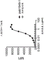
In one embodiment, the antibody is agonistic. In one embodiment, the antibody activates, enhances or induces the activity of human OX 40. In one embodiment, the antibody induces CD4+ T cell proliferation. In one embodiment, CD4+ T cell proliferation is a substantially increasing function of antibody concentration. In one embodiment, CD4+ T cell proliferation exhibits a sigmoidal dose response curve. In one embodiment, the antibody induces production of IL-2, TNF α, IFN γ, IL-4, IL-10, IL-13, or a combination thereof by anti-CD 3-stimulated T cells. In one embodiment, the antibody induces the production of TNF α, TNF β, IFN γ, GM-CSF, IL-2, IL-4, IL-10, IL-13, or a combination thereof, by anti-CD 3-stimulated Peripheral Blood Mononuclear Cells (PBMCs). In one embodiment, the antibody induces production of TNF α, TNF β, IFN γ, GM-CSF, IL-2, IL-10, or IL-13 by anti-CD 3-stimulated PBMCs, wherein the production of TNF α, TNF β, IFN γ, GM-CSF, IL-2, IL-10, or IL-13 is a substantially increasing function of antibody concentration. In one embodiment, the antibody induces production of TNF α, TNF β, IFN γ, GM-CSF, IL-2, IL-10, or IL-13 by anti-CD 3-stimulated PBMCs, wherein the production of TNF α, TNF β, IFN γ, GM-CSF, IL-2, IL-10, or IL-13 exhibits a sigmoidal dose response curve. In one embodiment, the antibody induces production of IL-2 by SEA-stimulated T cells and inhibits production of IL-10 by SEA-stimulated T cells. In one embodiment, the antibody induces production of IL-2 by SEA-stimulated Peripheral Blood Mononuclear Cells (PBMCs) and inhibits production of IL-2 by SEA-stimulated PBMCs. In one embodiment, the antibody induces production of IL-2 by SEA-stimulated PBMCs, wherein the production of IL-2 is a substantially increasing function of antibody concentration. In one embodiment, the antibody induces production of IL-2 by SEA-stimulated PBMCs, wherein the production of IL-2 exhibits a sigmoidal dose response curve.
In one embodiment, the antibody alleviates suppression of effector T cells by regulatory T cells.
In one embodiment, the antibody induces production of IL-2 by co-culture of effector T cells and regulatory T cells and inhibits production of IL-10 by co-culture of effector T cells and regulatory T cells.
In one embodiment, an antibody that specifically binds OX40 comprises: VH CDR1 comprising the amino acid sequence of GSAMH (SEQ ID NO: 4); VH CDR2 comprising the amino acid sequence of RIRSKANSYATAYAASVKG (SEQ ID NO: 5); VH CDR3 comprising the amino acid sequence of GIYDSSGYDY (SEQ ID NO: 6); VL CDR1 comprising the amino acid sequence of RSSQSLLHSNGYNYLD (SEQ ID NO: 1); VL CDR2 comprising the amino acid sequence of LGSNRAS (SEQ ID NO: 2); and a VL CDR3 comprising the amino acid sequence of MQALQTPLT (SEQ ID NO:3), wherein the antibody, in combination with Staphylococcal Enterotoxin A (SEA) (e.g., 100ng/ml), when in combination with 5% CO at, e.g., 37 ℃. (SEQ ID NO:3) 2 And stimulation at 97% humidity for e.g. 5 days to induce IL-2 production in e.g. PBMCs as measured by e.g. electrochemiluminescence (e.g. human TH1/TH 210-Plex tissue culture kit (microscopical discovery Co.)), wherein IL-2 production is substantially at an antibody concentration of e.g. 0.032 μ g/ml and 20 μ g/ml, 0.16 μ g/ml and 20 μ g/ml, 0.8 μ g/ml and 20 μ g/ml, 4 μ g/ml and 20 μ g/ml, 0.032 μ g/ml and 4 μ g/ml, 0.16 μ g/ml and 4 μ g/ml, or between 0.8 μ g/ml and 4 μ g/ml As a function of the upper increment. IL-2 production can be assessed, for example, in an assay comprising the following steps: (a) in the absence or presence of various concentrations of the antibody (e.g., 20, 4, 0.8, 0.16, 0.032, 0.0064, 0.00128, and 0.000256 μ g/ml) and SEA of, e.g., 100ng/ml, at, e.g., 37 ℃, 5% CO 2 And culturing these PBMCs at 97% humidity (e.g., 10 in well) 5 Individual cells) for, e.g., 5 days; and (b) collecting the clear supernatant and measuring the titer of IL-2 by, for example, electrochemiluminescence (e.g., human TH1/TH 210-Plex tissue culture kit (microscopical discovery Co.). In one embodiment, an antibody that specifically binds OX40 comprises: VH CDR1 comprising the amino acid sequence of GSAMH (SEQ ID NO: 4); VH CDR2 comprising the amino acid sequence of RIRSKANSYATAYAASVKG (SEQ ID NO: 5); VH CDR3 comprising the amino acid sequence of GIYDSSGYDY (SEQ ID NO: 6); VL CDR1 comprising the amino acid sequence of RSSQSLLHSNGYNYLD (SEQ ID NO: 1); VL CDR2 comprising the amino acid sequence of LGSNRAS (SEQ ID NO: 2); and a VL CDR3 comprising the amino acid sequence of MQALQTPLT (SEQ ID NO:3), wherein the antibody, in combination with Staphylococcal Enterotoxin A (SEA) (e.g., 100ng/ml), when in combination with 5% CO at, e.g., 37 ℃. (SEQ ID NO:3) 2 And stimulation at 97% humidity for e.g. 5 days to induce IL-2 production in e.g. PBMCs as measured by e.g. electrochemiluminescence (e.g. human TH1/TH 210-Plex tissue culture kit (microscopical discovery company)), wherein the production of IL-2 shows a sigmoidal dose response curve when the anti-OX 40 antibody concentration is e.g. 0.032 μ g/ml and 20 μ g/ml, 0.16 μ g/ml and 20 μ g/ml, 0.8 μ g/ml and 20 μ g/ml, 4 μ g/ml and 20 μ g/ml, 0.032 μ g/ml and 4 μ g/ml, 0.16 μ g/ml and 4 μ g/ml, or between 0.8 μ g/ml and 4 μ g/ml, as assessed e.g. in an assay comprising the following steps: (a) in the absence or presence of various concentrations of the antibody (e.g., 20, 4, 0.8, 0.16, 0.032, 0.0064, 0.00128, and 0.000256 μ g/ml) and SEA of, e.g., 100ng/ml, at, e.g., 37 ℃, 5% CO 2 And culturing these PBMCs at 97% humidity (e.g., 10 in well) 5 Individual cells) for, e.g., 5 days; and (b) collecting the clarified supernatant and measuring IL by, e.g., electrochemiluminescence (e.g., human TH1/TH 210-Plex tissue culture kit (microscopical discovery Co.))-a titer of 2.
In one embodiment, an antibody that specifically binds OX40 comprises: a VH CDR1 comprising the amino acid sequence of GSAMH (SEQ ID NO: 4); VH CDR2 comprising the amino acid sequence of RIRSKANSYATAYAASVKG (SEQ ID NO: 5); VH CDR3 comprising the amino acid sequence of GIYDSSGYDY (SEQ ID NO: 6); VL CDR1 comprising the amino acid sequence of RSSQSLLHSNGYNYLD (SEQ ID NO: 1); VL CDR2 comprising the amino acid sequence of LGSNRAS (SEQ ID NO: 2); and a VL CDR3 comprising the amino acid sequence of MQALQTPLT (SEQ ID NO:3), wherein the antibody, in combination with Staphylococcal Enterotoxin A (SEA) (e.g., 100ng/ml), when in combination with 5% CO at, e.g., 37 ℃. (SEQ ID NO:3) 2 And stimulation at 97% humidity for e.g. after 5 days induces production of IL-2 in e.g. PBMCs as measured by e.g. electrochemiluminescence (e.g. human TH1/TH 210-Plex tissue culture kit (microscopical discovery company)), wherein the IL-2 production is greater in the presence of 4 μ g/ml of antibody than in the presence of 0.032 μ g/ml of antibody. IL-2 production can be assessed, for example, in an assay comprising the following steps: (a) in the absence or presence of various concentrations of the antibody (e.g., 20, 4, 0.8, 0.16, 0.032, 0.0064, 0.00128, and 0.000256 μ g/ml) and SEA of, e.g., 100ng/ml, at, e.g., 37 ℃, 5% CO 2 And culturing these PBMCs at 97% humidity (e.g., 10 in well) 5 Individual cells) for, e.g., 5 days; and (b) collecting the clear supernatant and measuring the titer of IL-2 by, for example, electrochemiluminescence (e.g., human TH1/TH 210-Plex tissue culture kit (microscopical discovery Co.).
In one embodiment, an antibody that specifically binds OX40 comprises: VH CDR1 comprising the amino acid sequence of GSAMH (SEQ ID NO: 4); VH CDR2 comprising the amino acid sequence of RIRSKANSYATAYAASVKG (SEQ ID NO: 5); VH CDR3 comprising the amino acid sequence of GIYDSSGYDY (SEQ ID NO: 6); VL CDR1 comprising the amino acid sequence of RSSQSLLHSNGYNYLD (SEQ ID NO: 1); VL CDR2 comprising the amino acid sequence of LGSNRAS (SEQ ID NO: 2); and a VL CDR3 comprising the amino acid sequence of MQALQTPLT (SEQ ID NO:3), wherein the antibody, when bound to a plate, in combination with a plate-bound anti-CD 3 antibody (e.g., 0.8 μ g/ml), when at, e.g., 37 ℃ and 5% CO 2 The stimulation induces production of one or more cytokines (e.g., TNF α, TNF β, IFN γ, GM-CSF, IL-2, IL-10, or IL-13) in, e.g., PBMCs or T cells, as measured, e.g., by electrochemiluminescence (e.g., human TH1/TH 210-Plex tissue culture kit (microscopical discovery company) or non-human primate (NHP) V-Plex assay kit (microscopical discovery company)), wherein production of the one or more cytokines (e.g., TNF α, TNF β, IFN γ, GM-CSF, IL-2, IL-10, or IL-13) is a substantially increasing function of antibody concentration between, e.g., 0.7 μ g/ml and 50 μ g/ml, 1.6 μ g/ml and 50 μ g/ml, 3.1 μ g/ml and 50 μ g/ml, or 6.3 μ g/ml and 50 μ g/ml, as evaluated in an assay comprising, for example: (a) in the presence of a plate-bound anti-CD 3 antibody (e.g., 0.8. mu.g/ml) and a plate-bound antibody of the invention at various concentrations (e.g., 0, 0.3, 1, 3, 6, 12, 25, and 50. mu.g/ml; or 0, 0.7, 1.6, 3.1, 6.3, 12.5, 25, or 50. mu.g/ml), at, e.g., 37 ℃ and 5% CO 2 These PBMCs are cultured under for, e.g., 4 days; and (b) collecting the supernatant and measuring the production of one or more cytokines (e.g., TNF α, TNF β, IFN γ, GM-CSF, IL-2, IL-10, or IL-13) by, for example, electrochemiluminescence (e.g., human TH1/TH 210-Plex tissue culture kit (microscopical discovery Co.) or non-human primate (NHP) V-Plex assay kit (microscopical discovery Co.). In one embodiment, an antibody that specifically binds OX40 comprises: VH CDR1 comprising the amino acid sequence of GSAMH (SEQ ID NO: 4); VH CDR2 comprising the amino acid sequence of RIRSKANSYATAYAASVKG (SEQ ID NO: 5); VH CDR3 comprising the amino acid sequence of GIYDSSGYDY (SEQ ID NO: 6); VL CDR1 comprising the amino acid sequence of RSSQSLLHSNGYNYLD (SEQ ID NO: 1); VL CDR2 comprising the amino acid sequence of LGSNRAS (SEQ ID NO: 2); and a VL CDR3 comprising the amino acid sequence of MQALQTPLT (SEQ ID NO:3), wherein the antibody, when bound to a plate, in combination with a plate-bound anti-CD 3 antibody (e.g., 0.8 μ g/ml), when at, e.g., 37 ℃ and 5% CO 2 Down stimulation induces production of one or more cytokines (e.g., TNF α, TNF β, IFN γ, GM-CSF, IL-2, IL-10, or IL-13), e.g., in PBMCs or T cells, e.g., after a duration of, e.g., 4 days, e.g., bySuch as electrochemiluminescence (e.g., human TH1/TH 210-Plex tissue culture kit (microscopical discovery Co.) or non-human primate (NHP) V-Plex assay kit (microscopical discovery Co.)), wherein the production of one or more cytokines (e.g., TNF α, TNF β, IFN γ, GM-CSF, IL-2, IL-10, or IL-13) exhibits a sigmoidal dose response curve when the anti-OX 40 antibody concentration is between, for example, 0.7 μ g/ml and 50 μ g/ml, 1.6 μ g/ml and 50 μ g/ml, 3.1 μ g/ml and 50 μ g/ml, or 6.3 μ g/ml and 50 μ g/ml, as assessed in, for example, an assay comprising the steps of: (a) in the presence of a plate-bound anti-CD 3 antibody (e.g., 0.8. mu.g/ml) and a plate-bound antibody of the invention at various concentrations (e.g., 0, 0.3, 1, 3, 6, 12, 25, and 50. mu.g/ml; or 0, 0.7, 1.6, 3.1, 6.3, 12.5, 25, or 50. mu.g/ml), at, e.g., 37 ℃ and 5% CO 2 These PBMCs are cultured under for, e.g., 4 days; and (b) collecting the supernatant and measuring the production of one or more cytokines (e.g., TNF α, TNF β, IFN γ, GM-CSF, IL-2, IL-10, or IL-13) by, for example, electrochemiluminescence (e.g., human TH1/TH 210-Plex tissue culture kit (microscopical discovery Co.) or non-human primate (NHP) V-Plex assay kit (microscopical discovery Co.).
In one embodiment, an antibody that specifically binds OX40 comprises: VH CDR1 comprising the amino acid sequence of GSAMH (SEQ ID NO: 4); VH CDR2 comprising the amino acid sequence of RIRSKANSYATAYAASVKG (SEQ ID NO: 5); VH CDR3 comprising the amino acid sequence of GIYDSSGYDY (SEQ ID NO: 6); VL CDR1 comprising the amino acid sequence of RSSQSLLHSNGYNYLD (SEQ ID NO: 1); VL CDR2 comprising the amino acid sequence of LGSNRAS (SEQ ID NO: 2); and a VL CDR3 comprising the amino acid sequence of MQALQTPLT (SEQ ID NO:3), wherein the antibody increases CD4+ T cell proliferation, wherein CD4+ T cell proliferation is a substantially increasing function of antibody concentration, e.g., between 0.2 μ g/ml and 20 μ g/ml, or between 2 μ g/ml and 20 μ g/ml, as evaluated in an assay, e.g., comprising the steps of: (a) labeling, e.g., enriched CD4+ T cells with, e.g., 10 μ M carboxyfluorescein acetoacetate succinimidyl ester (CFSE) at, e.g., 37 ℃ for, e.g., 7 minutes; (b) after extensive washing, the anti-CD 3 antibody bound with e.g.a plate of e.g.3. mu.g/ml andplate-bound antibodies such as described herein, at various concentrations (e.g., 0.002, 0.02, 0.2, 2, and 20 μ g/ml), at e.g., 37 ℃ and 5% CO 2 Stimulating the cells (e.g., in the well 10) 5 Individual cells); and (c) at e.g. day 4, cell staining with e.g. anti-CD 4 antibody and checking for CD4+ T cell proliferation by means of flow cytometry by e.g. measuring the percentage of CFSE low CD4+ cells. In one embodiment, an antibody that specifically binds OX40 comprises: VH CDR1 comprising the amino acid sequence of GSAMH (SEQ ID NO: 4); VH CDR2 comprising the amino acid sequence of RIRSKANSYATAYAASVKG (SEQ ID NO: 5); VH CDR3 comprising the amino acid sequence of GIYDSSGYDY (SEQ ID NO: 6); VL CDR1 comprising the amino acid sequence of RSSQSLLHSNGYNYLD (SEQ ID NO: 1); VL CDR2 comprising the amino acid sequence of LGSNRAS (SEQ ID NO: 2); and a VL CDR3 comprising the amino acid sequence of MQALQTPLT (SEQ ID NO:3), wherein the antibody increases CD4+ T cell proliferation, wherein CD4+ T cell proliferation exhibits a sigmoidal dose-response curve when the anti-OX 40 antibody concentration is, for example, between 0.2 μ g/ml and 20 μ g/ml, or between 2 μ g/ml and 20 μ g/ml, as evaluated in, for example, an assay comprising: (a) labeling, e.g., enriched CD4+ T cells with, e.g., 10 μ M carboxyfluorescein acetoacetate succinimidyl ester (CFSE) at, e.g., 37 ℃ for, e.g., 7 minutes; (b) after extensive washing, the antibody bound to a plate such as described herein, at e.g., 37 ℃ and 5% CO, was washed with e.g., 3. mu.g/ml of e.g., plate-bound anti-CD 3 antibody and various concentrations (e.g., 0.002, 0.02, 0.2, 2, and 20. mu.g/ml) of the antibody bound to a plate such as described herein 2 In the presence of (e.g., in the well 10) 5 Individual cells); and (c) at, e.g., day 4, cell staining with, e.g., anti-CD 4 antibody and examining CD4+ T cell proliferation by means of flow cytometry, e.g., by measuring the percentage of CFSE low CD4+ cells.
In one embodiment, an antibody that specifically binds OX40 comprises: a VH CDR1 comprising the amino acid sequence of GSAMH (SEQ ID NO: 4); VH CDR2 comprising the amino acid sequence of RIRSKANSYATAYAASVKG (SEQ ID NO: 5); VH CDR3 comprising the amino acid sequence of GIYDSSGYDY (SEQ ID NO: 6); VL CDR1 comprising the amino acid sequence of RSSQSLLHSNGYNYLD (SEQ ID NO: 1); VLCDR2 comprising the amino acid sequence of LGSNRAS (SEQ ID NO: 2); and a VL CDR3 comprising the amino acid sequence of MQALQTPLT (SEQ ID NO:3), wherein the antibody results in a greater increase in CD4+ T cell proliferation when the antibody is present at a concentration of 20 μ g/ml compared to a concentration of 2 μ g/ml, as evaluated in, for example, an assay comprising: (a) labeling, e.g., enriched CD4+ T cells with, e.g., 10 μ M carboxyfluorescein acetoacetate succinimidyl ester (CFSE) at, e.g., 37 ℃ for, e.g., 7 minutes; (b) after extensive washing, the antibody bound to a plate such as described herein, at e.g., 37 ℃ and 5% CO, was washed with e.g., 3. mu.g/ml of e.g., plate-bound anti-CD 3 antibody and various concentrations (e.g., 0.002, 0.02, 0.2, 2, and 20. mu.g/ml) of the antibody bound to a plate such as described herein 2 In the presence of (e.g., in the well 10) 5 Individual cells); and (c) at e.g. day 4, cell staining with e.g. anti-CD 4 antibody and checking for CD4+ T cell proliferation by means of flow cytometry by e.g. measuring the percentage of CFSE low CD4+ cells.
In one embodiment, the antibody comprises a VL domain having at least 70% identity to the amino acid sequence of SEQ ID NO. 15 and a VH domain having at least 70% identity to the amino acid sequence of SEQ ID NO. 16, wherein the antibody, in combination with Staphylococcal Enterotoxin A (SEA) (e.g., 100ng/ml), when exposed to 5% CO at, e.g., 37 ℃. (see FIGS 2 And inducing production of IL-2 in, e.g., PBMCs after stimulation at 97% humidity for, e.g., 5 days, as measured by, e.g., electrochemiluminescence (e.g., human TH1/TH 210-Plex tissue culture kit (microscopical discovery Co.)), wherein production of IL-2 is a substantially increasing function of antibody concentration at, e.g., 0.032 μ g/ml and 20 μ g/ml, 0.16 μ g/ml and 20 μ g/ml, 0.8 μ g/ml and 20 μ g/ml, 4 μ g/ml and 20 μ g/ml, 0.032 μ g/ml and 4 μ g/ml, 0.16 μ g/ml and 4 μ g/ml, or between 0.8 μ g/ml and 4 μ g/ml. IL-2 production can be assessed, for example, in an assay comprising the following steps: (a) in the absence or presence of various concentrations of the antibody (e.g., 20, 4, 0.8, 0.16, 0.032, 0.0064, 0.00128, and 0.000256 μ g/ml) and SEA of, e.g., 100ng/ml, at, e.g., 37 ℃, 5% CO 2 And culturing these PBMCs at 97% humidity (e.g., 10 in well) 5 Individual cell) holderFor example, for 5 days; and (b) collecting the clear supernatant and measuring the titer of IL-2 by, for example, electrochemiluminescence (e.g., human TH1/TH 210-Plex tissue culture kit (microscopical discovery Co.). In one embodiment, the antibody comprises a VL domain having at least 70% identity to the amino acid sequence of SEQ ID NO. 15 and a VH domain having at least 70% identity to the amino acid sequence of SEQ ID NO. 16, wherein the antibody, in combination with Staphylococcal Enterotoxin A (SEA) (e.g., 100ng/ml), when exposed to 5% CO at, e.g., 37 ℃. (see FIGS 2 And stimulation at 97% humidity for e.g. 5 days to induce IL-2 production in e.g. PBMCs as measured by e.g. electrochemiluminescence (e.g. human TH1/TH 210-Plex tissue culture kit (microscopical discovery company)), wherein the production of IL-2 shows a sigmoidal dose response curve when the anti-OX 40 antibody concentration is e.g. 0.032 μ g/ml and 20 μ g/ml, 0.16 μ g/ml and 20 μ g/ml, 0.8 μ g/ml and 20 μ g/ml, 4 μ g/ml and 20 μ g/ml, 0.032 μ g/ml and 4 μ g/ml, 0.16 μ g/ml and 4 μ g/ml, or between 0.8 μ g/ml and 4 μ g/ml, as assessed e.g. in an assay comprising the following steps: (a) in the absence or presence of different concentrations of the antibody (e.g., 20, 4, 0.8, 0.16, 0.032, 0.0064, 0.00128, and 0.000256 μ g/ml) and SEA of, e.g., 100ng/ml, at, e.g., 37 ℃, 5% CO 2 And culturing these PBMCs at 97% humidity (e.g., 10 in well) 5 Individual cells) for, e.g., 5 days; and (b) collecting the clear supernatant and measuring the titer of IL-2 by, e.g., electrochemiluminescence (e.g., human TH1/TH 210-Plex tissue culture kit (mesoscopic discovery Co.).
In one embodiment, the antibody comprises a VL domain having at least 70% identity to the amino acid sequence of SEQ ID NO. 15 and a VH domain having at least 70% identity to the amino acid sequence of SEQ ID NO. 16, wherein the antibody, in combination with Staphylococcal Enterotoxin A (SEA) (e.g., 100ng/ml), when exposed to 5% CO at, e.g., 37 ℃. (see FIGS 2 And stimulation at 97% humidity for e.g. after 5 days induces production of IL-2 in e.g. PBMCs as measured by e.g. electrochemiluminescence (e.g. human TH1/TH 210-Plex tissue culture kit (mesoscale Discovery) in the presence of 4 μ g/ml antibody compared to 0.032 μ gIL-2 production was greater in the presence of g/ml antibody. IL-2 production can be assessed, for example, in an assay comprising the following steps: (a) in the absence or presence of different concentrations of the antibody (e.g., 20, 4, 0.8, 0.16, 0.032, 0.0064, 0.00128, and 0.000256 μ g/ml) and SEA of, e.g., 100ng/ml, at, e.g., 37 ℃, 5% CO 2 And culturing these PBMCs at 97% humidity (e.g., 10 in well) 5 Individual cells) for, e.g., 5 days; and (b) collecting the clear supernatant and measuring the titer of IL-2 by, for example, electrochemiluminescence (e.g., human TH1/TH 210-Plex tissue culture kit (microscopical discovery Co.).
In one embodiment, the antibody comprises a VL domain having at least 75% identity to the amino acid sequence of SEQ ID NO. 15 and a VH domain having at least 75% identity to the amino acid sequence of SEQ ID NO. 16, wherein the antibody, in combination with Staphylococcal Enterotoxin A (SEA) (e.g., 100ng/ml), when exposed to 5% CO at, e.g., 37 ℃. (see FIGS 2 And inducing production of IL-2 in, e.g., PBMCs after stimulation at 97% humidity for, e.g., 5 days, as measured by, e.g., electrochemiluminescence (e.g., human TH1/TH 210-Plex tissue culture kit (microscopical discovery Co.)), wherein production of IL-2 is a substantially increasing function of antibody concentration at, e.g., 0.032 μ g/ml and 20 μ g/ml, 0.16 μ g/ml and 20 μ g/ml, 0.8 μ g/ml and 20 μ g/ml, 4 μ g/ml and 20 μ g/ml, 0.032 μ g/ml and 4 μ g/ml, 0.16 μ g/ml and 4 μ g/ml, or between 0.8 μ g/ml and 4 μ g/ml. IL-2 production can be assessed, for example, in an assay comprising the following steps: (a) in the absence or presence of various concentrations of the antibody (e.g., 20, 4, 0.8, 0.16, 0.032, 0.0064, 0.00128, and 0.000256 μ g/ml) and SEA of, e.g., 100ng/ml, at, e.g., 37 ℃, 5% CO 2 And culturing these PBMCs at 97% humidity (e.g., 10 in well) 5 Individual cells) for, e.g., 5 days; and (b) collecting the clear supernatant and measuring the titer of IL-2 by, for example, electrochemiluminescence (e.g., human TH1/TH 210-Plex tissue culture kit (microscopical discovery Co.). In one embodiment, the antibody comprises a VL domain having at least 75% identity to the amino acid sequence of SEQ ID NO. 15 and a VH structure having at least 75% identity to the amino acid sequence of SEQ ID NO. 16A domain wherein the antibody, in combination with Staphylococcal Enterotoxin A (SEA) (e.g. 100ng/ml), when in combination with 5% CO at e.g. 37 ℃. (see) 2 And stimulation at 97% humidity for e.g. 5 days induces production of IL-2 in e.g. PBMCs as measured by e.g. electrochemiluminescence (e.g. human TH1/TH 210-Plex tissue culture kit (mesodiscovery corp)), wherein production of IL-2 shows a sigmoidal dose-response curve when the anti-OX 40 antibody concentration is e.g. 0.032 μ g/ml and 20 μ g/ml, 0.16 μ g/ml and 20 μ g/ml, 0.8 μ g/ml and 20 μ g/ml, 4 μ g/ml and 20 μ g/ml, 0.032 μ g/ml and 4 μ g/ml, 0.16 μ g/ml and 4 μ g/ml, or between 0.8 μ g/ml and 4 μ g/ml, as assessed in e.g. an assay comprising the steps of: (a) in the absence or presence of various concentrations of the antibody (e.g., 20, 4, 0.8, 0.16, 0.032, 0.0064, 0.00128, and 0.000256 μ g/ml) and SEA of, e.g., 100ng/ml, at, e.g., 37 ℃, 5% CO 2 And culturing these PBMCs at 97% humidity (e.g., 10 in well) 5 Individual cells) for, e.g., 5 days; and (b) collecting the clear supernatant and measuring the titer of IL-2 by, e.g., electrochemiluminescence (e.g., human TH1/TH 210-Plex tissue culture kit (mesoscopic discovery Co.).
In one embodiment, the antibody comprises a VL domain having at least 75% identity to the amino acid sequence of SEQ ID NO. 15 and a VH domain having at least 75% identity to the amino acid sequence of SEQ ID NO. 16, wherein the antibody, in combination with Staphylococcal Enterotoxin A (SEA) (e.g., 100ng/ml), when exposed to 5% CO at, e.g., 37 ℃. (see FIGS 2 And stimulation at 97% humidity for e.g. after 5 days induces production of IL-2 in e.g. PBMCs as measured by e.g. electrochemiluminescence (e.g. human TH1/TH 210-Plex tissue culture kit (microscopical discovery company)), wherein the production of IL-2 is greater in the presence of 4 μ g/ml of antibody than in the presence of 0.032 μ g/ml of antibody. IL-2 production can be assessed, for example, in an assay comprising the following steps: (a) in the absence or presence of various concentrations of the antibody (e.g., 20, 4, 0.8, 0.16, 0.032, 0.0064, 0.00128, and 0.000256 μ g/ml) and SEA of, e.g., 100ng/ml, at, e.g., 37 ℃, 5% CO 2 And culturing these PBMCs at 97% humidity (e.g., 10 in well) 5 Individual cells) for, e.g., 5 days; and (b) collecting the clear supernatant and measuring the titer of IL-2 by, for example, electrochemiluminescence (e.g., human TH1/TH 210-Plex tissue culture kit (microscopical discovery Co.).
In one embodiment, the antibody comprises a VL domain having at least 80% identity to the amino acid sequence of SEQ ID NO. 15 and a VH domain having at least 80% identity to the amino acid sequence of SEQ ID NO. 16, wherein the antibody, in combination with Staphylococcal Enterotoxin A (SEA) (e.g., 100ng/ml), when exposed to 5% CO at, e.g., 37 ℃. (see FIGS 2 And inducing production of IL-2 in, e.g., PBMCs after stimulation at 97% humidity for, e.g., 5 days, as measured by, e.g., electrochemiluminescence (e.g., human TH1/TH 210-Plex tissue culture kit (microscopical discovery Co.)), wherein production of IL-2 is a substantially increasing function of antibody concentration at, e.g., 0.032 μ g/ml and 20 μ g/ml, 0.16 μ g/ml and 20 μ g/ml, 0.8 μ g/ml and 20 μ g/ml, 4 μ g/ml and 20 μ g/ml, 0.032 μ g/ml and 4 μ g/ml, 0.16 μ g/ml and 4 μ g/ml, or between 0.8 μ g/ml and 4 μ g/ml. IL-2 production can be assessed, for example, in an assay comprising the following steps: (a) in the absence or presence of various concentrations of the antibody (e.g., 20, 4, 0.8, 0.16, 0.032, 0.0064, 0.00128, and 0.000256 μ g/ml) and SEA of, e.g., 100ng/ml, at, e.g., 37 ℃, 5% CO 2 And culturing these PBMCs at 97% humidity (e.g., 10 in well) 5 Individual cells) for, e.g., 5 days; and (b) collecting the clear supernatant and measuring the titer of IL-2 by, for example, electrochemiluminescence (e.g., human TH1/TH 210-Plex tissue culture kit (microscopical discovery Co.). In one embodiment, the antibody comprises a VL domain having at least 80% identity to the amino acid sequence of SEQ ID NO. 15 and a VH domain having at least 80% identity to the amino acid sequence of SEQ ID NO. 16, wherein the antibody, in combination with Staphylococcal Enterotoxin A (SEA) (e.g., 100ng/ml), when exposed to 5% CO at, e.g., 37 ℃. (see FIGS 2 And stimulation at 97% humidity for e.g. 5 days induces production of IL-2 in e.g. PBMCs as measured by e.g. electrochemiluminescence (e.g. human TH1/TH 210-Plex tissue culture kit (microscopical discovery Co.)), wherein when the anti-OX 40 antibody concentration is e.g. 0.032 μ g/ml and 20Production of IL-2 shows a sigmoidal dose-response curve when μ g/ml, 0.16 μ g/ml and 20 μ g/ml, 0.8 μ g/ml and 20 μ g/ml, 4 μ g/ml and 20 μ g/ml, 0.032 μ g/ml and 4 μ g/ml, 0.16 μ g/ml and 4 μ g/ml, or between 0.8 μ g/ml and 4 μ g/ml, as assessed in an assay, for example, comprising the steps of: (a) in the absence or presence of different concentrations of the antibody (e.g., 20, 4, 0.8, 0.16, 0.032, 0.0064, 0.00128, and 0.000256 μ g/ml) and SEA of, e.g., 100ng/ml, at, e.g., 37 ℃, 5% CO 2 And culturing the PBMCs at 97% humidity (e.g., 10 in well) 5 Individual cells) for, e.g., 5 days; and (b) collecting the clear supernatant and measuring the titer of IL-2 by, for example, electrochemiluminescence (e.g., human TH1/TH 210-Plex tissue culture kit (microscopical discovery Co.).
In one embodiment, the antibody comprises a VL domain having at least 80% identity to the amino acid sequence of SEQ ID NO. 15 and a VH domain having at least 80% identity to the amino acid sequence of SEQ ID NO. 16, wherein the antibody, in combination with Staphylococcal Enterotoxin A (SEA) (e.g., 100ng/ml), when exposed to 5% CO at, e.g., 37 ℃. (see FIGS 2 And stimulation at 97% humidity for e.g. after 5 days induces production of IL-2 in e.g. PBMCs as measured by e.g. electrochemiluminescence (e.g. human TH1/TH 210-Plex tissue culture kit (microscopical discovery company)), wherein the production of IL-2 is greater in the presence of 4 μ g/ml of antibody than in the presence of 0.032 μ g/ml of antibody. IL-2 production can be assessed, for example, in an assay comprising the following steps: (a) in the absence or presence of various concentrations of the antibody (e.g., 20, 4, 0.8, 0.16, 0.032, 0.0064, 0.00128, and 0.000256 μ g/ml) and SEA of, e.g., 100ng/ml, at, e.g., 37 ℃, 5% CO 2 And culturing these PBMCs at 97% humidity (e.g., 10 in well) 5 Individual cells) for, e.g., 5 days; and (b) collecting the clear supernatant and measuring the titer of IL-2 by, for example, electrochemiluminescence (e.g., human TH1/TH 210-Plex tissue culture kit (microscopical discovery Co.).
In one embodiment, the antibody comprises a VL domain having at least 85% identity to the amino acid sequence of SEQ ID NO. 15 and at least the amino acid sequence of SEQ ID NO. 16VH domain of 85% identity, wherein the antibody, in combination with Staphylococcal Enterotoxin A (SEA) (e.g., 100ng/ml), when exposed to 5% CO at, e.g., 37 ℃ 2 And inducing production of IL-2 in, e.g., PBMCs after stimulation at 97% humidity for, e.g., 5 days, as measured by, e.g., electrochemiluminescence (e.g., human TH1/TH 210-Plex tissue culture kit (microscopical discovery Co.)), wherein production of IL-2 is a substantially increasing function of antibody concentration at, e.g., 0.032 μ g/ml and 20 μ g/ml, 0.16 μ g/ml and 20 μ g/ml, 0.8 μ g/ml and 20 μ g/ml, 4 μ g/ml and 20 μ g/ml, 0.032 μ g/ml and 4 μ g/ml, 0.16 μ g/ml and 4 μ g/ml, or between 0.8 μ g/ml and 4 μ g/ml. IL-2 production can be assessed, for example, in an assay comprising the following steps: (a) in the absence or presence of various concentrations of the antibody (e.g., 20, 4, 0.8, 0.16, 0.032, 0.0064, 0.00128, and 0.000256 μ g/ml) and SEA of, e.g., 100ng/ml, at, e.g., 37 ℃, 5% CO 2 And culturing these PBMCs at 97% humidity (e.g., 10 in well) 5 Individual cells) for, e.g., 5 days; and (b) collecting the clear supernatant and measuring the titer of IL-2 by, for example, electrochemiluminescence (e.g., human TH1/TH 210-Plex tissue culture kit (microscopical discovery Co.). In one embodiment, the antibody comprises a VL domain having at least 85% identity to the amino acid sequence of SEQ ID NO. 15 and a VH domain having at least 85% identity to the amino acid sequence of SEQ ID NO. 16, wherein the antibody, in combination with Staphylococcal Enterotoxin A (SEA) (e.g., 100ng/ml), when exposed to 5% CO at, e.g., 37 ℃. (see FIGS 2 And stimulation at 97% humidity for e.g. 5 days to induce IL-2 production in e.g. PBMCs as measured by e.g. electrochemiluminescence (e.g. human TH1/TH 210-Plex tissue culture kit (microscopical discovery company)), wherein the production of IL-2 shows a sigmoidal dose response curve when the anti-OX 40 antibody concentration is e.g. 0.032 μ g/ml and 20 μ g/ml, 0.16 μ g/ml and 20 μ g/ml, 0.8 μ g/ml and 20 μ g/ml, 4 μ g/ml and 20 μ g/ml, 0.032 μ g/ml and 4 μ g/ml, 0.16 μ g/ml and 4 μ g/ml, or between 0.8 μ g/ml and 4 μ g/ml, as assessed e.g. in an assay comprising the following steps: (a) in the absence or presence of various concentrations of the antibody (e.g., 20, 4, 0.8, 0.16, 0.032, 0.0064, 0.00128, and 0.000256 μ g/ml) And SEA of, for example, 100ng/ml, 5% CO at, for example, 37 deg.C 2 And culturing these PBMCs at 97% humidity (e.g., 10 in well) 5 Individual cells) for, e.g., 5 days; and (b) collecting the clear supernatant and measuring the titer of IL-2 by, for example, electrochemiluminescence (e.g., human TH1/TH 210-Plex tissue culture kit (microscopical discovery Co.).
In one embodiment, the antibody comprises a VL domain having at least 85% identity to the amino acid sequence of SEQ ID NO. 15 and a VH domain having at least 85% identity to the amino acid sequence of SEQ ID NO. 16, wherein the antibody, in combination with Staphylococcal Enterotoxin A (SEA) (e.g., 100ng/ml), when exposed to 5% CO at, e.g., 37 ℃. (see FIGS 2 And stimulation at 97% humidity for e.g. after 5 days induces production of IL-2 in e.g. PBMCs as measured by e.g. electrochemiluminescence (e.g. human TH1/TH 210-Plex tissue culture kit (microscopical discovery company)), wherein the production of IL-2 is greater in the presence of 4 μ g/ml of antibody than in the presence of 0.032 μ g/ml of antibody. IL-2 production can be assessed, for example, in an assay comprising the following steps: (a) in the absence or presence of various concentrations of the antibody (e.g., 20, 4, 0.8, 0.16, 0.032, 0.0064, 0.00128, and 0.000256 μ g/ml) and SEA of, e.g., 100ng/ml, at, e.g., 37 ℃, 5% CO 2 And culturing these PBMCs at 97% humidity (e.g., 10 in well) 5 Individual cells) for, e.g., 5 days; and (b) collecting the clear supernatant and measuring the titer of IL-2 by, for example, electrochemiluminescence (e.g., human TH1/TH 210-Plex tissue culture kit (microscopical discovery Co.).
In one embodiment, the antibody comprises a VL domain having at least 90% identity to the amino acid sequence of SEQ ID NO. 15 and a VH domain having at least 90% identity to the amino acid sequence of SEQ ID NO. 16, wherein the antibody, in combination with Staphylococcal Enterotoxin A (SEA) (e.g., 100ng/ml), when exposed to 5% CO at, e.g., 37 ℃. (see FIGS 2 And stimulation at 97% humidity for e.g. 5 days induces production of IL-2 in e.g. PBMCs as measured by e.g. electrochemiluminescence (e.g. human TH1/TH 210-Plex tissue culture kit (microscopical discovery Co.)), wherein production of IL-2 is e.g. inA substantially increasing function of antibody concentration between 0.032 and 20. mu.g/ml, 0.16 and 20. mu.g/ml, 0.8 and 20. mu.g/ml, 4 and 20. mu.g/ml, 0.032 and 4. mu.g/ml, 0.16 and 4. mu.g/ml, or 0.8 and 4. mu.g/ml. IL-2 production can be assessed, for example, in an assay comprising the following steps: (a) in the absence or presence of various concentrations of the antibody (e.g., 20, 4, 0.8, 0.16, 0.032, 0.0064, 0.00128, and 0.000256 μ g/ml) and SEA of, e.g., 100ng/ml, at, e.g., 37 ℃, 5% CO 2 And culturing these PBMCs at 97% humidity (e.g., 10 in well) 5 Individual cells) for, e.g., 5 days; and (b) collecting the clear supernatant and measuring the titer of IL-2 by, for example, electrochemiluminescence (e.g., human TH1/TH 210-Plex tissue culture kit (microscopical discovery Co.). In one embodiment, the antibody comprises a VL domain having at least 90% identity to the amino acid sequence of SEQ ID NO. 15 and a VH domain having at least 90% identity to the amino acid sequence of SEQ ID NO. 16, wherein the antibody, in combination with Staphylococcal Enterotoxin A (SEA) (e.g., 100ng/ml), when exposed to 5% CO at, e.g., 37 ℃. (see FIGS 2 And stimulation at 97% humidity for e.g. 5 days to induce IL-2 production in e.g. PBMCs as measured by e.g. electrochemiluminescence (e.g. human TH1/TH 210-Plex tissue culture kit (microscopical discovery company)), wherein the production of IL-2 shows a sigmoidal dose response curve when the anti-OX 40 antibody concentration is e.g. 0.032 μ g/ml and 20 μ g/ml, 0.16 μ g/ml and 20 μ g/ml, 0.8 μ g/ml and 20 μ g/ml, 4 μ g/ml and 20 μ g/ml, 0.032 μ g/ml and 4 μ g/ml, 0.16 μ g/ml and 4 μ g/ml, or between 0.8 μ g/ml and 4 μ g/ml, as assessed e.g. in an assay comprising the following steps: (a) in the absence or presence of various concentrations of the antibody (e.g., 20, 4, 0.8, 0.16, 0.032, 0.0064, 0.00128, and 0.000256 μ g/ml) and SEA of, e.g., 100ng/ml, at, e.g., 37 ℃, 5% CO 2 And culturing these PBMCs at 97% humidity (e.g., 10 in well) 5 Individual cells) for, e.g., 5 days; and (b) collecting the clear supernatant and measuring the titer of IL-2 by, for example, electrochemiluminescence (e.g., human TH1/TH 210-Plex tissue culture kit (microscopical discovery Co.).
In one embodimentAn antibody comprising a VL domain having at least 90% identity to the amino acid sequence of SEQ ID NO. 15 and a VH domain having at least 90% identity to the amino acid sequence of SEQ ID NO. 16, wherein the antibody, in combination with Staphylococcal Enterotoxin A (SEA) (e.g. 100ng/ml), when exposed to 5% CO at e.g. 37 ℃ 2 And stimulation at 97% humidity for e.g. after 5 days induces production of IL-2 in e.g. PBMCs as measured by e.g. electrochemiluminescence (e.g. human TH1/TH 210-Plex tissue culture kit (microscopical discovery company)), wherein the production of IL-2 is greater in the presence of 4 μ g/ml of antibody than in the presence of 0.032 μ g/ml of antibody. IL-2 production can be assessed, for example, in an assay comprising the following steps: (a) in the absence or presence of various concentrations of the antibody (e.g., 20, 4, 0.8, 0.16, 0.032, 0.0064, 0.00128, and 0.000256 μ g/ml) and SEA of, e.g., 100ng/ml, at, e.g., 37 ℃, 5% CO 2 And culturing these PBMCs at 97% humidity (e.g., 10 in well) 5 Individual cells) for, e.g., 5 days; and (b) collecting the clear supernatant and measuring the titer of IL-2 by, for example, electrochemiluminescence (e.g., human TH1/TH 210-Plex tissue culture kit (microscopical discovery Co.).
In one embodiment, the antibody comprises a VL domain having at least 95% identity to the amino acid sequence of SEQ ID NO. 15 and a VH domain having at least 95% identity to the amino acid sequence of SEQ ID NO. 16, wherein the antibody, in combination with Staphylococcal Enterotoxin A (SEA) (e.g., 100ng/ml), when exposed to 5% CO at, e.g., 37 ℃. (see FIGS 2 And inducing production of IL-2 in, e.g., PBMCs after stimulation at 97% humidity for, e.g., 5 days, as measured by, e.g., electrochemiluminescence (e.g., human TH1/TH 210-Plex tissue culture kit (microscopical discovery Co.)), wherein production of IL-2 is a substantially increasing function of antibody concentration at, e.g., 0.032 μ g/ml and 20 μ g/ml, 0.16 μ g/ml and 20 μ g/ml, 0.8 μ g/ml and 20 μ g/ml, 4 μ g/ml and 20 μ g/ml, 0.032 μ g/ml and 4 μ g/ml, 0.16 μ g/ml and 4 μ g/ml, or between 0.8 μ g/ml and 4 μ g/ml. IL-2 production can be assessed, for example, in an assay comprising the following steps: (a) in the absence or presence of various concentrations of the antibody (e.g., 20, 4, 0.8, 0.16, 0.032, 0.0064, 0.00128, and 0.000256. mu.g/ml) and SEA, e.g. 100ng/ml, at e.g. 37 ℃ 5% CO 2 And culturing these PBMCs at 97% humidity (e.g., 10 in well) 5 Individual cells) for, e.g., 5 days; and (b) collecting the clear supernatant and measuring the titer of IL-2 by, for example, electrochemiluminescence (e.g., human TH1/TH 210-Plex tissue culture kit (microscopical discovery Co.). In one embodiment, the antibody comprises a VL domain having at least 95% identity to the amino acid sequence of SEQ ID NO. 15 and a VH domain having at least 95% identity to the amino acid sequence of SEQ ID NO. 16, wherein the antibody, in combination with Staphylococcal Enterotoxin A (SEA) (e.g., 100ng/ml), when exposed to 5% CO at, e.g., 37 ℃. (see FIGS 2 And stimulation at 97% humidity for e.g. 5 days induces production of IL-2 in e.g. PBMCs as measured by e.g. electrochemiluminescence (e.g. human TH1/TH 210-Plex tissue culture kit (mesodiscovery corp)), wherein production of IL-2 shows a sigmoidal dose-response curve when the anti-OX 40 antibody concentration is e.g. 0.032 μ g/ml and 20 μ g/ml, 0.16 μ g/ml and 20 μ g/ml, 0.8 μ g/ml and 20 μ g/ml, 4 μ g/ml and 20 μ g/ml, 0.032 μ g/ml and 4 μ g/ml, 0.16 μ g/ml and 4 μ g/ml, or between 0.8 μ g/ml and 4 μ g/ml, as assessed in e.g. an assay comprising the steps of: (a) in the absence or presence of various concentrations of the antibody (e.g., 20, 4, 0.8, 0.16, 0.032, 0.0064, 0.00128, and 0.000256 μ g/ml) and SEA of, e.g., 100ng/ml, at, e.g., 37 ℃, 5% CO 2 And culturing these PBMCs at 97% humidity (e.g., 10 in well) 5 Individual cells) for, e.g., 5 days; and (b) collecting the clear supernatant and measuring the titer of IL-2 by, for example, electrochemiluminescence (e.g., human TH1/TH 210-Plex tissue culture kit (microscopical discovery Co.).
In one embodiment, the antibody comprises a VL domain having at least 95% identity to the amino acid sequence of SEQ ID NO. 15 and a VH domain having at least 95% identity to the amino acid sequence of SEQ ID NO. 16, wherein the antibody, in combination with Staphylococcal Enterotoxin A (SEA) (e.g., 100ng/ml), when exposed to 5% CO at, e.g., 37 ℃. (see FIGS 2 And inducing IL-2 production in, e.g., PBMCs, after stimulation at 97% humidity for, e.g., 5 days, e.g., by way of exampleAs measured by electrochemiluminescence (e.g., human TH1/TH 210-Plex tissue culture kit (microscopical discovery Co.)), wherein the production of IL-2 is greater in the presence of 4. mu.g/ml of antibody than in the presence of 0.032. mu.g/ml of antibody. IL-2 production can be assessed, for example, in an assay comprising the following steps: (a) in the absence or presence of various concentrations of the antibody (e.g., 20, 4, 0.8, 0.16, 0.032, 0.0064, 0.00128, and 0.000256 μ g/ml) and SEA of, e.g., 100ng/ml, at, e.g., 37 ℃, 5% CO 2 And culturing the PBMCs at 97% humidity (e.g., 10 in well) 5 Individual cells) for, e.g., 5 days; and (b) collecting the clear supernatant and measuring the titer of IL-2 by, for example, electrochemiluminescence (e.g., human TH1/TH 210-Plex tissue culture kit (microscopical discovery Co.).
In one embodiment, the antibody comprises a VL domain having at least 98% identity to the amino acid sequence of SEQ ID NO. 15 and a VH domain having at least 98% identity to the amino acid sequence of SEQ ID NO. 16, wherein the antibody, in combination with Staphylococcal Enterotoxin A (SEA) (e.g., 100ng/ml), when exposed to 5% CO at, e.g., 37 ℃ 2 And inducing production of IL-2 in, e.g., PBMCs after stimulation at 97% humidity for, e.g., 5 days, as measured by, e.g., electrochemiluminescence (e.g., human TH1/TH 210-Plex tissue culture kit (microscopical discovery Co.)), wherein production of IL-2 is a substantially increasing function of antibody concentration at, e.g., 0.032 μ g/ml and 20 μ g/ml, 0.16 μ g/ml and 20 μ g/ml, 0.8 μ g/ml and 20 μ g/ml, 4 μ g/ml and 20 μ g/ml, 0.032 μ g/ml and 4 μ g/ml, 0.16 μ g/ml and 4 μ g/ml, or between 0.8 μ g/ml and 4 μ g/ml. IL-2 production can be assessed, for example, in an assay comprising the following steps: (a) in the absence or presence of various concentrations of the antibody (e.g., 20, 4, 0.8, 0.16, 0.032, 0.0064, 0.00128, and 0.000256 μ g/ml) and SEA of, e.g., 100ng/ml, at, e.g., 37 ℃, 5% CO 2 And culturing these PBMCs at 97% humidity (e.g., 10 in well) 5 Individual cells) for, e.g., 5 days; and (b) collecting the clear supernatant and measuring the titer of IL-2 by, for example, electrochemiluminescence (e.g., human TH1/TH 210-Plex tissue culture kit (microscopical discovery Co.). In one embodiment, the antibodyComprising a VL domain having at least 98% identity with the amino acid sequence of SEQ ID NO:15 and a VH domain having at least 98% identity with the amino acid sequence of SEQ ID NO:16, wherein the antibody, in combination with Staphylococcal Enterotoxin A (SEA) (e.g. 100ng/ml), when exposed to 5% CO at e.g. 37 ℃. (see FIGS 2 And stimulation at 97% humidity for e.g. 5 days to induce IL-2 production in e.g. PBMCs as measured by e.g. electrochemiluminescence (e.g. human TH1/TH 210-Plex tissue culture kit (microscopical discovery company)), wherein the production of IL-2 shows a sigmoidal dose response curve when the anti-OX 40 antibody concentration is e.g. 0.032 μ g/ml and 20 μ g/ml, 0.16 μ g/ml and 20 μ g/ml, 0.8 μ g/ml and 20 μ g/ml, 4 μ g/ml and 20 μ g/ml, 0.032 μ g/ml and 4 μ g/ml, 0.16 μ g/ml and 4 μ g/ml, or between 0.8 μ g/ml and 4 μ g/ml, as assessed e.g. in an assay comprising the following steps: (a) in the absence or presence of different concentrations of the antibody (e.g., 20, 4, 0.8, 0.16, 0.032, 0.0064, 0.00128, and 0.000256 μ g/ml) and SEA of, e.g., 100ng/ml, at, e.g., 37 ℃, 5% CO 2 And culturing these PBMCs at 97% humidity (e.g., 10 in well) 5 Individual cells) for, e.g., 5 days; and (b) collecting the clear supernatant and measuring the titer of IL-2 by, e.g., electrochemiluminescence (e.g., human TH1/TH 210-Plex tissue culture kit (mesoscopic discovery Co.).
In one embodiment, the antibody comprises a VL domain having at least 98% identity to the amino acid sequence of SEQ ID NO. 15 and a VH domain having at least 98% identity to the amino acid sequence of SEQ ID NO. 16, wherein the antibody, in combination with Staphylococcal Enterotoxin A (SEA) (e.g., 100ng/ml), when exposed to 5% CO at, e.g., 37 ℃ 2 And stimulation at 97% humidity for e.g. after 5 days induces production of IL-2 in e.g. PBMCs as measured by e.g. electrochemiluminescence (e.g. human TH1/TH 210-Plex tissue culture kit (microscopical discovery company)), wherein the production of IL-2 is greater in the presence of 4 μ g/ml of antibody than in the presence of 0.032 μ g/ml of antibody. IL-2 production can be assessed, for example, in an assay comprising the following steps: (a) the antibody is present in the absence or presence of various concentrations (e.g., 20, 4, 0.8, 0.16, 0.032, 0.0064, 0.00128, and 0.000256. mu.g/ml) and SEA of, for example, 100ng/ml, at, for example, 37 ℃ and 5% CO 2 And culturing these PBMCs at 97% humidity (e.g., 10 in well) 5 Individual cells) for, e.g., 5 days; and (b) collecting the clear supernatant and measuring the titer of IL-2 by, for example, electrochemiluminescence (e.g., human TH1/TH 210-Plex tissue culture kit (microscopical discovery Co.).
In one embodiment, the antibody comprises a VL domain having at least 70% identity to the amino acid sequence of SEQ ID NO. 15 and a VH domain having at least 70% identity to the amino acid sequence of SEQ ID NO. 16, wherein the antibody, when bound to a plate, in combination with an anti-CD 3 antibody (e.g., 0.8 μ g/ml) bound to the plate, when at, e.g., 37 ℃ and 5% CO 2 (ii) inducing production of one or more cytokines (e.g., TNF α, TNF β, IFN γ, GM-CSF, IL-2, IL-10, or IL-13) in, e.g., PBMCs or T cells, as measured by, e.g., electrochemiluminescence (e.g., human TH1/TH 210-Plex tissue culture kit (microscopical discovery Co., Ltd.) or non-human primate (NHP) V-Plex assay kit (microscopical discovery Co., Ltd)), wherein production of the one or more cytokines (e.g., TNF α, TNF β, IFN γ, GM-CSF, IL-2, IL-10, or IL-13) is a substantially increasing function of antibody concentration between, e.g., 0.7 μ g/ml and 50 μ g/ml, 1.6 μ g/ml and 50 μ g/ml, 3.1 μ g/ml and 50 μ g/ml, or 6.3 μ g/ml and 50 μ g/ml, as evaluated in an assay comprising, for example: (a) in the presence of a plate-bound anti-CD 3 antibody (e.g., 0.8. mu.g/ml) and a plate-bound antibody of the invention at various concentrations (e.g., 0, 0.3, 1, 3, 6, 12, 25, and 50. mu.g/ml; or 0, 0.7, 1.6, 3.1, 6.3, 12.5, 25, or 50. mu.g/ml), at, e.g., 37 ℃ and 5% CO 2 These PBMCs are cultured under for, e.g., 4 days; and (b) collecting the supernatant and measuring the production of one or more cytokines (e.g., TNF α, TNF β, IFN γ, GM-CSF, IL-2, IL-10, or IL-13) by, for example, electrochemiluminescence (e.g., human TH1/TH 210-Plex tissue culture kit (microscopical discovery Co.) or non-human primate (NHP) V-Plex assay kit (microscopical discovery Co.). In one embodiment, the antibody comprises an amino acid sequence having amino acid sequence as set forth in SEQ ID NO. 15A VL domain having at least 70% identity to the amino acid sequence of SEQ ID NO:16 and a VH domain having at least 70% identity to the amino acid sequence of SEQ ID NO:16, wherein the antibody, when bound to a plate, in combination with an anti-CD 3 antibody (e.g., 0.8 μ g/ml) bound to the plate, when incubated at, e.g., 37 ℃ and 5% CO 2 The sub-stimulation induces production of one or more cytokines (e.g., TNF α, TNF β, IFN γ, GM-CSF, IL-2, IL-10, or IL-13) in, e.g., PBMCs or T cells, as measured by, e.g., electrochemiluminescence (e.g., human TH1/TH 210-Plex tissue culture kit (microscopical discovery Co., Ltd.) or non-human primate (NHP) V-Plex assay kit (microscopical discovery Co.)), wherein the one or more cytokines (e.g., TNF α, TNF β, IFN γ, GM-CSF, IL-2, IL-13) when the anti-OX 40 antibody concentration is between, e.g., 0.7 μ g/ml and 50 μ g/ml, 1.6 μ g/ml and 50 μ g/ml, 3.1 μ g/ml and 50 μ g/ml, or 6.3 μ g/ml and 50 μ g/ml) IL-10, or IL-13) shows a sigmoidal dose-response curve as assessed, for example, in an assay comprising the steps of: (a) in the presence of a plate-bound anti-CD 3 antibody (e.g., 0.8. mu.g/ml) and a plate-bound antibody of the invention at various concentrations (e.g., 0, 0.3, 1, 3, 6, 12, 25, and 50. mu.g/ml; or 0, 0.7, 1.6, 3.1, 6.3, 12.5, 25, or 50. mu.g/ml), at, e.g., 37 ℃ and 5% CO 2 These PBMCs are cultured under for, e.g., 4 days; and (b) collecting the supernatant and measuring the production of one or more cytokines (e.g., TNF α, TNF β, IFN γ, GM-CSF, IL-2, IL-10, or IL-13) by, for example, electrochemiluminescence (e.g., human TH1/TH 210-Plex tissue culture kit (microscopical discovery Co.) or non-human primate (NHP) V-Plex assay kit (microscopical discovery Co.).
In one embodiment, the antibody comprises a VL domain having at least 75% identity to the amino acid sequence of SEQ ID NO. 15 and a VH domain having at least 75% identity to the amino acid sequence of SEQ ID NO. 16, wherein the antibody, when bound to a plate, in combination with an anti-CD 3 antibody (e.g., 0.8 μ g/ml) bound to the plate, when at, e.g., 37 ℃ and 5% CO 2 Down stimulation induces one or more cytokines (e.g., TNF α, TNF β, IFN γ, GM-CSF, IL-2, IL-10, or IL-13) in, for example, PBMC or T cells after a duration of, for example, 4 days) As measured by, for example, electrochemiluminescence (e.g., human TH1/TH 210-Plex tissue culture kit (mesodiscovery corporation) or non-human primate (NHP) V-Plex assay kit (mesodiscovery corporation)), wherein production of one or more cytokines (e.g., TNF α, TNF β, IFN γ, GM-CSF, IL-2, IL-10, or IL-13) is a substantially increasing function of antibody concentration between, for example, 0.7 μ g/ml and 50 μ g/ml, 1.6 μ g/ml and 50 μ g/ml, 3.1 μ g/ml and 50 μ g/ml, or 6.3 μ g/ml and 50 μ g/ml, as assessed in, for example, an assay comprising the steps of: (a) in the presence of a plate-bound anti-CD 3 antibody (e.g., 0.8. mu.g/ml) and a plate-bound antibody of the invention at various concentrations (e.g., 0, 0.3, 1, 3, 6, 12, 25, and 50. mu.g/ml; or 0, 0.7, 1.6, 3.1, 6.3, 12.5, 25, or 50. mu.g/ml), at, e.g., 37 ℃ and 5% CO 2 These PBMCs are cultured under for e.g. 4 days; and (b) collecting the supernatant and measuring the production of one or more cytokines (e.g., TNF α, TNF β, IFN γ, GM-CSF, IL-2, IL-10, or IL-13) by, for example, electrochemiluminescence (e.g., human TH1/TH 210-Plex tissue culture kit (microscopical discovery Co.) or non-human primate (NHP) V-Plex assay kit (microscopical discovery Co.). In one embodiment, the antibody comprises a VL domain having at least 75% identity to the amino acid sequence of SEQ ID NO. 15 and a VH domain having at least 75% identity to the amino acid sequence of SEQ ID NO. 16, wherein the antibody, when bound to a plate, in combination with an anti-CD 3 antibody (e.g., 0.8 μ g/ml) bound to the plate, when at, e.g., 37 ℃ and 5% CO 2 The sub-stimulation induces production of one or more cytokines (e.g., TNF α, TNF β, IFN γ, GM-CSF, IL-2, IL-10, or IL-13) in, e.g., PBMCs or T cells, as measured by, e.g., electrochemiluminescence (e.g., human TH1/TH 210-Plex tissue culture kit (microscopical discovery Co., Ltd.) or non-human primate (NHP) V-Plex assay kit (microscopical discovery Co.)), wherein the one or more cytokines (e.g., TNF α, TNF β, IFN γ, GM-CSF, IL-2, IL-13) when the anti-OX 40 antibody concentration is between, e.g., 0.7 μ g/ml and 50 μ g/ml, 1.6 μ g/ml and 50 μ g/ml, 3.1 μ g/ml and 50 μ g/ml, or 6.3 μ g/ml and 50 μ g/ml) IL-10, or IL-13) production display A sigmoidal dose response curve, as evaluated in an assay, for example, comprising the steps of: (a) in the presence of a plate-bound anti-CD 3 antibody (e.g., 0.8. mu.g/ml) and a plate-bound antibody of the invention at various concentrations (e.g., 0, 0.3, 1, 3, 6, 12, 25, and 50. mu.g/ml; or 0, 0.7, 1.6, 3.1, 6.3, 12.5, 25, or 50. mu.g/ml), at, e.g., 37 ℃ and 5% CO 2 These PBMCs are cultured under for, e.g., 4 days; and (b) collecting the supernatant and measuring the production of one or more cytokines (e.g., TNF α, TNF β, IFN γ, GM-CSF, IL-2, IL-10, or IL-13) by, for example, electrochemiluminescence (e.g., human TH1/TH 210-Plex tissue culture kit (microscopical discovery Co.) or non-human primate (NHP) V-Plex assay kit (microscopical discovery Co.).
In one embodiment, the antibody comprises a VL domain having at least 80% identity to the amino acid sequence of SEQ ID NO. 15 and a VH domain having at least 80% identity to the amino acid sequence of SEQ ID NO. 16, wherein the antibody, when bound to a plate, in combination with an anti-CD 3 antibody (e.g., 0.8 μ g/ml) bound to the plate, when at, e.g., 37 ℃ and 5% CO 2 (ii) inducing production of one or more cytokines (e.g., TNF α, TNF β, IFN γ, GM-CSF, IL-2, IL-10, or IL-13) in, e.g., PBMCs or T cells, as measured by, e.g., electrochemiluminescence (e.g., human TH1/TH 210-Plex tissue culture kit (microscopical discovery Co., Ltd.) or non-human primate (NHP) V-Plex assay kit (microscopical discovery Co., Ltd)), wherein production of the one or more cytokines (e.g., TNF α, TNF β, IFN γ, GM-CSF, IL-2, IL-10, or IL-13) is a substantially increasing function of antibody concentration between, e.g., 0.7 μ g/ml and 50 μ g/ml, 1.6 μ g/ml and 50 μ g/ml, 3.1 μ g/ml and 50 μ g/ml, or 6.3 μ g/ml and 50 μ g/ml, as assessed in, for example, an assay comprising the steps of: (a) in the presence of a plate-bound anti-CD 3 antibody (e.g., 0.8. mu.g/ml) and a plate-bound antibody of the invention at various concentrations (e.g., 0, 0.3, 1, 3, 6, 12, 25, and 50. mu.g/ml; or 0, 0.7, 1.6, 3.1, 6.3, 12.5, 25, or 50. mu.g/ml), at, e.g., 37 ℃ and 5% CO 2 These PBMCs are cultured under for, e.g., 4 days; and (b) collecting the supernatant and passing the exampleProduction of one or more cytokines (e.g., TNF α, TNF β, IFN γ, GM-CSF, IL-2, IL-10, or IL-13) is measured as electrochemiluminescence (e.g., human TH1/TH 210-Plex tissue culture kit (microscopical discovery Co.) or non-human primate (NHP) V-Plex assay kit (microscopical discovery Co.). In one embodiment, the antibody comprises a VL domain having at least 80% identity to the amino acid sequence of SEQ ID NO. 15 and a VH domain having at least 80% identity to the amino acid sequence of SEQ ID NO. 16, wherein the antibody, when bound to a plate, in combination with an anti-CD 3 antibody (e.g., 0.8 μ g/ml) bound to the plate, when at, e.g., 37 ℃ and 5% CO 2 The sub-stimulation induces production of one or more cytokines (e.g., TNF α, TNF β, IFN γ, GM-CSF, IL-2, IL-10, or IL-13) in, e.g., PBMCs or T cells, as measured by, e.g., electrochemiluminescence (e.g., human TH1/TH 210-Plex tissue culture kit (microscopical discovery Co., Ltd.) or non-human primate (NHP) V-Plex assay kit (microscopical discovery Co.)), wherein the one or more cytokines (e.g., TNF α, TNF β, IFN γ, GM-CSF, IL-2, IL-13) when the anti-OX 40 antibody concentration is between, e.g., 0.7 μ g/ml and 50 μ g/ml, 1.6 μ g/ml and 50 μ g/ml, 3.1 μ g/ml and 50 μ g/ml, or 6.3 μ g/ml and 50 μ g/ml) IL-10, or IL-13) shows a sigmoidal dose-response curve as assessed, for example, in an assay comprising the steps of: (a) in the presence of a plate-bound anti-CD 3 antibody (e.g., 0.8. mu.g/ml) and a plate-bound antibody of the invention at various concentrations (e.g., 0, 0.3, 1, 3, 6, 12, 25, and 50. mu.g/ml; or 0, 0.7, 1.6, 3.1, 6.3, 12.5, 25, or 50. mu.g/ml), at, e.g., 37 ℃ and 5% CO 2 These PBMCs are cultured under for, e.g., 4 days; and (b) collecting the supernatant and measuring the production of one or more cytokines (e.g., TNF α, TNF β, IFN γ, GM-CSF, IL-2, IL-10, or IL-13) by, for example, electrochemiluminescence (e.g., human TH1/TH 210-Plex tissue culture kit (microscopical discovery Co.) or non-human primate (NHP) V-Plex assay kit (microscopical discovery Co.).
In one embodiment, the antibody comprises a VL domain having at least 85% identity to the amino acid sequence of SEQ ID NO. 15 and a VL domain having at least 85% identity to SEQ ID NO. 16VH domain having an amino acid sequence of at least 85% identity, wherein the antibody, when bound to a plate, in combination with a plate-bound anti-CD 3 antibody (e.g., 0.8 μ g/ml), when at, e.g., 37 ℃ and 5% CO 2 The stimulation induces production of one or more cytokines (e.g., TNF α, TNF β, IFN γ, GM-CSF, IL-2, IL-10, or IL-13) in, e.g., PBMCs or T cells, as measured, e.g., by electrochemiluminescence (e.g., human TH1/TH 210-Plex tissue culture kit (microscopical discovery company) or non-human primate (NHP) V-Plex assay kit (microscopical discovery company)), wherein production of the one or more cytokines (e.g., TNF α, TNF β, IFN γ, GM-CSF, IL-2, IL-10, or IL-13) is a substantially increasing function of antibody concentration between, e.g., 0.7 μ g/ml and 50 μ g/ml, 1.6 μ g/ml and 50 μ g/ml, 3.1 μ g/ml and 50 μ g/ml, or 6.3 μ g/ml and 50 μ g/ml, as evaluated in an assay comprising, for example: (a) in the presence of a plate-bound anti-CD 3 antibody (e.g., 0.8. mu.g/ml) and a plate-bound antibody of the invention at various concentrations (e.g., 0, 0.3, 1, 3, 6, 12, 25, and 50. mu.g/ml; or 0, 0.7, 1.6, 3.1, 6.3, 12.5, 25, or 50. mu.g/ml), at, e.g., 37 ℃ and 5% CO 2 These PBMCs are cultured under for, e.g., 4 days; and (b) collecting the supernatant and measuring the production of one or more cytokines (e.g., TNF α, TNF β, IFN γ, GM-CSF, IL-2, IL-10, or IL-13) by, for example, electrochemiluminescence (e.g., human TH1/TH 210-Plex tissue culture kit (microscopical discovery Co.) or non-human primate (NHP) V-Plex assay kit (microscopical discovery Co.). In one embodiment, the antibody comprises a VL domain having at least 85% identity to the amino acid sequence of SEQ ID NO:15 and a VH domain having at least 85% identity to the amino acid sequence of SEQ ID NO:16, wherein the antibody, when bound to a plate, in combination with an anti-CD 3 antibody (e.g., 0.8 μ g/ml) bound to the plate, when at, e.g., 37 ℃ and 5% CO 2 Down stimulation induces production of one or more cytokines (e.g., TNF α, TNF β, IFN γ, GM-CSF, IL-2, IL-10, or IL-13) in, e.g., PBMCs or T cells after a duration of, e.g., 4 days, as by, e.g., electrochemiluminescence (e.g., human TH1/TH 210-Plex tissue culture kit (Fine cell TH1/TH 210-Plex)Visit discovery company) or a non-human primate (NHP) V-Plex assay kit (visit discovery company)), wherein production of one or more cytokines (e.g., TNF α, TNF β, IFN γ, GM-CSF, IL-2, IL-10, or IL-13) exhibits a sigmoidal dose response curve when the anti-OX 40 antibody concentration is between, for example, 0.7 μ g/ml and 50 μ g/ml, 1.6 μ g/ml and 50 μ g/ml, 3.1 μ g/ml and 50 μ g/ml, or 6.3 μ g/ml and 50 μ g/ml, as evaluated in an assay, for example, comprising the steps of: (a) in the presence of a plate-bound anti-CD 3 antibody (e.g., 0.8. mu.g/ml) and a plate-bound antibody of the invention at various concentrations (e.g., 0, 0.3, 1, 3, 6, 12, 25, and 50. mu.g/ml; or 0, 0.7, 1.6, 3.1, 6.3, 12.5, 25, or 50. mu.g/ml), at, e.g., 37 ℃ and 5% CO 2 These PBMCs are cultured under for, e.g., 4 days; and (b) collecting the supernatant and measuring the production of one or more cytokines (e.g., TNF α, TNF β, IFN γ, GM-CSF, IL-2, IL-10, or IL-13) by, for example, electrochemiluminescence (e.g., human TH1/TH 210-Plex tissue culture kit (microscopical discovery Co.) or non-human primate (NHP) V-Plex assay kit (microscopical discovery Co.).
In one embodiment, the antibody comprises a VL domain having at least 90% identity to the amino acid sequence of SEQ ID NO. 15 and a VH domain having at least 90% identity to the amino acid sequence of SEQ ID NO. 16, wherein the antibody, when bound to a plate, in combination with an anti-CD 3 antibody (e.g., 0.8 μ g/ml) bound to the plate, when at, e.g., 37 ℃ and 5% CO 2 (ii) inducing production of one or more cytokines (e.g., TNF α, TNF β, IFN γ, GM-CSF, IL-2, IL-10, or IL-13) in, e.g., PBMCs or T cells, as measured by, e.g., electrochemiluminescence (e.g., human TH1/TH 210-Plex tissue culture kit (microscopical discovery Co., Ltd.) or non-human primate (NHP) V-Plex assay kit (microscopical discovery Co., Ltd)), wherein production of the one or more cytokines (e.g., TNF α, TNF β, IFN γ, GM-CSF, IL-2, IL-10, or IL-13) is a substantially increasing function of antibody concentration between, e.g., 0.7 μ g/ml and 50 μ g/ml, 1.6 μ g/ml and 50 μ g/ml, 3.1 μ g/ml and 50 μ g/ml, or 6.3 μ g/ml and 50 μ g/ml, as in, for example, the test comprising the following steps And (3) centering evaluation: (a) in the presence of a plate-bound anti-CD 3 antibody (e.g., 0.8. mu.g/ml) and a plate-bound antibody of the invention at various concentrations (e.g., 0, 0.3, 1, 3, 6, 12, 25, and 50. mu.g/ml; or 0, 0.7, 1.6, 3.1, 6.3, 12.5, 25, or 50. mu.g/ml), at, e.g., 37 ℃ and 5% CO 2 These PBMCs are cultured under for e.g. 4 days; and (b) collecting the supernatant and measuring the production of one or more cytokines (e.g., TNF α, TNF β, IFN γ, GM-CSF, IL-2, IL-10, or IL-13) by, for example, electrochemiluminescence (e.g., human TH1/TH 210-Plex tissue culture kit (microscopical discovery Co.) or non-human primate (NHP) V-Plex assay kit (microscopical discovery Co.). In one embodiment, the antibody comprises a VL domain having at least 90% identity to the amino acid sequence of SEQ ID NO. 15 and a VH domain having at least 90% identity to the amino acid sequence of SEQ ID NO. 16, wherein the antibody, when bound to a plate, in combination with an anti-CD 3 antibody (e.g., 0.8 μ g/ml) bound to the plate, when at, e.g., 37 ℃ and 5% CO 2 The sub-stimulation induces production of one or more cytokines (e.g., TNF α, TNF β, IFN γ, GM-CSF, IL-2, IL-10, or IL-13) in, e.g., PBMCs or T cells, as measured by, e.g., electrochemiluminescence (e.g., human TH1/TH 210-Plex tissue culture kit (microscopical discovery Co., Ltd.) or non-human primate (NHP) V-Plex assay kit (microscopical discovery Co.)), wherein the one or more cytokines (e.g., TNF α, TNF β, IFN γ, GM-CSF, IL-2, IL-13) when the anti-OX 40 antibody concentration is between, e.g., 0.7 μ g/ml and 50 μ g/ml, 1.6 μ g/ml and 50 μ g/ml, 3.1 μ g/ml and 50 μ g/ml, or 6.3 μ g/ml and 50 μ g/ml) IL-10, or IL-13) shows a sigmoidal dose-response curve as assessed, for example, in an assay comprising the steps of: (a) in the presence of a plate-bound anti-CD 3 antibody (e.g., 0.8. mu.g/ml) and a plate-bound antibody of the invention at various concentrations (e.g., 0, 0.3, 1, 3, 6, 12, 25, and 50. mu.g/ml; or 0, 0.7, 1.6, 3.1, 6.3, 12.5, 25, or 50. mu.g/ml), at, e.g., 37 ℃ and 5% CO 2 These PBMCs are cultured under for, e.g., 4 days; and (b) collecting the supernatant and culturing the same by, e.g., electrochemiluminescence (e.g., human TH1/TH 210-Plex tissue culture kit (mesodiscovery Co., Ltd.)) Or a non-human primate (NHP) V-Plex assay kit (microscopical discovery Co.)) measures the production of one or more cytokines (e.g., TNF α, TNF β, IFN γ, GM-CSF, IL-2, IL-10, or IL-13).
In one embodiment, the antibody comprises a VL domain having at least 95% identity to the amino acid sequence of SEQ ID NO:15 and a VH domain having at least 95% identity to the amino acid sequence of SEQ ID NO:16, wherein the antibody, when bound to a plate, in combination with an anti-CD 3 antibody (e.g., 0.8 μ g/ml) bound to the plate, when at, e.g., 37 ℃ and 5% CO 2 (ii) inducing production of one or more cytokines (e.g., TNF α, TNF β, IFN γ, GM-CSF, IL-2, IL-10, or IL-13) in, e.g., PBMCs or T cells, as measured by, e.g., electrochemiluminescence (e.g., human TH1/TH 210-Plex tissue culture kit (microscopical discovery Co., Ltd.) or non-human primate (NHP) V-Plex assay kit (microscopical discovery Co., Ltd)), wherein production of the one or more cytokines (e.g., TNF α, TNF β, IFN γ, GM-CSF, IL-2, IL-10, or IL-13) is a substantially increasing function of antibody concentration between, e.g., 0.7 μ g/ml and 50 μ g/ml, 1.6 μ g/ml and 50 μ g/ml, 3.1 μ g/ml and 50 μ g/ml, or 6.3 μ g/ml and 50 μ g/ml, as evaluated in an assay comprising, for example: (a) in the presence of a plate-bound anti-CD 3 antibody (e.g., 0.8. mu.g/ml) and a plate-bound antibody of the invention at various concentrations (e.g., 0, 0.3, 1, 3, 6, 12, 25, and 50. mu.g/ml; or 0, 0.7, 1.6, 3.1, 6.3, 12.5, 25, or 50. mu.g/ml), at, e.g., 37 ℃ and 5% CO 2 These PBMCs are cultured under for, e.g., 4 days; and (b) collecting the supernatant and measuring the production of one or more cytokines (e.g., TNF α, TNF β, IFN γ, GM-CSF, IL-2, IL-10, or IL-13) by, for example, electrochemiluminescence (e.g., human TH1/TH 210-Plex tissue culture kit (microscopical discovery Co.) or non-human primate (NHP) V-Plex assay kit (microscopical discovery Co.). In one embodiment, the antibody comprises a VL domain having at least 95% identity to the amino acid sequence of SEQ ID NO. 15 and a VH domain having at least 95% identity to the amino acid sequence of SEQ ID NO. 16, wherein the antibody, when bound to a plate, binds to a platePlate-bound anti-CD 3 antibodies (e.g., 0.8. mu.g/ml) in combination when tested at, e.g., 37 ℃ and 5% CO 2 The sub-stimulation induces production of one or more cytokines (e.g., TNF α, TNF β, IFN γ, GM-CSF, IL-2, IL-10, or IL-13) in, e.g., PBMCs or T cells, as measured by, e.g., electrochemiluminescence (e.g., human TH1/TH 210-Plex tissue culture kit (microscopical discovery Co., Ltd.) or non-human primate (NHP) V-Plex assay kit (microscopical discovery Co.)), wherein the one or more cytokines (e.g., TNF α, TNF β, IFN γ, GM-CSF, IL-2, IL-13) when the anti-OX 40 antibody concentration is between, e.g., 0.7 μ g/ml and 50 μ g/ml, 1.6 μ g/ml and 50 μ g/ml, 3.1 μ g/ml and 50 μ g/ml, or 6.3 μ g/ml and 50 μ g/ml) IL-10, or IL-13) shows a sigmoidal dose-response curve as assessed, for example, in an assay comprising the steps of: (a) in the presence of a plate-bound anti-CD 3 antibody (e.g., 0.8. mu.g/ml) and a plate-bound antibody of the invention at various concentrations (e.g., 0, 0.3, 1, 3, 6, 12, 25, and 50. mu.g/ml; or 0, 0.7, 1.6, 3.1, 6.3, 12.5, 25, or 50. mu.g/ml), at, e.g., 37 ℃ and 5% CO 2 These PBMCs are cultured under for, e.g., 4 days; and (b) collecting the supernatant and measuring the production of one or more cytokines (e.g., TNF α, TNF β, IFN γ, GM-CSF, IL-2, IL-10, or IL-13) by, for example, electrochemiluminescence (e.g., human TH1/TH 210-Plex tissue culture kit (Microscopy discovery Co.) or non-human primate (NHP) V-Plex assay kit (Microscopy discovery Co.).
In one embodiment, the antibody comprises a VL domain having at least 98% identity to the amino acid sequence of SEQ ID NO. 15 and a VH domain having at least 98% identity to the amino acid sequence of SEQ ID NO. 16, wherein the antibody, when bound to a plate, in combination with an anti-CD 3 antibody (e.g., 0.8 μ g/ml) bound to the plate, when at, e.g., 37 ℃ and 5% CO 2 Down stimulation induces production of one or more cytokines (e.g., TNF α, TNF β, IFN γ, GM-CSF, IL-2, IL-10, or IL-13), e.g., in PBMCs or T cells, after being sustained for, e.g., 4 days, as measured by, e.g., electrochemiluminescence (e.g., human TH1/TH 210-Plex tissue culture kit (microscopical discovery Co.) or non-human primate (NHP) V-PlexA kit (microscopical discovery company)), wherein production of one or more cytokines (e.g., TNF α, TNF β, IFN γ, GM-CSF, IL-2, IL-10, or IL-13) is a substantially increasing function of antibody concentration between, e.g., 0.7 μ g/ml and 50 μ g/ml, 1.6 μ g/ml and 50 μ g/ml, 3.1 μ g/ml and 50 μ g/ml, or 6.3 μ g/ml and 50 μ g/ml, as assessed in, e.g., an assay comprising the steps of: (a) in the presence of a plate-bound anti-CD 3 antibody (e.g., 0.8. mu.g/ml) and a plate-bound antibody of the invention at various concentrations (e.g., 0, 0.3, 1, 3, 6, 12, 25, and 50. mu.g/ml; or 0, 0.7, 1.6, 3.1, 6.3, 12.5, 25, or 50. mu.g/ml), at, e.g., 37 ℃ and 5% CO 2 These PBMCs are cultured under for, e.g., 4 days; and (b) collecting the supernatant and measuring the production of one or more cytokines (e.g., TNF α, TNF β, IFN γ, GM-CSF, IL-2, IL-10, or IL-13) by, for example, electrochemiluminescence (e.g., human TH1/TH 210-Plex tissue culture kit (microscopical discovery Co.) or non-human primate (NHP) V-Plex assay kit (microscopical discovery Co.). In one embodiment, the antibody comprises a VL domain having at least 98% identity to the amino acid sequence of SEQ ID NO. 15 and a VH domain having at least 98% identity to the amino acid sequence of SEQ ID NO. 16, wherein the antibody, when bound to a plate, in combination with an anti-CD 3 antibody (e.g., 0.8 μ g/ml) bound to the plate, when at, e.g., 37 ℃ and 5% CO 2 The sub-stimulation induces production of one or more cytokines (e.g., TNF α, TNF β, IFN γ, GM-CSF, IL-2, IL-10, or IL-13) in, e.g., PBMCs or T cells, as measured by, e.g., electrochemiluminescence (e.g., human TH1/TH 210-Plex tissue culture kit (microscopical discovery Co., Ltd.) or non-human primate (NHP) V-Plex assay kit (microscopical discovery Co.)), wherein the one or more cytokines (e.g., TNF α, TNF β, IFN γ, GM-CSF, IL-2, IL-13) when the anti-OX 40 antibody concentration is between, e.g., 0.7 μ g/ml and 50 μ g/ml, 1.6 μ g/ml and 50 μ g/ml, 3.1 μ g/ml and 50 μ g/ml, or 6.3 μ g/ml and 50 μ g/ml) IL-10, or IL-13) shows a sigmoidal dose-response curve as assessed, for example, in an assay comprising the steps of: (a) anti-CD 3 antibody bound to the plate (e.g., 0.8 μ g/ml) and varying concentrations (e.g., 0. 0.3, 1, 3, 6, 12, 25, and 50 μ g/ml; or 0, 0.7, 1.6, 3.1, 6.3, 12.5, 25, or 50 μ g/ml) of a plate-bound antibody of the invention at, e.g., 37 ℃ and 5% CO 2 These PBMCs are cultured under for, e.g., 4 days; and (b) collecting the supernatant and measuring the production of one or more cytokines (e.g., TNF α, TNF β, IFN γ, GM-CSF, IL-2, IL-10, or IL-13) by, for example, electrochemiluminescence (e.g., human TH1/TH 210-Plex tissue culture kit (Microscopy discovery Co.) or non-human primate (NHP) V-Plex assay kit (Microscopy discovery Co.).
In one embodiment, the antibody comprises a VL domain having at least 70% identity to the amino acid sequence of SEQ ID No. 15 and a VH domain having at least 70% identity to the amino acid sequence of SEQ ID No. 16, wherein the antibody increases CD4+ T cell proliferation, wherein CD4+ T cell proliferation is a substantially increasing function of antibody concentration between, for example, 0.2 μ g/ml and 20 μ g/ml, or 2 μ g/ml and 20 μ g/ml, as assessed in, for example, an assay comprising the steps of: (a) labeling, e.g., enriched CD4+ T cells with, e.g., 10 μ M carboxyfluorescein acetoacetate succinimidyl ester (CFSE) at, e.g., 37 ℃ for, e.g., 7 minutes; (b) after extensive washing, the antibody bound to a plate such as described herein, at e.g., 37 ℃ and 5% CO, was washed with e.g., 3. mu.g/ml of e.g., plate-bound anti-CD 3 antibody and various concentrations (e.g., 0.002, 0.02, 0.2, 2, and 20. mu.g/ml) of the antibody bound to a plate such as described herein 2 In the presence of (e.g., in the well 10) 5 Individual cells); and (c) at e.g. day 4, cell staining with e.g. anti-CD 4 antibody and checking for CD4+ T cell proliferation by means of flow cytometry by e.g. measuring the percentage of CFSE low CD4+ cells. In one embodiment, the antibody comprises a VL domain having at least 70% identity to the amino acid sequence of SEQ ID No. 15 and a VH domain having at least 70% identity to the amino acid sequence of SEQ ID No. 16, wherein the antibody increases CD4+ T cell proliferation, wherein CD4+ T cell proliferation exhibits a sigmoidal dose response curve when the anti-OX 40 antibody concentration is, for example, between 0.2 μ g/ml and 20 μ g/ml, or between 2 μ g/ml and 20 μ g/ml, as evaluated in, for example, an assay comprising the steps of: (a) at e.g. 37 ℃ with e.g.Labeling, e.g., enriched CD4+ T cells for, e.g., 7 minutes, with 10 μ M carboxyfluorescein acetoacetate succinimidyl ester (CFSE); (b) after extensive washing, the antibody bound to a plate such as described herein, at e.g., 37 ℃ and 5% CO, was washed with e.g., 3. mu.g/ml of e.g., plate-bound anti-CD 3 antibody and various concentrations (e.g., 0.002, 0.02, 0.2, 2, and 20. mu.g/ml) of the antibody bound to a plate such as described herein 2 In the presence of (e.g., in the well 10) 5 Individual cells); and (c) at e.g. day 4, cell staining with e.g. anti-CD 4 antibody and checking for CD4+ T cell proliferation by means of flow cytometry by e.g. measuring the percentage of CFSE low CD4+ cells.
In one embodiment, the antibody comprises a VL domain having at least 70% identity to the amino acid sequence of SEQ ID No. 15 and a VH domain having at least 70% identity to the amino acid sequence of SEQ ID No. 16, wherein the antibody results in a greater increase in CD4+ T cell proliferation when the antibody is present at a concentration of 20 μ g/ml compared to a concentration of 2 μ g/ml, as assessed in, for example, an assay comprising the steps of: (a) labeling, e.g., enriched CD4+ T cells with, e.g., 10 μ M carboxyfluorescein acetoacetate succinimidyl ester (CFSE) at, e.g., 37 ℃ for, e.g., 7 minutes; (b) after extensive washing, the antibody bound to a plate such as described herein, at e.g., 37 ℃ and 5% CO, was washed with e.g., 3. mu.g/ml of e.g., plate-bound anti-CD 3 antibody and various concentrations (e.g., 0.002, 0.02, 0.2, 2, and 20. mu.g/ml) of the antibody bound to a plate such as described herein 2 In the presence of (e.g., in the well 10) 5 Individual cells); and (c) at e.g. day 4, cell staining with e.g. anti-CD 4 antibody and checking for CD4+ T cell proliferation by means of flow cytometry by e.g. measuring the percentage of CFSE low CD4+ cells.
In one embodiment, the antibody comprises a VL domain having at least 75% identity to the amino acid sequence of SEQ ID NO. 15 and a VH domain having at least 75% identity to the amino acid sequence of SEQ ID NO. 16, wherein the antibody increases CD4+ T cell proliferation, wherein CD4+ T cell proliferation is a substantially increasing function of antibody concentration between, for example, 0.2 μ g/ml and 20 μ g/ml, or 2 μ g/ml and 20 μ g/ml, as assessed, for example, in an assay comprising the steps of: (a) labelling e.g. enriched CD4+ T cells with e.g. 10 μ M carboxyfluorescein acetoacetate succinimidyl ester (CFSE) for e.g. 7 minutes at e.g. 37 ℃; (b) after extensive washing, the antibody bound to a plate such as described herein, at e.g., 37 ℃ and 5% CO, was washed with e.g., 3. mu.g/ml of e.g., plate-bound anti-CD 3 antibody and various concentrations (e.g., 0.002, 0.02, 0.2, 2, and 20. mu.g/ml) of the antibody bound to a plate such as described herein 2 Stimulating the cells (e.g., in the well 10) 5 Individual cells); and (c) at e.g. day 4, cell staining with e.g. anti-CD 4 antibody and checking for CD4+ T cell proliferation by means of flow cytometry by e.g. measuring the percentage of CFSE low CD4+ cells. In one embodiment, the antibody comprises a VL domain having at least 75% identity to the amino acid sequence of SEQ ID No. 15 and a VH domain having at least 75% identity to the amino acid sequence of SEQ ID No. 16, wherein the antibody increases CD4+ T cell proliferation, wherein CD4+ T cell proliferation exhibits a sigmoidal dose response curve when the anti-OX 40 antibody concentration is between, for example, 0.2 μ g/ml and 20 μ g/ml, or 2 μ g/ml and 20 μ g/ml, as evaluated in, for example, an assay comprising the steps of: (a) labeling, e.g., enriched CD4+ T cells with, e.g., 10 μ M carboxyfluorescein acetoacetate succinimidyl ester (CFSE) at, e.g., 37 ℃ for, e.g., 7 minutes; (b) after extensive washing, the antibody bound to a plate such as described herein, at e.g., 37 ℃ and 5% CO, was washed with e.g., 3. mu.g/ml of e.g., plate-bound anti-CD 3 antibody and various concentrations (e.g., 0.002, 0.02, 0.2, 2, and 20. mu.g/ml) of the antibody bound to a plate such as described herein 2 In the presence of (e.g., in the well 10) 5 Individual cells); and (c) at e.g. day 4, cell staining with e.g. anti-CD 4 antibody and checking for CD4+ T cell proliferation by means of flow cytometry by e.g. measuring the percentage of CFSE low CD4+ cells.
In one embodiment, the antibody comprises a VL domain having at least 75% identity to the amino acid sequence of SEQ ID NO. 15 and a VH domain having at least 75% identity to the amino acid sequence of SEQ ID NO. 16, wherein the antibody results in a greater increase in CD4+ T cell proliferation when the antibody is present at a concentration of 20 μ g/ml compared to a concentration of 2 μ g/ml, as in an assay, e.g., comprising the steps ofEvaluation: (a) labeling, e.g., enriched CD4+ T cells with, e.g., 10 μ M carboxyfluorescein acetoacetate succinimidyl ester (CFSE) at, e.g., 37 ℃ for, e.g., 7 minutes; (b) after extensive washing, the antibody bound to a plate such as described herein, at e.g., 37 ℃ and 5% CO, was washed with e.g., 3. mu.g/ml of e.g., plate-bound anti-CD 3 antibody and various concentrations (e.g., 0.002, 0.02, 0.2, 2, and 20. mu.g/ml) of the antibody bound to a plate such as described herein 2 In the presence of (e.g., in the well 10) 5 Individual cells); and (c) at e.g. day 4, cell staining with e.g. anti-CD 4 antibody and checking for CD4+ T cell proliferation by means of flow cytometry by e.g. measuring the percentage of CFSE low CD4+ cells.
In one embodiment, the antibody comprises a VL domain having at least 80% identity to the amino acid sequence of SEQ ID No. 15 and a VH domain having at least 80% identity to the amino acid sequence of SEQ ID No. 16, wherein the antibody increases CD4+ T cell proliferation, wherein CD4+ T cell proliferation is a substantially increasing function of antibody concentration between, for example, 0.2 μ g/ml and 20 μ g/ml, or 2 μ g/ml and 20 μ g/ml, as assessed in, for example, an assay comprising the steps of: (a) labeling, e.g., enriched CD4+ T cells with, e.g., 10 μ M carboxyfluorescein acetoacetate succinimidyl ester (CFSE) at, e.g., 37 ℃ for, e.g., 7 minutes; (b) after extensive washing, the antibody bound to a plate such as described herein, at e.g., 37 ℃ and 5% CO, was washed with e.g., 3. mu.g/ml of e.g., plate-bound anti-CD 3 antibody and various concentrations (e.g., 0.002, 0.02, 0.2, 2, and 20. mu.g/ml) of the antibody bound to a plate such as described herein 2 In the presence of (e.g., in the well 10) 5 Individual cells); and (c) at, e.g., day 4, cell staining with, e.g., anti-CD 4 antibody and examining CD4+ T cell proliferation by means of flow cytometry, e.g., by measuring the percentage of CFSE low CD4+ cells. In one embodiment, the antibody comprises a VL domain having at least 80% identity to the amino acid sequence of SEQ ID NO. 15 and a VH domain having at least 80% identity to the amino acid sequence of SEQ ID NO. 16, wherein the antibody increases CD4+ T cell proliferation, wherein CD4+ T cell proliferation shows a sigmoidal dose response when the anti-OX 40 antibody concentration is between, for example, 0.2 μ g/ml and 20 μ g/ml, or 2 μ g/ml and 20 μ g/ml Curves as evaluated in an assay, for example, comprising the steps of: (a) labeling, e.g., enriched CD4+ T cells with, e.g., 10 μ M carboxyfluorescein acetoacetate succinimidyl ester (CFSE) at, e.g., 37 ℃ for, e.g., 7 minutes; (b) after extensive washing, the antibody bound to a plate such as described herein, at e.g., 37 ℃ and 5% CO, was washed with e.g., 3. mu.g/ml of e.g., plate-bound anti-CD 3 antibody and various concentrations (e.g., 0.002, 0.02, 0.2, 2, and 20. mu.g/ml) of the antibody bound to a plate such as described herein 2 In the presence of (e.g., in the well 10) 5 Individual cells); and (c) at e.g. day 4, cell staining with e.g. anti-CD 4 antibody and checking for CD4+ T cell proliferation by means of flow cytometry by e.g. measuring the percentage of CFSE low CD4+ cells.
In one embodiment, the antibody comprises a VL domain having at least 80% identity to the amino acid sequence of SEQ ID No. 15 and a VH domain having at least 80% identity to the amino acid sequence of SEQ ID No. 16, wherein the antibody results in a greater increase in CD4+ T cell proliferation when the antibody is present at a concentration of 20 μ g/ml compared to a concentration of 2 μ g/ml, as assessed in, for example, an assay comprising the steps of: (a) labeling, e.g., enriched CD4+ T cells with, e.g., 10 μ M carboxyfluorescein acetoacetate succinimidyl ester (CFSE) at, e.g., 37 ℃ for, e.g., 7 minutes; (b) after extensive washing, the antibody bound to a plate such as described herein, at e.g., 37 ℃ and 5% CO, was washed with e.g., 3. mu.g/ml of e.g., plate-bound anti-CD 3 antibody and various concentrations (e.g., 0.002, 0.02, 0.2, 2, and 20. mu.g/ml) of the antibody bound to a plate such as described herein 2 In the presence of (e.g., in the well 10) 5 Individual cells); and (c) at e.g. day 4, cell staining with e.g. anti-CD 4 antibody and checking for CD4+ T cell proliferation by means of flow cytometry by e.g. measuring the percentage of CFSE low CD4+ cells.
In one embodiment, the antibody comprises a VL domain having at least 85% identity to the amino acid sequence of SEQ ID NO. 15 and a VH domain having at least 85% identity to the amino acid sequence of SEQ ID NO. 16, wherein the antibody increases CD4+ T cell proliferation, wherein CD4+ T cell proliferation is anti-between, for example, 0.2 μ g/ml and 20 μ g/ml, or 2 μ g/ml and 20 μ g/mlA substantially increasing function of body concentration, as evaluated in, for example, an assay comprising the steps of: (a) labeling, e.g., enriched CD4+ T cells with, e.g., 10 μ M carboxyfluorescein acetoacetate succinimidyl ester (CFSE) at, e.g., 37 ℃ for, e.g., 7 minutes; (b) after extensive washing, the antibody bound to a plate such as described herein, at e.g., 37 ℃ and 5% CO, was washed with e.g., 3. mu.g/ml of e.g., plate-bound anti-CD 3 antibody and various concentrations (e.g., 0.002, 0.02, 0.2, 2, and 20. mu.g/ml) of the antibody bound to a plate such as described herein 2 In the presence of (e.g., in the well 10) 5 Individual cells); and (c) at e.g. day 4, cell staining with e.g. anti-CD 4 antibody and checking for CD4+ T cell proliferation by means of flow cytometry by e.g. measuring the percentage of CFSE low CD4+ cells. In one embodiment, the antibody comprises a VL domain having at least 85% identity to the amino acid sequence of SEQ ID No. 15 and a VH domain having at least 85% identity to the amino acid sequence of SEQ ID No. 16, wherein the antibody increases CD4+ T cell proliferation, wherein CD4+ T cell proliferation exhibits a sigmoidal dose response curve when the anti-OX 40 antibody concentration is, for example, between 0.2 μ g/ml and 20 μ g/ml, or between 2 μ g/ml and 20 μ g/ml, as evaluated in, for example, an assay comprising the steps of: (a) labeling, e.g., enriched CD4+ T cells with, e.g., 10 μ M carboxyfluorescein acetoacetate succinimidyl ester (CFSE) at, e.g., 37 ℃ for, e.g., 7 minutes; (b) after extensive washing, the antibody bound to a plate such as described herein, at e.g., 37 ℃ and 5% CO, was washed with e.g., 3. mu.g/ml of e.g., plate-bound anti-CD 3 antibody and various concentrations (e.g., 0.002, 0.02, 0.2, 2, and 20. mu.g/ml) of the antibody bound to a plate such as described herein 2 In the presence of (e.g., in the well 10) 5 Individual cells); and (c) at, e.g., day 4, cell staining with, e.g., anti-CD 4 antibody and examining CD4+ T cell proliferation by means of flow cytometry, e.g., by measuring the percentage of CFSE low CD4+ cells.
In one embodiment, the antibody comprises a VL domain having at least 85% identity to the amino acid sequence of SEQ ID NO. 15 and a VH domain having at least 85% identity to the amino acid sequence of SEQ ID NO. 16, wherein the antibody is present at a concentration of 20 μ g/ml compared to a concentration of 2 μ g/mlThe antibody results in a greater increase in CD4+ T cell proliferation as assessed, for example, in an assay comprising the steps of: (a) labeling, e.g., enriched CD4+ T cells with, e.g., 10 μ M carboxyfluorescein acetoacetate succinimidyl ester (CFSE) at, e.g., 37 ℃ for, e.g., 7 minutes; (b) after extensive washing, the antibody bound to a plate such as described herein, at e.g., 37 ℃ and 5% CO, was washed with e.g., 3. mu.g/ml of e.g., plate-bound anti-CD 3 antibody and various concentrations (e.g., 0.002, 0.02, 0.2, 2, and 20. mu.g/ml) of the antibody bound to a plate such as described herein 2 In the presence of (e.g., in the well 10) 5 Individual cells); and (c) at e.g. day 4, cell staining with e.g. anti-CD 4 antibody and checking for CD4+ T cell proliferation by means of flow cytometry by e.g. measuring the percentage of CFSE low CD4+ cells.
In one embodiment, the antibody comprises a VL domain having at least 90% identity to the amino acid sequence of SEQ ID No. 15 and a VH domain having at least 90% identity to the amino acid sequence of SEQ ID No. 16, wherein the antibody increases CD4+ T cell proliferation, wherein CD4+ T cell proliferation is a substantially increasing function of antibody concentration between, for example, 0.2 μ g/ml and 20 μ g/ml, or 2 μ g/ml and 20 μ g/ml, as assessed in, for example, an assay comprising the steps of: (a) labeling, e.g., enriched CD4+ T cells with, e.g., 10 μ M carboxyfluorescein acetoacetate succinimidyl ester (CFSE) at, e.g., 37 ℃ for, e.g., 7 minutes; (b) after extensive washing, the antibody bound to a plate such as described herein, at e.g., 37 ℃ and 5% CO, was washed with e.g., 3. mu.g/ml of e.g., plate-bound anti-CD 3 antibody and various concentrations (e.g., 0.002, 0.02, 0.2, 2, and 20. mu.g/ml) of the antibody bound to a plate such as described herein 2 In the presence of (e.g., in the well 10) 5 Individual cells); and (c) at e.g. day 4, cell staining with e.g. anti-CD 4 antibody and checking for CD4+ T cell proliferation by means of flow cytometry by e.g. measuring the percentage of CFSE low CD4+ cells. In one embodiment, the antibody comprises a VL domain having at least 90% identity to the amino acid sequence of SEQ ID NO. 15 and a VH domain having at least 90% identity to the amino acid sequence of SEQ ID NO. 16, wherein the antibody increases CD4+ T cell proliferation, wherein the anti-OX 40 antibody concentration is at, e.g., 0.2 μBetween g/ml and 20 μ g/ml, or 2 μ g/ml and 20 μ g/ml, CD4+ T cell proliferation exhibits a sigmoidal dose-response curve, as assessed, for example, in an assay comprising the steps of: (a) labeling, e.g., enriched CD4+ T cells with, e.g., 10 μ M carboxyfluorescein acetoacetate succinimidyl ester (CFSE) at, e.g., 37 ℃ for, e.g., 7 minutes; (b) after extensive washing, the antibody bound to a plate such as described herein, at e.g., 37 ℃ and 5% CO, was washed with e.g., 3. mu.g/ml of e.g., plate-bound anti-CD 3 antibody and various concentrations (e.g., 0.002, 0.02, 0.2, 2, and 20. mu.g/ml) of the antibody bound to a plate such as described herein 2 In the presence of (e.g., in the well 10) 5 Individual cells); and (c) at e.g. day 4, cell staining with e.g. anti-CD 4 antibody and checking for CD4+ T cell proliferation by means of flow cytometry by e.g. measuring the percentage of CFSE low CD4+ cells.
In one embodiment, the antibody comprises a VL domain having at least 90% identity to the amino acid sequence of SEQ ID No. 15 and a VH domain having at least 90% identity to the amino acid sequence of SEQ ID No. 16, wherein the antibody results in a greater increase in CD4+ T cell proliferation when the antibody is present at a concentration of 20 μ g/ml compared to a concentration of 2 μ g/ml, as assessed in, for example, an assay comprising the steps of: (a) labeling, e.g., enriched CD4+ T cells with, e.g., 10 μ M carboxyfluorescein acetoacetate succinimidyl ester (CFSE) at, e.g., 37 ℃ for, e.g., 7 minutes; (b) after extensive washing, the antibody bound to a plate such as described herein, at e.g., 37 ℃ and 5% CO, was washed with e.g., 3. mu.g/ml of e.g., plate-bound anti-CD 3 antibody and various concentrations (e.g., 0.002, 0.02, 0.2, 2, and 20. mu.g/ml) of the antibody bound to a plate such as described herein 2 In the presence of (e.g., in the well 10) 5 Individual cells); and (c) at e.g. day 4, cell staining with e.g. anti-CD 4 antibody and checking for CD4+ T cell proliferation by means of flow cytometry by e.g. measuring the percentage of CFSE low CD4+ cells.
In one embodiment, the antibody comprises a VL domain having at least 95% identity to the amino acid sequence of SEQ ID NO. 15 and a VH domain having at least 95% identity to the amino acid sequence of SEQ ID NO. 16, wherein the antibody increases CD4+ T cell proliferation,wherein CD4+ T cell proliferation is a substantially increasing function of antibody concentration between, for example, 0.2 μ g/ml and 20 μ g/ml, or 2 μ g/ml and 20 μ g/ml, as assessed in, for example, an assay comprising the steps of: (a) labeling, e.g., enriched CD4+ T cells with, e.g., 10 μ M carboxyfluorescein acetoacetate succinimidyl ester (CFSE) at, e.g., 37 ℃ for, e.g., 7 minutes; (b) after extensive washing, the antibody bound to a plate such as described herein, at e.g., 37 ℃ and 5% CO, was washed with e.g., 3. mu.g/ml of e.g., plate-bound anti-CD 3 antibody and various concentrations (e.g., 0.002, 0.02, 0.2, 2, and 20. mu.g/ml) of the antibody bound to a plate such as described herein 2 In the presence of (e.g., in the well 10) 5 Individual cells); and (c) at e.g. day 4, cell staining with e.g. anti-CD 4 antibody and checking for CD4+ T cell proliferation by means of flow cytometry by e.g. measuring the percentage of CFSE low CD4+ cells. In one embodiment, the antibody comprises a VL domain having at least 95% identity to the amino acid sequence of SEQ ID No. 15 and a VH domain having at least 95% identity to the amino acid sequence of SEQ ID No. 16, wherein the antibody increases CD4+ T cell proliferation, wherein CD4+ T cell proliferation exhibits a sigmoidal dose response curve when the anti-OX 40 antibody concentration is between, for example, 0.2 μ g/ml and 20 μ g/ml, or 2 μ g/ml and 20 μ g/ml, as evaluated in, for example, an assay comprising the steps of: (a) labeling, e.g., enriched CD4+ T cells with, e.g., 10 μ M carboxyfluorescein acetoacetate succinimidyl ester (CFSE) at, e.g., 37 ℃ for, e.g., 7 minutes; (b) after extensive washing, the antibody bound to a plate such as described herein, at e.g., 37 ℃ and 5% CO, was washed with e.g., 3. mu.g/ml of e.g., plate-bound anti-CD 3 antibody and various concentrations (e.g., 0.002, 0.02, 0.2, 2, and 20. mu.g/ml) of the antibody bound to a plate such as described herein 2 In the presence of (e.g., in the well 10) 5 Individual cells); and (c) at, e.g., day 4, cell staining with, e.g., anti-CD 4 antibody and examining CD4+ T cell proliferation by means of flow cytometry, e.g., by measuring the percentage of CFSE low CD4+ cells.
In one embodiment, the antibody comprises a VL domain having at least 95% identity to the amino acid sequence of SEQ ID NO. 15 and a VH junction having at least 95% identity to the amino acid sequence of SEQ ID NO. 16A domain wherein the antibody causes a greater increase in CD4+ T cell proliferation when the antibody is present at a concentration of 20 μ g/ml compared to a concentration of 2 μ g/ml, as assessed, for example, in an assay comprising the steps of: (a) labeling, e.g., enriched CD4+ T cells with, e.g., 10 μ M carboxyfluorescein acetoacetate succinimidyl ester (CFSE) at, e.g., 37 ℃ for, e.g., 7 minutes; (b) after extensive washing, the antibody bound to a plate such as described herein, at e.g., 37 ℃ and 5% CO, was washed with e.g., 3. mu.g/ml of e.g., plate-bound anti-CD 3 antibody and various concentrations (e.g., 0.002, 0.02, 0.2, 2, and 20. mu.g/ml) of the antibody bound to a plate such as described herein 2 In the presence of (e.g., in the well 10) 5 Individual cells); and (c) at, e.g., day 4, cell staining with, e.g., anti-CD 4 antibody and examining CD4+ T cell proliferation by means of flow cytometry, e.g., by measuring the percentage of CFSE low CD4+ cells.
In one embodiment, the antibody comprises a VL domain having at least 98% identity to the amino acid sequence of SEQ ID No. 15 and a VH domain having at least 98% identity to the amino acid sequence of SEQ ID No. 16, wherein the antibody increases CD4+ T cell proliferation, wherein CD4+ T cell proliferation is a substantially increasing function of antibody concentration between, for example, 0.2 μ g/ml and 20 μ g/ml, or 2 μ g/ml and 20 μ g/ml, as assessed in, for example, an assay comprising the steps of: (a) labeling, e.g., enriched CD4+ T cells with, e.g., 10 μ M carboxyfluorescein acetoacetate succinimidyl ester (CFSE) at, e.g., 37 ℃ for, e.g., 7 minutes; (b) after extensive washing, the antibody bound to a plate such as described herein, at e.g., 37 ℃ and 5% CO, was washed with e.g., 3. mu.g/ml of e.g., plate-bound anti-CD 3 antibody and various concentrations (e.g., 0.002, 0.02, 0.2, 2, and 20. mu.g/ml) of the antibody bound to a plate such as described herein 2 In the presence of (e.g., in the well 10) 5 Individual cells); and (c) at e.g. day 4, cell staining with e.g. anti-CD 4 antibody and checking for CD4+ T cell proliferation by means of flow cytometry by e.g. measuring the percentage of CFSE low CD4+ cells. In one embodiment, the antibody comprises a VL domain having at least 98% identity to the amino acid sequence of SEQ ID NO. 15 and a VH structure having at least 98% identity to the amino acid sequence of SEQ ID NO. 16 A domain wherein the antibody increases CD4+ T cell proliferation, wherein CD4+ T cell proliferation exhibits a sigmoidal dose response curve when the anti-OX 40 antibody concentration is between, for example, 0.2 μ g/ml and 20 μ g/ml, or 2 μ g/ml and 20 μ g/ml, as evaluated in an assay, for example, comprising the steps of: (a) labeling, e.g., enriched CD4+ T cells with, e.g., 10 μ M carboxyfluorescein acetoacetate succinimidyl ester (CFSE) at, e.g., 37 ℃ for, e.g., 7 minutes; (b) after extensive washing, the antibody bound to a plate such as described herein, at e.g., 37 ℃ and 5% CO, was washed with e.g., 3. mu.g/ml of e.g., plate-bound anti-CD 3 antibody and various concentrations (e.g., 0.002, 0.02, 0.2, 2, and 20. mu.g/ml) of the antibody bound to a plate such as described herein 2 In the presence of (e.g., in the well 10) 5 Individual cells); and (c) at e.g. day 4, cell staining with e.g. anti-CD 4 antibody and checking for CD4+ T cell proliferation by means of flow cytometry by e.g. measuring the percentage of CFSE low CD4+ cells.
In one embodiment, the antibody comprises a VL domain having at least 98% identity to the amino acid sequence of SEQ ID No. 15 and a VH domain having at least 98% identity to the amino acid sequence of SEQ ID No. 16, wherein the antibody results in a greater increase in CD4+ T cell proliferation when the antibody is present at a concentration of 20 μ g/ml compared to a concentration of 2 μ g/ml, as assessed in, for example, an assay comprising the steps of: (a) labeling, e.g., enriched CD4+ T cells with, e.g., 10 μ M carboxyfluorescein acetoacetate succinimidyl ester (CFSE) at, e.g., 37 ℃ for, e.g., 7 minutes; (b) after extensive washing, the antibody bound to a plate such as described herein, at e.g., 37 ℃ and 5% CO, was washed with e.g., 3. mu.g/ml of e.g., plate-bound anti-CD 3 antibody and various concentrations (e.g., 0.002, 0.02, 0.2, 2, and 20. mu.g/ml) of the antibody bound to a plate such as described herein 2 In the presence of (e.g., in the well 10) 5 Individual cells); and (c) at e.g. day 4, cell staining with e.g. anti-CD 4 antibody and checking for CD4+ T cell proliferation by means of flow cytometry by e.g. measuring the percentage of CFSE low CD4+ cells.
In one embodiment, the antibody comprises a human immunoglobulin IgG 1 Heavy chain constant region, wherein IgG 1 Amino acid sequence of heavy chain constant regionThe columns include mutations selected from the group consisting of: N297A, N297Q, D265A, and combinations thereof. In one embodiment, the antibody comprises an IgG 1 Heavy chain constant region, wherein IgG 1 The amino acid sequence of the heavy chain constant region comprises a mutation selected from the group consisting of: D265A, P329A, and combinations thereof.
In one embodiment, the antibody that specifically binds human OX40 is an antibody, wherein the binding between the antibody and variant OX40 is substantially weaker relative to the binding between the antibody and the human OX40 sequence of SEQ ID NO:55, and wherein the variant OX40 comprises the sequence of SEQ ID NO:55, except for amino acid mutations selected from the group consisting of SEQ ID NO: 55: N60A, R62A, R80A, L88A, P93A, P99A, P115A, and combinations thereof.
In one embodiment, the antibody that specifically binds human OX40 is an antibody, wherein the binding between the antibody and variant OX40 is substantially weakened relative to the binding between the antibody and the human OX40 sequence of SEQ ID NO:55, and wherein the variant OX40 comprises the sequence of SEQ ID NO:55, except for the following amino acid mutations: W58A, N60A, R62A, R80A, L88A, P93A, P99A, and P115A.
In one embodiment, the antibody that specifically binds human OX40 is an antibody that exhibits reduced or NO binding as compared to the human OX40 sequence that binds SEQ ID NO:55 for the same protein as SEQ ID NO:55 (except for the presence of amino acid mutations selected from the group consisting of N60A, R62A, R80A, L88A, P93A, P99A, P115A, and combinations thereof).
In one embodiment, the antibody specifically binds human OX 40. An isolated antibody that specifically binds to human OX40, wherein the antibody exhibits reduced or NO binding to the same protein as SEQ ID NO:55 (except for the presence of the amino acid mutations W58A, N60A, R62A, R80A, L88A, P93A, P99A, and P115A) as compared to the human OX40 sequence that binds SEQ ID NO: 55.
In one embodiment, the antibody that specifically binds human OX40 is an antibody that specifically binds an epitope of human OX40 sequence comprising residues of SEQ ID NO:55 selected from the group consisting of: 60. 62, 80, 88, 93, 99, 115, and combinations thereof.
In one embodiment, the antibody that specifically binds to human OX40 is an antibody that specifically binds to an epitope of human OX40 sequence that includes residues 58, 60, 62, 80, 88, 93, 99, and 115 of SEQ ID NO: 55.
In one embodiment, the antibody that specifically binds human OX40 is an antibody that specifically binds at least one residue of SEQ ID NO:55 selected from the group consisting of: 60. 62, 80, 88, 93, 99, 115, and combinations thereof.
In one embodiment, an antibody comprises a heavy chain variable region sequence and a light chain variable region sequence, provided herein and selected from the group consisting of: fab, Fab ', F (ab') 2, and scFv fragments.
In one embodiment, the antibody comprises a human immunoglobulin IgG 1 Heavy chain constant region, and wherein IgG 1 The amino acid sequence of the heavy chain constant region comprises a mutation selected from the group consisting of: N297A, N297Q, D265A, and combinations thereof. In one embodiment, the antibody comprises an IgG 1 Heavy chain constant region, wherein IgG 1 The amino acid sequence of the heavy chain constant region comprises a mutation selected from the group consisting of: D265A, P329A, and combinations thereof.
In one embodiment, an antibody that specifically binds human OX40 comprises: (a) a first antigen-binding domain that specifically binds human OX 40; and (b) a second antigen-binding domain that does not specifically bind to an antigen expressed by a human immune cell.
In one embodiment, an antigen binding domain that specifically binds human OX40 comprises: (a) a first heavy chain variable domain (VH) comprising a VH Complementarity Determining Region (CDR)1, the VH CDR1 comprising the amino acid sequence of GSAMH (SEQ ID NO: 4); VH-CDR2 comprising the amino acid sequence of RIRSKANSYATAYAASVKG (SEQ ID NO: 5); and a VH-CDR3 comprising the amino acid sequence of GIYDSSGYDY (SEQ ID NO: 6); and (b) a first light chain variable domain (VL) comprising VL-CDR1, the VL-CDR1 comprising the amino acid sequence of RSSQSLLHSNGYNYLD (SEQ ID NO: 1); VL-CDR2 comprising the amino acid sequence of LGSNRAS (SEQ ID NO: 2); and a VL-CDR3 comprising the amino acid sequence of MQALQTPLT (SEQ ID NO: 3). In one embodiment, the antigen binding domain that specifically binds human OX40 specifically binds the same epitope of human OX40 as an antibody comprising: VH comprising the amino acid sequence of SEQ ID NO 16, and VL comprising the amino acid sequence of SEQ ID NO 15. In one embodiment, the antigen-binding domain that specifically binds to human OX40 exhibits reduced or NO binding as compared to the human OX40 sequence that binds to SEQ ID NO:55 (except for the presence of amino acid mutations selected from the group consisting of N60A, R62A, R80A, L88A, P93A, P99A, P115A, and combinations thereof) of the same protein as SEQ ID NO: 55. In one embodiment, an antigen-binding domain that specifically binds to human OX40 comprises a VH and a VL, wherein the VH comprises the amino acid sequence of SEQ ID NO. 16. In one embodiment, an antigen-binding domain that specifically binds human OX40 includes a VH and a VL, wherein VL includes the amino acid sequence of SEQ ID NO. 15.
In one embodiment, the second antigen-binding domain specifically binds to a non-human antigen. In one embodiment, the second antigen-binding domain specifically binds to a viral antigen. In one embodiment, the viral antigen is an HIV antigen. In one embodiment, the second antigen-binding domain specifically binds to chicken albumin or chicken egg lysozyme.
In one embodiment, an antibody that specifically binds human OX40 comprises: (a) a first heavy chain variable domain (VH) comprising a VH Complementarity Determining Region (CDR)1, the VH CDR1 comprising the amino acid sequence of GSAMH (SEQ ID NO: 4); VH-CDR2 comprising the amino acid sequence of RIRSKANSYATAYAASVKG (SEQ ID NO: 5); and a VH-CDR3 comprising the amino acid sequence of GIYDSSGYDY (SEQ ID NO: 6); and (b) a first light chain variable domain (VL) comprising a VL-CDR1, the VL-CDR1 comprising the amino acid sequence of RSSQSLLHSNGYNYLD (SEQ ID NO: 1); VL-CDR2 comprising the amino acid sequence of LGSNRAS (SEQ ID NO: 2); and a VL-CDR3 comprising the amino acid sequence of MQALQTPLT (SEQ ID NO: 3). In one embodiment, the antigen binding domain that specifically binds human OX40 specifically binds the same epitope of human OX40 as an antibody comprising: VH comprising the amino acid sequence of SEQ ID NO:16, and VL comprising the amino acid sequence of SEQ ID NO: 15. In one embodiment, the antigen-binding domain that specifically binds human OX40 exhibits reduced or NO binding as compared to the human OX40 sequence that binds SEQ ID NO:55 with the same protein as SEQ ID NO:55 (except for the presence of amino acid mutations selected from the group consisting of N60A, R62A, R80A, L88A, P93A, P99A, P115A, and combinations thereof). In one embodiment, an antigen-binding domain that specifically binds to human OX40 comprises a VH and a VL, wherein the VH comprises the amino acid sequence of SEQ ID NO. 16. In one embodiment, an antigen-binding domain that specifically binds to human OX40 comprises a VH and a VL, wherein VL comprises the amino acid sequence of SEQ ID NO. 15.
In one embodiment, the second heavy chain is an Fc fragment.
In one embodiment, an antigen binding domain that specifically binds human OX40 comprises: a VH comprising an amino acid sequence at least 75%, 80%, 85%, 90%, 95%, or 99% identical to the amino acid sequence of SEQ ID NO: 16. In one embodiment, an antigen binding domain that specifically binds human OX40 comprises: VH comprising the amino acid sequence of SEQ ID NO 16. In one embodiment, an antigen binding domain that specifically binds human OX40 comprises: a VH comprising an amino acid sequence derived from the germline sequence of human IGHV 3-73.
In one embodiment, an antigen binding domain that specifically binds human OX40 comprises: a VL comprising an amino acid sequence at least 75%, 80%, 85%, 90%, 95%, or 99% identical to the amino acid sequence of SEQ ID NO. 15. In one embodiment, an antigen binding domain that specifically binds human OX40 comprises: VL-CDR3 comprising the amino acid sequence of SEQ ID NO. 3. In one embodiment, an antigen binding domain that specifically binds human OX40 comprises: VL comprising the amino acid sequence of SEQ ID NO. 15. In one embodiment, an antigen binding domain that specifically binds human OX40 comprises: a light chain comprising the amino acid sequence of SEQ ID NO. 20. In one embodiment, an antigen binding domain that specifically binds human OX40 comprises: a light chain comprising the amino acid sequence of SEQ ID NO 50. In one embodiment, an antigen binding domain that specifically binds human OX40 comprises: a VL comprising an amino acid sequence derived from a human IGKV2-28 germline sequence.
In one embodiment, the antigen-binding domain that specifically binds to human OX40 includes the VH and VL sequences set forth in SEQ ID NOS: 16 and 15, respectively. In one embodiment, an antigen binding domain that specifically binds human OX40 comprises: heavy chain comprising the amino acid sequence of SEQ ID NO 21. In one embodiment, an antigen binding domain that specifically binds human OX40 comprises: heavy chain comprising the amino acid sequence of SEQ ID NO 60.
In one embodiment, the first antigen-binding domain and the second antigen-binding domain comprise the same mutations selected from the group consisting of: N297A, N297Q, D265A, and combinations thereof. In one embodiment, the first antigen-binding domain and the second antigen-binding domain comprise the same mutations selected from the group consisting of: D265A, P329A, and combinations thereof. In one embodiment, the antigen binding domain that specifically binds human OX40 and the second heavy chain or fragment thereof comprise the same mutations selected from the group consisting of: N297A, N297Q, D265A, and combinations thereof. In one embodiment, the antigen binding domain that specifically binds human OX40 and the second heavy chain or fragment thereof comprise the same mutations selected from the group consisting of: D265A, P329A, and combinations thereof.
In one embodiment, the antibody antagonizes human OX 40. In one embodiment, the antibody inactivates, reduces, or inhibits the activity of human OX 40. In one embodiment, the antibody inhibits or reduces binding of human OX40 to human OX40 ligand. In one embodiment, the antibody inhibits or reduces human OX40 signaling. In one embodiment, the antibody inhibits or reduces human OX40 signaling induced by human OX40 ligand.
In one embodiment, the antibody comprises a detectable label.
In one embodiment, the invention relates to an antibody of the invention for use as a medicament.
In one embodiment, the invention relates to the use of an antibody of the invention as a diagnostic agent.
In one embodiment, the invention relates to the use of an antibody of the invention for the in vitro detection of OX40 in a sample. In one embodiment, OX40 is human OX 40.
In one embodiment, the invention relates to the use of an antibody of the invention for activating, enhancing or inducing human OX40 activity in vitro. In one embodiment, the antibody induces CD4+ T cell proliferation in vitro.
In one aspect, provided herein are isolated nucleic acid molecules encoding antibodies that specifically bind to OX40 (e.g., human OX 40). In one embodiment, the nucleic acid molecule encodes a heavy chain variable region or a heavy chain of an anti-OX 40 antibody provided herein. In one embodiment, the nucleic acid molecule encodes a light chain variable region or a light chain of an anti-OX 40 antibody provided herein. In one embodiment, the nucleic acid molecule encodes the heavy chain variable region or heavy chain of an anti-OX 40 antibody provided herein and the light chain variable region or light chain of the antibody. In one embodiment, the nucleic acid molecule encodes a heavy chain variable region comprising the amino acid sequence of SEQ ID NO 16. In one embodiment, the nucleic acid molecule encodes a light chain variable region comprising the amino acid sequence of SEQ ID NO. 15. Also provided herein are isolated antibodies encoded by such nucleic acid molecules.
In one aspect, provided herein are vectors comprising such nucleic acid molecules.
In one aspect, provided herein are host cells comprising such nucleic acid molecules or such vectors. In one embodiment, the host cell is selected from the group consisting of: escherichia coli, Pseudomonas, Bacillus, Streptomyces, yeast, CHO, YB/20, NS0, PER-C6, HEK-293T, NIH-3T3, Hela (HeLa), BHK, Hep G2, SP2/0, R1.1, B-W, L-M, COS 1, COS 7, BSC1, BSC40, BMT10 cells, plant cells, insect cells and human cells in tissue culture.
In one aspect, provided herein are methods of producing an antibody that specifically binds OX40 (e.g., human OX40) comprising culturing such host cells such that a nucleic acid molecule is expressed and the antibody is produced.
In one embodiment, the invention relates to an antibody of the invention, a nucleic acid molecule of the invention, a vector of the invention, and/or a host cell of the invention for use as a medicament.
In one embodiment, the invention relates to the use of an antibody of the invention, a nucleic acid molecule of the invention, a vector of the invention, and/or a host cell of the invention as a diagnostic agent.
In one embodiment, the invention relates to the use of an antibody of the invention, a nucleic acid molecule of the invention, a vector of the invention, and/or a host cell of the invention for detecting OX40 in a sample in vitro. In one embodiment, OX40 is human OX 40.
In one embodiment, the invention relates to the use of an antibody of the invention for activating, enhancing or inducing human OX40 activity in vitro. In one embodiment, the antibody induces CD4+ T cell proliferation in vitro.
In one aspect, provided herein are pharmaceutical compositions comprising an antibody provided herein that specifically binds OX40, a nucleic acid molecule encoding an antibody that specifically binds OX40 (e.g., human OX40), a vector comprising such a nucleic acid molecule, or a host cell comprising such a nucleic acid molecule or vector.
In one aspect, provided herein are pharmaceutical compositions comprising an antibody provided herein that specifically binds OX40, a nucleic acid molecule encoding an antibody that specifically binds OX40 (e.g., human OX40), a vector comprising such a nucleic acid molecule, or a host cell comprising such a nucleic acid molecule or vector, for use as a medicament.
In one aspect, provided herein are pharmaceutical compositions comprising an antibody provided herein that specifically binds OX40, a nucleic acid molecule encoding an antibody that specifically binds OX40 (e.g., human OX40), a vector comprising such a nucleic acid molecule, or a host cell comprising such a nucleic acid molecule or vector for use as a diagnostic agent.
In one aspect, provided herein is a method for modulating an immune response in a subject, the method comprising administering to the subject an effective amount of an antibody, nucleic acid, vector, host cell, or pharmaceutical composition provided herein. In one embodiment, the method is for enhancing or inducing an immune response in a subject. In one embodiment, modulating an immune response comprises enhancing or inducing an immune response in the subject.
In one aspect, the invention relates to the antibodies, nucleic acids, vectors, host cells, and/or pharmaceutical compositions of the invention for use in methods of modulating an immune response.
In one aspect, the invention relates to the antibodies, nucleic acids, vectors, host cells, and/or pharmaceutical compositions of the invention for use in a method of enhancing or inducing an immune response. In one embodiment, the antibody is agonistic.
In one aspect, the invention relates to the antibodies, nucleic acids, vectors, host cells, and/or pharmaceutical compositions of the invention for use in a method of modulating an immune response in a subject.
In one aspect, the invention relates to the antibodies, nucleic acids, vectors, host cells, and/or pharmaceutical compositions of the invention for use in a method of enhancing or inducing an immune response in a subject. In one embodiment, the antibody is agonistic.
In one aspect, the invention relates to an antibody, nucleic acid, vector, host cell, and/or pharmaceutical composition of the invention for use in a method of modulating an immune response in a subject, the method comprising administering to the subject an effective amount of an antibody, nucleic acid, vector, host cell, or pharmaceutical composition of the invention.
In one aspect, the invention relates to an antibody, nucleic acid, vector, host cell, and/or pharmaceutical composition of the invention for use in a method of enhancing or inducing an immune response in a subject, the method comprising administering to the subject an effective amount of an antibody, nucleic acid, vector, host cell, or pharmaceutical composition of the invention. In one embodiment, the antibody is agonistic.
In one aspect, provided herein is a method for enhancing T cell expansion and T cell effector function in a subject, the method comprising administering to the subject an effective amount of an antibody, nucleic acid, vector, host cell, or pharmaceutical composition provided herein.
In one aspect, the invention relates to the antibodies, nucleic acids, vectors, host cells, and/or pharmaceutical compositions of the invention for use in methods of enhancing T cell expansion and T cell effector function. In one embodiment, the antibody is agonistic.
In one aspect, the invention relates to the antibodies, nucleic acids, vectors, host cells, and/or pharmaceutical compositions of the invention for use in a method of enhancing T cell expansion and T cell effector function in a subject. In one embodiment, the antibody is agonistic.
In one aspect, the invention relates to an antibody, nucleic acid, vector, host cell, and/or pharmaceutical composition of the invention for use in a method of enhancing T cell expansion and T cell effector function in a subject, the method comprising administering to the subject an effective amount of the antibody, nucleic acid, vector, host cell, and/or pharmaceutical composition of the invention. In one embodiment, the antibody is agonistic.
In one aspect, provided herein is a method for treating cancer in a subject, the method comprising administering to the subject an effective amount of an antibody, nucleic acid, vector, host cell, or pharmaceutical composition provided herein. In some embodiments, the cancer is selected from the group consisting of: melanoma, renal cancer, and prostate cancer. In some embodiments, the cancer is selected from the group consisting of: melanoma, renal, prostate, colon, and lung cancer. In some embodiments, the lung cancer is non-small cell lung cancer (NSCLC). In one example, the method further comprises administering a checkpoint targeting agent to the subject. In one example, the checkpoint targeting agent is selected from the group consisting of: antagonist anti-PD-1 antibodies, antagonist anti-PD-L1 antibodies, antagonist anti-PD-L2 antibodies, antagonist anti-CTLA-4 antibodies, antagonist anti-TIM-3 antibodies, antagonist anti-LAG-3 antibodies, antagonist anti-CEACAM 1 antibodies, agonist anti-GITR antibodies, agonist anti-CD 137 antibodies, and agonist anti-OX 40 antibodies.
In one aspect, the invention relates to the antibodies, nucleic acids, vectors, host cells, and/or pharmaceutical compositions of the invention for use in a method of treating cancer. In one embodiment, the antibody is agonistic.
In one aspect, the invention relates to the antibodies, nucleic acids, vectors, host cells, and/or pharmaceutical compositions of the invention for use in a method of treating cancer in a subject. In one embodiment, the antibody is agonistic.
In one aspect, the invention relates to an antibody, nucleic acid, vector, host cell, and/or pharmaceutical composition of the invention for use in a method of treating cancer in a subject, the method comprising administering to the subject an effective amount of the antibody, nucleic acid, vector, host cell, and/or pharmaceutical composition of the invention. In one embodiment, the antibody is agonistic.
In one aspect, the invention relates to (a) an antibody, nucleic acid, vector, host cell, and/or pharmaceutical composition of the invention and (b) a checkpoint targeting agent for use as a medicament. In one embodiment, the antibody is agonistic.
In one aspect, the invention relates to (a) an antibody, nucleic acid, vector, host cell, and/or pharmaceutical composition of the invention and (b) a checkpoint targeting agent, for use in a method of treating cancer. In one embodiment, the antibody is agonistic.
In one aspect, the invention relates to a pharmaceutical composition, kit or kit of parts comprising (a) an antibody, nucleic acid, vector, host cell, and/or pharmaceutical composition of the invention and (b) a checkpoint targeting agent.
The antibodies as described herein may be used in combination with IDO inhibitors to treat cancer. In one embodiment, the method further comprises administering to the subject an inhibitor of indoleamine-2, 3-dioxygenase (IDO). An IDO inhibitor for use in the treatment of cancer as described herein is present in a solid dosage form (e.g., a tablet, pill, or capsule) of a pharmaceutical composition, wherein the pharmaceutical composition includes the IDO inhibitor and a pharmaceutically acceptable excipient. Thus, an antibody as described herein and an IDO inhibitor as described herein may be administered separately, sequentially or simultaneously as separate dosage forms. In one embodiment, the antibody is administered parenterally and the IDO inhibitor is administered orally. In particular embodiments, the inhibitor is selected from the group consisting of: epacadostant (eptase Corporation), F001287 (Flexus Biosciences), indoximod (indoximod) (NewLink Genetics), and NLG919 (NewLink Genetics). Epacadotriptan (epacadostat) is described in PCT publication No. WO 2010/005958, which is incorporated herein by reference in its entirety for all purposes. In one embodiment, the inhibitor is epacadostatin (epacadostat). In another embodiment, the inhibitor is F001287. In another embodiment, the inhibitor is indoximod. In another embodiment, the inhibitor is NLG 919.
In one aspect, the invention relates to (a) an antibody, nucleic acid, vector, host cell, and/or pharmaceutical composition of the invention and (b) an IDO inhibitor, for use as a medicament.
In one aspect, the invention relates to (a) an antibody, nucleic acid, vector, host cell, and/or pharmaceutical composition of the invention and (b) an IDO inhibitor, for use in a method of treating cancer. In one aspect, the invention relates to a composition, kit or kit of parts comprising (a) an antibody, nucleic acid, vector, host cell, and/or pharmaceutical composition of the invention and (b) an IDO inhibitor.
The antibodies described herein can be used in combination with a vaccine. In a particular embodiment, the vaccine comprises a Heat Shock Protein Peptide Complex (HSPPC), wherein the HSPPC comprises a heat shock protein (e.g., gp96 protein, hsp70 protein, or hsp70 protein) complexed with one or more antigenic peptides (e.g., tumor-associated antigenic peptides). In one embodiment, the heat shock protein is a gp96 protein and is complexed with a tumor-associated antigenic peptide. In one embodiment, the heat shock protein is a hsp70 or hsc70 protein and is complexed with a tumor-associated antigenic peptide. In one embodiment, the heat shock protein is a gp96 protein and is complexed with a tumor-associated antigenic peptide, wherein the HSPPC is derived from a tumor obtained from the subject. In one embodiment, the heat shock protein is a hsp70 or hsp70 protein and is complexed with a tumor-associated antigenic peptide, wherein the HSPPC is derived from a tumor obtained from the subject.
In one aspect, the invention relates to (a) an antibody, nucleic acid, vector, host cell, and/or pharmaceutical composition of the invention and (b) a vaccine, for use as a medicament. In one embodiment, the antibody is agonistic.
In one aspect, the invention relates to (a) an antibody, nucleic acid, vector, host cell, and/or pharmaceutical composition of the invention and (b) a vaccine, for use in a method of treating cancer. In one embodiment, the antibody is agonistic.
In one aspect, the invention relates to a composition, kit or kit of parts comprising (a) an antibody, nucleic acid, vector, host cell, and/or pharmaceutical composition of the invention and (b) a vaccine. In one embodiment, the antibody is agonistic.
In some embodiments, the disclosure provides for the use of an antibody as described herein in the manufacture of a medicament for the treatment of cancer. In certain embodiments, the disclosure provides antibodies as described herein for use in treating cancer. In certain embodiments, the present disclosure provides for the use of a pharmaceutical composition as described herein in the manufacture of a medicament for the treatment of cancer. In certain embodiments, the present disclosure provides a pharmaceutical composition as described herein for use in treating cancer.
In one aspect, provided herein is a method for treating an autoimmune or inflammatory disease or disorder in a subject, the method comprising administering to the subject an effective amount of an antibody, nucleic acid, vector, host cell, or pharmaceutical composition provided herein. In some embodiments, the disease or disorder is selected from the group consisting of: transplant rejection, vasculitis, asthma, rheumatoid arthritis, dermatitis, inflammatory bowel disease, uveitis, and lupus.
In one aspect, the invention relates to the antibodies, nucleic acids, vectors, host cells, and/or pharmaceutical compositions of the invention for use in a method of treating an autoimmune or inflammatory disease or disorder. In one embodiment, the antibody is antagonistic.
In one aspect, the invention relates to the antibodies, nucleic acids, vectors, host cells, and/or pharmaceutical compositions of the invention for use in a method of treating an autoimmune or inflammatory disease or disorder in a subject. In one embodiment, the antibody is antagonistic.
In one aspect, the invention relates to an antibody, nucleic acid, vector, host cell, and/or pharmaceutical composition of the invention for use in a method of treating an autoimmune or inflammatory disease or disorder in a subject, the method comprising administering to the subject an effective amount of the antibody, nucleic acid, vector, host cell, and/or pharmaceutical composition of the invention. In one embodiment, the antibody is antagonistic.
In one aspect, provided herein is a method for treating an infectious disease in a subject, the method comprising administering to the subject an effective amount of an antibody, nucleic acid, vector, host cell, or pharmaceutical composition provided herein.
In one aspect, the invention relates to the antibodies, nucleic acids, vectors, host cells, and/or pharmaceutical compositions of the invention for use in methods of treating infectious diseases. In one embodiment, the antibody is agonistic. In one embodiment, the antibody is antagonistic.
In one aspect, the invention relates to the antibodies, nucleic acids, vectors, host cells, and/or pharmaceutical compositions of the invention for use in a method of treating an infectious disease in a subject. In one embodiment, the antibody is agonistic. In one embodiment, the antibody is antagonistic.
In one aspect, the invention relates to an antibody, nucleic acid, vector, host cell, and/or pharmaceutical composition of the invention for use in a method of treating an infectious disease in a subject, the method comprising administering to the subject an effective amount of an antibody, nucleic acid, vector, host cell, and/or pharmaceutical composition of the invention. In one embodiment, the antibody is agonistic. In one embodiment, the antibody is antagonistic.
In one embodiment of the methods provided herein, the subject is a human.
In one aspect, provided herein is a method for detecting OX40 in a sample, the method comprising contacting the sample with an antibody provided herein.
In one aspect, provided herein is a method for detecting OX40 in a sample in vitro, the method comprising contacting the sample with an antibody provided herein. In one embodiment, OX40 is human OX 40.
In one aspect, provided herein are methods for detecting OX40 in a sample in vitro, comprising contacting the sample with an antibody, nucleic acid, vector, host cell, and/or pharmaceutical composition provided herein. In one embodiment, OX40 is human OX 40.
In one aspect, provided herein are antibodies, nucleic acids, vectors, host cells, and/or pharmaceutical compositions provided herein, preferably the use of antibodies provided herein for the in vitro detection of OX40 in a sample.
In one aspect, provided herein are antibodies, nucleic acids, vectors, host cells, and/or pharmaceutical compositions provided herein, preferably antibodies provided herein, for use in detecting OX40 in a subject. In one embodiment, the subject is a human.
In one aspect, provided herein is a kit comprising: provided herein are antibodies that specifically bind OX40, nucleic acid molecules encoding antibodies that specifically bind OX40 (e.g., human OX40), vectors comprising such nucleic acid molecules, host cells comprising such nucleic acid molecules or vectors, or pharmaceutical compositions comprising such antibodies, nucleic acid molecules, vectors, or host cells, and a) detection reagents, b) an OX40 antigen, c) a marker that reflects approval for use or sale for human administration, or d) a combination thereof.
4. Brief description of the drawings
FIGS. 1A, 1B, 1C, 1D, and 1E: figure 1A is a pair of histograms showing the binding of anti-OX 40 antibody pab1949 and isotype control to activated human CD4+ T cells and CD8+ T cells. Figure 1B is a pair of histograms showing binding of pab1949-1 and isotype control to unstimulated and stimulated (using anti-CD 3 antibody) CD4+ T cells. Figure 1C is a graph showing the binding of dose-titrated pab1949-1 or isotype control to activated human CD4+ T cells. FIG. 1D is a set of histograms measuring the binding of pab1949-1 and isotype control to a population of human unstimulated blood-derived immune cells. Figure 1E is a histogram of pab1949 and isotype control binding to activated cynomolgus monkey (Macaca fascicularis) CD4+ T cells.
Figures 2A, 2B, and 2C are graphs of the results of suboptimal CD3 stimulation assays to evaluate the stimulation of anti-OX 40 antibodies pab1949 (figures 2A and 2C) and pab2044 (figure 2B) on enriched CD4+ T cell proliferation. Antibody pab1949 is human IgG 1 An antibody. Antibody pab2044 has the same heavy chain variable region and the same light chain as pab1949, but includes human IgG 4 A constant region. Antibodies to each test showed cell proliferation (CFSE; x axis): IgG in FIG. 2A 1 Isotype control, pab1949, and anti-CD 28 antibody as positive controls; and IgG in FIG. 2B 4 Isotype control, pab2044, and anti-CD 28 antibody. Figure 2C is a line graph showing titration of anti-OX 40 antibody pab1949(0.002, 0.02, 0.2, 2, and 20 μ g/ml) under suboptimal anti-CD 3 stimulation and the effect of the antibody on enriched CD4+ T cell proliferation.
FIGS. 3A, 3B, 3C, 3D, 3E, and 3F are representative assays from IFN γ and TNF α cytokine production induced by anti-OX 40 antibody pab1949 or pab1949-1 in combination with varying suboptimal concentrations of anti-CD 3 antibody and IL-2. In fig. 3A-3C PBMCs from four different donors were tested: donor KM, donor TM, donor GS, and donor SB. Fig. 3A and 3B are plots showing intracellular cytokine staining (IFN γ and TNF α) of CD4+ T cells and CD8+ T cells from donor SB (fig. 3A) and donor GS (fig. 3B). The percentage of IFN γ + tnfa + multifunctional CD4+ T cells and CD8+ T cells or tnfa + monofunctional CD4+ T cells and CD8+ T cells was plotted for anti-OX 40 antibody pab1949 or isotype control (fig. 3C). The percentages shown in figure 3C represent the highest response produced at three different anti-CD 3 antibody concentrations. Error bar representation mark Quasi-deviation (n ═ 2). FIGS. 3D, 3E, and 3F are a set of graphs showing the expression of anti-OX 40 antibodies pab1949-1 or IgG in cells derived from PBMCs of donor GS in a similar suboptimal anti-CD 3 stimulation assay 1 Isotype control antibody dose titration induced percentages of TNF α + CD4+ T cells (fig. 3D), IFN γ + TNF α + multifunctional CD8+ T cells (fig. 3E), and IFN γ + CD8+ T cells (fig. 3F).
Figures 4A, 4B, and 4C are a set of graphs showing the results of a suboptimal anti-CD 3 stimulation assay using cells derived from PBMCs of donors 1, 2, 4, 5, 7, 8, 9, and 10. Against pab1949-1 or IgG 1 The antibody concentration range of the isotype control antibody was plotted against the percentage of IFN γ +, TNF α +, or IFN γ + TNF α + multifunctional CD4+ or CD8+ T cells.
FIG. 5A is a set of bar graphs showing the effect of anti-OX 40 antibody pab1949 or isotype control on secretion of a range of cytokines (IL-2, TNF α, IL-10, IL-4, and IL-13) using PBMCs from donor SB and donor GS in a suboptimal anti-CD 3 stimulation assay. Different suboptimal concentrations of anti-CD 3 antibody (clone SP34), IL2(20U/ml), and anti-OX 40 antibody at 5. mu.g/ml or IgG were used 1 Isotype control activated PBMCs from healthy donors and cytokines were measured 4 days (SB # 1A) or 3 days (SB # 1B, SB # 2, and GS) after stimulation. The bars in FIG. 5A represent the highest cytokine secretion induced by all anti-CD 3 concentrations tested at an anti-OX 40 antibody concentration of 5 μ g/ml. Error bars represent standard deviation (n-2). FIGS. 5B, 5C, and 5D are a set of graphs showing the presence of suboptimal concentrations of anti-CD 3 antibody in cells derived from PBMC of donor GS by varying concentrations of pab1949-1 or IgG 1 Isotype control antibodies induced cytokine (TNF alpha, IL-10, or IL-13) secretion.
FIGS. 6A, 6B, and 6C are a set of graphs showing the results of pab1949-1 or IgG in cells derived from PBMCs of donors 1-10 in a suboptimal anti-CD 3 stimulation assay 1 Isotype control antibody dose titration induced secretion of GM-CSF. ECL refers to electrochemiluminescence.
FIGS. 7A, 7B, and 7C are similar to FIGS. 6A, 6B, and 6C and show a pab1949-1 or IgG molecule 1 Isotype control antibody dose titrationThe amount of IL-2 secreted. ECL refers to electrochemiluminescence.
FIGS. 8A, 8B, and 8C are similar to FIGS. 6A, 6B, and 6C and show a pab1949-1 or IgG molecule 1 Isotype control antibody dose titration induced TNF β secretion.
FIGS. 9A and 9B are bar graphs showing the results of the co-culture of regulatory T cells (Tregs) and effector T cells (Teff) (Treg: Teff) at a ratio of 1:3, from soluble or cross-linked (using anti-Fc F (ab') 2 ) pab1949-1 or IgG 1 Isotype control antibody induced IL-2 (FIG. 9A) and IL-10 (FIG. 9B) production.
10A, 10B, 10C, 10D, 10E, 10F, and 10G are graphs depicting functional activity of anti-OX 40 antibodies on primary human PBMCs upon Staphylococcal Enterotoxin A (SEA) stimulation. Human PBMCs were stimulated with SEA and IL-2 or IL-10 cytokine secretion was assessed in the absence or presence of a fixed concentration (10. mu.g/ml) or different concentrations of anti-OX 40 antibody or isotype control. The anti-OX 40 antibodies tested included pab1949, pab1949-1, pab2193-1, pab1949-1-N297A, and the reference antibodies pab1784 and pab 2045. Fold changes in IL-2 (FIG. 10A) and IL-10 (FIG. 10B) were plotted for the antibodies tested at an anti-OX 40 antibody concentration of 10 μ g/ml. FIGS. 10C, 10D, and 10E are dose response curves showing fold-change in IL-2 production at different concentrations of pab1949, pab1949-1, or reference antibodies pab1784 and pab 2045. Statistical significance was determined by student's t-test compared to isotype control samples indicated by asterisks. Error bars represent standard deviation from 3 replicates. In FIG. 10A, denotes p <0.001. In FIG. 10B denotes p<0.01. FIG. 10F is a drawing showing a schematic representation of a sequence consisting of pab1949-1, pab2193-1, IgG 1 Isotype control antibodies, or IgG 2 Isotype control antibody dose titration induced IL-2 production. FIG. 10G is a graph showing response to pab1949-1, pab1949-1-N297A, or IgG 1 Isotype control antibody dose-titrated IL-2 production.
FIGS. 11A and 11B are results from an assay in which OX40 NF-. kappa.B-luciferase reporter cell line was used against soluble (soluble conditions, FIG. 11A) or cross-linked (complexed conditions, FIG. 11B) pab1949-1 or IgG 1949 1 Same kind ofType control antibodies were tested. Relative Light Units (RLU) were plotted against the different antibody concentrations tested.
Fig. 12A and 12B are results from a reporter assay in which the anti-OX 40 antibody was tested for its ability to activate reporter cells expressing Fc γ RIIIA (fig. 12A) or Fc γ RIIAH131 variant (fig. 12B) when the anti-OX 40 antibody binds to target cells expressing OX 40. In FIG. 12A, Δ RLU values are plotted for different concentrations of pab1949-1 and pab 2044-1. Δ RLU represents RLU of anti-OX 40 antibody minus RLU of isotype control. In FIG. 12B, the increase in concentration of pab1949-1, pab1949-1-S267E/L328F, pab2193-1, IgG was targeted 1 Isotype control antibody or IgG 2 Isotype control antibodies were plotted against RLU values.
Fig. 13A is a bar graph showing Δ MFI of human OX40 on nTregs, CD4+ T cells, or CD8+ T cells from healthy donors activated by anti-CD 3/anti-CD 28 beads (as measured by flow cytometry). Δ MFI represents the MFI of the anti-OX 40 antibody minus the MFI of the isotype control. The anti-OX 40 antibody used was a PE-conjugated mouse anti-human OX40 antibody (Biogesson (Biolegend): ACT 35; catalog: 350004; batch: B181090). Fig. 13B is a bar graph showing Δ MFI of human OX40 on activated ntregs and effector T cells from two healthy donors. Cells were stained with either a commercial anti-OX 40 antibody (BER-ACT35 clone) or an isotype control antibody and analyzed using flow cytometry. Figure 13C is a graph of the examination of anti-OX 40 antibody pab1949 using the Fc γ receptor IIIA (Fc γ RIIIA) reporter cell line. Jurkat NFAT-luciferase reporter cells over-expressing Fc γ RIIIA (158V/V polymorphism) were co-cultured with activated primary ntregs and effector T cells for 20 hours at 37 ℃ in the presence of pab1949 or isotype control. After 20 hours, Relative Light Units (RLU) were recorded, which represent Fc γ RIIIA binding. Δ RLU represents RLU of anti-OX 40 antibody minus RLU of isotype control. Error bars represent standard deviation (n-2). The data shown represent experiments using cells from three donors. FIG. 13D is similar to FIG. 13C, showing results from a study using a modified protocol to test pab 1949-1.
FIG. 14A is a set of histograms showing surface expression of OX40 as measured by flow cytometry. Samples were collected from blood of healthy human donors (a-c, n-3) or from tumor tissue of non-small cell lung cancer patients (NSCLC) (d-f, n-3). The cell population is defined as follows: tconv (CD3+, CD4+, CD8a-, CD25 Low, and FOXP3-) or Treg (CD3+, CD4+, CD8a-, CD25 high, and FOXP3 +). Fig. 14B is a pair of histograms from studies similar to that depicted in fig. 14A measuring surface OX40 expression in CD8+ or CD4+ T cells, or Treg cells from endometrial cancer samples. Figure 14C is a bar graph showing OX40 expression on Treg cells and Teff cells in different tumor types. Figure 14D is a table summarizing OX40 expression in tumor-associated CD4+ Teff cells and Treg cells. "-" represents negative/no expression, "+" represents weak expression, "+ + +" represents moderate expression, and "+ + + + + +" represents high expression.
FIGS. 15A and 15B are a set of graphs showing the pab1949-1 or IgG differentiation in cells derived from cynomolgus monkey PBMC 1 Isotype control antibody dose titration induced secretion of GM-CSF. Note that Cyno 2 and Cyno 9 refer to PBMCs from the same cynomolgus monkey tested in independent experiments. All other PBMC samples were collected from different cynomolgus monkey donors.
FIGS. 16A and 16B are similar to FIGS. 15A and 15B and show a pab1949-1 or IgG binding domain 1 Isotype control antibody dose titration induced IL-17 secretion.
FIGS. 17A and 17B are similar to FIGS. 15A and 15B and show a pab1949-1 or IgG binding domain 1 Isotype control antibody dose titration induced TNF β secretion.
FIGS. 18A and 18B are similar to FIGS. 15A and 15B and show a pab1949-1 or IgG binding 1 Isotype control antibody dose titration induced IL-5 secretion.
FIGS. 19A and 19B are similar to FIGS. 15A and 15B and show a pab1949-1 or IgG binding domain 1 Isotype control antibody dose titration induced IL-10 secretion.
FIGS. 20A and 20B are a set of graphs showing results from an assay examining the functional activity of anti-OX 40 antibody pab1949-1 on primary cynomolgus PBMC when stimulated with Staphylococcal Enterotoxin A (SEA). NeedleFor pab1949-1 or IgG 1 Dose titration of isotype control antibody, the amount of IL-2 secreted by PBMCs from two cynomolgus monkey donors was plotted.
FIG. 21 is a table summarizing the binding of monoclonal anti-OX 40 antibodies pab1949-1 and pab1928 to 1624-5 cells expressing a human OX40 alanine mutant.
5. Detailed description of the invention
Provided herein are antibodies (e.g., monoclonal antibodies) that specifically bind OX40 (e.g., human OX40) and modulate OX40 activity. For example, in one aspect, provided herein are antibodies that specifically bind OX40 and enhance, induce, or increase one or more OX40 activities. For example, in another aspect, provided herein are antibodies that specifically bind OX40 (e.g., human OX40) and inactivate, reduce, or inhibit one or more OX40 activities. In particular embodiments, the antibody is isolated.
Isolated nucleic acids (polynucleotides), such as complementary dna (cdna), encoding such antibodies are also provided. Further provided are vectors (e.g., expression vectors) and cells (e.g., host cells) that include nucleic acids (polynucleotides) encoding such antibodies. Methods of making such antibodies are also provided. In other aspects, provided herein are methods and uses for inducing, increasing or enhancing OX40 activity and treating certain disorders (such as cancer). Further provided are methods and uses for inactivating, reducing, or inhibiting OX40 (e.g., human OX40) activity and treating certain conditions, such as inflammatory or autoimmune diseases and disorders. Related compositions (e.g., pharmaceutical compositions), kits, and detection methods are also provided.
5.1 terms
As used herein, the terms "about" and "approximately," when used to modify a numerical value or range of values, indicate that deviations of 5% to 10% above and 5% to 10% below the value or range are maintained within the range that the value or range is intended to mean.
As used herein, B is a "substantially increasing function" of a over a specified domain of a value if B increases substantially as a increases over the specified domain (e.g., over a given experiment, or using an average of multiple experiments). This definition allows a B value corresponding to a given value of a to be 1%, 2%, 3%, 4%, 5%, 10%, 15%, or 20% lower relative to a B value corresponding to any lower value of a.
As used herein, the term "antibody" is a term of art and is used interchangeably herein and means a molecule having an antigen binding site that specifically binds an antigen.
Antibodies may include, for example, monoclonal antibodies, recombinantly produced antibodies, humanized antibodies, surface-renewed antibodies (resurfaced antibodies), chimeric antibodies, immunoglobulins, synthetic antibodies, tetrameric antibodies comprising two heavy and two light chain molecules, antibody light chain monomers, antibody heavy chain monomers, antibody light chain dimers, antibody heavy chain dimers, antibody light chain-antibody heavy chain pairs, intrabodies, heteroconjugate (heteroconjugate) antibodies, single domain antibodies, monovalent antibodies, single chain antibodies or single chain fvs (scfv), camelized (camelized) antibodies, afflybodies, Fab fragments, F (ab') 2 Fragments, disulfide-linked fvs (sdfv), anti-idiotypic (anti-Id) antibodies (including, e.g., anti-Id antibodies), bispecific antibodies, and multispecific antibodies. In certain embodiments, an antibody described herein refers to a polyclonal antibody population. The antibody can be of any type (e.g., IgG, IgE, IgM, IgD, IgA or IgY), any class (e.g., IgG) 1 、IgG 2 、IgG 3 、IgG 4 、IgA 1 Or IgA 2 ) Or any subclass (e.g., IgG) 2a Or IgG 2b ) The immunoglobulin molecule of (1). In certain embodiments, the antibodies described herein are IgG antibodies, or classes thereof (e.g., human IgG) 1 、IgG 2 Or IgG 4 ) Or a subclass. In a particular embodiment, the antibody is a humanized monoclonal antibody. In another embodiment, the antibody is a human monoclonal antibody, e.g., it is an immunoglobulin. In certain embodiments, the antibodies described herein are IgG 1 、IgG 2 Or IgG 4 An antibody.
As used herein, the terms "antigen binding domain," "antigen binding region," "antigen binding site," and similar terms refer to a portion of an antibody molecule that includes amino acid residues (e.g., Complementarity Determining Regions (CDRs)) that confer specificity of the antibody molecule for an antigen. The antigen binding region can be derived from any animal species, such as rodents (e.g., mice, rats or hamsters) and humans.
As used herein, the terms "variable region" or "variable domain" are used interchangeably and are common in the art. The variable region typically refers to a portion of an antibody, generally speaking, a portion of a light or heavy chain, typically about 110 to 120 amino acids or 110 to 125 amino acids of the amino terminus of the mature heavy chain and about 90 to 115 amino acids in the mature light chain, which differ widely in sequence between antibodies and are used for the binding and specificity of a particular antibody for its particular antigen. The variability of the sequence is concentrated in those regions called Complementarity Determining Regions (CDRs), while the more highly conserved regions in the variable domains are called Framework Regions (FRs). Without wishing to be bound by any particular mechanism or theory, it is believed that the CDRs of the light and heavy chains are primarily responsible for the interaction and specificity of the antibody with the antigen. In certain embodiments, the variable region is a human variable region. In certain embodiments, the variable regions include rodent or murine CDRs and human Framework Regions (FRs). In particular embodiments, the variable region is a primate (e.g., non-human primate) variable region. In certain embodiments, the variable regions include rodent or murine CDRs and primate (e.g., non-human primate) Framework Regions (FRs).
The terms "VL" and "VL domain" are used interchangeably to refer to the variable region of the light chain of an antibody.
The terms "VH" and "VH domain" are used interchangeably to refer to the heavy chain variable region of an antibody.
The term "kabat numbering" and similar terms are art-recognized and refer to the system of numbering amino acid residues in the heavy and light chain variable regions of an antibody, or antigen-binding portions thereof. In certain aspects, the CDRs of an antibody may be determined according to the Kabat numbering system (see, e.g., Kabat (Kabat) EA & Wu (Wu) TT (1971) New York scientific academic Ann NY Acad Sci 190:382-391 and Kabat (Kabat) EA et al, (1991) Sequences of immunologically important Proteins (Sequences of Proteins of Immunological Interest), fifth edition, U.S. department of Health and Human Services, NIH publication No. 91-3242). Using the kabat numbering system, CDRs in an antibody heavy chain molecule typically exist at amino acid positions 31-35, which optionally may include one or two additional amino acids after 35 (referred to as 35A and 35B in the kabat numbering scheme) (CDR1), amino acid positions 50-65 (CDR2), and amino acid positions 95-102 (CDR 3). Using the kabat numbering system, CDRs in an antibody light chain molecule are typically present at amino acid positions 24 to 34(CDR1), amino acid positions 50 to 56(CDR2), and amino acid positions 89 to 97(CDR 3). In one particular embodiment, the CDRs of the antibodies described herein have been determined according to the kabat numbering scheme.
As used herein, the terms "constant region" or "constant domain" are interchangeable and have the common meaning in the art. The constant region is a portion of an antibody, e.g., the carboxy-terminal portion of a light chain and/or heavy chain, that is not directly involved in binding of the antibody to an antigen but may exhibit various effector functions, such as interaction with an Fc receptor. The constant regions of immunoglobulin molecules generally have more conserved amino acid sequences relative to the variable domains of immunoglobulins.
As used herein, the term "heavy chain," when used in reference to an antibody, can mean any of the different types, e.g., α, δ, ε, γ, and μ, based on the amino acid sequence of the constant domain, which produce the IgA, IgD, IgE, IgG, and IgM types, respectively, of the antibody, including the subclass of IgG, e.g., IgG 1 、IgG 2 、IgG 3 And IgG 4 。
As used herein, the term "light chain," when used in relation to an antibody, may mean any of a variety of types, e.g., kappa (κ) or lambda (λ), based on the amino acid sequence of the constant domain. Light chain amino acid sequences are well known in the art. In a specific embodiment, the light chain is a human light chain.
"binding affinity" refers generally to the singleness of a molecule (e.g., an antibody) The strength of the sum of non-covalent interactions between a binding site and its binding partner (e.g., antigen). As used herein, "binding affinity" refers to internal binding affinity, which reflects a 1:1 interaction between members of a binding pair (e.g., an antibody and an antigen), unless otherwise specified. The affinity of a molecule X for its partner Y can generally be determined by the dissociation constant (K) D ) To indicate. Affinity can be measured and/or expressed in a variety of ways known in the art, including, but not limited to, equilibrium dissociation constant (K) D ) And equilibrium association constant (K) A ). From k Closing device /k Switch (C) Quotient calculation of (K) D From k to k Switch (C) /k Closing (A) Quotient calculation K of A 。k Switch (C) Refers to, for example, the binding rate constant of an antibody to an antigen, and k Closing device Refers to, for example, dissociation of an antibody to an antigen. Can be prepared by techniques known to those of ordinary skill in the art, e.g. Or KinExA to determine k Opening device And k Closing device 。
As used herein, a "conservative amino acid substitution" is a substitution in which an amino acid residue is replaced with an amino acid residue having a similar side chain. Families of amino acid residues having side chains have been defined in the art. These families include amino acids with the following: basic side chains (e.g., lysine, arginine, histidine), acidic side chains (e.g., aspartic acid, glutamic acid), side chains without electrical polarity (e.g., glycine, asparagine, glutamine, serine, threonine, tyrosine, cysteine, tryptophan), nonpolar side chains (e.g., alanine, valine, leucine, isoleucine, proline, phenylalanine, methionine), beta-branched side chains (e.g., threonine, valine, isoleucine) and aromatic side chains (e.g., tyrosine, phenylalanine, tryptophan, histidine). In certain embodiments, one or more amino acid residues in one or more CDRs or in one or more framework regions of an antibody can be substituted with amino acid residues having similar side chains.
As used herein, "epitope" is a term in the art and refers to a local region of an antigen to which an antibody can specifically bind. An epitope can be, for example, contiguous amino acids of a polypeptide (linear or contiguous epitope) or an epitope can be, for example, two or more non-contiguous regions from one or more polypeptides clustered together (conformational, non-linear, discontinuous, or non-contiguous epitope). In certain embodiments, the epitope to which the antibody binds can be determined by, for example, NMR spectroscopy, X-ray diffraction crystallography studies, ELISA analysis, hydrogen/deuterium exchange coupled mass spectrometry (e.g., liquid chromatography mass spectrometry), array-based oligopeptide scan analysis, and/or mutagenic gene mapping (e.g., site-directed mutagenic gene mapping). For X-ray crystallography, crystallization can be accomplished using any method known in the art (e.g., Giege R et al, (1994) crystallography D: Biocrystallography (Acta Crystallogr D Biol Crystallogr)50(Pt 4): 339-350; McPherson (McPherson) A (1990) European journal of biochemistry (Eur J Biochem)189: 1-23; Qian (Chayen) NE (1997) Structure (Structure)5: 1269-1274; McPherson A (1976) journal of biochemistry (J Biol Chem)251: 6300-6303). Antibody: antigen crystals can be studied using well known X-ray diffraction techniques and can be refined using computer software (e.g., X-PLOR (Yale University, 1992, by Molecular standards, Inc.); see, e.g., methods in enzymology (Meth enzymology) (1985) Vol.114 and 115, editors (Wyckoff) HW et al; U.S.2004/0014194), and BUSTER (Bricogene) G (1993) Crystal bulletin D: Biocrystallography 49(Pt 1): 37-60; Brickel G (1997) enzymology 276A:361 @; CW (Carter) 423, editors (Carter) Cw.; Rover (Rovers) P et al, (2000) Crystal bulletin D: Biocrystallography 56(Pt 10): Pt 10; 3.) Gene profiling can be done using any method known to those skilled in the art: 1321316), changbe (Champe) M et al, (1995) journal of biochemistry 270: 1388-. In one embodiment, the epitope of the antibody is determined using alanine scanning mutagenesis studies.
As used herein, the terms "immunospecific binding," "immunospecific recognition," "specific binding," and "specific recognition" are similar terms in the context of antibodies and refer to molecules that bind to an antigen (e.g., an epitope or an immune complex), as such binding is understood by those of skill in the art. For example, molecules that specifically bind antigens are generally bound with lower affinity to other peptides or polypeptides by, for example, immunoassays,
KinExA 3000 Instruments (Sapidyne Instruments, Boise, state of Idaho (ID)) or other assays known in the art. In a particular embodiment, the molecules that immunospecifically bind to one antigen are at least 2 log, 2.5 log, 3 log, 4 log, or higher than the K when the molecule nonspecifically binds to another antigen A K of A Binds to the antigen. The terms "immunospecific binding," "immunospecific recognition," "specific binding," and "specific recognition," in the context of an antibody having an anti-OX 40 antigen-binding domain and a second antigen-binding domain that does not specifically bind to an antigen expressed by human immune cells, refer to an antibody having outstanding specificity for more than one antigen (i.e., the OX40 and antigen associated with the second antigen-binding domain).
In another embodiment, the molecule that immunospecifically binds to an antigen does not cross-react with other proteins under similar binding conditions. In another embodiment, the molecule that immunospecifically binds to an antigen does not cross-react with other non-OX 40 proteins. In a particular embodiment, provided herein are antibodies that bind OX40 with higher affinity than to another unrelated antigen. In certain embodiments, provided herein are antibodies that bind OX40 (e.g., human OX40) at 20%, 25%, 30%, 35%, 40%, 45%, 50%, 55%, 60%, 65%, 70%, 75%, 80%, 85%, 90%, 95% affinity or higher affinity than another unrelated antigen, as measured by, for example, a radioimmunoassay, surface plasmon resonance, or kinetic exclusion assay. In a particular embodiment, the anti-OX 40 antibody described herein binds to an irrelevant non-OX 40 protein to a lesser extent than 10%, 15%, or 20% of the binding of the antibody to OX40 protein, as measured by, for example, a radioimmunoassay.
In a particular embodiment, provided herein are antibodies that bind human OX40 with higher affinity than to another OX 40. In certain embodiments, provided herein are antibodies that bind human OX40 with 5%, 10%, 15%, 20%, 25%, 30%, 35%, 40%, 45%, 50%, 55%, 60%, 65%, 70% affinity or higher affinity than to another OX40, as measured by, e.g., a radioimmunoassay, surface plasmon resonance, or kinetic exclusion assay. In a particular embodiment, an antibody that binds human OX40 described herein will bind to another OX40 protein at less than 10%, 15%, or 20% of the binding of the antibody to human OX40 protein as measured by, for example, a radioimmunoassay, surface plasmon resonance, or kinetic exclusion assay.
As used herein, the term "OX 40 receptor" or "OX 40" or "OX 40 polypeptide" refers to OX40 including, but not limited to, native OX40, a subtype of OX40, or an interspecies OX40 homolog of OX 40. Ox40 is also known as tumor necrosis factor receptor superfamily member 4(TNFRSF4), ACT35, CD134, IMD16, and TXGP 1L. GenBank TM Accession numbers BC105070 and BC105072 provide the human OX40 nucleic acid sequence. Refseq number NP-003318.1 provides the amino acid sequence of human OX 40. The immature amino acid sequence of human OX40 is provided as SEQ ID NO 17. The mature amino acid sequence of human OX40 is provided as SEQ ID NO: 55. Human OX40 was designated by Entrez Gene as GeneID: 7293. RefSeq nos. XM _005545122.1 and XP _005545179.1 provide the predicted cynomolgus OX40 nucleic acid and amino acid sequences, respectively. A soluble subtype of human OX40 has also been reported (Taylor L et al, (2001) J Immunol methods 255: 67-72). As used herein, the term "human OX 40" refers to OX40 that includes the polypeptide sequence of SEQ ID NO: 55.
As used herein, the terms "OX 40 ligand" and "OX 40L" refer to tumor necrosis factor ligand superfamily member 4(TNFSF 4). OX40L is otherwise known asCD252, GP34, TXGP1, and CD 134L. GenBank TM Accession numbers D90224.1 and AK297932.1 provide exemplary human OX40L nucleic acid sequences. Exemplary human OX40L amino acid sequences for subtype 1 are provided by RefSeq accession NP-003317.1 and Swiss-Prot accession number P23510-1. Exemplary human OX40L amino acid sequences for subtype 2 are provided by RefSeq accession NP-001284491.1 and Swiss-Prot accession number P23510-2. Human OX40L was designated by Entrez Gene as GeneID: 7292. In a particular embodiment, OX40L is human OX40L subtype 1 of SEQ ID NO. 42 or subtype 2 of SEQ ID NO. 43.
As used herein, the term "host cell" can be any type of cell (e.g., a primary cell, a cell in culture, or a cell from a cell line). In particular embodiments, the term "host cell" refers to a cell transfected with a nucleic acid molecule as well as progeny or potential progeny of such a cell. Progeny of such a cell may not be identical to the parent cell transfected with the nucleic acid molecule, because of, for example, mutations or environmental effects that may occur in the progeny or when the nucleic acid molecule is integrated into the genome of the host cell.
As used herein, the term "effective amount" in the context of administering a treatment to a subject refers to the amount of treatment that achieves the desired prophylactic or therapeutic effect. Examples of effective amounts are provided in section 5.5.1.3 below.
As used herein, the terms "subject" and "patient" are used interchangeably. The subject may be an animal. In some embodiments, the subject is a mammal, such as a non-primate (e.g., cow, pig, horse, cat, dog, rat, etc.) or a primate (e.g., monkey or human), most preferably a human. In some embodiments, the subject is a cynomolgus monkey. In certain embodiments, such terms refer to a non-human animal (e.g., a non-human animal such as a pig, horse, cow, cat, or dog). In some embodiments, such terms refer to pets or livestock. In particular embodiments, such terms refer to humans.
As used herein, if the binding between the test antibody and the first antigen is reduced by at least 30%, 40%, 50%, 60%, 70%, or 80% relative to the binding between the test antibody and the second antigen, the binding between the test antibody and the first antigen is "substantially weakened" relative to the binding between the test antibody and the second antigen, as measured, for example, in a flow cytometry analysis.
Determination of "percent identity" between two sequences (e.g., amino acid sequences or nucleic acid sequences) can also be accomplished using a mathematical algorithm. A specific, non-limiting example of a mathematical algorithm for comparing two sequences is the algorithm of Callin (Karlin) S and Alchur (Altschul) SF (1990) Proc. Natl. Acad. Sci. USA newspaper 87: 2264-. Such an algorithm is incorporated into the NBLAST and XBLAST programs of Archie SF et al, (1990) J.Molec.biol.215: 403. BLAST nucleotide searches can be performed by setting NBLAST nucleotide program parameters, for example, to a score of 100 and a word length of 12, to obtain nucleotide sequences homologous to the nucleic acid molecules described herein. BLAST protein searches can be performed by setting the XBLAST program parameters, for example, to a score of 50, a word length of 3, to obtain amino acid sequences homologous to the protein molecules described herein. To obtain a gapped alignment for comparison purposes, gapped BLAST as described in Archie SF et al, (1997) nucleic acid research 25: 33893402 can be utilized. Alternatively, PSI BLAST can be used to perform an iterative search (Id.) to detect distance relationships between molecules. When utilizing BLAST, gapped BLAST, and PSI BLAST programs, default parameters (e.g., of XBLAST and NBLAST) of the corresponding programs can be used (see, e.g., National Center for Biotechnology Information, NCBI, on the world wide web (NCBI). Another specific, non-limiting example of a mathematical algorithm for sequence comparison is the Meyer (Myers) and Miller (Miller) algorithms (1988, CABIOS 4: 1117). Such an algorithm is incorporated into the ALIGN program (version 2.0), which is part of the GCG sequence alignment software package. When using the ALIGN program for comparing amino acid sequences, a PAM120 weight residue table, a gap length penalty of 12, and a gap penalty of 4 can be used.
Percent identity between two sequences may be determined with or without allowing gaps using techniques similar to those described above. In calculating percent identity, only exact matches are typically counted.
As used herein, the term "antigen-binding domain that does not bind to an antigen expressed by a human immune cell" means that the antigen-binding domain does not bind to an antigen expressed by any human cell of hematopoietic origin that plays a role in an immune response. Immune cells include lymphocytes (e.g., B cells and T cells); a natural killer cell; and bone marrow cells (e.g., monocytes, macrophages, eosinophils, mast cells, basophils, and granulocytes). For example, such a binding domain does not bind to OX40 or any other member of the TNF receptor superfamily that is expressed by human cells. However, the antigen binding domain may bind to antigens such as, but not limited to: antigens expressed in other organisms than humans (i.e., non-human antigens); an antigen that is not expressed by a wild-type human cell; or viral antigens (including, but not limited to, antigens from viruses that do not infect human cells, or viral antigens that are not present in non-infected human immune cells).
5.2 antibodies
In a particular aspect, provided herein are antibodies (e.g., monoclonal antibodies (such as chimeric, humanized, or human antibodies)) that specifically bind to OX40 (e.g., human OX 40).
In certain embodiments, the antibodies described herein bind to human CD4+ T cells and human CD8+ T cells. In certain embodiments, the antibodies described herein bind to human CD4+ cells and cynomolgus monkey CD4+ T cells.
In a particular embodiment, an antibody described herein that specifically binds OX40 (e.g., human OX40) comprises a light chain variable region (VL) comprising:
(a) comprising amino acid sequence RSSQSLLHSNGYNYLD (SEQ ID NO:1), a VL CDR1 consisting of, or consisting essentially of,
(b) comprising the amino acid sequence LGSNRAS (SEQ ID NO:2), a VL CDR2 consisting of, or consisting essentially of, and
(c) includes, consists of, or consists essentially of the amino acid sequence MQALQTPLT (SEQ ID NO:3) a VL CDR3 as shown in Table 1.
In some embodiments, the antibody comprises a VL framework region described herein. In particular embodiments, the antibodies include the VL Framework Regions (FRs) of the antibodies listed in table 3.
In another embodiment, an antibody described herein that specifically binds OX40 (e.g., human OX40) comprises a heavy chain variable region (VH) comprising:
(a) Comprising the amino acid sequence GSAMH (SEQ ID NO:4), a VH CDR1 consisting of, or consisting essentially of,
(b) comprising, consisting of, or consisting essentially of amino acid sequence RIRSKANSYATAYAASVKG (SEQ ID NO:5), a VH CDR2, and
(c) comprising, consisting of, or consisting essentially of amino acid sequence GIYDSSGYDY (SEQ ID NO:6) a VH CDR3, as shown in Table 2.
In some embodiments, the antibody comprises a VH framework described herein. In particular embodiments, the antibody comprises a VH framework region of an antibody listed in table 4.
TABLE 1 VL CDR amino acid sequences 1

1 The VL CDRs in Table 1 were determined according to Kabat (Kabat).
TABLE 2 VH CDR amino acid sequences 2

2 The VH CDRs in Table 2 were determined according to Kabat (Kabat).
TABLE 3 VL FR amino acid sequences 3

3 The VL framework regions described in table 3 were determined for the CDRs based on the boundaries of the kabat numbering system. In other words, VL CDRs are determined by Kabat (Kabat), and the framework regions are the amino acid residues surrounding the CDRs in the variable regions in the form of FR1, CDR1, FR2, CDR2, FR3, CDR3, and FR 4.
TABLE 4 VH FR amino acid sequences 4

4 The VH framework regions described in table 4 were determined for the CDRs based on the boundaries of the kabat numbering system. In other words, VH CDRs are determined by Kabat (Kabat), and the framework regions are amino acid residues surrounding the CDRs in the variable regions in the FR1, CDR1, FR2, CDR2, FR3, CDR3, and FR4 states.
In particular embodiments, the antibody comprises four VL Framework Regions (FRs) listed in table 3 and four VH Framework Regions (FRs) listed in table 4.
In certain embodiments, provided herein are antibodies that specifically bind OX40 (e.g., human OX40) and include the light chain variable region (VL) CDRs and the heavy chain variable region (VH) CDRs of pab1949 or pab2044, e.g., as set forth in tables 1 and 2 (i.e., SEQ ID NOs: 1-6). In certain embodiments, provided herein are antibodies that specifically bind OX40 (e.g., human OX40) and comprise light chain variable region (VL) CDRs and heavy chain variable region (VH) CDRs of pab1949 or pab2044, e.g., as set forth in tables 1 and 2 (i.e., SEQ ID NOs: 1-6), and VL framework regions and VH framework regions are set forth in tables 3 and 4.
In a particular embodiment, an antibody described herein that specifically binds OX40 (e.g., human OX40) comprises a light chain variable region (VL) comprising: VL CDR1, VL CDR2, and VL CDR3 as listed in table 1 and VL framework regions as listed in table 3.
In certain embodiments, the antibody comprises a light chain variable framework region derived from an amino acid sequence encoded by a human gene, wherein the amino acid sequence is that of IGKV2-28 x 01 (SEQ ID NO: 18).
In a particular embodiment, an antibody described herein that specifically binds OX40 (e.g., human OX40) comprises a heavy chain variable region (VH) comprising: the VH CDR1, VH CDR2, and VH CDR3 as listed in table 2 and VH framework regions as listed in table 4.
In certain embodiments, the antibody comprises a heavy chain variable framework region derived from an amino acid sequence encoded by a human gene, wherein the amino acid sequence is that of IGHV3-73 x 01 (SEQ ID NO: 19).
In a particular embodiment, an antibody that specifically binds OX40 (e.g., human OX40) comprises: a VL domain comprising the amino acid sequence of SEQ ID NO 15. In a particular embodiment, an antibody that specifically binds OX40 (e.g., human OX40) comprises a VL domain consisting of, or consisting essentially of, the amino acid sequence of SEQ ID NO: 15.
In certain embodiments, antibodies that specifically bind OX40 (e.g., human OX40) include: a VH domain comprising the amino acid sequence of SEQ ID NO 16. In some embodiments, an antibody that specifically binds OX40 (e.g., human OX40) comprises a VH domain consisting of, or consisting essentially of, the amino acid sequence of SEQ ID NO: 16.
In certain embodiments, an antibody that specifically binds OX40 (e.g., human OX40) comprises a VH domain and a VL domain, wherein the VH domain and VL domain comprise the amino acid sequences of SEQ ID NO:16 and SEQ ID NO:15, respectively. In certain embodiments, an antibody that specifically binds OX40 (e.g., human OX40) comprises a VH domain and a VL domain, wherein the VH domain and VL domain consist of, or consist essentially of, the amino acid sequences of SEQ ID NO:16 and SEQ ID NO:15, respectively.
In certain aspects, an antibody described herein can be described by its VL domain alone or its VH domain alone, or by its 3 VL CDRs alone or its 3 VH CDRs alone. See, e.g., Reid (Rader) C et al, (1998) Proc. Sci. USA 95:8910-8915, which is incorporated herein by reference in its entirety, which describes the humanization of mouse anti- α v β 3 antibodies by identifying complementary light or heavy chains from a library of human light or heavy chains, respectively, to obtain humanized antibody variants having an affinity as high or higher than that of the original antibody. See also Clakson T et al, (1991) Nature 352: 624-. The screen generated 14 new partners for a particular VH domain and 13 new partners for a particular VL domain, which were strong binders as determined by ELISA. See also kimm (Kim) SJ and flood (Hong) HJ, (2007) journal of microbiology (J Microbiol)45: 572-; the selected VL domains may in turn be used to guide the selection of additional complementary (e.g. human) VH domains.
In certain aspects, the CDRs of an antibody can be determined according to the Georgia numbering scheme (Chothia numbering scheme), which refers to the position of immunoglobulin structural loops (see, e.g., Georgia C and Lesk (Lesk) Am, (1987), journal of molecular biology 196: 901-917; Al-Lazikani B et Al, (1997) journal of molecular biology 273: 927-948; Georgia C et Al, (1992) journal of molecular biology 227: 799-817; Lamontano (Tramono) A et Al, (1990) journal of molecular biology (215) (215: 175-82; and U.S. Pat. No. 7,709,226). Typically, when using the Kabat numbering convention (Kabat numbering convention), the Chothia CDR-H1 loop is present at heavy chain amino acids 26 to 32, 33, or 34, the Chothia CDR-H2 loop is present at heavy chain amino acids 52 to 56, and the Chothia CDR-H3 loop is present at heavy chain amino acids 95 to 102, while the Chothia CDR-L1 loop is present at light chain amino acids 24 to 34, the Chothia CDR-L2 loop is present at light chain amino acids 50 to 56, and the Chothia CDR-L3 loop is present at light chain amino acids 89 to 97. When numbering using the kabat numbering convention, the ends of Chothia CDR-H1 loops vary between H32 and H34 according to the length of the loops (since the kabat numbering scheme will place the insertions at H35A and H35B; if neither 35A nor 35B is present, the loop terminates at 32; or if only 35A is present, the loop terminates at 33; if both 35A and 35B are present, the loop terminates at 34).
In certain aspects, provided herein are antibodies that specifically bind to OX40 (e.g., human OX40) and include Chothia VL CDRs of the VL of pab1949 or pab 2044. In certain aspects, provided herein are antibodies that specifically bind to OX40 (e.g., human OX40) and include Chothia VH CDRs of the VH of pab1949 or pab 2044. In certain aspects, provided herein are antibodies that specifically bind OX40 (e.g., human OX40) and include Chothia VL CDRs of the VL of pab1949 or pab2044 and Chothia VH CDRs of the VH of pab1949 or pab 2044. In certain embodiments, an antibody that specifically binds OX40 (e.g., human OX40) includes one or more CDRs wherein the Chothia and Kabat CDRs have the same amino acid sequence. In certain embodiments, provided herein are antibodies that specifically bind OX40 (e.g., human OX40) and include a Kabat CDR and Chothia CDR combination.
In certain aspects, The CDRs of an antibody can be determined according to The IMGT numbering system as described in Lefran (Lefranc) M-P, (1999) Immunologist (The Immunologist)7: 132-. According to the IMGT numbering scheme, VH-CDR1 is at positions 26 to 35, VH-CDR2 is at positions 51 to 57, VH-CDR3 is at positions 93 to 102, VL-CDR1 is at positions 27 to 32, VL-CDR2 is at positions 50 to 52, and VL-CDR3 is at positions 89 to 97. In a particular embodiment, provided herein are antibodies that specifically bind OX40 (e.g., human OX40) and include the CDRs of pab1949 or pab2044, as determined by the IMGT numbering system (e.g., as described in the aforementioned ruffian M-P (1999) and the aforementioned ruffian M-P et al (1999)).
In certain aspects, the CDRs of an antibody can be determined according to MacCaren (MacCallum) RM et al, (1996) journal of molecular biology 262: 732-. See also, for example, Martin (Martin) A. "Protein Sequence and Structure Analysis of Antibody Variable Domains" in Antibody Engineering ("Protein Sequence and Structure Analysis of Antibody Variable Domains"), Comterman (Kontermann) and Dibel (D ü bel), eds., Chapter 31, p. 422-439, Berlin Spprin publication (Springer-Verlag), Berlin (Berlin) (2001). In a particular embodiment, provided herein are antibodies that specifically bind OX40 (e.g., human OX40) and include the CDRs of pab1949 or pab2044, as determined by the method in maccarron RM et al.
In certain aspects, the CDRs of an antibody can be determined according to the AbM numbering scheme, which refers to the AbM hypervariable regions that represent the balance between Kabat CDRs and Chothia structural loops (compmise) and are used by Oxford Molecular's AbM antibody modeling software (Oxford university Molecular Group, Inc.). In a particular embodiment, provided herein are antibodies that specifically bind OX40 (e.g., human OX40) and include the CDRs of pab1949 or pab2044 as determined by the AbM numbering scheme.
In a particular embodiment, the position of one or more CDRs along the VH (e.g., CDR1, CDR2, or CDR3) and/or VL (e.g., CDR1, CDR2, or CDR3) regions of the antibodies described herein can vary by one, two, three, four, five, or six amino acid positions, so long as immunospecific binding to OX40 (e.g., human OX40) is maintained (e.g., substantially maintained, e.g., at least 50%, at least 60%, at least 70%, at least 80%, at least 90%, at least 95%). For example, in one embodiment, as long as immunospecific binding to OX40 (e.g., human OX40) is maintained (e.g., substantially maintained, e.g., at least 50%, at least 60%, at least 70%, at least 80%, at least 90%, at least 95%), the positions of the CDRs defining an antibody described herein can be varied by shifting the N-terminal and/or C-terminal boundaries of the CDRs by one, two, three, four, five, or six amino acids relative to the CDR positions of an antibody described herein (e.g., pab1949 or pab 2044). In another embodiment, the length of one or more CDRs along the VH (e.g., CDR1, CDR2, or CDR3) and/or VL (e.g., CDR1, CDR2, or CDR3) regions of an antibody described herein can vary (e.g., shorten or lengthen) by one, two, three, four, five, or more amino acids, so long as immunospecific binding to OX40 (e.g., human OX40) is maintained (e.g., substantially maintained, e.g., at least 50%, at least 60%, at least 70%, at least 80%, at least 90%, at least 95%).
In one embodiment, the VL CDR1, VL CDR2, VL CDR3, VH CDR1, VH CDR2, and/or VH CDR3 described herein can be one, two, three, four, five, or more amino acids shorter than one or more of the CDRs described herein (e.g., SEQ ID NOs: 1-6) so long as immunospecific binding to OX40 (e.g., human OX40) is maintained (e.g., substantially maintained, e.g., at least 50%, at least 60%, at least 70%, at least 80%, at least 90%, at least 95%). In another embodiment, the VL CDR1, VL CDR2, VL CDR3, VH CDR1, VH CDR2, and/or VH CDR3 described herein can be one, two, three, four, five, or more amino acids longer than one or more of the CDRs described herein (e.g., SEQ ID NOs: 1-6) so long as immunospecific binding to OX40 (e.g., human OX40) is maintained (e.g., substantially maintained, e.g., at least 50%, at least 60%, at least 70%, at least 80%, at least 90%, at least 95%). In another embodiment, the amino terminus of a VL CDR1, VL CDR2, VL CDR3, VH CDR1, VH CDR2, and/or VH CDR3 described herein can be extended by one, two, three, four, five or more amino acids as compared to one or more CDRs described herein (e.g., SEQ ID NOs: 1-6) so long as immunospecific binding to OX40 (e.g., human OX40) is maintained (e.g., substantially maintained, e.g., at least 50%, at least 60%, at least 70%, at least 80%, at least 90%, at least 95%). In another embodiment, the carboxy terminus of a VL CDR1, VL CDR2, VL CDR3, VH CDR1, VH CDR2, and/or VH CDR3 described herein can be extended by one, two, three, four, five or more amino acids as compared to one or more CDRs described herein (e.g., SEQ ID NOs: 1-6) so long as immunospecific binding to OX40 (e.g., human OX40) is maintained (e.g., substantially maintained, e.g., at least 50%, at least 60%, at least 70%, at least 80%, at least 90%, at least 95%). In another embodiment, the amino terminus of a VL CDR1, VL CDR2, VL CDR3, VH CDR1, VH CDR2, and/or VH CDR3 described herein can be shortened by one, two, three, four, five or more amino acids as compared to one or more CDRs described herein (e.g., SEQ ID NOs: 1-6) so long as immunospecific binding to OX40 (e.g., human OX40) is maintained (e.g., substantially maintained, e.g., at least 50%, at least 60%, at least 70%, at least 80%, at least 90%, at least 95%). In one embodiment, the carboxy terminus of a VL CDR1, VL CDR2, VL CDR3, VH CDR1, VH CDR2, and/or VH CDR3 described herein can be shortened by one, two, three, four, five or more amino acids as compared to one or more CDRs described herein (e.g., SEQ ID NOs: 1-6) so long as immunospecific binding to OX40 (e.g., human OX40) is maintained (e.g., substantially maintained, e.g., at least 50%, at least 60%, at least 70%, at least 80%, at least 90%, at least 95%). Any method known in the art can be used to confirm whether immunospecific binding to OX40 (e.g., human OX40) is maintained, e.g., the binding assays and conditions described in the "examples" section (section 6) provided herein.
In particular aspects, provided herein are antibodies that include antibody light and heavy chains (e.g., light and heavy chains, respectively). With respect to light chains, in one particular embodiment, the antibody light chains described herein are kappa light chains. In another specific embodiment, the antibody light chain described herein is a lambda light chain. In yet another embodiment, the antibody light chain described herein is a human kappa light chain or a human lambda light chain. In a particular embodiment, an antibody described herein that immunospecifically binds to an OX40 polypeptide (e.g., human OX40) comprises a light chain, wherein the amino acid sequence of the VL domain comprises the sequence set forth in SEQ ID NO:15, and wherein the constant region of the light chain comprises the amino acid sequence of a human kappa light chain constant region. In another particular embodiment, an antibody described herein that immunospecifically binds to OX40 (e.g., human OX40) comprises a light chain, wherein the amino acid sequence of the VL domain comprises the sequence set forth in SEQ ID NO:15, and wherein the constant region of the light chain comprises the amino acid sequence of a human λ light chain constant region. In a particular embodiment, an antibody described herein that immunospecifically binds to OX40 (e.g., human OX40) comprises a light chain, wherein the amino acid sequence of the VL domain comprises the sequence set forth in SEQ ID NO:15, and wherein the constant region of the light chain comprises the amino acid sequence of a human kappa or lambda light chain constant region. Non-limiting examples of human constant region sequences have been described in the art, for example, see U.S. Pat. No. 5,693,780 and above for Carbarteta et al (1991).
In a particular embodiment, an antibody described herein that specifically binds OX40 (e.g., human OX40) comprises a light chain comprising the amino acid sequence set forth in SEQ ID NO: 20.
With respect to heavy chains, in one particular embodiment, the antibody heavy chains described herein can be alpha, delta, epsilon, gamma, or mu heavy chains. In another embodiment, the antibody heavy chain may comprise a human alpha, delta, epsilon, gamma, or mu heavy chain. In a particular embodiment, an antibody described herein that immunospecifically binds to OX40 (e.g., human OX40) comprises a heavy chain, wherein the amino acid sequence of the VH domain can comprise the sequence set forth in SEQ ID NO:16, and wherein the constant region of the heavy chain comprises the amino acid sequence of a human gamma heavy chain constant region. In a particular embodiment, an antibody described herein that specifically binds OX40 (e.g., human OX40) comprises a heavy chain, wherein the amino acid sequence of the VH domain comprises the sequence set forth in SEQ ID NO:16, and wherein the constant region of the heavy chain comprises the amino acids of a human heavy chain described herein or known in the art. Non-limiting examples of human constant region sequences have been described in the art, for example, see U.S. Pat. No. 5,693,780 and kabat EA et al, (1991) supra.
In a particular embodiment, an antibody described herein that specifically binds OX40 (e.g., human OX40) comprises a heavy chain comprising the amino acid sequence set forth in SEQ ID NO: 21. In a particular embodiment, an antibody described herein that specifically binds OX40 (e.g., human OX40) comprises a heavy chain comprising the amino acid sequence set forth in SEQ ID NO: 60. In another embodiment, an antibody described herein that specifically binds OX40 (e.g., human OX40) comprises a heavy chain comprising the amino acid sequence set forth in SEQ ID NO: 23. In another embodiment, an antibody described herein that specifically binds OX40 (e.g., human OX40) comprises a heavy chain comprising the amino acid sequence set forth in SEQ ID NO: 61. In another embodiment, an antibody described herein that specifically binds OX40 (e.g., human OX40) comprises a heavy chain comprising the amino acid sequence set forth in SEQ ID NO: 51. In another embodiment, an antibody described herein that specifically binds OX40 (e.g., human OX40) comprises a heavy chain comprising the amino acid sequence set forth in SEQ ID NO: 62. In another embodiment, an antibody described herein that specifically binds OX40 (e.g., human OX40) comprises a heavy chain comprising the amino acid sequence set forth in SEQ ID NO: 52. In another embodiment, an antibody described herein that specifically binds OX40 (e.g., human OX40) comprises a heavy chain comprising the amino acid sequence set forth in SEQ ID NO: 63.
In a particular embodiment, an antibody described herein that immunospecifically binds OX40 (e.g., human OX40) includes a VL domain and a VH domain comprising any of the amino acid sequences described herein, and wherein the constant region includes the amino acid sequence of an IgG, IgE, IgM, IgD, IgA, or IgY immunoglobulin molecule, or a human IgG, IgE, IgM, IgD, IgA, or IgY immunoglobulin molecule constant region. In another particular embodiment, an antibody described herein that immunospecifically binds OX40 (e.g., human OX40) includes a VL domain and a VH domain comprising any of the amino acid sequences described herein, and wherein the constant region includes an IgG, IgE, IgM, IgD, IgA, or IgY immunoglobulin molecule, any species (e.g., IgG) 1 、IgG 2 、IgG 3 、IgG 4 、IgA 1 And IgA 2 ) Or of any subclass (e.g., IgG) 2a And IgG 2b ) The amino acid sequence of the constant region of an immunoglobulin molecule. In one embodiment, the constant region comprises a human IgG, IgE, IgM, IgD, IgA, or IgY immunoglobulin molecule, any species (e.g., IgG) 1 、IgG 2 、IgG 3 、IgG 4 、IgA 1 And IgA 2 ) Or of any subclass (e.g., IgG) 2a And IgG 2b ) The amino acid sequence of the constant region of an immunoglobulin molecule.
In another particular embodiment, an antibody described herein that immunospecifically binds OX40 (e.g., human OX40) comprises a VL domain and a VH domain comprising any of the amino acid sequences described herein, and wherein the constant region comprises a human IgG 1 (e.g., allotypes G1m3, G1m17,1 or G1m17,1,2), human IgG 2 Or human IgG 4 Amino acid sequence of the constant region. In a particular embodiment, an antibody described herein that immunospecifically binds OX40 (e.g., human OX40) includes a VL domain and a VH domain comprising any of the amino acid sequences described herein, and wherein the constant region includes a human IgG 1 (allotype Glm3) amino acid sequence of the constant region. Non-limiting examples of human constant regions are described in the art, e.g., see Kabat et al, (1991), supra.
In another embodiment, an antibody described herein that specifically binds OX40 (e.g., human OX40) comprises: a light chain comprising the amino acid sequence set forth in SEQ ID NO:20 and a heavy chain comprising the amino acid sequence set forth in SEQ ID NO: 21. In another embodiment, an antibody described herein that specifically binds OX40 (e.g., human OX40) comprises: a light chain comprising the amino acid sequence set forth in SEQ ID NO:20 and a heavy chain comprising the amino acid sequence set forth in SEQ ID NO: 60. In another embodiment, an antibody described herein that specifically binds OX40 (e.g., human OX40) comprises: a light chain comprising the amino acid sequence set forth in SEQ ID NO:20 and a heavy chain comprising the amino acid sequence set forth in SEQ ID NO: 23. In another embodiment, an antibody described herein that specifically binds OX40 (e.g., human OX40) comprises: a light chain comprising the amino acid sequence set forth in SEQ ID NO:20, and a heavy chain comprising the amino acid sequence set forth in SEQ ID NO: 61. In another embodiment, an antibody described herein that specifically binds OX40 (e.g., human OX40) comprises: a light chain comprising the amino acid sequence set forth in SEQ ID NO:20, and a heavy chain comprising the amino acid sequence set forth in SEQ ID NO:51 or 52. In another embodiment, an antibody described herein that specifically binds OX40 (e.g., human OX40) comprises: a light chain comprising the amino acid sequence set forth in SEQ ID NO:20, and a heavy chain comprising the amino acid sequence set forth in SEQ ID NO:62 or 63.
In certain embodiments, one, two, or more mutations (e.g., amino acid substitutions) are introduced into the Fc region (e.g., CH2 domain (human IgG) of an antibody described herein 1 Residues 231 to 340) and/or the CH3 domain (human IgG) 1 Residue 341-447) and/or a hinge region having a numbering according to the Kabat numbering system (e.g., EU index (EU index) in Kabat (Kabat)) to alter one or more functional properties of the antibody (e.g., serum half-life, complement binding, Fc receptor binding, and/or antigen-dependent cytotoxicity).
In certain embodiments, one, two, or more mutations (e.g., amino acid substitutions) are introduced into the hinge region of the Fc region (CH1 domain) such that the number of cysteine residues in the hinge region is changed (e.g., increased or decreased), as described, for example, in U.S. patent No. 5,677,425. The number of cysteine residues in the hinge region of the CH1 domain may be altered, for example, to assist in the assembly of the light and heavy chains, or to alter (e.g., increase or decrease) the stability of the antibody.
In some embodiments, one, two, or more mutations (e.g., amino acid substitutions) are introduced into the Fc region (e.g., CH2 domain (human IgG) of an antibody described herein 1 Residues 231 to 340) and/or the CH3 domain (human IgG) 1 Residue 341 + 447) and/or a hinge region having a numbering according to the Kabat numbering system (e.g., EU index (EU index) in Kabat (Kabat)) to increase or decrease the affinity of the antibody for an Fc receptor (e.g., an activated Fc receptor) on the surface of an effector cell. Mutations in the Fc region of antibodies that reduce or increase the affinity of the antibody for an Fc receptor, and techniques for introducing such mutations into an Fc receptor or fragment thereof, are known to those of skill in the art. Examples of mutations in the Fc receptor of an antibody that can alter the affinity of the antibody for the Fc receptor are described, for example, in Smith (Smith) P et al, (2012) proceedings of the american academy of sciences 109: 6181-; WO 98/23289; and WO 97/34631, which is hereby incorporated by reference.
In a particular embodiment, one, two or more amino acid mutations (i.e., substitutions, insertions or deletions) are introduced into an IgG constant domain or FcRn-binding fragment thereof (preferably, an Fc or hinge-Fc domain fragment) to alter (e.g., reduce or increase) the half-life of the antibody in vivo. For examples of mutations that will alter (e.g., decrease or increase) the half-life of an antibody in vivo, see, e.g., international publication nos. WO 02/060919; WO 98/23289; and WO 97/34631; and U.S. Pat. nos. 5,869,046, 6,121,022, 6,277,375, and 6,165,745. In some embodiments, one, two or more amino acid mutations (i.e., substitutions, insertions, or deletions) are introduced into an IgG constant domain or FcRn-binding fragment thereof (preferably, an Fc or hinge-Fc domain fragment) to reduce the half-life of the antibody in vivo. In other embodiments, one, two or more amino acid mutations (i.e., substitutions, insertions or deletions) are introduced into an IgG constant domain or FcRn-binding fragment thereof (preferably, an Fc or hinge-Fc domain fragment) to increase the half-life of the antibody in vivo. In one particular embodiment, the antibody may be in the second constant (CH2) domain (human IgG) 1 Residues 231 to 340) and/or a third constant (CH3) domain (human IgG) 1 Residue 341-447) having one or more amino acid mutations (e.g., substitutions) according to the EU index (EU index) in Kabat (Kabat) (Kabat EA et al, (1991) above). In one embodiment, the IgG of an antibody described herein 1 The constant region of (a) includes a substitution of methionine (M) to tyrosine (Y) at position 252, a substitution of serine (S) to threonine (T) at position 254, and a substitution of threonine (T) to glutamic acid (E) at position 256, numbering according to the EU index (EU index) in Kabat. See U.S. Pat. No. 7,658,921, which is incorporated herein by reference. This type of mutant IgG (called "YTE mutant") compared to phaseWild-type forms of the alloantibodies have been shown to exhibit a four-fold increase in half-life (see Dall' Acqua) WF et al, (2006) journal of biochemistry 281: 23514-24). In certain embodiments, the antibody comprises an IgG constant domain comprising one, two, three, or more amino acid substitutions at amino acid residues at positions 251 to 257, 285 to 290, 308 to 314, 385 to 389, and 428 to 436, numbered according to the EU index (EU index) in Kabat.
In further embodiments, one, two or more amino acid substitutions are introduced into the IgG constant domain Fc region to alter one or more effector functions of the antibody. For example, one or more amino acids selected from amino acid residues 234, 235, 236, 237, 297, 318, 320 and 322 (numbered according to the EU index (EU index) in Kabat) may be substituted with a different amino acid such that the antibody has an altered affinity for an effector ligand, but retains the antigen binding ability of the parent antibody. The altered affinity effector ligand may be, for example, an Fc receptor or the C1 component of complement. This approach is described in further detail in U.S. Pat. nos. 5,624,821 and 5,648,260. In some embodiments, deletion or inactivation of the constant region domain (via point mutation or otherwise) reduces Fc receptor binding of the circulating antibody, thereby increasing tumor localization. See, e.g., U.S. patent nos. 5,585,097 and 8,591,886 for a description of mutations that delete or inactivate constant domains and thereby improve tumor localization. In certain embodiments, one or more amino acid substitutions may be introduced into the Fc region of the antibodies described herein to remove potential glycosylation sites on the Fc region that reduce Fc receptor binding (see, e.g., Sheltz (Shields) RL et al, (2001) J. biol. chem. 276: 6591-604). In various embodiments, one or more of the following mutations may occur in the constant region of the antibodies described herein: a N297A substitution; a N297Q substitution; L235A substitution and L237A substitution; L234A substitution and L235A substitution; E233P substitution; L234V substitution; L235A substitution; a C236 deletion; a P238A substitution; a D265A substitution; a327Q substitution; or the P329A substitution, numbered according to the EU index (EU index) in Kabat (Kabat). In certain embodiments, mutations selected from the group consisting of: D265A, P329A, and combinations thereof.
In a particular embodiment, the antibodies described herein include IgG with an amino acid substitution of N297Q or N297A 1 Of a constant domain of (a). In one embodiment, the antibodies described herein comprise IgG with mutations selected from the group consisting of 1 The set consisting of: D265A, P329A, and combinations thereof.
In certain embodiments, one or more amino acids selected from amino acid residues 329, 331 and 322 (numbered according to the EU index in Kabat) in the constant region of an antibody described herein may be substituted with a different amino acid such that the antibody has altered C1q binding and/or reduced or abolished Complement Dependent Cytotoxicity (CDC). This approach is described in further detail in U.S. Pat. No. 6,194,551 (Idusogene et al). In some embodiments, one or more amino acid residues within amino acid positions 231 to 238 in the N-terminal region of the CH2 domain of an antibody described herein may be altered to thereby alter the ability of the antibody to fix complement. This approach is further described in international publication number WO 94/29351. In certain embodiments, the Fc region of the antibodies described herein is modified to increase the ability of the antibody to mediate antibody-dependent cellular cytotoxicity (ADCC), and/or to increase the affinity of the antibody for an fey receptor, by mutating (e.g., introducing amino acid substitutions) one or more amino acids at the following positions: 238. 239, 248, 249, 252, 254, 255, 256, 258, 265, 267, 268, 269, 270, 272, 276, 278, 280, 283, 285, 286, 289, 290, 292, 293, 294, 295, 296, 298, 301, 303, 305, 307, 309, 312, 315, 320, 322, 324, 326, 327, 328, 329, 330, 331, 333, 334, 335, 337, 338, 340, 360, 373, 376, 378, 382, 388, 389, 398, 414, 416, 419, 430, 434, 435, 437, 438, or 439, according to the EU index (EU index) in Kabat (EU). This route is further described in International publication No. WO 00/42072.
In certain embodiments, the antibodies described herein comprise IgG with a mutation (e.g., substitution) at position 267, 328, or a combination thereof, numbered according to the EU index (EU index) in Kabat (Kabat) 1 Of a constant domain of (a). In certain embodiments, the antibodies described herein comprise IgG with mutations (e.g., substitutions) selected from the group consisting of 1 The set consisting of: S267E, L328F, and combinations thereof. In certain embodiments, the antibodies described herein comprise an IgG with the S267E/L328F mutation (e.g., substitution) 1 Of a constant domain of (a). In certain embodiments, described herein include iggs with the S267E/L328F mutation (e.g., substitution) 1 The antibody of constant domain of (a) has increased binding affinity for fcyriia, fcyriib, or fcyriia and fcyriib.
In certain embodiments, the antibodies described herein comprise IgG 4 A constant domain of an antibody, and a substitution of serine (numbered according to EU index in Kabat (EU index)) at heavy chain amino acid residue 228 to proline.
In certain embodiments, the antibodies described herein comprise IgG 2 The constant domain of the antibody, and a cysteine at heavy chain amino acid residue 127 (numbered according to Kabat) substituted to a serine.
Antibodies with low fucose content have been reported to have increased affinity for Fc receptors (e.g., Fc γ RIIIA). Thus, in certain embodiments, the antibodies described herein have a low fucose content or an afucose content. Such antibodies can be produced using techniques known to those skilled in the art. For example, the antibody may be expressed in a cell that is fucosylation deficient or lacks fucosylation ability. In one particular example, a cell line having both alleles of a 1, 6-fucosyltransferase knocked-out can be used to produce antibodies with low fucose content. The system (Longsha) is an example of such a system, which can be used to produce a product with a low levelFucose-containing antibodies. Alternatively, antibodies with low fucose content or no fucose content can be produced, for example, by: (i) culturing the cell under conditions that avoid or reduce fucosylation; (ii) post-translational removal of fucose (e.g., with fucosidase); (iii) post-translationally adding the desired saccharide (e.g., after recombinant expression of the aglycosylated glycoprotein); or (iv) purifying the glycoprotein to select for non-fucosylated antibodies. For methods for producing antibodies with no or low fucose content, see, e.g., langmor (Longmore) GD and shachter (Schachter) H (1982) carbohydrate research (carbohydrar Res)100:365-92 and shanjing west valley (Imai-Nishiya) H et al, (2007) BMC biotechnology (BMC Biotechnol.)7: 84.
Engineered glycoforms can be used for a variety of purposes, including, but not limited to, enhancing or reducing effector function. Methods for generating engineered glycoforms in the antibodies described herein include, but are not limited to, those disclosed in, for example, Umamner P et al, (1999) Natural Biotechnology (Nat Biotechnol)17: 176-180; davis (Davies) J et al, (2001) Biotechnology and bioengineering (Biotechnol Bioeng)74: 288-294; sherz (Shields) RL et al, (2002) J. Biochem 277: 26733-; xinchuan (Shinkawa) T et al, (2003) journal of biochemistry 278: 3466-; dan Yu (Niwa) R et al, (2004) clinical Cancer research (Clin Cancer Res)1: 6248-; prussta (Presta) LG et al, (2002) journal of the Biochem Soc Trans 30: 487-490; kanda Y et al, (2007) Glycobiology 17: 104-118; U.S. Pat. nos. 6,602,684; 6,946,292, respectively; and 7,214,775; U.S. patent publication nos. US 2007/0248600; 2007/0178551, respectively; 2008/0060092; and 2006/0253928; international publication nos. WO 00/61739; WO 01/292246; WO 02/311140; and WO 02/30954; potilllegent TM Technology (Biowa corporation, Princeton, n.j.); and
  Those in the glycoengineering technology (Glycart Biotechnology AG, Zurich, Switzerland). See also, e.g., Ferrara C et al, (2006) Biotechnology and Biotechnology engineering 93: 851-; international publication nos. WO 07/039818; WO 12/130831; WO 99/054342; WO 03/011878; and WO 04/065540.
In certain embodiments, the technique used to engineer the Fc domain of the antibodies described herein is that of Xencor (Monrovia, CA) Provided is a technology. See, for example, U.S. patent nos. 8,367,805; 8,039,592, respectively; 8,124,731, respectively; 8,188,231, respectively; U.S. patent publication numbers 2006/0235208; international publication nos. WO 05/077981; WO 11/097527; and Ricketts JO et al, (2008) molecular Cancer therapeutics (Mol Cancer Ther)7: 2517-.
In certain embodiments, the amino acid residues in the constant region of the antibodies described herein at positions corresponding to positions L234, L235, and D265 in the heavy chain of human IgG1 (numbered according to the numbering EU index) are not L, L, and D, respectively. This approach is described in detail in international publication number WO 14/108483. In a specific embodiment, the amino acids corresponding to positions L234, L235, and D265 in the heavy chain of human IgG1 are F, E, and a, respectively; or A, A, and A.
In certain embodiments, any of the constant region mutations or modifications described herein can be introduced into one or both heavy chain constant regions of an antibody having two heavy chain constant regions described herein.
In another particular embodiment, an antibody described herein that immunospecifically binds OX40 (e.g., human OX40) comprises a light chain and a heavy chain, wherein (i) the light chain comprises a VL domain comprising the VL CDR1, VL CDR2, and VL CDR3 amino acid sequences listed in SEQ ID NOs 1-3 (e.g., those listed in table 1); (ii) the heavy chain includes a VH domain comprising the VH CDR1, VH CDR2, and VH CDR3 amino acid sequences set forth in SEQ ID NOs 4-6 (e.g., those set forth in Table 2); (iii) the light chain further comprises a light chain constant(ii) a domain comprising the amino acid sequence of a human kappa light chain constant domain; and (iv) the heavy chain further comprises a heavy chain constant domain comprising a human IgG 1 (optionally IgG) 1 (allotype Glm3)) heavy chain constant domain.
In another particular embodiment, an antibody described herein that immunospecifically binds OX40 (e.g., human OX40) comprises a light chain and a heavy chain, wherein (i) the light chain comprises a VL domain comprising the amino acids set forth in SEQ ID NO: 15; (ii) the heavy chain comprises a VH domain comprising the amino acid sequence set forth in SEQ ID NO 16; (iii) the light chain further comprises a constant domain comprising the amino acid sequence of a human kappa light chain constant domain; and (iv) the heavy chain further comprises a constant domain comprising a human IgG 1 (optionally IgG) 1 (allotype Glm3)) heavy chain constant domain.
In another particular embodiment, an antibody described herein that immunospecifically binds OX40 (e.g., human OX40) comprises a light chain and a heavy chain, wherein (i) the light chain comprises a VL domain comprising VL CDR1, VL CDR2, and VL CDR3 amino acid sequences listed in SEQ ID NOs 1-3 (e.g., those listed in table 1); (ii) the heavy chain includes a VH domain comprising the VH CDR1, VH CDR2, and VH CDR3 amino acid sequences set forth in SEQ ID NOs 4-6 (e.g., those set forth in Table 2); (iii) the light chain further comprises a light chain constant domain comprising the amino acid sequence of a human kappa light chain constant domain; and (iv) the heavy chain further comprises a heavy chain constant domain comprising a human IgG 4 Amino acid sequence of a heavy chain constant domain.
In another specific embodiment, an antibody described herein that immunospecifically binds to OX40 (e.g., human OX40) comprises a light chain and a heavy chain, wherein (i) the light chain comprises a VL domain comprising the amino acid sequence of SEQ ID NO. 15; (ii) the heavy chain comprises a VH domain comprising the amino acid sequence of SEQ ID NO 16; (iii) the light chain further comprises a constant domain comprising amino acids of a human kappa light chain constant domain A sequence; and (iv) the heavy chain further comprises a constant domain comprising a human IgG 4 Amino acid sequence of a heavy chain constant domain.
In another particular embodiment, an antibody described herein that immunospecifically binds OX40 (e.g., human OX40) comprises a light chain and a heavy chain, wherein (i) the light chain comprises a VL domain comprising VL CDR1, VL CDR2, and VL CDR3 amino acid sequences listed in SEQ ID NOs 1-3 (e.g., those listed in table 1); (ii) the heavy chain includes a VH domain comprising the VH CDR1, VH CDR2, and VH CDR3 amino acid sequences set forth in SEQ ID NOS: 4-6 (e.g., those set forth in Table 2); (iii) the light chain further comprises a light chain constant domain comprising the amino acid sequence of a human kappa light chain constant domain; and (iv) the heavy chain further comprises a heavy chain constant domain comprising a human IgG 2 Amino acid sequence of a heavy chain constant domain.
In another specific embodiment, an antibody described herein that immunospecifically binds to OX40 (e.g., human OX40) comprises a light chain and a heavy chain, wherein (i) the light chain comprises a VL domain comprising the amino acid sequence of SEQ ID NO. 15; (ii) the heavy chain comprises a VH domain comprising the amino acid sequence of SEQ ID NO 16; (iii) the light chain further comprises a constant domain comprising the amino acid sequence of a human kappa light chain constant domain; and (iv) the heavy chain further comprises a constant domain comprising a human IgG 2 Amino acid sequence of a heavy chain constant domain. In certain embodiments, the light chain comprises the amino acid sequence of SEQ ID NO. 50 and the heavy chain comprises the amino acid sequence of SEQ ID NO. 51. In certain embodiments, the light chain comprises the amino acid sequence of SEQ ID NO 50 and the heavy chain comprises the amino acid sequence of SEQ ID NO 62. In certain embodiments, the light chain comprises the amino acid sequence of SEQ ID NO 20 and the heavy chain comprises the amino acid sequence of SEQ ID NO 51. In certain embodiments, the light chain comprises the amino acid sequence of SEQ ID NO 20 and the heavy chain comprises the amino acid sequence of SEQ ID NO 62.
In another particular embodiment, the antibodies provided herein that specifically bind OX40 (e.g., human OX40) comprise: (a) a heavy chain comprising the amino acid sequence of SEQ ID NO 21 having an amino acid substitution of N to a or Q at amino acid position 297; and (b) a light chain comprising the amino acid sequence of SEQ ID NO: 20. In another particular embodiment, the antibodies provided herein that specifically bind OX40 (e.g., human OX40) comprise: (a) a heavy chain comprising the amino acid sequence of SEQ ID NO 60 having an amino acid substitution of N to a or Q at amino acid position 297; and (b) a light chain comprising the amino acid sequence of SEQ ID NO 20.
In another particular embodiment, antibodies provided herein that specifically bind OX40 (e.g., human OX40) include: (a) a heavy chain comprising the amino acid sequence of SEQ ID No. 21 having an amino acid substitution selected from the group consisting of: a substitution of S to E at amino acid position 267, a substitution of L to F at amino acid position 328, and both a substitution of S to E at amino acid position 267 and a substitution of L to F at amino acid position 328; and (b) a light chain comprising the amino acid sequence of SEQ ID NO: 20. In another particular embodiment, antibodies provided herein that specifically bind OX40 (e.g., human OX40) include: (a) 60 having an amino acid substitution selected from the group consisting of: a substitution of S to E at amino acid position 267, a substitution of L to F at amino acid position 328, and both a substitution of S to E at amino acid position 267 and a substitution of L to F at amino acid position 328; and (b) a light chain comprising the amino acid sequence of SEQ ID NO 20.
In particular embodiments, the antibodies described herein that immunospecifically bind to OX40 (e.g., human OX40) exhibit antibody-dependent cellular cytotoxicity (ADCC) activity. In particular embodiments, antibodies described herein that immunospecifically bind to OX40 (e.g., human OX40) elicit natural killer cell-mediated cell depletion. In particular embodiments, antibodies described herein that immunospecifically bind to OX40 (e.g., human OX40) are used to treat tumors infiltrated with natural killer cells. In particular embodiments, the antibodies described herein that immunospecifically bind to OX40 (e.g., human OX40) exhibit antibody-dependent cellular phagocytosis (ADCP) activity. In particular embodiments, antibodies described herein that immunospecifically bind to OX40 (e.g., human OX40) elicit macrophage-mediated cell depletion. In particular embodiments, the antibodies described herein that immunospecifically bind to OX40 (e.g., human OX40) are used to treat tumors infiltrated by macrophages. In particular embodiments, the antibodies described herein that immunospecifically bind to OX40 (e.g., human OX40) selectively deplete regulatory T cells within the tumor.
In particular embodiments, antibodies described herein that immunospecifically bind to OX40 (e.g., human OX40) comprise framework regions (e.g., framework regions of a VL domain and/or a VH domain) that are human framework regions or are derived from human framework regions. Non-limiting examples of human framework regions are described in the art, e.g., see Kabat et al, (1991), supra. In certain embodiments, the antibodies described herein comprise a framework region (e.g., a framework region of a VL domain and/or a VH domain) that is a primate (e.g., non-human primate) framework region or derived from a primate (e.g., non-human primate) framework region.
For example, CDRs from an antigen-specific non-human antibody, typically of rodent origin (e.g., mouse or rat), are grafted into a homologous human or non-human primate receptor framework. In one embodiment, the non-human primate acceptor framework is from the Old World ape (Old World ape). In a particular embodiment, the Old World ape (Old World ape) acceptor framework is from a chimpanzee (Pan troglodytes), bonobo chimpanzee (Pan paniscus) or Gorilla (Gorilla). In a particular embodiment, the non-human primate acceptor framework is from a chimpanzee (Pan troglodytes). In a specific embodiment, the non-human primate acceptor framework is an old world monkey acceptor framework. In one embodiment, the old world monkey acceptor framework is from the genus macaca. In a certain embodiment, the non-human primate acceptor framework is derived from cynomolgus monkey (Macaca cynomolgus). Non-human primate framework sequences are described in U.S. patent application publication No. US 2005/0208625.
In certain embodiments, an antibody described herein that specifically binds OX40 (e.g., human OX40) comprises one, two, or more VL Framework Regions (FRs) having amino acid sequences described herein for antibodies listed in table 3 above. In some embodiments, an antibody described herein that specifically binds OX40 (e.g., human OX40) comprises one, two, or more VH Framework Regions (FRs) having amino acid sequences described herein for antibodies listed in table 4 above. In particular embodiments, antibodies described herein that specifically bind OX40 (e.g., human OX40) include: one, two or more VL framework regions having amino acid sequences described herein for antibodies listed above in table 3, and one, two or more VH framework regions having amino acid sequences described herein for antibodies listed above in table 4.
In some embodiments, antibodies described herein that specifically bind OX40 (e.g., human OX40) include: one, two, three or four framework regions of a VL domain having the amino acid sequence of pab1949 or pab2044 (e.g., SEQ ID NOs: 7-10) with 1, 2, 3, 4, 5, 6, 7, 8, 9 or more amino acid mutations (e.g., amino acid substitutions (such as conservative amino acid substitutions)) and/or framework regions of a VH domain having the amino acid sequence of pab1949 or pab2044 (e.g., SEQ ID NOs: 11-14). In certain embodiments, the antibodies described herein that specifically bind OX40 (e.g., human OX40) comprise: one, two, three or four framework regions of a VH domain having the amino acid sequence of pab1949 or pab2044 (e.g., SEQ ID NOS: 11-14) with 1, 2, 3, 4, 5, 6, 7, 8, 9 or more amino acid mutations (e.g., amino acid substitutions (such as conservative amino acid substitutions)) and/or a framework region of a VL domain having the amino acid sequence of pab1949 or pab2044 (e.g., SEQ ID NOS: 7-10).
In certain embodiments, an antibody described herein that specifically binds OX40 (e.g., human OX40) comprises a VL Framework Region (FR) having at least 70%, at least 75%, at least 80%, at least 85%, at least 90%, at least 95%, or at least 98% sequence identity to a VL framework region described herein in table 3 above. In certain embodiments, an antibody described herein that specifically binds OX40 (e.g., human OX40) comprises VH Framework Regions (FRs) having at least 70%, at least 75%, at least 80%, at least 85%, at least 90%, at least 95%, or at least 98% sequence identity to the VH framework regions described herein in table 4 above. In some embodiments, an antibody described herein that specifically binds OX40 (e.g., human OX40) comprises: a VH Framework Region (FR) having at least 70%, at least 75%, at least 80%, at least 85%, at least 90%, at least 95%, or at least 98% sequence identity to a VH framework region described herein in table 4 above, and a VL Framework Region (FR) having at least 70%, at least 75%, at least 80%, at least 85%, at least 90%, at least 95%, or at least 98% sequence identity to a VL framework region described herein in table 3 above.
Determination of percent identity between two sequences (e.g., amino acid sequences or nucleic acid sequences) can also be accomplished using a mathematical algorithm. A specific, non-limiting example of a mathematical algorithm for comparing two sequences is the algorithm of Carlin S and Aluchul SF (1990) Proc. Natl.Acad.Sci.USA, 87: 2264-. Such an algorithm is incorporated into the NBLAST and XBLAST programs of Archie SF et al, (1990) J.Molec.biol.215: 403. BLAST nucleotide searches can be performed by NBLAST nucleotide program parameters set, for example, to a score of 100 and a word length of 12 to obtain nucleotide sequences homologous to the nucleic acid molecules described herein. BLAST protein searches can be performed with the XBLAST program parameters set, for example, to a score of 50, a word length of 3, to obtain amino acid sequences homologous to the protein molecules described herein. To obtain a gapped alignment for comparison purposes, gapped BLAST, as described in Archie SF et al, (1997) nucleic acid research 25: 33893402, can be utilized. Alternatively, PSI BLAST can be used to perform an iterative search (Id.) of the distance relationships between the detected molecules. When utilizing BLAST, gapped BLAST, and PSI BLAST programs, default parameters (e.g., of XBLAST and NBLAST) of the corresponding programs can be used (see, e.g., National Center for Biotechnology Information, NCBI, on the world wide web (NCBI). Another specific, non-limiting example of a mathematical algorithm for sequence comparison is the Meyer (Myers) and Miller (Miller) algorithms (1988, CABIOS 4: 1117). Such an algorithm is incorporated into the ALIGN program (version 2.0), which is part of the GCG sequence alignment software package. When using the ALIGN program for comparing amino acid sequences, a PAM120 weight residue table, a gap length penalty of 12, and a gap penalty of 4 can be used.
Percent identity between two sequences may be determined with or without allowing gaps using techniques similar to those described above. In calculating percent identity, only exact matches are typically counted.
In certain embodiments, an antibody described herein that immunospecifically binds OX40 (e.g., human OX40) comprises a VL domain having at least 70%, at least 75%, at least 80%, at least 85%, at least 90%, at least 95%, or at least 98% sequence identity to the amino acid sequence of the VL domain of pab1949 or pab2044 (e.g., SEQ ID NO:15), wherein the antibody comprises VL CDRs that are identical to the VL CDRs of pab1949 or pab 2044.
In certain embodiments, an antibody described herein that immunospecifically binds OX40 (e.g., human OX40) comprises a VH domain having at least 70%, at least 75%, at least 80%, at least 85%, at least 90%, at least 95%, or at least 98% sequence identity to the amino acid sequence of the VH domain of pab1949 or pab2044 (e.g., SEQ ID NO:16), wherein the antibody comprises VH CDRs identical to the VH CDRs of pab1949 or pab 2044.
In certain embodiments, the antibodies described herein that immunospecifically bind to OX40 (e.g., human OX40) include: (i) a VL domain having at least 70%, at least 75%, at least 80%, at least 85%, at least 90%, at least 95%, or at least 98% sequence identity to the amino acid sequence of the VL domain of pab1949 or pab2044 (e.g., SEQ ID NO: 15); and (ii) a VH domain having at least 70%, at least 75%, at least 80%, at least 85%, at least 90%, at least 95%, or at least 98% sequence identity to the amino acid sequence of the VH domain of pab1949 or pab2044 (e.g., SEQ ID NO:16), wherein the antibody comprises VL and VH CDRs that are identical to the VL and VH CDRs of pab1949 or pab 2044.
In certain embodiments, an antibody described herein that immunospecifically binds OX40 (e.g., human OX40) includes a VL domain having at least 70%, at least 75%, at least 80%, at least 85%, at least 90%, at least 95%, or at least 98% sequence identity to the amino acid sequence of the VL domain of pab1949 or pab2044 (e.g., SEQ ID NO:15), wherein the antibody, in combination with Staphylococcal Enterotoxin A (SEA) (e.g., 100ng/ml), when at, e.g., 37 ℃, 5% CO 2 And stimulation at 97% humidity for e.g. 5 days induces production of IL-2 in e.g. PBMCs as measured by e.g. electrochemiluminescence (e.g. human TH1/TH 210-Plex tissue culture kit (microscopical discovery Co.)), wherein production of IL-2 is a substantially increasing function of antibody concentration of e.g. 0.032 μ g/ml and 20 μ g/ml, 0.16 μ g/ml and 20 μ g/ml, 0.8 μ g/ml and 20 μ g/ml, 4 μ g/ml and 20 μ g/ml, 0.032 μ g/ml and 4 μ g/ml, 0.16 μ g/ml and 4 μ g/ml, or between 0.8 μ g/ml and 4 μ g/ml. In certain embodiments, an antibody described herein that immunospecifically binds OX40 (e.g., human OX40) includes a VL domain having at least 70%, at least 75%, at least 80%, at least 85%, at least 90%, at least 95%, or at least 98% sequence identity to the amino acid sequence of the VL domain of pab1949 or pab2044 (e.g., SEQ ID NO:15), wherein the antibody, in combination with Staphylococcal Enterotoxin A (SEA), induces production of IL-2, e.g., in PBMCs, wherein the production of IL-2 is at, e.g., 0.032 μ g/ml and 20 μ g/ml, 0.16 μ g/ml and 20 μ g/ml, 0.8 μ g/ml and 20 μ g/ml, 4 μ g/ml and 20 μ g/ml, 0.032 μ g/ml and 4 μ g/ml, 0.16 μ g/ml and 4 μ g/ml, a, Or between 0.8 and 4 μ g/ml of antibody concentration as assessed in, for example, an assay comprising the steps of: (a) in the absence or presence of various concentrations of the antibody (e.g., 20, 4, 0.8, 0.16, 0.032, 0.0064, 0.00128, and 0.000256 μ g/ml) and SEA of, e.g., 100ng/ml, at, e.g., 37 ℃, 5% CO 2 And culturing these PBMCs at 97% humidity (e.g., 10 in well) 5 Individual cells) for, e.g., 5 days; and (b) collecting the clear supernatant and the passage exampleIL-2 titers were measured as electrochemiluminescence (e.g., human TH1/TH 210-Plex tissue culture kit (microscopical discovery Co.).
In certain embodiments, an antibody described herein that immunospecifically binds OX40 (e.g., human OX40) includes a VL domain having at least 70%, at least 75%, at least 80%, at least 85%, at least 90%, at least 95%, or at least 98% sequence identity to the amino acid sequence of the VL domain of pab1949 or pab2044 (e.g., SEQ ID NO:15), wherein the antibody, in combination with Staphylococcal Enterotoxin A (SEA) (e.g., 100ng/ml), when at, e.g., 37 ℃, 5% CO 2 And stimulation at 97% humidity for, e.g., 5 days, induces production of IL-2, e.g., in PBMCs, as measured by, e.g., electrochemiluminescence (e.g., human TH1/TH 210-Plex tissue culture kit (microscopical discovery Co.)), wherein production of IL-2 exhibits a sigmoidal dose-response curve when anti-OX 40 antibody is at, e.g., 0.032 μ g/ml and 20 μ g/ml, 0.16 μ g/ml and 20 μ g/ml, 0.8 μ g/ml and 20 μ g/ml, 4 μ g/ml and 20 μ g/ml, 0.032 μ g/ml and 4 μ g/ml, 0.16 μ g/ml and 4 μ g/ml, or between 0.8 μ g/ml and 4 μ g/ml. In certain embodiments, an antibody described herein that immunospecifically binds OX40 (e.g., human OX40) includes a VL domain having at least 70%, at least 75%, at least 80%, at least 85%, at least 90%, at least 95%, or at least 98% sequence identity to the amino acid sequence of the VL domain of pab1949 or pab2044 (e.g., SEQ ID NO:15), wherein the antibody, in combination with Staphylococcal Enterotoxin A (SEA), induces production of IL-2, for example, in PBMCs, wherein when the anti-OX 40 antibody is at, for example, 0.032 μ g/ml and 20 μ g/ml, 0.16 μ g/ml and 20 μ g/ml, 0.8 μ g/ml and 20 μ g/ml, 4 μ g/ml and 20 μ g/ml, 0.032 μ g/ml and 4 μ g/ml, 0.16 μ g/ml and 4 μ g/ml, or between 0.8 μ g/ml and 4 μ g/ml, the production of IL-2 shows a sigmoidal dose-response curve as evaluated in an assay comprising, for example: (a) in the absence or presence of different concentrations of the antibody (e.g., 20, 4, 0.8, 0.16, 0.032, 0.0064, 0.00128, and 0.000256 μ g/ml) and SEA of, e.g., 100ng/ml, at, e.g., 37 ℃, 5% CO 2 And culturing these PBMCs at 97% humidity (e.g., 10 in well) 5 Individual cells) for, e.g., 5 days; and (b) collecting the supernatantThe supernatant and IL-2 titer is measured, for example, by electrochemiluminescence (e.g., human TH1/TH 210-Plex tissue culture kit (microscopical discovery Co.).
In certain embodiments, an antibody described herein that immunospecifically binds OX40 (e.g., human OX40) includes a VH domain having at least 70%, at least 75%, at least 80%, at least 85%, at least 90%, at least 95%, or at least 98% sequence identity to the amino acid sequence of the VH domain of pab1949 or pab2044 (e.g., SEQ ID NO:16), wherein the antibody, in combination with Staphylococcal Enterotoxin A (SEA) (e.g., 100ng/ml), when at, e.g., 37 ℃, 5% CO 2 And stimulation at 97% humidity for e.g. 5 days induces production of IL-2 in e.g. PBMCs as measured by e.g. electrochemiluminescence (e.g. human TH1/TH 210-Plex tissue culture kit (microscopical discovery Co.)), wherein production of IL-2 is a substantially increasing function of antibody concentration of e.g. 0.032 μ g/ml and 20 μ g/ml, 0.16 μ g/ml and 20 μ g/ml, 0.8 μ g/ml and 20 μ g/ml, 4 μ g/ml and 20 μ g/ml, 0.032 μ g/ml and 4 μ g/ml, 0.16 μ g/ml and 4 μ g/ml, or between 0.8 μ g/ml and 4 μ g/ml. In certain embodiments, an antibody described herein that immunospecifically binds OX40 (e.g., human OX40) comprises a VH domain having at least 70%, at least 75%, at least 80%, at least 85%, at least 90%, at least 95%, or at least 98% sequence identity to the amino acid sequence of the VH domain of pab1949 or pab2044 (e.g., SEQ ID NO:16), wherein the antibody, in combination with Staphylococcal Enterotoxin A (SEA), induces production of IL-2, e.g., in PBMCs, wherein the production of IL-2 is at, e.g., 0.032 μ g/ml and 20 μ g/ml, 0.16 μ g/ml and 20 μ g/ml, 0.8 μ g/ml and 20 μ g/ml, 4 μ g/ml and 20 μ g/ml, 0.032 μ g/ml and 4 μ g/ml, 0.16 μ g/ml and 4 μ g/ml, a, Or a substantially increasing function of antibody concentration between 0.8 μ g/ml and 4 μ g/ml as assessed in, for example, an assay comprising the steps of: (a) in the absence or presence of different concentrations of the antibody (e.g., 20, 4, 0.8, 0.16, 0.032, 0.0064, 0.00128, and 0.000256 μ g/ml) and SEA of, e.g., 100ng/ml, at, e.g., 37 ℃, 5% CO 2 And culturing these PBMCs at 97% humidity (e.g., 10 in well) 5 Individual cells) for, e.g., 5 days; and(b) the clear supernatant is collected and the IL-2 titer is measured, for example, by electrochemiluminescence (e.g., human TH1/TH 210-Plex tissue culture kit (microscopical discovery Co.).
In certain embodiments, an antibody described herein that immunospecifically binds OX40 (e.g., human OX40) comprises a VH domain having at least 70%, at least 75%, at least 80%, at least 85%, at least 90%, at least 95%, or at least 98% sequence identity to the amino acid sequence of the VH domain of pab1949 or pab2044 (e.g., SEQ ID NO:16), wherein the antibody, in combination with Staphylococcal Enterotoxin A (SEA) (e.g., 100ng/ml), when at, e.g., 37 ℃, 5% CO 2 And stimulation at 97% humidity for e.g. 5 days to induce IL-2 production in e.g. PBMCs as measured by e.g. electrochemiluminescence (e.g. human TH1/TH 210-Plex tissue culture kit (microscopical discovery Co.)), wherein IL-2 production shows a sigmoidal dose-response curve when anti-OX 40 antibody is between e.g. 0.032 μ g/ml and 20 μ g/ml, 0.16 μ g/ml and 20 μ g/ml, 0.8 μ g/ml and 20 μ g/ml, 4 μ g/ml and 20 μ g/ml, 0.032 μ g/ml and 4 μ g/ml, 0.16 μ g/ml and 4 μ g/ml, or 0.8 μ g/ml and 4 μ g/ml. In certain embodiments, an antibody described herein that immunospecifically binds OX40 (e.g., human OX40) includes a VH domain having at least 70%, at least 75%, at least 80%, at least 85%, at least 90%, at least 95%, or at least 98% sequence identity to the amino acid sequence of the VH domain of pab1949 or pab2044 (e.g., SEQ ID NO:16), wherein the antibody, in combination with Staphylococcal Enterotoxin A (SEA), induces production of IL-2, for example, in PBMCs, wherein when the anti-OX 40 antibody is between, for example, 0.032 μ g/ml and 20 μ g/ml, 0.16 μ g/ml and 20 μ g/ml, 0.8 μ g/ml and 20 μ g/ml, 4 μ g/ml, 0.032 μ g/ml and 4 μ g/ml, 0.16 μ g/ml and 4 μ g/ml, or 0.8 μ g/ml and 4 μ g/ml, the production of IL-2 shows a sigmoidal dose-response curve as evaluated in an assay comprising, for example: (a) in the absence or presence of various concentrations of the antibody (e.g., 20, 4, 0.8, 0.16, 0.032, 0.0064, 0.00128, and 0.000256 μ g/ml) and SEA of, e.g., 100ng/ml, at, e.g., 37 ℃, 5% CO 2 And culturing these PBMCs at 97% humidity (e.g., 10 in well) 5 Individual cell) holdingFor example, for 5 days; and (b) collecting the clear supernatant and measuring the titer of IL-2 by, for example, electrochemiluminescence (e.g., human TH1/TH 210-Plex tissue culture kit (microscopical discovery Co.).
In certain embodiments, antibodies described herein that immunospecifically bind OX40 (e.g., human OX40) include: (i) a VL domain having at least 70%, at least 75%, at least 80%, at least 85%, at least 90%, at least 95%, or at least 98% sequence identity to the amino acid sequence of the VL domain of pab1949 or pab2044 (e.g., SEQ ID NO: 15); and (ii) a VH domain having at least 70%, at least 75%, at least 80%, at least 85%, at least 90%, at least 95%, or at least 98% sequence identity to the amino acid sequence of the VH domain of pab1949 or pab2044 (e.g., SEQ ID NO:16), wherein the antibody, in combination with Staphylococcal Enterotoxin A (SEA) (e.g., 100ng/ml), when at, e.g., 37 ℃, 5% CO 2 And inducing production of IL-2 in, e.g., PBMCs after stimulation at 97% humidity for, e.g., 5 days, as measured by, e.g., electrochemiluminescence (e.g., human TH1/TH 210-Plex tissue culture kit (microscopical discovery Co.)), wherein production of IL-2 is a substantially increasing function of antibody concentration at, e.g., 0.032 μ g/ml and 20 μ g/ml, 0.16 μ g/ml and 20 μ g/ml, 0.8 μ g/ml and 20 μ g/ml, 4 μ g/ml and 20 μ g/ml, 0.032 μ g/ml and 4 μ g/ml, 0.16 μ g/ml and 4 μ g/ml, or between 0.8 μ g/ml and 4 μ g/ml. In certain embodiments, antibodies described herein that immunospecifically bind OX40 (e.g., human OX40) include: (i) a VL domain having at least 70%, at least 75%, at least 80%, at least 85%, at least 90%, at least 95%, or at least 98% sequence identity to the amino acid sequence of the VL domain of pab1949 or pab2044 (e.g., SEQ ID NO: 15); and (ii) a VH domain having at least 70%, at least 75%, at least 80%, at least 85%, at least 90%, at least 95%, or at least 98% sequence identity to the amino acid sequence of the VH domain of pab1949 or pab2044 (e.g., SEQ ID NO:16), wherein the antibody, in combination with Staphylococcal Enterotoxin A (SEA), induces production of IL-2, e.g., in PBMCs, wherein the production of IL-2 is at, e.g., 0.032 μ g/ml and 20 μ g/ml, 0.16 μ g/ml and 20 μ g/ml, 0.8 μ g/ml and 20 μ g/m A substantially increasing function of antibody concentration between l, 4 and 20 μ g/ml, 0.032 and 4 μ g/ml, 0.16 and 4 μ g/ml, or 0.8 and 4 μ g/ml, as assessed in an assay comprising for example: (a) in the absence or presence of different concentrations of the antibody (e.g., 20, 4, 0.8, 0.16, 0.032, 0.0064, 0.00128, and 0.000256 μ g/ml) and SEA of, e.g., 100ng/ml, at, e.g., 37 ℃, 5% CO 2 And culturing these PBMCs at 97% humidity (e.g., 10 in well) 5 Individual cells) for, e.g., 5 days; and (b) collecting the clear supernatant and measuring the titer of IL-2 by, e.g., electrochemiluminescence (e.g., human TH1/TH 210-Plex tissue culture kit (mesoscopic discovery Co.).
In certain embodiments, the antibodies described herein that immunospecifically bind to OX40 (e.g., human OX40) include: (i) a VL domain having at least 70%, at least 75%, at least 80%, at least 85%, at least 90%, at least 95%, or at least 98% sequence identity to the amino acid sequence of the VL domain of pab1949 or pab2044 (e.g., SEQ ID NO: 15); and (ii) a VH domain having at least 70%, at least 75%, at least 80%, at least 85%, at least 90%, at least 95%, or at least 98% sequence identity to the amino acid sequence of the VH domain of pab1949 or pab2044 (e.g., SEQ ID NO:16), wherein the antibody, in combination with Staphylococcal Enterotoxin A (SEA) (e.g., 100ng/ml), when at, e.g., 37 ℃, 5% CO 2 And stimulation at 97% humidity for, e.g., 5 days, induces production of IL-2, e.g., in PBMCs, as measured by, e.g., electrochemiluminescence (e.g., human TH1/TH 210-Plex tissue culture kit (microscopical discovery Co.)), wherein production of IL-2 exhibits a sigmoidal dose-response curve when anti-OX 40 antibody is at, e.g., 0.032 μ g/ml and 20 μ g/ml, 0.16 μ g/ml and 20 μ g/ml, 0.8 μ g/ml and 20 μ g/ml, 4 μ g/ml and 20 μ g/ml, 0.032 μ g/ml and 4 μ g/ml, 0.16 μ g/ml and 4 μ g/ml, or between 0.8 μ g/ml and 4 μ g/ml. In certain embodiments, antibodies described herein that immunospecifically bind OX40 (e.g., human OX40) include: (i) at least 70%, at least 75%, at least 80%, at least 85%, at least 90%, up to the amino acid sequence of the VL domain of pab1949 or pab2044 (e.g., SEQ ID NO:15)A VL domain that is less than 95%, or at least 98%, sequence identity; and (ii) a VH domain having at least 70%, at least 75%, at least 80%, at least 85%, at least 90%, at least 95%, or at least 98% sequence identity to the amino acid sequence of the VH domain of pab1949 or pab2044 (e.g., SEQ ID NO:16), wherein the antibody, in combination with Staphylococcal Enterotoxin A (SEA), induces production of IL-2, e.g., in PBMCs, wherein production of IL-2 exhibits a sigmoidal dose response curve when the anti-OX 40 antibody is between, e.g., 0.032 μ g/ml and 20 μ g/ml, 0.16 μ g/ml and 20 μ g/ml, 0.8 μ g/ml and 20 μ g/ml, 4 μ g/ml and 20 μ g/ml, 0.032 μ g/ml, 0.16 μ g/ml and 4 μ g/ml, or 0.8 μ g/ml and 4 μ g/ml, as assessed in, for example, an assay comprising the steps of: (a) in the absence or presence of different concentrations of the antibody (e.g., 20, 4, 0.8, 0.16, 0.032, 0.0064, 0.00128, and 0.000256 μ g/ml) and SEA of, e.g., 100ng/ml, at, e.g., 37 ℃, 5% CO 2 And culturing these PBMCs at 97% humidity (e.g., 10 in well) 5 Individual cells) for, e.g., 5 days; and (b) collecting the clear supernatant and measuring the titer of IL-2 by, for example, electrochemiluminescence (e.g., human TH1/TH 210-Plex tissue culture kit (microscopical discovery Co.).
In certain embodiments, an antibody described herein that immunospecifically binds OX40 (e.g., human OX40) includes a VL domain having at least 70%, at least 75%, at least 80%, at least 85%, at least 90%, at least 95%, or at least 98% sequence identity to the amino acid sequence of the VL domain of pab1949 or pab2044 (e.g., SEQ ID NO:15), wherein the antibody, when bound to a plate, in combination with a plate-bound anti-CD 3 antibody (e.g., 0.8 μ g/ml), when bound at, e.g., 37 ℃ and 5% CO 2 The stimulation followed for example after 4 days induces production of one or more cytokines (e.g., TNF α, TNF β, IFN γ, GM-CSF, IL-2, IL-10, or IL-13) in, for example, PBMC or T cells, as measured by, for example, electrochemiluminescence (e.g., human TH1/TH 210-Plex tissue culture kit (microscopical discovery Co., Ltd.) or non-human primate (NHP) V-Plex assay kit (microscopical discovery Co., Ltd.)), wherein the one or more cytokines (e.g., TNF α, TNF β, IFN γ, GM-CSF, IL-2, IL-10, IFN γ, IL-13, or a combination thereof), Or IL-13) is a substantially increasing function of antibody concentration between, e.g., 0.7 μ g/ml and 50 μ g/ml, 1.6 μ g/ml and 50 μ g/ml, 3.1 μ g/ml and 50 μ g/ml, or 6.3 μ g/ml and 50 μ g/ml, as evaluated in, e.g., an assay comprising: (a) in the presence of a plate-bound anti-CD 3 antibody (e.g., 0.8. mu.g/ml) and a plate-bound antibody of the invention at various concentrations (e.g., 0, 0.3, 1, 3, 6, 12, 25, and 50. mu.g/ml; or 0, 0.7, 1.6, 3.1, 6.3, 12.5, 25, or 50. mu.g/ml), at, e.g., 37 ℃ and 5% CO 2 These PBMCs are cultured under for, e.g., 4 days; and (b) collecting the supernatant and measuring the production of one or more cytokines (e.g., TNF α, TNF β, IFN γ, GM-CSF, IL-2, IL-10, or IL-13) by, for example, electrochemiluminescence (e.g., human TH1/TH 210-Plex tissue culture kit (microscopical discovery Co.) or non-human primate (NHP) V-Plex assay kit (microscopical discovery Co.). In certain embodiments, an antibody described herein that immunospecifically binds OX40 (e.g., human OX40) includes a VL domain having at least 70%, at least 75%, at least 80%, at least 85%, at least 90%, at least 95%, or at least 98% sequence identity to the amino acid sequence of the VL domain of pab1949 or pab2044 (e.g., SEQ ID NO:15), wherein the antibody, when bound to a plate, in combination with a plate-bound anti-CD 3 antibody (e.g., 0.8 μ g/ml), when bound at, e.g., 37 ℃ and 5% CO 2 The sub-stimulus induces production of one or more cytokines (e.g., TNF α, TNF β, IFN γ, GM-CSF, IL-2, IL-10, or IL-13) in, e.g., PBMCs or T cells, as measured by, e.g., electrochemiluminescence (e.g., human TH1/TH 210-Plex tissue culture kit (microscopical discovery company) or non-human primate (NHP) V-Plex assay kit (microscopical discovery company)), wherein the one or more cytokines (e.g., TNF α, TNF β, IFN γ, GM-CSF, IL-2, IL-13, as measured when the anti-OX 40 antibody concentration is between, e.g., 0.7 μ g/ml and 50 μ g/ml, 1.6 μ g/ml and 50 μ g/ml, 3.1 μ g/ml and 50 μ g/ml, or 6.3 μ g/ml and 50 μ g/ml), wherein the one or more cytokines (e.g., TNF α, TNF β, IFN γ, GM-CSF, IL-2, IL-13, or IL-13) are induced in the PBMCs or T cells, e.g, IL-10, or IL-13) shows a sigmoidal dose-response curve as assessed, for example, in an assay comprising the steps of: (a) anti-CD 3 antibody bound at the plate (e.g., 0.8. mu.g/ml) and at various concentrations (e.g., 0, 0.3, 1,3. 6, 12, 25, and 50 μ g/ml; or 0, 0.7, 1.6, 3.1, 6.3, 12.5, 25, or 50 μ g/ml) of a plate-bound antibody of the invention, at, e.g., 37 ℃ and 5% CO 2 These PBMCs are cultured under for e.g. 4 days; and (b) collecting the supernatant and measuring the production of one or more cytokines (e.g., TNF α, TNF β, IFN γ, GM-CSF, IL-2, IL-10, or IL-13) by, for example, electrochemiluminescence (e.g., human TH1/TH 210-Plex tissue culture kit (Microscopy discovery Co.) or non-human primate (NHP) V-Plex assay kit (Microscopy discovery Co.).
In certain embodiments, an antibody described herein that immunospecifically binds OX40 (e.g., human OX40) includes a VH domain having at least 70%, at least 75%, at least 80%, at least 85%, at least 90%, at least 95%, or at least 98% sequence identity to the amino acid sequence of the VH domain of pab1949 or pab2044 (e.g., SEQ ID NO:16), wherein the antibody, when bound to a plate, in combination with a plate-bound anti-CD 3 antibody (e.g., 0.8 μ g/ml), when bound at, e.g., 37 ℃ and 5% CO 2 The stimulation induces production of one or more cytokines (e.g., TNF α, TNF β, IFN γ, GM-CSF, IL-2, IL-10, or IL-13) in, e.g., PBMCs or T cells, as measured, e.g., by electrochemiluminescence (e.g., human TH1/TH 210-Plex tissue culture kit (microscopical discovery company) or non-human primate (NHP) V-Plex assay kit (microscopical discovery company)), wherein production of the one or more cytokines (e.g., TNF α, TNF β, IFN γ, GM-CSF, IL-2, IL-10, or IL-13) is a substantially increasing function of antibody concentration between, e.g., 0.7 μ g/ml and 50 μ g/ml, 1.6 μ g/ml and 50 μ g/ml, 3.1 μ g/ml and 50 μ g/ml, or 6.3 μ g/ml and 50 μ g/ml, as evaluated in an assay comprising, for example: (a) in the presence of a plate-bound anti-CD 3 antibody (e.g., 0.8. mu.g/ml) and a plate-bound antibody of the invention at various concentrations (e.g., 0, 0.3, 1, 3, 6, 12, 25, and 50. mu.g/ml; or 0, 0.7, 1.6, 3.1, 6.3, 12.5, 25, or 50. mu.g/ml), at, e.g., 37 ℃ and 5% CO 2 These PBMCs are cultured under for, e.g., 4 days; and (b) collecting the supernatant and producing a sample by, for example, electrochemiluminescence (e.g., human TH1/THThe 210-Plex tissue culture kit (microscopical discovery corporation) or the non-human primate (NHP) V-Plex assay kit (microscopical discovery corporation)) measures the production of one or more cytokines (e.g., TNF α, TNF β, IFN γ, GM-CSF, IL-2, IL-10, or IL-13). In certain embodiments, an antibody described herein that immunospecifically binds OX40 (e.g., human OX40) includes a VH domain having at least 70%, at least 75%, at least 80%, at least 85%, at least 90%, at least 95%, or at least 98% sequence identity to the amino acid sequence of the VH domain of pab1949 or pab2044 (e.g., SEQ ID NO:16), wherein the antibody, when bound to a plate, in combination with a plate-bound anti-CD 3 antibody (e.g., 0.8 μ g/ml), when bound at, e.g., 37 ℃ and 5% CO 2 The sub-stimulation induces production of one or more cytokines (e.g., TNF α, TNF β, IFN γ, GM-CSF, IL-2, IL-10, or IL-13) in, e.g., PBMCs or T cells, as measured by, e.g., electrochemiluminescence (e.g., human TH1/TH 210-Plex tissue culture kit (microscopical discovery Co., Ltd.) or non-human primate (NHP) V-Plex assay kit (microscopical discovery Co.)), wherein the one or more cytokines (e.g., TNF α, TNF β, IFN γ, GM-CSF, IL-2, IL-13) when the anti-OX 40 antibody concentration is between, e.g., 0.7 μ g/ml and 50 μ g/ml, 1.6 μ g/ml and 50 μ g/ml, 3.1 μ g/ml and 50 μ g/ml, or 6.3 μ g/ml and 50 μ g/ml) IL-10, or IL-13) shows a sigmoidal dose-response curve as assessed, for example, in an assay comprising the steps of: (a) in the presence of a plate-bound anti-CD 3 antibody (e.g., 0.8. mu.g/ml) and a plate-bound antibody of the invention at various concentrations (e.g., 0, 0.3, 1, 3, 6, 12, 25, and 50. mu.g/ml; or 0, 0.7, 1.6, 3.1, 6.3, 12.5, 25, or 50. mu.g/ml), at, e.g., 37 ℃ and 5% CO 2 These PBMCs are cultured under for, e.g., 4 days; and (b) collecting the supernatant and measuring the production of one or more cytokines (e.g., TNF α, TNF β, IFN γ, GM-CSF, IL-2, IL-10, or IL-13) by, for example, electrochemiluminescence (e.g., human TH1/TH 210-Plex tissue culture kit (Microscopy discovery Co.) or non-human primate (NHP) V-Plex assay kit (Microscopy discovery Co.).
In certain embodiments, the immunotoxins described hereinAntibodies that specifically bind OX40 (e.g., human OX40) include: (i) a VL domain having at least 70%, at least 75%, at least 80%, at least 85%, at least 90%, at least 95%, or at least 98% sequence identity to the amino acid sequence of the VL domain of pab1949 or pab2044 (e.g., SEQ ID NO: 15); and (ii) a VH domain having at least 70%, at least 75%, at least 80%, at least 85%, at least 90%, at least 95%, or at least 98% sequence identity to the amino acid sequence of the VH domain of pab1949 or pab2044 (e.g., SEQ ID NO:16), wherein the antibody, when bound to a plate, in combination with an anti-CD 3 antibody (e.g., 0.8 μ g/ml) bound to the plate, when at, e.g., 37 ℃ and 5% CO 2 (ii) inducing production of one or more cytokines (e.g., TNF α, TNF β, IFN γ, GM-CSF, IL-2, IL-10, or IL-13) in, e.g., PBMCs or T cells, as measured by, e.g., electrochemiluminescence (e.g., human TH1/TH 210-Plex tissue culture kit (microscopical discovery Co., Ltd.) or non-human primate (NHP) V-Plex assay kit (microscopical discovery Co., Ltd)), wherein production of the one or more cytokines (e.g., TNF α, TNF β, IFN γ, GM-CSF, IL-2, IL-10, or IL-13) is a substantially increasing function of antibody concentration between, e.g., 0.7 μ g/ml and 50 μ g/ml, 1.6 μ g/ml and 50 μ g/ml, 3.1 μ g/ml and 50 μ g/ml, or 6.3 μ g/ml and 50 μ g/ml, as assessed in, for example, an assay comprising the steps of: (a) in the presence of a plate-bound anti-CD 3 antibody (e.g., 0.8. mu.g/ml) and a plate-bound antibody of the invention at various concentrations (e.g., 0, 0.3, 1, 3, 6, 12, 25, and 50. mu.g/ml; or 0, 0.7, 1.6, 3.1, 6.3, 12.5, 25, or 50. mu.g/ml), at, e.g., 37 ℃ and 5% CO 2 These PBMCs are cultured under for, e.g., 4 days; and (b) collecting the supernatant and measuring the production of one or more cytokines (e.g., TNF α, TNF β, IFN γ, GM-CSF, IL-2, IL-10, or IL-13) by, for example, electrochemiluminescence (e.g., human TH1/TH 210-Plex tissue culture kit (microscopical discovery Co.) or non-human primate (NHP) V-Plex assay kit (microscopical discovery Co.). In certain embodiments, antibodies described herein that immunospecifically bind OX40 (e.g., human OX40) include: (i) and pab1949 or pab2044 (e.g., SEQ ID N)O15) has at least 70%, at least 75%, at least 80%, at least 85%, at least 90%, at least 95%, or at least 98% sequence identity; and (ii) a VH domain having at least 70%, at least 75%, at least 80%, at least 85%, at least 90%, at least 95%, or at least 98% sequence identity to the amino acid sequence of the VH domain of pab1949 or pab2044 (e.g., SEQ ID NO:16), wherein the antibody, when bound to a plate, in combination with a plate-bound anti-CD 3 antibody (e.g., 0.8 μ g/ml), when at, e.g., 37 ℃ and 5% CO 2 The sub-stimulation induces production of one or more cytokines (e.g., TNF α, TNF β, IFN γ, GM-CSF, IL-2, IL-10, or IL-13) in, e.g., PBMCs or T cells, as measured by, e.g., electrochemiluminescence (e.g., human TH1/TH 210-Plex tissue culture kit (microscopical discovery Co., Ltd.) or non-human primate (NHP) V-Plex assay kit (microscopical discovery Co.)), wherein the one or more cytokines (e.g., TNF α, TNF β, IFN γ, GM-CSF, IL-2, IL-13) when the anti-OX 40 antibody concentration is between, e.g., 0.7 μ g/ml and 50 μ g/ml, 1.6 μ g/ml and 50 μ g/ml, 3.1 μ g/ml and 50 μ g/ml, or 6.3 μ g/ml and 50 μ g/ml) IL-10, or IL-13) shows a sigmoidal dose-response curve as assessed, for example, in an assay comprising the steps of: (a) in the presence of a plate-bound anti-CD 3 antibody (e.g., 0.8. mu.g/ml) and a plate-bound antibody of the invention at various concentrations (e.g., 0, 0.3, 1, 3, 6, 12, 25, and 50. mu.g/ml; or 0, 0.7, 1.6, 3.1, 6.3, 12.5, 25, or 50. mu.g/ml), at, e.g., 37 ℃ and 5% CO 2 These PBMCs are cultured under for, e.g., 4 days; and (b) collecting the supernatant and measuring the production of one or more cytokines (e.g., TNF α, TNF β, IFN γ, GM-CSF, IL-2, IL-10, or IL-13) by, for example, electrochemiluminescence (e.g., human TH1/TH 210-Plex tissue culture kit (microscopical discovery Co.) or non-human primate (NHP) V-Plex assay kit (microscopical discovery Co.).
In certain embodiments, an antibody described herein that immunospecifically binds OX40 (e.g., human OX40) includes an amino acid sequence that is at least 70% to the VL domain of pab1949 or pab2044 (e.g., SEQ ID NO:15)A VL domain that is 75%, at least 80%, at least 85%, at least 90%, at least 95%, or at least 98% sequence identical, wherein the antibody increases CD4+ T cell proliferation, wherein CD4+ T cell proliferation is a substantially increasing function of antibody concentration, e.g., between 0.2 μ g/ml and 20 μ g/ml, or between 2 μ g/ml and 20 μ g/ml, as assessed, e.g., in an assay comprising: (a) labeling, e.g., enriched CD4+ T cells with, e.g., 10 μ M carboxyfluorescein acetoacetate succinimidyl ester (CFSE) at, e.g., 37 ℃ for, e.g., 7 minutes; (b) after extensive washing, the antibody bound to a plate such as described herein, at e.g., 37 ℃ and 5% CO, was washed with e.g., 3. mu.g/ml of e.g., plate-bound anti-CD 3 antibody and various concentrations (e.g., 0.002, 0.02, 0.2, 2, and 20. mu.g/ml) of the antibody bound to a plate such as described herein 2 In the presence of (e.g., in the well 10) 5 Individual cells); and (c) at e.g. day 4, cell staining with e.g. anti-CD 4 antibody and checking for CD4+ T cell proliferation by means of flow cytometry by e.g. measuring the percentage of CFSE low CD4+ cells. In certain embodiments, an antibody described herein that immunospecifically binds OX40 (e.g., human OX40) includes a VL domain having at least 70%, at least 75%, at least 80%, at least 85%, at least 90%, at least 95%, or at least 98% sequence identity to the amino acid sequence of the VL domain of pab1949 or pab2044 (e.g., SEQ ID NO:15), wherein the antibody causes a greater increase in CD4+ T cell proliferation when the antibody is present at a concentration of 20 μ g/ml compared to a concentration of 2 μ g/ml, as assessed, for example, in an assay that includes: (a) labeling, e.g., enriched CD4+ T cells with, e.g., 10 μ M carboxyfluorescein acetoacetate succinimidyl ester (CFSE) at, e.g., 37 ℃ for, e.g., 7 minutes; (b) after extensive washing, the antibody bound to a plate such as described herein, at e.g., 37 ℃ and 5% CO, was washed with e.g., 3. mu.g/ml of e.g., plate-bound anti-CD 3 antibody and various concentrations (e.g., 0.002, 0.02, 0.2, 2, and 20. mu.g/ml) of the antibody bound to a plate such as described herein 2 In the presence of (e.g., in the well 10) 5 Individual cells); and (c) at e.g. day 4, cell staining with e.g. anti-CD 4 antibody and examining CD4+ T cells by means of flow cytometry, e.g. by measuring the percentage of CFSE low CD4+ cellsAnd (5) proliferation.
In certain embodiments, an antibody described herein that immunospecifically binds OX40 (e.g., human OX40) includes a VL domain having at least 70%, at least 75%, at least 80%, at least 85%, at least 90%, at least 95%, or at least 98% sequence identity to the amino acid sequence of the VL domain of pab1949 or pab2044 (e.g., SEQ ID NO:15), wherein the antibody increases CD4+ T cell proliferation, wherein CD4+ T cell proliferation exhibits a sigmoidal dose response curve when the anti-OX 40 antibody concentration is between, for example, 0.2 μ g/ml and 20 μ g/ml, or 2 μ g/ml and 20 μ g/ml, as assessed in an assay, for example, comprising: (a) labeling, e.g., enriched CD4+ T cells with, e.g., 10 μ M carboxyfluorescein acetoacetate succinimidyl ester (CFSE) at, e.g., 37 ℃ for, e.g., 7 minutes; (b) after extensive washing, the antibody bound to a plate such as described herein, at e.g., 37 ℃ and 5% CO, was washed with e.g., 3. mu.g/ml of e.g., plate-bound anti-CD 3 antibody and various concentrations (e.g., 0.002, 0.02, 0.2, 2, and 20. mu.g/ml) of the antibody bound to a plate such as described herein 2 In the presence of (e.g., in the well 10) 5 Individual cells); and (c) at e.g. day 4, cell staining with e.g. anti-CD 4 antibody and checking for CD4+ T cell proliferation by means of flow cytometry by e.g. measuring the percentage of CFSE low CD4+ cells.
In certain embodiments, an antibody described herein that immunospecifically binds OX40 (e.g., human OX40) comprises a VH domain having at least 70%, at least 75%, at least 80%, at least 85%, at least 90%, at least 95%, or at least 98% sequence identity to the amino acid sequence of the VH domain of pab1949 or pab2044 (e.g., SEQ ID NO:16), wherein the antibody increases CD4+ T cell proliferation, wherein CD4+ T cell proliferation is a substantially increasing function of antibody concentration between, for example, 0.2 μ g/ml and 20 μ g/ml, or 2 μ g/ml and 20 μ g/ml, as evaluated in, for example, an assay comprising: (a) labeling, e.g., enriched CD4+ T cells with, e.g., 10 μ M carboxyfluorescein acetoacetate succinimidyl ester (CFSE) at, e.g., 37 ℃ for, e.g., 7 minutes; (b) after extensive washing, the cells are washed, for example, with 3. mu.g/ml, e.g., plate-bound, anti-CD 3 antibody and various concentrations (e.g., 0.002, 0.02, 0.2, 2, And 20. mu.g/ml) of a plate-bound antibody such as described herein, at e.g. 37 ℃ and 5% CO 2 In the presence of (e.g., in the well 10) 5 Individual cells); and (c) at, e.g., day 4, cell staining with, e.g., anti-CD 4 antibody and examining CD4+ T cell proliferation by means of flow cytometry, e.g., by measuring the percentage of CFSE low CD4+ cells. In certain embodiments, an antibody described herein that immunospecifically binds OX40 (e.g., human OX40) includes a VH domain having at least 70%, at least 75%, at least 80%, at least 85%, at least 90%, at least 95%, or at least 98% sequence identity to the amino acid sequence of the VH domain of pab1949 or pab2044 (e.g., SEQ ID NO:16), wherein the antibody causes a greater increase in CD4+ T cell proliferation when the antibody is present at a concentration of 20 μ g/ml compared to a concentration of 2 μ g/ml, as assessed, for example, in an assay that includes: (a) labelling e.g. enriched CD4+ T cells with e.g. 10 μ M carboxyfluorescein acetoacetate succinimidyl ester (CFSE) for e.g. 7 minutes at e.g. 37 ℃; (b) after extensive washing, the plate-bound antibodies, e.g., described herein, at, e.g., 37 ℃ and 5% CO, were washed with, e.g., 3 μ g/ml of the plate-bound anti-CD 3 antibody and various concentrations (e.g., 0.002, 0.02, 0.2, 2, and 20 μ g/ml) of the plate-bound antibody, e.g., as described herein 2 In the presence of (e.g., in the well 10) 5 Individual cells); and (c) at e.g. day 4, cell staining with e.g. anti-CD 4 antibody and checking for CD4+ T cell proliferation by means of flow cytometry by e.g. measuring the percentage of CFSE low CD4+ cells.
In certain embodiments, an antibody described herein that immunospecifically binds OX40 (e.g., human OX40) includes a VH domain having at least 70%, at least 75%, at least 80%, at least 85%, at least 90%, at least 95%, or at least 98% sequence identity to the amino acid sequence of the VH domain of pab1949 or pab2044 (e.g., SEQ ID NO:16), wherein the antibody increases CD4+ T cell proliferation, wherein CD4+ T cell proliferation exhibits a sigmoidal dose response curve when the anti-OX 40 antibody concentration is between, for example, 0.2 μ g/ml and 20 μ g/ml, or 2 μ g/ml and 20 μ g/ml, as assessed in an assay, for example, comprising: (a) at e.g. 37 ℃ withLabeling, e.g., enriched CD4+ T cells for, e.g., 7 minutes, e.g., 10 μ M carboxyfluorescein acetoacetate succinimidyl ester (CFSE); (b) after extensive washing, the plate-bound antibodies, e.g., described herein, at, e.g., 37 ℃ and 5% CO, were washed with, e.g., 3 μ g/ml of the plate-bound anti-CD 3 antibody and various concentrations (e.g., 0.002, 0.02, 0.2, 2, and 20 μ g/ml) of the plate-bound antibody, e.g., as described herein 2 In the presence of (e.g., in the well 10) 5 Individual cells); and (c) at e.g. day 4, cell staining with e.g. anti-CD 4 antibody and checking for CD4+ T cell proliferation by means of flow cytometry by e.g. measuring the percentage of CFSE low CD4+ cells.
In certain embodiments, antibodies described herein that immunospecifically bind OX40 (e.g., human OX40) include: (i) a VL domain having at least 70%, at least 75%, at least 80%, at least 85%, at least 90%, at least 95%, or at least 98% sequence identity to the amino acid sequence of the VL domain of pab1949 or pab2044 (e.g., SEQ ID NO: 15); and (ii) a VH domain having at least 70%, at least 75%, at least 80%, at least 85%, at least 90%, at least 95%, or at least 98% sequence identity to the amino acid sequence of the VH domain of pab1949 or pab2044 (e.g., SEQ ID NO:16), wherein the antibody increases CD4+ T cell proliferation, wherein CD4+ T cell proliferation is a substantially increasing function of antibody concentration between, for example, 0.2 μ g/ml and 20 μ g/ml, or 2 μ g/ml and 20 μ g/ml, as assessed in, for example, an assay comprising: (a) labeling, e.g., enriched CD4+ T cells with, e.g., 10 μ M carboxyfluorescein acetoacetate succinimidyl ester (CFSE) at, e.g., 37 ℃ for, e.g., 7 minutes; (b) after extensive washing, the antibody bound to a plate such as described herein, at e.g., 37 ℃ and 5% CO, was washed with e.g., 3. mu.g/ml of e.g., plate-bound anti-CD 3 antibody and various concentrations (e.g., 0.002, 0.02, 0.2, 2, and 20. mu.g/ml) of the antibody bound to a plate such as described herein 2 In the presence of (e.g., in the well 10) 5 Individual cells); and (c) at e.g. day 4, cell staining with e.g. anti-CD 4 antibody and checking for CD4+ T cell proliferation by means of flow cytometry by e.g. measuring the percentage of CFSE low CD4+ cells. In certain embodiments, the exemptions described hereinAntibodies that specifically bind OX40 (e.g., human OX40) include: (i) a VL domain having at least 70%, at least 75%, at least 80%, at least 85%, at least 90%, at least 95%, or at least 98% sequence identity to the amino acid sequence of the VL domain of pab1949 or pab2044 (e.g., SEQ ID NO: 15); and (ii) a VH domain having at least 70%, at least 75%, at least 80%, at least 85%, at least 90%, at least 95%, or at least 98% sequence identity to the amino acid sequence of the VH domain of pab1949 or pab2044 (e.g., SEQ ID NO:16), wherein the antibody results in a greater increase in CD4+ T cell proliferation when the antibody is present at a concentration of 20 μ g/ml compared to a concentration of 2 μ g/ml, as assessed, for example, in an assay comprising: (a) labelling e.g. enriched CD4+ T cells with e.g. 10 μ M carboxyfluorescein acetoacetate succinimidyl ester (CFSE) for e.g. 7 minutes at e.g. 37 ℃; (b) after extensive washing, the plate-bound antibodies, e.g., described herein, at, e.g., 37 ℃ and 5% CO, were washed with, e.g., 3 μ g/ml of the plate-bound anti-CD 3 antibody and various concentrations (e.g., 0.002, 0.02, 0.2, 2, and 20 μ g/ml) of the plate-bound antibody, e.g., as described herein 2 In the presence of (e.g., in the well 10) 5 Individual cells); and (c) at e.g. day 4, cell staining with e.g. anti-CD 4 antibody and checking for CD4+ T cell proliferation by means of flow cytometry by e.g. measuring the percentage of CFSE low CD4+ cells.
In certain embodiments, antibodies described herein that immunospecifically bind OX40 (e.g., human OX40) include: (i) a VL domain having at least 70%, at least 75%, at least 80%, at least 85%, at least 90%, at least 95%, or at least 98% sequence identity to the amino acid sequence of the VL domain of pab1949 or pab2044 (e.g., SEQ ID NO: 15); and (ii) a VH domain having at least 70%, at least 75%, at least 80%, at least 85%, at least 90%, at least 95%, or at least 98% sequence identity to the amino acid sequence of the VH domain of pab1949 or pab2044 (e.g., SEQ ID NO:16), wherein the antibody increases CD4+ T cell proliferation, wherein CD4+ T cell proliferation exhibits a sigmoidal dose-response curve when the anti-OX 40 antibody concentration is between, for example, 0.2 μ g/ml and 20 μ g/ml, or 2 μ g/ml and 20 μ g/ml, such asEvaluated in an assay comprising, for example: (a) labeling, e.g., enriched CD4+ T cells with, e.g., 10 μ M carboxyfluorescein acetoacetate succinimidyl ester (CFSE) at, e.g., 37 ℃ for, e.g., 7 minutes; (b) after extensive washing, the plate-bound antibodies, e.g., described herein, at, e.g., 37 ℃ and 5% CO, were washed with, e.g., 3 μ g/ml of the plate-bound anti-CD 3 antibody and various concentrations (e.g., 0.002, 0.02, 0.2, 2, and 20 μ g/ml) of the plate-bound antibody, e.g., as described herein 2 In the presence of (e.g., in the well 10) 5 Individual cells); and (c) at e.g. day 4, cell staining with e.g. anti-CD 4 antibody and checking for CD4+ T cell proliferation by means of flow cytometry by e.g. measuring the percentage of CFSE low CD4+ cells.
In another aspect, provided herein are antibodies that bind to the same or an overlapping epitope of OX40 (e.g., an epitope of human OX40) as the antibodies described herein (e.g., pab1949 or pab 2044). In certain embodiments, the epitope of the antibody can be determined by, for example, NMR spectroscopy, X-ray diffraction crystallography studies, ELISA analysis, hydrogen/deuterium exchange by coupled mass spectrometry (e.g., liquid chromatography mass spectrometry), array-based oligopeptide scan analysis, and/or mutagenic gene mapping (e.g., site-directed mutagenic gene mapping). For X-ray crystallography, crystallization can be accomplished using any method known in the art (e.g., Gieger R et al, (1994) Crystal science D edition: Biocrystallography (Acta Crystallogr D Biol Crystallogr)50(Pt 4): 339-350; McPherson A (1990) Eur J Biochem 189: 1-23; Structure (Structure)5: 1269-. Antibody: the antigen crystals may be studied using well known X-ray diffraction techniques and may be refined using computer software (e.g., X-PLOR (Yale University, 1992, by Molecular Simulations, Inc.); see, e.g., methods in enzymology (Meth Enzymol) (1985) volumes 114 and 115, editors (Wyckoff) HW et al, U.S. patent application No. 2004/0014194), and BUSTER (Bridgne) G (1993) Crystal proceedings D: Biocrystallography 49(Pt1): 37-60; Bridgne G (1997) enzymology 276A: 361; 423, editors (Carter) CW; Rovey (Rovers) P et al, (2000) Crystal proceedings D: Biocrystallography 56(Pt 10):1316 3.) the study of mutagenized genes can be done using any method known to those skilled in the art, for example, the descriptions of mutagenesis techniques, including alanine-directed mutagenesis techniques, described by Qianbei (Champe) M et al, (1995) and by Cunningham (Cunningham) BC and Weils (Wells) JA (1989) supra. In one embodiment, the epitope of the antibody is determined using alanine scanning mutagenesis studies. In addition, antibodies that recognize and bind the same or overlapping epitopes of OX40 (e.g., human OX40) can be identified using conventional techniques (e.g., immunoassays), e.g., by demonstrating that one antibody blocks the ability of another antibody to bind to a target antigen (i.e., competitive binding assays). Competitive binding assays can also be used to determine whether two antibodies have similar binding specificities for an epitope. Competitive binding can be determined in an assay in which the immunoglobulin under test inhibits specific binding of a reference antibody to a common antigen (e.g., OX 40). Many types of competitive binding assays are known, for example: solid phase direct or indirect Radioimmunoassay (RIA), solid phase direct or indirect enzyme-linked immunoassay (EIA), sandwich competition assay (see Stahli (Stahli) C et al, (1983) Methods in enzymology (Methods Enzymol)9: 242-; solid phase direct biotin-avidin EIA (see Cokland (Kirkland) TN et al, (1986) J Immunol 137: 3614-9); solid phase direct labeling assays, solid phase direct labeling sandwich assays (see haro E and la inner D, (1988) antibodies: laboratory manuals, cold spring harbor laboratory Press); direct labeling of RIA using an I-125-tagged solid phase (see Morel (Morel) GA et al, (1988) molecular immunology (Mol Immunol)25(1): 7-15); solid phase direct biotin-avidin EIA (Zhang (Cheung) RC et al, (1990) Virology (Virology)176: 546-52); and direct labeling of RIA (Moldenhauer G et al, (1990) Scandinavian journal of immunology (Scand J Immunol)32: 77-82). Typically, such analysis involves the use of purified antigen (e.g., OX40 (such as human OX40)) bound to a solid surface or cells bearing any of unlabeled test immunoglobulin and labeled reference immunoglobulin. Competitive inhibition can be measured by determining the amount of label bound to a solid surface or cell in the presence of a test immunoglobulin. Typically, the test immunoglobulin is present in excess. Typically, when a competing antibody is present in excess, it will inhibit the specific binding of a reference antibody to a common antigen by at least 50% -55%, 55% -60%, 60% -65%, 65% -70%, 70% -75% or more. Competitive binding assays can be configured in many different formats using labeled antigens or labeled antibodies. In a commonly used version of this assay, the antigen is immobilized on a 96-well plate. The ability of the unlabeled antibody to block binding of the labeled antibody to the antigen is then measured using a radioactive or enzymatic label. For further details, see, e.g., Wagner C et al, (1983) J Immunol 130: 2308-2315; wagner C et al, (1984) J Immunol methods 68: 269-274; black wood (Kuroki) M et al, (1990) cancer study 50: 4872-4879; mucun M et al, (1992) immunological research (Immunol Invest)21: 523-538; sambucus nigra M et al, (1992) Hybridoma (Hybridoma)11:391-407 and antibody: a laboratory Manual, edited by Haloe and Lain D, supra, pp 386-389.
In one embodiment, surface plasmon resonance is used Competitive assays are performed, for example, by the ` tandem approach ` (as described by Edihe (Abdiche) YN et al, (2009) Analytical biochemistry (Analytical biochemistry) 386: 172-180) whereby OX40 antigen is immobilized on the chip surface (e.g., CM5 sensor chip) and then anti-OX 40 antibody is run over the chip. To determine whether an antibody competes with the anti-OX 40 antibody described herein, the anti-OX 40 antibody was first run over the chip surface to reach saturation, and then potential competing antibodies were added. The binding of the competing antibody can then be determined and quantified relative to a non-competing control.
In certain aspects, a competitive binding assay can be used to determine whether an antibody is competitively blocked, e.g., in a dose-dependent manner, by another antibody, e.g., when two antibodies recognize the same or spatially overlapping epitope in a competitive binding assay (e.g., a competitive ELISA assay), the antibodies bind substantially the same epitope or overlapping epitope as a reference antibody, which assay can be configured in many different formats using a labeled antigen or labeled antibody. In one embodiment, the antibody can be tested in a competitive binding assay with an antibody described herein (e.g., antibody pab1949 or pab2044), or a chimeric or Fab antibody thereof, or an antibody comprising the VH and VL CDRs of an antibody described herein (e.g., pab1949 or pab 2044).
In another aspect, provided herein are antibodies that compete (e.g., in a dose-dependent manner) for binding to OX40 (e.g., human OX40) with antibodies described herein (e.g., pab1949 or pab2044), as determined using assays known to those of skill in the art or described herein (e.g., ELISA competition assays or surface plasmon resonance). In another aspect, provided herein are antibodies that competitively inhibit (e.g., in a dose-dependent manner) binding of an antibody described herein (e.g., pab1949 or pab2044) to OX40 (e.g., human OX40), as determined using assays known to those of skill in the art or described herein (e.g., ELISA competition assays, or suspension array or surface plasmon resonance assays). In particular embodiments, such competitive blocking antibodies activate, induce, or enhance one or more OX40 activities. In particular aspects, provided herein are antibodies that compete (e.g., in a dose-dependent manner) for specific binding to OX40 (e.g., human OX40) with antibodies comprising amino acid sequences described herein (e.g., VL and/or VH amino acid sequences of antibodies pab1949 or pab2044), as determined using assays known to those of skill in the art or described herein (e.g., ELISA competition assays, or suspension array or surface plasmon resonance assays).
In certain embodiments, provided herein are antibodies that compete for binding to OX40 (e.g., human OX40) to the same extent as antibodies described herein self-compete for binding to OX40 (e.g., human OX 40). In some embodiments, provided herein are first antibodies that compete for binding to OX40 (e.g., human OX40) with an antibody described herein, wherein the first antibody competes for binding in an assay comprising the steps of: (a) incubating OX40 transfected cells in a container with a first antibody in unlabeled form; and (b) adding the antibody described herein in labeled form to the container and incubating the cells in the container; and (c) detecting the antibody described herein in labeled form bound to the cells. In certain embodiments, provided herein are first antibodies that compete with the antibodies described herein for binding to OX40 (e.g., human OX40), wherein the competition is exhibited as a reduction in binding of the first antibody to OX40 by more than 80% (e.g., 85%, 90%, 95% or 98%, or between 80% to 85%, 80% to 90%, 85% to 90%, or 85% to 95%).
In particular aspects, provided herein are antibodies that compete (e.g., in a dose-dependent manner) for specific binding to OX40 (e.g., human OX40) with antibodies comprising a VL domain having the amino acid sequence set forth in SEQ ID NO:15 and a VH domain having the amino acid sequence set forth in SEQ ID NO: 16.
In particular aspects, provided herein are VL domains that include (i) a VL CDR1, a VL CDR2, and a VL CDR3 comprising amino acid sequences having VL CDRs listed in table 1; and (ii) an antibody comprising a VH domain of VH CDR1, VH CDR2, and VH CDR3 having the amino acid sequences of the CDRs listed in table 2 competes (e.g., in a dose-dependent manner) for specific binding to OX40 (e.g., human OX 40).
In a particular embodiment, the antibody described herein is one that competitively blocks (e.g., in a dose-dependent manner) specific binding to OX40 (e.g., human OX40) by an antibody comprising a VL domain having the amino acid sequence set forth in SEQ ID NO. 15 and a VH domain having the amino acid sequence set forth in SEQ ID NO. 16.
In another embodiment, an antibody described herein is a VL domain comprising (i) a VL CDR1, VL CDR2, and VL CDR3 comprising amino acid sequences of the CDRs listed in table 1; and (ii) an antibody that competitively blocks (e.g., in a dose-dependent manner) an antibody comprising a VH domain of VH CDR1, VH CDR2, and VH CDR3 having the amino acid sequences of the CDRs listed in table 2.
In particular aspects, provided herein are antibodies that immunospecifically bind to the same epitope as antibodies to pab1949 or pab2044 to specifically bind OX40 (e.g., human OX 40). Assays known to those skilled in the art or described herein (e.g., X-ray crystallography, hydrogen/deuterium exchange by coupled mass spectrometry (e.g., liquid chromatography mass spectrometry), alanine scanning, ELISA assays, etc.) can be used to determine whether two antibodies bind to the same epitope.
In one embodiment, the antibodies described herein immunospecifically bind to the same epitope bound by pab1949 or pab2044 or an epitope overlapping with the epitope.
In another embodiment, an antibody described herein is directed to a VL domain comprising (i) a VL CDR1, VL CDR2, and VL CDR3 comprising amino acid sequences of the CDRs listed in table 1; and (ii) an antibody comprising a VH domain of VH CDR1, VH CDR2, and VH CDR3 having the amino acid sequences of the CDRs listed in table 2 immunospecifically binds to the same epitope.
In a particular aspect, the binding between the antibody described herein and the variant OX40 is substantially weakened relative to the binding between the antibody and the human OX40 sequence of SEQ ID NO:55, and wherein the variant OX40 comprises the sequence of SEQ ID NO:55, except for an amino acid mutation (e.g., substitution) selected from the group consisting of: N60A, R62A, R80A, L88A, P93A, P99A, P115A, and combinations thereof. In some embodiments, variant OX40 comprises the sequence of SEQ ID NO:55, except for any one mutation selected from the group consisting of: N60A, R62A, R80A, L88A, P93A, P99A, and P115A. In some embodiments, variant OX40 includes the sequence of SEQ ID NO:55, except for any two, three, four, five, six, or seven mutations selected from the group consisting of: W58A, N60A, R62A, R80A, L88A, P93A, P99A, and P115A. In some embodiments, the variant OX40 includes the sequence of SEQ ID NO:55 except for the amino acid mutations W58A, N60A, R62A, R80A, L88A, P93A, P99A, and P115A.
In a particular aspect, the antibodies described herein specifically bind to an epitope of the human OX40 sequence that comprises, consists essentially of, or consists of the residues of SEQ ID NO:55 selected from the group consisting of: 60. 62, 80, 88, 93, 99, 115, and combinations thereof. In some embodiments, the epitope comprises, consists essentially of, or consists of any one residue selected from the group consisting of: 60, 62, 80, 88, 93, 99, and 115 of SEQ ID NO: 55. In some embodiments, the epitope comprises, consists essentially of, or consists of any two, three, four, five, six, or seven residues selected from the group consisting of: 58, 60, 62, 80, 88, 93, 99, and 115 of SEQ ID NO: 55. In some embodiments, the epitope comprises, consists essentially of, or consists of residues 58, 60, 62, 80, 88, 93, 99, and 115 of SEQ ID No. 55.
In a particular embodiment, the antibodies described herein specifically bind to, consist essentially of, or consist of the residues selected from the group consisting of SEQ ID NO: 55: 60. 62, 80, 88, 93, 99, 115, and combinations thereof. In some embodiments, the epitope comprises, consists essentially of, or consists of any one residue selected from the group consisting of: 60, 62, 80, 88, 93, 99, and 115 of SEQ ID NO: 55. In some embodiments, the epitope comprises, consists essentially of, or consists of any two, three, four, five, six, or seven residues selected from the group consisting of: 58, 60, 62, 80, 88, 93, 99, and 115 of SEQ ID NO: 55. In some embodiments, the epitope comprises, consists essentially of, or consists of residues 58, 60, 62, 80, 88, 93, 99, and 115 of SEQ ID No. 55.
In a particular aspect, the antibodies described herein specifically bind to at least one residue of SEQ ID No. 55 selected from the group consisting of: 60. 62, 80, 88, 93, 99, 115, and combinations thereof. In some embodiments, an antibody described herein specifically binds to any one residue, or any two, three, four, five, six, or seven residues, selected from the group consisting of: 60, 62, 80, 88, 93, 99, and 115 of SEQ ID NO: 55. In some embodiments, an antibody described herein specifically binds to any two, three, four, five, six, or seven residues selected from the group consisting of: 58, 60, 62, 80, 88, 93, 99, and 115 of SEQ ID NO: 55. In some embodiments, the antibodies described herein specifically bind to residues 58, 60, 62, 80, 88, 93, 99, and 115 of SEQ ID NO: 55.
In a particular aspect, the antibodies described herein exhibit reduced or NO binding as compared to the human OX40 sequence binding to SEQ ID NO:55 (except for the presence of an amino acid mutation (e.g., substitution) selected from the group consisting of N60A, R62A, R80A, L88A, P93A, P99A, P115A, and combinations thereof) of the same protein as SEQ ID NO: 55. In some embodiments, the protein is identical to SEQ ID No. 55, except that there are amino acid mutations including any one mutation, or any two, three, four, five, six, or seven mutations selected from the group consisting of: N60A, R62A, R80A, L88A, P93A, P99A, and P115A. In some embodiments, the protein is identical to SEQ ID No. 55, except that there are amino acid mutations including any two, three, four, five, six, or seven mutations selected from the group consisting of: W58A, N60A, R62A, R80A, L88A, P93A, P99A, and P115A. In some embodiments, the protein is identical to SEQ ID No. 55, except that there are amino acid substitutions including mutations W58A, N60A, R62A, R80A, L88A, P93A, P99A, and P115A.
In certain embodiments, epitopes of the antibodies described herein are used as immunogens to produce antibodies. For methods for producing antibodies, see, e.g., section 5.3 below.
In particular aspects, antibodies described herein that immunospecifically bind to OX40 (e.g., human OX40) function as agonists.
In certain embodiments, an antibody described herein that immunospecifically binds OX40 (e.g., human OX40) increases OX40 (e.g., human OX40) activity by at least about 1.2-fold, 1.3-fold, 1.4-fold, 1.5-fold, 2-fold, 2.5-fold, 3-fold, 3.5-fold, 4-fold, 4.5-fold, 5-fold, 6-fold, 7-fold, 8-fold, 9-fold, 10-fold, 15-fold, 20-fold, 30-fold, 40-fold, 50-fold, 60-fold, 70-fold, 80-fold, 90-fold, or 100-fold relative to OX40 (e.g., human OX40) activity in the absence of any antibody or in the presence of an unrelated antibody (e.g., an antibody that does not immunospecifically bind OX 40). In certain embodiments, an antibody described herein that immunospecifically binds OX40 (e.g., human OX40) increases OX40 (e.g., human OX40) activity by at least about 5%, 10%, 15%, 20%, 25%, 30%, 35%, 40%, 45%, 50%, 55%, 60%, 65%, 70%, 75%, 80%, 85%, 90%, 95%, 98%, or 99% relative to OX40 (e.g., human OX40) activity in the absence of any antibody or in the presence of an unrelated antibody (e.g., an antibody that does not immunospecifically bind OX40), as determined by methods described herein and/or known to those of skill in the art. Non-limiting examples of OX40 (e.g., human OX40) activity can include OX40 (e.g., human OX40) signaling, cell proliferation, cell survival, and cytokine production (e.g., IL-2, TNF- α, IFN- γ, IL-4, IL-10, and/or IL-13). In certain embodiments, an antibody described herein that immunospecifically binds to OX40 (e.g., human OX40) induces, enhances, or increases OX40 (e.g., human OX40) activity. In particular embodiments, an increase in OX40 activity is determined as described in the examples below.
In certain aspects, the antibodies described herein that immunospecifically bind to OX40 (e.g., human OX40) induce, enhance, or increase cell proliferation of cells that express OX40 and that proliferate in response to OX40 signaling (e.g., cells (such as T cells) that proliferate in response to OX40 stimulation and OX40 signaling). Cell proliferation assays as described in the art (e.g., 3 h-thymidine incorporation method, BrdU incorporation method, or CFSE method, as described in example 2), and can be easily implemented by one of ordinary skill in the art. In particular embodiments, T cells stimulated with a mitogen or T cell receptor complex stimulating agent alone (e.g., Phytohemagglutinin (PHA) and/or phorbol ethyl ester (PMA), or TCR complex stimulating antibodies (e.g., anti-CD 3 antibodies and anti-CD 28 antibodies)) are stimulated with a mitogen or T cell receptor complex stimulating agentT cell mitogen or T cell receptor complex stimulators (e.g., Phytohemagglutinin (PHA) and/or phorbol ethyl ester (PMA), or TCR complex stimulating antibodies (e.g., anti-CD 3 antibody and anti-CD 28 antibody)) stimulated T cells (e.g., CD4 + Or CD8 + Effector T cells) have increased cell proliferation in the presence of antibodies described herein that immunospecifically bind to OX40 (e.g., human OX 40).
In particular embodiments, an antibody described herein that immunospecifically binds to OX40 (e.g., human OX40) increases cell proliferation (e.g., T cells such as CD4 and CD8 effector T cells)) by at least about 1.2-fold, 1.3-fold, 1.4-fold, 1.5-fold, 2-fold, 2.5-fold, 3-fold, 3.5-fold, 4-fold, 4.5-fold, 5-fold, 6-fold, 7-fold, 8-fold, 9-fold, 10-fold, 15-fold, 20-fold, 30-fold, 40-fold, 50-fold, 60-fold, 70-fold, 80-fold, 90-fold, or 100-fold relative to stimulation of OX40 (e.g., human OX40) activity in the absence of any antibody or in the presence of an unrelated antibody (e.g., an antibody that does not immunospecifically bind to OX40), 3 H-Thymidine incorporation method, BrdU incorporation method or CFSE method, as described in example 2) below. In particular embodiments, an antibody described herein that immunospecifically binds to OX40 (e.g., human OX40) increases cell proliferation (e.g., T cells such as CD4 and CD8 effector T cells)) by at least about 5%, 10%, 15%, 20%, 25%, 30%, 35%, 40%, 45%, 50%, 55%, 60%, 65%, 70%, 75%, 80%, 85%, 90%, 95%, 98%, or 99% relative to OX40 (e.g., human OX40) activity in the absence of any antibody or in the presence of an unrelated antibody (e.g., an antibody that does not immunospecifically bind to OX40), as determined by methods described herein or known to those of skill in the art (e.g., 3 H-Thymidine incorporation method, BrdU incorporation method or CFSE method, as described in example 2) below. In particular embodiments, an antibody described herein that immunospecifically binds to OX40 (e.g., human OX40) increases CD4+ T cell proliferation, wherein CD4+ T cell proliferation is a substantially increasing function of antibody concentration between, e.g., 0.2 μ g/ml and 20 μ g/ml, as included, e.g., inEvaluation in assay of the following steps: (a) labeling, e.g., enriched CD4+ T cells with, e.g., 10 μ M carboxyfluorescein acetoacetate succinimidyl ester (CFSE) at, e.g., 37 ℃ for, e.g., 7 minutes; (b) after extensive washing, the antibody bound to a plate such as described herein, at e.g., 37 ℃ and 5% CO, was washed with e.g., 3. mu.g/ml of e.g., plate-bound anti-CD 3 antibody and various concentrations (e.g., 0.002, 0.02, 0.2, 2, and 20. mu.g/ml) of the antibody bound to a plate such as described herein 2 In the presence of (e.g., in the well 10) 5 Individual cells); and (c) at e.g. day 4, cell staining with e.g. anti-CD 4 antibody and checking for CD4+ T cell proliferation by means of flow cytometry by e.g. measuring the percentage of CFSE low CD4+ cells. In particular embodiments, an antibody described herein that immunospecifically binds to OX40 (e.g., human OX40) increases CD4+ T cell proliferation, wherein CD4+ T cell proliferation is a substantially increasing function of antibody concentration between, e.g., 2 μ g/ml and 20 μ g/ml, as evaluated in, e.g., an assay comprising: (a) labeling, e.g., enriched CD4+ T cells with, e.g., 10 μ M carboxyfluorescein acetoacetate succinimidyl ester (CFSE) at, e.g., 37 ℃ for, e.g., 7 minutes; (b) after extensive washing, the plate-bound antibodies, e.g., described herein, at, e.g., 37 ℃ and 5% CO, were washed with, e.g., 3 μ g/ml of the plate-bound anti-CD 3 antibody and various concentrations (e.g., 0.002, 0.02, 0.2, 2, and 20 μ g/ml) of the plate-bound antibody, e.g., as described herein 2 In the presence of (e.g., in the well 10) 5 Individual cells); and (c) at e.g. day 4, cell staining with e.g. anti-CD 4 antibody and checking for CD4+ T cell proliferation by means of flow cytometry by e.g. measuring the percentage of CFSE low CD4+ cells. In particular embodiments, the antibodies described herein that immunospecifically bind to OX40 (e.g., human OX40) increase CD4+ T cell proliferation, wherein CD4+ T cell proliferation exhibits a sigmoidal dose response curve when the anti-OX 40 antibody concentration is between, e.g., 0.2 μ g/ml and 20 μ g/ml, as evaluated in, e.g., an assay comprising: (a) labeling, e.g., enriched CD4+ T cells with, e.g., 10 μ M carboxyfluorescein acetoacetate succinimidyl ester (CFSE) at, e.g., 37 ℃ for, e.g., 7 minutes; (b) after extensive washing, the anti-CD 3 antibody bound with e.g.a plate of e.g.3. mu.g/mlAnd various concentrations (e.g., 0.002, 0.02, 0.2, 2, and 20 μ g/ml) of plate-bound antibody such as described herein, at e.g., 37 ℃ and 5% CO 2 In the presence of (e.g., in the well 10) 5 Individual cells); and (c) at e.g. day 4, cell staining with e.g. anti-CD 4 antibody and checking for CD4+ T cell proliferation by means of flow cytometry by e.g. measuring the percentage of CFSE low CD4+ cells. In particular embodiments, the antibodies described herein that immunospecifically bind to OX40 (e.g., human OX40) increase CD4+ T cell proliferation, wherein CD4+ T cell proliferation exhibits a sigmoidal dose response curve when the anti-OX 40 antibody concentration is between, for example, 2 μ g/ml and 20 μ g/ml, as evaluated in, for example, an assay comprising: (a) labeling, e.g., enriched CD4+ T cells with, e.g., 10 μ M carboxyfluorescein acetoacetate succinimidyl ester (CFSE) at, e.g., 37 ℃ for, e.g., 7 minutes; (b) after extensive washing, the plate-bound antibodies, e.g., described herein, at, e.g., 37 ℃ and 5% CO, were washed with, e.g., 3 μ g/ml of the plate-bound anti-CD 3 antibody and various concentrations (e.g., 0.002, 0.02, 0.2, 2, and 20 μ g/ml) of the plate-bound antibody, e.g., as described herein 2 In the presence of (e.g., in the well 10) 5 Individual cells); and (c) at, e.g., day 4, cell staining with, e.g., anti-CD 4 antibody and examining CD4+ T cell proliferation by means of flow cytometry, e.g., by measuring the percentage of CFSE low CD4+ cells. In particular embodiments, the antibodies described herein that immunospecifically bind to OX40 (e.g., human OX40) result in a greater increase in CD4+ T cell proliferation when the antibody is present at a concentration of 20 μ g/ml compared to a concentration of 2 μ g/ml, as assessed, for example, in an assay comprising: (a) labelling e.g. enriched CD4+ T cells with e.g. 10 μ M carboxyfluorescein acetoacetate succinimidyl ester (CFSE) for e.g. 7 minutes at e.g. 37 ℃; (b) after extensive washing, with, e.g., 3 μ g/ml of, e.g., plate-bound anti-CD 3 antibody and various concentrations (e.g., 0.002, 0.02, 0.2, 2, and 20 μ g/ml) of its plate-bound antibody, e.g., as described herein, at, e.g., 37 ℃ and 5% CO 2 Stimulating the cells (e.g., in the well 10) 5 Individual cells); and (c) at, e.g., day 4, with, e.g., an anti-CD 4 antibodyCells were stained and examined for CD4+ T cell proliferation by means of flow cytometry, for example by measuring the percentage of CFSE low CD4+ cells.
In some embodiments, T cells stimulated with a mitogen (e.g., anti-CD 3 antibody or phorbol ester) (e.g., CD 4) are compared to T cells stimulated with only a mitogen + Or CD8 + Effector T cells) have an increase in cell proliferation of at least about 1.2-fold, 1.3-fold, 1.4-fold, 1.5-fold, 2-fold, 2.5-fold, 3-fold, 3.5-fold, 4-fold, 4.5-fold, 5-fold, 6-fold, 7-fold, 8-fold, 9-fold, 10-fold, 15-fold, 20-fold, 30-fold, 40-fold, 50-fold, 60-fold, 70-fold, 80-fold, 90-fold, or 100-fold in the presence of an antibody described herein that immunospecifically binds to OX40 (e.g., human OX40), as determined by a method described herein or known to one of skill in the art (e.g., 3 H-Thymidine incorporation method, BrdU incorporation method or CFSE method, as described in example 2) below. In some embodiments, T cells (e.g., CD 4) stimulated with a T cell mitogen or T cell receptor complex stimulating agent (e.g., Phytohemagglutinin (PHA) and/or phorbol ethyl ester (PMA), or a TCR complex stimulating antibody (e.g., anti-CD 3 antibody and anti-CD 28 antibody)) are compared to T cells stimulated with a T cell mitogen or T cell receptor complex stimulating agent alone (e.g., Phytohemagglutinin (PHA) and/or phorbol ethyl ester (PMA), or a TCR complex stimulating antibody (e.g., anti-CD 3 antibody and anti-CD 28 antibody)) + Or CD8 + Effector T cells) have increased cell proliferation by at least about 5%, 10%, 15%, 20%, 25%, 30%, 35%, 40%, 45%, 50%, 55%, 60%, 65%, 70%, 75%, 80%, 85%, 90%, 95%, 98%, or 99% in the presence of an antibody described herein that immunospecifically binds to OX40 (e.g., human OX40), as determined by methods described herein or known to one of skill in the art (e.g., 3 H-Thymidine incorporation method, BrdU incorporation method or CFSE method, as described in example 2) below. In one embodiment, cell proliferation is determined as described in example 2 below. In particular embodiments, 5 μ g/ml of the OX40 antibody described herein increases proliferation of human CD 4T cells treated with 3 μ g/ml of anti-CD 3 antibody by at least 20 percent. In particular embodiments, 5 μ g/ml of the OX40 antibody described herein increases proliferation of human CD 4T cells treated with 3 μ g/ml of anti-CD 3 antibody by at least 30%. In particular embodiments, 5 μ g/ml of the OX40 antibody described herein increases proliferation of human CD 4T cells treated with 3 μ g/ml of anti-CD 3 antibody by at least 40%. In particular embodiments, 5 μ g/ml of the OX40 antibody described herein increases proliferation of human CD 4T cells treated with 3 μ g/ml of anti-CD 3 antibody by at least 50%.
In certain aspects, an antibody described herein that immunospecifically binds to OX40 (e.g., human OX40) increases survival of a cell (e.g., a T cell such as a CD4 and CD8 effector T cell). In a particular embodiment, T cells (e.g., CD 4) stimulated with a mitogen or T cell receptor complex stimulating agent (e.g., Phytohemagglutinin (PHA) and/or phorbol ethyl ester (PMA), or TCR complex stimulating antibody (e.g., anti-CD 3 antibody and anti-CD 28 antibody)) are compared to T cells stimulated with only a mitogen + Or CD8 + Effector T cells) have increased survival in the presence of antibodies described herein that immunospecifically bind to OX40 (e.g., human OX 40). Cell survival assays are as described in the art (e.g., trypan blue exclusion assay) and can be readily performed by one of ordinary skill in the art.
In particular embodiments, an antibody described herein that immunospecifically binds OX40 (e.g., human OX40) increases cell survival (e.g., T cells (e.g., CD4 and CD8 effector T cells)) by at least about 1.2-fold, 1.3-fold, 1.4-fold, 1.5-fold, 2-fold, 2.5-fold, 3-fold, 3.5-fold, 4-fold, 4.5-fold, 5-fold, 6-fold, 7-fold, 8-fold, 9-fold, 10-fold, 15-fold, 20-fold, 30-fold, 40-fold, 50-fold, 60-fold, 70-fold, 80-fold, 90-fold, or 100-fold as determined by methods described herein or known to one of skill in the art (e.g., trypan exclusion blue assay) in the absence of any antibody or in the presence of an unrelated antibody (e.g., an antibody that does not immunospecifically bind OX 40). In particular embodiments, an antibody described herein that immunospecifically binds OX40 (e.g., human OX40) increases cell survival (e.g., T cells such as CD4 and CD8 effector T cells)) by at least about 5%, 10%, 15%, 20%, 25%, 30%, 35%, 40%, 45%, 50%, 55%, 60%, 65%, 70%, 75%, 80%, 85%, 90%, 95%, 98%, or 99% relative to OX40 (e.g., human OX40) activity in the absence of any antibody or in the presence of an unrelated antibody (e.g., an antibody that does not immunospecifically bind OX40), as determined by methods described herein or known to those of skill in the art (e.g., trypan blue exclusion assay).
In some embodiments, T cells stimulated with a T cell mitogen (e.g., anti-CD 3 antibody or phorbol ester) (e.g., CD4 antibody) are compared to T cells stimulated with only a T cell mitogen or T cell receptor complex stimulating agent (e.g., Phytohemagglutinin (PHA) and/or phorbol ethyl ester (PMA), or TCR complex stimulating antibody (e.g., anti-CD 3 antibody and anti-CD 28 antibody)) + Or CD8 + Effector T cells) have increased cell survival of at least about 1.2-fold, 1.3-fold, 1.4-fold, 1.5-fold, 2-fold, 2.5-fold, 3-fold, 3.5-fold, 4-fold, 4.5-fold, 5-fold, 6-fold, 7-fold, 8-fold, 9-fold, 10-fold, 15-fold, 20-fold, 30-fold, 40-fold, 50-fold, 60-fold, 70-fold, 80-fold, 90-fold, or 100-fold in the presence of an antibody described herein that immunospecifically binds OX40 (e.g., human OX40), as determined by methods described herein or known to one of skill in the art (e.g., trypan blue exclusion assay). In some embodiments, T cells (e.g., CD 4) stimulated with a mitogen or T cell receptor complex stimulating agent (e.g., Phytohemagglutinin (PHA) and/or phorbol ethyl ester (PMA), or TCR complex stimulating antibody (e.g., anti-CD 3 antibody and anti-CD 28 antibody)) are compared to T cells stimulated with only a mitogen + Or CD8 + Effector T cells) have increased cell survival by at least about 5%, 10%, 15%, 20%, 25%, 30%, 35%, 40%, 45%, 50%, 55%, 60%, 65%, 70%, 75%, 80%, 85%, 90%, 95%, 98%, or 99% in the presence of an antibody described herein that immunospecifically binds to OX40 (e.g., human OX40), as determined by methods described herein or known to those of skill in the art (e.g., trypan blue exclusion assay).
In certain embodiments, the immunospecific junctionAntibodies described herein that bind to OX40 (e.g., human OX40) prevent effector T cells (e.g., CD 4) + And CD8 + Effector T cells) suffer activation-induced cell death.
In particular embodiments, an antibody described herein that immunospecifically binds to OX40 (e.g., human OX40) induces, enhances, or increases cytokine production (e.g., IL-2, TNF-a, IFN- γ, IL-4, IL-10, and/or IL-13) by at least about 5%, 10%, 15%, 20%, 25%, 30%, 35%, 40%, 45%, 50%, 55%, 60%, 65%, 70%, 75%, 80%, 85%, 90%, 95%, 98%, or 99% relative to cytokine production in the presence or absence of any antibody or an unrelated antibody (e.g., an antibody that does not immunospecifically bind to OX40) in the presence or absence of stimulation by OX40 (e.g., human OX40), as determined by the methods described herein (see examples below, e.g., example 2) or known to those skilled in the art. In particular embodiments, an antibody described herein that immunospecifically binds to OX40 (e.g., human OX40) induces or increases cytokine production (e.g., IL-2, TNF- α, IFN- γ, IL-4, IL-10, and/or IL-13) by at least about 1.2-fold, 1.3-fold, 1.4-fold, 1.5-fold, 2-fold, 2.5-fold, 3-fold, 3.5-fold, 4-fold, 4.5-fold, 5-fold, 6-fold, 7-fold, 8-fold, 9-fold, 10-fold, 15-fold, 20-fold, 30-fold, 40-fold, 50-fold, 60-fold, 70-fold, 80-fold, 90-fold, or 100-fold relative to cytokine production in the presence or absence of any antibody or in the presence of an unrelated antibody (e.g., an antibody that does not immunospecifically bind to OX40 (e.g., human OX40) stimulation, as determined by the methods described herein (see examples below, e.g., example 2) or known to those skilled in the art.
In certain embodiments, the T cell mitogen or T cell receptor complex stimulating agent (e.g., Phytohemagglutinin (PHA) and/or phorbol ethyl ester (PMA), or TCR complex stimulating antibody (e.g., anti-CD 3 antibody and anti-CD 28 antibody)) is used in contrast to T cells stimulated with only the T cell mitogen or T cell receptor complex stimulating agent (e.g., Phytohemagglutinin (PHA) and/or phorbol ethyl ester (PMA), or TCR complex stimulating antibody (e.g., anti-CD 3 antibody and anti-CD 28 antibody))Antibody)) of a T cell (e.g., CD 4) + Or CD8 + Effector T cells) have increased cytokine production (e.g., IL-2, TNF-a, IFN- γ, IL-4, IL-10, and/or IL-13) by at least about 5%, 10%, 15%, 20%, 25%, 30%, 35%, 40%, 45%, 50%, 55%, 60%, 65%, 70%, 75%, 80%, 85%, 90%, 95%, 98%, or 99% in the presence of an antibody described herein that immunospecifically binds OX40 (e.g., human OX40), as determined by methods described herein or known to those of skill in the art (e.g., ELISA assay or as described in the examples below). In some embodiments, T cells (e.g., CD 4) stimulated with a T cell mitogen or T cell receptor complex stimulating agent (e.g., Phytohemagglutinin (PHA) and/or phorbol ethyl ester (PMA), or TCR complex stimulating antibody (e.g., anti-CD 3 antibody and anti-CD 28 antibody)) are compared to T cells stimulated with a T cell mitogen or T cell receptor complex stimulating agent alone (e.g., Phytohemagglutinin (PHA) and/or phorbol ethyl ester (PMA), or TCR complex stimulating antibody (e.g., anti-CD 3 antibody and anti-CD 28 antibody)) + Or CD8 + Effector T cells) have increased cytokine production (e.g., IL-2, TNF-a, IFN- γ, IL-4, IL-10, and/or IL-13) by at least about 1.2-fold, 1.3-fold, 1.4-fold, 1.5-fold, 2-fold, 2.5-fold, 3-fold, 3.5-fold, 4-fold, 4.5-fold, 5-fold, 6-fold, 7-fold, 8-fold, 9-fold, 10-fold, 15-fold, 20-fold, 30-fold, 40-fold, 50-fold, 60-fold, 70-fold, 80-fold, 90-fold, or 100-fold in the presence of an antibody described herein that immunospecifically binds to OX40 (e.g., human OX40), as determined by methods described herein or known to those of skill in the art (e.g., ELISA assays or as described in the examples below).
In particular embodiments, an antibody described herein that immunospecifically binds to OX40 (e.g., human OX40) will increase IL-2 production in response to staphylococcal enterotoxin a (sea) stimulation by at least about 1.2-fold, 1.3-fold, 1.4-fold, 1.5-fold, 2-fold, 2.5-fold, 3-fold, 3.5-fold, 4-fold, 4.5-fold, 5-fold, 6-fold, 7-fold, 8-fold, 9-fold, 10-fold, 15-fold, 20-fold, 30-fold, 40-fold, 50-fold, 60-fold, 70-fold, 80-fold, 90-fold, or 100-fold relative to IL-2 production in the absence of any antibody or in the presence of an unrelated antibody (e.g., an antibody that does not immunospecifically bind to OX40), as determined by the methods described herein (see, e.g., example 2) or known to those skilled in the art.
In certain embodiments, T cells stimulated with Staphylococcal Enterotoxin A (SEA) (e.g., CD 4) are stimulated relative to T cells stimulated with SEA alone + Or CD8 + T cells) have increased IL-2 production by at least about 1.2-fold, 1.3-fold, 1.4-fold, 1.5-fold, 2-fold, 2.5-fold, 3-fold, 3.5-fold, 4-fold, 4.5-fold, 5-fold, 6-fold, 7-fold, 8-fold, 9-fold, 10-fold, 15-fold, 20-fold, 30-fold, 40-fold, 50-fold, 60-fold, 70-fold, 80-fold, 90-fold, or 100-fold in the presence of an antibody described herein that immunospecifically binds OX40 (e.g., human OX40), as determined by methods described herein or known to one of skill in the art (e.g., ELISA assay or as described in the examples below).
In particular embodiments, the antibodies described herein that immunospecifically bind to OX40 (e.g., human OX40), in combination with Staphylococcal Enterotoxin A (SEA) (e.g., 100ng/ml), when exposed to 5% CO at, e.g., 37 ℃. (see section) 2 And stimulation at 97% humidity for e.g. 5 days induces production of IL-2 in e.g. PBMCs as measured by e.g. electrochemiluminescence (e.g. human TH1/TH 210-Plex tissue culture kit (mesodiscovery corp)), wherein production of IL-2 is a function of a substantially increasing concentration of antibody between e.g. 0.032 μ g/ml and 20 μ g/ml. In certain embodiments, IL-2 production induced by antibody binding to SEA is a substantially increasing function of antibody concentration between, for example, 0.16 and 20. mu.g/ml, 0.8 and 20. mu.g/ml, 4 and 20. mu.g/ml, 0.032 and 4. mu.g/ml, 0.16 and 4. mu.g/ml, or 0.8 and 4. mu.g/ml. In another embodiment, an antibody described herein that immunospecifically binds OX40 (e.g., human OX40), in combination with staphylococcal enterotoxin a (sea), induces production of IL-2, e.g., in PBMCs, wherein the production of IL-2 is a substantially increasing function of the antibody concentration, e.g., between 0.032 μ g/ml and 20 μ g/ml, as assessed, e.g., in an assay comprising the steps of: (a) in the absence or presence of different concentrations of the antibody ( E.g., 20, 4, 0.8, 0.16, 0.032, 0.0064, 0.00128, and 0.000256 μ g/ml) and an SEA of, e.g., 100ng/ml, at, e.g., 37 deg.C, 5% CO 2 And culturing these PBMCs at 97% humidity (e.g., 10 in well) 5 Individual cells) for, e.g., 5 days; and (b) collecting the clear supernatant and measuring the titer of IL-2 by, for example, electrochemiluminescence (e.g., human TH1/TH 210-Plex tissue culture kit (microscopical discovery Co.). In certain embodiments, an antibody described herein that immunospecifically binds OX40 (e.g., human OX40), in combination with staphylococcal enterotoxin a (sea), induces production of IL-2 in, e.g., PBMCs, wherein production of IL-2 is a substantially increasing function of antibody concentration between, e.g., 0.16 μ g/ml and 20 μ g/ml, 0.8 μ g/ml and 20 μ g/ml, 4 μ g/ml and 20 μ g/ml, 0.032 μ g/ml and 4 μ g/ml, 0.16 μ g/ml and 4 μ g/ml, or 0.8 μ g/ml and 4 μ g/ml, as evaluated in, e.g., an assay comprising: (a) in the absence or presence of various concentrations of the antibody (e.g., 20, 4, 0.8, 0.16, 0.032, 0.0064, 0.00128, and 0.000256 μ g/ml) and SEA of, e.g., 100ng/ml, at, e.g., 37 ℃, 5% CO 2 And culturing these PBMCs at 97% humidity (e.g., 10 in well) 5 Individual cells) for, e.g., 5 days; and (b) collecting the clear supernatant and measuring the titer of IL-2 by, for example, electrochemiluminescence (e.g., human TH1/TH 210-Plex tissue culture kit (microscopical discovery Co.). In particular embodiments, when the antibody is present at a concentration of 20 μ g/ml compared to a concentration of 0.032 μ g/ml, 5% CO when at, e.g., 37 ℃ 2 And after stimulation at 97% humidity for, e.g., 5 days, antibodies described herein that immunospecifically bind OX40 (e.g., human OX40) result in more IL-2 production in response to staphylococcal enterotoxin a (sea) (e.g., 100 ng/ml). In a particular embodiment, when the antibody is present at a concentration of 20 μ g/ml compared to a concentration of 0.16 μ g/ml, 5% CO when at, for example, 37 ℃ 2 And after stimulation at 97% humidity for, e.g., 5 days, antibodies described herein that immunospecifically bind OX40 (e.g., human OX40) result in more IL-2 production in response to staphylococcal enterotoxin a (sea) (e.g., 100 ng/ml).
In particular embodiments, the immunotoxinAntibodies described herein that specifically bind OX40 (e.g., human OX40), in combination with Staphylococcal Enterotoxin A (SEA) (e.g., 100ng/ml), when exposed to 5% CO at, e.g., 37 ℃ 2 And stimulation at 97% humidity for e.g. 5 days induces production of IL-2 in e.g. PBMCs as measured by e.g. electrochemiluminescence (e.g. human TH1/TH 210-Plex tissue culture kit (microscopical findings), wherein production of IL-2 shows a sigmoidal dose-response curve when the anti-OX 40 antibody concentration is between e.g. 0.032 μ g/ml and 20 μ g/ml. In certain embodiments, IL-2 production induced by the antibody in combination with SEA exhibits a sigmoidal dose-response curve when the anti-OX 40 antibody concentration is between, for example, 0.16 and 20 μ g/ml, 0.8 and 20 μ g/ml, 4 and 20 μ g/ml, 0.032 and 4 μ g/ml, 0.16 and 4 μ g/ml, or 0.8 and 4 μ g/ml. In another embodiment, an antibody described herein that immunospecifically binds OX40 (e.g., human OX40), in combination with staphylococcal enterotoxin a (sea), induces production of IL-2, e.g., in PBMCs, wherein IL-2 production exhibits a sigmoidal dose-response curve when the anti-OX 40 antibody concentration is between, e.g., 0.032 μ g/ml and 20 μ g/ml, as evaluated, e.g., in an assay comprising: (a) in the absence or presence of various concentrations of the antibody (e.g., 20, 4, 0.8, 0.16, 0.032, 0.0064, 0.00128, and 0.000256 μ g/ml) and SEA of, e.g., 100ng/ml, at, e.g., 37 ℃, 5% CO 2 And culturing these PBMCs at 97% humidity (e.g., 10 in well) 5 Individual cells) for, e.g., 5 days; and (b) collecting the clear supernatant and measuring the titer of IL-2 by, for example, electrochemiluminescence (e.g., human TH1/TH 210-Plex tissue culture kit (microscopical discovery Co.). In certain embodiments, an antibody described herein that immunospecifically binds OX40 (e.g., human OX40), in combination with staphylococcal enterotoxin a (sea), induces production of IL-2, e.g., in PBMCs, wherein IL-2 production displays a sigmoidal dose response curve when the anti-OX 40 antibody concentration is between, e.g., 0.16 μ g/ml and 20 μ g/ml, 0.8 μ g/ml and 20 μ g/ml, 4 μ g/ml and 20 μ g/ml, 0.032 μ g/ml and 4 μ g/ml, 0.16 μ g/ml and 4 μ g/ml, or 0.8 μ g/ml and 4 μ g/ml, as evaluated in an assay, e.g., comprising: (a) in the absence or presence of various concentrations of the antibodyBodies (e.g., 20, 4, 0.8, 0.16, 0.032, 0.0064, 0.00128, and 0.000256 μ g/ml) and SEA of, e.g., 100ng/ml, at, e.g., 37 ℃, 5% CO 2 And culturing these PBMCs at 97% humidity (e.g., 10 in well) 5 Individual cells) for, e.g., 5 days; and (b) collecting the clear supernatant and measuring the titer of IL-2 by, for example, electrochemiluminescence (e.g., human TH1/TH 210-Plex tissue culture kit (microscopical discovery Co.). In particular embodiments, when the antibody is present at a concentration of 20 μ g/ml compared to a concentration of 0.032 μ g/ml, 5% CO when at, e.g., 37 ℃ 2 And after stimulation at 97% humidity for, e.g., 5 days, antibodies described herein that immunospecifically bind OX40 (e.g., human OX40) result in more IL-2 production in response to staphylococcal enterotoxin a (sea) (e.g., 100 ng/ml). In a particular embodiment, when the antibody is present at a concentration of 20 μ g/ml compared to a concentration of 0.16 μ g/ml, 5% CO when at, for example, 37 ℃ 2 And after stimulation at 97% humidity for, e.g., 5 days, antibodies described herein that immunospecifically bind OX40 (e.g., human OX40) result in more IL-2 production in response to staphylococcal enterotoxin a (sea) (e.g., 100 ng/ml).
In particular embodiments, an antibody described herein that immunospecifically binds to OX40 (e.g., human OX40) will reduce IL-10 production in response to staphylococcal enterotoxin a (sea) stimulation by at least about 5%, 10%, 15%, 20%, 25%, 30%, 35%, 40%, 45%, or 50% relative to IL-10 production in the absence of any antibody or in the presence of an unrelated antibody (e.g., an antibody that does not immunospecifically bind to OX40), as determined by the methods described herein (see examples below, such as example 2) or known to those of skill in the art.
In certain embodiments, T cells stimulated with Staphylococcal Enterotoxin A (SEA) relative to T cells stimulated with SEA alone (e.g., CD 4) + Or CD8 + T cells) have at least about 5%, 10%, 15%, 20%, 25%, 30%, 35%, 40%, 45%, or 50% reduced IL-10 production in the presence of an antibody described herein that immunospecifically binds to OX40 (e.g., human OX40), such as by an antibody described hereinAs described or determined by methods known to those skilled in the art (e.g., ELISA assays or as described in the examples below).
In particular embodiments, the degree to which an antibody described herein that immunospecifically binds OX40 (e.g., human OX40) binds to an activated Fc γ receptor selected from the group consisting of CD16, CD32A, and CD64 is greater than (e.g., 1.2-fold, 1.3-fold, 1.4-fold, 1.5-fold, 2-fold, 2.5-fold, 3-fold, 3.5-fold, 4-fold, 4.5-fold, 5-fold, 6-fold, 7-fold, 8-fold, 9-fold, 10-fold, 15-fold, 20-fold, 30-fold, 40-fold, 50-fold, 60-fold, 70-fold, 80-fold, 90-fold, or 100-fold) the degree to which the antibody binds to an activated Fc γ receptor selected from the group consisting of CD16, CD32A, and CD64 when bound to an activated effector T cell, as determined by methods described herein or known to the skilled artisan (e.g., Fc γ a (e.g., CD IIIA) as reported in examples) or 16. In particular embodiments, the activated Fc γ receptor is expressed on a cell selected from the group consisting of: myeloid-derived effector cells and lymphoid-derived effector cells. In a particular embodiment, the activating Fc γ receptor is CD 16.
In particular embodiments, the antibodies described herein that immunospecifically bind to OX40 (e.g., human OX40) when bound to activated regulatory T cells cause activation of an activated fey receptor selected from the group consisting of CD16, CD32A, and CD64 more strongly than the antibodies cause activation of an activated fey receptor selected from the group consisting of CD16, CD32A, and CD64 when bound to activated effector T cells. In particular embodiments, the activation of the activated fey receptor is greater when an antibody described herein that immunospecifically binds OX40 (e.g., human OX40) binds to activated regulatory T cells than when an antibody described herein that immunospecifically binds OX40 (e.g., human OX40) binds to activated effector T cells, the activation of the activated fey receptor is at least about 1.2 fold, 1.3 fold, 1.4 fold, 1.5 fold, 2 fold, 2.5 fold, 3 fold, 3.5 fold, 4 fold, 4.5 fold, 5 fold, 6 fold, 7 fold, 8 fold, 9 fold, 10 fold, 15 fold, 20 fold, 30 fold, 40 fold, 50 fold, 60 fold, 70 fold, 80 fold, 90 fold, or 100 fold as determined by methods described herein or known to those of skill in the art (e.g., fey receptor a (CD16) reporter assay or as described in the examples below). In particular embodiments, the activated Fc γ receptor is expressed on a cell selected from the group consisting of: myeloid-derived effector cells and lymphoid-derived effector cells. In a particular embodiment, the activating Fc γ receptor is CD 16.
In a particular aspect, provided herein are antagonist antibodies that immunospecifically bind to OX40 (e.g., human OX 40).
Activation of OX40 signaling is dependent on receptor clustering to form a more ordered receptor complex that effectively recruits apical adapter proteins to drive intracellular signaling. Without being bound by theory, anti-OX 40 agonist antibodies can mediate receptor clustering via bivalent antibody arms (i.e., two antibody arms that each bind OX40 antigen) and/or via Fc-Fc receptors (fcrs) co-engaged on the helper myeloid or lymphoid cells. Thus, one approach for developing anti-OX 40 antagonist antibodies is to select antibodies that compete with OX40 ligand (OX40L) for binding to OX40, reduce or eliminate binding of the Fc region of the antibody to Fc receptors, and/or take the form of monovalent antibodies. Monovalent antibody formats may include structurally monovalent antibodies such as, but not limited to, anti-OX 40 antibodies that include only one antigen binding domain (e.g., only one Fab arm), or antibodies that include only one antigen binding domain that binds to OX40 (e.g., human OX40) paired with a heavy chain or paired with a fragment of a heavy chain (e.g., an Fc fragment). Monovalent antibody formats can also include functionally monovalent antibodies, e.g., antibodies that include only one antigen binding domain that binds to OX40 (e.g., human OX40) paired with a second antigen binding domain that does not bind to an antigen expressed by human immune cells (i.e., the antibody includes two antigen binding domains, but only one antigen binding domain binds to OX 40).
The examples of mutations in the IgG constant domain Fc region discussed above may reduce Fc receptor binding or may remove potential glycosylation sites. In certain embodiments, a heavy chain constant region of an antibody that immunospecifically binds OX40 (e.g., human OX40) as described herein comprises a mutation selected from the group consisting of: N297A, N297Q, D265A, C127S, S228P, and the likeAnd (4) combining. In certain embodiments, the mutation is N297A, N297Q, D265A, or a combination thereof. In certain embodiments, the mutation is C127S. In certain embodiments, the mutation is S228P. In one embodiment, a heavy chain constant region of an antibody that immunospecifically binds OX40 (e.g., human OX40) as described herein includes a mutation selected from the group consisting of: D265A, P329A, and combinations thereof. In certain embodiments, the heavy chain constant region is selected from the group consisting of: immunoglobulin IgG 1 、IgG 2 、IgG 3 、IgG 4 、IgA 1 And IgA 2 . In certain embodiments, the immunoglobulin is a human immunoglobulin. Human immunoglobulins comprising mutations (e.g., substitutions) are also referred to herein as human immunoglobulins. In a particular aspect, an antibody that immunospecifically binds OX40 (e.g., human OX40) as described herein includes an immunoglobulin IgG 1 Heavy chain constant region, wherein IgG 1 The amino acid sequence of the heavy chain constant region comprises a mutation selected from the group consisting of: N297A, N297Q, D265A, or a combination thereof. In one aspect, an antibody that immunospecifically binds OX40 (e.g., human OX40) as described herein comprises an immunoglobulin IgG 1 Heavy chain constant region, wherein IgG 1 The amino acid sequence of the heavy chain constant region comprises a mutation selected from the group consisting of: D265A, P329A, and combinations thereof. In a particular aspect, an antibody that immunospecifically binds OX40 (e.g., human OX40) as described herein comprises an immunoglobulin IgG 2 Heavy chain constant region, wherein IgG 2 The amino acid sequence of the heavy chain constant region includes the C127S mutation. In a particular aspect, an antibody that immunospecifically binds OX40 (e.g., human OX40) as described herein comprises an immunoglobulin IgG 4 Heavy chain constant region, wherein IgG 4 The amino acid sequence of the heavy chain constant region includes the S228P mutation. In certain embodiments, the antibody is antagonistic.
In a particular aspect, an antibody that immunospecifically binds OX40 (e.g., human OX40) as described herein is selected from the group consisting of: fab, Fab ', F (ab') 2 And scFv fragments, wherein Fab, Fab ', F (ab') 2 Or scFv fragments comprise the heavy chain variable region sequence and the light chain variable region sequence of an anti-OX 40 antigen binding domain or antibody as described herein. Fab, Fab ', F (ab') 2 Or a scFv fragment. In certain embodiments, Fab ', F (ab') 2 Or the scFv fragment further comprises a moiety that extends the half-life of the antibody in vivo. This moiety is also referred to as a "half-life extending moiety". Any known method for extending Fab, Fab ', F (ab') 2 Or part of the half-life of the scFv fragment in vivo. For example, the half-life extending moiety may include an Fc region, a polymer, albumin, or an albumin binding protein or compound. The polymer may comprise natural or synthetic, optionally substituted, linear or branched polyalkylene, polyalkenylene, polyoxyalkylene, glycan, polyethylene glycol, polypropylene glycol, polyvinyl alcohol, methoxypolyethylene glycol, lactose, amylose, dextran, glycogen, or derivatives thereof. The substituents may include one or more hydroxyl, methyl, or methoxy groups. In certain embodiments, Fab ', F (ab') 2 Or a scFv fragment. In certain embodiments, the half-life extending moiety is polyethylene glycol or human serum albumin. In certain embodiments, Fab ', F (ab') 2 Or scFv fragment fused to the Fc region. In certain embodiments, the antibody is antagonistic.
In a particular aspect, an antibody that immunospecifically binds OX40 (e.g., human OX40) comprises a heavy chain and a light chain (i.e., the antibody does not comprise any other heavy or light chain, and comprises, consists essentially of, or consists of a single heavy-light chain pair), wherein the heavy and light chains comprise, respectively, the heavy chain variable region sequence and the light chain variable region sequence of the anti-OX 40 antigen-binding domain or antibody as described herein. In certain embodiments, the heavy chain comprises a mutation selected from the group consisting of: N297A, N297Q, D265A, C127S, S228P, and combinations thereof. In certain embodiments, the mutation is N297A, N297Q, D265A, or a combination thereof. In certain embodiments, the mutation is C127S. In certain embodiments, the mutation is S228P. In certain embodiments, the heavy chain comprises a mutation selected from the group consisting of: D265A, P329A, and combinations thereof. In certain embodiments, the heavy chain is selected from the group consisting of: immunoglobulin IgG 1 、IgG 2 、IgG 3 、IgG 4 、IgA 1 And IgA 2 . In certain embodiments, the immunoglobulin is a human immunoglobulin. In certain embodiments, the heavy chain is an IgG 1 A heavy chain comprising a mutation selected from the group consisting of: N297A, N297Q, D265A, or a combination thereof. In certain embodiments, the heavy chain is an IgG 2 A heavy chain comprising the C127S mutation. In certain embodiments, the heavy chain is IgG 4 A heavy chain comprising the S228P mutation. In certain embodiments, the heavy chain is IgG 1 A heavy chain comprising a mutation selected from the group consisting of: D265A, P329A, and combinations thereof. In certain embodiments, the antibody is antagonistic.
In a particular aspect, an antibody that immunospecifically binds OX40 (e.g., human OX40) as described herein includes: a first antigen-binding domain that binds OX40, as described herein; and a second antigen-binding domain that does not specifically bind to an antigen expressed by a human immune cell (i.e., the second antigen-binding domain does not bind to OX40 or any other antigen expressed by a human immune cell), as described herein. In certain embodiments, the first and second antigen-binding domains comprise complementary CH3 domains. For example, the complementary CH3 domains allow heterodimerization to occur preferentially between the heavy chain of the first antigen-binding domain and the heavy chain of the second antigen-binding domain, rather than homodimerization of the respective antigen-binding domains. Any technique known to those of ordinary skill in the art may be used (including, but not limited to, prominent holes (knob-into-h) as described in Ridgway JBB et al, (1996) Protein engineering (Protein Eng)9(7):617-621 and McJont (Merchant) M et al ole) technique) to produce complementary CH3 domains. For example, the knob-to-hole (knob-to-hole) technique replaces small amino acids in the first CH3 domain with larger amino acids (i.e., "overhangs") and replaces large amino acids in the second CH3 domain with smaller amino acids (i.e., "holes"). Polypeptides comprising a CH3 domain may then be dimerized based on the interaction of the protrusions and holes. In certain embodiments, one of the antigen binding domains comprises a first IgG 1 A CH3 domain comprising a substitution selected from the group consisting of: T366Y and T366W, and the other antigen binding domain comprises a second IgG 1 A CH3 domain comprising a substitution selected from the group consisting of: Y407T, T366S, L368A, and Y407V. In certain embodiments, the antigen bound by the second antigen-binding domain is not naturally expressed by a human immune cell. In certain embodiments, the immune cell is selected from the group consisting of: t cells (CD4+ T cells or CD8+ T cells), B cells, natural killer cells, dendritic cells, macrophages, and eosinophils. In certain embodiments, the antigen-binding domain that specifically binds OX40 comprises a first VH and a first VL, and the second antigen-binding domain comprises a second VH and a second VL. In certain embodiments, the antigen-binding domain that specifically binds OX40 comprises a first heavy chain and a first light chain, and the second antigen-binding domain comprises a second heavy chain and a second light chain. In certain embodiments, the antibody is for administration to a sample or subject, wherein the second antigen-binding domain is non-reactive (i.e., the antigen bound by the second antigen-binding domain is not present in the sample or subject). In certain embodiments, the second antigen-binding domain does not specifically bind to an antigen on a cell that expresses OX40 (e.g., the second antigen-binding domain does not bind to an antigen naturally expressed by a cell that expresses OX 40). In certain embodiments, the antibody functions as a monovalent antibody (i.e., a monovalent anti-OX 40 antibody) in the sample or subject, wherein a first antigen-binding domain of the antibody binds OX40 and a second antigen-binding domain is non-reactive in the sample or subject (e.g., due to the absence of antigen bound by the second antigen-binding domain in the sample or subject). In certain embodiments, the second antigen binds to The domain specifically binds to a non-human antigen (i.e., an antigen expressed in other organisms than humans). In certain embodiments, the second antigen binding domain specifically binds to a viral antigen. In certain embodiments, the viral antigen is from a virus that does not infect humans (i.e., a non-human virus). In certain embodiments, the viral antigen is not present in the human immune cell (e.g., the human immune cell is not infected by a virus associated with the viral antigen). In certain embodiments, the viral antigen is an HIV antigen. In certain embodiments, the second antigen-binding domain specifically binds to chicken albumin or chicken egg lysozyme. In certain embodiments, the second antigen-binding domain specifically binds to an antigen that is not expressed by (i.e., is not present in) a wild-type cell (e.g., a wild-type human cell). In certain embodiments, the second antigen-binding domain specifically binds to a tumor-associated antigen that is not expressed by (i.e., is not present in) a normal cell (e.g., a wild-type human cell)). In certain embodiments, the tumor-associated antigen is not expressed by (i.e., is not present in) a human cell. In certain embodiments, the heavy chain constant region of the second antigen binding domain comprises a mutation selected from the group consisting of: N297A, N297Q, D265A, C127S, S228P, and combinations thereof. In certain embodiments, the mutation is N297A, N297Q, D265A, or a combination thereof. In certain embodiments, the mutation is C127S. In certain embodiments, the mutation is S228P. In certain embodiments, the heavy chain constant region of the second antigen binding domain comprises a mutation selected from the group consisting of: D265A, P329A, and combinations thereof. In certain embodiments, the heavy chain constant regions of the first and second antigen-binding domains are selected from the group consisting of: immunoglobulin IgG 1 、IgG 2 、IgG 3 、IgG 4 、IgA 1 And IgA 2 . In certain embodiments, the immunoglobulin is a human immunoglobulin. In certain embodiments, the heavy chain constant regions of the first and second antigen-binding domains are of the same isotype. In certain embodiments, the first antigen-binding domain comprises a first IgG 1 A heavy chain constant region and the second antigen-binding domain comprises a second IgG 1 A heavy chain constant region, wherein the first and second heavy chains are constantThe localised region comprises the same mutation selected from the group consisting of: N297A, N297Q, D265A, or a combination thereof. In certain embodiments, the first antigen binding domain comprises a first IgG 1 The heavy chain constant region and the second antigen-binding domain comprise a second IgG 1 A heavy chain constant region, wherein the first and second heavy chain constant regions comprise the same mutation selected from the group consisting of: D265A, P329A, or a combination thereof. In certain embodiments, the first antigen binding domain comprises a first IgG 2 The heavy chain constant region and the second antigen-binding domain comprise a second IgG 2 A heavy chain constant region, wherein the first and second heavy chain constant regions comprise a C127S mutation. In certain embodiments, the first antigen binding domain comprises a first IgG 4 The heavy chain constant region and the second antigen-binding domain comprise a second IgG 4 A heavy chain constant region, wherein the first and second heavy chain constant regions comprise the S228P mutation. In certain embodiments, the antibody is antagonistic.
In a particular aspect, an antibody that immunospecifically binds OX40 (e.g., human OX40) as described herein comprises: a first antigen-binding domain that specifically binds OX40, comprising a first heavy chain and a first light chain; and a second heavy chain or fragment thereof. In certain embodiments, the first and second heavy chains, or fragments of the second heavy chain, comprise complementary CH3 domains. For example, the complementary CH3 domains allow heterodimerization to occur preferentially between heavy chains rather than homodimerization of the respective heavy chains. In certain embodiments, one of the heavy chains comprises a first IgG 1 A CH3 domain comprising a substitution selected from the group consisting of: T366Y and T366W, and the other heavy chain comprises a second IgG 1 A CH3 domain comprising a substitution selected from the group consisting of: Y407T, T366S, L368A, Y407V. In some embodiments, the fragment of the second heavy chain is an Fc fragment. In certain embodiments, the second heavy chain or fragment thereof is from an antigen-binding domain that specifically binds to a non-human antigen (i.e., an antigen expressed in other organisms than humans). In certain embodiments, the second heavy chain or fragment thereof is from an antigen binding domain that specifically binds to a viral antigen. In certain embodiments, the viral antigen Is not present in human immune cells (e.g., human immune cells are not infected with a virus associated with a viral antigen). In certain embodiments, the viral antigen is an HIV antigen. In certain embodiments, the second heavy chain or fragment thereof is from an antigen binding domain that specifically binds chicken albumin or chicken egg lysozyme. In certain embodiments, the second heavy chain or fragment thereof is from an antigen-binding domain that specifically binds to an antigen that is not expressed by (i.e., is not present in) a wild-type cell (e.g., a wild-type human cell). In certain embodiments, the second heavy chain or fragment thereof is from an antigen-binding domain that specifically binds to a tumor-associated antigen that is not expressed by (i.e., is not present in) a normal cell (e.g., a wild-type human cell)). In certain embodiments, the tumor-associated antigen is not expressed by (i.e., is not present in) human cells. In certain embodiments, the second heavy chain or fragment thereof comprises a mutation selected from the group consisting of: N297A, N297Q, D265A, C127S, S228P, and combinations thereof. In certain embodiments, the mutation is N297A, N297Q, D265A, or a combination thereof. In certain embodiments, the mutation is C127S. In certain embodiments, the mutation is S228P. In certain embodiments, the second heavy chain or fragment thereof comprises a mutation selected from the group consisting of: D265A, P329A, and combinations thereof. In certain embodiments, the first and second heavy chain constant regions are selected from the group consisting of: immunoglobulin IgG 1 、IgG 2 、IgG 3 、IgG 4 、IgA 1 And IgA 2 . In certain embodiments, the immunoglobulin is a human immunoglobulin. In certain embodiments, the first and second heavy chain constant regions are of the same isotype. In certain embodiments, the first and second heavy chain constant regions are IgG 1 A constant region, and comprising the same mutation selected from the group consisting of: N297A, N297Q, D265A, or a combination thereof. In certain embodiments, the first and second heavy chain constant regions are IgG 1 A constant region, and comprising the same mutation selected from the group consisting of: D265A, P329A, or a combination thereof. In certain embodiments, the first and second heavy chain constant regions are IgG 2 A heavy chain constant region, and comprises a C127S mutation. In certain embodiments, the first and second heavy chain constant regions are IgG 4 A heavy chain constant region, and comprises the S228P mutation. In certain embodiments, the antibody is antagonistic.
In aspects described above for antibodies that include an antigen binding domain that specifically binds OX40 (e.g., human OX40) and a second antigen binding domain or second heavy chain or fragment thereof, the antigen binding domain can include any or all of the anti-OX 40 sequences described herein. In certain embodiments, an antigen-binding domain that specifically binds OX40 (e.g., human OX40) comprises: (a) a first heavy chain variable domain (VH) comprising a VH Complementarity Determining Region (CDR)1, the VH CDR1 comprising the amino acid sequence of GSAMH (SEQ ID NO: 4); VH CDR2 comprising the amino acid sequence of RIRSKANSYATAYAASVKG (SEQ ID NO: 5); and a VH-CDR3 comprising the amino acid sequence of GIYDSSGYDY (SEQ ID NO: 6); and (b) a first light chain variable domain (VL) comprising VL-CDR1, comprising the amino acid sequence of RSSQSLLHSNGYNYLD (SEQ ID NO: 1); VL-CDR2 comprising the amino acid sequence of LGSNRAS (SEQ ID NO: 2); and VL-CDR3 comprising the amino acid sequence of MQALQTPLT (SEQ ID NO: 3). In certain embodiments, the antigen binding domain that specifically binds OX40 (e.g., human OX40) specifically binds the same epitope of OX40 (e.g., human OX40) as an antibody comprising: VH comprising the amino acid sequence of SEQ ID NO 16, and VL comprising the amino acid sequence of SEQ ID NO 15. In certain embodiments, an antigen-binding domain that specifically binds OX40 (e.g., human OX40) exhibits reduced or NO binding as compared to a human OX40 sequence that binds SEQ ID NO:55 to the same protein as SEQ ID NO:55 (except for the presence of amino acid mutations selected from the group consisting of N60A, R62A, R80A, L88A, P93A, P99A, P115A, and combinations thereof). In certain embodiments, an antigen-binding domain that specifically binds OX40 (e.g., human OX40) comprises a VH and a VL, wherein the VH comprises the amino acid sequence of SEQ ID NO: 16. In certain embodiments, an antigen-binding domain that specifically binds OX40 (e.g., human OX40) comprises a VH and a VL, wherein VL comprises the amino acid sequence of SEQ ID NO: 15. In certain embodiments, antigen binding domains that bind OX40 include: a VH comprising an amino acid sequence at least 75%, 80%, 85%, 90%, 95%, or 99% identical to the amino acid sequence of SEQ ID NO: 16. In certain embodiments, antigen-binding domains that specifically bind OX40 (e.g., human OX40) include: VH comprising the amino acid sequence of SEQ ID NO 16. In certain embodiments, antigen binding domains that bind OX40 include: a VH comprising an amino acid sequence derived from a human IGHV3-73 germline sequence (e.g., IGHV3-73 x 01 (e.g., having the amino acid sequence of SEQ ID NO: 19)). In certain embodiments, antigen-binding domains that specifically bind OX40 (e.g., human OX40) include: a VL comprising an amino acid sequence at least 75%, 80%, 85%, 90%, 95%, or 99% identical to the amino acid sequence of SEQ ID NO. 15. In certain embodiments, antigen-binding domains that specifically bind OX40 (e.g., human OX40) include: VL-CDR3 comprising the amino acid sequence SEQ ID NO 3. In certain embodiments, antigen-binding domains that specifically bind OX40 (e.g., human OX40) include: VL comprising the amino acid sequence of SEQ ID NO. 15. In certain embodiments, antigen-binding domains that specifically bind OX40 (e.g., human OX40) include: a light chain comprising the amino acid sequence of SEQ ID NO 20. In certain embodiments, antigen-binding domains that specifically bind OX40 (e.g., human OX40) include: a light chain comprising the amino acid sequence of SEQ ID NO 50. In certain embodiments, antigen-binding domains that specifically bind OX40 (e.g., human OX40) include: a VL comprising an amino acid sequence derived from a human IGKV2-28 germline sequence (e.g., IGKV2-28 x 01 (e.g., having the amino acid sequence of SEQ ID NO: 18)). In certain embodiments, the antigen-binding domain that specifically binds OX40 (e.g., human OX40) includes the VH and VL sequences set forth in SEQ ID NOS: 16 and 15, respectively. In certain embodiments, antigen-binding domains that specifically bind OX40 (e.g., human OX40) include: heavy chain comprising the amino acid sequence of SEQ ID NO 21. In certain embodiments, antigen-binding domains that specifically bind OX40 (e.g., human OX40) include: heavy chain comprising the amino acid sequence of SEQ ID NO 60. In certain embodiments, an antigen-binding domain that specifically binds OX40 (e.g., human OX40) comprises a mutation selected from the group consisting of: N297A, N297Q, D265A mutation, or a combination thereof. In certain embodiments, an antigen-binding domain that specifically binds OX40 (e.g., human OX40) comprises a mutation selected from the group consisting of: D265A, P329A, and combinations thereof.
In certain embodiments, the antagonist antibodies described herein antagonize OX40 (e.g., human OX 40). In certain embodiments, the antibody inactivates, reduces, or inhibits the activity of OX40 (e.g., human OX 40). In certain embodiments, the antibody inhibits or reduces binding of OX40 (e.g., human OX40) to an OX40 ligand (e.g., human OX40 ligand). In certain embodiments, the antibody inhibits or reduces OX40 (e.g., human OX40) signaling. In certain embodiments, the antibody inhibits or reduces OX40 (e.g., human OX40) activity (e.g., OX40 signaling) induced by an OX40 ligand (e.g., human OX40 ligand). In certain embodiments, the antagonist antibodies described herein inhibit or reduce T cell proliferation. In certain embodiments, the antagonist antibodies described herein inhibit or reduce T cell proliferation. In certain embodiments, the antagonist antibodies described herein inhibit or reduce cytokine production (e.g., inhibit or reduce production of IL-2, TNF α, IFN γ, IL-4, IL-10, IL-13, or a combination thereof, by stimulated T cells). In certain embodiments, the antagonist antibodies described herein inhibit or reduce IL-2 production by SEA-stimulated T cells. In certain embodiments, the antagonist antibodies described herein block the interaction of OX40 with OX40L (e.g., block the binding of OX40L and OX40 to each other (e.g., block the binding of human OX40 ligand to human OX 40)).
In certain embodiments, the antagonist antibodies described herein that immunospecifically bind to OX40 (e.g., human OX40) reduce OX40 (e.g., human OX40) activity by at least about 1.2-fold, 1.3-fold, 1.4-fold, 1.5-fold, 2-fold, 2.5-fold, 3-fold, 3.5-fold, 4-fold, 4.5-fold, 5-fold, 6-fold, 7-fold, 8-fold, 9-fold, 10-fold, 15-fold, 20-fold, 30-fold, 40-fold, 50-fold, 60-fold, 70-fold, 80-fold, 90-fold, or 100-fold relative to OX40 (e.g., human OX40) activity in the absence of any antibody or in the presence of an unrelated antibody (e.g., an antibody that does not immunospecifically bind to OX 40). In certain embodiments, an antagonist antibody described herein that immunospecifically binds OX40 (e.g., human OX40) reduces OX40 (e.g., human OX40) activity by at least about 5%, 10%, 15%, 20%, 25%, 30%, 35%, 40%, 45%, 50%, 55%, 60%, 65%, 70%, 75%, 80%, 85%, 90%, 95%, 98%, or 99% relative to OX40 (e.g., human OX40) activity in the absence of any antibody or in the presence of an unrelated antibody (e.g., an antibody that does not immunospecifically bind OX40), as determined by methods described herein and/or known to those of skill in the art. Non-limiting examples of OX40 (e.g., human OX40) activity can include OX40 (e.g., human OX40) signaling, cell proliferation, cell survival, and cytokine production (e.g., IL-2, TNF- α, IFN- γ, IL-4, IL-10, and/or IL-13). In certain embodiments, an antagonist antibody described herein that immunospecifically binds to OX40 (e.g., human OX40) inhibits, reduces, or inactivates OX40 (e.g., human OX40) activity. In particular embodiments, OX40 activity is determined as described in the examples below.
In certain aspects, an antagonist antibody described herein that immunospecifically binds to OX40 (e.g., human OX40) inhibits, reduces, or inactivates cell proliferation of cells that express OX40 and that proliferate in response to OX40 signaling (e.g., cells that proliferate in response to OX40 stimulation and OX40 signaling (such as T cells)). Cell proliferation assays as described in the art (e.g., 3 h-thymidine incorporation, BrdU incorporation or CFSE methods, as described in the examples below), and can be readily performed by one of ordinary skill in the art. In particular embodiments, T cells (e.g., CD 4) stimulated with a mitogen or T cell receptor complex stimulating agent (e.g., Phytohemagglutinin (PHA) and/or phorbol ethyl ester (PMA), or a TCR complex stimulating antibody (e.g., anti-CD 3 antibody and anti-CD 28 antibody)) are compared to T cells stimulated with a mitogen or T cell receptor complex stimulating agent alone (e.g., Phytohemagglutinin (PHA) and/or phorbol ethyl ester (PMA), or a TCR complex stimulating antibody (e.g., anti-CD 3 antibody and anti-CD 28 antibody)) + Or CD8 + EffectT cells) have reduced cell proliferation in the presence of an antagonist antibody described herein that immunospecifically binds to OX40 (e.g., human OX 40).
In certain aspects, antagonist antibodies described herein that immunospecifically bind to OX40 (e.g., human OX40) reduce survival of cells (e.g., T cells such as CD4 and CD8 effector T cells). In a particular embodiment, T cells (e.g., CD 4) stimulated with a mitogen or T cell receptor complex stimulating agent (e.g., Phytohemagglutinin (PHA) and/or phorbol ethyl ester (PMA), or TCR complex stimulating antibody (e.g., anti-CD 3 antibody and anti-CD 28 antibody)) are compared to T cells stimulated with only a mitogen + Or CD8 + Effector T cells) have reduced survival in the presence of an antagonist antibody described herein that immunospecifically binds to OX40 (e.g., human OX 40). Cell survival assays are as described in the art (e.g., trypan blue exclusion assay) and can be readily performed by one of ordinary skill in the art.
In particular embodiments, an antagonist antibody described herein that immunospecifically binds to OX40 (e.g., human OX40) reduces cell survival (e.g., T cells such as CD4 and CD8 effector T cells)) by at least about 1.2-fold, 1.3-fold, 1.4-fold, 1.5-fold, 2-fold, 2.5-fold, 3-fold, 3.5-fold, 4-fold, 4.5-fold, 5-fold, 6-fold, 7-fold, 8-fold, 9-fold, 10-fold, 15-fold, 20-fold, 30-fold, 40-fold, 50-fold, 60-fold, 70-fold, 80-fold, 90-fold, or 100-fold as determined by methods described herein or known to those of skill in the art (e.g., trypan exclusion assay) in the absence of any antibody or in the presence of an antibody (e.g., an antibody that does not immunospecifically bind to OX 40). In particular embodiments, antagonist antibodies described herein that immunospecifically bind to OX40 (e.g., human OX40) reduce cell survival (e.g., T cells such as CD4 and CD8 effector T cells)) by at least about 5%, 10%, 15%, 20%, 25%, 30%, 35%, 40%, 45%, 50%, 55%, 60%, 65%, 70%, 75%, 80%, 85%, 90%, 95%, 98%, or 99% relative to OX40 (e.g., human OX40) activity in the absence of any antibody or in the presence of an unrelated antibody (e.g., an antibody that does not immunospecifically bind to OX40), as determined by methods described herein or known to those of skill in the art (e.g., trypan blue exclusion assay).
In some embodiments, T cells stimulated with a T cell mitogen (e.g., anti-CD 3 antibody or phorbol ester) (e.g., CD4 antibody) are compared to T cells stimulated with only a T cell mitogen or T cell receptor complex stimulating agent (e.g., Phytohemagglutinin (PHA) and/or phorbol ethyl ester (PMA), or TCR complex stimulating antibody (e.g., anti-CD 3 antibody and anti-CD 28 antibody)) + Or CD8 + Effector T cells) have a decreased cell survival of at least about 1.2-fold, 1.3-fold, 1.4-fold, 1.5-fold, 2-fold, 2.5-fold, 3-fold, 3.5-fold, 4-fold, 4.5-fold, 5-fold, 6-fold, 7-fold, 8-fold, 9-fold, 10-fold, 15-fold, 20-fold, 30-fold, 40-fold, 50-fold, 60-fold, 70-fold, 80-fold, 90-fold, or 100-fold in the presence of an antagonist antibody described herein that immunospecifically binds OX40 (e.g., human OX40), as determined by methods described herein or known to those of skill in the art (e.g., trypan exclusion assay). In some embodiments, T cells (e.g., CD 4) stimulated with a mitogen or T cell receptor complex stimulating agent (e.g., Phytohemagglutinin (PHA) and/or phorbol ethyl ester (PMA), or TCR complex stimulating antibody (e.g., anti-CD 3 antibody and anti-CD 28 antibody)) are compared to T cells stimulated with only a mitogen + Or CD8 + Effector T cells) have a decreased cell survival of at least about 5%, 10%, 15%, 20%, 25%, 30%, 35%, 40%, 45%, 50%, 55%, 60%, 65%, 70%, 75%, 80%, 85%, 90%, 95%, 98%, or 99% in the presence of an antagonist antibody described herein that immunospecifically binds to OX40 (e.g., human OX40), as determined by methods described herein or known to those of skill in the art (e.g., trypan blue exclusion assay).
In certain embodiments, antagonist antibodies described herein that immunospecifically bind to OX40 (e.g., human OX40) fail to prevent effector T cells (e.g., CD 4) + And CD8 + Effector T cells) suffer activation-induced cell death.
In particular embodiments, an antagonist antibody described herein that immunospecifically binds to OX40 (e.g., human OX40) inhibits, reduces, or inactivates cytokine production (e.g., IL-2, TNF- α, IFN- γ, IL-4, IL-10, and/or IL-13) by at least about 5%, 10%, 15%, 20%, 25%, 30%, 35%, 40%, 45%, 50%, 55%, 60%, 65%, 70%, 75%, 80%, 85%, 90%, 95%, 98%, or 99% relative to cytokine production in the presence or absence of any antibody or unrelated antibody (e.g., an antibody that does not immunospecifically bind to OX40) in the presence or absence of stimulation by OX40 (e.g., human OX40), as determined by the methods described herein (see examples below) or known to those skilled in the art. In particular embodiments, an antagonist antibody described herein that immunospecifically binds to OX40 (e.g., human OX40) inhibits or reduces cytokine production (e.g., IL-2, TNF-a, IFN- γ, IL-4, IL-10, and/or IL-13) by at least about 1.2-fold, 1.3-fold, 1.4-fold, 1.5-fold, 2-fold, 2.5-fold, 3-fold, 3.5-fold, 4-fold, 4.5-fold, 5-fold, 6-fold, 7-fold, 8-fold, 9-fold, 10-fold, 15-fold, 20-fold, 30-fold, 40-fold, 50-fold, 60-fold, 70-fold, 80-fold, 90-fold, or 100-fold relative to cytokine production in the presence or absence of any antibody or in the presence of an unrelated antibody (e.g., an antibody that does not immunospecifically bind to OX40 (e.g., human OX40) stimulation, as described herein, by the examples below, as determined in example 2) or by methods known to those skilled in the art.
In certain embodiments, T cells (e.g., CD 4) stimulated with a T cell mitogen or T cell receptor complex stimulating agent (e.g., Phytohemagglutinin (PHA) and/or phorbol ethyl ester (PMA), or a TCR complex stimulating antibody (e.g., anti-CD 3 antibody and anti-CD 28 antibody)) are compared to T cells stimulated with a T cell mitogen or T cell receptor complex stimulating agent alone (e.g., Phytohemagglutinin (PHA) and/or phorbol ethyl ester (PMA), or a TCR complex stimulating antibody (e.g., anti-CD 3 antibody and anti-CD 28 antibody)) + Or CD8 + Effector T cells) have at least about a 5%, 10%, reduction in the presence of an antagonist antibody described herein that immunospecifically binds to OX40 (e.g., human OX40),15%, 20%, 25%, 30%, 35%, 40%, 45%, 50%, 55%, 60%, 65%, 70%, 75%, 80%, 85%, 90%, 95%, 98%, or 99% cytokine production (e.g., IL-2, TNF-a, IFN- γ, IL-4, IL-10, and/or IL-13), as determined by methods described herein or known to those of skill in the art (e.g., ELISA assays or as described in the examples below). In some embodiments, T cells (e.g., CD 4) stimulated with a T cell mitogen or T cell receptor complex stimulating agent (e.g., Phytohemagglutinin (PHA) and/or phorbol ethyl ester (PMA), or a TCR complex stimulating antibody (e.g., anti-CD 3 antibody and anti-CD 28 antibody)) are compared to T cells stimulated with a T cell mitogen or T cell receptor complex stimulating agent alone (e.g., Phytohemagglutinin (PHA) and/or phorbol ethyl ester (PMA), or a TCR complex stimulating antibody (e.g., anti-CD 3 antibody and anti-CD 28 antibody)) + Or CD8 + Effector T cells) have reduced cytokine production (e.g., IL-2, TNF-a, IFN- γ, IL-4, IL-10, and/or IL-13) by at least about 1.2-fold, 1.3-fold, 1.4-fold, 1.5-fold, 2-fold, 2.5-fold, 3-fold, 3.5-fold, 4-fold, 4.5-fold, 5-fold, 6-fold, 7-fold, 8-fold, 9-fold, 10-fold, 15-fold, 20-fold, 30-fold, 40-fold, 50-fold, 60-fold, 70-fold, 80-fold, 90-fold, or 100-fold in the presence of an antagonist antibody described herein that immunospecifically binds OX40 (e.g., human OX40), as determined by methods described herein or known to those of skill in the art (e.g., ELISA assays, or as described in the examples below).
In particular embodiments, antagonist antibodies described herein that immunospecifically bind to OX40 (e.g., human OX40) reduce IL-2 production in response to staphylococcal enterotoxin a (sea) stimulation by at least about 1.2-fold, 1.3-fold, 1.4-fold, 1.5-fold, 2-fold, 2.5-fold, 3-fold, 3.5-fold, 4-fold, 4.5-fold, 5-fold, 6-fold, 7-fold, 8-fold, 9-fold, 10-fold, 15-fold, 20-fold, 30-fold, 40-fold, 50-fold, 60-fold, 70-fold, 80-fold, 90-fold, or 100-fold relative to IL-2 production in the absence of any antibody or in the presence of an unrelated antibody (e.g., an antibody that does not immunospecifically bind to OX40), as determined by methods described herein (see, e.g., example 2) or known to one of skill in the art.
In certain embodiments, T cells stimulated with Staphylococcal Enterotoxin A (SEA) relative to T cells stimulated with SEA alone (e.g., CD 4) + Or CD8 + T cells) have reduced IL-2 production by at least about 1.2-fold, 1.3-fold, 1.4-fold, 1.5-fold, 2-fold, 2.5-fold, 3-fold, 3.5-fold, 4-fold, 4.5-fold, 5-fold, 6-fold, 7-fold, 8-fold, 9-fold, 10-fold, 15-fold, 20-fold, 30-fold, 40-fold, 50-fold, 60-fold, 70-fold, 80-fold, 90-fold, or 100-fold in the presence of an antagonist antibody described herein that immunospecifically binds OX40 (e.g., human OX40), as determined by an ELISA assay described herein or known to one of skill in the art (e.g., as described in the examples below).
The anti-OX 40 antibody can be fused or conjugated (e.g., covalently or non-covalently linked) to a detectable label or substance. Examples of detectable labels or substances include enzyme labels (e.g., glucose oxidase); radioisotopes (e.g. iodine (A)) 125 I、 121 I) Carbon (C) 14 C) Sulfur (S), (S) 35 S), tritium ( 3 H) Indium (I) and (II) 121 In) and technetium ( 99 Tc)); a luminescent tag (e.g., luminol); and fluorescent labels (e.g., fluorescein and rhodamine and biotin). Such labeled antibodies can be used to detect OX40 (e.g., human OX40) protein. See, e.g., section 5.5.2 below.
5.3 antibody production
Antibodies that immunospecifically bind to OX40 (e.g., human OX40) can be produced by any method known in the art for synthesizing antibodies, e.g., by chemical synthesis or by recombinant expression techniques. The methods described herein employ, unless otherwise indicated, conventional techniques of molecular biology, microbiology, genetic analysis, recombinant DNA, organic chemistry, biochemistry, PCR, oligonucleotide synthesis and modification, nucleic acid hybridization, and related arts. These techniques are described, for example, in the references cited herein and are fully explained in the literature. See, e.g., manitis T et al, (1982) molecular cloning: a Laboratory Manual (Molecular Cloning: Laboratory Manual), Cold Spring Harbor Laboratory Press (Cold Spring Harbor Laboratory Press); sambrook (Sambrook) J et al, (1989), molecular cloning: a laboratory manual, second edition, cold spring harbor laboratory Press; sambrook J et al, (2001), molecular cloning: a laboratory manual, cold spring harbor laboratory press, cold spring harbor, NY; osubel FM et al, Current protocols for molecular biology, John & Willi, parent-son (1987 and annual updates); contemporary immunological Protocols in Immunology, john & william son (1987 and annual updates) gat (Gait) (editors) (1984) oligonucleotide synthesis: practical methods (Oligonucleotide Synthesis: A Practical Approach), IRL Press; ekstein (Eckstein) (editors) (1991) oligonucleotides and analogs: practical methods (Oligonucleotides and antigens: A Practical Approach), IRL Press; bellen (Birren) B et al, (eds) (1999) genome analysis: a Laboratory Manual (Genome Analysis: A Laboratory Manual), Cold spring harbor Laboratory Press.
In a particular embodiment, an antibody described herein is an antibody (e.g., a recombinant antibody) prepared, expressed, created, or isolated by any means involving creation (e.g., via synthesis of DNA sequences, genetic engineering). In certain embodiments, such antibodies include sequences (e.g., DNA sequences or amino acid sequences) in an antibody repertoire that do not naturally occur in an animal or mammal (e.g., a human).
In a certain aspect, provided herein are methods of making an antibody that immunospecifically binds OX40 (e.g., human OX40) comprising culturing a cell or host cell described herein. In a certain aspect, provided herein are methods of making an antibody that immunospecifically binds OX40 (e.g., human OX40) comprising expressing (e.g., recombinantly expressing) the antibody using a cell or host cell described herein (e.g., a cell or host cell comprising a polynucleotide encoding an antibody described herein). In a particular embodiment, the cell is an isolated cell. In one embodiment, the exogenous polynucleotide has been introduced into a cell. In a particular embodiment, the method further comprises the step of purifying the antibody obtained from the cell or host cell.
Methods for producing polyclonal antibodies are known in the art (see, e.g., the finely compiled Molecular Biology laboratory guide (Short Protocols in Molecular Biology), (2002), 5 th edition, austobel FM et al, editors, john & william father, chapter 11 in new york).
Monoclonal antibodies can be prepared using a variety of techniques known in the art, including the use of hybridoma, recombinant, and phage display technologies, or a combination thereof. For example, monoclonal antibodies can be produced using hybridoma techniques, including techniques known and taught in the art, e.g., as described in Harlow E and Lane D, antibodies: a Laboratory Manual (Antibodies: A Laboratory Manual), Cold spring harbor Laboratory Press, 2 nd edition, 1988); hamlin (Hammerling) GJ et al, in Monoclonal antibody and T-Cell hybridoma 563681 (Elsevier, N.Y., 1981). As used herein, the term "monoclonal antibody" is not limited to antibodies produced by hybridoma technology. For example, monoclonal antibodies can be recombinantly produced by host cells that exogenously express the antibodies described herein.
In particular embodiments, a "monoclonal antibody" as used herein is an antibody produced by a single cell (e.g., a hybridoma or host cell that produces a recombinant antibody), wherein immunospecific binding of the antibody to OX40 (e.g., human OX40) is determined by, for example, ELISA or other antigen-binding or competitive binding assays known in the art or in the examples provided herein. In particular embodiments, the monoclonal antibody may be a chimeric antibody or a humanized antibody. In certain embodiments, the monoclonal antibody is a monovalent antibody or a multivalent (e.g., bivalent) antibody. In certain embodiments, the monoclonal antibody can be a Fab fragment or a F (ab') 2 And (3) fragment. The monoclonal antibodies described herein can be made, for example, by hybridoma methods as described in Kohler (Kohler) G and Milstein (Milstein) C (1975) Nature (Nature)256:495 or can be isolated from phage libraries, for example, using techniques such as those described herein. Other methods for preparing clonal cell lines and monoclonal antibodies expressed therefromAre well known in the art (see, e.g., the above-mentioned compendium on molecular biology (2002) editions 5, Chapter 11 in Osubel FM et al).
Methods for producing and screening specific antibodies using hybridoma technology are routine and well known in the art. For example, in a hybridoma method, a mouse or other suitable host animal (such as sheep, goat, rabbit, rat, hamster, or cynomolgus monkey) is immunized to elicit lymphocytes that produce or are capable of producing antibodies that will specifically bind to a protein used for immunization (e.g., OX40 (e.g., human OX 40)). Alternatively, lymphocytes may be immunized in vitro. Lymphocytes are then fused with myeloma cells using an appropriate fusing agent such as polyethylene glycol to form hybridoma cells (Goding JW (eds.), Monoclonal Antibodies: Principles and Practice (Monoclonal Antibodies: Principles and Practice), pp 59-103 (Academic Press, 1986)). In addition, animals can be immunized using RIMMS (multiple site repeat immunization) technique (Kilparick KE et al, (1997) Hybridoma (Hybridoma)16:381-9, incorporated by reference in its entirety).
In some embodiments, a mouse (or other animal, such as a rat, monkey, donkey, pig, sheep, hamster, or dog) can be immunized with an antigen (e.g., OX40 (e.g., human OX40)) and, once an immune response is detected, e.g., antibodies specific for the antigen are detected in mouse sera, the mouse spleen is collected and splenocytes isolated. The spleen cells are then fused by well-known techniques to any suitable myeloma cells, for example from cell line SP20 (available from the American center for germplasm preservation) (Manassas, VA)) to form hybridomas. Hybridomas were selected and cloned by limiting dilution. In certain embodiments, lymph nodes from immunized mice are collected and fused with NS0 myeloma cells.
Thus, the prepared hybridoma cells are seeded and grown in a suitable culture medium, which preferably contains one or more agents capable of inhibiting the growth or survival of the unfused parent myeloma cells. For example, if the parental myeloma cells lack the enzyme hypoxanthine guanine phosphoribosyl transferase (HGPRT or HPRT), the culture medium for the hybridoma will typically include hypoxanthine, aminopterin, and thymidine (HAT medium), which substances prevent the growth of HGPRT-deficient cells.
Particular embodiments employ myeloma cells that fuse efficiently, support stable high-level production of antibodies by selected antibody-producing cells, and are sensitive to culture medium (e.g., HAT medium). Among these, myeloma Cell lines are murine myeloma lines, such as the NS0 Cell line or Cell lines derived from MOPC-21 and MPC-11 mouse tumors, which may be obtained from the Salk Institute Cell Distribution Center (Salk Institute Cell) of Studian, San Diego, Calif., and SP-2 or X63-Ag8.653 cells, which may be obtained from the American germplasm preservation Center (Rockville), Rokville, Md., USA. Human myeloma and mouse-human hybrid myeloma cell lines are also described for the Production of human Monoclonal antibodies (Kozbor D (1984) J Immunol 133: 3001-5; Brodeur et al, Monoclonal Antibody Production Techniques and Applications, pp 51-63 (Marcel Dekker, Inc.), N.Y., 1987)).
To generate monoclonal antibodies against OX40 (e.g., human OX40), the media in which the hybridoma cells are grown is analyzed. The binding specificity of monoclonal antibodies produced by hybridoma cells is determined by methods known in the art, e.g., immunoprecipitation or by in vitro binding assays, such as Radioimmunoassay (RIA) or enzyme-linked immunosorbent assay (ELISA).
After identification of hybridoma cells producing antibodies with the desired specificity, affinity and/or activity, the clones can be subcloned by limiting dilution procedures and grown by standard methods (gotten JW (editors), monoclonal antibodies: principles and practices, supra). For this purpose, suitable media include, for example, D-MEM or RPMI 1640 medium. In addition, the hybridoma cells can be grown in vivo as ascites tumors in an animal.
Monoclonal antibodies secreted by the subclones are suitably isolated from the culture medium, ascites fluid, or serum by conventional immunoglobulin purification procedures (e.g., protein a-crosslinked agarose, hydroxyapatite chromatography, gel electrophoresis, dialysis, or affinity chromatography).
The antibodies described herein can be produced by any technique known to those of ordinary skill in the art. For example, enzymes such as papain (to produce Fab fragments) or pepsin (to produce F (ab') 2 Fragments) by proteolytic cleavage of immunoglobulin molecules to produce Fab and F (ab') 2 And (3) fragment. The Fab fragment corresponds to one of the two identical arms of a tetrameric antibody molecule and comprises the entire light chain paired with the VH and CH1 domains of the heavy chain. F (ab') 2 A fragment comprises the two antigen-binding arms of a tetrameric antibody molecule connected by disulfide bonds at the hinge region.
In addition, various phage display methods known in the art can also be used to generate the antibodies described herein. In the phage display method, proteins are displayed on the surface of phage particles that carry the polynucleotide sequences encoding them. Specifically, DNA sequences encoding the VH and VL domains are amplified from an animal cDNA library (e.g., a cDNA library of human or murine affected tissues). The DNA encoding these VH and VL domains was recombined with scFv linkers by PCR and cloned into phagemid vectors. The vector was transformed into E.coli by electroporation, and the E.coli was infected with helper phage. The phage used in these methods are typically filamentous phage including fd and M13, and these VH and VL domains are typically recombinantly fused to phage gene III or gene VIII. Phage expressing an antibody that binds to a particular antigen can be selected or identified with the antigen (e.g., using a labeled antigen or an antigen bound or captured to a solid surface or bead). Examples of phage display methods that can be used to prepare the antibodies described herein include those disclosed in: boolean Karman (Brinkman) et al, (1995) J Immunol Methods 182: 41-50; ames RS et al, (1995) J Immunol methods 184: 177-186; kettleburg (Kettleborough) CA et al, (1994) European J Immunol (Eur J Immunol)24: 952-; pexike (Persic) L et al, (1997) Gene (Gene)187: 9-18; burton (Burton) DR and Barbas (Barbas) CF (1994), Immunol Advan Immunol 57: 191-280; PCT application No. PCT/GB 91/001134; international publication nos. WO 90/02809, WO 91/10737, WO 92/01047, WO 92/18619, WO 93/11236, WO 95/15982, WO 95/20401, and WO 97/13844; and U.S. Pat. nos. 5,698,426, 5,223,409, 5,403,484, 5,580,717, 5,427,908, 5,750,753, 5,821,047, 5,571,698, 5,427,908, 5,516,637, 5,780,225, 5,658,727, 5,733,743, and 5,969,108.
As illustrated in the above references, following phage selection, the antibody coding regions from the phage can be isolated and used to produce antibodies, including human antibodies, and expressed in any desired host, including mammalian cells, insect cells, plant cells, yeast, and bacteria (e.g., as described below). Recombinant production of antibodies (e.g., Fab 'and F (ab') 2 Fragments) using methods known in the art, such as those disclosed in: PCT publication nos. WO 92/22324; marinerx (Mullinax) RL et al, (1992) Biotechnology (BioTechniques)12 (6: 864-9; sanhua (Sawai) H et al, (1995) journal of reproductive immunology (Am J Reprod Immunol)34: 26-34; and fibrate (Better) M et al, (1988) Science 240: 1041-.
In one aspect, for the production of antibodies, PCR primers comprising VH or VL nucleotide sequences, restriction sites, and flanking sequences protecting the restriction sites can be used to amplify the VH or VL sequences from the template (e.g., scFv clone). The PCR amplified VH domain can be cloned into a vector expressing VH constant regions, and the PCR amplified VL domain can be cloned into a vector expressing VL constant regions (e.g., human kappa or lambda constant regions), using cloning techniques known to those skilled in the art. The VH and VL domains may also be cloned into a vector expressing the essential constant regions. The heavy chain transformation vector and the light chain transformation vector are then co-transfected into a cell line using techniques known to those skilled in the art to generate a stable or transient cell line expressing an antibody (e.g., IgG).
Chimeric antibodies are molecules in which different portions of the antibody are derived from different immunoglobulin molecules. For example, a chimeric antibody can comprise the variable region of a mouse or rat monoclonal antibody fused to the constant region of a human antibody. Methods for producing chimeric antibodies are known in the art. See, e.g., Morrison (Morrison) SL (1985) science 229: 1202-7; vital (Oi) VT and Morrison SL (1986) Biotechnology 4: 214-221; gilles (Gillies) SD et al, (1989) journal of immunological methods 125: 191-202; and U.S. Pat. nos. 5,807,715, 4,816,567, 4,816,397, and 6,331,415.
The humanized antibody is capable of binding to a predetermined antigen and comprises framework regions having the amino acid sequence of a substantially human immunoglobulin, while the CDRs have the amino acid sequence of a substantially non-human immunoglobulin (e.g., a murine immunoglobulin). In particular embodiments, the humanized antibody further comprises at least a portion of an immunoglobulin constant region (Fc), which is typically a human immunoglobulin. Antibodies may also include the CH1, hinge, CH2, CH3, and CH4 regions of the heavy chain. The humanized antibody may be selected from any class of immunoglobulin including IgM, IgG, IgD, IgA and IgE and any isotype including IgG 1 、IgG 2 、IgG 3 And IgG 4 . Humanized antibodies can be generated using a variety of techniques known in the art, including, but not limited to, CDR-grafting (European patent No. EP 239400; International publication No. WO 91/09967; and U.S. Pat. Nos. 5,225,539, 5,530,101, and 5,585,089), veneering (tunneling) or resurfacing (resurfacing) (European patent No. EP 592106 and EP 519596; Paddlan (Padlan) EA (1991) molecular immunology (Mol Immunol)28(4/5):489-5,332), and the techniques disclosed in: for example, U.S. patent No. 6,407,213, U.S. patent No. 5,766,886, international publication No. WO 93/17105; tan P et al, (2002) J Immunol 169: 1119-25; caldas C et al, (2000) protein engineering 13(5) 353-60; moria (Morea) V et al, (2000) Methods 20 (3: 267-79; baka (Baca) M et al, (1997) journal of biochemistry (J Biol Chem)272(16) 10678-84; rogue (Roguska) MA et al, (1996) protein engineering 9(10): 895904; koto (Couto) JR et al, (1995) cancer research 55 (supplement 23):5973s-5977 s; koto (Couto) JR et al, (1995) cancer Studies 55(8) 1717-22; sandhu JS (1994) Gene 150(2):409-10 and Pedersen (Pedersen) JT et al, (1994) journal of molecular biology (J Mol Biol)235(3): 959-73. See also U.S. application publication No. US 2005/0042664 a1 (24/2/2005), which is incorporated herein by reference in its entirety.
Single domain antibodies (e.g., antibodies lacking a light chain) can be produced by methods well known in the art. See Richmann (Riechmann) L and spurious mans (Muydermans) S (1999) J Immunol 231: 25-38; natoll (Nuttall) SD et al, (2000) Current pharmaceutical Biotechnology (Curr Pharm Biotechnol)1(3): 253-263; sundaparis S, (2001) journal of Biotechnology (J Biotechnol)74(4): 277-302; U.S. patent nos. 6,005,079; and international publication numbers WO 94/04678, WO 94/25591 and WO 01/44301.
In addition, antibodies that immunospecifically bind to the OX40 antigen can in turn be used to generate anti-idiotypic antibodies that "mimic" the antigen, using techniques well known to those of ordinary skill in the art. (see, e.g., Greenspan NS and Bona CA (1989) FASEB journal 7(5): 437-24444; and Nixinoff A (1991) immunology journal 147(8): 2429-2438).
In particular embodiments, an antibody described herein that binds to the same epitope of OX40 (e.g., human OX40) as an anti-OX 40 antibody described herein is a human antibody. In particular embodiments, any of the antibodies described herein (e.g., pab1949 or pab2044) are competitively blocked (e.g., in a dose-dependent manner) from OX40 (e.g., human) Class OX40) is a human antibody. Human antibodies can be produced using any method known in the art. For example, transgenic mice that do not express functional endogenous immunoglobulins, but that express human immunoglobulin genes, can be used. Specifically, the human heavy and light chain immunoglobulin gene complexes can be introduced into mouse embryonic stem cells at random or by homologous recombination. Alternatively, in addition to human heavy and light chain genes, human variable, constant and diversity regions can be introduced into mouse embryonic stem cells. Mouse heavy and light chain immunoglobulin genes can be rendered non-functional either separately or simultaneously with introduction of human immunoglobulin loci by homologous recombination. Specifically, J H Homozygous deletion of regions prevents endogenous antibody production. The modified embryonic stem cells were expanded and microinjected into blastocysts to generate chimeric mice. The chimeric mice are then bred to produce homozygous progeny expressing human antibodies. Transgenic mice are immunized with selected antigens in a normal manner (e.g., all or a portion of an antigen (e.g., OX 40)). Monoclonal antibodies directed against the antigen can be obtained from immunized transgenic mice using conventional hybridoma techniques. The human immunoglobulin transgenes carried by the transgenic mice rearrange during B cell differentiation and subsequently undergo class switching and somatic mutation. Thus, using this technique, it is possible to produce therapeutically useful IgG, IgA, IgM, and IgE antibodies. For a review of this technology for the production of human antibodies, see Ronberg (Lonberg) N and Saxar (Huszar) D (1995) International review of immunology (Int Rev Immunol)13: 65-93. For a detailed discussion of this technology for the production of human antibodies and human monoclonal antibodies, and procedures for producing such antibodies, see, e.g., international publication nos. WO 98/24893, WO 96/34096, and WO 96/33735; and U.S. patent nos. 5,413,923, 5,625,126, 5,633,425, 5,569,825, 5,661,016, 5,545,806, 5,814,318, and 5,939,598. Examples of mice capable of producing human antibodies include Xenomouse TM (Ankenix Co.; U.S. Pat. Nos. 6,075,181 & 6,150,184), HuAb-Mouse TM (Medarex, Inc./Gen Pharm; U.S. Pat. Nos. 5,545,806 and 5,569,825)、Trans Chromo Mouse TM (Kirin) and KM Mouse TM (Meddarex (Medarex)/Kirin).
Human antibodies that specifically bind OX40 (e.g., human OX40) can be made by a variety of methods known in the art, including the phage display methods described above, using antibody libraries derived from human immunoglobulin sequences. See also U.S. Pat. nos. 4,444,887, 4,716,111, and 5,885,793; and international publications WO 98/46645, WO 98/50433, WO 98/24893, WO 98/16654, WO 96/34096, WO 96/33735, and WO 91/10741.
In some embodiments, human antibodies can be produced using mouse-human hybridomas. For example, human peripheral blood lymphocytes transformed with epstein-barr virus (EBV) can be fused with mouse myeloma cells to produce mouse-human hybridomas that secrete human monoclonal antibodies, and these mouse-human hybridomas can be screened to determine hybridomas that secrete human monoclonal antibodies that immunospecifically bind to a target antigen (e.g., OX40 (e.g., human OX 40)). Such methods are known and described in the art, see, e.g., newborn (Shinmoto) H et al, (2004) cell engineering (Cytotechnology)46: 19-23; nakanawa (Naganawa) Y et al, (2005) Human Antibodies (Human Antibodies)14: 27-31.
5.3.1 Polynucleotide
In certain aspects, provided herein are polynucleotides comprising a nucleotide sequence encoding an antibody or fragment thereof (e.g., a variable light chain region and/or a variable heavy chain region) described herein that immunospecifically binds to an OX40 (e.g., human OX40) antigen, and a vector, e.g., a vector comprising such polynucleotides, for recombinant expression in a host cell (e.g., e. Polynucleotides are provided herein that include nucleotide sequences encoding any of the antibodies provided herein, as well as vectors that include such polynucleotide sequences, such as expression vectors that effectively express them in a host cell (e.g., a mammalian cell).
As used herein, an "isolated" polynucleotide or nucleic acid molecule is isolated from other nucleic acid molecules that are present in the natural source of the nucleic acid molecule (e.g., in a mouse or human). In addition, an "isolated" nucleic acid molecule (e.g., a cDNA molecule) can be substantially free of other cellular material or culture medium when produced by recombinant techniques, or substantially free of chemical precursors or other chemicals when chemically synthesized. For example, the language "substantially free" includes preparations of polynucleotides or nucleic acid molecules having less than about 15%, 10%, 5%, 2%, 1%, 0.5%, or 0.1% (particularly less than about 10%) of other materials (e.g., cellular material, culture media, other nucleic acid molecules, chemical precursors, and/or other chemicals). In a particular embodiment, one or more nucleic acid molecules encoding the antibodies described herein are isolated or purified.
In particular aspects, provided herein are polynucleotides comprising a nucleotide sequence encoding an antibody that immunospecifically binds to an OX40 polypeptide (e.g., human OX40) and comprises an amino acid sequence as described herein, along with an antibody that competes with, or binds to the same epitope as, such an antibody that binds to an OX40 polypeptide (e.g., in a dose-dependent manner).
In certain aspects, provided herein are polynucleotides comprising a nucleotide sequence encoding an antibody light or heavy chain described herein. The polynucleotide may comprise a nucleotide sequence encoding a light chain, including the VL FRs and CDRs (see, e.g., tables 1 and 3) of the antibodies described herein. The polynucleotide may comprise a nucleotide sequence encoding a heavy chain, including the VH FR and CDRs of the antibodies described herein (see, e.g., tables 2 and 4). In particular embodiments, the polynucleotides described herein encode a VL domain comprising the amino acid sequence set forth in SEQ ID NO. 15. In particular embodiments, the polynucleotides described herein encode a VH domain comprising the amino acid sequence set forth in SEQ ID NO 16.
In particular embodiments, provided herein are polynucleotides comprising a nucleotide sequence encoding an anti-OX 40 antibody comprising three VL chain CDRs (e.g., comprising VL CDR1, VL CDR2, and VL CDR3 of any of the antibodies described herein (e.g., see table 1)). In particular embodiments, provided herein are polynucleotides comprising three VH chain CDRs (e.g., VH CDR1, VH CDR2, and VH CDR3 comprising any one of the antibodies described herein (e.g., see table 2)). In particular embodiments, provided herein are polynucleotides comprising a nucleotide sequence encoding an anti-OX 40 antibody comprising three VH chain CDRs (e.g., comprising VL CDR1, VL CDR2, and VL CDR3 of any of the antibodies described herein (e.g., see table 1)) and three VH chain CDRs (e.g., comprising VH CDR1, VH CDR2, and VH CDR3 of any of the antibodies described herein (e.g., see table 2)).
In particular embodiments, provided herein are polynucleotides comprising a nucleotide sequence encoding an anti-OX 40 antibody or fragment thereof comprising a VL domain, e.g., comprising FR1-CDR1-FR2-CDR2-FR3-CDR3-FR4, including the amino acid sequences described herein (e.g., see tables 1 and 3, e.g., the VL CDRs and VLFRs of the particular antibody identified by name in the tables). In particular embodiments, provided herein are polynucleotides comprising a nucleotide sequence encoding an anti-OX 40 antibody or fragment thereof comprising a VH domain, e.g., comprising FR1-CDR1-FR2-CDR2-FR3-CDR3-FR4, including the amino acid sequences described herein (e.g., see tables 2 and 4, e.g., the VH CDRs and VH FRs of the particular antibody identified by name in the tables).
In certain embodiments, the polynucleotides described herein comprise a nucleotide sequence encoding an antibody provided herein comprising a light chain variable region comprising an amino acid sequence described herein (e.g., SEQ ID NO:15), wherein the antibody immunospecifically binds OX40 (e.g., human OX 40). In a certain embodiment, the polynucleotides described herein comprise a nucleotide sequence encoding an antibody pab1949 or pab2044, or fragment thereof, provided herein, that comprises a light chain variable region comprising an amino acid sequence described herein (e.g., SEQ ID NO: 15).
In certain embodiments, the polynucleotides described herein comprise a nucleotide sequence encoding an antibody provided herein comprising a heavy chain variable region comprising an amino acid sequence described herein (e.g., SEQ ID NO:16), wherein the antibody immunospecifically binds OX40 (e.g., human OX 40). In a certain embodiment, the polynucleotides described herein comprise a nucleotide sequence encoding an antibody pab1949 or pab2044, or fragment thereof, provided herein, that comprises a heavy chain variable region comprising an amino acid sequence described herein (e.g., SEQ ID NO: 16).
In certain aspects, the polynucleotide comprises a nucleotide sequence encoding an antibody or fragment thereof described herein, the antibody or fragment thereof comprising a VL domain comprising one or more VL FRs having an amino acid sequence described herein (e.g., see table 3), wherein the antibody immunospecifically binds OX40 (e.g., human OX 40). In certain aspects, the polynucleotide comprises a nucleotide sequence encoding an antibody or fragment thereof described herein, the antibody or fragment thereof comprising a VH domain comprising one or more VH FRs having an amino acid sequence described herein (e.g., see table 4), wherein the antibody immunospecifically binds OX40 (e.g., human OX 40).
In particular embodiments, the polynucleotides provided herein comprise a nucleotide sequence encoding an antibody or fragment thereof described herein, the antibody or fragment thereof comprising: is a framework region of a human framework region (e.g., the framework regions of a VL domain and a VH domain), wherein the antibody immunospecifically binds OX40 (e.g., human OX 40). In certain embodiments, the polynucleotides provided herein include nucleotide sequences encoding the antibodies or fragments thereof (e.g., CDRs or variable domains) described in section 5.2 above.
In particular aspects, provided herein are polynucleotides comprising nucleotide sequences encoding antibodies comprising light and heavy chains (e.g., light and heavy chains, respectively). With respect to light chains, in one particular embodiment, the polynucleotides provided herein comprise a nucleotide sequence encoding a kappa light chain. In another embodiment, the polynucleotides provided herein comprise a nucleotide sequence encoding a lambda light chain. In yet another embodiment, the polynucleotides provided herein comprise a nucleotide sequence encoding an antibody described herein comprising a human kappa light chain or a human lambda light chain. In a particular embodiment, the polynucleotides provided herein include a nucleotide sequence encoding an antibody that immunospecifically binds OX40 (e.g., human OX40), wherein the antibody includes a light chain, and wherein the amino acid sequence of the VL domain can include the amino acid sequence set forth in SEQ ID No. 15, and wherein the constant region of the light chain includes the amino acid sequence of a human kappa light chain constant region. In another particular embodiment, the polynucleotides provided herein comprise nucleotide sequences encoding an antibody that immunospecifically binds OX40 (e.g., human OX40) and comprises a light chain, wherein the amino acid sequence of the VL domain may comprise the amino acid sequence set forth in SEQ ID No. 15, and wherein the constant region of the light chain comprises the amino acid sequence of a human λ light chain constant region. For example, human constant region sequences may be those described in U.S. patent No. 5,693,780.
In a particular embodiment, the polynucleotides provided herein include a nucleotide sequence encoding an antibody described herein that immunospecifically binds to OX40 (e.g., human OX40), wherein the antibody includes a heavy chain, wherein the amino acid sequence of the VH domain can include the amino acid sequence set forth in SEQ ID NO:16, and wherein the constant region of the heavy chain includes the amino acid sequence of a human gamma heavy chain constant region.
In certain embodiments, the polynucleotides provided herein comprise one or more nucleotide sequences encoding a VH domain and/or a VL domain of an antibody (e.g., pa91949 or pab2044, e.g., for a VH domain, SEQ ID NO:16, or for a VL domain, SEQ ID NO:15) described herein that immunospecifically binds OX40 (e.g., human OX 40).
In yet another embodiment, the polynucleotides provided herein comprise nucleotide sequences encoding an antibody described herein that immunospecifically binds OX40 (e.g., human OX40), wherein the antibody comprises a VL domain and a VH domain comprising any of the amino acid sequences described herein, and wherein the constant region comprises a human IgG 1 (e.g., allotype 1, 17 or 3), human IgG 2 Or human IgG 4 Amino acid sequence of the constant region.
In a particular embodiment, provided herein are polynucleotides comprising a nucleotide sequence encoding an anti-OX 40 antibody or domain thereof (as specified herein, see, e.g., tables 1-4, e.g., antibodies pab1949 or pab 2044).
Also provided herein are polynucleotides encoding anti-OX 40 antibodies or fragments thereof optimized, for example, by codon/RNA optimization, substitution with heterologous signal sequences, and elimination of mRNA instability components. Methods of generating nucleic acids encoding anti-OX 40 antibodies or fragments thereof (e.g., light chain, heavy chain, VH domain, or VL domain) for recombinant expression optimization by introducing codon changes and/or elimination of suppressors in mRNA can thus be optimized by employing the methods described in, for example, U.S. patent nos. 5,965,726; 6,174,666, respectively; 6,291,664, respectively; 6,414,132, respectively; and 6,794,498. For example, potential splice sites and labile components (e.g., A/T or A/U rich components) in an RNA can be mutated without altering the amino acids encoded by the nucleic acid sequence to increase the stability of recombinant expression of the RNA. These changes take advantage of the degeneracy of the genetic code, e.g., the use of alternative codons for the same amino acid. In some embodiments, it may be desirable to alter one or more codons to encode conservative mutations, e.g., similar amino acids having similar chemical structures and properties and/or functions as the original amino acid.
In certain embodiments, an optimized polynucleotide sequence encoding an anti-OX 40 antibody or fragment thereof (e.g., a VL domain or a VH domain) described herein can hybridize to an antisense (e.g., complementary) polynucleotide encoding an unoptimized polynucleotide sequence of an anti-OX 40 antibody or fragment thereof (e.g., a VL domain or a VH domain) described herein. In particular embodiments, an optimized nucleotide sequence encoding an anti-OX 40 antibody or fragment described herein hybridizes under high stringency conditions to an antisense polynucleotide encoding an unoptimized polynucleotide sequence of an anti-OX 40 antibody or fragment thereof described herein. In a particular embodiment, an optimized nucleotide sequence encoding an anti-OX 40 antibody or fragment thereof described herein hybridizes under high stringency, medium, or lower stringency hybridization conditions to an antisense polynucleotide encoding an unoptimized nucleotide sequence of an anti-OX 40 antibody or fragment thereof described herein. Information regarding hybridization conditions has been described, for example, in U.S. patent application publication No. US 2005/0048549 (e.g., paragraphs 72-73), which is incorporated herein by reference.
The polynucleotide may be obtained by any method known in the art, and the nucleotide sequence of the polynucleotide determined. The nucleotide sequences encoding the antibodies described herein (e.g., the antibodies described in tables 1-4) as well as modified forms of these antibodies can be determined using methods well known in the art, i.e., assembling nucleotide codons known to encode particular amino acids in such a manner as to produce nucleic acids encoding the antibodies. Such polynucleotides encoding antibodies can be assembled from chemically synthesized oligonucleotide combinations (e.g., as described in Kutmeier G et al, (1994), Biotechnology (BioTechniques)17: 242-246), which, briefly, involve synthesis of overlapping oligonucleotides comprising the sequence portions encoding the antibodies, annealing and ligating of those oligonucleotides, and then amplifying the ligated oligonucleotides by PCR.
Alternatively, polynucleotides encoding the antibodies or fragments thereof described herein can be generated from nucleic acids of appropriate origin (e.g., hybridomas) using methods well known in the art (e.g., PCR and other molecular cloning methods). For example, PCR amplification using synthetic primers that hybridize to the 3 'and 5' ends of known sequences can be performed using genomic DNA obtained from hybridoma cells producing the antibody of interest. Such PCR amplification methods can be used to obtain nucleic acids comprising sequences encoding the light and/or heavy chains of the antibody. Such PCR amplification methods can be used to obtain nucleic acids comprising sequences encoding antibody variable light chain regions and/or variable heavy chain regions. The amplified nucleic acids can be cloned into vectors for expression in host cells and further cloned, for example, to produce chimeric and humanized antibodies.
If clones comprising nucleic acids encoding a particular antibody or fragment thereof are not available, but the sequence of the antibody molecule or fragment thereof is known, the nucleic acid encoding the immunoglobulin or fragment thereof may be chemically synthesized or obtained from an appropriate source (e.g., an antibody cDNA library or a cDNA library generated from, or isolated nucleic acid (preferably poly A + RNA) from any tissue or cell expressing the antibody, such as hybridoma cells selected to express the antibody described herein), amplified by PCR, using synthetic primers that hybridize to the 3 'and 5' ends of the sequence, or by cloning, using oligonucleotide probes specific for a particular gene sequence, to identify, for example, antibody-encoding cDNA clones from the cDNA library. The amplified nucleic acid produced by PCR may then be cloned into a replicable cloning vector using any method well known in the art.
DNA encoding the anti-OX 40 antibodies described herein can be readily isolated and sequenced using conventional procedures (e.g., by using oligonucleotide probes that are capable of specifically binding to genes encoding the heavy and light chains of the anti-OX 40 antibody). Hybridoma cells may serve as a source of such DNA. Once isolated, the DNA may be placed into an expression vector, which is then transfected into host cells that do not otherwise produce immunoglobulin protein (e.g., E.coli cells, simian COS cells, Chinese Hamster Ovary (CHO) cells (e.g., from CHO GS System) TM CHO cells of the company dragon sand (Lonza)) or myeloma cells) to obtain synthetic anti-OX 40 antibodies in recombinant host cells.
To generate antibodies, PCR primers comprising VH or VL nucleotide sequences, restriction sites, and flanking sequences protecting the restriction sites can be used to amplify the VH or VL sequences in the scFv clones. Using cloning techniques known to those skilled in the art, a PCR-amplified VH domain can be cloned into a vector expressing a heavy chain constant region (e.g., a human γ 4 constant region), and a PCR-amplified VL domain can be cloned into a vector expressing a light chain constant region (e.g., a human κ or λ constant region). In certain embodiments, vectors for expressing the VH or VL domains include the EF-1 α promoter, a secretion signal, a cloning site for the variable domain, a constant domain, and a selectable marker such as neomycin. The VH and VL domains may also be cloned into a vector expressing the essential constant regions. The heavy chain transformation vector and the light chain transformation vector are then co-transfected into a cell line using techniques known to those skilled in the art to generate a stable or transient cell line expressing the full length antibody (e.g., IgG).
The DNA may also be modified, for example, by substituting the coding sequence for the human heavy and light chain constant domains with murine sequences, or by covalently binding all or part of the coding sequence for a non-immunoglobulin polypeptide to an immunoglobulin coding sequence.
Also provided are polynucleotides that hybridize to polynucleotides encoding the antibodies described herein under high stringency, medium, or lower stringency hybridization conditions. In particular embodiments, the polynucleotides described herein hybridize under high stringency, medium, or lower stringency hybridization conditions to polynucleotides encoding the VH domains (e.g., SEQ ID NO:16) and/or VL domains (e.g., SEQ ID NO:15) provided herein.
Hybridization conditions are described in the art and known to those skilled in the art. For example, hybridization under stringent conditions can involve the following: hybridization with filter-bound DNA in 6 XSSC/sodium citrate (SSC) at about 45 ℃ followed by one or more washes in 0.2 XSSC/0.1% SDS at about 50 ℃ to 65 ℃; hybridization under high stringency conditions can involve the following: hybridization with filter-bound nucleic acids in 6XSSC at about 45 ℃ followed by one or more washes in 0.1 XSSC/0.2% SDS at about 68 ℃. Hybridization under other stringent hybridization conditions is known to those skilled in the art and has been described, for example, in Otsubel (Ausubel) FM et al, eds, (1989) Current Protocols in Molecular Biology, Vol.I, Green Publishing Association, Inc., and John & Wiley father, Inc., New York, pp.6.3.1-6.3.6 and 2.10.3.
5.3.2 cells and vectors
In certain aspects, provided herein are cells (e.g., host cells) that express (e.g., recombinantly) an antibody described herein that specifically binds OX40 (e.g., human OX40), as well as related polynucleotides and expression vectors. Provided herein are vectors (e.g., expression vectors) comprising a polynucleotide comprising a nucleotide sequence encoding an anti-OX 40 antibody or fragment for recombinant expression in a host cell, preferably in a mammalian cell. Also provided herein are host cells that include such vectors to recombinantly express an anti-OX 40 antibody (e.g., a human or humanized antibody) described herein. In a particular aspect, provided herein are methods for producing an antibody described herein, comprising expressing such an antibody in a host cell.
Recombinant expression of an antibody described herein or a fragment thereof (e.g., an antibody heavy or light chain or single chain antibody described herein) that specifically binds OX40 (e.g., human OX40) involves construction of an expression vector comprising a polynucleotide encoding the antibody or fragment. Once a polynucleotide encoding an antibody or fragment thereof (e.g., a heavy or light chain variable domain) described herein has been obtained, vectors for producing the antibody molecules can be generated by recombinant DNA techniques using techniques well known in the art. Thus, described herein are methods for producing a protein by expressing a polynucleotide comprising a nucleotide sequence encoding an antibody or antibody fragment (e.g., a light chain or a heavy chain). Methods well known to those skilled in the art can be used to construct expression vectors containing antibody or antibody fragment (e.g., light chain or heavy chain) coding sequences and appropriate transcriptional and translational control signals. These methods include, for example, in vitro recombinant DNA techniques, synthetic techniques, and in vivo genetic recombination. Also provided are replicable vectors comprising a nucleotide sequence encoding an antibody molecule, an antibody heavy or light chain, an antibody or fragment thereof heavy or light chain variable domain, or a heavy or light chain CDR described herein, operably linked to a promoter. Such vectors may, for example, include nucleotide sequences encoding the constant regions of the antibody molecule (see, e.g., International publication Nos. WO 86/05807 and WO 89/01036; and U.S. Pat. No. 5,122,464), and the variable domains of the antibody may be cloned into such vectors to express the entire heavy chain, the entire light chain, or both the entire heavy and light chains.
The expression vector can be transferred into a cell (e.g., a host cell) by conventional techniques, and the resulting cell can then be cultured by conventional techniques to produce the antibodies described herein (e.g., an antibody comprising the CDRs of pab1949 or pab 2044), or fragments thereof. Thus, provided herein are host cells comprising a polynucleotide encoding an antibody described herein (e.g., an antibody comprising the CDRs of pab1949 or pab 2044) or fragment thereof (e.g., the heavy or light chain thereof, or fragment thereof), operably linked to a promoter for expression of such sequences in the host cell. In certain embodiments, to express a diabody, vectors encoding both the heavy and light chains separately may be co-expressed in a host cell to express the entire immunoglobulin molecule, as described in detail below. In certain embodiments, the host cell comprises a vector comprising a polynucleotide encoding both the heavy and light chains of an antibody described herein (e.g., an antibody comprising the CDRs of pab1949 or pab 2044), or a fragment thereof. In particular embodiments, the host cell comprises two different vectors, a first vector comprising a polynucleotide encoding a heavy or heavy chain variable region of an antibody described herein (e.g., an antibody comprising the CDRs of pab1949 or pab 2044), or a fragment thereof, and a second vector comprising a polynucleotide encoding a light or light chain variable region of an antibody described herein (e.g., an antibody comprising the CDRs of pab1949 or pab 2044), or a fragment thereof. In other embodiments, the first host cell comprises a first vector comprising a polynucleotide encoding a heavy chain or heavy chain variable region of an antibody described herein (e.g., an antibody comprising the CDRs of pab1949 or pab 2044), or a fragment thereof, and the second host cell comprises a second vector comprising a polynucleotide encoding a light chain or light chain variable region of an antibody described herein (e.g., an antibody comprising the CDRs of pab1949 or pab 2044). In particular embodiments, the heavy/heavy chain variable region expressed by the first cell is combined with the light/light chain variable region of the second cell to form an anti-OX 40 antibody described herein (e.g., an antibody comprising the CDRs of pab1949 or pab 2044). In certain embodiments, provided herein are populations of host cells comprising such first host cells and such second host cells.
In one embodiment, provided herein is a population of vectors comprising a first vector comprising a polynucleotide encoding the light chain/light chain variable region of an anti-OX 40 antibody described herein (e.g., an antibody comprising the CDRs of pab1949 or pab 2044) and a second vector comprising a polynucleotide encoding the heavy chain/heavy chain variable region of an anti-OX 40 antibody described herein (e.g., an antibody comprising the CDRs of pab1949 or pab 2044).
Various host-expression vector systems can be utilized to express the antibody molecules described herein (e.g., including pab1949 or pab 20)44) (see, e.g., U.S. patent No. 5,807,715). Such host expression systems represent vehicles by which the coding sequences of interest can be produced and subsequently purified, and also represent cells that can express the antibody molecules described herein in situ when transformed or transfected with the appropriate nucleotide coding sequences. These systems include, but are not limited to, microorganisms such as bacteria (e.g., E.coli and B.subtilis) transformed with recombinant phage DNA, plasmid DNA or cosmid DNA expression vectors containing antibody coding sequences; yeast (e.g., yeast (Saccharomyces), Pichia (Pichia)) transformed with a recombinant yeast expression vector containing antibody coding sequences; insect cell systems infected with recombinant viral expression vectors (e.g., baculovirus) containing antibody coding sequences; plant cell systems (e.g., green algae such as Chlamydomonas reinhardtii) infected with a recombinant viral expression vector (e.g., cauliflower mosaic virus, CaMV; tobacco mosaic virus, TMV) or transformed with a recombinant plasmid expression vector (e.g., Ti plasmid) containing antibody coding sequences; or mammalian cell systems (e.g., COS1 or COS), CHO, BHK, MDCK, HEK 293, NS0, PER. C6, VERO, CRL7O3O, HsS78Bst, Hela, and NIH 3T3, HEK-293T, HepG2, SP210, R1.1, B-W, L-M, BSC1, BSC40, YB/20, and BMT10 cells) having recombinant expression constructs comprising a genome derived from a mammalian cell (e.g., a metallothionein promoter) or a promoter derived from a mammalian virus (e.g., an adenovirus late promoter, a vaccinia virus 7.5K promoter). In a particular embodiment, the cell used to express an antibody described herein (e.g., an antibody comprising the CDRs of either antibody pab1949 or pab 2044) is a CHO cell, e.g., from a CHO GS System TM CHO cells of Longsha Co. In a particular embodiment, the cell used to express the antibodies described herein is a human cell, e.g., a human cell line. In one embodiment, the mammalian expression vector is pOptiVEC TM Or pcDNA3.3. In a particular embodiment, bacterial cells (e.g., e.coli) or eukaryotic cells (e.g., mammalian cells) are used for expression of recombinant antibody molecules, particularly for expression of whole recombinant antibody molecules. For example, mammalian cells (e.g., Chinese Hamster Ovary (CHO) cellsCell) associated vectors (e.g., the major mid-early Gene promoter component from human cytomegalovirus) are directed against the efficient expression system for antibodies (Fuerkin (Foecking) MK and Hofstetter (1986) Gene 45: 101-105; and Cockett MI et al, (1990) Biotechnology (Biotechnology)8: 662-. In certain embodiments, the antibodies described herein are produced by CHO cells or NS0 cells. In a particular embodiment, expression of a nucleotide sequence encoding an antibody described herein that immunospecifically binds to OX40 (e.g., human OX40) is regulated by a constitutive promoter, an inducible promoter, or a tissue-specific promoter.
In bacterial systems, a number of expression vectors may be advantageously selected depending on the intended use of the expressed antibody molecule. For example, when large quantities of such antibodies are to be produced for use in generating pharmaceutical compositions of antibody molecules, vectors that direct high-level expression of readily purified fusion protein products may be desirable. Such vectors include, but are not limited to, the E.coli expression vector pUR278 (Russel (Ruther) U and Miller-Hill (Mueller-Hill) B, (1983) EMBO J2: 1791-1794) in which the antibody coding sequence may be ligated into the vector separately in-frame with the lac Z coding region, such that a fusion protein is produced; pIN vectors (uphole (Inouye) S and uphole M (1985) nucleic acid research (Nuc Acids Res)13: 3101-; and the like. For example, pGEX vectors may also be used to express foreign polypeptides as fusion proteins with glutathione 5-transferase (GST). In general, such fusion proteins are soluble and can be easily purified from lysed cells by adsorption and binding to matrix glutathione-agarose beads, followed by elution in the presence of free glutathione. These pGEX vectors are designed to include thrombin or factor Xa protease cleavage sites, which allow the release of the cloned target gene product from the GST moiety.
In an insect system, for example, Autographa californica nuclear polyhedrosis virus (AcNPV) can be used as a vector for expressing a foreign gene. The virus was grown in Spodoptera frugiperda (Spodoptera frugiperda) cells. Antibody coding sequences can be cloned separately into non-essential regions of the virus (e.g., the polyhedrin gene) and placed under the control of an AcNPV promoter (e.g., the polyhedrin promoter).
In mammalian host cells, a number of viral-based expression systems can be utilized. In the case of using an adenovirus as an expression vector, the antibody coding sequence of interest can be ligated to an adenovirus transcription/translation regulatory complex, such as the late promoter and tripartite leader sequence. The chimeric gene can then be inserted into the adenovirus genome by in vitro or in vivo recombination. Insertion into non-essential regions of the viral genome (e.g., regions E1 or E3) results in a recombinant virus that is viable and capable of expressing antibody molecules in an infected host (see, e.g., Rogen (Logan) J and Shenk (Shenk) T (1984) Proc. Natl. Acad. Sci. USA (PNAS)81: 3655-3659). Specific initiation signals are also required for efficient translation of the inserted antibody coding sequence. These signals include the ATG initiation codon and adjacent sequences. In addition, the initiation codon must be in-frame with the reading frame of the desired coding sequence in order to ensure translation of the entire insert. These exogenous translational control signals and initiation codons can be of various origins, both natural and synthetic. The efficiency of expression can be enhanced by including appropriate transcription enhancer components, transcription terminators, and the like (see, e.g., bit (Bitter) G et al, (1987) Methods in enzymology (Methods Enzymol)153: 516-.
In addition, a host cell strain may be selected which regulates the expression of the inserted sequences or modifies and processes the gene product in the particular manner desired. Such modifications (e.g., glycosylation) and processing (e.g., cleavage) of the protein product may be important for the function of the protein. Different host cells have characteristics and specific mechanisms for post-translational processing and modification of proteins and gene products. Appropriate cell lines or host systems may be selected to ensure proper modification and processing of the expressed heterologous protein. For this purpose, eukaryotic host cells can be used which process the cellular machinery to appropriately process the primary transcript, glycosylation and phosphorylation of the gene product. Such mammalian host cells include, but are not limited to, CHO, VERO, BHK, Hira, MDCK, HEK 293, NIH 3T3, W138, BT483, Hs578T, HTB2, BT2O and T47D, NS0 (a murine myeloma cell line that does not endogenously produce any immunoglobulin chain), CRL7O3O, COS (e.g., COS1 or COS), PER. C6, VERO, HsS78Bst, HEK-293T, HepG2, SP210, R1.1, B-W, L-M, BSC1, BSC40, YB/20, BMT10 and HsS78Bst cells. In certain embodiments, an anti-OX 40 antibody described herein (e.g., an antibody comprising the CDRs of pab1949 or pab 2044) is produced in a mammalian cell (e.g., a CHO cell).
In a particular embodiment, the antibodies described herein have a low fucose content or an afucose content. Such antibodies can be produced using techniques known to those skilled in the art. For example, the antibody may be expressed in a cell that is deficient in fucosylation or lacks the ability to fucosylate. In one particular example, a cell line having both alleles of a 1, 6-fucosyltransferase knocked-out can be used to produce antibodies with low fucose content. The system (dragon sand) is an example of such a system that can be used to produce antibodies with low fucose content.
For long-term, high-yield production of recombinant proteins, stably expressing cells can be produced. For example, a cell line stably expressing an anti-OX 40 antibody described herein (e.g., an antibody comprising the CDRs of pab1949 or pab 2044) can be engineered. In particular embodiments, the cells provided herein stably express both the light chain/light chain variable domain and the heavy chain/heavy chain variable domain associated with the formation of an antibody described herein (e.g., an antibody comprising the CDRs of pab1949 or pab 2044).
In certain aspects, rather than using an expression vector containing a viral origin of replication, host cells can be transformed with DNA and a selectable marker controlled by appropriate expression control components (e.g., promoters, enhancers, sequences, transcription terminators, polyadenylation sites, etc.). Following the introduction of the exogenous DNA/polynucleotide, the engineered cells may be allowed to grow in the enrichment medium for 1 to 2 days, and then switched to the selective medium. The selectable marker in the recombinant plasmid confers resistance to the selection and allows the cell to stably integrate the plasmid into its chromosome and grow to form a lesion, which in turn can be cloned and expanded into a cell line. This method can be advantageously used to engineer cell lines that express the anti-OX 40 antibodies or fragments thereof described herein. Such engineered cell lines are particularly useful for screening and evaluating compositions that interact directly or indirectly with the antibody molecule.
A number of selection systems may be used, including, but not limited to, thymidine kinase of herpes simplex virus (Wigler M et al, (1977) cells (Cell)11(1): 223-. In addition, antimetabolite resistance can be used as a basis for selecting the following genes: dhfr, which confers resistance to methotrexate (Wigler M et al, (1980) Proc. Natl. Acad. Sci. USA (PNAS)77(6): 3567-; gpt, which confers resistance to mycophenolic acid (Murilgan (Mulligan) RC and Burger (Berg) P (1981) Proc. Natl. Acad. Sci. USA 78(4):2072 and 2076); neo which confers resistance to aminoglycoside G-418 (Wu) GY and Wu (Wu) CH (1991) Biotherapy (Biotherapy)3: 87-95; Tolstoshiev P (1993) pharmacological and toxicological Ann Rev Pharmacol Toxicol)32:573 596; Murichen RC (1993) Science 260:926 932; and Morgan RA and Anderson WF (Anderson) 1993) Biochemical annual review (Ann Rev Biochem)62: 191) 217; Nabel GJ and Ferger (Felgner) PL (1993) biotechnological trend (Trends Biotechnol)11 (5: 211); and hygro, which confers resistance to hygromycin (Santerre RF et al, (1984) Gene 30(1-3): 147-156). Methods of recombinant DNA techniques well known in the art can be routinely applied to select the desired recombinant clone, and such methods are described, for example, in austobel (Ausubel) FM et al (editors), Current Molecular Biology laboratory manuals (Current Protocols in Molecular Biology), John & willy father, NY (1993); kringler (Kriegler) M, gene transfer and expression: a Laboratory Manual (Gene Transfer and Expression, A Laboratory Manual), Stockton Press, NY (1990); and in chapters 12 and 13, Dracopoli NC et al (eds.), Current Protocols in Human Genetics laboratories, John & Willi father, NY (1994); colb ere-Garapin F et al, (1981) journal of molecular biology (J Mol Biol)150:1-14, which is hereby incorporated by reference in its entirety.
The expression level of antibody molecules can be increased by vector amplification (for a review, see bebington (Bebbington) CR and henschel (Hentschel) CCG, using vectors based on gene amplification to express cloned genes in mammalian cells in DNA clones (The use of vector based on gene amplification for The expression of cloned genes in mammalian cells in DNA cloning), volume 3 (Academic Press, new york, 1987)). When the marker in the vector system expressing the antibody is amplifiable, an increase in the level of inhibitor present in the host cell culture will result in an increase in the copy number of the marker gene. Since the amplified region is associated with an antibody gene, the production of this antibody will also be increased (Claus (Crouse) GF et al, (1983), journal of molecular Cell Biol 3: 257-66).
The host cell may be co-transfected with two or more expression vectors as described herein (a first vector encoding a heavy chain-derived polypeptide and a second vector encoding a light chain-derived polypeptide). Both vectors may contain the same selectable marker, which allows for equal expression of the heavy and light chain polypeptides. Host cells may be co-transfected with varying amounts of two or more expression vectors. For example, a host cell can be transfected with the first expression vector and the second expression vector in any of the following ratios: 1:1, 1:2, 1:3, 1:4, 1:5, 1:6, 1:7, 1:8, 1:9, 1:10, 1:12, 1:15, 1:20, 1:25, 1:30, 1:35, 1:40, 1:45, or 1: 50.
Alternatively, a single vector may be used which encodes and is capable of expressing both heavy and light chain polypeptides. In such cases, the light chain should be placed before the heavy chain to avoid excessive amounts of toxic free heavy chain (Proudfoot NJ (1986) Nature 322: 562-565; and Coleus et al G (1980) proceedings of the national academy of sciences of the United states (PNAS)77: 2197-. The heavy and light chain coding sequences may comprise cDNA or genomic DNA. The expression vector may be monocistronic or polycistronic. The polycistronic nucleic acid construct may encode 2, 3, 4, 5, 6, 7, 8, 9, 10 or more genes/nucleotide sequences, or in the range of 2 to 5, 5 to 10, or 10 to 20 genes/nucleotide sequences. For example, a dicistronic nucleic acid construct may include, in this order, a promoter, a first gene (e.g., an antibody heavy chain described herein), and a second gene and (e.g., an antibody light chain described herein). In such an expression vector, transcription of both genes can be driven by the promoter, while translation of mRNA from the first gene can be driven by a cap-dependent scanning mechanism, and translation of mRNA from the second gene can be driven by a cap-independent mechanism (e.g., by an IRES).
Once the antibody molecules described herein have been produced by recombinant expression, the antibody molecules may be purified by any method known in the art for purifying immunoglobulin molecules, for example, by chromatography (e.g., ion exchange, affinity (particularly by affinity of the specific antigen for protein a) and size fractionation column chromatography), centrifugation, differential solubility, or by any other standard technique for purifying proteins. In addition, the antibodies described herein can be fused to heterologous polypeptide sequences described herein or otherwise known in the art to facilitate purification.
In particular embodiments, the antibodies described herein are isolated or purified. Typically, an isolated antibody is substantially free of other antibodies having a different antigen specificity than the isolated antibody. For example, in one particular embodiment, the preparation of the antibodies described herein is substantially free of cellular material and/or chemical precursors. The language "substantially free of cellular material" includes preparations of antibodies wherein the antibodies are isolated from cellular components of the cells from which the antibodies are isolated or recombinantly produced. Thus, an antibody that is substantially free of cellular material includes an antibody preparation having less than about 30%, 20%, 10%, 5%, 2%, 1%, 0.5%, or 0.1% (by dry weight) of heterologous protein (also referred to herein as "contaminating protein") and/or antibody variants (e.g., different post-translationally modified forms of the antibody). When the antibody or fragment is recombinantly produced, it is also typically substantially free of culture medium, i.e., culture medium represents less than about 20%, 10%, 2%, 1%, 0.5%, or 0.1% of the volume of the protein preparation. When an antibody or fragment is produced by chemical synthesis, it is typically substantially free of, i.e., separated from, chemical precursors or other chemicals that are involved in synthesizing the protein. Thus, preparations of such antibodies or fragments have less than about 30%, 20%, 10%, or 5% (by dry weight) of chemical precursors or components other than the antibody or fragment of interest. In a particular embodiment, the antibodies described herein are isolated or purified.
5.4 pharmaceutical compositions
Compositions are provided herein that include an antibody described herein having a desired purity in a physiologically acceptable carrier, excipient, or stabilizer (Remington's Pharmaceutical Sciences (1990) Mark Publishing Co., Mack Publishing Co., Iston, Pa.). Acceptable carriers, excipients, or stabilizers are non-toxic to recipients at the dosages and concentrations employed.
The pharmaceutical compositions described herein can be used to enhance, induce or activate OX40 activity and treat conditions (such as cancer or infectious diseases). Examples of cancers that may be treated according to the methods described herein include, but are not limited to: b cell lymphomas (e.g., B cell chronic lymphocytic leukemia, B cell non-hodgkin lymphoma, cutaneous B cell lymphoma, diffuse large B cell lymphoma), basal cell carcinoma, bladder carcinoma, blastoma, brain metastasis, breast carcinoma, Burkitt's lymphoma (Burkitt lymphoma), carcinomas (e.g., adenocarcinoma of the stomach-esophagus junction)), cervical carcinoma, colon carcinoma, colorectal carcinoma (of the colon and rectum), endometrial carcinoma, esophageal carcinoma, ewing's sarcoma, follicular lymphoma, gastric carcinoma, carcinoma of the stomach-esophagus junction, gastrointestinal tract carcinoma, glioblastoma (e.g., glioblastoma multiforme (e.g., newly diagnosed or relapsed)), glioma, head and neck carcinoma (e.g., squamous cell carcinoma of the head and neck), hepatic metastasis, hodgkin lymphoma, and non-hodgkin lymphoma, Kidney cancer (e.g., renal cell carcinoma and Wilms' tumor)), larynx cancer, leukemia (e.g., chronic myelogenous leukemia, hairy cell leukemia), liver cancer (e.g., liver cancer and liver tumor), lung cancer (e.g., non-small cell lung cancer and small cell lung cancer), lymphoblastic lymphoma, mantle cell lymphoma, metastatic brain tumor, metastatic cancer, myeloma (e.g., multiple myeloma), neuroblastoma, ocular melanoma, oropharyngeal cancer, osteosarcoma, ovarian cancer, pancreatic cancer (e.g., pancreatic ductal adenocarcinoma), prostate cancer (e.g., hormone-resistant (e.g., castration-resistant), metastatic hormone-resistant (e.g., castration-resistant, androgen-independent)), renal cell cancer (e.g., metastatic), salivary gland cancer, prostate cancer (e.g., prostate cancer, and a, Sarcomas (e.g., rhabdomyosarcoma), skin cancers (e.g., melanoma (e.g., metastatic melanoma)), soft tissue sarcomas, solid tumors, squamous cell carcinoma, synovial sarcoma, testicular cancer, thyroid cancer, transitional cell cancer (urothelial cell cancer), uveal melanoma (e.g., metastatic), verrucous cancer, Waldenstrom macroglobulinemia. In one embodiment, examples of cancers that may be treated according to the methods described herein include, but are not limited to: advanced, recurrent, or metastatic solid tumors, lymphomas (e.g., diffuse large B-cell lymphoma or burkitt's lymphoma), breast cancer, prostate cancer, head and neck cancer, colorectal cancer, colon cancer, melanoma (e.g., metastatic melanoma), endometrial cancer, renal cell carcinoma, renal clear cell carcinoma, lung cancer (e.g., non-small cell lung cancer or lung adenocarcinoma), ovarian cancer, gastric cancer, bladder cancer, gastric cancer, uterine cancer, pheochromocytoma, metastatic cutaneous squamous cell carcinoma (e.g., in transplant patients), meckel cell carcinoma, cutaneous T-cell lymphoma, neuroendocrine tumors, tumors of bone origin (e.g., osteosarcoma), vascular involuntary tumors, tumors associated with genetic syndrome (NF1 or VHL), chordoma, ependymoma, medulloblastoma, germ cell tumors, small intestine tumors, appendiceal cancer, and virus-related tumors (e.g., kaposi's sarcoma). In one embodiment, the pharmaceutical compositions described herein are used as a medicament or diagnostic agent. In one embodiment, a pharmaceutical composition comprising an agonistic antibody described herein is for use in a method of treating cancer.
Pharmaceutical compositions described herein comprising antagonist antibodies described herein are useful for reducing, inhibiting, or inactivating OX40 activity and treating conditions (such as inflammatory or autoimmune diseases or disorders or infectious diseases). In one embodiment, a pharmaceutical composition comprising an antagonistic antibody described herein is for use in a method of treating an inflammatory or autoimmune disease or disorder or an infectious disease.
The pharmaceutical compositions described herein comprising an antagonist antibody described herein are useful for reducing, inactivating, or inhibiting OX40 activity and treating a disorder selected from the group consisting of: infections (viral, bacterial, fungal and parasitic), endotoxic shock associated with infection, arthritis, rheumatoid arthritis, asthma, Chronic Obstructive Pulmonary Disease (COPD), pelvic inflammatory disease, alzheimer's disease, inflammatory bowel disease, crohn's disease, ulcerative colitis, perniosis, celiac disease, gallbladder disease, tibetan hair disease, peritonitis, psoriasis, vasculitis, surgical adhesions, stroke, type I diabetes, lyme disease, arthritis, meningoencephalitis, uveitis, autoimmune uveitis, immune-mediated inflammatory diseases of the central and peripheral nervous system (such as multiple sclerosis, lupus (such as systemic lupus erythematosus)) and guillain-barre syndrome, dermatitis, atopic dermatitis, autoimmune hepatitis, fibroalveolar inflammation, graves ' disease, IgA nephropathy, idiopathic thrombocytopenic purpura, Chronic Obstructive Pulmonary Disease (COPD), pelvic inflammatory disease (such as systemic lupus), inflammatory disease (COPD), chronic obstructive pulmonary disease (referred to Chronic Obstructive Pulmonary Disease (COPD), chronic obstructive pulmonary disease (inflammatory disease (COPD), chronic obstructive pulmonary disease (inflammatory disease), chronic obstructive pulmonary disease (inflammatory disease (COPD), chronic obstructive pulmonary disease (inflammatory disease), chronic obstructive pulmonary disease (inflammatory disease), chronic obstructive pulmonary disease (inflammatory disease), chronic obstructive pulmonary disease (chronic obstructive pulmonary disease), chronic obstructive pulmonary disease (chronic obstructive pulmonary disease), chronic obstructive pulmonary disease (chronic obstructive pulmonary disease), chronic obstructive pulmonary disease (chronic obstructive pulmonary disease), chronic obstructive pulmonary disease (chronic obstructive pulmonary disease), chronic obstructive pulmonary, Meniere's disease, pemphigus, primary biliary cirrhosis, sarcoidosis, scleroderma, wegener's granulomatosis, pancreatitis, trauma (surgery), graft-versus-host disease, transplant rejection, heart disease (i.e., cardiovascular disease) including ischemic diseases (such as myocardial infarction as well as arteriosclerosis), intravascular coagulation, bone resorption, osteoporosis, osteoarthritis, periodontal periostitis, hypogastric hyperacidity (hypochrochlorydia), neuromyelitis optica, celiac disease, connective tissue diseases (e.g., lupus), inflammatory diseases post-infection (e.g., guillain-barre syndrome), and paraneoplastic syndrome.
These compositions for in vivo administration may be sterile. This can be easily achieved by filtration through, for example, a sterile filtration membrane.
5.5 uses and methods
5.5.1 therapeutic uses and methods
In one aspect, presented herein are methods for modulating one or more immune functions or responses in a subject, the method comprising administering to a subject in need thereof an anti-OX 40 antibody described herein, or a composition thereof. In a particular aspect, presented herein are methods for activating, enhancing, or inducing one or more immune functions or responses in a subject, the method comprising administering to a subject in need thereof an anti-OX 40 antibody or composition thereof. In a particular embodiment, presented herein are methods for preventing and/or treating a disease in which activation or enhancement of one or more immune functions or responses is desired, comprising administering to a subject in need thereof an anti-OX 40 antibody or composition thereof described herein. In a certain embodiment, presented herein is a method for treating an infectious disease comprising administering to a subject in need thereof an anti-OX 40 antibody or composition thereof. In a certain embodiment, presented herein is a method for treating cancer comprising administering to a subject in need thereof an anti-OX 40 antibody or composition thereof. The cancer may be selected from the group consisting of: melanoma, renal cancer, and prostate cancer. The cancer may be selected from the group consisting of: melanoma, renal, prostate, colon, and lung cancer. In a certain embodiment, presented herein is a method for treating melanoma comprising administering to a subject in need thereof an anti-OX 40 antibody or composition thereof. In a certain embodiment, presented herein is a method for treating kidney cancer, the method comprising administering to a subject in need thereof an anti-OX 40 antibody or composition thereof. In certain embodiments, presented herein are methods for treating prostate cancer comprising administering to a subject in need thereof an anti-OX 40 antibody or composition thereof. In certain embodiments, presented herein are methods for treating colon cancer comprising administering to a subject in need thereof an anti-OX 40 antibody or composition thereof. In certain embodiments, presented herein are methods for treating lung cancer comprising administering to a subject in need thereof an anti-OX 40 antibody or composition thereof. In certain embodiments, presented herein are methods for treating non-small cell lung cancer (NSCLC) comprising administering to a subject in need thereof an anti-OX 40 antibody or composition thereof. In one example, the method further comprises administering a checkpoint targeting agent to the subject. In one example, the checkpoint targeting agent is selected from the group consisting of: antagonist anti-PD-1 antibodies, antagonist anti-PD-L1 antibodies, antagonist anti-PD-L2 antibodies, antagonist anti-CTLA-4 antibodies, antagonist anti-TIM-3 antibodies, antagonist anti-LAG-3 antibodies, antagonist anti-CEACAM 1 antibodies, agonist anti-GITR antibodies, agonist anti-CD 137 antibodies, and agonist anti-OX 40 antibodies. In certain embodiments, the checkpoint targeting agent is an antagonist anti-PD-1 antibody. In certain embodiments, the checkpoint targeting agent is an antagonist anti-PD-L1 antibody. In certain embodiments, the checkpoint targeting agent is an agonist anti-GITR antibody. In certain embodiments, the checkpoint targeting agent is an agonist anti-CD 137 antibody.
In certain embodiments, anti-PD-1 antibodies are used in the methods disclosed herein. In certain embodiments, the anti-PD-1 antibody is Nivolumab (Nivolumab), also known as BMS-936558 or MDX1106, developed by Bristol-Myers Squibb. In certain embodiments, the anti-PD-1 antibody is panbralizumab (Pembrolizumab), also known as ramlizumab (Lambrolizumab) or MK-3475, developed by Merck & Co. In certain embodiments, the anti-PD-1 antibody is Pidilizumab (Pidilizumab), also known as CT-011, developed by CureTech. In certain embodiments, the anti-PD-1 antibody is MEDI0680, also known as AMP-514, developed by medical immunology corporation (Medimmune). In certain embodiments, the anti-PD-1 antibody is PDR001 developed by Novartis Pharmaceuticals (Novartis Pharmaceuticals). In certain embodiments, the anti-PD-1 antibody is REGN2810 developed by Regeneron Pharmaceuticals. In certain embodiments, the anti-PD-1 antibody is PF-06801591 developed by Pfizer, Inc. In certain embodiments, the anti-PD-1 antibody is BGB-A317 developed by Beigene (BeiGene). In certain embodiments, the anti-PD-1 antibody is TSR-042 developed by ananity bio (AnaptysBio) and Tesaro (Tesaro). In certain embodiments, the anti-PD-1 antibody is SHR-1210 developed by Hengrui (Hengrui).
Other non-limiting examples of anti-PD-1 antibodies that can be used in the treatment methods disclosed herein are disclosed in the following patents and patent applications, which documents are hereby incorporated by reference in their entirety for all purposes: U.S. patent nos. 6,808,710; U.S. patent nos. 7,332,582; U.S. patent nos. 7,488,802; U.S. Pat. nos. 8,008,449; U.S. patent nos. 8,114,845; U.S. patent nos. 8,168,757; U.S. patent nos. 8,354,509; U.S. patent nos. 8,686,119; U.S. patent nos. 8,735,553; U.S. patent nos. 8,747,847; U.S. patent nos. 8,779,105; U.S. patent nos. 8,927,697; U.S. patent nos. 8,993,731; U.S. patent nos. 9,102,727; U.S. patent nos. 9,205,148; U.S. publication nos. US 2013/0202623 a 1; U.S. publication nos. US 2013/0291136 a 1; U.S. publication nos. US 2014/0044738 a 1; U.S. publication nos. US 2014/0356363 a 1; U.S. publication nos. US 2016/0075783 a 1; and PCT publication nos. WO 2013/033091 a 1; PCT publication nos. WO 2015/036394 a 1; PCT publication nos. WO 2014/179664 a 2; PCT publication nos. WO 2014/209804 a 1; PCT publication nos. WO 2014/206107 a 1; PCT publication nos. WO 2015/058573 a 1; PCT publication nos. WO 2015/085847 a 1; PCT publication nos. WO 2015/200119 a 1; PCT publication nos. WO 2016/015685 a 1; and PCT publication No. WO 2016/020856 a 1.
In certain embodiments, an anti-PD-L1 antibody is used in the methods disclosed herein. In certain embodiments, the anti-PD-L1 antibody is atezolizumab developed by genetic technology corporation (Genentech). In certain embodiments, the anti-PD-L1 antibody is durvalumab developed by AstraZeneca pharmaceutical limited (AstraZeneca), seoul gene corporation (Celgene), and medical immunology. In certain embodiments, the anti-PD-L1 antibody is avelumab, also known as MSB0010718C, developed by Merck snow corp (Merck Serono) and by feverfew. In certain embodiments, the anti-PD-L1 antibody is MDX-1105 developed by the company Poisseria Baishi (Bristol-Myers Squibb). In certain embodiments, the anti-PD-L1 antibody is AMP-224 developed by Apelimule immunization (Amplimmune) and GSK.
Non-limiting examples of anti-PD-L1 antibodies that can be used in the treatment methods disclosed herein are disclosed in the following patents and patent applications, which are hereby incorporated by reference in their entirety for all purposes: U.S. patent nos. 7,943,743; U.S. patent nos. 8,168,179; U.S. patent nos. 8,217,149; U.S. patent nos. 8,552,154; U.S. patent nos. 8,779,108; U.S. patent nos. 8,981,063; U.S. patent nos. 9,175,082; U.S. publication nos. US 2010/0203056 a 1; U.S. publication nos. US 2003/0232323 a 1; U.S. publication nos. US 2013/0323249 a 1; U.S. publication nos. US 2014/0341917 a 1; U.S. publication nos. US 2014/0044738 a 1; U.S. publication nos. US 2015/0203580 a 1; U.S. publication nos. US 2015/0225483 a 1; U.S. publication nos. US 2015/0346208 a 1; U.S. publication nos. US 2015/0355184 a 1; and PCT publication nos. WO 2014/100079 a 1; PCT publication nos. WO 2014/022758 a 1; PCT publication nos. WO 2014/055897 a 2; PCT publication nos. WO 2015/061668 a 1; PCT publication nos. WO 2015/109124 a 1; PCT publication nos. WO 2015/195163 a 1; PCT publication nos. WO 2016/000619 a 1; and PCT publication No. WO 2016/030350a 1.
In a certain embodiment, presented herein is a method for treating cancer selected from the group consisting of: b cell lymphomas (e.g., B cell chronic lymphocytic leukemia, B cell non-hodgkin lymphoma, cutaneous B cell lymphoma, diffuse large B cell lymphoma), basal cell carcinoma, bladder carcinoma, blastoma, brain metastasis, breast carcinoma, Burkitt's lymphoma (Burkitt lymphoma), carcinomas (e.g., adenocarcinoma of the stomach-esophagus junction)), cervical carcinoma, colon carcinoma, colorectal carcinoma (of the colon and rectum), endometrial carcinoma, esophageal carcinoma, ewing's sarcoma, follicular lymphoma, gastric carcinoma, carcinoma of the stomach-esophagus junction, gastrointestinal tract carcinoma, glioblastoma (e.g., glioblastoma multiforme (e.g., newly diagnosed or relapsed)), glioma, head and neck carcinoma (e.g., squamous cell carcinoma of the head and neck), hepatic metastasis, hodgkin lymphoma, and non-hodgkin lymphoma, Kidney cancer (e.g., renal cell carcinoma and Wilms' tumor)), larynx cancer, leukemia (e.g., chronic myelogenous leukemia, hairy cell leukemia), liver cancer (e.g., liver cancer and liver tumor), lung cancer (e.g., non-small cell lung cancer and small cell lung cancer), lymphoblastic lymphoma, mantle cell lymphoma, metastatic brain tumor, metastatic cancer, myeloma (e.g., multiple myeloma), neuroblastoma, ocular melanoma, oropharyngeal cancer, osteosarcoma, ovarian cancer, pancreatic cancer (e.g., pancreatic ductal adenocarcinoma), prostate cancer (e.g., hormone-resistant (e.g., castration-resistant), metastatic hormone-resistant (e.g., castration-resistant, androgen-independent)), renal cell cancer (e.g., metastatic), salivary gland cancer, prostate cancer (e.g., prostate cancer, and a, Sarcomas (e.g., rhabdomyosarcoma), skin cancers (e.g., melanoma (e.g., metastatic melanoma)), soft tissue sarcomas, solid tumors, squamous cell carcinoma, synovial sarcoma, testicular cancer, thyroid cancer, transitional cell cancer (urothelial cell cancer), uveal melanoma (e.g., metastatic), verrucous cancer, Waldenstrom macroglobulinemia. In a certain embodiment, presented herein is a method for treating cancer selected from the group consisting of: advanced, recurrent, or metastatic solid tumors, lymphomas (e.g., diffuse large B-cell lymphoma or burkitt's lymphoma), breast cancer, prostate cancer, head and neck cancer, colorectal cancer, colon cancer, melanoma (e.g., metastatic melanoma), endometrial cancer, renal cell carcinoma, renal clear cell carcinoma, lung cancer (e.g., non-small cell lung cancer or lung adenocarcinoma), ovarian cancer, gastric cancer, bladder cancer, gastric cancer, uterine cancer, pheochromocytoma, metastatic cutaneous squamous cell carcinoma (e.g., in transplant patients), meckel cell carcinoma, cutaneous T-cell lymphoma, neuroendocrine tumors, tumors of bone origin (e.g., osteosarcoma), vascular involuntary tumors, tumors associated with genetic syndrome (NF1 or VHL), chordoma, ependymoma, medulloblastoma, germ cell tumors, small intestine tumors, appendiceal cancer, and virus-related tumors (e.g., kaposi's sarcoma).
In another embodiment, an anti-OX 40 antibody is administered to a patient diagnosed with cancer to increase one or more immune cell populations (e.g., T cell effector cells (such as CD 4) of the patient + And CD8 + T cells) proliferation and/or effector function.
In a particular embodiment, the anti-OX 40 antibodies described herein activate or enhance or induce one or more immune functions or responses in a subject by at least 99%, at least 98%, at least 95%, at least 90%, at least 85%, at least 80%, at least 75%, at least 70%, at least 60%, at least 50%, at least 45%, at least 40%, at least 45%, at least 35%, at least 30%, at least 25%, at least 20%, or at least 10% or in a range between 10% to 25%, 25% to 50%, 50% to 75%, or 75% to 95% relative to the immune function of a subject to whom the anti-OX 40 antibodies described herein are not administered, using assays well known in the art (e.g., ELISPOT, ELISA) and cell proliferation assays. In a particular embodiment, the immune function is cytokine production (e.g., IL-2, TNF- α, IFN- γ, IL-4, IL-10, and/or IL-13 production). In another example, immune function is T cell proliferation/expansion, which can be determined, for example, by flow cytometry to detect the number of cells expressing a T cell marker (e.g., CD3, CD4, or CD 8). In another embodiment, the immune function is antibody production, which can be measured, for example, by ELISA. In some embodiments, immune function is effector function, which can be determined, for example, by cytotoxicity assays or other assays well known in the art. In another embodiment, the immune function is a Th1 response. In another embodiment, the immune function is a Th2 response. In another embodiment, the immune function is a memory response.
In particular embodiments, non-limiting examples of immune functions that may be enhanced or induced by anti-OX 40 antibodies are: proliferation/expansion of effector lymphocytes (e.g., an increase in the number of effector T lymphocytes), and inhibition of apoptosis of effector lymphocytes (e.g., effector T lymphocytes). In particular embodiments, the immune function enhanced or induced by the anti-OX 40 antibodies described herein is: CD4 + T cells (e.g., Th1 and Th2 helper T cells), CD8 + T cells (e.g., cytotoxic T lymphocytes, α/β T cells, and γ/δ T cells), B cells (e.g., plasma cells), memory T cells, memory B cells, tumor-resident T cells, CD122 + T cells, Natural Killer (NK) cells), macrophages, monocytes, dendritic cells, mast cells, eosinophils, basophils, or polymorphonuclear leukocytes. In one embodiment, the anti-OX 40 antibodies described herein activate or enhance the proliferation/expansion or number of lymphocyte precursors. In some embodiments, the anti-OX 40 antibodies described herein bind CD4 relative to a negative control (e.g., the number of corresponding cells not treated, cultured, or contacted with the anti-OX 40 antibodies described herein) + T cells (e.g., Th1 and Th2 helper T cells), CD8 + T cells (e.g., cytotoxic T lymphocytes, α/β T cells, and γ/δ T cells), B cells (e.g., plasma cells), memory T cells, memory B cells, tumor-resident T cells (tumor-resident T cells), CD122 + T cells, Natural Killer (NK) cells), macrophages, monocytes, dendritic cells, mast cells, eosinophils, basophils, or polymorphonuclear leukocytes in an amount of about at least 99%, at least 98%, at least 95%, at least 90%, at least 85%, at least 80%, at least 75%, at least 70%, at least 60%, at least 50%, at least 45%, at least 40%, at least 45%, at least 35%, at least 30%, at least 25%, at least 20%, or at least 10%, or in the range of 10% to 25%, 25% to 50% >, or a combination thereof,In the range between 50% and 75%, or 75% and 95%.
In some embodiments, an anti-OX 40 antibody described herein is administered to a subject in combination with a compound that targets one or more immunomodulatory enzymes, such as IDO (indoleamine-2, 3-dioxygenase) and TDO (tryptophan-2, 3-dioxygenase). This group consists of: epacadostat (eptate Corp), F001287 (Flexus Biosciences), indoximod (indoximod) (NewLink Genetics), and NLG919 (NewLink Genetics), hi one embodiment, the compound is epacadostatin (epacadostat), in another embodiment, the compound is f001287 in another embodiment, the compound is indoximod in another embodiment, the compound is NLG 919.
In some embodiments, the anti-OX 40 antibodies described herein are administered to a subject in combination with a vaccine.
In some embodiments, the anti-OX 40 antibodies described herein are administered to a subject in combination with an anti-CD 137 antibody, rituximab, cyclophosphamide, chemotherapy, or radiation therapy.
In some embodiments, the anti-OX 40 antibodies described herein are administered to a subject in combination with a heat shock protein-based tumor vaccine or heat shock protein-based pathogen vaccine. In a particular embodiment, an anti-OX 40 antibody is administered to a subject in combination with a heat shock protein-based tumor vaccine. Heat Shock Proteins (HSPs) are a family of highly conserved proteins that are ubiquitous in all species. Their expression can be strongly induced to higher levels as a result of heat shock or other forms of stress, including exposure to toxins, oxidative stress or glucose deprivation. Classification into five families according to molecular weight: HSP-110, -90, -70, -60 and-28. HSPs deliver immunogenic peptides via the cross-presentation pathway of Antigen Presenting Cells (APCs), such as macrophages and Dendritic Cells (DCs), leading to T cell activation. HSPs function as chaperone carriers for tumor-associated antigenic peptides that form complexes capable of inducing tumor-specific immunity. Once released from dead tumor cells, HSP-antigen complexes are taken up by Antigen Presenting Cells (APC), where the antigen is processed into peptides that bind MHC class I and class II molecules, resulting in the activation of anti-tumor CD8+ and CD4+ T cells. The immunity elicited by HSP complexes derived from tumor preparations is specific to the unique antigenic peptide antibody repertoire expressed by each subject's cancer.
Heat Shock Protein Peptide Complexes (HSPPCs) are protein peptide complexes consisting of heat shock proteins non-covalently complexed with antigenic peptides. HSPPCs elicit both innate and adaptive immune responses. In a particular embodiment, one or more antigenic peptides exhibit antigenicity against the cancer being treated. HSPPCs can be effectively held by APCs via membrane receptors (mainly CD91) or by binding to Toll-like receptors. HSPPC internalization results in functional maturation of APCs with chemokine and cytokine production, leading to activation of Natural Killer (NK), monocyte and Th1 and Th-2 mediated immune responses. In some embodiments, HSPPCs for use in the methods disclosed herein comprise one or more heat shock proteins from the hsp60, hsp70, or hsp90 family of stress proteins complexed with antigenic peptides. In some embodiments, the HSPPC comprises hsp70, hsp70, hsp90, hsp110, grp170, gp96, calreticulin, or a combination of two or more thereof.
In a particular embodiment, an anti-OX 40 antibody is administered to a subject in combination with a Heat Shock Protein Peptide Complex (HSPPC) (e.g., heat shock protein peptide complex-96 (HSPPC-96)) to treat cancer. HSPPC-96 comprises a 96kDa heat shock protein (Hsp), gp96, complexed with an antigenic peptide. HSPPC-96 makes cancer immunotherapy from a subject's tumor and contains an antigen "fingerprint" of the cancer. In some embodiments, the fingerprint comprises a unique antigen that is present only in specific cancer cells of a particular subject, and the injection of the vaccine is intended to stimulate the subject's immune system to identify and attack any cells having a specific cancer fingerprint.
In some embodiments, HSPPC (e.g., HSPPC-96) is produced from tumor tissue of the subject. In a particular embodiment, HSPPC (e.g., HSPPC-96) is produced from a tumor or metastasis thereof of the cancer type being treated. In another specific embodiment, the HSPPC (e.g., HSPPC-96) is autologous to the subject being treated. In some embodiments, the tumor tissue is non-necrotic tumor tissue. In some embodiments, at least 1 gram (e.g., at least 1, at least 2, at least 3, at least 4, at least 5, at least 6, at least 7, at least 8, at least 9, or at least 10 grams) of non-necrotic tumor tissue is used to generate a vaccine regimen. In some embodiments, after surgical resection, the non-necrotic tumor tissue is frozen prior to use in vaccine preparation. In some embodiments, HSPPC (e.g., HSPPC-96) is isolated from tumor tissue by purification techniques, filtered, and prepared into injectable vaccines. In some embodiments, the subject is administered 6-12 doses of HSPPC (e.g., HSPPC-96). In such embodiments, the HSPPC (e.g., HSPPC-96) dose may be administered initially 4 doses per week, followed by 2-8 additional doses bi-weekly.
Other examples of HSPPCs that can be used in accordance with the methods described herein are disclosed in the following patents and patent applications, which documents are hereby incorporated by reference in their entirety for all purposes: U.S. patent nos. 6,391,306, 6,383,492, 6,403,095, 6,410,026, 6,436,404, 6,447,780, 6,447,781, and 6,610,659.
In some embodiments, the invention relates to an antibody or pharmaceutical composition of the invention for use as a medicament. In some aspects, the invention relates to an antibody or pharmaceutical composition of the invention for use in a method of treating cancer. In some aspects, the invention relates to an antibody or pharmaceutical composition of the invention for use in a method of treating cancer in a subject, the method comprising administering to the subject an effective amount of an antibody or pharmaceutical composition of the invention. In some aspects, the invention relates to (a) an antibody or pharmaceutical composition of the invention and (b) a checkpoint targeting agent, for use as a medicament. In some aspects, the invention relates to (a) an antibody or pharmaceutical composition of the invention and (b) a checkpoint targeting agent, for use in a method for treating cancer. In some aspects, the invention relates to a composition, kit or kit-of-parts comprising (a) an antibody or pharmaceutical composition of the invention and (b) a checkpoint targeting agent. In one aspect, the invention relates to (a) an antibody or pharmaceutical composition of the invention and (b) an IDO inhibitor, for use as a medicament. In some aspects, the invention relates to (a) an antibody or pharmaceutical composition of the invention and (b) an IDO inhibitor, for use in a method of treating cancer. In some aspects, the invention relates to a composition, kit or kit of parts comprising (a) an antibody or pharmaceutical composition of the invention and (b) an IDO inhibitor. In some aspects, the invention relates to (a) an antibody or pharmaceutical composition of the invention and (b) a vaccine, for use as a medicament. In some aspects, the invention relates to (a) an antibody or pharmaceutical composition of the invention and (b) a vaccine, for use in a method of treating cancer. In some aspects, the invention relates to a composition, kit or kit of parts comprising (a) an antibody or pharmaceutical composition of the invention and (b) a vaccine. In a preferred embodiment, the antibody or pharmaceutical composition is for use in a method of treating cancer.
In one aspect, a method for modulating one or more immune functions or responses in a subject as presented herein is a method for inactivating, reducing, or inhibiting one or more immune functions or responses in a subject, the method comprising administering to a subject in need thereof an anti-OX 40 antagonist antibody or composition thereof. In a particular embodiment, presented herein are methods for preventing and/or treating a disease in which inactivation, reduction, or inhibition of one or more immune functions or responses is desired, comprising administering to a subject in need thereof an anti-OX 40 antagonist antibody described herein, or a composition thereof. In a certain embodiment, presented herein is a method for treating an autoimmune or inflammatory disease or disorder, the method comprising administering to a subject in need thereof an effective amount of an anti-OX 40 antagonist antibody or composition thereof. In certain embodiments, the subject is a human. In certain embodiments, the disease or disorder is selected from the group consisting of: infections (viral, bacterial, fungal and parasitic), endotoxic shock associated with infection, arthritis, rheumatoid arthritis, asthma, Chronic Obstructive Pulmonary Disease (COPD), pelvic inflammatory disease, alzheimer's disease, inflammatory bowel disease, crohn's disease, ulcerative colitis, perniosis, celiac disease, gallbladder disease, tibetan hair disease, peritonitis, psoriasis, vasculitis, surgical adhesions, stroke, type I diabetes, lyme disease, arthritis, meningoencephalitis, uveitis, autoimmune uveitis, immune-mediated inflammatory diseases of the central and peripheral nervous system (such as multiple sclerosis, lupus (such as systemic lupus erythematosus)) and guillain-barre syndrome, dermatitis, atopic dermatitis, autoimmune hepatitis, fibroalveolar inflammation, graves ' disease, IgA nephropathy, idiopathic thrombocytopenic purpura, Chronic Obstructive Pulmonary Disease (COPD), pelvic inflammatory disease (such as systemic lupus), inflammatory disease (COPD), chronic obstructive pulmonary disease (referred to Chronic Obstructive Pulmonary Disease (COPD), chronic obstructive pulmonary disease (inflammatory disease (COPD), chronic obstructive pulmonary disease (inflammatory disease), chronic obstructive pulmonary disease (inflammatory disease (COPD), chronic obstructive pulmonary disease (inflammatory disease), chronic obstructive pulmonary disease (inflammatory disease), chronic obstructive pulmonary disease (inflammatory disease), chronic obstructive pulmonary disease (chronic obstructive pulmonary disease), chronic obstructive pulmonary disease (chronic obstructive pulmonary disease), chronic obstructive pulmonary disease (chronic obstructive pulmonary disease), chronic obstructive pulmonary disease (chronic obstructive pulmonary disease), chronic obstructive pulmonary disease (chronic obstructive pulmonary disease), chronic obstructive pulmonary, Meniere's disease, pemphigus, primary biliary cirrhosis, sarcoidosis, scleroderma, wegener's granulomatosis, pancreatitis, trauma (surgery), graft-versus-host disease, transplant rejection, heart disease (i.e., cardiovascular disease) including ischemic diseases (such as myocardial infarction as well as arteriosclerosis), intravascular coagulation, bone resorption, osteoporosis, osteoarthritis, periodontal periostitis, hypogastric hyperacidity (hypochrochlorydia), neuromyelitis optica, celiac disease, connective tissue diseases (e.g., lupus), inflammatory diseases post-infection (e.g., guillain-barre syndrome), and paraneoplastic syndrome. In certain embodiments, the disease or disorder is selected from the group consisting of: transplant rejection, vasculitis, asthma, rheumatoid arthritis, dermatitis, inflammatory bowel disease, uveitis, and lupus. In certain embodiments, any of the methods herein (e.g., a method for treating an infectious disease, or a method for treating an autoimmune or inflammatory disease or disorder) comprises administering to a subject an antibody as described herein and a checkpoint targeting agent. In certain embodiments, the checkpoint targeting agent is an antibody (e.g., an anti-PD-1 antibody, an anti-PD-L1 antibody, an anti-PD-L2 antibody, an anti-CTLA-4 antibody, an anti-TIM-3 antibody, an anti-LAG-3 antibody, an anti-CEACAM 1 antibody, an anti-GITR antibody, an anti-CD 137 antibody, or an anti-OX 40 antibody). In certain embodiments, the checkpoint targeting agent is an antagonist or agonist antibody. In certain embodiments, the checkpoint targeting agent is an anti-PD-1 antibody. In certain embodiments, the checkpoint targeting agent is an anti-GITR antibody. In certain embodiments, the checkpoint targeting agent is an anti-CD 137 antibody.
In another implementationIn an example, an anti-OX 40 antagonist antibody is administered to a patient diagnosed as having an autoimmune or inflammatory disease or disorder to reduce one or more immune cell populations (e.g., T cell effector cells (e.g., CD 4) of the patient + And CD8 + T cells) proliferation and/or effector function.
In a particular embodiment, the anti-OX 40 antagonist antibody described herein inactivates or reduces or inhibits one or more immune functions or responses of the subject by at least 99%, at least 98%, at least 95%, at least 90%, at least 85%, at least 80%, at least 75%, at least 70%, at least 60%, at least 50%, at least 45%, at least 40%, at least 45%, at least 35%, at least 30%, at least 25%, at least 20%, or at least 10% or in a range between 10% to 25%, 25% to 50%, 50% to 75%, or 75% to 95% relative to the immune function of the subject to which the anti-OX 40 antagonist antibody described herein is not administered, using assays well known in the art (e.g., ELISPOT, ELISA) and cell proliferation assays. In a particular embodiment, the immune function is cytokine production (e.g., IL-2, TNF- α, IFN- γ, IL-4, IL-10, and/or IL-13 production). In another example, immune function is T cell proliferation/expansion, which can be determined, for example, by flow cytometry to detect the number of cells expressing a T cell marker (e.g., CD3, CD4, or CD 8). In another embodiment, the immune function is antibody production, which can be measured, for example, by ELISA. In some embodiments, immune function is effector function, which can be determined, for example, by cytotoxicity assays or other assays well known in the art. In another embodiment, the immune function is a Th1 response. In another embodiment, the immune function is a Th2 response. In another embodiment, the immune function is a memory response.
In particular embodiments, non-limiting examples of immune functions that may be reduced or inhibited by anti-OX 40 antagonist antibodies are: proliferation/expansion of effector lymphocytes (e.g., reduction in the number of effector T lymphocytes), and stimulation of apoptosis of effector lymphocytes (e.g., effector T lymphocytes). In particular embodiments, the antagonist antibodies are raised against OX40 as described hereinThe reduced or suppressed immune function is: CD4 + T cells (e.g., Th1 and Th2 helper T cells), CD8 + T cells (e.g., cytotoxic T lymphocytes, α/β T cells, and γ/δ T cells), B cells (e.g., plasma cells), memory T cells, memory B cells, tumor-resident T cells (tumor-resident T cells), CD122 + T cells, Natural Killer (NK) cells), macrophages, monocytes, dendritic cells, mast cells, eosinophils, basophils, or polymorphonuclear leukocytes. In one embodiment, the anti-OX 40 antagonist antibodies described herein inactivate or reduce or inhibit the proliferation/expansion or number of lymphocyte precursors. In some embodiments, the anti-OX 40 antagonist antibodies described herein bind CD4 relative to a negative control (e.g., the number of corresponding cells not treated, cultured, or contacted with the anti-OX 40 antagonist antibodies described herein) + T cells (e.g., Th1 and Th2 helper T cells), CD8 + T cells (e.g., cytotoxic T lymphocytes, α/β T cells, and γ/δ T cells), B cells (e.g., plasma cells), memory T cells, memory B cells, tumor-resident T cells, CD122 + T cells, Natural Killer (NK) cells), macrophages, monocytes, dendritic cells, mast cells, eosinophils, basophils, or polymorphonuclear leukocytes are reduced in number by about at least 99%, at least 98%, at least 95%, at least 90%, at least 85%, at least 80%, at least 75%, at least 70%, at least 60%, at least 50%, at least 45%, at least 40%, at least 45%, at least 35%, at least 30%, at least 25%, at least 20%, or at least 10%, or in a range between 10% to 25%, 25% to 50%, 50% to 75%, or 75% to 95%.
In some aspects, the invention relates to the antibodies or pharmaceutical compositions of the invention for use in a method of treating an autoimmune or inflammatory disease or disorder. In one aspect, the invention relates to an antibody or pharmaceutical composition of the invention for use in a method of treating an infectious disease. In a preferred embodiment, the antibody or pharmaceutical composition is for use in a method of treating an autoimmune or inflammatory disease or disorder, or an infectious disease, the antibody is antagonistic.
5.5.1.1 administration route and dosage
The antibodies or compositions described herein can be delivered to a subject by a variety of routes, such as parenteral, subcutaneous, intravenous, intradermal, transdermal, intranasal, intratumoral, and administration to tumor draining lymph nodes. In one embodiment, the antibody or composition is administered by intravenous or intratumoral route.
The amount of antibody or composition that will be effective in treating and/or preventing a disorder will depend on the nature of the disease and can be determined by standard clinical techniques.
The precise dose employed in the composition will also depend on the route of administration, and the severity of the disease, and should be determined at the discretion of the practitioner and the individual subject's discretion. For example, the effective dose may also vary depending on the mode of administration, the target site, the physiological state of the patient (including age, weight, and health), whether the patient is a human or an animal, whether other drugs or treatments are administered prophylactically or therapeutically. Typically, the patient is a human, but non-human mammals, including transgenic mammals, can also be treated. Therapeutic doses can be titrated optimally to optimize safety and efficacy.
In certain embodiments, an in vitro assay is employed to help identify the optimal dosage range. Effective doses can be extrapolated from dose-response curves derived from in vitro or animal model test systems.
Generally, human antibodies have a longer half-life in humans than antibodies from other species due to the immune response to the foreign polypeptide. Thus, lower doses of human antibodies and less frequent administration are generally possible.
5.5.2 detection and diagnostic uses
anti-OX 40 antibodies described herein (see, e.g., section 5.2) can be used to analyze OX40 protein levels in biological samples using typical immunohistological methods known to those of skill in the art, including immunoassays, such as enzyme-linked immunosorbent assays (ELISAs), immunoprecipitations, or Western blots. Suitable antibody assay tags are known in the art andand include an enzyme tag (e.g., glucose oxidase); radioisotopes (e.g. iodine (A)) 125 I、 121 I) Carbon (C) 14 C) Sulfur (S), (S) 35 S), tritium ( 3 H) Indium (I) and (II) 121 In) and technetium ( 99 Tc)); a luminescent tag (e.g., luminol); and fluorescent labels (e.g., fluorescein and rhodamine and biotin). Such tags can be used to label the antibodies described herein. Alternatively, a second antibody that recognizes the anti-OX 40 antibody described herein can be labeled and used in combination with the anti-OX 40 antibody to detect OX40 protein levels.
Determining the level of OX40 protein expression is intended to include measuring or estimating the level of OX40 protein, either directly (e.g., by determining or estimating absolute protein levels) or relatively (e.g., by comparing with the protein level associated with disease in a second biological sample), qualitatively or quantitatively, in a first biological sample. The level of OX40 polypeptide expression in the first biological sample can be measured or estimated and compared to a standard OX40 protein level, the standard being taken from a second biological sample obtained from individuals not having the disease or determined from the average level of a population of individuals not having the disease. As will be understood in the art, once the "standard" OX40 polypeptide level is known, it can be reused as a standard for comparison.
As used herein, the term "biological sample" refers to any biological sample obtained from a subject, cell line, tissue, or other source of cells that may express OX 40. Methods for obtaining biopsy and body fluids from animals (e.g., humans) are well known in the art. The biological sample includes peripheral blood mononuclear cells.
The anti-OX 40 antibodies described herein can be used for prognostic, diagnostic, monitoring, and screening applications, including in vitro and in vivo applications that are well known and standard to the skilled artisan and are based on the present description. Prognostic, diagnostic, monitoring and screening assays and kits for in vitro assessment and evaluation of immune system status and/or immune response are useful for predicting, diagnosing and monitoring to assess patient samples, including those known to have or suspected of having immune system-dysfunction or as to prospective or desired immune system responses, antigen responses or vaccine responders. Exempt fromThe assessment and evaluation of immune system status and/or immune response can also be used to determine the suitability of a patient for a pharmaceutical clinical trial or for administration of a particular chemotherapeutic agent or antibody (including combinations thereof) relative to a different agent or antibody. This type of prognosis and diagnostic and monitoring assessment has been practiced using antibodies against the HER2 protein in breast cancer (HercepTest) TM Dako (Dako)), wherein the assay is performed using Also for evaluating patients for antibody therapy. In vivo applications include targeted cell therapy as well as immune system modulation and immune response radioimaging.
In one embodiment, the anti-OX 40 antibody may be used in immunohistochemistry of biopsy samples.
In another example, anti-OX 40 antibodies can be used to detect levels of OX40 or levels of cells that contain OX40 on their membrane surface, which levels can then be correlated with certain disease symptoms. The anti-OX 40 antibodies described herein can carry a detectable or functional tag. When using fluorescent labels, it is known in the art that currently available microscopy and fluorescence activated cell sorter analysis (FACS) or a combination of both procedures can be used to identify and quantify specific binding members. The anti-OX 40 antibodies described herein may carry a fluorescent tag. Exemplary fluorescent labels include, for example, reactive and conjugated probes (e.g., Aminocoumarin (Aminocumarin), Fluorescein (Fluorescein), and Texas Red (Texas red), Alexa fluorochrome (Alexa Fluor dye), Cy dye, and DyLight dye). The anti-OX 40 antibody may carry a radiolabel, such as an isotope 3 H、 14 C、 32 P、 35 S、 36 Cl、 51 Cr、 57 Co、 58 Co、 59 Fe、 67 Cu、 90 Y、 99 Tc、 111 In、 117 Lu、 121 I、 124 I、 125 I、 131 I、 198 Au、 211 At、 213 Bi、 225 Ac and 186 re. When using radioactive labelsCurrently available enumeration procedures are known in the art for identifying and quantifying specific binding of anti-OX 40 antibodies to OX40 (e.g., human OX 40). Where the label is an enzyme, detection may be accomplished by any of the colorimetric, spectrophotometric, fluorometric, amperometric or gas quantification techniques currently employed as known in the art. This can be achieved by contacting the sample or control sample with an anti-OX 40 antibody under conditions that allow for the formation of a complex between the antibody and OX 40. Any complexes formed between the antibody and OX40 are detected and compared in the sample and the control. In view of the specific binding of the antibodies described herein to OX40, the antibodies thereof can be used to specifically detect OX40 expression at the cell surface. The antibodies described herein may also be used to purify OX40 via immunoaffinity purification.
Also included herein are assay systems that can be prepared in the form of test kits for quantifying the presence of, for example, OX40 or OX40/OX40L complex. The system or test kit may include a labeling component (e.g., a labeled antibody) and one or more additional immunochemical reagents. See, e.g., section 5.6 below for a detailed description of the kit.
In some aspects, provided herein are methods for detecting OX40 in a sample in vitro, the methods comprising contacting the sample with an antibody. In some aspects, provided herein is the use of an antibody provided herein for the in vitro detection of OX40 in a sample. In one aspect, provided herein are antibodies or pharmaceutical compositions provided herein for use in detecting OX40 in a subject. In one aspect, provided herein are antibodies or pharmaceutical compositions provided herein for use as a diagnostic agent. In a preferred embodiment, the antibody comprises a detectable label. In a preferred embodiment, OX40 is human OX 40. In a preferred embodiment, the subject is a human.
5.6 kits
Provided herein are kits comprising one or more of the antibodies described herein or conjugates thereof. In a particular embodiment, provided herein is a pharmaceutical pack or kit comprising one or more containers filled with one or more ingredients of a pharmaceutical composition described herein (such as one or more antibodies provided herein). In some embodiments, the kit comprises a pharmaceutical composition described herein and any prophylactic or therapeutic agent (such as those described herein). In certain embodiments, the kit may comprise a T cell mitogen (e.g., Phytohemagglutinin (PHA) and/or phorbol ethyl ester (PMA)), or a TCR complex stimulating antibody (e.g., anti-CD 3 antibody and anti-CD 28 antibody). Optionally associated with such container may be an identifier in a form dictated by a governmental agency regulating the manufacture, use or sale of pharmaceuticals or biological products, which identifier reflects approval by the manufacture, use or sale agency for human administration.
Kits useful in the above methods are also provided herein. In one embodiment, the kit comprises an antibody described herein, preferably a purified antibody, in one or more containers. In a particular embodiment, the kits described herein comprise a substantially isolated OX40 antigen (e.g., human OX40) that can be used as a control. In another particular embodiment, the kits described herein further comprise a control antibody that is not reactive with the OX40 antigen. In another particular embodiment, the kits described herein comprise one or more components for detecting binding of an antibody to an OX40 antigen (e.g., the antibody can be conjugated to a detectable substrate (such as a fluorescent compound, an enzyme substrate, a radioactive compound, or a luminescent compound), or a second antibody that recognizes the first antibody can be conjugated to a detectable substrate). In particular embodiments, the kits provided herein can include a recombinantly produced or chemically synthesized OX40 antigen. The OX40 antigen provided in the kit may also be attached to a solid support. In a more specific embodiment, the detection device of the kit described above comprises a solid support to which the OX40 antigen is attached. Such kits may also include unattached, reporter-labeled anti-human or anti-mouse/rat antibodies. In this example, binding of the antibody to the OX40 antigen can be detected by binding of the reporter-labeled antibody. In addition, a kit or kit of parts is provided, comprising (a) an antibody or pharmaceutical composition of the invention and (b) a checkpoint targeting agent, an IDO inhibitor and/or a vaccine.
The following examples are provided by way of illustration and not by way of limitation.
6. Examples of the invention
The examples in this section (i.e., section 6) are provided by way of illustration and not by way of limitation.
6.1 example 1: generation of novel antibodies against human OX40
This example describes the generation and characterization of antibodies that bind to human OX 40. Specifically, this example describes the production of human antibodies that specifically bind to human OX40 and exhibit a costimulatory effect on T cells.
6.1.1 library Generation
The control Display is described herein TM Generation of the library. For library insert generation, total RNA was extracted by phenol/chloroform from FACS sorted CD19 positive human B lymphocytes derived from two cord blood samples. Total RNA from each cord blood sample (1. mu.g) was used for first strand cDNA synthesis using the RevertAID first strand cDNA synthesis kit from Funginase Tesla (Cat. Nos. K1621 and K1622). The antibody variable regions were amplified from the cDNA by PCR and cloned into a retroviral expression vector (pCMA). These constructs were then used to transduce pre-B cells to use the Retrocyte Display TM The technology expresses antibodies on a surface. The retroviral expression vector contains 5 'and 3' LTRs, an immunoglobulin constant region (IGHG1 or IGKC) including a membrane anchor portion (IGHG1), and a CD4 surface marker gene.
The light chain variable region (VL) was amplified by semi-nested PCR using a mixture of V κ family-specific forward and reverse primers. The forward primer introduced the HindIII cloning site and the reverse primer introduced the Eco47III cloning site.
The heavy chain variable region (VH) was amplified by PCR using a mixture of VH family specific forward and reverse primers. The forward primer introduced the HindIII cloning site and the reverse primer introduced the Eco47III cloning site.
The amplified VH and V κ regions were digested overnight at 37 ℃. After digestion, a band of 400-450bp in size was obtained and subjected to Gel purification (Marshall-Nagao, Macherey & Nagel, NucleoSpin Gel and PCR clean-up).
For cloning of the heavy chain variable region, construct 3181(pCMA-InsX Cg (iso3) loxP2-I-tr _ huCD4-loxP) was digested with HindIII/Eco47III at 37 ℃ for 4 hours and the band of size 8362bp was gel purified. For cloning of the kappa light chain variable region, construct 3204(pCMA-InsX Ck-I-CD4) was digested with HindIII/Eco47III at 37 ℃ for 4 hours and the band of size 7465bp was gel purified.
The digested and purified antibody variable regions were ligated in-frame into a suitable expression vector using a vector to insert 1:3 ratio. Each VH and vk family was separately ligated into retroviral expression vectors and concentrated 10-fold by precipitation. Precipitated VH and V κ ligation reactions were also transformed into E.coli DH10B cells, respectively, for library generation. The respective linkage, precipitation and transformation of each VH and vk family allows the library to reflect the natural distribution of functional germline genes, thereby ensuring that VH or vk families with a large number of functional germline genes are highly representative in the final library as compared to families with a smaller number of functional germline genes. After transformation, E.coli cells were harvested and pooled into the final library. The quality of each library was controlled by diagnostic restriction digests and analysis of sequencing data. Library diversity was calculated from the sequence analyzed data.
6.1.2 recovery of heavy and light chains from preselected pre-B cell clones
Library materials generated as described above were used to identify antibodies with high binding affinity for OX 40. The B cell clones are lysed and the heavy and light chain variable regions are amplified from the inserted retroviral vector stably integrated into the genomic DNA using PCR methods standard in the art. The amplified heavy and light chain variable regions are then cloned into a mammalian expression vector comprising human heavy and light chain constant regions. The DNA plasmid preparation was subsequently used to transfect CHO cells and the expressed antibodies were tested using suspension array technology. Antibody heavy and light chains were sequenced on Microsynth (Balgach, Switzerland).
6.1.3 biophysical characterization of anti-OX 40 antibodies
The antibody designated pab1949 was selected and characterized in a number of assays as described below. anti-OX 40 antibody pab1949 comprises a heavy chain comprising the amino acid sequence of SEQ ID NO:60 and a light chain comprising the amino acid sequence of SEQ ID NO: 50. Antibody pab1949 is a human IgG comprising a T109S substitution in the light chain constant domain (i.e., a threonine to serine substitution at position 109 relative to the wild-type light chain constant domain) 1 Antibodies (numbered according to Kabat), which facilitate cloning of the variable region in frame into the constant region. The mutation is a conservative modification that does not affect antibody binding or function. A wild-type counterpart comprising a threonine at position 109 (designated pab1949-1) (numbered according to Kabat) was also generated. Antibody pab1949-1 is a human IgG comprising the heavy chain of SEQ ID NO:60 and the light chain of SEQ ID NO:20 1 An antibody.
6.1.3.1 affinity determination by biofilm layer interferometry
The affinity of pab1949-1 was determined by Biofilm Layer Interferometry (BLI). Briefly, recombinant human OX40 antigen (OX40-Fc, R) was diluted with 1xPBS&D) To obtain 1,000. mu.l of 0.2. mu.M and added to a 96-well plate. Pab1949-1 was diluted in 1xPBS to a concentration of 50 nM. Six-point serial dilutions of pab1949-1 were prepared from 50nM solution using 1xPBS to obtain antibody dilutions in the range of 50nM to 0.78nM, and the corresponding antibody serial dilutions were added to each well of the 96-well plate. According to manufacturer's instructions, use At 25 ℃ for 5min, the sensor was coated with human OX40 antigen with a threshold of 1.0 nm. For blocking, 0.5mg/ml of non-specific IgG was used 1 The antibody was incubated for 10 minutes. Place plate containing serial antibody dilutions of pab1949-1 on
 On the instrument. The measurements were performed according to the manufacturer's instructions. Recording of binding and dissociation of pab1949-1 to OX40 antigen, respectively3 minutes and 10 minutes apart. Use of
 The data was analyzed by data analysis software and the results are shown in table 5.
TABLE 5 affinity assay for pab1949-1
| K a (1/Ms) | K d (1/s) | K D (nM) |
| 1.09×10 6 | 1.26×10 -4 | 0.11 |
6.1.3.2 antibodies that bind to activated human or cynomolgus monkey T cells
The binding properties of anti-OX 40 antibodies pab1949 and pab1949-1 to human or cynomolgus monkey OX40 were analyzed by flow cytometry.
Human PBMCs isolated from healthy donor buffy coats (Research Blood Components, LLC) via ficoll gradients were enriched for unaltered CD4+ and CD8+ T cells using magnetic-based separation (Miltenyi Biotec). The enriched population of T lymphocytes was then used with a cell culture with 500U of rIL-2 (R) under the recommended culture conditions&D systems Co.) was activated for 3 days with 50U rIL-2 following activation with CD3-CD28 expanded beads (Miltenyi Biotec). The recommended culture conditions are defined as follows: at 37 ℃ and 5% CO 2 Next, the cells were cultured in RPMI-1640 medium supplemented with 10% fetal bovine serum, 10mM HEPES, and 1X Pen/St rep-glutamine. After activation, cells were incubated with a mixture of conjugated antibodies comprising CD3(BV711, OKT3), CD4(BV605, OKT4), CD8a (BV650, RPA-T8) and surface antibodies of pre-conjugated anti-OX 40 antibodies or isotype controls (both Afluor488, 10 μ g/ml) diluted in FACS buffer (PBS with 2% FBS) for 30 minutes at 4 ℃. Additional samples were set aside for single stain offset control (CD45-BV650, CD45-Afluor488, CD45-BV605, and CD45-BV 711). Then, the cells were washed twice with FACS buffer and analyzed using a LSRFortessa flow cytometer (BD Biosciences). Flow cytometry plots were analyzed using FACS DIVA in combination with WEHI Weasel software. anti-OX 40 antibody pab1949 bound to activated human CD4+ T cells and CD8+ T cells (fig. 1A).
A series of concentrations of pab1949-1 were tested against binding-activated T cells to characterize dose-response relationships. Briefly, human Peripheral Blood Mononuclear Cells (PBMCs) were thawed and washed with PBS. Negative isolation of T cells was performed with magnetic beads (Miltenyi Biotec) and the purified T cells were resuspended in RPMI + 10% FBS and at 37 ℃ and 5% CO 2 Next, the beads were stimulated with anti-CD 3/anti-CD 28 for 72 hours. Cells were washed and blocked with Fc blocking solution (Trustain, biogenics) for 15 min at room temperature. The cells were washed again and stained with serial dilutions of pab1949-1 (10 to 0.00003. mu.g/ml) at 4 ℃ in the dark for 45 minutes. Cells were washed and then stained with a lineage marker antibody including anti-CD 3 Fluorescein Isothiocyanate (FITC) (clone SP34) and anti-CD 4 Bright Violet (BV)510 (clone OKT4) along with a second antibody to detect pab1949-1 (anti- κ IgG PE). Cells were washed, fixed with 1.6% paraformaldehyde, and collected using a Becton Dickinson Fortessa flow cytometer. pab1949-1 restricted binding to only stimulated T cells, not unstimulated T cells (fig. 1B). Binding of pab1949-1 to activated human CD4+ T cells was dose-dependent (fig. 1C).
Then, multiple quiescent immune cell subtypes were tested for binding to pab 1949-1. Human PBMCs were thawed and washed with PBS. To stain dead cellsColor, add Infrared (IR) survival dye (viatility dye) and incubate for 15 minutes at room temperature in the dark. Cells were washed and stained with amine dye infrared (Life Technologies) for 15 minutes at room temperature. Cells were washed and Fc blocked (trustin FcX, biogenics) for 10 min at room temperature. After washing, the cells were washed with 1. mu.g/ml of pab1949-1 or IgG at 4 ℃ 1 Isotype controls were incubated for 30 min in the dark. Cells were washed and stained with secondary reagents (anti-Fc F (ab') PE, Jackson Immune Research Laboratories) followed by lineage marker antibody staining including anti-CD 3 phycoerythrin cyanine 7(PECy7, clone SP34.2), anti-CD 8 BV510 (clone SK1), anti-CD 4 polymethine-chlorophyll-protein complex (PerCP) Cy5 (clone Ly200), and anti-CD 14 FITC (clone TUK 4). Cells were washed, fixed with 1.6% paraformaldehyde, and collected using a Becton Dickinson Fortessa flow cytometer. As shown in FIG. 1D, the anti-OX 40 antibody pab1949-1 showed no detectable binding to CD14+ cells, CD4+ T cells, CD8+ T cells, CD20+ B cells, or CD3-CD 20-cells.
To test for species cross-reactivity, cell binding assays were performed using activated cynomolgus monkey (Macaca fascicularis) PBMC. Briefly, at 37 ℃ in 5% CO 2 Viable cynomolgus PBMC (world wide Primates Inc.) were activated with concanavalin-a (Sigma-Aldrich, 5 μ g/ml) and recombinant IL-2(Miltenyi, 20U/ml) in RPMI medium supplemented with 10% heat inactivated FBS for 3 days in humidified chambers. After activation, cells were incubated with human Fc receptor blocker (Biolegend) for 15 minutes at room temperature to reduce non-specific binding. anti-OX 40 antibody pab1949 or human IgG 1 An isotype control (10. mu.g/ml) was added to the samples and incubated at 4 ℃ for 30 minutes. After one wash with FACS buffer, an antibody mixture containing APC-conjugated anti-human kappa antibodies together with antibodies specific for CD4(BV605, OKT4) and CD8a (PE, RPA-T8), each 2.5 μ g/ml, was diluted in FACS buffer (PBS, 2mM EDTA, 0.5% BSA and pH 7.2)This was added to each sample and incubated at 4 ℃ for 30 minutes. Prior to staining, additional samples were left for single stain compensation control (canine activity: CD4-BV605, CD4-PE, and CD 4-APC). The samples were washed twice in FACS buffer and analyzed using a LSRFortessa flow cytometer (BD Biosciences). As shown in fig. 1E, pab1949 bound to activated cynomolgus monkey CD4+ T cells.
6.1.3.3 OX40 antibody selectivity assay
Using the suspension array technique as a multiplex assay, pab1949-1 selectivity for OX40 was evaluated relative to other members of the TNFR superfamily. Chemical ligation of multiple TNFR family members using standard NHS-ester chemistry On the microspheres. Purified material of pab1949-1 was diluted to 10ng/ml, 100ng/ml, and 1000ng/ml in assay buffer (Roche) 11112589001). Briefly, 25. mu.l each dilution was combined in the dark (20 ℃, 650rpm) with 1500. mu.l in 5. mu.l assay buffer
  The microspheres were incubated in 96-half well filter plates (Millipore, MABVN1250) for 1 hour. Will be provided with
 Microspheres (Luminex Corp, LC10001-01, LC10005-01, LC10010-01, LC10014-01, LC10015-01, LC10018-01, LC10022-01, LC10026-01, LC10052-01, LC10053-01, and LC10055-01) and recombinant human LTBR-Fc (Acros Biosystems, LTR-H5251), anti-human IgG (F (ab)) 2 Specificity, JIR, 105-006-097), recombinant human OX40-Fc (R)&D systems Co Ltd (R)&D systems), 3388-OX), recombinant human GITR-Fc (R)&D, 689-GR), recombinant human DR6-Fc (SinoBiological, 10175-H02H), recombinant human DR3-Fc (R&D, 943-D3), recombinant human GITR-His (Sinobiological, 13643-H08H), recombinant human TWEAK R-Fc (Sinobiological, 10431-H01H), recombinant human OX40-His (Sinobiological, 10481-H08H), recombinant human 4-1BB-His (Sinobiological, 10041-H08H) or recombinant human BAFFR-Fc (R)&D, 1162-BR) via an amine coupled to the surface of COOH beads. Duplicate 25 μ l of human IgG1 standard (Sigma, I5154) in a 1:3 dilution series (0.08-540ng/ml) were used to generate a standard curve. 60 μ l of goat anti-human IgG F (ab) labeled with R-PE was used 2 (2.5. mu.g/ml; JIR 109-116-098, AbDSerotec Rapid RPE antibody conjugation kit, LNK022RPE) and an incubation time of one more hour (20 ℃, 650 rpm). Use of
 The plates were analyzed with a 200 system (Millipore). In a 48 μ l sample volume, the total number of beads counted per well was 100. PE MFI values were used to determine specific or non-specific binding to the recombinant proteins described above.
Antibody pab1949-1 showed specific binding to human OX40, and no significant binding to other TNFR family members was observed at the concentrations tested (data not shown).
6.2 example 2: menu characterization of anti-OX 40 antibodies
This example demonstrates the ability of the anti-OX 40 antibodies pab1949 and pab1949-1 produced by the above method to act as agonists of OX 40. Antibodies pab1949 and pab1949-1 were tested to determine their ability to co-stimulate primary human CD4+ or CD8+ T cells. Furthermore, it will be a human IgG 1 Antibody pab1949 and pab1949-1 were converted to human IgG, respectively 4 Antibodies pab2044 and pab 2044-1. Antibody pab2044 has the same heavy chain variable region and the same light chain as pab1949, but includes human IgG 4 A constant region. Antibody pab2044 includes the heavy chain sequence of SEQ ID NO 61 and the light chain sequence of SEQ ID NO 50. Similar to pab1949, pab2044 contains a single amino acid substitution of T109S in the light chain constant region, conservative modifications that do not affect antibody binding or function, to facilitate cloning. The wild-type counterpart, pab2044-1, comprises a threonine at position 109 (numbering according to Kabat) and comprises the heavy chain sequence of SEQ ID NO:61 and the light chain sequence of SEQ ID NO: 20. Class (2) Similarly, pab1949 and pab1949-1 were also converted to human IgG, respectively 2 Antibodies pab2193 and pab 2193-1. Antibody pab2193 includes the heavy chain sequence of SEQ ID NO:62 and the light chain sequence of SEQ ID NO: 50. Antibody pab2193-1 includes the heavy chain sequence of SEQ ID NO:62 and the light chain sequence of SEQ ID NO: 20. In some assays, pab1949-1, pab2044-1, pab2193, or pab2193-1 was tested for functional activity.
In some assays, the agonistic activity of an anti-OX 40 antibody of the invention is compared to the agonistic activity of reference antibodies pab1784 and pab 2045. Antibody pab1784 was generated based on the variable region of antibody 11D4 provided in U.S. patent No. 7,960,515, incorporated herein by reference. The heavy chain of pab1784 comprises the amino acid sequence of the heavy chain variable region of 11D4(SEQ ID NO:26) and human IgG of SEQ ID NO:65 1 A constant region. The light chain of pab1784 comprises the amino acid sequence of the light chain variable region of 11D4(SEQ ID NO:24) and the constant region of SEQ ID NO: 25.
Antibody pab2045 was generated based on the variable region of antibody 20E5 provided in international publication No. WO 13/038191 (incorporated herein by reference). The heavy chain of pab2045 comprises the amino acid sequence of the heavy chain variable region of 20E5(SEQ ID NO:30) and human IgG of SEQ ID NO:65 1 A constant region. The light chain of pab2045 includes the amino acid sequence of the light chain variable region of 20E5(SEQ ID NO:28) and the constant region of SEQ ID NO: 41.
6.2.1 Effect of anti-OX 40 antibodies on anti-CD 3 stimulated CD4+ T cell proliferation
To examine the effect of pab1949 on T cell proliferation, human PBMCs isolated from healthy donor buffy coat (Research Blood Components, LLC) via ficoll gradient were enriched for unaltered CD4+ T cells using magnetic-based isolation (Stemcell Technologies). Cell proliferation was determined by monitoring the dilution of carboxyfluorescein acetoacetate succinimidyl ester (CFSE) dye in dividing cells (Coxi (Quah) BJ et al, (2007) Nature protocols (Nat protocol), 2(9): 2049-56). Enriched CD4+ T cells were used with 10. mu.M CellTrace at 37 ℃ TM CFSE (Life Technologies) mark 7 minutes. After extensive washing, the cells were suspendedFloat in RPMI1640 medium supplemented with 10% heat-inactivated FBS at 1X10 6 Individual cells/ml. The total amount was 100. mu.l (1X 10) 5 Individual cells) were inoculated into each well of a flat bottom 96-well plate precoated with anti-CD 3 antibody (3 μ g/ml, BD Biosciences) with 5 μ g/ml pab1949, 5 μ g/ml IgG1 isotype control, or 2 μ g/ml anti-CD 28 antibody (BD Biosciences), and at 37 ℃ and 5% CO 2 Then, the culture was carried out. On day 5, cells were stained with 0.5 μ l/well of anti-CD 4 antibody labeled PerCP-cy5.5 in FACS buffer (2% FBS in PBS) for 30 minutes at 4 ℃ and the percentage of CFSE low CD4+ cells was determined by flow cytometry on LSRFortessa (BD Biosciences). Flow cytometry data was analyzed using FlowJo.
CD4+ T cells labelled with CFSE were used as described above and inoculated with IgG using anti-CD 3 antibody (3. mu.g/ml, BD Biosciences) and 5. mu.g/ml pab2044, 5. mu.g/ml 4 The activity of pab2044 was assessed in similar assays in 96-well plates pre-coated with either an isotype control (pab2031), or 2 μ g/ml anti-CD 28 antibody (BD Biosciences). On day 5, the percentage of CFSE low CD4+ cells was examined by flow cytometry.
Fig. 2A and 2B are histograms from representative flow cytometry analysis of CD4+ T cell proliferation induced by co-stimulation with anti-OX 40 antibody, showing cell number (Y-axis) and fluorescence levels emitted by CD4+ T cells labeled with CFSE (X-axis). Increased CD4+ T cell proliferation was shown by an increased percentage of cells accompanied by a decrease in the level of fluorescence emitted by CFSE. The percentage of CFSE low CD4+ cells is indicated in the histogram. When added to cells activated with suboptimal concentrations of anti-CD 3 antibody, pab1949 (fig. 2A) and pab2044 (fig. 2B), both induce CD4+ T cell proliferation when bound to the plate.
The dose response of pab1949 in inducing T cell proliferation was then measured. Using magnetic based separation (Stemcell Technologies), buffy coats from healthy donors will be run via ficoll gradient against unaltered CD4+ T cells (department of hematology)(Research Blood Components), LLC). The enriched population of CD4+ T cells was then plated with 10. mu.M CellTrace at 37 ℃ TM The CFSE (Life Technologies) mark lasted 7 min. After extensive washing, cells were suspended in RPMI1640 medium supplemented with 10% heat-inactivated FBS at 1 × 10 6 And/ml. Mu.l (1X 10) 5 ) The cells were inoculated with anti-CD 3 antibody (3. mu.g/ml, BD Biosciences) and varying concentrations of pab1949 or IgG 1 Isotype control pre-coated flat bottom 96-well plates, and at 37 ℃ and 5% CO 2 Then, the culture was carried out. On day 4, cells were stained with 0.5 μ Ι/well of anti-CD 4 APC-labeled antibody in FACS buffer (2% FBS in PBS) for 30 minutes at 4 ℃, and the percentage of CFSE low CD4+ cells was determined by flow cytometry on LSRFortessa (BD Biosciences).
As shown in fig. 2C, the anti-OX 40 antibody pab1949 was able to maintain high levels of T cell proliferation at pharmacologically relevant antibody concentrations. CD4+ T cell proliferation was a substantially increasing function of pab1949 concentrations between 0.2 and 20 μ g/ml (FIG. 2C).
6.2.2 Effect of anti-OX 40 antibodies on anti-CD 3 stimulated human PBMC cytokine production
As further evidence of agonistic activity of the anti-OX 40 antibodies pab1949 and pab1949-1, cytokine production was measured under suboptimal anti-CD 3 stimulation.
For intracellular cytokine staining experiments, human PBMCs isolated from healthy donor buffy coats (Research Blood Components, LLC) via ficoll gradients were stored in liquid nitrogen and thawed on the day of the experiment. Cells were resuspended in cell culture medium (RPMI + 10% FBS +20U/mL for IL-2) and added to a 96-well culture plate containing different sub-optimal concentrations of anti-CD 3 antibody bound to the plate, and 5 μ g/mL of anti-OX 40 antibody pab1949 or isotype control IgG 1 An antibody. At 37 ℃ and 5% CO 2 Next, the samples were incubated for 3 days. After activation, to limit intracellular protein transport, according to the manufacturer's instructions, will Cells were treated with brefeldin A (BD Biosciences) and at 37 ℃ and 5% CO 2 Next, the samples were incubated for 6 hours. After incubation, cells were stained with a survivin amine dye (Life Technologies) for dead cells. After washing with FACS buffer (PBS, 2% FBS, pH 7.2), an antibody mixture containing antibodies specific for CD3(APC Cy7, SP34.2), CD4(PercP Cy5.5, L200), and CD8a (PE Cy7, SK1), diluted in cold FACS buffer, was added to each sample and incubated at 4 ℃ for 10 minutes. Cells were fixed and permeabilized with Cytofix-Cytoperm (BD Biosciences) for intracellular staining according to the manufacturer's instructions. PBMCs were stained with antibodies specific for IFN γ (Alexa647, B27) and TNF α (PE, Mab11) and incubated at room temperature for 10 min. Prior to staining, beads that bound the kappa light chain of the mouse IgG antibody were stained with the antibody used to stain the cells using a single stain offset control. The samples were washed with 1 xPerm-wash buffer (BD Biosciences) and analyzed using FACS Canto flow cytometer (BD Biosciences). Flow cytometry plots were analyzed using the Flojo software.
PBMCs from four different donors were tested: donor KM, donor TM, donor GS, and donor SB. For all donors, pab1949 demonstrated co-stimulatory activity on human T cells, including IFN γ + tnfa + multifunctional CD4+ T cells and CD8+ T cells and tnfa + monofunctional CD4+ T cells and CD8+ T cells (fig. 3A, 3B, and 3C). Pab1949 was also able to increase the percentage of IFN γ + monofunctional T cells in PBMCs from donor GS (fig. 3B).
Then, dose titration of anti-OX 40 antibody pab1949-1 was tested in a suboptimal anti-CD 3 stimulation assay similar to the assay described above using cells derived from PBMCs of donor GS. Briefly, at 37 ℃ and 5% CO 2 Next, PBMC and anti-CD 3 antibody (0.8. mu.g/ml) bound to the plate and pab1949-1 or IgG bound to the plate 1 Isotype control antibodies (0, 0.3, 1, 3, 6, 12, 25, or 50 μ g/ml) were incubated for 4 days. After activationTo limit intracellular protein transport, cells were treated with brefeldin a (BD Biosciences) according to the manufacturer's instructions and at 37 ℃ and 5% CO 2 Next, the samples were incubated for 6 hours. After incubation, cells were stained with FITC survivin amine dye (Life Technologies) to distinguish live from dead cells. After washing with cold buffer (1xPBS + 2% FBS, pH 7.2), antibody mixtures containing anti-CD 3(APC Cy7, SP34.2), anti-CD 4(PercP Cy5.5, L200), and anti-CD 8a (PE Cy7, SK1) were added to each sample and incubated at 4 ℃ for 10 minutes. Cells were fixed and permeabilized with Cytofix-Cytoperm (BD Biosciences) for intracellular staining according to the manufacturer's instructions. PBMCs were stained with anti-IFN γ (Alexa647, B27) and anti-TNF α (PE, Mab11) antibodies and incubated at room temperature for 10 min. The samples were washed with 1 xPerm-wash buffer (BD Biosciences) and collected using a facscan to flow cytometer (BD Biosciences). Flow cytometry plots were analyzed using the Flojo software. As shown in figures 3D-3F, the anti-OX 40 antibody pab1949-1 demonstrated co-stimulatory activity and increased the percentage of TNF α + CD4+ T cells, IFN γ + TNF α + multifunctional CD8+ T cells, and IFN γ + CD8+ T cells in a dose-dependent manner.
The dose range of pab1949-1 dose was further tested for costimulatory activity using cells derived from PBMCs of additional donors in the suboptimal anti-CD 3 stimulation assay described above. anti-OX 40 antibodies pab1949-1 and IgG were tested at 0, 0.7, 1.6, 3.1, 6.3, 12.5, 25, or 50 μ g/ml 1 An isotype control antibody. anti-OX 40 antibody pab1949-1 consistently increased the percentage of IFN γ + and/or TNF α + T cells in PBMCs from multiple donors (FIGS. 4A-4C).
Notably, for PBMC from many donors, the percentage of IFN γ + and/or TNF α + T cells induced by anti-OX 40 antibody pab1949-1 was a function of the essentially increasing antibody concentration over the wide range of antibody concentrations tested (fig. 3D-3F and 4A-4C).
To further test the agonistic activity of anti-OX 40 antibody pab1949, antibodies were raised againstThe amount of secreted cytokine was measured. Human PBMCs isolated from healthy donor buffy coats (Research Blood Components, LLC) via ficoll gradients were stored in liquid nitrogen and thawed on the day of the experiment. Cells were resuspended in cell culture medium (RPMI + 10% FBS +20U/mL for IL-2) and added to a 96-well culture plate containing different sub-optimal concentrations of anti-CD 3 antibody bound to the plate, and 5 μ g/mL of anti-OX 40 antibody pab1949 or isotype control IgG 1 An antibody. At 37 ℃ and 5% CO 2 Samples were incubated down and cell culture supernatants were collected after 4 days (SB # 1A) or 3 days (SB # 1B, SB # 2, and GS). Samples were tested using the V-PLEX Proinfilfarmomatory Panel1 (human) kit (microscopical discovery Co.) for IL-2, TNF α, IL-10, IL-4, and IL-13 production according to the manufacturer's instructions.
As depicted in fig. 5A, the anti-OX 40 antibody pab1949 co-stimulated cytokine production in human PBMCs from two different donors (donor SB and donor GS). Cytokine production from PBMCs of donor SB was tested in two separate experiments: SB # 1A and SB # 1B show the results from the first experiment, where cytokines were measured 4 days and 3 days after stimulation, respectively; and SB # 2 shows the results from the second experiment, in which cytokines were measured 3 days after stimulation.
Cytokine secretion induced by dose titration of pab1949-1 was then examined using cells derived from PBMCs of donor GS in a suboptimal anti-CD 3 stimulation assay similar to the one described above. Briefly, at 37 ℃ and 5% CO 2 Next, PBMC and anti-CD 3 antibody (0.8. mu.g/ml) bound to the plate and pab1949-1 or IgG bound to the plate 1 Isotype control antibodies (0, 0.3, 1, 3, 6, 12, 25, or 50 μ g/ml) were incubated for 4 days. After activation, cell culture supernatants were collected and used for cytokine detection using the human TH1/TH 210-Plex tissue culture kit (microscopical discovery Co.).
As shown in FIGS. 5B-5D, anti-OX 40 antibody pab1949-1 stimulated TNF α, IL-10, and IL-13 production in a dose-dependent manner.
Using derivatisationThe co-stimulatory activity of pab1949-1 in inducing cytokine secretion was further demonstrated from cells of PBMC of additional donors. Briefly, at 37 ℃ and 5% CO 2 Next, PBMC and anti-CD 3 antibody (0.8. mu.g/ml) bound to the plate and pab1949-1 or IgG bound to the plate 1 Isotype control antibodies (0, 0.7, 1.6, 3.1, 6.3, 12.5, 25, or 50 μ g/ml) were incubated for 4 days. After activation, the amount of cytokines secreted into the supernatant was measured using a non-human primate (NHP) V-Plex assay kit (microscopical discovery).
For all donors tested, pab1949-1 dose-dependently increased secretion of GM-CSF (FIGS. 6A-6C), IL-2 (FIGS. 7A-7C), and TNF β (FIGS. 8A-8C).
For PBMCs from many donors, secretion of cytokines (GM-CSF, IL-2, TNF α, TNF β, IL-10, and IL-13) induced by anti-OX 40 antibody pab1949-1 was a function of the substantially increasing antibody concentration over a wide range of antibody concentrations (FIGS. 5B-5D, 6A-6C, 7A-7C, and 8A-8C).
6.2.3 anti-OX 40 antibodies in effector T cells: role in regulatory T cell co-culture assays
Then, anti-OX 40 antibody pab1949-1 was tested on effector T cells (Teff): activity in regulatory T cell (Treg) co-culture assay. Briefly, human PBMCs isolated from healthy donor buffy coats (Research Blood Components, LLC) via ficoll gradients were stored in liquid nitrogen and thawed on the day of the experiment. Separation by magnetic beads (CD 4, respectively) + CD25 + CD127 dim/- Regulatory T cell isolation kit II and patent T cell kit, Miltenyi Biotec) to isolate regulatory T cells and effector T cells. Regulatory T cells were then activated for 2 days by incubation with anti-CD 3/anti-CD 28/anti-CD 2 beads (Miltenyi Biotec) at a ratio of 1:2(T cells: beads) in cell culture medium (RPMI + 10% FBS). After activation, soluble or cross-linked (using anti-Fc F (ab') 2 Jackson ImmunoResearch (Jackson ImmunoResearch)) pab1949-1 or IgG 1 In the presence of an isotype control (10. mu.g/ml), regulatory T cells and effector T cells were mixed in a ratio of 1:3(Treg: Teff)Example added to a 96 well culture plate. At 37 ℃ and 5% CO 2 Next, the samples were incubated for 4 days. After activation, the supernatant was collected and used IL-10 or IL-2 was measured (Perkin Elmer).
In this in vitro Teff: Treg co-culture assay, the anti-OX 40 antibody pab1949-1 alleviated inhibition of Teff cell populations by Treg cells, as evidenced by enhanced IL-2 production (fig. 9A) and reduced IL-10 production (fig. 9B) in cells from pab1949-1 treatment compared to isotype treated cells.
6.2.4 Effect of anti-OX 40 antibodies on human PBMCs upon Staphylococcal Enterotoxin A (SEA) stimulation
The functional activity of anti-OX 40 antibodies pab1949 and pab1949-1 on primary human PBMC was further assessed following Staphylococcal Enterotoxin A (SEA) stimulation. Cryopreserved PBMC (10) in RPMI1640 supplemented with penicillin, streptomycin and 10% FBS (Hyclone) 5 Individual cells/well) to 96-well NUNCLON delta flat plate (NUNC) TM ) The above. At 37 deg.C, 5% CO 2 And cells were cultured for 5 days at 97% humidity in the absence or presence of fixed concentrations (10. mu.g/ml in FIGS. 10A and 10B) or different concentrations (20, 4, 0.8, 0.16, 0.032, 0.0064, and 0.00128. mu.g/ml in FIGS. 10C and 10D; 50, 10, 2, 0.4, 0.08, 0.016, and 0.0032. mu.g/ml in FIG. 10E) of anti-OX 40 antibody or isotype control and 100ng/ml of SEA (toxin technologies). The clear supernatant was collected and stored at-80 ℃ until analysis. For IL-2 and IL-10, cytokine titers were generated using electrochemiluminescence (microscopical findings).
anti-OX 40 antibody pab1949 showed agonistic activity in this primary human PBMC assay, including IL-2 production (FIG. 10A) and inhibition of IL-10 production (FIG. 10B). Enhancement of IL-2 production by pab1949 at 10. mu.g/ml was superior to that observed with the reference anti-OX 40 antibodies pab1784 and pab2045 (FIG. 10A). FIGS. 10C, 10D, and 10E are dose-response curves from three independent experiments showing fold-change in IL-2 after co-stimulation with different concentrations of pab1949, pab1949-1, or reference antibodies pab1784 and pab 2045. Antibodies pab1949 and pab1949-1 exhibited a different dose-response relationship than the reference antibody and were able to induce high levels of IL-2 production at pharmacologically relevant antibody concentrations. IL-2 production induced by pab1949 or pab1949-1 is a function of the substantial increase in antibody concentration over a wide range of antibody concentrations (e.g., between 0.032 and 20 μ g/ml, as shown in FIGS. 10C and 10D, or between 0.0032 and 50 μ g/ml, as shown in FIG. 10E).
IgG was then compared in the primary human PBMC assay described above 1 Antibodies pab1949-1 and IgG 2 Functional Activity of antibody pab 2193-1. Briefly, cryopreserved human PBMCs (Research Blood Components) were treated at 10 5 Individual cells/well were plated in 96-well NUNCLON delta flat plates supplemented with Normocin TM (Invivogen), # ant-nr) and 10% heat-inactivated FBS (Gibco, Invitrogen Corporation) in RPMI1640 medium. At 37 deg.C, 5% CO 2 And contacting the cells with increasing concentrations (50, 10, 2, 0.4, 0.08, 0.016, and 0.0032. mu.g/ml) of pab1949-1, pab2193-1, IgG at 97% humidity 1 Isotype control antibody, or IgG 2 Isotype control antibody, and 100ng/ml SEA superantigen (Toxin Technologies) were incubated for 5 days. The clear supernatant was collected and stored at-80 ℃ until analysis. The concentration of IL-2 was measured by ELISA.
IgG, as shown in FIG. 10F 1 Antibodies pab1949-1 and IgG 2 Antibody pab2193-1 all induced IL-2 production in human PBMC. Similar to pab1949-1, pab2193-1 also exhibited a dose-response relationship in which IL-2 production was a substantially increasing function of antibody concentration.
Alternatively, by mixing IgG 1 The role of Fc γ R interaction in the functional activity of the anti-OX 40 antibody pab1949-1 was examined by comparing the antibody pab1949-1 to N297A with the aglycosylated variant pab 1949-1-N297A. pab1949-1-N297A shared heavy and light chain variable region and light chain sequences with pab1949-1, but included an N297A substitution (numbering according to the EU numbering system) in the heavy chain constant region.
Will go through Ficoll ladderHuman PBMCs isolated from healthy donor buffy coats (Research Blood Components, LLC) were stored in liquid nitrogen and thawed on the day of the experiment. Cells were resuspended in cell culture medium (RPMI + 10% heat-inactivated FBS) and at 37 ℃ and 5% CO 2 Next, 100ng/ml SEA (Toxin Technologies) and pab1949-1, pab1949-1-N297A, or IgG were used 1 A dose titration of isotype control antibody (0-50. mu.g/ml) was performed for 5 days of incubation. Collecting the supernatant, and then using (platinum Elmer (Perkin Elmer)) test IL-2.
As shown in FIG. 10G, both pab1949-1 and pab1949-1-N297A induced IL-2 production in human PBMCs in a dose-dependent manner upon SEA stimulation. The critical glycosylation of the single N-linked glycosylation site present at asparagine 297(N297) was absent from the pab1949-1-N297A variant antibody, thereby resulting in the absence of binding of its Fc fragment to Fc γ R. The variant antibody exhibits reduced agonistic activity compared to the wild-type counterpart.
6.2.5 Effect of agonist anti-OX 40 antibodies on OX40 NF-. kappa.B-luciferase reporter cell lines
The ability of the anti-OX 40 antibody pab1949-1 to mediate signal transduction in T cells was measured using the human OX40 NF- κ B-luciferase reporter cell line. Reporter cells generated using the Jurkat cell line were resuspended in assay medium (RPMI + 10% FBS + penicillin/streptomycin/glutamate + 1. mu.g/ml puromycin) and treated with different concentrations of soluble pab1949-1 (0-6. mu.g/ml) or IgG 1 Isotype control antibodies were incubated in the presence (complexing conditions) or absence (soluble conditions) of anti-Fc reagents. At 37 ℃ and 5% CO 2 Next, the plate was incubated for 2 hours. After incubation, the plates were equilibrated at room temperature, and then an equal volume of room temperature Nano-Glo reagent (Promega) was added. Luminescence is read using EnVision multi-indicia reader 2100.
Cross-linked pab1949-1 alone induced significant activation of OX40 NF-. kappa.B-luciferase reporter cell line (FIG. 11B). Soluble pab1949-1 inducible reporterActivation of the reporter cell line was minimal and IgG 1 The isotype control antibody did not induce detectable levels of luciferase expression (fig. 11A and 11B).
6.2.6 Effect of agonistic anti-OX 40 antibodies on Fc gamma receptor IIIA reporter cell lines
In this example, IgG was paired using a reporter cell line expressing Fc γ receptor IIIA with a target cell expressing human OX40 1 Antibodies pab1949-1 and IgG 4 Antibody pab2044-1 was evaluated for its ability to co-engage OX40 and signal via an activated Fc γ receptor. Jurkat NFAT-luciferase reporter cells (Promega, Inc.; Promega), which overexpress Fc γ RIIIA (158V/V polymorphism), were used as effector cells. An antibody/antigen complex, wherein the antigen is located on the surface of the target cell, binding to Fc γ RIIIA on the effector cell signals the promoter/reporter construct and results in luciferase gene expression.
OX 40-overexpressing cells (PHA-activated Hut102 cells) were plated with Fc γ RIIIA reporter cells in soluble pab1949-1, pab2044-1, IgG 1 Isotype control, or IgG 2 Co-incubation in the presence of a dose titration of an isotype control (0-10. mu.g/ml). Reporter cell activation was assessed according to the manufacturer's instructions and Relative Light Units (RLU) were recorded. Δ RLU is calculated as follows: RLU of anti-OX 40 antibody minus RLU of isotype control. As shown in FIG. 12A, only IgG when bound to OX 40-expressing cells 1 Antibody pab1949-1 activates Fc γ RIIIA reporter cells.
6.2.7 Effect of agonist anti-OX 40 antibodies on Fc gamma receptor IIA reporter cell lines
Then, a reporter cell line expressing Fc γ RIIA (Promega) was used to target IgG cells (Jurkat cells expressing human OX 40) 1 Antibodies pab1949-1 and pab1949-1-S267E/L328F along with IgG 2 The ability of antibody pab2193-1 to co-engage OX40 and signal transduction via Fc γ RIIA was evaluated. pab1949-1-S267E/L328F shares heavy and light chain sequences with pab1949-1, but includes S267E and L328F substitutions (numbering according to the EU numbering system) in the heavy chain constant region.
Will express Fc gamma RIIA and Fc gamma RIIA with high affinity 131H/H polymorphism Jurkat cells driving the NFAT responsive module expressed by firefly luciferase were used as effector cells. Briefly, 25. mu.l of target cells (6X 10) were added 6 Individual cells/ml) was mixed with 25 μ l of serially diluted antibody in duplicate wells of a 96-well white assay plate. The antibodies tested were pab1949-1, pab1949-1-S267E/L328F, pab2193-1, IgG 1 Isotype control antibodies and IgG 2 An isotype control antibody. Then, 25. mu.l of effector cells (6X 10) 6 Individual cells/ml) was added to each well to give an effector to target ratio of 1: 1. At 37 ℃ and 5% CO 2 Next, the plate was incubated for 20 hours. After this incubation, Bio-Glo luciferase assay reagents (Promega) were thawed at room temperature and 75 μ Ι were added to each well. Luminescence was measured using an EnVision multi-label microplate detector (PerkinElmer) over 5-10 minutes. Background luminescence was subtracted from each sample reading and adjusted Relative Light Units (RLU) were recorded.
As shown in FIG. 12B, IgG when bound to OX40 expressing cells 2 Antibody pab2193-1 showed the strongest Fc γ RIIA H131 Activation was followed by pab1949-1-S267E/L328F and pab 1949-1.
6.2.8 interaction of anti-OX 40 antibodies with regulatory or effector T cells
In this example, expression of human OX40 by activated natural regulatory T cells (ntregs) and effector T cells (teffs) was examined. PBMCs isolated from healthy donors were enriched for unaltered CD3+ T cells (Teff) or CD4+ CD25+ CD45RA + T cells (nTregs) using magnetic-based separation. T lymphocytes with anti-CD 3/CD28 coupled beads with 500U rIL-2 activation for 4 days, and with 50U rIL-2 reactivation for 4 days. After 8 days of activation, T cells were harvested and stained with dead/viable cell fixable Near-IR dead cells in PBS for 20 minutes at 4 ℃. Surface antibody mixtures comprising conjugated antibodies against CD4(BV605, OKT4), CD8a (BV650, RPA-T8), CD127(BV421, AO1905), CD25(APC, M-a251), and OX-40(PE, ACT35) diluted in buffer (PBS with 2% FBS) were added to each sample and incubated at 4 ℃ for 30 minutes. The cells were then washed and fixed with buffer and stained with an intrabody mixture comprising conjugated antibodies against CD3(BV711, OKT3) and Foxp3(AF488, PCH101) diluted in buffer. For OX40, one sample from each T cell population was also stained with a fluorescence negative one (FMO) control using a mouse anti-human IgG1-PE isotype control. The samples were analyzed by flow cytometry. PE-conjugated Quantibrite beads were run simultaneously and used to quantify OX40 receptor density according to the manufacturer's instructions.
As shown in figure 13A, the surface expression of human OX40 was higher on activated nTreg cells than on activated CD4+ or CD8+ effector T cells.
In a similar study, activated ntregs and teffs from two donors were stained with commercial anti-OX 40 antibody (BER-ACT35 clone) or isotype control antibody and analyzed by flow cytometry. Delta mean fluorescence intensity (delta MFI) represents the MFI of anti-OX 40 antibody minus the MFI of isotype control. The results are shown in fig. 13B.
Then, using the above-described Fc γ receptor IIIA (Fc γ RIIIA) expressing reporter cell line with activated effector T cells (Teff) or nTreg cells generated as above, the ability of the anti-OX 40 antibody pab1949 to co-engage OX40 and signaling via activated Fc γ receptors was evaluated. anti-OX 40 antibody pab1949 or IgG 1 Isotype controls were serially diluted with 3-fold dilutions consistently at a concentration of 10 μ g/ml. In duplicate wells, 25 μ Ι of each antibody dilution was added to Teff or nTreg cells. Jurkat NFAT-luciferase reporter cells overexpressing Fc γ RIIIA (158V/V polymorphism) were added at an effector-to-target ratio of 1: 1. The plates were incubated for 20 hours and then analyzed using Bio-Glo luciferase assay reagent (Promega). Background luminescence (blank outer wells) was subtracted from each sample reading and adjusted Relative Light Units (RLUs) were recorded. Δ RLUs are shown in figure 13C, which represent RLUs of anti-OX 40 antibody minus the isotype control.
The study depicted in fig. 13C was repeated using a slightly modified protocol. Simple and convenientIn particular, buffy coat (Research Blood Components) from healthy volunteers was used to isolate primary regulatory T cells and effector T cells. Two T cell subsets were purified by magnetic bead separation (CD 4, respectively) + CD25 + CD127 dim/- Regulatory T cell isolation kit II and patent T cell kit, Miltenyi Biotec) and then activated for 7 days by incubating the cells with anti-CD 3/anti-CD 28/anti-CD 2 beads (Miltenyi Biotech) at a ratio of 1:4 (T cells: beads) in cell culture medium (RPMI + 10% FBS). Activated Treg or Teff cells were combined with the Fc γ RIIIA-expressing Jurkat NFAT-luciferase reporter cells described above (Promega) in soluble pab1949-1 or IgG 1942 1 Co-incubation in the presence of a dose titration of isotype control antibody (0-10. mu.g/ml). Reporter cell activation was assessed according to the manufacturer's instructions and Δ RLU is shown in figure 13D.
Consistent with differential surface OX40 expression between activated ntregs and activated CD4+ or CD8+ effector T cells (fig. 13A and 13B), anti-OX 40 antibodies pab1949 (fig. 13C) and pab1949-1 (fig. 13D) preferentially labeled activated nTreg cells, thereby inducing Fc γ RIIIA-dependent signaling in the reporter cell line.
To assess whether OX40 overexpression is characteristic of regulatory T cells located within the tumor microenvironment, OX40 expression on T cells isolated from healthy human donor blood (fig. 14A, a-c, n-3) or from tumor tissue of non-small cell lung cancer (NSCLC) patients (fig. 14A, d-f, n-3) was compared. To eliminate background binding of antibody to the immune population, all cells were incubated with purified CD16/32 antibody (10 μ g/ml, 20 minutes at room temperature) prior to addition of cell surface and intrabodies. After FcR-blocking, all samples were incubated with APC-conjugated anti-OX 40 antibody (clone Ber-ACT35) or isotype control and cell surface antibody lineage mixture (CD3-FITC, CD25-PECy7, CD4-BV650 and CD8a-PE) on ice for 45 minutes (1 μ g/ml each), washed three times with FACS buffer (PBS, EDTA and 0.5% BSA), then fixed/permeabilized and incubated with pacific blue conjugated FOXP3 (fixed/permeabilized on ice and incubated for 45 minutes each, 1 μ g/ml). The stained samples were then analyzed using a LSRFortessa flow cytometer (BD Biosciences). The cell population in fig. 14A is defined as follows: tconv (CD3+, CD4+, CD8a-, CD25 low, FOXP3-) or Treg (CD3+, CD4+, CD8a-, CD25 high, FOXP3 +).
As shown in figure 14A, OX40 surface expression was highest on regulatory T cells isolated from NSCLC patient tumor tissue, while there was little or no detectable levels on tregs or conventional T cells from healthy donors.
Similar analysis was performed for other tumor types. Briefly, frozen, dissociated tumor samples (convert) or PBMCs were thawed in AutoMACS water wash solution (wash buffer, Miltenyi Biotec) and cells were Fc-blocked prior to cell surface staining (trustin FcX, biogenesis). Cells were washed with wash buffer and stained at 4 ℃ for 45 minutes with lineage marker antibodies including anti-CD 3 (clone SP34), anti-CD 4 (clone OKT4), anti-CD 8 (clone SK1), anti-CD 25 (clone MA-251), and anti-OX 40 (clone BER-ACT 35). Cells were washed and permeabilized with the forkhead box P3(FOXP 3)/transcription factor staining buffer set (eBioscience) according to the manufacturer's instructions. After permeabilization, cells were stained with anti-FOXP 3 eFluor450 (clone PCH101, eBioscience). Stained samples were collected using a BD biotechnology (BD Biosciences) Fortessa flow cytometer and the data analyzed using Flojo software.
Samples from various tumor types, including ovarian, colon (CRC), endometrial, Renal Cell Carcinoma (RCC), non-small cell lung (NSCLC), and breast cancer, demonstrated higher expression of OX40 in tumor-associated regulatory T cells than in tumor-associated effector T cells (fig. 14B, 14C, and 14D).
6.2.9 Effect of anti-OX 40 antibodies on anti-CD 3 stimulated cytokine production in cynomolgus monkey PBMC
Agonist activity of anti-OX 40 antibody pab1949-1 on cynomolgus monkey PBMC was then tested using a suboptimal anti-CD 3 stimulation assay. Briefly, frozen cynomolgus PBMCs (World Wide Primates, Inc.)Stored in liquid nitrogen and thawed on the day of the experiment. The cells were resuspended in cell culture medium (RPMI + 10% FBS +20U/ml for IL-2) and at 37 ℃ and 5% CO 2 Next, it was combined with an anti-CD 3 antibody (0.8. mu.g/ml) bound to the plate and pab1949-1 or IgG bound to the plate 1 Isotype control antibodies (0, 0.8, 1.6, 3.1, 6.3, 12.5, 25, or 50 μ g/ml) were incubated for 4 days. Cell culture supernatants were collected and assayed for secreted cytokines using a non-human primate (NHP) V-Plex assay kit (microscopical discovery).
The anti-OX 40 antibody pab1949-1 dose-dependently enhanced the production of GM-CSF (FIGS. 15A and 15B), IL-17 (FIGS. 16A and 16B), TNF β (FIGS. 17A and 17B), IL-5 (FIGS. 18A and 18B), and IL-10 (FIGS. 19A and 19B) in PBMCs of multiple cynomolgus monkeys.
6.2.10 Effect of anti-OX 40 antibodies on cynomolgus PBMC upon stimulation with Staphylococcal Enterotoxin A (SEA)
Following staphylococcal enterotoxin a (sea) stimulation, pab1949-1 was further analyzed for its ability to co-stimulate cynomolgus PBMC. Frozen cynomolgus PBMC (World Wide Primates) were stored in liquid nitrogen and thawed on the day of the experiment. Cells were resuspended in cell culture medium (RPMI + 10% heat-inactivated FBS) and at 37 ℃ and 5% CO 2 Next, SEA antigen (100ng/ml) was used in conjunction with pab1949-1 or IgG 1 A dose titration of isotype control antibody (0, 0.8, 1.6, 3.1, 6.3, 12.5, 25, or 50 μ g/ml) was performed for 5 days of incubation. After activation, cell culture supernatants were collected and assayed for secreted cytokines using a non-human primate (NHP) V-Plex assay kit (microscopical discovery).
As shown in FIGS. 20A and 20B, the anti-OX 40 antibody pab1949-1 increased IL-2 production in cynomolgus PBMCs from two donors.
6.3 example 3: epitope mapping of anti-OX 40 antibodies
In this example, epitopes were analyzed by alanine scanning for pab1949 and the reference anti-OX 40 antibody pab 1928. Antibody pab1928 was generated based on the variable regions of antibody Hu106-122 provided in U.S. patent publication No. US 2013/0280275, which is incorporated herein by reference. The heavy chain of pab1928 includes the amino acid sequence of the heavy chain variable region of Hu106-122(SEQ ID NO:56) and the human IgG1 constant region of SEQ ID NO: 65. The light chain of pab1928 includes the amino acid sequence of the light chain variable region of Hu106-122(SEQ ID NO:57) and the constant region of SEQ ID NO: 25. Thus, the heavy chain includes the amino acid sequence of SEQ ID NO 72 and the light chain includes the amino acid sequence of SEQ ID NO 59.
6.3.1 epitope mapping-alanine scanning
The binding properties of pab1949-1 and reference antibody pab1928 were determined by alanine scanning. Briefly, a rapid-change HT Protein Engineering System (QuikChange HT Protein Engineering System) from Agilent Technologies (G5901A) was used to generate human OX40 mutants with alanine substitutions in the extracellular domain. The human OX40 mutant was expressed on the surface of 1624-5 cells using standard transfection techniques followed by transduction as described above.
Further selection of subpopulations expressing human OX40 mutant, not binding monoclonal anti-OX 40 antibody pab1949-1 or pab1928, was performed on cells expressing correctly folded human OX40 mutant as demonstrated by binding polyclonal anti-OX 40 antibody in flow cytometry. Cells exhibiting specific antibody binding were separated from the non-binding cell population by preparative, high speed FACS (FACSAriaII, BD Biosciences). The antibody-reactive or non-reactive cell pool was expanded again in tissue culture, and the cycle of antibody-directed cell sorting and tissue culture expansion was repeated due to the stably expressing phenotype of the retrovirus-transduced cells, until a point where a clearly detectable anti-OX 40 antibody (pab1949-1 or pab1928) non-reactive cell population was obtained. The anti-OX 40 antibody non-reactive cell population was subjected to a final single cell sorting step. After several days of cell expansion, single cell sorted cells were again tested for binding to polyclonal anti-OX 40 antibody and not monoclonal antibodies pab1949-1 or pab1928 using flow cytometry. Briefly, 1624-5 cells expressing individual human OX40 alanine mutants were incubated with monoclonal anti-OX 40 antibodies pab1949-1 or pab 1928. For each antibody, two antibody concentrations were tested (pab 1949-1: 2. mu.g/ml and 0.5. mu.g/ml; pab 1928: 1.1. mu.g/ml and 0.4. mu.g/ml). Polyclonal anti-OX 40 antibody conjugated to APC (AF3388, R & D systems) was diluted 1: 2000. FC receptor blocking (1: 200; BD cat # 553142) was added and the samples were incubated for 20 minutes at 4 ℃. After washing, the cells were incubated with the second anti-IgG antibody for 20min at 4 ℃ if necessary for detection (PE-conjugated; BD Cat No. 109-116-097). Then, the cells were washed and collected using a flow cytometer (BD Biosciences).
To link the phenotype (polyclonal anti-OX 40 antibody +, monoclonal anti-OX 40 antibody-) to genotype, single cell sorted human OX40 mutants were sequenced. FIG. 21 is a table showing human OX40 alanine mutants that still bound polyclonal anti-OX 40 antibody but did not bind monoclonal anti-OX 40 antibody pab1949-1 or pab 1928. All residues are numbered according to the mature amino acid sequence of human OX40 (SEQ ID NO: 55). Based on flow cytometry analysis, "+" indicates binding and "-" indicates lack of binding.
***
The present invention is not to be limited in scope by the specific embodiments described herein. Indeed, various modifications of the invention in addition to those described will become apparent to those skilled in the art from the foregoing description and accompanying drawings. Such modifications are intended to fall within the scope of the appended claims.
All references (e.g., publications or patents or patent applications) cited herein are hereby incorporated by reference in their entirety for all purposes to the same extent as if each individual reference (e.g., publication or patent application) was specifically and individually indicated to be incorporated by reference in its entirety for all purposes.
Other embodiments are within the scope of the following claims.
Sequence listing
<110> Aglinas, Inc
Ludwig cancer institute Co., Ltd
Commemorative slon-katelin cancer center
<120> anti-OX 40 antibodies and methods of use thereof
<130> 3617.003PC04/EKS/CLD
<150> 62/323,458
<151> 2016-04-15
<150> 62/262,373
<151> 2015-12-02
<150> 62/161,198
<151> 2015-05-13
<150> 62/158,515
<151> 2015-05-07
<160> 72
<170> PatentIn version 3.5
<210> 1
<211> 16
<212> PRT
<213> Artificial sequence
<220>
<223> synthetic peptide
<400> 1
Arg Ser Ser Gln Ser Leu Leu His Ser Asn Gly Tyr Asn Tyr Leu Asp
1 5 10 15
<210> 2
<211> 7
<212> PRT
<213> Artificial sequence
<220>
<223> synthetic peptide
<400> 2
Leu Gly Ser Asn Arg Ala Ser
1 5
<210> 3
<211> 9
<212> PRT
<213> Artificial sequence
<220>
<223> synthetic peptide
<400> 3
Met Gln Ala Leu Gln Thr Pro Leu Thr
1 5
<210> 4
<211> 5
<212> PRT
<213> Artificial sequence
<220>
<223> synthetic peptide
<400> 4
Gly Ser Ala Met His
1 5
<210> 5
<211> 19
<212> PRT
<213> Artificial sequence
<220>
<223> synthetic peptide
<400> 5
Arg Ile Arg Ser Lys Ala Asn Ser Tyr Ala Thr Ala Tyr Ala Ala Ser
1 5 10 15
Val Lys Gly
<210> 6
<211> 10
<212> PRT
<213> Artificial sequence
<220>
<223> synthetic peptide
<400> 6
Gly Ile Tyr Asp Ser Ser Gly Tyr Asp Tyr
1 5 10
<210> 7
<211> 23
<212> PRT
<213> Artificial sequence
<220>
<223> synthetic peptide
<400> 7
Asp Ile Val Met Thr Gln Ser Pro Leu Ser Leu Pro Val Thr Pro Gly
1 5 10 15
Glu Pro Ala Ser Ile Ser Cys
20
<210> 8
<211> 15
<212> PRT
<213> Artificial sequence
<220>
<223> synthetic peptide
<400> 8
Trp Tyr Leu Gln Lys Pro Gly Gln Ser Pro Gln Leu Leu Ile Tyr
1 5 10 15
<210> 9
<211> 32
<212> PRT
<213> Artificial sequence
<220>
<223> synthetic peptide
<400> 9
Gly Val Pro Asp Arg Phe Ser Gly Ser Gly Ser Gly Thr Asp Phe Thr
1 5 10 15
Leu Lys Ile Ser Arg Val Glu Ala Glu Asp Val Gly Val Tyr Tyr Cys
20 25 30
<210> 10
<211> 10
<212> PRT
<213> Artificial sequence
<220>
<223> synthetic peptide
<400> 10
Phe Gly Gly Gly Thr Lys Val Glu Ile Lys
1 5 10
<210> 11
<211> 30
<212> PRT
<213> Artificial sequence
<220>
<223> synthetic peptide
<400> 11
Glu Val Gln Leu Val Glu Ser Gly Gly Gly Leu Val Gln Pro Gly Gly
1 5 10 15
Ser Leu Lys Leu Ser Cys Ala Ala Ser Gly Phe Thr Phe Ser
20 25 30
<210> 12
<211> 14
<212> PRT
<213> Artificial sequence
<220>
<223> synthetic peptide
<400> 12
Trp Val Arg Gln Ala Ser Gly Lys Gly Leu Glu Trp Val Gly
1 5 10
<210> 13
<211> 32
<212> PRT
<213> Artificial sequence
<220>
<223> synthetic peptide
<400> 13
Arg Phe Thr Ile Ser Arg Asp Asp Ser Lys Asn Thr Ala Tyr Leu Gln
1 5 10 15
Met Asn Ser Leu Lys Thr Glu Asp Thr Ala Val Tyr Tyr Cys Thr Ser
20 25 30
<210> 14
<211> 11
<212> PRT
<213> Artificial sequence
<220>
<223> synthetic peptide
<400> 14
Trp Gly Gln Gly Thr Leu Val Thr Val Ser Ser
1 5 10
<210> 15
<211> 112
<212> PRT
<213> Artificial sequence
<220>
<223> synthetic peptide
<400> 15
Asp Ile Val Met Thr Gln Ser Pro Leu Ser Leu Pro Val Thr Pro Gly
1 5 10 15
Glu Pro Ala Ser Ile Ser Cys Arg Ser Ser Gln Ser Leu Leu His Ser
20 25 30
Asn Gly Tyr Asn Tyr Leu Asp Trp Tyr Leu Gln Lys Pro Gly Gln Ser
35 40 45
Pro Gln Leu Leu Ile Tyr Leu Gly Ser Asn Arg Ala Ser Gly Val Pro
50 55 60
Asp Arg Phe Ser Gly Ser Gly Ser Gly Thr Asp Phe Thr Leu Lys Ile
65 70 75 80
Ser Arg Val Glu Ala Glu Asp Val Gly Val Tyr Tyr Cys Met Gln Ala
85 90 95
Leu Gln Thr Pro Leu Thr Phe Gly Gly Gly Thr Lys Val Glu Ile Lys
100 105 110
<210> 16
<211> 121
<212> PRT
<213> Artificial sequence
<220>
<223> synthetic peptide
<400> 16
Glu Val Gln Leu Val Glu Ser Gly Gly Gly Leu Val Gln Pro Gly Gly
1 5 10 15
Ser Leu Lys Leu Ser Cys Ala Ala Ser Gly Phe Thr Phe Ser Gly Ser
20 25 30
Ala Met His Trp Val Arg Gln Ala Ser Gly Lys Gly Leu Glu Trp Val
35 40 45
Gly Arg Ile Arg Ser Lys Ala Asn Ser Tyr Ala Thr Ala Tyr Ala Ala
50 55 60
Ser Val Lys Gly Arg Phe Thr Ile Ser Arg Asp Asp Ser Lys Asn Thr
65 70 75 80
Ala Tyr Leu Gln Met Asn Ser Leu Lys Thr Glu Asp Thr Ala Val Tyr
85 90 95
Tyr Cys Thr Ser Gly Ile Tyr Asp Ser Ser Gly Tyr Asp Tyr Trp Gly
100 105 110
Gln Gly Thr Leu Val Thr Val Ser Ser
115 120
<210> 17
<211> 277
<212> PRT
<213> Intelligent people
<400> 17
Met Cys Val Gly Ala Arg Arg Leu Gly Arg Gly Pro Cys Ala Ala Leu
1 5 10 15
Leu Leu Leu Gly Leu Gly Leu Ser Thr Val Thr Gly Leu His Cys Val
20 25 30
Gly Asp Thr Tyr Pro Ser Asn Asp Arg Cys Cys His Glu Cys Arg Pro
35 40 45
Gly Asn Gly Met Val Ser Arg Cys Ser Arg Ser Gln Asn Thr Val Cys
50 55 60
Arg Pro Cys Gly Pro Gly Phe Tyr Asn Asp Val Val Ser Ser Lys Pro
65 70 75 80
Cys Lys Pro Cys Thr Trp Cys Asn Leu Arg Ser Gly Ser Glu Arg Lys
85 90 95
Gln Leu Cys Thr Ala Thr Gln Asp Thr Val Cys Arg Cys Arg Ala Gly
100 105 110
Thr Gln Pro Leu Asp Ser Tyr Lys Pro Gly Val Asp Cys Ala Pro Cys
115 120 125
Pro Pro Gly His Phe Ser Pro Gly Asp Asn Gln Ala Cys Lys Pro Trp
130 135 140
Thr Asn Cys Thr Leu Ala Gly Lys His Thr Leu Gln Pro Ala Ser Asn
145 150 155 160
Ser Ser Asp Ala Ile Cys Glu Asp Arg Asp Pro Pro Ala Thr Gln Pro
165 170 175
Gln Glu Thr Gln Gly Pro Pro Ala Arg Pro Ile Thr Val Gln Pro Thr
180 185 190
Glu Ala Trp Pro Arg Thr Ser Gln Gly Pro Ser Thr Arg Pro Val Glu
195 200 205
Val Pro Gly Gly Arg Ala Val Ala Ala Ile Leu Gly Leu Gly Leu Val
210 215 220
Leu Gly Leu Leu Gly Pro Leu Ala Ile Leu Leu Ala Leu Tyr Leu Leu
225 230 235 240
Arg Arg Asp Gln Arg Leu Pro Pro Asp Ala His Lys Pro Pro Gly Gly
245 250 255
Gly Ser Phe Arg Thr Pro Ile Gln Glu Glu Gln Ala Asp Ala His Ser
260 265 270
Thr Leu Ala Lys Ile
275
<210> 18
<211> 100
<212> PRT
<213> Artificial sequence
<220>
<223> synthetic peptide
<400> 18
Asp Ile Val Met Thr Gln Ser Pro Leu Ser Leu Pro Val Thr Pro Gly
1 5 10 15
Glu Pro Ala Ser Ile Ser Cys Arg Ser Ser Gln Ser Leu Leu His Ser
20 25 30
Asn Gly Tyr Asn Tyr Leu Asp Trp Tyr Leu Gln Lys Pro Gly Gln Ser
35 40 45
Pro Gln Leu Leu Ile Tyr Leu Gly Ser Asn Arg Ala Ser Gly Val Pro
50 55 60
Asp Arg Phe Ser Gly Ser Gly Ser Gly Thr Asp Phe Thr Leu Lys Ile
65 70 75 80
Ser Arg Val Glu Ala Glu Asp Val Gly Val Tyr Tyr Cys Met Gln Ala
85 90 95
Leu Gln Thr Pro
100
<210> 19
<211> 100
<212> PRT
<213> Artificial sequence
<220>
<223> synthetic peptide
<400> 19
Glu Val Gln Leu Val Glu Ser Gly Gly Gly Leu Val Gln Pro Gly Gly
1 5 10 15
Ser Leu Lys Leu Ser Cys Ala Ala Ser Gly Phe Thr Phe Ser Gly Ser
20 25 30
Ala Met His Trp Val Arg Gln Ala Ser Gly Lys Gly Leu Glu Trp Val
35 40 45
Gly Arg Ile Arg Ser Lys Ala Asn Ser Tyr Ala Thr Ala Tyr Ala Ala
50 55 60
Ser Val Lys Gly Arg Phe Thr Ile Ser Arg Asp Asp Ser Lys Asn Thr
65 70 75 80
Ala Tyr Leu Gln Met Asn Ser Leu Lys Thr Glu Asp Thr Ala Val Tyr
85 90 95
Tyr Cys Thr Arg
100
<210> 20
<211> 219
<212> PRT
<213> Artificial sequence
<220>
<223> synthetic peptide
<400> 20
Asp Ile Val Met Thr Gln Ser Pro Leu Ser Leu Pro Val Thr Pro Gly
1 5 10 15
Glu Pro Ala Ser Ile Ser Cys Arg Ser Ser Gln Ser Leu Leu His Ser
20 25 30
Asn Gly Tyr Asn Tyr Leu Asp Trp Tyr Leu Gln Lys Pro Gly Gln Ser
35 40 45
Pro Gln Leu Leu Ile Tyr Leu Gly Ser Asn Arg Ala Ser Gly Val Pro
50 55 60
Asp Arg Phe Ser Gly Ser Gly Ser Gly Thr Asp Phe Thr Leu Lys Ile
65 70 75 80
Ser Arg Val Glu Ala Glu Asp Val Gly Val Tyr Tyr Cys Met Gln Ala
85 90 95
Leu Gln Thr Pro Leu Thr Phe Gly Gly Gly Thr Lys Val Glu Ile Lys
100 105 110
Arg Thr Val Ala Ala Pro Ser Val Phe Ile Phe Pro Pro Ser Asp Glu
115 120 125
Gln Leu Lys Ser Gly Thr Ala Ser Val Val Cys Leu Leu Asn Asn Phe
130 135 140
Tyr Pro Arg Glu Ala Lys Val Gln Trp Lys Val Asp Asn Ala Leu Gln
145 150 155 160
Ser Gly Asn Ser Gln Glu Ser Val Thr Glu Gln Asp Ser Lys Asp Ser
165 170 175
Thr Tyr Ser Leu Ser Ser Thr Leu Thr Leu Ser Lys Ala Asp Tyr Glu
180 185 190
Lys His Lys Val Tyr Ala Cys Glu Val Thr His Gln Gly Leu Ser Ser
195 200 205
Pro Val Thr Lys Ser Phe Asn Arg Gly Glu Cys
210 215
<210> 21
<211> 451
<212> PRT
<213> Artificial sequence
<220>
<223> synthetic peptide
<400> 21
Glu Val Gln Leu Val Glu Ser Gly Gly Gly Leu Val Gln Pro Gly Gly
1 5 10 15
Ser Leu Lys Leu Ser Cys Ala Ala Ser Gly Phe Thr Phe Ser Gly Ser
20 25 30
Ala Met His Trp Val Arg Gln Ala Ser Gly Lys Gly Leu Glu Trp Val
35 40 45
Gly Arg Ile Arg Ser Lys Ala Asn Ser Tyr Ala Thr Ala Tyr Ala Ala
50 55 60
Ser Val Lys Gly Arg Phe Thr Ile Ser Arg Asp Asp Ser Lys Asn Thr
65 70 75 80
Ala Tyr Leu Gln Met Asn Ser Leu Lys Thr Glu Asp Thr Ala Val Tyr
85 90 95
Tyr Cys Thr Ser Gly Ile Tyr Asp Ser Ser Gly Tyr Asp Tyr Trp Gly
100 105 110
Gln Gly Thr Leu Val Thr Val Ser Ser Ala Ser Thr Lys Gly Pro Ser
115 120 125
Val Phe Pro Leu Ala Pro Ser Ser Lys Ser Thr Ser Gly Gly Thr Ala
130 135 140
Ala Leu Gly Cys Leu Val Lys Asp Tyr Phe Pro Glu Pro Val Thr Val
145 150 155 160
Ser Trp Asn Ser Gly Ala Leu Thr Ser Gly Val His Thr Phe Pro Ala
165 170 175
Val Leu Gln Ser Ser Gly Leu Tyr Ser Leu Ser Ser Val Val Thr Val
180 185 190
Pro Ser Ser Ser Leu Gly Thr Gln Thr Tyr Ile Cys Asn Val Asn His
195 200 205
Lys Pro Ser Asn Thr Lys Val Asp Lys Arg Val Glu Pro Lys Ser Cys
210 215 220
Asp Lys Thr His Thr Cys Pro Pro Cys Pro Ala Pro Glu Leu Leu Gly
225 230 235 240
Gly Pro Ser Val Phe Leu Phe Pro Pro Lys Pro Lys Asp Thr Leu Met
245 250 255
Ile Ser Arg Thr Pro Glu Val Thr Cys Val Val Val Asp Val Ser His
260 265 270
Glu Asp Pro Glu Val Lys Phe Asn Trp Tyr Val Asp Gly Val Glu Val
275 280 285
His Asn Ala Lys Thr Lys Pro Arg Glu Glu Gln Tyr Asn Ser Thr Tyr
290 295 300
Arg Val Val Ser Val Leu Thr Val Leu His Gln Asp Trp Leu Asn Gly
305 310 315 320
Lys Glu Tyr Lys Cys Lys Val Ser Asn Lys Ala Leu Pro Ala Pro Ile
325 330 335
Glu Lys Thr Ile Ser Lys Ala Lys Gly Gln Pro Arg Glu Pro Gln Val
340 345 350
Tyr Thr Leu Pro Pro Ser Arg Glu Glu Met Thr Lys Asn Gln Val Ser
355 360 365
Leu Thr Cys Leu Val Lys Gly Phe Tyr Pro Ser Asp Ile Ala Val Glu
370 375 380
Trp Glu Ser Asn Gly Gln Pro Glu Asn Asn Tyr Lys Thr Thr Pro Pro
385 390 395 400
Val Leu Asp Ser Asp Gly Ser Phe Phe Leu Tyr Ser Lys Leu Thr Val
405 410 415
Asp Lys Ser Arg Trp Gln Gln Gly Asn Val Phe Ser Cys Ser Val Met
420 425 430
His Glu Ala Leu His Asn His Tyr Thr Gln Lys Ser Leu Ser Leu Ser
435 440 445
Pro Gly Lys
450
<210> 22
<400> 22
000
<210> 23
<211> 448
<212> PRT
<213> Artificial sequence
<220>
<223> synthetic peptide
<400> 23
Glu Val Gln Leu Val Glu Ser Gly Gly Gly Leu Val Gln Pro Gly Gly
1 5 10 15
Ser Leu Lys Leu Ser Cys Ala Ala Ser Gly Phe Thr Phe Ser Gly Ser
20 25 30
Ala Met His Trp Val Arg Gln Ala Ser Gly Lys Gly Leu Glu Trp Val
35 40 45
Gly Arg Ile Arg Ser Lys Ala Asn Ser Tyr Ala Thr Ala Tyr Ala Ala
50 55 60
Ser Val Lys Gly Arg Phe Thr Ile Ser Arg Asp Asp Ser Lys Asn Thr
65 70 75 80
Ala Tyr Leu Gln Met Asn Ser Leu Lys Thr Glu Asp Thr Ala Val Tyr
85 90 95
Tyr Cys Thr Ser Gly Ile Tyr Asp Ser Ser Gly Tyr Asp Tyr Trp Gly
100 105 110
Gln Gly Thr Leu Val Thr Val Ser Ser Ala Ser Thr Lys Gly Pro Ser
115 120 125
Val Phe Pro Leu Ala Pro Cys Ser Arg Ser Thr Ser Glu Ser Thr Ala
130 135 140
Ala Leu Gly Cys Leu Val Lys Asp Tyr Phe Pro Glu Pro Val Thr Val
145 150 155 160
Ser Trp Asn Ser Gly Ala Leu Thr Ser Gly Val His Thr Phe Pro Ala
165 170 175
Val Leu Gln Ser Ser Gly Leu Tyr Ser Leu Ser Ser Val Val Thr Val
180 185 190
Pro Ser Ser Ser Leu Gly Thr Lys Thr Tyr Thr Cys Asn Val Asp His
195 200 205
Lys Pro Ser Asn Thr Lys Val Asp Lys Arg Val Glu Ser Lys Tyr Gly
210 215 220
Pro Pro Cys Pro Pro Cys Pro Ala Pro Glu Phe Leu Gly Gly Pro Ser
225 230 235 240
Val Phe Leu Phe Pro Pro Lys Pro Lys Asp Thr Leu Met Ile Ser Arg
245 250 255
Thr Pro Glu Val Thr Cys Val Val Val Asp Val Ser Gln Glu Asp Pro
260 265 270
Glu Val Gln Phe Asn Trp Tyr Val Asp Gly Val Glu Val His Asn Ala
275 280 285
Lys Thr Lys Pro Arg Glu Glu Gln Phe Asn Ser Thr Tyr Arg Val Val
290 295 300
Ser Val Leu Thr Val Leu His Gln Asp Trp Leu Asn Gly Lys Glu Tyr
305 310 315 320
Lys Cys Lys Val Ser Asn Lys Gly Leu Pro Ser Ser Ile Glu Lys Thr
325 330 335
Ile Ser Lys Ala Lys Gly Gln Pro Arg Glu Pro Gln Val Tyr Thr Leu
340 345 350
Pro Pro Ser Gln Glu Glu Met Thr Lys Asn Gln Val Ser Leu Thr Cys
355 360 365
Leu Val Lys Gly Phe Tyr Pro Ser Asp Ile Ala Val Glu Trp Glu Ser
370 375 380
Asn Gly Gln Pro Glu Asn Asn Tyr Lys Thr Thr Pro Pro Val Leu Asp
385 390 395 400
Ser Asp Gly Ser Phe Phe Leu Tyr Ser Arg Leu Thr Val Asp Lys Ser
405 410 415
Arg Trp Gln Glu Gly Asn Val Phe Ser Cys Ser Val Met His Glu Ala
420 425 430
Leu His Asn His Tyr Thr Gln Lys Ser Leu Ser Leu Ser Leu Gly Lys
435 440 445
<210> 24
<211> 107
<212> PRT
<213> Artificial sequence
<220>
<223> synthetic peptide
<400> 24
Asp Ile Gln Met Thr Gln Ser Pro Ser Ser Leu Ser Ala Ser Val Gly
1 5 10 15
Asp Arg Val Thr Ile Thr Cys Arg Ala Ser Gln Gly Ile Ser Ser Trp
20 25 30
Leu Ala Trp Tyr Gln Gln Lys Pro Glu Lys Ala Pro Lys Ser Leu Ile
35 40 45
Tyr Ala Ala Ser Ser Leu Gln Ser Gly Val Pro Ser Arg Phe Ser Gly
50 55 60
Ser Gly Ser Gly Thr Asp Phe Thr Leu Thr Ile Ser Ser Leu Gln Pro
65 70 75 80
Glu Asp Phe Ala Thr Tyr Tyr Cys Gln Gln Tyr Asn Ser Tyr Pro Pro
85 90 95
Thr Phe Gly Gly Gly Thr Lys Val Glu Ile Lys
100 105
<210> 25
<211> 107
<212> PRT
<213> Artificial sequence
<220>
<223> synthetic peptide
<400> 25
Arg Ser Val Ala Ala Pro Ser Val Phe Ile Phe Pro Pro Ser Asp Glu
1 5 10 15
Gln Leu Lys Ser Gly Thr Ala Ser Val Val Cys Leu Leu Asn Asn Phe
20 25 30
Tyr Pro Arg Glu Ala Lys Val Gln Trp Lys Val Asp Asn Ala Leu Gln
35 40 45
Ser Gly Asn Ser Gln Glu Ser Val Thr Glu Gln Asp Ser Lys Asp Ser
50 55 60
Thr Tyr Ser Leu Ser Ser Thr Leu Thr Leu Ser Lys Ala Asp Tyr Glu
65 70 75 80
Lys His Lys Val Tyr Ala Cys Glu Val Thr His Gln Gly Leu Ser Ser
85 90 95
Pro Val Thr Lys Ser Phe Asn Arg Gly Glu Cys
100 105
<210> 26
<211> 118
<212> PRT
<213> Artificial sequence
<220>
<223> synthetic peptide
<400> 26
Glu Val Gln Leu Val Glu Ser Gly Gly Gly Leu Val Gln Pro Gly Gly
1 5 10 15
Ser Leu Arg Leu Ser Cys Ala Ala Ser Gly Phe Thr Phe Ser Ser Tyr
20 25 30
Ser Met Asn Trp Val Arg Gln Ala Pro Gly Lys Gly Leu Glu Trp Val
35 40 45
Ser Tyr Ile Ser Ser Ser Ser Ser Thr Ile Asp Tyr Ala Asp Ser Val
50 55 60
Lys Gly Arg Phe Thr Ile Ser Arg Asp Asn Ala Lys Asn Ser Leu Tyr
65 70 75 80
Leu Gln Met Asn Ser Leu Arg Asp Glu Asp Thr Ala Val Tyr Tyr Cys
85 90 95
Ala Arg Glu Ser Gly Trp Tyr Leu Phe Asp Tyr Trp Gly Gln Gly Thr
100 105 110
Leu Val Thr Val Ser Ser
115
<210> 27
<400> 27
000
<210> 28
<211> 107
<212> PRT
<213> Artificial sequence
<220>
<223> synthetic peptide
<400> 28
Asp Ile Gln Met Thr Gln Thr Thr Ser Ser Leu Ser Ala Ser Leu Gly
1 5 10 15
Asp Arg Val Thr Ile Ser Cys Arg Ala Ser Gln Asp Ile Ser Asn Tyr
20 25 30
Leu Asn Trp Tyr Gln Gln Lys Pro Asp Gly Thr Val Lys Leu Leu Ile
35 40 45
Tyr Tyr Thr Ser Arg Leu His Ser Gly Val Pro Ser Arg Phe Ser Gly
50 55 60
Ser Gly Ser Gly Thr Asp Tyr Ser Leu Thr Ile Ser Asn Leu Glu Gln
65 70 75 80
Glu Asp Ile Ala Thr Tyr Phe Cys Gln Gln Gly Asn Thr Leu Pro Trp
85 90 95
Thr Phe Gly Gly Gly Thr Lys Leu Glu Ile Lys
100 105
<210> 29
<400> 29
000
<210> 30
<211> 119
<212> PRT
<213> Artificial sequence
<220>
<223> synthetic peptide
<400> 30
Glu Val Gln Leu Gln Gln Ser Gly Pro Glu Leu Val Lys Pro Gly Ala
1 5 10 15
Ser Val Lys Met Ser Cys Lys Ala Ser Gly Tyr Thr Phe Thr Ser Tyr
20 25 30
Val Met His Trp Val Lys Gln Lys Pro Gly Gln Gly Leu Glu Trp Ile
35 40 45
Gly Tyr Ile Asn Pro Tyr Asn Asp Gly Thr Lys Tyr Asn Glu Lys Phe
50 55 60
Lys Gly Lys Ala Thr Leu Thr Ser Asp Lys Ser Ser Ser Thr Ala Tyr
65 70 75 80
Met Glu Leu Ser Ser Leu Thr Ser Glu Asp Ser Ala Val Tyr Tyr Cys
85 90 95
Ala Asn Tyr Tyr Gly Ser Ser Leu Ser Met Asp Tyr Trp Gly Gln Gly
100 105 110
Thr Ser Val Thr Val Ser Ser
115
<210> 31
<400> 31
000
<210> 32
<211> 330
<212> PRT
<213> Artificial sequence
<220>
<223> synthetic peptide
<220>
<221> features not yet categorized
<222> (97)..(97)
<223> can be lysine or arginine
<220>
<221> features not yet classified
<222> (239)..(239)
<223> can be aspartic acid or glutamic acid
<220>
<221> features not yet classified
<222> (241)..(241)
<223> can be leucine or methionine
<220>
<221> features not yet categorized
<222> (314)..(314)
<223> can be glycine or alanine
<400> 32
Ala Ser Thr Lys Gly Pro Ser Val Phe Pro Leu Ala Pro Ser Ser Lys
1 5 10 15
Ser Thr Ser Gly Gly Thr Ala Ala Leu Gly Cys Leu Val Lys Asp Tyr
20 25 30
Phe Pro Glu Pro Val Thr Val Ser Trp Asn Ser Gly Ala Leu Thr Ser
35 40 45
Gly Val His Thr Phe Pro Ala Val Leu Gln Ser Ser Gly Leu Tyr Ser
50 55 60
Leu Ser Ser Val Val Thr Val Pro Ser Ser Ser Leu Gly Thr Gln Thr
65 70 75 80
Tyr Ile Cys Asn Val Asn His Lys Pro Ser Asn Thr Lys Val Asp Lys
85 90 95
Xaa Val Glu Pro Lys Ser Cys Asp Lys Thr His Thr Cys Pro Pro Cys
100 105 110
Pro Ala Pro Glu Leu Leu Gly Gly Pro Ser Val Phe Leu Phe Pro Pro
115 120 125
Lys Pro Lys Asp Thr Leu Met Ile Ser Arg Thr Pro Glu Val Thr Cys
130 135 140
Val Val Val Asp Val Ser His Glu Asp Pro Glu Val Lys Phe Asn Trp
145 150 155 160
Tyr Val Asp Gly Val Glu Val His Asn Ala Lys Thr Lys Pro Arg Glu
165 170 175
Glu Gln Tyr Asn Ser Thr Tyr Arg Val Val Ser Val Leu Thr Val Leu
180 185 190
His Gln Asp Trp Leu Asn Gly Lys Glu Tyr Lys Cys Lys Val Ser Asn
195 200 205
Lys Ala Leu Pro Ala Pro Ile Glu Lys Thr Ile Ser Lys Ala Lys Gly
210 215 220
Gln Pro Arg Glu Pro Gln Val Tyr Thr Leu Pro Pro Ser Arg Xaa Glu
225 230 235 240
Xaa Thr Lys Asn Gln Val Ser Leu Thr Cys Leu Val Lys Gly Phe Tyr
245 250 255
Pro Ser Asp Ile Ala Val Glu Trp Glu Ser Asn Gly Gln Pro Glu Asn
260 265 270
Asn Tyr Lys Thr Thr Pro Pro Val Leu Asp Ser Asp Gly Ser Phe Phe
275 280 285
Leu Tyr Ser Lys Leu Thr Val Asp Lys Ser Arg Trp Gln Gln Gly Asn
290 295 300
Val Phe Ser Cys Ser Val Met His Glu Xaa Leu His Asn His Tyr Thr
305 310 315 320
Gln Lys Ser Leu Ser Leu Ser Pro Gly Lys
325 330
<210> 33
<211> 330
<212> PRT
<213> Artificial sequence
<220>
<223> synthetic peptide
<400> 33
Ala Ser Thr Lys Gly Pro Ser Val Phe Pro Leu Ala Pro Ser Ser Lys
1 5 10 15
Ser Thr Ser Gly Gly Thr Ala Ala Leu Gly Cys Leu Val Lys Asp Tyr
20 25 30
Phe Pro Glu Pro Val Thr Val Ser Trp Asn Ser Gly Ala Leu Thr Ser
35 40 45
Gly Val His Thr Phe Pro Ala Val Leu Gln Ser Ser Gly Leu Tyr Ser
50 55 60
Leu Ser Ser Val Val Thr Val Pro Ser Ser Ser Leu Gly Thr Gln Thr
65 70 75 80
Tyr Ile Cys Asn Val Asn His Lys Pro Ser Asn Thr Lys Val Asp Lys
85 90 95
Arg Val Glu Pro Lys Ser Cys Asp Lys Thr His Thr Cys Pro Pro Cys
100 105 110
Pro Ala Pro Glu Leu Leu Gly Gly Pro Ser Val Phe Leu Phe Pro Pro
115 120 125
Lys Pro Lys Asp Thr Leu Met Ile Ser Arg Thr Pro Glu Val Thr Cys
130 135 140
Val Val Val Asp Val Ser His Glu Asp Pro Glu Val Lys Phe Asn Trp
145 150 155 160
Tyr Val Asp Gly Val Glu Val His Asn Ala Lys Thr Lys Pro Arg Glu
165 170 175
Glu Gln Tyr Asn Ser Thr Tyr Arg Val Val Ser Val Leu Thr Val Leu
180 185 190
His Gln Asp Trp Leu Asn Gly Lys Glu Tyr Lys Cys Lys Val Ser Asn
195 200 205
Lys Ala Leu Pro Ala Pro Ile Glu Lys Thr Ile Ser Lys Ala Lys Gly
210 215 220
Gln Pro Arg Glu Pro Gln Val Tyr Thr Leu Pro Pro Ser Arg Glu Glu
225 230 235 240
Met Thr Lys Asn Gln Val Ser Leu Thr Cys Leu Val Lys Gly Phe Tyr
245 250 255
Pro Ser Asp Ile Ala Val Glu Trp Glu Ser Asn Gly Gln Pro Glu Asn
260 265 270
Asn Tyr Lys Thr Thr Pro Pro Val Leu Asp Ser Asp Gly Ser Phe Phe
275 280 285
Leu Tyr Ser Lys Leu Thr Val Asp Lys Ser Arg Trp Gln Gln Gly Asn
290 295 300
Val Phe Ser Cys Ser Val Met His Glu Ala Leu His Asn His Tyr Thr
305 310 315 320
Gln Lys Ser Leu Ser Leu Ser Pro Gly Lys
325 330
<210> 34
<211> 330
<212> PRT
<213> Artificial sequence
<220>
<223> synthetic peptide
<400> 34
Ala Ser Thr Lys Gly Pro Ser Val Phe Pro Leu Ala Pro Ser Ser Lys
1 5 10 15
Ser Thr Ser Gly Gly Thr Ala Ala Leu Gly Cys Leu Val Lys Asp Tyr
20 25 30
Phe Pro Glu Pro Val Thr Val Ser Trp Asn Ser Gly Ala Leu Thr Ser
35 40 45
Gly Val His Thr Phe Pro Ala Val Leu Gln Ser Ser Gly Leu Tyr Ser
50 55 60
Leu Ser Ser Val Val Thr Val Pro Ser Ser Ser Leu Gly Thr Gln Thr
65 70 75 80
Tyr Ile Cys Asn Val Asn His Lys Pro Ser Asn Thr Lys Val Asp Lys
85 90 95
Lys Val Glu Pro Lys Ser Cys Asp Lys Thr His Thr Cys Pro Pro Cys
100 105 110
Pro Ala Pro Glu Leu Leu Gly Gly Pro Ser Val Phe Leu Phe Pro Pro
115 120 125
Lys Pro Lys Asp Thr Leu Met Ile Ser Arg Thr Pro Glu Val Thr Cys
130 135 140
Val Val Val Asp Val Ser His Glu Asp Pro Glu Val Lys Phe Asn Trp
145 150 155 160
Tyr Val Asp Gly Val Glu Val His Asn Ala Lys Thr Lys Pro Arg Glu
165 170 175
Glu Gln Tyr Asn Ser Thr Tyr Arg Val Val Ser Val Leu Thr Val Leu
180 185 190
His Gln Asp Trp Leu Asn Gly Lys Glu Tyr Lys Cys Lys Val Ser Asn
195 200 205
Lys Ala Leu Pro Ala Pro Ile Glu Lys Thr Ile Ser Lys Ala Lys Gly
210 215 220
Gln Pro Arg Glu Pro Gln Val Tyr Thr Leu Pro Pro Ser Arg Asp Glu
225 230 235 240
Leu Thr Lys Asn Gln Val Ser Leu Thr Cys Leu Val Lys Gly Phe Tyr
245 250 255
Pro Ser Asp Ile Ala Val Glu Trp Glu Ser Asn Gly Gln Pro Glu Asn
260 265 270
Asn Tyr Lys Thr Thr Pro Pro Val Leu Asp Ser Asp Gly Ser Phe Phe
275 280 285
Leu Tyr Ser Lys Leu Thr Val Asp Lys Ser Arg Trp Gln Gln Gly Asn
290 295 300
Val Phe Ser Cys Ser Val Met His Glu Ala Leu His Asn His Tyr Thr
305 310 315 320
Gln Lys Ser Leu Ser Leu Ser Pro Gly Lys
325 330
<210> 35
<211> 330
<212> PRT
<213> Artificial sequence
<220>
<223> synthetic peptide
<400> 35
Ala Ser Thr Lys Gly Pro Ser Val Phe Pro Leu Ala Pro Ser Ser Lys
1 5 10 15
Ser Thr Ser Gly Gly Thr Ala Ala Leu Gly Cys Leu Val Lys Asp Tyr
20 25 30
Phe Pro Glu Pro Val Thr Val Ser Trp Asn Ser Gly Ala Leu Thr Ser
35 40 45
Gly Val His Thr Phe Pro Ala Val Leu Gln Ser Ser Gly Leu Tyr Ser
50 55 60
Leu Ser Ser Val Val Thr Val Pro Ser Ser Ser Leu Gly Thr Gln Thr
65 70 75 80
Tyr Ile Cys Asn Val Asn His Lys Pro Ser Asn Thr Lys Val Asp Lys
85 90 95
Lys Val Glu Pro Lys Ser Cys Asp Lys Thr His Thr Cys Pro Pro Cys
100 105 110
Pro Ala Pro Glu Leu Leu Gly Gly Pro Ser Val Phe Leu Phe Pro Pro
115 120 125
Lys Pro Lys Asp Thr Leu Met Ile Ser Arg Thr Pro Glu Val Thr Cys
130 135 140
Val Val Val Asp Val Ser His Glu Asp Pro Glu Val Lys Phe Asn Trp
145 150 155 160
Tyr Val Asp Gly Val Glu Val His Asn Ala Lys Thr Lys Pro Arg Glu
165 170 175
Glu Gln Tyr Asn Ser Thr Tyr Arg Val Val Ser Val Leu Thr Val Leu
180 185 190
His Gln Asp Trp Leu Asn Gly Lys Glu Tyr Lys Cys Lys Val Ser Asn
195 200 205
Lys Ala Leu Pro Ala Pro Ile Glu Lys Thr Ile Ser Lys Ala Lys Gly
210 215 220
Gln Pro Arg Glu Pro Gln Val Tyr Thr Leu Pro Pro Ser Arg Asp Glu
225 230 235 240
Leu Thr Lys Asn Gln Val Ser Leu Thr Cys Leu Val Lys Gly Phe Tyr
245 250 255
Pro Ser Asp Ile Ala Val Glu Trp Glu Ser Asn Gly Gln Pro Glu Asn
260 265 270
Asn Tyr Lys Thr Thr Pro Pro Val Leu Asp Ser Asp Gly Ser Phe Phe
275 280 285
Leu Tyr Ser Lys Leu Thr Val Asp Lys Ser Arg Trp Gln Gln Gly Asn
290 295 300
Val Phe Ser Cys Ser Val Met His Glu Gly Leu His Asn His Tyr Thr
305 310 315 320
Gln Lys Ser Leu Ser Leu Ser Pro Gly Lys
325 330
<210> 36
<211> 327
<212> PRT
<213> Artificial sequence
<220>
<223> synthetic peptide
<400> 36
Ala Ser Thr Lys Gly Pro Ser Val Phe Pro Leu Ala Pro Cys Ser Arg
1 5 10 15
Ser Thr Ser Glu Ser Thr Ala Ala Leu Gly Cys Leu Val Lys Asp Tyr
20 25 30
Phe Pro Glu Pro Val Thr Val Ser Trp Asn Ser Gly Ala Leu Thr Ser
35 40 45
Gly Val His Thr Phe Pro Ala Val Leu Gln Ser Ser Gly Leu Tyr Ser
50 55 60
Leu Ser Ser Val Val Thr Val Pro Ser Ser Ser Leu Gly Thr Lys Thr
65 70 75 80
Tyr Thr Cys Asn Val Asp His Lys Pro Ser Asn Thr Lys Val Asp Lys
85 90 95
Arg Val Glu Ser Lys Tyr Gly Pro Pro Cys Pro Pro Cys Pro Ala Pro
100 105 110
Glu Phe Leu Gly Gly Pro Ser Val Phe Leu Phe Pro Pro Lys Pro Lys
115 120 125
Asp Thr Leu Met Ile Ser Arg Thr Pro Glu Val Thr Cys Val Val Val
130 135 140
Asp Val Ser Gln Glu Asp Pro Glu Val Gln Phe Asn Trp Tyr Val Asp
145 150 155 160
Gly Val Glu Val His Asn Ala Lys Thr Lys Pro Arg Glu Glu Gln Phe
165 170 175
Asn Ser Thr Tyr Arg Val Val Ser Val Leu Thr Val Leu His Gln Asp
180 185 190
Trp Leu Asn Gly Lys Glu Tyr Lys Cys Lys Val Ser Asn Lys Gly Leu
195 200 205
Ser Ser Ser Ile Glu Lys Thr Ile Ser Lys Ala Lys Gly Gln Pro Arg
210 215 220
Glu Pro Gln Val Tyr Thr Leu Pro Pro Ser Gln Glu Glu Met Thr Lys
225 230 235 240
Asn Gln Val Ser Leu Thr Cys Leu Val Lys Gly Phe Tyr Pro Ser Asp
245 250 255
Ile Ala Val Glu Trp Glu Ser Asn Gly Gln Pro Glu Asn Asn Tyr Lys
260 265 270
Thr Thr Pro Pro Val Leu Asp Ser Asp Gly Ser Phe Phe Leu Tyr Ser
275 280 285
Arg Leu Thr Val Asp Lys Ser Arg Trp Gln Glu Gly Asn Val Phe Ser
290 295 300
Cys Ser Val Met His Glu Ala Leu His Asn His Tyr Thr Gln Lys Ser
305 310 315 320
Leu Ser Leu Ser Leu Gly Lys
325
<210> 37
<211> 327
<212> PRT
<213> Artificial sequence
<220>
<223> synthetic peptide
<400> 37
Ala Ser Thr Lys Gly Pro Ser Val Phe Pro Leu Ala Pro Cys Ser Arg
1 5 10 15
Ser Thr Ser Glu Ser Thr Ala Ala Leu Gly Cys Leu Val Lys Asp Tyr
20 25 30
Phe Pro Glu Pro Val Thr Val Ser Trp Asn Ser Gly Ala Leu Thr Ser
35 40 45
Gly Val His Thr Phe Pro Ala Val Leu Gln Ser Ser Gly Leu Tyr Ser
50 55 60
Leu Ser Ser Val Val Thr Val Pro Ser Ser Ser Leu Gly Thr Lys Thr
65 70 75 80
Tyr Thr Cys Asn Val Asp His Lys Pro Ser Asn Thr Lys Val Asp Lys
85 90 95
Arg Val Glu Ser Lys Tyr Gly Pro Pro Cys Pro Pro Cys Pro Ala Pro
100 105 110
Glu Phe Leu Gly Gly Pro Ser Val Phe Leu Phe Pro Pro Lys Pro Lys
115 120 125
Asp Thr Leu Met Ile Ser Arg Thr Pro Glu Val Thr Cys Val Val Val
130 135 140
Asp Val Ser Gln Glu Asp Pro Glu Val Gln Phe Asn Trp Tyr Val Asp
145 150 155 160
Gly Val Glu Val His Asn Ala Lys Thr Lys Pro Arg Glu Glu Gln Phe
165 170 175
Asn Ser Thr Tyr Arg Val Val Ser Val Leu Thr Val Leu His Gln Asp
180 185 190
Trp Leu Asn Gly Lys Glu Tyr Lys Cys Lys Val Ser Asn Lys Gly Leu
195 200 205
Pro Ser Ser Ile Glu Lys Thr Ile Ser Lys Ala Lys Gly Gln Pro Arg
210 215 220
Glu Pro Gln Val Tyr Thr Leu Pro Pro Ser Gln Glu Glu Met Thr Lys
225 230 235 240
Asn Gln Val Ser Leu Thr Cys Leu Val Lys Gly Phe Tyr Pro Ser Asp
245 250 255
Ile Ala Val Glu Trp Glu Ser Asn Gly Gln Pro Glu Asn Asn Tyr Lys
260 265 270
Thr Thr Pro Pro Val Leu Asp Ser Asp Gly Ser Phe Phe Leu Tyr Ser
275 280 285
Arg Leu Thr Val Asp Lys Ser Arg Trp Gln Glu Gly Asn Val Phe Ser
290 295 300
Cys Ser Val Met His Glu Ala Leu His Asn His Tyr Thr Gln Lys Ser
305 310 315 320
Leu Ser Leu Ser Leu Gly Lys
325
<210> 38
<211> 107
<212> PRT
<213> Artificial sequence
<220>
<223> synthetic peptide
<220>
<221> features not yet classified
<222> (46)..(46)
<223> can be valine or alanine
<220>
<221> features not yet classified
<222> (84)..(84)
<223> can be leucine or valine
<400> 38
Arg Thr Val Ala Ala Pro Ser Val Phe Ile Phe Pro Pro Ser Asp Glu
1 5 10 15
Gln Leu Lys Ser Gly Thr Ala Ser Val Val Cys Leu Leu Asn Asn Phe
20 25 30
Tyr Pro Arg Glu Ala Lys Val Gln Trp Lys Val Asp Asn Xaa Leu Gln
35 40 45
Ser Gly Asn Ser Gln Glu Ser Val Thr Glu Gln Asp Ser Lys Asp Ser
50 55 60
Thr Tyr Ser Leu Ser Ser Thr Leu Thr Leu Ser Lys Ala Asp Tyr Glu
65 70 75 80
Lys His Lys Xaa Tyr Ala Cys Glu Val Thr His Gln Gly Leu Ser Ser
85 90 95
Pro Val Thr Lys Ser Phe Asn Arg Gly Glu Cys
100 105
<210> 39
<211> 107
<212> PRT
<213> Artificial sequence
<220>
<223> synthetic peptide
<400> 39
Arg Thr Val Ala Ala Pro Ser Val Phe Ile Phe Pro Pro Ser Asp Glu
1 5 10 15
Gln Leu Lys Ser Gly Thr Ala Ser Val Val Cys Leu Leu Asn Asn Phe
20 25 30
Tyr Pro Arg Glu Ala Lys Val Gln Trp Lys Val Asp Asn Val Leu Gln
35 40 45
Ser Gly Asn Ser Gln Glu Ser Val Thr Glu Gln Asp Ser Lys Asp Ser
50 55 60
Thr Tyr Ser Leu Ser Ser Thr Leu Thr Leu Ser Lys Ala Asp Tyr Glu
65 70 75 80
Lys His Lys Leu Tyr Ala Cys Glu Val Thr His Gln Gly Leu Ser Ser
85 90 95
Pro Val Thr Lys Ser Phe Asn Arg Gly Glu Cys
100 105
<210> 40
<211> 107
<212> PRT
<213> Artificial sequence
<220>
<223> synthetic peptide
<400> 40
Arg Thr Val Ala Ala Pro Ser Val Phe Ile Phe Pro Pro Ser Asp Glu
1 5 10 15
Gln Leu Lys Ser Gly Thr Ala Ser Val Val Cys Leu Leu Asn Asn Phe
20 25 30
Tyr Pro Arg Glu Ala Lys Val Gln Trp Lys Val Asp Asn Ala Leu Gln
35 40 45
Ser Gly Asn Ser Gln Glu Ser Val Thr Glu Gln Asp Ser Lys Asp Ser
50 55 60
Thr Tyr Ser Leu Ser Ser Thr Leu Thr Leu Ser Lys Ala Asp Tyr Glu
65 70 75 80
Lys His Lys Leu Tyr Ala Cys Glu Val Thr His Gln Gly Leu Ser Ser
85 90 95
Pro Val Thr Lys Ser Phe Asn Arg Gly Glu Cys
100 105
<210> 41
<211> 107
<212> PRT
<213> Artificial sequence
<220>
<223> synthetic peptide
<400> 41
Arg Thr Val Ala Ala Pro Ser Val Phe Ile Phe Pro Pro Ser Asp Glu
1 5 10 15
Gln Leu Lys Ser Gly Thr Ala Ser Val Val Cys Leu Leu Asn Asn Phe
20 25 30
Tyr Pro Arg Glu Ala Lys Val Gln Trp Lys Val Asp Asn Ala Leu Gln
35 40 45
Ser Gly Asn Ser Gln Glu Ser Val Thr Glu Gln Asp Ser Lys Asp Ser
50 55 60
Thr Tyr Ser Leu Ser Ser Thr Leu Thr Leu Ser Lys Ala Asp Tyr Glu
65 70 75 80
Lys His Lys Val Tyr Ala Cys Glu Val Thr His Gln Gly Leu Ser Ser
85 90 95
Pro Val Thr Lys Ser Phe Asn Arg Gly Glu Cys
100 105
<210> 42
<211> 183
<212> PRT
<213> Intelligent people
<400> 42
Met Glu Arg Val Gln Pro Leu Glu Glu Asn Val Gly Asn Ala Ala Arg
1 5 10 15
Pro Arg Phe Glu Arg Asn Lys Leu Leu Leu Val Ala Ser Val Ile Gln
20 25 30
Gly Leu Gly Leu Leu Leu Cys Phe Thr Tyr Ile Cys Leu His Phe Ser
35 40 45
Ala Leu Gln Val Ser His Arg Tyr Pro Arg Ile Gln Ser Ile Lys Val
50 55 60
Gln Phe Thr Glu Tyr Lys Lys Glu Lys Gly Phe Ile Leu Thr Ser Gln
65 70 75 80
Lys Glu Asp Glu Ile Met Lys Val Gln Asn Asn Ser Val Ile Ile Asn
85 90 95
Cys Asp Gly Phe Tyr Leu Ile Ser Leu Lys Gly Tyr Phe Ser Gln Glu
100 105 110
Val Asn Ile Ser Leu His Tyr Gln Lys Asp Glu Glu Pro Leu Phe Gln
115 120 125
Leu Lys Lys Val Arg Ser Val Asn Ser Leu Met Val Ala Ser Leu Thr
130 135 140
Tyr Lys Asp Lys Val Tyr Leu Asn Val Thr Thr Asp Asn Thr Ser Leu
145 150 155 160
Asp Asp Phe His Val Asn Gly Gly Glu Leu Ile Leu Ile His Gln Asn
165 170 175
Pro Gly Glu Phe Cys Val Leu
180
<210> 43
<211> 133
<212> PRT
<213> Intelligent people
<400> 43
Met Val Ser His Arg Tyr Pro Arg Ile Gln Ser Ile Lys Val Gln Phe
1 5 10 15
Thr Glu Tyr Lys Lys Glu Lys Gly Phe Ile Leu Thr Ser Gln Lys Glu
20 25 30
Asp Glu Ile Met Lys Val Gln Asn Asn Ser Val Ile Ile Asn Cys Asp
35 40 45
Gly Phe Tyr Leu Ile Ser Leu Lys Gly Tyr Phe Ser Gln Glu Val Asn
50 55 60
Ile Ser Leu His Tyr Gln Lys Asp Glu Glu Pro Leu Phe Gln Leu Lys
65 70 75 80
Lys Val Arg Ser Val Asn Ser Leu Met Val Ala Ser Leu Thr Tyr Lys
85 90 95
Asp Lys Val Tyr Leu Asn Val Thr Thr Asp Asn Thr Ser Leu Asp Asp
100 105 110
Phe His Val Asn Gly Gly Glu Leu Ile Leu Ile His Gln Asn Pro Gly
115 120 125
Glu Phe Cys Val Leu
130
<210> 44
<211> 11
<212> PRT
<213> Artificial sequence
<220>
<223> synthetic peptide
<400> 44
Gln Ser Leu Leu His Ser Asn Gly Tyr Asn Tyr
1 5 10
<210> 45
<211> 3
<212> PRT
<213> Artificial sequence
<220>
<223> synthetic peptide
<400> 45
Leu Gly Ser
1
<210> 46
<211> 9
<212> PRT
<213> Artificial sequence
<220>
<223> synthetic peptide
<400> 46
Met Gln Ala Leu Gln Thr Pro Leu Thr
1 5
<210> 47
<211> 8
<212> PRT
<213> Artificial sequence
<220>
<223> synthetic peptide
<400> 47
Gly Phe Thr Phe Ser Gly Ser Ala
1 5
<210> 48
<211> 10
<212> PRT
<213> Artificial sequence
<220>
<223> synthetic peptide
<400> 48
Ile Arg Ser Lys Ala Asn Ser Tyr Ala Thr
1 5 10
<210> 49
<211> 12
<212> PRT
<213> Artificial sequence
<220>
<223> synthetic peptide
<400> 49
Thr Ser Gly Ile Tyr Asp Ser Ser Gly Tyr Asp Tyr
1 5 10
<210> 50
<211> 219
<212> PRT
<213> Artificial sequence
<220>
<223> synthetic peptide
<400> 50
Asp Ile Val Met Thr Gln Ser Pro Leu Ser Leu Pro Val Thr Pro Gly
1 5 10 15
Glu Pro Ala Ser Ile Ser Cys Arg Ser Ser Gln Ser Leu Leu His Ser
20 25 30
Asn Gly Tyr Asn Tyr Leu Asp Trp Tyr Leu Gln Lys Pro Gly Gln Ser
35 40 45
Pro Gln Leu Leu Ile Tyr Leu Gly Ser Asn Arg Ala Ser Gly Val Pro
50 55 60
Asp Arg Phe Ser Gly Ser Gly Ser Gly Thr Asp Phe Thr Leu Lys Ile
65 70 75 80
Ser Arg Val Glu Ala Glu Asp Val Gly Val Tyr Tyr Cys Met Gln Ala
85 90 95
Leu Gln Thr Pro Leu Thr Phe Gly Gly Gly Thr Lys Val Glu Ile Lys
100 105 110
Arg Ser Val Ala Ala Pro Ser Val Phe Ile Phe Pro Pro Ser Asp Glu
115 120 125
Gln Leu Lys Ser Gly Thr Ala Ser Val Val Cys Leu Leu Asn Asn Phe
130 135 140
Tyr Pro Arg Glu Ala Lys Val Gln Trp Lys Val Asp Asn Ala Leu Gln
145 150 155 160
Ser Gly Asn Ser Gln Glu Ser Val Thr Glu Gln Asp Ser Lys Asp Ser
165 170 175
Thr Tyr Ser Leu Ser Ser Thr Leu Thr Leu Ser Lys Ala Asp Tyr Glu
180 185 190
Lys His Lys Val Tyr Ala Cys Glu Val Thr His Gln Gly Leu Ser Ser
195 200 205
Pro Val Thr Lys Ser Phe Asn Arg Gly Glu Cys
210 215
<210> 51
<211> 447
<212> PRT
<213> Artificial sequence
<220>
<223> synthetic peptide
<400> 51
Glu Val Gln Leu Val Glu Ser Gly Gly Gly Leu Val Gln Pro Gly Gly
1 5 10 15
Ser Leu Lys Leu Ser Cys Ala Ala Ser Gly Phe Thr Phe Ser Gly Ser
20 25 30
Ala Met His Trp Val Arg Gln Ala Ser Gly Lys Gly Leu Glu Trp Val
35 40 45
Gly Arg Ile Arg Ser Lys Ala Asn Ser Tyr Ala Thr Ala Tyr Ala Ala
50 55 60
Ser Val Lys Gly Arg Phe Thr Ile Ser Arg Asp Asp Ser Lys Asn Thr
65 70 75 80
Ala Tyr Leu Gln Met Asn Ser Leu Lys Thr Glu Asp Thr Ala Val Tyr
85 90 95
Tyr Cys Thr Ser Gly Ile Tyr Asp Ser Ser Gly Tyr Asp Tyr Trp Gly
100 105 110
Gln Gly Thr Leu Val Thr Val Ser Ser Ala Ser Thr Lys Gly Pro Ser
115 120 125
Val Phe Pro Leu Ala Pro Cys Ser Arg Ser Thr Ser Glu Ser Thr Ala
130 135 140
Ala Leu Gly Cys Leu Val Lys Asp Tyr Phe Pro Glu Pro Val Thr Val
145 150 155 160
Ser Trp Asn Ser Gly Ala Leu Thr Ser Gly Val His Thr Phe Pro Ala
165 170 175
Val Leu Gln Ser Ser Gly Leu Tyr Ser Leu Ser Ser Val Val Thr Val
180 185 190
Pro Ser Ser Asn Phe Gly Thr Gln Thr Tyr Thr Cys Asn Val Asp His
195 200 205
Lys Pro Ser Asn Thr Lys Val Asp Lys Thr Val Glu Arg Lys Cys Cys
210 215 220
Val Glu Cys Pro Pro Cys Pro Ala Pro Pro Val Ala Gly Pro Ser Val
225 230 235 240
Phe Leu Phe Pro Pro Lys Pro Lys Asp Thr Leu Met Ile Ser Arg Thr
245 250 255
Pro Glu Val Thr Cys Val Val Val Asp Val Ser His Glu Asp Pro Glu
260 265 270
Val Gln Phe Asn Trp Tyr Val Asp Gly Val Glu Val His Asn Ala Lys
275 280 285
Thr Lys Pro Arg Glu Glu Gln Phe Asn Ser Thr Phe Arg Val Val Ser
290 295 300
Val Leu Thr Val Val His Gln Asp Trp Leu Asn Gly Lys Glu Tyr Lys
305 310 315 320
Cys Lys Val Ser Asn Lys Gly Leu Pro Ala Pro Ile Glu Lys Thr Ile
325 330 335
Ser Lys Thr Lys Gly Gln Pro Arg Glu Pro Gln Val Tyr Thr Leu Pro
340 345 350
Pro Ser Arg Glu Glu Met Thr Lys Asn Gln Val Ser Leu Thr Cys Leu
355 360 365
Val Lys Gly Phe Tyr Pro Ser Asp Ile Ala Val Glu Trp Glu Ser Asn
370 375 380
Gly Gln Pro Glu Asn Asn Tyr Lys Thr Thr Pro Pro Met Leu Asp Ser
385 390 395 400
Asp Gly Ser Phe Phe Leu Tyr Ser Lys Leu Thr Val Asp Lys Ser Arg
405 410 415
Trp Gln Gln Gly Asn Val Phe Ser Cys Ser Val Met His Glu Ala Leu
420 425 430
His Asn His Tyr Thr Gln Lys Ser Leu Ser Leu Ser Pro Gly Lys
435 440 445
<210> 52
<211> 447
<212> PRT
<213> Artificial sequence
<220>
<223> synthetic peptide
<400> 52
Glu Val Gln Leu Val Glu Ser Gly Gly Gly Leu Val Gln Pro Gly Gly
1 5 10 15
Ser Leu Lys Leu Ser Cys Ala Ala Ser Gly Phe Thr Phe Ser Gly Ser
20 25 30
Ala Met His Trp Val Arg Gln Ala Ser Gly Lys Gly Leu Glu Trp Val
35 40 45
Gly Arg Ile Arg Ser Lys Ala Asn Ser Tyr Ala Thr Ala Tyr Ala Ala
50 55 60
Ser Val Lys Gly Arg Phe Thr Ile Ser Arg Asp Asp Ser Lys Asn Thr
65 70 75 80
Ala Tyr Leu Gln Met Asn Ser Leu Lys Thr Glu Asp Thr Ala Val Tyr
85 90 95
Tyr Cys Thr Ser Gly Ile Tyr Asp Ser Ser Gly Tyr Asp Tyr Trp Gly
100 105 110
Gln Gly Thr Leu Val Thr Val Ser Ser Ala Ser Thr Lys Gly Pro Ser
115 120 125
Val Phe Pro Leu Ala Pro Ser Ser Arg Ser Thr Ser Glu Ser Thr Ala
130 135 140
Ala Leu Gly Cys Leu Val Lys Asp Tyr Phe Pro Glu Pro Val Thr Val
145 150 155 160
Ser Trp Asn Ser Gly Ala Leu Thr Ser Gly Val His Thr Phe Pro Ala
165 170 175
Val Leu Gln Ser Ser Gly Leu Tyr Ser Leu Ser Ser Val Val Thr Val
180 185 190
Pro Ser Ser Asn Phe Gly Thr Gln Thr Tyr Thr Cys Asn Val Asp His
195 200 205
Lys Pro Ser Asn Thr Lys Val Asp Lys Thr Val Glu Arg Lys Cys Cys
210 215 220
Val Glu Cys Pro Pro Cys Pro Ala Pro Pro Val Ala Gly Pro Ser Val
225 230 235 240
Phe Leu Phe Pro Pro Lys Pro Lys Asp Thr Leu Met Ile Ser Arg Thr
245 250 255
Pro Glu Val Thr Cys Val Val Val Asp Val Ser His Glu Asp Pro Glu
260 265 270
Val Gln Phe Asn Trp Tyr Val Asp Gly Val Glu Val His Asn Ala Lys
275 280 285
Thr Lys Pro Arg Glu Glu Gln Phe Asn Ser Thr Phe Arg Val Val Ser
290 295 300
Val Leu Thr Val Val His Gln Asp Trp Leu Asn Gly Lys Glu Tyr Lys
305 310 315 320
Cys Lys Val Ser Asn Lys Gly Leu Pro Ala Pro Ile Glu Lys Thr Ile
325 330 335
Ser Lys Thr Lys Gly Gln Pro Arg Glu Pro Gln Val Tyr Thr Leu Pro
340 345 350
Pro Ser Arg Glu Glu Met Thr Lys Asn Gln Val Ser Leu Thr Cys Leu
355 360 365
Val Lys Gly Phe Tyr Pro Ser Asp Ile Ala Val Glu Trp Glu Ser Asn
370 375 380
Gly Gln Pro Glu Asn Asn Tyr Lys Thr Thr Pro Pro Met Leu Asp Ser
385 390 395 400
Asp Gly Ser Phe Phe Leu Tyr Ser Lys Leu Thr Val Asp Lys Ser Arg
405 410 415
Trp Gln Gln Gly Asn Val Phe Ser Cys Ser Val Met His Glu Ala Leu
420 425 430
His Asn His Tyr Thr Gln Lys Ser Leu Ser Leu Ser Pro Gly Lys
435 440 445
<210> 53
<211> 326
<212> PRT
<213> Artificial sequence
<220>
<223> synthetic peptide
<400> 53
Ala Ser Thr Lys Gly Pro Ser Val Phe Pro Leu Ala Pro Cys Ser Arg
1 5 10 15
Ser Thr Ser Glu Ser Thr Ala Ala Leu Gly Cys Leu Val Lys Asp Tyr
20 25 30
Phe Pro Glu Pro Val Thr Val Ser Trp Asn Ser Gly Ala Leu Thr Ser
35 40 45
Gly Val His Thr Phe Pro Ala Val Leu Gln Ser Ser Gly Leu Tyr Ser
50 55 60
Leu Ser Ser Val Val Thr Val Pro Ser Ser Asn Phe Gly Thr Gln Thr
65 70 75 80
Tyr Thr Cys Asn Val Asp His Lys Pro Ser Asn Thr Lys Val Asp Lys
85 90 95
Thr Val Glu Arg Lys Cys Cys Val Glu Cys Pro Pro Cys Pro Ala Pro
100 105 110
Pro Val Ala Gly Pro Ser Val Phe Leu Phe Pro Pro Lys Pro Lys Asp
115 120 125
Thr Leu Met Ile Ser Arg Thr Pro Glu Val Thr Cys Val Val Val Asp
130 135 140
Val Ser His Glu Asp Pro Glu Val Gln Phe Asn Trp Tyr Val Asp Gly
145 150 155 160
Val Glu Val His Asn Ala Lys Thr Lys Pro Arg Glu Glu Gln Phe Asn
165 170 175
Ser Thr Phe Arg Val Val Ser Val Leu Thr Val Val His Gln Asp Trp
180 185 190
Leu Asn Gly Lys Glu Tyr Lys Cys Lys Val Ser Asn Lys Gly Leu Pro
195 200 205
Ala Pro Ile Glu Lys Thr Ile Ser Lys Thr Lys Gly Gln Pro Arg Glu
210 215 220
Pro Gln Val Tyr Thr Leu Pro Pro Ser Arg Glu Glu Met Thr Lys Asn
225 230 235 240
Gln Val Ser Leu Thr Cys Leu Val Lys Gly Phe Tyr Pro Ser Asp Ile
245 250 255
Ala Val Glu Trp Glu Ser Asn Gly Gln Pro Glu Asn Asn Tyr Lys Thr
260 265 270
Thr Pro Pro Met Leu Asp Ser Asp Gly Ser Phe Phe Leu Tyr Ser Lys
275 280 285
Leu Thr Val Asp Lys Ser Arg Trp Gln Gln Gly Asn Val Phe Ser Cys
290 295 300
Ser Val Met His Glu Ala Leu His Asn His Tyr Thr Gln Lys Ser Leu
305 310 315 320
Ser Leu Ser Pro Gly Lys
325
<210> 54
<211> 326
<212> PRT
<213> Artificial sequence
<220>
<223> synthetic peptide
<400> 54
Ala Ser Thr Lys Gly Pro Ser Val Phe Pro Leu Ala Pro Ser Ser Arg
1 5 10 15
Ser Thr Ser Glu Ser Thr Ala Ala Leu Gly Cys Leu Val Lys Asp Tyr
20 25 30
Phe Pro Glu Pro Val Thr Val Ser Trp Asn Ser Gly Ala Leu Thr Ser
35 40 45
Gly Val His Thr Phe Pro Ala Val Leu Gln Ser Ser Gly Leu Tyr Ser
50 55 60
Leu Ser Ser Val Val Thr Val Pro Ser Ser Asn Phe Gly Thr Gln Thr
65 70 75 80
Tyr Thr Cys Asn Val Asp His Lys Pro Ser Asn Thr Lys Val Asp Lys
85 90 95
Thr Val Glu Arg Lys Cys Cys Val Glu Cys Pro Pro Cys Pro Ala Pro
100 105 110
Pro Val Ala Gly Pro Ser Val Phe Leu Phe Pro Pro Lys Pro Lys Asp
115 120 125
Thr Leu Met Ile Ser Arg Thr Pro Glu Val Thr Cys Val Val Val Asp
130 135 140
Val Ser His Glu Asp Pro Glu Val Gln Phe Asn Trp Tyr Val Asp Gly
145 150 155 160
Val Glu Val His Asn Ala Lys Thr Lys Pro Arg Glu Glu Gln Phe Asn
165 170 175
Ser Thr Phe Arg Val Val Ser Val Leu Thr Val Val His Gln Asp Trp
180 185 190
Leu Asn Gly Lys Glu Tyr Lys Cys Lys Val Ser Asn Lys Gly Leu Pro
195 200 205
Ala Pro Ile Glu Lys Thr Ile Ser Lys Thr Lys Gly Gln Pro Arg Glu
210 215 220
Pro Gln Val Tyr Thr Leu Pro Pro Ser Arg Glu Glu Met Thr Lys Asn
225 230 235 240
Gln Val Ser Leu Thr Cys Leu Val Lys Gly Phe Tyr Pro Ser Asp Ile
245 250 255
Ala Val Glu Trp Glu Ser Asn Gly Gln Pro Glu Asn Asn Tyr Lys Thr
260 265 270
Thr Pro Pro Met Leu Asp Ser Asp Gly Ser Phe Phe Leu Tyr Ser Lys
275 280 285
Leu Thr Val Asp Lys Ser Arg Trp Gln Gln Gly Asn Val Phe Ser Cys
290 295 300
Ser Val Met His Glu Ala Leu His Asn His Tyr Thr Gln Lys Ser Leu
305 310 315 320
Ser Leu Ser Pro Gly Lys
325
<210> 55
<211> 249
<212> PRT
<213> Intelligent
<400> 55
Leu His Cys Val Gly Asp Thr Tyr Pro Ser Asn Asp Arg Cys Cys His
1 5 10 15
Glu Cys Arg Pro Gly Asn Gly Met Val Ser Arg Cys Ser Arg Ser Gln
20 25 30
Asn Thr Val Cys Arg Pro Cys Gly Pro Gly Phe Tyr Asn Asp Val Val
35 40 45
Ser Ser Lys Pro Cys Lys Pro Cys Thr Trp Cys Asn Leu Arg Ser Gly
50 55 60
Ser Glu Arg Lys Gln Leu Cys Thr Ala Thr Gln Asp Thr Val Cys Arg
65 70 75 80
Cys Arg Ala Gly Thr Gln Pro Leu Asp Ser Tyr Lys Pro Gly Val Asp
85 90 95
Cys Ala Pro Cys Pro Pro Gly His Phe Ser Pro Gly Asp Asn Gln Ala
100 105 110
Cys Lys Pro Trp Thr Asn Cys Thr Leu Ala Gly Lys His Thr Leu Gln
115 120 125
Pro Ala Ser Asn Ser Ser Asp Ala Ile Cys Glu Asp Arg Asp Pro Pro
130 135 140
Ala Thr Gln Pro Gln Glu Thr Gln Gly Pro Pro Ala Arg Pro Ile Thr
145 150 155 160
Val Gln Pro Thr Glu Ala Trp Pro Arg Thr Ser Gln Gly Pro Ser Thr
165 170 175
Arg Pro Val Glu Val Pro Gly Gly Arg Ala Val Ala Ala Ile Leu Gly
180 185 190
Leu Gly Leu Val Leu Gly Leu Leu Gly Pro Leu Ala Ile Leu Leu Ala
195 200 205
Leu Tyr Leu Leu Arg Arg Asp Gln Arg Leu Pro Pro Asp Ala His Lys
210 215 220
Pro Pro Gly Gly Gly Ser Phe Arg Thr Pro Ile Gln Glu Glu Gln Ala
225 230 235 240
Asp Ala His Ser Thr Leu Ala Lys Ile
245
<210> 56
<211> 122
<212> PRT
<213> Artificial sequence
<220>
<223> synthetic peptide
<400> 56
Gln Val Gln Leu Val Gln Ser Gly Ser Glu Leu Lys Lys Pro Gly Ala
1 5 10 15
Ser Val Lys Val Ser Cys Lys Ala Ser Gly Tyr Thr Phe Thr Asp Tyr
20 25 30
Ser Met His Trp Val Arg Gln Ala Pro Gly Gln Gly Leu Lys Trp Met
35 40 45
Gly Trp Ile Asn Thr Glu Thr Gly Glu Pro Thr Tyr Ala Asp Asp Phe
50 55 60
Lys Gly Arg Phe Val Phe Ser Leu Asp Thr Ser Val Ser Thr Ala Tyr
65 70 75 80
Leu Gln Ile Ser Ser Leu Lys Ala Glu Asp Thr Ala Val Tyr Tyr Cys
85 90 95
Ala Asn Pro Tyr Tyr Asp Tyr Val Ser Tyr Tyr Ala Met Asp Tyr Trp
100 105 110
Gly Gln Gly Thr Thr Val Thr Val Ser Ser
115 120
<210> 57
<211> 107
<212> PRT
<213> Artificial sequence
<220>
<223> synthetic peptide
<400> 57
Asp Ile Gln Met Thr Gln Ser Pro Ser Ser Leu Ser Ala Ser Val Gly
1 5 10 15
Asp Arg Val Thr Ile Thr Cys Lys Ala Ser Gln Asp Val Ser Thr Ala
20 25 30
Val Ala Trp Tyr Gln Gln Lys Pro Gly Lys Ala Pro Lys Leu Leu Ile
35 40 45
Tyr Ser Ala Ser Tyr Leu Tyr Thr Gly Val Pro Ser Arg Phe Ser Gly
50 55 60
Ser Gly Ser Gly Thr Asp Phe Thr Phe Thr Ile Ser Ser Leu Gln Pro
65 70 75 80
Glu Asp Ile Ala Thr Tyr Tyr Cys Gln Gln His Tyr Ser Thr Pro Arg
85 90 95
Thr Phe Gly Gln Gly Thr Lys Leu Glu Ile Lys
100 105
<210> 58
<211> 452
<212> PRT
<213> Artificial sequence
<220>
<223> synthetic peptide
<400> 58
Gln Val Gln Leu Val Gln Ser Gly Ser Glu Leu Lys Lys Pro Gly Ala
1 5 10 15
Ser Val Lys Val Ser Cys Lys Ala Ser Gly Tyr Thr Phe Thr Asp Tyr
20 25 30
Ser Met His Trp Val Arg Gln Ala Pro Gly Gln Gly Leu Lys Trp Met
35 40 45
Gly Trp Ile Asn Thr Glu Thr Gly Glu Pro Thr Tyr Ala Asp Asp Phe
50 55 60
Lys Gly Arg Phe Val Phe Ser Leu Asp Thr Ser Val Ser Thr Ala Tyr
65 70 75 80
Leu Gln Ile Ser Ser Leu Lys Ala Glu Asp Thr Ala Val Tyr Tyr Cys
85 90 95
Ala Asn Pro Tyr Tyr Asp Tyr Val Ser Tyr Tyr Ala Met Asp Tyr Trp
100 105 110
Gly Gln Gly Thr Thr Val Thr Val Ser Ser Ala Ser Thr Lys Gly Pro
115 120 125
Ser Val Phe Pro Leu Ala Pro Ser Ser Lys Ser Thr Ser Gly Gly Thr
130 135 140
Ala Ala Leu Gly Cys Leu Val Lys Asp Tyr Phe Pro Glu Pro Val Thr
145 150 155 160
Val Ser Trp Asn Ser Gly Ala Leu Thr Ser Gly Val His Thr Phe Pro
165 170 175
Ala Val Leu Gln Ser Ser Gly Leu Tyr Ser Leu Ser Ser Val Val Thr
180 185 190
Val Pro Ser Ser Ser Leu Gly Thr Gln Thr Tyr Ile Cys Asn Val Asn
195 200 205
His Lys Pro Ser Asn Thr Lys Val Asp Lys Arg Val Glu Pro Lys Ser
210 215 220
Cys Asp Lys Thr His Thr Cys Pro Pro Cys Pro Ala Pro Glu Leu Leu
225 230 235 240
Gly Gly Pro Ser Val Phe Leu Phe Pro Pro Lys Pro Lys Asp Thr Leu
245 250 255
Met Ile Ser Arg Thr Pro Glu Val Thr Cys Val Val Val Asp Val Ser
260 265 270
His Glu Asp Pro Glu Val Lys Phe Asn Trp Tyr Val Asp Gly Val Glu
275 280 285
Val His Asn Ala Lys Thr Lys Pro Arg Glu Glu Gln Tyr Asn Ser Thr
290 295 300
Tyr Arg Val Val Ser Val Leu Thr Val Leu His Gln Asp Trp Leu Asn
305 310 315 320
Gly Lys Glu Tyr Lys Cys Lys Val Ser Asn Lys Ala Leu Pro Ala Pro
325 330 335
Ile Glu Lys Thr Ile Ser Lys Ala Lys Gly Gln Pro Arg Glu Pro Gln
340 345 350
Val Tyr Thr Leu Pro Pro Ser Arg Glu Glu Met Thr Lys Asn Gln Val
355 360 365
Ser Leu Thr Cys Leu Val Lys Gly Phe Tyr Pro Ser Asp Ile Ala Val
370 375 380
Glu Trp Glu Ser Asn Gly Gln Pro Glu Asn Asn Tyr Lys Thr Thr Pro
385 390 395 400
Pro Val Leu Asp Ser Asp Gly Ser Phe Phe Leu Tyr Ser Lys Leu Thr
405 410 415
Val Asp Lys Ser Arg Trp Gln Gln Gly Asn Val Phe Ser Cys Ser Val
420 425 430
Met His Glu Ala Leu His Asn His Tyr Thr Gln Lys Ser Leu Ser Leu
435 440 445
Ser Pro Gly Lys
450
<210> 59
<211> 214
<212> PRT
<213> Artificial sequence
<220>
<223> synthetic peptide
<400> 59
Asp Ile Gln Met Thr Gln Ser Pro Ser Ser Leu Ser Ala Ser Val Gly
1 5 10 15
Asp Arg Val Thr Ile Thr Cys Lys Ala Ser Gln Asp Val Ser Thr Ala
20 25 30
Val Ala Trp Tyr Gln Gln Lys Pro Gly Lys Ala Pro Lys Leu Leu Ile
35 40 45
Tyr Ser Ala Ser Tyr Leu Tyr Thr Gly Val Pro Ser Arg Phe Ser Gly
50 55 60
Ser Gly Ser Gly Thr Asp Phe Thr Phe Thr Ile Ser Ser Leu Gln Pro
65 70 75 80
Glu Asp Ile Ala Thr Tyr Tyr Cys Gln Gln His Tyr Ser Thr Pro Arg
85 90 95
Thr Phe Gly Gln Gly Thr Lys Leu Glu Ile Lys Arg Ser Val Ala Ala
100 105 110
Pro Ser Val Phe Ile Phe Pro Pro Ser Asp Glu Gln Leu Lys Ser Gly
115 120 125
Thr Ala Ser Val Val Cys Leu Leu Asn Asn Phe Tyr Pro Arg Glu Ala
130 135 140
Lys Val Gln Trp Lys Val Asp Asn Ala Leu Gln Ser Gly Asn Ser Gln
145 150 155 160
Glu Ser Val Thr Glu Gln Asp Ser Lys Asp Ser Thr Tyr Ser Leu Ser
165 170 175
Ser Thr Leu Thr Leu Ser Lys Ala Asp Tyr Glu Lys His Lys Val Tyr
180 185 190
Ala Cys Glu Val Thr His Gln Gly Leu Ser Ser Pro Val Thr Lys Ser
195 200 205
Phe Asn Arg Gly Glu Cys
210
<210> 60
<211> 450
<212> PRT
<213> Artificial sequence
<220>
<223> synthetic peptide
<400> 60
Glu Val Gln Leu Val Glu Ser Gly Gly Gly Leu Val Gln Pro Gly Gly
1 5 10 15
Ser Leu Lys Leu Ser Cys Ala Ala Ser Gly Phe Thr Phe Ser Gly Ser
20 25 30
Ala Met His Trp Val Arg Gln Ala Ser Gly Lys Gly Leu Glu Trp Val
35 40 45
Gly Arg Ile Arg Ser Lys Ala Asn Ser Tyr Ala Thr Ala Tyr Ala Ala
50 55 60
Ser Val Lys Gly Arg Phe Thr Ile Ser Arg Asp Asp Ser Lys Asn Thr
65 70 75 80
Ala Tyr Leu Gln Met Asn Ser Leu Lys Thr Glu Asp Thr Ala Val Tyr
85 90 95
Tyr Cys Thr Ser Gly Ile Tyr Asp Ser Ser Gly Tyr Asp Tyr Trp Gly
100 105 110
Gln Gly Thr Leu Val Thr Val Ser Ser Ala Ser Thr Lys Gly Pro Ser
115 120 125
Val Phe Pro Leu Ala Pro Ser Ser Lys Ser Thr Ser Gly Gly Thr Ala
130 135 140
Ala Leu Gly Cys Leu Val Lys Asp Tyr Phe Pro Glu Pro Val Thr Val
145 150 155 160
Ser Trp Asn Ser Gly Ala Leu Thr Ser Gly Val His Thr Phe Pro Ala
165 170 175
Val Leu Gln Ser Ser Gly Leu Tyr Ser Leu Ser Ser Val Val Thr Val
180 185 190
Pro Ser Ser Ser Leu Gly Thr Gln Thr Tyr Ile Cys Asn Val Asn His
195 200 205
Lys Pro Ser Asn Thr Lys Val Asp Lys Arg Val Glu Pro Lys Ser Cys
210 215 220
Asp Lys Thr His Thr Cys Pro Pro Cys Pro Ala Pro Glu Leu Leu Gly
225 230 235 240
Gly Pro Ser Val Phe Leu Phe Pro Pro Lys Pro Lys Asp Thr Leu Met
245 250 255
Ile Ser Arg Thr Pro Glu Val Thr Cys Val Val Val Asp Val Ser His
260 265 270
Glu Asp Pro Glu Val Lys Phe Asn Trp Tyr Val Asp Gly Val Glu Val
275 280 285
His Asn Ala Lys Thr Lys Pro Arg Glu Glu Gln Tyr Asn Ser Thr Tyr
290 295 300
Arg Val Val Ser Val Leu Thr Val Leu His Gln Asp Trp Leu Asn Gly
305 310 315 320
Lys Glu Tyr Lys Cys Lys Val Ser Asn Lys Ala Leu Pro Ala Pro Ile
325 330 335
Glu Lys Thr Ile Ser Lys Ala Lys Gly Gln Pro Arg Glu Pro Gln Val
340 345 350
Tyr Thr Leu Pro Pro Ser Arg Glu Glu Met Thr Lys Asn Gln Val Ser
355 360 365
Leu Thr Cys Leu Val Lys Gly Phe Tyr Pro Ser Asp Ile Ala Val Glu
370 375 380
Trp Glu Ser Asn Gly Gln Pro Glu Asn Asn Tyr Lys Thr Thr Pro Pro
385 390 395 400
Val Leu Asp Ser Asp Gly Ser Phe Phe Leu Tyr Ser Lys Leu Thr Val
405 410 415
Asp Lys Ser Arg Trp Gln Gln Gly Asn Val Phe Ser Cys Ser Val Met
420 425 430
His Glu Ala Leu His Asn His Tyr Thr Gln Lys Ser Leu Ser Leu Ser
435 440 445
Pro Gly
450
<210> 61
<211> 447
<212> PRT
<213> Artificial sequence
<220>
<223> synthetic peptide
<400> 61
Glu Val Gln Leu Val Glu Ser Gly Gly Gly Leu Val Gln Pro Gly Gly
1 5 10 15
Ser Leu Lys Leu Ser Cys Ala Ala Ser Gly Phe Thr Phe Ser Gly Ser
20 25 30
Ala Met His Trp Val Arg Gln Ala Ser Gly Lys Gly Leu Glu Trp Val
35 40 45
Gly Arg Ile Arg Ser Lys Ala Asn Ser Tyr Ala Thr Ala Tyr Ala Ala
50 55 60
Ser Val Lys Gly Arg Phe Thr Ile Ser Arg Asp Asp Ser Lys Asn Thr
65 70 75 80
Ala Tyr Leu Gln Met Asn Ser Leu Lys Thr Glu Asp Thr Ala Val Tyr
85 90 95
Tyr Cys Thr Ser Gly Ile Tyr Asp Ser Ser Gly Tyr Asp Tyr Trp Gly
100 105 110
Gln Gly Thr Leu Val Thr Val Ser Ser Ala Ser Thr Lys Gly Pro Ser
115 120 125
Val Phe Pro Leu Ala Pro Cys Ser Arg Ser Thr Ser Glu Ser Thr Ala
130 135 140
Ala Leu Gly Cys Leu Val Lys Asp Tyr Phe Pro Glu Pro Val Thr Val
145 150 155 160
Ser Trp Asn Ser Gly Ala Leu Thr Ser Gly Val His Thr Phe Pro Ala
165 170 175
Val Leu Gln Ser Ser Gly Leu Tyr Ser Leu Ser Ser Val Val Thr Val
180 185 190
Pro Ser Ser Ser Leu Gly Thr Lys Thr Tyr Thr Cys Asn Val Asp His
195 200 205
Lys Pro Ser Asn Thr Lys Val Asp Lys Arg Val Glu Ser Lys Tyr Gly
210 215 220
Pro Pro Cys Pro Pro Cys Pro Ala Pro Glu Phe Leu Gly Gly Pro Ser
225 230 235 240
Val Phe Leu Phe Pro Pro Lys Pro Lys Asp Thr Leu Met Ile Ser Arg
245 250 255
Thr Pro Glu Val Thr Cys Val Val Val Asp Val Ser Gln Glu Asp Pro
260 265 270
Glu Val Gln Phe Asn Trp Tyr Val Asp Gly Val Glu Val His Asn Ala
275 280 285
Lys Thr Lys Pro Arg Glu Glu Gln Phe Asn Ser Thr Tyr Arg Val Val
290 295 300
Ser Val Leu Thr Val Leu His Gln Asp Trp Leu Asn Gly Lys Glu Tyr
305 310 315 320
Lys Cys Lys Val Ser Asn Lys Gly Leu Pro Ser Ser Ile Glu Lys Thr
325 330 335
Ile Ser Lys Ala Lys Gly Gln Pro Arg Glu Pro Gln Val Tyr Thr Leu
340 345 350
Pro Pro Ser Gln Glu Glu Met Thr Lys Asn Gln Val Ser Leu Thr Cys
355 360 365
Leu Val Lys Gly Phe Tyr Pro Ser Asp Ile Ala Val Glu Trp Glu Ser
370 375 380
Asn Gly Gln Pro Glu Asn Asn Tyr Lys Thr Thr Pro Pro Val Leu Asp
385 390 395 400
Ser Asp Gly Ser Phe Phe Leu Tyr Ser Arg Leu Thr Val Asp Lys Ser
405 410 415
Arg Trp Gln Glu Gly Asn Val Phe Ser Cys Ser Val Met His Glu Ala
420 425 430
Leu His Asn His Tyr Thr Gln Lys Ser Leu Ser Leu Ser Leu Gly
435 440 445
<210> 62
<211> 446
<212> PRT
<213> Artificial sequence
<220>
<223> synthetic peptide
<400> 62
Glu Val Gln Leu Val Glu Ser Gly Gly Gly Leu Val Gln Pro Gly Gly
1 5 10 15
Ser Leu Lys Leu Ser Cys Ala Ala Ser Gly Phe Thr Phe Ser Gly Ser
20 25 30
Ala Met His Trp Val Arg Gln Ala Ser Gly Lys Gly Leu Glu Trp Val
35 40 45
Gly Arg Ile Arg Ser Lys Ala Asn Ser Tyr Ala Thr Ala Tyr Ala Ala
50 55 60
Ser Val Lys Gly Arg Phe Thr Ile Ser Arg Asp Asp Ser Lys Asn Thr
65 70 75 80
Ala Tyr Leu Gln Met Asn Ser Leu Lys Thr Glu Asp Thr Ala Val Tyr
85 90 95
Tyr Cys Thr Ser Gly Ile Tyr Asp Ser Ser Gly Tyr Asp Tyr Trp Gly
100 105 110
Gln Gly Thr Leu Val Thr Val Ser Ser Ala Ser Thr Lys Gly Pro Ser
115 120 125
Val Phe Pro Leu Ala Pro Cys Ser Arg Ser Thr Ser Glu Ser Thr Ala
130 135 140
Ala Leu Gly Cys Leu Val Lys Asp Tyr Phe Pro Glu Pro Val Thr Val
145 150 155 160
Ser Trp Asn Ser Gly Ala Leu Thr Ser Gly Val His Thr Phe Pro Ala
165 170 175
Val Leu Gln Ser Ser Gly Leu Tyr Ser Leu Ser Ser Val Val Thr Val
180 185 190
Pro Ser Ser Asn Phe Gly Thr Gln Thr Tyr Thr Cys Asn Val Asp His
195 200 205
Lys Pro Ser Asn Thr Lys Val Asp Lys Thr Val Glu Arg Lys Cys Cys
210 215 220
Val Glu Cys Pro Pro Cys Pro Ala Pro Pro Val Ala Gly Pro Ser Val
225 230 235 240
Phe Leu Phe Pro Pro Lys Pro Lys Asp Thr Leu Met Ile Ser Arg Thr
245 250 255
Pro Glu Val Thr Cys Val Val Val Asp Val Ser His Glu Asp Pro Glu
260 265 270
Val Gln Phe Asn Trp Tyr Val Asp Gly Val Glu Val His Asn Ala Lys
275 280 285
Thr Lys Pro Arg Glu Glu Gln Phe Asn Ser Thr Phe Arg Val Val Ser
290 295 300
Val Leu Thr Val Val His Gln Asp Trp Leu Asn Gly Lys Glu Tyr Lys
305 310 315 320
Cys Lys Val Ser Asn Lys Gly Leu Pro Ala Pro Ile Glu Lys Thr Ile
325 330 335
Ser Lys Thr Lys Gly Gln Pro Arg Glu Pro Gln Val Tyr Thr Leu Pro
340 345 350
Pro Ser Arg Glu Glu Met Thr Lys Asn Gln Val Ser Leu Thr Cys Leu
355 360 365
Val Lys Gly Phe Tyr Pro Ser Asp Ile Ala Val Glu Trp Glu Ser Asn
370 375 380
Gly Gln Pro Glu Asn Asn Tyr Lys Thr Thr Pro Pro Met Leu Asp Ser
385 390 395 400
Asp Gly Ser Phe Phe Leu Tyr Ser Lys Leu Thr Val Asp Lys Ser Arg
405 410 415
Trp Gln Gln Gly Asn Val Phe Ser Cys Ser Val Met His Glu Ala Leu
420 425 430
His Asn His Tyr Thr Gln Lys Ser Leu Ser Leu Ser Pro Gly
435 440 445
<210> 63
<211> 446
<212> PRT
<213> Artificial sequence
<220>
<223> synthetic peptide
<400> 63
Glu Val Gln Leu Val Glu Ser Gly Gly Gly Leu Val Gln Pro Gly Gly
1 5 10 15
Ser Leu Lys Leu Ser Cys Ala Ala Ser Gly Phe Thr Phe Ser Gly Ser
20 25 30
Ala Met His Trp Val Arg Gln Ala Ser Gly Lys Gly Leu Glu Trp Val
35 40 45
Gly Arg Ile Arg Ser Lys Ala Asn Ser Tyr Ala Thr Ala Tyr Ala Ala
50 55 60
Ser Val Lys Gly Arg Phe Thr Ile Ser Arg Asp Asp Ser Lys Asn Thr
65 70 75 80
Ala Tyr Leu Gln Met Asn Ser Leu Lys Thr Glu Asp Thr Ala Val Tyr
85 90 95
Tyr Cys Thr Ser Gly Ile Tyr Asp Ser Ser Gly Tyr Asp Tyr Trp Gly
100 105 110
Gln Gly Thr Leu Val Thr Val Ser Ser Ala Ser Thr Lys Gly Pro Ser
115 120 125
Val Phe Pro Leu Ala Pro Ser Ser Arg Ser Thr Ser Glu Ser Thr Ala
130 135 140
Ala Leu Gly Cys Leu Val Lys Asp Tyr Phe Pro Glu Pro Val Thr Val
145 150 155 160
Ser Trp Asn Ser Gly Ala Leu Thr Ser Gly Val His Thr Phe Pro Ala
165 170 175
Val Leu Gln Ser Ser Gly Leu Tyr Ser Leu Ser Ser Val Val Thr Val
180 185 190
Pro Ser Ser Asn Phe Gly Thr Gln Thr Tyr Thr Cys Asn Val Asp His
195 200 205
Lys Pro Ser Asn Thr Lys Val Asp Lys Thr Val Glu Arg Lys Cys Cys
210 215 220
Val Glu Cys Pro Pro Cys Pro Ala Pro Pro Val Ala Gly Pro Ser Val
225 230 235 240
Phe Leu Phe Pro Pro Lys Pro Lys Asp Thr Leu Met Ile Ser Arg Thr
245 250 255
Pro Glu Val Thr Cys Val Val Val Asp Val Ser His Glu Asp Pro Glu
260 265 270
Val Gln Phe Asn Trp Tyr Val Asp Gly Val Glu Val His Asn Ala Lys
275 280 285
Thr Lys Pro Arg Glu Glu Gln Phe Asn Ser Thr Phe Arg Val Val Ser
290 295 300
Val Leu Thr Val Val His Gln Asp Trp Leu Asn Gly Lys Glu Tyr Lys
305 310 315 320
Cys Lys Val Ser Asn Lys Gly Leu Pro Ala Pro Ile Glu Lys Thr Ile
325 330 335
Ser Lys Thr Lys Gly Gln Pro Arg Glu Pro Gln Val Tyr Thr Leu Pro
340 345 350
Pro Ser Arg Glu Glu Met Thr Lys Asn Gln Val Ser Leu Thr Cys Leu
355 360 365
Val Lys Gly Phe Tyr Pro Ser Asp Ile Ala Val Glu Trp Glu Ser Asn
370 375 380
Gly Gln Pro Glu Asn Asn Tyr Lys Thr Thr Pro Pro Met Leu Asp Ser
385 390 395 400
Asp Gly Ser Phe Phe Leu Tyr Ser Lys Leu Thr Val Asp Lys Ser Arg
405 410 415
Trp Gln Gln Gly Asn Val Phe Ser Cys Ser Val Met His Glu Ala Leu
420 425 430
His Asn His Tyr Thr Gln Lys Ser Leu Ser Leu Ser Pro Gly
435 440 445
<210> 64
<211> 329
<212> PRT
<213> Artificial sequence
<220>
<223> synthetic peptide
<220>
<221> features not yet classified
<222> (97)..(97)
<223> X is K or R
<220>
<221> features not yet classified
<222> (239)..(239)
<223> X is D or E
<220>
<221> features not yet classified
<222> (241)..(241)
<223> X is L or M
<220>
<221> features not yet classified
<222> (314)..(314)
<223> X is G or A
<400> 64
Ala Ser Thr Lys Gly Pro Ser Val Phe Pro Leu Ala Pro Ser Ser Lys
1 5 10 15
Ser Thr Ser Gly Gly Thr Ala Ala Leu Gly Cys Leu Val Lys Asp Tyr
20 25 30
Phe Pro Glu Pro Val Thr Val Ser Trp Asn Ser Gly Ala Leu Thr Ser
35 40 45
Gly Val His Thr Phe Pro Ala Val Leu Gln Ser Ser Gly Leu Tyr Ser
50 55 60
Leu Ser Ser Val Val Thr Val Pro Ser Ser Ser Leu Gly Thr Gln Thr
65 70 75 80
Tyr Ile Cys Asn Val Asn His Lys Pro Ser Asn Thr Lys Val Asp Lys
85 90 95
Xaa Val Glu Pro Lys Ser Cys Asp Lys Thr His Thr Cys Pro Pro Cys
100 105 110
Pro Ala Pro Glu Leu Leu Gly Gly Pro Ser Val Phe Leu Phe Pro Pro
115 120 125
Lys Pro Lys Asp Thr Leu Met Ile Ser Arg Thr Pro Glu Val Thr Cys
130 135 140
Val Val Val Asp Val Ser His Glu Asp Pro Glu Val Lys Phe Asn Trp
145 150 155 160
Tyr Val Asp Gly Val Glu Val His Asn Ala Lys Thr Lys Pro Arg Glu
165 170 175
Glu Gln Tyr Asn Ser Thr Tyr Arg Val Val Ser Val Leu Thr Val Leu
180 185 190
His Gln Asp Trp Leu Asn Gly Lys Glu Tyr Lys Cys Lys Val Ser Asn
195 200 205
Lys Ala Leu Pro Ala Pro Ile Glu Lys Thr Ile Ser Lys Ala Lys Gly
210 215 220
Gln Pro Arg Glu Pro Gln Val Tyr Thr Leu Pro Pro Ser Arg Xaa Glu
225 230 235 240
Xaa Thr Lys Asn Gln Val Ser Leu Thr Cys Leu Val Lys Gly Phe Tyr
245 250 255
Pro Ser Asp Ile Ala Val Glu Trp Glu Ser Asn Gly Gln Pro Glu Asn
260 265 270
Asn Tyr Lys Thr Thr Pro Pro Val Leu Asp Ser Asp Gly Ser Phe Phe
275 280 285
Leu Tyr Ser Lys Leu Thr Val Asp Lys Ser Arg Trp Gln Gln Gly Asn
290 295 300
Val Phe Ser Cys Ser Val Met His Glu Xaa Leu His Asn His Tyr Thr
305 310 315 320
Gln Lys Ser Leu Ser Leu Ser Pro Gly
325
<210> 65
<211> 329
<212> PRT
<213> Artificial sequence
<220>
<223> synthetic peptide
<400> 65
Ala Ser Thr Lys Gly Pro Ser Val Phe Pro Leu Ala Pro Ser Ser Lys
1 5 10 15
Ser Thr Ser Gly Gly Thr Ala Ala Leu Gly Cys Leu Val Lys Asp Tyr
20 25 30
Phe Pro Glu Pro Val Thr Val Ser Trp Asn Ser Gly Ala Leu Thr Ser
35 40 45
Gly Val His Thr Phe Pro Ala Val Leu Gln Ser Ser Gly Leu Tyr Ser
50 55 60
Leu Ser Ser Val Val Thr Val Pro Ser Ser Ser Leu Gly Thr Gln Thr
65 70 75 80
Tyr Ile Cys Asn Val Asn His Lys Pro Ser Asn Thr Lys Val Asp Lys
85 90 95
Arg Val Glu Pro Lys Ser Cys Asp Lys Thr His Thr Cys Pro Pro Cys
100 105 110
Pro Ala Pro Glu Leu Leu Gly Gly Pro Ser Val Phe Leu Phe Pro Pro
115 120 125
Lys Pro Lys Asp Thr Leu Met Ile Ser Arg Thr Pro Glu Val Thr Cys
130 135 140
Val Val Val Asp Val Ser His Glu Asp Pro Glu Val Lys Phe Asn Trp
145 150 155 160
Tyr Val Asp Gly Val Glu Val His Asn Ala Lys Thr Lys Pro Arg Glu
165 170 175
Glu Gln Tyr Asn Ser Thr Tyr Arg Val Val Ser Val Leu Thr Val Leu
180 185 190
His Gln Asp Trp Leu Asn Gly Lys Glu Tyr Lys Cys Lys Val Ser Asn
195 200 205
Lys Ala Leu Pro Ala Pro Ile Glu Lys Thr Ile Ser Lys Ala Lys Gly
210 215 220
Gln Pro Arg Glu Pro Gln Val Tyr Thr Leu Pro Pro Ser Arg Glu Glu
225 230 235 240
Met Thr Lys Asn Gln Val Ser Leu Thr Cys Leu Val Lys Gly Phe Tyr
245 250 255
Pro Ser Asp Ile Ala Val Glu Trp Glu Ser Asn Gly Gln Pro Glu Asn
260 265 270
Asn Tyr Lys Thr Thr Pro Pro Val Leu Asp Ser Asp Gly Ser Phe Phe
275 280 285
Leu Tyr Ser Lys Leu Thr Val Asp Lys Ser Arg Trp Gln Gln Gly Asn
290 295 300
Val Phe Ser Cys Ser Val Met His Glu Ala Leu His Asn His Tyr Thr
305 310 315 320
Gln Lys Ser Leu Ser Leu Ser Pro Gly
325
<210> 66
<211> 329
<212> PRT
<213> Artificial sequence
<220>
<223> synthetic peptide
<400> 66
Ala Ser Thr Lys Gly Pro Ser Val Phe Pro Leu Ala Pro Ser Ser Lys
1 5 10 15
Ser Thr Ser Gly Gly Thr Ala Ala Leu Gly Cys Leu Val Lys Asp Tyr
20 25 30
Phe Pro Glu Pro Val Thr Val Ser Trp Asn Ser Gly Ala Leu Thr Ser
35 40 45
Gly Val His Thr Phe Pro Ala Val Leu Gln Ser Ser Gly Leu Tyr Ser
50 55 60
Leu Ser Ser Val Val Thr Val Pro Ser Ser Ser Leu Gly Thr Gln Thr
65 70 75 80
Tyr Ile Cys Asn Val Asn His Lys Pro Ser Asn Thr Lys Val Asp Lys
85 90 95
Lys Val Glu Pro Lys Ser Cys Asp Lys Thr His Thr Cys Pro Pro Cys
100 105 110
Pro Ala Pro Glu Leu Leu Gly Gly Pro Ser Val Phe Leu Phe Pro Pro
115 120 125
Lys Pro Lys Asp Thr Leu Met Ile Ser Arg Thr Pro Glu Val Thr Cys
130 135 140
Val Val Val Asp Val Ser His Glu Asp Pro Glu Val Lys Phe Asn Trp
145 150 155 160
Tyr Val Asp Gly Val Glu Val His Asn Ala Lys Thr Lys Pro Arg Glu
165 170 175
Glu Gln Tyr Asn Ser Thr Tyr Arg Val Val Ser Val Leu Thr Val Leu
180 185 190
His Gln Asp Trp Leu Asn Gly Lys Glu Tyr Lys Cys Lys Val Ser Asn
195 200 205
Lys Ala Leu Pro Ala Pro Ile Glu Lys Thr Ile Ser Lys Ala Lys Gly
210 215 220
Gln Pro Arg Glu Pro Gln Val Tyr Thr Leu Pro Pro Ser Arg Asp Glu
225 230 235 240
Leu Thr Lys Asn Gln Val Ser Leu Thr Cys Leu Val Lys Gly Phe Tyr
245 250 255
Pro Ser Asp Ile Ala Val Glu Trp Glu Ser Asn Gly Gln Pro Glu Asn
260 265 270
Asn Tyr Lys Thr Thr Pro Pro Val Leu Asp Ser Asp Gly Ser Phe Phe
275 280 285
Leu Tyr Ser Lys Leu Thr Val Asp Lys Ser Arg Trp Gln Gln Gly Asn
290 295 300
Val Phe Ser Cys Ser Val Met His Glu Ala Leu His Asn His Tyr Thr
305 310 315 320
Gln Lys Ser Leu Ser Leu Ser Pro Gly
325
<210> 67
<211> 329
<212> PRT
<213> Artificial sequence
<220>
<223> synthetic peptide
<400> 67
Ala Ser Thr Lys Gly Pro Ser Val Phe Pro Leu Ala Pro Ser Ser Lys
1 5 10 15
Ser Thr Ser Gly Gly Thr Ala Ala Leu Gly Cys Leu Val Lys Asp Tyr
20 25 30
Phe Pro Glu Pro Val Thr Val Ser Trp Asn Ser Gly Ala Leu Thr Ser
35 40 45
Gly Val His Thr Phe Pro Ala Val Leu Gln Ser Ser Gly Leu Tyr Ser
50 55 60
Leu Ser Ser Val Val Thr Val Pro Ser Ser Ser Leu Gly Thr Gln Thr
65 70 75 80
Tyr Ile Cys Asn Val Asn His Lys Pro Ser Asn Thr Lys Val Asp Lys
85 90 95
Lys Val Glu Pro Lys Ser Cys Asp Lys Thr His Thr Cys Pro Pro Cys
100 105 110
Pro Ala Pro Glu Leu Leu Gly Gly Pro Ser Val Phe Leu Phe Pro Pro
115 120 125
Lys Pro Lys Asp Thr Leu Met Ile Ser Arg Thr Pro Glu Val Thr Cys
130 135 140
Val Val Val Asp Val Ser His Glu Asp Pro Glu Val Lys Phe Asn Trp
145 150 155 160
Tyr Val Asp Gly Val Glu Val His Asn Ala Lys Thr Lys Pro Arg Glu
165 170 175
Glu Gln Tyr Asn Ser Thr Tyr Arg Val Val Ser Val Leu Thr Val Leu
180 185 190
His Gln Asp Trp Leu Asn Gly Lys Glu Tyr Lys Cys Lys Val Ser Asn
195 200 205
Lys Ala Leu Pro Ala Pro Ile Glu Lys Thr Ile Ser Lys Ala Lys Gly
210 215 220
Gln Pro Arg Glu Pro Gln Val Tyr Thr Leu Pro Pro Ser Arg Asp Glu
225 230 235 240
Leu Thr Lys Asn Gln Val Ser Leu Thr Cys Leu Val Lys Gly Phe Tyr
245 250 255
Pro Ser Asp Ile Ala Val Glu Trp Glu Ser Asn Gly Gln Pro Glu Asn
260 265 270
Asn Tyr Lys Thr Thr Pro Pro Val Leu Asp Ser Asp Gly Ser Phe Phe
275 280 285
Leu Tyr Ser Lys Leu Thr Val Asp Lys Ser Arg Trp Gln Gln Gly Asn
290 295 300
Val Phe Ser Cys Ser Val Met His Glu Gly Leu His Asn His Tyr Thr
305 310 315 320
Gln Lys Ser Leu Ser Leu Ser Pro Gly
325
<210> 68
<211> 326
<212> PRT
<213> Artificial sequence
<220>
<223> synthetic peptide
<400> 68
Ala Ser Thr Lys Gly Pro Ser Val Phe Pro Leu Ala Pro Cys Ser Arg
1 5 10 15
Ser Thr Ser Glu Ser Thr Ala Ala Leu Gly Cys Leu Val Lys Asp Tyr
20 25 30
Phe Pro Glu Pro Val Thr Val Ser Trp Asn Ser Gly Ala Leu Thr Ser
35 40 45
Gly Val His Thr Phe Pro Ala Val Leu Gln Ser Ser Gly Leu Tyr Ser
50 55 60
Leu Ser Ser Val Val Thr Val Pro Ser Ser Ser Leu Gly Thr Lys Thr
65 70 75 80
Tyr Thr Cys Asn Val Asp His Lys Pro Ser Asn Thr Lys Val Asp Lys
85 90 95
Arg Val Glu Ser Lys Tyr Gly Pro Pro Cys Pro Pro Cys Pro Ala Pro
100 105 110
Glu Phe Leu Gly Gly Pro Ser Val Phe Leu Phe Pro Pro Lys Pro Lys
115 120 125
Asp Thr Leu Met Ile Ser Arg Thr Pro Glu Val Thr Cys Val Val Val
130 135 140
Asp Val Ser Gln Glu Asp Pro Glu Val Gln Phe Asn Trp Tyr Val Asp
145 150 155 160
Gly Val Glu Val His Asn Ala Lys Thr Lys Pro Arg Glu Glu Gln Phe
165 170 175
Asn Ser Thr Tyr Arg Val Val Ser Val Leu Thr Val Leu His Gln Asp
180 185 190
Trp Leu Asn Gly Lys Glu Tyr Lys Cys Lys Val Ser Asn Lys Gly Leu
195 200 205
Ser Ser Ser Ile Glu Lys Thr Ile Ser Lys Ala Lys Gly Gln Pro Arg
210 215 220
Glu Pro Gln Val Tyr Thr Leu Pro Pro Ser Gln Glu Glu Met Thr Lys
225 230 235 240
Asn Gln Val Ser Leu Thr Cys Leu Val Lys Gly Phe Tyr Pro Ser Asp
245 250 255
Ile Ala Val Glu Trp Glu Ser Asn Gly Gln Pro Glu Asn Asn Tyr Lys
260 265 270
Thr Thr Pro Pro Val Leu Asp Ser Asp Gly Ser Phe Phe Leu Tyr Ser
275 280 285
Arg Leu Thr Val Asp Lys Ser Arg Trp Gln Glu Gly Asn Val Phe Ser
290 295 300
Cys Ser Val Met His Glu Ala Leu His Asn His Tyr Thr Gln Lys Ser
305 310 315 320
Leu Ser Leu Ser Leu Gly
325
<210> 69
<211> 326
<212> PRT
<213> Artificial sequence
<220>
<223> synthetic peptide
<400> 69
Ala Ser Thr Lys Gly Pro Ser Val Phe Pro Leu Ala Pro Cys Ser Arg
1 5 10 15
Ser Thr Ser Glu Ser Thr Ala Ala Leu Gly Cys Leu Val Lys Asp Tyr
20 25 30
Phe Pro Glu Pro Val Thr Val Ser Trp Asn Ser Gly Ala Leu Thr Ser
35 40 45
Gly Val His Thr Phe Pro Ala Val Leu Gln Ser Ser Gly Leu Tyr Ser
50 55 60
Leu Ser Ser Val Val Thr Val Pro Ser Ser Ser Leu Gly Thr Lys Thr
65 70 75 80
Tyr Thr Cys Asn Val Asp His Lys Pro Ser Asn Thr Lys Val Asp Lys
85 90 95
Arg Val Glu Ser Lys Tyr Gly Pro Pro Cys Pro Pro Cys Pro Ala Pro
100 105 110
Glu Phe Leu Gly Gly Pro Ser Val Phe Leu Phe Pro Pro Lys Pro Lys
115 120 125
Asp Thr Leu Met Ile Ser Arg Thr Pro Glu Val Thr Cys Val Val Val
130 135 140
Asp Val Ser Gln Glu Asp Pro Glu Val Gln Phe Asn Trp Tyr Val Asp
145 150 155 160
Gly Val Glu Val His Asn Ala Lys Thr Lys Pro Arg Glu Glu Gln Phe
165 170 175
Asn Ser Thr Tyr Arg Val Val Ser Val Leu Thr Val Leu His Gln Asp
180 185 190
Trp Leu Asn Gly Lys Glu Tyr Lys Cys Lys Val Ser Asn Lys Gly Leu
195 200 205
Pro Ser Ser Ile Glu Lys Thr Ile Ser Lys Ala Lys Gly Gln Pro Arg
210 215 220
Glu Pro Gln Val Tyr Thr Leu Pro Pro Ser Gln Glu Glu Met Thr Lys
225 230 235 240
Asn Gln Val Ser Leu Thr Cys Leu Val Lys Gly Phe Tyr Pro Ser Asp
245 250 255
Ile Ala Val Glu Trp Glu Ser Asn Gly Gln Pro Glu Asn Asn Tyr Lys
260 265 270
Thr Thr Pro Pro Val Leu Asp Ser Asp Gly Ser Phe Phe Leu Tyr Ser
275 280 285
Arg Leu Thr Val Asp Lys Ser Arg Trp Gln Glu Gly Asn Val Phe Ser
290 295 300
Cys Ser Val Met His Glu Ala Leu His Asn His Tyr Thr Gln Lys Ser
305 310 315 320
Leu Ser Leu Ser Leu Gly
325
<210> 70
<211> 325
<212> PRT
<213> Artificial sequence
<220>
<223> synthetic peptide
<400> 70
Ala Ser Thr Lys Gly Pro Ser Val Phe Pro Leu Ala Pro Cys Ser Arg
1 5 10 15
Ser Thr Ser Glu Ser Thr Ala Ala Leu Gly Cys Leu Val Lys Asp Tyr
20 25 30
Phe Pro Glu Pro Val Thr Val Ser Trp Asn Ser Gly Ala Leu Thr Ser
35 40 45
Gly Val His Thr Phe Pro Ala Val Leu Gln Ser Ser Gly Leu Tyr Ser
50 55 60
Leu Ser Ser Val Val Thr Val Pro Ser Ser Asn Phe Gly Thr Gln Thr
65 70 75 80
Tyr Thr Cys Asn Val Asp His Lys Pro Ser Asn Thr Lys Val Asp Lys
85 90 95
Thr Val Glu Arg Lys Cys Cys Val Glu Cys Pro Pro Cys Pro Ala Pro
100 105 110
Pro Val Ala Gly Pro Ser Val Phe Leu Phe Pro Pro Lys Pro Lys Asp
115 120 125
Thr Leu Met Ile Ser Arg Thr Pro Glu Val Thr Cys Val Val Val Asp
130 135 140
Val Ser His Glu Asp Pro Glu Val Gln Phe Asn Trp Tyr Val Asp Gly
145 150 155 160
Val Glu Val His Asn Ala Lys Thr Lys Pro Arg Glu Glu Gln Phe Asn
165 170 175
Ser Thr Phe Arg Val Val Ser Val Leu Thr Val Val His Gln Asp Trp
180 185 190
Leu Asn Gly Lys Glu Tyr Lys Cys Lys Val Ser Asn Lys Gly Leu Pro
195 200 205
Ala Pro Ile Glu Lys Thr Ile Ser Lys Thr Lys Gly Gln Pro Arg Glu
210 215 220
Pro Gln Val Tyr Thr Leu Pro Pro Ser Arg Glu Glu Met Thr Lys Asn
225 230 235 240
Gln Val Ser Leu Thr Cys Leu Val Lys Gly Phe Tyr Pro Ser Asp Ile
245 250 255
Ala Val Glu Trp Glu Ser Asn Gly Gln Pro Glu Asn Asn Tyr Lys Thr
260 265 270
Thr Pro Pro Met Leu Asp Ser Asp Gly Ser Phe Phe Leu Tyr Ser Lys
275 280 285
Leu Thr Val Asp Lys Ser Arg Trp Gln Gln Gly Asn Val Phe Ser Cys
290 295 300
Ser Val Met His Glu Ala Leu His Asn His Tyr Thr Gln Lys Ser Leu
305 310 315 320
Ser Leu Ser Pro Gly
325
<210> 71
<211> 325
<212> PRT
<213> Artificial sequence
<220>
<223> synthetic peptide
<400> 71
Ala Ser Thr Lys Gly Pro Ser Val Phe Pro Leu Ala Pro Ser Ser Arg
1 5 10 15
Ser Thr Ser Glu Ser Thr Ala Ala Leu Gly Cys Leu Val Lys Asp Tyr
20 25 30
Phe Pro Glu Pro Val Thr Val Ser Trp Asn Ser Gly Ala Leu Thr Ser
35 40 45
Gly Val His Thr Phe Pro Ala Val Leu Gln Ser Ser Gly Leu Tyr Ser
50 55 60
Leu Ser Ser Val Val Thr Val Pro Ser Ser Asn Phe Gly Thr Gln Thr
65 70 75 80
Tyr Thr Cys Asn Val Asp His Lys Pro Ser Asn Thr Lys Val Asp Lys
85 90 95
Thr Val Glu Arg Lys Cys Cys Val Glu Cys Pro Pro Cys Pro Ala Pro
100 105 110
Pro Val Ala Gly Pro Ser Val Phe Leu Phe Pro Pro Lys Pro Lys Asp
115 120 125
Thr Leu Met Ile Ser Arg Thr Pro Glu Val Thr Cys Val Val Val Asp
130 135 140
Val Ser His Glu Asp Pro Glu Val Gln Phe Asn Trp Tyr Val Asp Gly
145 150 155 160
Val Glu Val His Asn Ala Lys Thr Lys Pro Arg Glu Glu Gln Phe Asn
165 170 175
Ser Thr Phe Arg Val Val Ser Val Leu Thr Val Val His Gln Asp Trp
180 185 190
Leu Asn Gly Lys Glu Tyr Lys Cys Lys Val Ser Asn Lys Gly Leu Pro
195 200 205
Ala Pro Ile Glu Lys Thr Ile Ser Lys Thr Lys Gly Gln Pro Arg Glu
210 215 220
Pro Gln Val Tyr Thr Leu Pro Pro Ser Arg Glu Glu Met Thr Lys Asn
225 230 235 240
Gln Val Ser Leu Thr Cys Leu Val Lys Gly Phe Tyr Pro Ser Asp Ile
245 250 255
Ala Val Glu Trp Glu Ser Asn Gly Gln Pro Glu Asn Asn Tyr Lys Thr
260 265 270
Thr Pro Pro Met Leu Asp Ser Asp Gly Ser Phe Phe Leu Tyr Ser Lys
275 280 285
Leu Thr Val Asp Lys Ser Arg Trp Gln Gln Gly Asn Val Phe Ser Cys
290 295 300
Ser Val Met His Glu Ala Leu His Asn His Tyr Thr Gln Lys Ser Leu
305 310 315 320
Ser Leu Ser Pro Gly
325
<210> 72
<211> 451
<212> PRT
<213> Artificial sequence
<220>
<223> synthetic peptide
<400> 72
Gln Val Gln Leu Val Gln Ser Gly Ser Glu Leu Lys Lys Pro Gly Ala
1 5 10 15
Ser Val Lys Val Ser Cys Lys Ala Ser Gly Tyr Thr Phe Thr Asp Tyr
20 25 30
Ser Met His Trp Val Arg Gln Ala Pro Gly Gln Gly Leu Lys Trp Met
35 40 45
Gly Trp Ile Asn Thr Glu Thr Gly Glu Pro Thr Tyr Ala Asp Asp Phe
50 55 60
Lys Gly Arg Phe Val Phe Ser Leu Asp Thr Ser Val Ser Thr Ala Tyr
65 70 75 80
Leu Gln Ile Ser Ser Leu Lys Ala Glu Asp Thr Ala Val Tyr Tyr Cys
85 90 95
Ala Asn Pro Tyr Tyr Asp Tyr Val Ser Tyr Tyr Ala Met Asp Tyr Trp
100 105 110
Gly Gln Gly Thr Thr Val Thr Val Ser Ser Ala Ser Thr Lys Gly Pro
115 120 125
Ser Val Phe Pro Leu Ala Pro Ser Ser Lys Ser Thr Ser Gly Gly Thr
130 135 140
Ala Ala Leu Gly Cys Leu Val Lys Asp Tyr Phe Pro Glu Pro Val Thr
145 150 155 160
Val Ser Trp Asn Ser Gly Ala Leu Thr Ser Gly Val His Thr Phe Pro
165 170 175
Ala Val Leu Gln Ser Ser Gly Leu Tyr Ser Leu Ser Ser Val Val Thr
180 185 190
Val Pro Ser Ser Ser Leu Gly Thr Gln Thr Tyr Ile Cys Asn Val Asn
195 200 205
His Lys Pro Ser Asn Thr Lys Val Asp Lys Arg Val Glu Pro Lys Ser
210 215 220
Cys Asp Lys Thr His Thr Cys Pro Pro Cys Pro Ala Pro Glu Leu Leu
225 230 235 240
Gly Gly Pro Ser Val Phe Leu Phe Pro Pro Lys Pro Lys Asp Thr Leu
245 250 255
Met Ile Ser Arg Thr Pro Glu Val Thr Cys Val Val Val Asp Val Ser
260 265 270
His Glu Asp Pro Glu Val Lys Phe Asn Trp Tyr Val Asp Gly Val Glu
275 280 285
Val His Asn Ala Lys Thr Lys Pro Arg Glu Glu Gln Tyr Asn Ser Thr
290 295 300
Tyr Arg Val Val Ser Val Leu Thr Val Leu His Gln Asp Trp Leu Asn
305 310 315 320
Gly Lys Glu Tyr Lys Cys Lys Val Ser Asn Lys Ala Leu Pro Ala Pro
325 330 335
Ile Glu Lys Thr Ile Ser Lys Ala Lys Gly Gln Pro Arg Glu Pro Gln
340 345 350
Val Tyr Thr Leu Pro Pro Ser Arg Glu Glu Met Thr Lys Asn Gln Val
355 360 365
Ser Leu Thr Cys Leu Val Lys Gly Phe Tyr Pro Ser Asp Ile Ala Val
370 375 380
Glu Trp Glu Ser Asn Gly Gln Pro Glu Asn Asn Tyr Lys Thr Thr Pro
385 390 395 400
Pro Val Leu Asp Ser Asp Gly Ser Phe Phe Leu Tyr Ser Lys Leu Thr
405 410 415
Val Asp Lys Ser Arg Trp Gln Gln Gly Asn Val Phe Ser Cys Ser Val
420 425 430
Met His Glu Ala Leu His Asn His Tyr Thr Gln Lys Ser Leu Ser Leu
435 440 445
Ser Pro Gly
450
Claims (96)
1. An isolated antibody that binds to human OX40, wherein the isolated antibody binds to the same epitope of human OX40 as an antibody comprising:
(a) a heavy chain comprising a heavy chain variable region comprising
Heavy chain complementarity determining region 1(CDR1) consisting of the amino acid sequence of SEQ ID NO. 4,
heavy chain CDR2 consisting of the amino acid sequence of SEQ ID NO. 5, and
consisting of SEQ ID NO: 6, and a heavy chain CDR3 consisting of the amino acid sequence of seq id no; and
(b) a light chain comprising a light chain variable region comprising
A light chain CDR1 consisting of the amino acid sequence of SEQ ID NO. 1,
a light chain CDR2 consisting of the amino acid sequence of SEQ ID NO. 2,
and a light chain CDR3 consisting of the amino acid sequence of SEQ ID NO. 3.
2. The isolated antibody of claim 1, wherein the isolated antibody binds to the same epitope of human OX40 as an antibody comprising:
(a) a heavy chain variable region comprising SEQ ID NO: 16; and
(b) a light chain variable region comprising the amino acid sequence of SEQ ID NO 15.
3. The isolated antibody of claim 2, wherein the isolated antibody binds the same epitope of human OX40 as an antibody comprising:
(a) a heavy chain comprising the amino acid sequence of SEQ ID NO 60; and
(b) a light chain comprising the amino acid sequence of SEQ ID NO 20.
4. The isolated antibody of claim 2, wherein the isolated antibody binds the same epitope of human OX40 as an antibody comprising:
(a) (ii) a heavy chain comprising the amino acid sequence of SEQ ID No. 61; and
(b) a light chain comprising the amino acid sequence of SEQ ID NO 20.
5. The isolated antibody of claim 2, wherein the isolated antibody binds the same epitope of human OX40 as an antibody comprising:
(a) (ii) a heavy chain comprising the amino acid sequence of SEQ ID NO: 62; and
(b) a light chain comprising the amino acid sequence of SEQ ID NO 20.
6. The isolated antibody of claim 2, wherein the isolated antibody binds the same epitope of human OX40 as an antibody comprising:
(a) a heavy chain comprising the amino acid sequence of SEQ ID NO 63; and
(b) a light chain comprising the amino acid sequence of SEQ ID NO 20.
7. The isolated antibody of claim 1, wherein the epitope of human OX40 comprises residues of the amino acid sequence of SEQ ID NO:55 selected from the group consisting of: 60. 62, 80, 88, 93, 99, 115, and combinations thereof.
8. The isolated antibody of claim 7, wherein the epitope of human OX40 comprises any two, three, four, five, six, or seven residues selected from the group consisting of: 55, 60, 62, 80, 88, 93, 99 and 115 of the amino acid sequence of SEQ ID NO.
9. The isolated antibody of claim 8, wherein the epitope of human OX40 comprises residues 60, 62, 80, 88, 93, 99, and 115 of the amino acid sequence of SEQ ID NO 55.
10. An isolated antibody that binds to human OX40 produced by a method of culturing a host cell comprising:
(a) nucleic acid molecules encoding
i) A heavy chain variable region comprising
Heavy chain complementarity determining region 1(CDR1) consisting of the amino acid sequence of SEQ ID NO. 4,
heavy chain CDR2 consisting of the amino acid sequence of SEQ ID NO. 5, and
consisting of SEQ ID NO: 6, and a heavy chain CDR3 consisting of the amino acid sequence of seq id no; and
ii) a light chain variable region comprising
A light chain CDR1 consisting of the amino acid sequence of SEQ ID NO. 1,
a light chain CDR2 consisting of the amino acid sequence of SEQ ID NO. 2, and
consisting of SEQ ID NO: 3, and a light chain CDR3 consisting of the amino acid sequence of seq id no; or
(b) A first nucleic acid molecule encoding a variable region of a heavy chain comprising
A heavy chain CDRL consisting of the amino acid sequence of SEQ ID NO. 4,
Heavy chain CDR2 consisting of the amino acid sequence of SEQ ID NO. 5, and
consisting of SEQ ID NO:6, and a heavy chain CDR3 consisting of the amino acid sequence of seq id no; and
(c) a second nucleic acid molecule encoding a light chain variable region comprising
A light chain CDR1 consisting of the amino acid sequence of SEQ ID NO. 1,
a light chain CDR2 consisting of the amino acid sequence of SEQ ID NO. 2,
and a light chain CDR3 consisting of the amino acid sequence of SEQ ID NO. 3,
thereby producing the antibody.
11. An isolated antibody produced by the method of claim 10, wherein
(a) The nucleic acid molecule encodes a heavy chain variable region comprising the amino acid sequence of SEQ ID NO 16 and a light chain variable region comprising the amino acid sequence of SEQ ID NO 15; or
(b) The first nucleic acid molecule encodes a heavy chain variable region comprising the amino acid sequence of SEQ ID NO 16 and the second nucleic acid molecule encodes a light chain variable region comprising the amino acid sequence of SEQ ID NO 15.
12. An isolated antibody produced by the method of claim 11, wherein
(a) The nucleic acid molecule encodes a heavy chain comprising the amino acid sequence of SEQ ID NO. 60 and a light chain comprising the amino acid sequence of SEQ ID NO. 20; or
(b) The first nucleic acid molecule encodes a heavy chain comprising the amino acid sequence of SEQ ID NO. 60 and the second nucleic acid molecule encodes a light chain comprising the amino acid sequence of SEQ ID NO. 20.
13. An isolated antibody produced by the method of claim 11, wherein
(a) The nucleic acid molecule encodes a heavy chain comprising the amino acid sequence of SEQ ID NO 61 and a light chain comprising the amino acid sequence of SEQ ID NO 20; or
(b) The first nucleic acid molecule encodes a heavy chain comprising the amino acid sequence of SEQ ID NO 61 and the second nucleic acid molecule encodes a light chain comprising the amino acid sequence of SEQ ID NO 20.
14. An isolated antibody produced by the method of claim 11, wherein
(a) The nucleic acid molecule encodes a heavy chain comprising the amino acid sequence of SEQ ID NO. 62 and a light chain comprising the amino acid sequence of SEQ ID NO. 20; or
(b) The first nucleic acid molecule encodes a heavy chain comprising the amino acid sequence of SEQ ID NO:62 and the second nucleic acid molecule encodes a light chain comprising the amino acid sequence of SEQ ID NO: 20.
15. An isolated antibody produced by the method of claim 11, wherein
(a) The nucleic acid molecule encodes a heavy chain comprising the amino acid sequence of SEQ ID NO 63 and a light chain comprising the amino acid sequence of SEQ ID NO 20; or
(b) The first nucleic acid molecule encodes a heavy chain comprising the amino acid sequence of SEQ ID NO 63 and the second nucleic acid molecule encodes a light chain comprising the amino acid sequence of SEQ ID NO 20.
16. An isolated antibody produced by the method of any one of claims 10-15, wherein each nucleic acid molecule comprises a vector.
17. The isolated antibody of claims 1-9 or produced by the method of any one of claims 10-16, further comprising a heavy chain constant region and/or a light chain constant region.
18. An isolated antibody produced by the method of claim 17, wherein the heavy chain constant region is selected from the group consisting of human immunoglobulin IgG1, IgG2, IgG3, IgG4, IgA1, and IgA 2.
19. The isolated antibody produced by the method of claim 18, wherein IgG1 is nonfucosylated IgG 1.
20. The isolated antibody produced by the method of claim 18, wherein the amino acid sequence of IgG1 comprises a N297A mutation or a mutation selected from the group consisting of D265A, P329A, and combinations thereof.
21. The isolated antibody produced by the method of claim 18, wherein the amino acid sequence of IgG1 comprises the N297Q mutation.
22. The isolated antibody produced by the method of claim 18, wherein the amino acid sequence of IgG4 comprises the S228P mutation.
23. The isolated antibody produced by the method of claim 18, wherein the amino acid sequence of IgG2 comprises a C127S mutation.
24. An isolated antibody produced by the method of claim 18, wherein the heavy chain constant region comprises a sequence selected from the group consisting of SEQ ID NOs: 64-71.
25. The isolated antibody produced by the method of claim 17, wherein the light chain constant region is selected from the group consisting of a human kappa light chain and a human lambda light chain.
26. The isolated antibody of claims 1-9 or produced by the method of any one of claims 10-16, wherein the antibody is a human antibody.
27. The isolated antibody of claims 1-9 or produced by the method of any one of claims 10-16, wherein the antibody is agonistic.
28. The isolated antibody of claims 1-9 or an isolated antibody produced by the method of any one of claims 10-16, wherein the antibody activates, enhances, or induces the activity of human OX 40.
29. The isolated antibody of claims 1-9 or produced by the method of any one of claims 10-16, wherein the antibody induces CD4+ T cell proliferation.
30. An isolated antibody produced by the method of claim 29, wherein CD4+ T cell proliferation is a substantially increasing function of antibody concentration.
31. An isolated antibody produced by the method of claim 29, wherein CD4+ T cell proliferation exhibits a sigmoidal dose response curve.
32. The isolated antibody of claims 1-9 or produced by the method of any one of claims 10-16, wherein the antibody induces production of TNF α, TNF β, IFN γ, GM-CSF, IL-2, IL-4, IL-10, IL-13, or a combination thereof by anti-CD 3-stimulated Peripheral Blood Mononuclear Cells (PBMCs).
33. The isolated antibody of claims 1-9 or an isolated antibody produced by the method of any one of claims 10-16, wherein the antibody induces production of TNF α, TNF β, IFN γ, GM-CSF, IL-2, IL-10, or IL-13 by anti-CD 3-stimulated Peripheral Blood Mononuclear Cells (PBMCs), wherein the production of TNF α, TNF β, IFN γ, GM-CSF, IL-2, IL-10, or IL-13 is a substantially increasing function of antibody concentration.
34. The isolated antibody of claims 1-9 or an isolated antibody produced by the method of any one of claims 10-16, wherein the antibody induces production of TNF α, TNF β, IFN γ, GM-CSF, IL-2, IL-10, or IL-13 by anti-CD 3-stimulated Peripheral Blood Mononuclear Cells (PBMCs), wherein the production of TNF α, TNF β, IFN γ, GM-CSF, IL-2, IL-10, or IL-13 exhibits a sigmoidal dose response curve.
35. The isolated antibody of claims 1-9 or produced by the method of any one of claims 10-16, wherein the antibody induces production of IL-2 by SEA-stimulated Peripheral Blood Mononuclear Cells (PBMCs) and inhibits production of IL-10 by SEA-stimulated PBMCs.
36. The isolated antibody of claims 1-9 or produced by the method of any one of claims 10-16, wherein the antibody induces production of IL-2 by SEA-stimulated PBMCs, wherein the production of IL-2 is a substantially increasing function of antibody concentration.
37. The isolated antibody of claims 1-9 or produced by the method of any one of claims 10-16, wherein the antibody induces production of IL-2 by SEA-stimulated PBMCs, wherein the production of IL-2 follows a sigmoidal dose response curve.
38. The isolated antibody of claims 1-9 or produced by the method of any one of claims 10-16, wherein the antibody reduces T effector cell inhibition by T regulatory cells.
39. The isolated antibody of claims 1-9 or an isolated antibody produced by the method of any one of claims 10-16, wherein the antibody induces IL-2 production by co-culture of T effector cells and T regulatory cells and inhibits IL-10 production by co-culture of T effector cells and T regulatory cells.
40. The isolated antibody of claims 1-9 or produced by the method of any one of claims 10-16, further comprising a detectable label.
41. An isolated nucleic acid molecule encoding:
(a) a heavy chain comprising the amino acid sequence of any one of SEQ ID NOs 60; and
(b) a light chain comprising the amino acid sequence of SEQ ID NO 20.
42. An isolated nucleic acid molecule encoding:
(a) a heavy chain comprising the amino acid sequence of any one of SEQ ID NOs 61; and
(b) a light chain comprising the amino acid sequence of SEQ ID NO 20.
43. An isolated nucleic acid molecule encoding:
(a) (ii) a heavy chain comprising the amino acid sequence of any one of SEQ ID NOs 62; and
(b) a light chain comprising the amino acid sequence of SEQ ID NO 20.
44. An isolated nucleic acid molecule encoding:
(a) 63, comprising the amino acid sequence of any one of SEQ ID NOs; and
(b) a light chain comprising the amino acid sequence of SEQ ID NO 20.
45. A polynucleotide composition comprising:
(a) 60 or a second isolated nucleic acid molecule encoding a heavy chain comprising the amino acid sequence of any one of SEQ ID NOs; and
(b) a second isolated nucleic acid molecule encoding a light chain comprising the amino acid sequence of SEQ ID NO 20.
46. A polynucleotide composition comprising:
(a) 61 encoding a heavy chain comprising the amino acid sequence of any one of SEQ ID NO; and
(b) a second isolated nucleic acid molecule encoding a light chain comprising the amino acid sequence of SEQ ID NO 20.
47. A polynucleotide composition comprising:
(a) encodes a polypeptide comprising SEQ ID NO: 62 of the heavy chain of the amino acid sequence of any one of seq id no; and
(b) a second isolated nucleic acid molecule encoding a light chain comprising the amino acid sequence of SEQ ID NO 20.
48. A polynucleotide composition comprising:
(a) a first isolated nucleic acid molecule encoding a heavy chain comprising the amino acid sequence of any one of SEQ ID NO 63; and
(b) a second isolated nucleic acid molecule encoding a light chain comprising the amino acid sequence of SEQ ID NO 20.
49. An isolated nucleic acid molecule encoding an antibody that specifically binds to human OX40, wherein the antibody comprises:
(a) a first antigen binding domain that specifically binds to human OX40 comprising
i) A first heavy chain variable domain (VH) comprising a light chain variable region consisting of SEQ ID NO: 4, and a VH Complementarity Determining Region (CDR)1 consisting of an amino acid sequence of seq id no; consisting of SEQ ID NO: 5, and VH-CDR 2; and a VH-CDR3 consisting of the amino acid sequence of SEQ ID NO 6, and
ii) a first light chain variable domain (VL) comprising a sequence consisting of SEQ ID NO: 1, VL-CDR 1; consisting of SEQ ID NO:2, VL-CDR 2; and consists of SEQ ID NO: 3, VL-CDR 3; and
(b) A second antigen-binding domain that does not specifically bind to an antigen expressed by a human immune cell.
50. The isolated nucleic acid molecule of claim 49, wherein the antigen-binding domain that specifically binds to human OX40 specifically binds to the same epitope of human OX40 as an antibody comprising a VH comprising the amino acid sequence of SEQ ID NO 16 and a VL comprising the amino acid sequence of SEQ ID NO 15.
51. The isolated nucleic acid molecule of claim 49, wherein the antigen binding domain that specifically binds to human OX40 is complementary to a sequence of SEQ ID NO: 55, exhibits binding to the human OX40 in addition to the following amino acid mutations: 55, and reduced or absent binding to a protein identical to human OX40, said amino acid mutations selected from the group consisting of: N60A, R62A, R80A, L88A, P93A, P99A, P115A, and combinations thereof.
52. The isolated nucleic acid molecule of claim 49, wherein the antigen-binding domain that specifically binds to human OX40 comprises a VH and a VL, wherein the VH comprises the amino acid sequence of SEQ ID NO 16.
53. The isolated nucleic acid molecule of claim 49, wherein the antigen-binding domain that specifically binds to human OX40 comprises a VH and a VL, wherein the VL comprises the amino acid sequence of SEQ ID NO 15.
54. The isolated nucleic acid molecule of claim 49, wherein the second antigen binding domain specifically binds to a non-human antigen.
55. The isolated nucleic acid molecule of claim 49, wherein the second antigen binding domain specifically binds a viral antigen.
56. The isolated nucleic acid molecule of claim 55, wherein the viral antigen is an HIV antigen.
57. The isolated nucleic acid molecule of claim 49, wherein the second antigen-binding domain specifically binds chicken albumin or hen egg lysozyme.
58. An isolated nucleic acid molecule encoding an antibody that specifically binds to OX40, wherein the antibody comprises:
(a) an antigen binding domain that specifically binds human OX40 comprising a first heavy chain and a light chain, wherein
i) The first heavy chain comprises a first heavy chain variable domain (VH) comprising a VH Complementarity Determining Region (CDR)1 consisting of the amino acid sequence of SEQ ID NO: 4; consisting of SEQ ID NO: 5, and VH-CDR 2; and a VH-CDR3 consisting of the amino acid sequence of SEQ ID NO 6, and
ii) the first light chain comprises a first light chain variable domain (VL) comprising a light chain variable domain consisting of SEQ ID NO: 1, VL-CDR1 consisting of the amino acid sequence of seq id no; consisting of SEQ ID NO: 2, VL-CDR 2; and by SEQ ID NO: 3, VL-CDR 3; and
(b) A second heavy chain or fragment thereof.
59. The isolated nucleic acid molecule of claim 58, wherein the antigen-binding domain that specifically binds to human OX40 specifically binds to the same epitope of human OX40 as an antibody comprising a VH comprising the amino acid sequence of SEQ ID NO 16 and a VL comprising the amino acid sequence of SEQ ID NO 15.
60. The isolated nucleic acid molecule of claim 58, wherein the antigen binding domain that specifically binds to human OX40 is complementary to a sequence of SEQ ID NO: 55, exhibits binding to the human OX40 in addition to the following amino acid mutations: 55, and reduced or absent binding to a protein identical to human OX40, said amino acid mutations selected from the group consisting of: N60A, R62A, R80A, L88A, P93A, P99A, P115A, and combinations thereof.
61. The isolated nucleic acid molecule of claim 58, wherein the antigen-binding domain that specifically binds to human OX40 comprises a VH and a VL, wherein the VH comprises the amino acid sequence of SEQ ID NO 16.
62. The isolated nucleic acid molecule of claim 58, wherein the antigen-binding domain that specifically binds to human OX40 comprises a VH and a VL, wherein the VL comprises the amino acid sequence of SEQ ID NO. 15.
63. The isolated nucleic acid molecule of claim 58, wherein said fragment of a second heavy chain is an Fc fragment.
64. The isolated nucleic acid molecule of any one of claims 49-63, wherein the antigen-binding domain that specifically binds to human OX40 comprises a VH comprising an amino acid sequence that is at least 75%, 80%, 85%, 90%, 95%, or 99% identical to the amino acid sequence of SEQ ID NO 16.
65. The isolated nucleic acid molecule of any of claims 49-63, wherein the antigen-binding domain that specifically binds to human OX40 comprises a VH comprising the amino acid sequence of SEQ ID NO 16.
66. The isolated nucleic acid molecule of any one of claims 49-63, wherein the antigen-binding domain that specifically binds to human OX40 comprises a VH comprising an amino acid sequence derived from a human IGHV3-73 germline sequence.
67. The isolated nucleic acid molecule of any of claims 49-63, wherein the antigen-binding domain that specifically binds to human OX40 comprises a VL comprising an amino acid sequence that is at least 75%, 80%, 85%, 90%, 95%, or 99% identical to the amino acid sequence of SEQ ID NO. 15.
68. The isolated nucleic acid molecule of any of claims 49-63, wherein the antigen-binding domain that specifically binds to human OX40 comprises a VL comprising the amino acid sequence of SEQ ID NO. 15.
69. The isolated nucleic acid molecule of any one of claims 49-63, wherein the antigen-binding domain that specifically binds to human OX40 comprises a light chain comprising the amino acid sequence of SEQ ID NO. 20.
70. The isolated nucleic acid molecule of any of claims 49-63, wherein the antigen-binding domain that specifically binds to human OX40 comprises a light chain comprising the amino acid sequence of SEQ ID NO 50.
71. The isolated nucleic acid molecule of any one of claims 49-63, wherein the antigen-binding domain that specifically binds to human OX40 comprises a VL comprising an amino acid sequence derived from a human IGKV2-28 germline sequence.
72. The isolated nucleic acid molecule of any of claims 49-63, wherein the antigen-binding domains that specifically bind to human OX40 comprise the amino acid sequences of SEQ ID NOs: 16 and 15, or a pharmaceutically acceptable salt thereof.
73. The isolated nucleic acid molecule of any of claims 49-63, wherein the antigen-binding domain that specifically binds to human OX40 comprises a heavy chain comprising the amino acid sequence of SEQ ID NO 60.
74. The isolated nucleic acid molecule of any one of claims 49-57, wherein the first antigen-binding domain and the second antigen-binding domain comprise the same mutation selected from the group consisting of N297A, N297Q, D265A, and a combination thereof or from the group consisting of D265A, P329A, and a combination thereof.
75. The isolated nucleic acid molecule of any one of claims 58-63, wherein the antigen-binding domain that specifically binds to human OX40 and the second heavy chain or fragment thereof comprise the same mutation selected from the group consisting of N297A, N297Q, D265A, and a combination thereof or selected from the group consisting of D265A, P329A, and a combination thereof.
76. The isolated nucleic acid molecule of any one of claims 49-63, wherein the antibody has antagonist activity to human OX 40.
77. The isolated nucleic acid molecule of any one of claims 49-63, wherein the antibody inactivates, reduces, or inhibits the activity of human OX 40.
78. The isolated nucleic acid molecule of any one of claims 49-63, wherein the antibody inhibits or reduces binding of human OX40 to human OX40 ligand.
79. The isolated nucleic acid molecule of any one of claims 49-63, wherein the antibody inhibits or reduces human OX40 signaling.
80. The isolated nucleic acid molecule of any one of claims 49-63, wherein the antibody inhibits or reduces human OX40 signaling induced by human OX40 ligand.
81. The isolated nucleic acid molecule of any one of claims 49-63, further comprising a nucleic acid sequence encoding a detectable label.
82. A pharmaceutical composition comprising the antibody of any one of claims 1-40, the isolated nucleic acid molecule of any one of claims 41-44 and 49-81, or the polynucleotide composition of any one of claims 45-48, and a pharmaceutically acceptable excipient.
83. Use of the antibody of any one of claims 1-40, the isolated nucleic acid molecule of any one of claims 41-44 and 49-81, the polynucleotide composition of any one of claims 45-48, or the pharmaceutical composition of claim 82 in the manufacture of a medicament for enhancing T cell expansion and T cell effector function in a subject.
84. Use of the antibody of any one of claims 1-40, the isolated nucleic acid molecule of any one of claims 41-44 and 49-81, the polynucleotide composition of any one of claims 45-48, or the pharmaceutical composition of claim 82, in the manufacture of a medicament for treating cancer in a subject, wherein the cancer is selected from non-small cell lung cancer, endometrial cancer, colorectal cancer, breast cancer, ovarian cancer, and renal cancer.
85. The use of claim 84, further comprising administering to the subject an indoleamine-2, 3-dioxygenase (IDO) inhibitor.
86. The use of claim 85, wherein the inhibitor is selected from the group consisting of epacadotril, F001287, indiodel and NLG 919.
87. The use according to claim 86, wherein the inhibitor is epacadotriptan.
88. The use of claim 86, wherein the inhibitor is F001287.
89. The use of claim 86, wherein said inhibitor is For.
90. The use of claim 86, wherein the inhibitor is NLG 919.
91. The use of claim 84, further comprising administering a vaccine to the subject.
92. The use of claim 91, wherein the vaccine comprises Heat Shock Protein Peptide Complexes (HSPPCs) comprising heat shock proteins complexed with antigenic peptides.
93. The use of claim 92, wherein the heat shock protein is gp96 protein and is complexed with a tumor-associated antigenic peptide, wherein the HSPPC is derived from a tumor obtained from a subject.
94. The use of claim 93 wherein the heat shock protein is a hsp70 or hsc70 protein and is complexed with a tumor-associated antigenic peptide.
95. The use of claim 84, further comprising administering a checkpoint targeting agent to the subject.
96. The use of claim 95, wherein the checkpoint targeting agent is selected from the group consisting of an antagonist anti-PD-1 antibody, an antagonist anti-PD-L1 antibody, an antagonist anti-PD-L2 antibody, an antagonist anti-CTLA-4 antibody, an antagonist anti-TIM-3 antibody, an antagonist anti-LAG-3 antibody, an antagonist anti-CEACAM 1 antibody, an agonist anti-GITR antibody, an agonist anti-CD 137 antibody, and an agonist anti-OX 40 antibody.
Applications Claiming Priority (10)
| Application Number | Priority Date | Filing Date | Title |
|---|---|---|---|
| US201562158515P | 2015-05-07 | 2015-05-07 | |
| US62/158,515 | 2015-05-07 | ||
| US201562161198P | 2015-05-13 | 2015-05-13 | |
| US62/161,198 | 2015-05-13 | ||
| US201562262373P | 2015-12-02 | 2015-12-02 | |
| US62/262,373 | 2015-12-02 | ||
| US201662323458P | 2016-04-15 | 2016-04-15 | |
| US62/323,458 | 2016-04-15 | ||
| CN201680039426.5A CN108137686B (en) | 2015-05-07 | 2016-05-06 | anti-OX 40 antibodies and methods of use thereof |
| PCT/US2016/031257 WO2016179517A1 (en) | 2015-05-07 | 2016-05-06 | Anti-ox40 antibodies and methods of use thereof |
Related Parent Applications (1)
| Application Number | Title | Priority Date | Filing Date |
|---|---|---|---|
| CN201680039426.5A Division CN108137686B (en) | 2015-05-07 | 2016-05-06 | anti-OX 40 antibodies and methods of use thereof |
Publications (1)
| Publication Number | Publication Date |
|---|---|
| CN115109158A true CN115109158A (en) | 2022-09-27 |
Family
ID=56015132
Family Applications (2)
| Application Number | Title | Priority Date | Filing Date |
|---|---|---|---|
| CN202210581278.7A Pending CN115109158A (en) | 2015-05-07 | 2016-05-06 | anti-OX 40 antibodies and methods of use thereof |
| CN201680039426.5A Active CN108137686B (en) | 2015-05-07 | 2016-05-06 | anti-OX 40 antibodies and methods of use thereof |
Family Applications After (1)
| Application Number | Title | Priority Date | Filing Date |
|---|---|---|---|
| CN201680039426.5A Active CN108137686B (en) | 2015-05-07 | 2016-05-06 | anti-OX 40 antibodies and methods of use thereof |
Country Status (18)
| Country | Link |
|---|---|
| US (8) | US10259882B2 (en) |
| EP (1) | EP3292152A1 (en) |
| JP (3) | JP6867955B2 (en) |
| KR (1) | KR20180015650A (en) |
| CN (2) | CN115109158A (en) |
| AU (2) | AU2016256911B2 (en) |
| BR (1) | BR112017023849A2 (en) |
| CA (1) | CA2984794A1 (en) |
| EA (1) | EA201792451A1 (en) |
| HK (1) | HK1250513A1 (en) |
| IL (1) | IL255278A0 (en) |
| MA (1) | MA42043A (en) |
| MX (1) | MX2017014188A (en) |
| MY (1) | MY189692A (en) |
| PH (1) | PH12017502013A1 (en) |
| SG (1) | SG11201708804WA (en) |
| TW (2) | TWI716405B (en) |
| WO (1) | WO2016179517A1 (en) |
Families Citing this family (88)
| Publication number | Priority date | Publication date | Assignee | Title |
|---|---|---|---|---|
| NZ629913A (en) * | 2010-08-23 | 2016-01-29 | Univ Texas | Anti-ox40 antibodies and methods of using the same |
| RS57840B1 (en) | 2013-03-18 | 2018-12-31 | Biocerox Prod Bv | Humanized anti-cd134 (ox40) antibodies and uses thereof |
| KR101697473B1 (en) | 2014-11-26 | 2017-01-18 | 주식회사 녹십자랩셀 | Method for Preparation of Natural Killer Cells Using T Cells |
| AU2016215175B2 (en) * | 2015-02-06 | 2021-09-16 | Heat Biologics, Inc. | Vector co-expressing vaccine and costimulatory molecules |
| GB201506411D0 (en) | 2015-04-15 | 2015-05-27 | Bergenbio As | Humanized anti-axl antibodies |
| SG11201708804WA (en) | 2015-05-07 | 2017-11-29 | Agenus Inc | Anti-ox40 antibodies and methods of use thereof |
| EA201890630A1 (en) | 2015-09-01 | 2018-10-31 | Эйдженус Инк. | ANTIBODIES AGAINST PD-1 AND METHODS OF THEIR APPLICATION |
| CN108137684B (en) | 2015-10-15 | 2022-03-08 | 苏州丁孚靶点生物技术有限公司 | anti-OX40 antibodies and uses thereof |
| CN108883173B (en) * | 2015-12-02 | 2022-09-06 | 阿吉纳斯公司 | Antibodies and methods of use thereof |
| AU2016364891A1 (en) * | 2015-12-03 | 2018-06-07 | Agenus Inc. | Anti-OX40 antibodies and methods of use thereof |
| CN108601731A (en) | 2015-12-16 | 2018-09-28 | 磨石肿瘤生物技术公司 | Discriminating, manufacture and the use of neoantigen |
| TWI765885B (en) * | 2016-04-21 | 2022-06-01 | 美商菲歐普提斯公司 | Method and device for determining tissue oxygen saturation with melanin correction |
| TWI784957B (en) | 2016-06-20 | 2022-12-01 | 英商克馬伯有限公司 | Immunocytokines |
| CA3041340A1 (en) | 2016-11-09 | 2018-05-17 | Agenus Inc. | Anti-ox40 antibodies, anti-gitr antibodies, and methods of use thereof |
| KR20210157471A (en) * | 2016-12-15 | 2021-12-28 | 애브비 바이오테라퓨틱스 인크. | Anti-ox40 antibodies and their uses |
| US20200347143A1 (en) | 2016-12-19 | 2020-11-05 | Glenmark Pharmaceuticals S.A. | Novel tnfr agonists and uses thereof |
| CN108623686A (en) * | 2017-03-25 | 2018-10-09 | 信达生物制药(苏州)有限公司 | Anti- OX40 antibody and application thereof |
| AU2018253948A1 (en) | 2017-04-20 | 2019-09-19 | Adc Therapeutics Sa | Combination therapy with an anti-AXL Antibody-Drug Conjugate |
| JP7402691B2 (en) | 2017-04-20 | 2023-12-21 | アーデーセー セラピューティクス ソシエテ アノニム | Combination therapy with anti-CD25 antibody drug conjugate |
| MX2019015042A (en) | 2017-06-14 | 2020-08-06 | Adc Therapeutics Sa | Dosage regimes for the administration of an anti-cd19 adc. |
| KR20200064085A (en) * | 2017-09-07 | 2020-06-05 | 큐 바이오파마, 인크. | T-cell regulatory multimeric polypeptide having a conjugation site and a method of using the same |
| KR20200087143A (en) | 2017-10-10 | 2020-07-20 | 그릿스톤 온콜로지, 인코포레이티드 | Identification of new antigens using hot spots |
| CA3083097A1 (en) | 2017-11-22 | 2019-05-31 | Gritstone Oncology, Inc. | Reducing junction epitope presentation for neoantigens |
| CN108218990B (en) | 2017-12-29 | 2021-03-02 | 南京优迈生物科技有限公司 | Isolated antibodies or antigen binding fragments thereof and their use in tumor therapy |
| CN110467674B (en) * | 2018-05-11 | 2022-05-31 | 同润澳门一人有限公司 | Fully human antibodies against OX40 and methods of making and using the same |
| JP2021524449A (en) | 2018-05-23 | 2021-09-13 | アーデーセー セラピューティクス ソシエテ アノニム | Molecular adjuvant |
| SG11202012043RA (en) | 2018-07-03 | 2021-01-28 | Gilead Sciences Inc | Antibodies that target hiv gp120 and methods of use |
| US20210277135A1 (en) | 2018-07-13 | 2021-09-09 | Bristol-Myers Squibb Company | Ox-40 agonist, pd-1 pathway inhibitor and ctla-4 inhibitor combination for use in a method of treating a cancer or a solid tumor |
| TW202035455A (en) | 2018-09-26 | 2020-10-01 | 大陸商江蘇恆瑞醫藥股份有限公司 | An anti-ox40 antibody, antigen-binding fragment thereof, and the pharmaceutical use |
| US11208485B2 (en) | 2018-10-11 | 2021-12-28 | Inhibrx, Inc. | PD-1 single domain antibodies and therapeutic compositions thereof |
| JP7179986B2 (en) * | 2018-11-14 | 2022-11-29 | グリーン クロス ラボ セル コーポレーション | Method for culturing cord blood-derived natural killer cells using transformed T cells |
| JP7346576B2 (en) * | 2018-12-25 | 2023-09-19 | ハンクス バイオファーマシューティクス,インコーポレイテッド | Anti-OX40 monoclonal antibody and its applications |
| US10442866B1 (en) * | 2019-01-23 | 2019-10-15 | Beijing Mabworks Biotech Co. Ltd | Antibodies binding OX40 and uses thereof |
| TWI762925B (en) | 2019-05-21 | 2022-05-01 | 美商基利科學股份有限公司 | Methods of identifying hiv patients sensitive to therapy with gp120 v3 glycan-directed antibodies |
| EP3990476A1 (en) | 2019-06-25 | 2022-05-04 | Gilead Sciences, Inc. | Flt3l-fc fusion proteins and methods of use |
| CN114040776A (en) * | 2019-06-28 | 2022-02-11 | 江苏恒瑞医药股份有限公司 | Use of a TLR agonist in combination with an anti-OX 40 antibody or antigen binding fragment thereof for the preparation of a medicament for the treatment of a tumor |
| US11795210B2 (en) | 2019-07-16 | 2023-10-24 | Gilead Sciences, Inc. | HIV vaccines and methods of making and using |
| US20220257619A1 (en) | 2019-07-18 | 2022-08-18 | Gilead Sciences, Inc. | Long-acting formulations of tenofovir alafenamide |
| JP7337373B2 (en) * | 2019-07-29 | 2023-09-04 | サイアス株式会社 | Method for producing antigen-specific T cells |
| CN114555799A (en) | 2019-09-30 | 2022-05-27 | 吉利德科学公司 | HBV vaccines and methods for treating HBV |
| EP4045083B1 (en) | 2019-10-18 | 2024-01-10 | Forty Seven, Inc. | Combination therapies for treating myelodysplastic syndromes and acute myeloid leukemia |
| CA3153636A1 (en) | 2019-10-31 | 2021-05-06 | Forty Seven, Inc. | Anti-cd47 and anti-cd20 based treatment of blood cancer |
| TWI778443B (en) | 2019-11-12 | 2022-09-21 | 美商基利科學股份有限公司 | Mcl1 inhibitors |
| GB201917254D0 (en) | 2019-11-27 | 2020-01-08 | Adc Therapeutics Sa | Combination therapy |
| EP4069729A1 (en) | 2019-12-06 | 2022-10-12 | Precision BioSciences, Inc. | Optimized engineered meganucleases having specificity for a recognition sequence in the hepatitis b virus genome |
| JP2023507115A (en) | 2019-12-17 | 2023-02-21 | アムジエン・インコーポレーテツド | Dual interleukin-2/TNF receptor agonist for use in therapy |
| PE20230376A1 (en) | 2019-12-24 | 2023-03-06 | Carna Biosciences Inc | DIACYL GLYCEROL KINASE MODULATING COMPOUNDS |
| US11692038B2 (en) | 2020-02-14 | 2023-07-04 | Gilead Sciences, Inc. | Antibodies that bind chemokine (C-C motif) receptor 8 (CCR8) |
| EP4121437A1 (en) | 2020-03-20 | 2023-01-25 | Gilead Sciences, Inc. | Prodrugs of 4'-c-substituted-2-halo-2'-deoxyadenosine nucleosides and methods of making and using the same |
| US20240043427A1 (en) | 2020-05-01 | 2024-02-08 | Gilead Sciences, Inc. | Cd73 compounds |
| WO2021236944A1 (en) | 2020-05-21 | 2021-11-25 | Gilead Sciences, Inc. | Pharmaceutical compositions comprising bictegravir |
| EP4192474A1 (en) | 2020-08-07 | 2023-06-14 | Gilead Sciences, Inc. | Prodrugs of phosphonamide nucleotide analogues and their pharmaceutical use |
| CN111704671B (en) * | 2020-08-19 | 2020-11-24 | 广东赛尔生物科技有限公司 | OX40 antibodies and their use in treating cancer |
| CN112442121A (en) * | 2020-08-20 | 2021-03-05 | 山东兴瑞生物科技有限公司 | OX40 antibody, encoding gene thereof, preparation method of antibody and application of antibody in enhancing human immune function |
| CN114106174A (en) * | 2020-08-27 | 2022-03-01 | 苏州景涞医疗科技有限公司 | Low toxicity anti-OX 40 antibodies, pharmaceutical compositions and uses thereof |
| TWI815194B (en) | 2020-10-22 | 2023-09-11 | 美商基利科學股份有限公司 | INTERLEUKIN-2-Fc FUSION PROTEINS AND METHODS OF USE |
| WO2022103758A1 (en) | 2020-11-11 | 2022-05-19 | Gilead Sciences, Inc. | METHODS OF IDENTIFYING HIV PATIENTS SENSITIVE TO THERAPY WITH gp120 CD4 BINDING SITE-DIRECTED ANTIBODIES |
| WO2022117572A2 (en) | 2020-12-02 | 2022-06-09 | Oncurious Nv | An ltbr agonist in combination therapy against cancer |
| GB202102396D0 (en) | 2021-02-19 | 2021-04-07 | Adc Therapeutics Sa | Molecular adjuvant |
| TW202302145A (en) | 2021-04-14 | 2023-01-16 | 美商基利科學股份有限公司 | Co-inhibition of cd47/sirpα binding and nedd8-activating enzyme e1 regulatory subunit for the treatment of cancer |
| WO2022241134A1 (en) | 2021-05-13 | 2022-11-17 | Gilead Sciences, Inc. | COMBINATION OF A TLR8 MODULATING COMPOUND AND ANTI-HBV siRNA THERAPEUTICS |
| TW202313094A (en) | 2021-05-18 | 2023-04-01 | 美商基利科學股份有限公司 | Methods of using flt3l-fc fusion proteins |
| US11932634B2 (en) | 2021-06-23 | 2024-03-19 | Gilead Sciences, Inc. | Diacylglycerol kinase modulating compounds |
| CN117377671A (en) | 2021-06-23 | 2024-01-09 | 吉利德科学公司 | Diacylglycerol kinase modulating compounds |
| EP4359415A1 (en) | 2021-06-23 | 2024-05-01 | Gilead Sciences, Inc. | Diacylglyercol kinase modulating compounds |
| KR20240005901A (en) | 2021-06-23 | 2024-01-12 | 길리애드 사이언시즈, 인코포레이티드 | Diacylglycerol Kinase Modulating Compounds |
| TW202330504A (en) | 2021-10-28 | 2023-08-01 | 美商基利科學股份有限公司 | Pyridizin-3(2h)-one derivatives |
| US11919869B2 (en) | 2021-10-29 | 2024-03-05 | Gilead Sciences, Inc. | CD73 compounds |
| US20230203071A1 (en) | 2021-12-03 | 2023-06-29 | Zhimin Du | Therapeutic compounds for hiv virus infection |
| TW202337439A (en) | 2021-12-03 | 2023-10-01 | 美商基利科學股份有限公司 | Therapeutic compounds for hiv virus infection |
| WO2023102523A1 (en) | 2021-12-03 | 2023-06-08 | Gilead Sciences, Inc. | Therapeutic compounds for hiv virus infection |
| US20230203202A1 (en) | 2021-12-08 | 2023-06-29 | Dragonfly Therapeutics, Inc. | Proteins binding nkg2d, cd16 and 5t4 |
| US20230220106A1 (en) | 2021-12-08 | 2023-07-13 | Dragonfly Therapeutics, Inc. | Antibodies targeting 5t4 and uses thereof |
| US20240124412A1 (en) | 2021-12-22 | 2024-04-18 | Gilead Sciences, Inc. | Ikaros zinc finger family degraders and uses thereof |
| WO2023122615A1 (en) | 2021-12-22 | 2023-06-29 | Gilead Sciences, Inc. | Ikaros zinc finger family degraders and uses thereof |
| TW202340168A (en) | 2022-01-28 | 2023-10-16 | 美商基利科學股份有限公司 | Parp7 inhibitors |
| WO2023152116A1 (en) | 2022-02-08 | 2023-08-17 | Hookipa Biotech Gmbh | Combination therapy with arenavirus particles and immune checkpoint modulators or cytokines |
| WO2023178181A1 (en) | 2022-03-17 | 2023-09-21 | Gilead Sciences, Inc. | Ikaros zinc finger family degraders and uses thereof |
| US20230355796A1 (en) | 2022-03-24 | 2023-11-09 | Gilead Sciences, Inc. | Combination therapy for treating trop-2 expressing cancers |
| TW202345901A (en) | 2022-04-05 | 2023-12-01 | 美商基利科學股份有限公司 | Combination therapy for treating colorectal cancer |
| TW202400165A (en) | 2022-04-06 | 2024-01-01 | 美商基利科學股份有限公司 | Bridged tricyclic carbamoylpyridone compounds and uses thereof |
| TW202400138A (en) | 2022-04-21 | 2024-01-01 | 美商基利科學股份有限公司 | Kras g12d modulating compounds |
| WO2024006982A1 (en) | 2022-07-01 | 2024-01-04 | Gilead Sciences, Inc. | Therapeutic compounds useful for the prophylactic or therapeutic treatment of an hiv virus infection |
| WO2024006929A1 (en) | 2022-07-01 | 2024-01-04 | Gilead Sciences, Inc. | Cd73 compounds |
| WO2024015741A1 (en) | 2022-07-12 | 2024-01-18 | Gilead Sciences, Inc. | Hiv immunogenic polypeptides and vaccines and uses thereof |
| US20240083984A1 (en) | 2022-08-26 | 2024-03-14 | Gilead Sciences, Inc. | Dosing and scheduling regimen for broadly neutralizing antibodies |
| US20240091351A1 (en) | 2022-09-21 | 2024-03-21 | Gilead Sciences, Inc. | FOCAL IONIZING RADIATION AND CD47/SIRPa DISRUPTION ANTICANCER COMBINATION THERAPY |
| WO2024076915A1 (en) | 2022-10-04 | 2024-04-11 | Gilead Sciences, Inc. | 4'-thionucleoside analogues and their pharmaceutical use |
Family Cites Families (251)
| Publication number | Priority date | Publication date | Assignee | Title |
|---|---|---|---|---|
| US886573A (en) | 1907-05-04 | 1908-05-05 | Albanus S Aiman | Weather-strip. |
| US4444887A (en) | 1979-12-10 | 1984-04-24 | Sloan-Kettering Institute | Process for making human antibody producing B-lymphocytes |
| US4716111A (en) | 1982-08-11 | 1987-12-29 | Trustees Of Boston University | Process for producing human antibodies |
| GB8308235D0 (en) | 1983-03-25 | 1983-05-05 | Celltech Ltd | Polypeptides |
| US4816567A (en) | 1983-04-08 | 1989-03-28 | Genentech, Inc. | Recombinant immunoglobin preparations |
| US5807715A (en) | 1984-08-27 | 1998-09-15 | The Board Of Trustees Of The Leland Stanford Junior University | Methods and transformed mammalian lymphocyte cells for producing functional antigen-binding protein including chimeric immunoglobulin |
| WO1986005807A1 (en) | 1985-04-01 | 1986-10-09 | Celltech Limited | Transformed myeloma cell-line and a process for the expression of a gene coding for a eukaryotic polypeptide employing same |
| GB8601597D0 (en) | 1986-01-23 | 1986-02-26 | Wilson R H | Nucleotide sequences |
| US5225539A (en) | 1986-03-27 | 1993-07-06 | Medical Research Council | Recombinant altered antibodies and methods of making altered antibodies |
| GB8607679D0 (en) | 1986-03-27 | 1986-04-30 | Winter G P | Recombinant dna product |
| EP0307434B2 (en) | 1987-03-18 | 1998-07-29 | Scotgen Biopharmaceuticals, Inc. | Altered antibodies |
| GB8717430D0 (en) | 1987-07-23 | 1987-08-26 | Celltech Ltd | Recombinant dna product |
| US5677425A (en) | 1987-09-04 | 1997-10-14 | Celltech Therapeutics Limited | Recombinant antibody |
| US5223409A (en) | 1988-09-02 | 1993-06-29 | Protein Engineering Corp. | Directed evolution of novel binding proteins |
| DE768377T1 (en) | 1988-09-02 | 1998-01-02 | Dyax Corp | Production and selection of recombinant proteins with different binding sites |
| US5530101A (en) | 1988-12-28 | 1996-06-25 | Protein Design Labs, Inc. | Humanized immunoglobulins |
| US5413923A (en) | 1989-07-25 | 1995-05-09 | Cell Genesys, Inc. | Homologous recombination for universal donor cells and chimeric mammalian hosts |
| GB8928874D0 (en) | 1989-12-21 | 1990-02-28 | Celltech Ltd | Humanised antibodies |
| AU7247191A (en) | 1990-01-11 | 1991-08-05 | Molecular Affinities Corporation | Production of antibodies using gene libraries |
| US5780225A (en) | 1990-01-12 | 1998-07-14 | Stratagene | Method for generating libaries of antibody genes comprising amplification of diverse antibody DNAs and methods for using these libraries for the production of diverse antigen combining molecules |
| US6075181A (en) | 1990-01-12 | 2000-06-13 | Abgenix, Inc. | Human antibodies derived from immunized xenomice |
| DE69120146T2 (en) | 1990-01-12 | 1996-12-12 | Cell Genesys Inc | GENERATION OF XENOGENIC ANTIBODIES |
| US5427908A (en) | 1990-05-01 | 1995-06-27 | Affymax Technologies N.V. | Recombinant library screening methods |
| GB9015198D0 (en) | 1990-07-10 | 1990-08-29 | Brien Caroline J O | Binding substance |
| US5661016A (en) | 1990-08-29 | 1997-08-26 | Genpharm International Inc. | Transgenic non-human animals capable of producing heterologous antibodies of various isotypes |
| ES2246502T3 (en) | 1990-08-29 | 2006-02-16 | Genpharm International, Inc. | TRANSGENIC NON-HUMAN ANIMALS ABLE TO PRODUCE HETEROLOGICAL ANTIBODIES. |
| US5814318A (en) | 1990-08-29 | 1998-09-29 | Genpharm International Inc. | Transgenic non-human animals for producing heterologous antibodies |
| US5633425A (en) | 1990-08-29 | 1997-05-27 | Genpharm International, Inc. | Transgenic non-human animals capable of producing heterologous antibodies |
| US5545806A (en) | 1990-08-29 | 1996-08-13 | Genpharm International, Inc. | Ransgenic non-human animals for producing heterologous antibodies |
| US5625126A (en) | 1990-08-29 | 1997-04-29 | Genpharm International, Inc. | Transgenic non-human animals for producing heterologous antibodies |
| US5698426A (en) | 1990-09-28 | 1997-12-16 | Ixsys, Incorporated | Surface expression libraries of heteromeric receptors |
| ES2113940T3 (en) | 1990-12-03 | 1998-05-16 | Genentech Inc | ENRICHMENT METHOD FOR PROTEIN VARIANTS WITH ALTERED UNION PROPERTIES. |
| DE69233750D1 (en) | 1991-04-10 | 2009-01-02 | Scripps Research Inst | Libraries of heterodimeric receptors using phagemids |
| EP0519596B1 (en) | 1991-05-17 | 2005-02-23 | Merck & Co. Inc. | A method for reducing the immunogenicity of antibody variable domains |
| AU2238292A (en) | 1991-06-14 | 1993-01-12 | Xoma Corporation | Microbially-produced antibody fragments and their conjugates |
| ES2206447T3 (en) | 1991-06-14 | 2004-05-16 | Genentech, Inc. | HUMANIZED ANTIBODY FOR HEREGULINE. |
| IE922437A1 (en) | 1991-07-25 | 1993-01-27 | Idec Pharma Corp | Recombinant antibodies for human therapy |
| ES2136092T3 (en) | 1991-09-23 | 1999-11-16 | Medical Res Council | PROCEDURES FOR THE PRODUCTION OF HUMANIZED ANTIBODIES. |
| DK1024191T3 (en) | 1991-12-02 | 2008-12-08 | Medical Res Council | Preparation of autoantibodies displayed on phage surfaces from antibody segment libraries |
| ES2202310T3 (en) | 1991-12-13 | 2004-04-01 | Xoma Corporation | METHODS AND MATERIALS FOR THE PREPARATION OF VARIABLE DOMAINS OF MODIFIED ANTIBODIES AND THEIR THERAPEUTIC USES. |
| GB9203459D0 (en) | 1992-02-19 | 1992-04-08 | Scotgen Ltd | Antibodies with germ-line variable regions |
| GB9206422D0 (en) | 1992-03-24 | 1992-05-06 | Bolt Sarah L | Antibody preparation |
| US5733743A (en) | 1992-03-24 | 1998-03-31 | Cambridge Antibody Technology Limited | Methods for producing members of specific binding pairs |
| US6174666B1 (en) | 1992-03-27 | 2001-01-16 | The United States Of America As Represented By The Department Of Health And Human Services | Method of eliminating inhibitory/instability regions from mRNA |
| WO1993022332A2 (en) | 1992-04-24 | 1993-11-11 | Board Of Regents, The University Of Texas System | Recombinant production of immunoglobulin-like domains in prokaryotic cells |
| US6005079A (en) | 1992-08-21 | 1999-12-21 | Vrije Universiteit Brussels | Immunoglobulins devoid of light chains |
| ES2162823T5 (en) | 1992-08-21 | 2010-08-09 | Vrije Universiteit Brussel | IMMUNOGLOBULINS DESPROVISTAS OF LIGHT CHAINS. |
| US5639641A (en) | 1992-09-09 | 1997-06-17 | Immunogen Inc. | Resurfacing of rodent antibodies |
| AU6796094A (en) | 1993-04-29 | 1994-11-21 | Raymond Hamers | Production of antibodies or (functionalized) fragments thereof derived from heavy chain immunoglobulins of (camelidae) |
| JPH08511420A (en) | 1993-06-16 | 1996-12-03 | セルテック・セラピューテイクス・リミテッド | Body |
| US5821332A (en) | 1993-11-03 | 1998-10-13 | The Board Of Trustees Of The Leland Stanford Junior University | Receptor on the surface of activated CD4+ T-cells: ACT-4 |
| EP0733070A1 (en) | 1993-12-08 | 1996-09-25 | Genzyme Corporation | Process for generating specific antibodies |
| DK0744958T3 (en) | 1994-01-31 | 2003-10-20 | Univ Boston | Polyclonal antibody libraries |
| US5759546A (en) | 1994-02-04 | 1998-06-02 | Weinberg; Andrew D. | Treatment of CD4 T-cell mediated conditions |
| US6242566B1 (en) | 1994-02-10 | 2001-06-05 | Board Of Trustees Of The Leland Stanford Junior University | Ligand (ACT-4-L) to a receptor on the surface of activated CD4+ T-cells |
| US5516637A (en) | 1994-06-10 | 1996-05-14 | Dade International Inc. | Method involving display of protein binding pairs on the surface of bacterial pili and bacteriophage |
| US5869046A (en) | 1995-04-14 | 1999-02-09 | Genentech, Inc. | Altered polypeptides with increased half-life |
| US6121022A (en) | 1995-04-14 | 2000-09-19 | Genentech, Inc. | Altered polypeptides with increased half-life |
| ES2304786T3 (en) | 1995-04-27 | 2008-10-16 | Amgen Fremont Inc. | ANTI-IL-8 HUMAN ANTIBODIES, DERIVED FROM IMMUNIZED XENORATONES. |
| EP0823941A4 (en) | 1995-04-28 | 2001-09-19 | Abgenix Inc | Human antibodies derived from immunized xenomice |
| GB9601081D0 (en) | 1995-10-06 | 1996-03-20 | Cambridge Antibody Tech | Specific binding members for human transforming growth factor beta;materials and methods |
| JP2978435B2 (en) | 1996-01-24 | 1999-11-15 | チッソ株式会社 | Method for producing acryloxypropyl silane |
| US6027947A (en) | 1996-08-20 | 2000-02-22 | Ramtron International Corporation | Partially or completely encapsulated top electrode of a ferroelectric capacitor |
| US5916771A (en) | 1996-10-11 | 1999-06-29 | Abgenix, Inc. | Production of a multimeric protein by cell fusion method |
| DK1500329T3 (en) | 1996-12-03 | 2012-07-09 | Amgen Fremont Inc | Human antibodies that specifically bind TNF-alpha |
| US6277375B1 (en) | 1997-03-03 | 2001-08-21 | Board Of Regents, The University Of Texas System | Immunoglobulin-like domains with increased half-lives |
| WO1998046645A2 (en) | 1997-04-14 | 1998-10-22 | Micromet Gesellschaft Für Biomedizinische Forschung Mbh | Method for the production of antihuman antigen receptors and uses thereof |
| US6235883B1 (en) | 1997-05-05 | 2001-05-22 | Abgenix, Inc. | Human monoclonal antibodies to epidermal growth factor receptor |
| US6312700B1 (en) | 1998-02-24 | 2001-11-06 | Andrew D. Weinberg | Method for enhancing an antigen specific immune response with OX-40L |
| CA2321161C (en) | 1998-02-24 | 2011-12-20 | Andrew D. Weinberg | Compositions containing an ox-40 receptor binding agent or a nucleic acid encoding the same and methods for enhancing antigen-specific immune response |
| US6194551B1 (en) | 1998-04-02 | 2001-02-27 | Genentech, Inc. | Polypeptide variants |
| AU3657899A (en) | 1998-04-20 | 1999-11-08 | James E. Bailey | Glycosylation engineering of antibodies for improving antibody-dependent cellular cytotoxicity |
| JP2002512776A (en) | 1998-04-28 | 2002-05-08 | スミスクライン・ビーチャム・コーポレイション | Monoclonal antibodies with reduced immunogenicity |
| US20030035790A1 (en) | 1999-01-15 | 2003-02-20 | Shu-Hsia Chen | Combination therapy for the prevention or treatment of cancer, inflammatory disorders or infectious diseases in a subject |
| HUP0104865A3 (en) | 1999-01-15 | 2004-07-28 | Genentech Inc | Polypeptide variants with altered effector function |
| EP2275540B1 (en) | 1999-04-09 | 2016-03-23 | Kyowa Hakko Kirin Co., Ltd. | Method for controlling the activity of immunologically functional molecule |
| EP1229125A4 (en) | 1999-10-19 | 2005-06-01 | Kyowa Hakko Kogyo Kk | Process for producing polypeptide |
| AU2161501A (en) | 1999-11-29 | 2001-06-25 | Unilever Plc | Immobilized single domain antigen-binding molecules |
| ES2528794T3 (en) | 2000-04-11 | 2015-02-12 | Genentech, Inc. | Multivalent antibodies and uses thereof |
| KR100787073B1 (en) | 2000-06-28 | 2007-12-21 | 글리코파이, 인크. | Methods for producing modified glycoproteins |
| JP2004531457A (en) | 2000-10-02 | 2004-10-14 | ザ スクリプス リサーチ インスティテュート | Methods and compositions for modulating T cell activation and uses thereof |
| CA2424977C (en) | 2000-10-06 | 2008-03-18 | Kyowa Hakko Kogyo Co., Ltd. | Process for purifying antibody |
| US6946292B2 (en) | 2000-10-06 | 2005-09-20 | Kyowa Hakko Kogyo Co., Ltd. | Cells producing antibody compositions with increased antibody dependent cytotoxic activity |
| CA2424602C (en) | 2000-10-06 | 2012-09-18 | Kyowa Hakko Kogyo Co., Ltd. | Antibody composition-producing cell |
| US7754208B2 (en) | 2001-01-17 | 2010-07-13 | Trubion Pharmaceuticals, Inc. | Binding domain-immunoglobulin fusion proteins |
| US20030133939A1 (en) | 2001-01-17 | 2003-07-17 | Genecraft, Inc. | Binding domain-immunoglobulin fusion proteins |
| US7829084B2 (en) | 2001-01-17 | 2010-11-09 | Trubion Pharmaceuticals, Inc. | Binding constructs and methods for use thereof |
| US20150329617A1 (en) | 2001-03-14 | 2015-11-19 | Dynal Biotech Asa | Novel MHC molecule constructs, and methods of employing these constructs for diagnosis and therapy, and uses of MHC molecules |
| WO2004006955A1 (en) | 2001-07-12 | 2004-01-22 | Jefferson Foote | Super humanized antibodies |
| NZ592087A (en) | 2001-08-03 | 2012-11-30 | Roche Glycart Ag | Antibody glycosylation variants having increased antibody-dependent cellular cytotoxicity |
| WO2003043583A2 (en) | 2001-11-20 | 2003-05-30 | Seattle Genetics, Inc. | Treatment of immunological disorders using anti-cd30 antibodies |
| TW200303759A (en) | 2001-11-27 | 2003-09-16 | Schering Corp | Methods for treating cancer |
| WO2003059245A2 (en) | 2001-12-18 | 2003-07-24 | J & J Research Pty Ltd | Method of treating asthma |
| US7317091B2 (en) | 2002-03-01 | 2008-01-08 | Xencor, Inc. | Optimized Fc variants |
| US8188231B2 (en) | 2002-09-27 | 2012-05-29 | Xencor, Inc. | Optimized FC variants |
| US20040132101A1 (en) | 2002-09-27 | 2004-07-08 | Xencor | Optimized Fc variants and methods for their generation |
| EP2339004B1 (en) | 2002-03-19 | 2015-02-25 | Stichting Dienst Landbouwkundig Onderzoek | Optimizing glycan processing in plants |
| CA2480128A1 (en) | 2002-03-26 | 2003-10-09 | Immunex Corporation | Methods of using flt3-ligand in immunization protocols |
| US20040014194A1 (en) | 2002-03-27 | 2004-01-22 | Schering Corporation | Beta-secretase crystals and methods for preparing and using the same |
| AR039209A1 (en) | 2002-04-03 | 2005-02-09 | Novartis Ag | INDOLILMALEIMIDA DERIVATIVES |
| US20030223989A1 (en) | 2002-04-18 | 2003-12-04 | Pluenneke John D. | CD137 agonists to treat patients with IgE-mediated conditions |
| DE60317677T2 (en) | 2002-06-13 | 2008-10-30 | Crucell Holland B.V. | OX40 (= CD134) RECEPTOR AGONISTS AND THERAPEUTIC USES |
| CA2493081C (en) | 2002-07-12 | 2013-04-23 | The Johns Hopkins University | Reagents and methods for engaging unique clonotypic lymphocyte receptors |
| US7291331B1 (en) | 2002-09-11 | 2007-11-06 | La Jolla Institute For Allergy And Immunology | Methods of treating OX40 medicated recall immune responses |
| US20060235208A1 (en) | 2002-09-27 | 2006-10-19 | Xencor, Inc. | Fc variants with optimized properties |
| CA2503457A1 (en) | 2002-10-25 | 2004-05-06 | University Of Connecticut Health Center | Apparatus and method for immunotherapy of a cancer through controlled cell lysis |
| DE10261223A1 (en) | 2002-12-20 | 2004-07-08 | MedInnova Gesellschaft für medizinische Innovationen aus akademischer Forschung mbH | Increasing the immune response through substances that influence the function of natural killer cells |
| CA2511538C (en) | 2002-12-30 | 2013-11-26 | 3M Innovative Properties Company | Immunostimulatory combinations |
| US9068234B2 (en) | 2003-01-21 | 2015-06-30 | Ptc Therapeutics, Inc. | Methods and agents for screening for compounds capable of modulating gene expression |
| ES2574993T3 (en) | 2003-01-22 | 2016-06-23 | Roche Glycart Ag | Fusion constructs and use thereof to produce antibodies with higher Fc receptor binding affinity and effector function |
| GB0303319D0 (en) | 2003-02-13 | 2003-03-19 | Novartis Ag | Organic compounds |
| GB0303663D0 (en) | 2003-02-18 | 2003-03-19 | Lorantis Ltd | Assays and medical treatments |
| US20040197312A1 (en) | 2003-04-02 | 2004-10-07 | Marina Moskalenko | Cytokine-expressing cellular vaccine combinations |
| US7435592B2 (en) | 2003-05-13 | 2008-10-14 | Immunovative Therapies, Ltd. | Compositions for allogeneic cell therapy |
| US7402431B2 (en) | 2004-03-01 | 2008-07-22 | Immunovative Therapies, Ltd. | T-cell therapy formulation |
| US7635472B2 (en) | 2003-05-31 | 2009-12-22 | Micromet Ag | Pharmaceutical compositions comprising bispecific anti-cd3, anti-cd19 antibody constructs for the treatment of b-cell related disorders |
| DK1644021T3 (en) | 2003-06-13 | 2012-10-29 | Ironwood Pharmaceuticals Inc | METHODS AND COMPOSITIONS FOR THE TREATMENT OF GASTROINTESTINAL DISEASES |
| WO2005049085A1 (en) | 2003-11-18 | 2005-06-02 | Valeocyte Therapies Llc | Use of soluble complexes to facilitate cell activation |
| WO2005077981A2 (en) | 2003-12-22 | 2005-08-25 | Xencor, Inc. | Fc POLYPEPTIDES WITH NOVEL Fc LIGAND BINDING SITES |
| US7648989B2 (en) | 2004-01-19 | 2010-01-19 | Novartis Ag | Indolylmaleimide derivatives as PKC inhibitors |
| US7592431B2 (en) | 2004-02-26 | 2009-09-22 | Immunovative Therapies, Ltd. | Biodegradable T-cell Activation device |
| BRPI0510716A (en) | 2004-05-05 | 2007-11-20 | Merrimack Pharmaceuticals Inc | use of a bispecific binding agent, bispecific binding agent, composition of a bispecific binding agent, and kit |
| TWI380996B (en) | 2004-09-17 | 2013-01-01 | Hoffmann La Roche | Anti-ox40l antibodies |
| US7563443B2 (en) | 2004-09-17 | 2009-07-21 | Domantis Limited | Monovalent anti-CD40L antibody polypeptides and compositions thereof |
| US8367805B2 (en) | 2004-11-12 | 2013-02-05 | Xencor, Inc. | Fc variants with altered binding to FcRn |
| MY146381A (en) | 2004-12-22 | 2012-08-15 | Amgen Inc | Compositions and methods relating relating to anti-igf-1 receptor antibodies |
| CN100509848C (en) * | 2005-04-07 | 2009-07-08 | 苏州大学 | Preparation and use of monoclonal antibody against human OX40L |
| PT1877090E (en) | 2005-05-06 | 2014-04-15 | Providence Health System | Trimeric ox40-immunoglobulin fusion protein and methods of use |
| EP1888649A2 (en) | 2005-05-09 | 2008-02-20 | GlycArt Biotechnology AG | Antigen binding molecules having modified fc regions and altered binding to fc receptors |
| JP2006345852A (en) | 2005-06-16 | 2006-12-28 | Virxsys Corp | Antibody conjugate |
| GB0512225D0 (en) | 2005-06-16 | 2005-07-27 | Univ Sheffield | Immunoglobulin molecules |
| CA2617009C (en) | 2005-07-29 | 2017-02-28 | Providence Health System | Defective ribosomal products in blebs (dribbles) and methods of use to stimulate an immune response |
| TWI461436B (en) | 2005-11-25 | 2014-11-21 | Kyowa Hakko Kirin Co Ltd | Human monoclonal antibody human cd134 (ox40) and methods of making and using same |
| ZA200804868B (en) | 2005-12-16 | 2009-10-28 | Genentech Inc | Anti-OX40L antibodies and methods using same |
| US20110008368A1 (en) | 2006-01-13 | 2011-01-13 | Board Of Regents, The University Of Texas System | Methods of modulating the ox40 receptor to treat cancer |
| US20080286286A1 (en) | 2006-01-13 | 2008-11-20 | Board Of Regents, The University Of Texas System | Methods to Treat Disease States by Influencing the Signaling of Ox-40-Receptors and High Throughput Screening Methods for Identifying Substances Therefor |
| KR20080094064A (en) | 2006-01-17 | 2008-10-22 | 바이오렉스 쎄라퓨틱스, 인코포레이티드 | Compositions and methods for humanization and optimization of n-glycans in plants |
| US20100015143A1 (en) | 2006-03-22 | 2010-01-21 | Tracy Hussell | Compositions and Methods Relating to Modulation of Immune System Components |
| US7846724B2 (en) | 2006-04-11 | 2010-12-07 | Hoffmann-La Roche Inc. | Method for selecting CHO cell for production of glycosylated antibodies |
| WO2007130555A2 (en) | 2006-05-02 | 2007-11-15 | Providence Health System | Augmentation of immune response to cancer vaccine |
| JP2009539413A (en) | 2006-06-12 | 2009-11-19 | トゥルビオン・ファーマシューティカルズ・インコーポレーテッド | Single-chain multivalent binding protein with effector function |
| US10106619B2 (en) | 2006-10-04 | 2018-10-23 | La Jolla Institute For Allergy And Immunology | Virus vaccination and treatment methods with OX40 agonist compositions |
| EP1914242A1 (en) | 2006-10-19 | 2008-04-23 | Sanofi-Aventis | Novel anti-CD38 antibodies for the treatment of cancer |
| GB0620894D0 (en) | 2006-10-20 | 2006-11-29 | Univ Southampton | Human immune therapies using a CD27 agonist alone or in combination with other immune modulators |
| CN101668776A (en) | 2007-02-27 | 2010-03-10 | 健泰科生物技术公司 | Antagonist ox40 antibodies and their use in the treatment of inflammatory and autoimmune diseases |
| ES2591281T3 (en) | 2007-07-12 | 2016-11-25 | Gitr, Inc. | Combination therapies that employ GITR binding molecules |
| CN101821289A (en) | 2007-09-07 | 2010-09-01 | 西福根有限公司 | Methods for recombinant manufacturing of anti-rsv antibodies |
| CA2699394C (en) | 2007-09-17 | 2020-03-24 | The Regents Of The University Of California | Internalizing human monoclonal antibodies targeting prostate cancer cells in situ |
| CN101918447B (en) | 2007-12-14 | 2014-06-11 | 布里斯托尔-米尔斯·斯奎布公司 | Binding molecules to the human OX40 receptor |
| WO2009141239A1 (en) | 2008-05-20 | 2009-11-26 | F. Hoffmann-La Roche Ag | A pharmaceutical formulation comprising an antibody against ox40l, uses thereof |
| US20110318380A1 (en) | 2008-10-01 | 2011-12-29 | Dako Denmark A/S | MHC Multimers in Cancer Vaccines and Immune Monitoring |
| DK2356270T3 (en) | 2008-11-07 | 2016-12-12 | Fabrus Llc | Combinatorial antibody libraries and uses thereof |
| CN102272158B (en) | 2008-11-12 | 2014-05-07 | 乔治·K·刘易斯 | Rapid expression cloning of human monoclonal antibodies from memory B cells |
| LT2398498T (en) | 2009-02-17 | 2019-01-10 | Ucb Biopharma Sprl | Antibody molecules having specificity for human ox40 |
| EP2412825B8 (en) | 2009-03-24 | 2018-01-10 | Riken | Leukemia stem cell markers |
| BR112012013736A2 (en) | 2009-12-07 | 2018-08-14 | Univ Leland Stanford Junior | process for antitumor antibody therapy intensification |
| GB201000467D0 (en) | 2010-01-12 | 2010-02-24 | Ucb Pharma Sa | Antibodies |
| WO2011097527A2 (en) | 2010-02-04 | 2011-08-11 | Xencor, Inc. | Immunoprotection of therapeutic moieties using enhanced fc regions |
| CA3083324A1 (en) | 2010-03-05 | 2011-09-09 | The Johns Hopkins University | Compositions and methods for targeted immunomodulatory antibodies and fusion proteins |
| GB201006096D0 (en) | 2010-04-13 | 2010-05-26 | Alligator Bioscience Ab | Novel compositions and uses thereof |
| US20110256184A1 (en) | 2010-04-14 | 2011-10-20 | Battelle Memorial Institute | Non-ordered Mesoporous Silica Structure for Biomolecule Loading and Release |
| EP2399604A1 (en) | 2010-06-25 | 2011-12-28 | F. Hoffmann-La Roche AG | Novel antibody formulation |
| US20130224237A1 (en) | 2010-07-20 | 2013-08-29 | University Of Miami | Inhibition of nonsense mediated decay pathways |
| NZ629913A (en) | 2010-08-23 | 2016-01-29 | Univ Texas | Anti-ox40 antibodies and methods of using the same |
| CN103347898B (en) | 2011-01-10 | 2017-12-05 | Ct大西洋有限公司 | Including the therapeutic alliance with tumor associated antigen binding antibody |
| PT2691417T (en) | 2011-03-29 | 2018-10-31 | Roche Glycart Ag | Antibody fc variants |
| WO2012140627A1 (en) | 2011-04-15 | 2012-10-18 | Compugen Ltd. | Polypeptides and polynucleotides, and uses thereof for treatment of immune related disorders and cancer |
| JP2014524891A (en) | 2011-06-03 | 2014-09-25 | シーティー アトランティック リミテッド | MAGEA3 binding antibody |
| CA2840170A1 (en) | 2011-06-21 | 2012-12-27 | The Johns Hopkins University | Focused radiation for augmenting immune-based therapies against neoplasms |
| LT2731677T (en) | 2011-07-11 | 2018-07-10 | Glenmark Pharmaceuticals S.A. | Antibodies that bind to ox40 and their uses |
| RU2562874C1 (en) * | 2011-08-23 | 2015-09-10 | Борд Оф Риджентс, Дзе Юниверсити Оф Техас Систем | Antibodies against ox40 and methods of their application |
| GB201116092D0 (en) | 2011-09-16 | 2011-11-02 | Bioceros B V | Antibodies and uses thereof |
| US20130071403A1 (en) | 2011-09-20 | 2013-03-21 | Vical Incorporated | Synergistic anti-tumor efficacy using alloantigen combination immunotherapy |
| WO2013049307A2 (en) | 2011-09-30 | 2013-04-04 | University Of Miami | Enhanced immune memory development by aptamer targeted mtor inhibition of t cells |
| PT2776466T (en) | 2011-11-11 | 2017-11-30 | Ucb Biopharma Sprl | Albumin binding antibodies and binding fragments thereof |
| UA112203C2 (en) * | 2011-11-11 | 2016-08-10 | Юсб Фарма С.А. | Fusion protein of a biospecific antibody that binds to human OX40 and serum human albumin |
| DK2785683T3 (en) | 2011-11-30 | 2020-04-14 | Ludwig Inst For Cancer Res Ltd | INKT CELL MODULATORS AND METHODS FOR USING IT |
| GB201120860D0 (en) | 2011-12-05 | 2012-01-18 | Cambridge Entpr Ltd | Cancer immunotherapy |
| JP6441079B2 (en) | 2011-12-19 | 2018-12-19 | シンイミューン ゲーエムベーハー | Bispecific antibody molecule |
| CA2862390A1 (en) | 2012-01-25 | 2013-08-01 | Dnatrix, Inc. | Biomarkers and combination therapies using oncolytic virus and immunomodulation |
| AU2013216320A1 (en) | 2012-02-01 | 2014-04-03 | Compugen Ltd. | C10RF32 antibodies, and uses thereof for treatment of cancer |
| ES2740358T3 (en) | 2012-02-06 | 2020-02-05 | Providence Health & Services Oregon | Method of monitoring cancer treatment with OX40 agonists |
| PT2814844T (en) | 2012-02-15 | 2017-09-18 | Novo Nordisk As | Antibodies that bind and block triggering receptor expressed on myeloid cells-1 (trem-1) |
| WO2013126733A1 (en) | 2012-02-22 | 2013-08-29 | The Trustees Of University Of Pennsylvania | Use of icos-based cars to enhance antitumor activity and car persistence |
| US20150037346A1 (en) | 2012-02-23 | 2015-02-05 | Sloan-Kettering Institute For Cancer Research | Prediction of Responsiveness to Treatment with Immunomodulatory Therapeutics and Method of Monitoring Abscopal Effects During Such Treatment |
| US20150157710A1 (en) | 2012-03-02 | 2015-06-11 | Providence Health & Services-Oregon D/B/A Providence Portland Medical Center | Dual ox40 agonist/il-2 cancer therapy methods |
| AR090263A1 (en) | 2012-03-08 | 2014-10-29 | Hoffmann La Roche | COMBINED ANTIBODY THERAPY AGAINST HUMAN CSF-1R AND USES OF THE SAME |
| US8993614B2 (en) | 2012-03-15 | 2015-03-31 | F. Hoffmann-La Roche Ag | Substituted pyrrolidine-2-carboxamides |
| WO2013190555A1 (en) | 2012-06-21 | 2013-12-27 | Compugen Ltd. | Lsr antibodies, and uses thereof for treatment of cancer |
| US11180572B2 (en) | 2012-07-06 | 2021-11-23 | Genmab B.V. | Dimeric protein with triple mutations |
| WO2014022138A2 (en) | 2012-07-30 | 2014-02-06 | Alex Wah Hin Yeung | Live and in-vivo tumor specific cancer vaccine system developed by co-administration of either at least two or all three of the following components such as tumor cells, an oncolytic virus vector with transgenic expression of gm-csf and an immune checkpoint modulator |
| CN104684928A (en) | 2012-08-02 | 2015-06-03 | Jn生物科学有限责任公司 | Antibodies or fusion proteins multimerized via cysteine mutation and a mu tailpiece |
| EP2951199A4 (en) | 2013-01-31 | 2016-07-20 | Univ Jefferson | Fusion proteins for modulating regulatory and effector t cells |
| WO2014121099A1 (en) | 2013-01-31 | 2014-08-07 | Thomas Jefferson University | Agonist fusion protein for cd40 ox40 and uses thereof |
| WO2015116178A1 (en) | 2014-01-31 | 2015-08-06 | Thomas Jefferson University | Fusion proteins for modulating regulatory and effector t cells |
| EP2958571B1 (en) | 2013-02-21 | 2024-04-10 | Michele Maio | Dna hypomethylating agents for cancer therapy |
| RS57840B1 (en) | 2013-03-18 | 2018-12-31 | Biocerox Prod Bv | Humanized anti-cd134 (ox40) antibodies and uses thereof |
| JP2016534090A (en) | 2013-07-15 | 2016-11-04 | ザ ボード オブ トラスティーズ オブ ザ レランド スタンフォード ジュニア ユニバーシティー | Medical use of CD38 agonist |
| US10966998B2 (en) | 2013-09-05 | 2021-04-06 | The Johns Hopkins University | Cancer therapy via a combination of epigenetic modulation and immune modulation |
| PE20160724A1 (en) | 2013-11-04 | 2016-08-04 | Glenmark Pharmaceuticals Sa | PRODUCTION OF T-CELL REDIRECTING HETERODIMERIC IMMUNOGLOBULINS |
| US20150273033A1 (en) | 2013-11-05 | 2015-10-01 | Cognate Bioservices, Inc. | Combinations of checkpoint inhibitors and therapeutics to treat cancer |
| EP3653714A1 (en) | 2013-11-22 | 2020-05-20 | DNAtrix, Inc. | Adenovirus expressing immune cell stimulatory receptor agonist(s) |
| CA2934028A1 (en) | 2013-12-17 | 2015-06-25 | Genentech, Inc. | Combination therapy comprising ox40 binding agonists and pd-1 axis binding antagonists |
| PL3712174T3 (en) | 2013-12-24 | 2022-07-04 | Janssen Pharmaceutica Nv | Anti-vista antibodies and fragments |
| US11014987B2 (en) | 2013-12-24 | 2021-05-25 | Janssen Pharmaceutics Nv | Anti-vista antibodies and fragments, uses thereof, and methods of identifying same |
| CA2935878C (en) | 2014-03-12 | 2023-05-02 | Curevac Ag | Combination of vaccination and ox40 agonists |
| JP6903432B2 (en) | 2014-03-12 | 2021-07-14 | イエダ リサーチ アンド ディベロップメント カンパニー リミテッド | Decreasing the level or activity of systemic regulatory T cells to treat CNS diseases and injuries |
| EP3632934A1 (en) | 2014-03-31 | 2020-04-08 | F. Hoffmann-La Roche AG | Anti-ox40 antibodies and methods of use |
| WO2015153514A1 (en) | 2014-03-31 | 2015-10-08 | Genentech, Inc. | Combination therapy comprising anti-angiogenesis agents and ox40 binding agonists |
| SG11201608181YA (en) | 2014-04-03 | 2016-10-28 | Augusta University Res Inst Inc | Methods for enhancing the efficacy of a tumor-directed immune response |
| WO2015155738A2 (en) | 2014-04-09 | 2015-10-15 | Christopher Rudd | Use of gsk-3 inhibitors or activators which modulate pd-1 or t-bet expression to modulate t cell immunity |
| US20150307620A1 (en) | 2014-04-16 | 2015-10-29 | University Of Connecticut | Linked immunotherapeutic agonists that costimulate multiple pathways |
| EP2939690A1 (en) | 2014-04-29 | 2015-11-04 | Medizinische Hochschule Hannover | Vaccine |
| BR112016026299A2 (en) | 2014-05-13 | 2018-02-20 | Chugai Seiyaku Kabushiki Kaisha | The T-lymph cell redirection antigen joint molecule to the cell which has an immunosuppressive function |
| EA037006B1 (en) | 2014-06-06 | 2021-01-26 | Бристол-Майерс Сквибб Компани | Antibodies against glucocorticoid-induced tumor necrosis factor receptor (gitr) and uses thereof |
| US10449227B2 (en) | 2014-06-27 | 2019-10-22 | H. Lee Moffitt Cancer Center And Research Institute, Inc. | Conjugates for immunotherapy |
| EP3161003A2 (en) | 2014-06-27 | 2017-05-03 | Apogenix GmbH | Combination of cd95/cd95l inhibition and cancer immunotherapy |
| CN105233291A (en) | 2014-07-09 | 2016-01-13 | 博笛生物科技有限公司 | Combined therapy composition and combined therapy method for treating cancers |
| CA2955432A1 (en) | 2014-07-18 | 2016-01-21 | Advaxis, Inc. | Listeria-based immunogenic compositions for eliciting anti-tumor responses |
| US20170216403A1 (en) | 2014-08-12 | 2017-08-03 | Massachusetts Institute Of Technology | Synergistic tumor treatment with il-2, a therapeutic antibody, and an immune checkpoint blocker |
| IL250583B (en) | 2014-08-19 | 2022-07-01 | Merck Sharp & Dohme | Anti-tigit antibodies |
| JO3663B1 (en) | 2014-08-19 | 2020-08-27 | Merck Sharp & Dohme | Anti-lag3 antibodies and antigen-binding fragments |
| US9993551B2 (en) | 2014-09-13 | 2018-06-12 | Novartis Ag | Combination therapies of EGFR inhibitors |
| KR20170066546A (en) | 2014-10-03 | 2017-06-14 | 노파르티스 아게 | Combination therapies |
| TW201619200A (en) | 2014-10-10 | 2016-06-01 | 麥迪紐有限責任公司 | Humanized anti-OX40 antibodies and uses thereof |
| WO2016057898A1 (en) | 2014-10-10 | 2016-04-14 | Idera Pharmaceuticals, Inc. | Treatment of cancer using tlr9 agonist with checkpoint inhibitors |
| WO2016059602A2 (en) | 2014-10-16 | 2016-04-21 | Glaxo Group Limited | Methods of treating cancer and related compositions |
| EP3209778B1 (en) | 2014-10-24 | 2019-04-03 | Astrazeneca AB | Combination |
| AU2015335607B2 (en) | 2014-10-24 | 2020-04-23 | Calidi Biotherapeutics, Inc. | Combination immunotherapy approach for treatment of cancer |
| WO2016066634A2 (en) | 2014-10-27 | 2016-05-06 | Ruprecht-Karls-Universität Heidelberg | Use of ccr5 antagonists alone or in combination therapy for the treatment of cancer |
| JP7305300B2 (en) | 2014-11-05 | 2023-07-10 | ザ リージェンツ オブ ザ ユニバーシティ オブ カリフォルニア | Combination immunotherapy |
| RU2017119428A (en) | 2014-11-06 | 2018-12-06 | Дженентек, Инк. | COMBINED THERAPY, INCLUDING THE USE OF OX40-CONNECTING AGONISTS AND TIGIT INHIBITORS |
| KR20170082579A (en) | 2014-11-11 | 2017-07-14 | 메디뮨 리미티드 | Therapeutic combinations for treating neoplasia |
| EP3226900A4 (en) | 2014-12-05 | 2018-09-19 | Immunext, Inc. | Identification of vsig8 as the putative vista receptor and its use thereof to produce vista/vsig8 modulators |
| TWI595006B (en) | 2014-12-09 | 2017-08-11 | 禮納特神經系統科學公司 | Anti-pd-1 antibodies and methods of use thereof |
| EP3233918A1 (en) | 2014-12-19 | 2017-10-25 | Novartis AG | Combination therapies |
| CA3224830A1 (en) | 2015-01-08 | 2016-07-14 | Biontech Ag | Agonistic tnf receptor binding agents |
| JP2018512397A (en) | 2015-03-11 | 2018-05-17 | プロビデンス ヘルス アンド サービシーズ−オレゴン | Compositions and methods for enhancing the effectiveness of cancer treatment |
| WO2016154544A1 (en) | 2015-03-25 | 2016-09-29 | The Regents Of The University Of Michigan | Compositions and methods for delivery of biomacromolecule agents |
| WO2016168361A1 (en) | 2015-04-14 | 2016-10-20 | Polynoma, Llc | Polyvalent vaccines and combination therapy for the treatment of melanoma |
| SG11201708804WA (en) | 2015-05-07 | 2017-11-29 | Agenus Inc | Anti-ox40 antibodies and methods of use thereof |
| WO2016187068A1 (en) | 2015-05-15 | 2016-11-24 | The General Hospital Corporation | Antagonistic anti-tumor necrosis factor receptor superfamily antibodies |
| US20160347848A1 (en) | 2015-05-28 | 2016-12-01 | Medimmune Limited | Therapeutic combinations and methods for treating neoplasia |
| CA2987410A1 (en) | 2015-05-29 | 2016-12-08 | Bristol-Myers Squibb Company | Antibodies against ox40 and uses thereof |
| CN108368176B (en) | 2015-10-01 | 2022-06-07 | 波滕扎治疗公司 | anti-TIGIT antigen binding proteins and methods of use thereof |
| MA43389A (en) | 2015-12-02 | 2021-05-12 | Agenus Inc | ANTI-OX40 ANTIBODIES AND PROCESSES FOR USE |
| CN108883173B (en) | 2015-12-02 | 2022-09-06 | 阿吉纳斯公司 | Antibodies and methods of use thereof |
| AU2016364891A1 (en) | 2015-12-03 | 2018-06-07 | Agenus Inc. | Anti-OX40 antibodies and methods of use thereof |
| FI3429618T3 (en) | 2016-03-16 | 2024-03-15 | Amal Therapeutics Sa | Combination of an immune checkpoint modulator and a complex comprising a cell penetrating peptide, a cargo and a tlr peptide agonist for use in medicine |
| EP3448427A1 (en) | 2016-04-29 | 2019-03-06 | CureVac AG | Rna encoding an antibody |
| CA3041340A1 (en) | 2016-11-09 | 2018-05-17 | Agenus Inc. | Anti-ox40 antibodies, anti-gitr antibodies, and methods of use thereof |
-
2016
- 2016-05-06 SG SG11201708804WA patent/SG11201708804WA/en unknown
- 2016-05-06 CN CN202210581278.7A patent/CN115109158A/en active Pending
- 2016-05-06 WO PCT/US2016/031257 patent/WO2016179517A1/en active Application Filing
- 2016-05-06 TW TW105114161A patent/TWI716405B/en active
- 2016-05-06 CA CA2984794A patent/CA2984794A1/en active Pending
- 2016-05-06 EA EA201792451A patent/EA201792451A1/en unknown
- 2016-05-06 CN CN201680039426.5A patent/CN108137686B/en active Active
- 2016-05-06 AU AU2016256911A patent/AU2016256911B2/en active Active
- 2016-05-06 EP EP16723616.5A patent/EP3292152A1/en active Pending
- 2016-05-06 MX MX2017014188A patent/MX2017014188A/en unknown
- 2016-05-06 MA MA042043A patent/MA42043A/en unknown
- 2016-05-06 KR KR1020177035013A patent/KR20180015650A/en not_active Application Discontinuation
- 2016-05-06 MY MYPI2017001627A patent/MY189692A/en unknown
- 2016-05-06 JP JP2017557938A patent/JP6867955B2/en active Active
- 2016-05-06 US US15/148,720 patent/US10259882B2/en active Active
- 2016-05-06 TW TW110100363A patent/TWI820377B/en active
- 2016-05-06 BR BR112017023849A patent/BR112017023849A2/en active Search and Examination
-
2017
- 2017-10-26 IL IL255278A patent/IL255278A0/en unknown
- 2017-11-06 PH PH12017502013A patent/PH12017502013A1/en unknown
-
2018
- 2018-07-31 HK HK18109884.0A patent/HK1250513A1/en unknown
-
2019
- 2019-02-25 US US16/284,811 patent/US11136404B2/en active Active
- 2019-04-15 US US16/384,116 patent/US10626181B2/en active Active
-
2020
- 2020-02-07 US US16/784,469 patent/US11332536B2/en active Active
- 2020-03-03 US US16/808,074 patent/US11472883B2/en active Active
-
2021
- 2021-04-09 JP JP2021066377A patent/JP7404303B2/en active Active
- 2021-09-01 US US17/446,706 patent/US20220098316A1/en not_active Abandoned
-
2022
- 2022-03-15 AU AU2022201791A patent/AU2022201791A1/en active Pending
- 2022-09-08 US US17/930,542 patent/US20230167186A1/en active Pending
- 2022-09-08 US US17/930,555 patent/US20230108348A1/en active Pending
-
2023
- 2023-07-27 JP JP2023122389A patent/JP2023156367A/en active Pending
Also Published As
Similar Documents
| Publication | Publication Date | Title |
|---|---|---|
| CN108137686B (en) | anti-OX 40 antibodies and methods of use thereof | |
| US20210324097A1 (en) | Anti-ox40 antibodies and methods of use thereof | |
| CN108883173B (en) | Antibodies and methods of use thereof | |
| US11359028B2 (en) | Anti-OX40 antibodies and anti-GITR antibodies | |
| US20200123265A1 (en) | Anti-gitr antibodies and methods of use thereof | |
| EA046360B1 (en) | ANTIBODIES TO OX40 AND METHODS OF THEIR APPLICATION | |
| WILSON et al. | Patent 3007233 Summary |
Legal Events
| Date | Code | Title | Description |
|---|---|---|---|
| PB01 | Publication | ||
| PB01 | Publication | ||
| SE01 | Entry into force of request for substantive examination | ||
| SE01 | Entry into force of request for substantive examination |