CN115066439A - Epithelial cadherin-specific antibodies - Google Patents
Epithelial cadherin-specific antibodies Download PDFInfo
- Publication number
- CN115066439A CN115066439A CN202180013805.8A CN202180013805A CN115066439A CN 115066439 A CN115066439 A CN 115066439A CN 202180013805 A CN202180013805 A CN 202180013805A CN 115066439 A CN115066439 A CN 115066439A
- Authority
- CN
- China
- Prior art keywords
- sequence
- antibody
- antigen
- depicted
- cell
- Prior art date
- Legal status (The legal status is an assumption and is not a legal conclusion. Google has not performed a legal analysis and makes no representation as to the accuracy of the status listed.)
- Pending
Links
Images


Classifications
-
- C—CHEMISTRY; METALLURGY
- C07—ORGANIC CHEMISTRY
- C07K—PEPTIDES
- C07K16/00—Immunoglobulins [IGs], e.g. monoclonal or polyclonal antibodies
- C07K16/18—Immunoglobulins [IGs], e.g. monoclonal or polyclonal antibodies against material from animals or humans
- C07K16/28—Immunoglobulins [IGs], e.g. monoclonal or polyclonal antibodies against material from animals or humans against receptors, cell surface antigens or cell surface determinants
- C07K16/2896—Immunoglobulins [IGs], e.g. monoclonal or polyclonal antibodies against material from animals or humans against receptors, cell surface antigens or cell surface determinants against molecules with a "CD"-designation, not provided for elsewhere
-
- A—HUMAN NECESSITIES
- A61—MEDICAL OR VETERINARY SCIENCE; HYGIENE
- A61K—PREPARATIONS FOR MEDICAL, DENTAL OR TOILETRY PURPOSES
- A61K47/00—Medicinal preparations characterised by the non-active ingredients used, e.g. carriers or inert additives; Targeting or modifying agents chemically bound to the active ingredient
- A61K47/50—Medicinal preparations characterised by the non-active ingredients used, e.g. carriers or inert additives; Targeting or modifying agents chemically bound to the active ingredient the non-active ingredient being chemically bound to the active ingredient, e.g. polymer-drug conjugates
- A61K47/51—Medicinal preparations characterised by the non-active ingredients used, e.g. carriers or inert additives; Targeting or modifying agents chemically bound to the active ingredient the non-active ingredient being chemically bound to the active ingredient, e.g. polymer-drug conjugates the non-active ingredient being a modifying agent
- A61K47/68—Medicinal preparations characterised by the non-active ingredients used, e.g. carriers or inert additives; Targeting or modifying agents chemically bound to the active ingredient the non-active ingredient being chemically bound to the active ingredient, e.g. polymer-drug conjugates the non-active ingredient being a modifying agent the modifying agent being an antibody, an immunoglobulin or a fragment thereof, e.g. an Fc-fragment
- A61K47/6801—Drug-antibody or immunoglobulin conjugates defined by the pharmacologically or therapeutically active agent
- A61K47/6803—Drugs conjugated to an antibody or immunoglobulin, e.g. cisplatin-antibody conjugates
-
- A—HUMAN NECESSITIES
- A61—MEDICAL OR VETERINARY SCIENCE; HYGIENE
- A61P—SPECIFIC THERAPEUTIC ACTIVITY OF CHEMICAL COMPOUNDS OR MEDICINAL PREPARATIONS
- A61P35/00—Antineoplastic agents
-
- C—CHEMISTRY; METALLURGY
- C07—ORGANIC CHEMISTRY
- C07K—PEPTIDES
- C07K16/00—Immunoglobulins [IGs], e.g. monoclonal or polyclonal antibodies
- C07K16/18—Immunoglobulins [IGs], e.g. monoclonal or polyclonal antibodies against material from animals or humans
- C07K16/22—Immunoglobulins [IGs], e.g. monoclonal or polyclonal antibodies against material from animals or humans against growth factors ; against growth regulators
-
- C—CHEMISTRY; METALLURGY
- C07—ORGANIC CHEMISTRY
- C07K—PEPTIDES
- C07K16/00—Immunoglobulins [IGs], e.g. monoclonal or polyclonal antibodies
- C07K16/18—Immunoglobulins [IGs], e.g. monoclonal or polyclonal antibodies against material from animals or humans
- C07K16/28—Immunoglobulins [IGs], e.g. monoclonal or polyclonal antibodies against material from animals or humans against receptors, cell surface antigens or cell surface determinants
- C07K16/2803—Immunoglobulins [IGs], e.g. monoclonal or polyclonal antibodies against material from animals or humans against receptors, cell surface antigens or cell surface determinants against the immunoglobulin superfamily
- C07K16/2809—Immunoglobulins [IGs], e.g. monoclonal or polyclonal antibodies against material from animals or humans against receptors, cell surface antigens or cell surface determinants against the immunoglobulin superfamily against the T-cell receptor (TcR)-CD3 complex
-
- C—CHEMISTRY; METALLURGY
- C12—BIOCHEMISTRY; BEER; SPIRITS; WINE; VINEGAR; MICROBIOLOGY; ENZYMOLOGY; MUTATION OR GENETIC ENGINEERING
- C12N—MICROORGANISMS OR ENZYMES; COMPOSITIONS THEREOF; PROPAGATING, PRESERVING, OR MAINTAINING MICROORGANISMS; MUTATION OR GENETIC ENGINEERING; CULTURE MEDIA
- C12N5/00—Undifferentiated human, animal or plant cells, e.g. cell lines; Tissues; Cultivation or maintenance thereof; Culture media therefor
- C12N5/06—Animal cells or tissues; Human cells or tissues
- C12N5/0602—Vertebrate cells
- C12N5/0634—Cells from the blood or the immune system
- C12N5/0636—T lymphocytes
-
- A—HUMAN NECESSITIES
- A61—MEDICAL OR VETERINARY SCIENCE; HYGIENE
- A61K—PREPARATIONS FOR MEDICAL, DENTAL OR TOILETRY PURPOSES
- A61K39/00—Medicinal preparations containing antigens or antibodies
- A61K2039/505—Medicinal preparations containing antigens or antibodies comprising antibodies
-
- C—CHEMISTRY; METALLURGY
- C07—ORGANIC CHEMISTRY
- C07K—PEPTIDES
- C07K2317/00—Immunoglobulins specific features
- C07K2317/20—Immunoglobulins specific features characterized by taxonomic origin
- C07K2317/21—Immunoglobulins specific features characterized by taxonomic origin from primates, e.g. man
-
- C—CHEMISTRY; METALLURGY
- C07—ORGANIC CHEMISTRY
- C07K—PEPTIDES
- C07K2317/00—Immunoglobulins specific features
- C07K2317/20—Immunoglobulins specific features characterized by taxonomic origin
- C07K2317/24—Immunoglobulins specific features characterized by taxonomic origin containing regions, domains or residues from different species, e.g. chimeric, humanized or veneered
-
- C—CHEMISTRY; METALLURGY
- C07—ORGANIC CHEMISTRY
- C07K—PEPTIDES
- C07K2317/00—Immunoglobulins specific features
- C07K2317/30—Immunoglobulins specific features characterized by aspects of specificity or valency
- C07K2317/31—Immunoglobulins specific features characterized by aspects of specificity or valency multispecific
-
- C—CHEMISTRY; METALLURGY
- C07—ORGANIC CHEMISTRY
- C07K—PEPTIDES
- C07K2317/00—Immunoglobulins specific features
- C07K2317/30—Immunoglobulins specific features characterized by aspects of specificity or valency
- C07K2317/34—Identification of a linear epitope shorter than 20 amino acid residues or of a conformational epitope defined by amino acid residues
-
- C—CHEMISTRY; METALLURGY
- C07—ORGANIC CHEMISTRY
- C07K—PEPTIDES
- C07K2317/00—Immunoglobulins specific features
- C07K2317/40—Immunoglobulins specific features characterized by post-translational modification
- C07K2317/41—Glycosylation, sialylation, or fucosylation
-
- C—CHEMISTRY; METALLURGY
- C07—ORGANIC CHEMISTRY
- C07K—PEPTIDES
- C07K2317/00—Immunoglobulins specific features
- C07K2317/50—Immunoglobulins specific features characterized by immunoglobulin fragments
- C07K2317/52—Constant or Fc region; Isotype
-
- C—CHEMISTRY; METALLURGY
- C07—ORGANIC CHEMISTRY
- C07K—PEPTIDES
- C07K2317/00—Immunoglobulins specific features
- C07K2317/50—Immunoglobulins specific features characterized by immunoglobulin fragments
- C07K2317/55—Fab or Fab'
-
- C—CHEMISTRY; METALLURGY
- C07—ORGANIC CHEMISTRY
- C07K—PEPTIDES
- C07K2317/00—Immunoglobulins specific features
- C07K2317/70—Immunoglobulins specific features characterized by effect upon binding to a cell or to an antigen
- C07K2317/77—Internalization into the cell
-
- C—CHEMISTRY; METALLURGY
- C07—ORGANIC CHEMISTRY
- C07K—PEPTIDES
- C07K2317/00—Immunoglobulins specific features
- C07K2317/90—Immunoglobulins specific features characterized by (pharmaco)kinetic aspects or by stability of the immunoglobulin
- C07K2317/92—Affinity (KD), association rate (Ka), dissociation rate (Kd) or EC50 value
-
- C—CHEMISTRY; METALLURGY
- C12—BIOCHEMISTRY; BEER; SPIRITS; WINE; VINEGAR; MICROBIOLOGY; ENZYMOLOGY; MUTATION OR GENETIC ENGINEERING
- C12N—MICROORGANISMS OR ENZYMES; COMPOSITIONS THEREOF; PROPAGATING, PRESERVING, OR MAINTAINING MICROORGANISMS; MUTATION OR GENETIC ENGINEERING; CULTURE MEDIA
- C12N2510/00—Genetically modified cells
Abstract
The present invention relates to epithelial cadherin-specific antibodies and their use in the diagnosis and treatment of diseases such as cancer.
Description
Technical Field
The present invention relates to the fields of biology, medicine and immunology.
Background
The transmembrane proteins epithelial cadherins (E-cadherin; also known as CD324, cadherin-1, CAM 120/80 and morula adhesion protein, among others) are members of the cadherin superfamily. E-cadherin, known in the art as the calcium-dependent cell-cell adhesion glycoprotein, has a molecular weight of about 120kDa and consists of five Extracellular Cadherin (EC) repeats (EC1-EC5), a transmembrane region, and a highly conserved cytoplasmic tail. E-cadherin is an important type of cell-cell adhesion protein used to hold epithelial cells tightly together. Down-regulation of E-cadherin decreases the strength of cell adhesion in tissues, which may lead to increased cell motility and epithelial-mesenchymal transition (EMT). Loss of E-cadherin function or expression has been implicated in cancer progression and metastasis.
Disclosure of Invention
In a first aspect, the invention provides E-cadherin-specific antibodies and antigen-binding fragments thereof having the structural and functional characteristics specified herein.
In various embodiments, the invention provides an antibody or antigen-binding fragment thereof that specifically binds to one or more O-mannosylated threonine residues of E-cadherin, wherein the one or more O-mannosylated threonine residues are present within amino acid position 467-472 of the E-cadherin sequence as depicted in FIG. 1A. In a preferred embodiment, binding of the antibody or antigen-binding fragment to the E-cadherin is affected by the presence of an O-mannosylated threonine residue at position 467, an O-mannosylated threonine residue at position 468, an O-mannosylated threonine residue at position 470, an O-mannosylated threonine residue at position 472, a glutamic acid residue at position 463, a serine residue at position 465, a serine residue at position 469 and/or a valine residue at position 477 of the E-cadherin sequence as depicted in FIG. 1A, particularly by the presence of an O-mannosylated threonine residue at position 467 and/or an O-mannosylated threonine residue at position 468 and/or an O-mannosylated threonine residue at position 470 of the E-cadherin sequence as depicted in FIG. 1A. In some embodiments, the serine residue at position 465 and/or position 469 is O-mannosylated. In a preferred embodiment, the antibody or antigen-binding fragment binds O-mannosylated truncated 70kDa E-cadherin better than O-mannosylated full-length E-cadherin. In a preferred embodiment, the antibody or antigen-binding fragment binds O-mannosylated truncated 70kDa E-cadherin at least 2-fold better, more preferably at least 3-fold better, more preferably at least 4-fold better, more preferably at least 5-fold better than O-mannosylated full-length E-cadherin.
In various embodiments, the invention provides an antibody or antigen-binding fragment thereof capable of binding O-mannosylated E-cadherin, wherein the antibody or antigen-binding fragment comprises one or more and optionally each of:
a. comprising the amino acid sequence GFX 1 FSX 2 Heavy chain variable region CDR1 of AW, wherein X 1 Is T or I, and wherein X 2 Is N or Y;
or comprises a difference from the GFX by 1, 2 or 3 conservative substitutions 1 FSX 2 The heavy chain variable region CDR1 of the amino acid sequence of AW sequences;
b. comprising the amino acid sequence IKSKIDG X 1 T X 2 The heavy chain variable region CDR2 of (1), wherein X 1 Is G or E, and wherein X 2 Is T or I;
or comprises a conservative substitution of 1, 2 or 3 to said IKKSIDG X 1 T X 2 A heavy chain variable region CDR2 of the amino acid sequence of seq id no;
c. comprising the amino acid sequence TPGVGX 1 NX 2 PYYFDR heavy chain variable region CDR3, wherein X 1 Is A or T, and wherein X 2 Is D or N;
or comprises a difference from the TPGVGX by 1, 2 or 3 conservative substitutions 1 NX 2 A heavy chain variable region CDR3 of the amino acid sequence of the PYYFDR sequence;
d. a light chain variable region CDR1 comprising amino acid sequence QSVLCRSNNKNC;
or a light chain variable region CDR1 comprising an amino acid sequence that differs from the QSVLCRSNNKNC sequence by 1, 2, or 3 conservative substitutions;
e. Comprising the amino acid sequence WAX 1 The light chain variable region CDR2 of (1), wherein X 1 Is S or C;
or comprises a difference from said WAX by 1, 2 or 3 conservative substitutions 1 Light chain variable region CDR2 of the amino acid sequence of seq id no;
f. a light chain variable region CDR3 comprising amino acid sequence QQYSNTPQT;
or a light chain variable region CDR3 comprising an amino acid sequence that differs from the QQYSNTPQT sequence by 1, 2, or 3 conservative substitutions.
In certain embodiments, the antibody or antigen-binding fragment comprises a heavy chain variable region comprising a sequence having at least 80% sequence identity to a VH sequence selected from the group consisting of SEQ ID NOs 1-17; and/or a light chain variable region comprising a sequence having at least 80% sequence identity to a VL sequence selected from the group consisting of SEQ ID NOs 18-22, as depicted in table 1. Preferably, the sequence identity is at least 85%, more preferably at least 86%, more preferably at least 87%, more preferably at least 88%, more preferably at least 89%, more preferably at least 90%, more preferably at least 91%, more preferably at least 92%, more preferably at least 93%, more preferably at least 94%, more preferably at least 95%, more preferably at least 96%, more preferably at least 97%, more preferably at least 98%, more preferably at least 99%, more preferably 100%. Preferably, said sequence variations of said VH and/or VL regions are located outside the CDR regions.
In various embodiments, an antibody or antigen-binding fragment according to the invention is a full-length antibody.
In various embodiments, the antibody or antigen-binding fragment according to the invention is a human antibody or antigen-binding fragment thereof.
In various embodiments, an antibody or antigen-binding fragment according to the invention is of IgA isotype. In various embodiments, the antibody or antigen-binding fragment according to the invention is an IgM isotype. In various embodiments, the antibody or antigen-binding fragment according to the invention is an IgD isotype. In certain embodiments, the antibody or antigen-binding fragment is human IgA, IgM, or IgD.
In various embodiments, the antibody or antigen-binding fragment according to the invention is of IgG isotype. In certain embodiments, the antibody or antigen binding fragment is IgG1, IgG2, IgG3, or IgG4, preferably IgG 1. In certain embodiments, the antibody or antigen binding fragment is human IgG1, IgG2, IgG3, or IgG4, preferably human IgG 1.
In various embodiments, the antibody or antigen binding fragment according to the invention is non-fucosylated (afucosylated).
Certain embodiments provide an antibody or antigen-binding fragment thereof that competes for binding to O-mannosylated E-cadherin, preferably to O-mannosylated truncated 70kDa E-cadherin, with an antibody selected from the group consisting of: AT1636, E-C06, D-H04, D-A02, D-E09, E-A04, E-B09, C-A05, C-A03, C-B02, C-D04-A, C-D04-B, F-C08, D-G03, D-F10, C-E08, D-B06, D-G05, D-H08, C-H01, D-C12, D-C11, E-C10, AT1636-I, AT1636-Y, AT1636-E, AT1636-N, AT1636-YN, AT1636-IYN and AT 1636-IYEN.
In certain embodiments, the antibody or antigen-binding fragment according to the invention has one or more and preferably each of the following characteristics:
-binding to the extracellular 3(EC3) domain of O-mannosylated E-cadherin;
-binds better, at least 2-fold better, more preferably at least 3-fold better, more preferably at least 4-fold better, more preferably at least 5-fold better, to O-mannosylated truncated 70kDa E-cadherin than O-mannosylated full-length E-cadherin; and
-binding to tumor cells co-expressing E-cadherin and O-mannosyltransferase, preferably TMTC 3.
In some embodiments, the antibody or antigen-binding fragment further comprises at least one of the following features:
-binds to the colon cancer subtypes CMS1, CMS2, CMS3 and CMS 4;
better binding to colon cancer cell line SW948 compared to healthy medullary thymic epithelial cells or dendritic cells or langerhans cells.
In certain embodiments, an antibody or antigen-binding fragment according to the invention is conjugated to another compound. In certain embodiments, the other compound is a therapeutic compound. In certain embodiments, the other compound is a compound selected from the group consisting of: immunomodulatory compounds, T cell binding compounds, natural killer cell (NK cell) binding compounds, natural killer T cell (NKT cell) binding compounds, γ - δ T cell binding compounds, CD3 specific binding compounds, TGF β specific binding compounds, cytokines, secondary antibodies or antigen binding portions thereof, detectable labels, drugs, chemotherapeutic drugs, cytotoxic agents, toxic moieties, hormones, enzymes, and radioactive compounds. In some embodiments, the immunomodulatory compound is not an Fc tail of an antibody according to the invention. In some embodiments, the immunomodulatory compound is a non-natural immunomodulatory compound.
An antibody or antigen-binding fragment according to the invention conjugated directly or indirectly to a therapeutic compound is also referred to herein as an antibody-drug conjugate (ADC) according to the invention.
The present invention also provides a bispecific or multispecific binding compound, preferably a bispecific or multispecific antibody or antigen-binding fragment thereof, capable of binding O-mannosylated E-cadherin, comprising:
-an antibody or antigen-binding fragment according to the invention; and
-an immunomodulatory compound.
In some embodiments, the immunomodulatory compound is not an Fc tail of an antibody according to the invention. In some embodiments, the immunomodulatory compound is a non-natural immunomodulatory compound.
The present invention also provides a bispecific or multispecific binding compound, preferably a bispecific or multispecific antibody or antigen-binding fragment thereof, capable of binding O-mannosylated E-cadherin, comprising:
-an antibody or antigen-binding fragment according to the invention; and
-a T cell binding compound or a natural killer cell (NK cell) binding compound or a natural killer T cell (NKT cell) binding compound or a gamma-delta T cell binding compound.
The present invention also provides a bispecific or multispecific binding compound, preferably a bispecific or multispecific antibody or antigen-binding fragment thereof, capable of binding O-mannosylated E-cadherin, comprising:
-an antibody or antigen-binding fragment according to the invention; and
-CD3 specific binding compounds.
The present invention also provides a bispecific or multispecific binding compound, preferably a bispecific or multispecific antibody or antigen-binding fragment thereof, capable of binding O-mannosylated E-cadherin, comprising:
-an antibody or antigen-binding fragment according to the invention; and
-KLRG1 specific binding compounds.
The present invention also provides a bispecific or multispecific binding compound, preferably a bispecific or multispecific antibody or antigen-binding fragment thereof, capable of binding O-mannosylated E-cadherin, comprising:
-an antibody or antigen-binding fragment according to the invention; and
-a CD103 specific binding compound.
The present invention also provides a bispecific or multispecific binding compound, preferably a bispecific or multispecific antibody or antigen-binding fragment thereof, capable of binding O-mannosylated E-cadherin, comprising:
-an antibody or antigen-binding fragment according to the invention; and
-a TGF β specific binding compound.
Also provided is a bispecific antibody or antigen-binding fragment thereof capable of binding O-mannosylated E-cadherin, comprising:
-one Fab fragment of an antibody or antigen binding fragment according to the invention; and
-preferably a Fab fragment of another antibody specific for T cells, NK cells, NKT cells or γ - δ T cells.
Also provided is a bispecific antibody or antigen-binding fragment thereof capable of binding O-mannosylated E-cadherin, comprising:
-one Fab fragment of an antibody or antigen binding fragment according to the invention; and
-a Fab fragment of another antibody specific for CD 3.
Also provided is a bispecific antibody or antigen-binding fragment thereof capable of binding O-mannosylated E-cadherin, comprising:
-one Fab fragment of an antibody or antigen binding fragment according to the invention; and
-a Fab fragment of another antibody specific for KLRG1 or CD 103.
Also provided is a bispecific antibody or antigen-binding fragment thereof capable of binding O-mannosylated E-cadherin, comprising:
-one Fab fragment of an antibody or antigen binding fragment according to the invention; and
-a Fab fragment of another antibody specific for TGF β.
Certain embodiments provide a Chimeric Antigen Receptor (CAR) T cell capable of binding O-mannosylated E-cadherin, wherein the CAR T cell comprises the heavy chain CDR1, CDR2 and CDR3 sequences of an antibody according to the invention. The CAR T cell preferably further comprises the light chain CDR1, CDR2 and CDR3 sequences of the antibody according to the invention. Preferably, the CDR1-3 sequence is present in single chain form at the surface of the CAR T cell.
The invention also provides nucleic acids having the structural and functional features specified herein. In various embodiments, the invention provides an isolated, synthetic or recombinant nucleic acid encoding an antibody or antigen-binding fragment according to the invention, or at least encoding a heavy chain variable region and/or a light chain variable region of an antibody or antigen-binding fragment according to the invention.
In certain embodiments, the invention provides a nucleic acid comprising a sequence having at least 80% sequence identity to a sequence selected from the group consisting of SEQ ID NOs 23-39, and/or comprising a sequence having at least 80% sequence identity to a sequence selected from the group consisting of SEQ ID NOs 40-44, as depicted in table 1. Preferably, the sequence identity is at least 85%, more preferably at least 86%, more preferably at least 87%, more preferably at least 88%, more preferably at least 89%, more preferably at least 90%, more preferably at least 91%, more preferably at least 92%, more preferably at least 93%, more preferably at least 94%, more preferably at least 95%, more preferably at least 96%, more preferably at least 97%, more preferably at least 98%, more preferably at least 99%, more preferably 100%. Preferably, the sequence variation is located outside the CDR region.
In certain embodiments, a nucleic acid according to the invention comprises DNA or RNA.
In certain embodiments, the nucleic acid according to the invention comprises a cDNA, a Peptide Nucleic Acid (PNA), a Locked Nucleic Acid (LNA) or a DNA/RNA helix.
In certain embodiments, the nucleic acid according to the invention is codon optimized for expression in a non-human host cell.
In certain embodiments, the nucleic acid according to the invention is codon optimized for expression in HEK293T cells or CHO cells.
The invention also provides a vector comprising a nucleic acid according to the invention. In some embodiments, the vector is a CAR T cell vector comprising a nucleic acid sequence encoding an antigen recognition domain and a T cell activation domain. In some embodiments, the CAR T cell vector further comprises a nucleic acid sequence encoding a transmembrane domain.
The invention also provides an isolated or recombinant host cell or non-human animal comprising an antibody, antigen-binding fragment, nucleic acid, vector, ADC or CAR T cell according to the invention. In certain embodiments, the host cell is a mammalian cell, a bacterial cell, a plant cell, a HEK293T cell, or a CHO cell.
The invention also provides a composition comprising an antibody, antigen-binding fragment, nucleic acid molecule, vector, ADC, CAR T cell or host cell according to the invention. In various embodiments, the composition is a pharmaceutical composition further comprising a pharmaceutically acceptable carrier, diluent, or excipient.
The invention also provides a component (parts) kit comprising an antibody, antigen-binding fragment, nucleic acid molecule, vector, ADC, CAR T cell or host cell according to the invention.
In certain embodiments, the composition or kit of parts according to the invention further comprises at least one additional therapeutic agent.
The invention also provides a method for producing an antibody or antigen-binding fragment according to the invention, the method comprising culturing a host cell comprising a nucleic acid or vector according to the invention and allowing the host cell to translate the nucleic acid or vector, thereby producing the antibody or antigen-binding fragment according to the invention. The method preferably further comprises recovering the antibody or antigen-binding fragment from the host cell and/or from the culture medium. In some embodiments, the host cell is provided with a vector comprising both a nucleic acid sequence encoding the heavy chain of the antibody and a nucleic acid sequence encoding the light chain of the antibody. In some embodiments, the host cell is provided with at least two different vectors, wherein one vector comprises a nucleic acid sequence encoding the heavy chain of the antibody and a second vector comprises a nucleic acid sequence encoding the light chain of the antibody.
Also provided is an antibody or antigen-binding fragment obtained by a method according to the invention.
The invention also provides an antibody or antigen-binding fragment, or a bispecific antibody or multispecific antibody, or an ADC, or a CAR T cell, or a nucleic acid, or a vector, or a host cell according to the invention for use as a medicament, or prophylactic, or diagnostic agent.
Also provided are antibodies or antigen-binding fragments, or bispecific antibodies or multispecific antibodies, or ADCs, or CAR T cells, or nucleic acids, or vectors, or host cells according to the invention for use in a method of treating or preventing a disorder associated with a cell expressing E-cadherin and an O-mannosyltransferase, preferably TMTC3, preferably a tumor cell. In some preferred embodiments, the disorder is an E-cadherin-positive and TMTC 3-positive cancer. In some embodiments, the cancer further comprises tumor cells expressing TGF β.
Various embodiments provide an antibody or antigen-binding fragment, or a bispecific antibody or a multispecific antibody, or an ADC, or a CAR T cell, or a nucleic acid, or a vector, or a host cell for use according to the invention, wherein the antibody or antigen-binding fragment, or bispecific antibody or multispecific antibody, or ADC, or CAR T cell, or nucleic acid, or vector, or host cell is in combination with another therapeutic agent suitable for treating and/or preventing a disorder associated with a cell, preferably a tumor cell, expressing E-cadherin and an O-mannosyltransferase, preferably TMTC 3.
The invention also provides the use of an antibody or antigen-binding fragment, or a bispecific antibody or multispecific antibody, or an ADC, or a CAR T cell, or a nucleic acid, or a vector, or a host cell according to the invention, for the manufacture of a medicament.
Also provided is the use of an antibody or antigen-binding fragment, or a bispecific antibody or multispecific antibody, or an ADC, or a CAR T cell, or a nucleic acid, or a vector, or a host cell according to the invention, for the manufacture of a medicament for the treatment or prevention of a disorder associated with a cell expressing E-cadherin and O-mannosyltransferase. In a particular embodiment, the cell is a tumor cell. In a particular embodiment, the O-mannosyltransferase is TMTC 3. Also provided is the use of an antibody or antigen-binding fragment, or a bispecific antibody or multispecific antibody, or an ADC, or a CAR T cell, or a nucleic acid, or a vector, or a host cell according to the invention for the manufacture of a medicament for the treatment or prevention of E-cadherin-positive and TMTC 3-positive cancers. In particular embodiments, the E-cadherin-positive and TMTC 3-positive cancer is an epithelial cancer. In some embodiments, the E-cadherin-positive and TMTC 3-positive cancers are selected from the group consisting of: adenocarcinoma, squamous cell carcinoma, adenosquamous carcinoma, anaplastic carcinoma, large cell carcinoma, small cell carcinoma, colorectal carcinoma, colon carcinoma, gastric carcinoma (stomach cancer), gastric carcinoma (gastric cancer), gastroesophageal junction cancer, breast carcinoma, pancreatic carcinoma, esophageal carcinoma, gastroesophageal junction cancer, bladder carcinoma, lung carcinoma, small cell lung carcinoma, non-small cell lung carcinoma, lung adenocarcinoma, urinary tract carcinoma, prostate carcinoma, brain carcinoma, thyroid carcinoma, larynx carcinoma, carcinoid, liver carcinoma, hepatocellular carcinoma, head and neck carcinoma, ovarian carcinoma (ovary cancer), cervical carcinoma, ovarian carcinoma (ovarian cancer), endometrial carcinoma, epithelial carcinoma, clear cell carcinoma, melanoma, multiple myeloma, kidney carcinoma, renal cell carcinoma, transitional cell carcinoma of the kidney, fallopian tube carcinoma, and peritoneal carcinoma.
In some embodiments, the E-cadherin-positive and TMTC 3-positive cancers are selected from the group consisting of: colorectal cancer, colon cancer subtype CMS1, colon cancer subtype CMS2, colon cancer subtype CMS3, colon cancer subtype CMS4, laryngeal cancer, head and neck cancer, breast cancer, pancreatic cancer, esophageal cancer, bladder cancer, lung cancer, stomach cancer, urinary tract cancer, prostate cancer, and ovarian cancer.
The invention also provides a method for the treatment and/or prevention of a disorder associated with cells expressing E-cadherin and O-mannosyltransferase, preferably tumor cells, comprising administering to an individual in need thereof a therapeutically effective amount of an antibody or antigen-binding fragment according to the invention, and/or a bispecific antibody or multispecific antibody according to the invention, or an ADC, or a CAR T cell, and/or a nucleic acid according to the invention, and/or a vector or cell according to the invention, and/or a composition or kit of parts according to the invention. Also provided is a method for at least partially treating and/or preventing E-cadherin-positive and TMTC 3-positive cancers comprising administering to an individual in need thereof a therapeutically effective amount of an antibody or antigen-binding fragment according to the invention, and/or a bispecific antibody or multispecific antibody according to the invention, or an ADC, or a CAR T cell, and/or a nucleic acid according to the invention, and/or a vector or cell according to the invention, and/or a composition or kit of parts according to the invention. The composition is preferably a pharmaceutical composition according to the invention.
The invention also provides the use of an antibody or antigen-binding fragment, or a bispecific antibody or multispecific antibody, or ADC, or CAR T cell according to the invention for determining whether a sample comprises an O-mannosylated E-cadherin-containing cell, preferably a tumor cell.
Also provided is a method for determining whether cells, preferably tumor cells, comprising O-mannosylated E-cadherin are present in a sample, the method comprising:
-contacting the sample with an antibody or antigen-binding fragment, or a bispecific antibody or multispecific antibody, or ADC, or CAR T-cell according to the invention, and
-allowing the antibody or antigen binding fragment, or bispecific antibody or multispecific antibody, or ADC, or CAR T cell to bind to a cell comprising O-mannosylated E-cadherin, preferably a tumor cell comprising O-mannosylated E-cadherin, and if present
-determining whether a cell binds to said antibody or antigen binding fragment, or bispecific antibody or multispecific antibody, or ADC, or CAR T cell, thereby determining whether a cell comprising O-mannosylated E-cadherin, preferably a tumor cell comprising O-mannosylated E-cadherin, is present in said sample.
Also provided is a method for determining whether a cell, preferably a tumor cell, expressing E-cadherin and an O-mannosyltransferase, preferably TMTC3, is present in a sample, the method comprising:
-contacting the sample with an antibody or antigen-binding fragment, or a bispecific antibody or multispecific antibody, or ADC, or CAR T-cell according to the invention, and
-allowing said antibody or antigen binding fragment, or bispecific antibody or multispecific antibody, or ADC, or CAR T cell to bind to a cell, preferably a tumor cell, expressing E-cadherin and an O-mannosyltransferase, preferably TMTC3, and if present
-determining whether a cell binds to said antibody or antigen-binding fragment, or bispecific antibody, or multispecific antibody, or ADC, or CAR T cell, thereby determining whether a cell, preferably a tumor cell, expressing E-cadherin and an O-mannosyltransferase, preferably TMTC3, is present in said sample.
The present invention also provides a method for determining whether a human or non-human individual has an O-mannosylated E-cadherin-positive cancer, the method comprising:
contacting tumor cells of said individual with an antibody or antigen-binding fragment, or a bispecific antibody or a multispecific antibody, or an ADC, or a CAR T-cell according to the invention,
-allowing the antibody or antigen binding fragment, or bispecific antibody or multispecific antibody, or ADC, or CAR T cell to bind to a tumor cell comprising O-mannosylated E-cadherin, and if present
-determining whether a tumor cell binds to the antibody or antigen-binding fragment, or bispecific antibody or multispecific antibody, or ADC, or CAR T cell, thereby determining whether the individual has an O-mannosylated E-cadherin positive cancer.
In some embodiments, the method is an ex vivo method. In other embodiments, the method is performed in vivo.
Drawings
Fig. 1 (a) shows the full-length E-cadherin amino acid sequence (Uniprot Q9UII7), including the numbering used throughout. (B) The different domains of E-cadherin are indicated. Adapted from Berx et al Genomics 1995. The predicted binding epitope for AT1636 is red. (C) A truncated 70kDa protein is shown, also indicated in the examples as p70, including the transmembrane (italic) and intracellular domains.
FIG. 2 (A) SDS-PAGE resolved immunoprecipitated samples of AT1002 and AT1636 antibodies. Arrows indicate protein "bands" immunoprecipitated by AT1636 and analyzed by mass spectrometry, JKT ═ Jurkat, negative control T cell line, DLD1 is colon cancer cell line, M ═ molecular weight marker, IP ═ immunoprecipitate, and AT1002 is the negative control antibody. (B) Western blot shows immunoprecipitation of full-length E-cadherin by EP700Y rabbit antibody (Abcam) and C-tailed intracellular directed antibody (BD Biosciences), and immunoprecipitation of p70 protein from DLD1 cells by AT 1636. EP 700Y; binds to the extracellular membrane proximal EC5 domain; the inside of the C-tail, a mouse anti-E-cadherin specific for the C-terminal intracellular domain of E-cadherin (BD Bioscience), was also used for detection, and AT1636 reacted with an epitope preferentially exposed on the p 70E-cadherin form.
FIG. 3 presents a graphic overview of full-length and truncated p 70E-cadherin. Black lollipops depict the reported O-mannosylation sites (Larsen PNAS (2017) and Vester-Christensen, PNAS (2013)), while dark grey lollipops describe predicted O-mannosylation sites, and light grey lollipops describe potential mannosylation sites we identify by mass spectrometry of AT1636 immunoprecipitated p 70E-cadherin. The amino acid residues indicated in bold are important for AT1636 binding as determined by alanine scanning. As described by Larsen et al 2017 and Vester-Christensen et al 2013, capitalized residues 472 and 474 are known to be O-mannosylated, and 470 is predicted to be mannosylated. In full-length E-cadherin (top), the antibody binding regions of SC10.17 (anti-CD 324 monoclonal antibody and its uses, US9534058(B2)) and EP700Y, as well as the β -catenin binding region, are depicted.
Figure 4 flow cytometry analysis of the binding of AT1636 to DLD1 cells pretreated with different inhibitors. Histograms (solid line, open histogram) of 5 μ g/ml AT1636 and control antibodies AT1002 and EP700Y are indicated on DLD1 cells pretreated with Mann (mannosyltransferase inhibitor: 4-oxo-2-thione-3-thiazolidinyl acetic acid (Sigma)) or CMK (furin, including invertase inhibitor: decanoyl-RVKR-CMK (tocris)) inhibitors for 48 hours. Filled histograms indicate binding to untreated cells.
FIG. 5 (A) selection and isolation of subclones (red boxes) with increased binding to E-cadherin recombinant protein compared to the average binding of E-cadherin cloning parental clone 7G 02. Cells were stained with recombinant E-cadherin and IgG (H + L) -Alexa647 and anti-mouse-Fc-PE antibodies. Single cell cloning of gated cells was performed using a cell sorter (FACS ARIA, BD). (B) Selection of subclones with increased E-cadherin antigen binding compared to the parental 7G02 clone. AT1636 GFP low parental cells were mixed with GFP high subcloned cells. This cell mixture was stained for E-cadherin binding and BCR expression. The antigen binding strength associated with BCR expression of subclones (blue) compared to parental 7G02 cells (orange) is shown.
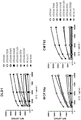
Fig. 6 (a) indicates the binding curves of AT1636 and AT1636 high affinity variants to human CRC cell line DLD1, mammary epithelial cell line MCF10a and mouse CRC cell line CMT93 as detected by flow cytometry (depicting Mean Fluorescence Intensity (MFI) of Alexa647 dye conjugated with goat anti-human secondary antibody (Invitrogen)). The EP700Y and SC10.17 antibodies were not cross-reactive with mouse E-cadherin. (B) The binding ratios of the AT1636 and AT 1636-YN and-IYN variants to the control antibody AT1002 on the skin epithelial cell line a431, lung a546 and mouse CMT93 cell lines as detected by flow cytometry are indicated.
FIG. 7 (A) SPR binding of AT1636, AT 1636-NY and-IYN variants to soluble p 70E-cadherin. The 5.0. mu.g/ml antibody was injected at the point of immobilization with 2.0. mu.g/ml p 70E-cadherin. As a control, EP700Y rabbit anti-human E-cadherin with specificity for the EC5 domain was used. Binding was detected using an IBIS multiplex SPR imaging (B) ELISA assay to determine binding of the AT1636 and AT 1636-IYN variants to recombinant immobilized full-length E-cadherin (left panel), p 70E-cadherin (middle panel) and the E-cadherin D3 domain containing the M470A substitution (preventing mannosylation of this residue) (right panel). The SC10.17 antibody was used as a control antibody that bound to full length (EC1 domain) but not to the p70 and D3 domains. AT1002 is a negative human control antibody against influenza. (C) A broad concentration range of AT1636 and variants thereof were used to bind to recombinant immobilized full-length E-cadherin (left panel), p 70E-cadherin (middle panel) and the E-cadherin D3 domain containing the M470A substitution (preventing mannosylation of this residue) (right panel) in an ELISA assay. AT1002 is a negative human control antibody against influenza.
Fig. 8 (a) western blot showing throughput (FT) after input and AT1636 immunoprecipitation and specific elution of p70 from AT1636 immunoprecipitate using high levels of mannopyranoside. (B) ELISA demonstrated that AT1636 binds to full-length E-cadherin (Sino Biological) from HEK cells (left panel) and AT1636 lacks binding to E-cadherin from E.coli (Lsbio) (right panel). E-cadherin produced by E.coli was recognized by the EP700Y antibody.
FIG. 9 alanine substitutions of the truncated EC3 domain (D3) of E-cadherin p70 reveal several amino acids important for the binding of AT 1636. Binding of AT1636 to the recombinant small-scale D3-FLAG-mouse Fc fusion protein was studied by ELISA after anti-mouse Fc capture. All proteins were expressed in similar amounts in culture supernatants, and D3-wild type binding was set to 1, and all were normalized to anti-FLAG assay.
FIG. 10 depicts the computational analysis of the combined mRNA expression of TMTC3 and E-cadherin in several tumor-specific cell lines; in the middle of the circle is the number of tumor cell lines contained in each tissue type. The light grey color indicates that both E-cadherin and TMTC3 are expressed high (. gtoreq.7-fold) and thus the percentage of tumor cell lines recognized by AT1636 is expected. The cut-off value of 7 was selected based on flow cytometry analysis, which demonstrated that such cell lines could be bound by the AT1636 antibody, see table 3. Tissues that are generally negative for both TMTC3 and E-cadherin are hematopoietic and lymphoid tissues, bone and soft tissue. (data obtained from the Broad Institute, https:// portals. Broadadintitute. org/ccle, J. Barretina, Nature (2012)).
FIG. 11. flow cytometry analysis indicated increased binding of AT1636 to SK-MEL-5 cells transduced with constructs containing full-length E-cadherin. SK-MEL-5 is generally negative for E-cadherin, but does express TMTC 3. AT1636 (solid line) does not bind SK-MEL-5 (left), but binds when E-cadherin is overexpressed (middle). In addition, EP700Y (right) is currently incorporating SK-MEL-5. The light gray curve is the background staining of the isotype control.
Figure 12 shRNA-induced knockdown of TMTC3 resulted in decreased AT1636 binding as determined by flow cytometry. In addition to control scrambled shRNA vectors, TMTC 3-targeted shRNA probes were developed and tested. TMTC3 expression was strongly reduced as determined by qPCR (left). TMTC3 knockdown resulted in > 3-fold reduction in AT1636 binding (right panel, solid line).
Fig. 13 (a) is a graphical representation of monovalent T cell engager (mTCE) structures consisting of AT1636 or AT1636-IYN fused to anti-CD 3 UCHT 1. (B) Compounds were tested in a 2D cell culture model in which luciferase and GFP positive CRC cell lines DLD1, HT29 and HCT116 were cultured O/N at 5000c/w (96w) and then incubated for 2 days with unstimulated total PBMCs as effector cells. Cytotoxicity of cells was established by measuring luciferase expressed over the course of 48 hours. (C) Graphical representation of the structure of the Knob-in-Hole (KiH) bispecific format and CD3 epsilon scFv derived from UCHT1 antibody, monovalent for AT1636, AT1636-IYN, or AT 1002. (D) Compounds were tested in a 2D cell culture model in which luciferase and GFP positive CRC cell lines DLD1 and HT29 and melanoma cell line a375 were cultured O/N AT 5000c/w (96w), then incubated with KiH bispecific monovalent AT1636, AT1636-IYN and AT1002 and UCHT1 scFv CD3 epsilon, and subsequently incubated for 2 days with unstimulated total PBMCs as effector cells. Cytotoxicity of the cells was assessed by measuring luciferase activity at the end of the 48 hour incubation time.
FIG. 14 Stable overexpression of p 70E-cadherin and full-length E-cadherin in cell lines normally expressing E-cadherin (DLD1, HCT116 and HT29) and cells normally negative for E-cadherin (SK-MEL-5). Cells transduced with the empty vector are shown in the left column. After overexpression of p 70E-cadherin, all cells exhibited the de-adhesive morphological phenotype (indicating the EMT phenotype).
Figure 15 flow cytometry analysis of the binding of AT1636 to DLD1 cells cultured in the absence of TGF β for extended periods of time compared to cells cultured in the absence of TGF β.
FIG. 16 cell growth and cell number decrease after addition of TGF β and AT 1636-IYN. (A) A431 cells were cultured in the absence and presence of TGF β and AT1636-IYN variants. The top row shows a431 cells cultured on tissue culture treated plastic for 5 days, and the bottom row shows a431 cells cultured on fibronectin coated plastic, using 10x magnification. The left panel shows cells cultured in medium, the middle panel shows the presence of TGF β, and the right panel shows the presence of TGF β and AT 1636-IYN. In wells cultured in the presence of TGF β and AT1636-IYN, reduced (viable) cells and fewer adherent cells were observed. No difference was observed between cells cultured in plastic or fibronectin coated plates, and no effect was observed for AT1636-wt or AT1002 control antibody (not shown). (B) A detailed representative overview using 20x magnification of a431 cells cultured in fibronectin-coated wells after 7d culture in the presence of TGF β (left panel) and TGF β and AT1636-IYN (right panel). In the right panel, more rounded dying single cells and fewer adherent cells are observed.
FIG. 17 time course analysis of internalization of AT1636 and variants thereof in DLD1 cells detected by fluorescence microscopy (Incucyte) using a pH sensitive Zenon-pHrodo iFL dye. All antibodies except the negative control AT1002 were internalized.
Figure 18 indicates cell surface coverage of CFSE labeled CD103+ T cells incubated on plates binding full length and p 70E-cadherin after incubation with AT1636 and variants thereof and CD103 specific antibody (MCA 708).
Detailed Description
E-cadherin
E-cadherin is encoded in humans by the CDH1 gene, also known as CD 324. The amino acid sequence of the currently known human E-cadherin is depicted in fig. 1A. E-cadherin is a 120-kDa transmembrane glycoprotein located at the adhesive junctions of epithelial cells. E-cadherins are members of the large family of cadherins and can be divided into several subtypes: type I classical cadherins such as E-cadherin (CDH1), N-cadherin (CDH2), and P-cadherin (CDH 3); classical cadherins of type II, such as VE-cadherin (CDH5) and OB-cadherin (CDH 11); desmosomal cadherins; seven transmembrane cadherins; FAT and Dachsous (DCHS) group cadherins; and Procalcitonin (PCDH). E-cadherin is a transmembrane protein with three components: (1) an extracellular cadherin domain (EC) responsible for homotypic cadherin-cadherin interactions, (2) a single transmembrane domain or seven transmembrane domain, and (3) a cytoplasmic domain that serves as a connector between the cell surface, associated cytoplasmic catenin, and the cytoskeleton. Cadherins are involved in the growth of organisms (embryogenesis), wound healing, and tumor invasion and metastasis.
In addition to being a calcium-dependent adhesion molecule, E-cadherin is also a key regulator of epithelial junction formation. Its association with catenin is necessary for cell-cell adhesion and polarization of epithelial cells/epithelial sheets between lateral and apical membranes. Tyrosine phosphorylation can disrupt these complexes, resulting in changes in cell adhesion properties. E-cadherin expression is often down-regulated in highly aggressive, poorly differentiated cancers. Increased E-cadherin expression in these cells reduces invasiveness. Thus, loss of E-cadherin expression or function appears to be an important step in tumorigenic progression. In addition, cadherin plays an important role in the invasion and migration of cells through epithelial-mesenchymal transition (EMT, a reversible process). EMT is a very diverse process that can be coordinated by many external signals (inflammation, stress, hypoxia, immune response, etc.). It is widely believed that, in particular, strong regulation of EMT-inducing transcription factors (Snail, E47, Twist and Zeb families) is the basis for EMT, which, for example, through the binding of TGF β, Wnt, integrins and growth factors to cells, will lead to down-regulation of E-cadherin, ZO-1, desmoplakin and up-regulation of vimentin, fibronectin, N-cadherin and the like. The cells undergo a process in which they resemble a more "dry" phenotype. Recently, this model has been changing because it was observed that cells can also "show" EMT without the need to down-or up-regulate known EMT markers. This is termed partial EMT, mixed EMT/MET and quasi-EMT, and most new models propose a system where cells can modulate protein expression (e.g., protein internalization, high/low protein turnover) or cells together (cluster) have different activity/invasion patterns. E-cadherin plays a dominant role in these processes, and in this regard, E-cadherin O-mannosylation is considered an additional tool for tumor cells to regulate adhesion and morphological changes upon interaction with the surrounding stroma.
anti-E-cadherin antibodies and antigen-binding fragments thereof
The present invention provides antibodies and antigen-binding fragments thereof that are capable of specifically binding to O-mannosylated E-cadherin. In some embodiments, the antibody is isolated. In other embodiments, the antibody is synthetic or recombinant. Interestingly, the present invention provides antibodies and antigen binding fragments thereof comprising VH and VL sequences based on human antibody VH and VL sequences from human individuals with stage IV colon cancer that has metastasized but has been in complete remission for many years following chemotherapy. In contrast, many currently known therapeutic antibodies are typically obtained by immunizing a non-human animal such as a mouse, rat, camel, rabbit, or goat, optionally followed by a humanization process. Such humanized antibodies still involve the risk of adverse side effects due to the recipient's immune response to the non-human sequence. In addition, many prior art therapeutic antibodies or fragments thereof are derived from artificial phage display libraries in which immunoglobulin heavy and light chains are randomly paired. In contrast, the present invention provides antibodies and antigen-binding fragments with naturally paired heavy and light chains, which are based on antibody sequences that have evolved in fully remitted human patients.
Because E-cadherin is widely expressed in many epithelial tissues, it was not considered in the art as an antigen of choice for therapeutic applications prior to the present invention, particularly in view of the fact that E-cadherin is often down-regulated in tumor cells to promote EMT. However, the present invention provides antibodies and antigen-binding fragments thereof that are capable of specifically binding truncated forms of E-cadherin, which has a molecular weight of about 70kDa and is often upregulated in tumor cells.
As used herein, the term "antibody" encompasses protein molecules as well as any antigen-binding fragments thereof. The protein molecules are preferably immunoglobulins, which means that they belong to the class of immunoglobulin proteins. In some embodiments, the antibody or antigen binding fragment thereof comprises one or more domains that bind to an epitope on an antigen, wherein such domains are preferably derived from or share sequence homology with the variable domains of the antibody.
Complementarity Determining Regions (CDRs) are hypervariable regions present in the heavy and light chain variable domains. In the case of a full-length antibody, CDRs 1-3 of the heavy chain and CDRs 1-3 of the linked light chain together form the antigen binding site.
The fragment crystallizable (Fc) region of a natural antibody consists of the CH2 and CH3 domains of two heavy chains.
Typically, an antigen-binding fragment of an antibody is capable of binding to the same antigen as the antibody, although not necessarily to the same extent. In some embodiments, the antigen-binding fragment comprises at least the heavy chain CDR3 region of the antibody. In some embodiments, the antigen-binding fragment comprises at least the heavy chain CDR3 region and the light chain CDR3 region of an antibody. In some embodiments, the heavy and light chain CDR3 regions are paired with each other.
In various embodiments, an antigen-binding fragment of an antibody comprises at least the heavy chain CDR1, CDR2, and CDR3 regions of the antibody. In various embodiments, an antigen-binding fragment of an antibody comprises at least a VH domain. In various embodiments, an antigen-binding fragment of an antibody comprises at least the heavy chain CDR1, CDR2, and CDR3 regions and the light chain CDR1, CDR2, and CDR3 regions of the antibody. In various embodiments, an antigen-binding fragment of an antibody comprises at least a VL domain. In various embodiments, an antigen-binding fragment of an antibody comprises at least VH and VL domains.
Non-limiting examples of antibodies or antigen-binding fragments according to the invention are full-length antibodies,  (bispecific antibodies containing two different IgG 1), single domain antibodies or nanobodies (containing a single VH or VL domain), single chain variable fragments (scFv; containing a VH domain and a VL domain, typically linked by a short linker peptide), Fv fragments (containing a VH domain and a VL domain, typically without a linker), unibody TM Fd fragment (containing a VH domain and a CH1 domain), diabody (containing two VH domains and two VL domains, wherein VH is connected to VL by a short linker such that they cannot pair with each other, but with VL and VH of the other chain, thereby creating two antigen binding sites), Fab-fragment (comprising the heavy chain constant domain CH1, the light chain constant domain CL and the heavy and light chain variable domains VH and VL) and F (ab')2 fragment (comprising two Fab fragments linked by a disulfide bond).
In various embodiments, the antibody or antigen-binding fragment of the invention comprises a heavy chain variable region (VH) and a light chain variable region (VL). In some embodiments, the VH is paired with the VL.
The antibody for therapeutic use is preferably as close as possible to the native antibody of the subject to be treated (e.g., a human antibody for a human subject). In various embodiments, the antibody of the invention is a full-length antibody, preferably an IgG or IgM or IgA full-length antibody. As used herein, an IgG full length antibody is a bivalent molecule comprising two gamma-like heavy chains and two light chains. As is well known to the skilled person, the heavy chain of an antibody is the larger of the two types of chains that make up an immunoglobulin molecule. Natural heavy chains typically comprise a constant domain CH, which comprises constant regions CH1, CH2, and CH 3; and a variable domain (VH) involved in antigen binding. The light chain of an antibody is the smaller of the two types of chains that make up an immunoglobulin molecule. Native light chains typically comprise a constant domain (CL) and a variable domain (VL). Light chain variable domains are often, but not always, associated with the variable domains of heavy chains involved in antigen binding.
Full-length IgD antibodies are bivalent molecules comprising two delta-like heavy chains and two light chains.
In the case of IgM, a full-length antibody is a ten-or twelve-valent molecule comprising 5 or 6 linked immunoglobulins in which each monomer has two antigen binding sites formed by a heavy chain and a light chain.
In the case of IgA, the full-length antibody may be monomeric or dimeric.
In some embodiments, the antibody or antigen-binding fragment according to the invention is a human antibody or antigen-binding fragment thereof. In contrast to murine or humanized antibodies, the presence of human amino acid sequences, which are involved in the risk of developing an anti-murine immune response in the human recipient, reduces the chance of adverse side effects during therapeutic use in human patients.
In various embodiments, the antibody or antigen-binding fragment according to the invention is an IgG isotype, preferably IgG 1. This is beneficial for medical applications in humans, for example, because IgG1 antibodies typically have a good half-life after in vivo administration to a human subject. Furthermore, the Fc tail of IgG1 allows effector functions like antibody-dependent cell-mediated cytotoxicity (ADCC), complement-dependent cytotoxicity (CDC) and antibody-dependent phagocytosis (ADCP) to be achieved. In some embodiments, the antibody or antigen binding fragment according to the invention is a human IgG, preferably human IgG 1.
In some embodiments, the antibody or antigen binding fragment according to the invention is an IgG2 isotype. In some embodiments, the antibody or antigen binding fragment according to the invention is an IgG3 isotype. In some embodiments, the antibody or antigen binding fragment according to the invention is an IgG4 isotype.
In some embodiments, the antibody or antigen-binding fragment according to the invention is of the IgM isotype. In some embodiments, the antibody or antigen-binding fragment according to the invention is of IgA isotype. In some embodiments, the antibody or antigen-binding fragment according to the invention is an IgD isotype.
In various embodiments, the antibody or antigen-binding fragment thereof comprises one or more mutations. Such mutations include, for example, amino acid substitutions, insertions or deletions. As used herein, a full-length antibody in which one or several, preferably up to 20, amino acid residues are deleted without substantially altering the binding characteristics of the resulting antibody is still considered a full-length antibody.
In some embodiments, an antibody or antigen binding fragment according to the invention has a modified Fc tail. In some embodiments, the Fc tail has been modified by one or more amino acid substitutions and/or by glycosylation changes. In some embodiments, the Fc tail has been modified to reduce ADCC activity. In some embodiments, the Fc tail has been modified to enhance ADCC activity. In some embodiments, the antibody or antigen binding fragment according to the invention is nonfucosylated to enhance ADCC activity.
The terms "capable of binding," "specific for …," "capable of specific binding," and "binding" are used interchangeably herein and refer to the interaction between an antibody or antigen-binding fragment thereof and its target (also referred to as its antigen). This means that the antibody or antigen-binding fragment preferentially binds to the antigen relative to other antigens or amino acid sequences. Thus, while an antibody or antigen-binding fragment may bind non-specifically to other antigens or amino acid sequences, the binding affinity of the antibody or antigen-binding fragment to its antigen is significantly higher than the non-specific binding affinity of the antibody or antigen-binding fragment to other antigens or amino acid sequences.
Typically, an antibody or antigen-binding fragment of the invention modified in some way retains at least 50% of its binding activity (when compared to the parent antibody). Preferably, the antibody or antigen binding fragment of the invention retains at least 60%, 70%, 80%, 90%, 95% or 100% of its binding activity as compared to the parent antibody.
In some embodiments, an antibody or antigen-binding fragment of the invention comprises conservative or non-conservative amino acid substitutions that do not significantly alter its biological activity (the resulting variants are referred to herein as "conservative variants" or "functionally conservative variants," respectively). In some embodiments, such a conservative variant or a functionally conservative variant retains at least 80%, 90%, 95%, or 100% of its binding activity as compared to the parent antibody.
As used herein, a conservative substitution is a substitution in which an amino acid residue is substituted with another residue having generally similar properties (size, hydrophobicity, etc.) such that the overall function of the antibody is not substantially affected. In general, substitutions of amino acid residues within the same class depicted in table 2 are considered conservative amino acid substitutions.
An antibody or antigen-binding fragment capable of binding O-mannosylated E-cadherin may also be specific for another compound if the O-mannosylated E-cadherin epitope bound by the antibody or antigen-binding fragment according to the invention is also present in said other compound. In this case, the antibody or antigen-binding fragment referred to herein as having specificity for O-mannosylated E-cadherin is also specific for this other compound that contains an O-mannosylated epitope of the same species. This other O-mannosylated epitope may be produced in vivo by another O-mannosyltransferase, rather than by an O-mannosyltransferase which produces O-mannosylated E-cadherin in vivo. Thus, the term "binds" or "specific for …" does not exclude the binding of an antibody or antigen-binding fragment of the invention to another protein or proteins containing the same class of O-mannosylated epitopes.
"binding affinity" refers to the strength of the sum of non-covalent interactions between a single binding site of an antibody or antigen-binding fragment and its binding partner (e.g., antigen). As used herein, unless otherwise indicated, "binding affinity" refers to an intrinsic binding affinity that reflects a 1:1 interaction between members of a binding pair (e.g., antibody and antigen). Affinity can generally be determined by the equilibrium dissociation constant (K) D ) Is expressed by k a And k is d See, e.g., Chen, Y, et al, (1999) J.Mol Biol 293: 865-881. Affinity can be measured by common methods known in the art, such as, for example, Surface Plasmon Resonance (SPR) assays, IBIS-iSPR instruments such as biacore (ge healthcare), octet (fortebio), or IBIS Technologies BV (Hengelo, the Netherlands) or solution phase assays, such as Kinexa.
As used herein, the terms "nucleic acid" and "nucleic acid molecule" are used interchangeably. In some embodiments, a nucleic acid or nucleic acid molecule according to the invention comprises a chain of nucleotides, more preferably DNA, cDNA or RNA. In some embodiments, the nucleic acid or nucleic acid molecule according to the invention comprises non-natural nucleotides, modified nucleotides and/or non-nucleotide building blocks exhibiting the same function as natural nucleotides, such as for example DNA/RNA helices, Peptide Nucleic Acids (PNAs) and/or Locked Nucleic Acids (LNAs).
Percent identity of amino acid or nucleic acid sequences or the term "% sequence identity" is defined herein as the percentage of residues in a candidate amino acid or nucleic acid sequence that are identical to residues in a reference sequence after aligning the two sequences and introducing gaps, if necessary, to achieve the maximum percent identity. Methods and computer programs for alignment are well known in the art, e.g., "Align 2".
As used herein, the singular term "a" or "an" encompasses the term "one or more.
Exemplary E-cadherin-specific antibodies
The present invention provides antibodies and antigen-binding fragments thereof that are specific for O-mannosylated E-cadherin and have specified structural and functional characteristics, and their therapeutic use for treating or preventing disease. A non-limiting example of such a disease is a cancer with O-mannosylated E-cadherin.
As used herein, the term "O-mannosylated E-cadherin" refers to an E-cadherin protein comprising at least one threonine or serine residue with an O-linked mannose, which means that the mannose is attached to the oxygen atom of said threonine or serine. In some embodiments, the E-cadherin protein comprises at least one single O-mannosylated threonine or serine residue. The term "single O-mannosylated threonine residue" refers to a threonine residue which contains an O-linked mannose without another sugar moiety attached to the O-linked mannose. The term "single O-mannosylated serine residue" refers to a serine residue which contains an O-linked mannose without another sugar moiety attached to said O-linked mannose.
Various embodiments provide an antibody or antigen-binding fragment specific for O-mannosylated E-cadherin according to the invention, wherein binding of the antibody or antigen-binding fragment to the E-cadherin is dependent on the presence of an O-mannosylated threonine residue at position 467, an O-mannosylated threonine residue at position 468, an O-mannosylated threonine residue at position 470, an O-mannosylated threonine residue at position 472, a glutamic acid residue at position 463, a serine residue at position 465, a serine residue at position 469 and/or a valine residue at position 477 of the E-cadherin sequence as depicted in fig. 1A.
Some embodiments provide antibodies and antigen-binding fragments thereof that are specific for O-mannosylated E-cadherin, and wherein binding of the antibody or antigen-binding fragment to the E-cadherin is dependent on the presence of one or more O-mannosylated threonine residues within E-cadherin amino acid region 467-472 as depicted in FIG. 1A. In some embodiments, the antibody or antigen-binding fragment is dependent on the presence of an O-mannosylated threonine residue at position 467, and/or an O-mannosylated threonine residue at position 468, and/or an O-mannosylated threonine residue at position 470, and/or an O-mannosylated threonine residue at position 472 of the E-cadherin sequence as depicted in fig. 1A. In some embodiments, the antibody or antigen-binding fragment is dependent on the presence of an O-mannosylated threonine residue at position 468 and an O-mannosylated threonine residue at position 470 of the E-cadherin sequence as depicted in FIG. 1A. In some embodiments, the antibody or antigen-binding fragment is dependent on the presence of an O-mannosylated threonine residue at position 467, an O-mannosylated threonine residue at position 468, and an O-mannosylated threonine residue at position 470 of the E-cadherin sequence as depicted in FIG. 1A. In some embodiments, the antibody or antigen-binding fragment is dependent on the presence of an O-mannosylated threonine residue at position 467, and an O-mannosylated threonine residue at position 468, and an O-mannosylated threonine residue at position 470, and an O-mannosylated threonine residue at position 472 of the E-cadherin sequence as depicted in fig. 1A. In some embodiments, binding of the antibody or antigen-binding fragment to the E-cadherin is dependent on the presence of a glutamic acid residue at position 463, and/or a serine residue at position 465, and/or a serine residue at position 469, and/or a valine residue at position 477 of the E-cadherin sequence as depicted in figure 1A. In some embodiments, the serine residue at position 465 and/or position 469 is O-mannosylated.
As used herein, the binding of an antibody or antigen-binding fragment is "dependent" on an amino acid residue if the substitution of that amino acid residue with alanine reduces the binding of the antibody or antigen-binding fragment to its antigen by at least 40%, preferably by at least 50%, preferably by at least 60%, preferably by at least 70%, preferably by at least 80%, preferably by at least 85%, more preferably by at least 90%, more preferably by at least 95%.
Some embodiments provide an antibody or antigen-binding fragment according to the invention that is specific for O-mannosylated E-cadherin and that specifically binds to an O-mannosylated threonine residue at position 467, and/or an O-mannosylated threonine residue at position 468, and/or an O-mannosylated threonine residue at position 470, and/or an O-mannosylated threonine residue at position 472, and/or a glutamic acid residue at position 463, and/or a serine residue at position 465, and/or a serine residue at position 469, and/or a valine residue at position 477 of an E-cadherin sequence as depicted in fig. 1A. In some embodiments, the serine residue at position 465 and/or position 469 is O-mannosylated.
Some embodiments of the invention provide an antibody or antigen-binding fragment thereof that is specific for O-mannosylated E-cadherin and specifically binds to one or more O-mannosylated threonine residues of E-cadherin, wherein the one or more O-mannosylated threonine residues are present within amino acid position 467-472 of the E-cadherin sequence as depicted in FIG. 1A.
Some embodiments provide antibodies and antigen binding fragments thereof that specifically bind to one or more O-mannosylated threonine residues selected from the group consisting of: an O-mannosylated threonine residue at position 467, an O-mannosylated threonine residue at position 468, an O-mannosylated threonine residue at position 470, and an O-mannosylated threonine residue at position 472 of the E-cadherin sequence as depicted in FIG. 1A. Some embodiments provide antibodies and antigen-binding fragments thereof that specifically bind to an O-mannosylated threonine residue at position 467 of the E-cadherin sequence as depicted in fig. 1A. Some embodiments provide antibodies and antigen-binding fragments thereof that specifically bind to an O-mannosylated threonine residue at position 468 of the E-cadherin sequence as depicted in fig. 1A. Some embodiments provide antibodies and antigen-binding fragments thereof that specifically bind to an O-mannosylated threonine residue at position 470 of an E-cadherin sequence as depicted in fig. 1A. Some embodiments provide antibodies and antigen binding fragments thereof that specifically bind to an O-mannosylated threonine residue at position 472 of the E-cadherin sequence as depicted in figure 1A.
Some embodiments provide antibodies and antigen-binding fragments thereof that specifically bind to an O-mannosylated threonine residue at position 468 and an O-mannosylated threonine residue at position 470 of an E-cadherin sequence as depicted in fig. 1A.
Some embodiments provide antibodies and antigen-binding fragments thereof that specifically bind to an O-mannosylated threonine residue at position 467, and an O-mannosylated threonine residue at position 468, and an O-mannosylated threonine residue at position 470 of an E-cadherin sequence as depicted in fig. 1A.
Some embodiments provide antibodies and antigen-binding fragments thereof that specifically bind to an O-mannosylated threonine residue at position 467, and an O-mannosylated threonine residue at position 468, and an O-mannosylated threonine residue at position 470, and an O-mannosylated threonine residue at position 472 of an E-cadherin sequence as depicted in fig. 1A.
Some embodiments provide antibodies and antigen-binding fragments thereof that specifically bind to an E-cadherin region comprising T468 and T470 of an E-cadherin sequence as depicted in figure 1A, wherein at least one of the threonine residues is O-mannosylated. In some embodiments, both threonine residues are O-mannosylated.
Some embodiments provide antibodies and antigen-binding fragments thereof that specifically bind to an E-cadherin region comprising T467, T468, and T470 of an E-cadherin sequence as depicted in figure 1A, wherein at least one of the threonine residues is O-mannosylated. In some embodiments, two of the threonine residues are O-mannosylated. In some embodiments, all three threonine residues are O-mannosylated.
Some embodiments provide antibodies and antigen-binding fragments thereof that specifically bind to an E-cadherin region comprising T467, T468, T470, and T472 of an E-cadherin sequence as depicted in figure 1A, wherein the threonine residue is O-mannosylated. In some embodiments, two of the threonine residues are O-mannosylated. In some embodiments, three of the threonine residues are O-mannosylated. In some embodiments, all four threonine residues are O-mannosylated.
Some embodiments provide antibodies and antigen-binding fragments thereof that specifically bind to an E-cadherin region comprising T468, S469, and T470 of an E-cadherin sequence as depicted in figure 1A, wherein at least one of the threonine residues is O-mannosylated. In some embodiments, both threonine residues are O-mannosylated. In some embodiments, the serine residue is O-mannosylated.
Some embodiments provide antibodies and antigen-binding fragments thereof that specifically bind to an E-cadherin region comprising T467, T468, S469, and T470 of an E-cadherin sequence as depicted in figure 1A, wherein at least one of the threonine residues is O-mannosylated. In some embodiments, at least two of the threonine residues are O-mannosylated. In some embodiments, all of the threonine residues are O-mannosylated. In some embodiments, the serine residue is O-mannosylated.
Some embodiments provide antibodies and antigen-binding fragments thereof that specifically bind to an E-cadherin region comprising S465, T467, T468, S469, and T470 of an E-cadherin sequence as depicted in figure 1A, wherein at least one of the threonine residues is O-mannosylated. In some embodiments, at least two of the threonine residues are O-mannosylated. In some embodiments, all of the threonine residues are O-mannosylated. In some embodiments, at least one serine residue is O-mannosylated. In some embodiments, both serine residues are O-mannosylated.
Some embodiments provide antibodies and antigen-binding fragments thereof that specifically bind to an E-cadherin region comprising S465, T467, T468, S469, T470, and T472 of an E-cadherin sequence as depicted in figure 1A, wherein at least one of the threonine residues is O-mannosylated. In some embodiments, at least two of the threonine residues are O-mannosylated. In some embodiments, at least three of the threonine residues are O-mannosylated. In some embodiments, all of the threonine residues are O-mannosylated. In some embodiments, at least one serine residue is O-mannosylated. In some embodiments, both serine residues are O-mannosylated.
Some embodiments provide antibodies and antigen binding fragments thereof that specifically bind to the E-cadherin region comprising E463, S465, T467, T468, S469, T470 and T472 of the E-cadherin sequence as depicted in figure 1A, wherein at least one of the threonine residues is O-mannosylated. In some embodiments, at least two of the threonine residues are O-mannosylated. In some embodiments, at least three of the threonine residues are O-mannosylated. In some embodiments, all of the threonine residues are O-mannosylated. In some embodiments, at least one serine residue is O-mannosylated. In some embodiments, both serine residues are O-mannosylated.
Some embodiments provide antibodies and antigen-binding fragments thereof that specifically bind to an E-cadherin region comprising E463, S465, T467, T468, S469, T470, T472, and V477 of an E-cadherin sequence as depicted in fig. 1A, wherein at least one of the threonine residues is O-mannosylated. In some embodiments, at least two of the threonine residues are O-mannosylated. In some embodiments, at least three of the threonine residues are O-mannosylated. In some embodiments, all of the threonine residues are O-mannosylated. In some embodiments, at least one serine residue is O-mannosylated. In some embodiments, both serine residues are O-mannosylated.
Some embodiments provide antibodies and antigen-binding fragments thereof that specifically bind to an epitope of E-cadherin comprising the sequence TST, wherein at least one of the threonine residues is O-mannosylated. In some embodiments, both threonine residues are O-mannosylated. In some embodiments, the serine residue is O-mannosylated.
Some embodiments provide antibodies and antigen-binding fragments thereof that specifically bind to an epitope of E-cadherin comprising the sequence TTST, wherein at least one of the threonine residues is O-mannosylated. In some embodiments, at least two of the threonine residues are O-mannosylated. In some embodiments, all of the threonine residues are O-mannosylated. In some embodiments, the serine residue is O-mannosylated.
Some embodiments provide antibodies and antigen-binding fragments thereof that specifically bind to an epitope of E-cadherin comprising the sequence STTST, wherein at least one of the threonine residues is O-mannosylated. In some embodiments, at least two of the threonine residues are O-mannosylated. In some embodiments, all of the threonine residues are O-mannosylated. In some embodiments, at least one serine residue is O-mannosylated. In some embodiments, both serine residues are O-mannosylated.
Some embodiments provide antibodies and antigen-binding fragments thereof that specifically bind to an epitope of E-cadherin comprising the sequence stttstt, wherein at least one of the threonine residues is O-mannosylated. In some embodiments, at least two of the threonine residues are O-mannosylated. In some embodiments, at least three of the threonine residues are O-mannosylated. In some embodiments, all of the threonine residues are O-mannosylated. In some embodiments, at least one serine residue is O-mannosylated. In some embodiments, both serine residues are O-mannosylated.
Some embodiments provide antibodies and antigen-binding fragments thereof that specifically bind to an epitope of E-cadherin comprising the sequence eststt, wherein at least one of the threonine residues is O-mannosylated. In some embodiments, at least two of the threonine residues are O-mannosylated. In some embodiments, at least three of the threonine residues are O-mannosylated. In some embodiments, all of the threonine residues are O-mannosylated. In some embodiments, at least one serine residue is O-mannosylated. In some embodiments, both serine residues are O-mannosylated.
Some embodiments provide antibodies and antigen-binding fragments thereof that specifically bind to an epitope of E-cadherin comprising the sequence ESTTSTTTV, wherein at least one of the threonine residues is O-mannosylated. In some embodiments, at least two of the threonine residues are O-mannosylated. In some embodiments, at least three of the threonine residues are O-mannosylated. In some embodiments, all of the threonine residues are O-mannosylated. In some embodiments, at least one serine residue is O-mannosylated. In some embodiments, both serine residues are O-mannosylated.
E-cadherin is known in the art as the product of the CDH1 gene, which has a molecular weight of about 120kDa in humans. However, the present invention provides the surprising insight that O-mannosylated truncated forms of E cadherin are also found in nature. This truncated form, with a molecular weight of about 70kDa, lacks the extracellular domains EC1 and EC2 of full-length E-cadherin. Extracellular domains 5, 4 and a portion of the extracellular domain EC3 are still present in the truncated 70kDa form. The present inventors provide insight that this truncated 70kDa form of E-cadherin is present on the surface of a variety of epithelial cells and is often upregulated on tumor cells. Without being bound by any theory, it is believed that upregulation of the 70kDa form of E-cadherin promotes tumor growth, particularly because the truncated 70kDa form stimulates epithelial-mesenchymal transition (EMT), as shown in the examples, thereby increasing migration and metastasis of tumor cells. Furthermore, it is believed that upregulation of the 70kDa form of E-cadherin promotes the immune escape mechanism of tumors, as the truncated 70kDa form of E-cadherin has a lower ability to bind immune cells compared to the 120kDa full-length form of E-cadherin. According to the invention, overexpression of the O-mannosylated 70kDa E-cadherin form can promote escape from immune cell recognition by CD3 or KLRG1 or CD103 and promote EMT without completely down-regulating E-cadherin. Thus, upregulation of the 70kDa truncated form on tumor cells can attenuate the interaction between these tumor cells and immune cells, thereby helping the tumor evade the immune response.
In some embodiments, the invention provides antibodies and antigen binding portions thereof that bind to the O-mannosylated truncated 70kDa E-cadherin described above better than the well-known O-mannosylated full-length E-cadherin of about 120 kDa. Preferred embodiments provide antibodies and antigen-binding portions thereof that bind O-mannosylated truncated 70kDa E-cadherin at least 2-fold better, more preferably at least 3-fold better, more preferably at least 4-fold better, more preferably at least 5-fold better than O-mannosylated full-length E-cadherin. This feature allows to increase tumor specificity and to reduce adverse side effects caused by healthy tissue binding in case the truncated 70kDa E-cadherin form is significantly upregulated on tumor cells. As used herein, the term "full-length E-cadherin" refers to the known CDH1 gene product, which has a molecular weight of about 120kDa in humans, as depicted, for example, in fig. 1A. The term "truncated 70kDa E-cadherin" or "70 kDa E-cadherin form" refers to the smaller E-cadherin form, which has a molecular weight between 60 and 80kDa, usually about 70kDa, and is also found on the surface of epithelial cells in nature. As shown in fig. 1C, this naturally occurring truncated form of E-cadherin, with a molecular weight between 60 and 80kDa, lacks the extracellular domains EC1 and EC2 of full-length E-cadherin. Extracellular domains 5, 4 and a portion of extracellular domain EC3 were still present in this truncated 70kDa form. The term "O-mannosylated truncated 70kDa E-cadherin" refers to the above mentioned truncated 70kDa E-cadherin protein comprising at least one O-mannosylated threonine or serine residue, preferably at least one O-mannosylated threonine residue at position 467, and/or position 468, and/or position 470 as depicted in fig. 1A.
In certain embodiments, an anti-E-cadherin antibody or antigen-binding fragment of the invention comprises:
comprising the amino acid sequence TPGVGX 1 NX 2 PYYFDR heavy chain variable region CDR3, wherein X 1 Is A or T, and wherein X 2 Is D or N; and
-light chain variable region CDR3 comprising amino acid sequence QQYSNTPQT.
Certain embodiments provide an antibody or antigen-binding fragment thereof capable of binding O-mannosylated E-cadherin, wherein the antibody or antigen-binding fragment comprises one or more and optionally each of:
a. comprising the amino acid sequence GFX 1 FSX 2 CDR1 of the heavy chain variable region of AW, wherein X 1 Is T or I, and wherein X 2 Is N or Y;
b. comprising the amino acid sequence IKSIDG X 1 T X 2 The heavy chain variable region CDR2 of (1), wherein X 1 Is G or E, and wherein X 2 Is T or I;
c. comprising the amino acid sequence TPGVGX 1 NX 2 The heavy chain variable region CDR3 of PYYFDR, wherein X 1 Is A or T, and wherein X 2 Is D or N;
d. a light chain variable region CDR1 comprising amino acid sequence QSVLCRSNNKNC;
e. comprising the amino acid sequence WAX 1 The light chain variable region CDR2 of (1), wherein X 1 Is S or C;
f. a light chain variable region CDR3 comprising amino acid sequence QQYSNTPQT;
or a light chain variable region CDR3 comprising an amino acid sequence that differs from the QQYSNTPQT sequence by 1, 2, or 3 conservative substitutions.
Optionally, conservative amino acid substitutions are applied to at least one of the above-mentioned CDR sequences. In some embodiments, the conservative substitution comprises substituting one or more amino acid residues of an amino acid class as depicted in table 2 with another amino acid residue of the same amino acid class. Non-limiting examples of conservative amino acid substitutions include the substitution of one hydrophobic residue, such as isoleucine, valine, leucine or methionine, for another, and the substitution of one polar residue for another, such as the substitution of arginine for lysine, glutamic for aspartic acids, or glutamine for asparagine. Preferably, the advantageous E-cadherin binding characteristics of the parent antibody are maintained or even improved after the amino acid substitutions are conserved. Preferably, the CDR sequences of such variants differ from the parent sequence by no more than three, preferably no more than two, preferably no more than one amino acid.
Accordingly, some embodiments provide an anti-E-cadherin antibody or antigen-binding fragment of the invention comprising:
comprising the amino acid sequence TPGVGX 1 NX 2 The heavy chain variable region CDR3 of PYYFDR, wherein X 1 Is A or T, and wherein X 2 Is D or N; or comprises a difference from the TPGVGX by 1, 2 or 3 conservative substitutions 1 NX 2 A heavy chain variable region CDR3 of the amino acid sequence of the PYYFDR sequence; and
-a light chain variable region CDR3 comprising amino acid sequence QQYSNTPQT, or a light chain variable region CDR3 comprising an amino acid sequence that differs from the QQYSNTPQT sequence by 1, 2 or 3 conservative substitutions.
Also provided is an antibody or antigen-binding fragment thereof, which is capable of binding O-mannosylated E-cadherin, wherein the antibody or antigen-binding fragment comprises one or more and optionally each of:
a. comprising the amino acid sequence GFX 1 FSX 2 Heavy chain variable region CDR1 of AW, wherein X 1 Is T or I, and wherein X 2 Is N or Y;
or comprises a difference from the GFX by 1, 2 or 3 conservative substitutions 1 FSX 2 A heavy chain variable region CDR1 of the amino acid sequence of the AW sequence;
b. comprising the amino acid sequence IKSIDGX 1 TX 2 The heavy chain variable region CDR2 of (1), wherein X 1 Is G or E, and wherein X 2 Is T or I;
or comprises a conservative substitution of 1, 2 or 3 to said IKKSIDDGX 1 TX 2 A heavy chain variable region CDR2 of the amino acid sequence of sequence;
c. comprising the amino acid sequence TPGVGX 1 NX 2 The heavy chain variable region CDR3 of PYYFDR, wherein X 1 Is A or T, and wherein X 2 Is D or N;
or comprises a difference from the TPGVGX by 1, 2 or 3 conservative substitutions 1 NX 2 A heavy chain variable region CDR3 of the amino acid sequence of the PYYFDR sequence;
d. a light chain variable region CDR1 comprising amino acid sequence QSVLCRSNNKNC;
or a light chain variable region CDR1 comprising an amino acid sequence that differs from the QSVLCRSNNKNC sequence by 1, 2, or 3 conservative substitutions;
e. comprising the amino acid sequence WAX 1 The light chain variable region CDR2 of (1), wherein X 1 Is S or C;
or comprises a conservative substitution of 1, 2 or 3 different from the WAX 1 Light chain variable region CDR2 of the amino acid sequence of seq id no;
f. a light chain variable region CDR3 comprising amino acid sequence QQYSNTPQT;
or a light chain variable region CDR3 comprising an amino acid sequence that differs from the QQYSNTPQT sequence by 1, 2, or 3 conservative substitutions.
Table 1 provides the sequences of preferred antibodies according to the invention. These preferred antibodies are referred to herein as antibodies AT1636, E-C06, D-H04,D-A02, D-E09, E-A04, E-B09, C-A05, C-A03, C-B02, C-D04-A, C-D04-B, F-C08, D-G03, D-F10, C-E08, D-B06, D-G05, D-H08, C-H01, D-C12, D-C11, E-C10, AT1636-I, AT1636-Y, AT1636-E, AT1636-N, AT1636-YN, AT1636-IYN and AT 1636-IYEN. These antibodies bind to O-mannosylated E-cadherin, particularly the newly found truncated form of E-cadherin of about 70kDa, as described above. The heavy and light chain CDR sequences of these preferred antibodies are according to CDR sequence GFX 1 FSX 2 AW、IKSKIDGX 1 TX 2 、TPGVGX 1 NX 2 PYYFDR、QSVLCRSNNKNC、WAX 1 And QQYSNTPQT, as described above in a) to f).
As used herein, the terms "AT 1636", "E-C06", "D-H04", "D-A02", "D-E09", "E-A04", "E-B09", "C-A05", "C-A03", "C-B02", "C-D04-A", "C-D04-B", "F-C08", "D-G03", "D-F10", "C-E08", "D-B06", "D-G05", "D-H08", "C-H01", "D-C12", "D-C11", "E-C10", "AT 1636-I", "AT 1636-Y", "AT 1636-E", "AT 6-N", "AT 6-163YN", "AT 1636-YN", "AT 1636-IYN" and "AT 1636-IYEN" encompass heavy and light chain 1-3 regions having AT least these antibodies as depicted in Table 1, and the CDRs 1, Preferably all antibodies and antigen-binding fragments of the heavy and light chain variable regions.
Based on the antibodies depicted in table 1, it is possible to generate antibodies, or antigen-binding fragments thereof, that bind O-mannosylated E-cadherin and comprise at least one CDR sequence of the antibodies depicted in table 1. Accordingly, an antibody or antigen binding fragment thereof is provided comprising at least one CDR sequence of an antibody as depicted in table 1. The CDR sequence is preferably the CDR3 sequence of an antibody as depicted in table 1. In some embodiments, an antibody or antigen-binding fragment is provided comprising a heavy chain CDR3 sequence and a light chain CDR3 sequence of an antibody as depicted in table 1. Accordingly, some embodiments provide an antibody or antigen-binding fragment thereof comprising the heavy and light chain CDR3 sequences of an antibody selected from the group consisting of: AT1636, E-C06, D-H04, D-A02, D-E09, E-A04, E-B09, C-A05, C-A03, C-B02, C-D04-A, C-D04-B, F-C08, D-G03, D-F10, C-E08, D-B06, D-G05, D-H08, C-H01, D-C12, D-C11, E-C10, AT1636-I, AT1636-Y, AT1636-E, AT1636-N, AT1636-YN, AT1636-IYN and AT 1636-IYEN.
Some embodiments provide an antibody or antigen-binding fragment thereof that binds O-mannosylated E-cadherin and comprises one or more heavy chain CDR 1-3 sequences of the antibody as depicted in table 1.
In some embodiments, an antibody or antigen-binding fragment is provided comprising the heavy chain CDR1, CDR2, and CDR3 sequences of the same antibody indicated in table 1. Thus, according to this embodiment, the heavy chain CDR 09, CDR 09 and CDR 09 sequences of antibodies AT1636, E-C06, D-H04, D-A02, D-E09, E-A09, E-B09, C-A09, C-B09, C-D09-09-C09, D-G09, D-F09, C-E09, D-B09, D-G09, D-H09, C-H09, D-C09, E-C09, AT 1636-09 1636-09 1636-09-1636-YN, AT 1636-09 or AT1636-IYEN are present together in an antibody or antigen-binding fragment. Such an antibody or antigen-binding fragment may also comprise a common light chain, defined herein as a light chain capable of functionally pairing with a plurality of different heavy chains, thereby maintaining the antigen specificity of said heavy chains. This approach is based on the well-known fact that the heavy chain is often the main driver for affinity and specificity. Pairing a common light chain with a given heavy chain generally provides a favorable conformation, while such a common light chain does not contribute significantly to antigen specificity.
In some embodiments, an antibody or antigen-binding fragment according to the invention comprises all three heavy chain CDRs and all three light chain CDRs of the same antibody depicted in table 1. Accordingly, there is also provided an antibody or antigen-binding fragment thereof comprising the heavy and light chain CDR1-3 sequences of an antibody selected from the group consisting of: AT1636, E-C06, D-H04, D-A02, D-E09, E-A04, E-B09, C-A05, C-A03, C-B02, C-D04-A, C-D04-B, F-C08, D-G03, D-F10, C-E08, D-B06, D-G05, D-H08, C-H01, D-C12, D-C11, E-C10, AT1636-I, AT1636-Y, AT1636-E, AT1636-N, AT1636-YN, AT1636-IYN and AT 1636-IYEN.
The heavy chain variable region (VH) and light chain variable region (VL) of antibodies AT1636, E-C06, D-H04, D-A02, D-E09, E-A04, E-B09, C-A05, C-A03, C-B02, C-D04-A, C-D04-B, F-C08, D-G03, D-F10, C-E08, D-B06, D-G05, D-H08, C-H01, D-C12, D-C11, E-C10, AT1636-I, AT1636-Y, AT1636-E, AT1636-N, AT1636-YN, AT1636-IYN and AT1636-IYEN are also depicted in Table 1. Based on these VH and/or VL sequences, it is also possible to generate an antibody or antigen-binding fragment thereof that binds O-mannosylated E-cadherin and comprises the heavy chain variable region (VH) and/or the light chain variable region (VL) of the antibody depicted in table 1 or a sequence having at least 80% sequence identity thereto. Typically, variations in VH and VL sequences between 80% and 99% are allowed while maintaining antigen specificity, particularly when the CDR regions remain unchanged. Accordingly, also provided herein are antibodies and antigen-binding fragments comprising VH or VL sequences having at least 80% sequence identity to VH or VL sequences as depicted in table 1.
Accordingly, an antibody or antigen binding fragment thereof is provided comprising the heavy chain variable region (VH) of an antibody depicted in table 1 or a sequence having at least 80% sequence identity thereto. Also provided is an antibody or antigen-binding fragment thereof comprising the light chain variable region (VL) of an antibody depicted in table 1 or a sequence having at least 80% sequence identity thereto. Some embodiments provide an antibody or antigen-binding fragment thereof comprising the heavy chain variable region (VH) and the light chain variable region (VL) of an antibody depicted in table 1, or sequences having at least 80% sequence identity thereto. Preferably, the sequence identity is at least 85%, more preferably at least 86%, more preferably at least 87%, more preferably at least 88%, more preferably at least 89%, more preferably at least 90%, more preferably at least 91%, more preferably at least 92%, more preferably at least 93%, more preferably at least 94%, more preferably at least 95%, more preferably at least 96%, more preferably at least 97%, more preferably at least 98%, more preferably at least 99%, more preferably 100%. Preferably, said sequence variations of said VH and/or VL regions are located outside the CDR regions. Accordingly, some embodiments provide an antibody or antigen-binding fragment thereof comprising the heavy chain variable region (VH) and/or the light chain variable region (VL) of an antibody depicted in table 1, or sequences having at least 80% sequence identity thereto, wherein the antibody or antigen-binding fragment comprises the heavy chain CDR3 sequence and the light chain CDR3 sequence of an antibody as depicted in table 1. Preferably, the antibody or antigen-binding fragment comprises the heavy chain CDR1-3 sequence and light chain CDR1-3 sequence of an antibody as depicted in table 1.
For example, in some embodiments, one or more framework residues of a VH or VL sequence depicted in table 1 are modified to reduce immunogenicity and/or to increase binding efficacy or stability of the resulting antibody or antigen-binding fragment. The framework sequence is optimized, for example, by mutating a nucleic acid molecule encoding such framework sequence, wherein the resulting antibody or antigen-binding fragment thereof is then preferably tested for characteristics. In this way, it is possible to obtain improved binding compounds.
In some embodiments, one or more framework residues are mutated back to the germline sequence from which antibody AT1636 was derived to reduce immunogenicity. Methods for comparing the framework regions of a given antibody to the germline sequence from which the antibody was derived are well known in the art.
In some embodiments, one or more framework residues of the VH or VL sequences depicted in table 1 are modified to remove one or more T cell epitopes, thereby reducing the potential immunogenicity of the resulting antibody or antigen-binding fragment. This is called deimmunization. Methods for deimmunizing the framework regions of a given antibody or antigen-binding fragment are also well known in the art, for example as described in De Groot et al, 2005.
In some embodiments, at most 10 amino acid residues of the framework residues of the VH or VL sequences as depicted in table 1 are modified compared to the VH or VL sequences as depicted in table 1. In some embodiments, up to 8 amino acid residues of the framework residues of a VH or VL sequence as depicted in table 1 are modified. In some embodiments, up to 5 amino acid residues of a framework residue of a VH or VL sequence as depicted in table 1 are modified. In some embodiments, up to 3 or 2 amino acid residues of the framework residues of a VH or VL sequence as depicted in table 1 are modified. In some embodiments, 1 amino acid residue of a framework residue of a VH or VL sequence as depicted in table 1 is modified.
Some embodiments provide an antibody or antigen-binding fragment capable of binding O-mannosylated E-cadherin according to the invention, comprising:
-a heavy chain variable region comprising a sequence having at least 80% sequence identity to a VH sequence selected from the group consisting of SEQ ID NOs 1-17; and/or
-a light chain variable region comprising a sequence having at least 80% sequence identity to a VL sequence selected from the group consisting of SEQ ID NOs 18-22.
A preferred antibody according to the invention is the antibody AT 1636. This antibody is preferred because it is capable of binding to O-mannosylated E-cadherins expressed on tumor cells, in particular the newly found truncated form of E-cadherin of about 70kDa, as described above. A particular advantage of AT1636 is the fact that it binds this truncated 70kDa E-cadherin form better than the full-length E-cadherin of about 120 kDa. This feature of AT1636 generally allows for increased tumor specificity in cases where O-mannosylated truncated 70kDa E-cadherin is up-regulated on tumor cells. Another advantage of AT 1636's preference for the truncated 70kDa E-cadherin form is that full-length E-cadherin is widely expressed. Thus, in the absence of a preference for the truncated 70kDa E-cadherin form, the widely expressed full-length E-cadherin may act as a sink (sink) and/or introduce unwanted effects. Furthermore, the expression level of full-length E-cadherin is very high and therefore often indistinguishable between healthy and tumor epithelial cells, while the preference for the truncated 70kDa E-cadherin form allows for more tumor specificity. In addition, E-cadherin has an important barrier function, so significant interference with the health functions of E-cadherin is preferably avoided.
In addition, AT1636 is derived from a human individual who has had stage IV colon cancer metastases with metastasis but has had complete remission for many years after chemotherapy (which indicates therapeutic efficacy). Interestingly, AT1636 has an IgG3 isotype. The presence of the human amino acid sequence reduces the chance of adverse side effects during therapeutic use in human patients.
In addition, AT1636 was chosen because it is able to bind to the colon cancer subtypes CMS1, CMS2, CMS3 and CMS4 expressing O-mannosylated E-cadherin. AT1636 binds to tumor cells, in particular epithelial tumor cells, more specifically cancer cells expressing O-mannosylated E-cadherin, such as, for example, colon, breast, pancreatic, bladder, endometrial, lung and esophageal cancer cells expressing O-mannosylated E-cadherin, as shown in the examples. Thus, antibody AT1636 is particularly suitable for the treatment and/or diagnosis of disorders associated with the presence of cells expressing O-mannosylated E-cadherin, such as cancer cells expressing O-mannosylated E-cadherin, in particular cancer cells expressing the newly discovered truncated form of E-cadherin AT about 70 kDa.
The antibodies E-C10, D-C12, and D-C11 depicted in Table 1 have the same heavy and light chain CDR1-3 sequences as AT1636, and thus have the same binding specificity. These antibodies are also preferred antibodies according to the invention, especially because they are capable of binding to O-mannosylated E-cadherins expressed on cells, in particular the newly found truncated form of E-cadherins of about 70kDa as described herein, more particularly one or more O-mannosylated threonine residues present within amino acid positions 467-472 of the E-cadherin sequence as depicted in FIG. 1A, and are therefore very suitable for the treatment and/or diagnosis of disorders associated with the presence of cells expressing such O-mannosylated E-cadherins, in particular cancer cells. The presence of the human amino acid sequence reduces the chance of adverse side effects during therapeutic use in human patients.
The heavy chain CDR1-3 sequences of antibodies AT1636, E-C10, D-C12 and D-C11 as depicted in Table 1 are GFTFSNAW, IKKSIDGGTT and TPGVGANDPYYFDR. The light chain CDR1-3 sequences of these antibodies AT1636, E-C10, D-C12 and D-C11 are QSVLCRSNNKNC, WAS and QQYSNTPQT. Accordingly, some embodiments provide an antibody or antigen-binding fragment capable of binding O-mannosylated E-cadherin, comprising: a heavy chain CDR1 comprising the sequence GFTFSNAW, and a heavy chain CDR2 comprising the sequence IKSKIDGGTT, and a heavy chain CDR3 comprising the sequence TPGVGANDPYYFDR, and a light chain CDR1 comprising the sequence QSVLCRSNNKNC, and a light chain CDR2 comprising the sequence WAS, and a light chain CDR3 comprising the sequence QQYSNTPQT.
The VH sequence of antibody AT1636 is depicted in Table 1 as SEQ ID NO 1. The VL sequence of antibody AT1636 is depicted in Table 1 as SEQ ID NO 18. Thus, some embodiments provide an antibody or antigen-binding fragment capable of binding O-mannosylated E-cadherin, which comprises a VH sequence as depicted in SEQ ID NO. 1 and a VL sequence as depicted in SEQ ID NO. 18, or a sequence having at least 80% sequence identity thereto. Preferably, the sequence identity is at least 85%, more preferably at least 86%, more preferably at least 87%, more preferably at least 88%, more preferably at least 89%, more preferably at least 90%, more preferably at least 91%, more preferably at least 92%, more preferably at least 93%, more preferably at least 94%, more preferably at least 95%, more preferably at least 96%, more preferably at least 97%, more preferably at least 98%, more preferably at least 99%, more preferably 100%. Preferably, said sequence variations of said VH and/or VL region are located outside the CDR regions. Thus, some embodiments provide an antibody or antigen-binding fragment capable of binding O-mannosylated E-cadherin, which comprises the amino acid sequence as set forth in SEQ ID NO:1 and the VH sequence as depicted in SEQ ID NO:18, or a sequence having at least 80%, preferably at least 85%, more preferably at least 86%, more preferably at least 87%, more preferably at least 88%, more preferably at least 89%, more preferably at least 90%, more preferably at least 91%, more preferably at least 92%, more preferably at least 93%, more preferably at least 94%, more preferably at least 95%, more preferably at least 96%, more preferably at least 97%, more preferably at least 98%, more preferably at least 99% sequence identity thereto, wherein the antibody or antigen-binding fragment comprises the heavy chain CDR 1-3 sequence and light chain CDR 1-3 sequence of antibody AT1636 as depicted in Table 1.
The VH sequence of antibody E-C10 is depicted in Table 1 as SEQ ID NO 1. The VL sequence of antibody E-C10 is depicted in Table 1 as SEQ ID NO 22. Thus, some embodiments provide an antibody or antigen-binding fragment capable of binding O-mannosylated E-cadherin, comprising a VH sequence as depicted in SEQ ID No. 1 and a VL sequence as depicted in SEQ ID No. 22, or a sequence having at least 80% sequence identity thereto. Preferably, the sequence identity is at least 85%, more preferably at least 86%, more preferably at least 87%, more preferably at least 88%, more preferably at least 89%, more preferably at least 90%, more preferably at least 91%, more preferably at least 92%, more preferably at least 93%, more preferably at least 94%, more preferably at least 95%, more preferably at least 96%, more preferably at least 97%, more preferably at least 98%, more preferably at least 99%, more preferably 100%. Preferably, said sequence variations of said VH and/or VL regions are located outside the CDR regions. Thus, some embodiments provide an antibody or antigen-binding fragment capable of binding O-mannosylated E-cadherin, which comprises the amino acid sequence as shown in SEQ ID NO:1 and VH sequences as depicted in SEQ ID NO:22, or a sequence having at least 80%, preferably at least 85%, more preferably at least 86%, more preferably at least 87%, more preferably at least 88%, more preferably at least 89%, more preferably at least 90%, more preferably at least 91%, more preferably at least 92%, more preferably at least 93%, more preferably at least 94%, more preferably at least 95%, more preferably at least 96%, more preferably at least 97%, more preferably at least 98%, more preferably at least 99% sequence identity thereto, wherein the antibody or antigen-binding fragment comprises the heavy chain CDR 1-3 sequence and light chain CDR 1-3 sequence of antibody E-C10 as depicted in Table 1.
The VH sequence of antibody D-C12 is depicted in Table 1 as SEQ ID NO 13. The VL sequence of antibody D-C12 is depicted in Table 1 as SEQ ID NO 18. Thus, some embodiments provide an antibody or antigen-binding fragment capable of binding O-mannosylated E-cadherin, comprising a VH sequence as depicted in SEQ ID NO. 13 and a VL sequence as depicted in SEQ ID NO. 18, or a sequence with at least 80% sequence identity thereto. Preferably, the sequence identity is at least 85%, more preferably at least 86%, more preferably at least 87%, more preferably at least 88%, more preferably at least 89%, more preferably at least 90%, more preferably at least 91%, more preferably at least 92%, more preferably at least 93%, more preferably at least 94%, more preferably at least 95%, more preferably at least 96%, more preferably at least 97%, more preferably at least 98%, more preferably at least 99%, more preferably 100%. Preferably, said sequence variations of said VH and/or VL regions are located outside the CDR regions. Thus, some embodiments provide an antibody or antigen-binding fragment capable of binding O-mannosylated E-cadherin, which comprises the amino acid sequence as shown in SEQ ID NO:13 and the VH sequence as depicted in SEQ ID NO:18, or a sequence having at least 80%, preferably at least 85%, more preferably at least 86%, more preferably at least 87%, more preferably at least 88%, more preferably at least 89%, more preferably at least 90%, more preferably at least 91%, more preferably at least 92%, more preferably at least 93%, more preferably at least 94%, more preferably at least 95%, more preferably at least 96%, more preferably at least 97%, more preferably at least 98%, more preferably at least 99% sequence identity thereto, wherein the antibody or antigen-binding fragment comprises the heavy chain CDR 1-3 sequence and light chain CDR 1-3 sequence of antibody D-C12 as depicted in Table 1.
The VH sequence of antibody D-C11 is depicted in Table 1 as SEQ ID NO 14. The VL sequence of antibody D-C11 is depicted in Table 1 as SEQ ID NO 18. Thus, some embodiments provide an antibody or antigen-binding fragment capable of binding O-mannosylated E-cadherin, comprising a VH sequence as depicted in SEQ ID No. 14 and a VL sequence as depicted in SEQ ID No. 18, or a sequence having at least 80% sequence identity thereto. Preferably, the sequence identity is at least 85%, more preferably at least 86%, more preferably at least 87%, more preferably at least 88%, more preferably at least 89%, more preferably at least 90%, more preferably at least 91%, more preferably at least 92%, more preferably at least 93%, more preferably at least 94%, more preferably at least 95%, more preferably at least 96%, more preferably at least 97%, more preferably at least 98%, more preferably at least 99%, more preferably 100%. Preferably, said sequence variations of said VH and/or VL regions are located outside the CDR regions. Thus, some embodiments provide an antibody or antigen-binding fragment capable of binding O-mannosylated E-cadherin, which comprises the amino acid sequence as set forth in SEQ ID NO:14 and the VH sequence as depicted in SEQ ID NO:18, or a sequence having at least 80%, preferably at least 85%, more preferably at least 86%, more preferably at least 87%, more preferably at least 88%, more preferably at least 89%, more preferably at least 90%, more preferably at least 91%, more preferably at least 92%, more preferably at least 93%, more preferably at least 94%, more preferably at least 95%, more preferably at least 96%, more preferably at least 97%, more preferably at least 98%, more preferably at least 99% sequence identity thereto, wherein the antibody or antigen-binding fragment comprises the heavy chain CDR 1-3 sequence and light chain CDR 1-3 sequence of antibody D-C11 as depicted in Table 1.
Also provided is an antibody or antigen binding fragment thereof that competes with the antibody AT1636, or E-C10, or D-C12, or D-C11 for binding to O-mannosylated E-cadherin, preferably for binding to one or more O-mannosylated threonine residues present within amino acid positions 467-472 of the E-cadherin sequence as depicted in FIG. 1A.
Also provided is an antibody or antigen-binding fragment thereof that competes with the antibody AT1636, or E-C10, or D-C12, or D-C11 for binding to a cell comprising O-mannosylated E-cadherin, preferably an O-mannosylated E-cadherin-positive tumor cell. The cells preferably express O-mannosylated E-cadherin on their surface.
Also provided is an antibody or antigen-binding fragment thereof that competes with the antibody AT1636, or E-C10, or D-C12, or D-C11 for binding to a cell, preferably a tumor cell, expressing E-cadherin and O-mannosyltransferase, preferably TMTC 3.
The antibodies E-C06, D-H04, D-A02, D-E09, E-A04, E-B09, C-A05, C-A03, C-B02, C-D04-A, C-D04-B, F-C08, D-G03, D-F10, C-E08, D-B06, D-G05, D-H08, C-H01, AT1636-I, AT1636-Y, AT1636-E and AT1636-N depicted in Table 1 are also preferred antibodies according to the invention. These antibodies are also capable of binding to O-mannosylated E-cadherins expressed on cells, in particular newly found truncated forms of E-cadherins of about 70kDa, more in particular one or more O-mannosylated threonine residues present within amino acid positions 467-472 of the E-cadherin sequence as depicted in FIG. 1A, and are therefore very suitable for the treatment and/or diagnosis of disorders associated with the presence of cells, in particular cancer cells, expressing such O-mannosylated E-cadherins. The presence of human amino acid sequences in these antibodies reduces the chance of adverse side effects during therapeutic use in human patients.
The heavy chain CDR1-3 sequences of antibodies E-C06, D-H04, D-A02, D-E09, E-A04, E-B09, and AT1636-I depicted in Table 1 are GFIFSNAW, IKKIDGGTT, and TPGVGANDPYYFDR. The light chain CDR1-3 sequences of these antibodies E-C06, D-H04, D-A02, D-E09, E-A04, E-B09 and AT1636-I are QSVLCRSNNKNC, WAS and QQYSNTPQT. Accordingly, some embodiments provide an antibody or antigen-binding fragment capable of binding O-mannosylated E-cadherin, comprising: a heavy chain CDR1 comprising the sequence GFIFSNAW, and a heavy chain CDR2 comprising the sequence IKSKIDGGTT, and a heavy chain CDR3 comprising the sequence TPGVGANDPYYFDR, and a light chain CDR1 comprising the sequence QSVLCRSNNKNC, and a light chain CDR2 comprising the sequence WAS, and a light chain CDR3 comprising the sequence QQYSNTPQT.
The VH sequences of antibodies E-C06 and D-H04 are depicted in Table 1 as SEQ ID NO 2. The VL sequences of antibodies E-C06 and D-H04 are depicted in Table 1 as SEQ ID NO 18. Thus, some embodiments provide an antibody or antigen-binding fragment capable of binding O-mannosylated E-cadherin, comprising a VH sequence as depicted in SEQ ID No. 2 and a VL sequence as depicted in SEQ ID No. 18, or a sequence having at least 80% sequence identity thereto. Preferably, the sequence identity is at least 85%, more preferably at least 86%, more preferably at least 87%, more preferably at least 88%, more preferably at least 89%, more preferably at least 90%, more preferably at least 91%, more preferably at least 92%, more preferably at least 93%, more preferably at least 94%, more preferably at least 95%, more preferably at least 96%, more preferably at least 97%, more preferably at least 98%, more preferably at least 99%, more preferably 100%. Preferably, said sequence variations of said VH and/or VL regions are located outside the CDR regions. Thus, some embodiments provide an antibody or antigen-binding fragment capable of binding O-mannosylated E-cadherin, comprising a VH sequence as depicted in SEQ ID No. 2 and a VL sequence as depicted in SEQ ID No. 18, or a sequence having at least 80%, preferably at least 85%, more preferably at least 86%, more preferably at least 87%, more preferably at least 88%, more preferably at least 89%, more preferably at least 90%, more preferably at least 91%, more preferably at least 92%, more preferably at least 93%, more preferably at least 94%, more preferably at least 95%, more preferably at least 96%, more preferably at least 97%, more preferably at least 98%, more preferably at least 99% sequence identity thereto, wherein the antibody or antigen-binding fragment comprises a heavy chain CDR1-3 sequence and a light chain CDR1-3 sequence of an antibody E-C06 or D-H04 as depicted in table 1 And (4) sequencing.
The VH sequences of antibodies D-A02, D-E09, E-A04, E-B09, and AT1636-I are depicted in Table 1 as SEQ ID NO 3. The VL sequences of antibodies D-A02, D-E09, E-A04, E-B09 and AT1636-I are depicted in Table 1 as SEQ ID NO 18. Thus, some embodiments provide an antibody or antigen-binding fragment capable of binding O-mannosylated E-cadherin, comprising a VH sequence as depicted in SEQ ID No. 3 and a VL sequence as depicted in SEQ ID No. 18, or a sequence having at least 80% sequence identity thereto. Preferably, the sequence identity is at least 85%, more preferably at least 86%, more preferably at least 87%, more preferably at least 88%, more preferably at least 89%, more preferably at least 90%, more preferably at least 91%, more preferably at least 92%, more preferably at least 93%, more preferably at least 94%, more preferably at least 95%, more preferably at least 96%, more preferably at least 97%, more preferably at least 98%, more preferably at least 99%, more preferably 100%. Preferably, said sequence variations of said VH and/or VL region are located outside the CDR regions. Thus, some embodiments provide an antibody or antigen-binding fragment capable of binding O-mannosylated E-cadherin, comprising a VH sequence as depicted in SEQ ID No. 3 and a VL sequence as depicted in SEQ ID No. 18, or a sequence having at least 80%, preferably at least 85%, more preferably at least 86%, more preferably at least 87%, more preferably at least 88%, more preferably at least 89%, more preferably at least 90%, more preferably at least 91%, more preferably at least 92%, more preferably at least 93%, more preferably at least 94%, more preferably at least 95%, more preferably at least 96%, more preferably at least 97%, more preferably at least 98%, more preferably at least 99% sequence identity thereto, wherein said antibody or antigen-binding fragment comprises an antibody D-a02, as depicted in table 1, The heavy chain CDR 1-3 and light chain CDR 1-3 sequences of D-E09, E-A04, E-B09 or AT 1636-I.
Also provided is an antibody or antigen binding fragment thereof that competes with the antibody E-C06, or D-H04, or D-A02 or D-E09, or E-A04, or E-B09, or AT1636-I for binding to O-mannosylated E-cadherin, preferably for binding to one or more O-mannosylated threonine residues present within amino acid position 467-472 of the E-cadherin sequence as depicted in FIG. 1A.
Also provided is an antibody or antigen-binding fragment thereof that competes with antibody E-C06, or D-H04, or D-A02 or D-E09, or E-A04, or E-B09, or AT1636-I for binding to cells comprising O-mannosylated E-cadherin, preferably O-mannosylated E-cadherin-positive tumor cells. The cells preferably express O-mannosylated E-cadherin on their surface.
Also provided is an antibody or antigen-binding fragment thereof that competes with antibody E-C06, or D-H04, or D-A02, or D-E09, or E-A04, or E-B09, or AT1636-I for binding to a cell, preferably a tumor cell, that expresses E-cadherin and an O-mannosyltransferase, preferably TMTC 3.
The heavy chain CDR1-3 sequences of antibody C-a05 depicted in table 1 are GFIFSNAW, IKSKIDGETT, and TPGVGANDPYYFDR. The light chain CDR1-3 sequences of this antibody C-A05 are QSVLCRSNNKNC, WAS and QQYSNTPQT.
Accordingly, some embodiments provide an antibody or antigen-binding fragment capable of binding O-mannosylated E-cadherin, comprising: a heavy chain CDR1 comprising the sequence GFIFSNAW, and a heavy chain CDR2 comprising the sequence IKSKIDGETT, and a heavy chain CDR3 comprising the sequence TPGVGANDPYYFDR, and a light chain CDR1 comprising the sequence QSVLCRSNNKNC, and a light chain CDR2 comprising the sequence WAS, and a light chain CDR3 comprising the sequence QQYSNTPQT.
The VH sequence of antibody C-A05 is depicted in Table 1 as SEQ ID NO 4. The VL sequence of antibody C-A05 is depicted in Table 1 as SEQ ID NO 19. Thus, some embodiments provide an antibody or antigen-binding fragment capable of binding O-mannosylated E-cadherin, which comprises a VH sequence as depicted in SEQ ID NO. 4 and a VL sequence as depicted in SEQ ID NO. 19, or a sequence having at least 80% sequence identity thereto. Preferably, the sequence identity is at least 85%, more preferably at least 86%, more preferably at least 87%, more preferably at least 88%, more preferably at least 89%, more preferably at least 90%, more preferably at least 91%, more preferably at least 92%, more preferably at least 93%, more preferably at least 94%, more preferably at least 95%, more preferably at least 96%, more preferably at least 97%, more preferably at least 98%, more preferably at least 99%, more preferably 100%. Preferably, said sequence variations of said VH and/or VL region are located outside the CDR regions. Thus, some embodiments provide an antibody or antigen-binding fragment capable of binding O-mannosylated E-cadherin, which comprises the amino acid sequence as set forth in SEQ ID NO:4 and VH sequences as depicted in SEQ ID NO:19, or a sequence having at least 80%, preferably at least 85%, more preferably at least 86%, more preferably at least 87%, more preferably at least 88%, more preferably at least 89%, more preferably at least 90%, more preferably at least 91%, more preferably at least 92%, more preferably at least 93%, more preferably at least 94%, more preferably at least 95%, more preferably at least 96%, more preferably at least 97%, more preferably at least 98%, more preferably at least 99% sequence identity thereto, wherein the antibody or antigen-binding fragment comprises the heavy chain CDR 1-3 sequences and light chain CDR 1-3 sequences of antibody C-A05 as depicted in Table 1.
Also provided is an antibody or antigen-binding fragment thereof that competes with antibody C-A05 for binding to O-mannosylated E-cadherin, preferably for binding to one or more O-mannosylated threonine residues present within amino acid positions 467-472 of the E-cadherin sequence as depicted in FIG. 1A.
Also provided is an antibody or antigen-binding fragment thereof that competes with antibody C-a05 for binding to cells comprising O-mannosylated E-cadherin, preferably O-mannosylated E-cadherin-positive tumor cells. The cells preferably express O-mannosylated E-cadherin on their surface.
Also provided is an antibody or antigen-binding fragment thereof that competes with antibody C-a05 for binding to cells, preferably tumor cells, expressing E-cadherin and O-mannosyltransferase, preferably TMTC 3.
The heavy chain CDR1-3 sequences of antibodies C-A03, C-B02, and AT1636-E depicted in Table 1 are GFTFSNAW, IKSIDGETT, and TPGVGANDPYYFDR. The light chain CDR1-3 sequences of these antibodies C-A03, C-B02, and AT1636-E are QSVLCRSNNKNC, WAS and QQYSNTPQT. Accordingly, some embodiments provide an antibody or antigen-binding fragment capable of binding O-mannosylated E-cadherin, comprising: a heavy chain CDR1 comprising the sequence GFTFSNAW, and a heavy chain CDR2 comprising the sequence IKSKIDGETT, and a heavy chain CDR3 comprising the sequence TPGVGANDPYYFDR, and a light chain CDR1 comprising the sequence QSVLCRSNNKNC, and a light chain CDR2 comprising the sequence WAS, and a light chain CDR3 comprising the sequence QQYSNTPQT.
The VH sequences of antibodies C-A03, C-B02 and AT1636-E are depicted in Table 1 as SEQ ID NO 5. The VL sequences of antibodies C-A03, C-B02, and AT1636-E are depicted in Table 1 as SEQ ID NO 18. Thus, some embodiments provide an antibody or antigen-binding fragment capable of binding O-mannosylated E-cadherin, which comprises a VH sequence as depicted in SEQ ID NO. 5 and a VL sequence as depicted in SEQ ID NO. 18, or a sequence having at least 80% sequence identity thereto. Preferably, the sequence identity is at least 85%, more preferably at least 86%, more preferably at least 87%, more preferably at least 88%, more preferably at least 89%, more preferably at least 90%, more preferably at least 91%, more preferably at least 92%, more preferably at least 93%, more preferably at least 94%, more preferably at least 95%, more preferably at least 96%, more preferably at least 97%, more preferably at least 98%, more preferably at least 99%, more preferably 100%. Preferably, said sequence variations of said VH and/or VL region are located outside the CDR regions. Thus, some embodiments provide an antibody or antigen-binding fragment capable of binding O-mannosylated E-cadherin, comprising a VH sequence as depicted in SEQ ID No. 5 and a VL sequence as depicted in SEQ ID No. 18, or a sequence having at least 80%, preferably at least 85%, more preferably at least 86%, more preferably at least 87%, more preferably at least 88%, more preferably at least 89%, more preferably at least 90%, more preferably at least 91%, more preferably at least 92%, more preferably at least 93%, more preferably at least 94%, more preferably at least 95%, more preferably at least 96%, more preferably at least 97%, more preferably at least 98%, more preferably at least 99% sequence identity thereto, wherein said antibody or antigen-binding fragment comprises an antibody C-a03, as depicted in table 1, Or C-B02, or the heavy chain CDR1-3 sequence and the light chain CDR1-3 sequence of AT 1636-E.
Also provided is an antibody or antigen binding fragment thereof that competes with the antibody C-A03, or C-B02, or AT1636-E for binding to O-mannosylated E-cadherin, preferably for binding to one or more O-mannosylated threonine residues present within amino acid position 467-472 of the E-cadherin sequence as depicted in FIG. 1A.
Also provided is an antibody or antigen-binding fragment thereof that competes with the antibody C-a03, or C-B02, or AT1636-E for binding to cells comprising O-mannosylated E-cadherin, preferably O-mannosylated E-cadherin-positive tumor cells. The cells preferably express O-mannosylated E-cadherin on their surface.
Also provided is an antibody or antigen-binding fragment thereof that competes with the antibodies C-a03, or C-B02, or AT1636-E for binding to a cell, preferably a tumor cell, that expresses E-cadherin and an O-mannosyltransferase, preferably TMTC 3.
The heavy chain CDR1-3 sequences of antibody C-D04-a depicted in table 1 are GFTFSNAW, IKSKIDGETT, and TPGVGANNPYYFDR. The light chain CDR1-3 sequences of this antibody C-D04-A are QSVLCRSNNKNC, WAS and QQYSNTPQT. Accordingly, some embodiments provide an antibody or antigen-binding fragment capable of binding O-mannosylated E-cadherin, comprising: a heavy chain CDR1 comprising the sequence GFTFSNAW, and a heavy chain CDR2 comprising the sequence IKSKIDGETT, and a heavy chain CDR3 comprising the sequence TPGVGANNPYYFDR, and a light chain CDR1 comprising the sequence QSVLCRSNNKNC, and a light chain CDR2 comprising the sequence WAS, and a light chain CDR3 comprising the sequence QQYSNTPQT.
The VH sequence of antibody C-D04-A is depicted in Table 1 as SEQ ID NO 6. The VL sequence of antibody C-D04-A is depicted in Table 1 as SEQ ID NO 18. Thus, some embodiments provide an antibody or antigen-binding fragment capable of binding O-mannosylated E-cadherin, comprising a VH sequence as depicted in SEQ ID No. 6 and a VL sequence as depicted in SEQ ID No. 18, or a sequence having at least 80% sequence identity thereto. Preferably, the sequence identity is at least 85%, more preferably at least 86%, more preferably at least 87%, more preferably at least 88%, more preferably at least 89%, more preferably at least 90%, more preferably at least 91%, more preferably at least 92%, more preferably at least 93%, more preferably at least 94%, more preferably at least 95%, more preferably at least 96%, more preferably at least 97%, more preferably at least 98%, more preferably at least 99%, more preferably 100%. Preferably, said sequence variations of said VH and/or VL regions are located outside the CDR regions. Thus, some embodiments provide an antibody or antigen-binding fragment capable of binding O-mannosylated E-cadherin, comprising a VH sequence as depicted in SEQ ID No. 6 and a VL sequence as depicted in SEQ ID No. 18, or a sequence having at least 80%, preferably at least 85%, more preferably at least 86%, more preferably at least 87%, more preferably at least 88%, more preferably at least 89%, more preferably at least 90%, more preferably at least 91%, more preferably at least 92%, more preferably at least 93%, more preferably at least 94%, more preferably at least 95%, more preferably at least 96%, more preferably at least 97%, more preferably at least 98%, more preferably at least 99% sequence identity thereto, wherein the antibody or antigen-binding fragment comprises a heavy chain CDR 1-3 sequence and a light chain CDR 1-3 sequence of antibody C-D04-a as depicted in table 1 .
Also provided is an antibody or antigen-binding fragment thereof that competes with antibody C-D04-A for binding to O-mannosylated E-cadherin, preferably for binding to one or more O-mannosylated threonine residues present within amino acid positions 467-472 of the E-cadherin sequence as depicted in FIG. 1A.
Also provided is an antibody or antigen-binding fragment thereof that competes with antibody C-D04-a for binding to cells comprising O-mannosylated E-cadherin, preferably O-mannosylated E-cadherin-positive tumor cells. The cells preferably express O-mannosylated E-cadherin on their surface.
Also provided is an antibody or antigen-binding fragment thereof that competes with antibody C-D04-A for binding to cells, preferably tumor cells, expressing E-cadherin and an O-mannosyltransferase, preferably TMTC 3.
The heavy chain CDR1-3 sequences of antibody C-D04-B depicted in table 1 are GFTFSNAW, IKSKIDGETT, and TPGVGANNPYYFDR. The light chain CDR1-3 sequences of this antibody C-D04-B are QSVLCRSNNKNC, WAC and QQYSNTPQT. Accordingly, some embodiments provide an antibody or antigen-binding fragment capable of binding O-mannosylated E-cadherin, comprising: a heavy chain CDR1 comprising the sequence GFTFSNAW, and a heavy chain CDR2 comprising the sequence IKSKIDGETT, and a heavy chain CDR3 comprising the sequence TPGVGANNPYYFDR, and a light chain CDR1 comprising the sequence QSVLCRSNNKNC, and a light chain CDR2 comprising the sequence WAC, and a light chain CDR3 comprising the sequence QQYSNTPQT.
The VH sequence of antibody C-D04-B is depicted in Table 1 as SEQ ID NO 6. The VL sequence of antibody C-D04-B is depicted in Table 1 as SEQ ID NO 20. Thus, some embodiments provide an antibody or antigen-binding fragment capable of binding O-mannosylated E-cadherin, comprising a VH sequence as depicted in SEQ ID No. 6 and a VL sequence as depicted in SEQ ID No. 20, or a sequence having at least 80% sequence identity thereto. Preferably, the sequence identity is at least 85%, more preferably at least 86%, more preferably at least 87%, more preferably at least 88%, more preferably at least 89%, more preferably at least 90%, more preferably at least 91%, more preferably at least 92%, more preferably at least 93%, more preferably at least 94%, more preferably at least 95%, more preferably at least 96%, more preferably at least 97%, more preferably at least 98%, more preferably at least 99%, more preferably 100%. Preferably, said sequence variations of said VH and/or VL regions are located outside the CDR regions. Thus, some embodiments provide an antibody or antigen-binding fragment capable of binding O-mannosylated E-cadherin, comprising a VH sequence as depicted in SEQ ID No. 6 and a VL sequence as depicted in SEQ ID No. 20, or a sequence having at least 80%, preferably at least 85%, more preferably at least 86%, more preferably at least 87%, more preferably at least 88%, more preferably at least 89%, more preferably at least 90%, more preferably at least 91%, more preferably at least 92%, more preferably at least 93%, more preferably at least 94%, more preferably at least 95%, more preferably at least 96%, more preferably at least 97%, more preferably at least 98%, more preferably at least 99% sequence identity thereto, wherein the antibody or antigen-binding fragment comprises a heavy chain CDR 1-3 sequence and a light chain CDR 1-3 sequence of antibody C-D04-B as depicted in table 1 .
Also provided is an antibody or antigen-binding fragment thereof that competes with antibody C-D04-B for binding to O-mannosylated E-cadherin, preferably for binding to one or more O-mannosylated threonine residues present within amino acid positions 467-472 of the E-cadherin sequence as depicted in FIG. 1A.
Also provided is an antibody or antigen-binding fragment thereof that competes with antibody C-D04-B for binding to cells comprising O-mannosylated E-cadherin, preferably O-mannosylated E-cadherin-positive tumor cells. The cells preferably express O-mannosylated E-cadherin on their surface.
Also provided is an antibody or antigen-binding fragment thereof that competes with antibody C-D04-B for binding to a cell, preferably a tumor cell, expressing E-cadherin and an O-mannosyltransferase, preferably TMTC 3.
The heavy chain CDR1-3 sequences of antibodies F-C08, D-G03, and AT1636-N depicted in Table 1 are GFTFSNAW, IKSIDGGTT, and TPGVGANNPYYFDR. The light chain CDR1-3 sequences of these antibodies F-C08, D-G03, and AT1636-N are QSVLCRSNNKNC, WAS and QQYSNTPQT. Accordingly, some embodiments provide an antibody or antigen-binding fragment capable of binding O-mannosylated E-cadherin, comprising: a heavy chain CDR1 comprising the sequence GFTFSNAW, and a heavy chain CDR2 comprising the sequence IKSKIDGGTT, and a heavy chain CDR3 comprising the sequence TPGVGANNPYYFDR, and a light chain CDR1 comprising the sequence QSVLCRSNNKNC, and a light chain CDR2 comprising the sequence WAS, and a light chain CDR3 comprising the sequence QQYSNTPQT.
The VH sequence of antibody F-C08 is depicted in Table 1 as SEQ ID NO 7. The VL sequence of antibody F-C08 is depicted in Table 1 as SEQ ID NO 18. Thus, some embodiments provide an antibody or antigen-binding fragment capable of binding O-mannosylated E-cadherin, comprising a VH sequence as depicted in SEQ ID No. 7 and a VL sequence as depicted in SEQ ID No. 18, or a sequence having at least 80% sequence identity thereto. Preferably, the sequence identity is at least 85%, more preferably at least 86%, more preferably at least 87%, more preferably at least 88%, more preferably at least 89%, more preferably at least 90%, more preferably at least 91%, more preferably at least 92%, more preferably at least 93%, more preferably at least 94%, more preferably at least 95%, more preferably at least 96%, more preferably at least 97%, more preferably at least 98%, more preferably at least 99%, more preferably 100%. Preferably, said sequence variations of said VH and/or VL regions are located outside the CDR regions. Thus, some embodiments provide an antibody or antigen-binding fragment capable of binding O-mannosylated E-cadherin, which comprises the amino acid sequence as shown in SEQ ID NO:7 and the VH sequence as depicted in SEQ ID NO:18, or a sequence having at least 80%, preferably at least 85%, more preferably at least 86%, more preferably at least 87%, more preferably at least 88%, more preferably at least 89%, more preferably at least 90%, more preferably at least 91%, more preferably at least 92%, more preferably at least 93%, more preferably at least 94%, more preferably at least 95%, more preferably at least 96%, more preferably at least 97%, more preferably at least 98%, more preferably at least 99% sequence identity thereto, wherein the antibody or antigen-binding fragment comprises the heavy chain CDR 1-3 sequences and light chain CDR 1-3 sequences of antibody F-C08 as depicted in Table 1.
The VH sequences of antibodies D-G03 and AT1636-N are depicted in Table 1 as SEQ ID NO 8. The VL sequences of antibodies D-G03 and AT1636-N are depicted in Table 1 as SEQ ID NO 18. Thus, some embodiments provide an antibody or antigen-binding fragment capable of binding O-mannosylated E-cadherin, comprising a VH sequence as depicted in SEQ ID No. 8 and a VL sequence as depicted in SEQ ID No. 18, or a sequence having at least 80% sequence identity thereto. Preferably, the sequence identity is at least 85%, more preferably at least 86%, more preferably at least 87%, more preferably at least 88%, more preferably at least 89%, more preferably at least 90%, more preferably at least 91%, more preferably at least 92%, more preferably at least 93%, more preferably at least 94%, more preferably at least 95%, more preferably at least 96%, more preferably at least 97%, more preferably at least 98%, more preferably at least 99%, more preferably 100%. Preferably, said sequence variations of said VH and/or VL regions are located outside the CDR regions. Thus, some embodiments provide an antibody or antigen-binding fragment capable of binding O-mannosylated E-cadherin, comprising a VH sequence as depicted in SEQ ID No. 8 and a VL sequence as depicted in SEQ ID No. 18, or a sequence having AT least 80%, preferably AT least 85%, more preferably AT least 86%, more preferably AT least 87%, more preferably AT least 88%, more preferably AT least 89%, more preferably AT least 90%, more preferably AT least 91%, more preferably AT least 92%, more preferably AT least 93%, more preferably AT least 94%, more preferably AT least 95%, more preferably AT least 96%, more preferably AT least 97%, more preferably AT least 98%, more preferably AT least 99% sequence identity thereto, wherein the antibody or antigen-binding fragment comprises a heavy chain CDR 1-3 sequence and a light chain CDR 1-3 sequence of an antibody D-G03 or AT1636-N as depicted in table 1 And (4) sequencing.
Also provided is an antibody or antigen binding fragment thereof that competes with antibody F-C08, or D-G03, or AT1636-N for binding to O-mannosylated E-cadherin, preferably for binding to one or more O-mannosylated threonine residues present within amino acid positions 467-472 of the E-cadherin sequence as depicted in FIG. 1A.
Also provided is an antibody or antigen-binding fragment thereof that competes with antibody F-C0, 8 or D-G03, or AT1636-N for binding to cells comprising O-mannosylated E-cadherin, preferably O-mannosylated E-cadherin-positive tumor cells. The cells preferably express O-mannosylated E-cadherin on their surface.
Also provided is an antibody or antigen-binding fragment thereof that competes with antibodies F-C08, or D-G03, or AT1636-N for binding to cells, preferably tumor cells, expressing E-cadherin and O-mannosyltransferase, preferably TMTC 3.
The heavy chain CDR1-3 sequences of antibody D-F10 depicted in table 1 are GFTFSNAW, IKSKIDGGTT, and TPGVGTNNPYYFDR. The light chain CDR1-3 sequences of this antibody C-A05 are QSVLCRSNNKNC, WAS and QQYSNTPQT. Accordingly, some embodiments provide an antibody or antigen-binding fragment capable of binding O-mannosylated E-cadherin, comprising: a heavy chain CDR1 comprising the sequence GFTFSNAW, and a heavy chain CDR2 comprising the sequence IKSKIDGGTT, and a heavy chain CDR3 comprising the sequence TPGVGTNNPYYFDR, and a light chain CDR1 comprising the sequence QSVLCRSNNKNC, and a light chain CDR2 comprising the sequence WAS, and a light chain CDR3 comprising the sequence QQYSNTPQT.
The VH sequence of antibody D-F10 is depicted in Table 1 as SEQ ID NO 9. The VL sequence of antibody D-F10 is depicted in Table 1 as SEQ ID NO 18. Thus, some embodiments provide an antibody or antigen-binding fragment capable of binding O-mannosylated E-cadherin, which comprises a VH sequence as depicted in SEQ ID NO. 9 and a VL sequence as depicted in SEQ ID NO. 18, or a sequence having at least 80% sequence identity thereto. Preferably, the sequence identity is at least 85%, more preferably at least 86%, more preferably at least 87%, more preferably at least 88%, more preferably at least 89%, more preferably at least 90%, more preferably at least 91%, more preferably at least 92%, more preferably at least 93%, more preferably at least 94%, more preferably at least 95%, more preferably at least 96%, more preferably at least 97%, more preferably at least 98%, more preferably at least 99%, more preferably 100%. Preferably, said sequence variations of said VH and/or VL regions are located outside the CDR regions. Thus, some embodiments provide an antibody or antigen-binding fragment capable of binding O-mannosylated E-cadherin, which comprises the amino acid sequence as shown in SEQ ID NO:9 and the VH sequence as depicted in SEQ ID NO:18, or a sequence having at least 80%, preferably at least 85%, more preferably at least 86%, more preferably at least 87%, more preferably at least 88%, more preferably at least 89%, more preferably at least 90%, more preferably at least 91%, more preferably at least 92%, more preferably at least 93%, more preferably at least 94%, more preferably at least 95%, more preferably at least 96%, more preferably at least 97%, more preferably at least 98%, more preferably at least 99% sequence identity thereto, wherein the antibody or antigen-binding fragment comprises the heavy chain CDR 1-3 sequences and light chain CDR 1-3 sequences of antibody D-F10 as depicted in Table 1.
Also provided is an antibody or antigen-binding fragment thereof that competes with antibody D-F10 for binding to O-mannosylated E-cadherin, preferably for binding to one or more O-mannosylated threonine residues present within amino acid positions 467-472 of the E-cadherin sequence as depicted in FIG. 1A.
Also provided is an antibody or antigen-binding fragment thereof that competes with antibody D-F10 for binding to cells comprising O-mannosylated E-cadherin, preferably O-mannosylated E-cadherin-positive tumor cells. The cells preferably express O-mannosylated E-cadherin on their surface.
Also provided is an antibody or antigen-binding fragment thereof that competes with antibody D-F10 for binding to a cell, preferably a tumor cell, expressing E-cadherin and an O-mannosyltransferase, preferably TMTC 3.
The heavy chain CDR1-3 sequences of antibodies C-E08, D-B06, D-G05, AT1636-Y, and D-H08 depicted in Table 1 are GFTFSYAW, IKKIDGGTT, and TPGVGANDPYYFDR. The light chain CDR1-3 sequences of these antibodies C-E08, D-B06, D-G05, and D-H08 are QSVLCRSNNKNC, WAS and QQYSNTPQT. Accordingly, some embodiments provide an antibody or antigen-binding fragment capable of binding O-mannosylated E-cadherin, comprising: heavy chain CDR1 comprising the sequence GFTFSYAW, and heavy chain CDR2 comprising the sequence IKSKIDGGTT, and heavy chain CDR3 comprising the sequence TPGVGANDPYYFDR, and light chain CDR1 comprising the sequence QSVLCRSNNKNC, and light chain CDR2 comprising the sequence WAS, and light chain CDR3 comprising the sequence QQYSNTPQT.
The VH sequences of antibodies C-E08, D-B06 and AT1636-Y are depicted in Table 1 as SEQ ID NO 10. The VL sequences of antibodies C-E08, D-B06, and AT1636-Y are depicted in Table 1 as SEQ ID NO 18. Thus, some embodiments provide an antibody or antigen-binding fragment capable of binding O-mannosylated E-cadherin, comprising a VH sequence as depicted in SEQ ID No. 10 and a VL sequence as depicted in SEQ ID No. 18, or a sequence having at least 80% sequence identity thereto. Preferably, the sequence identity is at least 85%, more preferably at least 86%, more preferably at least 87%, more preferably at least 88%, more preferably at least 89%, more preferably at least 90%, more preferably at least 91%, more preferably at least 92%, more preferably at least 93%, more preferably at least 94%, more preferably at least 95%, more preferably at least 96%, more preferably at least 97%, more preferably at least 98%, more preferably at least 99%, more preferably 100%. Preferably, said sequence variations of said VH and/or VL regions are located outside the CDR regions. Thus, some embodiments provide an antibody or antigen-binding fragment capable of binding O-mannosylated E-cadherin, comprising a VH sequence as depicted in SEQ ID No. 10 and a VL sequence as depicted in SEQ ID No. 18, or a sequence having at least 80%, preferably at least 85%, more preferably at least 86%, more preferably at least 87%, more preferably at least 88%, more preferably at least 89%, more preferably at least 90%, more preferably at least 91%, more preferably at least 92%, more preferably at least 93%, more preferably at least 94%, more preferably at least 95%, more preferably at least 96%, more preferably at least 97%, more preferably at least 98%, more preferably at least 99% sequence identity thereto, wherein said antibody or antigen-binding fragment comprises an antibody C-E08, as depicted in table 1, Or D-B06, or the heavy chain CDR1-3 sequence and light chain CDR1-3 sequence of AT 1636-Y.
The VH sequence of antibody D-G05 is depicted in Table 1 as SEQ ID NO 10. The VL sequence of antibody D-G05 is depicted in Table 1 as SEQ ID NO 21. Thus, some embodiments provide an antibody or antigen-binding fragment capable of binding O-mannosylated E-cadherin, comprising a VH sequence as depicted in SEQ ID No. 10 and a VL sequence as depicted in SEQ ID No. 21, or a sequence having at least 80% sequence identity thereto. Preferably, the sequence identity is at least 85%, more preferably at least 86%, more preferably at least 87%, more preferably at least 88%, more preferably at least 89%, more preferably at least 90%, more preferably at least 91%, more preferably at least 92%, more preferably at least 93%, more preferably at least 94%, more preferably at least 95%, more preferably at least 96%, more preferably at least 97%, more preferably at least 98%, more preferably at least 99%, more preferably 100%. Preferably, said sequence variations of said VH and/or VL region are located outside the CDR regions. Thus, some embodiments provide an antibody or antigen-binding fragment capable of binding O-mannosylated E-cadherin, which comprises the amino acid sequence as set forth in SEQ ID NO:10 and the VH sequence as depicted in SEQ ID NO:21, or a sequence having at least 80%, preferably at least 85%, more preferably at least 86%, more preferably at least 87%, more preferably at least 88%, more preferably at least 89%, more preferably at least 90%, more preferably at least 91%, more preferably at least 92%, more preferably at least 93%, more preferably at least 94%, more preferably at least 95%, more preferably at least 96%, more preferably at least 97%, more preferably at least 98%, more preferably at least 99% sequence identity thereto, wherein the antibody or antigen-binding fragment comprises the heavy chain CDR 1-3 sequences and light chain CDR 1-3 sequences of antibody D-G05 as depicted in Table 1.
The VH sequence of antibody D-H08 is depicted in Table 1 as SEQ ID NO 11. The VL sequence of antibody D-H08 is depicted in Table 1 as SEQ ID NO 18. Thus, some embodiments provide an antibody or antigen-binding fragment capable of binding O-mannosylated E-cadherin, comprising a VH sequence as depicted in SEQ ID No. 11 and a VL sequence as depicted in SEQ ID No. 18, or a sequence having at least 80% sequence identity thereto. Preferably, the sequence identity is at least 85%, more preferably at least 86%, more preferably at least 87%, more preferably at least 88%, more preferably at least 89%, more preferably at least 90%, more preferably at least 91%, more preferably at least 92%, more preferably at least 93%, more preferably at least 94%, more preferably at least 95%, more preferably at least 96%, more preferably at least 97%, more preferably at least 98%, more preferably at least 99%, more preferably 100%. Preferably, said sequence variations of said VH and/or VL regions are located outside the CDR regions. Thus, some embodiments provide an antibody or antigen-binding fragment capable of binding O-mannosylated E-cadherin, which comprises the amino acid sequence as set forth in SEQ ID NO:11 and the VH sequence as depicted in SEQ ID NO:18, or a sequence having at least 80%, preferably at least 85%, more preferably at least 86%, more preferably at least 87%, more preferably at least 88%, more preferably at least 89%, more preferably at least 90%, more preferably at least 91%, more preferably at least 92%, more preferably at least 93%, more preferably at least 94%, more preferably at least 95%, more preferably at least 96%, more preferably at least 97%, more preferably at least 98%, more preferably at least 99% sequence identity thereto, wherein the antibody or antigen-binding fragment comprises the heavy chain CDR 1-3 sequences and light chain CDR 1-3 sequences of antibody D-H08 as depicted in Table 1.
Also provided is an antibody or antigen binding fragment thereof that competes with antibody C-E08, or D-B06, or D-G05, or D-H08, or AT1636-Y for binding to O-mannosylated E-cadherin, preferably for binding to one or more O-mannosylated threonine residues present within amino acid positions 467-472 of the E-cadherin sequence as depicted in FIG. 1A.
Also provided is an antibody or antigen-binding fragment thereof that competes with antibody C-E08, or D-B06, or D-G05, or D-H08, or AT1636-Y for binding to cells comprising O-mannosylated E-cadherin, preferably O-mannosylated E-cadherin-positive tumor cells. The cells preferably express O-mannosylated E-cadherin on their surface.
Also provided is an antibody or antigen-binding fragment thereof that competes with antibody C-E08, or D-B06, or D-G05, or D-H08, or AT1636-Y for binding to cells, preferably tumor cells, that express E-cadherin and O-mannosyltransferase, preferably TMTC 3.
The heavy chain CDR1-3 sequences of antibody C-H01 depicted in table 1 are GFTFSNAW, IKSKIDGGTI, and TPGVGANDPYYFDR. The light chain CDR1-3 sequences of this antibody C-H01 are QSVLCRSNNKNC, WAS and QQYSNTPQT. Accordingly, some embodiments provide an antibody or antigen-binding fragment capable of binding O-mannosylated E-cadherin, comprising: a heavy chain CDR1 comprising the sequence GFTFSNAW, and a heavy chain CDR2 comprising the sequence IKSKIDGGTI, and a heavy chain CDR3 comprising the sequence TPGVGANDPYYFDR, and a light chain CDR1 comprising the sequence QSVLCRSNNKNC, and a light chain CDR2 comprising the sequence WAS, and a light chain CDR3 comprising the sequence QQYSNTPQT.
The VH sequence of antibody C-H01 is depicted in Table 1 as SEQ ID NO 12. The VL sequence of antibody C-H01 is depicted in Table 1 as SEQ ID NO 18. Thus, some embodiments provide an antibody or antigen-binding fragment capable of binding O-mannosylated E-cadherin, comprising a VH sequence as depicted in SEQ ID No. 12 and a VL sequence as depicted in SEQ ID No. 18, or a sequence having at least 80% sequence identity thereto. Preferably, the sequence identity is at least 85%, more preferably at least 86%, more preferably at least 87%, more preferably at least 88%, more preferably at least 89%, more preferably at least 90%, more preferably at least 91%, more preferably at least 92%, more preferably at least 93%, more preferably at least 94%, more preferably at least 95%, more preferably at least 96%, more preferably at least 97%, more preferably at least 98%, more preferably at least 99%, more preferably 100%. Preferably, said sequence variations of said VH and/or VL regions are located outside the CDR regions. Thus, some embodiments provide an antibody or antigen-binding fragment capable of binding O-mannosylated E-cadherin, which comprises the amino acid sequence as shown in SEQ ID NO:12 and the VH sequence as depicted in SEQ ID NO:18, or a sequence having at least 80%, preferably at least 85%, more preferably at least 86%, more preferably at least 87%, more preferably at least 88%, more preferably at least 89%, more preferably at least 90%, more preferably at least 91%, more preferably at least 92%, more preferably at least 93%, more preferably at least 94%, more preferably at least 95%, more preferably at least 96%, more preferably at least 97%, more preferably at least 98%, more preferably at least 99% sequence identity thereto, wherein the antibody or antigen-binding fragment comprises the heavy chain CDR 1-3 sequences and light chain CDR 1-3 sequences of antibody C-H01 as depicted in Table 1.
Also provided is an antibody or antigen-binding fragment thereof that competes with antibody C-H01 for binding to O-mannosylated E-cadherin, preferably for binding to one or more O-mannosylated threonine residues present within amino acid positions 467-472 of the E-cadherin sequence as depicted in FIG. 1A.
Also provided is an antibody or antigen-binding fragment thereof that competes with antibody C-H01 for binding to cells comprising O-mannosylated E-cadherin, preferably O-mannosylated E-cadherin-positive tumor cells. The cells preferably express O-mannosylated E-cadherin on their surface.
Also provided is an antibody or antigen-binding fragment thereof that competes with antibody C-H01 for binding to cells, preferably tumor cells, expressing E-cadherin and O-mannosyltransferase, preferably TMTC 3.
Another preferred antibody according to the invention is the antibody AT 1636-YN. This antibody is preferred because it is capable of binding to O-mannosylated E-cadherins expressed on tumor cells, in particular the newly found truncated form of E-cadherin of about 70 kDa. A particular advantage of AT1636-YN is the fact that it binds this truncated 70kDa E-cadherin form better than the full-length E-cadherin of about 120 kDa. As described above, this feature generally allows for increased tumor specificity in cases where O-mannosylated truncated 70kDa E-cadherin is up-regulated on tumor cells. Another advantage of AT1636-YN having a preference for the truncated 70kDa E-cadherin form is that full-length E-cadherin is widely expressed. Thus, in the absence of a preference for the truncated 70kDa E-cadherin form, the widely expressed full-length E-cadherin may act as a sink and/or introduce unwanted effects. Furthermore, the expression level of full-length E-cadherin is very high and therefore often indistinguishable between healthy and tumor epithelial cells, while the preference for the truncated 70kDa E-cadherin form allows for more tumor specificity. In addition, E-cadherin has an important barrier function, so significant interference with the health functions of E-cadherin is preferably avoided.
In addition, AT1636-YN is capable of binding to CMS1, CMS2, CMS3 and CMS4, which are colon cancer subtypes expressing O-mannosylated E-cadherin. AT1636-YN binds to tumor cells, particularly epithelial tumor cells, more particularly cancer cells expressing O-mannosylated E-cadherin, such as, for example, colon, breast, pancreatic, bladder, endometrial, lung, and esophageal cancer cells expressing O-mannosylated E-cadherin. Thus, the antibody AT1636-YN is particularly suitable for the treatment and/or diagnosis of disorders associated with the presence of cells expressing O-mannosylated E-cadherin, such as cancer cells expressing O-mannosylated E-cadherin, in particular cancer cells expressing the newly found truncated form of E-cadherin of about 70 kDa. The presence of the human amino acid sequence reduces the chance of adverse side effects during therapeutic use in human patients.
In addition, the antibody AT1636-YN bound the epidermoid cancer cell line a431, the lung cancer cell line a549 and the mouse tumor cell line CMT93 better than the antibody AT1636 (see fig. 6B).
The heavy chain CDR1-3 sequences of antibody AT1636-YN depicted in Table 1 are GFTFSYAW, IKKIDGGTT and TPGVGANNPYYFDR. The light chain CDR1-3 sequences of this antibody AT1636-YN are QSVLCRSNNKNC, WAS and QQYSNTPQT. Accordingly, some embodiments provide an antibody or antigen-binding fragment capable of binding O-mannosylated E-cadherin, comprising: heavy chain CDR1 comprising the sequence GFTFSYAW, and heavy chain CDR2 comprising the sequence IKSKIDGGTT, and heavy chain CDR3 comprising the sequence TPGVGANNPYYFDR, and light chain CDR1 comprising the sequence QSVLCRSNNKNC, and light chain CDR2 comprising the sequence WAS, and light chain CDR3 comprising the sequence QQYSNTPQT.
The VH sequence of antibody AT1636-YN is depicted in Table 1 as SEQ ID NO 15. The VL sequence of antibody AT1636-YN is depicted in Table 1 as SEQ ID NO 18. Thus, some embodiments provide an antibody or antigen-binding fragment capable of binding O-mannosylated E-cadherin, which comprises a VH sequence as depicted in SEQ ID NO. 15 and a VL sequence as depicted in SEQ ID NO. 18, or a sequence having at least 80% sequence identity thereto. Preferably, the sequence identity is at least 85%, more preferably at least 86%, more preferably at least 87%, more preferably at least 88%, more preferably at least 89%, more preferably at least 90%, more preferably at least 91%, more preferably at least 92%, more preferably at least 93%, more preferably at least 94%, more preferably at least 95%, more preferably at least 96%, more preferably at least 97%, more preferably at least 98%, more preferably at least 99%, more preferably 100%. Preferably, said sequence variations of said VH and/or VL regions are located outside the CDR regions. Thus, some embodiments provide an antibody or antigen-binding fragment capable of binding O-mannosylated E-cadherin, which comprises the amino acid sequence as shown in SEQ ID NO:15 and the VH sequence as depicted in SEQ ID NO:18, or a sequence having at least 80%, preferably at least 85%, more preferably at least 86%, more preferably at least 87%, more preferably at least 88%, more preferably at least 89%, more preferably at least 90%, more preferably at least 91%, more preferably at least 92%, more preferably at least 93%, more preferably at least 94%, more preferably at least 95%, more preferably at least 96%, more preferably at least 97%, more preferably at least 98%, more preferably at least 99% sequence identity thereto, wherein the antibody or antigen-binding fragment comprises the heavy chain CDR 1-3 sequences and the light chain CDR 1-3 sequences of antibody AT1636-YN as depicted in Table 1.
Also provided is an antibody or antigen-binding fragment thereof that competes with the antibody AT1636-YN for binding to O-mannosylated E-cadherin, preferably for binding to one or more O-mannosylated threonine residues present within amino acid positions 467-472 of the E-cadherin sequence as depicted in FIG. 1A.
Also provided is an antibody or antigen-binding fragment thereof that competes with the antibody AT1636-YN for binding to cells comprising O-mannosylated E-cadherin, preferably O-mannosylated E-cadherin-positive tumor cells. The cells preferably express O-mannosylated E-cadherin on their surface.
Also provided is an antibody or antigen-binding fragment thereof that competes with the antibody AT1636-YN for binding to cells, preferably tumor cells, expressing E-cadherin and an O-mannosyltransferase, preferably TMTC 3.
Another preferred antibody according to the invention is the antibody AT 1636-IYN. This antibody is preferred because it is capable of binding to O-mannosylated E-cadherins expressed on tumor cells, in particular the newly found truncated form of E-cadherin of about 70 kDa. A particular advantage of AT1636-IYN is the fact that it binds this truncated 70kDa E-cadherin form better than the full-length E-cadherin of about 120 kDa. As described above, this feature generally allows for increased tumor specificity in cases where O-mannosylated truncated 70kDa E-cadherin is up-regulated on tumor cells. Another advantage of AT1636-IYN having a preference for the truncated 70kDa E-cadherin form is that full-length E-cadherin is widely expressed. Thus, in the absence of a preference for the truncated 70kDa E-cadherin form, the widely expressed full-length E-cadherin may act as a sink and/or introduce unwanted effects. Furthermore, the expression level of full-length E-cadherin is very high and therefore often indistinguishable between healthy and tumor epithelial cells, while the preference for the truncated 70kDa E-cadherin form allows for more tumor specificity. In addition, E-cadherin has an important barrier function, so significant interference with the health functions of E-cadherin is preferably avoided.
In addition, AT1636-IYN is capable of binding to the O-mannosylated E-cadherin expressing colon cancer subtypes CMS1, CMS2, CMS3 and CMS 4. AT1636-IYN binds to tumor cells, particularly epithelial tumor cells, more particularly cancer cells expressing O-mannosylated E-cadherin, such as, for example, colon, breast, pancreatic, bladder, endometrial, lung, and esophageal cancer cells expressing O-mannosylated E-cadherin. Thus, the antibody AT1636-IYN is particularly suitable for the treatment and/or diagnosis of disorders associated with the presence of cells expressing O-mannosylated E-cadherin, such as cancer cells expressing O-mannosylated E-cadherin, in particular cancer cells expressing the newly found truncated form of E-cadherin of about 70 kDa. The presence of the human amino acid sequence reduces the chance of adverse side effects during therapeutic use in human patients.
In addition, the antibody AT1636-IYN bound the colon cell line DLD1, the breast epithelial cell line MCF10a, the epidermoid cancer cell line a431, the lung cancer cell line a549, and the mouse tumor cell line CMT93 better than the antibody AT1636 (see fig. 6A and 6B).
The heavy chain CDR1-3 sequences of antibodies AT1636-IYN depicted in table 1 are GFIFSYAW, IKSKIDGGTT and TPGVGANNPYYFDR. The light chain CDR1-3 sequences of this antibody AT1636-IYN are QSVLCRSNNKNC, WAS and QQYSNTPQT. Accordingly, some embodiments provide an antibody or antigen-binding fragment capable of binding O-mannosylated E-cadherin, comprising: a heavy chain CDR1 comprising the sequence GFIFSYAW, and a heavy chain CDR2 comprising the sequence IKSKIDGGTT, and a heavy chain CDR3 comprising the sequence TPGVGANNPYYFDR, and a light chain CDR1 comprising the sequence QSVLCRSNNKNC, and a light chain CDR2 comprising the sequence WAS, and a light chain CDR3 comprising the sequence QQYSNTPQT.
The VH sequence of antibody AT1636-IYN is depicted in Table 1 as SEQ ID NO 16. The VL sequence of antibody AT1636-IYN is depicted in Table 1 as SEQ ID NO 18. Thus, some embodiments provide an antibody or antigen-binding fragment capable of binding O-mannosylated E-cadherin, comprising a VH sequence as depicted in SEQ ID No. 16 and a VL sequence as depicted in SEQ ID No. 18, or a sequence having at least 80% sequence identity thereto. Preferably, the sequence identity is at least 85%, more preferably at least 86%, more preferably at least 87%, more preferably at least 88%, more preferably at least 89%, more preferably at least 90%, more preferably at least 91%, more preferably at least 92%, more preferably at least 93%, more preferably at least 94%, more preferably at least 95%, more preferably at least 96%, more preferably at least 97%, more preferably at least 98%, more preferably at least 99%, more preferably 100%. Preferably, said sequence variations of said VH and/or VL regions are located outside the CDR regions. Thus, some embodiments provide an antibody or antigen-binding fragment capable of binding O-mannosylated E-cadherin, which comprises the amino acid sequence as shown in SEQ ID NO:16 and the VH sequence as depicted in SEQ ID NO:18, or a sequence having at least 80%, preferably at least 85%, more preferably at least 86%, more preferably at least 87%, more preferably at least 88%, more preferably at least 89%, more preferably at least 90%, more preferably at least 91%, more preferably at least 92%, more preferably at least 93%, more preferably at least 94%, more preferably at least 95%, more preferably at least 96%, more preferably at least 97%, more preferably at least 98%, more preferably at least 99% sequence identity thereto, wherein the antibody or antigen-binding fragment comprises the heavy chain CDR 1-3 sequences and light chain CDR 1-3 sequences of antibody AT1636-IYN as depicted in Table 1.
Also provided is an antibody or antigen-binding fragment thereof that competes with antibody AT1636-IYN for binding to O-mannosylated E-cadherin, preferably for binding to one or more O-mannosylated threonine residues present within amino acid positions 467-472 of the E-cadherin sequence as depicted in FIG. 1A.
Also provided is an antibody or antigen-binding fragment thereof that competes with antibody AT1636-IYN for binding to cells comprising O-mannosylated E-cadherin, preferably O-mannosylated E-cadherin-positive tumor cells. The cells preferably express O-mannosylated E-cadherin on their surface.
Also provided is an antibody or antigen-binding fragment thereof that competes with the antibody AT1636-IYN for binding to cells, preferably tumor cells, expressing E-cadherin and an O-mannosyltransferase, preferably TMTC 3.
Another preferred antibody according to the invention is the antibody AT 1636-IYEN. This antibody is preferred because it is capable of binding to O-mannosylated E-cadherins expressed on tumor cells, in particular the newly found truncated form of E-cadherin of about 70 kDa. A particular advantage of AT1636-IYEN is the fact that it binds this truncated 70kDa E-cadherin form better than the full-length E-cadherin of about 120 kDa. As described above, this feature generally allows for increased tumor specificity in cases where O-mannosylated truncated 70kDa E-cadherin is up-regulated on tumor cells. Another advantage of AT 1636-IYEN's preference for the truncated 70kDa E-cadherin form is that full-length E-cadherin is widely expressed. Thus, in the absence of a preference for the truncated 70kDa E-cadherin form, the widely expressed full-length E-cadherin may act as a sink and/or introduce unwanted effects. Furthermore, the expression level of full-length E-cadherin is very high and therefore often indistinguishable between healthy and tumor epithelial cells, while the preference for the truncated 70kDa E-cadherin form allows for more tumor specificity. In addition, E-cadherin has an important barrier function, so significant interference with the health functions of E-cadherin is preferably avoided.
In addition, AT1636-IYEN is capable of binding to CMS1, CMS2, CMS3 and CMS4, colon cancer subtypes that express O-mannosylated E-cadherin. AT1636-IYEN binds to tumor cells, in particular to epithelial tumor cells, more in particular to cancer cells expressing O-mannosylated E-cadherin, such as, for example, colon, breast, pancreatic, bladder, endometrial, lung and esophageal cancer cells expressing O-mannosylated E-cadherin. Thus, antibody AT1636-IYEN is particularly suitable for the treatment and/or diagnosis of disorders associated with the presence of O-mannosylated E-cadherin-expressing cells, such as O-mannosylated E-cadherin-expressing cancer cells, in particular cancer cells expressing the newly discovered truncated form of E-cadherin of about 70 kDa. The presence of the human amino acid sequence reduces the chance of adverse side effects during therapeutic use in human patients.
Furthermore, the antibody AT1636-IYEN is better for the colon cell line DLD1, the mammary epithelial cell line MCF10a and the mouse tumor cell line CMT93 than the antibody AT1636 (see fig. 6A).
The heavy chain CDR1-3 sequences of antibody AT1636-IYEN depicted in table 1 are GFIFSYAW, IKSKIDGETT, and TPGVGANNPYYFDR. The light chain CDR1-3 sequences of this antibody AT1636-IYEN are QSVLCRSNNKNC, WAS and QQYSNTPQT. Accordingly, some embodiments provide an antibody or antigen-binding fragment capable of binding O-mannosylated E-cadherin, comprising: heavy chain CDR1 comprising the sequence GFIFSYAW, and heavy chain CDR2 comprising the sequence IKSKIDGETT, and heavy chain CDR3 comprising the sequence TPGVGANNPYYFDR, and light chain CDR1 comprising the sequence QSVLCRSNNKNC, and light chain CDR2 comprising the sequence WAS, and light chain CDR3 comprising the sequence QQYSNTPQT.
The VH sequence of antibody AT1636-IYEN is depicted in Table 1 as SEQ ID NO 17. The VL sequence of antibody AT1636-IYEN is depicted in Table 1 as SEQ ID NO 18. Thus, some embodiments provide an antibody or antigen-binding fragment capable of binding O-mannosylated E-cadherin, which comprises a VH sequence as depicted in SEQ ID NO:17 and a VL sequence as depicted in SEQ ID NO:18, or a sequence having at least 80% sequence identity thereto. Preferably, the sequence identity is at least 85%, more preferably at least 86%, more preferably at least 87%, more preferably at least 88%, more preferably at least 89%, more preferably at least 90%, more preferably at least 91%, more preferably at least 92%, more preferably at least 93%, more preferably at least 94%, more preferably at least 95%, more preferably at least 96%, more preferably at least 97%, more preferably at least 98%, more preferably at least 99%, more preferably 100%. Preferably, said sequence variations of said VH and/or VL regions are located outside the CDR regions. Thus, some embodiments provide an antibody or antigen-binding fragment capable of binding O-mannosylated E-cadherin, which comprises the amino acid sequence as shown in SEQ ID NO:17 and the VH sequence as depicted in SEQ ID NO:18 or a sequence having at least 80%, preferably at least 85%, more preferably at least 86%, more preferably at least 87%, more preferably at least 88%, more preferably at least 89%, more preferably at least 90%, more preferably at least 91%, more preferably at least 92%, more preferably at least 93%, more preferably at least 94%, more preferably at least 95%, more preferably at least 96%, more preferably at least 97%, more preferably at least 98%, more preferably at least 99% sequence identity thereto, wherein the antibody or antigen-binding fragment comprises the heavy chain CDR 1-3 sequences and light chain CDR 1-3 sequences of the antibody AT1636-IYEN as depicted in Table 1.
Also provided is an antibody or antigen-binding fragment thereof that competes with the antibody AT1636-IYEN for binding to O-mannosylated E-cadherin, preferably for binding to one or more O-mannosylated threonine residues present within amino acid positions 467-472 of the E-cadherin sequence as depicted in FIG. 1A.
Also provided is an antibody or antigen-binding fragment thereof that competes with antibody AT1636-IYEN for binding to cells comprising O-mannosylated E-cadherin, preferably O-mannosylated E-cadherin-positive tumor cells. The cells preferably express O-mannosylated E-cadherin on their surface.
Also provided is an antibody or antigen-binding fragment thereof that competes with the antibody AT1636-IYEN for binding to cells, preferably tumor cells, that express E-cadherin and an O-mannosyltransferase, preferably TMTC 3.
In some embodiments, the heavy and light chain CDR1-3 sequences of the antibodies described above consist of the recited heavy and light chain CDR1-3 sequences.
In some embodiments, the heavy and light chain CDR1-3 sequences of antibodies AT1636, E-C06, D-H04, D-A02, D-E09, E-A04, E-B09, C-A05, C-A03, C-B02, C-D04, F-C08, D-G03, D-F10, C-E08, D-B06, D-G05, D-H08, C-H01, D-C12, D-C11, E-C10, AT1636-I, AT1636-Y, AT1636-E, AT1636-N, AT1636-YN, AT1636-IYN, or AT1636-IYEN are grafted onto the framework sequences of different antibodies. The framework sequence is preferably a human framework sequence. The sequences of human framework regions are available from public DNA databases. In some preferred embodiments, human germline sequences are used for the framework regions in the antibodies and antigen-binding fragments according to the invention. The use of human germline sequences minimizes the immunogenic risk of the antibody because these germline sequences are generally free of somatic hypermutations that may elicit an immunogenic response.
In some embodiments, the antibody or antigen-binding fragment according to the invention is a human antibody or antigen-binding fragment thereof. The presence of the human amino acid sequence reduces the chance of adverse side effects during therapeutic use in human patients, as compared to non-human antibodies.
Some embodiments provide an antibody according to the invention, which is a full length antibody. Full-length antibodies are advantageous because of their advantageous half-life. The antibodies of the invention preferably have an IgG isotype. Specifically, IgG1 is favored because of its long circulating half-life in humans. Furthermore, IgG1 antibodies are easy to produce commercially, and their Fc tails allow effector functions to be achieved, like antibody-dependent cell-mediated cytotoxicity (ADCC), complement-dependent cytotoxicity (CDC), and antibody-dependent phagocytosis (ADCP). To prevent immunogenicity in humans, it is preferred that the antibodies according to the invention are human antibodies or antigen-binding fragments thereof.
As described above, the antibody AT1636 has an IgG3 isotype. Because IgG3 isotype antibodies have a tendency to aggregate, and are therefore difficult to develop commercially, some embodiments provide IgG1 isotype antibodies that comprise the heavy chain CDR1-3 and light chain CDR1-3 sequences of the antibody AT 1636. Some embodiments provide an IgG1 antibody comprising the heavy chain CDR1-3 and light chain CDR1-3 sequences of an antibody selected from the group consisting of: the antibodies AT1636-I, AT1636-Y, AT1636-E, AT1636-N, AT1636-YN, AT1636-IYN and AT 1636-IYEN.
Some embodiments provide an IgG1 antibody comprising the VH sequence and VL sequence of an antibody selected from the group consisting of seq id no: the antibodies AT1636, AT1636-I, AT1636-Y, AT1636-E, AT1636-N, AT1636-YN, AT1636-IYN and AT 1636-IYEN. Preferably, said sequence variations of said VH and/or VL regions are located outside the CDR regions.
Full-length IgG antibodies according to the invention encompass antibodies in which there are mutations that provide the desired characteristics. Such mutations should not be deletions of a substantial portion of any antibody region. However, as noted above, antibodies in which one or several amino acid residues are deleted without substantially altering the binding characteristics of the resulting antibody are still included within the term "full length antibody". For example, an IgG antibody may have 1-20 amino acid residue insertions, deletions, or combinations thereof in the constant region. For example, glycosylation can be reduced and ADCC or CDC activity can be altered, as described below.
In some embodiments, the antibody or antigen-binding fragment according to the invention comprises one or more and preferably each of the following features:
-binding to the Extracellular (EC)3 domain of O-mannosylated E-cadherin;
-the O-mannosylated truncated 70kDa E-cadherin binds better, preferably at least 2-fold better, more preferably at least 3-fold better, more preferably at least 4-fold better, more preferably at least 5-fold better than the O-mannosylated full-length E-cadherin;
-binding to tumor cells co-expressing E-cadherin and O-mannosyltransferase, preferably TMTC 3.
In some embodiments, the antibody or antigen-binding fragment further comprises at least one of the following features:
-binds to the colon cancer subtypes CMS1, CMS2, CMS3 and CMS 4;
better binding to the colon cancer cell line SW948 compared to healthy medullary thymic epithelial cells, or dendritic cells, or langerhans cells.
Some preferred embodiments provide antibodies and antigen-binding fragments according to the invention, which have each of the features listed above. Such antibodies and antigen binding fragments have broad anti-tumor applicability in view of their ability to bind to different cancer types and different colon cancer subtypes. Furthermore, as explained in detail above, given their preference for truncated 70kDa E-cadherins compared to full-length E-cadherins, such antibodies and antigen-binding fragments are suitable for increasing tumor specificity in cases where the truncated 70kDa E-cadherin form is significantly upregulated on tumor cells.
As shown in the examples, antibodies are provided that specifically bind to one or more O-mannosylated threonine and/or serine residues of E-cadherin, wherein the one or more O-mannosylated threonine and/or serine residues are present within amino acid position 467-. Knowing this, it is possible to obtain or generate additional antibodies that compete for the same epitope of O-mannosylated E-cadherin. This can be done, for example, by immunizing a non-human animal with an O-mannosylated E-cadherin peptide comprising the above-mentioned amino acid residues 467-472 of the E-cadherin sequence as depicted in FIG. 1A, or with an immunogenic compound comprising such a peptide, or with a nucleic acid molecule encoding such a peptide, preferably followed by one or more booster administrations. Alternatively, non-human animals may be immunized with cells expressing TMTC3 and E-cadherin to express O-mannosylated E-cadherin on the cell surface. In addition, non-human animals can be immunized with nucleic acids (e.g., like cDNA) that express both TMTC3 and E-cadherin by so-called DNA immunization techniques.
Subsequently, antibodies and/or B cells specific for the epitope or peptide can be harvested from the non-human animal. In some embodiments, the obtained antibody is humanized to optimize it for use in human therapy. In some embodiments, the obtained antibody or B cell is tested for its ability to compete with binding of an antibody or antigen-binding fragment thereof selected from the group consisting of: AT1636, E-C06, D-H04, D-A02, D-E09, E-A04, E-B09, C-A05, C-A03, C-B02, C-D04-A, C-D04-B, F-C08, D-G03, D-F10, C-E08, D-B06, D-G05, D-H08, C-H01, D-C12, D-C11, E-C10, AT1636-I, AT1636-Y, AT1636-E, AT1636-N, AT1636-YN, AT1636-IYN and AT 1636-IYEN.
Animal immunization protocols, including suitable administration procedures and adjuvants, procedures for obtaining and purifying antibodies and/or immune cells from such immunized animals, competition experiments for non-human antibodies, and humanization procedures are well known in the art. See, for example, Hanly et al, 1995.
Alternatively or additionally, the peptide or TMCT 3-E-cadherin co-expressing cells are used to screen phage display libraries to identify and/or isolate O-mannosylated E-cadherin specific immunoglobulins, typically Fab fragments. The obtained antibody, B cell or Fab fragment will typically compete with an antibody or antigen binding fragment thereof selected from the group consisting of: AT1636, E-C06, D-H04, D-A02, D-E09, E-A04, E-B09, C-A05, C-A03, C-B02, C-D04-A, C-D04-B, F-C08, D-G03, D-F10, C-E08, D-B06, D-G05, D-H08, C-H01, D-C12, D-C11, E-C10, AT1636-I, AT1636-Y, AT1636-E, AT1636-N, AT1636-YN, AT1636-IYN and AT 1636-IYEN. In some embodiments, a competition assay is performed.
Also provided herein are nucleic acid molecules and vectors encoding at least one CDR sequence of an antibody or antigen-binding fragment according to the invention. Thus, some embodiments provide an isolated, synthetic or recombinant nucleic acid or vector encoding at least one CDR sequence of an antibody or antigen-binding fragment according to the invention. In some embodiments, at least the heavy chain CDR3 sequence and the light chain CDR3 sequence of an antibody or antigen binding fragment according to the invention are encoded. Accordingly, also provided is an isolated, synthetic or recombinant nucleic acid or vector encoding at least the heavy chain CDR3 sequence and the light chain CDR3 sequence of an antibody or antigen binding fragment according to the invention. Preferably, at least the heavy chain CDR1-3 sequence and the light chain CDR1-3 sequence of an antibody or antigen binding fragment according to the invention are encoded. Accordingly, also provided is an isolated, synthetic or recombinant nucleic acid or vector encoding at least the heavy chain CDR1-3 sequence and the light chain CDR1-3 sequence of an antibody or antigen binding fragment according to the invention. Preferably, the CDR sequences are those of an antibody as depicted in table 1.
Particular embodiments provide an isolated, synthetic or recombinant nucleic acid encoding at least the heavy chain variable region and/or the light chain variable region of an antibody or antigen-binding fragment according to the invention. In some embodiments, the nucleic acid encodes both the heavy chain variable region and the light chain variable region of an antibody or antigen-binding fragment according to the invention. Such nucleic acids are particularly suitable for producing an antibody or antigen-binding fragment of the invention in a producer cell. In some embodiments, the nucleic acid comprises a nucleic acid sequence that has been codon optimized for a particular producer cell, such as, for example, an escherichia coli, Chinese Hamster Ovary (CHO), NSO (mouse myeloma), or T293 cell, thereby enabling efficient production of the antibody or antigen-binding fragment of the invention in such producer cells. It should be noted that antibody production can be performed by any recombinant antibody production system; the four production cell systems mentioned above are only a few examples of the many systems available to date. As used herein, the term "codon" means the triplet of nucleotides that encodes a particular amino acid residue. The term "codon optimized" means that one or more codons from an original, preferably human, nucleic acid sequence are replaced by one or more codons preferred by a certain producer cell. These replacement codons preferably encode the same amino acid residues as the original codons that have been replaced. Alternatively, one or more of the replacement codons encodes a different amino acid residue. This preferably results in conservative amino acid substitutions, although this is not required. In the constant region and framework region, usually allow one or more amino acid substitution. In the CDR region, codons encoding the same amino acid residues as the original codons that have been substituted are preferably used so that the resulting product has the same CDR amino acid sequence as the original antibody.
The VH and VL amino acid and nucleotide sequences of preferred antibodies according to the invention are listed in table 1. Because many amino acid residues are encoded by more than one different nucleic acid codon, different codons can be used for a certain amino acid residue, e.g., to optimize codon usage for a certain production cell, as explained above. In addition, some variations in the nucleic acid sequence that produce different amino acid residues are generally allowed, particularly outside the CDR coding sequence. Thus, particular embodiments provide an isolated, synthetic or recombinant nucleic acid encoding at least the heavy chain variable region and/or the light chain variable region of an antibody depicted in table 1. Some embodiments provide an isolated, synthetic or recombinant nucleic acid encoding a heavy chain variable region amino acid sequence selected from the group consisting of SEQ ID NOs 1-17, or encoding an amino acid sequence having at least 80% sequence identity thereto. Preferably, the sequence identity is at least 85%, more preferably at least 86%, more preferably at least 87%, more preferably at least 88%, more preferably at least 89%, more preferably at least 90%, more preferably at least 91%, more preferably at least 92%, more preferably at least 93%, more preferably at least 94%, more preferably at least 95%, more preferably at least 96%, more preferably at least 97%, more preferably at least 98%, more preferably at least 99%, more preferably 100%. Preferably, said sequence variation of said VH region is located outside the CDR regions. Some embodiments provide an isolated, synthetic or recombinant nucleic acid encoding a light chain variable region amino acid sequence selected from the group consisting of SEQ ID NOs 18-22, or encoding an amino acid sequence having at least 80% sequence identity thereto. Preferably, the sequence identity is at least 85%, more preferably at least 86%, more preferably at least 87%, more preferably at least 88%, more preferably at least 89%, more preferably at least 90%, more preferably at least 91%, more preferably at least 92%, more preferably at least 93%, more preferably at least 94%, more preferably at least 95%, more preferably at least 96%, more preferably at least 97%, more preferably at least 98%, more preferably at least 99%, more preferably 100%. Preferably, said sequence variation of said VL region is located outside the CDR regions. Some embodiments provide an isolated, synthetic or recombinant nucleic acid encoding a heavy chain variable region amino acid sequence selected from the group consisting of SEQ ID NOs 1-17 and a light chain variable region amino acid sequence selected from the group consisting of SEQ ID NOs 18-22, or encoding an amino acid sequence having at least 80% sequence identity thereto. Preferably, the sequence identity is at least 85%, more preferably at least 86%, more preferably at least 87%, more preferably at least 88%, more preferably at least 89%, more preferably at least 90%, more preferably at least 91%, more preferably at least 92%, more preferably at least 93%, more preferably at least 94%, more preferably at least 95%, more preferably at least 96%, more preferably at least 97%, more preferably at least 98%, more preferably at least 99%, more preferably 100%. Preferably, said sequence variations of said VH and/or VL regions are located outside the CDR regions.
Some embodiments provide an isolated, synthetic or recombinant nucleic acid having at least 80% sequence identity to a VH or VL sequence as depicted in table 1. The VH nucleic acid sequences of preferred antibodies according to the invention are listed in Table 1 as SEQ ID Nos 23-39. The VL nucleic acid sequences of preferred antibodies according to the invention are listed in Table 1 as SEQ ID Nos 40-44. Thus, also provided is a nucleic acid comprising a sequence having at least 80% sequence identity to a sequence selected from the group consisting of SEQ ID nos 23-39, and/or comprising a sequence having at least 80% sequence identity to a sequence selected from the group consisting of SEQ ID nos 40-44. Preferably, the nucleic acid molecule according to the invention comprises variable heavy chain coding sequences and variable light chain coding sequences of the same antibodies as depicted in table 1. Accordingly, there is also provided a nucleic acid comprising a sequence having at least 80% sequence identity to a sequence selected from the group consisting of SEQ ID nos 23-39, and comprising a sequence having at least 80% sequence identity to a sequence selected from the group consisting of SEQ ID nos 40-44. Preferably, the sequence identity is at least 85%, more preferably at least 86%, more preferably at least 87%, more preferably at least 88%, more preferably at least 89%, more preferably at least 90%, more preferably at least 91%, more preferably at least 92%, more preferably at least 93%, more preferably at least 94%, more preferably at least 95%, more preferably at least 96%, more preferably at least 97%, more preferably at least 98%, more preferably at least 99%, more preferably 100%. Preferably, said sequence variations of said VH and/or VL regions are located outside the CDR regions.
In some embodiments, nucleic acid molecules encoding the antibodies or antigen-binding fragments according to the invention are provided. Also provided is a nucleic acid molecule encoding an antibody selected from the group consisting of: antibodies AT1636, E-C06, D-H04, D-A02, D-E09, E-A04, E-B09, C-A05, C-A03, C-B02, C-D04-A, C-D04-B, F-C08, D-G03, D-F10, C-E08, D-B06, D-G05, D-H08, C-H01, D-C12, D-C11, E-C10, AT1636-I, AT1636-Y, AT1636-E, AT1636-N, AT1636-YN, AT1636-IYN and AT 1636-IYEN. In some embodiments, the nucleic acid is codon optimized for expression in a non-human host cell.
Also provided is a vector comprising a nucleic acid molecule according to the invention. As used herein, a "vector comprising a nucleic acid molecule according to the invention" is also referred to as a "vector according to the invention".
Methods for constructing vectors comprising one or more nucleic acid molecules according to the invention are well known in the art. Non-limiting examples of suitable vectors and production platforms are retroviral and lentiviral vectors, bacterial or yeast plasmids, SV40 vectors, baculovirus vectors, phage DNA vectors, pUC vectors, plasmid vectors like pBR322, manufactured by Lonza Vectors produced, for example, as pCon plus vectors, production systems produced by Rentschler Biopharma, for example, as Turbocell TM Expression platforms and expression platforms of Fujifilm Diosynth such as for example Apollo TM A mammalian expression platform.
In some embodiments, the vector according to the present invention comprises nucleic acid sequences encoding VH and VL sequences of an antibody as depicted in table 1. The VH nucleic acid sequences of these antibodies are listed in Table 1 as SEQ ID Nos 23-39, and the VL nucleic acid sequences of these antibodies are listed in Table 1 as SEQ ID Nos 40-44. Thus, also provided is a vector comprising a nucleic acid sequence having at least 80% sequence identity to a sequence selected from the group consisting of SEQ ID nos 23-39, and/or comprising a nucleic acid sequence having at least 80% sequence identity to a sequence selected from the group consisting of SEQ ID nos 40-44. Preferably, the vector according to the invention comprises the variable heavy chain coding sequence and the variable light chain coding sequence of an antibody as depicted in table 1. Thus, there is also provided a vector comprising a nucleic acid sequence having at least 80% sequence identity to a sequence selected from the group consisting of SEQ ID nos 23-39, and comprising a nucleic acid sequence having at least 80% sequence identity to a sequence selected from the group consisting of SEQ ID nos 40-44. Preferably, the sequence identity is at least 85%, more preferably at least 86%, more preferably at least 87%, more preferably at least 88%, more preferably at least 89%, more preferably at least 90%, more preferably at least 91%, more preferably at least 92%, more preferably at least 93%, more preferably at least 94%, more preferably at least 95%, more preferably at least 96%, more preferably at least 97%, more preferably at least 98%, more preferably at least 99%, more preferably 100%. Preferably, said sequence variations of said VH and/or VL region are located outside of the CDR region antibodies of said antibodies.
Some embodiments provide a vector comprising:
-a VH encoding nucleic acid sequence as depicted in SEQ ID No. 23 and a VL encoding nucleic acid sequence as depicted in SEQ ID No. 40, or sequences having at least 80% sequence identity thereto; or
-a VH encoding nucleic acid sequence as depicted in SEQ ID No. 23 and a VL encoding nucleic acid sequence as depicted in SEQ ID No. 44, or sequences having at least 80% sequence identity thereto; or
-a VH encoding nucleic acid sequence as depicted in SEQ ID No. 24 and a VL encoding nucleic acid sequence as depicted in SEQ ID No. 40, or sequences having at least 80% sequence identity thereto;
-a VH encoding nucleic acid sequence as depicted in SEQ ID No. 25 and a VL encoding nucleic acid sequence as depicted in SEQ ID No. 40, or sequences having at least 80% sequence identity thereto; or
-a VH encoding nucleic acid sequence as depicted in SEQ ID No. 26 and a VL encoding nucleic acid sequence as depicted in SEQ ID No. 41, or sequences having at least 80% sequence identity thereto; or
-a VH encoding nucleic acid sequence as depicted in SEQ ID No. 27 and a VL encoding nucleic acid sequence as depicted in SEQ ID No. 40, or sequences having at least 80% sequence identity thereto; or
-a VH encoding nucleic acid sequence as depicted in SEQ ID No. 28 and a VL encoding nucleic acid sequence as depicted in SEQ ID No. 40, or sequences having at least 80% sequence identity thereto; or
-a VH encoding nucleic acid sequence as depicted in SEQ ID No. 28 and a VL encoding nucleic acid sequence as depicted in SEQ ID No. 42, or sequences having at least 80% sequence identity thereto; or
-a VH encoding nucleic acid sequence as depicted in SEQ ID No. 29 and a VL encoding nucleic acid sequence as depicted in SEQ ID No. 40, or sequences having at least 80% sequence identity thereto; or
-a VH encoding nucleic acid sequence as depicted in SEQ ID No. 30 and a VL encoding nucleic acid sequence as depicted in SEQ ID No. 40, or sequences having at least 80% sequence identity thereto; or
-a VH encoding nucleic acid sequence as depicted in SEQ ID No. 31 and a VL encoding nucleic acid sequence as depicted in SEQ ID No. 40, or sequences having at least 80% sequence identity thereto; or
A VH-encoding nucleic acid sequence as depicted in SEQ ID NO. 32 and a VL-encoding nucleic acid sequence as depicted in SEQ ID NO. 40, or a sequence having at least 80% sequence identity thereto; or
A VH-encoding nucleic acid sequence as depicted in SEQ ID NO. 32 and a VL-encoding nucleic acid sequence as depicted in SEQ ID NO. 43, or a sequence having at least 80% sequence identity thereto; or
A VH encoding nucleic acid sequence as depicted in SEQ ID NO 33 and a VL encoding nucleic acid sequence as depicted in SEQ ID NO 40, or sequences having at least 80% sequence identity thereto; or
A VH-encoding nucleic acid sequence as depicted in SEQ ID NO:34 and a VL-encoding nucleic acid sequence as depicted in SEQ ID NO:40, or a sequence having at least 80% sequence identity thereto; or
A VH encoding nucleic acid sequence as depicted in SEQ ID NO 35 and a VL encoding nucleic acid sequence as depicted in SEQ ID NO 40, or sequences having at least 80% sequence identity thereto; or
A VH encoding nucleic acid sequence as depicted in SEQ ID NO:36 and a VL encoding nucleic acid sequence as depicted in SEQ ID NO:40, or sequences having at least 80% sequence identity thereto; or
A VH-encoding nucleic acid sequence as depicted in SEQ ID NO:37 and a VL-encoding nucleic acid sequence as depicted in SEQ ID NO:40, or sequences having at least 80% sequence identity thereto; or
A VH encoding nucleic acid sequence as depicted in SEQ ID NO 38 and a VL encoding nucleic acid sequence as depicted in SEQ ID NO 40, or sequences having at least 80% sequence identity thereto; or
A VH-encoding nucleic acid sequence as depicted in SEQ ID NO:39 and a VL-encoding nucleic acid sequence as depicted in SEQ ID NO:40, or sequences having at least 80% sequence identity thereto.
Preferably, the sequence identity is at least 85%, more preferably at least 86%, more preferably at least 87%, more preferably at least 88%, more preferably at least 89%, more preferably at least 90%, more preferably at least 91%, more preferably at least 92%, more preferably at least 93%, more preferably at least 94%, more preferably at least 95%, more preferably at least 96%, more preferably at least 97%, more preferably at least 98%, more preferably at least 99%, more preferably 100%. Preferably, said sequence variations of said VH and/or VL regions are located outside the CDR regions.
In some embodiments, the vector according to the invention is a CAR T cell vector comprising a nucleic acid sequence encoding an antigen recognition domain and a T cell activation domain. In some embodiments, the antigen recognition domain comprises at least the heavy chain CDR1-3 sequence of an antibody according to the invention. In some embodiments, the antigen recognition domain comprises at least the light chain CDR1-3 sequence of an antibody according to the invention. In some embodiments, the antigen recognition domain comprises the heavy chain CDR1-3 sequence and the light chain CDR1-3 sequence of an antibody according to the invention. In some embodiments, the antigen recognition domain comprises a VH sequence of an antibody according to the invention, or a sequence having at least 80% sequence identity thereto. In some embodiments, the antigen recognition domain comprises a VL sequence of an antibody according to the invention, or a sequence having at least 80% sequence identity thereto. In some embodiments, the antigen recognition domain comprises the VH and VL sequences of an antibody according to the invention, or sequences having at least 80% sequence identity thereto. Preferably, the sequence identity is at least 85%, more preferably at least 86%, more preferably at least 87%, more preferably at least 88%, more preferably at least 89%, more preferably at least 90%, more preferably at least 91%, more preferably at least 92%, more preferably at least 93%, more preferably at least 94%, more preferably at least 95%, more preferably at least 96%, more preferably at least 97%, more preferably at least 98%, more preferably at least 99%, more preferably 100%. Preferably, said sequence variations of said VH and/or VL regions are located outside the CDR regions.
In some embodiments, the antigen recognition domain is in a single chain form. In some embodiments, the CAR T cell vector further comprises a nucleic acid sequence encoding a transmembrane domain.
The vectors according to the invention can be used, for example, to generate antibodies or antigen-binding fragments or CAR T cells of the invention in vitro. This is accomplished, for example, by introducing such nucleic acid molecules or vectors into a cell such that the nucleic acid translation machinery of the cell will produce the encoded antibody, or antigen-binding fragment, or CAR T cell. In some embodiments, at least one nucleic acid molecule or vector according to the invention is expressed in so-called production cells, such as e.coli, CHO, NSO or T293 cells, some of which are suitable for commercial antibody production. As described above, in such cases it is preferred to use nucleic acid molecules wherein the original human sequence as provided herein is codon optimized for the producer cell. Proliferation of the producer cells results in a producer cell line capable of producing the antibody or antigen-binding fragment according to the invention. Preferably, the producer cell line is suitable for the production of antibodies for use in humans. Thus, the producer cell line is preferably free of pathogenic agents, such as pathogenic microorganisms. In some embodiments, antibodies consisting of human sequences are produced by such producer cell lines.
In some embodiments, a CAR T cell vector according to the invention is introduced into a T cell to produce a CAR T cell.
Thus, there is also provided an isolated or recombinant host cell comprising at least one antibody, or antigen-binding fragment, or nucleic acid molecule, or vector according to the invention. Such cells are preferably antibody producing cells, which enable large scale antibody production. In some embodiments, the cell is a mammalian cell, a T cell, a bacterial cell, a plant cell, a HEK293T cell, a CHO cell, a production system manufactured by Lonza, such as for example the pCon plus production system, a production system manufactured by Rentschler Biopharma, such as for example the TurboCell TM Expression platforms or expression platforms of Fujifilm Diosynth, such as for example Apollo TM A mammalian expression platform.
Also provided is a method for producing an antibody or antigen-binding fragment according to the invention, the method comprising culturing a host cell comprising a nucleic acid or vector according to the invention and allowing the host cell to translate the nucleic acid or vector, thereby producing the antibody or antigen-binding fragment according to the invention. The method according to the invention preferably further comprises the step of recovering the antibody or antigen-binding fragment from the host cell and/or from the culture medium. In some embodiments, the antibody or antigen binding fragment is an antibody and antigen binding fragment thereof as described in table 1, preferably an antibody selected from the group consisting of: AT1636, AT1636-I, AT1636-Y, AT1636-E, AT1636-N, AT1636-YN, AT1636-IYN and AT 1636-IYEN. In some preferred embodiments, the antibody or antigen-binding fragment is an antibody selected from the group consisting of AT1636-YN, AT1636-IYN, and AT1636-IYEN, and antigen-binding fragments thereof.
Also provided herein is an antibody or antigen-binding fragment obtained by a method according to the invention. The binding compounds obtained according to the invention are, for example, suitable for human therapy or diagnosis, optionally after additional purification, isolation or processing steps.
In some embodiments, at least one nucleic acid molecule or vector according to the invention is introduced into a non-human animal, e.g., for the production of antibodies in vivo. Accordingly, there is also provided an isolated or recombinant non-human animal comprising an antibody, antigen-binding fragment, nucleic acid molecule or vector according to the invention. Methods for producing transgenic non-human animals are known in the art.
Additional antibody modifications
Also provided are antibodies according to the invention, wherein one or more amino acid residues of the constant region are modified. In some embodiments, one or more amino acids in the Fc region are modified to reduce glycosylation. N-glycosylation is a common post-translational modification of antibodies and is known to occur at glycosylation motifs containing the consensus sequence N-X-S or N-X-T, wherein N represents asparagine, X represents any amino acid residue, S represents serine, and T represents threonine. Fc glycosylation affects the structural characteristics of the Fc portion of an antibody, thereby affecting effector function and pharmacokinetics. Glycosylation may be undesirable for therapeutic antibodies as Fc glycosylation may result in decreased half-life and/or increased immunogenicity. In some embodiments, one or more amino acids in the Fc glycosylation region are modified as compared to the original parent antibody to reduce or avoid glycosylation. For example, at least one of the N, S and T residues of the glycosylation motif mentioned above is altered. In some embodiments, the asparagine residue at position 47(N47) of the CH2 region is altered. In some embodiments, the threonine at position 95(T95) of the CH2 region is altered.
Alternatively or additionally, one or more glycosylation sites in the variable framework region of an antibody according to the invention are altered to reduce or avoid glycosylation.
The constant domains of antibodies play a role in a variety of antibody characteristics, such as antibody-dependent cell-mediated cytotoxicity (ADCC), antibody-dependent cellular phagocytosis (ADCP), and complement-dependent cytotoxicity (CDC). The Fc region mediates antibody function by binding to different receptors on immune effector cells such as macrophages, natural killer cells, B cells and neutrophils. Some of these receptors, such as CD16A (Fc γ RIIIA) and CD32A (Fc γ RIIA), activate immune effector cells to mount a response against an antigen. Other receptors, such as CD32B, inhibit the activation of immune cells. In some embodiments, an antibody according to the invention is engineered to enhance ADCC activity. One technique for enhancing ADCC activity of an antibody is nonfucosylation. Accordingly, there is also provided an antibody or antigen binding fragment according to the invention, which is nonfucosylated.
Any means known in the art for obtaining non-fucosylated antibodies may be applied. Non-fucosylated antibodies, e.g. by using production cell lines with reduced fucosylation capacity, such as e.g. the Lec13 CHO mutant (Patnaik) &Stanley, 2006). It is also possible to knock-out the FUT8 gene encoding alpha 1, 6-fucosyltransferase in a cell line such as CHO: ( Technique) (Yamane-Ohnuki et al, 2004).
Alternatively, antibody-producing cell lines can be used in which N-acetylglucosaminyltransferase III (GnT III) is overexpressed, thereby producing nonfucosylated antibodies (GlycoMAbs) TM A technique).
Alternatively or additionally, ADCC enhancement can be achieved using a variety of other strategies, including, for example, glycoengineering (Kyowa Hakko/Biowa, GlycArt (Roche), and Eureka Therapeutics) and mutagenesis (Xencor and Macrogenics), all of which seek to improve Fc binding to low affinity activated Fc γ RIIIa and/or reduce binding to low affinity inhibitory Fc γ RIIb. Chemical-enzymatic modifications have also been used to modify Fc-bound N-glycans.
In addition to fucose, other sugar moieties are also known to play a role in ADCC activity. In some embodiments, the antibody or antigen-binding fragment according to the invention is hyper-galactosylated to enhance ADCC.
In some embodiments, at least one amino acid of the fcyr binding site within the Fc domain of an antibody of the invention is modified to manipulate Fc/FcR interactions. In some embodiments, the amino acid mutations S298A, E333A, and K334A are introduced into the Fc domain of an antibody of the invention. These mutations are reported to enhance ADCC activity (Shields et al, 2001). In some embodiments, the ADCC activity of the antibody of the invention is enhanced by the introduction of the amino acid mutations S239D and I332E, optionally in combination with the amino acid mutation a330L (Lazar et al, 2006). In some embodiments, the ADCC activity of the antibodies of the invention is enhanced by the introduction of the amino acid mutations L235V, F243L, R292P, Y300L and P396L (Stavenhagen et al, 2007). Thus, there is also provided an antibody or antigen-binding fragment according to the invention comprising amino acid mutations selected from the group consisting of:
-S298A、E333A、K334A;
-S239D、I332E;
-S239D, I332E, a 330L; and
-L235V、F243L、R292P、Y300L、P396L。
there are several in vitro methods for determining the efficacy of an antibody in eliciting ADCC. These include chromium-51 [ Cr51] release assay, europium [ Eu ] release assay and sulphur-35 [ S35] release assay. Typically, a labeled target cell line expressing a certain surface exposed antigen is incubated with an antibody specific for this antigen. After washing, effector cells expressing the Fc receptor CD16 are typically co-incubated with antibody-labeled target cells. Target cell lysis is then typically measured by releasing intracellular markers, for example by scintillation counting or spectrophotometry. Alternatively, luciferase-based cytotoxicity assays may be used in which target cells expressing firefly luciferase are incubated with an antibody, such as, for example, a bi-or multispecific antibody. After washing, effector cells were added and incubated co-operatively. Target cell killing is then typically measured by lysing the remaining target cells and measuring fluorescein luminescence spectrophotometrically.
In some embodiments, antibodies according to the invention are engineered to enhance CDC activity. One way to enhance CDC is to introduce amino acid mutations K326W and/or E333S into the Fc domain (Idusogie et al, 2001). In some embodiments, the amino acid mutations S267E, H268F, and S324T are introduced into the Fc domain of the antibodies of the invention to enhance CDC activity. Since these mutations are reported to attenuate ADCC activity, amino acid substitutions G236A and I332E are preferably also introduced to restore ADCC activity (Moore et al, 2010).
In some embodiments, the amino acid mutation E345R is introduced into the Fc domain of an antibody of the invention to enhance CDC activity. In some embodiments, the amino acid mutations E345K and/or E430G are introduced into the Fc domain of an antibody of the invention to enhance CDC and ADCC activity (De Jong et al, 2016).
Thus, there is also provided an antibody or antigen-binding fragment according to the invention comprising one or more amino acid mutations selected from the group consisting of:
-K326W;
-E333S;
-K326W、E333S;
-E345R;
-E345K;
-E430G;
-E345K、E430G;
-S267E, H268F, S324T; and
-S267E、H268F、S324T、G236A、I332E。
while immune effector functions like ADCC and CDC are beneficial in many therapeutic applications, in other applications it is beneficial to attenuate them. For example, such applications include methods of treatment in which the mechanism of action is specifically on the Fab arm or other moiety fused to the Fc region. In such cases, reducing Fc/FcR and/or Fc/C1q interactions may be beneficial in reducing tissue damage caused by immune effector function. Therefore, in case the use of the antibody according to the invention does not require ADCC or CDC, a reduction of immune effector function may be preferred. The effector function of the antibody according to the invention may be reduced, for example, by using an IgG2 or IgG4 format with reduced effector function compared to IgG 1. In some embodiments, the effector function of an antibody according to the invention is reduced by introducing the L235E mutation in the Fc region, or by introducing one or more further mutations in amino acid positions 234-237. In some embodiments, the IgG1 antibodies of the invention have amino acid substitutions L234A and L235A (LALA mutations) to attenuate effector function (Lund et al, 1992). In some embodiments, the IgG1 antibodies of the invention have amino acid substitutions L234A, L235A, and P329G (LALA-PG mutations) to attenuate effector function. In some embodiments, the IgG4 antibodies of the invention have amino acid substitutions S228P and L235E (SPLE mutations). The introduction of the amino acid substitution P329G is also beneficial for attenuating effector function.
Thus, there is also provided an antibody or antigen-binding fragment according to the invention comprising one or more amino acid mutations selected from the group consisting of:
-L235E;
-L234A、L235A;
-L234A、L235A、P329G;
-S228P, L235E; and
-S228P、L235E、P329G。
bispecific or multispecific binding compounds
Another aspect of the invention provides an antibody or antigen-binding fragment according to the invention, conjugated to another compound. In some embodiments, the antibody or antigen-binding fragment according to the invention is coupled to another therapeutic moiety, such as, for example, a drug, a chemotherapeutic drug, a toxic moiety, a cytotoxic agent, or a radioactive compound, to form a so-called "antibody-drug conjugate" (ADC).
Some embodiments provide an ADC, wherein the ADC comprises an antibody or antigen-binding fragment according to the invention and a cytostatic or cytotoxic drug unit. The drug unit may, for example, disrupt DNA strands (e.g., duocarmycin, calicheamicin, pyrrolobenzodiazepines [ PBD ] and SN-38[ active metabolites of irinotecan ]) or microtubules (e.g., maytansine and auristatin), or exert topoisomerase or RNA polymerase inhibitory effects, leading to cell death (Chau et al, 2019). In some embodiments, the ADC comprises a chemical linker unit between the cytostatic or cytotoxic drug unit and the antibody unit (Tsuchikama, 2018). In some embodiments, the linker is cleavable under intracellular conditions such that cleavage of the linker releases the drug unit from the antibody or antigen-binding fragment in the intracellular environment. In some embodiments, the linker unit is non-cleavable, and the drug is released, e.g., by antibody degradation. In some embodiments, the linker can be cleaved by a cleavable agent present in the intracellular environment (e.g., within a lysosome or endosome or fovea). Non-limiting examples of cleavable linkers include disulfide bond-containing linkers cleavable by disulfide exchange, acid-labile linkers cleavable at acidic pH, and linkers cleavable by hydrolases, esterases, peptidases, and glucuronidases.
In some embodiments, the antibody or antigen-binding fragment is conjugated to a nucleic acid, which can be a cytotoxic ribonuclease, an antisense nucleic acid, an inhibitory RNA molecule (e.g., an siRNA molecule), or an immunostimulatory nucleic acid (e.g., a DNA molecule containing an immunostimulatory CpG motif). In some embodiments, the antibody or antigen-binding fragment is conjugated to an aptamer or ribozyme, rather than to an auristatin or a functional peptide analog or derivative thereof.
In some embodiments, the antibody drug conjugates according to the invention comprise one or more radiolabeled amino acids, which may be used for diagnostic and therapeutic purposes. Methods for preparing radiolabeled amino acid and related peptide derivatives are known in the art (see, e.g., Junghans et al 1996, US 4,681,581, US 4,735,210, US5,101,827, US5,102,990 (US RE35,50G), US5,648,471, and US5,697,902). In some embodiments, the antibody or antigen-binding fragment according to the invention is conjugated to a radioisotope or to a chelate containing a radioisotope.
The antibodies and antigen binding fragments thereof disclosed herein may also be conjugated with labels such as: 99 Tc、 90 Y、 111 In、 32 P、 14 C、 125 I、 3 H、 13 1I、 11 C、 15 0、 13 N、 18 F、 35 S、 51 Cr, 51To, 226Ra, 6oCo, 59Fe, 51Se, 152Eu, 67CU, 2nCi, 211At, 212Pb, 47Sc, 109Pd, 234Th and 4oK, 151Gd, 55 Mn、 52 tr and 56 Fe。
in some embodiments, the moiety conjugated to the antibody or antigen-binding fragment according to the invention is an immunomodulatory compound. Preferred examples of such immunomodulatory compounds are T cell binding compounds, NK cell binding compounds, NKT cell binding compounds or γ - δ T cell binding compounds. In some preferred embodiments, the T cell binding compound is a CD3 specific binding compound, a KLRG1 specific binding compound, or a CD103 specific binding compound. Such a compound binding to T cells, if conjugated to an antibody or antigen-binding fragment according to the invention, targets T cells to cells expressing E-cadherin and O-mannosyltransferase, such as cancer cells, thereby inducing or enhancing a cytotoxic T-cell response against said (cancer) cells.
Likewise, NK cell-binding compounds, NKT cell-binding compounds or γ - δ T cell-binding compounds are suitable to target NK cells, NKT cells or γ - δ -T cells, respectively, to attract them to cells expressing E-cadherin and O-mannosyltransferase and to induce cytotoxic or other immune-mediated activity.
In some preferred embodiments, the T cell binding compound is a CD3 specific binding compound. In some preferred embodiments, the T cell binding compound is a KLRG1 specific binding compound. In some preferred embodiments, the T cell binding compound is a CD103 specific binding compound.
In some embodiments, an antibody or antigen-binding fragment according to the invention is conjugated to a TGF β specific binding compound. This is particularly useful for targeting the antibodies or antigen binding fragments according to the invention to cells, preferably disease specific cells such as tumor cells, comprising O-mannosylated E-cadherin and TGF β. As shown in the examples, the antibodies or antigen binding fragments according to the invention are particularly well able to inhibit tumor cell growth and/or increase tumor cell death when the tumor expresses both O-mannosylated E-cadherin and TGF β.
An overview of bispecific antibodies and antibody constructs in oncology is provided in Suurs et al, 2019.
Accordingly, some embodiments provide a bispecific or multispecific binding compound comprising an antibody or antigen-binding fragment according to the invention and an immunomodulatory molecule.
Some embodiments provide a bispecific or multispecific binding compound comprising an antibody or antigen-binding fragment according to the present invention and a compound selected from the group consisting of: t cell binding compounds, NK cell binding compounds, NKT cell binding compounds and gamma-delta-T cell binding compounds.
Some embodiments provide a bispecific or multispecific binding compound comprising an antibody or antigen-binding fragment according to the invention and a CD 3-specific binding compound.
Some embodiments provide a bispecific or multispecific binding compound comprising an antibody or antigen-binding fragment according to the invention and a CD 103-specific binding compound.
Some embodiments provide a bispecific or multispecific binding compound comprising an antibody or antigen-binding fragment according to the invention and KLRG 1-specific binding compound.
Some embodiments provide a bispecific or multispecific binding compound comprising an antibody or antigen-binding fragment according to the invention and a TGF β -specific binding compound.
Some embodiments provide an antibody or antigen-binding fragment according to the invention conjugated to another tumor-binding compound. For example, such bispecific or multispecific compounds allow for increased or more specific tumor cell binding, especially when two or more conjugated binding compounds are specific for different epitopes on a tumor cell. Such bispecific or multispecific compounds are therefore well suited for therapeutic or diagnostic applications.
In some embodiments, the antibody or antigen-binding fragment according to the invention is conjugated to a label. This allows the use of such labelled binding compounds to detect cells containing E-cadherin, such as E-cadherin positive cancer cells. In some embodiments, the antibody or antigen-binding fragment according to the invention is conjugated to a hormone or enzyme. This allows targeting of this hormone or enzyme to (cancer) cells containing E-cadherin. Other embodiments provide an antibody or antigen-binding fragment according to the invention conjugated to a second antibody or antigen-binding fragment thereof.
Thus, some embodiments provide an antibody or antigen-binding fragment according to the invention, which is conjugated to another compound, preferably to a compound selected from the group consisting of: immunomodulatory compounds, T cell-binding compounds, NK cell-binding compounds, NKT cell-binding compounds, and γ - δ T cell-binding compounds, CD 3-specific binding compounds, TGF β -specific binding compounds, cytokines, secondary antibodies or antigen-binding fragments thereof, detectable labels, drugs, chemotherapeutic drugs, cytotoxic agents, toxic moieties, hormones, enzymes, and radioactive compounds.
In some embodiments, the second antibody or antigen-binding fragment thereof is also specific for O-mannosylated E-cadherin. Accordingly, a bispecific or multispecific binding compound is provided that comprises an antibody or antigen-binding fragment according to the present invention and a second antibody or antigen-binding fragment thereof that is also specific for O-mannosylated E-cadherin. The resulting binding compounds are monospecific for E-cadherin, and each Fab arm typically binds its own E-cadherin epitope. In some embodiments, the epitopes recognized by the Fab fragments are different from each other. In other embodimentsThe epitopes are identical. The Fab arms can bind epitopes with different affinities. Alternatively, the Fab arms bind their epitopes with essentially the same affinity, which means that the K of the Fab arms D Not more than 30%, preferably not more than 20% or not more than 10% of each other.
In some embodiments, the second antibody or antigen-binding fragment thereof is also an antibody or antigen-binding fragment according to the invention. Accordingly, there is provided a bispecific or multispecific binding compound comprising at least two antibodies or antigen-binding fragments according to the invention. In some embodiments, the at least two antibodies or antigen binding fragments according to the invention are conjugated to each other. In some embodiments, the bispecific or multispecific binding compound comprises AT least two AT1636 antibodies, or antigen-binding portions thereof. In some embodiments, the bispecific or multispecific binding compound comprises AT least two AT1636-I antibodies, or antigen-binding portions thereof. In some embodiments, the bispecific or multispecific binding compound comprises AT least two AT1636-E antibodies, or antigen-binding portions thereof. In some embodiments, the bispecific or multispecific binding compound comprises AT least two AT1636-N antibodies, or antigen-binding portions thereof. In some embodiments, the bispecific or multispecific binding compound comprises AT least two AT1636-Y antibodies, or antigen-binding portions thereof. In some embodiments, the bispecific or multispecific binding compound comprises AT least two AT1636-YN antibodies or antigen-binding portions thereof. In some embodiments, the bispecific or multispecific binding compound comprises AT least two AT1636-IYN antibodies, or antigen-binding portions thereof. In some embodiments, the bispecific or multispecific binding compound comprises AT least two AT1636-IYEN antibodies, or antigen-binding portions thereof.
Some embodiments provide a binding compound capable of binding O-mannosylated E-cadherin, wherein the compound comprises an antibody or antigen-binding fragment according to the invention and a therapeutic drug, or a radioactive compound, or a toxic moiety.
In some embodiments, an antibody or antigen-binding fragment according to the invention is conjugated to another E-cadherin specific binding compound, such as, for example, a currently known anti-E-cadherin antibody or antigen-binding fragment thereof, to produce a bispecific or multispecific compound. In some embodiments, the heavy chain of an antibody or antigen-binding fragment according to the invention is paired with the heavy chain of another E-cadherin-specific antibody to produce a bispecific antibody or antigen-binding fragment thereof. The bispecific or multispecific compounds according to the invention, for example, allow for increased binding to cells containing E-cadherin. Such bispecific or multispecific compounds are therefore well suited for therapeutic or diagnostic applications. It is also possible to use a bispecific or multispecific compound according to the invention in an assay in which different E-cadherin-containing cells bind to the same bispecific or multispecific binding compound.
Some embodiments provide a bispecific antibody or antigen-binding fragment thereof comprising one Fab fragment of an antibody according to the invention and one Fab fragment of another antibody. In some embodiments, such a bispecific antibody comprises one Fab fragment of an antibody according to the invention and one Fab fragment of another antibody, which preferably is specific for T cells, NK cells, NKT cells or γ - δ T cells, e.g. Fab fragments specific for CD3, KLRG1 or CD 103.
Accordingly, some embodiments provide a bispecific antibody or antigen-binding fragment thereof capable of binding O-mannosylated E-cadherin, comprising:
-one Fab fragment of an antibody or antigen binding fragment according to the invention; and
-preferably one Fab fragment of another antibody specific for T cells, NK cells, NKT cells or γ - δ T cells.
Also provided is a bispecific antibody or antigen-binding fragment thereof capable of binding O-mannosylated E-cadherin, comprising:
-one Fab fragment of an antibody or antigen binding fragment according to the invention; and
a Fab fragment of another antibody specific for CD 3.
Also provided is a bispecific antibody or antigen-binding fragment thereof capable of binding O-mannosylated E-cadherin, comprising:
-one Fab fragment of an antibody or antigen binding fragment according to the invention; and
-one Fab fragment of another antibody specific for KLRG 1.
Also provided is a bispecific antibody or antigen-binding fragment thereof capable of binding O-mannosylated E-cadherin, comprising:
-one Fab fragment of an antibody or antigen binding fragment according to the invention; and
-a Fab fragment of another antibody specific for CD 103.
Also provided is a bispecific antibody or antigen-binding fragment thereof capable of binding O-mannosylated E-cadherin, comprising:
-one Fab fragment of an antibody or antigen binding fragment according to the invention; and
-one Fab fragment of another antibody specific for TGF β.
The antibody or antigen-binding fragment according to the invention may be coupled to another moiety, such as for example a drug or an immunomodulatory compound or label, via a linker, such as for example an acid-labile hydrazone linker, or via a peptide linker, such as citrulline-valine, or via thioether bonds, or via transamidation catalyzed by a transpeptidase, which is described in detail in WO 2010/087994.
Transpeptidase-catalyzed transamidation involves engineering a transpeptidase recognition site (LPETGG) on the heavy chain of an antibody, preferably on the C-terminal part of the heavy chain, and on the part conjugated to said antibody. The antibodies and portions also typically contain GGGGS sequences and tags for purification purposes, such as HIS tags. Followed by transpeptidase-catalyzed transamidation followed by click chemistry linkage. In transpeptidase-catalyzed transamidation, "click chemical linkage" generally involves chemical coupling of, for example, an alkyne-containing reagent and, for example, an azide-containing reagent, which are added by a transpeptidase by adding glycine to the transpeptidase motif of the heavy chain of an antibody and to the transpeptidase motif on the moiety coupled to the antibody, such as a protein, peptide, or antibody. In one embodiment, the invention therefore provides an antibody according to the invention, wherein the transpeptidase recognition site (LPETGG) is engineered on the heavy chain of the antibody, preferably on the C-terminal part of the heavy chain, said antibody preferably further comprising a GGGGS sequence and a purification tag, such as a HIS tag.
In some embodiments, an antibody or antigen-binding fragment according to the invention is coupled to another moiety via a thioether bond. In such cases, it is preferred to incorporate one or more cysteines into the antibody or antigen-binding fragment according to the invention. Cysteines contain a thiol group, and thus one or more cysteines are incorporated into the antibody or antigen-binding fragment according to the invention, or one or more amino acids are replaced with one or more cysteines, so that the antibody or antigen-binding fragment can be coupled to another moiety. The one or more cysteines are preferably introduced at positions where they do not significantly affect the folding of the antibody or antigen-binding fragment and do not significantly alter antigen binding or effector function. Accordingly, the invention also provides an antibody or antigen-binding fragment according to the invention comprising the heavy chain sequence of an antibody selected from the group consisting of: AT1636, E-C06, D-H04, D-A02, D-E09, E-A04, E-B09, C-A05, C-A03, C-B02, C-D04-A, C-D04-B, F-C08, D-G03, D-F10, C-E08, D-B06, D-G05, D-H08, C-H01, D-C12, D-C11, E-C10, AT1636-I, AT1636-Y, AT1636-E, AT1636-N, AT1636-YN, AT1636-IYN and AT1636-IYEN, wherein AT least one amino acid (except cysteine) of the antibody has been substituted with cysteine.
The invention also provides a Chimeric Antigen Receptor (CAR) T cell comprising the heavy chain CDR1, CDR2 and CDR3 sequences of an antibody according to the invention. In some embodiments, the CAR T cell further comprises the light chain CDR1, CDR2, and CDR3 sequences of an antibody according to the invention.
Chimeric antigen receptors (CARs, also known as chimeric immunoreceptors, chimeric T cell receptors, or artificial T cell receptors) are engineered receptor proteins that can confer the ability of a cell to bind a new specific target. CARs bind both antigen binding and cell activation functions into a single receptor. CARs typically have a modular design that includes an antigen binding domain and one or more directly or indirectly bound intracellular domains that transmit activation signals. Depending on the number of co-stimulatory domains, CARs can be classified as either first generation (CD 3z only), second generation (one co-stimulatory domain + CD3z), or third generation CARs (more than one co-stimulatory domain + CD3 z). Introduction of the CAR gene into T cells successfully redirected T cells with additional antigen specificity and provided the necessary signals to drive complete T cell activation. Alternatively, the CAR gene can also be introduced into other immune cells such as NK, NKT or γ - δ -T cells (Rafiq et al 2019).
The antigen binding characteristics of the CAR are preferably defined by an extracellular scFv. scFv are typically in the form of two variable domains, linked by a flexible peptide sequence, oriented either VH-linker-VL or VL-linker-VH. Other forms known in the art include tandem CARs, circular tandem CARs, and CARs that bind common linker molecules. (Guedan et al Mol Ther 2019).
The intracellular signaling domain of a CAR typically comprises an activation domain and one or more co-stimulatory domains. In the art, the vast majority of CARs activate CAR T cells through an activation motif based on CD3 ζ -derived immunoreceptor tyrosine. The most widely studied costimulatory domains are derived from costimulatory molecules of the CD28 family (including CD28 and ICOS) or the Tumor Necrosis Factor Receptor (TNFR) gene family (including 4-1BB (CD137), OX40 and CD 27). Alternative domains include those derived from MYD88 or killer cell immunoglobulin-like receptor 2DS2(KIR2DS 2; co-expression combination with TYRO protein tyrosine kinase binding protein, also known as DAP 12). Alternatively, the binding domain for CAR-T cells can be fused to the extracellular N-terminus of any of the five other TCR subunits, thereby incorporating the corresponding TCR fusion construct (TRuC) into the TCR complex. (Bauerle et al, 2019).
Strategies used in the art for genetically modifying cells to express CARs include viral and non-viral based genetic engineering tools such as gamma retroviruses and lentiviral vectors. Other methods include, for example, transposon systems like Sleeping Beauty (SB) and piggyBac, mRNA, non-integrating lentiviruses, endonucleases (Guedan et al 2019), and DNA nanocarriers for in situ cell programming.
The CAR T cells according to the invention bind O-mannosylated E-cadherin, preferably a 70kDA truncated form thereof, and are therefore very suitable for immunotherapy against O-mannosylated E-cadherin positive cancer cells. Accordingly, some embodiments provide a Chimeric Antigen Receptor (CAR) T cell capable of binding O-mannosylated E-cadherin, wherein the CAR T cell comprises the heavy chain CDR1, CDR2 and CDR3 sequences of an antibody according to the invention. In some embodiments, the CAR T cell comprises the heavy chain CDR1, CDR2, and CDR3 sequences of an antibody as depicted in table 1. In some embodiments, the CAR T cell further comprises the light chain CDR1, CDR2, and CDR3 sequences of the antibody as depicted in table 1. In some embodiments, the CAR T cell comprises the heavy chain CDR1, CDR2, and CDR3 sequences and the light chain CDR1, CDR2, and CDR3 sequences of an antibody selected from the group consisting of: AT1636, E-C06, D-H04, D-A02, D-E09, E-A04, E-B09, C-A05, C-A03, C-B02, C-D04-A, C-D04-B, F-C08, D-G03, D-F10, C-E08, D-B06, D-G05, D-H08, C-H01, D-C12, D-C11, E-C10, AT1636-I, AT1636-Y, AT1636-E, AT1636-N, AT1636-YN, AT1636-IYN and AT 1636-IYEN.
Some embodiments provide an isolated or recombinant host cell or non-human animal comprising a bispecific or multispecific antibody or CAR T cell according to the invention.
Therapeutic use of anti-E-cadherin antibodies
The antibody or antigen binding fragment, or ADC, or CAR T cell according to the invention is suitable for use against a cell expressing O-mannosylated E-cadherin. Also provided are methods for treating a subject, including a human subject, in need of treatment with an antibody or antigen-binding fragment, or ADC, or CAR T cell according to the invention. Also provided is a nucleic acid molecule or vector according to the invention, or a cell comprising a nucleic acid according to the invention, for use as a medicament and/or prophylactic agent. When one or more nucleic acid molecules according to the invention (vectors comprising the same) are administered, the one or more nucleic acid molecules will be translated in situ into an antibody or antigen-binding fragment according to the invention. The resulting antibodies or antigen-binding fragments according to the invention will then counteract or prevent disorders associated with cells expressing O-mannosylated E-cadherin, such as E-cadherin-positive and TMTC 3-positive tumors, for example. Likewise, introduction of a cell according to the invention into a patient in need thereof will result in the in vivo production of a therapeutic or prophylactic anti-O-mannosylated E-cadherin antibody or antigen-binding fragment according to the invention.
Some embodiments provide an antibody or antigen-binding fragment, or a bispecific antibody or multispecific antibody, or an ADC, or a CAR T cell, or a nucleic acid, or a vector, or a host cell according to the invention for use as a medicament or prophylactic. In some embodiments, the pharmaceutical or prophylactic agent is directed against a disorder associated with cells expressing E-cadherin. In particular embodiments, the cells further express an O-mannosyltransferase, such that the E-cadherin is capable of O-mannosylation and is bound thereto by antibodies and antigen-binding fragments thereof that are specific for O-mannosylation of the E-cadherin. Accordingly, some embodiments provide an antibody or antigen-binding fragment, or a bispecific antibody or a multispecific antibody, or an ADC, or a CAR T cell, or a nucleic acid, or a vector, or a host cell according to the invention for use in a method of treating or preventing a disorder associated with a cell, preferably a tumor cell, expressing E-cadherin and O-mannosyltransferase.
In a particular embodiment, the O-mannosyltransferase is TMTC3, which is known for its E-cadherin O-mannosylation activity. Thus, there is also provided an antibody or antigen-binding fragment, or a bispecific antibody or a multispecific antibody, or an ADC, or a CAR T cell, or a nucleic acid, or a vector, or a host cell according to the invention for use in a method of treating or preventing a disorder associated with a cell, preferably a tumor cell, expressing E-cadherin and TMTC 3.
In some embodiments, the disorder associated with tumor cells expressing E-cadherin and O-mannosyltransferase is an epithelial cancer. In some embodiments, the disorder associated with tumor cells expressing E-cadherin and O-mannosyltransferase is selected from the group consisting of: adenocarcinoma, squamous cell carcinoma, adenosquamous carcinoma, anaplastic carcinoma, large cell carcinoma, small cell carcinoma, colorectal carcinoma, colon carcinoma, gastric carcinoma (stomach cancer), gastric carcinoma (gastic cancer), gastroesophageal junction cancer, breast carcinoma, pancreatic carcinoma, esophageal carcinoma, gastroesophageal junction cancer, bladder carcinoma, lung carcinoma, small cell lung carcinoma, non-small cell lung carcinoma, lung adenocarcinoma, urinary tract carcinoma, prostate carcinoma, brain carcinoma, thyroid carcinoma, laryngeal carcinoma, carcinoid, liver carcinoma, hepatocellular carcinoma, head and neck carcinoma, ovarian carcinoma (ovary cancer), cervical carcinoma, ovarian carcinoma (ovarian cancer), endometrial carcinoma, intraepithelial carcinoma, clear cell carcinoma, melanoma, multiple myeloma, kidney carcinoma, renal cell carcinoma, transitional cell carcinoma of the kidney, fallopian tube carcinoma, and peritoneal carcinoma. In some embodiments, the disorder associated with tumor cells expressing E-cadherin and O-mannosyltransferase is selected from the group consisting of: colorectal cancer, colon cancer subtype CMS1, colon cancer subtype CMS2, colon cancer subtype CMS3, colon cancer subtype CMS4, laryngeal cancer, head and neck cancer, breast cancer, pancreatic cancer, esophageal cancer, bladder cancer, lung cancer, gastric cancer, urinary tract cancer, prostate cancer and ovarian cancer.
As used herein, E-cadherin-expressing tumor cells are also referred to as "E-cadherin-expressing tumor cells" or "E-cadherin-positive tumor cells". Tumor cells expressing E-cadherin and TMTC3 are also referred to herein as "tumor cells expressing E-cadherin and expressing TMTC 3" or "tumor cells expressing E-cadherin and TMTC 3" or "tumor cells positive for E-cadherin and TMTC 3" or "tumor cells positive for E-cadherin and TMTC 3". Cancers comprising tumor cells expressing E-cadherin and TMTC3 are referred to herein as "E-cadherin-positive and TMTC 3-positive cancers".
A "subject" can be a human or animal individual. In some embodiments, the subject is a mammalian individual, such as, for example, a human, cat, dog, rabbit, mouse, rat, cow, goat, horse, pig, monkey, ape, or gorilla. In certain embodiments, the subject is a human individual.
As used herein, the term "a disorder associated with cells expressing E-cadherin and O-mannosyltransferase" means any disease involving the presence of disease-specific cells expressing E-cadherin and O-mannosyltransferase. In some embodiments, such cells are causative agents of disease, as is the case with tumor cells expressing E-cadherin and O-mannosyltransferase. In some embodiments, the presence of such cells causes an adverse symptom, such as, for example, inflammation and/or pain.
The term "treating or preventing a disorder associated with cells expressing E-cadherin and O-mannosyltransferase" may refer to combating the onset or progression of the disorder, and/or alleviating the symptoms caused by the disorder. For example, the term "treating or preventing a condition associated with a tumor cell that expresses E-cadherin and an O-mannosyltransferase" can include preventing, resisting, and/or slowing the growth of the tumor cell, and/or alleviating a symptom caused by the presence of the tumor cell in a patient.
Some embodiments provide an antibody or antigen binding fragment, or a bispecific antibody or multispecific antibody, or an ADC, or a CAR T cell, or a nucleic acid, or a vector, or a host cell for use in a method of treating or preventing E-cadherin-positive and TMTC 3-positive cancers. An advantage of the O-mannosylated E-cadherin specific antibodies and antigen-binding fragments according to the invention is that they are specific for (tumor) cells expressing both E-cadherin and TMTC3, whereas they bind to a significantly lower extent to E-cadherin positive cells not expressing TMTC 3. This can reduce adverse side effects, allowing higher doses to be tolerated.
Some embodiments provide an antibody or antigen-binding fragment, or a bispecific antibody or multispecific antibody, or ADC, or CAR T cell, or nucleic acid, or vector, or host cell according to the invention for use in a method of treating or preventing E-cadherin-positive and TMTC 3-positive epithelial cancers.
Some embodiments provide an antibody or antigen-binding fragment, or a bispecific antibody or a multispecific antibody, or an ADC, or a CAR T cell, or a nucleic acid, or a vector, or a host cell according to the invention for use in a method of treating or preventing an E-cadherin-positive and TMTC 3-positive cancer selected from the group consisting of: adenocarcinoma, squamous cell carcinoma, adenosquamous carcinoma, anaplastic carcinoma, large cell carcinoma, small cell carcinoma, colorectal carcinoma, colon carcinoma, gastric carcinoma, gastroesophageal junction carcinoma, breast carcinoma, pancreatic carcinoma, esophageal carcinoma, gastroesophageal junction carcinoma, bladder carcinoma, lung carcinoma, small cell lung carcinoma, non-small cell lung carcinoma, lung adenocarcinoma, urinary tract carcinoma, prostate carcinoma, brain carcinoma, thyroid carcinoma, laryngeal carcinoma, carcinoid, liver carcinoma, hepatocellular carcinoma, head and neck carcinoma, ovarian carcinoma, cervical carcinoma, ovarian carcinoma, endometrial carcinoma, intraepithelial carcinoma, clear cell carcinoma, melanoma, multiple myeloma, kidney carcinoma, renal cell carcinoma, transitional cell carcinoma of the kidney, carcinoma of the fallopian tube, and cancer of the peritoneum.
Some embodiments provide an antibody or antigen-binding fragment, or a bispecific antibody or a multispecific antibody, or an ADC, or a CAR T cell, or a nucleic acid, or a vector, or a host cell according to the invention for use in a method of treating or preventing E-cadherin-positive and TMTC 3-positive colorectal cancer.
Some embodiments provide an antibody or antigen-binding fragment, or a bispecific antibody or multispecific antibody, or an ADC, or a CAR T cell, or a nucleic acid, or a vector, or a host cell according to the invention for use in a method of treating or preventing E-cadherin-positive and TMTC 3-positive colon cancer.
Some embodiments provide an antibody or antigen-binding fragment, or a bispecific antibody or a multispecific antibody, or an ADC, or a CAR T cell, or a nucleic acid, or a vector, or a host cell according to the invention for use in a method of treating or preventing E-cadherin-positive and TMTC 3-positive colon cancer subtype CMS 1.
Some embodiments provide an antibody or antigen-binding fragment, or a bispecific antibody or a multispecific antibody, or an ADC, or a CAR T cell, or a nucleic acid, or a vector, or a host cell according to the invention for use in a method of treating or preventing E-cadherin-positive and TMTC 3-positive colon cancer subtype CMS 2.
Some embodiments provide an antibody or antigen-binding fragment, or a bispecific antibody or a multispecific antibody, or an ADC, or a CAR T cell, or a nucleic acid, or a vector, or a host cell according to the invention for use in a method of treating or preventing E-cadherin-positive and TMTC 3-positive colon cancer subtype CMS 3.
Some embodiments provide an antibody or antigen-binding fragment, or a bispecific antibody or a multispecific antibody, or an ADC, or a CAR T cell, or a nucleic acid, or a vector, or a host cell according to the invention for use in a method of treating or preventing E-cadherin-positive and TMTC 3-positive colon cancer subtype CMS 4.
Some embodiments provide an antibody or antigen-binding fragment, or a bispecific antibody or multispecific antibody, or an ADC, or a CAR T cell, or a nucleic acid, or a vector, or a host cell according to the invention for use in a method of treating or preventing E-cadherin-positive and TMTC 3-positive laryngeal cancer.
Some embodiments provide an antibody or antigen-binding fragment, or a bispecific antibody or multispecific antibody, or ADC, or CAR T cell, or nucleic acid, or vector, or host cell according to the invention for use in a method of treating or preventing E-cadherin positive and TMTC3 positive head and neck cancer.
Some embodiments provide an antibody or antigen-binding fragment, or a bispecific antibody or multispecific antibody, or an ADC, or a CAR T cell, or a nucleic acid, or a vector, or a host cell according to the invention for use in a method of treating or preventing E-cadherin-positive and TMTC 3-positive breast cancer.
Some embodiments provide an antibody or antigen-binding fragment, or a bispecific antibody or a multispecific antibody, or an ADC, or a CAR T cell, or a nucleic acid, or a vector, or a host cell according to the invention for use in a method of treating or preventing E-cadherin-positive and TMTC 3-positive pancreatic cancer.
Some embodiments provide an antibody or antigen-binding fragment, or a bispecific antibody or a multispecific antibody, or an ADC, or a CAR T cell, or a nucleic acid, or a vector, or a host cell according to the invention for use in a method of treating or preventing E-cadherin-positive and TMTC 3-positive esophageal cancer.
Some embodiments provide an antibody or antigen-binding fragment, or a bispecific antibody or multispecific antibody, or ADC, or CAR T cell, or nucleic acid, or vector, or host cell according to the invention for use in a method of treating or preventing E-cadherin-positive and TMTC 3-positive bladder cancer.
Some embodiments provide an antibody or antigen-binding fragment, or a bispecific antibody or multispecific antibody, or ADC, or CAR T cell, or nucleic acid, or vector, or host cell according to the invention for use in a method of treating or preventing E-cadherin-positive and TMTC 3-positive lung cancer.
Some embodiments provide an antibody or antigen-binding fragment, or a bispecific antibody or multispecific antibody, or ADC, or CAR T cell, or nucleic acid, or vector, or host cell according to the invention for use in a method of treating or preventing E-cadherin-positive and TMTC 3-positive gastric cancer.
Some embodiments provide an antibody or antigen-binding fragment, or a bispecific antibody or a multispecific antibody, or an ADC, or a CAR T cell, or a nucleic acid, or a vector, or a host cell according to the invention for use in a method of treating or preventing E-cadherin-positive and TMTC 3-positive urinary tract cancers.
Some embodiments provide an antibody or antigen-binding fragment, or a bispecific antibody or multispecific antibody, or an ADC, or a CAR T cell, or a nucleic acid, or a vector, or a host cell according to the invention for use in a method of treating or preventing E-cadherin-positive and TMTC 3-positive prostate or ovarian cancer.
In some embodiments, an antibody or antigen binding fragment, or a bispecific antibody or a multispecific antibody, or an ADC, or a CAR T cell, or a nucleic acid, or a vector, or a host cell according to the invention is used against an E-cadherin-positive and TMTC 3-positive cancer that also comprises a tumor cell that expresses transforming growth factor beta (TGF β), preferably TGF β 1. As used herein, cancers comprising tumor cells expressing E-cadherin and tumor cells expressing TMTC3 and tumor cells expressing TGF β are referred to as "E-cadherin-positive and TMTC 3-positive and TGF β -positive cancers". As shown in the examples, the antibodies or functional fragments according to the invention bind particularly well to tumor cells if TGF β is present. The combination of an antibody or antigen binding fragment according to the invention with TGF β is particularly suitable for inhibiting tumor cell growth and/or increasing tumor cell death. Thus, there is also provided an antibody or antigen binding fragment, or a bispecific antibody or a multispecific antibody, or an ADC, or a CAR T cell, or a nucleic acid, or a vector, or a host cell according to the invention for use in a method of treating or preventing E-cadherin-positive and TMTC 3-positive and TGF β -positive cancers. An advantage of improving tumor cell growth inhibition in the presence of TGF β is that lower doses can be used.
Preferred antibodies for use in any of the described methods are antibodies selected from the group consisting of: AT1636, E-C06, D-H04, D-A02, D-E09, E-A04, E-B09, C-A05, C-A03, C-B02, C-D04-A, C-D04-B, F-C08, D-G03, D-F10, C-E08, D-B06, D-G05, D-H08, C-H01, D-C12, D-C11, E-C10, AT1636-I, AT1636-Y, AT1636-E, AT1636-N, AT1636-YN, AT1636-IYN and AT 1636-IYEN.
Some embodiments provide the use of an antibody or antigen-binding fragment, or a bispecific antibody or multispecific antibody, or an ADC, or a CAR T cell, or a nucleic acid, or a vector, or a host cell according to the invention, for the manufacture of a medicament.
Some embodiments provide the use of an antibody or antigen-binding fragment, or a bispecific antibody or multispecific antibody, or an ADC, or a CAR T cell, or a nucleic acid, or a vector, or a host cell according to the invention, for the manufacture of a medicament to treat or prevent a disorder associated with a cell expressing E-cadherin and O-mannosyltransferase. In a particular embodiment, the cell is a tumor cell. In a particular embodiment, the O-mannosyltransferase is TMTC 3. Some embodiments provide the use of an antibody or antigen-binding fragment, or a bispecific antibody or multispecific antibody, or an ADC, or a CAR T cell, or a nucleic acid, or a vector, or a host cell according to the invention for the manufacture of a medicament for the treatment or prevention of E-cadherin-positive and TMTC 3-positive cancers. In some embodiments, the E-cadherin-positive and TMTC 3-positive cancer is an epithelial cancer. In some embodiments, the E-cadherin-positive and TMTC 3-positive cancers are selected from the group consisting of: adenocarcinoma, squamous cell carcinoma, adenosquamous carcinoma, anaplastic carcinoma, large-cell carcinoma, small-cell carcinoma, colorectal carcinoma, colon carcinoma, gastric carcinoma, gastroesophageal junction carcinoma, breast carcinoma, pancreatic carcinoma, esophageal carcinoma, gastroesophageal junction carcinoma, bladder carcinoma, lung carcinoma, small-cell lung carcinoma, non-small-cell lung carcinoma, lung adenocarcinoma, urinary tract carcinoma, prostate carcinoma, brain carcinoma, thyroid carcinoma, larynx carcinoma, carcinoid carcinoma, liver carcinoma, hepatocellular carcinoma, head and neck carcinoma, ovarian carcinoma, cervical carcinoma, ovarian carcinoma, endometrial carcinoma, intraepithelial carcinoma, clear cell carcinoma, melanoma, multiple myeloma, kidney carcinoma, renal cell carcinoma, transitional cell carcinoma of the kidney, fallopian tube carcinoma, and peritoneal carcinoma. In some embodiments, the E-cadherin-positive and TMTC 3-positive cancers are selected from the group consisting of: colorectal cancer, colon cancer subtype CMS1, colon cancer subtype CMS2, colon cancer subtype CMS3, colon cancer subtype CMS4, laryngeal cancer, head and neck cancer, breast cancer, pancreatic cancer, esophageal cancer, bladder cancer, lung cancer, stomach cancer, urinary tract cancer, prostate cancer, and ovarian cancer.
A further embodiment provides a composition comprising an antibody or antigen-binding fragment according to the invention. Some embodiments provide a composition comprising a bispecific antibody, multispecific antibody, ADC or CAR T cell according to the invention. Also provided is a composition comprising a nucleic acid molecule according to the invention, and a composition comprising a vector or cell according to the invention. In some embodiments, the antibody is an antibody selected from the group consisting of: AT1636, E-C06, D-H04, D-A02, D-E09, E-A04, E-B09, C-A05, C-A03, C-B02, C-D04-A, C-D04-B, F-C08, D-G03, D-F10, C-E08, D-B06, D-G05, D-H08, C-H01, D-C12, D-C11, E-C10, AT1636-I, AT1636-Y, AT1636-E, AT1636-N, AT1636-YN, AT1636-IYN and AT 1636-IYEN. In some embodiments, the composition according to the invention comprises an antibody according to the invention and another E-cadherin-specific antibody. The other E-cadherin specific antibodies preferably bind a different E-cadherin epitope than the antibodies according to the invention. This combination of different E-cadherin-specific antibodies is particularly suitable for binding to and/or resisting E-cadherin positive cells, such as E-cadherin and TMTC3 positive tumor cells.
In some embodiments, the composition according to the invention is a pharmaceutical composition. Such pharmaceutical compositions preferably further comprise a pharmaceutically acceptable carrier, diluent and/or excipient. Non-limiting examples of suitable carriers include, for example, Keyhole Limpet Hemocyanin (KLH), serum albumin (e.g., BSA or RSA), and ovalbumin. In some particular embodiments, the suitable carrier comprises a solution, such as, for example, saline. The pharmaceutical composition according to the invention is preferably suitable for human use.
The invention also provides a method for the treatment and/or prevention of a disorder associated with cells expressing E-cadherin and O-mannosyltransferase, preferably but not limited to tumor cells, comprising administering to an individual in need thereof a therapeutically effective amount of an antibody or antigen-binding fragment according to the invention, and/or a bispecific antibody or multispecific antibody according to the invention, or an ADC, or a CAR T cell, and/or a nucleic acid according to the invention, and/or a vector or cell according to the invention, and/or a composition or kit of parts according to the invention. Also provided is a method for at least partially treating and/or preventing E-cadherin-positive and TMTC 3-positive cancers comprising administering to an individual in need thereof a therapeutically effective amount of an antibody or antigen-binding fragment according to the invention, and/or a bispecific antibody or multispecific antibody according to the invention, or an ADC, or a CAR T cell, and/or a nucleic acid according to the invention, and/or a vector or cell according to the invention, and/or a composition or kit of parts according to the invention. The composition is preferably a pharmaceutical composition according to the invention. The antibody or antigen-binding fragment, or nucleic acid molecule, or vector, or ADC, or CAR T cell, or pharmaceutical composition according to the invention is preferably administered by one or more injections. In some embodiments, the antibody or antigen-binding fragment, or nucleic acid molecule, or vector, or ADC, or CAR T cell, or pharmaceutical composition according to the invention is administered by intravenous administration. Alternatively, other routes of administration known in the art are used. A non-limiting example of an administered dose of a binding compound according to the invention is 0.1 to 10mg/kg body weight.
Some embodiments provide an antibody or antigen binding fragment, or ADC, or CAR T cell, or nucleic acid, or vector, or host cell according to the invention in combination with another therapeutic agent, preferably an anti-cancer therapeutic agent and/or an immunomodulatory compound. For example, an antibody or antigen-binding fragment according to the invention is combined with another agent useful for the treatment and/or prevention of a disorder associated with cells, preferably tumor cells, expressing E-cadherin and an O-mannosyltransferase such as TMTC 3. Accordingly, an antibody or antigen-binding fragment, or a bispecific antibody or multispecific antibody, or ADC, or CAR T cell, or nucleic acid, or vector, or host cell according to the invention is provided for use in a method of treating or preventing a disorder associated with a cell expressing E-cadherin and O-mannosyltransferase, preferably TMTC3, preferably a tumor cell, wherein said antibody or antigen-binding fragment, or bispecific antibody or multispecific antibody, or ADC, or CAR T cell, or nucleic acid, or vector, or host cell according to the invention is combined with another therapeutic agent useful for treating and/or preventing said disorder associated with a cell expressing E-cadherin and O-mannosyltransferase, preferably TMTC3, preferably a tumor cell.
Also provided is an antibody or antigen-binding fragment, or a bispecific antibody or a multispecific antibody, or an ADC, or a CAR T cell, or a nucleic acid, or a vector, or a host cell according to the invention for use in a method of treating or preventing an E-cadherin-positive and TMTC 3-positive cancer, wherein the antibody or antigen-binding fragment, or bispecific antibody or multispecific antibody, or ADC, or CAR T cell, or nucleic acid, or vector, or host cell according to the invention is combined with another therapeutic agent for treating and/or preventing the cancer.
In some embodiments, the other therapeutic agent is a chemotherapeutic agent.
In some embodiments, the other therapeutic agent is a cytostatic or cytotoxic drug.
In some embodiments, the other therapeutic agent is a therapeutic nucleic acid. In some embodiments, the nucleic acid is a cytotoxic ribonuclease, an antisense nucleic acid, an inhibitory RNA molecule (e.g., an siRNA molecule), or an immunostimulatory nucleic acid (e.g., a DNA molecule containing an immunostimulatory CpG motif). In some embodiments, the nucleic acid is an aptamer or ribozyme.
In some embodiments, the other therapeutic agent comprises a radiolabeled amino acid.
In some embodiments, the other therapeutic agent comprises a radioisotope or a chelate containing a radioisotope.
The disorders associated with tumor cells expressing E-cadherin and O-mannosyltransferase are preferably E-cadherin-positive and TMTC 3-positive cancers. In some embodiments, the disorder associated with tumor cells expressing E-cadherin and O-mannosyltransferase is an epithelial cancer. In some embodiments, the disorder associated with tumor cells expressing E-cadherin and O-mannosyltransferase is a cancer selected from the group consisting of: adenocarcinoma, squamous cell carcinoma, adenosquamous carcinoma, anaplastic carcinoma, large cell carcinoma, small cell carcinoma, colorectal carcinoma, colon carcinoma, gastric carcinoma, gastroesophageal junction carcinoma, breast carcinoma, pancreatic carcinoma, esophageal carcinoma, gastroesophageal junction carcinoma, bladder carcinoma, lung carcinoma, small cell lung carcinoma, non-small cell lung carcinoma, lung adenocarcinoma, urinary tract carcinoma, prostate carcinoma, brain carcinoma, thyroid carcinoma, laryngeal carcinoma, carcinoid, liver carcinoma, hepatocellular carcinoma, head and neck carcinoma, ovarian carcinoma, cervical carcinoma, ovarian carcinoma, endometrial carcinoma, intraepithelial carcinoma, clear cell carcinoma, melanoma, multiple myeloma, kidney carcinoma, renal cell carcinoma, transitional cell carcinoma of the kidney, carcinoma of the fallopian tube, and cancer of the peritoneum. In some particular embodiments, the disorder associated with tumor cells expressing E-cadherin and O-mannosyltransferase is a cancer selected from the group consisting of: colorectal cancer, colon cancer subtype CMS1, colon cancer subtype CMS2, colon cancer subtype CMS3, colon cancer subtype CMS4, laryngeal cancer, head and neck cancer, breast cancer, pancreatic cancer, esophageal cancer, bladder cancer, lung cancer, stomach cancer, urinary tract cancer, prostate cancer, and ovarian cancer.
Also provided herein are compositions and component kits comprising an antibody or antigen-binding fragment, or an ADC, or a CAR T cell, or a nucleic acid, or a vector, or a host cell according to the invention in combination with another therapeutic agent. Some embodiments provide a component agent or composition comprising an antibody or antigen-binding fragment, or a bispecific antibody or multispecific antibody, or ADC, or CAR T cell, or nucleic acid molecule, or vector, or host cell according to the present invention and another therapeutic agent for treating or preventing a disorder associated with a cell expressing E-cadherin and O-mannosyltransferase, preferably TMTC3, preferably a tumor cell. In some embodiments, the composition is a pharmaceutical composition. The condition is preferably an E-cadherin positive and TMTC3 positive cancer.
In some embodiments, the composition is a pharmaceutical composition. The condition is preferably an E-cadherin positive and TMTC3 positive cancer.
A kit of parts according to the invention may comprise one or more containers filled with a composition comprising an antibody or antigen-binding fragment, or a bispecific antibody or multispecific antibody, or ADC, or CAR T cell, or nucleic acid molecule, or vector, or host cell according to the invention and a composition comprising another therapeutic agent. The kit of parts or the one or more containers further optionally comprise one or more pharmaceutically acceptable carriers, diluents or excipients. Associated with such one or more component kits or containers can be various written materials such as instructions for use, or notice in the form of a notice issued by a governmental agency regulating the manufacture, use or sale of pharmaceutical products, which notice reflects approval by the manufacture, use or sale agency. In some embodiments, a kit of parts according to the invention comprises instructions for use.
Some embodiments provide a method for treating or preventing a disorder associated with a cell, preferably a tumor cell, expressing E-cadherin and an O-mannosyltransferase, preferably TMTC3, in a human or non-human subject, the method comprising administering to the subject a therapeutically effective amount of an antibody or antigen-binding fragment, or bispecific antibody or multispecific antibody, or ADC, or CAR T cell, or nucleic acid, or vector, or host cell, or composition, or component kit, according to the invention in combination with another therapeutic agent or therapeutic procedure. The further therapeutic agent is preferably an agent as described above.
Additional uses of E-cadherin specific antibodies
The antibodies, antigen-binding fragments, ADCs and CAR T cells according to the invention are also particularly suitable for detecting cells expressing O-mannosylated E-cadherin. For example, if an individual, preferably a human, is suspected of having a disorder associated with cells expressing O-mannosylated E-cadherin, a sample from the individual may be tested for the presence of cells expressing O-mannosylated E-cadherin (also referred to herein as O-mannosylated E-cadherin positive cells) using an antibody or antigen binding fragment, or ADC, or CAR T-cells according to the invention. In some embodiments, the sample is mixed with an antibody or antigen-binding fragment, or ADC, or CAR T cell according to the invention, which sample will specifically bind to O-mannosylated E-cadherin positive cells if such cells are present in the sample. O-mannosylated E-cadherin positive cells, such as, for example, O-mannosylated E-cadherin positive tumor cells, which bind to an antibody or antigen binding fragment, or ADC, or CAR T cells according to the invention, can be isolated and/or detected from a sample using any method known in the art, such as, but not limited to, separation using magnetic beads, streptavidin-coated beads, or by using a secondary antibody immobilized on a column. Alternatively or additionally, the antibody or antigen binding fragment, or ADC, or CAR T-cell according to the invention is labelled so as to be able to detect it. Such antibodies or antigen binding fragments, or ADCs, or CAR T cells are, for example, fluorescently labeled, enzymatically labeled, or radiolabeled, for example with a fluorophore such as a rare earth chelate, fluorescein or a derivative thereof, rhodamine and a derivative thereof, an isothiocyanate, phycoerythrin, phycocyanin, allophycocyanin, o-phthaldehyde, fluorescamine, 152E u, dansyl, umbelliferone, fluorescein, luminol label (luminal), isoluminol label, aromatic acridinium ester label, imidazole label, acridinium salt label, oxalate ester label, aequorin label, 2, 3-dihydrodiketophthalazine, biotin/avidin, spin label, or a stable free radical. In some embodiments, the antibody or antigen-binding fragment or ADC or CAR T cell according to the invention is detected using a labelled secondary antibody directed against said antibody or antigen-binding fragment or ADC or CAR T cell.
Screening assays as provided herein can be performed using methods known in the art, such as enzyme linked immunosorbent assays (ELISAs), Radioimmunoassays (RIA), western blot assays, and immunohistochemical staining assays.
The labeled antibody or antigen binding fragment, or ADC, or CAR T-cells according to the invention are incubated, for example, with a cell-containing sample (such as, for example, a blood sample or a tissue sample) of the individual, wherein unbound binding compound is then washed away. Subsequently, it is determined whether the labeled antibody or antigen-binding fragment, or ADC, or CAR T cell according to the invention binds to O-mannosylated E-cadherin positive cells. In some embodiments, an unlabeled antibody or antigen-binding fragment, or ADC, or CAR T cell according to the invention is contacted with a sample containing cells. After incubation, one or more washing steps are preferably performed to remove unbound binding compounds. Subsequently, it is tested whether the antibody or antigen-binding fragment according to the invention, or the ADC, or the CAR T-cells bind to O-mannosylated E-cadherin positive cells, for example using a detection antibody specific for the antibody or antigen-binding fragment, or the ADC, or the CAR T-cells according to the invention and conjugated to a marker, such as for example a fluorescent compound or for example horseradish peroxidase or alkaline phosphatase. After a further washing step, it is preferably determined whether the detection antibody has bound, for example by measuring the light emission or by adding a substrate for horseradish peroxidase or alkaline phosphatase. These techniques are well known in the art.
The presence of O-mannosylated E-cadherin positive cells is indicated if the antibody or antigen-binding fragment, or ADC, or CAR T cells according to the invention appear to bind to a component of a patient sample. In this way, disease-specific cells, like O-mannosylated E-cadherin positive tumor cells, can be detected. Furthermore, the presence of disease-specific O-mannosylated E-cadherin-positive cells like O-mannosylated E-cadherin-positive tumor cells suggests that treatment with an antibody or antigen-binding fragment, or ADC, or CAR T cells according to the invention will have a beneficial effect. Thus, some embodiments provide the use of an antibody or antigen-binding fragment, or a bispecific antibody or multispecific antibody, or ADC, or CAR T cell according to the invention for determining whether a sample comprises cells expressing O-mannosylated E-cadherin. In some embodiments, the antibody or antigen binding fragment, or bispecific antibody or multispecific antibody, or ADC, or CAR T cell is used to determine whether a sample comprises a tumor cell expressing O-mannosylated E-cadherin.
Also provided is a method for determining whether cells expressing O-mannosylated E-cadherin, preferably tumor cells, are present in a sample, the method comprising:
-contacting the sample with an antibody or antigen-binding fragment, or a bispecific antibody or multispecific antibody, or ADC, or CAR T-cell according to the invention, and
-allowing the antibody or antigen-binding fragment, or bispecific antibody or multispecific antibody, or ADC, or CAR T cell to bind to a cell expressing O-mannosylated E-cadherin, preferably a tumor cell, and if present
-determining whether a cell binds to said antibody or antigen binding fragment, or bispecific or multispecific antibody, or ADC, or CAR T cell, thereby determining whether an O-mannosylated E-cadherin expressing cell, preferably a tumor cell, is present in said sample.
The antibodies AT1636, E-C06, D-H04, D-A02, D-E09, E-A04, E-B09, C-A05, C-A03, C-B02, C-D04-A, C-D04-B, F-C08, D-G03, D-F10, C-E08, D-B06, D-G05, D-H08, C-H01, D-C12, D-C11, E-C10, AT1636-I, AT1636-Y, AT1636-E, AT1636-N, AT1636-YN, AT1636-IYN and AT1636-IYEN and antigen-binding fragments thereof are particularly suitable for detecting cells expressing O-mannosylated E-cadherin, such as O-mannosylated E-calcium-positive tumor cells. Thus, there is also provided the use of an antibody or antigen-binding fragment thereof selected from the group consisting of: AT1636, E-C06, D-H04, D-A02, D-E09, E-A04, E-B09, C-A05, C-A03, C-B02, C-D04-A, C-D04-B, F-C08, D-G03, D-F10, C-E08, D-B06, D-G05, D-H08, C-H01, D-C12, D-C11, E-C10, AT1636-I, AT1636-Y, AT1636-E, AT1636-N, AT1636-YN, AT1636-IYN and AT 1636-IYEN. Some embodiments provide the use of an antibody or antigen-binding fragment thereof selected from the group consisting of: AT1636, E-C06, D-H04, D-A02, D-E09, E-A04, E-B09, C-A05, C-A03, C-B02, C-D04-A, C-D04-B, F-C08, D-G03, D-F10, C-E08, D-B06, D-G05, D-H08, C-H01, D-C12, D-C11, E-C10, AT1636-I, AT1636-Y, AT1636-E, AT1636-N, AT1636-YN, AT1636-IYN and AT 1636-IYEN; the cancer is selected from the group consisting of: adenocarcinoma, squamous cell carcinoma, adenosquamous carcinoma, anaplastic carcinoma, large cell carcinoma, small cell carcinoma, colorectal carcinoma, colon carcinoma, gastric carcinoma, gastroesophageal junction carcinoma, breast carcinoma, pancreatic carcinoma, esophageal carcinoma, gastroesophageal junction carcinoma, bladder carcinoma, lung carcinoma, small cell lung carcinoma, non-small cell lung carcinoma, lung adenocarcinoma, urinary tract carcinoma, prostate carcinoma, brain carcinoma, thyroid carcinoma, laryngeal carcinoma, carcinoid, liver carcinoma, hepatocellular carcinoma, head and neck carcinoma, ovarian carcinoma, cervical carcinoma, ovarian carcinoma, endometrial carcinoma, intraepithelial carcinoma, clear cell carcinoma, melanoma, multiple myeloma, kidney carcinoma, renal cell carcinoma, transitional cell carcinoma of the kidney, fallopian tube carcinoma, and peritoneal carcinoma, preferably selected from the group consisting of: colorectal cancer cells, colon cancer subtype CMS1 cells, colon cancer subtype CMS2 cells, colon cancer subtype CMS3 cells, colon cancer subtype CMS4 cells, laryngeal cancer cells, head and neck cancer cells, breast cancer cells, pancreatic cancer cells, esophageal cancer cells, bladder cancer cells, lung cancer cells, stomach cancer cells, urinary tract cancer cells, prostate cancer cells, and ovarian cancer cells.
Also provided is a method for determining whether cells, preferably tumor cells, comprising O-mannosylated E-cadherin are present in a sample, the method comprising:
-contacting the sample with an antibody or antigen-binding fragment thereof selected from the group consisting of: AT1636, E-C06, D-H04, D-A02, D-E09, E-A04, E-B09, C-A05, C-A03, C-B02, C-D04-A, C-D04-B, F-C08, D-G03, D-F10, C-E08, D-B06, D-G05, D-H08, C-H01, D-C12, D-C11, E-C10, AT1636-I, AT1636-Y, AT1636-E, AT1636-N, AT1636-YN, AT1636-IYN and AT1636-IYEN, and
-allowing the antibody or antigen-binding fragment to bind to cells comprising O-mannosylated E-cadherin, preferably tumor cells, and if present
-determining whether cells bind to said antibody or antigen-binding fragment, thereby determining whether cells, preferably tumor cells, comprising O-mannosylated E-cadherin are present in said sample.
Some embodiments provide a method according to the invention, wherein the sample comprises a blood sample or a bone marrow sample or a biopsy. In some embodiments, the biopsy is from the intestinal tract, preferably for testing for gastrointestinal, colorectal, colon, esophageal, or gastric cancer. In some embodiments, the biopsy is from pancreatic tissue or from lung tissue, or from breast tissue or from larynx tissue, or from squamous epithelium tissue, or from liver tissue, or from ovarian tissue, or from prostate tissue, or from urinary tract tissue, or from bladder tissue, or from brain tissue. In some embodiments, the sample is a blood sample, which may be used, for example, to test for the presence of multiple myeloma and/or metastasis of any of the above-mentioned solid tumors.
The test results of the method according to the invention can be used for typing of samples. For example, if a sample from an individual appears to contain malignant O-mannosylated E-cadherin positive cells, the sample is typed as containing disease-associated cells. This typing can then be used to diagnose disorders associated with cells expressing O-mannosylated E-cadherin. Thus, some embodiments provide an antibody or antigen-binding fragment, or a bispecific antibody or multispecific antibody, or ADC, or CAR T cell, or nucleic acid, or vector, or host cell according to the invention for use as a diagnostic agent. Also provided is an antibody or antigen-binding fragment, or a bispecific antibody or a multispecific antibody, or an ADC, or a CAR T cell, or a nucleic acid, or a vector, or a host cell according to the invention for use in diagnosing a disorder associated with a cell comprising O-mannosylated E-cadherin, preferably a tumor cell. The condition is preferably an epithelial cancer, preferably selected from the group consisting of: adenocarcinoma, squamous cell carcinoma, adenosquamous carcinoma, anaplastic carcinoma, large cell carcinoma, small cell carcinoma, colorectal carcinoma, colon carcinoma, gastric carcinoma, gastroesophageal junction carcinoma, breast carcinoma, pancreatic carcinoma, esophageal carcinoma, gastroesophageal junction carcinoma, bladder carcinoma, lung carcinoma, small cell lung carcinoma, non-small cell lung carcinoma, lung adenocarcinoma, urinary tract carcinoma, prostate carcinoma, brain carcinoma, thyroid carcinoma, laryngeal carcinoma, carcinoid, liver carcinoma, hepatocellular carcinoma, head and neck carcinoma, ovarian carcinoma, cervical carcinoma, ovarian carcinoma, endometrial carcinoma, intraepithelial carcinoma, clear cell carcinoma, melanoma, multiple myeloma, kidney carcinoma, renal cell carcinoma, transitional cell carcinoma of the kidney, fallopian tube carcinoma, and peritoneal carcinoma, more preferably selected from the group consisting of: colorectal cancer, colon cancer subtype CMS1, colon cancer subtype CMS2, colon cancer subtype CMS3, colon cancer subtype CMS4, laryngeal cancer, head and neck cancer, breast cancer, pancreatic cancer, esophageal cancer, bladder cancer, lung cancer, stomach cancer, urinary tract cancer, prostate cancer, and ovarian cancer.
In some preferred embodiments, an antibody or antigen-binding fragment thereof selected from the group consisting of: AT1636, E-C06, D-H04, D-A02, D-E09, E-A04, E-B09, C-A05, C-A03, C-B02, C-D04-A, C-D04-B, F-C08, D-G03, D-F10, C-E08, D-B06, D-G05, D-H08, C-H01, D-C12, D-C11, E-C10, AT1636-I, AT1636-Y, AT1636-E, AT1636-N, AT1636-YN, AT1636-IYN and AT 1636-IYEN. Accordingly, there is also provided an antibody or antigen-binding fragment thereof selected from the group consisting of: AT1636, E-C06, D-H04, D-A02, D-E09, E-A04, E-B09, C-A05, C-A03, C-B02, C-D04-A, C-D04-B, F-C08, D-G03, D-F10, C-E08, D-B06, D-G05, D-H08, C-H01, D-C12, D-C11, E-C10, AT1636-I, AT1636-Y, AT1636-E, AT1636-N, AT1636-YN, AT1636-IYN and AT1636-IYEN, which are useful for diagnosing disorders associated with cells containing O-mannosylated E-cadherin. Some embodiments provide an antibody or antigen-binding fragment thereof selected from the group consisting of seq id no: AT1636, E-C06, D-H04, D-A02, D-E09, E-A04, E-B09, C-A05, C-A03, C-B02, C-D04-A, C-D04-B, F-C08, D-G03, D-F10, C-E08, D-B06, D-G05, D-H08, C-H01, D-C12, D-C11, E-C10, AT1636-I, AT1636-Y, AT1636-E, AT1636-N, AT1636-YN, AT1636-IYN and AT1636-IYEN, said cancer being selected from the group consisting of: epithelial cancer, adenocarcinoma, squamous cell carcinoma, adenosquamous carcinoma, anaplastic carcinoma, large cell carcinoma, small cell carcinoma, colorectal carcinoma, colon carcinoma, gastric cancer, gastroesophageal junction cancer, breast cancer, pancreatic cancer, esophageal cancer, gastroesophageal junction cancer, bladder cancer, lung cancer, small cell lung cancer, non-small cell lung cancer, lung adenocarcinoma, urinary tract cancer, prostate cancer, brain cancer, thyroid cancer, laryngeal cancer, carcinoid cancer, liver cancer, hepatocellular carcinoma, head and neck cancer, ovarian cancer, cervical cancer, ovarian cancer, endometrial cancer, intraepithelial cancer, clear cell carcinoma, melanoma, multiple myeloma, kidney cancer, renal cell carcinoma, transitional cell carcinoma of the kidney, carcinoma of the fallopian tube, and cancer of the peritoneum, more preferably selected from the group consisting of: colorectal cancer, colon cancer subtype CMS1, colon cancer subtype CMS2, colon cancer subtype CMS3, colon cancer subtype CMS4, laryngeal cancer, head and neck cancer, breast cancer, pancreatic cancer, esophageal cancer, bladder cancer, lung cancer, gastric cancer, urinary tract cancer, prostate cancer and ovarian cancer.
Also provided is a method for determining whether a human or non-human individual has a cancer comprising O-mannosylated E-cadherin, the method comprising:
contacting cells of said individual with an antibody or antigen-binding fragment, or a bispecific or multispecific antibody, or an ADC, or a CAR T-cell according to the invention,
-allowing the antibody or antigen binding fragment, or bispecific antibody or multispecific antibody, or ADC, or CAR T cell to bind to a tumor cell comprising O-mannosylated E-cadherin, and if present
-determining whether a tumor cell binds to the antibody or antigen-binding fragment, or bispecific antibody or multispecific antibody, or ADC, or CAR T cell, thereby determining whether the individual has a cancer comprising O-mannosylated E-cadherin. In some embodiments, the method is an ex vivo method. In other embodiments, the method is an in vivo imaging method.
Suitable imaging techniques include SPECT imaging (single photon emission computed tomography) and PET imaging (positron emission tomography). Suitable labels include, for example, iodine-123 (A), (B), (C) and C) 123 1) And technetium-99 m (9 m) 9 Tc), e.g. for SPECT imaging or 11 C、 13 N、 15 0 or 18 F binding, for example to PET imaging or indium-111 (see, e.g., Gordon et al, (2005) International Rev. neurobiol.67: 385-440).
Non-limiting examples of O-mannosylated E-cadherin positive cancers are listed above. Preferably, an antibody or antigen-binding fragment thereof selected from the group consisting of: AT1636, E-C06, D-H04, D-A02, D-E09, E-A04, E-B09, C-A05, C-A03, C-B02, C-D04-A, C-D04-B, F-C08, D-G03, D-F10, C-E08, D-B06, D-G05, D-H08, C-H01, D-C12, D-C11, E-C10, AT1636-I, AT1636-Y, AT1636-E, AT1636-N, AT1636-YN, AT1636-IYN and AT 1636-IYEN. Accordingly, some embodiments provide a method for determining whether a human or non-human individual has a cancer that expresses O-mannosylated E-cadherin, the method comprising:
-contacting cells of the individual with an antibody or antigen-binding fragment thereof selected from the group consisting of: AT1636, E-C06, D-H04, D-A02, D-E09, E-A04, E-B09, C-A05, C-A03, C-B02, C-D04-A, C-D04-B, F-C08, D-G03, D-F10, C-E08, D-B06, D-G05, D-H08, C-H01, D-C12, D-C11, E-C10, AT1636-I, AT1636-Y, AT1636-E, AT1636-N, AT1636-YN, AT1636-IYN and AT1636-IYEN,
-allowing the antibody or antigen-binding fragment to bind to tumor cells comprising O-mannosylated E-cadherin, and if present
-determining whether tumor cells bind to the antibody or antigen-binding fragment, thereby determining whether the individual has a cancer comprising O-mannosylated E-cadherin.
In some embodiments, it is determined whether an individual has a cancer that expresses E-cadherin and an O-mannosyltransferase, preferably TMTC 3. As explained above, the presence of a cancer comprising O-mannosylated E-cadherin indicates that treatment with an antibody or antigen-binding fragment, or ADC, or CAR T-cell according to the invention will have a beneficial effect. Accordingly, there is also provided a method for determining whether an individual has a cancer that expresses E-cadherin and an O-mannosyltransferase, preferably TMTC3, comprising:
-contacting a sample from said individual with an antibody or antigen-binding fragment, or bispecific antibody or multispecific antibody, or ADC, or CAR T-cell according to the invention, and
-allowing said antibody or antigen binding fragment, or bispecific antibody or multispecific antibody, or ADC, or CAR T cell to bind to a tumor cell expressing E-cadherin and an O-mannosyltransferase, preferably TMTC3, and if present
-determining whether a tumor cell binds to said antibody or antigen-binding fragment, or bispecific antibody or multispecific antibody, or ADC, or CAR T cell, thereby determining whether said individual has a cancer that expresses E-cadherin and an O-mannosyltransferase, preferably TMTC 3. Preferably, the individual is a human.
Another aspect of the invention provides a method for determining whether treatment of a cancer patient with an antibody or antigen-binding fragment, or ADC, or CAR T cell according to the invention has an improved chance of a positive therapeutic outcome as compared to the average cancer patient population, the method comprising determining whether a sample of the cancer patient comprises O-mannosylated E-cadherin-positive tumor cells. If this is the case, an antibody according to the invention, such as for example an antibody or antigen-binding fragment thereof selected from the group consisting of: AT1636, E-C06, D-H04, D-A02, D-E09, E-A04, E-B09, C-A05, C-A03, C-B02, C-D04-A, C-D04-B, F-C08, D-G03, D-F10, C-E08, D-B06, D-G05, D-H08, C-H01, D-C12, D-C11, E-C10, AT1636-I, AT1636-Y, AT1636-E, AT1636-N, AT1636-YN, AT1636-IYN and AT 1636-IYEN. Thus, if it is known that the cancer cells of an individual contain O-mannosylated E-cadherin on their surface, the chance of successful treatment is increased. Accordingly, a screening method is also provided, which comprises determining whether a disease-specific cell, preferably a tumor cell, of an individual comprises O-mannosylated E-cadherin on its surface. In some aspects, it is determined whether the disease-specific cells express E-cadherin and an O-mannosyltransferase, preferably TMTC 3. In some aspects, it is also determined whether the disease-specific cells express TGF β. The chance of successful treatment of an antibody or antigen binding fragment, or ADC, or CAR T cell according to the invention is even higher if the disease-specific cell, like a cancer cell, expresses E-cadherin and an O-mannosyltransferase, preferably TMTC3 and TGF β.
Since the presence of O-mannosylated E-cadherins is typically the result of expression of E-cadherins and O-mannosyltransferases such as, for example, TMTC3, some embodiments provide a screening method comprising determining whether disease-specific cells, preferably tumor cells, of an individual express E-cadherins and O-mannosyltransferases, particularly TMTC 3. Some embodiments provide a screening method comprising determining whether disease-specific cells, preferably tumor cells, of an individual express E-cadherin and an O-mannosyltransferase, particularly TMTC3 and TGF β.
In some embodiments, such methods according to the invention comprise the steps of:
-contacting a sample containing disease-specific cells from an individual with a binding compound, preferably an antibody or antigen-binding fragment, specific for O-mannosylated E-cadherin;
-disease specific cells allowing the binding compound to bind to the sample, and
-determining whether the binding compound binds to disease-specific cells of the sample, wherein binding of the binding compound to disease-specific cells of the sample indicates that the patient has a significant chance of having a positive outcome of treatment with an antibody or antigen-binding fragment, or ADC, or CAR T-cell according to the invention.
In some embodiments, the disease-specific cell is a tumor cell.
In some embodiments, it is also determined whether the disease-specific cells also express TGF.
Although features may be described herein as part of the same embodiment or as part of a separate embodiment, the scope of the present invention also includes embodiments having any combination of all or some of the features described herein.
The invention is further explained in the following examples. These examples do not limit the scope of the present invention, but are merely illustrative thereof.
Table 1-amino acid sequences mentioned in the description. Amino acids and nucleotides that differ from the AT1636 sequence are highlighted.


Nucleic acid sequences as mentioned in the description


Table 2-amino acid classes for conservative amino acid substitutions.
Substitutions of amino acid residues within an amino acid class are non-limiting examples of conservative amino acid substitutions.

Table 3. binding of AT1636 to different (cancer) cell types, as determined using flow cytometry. E-cadherin and TMTC3 mRNA (Affy) expression data were from the cancer cell line encyclopedia of the Broadinstitute (https:// ports. Relative units <6 scored negative, 6-7 scored +/-, and >7 scored positive.


Table 4-calculation of fold increase in binding of AT1636 variants to full length, and p 70E-cadherin, and E-cadherin D3 truncated proteins compared to the parent AT1636 wild-type antibody. EC obtained by dividing by ELISA result shown in FIG. 7C 50 The ratio is calculated. Determination of EC Using Prism software 50 The value is obtained.

Reference documents
Aiello, NM, Madditi, R, Norgard, RJ, Bali, D, Li, J, Yuan, S, Yamazone, T et al, EMT Subtype innovations epidermical facility and Mode of Cell differentiation, development Cell 2018; 45: 681-695. e4.
Atwell,S,Ridgway,JB,Wells,JA,Carter,P.Stable heterodimers from remodeling the domain interface of a homodimer using a phage display library.Journal of Molecular Biology 1997;270:26–35.
Baeuuerle, P.A. et al, Nature Communications (2019)10:2087
Barretina, J, Caponigro, G, Stransky, N, Venkatesan, K, Margolin, AA, Kim, S, Wilson, CJ et al, The Cancer Cell Line encapsulating activity predicting modification of anti-Cancer drug sensitivity. Nature 2012; 483:603-607.
Bartels MF et al, (2016) Protein O-catalysis in the membrane strain: aerobic of mono-O-catalysis and identification of new substructures. PLoS One 11: e0166119.
Bartels, L, de Jong, G, Gillissen, MA, Yasuda, E, Kattler, V, Bru, C, Fatmawai, C et al, A Chemo-enzymational Linked Bispecific Antibody Retargets T Cells to a functionalized Epitope on CD43 in enzyme Myeloid Leukamia. cancer Research 2019; 79:3372-3382.
Bartels, L.et al, A Chemo-enzymatifically Linked Bispecific Antibody Retargets T Cells to a sized expression vector on CD43 in Acute Myeloid Leukemia methods 2019, accepted for publication
Carvalho S et al, (2016) O-catalysis and N-glycosylation Two coordinated mechanisms regulating the motion function of E-coding in cancer 7: 65231. on target 65246.
Chau C, et al, Lancet 2019:394:793-
Chen, Y et al, (1999) J.mol.biol.293:865-881
Gauthier L. et al, (2019) Cell 177,1701-1713
De Groot, A.S. et al, (2005) Dev.biol.122:171-
Guedan, S. et al, (2019) Molecular Therapy: Methods & Clinical development. Vol 12:145-
Hanly et al, (1995) ILAR journal.37(3):93-118
Idusogene, EE et al, (2001) J.Immunol.166:2571-2575
Junghans et al, in Cancer chemother and Biotherapy 655-
Kwakkenbos, m.j. et al, Genetic management of B cells for the isolation of rare therapeutic agents from the human rettories, methods 2013; 1-6
Larsen, ISB, Narimatsu, Y, Joshi, HJ, Siukstaff, L, Harrison, OJ, Brasch, J, Goodman, KM et al, Discovery of an O-mutation pathway selective serving and protocadherins. Proc Natl Acad Sci USA 2017; 114:11163-11168.
Lazar, GA et al, (2006) Proc. Natl. Acad. Sci. USA 103:4005-
Lommel M et al, (2013) Protein O-catalysis is crystalline for E-cadherin-mediated cell addition. Proc Natl Acad Sci USA 110: 21024-.
Lund, J et al, (1992) mol.Immunol.29:53-59.
Merchant,AM,Zhu,Z,Yuan,JQ,Goddard,A,Adams,CW,Presta,LG,Carter,P.An efficient route to human bispecific IgG.Nature Biotechnology,Published online:01August 2008;|doi:10.1038/nbt0808-886 1998;16:677–681.
Moore, GL et al, (2010) MAbs 2(2) (181-189).
Padmanaban,V,Krol,I,Suhail,Y,Szczerba,BM,Aceto,N,Bader,JS,Ewald,AJ.E-cadherin is required for metastasis in multiplemodels of breast cancer.Nature 2019;1–31.
Patnaik&Stanley(2006)Methods Enzymol.416:159-182
Racap é, M, Duong Van Huyen, J-P, Danger, R, Giral, M, Bleicher, F, Foucher, Y, Pallier, A et al, The invasion of SMILE/TMTC3 in Endoplastic Stress response PLoS ONE 2011; e19321.
Rafiq et al, 17december 2019, nature reviews, clinical ontology
Shield, RL et al, (2001) J.biol Chem 276:6591-
Stavenhagen, JB et al, (2007) Cancer Res.67:8882-
Sunryd,JC,Cheon,B,Graham,JB,Giorda,KM,Fissore,RA,Hebert,DN.TMTC1 and TMTC2 are novel endoplasmic reticulum tetratricopeptide repeat-containing adapter proteins involved in calcium homeostasis.Journal of Biological Chemistry 2014;289:16085–16099.
Suurs F.V. et al, (2019) Pharmacology & Therapeutics 201:103-119.
Tauriello, DVF, Palomo-Ponce, S, Stork, D, Berenger-Llergo, A, Badia-Ramentol, J, Iglesias, M, Sevillano, M et al, TGF beta drive image evaluation in genetic recording color cancer measurement, Nature 2018; 554:538-543
Tsuchikama k. et al, (2018) Protein Cell 9(1):33-46.
Vester-Christensen, MB, Halim, A, Joshi, HJ, Steentoft, C, Bennett, EP, Levery, SB, Vakhrushev, SY et al, Mining the O-mannose glycoproteome emissions systems as major O-manosylated glycoproteomics, Proc Natl Acad Sci USA 2013; 110:21018-21023.
Yamane-Ohnuki, N et al, (2004) Biotechnol. Bioeng 87:614-622.
US 4,681,581
US 4,735,210
US 5,101,827
US 5,102,990(US RE35,50G)
US 5,648,471
US 5,697,902
US9534058(B2)
WO 2010/087994
WO 2013/081463
WO 2015/093949
Examples
Example 1 AT1636 antibody discovery
Patient and healthy human material
The study protocol was approved by the Medical Ethical Committee of the Academic Medical center, Amsterdam, the Netherlands (Medical Ethical Committee of the Academic Medical Centre). All participants signed informed consent. Total Peripheral Blood Mononuclear Cells (PBMCs) were isolated from fresh blood after Ficoll gradient centrifugation and frozen until use.
Generation of colon cancer specific clone AT1636
Isolation of naive patients from patients with the lindie syndrome following the procedure as described in WO 2015/093949 and Kwakkenbos et al, Nat Med (2010) And memory IgG B cells, which are carriers of pathogenic genetic variation in the MSH6 gene and have been diagnosed with colorectal cancer at stage IV (CRC) and liver metastases, and have been successfully treated with avastin (avastin), capecitabine (capecitabine), and oxaliplatin (oxaliplatin). B cells were isolated from peripheral blood obtained from this patient 9 years after the last treatment. Primary and memory IgG B cells were immortalized by retroviral transduction of the Bcl6 and Bcl-xL genes, along with the reporter gene, GFP. Next, immortalized B cells (hereinafter referred to as microcultures) were seeded at a concentration of 5, 10 or 20 cells per well and expanded with IL-21 and CD 40L. The supernatants of the expanded B cell microcultures were then screened for specific antibodies that bound to a mixture of the following colon cell lines: COLO-205, CACO-2 and DLD1 cells (ATCC). Bound antibody was detected by flow cytometry (BD) using anti-human IgG-PE (southern Biotech) as a secondary antibody.
An irrelevant control antibody (AT1002) (described in WO 2013/081463) that specifically binds to influenza HA antigen was included as a negative control in the experiment.
The micro-cultures, whose supernatants exhibit specific binding to the colon cell line, were selected and inoculated at a concentration of 1 cell/well to obtain clonal cultures. After expansion, the supernatants of the clonal cultures were again tested for the presence of antibodies that specifically bind to the colon cell line using flow cytometry as described above. One of the obtained colon-specific B cell clones, designated 7G02, produced IgG3 antibody and bound two of the three colon cell lines.
Cloning of the Colon cancer specific antibody AT1636
To identify antibodies produced by 7G02, total mRNA was isolated using the TriPure/chloroform method (Roche) according to the manufacturer's instructions. Next, cDNA was generated from reverse transcriptase (SuperScript III, Invitrogen) and random hexamer (Promega). The IgG variable domains of the heavy and light chains were amplified by PCR (FastStart Taq DNA polymerase, Roche) using a leader specific primer in combination with CH1 (heavy chain) and ck (light chain) specific primers according to the manufacturer's procedure. Use of amplicons with homologous primers for amplification  Dideoxy fluorescent sequencing (BDT, Invitrogen). To exclude reverse transcriptase or DNA polymerase induced mutations, at least 5 clones were sequenced.
Next, a synthetic codon optimized DNA fragment (GeneArt) encoding the complete heavy and light chain regions of 7G02 was subcloned into a two-gene pXC-based expression vector (Lonza). The integrity of the constructs was checked by DNA sequencing. From here on, the human IgG 1/kappa recombinant antibody of 7G02 is designated AT 1636.
Subsequently, the pXC double gene vector was stably transfected into CHO-GS cells to generate a stable pool (GS Xceed platform, Lonza). The stabilization cell was expanded and used for shake flask, fed-batch cell culture IgG production for 7 days. Cell clear supernatants containing recombinant AT1636 antibody were harvested and purified using protein a chromatography using AKTA purification system (General Electric Lifesciences). The antibody was eluted using 0.1M citrate, 150mM NaCl, pH 3.5 buffer, then neutralized in 1M Tris-HCl, pH 9.0, and then rebuffered in TBS-TS by size exclusion chromatography. Concentrations were established using a NanoDrop spectrophotometer (OD280, ThermoFisher). The monomer content of the purified antibody was confirmed to be > 90% using size exclusion chromatography. The integrity of the purified protein was established by SDS-PAGE.
Flow cytometry binding properties of AT1636
The binding of recombinant AT1636 antibody to a panel of cell lines and primary cell material was tested using flow cytometry. Briefly, cells were incubated with antibody solution at 4 ℃ for 30-60 minutes and then washed twice with 150 μ l PBS 1% BSA. Antibody binding was detected with anti-human IgG-RPE (southern Biotech) or Alexa Fluor 647 conjugated polyclonal BCR antibody (Invitrogen) and analyzed on FACSCANTO II or LSRFortesta (Becton, Dickinson and Company). AT1636 shows binding to an epithelial cell line co-expressing E-cadherin and TMCT3 (according to the cancer cell line encyclopedia (https:// portals. branched. organ. org/cle)) (see Table 3).
Example 2 target identification of AT1636
Immunoprecipitation
To identify the target of the AT1636 antibody, the target was Immunoprecipitated (IP) using the colon cancer cell line DLD1(ATCC CCL-221) and cells of the T cell line Jurkat (negative control). Lysis buffer (0, 5% Triton X114(Sigma), 0.5% DOC; 0.1% SDS, 150mM NaCl, 10mM Tris-HCl pH7.4, 1.5mM MgCl) supplemented with protease and phosphatase inhibitors (Roche) was used 2 ) The cells were lysed. After lysis, insoluble fractions were removed by centrifugation. Lysates were then pre-cleared with irrelevant antibody (RSV antibody Palivizumab (Palivizumab)) bound to protein-G Dynabead (Invitrogen) and streptavidin beads (Invitrogen) to remove non-specifically bound proteins. The previously cleared lysate was then incubated for 3 h AT 4 ℃ with 50. mu.g of protein-G Dynabead-bound AT1636 antibody or with the bead-bound influenza-specific antibody AT1002 as a negative control When the user wants to use the device. Antibody incubated beads were washed three times with lysis buffer and bound protein was eluted from the beads with 1x SDS-PAGE sample buffer (BioRad) +0.1M DTT. Samples were resolved on SDS-PAGE gels. An 85% sample of the IP was run on preparative SDS-PAGE and immunoprecipitated proteins were visualized with Imperial protein stain (Pierce). Differential immunoprecipitation proteins between AT1636 and AT1002 (negative control) immunoprecipitates from DLD1 versus Jurkat T cells (as negative control) were excised: 70kDa and 85kDa, see FIG. 2A. The strip is subjected to mass spectrometry analysis. Proteins were subjected to reduction with dithiothreitol, alkylation with iodoacetamide, and in-gel tryptic digestion using the protein DP digestion robot (Bruker Daltonics, Bremen, Germany). Tryptic peptides were analyzed by on-line C18 nm HPLC MS/MS using a system consisting of an Easy nLC 1000 gradient HPLC system (Thermo, Bremen, Germany) and a LUMOS mass spectrometer (Thermo). Proteins were subsequently identified by searching mass spectral data against the human Uniprot database using the Mascot algorithm (Mascot v2.2.04, Matrix Science). An MS tolerance of 10ppm and an MS/MS tolerance of 0.02Da were used. Trypsin was designated as the enzyme of choice and allowed up to 2 deleted cleavage sites. Urea methyl cysteine (carbamidomethylcysteine) was chosen as the fixed modification, and oxidation of methionine and N-terminal acetylation were chosen as the variable modification. Results from database searches were analyzed and visualized using a Scaffold (www.proteomesoftware.com). E-cadherin was found to be O-mannosylated. To identify O-mannosylation, the modification of serine and threonine by hexoses was chosen as the variable modification. Hemitrypsin was used as enzyme specificity to identify the non-trypsin N-terminus.
Mass spectrometry analysis revealed that the protein immunoprecipitated by AT1636 was a truncated 70kDa form of E-cadherin (CDH1, the truncated form referred to herein as p70) with 24% coverage of the E-cadherin sequence in the excised 70kDa band, while beta catenin was present in the 85kDa band (76% protein coverage). No peptide corresponding to the outermost C-terminal domain of E-cadherin was detected, indicating a truncated protein (see full-length and truncated E-cadherin cartoon in fig. 3). Further N-terminal acetylation experiments revealed that the N-terminal residue was glutamic acid 463 (numbered according to Uniprot P12830 entry).
Western blot
Specific binding of AT1636 to p 70E-cadherin was confirmed by Western blot. AT1636 reactivity was compared to commercially available EP700Y (Abcam, rabbit antibody) and a mouse antibody specific for the cytoplasmic domain of E-cadherin (clone 36/E, BD Biosciences). EP700Y has been shown to bind to the EC5 domain of human E-cadherin, and thus will bind both full-length and p 70E-cadherin, as does the intracellular C-tail antibody. E-cadherin antibody immunoprecipitation was performed from DLD1 cells with equal amounts of lysate (10mg) and antibody (2,5 μ g). The input (40. mu.g) and IP samples (all) were run on SDS-PAGE and transferred to PVDF membrane (Bio-Rad) for immunoblotting. Full-length (120kDa) E-cadherin and 70kDa proteins were immunoprecipitated using antibodies that bind the intracellular domain of E-cadherin as well as the EP700Y antibody, while AT1636 immunoprecipitates mainly the 70kDa protein (FIG. 2B). Thus, AT1636 preferentially binds p70 relative to full-length E-cadherin, as shown by enrichment by p70 over full-length E-cadherin. After densitometric quantification of the signal, we found that the enrichment of p70 in AT1636 IP was 7-fold of full length compared to EP700Y IP.
In fig. 3, the graph summarizes the truncation found in p70, which removes the EC1, EC2 and most of the EC3 domain of full-length E-cadherin, and leaves the short peptide of the D3 domain plus the EC4 and EC5 domains. Binding regions and β -catenin interaction domains of several antibodies are also depicted.
Proteolytic cleavage of AT1636 target
To investigate whether E-cadherin is proteolytically cleaved to generate p70, we inhibited furin and related invertases by a furin/invertase inhibitor (decanoyl-RVKR-cmk (tocris)) added to DLD1 cells. Cells were cultured for 48 hours and refreshed once in the absence or presence of the indicated concentrations of inhibitor. Cells were harvested and flow cytometry was performed with the indicated antibodies (fig. 4). Incubation of DLD1 cells with CMK reduced but did not completely eliminate binding of AT1636 to DLD1 cells (fig. 4), indicating that p70 cleavage is mediated in part by furin and other related convertases.
In addition to the unique cleavage of E-cadherin within the EC3 domain, it is known that E-cadherin can be O-mannosylated (i.s.b.larsen, PNAS (2017), m.b.vester-Christensen, PNAS (2013), m.lommel, PNAS (2013) and s.carvalho S, Oncotarget (2016)). Adjacent to the cleavage site and possible binding domain of AT1636 are AT least two O-mannosylated threonines (Thr residues 472 and 474), and one possible AT position 470. To investigate the dependence of O-mannosylation of p70 on AT1636 binding, experiments similar to the CMK experiments were performed with a mannosyltransferase inhibitor (Mann, oxo-2-thione-3-thiazolidylacetic acid, Sigma). The right two columns in fig. 4 show reduced binding of AT1636 to Mann-treated DLD1 cells, indicating that p 70O-mannosylation within the AT1636 binding region is required for binding.
Example 3 Generation of high affinity AT1636 variants
Production of recombinant soluble p 70E-cadherin protein
Neither the full-length E-cadherin cDNA nor the p 70E-cadherin cDNA corresponding to EC5 and EC4 domains, as well as the portion of EC3 up to the N-terminus at position 463 (fig. 1) included intracellular and Transmembrane (TM) domains, which were obtained from GeneArt and subsequently cloned into pCMV3, pcDNA3, and pXC19 vectors containing the FLAG tag on the mouse Fc tail equipped with transpeptidase and HIS tags, or producing proteins containing only the C tag. The vectors were transiently transfected in Expi293 or CHO cells and the recombinant proteins were purified using C-tag affinity matrix or protein a agarose. Eluted proteins were rebuffered in TBS-TS by size exclusion chromatography. Concentrations were established using a NanoDrop spectrophotometer (OD280, ThermoFisher). The monomer content of the purified antibody was confirmed to be > 90% using size exclusion chromatography. The integrity of the purified protein was established by SDS-PAGE.
Production of GFP-highly expressed 7G 02B cells
Beside the genes for Bcl6 and Bcl-xL, the retrovirus used to transduce 7G 02B cells also contained the gene for GFP as a reporter gene for successful transduction of B cells. 7G 02B cells were subjected to a second round of retroviral transduction using a retrovirus containing the Bcl6, Bcl-xL and GFP genes. This resulted in 7G 02B cells with higher GFP expression than the original 7G 02B cells. Cell sorting of high GFP expressing 7G 02B cells was performed using FACSAria III (BD Biosciences) to generate a homogenous population of 7G 02B cells stably expressing high levels of GFP. The sequences of the variable domains of the antibody heavy and light chains of 7G 02-GFP-high cells were determined by isolating total RNA using TriPure/chloroform (Roche/Merck) according to the manufacturer's protocol. Next, cDNA was generated using reverse transcriptase (Invitrogen). cDNA encoding the variable domains of the antibody heavy and light chains was amplified by PCR using VH and VL primers and DNA sequencing was performed. The sequences of the variable domains of the antibody heavy and light chains of 7G 02-GFP-high B cells were identical to those of 7G 02-GFP-low B cells.
Isolation of 7G 02B cell clone with increased target binding
Soluble E-cadherin protein was used to select subclones with increased antigen binding compared to the original 7G 02B cell clone by aimprow method as described by Kwakkenbos et al (m.j. Kwakkenbos, Methods (2013)). Briefly, 7G 02B cells were amplified for GFP subclones and the expanded cells were incubated with recombinant soluble E-cadherin mouse-Fc fusion protein (see above). Subsequently, cells were washed and co-incubated with Alexa Fluor 647 conjugated polyclonal antibody (Invitrogen) that specifically binds to the heavy and light chains of the B Cell Receptor (BCR) to assess BCR expression levels, and with R-phycoerythrin-labeled polyclonal anti-mouse Fc antibody (Jackson ImmunoResearch) to visualize bound E-cadherin protein. Cells were analyzed by flow cytometry, and single cell sorting was performed using FACSAria III (BD Biosciences) on cells that showed higher recombinant E-cadherin binding relative to their BCR expression compared to the mean 7G 02B cell population (a in fig. 5). Selected clones were cultured for 2 to 3 weeks to allow for expansion and then tested for increased antigen binding compared to the parental 7G 02-GFP-low clone in an antigen competition assay (see below).
Antigen competition assay
7G 02B cells (7G02, GFP low) were harvested and seeded into 96-well round-bottom microplates at 10,000 cells per well. Subsequently, 10-50 μ l of sorted subclones (GFP high) were added to these wells. The total cells were washed twice and incubated with E-cadherin-mouse-Fc protein on ice for 1-3 hours. Subsequently, the cells were washed twice and incubated with Alexa Fluor 647-conjugated polyclonal BCR antibody and R-phycoerythrin-labeled polyclonal anti-mouse-Fc antibody (Jackson ImmunoResearch) for approximately 1 hour. Cells were then washed and bound antibodies were detected by flow cytometry using a FACS Canto (BD Biosciences). The amount of recombinant E-cadherin protein bound to parental 7G 02-GFP-low cells was compared to the amount of recombinant E-cadherin protein bound to subclones (GFP high). Higher binding of recombinant E-cadherin to subclones (GFP-high) relative to parental clones (GFP-low) relative to their BCR expression levels indicates that BCR has higher binding capacity. Depicted in fig. 5B is an example of a 7G 02-GFP-high subclone showing increased binding to E-cadherin protein relative to their BCR expression compared to parental 7G 02-GFP-low B cells.
Cloning and sequence analysis of selected AT1636 subclone antibodies
The subcloned groups were selected based on enhanced recombinant E-cadherin antigen binding compared to the parental 7G02 clone. In these subclones, total RNA was isolated using the TriPure/chloroform method (Roche) according to the manufacturer's instructions. Next, cDNA was generated from reverse transcriptase (SuperScript III, Invitrogen) and random hexamer (Promega). IgG variable domains of heavy and light chains were amplified by PCR (FastStart Taq DNA polymerase, Roche) using a combination of leader-specific primers with CH1 (heavy chain) and ck (light chain) specific primers according to the manufacturer's procedure. Use of amplicons with homologous primers for amplification Dideoxy fluorescent sequencing (BDT, Invitrogen). Table 1 depicts the DNA and amino acid sequences of the subclones showing increased antigen binding relative to the amount of BCR on the surface of B cell subclones. Based on this sequence data, recombinant antibody AT1636-I (VH SEQ ID NO: 3) was recombinantly producedVL SEQ ID NO:18), AT1636-Y (VH SEQ ID NO:10, VL SEQ ID NO:18), AT1636-E (VH SEQ ID NO:5, VL SEQ ID NO:18), AT1636-N (VH SEQ ID NO:8, VL SEQ ID NO:18), AT1636-YN (VH SEQ ID NO:15, VL SEQ ID NO:18), AT1636-IYN (VH SEQ ID NO:16, VL SEQ ID NO:18) and AT1636-IYEN (VH SEQ ID NO:17, VL SEQ ID NO: 18).
To generate recombinant antibodies based on the 7G02 subcloned sequence, the heavy and light variable regions were cloned in frame with human IgG1 and kappa constant regions into a two-gene pXC-based expression vector (Lonza). The integrity of the constructs was checked by DNA sequencing and transfected into ExpicHO-S cells (GS Xceed platform, Lonza). Cells were expanded and used for shake flask, fed-batch cell culture IgG production for 7 days. Cell clear supernatants containing recombinant AT1636 antibody were harvested and purified using protein a chromatography using AKTA purification system (General Electric Lifesciences). The antibody was eluted using 0.1M citrate, 150mM NaCl, pH 3.5 buffer, then neutralized in 1M Tris-HCl, pH 9.0, and then rebuffered in TBS-TS by size exclusion chromatography. Concentrations were established using a NanoDrop spectrophotometer (OD280, ThermoFisher). The monomer content of the purified antibody was confirmed to be > 90% using size exclusion chromatography. The integrity of the purified protein was established by SDS-PAGE.
Next, the recombinant antibodies were compared for flow cytometry binding to different cell lines including colon DLD1, mouse CMT93, breast MCF10a, skin a431, and lung a547 (fig. 6A and 6B). From these experiments we can conclude that the AT1636-IYN antibody combining the 3 mutations (VH SEQ ID NO:16, VL SEQ ID NO:18) binds these cells more efficiently than AT1636 and other AT1636 variants.
Example 4 analysis of AT1636 high affinity variants
Binding of AT1636 high affinity variants using SPR
The binding of the AT1636 recombinant antibody and AT1636-YN (also referred to herein as-NY; VH SEQ ID:15 and VL SEQ ID:18) and AT1636-IYN (also referred to herein as-IYN; VH SEQ ID:16 and VL SEQ ID:18) variants to recombinant p 70E-cadherin was tested using IBIS Mx96(IBIS Technologies) and CFM Spotter (Wattch Microfluidics) and compared to the antibody EP700Y E-cadherin antibody. Results were analyzed using Sprint software (version 11.0.24, IBIS). Binding curves were fitted using the Scrubber2 software (Biologic software).
The Senseye G-STREP chip (Ssens BV, Enschede, Netherlands) was coated with a range of concentrations (0.2-2.0. mu.g/ml) of human p 70E-cadherin-mouse Fc-biotin (see example 3). Binding was assessed at a temperature of 25 ℃ at a flow rate of 2. mu.l/min (during crosslinking + dissociation), 8. mu.l/min (regeneration step). Anti-rabbit IgG (goat anti-rabbit H + L, Jackson), anti-mouse IgG (goat anti-mouse H + L, Jackson), EP700Y (Abcam), AT1636 and variants-IYN and-NY were subsequently injected in duplicate AT a range of concentrations from 0.5 to 20. mu.g/ml. Binding was determined by IBIS multiplex SPR imaging.
As shown in FIG. 7A, the binding of AT1636 and variants-YN and-IYN to soluble p 70E-cadherin was confirmed. AT1636 NY shows a greatly improved binding compared to AT1636 and approximately equal binding compared to AT1636 IYN. In particular, the binding rate of the AT1636 antibody was increased and the dissociation rate remained unchanged.
Steady-state binding of AT1636 variants by ELISA
Recombinant E-cadherin mFc protein was captured on 96-well plates (Costar) using goat anti-mouse IgG Fcy antibody (Jackson). After blocking with TBS/5% BSA/0.05% Tween 20, washing twice with PBS/0.05% Tween, capture and washing twice with PBS/0.05% Tween, AT1636 and AT1636 affinity variants (AT 4 degrees) were added and binding of these antibodies was detected using goat a-human IgG H + L-hrp (jackson) after washing 2 times with PBS/0.05% Tween. Bound antibody was visualized using TMB substrate (Sigma) and H 2 SO 4 (Merck) the reaction was stopped and quantified by OD450 measurement using an Envision plate reader (Perkin Elmer). As shown in FIG. 7B (two left panels), similar increases in binding of the AT1636-YN and-IYN antibody variants to full-length E-cadherin and p70 were observed by ELISA as compared to the AT1636 antibody. The AT1636IYN variant again showed stronger binding than the-YN and AT1636 antibodies. Here, SC10.17 (a humanized EC1 domain-specific E-cadherin antibody) binds to full-length E-cadherin, but not to p70 A truncated variant. It should be noted that all tested antibody AT1636 variants as well as the AT1636 wild-type bound better to the truncated 70kDa form of E-cadherin than full-length E-cadherin.
In another ELISA, we tested AT1636 and variants thereof for binding to the D3-mouse Fc protein containing an alanine substitution of residue Thr 470. Since AT1636 binding is dependent on O-mannosylation of p70, as shown in example 2, fig. 4 for mannosyltransferase inhibition using oxo-2-thione-3-thiazolidinyl acetic acid, we performed substitutions that eliminated O-mannosylation. Residue 470 is located in the center of the AT1636 epitope and may be mannosylated. In fact, we can show that AT1636 almost completely loses binding to this Thr470Ala D3 variant (fig. 7B, right panel). Although the AT1636 variant has an increased binding capacity to p70 compared to AT1636-wt, it is still dependent on an appropriate O-mannosylation within its binding epitope.
To assess the binding preference for p70 over full-length E-cadherin, we tested a wide concentration range of AT1636 and variants thereof (shown in fig. 7B) for binding to full-length E-cadherin, p70, and the alanine-substituted D3-mouse Fc protein containing residue Thr470 (a variant to which AT1636 binds strongly reduced).
As shown in FIG. 7C and Table 4, the increase in binding was most significant for the-IYN and-YN variants compared to the AT1636 wild-type. Importantly, they retained the preferred binding to the p70 form of E-cadherin ((56.5 fold YN and 67.1 fold IYN) and (28.4 fold YN and 64.3 fold IYN) respectively) compared to full-length E-cadherin.
Example 5 epitope mapping
The binding of p 70E-cadherin to AT1636 is mannose-dependent
DLD1 cells (ATCC CCL-221) were lysed at 4 ℃ for 3 hours using lysis buffer (0, 5% Triton X114(Sigma), 0.5% DOC; 0.1% SDS, 150mM NaCl, 10mM Tris-HCl pH7.4, 1.5mM MgCl2, supplemented with protease and phosphatase inhibitors (Roche)). After lysis, insoluble fractions were removed by centrifugation. Next, capture protein-G Dynabead (Invitrogen) and streptavidin were usedIrrelevant antibodies (RSV antibody palivizumab) to beads (Invitrogen) cleared lysates beforehand to remove non-specific binding proteins. The pre-cleared lysate was then incubated with AT1636 antibody capturing the protein-G Dynabead (Invitrogen) for 3 hours AT 4 ℃, washed 3 times in lysis buffer, and bound protein eluted from the beads with 450mM methyl α -D-mannopyranoside (Sigma). The eluate was analyzed by SDS-PAGE gel in 1 XSDS-PAGE sample buffer (BioRad) +0,1M DTT, followed by Western blotting on PVDF membrane. After blocking the membrane with TBST/5% BSA, the membrane was incubated with rabbit anti-E-cadherin (EP700Y, Abcam) to detect E-cadherin. As shown in fig. 8A, p 70E-cadherin is eluted from AT1636 by high mannose addition, indicating that one or more mannosyl groups form an intrinsic part of the AT1636 binding epitope. Mannose dependence of AT1636 binding to E-cadherin was confirmed by ELISA. Recombinant E-cadherin proteins were captured on 96-well plates (Costar) using goat anti-mouse IgG Fcy antibody (Jackson). Full-length E-cadherins derived from HEK cells (Sino Biologics) and from E.coli (LSbio) were captured. After capture and washing 2 times with TBS/0.05% Tween, AT1636 and EP700Y (Abcam) were added AT dose concentrations, as well as AT1002 as a negative control. After washing 2 times with TBS/0.05% Tween, binding of these antibodies was detected using goat anti-human IgG Fc (y) -hrp (jackson) or anti-rabbit IgG-hrp (dako). Bound antibody was visualized using TMB substrate (Sigma) and H 2 SO 4 (Merck) the reaction was stopped and quantified by OD450 measurement using an Envision plate reader (Perkin Elmer). Although the rabbit antibody EP700Y directed against the EC5 domain of E-cadherin binds to both E-cadherin recombinant proteins, AT1636 does not bind to E-cadherin derived from e.coli (not carrying post-translational modifications), where it does bind to recombinant E-cadherin produced by HEK (see fig. 8B).
Several Ser/Thr residues are reported to be mannosylated; there are 5 EC2 domains of E-cadherin, 4 in EC3, 4 in EC4, and 1 in EC5 (Larsen, PNAS (2017), Vester-Christensen, PNAS (2013), and Lommel, PNAS (2015)). In FIG. 3, the putative O-mannosylation site on E-cadherin is indicated in black. In the mass spectrometric analysis of AT1636 immunoprecipitated p 70E-cadherin, the Ser/Thr residue was found to be singly O-mannosylated.
Binding of AT1636 to the alanine mutated EC 3E-cadherin domain
To determine the precise binding epitope of the AT1636 antibody within the truncated E-cadherin p70 domain, multiple alanine mutations were generated within the truncated recombinant extracellular domain 3 (D3). First, only the domain of truncated extracellular domain D3 consisting of amino acid residue EVSLTTSTATVTVDVLDVNEAPIF was generated (fig. 3). In addition, a single alanine mutation per residue was designed. The cDNA encoding D3 and its alanine mutant was cloned into pcDNA3 vector fused with FLAG tag and mouse Fc tail equipped with transpeptidase and HIS tag. The vector was transiently transfected in Expi293 or CHO cells and the supernatant containing the recombinant protein was harvested after 7 days. Recombinant D3 protein or its alanine mutants were captured via mouse Fc domain on anti-mouse IgG H + L (5ug/ml, Jackson) coated 384-well plates hb (perkin elmer). After incubation and washing 2 times with TBS/0.05% Tween, AT1636-YN or control Ab AT1002 was added and binding of these antibodies was detected using a secondary goat a-human IgG Fc (y) -HRP (Jackson) after washing 2 times with TBS/0.05% Tween. Visualization of bound antibody using TMB substrate and H 2 SO 4 The reaction was stopped. Quantification of bound antibody was established by measuring OD450 on Envision plate reader (Perkin Elmer).
Alanine substitutions resulted in partial abolition of AT1636-YN binding compared to wild-type E-cadherin as follows: E463A, S465A, T467A, S469A, T472A and V477A. Substitutions T468A and T470A/G/N/D completely abolished AT1636-YN binding. Example 4 the significance of residue 470 is also shown in the right panel of fig. 7B, where AT1636 and variants thereof are unable to bind to the D3-protein variant having an alanine mutation AT position 470. In conclusion, especially the 4 central threonines (467, 468, 470 and 472, to a lesser extent) appear to control the binding of AT1636 to E-cadherin and p 70E-cadherin (see fig. 9).
Example 6 binding of AT1636 corresponds to TMTC3 and E-cadherinExpression of
Larsen and colleagues reported that E-cadherin mannosylation is dependent on the presence of a protein containing tetra-tri-peptide repeats (TPR) repeats (TMTC 1-4) (m.racap em, PLoS One (2011), Bartels MF, PLoS One (2016), j.c.sunryd, J Biol Chem (2014) and i.s.b.larsen, PNAS (2017)). Using mRNA expression of full-length E-cadherin and TMTC3 in >1100 cell lines retrieved from the cancer cell line encyclopedia mRNA database of the blord institute (https:// portal. In addition, a correlation between the binding of E-cadherin, TMTC3 and AT1636 can be established (Table 3). Using a 7 cut-off for mRNA expression of TMTC3 and E-cadherin, all cell lines showing mRNA expression of TMTC3 and E-cadherin > 7 were predicted to be AT1636 binding positive. Figure 10 depicts the percentage of cell lines from different tumor types that are predicted to be positive for AT1636 binding. Some solid tumors express both genes AT high levels, suggesting that most tumor cell lines, including tumors from the upper (respiratory) digestive tract, esophagus, breast, colon, prostate, pancreas, stomach, urinary tract, ovary, and lung, bind AT1636 or AT1636 high affinity variants.
Expression of full-length E-cadherin in TMTC 3-expressing E-cadherin negative cell lines
The complete coding sequence of E-cadherin was obtained from Geneart and subcloned into pHEF lentiviral vectors containing IRES-GFP. Lentiviral particles were produced with VSV-G envelope and SK-MEL-5 target cells (ATCC HTB-70) were transduced with the virus and sorted for GFP expression after at least one week of amplification using FACS Aria (BD). Flow cytometry of the isolated cells was performed using goat anti-human-Alexa 647(Invitrogen) and goat anti-rabbit-Alexa 647(Jackson) antibodies as detection reagents, AT1002 as negative control antibody, AT1636 and EP700Y E-cadherin antibodies (FIG. 11). Overexpression of full-length E-cadherin in SK-MEL-5 cells expressing TMTC3, but which are normally E-cadherin negative, resulted in binding of the AT1636 antibody.
AT1636 binding is dependent on TMTC3
Several shRNAs were designed to target TMTC3 mRNA (NCBI reference sequence: NM-181783.4). Finally, the selected shTMTC3 targets the last coding exon (14): 22_ mer: AGGAGACATTCTGATGAATCAA. The selected shRNA was subcloned into pTRIPZ vector (Thermo Scientific) and lentiviral particles with VSV-G envelope were generated according to the manufacturer's instructions. DLD1 cells (ATCC CCL-221) were transduced and shRNA expression was induced after addition of 1 μ g/ml doxycycline (Sigma). After 4 to 7 days of culture, cells were subjected to flow cytometry analysis for binding of AT1636 and AT1002 antibodies using goat anti-human-Alexa 647(Invitrogen) antibody to detect bound antibodies. In parallel, RNA was isolated using Tripure isolation reagent (Roche) and cDNA was generated using the SuperscriptIII reverse transcriptase kit with Oligo-dT primer (Invitrogen). mRNA transcripts were quantified by quantitative PCR on icycler (BioRad) with IQ Sybrgreen super mix (BioRad) using TMTC3 qPCR forward primer 5'-GGTGTGGTTACTGCCTGCTAT-3' and reverse primer 5'-GGACGGTAAGACTTGTGGCT-3' and GAPDH control to detect TMTC3 mRNA.
As shown in fig. 12, following shRNA knockdown of TMTC3, AT1636 binding to DLD1 cells was strongly reduced.
Example 7T cell engagers targeting p 70E-cadherin induced cytotoxicity to tumor cell lines.
Generation of T cell conjugated antibodies
T-cell conjugated antibodies (TCEs) were generated in TCE format, bivalent for p70 and monovalent for CD3 epsilon binding (mTCE) (FIG. 13A; S. Atwell, Journal of Molecular Biology (1997) and A.M. Merchant, Nature Biotechnology (1998)). To obtain an antibody that can be equipped with a single anti-CD 3 fragment on only one heavy chain C-terminus, sequences of AT1636 and AT1636-IYN encoding the "knob" mutations S354C and T366W (SEQ ID) in one heavy chain Fc region and the "hole" mutations Y349C, T366S, L368A, Y407V (SEQ ID) in the other heavy chain Fc region were used. In addition, in the heavy chain sequence containing the "knob" mutation, the C-terminal lysine residue was replaced with a C-terminal ST-tag (amino acid sequence: GGGGSLPETGGHHHHHH). The antibodies were expressed in CHO cells transiently transfected with three different vectors encoding a) a light chain, b a heavy chain containing an ST-tag mutated with a "knob" and c a heavy chain mutated with a "hole". mTCE was generated and purified according to the Methods described by l.bartels et al Cancer Res (2019) and l.bartels et al Methods (2019). After 7 days of culture, recombinant antibodies were harvested and purified from the culture supernatant using protein a chromatography using AKTA purification system (General Electric Lifesciences). The antibody was eluted using 0.1M citrate, 150mM NaCl, pH 3.5 buffer, then neutralized in 1M Tris-HCl, pH 9.0, and then rebuffered in TBS-TS by size exclusion chromatography. Concentrations were established using a NanoDrop spectrophotometer (OD280, ThermoFisher). The monomer content of the purified antibody was confirmed to be > 90% using size exclusion chromatography. The integrity of the purified protein was established by SDS-PAGE.
Next, the ST-tag modified heavy chain C-terminus was equipped with a methyltetrazine click handle using transpeptidase-catalyzed transpeptidation and conjugated to an anti-CD 3 single chain variable fragment based on the antibody UCHT1, which has been modified by transpeptidase-catalyzed transpeptidation, similar to the full-length antibody, but with a complementary click handle trans-cyclooctene.
A control mTCE based on antibody AT1002 (specific for the hemagglutinin protein of group 2 influenza viruses) was similarly prepared.
Endotoxin that may remain in the mTCE preparation was removed using a Pierce high-capacity endotoxin removal spin column (ThermoFisher), and the final endotoxin level was confirmed with an EndoZyme assay kit (Hyglos).
In another experiment, we generated T cell engagers that were monovalent for both E-cadherin and CD3 ε. The AT1636 variant was reformatted into a knob-and-hole (KiH) bispecific format consisting of one heavy and light chain of AT1636, AT1636-IYN or AT1002 and a single chain anti-CD 3 epsilon fragment fused to the Fc tail (fig. 13C). The antibody was expressed in CHO cells transiently transfected with three different vectors encoding a) the light chain of AT1636, AT1636-IYN or AT1002, b) the heavy chain carrying the "knob" mutation of the mutations S354C and T1636, AT1636-IYN or T366W of AT1002, and c) the heavy chain carrying the 'hole' mutations Y349C, T366S, L368A and Y407V, wherein the VH-CH1 region has been replaced by a single chain variable fragment (scFv) of UCHT 1. The KiH bispecific antibody was purified using a HiTrap MabSelect Sure column (GE Healthcare) as the other antibody (as described above).
CD 3T cell engagers of AT1636 and-IYN induce cytotoxicity of tumor cells
mTCE variants of AT1636 and AT1636-IYN were evaluated in luciferase-based cytotoxicity assays. First, DLD1, HCT116 and HT29 (all CRC cell lines) were transduced with lentiviral vectors encoding firefly luciferase, followed by transduction with ZsGreen fluorescent protein controlled by either the pHIV or pHCMV promoter (Addgene). Green fluorescent cells were sorted using the FACS ARIA system (BD). Transfected and isolated cells were preincubated overnight in flat-bottom tissue culture plates with various concentrations of mTCE and with peripheral blood mononuclear cells as effector cells in an approximately 10:1 ratio of effector cells to target cells. After assay incubation for 40-44 hours, cells were lysed using ONE-Glo, a fluorescein-containing lysis solution (Promega). Luminescence was obtained using an EnVision plate reader (Perkin Elmer).
In the T cell engagement assay, AT1636-IYN mTCE induces cytotoxicity, EC, against DLD1, HT29 and HCT116 target cells 50 The values were 139pM, 476pM and 926pM, respectively (FIG. 13B). The maximum target cell lysis ranged from 69% to 91%. AT1636 mTCE induced lysis of target cells AT higher concentrations and no lysis was observed after incubation with the negative control AT1002 mTCE.
AT1636-IYN KiH induces cytotoxicity against DLD1, HT29 and A375 target cells, EC 50 The values were 160pM, 2500pM and 470pM, respectively (FIG. 13D). The maximum target cell lysis ranged from 34% to 96%. AT1636 and AT1002 KiH induced target cell lysis only AT higher concentrations.
Example 8 overexpression of p 70E-cadherin acts as a de-adhesion molecule.
The full-length open reading frame for E-cadherin and the p 70E-cadherin coding sequence were obtained from Geneart and subcloned into pHEF lentiviral vectors containing IRES-GFP. Lentiviral particles were produced with VSV-G envelope and target cells were transduced with virus. Transduced cells were selected for GFP expression and plated in equal amounts. After 48 hours, cells overexpressing p 70E-cadherin exhibited abnormal round Cell morphology, as depicted in fig. 14, indicating weak Cell-Cell interactions, more single cells, and Epithelial Mesenchymal Transition (EMT) (v. padmanaban, Nature (2019) and n.m. aiello, development Cell (2018)).
Example 9 binding of AT1636 on epithelial cell lines is increased in the presence of TGF β.
TGF is a well-known factor that promotes epithelial-mesenchymal transition. TGF β (Prospec, Rehovot, Israel) (v.padmanaban, Nature (2019), d.v.f.tauriello, Nature (2018) and n.m.aiello, development Cell (2018)) was added to human CRC Cell line DLD1, mouse colon CMT93, human skin a431 and human breast MCF7 (a Cell line) at10 to 40ng/ml for 6 to 7 days. TGF β was added every other day and the medium was refreshed on day 4. Cells were cultured in 24-well plates at low cell density on tissue culture treated plastic or in wells coated with fibronectin. In addition, AT1636 or AT1636-IYN (AT concentrations between 10 and 50 μ g/ml) is added with or without TGF β, as appropriate. Cell morphology and cell density were monitored by Operetta (Perkin Elmer), and binding of AT1636 or AT1636-IYN to cell lines (MFI/cell) was monitored by flow cytometry.
In FIG. 15, the binding ratio of AT1636-IYN between cell lines cultured in the presence or absence of TGF-beta is shown. Prolonged culture in the presence of TGF increased the binding of AT1636-IYN to a431 and CMT93 by 3-4 fold, while finding less induction of MCF7, as determined by flow cytometry. A minor change in the binding of AT1636 to HT29 cells was observed in the presence of TGF β.
During co-culture with TGF β and AT1636-IYN antibodies for 5-7 days as described above, cell growth of a431 cells and a decrease in the number of cells attached to the well surface were observed (see fig. 16A (10x magnification) and fig. 16B (20x magnification)). This effect was observed in the environment where cells were cultured directly on plastic or fibronectin coated wells.
Example 10 internalization of AT1636 and high affinity variants
8,000 DLD1 cells were seeded in 96-well plates at 100. mu.l per well and O/N cultured. AT1636, AT1636 high affinity variants, AT1002 and SC10.17 antibodies were conjugated with goat anti-human Fc Fab labeled Zenon phro iFL dye (Invitrogen) during 15 min incubation according to the manufacturer's instructions. Next, the antibody was added to DLD1 cells and the internalization of the pHrodo conjugated antibody was monitored over time by detection in incucyte (essenbio) once per hour for up to 60 hours.
In fig. 17, acidic environment induced fluorescence of the pHrodo dye is depicted, indicating that AT1636 and AT1636 high affinity variants are internalized into DLD1 cells. The AT1636-IYN antibody was most effective AT internalization compared to the AT1636-YN, AT1636 or SC10.17 antibodies. In contrast, AT1002 as a negative control was not internalized.
Example 11 interaction of full-length and p 70E-cadherin with CD103 on CD8+ T cells.
Fresh PBMCs were obtained from blood collected in lithium-heparin tubes using Ficoll gradient centrifugation and magniosert was used according to the manufacturer's instructions TM Human CD 8T cell enrichment kit (Thermo Fisher) isolated CD8+ T cells. Subsequently, 1X10 in 1mL in the presence of 10. mu.g/mL PHA, 6000U/mL IL-2 and 10ng/mL TGF β 6 Cell density of PBMC, CD8+ cells were cultured in RPMI 10% FCS penicillin/streptomycin. After approximately 10 days, CD103 expression on CD8 cells was determined by flow cytometry (Ber ACT 8 clone, BD, FITC label).
To analyze the interaction of CD103 on CD8+ T cells with E-cadherin bound to the plate, cells were incubated with 1mM Ca 2+ And Mg 2+ The DPBS of (1) was O/N coated with full length and p 70E-cadherin, and CD8+ T cells expressing CD103 were labeled with 5. mu.M Celltrace-CFSE (thermo Fisher) for 5 minutes at RT. Next, cells were plated at 1x10 5 Resuspended in a concentration of 1mM Mn/ml 2+ In culture medium and pre-warmed with 10. mu.g/ml of AT1002 negative control antibody, anti-CD 103 antibody and AT1636 and its-IYN variantIncubate for 30 minutes. After 50.000 cells (100 μ l) were added to each well containing E-cadherin protein at 37 ℃ for 30 minutes, the wells were emptied by flipping the plate, washed with DPBS and fixed with 3.7% formalin in DPBS before analysis in a fluorescence microscope.
The well coverage of CFSE labeled CD103+ CD8+ T cells is depicted in fig. 18. When cells were preincubated with the CD 103-specific antibody MCA708, the cells did not adhere to full-length E-cadherin. When the cells were preincubated with AT1636, -IYN variant cells could still attach, indicating that AT1636 does not interact with the CD103 protein on the cells. In addition, we observed that CD103+ CD8+ T cells did not attach to p 70E-cadherin protein.
Claims (47)
1. An antibody or antigen-binding fragment thereof that specifically binds to one or more O-mannosylated threonine residues of E-cadherin, wherein the one or more O-mannosylated threonine residues are present within amino acid position 467-472 of the E-cadherin sequence as depicted in FIG. 1A.
2. The antibody or antigen-binding fragment of claim 1, wherein binding of the antibody or antigen-binding fragment to the E-cadherin is dependent on the presence of an O-mannosylated threonine residue at position 467, an O-mannosylated threonine residue at position 468, an O-mannosylated threonine residue at position 470, an O-mannosylated threonine residue at position 472, a glutamic acid residue at position 463, a serine residue at position 465, a serine residue at position 469 and/or a valine residue at position 477 of the E-cadherin sequence as depicted in figure 1A.
3. The antibody or antigen-binding fragment of claim 1 or 2, wherein binding of the antibody or antigen-binding fragment to the E-cadherin is dependent on the presence of an O-mannosylated threonine residue at position 467, and/or an O-mannosylated threonine residue at position 468, and/or an O-mannosylated threonine residue at position 470 of the E-cadherin sequence as depicted in figure 1A.
4. The antibody or antigen-binding fragment of any one of claims 1-3, wherein the antibody or antigen-binding fragment binds O-mannosylated truncated 70kDa E-cadherin better than O-mannosylated full-length E-cadherin.
5. An antibody or antigen-binding fragment thereof capable of binding O-mannosylated E-cadherin, wherein the antibody or antigen-binding fragment comprises one or more and optionally each of:
a. comprising the amino acid sequence GFX 1 FSX 2 Heavy chain variable region CDR1 of AW, wherein X 1 Is T or I, and wherein X 2 Is N or Y;
or comprises a difference from the GFX by 1, 2 or 3 conservative substitutions 1 FSX 2 A heavy chain variable region CDR1 of the amino acid sequence of the AW sequence;
b. comprising the amino acid sequence IKSIDG X 1 T X 2 The heavy chain variable region CDR2 of (1), wherein X 1 Is G or E, and wherein X 2 Is T or I;
or comprises a conservative substitution of 1, 2 or 3 with another IKKSIDG X 1 T X 2 A heavy chain variable region CDR2 of the amino acid sequence of seq id no;
c. comprising the amino acid sequence TPGVGX 1 NX 2 PYYFDR heavy chain variable region CDR3, wherein X 1 Is A or T, and wherein X 2 Is D or N;
or comprises 1, 2 or 3 conservative substitutions other than said TPGVGX 1 NX 2 A heavy chain variable region CDR3 of the amino acid sequence of the PYYFDR sequence;
d. a light chain variable region CDR1 comprising amino acid sequence QSVLCRSNNKNC;
or a light chain variable region CDR1 comprising an amino acid sequence that differs from the QSVLCRSNNKNC sequence by 1, 2, or 3 conservative substitutions;
e. comprising the amino acid sequence WAX 1 The light chain variable region CDR2 of (1), wherein X 1 Is S or C;
or comprisesDiffers from the WAX by 1, 2 or 3 conservative substitutions 1 The light chain variable region CDR2 of the amino acid sequence of sequence;
f. a light chain variable region CDR3 comprising amino acid sequence QQYSNTPQT;
or a light chain variable region CDR3 comprising an amino acid sequence that differs from the QQYSNTPQT sequence by 1, 2, or 3 conservative substitutions.
6. The antibody or antigen-binding fragment of any one of claims 1-5, comprising:
a. a heavy chain CDR1 comprising the sequence GFTFSNAW, and a heavy chain CDR2 comprising the sequence IKSKIDGGTT, and a heavy chain CDR3 comprising the sequence TPGVGANDPYYFDR, and a light chain CDR1 comprising the sequence QSVLCRSNNKNC, and a light chain CDR2 comprising the sequence WAS, and a light chain CDR3 comprising the sequence QQYSNTPQT; or
b. A heavy chain CDR1 comprising the sequence GFIFSNAW, and a heavy chain CDR2 comprising the sequence IKSKIDGGTT, and a heavy chain CDR3 comprising the sequence TPGVGANDPYYFDR, and a light chain CDR1 comprising the sequence QSVLCRSNNKNC, and a light chain CDR2 comprising the sequence WAS, and a light chain CDR3 comprising the sequence QQYSNTPQT; or
c. A heavy chain CDR1 comprising the sequence GFIFSNAW, and a heavy chain CDR2 comprising the sequence IKSKIDGETT, and a heavy chain CDR3 comprising the sequence TPGVGANDPYYFDR, and a light chain CDR1 comprising the sequence QSVLCRSNNKNC, and a light chain CDR2 comprising the sequence WAS, and a light chain CDR3 comprising the sequence QQYSNTPQT; or
d. A heavy chain CDR1 comprising the sequence GFTFSNAW, and a heavy chain CDR2 comprising the sequence IKSKIDGETT, and a heavy chain CDR3 comprising the sequence TPGVGANDPYYFDR, and a light chain CDR1 comprising the sequence QSVLCRSNNKNC, and a light chain CDR2 comprising the sequence WAS, and a light chain CDR3 comprising the sequence QQYSNTPQT; or
e. A heavy chain CDR1 comprising the sequence GFTFSNAW, and a heavy chain CDR2 comprising the sequence IKSKIDGETT, and a heavy chain CDR3 comprising the sequence TPGVGANNPYYFDR, and a light chain CDR1 comprising the sequence QSVLCRSNNKNC, and a light chain CDR2 comprising the sequence WAS, and a light chain CDR3 comprising the sequence QQYSNTPQT; or
f. A heavy chain CDR1 comprising the sequence GFTFSNAW, and a heavy chain CDR2 comprising the sequence IKSKIDGETT, and a heavy chain CDR3 comprising the sequence TPGVGANNPYYFDR, and a light chain CDR1 comprising the sequence QSVLCRSNNKNC, and a light chain CDR2 comprising the sequence WAC, and a light chain CDR3 comprising the sequence QQYSNTPQT; or
g. A heavy chain CDR1 comprising the sequence GFTFSNAW, and a heavy chain CDR2 comprising the sequence IKSKIDGGTT, and a heavy chain CDR3 comprising the sequence TPGVGANNPYYFDR, and a light chain CDR1 comprising the sequence QSVLCRSNNKNC, and a light chain CDR2 comprising the sequence WAS, and a light chain CDR3 comprising the sequence QQYSNTPQT; or
h. A heavy chain CDR1 comprising the sequence GFTFSNAW, and a heavy chain CDR2 comprising the sequence IKSKIDGGTT, and a heavy chain CDR3 comprising the sequence TPGVGTNNPYYFDR, and a light chain CDR1 comprising the sequence QSVLCRSNNKNC, and a light chain CDR2 comprising the sequence WAS, and a light chain CDR3 comprising the sequence QQYSNTPQT; or
i. A heavy chain CDR1 comprising the sequence GFTFSYAW, and a heavy chain CDR2 comprising the sequence IKSKIDGGTT, and a heavy chain CDR3 comprising the sequence TPGVGANDPYYFDR, and a light chain CDR1 comprising the sequence QSVLCRSNNKNC, and a light chain CDR2 comprising the sequence WAS, and a light chain CDR3 comprising the sequence QQYSNTPQT; or
j. A heavy chain CDR1 comprising the sequence GFTFSNAW, and a heavy chain CDR2 comprising the sequence IKSKIDGGTI, and a heavy chain CDR3 comprising the sequence TPGVGANDPYYFDR, and a light chain CDR1 comprising the sequence QSVLCRSNNKNC, and a light chain CDR2 comprising the sequence WAS, and a light chain CDR3 comprising the sequence QQYSNTPQT; or
k. A heavy chain CDR1 comprising the sequence GFTFSYAW, and a heavy chain CDR2 comprising the sequence IKSKIDGGTT, and a heavy chain CDR3 comprising the sequence TPGVGANNPYYFDR, and a light chain CDR1 comprising the sequence QSVLCRSNNKNC, and a light chain CDR2 comprising the sequence WAS, and a light chain CDR3 comprising the sequence QQYSNTPQT; or
A heavy chain CDR1 comprising the sequence GFIFSYAW, and a heavy chain CDR2 comprising the sequence IKSKIDGGTT, and a heavy chain CDR3 comprising the sequence TPGVGANNPYYFDR, and a light chain CDR1 comprising the sequence QSVLCRSNNKNC, and a light chain CDR2 comprising the sequence WAS, and a light chain CDR3 comprising the sequence QQYSNTPQT; or
m. a heavy chain CDR1 comprising the sequence GFIFSYAW, and a heavy chain CDR2 comprising the sequence IKSKIDGETT, and a heavy chain CDR3 comprising the sequence TPGVGANNPYYFDR, and a light chain CDR1 comprising the sequence QSVLCRSNNKNC, and a light chain CDR2 comprising the sequence WAS, and a light chain CDR3 comprising the sequence QQYSNTPQT.
7. The antibody or antigen-binding fragment of any one of claims 1-6, comprising:
-a heavy chain variable region comprising a sequence having at least 80% sequence identity to a VH sequence selected from the group consisting of SEQ ID NOs 1-17; and/or
-a light chain variable region comprising a sequence having at least 80% sequence identity to a VL sequence selected from the group consisting of SEQ ID NOs 18-22.
8. The antibody or antigen-binding fragment of any one of claims 1-7, comprising:
-a VH sequence as depicted in SEQ ID No. 1 and a VL sequence as depicted in SEQ ID No. 18, or sequences having at least 80% sequence identity thereto; or
-a VH sequence as depicted in SEQ ID No. 1 and a VL sequence as depicted in SEQ ID No. 22, or sequences having at least 80% sequence identity thereto; or
-a VH sequence as depicted in SEQ ID No. 2 and a VL sequence as depicted in SEQ ID No. 18, or sequences having at least 80% sequence identity thereto; or
-a VH sequence as depicted in SEQ ID No. 3 and a VL sequence as depicted in SEQ ID No. 18, or sequences having at least 90% sequence identity thereto; or
-a VH sequence as depicted in SEQ ID No. 4 and a VL sequence as depicted in SEQ ID No. 19, or sequences having at least 80% sequence identity thereto; or
-a VH sequence as depicted in SEQ ID No. 5 and a VL sequence as depicted in SEQ ID No. 18, or sequences having at least 80% sequence identity thereto; or
-a VH sequence as depicted in SEQ ID No. 6 and a VL sequence as depicted in SEQ ID No. 18, or sequences having at least 80% sequence identity thereto; or
-a VH sequence as depicted in SEQ ID No. 6 and a VL sequence as depicted in SEQ ID No. 20, or sequences having at least 80% sequence identity thereto; or
-a VH sequence as depicted in SEQ ID No. 7 and a VL sequence as depicted in SEQ ID No. 18, or sequences having at least 80% sequence identity thereto; or
-a VH sequence as depicted in SEQ ID No. 8 and a VL sequence as depicted in SEQ ID No. 18, or sequences having at least 80% sequence identity thereto; or
-a VH sequence as depicted in SEQ ID No. 9 and a VL sequence as depicted in SEQ ID No. 18, or sequences having at least 80% sequence identity thereto; or
-a VH sequence as depicted in SEQ ID No. 10 and a VL sequence as depicted in SEQ ID No. 18, or sequences having at least 80% sequence identity thereto; or
-a VH sequence as depicted in SEQ ID No. 10 and a VL sequence as depicted in SEQ ID No. 21, or sequences having at least 80% sequence identity thereto; or
-a VH sequence as depicted in SEQ ID No. 11 and a VL sequence as depicted in SEQ ID No. 18, or sequences having at least 80% sequence identity thereto; or
-a VH sequence as depicted in SEQ ID No. 12 and a VL sequence as depicted in SEQ ID No. 18, or sequences having at least 80% sequence identity thereto; or
-a VH sequence as depicted in SEQ ID No. 13 and a VL sequence as depicted in SEQ ID No. 18, or sequences having at least 80% sequence identity thereto; or
-a VH sequence as depicted in SEQ ID No. 14 and a VL sequence as depicted in SEQ ID No. 18, or sequences having at least 80% sequence identity thereto; or
-a VH sequence as depicted in SEQ ID No. 15 and a VL sequence as depicted in SEQ ID No. 18, or sequences having at least 80% sequence identity thereto; or
-a VH sequence as depicted in SEQ ID No. 16 and a VL sequence as depicted in SEQ ID No. 18, or sequences having at least 80% sequence identity thereto; or
A VH sequence as depicted in SEQ ID NO. 17 and a VL sequence as depicted in SEQ ID NO. 18, or a sequence having at least 80% sequence identity thereto.
9. The antibody or antigen-binding fragment of any one of claims 1-8, which is a human antibody or antigen-binding fragment thereof.
10. The antibody or antigen-binding fragment of any one of claims 1-9, wherein the antibody is an IgG isotype, preferably IgG 1.
11. The antibody or antigen binding fragment of any one of claims 1-10, which is nonfucosylated.
12. An antibody or antigen-binding fragment thereof that competes with the antibody of any one of claims 1-11 for binding to O-mannosylated E-cadherin, preferably to O-mannosylated truncated 70kDa E-cadherin.
13. The antibody or antigen-binding fragment of any one of claims 1-12, wherein the antibody or antigen-binding fragment has one or more and preferably each of the following characteristics:
-binding to the Extracellular (EC)3 domain of O-mannosylated E-cadherin;
-binds better to O-mannosylated truncated 70kDa E-cadherin than to O-mannosylated full-length E-cadherin;
binding to tumor cells co-expressing E-cadherin and O-mannosyltransferase, preferably TMTC 3.
14. The antibody or antigen-binding fragment of any one of claims 1-13, which is conjugated to another compound, preferably a compound selected from the group consisting of: immunomodulatory compounds, T cell binding compounds, natural killer cell (NK cell) binding compounds, natural killer T cell (NKT cell) binding compounds, γ - δ T cell binding compounds, CD3 specific binding compounds, TGF β specific binding compounds, cytokines, secondary antibodies or antigen binding portions thereof, detectable labels, drugs, chemotherapeutic drugs, cytotoxic agents, toxic moieties, hormones, enzymes, and radioactive compounds.
15. A bispecific or multispecific binding compound, preferably a bispecific or multispecific antibody or antigen-binding fragment thereof, capable of binding O-mannosylated E-cadherin, comprising:
-an antibody or antigen-binding fragment according to any one of claims 1-13; and
-an immunomodulatory compound.
16. An antibody-drug conjugate comprising the antibody or antigen-binding fragment of any one of claims 1-13 and a therapeutic compound.
17. The antibody or antigen-binding fragment of claim 14, or the bispecific or multispecific antibody or antigen-binding fragment thereof of claim 15, or the antibody-drug conjugate of claim 16, wherein the antibody or antigen-binding fragment of any one of claims 1-13 is coupled to a CD 3-specific binding compound or a TGF β -specific binding compound.
18. A bispecific antibody or antigen-binding fragment thereof capable of binding O-mannosylated E-cadherin, comprising:
-a Fab fragment of the antibody or antigen binding fragment of any one of claims 1-13; and
-a Fab fragment of another antibody specific for T cells, natural killer cells (NK cells), natural killer T cells (NKT cells) or γ - δ T cells, preferably specific for CD 3.
19. A bispecific antibody or antigen-binding fragment thereof capable of binding O-mannosylated E-cadherin comprising:
-a Fab fragment of the antibody or antigen binding fragment according to any one of claims 1-13; and
-a Fab fragment of another antibody specific for TGF β.
20. A Chimeric Antigen Receptor (CAR) T cell capable of binding O-mannosylated E-cadherin, wherein the CAR T cell comprises the heavy chain CDR1, CDR2 and CDR3 sequences of the antibody of any one of claims 1-13.
21. The Chimeric Antigen Receptor (CAR) T cell of claim 20, comprising the heavy chain CDR1, CDR2, and CDR3 sequences set forth in claim 5 or 6.
22. An isolated, synthetic or recombinant nucleic acid encoding the antibody or antigen-binding fragment of any one of claims 1-19, or encoding at least the heavy chain CDR1, CDR2 and CDR3 sequences of the antibody of any one of claims 1-19, or encoding at least the heavy chain CDR1, CDR2 and CDR3 sequences and the light chain CDR1, CDR2 and CDR3 sequences of the antibody of any one of claims 1-19, or encoding at least the heavy chain variable region and/or the light chain variable region of the antibody or antigen-binding fragment of any one of claims 1-19.
23. The nucleic acid of claim 22, comprising a sequence having at least 80% sequence identity to a sequence selected from the group consisting of SEQ ID NOs 23-39 and/or comprising a sequence having at least 80% sequence identity to a sequence selected from the group consisting of SEQ ID NOs 40-44.
24. The nucleic acid of any one of claims 22-23, which is codon optimized for expression in a non-human host cell.
25. A vector comprising the nucleic acid of any one of claims 22-24.
26. The vector of claim 25, wherein the vector is a CAR T cell vector comprising a nucleic acid sequence encoding a T cell activation domain and a nucleic acid sequence encoding an antigen recognition domain, wherein the antigen recognition domain comprises at least the heavy chain CDR1, CDR2 and CDR3 sequences of the antibody of any one of claims 1-19, preferably at least the heavy chain CDR1, CDR2 and CDR3 sequences and the light chain CDR1, CDR2 and CDR3 sequences of the antibody of any one of claims 1-19.
27. The vector of claim 25 or 26, comprising:
-a VH encoding nucleic acid sequence as depicted in SEQ ID No. 23 and a VL encoding nucleic acid sequence as depicted in SEQ ID No. 40, or sequences having at least 80% sequence identity thereto; or
-a VH encoding nucleic acid sequence as depicted in SEQ ID No. 23 and a VL encoding nucleic acid sequence as depicted in SEQ ID No. 44, or sequences having at least 80% sequence identity thereto; or
-a VH encoding nucleic acid sequence as depicted in SEQ ID No. 24 and a VL encoding nucleic acid sequence as depicted in SEQ ID No. 40, or sequences having at least 80% sequence identity thereto;
-a VH encoding nucleic acid sequence as depicted in SEQ ID No. 25 and a VL encoding nucleic acid sequence as depicted in SEQ ID No. 40, or sequences having at least 80% sequence identity thereto; or
-a VH encoding nucleic acid sequence as depicted in SEQ ID No. 26 and a VL encoding nucleic acid sequence as depicted in SEQ ID No. 41, or sequences having at least 80% sequence identity thereto; or
-a VH encoding nucleic acid sequence as depicted in SEQ ID No. 27 and a VL encoding nucleic acid sequence as depicted in SEQ ID No. 40, or sequences having at least 80% sequence identity thereto; or
-a VH encoding nucleic acid sequence as depicted in SEQ ID No. 28 and a VL encoding nucleic acid sequence as depicted in SEQ ID No. 40, or sequences having at least 80% sequence identity thereto; or
-a VH encoding nucleic acid sequence as depicted in SEQ ID No. 28 and a VL encoding nucleic acid sequence as depicted in SEQ ID No. 42, or sequences having at least 80% sequence identity thereto; or
-a VH encoding nucleic acid sequence as depicted in SEQ ID No. 29 and a VL encoding nucleic acid sequence as depicted in SEQ ID No. 40, or sequences having at least 80% sequence identity thereto; or
-a VH encoding nucleic acid sequence as depicted in SEQ ID No. 30 and a VL encoding nucleic acid sequence as depicted in SEQ ID No. 40, or sequences having at least 80% sequence identity thereto; or
-a VH encoding nucleic acid sequence as depicted in SEQ ID No. 31 and a VL encoding nucleic acid sequence as depicted in SEQ ID No. 40, or sequences having at least 80% sequence identity thereto; or
A VH-encoding nucleic acid sequence as depicted in SEQ ID NO:32 and a VL-encoding nucleic acid sequence as depicted in SEQ ID NO:40, or sequences having at least 80% sequence identity thereto; or
A VH-encoding nucleic acid sequence as depicted in SEQ ID NO:32 and a VL-encoding nucleic acid sequence as depicted in SEQ ID NO:43, or sequences having at least 80% sequence identity thereto; or
A VH encoding nucleic acid sequence as depicted in SEQ ID NO 33 and a VL encoding nucleic acid sequence as depicted in SEQ ID NO 40, or sequences having at least 80% sequence identity thereto; or
A VH encoding nucleic acid sequence as depicted in SEQ ID NO:34 and a VL encoding nucleic acid sequence as depicted in SEQ ID NO:40, or sequences having at least 80% sequence identity thereto; or
A VH encoding nucleic acid sequence as depicted in SEQ ID NO 35 and a VL encoding nucleic acid sequence as depicted in SEQ ID NO 40, or sequences having at least 80% sequence identity thereto; or
A VH encoding nucleic acid sequence as depicted in SEQ ID NO:36 and a VL encoding nucleic acid sequence as depicted in SEQ ID NO:40, or sequences having at least 80% sequence identity thereto; or
A VH-encoding nucleic acid sequence as depicted in SEQ ID NO 37 and a VL-encoding nucleic acid sequence as depicted in SEQ ID NO 40, or sequences having at least 80% sequence identity thereto; or
A VH-encoding nucleic acid sequence as depicted in SEQ ID NO 38 and a VL-encoding nucleic acid sequence as depicted in SEQ ID NO 40, or sequences having at least 80% sequence identity thereto; or
A VH-encoding nucleic acid sequence as depicted in SEQ ID NO:39 and a VL-encoding nucleic acid sequence as depicted in SEQ ID NO:40, or a sequence having at least 80% sequence identity thereto.
28. An isolated or recombinant host cell or non-human animal comprising the antibody, antigen-binding fragment, bispecific antibody, multispecific antibody, antibody-drug conjugate, CAR T cell, nucleic acid, or vector of any one of claims 1-27.
29. The cell of claim 28, which is a mammalian cell, a bacterial cell, a plant cell, a HEK293T cell, or a CHO cell.
30. A composition comprising the antibody, antigen-binding fragment, bispecific antibody, multispecific antibody, antibody-drug conjugate, CAR T cell, nucleic acid molecule, vector, or host cell of any one of claims 1-29.
31. The composition of claim 30, wherein the composition is a pharmaceutical composition further comprising a pharmaceutically acceptable carrier, diluent, or excipient.
32. The composition according to claim 30 or 31, further comprising another therapeutic agent for the treatment or prevention of a disorder associated with cells comprising O-mannosylated E-cadherin, preferably tumor cells.
33. A kit of parts comprising the antibody, antigen-binding fragment, bispecific antibody, multispecific antibody, antibody-drug conjugate, CAR T cell, nucleic acid molecule, vector, or host cell of any one of claims 1-29 and another therapeutic agent for treating or preventing a disorder associated with an O-mannosylated E-cadherin-containing cell, preferably a tumor cell.
34. A method for producing an antibody or antigen-binding fragment, or antibody-drug conjugate, or CAR T cell according to any one of claims 1-21, the method comprising culturing a cell comprising the nucleic acid or vector of any one of claims 22-27, and allowing the cell to translate the nucleic acid or vector, thereby producing the antibody or antigen-binding fragment, or antibody-drug conjugate, or CAR T cell according to any one of claims 1-21, preferably further comprising recovering the antibody or antigen-binding fragment, or antibody-drug conjugate, or CAR T cell from the cell and/or from the culture medium.
35. The antibody or antigen-binding fragment of any one of claims 1-29, or a bispecific antibody or multispecific antibody, or an antibody-drug conjugate, or a CAR T cell or nucleic acid or vector or host cell for use as a medicament, or prophylactic, or diagnostic.
36. The antibody or antigen-binding fragment, or a bispecific antibody or multispecific antibody, or antibody-drug conjugate, or a CAR T cell, or a nucleic acid, or vector, or host cell according to any one of claims 1-29, for use in a method of treating or preventing a disorder associated with an O-mannosylated E-cadherin-containing cell, preferably a tumor cell.
37. The antibody or antigen-binding fragment of any one of claims 1-29, or a bispecific antibody or multispecific antibody, or an antibody-drug conjugate, or a CAR T cell, or a nucleic acid, or a vector, or a host cell for use in diagnosing a disorder associated with a cell comprising O-mannosylated E-cadherin, preferably a tumor cell.
38. The composition of claim 32, the kit of parts of claim 33, the antibody or antigen-binding fragment for use of claim 36 or 37, or a bispecific antibody or multispecific antibody, or a CAR T cell, or a nucleic acid, or a vector, or a host cell, wherein the disorder is selected from the group consisting of: epithelial cancer, adenocarcinoma, squamous cell carcinoma, adenosquamous carcinoma, anaplastic carcinoma, large cell carcinoma, small cell carcinoma, colorectal carcinoma, colon carcinoma, gastric cancer, gastroesophageal junction cancer, breast cancer, pancreatic cancer, esophageal cancer, gastroesophageal junction cancer, bladder cancer, lung cancer, small cell lung cancer, non-small cell lung cancer, lung adenocarcinoma, urinary tract cancer, prostate cancer, brain cancer, thyroid cancer, laryngeal cancer, carcinoid cancer, liver cancer, hepatocellular carcinoma, head and neck cancer, ovarian cancer, cervical cancer, ovarian cancer, endometrial cancer, intraepithelial cancer, clear cell carcinoma, melanoma, multiple myeloma, kidney cancer, renal cell carcinoma, transitional cell carcinoma of the kidney, carcinoma of the fallopian tube, and cancer of the peritoneum, preferably selected from the group consisting of: colorectal cancer, colon cancer subtype CMS1, colon cancer subtype CMS2, colon cancer subtype CMS3, colon cancer subtype CMS4, laryngeal cancer, head and neck cancer, breast cancer, pancreatic cancer, esophageal cancer, bladder cancer, lung cancer, stomach cancer, urinary tract cancer, prostate cancer, and ovarian cancer.
39. The antibody or antigen-binding fragment, or bispecific antibody or multispecific antibody, or antibody-drug conjugate, or CAR T cell, or nucleic acid, or vector, or host cell for the use of any one of claims 36-38, wherein the antibody or antigen-binding fragment, or bispecific antibody or multispecific antibody, or antibody-drug conjugate, or CAR T cell, or nucleic acid, or vector, or host cell is combined with another therapeutic agent suitable for treating and/or preventing a disorder associated with an O-mannosylated E-cadherin-containing cell, preferably a tumor cell.
40. Use of an antibody or antigen-binding fragment, or a bispecific antibody or a multispecific antibody, or an antibody-drug conjugate, or a CAR T cell according to any one of claims 1-21, for determining whether a sample comprises an O-mannosylated E-cadherin-containing cell, preferably a tumor cell.
41. A method for determining whether cells, preferably tumor cells, comprising O-mannosylated E-cadherin are present in a sample, the method comprising:
-contacting the sample with an antibody or antigen-binding fragment, or a bispecific or multispecific antibody, or an antibody-drug conjugate, or a CAR T cell according to any one of claims 1-21, and
-allowing the antibody or antigen-binding fragment, or bispecific antibody or multispecific antibody, or antibody-drug conjugate, or CAR T cell to bind to a cell comprising O-mannosylated E-cadherin, preferably a tumor cell, and if present
-determining whether a cell binds to said antibody or antigen binding fragment, or bispecific antibody or multispecific antibody, or antibody-drug conjugate, or CAR T cell, thereby determining whether a cell comprising O-mannosylated E-cadherin, preferably a tumor cell, is present in said sample.
42. A method for determining whether a human or non-human individual has an O-mannosylated E-cadherin-positive cancer, the method comprising:
-contacting tumor cells of the individual with an antibody or antigen-binding fragment, or a bispecific antibody or a multispecific antibody, or an antibody-drug conjugate, or CAR T-cells according to any one of claims 1-21,
-allowing the antibody or antigen binding fragment, or bispecific antibody or multispecific antibody, or antibody-drug conjugate, or CAR T cell to bind to a tumor cell comprising O-mannosylated E-cadherin, and if present
-determining whether a tumor cell binds to the antibody or antigen-binding fragment, or bispecific antibody or multispecific antibody, or antibody-drug conjugate, or CAR T cell, thereby determining whether the individual has a cancer comprising O-mannosylated E-cadherin and an O-mannosyltransferase, preferably TMTC 3.
43. A method for treating or preventing a disorder associated with a cell comprising O-mannosylated E-cadherin, preferably a tumor cell, the method comprising administering to an individual in need thereof a therapeutically effective amount of an antibody or antigen-binding fragment of any one of claims 1-33, or a bispecific antibody or multispecific antibody, or an antibody-drug conjugate, or a CAR T cell, or a nucleic acid, or a vector, or a host cell, or a composition, or a kit-of-parts, optionally in combination with another therapeutic agent or therapeutic procedure.
44. Use of the antibody or antigen-binding fragment of any one of claims 1-29, or a bispecific antibody or multispecific antibody, or antibody-drug conjugate, or a CAR T cell, or a nucleic acid, or vector, or host cell for the manufacture of a medicament.
45. Use of an antibody or antigen-binding fragment, or a bispecific antibody or a multispecific antibody, or an antibody-drug conjugate, or a CAR T cell, or a nucleic acid, or a vector, or a host cell according to any one of claims 1-29, for the manufacture of a medicament for the treatment or prevention of a disorder associated with a cell comprising O-mannosylated E-cadherin, preferably a tumor cell.
46. The method according to claim 43 or use according to claim 44 or 45, wherein the disorder is selected from the group consisting of: epithelial cancer, adenocarcinoma, squamous cell carcinoma, adenosquamous carcinoma, anaplastic carcinoma, large cell carcinoma, small cell carcinoma, colorectal carcinoma, colon carcinoma, gastric cancer, gastroesophageal junction cancer, breast cancer, pancreatic cancer, esophageal cancer, gastroesophageal junction cancer, bladder cancer, lung cancer, small cell lung cancer, non-small cell lung cancer, lung adenocarcinoma, urinary tract cancer, prostate cancer, brain cancer, thyroid cancer, laryngeal cancer, carcinoid cancer, liver cancer, hepatocellular carcinoma, head and neck cancer, ovarian cancer, cervical cancer, ovarian cancer, endometrial cancer, intraepithelial cancer, clear cell carcinoma, melanoma, multiple myeloma, kidney cancer, renal cell carcinoma, transitional cell carcinoma of the kidney, carcinoma of the fallopian tube, and cancer of the peritoneum, preferably selected from the group consisting of: colorectal cancer, colon cancer subtype CMS1, colon cancer subtype CMS2, colon cancer subtype CMS3, colon cancer subtype CMS4, laryngeal cancer, head and neck cancer, breast cancer, pancreatic cancer, esophageal cancer, bladder cancer, lung cancer, stomach cancer, urinary tract cancer, prostate cancer, and ovarian cancer.
47. An antibody or antigen-binding fragment, or an antibody-drug conjugate, or a CAR T cell obtained by the method of claim 34.
Priority Applications (1)
| Application Number | Priority Date | Filing Date | Title |
|---|---|---|---|
| CN202211674336.7A CN116003604A (en) | 2020-01-10 | 2021-01-08 | Epithelial cadherin-specific antibodies |
Applications Claiming Priority (3)
| Application Number | Priority Date | Filing Date | Title |
|---|---|---|---|
| EP20151325.6 | 2020-01-10 | ||
| EP20151325 | 2020-01-10 | ||
| PCT/NL2021/050009 WO2021141492A1 (en) | 2020-01-10 | 2021-01-08 | Epithelial cadherin-specific antibodies |
Related Child Applications (1)
| Application Number | Title | Priority Date | Filing Date |
|---|---|---|---|
| CN202211674336.7A Division CN116003604A (en) | 2020-01-10 | 2021-01-08 | Epithelial cadherin-specific antibodies |
Publications (1)
| Publication Number | Publication Date |
|---|---|
| CN115066439A true CN115066439A (en) | 2022-09-16 |
Family
ID=69159601
Family Applications (2)
| Application Number | Title | Priority Date | Filing Date |
|---|---|---|---|
| CN202211674336.7A Pending CN116003604A (en) | 2020-01-10 | 2021-01-08 | Epithelial cadherin-specific antibodies |
| CN202180013805.8A Pending CN115066439A (en) | 2020-01-10 | 2021-01-08 | Epithelial cadherin-specific antibodies |
Family Applications Before (1)
| Application Number | Title | Priority Date | Filing Date |
|---|---|---|---|
| CN202211674336.7A Pending CN116003604A (en) | 2020-01-10 | 2021-01-08 | Epithelial cadherin-specific antibodies |
Country Status (10)
| Country | Link |
|---|---|
| EP (1) | EP4087872A1 (en) |
| JP (1) | JP2023509973A (en) |
| KR (1) | KR20220131527A (en) |
| CN (2) | CN116003604A (en) |
| AU (1) | AU2021206586A1 (en) |
| BR (1) | BR112022013601A2 (en) |
| CA (1) | CA3163286A1 (en) |
| IL (1) | IL294342A (en) |
| MX (1) | MX2022008391A (en) |
| WO (1) | WO2021141492A1 (en) |
Family Cites Families (11)
| Publication number | Priority date | Publication date | Assignee | Title |
|---|---|---|---|---|
| US4681581A (en) | 1983-12-05 | 1987-07-21 | Coates Fredrica V | Adjustable size diaper and folding method therefor |
| US5776093A (en) | 1985-07-05 | 1998-07-07 | Immunomedics, Inc. | Method for imaging and treating organs and tissues |
| US5101827A (en) | 1985-07-05 | 1992-04-07 | Immunomedics, Inc. | Lymphographic and organ imaging method and kit |
| US4735210A (en) | 1985-07-05 | 1988-04-05 | Immunomedics, Inc. | Lymphographic and organ imaging method and kit |
| US5648471A (en) | 1987-12-03 | 1997-07-15 | Centocor, Inc. | One vial method for labeling antibodies with Technetium-99m |
| JP3070763B2 (en) | 1989-08-09 | 2000-07-31 | ロメッド インコーポレイティド | Direct radiolabeling of antibodies or other proteins with technetium or rhenium |
| WO2010087994A2 (en) | 2009-01-30 | 2010-08-05 | Whitehead Institute For Biomedical Research | Methods for ligation and uses thereof |
| EP3404043B1 (en) * | 2010-10-29 | 2022-10-26 | Perseus Proteomics Inc. | Anti-cdh3 antibody having high internalization capacity |
| SG11201402780UA (en) | 2011-12-02 | 2014-10-30 | Aimm Therapeutics Bv | Influenza a virus specific antibodies |
| US9534058B2 (en) * | 2012-02-08 | 2017-01-03 | Abbvie Stemcentrx Llc | Anti-CD324 monoclonal antibodies and uses thereof |
| BR112016014072B1 (en) | 2013-12-17 | 2023-12-26 | Kling Biotherapeutics B.V. | ISOLATED, SYNTHETIC OR RECOMBINANT HUMAN ANTIBODY, OR A FUNCTIONAL PART THEREOF, ISOLATED NUCLEIC ACID MOLECULE, VECTOR, COMPOSITION, USE OF AN ANTIBODY OR FUNCTIONAL PART, OR A NUCLEIC ACID MOLECULE OR A VECTOR, AND, METHODS FOR PRODUCING AN ANTIBODY OR FUNCTIONAL PART, AND TO DETERMINE WHETHER AML CELLS OR LYMPHOMA CELLS OR MULTIPLE MYELOMA CELLS OR NON-HODGKIN LYMPHOMA B CELLS ARE PRESENT IN A SAMPLE |
-
2021
- 2021-01-08 MX MX2022008391A patent/MX2022008391A/en unknown
- 2021-01-08 EP EP21700098.3A patent/EP4087872A1/en active Pending
- 2021-01-08 JP JP2022542247A patent/JP2023509973A/en active Pending
- 2021-01-08 BR BR112022013601A patent/BR112022013601A2/en unknown
- 2021-01-08 IL IL294342A patent/IL294342A/en unknown
- 2021-01-08 AU AU2021206586A patent/AU2021206586A1/en active Pending
- 2021-01-08 WO PCT/NL2021/050009 patent/WO2021141492A1/en unknown
- 2021-01-08 CN CN202211674336.7A patent/CN116003604A/en active Pending
- 2021-01-08 CA CA3163286A patent/CA3163286A1/en active Pending
- 2021-01-08 CN CN202180013805.8A patent/CN115066439A/en active Pending
- 2021-01-08 KR KR1020227027102A patent/KR20220131527A/en unknown
Also Published As
| Publication number | Publication date |
|---|---|
| IL294342A (en) | 2022-08-01 |
| CN116003604A (en) | 2023-04-25 |
| CA3163286A1 (en) | 2021-07-15 |
| BR112022013601A2 (en) | 2022-09-13 |
| AU2021206586A1 (en) | 2022-07-21 |
| EP4087872A1 (en) | 2022-11-16 |
| JP2023509973A (en) | 2023-03-10 |
| KR20220131527A (en) | 2022-09-28 |
| MX2022008391A (en) | 2022-09-29 |
| WO2021141492A1 (en) | 2021-07-15 |
Similar Documents
| Publication | Publication Date | Title |
|---|---|---|
| TWI781108B (en) | Anti- gprc5d antibodies, bispecific antigen binding molecules that bind gprc5d and cd3, and uses thereof | |
| AU2017226510C1 (en) | A pdl-1 antibody, pharmaceutical composition thereof and use thereof | |
| KR102536145B1 (en) | Anti-pd-1 antibodies and uses thereof | |
| JP7037359B2 (en) | CD73 block | |
| US20190270826A1 (en) | Anti-il1rap antibodies, bispecific antigen binding molecules that bind il1rap and cd3, and uses thereof | |
| WO2018147245A1 (en) | Anti-gprc5d antibody and molecule containing same | |
| TWI551296B (en) | Antibodies reactive with b7-h3, immunologically active fragments thereof and uses thereof | |
| US11447551B2 (en) | Binding molecules specific for claudin 18.2, compositions and methods thereof, for the treatment of cancer and other diseases | |
| CN116059347A (en) | Treatment and prevention of cancer using HER3 antigen binding molecules | |
| CN114375302A (en) | Protein comprising kallikrein associated peptidase 2 antigen binding domain and uses thereof | |
| KR20180101483A (en) | Therapeutic anti-CD9 antibody | |
| JP2023540526A (en) | Nectin-4 antibody and its use | |
| KR20200002451A (en) | Antibody specifically binding to human L1CAM, and use thereof | |
| EP4233894A2 (en) | Materials and methods for binding siglec-3/cd33 | |
| WO2022201053A1 (en) | Proteins comprising cd3 antigen binding domains and uses thereof | |
| CN115066439A (en) | Epithelial cadherin-specific antibodies | |
| WO2021097800A1 (en) | Anti-pd-l1/anti-b7-h3 multispecific antibodies and uses thereof | |
| CN111704668B (en) | anti-CCR 4 antibodies and their use in treating cancer | |
| WO2024061223A1 (en) | Antibody and use thereof in resisting tumor | |
| CN116848139A (en) | BCMA/TACI antigen binding molecules | |
| CN116997354A (en) | Monoclonal antibodies against LILRB1 for diagnostic and therapeutic use | |
| CN116507641A (en) | Binding agent-4 antibodies and uses thereof | |
| CN112912393A (en) | Engineered FC |
Legal Events
| Date | Code | Title | Description |
|---|---|---|---|
| PB01 | Publication | ||
| PB01 | Publication | ||
| SE01 | Entry into force of request for substantive examination | ||
| SE01 | Entry into force of request for substantive examination |