CN114340675A - Compositions and methods for treating lupus nephritis - Google Patents
Compositions and methods for treating lupus nephritis Download PDFInfo
- Publication number
- CN114340675A CN114340675A CN202080062944.5A CN202080062944A CN114340675A CN 114340675 A CN114340675 A CN 114340675A CN 202080062944 A CN202080062944 A CN 202080062944A CN 114340675 A CN114340675 A CN 114340675A
- Authority
- CN
- China
- Prior art keywords
- antibody
- type
- exposure
- dose
- seq
- Prior art date
- Legal status (The legal status is an assumption and is not a legal conclusion. Google has not performed a legal analysis and makes no representation as to the accuracy of the status listed.)
- Pending
Links
- 238000000034 method Methods 0.000 title claims abstract description 357
- 208000005777 Lupus Nephritis Diseases 0.000 title claims abstract description 131
- 239000000203 mixture Substances 0.000 title description 17
- 206010025135 lupus erythematosus Diseases 0.000 claims abstract description 60
- 201000008350 membranous glomerulonephritis Diseases 0.000 claims abstract description 46
- 206010018372 Glomerulonephritis membranous Diseases 0.000 claims abstract description 45
- 231100000855 membranous nephropathy Toxicity 0.000 claims abstract description 45
- 210000003719 b-lymphocyte Anatomy 0.000 claims description 284
- 210000004027 cell Anatomy 0.000 claims description 219
- 238000011282 treatment Methods 0.000 claims description 203
- 241000282414 Homo sapiens Species 0.000 claims description 141
- 229960003347 obinutuzumab Drugs 0.000 claims description 121
- 125000003275 alpha amino acid group Chemical group 0.000 claims description 109
- 230000002093 peripheral effect Effects 0.000 claims description 104
- 102100024222 B-lymphocyte antigen CD19 Human genes 0.000 claims description 69
- 101000980825 Homo sapiens B-lymphocyte antigen CD19 Proteins 0.000 claims description 69
- 238000001990 intravenous administration Methods 0.000 claims description 64
- 208000033709 Primary membranous glomerulonephritis Diseases 0.000 claims description 47
- 239000003814 drug Substances 0.000 claims description 38
- 210000005259 peripheral blood Anatomy 0.000 claims description 36
- 239000011886 peripheral blood Substances 0.000 claims description 36
- 239000003795 chemical substances by application Substances 0.000 claims description 32
- 239000003246 corticosteroid Substances 0.000 claims description 32
- 238000005259 measurement Methods 0.000 claims description 32
- RZVAJINKPMORJF-UHFFFAOYSA-N Acetaminophen Chemical compound CC(=O)NC1=CC=C(O)C=C1 RZVAJINKPMORJF-UHFFFAOYSA-N 0.000 claims description 31
- 229960004866 mycophenolate mofetil Drugs 0.000 claims description 31
- RTGDFNSFWBGLEC-SYZQJQIISA-N mycophenolate mofetil Chemical compound COC1=C(C)C=2COC(=O)C=2C(O)=C1C\C=C(/C)CCC(=O)OCCN1CCOCC1 RTGDFNSFWBGLEC-SYZQJQIISA-N 0.000 claims description 31
- 229940079593 drug Drugs 0.000 claims description 30
- 238000001802 infusion Methods 0.000 claims description 28
- 230000000779 depleting effect Effects 0.000 claims description 27
- FQISKWAFAHGMGT-SGJOWKDISA-M Methylprednisolone sodium succinate Chemical compound [Na+].C([C@@]12C)=CC(=O)C=C1[C@@H](C)C[C@@H]1[C@@H]2[C@@H](O)C[C@]2(C)[C@@](O)(C(=O)COC(=O)CCC([O-])=O)CC[C@H]21 FQISKWAFAHGMGT-SGJOWKDISA-M 0.000 claims description 26
- 229960004584 methylprednisolone Drugs 0.000 claims description 26
- 230000036961 partial effect Effects 0.000 claims description 26
- 230000002829 reductive effect Effects 0.000 claims description 25
- 239000003862 glucocorticoid Substances 0.000 claims description 24
- 229960004618 prednisone Drugs 0.000 claims description 22
- XOFYZVNMUHMLCC-ZPOLXVRWSA-N prednisone Chemical compound O=C1C=C[C@]2(C)[C@H]3C(=O)C[C@](C)([C@@](CC4)(O)C(=O)CO)[C@@H]4[C@@H]3CCC2=C1 XOFYZVNMUHMLCC-ZPOLXVRWSA-N 0.000 claims description 22
- 239000003018 immunosuppressive agent Substances 0.000 claims description 20
- 239000000041 non-steroidal anti-inflammatory agent Substances 0.000 claims description 19
- 229940021182 non-steroidal anti-inflammatory drug Drugs 0.000 claims description 19
- 229940125364 angiotensin receptor blocker Drugs 0.000 claims description 17
- ZZVUWRFHKOJYTH-UHFFFAOYSA-N diphenhydramine Chemical compound C=1C=CC=CC=1C(OCCN(C)C)C1=CC=CC=C1 ZZVUWRFHKOJYTH-UHFFFAOYSA-N 0.000 claims description 16
- 229960000520 diphenhydramine Drugs 0.000 claims description 16
- 229960005489 paracetamol Drugs 0.000 claims description 16
- CMSMOCZEIVJLDB-UHFFFAOYSA-N Cyclophosphamide Chemical compound ClCCN(CCCl)P1(=O)NCCCO1 CMSMOCZEIVJLDB-UHFFFAOYSA-N 0.000 claims description 15
- 239000000739 antihistaminic agent Substances 0.000 claims description 15
- 229960004397 cyclophosphamide Drugs 0.000 claims description 14
- HPNSFSBZBAHARI-RUDMXATFSA-N mycophenolic acid Chemical compound OC1=C(C\C=C(/C)CCC(O)=O)C(OC)=C(C)C2=C1C(=O)OC2 HPNSFSBZBAHARI-RUDMXATFSA-N 0.000 claims description 14
- GUGOEEXESWIERI-UHFFFAOYSA-N Terfenadine Chemical compound C1=CC(C(C)(C)C)=CC=C1C(O)CCCN1CCC(C(O)(C=2C=CC=CC=2)C=2C=CC=CC=2)CC1 GUGOEEXESWIERI-UHFFFAOYSA-N 0.000 claims description 13
- 230000001387 anti-histamine Effects 0.000 claims description 13
- 229940125721 immunosuppressive agent Drugs 0.000 claims description 13
- HPNSFSBZBAHARI-UHFFFAOYSA-N micophenolic acid Natural products OC1=C(CC=C(C)CCC(O)=O)C(OC)=C(C)C2=C1C(=O)OC2 HPNSFSBZBAHARI-UHFFFAOYSA-N 0.000 claims description 12
- 229940044094 angiotensin-converting-enzyme inhibitor Drugs 0.000 claims description 9
- 239000002220 antihypertensive agent Substances 0.000 claims description 7
- 229940030600 antihypertensive agent Drugs 0.000 claims description 7
- 229960003444 immunosuppressant agent Drugs 0.000 claims description 7
- 230000001861 immunosuppressant effect Effects 0.000 claims description 7
- 239000003430 antimalarial agent Substances 0.000 claims description 6
- 229940127089 cytotoxic agent Drugs 0.000 claims description 6
- 230000002045 lasting effect Effects 0.000 claims description 6
- 229940014456 mycophenolate Drugs 0.000 claims description 6
- 229960000951 mycophenolic acid Drugs 0.000 claims description 6
- 201000008383 nephritis Diseases 0.000 claims description 6
- 239000002254 cytotoxic agent Substances 0.000 claims description 5
- 231100000599 cytotoxic agent Toxicity 0.000 claims description 5
- 229940088597 hormone Drugs 0.000 claims description 5
- 239000005556 hormone Substances 0.000 claims description 5
- 229940123038 Integrin antagonist Drugs 0.000 claims description 4
- 229960002170 azathioprine Drugs 0.000 claims description 4
- LMEKQMALGUDUQG-UHFFFAOYSA-N azathioprine Chemical compound CN1C=NC([N+]([O-])=O)=C1SC1=NC=NC2=C1NC=N2 LMEKQMALGUDUQG-UHFFFAOYSA-N 0.000 claims description 4
- 150000003839 salts Chemical class 0.000 claims description 4
- 238000011272 standard treatment Methods 0.000 claims description 4
- 239000000430 cytokine receptor antagonist Substances 0.000 claims 1
- DDRJAANPRJIHGJ-UHFFFAOYSA-N creatinine Chemical compound CN1CC(=O)NC1=N DDRJAANPRJIHGJ-UHFFFAOYSA-N 0.000 description 128
- 230000027455 binding Effects 0.000 description 80
- 239000000427 antigen Substances 0.000 description 72
- 102000036639 antigens Human genes 0.000 description 70
- 108091007433 antigens Proteins 0.000 description 70
- 229940109239 creatinine Drugs 0.000 description 64
- 210000002966 serum Anatomy 0.000 description 62
- 101000897405 Homo sapiens B-lymphocyte antigen CD20 Proteins 0.000 description 51
- 102100022005 B-lymphocyte antigen CD20 Human genes 0.000 description 50
- 108090000623 proteins and genes Proteins 0.000 description 47
- 230000010056 antibody-dependent cellular cytotoxicity Effects 0.000 description 44
- 229940068196 placebo Drugs 0.000 description 42
- 239000000902 placebo Substances 0.000 description 42
- 230000000694 effects Effects 0.000 description 38
- 230000004044 response Effects 0.000 description 37
- BYYNJRSNDARRBX-YFKPBYRVSA-N Gly-Gln-Gly Chemical compound NCC(=O)N[C@@H](CCC(N)=O)C(=O)NCC(O)=O BYYNJRSNDARRBX-YFKPBYRVSA-N 0.000 description 36
- 230000001965 increasing effect Effects 0.000 description 35
- 150000002482 oligosaccharides Chemical class 0.000 description 35
- 201000000596 systemic lupus erythematosus Diseases 0.000 description 35
- 210000002700 urine Anatomy 0.000 description 35
- 235000018102 proteins Nutrition 0.000 description 33
- 102000004169 proteins and genes Human genes 0.000 description 33
- 229960004641 rituximab Drugs 0.000 description 33
- 229920001542 oligosaccharide Polymers 0.000 description 30
- 238000006467 substitution reaction Methods 0.000 description 30
- 238000003556 assay Methods 0.000 description 29
- 238000002560 therapeutic procedure Methods 0.000 description 29
- 230000004540 complement-dependent cytotoxicity Effects 0.000 description 27
- 239000012634 fragment Substances 0.000 description 27
- MNQMTYSEKZHIDF-GCJQMDKQSA-N Asp-Thr-Ala Chemical compound [H]N[C@@H](CC(O)=O)C(=O)N[C@@H]([C@@H](C)O)C(=O)N[C@@H](C)C(O)=O MNQMTYSEKZHIDF-GCJQMDKQSA-N 0.000 description 26
- MMJJFXWMCMJMQA-STQMWFEESA-N Phe-Pro-Gly Chemical compound C([C@H](N)C(=O)N1[C@@H](CCC1)C(=O)NCC(O)=O)C1=CC=CC=C1 MMJJFXWMCMJMQA-STQMWFEESA-N 0.000 description 26
- 230000000875 corresponding effect Effects 0.000 description 26
- 239000012636 effector Substances 0.000 description 26
- 230000006870 function Effects 0.000 description 26
- 108010063718 gamma-glutamylaspartic acid Proteins 0.000 description 26
- BMKNXTJLHFIAAH-CIUDSAMLSA-N Ser-Ser-Leu Chemical compound [H]N[C@@H](CO)C(=O)N[C@@H](CO)C(=O)N[C@@H](CC(C)C)C(O)=O BMKNXTJLHFIAAH-CIUDSAMLSA-N 0.000 description 25
- MNYNCKZAEIAONY-XGEHTFHBSA-N Thr-Val-Ser Chemical compound C[C@@H](O)[C@H](N)C(=O)N[C@@H](C(C)C)C(=O)N[C@@H](CO)C(O)=O MNYNCKZAEIAONY-XGEHTFHBSA-N 0.000 description 25
- OWFGFHQMSBTKLX-UFYCRDLUSA-N Val-Tyr-Tyr Chemical compound CC(C)[C@@H](C(=O)N[C@@H](CC1=CC=C(C=C1)O)C(=O)N[C@@H](CC2=CC=C(C=C2)O)C(=O)O)N OWFGFHQMSBTKLX-UFYCRDLUSA-N 0.000 description 25
- 210000003743 erythrocyte Anatomy 0.000 description 25
- 238000012216 screening Methods 0.000 description 25
- WBDWQKRLTVCDSY-WHFBIAKZSA-N Asp-Gly-Asp Chemical compound OC(=O)C[C@H](N)C(=O)NCC(=O)N[C@@H](CC(O)=O)C(O)=O WBDWQKRLTVCDSY-WHFBIAKZSA-N 0.000 description 24
- OCQUNKSFDYDXBG-QXEWZRGKSA-N Gly-Arg-Ile Chemical compound CC[C@H](C)[C@@H](C(O)=O)NC(=O)[C@@H](NC(=O)CN)CCCN=C(N)N OCQUNKSFDYDXBG-QXEWZRGKSA-N 0.000 description 24
- 108010021625 Immunoglobulin Fragments Proteins 0.000 description 24
- 102000008394 Immunoglobulin Fragments Human genes 0.000 description 24
- KZSYAEWQMJEGRZ-RHYQMDGZSA-N Thr-Leu-Val Chemical compound [H]N[C@@H]([C@@H](C)O)C(=O)N[C@@H](CC(C)C)C(=O)N[C@@H](C(C)C)C(O)=O KZSYAEWQMJEGRZ-RHYQMDGZSA-N 0.000 description 24
- LMVWCLDJNSBOEA-FKBYEOEOSA-N Val-Tyr-Trp Chemical compound CC(C)[C@@H](C(=O)N[C@@H](CC1=CC=C(C=C1)O)C(=O)N[C@@H](CC2=CNC3=CC=CC=C32)C(=O)O)N LMVWCLDJNSBOEA-FKBYEOEOSA-N 0.000 description 24
- 108010069020 alanyl-prolyl-glycine Proteins 0.000 description 24
- 108010047857 aspartylglycine Proteins 0.000 description 24
- 108010033670 threonyl-aspartyl-tyrosine Proteins 0.000 description 24
- 108010038745 tryptophylglycine Proteins 0.000 description 24
- ISJWBVIYRBAXEB-CIUDSAMLSA-N Arg-Ser-Glu Chemical compound [H]N[C@@H](CCCNC(N)=N)C(=O)N[C@@H](CO)C(=O)N[C@@H](CCC(O)=O)C(O)=O ISJWBVIYRBAXEB-CIUDSAMLSA-N 0.000 description 23
- 102000009109 Fc receptors Human genes 0.000 description 23
- 108010087819 Fc receptors Proteins 0.000 description 23
- JYGYNWYVKXENNE-OALUTQOASA-N Gly-Tyr-Trp Chemical compound [H]NCC(=O)N[C@@H](CC1=CC=C(O)C=C1)C(=O)N[C@@H](CC1=CNC2=C1C=CC=C2)C(O)=O JYGYNWYVKXENNE-OALUTQOASA-N 0.000 description 23
- 108060003951 Immunoglobulin Proteins 0.000 description 23
- SJDQOYTYNGZZJX-SRVKXCTJSA-N Met-Glu-Leu Chemical compound CSCC[C@H](N)C(=O)N[C@@H](CCC(O)=O)C(=O)N[C@@H](CC(C)C)C(O)=O SJDQOYTYNGZZJX-SRVKXCTJSA-N 0.000 description 23
- DCLBXIWHLVEPMQ-JRQIVUDYSA-N Thr-Asp-Tyr Chemical compound C[C@@H](O)[C@H](N)C(=O)N[C@@H](CC(O)=O)C(=O)N[C@H](C(O)=O)CC1=CC=C(O)C=C1 DCLBXIWHLVEPMQ-JRQIVUDYSA-N 0.000 description 23
- WMRWZYSRQUORHJ-YDHLFZDLSA-N Val-Phe-Asp Chemical compound CC(C)[C@@H](C(=O)N[C@@H](CC1=CC=CC=C1)C(=O)N[C@@H](CC(=O)O)C(=O)O)N WMRWZYSRQUORHJ-YDHLFZDLSA-N 0.000 description 23
- 108010092854 aspartyllysine Proteins 0.000 description 23
- 230000008859 change Effects 0.000 description 23
- 102000018358 immunoglobulin Human genes 0.000 description 23
- 210000003734 kidney Anatomy 0.000 description 23
- 108010080629 tryptophan-leucine Proteins 0.000 description 23
- OYTPNWYZORARHL-XHNCKOQMSA-N Gln-Ala-Pro Chemical compound C[C@@H](C(=O)N1CCC[C@@H]1C(=O)O)NC(=O)[C@H](CCC(=O)N)N OYTPNWYZORARHL-XHNCKOQMSA-N 0.000 description 22
- PMGDADKJMCOXHX-UHFFFAOYSA-N L-Arginyl-L-glutamin-acetat Natural products NC(=N)NCCCC(N)C(=O)NC(CCC(N)=O)C(O)=O PMGDADKJMCOXHX-UHFFFAOYSA-N 0.000 description 22
- FEHQLKKBVJHSEC-SZMVWBNQSA-N Leu-Glu-Trp Chemical compound C1=CC=C2C(C[C@H](NC(=O)[C@H](CCC(O)=O)NC(=O)[C@@H](N)CC(C)C)C(O)=O)=CNC2=C1 FEHQLKKBVJHSEC-SZMVWBNQSA-N 0.000 description 22
- PDIDTSZKKFEDMB-UWVGGRQHSA-N Lys-Pro-Gly Chemical compound [H]N[C@@H](CCCCN)C(=O)N1CCC[C@H]1C(=O)NCC(O)=O PDIDTSZKKFEDMB-UWVGGRQHSA-N 0.000 description 22
- QJJXYPPXXYFBGM-LFZNUXCKSA-N Tacrolimus Chemical compound C1C[C@@H](O)[C@H](OC)C[C@@H]1\C=C(/C)[C@@H]1[C@H](C)[C@@H](O)CC(=O)[C@H](CC=C)/C=C(C)/C[C@H](C)C[C@H](OC)[C@H]([C@H](C[C@H]2C)OC)O[C@@]2(O)C(=O)C(=O)N2CCCC[C@H]2C(=O)O1 QJJXYPPXXYFBGM-LFZNUXCKSA-N 0.000 description 22
- NJEMRSFGDNECGF-GCJQMDKQSA-N Thr-Ala-Asp Chemical compound C[C@@H](O)[C@H](N)C(=O)N[C@@H](C)C(=O)N[C@H](C(O)=O)CC(O)=O NJEMRSFGDNECGF-GCJQMDKQSA-N 0.000 description 22
- MBLJBGZWLHTJBH-SZMVWBNQSA-N Trp-Val-Arg Chemical compound C1=CC=C2C(C[C@H](N)C(=O)N[C@@H](C(C)C)C(=O)N[C@@H](CCCN=C(N)N)C(O)=O)=CNC2=C1 MBLJBGZWLHTJBH-SZMVWBNQSA-N 0.000 description 22
- WUFHZIRMAZZWRS-OSUNSFLBSA-N Val-Thr-Ile Chemical compound CC[C@H](C)[C@@H](C(=O)O)NC(=O)[C@H]([C@@H](C)O)NC(=O)[C@H](C(C)C)N WUFHZIRMAZZWRS-OSUNSFLBSA-N 0.000 description 22
- 235000001014 amino acid Nutrition 0.000 description 22
- 108010008355 arginyl-glutamine Proteins 0.000 description 22
- 238000004519 manufacturing process Methods 0.000 description 22
- 229960001967 tacrolimus Drugs 0.000 description 22
- QJJXYPPXXYFBGM-SHYZHZOCSA-N tacrolimus Natural products CO[C@H]1C[C@H](CC[C@@H]1O)C=C(C)[C@H]2OC(=O)[C@H]3CCCCN3C(=O)C(=O)[C@@]4(O)O[C@@H]([C@H](C[C@H]4C)OC)[C@@H](C[C@H](C)CC(=C[C@@H](CC=C)C(=O)C[C@H](O)[C@H]2C)C)OC QJJXYPPXXYFBGM-SHYZHZOCSA-N 0.000 description 22
- DVWVZSJAYIJZFI-FXQIFTODSA-N Ala-Arg-Asn Chemical compound [H]N[C@@H](C)C(=O)N[C@@H](CCCNC(N)=N)C(=O)N[C@@H](CC(N)=O)C(O)=O DVWVZSJAYIJZFI-FXQIFTODSA-N 0.000 description 21
- MIFFFXHMAHFACR-KATARQTJSA-N Lys-Ser-Thr Chemical compound C[C@@H](O)[C@@H](C(O)=O)NC(=O)[C@H](CO)NC(=O)[C@@H](N)CCCCN MIFFFXHMAHFACR-KATARQTJSA-N 0.000 description 21
- 108010086434 alanyl-seryl-glycine Proteins 0.000 description 21
- 238000000684 flow cytometry Methods 0.000 description 21
- OLVIPTLKNSAYRJ-YUMQZZPRSA-N Asn-Gly-Lys Chemical compound C(CCN)C[C@@H](C(=O)O)NC(=O)CNC(=O)[C@H](CC(=O)N)N OLVIPTLKNSAYRJ-YUMQZZPRSA-N 0.000 description 20
- XJDMUQCLVSCRSJ-VZFHVOOUSA-N Ser-Thr-Ala Chemical compound [H]N[C@@H](CO)C(=O)N[C@@H]([C@@H](C)O)C(=O)N[C@@H](C)C(O)=O XJDMUQCLVSCRSJ-VZFHVOOUSA-N 0.000 description 20
- 230000014509 gene expression Effects 0.000 description 20
- 230000002485 urinary effect Effects 0.000 description 20
- SHZGCJCMOBCMKK-DHVFOXMCSA-N L-fucopyranose Chemical compound C[C@@H]1OC(O)[C@@H](O)[C@H](O)[C@@H]1O SHZGCJCMOBCMKK-DHVFOXMCSA-N 0.000 description 19
- GQZMPWBZQALKJO-UWVGGRQHSA-N Lys-Gly-Arg Chemical compound [H]N[C@@H](CCCCN)C(=O)NCC(=O)N[C@@H](CCCNC(N)=N)C(O)=O GQZMPWBZQALKJO-UWVGGRQHSA-N 0.000 description 19
- XNMYNGDKJNOKHH-BZSNNMDCSA-N Phe-Ser-Tyr Chemical compound [H]N[C@@H](CC1=CC=CC=C1)C(=O)N[C@@H](CO)C(=O)N[C@@H](CC1=CC=C(O)C=C1)C(O)=O XNMYNGDKJNOKHH-BZSNNMDCSA-N 0.000 description 19
- 230000007423 decrease Effects 0.000 description 19
- 229920001184 polypeptide Polymers 0.000 description 19
- 108090000765 processed proteins & peptides Proteins 0.000 description 19
- 102000004196 processed proteins & peptides Human genes 0.000 description 19
- 208000037265 diseases, disorders, signs and symptoms Diseases 0.000 description 18
- NFGXHKASABOEEW-UHFFFAOYSA-N 1-methylethyl 11-methoxy-3,7,11-trimethyl-2,4-dodecadienoate Chemical compound COC(C)(C)CCCC(C)CC=CC(C)=CC(=O)OC(C)C NFGXHKASABOEEW-UHFFFAOYSA-N 0.000 description 17
- 108091007491 NSP3 Papain-like protease domains Proteins 0.000 description 17
- 201000010099 disease Diseases 0.000 description 17
- 230000013595 glycosylation Effects 0.000 description 17
- 238000006206 glycosylation reaction Methods 0.000 description 17
- 201000001474 proteinuria Diseases 0.000 description 17
- 208000024891 symptom Diseases 0.000 description 17
- SHZGCJCMOBCMKK-UHFFFAOYSA-N D-mannomethylose Natural products CC1OC(O)C(O)C(O)C1O SHZGCJCMOBCMKK-UHFFFAOYSA-N 0.000 description 16
- PNNNRSAQSRJVSB-SLPGGIOYSA-N Fucose Natural products C[C@H](O)[C@@H](O)[C@H](O)[C@H](O)C=O PNNNRSAQSRJVSB-SLPGGIOYSA-N 0.000 description 16
- RCMHSGRBJCMFLR-BPUTZDHNSA-N Trp-Met-Asn Chemical compound C1=CC=C2C(C[C@H](N)C(=O)N[C@@H](CCSC)C(=O)N[C@@H](CC(N)=O)C(O)=O)=CNC2=C1 RCMHSGRBJCMFLR-BPUTZDHNSA-N 0.000 description 16
- 230000002411 adverse Effects 0.000 description 16
- 230000001976 improved effect Effects 0.000 description 16
- 206010028980 Neoplasm Diseases 0.000 description 15
- UQFYNFTYDHUIMI-WHFBIAKZSA-N Ser-Gly-Ala Chemical compound OC(=O)[C@H](C)NC(=O)CNC(=O)[C@@H](N)CO UQFYNFTYDHUIMI-WHFBIAKZSA-N 0.000 description 15
- 238000004458 analytical method Methods 0.000 description 15
- 208000015181 infectious disease Diseases 0.000 description 15
- ZYRXTRTUCAVNBQ-GVXVVHGQSA-N Glu-Val-Lys Chemical compound CC(C)[C@@H](C(=O)N[C@@H](CCCCN)C(=O)O)NC(=O)[C@H](CCC(=O)O)N ZYRXTRTUCAVNBQ-GVXVVHGQSA-N 0.000 description 14
- MVJRBCJCRYGCKV-GVXVVHGQSA-N Leu-Val-Gln Chemical compound [H]N[C@@H](CC(C)C)C(=O)N[C@@H](C(C)C)C(=O)N[C@@H](CCC(N)=O)C(O)=O MVJRBCJCRYGCKV-GVXVVHGQSA-N 0.000 description 14
- 230000035945 sensitivity Effects 0.000 description 14
- LGIMRDKGABDMBN-DCAQKATOSA-N Ser-Val-Lys Chemical compound CC(C)[C@@H](C(=O)N[C@@H](CCCCN)C(=O)O)NC(=O)[C@H](CO)N LGIMRDKGABDMBN-DCAQKATOSA-N 0.000 description 13
- 102100036922 Tumor necrosis factor ligand superfamily member 13B Human genes 0.000 description 13
- JQTYTBPCSOAZHI-FXQIFTODSA-N Val-Ser-Cys Chemical compound CC(C)[C@@H](C(=O)N[C@@H](CO)C(=O)N[C@@H](CS)C(=O)O)N JQTYTBPCSOAZHI-FXQIFTODSA-N 0.000 description 13
- 229940024606 amino acid Drugs 0.000 description 13
- 150000001413 amino acids Chemical class 0.000 description 13
- 238000003780 insertion Methods 0.000 description 13
- 230000037431 insertion Effects 0.000 description 13
- 230000004048 modification Effects 0.000 description 13
- 238000012986 modification Methods 0.000 description 13
- 238000012360 testing method Methods 0.000 description 13
- ZFBBMCKQSNJZSN-AUTRQRHGSA-N Gln-Val-Gln Chemical compound [H]N[C@@H](CCC(N)=O)C(=O)N[C@@H](C(C)C)C(=O)N[C@@H](CCC(N)=O)C(O)=O ZFBBMCKQSNJZSN-AUTRQRHGSA-N 0.000 description 12
- 210000004369 blood Anatomy 0.000 description 12
- 239000008280 blood Substances 0.000 description 12
- 208000020832 chronic kidney disease Diseases 0.000 description 12
- XKUKSGPZAADMRA-UHFFFAOYSA-N glycyl-glycyl-glycine Natural products NCC(=O)NCC(=O)NCC(O)=O XKUKSGPZAADMRA-UHFFFAOYSA-N 0.000 description 12
- 210000004408 hybridoma Anatomy 0.000 description 12
- 230000009467 reduction Effects 0.000 description 12
- 239000013049 sediment Substances 0.000 description 12
- YQPFCZVKMUVZIN-AUTRQRHGSA-N Glu-Val-Gln Chemical compound [H]N[C@@H](CCC(O)=O)C(=O)N[C@@H](C(C)C)C(=O)N[C@@H](CCC(N)=O)C(O)=O YQPFCZVKMUVZIN-AUTRQRHGSA-N 0.000 description 11
- UWKNTTJNVSYXPC-CIUDSAMLSA-N Lys-Ala-Ser Chemical compound OC[C@@H](C(O)=O)NC(=O)[C@H](C)NC(=O)[C@@H](N)CCCCN UWKNTTJNVSYXPC-CIUDSAMLSA-N 0.000 description 11
- 230000004071 biological effect Effects 0.000 description 11
- 230000006872 improvement Effects 0.000 description 11
- 108020004707 nucleic acids Proteins 0.000 description 11
- 102000039446 nucleic acids Human genes 0.000 description 11
- 150000007523 nucleic acids Chemical class 0.000 description 11
- 229950005751 ocrelizumab Drugs 0.000 description 11
- 239000000523 sample Substances 0.000 description 11
- IJGRMHOSHXDMSA-UHFFFAOYSA-N Atomic nitrogen Chemical compound N#N IJGRMHOSHXDMSA-UHFFFAOYSA-N 0.000 description 10
- 102100027207 CD27 antigen Human genes 0.000 description 10
- PDUHNKAFQXQNLH-ZETCQYMHSA-N Gly-Lys-Gly Chemical compound NCCCC[C@H](NC(=O)CN)C(=O)NCC(O)=O PDUHNKAFQXQNLH-ZETCQYMHSA-N 0.000 description 10
- 101000914511 Homo sapiens CD27 antigen Proteins 0.000 description 10
- AIMGJYMCTAABEN-GVXVVHGQSA-N Leu-Val-Glu Chemical compound [H]N[C@@H](CC(C)C)C(=O)N[C@@H](C(C)C)C(=O)N[C@@H](CCC(O)=O)C(O)=O AIMGJYMCTAABEN-GVXVVHGQSA-N 0.000 description 10
- YMTLKLXDFCSCNX-BYPYZUCNSA-N Ser-Gly-Gly Chemical compound OC[C@H](N)C(=O)NCC(=O)NCC(O)=O YMTLKLXDFCSCNX-BYPYZUCNSA-N 0.000 description 10
- 230000002062 proliferating effect Effects 0.000 description 10
- 239000000243 solution Substances 0.000 description 10
- YYSWCHMLFJLLBJ-ZLUOBGJFSA-N Ala-Ala-Ser Chemical compound C[C@H](N)C(=O)N[C@@H](C)C(=O)N[C@@H](CO)C(O)=O YYSWCHMLFJLLBJ-ZLUOBGJFSA-N 0.000 description 9
- KDXKERNSBIXSRK-UHFFFAOYSA-N Lysine Natural products NCCCCC(N)C(O)=O KDXKERNSBIXSRK-UHFFFAOYSA-N 0.000 description 9
- QYSFWUIXDFJUDW-DCAQKATOSA-N Ser-Leu-Arg Chemical compound [H]N[C@@H](CO)C(=O)N[C@@H](CC(C)C)C(=O)N[C@@H](CCCNC(N)=N)C(O)=O QYSFWUIXDFJUDW-DCAQKATOSA-N 0.000 description 9
- 239000005557 antagonist Substances 0.000 description 9
- 206010003246 arthritis Diseases 0.000 description 9
- 229960001334 corticosteroids Drugs 0.000 description 9
- 230000003247 decreasing effect Effects 0.000 description 9
- 108010067216 glycyl-glycyl-glycine Proteins 0.000 description 9
- 238000011862 kidney biopsy Methods 0.000 description 9
- 210000003720 plasmablast Anatomy 0.000 description 9
- 230000001225 therapeutic effect Effects 0.000 description 9
- 108010073969 valyllysine Proteins 0.000 description 9
- PMATZTZNYRCHOR-CGLBZJNRSA-N Cyclosporin A Chemical compound CC[C@@H]1NC(=O)[C@H]([C@H](O)[C@H](C)C\C=C\C)N(C)C(=O)[C@H](C(C)C)N(C)C(=O)[C@H](CC(C)C)N(C)C(=O)[C@H](CC(C)C)N(C)C(=O)[C@@H](C)NC(=O)[C@H](C)NC(=O)[C@H](CC(C)C)N(C)C(=O)[C@H](C(C)C)NC(=O)[C@H](CC(C)C)N(C)C(=O)CN(C)C1=O PMATZTZNYRCHOR-CGLBZJNRSA-N 0.000 description 8
- 108010036949 Cyclosporine Proteins 0.000 description 8
- MIIVFRCYJABHTQ-ONGXEEELSA-N Gly-Leu-Val Chemical compound [H]NCC(=O)N[C@@H](CC(C)C)C(=O)N[C@@H](C(C)C)C(O)=O MIIVFRCYJABHTQ-ONGXEEELSA-N 0.000 description 8
- WNZOCXUOGVYYBJ-CDMKHQONSA-N Gly-Phe-Thr Chemical compound C[C@H]([C@@H](C(=O)O)NC(=O)[C@H](CC1=CC=CC=C1)NC(=O)CN)O WNZOCXUOGVYYBJ-CDMKHQONSA-N 0.000 description 8
- UIQGJYUEQDOODF-KWQFWETISA-N Gly-Tyr-Ala Chemical compound OC(=O)[C@H](C)NC(=O)[C@@H](NC(=O)CN)CC1=CC=C(O)C=C1 UIQGJYUEQDOODF-KWQFWETISA-N 0.000 description 8
- 108010073807 IgG Receptors Proteins 0.000 description 8
- 102000009490 IgG Receptors Human genes 0.000 description 8
- 102100026120 IgG receptor FcRn large subunit p51 Human genes 0.000 description 8
- 206010051792 Infusion related reaction Diseases 0.000 description 8
- KIZIOFNVSOSKJI-CIUDSAMLSA-N Leu-Ser-Cys Chemical compound CC(C)C[C@@H](C(=O)N[C@@H](CO)C(=O)N[C@@H](CS)C(=O)O)N KIZIOFNVSOSKJI-CIUDSAMLSA-N 0.000 description 8
- 230000008901 benefit Effects 0.000 description 8
- 229940046731 calcineurin inhibitors Drugs 0.000 description 8
- 201000011510 cancer Diseases 0.000 description 8
- 150000001720 carbohydrates Chemical class 0.000 description 8
- 238000001727 in vivo Methods 0.000 description 8
- 210000003819 peripheral blood mononuclear cell Anatomy 0.000 description 8
- 238000003752 polymerase chain reaction Methods 0.000 description 8
- 206010036807 progressive multifocal leukoencephalopathy Diseases 0.000 description 8
- 108010020755 prolyl-glycyl-glycine Proteins 0.000 description 8
- 238000011002 quantification Methods 0.000 description 8
- 239000013598 vector Substances 0.000 description 8
- 108010047041 Complementarity Determining Regions Proteins 0.000 description 7
- GBYYQVBXFVDJPJ-WLTAIBSBSA-N Gly-Tyr-Thr Chemical compound C[C@H]([C@@H](C(=O)O)NC(=O)[C@H](CC1=CC=C(C=C1)O)NC(=O)CN)O GBYYQVBXFVDJPJ-WLTAIBSBSA-N 0.000 description 7
- 101710177940 IgG receptor FcRn large subunit p51 Proteins 0.000 description 7
- 241001465754 Metazoa Species 0.000 description 7
- 241001529936 Murinae Species 0.000 description 7
- OVRNDRQMDRJTHS-UHFFFAOYSA-N N-acelyl-D-glucosamine Natural products CC(=O)NC1C(O)OC(CO)C(O)C1O OVRNDRQMDRJTHS-UHFFFAOYSA-N 0.000 description 7
- MBLBDJOUHNCFQT-LXGUWJNJSA-N N-acetylglucosamine Natural products CC(=O)N[C@@H](C=O)[C@@H](O)[C@H](O)[C@H](O)CO MBLBDJOUHNCFQT-LXGUWJNJSA-N 0.000 description 7
- 210000001744 T-lymphocyte Anatomy 0.000 description 7
- 239000011324 bead Substances 0.000 description 7
- 239000006143 cell culture medium Substances 0.000 description 7
- 235000018417 cysteine Nutrition 0.000 description 7
- 238000005516 engineering process Methods 0.000 description 7
- VPZXBVLAVMBEQI-UHFFFAOYSA-N glycyl-DL-alpha-alanine Natural products OC(=O)C(C)NC(=O)CN VPZXBVLAVMBEQI-UHFFFAOYSA-N 0.000 description 7
- 210000004698 lymphocyte Anatomy 0.000 description 7
- 230000002085 persistent effect Effects 0.000 description 7
- 230000003285 pharmacodynamic effect Effects 0.000 description 7
- 102000005962 receptors Human genes 0.000 description 7
- 108020003175 receptors Proteins 0.000 description 7
- UUUHXMGGBIUAPW-UHFFFAOYSA-N 1-[1-[2-[[5-amino-2-[[1-[5-(diaminomethylideneamino)-2-[[1-[3-(1h-indol-3-yl)-2-[(5-oxopyrrolidine-2-carbonyl)amino]propanoyl]pyrrolidine-2-carbonyl]amino]pentanoyl]pyrrolidine-2-carbonyl]amino]-5-oxopentanoyl]amino]-3-methylpentanoyl]pyrrolidine-2-carbon Chemical compound C1CCC(C(=O)N2C(CCC2)C(O)=O)N1C(=O)C(C(C)CC)NC(=O)C(CCC(N)=O)NC(=O)C1CCCN1C(=O)C(CCCN=C(N)N)NC(=O)C1CCCN1C(=O)C(CC=1C2=CC=CC=C2NC=1)NC(=O)C1CCC(=O)N1 UUUHXMGGBIUAPW-UHFFFAOYSA-N 0.000 description 6
- 102100038080 B-cell receptor CD22 Human genes 0.000 description 6
- 102000003886 Glycoproteins Human genes 0.000 description 6
- 108090000288 Glycoproteins Proteins 0.000 description 6
- 101000884305 Homo sapiens B-cell receptor CD22 Proteins 0.000 description 6
- 101000738771 Homo sapiens Receptor-type tyrosine-protein phosphatase C Proteins 0.000 description 6
- -1 Leu-16 Proteins 0.000 description 6
- 241000699666 Mus <mouse, genus> Species 0.000 description 6
- 108010079364 N-glycylalanine Proteins 0.000 description 6
- 102000004270 Peptidyl-Dipeptidase A Human genes 0.000 description 6
- 108090000882 Peptidyl-Dipeptidase A Proteins 0.000 description 6
- 239000002202 Polyethylene glycol Substances 0.000 description 6
- DNIAPMSPPWPWGF-UHFFFAOYSA-N Propylene glycol Chemical compound CC(O)CO DNIAPMSPPWPWGF-UHFFFAOYSA-N 0.000 description 6
- 102100037422 Receptor-type tyrosine-protein phosphatase C Human genes 0.000 description 6
- 241000219061 Rheum Species 0.000 description 6
- 125000000539 amino acid group Chemical group 0.000 description 6
- 239000002333 angiotensin II receptor antagonist Substances 0.000 description 6
- 230000000295 complement effect Effects 0.000 description 6
- 230000001186 cumulative effect Effects 0.000 description 6
- 230000009089 cytolysis Effects 0.000 description 6
- 238000002784 cytotoxicity assay Methods 0.000 description 6
- 231100000263 cytotoxicity test Toxicity 0.000 description 6
- 238000012217 deletion Methods 0.000 description 6
- 230000037430 deletion Effects 0.000 description 6
- 238000003745 diagnosis Methods 0.000 description 6
- ZZUFCTLCJUWOSV-UHFFFAOYSA-N furosemide Chemical compound C1=C(Cl)C(S(=O)(=O)N)=CC(C(O)=O)=C1NCC1=CC=CO1 ZZUFCTLCJUWOSV-UHFFFAOYSA-N 0.000 description 6
- 238000000338 in vitro Methods 0.000 description 6
- 238000011534 incubation Methods 0.000 description 6
- 239000003112 inhibitor Substances 0.000 description 6
- 238000007477 logistic regression Methods 0.000 description 6
- 210000001806 memory b lymphocyte Anatomy 0.000 description 6
- 230000035772 mutation Effects 0.000 description 6
- 238000002823 phage display Methods 0.000 description 6
- 108010051242 phenylalanylserine Proteins 0.000 description 6
- 239000002953 phosphate buffered saline Substances 0.000 description 6
- 238000000159 protein binding assay Methods 0.000 description 6
- 238000003127 radioimmunoassay Methods 0.000 description 6
- 230000036454 renin-angiotensin system Effects 0.000 description 6
- 238000004088 simulation Methods 0.000 description 6
- 230000003319 supportive effect Effects 0.000 description 6
- 230000002459 sustained effect Effects 0.000 description 6
- 102100031585 ADP-ribosyl cyclase/cyclic ADP-ribose hydrolase 1 Human genes 0.000 description 5
- 108010028006 B-Cell Activating Factor Proteins 0.000 description 5
- 229930105110 Cyclosporin A Natural products 0.000 description 5
- 241000711549 Hepacivirus C Species 0.000 description 5
- 101000777636 Homo sapiens ADP-ribosyl cyclase/cyclic ADP-ribose hydrolase 1 Proteins 0.000 description 5
- 206010020772 Hypertension Diseases 0.000 description 5
- 108010054477 Immunoglobulin Fab Fragments Proteins 0.000 description 5
- 102000001706 Immunoglobulin Fab Fragments Human genes 0.000 description 5
- 102000015696 Interleukins Human genes 0.000 description 5
- 108010063738 Interleukins Proteins 0.000 description 5
- OVRNDRQMDRJTHS-RTRLPJTCSA-N N-acetyl-D-glucosamine Chemical compound CC(=O)N[C@H]1C(O)O[C@H](CO)[C@@H](O)[C@@H]1O OVRNDRQMDRJTHS-RTRLPJTCSA-N 0.000 description 5
- 206010029164 Nephrotic syndrome Diseases 0.000 description 5
- VGTJSEYTVMAASM-RPTUDFQQSA-N Phe-Thr-Tyr Chemical compound [H]N[C@@H](CC1=CC=CC=C1)C(=O)N[C@@H]([C@@H](C)O)C(=O)N[C@@H](CC1=CC=C(O)C=C1)C(O)=O VGTJSEYTVMAASM-RPTUDFQQSA-N 0.000 description 5
- 229940100198 alkylating agent Drugs 0.000 description 5
- 239000002168 alkylating agent Substances 0.000 description 5
- 238000013459 approach Methods 0.000 description 5
- 210000004899 c-terminal region Anatomy 0.000 description 5
- 235000014633 carbohydrates Nutrition 0.000 description 5
- 238000004132 cross linking Methods 0.000 description 5
- XUJNEKJLAYXESH-UHFFFAOYSA-N cysteine Natural products SCC(N)C(O)=O XUJNEKJLAYXESH-UHFFFAOYSA-N 0.000 description 5
- 238000011161 development Methods 0.000 description 5
- 230000018109 developmental process Effects 0.000 description 5
- 238000000502 dialysis Methods 0.000 description 5
- 230000009266 disease activity Effects 0.000 description 5
- 238000010494 dissociation reaction Methods 0.000 description 5
- 230000005593 dissociations Effects 0.000 description 5
- 201000000523 end stage renal failure Diseases 0.000 description 5
- 230000024924 glomerular filtration Effects 0.000 description 5
- 108010089804 glycyl-threonine Proteins 0.000 description 5
- 230000036541 health Effects 0.000 description 5
- 230000005847 immunogenicity Effects 0.000 description 5
- 238000002650 immunosuppressive therapy Methods 0.000 description 5
- 238000002347 injection Methods 0.000 description 5
- 239000007924 injection Substances 0.000 description 5
- 230000003993 interaction Effects 0.000 description 5
- 230000003907 kidney function Effects 0.000 description 5
- 230000007774 longterm Effects 0.000 description 5
- 210000004962 mammalian cell Anatomy 0.000 description 5
- 210000001616 monocyte Anatomy 0.000 description 5
- 229910052757 nitrogen Inorganic materials 0.000 description 5
- 230000007170 pathology Effects 0.000 description 5
- 229920001223 polyethylene glycol Polymers 0.000 description 5
- 238000009101 premedication Methods 0.000 description 5
- 238000012552 review Methods 0.000 description 5
- 231100000279 safety data Toxicity 0.000 description 5
- 238000012289 standard assay Methods 0.000 description 5
- 229940037128 systemic glucocorticoids Drugs 0.000 description 5
- 206010043554 thrombocytopenia Diseases 0.000 description 5
- 230000009261 transgenic effect Effects 0.000 description 5
- 238000002054 transplantation Methods 0.000 description 5
- 239000005541 ACE inhibitor Substances 0.000 description 4
- 108010088751 Albumins Proteins 0.000 description 4
- 102000009027 Albumins Human genes 0.000 description 4
- PBSQFBAJKPLRJY-BYULHYEWSA-N Asn-Gly-Ile Chemical compound CC[C@H](C)[C@@H](C(=O)O)NC(=O)CNC(=O)[C@H](CC(=O)N)N PBSQFBAJKPLRJY-BYULHYEWSA-N 0.000 description 4
- 241000894006 Bacteria Species 0.000 description 4
- 102000011022 Chorionic Gonadotropin Human genes 0.000 description 4
- 108010062540 Chorionic Gonadotropin Proteins 0.000 description 4
- 241000699802 Cricetulus griseus Species 0.000 description 4
- WQZGKKKJIJFFOK-QTVWNMPRSA-N D-mannopyranose Chemical compound OC[C@H]1OC(O)[C@@H](O)[C@@H](O)[C@@H]1O WQZGKKKJIJFFOK-QTVWNMPRSA-N 0.000 description 4
- 206010061818 Disease progression Diseases 0.000 description 4
- IKFZXRLDMYWNBU-YUMQZZPRSA-N Gln-Gly-Arg Chemical compound NC(=O)CC[C@H](N)C(=O)NCC(=O)N[C@H](C(O)=O)CCCN=C(N)N IKFZXRLDMYWNBU-YUMQZZPRSA-N 0.000 description 4
- LHMWTCWZARHLPV-CIUDSAMLSA-N Gln-Met-Ser Chemical compound CSCC[C@@H](C(=O)N[C@@H](CO)C(=O)O)NC(=O)[C@H](CCC(=O)N)N LHMWTCWZARHLPV-CIUDSAMLSA-N 0.000 description 4
- SOEGEPHNZOISMT-BYPYZUCNSA-N Gly-Ser-Gly Chemical compound NCC(=O)N[C@@H](CO)C(=O)NCC(O)=O SOEGEPHNZOISMT-BYPYZUCNSA-N 0.000 description 4
- 208000031886 HIV Infections Diseases 0.000 description 4
- 241000282412 Homo Species 0.000 description 4
- 101000946889 Homo sapiens Monocyte differentiation antigen CD14 Proteins 0.000 description 4
- 101000934338 Homo sapiens Myeloid cell surface antigen CD33 Proteins 0.000 description 4
- 101000581981 Homo sapiens Neural cell adhesion molecule 1 Proteins 0.000 description 4
- QVFGXCVIXXBFHO-AVGNSLFASA-N Leu-Glu-Leu Chemical compound CC(C)C[C@H](N)C(=O)N[C@@H](CCC(O)=O)C(=O)N[C@@H](CC(C)C)C(O)=O QVFGXCVIXXBFHO-AVGNSLFASA-N 0.000 description 4
- NRFGTHFONZYFNY-MGHWNKPDSA-N Leu-Ile-Tyr Chemical compound CC(C)C[C@H](N)C(=O)N[C@@H]([C@@H](C)CC)C(=O)N[C@H](C(O)=O)CC1=CC=C(O)C=C1 NRFGTHFONZYFNY-MGHWNKPDSA-N 0.000 description 4
- GILLQRYAWOMHED-DCAQKATOSA-N Lys-Val-Ser Chemical compound OC[C@@H](C(O)=O)NC(=O)[C@H](C(C)C)NC(=O)[C@@H](N)CCCCN GILLQRYAWOMHED-DCAQKATOSA-N 0.000 description 4
- 102100035877 Monocyte differentiation antigen CD14 Human genes 0.000 description 4
- 102100025243 Myeloid cell surface antigen CD33 Human genes 0.000 description 4
- 102100027347 Neural cell adhesion molecule 1 Human genes 0.000 description 4
- 206010030113 Oedema Diseases 0.000 description 4
- 206010035226 Plasma cell myeloma Diseases 0.000 description 4
- QKWYXRPICJEQAJ-KJEVXHAQSA-N Pro-Tyr-Thr Chemical compound C[C@H]([C@@H](C(=O)O)NC(=O)[C@H](CC1=CC=C(C=C1)O)NC(=O)[C@@H]2CCCN2)O QKWYXRPICJEQAJ-KJEVXHAQSA-N 0.000 description 4
- 240000004808 Saccharomyces cerevisiae Species 0.000 description 4
- JAWUQFCGNVEDRN-MEYUZBJRSA-N Thr-Tyr-Leu Chemical compound C[C@H]([C@@H](C(=O)N[C@@H](CC1=CC=C(C=C1)O)C(=O)N[C@@H](CC(C)C)C(=O)O)N)O JAWUQFCGNVEDRN-MEYUZBJRSA-N 0.000 description 4
- 108010081404 acein-2 Proteins 0.000 description 4
- 230000009824 affinity maturation Effects 0.000 description 4
- 108010087924 alanylproline Proteins 0.000 description 4
- 230000004075 alteration Effects 0.000 description 4
- 230000037396 body weight Effects 0.000 description 4
- 210000004978 chinese hamster ovary cell Anatomy 0.000 description 4
- 229960001265 ciclosporin Drugs 0.000 description 4
- 238000010367 cloning Methods 0.000 description 4
- 230000024203 complement activation Effects 0.000 description 4
- 230000005750 disease progression Effects 0.000 description 4
- 238000009826 distribution Methods 0.000 description 4
- 201000004997 drug-induced lupus erythematosus Diseases 0.000 description 4
- MHMNJMPURVTYEJ-UHFFFAOYSA-N fluorescein-5-isothiocyanate Chemical compound O1C(=O)C2=CC(N=C=S)=CC=C2C21C1=CC=C(O)C=C1OC1=CC(O)=CC=C21 MHMNJMPURVTYEJ-UHFFFAOYSA-N 0.000 description 4
- 229940084986 human chorionic gonadotropin Drugs 0.000 description 4
- 229940072221 immunoglobulins Drugs 0.000 description 4
- 230000001506 immunosuppresive effect Effects 0.000 description 4
- 210000003292 kidney cell Anatomy 0.000 description 4
- 108010057821 leucylproline Proteins 0.000 description 4
- 210000000265 leukocyte Anatomy 0.000 description 4
- 108010038320 lysylphenylalanine Proteins 0.000 description 4
- 108010017391 lysylvaline Proteins 0.000 description 4
- 230000001404 mediated effect Effects 0.000 description 4
- 238000012544 monitoring process Methods 0.000 description 4
- 201000000050 myeloid neoplasm Diseases 0.000 description 4
- 210000000822 natural killer cell Anatomy 0.000 description 4
- 238000010606 normalization Methods 0.000 description 4
- 210000001672 ovary Anatomy 0.000 description 4
- 229910052698 phosphorus Inorganic materials 0.000 description 4
- 210000004180 plasmocyte Anatomy 0.000 description 4
- 229920000642 polymer Polymers 0.000 description 4
- 238000002360 preparation method Methods 0.000 description 4
- 230000002265 prevention Effects 0.000 description 4
- 238000003908 quality control method Methods 0.000 description 4
- 241000894007 species Species 0.000 description 4
- 238000002198 surface plasmon resonance spectroscopy Methods 0.000 description 4
- 230000000007 visual effect Effects 0.000 description 4
- 238000005406 washing Methods 0.000 description 4
- XLYOFNOQVPJJNP-UHFFFAOYSA-N water Chemical compound O XLYOFNOQVPJJNP-UHFFFAOYSA-N 0.000 description 4
- 102000001714 Agammaglobulinaemia Tyrosine Kinase Human genes 0.000 description 3
- 108010029445 Agammaglobulinaemia Tyrosine Kinase Proteins 0.000 description 3
- AWAXZRDKUHOPBO-GUBZILKMSA-N Ala-Gln-Lys Chemical compound C[C@H](N)C(=O)N[C@@H](CCC(N)=O)C(=O)N[C@@H](CCCCN)C(O)=O AWAXZRDKUHOPBO-GUBZILKMSA-N 0.000 description 3
- 239000004382 Amylase Substances 0.000 description 3
- NCFJQJRLQJEECD-NHCYSSNCSA-N Asn-Leu-Val Chemical compound [H]N[C@@H](CC(N)=O)C(=O)N[C@@H](CC(C)C)C(=O)N[C@@H](C(C)C)C(O)=O NCFJQJRLQJEECD-NHCYSSNCSA-N 0.000 description 3
- MRQQMVZUHXUPEV-IHRRRGAJSA-N Asp-Arg-Phe Chemical compound [H]N[C@@H](CC(O)=O)C(=O)N[C@@H](CCCNC(N)=N)C(=O)N[C@@H](CC1=CC=CC=C1)C(O)=O MRQQMVZUHXUPEV-IHRRRGAJSA-N 0.000 description 3
- 241000282693 Cercopithecidae Species 0.000 description 3
- 108010028780 Complement C3 Proteins 0.000 description 3
- 102000016918 Complement C3 Human genes 0.000 description 3
- GEEXORWTBTUOHC-FXQIFTODSA-N Cys-Arg-Ser Chemical compound C(C[C@@H](C(=O)N[C@@H](CO)C(=O)O)NC(=O)[C@H](CS)N)CN=C(N)N GEEXORWTBTUOHC-FXQIFTODSA-N 0.000 description 3
- 108020004414 DNA Proteins 0.000 description 3
- 241000196324 Embryophyta Species 0.000 description 3
- LYCAIKOWRPUZTN-UHFFFAOYSA-N Ethylene glycol Chemical compound OCCO LYCAIKOWRPUZTN-UHFFFAOYSA-N 0.000 description 3
- 208000010201 Exanthema Diseases 0.000 description 3
- WQZGKKKJIJFFOK-GASJEMHNSA-N Glucose Natural products OC[C@H]1OC(O)[C@H](O)[C@@H](O)[C@@H]1O WQZGKKKJIJFFOK-GASJEMHNSA-N 0.000 description 3
- XHVONGZZVUUORG-WEDXCCLWSA-N Gly-Thr-Lys Chemical compound NCC(=O)N[C@@H]([C@H](O)C)C(=O)N[C@H](C(O)=O)CCCCN XHVONGZZVUUORG-WEDXCCLWSA-N 0.000 description 3
- PEDCQBHIVMGVHV-UHFFFAOYSA-N Glycerine Chemical compound OCC(O)CO PEDCQBHIVMGVHV-UHFFFAOYSA-N 0.000 description 3
- DHMQDGOQFOQNFH-UHFFFAOYSA-N Glycine Chemical compound NCC(O)=O DHMQDGOQFOQNFH-UHFFFAOYSA-N 0.000 description 3
- 108010054147 Hemoglobins Proteins 0.000 description 3
- 102000001554 Hemoglobins Human genes 0.000 description 3
- 208000005176 Hepatitis C Diseases 0.000 description 3
- 241000238631 Hexapoda Species 0.000 description 3
- FBGXMKUWQFPHFB-JBDRJPRFSA-N Ile-Ser-Cys Chemical compound CC[C@H](C)[C@@H](C(=O)N[C@@H](CO)C(=O)N[C@@H](CS)C(=O)O)N FBGXMKUWQFPHFB-JBDRJPRFSA-N 0.000 description 3
- 108010065920 Insulin Lispro Proteins 0.000 description 3
- KDXKERNSBIXSRK-YFKPBYRVSA-N L-lysine Chemical compound NCCCC[C@H](N)C(O)=O KDXKERNSBIXSRK-YFKPBYRVSA-N 0.000 description 3
- OUYCCCASQSFEME-QMMMGPOBSA-N L-tyrosine Chemical compound OC(=O)[C@@H](N)CC1=CC=C(O)C=C1 OUYCCCASQSFEME-QMMMGPOBSA-N 0.000 description 3
- SPSSJSICDYYTQN-HJGDQZAQSA-N Met-Thr-Gln Chemical compound CSCC[C@H](N)C(=O)N[C@@H]([C@@H](C)O)C(=O)N[C@H](C(O)=O)CCC(N)=O SPSSJSICDYYTQN-HJGDQZAQSA-N 0.000 description 3
- 102100032028 Non-receptor tyrosine-protein kinase TYK2 Human genes 0.000 description 3
- NAXPHWZXEXNDIW-JTQLQIEISA-N Phe-Gly-Gly Chemical compound OC(=O)CNC(=O)CNC(=O)[C@@H](N)CC1=CC=CC=C1 NAXPHWZXEXNDIW-JTQLQIEISA-N 0.000 description 3
- 239000004698 Polyethylene Substances 0.000 description 3
- 229920001213 Polysorbate 20 Polymers 0.000 description 3
- HJSCRFZVGXAGNG-SRVKXCTJSA-N Pro-Gln-Leu Chemical compound CC(C)C[C@@H](C(O)=O)NC(=O)[C@H](CCC(N)=O)NC(=O)[C@@H]1CCCN1 HJSCRFZVGXAGNG-SRVKXCTJSA-N 0.000 description 3
- 102000001708 Protein Isoforms Human genes 0.000 description 3
- 108010029485 Protein Isoforms Proteins 0.000 description 3
- 241000700159 Rattus Species 0.000 description 3
- 239000006146 Roswell Park Memorial Institute medium Substances 0.000 description 3
- UIGMAMGZOJVTDN-WHFBIAKZSA-N Ser-Gly-Ser Chemical compound OC[C@H](N)C(=O)NCC(=O)N[C@@H](CO)C(O)=O UIGMAMGZOJVTDN-WHFBIAKZSA-N 0.000 description 3
- XXXAXOWMBOKTRN-XPUUQOCRSA-N Ser-Gly-Val Chemical compound [H]N[C@@H](CO)C(=O)NCC(=O)N[C@@H](C(C)C)C(O)=O XXXAXOWMBOKTRN-XPUUQOCRSA-N 0.000 description 3
- UIPXCLNLUUAMJU-JBDRJPRFSA-N Ser-Ile-Ser Chemical compound [H]N[C@@H](CO)C(=O)N[C@@H]([C@@H](C)CC)C(=O)N[C@@H](CO)C(O)=O UIPXCLNLUUAMJU-JBDRJPRFSA-N 0.000 description 3
- YUJLIIRMIAGMCQ-CIUDSAMLSA-N Ser-Leu-Ser Chemical compound [H]N[C@@H](CO)C(=O)N[C@@H](CC(C)C)C(=O)N[C@@H](CO)C(O)=O YUJLIIRMIAGMCQ-CIUDSAMLSA-N 0.000 description 3
- 108010003723 Single-Domain Antibodies Proteins 0.000 description 3
- KRPKYGOFYUNIGM-XVSYOHENSA-N Thr-Asp-Phe Chemical compound C[C@H]([C@@H](C(=O)N[C@@H](CC(=O)O)C(=O)N[C@@H](CC1=CC=CC=C1)C(=O)O)N)O KRPKYGOFYUNIGM-XVSYOHENSA-N 0.000 description 3
- RVMNUBQWPVOUKH-HEIBUPTGSA-N Thr-Ser-Thr Chemical compound [H]N[C@@H]([C@@H](C)O)C(=O)N[C@@H](CO)C(=O)N[C@@H]([C@@H](C)O)C(O)=O RVMNUBQWPVOUKH-HEIBUPTGSA-N 0.000 description 3
- HJWLQSFTGDQSRX-BPUTZDHNSA-N Trp-Met-Ser Chemical compound [H]N[C@@H](CC1=CNC2=C1C=CC=C2)C(=O)N[C@@H](CCSC)C(=O)N[C@@H](CO)C(O)=O HJWLQSFTGDQSRX-BPUTZDHNSA-N 0.000 description 3
- RZAGEHHVNYESNR-RNXOBYDBSA-N Tyr-Trp-Tyr Chemical compound [H]N[C@@H](CC1=CC=C(O)C=C1)C(=O)N[C@@H](CC1=CNC2=C1C=CC=C2)C(=O)N[C@@H](CC1=CC=C(O)C=C1)C(O)=O RZAGEHHVNYESNR-RNXOBYDBSA-N 0.000 description 3
- VCAWFLIWYNMHQP-UKJIMTQDSA-N Val-Glu-Ile Chemical compound CC[C@H](C)[C@@H](C(=O)O)NC(=O)[C@H](CCC(=O)O)NC(=O)[C@H](C(C)C)N VCAWFLIWYNMHQP-UKJIMTQDSA-N 0.000 description 3
- 241000251539 Vertebrata <Metazoa> Species 0.000 description 3
- 230000001154 acute effect Effects 0.000 description 3
- 208000007502 anemia Diseases 0.000 description 3
- 210000004102 animal cell Anatomy 0.000 description 3
- 239000003242 anti bacterial agent Substances 0.000 description 3
- 230000002924 anti-infective effect Effects 0.000 description 3
- 229940088710 antibiotic agent Drugs 0.000 description 3
- 230000009286 beneficial effect Effects 0.000 description 3
- 230000000903 blocking effect Effects 0.000 description 3
- 230000036772 blood pressure Effects 0.000 description 3
- 239000000872 buffer Substances 0.000 description 3
- 238000004113 cell culture Methods 0.000 description 3
- 230000030833 cell death Effects 0.000 description 3
- 230000001684 chronic effect Effects 0.000 description 3
- 150000001875 compounds Chemical class 0.000 description 3
- 229920001577 copolymer Polymers 0.000 description 3
- 229930182912 cyclosporin Natural products 0.000 description 3
- 125000000151 cysteine group Chemical group N[C@@H](CS)C(=O)* 0.000 description 3
- 230000001086 cytosolic effect Effects 0.000 description 3
- 230000002950 deficient Effects 0.000 description 3
- 238000001514 detection method Methods 0.000 description 3
- 239000003937 drug carrier Substances 0.000 description 3
- 208000028208 end stage renal disease Diseases 0.000 description 3
- 201000005884 exanthem Diseases 0.000 description 3
- 230000007717 exclusion Effects 0.000 description 3
- 230000033581 fucosylation Effects 0.000 description 3
- 210000003958 hematopoietic stem cell Anatomy 0.000 description 3
- 208000002672 hepatitis B Diseases 0.000 description 3
- 230000001900 immune effect Effects 0.000 description 3
- 230000003053 immunization Effects 0.000 description 3
- 230000016784 immunoglobulin production Effects 0.000 description 3
- 238000000099 in vitro assay Methods 0.000 description 3
- 230000006698 induction Effects 0.000 description 3
- 230000002458 infectious effect Effects 0.000 description 3
- 239000000463 material Substances 0.000 description 3
- 108010068617 neonatal Fc receptor Proteins 0.000 description 3
- 229960002450 ofatumumab Drugs 0.000 description 3
- 230000008506 pathogenesis Effects 0.000 description 3
- 239000008194 pharmaceutical composition Substances 0.000 description 3
- 108010070409 phenylalanyl-glycyl-glycine Proteins 0.000 description 3
- 210000000557 podocyte Anatomy 0.000 description 3
- 239000000256 polyoxyethylene sorbitan monolaurate Substances 0.000 description 3
- 235000010486 polyoxyethylene sorbitan monolaurate Nutrition 0.000 description 3
- 230000035935 pregnancy Effects 0.000 description 3
- 230000008569 process Effects 0.000 description 3
- 108010031719 prolyl-serine Proteins 0.000 description 3
- 238000011321 prophylaxis Methods 0.000 description 3
- ZAHRKKWIAAJSAO-UHFFFAOYSA-N rapamycin Natural products COCC(O)C(=C/C(C)C(=O)CC(OC(=O)C1CCCCN1C(=O)C(=O)C2(O)OC(CC(OC)C(=CC=CC=CC(C)CC(C)C(=O)C)C)CCC2C)C(C)CC3CCC(O)C(C3)OC)C ZAHRKKWIAAJSAO-UHFFFAOYSA-N 0.000 description 3
- 206010037844 rash Diseases 0.000 description 3
- 206010039073 rheumatoid arthritis Diseases 0.000 description 3
- 238000013207 serial dilution Methods 0.000 description 3
- 229960002930 sirolimus Drugs 0.000 description 3
- QFJCIRLUMZQUOT-HPLJOQBZSA-N sirolimus Chemical compound C1C[C@@H](O)[C@H](OC)C[C@@H]1C[C@@H](C)[C@H]1OC(=O)[C@@H]2CCCCN2C(=O)C(=O)[C@](O)(O2)[C@H](C)CC[C@H]2C[C@H](OC)/C(C)=C/C=C/C=C/[C@@H](C)C[C@@H](C)C(=O)[C@H](OC)[C@H](O)/C(C)=C/[C@@H](C)C(=O)C1 QFJCIRLUMZQUOT-HPLJOQBZSA-N 0.000 description 3
- 238000011301 standard therapy Methods 0.000 description 3
- 239000000725 suspension Substances 0.000 description 3
- 210000001519 tissue Anatomy 0.000 description 3
- 230000001988 toxicity Effects 0.000 description 3
- 231100000419 toxicity Toxicity 0.000 description 3
- 238000012546 transfer Methods 0.000 description 3
- 201000008827 tuberculosis Diseases 0.000 description 3
- 210000004881 tumor cell Anatomy 0.000 description 3
- 108091032973 (ribonucleotides)n+m Proteins 0.000 description 2
- HZAXFHJVJLSVMW-UHFFFAOYSA-N 2-Aminoethan-1-ol Chemical compound NCCO HZAXFHJVJLSVMW-UHFFFAOYSA-N 0.000 description 2
- FPQQSJJWHUJYPU-UHFFFAOYSA-N 3-(dimethylamino)propyliminomethylidene-ethylazanium;chloride Chemical compound Cl.CCN=C=NCCCN(C)C FPQQSJJWHUJYPU-UHFFFAOYSA-N 0.000 description 2
- 208000009304 Acute Kidney Injury Diseases 0.000 description 2
- ORILYTVJVMAKLC-UHFFFAOYSA-N Adamantane Natural products C1C(C2)CC3CC1CC2C3 ORILYTVJVMAKLC-UHFFFAOYSA-N 0.000 description 2
- CSAHOYQKNHGDHX-ACZMJKKPSA-N Ala-Gln-Asn Chemical compound C[C@H](N)C(=O)N[C@@H](CCC(N)=O)C(=O)N[C@@H](CC(N)=O)C(O)=O CSAHOYQKNHGDHX-ACZMJKKPSA-N 0.000 description 2
- ARHJJAAWNWOACN-FXQIFTODSA-N Ala-Ser-Val Chemical compound [H]N[C@@H](C)C(=O)N[C@@H](CO)C(=O)N[C@@H](C(C)C)C(O)=O ARHJJAAWNWOACN-FXQIFTODSA-N 0.000 description 2
- XQNRANMFRPCFFW-GCJQMDKQSA-N Ala-Thr-Asn Chemical compound C[C@H]([C@@H](C(=O)N[C@@H](CC(=O)N)C(=O)O)NC(=O)[C@H](C)N)O XQNRANMFRPCFFW-GCJQMDKQSA-N 0.000 description 2
- 102000013142 Amylases Human genes 0.000 description 2
- 108010065511 Amylases Proteins 0.000 description 2
- XRNXPIGJPQHCPC-RCWTZXSCSA-N Arg-Thr-Val Chemical compound CC(C)[C@H](NC(=O)[C@@H](NC(=O)[C@@H](N)CCCNC(N)=N)[C@@H](C)O)C(O)=O XRNXPIGJPQHCPC-RCWTZXSCSA-N 0.000 description 2
- CIWBSHSKHKDKBQ-JLAZNSOCSA-N Ascorbic acid Chemical compound OC[C@H](O)[C@H]1OC(=O)C(O)=C1O CIWBSHSKHKDKBQ-JLAZNSOCSA-N 0.000 description 2
- KBQOUDLMWYWXNP-YDHLFZDLSA-N Asn-Val-Phe Chemical compound CC(C)[C@@H](C(=O)N[C@@H](CC1=CC=CC=C1)C(=O)O)NC(=O)[C@H](CC(=O)N)N KBQOUDLMWYWXNP-YDHLFZDLSA-N 0.000 description 2
- KYQNAIMCTRZLNP-QSFUFRPTSA-N Asp-Ile-Val Chemical compound [H]N[C@@H](CC(O)=O)C(=O)N[C@@H]([C@@H](C)CC)C(=O)N[C@@H](C(C)C)C(O)=O KYQNAIMCTRZLNP-QSFUFRPTSA-N 0.000 description 2
- XWKPSMRPIKKDDU-RCOVLWMOSA-N Asp-Val-Gly Chemical compound [H]N[C@@H](CC(O)=O)C(=O)N[C@@H](C(C)C)C(=O)NCC(O)=O XWKPSMRPIKKDDU-RCOVLWMOSA-N 0.000 description 2
- 201000008162 B cell deficiency Diseases 0.000 description 2
- FERIUCNNQQJTOY-UHFFFAOYSA-N Butyric acid Chemical compound CCCC(O)=O FERIUCNNQQJTOY-UHFFFAOYSA-N 0.000 description 2
- 229940124292 CD20 monoclonal antibody Drugs 0.000 description 2
- 206010053567 Coagulopathies Diseases 0.000 description 2
- 108010028778 Complement C4 Proteins 0.000 description 2
- 108091035707 Consensus sequence Proteins 0.000 description 2
- 241000699800 Cricetinae Species 0.000 description 2
- XMTDCXXLDZKAGI-ACZMJKKPSA-N Cys-Ala-Gln Chemical compound C[C@@H](C(=O)N[C@@H](CCC(=O)N)C(=O)O)NC(=O)[C@H](CS)N XMTDCXXLDZKAGI-ACZMJKKPSA-N 0.000 description 2
- 229920002307 Dextran Polymers 0.000 description 2
- 208000007342 Diabetic Nephropathies Diseases 0.000 description 2
- 101800001224 Disintegrin Proteins 0.000 description 2
- 102000004190 Enzymes Human genes 0.000 description 2
- 108090000790 Enzymes Proteins 0.000 description 2
- 108010021468 Fc gamma receptor IIA Proteins 0.000 description 2
- 108010021472 Fc gamma receptor IIB Proteins 0.000 description 2
- 206010017533 Fungal infection Diseases 0.000 description 2
- 241000233866 Fungi Species 0.000 description 2
- GLZPCOQZEFWAFX-UHFFFAOYSA-N Geraniol Chemical compound CC(C)=CCCC(C)=CCO GLZPCOQZEFWAFX-UHFFFAOYSA-N 0.000 description 2
- XZLLTYBONVKGLO-SDDRHHMPSA-N Gln-Lys-Pro Chemical compound C1C[C@@H](N(C1)C(=O)[C@H](CCCCN)NC(=O)[C@H](CCC(=O)N)N)C(=O)O XZLLTYBONVKGLO-SDDRHHMPSA-N 0.000 description 2
- HMIXCETWRYDVMO-GUBZILKMSA-N Gln-Pro-Glu Chemical compound [H]N[C@@H](CCC(N)=O)C(=O)N1CCC[C@H]1C(=O)N[C@@H](CCC(O)=O)C(O)=O HMIXCETWRYDVMO-GUBZILKMSA-N 0.000 description 2
- 206010018364 Glomerulonephritis Diseases 0.000 description 2
- LKDIBBOKUAASNP-FXQIFTODSA-N Glu-Ala-Glu Chemical compound OC(=O)CC[C@H](N)C(=O)N[C@@H](C)C(=O)N[C@@H](CCC(O)=O)C(O)=O LKDIBBOKUAASNP-FXQIFTODSA-N 0.000 description 2
- QJVZSVUYZFYLFQ-CIUDSAMLSA-N Glu-Pro-Ala Chemical compound [H]N[C@@H](CCC(O)=O)C(=O)N1CCC[C@H]1C(=O)N[C@@H](C)C(O)=O QJVZSVUYZFYLFQ-CIUDSAMLSA-N 0.000 description 2
- ZSIDREAPEPAPKL-XIRDDKMYSA-N Glu-Trp-Met Chemical compound CSCC[C@@H](C(=O)O)NC(=O)[C@H](CC1=CNC2=CC=CC=C21)NC(=O)[C@H](CCC(=O)O)N ZSIDREAPEPAPKL-XIRDDKMYSA-N 0.000 description 2
- YGHSQRJSHKYUJY-SCZZXKLOSA-N Gly-Val-Pro Chemical compound CC(C)[C@@H](C(=O)N1CCC[C@@H]1C(=O)O)NC(=O)CN YGHSQRJSHKYUJY-SCZZXKLOSA-N 0.000 description 2
- 208000037357 HIV infectious disease Diseases 0.000 description 2
- 208000035186 Hemolytic Autoimmune Anemia Diseases 0.000 description 2
- 208000032843 Hemorrhage Diseases 0.000 description 2
- 101000840258 Homo sapiens Immunoglobulin J chain Proteins 0.000 description 2
- 101100495232 Homo sapiens MS4A1 gene Proteins 0.000 description 2
- 101000654935 Homo sapiens Thrombospondin type-1 domain-containing protein 7A Proteins 0.000 description 2
- 208000031226 Hyperlipidaemia Diseases 0.000 description 2
- DGAQECJNVWCQMB-PUAWFVPOSA-M Ilexoside XXIX Chemical compound C[C@@H]1CC[C@@]2(CC[C@@]3(C(=CC[C@H]4[C@]3(CC[C@@H]5[C@@]4(CC[C@@H](C5(C)C)OS(=O)(=O)[O-])C)C)[C@@H]2[C@]1(C)O)C)C(=O)O[C@H]6[C@@H]([C@H]([C@@H]([C@H](O6)CO)O)O)O.[Na+] DGAQECJNVWCQMB-PUAWFVPOSA-M 0.000 description 2
- 102000006496 Immunoglobulin Heavy Chains Human genes 0.000 description 2
- 108010019476 Immunoglobulin Heavy Chains Proteins 0.000 description 2
- 102100029571 Immunoglobulin J chain Human genes 0.000 description 2
- 206010062016 Immunosuppression Diseases 0.000 description 2
- 108090001005 Interleukin-6 Proteins 0.000 description 2
- 102000004889 Interleukin-6 Human genes 0.000 description 2
- 108090001007 Interleukin-8 Proteins 0.000 description 2
- 102000004890 Interleukin-8 Human genes 0.000 description 2
- 108010024121 Janus Kinases Proteins 0.000 description 2
- 102000015617 Janus Kinases Human genes 0.000 description 2
- 102100020880 Kit ligand Human genes 0.000 description 2
- UGTHTQWIQKEDEH-BQBZGAKWSA-N L-alanyl-L-prolylglycine zwitterion Chemical compound C[C@H](N)C(=O)N1CCC[C@H]1C(=O)NCC(O)=O UGTHTQWIQKEDEH-BQBZGAKWSA-N 0.000 description 2
- DCXYFEDJOCDNAF-REOHCLBHSA-N L-asparagine Chemical compound OC(=O)[C@@H](N)CC(N)=O DCXYFEDJOCDNAF-REOHCLBHSA-N 0.000 description 2
- ZDXPYRJPNDTMRX-VKHMYHEASA-N L-glutamine Chemical compound OC(=O)[C@@H](N)CCC(N)=O ZDXPYRJPNDTMRX-VKHMYHEASA-N 0.000 description 2
- FQZPTCNSNPWHLJ-AVGNSLFASA-N Leu-Gln-Lys Chemical compound CC(C)C[C@H](N)C(=O)N[C@@H](CCC(N)=O)C(=O)N[C@@H](CCCCN)C(O)=O FQZPTCNSNPWHLJ-AVGNSLFASA-N 0.000 description 2
- OYQUOLRTJHWVSQ-SRVKXCTJSA-N Leu-His-Ser Chemical compound [H]N[C@@H](CC(C)C)C(=O)N[C@@H](CC1=CNC=N1)C(=O)N[C@@H](CO)C(O)=O OYQUOLRTJHWVSQ-SRVKXCTJSA-N 0.000 description 2
- IAJFFZORSWOZPQ-SRVKXCTJSA-N Leu-Leu-Asn Chemical compound CC(C)C[C@H](N)C(=O)N[C@@H](CC(C)C)C(=O)N[C@@H](CC(N)=O)C(O)=O IAJFFZORSWOZPQ-SRVKXCTJSA-N 0.000 description 2
- KYIIALJHAOIAHF-KKUMJFAQSA-N Leu-Leu-His Chemical compound CC(C)C[C@H](N)C(=O)N[C@@H](CC(C)C)C(=O)N[C@H](C(O)=O)CC1=CN=CN1 KYIIALJHAOIAHF-KKUMJFAQSA-N 0.000 description 2
- 108090001060 Lipase Proteins 0.000 description 2
- 102000004882 Lipase Human genes 0.000 description 2
- 239000004367 Lipase Substances 0.000 description 2
- 102100029205 Low affinity immunoglobulin gamma Fc region receptor II-b Human genes 0.000 description 2
- 206010025327 Lymphopenia Diseases 0.000 description 2
- GQFDWEDHOQRNLC-QWRGUYRKSA-N Lys-Gly-Leu Chemical compound CC(C)C[C@@H](C(O)=O)NC(=O)CNC(=O)[C@@H](N)CCCCN GQFDWEDHOQRNLC-QWRGUYRKSA-N 0.000 description 2
- IOQWIOPSKJOEKI-SRVKXCTJSA-N Lys-Ser-Leu Chemical compound [H]N[C@@H](CCCCN)C(=O)N[C@@H](CO)C(=O)N[C@@H](CC(C)C)C(O)=O IOQWIOPSKJOEKI-SRVKXCTJSA-N 0.000 description 2
- QLFAPXUXEBAWEK-NHCYSSNCSA-N Lys-Val-Asp Chemical compound [H]N[C@@H](CCCCN)C(=O)N[C@@H](C(C)C)C(=O)N[C@@H](CC(O)=O)C(O)=O QLFAPXUXEBAWEK-NHCYSSNCSA-N 0.000 description 2
- 239000004472 Lysine Substances 0.000 description 2
- 241000282567 Macaca fascicularis Species 0.000 description 2
- 241000699660 Mus musculus Species 0.000 description 2
- 241000699670 Mus sp. Species 0.000 description 2
- 208000031888 Mycoses Diseases 0.000 description 2
- OVRNDRQMDRJTHS-FMDGEEDCSA-N N-acetyl-beta-D-glucosamine Chemical compound CC(=O)N[C@H]1[C@H](O)O[C@H](CO)[C@@H](O)[C@@H]1O OVRNDRQMDRJTHS-FMDGEEDCSA-N 0.000 description 2
- KZNQNBZMBZJQJO-UHFFFAOYSA-N N-glycyl-L-proline Natural products NCC(=O)N1CCCC1C(O)=O KZNQNBZMBZJQJO-UHFFFAOYSA-N 0.000 description 2
- 208000013901 Nephropathies and tubular disease Diseases 0.000 description 2
- 102000019040 Nuclear Antigens Human genes 0.000 description 2
- 108010051791 Nuclear Antigens Proteins 0.000 description 2
- 208000007117 Oral Ulcer Diseases 0.000 description 2
- 241000283973 Oryctolagus cuniculus Species 0.000 description 2
- 208000037581 Persistent Infection Diseases 0.000 description 2
- 239000004372 Polyvinyl alcohol Substances 0.000 description 2
- JMVQDLDPDBXAAX-YUMQZZPRSA-N Pro-Gly-Gln Chemical compound NC(=O)CC[C@@H](C(O)=O)NC(=O)CNC(=O)[C@@H]1CCCN1 JMVQDLDPDBXAAX-YUMQZZPRSA-N 0.000 description 2
- NBBJYMSMWIIQGU-UHFFFAOYSA-N Propionic aldehyde Chemical compound CCC=O NBBJYMSMWIIQGU-UHFFFAOYSA-N 0.000 description 2
- 239000004365 Protease Substances 0.000 description 2
- 208000033626 Renal failure acute Diseases 0.000 description 2
- HBOABDXGTMMDSE-GUBZILKMSA-N Ser-Arg-Val Chemical compound [H]N[C@@H](CO)C(=O)N[C@@H](CCCNC(N)=N)C(=O)N[C@@H](C(C)C)C(O)=O HBOABDXGTMMDSE-GUBZILKMSA-N 0.000 description 2
- KCGIREHVWRXNDH-GARJFASQSA-N Ser-Leu-Pro Chemical compound CC(C)C[C@@H](C(=O)N1CCC[C@@H]1C(=O)O)NC(=O)[C@H](CO)N KCGIREHVWRXNDH-GARJFASQSA-N 0.000 description 2
- FPCGZYMRFFIYIH-CIUDSAMLSA-N Ser-Lys-Ser Chemical compound [H]N[C@@H](CO)C(=O)N[C@@H](CCCCN)C(=O)N[C@@H](CO)C(O)=O FPCGZYMRFFIYIH-CIUDSAMLSA-N 0.000 description 2
- HNDMFDBQXYZSRM-IHRRRGAJSA-N Ser-Val-Phe Chemical compound [H]N[C@@H](CO)C(=O)N[C@@H](C(C)C)C(=O)N[C@@H](CC1=CC=CC=C1)C(O)=O HNDMFDBQXYZSRM-IHRRRGAJSA-N 0.000 description 2
- 108010071390 Serum Albumin Proteins 0.000 description 2
- 102000007562 Serum Albumin Human genes 0.000 description 2
- CDBYLPFSWZWCQE-UHFFFAOYSA-L Sodium Carbonate Chemical compound [Na+].[Na+].[O-]C([O-])=O CDBYLPFSWZWCQE-UHFFFAOYSA-L 0.000 description 2
- 241000256251 Spodoptera frugiperda Species 0.000 description 2
- 108010039445 Stem Cell Factor Proteins 0.000 description 2
- 108010010057 TYK2 Kinase Proteins 0.000 description 2
- MECLEFZMPPOEAC-VOAKCMCISA-N Thr-Leu-Lys Chemical compound C[C@H]([C@@H](C(=O)N[C@@H](CC(C)C)C(=O)N[C@@H](CCCCN)C(=O)O)N)O MECLEFZMPPOEAC-VOAKCMCISA-N 0.000 description 2
- DEGCBBCMYWNJNA-RHYQMDGZSA-N Thr-Pro-Leu Chemical compound CC(C)C[C@@H](C(O)=O)NC(=O)[C@@H]1CCCN1C(=O)[C@@H](N)[C@@H](C)O DEGCBBCMYWNJNA-RHYQMDGZSA-N 0.000 description 2
- PWONLXBUSVIZPH-RHYQMDGZSA-N Thr-Val-Lys Chemical compound C[C@H]([C@@H](C(=O)N[C@@H](C(C)C)C(=O)N[C@@H](CCCCN)C(=O)O)N)O PWONLXBUSVIZPH-RHYQMDGZSA-N 0.000 description 2
- 102100032612 Thrombospondin type-1 domain-containing protein 7A Human genes 0.000 description 2
- AZBIIKDSDLVJAK-VHWLVUOQSA-N Trp-Ile-Asn Chemical compound CC[C@H](C)[C@@H](C(=O)N[C@@H](CC(=O)N)C(=O)O)NC(=O)[C@H](CC1=CNC2=CC=CC=C21)N AZBIIKDSDLVJAK-VHWLVUOQSA-N 0.000 description 2
- KCZGSXPFPNKGLE-WDSOQIARSA-N Trp-Met-His Chemical compound CSCC[C@@H](C(=O)N[C@@H](CC1=CN=CN1)C(=O)O)NC(=O)[C@H](CC2=CNC3=CC=CC=C32)N KCZGSXPFPNKGLE-WDSOQIARSA-N 0.000 description 2
- UUZYQOUJTORBQO-ZVZYQTTQSA-N Trp-Val-Gln Chemical compound C1=CC=C2C(C[C@H](N)C(=O)N[C@@H](C(C)C)C(=O)N[C@@H](CCC(N)=O)C(O)=O)=CNC2=C1 UUZYQOUJTORBQO-ZVZYQTTQSA-N 0.000 description 2
- 206010067584 Type 1 diabetes mellitus Diseases 0.000 description 2
- 244000000188 Vaccinium ovalifolium Species 0.000 description 2
- DVLWZWNAQUBZBC-ZNSHCXBVSA-N Val-Thr-Pro Chemical compound C[C@H]([C@@H](C(=O)N1CCC[C@@H]1C(=O)O)NC(=O)[C@H](C(C)C)N)O DVLWZWNAQUBZBC-ZNSHCXBVSA-N 0.000 description 2
- 230000002159 abnormal effect Effects 0.000 description 2
- 239000002253 acid Substances 0.000 description 2
- 230000003213 activating effect Effects 0.000 description 2
- 201000011040 acute kidney failure Diseases 0.000 description 2
- 208000012998 acute renal failure Diseases 0.000 description 2
- 230000002776 aggregation Effects 0.000 description 2
- 238000004220 aggregation Methods 0.000 description 2
- 235000004279 alanine Nutrition 0.000 description 2
- 238000012867 alanine scanning Methods 0.000 description 2
- WQZGKKKJIJFFOK-PHYPRBDBSA-N alpha-D-galactose Chemical compound OC[C@H]1O[C@H](O)[C@H](O)[C@@H](O)[C@H]1O WQZGKKKJIJFFOK-PHYPRBDBSA-N 0.000 description 2
- 235000019418 amylase Nutrition 0.000 description 2
- 238000010171 animal model Methods 0.000 description 2
- 210000003423 ankle Anatomy 0.000 description 2
- 230000002391 anti-complement effect Effects 0.000 description 2
- 230000003460 anti-nuclear Effects 0.000 description 2
- 108010008730 anticomplement Proteins 0.000 description 2
- 230000000890 antigenic effect Effects 0.000 description 2
- 229940125715 antihistaminic agent Drugs 0.000 description 2
- 229960005475 antiinfective agent Drugs 0.000 description 2
- 230000006907 apoptotic process Effects 0.000 description 2
- 239000007864 aqueous solution Substances 0.000 description 2
- 230000001363 autoimmune Effects 0.000 description 2
- 201000000448 autoimmune hemolytic anemia Diseases 0.000 description 2
- 230000001580 bacterial effect Effects 0.000 description 2
- 239000008228 bacteriostatic water for injection Substances 0.000 description 2
- 229960003270 belimumab Drugs 0.000 description 2
- WQZGKKKJIJFFOK-VFUOTHLCSA-N beta-D-glucose Chemical compound OC[C@H]1O[C@@H](O)[C@H](O)[C@@H](O)[C@@H]1O WQZGKKKJIJFFOK-VFUOTHLCSA-N 0.000 description 2
- 238000013357 binding ELISA Methods 0.000 description 2
- 239000000090 biomarker Substances 0.000 description 2
- 230000015572 biosynthetic process Effects 0.000 description 2
- 238000001815 biotherapy Methods 0.000 description 2
- 230000000740 bleeding effect Effects 0.000 description 2
- 208000015294 blood coagulation disease Diseases 0.000 description 2
- 125000003178 carboxy group Chemical group [H]OC(*)=O 0.000 description 2
- 210000003169 central nervous system Anatomy 0.000 description 2
- 238000006243 chemical reaction Methods 0.000 description 2
- 238000010276 construction Methods 0.000 description 2
- 238000007796 conventional method Methods 0.000 description 2
- 239000013078 crystal Substances 0.000 description 2
- 150000001945 cysteines Chemical class 0.000 description 2
- 230000034994 death Effects 0.000 description 2
- 230000003111 delayed effect Effects 0.000 description 2
- 238000001212 derivatisation Methods 0.000 description 2
- 206010012601 diabetes mellitus Diseases 0.000 description 2
- 208000033679 diabetic kidney disease Diseases 0.000 description 2
- 230000035487 diastolic blood pressure Effects 0.000 description 2
- 230000029087 digestion Effects 0.000 description 2
- 239000000539 dimer Substances 0.000 description 2
- 238000001493 electron microscopy Methods 0.000 description 2
- 230000008030 elimination Effects 0.000 description 2
- 238000003379 elimination reaction Methods 0.000 description 2
- 229940088598 enzyme Drugs 0.000 description 2
- 210000003527 eukaryotic cell Anatomy 0.000 description 2
- 238000011156 evaluation Methods 0.000 description 2
- 230000029142 excretion Effects 0.000 description 2
- 238000011985 exploratory data analysis Methods 0.000 description 2
- 210000003754 fetus Anatomy 0.000 description 2
- 210000004904 fingernail bed Anatomy 0.000 description 2
- 238000001943 fluorescence-activated cell sorting Methods 0.000 description 2
- 230000004927 fusion Effects 0.000 description 2
- 230000005182 global health Effects 0.000 description 2
- 239000008103 glucose Substances 0.000 description 2
- 108010078144 glutaminyl-glycine Proteins 0.000 description 2
- 108010048994 glycyl-tyrosyl-alanine Proteins 0.000 description 2
- 108010010147 glycylglutamine Proteins 0.000 description 2
- 108010037850 glycylvaline Proteins 0.000 description 2
- 208000006750 hematuria Diseases 0.000 description 2
- 210000003494 hepatocyte Anatomy 0.000 description 2
- 108010018006 histidylserine Proteins 0.000 description 2
- 229920001519 homopolymer Polymers 0.000 description 2
- 208000033519 human immunodeficiency virus infectious disease Diseases 0.000 description 2
- JYGXADMDTFJGBT-VWUMJDOOSA-N hydrocortisone Chemical compound O=C1CC[C@]2(C)[C@H]3[C@@H](O)C[C@](C)([C@@](CC4)(O)C(=O)CO)[C@@H]4[C@@H]3CCC2=C1 JYGXADMDTFJGBT-VWUMJDOOSA-N 0.000 description 2
- 230000028993 immune response Effects 0.000 description 2
- 229940127121 immunoconjugate Drugs 0.000 description 2
- 230000002163 immunogen Effects 0.000 description 2
- 238000003364 immunohistochemistry Methods 0.000 description 2
- 108091008042 inhibitory receptors Proteins 0.000 description 2
- 102000006495 integrins Human genes 0.000 description 2
- 108010044426 integrins Proteins 0.000 description 2
- 238000006317 isomerization reaction Methods 0.000 description 2
- 230000002147 killing effect Effects 0.000 description 2
- 238000009533 lab test Methods 0.000 description 2
- 238000002372 labelling Methods 0.000 description 2
- 208000032839 leukemia Diseases 0.000 description 2
- 201000002364 leukopenia Diseases 0.000 description 2
- 231100001022 leukopenia Toxicity 0.000 description 2
- 230000000670 limiting effect Effects 0.000 description 2
- 235000019421 lipase Nutrition 0.000 description 2
- 150000002632 lipids Chemical class 0.000 description 2
- 210000004185 liver Anatomy 0.000 description 2
- 210000002751 lymph Anatomy 0.000 description 2
- 231100001023 lymphopenia Toxicity 0.000 description 2
- 239000000626 magnesium lactate Substances 0.000 description 2
- 238000009115 maintenance therapy Methods 0.000 description 2
- 230000008774 maternal effect Effects 0.000 description 2
- 230000035800 maturation Effects 0.000 description 2
- 210000003519 mature b lymphocyte Anatomy 0.000 description 2
- 230000007246 mechanism Effects 0.000 description 2
- 238000002483 medication Methods 0.000 description 2
- 239000002609 medium Substances 0.000 description 2
- 229960000901 mepacrine Drugs 0.000 description 2
- 229950006780 n-acetylglucosamine Drugs 0.000 description 2
- 229960005027 natalizumab Drugs 0.000 description 2
- 230000007935 neutral effect Effects 0.000 description 2
- 210000000440 neutrophil Anatomy 0.000 description 2
- 230000003287 optical effect Effects 0.000 description 2
- 230000004768 organ dysfunction Effects 0.000 description 2
- 230000037361 pathway Effects 0.000 description 2
- 229940068977 polysorbate 20 Drugs 0.000 description 2
- 229920002451 polyvinyl alcohol Polymers 0.000 description 2
- 229920000036 polyvinylpyrrolidone Polymers 0.000 description 2
- 239000001267 polyvinylpyrrolidone Substances 0.000 description 2
- 235000013855 polyvinylpyrrolidone Nutrition 0.000 description 2
- 238000012545 processing Methods 0.000 description 2
- 239000000047 product Substances 0.000 description 2
- 230000000750 progressive effect Effects 0.000 description 2
- 108010070643 prolylglutamic acid Proteins 0.000 description 2
- 108010029020 prolylglycine Proteins 0.000 description 2
- 150000005599 propionic acid derivatives Chemical class 0.000 description 2
- 238000000746 purification Methods 0.000 description 2
- GPKJTRJOBQGKQK-UHFFFAOYSA-N quinacrine Chemical compound C1=C(OC)C=C2C(NC(C)CCCN(CC)CC)=C(C=CC(Cl)=C3)C3=NC2=C1 GPKJTRJOBQGKQK-UHFFFAOYSA-N 0.000 description 2
- 201000008158 rapidly progressive glomerulonephritis Diseases 0.000 description 2
- 239000011541 reaction mixture Substances 0.000 description 2
- 238000010188 recombinant method Methods 0.000 description 2
- 238000012959 renal replacement therapy Methods 0.000 description 2
- 239000011734 sodium Substances 0.000 description 2
- 229910052708 sodium Inorganic materials 0.000 description 2
- 230000009870 specific binding Effects 0.000 description 2
- 230000002269 spontaneous effect Effects 0.000 description 2
- 238000010186 staining Methods 0.000 description 2
- 210000000130 stem cell Anatomy 0.000 description 2
- 239000006228 supernatant Substances 0.000 description 2
- 230000004083 survival effect Effects 0.000 description 2
- 230000035488 systolic blood pressure Effects 0.000 description 2
- 230000008685 targeting Effects 0.000 description 2
- 229940124597 therapeutic agent Drugs 0.000 description 2
- 125000003396 thiol group Chemical group [H]S* 0.000 description 2
- 238000011830 transgenic mouse model Methods 0.000 description 2
- 208000001072 type 2 diabetes mellitus Diseases 0.000 description 2
- OUYCCCASQSFEME-UHFFFAOYSA-N tyrosine Natural products OC(=O)C(N)CC1=CC=C(O)C=C1 OUYCCCASQSFEME-UHFFFAOYSA-N 0.000 description 2
- 229960005486 vaccine Drugs 0.000 description 2
- 238000010200 validation analysis Methods 0.000 description 2
- 210000003501 vero cell Anatomy 0.000 description 2
- 229920003169 water-soluble polymer Polymers 0.000 description 2
- 230000003442 weekly effect Effects 0.000 description 2
- 230000004580 weight loss Effects 0.000 description 2
- LJRDOKAZOAKLDU-UDXJMMFXSA-N (2s,3s,4r,5r,6r)-5-amino-2-(aminomethyl)-6-[(2r,3s,4r,5s)-5-[(1r,2r,3s,5r,6s)-3,5-diamino-2-[(2s,3r,4r,5s,6r)-3-amino-4,5-dihydroxy-6-(hydroxymethyl)oxan-2-yl]oxy-6-hydroxycyclohexyl]oxy-4-hydroxy-2-(hydroxymethyl)oxolan-3-yl]oxyoxane-3,4-diol;sulfuric ac Chemical compound OS(O)(=O)=O.N[C@@H]1[C@@H](O)[C@H](O)[C@H](CN)O[C@@H]1O[C@H]1[C@@H](O)[C@H](O[C@H]2[C@@H]([C@@H](N)C[C@@H](N)[C@@H]2O)O[C@@H]2[C@@H]([C@@H](O)[C@H](O)[C@@H](CO)O2)N)O[C@@H]1CO LJRDOKAZOAKLDU-UDXJMMFXSA-N 0.000 description 1
- WHTVZRBIWZFKQO-AWEZNQCLSA-N (S)-chloroquine Chemical compound ClC1=CC=C2C(N[C@@H](C)CCCN(CC)CC)=CC=NC2=C1 WHTVZRBIWZFKQO-AWEZNQCLSA-N 0.000 description 1
- WNXJIVFYUVYPPR-UHFFFAOYSA-N 1,3-dioxolane Chemical compound C1COCO1 WNXJIVFYUVYPPR-UHFFFAOYSA-N 0.000 description 1
- FUFLCEKSBBHCMO-UHFFFAOYSA-N 11-dehydrocorticosterone Natural products O=C1CCC2(C)C3C(=O)CC(C)(C(CC4)C(=O)CO)C4C3CCC2=C1 FUFLCEKSBBHCMO-UHFFFAOYSA-N 0.000 description 1
- WNEODWDFDXWOLU-QHCPKHFHSA-N 3-[3-(hydroxymethyl)-4-[1-methyl-5-[[5-[(2s)-2-methyl-4-(oxetan-3-yl)piperazin-1-yl]pyridin-2-yl]amino]-6-oxopyridin-3-yl]pyridin-2-yl]-7,7-dimethyl-1,2,6,8-tetrahydrocyclopenta[3,4]pyrrolo[3,5-b]pyrazin-4-one Chemical compound C([C@@H](N(CC1)C=2C=NC(NC=3C(N(C)C=C(C=3)C=3C(=C(N4C(C5=CC=6CC(C)(C)CC=6N5CC4)=O)N=CC=3)CO)=O)=CC=2)C)N1C1COC1 WNEODWDFDXWOLU-QHCPKHFHSA-N 0.000 description 1
- MJZJYWCQPMNPRM-UHFFFAOYSA-N 6,6-dimethyl-1-[3-(2,4,5-trichlorophenoxy)propoxy]-1,6-dihydro-1,3,5-triazine-2,4-diamine Chemical compound CC1(C)N=C(N)N=C(N)N1OCCCOC1=CC(Cl)=C(Cl)C=C1Cl MJZJYWCQPMNPRM-UHFFFAOYSA-N 0.000 description 1
- GOZMBJCYMQQACI-UHFFFAOYSA-N 6,7-dimethyl-3-[[methyl-[2-[methyl-[[1-[3-(trifluoromethyl)phenyl]indol-3-yl]methyl]amino]ethyl]amino]methyl]chromen-4-one;dihydrochloride Chemical compound Cl.Cl.C=1OC2=CC(C)=C(C)C=C2C(=O)C=1CN(C)CCN(C)CC(C1=CC=CC=C11)=CN1C1=CC=CC(C(F)(F)F)=C1 GOZMBJCYMQQACI-UHFFFAOYSA-N 0.000 description 1
- 206010069754 Acquired gene mutation Diseases 0.000 description 1
- CXRCVCURMBFFOL-FXQIFTODSA-N Ala-Ala-Pro Chemical compound C[C@H](N)C(=O)N[C@@H](C)C(=O)N1CCC[C@H]1C(O)=O CXRCVCURMBFFOL-FXQIFTODSA-N 0.000 description 1
- WRDANSJTFOHBPI-FXQIFTODSA-N Ala-Arg-Cys Chemical compound C[C@@H](C(=O)N[C@@H](CCCN=C(N)N)C(=O)N[C@@H](CS)C(=O)O)N WRDANSJTFOHBPI-FXQIFTODSA-N 0.000 description 1
- LSLIRHLIUDVNBN-CIUDSAMLSA-N Ala-Asp-Lys Chemical compound C[C@H](N)C(=O)N[C@@H](CC(O)=O)C(=O)N[C@H](C(O)=O)CCCCN LSLIRHLIUDVNBN-CIUDSAMLSA-N 0.000 description 1
- KUDREHRZRIVKHS-UWJYBYFXSA-N Ala-Asp-Tyr Chemical compound [H]N[C@@H](C)C(=O)N[C@@H](CC(O)=O)C(=O)N[C@@H](CC1=CC=C(O)C=C1)C(O)=O KUDREHRZRIVKHS-UWJYBYFXSA-N 0.000 description 1
- KXEVYGKATAMXJJ-ACZMJKKPSA-N Ala-Glu-Asp Chemical compound C[C@H](N)C(=O)N[C@@H](CCC(O)=O)C(=O)N[C@@H](CC(O)=O)C(O)=O KXEVYGKATAMXJJ-ACZMJKKPSA-N 0.000 description 1
- HMRWQTHUDVXMGH-GUBZILKMSA-N Ala-Glu-Lys Chemical compound C[C@H](N)C(=O)N[C@@H](CCC(O)=O)C(=O)N[C@H](C(O)=O)CCCCN HMRWQTHUDVXMGH-GUBZILKMSA-N 0.000 description 1
- CBCCCLMNOBLBSC-XVYDVKMFSA-N Ala-His-Ser Chemical compound [H]N[C@@H](C)C(=O)N[C@@H](CC1=CNC=N1)C(=O)N[C@@H](CO)C(O)=O CBCCCLMNOBLBSC-XVYDVKMFSA-N 0.000 description 1
- DPNZTBKGAUAZQU-DLOVCJGASA-N Ala-Leu-His Chemical compound C[C@@H](C(=O)N[C@@H](CC(C)C)C(=O)N[C@@H](CC1=CN=CN1)C(=O)O)N DPNZTBKGAUAZQU-DLOVCJGASA-N 0.000 description 1
- MEFILNJXAVSUTO-JXUBOQSCSA-N Ala-Leu-Thr Chemical compound C[C@H](N)C(=O)N[C@@H](CC(C)C)C(=O)N[C@@H]([C@@H](C)O)C(O)=O MEFILNJXAVSUTO-JXUBOQSCSA-N 0.000 description 1
- OINVDEKBKBCPLX-JXUBOQSCSA-N Ala-Lys-Thr Chemical compound [H]N[C@@H](C)C(=O)N[C@@H](CCCCN)C(=O)N[C@@H]([C@@H](C)O)C(O)=O OINVDEKBKBCPLX-JXUBOQSCSA-N 0.000 description 1
- MDNAVFBZPROEHO-UHFFFAOYSA-N Ala-Lys-Val Natural products CC(C)C(C(O)=O)NC(=O)C(NC(=O)C(C)N)CCCCN MDNAVFBZPROEHO-UHFFFAOYSA-N 0.000 description 1
- RTZCUEHYUQZIDE-WHFBIAKZSA-N Ala-Ser-Gly Chemical compound C[C@H](N)C(=O)N[C@@H](CO)C(=O)NCC(O)=O RTZCUEHYUQZIDE-WHFBIAKZSA-N 0.000 description 1
- IOFVWPYSRSCWHI-JXUBOQSCSA-N Ala-Thr-Leu Chemical compound CC(C)C[C@@H](C(O)=O)NC(=O)[C@H]([C@@H](C)O)NC(=O)[C@H](C)N IOFVWPYSRSCWHI-JXUBOQSCSA-N 0.000 description 1
- ZJLORAAXDAJLDC-CQDKDKBSSA-N Ala-Tyr-Leu Chemical compound [H]N[C@@H](C)C(=O)N[C@@H](CC1=CC=C(O)C=C1)C(=O)N[C@@H](CC(C)C)C(O)=O ZJLORAAXDAJLDC-CQDKDKBSSA-N 0.000 description 1
- YJHKTAMKPGFJCT-NRPADANISA-N Ala-Val-Glu Chemical compound [H]N[C@@H](C)C(=O)N[C@@H](C(C)C)C(=O)N[C@@H](CCC(O)=O)C(O)=O YJHKTAMKPGFJCT-NRPADANISA-N 0.000 description 1
- 102100021266 Alpha-(1,6)-fucosyltransferase Human genes 0.000 description 1
- 101710146120 Alpha-(1,6)-fucosyltransferase Proteins 0.000 description 1
- 208000019901 Anxiety disease Diseases 0.000 description 1
- VKKYFICVTYKFIO-CIUDSAMLSA-N Arg-Ala-Glu Chemical compound OC(=O)CC[C@@H](C(O)=O)NC(=O)[C@H](C)NC(=O)[C@@H](N)CCCN=C(N)N VKKYFICVTYKFIO-CIUDSAMLSA-N 0.000 description 1
- GXXWTNKNFFKTJB-NAKRPEOUSA-N Arg-Ile-Ser Chemical compound [H]N[C@@H](CCCNC(N)=N)C(=O)N[C@@H]([C@@H](C)CC)C(=O)N[C@@H](CO)C(O)=O GXXWTNKNFFKTJB-NAKRPEOUSA-N 0.000 description 1
- PRLPSDIHSRITSF-UNQGMJICSA-N Arg-Phe-Thr Chemical compound [H]N[C@@H](CCCNC(N)=N)C(=O)N[C@@H](CC1=CC=CC=C1)C(=O)N[C@@H]([C@@H](C)O)C(O)=O PRLPSDIHSRITSF-UNQGMJICSA-N 0.000 description 1
- HGKHPCFTRQDHCU-IUCAKERBSA-N Arg-Pro-Gly Chemical compound NC(N)=NCCC[C@H](N)C(=O)N1CCC[C@H]1C(=O)NCC(O)=O HGKHPCFTRQDHCU-IUCAKERBSA-N 0.000 description 1
- FRBAHXABMQXSJQ-FXQIFTODSA-N Arg-Ser-Ser Chemical compound [H]N[C@@H](CCCNC(N)=N)C(=O)N[C@@H](CO)C(=O)N[C@@H](CO)C(O)=O FRBAHXABMQXSJQ-FXQIFTODSA-N 0.000 description 1
- ZJBUILVYSXQNSW-YTWAJWBKSA-N Arg-Thr-Pro Chemical compound C[C@H]([C@@H](C(=O)N1CCC[C@@H]1C(=O)O)NC(=O)[C@H](CCCN=C(N)N)N)O ZJBUILVYSXQNSW-YTWAJWBKSA-N 0.000 description 1
- ULBHWNVWSCJLCO-NHCYSSNCSA-N Arg-Val-Glu Chemical compound OC(=O)CC[C@@H](C(O)=O)NC(=O)[C@H](C(C)C)NC(=O)[C@@H](N)CCCN=C(N)N ULBHWNVWSCJLCO-NHCYSSNCSA-N 0.000 description 1
- 239000004475 Arginine Substances 0.000 description 1
- XYOVHPDDWCEUDY-CIUDSAMLSA-N Asn-Ala-Leu Chemical compound [H]N[C@@H](CC(N)=O)C(=O)N[C@@H](C)C(=O)N[C@@H](CC(C)C)C(O)=O XYOVHPDDWCEUDY-CIUDSAMLSA-N 0.000 description 1
- GXMSVVBIAMWMKO-BQBZGAKWSA-N Asn-Arg-Gly Chemical compound NC(=O)C[C@H](N)C(=O)N[C@H](C(=O)NCC(O)=O)CCCN=C(N)N GXMSVVBIAMWMKO-BQBZGAKWSA-N 0.000 description 1
- BVLIJXXSXBUGEC-SRVKXCTJSA-N Asn-Asn-Tyr Chemical compound [H]N[C@@H](CC(N)=O)C(=O)N[C@@H](CC(N)=O)C(=O)N[C@@H](CC1=CC=C(O)C=C1)C(O)=O BVLIJXXSXBUGEC-SRVKXCTJSA-N 0.000 description 1
- QUAWOKPCAKCHQL-SRVKXCTJSA-N Asn-His-Lys Chemical compound C1=C(NC=N1)C[C@@H](C(=O)N[C@@H](CCCCN)C(=O)O)NC(=O)[C@H](CC(=O)N)N QUAWOKPCAKCHQL-SRVKXCTJSA-N 0.000 description 1
- WQLJRNRLHWJIRW-KKUMJFAQSA-N Asn-His-Tyr Chemical compound C1=CC(=CC=C1C[C@@H](C(=O)O)NC(=O)[C@H](CC2=CN=CN2)NC(=O)[C@H](CC(=O)N)N)O WQLJRNRLHWJIRW-KKUMJFAQSA-N 0.000 description 1
- RCFGLXMZDYNRSC-CIUDSAMLSA-N Asn-Lys-Ala Chemical compound [H]N[C@@H](CC(N)=O)C(=O)N[C@@H](CCCCN)C(=O)N[C@@H](C)C(O)=O RCFGLXMZDYNRSC-CIUDSAMLSA-N 0.000 description 1
- VHQSGALUSWIYOD-QXEWZRGKSA-N Asn-Pro-Val Chemical compound [H]N[C@@H](CC(N)=O)C(=O)N1CCC[C@H]1C(=O)N[C@@H](C(C)C)C(O)=O VHQSGALUSWIYOD-QXEWZRGKSA-N 0.000 description 1
- HPBNLFLSSQDFQW-WHFBIAKZSA-N Asn-Ser-Gly Chemical compound NC(=O)C[C@H](N)C(=O)N[C@@H](CO)C(=O)NCC(O)=O HPBNLFLSSQDFQW-WHFBIAKZSA-N 0.000 description 1
- HNXWVVHIGTZTBO-LKXGYXEUSA-N Asn-Ser-Thr Chemical compound C[C@@H](O)[C@@H](C(O)=O)NC(=O)[C@H](CO)NC(=O)[C@@H](N)CC(N)=O HNXWVVHIGTZTBO-LKXGYXEUSA-N 0.000 description 1
- XYBJLTKSGFBLCS-QXEWZRGKSA-N Asp-Arg-Val Chemical compound NC(N)=NCCC[C@@H](C(=O)N[C@@H](C(C)C)C(O)=O)NC(=O)[C@@H](N)CC(O)=O XYBJLTKSGFBLCS-QXEWZRGKSA-N 0.000 description 1
- PGUYEUCYVNZGGV-QWRGUYRKSA-N Asp-Gly-Tyr Chemical compound OC(=O)C[C@H](N)C(=O)NCC(=O)N[C@H](C(O)=O)CC1=CC=C(O)C=C1 PGUYEUCYVNZGGV-QWRGUYRKSA-N 0.000 description 1
- WSGVTKZFVJSJOG-RCOVLWMOSA-N Asp-Gly-Val Chemical compound [H]N[C@@H](CC(O)=O)C(=O)NCC(=O)N[C@@H](C(C)C)C(O)=O WSGVTKZFVJSJOG-RCOVLWMOSA-N 0.000 description 1
- QNFRBNZGVVKBNJ-PEFMBERDSA-N Asp-Ile-Gln Chemical compound CC[C@H](C)[C@@H](C(=O)N[C@@H](CCC(=O)N)C(=O)O)NC(=O)[C@H](CC(=O)O)N QNFRBNZGVVKBNJ-PEFMBERDSA-N 0.000 description 1
- GYWQGGUCMDCUJE-DLOVCJGASA-N Asp-Phe-Ala Chemical compound [H]N[C@@H](CC(O)=O)C(=O)N[C@@H](CC1=CC=CC=C1)C(=O)N[C@@H](C)C(O)=O GYWQGGUCMDCUJE-DLOVCJGASA-N 0.000 description 1
- USNJAPJZSGTTPX-XVSYOHENSA-N Asp-Phe-Thr Chemical compound [H]N[C@@H](CC(O)=O)C(=O)N[C@@H](CC1=CC=CC=C1)C(=O)N[C@@H]([C@@H](C)O)C(O)=O USNJAPJZSGTTPX-XVSYOHENSA-N 0.000 description 1
- CUQDCPXNZPDYFQ-ZLUOBGJFSA-N Asp-Ser-Asp Chemical compound [H]N[C@@H](CC(O)=O)C(=O)N[C@@H](CO)C(=O)N[C@@H](CC(O)=O)C(O)=O CUQDCPXNZPDYFQ-ZLUOBGJFSA-N 0.000 description 1
- GCACQYDBDHRVGE-LKXGYXEUSA-N Asp-Thr-Ser Chemical compound OC[C@@H](C(O)=O)NC(=O)[C@H]([C@H](O)C)NC(=O)[C@@H](N)CC(O)=O GCACQYDBDHRVGE-LKXGYXEUSA-N 0.000 description 1
- YUELDQUPTAYEGM-XIRDDKMYSA-N Asp-Trp-Leu Chemical compound CC(C)C[C@@H](C(=O)O)NC(=O)[C@H](CC1=CNC2=CC=CC=C21)NC(=O)[C@H](CC(=O)O)N YUELDQUPTAYEGM-XIRDDKMYSA-N 0.000 description 1
- NJLLRXWFPQQPHV-SRVKXCTJSA-N Asp-Tyr-Asn Chemical compound [H]N[C@@H](CC(O)=O)C(=O)N[C@@H](CC1=CC=C(O)C=C1)C(=O)N[C@@H](CC(N)=O)C(O)=O NJLLRXWFPQQPHV-SRVKXCTJSA-N 0.000 description 1
- DCXYFEDJOCDNAF-UHFFFAOYSA-N Asparagine Natural products OC(=O)C(N)CC(N)=O DCXYFEDJOCDNAF-UHFFFAOYSA-N 0.000 description 1
- 208000023275 Autoimmune disease Diseases 0.000 description 1
- 235000005781 Avena Nutrition 0.000 description 1
- 244000075850 Avena orientalis Species 0.000 description 1
- 208000028564 B-cell non-Hodgkin lymphoma Diseases 0.000 description 1
- 108050001413 B-lymphocyte antigen CD20 Proteins 0.000 description 1
- 229940124291 BTK inhibitor Drugs 0.000 description 1
- BVKZGUZCCUSVTD-UHFFFAOYSA-M Bicarbonate Chemical compound OC([O-])=O BVKZGUZCCUSVTD-UHFFFAOYSA-M 0.000 description 1
- 206010065553 Bone marrow failure Diseases 0.000 description 1
- 108091003079 Bovine Serum Albumin Proteins 0.000 description 1
- 206010006187 Breast cancer Diseases 0.000 description 1
- 229940122739 Calcineurin inhibitor Drugs 0.000 description 1
- 101710192106 Calcineurin-binding protein cabin-1 Proteins 0.000 description 1
- 102100024123 Calcineurin-binding protein cabin-1 Human genes 0.000 description 1
- OYPRJOBELJOOCE-UHFFFAOYSA-N Calcium Chemical compound [Ca] OYPRJOBELJOOCE-UHFFFAOYSA-N 0.000 description 1
- 241000282465 Canis Species 0.000 description 1
- OKTJSMMVPCPJKN-UHFFFAOYSA-N Carbon Chemical compound [C] OKTJSMMVPCPJKN-UHFFFAOYSA-N 0.000 description 1
- 229920002134 Carboxymethyl cellulose Polymers 0.000 description 1
- 208000009458 Carcinoma in Situ Diseases 0.000 description 1
- 102000014914 Carrier Proteins Human genes 0.000 description 1
- 206010008342 Cervix carcinoma Diseases 0.000 description 1
- VEXZGXHMUGYJMC-UHFFFAOYSA-M Chloride anion Chemical compound [Cl-] VEXZGXHMUGYJMC-UHFFFAOYSA-M 0.000 description 1
- 108020004705 Codon Proteins 0.000 description 1
- 206010010904 Convulsion Diseases 0.000 description 1
- MFYSYFVPBJMHGN-ZPOLXVRWSA-N Cortisone Chemical compound O=C1CC[C@]2(C)[C@H]3C(=O)C[C@](C)([C@@](CC4)(O)C(=O)CO)[C@@H]4[C@@H]3CCC2=C1 MFYSYFVPBJMHGN-ZPOLXVRWSA-N 0.000 description 1
- MFYSYFVPBJMHGN-UHFFFAOYSA-N Cortisone Natural products O=C1CCC2(C)C3C(=O)CC(C)(C(CC4)(O)C(=O)CO)C4C3CCC2=C1 MFYSYFVPBJMHGN-UHFFFAOYSA-N 0.000 description 1
- AMRLSQGGERHDHJ-FXQIFTODSA-N Cys-Ala-Arg Chemical compound [H]N[C@@H](CS)C(=O)N[C@@H](C)C(=O)N[C@@H](CCCNC(N)=N)C(O)=O AMRLSQGGERHDHJ-FXQIFTODSA-N 0.000 description 1
- NDUSUIGBMZCOIL-ZKWXMUAHSA-N Cys-Asn-Val Chemical compound CC(C)[C@@H](C(=O)O)NC(=O)[C@H](CC(=O)N)NC(=O)[C@H](CS)N NDUSUIGBMZCOIL-ZKWXMUAHSA-N 0.000 description 1
- VIRYODQIWJNWNU-NRPADANISA-N Cys-Glu-Val Chemical compound CC(C)[C@@H](C(=O)O)NC(=O)[C@H](CCC(=O)O)NC(=O)[C@H](CS)N VIRYODQIWJNWNU-NRPADANISA-N 0.000 description 1
- OHLLDUNVMPPUMD-DCAQKATOSA-N Cys-Leu-Val Chemical compound CC(C)C[C@@H](C(=O)N[C@@H](C(C)C)C(=O)O)NC(=O)[C@H](CS)N OHLLDUNVMPPUMD-DCAQKATOSA-N 0.000 description 1
- SMEYEQDCCBHTEF-FXQIFTODSA-N Cys-Pro-Ala Chemical compound [H]N[C@@H](CS)C(=O)N1CCC[C@H]1C(=O)N[C@@H](C)C(O)=O SMEYEQDCCBHTEF-FXQIFTODSA-N 0.000 description 1
- KSMSFCBQBQPFAD-GUBZILKMSA-N Cys-Pro-Pro Chemical compound SC[C@H](N)C(=O)N1CCC[C@H]1C(=O)N1[C@H](C(O)=O)CCC1 KSMSFCBQBQPFAD-GUBZILKMSA-N 0.000 description 1
- ALTQTAKGRFLRLR-GUBZILKMSA-N Cys-Val-Val Chemical compound CC(C)[C@@H](C(=O)N[C@@H](C(C)C)C(=O)O)NC(=O)[C@H](CS)N ALTQTAKGRFLRLR-GUBZILKMSA-N 0.000 description 1
- 102000004127 Cytokines Human genes 0.000 description 1
- 108090000695 Cytokines Proteins 0.000 description 1
- FBPFZTCFMRRESA-FSIIMWSLSA-N D-Glucitol Natural products OC[C@H](O)[C@H](O)[C@@H](O)[C@H](O)CO FBPFZTCFMRRESA-FSIIMWSLSA-N 0.000 description 1
- FBPFZTCFMRRESA-KVTDHHQDSA-N D-Mannitol Chemical compound OC[C@@H](O)[C@@H](O)[C@H](O)[C@H](O)CO FBPFZTCFMRRESA-KVTDHHQDSA-N 0.000 description 1
- FBPFZTCFMRRESA-JGWLITMVSA-N D-glucitol Chemical compound OC[C@H](O)[C@@H](O)[C@H](O)[C@H](O)CO FBPFZTCFMRRESA-JGWLITMVSA-N 0.000 description 1
- 102000053602 DNA Human genes 0.000 description 1
- 239000004375 Dextrin Substances 0.000 description 1
- 229920001353 Dextrin Polymers 0.000 description 1
- 102100024746 Dihydrofolate reductase Human genes 0.000 description 1
- 229940123907 Disease modifying antirheumatic drug Drugs 0.000 description 1
- KCXVZYZYPLLWCC-UHFFFAOYSA-N EDTA Chemical compound OC(=O)CN(CC(O)=O)CCN(CC(O)=O)CC(O)=O KCXVZYZYPLLWCC-UHFFFAOYSA-N 0.000 description 1
- 238000002965 ELISA Methods 0.000 description 1
- 241000976823 Embernagra platensis Species 0.000 description 1
- 208000005189 Embolism Diseases 0.000 description 1
- 241000588724 Escherichia coli Species 0.000 description 1
- 229910052693 Europium Inorganic materials 0.000 description 1
- 206010073306 Exposure to radiation Diseases 0.000 description 1
- 108010010803 Gelatin Proteins 0.000 description 1
- NVEASDQHBRZPSU-BQBZGAKWSA-N Gln-Gln-Gly Chemical compound [H]N[C@@H](CCC(N)=O)C(=O)N[C@@H](CCC(N)=O)C(=O)NCC(O)=O NVEASDQHBRZPSU-BQBZGAKWSA-N 0.000 description 1
- FGYPOQPQTUNESW-IUCAKERBSA-N Gln-Gly-Leu Chemical compound CC(C)C[C@@H](C(=O)O)NC(=O)CNC(=O)[C@H](CCC(=O)N)N FGYPOQPQTUNESW-IUCAKERBSA-N 0.000 description 1
- IULKWYSYZSURJK-AVGNSLFASA-N Gln-Leu-Lys Chemical compound NC(=O)CC[C@H](N)C(=O)N[C@@H](CC(C)C)C(=O)N[C@@H](CCCCN)C(O)=O IULKWYSYZSURJK-AVGNSLFASA-N 0.000 description 1
- NMYFPKCIGUJMIK-GUBZILKMSA-N Gln-Met-Gln Chemical compound CSCC[C@@H](C(=O)N[C@@H](CCC(=O)N)C(=O)O)NC(=O)[C@H](CCC(=O)N)N NMYFPKCIGUJMIK-GUBZILKMSA-N 0.000 description 1
- FQCILXROGNOZON-YUMQZZPRSA-N Gln-Pro-Gly Chemical compound NC(=O)CC[C@H](N)C(=O)N1CCC[C@H]1C(=O)NCC(O)=O FQCILXROGNOZON-YUMQZZPRSA-N 0.000 description 1
- JILRMFFFCHUUTJ-ACZMJKKPSA-N Gln-Ser-Ser Chemical compound [H]N[C@@H](CCC(N)=O)C(=O)N[C@@H](CO)C(=O)N[C@@H](CO)C(O)=O JILRMFFFCHUUTJ-ACZMJKKPSA-N 0.000 description 1
- 208000022461 Glomerular disease Diseases 0.000 description 1
- 206010051920 Glomerulonephropathy Diseases 0.000 description 1
- JJKKWYQVHRUSDG-GUBZILKMSA-N Glu-Ala-Lys Chemical compound [H]N[C@@H](CCC(O)=O)C(=O)N[C@@H](C)C(=O)N[C@@H](CCCCN)C(O)=O JJKKWYQVHRUSDG-GUBZILKMSA-N 0.000 description 1
- CLROYXHHUZELFX-FXQIFTODSA-N Glu-Gln-Asp Chemical compound [H]N[C@@H](CCC(O)=O)C(=O)N[C@@H](CCC(N)=O)C(=O)N[C@@H](CC(O)=O)C(O)=O CLROYXHHUZELFX-FXQIFTODSA-N 0.000 description 1
- HUFCEIHAFNVSNR-IHRRRGAJSA-N Glu-Gln-Tyr Chemical compound OC(=O)CC[C@H](N)C(=O)N[C@@H](CCC(N)=O)C(=O)N[C@H](C(O)=O)CC1=CC=C(O)C=C1 HUFCEIHAFNVSNR-IHRRRGAJSA-N 0.000 description 1
- XTZDZAXYPDISRR-MNXVOIDGSA-N Glu-Ile-Lys Chemical compound CC[C@H](C)[C@@H](C(=O)N[C@@H](CCCCN)C(=O)O)NC(=O)[C@H](CCC(=O)O)N XTZDZAXYPDISRR-MNXVOIDGSA-N 0.000 description 1
- NJCALAAIGREHDR-WDCWCFNPSA-N Glu-Leu-Thr Chemical compound [H]N[C@@H](CCC(O)=O)C(=O)N[C@@H](CC(C)C)C(=O)N[C@@H]([C@@H](C)O)C(O)=O NJCALAAIGREHDR-WDCWCFNPSA-N 0.000 description 1
- AAJHGGDRKHYSDH-GUBZILKMSA-N Glu-Pro-Gln Chemical compound C1C[C@H](N(C1)C(=O)[C@H](CCC(=O)O)N)C(=O)N[C@@H](CCC(=O)N)C(=O)O AAJHGGDRKHYSDH-GUBZILKMSA-N 0.000 description 1
- BFEZQZKEPRKKHV-SRVKXCTJSA-N Glu-Pro-Lys Chemical compound C1C[C@H](N(C1)C(=O)[C@H](CCC(=O)O)N)C(=O)N[C@@H](CCCCN)C(=O)O BFEZQZKEPRKKHV-SRVKXCTJSA-N 0.000 description 1
- NNQDRRUXFJYCCJ-NHCYSSNCSA-N Glu-Pro-Val Chemical compound [H]N[C@@H](CCC(O)=O)C(=O)N1CCC[C@H]1C(=O)N[C@@H](C(C)C)C(O)=O NNQDRRUXFJYCCJ-NHCYSSNCSA-N 0.000 description 1
- JVZLZVJTIXVIHK-SXNHZJKMSA-N Glu-Trp-Ile Chemical compound CC[C@H](C)[C@@H](C(=O)O)NC(=O)[C@H](CC1=CNC2=CC=CC=C21)NC(=O)[C@H](CCC(=O)O)N JVZLZVJTIXVIHK-SXNHZJKMSA-N 0.000 description 1
- ZALGPUWUVHOGAE-GVXVVHGQSA-N Glu-Val-His Chemical compound CC(C)[C@@H](C(=O)N[C@@H](CC1=CN=CN1)C(=O)O)NC(=O)[C@H](CCC(=O)O)N ZALGPUWUVHOGAE-GVXVVHGQSA-N 0.000 description 1
- WGYHAAXZWPEBDQ-IFFSRLJSSA-N Glu-Val-Thr Chemical compound [H]N[C@@H](CCC(O)=O)C(=O)N[C@@H](C(C)C)C(=O)N[C@@H]([C@@H](C)O)C(O)=O WGYHAAXZWPEBDQ-IFFSRLJSSA-N 0.000 description 1
- PMNHJLASAAWELO-FOHZUACHSA-N Gly-Asp-Thr Chemical compound [H]NCC(=O)N[C@@H](CC(O)=O)C(=O)N[C@@H]([C@@H](C)O)C(O)=O PMNHJLASAAWELO-FOHZUACHSA-N 0.000 description 1
- IXKRSKPKSLXIHN-YUMQZZPRSA-N Gly-Cys-Leu Chemical compound [H]NCC(=O)N[C@@H](CS)C(=O)N[C@@H](CC(C)C)C(O)=O IXKRSKPKSLXIHN-YUMQZZPRSA-N 0.000 description 1
- GNPVTZJUUBPZKW-WDSKDSINSA-N Gly-Gln-Ser Chemical compound [H]NCC(=O)N[C@@H](CCC(N)=O)C(=O)N[C@@H](CO)C(O)=O GNPVTZJUUBPZKW-WDSKDSINSA-N 0.000 description 1
- LLZXNUUIBOALNY-QWRGUYRKSA-N Gly-Leu-Lys Chemical compound NCC(=O)N[C@@H](CC(C)C)C(=O)N[C@H](C(O)=O)CCCCN LLZXNUUIBOALNY-QWRGUYRKSA-N 0.000 description 1
- NNCSJUBVFBDDLC-YUMQZZPRSA-N Gly-Leu-Ser Chemical compound NCC(=O)N[C@@H](CC(C)C)C(=O)N[C@@H](CO)C(O)=O NNCSJUBVFBDDLC-YUMQZZPRSA-N 0.000 description 1
- AFWYPMDMDYCKMD-KBPBESRZSA-N Gly-Leu-Tyr Chemical compound NCC(=O)N[C@@H](CC(C)C)C(=O)N[C@H](C(O)=O)CC1=CC=C(O)C=C1 AFWYPMDMDYCKMD-KBPBESRZSA-N 0.000 description 1
- VBOBNHSVQKKTOT-YUMQZZPRSA-N Gly-Lys-Ala Chemical compound [H]NCC(=O)N[C@@H](CCCCN)C(=O)N[C@@H](C)C(O)=O VBOBNHSVQKKTOT-YUMQZZPRSA-N 0.000 description 1
- MHZXESQPPXOING-KBPBESRZSA-N Gly-Lys-Phe Chemical compound [H]NCC(=O)N[C@@H](CCCCN)C(=O)N[C@@H](CC1=CC=CC=C1)C(O)=O MHZXESQPPXOING-KBPBESRZSA-N 0.000 description 1
- WMGHDYWNHNLGBV-ONGXEEELSA-N Gly-Phe-Ala Chemical compound OC(=O)[C@H](C)NC(=O)[C@@H](NC(=O)CN)CC1=CC=CC=C1 WMGHDYWNHNLGBV-ONGXEEELSA-N 0.000 description 1
- JJGBXTYGTKWGAT-YUMQZZPRSA-N Gly-Pro-Glu Chemical compound NCC(=O)N1CCC[C@H]1C(=O)N[C@@H](CCC(O)=O)C(O)=O JJGBXTYGTKWGAT-YUMQZZPRSA-N 0.000 description 1
- JSLVAHYTAJJEQH-QWRGUYRKSA-N Gly-Ser-Phe Chemical compound NCC(=O)N[C@@H](CO)C(=O)N[C@H](C(O)=O)CC1=CC=CC=C1 JSLVAHYTAJJEQH-QWRGUYRKSA-N 0.000 description 1
- CQMFNTVQVLQRLT-JHEQGTHGSA-N Gly-Thr-Gln Chemical compound [H]NCC(=O)N[C@@H]([C@@H](C)O)C(=O)N[C@@H](CCC(N)=O)C(O)=O CQMFNTVQVLQRLT-JHEQGTHGSA-N 0.000 description 1
- ZZWUYQXMIFTIIY-WEDXCCLWSA-N Gly-Thr-Leu Chemical compound [H]NCC(=O)N[C@@H]([C@@H](C)O)C(=O)N[C@@H](CC(C)C)C(O)=O ZZWUYQXMIFTIIY-WEDXCCLWSA-N 0.000 description 1
- KSOBNUBCYHGUKH-UWVGGRQHSA-N Gly-Val-Val Chemical compound CC(C)[C@@H](C(O)=O)NC(=O)[C@H](C(C)C)NC(=O)CN KSOBNUBCYHGUKH-UWVGGRQHSA-N 0.000 description 1
- 239000004471 Glycine Substances 0.000 description 1
- 102000005744 Glycoside Hydrolases Human genes 0.000 description 1
- 108010031186 Glycoside Hydrolases Proteins 0.000 description 1
- 208000002250 Hematologic Neoplasms Diseases 0.000 description 1
- ALPXXNRQBMRCPZ-MEYUZBJRSA-N His-Thr-Phe Chemical compound [H]N[C@@H](CC1=CNC=N1)C(=O)N[C@@H]([C@@H](C)O)C(=O)N[C@@H](CC1=CC=CC=C1)C(O)=O ALPXXNRQBMRCPZ-MEYUZBJRSA-N 0.000 description 1
- 101000972485 Homo sapiens Lupus La protein Proteins 0.000 description 1
- 101000844245 Homo sapiens Non-receptor tyrosine-protein kinase TYK2 Proteins 0.000 description 1
- 208000002682 Hyperkalemia Diseases 0.000 description 1
- 206010020740 Hyperproteinaemia Diseases 0.000 description 1
- 206010020751 Hypersensitivity Diseases 0.000 description 1
- 208000003623 Hypoalbuminemia Diseases 0.000 description 1
- QQFSKBMCAKWHLG-UHFFFAOYSA-N Ile-Phe-Pro-Pro Chemical compound C1CCC(C(=O)N2C(CCC2)C(O)=O)N1C(=O)C(NC(=O)C(N)C(C)CC)CC1=CC=CC=C1 QQFSKBMCAKWHLG-UHFFFAOYSA-N 0.000 description 1
- YKZAMJXNJUWFIK-JBDRJPRFSA-N Ile-Ser-Ala Chemical compound CC[C@H](C)[C@@H](C(=O)N[C@@H](CO)C(=O)N[C@@H](C)C(=O)O)N YKZAMJXNJUWFIK-JBDRJPRFSA-N 0.000 description 1
- 102000009786 Immunoglobulin Constant Regions Human genes 0.000 description 1
- 108010009817 Immunoglobulin Constant Regions Proteins 0.000 description 1
- 102000018071 Immunoglobulin Fc Fragments Human genes 0.000 description 1
- 108010091135 Immunoglobulin Fc Fragments Proteins 0.000 description 1
- 108010067060 Immunoglobulin Variable Region Proteins 0.000 description 1
- 102000017727 Immunoglobulin Variable Region Human genes 0.000 description 1
- 206010061218 Inflammation Diseases 0.000 description 1
- 108010041012 Integrin alpha4 Proteins 0.000 description 1
- 102100025390 Integrin beta-2 Human genes 0.000 description 1
- 102000008607 Integrin beta3 Human genes 0.000 description 1
- 108010020950 Integrin beta3 Proteins 0.000 description 1
- 108010050904 Interferons Proteins 0.000 description 1
- 102000014150 Interferons Human genes 0.000 description 1
- 108010002352 Interleukin-1 Proteins 0.000 description 1
- 102000003815 Interleukin-11 Human genes 0.000 description 1
- 108090000177 Interleukin-11 Proteins 0.000 description 1
- 102000013462 Interleukin-12 Human genes 0.000 description 1
- 108010065805 Interleukin-12 Proteins 0.000 description 1
- 102000003812 Interleukin-15 Human genes 0.000 description 1
- 108090000172 Interleukin-15 Proteins 0.000 description 1
- 108010002350 Interleukin-2 Proteins 0.000 description 1
- 102000000588 Interleukin-2 Human genes 0.000 description 1
- 108010002386 Interleukin-3 Proteins 0.000 description 1
- 102000000646 Interleukin-3 Human genes 0.000 description 1
- 102000004388 Interleukin-4 Human genes 0.000 description 1
- 108090000978 Interleukin-4 Proteins 0.000 description 1
- 108010002616 Interleukin-5 Proteins 0.000 description 1
- 102000000743 Interleukin-5 Human genes 0.000 description 1
- 108010002586 Interleukin-7 Proteins 0.000 description 1
- 102000000704 Interleukin-7 Human genes 0.000 description 1
- 108010002335 Interleukin-9 Proteins 0.000 description 1
- 102000000585 Interleukin-9 Human genes 0.000 description 1
- 229940122245 Janus kinase inhibitor Drugs 0.000 description 1
- FADYJNXDPBKVCA-UHFFFAOYSA-N L-Phenylalanyl-L-lysin Natural products NCCCCC(C(O)=O)NC(=O)C(N)CC1=CC=CC=C1 FADYJNXDPBKVCA-UHFFFAOYSA-N 0.000 description 1
- QNAYBMKLOCPYGJ-REOHCLBHSA-N L-alanine Chemical compound C[C@H](N)C(O)=O QNAYBMKLOCPYGJ-REOHCLBHSA-N 0.000 description 1
- ODKSFYDXXFIFQN-BYPYZUCNSA-P L-argininium(2+) Chemical compound NC(=[NH2+])NCCC[C@H]([NH3+])C(O)=O ODKSFYDXXFIFQN-BYPYZUCNSA-P 0.000 description 1
- AGPKZVBTJJNPAG-WHFBIAKZSA-N L-isoleucine Chemical compound CC[C@H](C)[C@H](N)C(O)=O AGPKZVBTJJNPAG-WHFBIAKZSA-N 0.000 description 1
- RCFDOSNHHZGBOY-UHFFFAOYSA-N L-isoleucyl-L-alanine Natural products CCC(C)C(N)C(=O)NC(C)C(O)=O RCFDOSNHHZGBOY-UHFFFAOYSA-N 0.000 description 1
- ROHFNLRQFUQHCH-YFKPBYRVSA-N L-leucine Chemical compound CC(C)C[C@H](N)C(O)=O ROHFNLRQFUQHCH-YFKPBYRVSA-N 0.000 description 1
- LRQKBLKVPFOOQJ-YFKPBYRVSA-N L-norleucine Chemical compound CCCC[C@H]([NH3+])C([O-])=O LRQKBLKVPFOOQJ-YFKPBYRVSA-N 0.000 description 1
- 239000002177 L01XE27 - Ibrutinib Substances 0.000 description 1
- 241000880493 Leptailurus serval Species 0.000 description 1
- DBVWMYGBVFCRBE-CIUDSAMLSA-N Leu-Asn-Asn Chemical compound [H]N[C@@H](CC(C)C)C(=O)N[C@@H](CC(N)=O)C(=O)N[C@@H](CC(N)=O)C(O)=O DBVWMYGBVFCRBE-CIUDSAMLSA-N 0.000 description 1
- CQGSYZCULZMEDE-UHFFFAOYSA-N Leu-Gln-Pro Natural products CC(C)CC(N)C(=O)NC(CCC(N)=O)C(=O)N1CCCC1C(O)=O CQGSYZCULZMEDE-UHFFFAOYSA-N 0.000 description 1
- VWHGTYCRDRBSFI-ZETCQYMHSA-N Leu-Gly-Gly Chemical compound CC(C)C[C@H](N)C(=O)NCC(=O)NCC(O)=O VWHGTYCRDRBSFI-ZETCQYMHSA-N 0.000 description 1
- BKTXKJMNTSMJDQ-AVGNSLFASA-N Leu-His-Gln Chemical compound CC(C)C[C@@H](C(=O)N[C@@H](CC1=CN=CN1)C(=O)N[C@@H](CCC(=O)N)C(=O)O)N BKTXKJMNTSMJDQ-AVGNSLFASA-N 0.000 description 1
- AUNMOHYWTAPQLA-XUXIUFHCSA-N Leu-Met-Ile Chemical compound [H]N[C@@H](CC(C)C)C(=O)N[C@@H](CCSC)C(=O)N[C@@H]([C@@H](C)CC)C(O)=O AUNMOHYWTAPQLA-XUXIUFHCSA-N 0.000 description 1
- UHNQRAFSEBGZFZ-YESZJQIVSA-N Leu-Phe-Pro Chemical compound CC(C)C[C@@H](C(=O)N[C@@H](CC1=CC=CC=C1)C(=O)N2CCC[C@@H]2C(=O)O)N UHNQRAFSEBGZFZ-YESZJQIVSA-N 0.000 description 1
- WMIOEVKKYIMVKI-DCAQKATOSA-N Leu-Pro-Ala Chemical compound [H]N[C@@H](CC(C)C)C(=O)N1CCC[C@H]1C(=O)N[C@@H](C)C(O)=O WMIOEVKKYIMVKI-DCAQKATOSA-N 0.000 description 1
- DPURXCQCHSQPAN-AVGNSLFASA-N Leu-Pro-Pro Chemical compound CC(C)C[C@H](N)C(=O)N1CCC[C@H]1C(=O)N1[C@H](C(O)=O)CCC1 DPURXCQCHSQPAN-AVGNSLFASA-N 0.000 description 1
- AMSSKPUHBUQBOQ-SRVKXCTJSA-N Leu-Ser-Lys Chemical compound CC(C)C[C@@H](C(=O)N[C@@H](CO)C(=O)N[C@@H](CCCCN)C(=O)O)N AMSSKPUHBUQBOQ-SRVKXCTJSA-N 0.000 description 1
- SBANPBVRHYIMRR-GARJFASQSA-N Leu-Ser-Pro Chemical compound CC(C)C[C@@H](C(=O)N[C@@H](CO)C(=O)N1CCC[C@@H]1C(=O)O)N SBANPBVRHYIMRR-GARJFASQSA-N 0.000 description 1
- SBANPBVRHYIMRR-UHFFFAOYSA-N Leu-Ser-Pro Natural products CC(C)CC(N)C(=O)NC(CO)C(=O)N1CCCC1C(O)=O SBANPBVRHYIMRR-UHFFFAOYSA-N 0.000 description 1
- BRTVHXHCUSXYRI-CIUDSAMLSA-N Leu-Ser-Ser Chemical compound CC(C)C[C@H](N)C(=O)N[C@@H](CO)C(=O)N[C@@H](CO)C(O)=O BRTVHXHCUSXYRI-CIUDSAMLSA-N 0.000 description 1
- LJBVRCDPWOJOEK-PPCPHDFISA-N Leu-Thr-Ile Chemical compound [H]N[C@@H](CC(C)C)C(=O)N[C@@H]([C@@H](C)O)C(=O)N[C@@H]([C@@H](C)CC)C(O)=O LJBVRCDPWOJOEK-PPCPHDFISA-N 0.000 description 1
- AIQWYVFNBNNOLU-RHYQMDGZSA-N Leu-Thr-Val Chemical compound [H]N[C@@H](CC(C)C)C(=O)N[C@@H]([C@@H](C)O)C(=O)N[C@@H](C(C)C)C(O)=O AIQWYVFNBNNOLU-RHYQMDGZSA-N 0.000 description 1
- YQFZRHYZLARWDY-IHRRRGAJSA-N Leu-Val-Lys Chemical compound CC(C)C[C@H](N)C(=O)N[C@@H](C(C)C)C(=O)N[C@H](C(O)=O)CCCCN YQFZRHYZLARWDY-IHRRRGAJSA-N 0.000 description 1
- VKVDRTGWLVZJOM-DCAQKATOSA-N Leu-Val-Ser Chemical compound CC(C)C[C@H](N)C(=O)N[C@@H](C(C)C)C(=O)N[C@@H](CO)C(O)=O VKVDRTGWLVZJOM-DCAQKATOSA-N 0.000 description 1
- ROHFNLRQFUQHCH-UHFFFAOYSA-N Leucine Natural products CC(C)CC(N)C(O)=O ROHFNLRQFUQHCH-UHFFFAOYSA-N 0.000 description 1
- 102100029204 Low affinity immunoglobulin gamma Fc region receptor II-a Human genes 0.000 description 1
- 102100029193 Low affinity immunoglobulin gamma Fc region receptor III-A Human genes 0.000 description 1
- 101710099301 Low affinity immunoglobulin gamma Fc region receptor III-A Proteins 0.000 description 1
- 102100022742 Lupus La protein Human genes 0.000 description 1
- 108010064548 Lymphocyte Function-Associated Antigen-1 Proteins 0.000 description 1
- WSXTWLJHTLRFLW-SRVKXCTJSA-N Lys-Ala-Lys Chemical compound NCCCC[C@H](N)C(=O)N[C@@H](C)C(=O)N[C@@H](CCCCN)C(O)=O WSXTWLJHTLRFLW-SRVKXCTJSA-N 0.000 description 1
- YKIRNDPUWONXQN-GUBZILKMSA-N Lys-Asn-Gln Chemical compound C(CCN)C[C@@H](C(=O)N[C@@H](CC(=O)N)C(=O)N[C@@H](CCC(=O)N)C(=O)O)N YKIRNDPUWONXQN-GUBZILKMSA-N 0.000 description 1
- QUCDKEKDPYISNX-HJGDQZAQSA-N Lys-Asn-Thr Chemical compound [H]N[C@@H](CCCCN)C(=O)N[C@@H](CC(N)=O)C(=O)N[C@@H]([C@@H](C)O)C(O)=O QUCDKEKDPYISNX-HJGDQZAQSA-N 0.000 description 1
- KWUKZRFFKPLUPE-HJGDQZAQSA-N Lys-Asp-Thr Chemical compound [H]N[C@@H](CCCCN)C(=O)N[C@@H](CC(O)=O)C(=O)N[C@@H]([C@@H](C)O)C(O)=O KWUKZRFFKPLUPE-HJGDQZAQSA-N 0.000 description 1
- NKKFVJRLCCUJNA-QWRGUYRKSA-N Lys-Gly-Lys Chemical compound NCCCC[C@H](N)C(=O)NCC(=O)N[C@H](C(O)=O)CCCCN NKKFVJRLCCUJNA-QWRGUYRKSA-N 0.000 description 1
- FGMHXLULNHTPID-KKUMJFAQSA-N Lys-His-Lys Chemical compound NCCCC[C@H](N)C(=O)N[C@H](C(=O)N[C@@H](CCCCN)C(O)=O)CC1=CN=CN1 FGMHXLULNHTPID-KKUMJFAQSA-N 0.000 description 1
- QQPSCXKFDSORFT-IHRRRGAJSA-N Lys-Lys-Val Chemical compound CC(C)[C@@H](C(O)=O)NC(=O)[C@H](CCCCN)NC(=O)[C@@H](N)CCCCN QQPSCXKFDSORFT-IHRRRGAJSA-N 0.000 description 1
- ODTZHNZPINULEU-KKUMJFAQSA-N Lys-Phe-Asn Chemical compound C1=CC=C(C=C1)C[C@@H](C(=O)N[C@@H](CC(=O)N)C(=O)O)NC(=O)[C@H](CCCCN)N ODTZHNZPINULEU-KKUMJFAQSA-N 0.000 description 1
- SQXZLVXQXWILKW-KKUMJFAQSA-N Lys-Ser-Phe Chemical compound [H]N[C@@H](CCCCN)C(=O)N[C@@H](CO)C(=O)N[C@@H](CC1=CC=CC=C1)C(O)=O SQXZLVXQXWILKW-KKUMJFAQSA-N 0.000 description 1
- JHNOXVASMSXSNB-WEDXCCLWSA-N Lys-Thr-Gly Chemical compound [H]N[C@@H](CCCCN)C(=O)N[C@@H]([C@@H](C)O)C(=O)NCC(O)=O JHNOXVASMSXSNB-WEDXCCLWSA-N 0.000 description 1
- CAVRAQIDHUPECU-UVOCVTCTSA-N Lys-Thr-Thr Chemical compound [H]N[C@@H](CCCCN)C(=O)N[C@@H]([C@@H](C)O)C(=O)N[C@@H]([C@@H](C)O)C(O)=O CAVRAQIDHUPECU-UVOCVTCTSA-N 0.000 description 1
- 241000124008 Mammalia Species 0.000 description 1
- 229930195725 Mannitol Natural products 0.000 description 1
- CAODKDAPYGUMLK-FXQIFTODSA-N Met-Asn-Ser Chemical compound CSCC[C@H](N)C(=O)N[C@@H](CC(N)=O)C(=O)N[C@@H](CO)C(O)=O CAODKDAPYGUMLK-FXQIFTODSA-N 0.000 description 1
- VOOINLQYUZOREH-SRVKXCTJSA-N Met-Gln-Leu Chemical compound CC(C)C[C@@H](C(=O)O)NC(=O)[C@H](CCC(=O)N)NC(=O)[C@H](CCSC)N VOOINLQYUZOREH-SRVKXCTJSA-N 0.000 description 1
- 206010028034 Mouth ulceration Diseases 0.000 description 1
- NQTADLQHYWFPDB-UHFFFAOYSA-N N-Hydroxysuccinimide Chemical compound ON1C(=O)CCC1=O NQTADLQHYWFPDB-UHFFFAOYSA-N 0.000 description 1
- YBAFDPFAUTYYRW-UHFFFAOYSA-N N-L-alpha-glutamyl-L-leucine Natural products CC(C)CC(C(O)=O)NC(=O)C(N)CCC(O)=O YBAFDPFAUTYYRW-UHFFFAOYSA-N 0.000 description 1
- 230000004988 N-glycosylation Effects 0.000 description 1
- GLZPCOQZEFWAFX-JXMROGBWSA-N Nerol Natural products CC(C)=CCC\C(C)=C\CO GLZPCOQZEFWAFX-JXMROGBWSA-N 0.000 description 1
- 241000238413 Octopus Species 0.000 description 1
- 108020005187 Oligonucleotide Probes Proteins 0.000 description 1
- 235000011203 Origanum Nutrition 0.000 description 1
- 241001529744 Origanum Species 0.000 description 1
- 229910019142 PO4 Inorganic materials 0.000 description 1
- 241000609499 Palicourea Species 0.000 description 1
- 108090000526 Papain Proteins 0.000 description 1
- 101710149632 Pectinesterase A Proteins 0.000 description 1
- 108090000284 Pepsin A Proteins 0.000 description 1
- 102000057297 Pepsin A Human genes 0.000 description 1
- 102000035195 Peptidases Human genes 0.000 description 1
- 108091005804 Peptidases Proteins 0.000 description 1
- YYKZDTVQHTUKDW-RYUDHWBXSA-N Phe-Gly-Gln Chemical compound C1=CC=C(C=C1)C[C@@H](C(=O)NCC(=O)N[C@@H](CCC(=O)N)C(=O)O)N YYKZDTVQHTUKDW-RYUDHWBXSA-N 0.000 description 1
- JDMKQHSHKJHAHR-UHFFFAOYSA-N Phe-Phe-Leu-Tyr Natural products C=1C=C(O)C=CC=1CC(C(O)=O)NC(=O)C(CC(C)C)NC(=O)C(NC(=O)C(N)CC=1C=CC=CC=1)CC1=CC=CC=C1 JDMKQHSHKJHAHR-UHFFFAOYSA-N 0.000 description 1
- SJRQWEDYTKYHHL-SLFFLAALSA-N Phe-Tyr-Pro Chemical compound C1C[C@@H](N(C1)C(=O)[C@H](CC2=CC=C(C=C2)O)NC(=O)[C@H](CC3=CC=CC=C3)N)C(=O)O SJRQWEDYTKYHHL-SLFFLAALSA-N 0.000 description 1
- 108010043045 Phospholipase A2 Receptors Proteins 0.000 description 1
- 102000004050 Phospholipase A2 Receptors Human genes 0.000 description 1
- OAICVXFJPJFONN-UHFFFAOYSA-N Phosphorus Chemical compound [P] OAICVXFJPJFONN-UHFFFAOYSA-N 0.000 description 1
- 206010034972 Photosensitivity reaction Diseases 0.000 description 1
- 241000276498 Pollachius virens Species 0.000 description 1
- ZLMJMSJWJFRBEC-UHFFFAOYSA-N Potassium Chemical compound [K] ZLMJMSJWJFRBEC-UHFFFAOYSA-N 0.000 description 1
- 241000288906 Primates Species 0.000 description 1
- OOLOTUZJUBOMAX-GUBZILKMSA-N Pro-Ala-Val Chemical compound [H]N1CCC[C@H]1C(=O)N[C@@H](C)C(=O)N[C@@H](C(C)C)C(O)=O OOLOTUZJUBOMAX-GUBZILKMSA-N 0.000 description 1
- IHCXPSYCHXFXKT-DCAQKATOSA-N Pro-Arg-Glu Chemical compound [H]N1CCC[C@H]1C(=O)N[C@@H](CCCNC(N)=N)C(=O)N[C@@H](CCC(O)=O)C(O)=O IHCXPSYCHXFXKT-DCAQKATOSA-N 0.000 description 1
- NXEYSLRNNPWCRN-SRVKXCTJSA-N Pro-Glu-Leu Chemical compound [H]N1CCC[C@H]1C(=O)N[C@@H](CCC(O)=O)C(=O)N[C@@H](CC(C)C)C(O)=O NXEYSLRNNPWCRN-SRVKXCTJSA-N 0.000 description 1
- VPEVBAUSTBWQHN-NHCYSSNCSA-N Pro-Glu-Val Chemical compound [H]N1CCC[C@H]1C(=O)N[C@@H](CCC(O)=O)C(=O)N[C@@H](C(C)C)C(O)=O VPEVBAUSTBWQHN-NHCYSSNCSA-N 0.000 description 1
- CLNJSLSHKJECME-BQBZGAKWSA-N Pro-Gly-Ala Chemical compound OC(=O)[C@H](C)NC(=O)CNC(=O)[C@@H]1CCCN1 CLNJSLSHKJECME-BQBZGAKWSA-N 0.000 description 1
- KWMUAKQOVYCQJQ-ZPFDUUQYSA-N Pro-Ile-Glu Chemical compound CC[C@H](C)[C@@H](C(=O)N[C@@H](CCC(=O)O)C(=O)O)NC(=O)[C@@H]1CCCN1 KWMUAKQOVYCQJQ-ZPFDUUQYSA-N 0.000 description 1
- FMLRRBDLBJLJIK-DCAQKATOSA-N Pro-Leu-Ala Chemical compound OC(=O)[C@H](C)NC(=O)[C@H](CC(C)C)NC(=O)[C@@H]1CCCN1 FMLRRBDLBJLJIK-DCAQKATOSA-N 0.000 description 1
- RMODQFBNDDENCP-IHRRRGAJSA-N Pro-Lys-Leu Chemical compound [H]N1CCC[C@H]1C(=O)N[C@@H](CCCCN)C(=O)N[C@@H](CC(C)C)C(O)=O RMODQFBNDDENCP-IHRRRGAJSA-N 0.000 description 1
- ULWBBFKQBDNGOY-RWMBFGLXSA-N Pro-Lys-Pro Chemical compound C1C[C@H](NC1)C(=O)N[C@@H](CCCCN)C(=O)N2CCC[C@@H]2C(=O)O ULWBBFKQBDNGOY-RWMBFGLXSA-N 0.000 description 1
- KBUAPZAZPWNYSW-SRVKXCTJSA-N Pro-Pro-Val Chemical compound CC(C)[C@@H](C(O)=O)NC(=O)[C@@H]1CCCN1C(=O)[C@H]1NCCC1 KBUAPZAZPWNYSW-SRVKXCTJSA-N 0.000 description 1
- MKGIILKDUGDRRO-FXQIFTODSA-N Pro-Ser-Ser Chemical compound OC[C@@H](C(O)=O)NC(=O)[C@H](CO)NC(=O)[C@@H]1CCCN1 MKGIILKDUGDRRO-FXQIFTODSA-N 0.000 description 1
- PRKWBYCXBBSLSK-GUBZILKMSA-N Pro-Ser-Val Chemical compound [H]N1CCC[C@H]1C(=O)N[C@@H](CO)C(=O)N[C@@H](C(C)C)C(O)=O PRKWBYCXBBSLSK-GUBZILKMSA-N 0.000 description 1
- YDTUEBLEAVANFH-RCWTZXSCSA-N Pro-Val-Thr Chemical compound C[C@@H](O)[C@@H](C(O)=O)NC(=O)[C@H](C(C)C)NC(=O)[C@@H]1CCCN1 YDTUEBLEAVANFH-RCWTZXSCSA-N 0.000 description 1
- 229940123573 Protein synthesis inhibitor Drugs 0.000 description 1
- 208000028017 Psychotic disease Diseases 0.000 description 1
- 206010037867 Rash macular Diseases 0.000 description 1
- 241000700157 Rattus norvegicus Species 0.000 description 1
- 108020004511 Recombinant DNA Proteins 0.000 description 1
- 208000001647 Renal Insufficiency Diseases 0.000 description 1
- 206010061481 Renal injury Diseases 0.000 description 1
- UEJYSALTSUZXFV-SRVKXCTJSA-N Rigin Chemical compound NCC(=O)N[C@@H](CCC(N)=O)C(=O)N1CCC[C@H]1C(=O)N[C@@H](CCCN=C(N)N)C(O)=O UEJYSALTSUZXFV-SRVKXCTJSA-N 0.000 description 1
- 229910052772 Samarium Inorganic materials 0.000 description 1
- 102100029392 Secretory phospholipase A2 receptor Human genes 0.000 description 1
- 101710122046 Secretory phospholipase A2 receptor Proteins 0.000 description 1
- YQHZVYJAGWMHES-ZLUOBGJFSA-N Ser-Ala-Ser Chemical compound OC[C@H](N)C(=O)N[C@@H](C)C(=O)N[C@@H](CO)C(O)=O YQHZVYJAGWMHES-ZLUOBGJFSA-N 0.000 description 1
- QEDMOZUJTGEIBF-FXQIFTODSA-N Ser-Arg-Asp Chemical compound [H]N[C@@H](CO)C(=O)N[C@@H](CCCNC(N)=N)C(=O)N[C@@H](CC(O)=O)C(O)=O QEDMOZUJTGEIBF-FXQIFTODSA-N 0.000 description 1
- QGMLKFGTGXWAHF-IHRRRGAJSA-N Ser-Arg-Phe Chemical compound [H]N[C@@H](CO)C(=O)N[C@@H](CCCNC(N)=N)C(=O)N[C@@H](CC1=CC=CC=C1)C(O)=O QGMLKFGTGXWAHF-IHRRRGAJSA-N 0.000 description 1
- HZWAHWQZPSXNCB-BPUTZDHNSA-N Ser-Arg-Trp Chemical compound [H]N[C@@H](CO)C(=O)N[C@@H](CCCNC(N)=N)C(=O)N[C@@H](CC1=CNC2=C1C=CC=C2)C(O)=O HZWAHWQZPSXNCB-BPUTZDHNSA-N 0.000 description 1
- VAUMZJHYZQXZBQ-WHFBIAKZSA-N Ser-Asn-Gly Chemical compound OC[C@H](N)C(=O)N[C@@H](CC(N)=O)C(=O)NCC(O)=O VAUMZJHYZQXZBQ-WHFBIAKZSA-N 0.000 description 1
- UGJRQLURDVGULT-LKXGYXEUSA-N Ser-Asn-Thr Chemical compound [H]N[C@@H](CO)C(=O)N[C@@H](CC(N)=O)C(=O)N[C@@H]([C@@H](C)O)C(O)=O UGJRQLURDVGULT-LKXGYXEUSA-N 0.000 description 1
- FTVRVZNYIYWJGB-ACZMJKKPSA-N Ser-Asp-Glu Chemical compound [H]N[C@@H](CO)C(=O)N[C@@H](CC(O)=O)C(=O)N[C@@H](CCC(O)=O)C(O)=O FTVRVZNYIYWJGB-ACZMJKKPSA-N 0.000 description 1
- QPFJSHSJFIYDJZ-GHCJXIJMSA-N Ser-Asp-Ile Chemical compound CC[C@H](C)[C@@H](C(O)=O)NC(=O)[C@H](CC(O)=O)NC(=O)[C@@H](N)CO QPFJSHSJFIYDJZ-GHCJXIJMSA-N 0.000 description 1
- KNCJWSPMTFFJII-ZLUOBGJFSA-N Ser-Cys-Asp Chemical compound [H]N[C@@H](CO)C(=O)N[C@@H](CS)C(=O)N[C@@H](CC(O)=O)C(O)=O KNCJWSPMTFFJII-ZLUOBGJFSA-N 0.000 description 1
- MOVJSUIKUNCVMG-ZLUOBGJFSA-N Ser-Cys-Ser Chemical compound C([C@@H](C(=O)N[C@@H](CS)C(=O)N[C@@H](CO)C(=O)O)N)O MOVJSUIKUNCVMG-ZLUOBGJFSA-N 0.000 description 1
- ZOHGLPQGEHSLPD-FXQIFTODSA-N Ser-Gln-Glu Chemical compound [H]N[C@@H](CO)C(=O)N[C@@H](CCC(N)=O)C(=O)N[C@@H](CCC(O)=O)C(O)=O ZOHGLPQGEHSLPD-FXQIFTODSA-N 0.000 description 1
- BPMRXBZYPGYPJN-WHFBIAKZSA-N Ser-Gly-Asn Chemical compound [H]N[C@@H](CO)C(=O)NCC(=O)N[C@@H](CC(N)=O)C(O)=O BPMRXBZYPGYPJN-WHFBIAKZSA-N 0.000 description 1
- SFTZWNJFZYOLBD-ZDLURKLDSA-N Ser-Gly-Thr Chemical compound C[C@@H](O)[C@@H](C(O)=O)NC(=O)CNC(=O)[C@@H](N)CO SFTZWNJFZYOLBD-ZDLURKLDSA-N 0.000 description 1
- HMRAQFJFTOLDKW-GUBZILKMSA-N Ser-His-Glu Chemical compound [H]N[C@@H](CO)C(=O)N[C@@H](CC1=CNC=N1)C(=O)N[C@@H](CCC(O)=O)C(O)=O HMRAQFJFTOLDKW-GUBZILKMSA-N 0.000 description 1
- PPNPDKGQRFSCAC-CIUDSAMLSA-N Ser-Lys-Asp Chemical compound NCCCC[C@H](NC(=O)[C@@H](N)CO)C(=O)N[C@@H](CC(O)=O)C(O)=O PPNPDKGQRFSCAC-CIUDSAMLSA-N 0.000 description 1
- XUDRHBPSPAPDJP-SRVKXCTJSA-N Ser-Lys-Leu Chemical compound CC(C)C[C@@H](C(O)=O)NC(=O)[C@H](CCCCN)NC(=O)[C@@H](N)CO XUDRHBPSPAPDJP-SRVKXCTJSA-N 0.000 description 1
- AZWNCEBQZXELEZ-FXQIFTODSA-N Ser-Pro-Ser Chemical compound OC[C@H](N)C(=O)N1CCC[C@H]1C(=O)N[C@@H](CO)C(O)=O AZWNCEBQZXELEZ-FXQIFTODSA-N 0.000 description 1
- WLJPJRGQRNCIQS-ZLUOBGJFSA-N Ser-Ser-Asn Chemical compound [H]N[C@@H](CO)C(=O)N[C@@H](CO)C(=O)N[C@@H](CC(N)=O)C(O)=O WLJPJRGQRNCIQS-ZLUOBGJFSA-N 0.000 description 1
- XQJCEKXQUJQNNK-ZLUOBGJFSA-N Ser-Ser-Ser Chemical compound OC[C@H](N)C(=O)N[C@@H](CO)C(=O)N[C@@H](CO)C(O)=O XQJCEKXQUJQNNK-ZLUOBGJFSA-N 0.000 description 1
- BDMWLJLPPUCLNV-XGEHTFHBSA-N Ser-Thr-Val Chemical compound [H]N[C@@H](CO)C(=O)N[C@@H]([C@@H](C)O)C(=O)N[C@@H](C(C)C)C(O)=O BDMWLJLPPUCLNV-XGEHTFHBSA-N 0.000 description 1
- VVKVHAOOUGNDPJ-SRVKXCTJSA-N Ser-Tyr-Ser Chemical compound [H]N[C@@H](CO)C(=O)N[C@@H](CC1=CC=C(O)C=C1)C(=O)N[C@@H](CO)C(O)=O VVKVHAOOUGNDPJ-SRVKXCTJSA-N 0.000 description 1
- SIEBDTCABMZCLF-XGEHTFHBSA-N Ser-Val-Thr Chemical compound [H]N[C@@H](CO)C(=O)N[C@@H](C(C)C)C(=O)N[C@@H]([C@@H](C)O)C(O)=O SIEBDTCABMZCLF-XGEHTFHBSA-N 0.000 description 1
- HSWXBJCBYSWBPT-GUBZILKMSA-N Ser-Val-Val Chemical compound CC(C)[C@H](NC(=O)[C@@H](NC(=O)[C@@H](N)CO)C(C)C)C(O)=O HSWXBJCBYSWBPT-GUBZILKMSA-N 0.000 description 1
- 206010058556 Serositis Diseases 0.000 description 1
- VMHLLURERBWHNL-UHFFFAOYSA-M Sodium acetate Chemical compound [Na+].CC([O-])=O VMHLLURERBWHNL-UHFFFAOYSA-M 0.000 description 1
- 102000013530 TOR Serine-Threonine Kinases Human genes 0.000 description 1
- 108010065917 TOR Serine-Threonine Kinases Proteins 0.000 description 1
- IGROJMCBGRFRGI-YTLHQDLWSA-N Thr-Ala-Ala Chemical compound C[C@@H](O)[C@H](N)C(=O)N[C@@H](C)C(=O)N[C@@H](C)C(O)=O IGROJMCBGRFRGI-YTLHQDLWSA-N 0.000 description 1
- DGDCHPCRMWEOJR-FQPOAREZSA-N Thr-Ala-Tyr Chemical compound C[C@@H](O)[C@H](N)C(=O)N[C@@H](C)C(=O)N[C@H](C(O)=O)CC1=CC=C(O)C=C1 DGDCHPCRMWEOJR-FQPOAREZSA-N 0.000 description 1
- GLQFKOVWXPPFTP-VEVYYDQMSA-N Thr-Arg-Asp Chemical compound [H]N[C@@H]([C@@H](C)O)C(=O)N[C@@H](CCCNC(N)=N)C(=O)N[C@@H](CC(O)=O)C(O)=O GLQFKOVWXPPFTP-VEVYYDQMSA-N 0.000 description 1
- LAFLAXHTDVNVEL-WDCWCFNPSA-N Thr-Gln-Lys Chemical compound C[C@H]([C@@H](C(=O)N[C@@H](CCC(=O)N)C(=O)N[C@@H](CCCCN)C(=O)O)N)O LAFLAXHTDVNVEL-WDCWCFNPSA-N 0.000 description 1
- CYVQBKQYQGEELV-NKIYYHGXSA-N Thr-His-Gln Chemical compound C[C@H]([C@@H](C(=O)N[C@@H](CC1=CN=CN1)C(=O)N[C@@H](CCC(=O)N)C(=O)O)N)O CYVQBKQYQGEELV-NKIYYHGXSA-N 0.000 description 1
- SXAGUVRFGJSFKC-ZEILLAHLSA-N Thr-His-Thr Chemical compound [H]N[C@@H]([C@@H](C)O)C(=O)N[C@@H](CC1=CNC=N1)C(=O)N[C@@H]([C@@H](C)O)C(O)=O SXAGUVRFGJSFKC-ZEILLAHLSA-N 0.000 description 1
- FQPDRTDDEZXCEC-SVSWQMSJSA-N Thr-Ile-Ser Chemical compound [H]N[C@@H]([C@@H](C)O)C(=O)N[C@@H]([C@@H](C)CC)C(=O)N[C@@H](CO)C(O)=O FQPDRTDDEZXCEC-SVSWQMSJSA-N 0.000 description 1
- GXUWHVZYDAHFSV-FLBSBUHZSA-N Thr-Ile-Thr Chemical compound [H]N[C@@H]([C@@H](C)O)C(=O)N[C@@H]([C@@H](C)CC)C(=O)N[C@@H]([C@@H](C)O)C(O)=O GXUWHVZYDAHFSV-FLBSBUHZSA-N 0.000 description 1
- RFKVQLIXNVEOMB-WEDXCCLWSA-N Thr-Leu-Gly Chemical compound C[C@H]([C@@H](C(=O)N[C@@H](CC(C)C)C(=O)NCC(=O)O)N)O RFKVQLIXNVEOMB-WEDXCCLWSA-N 0.000 description 1
- VRUFCJZQDACGLH-UVOCVTCTSA-N Thr-Leu-Thr Chemical compound [H]N[C@@H]([C@@H](C)O)C(=O)N[C@@H](CC(C)C)C(=O)N[C@@H]([C@@H](C)O)C(O)=O VRUFCJZQDACGLH-UVOCVTCTSA-N 0.000 description 1
- JLNMFGCJODTXDH-WEDXCCLWSA-N Thr-Lys-Gly Chemical compound [H]N[C@@H]([C@@H](C)O)C(=O)N[C@@H](CCCCN)C(=O)NCC(O)=O JLNMFGCJODTXDH-WEDXCCLWSA-N 0.000 description 1
- SPVHQURZJCUDQC-VOAKCMCISA-N Thr-Lys-Leu Chemical compound [H]N[C@@H]([C@@H](C)O)C(=O)N[C@@H](CCCCN)C(=O)N[C@@H](CC(C)C)C(O)=O SPVHQURZJCUDQC-VOAKCMCISA-N 0.000 description 1
- FWTFAZKJORVTIR-VZFHVOOUSA-N Thr-Ser-Ala Chemical compound [H]N[C@@H]([C@@H](C)O)C(=O)N[C@@H](CO)C(=O)N[C@@H](C)C(O)=O FWTFAZKJORVTIR-VZFHVOOUSA-N 0.000 description 1
- AHERARIZBPOMNU-KATARQTJSA-N Thr-Ser-Leu Chemical compound [H]N[C@@H]([C@@H](C)O)C(=O)N[C@@H](CO)C(=O)N[C@@H](CC(C)C)C(O)=O AHERARIZBPOMNU-KATARQTJSA-N 0.000 description 1
- RPECVQBNONKZAT-WZLNRYEVSA-N Thr-Tyr-Ile Chemical compound CC[C@H](C)[C@@H](C(=O)O)NC(=O)[C@H](CC1=CC=C(C=C1)O)NC(=O)[C@H]([C@@H](C)O)N RPECVQBNONKZAT-WZLNRYEVSA-N 0.000 description 1
- CYCGARJWIQWPQM-YJRXYDGGSA-N Thr-Tyr-Ser Chemical compound C[C@@H](O)[C@H]([NH3+])C(=O)N[C@H](C(=O)N[C@@H](CO)C([O-])=O)CC1=CC=C(O)C=C1 CYCGARJWIQWPQM-YJRXYDGGSA-N 0.000 description 1
- YOPQYBJJNSIQGZ-JNPHEJMOSA-N Thr-Tyr-Tyr Chemical compound C([C@H](NC(=O)[C@@H](N)[C@H](O)C)C(=O)N[C@@H](CC=1C=CC(O)=CC=1)C(O)=O)C1=CC=C(O)C=C1 YOPQYBJJNSIQGZ-JNPHEJMOSA-N 0.000 description 1
- FYBFTPLPAXZBOY-KKHAAJSZSA-N Thr-Val-Asp Chemical compound [H]N[C@@H]([C@@H](C)O)C(=O)N[C@@H](C(C)C)C(=O)N[C@@H](CC(O)=O)C(O)=O FYBFTPLPAXZBOY-KKHAAJSZSA-N 0.000 description 1
- QNXZCKMXHPULME-ZNSHCXBVSA-N Thr-Val-Pro Chemical compound C[C@H]([C@@H](C(=O)N[C@@H](C(C)C)C(=O)N1CCC[C@@H]1C(=O)O)N)O QNXZCKMXHPULME-ZNSHCXBVSA-N 0.000 description 1
- 208000001435 Thromboembolism Diseases 0.000 description 1
- 239000004012 Tofacitinib Substances 0.000 description 1
- 206010066901 Treatment failure Diseases 0.000 description 1
- SVGAWGVHFIYAEE-JSGCOSHPSA-N Trp-Gly-Gln Chemical compound C1=CC=C2C(C[C@H](N)C(=O)NCC(=O)N[C@@H](CCC(N)=O)C(O)=O)=CNC2=C1 SVGAWGVHFIYAEE-JSGCOSHPSA-N 0.000 description 1
- SAKLWFSRZTZQAJ-GQGQLFGLSA-N Trp-Ile-Ser Chemical compound CC[C@H](C)[C@@H](C(=O)N[C@@H](CO)C(=O)O)NC(=O)[C@H](CC1=CNC2=CC=CC=C21)N SAKLWFSRZTZQAJ-GQGQLFGLSA-N 0.000 description 1
- WMBFONUKQXGLMU-WDSOQIARSA-N Trp-Leu-Val Chemical compound CC(C)C[C@@H](C(=O)N[C@@H](C(C)C)C(=O)O)NC(=O)[C@H](CC1=CNC2=CC=CC=C21)N WMBFONUKQXGLMU-WDSOQIARSA-N 0.000 description 1
- PKZIWSHDJYIPRH-JBACZVJFSA-N Trp-Tyr-Gln Chemical compound [H]N[C@@H](CC1=CNC2=C1C=CC=C2)C(=O)N[C@@H](CC1=CC=C(O)C=C1)C(=O)N[C@@H](CCC(N)=O)C(O)=O PKZIWSHDJYIPRH-JBACZVJFSA-N 0.000 description 1
- SSSDKJMQMZTMJP-BVSLBCMMSA-N Trp-Tyr-Val Chemical compound C([C@@H](C(=O)N[C@@H](C(C)C)C(O)=O)NC(=O)[C@@H](N)CC=1C2=CC=CC=C2NC=1)C1=CC=C(O)C=C1 SSSDKJMQMZTMJP-BVSLBCMMSA-N 0.000 description 1
- UOXPLPBMEPLZBW-WDSOQIARSA-N Trp-Val-Lys Chemical compound C1=CC=C2C(C[C@H](N)C(=O)N[C@@H](C(C)C)C(=O)N[C@@H](CCCCN)C(O)=O)=CNC2=C1 UOXPLPBMEPLZBW-WDSOQIARSA-N 0.000 description 1
- 108060008682 Tumor Necrosis Factor Proteins 0.000 description 1
- XGEUYEOEZYFHRL-KKXDTOCCSA-N Tyr-Ala-Phe Chemical compound C([C@H](N)C(=O)N[C@@H](C)C(=O)N[C@@H](CC=1C=CC=CC=1)C(O)=O)C1=CC=C(O)C=C1 XGEUYEOEZYFHRL-KKXDTOCCSA-N 0.000 description 1
- QYSBJAUCUKHSLU-JYJNAYRXSA-N Tyr-Arg-Val Chemical compound [H]N[C@@H](CC1=CC=C(O)C=C1)C(=O)N[C@@H](CCCNC(N)=N)C(=O)N[C@@H](C(C)C)C(O)=O QYSBJAUCUKHSLU-JYJNAYRXSA-N 0.000 description 1
- GZUIDWDVMWZSMI-KKUMJFAQSA-N Tyr-Lys-Cys Chemical compound NCCCC[C@@H](C(=O)N[C@@H](CS)C(O)=O)NC(=O)[C@@H](N)CC1=CC=C(O)C=C1 GZUIDWDVMWZSMI-KKUMJFAQSA-N 0.000 description 1
- WURLIFOWSMBUAR-SLFFLAALSA-N Tyr-Phe-Pro Chemical compound C1C[C@@H](N(C1)C(=O)[C@H](CC2=CC=CC=C2)NC(=O)[C@H](CC3=CC=C(C=C3)O)N)C(=O)O WURLIFOWSMBUAR-SLFFLAALSA-N 0.000 description 1
- AUZADXNWQMBZOO-JYJNAYRXSA-N Tyr-Pro-Arg Chemical compound C([C@H](N)C(=O)N1[C@@H](CCC1)C(=O)N[C@@H](CCCN=C(N)N)C(O)=O)C1=CC=C(O)C=C1 AUZADXNWQMBZOO-JYJNAYRXSA-N 0.000 description 1
- BXJQKVDPRMLGKN-PMVMPFDFSA-N Tyr-Trp-Leu Chemical compound C([C@H](N)C(=O)N[C@@H](CC=1C2=CC=CC=C2NC=1)C(=O)N[C@@H](CC(C)C)C(O)=O)C1=CC=C(O)C=C1 BXJQKVDPRMLGKN-PMVMPFDFSA-N 0.000 description 1
- ANHVRCNNGJMJNG-BZSNNMDCSA-N Tyr-Tyr-Cys Chemical compound C1=CC(=CC=C1C[C@@H](C(=O)N[C@@H](CC2=CC=C(C=C2)O)C(=O)N[C@@H](CS)C(=O)O)N)O ANHVRCNNGJMJNG-BZSNNMDCSA-N 0.000 description 1
- 229940123371 Tyrosine kinase 2 inhibitor Drugs 0.000 description 1
- 208000006105 Uterine Cervical Neoplasms Diseases 0.000 description 1
- 101150117115 V gene Proteins 0.000 description 1
- SCBITHMBEJNRHC-LSJOCFKGSA-N Val-Asp-Val Chemical compound CC(C)[C@@H](C(=O)N[C@@H](CC(=O)O)C(=O)N[C@@H](C(C)C)C(=O)O)N SCBITHMBEJNRHC-LSJOCFKGSA-N 0.000 description 1
- LHADRQBREKTRLR-DCAQKATOSA-N Val-Cys-Leu Chemical compound CC(C)C[C@@H](C(=O)O)NC(=O)[C@H](CS)NC(=O)[C@H](C(C)C)N LHADRQBREKTRLR-DCAQKATOSA-N 0.000 description 1
- OXVPMZVGCAPFIG-BQFCYCMXSA-N Val-Gln-Trp Chemical compound CC(C)[C@@H](C(=O)N[C@@H](CCC(=O)N)C(=O)N[C@@H](CC1=CNC2=CC=CC=C21)C(=O)O)N OXVPMZVGCAPFIG-BQFCYCMXSA-N 0.000 description 1
- XXROXFHCMVXETG-UWVGGRQHSA-N Val-Gly-Val Chemical compound CC(C)[C@H](N)C(=O)NCC(=O)N[C@@H](C(C)C)C(O)=O XXROXFHCMVXETG-UWVGGRQHSA-N 0.000 description 1
- KTEZUXISLQTDDQ-NHCYSSNCSA-N Val-Lys-Asp Chemical compound CC(C)[C@@H](C(=O)N[C@@H](CCCCN)C(=O)N[C@@H](CC(=O)O)C(=O)O)N KTEZUXISLQTDDQ-NHCYSSNCSA-N 0.000 description 1
- SBJCTAZFSZXWSR-AVGNSLFASA-N Val-Met-His Chemical compound CC(C)[C@@H](C(=O)N[C@@H](CCSC)C(=O)N[C@@H](CC1=CN=CN1)C(=O)O)N SBJCTAZFSZXWSR-AVGNSLFASA-N 0.000 description 1
- VHIZXDZMTDVFGX-DCAQKATOSA-N Val-Ser-Leu Chemical compound CC(C)C[C@@H](C(=O)O)NC(=O)[C@H](CO)NC(=O)[C@H](C(C)C)N VHIZXDZMTDVFGX-DCAQKATOSA-N 0.000 description 1
- NZYNRRGJJVSSTJ-GUBZILKMSA-N Val-Ser-Val Chemical compound CC(C)[C@H](N)C(=O)N[C@@H](CO)C(=O)N[C@@H](C(C)C)C(O)=O NZYNRRGJJVSSTJ-GUBZILKMSA-N 0.000 description 1
- USXYVSTVPHELAF-RCWTZXSCSA-N Val-Thr-Met Chemical compound C[C@H]([C@@H](C(=O)N[C@@H](CCSC)C(=O)O)NC(=O)[C@H](C(C)C)N)O USXYVSTVPHELAF-RCWTZXSCSA-N 0.000 description 1
- HTONZBWRYUKUKC-RCWTZXSCSA-N Val-Thr-Val Chemical compound CC(C)[C@H](N)C(=O)N[C@@H]([C@@H](C)O)C(=O)N[C@@H](C(C)C)C(O)=O HTONZBWRYUKUKC-RCWTZXSCSA-N 0.000 description 1
- QPJSIBAOZBVELU-BPNCWPANSA-N Val-Tyr-Ala Chemical compound C[C@@H](C(=O)O)NC(=O)[C@H](CC1=CC=C(C=C1)O)NC(=O)[C@H](C(C)C)N QPJSIBAOZBVELU-BPNCWPANSA-N 0.000 description 1
- JXWGBRRVTRAZQA-ULQDDVLXSA-N Val-Tyr-Leu Chemical compound CC(C)C[C@@H](C(=O)O)NC(=O)[C@H](CC1=CC=C(C=C1)O)NC(=O)[C@H](C(C)C)N JXWGBRRVTRAZQA-ULQDDVLXSA-N 0.000 description 1
- BGTDGENDNWGMDQ-KJEVXHAQSA-N Val-Tyr-Thr Chemical compound C[C@H]([C@@H](C(=O)O)NC(=O)[C@H](CC1=CC=C(C=C1)O)NC(=O)[C@H](C(C)C)N)O BGTDGENDNWGMDQ-KJEVXHAQSA-N 0.000 description 1
- 241000700605 Viruses Species 0.000 description 1
- 208000021017 Weight Gain Diseases 0.000 description 1
- 238000012452 Xenomouse strains Methods 0.000 description 1
- PNNCWTXUWKENPE-UHFFFAOYSA-N [N].NC(N)=O Chemical compound [N].NC(N)=O PNNCWTXUWKENPE-UHFFFAOYSA-N 0.000 description 1
- 230000005856 abnormality Effects 0.000 description 1
- 150000001242 acetic acid derivatives Chemical class 0.000 description 1
- 230000004913 activation Effects 0.000 description 1
- 239000013543 active substance Substances 0.000 description 1
- 239000000556 agonist Substances 0.000 description 1
- 125000003295 alanine group Chemical group N[C@@H](C)C(=O)* 0.000 description 1
- 108010008685 alanyl-glutamyl-aspartic acid Proteins 0.000 description 1
- 208000026935 allergic disease Diseases 0.000 description 1
- 230000007815 allergy Effects 0.000 description 1
- 108010050025 alpha-glutamyltryptophan Proteins 0.000 description 1
- 230000009435 amidation Effects 0.000 description 1
- 238000007112 amidation reaction Methods 0.000 description 1
- 230000003698 anagen phase Effects 0.000 description 1
- 230000000202 analgesic effect Effects 0.000 description 1
- 150000001450 anions Chemical class 0.000 description 1
- RWZYAGGXGHYGMB-UHFFFAOYSA-N anthranilic acid Chemical class NC1=CC=CC=C1C(O)=O RWZYAGGXGHYGMB-UHFFFAOYSA-N 0.000 description 1
- 230000000340 anti-metabolite Effects 0.000 description 1
- 230000001905 anti-neuroblastoma Effects 0.000 description 1
- 230000003356 anti-rheumatic effect Effects 0.000 description 1
- 230000000840 anti-viral effect Effects 0.000 description 1
- 210000000628 antibody-producing cell Anatomy 0.000 description 1
- 238000010913 antigen-directed enzyme pro-drug therapy Methods 0.000 description 1
- 229940111131 antiinflammatory and antirheumatic product propionic acid derivative Drugs 0.000 description 1
- 229940100197 antimetabolite Drugs 0.000 description 1
- 239000002256 antimetabolite Substances 0.000 description 1
- 229940045687 antimetabolites folic acid analogs Drugs 0.000 description 1
- 239000002246 antineoplastic agent Substances 0.000 description 1
- 239000003963 antioxidant agent Substances 0.000 description 1
- 235000006708 antioxidants Nutrition 0.000 description 1
- 239000003435 antirheumatic agent Substances 0.000 description 1
- 230000036506 anxiety Effects 0.000 description 1
- 208000002399 aphthous stomatitis Diseases 0.000 description 1
- 230000004596 appetite loss Effects 0.000 description 1
- ODKSFYDXXFIFQN-UHFFFAOYSA-N arginine Natural products OC(=O)C(N)CCCNC(N)=N ODKSFYDXXFIFQN-UHFFFAOYSA-N 0.000 description 1
- 108010052670 arginyl-glutamyl-glutamic acid Proteins 0.000 description 1
- 108010009111 arginyl-glycyl-glutamic acid Proteins 0.000 description 1
- 150000004982 aromatic amines Chemical class 0.000 description 1
- 150000001491 aromatic compounds Chemical class 0.000 description 1
- 229960005070 ascorbic acid Drugs 0.000 description 1
- 235000010323 ascorbic acid Nutrition 0.000 description 1
- 239000011668 ascorbic acid Substances 0.000 description 1
- 235000009582 asparagine Nutrition 0.000 description 1
- 229960001230 asparagine Drugs 0.000 description 1
- 125000000613 asparagine group Chemical group N[C@@H](CC(N)=O)C(=O)* 0.000 description 1
- 108010038633 aspartylglutamate Proteins 0.000 description 1
- 108010068265 aspartyltyrosine Proteins 0.000 description 1
- 208000006673 asthma Diseases 0.000 description 1
- 239000012298 atmosphere Substances 0.000 description 1
- 230000002238 attenuated effect Effects 0.000 description 1
- 229950000971 baricitinib Drugs 0.000 description 1
- XUZMWHLSFXCVMG-UHFFFAOYSA-N baricitinib Chemical compound C1N(S(=O)(=O)CC)CC1(CC#N)N1N=CC(C=2C=3C=CNC=3N=CN=2)=C1 XUZMWHLSFXCVMG-UHFFFAOYSA-N 0.000 description 1
- 229940092705 beclomethasone Drugs 0.000 description 1
- NBMKJKDGKREAPL-DVTGEIKXSA-N beclomethasone Chemical compound C1CC2=CC(=O)C=C[C@]2(C)[C@]2(Cl)[C@@H]1[C@@H]1C[C@H](C)[C@@](C(=O)CO)(O)[C@@]1(C)C[C@@H]2O NBMKJKDGKREAPL-DVTGEIKXSA-N 0.000 description 1
- 208000013404 behavioral symptom Diseases 0.000 description 1
- SQVRNKJHWKZAKO-UHFFFAOYSA-N beta-N-Acetyl-D-neuraminic acid Natural products CC(=O)NC1C(O)CC(O)(C(O)=O)OC1C(O)C(O)CO SQVRNKJHWKZAKO-UHFFFAOYSA-N 0.000 description 1
- 229960002537 betamethasone Drugs 0.000 description 1
- UREBDLICKHMUKA-DVTGEIKXSA-N betamethasone Chemical compound C1CC2=CC(=O)C=C[C@]2(C)[C@]2(F)[C@@H]1[C@@H]1C[C@H](C)[C@@](C(=O)CO)(O)[C@@]1(C)C[C@@H]2O UREBDLICKHMUKA-DVTGEIKXSA-N 0.000 description 1
- 230000002146 bilateral effect Effects 0.000 description 1
- 108091008324 binding proteins Proteins 0.000 description 1
- 238000004820 blood count Methods 0.000 description 1
- 229940098773 bovine serum albumin Drugs 0.000 description 1
- 210000000481 breast Anatomy 0.000 description 1
- 206010006451 bronchitis Diseases 0.000 description 1
- ZTQSAGDEMFDKMZ-UHFFFAOYSA-N butyric aldehyde Natural products CCCC=O ZTQSAGDEMFDKMZ-UHFFFAOYSA-N 0.000 description 1
- 229910052791 calcium Inorganic materials 0.000 description 1
- 239000011575 calcium Substances 0.000 description 1
- 238000002619 cancer immunotherapy Methods 0.000 description 1
- 102000023852 carbohydrate binding proteins Human genes 0.000 description 1
- 108091008400 carbohydrate binding proteins Proteins 0.000 description 1
- 239000002041 carbon nanotube Substances 0.000 description 1
- 229910021393 carbon nanotube Inorganic materials 0.000 description 1
- JLQUFIHWVLZVTJ-UHFFFAOYSA-N carbosulfan Chemical compound CCCCN(CCCC)SN(C)C(=O)OC1=CC=CC2=C1OC(C)(C)C2 JLQUFIHWVLZVTJ-UHFFFAOYSA-N 0.000 description 1
- 239000001768 carboxy methyl cellulose Substances 0.000 description 1
- 235000010948 carboxy methyl cellulose Nutrition 0.000 description 1
- 239000008112 carboxymethyl-cellulose Substances 0.000 description 1
- 239000000969 carrier Substances 0.000 description 1
- 230000015556 catabolic process Effects 0.000 description 1
- 230000011712 cell development Effects 0.000 description 1
- 230000024245 cell differentiation Effects 0.000 description 1
- 230000004663 cell proliferation Effects 0.000 description 1
- 239000006285 cell suspension Substances 0.000 description 1
- 238000005119 centrifugation Methods 0.000 description 1
- 201000010881 cervical cancer Diseases 0.000 description 1
- 239000002738 chelating agent Substances 0.000 description 1
- 238000012412 chemical coupling Methods 0.000 description 1
- 229960004630 chlorambucil Drugs 0.000 description 1
- JCKYGMPEJWAADB-UHFFFAOYSA-N chlorambucil Chemical compound OC(=O)CCCC1=CC=C(N(CCCl)CCCl)C=C1 JCKYGMPEJWAADB-UHFFFAOYSA-N 0.000 description 1
- 229960003677 chloroquine Drugs 0.000 description 1
- WHTVZRBIWZFKQO-UHFFFAOYSA-N chloroquine Natural products ClC1=CC=C2C(NC(C)CCCN(CC)CC)=CC=NC2=C1 WHTVZRBIWZFKQO-UHFFFAOYSA-N 0.000 description 1
- 210000000349 chromosome Anatomy 0.000 description 1
- 150000001860 citric acid derivatives Chemical class 0.000 description 1
- 230000004186 co-expression Effects 0.000 description 1
- 238000000576 coating method Methods 0.000 description 1
- 238000012875 competitive assay Methods 0.000 description 1
- 230000002860 competitive effect Effects 0.000 description 1
- 230000009827 complement-dependent cellular cytotoxicity Effects 0.000 description 1
- 239000013068 control sample Substances 0.000 description 1
- 208000029078 coronary artery disease Diseases 0.000 description 1
- 230000002596 correlated effect Effects 0.000 description 1
- 229960004544 cortisone Drugs 0.000 description 1
- 229940111134 coxibs Drugs 0.000 description 1
- 238000012136 culture method Methods 0.000 description 1
- 238000012258 culturing Methods 0.000 description 1
- 239000003255 cyclooxygenase 2 inhibitor Substances 0.000 description 1
- 231100000433 cytotoxic Toxicity 0.000 description 1
- 230000001472 cytotoxic effect Effects 0.000 description 1
- 230000006378 damage Effects 0.000 description 1
- 230000006735 deficit Effects 0.000 description 1
- 230000032798 delamination Effects 0.000 description 1
- 230000001934 delay Effects 0.000 description 1
- 230000002939 deleterious effect Effects 0.000 description 1
- 239000003599 detergent Substances 0.000 description 1
- 230000001627 detrimental effect Effects 0.000 description 1
- 229960003957 dexamethasone Drugs 0.000 description 1
- UREBDLICKHMUKA-CXSFZGCWSA-N dexamethasone Chemical compound C1CC2=CC(=O)C=C[C@]2(C)[C@]2(F)[C@@H]1[C@@H]1C[C@@H](C)[C@@](C(=O)CO)(O)[C@@]1(C)C[C@@H]2O UREBDLICKHMUKA-CXSFZGCWSA-N 0.000 description 1
- 235000019425 dextrin Nutrition 0.000 description 1
- 108020001096 dihydrofolate reductase Proteins 0.000 description 1
- 238000007865 diluting Methods 0.000 description 1
- 239000003085 diluting agent Substances 0.000 description 1
- LOKCTEFSRHRXRJ-UHFFFAOYSA-I dipotassium trisodium dihydrogen phosphate hydrogen phosphate dichloride Chemical compound P(=O)(O)(O)[O-].[K+].P(=O)(O)([O-])[O-].[Na+].[Na+].[Cl-].[K+].[Cl-].[Na+] LOKCTEFSRHRXRJ-UHFFFAOYSA-I 0.000 description 1
- 150000002016 disaccharides Chemical class 0.000 description 1
- 230000008034 disappearance Effects 0.000 description 1
- 208000035475 disorder Diseases 0.000 description 1
- 238000009300 dissolved air flotation Methods 0.000 description 1
- 238000001647 drug administration Methods 0.000 description 1
- 230000009977 dual effect Effects 0.000 description 1
- 230000008482 dysregulation Effects 0.000 description 1
- 229960002224 eculizumab Drugs 0.000 description 1
- 229960000284 efalizumab Drugs 0.000 description 1
- 150000002081 enamines Chemical class 0.000 description 1
- 230000002708 enhancing effect Effects 0.000 description 1
- 238000011067 equilibration Methods 0.000 description 1
- 230000003628 erosive effect Effects 0.000 description 1
- 230000005284 excitation Effects 0.000 description 1
- 238000002474 experimental method Methods 0.000 description 1
- 239000013604 expression vector Substances 0.000 description 1
- 206010016256 fatigue Diseases 0.000 description 1
- 230000002349 favourable effect Effects 0.000 description 1
- 229950009618 fenebrutinib Drugs 0.000 description 1
- 238000001914 filtration Methods 0.000 description 1
- 229960000556 fingolimod Drugs 0.000 description 1
- KKGQTZUTZRNORY-UHFFFAOYSA-N fingolimod Chemical compound CCCCCCCCC1=CC=C(CCC(N)(CO)CO)C=C1 KKGQTZUTZRNORY-UHFFFAOYSA-N 0.000 description 1
- 238000009093 first-line therapy Methods 0.000 description 1
- 150000002224 folic acids Chemical class 0.000 description 1
- 230000003325 follicular Effects 0.000 description 1
- 210000002683 foot Anatomy 0.000 description 1
- 238000009472 formulation Methods 0.000 description 1
- 230000005714 functional activity Effects 0.000 description 1
- 230000002538 fungal effect Effects 0.000 description 1
- 230000005021 gait Effects 0.000 description 1
- 229930182830 galactose Natural products 0.000 description 1
- 229920000159 gelatin Polymers 0.000 description 1
- 239000008273 gelatin Substances 0.000 description 1
- 235000019322 gelatine Nutrition 0.000 description 1
- 235000011852 gelatine desserts Nutrition 0.000 description 1
- 238000003197 gene knockdown Methods 0.000 description 1
- 238000003209 gene knockout Methods 0.000 description 1
- 210000004602 germ cell Anatomy 0.000 description 1
- 239000011521 glass Substances 0.000 description 1
- 231100000852 glomerular disease Toxicity 0.000 description 1
- 206010061989 glomerulosclerosis Diseases 0.000 description 1
- ZDXPYRJPNDTMRX-UHFFFAOYSA-N glutamine Natural products OC(=O)C(N)CCC(N)=O ZDXPYRJPNDTMRX-UHFFFAOYSA-N 0.000 description 1
- 108010049041 glutamylalanine Proteins 0.000 description 1
- 125000003147 glycosyl group Chemical group 0.000 description 1
- 102000035122 glycosylated proteins Human genes 0.000 description 1
- 108091005608 glycosylated proteins Proteins 0.000 description 1
- XBGGUPMXALFZOT-UHFFFAOYSA-N glycyl-L-tyrosine hemihydrate Natural products NCC(=O)NC(C(O)=O)CC1=CC=C(O)C=C1 XBGGUPMXALFZOT-UHFFFAOYSA-N 0.000 description 1
- 108010000434 glycyl-alanyl-leucine Proteins 0.000 description 1
- 108010050475 glycyl-leucyl-tyrosine Proteins 0.000 description 1
- 108010079413 glycyl-prolyl-glutamic acid Proteins 0.000 description 1
- 108010050848 glycylleucine Proteins 0.000 description 1
- PCHJSUWPFVWCPO-UHFFFAOYSA-N gold Chemical compound [Au] PCHJSUWPFVWCPO-UHFFFAOYSA-N 0.000 description 1
- 239000010931 gold Substances 0.000 description 1
- 229910052737 gold Inorganic materials 0.000 description 1
- 210000003714 granulocyte Anatomy 0.000 description 1
- 230000012010 growth Effects 0.000 description 1
- 229940093915 gynecological organic acid Drugs 0.000 description 1
- 150000003278 haem Chemical class 0.000 description 1
- 238000001631 haemodialysis Methods 0.000 description 1
- 230000009931 harmful effect Effects 0.000 description 1
- 238000003306 harvesting Methods 0.000 description 1
- 230000002489 hematologic effect Effects 0.000 description 1
- 230000000322 hemodialysis Effects 0.000 description 1
- 208000007475 hemolytic anemia Diseases 0.000 description 1
- 239000000833 heterodimer Substances 0.000 description 1
- 239000000938 histamine H1 antagonist Substances 0.000 description 1
- 239000003485 histamine H2 receptor antagonist Substances 0.000 description 1
- 238000002657 hormone replacement therapy Methods 0.000 description 1
- 210000005260 human cell Anatomy 0.000 description 1
- YPGCWEMNNLXISK-UHFFFAOYSA-N hydratropic acid Chemical class OC(=O)C(C)C1=CC=CC=C1 YPGCWEMNNLXISK-UHFFFAOYSA-N 0.000 description 1
- 229960000890 hydrocortisone Drugs 0.000 description 1
- 229920001477 hydrophilic polymer Polymers 0.000 description 1
- 230000002209 hydrophobic effect Effects 0.000 description 1
- XXSMGPRMXLTPCZ-UHFFFAOYSA-N hydroxychloroquine Chemical compound ClC1=CC=C2C(NC(C)CCCN(CCO)CC)=CC=NC2=C1 XXSMGPRMXLTPCZ-UHFFFAOYSA-N 0.000 description 1
- 229960004171 hydroxychloroquine Drugs 0.000 description 1
- 201000001421 hyperglycemia Diseases 0.000 description 1
- 208000003532 hypothyroidism Diseases 0.000 description 1
- 230000002989 hypothyroidism Effects 0.000 description 1
- XYFPWWZEPKGCCK-GOSISDBHSA-N ibrutinib Chemical compound C1=2C(N)=NC=NC=2N([C@H]2CN(CCC2)C(=O)C=C)N=C1C(C=C1)=CC=C1OC1=CC=CC=C1 XYFPWWZEPKGCCK-GOSISDBHSA-N 0.000 description 1
- 229960001507 ibrutinib Drugs 0.000 description 1
- 208000016036 idiopathic nephrotic syndrome Diseases 0.000 description 1
- 210000000987 immune system Anatomy 0.000 description 1
- 238000002649 immunization Methods 0.000 description 1
- 238000010166 immunofluorescence Methods 0.000 description 1
- 238000010820 immunofluorescence microscopy Methods 0.000 description 1
- 201000004933 in situ carcinoma Diseases 0.000 description 1
- 230000000415 inactivating effect Effects 0.000 description 1
- 230000001939 inductive effect Effects 0.000 description 1
- 208000000509 infertility Diseases 0.000 description 1
- 230000036512 infertility Effects 0.000 description 1
- 231100000535 infertility Toxicity 0.000 description 1
- 230000004054 inflammatory process Effects 0.000 description 1
- 230000002401 inhibitory effect Effects 0.000 description 1
- 230000005764 inhibitory process Effects 0.000 description 1
- 238000011221 initial treatment Methods 0.000 description 1
- 239000002348 inosinate dehydrogenase inhibitor Substances 0.000 description 1
- 230000002452 interceptive effect Effects 0.000 description 1
- 229940047124 interferons Drugs 0.000 description 1
- 238000007918 intramuscular administration Methods 0.000 description 1
- 229940124625 intravenous corticosteroids Drugs 0.000 description 1
- 108010027338 isoleucylcysteine Proteins 0.000 description 1
- 208000017169 kidney disease Diseases 0.000 description 1
- 201000006370 kidney failure Diseases 0.000 description 1
- 108010044311 leucyl-glycyl-glycine Proteins 0.000 description 1
- 108010073472 leucyl-prolyl-proline Proteins 0.000 description 1
- 235000021266 loss of appetite Nutrition 0.000 description 1
- 208000019017 loss of appetite Diseases 0.000 description 1
- 210000005265 lung cell Anatomy 0.000 description 1
- 210000003563 lymphoid tissue Anatomy 0.000 description 1
- 125000003588 lysine group Chemical group [H]N([H])C([H])([H])C([H])([H])C([H])([H])C([H])([H])C([H])(N([H])[H])C(*)=O 0.000 description 1
- 108010064235 lysylglycine Proteins 0.000 description 1
- 229940124302 mTOR inhibitor Drugs 0.000 description 1
- 238000012423 maintenance Methods 0.000 description 1
- 230000036210 malignancy Effects 0.000 description 1
- 239000003628 mammalian target of rapamycin inhibitor Substances 0.000 description 1
- 239000000594 mannitol Substances 0.000 description 1
- 235000010355 mannitol Nutrition 0.000 description 1
- 238000013507 mapping Methods 0.000 description 1
- 238000001840 matrix-assisted laser desorption--ionisation time-of-flight mass spectrometry Methods 0.000 description 1
- 230000010534 mechanism of action Effects 0.000 description 1
- 201000001441 melanoma Diseases 0.000 description 1
- 239000012528 membrane Substances 0.000 description 1
- 230000004630 mental health Effects 0.000 description 1
- 108020004999 messenger RNA Proteins 0.000 description 1
- 208000030159 metabolic disease Diseases 0.000 description 1
- 125000001360 methionine group Chemical group N[C@@H](CCSC)C(=O)* 0.000 description 1
- 244000005700 microbiome Species 0.000 description 1
- 230000027939 micturition Effects 0.000 description 1
- 238000002156 mixing Methods 0.000 description 1
- 239000003607 modifier Substances 0.000 description 1
- 238000010369 molecular cloning Methods 0.000 description 1
- 150000002772 monosaccharides Chemical class 0.000 description 1
- 238000002703 mutagenesis Methods 0.000 description 1
- 231100000350 mutagenesis Toxicity 0.000 description 1
- 230000003589 nefrotoxic effect Effects 0.000 description 1
- 230000000926 neurological effect Effects 0.000 description 1
- 230000003448 neutrophilic effect Effects 0.000 description 1
- 150000002829 nitrogen Chemical class 0.000 description 1
- 239000002736 nonionic surfactant Substances 0.000 description 1
- 231100000252 nontoxic Toxicity 0.000 description 1
- 230000003000 nontoxic effect Effects 0.000 description 1
- 239000002773 nucleotide Substances 0.000 description 1
- 125000003729 nucleotide group Chemical group 0.000 description 1
- 239000002751 oligonucleotide probe Substances 0.000 description 1
- 238000002515 oligonucleotide synthesis Methods 0.000 description 1
- 238000011275 oncology therapy Methods 0.000 description 1
- 230000005868 ontogenesis Effects 0.000 description 1
- 229940127240 opiate Drugs 0.000 description 1
- 238000000399 optical microscopy Methods 0.000 description 1
- 229940127249 oral antibiotic Drugs 0.000 description 1
- 150000007524 organic acids Chemical class 0.000 description 1
- 235000005985 organic acids Nutrition 0.000 description 1
- 229940043515 other immunoglobulins in atc Drugs 0.000 description 1
- 230000002018 overexpression Effects 0.000 description 1
- 229940055729 papain Drugs 0.000 description 1
- 235000019834 papain Nutrition 0.000 description 1
- 230000001575 pathological effect Effects 0.000 description 1
- 230000007119 pathological manifestation Effects 0.000 description 1
- 108010089193 pattern recognition receptors Proteins 0.000 description 1
- 102000007863 pattern recognition receptors Human genes 0.000 description 1
- 229960001639 penicillamine Drugs 0.000 description 1
- 229940111202 pepsin Drugs 0.000 description 1
- 238000010647 peptide synthesis reaction Methods 0.000 description 1
- 208000008494 pericarditis Diseases 0.000 description 1
- 230000000737 periodic effect Effects 0.000 description 1
- 239000000546 pharmaceutical excipient Substances 0.000 description 1
- 125000001997 phenyl group Chemical class [H]C1=C([H])C([H])=C(*)C([H])=C1[H] 0.000 description 1
- 150000002993 phenylalanine derivatives Chemical class 0.000 description 1
- 108010024654 phenylalanyl-prolyl-alanine Proteins 0.000 description 1
- 108010073101 phenylalanylleucine Proteins 0.000 description 1
- 235000021317 phosphate Nutrition 0.000 description 1
- 150000003013 phosphoric acid derivatives Chemical class 0.000 description 1
- 239000011574 phosphorus Substances 0.000 description 1
- 230000036211 photosensitivity Effects 0.000 description 1
- 229920003023 plastic Polymers 0.000 description 1
- 239000004033 plastic Substances 0.000 description 1
- 208000008423 pleurisy Diseases 0.000 description 1
- 229920000191 poly(N-vinyl pyrrolidone) Polymers 0.000 description 1
- 229920001308 poly(aminoacid) Polymers 0.000 description 1
- 229920001583 poly(oxyethylated polyols) Polymers 0.000 description 1
- 108010054442 polyalanine Proteins 0.000 description 1
- 229920001451 polypropylene glycol Polymers 0.000 description 1
- 229920000136 polysorbate Polymers 0.000 description 1
- 230000004481 post-translational protein modification Effects 0.000 description 1
- 239000011591 potassium Substances 0.000 description 1
- 229910052700 potassium Inorganic materials 0.000 description 1
- 229960005205 prednisolone Drugs 0.000 description 1
- OIGNJSKKLXVSLS-VWUMJDOOSA-N prednisolone Chemical compound O=C1C=C[C@]2(C)[C@H]3[C@@H](O)C[C@](C)([C@@](CC4)(O)C(=O)CO)[C@@H]4[C@@H]3CCC2=C1 OIGNJSKKLXVSLS-VWUMJDOOSA-N 0.000 description 1
- 125000002924 primary amino group Chemical group [H]N([H])* 0.000 description 1
- 210000001948 pro-b lymphocyte Anatomy 0.000 description 1
- 238000004393 prognosis Methods 0.000 description 1
- 230000002250 progressing effect Effects 0.000 description 1
- 230000007425 progressive decline Effects 0.000 description 1
- 230000000770 proinflammatory effect Effects 0.000 description 1
- 210000001236 prokaryotic cell Anatomy 0.000 description 1
- 108010077112 prolyl-proline Proteins 0.000 description 1
- 230000000069 prophylactic effect Effects 0.000 description 1
- 210000002307 prostate Anatomy 0.000 description 1
- 235000019419 proteases Nutrition 0.000 description 1
- 239000000007 protein synthesis inhibitor Substances 0.000 description 1
- 230000002797 proteolythic effect Effects 0.000 description 1
- 230000005180 public health Effects 0.000 description 1
- 150000003212 purines Chemical class 0.000 description 1
- 150000003230 pyrimidines Chemical class 0.000 description 1
- 238000010791 quenching Methods 0.000 description 1
- 230000000171 quenching effect Effects 0.000 description 1
- 230000005855 radiation Effects 0.000 description 1
- 238000007420 radioactive assay Methods 0.000 description 1
- 229920005604 random copolymer Polymers 0.000 description 1
- 238000002708 random mutagenesis Methods 0.000 description 1
- 230000008707 rearrangement Effects 0.000 description 1
- 230000000306 recurrent effect Effects 0.000 description 1
- 230000001105 regulatory effect Effects 0.000 description 1
- 230000008085 renal dysfunction Effects 0.000 description 1
- 210000005084 renal tissue Anatomy 0.000 description 1
- 238000011160 research Methods 0.000 description 1
- 230000004043 responsiveness Effects 0.000 description 1
- 230000000717 retained effect Effects 0.000 description 1
- 230000002441 reversible effect Effects 0.000 description 1
- 150000003873 salicylate salts Chemical class 0.000 description 1
- 238000005070 sampling Methods 0.000 description 1
- 230000028327 secretion Effects 0.000 description 1
- 210000000717 sertoli cell Anatomy 0.000 description 1
- 239000004017 serum-free culture medium Substances 0.000 description 1
- 108010069117 seryl-lysyl-aspartic acid Proteins 0.000 description 1
- 108010048397 seryl-lysyl-leucine Proteins 0.000 description 1
- 108010026333 seryl-proline Proteins 0.000 description 1
- 238000004904 shortening Methods 0.000 description 1
- SQVRNKJHWKZAKO-OQPLDHBCSA-N sialic acid Chemical compound CC(=O)N[C@@H]1[C@@H](O)C[C@@](O)(C(O)=O)OC1[C@H](O)[C@H](O)CO SQVRNKJHWKZAKO-OQPLDHBCSA-N 0.000 description 1
- 238000002741 site-directed mutagenesis Methods 0.000 description 1
- 201000008261 skin carcinoma Diseases 0.000 description 1
- 150000003384 small molecules Chemical class 0.000 description 1
- 239000001632 sodium acetate Substances 0.000 description 1
- 235000017281 sodium acetate Nutrition 0.000 description 1
- 229910000029 sodium carbonate Inorganic materials 0.000 description 1
- 230000000392 somatic effect Effects 0.000 description 1
- 230000037439 somatic mutation Effects 0.000 description 1
- 239000000600 sorbitol Substances 0.000 description 1
- 238000001179 sorption measurement Methods 0.000 description 1
- 239000003381 stabilizer Substances 0.000 description 1
- 230000000087 stabilizing effect Effects 0.000 description 1
- 238000010561 standard procedure Methods 0.000 description 1
- 238000007619 statistical method Methods 0.000 description 1
- 150000003431 steroids Chemical class 0.000 description 1
- 238000003860 storage Methods 0.000 description 1
- 239000000126 substance Substances 0.000 description 1
- 125000001424 substituent group Chemical group 0.000 description 1
- 150000005846 sugar alcohols Chemical class 0.000 description 1
- 239000004094 surface-active agent Substances 0.000 description 1
- 108010071097 threonyl-lysyl-proline Proteins 0.000 description 1
- 108010072986 threonyl-seryl-lysine Proteins 0.000 description 1
- 230000036962 time dependent Effects 0.000 description 1
- 238000004448 titration Methods 0.000 description 1
- 229960001350 tofacitinib Drugs 0.000 description 1
- UJLAWZDWDVHWOW-YPMHNXCESA-N tofacitinib Chemical compound C[C@@H]1CCN(C(=O)CC#N)C[C@@H]1N(C)C1=NC=NC2=C1C=CN2 UJLAWZDWDVHWOW-YPMHNXCESA-N 0.000 description 1
- 231100000331 toxic Toxicity 0.000 description 1
- 230000002588 toxic effect Effects 0.000 description 1
- 230000002103 transcriptional effect Effects 0.000 description 1
- 102000035160 transmembrane proteins Human genes 0.000 description 1
- 108091005703 transmembrane proteins Proteins 0.000 description 1
- 238000011269 treatment regimen Methods 0.000 description 1
- 229960005294 triamcinolone Drugs 0.000 description 1
- GFNANZIMVAIWHM-OBYCQNJPSA-N triamcinolone Chemical compound O=C1C=C[C@]2(C)[C@@]3(F)[C@@H](O)C[C@](C)([C@@]([C@H](O)C4)(O)C(=O)CO)[C@@H]4[C@@H]3CCC2=C1 GFNANZIMVAIWHM-OBYCQNJPSA-N 0.000 description 1
- 230000001960 triggered effect Effects 0.000 description 1
- 210000005233 tubule cell Anatomy 0.000 description 1
- 102000003390 tumor necrosis factor Human genes 0.000 description 1
- 108010079202 tyrosyl-alanyl-cysteine Proteins 0.000 description 1
- 108010051110 tyrosyl-lysine Proteins 0.000 description 1
- 108010071635 tyrosyl-prolyl-arginine Proteins 0.000 description 1
- 241000701447 unidentified baculovirus Species 0.000 description 1
- 238000011144 upstream manufacturing Methods 0.000 description 1
- 238000002562 urinalysis Methods 0.000 description 1
- 229960003824 ustekinumab Drugs 0.000 description 1
- 108010052774 valyl-lysyl-glycyl-phenylalanyl-tyrosine Proteins 0.000 description 1
- 230000035899 viability Effects 0.000 description 1
- 238000012800 visualization Methods 0.000 description 1
- 230000004584 weight gain Effects 0.000 description 1
- 235000019786 weight gain Nutrition 0.000 description 1
- 238000001262 western blot Methods 0.000 description 1
Images


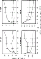
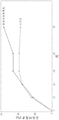
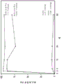


Classifications
-
- C—CHEMISTRY; METALLURGY
- C07—ORGANIC CHEMISTRY
- C07K—PEPTIDES
- C07K16/00—Immunoglobulins [IGs], e.g. monoclonal or polyclonal antibodies
- C07K16/18—Immunoglobulins [IGs], e.g. monoclonal or polyclonal antibodies against material from animals or humans
- C07K16/28—Immunoglobulins [IGs], e.g. monoclonal or polyclonal antibodies against material from animals or humans against receptors, cell surface antigens or cell surface determinants
- C07K16/2887—Immunoglobulins [IGs], e.g. monoclonal or polyclonal antibodies against material from animals or humans against receptors, cell surface antigens or cell surface determinants against CD20
-
- A—HUMAN NECESSITIES
- A61—MEDICAL OR VETERINARY SCIENCE; HYGIENE
- A61K—PREPARATIONS FOR MEDICAL, DENTAL OR TOILETRY PURPOSES
- A61K31/00—Medicinal preparations containing organic active ingredients
- A61K31/13—Amines
- A61K31/135—Amines having aromatic rings, e.g. ketamine, nortriptyline
- A61K31/138—Aryloxyalkylamines, e.g. propranolol, tamoxifen, phenoxybenzamine
-
- A—HUMAN NECESSITIES
- A61—MEDICAL OR VETERINARY SCIENCE; HYGIENE
- A61K—PREPARATIONS FOR MEDICAL, DENTAL OR TOILETRY PURPOSES
- A61K31/00—Medicinal preparations containing organic active ingredients
- A61K31/16—Amides, e.g. hydroxamic acids
- A61K31/165—Amides, e.g. hydroxamic acids having aromatic rings, e.g. colchicine, atenolol, progabide
- A61K31/167—Amides, e.g. hydroxamic acids having aromatic rings, e.g. colchicine, atenolol, progabide having the nitrogen of a carboxamide group directly attached to the aromatic ring, e.g. lidocaine, paracetamol
-
- A—HUMAN NECESSITIES
- A61—MEDICAL OR VETERINARY SCIENCE; HYGIENE
- A61K—PREPARATIONS FOR MEDICAL, DENTAL OR TOILETRY PURPOSES
- A61K31/00—Medicinal preparations containing organic active ingredients
- A61K31/33—Heterocyclic compounds
- A61K31/335—Heterocyclic compounds having oxygen as the only ring hetero atom, e.g. fungichromin
- A61K31/365—Lactones
-
- A—HUMAN NECESSITIES
- A61—MEDICAL OR VETERINARY SCIENCE; HYGIENE
- A61K—PREPARATIONS FOR MEDICAL, DENTAL OR TOILETRY PURPOSES
- A61K31/00—Medicinal preparations containing organic active ingredients
- A61K31/33—Heterocyclic compounds
- A61K31/395—Heterocyclic compounds having nitrogen as a ring hetero atom, e.g. guanethidine or rifamycins
- A61K31/535—Heterocyclic compounds having nitrogen as a ring hetero atom, e.g. guanethidine or rifamycins having six-membered rings with at least one nitrogen and one oxygen as the ring hetero atoms, e.g. 1,2-oxazines
- A61K31/5375—1,4-Oxazines, e.g. morpholine
- A61K31/5377—1,4-Oxazines, e.g. morpholine not condensed and containing further heterocyclic rings, e.g. timolol
-
- A—HUMAN NECESSITIES
- A61—MEDICAL OR VETERINARY SCIENCE; HYGIENE
- A61K—PREPARATIONS FOR MEDICAL, DENTAL OR TOILETRY PURPOSES
- A61K31/00—Medicinal preparations containing organic active ingredients
- A61K31/56—Compounds containing cyclopenta[a]hydrophenanthrene ring systems; Derivatives thereof, e.g. steroids
- A61K31/57—Compounds containing cyclopenta[a]hydrophenanthrene ring systems; Derivatives thereof, e.g. steroids substituted in position 17 beta by a chain of two carbon atoms, e.g. pregnane or progesterone
- A61K31/573—Compounds containing cyclopenta[a]hydrophenanthrene ring systems; Derivatives thereof, e.g. steroids substituted in position 17 beta by a chain of two carbon atoms, e.g. pregnane or progesterone substituted in position 21, e.g. cortisone, dexamethasone, prednisone or aldosterone
-
- A—HUMAN NECESSITIES
- A61—MEDICAL OR VETERINARY SCIENCE; HYGIENE
- A61K—PREPARATIONS FOR MEDICAL, DENTAL OR TOILETRY PURPOSES
- A61K39/00—Medicinal preparations containing antigens or antibodies
- A61K39/395—Antibodies; Immunoglobulins; Immune serum, e.g. antilymphocytic serum
- A61K39/39533—Antibodies; Immunoglobulins; Immune serum, e.g. antilymphocytic serum against materials from animals
- A61K39/39541—Antibodies; Immunoglobulins; Immune serum, e.g. antilymphocytic serum against materials from animals against normal tissues, cells
-
- A—HUMAN NECESSITIES
- A61—MEDICAL OR VETERINARY SCIENCE; HYGIENE
- A61K—PREPARATIONS FOR MEDICAL, DENTAL OR TOILETRY PURPOSES
- A61K45/00—Medicinal preparations containing active ingredients not provided for in groups A61K31/00 - A61K41/00
- A61K45/06—Mixtures of active ingredients without chemical characterisation, e.g. antiphlogistics and cardiaca
-
- A—HUMAN NECESSITIES
- A61—MEDICAL OR VETERINARY SCIENCE; HYGIENE
- A61P—SPECIFIC THERAPEUTIC ACTIVITY OF CHEMICAL COMPOUNDS OR MEDICINAL PREPARATIONS
- A61P13/00—Drugs for disorders of the urinary system
- A61P13/12—Drugs for disorders of the urinary system of the kidneys
-
- A—HUMAN NECESSITIES
- A61—MEDICAL OR VETERINARY SCIENCE; HYGIENE
- A61P—SPECIFIC THERAPEUTIC ACTIVITY OF CHEMICAL COMPOUNDS OR MEDICINAL PREPARATIONS
- A61P37/00—Drugs for immunological or allergic disorders
- A61P37/02—Immunomodulators
- A61P37/06—Immunosuppressants, e.g. drugs for graft rejection
-
- A—HUMAN NECESSITIES
- A61—MEDICAL OR VETERINARY SCIENCE; HYGIENE
- A61K—PREPARATIONS FOR MEDICAL, DENTAL OR TOILETRY PURPOSES
- A61K39/00—Medicinal preparations containing antigens or antibodies
- A61K2039/505—Medicinal preparations containing antigens or antibodies comprising antibodies
-
- A—HUMAN NECESSITIES
- A61—MEDICAL OR VETERINARY SCIENCE; HYGIENE
- A61K—PREPARATIONS FOR MEDICAL, DENTAL OR TOILETRY PURPOSES
- A61K39/00—Medicinal preparations containing antigens or antibodies
- A61K2039/545—Medicinal preparations containing antigens or antibodies characterised by the dose, timing or administration schedule
-
- A—HUMAN NECESSITIES
- A61—MEDICAL OR VETERINARY SCIENCE; HYGIENE
- A61K—PREPARATIONS FOR MEDICAL, DENTAL OR TOILETRY PURPOSES
- A61K31/00—Medicinal preparations containing organic active ingredients
- A61K31/33—Heterocyclic compounds
- A61K31/335—Heterocyclic compounds having oxygen as the only ring hetero atom, e.g. fungichromin
- A61K31/34—Heterocyclic compounds having oxygen as the only ring hetero atom, e.g. fungichromin having five-membered rings with one oxygen as the only ring hetero atom, e.g. isosorbide
-
- C—CHEMISTRY; METALLURGY
- C07—ORGANIC CHEMISTRY
- C07K—PEPTIDES
- C07K2317/00—Immunoglobulins specific features
- C07K2317/20—Immunoglobulins specific features characterized by taxonomic origin
- C07K2317/24—Immunoglobulins specific features characterized by taxonomic origin containing regions, domains or residues from different species, e.g. chimeric, humanized or veneered
-
- C—CHEMISTRY; METALLURGY
- C07—ORGANIC CHEMISTRY
- C07K—PEPTIDES
- C07K2317/00—Immunoglobulins specific features
- C07K2317/40—Immunoglobulins specific features characterized by post-translational modification
-
- C—CHEMISTRY; METALLURGY
- C07—ORGANIC CHEMISTRY
- C07K—PEPTIDES
- C07K2317/00—Immunoglobulins specific features
- C07K2317/50—Immunoglobulins specific features characterized by immunoglobulin fragments
- C07K2317/56—Immunoglobulins specific features characterized by immunoglobulin fragments variable (Fv) region, i.e. VH and/or VL
- C07K2317/565—Complementarity determining region [CDR]
-
- C—CHEMISTRY; METALLURGY
- C07—ORGANIC CHEMISTRY
- C07K—PEPTIDES
- C07K2317/00—Immunoglobulins specific features
- C07K2317/70—Immunoglobulins specific features characterized by effect upon binding to a cell or to an antigen
- C07K2317/73—Inducing cell death, e.g. apoptosis, necrosis or inhibition of cell proliferation
Abstract
The present disclosure provides methods for treating lupus nephritis in an individual with lupus. In some embodiments, the method comprises administering to the individual an effective amount of a type II anti-CD 20 antibody. In other aspects, the present disclosure provides methods for treating membranous nephropathy.
Description
Cross Reference to Related Applications
The present application claims us provisional application 62/899,706 filed on 12/9/2019; 62/930,527 filed on 11/4/2019; 62/931,032 filed on 5.11.2019; and 63/005,071 priority as applied on 3/4/2020; are all incorporated herein by reference in their entirety.
Sequence listing for submission of ASCII text documents
The following ASCII text documents are submitted with the contents fully incorporated herein by reference: TXT (filename: 146392048341SEQLIST, recording date: 2020, 8/17/h, size: 37KB) in Computer Readable Form (CRF).
Technical Field
Provided herein are methods for treating Lupus Nephritis (LN) in an individual with lupus by administering a type II anti-CD 20 antibody. In other aspects, provided herein are methods for treating membranous nephropathy.
Background
Proliferative lupus nephritis is the most common organ-threatening manifestation of systemic lupus erythematosus. Glomerular injury and tubulointerstitial inflammation lead to proteinuria, hematuria, and progressive impairment of renal function. Therapeutic goals include reducing proteinuria, preventing kidney damage, and minimizing toxicity of immunosuppressive therapy. Hahn et al, Arthritis Care and Research 64: 797-; fanouriakis et al, Ann. Rheum. Dis.78:736-45, 2019. Even with treatment, many patients have poor prognostic outcome, such as progression to end-stage renal disease (ESRD), need for hemodialysis or kidney transplantation or death, and the risk of ESRD has not improved substantially over the past two decades. Hanly et al, Rheumatology 55(2):252-62, 2016; tektonitou et al, Arthritis Rheumatotol 68(6): 1432-. Currently, there is no approved therapy in the united states for the treatment of lupus nephritis. Current standard treatments have not been approved in connection with toxicity and low complete response rates.
In clinical studies, two anti-CD 20 antibodies have been tested for efficacy in treating lupus nephritis. In one clinical study (LUNAR), rituximab (rituximab), a type I anti-CD 20 antibody), resulted in the depletion of peripheral CD19+ B cells by 71 out of 72 patients, and resulted in more responses and a dramatic decrease in the amount of anti-dsDNA and C3/C4. The dosage regimen for the LUNAR study included administration of rituximab (1,000mg) or placebo to patients with type III or type IV Lupus Nephritis (LN) on days 1, 15, 168, and 182 ( weeks 0, 2, 24, 26). However, clinical outcomes did not improve after 1 year of rituximab treatment.
Ocrelizumab (ocrelizumab) is another type I anti-CD 20 antibody that was tested in clinical studies (BELONG). On days 1 and 15, patients were injected with 1:1:1 randomized intravenous placebo, 400mg ocrelizumab, or 1,000mg ocrelizumab, followed by injections at week 16 and every 16 weeks thereafter, and using either background glucocorticoid gammoplate (MMF) or european lupus nephritis test (eltt) protocol (cyclophosphamide plus azathioprine). The study was terminated, in part, because the severe infectious events were not balanced (Mysler, E.F. et al, (2013) Arthritis Rheum.65: 2368-.
Thus, there remains a need for additional options to test the efficacy of LN in patients with lupus.
All references, including patent applications and publications, cited herein are hereby incorporated by reference in their entirety.
Disclosure of Invention
In certain aspects, provided herein is a method for treating lupus nephritis in an individual having lupus, comprising administering to the individual at least a first antibody exposure to a type II anti-CD 20 antibody, a second antibody exposure to the type II anti-CD 20 antibody, and a third antibody exposure to the type II anti-CD 20 antibody; wherein the second antibody exposure is not provided until about 18 weeks to about 26 weeks after the first antibody exposure; wherein the third antibody exposure is not provided until about 24 weeks to about 32 weeks after the second antibody exposure; wherein the first antibody exposure comprises one or two doses of the type II anti-CD 20 antibody, the first antibody exposure comprising a total exposure of between about 1800mg and about 2200mg of the type II anti-CD 20 antibody; wherein the second antibody exposure comprises one or two doses of the type II anti-CD 20 antibody, the second antibody exposure comprising a total exposure of between about 1800mg and about 2200mg of the type II anti-CD 20 antibody; wherein the third antibody exposure comprises one or two doses of the type II anti-CD 20 antibody, the third antibody exposure comprising a total exposure of between about 800mg and about 1200mg of the type II anti-CD 20 antibody; wherein the type II anti-CD 20 antibody comprises a heavy chain comprising the HVR-H1 sequence of SEQ ID NO. 1, the HVR-H2 sequence of SEQ ID NO. 2, and the HVR-H3 sequence of SEQ ID NO. 3, and a light chain comprising the HVR-L1 sequence of SEQ ID NO. 4, the HVR-L2 sequence of SEQ ID NO. 5, and the HVR-L3 sequence of SEQ ID NO. 6. Also provided herein is a type II anti-CD 20 antibody for use in a method of treating lupus nephritis in an individual, wherein the method comprises administering to the individual a first antibody exposure to the type II anti-CD 20 antibody, a second antibody exposure to the type II anti-CD 20 antibody, and a third antibody exposure to the type II anti-CD 20 antibody; wherein the second antibody exposure is not provided until about 18 weeks to about 26 weeks after the first antibody exposure; wherein the third antibody exposure is not provided until about 24 weeks to about 32 weeks after the second antibody exposure; wherein the first antibody exposure comprises one or two doses of the type II anti-CD 20 antibody, the first antibody exposure comprising a total exposure of between about 1800mg and about 2200mg of the type II anti-CD 20 antibody; wherein the second antibody exposure comprises one or two doses of the type II anti-CD 20 antibody, the second antibody exposure comprising a total exposure of between about 1800mg and about 2200mg of the type II anti-CD 20 antibody; wherein the third antibody exposure comprises one or two doses of the type II anti-CD 20 antibody, the third antibody exposure comprising a total exposure of between about 800mg and about 1200mg of the type II anti-CD 20 antibody; and wherein the type II anti-CD 20 antibody comprises a heavy chain comprising the HVR-H1 sequence of SEQ ID NO. 1, the HVR-H2 sequence of SEQ ID NO. 2, and the HVR-H3 sequence of SEQ ID NO. 3, and a light chain comprising the HVR-L1 sequence of SEQ ID NO. 4, the HVR-L2 sequence of SEQ ID NO. 5, and the HVR-L3 sequence of SEQ ID NO. 6.
In some embodiments, the first antibody exposure comprises a first dose of between about 900mg and about 1100mg of the type II anti-CD 20 antibody and a second dose of between about 900mg and about 1100mg of the type II anti-CD 20 antibody. In some embodiments, the first antibody exposure comprises a first dose of the type II anti-CD 20 antibody and a second dose of the type II anti-CD 20 antibody, and the second dose of the first antibody exposure is not provided until about 1.5 weeks to about 2.5 weeks after the first dose of the first antibody exposure. In some embodiments, the first antibody exposure comprises a first dose of the type II anti-CD 20 antibody and a second dose of the type II anti-CD 20 antibody, and the second dose of the first antibody exposure is not provided until about 2 weeks after the first dose of the first antibody exposure. In some embodiments, the first agent of the first antibody exposure is about 1000mg of the type II anti-CD 20 antibody. In some embodiments, the second agent to which the first antibody is exposed is about 1000mg of the type II anti-CD 20 antibody.
In some embodiments, the second antibody exposure comprises a first dose of between about 900mg and about 1100mg of the type II anti-CD 20 antibody and a second dose of between about 900mg and about 1100mg of the type II anti-CD 20 antibody. In some embodiments, the second antibody exposure comprises a first dose of the type II anti-CD 20 antibody and a second dose of the type II anti-CD 20 antibody, and the second antibody-exposed second dose is not provided until about 1.5 weeks to about 2.5 weeks after the first dose of the second antibody exposure. In some embodiments, the second antibody exposure comprises a first dose of the type II anti-CD 20 antibody and a second dose of the type II anti-CD 20 antibody, and the second antibody-exposed second dose is not provided until about 2 weeks after the first dose of the second antibody exposure. In some embodiments, the first dose to which the second antibody is exposed is about 1000mg of the type II anti-CD 20 antibody. In some embodiments, the second agent to which the second antibody is exposed is about 1000mg of the type II anti-CD 20 antibody.
In some embodiments, the third antibody exposure comprises a single dose of between about 900mg and about 1100mg of the type II anti-CD 20 antibody. In some embodiments, the single dose of the third antibody exposure is about 1000mg of the type II anti-CD 20 antibody. In some embodiments, the single dose of the third antibody exposure is not provided until about 52 weeks after the first dose of the first antibody exposure or until about 28 weeks after the first dose of the second antibody exposure.
In some embodiments, the first antibody exposure, and/or the second antibody exposure, and/or the third antibody exposure is administered intravenously.
In some embodiments, the individual has lupus nephritis. In some embodiments, the individual has type III or type IV lupus nephritis. In some embodiments, the individual is at risk of developing lupus nephritis of type III or type IV. In some embodiments, the subject has lupus nephritis type iii, (c) or type iv (c). In some embodiments, the individual has concomitant lupus V nephritis.
In some embodiments, the method further comprises administering to the individual an effective amount of an immunosuppressive agent. In some embodiments, the immunosuppressive agent comprises mycophenolic acid, a derivative thereof, or a salt thereof. In some embodiments, the immunosuppressive agent comprises mycophenolate mofetil (mycophenolate mofetil). In some embodiments, the method further comprises administering to the individual an effective amount of a glucocorticoid or corticosteroid. In some embodiments, the glucocorticoid or corticosteroid comprises methylprednisolone (methylprednisolone). In some embodiments, the glucocorticoid or corticosteroid comprises prednisone (prednisone). In some embodiments, the method further comprises administering to the individual an effective amount of an antihistamine. In some embodiments, the antihistamine comprises diphenhydramine (diphenhydramine). In some embodiments, the method further comprises administering to the individual an effective amount of a non-steroidal anti-inflammatory drug (NSAID). In some embodiments, the NSAID comprises acetaminophen (acetaminophen). In some embodiments, the method further comprises administering to the individual an effective amount of an antihypertensive agent. In some embodiments, the antihypertensive agent is an Angiotensin Converting Enzyme (ACE) inhibitor or an angiotensin receptor blocker. In some embodiments, the method further comprises administering to the individual a standard treatment. In some embodiments, the standard of care includes treatment with one or more of Angiotensin Converting Enzyme (ACE) inhibitors, angiotensin receptor blockers, cyclophosphamide, mycophenolate mofetil, azathioprine, and glucocorticoids or corticosteroids.
In some embodiments, the method results in Complete Renal Remission (CRR) in the subject. In some embodiments, the method results in Partial Renal Remission (PRR) in the subject. In some embodiments, the method results in a circulating peripheral B cell deficiency in the individual. In some embodiments, the circulating peripheral B cells are CD19+ B cells. In some embodiments, the B cells are naive B cells (e.g., CD19+ CD27-B cells), memory B cells (e.g., CD19+ CD27+ B cells), or plasmablasts (e.g., CD19+ CD27+ CD38+ + B cells). In some embodiments, the B cells are CD19+ CD3-CD14-CD33-CD 56-cells. In some embodiments, following administration of the type II anti-CD 20 antibody, the B cells are depleted to a level such that circulating peripheral B cells are present in peripheral blood from the individual in an amount of about 5 cells/μ L or less. In some embodiments, the B cells are depleted to a level such that circulating peripheral B cells are present in peripheral blood from the individual in an amount of about 1 cell/μ L or less. In some embodiments, the B cells are depleted to a level such that circulating peripheral B cells are present in peripheral blood from the individual in an amount of about 0.5 cells/μ L or less. In some embodiments, B cells are depleted to a level such that circulating peripheral B cells are present in the peripheral blood of the subject to which the depletion was reached following the first antibody exposure. In some embodiments, B cells are depleted below the threshold level detectable using HSFC. In some embodiments, the HSFC has a lower limit of quantitation of B cells (LLOQ) of about 1.0 cells/μ L or less, about 0.8 cells/μ L or less, about 0.6 cells/μ L or less, about 0.5 cells/μ L or less, or 0.441 cells/μ L or less. In some embodiments, B cell depletion continues for at least 52 weeks after the first dose of the first antibody exposure. In some embodiments, after administration of the type II anti-CD 20 antibody, compared to corresponding measurements performed on the same subject prior to administration of the type II anti-CD 20 antibody, or compared to non-receiving type II anti-CD 20 antibody A corresponding measure performed by the treated subject, the subject's circulating peripheral B cell depletion is at least about 90%.
In some embodiments, the individual is a human.
In some embodiments, the first antibody exposure comprises two doses of 1000mg of the type II anti-CD 20 antibody on days 1 and 15 of treatment; the second antibody was exposed to two doses containing 1000mg of the type II anti-CD 20 antibody on days 168 and 182 of treatment; the third antibody exposure comprises a dose of 1000mg of the type II anti-CD 20 antibody on day 364 of treatment; wherein the type II anti-CD 20 antibody is obinutuzumab (obinutuzumab); and wherein the individual is a human. In some embodiments, the first antibody exposure comprises two doses of 1000mg of the type II anti-CD 20 antibody at weeks 0 and 2 of treatment; the second antibody was exposed to two doses of 1000mg of the type II anti-CD 20 antibody contained at weeks 24 and 26 of treatment; the third antibody exposure comprises a dose of 1000mg of the type II anti-CD 20 antibody at week 52 of treatment; wherein the type II anti-CD 20 antibody is obinutuzumab; wherein the type II anti-CD 20 antibody is administered intravenously; and wherein the individual is a human. In some embodiments, the first antibody exposure comprises two doses of 1000mg of the type II anti-CD 20 antibody on days 1 and 15 of treatment; the second antibody was exposed to two doses containing 1000mg of the type II anti-CD 20 antibody on days 168 and 182 of treatment; the third antibody exposure comprises two doses of 1000mg of the type II anti-CD 20 antibody on days 350 and 364 of treatment; wherein the type II anti-CD 20 antibody is obinutuzumab; and wherein the individual is a human. In some embodiments, the first antibody exposure comprises two doses of 1000mg of the type II anti-CD 20 antibody at weeks 0 and 2 of treatment; the second antibody was exposed to two doses of 1000mg of the type II anti-CD 20 antibody contained at weeks 24 and 26 of treatment; the third antibody exposure comprises two doses of 1000mg of the type II anti-CD 20 antibody at weeks 50 and 52 of treatment; wherein the type II anti-CD 20 antibody is obinutuzumab; and wherein the individual is a human.
In certain aspects, provided herein is a method for depleting circulating peripheral B cells in an individual comprising administering to the individual a first antibody exposure to a type II anti-CD 20 antibody, a second antibody exposure to the type II anti-CD 20 antibody, and a third antibody exposure to the type II anti-CD 20 antibody; wherein the second antibody exposure is not provided until about 18 weeks to about 26 weeks after the first antibody exposure; wherein the third antibody exposure is not provided until about 24 weeks to about 32 weeks after the second antibody exposure; wherein the first antibody exposure comprises one or two doses of the type II anti-CD 20 antibody, the first antibody exposure comprising a total exposure of between about 1800mg and about 2200mg of the type II anti-CD 20 antibody; wherein the second antibody exposure comprises one or two doses of the type II anti-CD 20 antibody, the second antibody exposure comprising a total exposure of between about 1800mg and about 2200mg of the type II anti-CD 20 antibody; wherein the third antibody exposure comprises one or two doses of the type II anti-CD 20 antibody, the third antibody exposure comprising a total exposure of between about 800mg and about 1200mg of the type II anti-CD 20 antibody; wherein the type II anti-CD 20 antibody comprises a heavy chain comprising the HVR-H1 sequence of SEQ ID NO. 1, the HVR-H2 sequence of SEQ ID NO. 2, and the HVR-H3 sequence of SEQ ID NO. 3, and a light chain comprising the HVR-L1 sequence of SEQ ID NO. 4, the HVR-L2 sequence of SEQ ID NO. 5, and HVR-L3 sequence of SEQ ID NO. 6; and wherein after administration of the type II anti-CD 20 antibody, the B cells are depleted to a level such that circulating peripheral B cells are present in peripheral blood from the individual in an amount of about 5 cells/μ L or less. Also provided herein is a type II anti-CD 20 antibody for use in a method of depleting circulating peripheral B cells in an individual, wherein the method comprises administering to the individual a first antibody exposure to the type II anti-CD 20 antibody, a second antibody exposure to the type II anti-CD 20 antibody, and a third antibody exposure to the type II anti-CD 20 antibody; wherein the second antibody exposure is not provided until about 18 weeks to about 26 weeks after the first antibody exposure; wherein the third antibody exposure is not provided until about 24 weeks to about 32 weeks after the second antibody exposure; wherein the first antibody exposure comprises one or two doses of the type II anti-CD 20 antibody, the first antibody exposure comprising a total exposure of between about 1800mg and about 2200mg of the type II anti-CD 20 antibody; wherein the second antibody exposure comprises one or two doses of the type II anti-CD 20 antibody, the second antibody exposure comprising a total exposure of between about 1800mg and about 2200mg of the type II anti-CD 20 antibody; wherein the third antibody exposure comprises one or two doses of the type II anti-CD 20 antibody, the third antibody exposure comprising a total exposure of between about 800mg and about 1200mg of the type II anti-CD 20 antibody; wherein the type II anti-CD 20 antibody comprises a heavy chain comprising the HVR-H1 sequence of SEQ ID NO. 1, the HVR-H2 sequence of SEQ ID NO. 2, and the HVR-H3 sequence of SEQ ID NO. 3, and a light chain comprising the HVR-L1 sequence of SEQ ID NO. 4, the HVR-L2 sequence of SEQ ID NO. 5, and the HVR-L3 sequence of SEQ ID NO. 6; and wherein after administration of the type II anti-CD 20 antibody, the B cells are depleted to a level such that circulating peripheral B cells are present in peripheral blood from the individual in an amount of about 5 cells/μ L or less.
In some embodiments, the first antibody exposure comprises a first dose of between about 900mg and about 1100mg of the type II anti-CD 20 antibody and a second dose of between about 900mg and about 1100mg of the type II anti-CD 20 antibody. In some embodiments, the first antibody exposure comprises a first dose of the type II anti-CD 20 antibody and a second dose of the type II anti-CD 20 antibody, and the second dose of the first antibody exposure is not provided until about 1.5 weeks to about 2.5 weeks after the first dose of the first antibody exposure. In some embodiments, the first antibody exposure comprises a first dose of the type II anti-CD 20 antibody and a second dose of the type II anti-CD 20 antibody, and the second dose of the first antibody exposure is not provided until about 2 weeks after the first dose of the first antibody exposure. In some embodiments, the first agent of the first antibody exposure is about 1000mg of the type II anti-CD 20 antibody. In some embodiments, the second agent to which the first antibody is exposed is about 1000mg of the type II anti-CD 20 antibody.
In some embodiments, the second antibody exposure comprises a first dose of between about 900mg and about 1100mg of the type II anti-CD 20 antibody and a second dose of between about 900mg and about 1100mg of the type II anti-CD 20 antibody. In some embodiments, the second antibody exposure comprises a first dose of the type II anti-CD 20 antibody and a second dose of the type II anti-CD 20 antibody, and the second antibody-exposed second dose is not provided until about 1.5 weeks to about 2.5 weeks after the first dose of the second antibody exposure. In some embodiments, the second antibody exposure comprises a first dose of the type II anti-CD 20 antibody and a second dose of the type II anti-CD 20 antibody, and the second antibody-exposed second dose is not provided until about 2 weeks after the first dose of the second antibody exposure. In some embodiments, the first dose to which the second antibody is exposed is about 1000mg of the type II anti-CD 20 antibody. In some embodiments, the second agent to which the second antibody is exposed is about 1000mg of the type II anti-CD 20 antibody.
In some embodiments, the third antibody exposure comprises a single dose of between about 900mg and about 1100mg of the type II anti-CD 20 antibody. In some embodiments, the single dose of the third antibody exposure is about 1000mg of the type II anti-CD 20 antibody. In some embodiments, the single dose of the third antibody exposure is not provided until about 52 weeks after the first dose of the first antibody exposure or until about 28 weeks after the first dose of the second antibody exposure.
In some embodiments, the first antibody exposure, and/or the second antibody exposure, and/or the third antibody exposure is administered intravenously.
In some embodiments, the individual has lupus nephritis. In some embodiments, the individual has type III or type IV lupus nephritis. In some embodiments, the individual is at risk of developing lupus nephritis type III or IV. In some embodiments, the subject has lupus nephritis type iii, (c) or type iv (c). In some embodiments, the individual has concomitant lupus V nephritis. In some embodiments, the individual has Membranous Nephropathy (MN), such as primary membranous nephropathy (pMN). In some embodiments, the individual is at risk of developing Membranous Nephropathy (MN), e.g., primary membranous nephropathy (pMN).
In some embodiments, the circulating peripheral B cells are CD19+ B cells. In some embodiments, the B cells are naive B cells (e.g., CD19+ CD27-B cells), memory B cells (e.g., CD19+ CD27+ B cells), and/or plasmablasts (e.g., CD19+ CD27+ CD38+ + B cells). In some embodiments, the B cells are CD19+ CD3-CD14-CD33-CD 56-cells. In some embodiments of the present invention, the,the B cells include CD19+ CD20+ B cells, CD19+ CD20-B cells, and CD19+ CD22+ B cells. In some embodiments, the B cells are depleted to a level such that circulating peripheral B cells are present in peripheral blood from the individual in an amount of about 1 cell/μ L or less. In some embodiments, the B cells are depleted to a level such that circulating peripheral B cells are present in peripheral blood from the individual in an amount of about 0.5 cells/μ L or less. In some embodiments, B cells are depleted below a threshold level detectable using HSFC. In some embodiments, the lower limit of quantification of B cells by the HSFC is (LLOQ) about 1.0 cells/μ L or less, about 0.8 cells/μ L or less, about 0.6 cells/μ L or less, about 0.5 cells/μ L or less, or 0.441 cells/μ L or less. In some embodiments, the depleting is achieved after exposure of the first antibody. In some embodiments, B cell depletion continues for at least 52 weeks after the first dose of the first antibody exposure. In some embodiments, following administration of the type II anti-CD 20 antibody, the subject has at least about 90% circulating peripheral B cell depletion compared to a corresponding measurement performed on the same subject prior to administration of the type II anti-CD 20 antibody, or compared to a corresponding measurement performed on a subject not receiving treatment with a type II anti-CD 20 antibody. In some embodiments, following administration of the type II anti-CD 20 antibody, there is an increase in the amount of serum B cell activating factor (BAFF) in the individual (e.g., the amount of BAFF in a serum sample from the individual) as compared to a corresponding measurement taken of the same individual prior to administration of the type II anti-CD 20 antibody, or as compared to a corresponding measurement taken of an individual not receiving treatment with the type II anti-CD 20 antibody. In some embodiments, there is an increase in the amount of serum B cell activating factor (BAFF), e.g., in a serum sample from an individual, in 6 weeks or less, 4 weeks or less, or 2 weeks or less after administration of the type II anti-CD 20 antibody, e.g., as compared to a corresponding measurement taken on the same individual prior to administration of the type II anti-CD 20 antibody, or as compared to a corresponding measurement taken on an individual not receiving treatment with a type II anti-CD 20 antibody. In some embodiments, the type II anti-CD 20 antibody is administered prior to, e.g., administering the type II anti-CD 20 antibody, or to the same subject prior to, e.g., administering the type II anti-CD 20 antibody A corresponding measurement by a type II anti-CD 20 antibody-treated individual is that the individual's amount of serum B cell activating factor (BAFF), e.g., the amount of BAFF in a serum sample from the individual, is increased by at least 50%, at least 75%, at least 100%, at least 2-fold, or at least 3-fold.
In some embodiments, the individual is a human.
In some embodiments, the first antibody exposure comprises two doses of 1000mg of the type II anti-CD 20 antibody on days 1 and 15 of treatment; the second antibody was exposed to two doses containing 1000mg of the type II anti-CD 20 antibody on days 168 and 182 of treatment; the third antibody exposure comprises a dose of 1000mg of the type II anti-CD 20 antibody on day 364 of treatment; wherein the type II anti-CD 20 antibody is obinutuzumab (obinutuzumab); and wherein the individual is a human. In some embodiments, the first antibody exposure comprises two doses of 1000mg of the type II anti-CD 20 antibody at weeks 0 and 2 of treatment; the second antibody was exposed to two doses of 1000mg of the type II anti-CD 20 antibody contained at weeks 24 and 26 of treatment; the third antibody exposure comprises a dose of 1000mg of the type II anti-CD 20 antibody at week 52 of treatment; wherein the type II anti-CD 20 antibody is obinutuzumab; wherein the type II anti-CD 20 antibody is administered intravenously; and wherein the individual is a human. In some embodiments, the first antibody exposure comprises two doses of 1000mg of the type II anti-CD 20 antibody on days 1 and 15 of treatment; the second antibody was exposed to two doses containing 1000mg of the type II anti-CD 20 antibody on days 168 and 182 of treatment; the third antibody exposure comprises two doses of 1000mg of the type II anti-CD 20 antibody on days 350 and 364 of treatment; wherein the type II anti-CD 20 antibody is obinutuzumab; and wherein the individual is a human. In some embodiments, the first antibody exposure comprises two doses of 1000mg of the type II anti-CD 20 antibody at weeks 0 and 2 of treatment; the second antibody was exposed to two doses of 1000mg of the type II anti-CD 20 antibody contained at weeks 24 and 26 of treatment; the third antibody exposure comprises two doses of 1000mg of the type II anti-CD 20 antibody at weeks 50 and 52 of treatment; wherein the type II anti-CD 20 antibody is obinutuzumab; and wherein the individual is a human.
In certain aspects, provided herein is a method for depleting circulating peripheral B cells in an individual comprising administering to the individual a first antibody exposure to a type II anti-CD 20 antibody and a second antibody exposure to the type II anti-CD 20 antibody; wherein the second antibody exposure is not provided until about 18 weeks to about 26 weeks after the first antibody exposure; wherein the first antibody exposure comprises one or two doses of the type II anti-CD 20 antibody, the first antibody exposure comprising a total exposure of between about 1800mg and about 2200mg of the type II anti-CD 20 antibody; wherein the second antibody exposure comprises one or two doses of the type II anti-CD 20 antibody, the second antibody exposure comprising a total exposure of between about 1800mg and about 2200mg of the type II anti-CD 20 antibody; wherein the type II anti-CD 20 antibody comprises a heavy chain comprising the HVR-H1 sequence of SEQ ID NO. 1, the HVR-H2 sequence of SEQ ID NO. 2, and the HVR-H3 sequence of SEQ ID NO. 3, and a light chain comprising the HVR-L1 sequence of SEQ ID NO. 4, the HVR-L2 sequence of SEQ ID NO. 5, and HVR-L3 sequence of SEQ ID NO. 6; and wherein following administration of the type II anti-CD 20 antibody, B cells are depleted to a level such that circulating peripheral B cells are present in peripheral blood from the individual in an amount of about 5 cells/μ L or less, the B cell depletion lasting at least 52 weeks following the first antibody exposure. Also provided herein is a type II anti-CD 20 antibody in a method for depleting circulating peripheral B cells in an individual, wherein the method comprises administering to the individual a first antibody exposure to the type II anti-CD 20 antibody and a second antibody exposure to the type II anti-CD 20 antibody; wherein the second antibody exposure is not provided until about 18 weeks to about 26 weeks after the first antibody exposure; wherein the first antibody exposure comprises one or two doses of the type II anti-CD 20 antibody, the first antibody exposure comprising a total exposure of between about 1800mg and about 2200mg of the type II anti-CD 20 antibody; wherein the second antibody exposure comprises one or two doses of the type II anti-CD 20 antibody, the second antibody exposure comprising a total exposure of between about 1800mg and about 2200mg of the type II anti-CD 20 antibody; wherein the type II anti-CD 20 antibody comprises a heavy chain comprising the HVR-H1 sequence of SEQ ID NO. 1, the HVR-H2 sequence of SEQ ID NO. 2, and the HVR-H3 sequence of SEQ ID NO. 3, and a light chain comprising the HVR-L1 sequence of SEQ ID NO. 4, the HVR-L2 sequence of SEQ ID NO. 5, and the HVR-L3 sequence of SEQ ID NO. 6; and wherein following administration of the type II anti-CD 20 antibody, B cells are depleted to a level such that circulating peripheral B cells are present in peripheral blood from the individual in an amount of about 5 cells/μ L or less, the B cell depletion lasting at least 52 weeks following the first antibody exposure.
In some embodiments, the first antibody exposure comprises a first dose of between about 900mg and about 1100mg of the type II anti-CD 20 antibody and a second dose of between about 900mg and about 1100mg of the type II anti-CD 20 antibody. In some embodiments, the first antibody exposure comprises a first dose of the type II anti-CD 20 antibody and a second dose of the type II anti-CD 20 antibody, and the second dose of the first antibody exposure is not provided until about 1.5 weeks to about 2.5 weeks after the first dose of the first antibody exposure. In some embodiments, the first antibody exposure comprises a first dose of the type II anti-CD 20 antibody and a second dose of the type II anti-CD 20 antibody, and the second dose of the first antibody exposure is not provided until about 2 weeks after the first dose of the first antibody exposure. In some embodiments, the first dose of the first antibody exposure is about 1000mg of the type II anti-CD 20 antibody. In some embodiments, the second agent to which the first antibody is exposed is about 1000mg of the type II anti-CD 20 antibody.
In some embodiments, the second antibody exposure comprises a first dose of between about 900mg and about 1100mg of the type II anti-CD 20 antibody and a second dose of between about 900mg and about 1100mg of the type II anti-CD 20 antibody. In some embodiments, the second antibody exposure comprises a first dose of the type II anti-CD 20 antibody and a second dose of the type II anti-CD 20 antibody, and the second antibody-exposed second dose is not provided until about 1.5 weeks to about 2.5 weeks after the first dose of the second antibody exposure. In some embodiments, the second antibody exposure comprises a first dose of the type II anti-CD 20 antibody and a second dose of the type II anti-CD 20 antibody, and the second antibody-exposed second dose is not provided until about 2 weeks after the first dose of the second antibody exposure. In some embodiments, the first dose to which the second antibody is exposed is about 1000mg of the type II anti-CD 20 antibody. In some embodiments, the second agent to which the second antibody is exposed is about 1000mg of the type II anti-CD 20 antibody.
In some embodiments, the first antibody exposure, and/or the second antibody exposure is administered intravenously.
In some embodiments, the individual has lupus nephritis. In some embodiments, the individual has type III or type IV lupus nephritis. In some embodiments, the individual is at risk of developing lupus nephritis type III or IV. In some embodiments, the subject has lupus nephritis type iii, (c) or type iv (c). In some embodiments, the individual has concomitant lupus V nephritis. In some embodiments, the individual has Membranous Nephropathy (MN), such as primary membranous nephropathy (pMN). In some embodiments, the individual is at risk of developing Membranous Nephropathy (MN), e.g., primary membranous nephropathy (pMN).
In some embodiments, the circulating peripheral B cells are CD19+ B cells. In some embodiments, the circulating peripheral B cells are CD19+ B cells. In some embodiments, the B cells are naive B cells (e.g., CD19+ CD27-B cells), memory B cells (e.g., CD19+ CD27+ B cells), and/or plasmablasts (e.g., CD19+ CD27+ CD38+ + B cells). In some embodiments, the B cells are CD19+ CD3-CD14-CD33-CD 56-cells. In some embodiments, the B cells comprise CD19+ CD20+ B cells, CD19+ CD20-B cells, and CD19+ CD22+ B cells. In some embodiments, the B cells are depleted to a level such that circulating peripheral B cells are present in peripheral blood from the subject in an amount of about 1 cell/μ L or less. In some embodiments, the B cells are depleted to a level such that circulating peripheral B cells are present in peripheral blood from the subject in an amount of about 0.5 cells/μ L or less. In some embodiments, B cells are depleted below a threshold level detectable using HSFC. In some embodiments, the lower limit of quantification of B cells by the HSFC is (LLOQ) about 1.0 cells/μ L or less, about 0.8 cells/μ L or less, about 0.6 cells/μ L or less, about 0.5 cells/μ L or less, or 0.441 cells/μ L or less. In some embodiments, the depleting is achieved after exposure of the first antibody. At one end In some embodiments, B cell depletion continues for at least 52 weeks after the first dose of the first antibody exposure. In some embodiments, following administration of the type II anti-CD 20 antibody, the subject has at least about 90% circulating peripheral B cell depletion compared to a corresponding measurement performed on the same subject prior to administration of the type II anti-CD 20 antibody, or compared to a corresponding measurement performed on a subject not receiving treatment with a type II anti-CD 20 antibody.
In certain aspects, provided herein is a method for treating rheumatoid arthritis, Systemic Lupus Erythematosus (SLE), Membranous Nephropathy (MN), or extrarenal lupus (ERL) in an individual comprising administering to the individual a first antibody exposure to a type II anti-CD 20 antibody, a second antibody exposure to the type II anti-CD 20 antibody, and a third antibody exposure to the type II anti-CD 20 antibody; wherein the second antibody exposure is not provided until about 18 weeks to about 26 weeks after the first antibody exposure; wherein the third antibody exposure is not provided until about 24 weeks to about 32 weeks after the second antibody exposure; wherein the first antibody exposure comprises one or two doses of the type II anti-CD 20 antibody, the first antibody exposure comprising a total exposure of between about 1800mg and about 2200mg of the type II anti-CD 20 antibody; wherein the second antibody exposure comprises one or two doses of the type II anti-CD 20 antibody, the second antibody exposure comprising a total exposure of between about 1800mg and about 2200mg of the type II anti-CD 20 antibody; wherein the third antibody exposure comprises one or two doses of the type II anti-CD 20 antibody, the third antibody exposure comprising a total exposure of between about 800mg and about 1200mg of the type II anti-CD 20 antibody; wherein the type II anti-CD 20 antibody comprises a heavy chain comprising the HVR-H1 sequence of SEQ ID NO. 1, the HVR-H2 sequence of SEQ ID NO. 2, and the HVR-H3 sequence of SEQ ID NO. 3, and a light chain comprising the HVR-L1 sequence of SEQ ID NO. 4, the HVR-L2 sequence of SEQ ID NO. 5, and the HVR-L3 sequence of SEQ ID NO. 6. Also provided herein is a type II anti-CD 20 antibody for use in a method of treating rheumatoid arthritis, Systemic Lupus Erythematosus (SLE), Membranous Nephropathy (MN), or extrarenal lupus (ERL) in an individual, wherein the method comprises administering to the individual a first antibody exposure to the type II anti-CD 20 antibody, a second antibody exposure to the type II anti-CD 20 antibody, and a third antibody exposure to the type II anti-CD 20 antibody; wherein the second antibody exposure is not provided until about 18 weeks to about 26 weeks after the first antibody exposure; wherein the third antibody exposure is not provided until about 24 weeks to about 32 weeks after the second antibody exposure; wherein the first antibody exposure comprises one or two doses of the type II anti-CD 20 antibody, the first antibody exposure comprising a total exposure of between about 1800mg and about 2200mg of the type II anti-CD 20 antibody; wherein the second antibody exposure comprises one or two doses of the type II anti-CD 20 antibody, the second antibody exposure comprising a total exposure of between about 1800mg and about 2200mg of the type II anti-CD 20 antibody; wherein the third antibody exposure comprises one or two doses of the type II anti-CD 20 antibody, the third antibody exposure comprising a total exposure of between about 800mg and about 1200mg of the type II anti-CD 20 antibody; and wherein the type II anti-CD 20 antibody comprises a heavy chain comprising the HVR-H1 sequence of SEQ ID NO. 1, the HVR-H2 sequence of SEQ ID NO. 2, and the HVR-H3 sequence of SEQ ID NO. 3, and a light chain comprising the HVR-L1 sequence of SEQ ID NO. 4, the HVR-L2 sequence of SEQ ID NO. 5, and the HVR-L3 sequence of SEQ ID NO. 6.
In some embodiments, the first antibody exposure comprises a first dose of between about 900mg and about 1100mg of the type II anti-CD 20 antibody and a second dose of between about 900mg and about 1100mg of the type II anti-CD 20 antibody. In some embodiments, the first antibody exposure comprises a first dose of the type II anti-CD 20 antibody and a second dose of the type II anti-CD 20 antibody, and the second dose of the first antibody exposure is not provided until about 1.5 weeks to about 2.5 weeks after the first dose of the first antibody exposure. In some embodiments, the first antibody exposure comprises a first dose of the type II anti-CD 20 antibody and a second dose of the type II anti-CD 20 antibody, and the second dose of the first antibody exposure is not provided until about 2 weeks after the first dose of the first antibody exposure. In some embodiments, the first agent of the first antibody exposure is about 1000mg of the type II anti-CD 20 antibody. In some embodiments, the second agent to which the first antibody is exposed is about 1000mg of the type II anti-CD 20 antibody.
In some embodiments, the second antibody exposure comprises a first dose of between about 900mg and about 1100mg of the type II anti-CD 20 antibody and a second dose of between about 900mg and about 1100mg of the type II anti-CD 20 antibody. In some embodiments, the second antibody exposure comprises a first dose of the type II anti-CD 20 antibody and a second dose of the type II anti-CD 20 antibody, and the second antibody-exposed second dose is not provided until about 1.5 weeks to about 2.5 weeks after the first dose of the second antibody exposure. In some embodiments, the second antibody exposure comprises a first dose of the type II anti-CD 20 antibody and a second dose of the type II anti-CD 20 antibody, and the second antibody-exposed second dose is not provided until about 2 weeks after the first dose of the second antibody exposure. In some embodiments, the first dose to which the second antibody is exposed is about 1000mg of the type II anti-CD 20 antibody. In some embodiments, the second agent to which the second antibody is exposed is about 1000mg of the type II anti-CD 20 antibody.
In some embodiments, the third antibody exposure comprises a single dose of between about 900mg and about 1100mg of the type II anti-CD 20 antibody. In some embodiments, the single dose of the third antibody exposure is about 1000mg of the type II anti-CD 20 antibody. In some embodiments, the single dose of the third antibody exposure is not provided until about 52 weeks after the first dose of the first antibody exposure or until about 28 weeks after the first dose of the second antibody exposure.
In some embodiments, the first antibody exposure, and/or the second antibody exposure, and/or the third antibody exposure is administered intravenously.
In certain aspects, provided herein is a method for treating lupus nephritis in an individual having lupus or depleting circulating peripheral B cells in an individual, comprising intravenously administering to the individual a first antibody exposure, a second antibody exposure, and a third antibody exposure to a type II anti-CD 20 antibody; wherein the first antibody exposure comprises two doses of 1000mg of the type II anti-CD 20 antibody at weeks 0 and 2 of treatment; wherein the second antibody exposes two doses of 1000mg of the type II anti-CD 20 antibody contained at weeks 24 and 26 of treatment; wherein the third antibody exposure comprises a dose of 1000mg of the type II anti-CD 20 antibody at week 52 of treatment; wherein the type II anti-CD 20 antibody comprises a heavy chain comprising the HVR-H1 sequence of SEQ ID NO. 1, the HVR-H2 sequence of SEQ ID NO. 2, and the HVR-H3 sequence of SEQ ID NO. 3, and a light chain comprising the HVR-L1 sequence of SEQ ID NO. 4, the HVR-L2 sequence of SEQ ID NO. 5, and the HVR-L3 sequence of SEQ ID NO. 6; and wherein the subject is a human. In some embodiments, the type II anti-CD 20 antibody is obinutuzumab.
In certain aspects, provided herein is a method for treating lupus nephritis in an individual having lupus or depleting circulating peripheral B cells in an individual, comprising intravenously administering to the individual a first antibody exposure, a second antibody exposure, and a third antibody exposure to a type II anti-CD 20 antibody; wherein the first antibody exposure comprises two doses of 1000mg of the type II anti-CD 20 antibody at weeks 0 and 2 of treatment; wherein the second antibody exposes two doses of 1000mg of the type II anti-CD 20 antibody contained at weeks 24 and 26 of treatment; wherein the third antibody exposure comprises two doses of 1000mg of the type II anti-CD 20 antibody at weeks 50 and 52 of treatment; wherein the type II anti-CD 20 antibody comprises a heavy chain comprising the HVR-H1 sequence of SEQ ID NO. 1, the HVR-H2 sequence of SEQ ID NO. 2, and the HVR-H3 sequence of SEQ ID NO. 3, and a light chain comprising the HVR-L1 sequence of SEQ ID NO. 4, the HVR-L2 sequence of SEQ ID NO. 5, and the HVR-L3 sequence of SEQ ID NO. 6; and wherein the subject is a human. In some embodiments, the type II anti-CD 20 antibody is obinutuzumab.
In some embodiments of the methods described herein, the type II anti-CD 20 antibody is a humanized antibody. In some embodiments, the type II anti-CD 20 antibody is afucosylated (afucosylated). In some embodiments, the heavy chain of the type II anti-CD 20 antibody comprises a heavy chain variable region comprising the amino acid sequence of SEQ ID No. 7. In some embodiments, the light chain of the type II anti-CD 20 antibody comprises a light chain variable region comprising the amino acid sequence of SEQ ID No. 8. In some embodiments, the type II anti-CD 20 antibody comprises the heavy chain variable region comprising the amino acid sequence of SEQ ID No. 7; and a light chain variable region comprising the amino acid sequence of SEQ ID NO 8. In some embodiments, the type II anti-CD 20 antibody comprises a heavy chain comprising the amino acid sequence of SEQ ID NO. 9 and a light chain comprising the amino acid sequence of SEQ ID NO. 10. In some embodiments, the type II anti-CD 20 antibody is obinutuzumab.
In some embodiments of the methods described herein, the methods further comprise administering mycophenolate mofetil to the subject. In some embodiments, the methods further comprise administering to the subject on day 1 of treatment a dose of 1500mg per day. In some embodiments, mycophenolate mofetil is administered to the subject on day 1 of treatment at a dose of 1500mg per day for up to week 4 of treatment titrating at 500mg per week to a dose of between 2.0g per day and 2.5g per day. In some embodiments, the methods further comprise administering oral prednisone to the subject. In some embodiments, the oral prednisone is administered to the individual on day 2 of treatment at a dose of 0.5mg/kg per day. In some embodiments, the oral prednisone is administered to the individual at a dose of 0.5mg/kg per day from day 2 to week 2, and then decreases to a dose of 5mg per day by week 24 of treatment. In some embodiments, the methods further comprise administering methylprednisolone to the individual by Intravenous (IV) infusion at weeks 0, 2, 24, and 52 of treatment. In some embodiments, the methods further comprise administering methylprednisolone to the individual by Intravenous (IV) infusion at weeks 0, 2, 24, 26, and 52 of treatment. In some embodiments, the methods further comprise orally administering between 650mg and 1000mg of acetaminophen for 30-60 minutes to the subject prior to administering one or more doses of the type II anti-CD 20 antibody. In some embodiments, the methods further comprise orally administering between 650mg and 1000mg of acetaminophen for 30-60 minutes to the individual prior to administering each dose of the type II anti-CD 20 antibody. In some embodiments, the methods further comprise orally administering 50mg of diphenhydramine to the subject for 30 to 60 minutes prior to administering one or more doses of the type II anti-CD 20 antibody. In some embodiments, the methods further comprise orally administering 50mg of diphenhydramine to the subject for 30 to 60 minutes prior to administering each dose of the type II anti-CD 20 antibody.
In certain aspects, provided herein is a kit for treating lupus nephritis in an individual with lupus, comprising: a container comprising a type II anti-CD 20 antibody, wherein the type II anti-CD 20 antibody comprises a heavy chain comprising the HVR-H1 sequence of SEQ ID NO:1, the HVR-H2 sequence of SEQ ID NO:2, and the HVR-H3 sequence of SEQ ID NO:3, and a light chain comprising the HVR-L1 sequence of SEQ ID NO:4, the HVR-L2 sequence of SEQ ID NO:5, and the HVR-L3 sequence of SEQ ID NO: 6; a package insert having instructions for using the type II anti-CD 20 in any of the methods described above and herein. In some embodiments, the package insert provides instructions for treating lupus nephritis in an individual, wherein the instructions indicate that a first antibody exposure to the type II anti-CD 20 antibody, a second antibody exposure to the type II anti-CD 20 antibody, and a third antibody exposure to the type II anti-CD 20 antibody are administered to the individual, the second antibody exposure is not provided until about 18 weeks to about 26 weeks after the first antibody exposure, and the third antibody exposure is not provided until about 24 weeks to about 32 weeks after the second antibody exposure; wherein the first antibody exposure comprises one or two doses of the type II anti-CD 20 antibody, the first antibody exposure comprising a total exposure of between about 1800mg and about 2200mg of the type II anti-CD 20 antibody; wherein the second antibody exposure comprises one or two doses of the type II anti-CD 20 antibody, the second antibody exposure comprising a total exposure of between about 1800mg and about 2200mg of the type II anti-CD 20 antibody; and wherein the third antibody exposure comprises one or two doses of the type II anti-CD 20 antibody, the third antibody exposure comprising a total exposure of between about 800mg and about 1200mg of the type II anti-CD 20 antibody.
In some embodiments, the third antibody exposure comprises a single dose of between about 900mg and about 1100mg of the type II anti-CD 20 antibody. In some embodiments, the first antibody exposure, and/or the second antibody exposure, and/or the third antibody exposure is administered intravenously.
In some embodiments, the kit further comprises a container comprising: a second agent, wherein the type II anti-CD 20 antibody is the first agent; and instructions on the package insert for administering the second medicament to the subject. In some embodiments, the second agent is an immunosuppressant, a glucocorticoid, a corticosteroid, an antimalarial agent, a cytotoxic agent, an integrin antagonist, an interleukin antagonist, or a hormone.
In certain aspects, provided herein is a method for treating Membranous Nephropathy (MN), comprising administering to an individual in need thereof a first antibody exposure to a type II anti-CD 20 antibody and a second antibody exposure to the type II anti-CD 20 antibody; wherein the second antibody exposure is not provided until about 18 weeks to about 26 weeks after the first antibody exposure; wherein the first antibody exposure comprises one or two doses of the type II anti-CD 20 antibody, the first antibody exposure comprising a total exposure of between about 1800mg and about 2200mg of the type II anti-CD 20 antibody; wherein the second antibody exposure comprises one or two doses of the type II anti-CD 20 antibody, the second antibody exposure comprising a total exposure of between about 1800mg and about 2200mg of the type II anti-CD 20 antibody; and wherein the type II anti-CD 20 antibody comprises a heavy chain comprising the HVR-H1 sequence of SEQ ID NO. 1, the HVR-H2 sequence of SEQ ID NO. 2, and the HVR-H3 sequence of SEQ ID NO. 3, and a light chain comprising the HVR-L1 sequence of SEQ ID NO. 4, the HVR-L2 sequence of SEQ ID NO. 5, and the HVR-L3 sequence of SEQ ID NO. 6. Also provided herein is a type II anti-CD 20 antibody for use in a method of treating primary membranous nephropathy (pMN) in an individual, wherein the method comprises administering to the individual a first antibody exposure to a type II anti-CD 20 antibody and a second antibody exposure to the type II anti-CD 20 antibody; wherein the second antibody exposure is not provided until about 18 weeks to about 26 weeks after the first antibody exposure; wherein the first antibody exposure comprises one or two doses of the type II anti-CD 20 antibody, the first antibody exposure comprising a total exposure of between about 1800mg and about 2200mg of the type II anti-CD 20 antibody; wherein the second antibody exposure comprises one or two doses of the type II anti-CD 20 antibody, the second antibody exposure comprising a total exposure of between about 1800mg and about 2200mg of the type II anti-CD 20 antibody; and wherein the type II anti-CD 20 antibody comprises a heavy chain comprising the HVR-H1 sequence of SEQ ID NO. 1, the HVR-H2 sequence of SEQ ID NO. 2, and the HVR-H3 sequence of SEQ ID NO. 3, and a light chain comprising the HVR-L1 sequence of SEQ ID NO. 4, the HVR-L2 sequence of SEQ ID NO. 5, and the HVR-L3 sequence of SEQ ID NO. 6. In some embodiments, the membranous nephropathy is primary membranous nephropathy (pMN).
In some embodiments, the first antibody exposure comprises a first dose of between about 900mg and about 1100mg of the type II anti-CD 20 antibody and a second dose of between about 900mg and about 1100mg of the type II anti-CD 20 antibody. In some embodiments, the first antibody exposure comprises a first dose of the type II anti-CD 20 antibody and a second dose of the type II anti-CD 20 antibody, and the second dose of the first antibody exposure is not provided until about 1.5 weeks to about 2.5 weeks after the first dose of the first antibody exposure. In some embodiments, the first antibody exposure comprises a first dose of the type II anti-CD 20 antibody and a second dose of the type II anti-CD 20 antibody, and the second dose of the first antibody exposure is not provided until about 2 weeks after the first dose of the first antibody exposure. In some embodiments, the first agent of the first antibody exposure is about 1000mg of the type II anti-CD 20 antibody. In some embodiments, the second agent to which the first antibody is exposed is about 1000mg of the type II anti-CD 20 antibody.
In some embodiments, the second antibody exposure comprises a first dose of between about 900mg and about 1100mg of the type II anti-CD 20 antibody and a second dose of between about 900mg and about 1100mg of the type II anti-CD 20 antibody. In some embodiments, the second antibody exposure comprises a first dose of the type II anti-CD 20 antibody and a second dose of the type II anti-CD 20 antibody, and the second antibody-exposed second dose is not provided until about 1.5 weeks to about 2.5 weeks after the first dose of the second antibody exposure. In some embodiments, the second antibody exposure comprises a first dose of the type II anti-CD 20 antibody and a second dose of the type II anti-CD 20 antibody, and the second antibody-exposed second dose is not provided until about 2 weeks after the first dose of the second antibody exposure. In some embodiments, the first dose to which the second antibody is exposed is about 1000mg of the type II anti-CD 20 antibody. In some embodiments, the second agent to which the second antibody is exposed is about 1000mg of the type II anti-CD 20 antibody.
In some embodiments, the first and/or the second antibody exposure is administered intravenously.
In some embodiments, the method further comprises administering to the individual an effective amount of a glucocorticoid or corticosteroid. In some embodiments, the glucocorticoid or corticosteroid comprises methylprednisolone (methylprednisolone). In some embodiments, 80mg of methylprednisolone is administered intravenously to the subject for 30 to 60 minutes prior to administration of one or more doses of the type II anti-CD 20 antibody. In some embodiments, the method further comprises administering to the individual an effective amount of an antihistamine. In some embodiments, the antihistamine comprises diphenhydramine (diphenhydramine). In some embodiments, 50mg of diphenhydramine are orally administered to the subject for 30 to 60 minutes prior to administration of one or more doses of the type II anti-CD 20 antibody. In some embodiments, the method further comprises administering to the individual an effective amount of a non-steroidal anti-inflammatory drug (NSAID). In some embodiments, the NSAID comprises acetaminophen (acetaminophen). In some embodiments, 650 to 1000mg of acetaminophen is orally administered to the subject for 30 to 60 minutes prior to the administration of one or more doses of the type II anti-CD 20 antibody.
In some embodiments, the method results in Complete Renal Remission (CRR) in the subject. In some embodiments, the method results in Partial Renal Remission (PRR) in the subject. In some embodiments, the method results in a circulating peripheral B cell deficiency in the individual. In some embodiments, the circulating peripheral B cells are CD19+ B cells. In some embodiments, the B cells are naive B cells (e.g., CD19+ CD27-B cells), memory B cells (e.g., CD19+ CD27+ B cells), or plasmablasts (e.g., CD19+ CD27+ CD38+ + B cells). In some embodiments, the B cells are CD19+ CD3-CD14-CD33-CD 56-cells. In some embodiments, following administration of the type II anti-CD 20 antibody, the B cells are depleted to a level such that circulating peripheral B cells are present in peripheral blood from the individual in an amount of about 5 cells/μ L or less. In some embodiments, the B cells are depleted to a level such that circulating peripheral B cells are present in peripheral blood from the individual in an amount of about 1 cell/μ L or less. In some embodiments, the B cells are depleted to a level such that circulating peripheral B cells are present in peripheral blood from the individual in an amount of about 0.5 cells/μ L or less. In some embodiments, B cells are depleted to a level such that circulating peripheral B cells are present in the peripheral blood of the subject to which the depletion was reached following the first antibody exposure. In some embodiments, the B cells are depleted to Below the limit level detectable using HSFC. In some embodiments, the lower limit of quantification of B cells by the HSFC is (LLOQ) about 1.0 cells/μ L or less, about 0.8 cells/μ L or less, about 0.6 cells/μ L or less, about 0.5 cells/μ L or less, or 0.441 cells/μ L or less. In some embodiments, B cell depletion continues for at least 52 weeks after the first dose of the first antibody exposure. In some embodiments, after administration of the type II anti-CD 20 antibody, the subject has at least about 90% circulating peripheral B cell depletion as compared to a corresponding measurement performed on the same subject prior to administration of the type II anti-CD 20 antibody, or as compared to a corresponding measurement performed on a subject not receiving treatment with a type II anti-CD 20 antibody.
In one embodiment, the individual is a human.
In some embodiments, the first antibody exposure comprises two doses of 1000mg of the type II anti-CD 20 antibody on days 1 and 15 of treatment; and the second antibody is exposed to two doses containing 1000mg of the type II anti-CD 20 antibody on days 168 and 182 of treatment; the type II anti-CD 20 antibody is obinutuzumab; and the individual is a human. In some embodiments, the first antibody exposure comprises two doses of 1000mg of the type II anti-CD 20 antibody at weeks 0 and 2 of treatment; the second antibody was exposed to two doses of 1000mg of the type II anti-CD 20 antibody contained at weeks 24 and 26 of treatment; the type II anti-CD 20 antibody is obinutuzumab; the type II anti-CD 20 antibody is administered intravenously; and the individual is a human. In some embodiments, the first antibody exposure comprises two doses of 1000mg of the type II anti-CD 20 antibody on days 1 and 15 of treatment; the second antibody was exposed to two doses containing 1000mg of the type II anti-CD 20 antibody on days 168 and 182 of treatment; the type II anti-CD 20 antibody is obinutuzumab; and the individual is a human. In some embodiments, the first antibody exposure comprises two doses of 1000mg of the type II anti-CD 20 antibody at weeks 0 and 2 of treatment; the second antibody was exposed to two doses of 1000mg of the type II anti-CD 20 antibody contained at weeks 24 and 26 of treatment; the type II anti-CD 20 antibody is obinutuzumab; and the individual is a human.
In some embodiments of the methods described herein, the type II anti-CD 20 antibody is a humanized antibody. In some embodiments, the type II anti-CD 20 antibody is afucosylated (afucosylated). In some embodiments, the heavy chain of the type II anti-CD 20 antibody comprises a heavy chain variable region comprising the amino acid sequence of SEQ ID No. 7. In some embodiments, the light chain of the type II anti-CD 20 antibody comprises a light chain variable region comprising the amino acid sequence of SEQ ID No. 8. In some embodiments, the type II anti-CD 20 antibody comprises the heavy chain variable region comprising the amino acid sequence of SEQ ID No. 7; and a light chain variable region comprising the amino acid sequence of SEQ ID NO 8. In some embodiments, the type II anti-CD 20 antibody comprises a heavy chain comprising the amino acid sequence of SEQ ID NO. 9 and a light chain comprising the amino acid sequence of SEQ ID NO. 10. In some embodiments, the type II anti-CD 20 antibody is obinutuzumab.
In certain aspects, provided herein is a kit for treating Membranous Nephropathy (MN) in an individual, the kit comprising: a container comprising a type II anti-CD 20 antibody, wherein the type II anti-CD 20 antibody comprises a heavy chain comprising the HVR-H1 sequence of SEQ ID NO:1, the HVR-H2 sequence of SEQ ID NO:2, and the HVR-H3 sequence of SEQ ID NO:3, and a light chain comprising the HVR-L1 sequence of SEQ ID NO:4, the HVR-L2 sequence of SEQ ID NO:5, and the HVR-L3 sequence of SEQ ID NO: 6; a package insert having instructions for using the type II anti-CD 20 using any of the methods described above and herein. In some embodiments, the package insert provides instructions for treating Membranous Nephropathy (MN) in an individual, wherein the instructions indicate a first antibody exposure to a type II anti-CD 20 antibody and a second antibody exposure to the type II anti-CD 20 antibody, the second antibody exposure not being provided until about 18 weeks to about 26 weeks after the first antibody exposure; wherein the first antibody exposure comprises one or two doses of the type II anti-CD 20 antibody, the first antibody exposure comprising a total exposure of between about 1800mg and about 2200mg of the type II anti-CD 20 antibody; and wherein the second antibody exposure comprises one or two doses of the type II anti-CD 20 antibody, the second antibody exposure comprising a total exposure of between about 1800mg and about 2200mg of the type II anti-CD 20 antibody. In some embodiments, the membranous nephropathy is primary membranous nephropathy (pMN). In one embodiment, the individual is a human.
It is to be understood that one, some, or all of the features of the various embodiments described herein may be combined to form other embodiments of the invention. These and other aspects of the invention will become apparent to those skilled in the art. These and other embodiments of the invention are further described by the following detailed description.
Drawings
Figure 1A shows renal remission achieved at weeks 52 and 76 in the orbentizumab and placebo treated groups. CRR — complete renal remission; PRR is partial renal remission.
Figure 1B shows the kinetics of renal remission for the obinutuzumab and placebo treated groups. CRR ═ complete renal remission; PRR ═ partial renal remission; ORR ═ total renal remission.
Figure 1C shows a heat map of renal remission at patient levels. Row (row) represents individual patient; column (column) represents visit. UPCR-the ratio of urine protein to creatinine; SCr ═ serum creatinine; ULN is the upper limit of the normal value. Complete kidney remission requires a urine protein to creatinine ratio (UPCR) <0.5, serum creatinine less than the upper limit of normal and no increase > 15% relative to baseline, and urine Red Blood Cells (RBC) <10/hpf, RBC-free casts (rbccast). The modified complete kidney remission UPCR was <0.5 and serum creatinine was below the upper limit of normal. Partial renal remission requires a greater than or equal to 50% to < 1% decrease from baseline (if baseline is greater than or equal to 3, then decrease to <3), no > 15% increase from baseline in serum creatinine, > 10/hpf in urine Red Blood Cells (RBC) or > 50% increase from baseline.
Figure 1D shows the mean of CD19+ peripheral B cells detected over time using high sensitivity flow cytometry. A value of <0.441 was calculated as 0.441 to calculate the mean and SEM. SEM is standard error of mean.
Figure 2A shows the change in serology and laboratory parameters from baseline. The mean change from baseline is calculated and for missing data, a last observed value is used to fill in (last observed carried forward). If the treatment fails, the last measurement before the treatment failed is used. SEM is standard error of the mean.
Figure 2B shows the complete response rate over time (CRR) to the obinituzumab and placebo groups within the allowable serum creatinine standard. CRR is defined as UPCR <0.5 and serum creatinine ≦ upper limit of normal values.
Figure 3A shows the kinetics of B cell depletion, expressed as the proportion of B cell depleted patients, using conventional and high sensitivity flow cytometry.
Figure 3B shows the mean measurements of B cell subpopulations over time detected using high sensitivity flow cytometry. A value of <0.441 was calculated as 0.441 to calculate the mean and SEM. SEM is standard error of the mean.
Figure 4 shows the response over time by treatment group and peripheral B cell status. There was sustained depletion if the peripheral B cell count at both weeks 24 and 52 was < 0.441. In the case of missing data at week 24, there was continued depletion if the peripheral B cell count was <0.441 at weeks 12 and 52 in the context of the 24 week orbing eutuzumab PK. B cells can be detected if the peripheral B cell count at week 24 or 52 is > 0.441. Patients with omabinetuzumab who had insufficient data to determine status were classified as "data deficient". All placebo-treated patients detected B cells at either week 24 or 52. CRR ═ complete renal remission; PRR ═ partial renal remission; UPCR-the ratio of urine protein to creatinine; SCr ═ serum creatinine; ULN is the upper limit of the normal value.
Fig. 5 shows a visual predictive review (VPC) chart used to validate the Pharmacokinetic (PK) model described in example 2. The solid line shows the median, 5 th percentile, and 95 th percentile of the observed concentration of obinituzumab (μ g/mL) at the indicated time (days). Shaded areas show the 90% confidence intervals (90% CI) for the concentration of obinituzumab (μ g/mL) at the simulated median, 5 th percentile, and 95 th percentile for the indicated time (days) based on the PK model. The simulated concentrations were calculated from the covariate values of the dosing, sampling, and analysis datasets from 1000 trials (described in example 2).
Figure 6 shows predicted profiles of the concentration of obinutuzumab over time for all patients following the dosing regimen described in example 1 (1000mg of obinutuzumab on days 0, 14, 168, and 182) based on the PK model described in example 2. Concentration curves were simulated using covariate factors and individual random effects of the patients. The residual variability is not included. The median, 5 th percentile, and 95 th percentile of obinituzumab are shown.
FIG. 7 is a graph of the probability of occurrence of any level of late onset SAE (severe adverse event occurring after dose 2) versus cumulative Orbiuzumab exposure (from the start of treatment to week 52 (AUC) 52) Logistic regression analysis of). Circles depict the observed response (0 ═ no late SAE event; 1 ═ late SAE event). The circles are vertically dispersed to facilitate visualization. A logistic regression line is shown. The shaded area is the 90% Confidence (CI) of the regression line. P-values (p ═ 0.383) are provided for any grade of late onset SAE probability versus the relationship between the abintetuzumab exposures. Logistic regression was performed based on late-onset SAE data only from Active arm patients.
Figure 8 shows B cell counts observed in the study described in example 1 versus the individual predicted concentration of obinutuzumab (12 weeks after the initial dose of obinutuzumab). The circle with a B cell count of 0.2 corresponds to an observed value below the B cell quantification limit (BQL).
FIG. 9 shows that B cell counts rebounded to higher than BQL at week 52 (B cells)>BQL) compared to the exposure (AUC) of patients administered with obinutuzumab (1000mg) at weeks 0, 2, 24, and 2652). Circles indicate observed B cell levels (1 ═ B cells)>BQL; 0 ═ B cells<BQL). A logistic regression line is shown. The shaded area is the 90% Confidence (CI) of the regression line. Provide a rebound of B cell count above BQL (B cells) at week 52 >BQL) and the exposure of obinutuzumab (p ═ 0.045).
Figure 10 shows baseline characteristics of patients in the orbentizumab and control treatment groups. Patients in the orituentuzumab group were further divided into patients with continued B-cell depletion who subsequently responded to the orituentuzumab therapy and patients with detectable B-cells who responded to the orituentuzumab therapy.
Figure 11 shows serum B cell activating factor (BAFF) levels (pg/mL) over time (weeks) for patients treated with obinmentuzumab and MMF or placebo and MMF.
Figure 12 shows the percentage of patients who responded to treatment as stratified by baseline serum creatinine levels at week 76.
Figure 13 shows the use of surrogate responses to define the response rate between the beauteuzumab and placebo treated groups at weeks 52 and 76. OBI is Orabituzumab; PBO is placebo; UPCR is the urine protein/creatinine ratio; SCr is serum creatinine; ULN is the upper limit of normal.
Detailed Description
Although non-clinical data indicate that lupus can be treated with both type I anti-CD 20 antibodies (rituximab) and ocrelizumab) and type II anti-CD 20 antibodies (obinutuzumab), unlike rituximab and ocrelizumab, obinutuzumab treatment met a primary and critical secondary efficacy endpoint in a phase II clinical study (nobilisy). At one year, when mycophenolate mofetil and corticosteroids were added to treat proliferative lupus nephritis, the treatment with obinituzumab increased complete and partial renal remission compared to placebo. Further, obinutuzumab was not associated with an increased incidence of severe adverse events or severe infections. Pharmacokinetic (PK) and Pharmacodynamic (PD) analyses from phase II clinical studies demonstrated that sustained peripheral B cell depletion (B cells less than 0.441 cells/μ L) was positively correlated with achieving Complete Renal Remission (CRR), and that concentrations of oribineuzumab above 1 μ g/mL were critical to maintaining B cell depletion. Simulations based on the PK model for the 1000mg regimen of obinutuzumab at weeks 0, 2, 24, 26, and 52 showed that an additional single dose of 1000mg of obinutuzumab at week 52 would be expected to cause the concentration of obinutuzumab in a significant fraction of patients at week 76 to exceed 1 μ g/mL. Thus, an additional single dose of obinutuzumab at week 52 is expected to maintain a critical amount of obinutuzumab above 1 μ g/mL, and is expected to maintain B cell depletion at week 76 and translate into better efficacy.
The previous dosing regimen of obinutuzumab was only given at weeks 0, 2, 24 and 26 for comparison with the clinical trial of rituximab. It is not clear whether or not Orbiuzumab would be effective and there is no clear reason to re-administer it without evidence of effectiveness. The results presented in examples 1 and 2 unexpectedly show that by a dosing regimen of 1000mg of obinutuzumab administered at weeks 0, 2, 24, 26, and 52, B cell depletion is expected to persist at week 76 without producing other adverse events. Maintenance of B cell depletion at week 76 is expected to translate into better efficacy (e.g., CRR). Additional doses are required every 24 weeks thereafter to maintain efficacy. Since evidence of therapeutic efficacy correlates with further B cell depletion and greater responsiveness of obinutuzumab, it is believed that continued administration maintains B cell depletion and clinical benefit.
In one aspect, provided herein is a method for treating lupus nephritis in an individual, comprising administering to the individual a first antibody exposure to a type II anti-CD 20 antibody, a second antibody exposure to the type II anti-CD 20 antibody, and a third antibody exposure to the type II anti-CD 20 antibody. In some embodiments, the individual has lupus. In some embodiments, the second antibody exposure is not provided until about 18 weeks to about 26 weeks after the first antibody exposure. In some embodiments, the third antibody exposure is not provided until about 24 weeks to about 32 weeks after the second antibody exposure. In some embodiments, the first antibody exposure comprises one or two doses of the type II anti-CD 20 antibody, the first antibody exposure comprising a total exposure of the type II anti-CD 20 antibody containing between about 1800mg and about 2200 mg. In some embodiments, the second antibody exposure comprises one or two doses of type II anti-CD 20 antibody, the second antibody exposure comprising a total exposure of between about 1800mg and about 2200mg of type II anti-CD 20 antibody. In some embodiments, the third antibody exposure comprises one or two doses of the type II anti-CD 20 antibody, the third antibody exposure comprising a total exposure of between about 800mg and about 1200mg of the type II anti-CD 20 antibody. In some embodiments, the antibody comprises a heavy chain comprising the HVR-H1 sequence of SEQ ID NO. 1, the HVR-H2 sequence of SEQ ID NO. 2, and the HVR-H3 sequence of SEQ ID NO. 3, and a light chain comprising the HVR-L1 sequence of SEQ ID NO. 4, the HVR-L2 sequence of SEQ ID NO. 5, and the HVR-L3 sequence of SEQ ID NO. 6.
In another aspect, provided herein is a method for treating lupus nephritis in an individual, comprising administering to the individual a first antibody exposure to a type II anti-CD 20 antibody and a second antibody exposure to the type II anti-CD 20 antibody. In some embodiments, the individual has lupus. In some embodiments, the second antibody exposure is not provided until about 18 weeks to about 26 weeks after the first antibody exposure. In some embodiments, the first antibody exposure comprises one or two doses of the type II anti-CD 20 antibody, the first antibody exposure comprising a total exposure of the type II anti-CD 20 antibody containing between about 1800mg and about 2200 mg. In some embodiments, the second antibody exposure comprises one or two doses of type II anti-CD 20 antibody, the second antibody exposure comprising a total exposure of between about 1800mg and about 2200mg of type II anti-CD 20 antibody. In some embodiments, the antibody comprises a heavy chain comprising the HVR-H1 sequence of SEQ ID NO. 1, the HVR-H2 sequence of SEQ ID NO. 2, and the HVR-H3 sequence of SEQ ID NO. 3, and a light chain comprising the HVR-L1 sequence of SEQ ID NO. 4, the HVR-L2 sequence of SEQ ID NO. 5, and the HVR-L3 sequence of SEQ ID NO. 6.
In another aspect, provided herein is a method for treating lupus nephritis in an individual with lupus, comprising administering to the individual an effective amount of a type II anti-CD 20 antibody. In some embodiments, the antibody comprises a heavy chain comprising the HVR-H1 sequence of SEQ ID NO. 1, the HVR-H2 sequence of SEQ ID NO. 2, and the HVR-H3 sequence of SEQ ID NO. 3, and a light chain comprising the HVR-L1 sequence of SEQ ID NO. 4, HVR-L2 sequence of SEQ ID NO. 5, and HVR-L3 sequence of SEQ ID NO. 6. In some embodiments, the individual has type III or type IV lupus nephritis.
In another aspect, provided herein is a method for treating rheumatoid arthritis, Systemic Lupus Erythematosus (SLE), Membranous Nephropathy (MN), or extrarenal lupus (ERL) in an individual, comprising administering to the individual an effective amount of a type II anti-CD 20 antibody. In some embodiments, the antibody comprises a heavy chain variable region comprising the HVR-H1 sequence of SEQ ID NO:1, the HVR-H2 sequence of SEQ ID NO:2, and the HVR-H3 sequence of SEQ ID NO:3, and a light chain variable region comprising the HVR-L1 sequence of SEQ ID NO:4, the HVR-L2 sequence of SEQ ID NO:5, and the HVR-L3 sequence of SEQ ID NO: 6.
I. General techniques
Those skilled in the art will readily appreciate and will generally use conventional methods for using the techniques and procedures described or referenced herein, such as, for example, Sambrook et al, Molecular Cloning: A Laboratory Manual 3d edition (2001) Cold Spring Harbor Laboratory Press, Cold Spring Harbor, N.Y.; current Protocols in Molecular Biology (edited by F.M. Ausubel et al (2003)); methods in Enzymology series (Academic Press, Inc.): PCR 2: A Practical Approach (M.J. MacPherson, B.D. Hames and G.R. Taylor editor (1995)), Harlow and Lane editor (1988) Antibodies, A Laboratory Manual, and Animal Cell Culture (R.I. Freshney editor (1987)); oligonucleotide Synthesis (m.j. gait editors, 1984); methods in Molecular Biology, human Press; cell Biology A Laboratory Notebook (edited by J.E.Cellis, 1998) Academic Press; animal Cell Culture (r.i. freshney, editors, 1987); introduction to Cell and Tissue Culture (J.P.Mather and P.E.Roberts,1998) Plenum Press; cell and Tissue Culture Laboratory Procedures (A.Doyle, J.B.Griffiths and D.G.Newell editors, 1993-8) J.Wiley and Sons; handbook of Experimental Immunology (edited by d.m.weir and c.c.blackwell); gene Transfer Vectors for Mammalian Cells (edited by J.M.Miller and M.P.Calos, 1987); PCR The Polymerase Chain Reaction, (edited by Mullis et al, 1994); current Protocols in Immunology (edited by J.E. Coligan et al, 1991); short Protocols in Molecular Biology (Wiley and Sons, 1999); immunobiology (c.a. janeway and p.travers, 1997); antibodies (p.finch, 1997); antibodies: A Practical Approach (edited by D.Catty, IRL Press, 1988-1989); monoclonal Antibodies A Practical Approach (edited by P.Shepherd and C.dean, Oxford University Press, 2000); a Laboratory Manual (E.Harlow and D.Lane (Cold Spring Harbor Laboratory Press, 1999)); the Antibodies (edited by M.Zantetti and J.D.Capra, Harwood Academic Publishers, 1995); and Cancer: Principles and Practice of Oncology (V.T. Devita et al, J.B. Lippincott Company, 1993).
Definition of
The term "Lupus Nephritis (LN)" refers to the manifestation of lupus in the kidney (e.g., systemic lupus erythematosus, drug-induced lupus, neonatal lupus, or discoid lupus).
The term "antibody" includes monoclonal antibodies (including full length antibodies having an immunoglobulin Fc region), antibody compositions having polyepitopic specificity, multispecific antibodies (e.g., bispecific antibodies, diabodies, and single chain molecules), and antibody fragments (e.g., Fab, F (ab')2And Fv). The term "immunoglobulin" (Ig) is used interchangeably herein with antibody.
The basic 4 chain antibody unit is a heterotetrameric glycoprotein composed of two identical light (L) chains and two identical heavy (H) chains. IgM antibodies consist of 5 elementary heterotetramer units and an additional polypeptide called the J chain and contain 10 antigen binding sites, while IgA antibodies contain 2-5 elementary 4 chain units that can polymerize in conjunction with the J chain to form a multivalent assembly. In the case of IgG, the 4-chain unit is typically about 150,000 daltons. Each L chain is linked to an H chain by one covalent disulfide bond, while the two H chains are linked to each other by one or more disulfide bonds, depending on the H chain isotype. Each H and L chain also has regularly spaced intrachain disulfide bonds. Each H chain has a variable domain at the N-terminus (V) H) Followed by three constant domains (C)H) (for each alpha and gamma chain) and four CHDomains (for μ and ε isoforms). Each L chain has a variable domain at the N-terminus (V)L) And the other end has a constant domain. VLAnd VHAligned and CLTo the first constant domain (C) of the heavy chainH1) And (4) aligning. It is believed that particular amino acid residues form an interface between the light and heavy chain variable domains. VHAnd VLForm a single antigen binding site. For the structure and properties of different classes of antibodies, see, e.g., Basic and Clinical Immunology, 8 th edition, DaniStits, Abba I Terr and Tristram G Parslow (eds.), Appleton&Lange, Norwalk, CT, 1994, page 71 and chapter 6. The L chain from any vertebrate can be assigned to one of two distinctly different types, called kappa (κ) and lambda (λ), based on the amino acid sequence of its constant domain. Immunoglobulins can be assigned to different classes or isotypes based on the amino acid sequence of their heavy chain constant domains (CH). There are five classes of immunoglobulins: IgA, IgD, IgE, IgG and IgM, the heavy chains of which are designated α, δ, ε, γ and μ, respectively. The γ and α classes are further divided into subclasses based on the relatively minor differences in CH sequence and function, e.g., humans express the following subclasses: IgG1, IgG2A, IgG2B, IgG3, IgG4, IgA1, and IgA 2.
The "variable region" or "variable domain" of an antibody refers to the amino-terminal domain of the heavy or light chain of the antibody. The variable domains of heavy and light chains may be referred to as "VH" and "VL", respectively. These domains are usually the most variable part of an antibody (relative to other antibodies of the same class) and contain an antigen binding site.
The term "variable" means that certain fragments of the variable domains vary widely between the sequences of the antibodies. The V domain mediates antigen binding and defines the specificity of a particular antibody for its particular antigen. However, the variability is not evenly distributed across the entire span of the variable domain. Instead, it is concentrated in three segments called hypervariable regions (HVRs) in the light and heavy chain variable domains. The more conserved portions of the variable domains are called Framework Regions (FR). The variable domains of native heavy and light chains each comprise four FR regions, predominantly in the beta sheet structure, connected by three HVRs that form loops connecting, and in some cases forming part of, the beta sheet structure. The HVRs in each chain are held tightly together by the FR region and, together with the HVRs in the other chain, contribute to the formation of the antigen-binding site of the antibody (see Kabat et al, Sequences of Immunological Interest, fifth edition, National Institute of Health, Bethesda, Md. (1991)). The constant domains are not directly involved in binding of the antibody to the antigen, but exhibit a variety of effector functions, such as participation of the antibody in antibody-dependent cellular cytotoxicity.
The term "monoclonal antibody" as used herein refers to an antibody obtained from a substantially homogeneous population of antibodies, e.g., the individual antibodies comprising the population are identical except for possible minor naturally occurring mutations and/or post-translational modifications (e.g., isomerization, amidation). Monoclonal antibodies have high specificity for a single antigenic site. In contrast to polyclonal antibody preparations, which typically include different antibodies directed against different determinants (epitopes), each monoclonal antibody is directed against a single determinant on the antigen. In addition to specificity, monoclonal antibodies are advantageous in that they are synthesized by hybridoma cultures and are uncontaminated by other immunoglobulins. The modifier "monoclonal" indicates that the characteristics of the antibody are obtained from a substantially homogeneous population of antibodies, and is not to be construed as requiring production of the antibody by any particular method. For example, Monoclonal Antibodies used according to the invention can be made by a variety of techniques, including, for example, the Hybridoma method (e.g., Kohler and Milstein, Nature,256:495-97 (1975); Hongo et al, Hybridoma,14(3):253-260 (1995); Harlow et al, Antibodies: A Laboratory Manual, (Cold Spring Harbor Laboratory Press, second edition, 1988); Hammerling et al, Monoclonal Antibodies and T-Cell hybrids 563-681(Elsevier, N.Y.,1981), recombinant DNA method (see, for example, U.S. Pat. No. 4,816,567), phage display technology (see, for example, Clackson et al, Nature 352: 624-628 (1991); Marks et al, J.mol. 124222, 1247, Biodhe et al, Nature, 2000-340, Ledhk et al, (Ledhk. 3: 32, USA) and Ledhk 3; Nature, 2000-35, Biodhus, WO 3,340; Legend-32; Legend et al, Biodhe et al, Biodhs.32; Legend-32; Legend; USA; 310; SEQ ID No. 32; 35; Legend; SEQ ID No. 32; 35; SEQ ID No. 32; Legend; SEQ ID No. 3; 35; Legend; SEQ ID No. 32; SEQ ID No. 3; SEQ ID No. 32; SEQ ID No. 3; SEQ ID No. 32; 35; SEQ ID No. 3; 2004; SEQ ID No. 32; SEQ ID No. 3; SEQ ID NO: 2004; 35; SEQ ID NO: 2004; SEQ ID NO: 32; SEQ ID NO: 2000; SEQ ID NO: 32; SEQ ID NO: 2004; SEQ ID NO: 32; SEQ ID NO: 2000; 2004; SEQ ID NO: 2000; SEQ ID NO: 76; SEQ ID NO: 32; SEQ ID NO: 76; SEQ ID NO: 2000; SEQ ID NO: 32; SEQ ID NO: 76; SEQ ID NO: 2000; SEQ ID NO: 76; SEQ ID NO: 32; SEQ ID NO: 2000; SEQ ID NO: 32; SEQ ID NO: 76; SEQ ID NO: 32; SEQ ID NO: 2000; SEQ ID NO: 76; SEQ ID NO: 2000, methods 284(1-2):119-132(2004)), and techniques for making human or human-like antibodies in animals having part or all of a human immunoglobulin locus or a gene encoding a human immunoglobulin sequence (see, e.g., WO 1998/24893; WO 1996/34096; WO 1996/33735; WO 1991/10741; jakobovits et al, proc.natl.acad.sci.usa 90: 2551 (1993); jakobovits et al, Nature 362: 255-258 (1993); bruggemann et al, Yeast in Immunol.7:33 (1993); U.S. patent nos. 5,545,807; 5,545,806; 5,569,825; 5,625,126, respectively; 5,633,425, respectively; and 5,661,016; marks et al Bio/Technology 10: 779 783 (1992); lonberg et al, Nature368: 856-859 (1994); morrison, Nature368: 812-; fishwild et al, Nature Biotechnol.14: 845 (1996); neuberger, Nature Biotechnol.14: 826 (1996); and Lonberg and huskzar, lntern. rev. immunol.13: 65-93(1995)).
The term "naked antibody" refers to an antibody that is not conjugated to a cytotoxic moiety or radiolabel.
The terms "full length antibody," "intact antibody," or "whole antibody" are used interchangeably to refer to an antibody in its substantially intact form, rather than an antibody fragment. In particular, whole antibodies include antibodies having heavy and light chains that include an Fc region. The constant domain can be a native sequence constant domain (e.g., a human native sequence constant domain) or an amino acid sequence variant thereof. In some cases, an intact antibody may have one or more effector functions.
An "antibody fragment" comprises a portion of an intact antibody, preferably the antigen binding and/or variable regions of an intact antibody. Examples of antibody fragments include Fab, Fab ', F (ab')2And Fv fragments; a diabody; linear antibodies (see U.S. Pat. No. 5,641,870, example 2; Zapata et al, Protein Eng.8(10):1057-1062[1995]) (ii) a Single chain antibody molecules formed from antibody fragments and multispecific antibodies. Papain digestion of antibodies produces two identical antigen binding fragments (called "Fab" fragments) and a residual "Fc" fragment (the name of which reflects its ability to crystallize readily). Fab fragments consist of the entire L chain as well as the variable region domain of the H chain (V) H) And a first constant domain of a heavy chain (C)H1) And (4) forming. Each Fab fragment is monovalent with respect to antigen binding, i.e., it has a single antigen binding site. Pepsin treatment of antibodies produced a single large F (ab')2A fragment which corresponds approximately to two Fab fragments linked by a disulfide bond having different antigen binding activity and still capable of crosslinking an antigen. Fab 'fragments differ from Fab fragments in that the Fab' fragments are at CH1 domainThe carboxy terminus of (a) has added thereto a number of additional residues including one or more cysteines from the antibody hinge region. Fab '-SH is the designation herein for Fab' in which the cysteine residues of the constant domains carry a free thiol group. F (ab')2Antibody fragments were originally produced as pairs of Fab' fragments with hinge cysteines between them. Other chemical couplings of antibody fragments are also known.
The Fc fragment contains the carboxy terminal portions of two H chains linked together by a disulfide bond. The effector functions of antibodies are determined by sequences in the Fc region, which are also recognized by Fc receptors (fcrs) present on certain types of cells.
"Fv" is the smallest antibody fragment that contains the entire antigen recognition and binding site. The fragment consists of a dimer of a heavy chain variable region domain and a light chain variable region domain in close, non-covalent association. Six hypervariable loops (3 loops for each of the H and L chains) are generated by the folding of these two domains, which contribute amino acid residues to achieve antigen binding and confer antigen binding specificity to the antibody. However, even a single variable domain (or half of an Fv comprising only three HVRs specific for an antigen) has the ability to recognize and bind antigen, although with a lower affinity than the entire binding site.
"Single-chain Fv", also abbreviated as "sFv" or "scFv", is a polypeptide comprising a V linked into a single polypeptide chainHAnd VLAntibody fragments of antibody domains. Preferably, the sFv polypeptide is at VHAnd VLThe structural domains further comprise polypeptide connecting groups, so that the sFv forms a required antigen binding structure. For an overview of sFv, see Pluckthun, The Pharmacology of Monoclonal Antibodies, Vol.113, Rosenburg and Moore eds., Springer-Verlag, New York, p.269-315 (1994).
"functional fragments" of an antibody of the invention comprise a portion of an intact antibody, typically including the antigen binding or variable region of an intact antibody or the Fc region of an antibody that retains or has modified FcR binding ability. Examples of antibody fragments include linear antibodies; single chain antibody molecules and multispecific antibodies formed from antibody fragments.
The term "diabodies" refers to small antibody fragments produced by construction of sFv fragments (see preceding paragraph), where V isHAnd VLThe domains have short linkers (about 5-10 residues) between them, thereby enabling inter-chain pairing of the V domains rather than intra-chain pairing, resulting in a bivalent fragment, i.e., a fragment with two antigen binding sites. Bispecific diabodies are heterodimers of two "cross" sFv fragments, where the V of both antibodies HAnd VLThe domains are located on different polypeptide chains. Diabodies are described in more detail in, for example, EP 404,097; WO 93/11161; hollinger et al, Proc. Natl. Acad. Sci. USA90:6444-6448(1993)。
Monoclonal antibodies herein specifically include "chimeric" antibodies (immunoglobulins) in which a portion of the heavy and/or light chain is identical with or homologous to corresponding sequences in antibodies from a particular species or belonging to a particular antibody class or subclass, and the remainder of one or more chains is identical with or homologous to corresponding sequences in antibodies from another species or belonging to another antibody class or subclass, as well as fragments of such antibodies, so long as they exhibit the desired biological activity (U.S. Pat. No. 4,816,567; Morrison et al, proc.natl.acad.sci.usa,81:6851-6855(1984)). Chimeric antibodies of interest herein include An antibody, wherein the antigen binding region of the antibody is derived from an antibody produced by, for example, immunizing cynomolgus monkeys with an antigen of interest. . As used herein, "humanized antibodies" are used as a subset of "chimeric antibodies".
A "humanized" form of a non-human (e.g., murine) antibody is a chimeric antibody comprising minimal sequences derived from a non-human immunoglobulin. In one embodiment, the humanized antibody is a human immunoglobulin (recipient antibody) in which residues from a recipient HVR (as defined below) are replaced by residues from an HVR of a non-human species (donor antibody), e.g., mouse, rat, rabbit, or non-human primate having the desired specificity, affinity, and/or capacity. In some cases, Framework (FR) residues of the human immunoglobulin are replaced by corresponding non-human residues. In addition, humanized antibodies may comprise residues that are not present in the recipient antibody or the donor antibody. These modifications can be made to further improve antibody performance, such as binding affinity. In general, a humanized antibody will comprise substantially all of at least one, and typically two, variable domains, in which all or substantially all of the hypervariable loops correspond to those of a non-human immunoglobulin sequence, and all or substantially all of the FR regions are those of a human immunoglobulin sequence, although the FR regions may include one or more individual FR residue substitutions to improve antibody properties, e.g., binding affinity, isomerization, immunogenicity, and the like. The number of these amino acid substitutions in the FR is usually not more than 6 in the H chain and not more than 3 in the L chain. The humanized antibody will also optionally comprise at least a portion of an immunoglobulin constant region (Fc), which is typically a human immunoglobulin. For more details see, e.g., Jones et al, Nature 321:522-525 (1986); riechmann et al, Nature 332: 323-E329 (1988); and Presta, curr, Op, Structure, biol.2:593-596 (1992). See, for example, Vaswani and Hamilton, Ann. allergy, Asthma & Immunol.1: 105-; harris, biochem. Soc. transactions 23: 1035-; hurle and Gross, curr. Op. Biotech.5: 428-; and U.S. patent nos. 6,982,321 and 7,087,409.
A "human antibody" is an antibody having an amino acid sequence corresponding to an antibody produced by a human and/or made using any of the techniques disclosed herein for making human antibodies. This definition of human antibody specifically excludes humanized antibodies comprising non-human antigen binding residues. Human antibodies, including phage display libraries, can be generated using a variety of techniques known in the art. Hoogenboom and Winter, J.mol.biol.,227:381 (1991); marks et al, J.mol.biol.,222:581 (1991). Also useful in methods for preparing human Monoclonal Antibodies are Cole et al, Monoclonal Antibodies and Cancer Therapy, Alan R.Liss, p.77 (1985); boerner et al, J.Immunol.,147(1):86-95 (1991). See also van Dijk and van de Winkel, curr. opin. pharmacol,5: 368-74(2001). Can be prepared by administering an antigen to a transgenic animalTo make human antibodies that have been modified to produce such antibodies in response to antigen challenge but have failed at endogenous loci, e.g., by immunizing xenomice (see, e.g., for xenomice)TMU.S. Pat. nos. 6,075,181 and 6,150,584 to technology). For human antibodies produced by the human B-cell hybridoma technique, see also, for example, Li et al, proc.natl.acad.sci.usa, 103:3557-3562(2006)。
The term "hypervariable region", "HVR" or "HV" as used herein refers to a region of an antibody variable domain which is hypervariable in sequence and/or forms structurally defined loops. Typically, an antibody comprises six HVRs; three in VH (H1, H2, H3) and three in VL (L1, L2, L3). Among natural antibodies, H3 and L3 showed the most diversity among six HVRs, and in particular H3 was thought to play a unique role in conferring fine specificity to antibodies. See, e.g., Xu et al, Immunity1337-45 (2000); johnson and Wu, Methods in Molecular Biology2481-25(Lo eds., Human Press, Totowa, NJ, 2003). In fact, naturally occurring camelid antibodies consisting of only heavy chains are functional and stable in the absence of light chains. See, for example: Hamers-Casterman et al, Nature363446-; sheriff et al, Nature struct. biol.3:733-736(1996)。
Many HVR descriptions are used and are included herein. Kabat Complementarity Determining Regions (CDRs) are based on sequence variability and are the most commonly used (Kabat et al, Sequences of Proteins of Immunological Interest, fifth edition, Public Health Service, National Institutes of Health, Bethesda, Md. (1991)). In contrast, Chothia refers to the position of the structural loop (Chothia and Lesk, J.mol.biol.196:901-917 (1987)). The AbM HVR represents a compromise between the Kabat HVR and Chothia structural loops and was adopted by the AbM antibody modeling software of Oxford Molecular (Oxford Molecular). The "contact" HVRs are based on available analysis results of complex crystal structures. The residues of each of these HVRs are described below.

The HVRs may include the following "extended HVRs": 24-36 or 24-34(L1), 46-56 or 50-56(L2) and 89-97 or 89-96(L3) in VL, and 26-35(H1), 50-65 or 49-65(H2) and 93-102, 94-102 or 95-102(H3) in VH. For each of these definitions, the variable domain residues are numbered according to the method of Kabat et al, supra.
The expression "variable domain residue numbering as in Kabat" or "amino acid position numbering as in Kabat" and variations thereof refers to the numbering system of heavy or light chain variable domains used for antibody compilation in the Kabat et al literature described above. Using this numbering system, the actual linear amino acid sequence may contain fewer or additional amino acids, which correspond to a shortening or insertion of the FR or HVR of the variable domain. For example, a heavy chain variable domain may include a single amino acid insertion (residue 52a according to Kabat numbering) after residue 52 of H2 and inserted residues (e.g., residues 82a, 82b, and 82c according to Kabat numbering, etc.) after heavy chain FR residue 82. The Kabat numbering of residues for a given antibody can be determined by aligning the antibody sequences to regions of homology of "standard" Kabat numbered sequences.
"framework" or "FR" residues are those variable domain residues other than the HVR residues as defined herein.
"human consensus framework" or "acceptor human framework" refers to the amino acid residues most commonly found in the selection of human immunoglobulin VL or VH framework sequences. In general, the selection of human immunoglobulin VL or VH sequences is from a subset of variable domain sequences. Typically, the subsets of sequences are as follows: kabat et al, Sequences of Proteins of Immunological Interest,5thPublic Health Service, National Institutes of Health, Bethesda, MD (1991). Examples for VL include subgroups kappa I, kappa II, kappa III or kappa IV as described in Kabat et al, supra. Additionally, for Vh, the subgroup can be subgroup I, subgroup Ii, or subgroup III, as described by Kabat et al, supra. Alternatively, the human consensus framework may be derived from residues specified therein, for example when humanFramework residues are based on when selected by aligning a series of various human framework sequences to the donor framework sequence for homology to the donor framework. An acceptor human framework "derived from" a human immunoglobulin framework or human consensus framework may comprise the same amino acid sequence as the human immunoglobulin framework or human consensus framework, or it may comprise pre-existing amino acid sequence variations. In some embodiments, the number of pre-existing amino acid changes is 10 or less, 9 or less, 8 or less, 7 or less, 6 or less, 5 or less, 4 or less, 3 or less, or 2 or less.
The "VH subgroup III consensus framework" comprises consensus sequences obtained from the amino acid sequences in variant subgroup III of Kabat et al, supra. In one embodiment, the VH subgroup III consensus framework amino acid sequence comprises at least a portion or all of each of the following:
EVQLVESGGGLVQPGGSLRLSCAAS(HC-FR1)(SEQ ID NO:35)、WVRQAPGKGLEWV(HC-FR2)(SEQ ID NO:36)、RFTISADTSKNTAYLQMNSLRAEDTAVYYCAR(HC-FR3,SEQ ID NO:37)、WGQGTLVTVSA(HC-FR4)(SEQ ID NO:38)。
the "VL kappa I consensus framework" comprises the common sequence obtained from the amino acid sequences in variant light kappa subgroup I of Kabat et al, supra. In one embodiment, the VH subgroup I consensus framework amino acid sequence comprises at least a portion or all of each of the following:
DIQMTQSPSSLSASVGDRVTITC(LC-FR1)(SEQ ID NO:39)、WYQQKPGKAPKLLIY(LC-FR2)(SEQ ID NO:40)、GVPSRFSGSGSGTDFTLTISSLQPEDFATYYC(LC-FR3)(SEQ ID NO:41)、FGQGTKVEIKR(LC-FR4)(SEQ ID NO:42)。
"amino acid modification" at a specific position, for example, in the Fc region, refers to substitution or deletion of a specified residue, or insertion of at least one amino acid residue adjacent to the specified residue. Insertions "adjacent" to a specified residue are insertions within one to two of their residues. Insertions may be either N-or C-terminal to the designated residues. Preferred amino acid modifications herein are substitutions.
An "affinity matured" antibody is one in which one or more HVRs have one or more alterations that result in an improvement in the affinity of the antibody for an antigen compared to the affinity of a parent antibody that does not have these alterations. In one embodiment, the affinity matured antibody has a nanomolar (nanomolar) or even picomolar (picomolar) affinity for the target antigen. Affinity matured antibodies are produced by methods known in the art. For example, Marks et al, Bio/Technology10:779-783(1992) describe affinity maturation by VH and VL domain shuffling. Random mutagenesis of HVRs and/or framework residues is described in: barbas et al, Proc nat. Acad. Sci. USA 91: 3809-; schier et al, Gene 169:147-155 (1995); yelton et al, J.Immunol.155:1994-2004 (1995); jackson et al, J.Immunol.154(7):3310-9 (1995); and Hawkins et al, J.mol.biol.226:889-896 (1992).
As used herein, the term "specific binding" or "having specificity" refers to a measurable and reproducible interaction, such as binding between a target and an antibody, which determines the presence of the target in the presence of a heterogeneous population of molecules, including biomolecules. For example, an antibody that specifically binds to a target (which may be an epitope) is an antibody that binds to that target with greater affinity, avidity, more readily, and/or for a longer duration than it binds to other targets. In one embodiment, the degree of binding of the antibody to an unrelated target is less than about 10% of the binding of the antibody to the antigen, as determined by Radioimmunoassay (RIA). In certain embodiments, the antibody that specifically binds to the target has a dissociation constant (Kd) of less than or equal to 1 μ M, less than or equal to 100nM, less than or equal to 10nM, less than or equal to 1nM, or less than or equal to 0.1 nM. In certain embodiments, the antibody specifically binds to an epitope on the protein that is conserved among proteins of different species. In another embodiment, specific binding may include, but does not require, exclusive binding.
The term "Fc region" is used herein to define the C-terminal region of an immunoglobulin heavy chain, which includes native sequence Fc regions and variant Fc regions. Although the boundaries of the immunoglobulin heavy chain Fc region may vary, the human IgG heavy chain Fc region is generally defined as extending from the amino acid residue at position Cys226 or from Pro230 to the carboxy terminus of the heavy chain. The C-terminal lysine of the Fc region (residue 447 according to the EU numbering system) may be removed, for example, during production or purification of the antibody or by recombinantly designing nucleic acid encoding the heavy chain of the antibody. Thus, a composition of intact antibodies may include a population of antibodies with all K447 residues removed, a population of antibodies without K447 residues removed, and a population of antibodies with a mixture of antibodies with and without K447 residues. Suitable native sequence Fc regions for use in antibodies of the invention include human IgG1, IgG2(IgG2A, IgG2B), IgG3, and IgG 4.
"Fc receptor" or "FcR" refers to a receptor that binds to the Fc region of an antibody. A preferred FcR is a native sequence human FcR. In addition, a preferred FcR is one that binds an IgG antibody (gamma receptor) and includes Fc γ RI, Fc γ RII and Fc γ RIII subclasses, including allelic variants and alternatively spliced forms of these receptors, and Fc γ RII receptors including Fc γ RIIA ("activating receptor") and Fc γ RIIB ("inhibitory receptor"), which have similar amino acid sequences, differing primarily in their cytoplasmic domains. The activating receptor Fc γ RIIA comprises in its cytoplasmic domain an immunoreceptor tyrosine-based activation motif (ITAM). The inhibitory receptor Fc γ RIIB contains an immunoreceptor tyrosine-based inhibitory motif (ITIM) in its cytoplasmic domain. (see M. Annu.Rev.Immunol.15:203-234(1997)). FcR is reviewed as follows: ravech and Kinet, annu.9: 457-92 (1991); capel et al, immunolmethods4: 25-34 (1994); and de Haas et al, j.lab.clin.med.126: 330-41(1995). The term "FcR" herein encompasses other fcrs, including portions to be identified in the future.
The term "Fc receptor" or "FcR" also includes the neonatal receptor FcRn responsible for transfer of maternal IgG to the fetus. Guyer et al, J. 117: 587(1976) and Kim et al, j.24: 249(1994). Methods for measuring binding to FcRn are known (see e.g. Ghetie and Ward, immunol18: (12) 592-8 (1997); ghetie et al, Nature Biotechnology 15(7):637-40(1997) (ii) a Hinton et al, J.biol.chem.279(8):6213-6 (2004); WO2004/92219(Hinton, etc.). The in vivo binding to FcRn and the serum half-life of the human FcRn high affinity binding polypeptide can be determined, for example, in transgenic mice or transfected human cell lines expressing human FcRn or in primates administered with the polypeptide having a variant Fc region. WO 2004/42072(Presta) describes antibody variants with improved or reduced binding to FcR. See also, e.g., Shields et al, j.biol.chem.9(2):6591-6604(2001)。
As used herein, the phrase "substantially reduces" or "substantially differs" means that there is a sufficiently high difference between two values (typically one value is associated with a molecule and the other value is associated with a reference/control molecule) such that one of skill in the art would consider the difference between the two values to be statistically significant in the context of the biological property measured by the value (e.g., Kd value). The difference between the two values is, for example, greater than about 10%, greater than about 20%, greater than about 30%, greater than about 40%, and/or greater than about 50%, depending on the value of the reference/contrast molecule.
As used herein, the term "substantially similar" or "substantially the same" means that there is a sufficiently high degree of similarity between two values (e.g., one value is associated with an antibody described in this disclosure and the other value is associated with a reference/control antibody) such that one skilled in the art would consider that the difference between the two values has little biological and/or statistical significance in the context of the biological property measured by the values (e.g., Kd values). The difference between the two values is, for example, less than about 50%, less than about 40%, less than about 30%, less than about 20%, and/or less than about 10% depending on the value of the reference/contrast molecule.
As used herein, "carrier" includes pharmaceutically acceptable carriers, excipients, or stabilizers which are non-toxic to the cells or mammal to which they are exposed at the dosages and concentrations employed. The physiologically acceptable carrier is typically a pH buffered aqueous solution. Examples of physiologically acceptable carriers include: buffers such as phosphates, citrates and other organic acids; antioxidants, including ascorbic acid; low molecular weight (less than about 10 residues) polypeptides; proteinSubstances such as serum albumin, gelatin or immunoglobulins; hydrophilic polymers such as polyvinylpyrrolidone; amino acids such as glycine, glutamine, asparagine, arginine or lysine; monosaccharides, disaccharides, and other carbohydrates including glucose, mannose, or dextrins; chelating agents, such as EDTA; sugar alcohols such as mannitol or sorbitol; salt-forming counterions such as sodium; and/or nonionic surfactants, such as TWEEN TMPolyethylene glycol (PEG) and PLURONICSTM。
"package insert" refers to instructions typically contained in a pharmaceutical commercial package that contain information regarding the indication (including indication, usage, dosage, mode of administration, contraindications, other medications used in conjunction with the packaged product) and/or warnings regarding the use of such medications, etc., typically contained in the pharmaceutical commercial package.
As used herein, the term "treatment" refers to a clinical intervention designed to alter the natural course of the individual or cell being treated during the course of clinical pathology. Desirable therapeutic effects include, but are not limited to, reducing the rate of disease progression, slowing or alleviating the disease state, alleviating or ameliorating the prognosis, and delaying disease progression. For example, an individual is successfully "treated" if one or more symptoms associated with lupus nephritis, including but not limited to, elevated serum creatinine, proteinuria, erythrocytic casts, reduced renal function, nephrotic syndrome, granular casts, microsuria, macroscopic hematuria, hypertension, tubular abnormalities, hyperkalemia, acute progressive glomerulonephritis (RPGN), and Acute Renal Failure (ARF), are reduced or eliminated. Delaying the progression of a disease (e.g., lupus nephritis) means delaying, impeding, slowing, delaying, stabilizing, and/or delaying the progression of the disease. Such delays may be of varying lengths of time, depending on the medical history and/or the individual to be treated. It will be apparent to those skilled in the art that a sufficient or significant delay may actually encompass prevention, since, for example, an individual at risk of developing the disease does not suffer from the disease. For example, prior to the onset of LN symptoms and/or pathology, the progression of SLE in an individual may be delayed such that the development of LN is delayed or prevented.
As used herein, "Complete Renal Remission (CRR)" refers to a response to treatment that includes normalization of serum creatinine, inactive urinary sediment, and a ratio of urine protein to creatinine of less than 0.5.
As used herein, "Partial Renal Remission (PRR)" refers to a response to treatment that is less than the CRR, but still includes alleviation of one or more symptoms, including but not limited to, reduction in serum creatinine, reduction in urinary sediment, and reduction in proteinuria.
An "effective amount" is at least the minimum amount necessary to achieve a measurable improvement or prevention of a particular condition. An effective amount herein may vary depending on factors such as the disease state, age, sex, and weight of the patient, and the ability of the antibody to elicit an expected response in the individual. An effective amount is also an amount where any toxic or detrimental effects of the treatment are outweighed by the therapeutically beneficial effects. For prophylactic use, beneficial or intended results include, for example, elimination or reduction of risk, lessening of severity or delaying onset of the disease, including biochemical, histological and/or behavioral symptoms of the disease, complications thereof, and intermediate pathological phenotypes that arise during the course of disease progression. For therapeutic use, beneficial or expected results include clinical results, such as reducing one or more symptoms caused by the disease, improving the quality of life of the patient, reducing the dosage of other drugs required to treat the disease, enhancing the effects of other drugs (such as by targeting, delaying disease progression, and/or prolonging survival). In the case of lupus nephritis, an effective amount of the drug may have an effect on and/or alleviate to some extent the symptoms associated with the condition. The effective amount may be administered one or more times. For the purposes of the present invention, an effective amount of a drug, compound or pharmaceutical composition is an amount sufficient for direct or indirect prophylaxis or treatment. As understood in the clinical setting, an effective amount of a drug, compound or pharmaceutical composition may or may not be achieved in combination with another drug, compound or pharmaceutical composition. Thus, an "effective amount" may be considered in the context of administering one or more therapeutic agents, and administration of an effective amount of a single agent may be considered if the desired result is achieved or achieved in combination with one or more other agents.
As used herein, "CD 20" means that the human B lymphocyte antigen CD20 (also referred to as CD20, B lymphocyte surface antigens B1, Leu-16, Bp35, BM5, and LF 5; this sequence is characterized by SwissProt database entry P11836) is a hydrophobic transmembrane protein of approximately 35kD molecular weight located on pre-B and mature B lymphocytes. (Valentine, M.A. et al, J.biol.chem.264(19) (198911282-11287; tedder, t.f., et al, proc.natl.acad.sci.u.s.a.85(1988) 208-12; stamenkovic, I., et al, J.Exp.Med.167(1988) 1975-80; einfeld, d.a., et al, EMBO J.7(1988) 711-7; tedder, t.f., et al, j.immunol.142(1989) 2560-8.) the corresponding human gene is transmembrane 4 domain subfamily a member 1, also known as ms4A 1. this gene encodes a member of the transmembrane 4A gene family, members of this nascent protein family are characterized by common structural features and similar intron/exon splicing boundaries, and exhibit a unique pattern of expression between hematopoietic cells and non-lymphoid tissues, the gene encodes a B lymphocyte surface molecule, this molecule plays a role in B cell development and differentiation into plasma cells, the family member is located at 11q12, alternative splicing of this gene results in two transcriptional variants encoding the same protein.
The terms "CD 20" and "CD 20 antigen" are used interchangeably herein and include any variant, isoform, species homolog of human CD20 that is naturally expressed by a cell or is expressed on a cell transfected with the CD20 gene. Binding of the antibodies of the invention to the CD20 antigen mediates killing of cells expressing CD20 (e.g., tumor cells) by inactivating CD 20. Killing of CD 20-expressing cells can occur by one or more of the following mechanisms: cell death/apoptosis induction, ADCC and CDC.
Synonyms for CD20 include the B lymphocyte antigen CD20, the B lymphocyte surface antigen B1, Leu-16, Bp35, BM5, and LF5, as recognized in the art.
According to the present invention, the term "anti-CD 20 antibody" is an antibody that specifically binds to the CD20 antigen. Based on the binding properties and biological activity of anti-CD 20 antibodies to CD20 antigen, two types of anti-CD 20 antibodies (type I and type II anti-CD 20 antibodies) can be distinguished, as shown in Cragg, M.S. et al, Blood103(2004) 2738-2743; and Cragg, m.s., et al, Blood101(2003) 1045-.
TABLE 1 Properties of type I and type II anti-CD 20 antibodies
| Type I anti-CD 20 antibodies | Type II anti-CD 20 antibodies |
| Type I CD20 epitope | Type II CD20 epitope |
| Positioning CD20 in lipid rafts | Not positioning CD20 on lipid raft |
| Increased CDC (if IgG1 isotype) | CDC decrease (if IgG1 isotype) |
| ADCC Activity (if IgG1 isotype) | ADCC Activity (if IgG1 isotype) |
| Full binding capacity | Reduced binding capacity |
| Homotypic aggregation | Stronger homotypic aggregation |
| Apoptosis induction upon crosslinking | Strong induction of cell death without cross-linking |
Examples of type II anti-CD 20 antibodies include, for example, humanized B-Ly1 antibody IgG1 (a chimeric humanized IgG1 antibody as described in WO 2005/044859), 11B8 IgG1 (as disclosed in WO 2004/035607), and AT80 IgG 1. Generally, type II anti-CD 20 antibodies of the IgG1 isotype exhibit CDC properties. CDC of type II anti-CD 20 antibodies is reduced (if of the IgG1 isotype) compared to type I antibodies of the IgG1 isotype.
Examples of type I anti-CD 20 antibodies include, for example, rituximab, HI47 IgG3(ECACC, hybridoma), 2C6 IgG1 (as disclosed in WO 2005/103081), 2F2 IgG1 (as disclosed in WO 2004/035607 and WO 2005/103081), and 2H7 IgG1 (as disclosed in WO 2004/056312).
The defucosylated anti-CD 20 antibody according to the invention is preferably a type II anti-CD 20 antibody, more preferably a defucosylated humanized B-Ly1 antibody (as described in WO 2005/044859 and WO 2007/031875).
The "rituximab" antibody (reference antibody; an example of a type I anti-CD 20 antibody) is a genetically engineered chimeric human gamma 1 murine constant domain containing a monoclonal antibody directed against the human CD20 antigen. However, the antibody is not glycoengineered nor defucosylated and thus has a fucose content of at least 85%. The chimeric antibody contains human gamma 1 constant domains and is identified by the name "C2B 8" in U.S. patent No. 5,736,137(Andersen et al), assigned to IDEC Pharmaceuticals Corporation, published 4.17.1998. Rituximab is approved for the treatment of low-grade or follicular CD20 positive B cell non-hodgkin's lymphoma patients who are relapsed or refractory. In vitro studies of mechanism of action have shown that rituximab exhibits human Complement Dependent Cytotoxicity (CDC) (Reff, M.E. et al, Blood83(2) (1994)435- > 445). Furthermore, it shows activity in assays measuring antibody-dependent cellular cytotoxicity (ADCC).
As used herein, the term "GA 101 antibody" refers to any one of the following antibodies that bind to human CD 20: (1) an antibody comprising HVR-H1 comprising the amino acid sequence of SEQ ID NO. 1, HVR-H2 comprising the amino acid sequence of SEQ ID NO. 2, HVR-H3 comprising the amino acid sequence of SEQ ID NO. 3, HVR-L1 comprising the amino acid sequence of SEQ ID NO. 4, HVR-L2 comprising the amino acid sequence of SEQ ID NO. 5, and HVR-L3 comprising the amino acid sequence of SEQ ID NO. 6; (2) an antibody comprising a VH domain comprising the amino acid sequence of SEQ ID No. 7 and a VL domain comprising the amino acid sequence of SEQ ID No. 8, (3) an antibody comprising the amino acid sequence of SEQ ID No. 9 and the amino acid sequence of SEQ ID No. 10; (4) an antibody designated as Orabiuetuzumab, or (5) an antibody comprising an amino acid sequence having at least 95%, 96%, 97%, 98%, or 99% sequence identity to the amino acid sequence of SEQ ID No. 9 and an amino acid sequence having at least 95%, 96%, 97%, 98%, or 99% sequence identity to the amino acid sequence of SEQ ID No. 10. In one embodiment, the GA101 antibody is an IgG1 isotype antibody. In some embodiments, the anti-CD 20 antibody is a humanized B-Ly1 antibody.
The term "humanized B-Ly1 antibody" refers to a humanized B-Ly1 antibody as disclosed in WO 2005/044859 and WO 2007/031875, which was obtained from the murine monoclonal anti-CD 20 antibody B-Ly1 (variable region of murine heavy chain (VH): SEQ ID NO: 11; variable region of murine light chain (VL): SEQ ID NO: 12-see Poppema, S. and Visser, L., Biotest Bulletin3(1987)131-139) which was chimerized with and subsequently humanized from the human constant region from IgG1 (see WO 2005/044859 and WO 2007/031875). These "humanized B-Ly1 antibodies" are disclosed in detail in WO 2005/044859 and WO 2007/031875.
Murine monoclonal anti-CD 20 antibody B-Ly1 heavy chain (VH) variable region (SEQ ID NO:11)

Variable region of murine monoclonal anti-CD 20 antibody B-Ly1 light chain (VL) (SEQ ID NO:12)

In one embodiment, a "humanized B-Ly1 antibody" has the variable regions of the heavy chain (VH) selected from the group of SEQ ID NOs: 7, 8 and 13 to 33 (corresponding in particular to B-HH2 to B-HH9 and B-HL8 to B-HL17 of WO 2005/044859 and WO 2007/031875). In a particular embodiment, such variable domains are selected from the group comprising SEQ ID NOs: 14, 15, 7, 19, 25, 27 and 29 (corresponding to B-HH2, BHH-3, B-HH6, B-HH8, B-HL8, B-HL11 and B-HL13 of WO 2005/044859 and WO 2007/031875). In one particular embodiment, a "humanized B-Ly1 antibody" has the light chain (VL) variable region of SEQ ID NO:8 (corresponding to B-KV1 of WO 2005/044859 and WO 2007/031875). In one particular embodiment, a "humanized B-Ly1 antibody" has the variable region of the heavy chain (VH) of SEQ ID NO:7 (corresponding to B-HH6 of WO 2005/044859 and WO 2007/031875) and the light chain (VL) variable region of SEQ ID NO:8 (corresponding to B-KV1 of WO 2005/044859 and WO 2007/031875). Furthermore, in one embodiment, the humanized B-Ly1 antibody is an IgG1 antibody. According to the invention, such defucosylated humanized B-Ly1 antibody is Glycoengineered (GE) in the Fc region according to the procedure described below: WO 2005/044859, WO 2004/065540, WO 2007/031875, Umana, P.et al, Nature Biotechnol.17(1999)176-180 and WO 99/154342. In one embodiment, the defucosylated glycoengineered humanized B-Ly1 is B-HH6-B-KV1 GE. In one embodiment, the anti-CD 20 antibody is obinutuzumab (recommendation INN, WHO Drug Information, vol.26, No.4,2012, p.453). As used herein, obinutuzumab is a synonym for GA101 or RO 5072759. This version replaces all previous versions (e.g., Vol.25, No.1,2011, p.75-76) and was previously referred to as Avena beads (recommended INN, WHO Drug Information, Vol.23, No.2,2009, p.176; Vol.22, No.2,2008, p.124). As used herein, reference to obinutuzumab is meant to refer to  And biologically similar antibodies thereto. In some embodiments, the humanized B-Ly1 antibody is an antibody comprising a heavy chain comprising the amino acid sequence of SEQ ID NO. 9 and a light chain comprising the amino acid sequence of SEQ ID NO. 10, or an antigenic junction thereofAnd (5) combining the fragments. In some embodiments, the humanized B-Ly1 antibody comprises a heavy chain variable region comprising the three heavy chain CDRs of SEQ ID NO. 9 and a light chain variable region comprising the three light chain CDRs of SEQ ID NO. 10.
Heavy chain (SEQ ID NO:9)

Light chain (SEQ ID NO:10)


In some embodiments, the humanized B-Ly1 antibody is a defucosylated glycosyl engineered humanized B-Ly 1. Such glycosylation engineered humanized B-Ly1 antibodies have altered glycosylation patterns in the Fc region, preferably reduced levels of fucose residues. Preferably, the amount of fucose is 60% or less of the total amount of oligosaccharides at Asn297 (in one embodiment, the amount of fucose is between 40% and 60%, in another embodiment, the amount of fucose is 50% or less, and in yet another embodiment, the amount of fucose is 30% or less). In addition, the oligosaccharide of the Fc region is preferably bipartite (bisected). These glycoengineered humanized B-Ly1 antibodies have enhanced ADCC.
The "ratio of the binding capacity of anti-CD 20 antibody to rituximab to CD20 on Raji cells (ATCC-No. ccl-86)" was determined by direct immunofluorescence measurements (measuring Mean Fluorescence Intensity (MFI)) using anti-CD 20 antibody conjugated with Cy5 and rituximab conjugated with Cy5 in facsarray (becton dickinson) and Raji cells (ATCC-No. ccl-86) as described in example 2, calculated as follows:
the ratio of the binding capacity to CD20 on Raji cells (ATCC-No. CCL-86)

MFI is the mean fluorescence intensity. As used herein, "Cy 5 labeling rate" refers to the number of Cy5 labeled molecules per molecule of antibody.
Typically, the type II anti-CD 20 antibody has a ratio of the type II anti-CD 20 antibody to the ability of rituximab to bind to CD20 on Raji cells (ATCC-No. ccl-86) of 0.3 to 0.6, and in one embodiment 0.35 to 0.55, and in yet another embodiment 0.4 to 0.5.
In one embodiment, the type II anti-CD 20 antibody, e.g., a GA101 antibody, has increased Antibody Dependent Cellular Cytotoxicity (ADCC).
By "antibody with increased antibody-dependent cellular cytotoxicity (ADCC)" is meant an antibody as defined herein with increased ADCC as determined by any suitable method known to the person of ordinary skill in the art. One acceptable in vitro ADCC assay is as follows:
1) The assay uses target cells known to express a target antigen that is recognized by the antigen binding region of the antibody;
2) the assay uses human Peripheral Blood Mononuclear Cells (PBMCs) isolated from the blood of randomly selected healthy donors as effector cells;
3) the assay was performed according to the following protocol:
i) PBMCs were isolated using standard density centrifugation procedure and 5x10⁶Suspension of individual cells/ml in RPMI cell culture medium;
ii) growing the target cells by standard tissue culture methods, harvesting from exponential growth phase with a viability of greater than 90%, washing in RPMI cell culture medium, and using 100 microCurie51Cr labeling, washing twice with cell culture medium, and washing with 105Resuspend the cells in cell culture medium at a density of individual cells/ml;
iii) transferring 100 microliters of the final target cell suspension into each well of a 96-well microtiter plate;
iv) serially diluting the antibody from 4000ng/ml to 0.04ng/ml in cell culture medium and adding 50 microliters of the resulting antibody solution to the target cells in a 96-well microtiter plate, testing each antibody concentration in triplicate covering the entire concentration range described above;
v) for Maximum Release (MR) control, 50 μ l of 2% (VN) non-ionic detergent aqueous solution (Nonidet, Sigma, st. louis) was added to 3 additional wells in the plate containing labeled target cells instead of the antibody solution (point iv above);
vi) for the Spontaneous Release (SR) control, 50 microliters of RPMI cell culture medium was added to the other 3 wells containing labeled target cells in the plate instead of the antibody solution (point iv above);
vii) the 96-well microtiter plate is then centrifuged at 50 Xg for 1 minute and incubated at 4 ℃ for 1 hour;
viii) 50 microliters of PBMC suspension (point i above) was added to each well so that the ratio of effector to target cells was 25:1, and the plate was placed in an incubator under an atmosphere of 5% CO2 for 4 hours at 37 ℃;
ix) cell-free supernatant from each well was collected and radioactivity released (ER) from the experiment was quantified using a gamma counter;
x) calculating the percentage of specific lysis for each antibody concentration according to the formula (ER-MR)/(MR-SR) x 100, wherein ER is the average radioactivity quantified for that antibody concentration (see point ix above), MR is the average radioactivity quantified for the MR control (see point V above) (see point ix above), and SR is the average radioactivity quantified for the SR control (see point vi above) (see point ix above);
4) "increased ADCC" is defined as an increase in the maximum percentage of specific lysis observed over the range of antibody concentrations tested above, and/or a decrease in the concentration of antibody required to reach half the maximum percentage of specific lysis observed over the range of antibody concentrations tested above. In one embodiment, the increase in ADCC is known to those of skill in the art relative to ADCC measured by the above-described assay, mediated by the same antibody, produced by the same type of host cell, produced using the same standard production, purification, formulation and storage methods, except that the comparison antibody (lacking increased ADCC) is not produced by a host cell engineered to exhibit GnTIII in excess and/or engineered to exhibit a decrease in fucosyltransferase 8(FUT8) gene expression (e.g., including engineering against FUT8 knock-out).
The "increased ADCC" may be obtained, for example, by mutation and/or glycosylation engineering of the antibody. In one embodiment, the antibody is glycosylated to have dihedral oligosaccharides attached to the Fc region of an antibody that is a bisecting form of GlcNAc (e.g., described below: WO 2003/011878(Jean-Mairet et al); U.S. Pat. No. 6,602,684(Umana et al); U.S. Pat. No. 2005/0123546(Umana et al), Umana, P.et al, Nature Biotechnol.17(1999) 176-. In another example, the antibody is glycoengineered to lack fucose on carbohydrates attached to the Fc region, particularly by expressing the antibody in a host cell lacking protein fucosylation (e.g., Lec13 CHO cells or cells in which the α -1, 6-fucosyltransferase gene (FUT8) is deleted or FUT gene expression is attenuated (knock down) (see, e.g., Yamane-Ohnuki et al, Biotech.Bioeng.87:614 (2004); Kanda, Y. et al, Biotechnol. Bioeng.,94(4): 680-) 688 (2006); and WO 2003/085107). In yet another embodiment, the antibody sequence has been engineered in its Fc region to enhance ADCC (e.g., in one embodiment, such engineered antibody variants comprise an Fc region with one or more amino acid substitutions at positions 298, 333, and/or 334 (EU numbering of residues)).
The term "Complement Dependent Cytotoxicity (CDC)" refers to the lysis of human tumor target cells by the antibodies of the invention in the presence of complement. CDC can be measured by treating a preparation of CD20 expressing cells with an anti-CD 20 antibody according to the invention in the presence of complement. CDC is if the antibody induces lysis (cell death) of 20% or more of the tumor cells at a concentration of 100nM after 4 hours. In one embodiment, use51Determination of Cr or Eu-labeled tumor cells and measurement of Release51Cr or Eu. Controls included incubation of tumor-targeted cells with complement, but not with antibodies.
The term "expression of CD 20" antigen is intended to mean a significant level of expression of CD20 antigen in a cell, e.g., a T cell or B cell. In one embodiment, the expression level of CD20 on B cells of a patient to be treated according to the methods of the invention is significant. CD20 expression on B cells can be determined by standard assays known in the art. For example, expression of the CD20 antigen is detected using Immunohistochemistry (IHC) detection, FACS, or via PCR-based detection of the corresponding mRNA.
As used in this specification and the appended claims, the singular forms "a", "an", "the" and "the" include plural referents unless the context clearly dictates otherwise. Thus, for example, reference to a "molecule" optionally includes a combination of two or more such molecules, and the like.
As used herein, the term "about" refers to the usual error range for various values as is well known to those skilled in the art. References herein to "about" a value or parameter include (and describe) embodiments that refer to the value or parameter itself.
It should be understood that aspects and embodiments of the invention described herein include aspects and embodiments that are referred to as "comprising," consisting of, "and" consisting essentially of.
Method III
In one aspect, provided herein are methods for treating lupus nephritis or depleting circulating peripheral B cells in an individual with lupus by administering an effective amount of a type II anti-CD 20 antibody. In some embodiments, the individual has or is at risk of developing lupus nephritis. In some embodiments, the lupus nephritis is type III or type IV lupus nephritis. In some embodiments, the subject has lupus nephritis type iii, (c) or type iv (c). In some embodiments, the individual has concomitant lupus V nephritis. In some embodiments, the method comprises administering to the individual a first antibody exposure to a type II anti-CD 20 antibody, a second antibody exposure to the type II anti-CD 20 antibody that is not provided until about 18 weeks to about 26 weeks after the first antibody exposure, and a third antibody exposure to the type II anti-CD 20 antibody that is not provided until about 24 weeks to about 32 weeks after the second antibody exposure; wherein the first antibody exposure comprises one or two doses of the type II anti-CD 20 antibody, the first antibody exposure comprising a total exposure of between about 1800mg and about 2200mg of the type II anti-CD 20 antibody; wherein the second antibody exposure comprises one or two doses of the type II anti-CD 20 antibody, the second antibody exposure comprising a total exposure of between about 1800mg and about 2200mg of the type II anti-CD 20 antibody; and wherein the third antibody exposure comprises one or two doses of the type II anti-CD 20 antibody, the third antibody exposure comprising a total exposure of between about 800mg and about 1200mg of the type II anti-CD 20 antibody. In some embodiments, the method comprises administering to the individual a first antibody exposure to a type II anti-CD 20 antibody and a second antibody exposure to the type II anti-CD 20 antibody, the second antibody exposure not being provided until about 18 weeks to about 26 weeks after the first antibody exposure; wherein the first antibody exposure comprises one or two doses of the type II anti-CD 20 antibody, the first antibody exposure comprising a total exposure of between about 1800mg and about 2200mg of the type II anti-CD 20 antibody; and wherein the second antibody exposure comprises one or two doses of the type II anti-CD 20 antibody, the second antibody exposure comprising a total exposure of between about 1800mg and about 2200mg of the type II anti-CD 20 antibody. As described herein, in some embodiments, the antibody comprises a heavy chain comprising the HVR-H1 sequence of SEQ ID NO. 1, the HVR-H2 sequence of SEQ ID NO. 2, and the HVR-H3 sequence of SEQ ID NO. 3, and a light chain comprising the HVR-L1 sequence of SEQ ID NO. 4, the HVR-L2 sequence of SEQ ID NO. 5, and the HVR-L3 sequence of SEQ ID NO. 6. In some embodiments, the antibody comprises a VH domain comprising the amino acid sequence of SEQ ID No. 7; and a VL domain comprising the amino acid sequence of SEQ ID NO 8. In some embodiments, the antibody comprises the amino acid sequence of SEQ ID NO 9 and the amino acid sequence of SEQ ID NO 10. In some embodiments, the antibody comprises an antibody comprising an amino acid sequence having at least 95%, 96%, 97%, 98%, or 99% sequence identity to the amino acid sequence of SEQ ID No. 9, and comprising an amino acid sequence having at least 95%, 96%, 97%, 98%, or 99% sequence identity to the amino acid sequence of SEQ ID No. 10.
In another aspect, provided herein are methods for treating Membranous Nephropathy (MN) (e.g., primary membranous nephropathy, pMN) by administering an effective amount of a type II anti-CD 20 antibody. In some embodiments, the package insert provides instructions for treating Membranous Nephropathy (MN) in an individual, wherein the instructions indicate a first antibody exposure to a type II anti-CD 20 antibody and a second antibody exposure to the type II anti-CD 20 antibody, the second antibody exposure not being provided until about 18 weeks to about 26 weeks after the first antibody exposure; wherein the first antibody exposure comprises one or two doses of the type II anti-CD 20 antibody, the first antibody exposure comprising a total exposure of between about 1800mg and about 2200mg of the type II anti-CD 20 antibody; and wherein the second antibody exposure comprises one or two doses of the type II anti-CD 20 antibody, the second antibody exposure comprising a total exposure of between about 1800mg and about 2200mg of the type II anti-CD 20 antibody. As described herein, in some embodiments, the antibody comprises a heavy chain comprising the HVR-H1 sequence of SEQ ID NO. 1, the HVR-H2 sequence of SEQ ID NO. 2, and the HVR-H3 sequence of SEQ ID NO. 3, and a light chain comprising the HVR-L1 sequence of SEQ ID NO. 4, the HVR-L2 sequence of SEQ ID NO. 5, and the HVR-L3 sequence of SEQ ID NO. 6. In some embodiments, the antibody comprises a VH domain comprising the amino acid sequence of SEQ ID No. 7; and a VL domain comprising the amino acid sequence of SEQ ID NO 8. In some embodiments, the antibody comprises the amino acid sequence of SEQ ID NO. 9 and the amino acid sequence of SEQ ID NO. 10. In some embodiments, the antibody comprises an antibody comprising an amino acid sequence having at least 95%, 96%, 97%, 98%, or 99% sequence identity to the amino acid sequence of SEQ ID No. 9, and comprising an amino acid sequence having at least 95%, 96%, 97%, 98%, or 99% sequence identity to the amino acid sequence of SEQ ID No. 10.
anti-CD 20 antibodies
Certain aspects of the present disclosure relate to anti-CD 20 antibodies, e.g., for use in methods described herein, e.g., for treating or preventing a progressor of lupus nephritis, or for treating or preventing the progression of membranous nephropathy (e.g., pMN). In some embodiments, the anti-CD 20 antibody is a type II antibody. In some embodiments, the anti-CD 20 antibody is human or humanized. In some embodiments, the anti-CD 20 antibody is defucosylated. In some embodiments, the anti-CD 20 antibody is a GA101 antibody.
Examples of type II anti-CD 20 antibodies include, for example, humanized B-Ly1 antibody IgG1 (a chimeric humanized IgG1 antibody as described in WO 2005/044859), 11B8 IgG1 (as disclosed in WO 2004/035607), and AT80 IgG 1. Generally, type II anti-CD 20 antibodies of the IgG1 isotype exhibit CDC properties. CDC of type II anti-CD 20 antibodies is reduced (if of the IgG1 isotype) compared to type I antibodies of the IgG1 isotype.
In some embodiments, the anti-CD 20 antibody is a GA101 antibody as described herein. In some embodiments, the anti-CD 20 antibody refers to any one of the following antibodies that bind to human CD 20: (1) an antibody comprising HVR-H1 comprising the amino acid sequence of GYAFSY (SEQ ID NO:1), HVR-H2 comprising the amino acid sequence of (SEQ ID NO:2), HVR-H3 comprising the amino acid sequence of NVFDGYWLVY (SEQ ID NO:3), HVR-L1 comprising the amino acid sequence of RSSKSLLHSNGITYLY (SEQ ID NO:4), HVR-L2 comprising the amino acid sequence of QMS NLVS (SEQ ID NO:5), and HVR-L3 comprising the amino acid sequence of AQNLPYT (SEQ ID NO: 6); (2) an antibody comprising a VH domain comprising the amino acid sequence of SEQ ID No. 7 and a VL domain comprising the amino acid sequence of SEQ ID No. 8, (3) an antibody comprising the amino acid sequence of SEQ ID No. 9 and the amino acid sequence of SEQ ID No. 10; (4) an antibody designated as Orabiuetuzumab, or (5) an antibody comprising an amino acid sequence having at least 95%, 96%, 97%, 98%, or 99% sequence identity to the amino acid sequence of SEQ ID No. 9 and an amino acid sequence having at least 95%, 96%, 97%, 98%, or 99% sequence identity to the amino acid sequence of SEQ ID No. 10. In one embodiment, the GA101 antibody is an IgG1 isotype antibody. In some embodiments, the anti-CD 20 antibody comprises HVR-H1, HVR-H2, HVR-H3, HVR-L1, HVR-L2, and HVR-L3 of any one of the antibodies described herein, e.g., 3 HVRs from SEQ ID NO:7 and 3 HVRs from SEQ ID NO:8, 3 HVRs from SEQ ID NO:9 and 3 HVRs from SEQ ID NO:10, or any HVR of the amino acid sequences provided in Table 2.
In some embodiments, the anti-CD 20 antibody comprises a heavy chain variable region (VH) comprising the amino acid sequence of SEQ ID NO:7 and the light chain variable region (VL) comprising the amino acid sequence of SEQ ID NO: 8.
QVQLVQSGAEVKKPGSSVKVSCKASGYAFSYSWINWVRQAPGQGLEWMGRIFPGDGDTDYNGKFKGRVTITADKSTSTAYMELSSLRSEDTAVYYCARNVFDGYWLVYWGQGTLVTVSS(SEQ ID NO:7)
DIVMTQTPLSLPVTPGEPASISCRSSKSLLHSNGITYLYWYLQKPGQSPQLLIYQMSNLVSGVPDRFSGSGSGTDFTLKISRVEAEDVGVYYCAQNLELPYTFGGGTKVEIKRTV(SEQ ID NO:8)。
In some embodiments, the anti-CD 20 antibody comprises a heavy chain comprising the amino acid sequence of SEQ ID NO. 9 and a light chain comprising the amino acid sequence of SEQ ID NO. 10.QVQLVQSGAEVKKPGSSVKVSCKASGYAFSYSWI NWVRQAPGQGLEWMGRIFPGDGDTDYNGKFKGRVTITADKSTSTAYMELSSLRSEDTAVYYCARNVFDGYWLVYWG QGTLVTVSSASTKGPSVFPLAPSSKSTSGGTAALGCLVKDYFPEPVTVSWNSGALTSGVHTFPAVLQSSGLYSLSSVVTVPSSSLGTQTYICNVNHKPSNTKVDKKVEPKSCDKTHTCPPCPAPELLGGPSVFLFPPKPKDTLMISRTPEVTCVVVDVSHEDPEVKFNWYVDGVEVHNAKTKPREEQYNSTYRVVSVLTVLHQDWLNGKEYKCKVSNKALPAPIEKTISKAKGQPREPQVYTLPPSRDELTKNQVSLTCLVKGFYPSDIAVEWESNGQPENNYKTTPPVLDSDGSFFLYSKLTVDKSRWQQGNVFSCSVMHEALHNHYTQKSLSLSPG(SEQ ID NO:9)
DIVMTQTPLSLPVTPGEPASISCRSSKSLLHSNGITYLYWYLQKPGQSPQLLIYQMSNLVSGVPDRFS GSGSGTDFTLKISRVEAEDVGVYYCAQNLELPYTFGGGTKVEIKRTVAAPSVFIFPPSDEQLKSGTASVVCLLNNFYPREAKVQWKVDNALQSGNSQESVTEQDSKDSTYSLSSTLTLSKADYEKHKVYACEVTHQGLSSPVTKSFNRGEC(SEQ ID NO:10)
In some embodiments, the anti-CD 20 antibody is a humanized B-Ly1 antibody. In some embodiments, the humanized B-Ly1 antibody comprises a heavy chain variable region comprising the three heavy chain CDRs of SEQ ID NO. 9 and a light chain variable region comprising the three light chain CDRs of SEQ ID NO. 10. In some embodiments, the humanized B-Ly1 antibody comprises a heavy chain comprising the sequence of SEQ ID NO. 9 and a light chain comprising the sequence of SEQ ID NO. 10.
In some embodiments, the anti-CD 20 antibody comprises an amino acid sequence that is at least 80%, 85%, 90%, 95%, 96%, 97%, 98%, or 99% identical to a polypeptide sequence set forth in table 2 below.
Table 2 polypeptide sequences.


In some embodiments, the anti-CD 20 antibody (e.g., type II anti-CD 20 antibody) is a defucosylated glycoengineered antibody. Such glycoengineered antibodies have altered glycosylation patterns in the Fc region, preferably reduced levels of fucose residues. Preferably, the amount of fucose is 60% or less of the total amount of oligosaccharides at Asn297 (in one embodiment, the amount of fucose is between 40% and 60%, in another embodiment, the amount of fucose is 50% or less, and in yet another embodiment, the amount of fucose is 30% or less). In addition, the oligosaccharide of the Fc region is preferably bipartite (bisected). In some embodiments, the type II anti-CD 20 antibody comprises an Fc region comprising a double-angle oligosaccharide bisected by N-acetylglucosamine (GlcNAc). These glycoengineered humanized anti-CD 20 (e.g., B-Ly1) antibodies have increased ADCC.
The oligosaccharide component can significantly affect properties related to the efficacy of the therapeutic glycoprotein, including physical stability, resistance to protease attack, interaction with the immune system, pharmacokinetics, and specific biological activity. Such properties depend not only on the presence or absence of oligosaccharides, but also on the specific structure of the oligosaccharides. Some generalizations can be made about oligosaccharide structure and glycoprotein function. For example, certain oligosaccharide structures mediate the rapid clearance of glycoproteins from the bloodstream through interaction with specific carbohydrate-binding proteins, while other oligosaccharide structures may be bound by antibodies and trigger undesirable immune responses. (Jenkins, N. et al, Nature Biotechnol.14(1996) 975-81).
Mammalian cells are preferred hosts for the production of therapeutic glycoproteins because of their ability to glycosylate proteins in the most compatible form for humans. (Cumming, D.A. et al, Glycobiology 1(1991) 115-30; Jenkins, N. et al, Nature Biotechnol.14(1996) 975-81). Bacteria have few glycosylated proteins and, like other types of common hosts (such as yeast, filamentous fungi, insect and plant cells), produce glycosylation patterns that are associated with rapid clearance from the blood, poor immune interactions, and reduced biological activity in certain specific cases. Among mammalian cells, Chinese Hamster Ovary (CHO) cells have been the most commonly used cell for the last two decades. In addition to providing a suitable glycosylation pattern, these cells can continue to produce genetically stable, highly productive clonal cell lines. They can be cultured to high densities in simple bioreactors using serum-free media and allow the development of safe and reproducible bioprocesses. Other commonly used animal cells include Baby Hamster Kidney (BHK) cells, NSO, and SP2/0 mouse myeloma cells. Recently, the production of transgenic animals was also tested. (Jenkins, N., et al, Nature Biotechnol.14(1996) 975-.
Antibodies may contain carbohydrate structures at conserved positions in the heavy chain constant region, each isoform having a series of different nitrogen-linked carbohydrate structures that variably affect the assembly, secretion, or functional activity of the protein. (Wright, A., and Morrison, S.L., Trends Biotech.15(1997) 26-32). The structure of the attached nitrogen-linked carbohydrate varies greatly depending on the degree of processing and may include high mannose, multi-branched, and double-horn complex oligosaccharides. (Wright, A., and Morrison, S.L., Trends Biotech.15(1997) 26-32). Typically, heterogeneous processing of core oligosaccharide structures attached at specific glycosylation sites occurs, and therefore even monoclonal antibodies exist in multiple glycoforms. Also, it has been shown that major differences in antibody glycosylation occur between cell lines, with even minor differences observed for a given cell line grown under different culture conditions. (Life, M.R. et al, Glycobiology5(8) (1995) 813-22).
One way to achieve a substantial increase in potency while maintaining a simple manufacturing process and possibly avoiding significant adverse side effects is to enhance the natural, cell-mediated effector functions of monoclonal antibodies by engineering their oligosaccharide components as follows: nature Biotechnol.17(1999)176-180 and U.S. Pat. No. 6,602,684. IgG 1-type antibodies, the most commonly used antibodies in cancer immunotherapy, are glycoproteins with conserved N-linked glycosylation sites at Asn297 in each CH2 domain. Two complex dihedral oligosaccharides attached at Asn297 are buried between the CH2 domains, forming extensive contacts with the polypeptide backbone, their presence being essential for antibody-mediated effector functions such as antibody-dependent cellular cytotoxicity (ADCC) (Lifely, m.r., et al, Glycobiology5(1995) 813-822; Jefferis, r.et al, immunol.rev.163(1998) 59-76; Wright, a., and Morrison, s.l., trends biotechnol.15(1997) 26-32).
It was previously shown that overexpression of β (1,4) -N-acetylglucosaminyltransferase I11("GnTII17y), a glycosidase enzyme that catalyzes the formation of bipartite oligosaccharide (biseccrine), in Chinese Hamster Ovary (CHO) cells significantly increases the in vitro ADCC activity of the chimeric anti-neuroblastoma monoclonal antibody (chCE7) produced by the engineered CHO cells. (see Umana, P. et al, Nature Biotechnol.17(1999) 176-180; and WO 99/154342, the entire contents of which are incorporated herein by reference). The antibody chCE7 belongs to a large class of unconjugated monoclonal antibodies that have high tumor affinity and specificity, but that are too low in potency to be clinically useful when manufactured in standard industrial cell lines lacking GnTIII enzyme (Umana, P. et al, NatureBiotechnol.17(1999) 176-180). This study shows for the first time that expression of GnTIII by antibody producing cells can greatly enhance ADCC activity, which also results in an increased proportion of constant region (Fc) -associated bisected oligosaccharides (including bisected defucosylated oligosaccharides) above the levels found in natural antibodies.
In some embodiments, an anti-CD 20 antibody (e.g., a type II anti-CD 20 antibody) comprises a human Fc region (e.g., a human IgG1 Fc region). In some embodiments, the Fc region comprises nitrogen-linked oligosaccharides that have been modified. In some embodiments, the nitrogen-linked oligosaccharides of the Fc region have reduced fucose residues as compared to antibodies having unmodified nitrogen-linked oligosaccharides. In some embodiments, the bisected oligosaccharide is a bisected complex oligosaccharide. In some embodiments, the nitrogen-linked oligosaccharides have been modified to have an increased bipartite, defucosylated oligosaccharide. In some embodiments, the bisected, defucosylated oligosaccharides are of a promiscuous type. In some embodiments, the bisected, defucosylated oligosaccharides are of the complex type. For a more detailed description see, for example, WO 2003/011878(Jean-Mairet et al); U.S. Pat. No. 6,602,684(Umana et al); US 2005/0123546(Umana et al); and U.S. Pat. No. 8,883,980(Umana et al).
In some embodiments, the type II anti-CD 20 antibody is obinutuzumab.
Antibody preparation
The antibodies according to any of the above embodiments (e.g., type II anti-CD 20 antibodies of the present disclosure) can incorporate any of the features, alone or in combination, as described in sections 1-7 below:
1. affinity of antibody
In certain embodiments, an antibody provided herein has a dissociation constant (Kd) of less than or equal to 1 μ M, less than or equal to 100nM, less than or equal to 10nM, less than or equal to 1nM, less than or equal to 0.1nM, less than or equal to 0.01nM or less than 0.001nM (e.g., 10 nM)-8M or less, e.g. 10-8M to 10-13E.g. 10-9M to 10- 13M)。
In one embodiment, Kd is measured by a radiolabeled antigen binding assay (RIA). In one embodiment, RIA is performed with the Fab form of the antibody of interest and its antigen. For example, by titration with a minimum concentration in the presence of a series of unlabeled antigens: (125I) The solution binding affinity of Fab for antigen was measured by equilibration of Fab with labeled antigen and subsequent capture of the bound antigen with an anti-Fab antibody coated plate (see, e.g., Chen et al, J.mol.biol.293:865 881 (1999)). To determine the assay conditions, capture antibody was used at 5. mu.g/ml in 50mM sodium carbonate (pH 9.6)Fab antibody (Cappel Labs) coatings The plate (Thermo Scientific) was blocked overnight, then for two to five hours at room temperature (about 23 ℃) with 2% (w/v) bovine serum albumin in PBS. In the non-adsorption plate (Nunc #269620), 100pM or 26pM [ alpha ], [ beta ] -amylase 125I]Mixing of antigen with serial dilutions of Fab of interest (e.g.following the assessment of anti-VEGF antibodies (Fab-12) in Presta et al, Cancer Res.57:4593-4599 (1997)). Then incubating the target Fab overnight; however, incubation may be continued for a longer period of time (e.g., about 65 hours) to ensure equilibrium is reached. Thereafter, the mixture is transferred to a capture plate for incubation at room temperature (e.g., one hour). The solution was then removed and used with 0.1
% polysorbate 20 in PBS The plate was washed eight times. When the plates had dried, 150. mu.l/well of scintillator (MICROSCINT-20) was addedTM(ii) a Packard) and in TOPCOUNTTMThe gamma counter (Packard) counts the plate for tens of minutes. The concentration of each Fab that gives less than or equal to 20% maximal binding is selected for use in a competitive binding assay.
According to another embodiment, use is made of Surface plasmon resonance assay measures Kd. For example, use
 -2000 or
 -3000(BIAcore, inc., Piscataway, NJ) was assayed at 25 ℃ with an immobilized antigen CM5 chip with about 10 Response Units (RU). In one example, carboxymethylated dextran biosensor cores were activated with N-ethyl-N '- (3-dimethylaminopropyl) -carbodiimide hydrochloride (EDC) and N-hydroxysuccinimide (NHS) according to supplier's instructions Tablets (CM5, BIACORE, Inc.). Antigen was diluted to 5 μ g/ml (about 0.2 μ M) with 10mM sodium acetate pH 4.8 before injection at a flow rate of 5 μ l/min to obtain approximately 10 Response Units (RU) of conjugated protein. After injection of the antigen, 1M ethanolamine was injected to block unreacted groups. For kinetic measurements, polysorbate 20 (TWEEN-20) was injected at 25 deg.C at a flow rate of about 25. mu.l/min in a volume of 0.05%TM) Two-fold serial dilutions (0.78nM to 500nM) of Fab in PBS of surfactant (PBST). By fitting both association and dissociation sensorgrams simultaneously, using a simple one-to-one Langmuir binding model: (
 Evaluation software version 3.2) calculate association rate (kon) and dissociation rate (koff). The equilibrium dissociation constant (Kd) is calculated as the ratio koff/kon. See, e.g., Chen et al, J.mol.biol.293:865-881 (1999). If the association rate exceeds 106M-1s-1 as determined by surface plasmon resonance as described above, the association rate can be determined by using a fluorescence quenching technique, e.g., in a spectrometer such as an Aviv Instruments equipped with a flow stopping device or a 8000 series SLM-AMINCOTMThe increase or decrease in fluorescence emission intensity (excitation wavelength 295 nM; emission wavelength 340nM, band pass 16nM) of 20nM anti-antigen antibody (Fab form) in PBS pH 7.2 at 25 ℃ was measured in a spectrophotometer (ThermoSpectronic) with a stirred cuvette in the presence of increasing concentrations of antigen.
2. Antibody fragments
In certain embodiments, the antibodies provided herein are antibody fragments. Antibody fragments include, but are not limited to, Fab '-SH, F (ab')2Fv and scFv fragments, as well as other fragments described below. For a review of certain antibody fragments, see Hudson et al, nat. Med.9: 129-. For reviews on scFv fragments see, for example, Pluckth ü n, described in The pharmacolgy of Monoclonal Antibodies, Vol.113, eds. Rosenburg and Moore, (Springer-Verlag, New York), p.269 to p.315 (1994); see also WO 93/16185; and U.S. patent nos. 5,571,894 and 5,587,458. For polypeptides comprising salvage receptor binding epitope residues and havingFab fragments and F (ab') with increased in vivo half-life2See U.S. Pat. No. 5,869,046 for a discussion of fragments.
Diabodies are antibody fragments with two antigen binding sites, which may be bivalent or bispecific. See, e.g., EP 404,097; WO 1993/01161; hudson et al, nat. Med.9: 129-; and Hollinger et al, Proc. Natl. Acad. Sci. USA 90: 6444-. Tri-and tetrad antibodies are also described in Hudson et al, nat. Med.9: 129-.
A single domain antibody is an antibody fragment comprising all or part of a heavy chain variable domain or all or part of a light chain variable domain of an antibody. In certain embodiments, the single domain antibody is a human single domain antibody (Domantis, Inc., Waltham, MA; see, e.g., U.S. Pat. No. 6,248,516B 1).
Antibody fragments can be prepared by a variety of techniques, including but not limited to proteolytic digestion of intact antibodies and production by recombinant host cells (e.g., e.
3. Chimeric and humanized antibodies
In certain embodiments, the antibodies provided herein are chimeric antibodies. Certain chimeric antibodies are described, for example, in U.S. Pat. nos. 4,816,567; and Morrison et al, Proc. Natl. Acad. Sci. USA,81: 6851-. In one example, a chimeric antibody comprises a non-human variable region (e.g., a variable region derived from a mouse, rat, hamster, rabbit, or non-human primate such as a monkey) and a human constant region. In another example, a chimeric antibody is a "class switch" antibody in which the class or subclass has been altered from that of the parent antibody. Chimeric antibodies include antigen-binding fragments thereof.
In certain embodiments, the chimeric antibody is a humanized antibody. Typically, non-human antibodies are humanized to reduce immunogenicity to humans, while retaining the specificity and affinity of the parent non-human antibody. Typically, a humanized antibody comprises one or more variable domains in which HVRs, e.g., CDRs (or portions thereof), are derived from a non-human antibody and FRs (or portions thereof) are derived from a human antibody sequence. The humanized antibody optionally will also comprise at least a portion of a human constant region. In some embodiments, some FR residues in the humanized antibody are substituted with corresponding residues from a non-human antibody (e.g., an antibody from which the HVR residues are derived), e.g., to restore or improve antibody specificity or affinity
Humanized antibodies and methods for their preparation are reviewed, for example, in Almagro and Fransson, front.biosci.13:1619-1633(2008), and are further described, for example: riechmann et al, Nature 332: 323-E329 (1988); queen et al, Proc.nat' l Acad.Sci.USA86:10029-10033 (1989); US patent nos. 5,821,337, 7,527,791, 6,982,321 and 7,087,409; kashmiri et al, Methods 36:25-34(2005) (specifically described for decision zone (SDR) grafting); padlan, mol.Immunol.28:489-498(1991) (describing "resurfacing"); dall' Acqua et al, Methods 36:43-60(2005) (describing "FR shuffling"); osbourn et al, Methods 36:61-68 (2005); and Klimka et al, Br.J. cancer, 83: 252-.
Human framework regions that may be used for humanization include, but are not limited to: framework regions selected using the "best match" approach (see, e.g., Sims et al J. Immunol.151:2296 (1993)); the framework regions derived from consensus sequences of human antibodies from a particular subset of light or heavy chain variable regions (see, e.g., Carter et al Proc. Natl. Acad. Sci. USA, 89:4285 (1992); and Presta et al J. Immunol., 151:2623 (1993)); a human mature (somatic mutation) framework region or a human germline framework region (see, e.g., Almagro and Fransson, front.biosci.13:1619-1633 (2008)); and the framework regions derived from screening FR libraries (see, e.g., Baca et al, J.Biol.chem.272:10678-10684 (1997); and Rosok et al, J.Biol.chem.271:22611-22618 (1996)).
4. Human antibodies
In certain embodiments, the antibodies provided herein are human antibodies. Human antibodies can be produced using a variety of techniques known in the art. Human antibodies are generally described in: van Dijk and van de Winkel, curr. opin. pharmacol.5, 368-74 (2001); and Lonberg, curr, Opin, Immunol.20: 450-.
Can be realized in the following wayPreparation of human antibodies: the immunogen is administered to a transgenic animal that has been modified to produce a fully human antibody or a fully antibody with human variable regions in response to antigen challenge. Such animals typically contain all or part of a human immunoglobulin locus that replaces an endogenous immunoglobulin locus, or is present extrachromosomally or randomly integrated into the chromosome of the animal. In such transgenic mice, the endogenous immunoglobulin loci have typically been inactivated. For an overview of the methods for obtaining human antibodies from transgenic animals, see Lonberg, nat. Biotech.23:1117-1125 (2005). See also, e.g., the description XENOMOUSE TMU.S. Pat. nos. 6,075,181 and 6,150,584 to technology; description of the preferred embodiment U.S. patent numbers 5,770,429 for technology; description of K-M
 U.S. Pat. No. 7,041,870 to Art, and description
 U.S. patent application publication No. US2007/0061900 of the art). The human variable regions from intact antibodies produced by such animals may be further modified, for example by combination with different human constant regions.
Human antibodies can also be made by hybridoma-based methods. Human myeloma and mouse-human hybrid myeloma cell lines have been described for the production of human monoclonal antibodies. (see, e.g., Kozbor J.Immunol.,133:3001 (1984); Brodeur et al, Monoclonal Antibody Production Techniques and Applications, pp 51-63 (Marcel Dekker, Inc., New York,1987), and Boerner et al, J.Immunol.,147:86 (1991)), human antibodies produced via human B-cell hybridoma technology are also described, e.g., in Li et al, Proc.Natl.Acad.Sci.USA, 103: 3557-. Additional methods include, for example, those described in U.S. Pat. No. 7,189,826 (describing the production of monoclonal human IgM antibodies from hybridoma cell lines) and Ni, Xiandai Mianyixue,26(4):265-268(2006) (describing human-human hybridomas). The human hybridoma technique (Trioma technique) is also described in Vollmers and Brandlens, Histology and Histopathology,20(3): 927-.
Human antibodies can also be produced by isolating Fv clone variable domain sequences selected from a human phage display library. Such variable domain sequences can then be combined with the desired human constant domains. Techniques for selecting human antibodies from antibody libraries are described below.
5. Antibodies derived from libraries
Antibodies useful in the invention can be isolated by screening combinatorial libraries for antibodies having one or more desired activities. For example, various methods are known in the art for generating phage display libraries and screening such libraries for antibodies with desired binding characteristics. Such methods are reviewed, for example, in the following: hoogenboom et al in Methods in Molecular Biology178:1-37 (edited by O' Brien et al, Human Press, Totowa, NJ,2001), and further described, for example, below: McCafferty et al, Nature 348: 552-554; clackson et al, Nature 352: 624-628 (1991); marks et al, j.mol.biol.222: 581-597 (1992); marks and Bradbury, in Methods in Molecular Biology 248:161-175(Lo editor, Human Press, Totowa, NJ, 2003); sidhu et al, J.mol.biol.338(2):299-310 (2004); lee et al, J.mol.biol.340(5): 1073-; fellouse, Proc. Natl. Acad. Sci. USA 101(34): 12467-; and Lee et al, J.Immunol.methods 284(1-2):119-132 (2004).
In some phage display methods, the repertoire of VH and VL genes are individually cloned by Polymerase Chain Reaction (PCR) and randomly recombined in a phage library from which antigen-binding phage can then be selected, as described in Winter et al, Ann. Rev. Immunol.,12:433-455 (1994). Phage typically display antibody fragments as single chain fv (scFv) fragments or Fab fragments. Libraries from immunized sources provide high affinity antibodies to the immunogen without the need to construct hybridomas. Alternatively, the initial repertoire (e.g., from humans) can be cloned to provide a single source of antibodies to a wide range of non-self and self-antigens without any immunization, as described by Griffiths et al in EMBO J,12:725-734 (1993). Finally, the initial library can also be made by: cloning unrearranged V gene segments from stem cells; and the use of PCR primers containing random sequences to encode highly variable CDR3 regions and to accomplish in vitro rearrangement as described by Hoogenboom and Winter in J.mol.biol.,227:381-388 (1992). Patent publications describing human antibody phage libraries include, for example: U.S. Pat. No. 5,750,373, and U.S. publication nos. 2005/0079574, 2005/0119455, 2005/0266000, 2007/0117126, 2007/0160598, 2007/0237764, 2007/0292936, and 2009/0002360.
Antibodies or antibody fragments isolated from a human antibody library are considered herein to be human antibodies or human antibody fragments.
6. Multispecific antibodies
In certain embodiments, the antibodies provided herein are multispecific antibodies, e.g., bispecific antibodies. Multispecific antibodies are monoclonal antibodies that have binding specificities for at least two different sites. In certain embodiments, one of the binding specificities is for CD20 and the other is for any other antigen. In certain embodiments, a bispecific antibody can bind two different epitopes of CD 20. Bispecific antibodies can also be used to localize cytotoxic agents to cells expressing CD 20. Bispecific antibodies can be made as full length antibodies or antibody fragments.
Techniques for making multispecific antibodies include, but are not limited to, recombinant co-expression of two immunoglobulin heavy chain-light chain pairs with different specificities (see Milstein and Cuello, Nature 305: 537(1983)), WO 93/08829, and Traunecker et al, EMBO j.10: 3655(1991)), and "knob-in-hole" (see, e.g., U.S. Pat. No. 5,731,168). Multispecific antibodies can also be prepared by the following method: engineering electrostatic manipulation effects to produce antibody Fc-heterodimer molecules (WO 2009/089004a 1); crosslinking two or more antibodies or fragments (see, e.g., U.S. Pat. No. 4,676,980, and Brennan et al, Science 229:81 (1985)); bispecific antibodies were generated using leucine zippers (see, e.g., Kostelny, s.a. et al, jj.immunol.,148(5):1547-1553 (1992)); bispecific antibody fragments were prepared using the "diabody" technique (see, e.g., Hollinger et al, Proc. Natl. Acad. Sci. USA,90: 6444-; the use of single chain fv (sFv) dimers (see, e.g., Gruber et al, J.Immunol.,152:5368 (1994)); and trispecific antibodies were prepared as described in Tutt et al, J.Immunol.147:60 (1991).
Also included herein are engineered antibodies having three or more functional antigen binding sites, including "octopus antibodies" (see, e.g., US 2006/0025576a 1).
Antibodies or fragments herein also include "dual action fabs" or "DAFs" that include an antigen binding site that binds to CD20 as well as other different antigens (see, e.g., US 2008/0069820).
7. Antibody variants
In certain embodiments, amino acid sequence variants of the antibodies provided herein are contemplated. For example, it may be desirable to improve the binding affinity and/or other biological properties of an antibody. Amino acid sequence variants of an antibody can be prepared by introducing appropriate modifications into the nucleotide sequence encoding the antibody or by peptide synthesis. Such modifications include, for example, deletions from, and/or insertions into, and/or substitutions of, residues within the amino acid sequence of the antibody. Any combination of deletions, insertions, and substitutions can be made to arrive at the final construct, provided that the final construct possesses the desired characteristics, e.g., antigen binding.
a) Substitution, insertion, and deletion variants
In certain embodiments, antibody variants having one or more amino acid substitutions are provided. Sites of interest for substitution mutations include HVRs and FRs. Conservative substitutions are shown in table a under the heading of "preferred substitutions". Further substantial changes are provided under the heading "exemplary substitutions" of table a, and are further described below with reference to amino acid side chain classes. Amino acid substitutions may be introduced into the antibody of interest and the product screened for a desired activity (e.g., retained/improved antigen binding, reduced immunogenicity, or improved ADCC or CDC).
TABLE A


Amino acids can be grouped according to common side chain properties:
(1) hydrophobicity: norleucine, Met, Ala, Val, Leu, Ile;
(2) neutral hydrophilicity: cys, Ser, Thr, Asn, Gln;
(3) acidity: asp and Glu;
(4) alkalinity: his, Lys, Arg;
(5) residues that influence chain orientation: : gly, Pro;
(6) aromatic compounds: trp, Tyr, Phe.
Non-conservative substitutions will require the exchange of a member of one of these classes for another.
One type of substitutional variant involves substituting one or more hypervariable region residues of a parent antibody (e.g., a humanized or human antibody). Typically, one or more of the resulting variants selected for further study will be altered (e.g., improved) in certain biological properties (e.g., increased affinity, decreased immunogenicity) and/or will substantially retain certain biological properties of the parent antibody relative to the parent antibody. Exemplary substitution variants are affinity matured antibodies, which can be conveniently generated, for example, using phage display-based affinity maturation techniques such as those described herein. Briefly, one or more HVR residues are mutated and variant antibodies are displayed on phage and screened for a particular biological activity (e.g., binding affinity).
For example, HVRs can be altered (e.g., substituted) to improve antibody affinity. Such changes can be made in HVR "hot spots", i.e., residues encoded by codons that undergo high frequency mutation during the somatic maturation process (see, e.g., Chowdhury, Methods mol. biol.207: 179. 196(2008)) and/or antigen-contacting residues, where the resulting variant VH or VL is subjected to a binding affinity test. Affinity maturation by construction and re-selection from secondary libraries has been described, for example, by Hoogenboom et al in Methods in Molecular Biology 178:1-37(O' Brien et al, eds., Human Press, Totowa, NJ, (2001)). In some embodiments of affinity maturation, diversity is introduced into the variable genes selected for maturation using any of a variety of methods (e.g., error-prone PCR, strand shuffling, or oligonucleotide-directed mutagenesis genes). A secondary library is then created. The library is then screened to identify any antibody variants with the desired affinity. Another method of introducing diversity involves HVR targeting methods, in which several HVR residues (e.g., 4-6 residues at a time) are randomized. HVR residues involved in antigen binding can be specifically identified, for example, using alanine scanning mutagenesis or modeling. In particular, CDR-H3 and CDR-L3 are often targeted.
In certain embodiments, substitutions, insertions, or deletions may occur within one or more HVRs, so long as such alterations do not substantially reduce the antigen-binding ability of the antibody. For example, conservative changes that do not substantially reduce binding affinity (e.g., conservative substitutions as provided herein) may be made in HVRs. Such changes may be outside of the antigen contacting residues of the HVRs. In certain embodiments of the variant VH and VL sequences provided above, each HVR remains unchanged, or comprises no more than one, two, or three amino acid substitutions.
A method that can be used to identify antibody residues or regions that can be targeted for mutagenesis is referred to as "alanine scanning mutagenesis" as described by Cunningham and Wells (1989) Science,244: 1081-1085. In this method, a residue or set of target residues (e.g., charged residues such as arg, asp, his, lys, and glu) are identified and replaced with a neutral or negatively charged amino acid (e.g., alanine or polyalanine) to determine whether the interaction of the antibody with the antigen is affected. Additional substitutions may be introduced at amino acid positions that exhibit functional sensitivity to the initial substitution. Alternatively or additionally, the crystal structure of the antigen-antibody complex is used to identify contact points between the antibody and the antigen. Such contact residues and adjacent residues that are candidates for substitution may be targeted or eliminated. Variants can be screened to determine if they possess the desired properties.
Amino acid sequence insertions include amino and/or carboxyl terminal fusions ranging in length from one residue to polypeptides containing one hundred or more residues, as well as intrasequence insertions of one or more amino acid residues. Examples of terminal insertions include antibodies with an N-terminal methionyl residue. Other insertional variants of the antibody molecule include fusions to the N-terminus or C-terminus of the antibody with enzymes (e.g. for ADEPT) or polypeptides that increase the serum half-life of the antibody.
b) Glycosylation variants
In certain aspects, the antibodies provided herein are altered to increase or decrease the degree of antibody glycosylation. Antibody addition or deletion of glycosylation sites can be conveniently achieved by altering the amino acid sequence to create or remove one or more glycosylation sites.
When the antibody comprises an Fc region, the carbohydrate attached thereto may be altered. Natural antibodies produced by mammalian cells typically comprise bi-antennary oligosaccharides with a branched chain, typically attached through an N-linkage to Asn297 of the CH2 domain of the Fc region. See, e.g., Wright et al TIBTECH15:26-32 (1997). Oligosaccharides may include various carbohydrates, for example, mannose, N-acetylglucosamine (GlcNAc), galactose, and sialic acid, as well as fucose attached to GlcNAc in the "backbone" of the biantennary oligosaccharide structure. In some embodiments, the oligosaccharides in the antibodies of the invention may be modified in order to produce antibody variants with certain improved properties.
In one embodiment, antibody variants are provided having a carbohydrate structure that lacks fucose attached (directly or indirectly) to an Fc region. For example, the fucose content in such antibodies may be 1% to 80%, 1% to 65%, 5% to 65%, or 20% to 40%. The amount of fucose is determined by calculating the average amount of fucose at Asn297 in the sugar chain relative to the sum of all sugar structures (e.g., complex, hybrid and high mannose structures) attached to Asn297 as determined by MALDI-TOF mass spectrometry, as described in WO 2008/077546. Asn297 refers to the asparagine residue at about position 297 in the Fc region (EU numbering of Fc region residues); however, due to minor sequence variations in the antibody, Asn297 may also be located about ± 3 amino acids upstream or downstream of position 297, i.e. between positions 294 and 300. Such fucosylated variants may have improved ADCC function. See, e.g., U.S. patent publication No. US 2003/0157108(Presta, L.); US 2004/0093621(Kyowa Hakko Kogyo co., Ltd). Examples of publications relating to "defucosylated" or "fucose-deficient" antibody variants include: : US 2003/0157108; WO 2000/61739; WO 2001/29246; US 2003/0115614; US 2002/0164328; US 2004/0093621; US 2004/0132140; US 2004/0110704; US 2004/0110282; US 2004/0109865; WO 2003/085119; WO 2003/084570; WO 2005/035586; WO 2005/035778; WO 2005/053742; WO 2002/031140; okazaki et al, J.mol.biol.336:1239-1249 (2004); Yamane-Ohnuki et al, Biotech.Bioeng.87:614 (2004). Examples of cell lines capable of producing defucosylated antibodies include protein fucosylation deficient Lec13 CHO cells (Ripka et al, Arch. biochem. Biophys.249:533-545 (1986); U.S. patent application No. US 2003/0157108A 1, Presta, L; and WO 2004/056312A 1, Adams et al, especially example 11), and gene knockout cell lines, such as the alpha-1, 6-fucosyltransferase gene FUT8 knock out CHO cells (see, e.g., Yamane-Ohnuki et al, Biotech. Bioeng.87:614 (2004); Kanda, Y. et al, Biotechnol. Bioeng.,94(4):680-688 (2006); and WO 2003/085107).
Further provided are antibody variants comprising bisected oligosaccharides, e.g., wherein the double-angle oligosaccharides attached to the Fc region of the antibody are bisected by GlcNAc. Such antibody variants may have reduced fucosylation and/or improved ADCC function. Examples of such antibody variants are described, for example, in WO 2003/011878(Jean-Mairet et al), U.S. Pat. No. 6,602,684(Umana et al), and US 2005/0123546(Umana et al). Also provided are antibody variants having at least one galactose residue in an oligosaccharide attached to an Fc region. Such antibody variants may have improved CDC function. Such antibody variants are described, for example, in WO 1997/30087(Patel et al); WO 1998/58964(Raju, S.); and WO 1999/22764(Raju, S.).
c) Fc region variants
In certain embodiments, one or more amino acid modifications can be introduced into the Fc region of an antibody provided herein, thereby generating an Fc region variant. The Fc region variant may comprise a human Fc region sequence (e.g., a human IgG1, IgG2, IgG3, or IgG4 Fc region) comprising an amino acid modification (e.g., substitution) at one or more amino acid positions.
In certain embodiments, the invention contemplates antibody variants with some, but not all, effector functions, which make them ideal candidates for use, where the half-life of the antibody in vivo is important, but certain effector functions (such as complement and ADCC) are unnecessary or deleterious. In vitro and/or in vivo cytotoxicity assays may be performed to confirm the reduction/depletion of CDC and/or ADCC activity. For example, Fc receptor (FcR) binding assays may be performed to ensure that the antibody lacks fcyr binding (and therefore may lack ADCC activity), but retains FcRn binding ability. The major cells mediating ADCC NK cells express only Fc (RIII, whereas monocytes express Fc (RI, Fc (RII and Fc (RIII. FcR expression on hematopoietic cells is summarized below: Ravetch and Kinet, Annu. Rev. Immunol.9: 457-A492 (1991); non-limiting examples of in vitro assays for assessing ADCC activity of molecules of interest are described in U.S. Pat. No. 5,500,362 (see, e.g., Hellstrom, I. et al, Proc. nat 'l Acad. Sci. USA 83: 7059-A7063 (1986)) and Hellstrom, I. et al, Proc. nat' l Acad. Sci. USA 82: 9-A1502 (1985); 5,821,337 (see Bruggemann, M. et al, J.exp.166: 1351)) (see, flow cytometry methods for alternative assays, e.g., 1497. 1497 (see, USA 3, USA 7, USA) for in vitro assays for ADCC activity  Non-radioactive cytotoxicity assay (CellTechnology, inc. mountain View, CA); and CytoTox
 Non-radioactive cytotoxicity assay (Promega, Madison, WI). Useful effector cells for such assays include Peripheral Blood Mononuclear Cells (PBMC) and Natural Killer (NK) cells. Alternatively or additionally, the ADCC activity of the molecule of interest may be assessed in vivo, for example, in an animal model such as disclosed in Clynes et al, proc.nat' l acad.sci.usa 95: 652-. A C1q binding assay may also be performed to confirm that the antibody is unable to bind C1q and therefore lacks CDC activity. See, e.g., WO 2006/029879 and WO 2005/100402 for C1q and C3C binding ELISAs. To assess complement activation, CDC assays may be performed (see, e.g., Gazzano-Santoro et al, J.Immunol. methods 202:163 (1996); Cragg, M.S. et al, Blood 101: 1045-. FcRn binding and in vivo clearance/half-life assays can also be performed using methods known in the art (see, e.g., Petkova, s.b. et al, Int' l.immunol.18(12): 1759-.
Antibodies with reduced effector function include those with substitutions in one or more of residues 238, 265, 269, 270, 297, 327 and 329 of the Fc region (U.S. Pat. No. 6,737,056). Such Fc mutants include Fc mutants having substitutions at two or more of amino acids 265, 269, 270, 297 and 327, including so-called "DANA" Fc mutants in which residues 265 and 297 are substituted with alanine (U.S. Pat. No. 7,332,581).
In certain embodiments, the Fc variants described herein further comprise one or more amino acid modifications (such as CDC and/or ADCC) for attenuating effector function. In exemplary embodiments, the modification to attenuate effector function does not alter the glycosylation pattern of the Fc region. In certain embodiments, the modification that attenuates effector function reduces or eliminates binding to human effector cells, binding to one or more Fc receptors, and/or binding to cells expressing Fc receptors. In an exemplary embodiment, the Fc variants described herein comprise the following modifications: L234A, L235A and P329G in the Fc region of human IgG1 resulted in reduced effector function. It has been previously shown that the substituents L234A, L235A and P329G (the L234A/L235A/P329G triple variant is called lapg) can reduce binding to Fc receptors and complement (see e.g. U.S. publication No. 2012/0251531).
In various embodiments, an Fc variant with reduced effector function refers to an Fc variant that reduces effector function (e.g., CDC, ADCC, and/or binding to FcR activity) by at least 10%, 20%, 30%, 40%, 50%, 60%, 70%, 80%, 90%, 95%, 97%, 98%, 99% or more compared to effector function achieved by a wild-type Fc region (e.g., the Fc region does not have mutations that reduce effector function, although it may have other mutations). In certain embodiments, an Fc variant with reduced effector function refers to an Fc variant that eliminates all detectable effector function as compared to a wild-type Fc region. Assays for measuring effector function are known in the art and are described below.
In vitro and/or in vivo cytotoxicity assays may be performed to confirm the reduction/depletion of CDC and/or ADCC activity. For example, Fc receptor (FcR) binding assays may be performed to ensure that the antibody lacks fcyr binding capability (and thus may lack ADCC activity). The major cells mediating ADCC, NK cells, express Fc γ RIII only, whereas monocytes express Fc γ RI, Fc γ RII and Fc γ RIII. FcR performance on hematopoietic cells is summarized below: ravatch and Kinet, Annu.Rev.Immunol.9:457-492 (1991). Non-limiting examples of in vitro assays for assessing ADCC activity of a molecule of interest are described in U.S. Pat. No. 5,500,362 (see, e.g., Hellstrom, I.et al, Proc. nat 'l Acad. Sci. USA 83:7059-7063(1986)) and Hellstrom, I.et al, Proc. nat' l Acad. Sci. USA 82:1499-1502 (1985); 5,821,337 (see Bruggemann, M. et al, J.Exp. Med.166:1351-1361 (1987)). Alternatively, non-radioactive assay methods can be used (see, e.g., for flow cytometry Non-radioactive cytotoxicity assay (CellTechnology, inc. mountain View, CA); and CytoTox
 Non-radioactive cytotoxicity assay (Prom)ega, Madison, WI)). Useful effector cells for such assays include Peripheral Blood Mononuclear Cells (PBMC) and Natural Killer (NK) cells. Alternatively or additionally, the ADCC activity of the molecule of interest may be assessed in vivo, for example, in an animal model such as disclosed in Clynes et al, proc.nat' l acad.sci.usa 95: 652-. A C1q binding assay may also be performed to confirm that the antibody is unable to bind C1q and therefore lacks CDC activity. See, e.g., WO 2006/029879 and WO 2005/100402 for C1q and C3C binding ELISAs. To assess complement activation, CDC assays can be performed (see, e.g., Gazzano-Santoro et al, J.Immunol. methods 202:163 (1996); Cragg, M.S. et al, Blood 101: 1045-.
Certain antibody variants with improved or reduced FcR binding are described. (see, e.g., U.S. Pat. No. 6,737,056; WO 2004/056312, and Shields et al, J.biol.chem.9(2):6591-6604 (2001))
In certain embodiments, an antibody variant comprises an Fc region with one or more amino acid substitutions that improve ADCC, e.g., substitutions at positions 298, 333, and/or 334 of the Fc region (EU numbering of residues).
In some embodiments, alterations are made in the Fc region resulting in altered (i.e., improved or reduced) C1q binding and/or Complement Dependent Cytotoxicity (CDC), e.g., as described in U.S. Pat. Nos. 6,194,551, WO 99/51642, and Idusogene et al, J.Immunol.164: 4178-.
Antibodies with extended half-life and improved binding to the neonatal Fc receptor (FcRn), which is responsible for transfer of maternal IgG to the fetus (Guyer et al, J.Immunol.117:587(1976) and Kim et al, J.Immunol.24:249(1994)), are described in US2005/0014934A1(Hinton et al). Those antibodies comprise an Fc region having one or more substitutions therein which improve binding of the Fc region to FcRn. Such Fc variants include those having substitutions at one or more of the following Fc region residues: 238. 256, 265, 272, 286, 303, 305, 307, 311, 312, 317, 340, 356, 360, 362, 376, 378, 380, 382, 413, 424 or 434, for example, a substitution of residue 434 in the Fc region (U.S. patent No. 7,371,826).
For further examples of Fc region variants, see also: duncan and Winter, Nature 322:738-40 (1988); U.S. Pat. nos. 5,648,260; U.S. Pat. nos. 5,624,821; and WO 94/29351.
d) Cysteine engineered antibody variants
In certain embodiments, it may be desirable to produce cysteine engineered antibodies, e.g., "thiomabs," in which one or more residues of the antibody are replaced with cysteine residues. In particular embodiments, the substituted residues are present at accessible sites of the antibody. By replacing those residues with cysteine, the reactive thiol group is thereby localized at an accessible site of the antibody and can be used to conjugate the antibody with other moieties, such as a drug moiety or linker-drug moiety, to produce an immunoconjugate, as further described herein. In certain embodiments, any one or more of the following residues may be substituted with cysteine: v205 of the light chain (Kabat numbering); a118 of the heavy chain (EU numbering); and S400 of the heavy chain Fc region (EU numbering). Cysteine engineered antibodies can be produced as described, for example, in U.S. patent No. 7,521,541.
e) Antibody derivatives
In certain embodiments, the antibodies provided herein can be further modified to include additional non-protein moieties known in the art and readily available. Moieties suitable for derivatization of antibodies include, but are not limited to, water-soluble polymers. Non-limiting examples of water-soluble polymers include, but are not limited to, polyethylene glycol (PEG), copolymers of ethylene glycol/propylene glycol, carboxymethylcellulose, dextran, polyvinyl alcohol, polyvinylpyrrolidone, poly-1, 3-dioxolane, poly-1, 3, 6-trioxane, ethylene/maleic anhydride copolymers, polyamino acids (homopolymers or random copolymers) and dextran or poly (n-vinylpyrrolidone) polyethylene glycol, propylene glycol homopolymers, polypropylene oxide/ethylene oxide copolymers, polyoxyethylated polyols (e.g., glycerol), polyvinyl alcohol, and mixtures thereof. Polyethylene glycol propionaldehyde may have advantages in manufacturing due to its stability in water. The polymer may have any molecular weight and may or may not have branches. The number of polymers attached to the antibody can vary, and if more than one polymer is attached, they can be the same or different molecules. In general, the number and/or type of polymers used for derivatization can be determined based on considerations including, but not limited to, the particular property or function of the antibody to be improved, whether the antibody derivative will be used in a therapy under defined conditions, and the like.
In another embodiment, a conjugate of an antibody and a non-protein moiety that can be selectively heated by exposure to radiation is provided. In one embodiment, the non-protein moiety is a carbon nanotube (Kam et al, Proc. Natl. Acad. Sci. USA 102: 11600-. The radiation can be of any wavelength and includes, but is not limited to, wavelengths that are not harmful to normal cells, but that heat the non-proteinaceous part to a temperature at which cells proximal to the antibody-non-proteinaceous part are killed.
A. Recombinant methods and compositions
Recombinant methods and compositions can be used to produce antibodies, for example, as described in U.S. Pat. No. 4,816,567. In one embodiment, isolated nucleic acids encoding the anti-CD 20 antibodies described herein are provided. Such nucleic acids may encode amino acid sequences comprising a VL of an antibody and/or amino acid sequences comprising a VH of an antibody (e.g., a light chain and/or a heavy chain of an antibody). In further embodiments, one or more vectors (e.g., expression vectors) comprising such nucleic acids are provided. In further embodiments, host cells comprising such nucleic acids are provided. In one such embodiment, the host cell includes (e.g., has been transformed with): (1) a vector comprising a nucleic acid encoding an amino acid sequence comprising a VL of an antibody and an amino acid sequence comprising a VH of an antibody; or (2) a first vector comprising a nucleic acid encoding an amino acid sequence comprising a VL of an antibody, and a second vector comprising a nucleic acid encoding an amino acid sequence comprising a VH of an antibody. In one embodiment, the host cell is a eukaryotic cell, e.g., a Chinese Hamster Ovary (CHO) cell or a lymphoid cell (e.g., Y0, NS0, Sp20 cell). In one embodiment, a method of making an anti-CD 20 antibody is provided, wherein the method comprises culturing a host cell comprising a nucleic acid encoding the antibody provided above under conditions suitable for expression of the antibody, and optionally recovering the antibody from the host cell (or host cell culture medium).
For recombinant production of the CD20 antibody, the nucleic acid encoding the antibody (e.g., as described above) is isolated and inserted into one or more vectors for further cloning and/or expression in a host cell. Such nucleic acids can be readily isolated and sequenced using conventional procedures (e.g., by using oligonucleotide probes that are capable of specifically binding to genes encoding the heavy and light chains of an antibody).
Suitable host cells for cloning or expressing the antibody-encoding vector include prokaryotic or eukaryotic cells as described herein. For example, antibodies can be produced in bacteria, particularly when glycosylation and Fc effector function are not required. For expression of antibody fragments and polypeptides in bacteria, see, e.g., U.S. Pat. Nos. 5,648,237, 5,789,199, and 5,840,523. (see also Charlton, Methods in Molecular Biology, vol.248(B.K.C.Lo, ed., Humana Press, Totowa, NJ,2003), pp.245-254, describing the expression of antibody fragments in E.coli.) the antibody can be isolated from the bacterial cell paste in a soluble fraction after expression and can be further purified.
In addition to prokaryotes, eukaryotic microorganisms such as filamentous fungi or yeast, including fungal and yeast strains whose glycosylation pathways have been "humanized" resulting in the production of antibodies with partially or fully human glycosylation patterns, are suitable cloning or expression hosts for vectors encoding antibodies. After expression, the antibody can be separated from the soluble fraction in the bacterial cell paste and can be further purified. See Gerngross, nat. Biotech.22: 1409-.
Suitable host cells for expression of glycosylated antibodies are also derived from multicellular organisms (invertebrates and vertebrates). Examples of invertebrate cells include plant cells and insect cells. A number of baculovirus strains have been identified which can be used with insect cells, particularly for transfecting Spodoptera frugiperda (Spodoptera frugiperda) cells.
Plant cell cultures may also be used as hosts. See, e.g., U.S. Pat. Nos. 5,959,177, 6,040,498, 6,420,548, 7,125,978, and 6,417,429 (describing PLANTIBODIIES for antibody production in transgenic plantsTMA technique).
Vertebrate cells can also be used as hosts. For example, mammalian cell lines suitable for growth in suspension may be useful. Other examples of useful mammalian host cell lines are monkey kidney CV1 line transformed by SV40 (COS-7); human embryonic kidney cell lines (such as 293 or 293 cells as described in, for example, Graham et al, J.Gen Virol.36:59 (1977)); small hamster kidney cells (BHK); mouse Sertoli cells (e.g., TM4 cells described in Mather, biol. reprod.23:243-251 (1980)); monkey kidney cells (CV 1); VERO cells (VERO-76); human cervical cancer cells (HELA); canine kidney cells (MDCK); buffalo rat hepatocytes (BRL 3A); human lung cells (W138); human hepatocytes (Hep G2); mouse mammary tumor (MMT 060562); TRI cells (as described, for example, in Mather et al Annals N.Y.Acad.Sci.383:44-68 (1982)); MRC5 cells; and FS4 cells. Other useful mammalian host cell lines include Chinese Hamster Ovary (CHO) cells, including DHFR -CHO cells (Urlaub et al Proc.Natl.Acad.Sci.USA 77:4216 (1980)); and myeloma cell lines such as Y0, NS0, and Sp 2/0. For reviews of certain mammalian host cell lines suitable for antibody production, see, for example: yazaki and Wu, Methods in Molecular Biology, Vol.248(B.K.C.Lo, ed., Humana Press, Totowa, NJ), pp.255-268 (2003).
B. Measurement of
The physical/chemical properties and/or biological activities of the anti-CD 20 antibodies provided herein can be identified, screened, or characterized by various assays known in the art.
1. Binding assays and other assays
In one aspect, the antibodies of the invention are tested for antigen binding activity, e.g., by known methods such as ELISA, western blotting, and the like. CD20 binding can be determined using methods known in the art, and exemplary methods are disclosed herein. In one embodiment, binding is measured using a radioimmunoassay. Exemplary radioimmunoassays are provided below. The CD20 antibody was iodinated to prepare a competition reaction mixture containing a fixed concentration of iodinated antibody and a decreasing concentration of serially diluted unlabeled CD20 antibody. Cells expressing CD20 (e.g., BT474 cells stably transfected with human CD 20) were added to the reaction mixture. After incubation, the cells were washed to separate free iodinated CD20 antibody from CD20 antibody bound to the cells. The level of bound iodinated CD20 antibody is determined, for example, by counting radioactivity associated with the cells and determining binding affinity using standard methods. In another embodiment, the ability of the CD20 antibody to bind to surface-expressed CD20 (e.g., on a subpopulation of B cells) is assessed using flow cytometry. Peripheral blood leukocytes (e.g., from human, cynomolgus monkey, rat, or mouse) are obtained and the cells are blocked with serum. Labeled CD20 antibody was added to the serial dilutions and T cells were also stained to identify T cell subpopulations (using methods known in the art). After incubation and washing of the samples, the cells were sorted using flow cytometry and the data analyzed using methods well known in the art. In another example, surface plasmon resonance can be used to analyze CD20 binding. An exemplary surface plasmon resonance method is outlined in the examples.
In another aspect, a competition assay can be used to identify antibodies that compete for binding to CD20 with any of the anti-CD 20 antibodies disclosed herein. In certain embodiments, such competitive antibodies bind to the same epitope (e.g., a linear or conformational epitope) as any of the anti-CD 20 antibodies disclosed herein. Detailed exemplary methods for locating an epitope to which an antibody binds are provided in: : morris (1996) "Epitope Mapping Protocols," in methods in Molecular biology vol.66(Humana Press, Totowa, NJ).
In an exemplary competition assay, immobilized CD20 is incubated in a solution comprising a first labeled antibody (e.g., rituximab, GA101 antibody, etc.) that binds to CD20 and a second unlabeled antibody that is being tested for its ability to compete with the first antibody for binding to CD 20. The second antibody may be present in the hybridoma supernatant. As a control, immobilized CD20 was incubated in a solution containing the first labeled antibody but no second unlabeled antibody. After incubation under conditions that allow the primary antibody to bind to CD20, excess unbound antibody is removed and the amount of label associated with immobilized CD20 is measured. If the amount of label associated with immobilized CD20 is substantially reduced in the test sample relative to the control sample, it is indicative that the second antibody competes with the first antibody for binding to CD 20. See Harlow and Lane (1988) Antibodies: A Laboratory Manual.14 (Cold Spring Harbor Laboratory, Cold Spring Harbor, N.Y.).
2. Activity assay
anti-CD 20 antibodies (e.g., type II antibodies) of the present disclosure can be identified and/or characterized by one or more activity assays known in the art. For example, Complement Dependent Cytotoxicity (CDC) and/or Antibody Dependent Cellular Cytotoxicity (ADCC) may be used as described herein.
It will be appreciated that any of the above assays may be performed using the immunoconjugates of the invention in place of or in addition to the anti-CD 20 antibody.
It will be appreciated that any of the above assays may be performed using an anti-CD 20 antibody and an additional therapeutic agent.
Methods of administering type II anti-CD 20 antibodies
Provided herein are methods for treating Lupus Nephritis (LN) in an individual with lupus, wherein the method comprises administering to the individual a first antibody exposure to a type II anti-CD 20 antibody, a second antibody exposure to the type II anti-CD 20 antibody, and a third antibody exposure to the type II anti-CD 20 antibody. Also provided herein are methods for depleting circulating peripheral B cells in an individual, wherein the method comprises administering to the individual a first antibody exposure to a type II anti-CD 20 antibody, a second antibody exposure to the type II anti-CD 20 antibody, and a third antibody exposure to the type II anti-CD 20 antibody, and wherein after administration of the type II anti-CD 20 antibody, the B cells are depleted to a level such that circulating peripheral B cells are present in peripheral blood from the individual in an amount of about 5 cells/μ L or less. Also provided herein are methods for depleting an individual of circulating peripheral B cells, wherein the method comprises administering to the individual a first antibody exposure to a type II anti-CD 20 antibody and a second antibody exposure to the type II anti-CD 20 antibody, and wherein after administration of the type II anti-CD 20 antibody, the B cells are depleted to a level such that circulating peripheral B cells are present in peripheral blood from the individual in an amount of about 5 cells/μ L or less, the B cell depletion lasting at least 52 weeks after said first dose of said first antibody exposure. In some embodiments of the methods herein, the subject or patient is a human.
LN is known in the art to refer to the manifestation of lupus in the kidney (e.g., systemic lupus erythematosus, drug-induced lupus, neonatal lupus, or discoid lupus). The most common type of lupus in the kidney is Systemic Lupus Erythematosus (SLE). It is believed that 25-50% of SLE patients develop urinary and/or renal dysfunction in early stages of disease, with up to 60% of adults and 80% of children eventually developing LN (for further details, see Cameron, J.S, (1999) J.Am.Soc.Nephrol.10: 413-424). LN is considered to account for at least 50% of SLE-related morbidity and mortality.
In addition, renal manifestations have been noted in other forms of lupus, such as discoid lupus (Roujeau, J.C. et al, (1984) Acta Derm. Venoreol.64: 160-. In some embodiments, the subject has SLE, discoid lupus, or drug-induced lupus.
Diagnosis of SLE can be made according to current American College of Rheumatology (ACR) criteria. Active disease can be defined by: a British Islets Lupus Active Group (BILAG) "a" standard or two BILAG "B" standards; SLE disease activity index (SLEDAI); or the Systemic Lupus Erythematosus (SLE) responder index (SRI) as indicated in the examples below and described in Furie et al, Arthritis Rheum.61(9):1143-51 (2009). Some signs, symptoms, or other indicators for diagnosing SLE come from: tan et al, "The Revised Criteria for The Classification of SLE" Arth Rheum 25(1982), may be a macular rash, such as a rash on The cheek, a discoid rash or a red raised plaque, photosensitivity (such as a response to sunlight), leading to The development or increase of a rash, an oral ulcer (such as a nose or mouth ulcer) (generally painless), arthritis (such as non-erosive arthritis involving two or more peripheral joints (arthritis in which The skeleton around The joint is not destroyed)), serositis, pleuritis or pericarditis, kidney diseases, such as hyperproteinemia in urine (greater than 0.5gm daily or abnormal elements on a test stick showing 3+) and/or cell casts (abnormal elements derived from urine and/or leukocytes and/or renal tubule cells), neurological signs, symptoms or other indicators, seizures (tics), and/or psychosis, without drugs or metabolic disorders known to cause this effect, as well as hematological signs, symptoms or other indicators such as hemolytic anemia or leukopenia (white blood cell count below 4,000 cells per cubic millimeter) or lymphopenia (less than 1,500 lymphocytes per cubic millimeter) or thrombocytopenia (less than 100,000 platelets per cubic millimeter). Leukopenia and lymphopenia have to be detected two or more times. Thrombocytopenia must be detected in the absence of drugs known to induce thrombocytopenia. The present invention is not limited to these signs, symptoms or other indicators of lupus.
The presence of autoantibodies can be tested as an indication of lupus. Autoantibodies may include, but are not limited to, anti-dsDNA antibodies, anti-complement antibodies, and antinuclear antibodies (e.g., ENA combinations). ENA refers to extractable nuclear antigens, i.e., a group of nuclear antigens including RNP, Ro/SS-A, La/SS-B, Sm, SCL-70, Jo-1, as described below: McNeilage et al, J., Clin. Lab. Immunol.15:1-17 (1984); whittingham, Ann.Acad.Med.17(2):195-200 (1988); wallace and Hahn, dubois' lupus erythematosus,7thedLippincott (2007); tang et al, Medicine 89(1):62-67 (2010). ENA antibodies have been associated with lupus. McNeilage et al, 1984; whittingham 1988; asherson et al, Medicine 68(6): 366-; and Tang et al, 2010. Reduced complement activity may also be associated with lupus, for example, as determined by C3 levels, C4 levels, and/or CH50 assays.
As described above with reference to SLE, LNs are known in the art to develop gradually in patients with lupus (e.g., systemic lupus erythematosus, drug-induced lupus, neonatal lupus, or discoid lupus). That is, a patient can be diagnosed with lupus without clinical or pathological manifestations of one or more LN symptoms. However, because the frequency of eventual development of LN in lupus patients is high, it is still possible to consider the patient at risk for developing LN. Thus, in some embodiments, the methods of the invention can be used to delay LN progression or prevent LN in lupus patients. In some embodiments, the methods of the disclosure can be used to delay or prevent LN episodes in lupus patients (e.g., lack of a manifestation of lupus in the kidneys).
LN pathologies can be classified according to the International society for nephrology/Kidney pathology (ISN/RPS)2003 classification system, as shown in the following table (see Markowitz GS, D' agiti VD (2007) Kidney Int 71:491 oxa 495 and Weining, JJ (2004) Kidney Int 65: 521-.
TABLE 3 classification of lupus nephritis in ISN/RPS 2003.


LN lupus nephritis; a ═ activity; c is chronic; g ═ global (global); s is segmental.
Note: form V may appear in combination with form III or form IV, in which case both will be diagnosed. Type V LN can show late hardening.
In some embodiments, the patient has type III or type IV LN. In some embodiments, the patient has type III LN. For example, in some embodiments, the patient has type III (a) or type III (a/C) LN. In some embodiments, the patient has type IV LN. For example, in some embodiments, the patient has LN type IV-S (A), IV-G (A), IV-S (A/C), or IV-G (A/C). As shown in Table 3 above, type V LN may also occur simultaneously with type III or type IV LN. In some embodiments, the methods of the present disclosure are used to treat patients with type III or IV LN and concomitant type V LN.
As noted above, patients with lupus (e.g., SLE) eventually develop LN at a high frequency. In some embodiments, the patient is at risk of developing LN. In some embodiments, the patient is at risk of developing type III or type IV LN. In some embodiments, the patient is at risk of developing type III or type IV LN concomitant type V LN.
In some embodiments, the patient does not have LN type iii (c) (e.g., as described in table 3 above). In some embodiments, the patient does not have type IV (c) LN, such as IV-s (c) or IV-g (c) LN (e.g., as described in table 3 above).
In some embodiments, prior to treatment, e.g., in a 24 hour urine collection, the patient has a urine protein to creatinine ratio (UPCR) > 1. In some embodiments, the patient has received at least one dose of methylprednisolone (e.g., 500-. In some embodiments, the patient received an ACE inhibitor or Angiotensin Receptor Blocker (ARB) at a stable dose of 10 days or more prior to treatment.
In some embodiments, the patient has no severe renal injury or requires dialysis or renal transplantation, e.g., prior to treatment as described herein. In some embodiments, for example prior to treatment as described herein, > 50% of the glomeruli of the patient are unhardened in the renal biopsy. In some embodiments, for example prior to treatment as described herein, the patient does not have active central nervous system SLE. In some embodiments, for example prior to treatment as described herein, the patient has no history of Progressive Multifocal Leukoencephalopathy (PML). In some embodiments, e.g., prior to treatment as described herein, the patient is not seropositive for hepatitis C, has hemoglobin < 7g/dL (unless caused by autoimmune hemolytic anemia arising from SLE), has a platelet count < 20,000/uL, or is seropositive for human chorionic gonadotropin. In some embodiments, for example prior to treatment as described herein, the patient has no known history of HIV infection. In some embodiments, for example prior to treatment as described herein (e.g., 3 months prior to treatment as described herein), the patient has not been treated with one or more of: cyclophosphamide, calcineurin inhibitors, JAK inhibitors, BTK inhibitors, TYK2 inhibitors, or IV antibiotics.
Several laboratory tests known in the art can be used to diagnose and/or monitor the presence, progression, and/or response to treatment of lupus nephritis. In some embodiments, serum creatinine may be measured. In some embodiments, the normal range of serum creatinine may be from about 0.6mg/dL to about 1.3mg/dL, varying with age, sex, and differences between laboratories. In some embodiments, the presence of urinary sediment and/or casts may be measured, for example, by microscopic examination of urine. For example, the number of red blood cells in a urine sample can be determined by microscopic examination. In some embodiments, the normal value for urinary sediment may be about 4 Red Blood Cells (RBCs) per High Power Field (HPF) or less. Urine casts may include, but are not limited to, red blood cell casts, white blood cell casts, tubular epithelial casts, waxy casts, vitreous casts, granular casts, and fat casts. In some embodiments, the urine protein to creatinine ratio (UPCR) may be measured. The presence of protein in urine (proteinuria) can also be analyzed by tests including, but not limited to, urine albumin to creatinine ratio (UACR) and dipstick urinalysis. Other tests and/or measures that may be used to examine renal function include, but are not limited to, renal examination, creatinine clearance, sodium, potassium, chloride, bicarbonate, phosphorus, calcium, albumin, Blood Urea Nitrogen (BUN), creatinine, glucose, estimated glomerular filtration rate (eGFR), BUN/creatinine ratio, and anion clearance, and may include measurements of the above parameters in blood and/or urine, as appropriate. For a more detailed description, see, for example, American College of Rheumatology guides for Screening, Case Definition, Treatment and Management of Lupus Nephritis (Hahn, B. et al, (2012) Arthritis Care Res.64: 797-.
Further provided herein are methods for treating Membranous Nephropathy (MN), e.g., primary membranous nephropathy (pMN), wherein the method comprises administering to the individual a first antibody exposure to a type II anti-CD 20 antibody and a second antibody exposure to a type II anti-CD 20 antibody.
In some embodiments, pMN is confirmed by kidney biopsy, e.g., prior to treatment with type II anti-CD 20 antibody. For example, in some embodiments, pmns are diagnosed based on optical, immunofluorescent, and/or electron microscopy.
In some embodiments, the individual has been or is being treated concurrently with a blocker of the renin-angiotensin system, such as an Angiotensin Converting Enzyme (ACE) inhibitor and/or an Angiotensin Receptor Blocker (ARB). In some embodiments, the individual has a urine protein to creatinine ratio (UPCR) of greater than or equal to 5g (e.g., from a 24 hour urine collection) despite greater than or equal to 3 months of blocking treatment with the renin-angiotensin system prior to treatment with the type II anti-CD 20 antibody, e.g., an Angiotensin Converting Enzyme (ACE) inhibitor and/or an Angiotensin Receptor Blocker (ARB), or the UPCR is greater than or equal to 4g (e.g., from a 24 hour urine collection) despite greater than or equal to 6 months of blocking treatment with the renin-angiotensin system prior to treatment with the type II anti-CD 20 antibody. In some embodiments, the subject's predicted glomerular filtration rate (eGFR) is greater than or equal to 40mL/min/1.73m 2Or endogenous creatinine clearance ≧ 40mL/min (e.g., based on a 24-hour urine collection). In some embodiments, the eGFR is calculated using the CKD-EPI equation.
In some embodiments, the individual does not have a secondary MN. In some embodiments, the subject has no hypertension or uncontrolled blood pressure for at least 3 months prior to treatment, e.g., with a type II anti-CD 20 antibody. In some embodiments, for example prior to treatment with a type II anti-CD 20 antibody, the individual has a systolic pressure ≦ 140mmHg and a diastolic pressure ≦ 90 mmHg. In some embodiments, the individual is not treated with a calcineurin inhibitor drug (CNI) (e.g., cyclosporin a or an mTOR inhibitor) or an alkylating agent for at least 6 months prior to treatment with the type II anti-CD 20 antibody. In some embodiments, the subject is not treated with rituximab for at least 6 months prior to treatment with the type II anti-CD 20 antibody. In some embodiments, the individual has not been subjected to renal replacement therapy (e.g., kidney transplant, chronic dialysis) prior to treatment with the type II anti-CD 20 antibody. In some embodiments, the subject does not have type 1 or type 2 diabetes.In some embodiments, the subject has no history of active infection, severe relapse or chronic infection, HIV infection, or TB infection. In some embodiments, the subject has no history of cancer or PML. In some embodiments, the individual is negative for HBV, HCV, or serum human chorionic gonadotropin. In some embodiments, the subject does not have any or all of the following (e.g., prior to administration of a type II anti-CD 20 antibody): AST or ALT is more than 2.5 times of upper limit of normal value (ULN), amylase or lipase is more than 2 times of ULN, and neutrophil is obtained <1.5x10³/. mu.L, CD19+ B cells<5/. mu.L, hemoglobin<9g/dL or platelet count 75,000/uL.
In some embodiments, the methods of the present disclosure comprise administering to the individual a first antibody exposure to a type II anti-CD 20 antibody of the present disclosure, a second antibody exposure to a type II anti-CD 20 antibody of the present disclosure, and a third antibody exposure to a type II anti-CD 20 antibody of the present disclosure. In some embodiments, the second antibody exposure is not provided until about 18 weeks to about 26 weeks after the first antibody exposure. In some embodiments, the second antibody exposure is not provided until about 18 weeks after the first antibody exposure, about 19 weeks after the first antibody exposure, about 20 weeks after the first antibody exposure, about 21 weeks after the first antibody exposure, about 22 weeks after the first antibody exposure, about 23 weeks after the first antibody exposure, about 24 weeks after the first antibody exposure, about 25 weeks after the first antibody exposure, or about 26 weeks after the first antibody exposure. In some embodiments, the second antibody is not provided until less than about any of the following weeks after the first antibody exposure: 26 weeks, 25 weeks, 24 weeks, 23 weeks, 22 weeks, 21 weeks, 20 weeks, or 19 weeks. In some embodiments, the second antibody is not provided until more than about any of the following weeks after the first antibody exposure: 18 weeks, 19 weeks, 20 weeks, 21 weeks, 22 weeks, 23 weeks, 24 weeks, or 25 weeks. That is, the second antibody exposure is not provided until after any one of the range of numbers of weeks having an upper limit of 26 weeks, 25 weeks, 24 weeks, 23 weeks, 22 weeks, 21 weeks, 20 weeks, or 19 weeks and an independently selected lower limit of 18 weeks, 19 weeks, 20 weeks, 21 weeks, 22 weeks, 23 weeks, 24 weeks, or 25 weeks, wherein the lower limit is less than the upper limit. In some embodiments, the third antibody exposure is not provided until about 24 weeks to about 32 weeks after the second antibody exposure. In some embodiments, the third antibody exposure is not provided until about 24 weeks after the second antibody exposure, about 25 weeks after the second antibody exposure, about 26 weeks after the second antibody exposure, about 27 weeks after the second antibody exposure, about 28 weeks after the second antibody exposure, about 29 weeks after the second antibody exposure, about 30 weeks after the second antibody exposure, about 31 weeks after the second antibody exposure, or about 32 weeks after the second antibody exposure. In some embodiments, the third antibody exposure is not provided until less than about any of the following weeks after the second antibody exposure: 32 weeks, 31 weeks, 30 weeks, 29 weeks, 28 weeks, 27 weeks, 26 weeks, or 25 weeks. In some embodiments, the third antibody exposure is not provided until greater than about any of the following weeks after the second antibody exposure: 24 weeks, 25 weeks, 26 weeks, 27 weeks, 28 weeks, 29 weeks, 30 weeks, or 31 weeks. That is, a third antibody exposure is not provided until after any of the range of numbers of weeks having an upper limit of 32 weeks, 31 weeks, 30 weeks, 29 weeks, 28 weeks, 27 weeks, 26 weeks, or 25 weeks and an independently selected lower limit of 24 weeks, 25 weeks, 26 weeks, 27 weeks, 28 weeks, 29 weeks, 30 weeks, or 31 weeks, wherein the lower limit is less than the upper limit.
In some embodiments, the methods of the present disclosure comprise administering to the individual a first antibody exposure to a type II anti-CD 20 antibody of the present disclosure and a second antibody exposure to a type II anti-CD 20 antibody of the present disclosure. In some embodiments, the second antibody exposure is not provided until about 18 weeks to about 26 weeks after the first antibody exposure. In some embodiments, the second antibody exposure is not provided until about 18 weeks after the first antibody exposure, about 19 weeks after the first antibody exposure, about 20 weeks after the first antibody exposure, about 21 weeks after the first antibody exposure, about 22 weeks after the first antibody exposure, about 23 weeks after the first antibody exposure, about 24 weeks after the first antibody exposure, about 25 weeks after the first antibody exposure, or about 26 weeks after the first antibody exposure. In some embodiments, the second antibody is not provided until less than about any of the following weeks after the first antibody exposure: 26 weeks, 25 weeks, 24 weeks, 23 weeks, 22 weeks, 21 weeks, 20 weeks, or 19 weeks. In some embodiments, the second antibody is not provided until more than about any of the following weeks after the first antibody exposure: 18 weeks, 19 weeks, 20 weeks, 21 weeks, 22 weeks, 23 weeks, 24 weeks, or 25 weeks. That is, the second antibody exposure is not provided until after any one of the range of numbers of weeks having an upper limit of 26 weeks, 25 weeks, 24 weeks, 23 weeks, 22 weeks, 21 weeks, 20 weeks, or 19 weeks and an independently selected lower limit of 18 weeks, 19 weeks, 20 weeks, 21 weeks, 22 weeks, 23 weeks, 24 weeks, or 25 weeks, wherein the lower limit is less than the upper limit.
The dosing regimens described herein use a consistent system to track the time between doses, whereby the patient is administered the first dose on day 1 or week 0. As described herein, the antibody exposure of the present disclosure may include one or two doses. Where the antibody exposure comprises a dose, reference to providing a second antibody exposure after a period of time has elapsed after a first antibody exposure (as described herein) refers to the amount of time between the dose of the first antibody exposure (e.g., day 1 or week 0) and the dose of the second antibody exposure. If the first antibody exposure comprises two doses, a first dose of the first antibody exposure is provided on day 1 or week 0. Where antibody exposure comprises two doses, reference to providing a second antibody exposure after a period of time has elapsed after a first antibody exposure (as described herein) refers to the amount of time between the first antibody exposure to the first of the two doses (e.g., day 1 or week 0) and the second antibody exposure to the first of the two doses. For example, if the methods of the present disclosure include two doses of a first antibody exposure and two doses of a second antibody exposure, and the second antibody exposure is not provided until about 22 weeks after the first antibody exposure, the first dose of the first antibody exposure and the first dose of the second antibody exposure are about 22 weeks.
In some embodiments, the first antibody exposure of the present disclosure comprises one or two doses of the type II anti-CD 20 antibody of the present disclosure. In some embodiments, the first antibody exposure contains between about 1800mg and about 2200mg of a single dose of the type II anti-CD 20 antibody. In some embodiments, the first antibody exposure comprises a total exposure of about 1800mg, about 1900mg, about 2000mg, about 2100mg, or about 2200mg of the type II anti-CD 20 antibody.
In some embodiments, the first antibody exposure comprises two doses. In some embodiments, the first antibody exposure comprises a first dose of between about 900mg and about 1100mg of the type II anti-CD 20 antibody and a second dose of between about 900mg and about 1100mg of the type II anti-CD 20 antibody. In some embodiments, the first dose of the first antibody exposure contains about 1000mg of the type II anti-CD 20 antibody. In some embodiments, the second dose to which the first antibody is exposed contains about 1000mg of the type II anti-CD 20 antibody. In some embodiments, the second dose of the first antibody exposure is not provided until about 1.5 weeks to about 2.5 weeks after the first dose of the first antibody exposure. In some embodiments, the second dose of the first antibody exposure is not provided until about 2 weeks after the first dose of the first antibody exposure.
In some embodiments, the second antibody exposure of the present disclosure comprises one or two doses of the type II anti-CD 20 antibody of the present disclosure. In some embodiments, the second antibody exposure comprises a total exposure of between about 1800mg and about 2200mg of a type II anti-CD 20 antibody. In some embodiments, the second antibody exposure comprises a total exposure of about 1800mg, about 1900mg, about 2000mg, about 2100mg, or about 2200mg of the type II anti-CD 20 antibody.
In some embodiments, the second antibody exposure comprises two doses. In some embodiments, the second antibody exposure comprises a first dose of between about 900mg and about 1100mg of the type II anti-CD 20 antibody and a second dose of between about 900mg and about 1100mg of the type II anti-CD 20 antibody. In some embodiments, the first dose to which the second antibody is exposed contains about 1000mg of the type II anti-CD 20 antibody. In some embodiments, the second antibody-exposed second agent contains about 1000mg of the type II anti-CD 20 antibody. In some embodiments, the second antibody-exposed dose is not provided until about 1.5 weeks to about 2.5 weeks after the first antibody-exposed dose. In some embodiments, the second antibody-exposed dose is not provided until about 2 weeks after the first antibody-exposed dose.
In some embodiments, the third antibody exposure of the present disclosure comprises one or two doses of the type II anti-CD 20 antibody of the present disclosure. In some embodiments, the third antibody exposure comprises a total exposure of between about 800mg and about 1200mg of the type II anti-CD 20 antibody. In some embodiments, the third antibody exposure comprises a total exposure of about 800mg, about 900mg, about 1000mg, about 1100mg, or about 1200mg of the type II anti-CD 20 antibody.
In some embodiments, the third antibody exposure comprises a single dose. In some embodiments, the third antibody exposure comprises between about 900mg and about 1100mg of a single dose of the type II anti-CD 20 antibody. In some embodiments, the single dose of the third antibody exposure contains about 1000mg of the type II anti-CD 20 antibody.
In some embodiments, the type II anti-CD 20 antibodies of the present disclosure are administered intravenously (e.g., by IV infusion).
In some embodiments, the methods of the present disclosure further comprise administering an effective amount of an immunosuppressive agent (e.g., in combination with a type II anti-CD 20 antibody described herein). Several classes of immunosuppressive agents are known in the art, including, but not limited to, cell proliferation inhibitors (e.g., cytotoxic agents such as antibiotics, alkylating agents (e.g., cyclophosphamide, also known as cytophosphane), inosine monophosphate dehydrogenase inhibitors, antimetabolites such as protein synthesis inhibitors, folic acid analogs, purine analogs, pyrimidine analogs, and the like), immunosuppressive antibodies, glucocorticoids, immunoaffinity-targeting drugs (e.g., tacrolimus, sirolimus, rapamycin (rapamycin) and its analogs, cyclosporine, etc.), mTOR active site inhibitors, mycophenolic acid and its derivatives or salts, TNF binding proteins, interferons, opiates, and other small molecules (e.g., fingolimod). In some embodiments, the immunosuppressive agent comprises mycophenolic acid, a derivative of mycophenolic acid, or a salt of mycophenolic acid. In some embodiments, the immunosuppressive agent comprises mycophenolate mofetil. In some embodiments, the immunosuppressive agent comprises  (Roche). In some embodiments, the immunosuppressive agent comprises
 (Novartis). An effective amount of an immunosuppressant of the present disclosure is in the artAre known and can be readily determined by standard assays. For example, mycophenolate mofetil may be administered in a dose of 2.0-2.5g per day. In some embodiments, mycophenolate may be administered starting with 1000mg daily in divided doses (2 times daily) and titrating to 2.0-2.5g daily in divided doses (2 times daily) at
 week 4.
In some embodiments, the immunosuppressant can be administered before, during or after administration of a type II anti-CD 20 antibody of the disclosure, e.g., as a treatment of lupus. In some embodiments, the immunosuppressant may be administered throughout the course of treatment with the type II anti-CD 20 antibody of the present disclosure. In some embodiments, mycophenolate may be administered as described above throughout the course of treatment with the type II anti-CD 20 antibody.
In some embodiments, the methods of the present disclosure further comprise administering an effective amount of a glucocorticoid or corticosteroid (e.g., in combination with a type II anti-CD 20 antibody described herein). A variety of naturally occurring and synthetic glucocorticosteroids/corticosteroids are known in the art, including, but not limited to, beclomethasone, triamcinolone, dexamethasone, betamethasone, prednisone, methylprednisolone, prednisolone, cortisone and cortisol. In some embodiments, the glucocorticoid or corticosteroid comprises methylprednisolone (methylprednisolone). In some embodiments, the glucocorticoid or corticosteroid comprises prednisone (prednisone). Effective amounts of glucocorticoids or corticosteroids of the present disclosure are known in the art and can be readily determined by standard assays. For example, methylprednisolone may be administered once daily through IV at a dose of 750-1000 mg. As another example, prednisone (prednisone) may be administered orally at 0.5mg/kg and optionally gradually reduced to 7.5mg per day.
In some embodiments, the glucocorticoid may be administered before, during, or after administration of the type II anti-CD 20 antibodies of the present disclosure, e.g., to treat LN clinical activity. In some embodiments, the glucocorticoid may be administered prior to administration of the type II anti-CD 20 antibodies of the present disclosure, e.g., 30-60 minutes prior to the type II anti-CD 20 antibody. In some embodiments, 80mg methylprednisolone (methylprednisolone) may be administered by IV 30-60 minutes prior to administration of the type II anti-CD 20 antibody of the present disclosure. In some embodiments, prednisone (e.g., oral administration) and/or methylprednisolone (e.g., IV administration) may be administered at the time of treatment, followed by maintenance therapy (e.g., mycophenolate mofetil or cyclophosphamide).
In some embodiments, the methods of the present disclosure further comprise administering an effective amount of an antihistamine (e.g., in combination with a type II anti-CD 20 antibody described herein). Antihistamines known in the art and currently used clinically include histamine H1Receptor and histamine H2Receptor antagonists or inverse agonists. In some embodiments, the antihistamine comprises diphenhydramine (diphenhydramine). Effective amounts of the antihistamines of the present disclosure are known in the art and can be readily determined by standard assays. For example, diphenhydramine can be administered in a 50mg oral dose.
In some embodiments, the antihistamine can be administered before, during, or after administration of a type II anti-CD 20 antibody of the present disclosure, e.g., as a prophylactic treatment. In some embodiments, the antihistamine can be administered prior to administration of a type II anti-CD 20 antibody of the present disclosure, e.g., 30-60 minutes prior to a type II anti-CD 20 antibody. In some embodiments, 50mg of diphenhydramine may be administered orally 30-60 minutes prior to administration of the type II anti-CD 20 antibody of the present disclosure.
In some embodiments, the methods of the present disclosure further comprise administering an effective amount of a non-steroidal anti-inflammatory drug or NSAID (e.g., in combination with a type II anti-CD 20 antibody described herein). NSAIDs known in the art include acetic acid derivatives, propionic acid derivatives, salicylates, enolic acid derivatives, anthranilic acid derivatives, selective COX-2 inhibitors, sulfonanilides, and the like. In some embodiments, the NSAID comprises acetaminophen (acetaminophen). Effective amounts of NSAIDs of the present disclosure are known in the art and can be readily determined by standard assays. For example, acetaminophen can be administered in an oral dose of 650-1000 mg.
In some embodiments, the NSAID may be administered before, during, or after administration of the type II anti-CD 20 antibodies of the present disclosure, e.g., as a prophylactic treatment. In some embodiments, the NSAID may be administered prior to administration of the type II anti-CD 20 antibodies of the present disclosure, e.g., 30-60 minutes prior to the type II anti-CD 20 antibody. In some embodiments, 650-1000mg of acetaminophen can be orally administered 30-60 minutes prior to administration of the type II anti-CD 20 antibodies of the present disclosure.
In some embodiments, the methods of the present disclosure further comprise administering an effective amount of an anti-malarial agent (e.g., in combination with a type II anti-CD 20 antibody described herein). Examples of antimalarial agents that may be used include, but are not limited to, hydroxychloroquine, chloroquine, and quinacrine (quinacrine). In some embodiments, the anti-malarial agent may be administered before, during or after administration of the type II anti-CD 20 antibodies of the present disclosure, e.g., as a treatment for one or more symptoms of lupus.
In some embodiments, the methods of the present disclosure further comprise administering an effective amount of an integrin antagonist (e.g., in combination with a type II anti-CD 20 antibody described herein). Examples of integrin antagonists that can be used include, but are not limited to, LFA-1 antibodies, such as efletuzumab (efalizumab) commercially available from Genentech Or
Or alpha 4 integrin antibodies, such as natalizumab (natalizumab) available from Biogen Or a diazacyclophenylalanine derivative, phenylalanine derivative, phenylpropionic acid derivative, enamine derivative, propionic acid derivative, alkanoic acid derivative, substituted phenyl derivative, arylamine derivative, ADAM disintegrin domain polypeptide (ADAM disintegrin domain polypeptide), an antibody against α v β 3 integrin, an aza-bridged bicyclic amino acid derivative, or the like. In some embodiments, the integrin antagonist can be administered before, during, or after administration of a type II anti-CD 20 antibody of the present disclosure, e.g., as a treatment for one or more symptoms of lupus.
In some embodiments, the methods of the present disclosure further comprise administering an effective amount of an interleukin antagonist (e.g., in combination with a type II anti-CD 20 antibody described herein). Examples of interleukin antagonists that may be used include, but are not limited to, antagonists (e.g., antagonist antibodies) to IL-1, IL-l α, IL-2, IL-3, IL-4, IL-5, IL-6, IL-7, IL-8, IL-9, IL-11, IL-12, IL-15; tumor necrosis factors such as TNF- α or TNF- β; and other polypeptide factors, including LIF and Kit Ligand (KL). In some embodiments, the interleukin antagonist may be administered before, during, or after administration of a type II anti-CD 20 antibody of the present disclosure, e.g., as a treatment for one or more symptoms of lupus.
In some embodiments, the methods of the present disclosure further comprise administering an effective amount of a hormone (e.g., in combination with a type II anti-CD 20 antibody described herein). In some embodiments, a hormone (e.g., hormone replacement therapy) may be administered before, during, or after administration of a type II anti-CD 20 antibody of the present disclosure, e.g., as a medical means for a female with lupus.
In some embodiments, the methods of the present disclosure further comprise administering a standard of care (e.g., in combination with a type II anti-CD 20 antibody described herein). In some embodiments, standard therapy may be administered before, during, or after administration of a type II anti-CD 20 antibody of the present disclosure, e.g., for treating or preventing one or more symptoms of lupus. In certain embodiments, standard therapy may be administered after exposure of the second antibody of the present disclosure. In certain embodiments, standard therapy may be administered after the third antibody exposure of the present disclosure. For example, a type II anti-CD 20 antibody of the present disclosure can be administered to a patient as an induction therapy as described herein, and the patient can then be treated as a maintenance therapy according to standard of care. Standard treatments for lupus are well known in the art and include, but are not limited to, Angiotensin Converting Enzyme (ACE) inhibitors, angiotensin receptor blockers, cyclophosphamide, mycophenolate mofetil (e.g., at the dosages described herein, such as 2.0-2.5g per day), azathioprine, and glucocorticoids or corticosteroids (e.g., prednisone, such as prednisone decremental).
In some embodiments, the methods of the present disclosure further comprise administering an effective amount of an anti-hypertensive agent (e.g., in combination with a type II anti-CD 20 antibody described herein). In some embodiments, the anti-hypertensive agent may be administered before, during, or after administration of a type II anti-CD 20 antibody of the present disclosure, e.g., for treating or preventing hypertension. In some embodiments, antihypertensive agents include, but are not limited to, ACE inhibitors and angiotensin receptor blockers.
In some embodiments, the methods of the present disclosure result in Complete Renal Remission (CRR) in the subject. In some embodiments, the CRR comprises all of the following: normalization of serum creatinine, inactive urinary sediment, and a ratio of urinary protein to creatinine < 0.5. In some embodiments, normalization of serum creatinine is characterized by serum creatinine less than or equal to the upper normal limit (ULN) of the central laboratory value range, and/or serum creatinine greater than baseline by ≦ 15% and less than or equal to the central laboratory value range ULN if baseline (e.g., day 1) serum creatinine is within the normal range of central laboratory values. In some embodiments, inactive urinary sediment is characterized by <10 RBCs per High Power Field (HPF) and/or the absence of red cell casts. For a more detailed discussion of CRR and Partial Renal Remission (PRR) in LN, see, e.g., Chen, y.e. et al, (2008) clin.j.am.soc.nephrol.3: 46-53.
In some embodiments, the methods of the present disclosure result in Complete Renal Remission (CRR) or Partial Renal Remission (PRR) in the subject. In some embodiments, the PRR includes one or more of: normalization of serum creatinine, inactive urinary sediment and a ratio of urinary protein to creatinine of < 0.5. In some embodiments, the PRRs include one or more of: alleviating one or more symptoms including, but not limited to, a decrease in serum creatinine, a decrease in urinary sediment, a decrease in proteinuria, and any other improvement in renal function. In some embodiments, the CRR or PRR comprises a decrease in one or more biomarkers of lupus activity, including but not limited to anti-dsDNA antibodies, antinuclear antibodies/ENA, anti-complement antibodies, a decrease in complement C3 and/or C4 levels, and a decrease in complement activity (e.g., as measured by the CH50 assay).
In some embodiments, the methods of the present disclosure result in a circulating periphery B of the subjectThe cells are consumed. In some embodiments, the circulating peripheral B cells are CD19+ B cells. In some embodiments, the circulating peripheral B cells are naive B cells. In some embodiments, the circulating peripheral B cells are memory B cells. In some embodiments, the circulating peripheral B cells are plasmablasts or plasma cells. In some embodiments, following administration of a type II anti-CD 20 antibody of the present disclosure (e.g., according to any of the methods described herein), circulating peripheral B cells in peripheral blood are about 7 cells/μ L or less, about 6 cells/μ L or less, about 5 cells/μ L or less, about 4 cells/μ L or less, about 3 cells/μ L or less, about 2 cells/μ L or less, about 1 cell/μ L or less, or about 0.5 cells/μ L or less. In some embodiments, the level of circulating peripheral B cells is measured using High Sensitivity Flow Cytometry (HSFC) as described herein. In some embodiments, B cells are depleted below a threshold level detectable using HSFC. In some embodiments, the lower limit of quantification of B cells by the HSFC is (LLOQ) about 1.0 cells/μ L or less, about 0.8 cells/μ L or less, about 0.6 cells/μ L or less, about 0.5 cells/μ L or less, or 0.441 cells/μ L or less. In some embodiments, circulating peripheral B cells are depleted in the individual by at least about 50%, at least about 60%, at least about 70%, at least about 80%, at least about 90%, or about 100%. In some embodiments, the circulating peripheral B cells are depleted for at least 52 weeks after the first dose of the first antibody exposure. In some embodiments, circulating peripheral B cell depletion is sustained for at least 51 weeks, at least 50 weeks, at least 49 weeks, at least 48 weeks, at least 47 weeks, at least 46 weeks, at least 45 weeks, at least 44 weeks, at least 43 weeks, at least 42 weeks, at least 41 weeks, at least 40 weeks, at least 39 weeks, at least 38 weeks, at least 37 weeks, at least 36 weeks, at least 35 weeks, at least 34 weeks, at least 33 weeks, at least 32 weeks, at least 31 weeks, at least 30 weeks, at least 29 weeks, at least 28 weeks, at least 27 weeks, at least 26 weeks, at least 25 weeks, or at least 24 weeks after the first dose of the first antibody exposure. In some embodiments, circulating peripheral B cell depletion is in the first place Measurements of circulating peripheral B cells taken after antibody exposure (e.g., including 1 or 2 doses of an anti-CD 20 antibody as described herein), after second antibody exposure (e.g., including 1 or 2 doses of an anti-CD 20 antibody as described herein), after third antibody exposure (e.g., including 1 or 2 doses of an anti-CD 20 antibody as described herein), 3 months after treatment (e.g., after receiving a first, and/or second, and/or third antibody exposure as described herein), 6 months after treatment (e.g., after receiving a first, and/or second, and/or third antibody exposure as described herein), 9 months after treatment (e.g., after receiving a first, and/or second, and/or third antibody exposure as described herein), or 12 months after treatment (e.g., after receiving a first, and/or second, and/or third antibody exposure as described herein), e.g., as compared to the corresponding measurement of the same person prior to treatment, or as compared to a control subject (e.g., a subject not receiving treatment).
Methods for determining circulating peripheral B cell depletion in an individual are known in the art, e.g., flow cytometry using one or more antibodies that recognize B cell markers. In some embodiments, High Sensitivity Flow Cytometry (HSFC) can be used to determine circulating peripheral B cell depletion (see, e.g., vita, E.M. et al, (2011) Arthritis Rheum.63:3038-3047 and example 1). In some embodiments, the B cell is a CD19+ B cell. In some embodiments, the B cells are naive  B cells (e.g., CD19+ CD27-B cells), memory B cells (e.g., CD19+ CD27+ B cells), or plasmablasts (e.g., CD19+ CD27+ CD38+ + B cells). In some embodiments, the B cells are CD19+ CD3-CD 14-cells and/or CD19+ CD33-CD 56-cells. In some embodiments, the B cells are CD19+ CD3-CD14-CD33-CD 56-cells. In some embodiments, the B cells comprise CD19+ CD20+ B cells, CD19+ CD20-B cells, and CD19+ CD22+ B cells. In some embodiments, the B cells are circulating peripheral B cells, e.g., from a peripheral blood sample.
In some embodiments, the level of circulating peripheral B cells present in the peripheral blood sample is measured (e.g., by HSFC) as follows. Lymphocytes in the sample are identified by flow cytometry (e.g., by plotting CD45 against side scatter and bracketing CD45+ cells). In some embodiments, doublets are excluded from the analysis prior to this step (e.g., by circling single cell and excluding forward scatter and/or side scatter doublets). CD19+ B cells were then identified by depletion of T cells, NK cells and monocytes. For example, CD19+ CD3-CD 14-cells can be identified from parent CD45+ lymphocytes (e.g., by plotting CD19 against CD3/CD14 and bracketing CD19+ CD3-CD 14-cells), and CD19+ CD33-CD56-B cells can be identified from parent CD19+ CD3-CD 14-cells (e.g., by plotting CD19 against CD33/CD56 and bracketing CD19+ CD33-CD 56-cells). The B cell count can then be determined, for example, by dividing the number of detected CD19+ B cells (e.g., CD19+ CD3-CD14-CD33-CD 56-cells) by the sample volume. In some embodiments, bead numbers or other QC controls can also be quantified, and then B cell counts can be determined, for example, by calculating (CD19+ event x bead count)/(bead number x sample volume).
In some embodiments, following administration of a type II anti-CD 20 antibody of the present disclosure (e.g., according to any of the methods described herein), circulating peripheral B cells in peripheral blood are about 7 cells/μ L or less, about 6 cells/μ L or less, about 5 cells/μ L or less, about 4 cells/μ L or less, about 3 cells/μ L or less, about 2 cells/μ L or less, about 1 cell/μ L or less, or about 0.5 cells/μ L or less, e.g., 5 cells/μ L or less. In some embodiments, B cells are depleted below the threshold level detectable using HSFC. In some embodiments, the lower limit of quantification of B cells by the HSFC is (LLOQ) about 1.0 cells/μ L or less, about 0.8 cells/μ L or less, about 0.6 cells/μ L or less, about 0.5 cells/μ L or less, or 0.441 cells/μ L or less.
Article of manufacture or kit
In another aspect, an article of manufacture or kit comprising a type II anti-CD 20 antibody of the present disclosure that is useful in any of the methods described herein (e.g., for the treatment, prevention, and/or diagnosis of a disease described herein) is provided. The article of manufacture or kit comprises a container and a label or package insert on or associated with the container. Suitable containers include, for example, bottles, vials, syringes, IV solution bags, and the like. The container may be formed from a variety of materials such as glass or plastic. The container may contain a composition that is effective, by itself or in combination with another composition, in the treatment, prevention and/or diagnosis of disease or for the depletion of circulating peripheral B cells, and may have a sterile access port (e.g., the container may be an intravenous bag or a vial having a stopper that can be pierced through a hypodermic needle). At least one active agent in the composition is an antibody described herein (e.g., a type II anti-CD 20 antibody of the present disclosure). The label or package insert indicates that the composition is for use in treating a selected condition or depleting circulating peripheral B cells according to any of the methods described herein. Alternatively or additionally, the article of manufacture or kit may further comprise a second (or third) container comprising a pharmaceutically acceptable buffer, such as bacteriostatic water for injection (BWFI), phosphate buffered saline, Ringer's solution, and dextrose solution. From a commercial and user perspective, it may further comprise other materials, including other buffers, diluents, filters, needles and syringes.
In some embodiments, provided herein is an article of manufacture or kit comprising a container comprising a type II anti-CD 20 antibody of the present disclosure and optionally a pharmaceutically acceptable carrier, and optionally a package insert comprising instructions for treating lupus nephritis in an individual or for depleting circulating peripheral B cells in an individual, e.g., wherein the instructions indicate that a first antibody exposure to the type II anti-CD 20 antibody, a second antibody exposure to the type II anti-CD 20 antibody, and a third antibody exposure to the type II anti-CD 20 antibody are administered to the individual, the second antibody exposure not being provided until about 18 weeks to about 26 weeks after the first antibody exposure, the third antibody exposure not being provided until about 24 weeks to about 32 weeks after the second antibody exposure; wherein the first antibody exposure comprises one or two doses of the type II anti-CD 20 antibody, the first antibody exposure comprising a total exposure of between about 1800mg and about 2200mg of the type II anti-CD 20 antibody; wherein the second antibody exposure comprises one or two doses of the type II anti-CD 20 antibody, the second antibody exposure comprising a total exposure of between about 1800mg and about 2200mg of the type II anti-CD 20 antibody; and wherein the third antibody exposure comprises one or two doses of the type II anti-CD 20 antibody, the third antibody exposure comprising a total exposure of between about 800mg and about 1200mg of the type II anti-CD 20 antibody. In some embodiments, provided herein is a kit comprising a container comprising a type II anti-CD 20 antibody of the present disclosure and optionally a pharmaceutically acceptable carrier, and optionally a package insert comprising instructions for treating type III or type IV lupus nephritis in an individual. In some of any of the embodiments described above, the type II anti-CD 20 antibody comprises a heavy chain comprising the HVR-H1 sequence of SEQ ID NO. 1, the HVR-H2 sequence of SEQ ID NO. 2, and the HVR-H3 sequence of SEQ ID NO. 3, and a light chain comprising the HVR-L1 sequence of SEQ ID NO. 4, the HVR-L2 sequence of SEQ ID NO. 5, and the HVR-L3 sequence of SEQ ID NO. 6. In some of any of the embodiments above, the type II anti-CD 20 antibody is obinutuzumab. In one embodiment, the individual is a human.
The article of manufacture or kit may further comprise a second or third container comprising a second medicament, wherein the anti-CD 20 antibody (e.g., a type II anti-CD 20 antibody of the present disclosure) is the first medicament, wherein the article of manufacture further comprises instructions on the package insert for treating the subject with the second medicament. Exemplary second agents include chemotherapeutic agents, immunosuppressive agents, antimalarial agents, cytotoxic agents, integrin antagonists, interleukin antagonists or hormones, as well as any therapy that can be used in combination with a type II anti-CD 20 antibody as described herein. The articles of manufacture of these embodiments may further comprise a package insert indicating that the composition may be used to treat a particular disorder.
This description is deemed sufficient to enable a person skilled in the art to practice the invention. Various modifications of the invention in addition to those shown and described herein will become apparent to those skilled in the art from the foregoing description and fall within the scope of the appended claims. All publications, patents, and patent applications cited herein are hereby incorporated by reference in their entirety for all purposes.
Examples of the invention
The invention will be more fully understood by reference to the following examples. However, they should not be construed as limiting the scope of the invention. It is understood that the examples and embodiments described herein are for illustrative purposes only and that various modifications or changes in light thereof will be suggested to persons skilled in the art and are to be included within the spirit and purview of this application and scope of the appended claims.
Example 1: use of Orbiuzumab with mycophenolate mofetil and corticosteroids for treating proliferative lupus nephritis
B cells are central to the pathogenesis of lupus nephritis, but randomized control trials of type I anti-CD 20 antibody failed to demonstrate superiority over standard of care alone. The obisetuzumab is a type II anti-CD 20 monoclonal antibody engineered for glycosylation, inducing greater B cell depletion compared to the type I anti-CD 20 antibody. Obinutuzumab was compared to placebo treatment in patients with proliferative lupus nephritis treated with mycophenolate mofetil and corticosteroid.
The efficacy of obinutuzumab with placebo in patients with proliferative lupus nephritis treated with mycophenolate mofetil and corticosteroids was compared in a phase 2, multicenter, randomized, double blind trial (NOBILITY), with the results described below.
Materials and methods
126 patients were enrolled in 43 sites in north america, south america, europe and israel. After 4-week screening, patients were randomized to one of the two groups by an interactive network response system at a 1:1 ratio and received intravenous 1000mg of obinituzumab or placebo infusion on days 1, 15, 168 and 182 of the study, respectively. To reduce the risk of infusion-related reactions, patients randomized to orbin-uzumab or placebo received treatment with 80mg of methylprednisolone IV blindly or placebo, respectively, prior to study drug administration. All patients received mycophenolate (mycophenolate, target dose 2-2.5 grams per day or an equivalent dose of mycophenolic acid) and a standardized corticosteroid taper (starting prednisone at 0.5mg/kg per day, up to 60mg per day, tapering to 7.5mg per day on week 12). Patients were blindly tracked until week 104, and patients with persistent B-cell depletion were safety tracked and B-cell assessed. See also the protocol disclosed in WO 2016/183104.
Trial back-visits are planned at weeks 4, 12, 24, 36, 52, 76 and 104 to assess safety, urinary protein excretion (UPCR and/or random UPCR collected by 24 hours of urine, preferably from first morning urination for measurement), serum creatinine, autoantibodies and serum complement component levels, and clinical disease activity. Peripheral blood B cells were quantified using High Sensitivity Flow Cytometry (HSFC) technology at baseline (week 0) and weeks 2, 4, 12, 24, 52 and 104 (data taken from some patients at week 76 as an exploratory study outcome). Optional repeat renal biopsies were taken at week 52 for all patients and examined according to local clinical practice.
Patient(s) is/are
If the patient is between 18 and 75 years of age, the patient has Systemic Lupus Erythematosus (SLE) (as determined by American society for rheumatology) and a renal biopsy within six months of randomization in accordance with the International society for nephrology/Kidney Pathology 2003 criteria and has been diagnosed as type III or type IV (allowing concomitant type V), urine protein to creatinine ratio in 24 hours urine collection (UPCR)>1. And the glomerular filtration rate (eGFR) is estimated to be not less than 30mL/min/1.73m2Without a rapid progressive decline in renal function, the patient qualifies. All patients provided written informed consent.
Endpoint
The primary endpoint was the proportion of patients who achieved complete renal remission, defined as a UPCR <0.5, serum creatinine less than or equal to the upper limit of normal and no greater than 15% above baseline, less than 10 erythrocytes per High Power Field (HPF) and no red blood cell casts in the urinary sediment test (at week 52). An important secondary endpoint is the achievement of partial renal remission, defined as: a decrease in UPCR from baseline of > 50% to <1 (< 3 if baseline UPCR > 3), no increase in serum creatinine > 15% from baseline, and <10 or no increase in urine red blood cells > 50% from baseline; total renal remission, defined as the achievement of a complete or partial response; modified complete renal remission, defined as complete renal remission not including urinary sediment criteria; a second modified complete renal remission that allows serum creatinine to be less than or equal to the upper limit of normal, or not increased by > 15% from baseline; changes in relative baseline lupus nephritis disease activity biomarkers, including dsDNA antibody levels, complement component 3(C3), complement component 4 (C4); and safety. Patients who were rescued by pulsed doses of methylprednisolone (> 500mg), cyclophosphamide, rituximab, or other new immunosuppressive therapy after baseline or who had prematurely withdrawn from the study were classified as non-responsive at all response endpoints.
High Sensitivity Flow Cytometer (HSFC)
The minimal residual B cell (MRB 1.1) assay panel (including CD19, CD20, and CD22 markers) was assayed by HSFC to provide absolute counts of peripheral B cells. CD19 appears early in B cell ontogenesis and is expressed in all B cell lineage cells, but is down-regulated in plasma cells. All normal B cells, except the very early progenitor cells and terminally differentiated plasma cells, express CD 20. CD22 is mainly present in mature B cells.
HSFC assays use two tubes, each for Quality Control (QC) and test samples: a Fluorescent Minus One (FMO) control tube and an experimental tube for selection by circling, both of which pass II flow cytometry analysis (Becton Dickinson) equipped with 405nm, 488nm and 633nm lasers and BD
 Flow cytometer analysis software (Becton Dickinson). Briefly, whole blood is collected from a patient. 300 μ L of QC or whole blood was pipetted into the bottom of each staining tube using reverse pipetting. 50 μ L of mAb mixture was mixed and added to each tube. FMO tubes use the following: 5 μ L of anti-CD 3 FITC, 5 μ L of anti-CD 14 FITC, 5 μ L of anti-CD 33 PerCP-Cy5.5, 5 μ L of anti-CD 56: PerCP-Cy5.5, 15. mu.L anti-CD 45: APC-H7 and 15. mu.L PBS. The following were used for the experimental tubes: 5 μ L anti-CD 19 BV421, 5 μ L anti-CD 3 FITC, 5
 μ L anti-CD 14 FITC, 5 μ L anti-CD 22 PE, 5 μ L anti-CD 33 PerCP-Cy5.5, 5 μ L anti-CD 56 PerCP-Cy5.5, 5 μ L anti-CD 20 APC, and 15 μ L anti-CD 45 APC-H7. The tubes were mixed well (vortexed) and then incubated for 15 minutes at room temperature (18-26 ℃) in the dark. Next, 1.5mL of BD FACS lysis solution was added to each tube. The tubes were mixed thoroughly (shaken) and then left in the dark at room temperature for 30 minutes. The tube was shaken again and then analyzed.
Using preserved FACSDivaTMMRB Panel-1.1 Collection template Samples were collected on a II flow cytometer (Becton Dickinson). The threshold of the parameter 780/60(633) (CD45 APC-H7) was set to 1,000. Before acquisition, it should be verified by the first sample that the voltages of the SSC and FSC and the threshold values have been set correctly. At least 20,000 events in the lymph circle (lymph gate) were set as stop circle (stopping gate).
The following round-robin strategy was used. First, dot plots of time (x-axis) versus CD3/CD14 FITC-A (y-axis) were used for acquisition quality monitoring (FIG. 1). Events collected during system interrupts are negatively selected in the analysis by "time" lapses. A two variable plot of SSC-A (x-axis) versus SSC-H (Y-axis) was used to circle single cells and exclude side scatter doublets (FIG. 2). A bivariate dot plot of FSC-A (x-axis) versus previously circled FSC-H (Y-axis) was used to circle single cells and exclude pre-scatter doublets (FIG. 3). A two-exponential two-variable dot plot of CD45 APC-H7-A (x-axis) versus previously circled SSC-A (y-axis) was used to circle CD45+ lymphocytes (FIG. 4). A two-variable dot plot of FSC-A (x-axis) versus SSC-A (Y-axis) was used to confirm the placement of the lymphocyte panel of FIG. 4 (FIG. 5). Two-variable dot plot of CD19 BV421-A (x-axis) versus CD3/CD14 FITC-A (Y-axis) from lymphocyte sorting in FIG. 4 was used to exclude CD3+ T cells and CD14+ monocytes and to sort CD3 -CD14-CD19+Cell (FIG. 6). CD3-CD14-CD19+Circled CD19 BV421-A (x axis) relativeTwo-variable dot plots on CD33/CD56 PerCP-C5.5-A (Y axis) were used to exclude CD33+ monocytes and CD56 expressing T or NK cells and to report CD19+ B cells (CD 33)-CD56-CD19+) (FIG. 7). The time-circled double exponential bivariate plot of CD33/CD56 PerCP-Cy5.5-A (x-axis) versus CD22 PE-A (Y-axis) derived from FIG. 1 was used to circle bead events to calculate absolute counts (FIG. 8). The absolute count is determined as follows. CD 19B cells: cells/μ L ═ (CD19+ event x bead count)/(bead count x300 μ L blood volume for staining). The lower limit of quantitation (LLOQ) was determined to be 0.441 cells/. mu.L in this assay.
Statistical analysis
Estimates indicate that by enrolling 60 patients per treatment group, a 20% difference in Complete Renal Remission (CRR) between the obinutuzumab group (50% response rate) and the control group (30% response rate) was detected using the Cochrane-Mantel-Haenzel test with a bilateral alpha of 0.2. The hypothesis was based on the responses observed in recent randomized clinical trials on patients with proliferative lupus nephritis. To control type I errors in the primary analysis, hypothetical assays were performed on the study endpoints in sequence starting with the primary endpoint.
The safety analysis group consisted of all patients who received at least one dose of obinutuzumab or placebo. Descriptive statistics are used to evaluate security.
As a result, the
126 patients were randomly assigned. One patient was randomly assigned, but the study was discontinued due to pregnancy prior to the first blind infusion; the remaining 125 patients received at least one dose of the prescribed intervention and were included in the modified intent-to-treat group. 115 (92%) completed the 52 week treatment. Four patients (6%) in the obinutuzumab group and seven patients (11%) in the control group required rescue immunosuppression prior to week 52.
The majority (85%) of patients were women with a mean age of 33 years (table 4). 73% were determined to be hispanic or hispanic, and 43% white. A total of 78% have lupus nephritis type IV; the rest had type III lupus nephritis. The concomitant V lupus nephritis accounts for 29 percent. The mean (+/-SD) UPCR at baseline was 3.12. + -. 2.56, the mean serum creatinine at baseline was 0.84. + -. 0.77, and the mean eGFR at baseline was 102.0. + -. 31.7. The baseline disease characteristics of patients were similar in both treatment groups. As shown in fig. 10, the obinutuzumab group was further subdivided into patients with persistent B cell depletion observed after treatment with obinutuzumab and patients with detectable B cells observed after treatment with obinutuzumab.
TABLE 4 Baseline and demographic characteristics of patients

estimating glomerular filtration rate; RBC ═ red blood cells; UPCR is the ratio of urine protein to creatinine
Clinical results
Full renal remission (12% difference in risk; 80% CI, 2 to 22; P ═ 0.115) was achieved at week 52 (primary endpoint) in 22 patients (35%) in the orbing euzumab group and 14 patients (23%) in the control group (fig. 1A). The 35 patients (56%) and 22 patients (36%) in the Orbinukuzumab group reached an overall response at week 52 (20% risk difference; 80% CI, 9 to 31; P ═ 0.025). 25 patients (40%) in the Orbinuzumab group and 16 patients (26%) in the control group reached modified complete renal remission at week 52 (risk difference of 14 percentage points; 80% CI, 3 to 25; P ═ 0.09). Primary and secondary efficacy endpoints are listed in table 5A.
In the exploratory analysis at week 76, 25 patients (40%) in the Orbinuzumab group and 11 patients (18%) in the control group achieved complete renal remission (22% risk difference; 80% CI, 12 to 32). The pre-assigned alternative complete renal remission definition increased the response of both groups while maintaining the therapeutic benefit of obimentuzumab at week 76. Renal remission over time is shown in fig. 1B. Tables 5A and 5B list results of exploratory analyses at week 76. More efficacy data are shown in fig. 1C and 1D.
By week 76, six patients (10%) in the orbing eutuzumab group and twelve patients (19%) in the placebo group received rescue treatment. Wherein two patients of the Orabituzumab group and six patients of the placebo group were rescued with cyclophosphamide.
Compared to placebo, the obinutuzumab was associated with significant improvements in C3, C4, anti-dsDNA antibodies, and UPCR (table 5A). The post-adjustment mean differences in UPCR reductions from baseline between treatment groups were 0.57 (80% CI, 0.2 to 1.0) and 0.72 (80% CI, 0.4 to 1.1) at weeks 52 and 76, respectively. The change in each of these measurements from the baseline is shown in fig. 2A. At week 76, obinutuzumab was associated with an increase in CRR (40% to 18%, P ═ 0.007) and ORR (51% to 29%, P ═ 0.015). Alternative response definitions indicate an increase in response rates in both treatment groups (table 5A and fig. 13).
Table 5A: primary and secondary endpoints at weeks 52 and 76.


P-value is proposed based on predesignated analytical control for type I errors
Including all patients meeting PRR criteria, whether CRR is achieved or not
CRR — complete renal remission, which requires a urine protein to creatinine ratio (UPCR) <0.5, serum creatinine less than upper limit of normal and no increase > 15% from baseline, and urine Red Blood Cell (RBC) <10/hpf, RBC-free casts (rbccast).
Modified CRR is modified to complete renal remission, which requires UPCR <0.5 and serum creatinine below the upper limit of normal.
ORR — overall renal remission.
PRR ═ partial renal remission, which requires a > 50% decrease from baseline in UPCR to <1 (to <3 if baseline > 3), no > 15% increase from baseline in serum creatinine, and > 50% no increase from baseline in urinary RBC <10/hpf or relative to baseline.
SCr ═ serum creatinine; ULN-Upper limit of Central laboratory Normal values
Recent studies have shown that a threshold of 25% increase in serum creatinine may be applicable to patients with normal serum creatinine. Thus, new modified complete renal remission (mCRR) and modified partial renal remission (mPRR) criteria were applied to the data. According to the new standard, the mCRR needs to satisfy all the following conditions: UPCR < 0.5; serum creatinine is less than or equal to the upper limit of the normal value; serum creatinine increased by no more than 25% from baseline; and the mPRR needs to satisfy all of the following conditions: UPCR ≧ 50% is reduced to <1 (to <3 if baseline ≧ 3); serum creatinine was not increased by more than 25% from baseline. Using these criteria, orbentizumab was associated with increased mCRR at week 52 (43% vs 29%, 14% difference, p <0.2) and 76 (54% vs 31%, 23% difference, p <0.01) compared to placebo, and also with increased mrr at week 52 (68% vs 45%, 23% difference, p <0.05) and 76 (68% vs 50%, 18% difference, p < 0.05). The change in CRR over time (CRR definition: UPCR <0.5 and serum creatinine. ltoreq. the upper limit of normal) in both cohorts is shown in FIG. 2B.
At week 4, 98% and 89% of patients in the orbing eutuzumab group had lower than with conventional flow cytometry: (<5 cells/. mu.L) and high sensitivity flow cytometry: (<0.441 cells/. mu.l) of peripheral CD19+ B cell depletion. At weeks 24 and 52, the proportion of patients consumed with high sensitivity flow cytometry was 73% and 80%, respectively. At week 4, 98% and 89% of patients in the Orbinus tuzumab group had lower efficacy than with conventional flow cytometry: (<5 cells/. mu.L) and high sensitivity flow cytometry: (<0.441 cells/. mu.l) of peripheral CD19+ B cell depletion. At week 24 and 52, the proportion of patients consumed with high sensitivity flow cytometry was 73% and 80%, respectively (fig. 3A). Memory and original state B cells and plasmablasts were also rapidly depleted, with evidence that naive B cells and plasmablasts rejuvenate at
 week 24 prior to the third infusion (fig. 3B). Mean serum B cell activating factor (BAFF) levels in the abiuetuzumab group increased from 4,585pg/mL relative to baseline to 14,601pg/mL at week 52 (fig. 11, compared to that in the placebo group, which increased from 5,341pg/mL at baseline to 7,278pg/mL at week 52 by 36%. BAFF levels began to increase within 2 weeks in the abiuetuzumab group.
The consumption of thirty-two patients (51%) in the obinutuzumab group was still below the limit of using high sensitivity flow cytometry detection at both week 24 and week 52. These patients had higher values of CRR (50%) and ORR (66%) at week 76 than patients in which B cells were detectable at any time point, with CRR and ORR rates of 35% and 45%, respectively (fig. 4 and table 5B). Achievement of sustained B-cell depletion was associated with greater kidney remission benefit at week 76 in patients receiving orbienuzumab (table 5B), although baseline levels of proteinuria and serum creatinine were lower in patients achieving sustained B-cell depletion. Of the 52 patients with complete data, 32 patients achieved sustained B cell depletion (62%).
Table 5B: renal remission at week 76 in the depleted state.


The depletion status of the one or more patients in the Ornituzumab group was not determined and was excluded.
P <0.2 vs placebo group
P <0.05 vs placebo group
P <0.001 vs placebo group
CRR ═ complete renal remission, which requires UPCR <0.5, serum creatinine (SCr) ≦ upper normal value with no increase > 15% from baseline, and <10RBCs/HPF, no RBC casts.
Modified CRR ═ UPCR <0.5, and serum creatinine ≦ upper normal value limit.
ORR ═ total renal remission, which requires CRR or partial renal remission: the UPCR decreased by > 50% to <1 from baseline (if baseline UPCR > 3, then <3), serum creatinine did not increase > 15% from baseline, and urinary RBC increased by < 50% (or <10 RBC/HPF).
In patients with baseline SCr <0.65mg/dL (n-45), response rates were low because SCr was required to not increase by > 15% from baseline (fig. 12). Raising the threshold to 25% increased the response rate to a level similar to that of the other groups.
B cell depletion in a phase II clinical study (nobilis) testing for obinituzumab was compared to that achieved in a previous clinical study (LUNAR) on the type I anti-CD 20 antibody rituximab. At weeks 0, 2, 24 and 26, 1000mg of both antibodies were administered. As shown in tables 5C and 5D, the orbentizumab treatment achieved excellent B cell depletion, whether measured as ≦ 5 cells per μ L as measured using conventional methods (table 5C) or as depleted to 0 cells per μ L as measured by HSFC described herein (table 5D).
Table 5C: b cells are depleted to ≤ 5 cells per μ L
| | Rituximab | |
| Week | ||
| 2 | 96% | 52 |
| Week | ||
| 4 | 96% | 74 |
| Week | ||
| 12 | 94% | 87 |
| Week | ||
| 24 | 93% | 52 |
| Week | ||
| 52 | 94% | 48% |
TABLE 5D B cell depletion to ≦ 0 cells per μ L
| | Rituximab | |
| Week | ||
| 2 | 71% | 12 |
| Week | ||
| 4 | 71% | 17 |
| Week | ||
| 12 | 74% | 25 |
| Week | ||
| 24 | 66% | 5 |
| Week | ||
| 52 | 81% | 13% |
Adverse events
A summary of the security data is shown in table 6. The median follow-up time for the data expiration date is 78 weeks (range 5 to 104 weeks). One patient who had randomly assigned placebo received two infusions of obinutuzumab during the first cycle and was included in the obinutuzumab group for safety analysis. The incidence of severe adverse events was 23% in the Ornituzumab group, 30% in the placebo group, 6% in the Ornituzumab group, and 18% in the placebo group. One patient in the obinutuzumab group and three patients in the placebo group discontinued blind injections of obinutuzumab due to adverse events. Ten patients (16%) in the obinutuzumab group and six patients (10%) in the placebo group developed infusion-related reactions; all reactions were not severe and were resolved by supportive care. The most common adverse events of obinutuzumab are bronchitis and transfusion-related reactions.
By the expiration date, five mortality cases had occurred, one in the Orbinuzumab group and four in the placebo group. One fatal progressive multifocal leukoencephalopathy appeared in the placebo group, and the patients received cyclophosphamide rescue approximately six months prior to diagnosis.
Table 6: adverse event at week 76.


One patient who had been randomized to placebo had inadvertently received two infusions of orbenzuzumab during the first period. The patient was included in the obinutuzumab group for safety analysis.
As described above, the CRR of obinutuzumab was higher than placebo at weeks 52 and 76. The results at week 104 are shown in table 7 below.
Table 7: results at week 104.

CRR ═ UPCR <0.5, serum creatinine (SCr) ≦ Upper Limit of Normal (ULN) and ≦ 115% of baseline, red blood cells per high power field (RBCs/HPF) <10, and no RBC casts.
ORR ═ CRR or partial renal remission ═ UPCR <1 (if baseline UPCR ≧ 3, <3) and ≦ 50% of baseline, SCr ≦ 115% of baseline, and urinary RBC increase ≦ 50% (or <10 RBC/HPF).
mCRR=UPCR<0.5,SCr≤ULN。
As with the results at weeks 52 and 76, the CRR of obinutuzumab was higher than placebo at week 104 (41% versus 23%, P ═ 0.026). At week 104, the eGFR (+6.5vs. -3.2mL/min/1.73 m) of patients receiving Orbiuzumab therapy 2P0.018), UPCR, anti-dsDNA, C3 and C4 were even more improved.OBI did not increase severe adverse events (OBI 25% vs PBO 30%), severe infection events (8% vs 18%) and death events (1 vs 4).
In summary, this study showed that the benefit of obinutuzumab persisted to week 104 approximately 18 months after the last infusion of obinutuzumab.
Conclusion and discussion
Non-clinical data indicate that lupus can be treated with the anti-CD 20 antibodies rituximab, ocrelizumab, and ocntituzumab. However, clinical trials of rituximab and ocrelizumab have not reached the primary or critical secondary endpoint of treatment. Unlike rituximab and ocrelizumab, the data provided herein demonstrate that clinical trials of obinituzumab have reached primary and key secondary efficacy endpoints. Full and partial renal remission was increased in patients treated with obinutuzumab compared to placebo treated patients when mycophenolate mofetil and corticosteroids were added to treat proliferative lupus nephritis over the course of one year. Further, obinutuzumab was not associated with an increased incidence of severe adverse events or severe infections.
Compared to orelbuzumab treatment, peripheral CD19+ B cells, subpopulations of memory and pro-state B cells, and plasmablasts were rapidly and completely depleted in most patients receiving orelbuzumab treatment, with a lower incidence of safety events. At week 4, peripheral CD19+ B cell depletion was lower than the lower limit of quantitation (<0.441 cells/μ L) using High Sensitivity Flow Cytometry (HSFC) in 89% of patients in the obiuetuzumab group. In addition, an improvement in clinical efficacy was observed compared to the placebo group.
Of the 63 patients in the Orbinuzumab/mycophenolate group, 36 of them were confirmed to have continued consumption ≦ 5 cells/uL from day 28 to week 52, with 6 measurements at least ≧ 5 cells/uL (no continued consumption), of which 21 could not be evaluated for this analysis (one or more missing data points). Thus, the majority of patients in this group had B-cell depletion lasting up to 52 weeks.
In the experimental results, the complete and overall response of obinutuzumab to proliferative lupus nephritis was better at week 52 compared to placebo when combined with mycophenolate mofetil and corticosteroid. Exploratory analysis at week 76 showed increased efficacy compared to controls. In addition, patients receiving orbentizumab had greater improvement in anti-dsDNA antibody levels, C3, C4, and UPCR compared to the control group.
We hypothesized that in previous lupus nephritis trials, B cells remaining in peripheral blood and kidney tissues might explain the lack of therapeutic efficacy of type I anti-CD 20 monoclonal antibodies. We hypothesized that enhancement of B cell depletion with obinutuzumab significantly improved renal remission compared to the control group. The results of this study indicate that B cells play a key role in the pathogenesis of lupus nephritis, while achievement of complete depletion is associated with clinical benefit.
Orabiuetuzumab has been associated with improvement of complete and partial renal remission over The course of a year, each of which has been associated with improvement of long-term outcome of lupus nephritis (Chen, YE. et al, (2008) Clin J Am Soc Nephrol3: 46-53; Davidson, J. et al, (2018) The Journal of Rheumatology 45: 5). Since complete responses are uncommon during the first year of treatment, the European Association of antirheumatics (EULAR) guidelines now suggest partial renal remission as the initial target of treatment during the first year of treatment (Fanouriakis, A. et al, (2019) Ann Rheum Dis. Jun; 78(6): 736. 745). Exploratory results at week 76 showed that the benefit of obinutuzumab to overall renal remission for one year was superior to similar benefit to complete renal remission at 18 months compared to the control group.
Infusion-related responses were more common in patients treated with obinutuzumab than in controls. In CLL, the incidence and severity of infusion-related reactions of Ornituzumab appears to be greater than that observed with rituximab and is associated with significant release of proinflammatory cytokines, particularly IL-6 and IL-8 (Freeman, CL. et al, (2015) Blood126: 2646-. Enhanced cross-linking between CD 20-expressing Leukemia cells and effector cells with Fc γ RIIIA serves as a mechanism (Freeman, CL et al, (2016) Leukemia 30: 1763-. We hypothesized that the incidence of infusion-related reactions observed in this study was relatively low and that no severe infusion-related reactions occurred, probably due to corticosteroid therapy for treatment of lupus nephritis and differences in CD20 expression between tumors and lupus nephritis patients. As with tumors, the incidence of infusion-related reactions was highest with the first infusion of obinutuzumab and decreased with increasing continuous infusions.
The emergence of immunosuppressive therapy for treatment of lupus nephritis is associated with improvement in both short-term and long-term outcomes. However, in recent decades, current unapproved standards of care have not improved the incidence of ESRD and there are still no approved therapies in the united states for the treatment of lupus nephritis.
Example 2: a modified obinutuzumab dosage regimen for the treatment of proliferative lupus nephritis in combination with mycophenolate mofetil and a corticosteroid.
The study described in example 1 shows that a dosing regimen of 1000mg of obinutuzumab infused at weeks 0, 2, 24, and 26 in combination with standard control immunosuppression demonstrated efficacy and acceptable safety for Lupus Nephritis (LN) patients at weeks 52 and 76. This example describes how to use a modeling approach to predict the expected PK of obinituzumab following a 1000mg regimen of combined mycophenolate and corticosteroid infusion at weeks 0, 2, 24, 26 and 52.
Population pharmacokinetic model
A population Pharmacokinetic (PK) model was developed based on the data presented in example 1. The analytical data set used to develop the PK model included 658 serum concentration values post-administration of obinutuzumab from 63 patients treated with 1000mg infusion of obinutuzumab at weeks 0, 2, 24, and 26, in combination with standard care immunosuppressive therapy, as described in example 1.
The PK of obinmetuzumab is fully described by the two-compartment linear population model, where clearance represents the sum of two elimination pathways: a) clearance (CL) over timeT0) Coefficient of attenuation (k) over timedes) A decrease, possibly associated with a decrease in CD20 target and an improvement in proteinuria over time; and b) time-independent clearance rate (CL)INF) In contrast to the endogenous catabolic processes of IgGAnd (7) closing. Covariates found to affect the PK parameters of origanum tobuzumab were body weight (Bw), baseline serum Albumin (ALB), and baseline serum IgG amounts.
The final model includes CLINF、CLT0Q (inter-chamber clearance) is a different velocity dependence on body weight (power coefficient of 0.66), the dependence of central and peripheral distribution amounts on body weight (power coefficient of 0.600), and the dependence of peripheral distribution amounts on body weight (fixed power coefficient of 1). CLT0And CLINFDecreases with increasing albumin with powers of 2.8 and 0.685, respectively. CLINFDecreases with increasing IgG concentration, to a power of 0.4. The anti-drug antibody had no significant effect. The model parameter estimates for the final model are shown in table 8.
Table 8: final PK model parameter estimates (SE ═ standard error; RSE ═ relative standard error;% RSE ═ 100 xSE/PE; PE ═ parameter estimates; 95% CI ═ 95% confidence intervals; SD ═ standard deviation; CV ═ coefficient of variation, 100 xSD%; (kdes ═ attenuation coefficient of time-dependent profile (day 1); V ═ V- 1(ii) a central distribution volume; v2Peripheral distribution volume).


The effect of the covariates on the model parameters is shown in table 9.
Table 9: covariate effects in the final PK model.aThe continuous covariate values represent the 2.5 th percentile and the 97.5 th percentile of the values of the analysis dataset. CI-confidence interval.

The final model is validated using a goodness-of-fit map, a random effect and inter-individual parameter map, and a predictive review program such as visual predictive review (VPC).
PK model validation
Model validation indicates that the final PK model can be used to predict the exposure of obinutuzumab. For example, visual predictive examination (VPC) charts indicate good agreement between the observed concentration of obinituzumab and the data simulated using the final PK model (fig. 5).
The validated final PK model was used to simulate the time course of the concentration of obinutuzumab for all patients following the dosing schedule described in example 1 (1000 mg at weeks 0, 2, 24, 26 and 52). The predicted concentration of obinutuzumab over time is shown in fig. 6.
Safety and efficacy exposure analysis
Exploratory graphical and logistic regression analysis of exposure-safety relationships did not find any relationship to the Adverse Events (AEs) analyzed. This suggests that there may be a favorable therapeutic window and that it is possible to improve efficacy by increasing the dose administered without adversely affecting safety data. Logistic regression analysis of adverse event probability and abiuetuzumab exposure was evaluated for 3 types of events (late SAE, infection and infestation, and infusion-related reactions after first dose) and showed no correlation between abiuetuzumab exposure and event probability. For example, as shown in fig. 7, there was no statistically significant correlation (AUC) between the likelihood of occurrence of late SAEs and cumulative exposure from the start of treatment to week 52 52;p=0.383)。
Exploratory graphical analysis of the B cell efficacy relationships studied as described in example 1 showed that a greater proportion of patients who reached persistent peripheral B cell depletion reached Complete Renal Remission (CRR) in patients administered obinutuzumab compared to patients not administered obinutuzumab. For example, as shown in table 10, 73.7% of patients who reached CRR had B cell levels below the B cell quantification limit (BQL ═ 0.441 cells/. mu.l), while 65.8% of patients who did not reach CRR had B cell levels below BQL. In addition, pharmacokinetic and pharmacodynamic analyses indicate that patients with higher exposure to obinutuzumab are more likely to continue to experience peripheral B cell depletion than patients with lower exposure at weeks 24 and 52. For example, as shown in table C, 82.8% of B cells were below the limit of quantitation in patients with high olympietuzumab exposure (cumulative AUC over median at week 52), while 53.6% in patients with low exposure (cumulative AUC below median at week 52).
Table 10: the proportion of patients that are depleted of persistent B cells at the exposure level. Cumulative AUC at low exposure-week 52 was lower than median; cumulative AUC at high exposure-week 52 exceeded the median; BQL ═ below the B cell quantification limit (every 0.441 cells/. mu.l); CRR — complete renal remission; PRR is partial renal remission (0 ═ no response achieved and 1 ═ response achieved).

Furthermore, B cell depletion appears to last longer in patients with higher exposure. For example, as shown in fig. 8, at 12 weeks and later, as long as the concentration of obinituzumab was maintained above 1 μ g/mL, the B cell count decreased to below BQL and remained. Furthermore, as shown in fig. 9, as exposure increased (AUC)52) At week 52B cell counts rebound to higher than BQL (B cells)>BQL) (p ═ 0.045).
Model-based simulation of 1000mg additional dose of Orabituzumab planned at week 52
To maintain B cell depletion and possibly increase efficacy at week 76, a further 1000mg of obinutuzumab is scheduled to be infused at week 52 (1000 mg of obinutuzumab infused at weeks 0, 2, 24, 26, and 52). By simulations using the population PK model described above, the proportion of patients with additional doses of orbentizumab estimated a trough concentration of more than 1 μ g/mL at week 76 was evaluated after week 52.
As shown in table 11, exposure parameter simulations for 5 different dosing regimens of obinutuzumab show that at weeks 0, 2, 24, 26, and 52 dosing, 45.9% of subjects are expected to have a trough concentration of obinutuzumab above 1 μ g/mL at week 76. In contrast, only 1.6% of subjects with a trough concentration of obinutuzumab above 1 μ g/mL at week 76 were predicted in subjects dosed at weeks 0, 2, 24, and 26 or weeks 0, 2, 12, 24, and 26. Similarly, only 3.3% of subjects with a trough concentration of obinutuzumab above 1 μ g/mL at week 76 were predicted among subjects dosed at weeks 0, 2, 16, 18, 32, and 34.
Table 11: exposure parameters were simulated for 5 different regimens of obinituzumab. Provided at week 16 (C)16) 24 th week (C)24) 32 nd week (C)32) Week 52 (C)52) And week 76 (C)76) Shiobingeuzumab concentration>Percentage of patients at 1. mu.g/mL.

As shown in fig. 8, B cell counts dropped below BQL and remained as long as the concentration of obinituzumab remained above 1 μ g/mL for 12 weeks and thereafter. Thus, additional doses are expected to maintain B cell depletion at week 52 and to be more therapeutically effective at week 76.
Furthermore, based on a general understanding of the safety of obinituzumab, the additional dose at week 52 still yielded acceptable safety data in the absence of a statistically significant exposure-safety relationship, as well as the safety data collected at week 76 in the study described in example 1 (see table 6 in example 1).
Because of the risk of infusion-related reactions, 80mg IV methylprednisolone was used prior to each administration of obintuzumab.
In summary, based on the evaluation of the estimated exposure and available safety data for the dosing regimen simulation, a dosing regimen similar to that used in the study described in example 1 (i.e., 1000mg of obinutuzumab IV at weeks 0, 2, 24, and 26) was proposed with an additional dose of 1000mg IV every 6 months after the start of week 52.
The data from example 1 and the modeling and simulation results provided in this example indicate that the proposed dosing regimen (i.e., dosing at weeks 0, 2, 24, 26, and 52) induces rapid and long-term depletion of B cells and is effective for treating LN. This dosing regimen (including the additional dose at w 52) is expected to provide similar safety data as observed in the study described in example 1.
Example 3: a modified obinutuzumab dosage regimen for the treatment of proliferative lupus nephritis in combination with mycophenolate mofetil and a corticosteroid.
The modeling method described in example 2 was used to predict PK exposures for administration of a 1000mg regimen of obinutuzumab in combination with mycophenolate mofetil and corticosteroids at weeks 0, 2, 24, 26, and 52. This example describes a dosing regimen of 1000mg of obinutuzumab to be administered at weeks 0, 2, 24, 26, and 52.
Administration of drugs
Patients were randomly assigned to two groups: the obinituzumab group and the placebo group. Patients of the obinutuzumab group received an Intravenous (IV) infusion of 1000mg of obinutuzumab plus a pre-medication (pre-medication) at baseline and study weeks 2, 24, 26, and 52. The patients of the orbine eutuzumab panel were divided into two subgroups. Both subgroups received 1000mg of obinmetuzumab by Intravenous (IV) infusion. One subgroup received infusions plus predose at baseline and at study weeks 2, 24, 26 and 52; the other group received infusion plus pre-medication at baseline, study weeks 2, 24, 26, 50 and 52. Mycophenolate Mofetil (MMF) was administered on day 1 and adjusted to the target dose at week 4 and the target dose was maintained until week 80. The starting dose of MMF was 1500mg (or equivalent) daily, given in divided doses, adjusted to 2.0-2.5g daily by week 4 at a dose of 500mg weekly, and then maintained at the target dose for week 80. The strong initial dose of release was 0.5mg/kg daily by oral administration on day 2 until the visit at week 2. The weight loss of prednisone was started on day 16 and the target dose was 5mg per day by week 24. From week 24 to 80, prednisone is still 5mg per day.
Patients in the placebo cohort received placebo and pre-medication at baseline matched to the infusion of obinituzumab IV at weeks 2, 24, 26 and 52 simultaneously. MMF was administered on day 1 and adjusted to the target dose at week 4 and the target dose was maintained until week 80. The initial dose of MMF was 1500mg (or equivalent) daily, given in divided doses, adjusted to 2.0-2.5g daily by week 4 at a dose of 500mg weekly, and then maintained at the target dose for week 80. The strong initial dose of release was 0.5mg/kg daily by oral administration on day 2 until the visit at week 2. The weight loss of prednisone was 5mg per day starting on day 16 and going to week 24. From week 24 to 80, prednisone is still 5mg per day.
For pre-medication, 80mg of methylprednisolone was administered by IV infusion at weeks 0, 2, 24, 26 and 52. 650-1000mg paracetamol are administered orally 30-60 minutes prior to infusion of the Ornituzumab or placebo. 50mg oral diphenhydramine is administered 30-60 minutes prior to infusion of the Ornituzumab or placebo.
Patients were followed blindly and patients with persistent B-cell depletion were subjected to safety and B-cell assessment. See also the protocol disclosed in WO 2016/183104.
Patient's health
Patients are eligible for contraband if they are between 18 and 75 years of age, have Systemic Lupus Erythematosus (SLE) (as determined by current American college of rheumatology criteria), are screened for renal biopsy and are diagnosed as type III or type IV (which may show concomitant type V in addition to type III or type IV), have a urine protein to creatinine ratio (CR UPP) >1 in the 24 hour urine collection, have received at least one dose of methylprednisolone IV (500 and 1000mg) or equivalent for the treatment of recent active LN, and have received a stable dose of ACE inhibitor or Angiotensin Receptor Blocker (ARB) for 10 days before randomization (unless these therapies are contraindicated) 6 months or more before screening.
Exclusion criteria included: pregnancy, breast feeding, or pregnancy intended within 18 months during the study or after the final administration of study medication; severe renal insufficiency or the need for dialysis or kidney transplantation; in renal biopsy>50% glomerulosclerosis; the presence of rapidly progressive glomerulonephritis; receiving exclusion therapy (randomized division)Any anti-CD 20 treatment within the first 12 months of randomization, cyclophosphamide, tacrolimus, cyclosporine, or voclosporine treatment within the first 2 months of randomization, any biologic therapy other than anti-CD 20 within the first 3 months of randomization, Janus kinase (JAK), Bruton Tyrosine Kinase (BTK), or tyrosine kinase 2(TYK2) oral inhibitors within the first 3 months of randomization, any live vaccine within the first 2 months of randomization; severe, active central nervous system SLE; a high risk of clinically significant bleeding or organ dysfunction due to thrombocytopenia, anemia and/or coagulopathy or the need for plasma clearance, intravenous immunoglobulin or acute blood transfusion; known HIV infections; any form of active infection is known, except fungal infection of the nail bed; any major infectious episodes requiring hospitalization or treatment with IV antibiotics or anti-infectives within 3 months prior to randomization or treatment with oral antibiotics or anti-infectives within 6 weeks prior to randomization; a history of Progressive Multifocal Leukoencephalopathy (PML); a history of cancer other than treated or resected and resolved cutaneous non-melanoma; and intolerance study therapies or contraindications, including seropositive for hepatitis C, hemoglobin <7g/dL (unless induced by SLE-induced autoimmune hemolytic anemia), platelet count<25,000/uL, serum positive for human chorionic gonadotropin (AST) or ALT before first infusion of Orabituzumab>2.5 times the Upper Limit of Normal (ULN), amylase or lipase>2-fold of ULN, neutrophils<1.5x10³Positive for/uL, hepatitis B surface antigen (HBsAg).
Endpoint
Peripheral blood B cells, safety, urinary protein excretion, levels of serum creatinine, autoantibodies and serum complement components, and clinical disease activity were evaluated as described in example 1. Endpoints such as the proportion of patients who reached CRR, the proportion of patients who reached modified CRR, and the proportion of patients who reached PRR were measured as described in example 1.
The primary outcome index is the percentage of participants in Complete Renal Remission (CRR) at week 76.
Secondary outcome measures include: percentage of total renal remission (ORR) participants, defined as achieving CRR or Partial Renal Remission (PRR); (iii) anti-dsDNA titer change; a change in complement C3; time of first CRR; percentage of participants who reached CRR, including urinary sediment (CRR deposits); percentage of participants who reached ORR, including urinary sediment (ORR deposits); a change in systemic lupus erythematosus disease activity index 2000(SLEDAI-2 k); changes in fatigue scale (FACIT-F); changes in the HRQoL (SF-36) scale; a change in estimated glomerular filtration rate (eGFR); percentage of participants who reached CRR according to the eGFR standard; the percentage of participants who experienced an adverse event; maximum serum concentration of obinituzumab; percentage of participants with ADA after baseline and anti-drug antibody (ADA) treatment; and the change in total peripheral B cell count from baseline.
Example 4: phase III, randomized, open-active drug control, multicenter study to evaluate efficacy and safety of Orbiuzumab in patients with primary membranous nephropathy
This study evaluated the efficacy, safety, pharmacodynamics and pharmacokinetics of obinutuzumab compared to tacrolimus in patients with primary membranous nephropathy (pMN). This is a phase III, randomized, parallel, active control, open study that evaluated the efficacy and safety of obinutuzumab in pMN patients compared to tacrolimus.
Membranous Nephropathy (MN) is classified as primary or secondary MN according to its etiology. Spontaneous or primary mn (pmn) is autoimmune in nature, caused by autoantibodies directed against podocyte membranes. Secondary MNs may be caused by underlying diseases such as cancer, infection, autoimmune diseases (e.g. systemic lupus erythematosus) or certain drug (e.g. gold/penicillamine) treatments. pMN is a kidney-specific autoimmune glomerular disease that is manifested by increased protein content in the urine, associated with the etiologic pattern of glomerular injury. Most pMNs are mediated by anti-M-type phospholipase A2 receptor (anti-PLA 2R) (70-85%), thrombospondin type 1 domain containing 7A (THSD7A) (3-5%), or other (unrecognized anti-podocyte autoantibodies (10%))) antibodies (Couiser WG. Clin J Am Soc Nephrol 2017; 12(6): 983-97). These autoantibodies target the podocyte envelope, resulting in epithelial subsidence of the immune complex and disappearance of large podocyte foot processes, resulting in ineffective filtration and proteinuria. These autoantibodies may be triggered by B cell dysregulation and patients with persistent proteinuria show improvement after receiving immunosuppressant treatment (KDIGO guidelines for clinical practice for glomerulonephritis-chapter 7: idiopathic membranous nephropathy).
pMN is the most common cause of idiopathic nephrotic syndrome in non-diabetic adults worldwide, accounting for 20-37% of cases, up to 40% in adults over 60 years of age (Couser WG. Clin J Am Soc Nephrol 2017; 12(6): 983-97). Signs and symptoms of nephrotic syndrome are hypoalbuminemia, edema, weight gain, hyperlipidemia, fatigue, and loss of appetite. Thromboembolism, infection, hypothyroidism, hypertension, anemia and coronary artery disease are common complications (de Seigneux S, Martin PY. Swiss Med Wkly 2009; 139(29-30): 416-22). The natural course of pMN is variable, with one third of patients spontaneously recovering, the other third developing chronic renal insufficiency proteinuria, and the remaining third progressing to end-stage renal disease (ESRD) in 5 to 10 years. Clinically, 80% of pMN patients present with nephrotic syndrome and 20% with non-nephrotic proteinuria (Couiser WG. ClinJ Am Soc Nephrol 2017; 12(6): 983-97). Complete reconstitution of nephrotic range proteinuria is indicative of good long-term renal and patient survival, while partial remission of sub-nephrotic range proteinuria, and may also be achieved to greatly mitigate the risk of developing ESRD requiring dialysis or kidney transplantation (Cattran DC, Brenchley pe. kidney Int 912017; 566-574; Troyanov S et al, and Toronto glourophilis Registry group. kidney Int 2004; 66(3): 1199-205; Fervenza FC, Sethi S, clocks u.s.am Soc nerol 2008; 3(3): 905-19; Hladonewich MA, et al, and the polaton gloriophilis Registry registration. clin J Am. Soc (4) 2009-9; polando 7-22, 1417-22, et al, and the same  de Nefrología.J Am Soc Nephrol 2010;21(4):697-704)。
No pMN therapy has been approved by the U.S. Food and Drug Administration (FDA), and current treatment methods remain controversial. Because of the natural course of the disease, KDIGO guidelines recommend that the initial treatment for patients with MN is an optimized supportive treatment (including renin angiotensin system [ RAS ] blockade and blood pressure control) and that immunosuppressive treatment be given to patients with persistent nephrotic syndrome. Regimens with alternating glucocorticoids and alkylating agents, such as chlorambucil butyric acid (Italian Ponticelli regimen) or cyclophosphamide (modified Ponticelli regimen), are effective in alleviating some form of symptoms in 60-70% of patients, but have clinically significant toxicity and adverse effects, including hyperglycemia, myelosuppression, infection, infertility, and cancer (Waldman M, Austin HA 3rd. j Am Soc Nephrol 2012; 23(10): 1617-30). Calcineurin inhibitors (CNI), including cyclosporine, are effective and the treatment of choice for MN in the united states and canada. However, these drugs are associated with high recurrence rates and frequent side effects after withdrawal, including hypertension, hyperlipidemia, and nephrotoxic effects (Rojas-river JE, Carriazo S, Ortiz A. Clin Kidney J2019; 12(5): 629-38; Fervenza FC, Appel GB, Barbour SJ, et al, and the MEDITOR investors. N Engl J Med 2019,381(1): 36-46). In view of the high recurrence rate and severe adverse effects, the need for an effective treatment regimen in pMN is not met.
Since B cells play a key role in the production of autoantibodies, and because of the pathogenesis of pMN, the type I anti-CD 20 antibody rituximab was used in several studies to produce relief from B cell depletion leading to nephrotic syndrome (Fervenza FC, et al, and the MENTOR investators. N Engl J Med 2019; 381(1): 36-46; Dahan K, et al kidney Int Rep 2018; 3(2):498- & 501; rugenti P, et al. J Am Soc Nephrol 2015; 26(10): 2545-58). A recent study, MERTOR, was a randomized, controlled clinical study comparing rituximab and cyclosporine A (CsA) in 130 pMN patients, and at 24 months rituximab showed overall renal remission, a reduced rate of relapse and a reduced number of severe adverse events over active control CsA (Fervenza FC, et al, and the MERTOR investigators.N Engl J Med 2019; 381(1): 36-46). However, with rituximab, about 40% of patients fail therapy within 24 months or do not exhibit any type of response (neither complete nor partial), highlighting that medical needs remain unmet (Fervenza FC, et al, and the MEDITOR investigators.N EnglJ Med 2019; 381(1): 36-46).
Target
The primary efficacy objective was to assess efficacy of obinutuzumab with tacrolimus based on the proportion of patients who achieved Complete Remission (CR) at week 104. CR was defined as the ratio of urine protein to creatinine (UPCR). ltoreq.0.3 (24 hour collection), with a stable estimated GFR (eGFR), defined as eGFR < 15% below baseline and no complications (escape therapy), treatment failure or early study withdrawal). The formula for epidemiological collaboration of chronic renal disease (CKD-EPI) was used to calculate eGFR (Levey, a.s. et al, (2009) ann.
The secondary efficacy objective was to evaluate the efficacy of obinutuzumab against tacrolimus according to the following endpoints:
proportion of patients who achieved Overall Remission (OR) (CR and/OR partial remission [ PR ]) at week 104. PR is defined as a 50% reduction in UPCR compared to baseline, and UPCR < 3.5 but >0.3 at stable eGFR, defined as < 15% below baseline. The eGFR was calculated using the CKD-EPI formula.
Proportion of patients who reached CR at week 76.
The proportion of patients who relapsed at week 104, defined as an increase in UPCR to >3.5 after reaching CR or PR. Recurrence in patients who reached PR additionally required an increase of > 50% UPCR to >3.5 (50% increase from the nadir of UPCR during PR), or the investigator considered a need for additional or altered immunosuppressive therapy.
The proportion of patients receiving escape therapy at week 104.
Proportion of patients who reached immune remission at week 52 (status versus baseline positive for anti-phospholipase a2 receptor [ PLA2R ] autoantibodies to anti-PLA 2R negative).
Mean ankle circumference (edema) change to week 104 from baseline.
By week 104 relative to baseline, the proportion of patients with eGFR decreased by > 30%.
Mean change from baseline in the Patient's Reported Outcome Measure Information System (PROMIS) global health status scale at week 104.
Mean change from baseline of the PROMIS fatigue Scale at week 104.
Duration of CR.
The exploratory efficacy objective was to evaluate the efficacy of obinutuzumab versus tacrolimus according to the following endpoints:
proportion of patients who reached OR (CR and/OR PR) at week 52 OR week 76. PR is defined as a 50% reduction in UPCR compared to baseline, and UPCR < 3.5 but >0.3 at stable eGFR, defined as < 15% below baseline. The eGFR was calculated using the CKD-EPI formula.
Patient proportion at week 52, week 76 and week 104 UPCR ≦ 0.3.
Time to first CR within week 104.
Time to relapse after CR or PR during week 104.
Change in the anti-PLA 2R autoantibody power from baseline (patients positive for anti-PLA 2R autoantibody at baseline).
Change in anti-THSD 7A autoantibody titers from baseline (patients positive for anti-THSD 7A autoantibodies at baseline).
Proportion of patients who achieved immune remission at weeks 4, 12, 76 and 104.
Time to immune remission.
Proportion of patients reaching OR at week 104 relative to baseline anti-PLA 2R autoantibody status (positive/negative).
Proportion of patients who reached CR at week 104 relative to baseline anti-PLA 2R autoantibody status (positive/negative).
Proportion of patients who reached OR at week 104 relative to baseline anti-PLA 2R self antibody titers (low/medium/high).
Proportion of patients who reached CR at week 104 relative to baseline anti-PLA 2R self antibody titers (low/medium/high).
Proportion of patients who reached OR at week 104, depending on the immune remission status at week 24.
Proportion of patients reaching CR at week 104, depending on the immune remission status at week 24.
Mean change in ankle circumference (edema) to week 76 from baseline.
The proportion of patients with a 50% reduction in relative baseline creatinine clearance by week 52.
Mean change from baseline in PROMIS global health status assessment scale at week 52 and week 76
Mean change from baseline in the week 52 and week 76 PROMIS fatigue Scale.
Cured glomerulonephropathy network (CureGN) patients reported mean changes in the edema scale from baseline at weeks 52, 76, and 104.
Mean change from baseline in the PROMIS global assessment mental health scale at week 52, week 76, and week 104.
Mean change from baseline in the PROMIS anxiety scale at week 52, week 76, and week 104.
Mean change from baseline in the PROMIS sleep scale at week 52, week 76, and week 104.
Mean change from baseline in individual global assessments (SGA) at weeks 52, 76, and 104.
Changes from baseline at week 24, week 52, week 76 and week 104 based on the EuroQol 5-dimensional questionnaire (grade 5 version; EQ 5D-5L) index and Visual Analog Scale (VAS) score.
The proportion of escaped patients who reached renal remission at weeks 52, 76 and 104 of escape therapy.
The safety objective of this study was to evaluate the safety of obinutuzumab compared to tacrolimus based on the following endpoints:
incidence and severity of adverse events, the severity of which was determined according to the national cancer institute adverse event general terminology standard (NCI CTCAE), version 5.0.
The characteristics of the adverse events of particular interest.
Change in target vital signs from baseline.
Change in target clinical laboratory test results from baseline.
The Pharmacodynamics (PD) objective of this study was to characterize the PD effect of obinutuzumab in pMN patients according to peripheral B cell counts at the indicated time points.
The Pharmacokinetic (PK) objective of this study was to characterize the PK of abiuetuzumab in the pMN population based on the serum concentration of abiuetuzumab at the indicated time points.
The immunogenicity objective of this study was to assess the immune response to obinutuzumab based on the prevalence of anti-drug antibodies (ADA) at baseline and the incidence of ADA during the study.
Target population
Approximately 140 patients were recruited. The proportion of patients who have received prior immunosuppressant treatment does not exceed 30% of the total population. pMN patients were 18 to 75 years old as confirmed by renal biopsy. At the time of enrollment, patients must have received and still be receiving optimal supportive care (e.g., maximal blockade of the renin-angiotensin system with angiotensin converting enzyme [ ACE ] inhibitors and/or angiotensin receptor blockers [ ARBs ]) using UPCR ≧ 5 for at least 3 months or UPCR ≧ 4 for at least 6 months, without a 50% reduction in proteinuria during this period. The study included the following study periods: screening, open treatment, long-term follow-up (LTFU), escape treatment (for a proportion of patients who meet escape criteria), and safety follow-up (SFU).
Patients must meet the following inclusion criteria to enter the study:
Diagnosis of pMN from renal biopsy before or during screening
The omicron diagnosis should be based on optical and immunofluorescence microscopy, if possible, electron microscopy
UPCR > 5g collected from 24 hour urine, despite optimal supportive care not less than 3 months prior to screening, or not less than 4g in the case of optimal supportive care not less than 6 months prior to screening. Optimal supportive treatment includes blocking the renin-angiotensin system with maximally tolerated approved doses of ACE inhibitors and/or ARBs.
Systolic pressure is less than or equal to 140mmHg and diastolic pressure is less than or equal to 90mmHg at screening
·eGFR≥40mL/min/1.73m2Or the qualified endogenous creatinine clearance rate is more than or equal to 40mL/min, depending on the 24-hour urine collection condition in the screening process. The eGFR was calculated using the CKD-EPI formula.
Patients who previously had a CR or PR response to CNI (CsA or tacrolimus), rituximab, or alkylating agent and subsequently had relapses were eligible, but had to discontinue CNI or alkylating agent for 6 months or more and to discontinue rituximab for 9 months or more prior to screening.
Patients meeting any of the following exclusion criteria will be excluded from the study entry:
secondary MN patients (e.g., hepatitis B, SLE, drugs, malignancies)
Uncontrolled blood pressure 3 months before screening
Evidence indicates a greater than or equal to 50% reduction in proteinuria during the 6 month period prior to screening
Receiving renal replacement therapy (e.g. renal transplantation, chronic dialysis)
Type 1 or type 2 diabetes (excluding proteinuria secondary to diabetic nephropathy). There was a recent history of steroid-induced diabetes, but patients with no evidence of recurrence on diabetic nephropathy renal biopsy taken within 6 months after study entry were enrolled.
History of resistance (no response) to antibody depletion by CNI or B cells. Patients who are not responsive to rituximab due to the development of human anti-chimeric antibodies are eligible
Receiving previous therapy as follows:
treatment with MMF or oral, intramuscular or intravenous corticosteroids 1 month before screen
Omicron all B-cell depleting therapies, e.g., rituximab, ocrelizumab (ocrelizumab), or Ofatumumab (Ofatumumab), within 9 months prior to screening
Treatment with cyclophosphamide or CNI within 6 months prior to omicron screening
Treatment with any biologic therapy (except B cell depleting agents) within 6 months prior to screening, such as belimumab, ustekinumab, or anifrolimumab
Omicron kinase, Bruton tyrosine kinase or tyrosine kinase 2 inhibition therapy within 3 months prior to screening, including but not limited to tofacitinib, baricitinib, uparatinib, filgonitinib, ibrutinib, or fenebrutinib
O receive live vaccine before or during screening within 28 days
Thrombocytopenia, anemia and/or coagulopathy, presence of clinically significant bleeding or organ dysfunction or need for plasma clearance, high risk of IVIg or acute blood transfusion
Known HIV infection
Tuberculosis (TB) infection
Any form of active infection known, except fungal infection of the nail bed
Any significant infectious event requiring hospitalization 2 months prior to or during screening or oral anti-infective therapy at IV or 2 weeks prior to or during screening
History of severe recurrent or chronic infections
History of Progressive Multifocal Leukoencephalopathy (PML)
History of cancer, including solid tumors, hematologic malignancies and carcinoma in situ, except for non-melanoma cancer of the skin which has been treated or resected and has regressed
Laboratory parameters
Omicron AST or ALT >2.5 times the upper limit of normal value (ULN)
Omicron >2x ULN
Omicron neutrophilic granulocytes<1.5x10³/μL
Omicron CD19+ B cells <5/μ L
Positive for hepatitis B surface antigen (HBsAg) at screening. Patients who were HBsAg negative and B liver core antibody positive but no detectable B liver virus (HBV) DNA were admitted to the study, but required regular HBV DNA monitoring.
Omicron Hepatitis C Virus (HCV) antibody positive at screening. Patients who have a positive hepatitis C antibody test and no detectable HCV RNA are eligible for at least 12 months after completion of antiviral therapy, but require periodic HCV RNA monitoring.
O heme <9g/dL
Omicron platelet count <75,000/μ L
Serum human chorionic gonadotropin positivity measured at omicron screening
Treatment of
At enrollment, patients were randomized to receive open treatment with obinutuzumab or tacrolimus at a 1:1 ratio. Randomization was by area and delamination of the anti-PLA 2R self antibody power (high power ≧ 175 RU/mL) from non-high power [ <175 RU/mL).
Patients assigned to the abiuetuzumab panel received 1000mg injections of abiuetuzumab at week 0 (day 1), week 2, week 24, and week 26. Methylprednisolone 80mg IV, oral antihistamine and analgesic were used prior to the infusion of the orbiteuzumab to reduce the possibility of infusion-related responses (IRR).
Patients assigned to tacrolimus begin to receive an oral dose of tacrolimus of 0.05mg/kg (patient dry weight) per day at 2 equal doses every 12 hours. Blood tacrolimus concentrations were evaluated every 2 weeks and the dosage of tacrolimus was adjusted to maintain a target trough concentration of 5-7 ng/mL. The optimal dose of tacrolimus was maintained for 52 weeks and then gradually decreased over 8 weeks. All patients assigned tacrolimus were tapered at the beginning of week 60.
The open treatment period ended after week 104 or when escape treatment began.
Patients who met the escape criteria or who relapsed during the open treatment period after week 52 followed the escape treatment plan. The patient is eligible for only one escape therapy. Patients who received treatment with obinutuzumab during the escape treatment period and who did not respond to treatment will receive treatment according to the best medical judgment of the investigator. The escape treatment period ended when the study reached the common end time point. Escape therapy for patients initially in the obinutuzumab cohort included 1000mg Iv of obinutuzumab and tacrolimus treatment at weeks 0 (day 1) and 2 for 26 weeks followed by 8 weeks of decreasing therapy. For patients initially in the tacrolimus panel, escape therapy included 1000mg IV of obibine eculizumab at week 0 (day 1) and week 2, and repeated for another course of therapy (2 infusions 14 days apart) after 26 weeks, with the dose of tacrolimus decreasing gradually for 8 weeks starting at week 24.
Patients evaluated at week 104 after the open label treatment period entered and stayed in the LTFU period until the study reached the common end time point. In principle, patients were observed for clinical evaluation and treated with obinutuzumab, consistent with established clinical guidelines.
All patients in the study, except those with early discontinuation of treatment, received a visit with SFU. The SFU phase is a monitoring phase for patients receiving obinutuzumab, including patients assigned to the tacrolimus panel receiving any amount of obinutuzumab at any time during the study and their peripheral CD19+ B cells below the lower normal limit (LLN) or baseline value (whichever is lower). SFU stage patients were evaluated for Q26W until their peripheral CD19+ B cells returned to LLN or baseline values, whichever was lower. After the end of the LTFU or escape therapy period, or after the early treatment interruption, the patient may be eligible for SFU. If the patient receives any B cell depleting therapy during SFU, including but not limited to rituximab, cyclophosphamide, obinutuzumab, ofatumumab or belimumab, a final SFU visit is required 28 days after the final dose of the B cell depleting therapy and the patient is discontinued from SFU. If the patient receives only tacrolimus treatment, a single SFU visit is required 28 days after the final dose of tacrolimus and the patient does not enter the SFU phase. No study drug was provided during SFU or SFU.
Sequence listing
<110> Haofmai Roche Ltd
<120> compositions and methods for treating lupus nephritis
<130> 14639-20483.40
<140> not yet allocated
<141> at the same time
<150> US 63/005,071
<151> 2020-04-03
<150> US 62/931,032
<151> 2019-11-05
<150> US 62/930,527
<151> 2019-11-04
<150> US 62/899,706
<151> 2019-09-12
<160> 43
<170> FastSEQ for Windows, version 4.0
<210> 1
<211> 6
<212> PRT
<213> Artificial sequence
<220>
<223> synthetic construct
<400> 1
Gly Tyr Ala Phe Ser Tyr
1 5
<210> 2
<211> 8
<212> PRT
<213> Artificial sequence
<220>
<223> synthetic construct
<400> 2
Phe Pro Gly Asp Gly Asp Thr Asp
1 5
<210> 3
<211> 10
<212> PRT
<213> Artificial sequence
<220>
<223> synthetic construct
<400> 3
Asn Val Phe Asp Gly Tyr Trp Leu Val Tyr
1 5 10
<210> 4
<211> 16
<212> PRT
<213> Artificial sequence
<220>
<223> synthetic construct
<400> 4
Arg Ser Ser Lys Ser Leu Leu His Ser Asn Gly Ile Thr Tyr Leu Tyr
1 5 10 15
<210> 5
<211> 7
<212> PRT
<213> Artificial sequence
<220>
<223> synthetic construct
<400> 5
Gln Met Ser Asn Leu Val Ser
1 5
<210> 6
<211> 9
<212> PRT
<213> Artificial sequence
<220>
<223> synthetic construct
<400> 6
Ala Gln Asn Leu Glu Leu Pro Tyr Thr
1 5
<210> 7
<211> 119
<212> PRT
<213> Artificial sequence
<220>
<223> synthetic construct
<400> 7
Gln Val Gln Leu Val Gln Ser Gly Ala Glu Val Lys Lys Pro Gly Ser
1 5 10 15
Ser Val Lys Val Ser Cys Lys Ala Ser Gly Tyr Ala Phe Ser Tyr Ser
20 25 30
Trp Ile Asn Trp Val Arg Gln Ala Pro Gly Gln Gly Leu Glu Trp Met
35 40 45
Gly Arg Ile Phe Pro Gly Asp Gly Asp Thr Asp Tyr Asn Gly Lys Phe
50 55 60
Lys Gly Arg Val Thr Ile Thr Ala Asp Lys Ser Thr Ser Thr Ala Tyr
65 70 75 80
Met Glu Leu Ser Ser Leu Arg Ser Glu Asp Thr Ala Val Tyr Tyr Cys
85 90 95
Ala Arg Asn Val Phe Asp Gly Tyr Trp Leu Val Tyr Trp Gly Gln Gly
100 105 110
Thr Leu Val Thr Val Ser Ser
115
<210> 8
<211> 115
<212> PRT
<213> Artificial sequence
<220>
<223> synthetic construct
<400> 8
Asp Ile Val Met Thr Gln Thr Pro Leu Ser Leu Pro Val Thr Pro Gly
1 5 10 15
Glu Pro Ala Ser Ile Ser Cys Arg Ser Ser Lys Ser Leu Leu His Ser
20 25 30
Asn Gly Ile Thr Tyr Leu Tyr Trp Tyr Leu Gln Lys Pro Gly Gln Ser
35 40 45
Pro Gln Leu Leu Ile Tyr Gln Met Ser Asn Leu Val Ser Gly Val Pro
50 55 60
Asp Arg Phe Ser Gly Ser Gly Ser Gly Thr Asp Phe Thr Leu Lys Ile
65 70 75 80
Ser Arg Val Glu Ala Glu Asp Val Gly Val Tyr Tyr Cys Ala Gln Asn
85 90 95
Leu Glu Leu Pro Tyr Thr Phe Gly Gly Gly Thr Lys Val Glu Ile Lys
100 105 110
Arg Thr Val
115
<210> 9
<211> 448
<212> PRT
<213> Artificial sequence
<220>
<223> synthetic construct
<400> 9
Gln Val Gln Leu Val Gln Ser Gly Ala Glu Val Lys Lys Pro Gly Ser
1 5 10 15
Ser Val Lys Val Ser Cys Lys Ala Ser Gly Tyr Ala Phe Ser Tyr Ser
20 25 30
Trp Ile Asn Trp Val Arg Gln Ala Pro Gly Gln Gly Leu Glu Trp Met
35 40 45
Gly Arg Ile Phe Pro Gly Asp Gly Asp Thr Asp Tyr Asn Gly Lys Phe
50 55 60
Lys Gly Arg Val Thr Ile Thr Ala Asp Lys Ser Thr Ser Thr Ala Tyr
65 70 75 80
Met Glu Leu Ser Ser Leu Arg Ser Glu Asp Thr Ala Val Tyr Tyr Cys
85 90 95
Ala Arg Asn Val Phe Asp Gly Tyr Trp Leu Val Tyr Trp Gly Gln Gly
100 105 110
Thr Leu Val Thr Val Ser Ser Ala Ser Thr Lys Gly Pro Ser Val Phe
115 120 125
Pro Leu Ala Pro Ser Ser Lys Ser Thr Ser Gly Gly Thr Ala Ala Leu
130 135 140
Gly Cys Leu Val Lys Asp Tyr Phe Pro Glu Pro Val Thr Val Ser Trp
145 150 155 160
Asn Ser Gly Ala Leu Thr Ser Gly Val His Thr Phe Pro Ala Val Leu
165 170 175
Gln Ser Ser Gly Leu Tyr Ser Leu Ser Ser Val Val Thr Val Pro Ser
180 185 190
Ser Ser Leu Gly Thr Gln Thr Tyr Ile Cys Asn Val Asn His Lys Pro
195 200 205
Ser Asn Thr Lys Val Asp Lys Lys Val Glu Pro Lys Ser Cys Asp Lys
210 215 220
Thr His Thr Cys Pro Pro Cys Pro Ala Pro Glu Leu Leu Gly Gly Pro
225 230 235 240
Ser Val Phe Leu Phe Pro Pro Lys Pro Lys Asp Thr Leu Met Ile Ser
245 250 255
Arg Thr Pro Glu Val Thr Cys Val Val Val Asp Val Ser His Glu Asp
260 265 270
Pro Glu Val Lys Phe Asn Trp Tyr Val Asp Gly Val Glu Val His Asn
275 280 285
Ala Lys Thr Lys Pro Arg Glu Glu Gln Tyr Asn Ser Thr Tyr Arg Val
290 295 300
Val Ser Val Leu Thr Val Leu His Gln Asp Trp Leu Asn Gly Lys Glu
305 310 315 320
Tyr Lys Cys Lys Val Ser Asn Lys Ala Leu Pro Ala Pro Ile Glu Lys
325 330 335
Thr Ile Ser Lys Ala Lys Gly Gln Pro Arg Glu Pro Gln Val Tyr Thr
340 345 350
Leu Pro Pro Ser Arg Asp Glu Leu Thr Lys Asn Gln Val Ser Leu Thr
355 360 365
Cys Leu Val Lys Gly Phe Tyr Pro Ser Asp Ile Ala Val Glu Trp Glu
370 375 380
Ser Asn Gly Gln Pro Glu Asn Asn Tyr Lys Thr Thr Pro Pro Val Leu
385 390 395 400
Asp Ser Asp Gly Ser Phe Phe Leu Tyr Ser Lys Leu Thr Val Asp Lys
405 410 415
Ser Arg Trp Gln Gln Gly Asn Val Phe Ser Cys Ser Val Met His Glu
420 425 430
Ala Leu His Asn His Tyr Thr Gln Lys Ser Leu Ser Leu Ser Pro Gly
435 440 445
<210> 10
<211> 219
<212> PRT
<213> Artificial sequence
<220>
<223> synthetic construct
<400> 10
Asp Ile Val Met Thr Gln Thr Pro Leu Ser Leu Pro Val Thr Pro Gly
1 5 10 15
Glu Pro Ala Ser Ile Ser Cys Arg Ser Ser Lys Ser Leu Leu His Ser
20 25 30
Asn Gly Ile Thr Tyr Leu Tyr Trp Tyr Leu Gln Lys Pro Gly Gln Ser
35 40 45
Pro Gln Leu Leu Ile Tyr Gln Met Ser Asn Leu Val Ser Gly Val Pro
50 55 60
Asp Arg Phe Ser Gly Ser Gly Ser Gly Thr Asp Phe Thr Leu Lys Ile
65 70 75 80
Ser Arg Val Glu Ala Glu Asp Val Gly Val Tyr Tyr Cys Ala Gln Asn
85 90 95
Leu Glu Leu Pro Tyr Thr Phe Gly Gly Gly Thr Lys Val Glu Ile Lys
100 105 110
Arg Thr Val Ala Ala Pro Ser Val Phe Ile Phe Pro Pro Ser Asp Glu
115 120 125
Gln Leu Lys Ser Gly Thr Ala Ser Val Val Cys Leu Leu Asn Asn Phe
130 135 140
Tyr Pro Arg Glu Ala Lys Val Gln Trp Lys Val Asp Asn Ala Leu Gln
145 150 155 160
Ser Gly Asn Ser Gln Glu Ser Val Thr Glu Gln Asp Ser Lys Asp Ser
165 170 175
Thr Tyr Ser Leu Ser Ser Thr Leu Thr Leu Ser Lys Ala Asp Tyr Glu
180 185 190
Lys His Lys Val Tyr Ala Cys Glu Val Thr His Gln Gly Leu Ser Ser
195 200 205
Pro Val Thr Lys Ser Phe Asn Arg Gly Glu Cys
210 215
<210> 11
<211> 112
<212> PRT
<213> mice
<400> 11
Gly Pro Glu Leu Val Lys Pro Gly Ala Ser Val Lys Ile Ser Cys Lys
1 5 10 15
Ala Ser Gly Tyr Ala Phe Ser Tyr Ser Trp Met Asn Trp Val Lys Leu
20 25 30
Arg Pro Gly Gln Gly Leu Glu Trp Ile Gly Arg Ile Phe Pro Gly Asp
35 40 45
Gly Asp Thr Asp Tyr Asn Gly Lys Phe Lys Gly Lys Ala Thr Leu Thr
50 55 60
Ala Asp Lys Ser Ser Asn Thr Ala Tyr Met Gln Leu Thr Ser Leu Thr
65 70 75 80
Ser Val Asp Ser Ala Val Tyr Leu Cys Ala Arg Asn Val Phe Asp Gly
85 90 95
Tyr Trp Leu Val Tyr Trp Gly Gln Gly Thr Leu Val Thr Val Ser Ala
100 105 110
<210> 12
<211> 103
<212> PRT
<213> mice
<400> 12
Asn Pro Val Thr Leu Gly Thr Ser Ala Ser Ile Ser Cys Arg Ser Ser
1 5 10 15
Lys Ser Leu Leu His Ser Asn Gly Ile Thr Tyr Leu Tyr Trp Tyr Leu
20 25 30
Gln Lys Pro Gly Gln Ser Pro Gln Leu Leu Ile Tyr Gln Met Ser Asn
35 40 45
Leu Val Ser Gly Val Pro Asp Arg Phe Ser Ser Ser Gly Ser Gly Thr
50 55 60
Asp Phe Thr Leu Arg Ile Ser Arg Val Glu Ala Glu Asp Val Gly Val
65 70 75 80
Tyr Tyr Cys Ala Gln Asn Leu Glu Leu Pro Tyr Thr Phe Gly Gly Gly
85 90 95
Thr Lys Leu Glu Ile Lys Arg
100
<210> 13
<211> 119
<212> PRT
<213> Artificial sequence
<220>
<223> synthetic construct
<400> 13
Gln Val Gln Leu Val Gln Ser Gly Ala Glu Val Lys Lys Pro Gly Ser
1 5 10 15
Ser Val Lys Val Ser Cys Lys Ala Ser Gly Tyr Thr Phe Ser Tyr Ser
20 25 30
Trp Met Ser Trp Val Arg Gln Ala Pro Gly Gln Gly Leu Glu Trp Met
35 40 45
Gly Arg Ile Phe Pro Gly Asp Gly Asp Thr Asp Tyr Ala Gln Lys Phe
50 55 60
Gln Gly Arg Val Thr Ile Thr Ala Asp Lys Ser Thr Ser Thr Ala Tyr
65 70 75 80
Met Glu Leu Ser Ser Leu Arg Ser Glu Asp Thr Ala Val Tyr Tyr Cys
85 90 95
Ala Arg Asn Val Phe Asp Gly Tyr Trp Leu Val Tyr Trp Gly Gln Gly
100 105 110
Thr Leu Val Thr Val Ser Ser
115
<210> 14
<211> 119
<212> PRT
<213> Artificial sequence
<220>
<223> synthetic construct
<400> 14
Gln Val Gln Leu Val Gln Ser Gly Ala Glu Val Lys Lys Pro Gly Ser
1 5 10 15
Ser Val Lys Val Ser Cys Lys Ala Ser Gly Tyr Ala Phe Ser Tyr Ser
20 25 30
Trp Met Asn Trp Val Arg Gln Ala Pro Gly Gln Gly Leu Glu Trp Met
35 40 45
Gly Arg Ile Phe Pro Gly Asp Gly Asp Thr Asp Tyr Asn Gly Lys Phe
50 55 60
Lys Gly Arg Val Thr Ile Thr Ala Asp Lys Ser Thr Ser Thr Ala Tyr
65 70 75 80
Met Glu Leu Ser Ser Leu Arg Ser Glu Asp Thr Ala Val Tyr Tyr Cys
85 90 95
Ala Arg Asn Val Phe Asp Gly Tyr Trp Leu Val Tyr Trp Gly Gln Gly
100 105 110
Thr Leu Val Thr Val Ser Ser
115
<210> 15
<211> 119
<212> PRT
<213> Artificial sequence
<220>
<223> synthetic construct
<400> 15
Gln Val Gln Leu Val Gln Ser Gly Ala Glu Val Lys Lys Pro Gly Ser
1 5 10 15
Ser Val Lys Val Ser Cys Lys Ala Ser Gly Tyr Ala Phe Ser Tyr Ser
20 25 30
Trp Met Asn Trp Val Arg Gln Ala Pro Gly Gln Gly Leu Glu Trp Met
35 40 45
Gly Arg Ile Phe Pro Gly Asp Gly Asp Thr Asp Tyr Asn Gly Lys Phe
50 55 60
Lys Gly Arg Val Thr Ile Thr Ala Asp Lys Ser Thr Ser Thr Ala Tyr
65 70 75 80
Met Glu Leu Ser Ser Leu Arg Ser Glu Asp Thr Ala Val Tyr Leu Cys
85 90 95
Ala Arg Asn Val Phe Asp Gly Tyr Trp Leu Val Tyr Trp Gly Gln Gly
100 105 110
Thr Leu Val Thr Val Ser Ser
115
<210> 16
<211> 119
<212> PRT
<213> Artificial sequence
<220>
<223> synthetic construct
<400> 16
Gln Val Gln Leu Val Gln Ser Gly Ala Glu Val Lys Lys Pro Gly Ala
1 5 10 15
Ser Val Lys Val Ser Cys Lys Val Ser Gly Tyr Ala Phe Ser Tyr Ser
20 25 30
Trp Met Asn Trp Val Arg Gln Ala Pro Gly Gln Gly Leu Glu Trp Met
35 40 45
Gly Arg Ile Phe Pro Gly Asp Gly Asp Thr Asp Tyr Asn Gly Lys Phe
50 55 60
Lys Gly Arg Val Thr Ile Thr Ala Asp Lys Ser Thr Ser Thr Ala Tyr
65 70 75 80
Met Glu Leu Ser Ser Leu Arg Ser Glu Asp Thr Ala Val Tyr Tyr Cys
85 90 95
Ala Arg Asn Val Phe Asp Gly Tyr Trp Leu Val Tyr Trp Gly Gln Gly
100 105 110
Thr Leu Val Thr Val Ser Ser
115
<210> 17
<211> 119
<212> PRT
<213> Artificial sequence
<220>
<223> synthetic construct
<400> 17
Gln Val Gln Leu Val Gln Ser Gly Ala Glu Val Lys Lys Pro Gly Ser
1 5 10 15
Ser Val Lys Val Ser Cys Lys Ala Ser Gly Tyr Ala Phe Ser Tyr Ser
20 25 30
Trp Met Ser Trp Val Arg Gln Ala Pro Gly Gln Gly Leu Glu Trp Met
35 40 45
Gly Arg Ile Phe Pro Gly Asp Gly Asp Thr Asp Tyr Asn Gly Lys Phe
50 55 60
Lys Gly Arg Val Thr Ile Thr Ala Asp Lys Ser Thr Ser Thr Ala Tyr
65 70 75 80
Met Glu Leu Ser Ser Leu Arg Ser Glu Asp Thr Ala Val Tyr Tyr Cys
85 90 95
Ala Arg Asn Val Phe Asp Gly Tyr Trp Leu Val Tyr Trp Gly Gln Gly
100 105 110
Thr Leu Val Thr Val Ser Ser
115
<210> 18
<211> 119
<212> PRT
<213> Artificial sequence
<220>
<223> synthetic construct
<400> 18
Gln Val Gln Leu Val Gln Ser Gly Ala Glu Val Lys Lys Pro Gly Ser
1 5 10 15
Ser Val Lys Val Ser Cys Lys Ala Ser Gly Tyr Ala Phe Ser Tyr Ser
20 25 30
Trp Ile Ser Trp Val Arg Gln Ala Pro Gly Gln Gly Leu Glu Trp Met
35 40 45
Gly Arg Ile Phe Pro Gly Asp Gly Asp Thr Asp Tyr Asn Gly Lys Phe
50 55 60
Lys Gly Arg Val Thr Ile Thr Ala Asp Lys Ser Thr Ser Thr Ala Tyr
65 70 75 80
Met Glu Leu Ser Ser Leu Arg Ser Glu Asp Thr Ala Val Tyr Tyr Cys
85 90 95
Ala Arg Asn Val Phe Asp Gly Tyr Trp Leu Val Tyr Trp Gly Gln Gly
100 105 110
Thr Leu Val Thr Val Ser Ser
115
<210> 19
<211> 119
<212> PRT
<213> Artificial sequence
<220>
<223> synthetic construct
<400> 19
Gln Val Gln Leu Val Gln Ser Gly Ala Glu Val Lys Lys Pro Gly Ala
1 5 10 15
Ser Val Lys Val Ser Cys Lys Ala Ser Gly Tyr Thr Phe Thr Tyr Ser
20 25 30
Trp Met Asn Trp Val Arg Gln Ala Pro Gly Gln Gly Leu Glu Trp Met
35 40 45
Gly Arg Ile Phe Pro Gly Asp Gly Asp Thr Asp Tyr Asn Gly Lys Phe
50 55 60
Lys Gly Arg Val Thr Ile Thr Ala Asp Lys Ser Thr Ser Thr Ala Tyr
65 70 75 80
Met Glu Leu Ser Ser Leu Arg Ser Glu Asp Thr Ala Val Tyr Tyr Cys
85 90 95
Ala Arg Asn Val Phe Asp Gly Tyr Trp Leu Val Tyr Trp Gly Gln Gly
100 105 110
Thr Leu Val Thr Val Ser Ser
115
<210> 20
<211> 119
<212> PRT
<213> Artificial sequence
<220>
<223> synthetic construct
<400> 20
Gln Val Gln Leu Val Gln Ser Gly Ala Glu Val Lys Lys Pro Gly Ala
1 5 10 15
Ser Val Lys Val Ser Cys Lys Ala Ser Gly Tyr Thr Phe Ser Tyr Ser
20 25 30
Trp Met Asn Trp Val Arg Gln Ala Pro Gly Gln Gly Leu Glu Trp Met
35 40 45
Gly Arg Ile Phe Pro Gly Asp Gly Asp Thr Asp Tyr Asn Gly Lys Phe
50 55 60
Lys Gly Arg Val Thr Ile Thr Ala Asp Lys Ser Thr Ser Thr Ala Tyr
65 70 75 80
Met Glu Leu Ser Ser Leu Arg Ser Glu Asp Thr Ala Val Tyr Tyr Cys
85 90 95
Ala Arg Asn Val Phe Asp Gly Tyr Trp Leu Val Tyr Trp Gly Gln Gly
100 105 110
Thr Leu Val Thr Val Ser Ser
115
<210> 21
<211> 119
<212> PRT
<213> Artificial sequence
<220>
<223> synthetic construct
<400> 21
Gln Val Gln Leu Val Gln Ser Gly Ala Glu Val Lys Lys Pro Gly Ala
1 5 10 15
Ser Val Lys Val Ser Cys Lys Ala Ser Gly Tyr Thr Phe Thr Tyr Ser
20 25 30
Trp Met His Trp Val Arg Gln Ala Pro Gly Gln Gly Leu Glu Trp Met
35 40 45
Gly Arg Ile Phe Pro Gly Asp Gly Asp Thr Asp Tyr Ala Gln Lys Phe
50 55 60
Gln Gly Arg Val Thr Met Thr Arg Asp Thr Ser Thr Ser Thr Val Tyr
65 70 75 80
Met Glu Leu Ser Ser Leu Arg Ser Glu Asp Thr Ala Val Tyr Tyr Cys
85 90 95
Ala Arg Asn Val Phe Asp Gly Tyr Trp Leu Val Tyr Trp Gly Gln Gly
100 105 110
Thr Leu Val Thr Val Ser Ser
115
<210> 22
<211> 119
<212> PRT
<213> Artificial sequence
<220>
<223> synthetic construct
<400> 22
Glu Val Gln Leu Val Gln Ser Gly Ala Glu Val Lys Lys Pro Gly Ala
1 5 10 15
Thr Val Lys Ile Ser Cys Lys Val Ser Gly Tyr Thr Phe Thr Tyr Ser
20 25 30
Trp Met His Trp Val Gln Gln Ala Pro Gly Lys Gly Leu Glu Trp Met
35 40 45
Gly Arg Ile Phe Pro Gly Asp Gly Asp Thr Asp Tyr Ala Glu Lys Phe
50 55 60
Gln Gly Arg Val Thr Ile Thr Ala Asp Thr Ser Thr Asp Thr Ala Tyr
65 70 75 80
Met Glu Leu Ser Ser Leu Arg Ser Glu Asp Thr Ala Val Tyr Tyr Cys
85 90 95
Ala Thr Asn Val Phe Asp Gly Tyr Trp Leu Val Tyr Trp Gly Gln Gly
100 105 110
Thr Leu Val Thr Val Ser Ser
115
<210> 23
<211> 119
<212> PRT
<213> Artificial sequence
<220>
<223> synthetic construct
<400> 23
Glu Val Gln Leu Val Gln Ser Gly Ala Glu Val Lys Lys Pro Gly Ala
1 5 10 15
Thr Val Lys Ile Ser Cys Lys Val Ser Gly Tyr Thr Phe Thr Tyr Ser
20 25 30
Trp Met Asn Trp Val Gln Gln Ala Pro Gly Lys Gly Leu Glu Trp Met
35 40 45
Gly Arg Ile Phe Pro Gly Asp Gly Asp Thr Asp Tyr Asn Gly Lys Phe
50 55 60
Lys Gly Arg Val Thr Ile Thr Ala Asp Thr Ser Thr Asp Thr Ala Tyr
65 70 75 80
Met Glu Leu Ser Ser Leu Arg Ser Glu Asp Thr Ala Val Tyr Tyr Cys
85 90 95
Ala Thr Asn Val Phe Asp Gly Tyr Trp Leu Val Tyr Trp Gly Gln Gly
100 105 110
Thr Leu Val Thr Val Ser Ser
115
<210> 24
<211> 119
<212> PRT
<213> Artificial sequence
<220>
<223> synthetic construct
<400> 24
Gln Met Gln Leu Val Gln Ser Gly Ala Glu Val Lys Lys Thr Gly Ser
1 5 10 15
Ser Val Lys Val Ser Cys Lys Ala Ser Gly Tyr Thr Phe Thr Tyr Ser
20 25 30
Trp Met Ser Trp Val Arg Gln Ala Pro Gly Gln Gly Leu Glu Trp Met
35 40 45
Gly Arg Ile Phe Pro Gly Asp Gly Asp Thr Asp Tyr Ala Gln Lys Phe
50 55 60
Gln Gly Arg Val Thr Ile Thr Ala Asp Lys Ser Thr Ser Thr Ala Tyr
65 70 75 80
Met Glu Leu Ser Ser Leu Arg Ser Glu Asp Thr Ala Val Tyr Tyr Cys
85 90 95
Ala Arg Asn Val Phe Asp Gly Tyr Trp Leu Val Tyr Trp Gly Gln Gly
100 105 110
Thr Leu Val Thr Val Ser Ser
115
<210> 25
<211> 119
<212> PRT
<213> Artificial sequence
<220>
<223> synthetic construct
<400> 25
Glu Val Gln Leu Val Glu Ser Gly Gly Gly Leu Val Lys Pro Gly Gly
1 5 10 15
Ser Leu Arg Leu Ser Cys Ala Ala Ser Gly Phe Thr Phe Ser Tyr Ser
20 25 30
Trp Met Asn Trp Val Arg Gln Ala Pro Gly Lys Gly Leu Glu Trp Val
35 40 45
Gly Arg Ile Phe Pro Gly Asp Gly Asp Thr Asp Tyr Asn Gly Lys Phe
50 55 60
Lys Gly Arg Val Thr Ile Thr Ala Asp Lys Ser Thr Ser Thr Ala Tyr
65 70 75 80
Met Glu Leu Ser Ser Leu Arg Ser Glu Asp Thr Ala Val Tyr Tyr Cys
85 90 95
Ala Arg Asn Val Phe Asp Gly Tyr Trp Leu Val Tyr Trp Gly Gln Gly
100 105 110
Thr Leu Val Thr Val Ser Ser
115
<210> 26
<211> 119
<212> PRT
<213> Artificial sequence
<220>
<223> synthetic construct
<400> 26
Glu Val Gln Leu Val Glu Ser Gly Gly Gly Leu Val Lys Pro Gly Gly
1 5 10 15
Ser Leu Arg Leu Ser Cys Ala Ala Ser Gly Phe Ala Phe Ser Tyr Ser
20 25 30
Trp Met Asn Trp Val Arg Gln Ala Pro Gly Lys Gly Leu Glu Trp Val
35 40 45
Gly Arg Ile Phe Pro Gly Asp Gly Asp Thr Asp Tyr Asn Gly Lys Phe
50 55 60
Lys Gly Arg Val Thr Ile Thr Ala Asp Lys Ser Thr Ser Thr Ala Tyr
65 70 75 80
Met Glu Leu Ser Ser Leu Arg Ser Glu Asp Thr Ala Val Tyr Tyr Cys
85 90 95
Ala Arg Asn Val Phe Asp Gly Tyr Trp Leu Val Tyr Trp Gly Gln Gly
100 105 110
Thr Leu Val Thr Val Ser Ser
115
<210> 27
<211> 119
<212> PRT
<213> Artificial sequence
<220>
<223> synthetic construct
<400> 27
Gln Val Gln Leu Val Glu Ser Gly Gly Gly Leu Val Lys Pro Gly Gly
1 5 10 15
Ser Leu Arg Leu Ser Cys Ala Ala Ser Gly Phe Thr Phe Ser Tyr Ser
20 25 30
Trp Met Asn Trp Val Arg Gln Ala Pro Gly Lys Gly Leu Glu Trp Val
35 40 45
Gly Arg Ile Phe Pro Gly Asp Gly Asp Thr Asp Tyr Asn Gly Lys Phe
50 55 60
Lys Gly Arg Val Thr Ile Thr Ala Asp Lys Ser Thr Ser Thr Ala Tyr
65 70 75 80
Met Glu Leu Ser Ser Leu Arg Ser Glu Asp Thr Ala Val Tyr Tyr Cys
85 90 95
Ala Arg Asn Val Phe Asp Gly Tyr Trp Leu Val Tyr Trp Gly Gln Gly
100 105 110
Thr Leu Val Thr Val Ser Ser
115
<210> 28
<211> 119
<212> PRT
<213> Artificial sequence
<220>
<223> synthetic construct
<400> 28
Glu Val Gln Leu Val Glu Ser Gly Ala Gly Leu Val Lys Pro Gly Gly
1 5 10 15
Ser Leu Arg Leu Ser Cys Ala Ala Ser Gly Phe Thr Phe Ser Tyr Ser
20 25 30
Trp Met Asn Trp Val Arg Gln Ala Pro Gly Lys Gly Leu Glu Trp Met
35 40 45
Gly Arg Ile Phe Pro Gly Asp Gly Asp Thr Asp Tyr Asn Gly Lys Phe
50 55 60
Lys Gly Arg Val Thr Ile Thr Ala Asp Lys Ser Thr Ser Thr Ala Tyr
65 70 75 80
Met Glu Leu Ser Ser Leu Arg Ser Glu Asp Thr Ala Val Tyr Tyr Cys
85 90 95
Ala Arg Asn Val Phe Asp Gly Tyr Trp Leu Val Tyr Trp Gly Gln Gly
100 105 110
Thr Leu Val Thr Val Ser Ser
115
<210> 29
<211> 119
<212> PRT
<213> Artificial sequence
<220>
<223> synthetic construct
<400> 29
Glu Val Gln Leu Val Glu Ser Gly Gly Gly Val Val Lys Pro Gly Gly
1 5 10 15
Ser Leu Arg Leu Ser Cys Ala Ala Ser Gly Phe Thr Phe Ser Tyr Ser
20 25 30
Trp Met Asn Trp Val Arg Gln Ala Pro Gly Lys Gly Leu Glu Trp Met
35 40 45
Gly Arg Ile Phe Pro Gly Asp Gly Asp Thr Asp Tyr Asn Gly Lys Phe
50 55 60
Lys Gly Arg Val Thr Ile Thr Ala Asp Lys Ser Thr Ser Thr Ala Tyr
65 70 75 80
Met Glu Leu Ser Ser Leu Arg Ser Glu Asp Thr Ala Val Tyr Tyr Cys
85 90 95
Ala Arg Asn Val Phe Asp Gly Tyr Trp Leu Val Tyr Trp Gly Gln Gly
100 105 110
Thr Leu Val Thr Val Ser Ser
115
<210> 30
<211> 119
<212> PRT
<213> Artificial sequence
<220>
<223> synthetic construct
<400> 30
Glu Val Gln Leu Val Glu Ser Gly Gly Gly Leu Lys Lys Pro Gly Gly
1 5 10 15
Ser Leu Arg Leu Ser Cys Ala Ala Ser Gly Phe Thr Phe Ser Tyr Ser
20 25 30
Trp Met Asn Trp Val Arg Gln Ala Pro Gly Lys Gly Leu Glu Trp Met
35 40 45
Gly Arg Ile Phe Pro Gly Asp Gly Asp Thr Asp Tyr Asn Gly Lys Phe
50 55 60
Lys Gly Arg Val Thr Ile Thr Ala Asp Lys Ser Thr Ser Thr Ala Tyr
65 70 75 80
Met Glu Leu Ser Ser Leu Arg Ser Glu Asp Thr Ala Val Tyr Tyr Cys
85 90 95
Ala Arg Asn Val Phe Asp Gly Tyr Trp Leu Val Tyr Trp Gly Gln Gly
100 105 110
Thr Leu Val Thr Val Ser Ser
115
<210> 31
<211> 119
<212> PRT
<213> Artificial sequence
<220>
<223> synthetic construct
<400> 31
Glu Val Gln Leu Val Glu Ser Gly Gly Gly Leu Val Lys Pro Gly Ser
1 5 10 15
Ser Leu Arg Leu Ser Cys Ala Ala Ser Gly Phe Thr Phe Ser Tyr Ser
20 25 30
Trp Met Asn Trp Val Arg Gln Ala Pro Gly Lys Gly Leu Glu Trp Met
35 40 45
Gly Arg Ile Phe Pro Gly Asp Gly Asp Thr Asp Tyr Asn Gly Lys Phe
50 55 60
Lys Gly Arg Val Thr Ile Thr Ala Asp Lys Ser Thr Ser Thr Ala Tyr
65 70 75 80
Met Glu Leu Ser Ser Leu Arg Ser Glu Asp Thr Ala Val Tyr Tyr Cys
85 90 95
Ala Arg Asn Val Phe Asp Gly Tyr Trp Leu Val Tyr Trp Gly Gln Gly
100 105 110
Thr Leu Val Thr Val Ser Ser
115
<210> 32
<211> 119
<212> PRT
<213> Artificial sequence
<220>
<223> synthetic construct
<400> 32
Glu Val Gln Leu Val Glu Ser Gly Gly Gly Leu Val Lys Pro Gly Gly
1 5 10 15
Ser Leu Arg Val Ser Cys Ala Ala Ser Gly Phe Thr Phe Ser Tyr Ser
20 25 30
Trp Met Asn Trp Val Arg Gln Ala Pro Gly Lys Gly Leu Glu Trp Met
35 40 45
Gly Arg Ile Phe Pro Gly Asp Gly Asp Thr Asp Tyr Asn Gly Lys Phe
50 55 60
Lys Gly Arg Val Thr Ile Thr Ala Asp Lys Ser Thr Ser Thr Ala Tyr
65 70 75 80
Met Glu Leu Ser Ser Leu Arg Ser Glu Asp Thr Ala Val Tyr Tyr Cys
85 90 95
Ala Arg Asn Val Phe Asp Gly Tyr Trp Leu Val Tyr Trp Gly Gln Gly
100 105 110
Thr Leu Val Thr Val Ser Ser
115
<210> 33
<211> 119
<212> PRT
<213> Artificial sequence
<220>
<223> synthetic construct
<400> 33
Glu Val Gln Leu Val Glu Ser Gly Gly Gly Leu Val Lys Pro Gly Gly
1 5 10 15
Ser Leu Arg Leu Ser Cys Ala Ala Ser Gly Phe Thr Phe Ser Tyr Ser
20 25 30
Trp Met Asn Trp Val Arg Gln Ala Pro Gly Lys Gly Leu Glu Trp Met
35 40 45
Gly Arg Ile Phe Pro Gly Asp Gly Asp Thr Asp Tyr Asn Gly Lys Phe
50 55 60
Lys Gly Arg Val Thr Ile Thr Ala Asp Lys Ser Thr Ser Thr Ala Tyr
65 70 75 80
Met Glu Leu Ser Ser Leu Arg Ser Glu Asp Thr Ala Val Tyr Tyr Cys
85 90 95
Ala Arg Asn Val Phe Asp Gly Tyr Trp Leu Val Tyr Trp Gly Gln Gly
100 105 110
Thr Leu Val Thr Val Ser Ser
115
<210> 34
<211> 19
<212> PRT
<213> Artificial sequence
<220>
<223> synthetic construct
<400> 34
Met Asp Trp Thr Trp Arg Ile Leu Phe Leu Val Ala Ala Ala Thr Gly
1 5 10 15
Ala His Ser
<210> 35
<211> 25
<212> PRT
<213> Artificial sequence
<220>
<223> synthetic construct
<400> 35
Glu Val Gln Leu Val Glu Ser Gly Gly Gly Leu Val Gln Pro Gly Gly
1 5 10 15
Ser Leu Arg Leu Ser Cys Ala Ala Ser
20 25
<210> 36
<211> 13
<212> PRT
<213> Artificial sequence
<220>
<223> synthetic construct
<400> 36
Trp Val Arg Gln Ala Pro Gly Lys Gly Leu Glu Trp Val
1 5 10
<210> 37
<211> 32
<212> PRT
<213> Artificial sequence
<220>
<223> synthetic construct
<400> 37
Arg Phe Thr Ile Ser Ala Asp Thr Ser Lys Asn Thr Ala Tyr Leu Gln
1 5 10 15
Met Asn Ser Leu Arg Ala Glu Asp Thr Ala Val Tyr Tyr Cys Ala Arg
20 25 30
<210> 38
<211> 11
<212> PRT
<213> Artificial sequence
<220>
<223> synthetic construct
<400> 38
Trp Gly Gln Gly Thr Leu Val Thr Val Ser Ala
1 5 10
<210> 39
<211> 23
<212> PRT
<213> Artificial sequence
<220>
<223> synthetic construct
<400> 39
Asp Ile Gln Met Thr Gln Ser Pro Ser Ser Leu Ser Ala Ser Val Gly
1 5 10 15
Asp Arg Val Thr Ile Thr Cys
20
<210> 40
<211> 15
<212> PRT
<213> Artificial sequence
<220>
<223> synthetic construct
<400> 40
Trp Tyr Gln Gln Lys Pro Gly Lys Ala Pro Lys Leu Leu Ile Tyr
1 5 10 15
<210> 41
<211> 32
<212> PRT
<213> Artificial sequence
<220>
<223> synthetic construct
<400> 41
Gly Val Pro Ser Arg Phe Ser Gly Ser Gly Ser Gly Thr Asp Phe Thr
1 5 10 15
Leu Thr Ile Ser Ser Leu Gln Pro Glu Asp Phe Ala Thr Tyr Tyr Cys
20 25 30
<210> 42
<211> 11
<212> PRT
<213> Artificial sequence
<220>
<223> synthetic construct
<400> 42
Phe Gly Gln Gly Thr Lys Val Glu Ile Lys Arg
1 5 10
<210> 43
<211> 22
<212> PRT
<213> Artificial sequence
<220>
<223> synthetic construct
<400> 43
Met Asp Met Arg Val Pro Ala Gln Leu Leu Gly Leu Leu Leu Leu Trp
1 5 10 15
Phe Pro Gly Ala Arg Cys
20
Claims (170)
1. A method for treating lupus nephritis in an individual having lupus, the method comprising administering to the individual a first antibody exposure to a type II anti-CD 20 antibody, a second antibody exposure to the type II anti-CD 20 antibody, and a third antibody exposure to the type II anti-CD 20 antibody;
Wherein the second antibody exposure is not provided until about 18 weeks to about 26 weeks after the first antibody exposure;
wherein the third antibody exposure is not provided until about 24 weeks to about 32 weeks after the second antibody exposure;
wherein the first antibody exposure comprises one or two doses of the type II anti-CD 20 antibody, the first antibody exposure comprising a total exposure of between about 1800mg and about 2200mg of the type II anti-CD 20 antibody;
wherein the second antibody exposure comprises one or two doses of the type II anti-CD 20 antibody, the second antibody exposure comprising a total exposure of between about 1800mg and about 2200mg of the type II anti-CD 20 antibody;
wherein the third antibody exposure comprises one or two doses of the type II anti-CD 20 antibody, the third antibody exposure comprising a total exposure of between about 800mg and about 1200mg of the type II anti-CD 20 antibody; and is provided with
Wherein the type II anti-CD 20 antibody comprises a heavy chain comprising the HVR-H1 sequence of SEQ ID NO. 1, the HVR-H2 sequence of SEQ ID NO. 2 and the HVR-H3 sequence of SEQ ID NO. 3, and a light chain comprising the HVR-L1 sequence of SEQ ID NO. 4, the HVR-L2 sequence of SEQ ID NO. 5 and the HVR-L3 sequence of SEQ ID NO. 6.
2. The method of claim 1, wherein the first antibody exposure comprises a first dose of between about 900mg and about 1100mg of the type II anti-CD 20 antibody and a second dose of between about 900mg and about 1100mg of the type II anti-CD 20 antibody.
3. The method of claim 1 or claim 2, wherein the first antibody exposure comprises a first dose of the type II anti-CD 20 antibody and a second dose of the type II anti-CD 20 antibody, and wherein the second dose of the first antibody exposure is not provided until about 1.5 weeks to about 2.5 weeks after the first dose of the first antibody exposure.
4. The method of claim 3, wherein the first antibody exposure comprises a first dose of the type II anti-CD 20 antibody and a second dose of the type II anti-CD 20 antibody, and wherein the second dose of the first antibody exposure is not provided until about 2 weeks after the first dose of the first antibody exposure.
5. The method of any one of claims 2 to 4, wherein the first dose of the first antibody exposure is about 1000mg of the type II anti-CD 20 antibody.
6. The method of any one of claims 2 to 5, wherein the second agent to which the first antibody is exposed is about 1000mg of the type II anti-CD 20 antibody.
7. The method of any one of claims 1 to 6, wherein the second antibody exposure comprises a first dose of between about 900mg and about 1100mg of the type II anti-CD 20 antibody and a second dose of between about 900mg and about 1100mg of the type II anti-CD 20 antibody.
8. The method of any one of claims 1 to 7, wherein the second antibody exposure comprises a first dose of the type II anti-CD 20 antibody and a second dose of the type II anti-CD 20 antibody, and wherein the second dose of the second antibody exposure is not provided until about 1.5 weeks to about 2.5 weeks after the first dose of the second antibody exposure.
9. The method of claim 8, wherein the second antibody exposure comprises a first dose of the type II anti-CD 20 antibody and a second dose of the type II anti-CD 20 antibody, and wherein the second dose of the second antibody exposure is not provided until about 2 weeks after the first dose of the second antibody exposure.
10. The method according to any one of claims 7 to 9, wherein the first dose to which the second antibody is exposed is about 1000mg of the type II anti-CD 20 antibody.
11. The method according to any one of claims 7 to 10, wherein the second agent to which the second antibody is exposed is about 1000mg of the type II anti-CD 20 antibody.
12. The method of any one of claims 1-11, wherein the third antibody exposure comprises a single dose of between about 900mg and about 1100mg of the type II anti-CD 20 antibody.
13. The method of claim 12, wherein the single dose to which the third antibody is exposed is about 1000mg of the type II anti-CD 20 antibody.
14. The method of claim 12 or claim 13, wherein the single dose of the third antibody exposure is not provided until about 52 weeks after the first dose of the first antibody exposure or until about 28 weeks after the first dose of the second antibody exposure.
15. The method of any one of claims 1-14, wherein the individual has lupus nephritis.
16. The method of any one of claims 1-14, wherein the individual has lupus nephritis type III or IV.
17. The method of any one of claims 1-14, wherein the individual is at risk of developing lupus nephritis type III or IV.
18. The method of any one of claims 1-14, wherein the subject has lupus nephritis type iii, (c) or type iv (c).
19. The method of any one of claims 1-14, wherein the individual has concomitant lupus V nephritis.
20. The method of any one of claims 1-19, further comprising administering to the individual an effective amount of an immunosuppressive agent.
21. The method of claim 20, wherein the immunosuppressive agent comprises mycophenolic acid, a derivative thereof, or a salt thereof.
22. The method of claim 21, wherein the immunosuppressive agent comprises mycophenolate mofetil.
23. The method of any one of claims 1-22, further comprising administering to the individual an effective amount of a glucocorticoid or corticosteroid.
24. The method of claim 23, wherein the glucocorticoid or corticosteroid comprises methylprednisolone.
25. The method of claim 23, wherein the glucocorticoid or corticosteroid comprises prednisone.
26. The method of any one of claims 1 to 25, further comprising administering to the individual an effective amount of an antihistamine.
27. The method of claim 26, wherein said antihistamine comprises diphenhydramine.
28. The method of any one of claims 1 to 27, further comprising administering to the individual an effective amount of a non-steroidal anti-inflammatory drug (NSAID).
29. The method of claim 28, wherein the NSAID comprises acetaminophen.
30. The method of any one of claims 1 to 29, further comprising administering to the individual an effective amount of an antihypertensive agent.
31. The method of claim 30, wherein the antihypertensive agent is an Angiotensin Converting Enzyme (ACE) inhibitor or an angiotensin receptor blocker.
32. The method of any one of claims 1 to 31, further comprising administering to the individual a standard treatment.
33. The method of claim 32, wherein the standard of care treatment comprises treatment with one or more of an Angiotensin Converting Enzyme (ACE) inhibitor, an angiotensin receptor blocker, cyclophosphamide, mycophenolate mofetil, azathioprine, and a glucocorticoid or corticosteroid.
34. The method of any one of claims 1-33, wherein the method results in Complete Renal Remission (CRR) in the subject.
35. The method of any one of claims 1-33, wherein the method results in Partial Renal Remission (PRR) in the subject.
36. The method of any one of claims 1-35, wherein the method results in the depletion of circulating peripheral B cells in the subject.
37. The method of claim 36, wherein the circulating peripheral B cells are CD19+ B cells.
38. The method of any one of claims 1-37, wherein following administration of the type II anti-CD 20 antibody, B cells are depleted to a level such that circulating peripheral B cells are present in peripheral blood from the individual in an amount of about 5 cells/μ Ι _, or less.
39. The method of claim 38, wherein B cells are depleted to a level such that circulating peripheral B cells are present in peripheral blood from the individual in an amount of about 1 cell/μ L or less or in an amount of about 0.5 cell/μ L or less.
40. The method of any one of claims 36-39, wherein the depleting is effected after the first antibody is exposed.
41. The method of any one of claims 36-40, wherein B cell depletion persists for at least 52 weeks after the first dose of the first antibody exposure.
42. The method of any one of claims 1-41, wherein following administration of the type II anti-CD 20 antibody, the subject has at least about 90% circulating peripheral B cell depletion as compared to a corresponding measurement taken of the same subject prior to administration of the type II anti-CD 20 antibody, or as compared to a corresponding measurement taken of a subject not receiving treatment with a type II anti-CD 20 antibody.
43. The method of any one of claims 1-42, wherein the individual is a human.
44. A method for depleting circulating peripheral B cells of an individual comprising administering to the individual a first antibody exposure to a type II anti-CD 20 antibody, a second antibody exposure to the type II anti-CD 20 antibody, and a third antibody exposure to the type II anti-CD 20 antibody;
Wherein the second antibody exposure is not provided until about 18 weeks to about 26 weeks after the first antibody exposure;
wherein the third antibody exposure is not provided until about 24 weeks to about 32 weeks after the second antibody exposure;
wherein the first antibody exposure comprises one or two doses of the type II anti-CD 20 antibody, the first antibody exposure comprising a total exposure of between about 1800mg and about 2200mg of the type II anti-CD 20 antibody;
wherein the second antibody exposure comprises one or two doses of the type II anti-CD 20 antibody, the second antibody exposure comprising a total exposure of between about 1800mg and about 2200mg of the type II anti-CD 20 antibody;
wherein the third antibody exposure comprises one or two doses of the type II anti-CD 20 antibody, the third antibody exposure comprising a total exposure of between about 800mg and about 1200mg of the type II anti-CD 20 antibody;
wherein the type II anti-CD 20 antibody comprises a heavy chain comprising the HVR-H1 sequence of SEQ ID NO. 1, the HVR-H2 sequence of SEQ ID NO. 2 and the HVR-H3 sequence of SEQ ID NO. 3, and a light chain comprising the HVR-L1 sequence of SEQ ID NO. 4, the HVR-L2 sequence of SEQ ID NO. 5 and the HVR-L3 sequence of SEQ ID NO. 6; and is provided with
Wherein following administration of the type II anti-CD 20 antibody, B cells are depleted to a level such that circulating peripheral B cells are present in peripheral blood from the individual in an amount of about 5 cells/. mu.L or less.
45. The method of claim 44, wherein the first antibody exposure comprises a first dose of between about 900mg and about 1100mg of the type II anti-CD 20 antibody and a second dose of between about 900mg and about 1100mg of the type II anti-CD 20 antibody.
46. The method of claim 44 or claim 45, wherein the first antibody exposure comprises a first dose of the type II anti-CD 20 antibody and a second dose of the type II anti-CD 20 antibody, and wherein the second dose of the first antibody exposure is not provided until about 1.5 weeks to about 2.5 weeks after the first dose of the first antibody exposure.
47. The method of claim 46, wherein the first antibody exposure comprises a first dose of the type II anti-CD 20 antibody and a second dose of the type II anti-CD 20 antibody, and wherein the second dose of the first antibody exposure is not provided until about 2 weeks after the first dose of the first antibody exposure.
48. The method of any one of claims 45-47, wherein the first dose of the first antibody exposure is about 1000mg of the type II anti-CD 20 antibody.
49. The method of any one of claims 45-48, wherein the second agent to which the first antibody is exposed is about 1000mg of the type II anti-CD 20 antibody.
50. The method of any one of claims 44 to 49, wherein the second antibody exposure comprises a first dose of between about 900mg and about 1100mg of the type II anti-CD 20 antibody and a second dose of between about 900mg and about 1100mg of the type II anti-CD 20 antibody.
51. The method of any one of claims 44 to 50, wherein the second antibody exposure comprises a first dose of the type II anti-CD 20 antibody and a second dose of the type II anti-CD 20 antibody, and wherein the second dose of the second antibody exposure is not provided until about 1.5 weeks to about 2.5 weeks after the first dose of the second antibody exposure.
52. The method of claim 51, wherein the second antibody exposure comprises a first dose of the type II anti-CD 20 antibody and a second dose of the type II anti-CD 20 antibody, and wherein the second dose of the second antibody exposure is not provided until about 2 weeks after the first dose of the second antibody exposure.
53. The method according to any one of claims 50 to 52, wherein the first dose to which the second antibody is exposed is about 1000mg of the type II anti-CD 20 antibody.
54. The method according to any one of claims 50 to 53, wherein the second agent to which the second antibody is exposed is about 1000mg of the type II anti-CD 20 antibody.
55. The method of any one of claims 44-54, wherein the third antibody exposure comprises a single dose of between about 900mg and about 1100mg of the type II anti-CD 20 antibody.
56. The method of claim 55, wherein the single dose to which the third antibody is exposed is about 1000mg of the type II anti-CD 20 antibody.
57. The method of claim 55 or claim 56, wherein the single dose of the third antibody exposure is not provided until about 52 weeks after the first dose of the first antibody exposure or until about 28 weeks after the first dose of the second antibody exposure.
58. The method of any one of claims 44-57, wherein the individual has lupus nephritis.
59. The method of any one of claims 44-57, wherein the individual has lupus nephritis type III or type IV.
60. The method of any one of claims 44-57, wherein the individual is at risk of developing lupus nephritis type III or IV.
61. The method of any one of claims 44-57, wherein the subject has lupus nephritis type III (C) or type IV (C).
62. The method of any one of claims 44-57, wherein the individual has concomitant V-lupus nephritis.
63. The method of any one of claims 44-62, wherein the circulating peripheral B cells are CD19+ B cells.
64. The method of any one of claims 44-63, wherein B cells are depleted to a level such that circulating peripheral B cells are present in peripheral blood from the individual in an amount of about 1 cell/μ L or less or in an amount of about 0.5 cell/μ L or less.
65. The method of any one of claims 44-64, wherein the depleting is effected after the first antibody is exposed.
66. The method of any one of claims 44-65, wherein B cell depletion persists for at least 52 weeks after the first dose of the first antibody exposure.
67. The method of any one of claims 44-66, wherein following administration of the type II anti-CD 20 antibody, the subject has at least about 90% circulating peripheral B cell depletion as compared to a corresponding measurement taken of the same subject prior to administration of the type II anti-CD 20 antibody, or as compared to a corresponding measurement taken of a subject not receiving treatment with a type II anti-CD 20 antibody.
68. The method of any one of claims 44-67, wherein the individual is a human.
69. The method according to any one of claims 1 to 68, wherein the first antibody exposure and/or the second antibody exposure and/or the third antibody exposure is administered intravenously.
70. The method of any one of claims 1 to 69, wherein the antibody is humanized.
71. The method of any one of claims 1 to 70, wherein the antibody is defucosylated.
72. The method of any one of claims 1-71, wherein the heavy chain of the type II anti-CD 20 antibody comprises a heavy chain variable region comprising the amino acid sequence of SEQ ID NO 7.
73. The method of any one of claims 1-72, wherein the light chain of the type II anti-CD 20 antibody comprises a light chain variable region comprising the amino acid sequence of SEQ ID NO 8.
74. The method of any one of claims 1 to 73, wherein the heavy chain variable region of the type II anti-CD 20 antibody comprises the amino acid sequence of SEQ ID NO 7 and the light chain variable region of the type II anti-CD 20 antibody comprises the amino acid sequence of SEQ ID NO 8.
75. The method of any one of claims 1 to 74, wherein the heavy chain of the type II anti-CD 20 antibody comprises the amino acid sequence of SEQ ID NO 9 and the light chain of the type II anti-CD 20 antibody comprises the amino acid sequence of SEQ ID NO 10.
76. The method of any one of claims 1-69, wherein the type II anti-CD 20 antibody is obinutuzumab.
77. The method of claim 1 or claim 44, wherein the first antibody exposure comprises two doses of 1000mg of the type II anti-CD 20 antibody on days 1 and 15 of treatment; wherein the second antibody exposes two doses comprising 1000mg of the type II anti-CD 20 antibody on days 168 and 182 of treatment; wherein the third antibody exposure comprises a dose of 1000mg of the type II anti-CD 20 antibody on day 364 of treatment; wherein the type II anti-CD 20 antibody is obinutuzumab; and wherein the subject is a human.
78. The method of claim 1 or claim 44, wherein the first antibody exposure comprises two doses of 1000mg of the type II anti-CD 20 antibody on days 1 and 15 of treatment; wherein the second antibody exposes two doses comprising 1000mg of the type II anti-CD 20 antibody on days 168 and 182 of treatment; wherein the third antibody exposure comprises two doses of 1000mg of the type II anti-CD 20 antibody on days 350 and 364 of treatment; wherein the type II anti-CD 20 antibody is obinutuzumab; and wherein the subject is a human.
79. The method of claim 1 or claim 44, wherein the first antibody exposure comprises two doses of 1000mg of the type II anti-CD 20 antibody at weeks 0 and 2 of treatment; wherein the second antibody exposes two doses of 1000mg of the type II anti-CD 20 antibody contained at weeks 24 and 26 of treatment; wherein the third antibody exposure comprises a dose of 1000mg of the type II anti-CD 20 antibody at week 52 of treatment; wherein the type II anti-CD 20 antibody is obinutuzumab; and wherein the subject is a human.
80. The method of claim 1 or claim 44, wherein the first antibody exposure comprises two doses of 1000mg of the type II anti-CD 20 antibody at weeks 0 and 2 of treatment; wherein the second antibody exposes two doses of 1000mg of the type II anti-CD 20 antibody contained at weeks 24 and 26 of treatment; wherein the third antibody exposure comprises two doses of 1000mg of the type II anti-CD 20 antibody at weeks 50 and 52 of treatment; wherein the type II anti-CD 20 antibody is obinutuzumab; and wherein the subject is a human.
81. A method for depleting circulating peripheral B cells of an individual, the method comprising administering to the individual a first antibody exposure to a type II anti-CD 20 antibody and a second antibody exposure to the type II anti-CD 20 antibody;
Wherein the second antibody exposure is not provided until about 18 weeks to about 26 weeks after the first antibody exposure;
wherein the first antibody exposure comprises one or two doses of the type II anti-CD 20 antibody, the first antibody exposure comprising a total exposure of between about 1800mg and about 2200mg of the type II anti-CD 20 antibody;
wherein the second antibody exposure comprises one or two doses of the type II anti-CD 20 antibody, the second antibody exposure comprising a total exposure of between about 1800mg and about 2200mg of the type II anti-CD 20 antibody;
wherein the type II anti-CD 20 antibody comprises a heavy chain comprising the HVR-H1 sequence of SEQ ID NO. 1, the HVR-H2 sequence of SEQ ID NO. 2 and the HVR-H3 sequence of SEQ ID NO. 3, and a light chain comprising the HVR-L1 sequence of SEQ ID NO. 4, the HVR-L2 sequence of SEQ ID NO. 5 and the HVR-L3 sequence of SEQ ID NO. 6; and is provided with
Wherein following administration of the type II anti-CD 20 antibody, B cells are depleted to a level such that circulating peripheral B cells are present in peripheral blood from the individual in an amount of about 5 cells/μ L or less, the depletion lasting at least 52 weeks after the first dose of the first antibody exposure.
82. The method of claim 81, wherein the first antibody exposure comprises a first dose of between about 900mg and about 1100mg of the type II anti-CD 20 antibody and a second dose of between about 900mg and about 1100mg of the type II anti-CD 20 antibody.
83. The method of claim 81 or claim 82, wherein the first antibody exposure comprises a first dose of the type II anti-CD 20 antibody and a second dose of the type II anti-CD 20 antibody, and wherein the second dose of the first antibody exposure is not provided until about 1.5 weeks to about 2.5 weeks after the first dose of the first antibody exposure.
84. The method of claim 83, wherein the first antibody exposure comprises a first dose of the type II anti-CD 20 antibody and a second dose of the type II anti-CD 20 antibody, and wherein the second dose of the first antibody exposure is not provided until about 2 weeks after the first dose of the first antibody exposure.
85. The method of any one of claims 82-84, wherein the first dose of the first antibody exposure is about 1000mg of the type II anti-CD 20 antibody.
86. The method of any one of claims 82-85, wherein the second agent of the first antibody exposure is about 1000mg of the type II anti-CD 20 antibody.
87. The method of any one of claims 81 to 86, wherein the second antibody exposure comprises a first dose of between about 900mg and about 1100mg of the type II anti-CD 20 antibody and a second dose of between about 900mg and about 1100mg of the type II anti-CD 20 antibody.
88. The method of any one of claims 81 to 87, wherein the second antibody exposure comprises a first dose of the type II anti-CD 20 antibody and a second dose of the type II anti-CD 20 antibody, and wherein the second dose of the second antibody exposure is not provided until about 1.5 weeks to about 2.5 weeks after the first dose of the second antibody exposure.
89. The method of claim 88, wherein the second antibody exposure comprises a first dose of the type II anti-CD 20 antibody and a second dose of the type II anti-CD 20 antibody, and wherein the second dose of the second antibody exposure is not provided until about 2 weeks after the first dose of the second antibody exposure.
90. The method according to any one of claims 87 to 89 wherein the first dose to which the second antibody is exposed is about 1000mg of the type II anti-CD 20 antibody.
91. The method of any one of claims 87 to 90 wherein the second agent to which the second antibody is exposed is about 1000mg of the type II anti-CD 20 antibody.
92. The method of any one of claims 81-91, wherein the individual has lupus nephritis.
93. The method of any one of claims 81-91, wherein the individual has lupus nephritis type III or type IV.
94. The method of any one of claims 81-91, wherein the individual is at risk of developing lupus nephritis type III or IV.
95. The method of any one of claims 81-91, wherein the subject has lupus nephritis type III (C) or type IV (C).
96. The method of any one of claims 81-91, wherein the individual has concomitant lupus V nephritis.
97. The method of any one of claims 81-91, wherein the subject has Membranous Nephropathy (MN).
98. The method of any one of claims 81-91, wherein the individual is at risk of developing Membranous Nephropathy (MN).
99. The method of claim 97 or claim 98, wherein the membranous nephropathy is primary membranous nephropathy (pMN).
100. The method of any one of claims 81-99, wherein the circulating peripheral B cells are CD19+ B cells.
101. The method of any one of claims 81-100, wherein B cells are depleted to a level such that circulating peripheral B cells are present in peripheral blood from the individual in an amount of about 1 cell/μ L or less or in an amount of about 0.5 cell/μ L or less.
102. The method of any one of claims 81-101, wherein the depleting is effected after the first antibody is exposed.
103. The method of any one of claims 81-102, wherein B cell depletion persists for at least 52 weeks after the first dose of the first antibody exposure.
104. The method of any one of claims 81-103, wherein following administration of the type II anti-CD 20 antibody, the subject has at least about 90% circulating peripheral B cell depletion compared to a corresponding measurement taken of the same subject prior to administration of the type II anti-CD 20 antibody, or compared to a corresponding measurement taken of a subject not receiving treatment with a type II anti-CD 20 antibody.
105. The method of any one of claims 81 to 104 wherein the first antibody exposure and/or the second antibody exposure is administered intravenously.
106. The method of any one of claims 81-105, wherein the type II anti-CD 20 antibody is a humanized antibody.
107. The method of any one of claims 81-106, wherein the type II anti-CD 20 antibody is defucosylated.
108. The method of any one of claims 81-107, wherein the heavy chain variable region of the type II anti-CD 20 antibody comprises the amino acid sequence of SEQ ID NO 7.
109. The method of any one of claims 81-108, wherein the light chain variable region of the type II anti-CD 20 antibody comprises the amino acid sequence of SEQ ID NO 8.
110. The method of any one of claims 81-107, wherein the heavy chain variable region of the type II anti-CD 20 antibody comprises the amino acid sequence of SEQ ID NO 7 and the light chain variable region of the type II anti-CD 20 antibody comprises the amino acid sequence of SEQ ID NO 8.
111. The method of any one of claims 81-110, wherein the heavy chain of the type II anti-CD 20 antibody comprises the amino acid sequence of SEQ ID NO 9 and the light chain of the type II anti-CD 20 antibody comprises the amino acid sequence of SEQ ID NO 10.
112. The method of any one of claims 81-105, wherein the type II anti-CD 20 antibody is abiuetuzumab.
113. The method of any one of claims 81-112, wherein the individual is a human.
114. A method for treating lupus nephritis in an individual having lupus, comprising intravenously administering to the individual a first antibody exposure, a second antibody exposure, and a third antibody exposure to a type II anti-CD 20 antibody;
Wherein the first antibody exposure comprises two doses of 1000mg of the type II anti-CD 20 antibody at weeks 0 and 2 of treatment;
wherein the second antibody exposes two doses of 1000mg of the type II anti-CD 20 antibody contained at weeks 24 and 26 of treatment;
wherein the third antibody exposure comprises a dose of 1000mg of the type II anti-CD 20 antibody at week 52 of treatment;
wherein the type II anti-CD 20 antibody comprises a heavy chain comprising the HVR-H1 sequence of SEQ ID NO. 1, the HVR-H2 sequence of SEQ ID NO. 2 and the HVR-H3 sequence of SEQ ID NO. 3, and a light chain comprising the HVR-L1 sequence of SEQ ID NO. 4, the HVR-L2 sequence of SEQ ID NO. 5 and the HVR-L3 sequence of SEQ ID NO. 6; and is provided with
Wherein the subject is a human.
115. A method for treating lupus nephritis in an individual having lupus, the method comprising intravenously administering to the individual a first antibody exposure, a second antibody exposure, and a third antibody exposure to a type II anti-CD 20 antibody;
wherein the first antibody exposure comprises two doses of 1000mg of the type II anti-CD 20 antibody at weeks 0 and 2 of treatment;
wherein the second antibody exposes two doses of 1000mg of the type II anti-CD 20 antibody contained at weeks 24 and 26 of treatment;
Wherein the third antibody exposure comprises two doses of 1000mg of the type II anti-CD 20 antibody at weeks 50 and 52 of treatment;
wherein the type II anti-CD 20 antibody comprises a heavy chain comprising the HVR-H1 sequence of SEQ ID NO. 1, the HVR-H2 sequence of SEQ ID NO. 2 and the HVR-H3 sequence of SEQ ID NO. 3, and a light chain comprising the HVR-L1 sequence of SEQ ID NO. 4, the HVR-L2 sequence of SEQ ID NO. 5 and the HVR-L3 sequence of SEQ ID NO. 6; and is provided with
Wherein the subject is a human.
116. A method for depleting circulating peripheral B cells of an individual, comprising intravenously administering to the individual a first antibody exposure, a second antibody exposure, and a third antibody exposure to a type II anti-CD 20 antibody;
wherein the first antibody exposure comprises two doses of 1000mg of the type II anti-CD 20 antibody at weeks 0 and 2 of treatment;
wherein the second antibody exposes two doses of 1000mg of the type II anti-CD 20 antibody contained at weeks 24 and 26 of treatment;
wherein the third antibody exposure comprises a dose of 1000mg of the type II anti-CD 20 antibody at week 52 of treatment;
wherein the type II anti-CD 20 antibody comprises a heavy chain comprising the HVR-H1 sequence of SEQ ID NO. 1, the HVR-H2 sequence of SEQ ID NO. 2 and the HVR-H3 sequence of SEQ ID NO. 3, and a light chain comprising the HVR-L1 sequence of SEQ ID NO. 4, the HVR-L2 sequence of SEQ ID NO. 5 and the HVR-L3 sequence of SEQ ID NO. 6; and is
Wherein the subject is a human.
117. A method for depleting circulating peripheral B cells of an individual, comprising intravenously administering to the individual a first antibody exposure, a second antibody exposure, and a third antibody exposure to a type II anti-CD 20 antibody;
wherein the first antibody exposure comprises two doses of 1000mg of the type II anti-CD 20 antibody at weeks 0 and 2 of treatment;
wherein the second antibody exposes two doses of 1000mg of the type II anti-CD 20 antibody contained at weeks 24 and 26 of treatment;
wherein the third antibody exposure comprises two doses of 1000mg of the type II anti-CD 20 antibody at weeks 50 and 52 of treatment;
wherein the type II anti-CD 20 antibody comprises a heavy chain comprising the HVR-H1 sequence of SEQ ID NO. 1, the HVR-H2 sequence of SEQ ID NO. 2 and the HVR-H3 sequence of SEQ ID NO. 3, and a light chain comprising the HVR-L1 sequence of SEQ ID NO. 4, the HVR-L2 sequence of SEQ ID NO. 5 and the HVR-L3 sequence of SEQ ID NO. 6; and is
Wherein the subject is a human.
118. The method of any one of claims 114-117, further comprising administering mycophenolate to the individual.
119. The method of claim 118, wherein mycophenolate mofetil is administered to the individual on day 1 of treatment at a dose of 1500 mg/day.
120. The method of claim 118 or claim 119, wherein mycophenolate mofetil is administered to the subject on day 1 of treatment at a dose of 1500 mg/day up to week 4 of treatment titrated at 500 mg/week to a dose between 2.0 g/day and 2.5 g/day.
121. The method of any one of claims 114-120, further comprising administering oral prednisone to the individual.
122. The method of claim 121, wherein oral prednisone is administered to the individual at a dose of 0.5 mg/kg/day on day 2 of treatment.
123. The method of claim 122, wherein oral prednisone is administered to the individual on day 2 at a dose of 0.5 mg/kg/day until week 2, and then gradually reduced to a dose of 5 mg/day by week 24 of treatment.
124. The method of any one of claims 114-123, further comprising administering methylprednisolone to the individual by Intravenous (IV) infusion at weeks 0, 2, 24, and 52 of treatment.
125. The method of claim 124, further comprising administering methylprednisolone to said individual by Intravenous (IV) infusion at week 26 of treatment.
126. The method of any one of claims 114-125, further comprising orally administering acetaminophen to the individual in an amount of between 650mg and 1000mg for 30-60 minutes prior to administering one or more doses of the type II anti-CD 20 antibody.
127. The method of any one of claims 114 to 126, further comprising orally administering diphenhydramine to the subject in an amount of 50mg for 30 to 60 minutes prior to administering one or more doses of the type II anti-CD 20 antibody.
128. A kit for treating lupus nephritis in an individual with lupus, the kit comprising:
(a) a container comprising a type II anti-CD 20 antibody, wherein the type II anti-CD 20 antibody comprises a heavy chain comprising the HVR-H1 sequence of SEQ ID NO:1, the HVR-H2 sequence of SEQ ID NO:2, and the HVR-H3 sequence of SEQ ID NO:3, and a light chain comprising the HVR-L1 sequence of SEQ ID NO:4, the HVR-L2 sequence of SEQ ID NO:5, and the HVR-L3 sequence of SEQ ID NO: 6;
(b) a package insert having instructions for treating lupus nephritis in an individual, wherein the instructions indicate that a first antibody exposure to the type II anti-CD 20 antibody, a second antibody exposure to the type II anti-CD 20 antibody, and a third antibody exposure to the type II anti-CD 20 antibody are administered to the individual, the second antibody exposure is not provided until about 18 weeks to about 26 weeks after the first antibody exposure, and the third antibody exposure is not provided until about 24 weeks to about 32 weeks after the second antibody exposure;
Wherein the first antibody exposure comprises one or two doses of the type II anti-CD 20 antibody, the first antibody exposure comprising a total exposure of between about 1800mg and about 2200mg of the type II anti-CD 20 antibody;
wherein the second antibody exposure comprises one or two doses of the type II anti-CD 20 antibody, the second antibody exposure comprising a total exposure of between about 1800mg and about 2200mg of the type II anti-CD 20 antibody; and is provided with
Wherein the third antibody exposure comprises one or two doses of the type II anti-CD 20 antibody, the third antibody exposure comprising a total exposure of between about 800mg and about 1200mg of the type II anti-CD 20 antibody.
129. The kit of claim 128, further comprising a container comprising:
(c) a second agent, wherein the type II anti-CD 20 antibody is the first agent; and
(d) instructions for administering the second medicament to the subject on the package insert.
130. The kit of claim 128 or claim 129, wherein the third antibody exposure comprises a single dose of between about 900mg and about 1100mg of the type II anti-CD 20 antibody.
131. The kit of claim 129 or claim 130, wherein the second drug is an immunosuppressant, a glucocorticoid, a corticosteroid, an antimalarial agent, a cytotoxic agent, an integrin antagonist, a cytokine antagonist, or a hormone.
132. A type II anti-CD 20 antibody for use in a method of treating lupus nephritis in an individual, wherein the method comprises administering to the individual a first antibody exposure to a type II anti-CD 20 antibody, a second antibody exposure to the type II anti-CD 20 antibody, and a third antibody exposure to the type II anti-CD 20 antibody;
wherein the second antibody exposure is not provided until about 18 weeks to about 26 weeks after the first antibody exposure;
wherein the third antibody exposure is not provided until about 24 weeks to about 32 weeks after the second antibody exposure;
wherein the first antibody exposure comprises one or two doses of the type II anti-CD 20 antibody, the first antibody exposure comprising a total exposure of between about 1800mg and about 2200mg of the type II anti-CD 20 antibody;
wherein the second antibody exposure comprises one or two doses of the type II anti-CD 20 antibody, the second antibody exposure comprising a total exposure of between about 1800mg and about 2200mg of the type II anti-CD 20 antibody;
wherein the third antibody exposure comprises one or two doses of the type II anti-CD 20 antibody, the third antibody exposure comprising a total exposure of between about 800mg and about 1200mg of the type II anti-CD 20 antibody; and is provided with
Wherein the type II anti-CD 20 antibody comprises a heavy chain comprising the HVR-H1 sequence of SEQ ID NO. 1, the HVR-H2 sequence of SEQ ID NO. 2 and the HVR-H3 sequence of SEQ ID NO. 3, and a light chain comprising the HVR-L1 sequence of SEQ ID NO. 4, the HVR-L2 sequence of SEQ ID NO. 5 and the HVR-L3 sequence of SEQ ID NO. 6.
133. A type II anti-CD 20 antibody for use in a method of depleting circulating peripheral B cells in an individual, wherein the method comprises administering to the individual a first antibody exposure to a type II anti-CD 20 antibody, a second antibody exposure to the type II anti-CD 20 antibody, and a third antibody exposure to the type II anti-CD 20 antibody;
wherein the second antibody exposure is not provided until about 18 weeks to about 26 weeks after the first antibody exposure;
wherein the third antibody exposure is not provided until about 24 weeks to about 32 weeks after the second antibody exposure;
wherein the first antibody exposure comprises one or two doses of the type II anti-CD 20 antibody, the first antibody exposure comprising a total exposure of between about 1800mg and about 2200mg of the type II anti-CD 20 antibody;
wherein the second antibody exposure comprises one or two doses of the type II anti-CD 20 antibody, the second antibody exposure comprising a total exposure of between about 1800mg and about 2200mg of the type II anti-CD 20 antibody;
Wherein the third antibody exposure comprises one or two doses of the type II anti-CD 20 antibody, the third antibody exposure comprising a total exposure of between about 800mg and about 1200mg of the type II anti-CD 20 antibody;
wherein the type II anti-CD 20 antibody comprises a heavy chain comprising the HVR-H1 sequence of SEQ ID NO. 1, the HVR-H2 sequence of SEQ ID NO. 2 and the HVR-H3 sequence of SEQ ID NO. 3, and a light chain comprising the HVR-L1 sequence of SEQ ID NO. 4, the HVR-L2 sequence of SEQ ID NO. 5 and the HVR-L3 sequence of SEQ ID NO. 6; and is provided with
Wherein following administration of the type II anti-CD 20 antibody, B cells are depleted to a level such that circulating peripheral B cells are present in peripheral blood from the individual in an amount of about 5 cells/. mu.L or less.
134. A type II anti-CD 20 antibody for use in a method of depleting a subject of circulating peripheral B cells, wherein the method comprises administering to the subject a first antibody exposure to a type II anti-CD 20 antibody and a second antibody exposure to the type II anti-CD 20 antibody;
wherein the second antibody exposure is not provided until about 18 weeks to about 26 weeks after the first antibody exposure;
wherein the first antibody exposure comprises one or two doses of the type II anti-CD 20 antibody, the first antibody exposure comprising a total exposure of between about 1800mg and about 2200mg of the type II anti-CD 20 antibody;
Wherein the second antibody exposure comprises one or two doses of the type II anti-CD 20 antibody, the second antibody exposure comprising a total exposure of between about 1800mg and about 2200mg of the type II anti-CD 20 antibody;
wherein the type II anti-CD 20 antibody comprises a heavy chain comprising the HVR-H1 sequence of SEQ ID NO. 1, the HVR-H2 sequence of SEQ ID NO. 2 and the HVR-H3 sequence of SEQ ID NO. 3, and a light chain comprising the HVR-L1 sequence of SEQ ID NO. 4, the HVR-L2 sequence of SEQ ID NO. 5 and the HVR-L3 sequence of SEQ ID NO. 6; and is provided with
Wherein following administration of the type II anti-CD 20 antibody, B cells are depleted to a level such that circulating peripheral B cells are present in peripheral blood from the individual in an amount of about 5 cells/. mu.L or less, the depletion lasting at least 52 weeks.
135. A type II anti-CD 20 antibody for use in the method of any one of claims 1-127.
136. A method for treating Membranous Nephropathy (MN) comprising administering to an individual in need thereof a first antibody exposure to a type II anti-CD 20 antibody and a second antibody exposure to the type II anti-CD 20 antibody;
wherein the second antibody exposure is not provided until about 18 weeks to about 26 weeks after the first antibody exposure;
Wherein the first antibody exposure comprises one or two doses of the type II anti-CD 20 antibody, the first antibody exposure comprising a total exposure of between about 1800mg and about 2200mg of the type II anti-CD 20 antibody;
wherein the second antibody exposure comprises one or two doses of the type II anti-CD 20 antibody, the second antibody exposure comprising a total exposure of between about 1800mg and about 2200mg of the type II anti-CD 20 antibody; and is provided with
Wherein the type II anti-CD 20 antibody comprises a heavy chain comprising the HVR-H1 sequence of SEQ ID NO. 1, the HVR-H2 sequence of SEQ ID NO. 2 and the HVR-H3 sequence of SEQ ID NO. 3, and a light chain comprising the HVR-L1 sequence of SEQ ID NO. 4, the HVR-L2 sequence of SEQ ID NO. 5 and the HVR-L3 sequence of SEQ ID NO. 6.
137. The method of claim 136, wherein the first antibody exposure comprises a first dose of between about 900mg and about 1100mg of the type II anti-CD 20 antibody and a second dose of between about 900mg and about 1100mg of the type II anti-CD 20 antibody.
138. The method of claim 136 or claim 137, wherein the first antibody exposure comprises a first dose of the type II anti-CD 20 antibody and a second dose of the type II anti-CD 20 antibody, and wherein the second dose of the first antibody exposure is not provided until about 1.5 weeks to about 2.5 weeks after the first dose of the first antibody exposure.
139. The method of claim 137, wherein the first antibody exposure comprises a first dose of the type II anti-CD 20 antibody and a second dose of the type II anti-CD 20 antibody, and wherein the second dose of the first antibody exposure is not provided until about 2 weeks after the first dose of the first antibody exposure.
140. The method of any one of claims 136 to 139, wherein the first dose of the first antibody exposure is about 1000mg of the type II anti-CD 20 antibody.
141. The method of any one of claims 136-140, wherein the second agent of the first antibody exposure is about 1000mg of the type II anti-CD 20 antibody.
142. The method of any one of claims 136 to 141 wherein the second antibody exposure comprises a first dose of between about 900mg and about 1100mg of the type II anti-CD 20 antibody and a second dose of between about 900mg and about 1100mg of the type II anti-CD 20 antibody.
143. The method of any one of claims 136 to 142, wherein the second antibody exposure comprises a first dose of the type II anti-CD 20 antibody and a second dose of the type II anti-CD 20 antibody, and wherein the second dose of the second antibody exposure is not provided until about 1.5 weeks to about 2.5 weeks after the first dose of the second antibody exposure.
144. The method of claim 143, wherein the second antibody exposure comprises a first dose of the type II anti-CD 20 antibody and a second dose of the type II anti-CD 20 antibody, and wherein the second dose of the second antibody exposure is not provided until about 2 weeks after the first dose of the second antibody exposure.
145. The method of any one of claims 142-144, wherein the first dose to which the second antibody is exposed is about 1000mg of the type II anti-CD 20 antibody.
146. The method of any one of claims 142-145, wherein the second agent to which the second antibody is exposed is about 1000mg of the type II anti-CD 20 antibody.
147. The method of any one of claims 136-146, wherein the individual has primary membranous nephropathy (pMN).
148. The method of any one of claims 136-147, further comprising administering to the individual an effective amount of a glucocorticoid or corticosteroid.
149. The method of claim 148, wherein the glucocorticoid or corticosteroid comprises methylprednisolone.
150. The method of claim 149, wherein 80mg methylprednisolone is administered intravenously to the subject for 30 to 60 minutes prior to administering one or more doses of the type II anti-CD 20 antibody.
151. The method of any one of claims 136 to 150, further comprising administering to said individual an effective amount of an antihistamine.
152. The method of claim 151, wherein said antihistamine comprises diphenhydramine.
153. The method of claim 152, wherein 50mg of diphenhydramine are orally administered to the subject for 30 to 60 minutes prior to administration of one or more doses of the type II anti-CD 20 antibody.
154. The method of any one of claims 136-153, further comprising administering to the individual an effective amount of a non-steroidal anti-inflammatory drug (NSAID).
155. The method of claim 154, wherein the NSAID comprises acetaminophen.
156. The method of claim 155, wherein 650mg to 1000mg of acetaminophen are orally administered to the individual for 30 to 60 minutes prior to administering one or more doses of the type II anti-CD 20 antibody.
157. The method of any one of claims 136-156, wherein the method results in Complete Remission (CR) in the individual.
158. The method of any one of claims 136-156, wherein the method results in Partial Remission (PR) in the individual.
159. The method of any one of claims 136-158, wherein the method results in the depletion of circulating peripheral B cells in the subject.
160. The method of claim 159, wherein the circulating peripheral B cells are CD19+ B cells.
161. The method of any one of claims 136 to 160, wherein following administration of the type II anti-CD 20 antibody, B cells are depleted to a level such that circulating peripheral B cells are present in peripheral blood from the individual in an amount of about 5 cells/μ Ι _, or less.
162. The method of claim 161, wherein B cells are depleted to a level such that circulating peripheral B cells are present in peripheral blood from the individual in an amount of about 1 cell/μ L or less or in an amount of about 0.5 cell/μ L or less.
163. The method of any one of claims 159-162, wherein said depleting is effected after exposure of said first antibody.
164. The method of any one of claims 159-163, wherein B cell depletion continues for at least 52 weeks after the first dose of the first antibody exposure.
165. The method of any one of claims 136-164, wherein following administration of the type II anti-CD 20 antibody, the subject has at least about 90% circulating peripheral B cell depletion as compared to a corresponding measurement taken of the same subject prior to administration of the type II anti-CD 20 antibody, or as compared to a corresponding measurement taken of a subject not receiving treatment with a type II anti-CD 20 antibody.
166. The method of any one of claims 136-165, wherein the individual is a human.
167. A method for treating primary membranous nephropathy (pMN) in an individual, the method comprising intravenously administering to the individual a first antibody exposure and a second antibody exposure to a type II anti-CD 20 antibody;
wherein the first antibody exposure comprises two doses of 1000mg of the type II anti-CD 20 antibody at weeks 0 and 2 of treatment;
wherein the second antibody exposes two doses of 1000mg of the type II anti-CD 20 antibody contained at weeks 24 and 26 of treatment;
wherein the type II anti-CD 20 antibody comprises a heavy chain comprising the HVR-H1 sequence of SEQ ID NO. 1, the HVR-H2 sequence of SEQ ID NO. 2 and the HVR-H3 sequence of SEQ ID NO. 3, and a light chain comprising the HVR-L1 sequence of SEQ ID NO. 4, the HVR-L2 sequence of SEQ ID NO. 5 and the HVR-L3 sequence of SEQ ID NO. 6; and is provided with
Wherein the subject is a human.
168. A kit for treating primary membranous nephropathy (pMN) in an individual, the kit comprising:
(a) a container comprising a type II anti-CD 20 antibody, wherein the type II anti-CD 20 antibody comprises a heavy chain comprising the HVR-H1 sequence of SEQ ID NO:1, the HVR-H2 sequence of SEQ ID NO:2, and the HVR-H3 sequence of SEQ ID NO:3, and a light chain comprising the HVR-L1 sequence of SEQ ID NO:4, the HVR-L2 sequence of SEQ ID NO:5, and the HVR-L3 sequence of SEQ ID NO: 6;
(b) A package insert having instructions for treating a pMN of an individual, wherein the instructions indicate that a first antibody exposure to the type II anti-CD 20 antibody and a second antibody exposure to the type II anti-CD 20 antibody are administered to the individual, the second antibody exposure not being provided until about 18 weeks to about 26 weeks after the first antibody exposure;
wherein the first antibody exposure comprises one or two doses of the type II anti-CD 20 antibody, the first antibody exposure comprising a total exposure of between about 1800mg and about 2200mg of the type II anti-CD 20 antibody; and is provided with
Wherein the second antibody exposure comprises one or two doses of the type II anti-CD 20 antibody, the second antibody exposure comprising a total exposure of between about 1800mg and about 2200mg of the type II anti-CD 20 antibody.
169. A type II anti-CD 20 antibody for use in a method of treating primary membranous nephropathy (pMN) in an individual, wherein the method comprises administering to the individual a first antibody exposure to a type II anti-CD 20 antibody and a second antibody exposure to the type II anti-CD 20 antibody;
wherein the second antibody exposure is not provided until about 18 weeks to about 26 weeks after the first antibody exposure;
wherein the first antibody exposure comprises one or two doses of the type II anti-CD 20 antibody, the first antibody exposure comprising a total exposure of between about 1800mg and about 2200mg of the type II anti-CD 20 antibody;
Wherein the second antibody exposure comprises one or two doses of the type II anti-CD 20 antibody, the second antibody exposure comprising a total exposure of between about 1800mg and about 2200mg of the type II anti-CD 20 antibody; and is provided with
Wherein the type II anti-CD 20 antibody comprises a heavy chain comprising the HVR-H1 sequence of SEQ ID NO. 1, the HVR-H2 sequence of SEQ ID NO. 2 and the HVR-H3 sequence of SEQ ID NO. 3, and a light chain comprising the HVR-L1 sequence of SEQ ID NO. 4, the HVR-L2 sequence of SEQ ID NO. 5 and the HVR-L3 sequence of SEQ ID NO. 6.
170. A type II anti-CD 20 antibody for use in the method of any one of claims 136-167.
Applications Claiming Priority (9)
| Application Number | Priority Date | Filing Date | Title |
|---|---|---|---|
| US201962899706P | 2019-09-12 | 2019-09-12 | |
| US62/899,706 | 2019-09-12 | ||
| US201962930527P | 2019-11-04 | 2019-11-04 | |
| US62/930,527 | 2019-11-04 | ||
| US201962931032P | 2019-11-05 | 2019-11-05 | |
| US62/931,032 | 2019-11-05 | ||
| US202063005071P | 2020-04-03 | 2020-04-03 | |
| US63/005,071 | 2020-04-03 | ||
| PCT/US2020/050072 WO2021050645A1 (en) | 2019-09-12 | 2020-09-10 | Compositions and methods of treating lupus nephritis |
Publications (1)
| Publication Number | Publication Date |
|---|---|
| CN114340675A true CN114340675A (en) | 2022-04-12 |
Family
ID=72644940
Family Applications (1)
| Application Number | Title | Priority Date | Filing Date |
|---|---|---|---|
| CN202080062944.5A Pending CN114340675A (en) | 2019-09-12 | 2020-09-10 | Compositions and methods for treating lupus nephritis |
Country Status (11)
| Country | Link |
|---|---|
| US (2) | US20220259321A1 (en) |
| EP (1) | EP4028054A1 (en) |
| JP (1) | JP2022547577A (en) |
| KR (1) | KR20220062304A (en) |
| CN (1) | CN114340675A (en) |
| AU (1) | AU2020345913A1 (en) |
| CA (1) | CA3146616A1 (en) |
| IL (1) | IL291192A (en) |
| MX (1) | MX2022002963A (en) |
| TW (1) | TW202118512A (en) |
| WO (1) | WO2021050645A1 (en) |
Family Cites Families (98)
| Publication number | Priority date | Publication date | Assignee | Title |
|---|---|---|---|---|
| US4816567A (en) | 1983-04-08 | 1989-03-28 | Genentech, Inc. | Recombinant immunoglobin preparations |
| US4676980A (en) | 1985-09-23 | 1987-06-30 | The United States Of America As Represented By The Secretary Of The Department Of Health And Human Services | Target specific cross-linked heteroantibodies |
| US6548640B1 (en) | 1986-03-27 | 2003-04-15 | Btg International Limited | Altered antibodies |
| IL85035A0 (en) | 1987-01-08 | 1988-06-30 | Int Genetic Eng | Polynucleotide molecule,a chimeric antibody with specificity for human b cell surface antigen,a process for the preparation and methods utilizing the same |
| WO1988007089A1 (en) | 1987-03-18 | 1988-09-22 | Medical Research Council | Altered antibodies |
| GB8823869D0 (en) | 1988-10-12 | 1988-11-16 | Medical Res Council | Production of antibodies |
| JP2919890B2 (en) | 1988-11-11 | 1999-07-19 | メディカル リサーチ カウンスル | Single domain ligand, receptor consisting of the ligand, method for producing the same, and use of the ligand and the receptor |
| DE3920358A1 (en) | 1989-06-22 | 1991-01-17 | Behringwerke Ag | BISPECIFIC AND OLIGO-SPECIFIC, MONO- AND OLIGOVALENT ANTI-BODY CONSTRUCTS, THEIR PRODUCTION AND USE |
| US5959177A (en) | 1989-10-27 | 1999-09-28 | The Scripps Research Institute | Transgenic plants expressing assembled secretory antibodies |
| US6075181A (en) | 1990-01-12 | 2000-06-13 | Abgenix, Inc. | Human antibodies derived from immunized xenomice |
| DE69133566T2 (en) | 1990-01-12 | 2007-12-06 | Amgen Fremont Inc. | Formation of xenogenic antibodies |
| US6150584A (en) | 1990-01-12 | 2000-11-21 | Abgenix, Inc. | Human antibodies derived from immunized xenomice |
| US5545806A (en) | 1990-08-29 | 1996-08-13 | Genpharm International, Inc. | Ransgenic non-human animals for producing heterologous antibodies |
| US5633425A (en) | 1990-08-29 | 1997-05-27 | Genpharm International, Inc. | Transgenic non-human animals capable of producing heterologous antibodies |
| DK0814159T3 (en) | 1990-08-29 | 2005-10-24 | Genpharm Int | Transgenic, non-human animals capable of forming heterologous antibodies |
| US5770429A (en) | 1990-08-29 | 1998-06-23 | Genpharm International, Inc. | Transgenic non-human animals capable of producing heterologous antibodies |
| US5661016A (en) | 1990-08-29 | 1997-08-26 | Genpharm International Inc. | Transgenic non-human animals capable of producing heterologous antibodies of various isotypes |
| US5625126A (en) | 1990-08-29 | 1997-04-29 | Genpharm International, Inc. | Transgenic non-human animals for producing heterologous antibodies |
| WO1992009690A2 (en) | 1990-12-03 | 1992-06-11 | Genentech, Inc. | Enrichment method for variant proteins with altered binding properties |
| US5571894A (en) | 1991-02-05 | 1996-11-05 | Ciba-Geigy Corporation | Recombinant antibodies specific for a growth factor receptor |
| DE122004000008I1 (en) | 1991-06-14 | 2005-06-09 | Genentech Inc | Humanized heregulin antibody. |
| GB9114948D0 (en) | 1991-07-11 | 1991-08-28 | Pfizer Ltd | Process for preparing sertraline intermediates |
| JP3951062B2 (en) | 1991-09-19 | 2007-08-01 | ジェネンテック・インコーポレーテッド | Expression of antibody fragments with cysteine present at least as a free thiol in E. coli for the production of bifunctional F (ab ') 2 antibodies |
| US5587458A (en) | 1991-10-07 | 1996-12-24 | Aronex Pharmaceuticals, Inc. | Anti-erbB-2 antibodies, combinations thereof, and therapeutic and diagnostic uses thereof |
| WO1993008829A1 (en) | 1991-11-04 | 1993-05-13 | The Regents Of The University Of California | Compositions that mediate killing of hiv-infected cells |
| EP0617706B1 (en) | 1991-11-25 | 2001-10-17 | Enzon, Inc. | Multivalent antigen-binding proteins |
| ATE295420T1 (en) | 1992-02-06 | 2005-05-15 | Chiron Corp | MARKER FOR CANCER AND BIOSYNTHETIC BINDING PROTEIN FOR IT |
| US5736137A (en) | 1992-11-13 | 1998-04-07 | Idec Pharmaceuticals Corporation | Therapeutic application of chimeric and radiolabeled antibodies to human B lymphocyte restricted differentiation antigen for treatment of B cell lymphoma |
| CA2163345A1 (en) | 1993-06-16 | 1994-12-22 | Susan Adrienne Morgan | Antibodies |
| US5789199A (en) | 1994-11-03 | 1998-08-04 | Genentech, Inc. | Process for bacterial production of polypeptides |
| US5840523A (en) | 1995-03-01 | 1998-11-24 | Genetech, Inc. | Methods and compositions for secretion of heterologous polypeptides |
| US5731168A (en) | 1995-03-01 | 1998-03-24 | Genentech, Inc. | Method for making heteromultimeric polypeptides |
| US5641870A (en) | 1995-04-20 | 1997-06-24 | Genentech, Inc. | Low pH hydrophobic interaction chromatography for antibody purification |
| US5869046A (en) | 1995-04-14 | 1999-02-09 | Genentech, Inc. | Altered polypeptides with increased half-life |
| EP0822830B1 (en) | 1995-04-27 | 2008-04-02 | Amgen Fremont Inc. | Human anti-IL-8 antibodies, derived from immunized xenomice |
| AU2466895A (en) | 1995-04-28 | 1996-11-18 | Abgenix, Inc. | Human antibodies derived from immunized xenomice |
| GB9603256D0 (en) | 1996-02-16 | 1996-04-17 | Wellcome Found | Antibodies |
| CA2722378C (en) | 1996-12-03 | 2015-02-03 | Amgen Fremont Inc. | Human antibodies that bind tnf.alpha. |
| EP0994903B1 (en) | 1997-06-24 | 2005-05-25 | Genentech, Inc. | Methods and compositions for galactosylated glycoproteins |
| US6040498A (en) | 1998-08-11 | 2000-03-21 | North Caroline State University | Genetically engineered duckweed |
| EP1028751B1 (en) | 1997-10-31 | 2008-12-31 | Genentech, Inc. | Methods and compositions comprising glycoprotein glycoforms |
| US6610833B1 (en) | 1997-11-24 | 2003-08-26 | The Institute For Human Genetics And Biochemistry | Monoclonal human natural antibodies |
| PT1034298E (en) | 1997-12-05 | 2012-02-03 | Scripps Research Inst | Humanization of murine antibody |
| CA2323757C (en) | 1998-04-02 | 2011-08-02 | Genentech, Inc. | Antibody variants and fragments thereof |
| US6194551B1 (en) | 1998-04-02 | 2001-02-27 | Genentech, Inc. | Polypeptide variants |
| EP2261229A3 (en) | 1998-04-20 | 2011-03-23 | GlycArt Biotechnology AG | Glycosylation engineering of antibodies for improving antibody-dependent cellular cytotoxicity |
| US6737056B1 (en) | 1999-01-15 | 2004-05-18 | Genentech, Inc. | Polypeptide variants with altered effector function |
| NZ539776A (en) | 1999-01-15 | 2006-12-22 | Genentech Inc | Polypeptide variants with altered effector function |
| EP2270148A3 (en) | 1999-04-09 | 2011-06-08 | Kyowa Hakko Kirin Co., Ltd. | Method for controlling the activity of immunologically functional molecule |
| US7125978B1 (en) | 1999-10-04 | 2006-10-24 | Medicago Inc. | Promoter for regulating expression of foreign genes |
| CA2385347C (en) | 1999-10-04 | 2009-12-15 | Medicago Inc. | Method for regulating transcription of foreign genes |
| AU7950400A (en) | 1999-10-19 | 2001-04-30 | Kyowa Hakko Kogyo Co. Ltd. | Process for producing polypeptide |
| WO2001044463A1 (en) | 1999-12-15 | 2001-06-21 | Genentech, Inc. | Shotgun scanning, a combinatorial method for mapping functional protein epitopes |
| ES2637801T3 (en) | 2000-04-11 | 2017-10-17 | Genentech, Inc. | Multivalent antibodies and uses thereof |
| US7064191B2 (en) | 2000-10-06 | 2006-06-20 | Kyowa Hakko Kogyo Co., Ltd. | Process for purifying antibody |
| US6946292B2 (en) | 2000-10-06 | 2005-09-20 | Kyowa Hakko Kogyo Co., Ltd. | Cells producing antibody compositions with increased antibody dependent cytotoxic activity |
| ES2639222T5 (en) | 2000-10-06 | 2023-11-24 | Kyowa Kirin Co Ltd | Cells that produce antibody compositions |
| US6596541B2 (en) | 2000-10-31 | 2003-07-22 | Regeneron Pharmaceuticals, Inc. | Methods of modifying eukaryotic cells |
| ES2295228T3 (en) | 2000-11-30 | 2008-04-16 | Medarex, Inc. | TRANSGROMIC TRANSCROMOSOMIC ROLLERS FOR THE PREPARATION OF HUMAN ANTIBODIES. |
| CN1555411A (en) | 2001-08-03 | 2004-12-15 | ���迨�����\���ɷݹ�˾ | Antibody glycosylation variants having increased antibody-dependent cellular cytotoxicity |
| HUP0600342A3 (en) | 2001-10-25 | 2011-03-28 | Genentech Inc | Glycoprotein compositions |
| US20040093621A1 (en) | 2001-12-25 | 2004-05-13 | Kyowa Hakko Kogyo Co., Ltd | Antibody composition which specifically binds to CD20 |
| JPWO2003085118A1 (en) | 2002-04-09 | 2005-08-11 | 協和醗酵工業株式会社 | Method for producing antibody composition |
| WO2003084569A1 (en) | 2002-04-09 | 2003-10-16 | Kyowa Hakko Kogyo Co., Ltd. | Drug containing antibody composition |
| DE60336548D1 (en) | 2002-04-09 | 2011-05-12 | Kyowa Hakko Kirin Co Ltd | CELL WITH REDUCED OR DELETED ACTIVITY OF A PROTEIN INVOLVED IN GDP FUCOSET TRANSPORT |
| CA2481658A1 (en) | 2002-04-09 | 2003-10-16 | Kyowa Hakko Kogyo Co., Ltd. | Method of enhancing of binding activity of antibody composition to fcy receptor iiia |
| WO2003084570A1 (en) | 2002-04-09 | 2003-10-16 | Kyowa Hakko Kogyo Co., Ltd. | DRUG CONTAINING ANTIBODY COMPOSITION APPROPRIATE FOR PATIENT SUFFERING FROM FcϜRIIIa POLYMORPHISM |
| PL373256A1 (en) | 2002-04-09 | 2005-08-22 | Kyowa Hakko Kogyo Co, Ltd. | Cells with modified genome |
| AU2003239966B9 (en) | 2002-06-03 | 2010-08-26 | Genentech, Inc. | Synthetic antibody phage libraries |
| US7361740B2 (en) | 2002-10-15 | 2008-04-22 | Pdl Biopharma, Inc. | Alteration of FcRn binding affinities or serum half-lives of antibodies by mutagenesis |
| US7217797B2 (en) | 2002-10-15 | 2007-05-15 | Pdl Biopharma, Inc. | Alteration of FcRn binding affinities or serum half-lives of antibodies by mutagenesis |
| KR100944575B1 (en) | 2002-10-17 | 2010-02-25 | 젠맵 에이/에스 | Human monoclonal antibodies against cd20 |
| AU2003301882A1 (en) | 2002-11-01 | 2004-06-07 | The Regents Of The University Of Colorado, A Body Corporate | Quantitative analysis of protein isoforms using matrix-assisted laser desorption/ionization time of flight mass spectrometry |
| EP1944320A1 (en) | 2002-12-16 | 2008-07-16 | Genentech, Inc. | Immunoglobulin variants and uses thereof |
| WO2004065416A2 (en) | 2003-01-16 | 2004-08-05 | Genentech, Inc. | Synthetic antibody phage libraries |
| ATE475708T1 (en) | 2003-01-22 | 2010-08-15 | Glycart Biotechnology Ag | FUSION CONSTRUCTS AND THEIR USE FOR THE PRODUCTION OF ANTIBODIES WITH INCREASED FC RECEPTOR BINDING AFFINITY AND EFFECTOR FUNCTION |
| EP1688439A4 (en) | 2003-10-08 | 2007-12-19 | Kyowa Hakko Kogyo Kk | Fused protein composition |
| JPWO2005035778A1 (en) | 2003-10-09 | 2006-12-21 | 協和醗酵工業株式会社 | Method for producing antibody composition using RNA that suppresses function of α1,6-fucosyltransferase |
| PL1692182T3 (en) | 2003-11-05 | 2010-09-30 | Roche Glycart Ag | Cd20 antibodies with increased fc receptor binding affinity and effector function |
| JPWO2005053742A1 (en) | 2003-12-04 | 2007-06-28 | 協和醗酵工業株式会社 | Medicament containing antibody composition |
| RU2386638C2 (en) | 2004-03-31 | 2010-04-20 | Дженентек, Инк. | Humanised anti-tgf-beta-antibody |
| US7785903B2 (en) | 2004-04-09 | 2010-08-31 | Genentech, Inc. | Variable domain library and uses |
| DK1737891T3 (en) | 2004-04-13 | 2013-03-25 | Hoffmann La Roche | ANTI-P-selectin ANTIBODIES |
| US7850962B2 (en) | 2004-04-20 | 2010-12-14 | Genmab A/S | Human monoclonal antibodies against CD20 |
| US20060024295A1 (en) * | 2004-06-04 | 2006-02-02 | Genentech, Inc. | Method for treating lupus |
| TWI309240B (en) | 2004-09-17 | 2009-05-01 | Hoffmann La Roche | Anti-ox40l antibodies |
| CN101065151B (en) | 2004-09-23 | 2014-12-10 | 健泰科生物技术公司 | Cysteine engineered antibodies and conjugates |
| EP2395024B1 (en) | 2005-08-26 | 2018-12-19 | Roche Glycart AG | Modified antigen binding molecules with altered cell signaling activity |
| WO2007056441A2 (en) | 2005-11-07 | 2007-05-18 | Genentech, Inc. | Binding polypeptides with diversified and consensus vh/vl hypervariable sequences |
| US20070237764A1 (en) | 2005-12-02 | 2007-10-11 | Genentech, Inc. | Binding polypeptides with restricted diversity sequences |
| AU2007249408A1 (en) | 2006-05-09 | 2007-11-22 | Genentech, Inc. | Binding polypeptides with optimized scaffolds |
| WO2008027236A2 (en) | 2006-08-30 | 2008-03-06 | Genentech, Inc. | Multispecific antibodies |
| US20080226635A1 (en) | 2006-12-22 | 2008-09-18 | Hans Koll | Antibodies against insulin-like growth factor I receptor and uses thereof |
| CN100592373C (en) | 2007-05-25 | 2010-02-24 | 群康科技(深圳)有限公司 | Liquid crystal panel drive device and its drive method |
| SI2235064T1 (en) | 2008-01-07 | 2016-04-29 | Amgen Inc. | Method for making antibody fc-heterodimeric molecules using electrostatic steering effects |
| MX336740B (en) | 2011-03-29 | 2016-01-29 | Roche Glycart Ag | Antibody fc variants. |
| WO2016183104A1 (en) | 2015-05-11 | 2016-11-17 | Genentech, Inc. | Compositions and methods of treating lupus nephritis |
| WO2017100722A1 (en) * | 2015-12-09 | 2017-06-15 | Cedars-Sinai Medical Center | Methods for treating nephrotic syndrome |
-
2020
- 2020-09-10 JP JP2022516049A patent/JP2022547577A/en active Pending
- 2020-09-10 TW TW109131125A patent/TW202118512A/en unknown
- 2020-09-10 KR KR1020227009608A patent/KR20220062304A/en unknown
- 2020-09-10 CA CA3146616A patent/CA3146616A1/en active Pending
- 2020-09-10 EP EP20780466.7A patent/EP4028054A1/en active Pending
- 2020-09-10 CN CN202080062944.5A patent/CN114340675A/en active Pending
- 2020-09-10 MX MX2022002963A patent/MX2022002963A/en unknown
- 2020-09-10 AU AU2020345913A patent/AU2020345913A1/en active Pending
- 2020-09-10 WO PCT/US2020/050072 patent/WO2021050645A1/en unknown
-
2022
- 2022-03-08 IL IL291192A patent/IL291192A/en unknown
- 2022-03-10 US US17/691,356 patent/US20220259321A1/en not_active Abandoned
- 2022-08-02 US US17/816,946 patent/US20230039927A1/en active Pending
Also Published As
| Publication number | Publication date |
|---|---|
| US20220259321A1 (en) | 2022-08-18 |
| US20230039927A1 (en) | 2023-02-09 |
| MX2022002963A (en) | 2022-04-06 |
| WO2021050645A1 (en) | 2021-03-18 |
| IL291192A (en) | 2022-05-01 |
| JP2022547577A (en) | 2022-11-14 |
| CA3146616A1 (en) | 2021-03-18 |
| KR20220062304A (en) | 2022-05-16 |
| EP4028054A1 (en) | 2022-07-20 |
| AU2020345913A1 (en) | 2022-02-24 |
| TW202118512A (en) | 2021-05-16 |
Similar Documents
| Publication | Publication Date | Title |
|---|---|---|
| TWI713480B (en) | ANTI-CD79b ANTIBODIES AND METHODS OF USE | |
| US20240033351A1 (en) | Compositions and methods of treating lupus nephritis | |
| JP2019531268A (en) | Cancer treatment and diagnostic methods | |
| US20170029520A1 (en) | Compositions and methods for use in organ transplantation | |
| CN112585166A (en) | Methods of treating lung cancer with PD-1 axis binding antagonists, platinating agents, and topoisomerase II inhibitors | |
| KR20230095113A (en) | Dosing for Treatment with Anti-CD20/Anti-CD3 Bispecific Antibodies and Anti-CD79B Antibody Drug Conjugates | |
| US20230039927A1 (en) | Compositions and methods of treating lupus nephritis | |
| US20240043554A1 (en) | Compositions and methods of treating lupus nephritis | |
| CN117561077A (en) | Compositions and methods for treating lupus nephritis | |
| JP7402381B2 (en) | Administration for treatment with anti-CD20/anti-CD3 bispecific antibodies |
Legal Events
| Date | Code | Title | Description |
|---|---|---|---|
| PB01 | Publication | ||
| PB01 | Publication | ||
| REG | Reference to a national code |
Ref country code: HK Ref legal event code: DE Ref document number: 40063710 Country of ref document: HK |
|
| SE01 | Entry into force of request for substantive examination | ||
| SE01 | Entry into force of request for substantive examination |