CN113710703A - LYM-1 and LYM-2 antibody compositions and improved CAR constructs - Google Patents
LYM-1 and LYM-2 antibody compositions and improved CAR constructs Download PDFInfo
- Publication number
- CN113710703A CN113710703A CN202080020167.8A CN202080020167A CN113710703A CN 113710703 A CN113710703 A CN 113710703A CN 202080020167 A CN202080020167 A CN 202080020167A CN 113710703 A CN113710703 A CN 113710703A
- Authority
- CN
- China
- Prior art keywords
- cells
- car
- cell
- antibody
- antigen
- Prior art date
- Legal status (The legal status is an assumption and is not a legal conclusion. Google has not performed a legal analysis and makes no representation as to the accuracy of the status listed.)
- Pending
Links
- 239000000203 mixture Substances 0.000 title claims abstract description 50
- 230000001976 improved effect Effects 0.000 title description 3
- 108010019670 Chimeric Antigen Receptors Proteins 0.000 claims abstract description 332
- 239000000427 antigen Substances 0.000 claims abstract description 177
- 108091007433 antigens Proteins 0.000 claims abstract description 177
- 102000036639 antigens Human genes 0.000 claims abstract description 177
- 230000027455 binding Effects 0.000 claims abstract description 167
- 206010028980 Neoplasm Diseases 0.000 claims abstract description 154
- 238000000034 method Methods 0.000 claims abstract description 142
- 239000013598 vector Substances 0.000 claims abstract description 92
- 201000011510 cancer Diseases 0.000 claims abstract description 56
- 210000004027 cell Anatomy 0.000 claims description 427
- 210000001744 T-lymphocyte Anatomy 0.000 claims description 195
- 108090000765 processed proteins & peptides Proteins 0.000 claims description 144
- 102000004196 processed proteins & peptides Human genes 0.000 claims description 121
- 229920001184 polypeptide Polymers 0.000 claims description 115
- 239000012634 fragment Substances 0.000 claims description 92
- 230000014509 gene expression Effects 0.000 claims description 72
- 230000011664 signaling Effects 0.000 claims description 72
- 150000007523 nucleic acids Chemical group 0.000 claims description 70
- 125000003275 alpha amino acid group Chemical group 0.000 claims description 63
- 108060003951 Immunoglobulin Proteins 0.000 claims description 54
- 102000018358 immunoglobulin Human genes 0.000 claims description 54
- 108091028043 Nucleic acid sequence Proteins 0.000 claims description 36
- 102000039446 nucleic acids Human genes 0.000 claims description 34
- 108020004707 nucleic acids Proteins 0.000 claims description 34
- 238000002560 therapeutic procedure Methods 0.000 claims description 29
- 210000002865 immune cell Anatomy 0.000 claims description 24
- 210000000822 natural killer cell Anatomy 0.000 claims description 19
- FWMNVWWHGCHHJJ-SKKKGAJSSA-N 4-amino-1-[(2r)-6-amino-2-[[(2r)-2-[[(2r)-2-[[(2r)-2-amino-3-phenylpropanoyl]amino]-3-phenylpropanoyl]amino]-4-methylpentanoyl]amino]hexanoyl]piperidine-4-carboxylic acid Chemical compound C([C@H](C(=O)N[C@H](CC(C)C)C(=O)N[C@H](CCCCN)C(=O)N1CCC(N)(CC1)C(O)=O)NC(=O)[C@H](N)CC=1C=CC=CC=1)C1=CC=CC=C1 FWMNVWWHGCHHJJ-SKKKGAJSSA-N 0.000 claims description 14
- 230000004068 intracellular signaling Effects 0.000 claims description 14
- 230000035755 proliferation Effects 0.000 claims description 14
- 230000002401 inhibitory effect Effects 0.000 claims description 11
- 210000003289 regulatory T cell Anatomy 0.000 claims description 10
- 210000000130 stem cell Anatomy 0.000 claims description 9
- 108700028146 Genetic Enhancer Elements Proteins 0.000 claims description 6
- 102100029360 Hematopoietic cell signal transducer Human genes 0.000 claims description 6
- 101000990188 Homo sapiens Hematopoietic cell signal transducer Proteins 0.000 claims description 6
- 239000003112 inhibitor Substances 0.000 claims description 5
- 210000004985 myeloid-derived suppressor cell Anatomy 0.000 claims description 5
- 230000037361 pathway Effects 0.000 claims description 5
- 230000004614 tumor growth Effects 0.000 claims description 5
- GHASVSINZRGABV-UHFFFAOYSA-N Fluorouracil Chemical compound FC1=CNC(=O)NC1=O GHASVSINZRGABV-UHFFFAOYSA-N 0.000 claims description 4
- 102000003964 Histone deacetylase Human genes 0.000 claims description 4
- 108090000353 Histone deacetylase Proteins 0.000 claims description 4
- 239000002246 antineoplastic agent Substances 0.000 claims description 4
- 229960002949 fluorouracil Drugs 0.000 claims description 4
- 239000000556 agonist Substances 0.000 claims description 3
- 238000012544 monitoring process Methods 0.000 claims description 3
- 108020003175 receptors Proteins 0.000 claims description 3
- 229940076838 Immune checkpoint inhibitor Drugs 0.000 claims description 2
- 108010050904 Interferons Proteins 0.000 claims description 2
- 239000012274 immune-checkpoint protein inhibitor Substances 0.000 claims description 2
- 229940044655 toll-like receptor 9 agonist Drugs 0.000 claims 1
- 239000013612 plasmid Substances 0.000 abstract description 13
- 108091007741 Chimeric antigen receptor T cells Proteins 0.000 description 120
- 235000001014 amino acid Nutrition 0.000 description 108
- 108090000623 proteins and genes Proteins 0.000 description 107
- 150000001413 amino acids Chemical class 0.000 description 105
- 229940024606 amino acid Drugs 0.000 description 104
- 241000282414 Homo sapiens Species 0.000 description 99
- 102000040430 polynucleotide Human genes 0.000 description 73
- 108091033319 polynucleotide Proteins 0.000 description 73
- 239000002157 polynucleotide Substances 0.000 description 73
- 102000004169 proteins and genes Human genes 0.000 description 71
- 235000018102 proteins Nutrition 0.000 description 68
- 241000699670 Mus sp. Species 0.000 description 53
- 238000011282 treatment Methods 0.000 description 49
- 102000017420 CD3 protein, epsilon/gamma/delta subunit Human genes 0.000 description 48
- 108050005493 CD3 protein, epsilon/gamma/delta subunit Proteins 0.000 description 48
- 101000946843 Homo sapiens T-cell surface glycoprotein CD8 alpha chain Proteins 0.000 description 46
- 102100034922 T-cell surface glycoprotein CD8 alpha chain Human genes 0.000 description 46
- 102100027670 Islet amyloid polypeptide Human genes 0.000 description 44
- 238000010361 transduction Methods 0.000 description 39
- 230000026683 transduction Effects 0.000 description 39
- 101000914514 Homo sapiens T-cell-specific surface glycoprotein CD28 Proteins 0.000 description 38
- 102100027213 T-cell-specific surface glycoprotein CD28 Human genes 0.000 description 38
- -1 4-IBB (CD 137) Proteins 0.000 description 37
- 102100024222 B-lymphocyte antigen CD19 Human genes 0.000 description 37
- 101000980825 Homo sapiens B-lymphocyte antigen CD19 Proteins 0.000 description 37
- 208000037265 diseases, disorders, signs and symptoms Diseases 0.000 description 36
- 210000003719 b-lymphocyte Anatomy 0.000 description 33
- 201000010099 disease Diseases 0.000 description 31
- 125000005647 linker group Chemical group 0.000 description 31
- 238000005516 engineering process Methods 0.000 description 30
- 238000001727 in vivo Methods 0.000 description 27
- 238000004519 manufacturing process Methods 0.000 description 26
- 238000002360 preparation method Methods 0.000 description 26
- 210000001519 tissue Anatomy 0.000 description 25
- 238000000338 in vitro Methods 0.000 description 23
- 239000002609 medium Substances 0.000 description 23
- 108020004999 messenger RNA Proteins 0.000 description 23
- 108010047041 Complementarity Determining Regions Proteins 0.000 description 22
- 206010025323 Lymphomas Diseases 0.000 description 22
- 230000003013 cytotoxicity Effects 0.000 description 21
- 231100000135 cytotoxicity Toxicity 0.000 description 21
- 239000012636 effector Substances 0.000 description 21
- 230000006870 function Effects 0.000 description 21
- 239000000523 sample Substances 0.000 description 21
- 241001465754 Metazoa Species 0.000 description 20
- 241000699666 Mus <mouse, genus> Species 0.000 description 20
- 230000000694 effects Effects 0.000 description 20
- 238000000684 flow cytometry Methods 0.000 description 20
- 239000002953 phosphate buffered saline Substances 0.000 description 20
- 230000001177 retroviral effect Effects 0.000 description 20
- UCSJYZPVAKXKNQ-HZYVHMACSA-N streptomycin Chemical compound CN[C@H]1[C@H](O)[C@@H](O)[C@H](CO)O[C@H]1O[C@@H]1[C@](C=O)(O)[C@H](C)O[C@H]1O[C@@H]1[C@@H](NC(N)=N)[C@H](O)[C@@H](NC(N)=N)[C@H](O)[C@H]1O UCSJYZPVAKXKNQ-HZYVHMACSA-N 0.000 description 20
- 239000013603 viral vector Substances 0.000 description 20
- 230000003612 virological effect Effects 0.000 description 20
- 102000004127 Cytokines Human genes 0.000 description 19
- 108090000695 Cytokines Proteins 0.000 description 19
- 108020004414 DNA Proteins 0.000 description 19
- 230000004913 activation Effects 0.000 description 19
- 230000003834 intracellular effect Effects 0.000 description 19
- 230000001105 regulatory effect Effects 0.000 description 19
- 108091033409 CRISPR Proteins 0.000 description 18
- 241000124008 Mammalia Species 0.000 description 18
- 230000000139 costimulatory effect Effects 0.000 description 18
- 230000003828 downregulation Effects 0.000 description 18
- 125000003729 nucleotide group Chemical group 0.000 description 18
- 230000004083 survival effect Effects 0.000 description 18
- 230000001965 increasing effect Effects 0.000 description 17
- 241000894007 species Species 0.000 description 17
- 102100024952 Protein CBFA2T1 Human genes 0.000 description 16
- 241000700605 Viruses Species 0.000 description 16
- 239000003795 chemical substances by application Substances 0.000 description 16
- 238000002474 experimental method Methods 0.000 description 16
- 208000032839 leukemia Diseases 0.000 description 16
- 238000004806 packaging method and process Methods 0.000 description 15
- 238000000746 purification Methods 0.000 description 15
- 210000004881 tumor cell Anatomy 0.000 description 15
- 241001529936 Murinae Species 0.000 description 14
- 230000001771 impaired effect Effects 0.000 description 14
- 238000002955 isolation Methods 0.000 description 14
- 230000001404 mediated effect Effects 0.000 description 14
- 239000002773 nucleotide Substances 0.000 description 14
- 241000283707 Capra Species 0.000 description 13
- 101710089372 Programmed cell death protein 1 Proteins 0.000 description 13
- 102100040678 Programmed cell death protein 1 Human genes 0.000 description 13
- 238000005415 bioluminescence Methods 0.000 description 13
- 238000011161 development Methods 0.000 description 13
- 239000003623 enhancer Substances 0.000 description 13
- 208000024891 symptom Diseases 0.000 description 13
- 238000010354 CRISPR gene editing Methods 0.000 description 12
- DHMQDGOQFOQNFH-UHFFFAOYSA-N Glycine Chemical compound NCC(O)=O DHMQDGOQFOQNFH-UHFFFAOYSA-N 0.000 description 12
- 241000713666 Lentivirus Species 0.000 description 12
- 108091008874 T cell receptors Proteins 0.000 description 12
- 102000016266 T-Cell Antigen Receptors Human genes 0.000 description 12
- 238000001514 detection method Methods 0.000 description 12
- 230000007246 mechanism Effects 0.000 description 12
- 102100038080 B-cell receptor CD22 Human genes 0.000 description 11
- 101000884305 Homo sapiens B-cell receptor CD22 Proteins 0.000 description 11
- 229930182555 Penicillin Natural products 0.000 description 11
- JGSARLDLIJGVTE-MBNYWOFBSA-N Penicillin G Chemical compound N([C@H]1[C@H]2SC([C@@H](N2C1=O)C(O)=O)(C)C)C(=O)CC1=CC=CC=C1 JGSARLDLIJGVTE-MBNYWOFBSA-N 0.000 description 11
- 241000700159 Rattus Species 0.000 description 11
- 238000003556 assay Methods 0.000 description 11
- 230000029918 bioluminescence Effects 0.000 description 11
- 230000001086 cytosolic effect Effects 0.000 description 11
- 238000003384 imaging method Methods 0.000 description 11
- 238000004020 luminiscence type Methods 0.000 description 11
- 239000002245 particle Substances 0.000 description 11
- 229940049954 penicillin Drugs 0.000 description 11
- 238000011357 CAR T-cell therapy Methods 0.000 description 10
- KZSNJWFQEVHDMF-BYPYZUCNSA-N L-valine Chemical compound CC(C)[C@H](N)C(O)=O KZSNJWFQEVHDMF-BYPYZUCNSA-N 0.000 description 10
- 239000012980 RPMI-1640 medium Substances 0.000 description 10
- 102100022153 Tumor necrosis factor receptor superfamily member 4 Human genes 0.000 description 10
- KZSNJWFQEVHDMF-UHFFFAOYSA-N Valine Natural products CC(C)C(N)C(O)=O KZSNJWFQEVHDMF-UHFFFAOYSA-N 0.000 description 10
- 239000011324 bead Substances 0.000 description 10
- 230000008827 biological function Effects 0.000 description 10
- 239000000872 buffer Substances 0.000 description 10
- 210000004408 hybridoma Anatomy 0.000 description 10
- NOESYZHRGYRDHS-UHFFFAOYSA-N insulin Chemical compound N1C(=O)C(NC(=O)C(CCC(N)=O)NC(=O)C(CCC(O)=O)NC(=O)C(C(C)C)NC(=O)C(NC(=O)CN)C(C)CC)CSSCC(C(NC(CO)C(=O)NC(CC(C)C)C(=O)NC(CC=2C=CC(O)=CC=2)C(=O)NC(CCC(N)=O)C(=O)NC(CC(C)C)C(=O)NC(CCC(O)=O)C(=O)NC(CC(N)=O)C(=O)NC(CC=2C=CC(O)=CC=2)C(=O)NC(CSSCC(NC(=O)C(C(C)C)NC(=O)C(CC(C)C)NC(=O)C(CC=2C=CC(O)=CC=2)NC(=O)C(CC(C)C)NC(=O)C(C)NC(=O)C(CCC(O)=O)NC(=O)C(C(C)C)NC(=O)C(CC(C)C)NC(=O)C(CC=2NC=NC=2)NC(=O)C(CO)NC(=O)CNC2=O)C(=O)NCC(=O)NC(CCC(O)=O)C(=O)NC(CCCNC(N)=N)C(=O)NCC(=O)NC(CC=3C=CC=CC=3)C(=O)NC(CC=3C=CC=CC=3)C(=O)NC(CC=3C=CC(O)=CC=3)C(=O)NC(C(C)O)C(=O)N3C(CCC3)C(=O)NC(CCCCN)C(=O)NC(C)C(O)=O)C(=O)NC(CC(N)=O)C(O)=O)=O)NC(=O)C(C(C)CC)NC(=O)C(CO)NC(=O)C(C(C)O)NC(=O)C1CSSCC2NC(=O)C(CC(C)C)NC(=O)C(NC(=O)C(CCC(N)=O)NC(=O)C(CC(N)=O)NC(=O)C(NC(=O)C(N)CC=1C=CC=CC=1)C(C)C)CC1=CN=CN1 NOESYZHRGYRDHS-UHFFFAOYSA-N 0.000 description 10
- 210000004698 lymphocyte Anatomy 0.000 description 10
- 239000012528 membrane Substances 0.000 description 10
- 230000026731 phosphorylation Effects 0.000 description 10
- 238000006366 phosphorylation reaction Methods 0.000 description 10
- 210000004986 primary T-cell Anatomy 0.000 description 10
- 239000000047 product Substances 0.000 description 10
- 239000000243 solution Substances 0.000 description 10
- 229960005322 streptomycin Drugs 0.000 description 10
- 230000008685 targeting Effects 0.000 description 10
- 238000012546 transfer Methods 0.000 description 10
- 239000004474 valine Substances 0.000 description 10
- 101000716102 Homo sapiens T-cell surface glycoprotein CD4 Proteins 0.000 description 9
- 108010021625 Immunoglobulin Fragments Proteins 0.000 description 9
- 102000008394 Immunoglobulin Fragments Human genes 0.000 description 9
- 108010038807 Oligopeptides Proteins 0.000 description 9
- 102000015636 Oligopeptides Human genes 0.000 description 9
- 102100036011 T-cell surface glycoprotein CD4 Human genes 0.000 description 9
- 238000007792 addition Methods 0.000 description 9
- 210000004369 blood Anatomy 0.000 description 9
- 239000008280 blood Substances 0.000 description 9
- 239000003153 chemical reaction reagent Substances 0.000 description 9
- 150000001875 compounds Chemical class 0.000 description 9
- 210000003527 eukaryotic cell Anatomy 0.000 description 9
- 238000009472 formulation Methods 0.000 description 9
- 230000028993 immune response Effects 0.000 description 9
- 230000000670 limiting effect Effects 0.000 description 9
- 230000036210 malignancy Effects 0.000 description 9
- 210000004962 mammalian cell Anatomy 0.000 description 9
- 239000003550 marker Substances 0.000 description 9
- 230000002829 reductive effect Effects 0.000 description 9
- 230000010076 replication Effects 0.000 description 9
- 230000009870 specific binding Effects 0.000 description 9
- 238000012360 testing method Methods 0.000 description 9
- YBJHBAHKTGYVGT-ZKWXMUAHSA-N (+)-Biotin Chemical compound N1C(=O)N[C@@H]2[C@H](CCCCC(=O)O)SC[C@@H]21 YBJHBAHKTGYVGT-ZKWXMUAHSA-N 0.000 description 8
- 102100021569 Apoptosis regulator Bcl-2 Human genes 0.000 description 8
- 108091012583 BCL2 Proteins 0.000 description 8
- 102100027207 CD27 antigen Human genes 0.000 description 8
- 108010058597 HLA-DR Antigens Proteins 0.000 description 8
- 102000006354 HLA-DR Antigens Human genes 0.000 description 8
- 241000282412 Homo Species 0.000 description 8
- 101000914511 Homo sapiens CD27 antigen Proteins 0.000 description 8
- 101000809875 Homo sapiens TYRO protein tyrosine kinase-binding protein Proteins 0.000 description 8
- COLNVLDHVKWLRT-QMMMGPOBSA-N L-phenylalanine Chemical compound OC(=O)[C@@H](N)CC1=CC=CC=C1 COLNVLDHVKWLRT-QMMMGPOBSA-N 0.000 description 8
- 102000017578 LAG3 Human genes 0.000 description 8
- 101150030213 Lag3 gene Proteins 0.000 description 8
- 108010004729 Phycoerythrin Proteins 0.000 description 8
- 102100038717 TYRO protein tyrosine kinase-binding protein Human genes 0.000 description 8
- 230000000259 anti-tumor effect Effects 0.000 description 8
- 238000004113 cell culture Methods 0.000 description 8
- 239000001963 growth medium Substances 0.000 description 8
- 239000003446 ligand Substances 0.000 description 8
- 239000000463 material Substances 0.000 description 8
- COLNVLDHVKWLRT-UHFFFAOYSA-N phenylalanine Natural products OC(=O)C(N)CC1=CC=CC=C1 COLNVLDHVKWLRT-UHFFFAOYSA-N 0.000 description 8
- 239000002243 precursor Substances 0.000 description 8
- 230000008569 process Effects 0.000 description 8
- 208000003950 B-cell lymphoma Diseases 0.000 description 7
- 108091035707 Consensus sequence Proteins 0.000 description 7
- 241000725303 Human immunodeficiency virus Species 0.000 description 7
- 108060001084 Luciferase Proteins 0.000 description 7
- 239000005089 Luciferase Substances 0.000 description 7
- 108010064548 Lymphocyte Function-Associated Antigen-1 Proteins 0.000 description 7
- 206010061309 Neoplasm progression Diseases 0.000 description 7
- 108010076504 Protein Sorting Signals Proteins 0.000 description 7
- 238000000692 Student's t-test Methods 0.000 description 7
- 101710165473 Tumor necrosis factor receptor superfamily member 4 Proteins 0.000 description 7
- 108091093126 WHP Posttrascriptional Response Element Proteins 0.000 description 7
- 239000002671 adjuvant Substances 0.000 description 7
- 125000000539 amino acid group Chemical group 0.000 description 7
- 230000006907 apoptotic process Effects 0.000 description 7
- 230000037396 body weight Effects 0.000 description 7
- 230000010261 cell growth Effects 0.000 description 7
- 238000002659 cell therapy Methods 0.000 description 7
- 230000000295 complement effect Effects 0.000 description 7
- 230000001419 dependent effect Effects 0.000 description 7
- ZDXPYRJPNDTMRX-UHFFFAOYSA-N glutamine Natural products OC(=O)C(N)CCC(N)=O ZDXPYRJPNDTMRX-UHFFFAOYSA-N 0.000 description 7
- 235000004554 glutamine Nutrition 0.000 description 7
- 238000011534 incubation Methods 0.000 description 7
- 230000035772 mutation Effects 0.000 description 7
- 210000001236 prokaryotic cell Anatomy 0.000 description 7
- 238000011160 research Methods 0.000 description 7
- 230000004044 response Effects 0.000 description 7
- 239000000126 substance Substances 0.000 description 7
- 239000006228 supernatant Substances 0.000 description 7
- 238000013518 transcription Methods 0.000 description 7
- 230000035897 transcription Effects 0.000 description 7
- 230000005751 tumor progression Effects 0.000 description 7
- 241001430294 unidentified retrovirus Species 0.000 description 7
- HDTRYLNUVZCQOY-UHFFFAOYSA-N α-D-glucopyranosyl-α-D-glucopyranoside Natural products OC1C(O)C(O)C(CO)OC1OC1C(O)C(O)C(O)C(CO)O1 HDTRYLNUVZCQOY-UHFFFAOYSA-N 0.000 description 6
- HJCMDXDYPOUFDY-WHFBIAKZSA-N Ala-Gln Chemical compound C[C@H](N)C(=O)N[C@H](C(O)=O)CCC(N)=O HJCMDXDYPOUFDY-WHFBIAKZSA-N 0.000 description 6
- 239000012099 Alexa Fluor family Substances 0.000 description 6
- ZHNUHDYFZUAESO-UHFFFAOYSA-N Formamide Chemical compound NC=O ZHNUHDYFZUAESO-UHFFFAOYSA-N 0.000 description 6
- 239000004471 Glycine Substances 0.000 description 6
- 108020005004 Guide RNA Proteins 0.000 description 6
- ROHFNLRQFUQHCH-YFKPBYRVSA-N L-leucine Chemical compound CC(C)C[C@H](N)C(O)=O ROHFNLRQFUQHCH-YFKPBYRVSA-N 0.000 description 6
- ROHFNLRQFUQHCH-UHFFFAOYSA-N Leucine Natural products CC(C)CC(N)C(O)=O ROHFNLRQFUQHCH-UHFFFAOYSA-N 0.000 description 6
- 241000283973 Oryctolagus cuniculus Species 0.000 description 6
- 108020004511 Recombinant DNA Proteins 0.000 description 6
- HDTRYLNUVZCQOY-LIZSDCNHSA-N alpha,alpha-trehalose Chemical compound O[C@@H]1[C@@H](O)[C@H](O)[C@@H](CO)O[C@@H]1O[C@@H]1[C@H](O)[C@@H](O)[C@H](O)[C@@H](CO)O1 HDTRYLNUVZCQOY-LIZSDCNHSA-N 0.000 description 6
- 238000013459 approach Methods 0.000 description 6
- 230000015572 biosynthetic process Effects 0.000 description 6
- 210000001185 bone marrow Anatomy 0.000 description 6
- 239000006143 cell culture medium Substances 0.000 description 6
- 238000006243 chemical reaction Methods 0.000 description 6
- 239000013604 expression vector Substances 0.000 description 6
- 238000010362 genome editing Methods 0.000 description 6
- 238000009396 hybridization Methods 0.000 description 6
- 230000003993 interaction Effects 0.000 description 6
- 239000007788 liquid Substances 0.000 description 6
- 210000003141 lower extremity Anatomy 0.000 description 6
- 238000002823 phage display Methods 0.000 description 6
- 239000000546 pharmaceutical excipient Substances 0.000 description 6
- 238000011002 quantification Methods 0.000 description 6
- 238000012216 screening Methods 0.000 description 6
- 238000013207 serial dilution Methods 0.000 description 6
- 125000006850 spacer group Chemical group 0.000 description 6
- 230000001225 therapeutic effect Effects 0.000 description 6
- 230000001988 toxicity Effects 0.000 description 6
- 231100000419 toxicity Toxicity 0.000 description 6
- 238000011144 upstream manufacturing Methods 0.000 description 6
- 108090000672 Annexin A5 Proteins 0.000 description 5
- 102000004121 Annexin A5 Human genes 0.000 description 5
- 208000004736 B-Cell Leukemia Diseases 0.000 description 5
- 241000894006 Bacteria Species 0.000 description 5
- 108091079001 CRISPR RNA Proteins 0.000 description 5
- 201000009030 Carcinoma Diseases 0.000 description 5
- 206010010144 Completed suicide Diseases 0.000 description 5
- 239000006144 Dulbecco’s modified Eagle's medium Substances 0.000 description 5
- 102000004190 Enzymes Human genes 0.000 description 5
- 108090000790 Enzymes Proteins 0.000 description 5
- 101000603882 Homo sapiens Nuclear receptor subfamily 1 group I member 3 Proteins 0.000 description 5
- 102000004877 Insulin Human genes 0.000 description 5
- 108090001061 Insulin Proteins 0.000 description 5
- 102100034343 Integrase Human genes 0.000 description 5
- QIVBCDIJIAJPQS-VIFPVBQESA-N L-tryptophane Chemical compound C1=CC=C2C(C[C@H](N)C(O)=O)=CNC2=C1 QIVBCDIJIAJPQS-VIFPVBQESA-N 0.000 description 5
- 241000713869 Moloney murine leukemia virus Species 0.000 description 5
- 206010039491 Sarcoma Diseases 0.000 description 5
- 108010090804 Streptavidin Proteins 0.000 description 5
- HDTRYLNUVZCQOY-WSWWMNSNSA-N Trehalose Natural products O[C@@H]1[C@@H](O)[C@@H](O)[C@@H](CO)O[C@@H]1O[C@@H]1[C@H](O)[C@@H](O)[C@@H](O)[C@@H](CO)O1 HDTRYLNUVZCQOY-WSWWMNSNSA-N 0.000 description 5
- QIVBCDIJIAJPQS-UHFFFAOYSA-N Tryptophan Natural products C1=CC=C2C(CC(N)C(O)=O)=CNC2=C1 QIVBCDIJIAJPQS-UHFFFAOYSA-N 0.000 description 5
- 239000012190 activator Substances 0.000 description 5
- 230000003321 amplification Effects 0.000 description 5
- 230000001580 bacterial effect Effects 0.000 description 5
- 239000002299 complementary DNA Substances 0.000 description 5
- 230000001276 controlling effect Effects 0.000 description 5
- 238000007796 conventional method Methods 0.000 description 5
- 238000012258 culturing Methods 0.000 description 5
- 231100000433 cytotoxic Toxicity 0.000 description 5
- 210000001151 cytotoxic T lymphocyte Anatomy 0.000 description 5
- 230000001472 cytotoxic effect Effects 0.000 description 5
- 208000035475 disorder Diseases 0.000 description 5
- 239000003814 drug Substances 0.000 description 5
- 239000000284 extract Substances 0.000 description 5
- 238000009093 first-line therapy Methods 0.000 description 5
- 208000015181 infectious disease Diseases 0.000 description 5
- 108091008042 inhibitory receptors Proteins 0.000 description 5
- 229940125396 insulin Drugs 0.000 description 5
- 239000013642 negative control Substances 0.000 description 5
- 238000003199 nucleic acid amplification method Methods 0.000 description 5
- 231100000862 numbness Toxicity 0.000 description 5
- 238000010186 staining Methods 0.000 description 5
- 230000000638 stimulation Effects 0.000 description 5
- 238000006467 substitution reaction Methods 0.000 description 5
- 238000001890 transfection Methods 0.000 description 5
- 230000009466 transformation Effects 0.000 description 5
- 229940074410 trehalose Drugs 0.000 description 5
- 241000283690 Bos taurus Species 0.000 description 4
- 101150013553 CD40 gene Proteins 0.000 description 4
- 210000001266 CD8-positive T-lymphocyte Anatomy 0.000 description 4
- 102100037904 CD9 antigen Human genes 0.000 description 4
- 108091026890 Coding region Proteins 0.000 description 4
- 206010009944 Colon cancer Diseases 0.000 description 4
- 238000002965 ELISA Methods 0.000 description 4
- 241000713730 Equine infectious anemia virus Species 0.000 description 4
- 241000206602 Eukaryota Species 0.000 description 4
- 241000713800 Feline immunodeficiency virus Species 0.000 description 4
- 241000282326 Felis catus Species 0.000 description 4
- 241000713813 Gibbon ape leukemia virus Species 0.000 description 4
- BCCRXDTUTZHDEU-VKHMYHEASA-N Gly-Ser Chemical compound NCC(=O)N[C@@H](CO)C(O)=O BCCRXDTUTZHDEU-VKHMYHEASA-N 0.000 description 4
- 102100026122 High affinity immunoglobulin gamma Fc receptor I Human genes 0.000 description 4
- 241001272567 Hominoidea Species 0.000 description 4
- 101000913074 Homo sapiens High affinity immunoglobulin gamma Fc receptor I Proteins 0.000 description 4
- 101000777628 Homo sapiens Leukocyte antigen CD37 Proteins 0.000 description 4
- 101000934338 Homo sapiens Myeloid cell surface antigen CD33 Proteins 0.000 description 4
- 101001109503 Homo sapiens NKG2-C type II integral membrane protein Proteins 0.000 description 4
- 101000738771 Homo sapiens Receptor-type tyrosine-protein phosphatase C Proteins 0.000 description 4
- 101000934341 Homo sapiens T-cell surface glycoprotein CD5 Proteins 0.000 description 4
- 101000914484 Homo sapiens T-lymphocyte activation antigen CD80 Proteins 0.000 description 4
- 101000851376 Homo sapiens Tumor necrosis factor receptor superfamily member 8 Proteins 0.000 description 4
- 108010054477 Immunoglobulin Fab Fragments Proteins 0.000 description 4
- 102000001706 Immunoglobulin Fab Fragments Human genes 0.000 description 4
- 102100031586 Leukocyte antigen CD37 Human genes 0.000 description 4
- 102100025243 Myeloid cell surface antigen CD33 Human genes 0.000 description 4
- 102100022683 NKG2-C type II integral membrane protein Human genes 0.000 description 4
- 206010035226 Plasma cell myeloma Diseases 0.000 description 4
- 239000002202 Polyethylene glycol Substances 0.000 description 4
- 241000288906 Primates Species 0.000 description 4
- 102100037422 Receptor-type tyrosine-protein phosphatase C Human genes 0.000 description 4
- MTCFGRXMJLQNBG-UHFFFAOYSA-N Serine Natural products OCC(N)C(O)=O MTCFGRXMJLQNBG-UHFFFAOYSA-N 0.000 description 4
- 241000713311 Simian immunodeficiency virus Species 0.000 description 4
- 102100025244 T-cell surface glycoprotein CD5 Human genes 0.000 description 4
- 102100027222 T-lymphocyte activation antigen CD80 Human genes 0.000 description 4
- 210000000173 T-lymphoid precursor cell Anatomy 0.000 description 4
- 108700019146 Transgenes Proteins 0.000 description 4
- 102100040245 Tumor necrosis factor receptor superfamily member 5 Human genes 0.000 description 4
- 102100036857 Tumor necrosis factor receptor superfamily member 8 Human genes 0.000 description 4
- 241001492404 Woodchuck hepatitis virus Species 0.000 description 4
- 230000003213 activating effect Effects 0.000 description 4
- 101150063416 add gene Proteins 0.000 description 4
- 230000009286 beneficial effect Effects 0.000 description 4
- 229960002685 biotin Drugs 0.000 description 4
- 235000020958 biotin Nutrition 0.000 description 4
- 239000011616 biotin Substances 0.000 description 4
- 150000001720 carbohydrates Chemical class 0.000 description 4
- 235000014633 carbohydrates Nutrition 0.000 description 4
- 230000030833 cell death Effects 0.000 description 4
- 210000000170 cell membrane Anatomy 0.000 description 4
- 230000001413 cellular effect Effects 0.000 description 4
- 238000002512 chemotherapy Methods 0.000 description 4
- 238000004587 chromatography analysis Methods 0.000 description 4
- 238000003501 co-culture Methods 0.000 description 4
- 230000021615 conjugation Effects 0.000 description 4
- 238000010276 construction Methods 0.000 description 4
- 206010052015 cytokine release syndrome Diseases 0.000 description 4
- 238000012217 deletion Methods 0.000 description 4
- 230000037430 deletion Effects 0.000 description 4
- 239000003937 drug carrier Substances 0.000 description 4
- 108010078428 env Gene Products Proteins 0.000 description 4
- 239000003797 essential amino acid Substances 0.000 description 4
- 235000020776 essential amino acid Nutrition 0.000 description 4
- 239000012997 ficoll-paque Substances 0.000 description 4
- 230000002068 genetic effect Effects 0.000 description 4
- 208000005017 glioblastoma Diseases 0.000 description 4
- 230000012010 growth Effects 0.000 description 4
- 210000003958 hematopoietic stem cell Anatomy 0.000 description 4
- 125000001165 hydrophobic group Chemical group 0.000 description 4
- 230000001939 inductive effect Effects 0.000 description 4
- 230000010354 integration Effects 0.000 description 4
- 108010045069 keyhole-limpet hemocyanin Proteins 0.000 description 4
- 150000002632 lipids Chemical class 0.000 description 4
- 201000001441 melanoma Diseases 0.000 description 4
- 244000005700 microbiome Species 0.000 description 4
- 238000002156 mixing Methods 0.000 description 4
- 230000004048 modification Effects 0.000 description 4
- 238000012986 modification Methods 0.000 description 4
- 201000000050 myeloid neoplasm Diseases 0.000 description 4
- 210000000056 organ Anatomy 0.000 description 4
- 229920001223 polyethylene glycol Polymers 0.000 description 4
- 229920001282 polysaccharide Polymers 0.000 description 4
- 230000001124 posttranscriptional effect Effects 0.000 description 4
- 239000003755 preservative agent Substances 0.000 description 4
- 239000001044 red dye Substances 0.000 description 4
- 230000000717 retained effect Effects 0.000 description 4
- 235000004400 serine Nutrition 0.000 description 4
- 210000002966 serum Anatomy 0.000 description 4
- 239000007787 solid Substances 0.000 description 4
- 235000000346 sugar Nutrition 0.000 description 4
- 150000008163 sugars Chemical class 0.000 description 4
- 230000001256 tonic effect Effects 0.000 description 4
- 230000001052 transient effect Effects 0.000 description 4
- 238000013519 translation Methods 0.000 description 4
- 102000035160 transmembrane proteins Human genes 0.000 description 4
- 108091005703 transmembrane proteins Proteins 0.000 description 4
- 230000003827 upregulation Effects 0.000 description 4
- 239000011534 wash buffer Substances 0.000 description 4
- MTCFGRXMJLQNBG-REOHCLBHSA-N (2S)-2-Amino-3-hydroxypropansäure Chemical compound OC[C@H](N)C(O)=O MTCFGRXMJLQNBG-REOHCLBHSA-N 0.000 description 3
- QGKMIGUHVLGJBR-UHFFFAOYSA-M (4z)-1-(3-methylbutyl)-4-[[1-(3-methylbutyl)quinolin-1-ium-4-yl]methylidene]quinoline;iodide Chemical compound [I-].C12=CC=CC=C2N(CCC(C)C)C=CC1=CC1=CC=[N+](CCC(C)C)C2=CC=CC=C12 QGKMIGUHVLGJBR-UHFFFAOYSA-M 0.000 description 3
- NFGXHKASABOEEW-UHFFFAOYSA-N 1-methylethyl 11-methoxy-3,7,11-trimethyl-2,4-dodecadienoate Chemical compound COC(C)(C)CCCC(C)CC=CC(C)=CC(=O)OC(C)C NFGXHKASABOEEW-UHFFFAOYSA-N 0.000 description 3
- 239000004475 Arginine Substances 0.000 description 3
- 206010003445 Ascites Diseases 0.000 description 3
- 208000011691 Burkitt lymphomas Diseases 0.000 description 3
- 241000282465 Canis Species 0.000 description 3
- 241000282472 Canis lupus familiaris Species 0.000 description 3
- 102000011727 Caspases Human genes 0.000 description 3
- 108010076667 Caspases Proteins 0.000 description 3
- 241000700198 Cavia Species 0.000 description 3
- 241000282693 Cercopithecidae Species 0.000 description 3
- 102100035360 Cerebellar degeneration-related antigen 1 Human genes 0.000 description 3
- 101710094648 Coat protein Proteins 0.000 description 3
- 241000196324 Embryophyta Species 0.000 description 3
- 241000283086 Equidae Species 0.000 description 3
- 241000282324 Felis Species 0.000 description 3
- 108010029961 Filgrastim Proteins 0.000 description 3
- PEDCQBHIVMGVHV-UHFFFAOYSA-N Glycerine Chemical compound OCC(O)CO PEDCQBHIVMGVHV-UHFFFAOYSA-N 0.000 description 3
- 101000595923 Homo sapiens Placenta growth factor Proteins 0.000 description 3
- 108091006905 Human Serum Albumin Proteins 0.000 description 3
- 102000008100 Human Serum Albumin Human genes 0.000 description 3
- 241000701024 Human betaherpesvirus 5 Species 0.000 description 3
- 241000282596 Hylobatidae Species 0.000 description 3
- 102100034353 Integrase Human genes 0.000 description 3
- 108010002350 Interleukin-2 Proteins 0.000 description 3
- ODKSFYDXXFIFQN-BYPYZUCNSA-P L-argininium(2+) Chemical compound NC(=[NH2+])NCCC[C@H]([NH3+])C(O)=O ODKSFYDXXFIFQN-BYPYZUCNSA-P 0.000 description 3
- AYFVYJQAPQTCCC-GBXIJSLDSA-N L-threonine Chemical compound C[C@@H](O)[C@H](N)C(O)=O AYFVYJQAPQTCCC-GBXIJSLDSA-N 0.000 description 3
- 241000282553 Macaca Species 0.000 description 3
- 102000012750 Membrane Glycoproteins Human genes 0.000 description 3
- 108010090054 Membrane Glycoproteins Proteins 0.000 description 3
- 206010027476 Metastases Diseases 0.000 description 3
- 241000714177 Murine leukemia virus Species 0.000 description 3
- 241000282579 Pan Species 0.000 description 3
- 241001494479 Pecora Species 0.000 description 3
- 102100035194 Placenta growth factor Human genes 0.000 description 3
- 241000282405 Pongo abelii Species 0.000 description 3
- 206010036790 Productive cough Diseases 0.000 description 3
- 240000004808 Saccharomyces cerevisiae Species 0.000 description 3
- 108091027967 Small hairpin RNA Proteins 0.000 description 3
- 241000282887 Suidae Species 0.000 description 3
- AYFVYJQAPQTCCC-UHFFFAOYSA-N Threonine Natural products CC(O)C(N)C(O)=O AYFVYJQAPQTCCC-UHFFFAOYSA-N 0.000 description 3
- 239000004473 Threonine Substances 0.000 description 3
- 208000036142 Viral infection Diseases 0.000 description 3
- 230000006786 activation induced cell death Effects 0.000 description 3
- 230000002411 adverse Effects 0.000 description 3
- 230000000735 allogeneic effect Effects 0.000 description 3
- 238000004458 analytical method Methods 0.000 description 3
- 238000010171 animal model Methods 0.000 description 3
- ODKSFYDXXFIFQN-UHFFFAOYSA-N arginine Natural products OC(=O)C(N)CCCNC(N)=N ODKSFYDXXFIFQN-UHFFFAOYSA-N 0.000 description 3
- 230000004071 biological effect Effects 0.000 description 3
- 230000033228 biological regulation Effects 0.000 description 3
- 239000000969 carrier Substances 0.000 description 3
- 230000006037 cell lysis Effects 0.000 description 3
- 230000004663 cell proliferation Effects 0.000 description 3
- 230000004700 cellular uptake Effects 0.000 description 3
- 210000001175 cerebrospinal fluid Anatomy 0.000 description 3
- 230000008859 change Effects 0.000 description 3
- 238000012761 co-transfection Methods 0.000 description 3
- 208000029742 colonic neoplasm Diseases 0.000 description 3
- 231100000263 cytotoxicity test Toxicity 0.000 description 3
- 210000004443 dendritic cell Anatomy 0.000 description 3
- 238000013461 design Methods 0.000 description 3
- 238000000502 dialysis Methods 0.000 description 3
- 239000003085 diluting agent Substances 0.000 description 3
- LOKCTEFSRHRXRJ-UHFFFAOYSA-I dipotassium trisodium dihydrogen phosphate hydrogen phosphate dichloride Chemical compound P(=O)(O)(O)[O-].[K+].P(=O)(O)([O-])[O-].[Na+].[Na+].[Cl-].[K+].[Cl-].[Na+] LOKCTEFSRHRXRJ-UHFFFAOYSA-I 0.000 description 3
- 108700004025 env Genes Proteins 0.000 description 3
- 230000001747 exhibiting effect Effects 0.000 description 3
- 239000012530 fluid Substances 0.000 description 3
- 230000004927 fusion Effects 0.000 description 3
- 230000009368 gene silencing by RNA Effects 0.000 description 3
- 150000004676 glycans Chemical class 0.000 description 3
- 239000003102 growth factor Substances 0.000 description 3
- 238000003018 immunoassay Methods 0.000 description 3
- 229940127121 immunoconjugate Drugs 0.000 description 3
- 210000002751 lymph Anatomy 0.000 description 3
- 239000011159 matrix material Substances 0.000 description 3
- 230000009401 metastasis Effects 0.000 description 3
- 108091070501 miRNA Proteins 0.000 description 3
- 238000010369 molecular cloning Methods 0.000 description 3
- 210000005087 mononuclear cell Anatomy 0.000 description 3
- 230000001613 neoplastic effect Effects 0.000 description 3
- 210000000440 neutrophil Anatomy 0.000 description 3
- 235000015097 nutrients Nutrition 0.000 description 3
- 210000003819 peripheral blood mononuclear cell Anatomy 0.000 description 3
- 210000002381 plasma Anatomy 0.000 description 3
- 239000005017 polysaccharide Substances 0.000 description 3
- 230000002335 preservative effect Effects 0.000 description 3
- 238000001959 radiotherapy Methods 0.000 description 3
- 150000003839 salts Chemical class 0.000 description 3
- 238000000926 separation method Methods 0.000 description 3
- 239000004055 small Interfering RNA Substances 0.000 description 3
- 210000003802 sputum Anatomy 0.000 description 3
- 208000024794 sputum Diseases 0.000 description 3
- 239000003381 stabilizer Substances 0.000 description 3
- 230000004936 stimulating effect Effects 0.000 description 3
- 239000013589 supplement Substances 0.000 description 3
- 230000002459 sustained effect Effects 0.000 description 3
- 235000008521 threonine Nutrition 0.000 description 3
- 238000005199 ultracentrifugation Methods 0.000 description 3
- 210000002700 urine Anatomy 0.000 description 3
- 210000003462 vein Anatomy 0.000 description 3
- 230000009385 viral infection Effects 0.000 description 3
- 238000005406 washing Methods 0.000 description 3
- 230000003442 weekly effect Effects 0.000 description 3
- 108091032973 (ribonucleotides)n+m Proteins 0.000 description 2
- 102000040650 (ribonucleotides)n+m Human genes 0.000 description 2
- UFBJCMHMOXMLKC-UHFFFAOYSA-N 2,4-dinitrophenol Chemical compound OC1=CC=C([N+]([O-])=O)C=C1[N+]([O-])=O UFBJCMHMOXMLKC-UHFFFAOYSA-N 0.000 description 2
- KDCGOANMDULRCW-UHFFFAOYSA-N 7H-purine Chemical compound N1=CNC2=NC=NC2=C1 KDCGOANMDULRCW-UHFFFAOYSA-N 0.000 description 2
- 208000030507 AIDS Diseases 0.000 description 2
- 208000003200 Adenoma Diseases 0.000 description 2
- 206010001233 Adenoma benign Diseases 0.000 description 2
- 229920000936 Agarose Polymers 0.000 description 2
- 102000000412 Annexin Human genes 0.000 description 2
- 108050008874 Annexin Proteins 0.000 description 2
- 244000105975 Antidesma platyphyllum Species 0.000 description 2
- 206010003571 Astrocytoma Diseases 0.000 description 2
- 241000271566 Aves Species 0.000 description 2
- 108091003079 Bovine Serum Albumin Proteins 0.000 description 2
- 241000713704 Bovine immunodeficiency virus Species 0.000 description 2
- 208000003174 Brain Neoplasms Diseases 0.000 description 2
- 206010006187 Breast cancer Diseases 0.000 description 2
- 208000026310 Breast neoplasm Diseases 0.000 description 2
- 102100035355 Cadherin-related family member 3 Human genes 0.000 description 2
- 241000713756 Caprine arthritis encephalitis virus Species 0.000 description 2
- 108090000994 Catalytic RNA Proteins 0.000 description 2
- 102000053642 Catalytic RNA Human genes 0.000 description 2
- 206010008342 Cervix carcinoma Diseases 0.000 description 2
- 229920002307 Dextran Polymers 0.000 description 2
- 238000008157 ELISA kit Methods 0.000 description 2
- 210000001783 ELP Anatomy 0.000 description 2
- 206010015150 Erythema Diseases 0.000 description 2
- 208000031637 Erythroblastic Acute Leukemia Diseases 0.000 description 2
- 208000036566 Erythroleukaemia Diseases 0.000 description 2
- 241000588724 Escherichia coli Species 0.000 description 2
- 241000589601 Francisella Species 0.000 description 2
- 101710177291 Gag polyprotein Proteins 0.000 description 2
- 108010010803 Gelatin Proteins 0.000 description 2
- 208000032612 Glial tumor Diseases 0.000 description 2
- 206010018338 Glioma Diseases 0.000 description 2
- WQZGKKKJIJFFOK-GASJEMHNSA-N Glucose Natural products OC[C@H]1OC(O)[C@H](O)[C@@H](O)[C@@H]1O WQZGKKKJIJFFOK-GASJEMHNSA-N 0.000 description 2
- WHUUTDBJXJRKMK-UHFFFAOYSA-N Glutamic acid Natural products OC(=O)C(N)CCC(O)=O WHUUTDBJXJRKMK-UHFFFAOYSA-N 0.000 description 2
- 108010017080 Granulocyte Colony-Stimulating Factor Proteins 0.000 description 2
- 102000004269 Granulocyte Colony-Stimulating Factor Human genes 0.000 description 2
- 102100039619 Granulocyte colony-stimulating factor Human genes 0.000 description 2
- 108010017213 Granulocyte-Macrophage Colony-Stimulating Factor Proteins 0.000 description 2
- 102100039620 Granulocyte-macrophage colony-stimulating factor Human genes 0.000 description 2
- 208000002250 Hematologic Neoplasms Diseases 0.000 description 2
- 241000238631 Hexapoda Species 0.000 description 2
- 101000737802 Homo sapiens Cadherin-related family member 3 Proteins 0.000 description 2
- 101000795107 Homo sapiens Triggering receptor expressed on myeloid cells 1 Proteins 0.000 description 2
- 102000006496 Immunoglobulin Heavy Chains Human genes 0.000 description 2
- 108010019476 Immunoglobulin Heavy Chains Proteins 0.000 description 2
- 108010061833 Integrases Proteins 0.000 description 2
- 239000007760 Iscove's Modified Dulbecco's Medium Substances 0.000 description 2
- XUJNEKJLAYXESH-REOHCLBHSA-N L-Cysteine Chemical compound SC[C@H](N)C(O)=O XUJNEKJLAYXESH-REOHCLBHSA-N 0.000 description 2
- QNAYBMKLOCPYGJ-REOHCLBHSA-N L-alanine Chemical compound C[C@H](N)C(O)=O QNAYBMKLOCPYGJ-REOHCLBHSA-N 0.000 description 2
- CKLJMWTZIZZHCS-REOHCLBHSA-N L-aspartic acid Chemical compound OC(=O)[C@@H](N)CC(O)=O CKLJMWTZIZZHCS-REOHCLBHSA-N 0.000 description 2
- WHUUTDBJXJRKMK-VKHMYHEASA-N L-glutamic acid Chemical compound OC(=O)[C@@H](N)CCC(O)=O WHUUTDBJXJRKMK-VKHMYHEASA-N 0.000 description 2
- HNDVDQJCIGZPNO-YFKPBYRVSA-N L-histidine Chemical compound OC(=O)[C@@H](N)CC1=CN=CN1 HNDVDQJCIGZPNO-YFKPBYRVSA-N 0.000 description 2
- AGPKZVBTJJNPAG-WHFBIAKZSA-N L-isoleucine Chemical compound CC[C@H](C)[C@H](N)C(O)=O AGPKZVBTJJNPAG-WHFBIAKZSA-N 0.000 description 2
- KDXKERNSBIXSRK-YFKPBYRVSA-N L-lysine Chemical compound NCCCC[C@H](N)C(O)=O KDXKERNSBIXSRK-YFKPBYRVSA-N 0.000 description 2
- FFEARJCKVFRZRR-BYPYZUCNSA-N L-methionine Chemical compound CSCC[C@H](N)C(O)=O FFEARJCKVFRZRR-BYPYZUCNSA-N 0.000 description 2
- KDXKERNSBIXSRK-UHFFFAOYSA-N Lysine Natural products NCCCCC(N)C(O)=O KDXKERNSBIXSRK-UHFFFAOYSA-N 0.000 description 2
- 239000004472 Lysine Substances 0.000 description 2
- 101710125418 Major capsid protein Proteins 0.000 description 2
- 206010027406 Mesothelioma Diseases 0.000 description 2
- 241000713862 Moloney murine sarcoma virus Species 0.000 description 2
- 206010027926 Monoplegia Diseases 0.000 description 2
- 241001467552 Mycobacterium bovis BCG Species 0.000 description 2
- 241000204031 Mycoplasma Species 0.000 description 2
- 206010029260 Neuroblastoma Diseases 0.000 description 2
- 201000010133 Oligodendroglioma Diseases 0.000 description 2
- 102000007079 Peptide Fragments Human genes 0.000 description 2
- 108010033276 Peptide Fragments Proteins 0.000 description 2
- 101710101148 Probable 6-oxopurine nucleoside phosphorylase Proteins 0.000 description 2
- 102220494076 Prorelaxin H1_S87A_mutation Human genes 0.000 description 2
- 102000030764 Purine-nucleoside phosphorylase Human genes 0.000 description 2
- 238000012228 RNA interference-mediated gene silencing Methods 0.000 description 2
- 108010092799 RNA-directed DNA polymerase Proteins 0.000 description 2
- 201000000582 Retinoblastoma Diseases 0.000 description 2
- 108091028664 Ribonucleotide Proteins 0.000 description 2
- 241000714474 Rous sarcoma virus Species 0.000 description 2
- 108020004459 Small interfering RNA Proteins 0.000 description 2
- FAPWRFPIFSIZLT-UHFFFAOYSA-M Sodium chloride Chemical compound [Na+].[Cl-] FAPWRFPIFSIZLT-UHFFFAOYSA-M 0.000 description 2
- CZMRCDWAGMRECN-UGDNZRGBSA-N Sucrose Chemical compound O[C@H]1[C@H](O)[C@@H](CO)O[C@@]1(CO)O[C@@H]1[C@H](O)[C@@H](O)[C@H](O)[C@@H](CO)O1 CZMRCDWAGMRECN-UGDNZRGBSA-N 0.000 description 2
- 229930006000 Sucrose Natural products 0.000 description 2
- 230000006044 T cell activation Effects 0.000 description 2
- 230000024932 T cell mediated immunity Effects 0.000 description 2
- 230000005867 T cell response Effects 0.000 description 2
- 238000010459 TALEN Methods 0.000 description 2
- 241000723873 Tobacco mosaic virus Species 0.000 description 2
- 102100029681 Triggering receptor expressed on myeloid cells 1 Human genes 0.000 description 2
- 208000006105 Uterine Cervical Neoplasms Diseases 0.000 description 2
- 241000251539 Vertebrata <Metazoa> Species 0.000 description 2
- 241000711975 Vesicular stomatitis virus Species 0.000 description 2
- 241000713325 Visna/maedi virus Species 0.000 description 2
- TVXBFESIOXBWNM-UHFFFAOYSA-N Xylitol Natural products OCCC(O)C(O)C(O)CCO TVXBFESIOXBWNM-UHFFFAOYSA-N 0.000 description 2
- 230000002159 abnormal effect Effects 0.000 description 2
- 230000002378 acidificating effect Effects 0.000 description 2
- 239000013543 active substance Substances 0.000 description 2
- 208000021841 acute erythroid leukemia Diseases 0.000 description 2
- 238000011374 additional therapy Methods 0.000 description 2
- 208000009956 adenocarcinoma Diseases 0.000 description 2
- 238000001042 affinity chromatography Methods 0.000 description 2
- 235000004279 alanine Nutrition 0.000 description 2
- HFHDHCJBZVLPGP-RWMJIURBSA-N alpha-cyclodextrin Chemical compound OC[C@H]([C@H]([C@@H]([C@H]1O)O)O[C@H]2O[C@@H]([C@@H](O[C@H]3O[C@H](CO)[C@H]([C@@H]([C@H]3O)O)O[C@H]3O[C@H](CO)[C@H]([C@@H]([C@H]3O)O)O[C@H]3O[C@H](CO)[C@H]([C@@H]([C@H]3O)O)O3)[C@H](O)[C@H]2O)CO)O[C@@H]1O[C@H]1[C@H](O)[C@@H](O)[C@@H]3O[C@@H]1CO HFHDHCJBZVLPGP-RWMJIURBSA-N 0.000 description 2
- 150000001412 amines Chemical class 0.000 description 2
- 210000004102 animal cell Anatomy 0.000 description 2
- 238000009175 antibody therapy Methods 0.000 description 2
- 230000009831 antigen interaction Effects 0.000 description 2
- 210000000612 antigen-presenting cell Anatomy 0.000 description 2
- 235000003704 aspartic acid Nutrition 0.000 description 2
- 229960000190 bacillus calmette–guérin vaccine Drugs 0.000 description 2
- OQFSQFPPLPISGP-UHFFFAOYSA-N beta-carboxyaspartic acid Natural products OC(=O)C(N)C(C(O)=O)C(O)=O OQFSQFPPLPISGP-UHFFFAOYSA-N 0.000 description 2
- 239000011230 binding agent Substances 0.000 description 2
- 239000011173 biocomposite Substances 0.000 description 2
- 239000012472 biological sample Substances 0.000 description 2
- 238000001574 biopsy Methods 0.000 description 2
- 230000015556 catabolic process Effects 0.000 description 2
- 230000005754 cellular signaling Effects 0.000 description 2
- 238000005119 centrifugation Methods 0.000 description 2
- 201000010881 cervical cancer Diseases 0.000 description 2
- 238000012512 characterization method Methods 0.000 description 2
- 108700010039 chimeric receptor Proteins 0.000 description 2
- 230000001684 chronic effect Effects 0.000 description 2
- 238000003776 cleavage reaction Methods 0.000 description 2
- 238000010367 cloning Methods 0.000 description 2
- 231100000026 common toxicity Toxicity 0.000 description 2
- 239000000356 contaminant Substances 0.000 description 2
- 239000013068 control sample Substances 0.000 description 2
- 239000002577 cryoprotective agent Substances 0.000 description 2
- 210000002726 cyst fluid Anatomy 0.000 description 2
- XUJNEKJLAYXESH-UHFFFAOYSA-N cysteine Natural products SCC(N)C(O)=O XUJNEKJLAYXESH-UHFFFAOYSA-N 0.000 description 2
- 235000018417 cysteine Nutrition 0.000 description 2
- 238000003568 cytokine secretion assay Methods 0.000 description 2
- 230000009089 cytolysis Effects 0.000 description 2
- 230000001461 cytolytic effect Effects 0.000 description 2
- 230000007402 cytotoxic response Effects 0.000 description 2
- 238000002784 cytotoxicity assay Methods 0.000 description 2
- 230000007423 decrease Effects 0.000 description 2
- 230000007547 defect Effects 0.000 description 2
- 238000006731 degradation reaction Methods 0.000 description 2
- 239000005547 deoxyribonucleotide Substances 0.000 description 2
- 125000002637 deoxyribonucleotide group Chemical group 0.000 description 2
- 230000004069 differentiation Effects 0.000 description 2
- 229940079593 drug Drugs 0.000 description 2
- 239000000975 dye Substances 0.000 description 2
- 210000003608 fece Anatomy 0.000 description 2
- 239000012894 fetal calf serum Substances 0.000 description 2
- 229960004177 filgrastim Drugs 0.000 description 2
- 238000001943 fluorescence-activated cell sorting Methods 0.000 description 2
- 230000005714 functional activity Effects 0.000 description 2
- 210000004475 gamma-delta t lymphocyte Anatomy 0.000 description 2
- 239000007789 gas Substances 0.000 description 2
- 239000008273 gelatin Substances 0.000 description 2
- 229920000159 gelatin Polymers 0.000 description 2
- 235000019322 gelatine Nutrition 0.000 description 2
- 235000011852 gelatine desserts Nutrition 0.000 description 2
- 238000012239 gene modification Methods 0.000 description 2
- 238000001415 gene therapy Methods 0.000 description 2
- 238000010353 genetic engineering Methods 0.000 description 2
- 230000005017 genetic modification Effects 0.000 description 2
- 235000013617 genetically modified food Nutrition 0.000 description 2
- 239000011521 glass Substances 0.000 description 2
- 235000013922 glutamic acid Nutrition 0.000 description 2
- 239000004220 glutamic acid Substances 0.000 description 2
- KWIUHFFTVRNATP-UHFFFAOYSA-N glycine betaine Chemical compound C[N+](C)(C)CC([O-])=O KWIUHFFTVRNATP-UHFFFAOYSA-N 0.000 description 2
- 235000009424 haa Nutrition 0.000 description 2
- 210000002443 helper t lymphocyte Anatomy 0.000 description 2
- 230000009033 hematopoietic malignancy Effects 0.000 description 2
- 210000001357 hemopoietic progenitor cell Anatomy 0.000 description 2
- 206010073071 hepatocellular carcinoma Diseases 0.000 description 2
- 231100000844 hepatocellular carcinoma Toxicity 0.000 description 2
- HNDVDQJCIGZPNO-UHFFFAOYSA-N histidine Natural products OC(=O)C(N)CC1=CN=CN1 HNDVDQJCIGZPNO-UHFFFAOYSA-N 0.000 description 2
- 230000004727 humoral immunity Effects 0.000 description 2
- 239000001257 hydrogen Substances 0.000 description 2
- 229910052739 hydrogen Inorganic materials 0.000 description 2
- 230000001900 immune effect Effects 0.000 description 2
- 230000005847 immunogenicity Effects 0.000 description 2
- 229940072221 immunoglobulins Drugs 0.000 description 2
- 230000002055 immunohistochemical effect Effects 0.000 description 2
- 238000009169 immunotherapy Methods 0.000 description 2
- 230000006872 improvement Effects 0.000 description 2
- 230000005917 in vivo anti-tumor Effects 0.000 description 2
- 239000000411 inducer Substances 0.000 description 2
- 239000004615 ingredient Substances 0.000 description 2
- 230000005764 inhibitory process Effects 0.000 description 2
- 238000011221 initial treatment Methods 0.000 description 2
- 230000000977 initiatory effect Effects 0.000 description 2
- 238000002347 injection Methods 0.000 description 2
- 239000007924 injection Substances 0.000 description 2
- 238000007912 intraperitoneal administration Methods 0.000 description 2
- SZVJSHCCFOBDDC-UHFFFAOYSA-N iron(II,III) oxide Inorganic materials O=[Fe]O[Fe]O[Fe]=O SZVJSHCCFOBDDC-UHFFFAOYSA-N 0.000 description 2
- AGPKZVBTJJNPAG-UHFFFAOYSA-N isoleucine Natural products CCC(C)C(N)C(O)=O AGPKZVBTJJNPAG-UHFFFAOYSA-N 0.000 description 2
- 229960000310 isoleucine Drugs 0.000 description 2
- 210000003292 kidney cell Anatomy 0.000 description 2
- 230000002147 killing effect Effects 0.000 description 2
- 210000000265 leukocyte Anatomy 0.000 description 2
- 238000001325 log-rank test Methods 0.000 description 2
- 230000007774 longterm Effects 0.000 description 2
- 210000001165 lymph node Anatomy 0.000 description 2
- 108010026228 mRNA guanylyltransferase Proteins 0.000 description 2
- 210000002540 macrophage Anatomy 0.000 description 2
- 238000005259 measurement Methods 0.000 description 2
- 206010027191 meningioma Diseases 0.000 description 2
- HEBKCHPVOIAQTA-UHFFFAOYSA-N meso ribitol Natural products OCC(O)C(O)C(O)CO HEBKCHPVOIAQTA-UHFFFAOYSA-N 0.000 description 2
- 229930182817 methionine Natural products 0.000 description 2
- 239000002679 microRNA Substances 0.000 description 2
- 239000007758 minimum essential medium Substances 0.000 description 2
- 210000001616 monocyte Anatomy 0.000 description 2
- 210000000581 natural killer T-cell Anatomy 0.000 description 2
- 230000009826 neoplastic cell growth Effects 0.000 description 2
- 230000006780 non-homologous end joining Effects 0.000 description 2
- 238000007899 nucleic acid hybridization Methods 0.000 description 2
- 229920001542 oligosaccharide Polymers 0.000 description 2
- 150000002482 oligosaccharides Chemical class 0.000 description 2
- 230000003287 optical effect Effects 0.000 description 2
- 210000003463 organelle Anatomy 0.000 description 2
- 230000036961 partial effect Effects 0.000 description 2
- 239000000816 peptidomimetic Substances 0.000 description 2
- 239000008194 pharmaceutical composition Substances 0.000 description 2
- 239000012071 phase Substances 0.000 description 2
- 108700004029 pol Genes Proteins 0.000 description 2
- 229920001983 poloxamer Polymers 0.000 description 2
- 230000008488 polyadenylation Effects 0.000 description 2
- 229920005862 polyol Polymers 0.000 description 2
- 150000003077 polyols Chemical class 0.000 description 2
- 239000000843 powder Substances 0.000 description 2
- 238000011321 prophylaxis Methods 0.000 description 2
- 230000005855 radiation Effects 0.000 description 2
- 239000012217 radiopharmaceutical Substances 0.000 description 2
- 229940121896 radiopharmaceutical Drugs 0.000 description 2
- 230000002799 radiopharmaceutical effect Effects 0.000 description 2
- 230000009257 reactivity Effects 0.000 description 2
- 210000003370 receptor cell Anatomy 0.000 description 2
- 102000005962 receptors Human genes 0.000 description 2
- 230000009467 reduction Effects 0.000 description 2
- 238000002271 resection Methods 0.000 description 2
- 230000000284 resting effect Effects 0.000 description 2
- 230000002441 reversible effect Effects 0.000 description 2
- PYWVYCXTNDRMGF-UHFFFAOYSA-N rhodamine B Chemical compound [Cl-].C=12C=CC(=[N+](CC)CC)C=C2OC2=CC(N(CC)CC)=CC=C2C=1C1=CC=CC=C1C(O)=O PYWVYCXTNDRMGF-UHFFFAOYSA-N 0.000 description 2
- 239000002336 ribonucleotide Substances 0.000 description 2
- 125000002652 ribonucleotide group Chemical group 0.000 description 2
- 108091092562 ribozyme Proteins 0.000 description 2
- 102200033501 rs387907005 Human genes 0.000 description 2
- 230000007017 scission Effects 0.000 description 2
- 230000003248 secreting effect Effects 0.000 description 2
- 230000009131 signaling function Effects 0.000 description 2
- 239000002924 silencing RNA Substances 0.000 description 2
- 229940126586 small molecule drug Drugs 0.000 description 2
- 150000003384 small molecules Chemical class 0.000 description 2
- 239000007790 solid phase Substances 0.000 description 2
- 210000000952 spleen Anatomy 0.000 description 2
- 230000002269 spontaneous effect Effects 0.000 description 2
- 238000010561 standard procedure Methods 0.000 description 2
- 238000007619 statistical method Methods 0.000 description 2
- 238000007920 subcutaneous administration Methods 0.000 description 2
- 239000005720 sucrose Substances 0.000 description 2
- 150000005846 sugar alcohols Chemical class 0.000 description 2
- 238000001356 surgical procedure Methods 0.000 description 2
- 238000011287 therapeutic dose Methods 0.000 description 2
- 210000001541 thymus gland Anatomy 0.000 description 2
- 238000003146 transient transfection Methods 0.000 description 2
- 238000002054 transplantation Methods 0.000 description 2
- 238000011269 treatment regimen Methods 0.000 description 2
- 235000002374 tyrosine Nutrition 0.000 description 2
- XLYOFNOQVPJJNP-UHFFFAOYSA-N water Chemical compound O XLYOFNOQVPJJNP-UHFFFAOYSA-N 0.000 description 2
- 239000000811 xylitol Substances 0.000 description 2
- 235000010447 xylitol Nutrition 0.000 description 2
- HEBKCHPVOIAQTA-SCDXWVJYSA-N xylitol Chemical compound OC[C@H](O)[C@@H](O)[C@H](O)CO HEBKCHPVOIAQTA-SCDXWVJYSA-N 0.000 description 2
- 229960002675 xylitol Drugs 0.000 description 2
- HKZAAJSTFUZYTO-LURJTMIESA-N (2s)-2-[[2-[[2-[[2-[(2-aminoacetyl)amino]acetyl]amino]acetyl]amino]acetyl]amino]-3-hydroxypropanoic acid Chemical compound NCC(=O)NCC(=O)NCC(=O)NCC(=O)N[C@@H](CO)C(O)=O HKZAAJSTFUZYTO-LURJTMIESA-N 0.000 description 1
- CZWUESRDTYLNDE-UHFFFAOYSA-N (2z)-2-[(2e,4e,6e)-7-[1-(5-carboxypentyl)-3,3-dimethyl-5-sulfoindol-1-ium-2-yl]hepta-2,4,6-trienylidene]-1-ethyl-3,3-dimethylindole-5-sulfonate Chemical compound CC1(C)C2=CC(S([O-])(=O)=O)=CC=C2N(CC)\C1=C/C=C/C=C/C=C/C1=[N+](CCCCCC(O)=O)C2=CC=C(S(O)(=O)=O)C=C2C1(C)C CZWUESRDTYLNDE-UHFFFAOYSA-N 0.000 description 1
- ASWBNKHCZGQVJV-UHFFFAOYSA-N (3-hexadecanoyloxy-2-hydroxypropyl) 2-(trimethylazaniumyl)ethyl phosphate Chemical compound CCCCCCCCCCCCCCCC(=O)OCC(O)COP([O-])(=O)OCC[N+](C)(C)C ASWBNKHCZGQVJV-UHFFFAOYSA-N 0.000 description 1
- OWEGMIWEEQEYGQ-UHFFFAOYSA-N 100676-05-9 Natural products OC1C(O)C(O)C(CO)OC1OCC1C(O)C(O)C(O)C(OC2C(OC(O)C(O)C2O)CO)O1 OWEGMIWEEQEYGQ-UHFFFAOYSA-N 0.000 description 1
- 125000001917 2,4-dinitrophenyl group Chemical group [H]C1=C([H])C(=C([H])C(=C1*)[N+]([O-])=O)[N+]([O-])=O 0.000 description 1
- KQPKMEYBZUPZGK-UHFFFAOYSA-N 4-[(4-azido-2-nitroanilino)methyl]-5-(hydroxymethyl)-2-methylpyridin-3-ol Chemical compound CC1=NC=C(CO)C(CNC=2C(=CC(=CC=2)N=[N+]=[N-])[N+]([O-])=O)=C1O KQPKMEYBZUPZGK-UHFFFAOYSA-N 0.000 description 1
- LIZDKDDCWIEQIN-UHFFFAOYSA-N 6-[2-[5-(3-ethyl-1,1-dimethyl-6,8-disulfobenzo[e]indol-2-ylidene)penta-1,3-dienyl]-1,1-dimethyl-6,8-disulfobenzo[e]indol-3-ium-3-yl]hexanoate Chemical compound C1=CC2=C(S(O)(=O)=O)C=C(S(O)(=O)=O)C=C2C(C2(C)C)=C1N(CC)\C2=C\C=C\C=C\C1=[N+](CCCCCC([O-])=O)C2=CC=C(C(=CC(=C3)S(O)(=O)=O)S(O)(=O)=O)C3=C2C1(C)C LIZDKDDCWIEQIN-UHFFFAOYSA-N 0.000 description 1
- 102100031126 6-phosphogluconolactonase Human genes 0.000 description 1
- 108010029731 6-phosphogluconolactonase Proteins 0.000 description 1
- 208000008190 Agammaglobulinemia Diseases 0.000 description 1
- 229920001817 Agar Polymers 0.000 description 1
- 239000012114 Alexa Fluor 647 Substances 0.000 description 1
- 102000002260 Alkaline Phosphatase Human genes 0.000 description 1
- 108020004774 Alkaline Phosphatase Proteins 0.000 description 1
- GUBGYTABKSRVRQ-XLOQQCSPSA-N Alpha-Lactose Chemical compound O[C@@H]1[C@@H](O)[C@@H](O)[C@@H](CO)O[C@H]1O[C@@H]1[C@@H](CO)O[C@H](O)[C@H](O)[C@H]1O GUBGYTABKSRVRQ-XLOQQCSPSA-N 0.000 description 1
- 241000710929 Alphavirus Species 0.000 description 1
- 239000004382 Amylase Substances 0.000 description 1
- 102000013142 Amylases Human genes 0.000 description 1
- 108010065511 Amylases Proteins 0.000 description 1
- 241000203069 Archaea Species 0.000 description 1
- DCXYFEDJOCDNAF-UHFFFAOYSA-N Asparagine Natural products OC(=O)C(N)CC(N)=O DCXYFEDJOCDNAF-UHFFFAOYSA-N 0.000 description 1
- 108010011485 Aspartame Proteins 0.000 description 1
- 206010003591 Ataxia Diseases 0.000 description 1
- 208000023275 Autoimmune disease Diseases 0.000 description 1
- 108091008875 B cell receptors Proteins 0.000 description 1
- 102000016605 B-Cell Activating Factor Human genes 0.000 description 1
- 108010028006 B-Cell Activating Factor Proteins 0.000 description 1
- 208000010839 B-cell chronic lymphocytic leukemia Diseases 0.000 description 1
- 102100022005 B-lymphocyte antigen CD20 Human genes 0.000 description 1
- NTTIDCCSYIDANP-UHFFFAOYSA-N BCCP Chemical compound BCCP NTTIDCCSYIDANP-UHFFFAOYSA-N 0.000 description 1
- 241000193830 Bacillus <bacterium> Species 0.000 description 1
- 102100026189 Beta-galactosidase Human genes 0.000 description 1
- 101710201279 Biotin carboxyl carrier protein Proteins 0.000 description 1
- 101710180532 Biotin carboxyl carrier protein of acetyl-CoA carboxylase Proteins 0.000 description 1
- 108700031361 Brachyury Proteins 0.000 description 1
- 208000014644 Brain disease Diseases 0.000 description 1
- 102100035793 CD83 antigen Human genes 0.000 description 1
- 238000010453 CRISPR/Cas method Methods 0.000 description 1
- 102100035351 Cadherin-related family member 2 Human genes 0.000 description 1
- 108010051152 Carboxylesterase Proteins 0.000 description 1
- 102000013392 Carboxylesterase Human genes 0.000 description 1
- 102000014914 Carrier Proteins Human genes 0.000 description 1
- 108010078791 Carrier Proteins Proteins 0.000 description 1
- 241000251730 Chondrichthyes Species 0.000 description 1
- 241001478240 Coccus Species 0.000 description 1
- 108010035532 Collagen Proteins 0.000 description 1
- 102000008186 Collagen Human genes 0.000 description 1
- 102100031673 Corneodesmosin Human genes 0.000 description 1
- 208000019736 Cranial nerve disease Diseases 0.000 description 1
- 241000699800 Cricetinae Species 0.000 description 1
- 241000938605 Crocodylia Species 0.000 description 1
- CMSMOCZEIVJLDB-UHFFFAOYSA-N Cyclophosphamide Chemical compound ClCCN(CCCl)P1(=O)NCCCO1 CMSMOCZEIVJLDB-UHFFFAOYSA-N 0.000 description 1
- 108010015742 Cytochrome P-450 Enzyme System Proteins 0.000 description 1
- 102000003849 Cytochrome P450 Human genes 0.000 description 1
- 102000000311 Cytosine Deaminase Human genes 0.000 description 1
- 108010080611 Cytosine Deaminase Proteins 0.000 description 1
- GUBGYTABKSRVRQ-CUHNMECISA-N D-Cellobiose Chemical compound O[C@@H]1[C@@H](O)[C@H](O)[C@@H](CO)O[C@H]1O[C@@H]1[C@@H](CO)OC(O)[C@H](O)[C@H]1O GUBGYTABKSRVRQ-CUHNMECISA-N 0.000 description 1
- FBPFZTCFMRRESA-KVTDHHQDSA-N D-Mannitol Chemical compound OC[C@@H](O)[C@@H](O)[C@H](O)[C@H](O)CO FBPFZTCFMRRESA-KVTDHHQDSA-N 0.000 description 1
- FBPFZTCFMRRESA-JGWLITMVSA-N D-glucitol Chemical compound OC[C@H](O)[C@@H](O)[C@H](O)[C@H](O)CO FBPFZTCFMRRESA-JGWLITMVSA-N 0.000 description 1
- WQZGKKKJIJFFOK-QTVWNMPRSA-N D-mannopyranose Chemical compound OC[C@H]1OC(O)[C@@H](O)[C@@H](O)[C@@H]1O WQZGKKKJIJFFOK-QTVWNMPRSA-N 0.000 description 1
- QWIZNVHXZXRPDR-UHFFFAOYSA-N D-melezitose Natural products O1C(CO)C(O)C(O)C(O)C1OC1C(O)C(CO)OC1(CO)OC1OC(CO)C(O)C(O)C1O QWIZNVHXZXRPDR-UHFFFAOYSA-N 0.000 description 1
- 108010014303 DNA-directed DNA polymerase Proteins 0.000 description 1
- 102000016928 DNA-directed DNA polymerase Human genes 0.000 description 1
- 108090000626 DNA-directed RNA polymerases Proteins 0.000 description 1
- 102000004163 DNA-directed RNA polymerases Human genes 0.000 description 1
- 241000702421 Dependoparvovirus Species 0.000 description 1
- 101100278318 Dictyostelium discoideum dohh-2 gene Proteins 0.000 description 1
- SHIBSTMRCDJXLN-UHFFFAOYSA-N Digoxigenin Natural products C1CC(C2C(C3(C)CCC(O)CC3CC2)CC2O)(O)C2(C)C1C1=CC(=O)OC1 SHIBSTMRCDJXLN-UHFFFAOYSA-N 0.000 description 1
- LTMHDMANZUZIPE-AMTYYWEZSA-N Digoxin Natural products O([C@H]1[C@H](C)O[C@H](O[C@@H]2C[C@@H]3[C@@](C)([C@@H]4[C@H]([C@]5(O)[C@](C)([C@H](O)C4)[C@H](C4=CC(=O)OC4)CC5)CC3)CC2)C[C@@H]1O)[C@H]1O[C@H](C)[C@@H](O[C@H]2O[C@@H](C)[C@H](O)[C@@H](O)C2)[C@@H](O)C1 LTMHDMANZUZIPE-AMTYYWEZSA-N 0.000 description 1
- 208000032274 Encephalopathy Diseases 0.000 description 1
- 102100031780 Endonuclease Human genes 0.000 description 1
- 108010042407 Endonucleases Proteins 0.000 description 1
- 206010015548 Euthanasia Diseases 0.000 description 1
- 241000724791 Filamentous phage Species 0.000 description 1
- 241000589599 Francisella tularensis subsp. novicida Species 0.000 description 1
- 229930091371 Fructose Natural products 0.000 description 1
- RFSUNEUAIZKAJO-ARQDHWQXSA-N Fructose Chemical compound OC[C@H]1O[C@](O)(CO)[C@@H](O)[C@@H]1O RFSUNEUAIZKAJO-ARQDHWQXSA-N 0.000 description 1
- 239000005715 Fructose Substances 0.000 description 1
- 241000233866 Fungi Species 0.000 description 1
- 108091006027 G proteins Proteins 0.000 description 1
- 102000030782 GTP binding Human genes 0.000 description 1
- 108091000058 GTP-Binding Proteins 0.000 description 1
- 241000295146 Gallionellaceae Species 0.000 description 1
- 206010056740 Genital discharge Diseases 0.000 description 1
- 101100118916 Gibbon ape leukemia virus env gene Proteins 0.000 description 1
- 108010018962 Glucosephosphate Dehydrogenase Proteins 0.000 description 1
- 101710143544 Griffithsin Proteins 0.000 description 1
- 102100031547 HLA class II histocompatibility antigen, DO alpha chain Human genes 0.000 description 1
- 108010053491 HLA-DR beta-Chains Proteins 0.000 description 1
- 206010019468 Hemiplegia Diseases 0.000 description 1
- 241000700721 Hepatitis B virus Species 0.000 description 1
- SQUHHTBVTRBESD-UHFFFAOYSA-N Hexa-Ac-myo-Inositol Natural products CC(=O)OC1C(OC(C)=O)C(OC(C)=O)C(OC(C)=O)C(OC(C)=O)C1OC(C)=O SQUHHTBVTRBESD-UHFFFAOYSA-N 0.000 description 1
- 208000017604 Hodgkin disease Diseases 0.000 description 1
- 208000021519 Hodgkin lymphoma Diseases 0.000 description 1
- 208000010747 Hodgkins lymphoma Diseases 0.000 description 1
- 101000897405 Homo sapiens B-lymphocyte antigen CD20 Proteins 0.000 description 1
- 101000946856 Homo sapiens CD83 antigen Proteins 0.000 description 1
- 101000738354 Homo sapiens CD9 antigen Proteins 0.000 description 1
- 101000737811 Homo sapiens Cadherin-related family member 2 Proteins 0.000 description 1
- 101000866278 Homo sapiens HLA class II histocompatibility antigen, DO alpha chain Proteins 0.000 description 1
- 101000945339 Homo sapiens Killer cell immunoglobulin-like receptor 2DS2 Proteins 0.000 description 1
- 101000917858 Homo sapiens Low affinity immunoglobulin gamma Fc region receptor III-A Proteins 0.000 description 1
- 101000917839 Homo sapiens Low affinity immunoglobulin gamma Fc region receptor III-B Proteins 0.000 description 1
- 101000623901 Homo sapiens Mucin-16 Proteins 0.000 description 1
- 101001111984 Homo sapiens N-acylneuraminate-9-phosphatase Proteins 0.000 description 1
- 101000617823 Homo sapiens Solute carrier organic anion transporter family member 6A1 Proteins 0.000 description 1
- 101000914496 Homo sapiens T-cell antigen CD7 Proteins 0.000 description 1
- 101000934346 Homo sapiens T-cell surface antigen CD2 Proteins 0.000 description 1
- 108010001336 Horseradish Peroxidase Proteins 0.000 description 1
- 241000598436 Human T-cell lymphotropic virus Species 0.000 description 1
- 241000701044 Human gammaherpesvirus 4 Species 0.000 description 1
- 206010020751 Hypersensitivity Diseases 0.000 description 1
- 206010020983 Hypogammaglobulinaemia Diseases 0.000 description 1
- 208000001953 Hypotension Diseases 0.000 description 1
- 238000012404 In vitro experiment Methods 0.000 description 1
- 101710203526 Integrase Proteins 0.000 description 1
- 102000010781 Interleukin-6 Receptors Human genes 0.000 description 1
- 108010038501 Interleukin-6 Receptors Proteins 0.000 description 1
- 108020004684 Internal Ribosome Entry Sites Proteins 0.000 description 1
- PIWKPBJCKXDKJR-UHFFFAOYSA-N Isoflurane Chemical compound FC(F)OC(Cl)C(F)(F)F PIWKPBJCKXDKJR-UHFFFAOYSA-N 0.000 description 1
- 102100033630 Killer cell immunoglobulin-like receptor 2DS2 Human genes 0.000 description 1
- 238000012313 Kruskal-Wallis test Methods 0.000 description 1
- LKDRXBCSQODPBY-AMVSKUEXSA-N L-(-)-Sorbose Chemical compound OCC1(O)OC[C@H](O)[C@@H](O)[C@@H]1O LKDRXBCSQODPBY-AMVSKUEXSA-N 0.000 description 1
- ONIBWKKTOPOVIA-BYPYZUCNSA-N L-Proline Chemical compound OC(=O)[C@@H]1CCCN1 ONIBWKKTOPOVIA-BYPYZUCNSA-N 0.000 description 1
- DCXYFEDJOCDNAF-REOHCLBHSA-N L-asparagine Chemical compound OC(=O)[C@@H](N)CC(N)=O DCXYFEDJOCDNAF-REOHCLBHSA-N 0.000 description 1
- ZDXPYRJPNDTMRX-VKHMYHEASA-N L-glutamine Chemical compound OC(=O)[C@@H](N)CCC(N)=O ZDXPYRJPNDTMRX-VKHMYHEASA-N 0.000 description 1
- OUYCCCASQSFEME-QMMMGPOBSA-N L-tyrosine Chemical compound OC(=O)[C@@H](N)CC1=CC=C(O)C=C1 OUYCCCASQSFEME-QMMMGPOBSA-N 0.000 description 1
- GUBGYTABKSRVRQ-QKKXKWKRSA-N Lactose Natural products OC[C@H]1O[C@@H](O[C@H]2[C@H](O)[C@@H](O)C(O)O[C@@H]2CO)[C@H](O)[C@@H](O)[C@H]1O GUBGYTABKSRVRQ-QKKXKWKRSA-N 0.000 description 1
- 208000031671 Large B-Cell Diffuse Lymphoma Diseases 0.000 description 1
- 241000589242 Legionella pneumophila Species 0.000 description 1
- 206010024305 Leukaemia monocytic Diseases 0.000 description 1
- 102100029185 Low affinity immunoglobulin gamma Fc region receptor III-B Human genes 0.000 description 1
- 208000031422 Lymphocytic Chronic B-Cell Leukemia Diseases 0.000 description 1
- 102000043131 MHC class II family Human genes 0.000 description 1
- 108091054438 MHC class II family Proteins 0.000 description 1
- 206010064912 Malignant transformation Diseases 0.000 description 1
- 229920002774 Maltodextrin Polymers 0.000 description 1
- 239000005913 Maltodextrin Substances 0.000 description 1
- GUBGYTABKSRVRQ-PICCSMPSSA-N Maltose Natural products O[C@@H]1[C@@H](O)[C@H](O)[C@@H](CO)O[C@@H]1O[C@@H]1[C@@H](CO)OC(O)[C@H](O)[C@H]1O GUBGYTABKSRVRQ-PICCSMPSSA-N 0.000 description 1
- 101710175625 Maltose/maltodextrin-binding periplasmic protein Proteins 0.000 description 1
- 229930195725 Mannitol Natural products 0.000 description 1
- 102000018697 Membrane Proteins Human genes 0.000 description 1
- 108010052285 Membrane Proteins Proteins 0.000 description 1
- 102000003735 Mesothelin Human genes 0.000 description 1
- 108090000015 Mesothelin Proteins 0.000 description 1
- 102100023123 Mucin-16 Human genes 0.000 description 1
- 102100038895 Myc proto-oncogene protein Human genes 0.000 description 1
- 101710135898 Myc proto-oncogene protein Proteins 0.000 description 1
- 102100023906 N-acylneuraminate-9-phosphatase Human genes 0.000 description 1
- 241000588653 Neisseria Species 0.000 description 1
- 241000588649 Neisseria lactamica Species 0.000 description 1
- 241000588650 Neisseria meningitidis Species 0.000 description 1
- 241000772415 Neovison vison Species 0.000 description 1
- 102000004459 Nitroreductase Human genes 0.000 description 1
- 208000015914 Non-Hodgkin lymphomas Diseases 0.000 description 1
- 238000000636 Northern blotting Methods 0.000 description 1
- 108020004711 Nucleic Acid Probes Proteins 0.000 description 1
- 108090001074 Nucleocapsid Proteins Proteins 0.000 description 1
- 241001195348 Nusa Species 0.000 description 1
- 239000004677 Nylon Substances 0.000 description 1
- 108091034117 Oligonucleotide Proteins 0.000 description 1
- 206010057249 Phagocytosis Diseases 0.000 description 1
- 208000016012 Phenotypic abnormality Diseases 0.000 description 1
- 244000203593 Piper nigrum Species 0.000 description 1
- 208000002151 Pleural effusion Diseases 0.000 description 1
- 241000276498 Pollachius virens Species 0.000 description 1
- 239000004698 Polyethylene Substances 0.000 description 1
- 239000004743 Polypropylene Substances 0.000 description 1
- 239000004793 Polystyrene Substances 0.000 description 1
- ONIBWKKTOPOVIA-UHFFFAOYSA-N Proline Natural products OC(=O)C1CCCN1 ONIBWKKTOPOVIA-UHFFFAOYSA-N 0.000 description 1
- 229940096437 Protein S Drugs 0.000 description 1
- CZPWVGJYEJSRLH-UHFFFAOYSA-N Pyrimidine Chemical compound C1=CN=CN=C1 CZPWVGJYEJSRLH-UHFFFAOYSA-N 0.000 description 1
- 108091034057 RNA (poly(A)) Proteins 0.000 description 1
- 108091030071 RNAI Proteins 0.000 description 1
- MUPFEKGTMRGPLJ-RMMQSMQOSA-N Raffinose Natural products O(C[C@H]1[C@@H](O)[C@H](O)[C@@H](O)[C@@H](O[C@@]2(CO)[C@H](O)[C@@H](O)[C@@H](CO)O2)O1)[C@@H]1[C@H](O)[C@@H](O)[C@@H](O)[C@@H](CO)O1 MUPFEKGTMRGPLJ-RMMQSMQOSA-N 0.000 description 1
- 108010008281 Recombinant Fusion Proteins Proteins 0.000 description 1
- 102000007056 Recombinant Fusion Proteins Human genes 0.000 description 1
- 241000607142 Salmonella Species 0.000 description 1
- 241000710961 Semliki Forest virus Species 0.000 description 1
- 229920002684 Sepharose Polymers 0.000 description 1
- 108010071390 Serum Albumin Proteins 0.000 description 1
- 102000007562 Serum Albumin Human genes 0.000 description 1
- 241000700584 Simplexvirus Species 0.000 description 1
- 241000710960 Sindbis virus Species 0.000 description 1
- 238000002105 Southern blotting Methods 0.000 description 1
- 241000713880 Spleen focus-forming virus Species 0.000 description 1
- 101100166144 Staphylococcus aureus cas9 gene Proteins 0.000 description 1
- 229920002472 Starch Polymers 0.000 description 1
- 241000193996 Streptococcus pyogenes Species 0.000 description 1
- 241000194020 Streptococcus thermophilus Species 0.000 description 1
- QTENRWWVYAAPBI-YZTFXSNBSA-N Streptomycin sulfate Chemical compound OS(O)(=O)=O.OS(O)(=O)=O.OS(O)(=O)=O.CN[C@H]1[C@H](O)[C@@H](O)[C@H](CO)O[C@H]1O[C@@H]1[C@](C=O)(O)[C@H](C)O[C@H]1O[C@H]1[C@H](N=C(N)N)[C@@H](O)[C@H](N=C(N)N)[C@@H](O)[C@@H]1O.CN[C@H]1[C@H](O)[C@@H](O)[C@H](CO)O[C@H]1O[C@@H]1[C@](C=O)(O)[C@H](C)O[C@H]1O[C@H]1[C@H](N=C(N)N)[C@@H](O)[C@H](N=C(N)N)[C@@H](O)[C@@H]1O QTENRWWVYAAPBI-YZTFXSNBSA-N 0.000 description 1
- 101710172711 Structural protein Proteins 0.000 description 1
- 230000006052 T cell proliferation Effects 0.000 description 1
- 208000031673 T-Cell Cutaneous Lymphoma Diseases 0.000 description 1
- 208000000389 T-cell leukemia Diseases 0.000 description 1
- 208000028530 T-cell lymphoblastic leukemia/lymphoma Diseases 0.000 description 1
- 102100036407 Thioredoxin Human genes 0.000 description 1
- 102000006601 Thymidine Kinase Human genes 0.000 description 1
- 108020004440 Thymidine kinase Proteins 0.000 description 1
- 101710120037 Toxin CcdB Proteins 0.000 description 1
- GYDJEQRTZSCIOI-UHFFFAOYSA-N Tranexamic acid Chemical compound NCC1CCC(C(O)=O)CC1 GYDJEQRTZSCIOI-UHFFFAOYSA-N 0.000 description 1
- 102000040945 Transcription factor Human genes 0.000 description 1
- 108091023040 Transcription factor Proteins 0.000 description 1
- 101710150448 Transcriptional regulator Myc Proteins 0.000 description 1
- 108020004566 Transfer RNA Proteins 0.000 description 1
- 206010052779 Transplant rejections Diseases 0.000 description 1
- 206010044565 Tremor Diseases 0.000 description 1
- MUPFEKGTMRGPLJ-UHFFFAOYSA-N UNPD196149 Natural products OC1C(O)C(CO)OC1(CO)OC1C(O)C(O)C(O)C(COC2C(C(O)C(O)C(CO)O2)O)O1 MUPFEKGTMRGPLJ-UHFFFAOYSA-N 0.000 description 1
- 240000003864 Ulex europaeus Species 0.000 description 1
- 108091023045 Untranslated Region Proteins 0.000 description 1
- 108700005077 Viral Genes Proteins 0.000 description 1
- 108010031318 Vitronectin Proteins 0.000 description 1
- 208000027418 Wounds and injury Diseases 0.000 description 1
- 241000021375 Xenogenes Species 0.000 description 1
- 238000010521 absorption reaction Methods 0.000 description 1
- 239000002253 acid Substances 0.000 description 1
- 150000007513 acids Chemical class 0.000 description 1
- 230000009471 action Effects 0.000 description 1
- 210000005006 adaptive immune system Anatomy 0.000 description 1
- 238000005377 adsorption chromatography Methods 0.000 description 1
- 239000008272 agar Substances 0.000 description 1
- 230000007815 allergy Effects 0.000 description 1
- WQZGKKKJIJFFOK-PHYPRBDBSA-N alpha-D-galactose Chemical compound OC[C@H]1O[C@H](O)[C@H](O)[C@@H](O)[C@H]1O WQZGKKKJIJFFOK-PHYPRBDBSA-N 0.000 description 1
- WNROFYMDJYEPJX-UHFFFAOYSA-K aluminium hydroxide Chemical compound [OH-].[OH-].[OH-].[Al+3] WNROFYMDJYEPJX-UHFFFAOYSA-K 0.000 description 1
- 238000004082 amperometric method Methods 0.000 description 1
- 238000012197 amplification kit Methods 0.000 description 1
- 235000019418 amylase Nutrition 0.000 description 1
- 238000004873 anchoring Methods 0.000 description 1
- 230000002494 anti-cea effect Effects 0.000 description 1
- 230000000690 anti-lymphoma Effects 0.000 description 1
- 230000000692 anti-sense effect Effects 0.000 description 1
- 230000005809 anti-tumor immunity Effects 0.000 description 1
- 230000030741 antigen processing and presentation Effects 0.000 description 1
- 201000007201 aphasia Diseases 0.000 description 1
- 235000009582 asparagine Nutrition 0.000 description 1
- 229960001230 asparagine Drugs 0.000 description 1
- IAOZJIPTCAWIRG-QWRGUYRKSA-N aspartame Chemical compound OC(=O)C[C@H](N)C(=O)N[C@H](C(=O)OC)CC1=CC=CC=C1 IAOZJIPTCAWIRG-QWRGUYRKSA-N 0.000 description 1
- 239000000605 aspartame Substances 0.000 description 1
- 235000010357 aspartame Nutrition 0.000 description 1
- 229960003438 aspartame Drugs 0.000 description 1
- 238000002820 assay format Methods 0.000 description 1
- 238000000594 atomic force spectroscopy Methods 0.000 description 1
- QVGXLLKOCUKJST-UHFFFAOYSA-N atomic oxygen Chemical compound [O] QVGXLLKOCUKJST-UHFFFAOYSA-N 0.000 description 1
- 239000007640 basal medium Substances 0.000 description 1
- 230000037429 base substitution Effects 0.000 description 1
- 210000003651 basophil Anatomy 0.000 description 1
- 235000015278 beef Nutrition 0.000 description 1
- WQZGKKKJIJFFOK-VFUOTHLCSA-N beta-D-glucose Chemical compound OC[C@H]1O[C@@H](O)[C@H](O)[C@@H](O)[C@@H]1O WQZGKKKJIJFFOK-VFUOTHLCSA-N 0.000 description 1
- 108010005774 beta-Galactosidase Proteins 0.000 description 1
- GUBGYTABKSRVRQ-QUYVBRFLSA-N beta-maltose Chemical compound OC[C@H]1O[C@H](O[C@H]2[C@H](O)[C@@H](O)[C@H](O)O[C@@H]2CO)[C@H](O)[C@@H](O)[C@@H]1O GUBGYTABKSRVRQ-QUYVBRFLSA-N 0.000 description 1
- 229960003237 betaine Drugs 0.000 description 1
- 230000004993 binary fission Effects 0.000 description 1
- 238000002306 biochemical method Methods 0.000 description 1
- 230000003115 biocidal effect Effects 0.000 description 1
- 239000003124 biologic agent Substances 0.000 description 1
- 230000031018 biological processes and functions Effects 0.000 description 1
- 238000007413 biotinylation Methods 0.000 description 1
- 230000006287 biotinylation Effects 0.000 description 1
- 230000000903 blocking effect Effects 0.000 description 1
- 210000000601 blood cell Anatomy 0.000 description 1
- 210000001124 body fluid Anatomy 0.000 description 1
- 230000003139 buffering effect Effects 0.000 description 1
- 238000002619 cancer immunotherapy Methods 0.000 description 1
- 229940041514 candida albicans extract Drugs 0.000 description 1
- 210000000234 capsid Anatomy 0.000 description 1
- 239000002775 capsule Substances 0.000 description 1
- 239000005018 casein Substances 0.000 description 1
- BECPQYXYKAMYBN-UHFFFAOYSA-N casein, tech. Chemical compound NCCCCC(C(O)=O)N=C(O)C(CC(O)=O)N=C(O)C(CCC(O)=N)N=C(O)C(CC(C)C)N=C(O)C(CCC(O)=O)N=C(O)C(CC(O)=O)N=C(O)C(CCC(O)=O)N=C(O)C(C(C)O)N=C(O)C(CCC(O)=N)N=C(O)C(CCC(O)=N)N=C(O)C(CCC(O)=N)N=C(O)C(CCC(O)=O)N=C(O)C(CCC(O)=O)N=C(O)C(COP(O)(O)=O)N=C(O)C(CCC(O)=N)N=C(O)C(N)CC1=CC=CC=C1 BECPQYXYKAMYBN-UHFFFAOYSA-N 0.000 description 1
- 235000021240 caseins Nutrition 0.000 description 1
- 230000003915 cell function Effects 0.000 description 1
- 230000007969 cellular immunity Effects 0.000 description 1
- 230000036755 cellular response Effects 0.000 description 1
- 239000001913 cellulose Substances 0.000 description 1
- 229920002678 cellulose Polymers 0.000 description 1
- 125000003636 chemical group Chemical group 0.000 description 1
- 239000012707 chemical precursor Substances 0.000 description 1
- 238000009104 chemotherapy regimen Methods 0.000 description 1
- 102000021178 chitin binding proteins Human genes 0.000 description 1
- 108091011157 chitin binding proteins Proteins 0.000 description 1
- 239000013611 chromosomal DNA Substances 0.000 description 1
- 208000032852 chronic lymphocytic leukemia Diseases 0.000 description 1
- 239000007979 citrate buffer Substances 0.000 description 1
- 230000004186 co-expression Effects 0.000 description 1
- 229920001436 collagen Polymers 0.000 description 1
- 210000001072 colon Anatomy 0.000 description 1
- 238000004737 colorimetric analysis Methods 0.000 description 1
- 238000004891 communication Methods 0.000 description 1
- 238000012875 competitive assay Methods 0.000 description 1
- 230000009137 competitive binding Effects 0.000 description 1
- 230000009918 complex formation Effects 0.000 description 1
- 238000011109 contamination Methods 0.000 description 1
- 239000003246 corticosteroid Substances 0.000 description 1
- 229960001334 corticosteroids Drugs 0.000 description 1
- 239000007822 coupling agent Substances 0.000 description 1
- 201000005937 cranial nerve palsy Diseases 0.000 description 1
- 238000004132 cross linking Methods 0.000 description 1
- 210000004748 cultured cell Anatomy 0.000 description 1
- 201000007241 cutaneous T cell lymphoma Diseases 0.000 description 1
- 229960004397 cyclophosphamide Drugs 0.000 description 1
- 208000031513 cyst Diseases 0.000 description 1
- 210000004292 cytoskeleton Anatomy 0.000 description 1
- 229940127089 cytotoxic agent Drugs 0.000 description 1
- 230000006378 damage Effects 0.000 description 1
- 230000003247 decreasing effect Effects 0.000 description 1
- 230000002950 deficient Effects 0.000 description 1
- 230000006735 deficit Effects 0.000 description 1
- 239000008367 deionised water Substances 0.000 description 1
- 229910021641 deionized water Inorganic materials 0.000 description 1
- 230000000368 destabilizing effect Effects 0.000 description 1
- 238000003745 diagnosis Methods 0.000 description 1
- 238000002405 diagnostic procedure Methods 0.000 description 1
- 235000015872 dietary supplement Nutrition 0.000 description 1
- QONQRTHLHBTMGP-UHFFFAOYSA-N digitoxigenin Natural products CC12CCC(C3(CCC(O)CC3CC3)C)C3C11OC1CC2C1=CC(=O)OC1 QONQRTHLHBTMGP-UHFFFAOYSA-N 0.000 description 1
- SHIBSTMRCDJXLN-KCZCNTNESA-N digoxigenin Chemical compound C1([C@@H]2[C@@]3([C@@](CC2)(O)[C@H]2[C@@H]([C@@]4(C)CC[C@H](O)C[C@H]4CC2)C[C@H]3O)C)=CC(=O)OC1 SHIBSTMRCDJXLN-KCZCNTNESA-N 0.000 description 1
- LTMHDMANZUZIPE-PUGKRICDSA-N digoxin Chemical compound C1[C@H](O)[C@H](O)[C@@H](C)O[C@H]1O[C@@H]1[C@@H](C)O[C@@H](O[C@@H]2[C@H](O[C@@H](O[C@@H]3C[C@@H]4[C@]([C@@H]5[C@H]([C@]6(CC[C@@H]([C@@]6(C)[C@H](O)C5)C=5COC(=O)C=5)O)CC4)(C)CC3)C[C@@H]2O)C)C[C@@H]1O LTMHDMANZUZIPE-PUGKRICDSA-N 0.000 description 1
- 229960005156 digoxin Drugs 0.000 description 1
- LTMHDMANZUZIPE-UHFFFAOYSA-N digoxine Natural products C1C(O)C(O)C(C)OC1OC1C(C)OC(OC2C(OC(OC3CC4C(C5C(C6(CCC(C6(C)C(O)C5)C=5COC(=O)C=5)O)CC4)(C)CC3)CC2O)C)CC1O LTMHDMANZUZIPE-UHFFFAOYSA-N 0.000 description 1
- 150000002016 disaccharides Chemical class 0.000 description 1
- 231100000676 disease causative agent Toxicity 0.000 description 1
- 239000006185 dispersion Substances 0.000 description 1
- 230000006334 disulfide bridging Effects 0.000 description 1
- 239000002552 dosage form Substances 0.000 description 1
- 230000005782 double-strand break Effects 0.000 description 1
- 238000002651 drug therapy Methods 0.000 description 1
- 238000001493 electron microscopy Methods 0.000 description 1
- 238000001962 electrophoresis Methods 0.000 description 1
- 238000004520 electroporation Methods 0.000 description 1
- 239000000839 emulsion Substances 0.000 description 1
- 108010030074 endodeoxyribonuclease MluI Proteins 0.000 description 1
- 101150030339 env gene Proteins 0.000 description 1
- 230000002255 enzymatic effect Effects 0.000 description 1
- 210000003979 eosinophil Anatomy 0.000 description 1
- 102000052116 epidermal growth factor receptor activity proteins Human genes 0.000 description 1
- 108700015053 epidermal growth factor receptor activity proteins Proteins 0.000 description 1
- 230000008029 eradication Effects 0.000 description 1
- 150000002148 esters Chemical class 0.000 description 1
- 150000002170 ethers Chemical class 0.000 description 1
- 238000011156 evaluation Methods 0.000 description 1
- 230000017188 evasion or tolerance of host immune response Effects 0.000 description 1
- 210000004700 fetal blood Anatomy 0.000 description 1
- 238000005206 flow analysis Methods 0.000 description 1
- MHMNJMPURVTYEJ-UHFFFAOYSA-N fluorescein-5-isothiocyanate Chemical compound O1C(=O)C2=CC(N=C=S)=CC=C2C21C1=CC=C(O)C=C1OC1=CC(O)=CC=C21 MHMNJMPURVTYEJ-UHFFFAOYSA-N 0.000 description 1
- 239000007850 fluorescent dye Substances 0.000 description 1
- 201000003444 follicular lymphoma Diseases 0.000 description 1
- 230000002538 fungal effect Effects 0.000 description 1
- 108020001507 fusion proteins Proteins 0.000 description 1
- 102000037865 fusion proteins Human genes 0.000 description 1
- 230000005021 gait Effects 0.000 description 1
- 229930182830 galactose Natural products 0.000 description 1
- 239000000499 gel Substances 0.000 description 1
- 238000001641 gel filtration chromatography Methods 0.000 description 1
- 102000034356 gene-regulatory proteins Human genes 0.000 description 1
- 108091006104 gene-regulatory proteins Proteins 0.000 description 1
- 239000008103 glucose Substances 0.000 description 1
- 230000036541 health Effects 0.000 description 1
- 238000004128 high performance liquid chromatography Methods 0.000 description 1
- 230000006801 homologous recombination Effects 0.000 description 1
- 238000002744 homologous recombination Methods 0.000 description 1
- 229940088597 hormone Drugs 0.000 description 1
- 239000005556 hormone Substances 0.000 description 1
- 108091008039 hormone receptors Proteins 0.000 description 1
- 230000005745 host immune response Effects 0.000 description 1
- 210000005260 human cell Anatomy 0.000 description 1
- 230000002209 hydrophobic effect Effects 0.000 description 1
- 230000036543 hypotension Effects 0.000 description 1
- 210000002861 immature t-cell Anatomy 0.000 description 1
- 210000001822 immobilized cell Anatomy 0.000 description 1
- 230000005746 immune checkpoint blockade Effects 0.000 description 1
- 230000036039 immunity Effects 0.000 description 1
- 230000003053 immunization Effects 0.000 description 1
- 238000002649 immunization Methods 0.000 description 1
- 230000002163 immunogen Effects 0.000 description 1
- 230000016784 immunoglobulin production Effects 0.000 description 1
- 230000006698 induction Effects 0.000 description 1
- 230000002458 infectious effect Effects 0.000 description 1
- 229940102223 injectable solution Drugs 0.000 description 1
- 208000014674 injury Diseases 0.000 description 1
- 210000005007 innate immune system Anatomy 0.000 description 1
- 238000011081 inoculation Methods 0.000 description 1
- 239000002054 inoculum Substances 0.000 description 1
- 229910052500 inorganic mineral Inorganic materials 0.000 description 1
- CDAISMWEOUEBRE-GPIVLXJGSA-N inositol Chemical compound O[C@H]1[C@H](O)[C@@H](O)[C@H](O)[C@H](O)[C@@H]1O CDAISMWEOUEBRE-GPIVLXJGSA-N 0.000 description 1
- 229960000367 inositol Drugs 0.000 description 1
- 238000003780 insertion Methods 0.000 description 1
- 230000037431 insertion Effects 0.000 description 1
- 230000002452 interceptive effect Effects 0.000 description 1
- 238000001361 intraarterial administration Methods 0.000 description 1
- 238000010212 intracellular staining Methods 0.000 description 1
- 238000007918 intramuscular administration Methods 0.000 description 1
- 239000007928 intraperitoneal injection Substances 0.000 description 1
- 238000007913 intrathecal administration Methods 0.000 description 1
- 230000002601 intratumoral effect Effects 0.000 description 1
- 238000001990 intravenous administration Methods 0.000 description 1
- 238000010253 intravenous injection Methods 0.000 description 1
- 238000004255 ion exchange chromatography Methods 0.000 description 1
- 238000001155 isoelectric focusing Methods 0.000 description 1
- 229960002725 isoflurane Drugs 0.000 description 1
- 238000002372 labelling Methods 0.000 description 1
- 101150066555 lacZ gene Proteins 0.000 description 1
- SXQFCVDSOLSHOQ-UHFFFAOYSA-N lactamide Chemical compound CC(O)C(N)=O SXQFCVDSOLSHOQ-UHFFFAOYSA-N 0.000 description 1
- 239000000832 lactitol Substances 0.000 description 1
- 235000010448 lactitol Nutrition 0.000 description 1
- VQHSOMBJVWLPSR-JVCRWLNRSA-N lactitol Chemical compound OC[C@H](O)[C@@H](O)[C@@H]([C@H](O)CO)O[C@@H]1O[C@H](CO)[C@H](O)[C@H](O)[C@H]1O VQHSOMBJVWLPSR-JVCRWLNRSA-N 0.000 description 1
- 229960003451 lactitol Drugs 0.000 description 1
- 239000008101 lactose Substances 0.000 description 1
- 229940115932 legionella pneumophila Drugs 0.000 description 1
- 229940087875 leukine Drugs 0.000 description 1
- 231100001106 life-threatening toxicity Toxicity 0.000 description 1
- 239000002502 liposome Substances 0.000 description 1
- 238000004811 liquid chromatography Methods 0.000 description 1
- 231100000053 low toxicity Toxicity 0.000 description 1
- 210000005265 lung cell Anatomy 0.000 description 1
- 208000020816 lung neoplasm Diseases 0.000 description 1
- 208000037841 lung tumor Diseases 0.000 description 1
- 229940040129 luteinizing hormone Drugs 0.000 description 1
- 210000004324 lymphatic system Anatomy 0.000 description 1
- 208000025036 lymphosarcoma Diseases 0.000 description 1
- 230000002101 lytic effect Effects 0.000 description 1
- 229920002521 macromolecule Polymers 0.000 description 1
- 238000012423 maintenance Methods 0.000 description 1
- 230000036212 malign transformation Effects 0.000 description 1
- 230000003211 malignant effect Effects 0.000 description 1
- 239000000845 maltitol Substances 0.000 description 1
- 235000010449 maltitol Nutrition 0.000 description 1
- VQHSOMBJVWLPSR-WUJBLJFYSA-N maltitol Chemical compound OC[C@H](O)[C@@H](O)[C@@H]([C@H](O)CO)O[C@H]1O[C@H](CO)[C@@H](O)[C@H](O)[C@H]1O VQHSOMBJVWLPSR-WUJBLJFYSA-N 0.000 description 1
- 229940035436 maltitol Drugs 0.000 description 1
- 229940035034 maltodextrin Drugs 0.000 description 1
- 238000007726 management method Methods 0.000 description 1
- 239000000594 mannitol Substances 0.000 description 1
- 235000010355 mannitol Nutrition 0.000 description 1
- 229960001855 mannitol Drugs 0.000 description 1
- 210000001939 mature NK cell Anatomy 0.000 description 1
- QWIZNVHXZXRPDR-WSCXOGSTSA-N melezitose Chemical compound O([C@@]1(O[C@@H]([C@H]([C@@H]1O[C@@H]1[C@@H]([C@@H](O)[C@H](O)[C@@H](CO)O1)O)O)CO)CO)[C@H]1O[C@H](CO)[C@@H](O)[C@H](O)[C@H]1O QWIZNVHXZXRPDR-WSCXOGSTSA-N 0.000 description 1
- 210000005060 membrane bound organelle Anatomy 0.000 description 1
- 210000001806 memory b lymphocyte Anatomy 0.000 description 1
- 210000003071 memory t lymphocyte Anatomy 0.000 description 1
- 239000002207 metabolite Substances 0.000 description 1
- 230000001394 metastastic effect Effects 0.000 description 1
- 206010061289 metastatic neoplasm Diseases 0.000 description 1
- 239000000693 micelle Substances 0.000 description 1
- 239000011325 microbead Substances 0.000 description 1
- 230000005012 migration Effects 0.000 description 1
- 238000013508 migration Methods 0.000 description 1
- 235000010755 mineral Nutrition 0.000 description 1
- 239000011707 mineral Substances 0.000 description 1
- 210000003470 mitochondria Anatomy 0.000 description 1
- 201000006894 monocytic leukemia Diseases 0.000 description 1
- 239000000178 monomer Substances 0.000 description 1
- 150000002772 monosaccharides Chemical class 0.000 description 1
- 201000005962 mycosis fungoides Diseases 0.000 description 1
- YOHYSYJDKVYCJI-UHFFFAOYSA-N n-[3-[[6-[3-(trifluoromethyl)anilino]pyrimidin-4-yl]amino]phenyl]cyclopropanecarboxamide Chemical compound FC(F)(F)C1=CC=CC(NC=2N=CN=C(NC=3C=C(NC(=O)C4CC4)C=CC=3)C=2)=C1 YOHYSYJDKVYCJI-UHFFFAOYSA-N 0.000 description 1
- 229940029345 neupogen Drugs 0.000 description 1
- 108020001162 nitroreductase Proteins 0.000 description 1
- 239000002853 nucleic acid probe Substances 0.000 description 1
- 229920001778 nylon Polymers 0.000 description 1
- 230000009437 off-target effect Effects 0.000 description 1
- 238000002515 oligonucleotide synthesis Methods 0.000 description 1
- 230000004768 organ dysfunction Effects 0.000 description 1
- 239000001301 oxygen Substances 0.000 description 1
- 229910052760 oxygen Inorganic materials 0.000 description 1
- 238000012858 packaging process Methods 0.000 description 1
- 239000012188 paraffin wax Substances 0.000 description 1
- 230000003071 parasitic effect Effects 0.000 description 1
- 230000001575 pathological effect Effects 0.000 description 1
- 230000035515 penetration Effects 0.000 description 1
- 210000005259 peripheral blood Anatomy 0.000 description 1
- 239000011886 peripheral blood Substances 0.000 description 1
- 210000005105 peripheral blood lymphocyte Anatomy 0.000 description 1
- 230000002688 persistence Effects 0.000 description 1
- 230000002085 persistent effect Effects 0.000 description 1
- 230000008782 phagocytosis Effects 0.000 description 1
- 229940124531 pharmaceutical excipient Drugs 0.000 description 1
- 239000000825 pharmaceutical preparation Substances 0.000 description 1
- 150000003904 phospholipids Chemical class 0.000 description 1
- 238000003566 phosphorylation assay Methods 0.000 description 1
- 238000000053 physical method Methods 0.000 description 1
- 239000013600 plasmid vector Substances 0.000 description 1
- YIQPUIGJQJDJOS-UHFFFAOYSA-N plerixafor Chemical compound C=1C=C(CN2CCNCCCNCCNCCC2)C=CC=1CN1CCCNCCNCCCNCC1 YIQPUIGJQJDJOS-UHFFFAOYSA-N 0.000 description 1
- 229960002169 plerixafor Drugs 0.000 description 1
- 108010089520 pol Gene Products Proteins 0.000 description 1
- 101150088264 pol gene Proteins 0.000 description 1
- 229920001481 poly(stearyl methacrylate) Polymers 0.000 description 1
- 229920002401 polyacrylamide Polymers 0.000 description 1
- 238000002264 polyacrylamide gel electrophoresis Methods 0.000 description 1
- 229920000447 polyanionic polymer Polymers 0.000 description 1
- 229920000573 polyethylene Polymers 0.000 description 1
- 238000006116 polymerization reaction Methods 0.000 description 1
- 229920001155 polypropylene Polymers 0.000 description 1
- 229920002223 polystyrene Polymers 0.000 description 1
- 230000023603 positive regulation of transcription initiation, DNA-dependent Effects 0.000 description 1
- 230000003389 potentiating effect Effects 0.000 description 1
- 238000001556 precipitation Methods 0.000 description 1
- 230000002265 prevention Effects 0.000 description 1
- 208000025638 primary cutaneous T-cell non-Hodgkin lymphoma Diseases 0.000 description 1
- 230000001566 pro-viral effect Effects 0.000 description 1
- 230000000750 progressive effect Effects 0.000 description 1
- 230000000770 proinflammatory effect Effects 0.000 description 1
- 230000001737 promoting effect Effects 0.000 description 1
- 230000000644 propagated effect Effects 0.000 description 1
- 238000012514 protein characterization Methods 0.000 description 1
- 238000001742 protein purification Methods 0.000 description 1
- 230000002285 radioactive effect Effects 0.000 description 1
- 239000000941 radioactive substance Substances 0.000 description 1
- MUPFEKGTMRGPLJ-ZQSKZDJDSA-N raffinose Chemical compound O[C@H]1[C@H](O)[C@@H](CO)O[C@@]1(CO)O[C@@H]1[C@H](O)[C@@H](O)[C@H](O)[C@@H](CO[C@@H]2[C@@H]([C@@H](O)[C@@H](O)[C@@H](CO)O2)O)O1 MUPFEKGTMRGPLJ-ZQSKZDJDSA-N 0.000 description 1
- 230000010837 receptor-mediated endocytosis Effects 0.000 description 1
- 238000010188 recombinant method Methods 0.000 description 1
- 230000000306 recurrent effect Effects 0.000 description 1
- 239000013074 reference sample Substances 0.000 description 1
- 230000022532 regulation of transcription, DNA-dependent Effects 0.000 description 1
- 210000002707 regulatory b cell Anatomy 0.000 description 1
- 230000003252 repetitive effect Effects 0.000 description 1
- 201000006845 reticulosarcoma Diseases 0.000 description 1
- 208000029922 reticulum cell sarcoma Diseases 0.000 description 1
- 238000004366 reverse phase liquid chromatography Methods 0.000 description 1
- 238000003757 reverse transcription PCR Methods 0.000 description 1
- 238000012552 review Methods 0.000 description 1
- 108020004418 ribosomal RNA Proteins 0.000 description 1
- 239000007320 rich medium Substances 0.000 description 1
- 229960004641 rituximab Drugs 0.000 description 1
- 235000002020 sage Nutrition 0.000 description 1
- 108010038379 sargramostim Proteins 0.000 description 1
- CDAISMWEOUEBRE-UHFFFAOYSA-N scyllo-inosotol Natural products OC1C(O)C(O)C(O)C(O)C1O CDAISMWEOUEBRE-UHFFFAOYSA-N 0.000 description 1
- 238000009094 second-line therapy Methods 0.000 description 1
- 238000011519 second-line treatment Methods 0.000 description 1
- 230000035945 sensitivity Effects 0.000 description 1
- 230000007727 signaling mechanism Effects 0.000 description 1
- 238000009097 single-agent therapy Methods 0.000 description 1
- 230000005783 single-strand break Effects 0.000 description 1
- 239000011780 sodium chloride Substances 0.000 description 1
- 239000002904 solvent Substances 0.000 description 1
- 210000004989 spleen cell Anatomy 0.000 description 1
- 230000000087 stabilizing effect Effects 0.000 description 1
- 239000008107 starch Substances 0.000 description 1
- 235000019698 starch Nutrition 0.000 description 1
- 238000003860 storage Methods 0.000 description 1
- 239000000758 substrate Substances 0.000 description 1
- 230000009469 supplementation Effects 0.000 description 1
- 230000020382 suppression by virus of host antigen processing and presentation of peptide antigen via MHC class I Effects 0.000 description 1
- 238000002198 surface plasmon resonance spectroscopy Methods 0.000 description 1
- 239000000725 suspension Substances 0.000 description 1
- 230000009885 systemic effect Effects 0.000 description 1
- 230000008718 systemic inflammatory response Effects 0.000 description 1
- 239000003826 tablet Substances 0.000 description 1
- JGVWCANSWKRBCS-UHFFFAOYSA-N tetramethylrhodamine thiocyanate Chemical compound [Cl-].C=12C=CC(N(C)C)=CC2=[O+]C2=CC(N(C)C)=CC=C2C=1C1=CC=C(SC#N)C=C1C(O)=O JGVWCANSWKRBCS-UHFFFAOYSA-N 0.000 description 1
- 150000004044 tetrasaccharides Chemical class 0.000 description 1
- MPLHNVLQVRSVEE-UHFFFAOYSA-N texas red Chemical compound [O-]S(=O)(=O)C1=CC(S(Cl)(=O)=O)=CC=C1C(C1=CC=2CCCN3CCCC(C=23)=C1O1)=C2C1=C(CCC1)C3=[N+]1CCCC3=C2 MPLHNVLQVRSVEE-UHFFFAOYSA-N 0.000 description 1
- 108060008226 thioredoxin Proteins 0.000 description 1
- 229940094937 thioredoxin Drugs 0.000 description 1
- 238000009095 third-line therapy Methods 0.000 description 1
- 230000003614 tolerogenic effect Effects 0.000 description 1
- 230000000699 topical effect Effects 0.000 description 1
- 231100000331 toxic Toxicity 0.000 description 1
- 230000002588 toxic effect Effects 0.000 description 1
- 239000003053 toxin Substances 0.000 description 1
- 231100000765 toxin Toxicity 0.000 description 1
- 108700012359 toxins Proteins 0.000 description 1
- 239000011573 trace mineral Substances 0.000 description 1
- 235000013619 trace mineral Nutrition 0.000 description 1
- 108091006106 transcriptional activators Proteins 0.000 description 1
- 230000009261 transgenic effect Effects 0.000 description 1
- 238000011830 transgenic mouse model Methods 0.000 description 1
- 230000010474 transient expression Effects 0.000 description 1
- 230000007704 transition Effects 0.000 description 1
- 229940074409 trehalose dihydrate Drugs 0.000 description 1
- 230000001960 triggered effect Effects 0.000 description 1
- 150000004043 trisaccharides Chemical class 0.000 description 1
- 230000005641 tunneling Effects 0.000 description 1
- OUYCCCASQSFEME-UHFFFAOYSA-N tyrosine Natural products OC(=O)C(N)CC1=CC=C(O)C=C1 OUYCCCASQSFEME-UHFFFAOYSA-N 0.000 description 1
- 150000003668 tyrosines Chemical class 0.000 description 1
- 238000000108 ultra-filtration Methods 0.000 description 1
- 241000701161 unidentified adenovirus Species 0.000 description 1
- 241000701447 unidentified baculovirus Species 0.000 description 1
- 210000005166 vasculature Anatomy 0.000 description 1
- OPUPHQHVRPYOTC-UHFFFAOYSA-N vgf3hm1rrf Chemical compound C1=NC(C(=O)C=2C3=CC=CN=2)=C2C3=NC=CC2=C1 OPUPHQHVRPYOTC-UHFFFAOYSA-N 0.000 description 1
- 230000000007 visual effect Effects 0.000 description 1
- 239000011782 vitamin Substances 0.000 description 1
- 235000013343 vitamin Nutrition 0.000 description 1
- 229940088594 vitamin Drugs 0.000 description 1
- 229930003231 vitamin Natural products 0.000 description 1
- 238000004832 voltammetry Methods 0.000 description 1
- 230000004580 weight loss Effects 0.000 description 1
- 238000001262 western blot Methods 0.000 description 1
- 239000012138 yeast extract Substances 0.000 description 1
Images


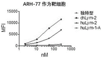
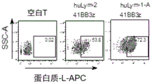


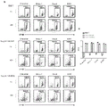


Classifications
-
- C—CHEMISTRY; METALLURGY
- C07—ORGANIC CHEMISTRY
- C07K—PEPTIDES
- C07K16/00—Immunoglobulins [IGs], e.g. monoclonal or polyclonal antibodies
- C07K16/18—Immunoglobulins [IGs], e.g. monoclonal or polyclonal antibodies against material from animals or humans
- C07K16/28—Immunoglobulins [IGs], e.g. monoclonal or polyclonal antibodies against material from animals or humans against receptors, cell surface antigens or cell surface determinants
- C07K16/2803—Immunoglobulins [IGs], e.g. monoclonal or polyclonal antibodies against material from animals or humans against receptors, cell surface antigens or cell surface determinants against the immunoglobulin superfamily
- C07K16/2833—Immunoglobulins [IGs], e.g. monoclonal or polyclonal antibodies against material from animals or humans against receptors, cell surface antigens or cell surface determinants against the immunoglobulin superfamily against MHC-molecules, e.g. HLA-molecules
-
- A—HUMAN NECESSITIES
- A61—MEDICAL OR VETERINARY SCIENCE; HYGIENE
- A61K—PREPARATIONS FOR MEDICAL, DENTAL OR TOILETRY PURPOSES
- A61K39/00—Medicinal preparations containing antigens or antibodies
- A61K39/395—Antibodies; Immunoglobulins; Immune serum, e.g. antilymphocytic serum
- A61K39/39533—Antibodies; Immunoglobulins; Immune serum, e.g. antilymphocytic serum against materials from animals
- A61K39/3955—Antibodies; Immunoglobulins; Immune serum, e.g. antilymphocytic serum against materials from animals against proteinaceous materials, e.g. enzymes, hormones, lymphokines
-
- A—HUMAN NECESSITIES
- A61—MEDICAL OR VETERINARY SCIENCE; HYGIENE
- A61K—PREPARATIONS FOR MEDICAL, DENTAL OR TOILETRY PURPOSES
- A61K39/00—Medicinal preparations containing antigens or antibodies
- A61K39/46—Cellular immunotherapy
- A61K39/461—Cellular immunotherapy characterised by the cell type used
- A61K39/4611—T-cells, e.g. tumor infiltrating lymphocytes [TIL], lymphokine-activated killer cells [LAK] or regulatory T cells [Treg]
-
- A—HUMAN NECESSITIES
- A61—MEDICAL OR VETERINARY SCIENCE; HYGIENE
- A61K—PREPARATIONS FOR MEDICAL, DENTAL OR TOILETRY PURPOSES
- A61K39/00—Medicinal preparations containing antigens or antibodies
- A61K39/46—Cellular immunotherapy
- A61K39/463—Cellular immunotherapy characterised by recombinant expression
- A61K39/4631—Chimeric Antigen Receptors [CAR]
-
- A—HUMAN NECESSITIES
- A61—MEDICAL OR VETERINARY SCIENCE; HYGIENE
- A61K—PREPARATIONS FOR MEDICAL, DENTAL OR TOILETRY PURPOSES
- A61K39/00—Medicinal preparations containing antigens or antibodies
- A61K39/46—Cellular immunotherapy
- A61K39/464—Cellular immunotherapy characterised by the antigen targeted or presented
- A61K39/4643—Vertebrate antigens
- A61K39/4644—Cancer antigens
- A61K39/464402—Receptors, cell surface antigens or cell surface determinants
- A61K39/464411—Immunoglobulin superfamily
- A61K39/464412—CD19 or B4
-
- A—HUMAN NECESSITIES
- A61—MEDICAL OR VETERINARY SCIENCE; HYGIENE
- A61K—PREPARATIONS FOR MEDICAL, DENTAL OR TOILETRY PURPOSES
- A61K45/00—Medicinal preparations containing active ingredients not provided for in groups A61K31/00 - A61K41/00
- A61K45/06—Mixtures of active ingredients without chemical characterisation, e.g. antiphlogistics and cardiaca
-
- A—HUMAN NECESSITIES
- A61—MEDICAL OR VETERINARY SCIENCE; HYGIENE
- A61P—SPECIFIC THERAPEUTIC ACTIVITY OF CHEMICAL COMPOUNDS OR MEDICINAL PREPARATIONS
- A61P35/00—Antineoplastic agents
-
- C—CHEMISTRY; METALLURGY
- C07—ORGANIC CHEMISTRY
- C07K—PEPTIDES
- C07K14/00—Peptides having more than 20 amino acids; Gastrins; Somatostatins; Melanotropins; Derivatives thereof
- C07K14/435—Peptides having more than 20 amino acids; Gastrins; Somatostatins; Melanotropins; Derivatives thereof from animals; from humans
- C07K14/46—Peptides having more than 20 amino acids; Gastrins; Somatostatins; Melanotropins; Derivatives thereof from animals; from humans from vertebrates
- C07K14/47—Peptides having more than 20 amino acids; Gastrins; Somatostatins; Melanotropins; Derivatives thereof from animals; from humans from vertebrates from mammals
-
- C—CHEMISTRY; METALLURGY
- C07—ORGANIC CHEMISTRY
- C07K—PEPTIDES
- C07K14/00—Peptides having more than 20 amino acids; Gastrins; Somatostatins; Melanotropins; Derivatives thereof
- C07K14/435—Peptides having more than 20 amino acids; Gastrins; Somatostatins; Melanotropins; Derivatives thereof from animals; from humans
- C07K14/705—Receptors; Cell surface antigens; Cell surface determinants
- C07K14/70503—Immunoglobulin superfamily
- C07K14/7051—T-cell receptor (TcR)-CD3 complex
-
- C—CHEMISTRY; METALLURGY
- C07—ORGANIC CHEMISTRY
- C07K—PEPTIDES
- C07K14/00—Peptides having more than 20 amino acids; Gastrins; Somatostatins; Melanotropins; Derivatives thereof
- C07K14/435—Peptides having more than 20 amino acids; Gastrins; Somatostatins; Melanotropins; Derivatives thereof from animals; from humans
- C07K14/705—Receptors; Cell surface antigens; Cell surface determinants
- C07K14/70503—Immunoglobulin superfamily
- C07K14/70517—CD8
-
- C—CHEMISTRY; METALLURGY
- C07—ORGANIC CHEMISTRY
- C07K—PEPTIDES
- C07K14/00—Peptides having more than 20 amino acids; Gastrins; Somatostatins; Melanotropins; Derivatives thereof
- C07K14/435—Peptides having more than 20 amino acids; Gastrins; Somatostatins; Melanotropins; Derivatives thereof from animals; from humans
- C07K14/705—Receptors; Cell surface antigens; Cell surface determinants
- C07K14/70578—NGF-receptor/TNF-receptor superfamily, e.g. CD27, CD30, CD40, CD95
-
- C—CHEMISTRY; METALLURGY
- C12—BIOCHEMISTRY; BEER; SPIRITS; WINE; VINEGAR; MICROBIOLOGY; ENZYMOLOGY; MUTATION OR GENETIC ENGINEERING
- C12N—MICROORGANISMS OR ENZYMES; COMPOSITIONS THEREOF; PROPAGATING, PRESERVING, OR MAINTAINING MICROORGANISMS; MUTATION OR GENETIC ENGINEERING; CULTURE MEDIA
- C12N15/00—Mutation or genetic engineering; DNA or RNA concerning genetic engineering, vectors, e.g. plasmids, or their isolation, preparation or purification; Use of hosts therefor
- C12N15/09—Recombinant DNA-technology
- C12N15/11—DNA or RNA fragments; Modified forms thereof; Non-coding nucleic acids having a biological activity
- C12N15/62—DNA sequences coding for fusion proteins
-
- C—CHEMISTRY; METALLURGY
- C12—BIOCHEMISTRY; BEER; SPIRITS; WINE; VINEGAR; MICROBIOLOGY; ENZYMOLOGY; MUTATION OR GENETIC ENGINEERING
- C12N—MICROORGANISMS OR ENZYMES; COMPOSITIONS THEREOF; PROPAGATING, PRESERVING, OR MAINTAINING MICROORGANISMS; MUTATION OR GENETIC ENGINEERING; CULTURE MEDIA
- C12N15/00—Mutation or genetic engineering; DNA or RNA concerning genetic engineering, vectors, e.g. plasmids, or their isolation, preparation or purification; Use of hosts therefor
- C12N15/09—Recombinant DNA-technology
- C12N15/63—Introduction of foreign genetic material using vectors; Vectors; Use of hosts therefor; Regulation of expression
- C12N15/79—Vectors or expression systems specially adapted for eukaryotic hosts
- C12N15/85—Vectors or expression systems specially adapted for eukaryotic hosts for animal cells
- C12N15/86—Viral vectors
-
- C—CHEMISTRY; METALLURGY
- C12—BIOCHEMISTRY; BEER; SPIRITS; WINE; VINEGAR; MICROBIOLOGY; ENZYMOLOGY; MUTATION OR GENETIC ENGINEERING
- C12N—MICROORGANISMS OR ENZYMES; COMPOSITIONS THEREOF; PROPAGATING, PRESERVING, OR MAINTAINING MICROORGANISMS; MUTATION OR GENETIC ENGINEERING; CULTURE MEDIA
- C12N5/00—Undifferentiated human, animal or plant cells, e.g. cell lines; Tissues; Cultivation or maintenance thereof; Culture media therefor
- C12N5/06—Animal cells or tissues; Human cells or tissues
- C12N5/0602—Vertebrate cells
- C12N5/0634—Cells from the blood or the immune system
- C12N5/0636—T lymphocytes
-
- C—CHEMISTRY; METALLURGY
- C12—BIOCHEMISTRY; BEER; SPIRITS; WINE; VINEGAR; MICROBIOLOGY; ENZYMOLOGY; MUTATION OR GENETIC ENGINEERING
- C12N—MICROORGANISMS OR ENZYMES; COMPOSITIONS THEREOF; PROPAGATING, PRESERVING, OR MAINTAINING MICROORGANISMS; MUTATION OR GENETIC ENGINEERING; CULTURE MEDIA
- C12N5/00—Undifferentiated human, animal or plant cells, e.g. cell lines; Tissues; Cultivation or maintenance thereof; Culture media therefor
- C12N5/06—Animal cells or tissues; Human cells or tissues
- C12N5/0602—Vertebrate cells
- C12N5/0634—Cells from the blood or the immune system
- C12N5/0646—Natural killers cells [NK], NKT cells
-
- A—HUMAN NECESSITIES
- A61—MEDICAL OR VETERINARY SCIENCE; HYGIENE
- A61K—PREPARATIONS FOR MEDICAL, DENTAL OR TOILETRY PURPOSES
- A61K39/00—Medicinal preparations containing antigens or antibodies
- A61K2039/505—Medicinal preparations containing antigens or antibodies comprising antibodies
-
- A—HUMAN NECESSITIES
- A61—MEDICAL OR VETERINARY SCIENCE; HYGIENE
- A61K—PREPARATIONS FOR MEDICAL, DENTAL OR TOILETRY PURPOSES
- A61K39/00—Medicinal preparations containing antigens or antibodies
- A61K2039/51—Medicinal preparations containing antigens or antibodies comprising whole cells, viruses or DNA/RNA
- A61K2039/515—Animal cells
- A61K2039/5156—Animal cells expressing foreign proteins
-
- A—HUMAN NECESSITIES
- A61—MEDICAL OR VETERINARY SCIENCE; HYGIENE
- A61K—PREPARATIONS FOR MEDICAL, DENTAL OR TOILETRY PURPOSES
- A61K39/00—Medicinal preparations containing antigens or antibodies
- A61K2039/51—Medicinal preparations containing antigens or antibodies comprising whole cells, viruses or DNA/RNA
- A61K2039/515—Animal cells
- A61K2039/5158—Antigen-pulsed cells, e.g. T-cells
-
- A—HUMAN NECESSITIES
- A61—MEDICAL OR VETERINARY SCIENCE; HYGIENE
- A61K—PREPARATIONS FOR MEDICAL, DENTAL OR TOILETRY PURPOSES
- A61K39/00—Medicinal preparations containing antigens or antibodies
- A61K2039/58—Medicinal preparations containing antigens or antibodies raising an immune response against a target which is not the antigen used for immunisation
- A61K2039/585—Medicinal preparations containing antigens or antibodies raising an immune response against a target which is not the antigen used for immunisation wherein the target is cancer
-
- A—HUMAN NECESSITIES
- A61—MEDICAL OR VETERINARY SCIENCE; HYGIENE
- A61K—PREPARATIONS FOR MEDICAL, DENTAL OR TOILETRY PURPOSES
- A61K2239/00—Indexing codes associated with cellular immunotherapy of group A61K39/46
- A61K2239/10—Indexing codes associated with cellular immunotherapy of group A61K39/46 characterized by the structure of the chimeric antigen receptor [CAR]
- A61K2239/11—Antigen recognition domain
- A61K2239/13—Antibody-based
-
- A—HUMAN NECESSITIES
- A61—MEDICAL OR VETERINARY SCIENCE; HYGIENE
- A61K—PREPARATIONS FOR MEDICAL, DENTAL OR TOILETRY PURPOSES
- A61K2239/00—Indexing codes associated with cellular immunotherapy of group A61K39/46
- A61K2239/31—Indexing codes associated with cellular immunotherapy of group A61K39/46 characterized by the route of administration
-
- A—HUMAN NECESSITIES
- A61—MEDICAL OR VETERINARY SCIENCE; HYGIENE
- A61K—PREPARATIONS FOR MEDICAL, DENTAL OR TOILETRY PURPOSES
- A61K2239/00—Indexing codes associated with cellular immunotherapy of group A61K39/46
- A61K2239/38—Indexing codes associated with cellular immunotherapy of group A61K39/46 characterised by the dose, timing or administration schedule
-
- A—HUMAN NECESSITIES
- A61—MEDICAL OR VETERINARY SCIENCE; HYGIENE
- A61K—PREPARATIONS FOR MEDICAL, DENTAL OR TOILETRY PURPOSES
- A61K2239/00—Indexing codes associated with cellular immunotherapy of group A61K39/46
- A61K2239/46—Indexing codes associated with cellular immunotherapy of group A61K39/46 characterised by the cancer treated
- A61K2239/48—Blood cells, e.g. leukemia or lymphoma
-
- C—CHEMISTRY; METALLURGY
- C07—ORGANIC CHEMISTRY
- C07K—PEPTIDES
- C07K2317/00—Immunoglobulins specific features
- C07K2317/20—Immunoglobulins specific features characterized by taxonomic origin
- C07K2317/24—Immunoglobulins specific features characterized by taxonomic origin containing regions, domains or residues from different species, e.g. chimeric, humanized or veneered
-
- C—CHEMISTRY; METALLURGY
- C07—ORGANIC CHEMISTRY
- C07K—PEPTIDES
- C07K2317/00—Immunoglobulins specific features
- C07K2317/50—Immunoglobulins specific features characterized by immunoglobulin fragments
- C07K2317/51—Complete heavy chain or Fd fragment, i.e. VH + CH1
-
- C—CHEMISTRY; METALLURGY
- C07—ORGANIC CHEMISTRY
- C07K—PEPTIDES
- C07K2317/00—Immunoglobulins specific features
- C07K2317/50—Immunoglobulins specific features characterized by immunoglobulin fragments
- C07K2317/515—Complete light chain, i.e. VL + CL
-
- C—CHEMISTRY; METALLURGY
- C07—ORGANIC CHEMISTRY
- C07K—PEPTIDES
- C07K2317/00—Immunoglobulins specific features
- C07K2317/60—Immunoglobulins specific features characterized by non-natural combinations of immunoglobulin fragments
- C07K2317/62—Immunoglobulins specific features characterized by non-natural combinations of immunoglobulin fragments comprising only variable region components
- C07K2317/622—Single chain antibody (scFv)
-
- C—CHEMISTRY; METALLURGY
- C07—ORGANIC CHEMISTRY
- C07K—PEPTIDES
- C07K2319/00—Fusion polypeptide
- C07K2319/01—Fusion polypeptide containing a localisation/targetting motif
- C07K2319/02—Fusion polypeptide containing a localisation/targetting motif containing a signal sequence
-
- C—CHEMISTRY; METALLURGY
- C07—ORGANIC CHEMISTRY
- C07K—PEPTIDES
- C07K2319/00—Fusion polypeptide
- C07K2319/01—Fusion polypeptide containing a localisation/targetting motif
- C07K2319/03—Fusion polypeptide containing a localisation/targetting motif containing a transmembrane segment
-
- C—CHEMISTRY; METALLURGY
- C07—ORGANIC CHEMISTRY
- C07K—PEPTIDES
- C07K2319/00—Fusion polypeptide
- C07K2319/30—Non-immunoglobulin-derived peptide or protein having an immunoglobulin constant or Fc region, or a fragment thereof, attached thereto
-
- C—CHEMISTRY; METALLURGY
- C07—ORGANIC CHEMISTRY
- C07K—PEPTIDES
- C07K2319/00—Fusion polypeptide
- C07K2319/33—Fusion polypeptide fusions for targeting to specific cell types, e.g. tissue specific targeting, targeting of a bacterial subspecies
-
- C—CHEMISTRY; METALLURGY
- C07—ORGANIC CHEMISTRY
- C07K—PEPTIDES
- C07K2319/00—Fusion polypeptide
- C07K2319/70—Fusion polypeptide containing domain for protein-protein interaction
- C07K2319/74—Fusion polypeptide containing domain for protein-protein interaction containing a fusion for binding to a cell surface receptor
-
- C—CHEMISTRY; METALLURGY
- C12—BIOCHEMISTRY; BEER; SPIRITS; WINE; VINEGAR; MICROBIOLOGY; ENZYMOLOGY; MUTATION OR GENETIC ENGINEERING
- C12N—MICROORGANISMS OR ENZYMES; COMPOSITIONS THEREOF; PROPAGATING, PRESERVING, OR MAINTAINING MICROORGANISMS; MUTATION OR GENETIC ENGINEERING; CULTURE MEDIA
- C12N2510/00—Genetically modified cells
-
- C—CHEMISTRY; METALLURGY
- C12—BIOCHEMISTRY; BEER; SPIRITS; WINE; VINEGAR; MICROBIOLOGY; ENZYMOLOGY; MUTATION OR GENETIC ENGINEERING
- C12N—MICROORGANISMS OR ENZYMES; COMPOSITIONS THEREOF; PROPAGATING, PRESERVING, OR MAINTAINING MICROORGANISMS; MUTATION OR GENETIC ENGINEERING; CULTURE MEDIA
- C12N2740/00—Reverse transcribing RNA viruses
- C12N2740/00011—Details
- C12N2740/10011—Retroviridae
- C12N2740/15011—Lentivirus, not HIV, e.g. FIV, SIV
- C12N2740/15041—Use of virus, viral particle or viral elements as a vector
- C12N2740/15043—Use of virus, viral particle or viral elements as a vector viral genome or elements thereof as genetic vector
-
- C—CHEMISTRY; METALLURGY
- C12—BIOCHEMISTRY; BEER; SPIRITS; WINE; VINEGAR; MICROBIOLOGY; ENZYMOLOGY; MUTATION OR GENETIC ENGINEERING
- C12N—MICROORGANISMS OR ENZYMES; COMPOSITIONS THEREOF; PROPAGATING, PRESERVING, OR MAINTAINING MICROORGANISMS; MUTATION OR GENETIC ENGINEERING; CULTURE MEDIA
- C12N2800/00—Nucleic acids vectors
- C12N2800/10—Plasmid DNA
- C12N2800/106—Plasmid DNA for vertebrates
- C12N2800/107—Plasmid DNA for vertebrates for mammalian
Abstract
Provided herein are novel antibodies and Chimeric Antigen Receptor (CAR) cells comprising the antigen binding domains of these antibodies. Also provided are compositions comprising the antibodies and cells, vectors or plasmids encoding the antibodies and CARs, and methods of producing them or methods of using them to detect or treat cancer, as well as kits for practicing the methods.
Description
Cross Reference to Related Applications
This application claims priority to U.S. provisional application serial nos. 62/806632, 62/815961, and 62/924151, filed on 35 u.s.c. § 119(e), each incorporated herein by reference in its entirety, on days 2 and 15 of 2019, 3 and 8 of 2019, and 21 of 2019 and 10 of 2019, respectively.
Background
The present disclosure relates generally to the field of human immunology, and in particular to cancer immunotherapy.
The following discussion of the background art is intended to aid the reader in understanding the present disclosure and is not an admission that it describes or constitutes prior art against the present disclosure. Throughout the disclosure of this invention, various publications are referenced by Arabic numerals, a full bibliographic citation of which precedes the claims. These references, as well as the cited techniques and patent documents, are incorporated herein by reference.
Lym-1 and Lym-2 are directed against MHC class II HLA-DR molecules known to be expressed predominantly on the surface of human B cells, dendritic cells and B cell derived lymphomas and leukemias.
Murine monoclonal antibodies Lym-1 and Lym-2 (Epstein AL, et AL, 1987, Cancer Res. 47: 830-840) were originally developed for targeting HLA-Dr antigens expressed on the cell surface of most human B-cell malignancies (Rose et AL, 1996, Cancer Immunol Immunother 43(1): 26-30). Lym-1 binding epitopes were then identified, which are discontinuous sites on the human HLA-Dr light chain. Lym-2 does not compete with Lym-1 and therefore binds to a different epitope on the HLA-Dr antigen. Lym-1 has been tested in humans as an I-131 radiolabeled drug and as a naked Antibody for the treatment of diffuse large cell lymphoma and has proven to be effective and safe (DeNardo SJ et al, 1988, antibodies, Immunoconjugates, and Radiopharmaceuticals 1: 17-33; DeNardo, S.J. et al, 1988, int. J. Cancer 3: 96-101; and Hu E. et al, 1989, histological Oncology 7: 155-. Unlike antibodies to other B cell antigens (e.g., CD19 and CD 20), Lym-1, but not Lym-2, undergoes tumor cell apoptosis upon binding due to the antigen being located in lipid rafts on the surface of human lymphoma cells (Epstein AL, et AL, 1987, Cancer Res. 47: 830-840). Furthermore, Lym-1 and Lym-2 antigens are not shed or internalized to any extent and are only expressed on normal circulating and lymph node-bound B cells at concentrations of 1/4 compared to lymphoma cells.
Summary of The Invention
Due to the above properties, Lym-1 and Lym-2 are ideal targeting antibodies useful for the treatment of antigen positive leukemias and lymphomas. Recently, both Lym-1 and Lym-2 have been used to generate potent human CAR T cells as novel compositions of matter for immunotherapy of these malignancies and have been patented. The disclosure shows that the newly generated humanized Lym-1 and Lym-2 have lower affinity for antigen than the parent antibody, can be used to generate clinically effective human CAR T cells to treat B cell malignancies, and are effective against metastatic antigen positive tumors upon NSG mouse xenograft. These data provide key information on the potential use of these humanized antibody constructs in generating human CAR T cells capable of treating B cell tumor patients.
Since recent, unprecedented results have been obtained in the treatment of B-cell lymphomas and leukemias using autologous therapy with genetically engineered Chimeric Antigen Receptor (CAR) T cells, many laboratories have begun to apply this approach to solid tumors. CAR-modified cells combine the HLA-independent targeting specificity of monoclonal antibodies with the cytolytic activity, proliferation and homing properties of activated T cells, but do not respond to checkpoint inhibition. Because they are able to kill antigen-expressing targets directly, CAR cells are highly toxic to any antigen-positive cell or tissue, and it is therefore desirable to construct CARs with highly specific antibodies. In one aspect, disclosed herein are novel antibodies, CARs, and methods of their use in diagnosis and therapy, having desirable safety and efficacy characteristics.
In one aspect, the disclosure provides an antibody comprising, consisting essentially of, or consisting of: a Heavy Chain (HC) immunoglobulin variable domain sequence comprising SEQ ID NO: 2 or 6 or an equivalent of each thereof; and/or a Light Chain (LC) immunoglobulin variable domain sequence comprising SEQ ID NO: 4 or 8 or an equivalent of each thereof. In another aspect, disclosed herein is an antibody comprising, consisting essentially of, or consisting of the sequence of seq id no: comprises the amino acid sequence of SEQ ID NO: 10 or an equivalent thereof; and/or comprises SEQ ID NO: 12 or an equivalent thereof, or a Light Chain (LC) immunoglobulin variable domain sequence.
In one aspect, the antibodies disclosed herein are IgA, IgD, IgE, IgG, or IgM antibodies. The constant region of the antibody may also vary. For example, antibodies having Fc regions of any isotype can be provided: IgA (IgA 1, IgA 2), IgD, IgE, IgG (IgG 1, IgG2, IgG3, IgG 4) or IgM. In one embodiment, the constant region of an antibody disclosed herein is an IgGl constant region or an Ig κ constant region. In a particular aspect, an antibody disclosed herein can further comprise, consist essentially of, or consist of a detectable label or a purification label. Further provided herein is a method of producing an antibody disclosed herein, comprising, consisting essentially of, or consisting of: culturing the isolated cell described above, wherein the isolated cell is optionally a mammalian cell.
Further provided herein are antigen binding fragments of the antibodies disclosed herein. In some aspects of the antibodies provided herein, the antibody fragment is selected from Fab, F (ab) '2, Fab', scFv, and Fv. In another aspect, provided herein are isolated antibodies or fragments thereof as disclosed herein and a detectable or purification label, alone or in combination with an antigen or fragment thereof. Further provided herein are ex vivo (ex vivo) cells comprising, consisting essentially of, or consisting of the antigen/antibody complexes.
The disclosure provides polypeptides comprising SEQ ID NO: 2. 4, 6, 8, 10 or 12 or an equivalent of each thereof, or consists essentially of, or consists of. Further provided is an isolated polypeptide comprising, consisting essentially of, or consisting of the amino acid sequence of an antibody disclosed herein or a fragment thereof and an isolated polynucleotide encoding the same.
Also further provided are isolated nucleic acids encoding the antibodies and fragments thereof disclosed herein. In one aspect, the isolated nucleic acid sequence comprises SEQ ID NO: 1. 3, 5, 7, 9 and 11 or their respective equivalents, or consist essentially of, or consist of. In one aspect, the polynucleotide sequence is operably linked to a promoter and/or enhancer element. They may also be used in combination with a vector or a suitable host cell and/or a suitable vector for diagnostic or therapeutic use. In one aspect, the nucleic acid is contained in a host cell for recombinant production of polypeptides and proteins. The host cell may be a eukaryotic cell or a prokaryotic cell.
Aspects disclosed herein relate to a Chimeric Antigen Receptor (CAR) comprising, consisting essentially of, or consisting of: (a) an antigen binding domain of any of the antibodies disclosed herein, (b) a hinge domain, (c) a transmembrane domain, and (d) an intracellular signaling domain. In one aspect, the CAR further comprises, consists essentially of, or consists of one or more co-stimulatory signaling regions. In another aspect, a CAR disclosed herein is a first generation CAR, a second generation CAR, a third generation CAR, or a fourth generation CAR.
Also provided herein is a Chimeric Antigen Receptor (CAR) comprising, consisting essentially of, or consisting of the sequence of seq id no: (a) an antigen binding domain (e.g., of an anti-Lym antibody), (b) a hinge domain, (c) a transmembrane domain, and (d) a DAP 10 and/or DAP 12 domain. In another aspect, the CAR further comprises the 10 amino acid epitope "AVPPQQWALS" inserted directly after at least the antigen binding domain. The antigen binding domain may be from any suitable species, such as mammalian, murine, rat or e.g. human.
The disclosure herein provides a CAR construct comprising, or consisting essentially of, or consisting of: (a) an antigen binding domain of an antibody (e.g., an anti-Lym antibody, a humanized anti-Lym antibody, or an anti-CD 19 antibody), (b) a hinge domain, (c) a transmembrane domain, and (d) an intracellular signaling domain comprising one or more transmembrane domains of DAP10 and/or DAP 12. Non-limiting examples of antibodies include anti-Lym-1, anti-huLym-1 or Lym2, anti-Lym 2 antibodies, or anti-CD 19 antibodies, which may be from any suitable species. The construct may further comprise, consist essentially of, or consist of one or more linker polypeptides linking the above elements (a) to (d). In another aspect, the CAR further comprises, consists essentially of, or consists of a leader peptide located at the amine terminus of the antigen binding domain. In one embodiment, the hinge domain is a CD8 a or IgG1 hinge domain. In another aspect, the transmembrane domain comprises a CD8 a transmembrane domain. In another aspect, the CAR further comprises the 10 amino acid epitope "AVPPQQWALS" inserted directly after at least the antigen binding domain. The antigen binding domain may be from any suitable species, such as mammalian, murine, rat or e.g. human.
In particular embodiments, the CAR comprises, or consists essentially of, or consists of the following sequence: the antigen binding domain of any of the antibodies or fragments thereof disclosed herein (e.g., scFv), the CD8 a hinge domain or the IgG1 hinge domain, the CD8 a transmembrane domain, and the DAP 10 and/or DAP 12 transmembrane domain. In another aspect, the CAR includes DAP 10 and DAP 12 transmembrane domains.
In a particular aspect, the antibodies or CARs of the present disclosure may further comprise, consist essentially of, or consist of a detectable label or a purification label.
In another embodiment, the disclosed CAR comprises, or consists essentially of, or consists of the following sequence: a CD8 a or IgGl hinge domain, the transmembrane domain comprising a CD28 or CD8 a transmembrane domain, the one or more co-stimulatory signaling domains being selected from the group consisting of CD27, CD28, 4-IBB (CD 137), OX40, CD30, CD40, PD-1, ICOS, lymphocyte function-associated antigen-1 (LFA-1), CD2, CD7, CD27, LIGHT, NKG2C, and B7-H3; and the intracellular signaling domain comprises a CD3 zeta signaling domain.
Also provided herein is a method of making a cell expressing a CAR (e.g., an anti-Lym CAR or anti-CD 19), comprising, consisting essentially of, or consisting of the steps of: introducing a nucleic acid sequence encoding a CAR disclosed herein into a population of immune cells; and (ii) selecting a subpopulation of immune cells that have been successfully transduced with the nucleic acid sequence of step (i), thereby generating cells expressing the CAR.
Aspects of the disclosure relate to an isolated cell comprising, consisting essentially of, or consisting of a CAR of the disclosure, and methods of producing such cells.
Also provided herein is a vector comprising, consisting essentially of, or consisting of an isolated nucleic acid sequence encoding an antibody or CAR disclosed herein. In one aspect, the disclosure provides a vector comprising, consisting essentially of, or consisting of an isolated nucleic acid sequence encoding the antibody or CAR construct.
In another aspect, the present disclosure provides a composition comprising, consisting essentially of, or consisting of a carrier (carrier) and one or more of an antibody, antigen-binding fragment, polypeptide, CAR, isolated nucleic acid, vector (vector), and/or isolated cell disclosed herein.
Other method aspects disclosed herein relate to a method of inhibiting tumor growth and/or treating cancer and/or preventing recurrence of cancer in a subject in need thereof, comprising, consisting essentially of, or consisting of: administering to the subject an effective amount of a CAR (e.g., anti-Lym or CD 19) expressing cell provided herein, an effective amount of an antibody, an effective amount of an antigen binding fragment thereof, and/or an effective amount of a polypeptide provided herein. In one aspect, the CAR expresses an antigen binding domain directed against an antigen expressed on a cancer or tumor cell to be treated or inhibited. In one aspect, the CAR-expressing cells are autologous or allogeneic to the subject, and optionally are for first-line, second-line, third-line, fourth-line, or fifth-line therapy. In another aspect, the tumor or cancer cell expresses or overexpresses a CAR-binding antigen, e.g., CD19, Lym1, and/or Lym 2. In another aspect, the cancer or tumor is selected from a carcinoma, sarcoma, or leukemia. In a particular aspect, the tumor or cancer is a B cell lymphoma or leukemia. In one embodiment, the tumor is a solid tumor. The solid tumor may be melanoma, colon cancer, breast cancer and/or brain tumor. In one aspect, the cancer treated is a carcinoma, sarcoma, neuroblastoma, cervical cancer, hepatocellular carcinoma, mesothelioma, glioblastoma, myeloma, lymphoma, leukemia, adenoma, adenocarcinoma, glioma, glioblastoma, retinoblastoma, astrocytoma, oligodendroglioma, meningioma, or melanoma.
The methods can be used to treat subjects such as humans, non-human primates (e.g., apes, gibbons, chimpanzees, orangutans, monkeys, macaques, etc.), farm animals (e.g., dogs and cats), farm animals (e.g., horses, cows, goats, sheep, pigs), and laboratory animals (e.g., mice, rats, rabbits, guinea pigs). The mammal may be of any age or at any stage of development (e.g., an adult, adolescent, child, infant or intrauterine mammal). The mammal may be male or female. In certain embodiments, the subject has or is suspected of having a neoplastic disease, neoplasia, tumor, malignancy, or cancer.
The methods disclosed herein may further comprise, consist essentially of, or consist of the steps of: administering to the subject an anti-tumor therapy other than CAR therapy.
The disclosure also relates to methods for inhibiting proliferation of a cancer cell or cancer stem cell, comprising, consisting essentially of, or consisting of: contacting the cell with an effective amount of a CAR-expressing cell, an effective amount of an antibody, an effective amount of an antigen-binding fragment, and/or an effective amount of a polypeptide disclosed herein. The CAR may be an anti-Lym-1, anti-Lym-2, or anti-CD 19-expressed CAR.
Further provided herein are methods for determining whether a subject is likely to respond or is unlikely to respond to treatment, comprising, consisting essentially of, or consisting of: contacting a sample isolated from a patient with an antibody, antigen-binding fragment, and/or polypeptide disclosed herein, and detecting the antibody-cell complex, antigen-binding fragment-cell complex, and/or polypeptide-cell complex in the sample, wherein the presence of the complex indicates that the subject is likely to respond to treatment and the absence of the complex indicates that the subject is unlikely to respond to treatment. The antibody, antigen-binding fragment, and/or polypeptide may be detectably labeled. Also further disclosed herein is a method comprising, consisting essentially of, or consisting of: administering an effective amount of an antibody or CAR disclosed herein to a subject determined to be likely to respond to treatment.
Further disclosed herein are methods directed to monitoring therapy for a subject, comprising, consisting essentially of, or consisting of: contacting a sample isolated from the subject with an antibody or antigen-binding fragment disclosed herein, and detecting the antibody-cell complex in the sample. The method can be performed before and/or after administering an effective amount of a CAR-expressing cell, an effective amount of an antibody, an effective amount of an antigen-binding fragment, and/or an effective amount of a polypeptide disclosed herein to the subject. The CAR may be an anti-Lym-1, anti-Lym-2, or anti-CD 19 expressing CAR. In one aspect, the CAR, antibody, or antigen-binding fragment thereof is detectably labeled. In another aspect, the sample comprises one or more of sputum, serum, plasma, lymph, cyst fluid, urine, feces, cerebrospinal fluid, ascites, blood, or tissue.
Also provided herein are kits for practicing the methods disclosed herein and instructions for practicing the methods disclosed herein. The kit comprises, consists essentially of, or consists of: one or more of the antibodies, CARs, antigen-binding fragments, polypeptides, isolated nucleic acids, vectors, isolated cells, and/or compositions disclosed herein and instructions for use.
Drawings
FIG. 1: the two humanized Lym-1 antibodies have different binding capacities for epitope positive cells. Will be 5X 10⁵Individual Raji cells were incubated with the indicated antibodies at concentrations increasing from 0.013 to 1300 nM. After detection with (AF 488) conjugated goat anti-human IgG, Mean Fluorescence Intensity (MFI) was quantified by flow cytometry.
FIG. 2: successful production of CAR T cells. On day 10, CAR T cell preparations were labeled with biotin-anti-Lym-1 antibody and then detected with APC-streptavidin. Blank T cells were used as negative controls.
FIGS. 3A-3C: both humanized Lym-1 CAR-T based cells induced complete remission in Raji xenografts. One million Raji/Luc-eGFP cells were injected intravenously into 8-10 week old male NSG mice (day 0). Luciferase activity was measured on day 6 to assess tumor burden prior to treatment. (figure 3A) ten million blank and CAR T cells (50% CAR positive) in 100 μ Ι PBS were injected intravenously into a randomly generated group of tumor-bearing mice (n = 5) on day 7. (fig. 3B) mean bioluminescent radiation from the dorsal and ventral images was quantified and summed. (FIG. 3C) Kaplan-Meier plot shows the survival rate of mice from control and experimental groups.
FIG. 4: production of a functional humanized Lym-1 antibody. Will be 5X 10⁵Individual ARH-77 cells were incubated with the indicated antibodies at concentrations increasing from 2.218 to 266 nM. After detection with (AF 488) conjugated goat anti-human IgG, flow cytometry was performedQuantity average fluorescence intensity (MFI).
FIG. 5: successful production of huLym-1 and huLym-2 CAR-T cells. On day 10, CAR T cell preparations were labeled with biotin-protein-L and then detected with APC-streptavidin. Blank T cells were used as negative controls.
FIG. 6: huLym-2CAR showed increased cytotoxicity against epitope positive cell lines. Blank or CAR T cells were incubated with ARH-77 eGFP/Luc at an effector/target (E/T) ratio of 2 to 0.25. Cytotoxicity was calculated from luciferase activity 17 hours after co-cultivation.
FIG. 7: schematic representation of 41BB3z and DAP10-12 based CAR T cell constructs.
FIG. 8: successful production of CAR-T cells. On day 10, CAR T cell preparations were labeled with biotin-anti-Lym-1 antibody and then detected with APC-streptavidin. Blank T cells were used as negative controls.
FIGS. 9A-9B: stable gene expression and cell proliferation of huLym-1-B-DAP transduced CAR T cells. Primary human T cells were activated on day 0 with anti-CD 3/CD28 magnetic beads (Dynabeads) and transduced with each construct on day 3. 100 ten thousand T cells in each group were restimulated with anti-CD 3/CD28 on days 7 and 14. (FIG. 9A) the absolute cell numbers in the blank and CAR-T cell preparations were counted on days 4, 7, 8 and 12. (FIG. 9B) CAR expression was measured on days 7, 14, and 21.
FIG. 10: epitope-driven cytotoxicity against Raji cells. Blank or CAR T cells were incubated with Raji-eGFP/Luc at an effector/target (E/T) ratio of 2 to 0.25. Cytotoxicity was measured according to luciferase activity 17 hours after co-cultivation.
FIGS. 11A-11D: dorsal bioluminescence imaging of tumor burden and weight changes in blank and CAR treated mice. One million Raji/Luc-eGFP cells were injected intravenously into 8-10 week old male NSG mice (day 0). Luciferase activity was measured on day 7 to assess tumor burden prior to treatment. On day 8, 100 ten thousand blanks in 100 μ Ι PBS and CAR T cells were injected intravenously into a randomly generated tumor-bearing group of mice (n = 5). (fig. 11A) example bioluminescent images. (FIG. 11B) Kaplan-Meier survival images of mice. (fig. 11C) quantification and summation of mean bioluminescent radiation from dorsal and ventral images. (FIG. 11D) weight change images of treated mice over time. The body weight of each mouse on day 0 was used as a reference for calculating the percent change in body weight. The body weight of the mice was measured twice a week with a digital scale.
FIG. 12: experiments showing DAP CD19 CAR T cells for the treatment of disseminated Raji lymphoma in NSG mice. Mice were treated with 100 million CAR T cells of huLym-1 DAP, CD19 second generation, or CD19 DAP 8 days after intravenous injection of Raji. Mice were then imaged weekly by bioluminescence and the results showed that all CD19 CAR T cells with the second generation construct died, but those receiving the CD19 DAP CAR construct remained viable at 21 days, although at this dose they still had tumors.
FIGS. 13A-13C: both Lym-1 and huLym-1-B CAR, having the intracellular domain of 4-1BB3z, can eliminate Raji tumors in vivo. (FIG. 13A) schematic of in vivo study schedule. (FIG. 13B) inoculation 10 on day 06Raji-eGFP/Luc, then 5X 10 at day 66Bioluminescence images of NSG mice treated with individual naive T cells, Lym-1-BB3z CAR T cells, huLym-1-B-BB3z CAR T cells, or 100. mu.l PBS. Bioluminescent signals from dorsal and ventral images of each NSG mouse were added and plotted against time. (n =5 mice/group, data not shown). (FIG. 13C) Kaplan-Meier survival Curve (.)p<0.001 by log rank test).
FIGS. 14A-14F: effect of intracellular signaling domain on huLym-1-B CAR T cell expansion. (figure 14A) in vitro expansion of blank or CAR positive T cells after transduction and restimulation (summary results from five donors). (FIG. 14B) CAR expression on T cell preparations at the days indicated. T cells were reactivated with stimulators of alpha-CD 2/CD3/CD 28-based antibodies on days 7 and 14. (figure 14C) in vitro expansion of blank or CAR positive T cells after transduction and restimulation (summary results from 3 donors). (FIG. 14D) CAR expression on T cell preparations at the days indicated. T cells were reactivated with alpha-CD 2/CD3/CD28 stimulator on days 7 and 14 (representative results from 3 donors). (figure 14E) in vitro expansion of blank or CAR positive T cells after transduction and restimulation (summary results from 3 donors). (FIG. 14F) CAR expression on T cell preparations at the days indicated. T cells were reactivated with α -CD2/CD3/CD28 stimulators on days 7 and 14 (representative results from five donors).
FIGS. 15A-15B: ligand-dependent robust signaling is the major mechanism of impaired T cell proliferation of huLym-1-B-BB3z CAR. (fig. 15A) annexin v (annexin v) and dead cell cyanine (Sytox-Green) staining (representative results from two donors) of blank or CAR transduced T cells from day 9 were evaluated. (FIG. 15B) CD19-BB3zCAR T cells were pre-labeled with Cell trace far-red dye (Cell trace face-red dye) and then co-cultured overnight with blank or indicated CAR T cells at a ratio of 1: 1. CD19-BB3zCAR T cells were then stained with annexin V (annexin V) and cyanine Green (Sytox-Green) to measure potential self-killing of huLym-1-B-BB3z or huLym-1-B-DAP CAR T cells (representative results from two donors).
FIGS. 16A-16B: huLym-1-B-DAP CAR T cells showed activation-induced reduction in cell death. Blank, huLym-1-B-BB3z and huLym-1-B-DAP CAR T cells were cultured in the presence of a-CD2/3/28 stimulus, or overnight with K562 cells or Raji cells. Blank and CAR T cells were then stained for annexin V and green cyanine (dead cell dye). (FIG. 16A) representative plot from one of three donors. (FIG. 16B) the percentage of annexin V positive cells in live cells was quantified by the following formula: Q3/(Q3 + Q4) (from three donors, P <0.05 by Student's t test bar shows mean ± SD).
FIGS. 17A-17C: huLym-1-B-DAP exhibits effector function against a panel of B lymphoma and leukemia cell lines. (FIG. 17A) cytokines released from blank, huLym-1-B-DAP and huLym-1-B-BB3z when co-cultured overnight with a panel of human lymphoma B cell lines at an effector to target ratio of 1: 1. Quantification of secreted cytokines was normalized to pg/10000 cells (GM-CSF) or ng/10000 cells (IL-2 and INF- γ) (n =3 technical replicates representative results from two donors). (FIG. 17B) to measure the cytotoxicity of blank, huLym-1-B-DAP and huLym-1-B-BB3z, tumor cells were pre-labeled with Cell trace far-red dye (Cell trace face-red dye) and then co-cultured with effector cells at a ratio of 1: 1; live tumor cells were gated at each time point. (FIG. 17C) the percentage of viable tumor cells was quantified and plotted. The percentage lysis was calculated as (% tumor at 1 hour [% tumor at 48 hours)/(% tumor at 1 hour) (n =3 technical replicates and representative results from two donors are shown.
FIGS. 18A-18B: the Lym-1 epitope was not significantly down-regulated in the presence of huLym-1-B-DAP CAR T cells. Blanks T, huLym-1-B-DAP and CD19-BB3z were co-cultured with a panel of B lymphoma and leukemia cell lines at a 1:2 ratio. After overnight co-culture, the B cell lines were labeled with antibodies against CD22, Lym-1, and CD 19.
FIGS. 19A-19D: the CAR signal domain does not affect antigen regulation. (FIG. 19A) blank T, CD19-BB3z, CD19-DAP, huLym-1-B-BB3z and huLym-1-B-DAP CAR T cells were co-cultured overnight with Raji-eGFP/Luc cells at a ratio of approximately 1: 2. CD19 antigen and Lym-1 epitope expression in residual live Raji cells was then measured by flow cytometry via fluorophore-conjugated monoclonal antibodies. (FIG. 19B) scattergrams of Mean Fluorescence Intensity (MFI) of Lym-1 epitope and CD19 antigen expression in residual Raji cells. Percentage of live Raji cells (fig. 19C) and concentration (fig. 19D) after overnight co-culture (n =3 technical replicates from two donors representative results).
FIGS. 20A-20F: huLym-1-B-DAP CAR mediated tumor-free survival at lower doses. (FIG. 20A) blank and CD19-DAP T cells were co-cultured overnight with Raji-eGFP/Luc cells at a ratio of 1: 2. The monoclonal antibody CD19 antigen conjugated by the fluorophore and the Lym-1 epitope expression were then measured by flow cytometry (representative results from three replicates). (FIG. 20B) schematic representation of in vivo studies. (FIG. 20C) 10 on day 06Each Raji-eGFP/Luc was inoculated, followed by a blank, huLym-1-B-DAP, CD19-BB3z or CD19-DAP at the indicated dose on day 8 Bioluminescence image of CAR T cell treated NSG mice (n =5 mice per group). (fig. 20D) Kaplan-Meier survival curve (ns ═ log rank (Mantel-Cox) test not significant). (FIG. 20E) use 10 on day 06The individual Raji-eGFP/Luc were inoculated and then inoculated with 10 on day 86Bioluminescence images of blank, CD19-BB3z or CD19-DAP CAR T cell treated NSG mice (n =5 mice/group). This experiment was performed side by side with the experiment in fig. 25E, using the same blank T control group. (FIG. 20F) Kaplan-Meier survival Curve (examined by Log-rank (Mantel-Cox))P=0.002)。
FIGS. 21A-21C: huLym-1-B was selected as a candidate for CAR development. (FIG. 21A) binding ability of a panel of 12 humanized Lym-1 antibodies to Raji cells. All antibodies were expressed as human IgG1 isotype. huLym-1-60 has the same sequence as chLym-1. hu51 is an internally produced human IgG1 isotype control. Raji cells were incubated at the indicated concentrations for 30 min and then detected with AF-488 conjugated goat anti-human IgG. Mean Fluorescence Intensity (MFI) was assessed by flow cytometry to compare the differential binding of the antibodies. (FIG. 21B) the binding capacity of the internally produced huLym-1-B and chimeric Lym-1 (chLym-1) on Raji cells was evaluated by flow cytometry. The secondary antibody used here was an APC-conjugated mouse anti-human IgG monoclonal antibody. (FIG. 21C) binding of chLym-1 and huLym-1-B to a panel of chLym-1 positive and negative cell lines. For these measurements, APC-conjugated mouse anti-human IgG monoclonal antibodies were used as secondary antibodies.
FIGS. 22A-22H: lym-1 and huLym-1-B CAR T cells with the BB3z-CD3z signaling domain are cytotoxic in culture despite impaired proliferation. (FIG. 22A) schematic representation of CAR construct. The 261 tag is a 10 amino acid linear epitope derived from the schedule of human placental growth factor (fig. 22B) CAR-T production and amplification. (figure 22C) flow cytometry analysis of CAR expression on blank or transduced primary human T cells at day 7; CAR expression was measured against 261 tag by Dylight 650 conjugated antibody. (FIG. 22D) cytotoxicity of CAR T cells at the Effector-to-target (E: T) ratios shown for Lym-1 epitope negative (K562) and positive (Raji) cell lines(ii) a Day 9 CAR T cells were used to measure cytotoxicity. No restimulation was performed on day 7 of the experiment (n = 3 technical replicates representing results from 3 donors). (FIG. 22E) fold expansion of in vitro cultured blank or CAR positive T cells from day 7-14 (summary results from 6 donors. ns-not significant. by Student's T testP<0.001). (FIG. 22F) shows day 9 CD3𝜁Representative graphs of phosphorylation (no stimulation on day 7; n ═ 3 donors) (figure 22G) representative CAR and inhibitory receptor expression on day 7 and 14 before restimulation. (FIG. 22H) quantification of the percentage of inhibitory receptors on blank or CAR-positive T cells from 3 donors (. about. P<0.05, Student's t test. Scatter plots and bar plots show mean ± SD).
FIGS. 23A-23G: impaired proliferation of huLym-1-B-BB3zCAR T cells is associated with a weakly expressed Lym-1 epitope on T cells and is represented by ITAM CD3𝜻And (4) mediating. (figure 23A) introducing two amino acid mutations in CDR3 of huLym-1-B variable heavy chain to generate huLym-1-Bmut antibody or huLym-1-Bmut-BB3 zCAR; CD3 in CAR𝜻Tyrosine (Y) was mutated to phenylalanine (F) in part of all three ITAMs to abolish huLym-1-B-BB3z activity (huLym-1-BBB 3 zY-F). (FIG. 23B) binding ability of chLym-1, huLym-1-B and huLym-1-Bmut to Raji cells. The secondary antibody used here was goat anti-human IgG conjugated with AF-488; ED50 is labeled on each curve. (FIG. 23C) use 10𝜇g/100𝜇l 50 ten thousand activated T cells were stained with the indicated antibodies and then detected with APC-anti-huIgG. Calculating and plotting average fluorescence intensity; scatter plots show the median MFI. Each color point represents a donor. (FIG. 23D) fold expansion of blank T, huLym-1-BBB3zY-F and huLym-1-Bmut-BB3z CAR-positive T cells cultured in vitro from day 7 to day 14 (summary results from 3 donors ns: Student's T test showed no significance, scatter plot shows mean. + -. SD). (FIG. 23E) shows day 9 CD3 𝜁Representative plots of phosphorylation; CAR expression was detected by Dylight 650-coupled anti 261-tag antibody (n = 2 donors). (FIG. 23F) representative plots of CAR and PD-1 and LAG-3 expression at days 7 and 14. (FIG. 23G) quantification of percentage PD-1 and LAG-3 in blank or CAR-positive T cellsPooled from three donors (.)P<0.05 Student’s tAnd (6) checking. Bar graph shows mean ± SD).
FIGS. 24A-24G: the substitution of the DAP signal for BB3z solved the problem of proliferation of huLym-1-B CAR T cells. (FIG. 24A) the left panel illustrates the consensus Amino Acids (AA) for ITIM and ITAM, and the right panel shows DAP12 and CD3𝜁AA of ITAM; shading emphasizes the presence of ITIM consensus sequences in ITAMs of DAP 12. (FIG. 24B) schematic representation of huLym-1-B CAR with BB3z or DAP as intracellular signaling domain. (FIG. 24C) in vitro expansion of blank, huLym-1-B-DAP and huLym-1-B-BB3z CAR positive T cells from days 7-14 (summary results from 5 donors. ns: Student's T test not significant). (FIG. 24D) shows day 9 CD3𝜁Phosphorylation (p-CD 3)𝜁) Representative of (iii) (representation of three donors). (figure 24E) p-CD3 in blank and CAR positive T cells (n =3 donors)𝜁And (4) quantifying. (figure 24F) representative CAR and inhibitory receptor expression at day 7 and 14 (n =3 donors;, x) P<0.05, Student's t test). (FIG. 24G) quantification of the percentage of inhibitory receptors in blank and CAR-positive T cells (from three donors; ns: not significant;. X)P<0.05 by Student's t test. Scatter plots and bar plots show mean ± SD).
FIGS. 25A-25F: huLym-1-B-DAP CAR T cells were superior to their BB3z counterpart in mediated antitumor efficacy in vivo. (FIG. 25A) cytotoxicity of blank, huLym-1-B-DAP or huLym-1-B-BB3z on Lym-1 positive (Raji, left panel) or negative (K562, right panel) cells. (FIG. 25B) cytokine release from blank, huLym-1-B-DAP or huLym-1-B-BB3z when co-cultured with K562 or Raji at an effector to target ratio of 1: 1. The quantification of the secreted cytokines was normalized to pg/10000 cells (GM-CSF) or ng/10000 cells (IL-2 and INF-𝛾) (n =3 technical repeats. Representative results were from three donors). (FIG. 25C) schematic of in vivo study with modification. (FIG. 25D, FIG. 25E) NSG mice were vaccinated 10 on day 06Raji-eGFP/Luc, then 1X 10 at day 86Blank, huLym-1-B-DAP or huLym-1-B-BB3z CAR T cell treated bioluminescent images (n =5 mice per group) . Two independent studies from different donors. (FIG. 25F) Kaplan-Meier survival Curve (P=0.000325, by log rank (Mantel-Cox) test. Pooled from two independent studies. Bar graph shows mean ± SD).
FIGS. 26A-26D: the Lym-1 epitope was not down-regulated in response to huLym-1-B-DAP CAR T cells. (FIG. 26A) blank T, CD19-BB3z and huLym-1-B-DAP CAR T cells were co-cultured overnight with Raji-eGFP/Luc cells at a ratio of 1:2, and then CD19 antigen and Lym-1 epitope expression were measured by flow cytometry using fluorophore-conjugated monoclonal antibodies. (representative from three replicate experiments). (FIG. 26B) scattergrams of Mean Fluorescence Intensity (MFI) of Lym-1 epitope and CD19 antigen expression on Raji (n =3 technical replicates). (FIG. 26C) NSG mice were vaccinated 10 on day 06Raji-eGFP/Luc, then 1X 10 at day 86Individual naive, CD19-BB3z or huLym-1-B-DAP CAR T cells were treated. On day 16, bone marrow samples were collected and Lym-1 epitope and CD19 antigen expression were assessed by flow cytometry using fluorophore conjugated monoclonal antibodies. (FIG. 26D) Mean Fluorescence Intensity (MFI) of Lym-1 epitope and scattergrams of CD19 antigen expression on Raji. (n = 5 mice per group;,) P<0.001; ns: the Kruskal-Wallis test was not significant. Scatter plots show mean ± SD).
Detailed Description
It is to be understood that the invention disclosed is not limited to the particular aspects described, as such may, of course, vary. It is also to be understood that the terminology used herein is for the purpose of describing particular aspects only, and is not intended to be limiting, since the scope of the present disclosure will be limited only by the appended claims. Furthermore, throughout and within the present disclosure, various technical and patent publications are cited by reference or arabic numerals, the entire citation of which can be found before the claims. The disclosures of these publications are incorporated herein by reference to further describe the prior art.
Unless defined otherwise, all technical and scientific terms used herein have the same meaning as commonly understood by one of ordinary skill in the art to which this technology belongs. Although any methods and materials similar or equivalent to those described herein can be used in the practice or testing of the present technology, the preferred methods, devices, and materials are now described. All technical and patent publications cited herein are incorporated by reference in their entirety. Nothing herein is to be construed as an admission that the technology is not entitled to antedate such disclosure by virtue of prior invention.
The practice of the present technology will employ, unless otherwise indicated, conventional techniques of tissue culture, immunology, molecular biology, microbiology, cell biology, and recombinant DNA, which are within the skill of the art. See, e.g., Sambrook and Russell eds. (2001) Molecular Cloning, A Laboratory Manual, 3rd edition; (2007) Current Protocols in Molecular Biology series; methods in Enzymology (Academic Press, Inc., N.Y.); MacPherson et al, (1991) PCR 1: A Practical Approach (IRL Press at Oxford University Press); MacPherson et al, (1995) PCR 2: A Practical Approach; harlow and Lane eds. (1999) Antibodies, A Laboratory Manual; freekney (2005) Culture of Animal Cells A Manual of Basic Technique, 5th edition; gait ed. (1984) Oligonucleotide Synthesis; U.S. patent nos. 4683195; hames and Higgins eds. (1984) Nucleic Acid Hybridization; anderson (1999) Nucleic Acid Hybridization; hames and Higgins eds. (1984) transformation and transformation; immobilized Cells and Enzymes (IRL Press (1986)); perbal (1984) A Practical Guide to Molecular Cloning; miller and Calos eds. (1987) Gene Transfer Vectors for Mammalian Cells (Cold Spring Harbor Laboratory); makrides ed. (2003) Gene Transfer and Expression in Mammarian Cells; mayer and Walker eds. (1987) biochemical Methods in Cell and Molecular Biology (Academic Press, London); and Herzenberg et al, eds (1996) Weir's Handbook of Experimental Immunology.
All numerical designations, such as pH, temperature, time, concentration, and molecular weight, including ranges, are approximate and vary (+) or (-), or vary +/-15%, or 10%, or 5%, or 2%, as the case may be, in increments of 1.0 or 0.1. It is to be understood that all numerical designations are preceded by the term "about," although this is not always explicitly stated. It is also to be understood that, although not always explicitly stated, the reagents described herein are exemplary only and equivalents thereof are known in the art.
Without explicit recitation and unless otherwise expressly inferred, when the present technology relates to polypeptides, proteins, polynucleotides or antibodies, their equivalents or biological equivalents are intended to be within the scope of the present technology.
Definition of
As used in the specification and in the claims, the singular form of "a", "an", and "the" include the plural forms unless the context clearly dictates otherwise. For example, the term "a cell" includes a plurality of cells, including mixtures thereof.
As used herein, the term "animal" refers to living multicellular vertebrate organisms, which category includes, for example, mammals and birds. The term "mammal" includes both human and non-human mammals.
The terms "subject", "host", "individual" and "patient" are used interchangeably herein to refer to an animal, typically a mammal. Any suitable mammal can be treated by the methods, cells, or compositions described herein. Non-limiting examples of mammals include humans, non-human primates (e.g., apes, gibbons, chimpanzees, orangutans, monkeys, macaques, etc.), farm animals (e.g., dogs and cats), farm animals (e.g., horses, cows, goats, sheep, pigs), and laboratory animals (e.g., mice, rats, rabbits, guinea pigs). In some embodiments, the mammal is a human. The mammal can be of any age or at any stage of development (e.g., an adult, adolescent, child, infant, or intrauterine mammal). The mammal may be male or female. The mammal may be a pregnant female. In some embodiments, the subject is a human. In some embodiments, the subject has or is suspected of having a cancer or a neoplastic disorder.
"eukaryotic cells" include all life kingdoms except prokaryotes. They can be easily distinguished by membrane bound cores. Animals, plants, fungi and protists are eukaryotes or organisms whose cells are organized into complex structures by the inner membrane and cytoskeleton. The most characteristic membrane-bound structure is the nucleus. Unless otherwise specified, the term "host" includes eukaryotic hosts, including, for example, yeast, higher plant, insect, and mammalian cells. Non-limiting examples of eukaryotic cells or hosts include apes, bovines, porcines, murines, rats, avians, reptiles, and humans.
"prokaryotic cells" generally have no nucleus or any other membrane-bound organelle and are divided into two areas, bacteria and archaea. In addition to chromosomal DNA, these cells may also contain genetic information in a circular loop called episome. The bacterial cells are very small, approximately corresponding to the size of the animal mitochondria (about 1-2 μm in diameter, 10 μm long). Prokaryotic cells have three main shapes: rod-like, spherical and spiral. Bacterial cells divide not by a complex replication process as eukaryotes, but by binary fission. Examples include, but are not limited to, Bacillus, E.coli and Salmonella.
As used herein, the term "antibody" is collectively referred to as an immunoglobulin or immunoglobulin-like molecule, including, for example and without limitation, IgA, IgD, IgE, IgG, and IgM, combinations thereof, and like molecules produced during immunization of any vertebrate (e.g., mammalian, such as human, goat, rabbit, and mouse, and non-mammalian species), such as shark immunoglobulin. Unless specifically stated otherwise, the term "antibody" includes intact immunoglobulins and "antibody fragments" or "antigen-binding fragments" that specifically bind to a molecule of interest (or a group of highly similar molecules of interest) while substantially excluding binding to other molecules (e.g., the binding constant for the molecule of interest is at least 10 greater than the binding constant for other molecules in other biological samples 3 M-1At least 10 greater4 M-1Or at least 10 greater5 M-1Antibodies and antibody fragments of (a). The term "antibody" also includes genetically engineered forms, e.g., chimericA conjugated antibody (e.g., a humanized murine antibody), a heteroconjugate antibody (e.g., a bispecific antibody). See also Pierce Catalog and Handbook (1994-1995) (Pierce Chemical Co., Rockford, Ill.); Kuby, J. (1997) Immunology, 3rd Ed., W.H. Freeman;)&Co., New York. An "antigen-binding fragment" of an antibody is a portion of an antibody that retains the ability to specifically bind to the target antigen of the antibody.
In terms of antibody structure, immunoglobulins have a heavy (H) chain and a light (L) chain interconnected by disulfide bonds. There are two types of light chains, λ and κ. There are five major heavy chain classes (or isotypes) that determine the functional activity of an antibody molecule: IgM, IgD, IgG, IgA, and IgE. Each of the heavy and light chains comprises a constant region and a variable region (these regions are also referred to as "domains"). The constant region of the antibody may also vary. For example, antibodies having Fc regions of any isotype can be provided: IgA (IgA 1, IgA 2), IgD, IgE, IgG (IgG 1, IgG2, IgG3, IgG 4) or IgM. Non-limiting examples of constant region sequences include: SEQ ID No: 19-27 or their respective equivalents.
In combination, the heavy chain variable region and the light chain variable region specifically bind to an antigen. The light and heavy chain variable regions comprise "framework" regions interrupted by three hypervariable regions, also known as "complementarity determining regions" or "CDRs". The extent of the framework regions and CDRs has been defined (see Kabat et al, Sequences of Proteins of Immunological Interest, U.S. Department of Health and Human Services, 1991, incorporated herein by reference). The Kabat database is now maintained online. The framework region sequences of different light or heavy chains are relatively conserved within a species. The framework regions of the antibody, i.e., the combined framework regions of the component light and heavy chains, adopt predominantly a β -sheet conformation with the CDRs forming loops connecting, and in some cases forming part of, the β -sheet structure. Thus, the framework regions serve to form a scaffold that positions the CDRs in the correct orientation through inter-chain, non-covalent interactions. Non-limiting examples of the Lym1 and Lym2 CDRs are provided in U.S. application No. 15/173,534.
The CDRs are primarily responsible for binding epitopes of the antigen. CDR Per chainCommonly referred to as CDR1, CDR2, and CDR3, are numbered sequentially from the N-terminus and are also typically identified by the chain in which the particular CDR is located (the heavy chain region labeled CDHR and the light chain region labeled CDLR). Thus, CDHR3 is the CDR3 from the antibody heavy chain variable domain in which it is located, while CDLR1 is the CDR1 from the antibody light chain variable domain in which it is located. The TNT antibody will have a specific V characteristic of the TNT associated antigen HRegion and VLThe region sequence, and thus has a particular CDR sequence. Antibodies with different specificities (i.e., different binding sites for different antigens) have different CDRs. Although CDRs vary from antibody to antibody, only a limited number of amino acid positions in a CDR are directly involved in antigen binding. These positions in the CDRs are called Specificity Determining Residues (SDRs).
The term "humanized", when used with respect to an antibody, refers to an antibody having an amino acid sequence with one or more non-human amino acid residues (e.g., mouse, rat, goat, rabbit, etc.) that specifically bind to the Complementarity Determining Regions (CDRs) of a desired antigen in the recipient human immunoglobulin molecule, and one or more human amino acid residues in the Fv Framework Region (FR), which are the amino acid residues flanking the CDRs. Such antibodies typically have reduced immunogenicity and thus a longer half-life in humans compared to the non-human parent antibody from which the one or more CDRs are derived or based.
As used herein, the term "antigen" refers to a compound, composition, or substance that can be specifically bound by a product of a particular humoral or cellular immunity (e.g., an antibody molecule or T cell receptor). The antigen may be any type of molecule, including, for example, haptens, simple intermediate metabolites, sugars (e.g., oligosaccharides), lipids, and hormones, as well as macromolecules, such as complex carbohydrates (e.g., polysaccharides), phospholipids, and proteins. Common classes of antigens include, but are not limited to, viral antigens, bacterial antigens, fungal antigens, protozoan and other parasitic antigens, tumor antigens, antigens involved in autoimmune diseases, allergies and transplant rejections, toxins, and other miscellaneous antigens.
As used herein, the term "antigen binding domain" refers to any protein or polypeptide domain that can specifically bind to an antigen target.
As used herein, the term "chimeric antigen receptor" (CAR) refers to a fusion protein comprising an extracellular domain capable of binding an antigen, a transmembrane domain derived from a polypeptide different from that from which the extracellular domain is derived, and at least one intracellular domain. "Chimeric Antigen Receptors (CARs)" are sometimes referred to as "chimeric receptors", "T-bodies", or "Chimeric Immunoreceptors (CIRs)". "extracellular domain capable of binding an antigen" refers to any oligopeptide or polypeptide that can bind to a particular antigen. By "intracellular domain" is meant any oligopeptide or polypeptide known to be used as a signaling activation or inhibition domain to cause a biological process in a cell. In certain embodiments, the intracellular domain may comprise, consist essentially of, or further comprise one or more costimulatory signaling domains in addition to the primary signaling domain. "transmembrane domain" refers to any oligopeptide or polypeptide known to span the cell membrane and may function to link the extracellular and signaling domains. The chimeric antigen receptor may optionally comprise a "hinge domain" that serves as a linker between the extracellular domain and the transmembrane domain. Non-limiting examples of such domains are provided herein, such as:
Hinge domain: IgGl heavy chain hinge sequence:
CTCGAGCCCAAATCTTGTGACAAAACTCACACATGCCCACCGTGCCCG
transmembrane domain: CD28 transmembrane region:
TTTTGGGTGCTGGTGGTGGTTGGTGGAGTCCTGGCTTGCTATAGCTTGCTAGTAACAGTGGCCTTTATTATTTTCTGGGTG
intracellular domain: 4-1BB co-stimulatory signaling region:
AAACGGGGCAGAAAGAAACTCCTGTATATATTCAAACAACCATTTATGAGACCAGTACAAACTACTCAAGAGGAAGATGGCTGTAGCTGCCGATTTCCAGAAGAAGAAGAAGGAGGATGTGAACTG
intracellular domain: CD28 costimulatory signaling regions:
AGGAGTAAGAGGAGCAGGCTCCTGCACAGTGACTACATGAACATGACTCCCCGCCGCCCCGGGCCCACCCGCAAGCATTACCAGCCCTATGCCCCACCACGCGACTTCGCAGCCTATCGCTCC
intracellular domain: CD3 zeta signaling region:
AGAGTGAAGTTCAGCAGGAGCGCAGACGCCCCCGCGTACCAGCAGGGCCAGAACCAGCTCTATAACGAGCTCAATCTAGGACGAAGAGAGGAGTACGATGTTTTGGACAAGAGACGTGGCCGGGACCCTGAGATGGGGGGAAAGCCGAGAAGGAAGAACCCTCAGGAAGGCCTGTACAATGAACTGCAGAAAGATAAGATGGCGGAGGCCTACAGTGAGATTGGGATGAAAGGCGAGCGCCGGAGGGGCAAGGGGCACGATGGCCTTTACCAGGGTCTCAGTACAGCCACCAAGGACACCTACGACGCCCTTCACATGCAGGCCCTGCCCCCTCGCTAA。
further embodiments of each exemplary domain component include other proteins having similar biological functions, which have at least 70% or at least 80% amino acid sequence identity, preferably 90% sequence identity, more preferably at least 95% sequence identity to the proteins encoded by the nucleic acid sequences disclosed above. Further, provided herein are non-limiting examples of such domains.
As used herein, a "first generation CAR" refers to a CAR that comprises an extracellular domain capable of binding an antigen, a transmembrane domain derived from a polypeptide different from that from which the extracellular domain is derived, and at least one intracellular domain. "second generation CAR" refers to a first generation CAR that further comprises a co-stimulatory domain (e.g., 4-1BB or CD 28). "third generation CAR" refers to a first generation CAR further comprising two co-stimulatory domains (e.g., CD27, CD28, ICOS, 4-1BB, or OX 40). "fourth generation CAR" (also referred to as "TRUCK") refers to CAR T cells that have been further engineered to secrete additional factors, such as the pro-inflammatory cytokine IL-12. An overview of these CAR techniques and cell therapies can be found in Maus, m.et al. clin. Cancer res. 22(3): 1875-84 (2016).
As used herein, the term "signal peptide" or "signal polypeptide" means an amino acid sequence that is typically present at the N-terminus of a newly synthesized secreted or membrane polypeptide or protein. Its function is to direct the polypeptide across or into the cell membrane, which is then removed. Examples of such are well known in the art. Non-limiting examples are described in U.S. patent nos. 8,853,381 and 5,958,736.
As used herein, with respect to regulatory polynucleotides, the term "operably linked" refers to the association between a regulatory polynucleotide and its linked polynucleotide sequence such that when a particular protein binds to the regulatory polynucleotide, the linked polynucleotide is transcribed.
By "composition" is generally meant a combination of an active agent (e.g., CAR T cells or CAR NK cells), an antibody, a compound, and a naturally-occurring or non-naturally-occurring carrier that is inert (to, e.g., a detectable agent or label) or active, e.g., an adjuvant, diluent, binder, stabilizer, buffer, salt, lipophilic solvent, preservative, adjuvant, etc., and includes a pharmaceutically acceptable carrier. The carrier also includes pharmaceutical excipients and additives proteins, peptides, amino acids, lipids and carbohydrates (e.g., sugars, including mono-, di-, tri-, tetra-and oligosaccharides; derivatized sugars, such as sugar alcohols, aldonic acids, esterified sugars and the like; and polysaccharides or sugar polymers), which may be present alone or in combination, comprising 1-99.99% by weight or volume. Exemplary protein excipients include serum albumin, such as human serum albumin (HAS), recombinant human albumin (rHA), gelatin, casein, and the like. Representative amino acid/antibody components may also serve to buffer potency and include alanine, arginine, glycine, arginine, betaine, histidine, glutamic acid, aspartic acid, cysteine, lysine, leucine, isoleucine, valine, methionine, phenylalanine, aspartame, and the like. Carbohydrate excipients are also within the scope of the present technology, examples of which include, but are not limited to: monosaccharides such as fructose, maltose, galactose, glucose, D-mannose, sorbose and the like; disaccharides, such as lactose, sucrose, trehalose, cellobiose, and the like; polysaccharides such as raffinose, melezitose, maltodextrin, dextran, starch, and the like; and sugar alcohols such as mannitol, xylitol, maltitol, lactitol, xylitol sorbitol (glucitol), and inositol.
Compositions, including cells, therapeutics, medicaments, pharmaceuticals and pharmaceutical preparations for use in accordance with the present disclosure can be packaged in dosage unit form for ease of administration and uniformity of dosage. The term "unit dose" or "dose" refers to physically discrete units suitable for use in a subject, each unit containing a predetermined quantity of a composition calculated to produce the desired response associated with its administration, i.e., the appropriate route and regimen. Depending on the number of treatments and the unit dose, the amount administered will depend on the desired outcome and/or protection. The precise amount of the composition also depends on the judgment of the practitioner and will vary from one individual to another. Factors that affect dosage include the physical and clinical state of the subject, the route of administration, the intended goal of treatment (symptomatic relief and cure), and the efficacy, stability and toxicity of the particular composition. Upon formulation, the solution will be administered in a manner compatible with the dosage formulation and in an amount effective for treatment or prevention. The formulations are readily administered in a variety of dosage forms, such as the injectable solution types described herein.
As used herein, the term "consensus sequence" refers to an amino acid or nucleic acid sequence determined by aligning a series of multiple sequences, which defines an idealized sequence that represents a primary selection of amino acids or bases at each corresponding position in the multiple sequences. Depending on the sequence of the multiple sequence series, the consensus sequence of the series may differ from each sequence by zero, one, several or more substitutions. In addition, based on the sequence of a multiple sequence series, more than one consensus sequence can be determined for the series. The generation of consensus sequences has been subjected to extensive mathematical analysis. Various software programs can be used to determine consensus sequences.
As used herein, the term "CD 8 a hinge domain" refers to the specific protein fragment to which this name relates and any other molecule with similar biological function that has at least 70% or at least 80% amino acid sequence identity, or at least 90% sequence identity, or at least 95% sequence identity to the CD8 a hinge domain sequence as set forth herein. Exemplary sequences for the CD8 α hinge domain of human, mouse and other species are provided in Pinto, R.D. et al (2006) vet. immunol. immunopathol. 110: 169-. Sequences related to the hinge domain of CD8 α are provided in Pinto, R.D. et al (2006) vet. immunol. 110: 169-177. Non-limiting examples of this include:
a human CD8 a hinge domain;
PAKPTTTPAPRPPTPAPTIASQPLSLRPEACRPAAGGAVHTRGLDFACDIY
mouse CD8 a hinge domain;
KVNSTTTKPVLRTPSPVHPTGTSQPQRPEDCRPRGSVKGTGLDFACDIY
a cat CD8 a hinge domain;
PVKPTTTPAPRPPTQAPITTSQRVSLRPGTCQPSAGSTVEASGLDLSCDIY, and equivalents thereof.
As used herein, the term "CD 8 a transmembrane domain" refers to the specific protein fragment associated with that name and any other molecule with similar biological function that has at least 70% or at least 80% amino acid sequence identity, or at least 90% sequence identity, or at least 95% sequence identity to the CD8 a transmembrane domain sequence as set forth herein. Further exemplary sequences of the CD8 a transmembrane domain are provided by sequences related to amino acid positions 183 to 203 of the CD8 a chain of human T cell surface glycoprotein (NCBI reference: NP-001759.3), or amino acid positions 197 to 217 of the CD8 a chain of mouse T cell surface glycoprotein (NCBI reference: NP-001074579.1) and fragments related to amino acid positions 190 to 210 of the CD8 a chain of rat T cell surface glycoprotein (NCBI reference: NP-113726.1). The sequences associated with each NCBI listed are provided below:
Human CD8 a transmembrane domain: IYIWAPLAGTCGVLLLSLVIT, respectively;
mouse CD8 a transmembrane domain: IWAPLAGICVALLLSLIITLI, respectively;
rat CD8 a transmembrane domain: IWAPLAGICAVLLLSLVITLI, and equivalents thereof.
As used herein, the term "CD 28 transmembrane domain" refers to the specific protein fragment associated with that name and any other molecule with similar biological function that has at least 70% or at least 80% amino acid sequence identity, or at least 90% sequence identity, or at least 95% sequence identity to the CD28 transmembrane domain sequence as set forth herein. The fragment sequences associated with GenBank Accession Nos XM-006712862.2 and XM-009444056.1 provide additional, non-limiting, exemplary sequences of the transmembrane domain of CD 28. The sequences associated with each accession number listed are incorporated herein.
As used herein, the term "4-1 BB co-stimulatory signal region" refers to both the specific protein fragment associated with that name and any other molecule with similar biological function that has at least 70% or at least 80% amino acid sequence identity, or at least 90% sequence identity, or at least 95% sequence identity to the 4-1BB co-stimulatory signal region sequence as shown herein. An exemplary sequence of the 4-1BB co-stimulatory signal region is provided in U.S. patent application publication No. 2013/0266551A 1 (filed as U.S. application No. 13/826258). The sequence of the 4-1BB co-stimulatory signal region disclosed in U.S. application No. 13/826258 is disclosed as follows:
4-1BB co-stimulatory signaling region:
KRGRKKLLYIFKQPFMRPVQTTQEEDGCSCRFPEEEEGGCEL, and their respective equivalents.
As used herein, the term "CD 28 costimulatory signaling region" refers to the specific protein fragment associated with this name and any other molecule with similar biological function that has at least 70% or at least 80% amino acid sequence identity, or at least 90% sequence identity, or at least 95% sequence identity to the CD28 costimulatory signaling region sequence as set forth herein. The CD28 costimulatory signaling region includes a transmembrane domain and an intracellular domain. Exemplary sequences of CD28 costimulatory signaling regions are provided in U.S. patent nos. 5686281; geiger, T.L. et al (2001) Blood 98: 2364-; hombach, A. et al. (2001) J Immunol. 167: 6123-; maher, J. et al (2002) Nat Biotechnol. 20: 70-75; haynes, N.M. et al. (2002) J Immunol.169: 5780-5786; haynes, N.M. et al. (2002) Blood 100: 3155-3163. Non-limiting examples include residues 114-220 of the following CD28 sequence:
MLRLLLALNL FPSIQVTGNK ILVKQSPMLV AYDNAVNLSC KYSYNLFSRE FRASLHKGLDSAVEVCVVYG NYSQQLQVYS KTGFNCDGKL GNESVTFYLQ NLYVNQTDIY FCKIEVMYPPPYLDNEKSNG TIIHVKGKHL CPSPLFPGPS KPFWVLVVVG GVLACYSLLVTVAFIIFWVR SKRSRLLHSD YMNMTPRRPG PTRKHYQPYA PPRDFAAYRS, and equivalents thereof.
As used herein, the term "ICOS co-stimulatory signaling region" refers to both the specific protein fragment associated with that name and any other molecule with similar biological function that has at least 70% or at least 80% amino acid sequence identity, or at least 90% sequence identity, or at least 95% sequence identity to the ICOS co-stimulatory signaling region sequence as set forth herein. Non-limiting exemplary sequences of ICOS costimulatory signaling regions are provided in U.S. publication 2015/0017141a1, and exemplary polynucleotide sequences are provided below.
ICOS co-stimulatory signaling region:
ACAAAAAAGAAGTATTCATCCAGTGTGCACGACCCTAACGGTGAATACATGTTCATGAGAGCAGTGAACACAGCCAAAAAATCCAGACTCACAGATGTGACCCTA。
as used herein, the term "OX 40 co-stimulatory signaling region" refers to specific protein fragments associated with that name and any other molecule with similar biological function that has at least 70% or at least 80% amino acid sequence identity, or at least 90% sequence identity, or at least 95% sequence identity to the OX40 co-stimulatory signaling region sequence as shown herein. Non-limiting exemplary sequences of the OX40 co-stimulatory signal region are disclosed in U.S. publication 2012/20148552A1 and include the exemplary sequences provided below.
OX40 costimulatory signaling regions:
AGGGACCAG AGGCTGCCCC CCGATGCCCA CAAGCCCCCT GGGGGAGGCA GTTTCCGGAC CCCCATCCAA GAGGAGCAGG CCGACGCCCA CTCCACCCTG GCCAAGATC。
as used herein, the term "CD 3 zeta signaling domain" refers to the specific protein fragment associated with that name and any other molecule with similar biological function that has at least 70% or at least 80% amino acid sequence identity, or at least 90% sequence identity, or at least 95% sequence identity to the CD3 zeta signaling domain sequence as set forth herein. Exemplary sequences of CD3 zeta signaling domains are provided in U.S. application No. 13/826,258 (published as US 2013/0266551). The sequences associated with the CD3 ζ signal domain are listed below:
RVKFSRSADAPAYQQGQNQLYNELNLGRREEYDVLDKRRGRDPEMGGKPRRKNPQEGLYNELQKDKMAEAYSEIGMKGERRRGKGHDGLYQGLSTATKDTYDALHMQALPPR, and equivalents thereof.
As used herein, the terms "T2A" and "2A peptide" are used interchangeably to refer to any 2A peptide or fragment thereof, any 2A-like peptide or fragment thereof, or an artificial peptide comprising essential amino acids in a relatively short peptide sequence (about 20 amino acids in length, depending on viral origin) containing the consensus polypeptide motif D-V/I-E-X-N-P-G-P, where X refers to any amino acid generally recognized as self-cleaving.
As used herein, the term "suicide gene" is a gene capable of inducing apoptosis; non-limiting examples include HSV-TK (herpes simplex virus thymidine kinase), cytosine deaminase, nitroreductase, carboxylesterase, cytochrome P450 or PNP (purine nucleoside phosphorylase), truncated EGFR or inducible caspase ("iCasp"). Suicide genes may act in a variety of ways and, in some cases, may be induced by an inducing agent (e.g., a small molecule). For example, the iCasp suicide gene comprises a portion of a caspase protein operably linked to a protein optimized for binding to an inducer; introduction of an inducer into cells containing a suicide gene leads to activation of caspases and subsequent apoptosis of the cells.
As used herein, the term "switching mechanism for controlling expression and/or activation of a CAR" refers to extracellular, transmembrane and intracellular domains, wherein the extracellular domain comprises a target-specific binding element comprising a tag, binding domain or label specific for a molecule other than a target antigen expressed on or by a target cell. The specificity of the CAR is provided by a second construct comprising a target antigen binding domain and a domain that is recognized or bound by a label, binding domain or tag on the CAR. See, for example, WO 2013/044225, WO 2016/000304, WO 2015/057834, WO 2015/057852, WO 2016/070061, US 9,233,125, US 2016/0129109. In this way, a T cell expressing a CAR can be administered to a subject, but is unable to bind its target antigen prior to administration of a second composition comprising a specific binding domain.
As used herein, the term "B cell" refers to a type of lymphocyte in the humoral immunity of the adaptive immune system. The main functions of B cells are to make antibodies, act as antigen presenting cells, release cytokines, and generate memory B cells upon activation of antigen interactions. B cells differ from other lymphocytes (e.g., T cells) in the presence of B cell receptors on the cell surface. B cells can be isolated or obtained from commercial sources. Non-limiting examples of commercially available B cell lines include AHH-1 (ATCC CRL-8146 ™), BC-1 (ATCC CRL-2230;), BC-2 (ATCC CRL-2231;), BC-3 (ATCC CRL-2277;), CA46 (ATCC CRL-1648;), DG-75 [ D.G. -75] (ATCC CRL-2625;), DS-1 (ATCC CRL-11102;), EB-3 [ EB3] (ATCC CCL-85;), z-138 (ATCC # CRL-3001), DB (ATCC CRL-2289), Toledo (ATCC CRL-2631), Pfeffer (ATCC CRL-2632), SR (ATCC CRL-2262), JM-1 (ATCC CRL-10421), NFS-5C-1 (ATCC CRL-1693); NFS-70C 10 (ATCC CRL-1694), NFS-25C-3 (ATCC CRL-1695), AND SUP-B15 (ATCC CRL-1929). Other examples include, but are not limited to, cell lines derived from anaplastic and large cell lymphomas, such as DEL, DL-40, FE-PD, JB6, Karpas 299, Ki-JK, Mac-2A Ply1, SR-786, SU-DHL-1, -2, -4, -5, -6, -7, -8, -9, -10, and-16, DOHH-2, NU-DHL-1, U-937, Granda 519, USC-DHL-1, RL; hodgkin's lymphomas such as DEV, HD-70, HDLM-2, HD-MyZ, HKB-1, KM-H2, L428, L540, L1236, SBH-1, SUP-HD1, SU/RH-HD-L. Non-limiting exemplary sources of such commercially available Cell lines include the American Type Culture Collection or ATCC (www.atcc.org /) and the German Collection of Microorganisms and Cell Cultures (https:// www.dsmz.de /).
As used herein, the term "T cell" refers to a lymphocyte that matures in the thymus. T cells play an important role in cell-mediated immunity and are distinguished from other lymphocytes (e.g., B cells) by the presence of T cell receptors on the cell surface. T cells may be isolated or obtained from commercial sources. "T cells" include all types of immune cells expressing CD3, including T helper cells (CD 4+ cells), cytotoxic T cells (CD 8+ cells), natural killer T cells, T regulatory cells (tregs), and γ δ T cells. "cytotoxic cells" include CD8+ T cells, Natural Killer (NK) cells, and neutrophils, which are capable of mediating a cytotoxic response. Non-limiting examples of commercially available T cell lines include BCL2 (AAA) Jurkat (ATCC ^ CRL-2902 ^), BCL2 (S70A) Jurkat (ATCC ^ CRL-2900 ^), BCL2 (S87A) Jurkat (ATCC ^ CRL-2901 ^), BCL2 Jurkat (ATCC ^ CRL-2899), Neo Jurkat (ATCC ^ CRL-2898), TALL-104 cytotoxic human T cell lines (ATCC # CRL-11386). Further examples include, but are not limited to, mature T cell lines such as Deglis, EBT-8, HPB-MLp-W, HUT78, HUT102, Karpas 384, Ki 225, My-La, Se-Ax, SKW-3, SMZ-1, and T34; and immature T cell lines such as ALL-SIL, Be13, CCRF-CEM, CML-T1, DND-41, DU.528, EU-9, HD-Mar, HPB-ALL, H-SB2, HT-1, JK-T1, Jurkat, Karpas 45, KE-37, KOPT-K1, K-T1, L-KAW, Loucy, MAT, MOLT-1, MOLT 3, MOLT-4, MOLT 13, MOLT-16, MT-1, MT-ALL, P12/Ichikawa, Peer, PER0117, PER-255, PF-382, PFI-285, RPMI-8402, ST-4, SUP-T1 to T14, TALL-1, TALL-101, L-103/2, L-104, TK-105, TALL-106, TALL-197, TALL-6-L-197, TALL-6-TK, TLBR-1, -2, -3, and-4, CCRF-HSB-2 (CCL-120.1), J.RT3-T3.5 (ATCC TIB-153), J45.01 (ATCC CRL-1990), J.CaM1.6 (ATCC CRL-2063), RS 4; 11 (ATCC CRL-1873), CCRF-CEM (ATCC CRM-CCL-119); and cutaneous T cell lymphoma lines such as HuT78 (ATCC CRM-TIB-161), MJ [ G11] (ATCC CRL-8294), HuT102 (ATCC TIB-162). Null leukemia cell lines (Null leukemia cell lines), including but not limited to REH, NALL-1, KM-3, L92-221, are another commercially available source of immune cells, as are cell lines derived from other leukemias and lymphomas, such as K562 erythroleukemia, THP-1 monocytic leukemia, U937 lymphoma, HEL erythroleukemia, HL60 leukemia, HMC-1 leukemia, KG-1 leukemia, U266 myeloma. Non-limiting exemplary sources of such commercially available cell lines include the American type culture Collection or ATCC (http:// www.atcc.org /) and the German Collection of microorganisms and cell cultures (https:// www.dsmz. de /).
As used herein, the term "NK cell," also known as a natural killer cell, refers to a type of lymphocyte that originates in the bone marrow and plays a key role in the innate immune system. NK cells provide a rapid immune response against virus-infected cells, tumor cells, or other stressed cells, even in the absence of antibodies and major histocompatibility complexes on the cell surface. NK cells can be isolated or obtained from commercial sources. Non-limiting examples of commercial NK cell lines include NK-92 (ATCC ® CRL-2407 cells), NK-92MI (ATCC ® CRL-2408 cells). Further examples include, but are not limited to, the NK lines HANK1, KHYG-1, NKL, NK-YS, NOI-90 and YT. Non-limiting exemplary sources of such commercially available cell lines include the American type culture Collection or ATCC (http:// www.atcc.org /) and the German Collection of microorganisms and cell cultures (https:// www.dsmz. de /).
As used herein, the term "CRISPR" refers to a technique of sequence-specific genetic manipulation that relies on clustered regularly interspaced short palindromic repeats pathways. CRISPRs can be used to perform gene editing and/or gene regulation, as well as simply target proteins to specific genomic locations. "Gene editing" refers to a genetic engineering in which the nucleotide sequence of a polynucleotide of interest is altered by introducing deletions, insertions, single-or double-strand breaks or base substitutions into the polynucleotide sequence.
In certain aspects, CRISPR-mediated gene editing utilizes non-homologous end joining (NHEJ) or homologous recombination pathways for editing. Gene regulation refers to increasing or decreasing the production of a particular gene product (e.g., a protein or RNA).
As used herein, the term "gRNA" or "guide RNA" refers to a guide RNA sequence used to target a particular polynucleotide sequence for gene editing using CRISPR techniques. Techniques for designing grnas and donor therapeutic polynucleotides specifically for a target are well known in the art. For example, Doench, j., et al, Nature biotechnology 2014; 32(12) 1262-7, Mohr, S. et al. (2016) FEBS Journal 283: 3232-38 and Graham, D.et al Genome biol. 2015, 16: 260. The gRNA comprises a fusion polynucleotide comprising CRISPR RNA (crRNA) and transactivation CRISPR RNA (tracrRNA); or a polynucleotide comprising, consisting essentially of, or consisting of CRISPR RNA (crRNA) and trans-activating CRIPSPR RNA (tracrRNA). In certain aspects, the gRNA is synthetic (Kelley, m. et al. (2016) J of Biotechnology 233 (2016) 74-83).
The term "Cas 9" refers to the CRISPR-associated endonuclease referred to by this name. Non-limiting exemplary Cas9 include staphylococcus aureus Cas9, nuclease-dead Cas9, and their respective orthologs and biological equivalents. Orthologs include, but are not limited to, streptococcus pyogenes Cas9 ("spCas 9"), Cas9 from streptococcus thermophilus, legionella pneumophila, Neisseria lactamide (Neisseria lactamica), Neisseria meningitidis, Francisella novii (Francisella novicida); and Cpf1 from various bacterial species including acidamine coccus and francisella foeniculi U112 (performing cleavage functions similar to Cas 9).
As used herein, the terms "nucleic acid sequence" and "polynucleotide" are used interchangeably to refer to a polymeric form of nucleotides of any length, either ribonucleotides or deoxyribonucleotides. Thus, the term includes, but is not limited to, single-, double-, or multi-stranded DNA or RNA, genomic DNA, cDNA, DNA-RNA hybrids, or other naturally, chemically, or biochemically modified, non-natural, or derivatized nucleotide bases comprising purine and pyrimidine bases.
The term "encoding" when applied to a nucleic acid sequence means that a polynucleotide referred to as "encoding" a polypeptide, if in its native state or when manipulated by methods well known to those skilled in the art, is transcribed and/or translated to produce mRNA of the polypeptide and/or fragments thereof. The antisense strand is the complementary strand of such a nucleic acid, from which the coding sequence can be deduced.
As used herein, the term signal peptide or signal polypeptide means an amino acid sequence that is typically present at the N-terminus of a newly synthesized secretory or membrane polypeptide or protein. Its function is to direct the polypeptide across or into the cell membrane, which is then removed. Examples of such are well known in the art. Non-limiting examples are those described in U.S. patent nos. 8853381 and 5958736.
As used herein, the term "vector" refers to a nucleic acid construct for transfer between different hosts, including but not limited to plasmids, viruses, cosmids, phages, BAC, YAC, and the like. "viral vector" is defined as a recombinantly produced virus or viral particle comprising a polynucleotide to be delivered to a host cell in vivo, ex vivo or in vitro. In some embodiments, the plasmid vector may be a commercially available vector. In other embodiments, the viral vector may be generated from baculovirus, retrovirus, adenovirus, AAV, and the like, according to techniques known in the art. In one embodiment, the viral vector is a lentiviral vector. Examples of viral vectors include retroviral vectors, adenoviral vectors, adeno-associated viral vectors, alphaviral vectors and the like. Infectious Tobacco Mosaic Virus (TMV) based vectors are useful for the production of proteins, and it has been reported that Griffithsin (O' Keefe et al (2009) Proc. nat. Acad. Sci. USA 106(15): 6099-. Alphavirus vectors, such as those based on the Semliki Forest virus and those based on the Sindbis virus, have also been developed for gene therapy and immunotherapy. See Schlesinger & Dubensky (1999) curr. Opin. Biotechnol. 5: 434. about.439 and Ying et al (1999) nat. Med. 5(7): 823. about.827. In terms of gene transfer mediated by a retroviral vector, a vector construct refers to a polynucleotide (e.g., a polynucleotide encoding a CAR) comprising the retroviral genome or portion thereof and a gene of interest. More details on modern vector methods for Gene transfer can be found, for example, in Kotterman et al (2015) visual Vectors for Gene Therapy: Translational and Clinical Outlook Annual Review of biological Engineering 17. Vectors comprising a promoter and a cloning site to which a polynucleotide is operably linked are well known in the art. Such vectors are capable of transcribing RNA in vitro or in vivo, and are commercially available from sources such as Agilent Technologies (Santa Clara, Calif.) and Promega Biotech (Madison, Wis.).
As used herein, the term "isolated cell" generally refers to a cell that is substantially separated from other cells of a tissue. The term includes prokaryotic and eukaryotic cells.
"immune cells" include, for example, leukocytes (leukocytes), lymphocytes (T cells, B cells, Natural Killer (NK) cells), and myeloid-derived cells (neutrophils, eosinophils, basophils, monocytes, macrophages, dendritic cells) derived from Hematopoietic Stem Cells (HSCs) produced in the bone marrow. "T cells" include all types of immune cells expressing CD3, including T helper cells (CD 4+ cells), cytotoxic T cells (CD 8+ cells), natural killer T cells, T regulatory cells (tregs), and γ - δ T cells. "cytotoxic cells" include CD8+ T cells, Natural Killer (NK) cells, and neutrophils, which are capable of mediating a cytotoxic response.
As used herein, the phrase "immune response" or its equivalent "immunological response" refers to the development of a cell-mediated response (e.g., mediated by antigen-specific T cells or their secretory products). The cellular immune response is triggered by presentation of polypeptide epitopes associated with class I or class II MHC molecules to treat or prevent viral infection, expand antigen-specific B-reg cells, TC1, CD4+ T helper and/or CD8+ cytotoxic T cells and/or disease producing, autoregulating T cells and B cell "memory" cells. The reaction may also involve activation of other components. In certain aspects, the term "immune response" may be used to encompass the formation of an immune cell regulatory network. Thus, the term "regulatory network formation" may refer to an immune response elicited such that immune cells, preferably T cells, more preferably T regulatory cells, trigger further differentiation of other immune cells, such as, but not limited to, B cells or antigen presenting cells-non-limiting examples of which include dendritic cells, monocytes, and macrophages. In certain embodiments, regulatory network formation involves differentiation of B cells into regulatory B cells; in certain embodiments, the formation of the regulatory network involves the formation of tolerogenic antigen-presenting cells.
The term "transduction" or "transduction" refers to the process of introducing an exogenous nucleotide sequence into a cell when used to generate a chimeric antigen receptor cell. In some embodiments, this transduction is accomplished by a vector.
As used herein, the term "autologous" with respect to a cell refers to a cell that is isolated and returned to the same subject (recipient or host). "allogeneic" refers to non-autologous cells.
An "effective amount" or "effective amount" refers to an agent (e.g., an HLA-DR CAR cell) or a combined amount of two or more agents, which when administered to treat a mammal or other subject, is sufficient to effect treatment for such a disease. The "effective amount" will vary depending on the agent, disease and its severity and age, weight, etc. of the subject to be treated.
As used herein, "cancer" is a disease state characterized by the presence in a subject of cells that exhibit abnormal uncontrolled replication, and may be used interchangeably with the term "tumor".
By "solid tumor" is meant an abnormal tissue mass that generally does not contain cysts or fluid areas. Solid tumors can be benign or malignant. Different types of solid tumors are named for the cell types that form them. Examples of solid tumors include sarcomas, carcinomas, and lymphomas.
The term "B-cell lymphoma or leukemia" refers to a cancer that forms and undergoes malignant transformation in a problem with the lymphatic system or bone marrow, which allows cells within the pathological cancer to have the ability for the host organism to invade or spread to other parts of the body.
As used herein, the term "comprising" is intended to mean both compositions and methods that include the recited elements, but not to exclude other elements. When used to define compositions and methods, "consisting essentially of … …" is meant to exclude other elements that may have any significance to the combination for the intended use. For example, a composition consisting essentially of the elements defined herein will not exclude trace contaminants from the isolation and purification process and pharmaceutically acceptable carriers (e.g., phosphate buffered saline, preservatives, etc.). "consisting of … …" refers to other ingredients excluding more than trace elements and the basic method steps for administering the compositions disclosed herein. Aspects defined by each of these transition terms are within the scope of the present disclosure.
The term "detectable marker" as used herein refers to at least one marker capable of directly or indirectly generating a detectable signal. A non-exhaustive list of such markers includes: enzymes that produce a detectable signal, e.g., by colorimetry, fluorescence, luminescence, such as horseradish peroxidase, alkaline phosphatase, beta-galactosidase, glucose-6-phosphate dehydrogenase; chromophores, such as fluorescent, luminescent dyes; groups having electron density detected by electron microscopy or by their electrical properties (e.g., conductivity, amperometry, voltammetry, impedance); detectable groups, e.g. groups whose molecular size is sufficient to induce a detectable change in its physical and/or chemical properties, can be detected by optical methods such as diffraction, surface plasmon resonance, surface changes, changes in contact angle or physical methods such as atomic force spectroscopy, tunneling or radioactive molecules such as 32P、35S or125I to complete.
As used herein, the term "purification marker" refers to at least one marker used for purification or identification. A non-exhaustive list of such tags includes His, lacZ, GST, maltose binding protein, NusA, BCCP, c-myc, CaM, FLAG, GFP, YFP, chery, thioredoxin, poly (NANP), V5, Snap, HA, chitin binding protein, Softag 1, Softag 3, Strep or S-protein. Suitable direct or indirect fluorescent labels include FLAG, GFP, YFP, RFP, dTomato, chery, Cy3, Cy5, Cy5.5, Cy7, DNP, AMCA, biotin, digoxin (Digoxigenin), Tamra, Texas Red, rhodamine (rhodamine), Alexa fluors, FITC, TRITC, or any other fluorescent dye or hapten.
As used herein, the term "expression" refers to the process of transcription of a polynucleotide into mRNA and/or the process of subsequent translation of the transcribed mRNA into a peptide, polypeptide, or protein. If the polynucleotide is derived from genomic DNA, expression may include splicing of the mRNA in a eukaryotic cell. The expression level of a gene can be determined by measuring the amount of mRNA or protein in a cell or tissue sample. In one aspect, the expression level of a gene from one sample can be directly compared to the expression level of a gene from a control or reference sample. In another aspect, the expression level of a gene from a sample can be directly compared to the expression level of the gene from the same sample following administration of the compound.
As used herein, "homology" or "identity", "identity" or "similarity" percentage, when used in the context of two or more nucleic acid or polypeptide sequences, means that the two or more sequences or subsequences are the same or have a specified percentage of nucleotides or amino acid residues that are the same, e.g., at least 60% identity, preferably at least 65%, 70%, 75%, 80%, 85%, 90%, 91%, 92%, 93%, 94%, 95%, 96%, 97%, 98%, 99% or more identity over a specified region (e.g., a nucleotide sequence encoding an antibody described herein or an amino acid sequence of an antibody described herein). Homology can be determined by comparing positions in each sequence, which can be aligned for comparison purposes. When a position in the compared sequences is occupied by the same base or amino acid, the molecules are homologous at that position. The degree of homology between sequences is a function of the number of matching or homologous positions shared by the sequences. Alignment and percent homology or sequence identity of sequences can be determined using software programs known in the art, for example as described in Current Protocols in Molecular Biology (Ausubel et al, eds. 1987) Supplement 30, section 7.7.18, Table 7.7.1. Preferably, default parameters are used for alignment. The preferred alignment program is BLAST, using default parameters. In particular, the preferred programs are BLASTN and BLASTP, using the following default parameters: genetic code = standard; filter = none; strand = both; cutoff = 60; desirably = 10; matrix = BLOSUM 62; = 50 sequences are described; ranking by = high score; database = non-redundant, GenBank + EMBL + DDBJ + PDB + GenBank CDS translation + SwissProtein + SPupdate + PIR. Details of these programs can be found at the following Internet addresses: ncbi.nlm.nih.gov/cgi-bin/BLAST. The terms "homology" or "identity", percent "identity" or "similarity" also refer to or are applicable to the complementarity of the test sequences. The term also includes sequences having deletions and/or additions, as well as sequences having substitutions. As described herein, the preferred algorithm may take into account gaps, etc. Preferably, the identity exists over a region of at least about 25 amino acids or nucleotides in length, or more preferably over a region of at least 50-100 amino acids or nucleotides in length. An "unrelated" or "non-homologous" sequence is less than 40% identical, or less than 25% identical, to one of the sequences disclosed herein.
The phrase "first line" or "second line" or "third line" refers to the order of treatment that the patient receives. A first line treatment regimen is a treatment given first, while a second or third line treatment is given after the first or second line treatment, respectively. First line treatment is defined by the national cancer institute as "first treatment for a disease or condition. For cancer patients, the primary treatment may be surgery, chemotherapy, radiation therapy, or a combination of these therapies. First line therapy is also referred to by those skilled in the art as "primary therapy and primary treatment". See national cancer institute website www.cancer.gov, last visit 5/1/2008. Typically, patients will receive a subsequent chemotherapy regimen because they do not exhibit a positive clinical or subclinical response to first-line therapy, or first-line therapy has ceased.
In one aspect, the term "equivalent" or "biologically equivalent" of an antibody refers to the ability of the antibody to selectively bind to its epitope protein or fragment thereof as measured by, e.g., ELISA or other suitable method. Biologically equivalent antibodies include, but are not limited to, those antibodies, peptides, antibody fragments, antibody variants, antibody derivatives, and antibody mimetics that bind the same epitope as the reference antibody.
Without being explicitly recited, unless otherwise indicated, it is to be inferred that when the present disclosure relates to a polypeptide, protein, polynucleotide, antibody or fragment thereof, an equivalent or biological equivalent thereof is intended to be disclosed within the scope of the present invention. As used herein, the term "biological equivalent thereof" when referring to a reference protein, antibody or fragment thereof, polypeptide or nucleic acid is intended to be synonymous with "equivalent thereof", meaning those substances having minimal homology while still maintaining a desired structure or function. Unless specifically stated herein, it is intended that any of the above also includes equivalents thereof. For example, an equivalent means at least about 70% homology or identity, or at least 80% homology or identity, or at least about 85%, or at least about 90%, or at least about 95%, or at least 98% homology or identity and/or exhibits substantially equivalent biological activity to a reference protein, polypeptide, antibody or fragment thereof, or nucleic acid. Alternatively, when referring to a polynucleotide, an equivalent thereof is a polynucleotide that hybridizes under stringent conditions to a reference polynucleotide or its complement.
The phrase "equivalent polypeptide" or "equivalent peptide fragment" refers to a protein, polynucleotide, or peptide fragment encoded by a polynucleotide that hybridizes under high stringency conditions to the complement of a polynucleotide encoding an exemplary polypeptide or a polynucleotide encoding an exemplary polypeptide, and/or that exhibits similar in vivo biological activity (e.g., approximately 100%, or more than 90%, or more than 85%, or more than 70%) as compared to a standard or control biological activity. Other embodiments within the scope of the present disclosure have a sequence homology of greater than 60%, or greater than 65%, or greater than 70%, or greater than 75%, or greater than 80%, or greater than 85%, or greater than 90%, or greater than 95%, or greater than 97%, or greater than 98% or 99%. Percent homology can be determined by sequence comparison using programs, such as the BLAST program run under appropriate conditions. In one aspect, the program runs under default parameters.
A polynucleotide or polynucleotide region (or polypeptide region) that has a certain percentage (e.g., 80%, 85%, 90%, or 95%) of "sequence identity" with another sequence means that when aligned, the percentage of bases (or amino acids) are the same when the two sequences are compared. Software programs known in the art, such as those described in Current Protocols in Molecular Biology (Ausubel et al, eds. 1987) Supplement 30, section 7.7.18, Table 7.7.1, can be used to determine alignments and percent homology or sequence identity. Preferably, default parameters are used for alignment. The preferred alignment program is BLAST using default parameters. In particular, the preferred programs are BLASTN and BLASTP, using the following default parameters: genetic code = standard; filter = none; strand = both; cutoff = 60; desirably = 10; matrix = BLOSUM 62; = 50 sequences are described; ranking by = high score; database = non-redundant, GenBank + EMBL + DDBJ + PDB + GenBank CDS translation + SwissProtein + SPupdate + PIR. Details of these programs can be found at the following Internet addresses: ncbi.nlm.nih.gov/cgi-bin/BLAST.
"hybridization" refers to a reaction in which one or more polynucleotides react to form a complex that is stabilized by hydrogen bonding between the bases of the nucleotide residues. Hydrogen bonding can occur by Watson-Crick base pairing, Hoogstein binding, or in any other sequence specific manner. The complex may comprise a duplex structure formed by two strands, a multi-stranded complex formed by three or more strands, a single self-hybridizing strand, or any combination of these. Hybridization reactions can constitute a step in a broader process, such as the initiation of a PCR reaction, or enzymatic cleavage of a polynucleotide by a ribozyme.
Examples of stringent hybridization conditions include: an incubation temperature of about 25 ℃ to about 37 ℃; a hybridization buffer concentration of about 6XSSC to about 10 XSSC; formamide concentrations of about 0% to about 25%; and a wash solution of about 4XSSC to about 8 XSSC. Examples of moderate hybridization conditions include: a culture temperature of about 40 ℃ to about 50 ℃; a buffer concentration of about 9XSSC to about 2 XSSC; formamide concentrations of about 30% to about 50%; and a wash solution of about 5XSSC to about 2 XSSC. Examples of high stringency conditions include: an incubation temperature of about 55 ℃ to about 68 ℃; a buffer concentration of about 1XSSC to about 0.1 XSSC; formamide concentrations of about 55% to about 75%; and a wash solution of about 1XSSC, 0.1XSSC, or deionized water. Typically, the hybridization incubation time is 5 minutes to 24 hours, with 1, 2, or more washing steps, and the washing incubation time is about 1, 2, or 15 minutes. SSC is 0.15M NaCl and 15mM citrate buffer. It should be understood that SSC equivalents using other buffering systems may be employed.
"Normal cells corresponding to a tumor tissue type" refers to normal cells from the same tissue type as the tumor tissue. Non-limiting examples are normal lung cells from a patient with a lung tumor, or normal colon cells from a patient with a colon tumor.
As used herein, the term "isolated" refers to a molecule or biological or cellular material that is substantially free of other materials. In one aspect, the term "isolated" refers to a nucleic acid (e.g., DNA or RNA), or a protein or polypeptide (e.g., an antibody or derivative thereof), or a cell or organelle, or a tissue or organ, respectively, that is separated from other DNA or RNA, or a protein or polypeptide, or a cell or organelle, or a tissue or organ, respectively, that is present in the natural source. The term "isolated" also refers to nucleic acids or peptides that are substantially free of cellular material, viral material, or culture medium when produced by recombinant DNA techniques, or substantially free of chemical precursors or other chemicals when chemically synthesized. In addition, "isolated nucleic acid" is intended to include nucleic acid fragments that are not naturally occurring fragments and are not found in nature. The term "isolated" is also used herein to refer to polypeptides isolated from other cellular proteins and is intended to include both purified and recombinant polypeptides. The term "isolated" is also used herein to refer to cells or tissues that are separated from other cells or tissues, and is intended to include cultured and engineered cells or tissues.
As used herein, the term "monoclonal antibody" refers to an antibody produced by a single clone of B lymphocytes or by cells into which the light and heavy chain genes of a single antibody have been transfected. Monoclonal antibodies are produced by methods known to those skilled in the art, for example, by preparing hybrid antibody-forming cells from fusions of myeloma cells with immune spleen cells. Monoclonal antibodies include humanized monoclonal antibodies.
The terms "protein", "peptide" and "polypeptide" are used interchangeably and refer in their broadest sense to a compound that is two or more subunit amino acids, amino acid analogs or peptidomimetics. The subunits may be linked by peptide bonds. In another aspect, the subunits may be linked by other linkages, such as esters, ethers, and the like. The protein or peptide must comprise at least two amino acids and there is no limit to the maximum number of amino acids that may comprise the protein or peptide sequence. As used herein, the term "amino acid" refers to natural and/or unnatural or synthetic amino acids, including glycine as well as D and L optical isomers, amino acid analogs, and peptidomimetics.
The terms "polynucleotide" and "oligonucleotide" are used interchangeably to refer to a polymeric form of nucleotides of any length, either deoxyribonucleotides or ribonucleotides or analogs thereof. The polynucleotide may have any three-dimensional structure and may perform any known or unknown function. The following are non-limiting examples of polynucleotides: a gene or gene fragment (e.g., a probe, primer, EST, or SAGE tag), an exon, an intron, messenger RNA (mrna), transfer RNA, ribosomal RNA, RNAi, ribozyme, cDNA, recombinant polynucleotide, branched polynucleotide, plasmid, vector, isolated DNA of any sequence, isolated RNA of any sequence, nucleic acid probe, and primer. Polynucleotides may comprise modified nucleotides, such as methylated nucleotides and nucleotide analogs. Modifications to the nucleotide structure, if present, may be imparted before or after polynucleotide assembly. The nucleotide sequence may be blocked by a non-nucleotide component. The polynucleotide may be further modified after polymerization, for example by conjugation with a labeling component. The term also refers to double-stranded and single-stranded molecules. Unless otherwise specified or required, polynucleotides of any aspect of the present technology include both the double-stranded form and each of two complementary single-stranded forms known or predicted to constitute the double-stranded form.
As used herein, the term "purified" does not require absolute purity; rather, it is intended as a relative term. Thus, for example, a purified nucleic acid, peptide, protein, biocomplex, or other active compound is a compound that is completely or partially separated from proteins or other contaminants. Generally, in a complete pharmaceutical formulation for therapeutic administration, a substantially purified peptide, protein, biocomposite or other active compound used in the present disclosure makes up more than 80% of all macromolecular species present in the formulation prior to mixing or formulating the peptide, protein, biocomposite or other active compound with a pharmaceutical carrier, excipient, buffer, absorption enhancer, stabilizer, preservative, adjuvant or other co-ingredient. More typically, peptides, proteins, biological complexes or other active compounds are purified to represent greater than 90%, often greater than 95%, of all macromolecular species present in the purified preparation prior to mixing with other formulation components. In other cases, the purified preparation may be substantially homogeneous, wherein other macromolecular species are not detectable by conventional techniques.
The term "specific binding" as used herein refers to at least 10 between an antibody and an antigen -6Binding affinity of M. In certain aspects, the antibody is present in an amount of at least about 10-7Affinity binding of M, and preferably 10-8M、10-9M、10-10M、10-11M or 10-12M。
The term "recombinant protein" as used herein refers to a polypeptide produced by recombinant DNA techniques, wherein the DNA encoding the polypeptide is typically inserted into a suitable expression vector, which is in turn used to transform a host cell into the production of a heterologous protein.
As used herein, "treating" a disease in a subject refers to (1) preventing the development of the symptoms or disease in a subject susceptible to or not yet exhibiting symptoms of the disease; (2) inhibiting the disease or arresting its development; (3) ameliorating or causing regression of the disease or disease symptoms. As understood in the art, "treatment" is a method for obtaining beneficial or desired results, including clinical results. For purposes of the present technology, beneficial or desired results may include one or more of, but are not limited to, alleviation or amelioration of one or more symptoms, whether detectable or undetectable, diminishment of extent of a disorder (including disease), stabilized (i.e., not worsening) state of a disorder (including disease), delay or slowing of a disorder (including disease), progression, amelioration or palliation of a disorder (including disease), maintenance of state, and remission (whether partial or total). When the disease is cancer, the following clinical endpoints are non-limiting examples of treatment: reduced tumor burden, slowed tumor growth, longer overall survival, longer time to tumor progression, inhibited metastasis or reduced metastasis of the tumor. In one aspect, treatment does not include prophylaxis.
As used herein, the term "overexpressing" with respect to a cell, tissue or organ is an amount of protein that is greater than the amount in a control cell, control tissue or organ. The overexpressed protein may be a protein endogenous to the host cell or a protein exogenous to the host cell.
As used herein, the term "linker sequence" relates to any amino acid sequence comprising 1 to 10 amino acids, or 8 amino acids, or 6 amino acids, or 5 amino acids, which amino acid sequence may repeat 1 to 10 times, or about 8 times, or about 6 times, or about 5, or 4 times, or 3 times, or 2 times. For example, the linker may comprise up to 15 amino acid residues consisting of a pentapeptide repeated 3 times. Non-limiting examples of linker sequences are known in the art, e.g., GGGGSGGGGSGGGG (and equivalents thereof); a tripeptide EFM; or Glu-Phe-Gly-Ala-Gly-Leu-Val-Leu-Gly-Gly-Gln-Phe-Met, and their respective equivalents. In one aspect, the linker sequence is a (glycine 4 serine) 3 flexible polypeptide linker comprising three copies of gly-gly-gly-gly-ser and equivalents thereof.
The term "enhancer," as used herein, refers to a sequence element that increases, improves or improves transcription of a nucleic acid sequence regardless of its position and orientation relative to the nucleic acid sequence to be expressed. Enhancers can enhance transcription from a single promoter or from more than one promoter at a time. Any truncated, mutated, or otherwise modified variant of a wild-type enhancer sequence is also within the above definitions, so long as such functionality to improve transcription is retained or substantially retained (e.g., at least 70%, at least 80%, at least 90%, or at least 95% of wild-type activity (i.e., full-length sequence activity)).
As used herein, the term "promoter" refers to any sequence that regulates a coding sequence (e.g., gene expression). For example, a promoter may be constitutive, inducible, repressible, or tissue-specific. A "promoter" is a control sequence of a polynucleotide sequence that controls the initiation and rate regions of transcription. It may contain genetic elements to which regulatory proteins and molecules can bind (e.g., RNA polymerase and other transcription factors).
As used herein, the term "WPRE" or "woodchuck hepatitis virus (WHP) post-transcriptional regulatory element" refers to a specific nucleotide fragment associated with that name and any other molecule with similar biological function that shares at least 70%, or at least 80%, preferably 90%, more preferably at least 95% amino acid sequence identity with the WPRE sequence set forth herein. For example, WPRE refers to a region similar to the human hepatitis B virus post-transcriptional regulatory element (HBVPRE) present in the woodchuck hepatitis virus genome sequence (GenBank Accession number J04514), corresponding to the post-transcriptional regulatory region at nucleotides 592 from 1093 to 1684 (Donello, J.E. et al. (1998) Journal of Virology 72: 5085-5092). Analysis using retroviral vectors showed that WPRE inserted into the untranslated region at the 3' -end of the target gene increased the amount of protein produced by 5 to 8 fold. It has also been reported that the introduction of WPRE inhibits mRNA degradation (Zufferey, R. et al. (1999) Journal of Virology 73: 2886-2892). Broadly speaking, an element such as WPRE, which improves the efficiency of amino acid translation by stabilizing mRNA, is also considered to be an enhancer.
The term "contacting" refers to direct or indirect binding or interaction between two or more. A specific example of a direct interaction is conjugation. A specific example of an indirect interaction is an entity acting on an intermediate molecule, which in turn acts on a second reference entity. As used herein, contacting includes in solution, in solid phase, in vitro, ex vivo, in cells, and in vivo. Contacting in vivo may be referred to as administering or dosing.
The term "introduction" as applied to a method of producing a modified cell (e.g., a chimeric antigen receptor cell) refers to the process of introducing an exogenous (i.e., exogenous or extracellular) agent into a host cell, thereby producing a cell comprising the exogenous agent. Methods of introducing nucleic acids include, but are not limited to, transduction, retroviral gene transfer, transfection, electroporation, transformation, viral infection, and other recombinant DNA techniques known in the art. In some embodiments, transduction is by a vector (e.g., a viral vector). In some embodiments, transfection is by chemical vectors, DNA/liposome complexes, or micelles (e.g., lipofectamine (invitrogen)). In some embodiments, viral infection is performed by infecting a cell with a viral particle comprising a polynucleotide of interest (e.g., AAV). In some embodiments, introducing further comprises CRISPR-mediated gene editing or transcription activator-like effector nuclease (TALEN) -mediated gene editing. Methods of introducing non-nucleic acid exogenous agents (e.g., soluble factors, cytokines, proteins, peptides, enzymes, growth factors, signaling molecules, small molecule inhibitors) include, but are not limited to, culturing cells in the presence of an exogenous agent, contacting cells with an agent, contacting cells with a composition comprising an agent and an excipient, and contacting cells with vesicles or viral particles comprising an agent.
The term "culturing" refers to culturing cells in a medium under conditions that favor cell expansion and proliferation. The term "culture medium" or "culture medium" is art-recognized and generally refers to any substance or formulation used to culture living cells. The term "medium" as used in the culture of cells includes the components of the environment surrounding the cells. The culture medium may be a solid, liquid, gas or a mixture of phases and materials. Media include liquid growth media as well as liquid media that do not sustain cell growth. The culture medium also includes gelatinous media such as agar, agarose, gelatin, and collagen matrices. Exemplary gaseous media include the gas phase to which cells grown on a culture dish or other solid or semi-solid support are exposed. The term "medium" also refers to a material intended for cell culture, even if it has not been in contact with cells. In other words, the nutrient-rich liquid prepared for the culture is the culture medium. Similarly, a mixture of powders that is mixed with water or other liquid to become suitable for cell culture may be referred to as a "powder medium". "defined medium" refers to a medium made of chemically defined (usually purified) components. "defined medium" does not contain poorly characterized biological extracts such as yeast extract and beef broth. "rich medium" includes media intended to support most or all viable forms of growth of a particular species. Rich media typically includes complex biological extracts. A "medium suitable for growth of a high density culture" is any medium that allows a cell culture to reach an OD600 of 3 or more when other conditions (e.g., temperature and oxygen transfer rate) allow the cell culture to grow. The term "basal medium" refers to a medium that promotes the growth of a variety of microorganisms without the need for any special nutritional supplements. Most basal media typically contain four basic chemical groups: amino acids, carbohydrates, inorganic salts and vitamins. Basal media are often used as the basis for more complex media to which supplements such as serum, buffers, growth factors, lipids, etc. are added. In one aspect, the growth medium may be a complex medium with the necessary growth factors to support the growth and expansion of the cells disclosed herein while maintaining their self-renewal capacity. Examples of basal media include, but are not limited to, Eagles basal media, minimal Essential media (Minimum Essential Medium), Dulbecco's modified Eagle Medium, Medium 199, Nutrient Mixtures (Nutrient Mixtures) Ham's F-10 and Ham's F-12, McCoy's 5A, Dulbecco's MEM/FI 2, RPMI 1640, and Iscove's Modified Dulbecco's Medium (IMDM).
As used herein, "cell population" means a collection of more than one cell that are the same (clonal) or different in phenotype and/or genotype.
As used herein, a "substantially homogeneous" cell population is a population of cells having at least 70%, or at least 75%, or at least 80%, or at least 85%, or at least 90%, or at least 95%, or at least 98% of the same phenotype, as measured by a preselected marker, phenotype, or genomic trait. In one aspect, the cell population is a clonal cell population.
As used herein, a "heterogeneous" cell population is a cell population that has up to 69%, or up to 60%, or up to 50%, or up to 40%, or up to 30%, or up to 20%, or up to 10%, or up to 5%, or up to 4%, or up to 3%, or up to 2%, or up to 61%, or up to 0.5% of the same phenotype as measured by a preselected marker, phenotype, or genomic signature.
"cryoprotectants" are known in the art and include, but are not limited to, sucrose, trehalose, and glycerol, for example. Cryoprotectants which exhibit low toxicity in biological systems are commonly used.
List of abbreviations
CAR: chimeric antigen receptors
HLA: histocompatibility lymphocyte antigen
Ip: intraperitoneal cavity
IRES: internal ribosome entry site
MFI: mean fluorescence intensity
MOI: multiplicity of infection
PBMC: peripheral blood mononuclear cells
PBS: phosphate buffered saline
scFv: single-stranded variable fragments
WPRE: woodchuck hepatitis virus post-transcriptional regulatory elements.
Modes for carrying out the invention
CAR T cells are genetically engineered autologous T cells in which a single chain antibody fragment (scFv) or ligand is linked to a T cell signaling domain capable of promoting T cell activation (Maher, J. (2012) ISRN Oncol. 2012: 278093; Curran, K.J. et al. (2012) J. Gene Med. 14: 405-415; Fedorov, V.D. et al. (2014) Cancer J. 20: 160-165; Barrett, D.M. et al. (2014) Annu. Rev. Med. 65: 333-347). CARs combine the HLA-independent targeting specificity of monoclonal antibodies with cytolytic activity with the homing properties of activated T cells. These properties enable the identification of target cells with reduced HLA expression or down-regulation of antigen processing pathways, two common methods for Tumor escape from host immune responses (Jakobsen, M.K. et al. (1995) J. Immunother. Emphasis Tumor Immunol. 17: 222-228; Lou, Y. et al. (2008) Clin. Cancer Res. 14: 1494-1501; Singh, R. et al. (2007) Cancer Res. 67: 1887-1892). CAR-modified T cells show great promise in preclinical and clinical settings as new therapies for the treatment of various diseases, including cancer.
The present disclosure provides antibodies specific for Lym1 and Lym2 and methods and compositions related to their use and production. Furthermore, the present disclosure provides cells that are Chimeric Antigen Receptors (CARs) comprising an antigen binding domain specific for Lym1, Lym2, or CD19, in certain aspects the antigen binding domain of the antibodies disclosed herein, as well as methods and compositions relating to their use and production.
Consistent with these principles and findings, the present disclosure provides the following embodiments.
Antibodies and fragments thereof
The general structure of antibodies is known in the art and will only be briefly summarized here. Immunoglobulin monomers comprise two heavy chains and two light chains linked by disulfide bonds. Each heavy chain is paired with one of the light chains that is directly bound by a disulfide bond. Each heavy chain comprises a constant region (which varies according to the isotype of the antibody) and a variable region. The variable regions comprise three hypervariable regions (or complementarity determining regions) which are designated CDRH1, CDRH2 and CDRH3 and which are supported within the framework regions. Each light chain comprises a constant region and a variable region, wherein the variable region comprises three hypervariable regions (designated CDRL1, CDRL2 and CDRL 3) supported by framework regions in a manner similar to the heavy chain variable region.
The hypervariable regions of each pair of heavy and light chains cooperate to provide an antigen-binding site capable of binding a target antigen. The binding specificity of a pair of heavy and light chains is defined by the sequences of the CDRs 1, 2, and 3 of the heavy and light chains. Thus, once a set of CDR sequences (i.e., sequences of CDR1, CDR2, and CDR3 of the heavy and light chains) that produce a particular binding specificity has been determined, in principle this set of CDR sequences can be inserted at the appropriate positions within any other antibody framework region linked to any antibody constant region to provide different antibodies with the same antigen binding specificity.
In one embodiment, the present disclosure provides an isolated antibody comprising, consisting essentially of, or consisting of the sequence of seq id no: a Heavy Chain (HC) immunoglobulin variable domain sequence and a Light Chain (LC) immunoglobulin variable domain sequence, wherein the antibody binds to an epitope of Lym1 or Lym 2.
Consistent with these principles and findings, the present disclosure provides the following embodiments. An antibody comprising, consisting essentially of, or consisting of the sequence of seq id no: a Heavy Chain (HC) immunoglobulin variable domain sequence comprising SEQ ID NO: 2 or 6 or their respective equivalents; and/or a Light Chain (LC) immunoglobulin variable domain sequence comprising SEQ ID NO: 4 or 8 or their respective equivalents. In one aspect, the Heavy Chain (HC) immunoglobulin variable domain sequence comprises the amino acid sequence of SEQ ID NO: 2, or consisting essentially of, or consisting of, and a Light Chain (LC) immunoglobulin variable domain sequence comprises the amino acid sequence of SEQ ID NO: 4, or consisting essentially of, or consisting of, or their respective equivalents. In another aspect, the Heavy Chain (HC) immunoglobulin variable domain sequence comprises the amino acid sequence of SEQ ID NO: 2, or consisting essentially of, or consisting of, and a Light Chain (LC) immunoglobulin variable domain sequence comprises the amino acid sequence of SEQ ID NO: 8, or consisting essentially of, or consisting of, or their respective equivalents. In another aspect, the Heavy Chain (HC) immunoglobulin variable domain sequence comprises the amino acid sequence of SEQ ID NO: 6, or consisting essentially of, or consisting of, and a Light Chain (LC) immunoglobulin variable domain sequence comprises the amino acid sequence of SEQ ID NO: 4, or consisting essentially of, or consisting of, or their respective equivalents. In yet another aspect, the Heavy Chain (HC) immunoglobulin variable domain sequence comprises the amino acid sequence of SEQ ID NO: 6, or consisting essentially of, or consisting of, and a Light Chain (LC) immunoglobulin variable domain sequence comprises the amino acid sequence of SEQ ID NO: 8, or consisting essentially of, or consisting of, or their respective equivalents. Also provided herein are antibodies having certain amino acids mutated, the sequences of which are disclosed herein.
In particular embodiments, the antibody comprises, consists essentially of, or consists of the sequence of seq id no: a Heavy Chain (HC) immunoglobulin variable domain sequence comprising SEQ ID NO: 10 or an equivalent thereof, or consists essentially of, or consists of; and/or a Light Chain (LC) immunoglobulin variable domain sequence comprising SEQ ID NO: 12 or an equivalent thereof, or consists essentially of, or consists of.
In one aspect, the antibodies disclosed herein are IgA, IgD, IgE, IgG or IgM antibodies. The constant region of the antibody may also vary. For example, antibodies having Fc regions of any isotype can be provided: IgA (IgA 1, IgA 2), IgD, IgE, IgG (IgG 1, IgG2, IgG3, IgG 4) or IgM. Non-limiting examples of constant region sequences include: SEQ ID NO: 19-27 or their respective equivalents. In one embodiment, the constant region of an antibody of the present disclosure is an IgGl constant region or an Ig κ constant region. In some embodiments, the immunoglobulin-related compositions of the technology comprise a heavy chain variable region that is identical to SEQ ID NO: 19-27 heavy chain constant regions that are at least 80%, at least 85%, at least 90%, at least 95%, at least 99%, or 100% similar.
An equivalent comprises, consists essentially of, or consists of a polypeptide having at least 80% amino acid identity to the polypeptide or a polypeptide encoded by a polynucleotide that hybridizes under highly stringent conditions to the complement of a polynucleotide encoding the polypeptide.
Also provided herein are antibodies that compete for binding with any of the antibodies disclosed herein. In some embodiments, the antibody is a monoclonal antibody or a humanized antibody. In certain aspects, the antibodies disclosed herein are polyclonal, i.e., a mixture of multiple types of antibodies having different amino acid sequences. In one aspect, the antibodies disclosed herein have a size of at least 10-6Binding affinity of M. In certain aspects, the antibody is present in an amount of at least about 10-7M, or at least about 10-8M、10-9M、10-10 M、10-11M or 10-12Affinity binding of M.
In some embodiments, the CDRHl sequence of the heavy chain variable region comprises, consists essentially of, or consists of the amino acid sequence of the CDHRl region of any one of the HC sequences disclosed herein, or an equivalent thereof, followed by an additional carboxy-terminal 50 amino acids, or about 40 amino acids, or about 30 amino acids, or about 20 amino acids, or about 10 amino acids, or about 5 amino acids, or about 4, or 3, or 2 or 1 amino acids.
In some embodiments, the CDRH2 sequence of the heavy chain variable region comprises, consists essentially of, or consists of the amino acid sequence of the CDHR2 region of any one of the HC sequences disclosed herein, or an equivalent thereof, followed by an additional carboxy-terminal 50 amino acids, or about 40 amino acids, or about 30 amino acids, or about 20 amino acids, or about 10 amino acids, or about 5 amino acids, or about 4, or 3, or 2 or 1 amino acids.
In some embodiments, the CDRH3 sequence of the heavy chain variable region comprises, consists essentially of, or consists of the amino acid sequence of the CDHR3 region of any one of the HC sequences disclosed herein, followed by an additional carboxy-terminal 50 amino acids, or about 40 amino acids, or about 30 amino acids, or about 20 amino acids, or about 10 amino acids, or about 5 amino acids, or about 4, or 3, or 2 or 1 amino acids, or an equivalent thereof.
In some embodiments, the CDRLl sequence of the light chain variable region comprises, consists essentially of, or consists of the amino acid sequence of the CDLRl region of any one of the LC sequences disclosed herein or an equivalent thereof, followed by an additional carboxy-terminal 50 amino acids, or about 40 amino acids, or about 30 amino acids, or about 20 amino acids, or about 10 amino acids, or about 5 amino acids, or about 4, or 3, or 2 or 1 amino acids.
In some embodiments, the CDRL2 sequence of the light chain variable region comprises, consists essentially of, or consists of the amino acid sequence of the CDLR2 region of any LC sequence disclosed herein or an equivalent thereof, followed by an additional carboxy-terminal 50 amino acids, or about 40 amino acids, or about 30 amino acids, or about 20 amino acids, or about 10 amino acids, or about 5 amino acids, or about 4, or 3, or 2 or 1 amino acids.
In some embodiments, the CDRL3 sequence of the light chain variable region comprises, consists essentially of, or consists of the amino acid sequence of the CDLR3 region of any LC sequence disclosed herein or an equivalent thereof, followed by an additional carboxy-terminal 50 amino acids, or about 40 amino acids, or about 30 amino acids, or about 20 amino acids, or about 10 amino acids, or about 5 amino acids, or about 4, or 3, or 2 or 1 amino acids.
In another aspect of the technology, the antibody comprises one or more of the following features:
(a) a light chain immunoglobulin variable domain sequence comprises, consists essentially of, or consists of one or more CDRs having at least 85% identity to a CDR of a light chain variable domain of any disclosed light chain sequence;
(b) A heavy chain immunoglobulin variable domain sequence comprises, consists essentially of, or consists of one or more CDRs having at least 85% identity to a CDR of a heavy chain variable domain of any of the disclosed heavy chain sequences;
(c) the light chain immunoglobulin variable domain sequence is at least 85% identical to the light chain variable domain of any of the disclosed light chain sequences;
(d) the HC immunoglobulin variable domain sequence has at least 85% identity to the heavy chain variable domain of any disclosed light chain sequence; and
(e) the antibody binds to an epitope that overlaps with the epitope bound by any of the disclosed sequences. In one aspect of CDR mutation, the equivalent antigen binding domain must retain amino acids that are altered from the parental or wild-type version.
In other aspects, one or more amino acid residues in a CDR of an antibody provided herein is substituted with another amino acid. Substitutions may be "conservative" in the sense that they are within the same amino acid family. Naturally occurring amino acids can be divided into four families within which conservative substitutions will occur:
1) amino acids with basic side chains: lysine, arginine, histidine;
2) amino acids with acidic side chains: aspartic acid, glutamic acid;
3) Amino acids with uncharged polar side chains: asparagine, glutamine, serine, threonine, tyrosine;
4) amino acids with nonpolar side chains: glycine, alanine, valine, leucine, isoleucine, proline, phenylalanine, methionine, tryptophan, cysteine.
In another aspect, one or more amino acid residues are added to or deleted from one or more CDRs of the antibody. Such additions or deletions occur at the N or C terminus of the CDR or at a position within the CDR.
By changing the amino acid sequence of the CDR of the antibody by adding, deleting or substituting amino acids, various effects can be obtained, such as increasing the binding affinity to a target antigen. In one aspect of CDR mutation, the equivalent antigen binding domain must retain amino acids that are altered from the parental or wild-type version.
It will be appreciated that the presently disclosed antibodies comprising, consisting essentially of, or consisting further of these different CDR sequences still bind Lym1 or Lym2 with similar specificity and sensitivity characteristics as the disclosed antibodies. This can be determined by binding experiments.
In some aspects of the antibodies provided herein, the HC and LC variable domain sequences are components of the same polypeptide chain. In some aspects of the antibodies provided herein, the HC and LC variable domain sequences are components of different polypeptide chains.
In some aspects, the antibody comprises, consists essentially of, or consists of a heavy chain constant region having at least 80% identity to any of those disclosed herein.
In some aspects, the antibody comprises, consists essentially of, or consists of a light chain constant region that is at least 80% identical to any of those disclosed herein.
In a particular aspect, the antibodies disclosed herein can further comprise, consist essentially of, or consist of a detectable label or a purification label.
Further provided herein are antigen-binding fragments of the disclosed antibodies. In some aspects of the antibodies provided herein, the antibody fragment is selected from the group consisting of Fab, F (ab) '2, Fab', scFv, and Fv.
In some aspects of the antibodies provided herein, the antibodies comprise structural modifications to facilitate rapid binding and uptake and/or slow release by cells. In some aspects, the antibodies contain deletions in the CH2 constant heavy chain region of the antibody to promote rapid binding and cellular uptake and/or slow release. In some aspects, Fab fragments are used to facilitate rapid binding and cellular uptake and/or slow release. In certain aspects, f (ab)'2 fragments are used to promote rapid binding and cellular uptake and/or slow release.
The antibodies, fragments thereof, and equivalents can be combined with a carrier (e.g., a pharmaceutically acceptable carrier) or other agent to provide a formulation for use and/or storage.
Method for producing antibody
Provided herein is a method of producing an antibody disclosed herein, comprising, consisting essentially of, or consisting of: culturing the isolated cell described above, wherein the isolated cell is optionally a mammalian cell.
Antibodies, their manufacture and use are well known and disclosed in, for example, Harlow, E.and Lane, D.A. Laboratory Manual, Cold Spring Harbor Laboratory Press, Cold Spring Harbor, N.Y., 1999. Antibodies can be produced using standard methods known in the art. Examples of antibodies include, but are not limited to, monoclonal, single chain and functional fragments of antibodies. Methods of producing such antibodies are known in the art; see, for example, Collarini et al (2009) J. Immunol. 183(10) 6338-.
Antibodies can be produced in a range of hosts, such as goats, rabbits, rats, mice, humans, and the like. In one aspect, the antibody is prepared by the following method: polynucleotides encoding CDRs (e.g., heavy and light chains) as described herein are expressed in a host cell, the cell is cultured and expressed, and then the antibody expressed by the polynucleotides is purified. The polynucleotide may be inserted into a vector and may further comprise regulatory sequences, such as promoters and enhancers selected for expression systems, operably linked to the polynucleotide encoding the CDRs of the anti-DCKL 1 antibody.
Antibodies can be raised against a target antigen or fragment thereof having immunogenic properties by injectionOr an oligopeptide. Depending on the host species, various adjuvants may be added and used to increase the immune response. Such adjuvants include, but are not limited to, Freund's, mineral gels (e.g., aluminum hydroxide), and surface active substances (e.g., lysolecithin), pluronic polyols (pluronic polyols), polyanions, peptides, oil emulsions, keyhole limpet hemocyanin (keyhole limpet hemocyanin), and dinitrophenol (dinitrophenol). Among adjuvants for human use, BCG (Bacillus Calmette-Guerin) andCorynebacterium parvum it is particularly useful. The invention also discloses isolated polypeptides and adjuvants.
In certain aspects, the antibodies disclosed herein are polyclonal, i.e., a mixture of multiple types of antibodies having different amino acid sequences. In one aspect, a polyclonal antibody includes a mixture of multiple types of antibodies having different CDRs. Thus, a mixture of cells producing different antibodies is cultured, and antibodies purified from the resulting culture can be used (see WO 2004/061104).
Production of monoclonal antibody: monoclonal antibodies can be prepared using any technique that provides for the production of antibody molecules by continuous cell lines in culture. Such techniques include, but are not limited to, hybridoma technology (see, e.g., Kohler & Milstein, Nature 256: 495-497 (1975)); trioma (trioma) technique; human B cell hybridoma technology (see, e.g., Kozbor, et al., immunol. Today 4: 72 (1983)) and EBV hybridoma technology to produce human monoclonal antibodies (see, e.g., Cole, et al., Alan r. Liss, inc., pp. 77-96 (1985)). Human monoclonal antibodies can be used in the practice of the present technology and can be generated using human hybridomas (see, e.g., Cote, et al., proc. Natl. Acad. Sci. 80: 2026-2030 (1983)) or by in vitro transformation of human B cells with Epstein Barr Virus (see, e.g., Cole, et al., Alan R. Liss, Inc., pp. 77-96 (1985)). For example, a population of nucleic acids encoding the antibody region can be isolated. PCR using primers derived from sequences encoding conserved regions of antibodies is used to amplify sequences encoding portions of the antibodies from a population, and then reconstitute DNA encoding the antibodies or fragments thereof (e.g., variable domains) from the amplified sequences. Such amplified sequences may also be fused to DNA encoding other proteins (e.g., phage coat or bacterial cell surface proteins) for expression and display of the fusion polypeptide on phage or bacteria. The amplified sequence may then be expressed and further selected or isolated based on, for example, the affinity of the expressed antibody or fragment thereof for an antigen or epitope present on Lym1 or Lym2 polypeptide.
Alternatively, hybridomas expressing monoclonal antibodies can be prepared by the following method: the subject is immunized, for example, with an isolated polypeptide comprising, consisting essentially of, or consisting of the amino acid sequence of any of the antibodies or fragments thereof disclosed herein, and the hybridoma is then isolated from the spleen of the subject using conventional methods. See, e.g., Milstein et al, (Galfre and Milstein, Methods Enzymol 73: 3-46 (1981)). Screening of hybridomas using standard methods will yield monoclonal antibodies with different specificities (i.e., directed against different epitopes) and affinities. Selected monoclonal antibodies having desired properties (e.g., binding to Lym1 or Lym 2) can be (i) used for expression by a hybridoma, (ii) conjugated to a molecule such as polyethylene glycol (PEG) to alter its properties, or (iii) cDNA encoding the monoclonal antibody can be isolated, sequenced, and manipulated in a variety of ways. In one aspect, the monoclonal antibody is produced by a hybridoma that includes, consists essentially of, or consists of a B cell obtained from a transgenic non-human animal (e.g., a transgenic mouse) whose genome comprises, consists of a human heavy chain transgene and a light chain transgene fused to an immortalized cell. Hybridoma techniques include those known in the art and taught in Harlow et al, Antibodies A Laboratory Manual Spring Harbor Laboratory, Cold Spring Harbor, N.Y., 349 (1988); hammerling et al, Monoclonal Antibodies And T-Cell hybrids, 563-681 (1981).
Phage display technology: as described above, the antibodies disclosed herein can be produced by using recombinant DNA and phage display technology. For example, antibodies can be prepared using various phage display methods known in the art. In the phage display method, functional antibody domains are displayed on the surface of phage particles carrying polynucleotide sequences encoding them. Phage with desired binding properties are selected from a library or combinatorial antibody library (e.g., human or murine) by direct selection of the antigen, usually an antigen bound or captured to a solid surface or bead. The phage used in these methods are typically filamentous phage, comprising antibodies to fd and M13 with Fab, Fv, or disulfide-stabilized Fv antibody domains recombinantly fused to phage gene III or gene VIII proteins. Furthermore, the methods may be applied to the construction of Fab expression libraries (see, e.g., Huse, et al., Science 246: 1275. cndot. 1281, 1989) to allow for the rapid and efficient identification of monoclonal Fab fragments or Lym2 polypeptides, e.g., polypeptides or derivatives, fragments, analogs or homologs thereof, having the desired specificity for Lym 1. Other examples of phage display methods that can be used to prepare the isolated humanized antibodies disclosed herein include those described in Huston et al, Proc. Natl. Acad. Sci. U.S.A., 85: 5879-5883 (1988); chaudhary et al, Proc. Natl. Acad. Sci. U.S.A., 87: 1066-; brinkman et al, J. Immunol. Methods 182: 41-50 (1995); ames et al, J. Immunol. Methods 184: 177-186 (1995); kettleborough et al, Eur. J. Immunol. 24: 952-958 (1994); persic et al, Gene 187: 9-18 (1997); burton et al, Advances in Immunology 57: 191-280 (1994); PCT/GB 91/01134; WO 90/02809; WO 91/10737; WO 92/01047; WO 92/18619; WO 93/11236; WO 95/15982; WO 95/20401; WO 96/06213; WO 92/01047 (Medical Research Council et al); WO 97/08320 (Morphosys); WO 92/01047 (CAT/MRC); WO 91/17271 (Affymax); and methods disclosed in U.S. patent nos. 5698426, 5223409, 5403484, 5580717, 5427908, 5750753, 5821047, 5571698, 5427908, 5516637, 5780225, 5,658,727, and 5733743.
Methods useful for displaying polypeptides on the surface of phage particles by disulfide bonding have been described in Lohning (U.S. Pat. No. 6753136). As described in the above references, following phage selection, the antibody coding regions from the phage can be isolated and used to produce whole antibodies, including human antibodies or any other desired antigen binding fragment, and expressed in any desired host, including mammalian cells, insect cells, plant cells, yeast and bacteria. For example, techniques for the recombinant production of Fab, Fab 'and F (ab')2 fragments known in the art may also be used, for example as described in WO 92/22324; mullinax et al, BioTechniques 12: 864-869 (1992); methods disclosed in Sawai et al, AJRI 34: 26-34 (1995) and Better et al, Science 240: 1041-1043 (1988).
In general, the hybrid antibody or hybrid antibody fragment cloned into a display vector can be selected against an appropriate antigen to identify variants that retain good binding activity, as the antibody or antibody fragment will be present on the surface of a phage or phagemid particle. See, e.g., Barbas III et al, Phage Display, A Laboratory Manual (Cold Spring Harbor Laboratory Press, Cold Spring Harbor, N.Y., 2001). However, other vector formats can be used for this process, such as cloning the antibody fragment library into a lytic phage vector (modified T7 or Lambda Zap system) for selection and/or screening.
Alternative methods of antibody production: antibodies can also be generated in vivo in lymphocyte populations or by screening recombinant immunoglobulin libraries or highly specific binding reagent sets (Orlandi et al, PNAS 86: 3833-.
Alternatively, techniques for producing single chain antibodies may be used. Single chain antibody (scF)v) Comprising a heavy chain variable region and a light chain variable region joined by a linker peptide (typically about 5 to 25 amino acids in length). Non-limiting examples of such techniques are shown in Carter, P. et al. (1992) Proc. Natl. Acad. Sci. USA89,4285-4289 and Baldi L. et al (2005) Biotechnol. prog. 21: 148-. At scFvThe variable regions of the heavy and light chains may be from the same antibody or different antibodies. scF can be synthesized using recombinant techniquesvFor example by expressing the code scF in a host organism (e.g.E.coli)vThe vector of (1). The code scF may be obtained by the following methodvThe DNA of (4): using as a template a local DNA encoding a heavy chain selected from the group consisting of those encoding the above-mentioned antibodies orThe DNA of the heavy chain variable region and the DNA encoding the light chain or light chain variable region thereof are all or desired DNA amino acid sequences amplified by PCR using primer pairs defining both ends thereof, and further the DNA encoding the polypeptide linker moiety and the primer pairs defining both ends thereof are amplified in combination so as to link both ends of the linker to the heavy chain and the light chain, respectively. Including code scF vThe expression vector of the DNA of (1) and the host transformed with the expression vector can be obtained according to a conventional method known in the art.
Antigen-binding fragments may also be produced, e.g., F (ab')2Fragments and fragments obtainable by reduction of F (ab')2Disulfide bonds of fragments to produce Fab fragments. Alternatively, Fab expression libraries can be constructed to allow rapid and easy identification of monoclonal Fab fragments with the desired specificity (Huse et al, Science, 256: 1275-.
And (3) modifying the antibody. The antibodies disclosed herein may be multimerized to increase affinity for an antigen. The antibody to be multimerized may be one antibody or multiple antibodies that recognize multiple epitopes of the same antigen. The multimerization of the antibody may be carried out by, for example, IgG CH3 domain and 2 scF domainsvBinding of molecules, binding of streptavidin, introduction of helix-turn-helix (helix-turn-helix) motifs, and the like.
The antibody compositions disclosed herein may be in the form of a conjugate formed between any of these antibodies and another agent (immunoconjugate). In one aspect, the antibodies disclosed herein are conjugated to a radioactive substance. In another aspect, the antibodies disclosed herein can bind to various types of molecules, such as polyethylene glycol (PEG).
And (4) screening antibodies. Various immunoassays can be used for screening to identify antibodies with the desired specificity. Numerous protocols for performing competitive binding or immunoradiometric assays using polyclonal or monoclonal antibodies of defined specificity are known in the art. Such immunoassays typically involve measuring complex formation between Lym1 or Lym2, or any fragment or oligopeptide thereof, and an antibody specific therefor. A two-site, monoclonal-based immunoassay using monoclonal antibodies specific for two non-interfering Lym1 or Lym2 epitopes may be used, but competitive binding assays may also be used (Maddox et al, J. exp. Med., 158: 1211-1216 (1983)).
Automated Immunohistochemical (IHC) screening for potential antibodies can be performed using Ventana Medical Systems, inc (vmsi) Discovery XT and formalin-fixed, paraffin-embedded human tissue on glass slides. Tissue samples are first deparaffinized, antigen repaired, and then added with potential and detection antibodies. The detection antibody was visualized using chromogen detection reagents from VMSI. Stained slides were screened manually under the microscope. Samples with the correct primary anti-staining pattern were selected as potential candidates.
And (5) purifying the antibody. The antibodies disclosed herein can be purified to homogeneity. The separation and purification of the antibody can be carried out by a conventional method for separating and purifying a protein.
By way of example only, antibodies may be isolated and purified by appropriate selection and combination of chromatography columns, filters, ultrafiltration, salt precipitation, dialysis, preparative polyacrylamide gel electrophoresis, isoelectric focusing electrophoresis, and the like. Protein purification and characterization strategies: a Laboratory Corse Manual, Daniel R. Marshak et al. eds., Cold Spring Harbor Laboratory Press (1996); a Laboratory Manual, Ed Harlow and David Lane, Cold Spring Harbor Laboratory (1988).
Examples of chromatography include affinity chromatography, ion exchange chromatography, hydrophobic chromatography, gel filtration chromatography, reverse phase chromatography, and adsorption chromatography. In one aspect, the chromatography may be performed by using liquid chromatography (e.g., HPLC or FPLC).
In one aspect, a protein a column or a protein G column can be used for affinity chromatography. Other exemplary columns include protein a columns, Hyper D, POROS, Sepharose f.f. (Pharmacia), and the like.
Isolated polypeptides and polynucleotides
The present disclosure provides polypeptides comprising SEQ ID NO: 2. 4, 6, 8, 10 or 12 or their respective equivalents, or consists essentially of, or consists of. Further provided is an isolated polypeptide comprising, consisting essentially of, or consisting of the amino acid sequence of an antibody disclosed herein or a fragment thereof and an isolated polynucleotide encoding the same.
Also further provided are isolated nucleic acids encoding the antibodies and fragments thereof disclosed herein. In one aspect, the isolated nucleic acid sequence comprises SEQ ID NO: 1. 3, 5, 7, 9 and 11 or their respective equivalents, or consist essentially of, or consist of. In one aspect, the polynucleotide sequence is operably linked to a promoter and/or enhancer element. They may also be used in combination with a vector or a suitable host cell and/or a suitable vector for diagnostic or therapeutic use. In one aspect, the nucleic acid is contained in a host cell for recombinant production of polypeptides and proteins. The host cell may be a eukaryotic cell or a prokaryotic cell.
In one aspect, the isolated polypeptide or polynucleotide further comprises, consists essentially of, or consists of a marker or selectable marker and/or a contiguous polypeptide sequence (e.g., a Keyhole Limpet Hemocyanin (KLH) carrier protein) or a polynucleotide of a coding sequence operably coupled to the polypeptide or polynucleotide (in the case of a polynucleotide). The polypeptide or polynucleotide may be combined with various carriers, such as phosphate buffered saline. Further provided is a host cell, e.g., prokaryotic or eukaryotic cell, e.g., bacteria, yeast, mammal (rat, simian, hamster, or human), comprising, consisting essentially of, or consisting of an isolated polypeptide or polynucleotide. The host cell may be combined with a vector.
Chimeric antigen receptors
Provided herein is a Chimeric Antigen Receptor (CAR) comprising, or consisting essentially of, or consisting of the following sequences: (a) an antigen binding domain of any of the antibodies disclosed herein, (b) a hinge domain, (c) a transmembrane domain, and (d) an intracellular signaling domain. In one aspect, the CAR further comprises, consists essentially of, or consists of one or more co-stimulatory signaling regions. In another aspect, the disclosed CAR is a first generation CAR, a second generation CAR, a third generation CAR, or a fourth generation CAR. In a particular aspect, the antibodies disclosed herein can further comprise, consist essentially of, or consist of a detectable label or a purification label.
The present disclosure provides a Lym 1-or Lym 2-binding CAR comprising, consisting essentially of, or consisting of a cell activating portion comprising extracellular, transmembrane and intracellular domains. The extracellular domain comprises a target-specific binding element, also known as an antigen-binding domain. The intracellular domain or cytoplasmic domain comprises at least one costimulatory signaling region and a zeta chain moiety. In one aspect, the disclosed CAR is encoded by an amino acid sequence comprising SEQ ID NO: 14. 16 or 18 or their respective equivalents, or consist essentially of, or consist of. In another aspect, the disclosed CAR comprises the polynucleotide sequence of SEQ ID NO: 13. 15 or 17 or their respective equivalents, or consist essentially of, or consist of.
Spacer domain: the CAR may optionally further comprise, consist essentially of, or consist of a spacer domain of up to 300 amino acids, preferably 10 to 100 amino acids, more preferably 25 to 50 amino acids. For example, the spacer can be 1, 2, 3, 4, 5, 6, 7, 8, 9, 10, 11, 12, 13, 14, 15, 16, 17, 18, 19, 20, 21, 22, 23, 24, 25, 26, 27, 28, 29, 30, 31, 32, 33, 34, 35, 36, 37, 38, 39, 40, 41, 42, 43, 44, 45, 46, 47, 48, 49, or 50 amino acids. The spacer domain may comprise, for example, a portion of a human Fc domain, a CH3 domain, or a hinge region of any immunoglobulin, such as IgA, IgD, IgE, IgG, or IgM, or variants thereof. For example, some embodiments may comprise an IgG4 hinge with or without the S228P, L235E, and/or N297Q mutations (numbering according to Kabat). Additional spacers include, but are not limited to, the CD4, CD8, and CD28 hinge regions.
Antigen binding domain: in certain aspects, the present disclosure provides a CAR comprising, consisting essentially of, or consisting of an antigen binding domain specific for any of the antibodies provided herein. In some embodiments, the antigen binding domain comprises, consists essentially of, or consists of a fragment, e.g., an scFv, of a target-specific antibody (i.e., an anti-Lym 1 or anti-Lym 2 antibody). The disclosed CARs may further comprise, consist essentially of, or consist of an antigen binding domain derived from an antibody to MUC-16 or an antibody to mesothelin.
The scFv region may comprise an immunoglobulin heavy chain (V) linked to a short linker peptideH) And light chain (V)L) The variable region of (a). The linker peptide may be 1 to 50 amino acids, for example 1, 2, 3, 4, 5, 6, 7, 8, 9, 10, 11, 12, 13, 14, 15, 16, 17, 18, 19, 20, 21, 22, 23, 24, 25, 26, 27, 28, 29, 30, 31, 32, 33, 34, 35, 36, 37, 38, 39, 40, 41, 42, 43, 44, 45, 46, 47, 48, 49 or 50 amino acids. In some embodiments, the linker is rich in glycine, but it may also contain serine or threonine.
A transmembrane domain. The transmembrane domain may be derived from natural or synthetic sources. If the source is natural, the domain can be from any membrane-bound protein or transmembrane protein. The transmembrane region particularly useful in the disclosure herein may be derived from CD8, CD28, CD3, CD45, CD4, CD5, CDs, CD9, CD 16, CD22, CD33, CD37, CD64, CD80, CD86, CD 134, CD137, CD154, TCR. Alternatively, the transmembrane domain may be synthetic, in which case it will contain predominantly hydrophobic residues, such as leucine and valine. Preferably, a triplet of phenylalanine, tryptophan and valine will be found at each end of the synthetic transmembrane domain. Optionally, a short oligo or polypeptide linker, preferably between 2 and 10 amino acids in length, may form a link between the transmembrane domain and the cytoplasmic signaling domain of the CAR. The glycine-serine doublet provides a particularly suitable linker.
A cytoplasmic domain. The cytoplasmic domain or intracellular signaling domain of the CAR is responsible for activating at least one traditional effector function of an immune cell into which the CAR has been placed. An intracellular signaling domain refers to a portion of a protein that transduces effector function signals and directs immune cells to perform their specific functions. The entire signaling domain or truncated portions thereof may be used, so long as the truncated portion is sufficient to transduce effector function signals. The cytoplasmic sequences of T Cell Receptors (TCRs) and co-receptors, as well as derivatives or variants thereof, can serve as intracellular signaling domains for CARs. Intracellular signaling domains particularly useful in the present disclosure may be derived from FcR, TCR, CD3, CDs, CD22, CD79a, CD79b, CD66 d. In some embodiments, the signaling domain of the CAR comprises, consists essentially of, or consists of a CD3 zeta signaling domain.
Secondary or co-stimulatory signals may also be required since the signal generated by the TCR alone is insufficient to fully activate the T cells. Thus, the intracellular region of at least one co-stimulatory signaling molecule, including but not limited to CD27, CD28, 4-IBB (CD 137), OX40, CD30, CD40, PD-1, ICOS, lymphocyte function-associated antigen-1 (LFA-1), CD2, CD7, LIGHT, NKG2C, B7-H3, or a ligand that specifically binds to CD83, may also be included in the cytoplasmic domain of the CAR. The disclosed CARs may comprise, consist essentially of, or consist of one or more co-stimulatory domains. For example, a CAR can comprise, consist essentially of, or consist of one, two, or more co-stimulatory domains in addition to a signaling domain (e.g., CD3 zeta signaling domain).
In some embodiments, the cell activating portion of the chimeric antigen receptor is a T cell signaling domain comprising, consisting essentially of, or consisting of one or more proteins or fragments thereof selected from the group consisting of: CD8 protein, CD28 protein, 4-1BB protein, OX40, CD30, CD40, PD-1, ICOS, LFA-1, CD2, CD7, CD27, LIGHT, NKG2C, B7-H3 and CD 3-zeta protein.
In particular embodiments, the CAR comprises, consists essentially of, or consists of an antigen binding domain of any of the antibodies disclosed herein or a fragment thereof (e.g., scFv), a CD8 a or IgGl hinge domain, a CD8 a transmembrane domain, at least one costimulatory signal region, and a CD3 zeta signal domain. In further embodiments, the co-stimulatory signaling regions comprise, consist essentially of, or consist of one or both of the CD28 co-stimulatory signaling region and the 4-1BB co-stimulatory signaling region.
In a particular embodiment, the CAR further comprises, consists essentially of, or consists of a linker polypeptide located between the HC variable region and the LC variable region. In one aspect, the linker polypeptide of the CAR comprises a polypeptide of sequence (GGGGS) n, wherein n is an integer from 1 to 6, or consists essentially of, or consists of.
In some embodiments, the CAR may further comprise, consist essentially of, or consist of a detectable label or a purification label.
A switching mechanism. In some embodiments, the CAR may further comprise, consist essentially of, or consist of a switching mechanism for controlling expression and/or activation of the CAR. For example, a CAR can comprise, consist essentially of, or consist of an extracellular, transmembrane, and intracellular domain, wherein the extracellular domain comprises a target-specific binding element comprising a label, binding domain, or tag specific for a molecule other than a target antigen expressed on or by a target cell. In such embodiments, the specificity of the CAR is provided by a second construct comprising, consisting essentially of, or consisting of an antigen-binding domain of interest and a domain that is recognized or bound by a label, binding domain, or tag on the CAR. See, for example, WO 2013/044225, WO 2016/000304, WO 2015/057834, WO 2015/057852, WO 2016/070061, US 9233125/US 1099. In this way, a T cell expressing a CAR can be administered to a subject, but is unable to bind to a target antigen prior to administration of a second composition comprising a specific binding domain.
The CARs disclosed in the present invention may likewise require multimerization to activate their signaling function (see, e.g., US 2015/0368342, US 2016/0175359, US 2015/0368360) and/or exogenous signals (e.g., small molecule drugs) (US 2016/0166613, Yung et al, Science, 2015) to initiate a T cell response.
Furthermore, the disclosed CARs may comprise, consist essentially of, or consist of a "suicide switch" to induce cell death of CAR T cells following treatment (Buddee et al, PLoS One, 2013) or to down-regulate expression of the CAR upon binding to a target antigen (WO 2016/011210).
DAP CARCells
In one aspect, provided herein are novel Chimeric Antigen Receptors (CARs) that redirect effector cell function in an epitope-specific DAP10 and/or DAP 12-dependent manner. Novel CARs were evaluated using the recognition domain of huLym-1-B, a humanized Lym-1 antibody, in mice bearing Lym-1 positive Burkitt's lymphoma xenografts. It was found that this new construct named huLym-1-B-DAP CAR, after transduction of primary human T cells, has increased cell proliferation in vitro, significantly reduced toxicity in vivo and better tumor control compared to second generation Lym-1-41BB3zCAR T cells. Accordingly, the disclosed CARs are directed to improving the clinical utility of human CAR T cells in the treatment of a variety of human cancers and related diseases by reducing the harmful side effects of the treatment and increasing anti-tumor cytotoxicity. Although anti-Lym CARs are exemplary, other antigen binding domains may be substituted for the Lym antigen binding domain, e.g., anti-CD 19. Other antigen binding domains include, for example, anti-CD-20, anti-HER, anti-EGFR, anti-IL 13, anti-mesothelin, anti-CD 123, anti-BCMA, anti-MUC 1, anti-CD 38, anti-CD 138, anti-CD 70, anti-EpCAM, anti-CD 133, anti-CEA, anti-CD 5 anti-GD 2, anti-PSMA, anti-Luteinizing Hormone Receptor (LHR), anti-B7-H4, anti-HLA-G, and PCT/US 2016/0243573; those described in PCT/US2016/0243613 and PCT/US 2016/0243543.
To demonstrate the clinical utility of the DAP CAR T cell construct, huLym-1 DAP CAR T cell constructs were prepared using the single chain binding site of the murine Lym-1 antibody, which was previously shown to target a subset of human B cell malignancies, such as Burkitt's lymphoma, diffuse and follicular lymphoma, and B cell chronic lymphocytic leukemia. (Rose LM et al, 1996, Cancer Immunol Immunother 43(1): 26-30) showed that murine Lym-1 binds to a discontinuous epitope on the light chain of HLA-Dr molecules, which is expressed in a variety of human B cell tumors. It is considered a good target for CAR T cell therapy because only normal B cells express this antigen, but unlike other B cell targets such as CD19, CD20 and CD22, they express binding sites in amounts approximating ¼ of B cell malignancies (Epstein AL et AL, 1987, Cancer Res. 47: 830-. Furthermore, the HLA-Dr human antigen recognized by Lym-1 is not shed or internalized, and thus can bind well and stably to the surface of human lymphoma cells. It has also been extensively studied in humans as an I-131 radiolabeled Antibody for imaging and treatment of Lym-1 positive tumors and has been shown to be safe even at therapeutic doses of I-131 (DeNardo SJ et al, 1988, antibodies, Immunoconjugates, and Radiopharmaceuticals 1: 17-33; DeNardo, S.J. et al, 1988, int. J. Cancer 3: 96-101). Finally, the clinical study conducted by Hu et al (Hu E et al, 1989, hematology Oncology 7: 155-166) showed no apparent toxicity using naked Lym-1, such as the hypogammaglobulinemia seen in patients currently receiving anti-CD 20 (Rituxan) therapy.
As recently reviewed by June et al (June CH et al, 2018, Science 359: 1361-1365), CAR T cells designed based on applicants' earlier understanding of the T cell receptor signaling mechanisms have had significant success in treating human hematopoietic malignancies. Despite the early success of anti-CD 19 CAR T cell products including FDA approval of yescata and kyiriah, the fundamental structural and targeted improvements of these agents are being intensively studied. The major toxicity associated with CAR T cell therapy limits their use to some extent and requires careful patient management. The most common toxicity, Cytokine Release Syndrome (CRS), is caused by a systemic inflammatory response caused by cytokines released by the injected CAR T cells. CRS can lead to a wide range of reversible organ dysfunction requiring early intervention for hypotension and treatment of concurrent infections. Furthermore, blockade of IL-6 receptors with tollizumab remains the primary drug therapy for CRS. In addition, CAR T cell therapy can lead to systemic encephalopathy as well as local defects such as aphasia, tremor, ataxia, hemiplegia, and cranial nerve palsy (Brudno JN and Kochenderfer JN (2016) Blood 127: 3321-. Treatment options include the use of corticosteroids, particularly for those patients who are non-responsive to toslizumab.
Reviewed from Srivastava and Riddell (Srivastava S and Riddell SR (2015) Trends in Immunology 36 (8): 494-. In particular, researchers have investigated (a) division triggering mechanisms using two recognition sites on CAR T cells, one linked to CD3 The other is linked to a 41BB motif to improve specificity and safety; (B) altering the configuration of the scFv to optimize specific constraints between cytotoxic T cells and tumor target cells; (C) generation of conditionally expressed CAR T cells; (D) use of different cell types to produce CAR T cells, such as memory T cells and NK cells, among others; (D) mimicking TCR-CD3 using a T cell antigen coupling agent (TAC): the co-receptor complex (Helsen CW et al (2018) Nature Communications 9(1): 3049); and (E) other co-stimulatory motifs for signaling, such as ICOS (Guedan s. et al, (2018) JCI inertia 3 (1)). While the TAC approach is promising, it requires the use of a DaRPin sequence linked to CD3, complicating its molecular biology. Nevertheless, these studies, as well as those first shown below, indicate that CRC and other toxicities of CAR T cell therapy are not necessary for optimal efficacy of CAR T cells in vivo. In summary, methods to decouple CRC and other common toxicities seen today during CAR T cell therapy may be possible and may alter applicants' opinion of these therapies without life-threatening toxicities. Methods and approaches to improve the safety and efficacy of chimeric receptor T cell therapies for future work on CAR T cells are reviewed by Li and Zhao (Li H et al, (2017) Protein) &Cell 8(8), 573-589). For solid tumor treatment, other problems also began to work as described in D 'Aloia et al (D' Aloia MM et al, (2018) Cell Death and Disease 9: 282-. In particular, there is a need to address migration and penetration into tumor vasculature, persistence of proliferation until antigen is encountered, and an inhibitory tumor microenvironmentSo that the intravenously injected CAR T cells work effectively after finally entering the tumor.
The DAP-constructed CAR T cells disclosed herein are able to proliferate even for CAR T cells that are not responsive to standard stimulation methods and cell growth, and show a more efficient product (requiring 10-fold fewer cells) and lower toxicity (minimal weight loss) than second generation constructs using the same binding epitope. Because of these properties, DAP CAR T cells may be an important step in the generation of successful and safe cell therapies for human hematopoietic malignancies and possibly solid tumors.
Chimeric antigen receptors
Provided herein is a Chimeric Antigen Receptor (CAR) comprising, consisting essentially of, or consisting of the sequence of seq id no: (a) an antigen binding domain (e.g., an anti-Lym antibody (e.g., a humanized anti-Lym antibody) or an anti-CD 19 antibody), (b) a hinge domain, (c) a transmembrane domain, and (d) a DAP 10 and/or DAP 12 intracellular signaling domain. The anti-Lym humanized anti-Lym antibody may be an anti-Lym 1 or anti-Lym 2 antibody, for example a human anti-Lym 1 or human anti-Lym 2 antibody. In another aspect, the antibody is an anti-CD 19 antibody or antigen-binding fragment thereof. In another aspect, the hinge domain comprises a CD8 a hinge domain or an IgG hinge domain. The transmembrane domain may be derived from natural or synthetic sources. If the source is natural, the domain can be from any membrane-bound or transmembrane protein. The transmembrane region particularly useful in the disclosure herein may be derived from CD8, CD28, CD3, CD45, CD4, CD5, CDs, CD9, CD 16, CD22, CD33, CD37, CD64, CD80, CD86, CD134, CD137, CD154, TCR. Alternatively, the transmembrane domain may be synthetic, in which case it will contain predominantly hydrophobic residues, such as leucine and valine. Preferably, a triplet of phenylalanine, tryptophan and valine will be found at each end of the synthetic transmembrane domain. Optionally, a short oligo or polypeptide linker, preferably between 2 and 10 amino acids in length, may form a link between the transmembrane domain and the cytoplasmic signaling domain of the CAR. The glycine-serine doublet provides a particularly suitable linker. In a particular embodiment, the transmembrane domain comprises a CD8 a transmembrane domain. The element may be of any species, for example human or murine. In a particular aspect, the disclosed CAR can further comprise, consist essentially of, or consist of a detectable label or a purification label. In another aspect, their CAR further comprises a signal peptide.
In one aspect, the CAR disclosed herein comprises an amino acid sequence comprising, consisting essentially of, or consisting of the CAR amino acid sequence set forth below, or its respective equivalent.
The CAR may optionally further comprise, consist essentially of, or consist of a spacer of up to 300 amino acids, preferably 10 to 100 amino acids, more preferably 25 to 50 amino acids. For example, the interval can be 1, 2, 3, 4, 5, 6, 7, 8, 9, 10, 11, 12, 13, 14, 15, 16, 17, 18, 19, 20, 21, 22, 23, 24, 25, 26, 27, 28, 29, 30, 31, 32, 33, 34, 35, 36, 37, 38, 39, 40, 41, 42, 43, 44, 45, 46, 47, 48, 49, or 50 amino acids.
In certain aspects, the disclosure provides a CAR comprising, consisting essentially of, or consisting of an anti-CD 19 antibody or an antigen binding domain of a Lym-1 or Lym-2 antibody. In some embodiments, the antigen binding domain comprises, consists essentially of, or consists of a fragment, such as an scFv, of a target-specific antibody (e.g., an anti-CD 19, anti-Lym 1, or anti-Lym 2 antibody).
In one aspect, the antigen binding domain is a scFV region. The scFv region may comprise an immunoglobulin heavy chain (V) H) And light chain (V)L) And (3) a short linker peptide. The linker peptide may be 1 to 50 amino acids, for example 1, 2, 3, 4, 5, 6, 7, 8, 9, 10, 11, 12, 13, 14, 15, 16, 17, 18, 19, 20, 21, 22, 23, 24, 25, 26, 27, 28, 29, 30, 31, 32, 33, 34, 35, 36, 37, 38, 39, 40, 41, 42, 43, 44, 45, 46, 47, 48, 49 or 50 amino acids. In some embodiments, the linker is rich in glycine, but it may also contain serine or threonine(ii) an amino acid.
In particular embodiments, the CAR comprises, consists essentially of, or consists of the antigen binding domain of any of the antibodies disclosed herein or fragments thereof (e.g., scFv), the CD8 a hinge domain or IgG1 hinge domain, the CD8 a transmembrane domain, and the DAP 10 and/or DAP 12 domains. In another aspect, the CAR includes DAP 10 and/or DAP 12 domains.
In a particular embodiment, the CAR further comprises, consists essentially of, or consists of a linker polypeptide located between the HC variable region and the LC variable region of the antibody. In one aspect, the linker polypeptide of the CAR comprises a polypeptide of sequence (GGGGS) n, wherein n is an integer from 1 to 6, or consists essentially of, or consists of.
In some embodiments, the CAR may further comprise, consist essentially of, or consist of a detectable label or a purification label.
An opening and closing mechanism. In some embodiments, the CAR may further comprise, consist essentially of, or consist of a switching mechanism for controlling expression and/or activation of the CAR. For example, a CAR can comprise, consist essentially of, or consist of an extracellular, transmembrane, and intracellular domain, wherein the extracellular domain comprises a target-specific binding element comprising a label, binding domain, or tag that is specific for a molecule other than a target antigen on or expressed by a target cell. In such embodiments, the specificity of the CAR is provided by a second construct comprising, consisting essentially of, or consisting of an antigen-binding domain of interest and a domain on the CAR that recognizes or binds to a label, binding domain, or tag. See, for example, WO 2013/044225, WO 2016/000304, WO 2015/057834, WO 2015/057852, WO 2016/070061, US 9,233,125/US 1099. In this way, a T cell expressing a CAR can be administered to a subject, but is unable to bind its target antigen prior to administration of a second composition comprising a specific binding domain.
The CARs disclosed herein may likewise require multimerization to activate their signaling function (see, e.g., US 2015/0368342, US 2016/0175359, US 2015/0368360) and/or exogenous signals (e.g., small molecule drugs) (US 2016/0166613, Yung et al, Science, 2015) to initiate a T cell response.
Furthermore, the disclosed CARs may comprise, consist essentially of, or consist of a "suicide switch" to induce cell death of CAR T cells following treatment or to down-regulate expression of the CAR upon binding to a target antigen (WO 2016/011210).
Isolated cells
Also provided herein is a method of producing an anti-Lym CAR-expressing cell comprising, consisting essentially of, or consisting of: introducing a nucleic acid sequence encoding a CAR disclosed herein into a population of immune cells; and selecting a subpopulation of immune cells that have successfully transduced the nucleic acid sequence, thereby generating cells that are resistant to expression of the Lym CAR.
Aspects of the disclosure relate to isolated cells comprising, consisting essentially of, or consisting of a CAR of the disclosure, and methods of producing such cells. The cell is a prokaryotic or eukaryotic cell. In one aspect, the cell is a T cell or NK cell. Eukaryotic cells may be from any preferred species, such as animal cells, mammalian cells such as human, feline, or canine cells.
In some aspects of the disclosure, the isolated population of cells transduced with a nucleic acid sequence encoding a CAR as described herein is a population of NK precursor cells and/or T cell precursor cells. Transduction of precursor cells results in a long-lived cell population capable of differentiating into CAR T cells and/or CAR NK cells. T cell precursors include, but are not limited to HSCs; long-term HSC; MPP; CLP; LMPP/ELP; DN 1; DN 2; DN 3; DN 4; and (7) DP. NK precursors include, but are not limited to, HSC, long-term HSC, MPP, CMP, GMP, pro-NK, pre-NK, and iNK cells. In a particular aspect, the isolated population of cells comprises mature T cells and T cell precursors to provide both short-lived effector CAR T cells and long-lived CAR T cell precursors for transplantation into a subject. In another aspect, the isolated population of cells includes both mature NK cells and NK precursors to provide short-lived effector CAR NK cells and long-lived CAR NK precursors for transplantation into a subject.
In particular embodiments, the isolated cell comprises, consists essentially of, or consists of an exogenous CAR comprising, consisting essentially of, or consisting of the antigen binding domain, CD8 a hinge domain, CD8 a transmembrane domain, CD28 costimulatory signaling region, and/or the 4-1BB costimulatory signaling region and CD3 zeta signaling region of an antibody provided herein.
In another aspect, the CAR comprises, or consists essentially of, or consists of the sequence of seq id no: (a) an antigen binding domain (e.g., of an anti-Lym antibody or an anti-CD 19 antibody), (b) a hinge domain, (c) a transmembrane domain, and (d) a DAP 10 and/or DAP 12 intracellular signal domain. The anti-Lym antibody may be an anti-Lym 1 or anti-Lym 2 antibody, for example a human anti-Lym 1 or human anti-Lym 2 antibody. In another aspect, the antibody is an anti-CD 19 antibody or antigen-binding fragment thereof. In another aspect, the hinge domain comprises a CD8 a hinge domain or an IgG hinge domain. The transmembrane domain may be derived from natural or synthetic sources. If the source is natural, the domain can be from any membrane-bound or transmembrane protein. The transmembrane region particularly useful in the present disclosure may be derived from CD8, CD28, CD3, CD45, CD4, CD5, CDs, CD9, CD16, CD22, CD33, CD37, CD64, CD80, CD86, CD134, CD137, CD154, TCR. Alternatively, the transmembrane domain may be synthetic, in which case it will contain predominantly hydrophobic residues, such as leucine and valine. Preferably, a triplet of phenylalanine, tryptophan and valine will be found at each end of the synthetic transmembrane domain. Optionally, a short oligo or polypeptide linker, preferably between 2 and 10 amino acids in length, may form a link between the transmembrane domain and the cytoplasmic signaling domain of the CAR. The glycine-serine doublet provides a particularly suitable linker. In a particular embodiment, the transmembrane domain comprises a CD8 a transmembrane domain. The element may be of any species, for example human or murine. In a particular aspect, the disclosed CAR can further comprise, consist essentially of, or consist of a detectable label or a purification label. In another aspect, their CAR further comprises a signal peptide.
In certain embodiments, the isolated cell is a T cell, e.g., an animal T cell, a mammalian T cell, a feline T cell, a canine T cell, or a human T cell. In certain embodiments, the isolated cell is an NK cell, e.g., an animal NK cell, a mammalian NK cell, a feline NK cell, a canine NK cell, or a human NK cell.
In some embodiments, the T cell expression of the disclosed CARs can be further modified to reduce or eliminate expression of endogenous TCRs. Reducing or eliminating endogenous TCRs can reduce off-target effects and increase the effectiveness of T cells. T cells stably lacking functional TCR expression can be produced using a variety of methods. T cells internalize, classify and degrade the entire T cell receptor as a complex, with a half-life of about 10 hours for resting T cells and about 3 hours for stimulated T cells (von Essen, M. et al 2004. J. Immunol. 173: 384-393). Proper functioning of the TCR complex requires an appropriate stoichiometric ratio of the proteins that make up the TCR complex. TCR function also requires two functional TCR ζ proteins with ITAM motifs. Activation of a TCR upon binding to an MHC peptide ligand requires the binding of multiple TCRs on the same T cell, all of which must be correctly signaled. Thus, if the TCR complex is unstable with proteins that do not bind properly or signal in an optimal manner, the T cell will not be activated sufficiently to initiate a cellular response.
Thus, in some embodiments, RNA interference (e.g., shRNA, siRNA, miRNA, etc.), CRISPR, or other methods of targeting nucleic acids encoding specific TCR (e.g., TCR-a and TCR- β) and/or CD3 chains in primary T cells can be used to eliminate TCR expression. By blocking the expression of one or more of these proteins, T cells will no longer produce one or more key components of the TCR complex, thereby destabilizing the TCR complex and preventing cell surface expression of functional TCRs. Although some TCR complexes may be recycled to the cell surface when RNA interference is used, the RNA (e.g., shRNA, siRNA, miRNA, etc.) will prevent the new production of TCR proteins, leading to degradation and removal of the entire TCR complex, leading to the generation of T cells with stable defects in functional TCR expression.
Inhibitory RNAs (e.g., shrnas, sirnas, mirnas, etc.) can be expressed in primary T cells using any conventional expression system (e.g., a lentiviral expression system). Although lentiviruses can be used to target quiescent primary T cells, not all T cells express shRNA. Some of these T cells may not express sufficient amounts of RNA to allow sufficient inhibition of TCR expression to alter the functional activity of the T cell. Thus, T cells that retain moderate to high TCR expression following viral transduction can be removed, for example by cell sorting or isolation techniques, so that the remaining T cells lack cell surface TCR or CD3, thereby enabling expansion of an isolated population of T cells that lack functional TCR or CD3 expression.
CRISPR expression in primary T cells can be achieved using conventional CRISPR/Cas systems and guide RNAs specific for the target TCR. Suitable expression systems are for example lentiviral or adenoviral expression systems known in the art. Similar to the delivery of inhibitor RNA, CRISPR systems can be used to specifically target resting primary T cells or other suitable immune cells in CAR cell therapy. Furthermore, in case CRISPR editing is unsuccessful, cells can be selected according to the methods disclosed above for success. For example, as described above, T cells that retain moderate to high TCR expression following viral transduction can be removed, e.g., by cell sorting or isolation techniques, such that the remaining T cells lack cell surface TCRs or CD3, thereby enabling expansion of an isolated population of T cells that lack functional TCR or CD3 expression. It is further understood that CRISPR editing constructs can be used to knock out endogenous TCRs and knock in CAR constructs disclosed herein. Thus, it can be appreciated that CRISPR systems can be designed to achieve one or both of these objectives.
While many of the above techniques are described with respect to T cells, it is to be understood that the uses and methods of generation and modification described herein and throughout this disclosure are not limited to T cells, but may be extended to any relevant cell including, but not limited to, immune cells, such as B cells, NK cells, and related stem cells.
Source of isolated cells: prior to expansion and genetic modification of the cells disclosed herein, the cells can be obtained from a subject-e.g., in embodiments involving autologous therapy-or commercially available cultures, e.g., from the American Type Culture Collection (ATCC).
Cells can be obtained from a variety of subject sources, including peripheral blood mononuclear cells, bone marrow, lymph node tissue, cord blood, thymus tissue, tissue from the site of infection, ascites, pleural effusion, spleen tissue, and tumors.
Methods of isolating relevant cells are well known in the art and can be readily adapted for use in the present application; an exemplary method is described in the following examples. Separation methods used in connection with the present disclosure include, but are not limited to, Life Technologies Dynabeads systems; a STEMcell Technologies easy Sep, RoboSep, RosetteSep, Sepmate; miltenyi Biotec MACS-cell isolation kits, as well as other commercially available cell isolation and isolation kits. Specific subpopulations of immune cells and precursors can be isolated by using Fluorescence Activated Cell Sorting (FACS), beads, or other binding agents available in such kits, which are specific for unique cell surface markers. For example, MACS-CD 4+ and CD8+ microbeads can be used to isolate CD4+ and CD8+ T cells.
Alternatively, the cells may be obtained from commercially available cell cultures, including, but not limited to, for T cells, BCL2 (AAA) Jurkat (ATCC CRL-2902 ™), BCL2 (S70A) Jurkat (ATCC CRL-2900;), BCL2 (S87A) Jurkat (ATCC CRL-2901;), BCL2 Jurkat (ATCC CRL-2899;), Neo Jurkat (ATCC CRL-2898;) cell lines; for NK cells, NK-92 (ATCC ^ CRL-2407 ^) and NK-92MI (ATCC ^ CRL-2408) cell lines.
In certain aspects, a pretreatment regimen may be administered to a subject to induce mobilization of precursor cells into peripheral blood prior to obtaining the cells from the subject. For example, an effective amount of at least one of granulocyte colony stimulating factor (G-CSF), filgrastim (Neupogen), sargrastim (Leukine), pegylated filgrastim (neuasta), and mozobium (Plerixafor) can be administered to a subject for up to two weeks or simultaneously prior to isolating cells from the subject. Mobilized precursor cells can be obtained from a subject by any method known in the art, including, for example, leukapheresis 1-14 days after administration of a pretreatment regimen.
Activation and expansion of T cells: whether before or after genetic modification of the T cells to express the desired CAR, the cells can be activated and expanded using well-known methods, such as those described in U.S. patent nos. 6,352,694; 6,534,055, respectively; 6,905,680, respectively; 6,692,964, respectively; 5,858,358, respectively; 6,887,466, respectively; 6,905,681, respectively; 7, 144, 575; 7,067,318, respectively; 7, 172, 869; 7,232,566, respectively; 7, 175, 843; 5,883,223, respectively; 6,905,874, respectively; 6,797,514, respectively; 6,867,041. Ex vivo stimulation with Lym1 or Lym2 antigens can activate and expand selected subpopulations of CAR-expressing cells. Alternatively, cells can be activated in vivo by interaction with Lym1 or Lym2 antigens.
Methods of activating relevant cells are well known in the art and can be readily adapted for use herein; an exemplary method is described in the following examples. Relevant separation methods for use with the present disclosure include, but are not limited to, Life Technologies Dynabeads systems activation and expansion kits; BD Biosciences Phosflow activation kits, Miltenyi Biotec MACS-activation/amplification kits, and other commercially available cell kits specific for the relevant cell-activating moiety. By using beads or other reagents available in such kits, specific immune cell subsets can be activated or amplified. For example, α -CD3/α -CD28 Dynabeads can be used to activate and expand isolated T cell populations.
Also disclosed herein are isolated cells comprising, consisting essentially of, or consisting of the antibodies, CARs, isolated nucleic acids and/or vectors disclosed herein.
Polynucleotides and vectors
The CAR can be made using a vector. Aspects of the present disclosure relate to isolated nucleic acid sequences encoding the CARs disclosed herein and vectors comprising, consisting essentially of, or consisting of an isolated nucleic acid sequence encoding a CAR and its complement and their respective equivalents.
Preparation of exemplary vectors and generation of CAR-expressing cells using the vectors are discussed in detail in the examples below. In general, expression of a natural or synthetic nucleic acid encoding a CAR is typically achieved by operably linking the nucleic acid encoding the CAR polypeptide or portion thereof to a promoter and incorporating the construct into an expression vector. The vectors may be suitable for replication and integration into eukaryotes.
In some embodiments, the isolated nucleic acid sequence encodes a CAR comprising, consisting essentially of, or consisting of: the antigen binding domain, CD8 a hinge domain, CD8 a transmembrane domain, CD28 costimulatory signal region and/or 4-1BB costimulatory signal region, and CD3 zeta signal region of the antibodies provided herein. In particular embodiments, an isolated nucleic acid sequence comprises, consists essentially of, or consists of a sequence encoding: (a) the antigen binding domain of the disclosed antibodies is followed by (b) a CD8 a hinge domain, (c) a CD8 a transmembrane domain, followed by (d) a CD28 costimulatory signaling region and/or a 4-1BB costimulatory signaling region, followed by (e) a CD3 zeta signaling domain.
In another aspect, the CAR comprises, or consists essentially of, or consists of the sequence of seq id no: (a) an antigen binding domain (e.g., of an anti-Lym antibody or an anti-CD 19 antibody), (b) a hinge domain, (c) a transmembrane domain, and (d) a DAP 10 and/or DAP 12 domain. The anti-Lym antibody may be an anti-Lym 1 or anti-Lym 2 antibody, for example a human anti-Lym 1 or human anti-Lym 2 antibody. In another aspect, the antibody is an anti-CD 19 antibody or antigen-binding fragment thereof. In another aspect, the hinge domain comprises a CD8 a hinge domain or an IgG hinge domain. The transmembrane domain may be derived from natural or synthetic sources. In one aspect, a peptide comprising sequence AVPPQQWALS is inserted after the antigen binding domain. If the source is natural, the domain can be from any membrane-bound or transmembrane protein. The transmembrane region particularly useful in the present disclosure may be derived from CD8, CD28, CD3, CD45, CD4, CD5, CDs, CD9, CD 16, CD22, CD33, CD37, CD64, CD80, CD86, CD134, CD137, CD154, TCR. Alternatively, the transmembrane domain may be synthetic, in which case it will contain predominantly hydrophobic residues, such as leucine and valine. Preferably, a triplet of phenylalanine, tryptophan and valine will be found at each end of the synthetic transmembrane domain. Optionally, a short oligo or polypeptide linker, preferably between 2 and 10 amino acids in length, may form a link between the transmembrane domain and the cytoplasmic signaling domain of the CAR. The glycine-serine doublet provides a particularly suitable linker. In a particular embodiment, the transmembrane domain comprises a CD8 a transmembrane domain. The element may be of any species, for example human or murine. In a particular aspect, the disclosed CAR can further comprise, consist essentially of, or consist of a detectable label or a purification label. In another aspect, their CAR further comprises a signal peptide.
In some embodiments, the isolated nucleic acid sequence comprises, consists essentially of, or consists of a Kozak consensus sequence upstream of a sequence encoding an antigen binding domain or enhancer of an antibody. In some embodiments, the isolated nucleic acid comprises, consists essentially of, or consists of a polynucleotide that confers antibiotic resistance. In a particular embodiment, the isolated nucleic acid encoding the CAR further comprises, consists essentially of, or consists of a switching mechanism for controlling expression and/or activation of the CAR.
In some embodiments, the isolated nucleic acid sequence comprises, consists essentially of, or consists of a signal peptide-encoding polynucleotide sequence upstream of the antigen binding domain of the antibody.
In a particular embodiment, the isolated nucleic acid sequence comprises, consists essentially of, or consists of a polynucleotide sequence encoding a 2A self-cleaving peptide (T2A) upstream of the antigen binding domain of the antibody.
In some embodiments, the isolated nucleic acid sequence is contained in a vector. In certain embodiments, the vector is a plasmid. In other embodiments, the vector is a viral vector. In a specific embodiment, the vector is a lentiviral vector.
Preparation of exemplary vectors and use of the vectors to generate CAR-expressing cells are discussed in detail in the examples below. In general, expression of a natural or synthetic nucleic acid encoding a CAR is typically achieved by operably linking the nucleic acid encoding the CAR polypeptide or portion thereof to a promoter and incorporating the construct into an expression vector. The vectors may be suitable for replication and integration into eukaryotes. Methods for producing cells comprising vectors and/or exogenous nucleic acids are well known in the art. See, for example, Sambrook et al (2001, Molecular Cloning: A Laboratory Manual, Cold Spring Harbor Laboratory, New York).
In some aspects, the vector is derived from or based on a wild-type virus. In a further aspect, the vector is derived from or based on a wild-type lentivirus. Examples include, but are not limited to, Human Immunodeficiency Virus (HIV), Equine Infectious Anemia Virus (EIAV), Simian Immunodeficiency Virus (SIV), and Feline Immunodeficiency Virus (FIV). Alternatively, it is contemplated that other retroviruses may be used as the basis for vector backbones such as Murine Leukemia Virus (MLV). It will be apparent that the viral vectors disclosed in accordance with the present invention need not be limited to components of a particular virus. Viral vectors may comprise components derived from two or more different viruses, and may also comprise synthetic components. The vector components can be manipulated to achieve a desired characteristic, such as target cell specificity.
The recombinant vectors disclosed herein may be derived from primates and non-primates. Examples of primate lentiviruses include Human Immunodeficiency Virus (HIV), the causative agent of human acquired immunodeficiency syndrome (AIDS), and Simian Immunodeficiency Virus (SIV). The non-primate lentiviral group includes the prototype "lentivirus" visna/maedi virus (VMV), as well as the related Caprine Arthritis Encephalitis Virus (CAEV), Equine Infectious Anemia Virus (EIAV), and the more recently described Feline Immunodeficiency Virus (FIV) and Bovine Immunodeficiency Virus (BIV). Prior art recombinant lentiviral vectors are known in the art, see, for example, U.S. patent nos. 6,924,123; 7,056,699, respectively; 7,419,829 and 7,442,551, incorporated herein by reference.
U.S. patent No. 6,924,123 discloses that certain retroviral sequences facilitate integration into the target cell genome. The patent teaches that each retroviral genome contains genes for encoding virion proteins and enzymes called gag, pol and env. These genes are flanked on both ends by regions called Long Terminal Repeats (LTRs). The LTRs are responsible for proviral integration and transcription. They also serve as enhancer-promoter sequences. In other words, the LTR may control the expression of viral genes. Encapsidation of retroviral RNA occurs through the psi sequence located at the 5' end of the viral genome. The LTRs themselves are identical sequences that can be divided into three elements, designated U3, R, and U5, respectively. U3 was derived from a sequence unique to the 3' end of the RNA. R is derived from a repetitive sequence at both ends of the RNA, and U5 is derived from a sequence specific to the 5' end of the RNA. The sizes of these three elements may vary greatly among different retroviruses. For the viral genome, the poly (a) addition (termination) site is located at the boundary between R and U5 to the right of the LTR. U3 contains most of the transcriptional control elements of the provirus, including the promoter and various enhancer sequences, and is responsive to cellular and, in some cases, viral transcriptional activators.
With respect to the structural genes gag, pol and env themselves, gag encodes the internal structural proteins of the virus. Gag protein is proteolytically processed into the mature proteins MA (matrix), CA (capsid) and NC (nucleocapsid). The pol gene encodes a Reverse Transcriptase (RT) comprising a DNA polymerase, a related RNase H and an Integrase (IN), which mediates replication of the genome.
To produce a viral vector particle, the vector RNA genome is expressed in a host cell from a DNA construct encoding it. The particulate components not encoded by the vector genome are provided in trans by additional nucleic acid sequences expressed in the host cell ("packaging systems", typically including one or both of the gag/pol and env genes). The set of sequences required for the production of the viral vector particles may be introduced into the host cell by transient transfection, or they may be integrated into the host cell genome, or they may be provided in a mixed manner. The techniques involved are known to those skilled in the art.
Retroviral vectors useful in the present disclosure include, but are not limited to: the "ViraPower" system of pLenti series versions 4, 6 and 6.2 of Invitrogen, manufactured by Lentigen Corp.; pHIV-7-GFP, a laboratory generated and used from the Hope Research Institute of Hope (City of House Research Institute); "Lenti-X" lentiviral vector pLVX, manufactured by Clontech; pLKO.1-puro, manufactured by Sigma-Aldrich; pLemiR, manufactured by Open Biosystems; and pLV, a laboratory of production and use from the Institute of Virology (CBF), Charite medical school, Berlin, Germany.
Regardless of the method of introducing the exogenous nucleic acid into the host cell disclosed herein, or otherwise exposing the cell to the inhibitor, a variety of assays may be performed in order to confirm the presence of the recombinant DNA sequence in the host cell. Such assays include, for example, "molecular biology" assays well known to those skilled in the art, such as Southern and Northern blots, RT-PCR and PCR; "biochemical" assays, such as detecting the presence or absence of a particular peptide, identify agents that fall within the scope of the present disclosure, such as by immunological means (ELISA and western blot) or by assays described herein.
Packaging vectors and cell lines: the CAR can be packaged into a lentiviral or retroviral packaging system by using a packaging vector and a cell line. Packaging plasmids include, but are not limited to, retroviral vectors, lentiviral vectors, adenoviral vectors, and adeno-associated viral vectors. Packaging vectors contain elements and sequences that facilitate the entry of genetic material into cells. For example, a retroviral construct is a packaging plasmid comprising at least one retroviral helper DNA sequence derived from a genome of a replication incompetent retrovirus, encoding in trans all the virion proteins required for packaging of the replication incompetent retroviral vector, and used to produce virion proteins capable of packaging replication incompetent retroviral vectors at high titers without the production of replication incompetent helper virus. The retroviral DNA sequence lacks the regions encoding the native enhancer and/or promoter of the viral 5 'LTR and lacks the psi functional sequences responsible for packaging the helper genome and the 3' LTR, but encodes a foreign polyadenylation site, such as the SV40 polyadenylation site, and a foreign enhancer and/or promoter to direct efficient transcription in the cell type in which viral production is desired. The retrovirus is a leukemia virus, such as Moloney Murine Leukemia Virus (MMLV), Human Immunodeficiency Virus (HIV), or Gibbon Ape Leukemia Virus (GALV). The exogenous enhancer and promoter may be the Human Cytomegalovirus (HCMV), early (IE), enhancer and promoter, the Moloney Murine Sarcoma Virus (MMSV) enhancer and promoter (region U3), the Rous Sarcoma Virus (RSV) region U3, the Splenomegalovirus (SFFV) region U3, or the HCMV IE enhancer linked to the native Moloney Murine Leukemia Virus (MMLV) promoter. A retroviral packaging plasmid may consist of two retroviral helper DNA sequences encoded by a plasmid-based expression vector, for example where the first helper sequence comprises cDNA encoding the gag and pol proteins of a ecotropic (ecotropic) MMLV or GALV, and the second helper sequence comprises cDNA encoding the env protein. The Env genes determining the host range may be derived from genes encoding heterologous (xenotropic), amphotropic (amphotropic), monotropic (ecotropic), polyhalotropic (polytropic) (mink foci formation) or 10a1 murine leukemia virus Env proteins, or gibbon ape leukemia virus (GALV Env proteins), human immunodeficiency virus Env (gp 160) proteins, Vesicular Stomatitis Virus (VSV) G proteins, human T cell leukemia (HTLV) type I and type II Env gene products, chimeric envelope genes derived from one or more of the above Env gene combinations or cytoplasmic and transmembrane chimeric envelope genes encoding the above Env gene products and monoclonal antibodies directed against specific surface molecules on the desired target cells.
In the packaging process, the packaging plasmid and retroviral vector are transiently co-transfected into a first population of mammalian cells capable of producing virus, such as human embryonic kidney cells, e.g., 293 cells (ATCC number CRL1573, ATCC, Rockville, Md.) to produce high titer supernatants containing recombinant retrovirus. In another method disclosed herein, this transiently transfected first cell population is then co-cultured with mammalian target cells (e.g., human lymphocytes) to transduce the target cells with the exogenous gene with high efficiency. In another method disclosed by the present invention, the supernatant from the transiently transfected first cell population is cultured together with mammalian target cells (e.g., human lymphocytes or hematopoietic stem cells) to transduce the target cells having the foreign gene with high efficiency.
In another aspect, the packaging plasmid is stably expressed in a first population of mammalian cells capable of producing a virus, such as human embryonic kidney cells, e.g., 293 cells. Retroviral or lentiviral vectors are introduced into cells by co-transfection with a selectable marker or infection with pseudotyped viruses. In both cases, the vector will integrate. Alternatively, the vector may be introduced into an episomally maintained plasmid. Supernatants containing high titers of recombinant retroviruses were generated.
Therapeutic and diagnostic methods
In one aspect, provided herein is a composition comprising, consisting essentially of, or consisting of a vector disclosed herein and one or more antibodies, antigen-binding fragments, polypeptides, CARs, isolated nucleic acids, vectors, and/or isolated cells. The antibodies and polynucleotides, vectors or host cells disclosed herein may also be combined with a number of different vectors. Thus, the present disclosure also provides compositions comprising an antibody and another active or inert substance. Examples of well-known carriers include glass, polystyrene, polypropylene, polyethylene, dextran, nylon, amylase, natural and modified cellulose, polyacrylamide, agarose, and magnetite (magnetite). For the purposes of this disclosure, the nature of the carrier may be soluble or insoluble. The skilled person will know of other suitable carriers for binding the antibody or will be able to determine these using routine experimentation. Frozen sections from tumors can be stained with huLym-1 or huLym-2 to demonstrate antigen positivity of tumors prior to patients receiving huLym-1 or huLym-2 CAR T cell or antibody therapy. Both Lym-1 and Lym-2 antigens are destroyed by paraffin embedding of the sections, and therefore frozen sections are required.
The antibodies or fragments thereof disclosed herein are useful for the treatment of tumors and cancers. The anti-Lym CAR-expressing cells, antibodies, antigen-binding fragments thereof, and/or polypeptides provided herein can be administered alone or in conjunction with diluents, known anti-cancer therapeutics, and/or with other components (e.g., cytokines or other cell populations as immune stimulators). They may be administered as first line therapy, second line therapy, third line therapy or further therapy. Thus, the disclosed antibodies can be combined with other therapies (e.g., chemotherapy, radiation therapy, surgery, etc.). Non-limiting examples of additional therapies include chemotherapeutic agents or biological agents. In order to alter the tumor microenvironment to make the CAR T cells or antibodies disclosed herein more effective, checkpoint inhibitors may be useful before or during CAR T cell and/or antibody therapy. Further non-limiting examples of additional therapies would also be useful including methods of deleting or suppressing suppressor cells such as regulatory T cells (tregs) and Myeloid Derived Suppressor Cells (MDSCs). Tregs can be deleted by low dose cyclophosphamide and MDSC by low dose fluorouracil (5-FU) chemotherapy or by methods not currently known. In addition, other non-limiting examples of other therapies include that upregulation of antigens can make tumor cells more sensitive to CAR T cell therapy. Upregulation of HLA-DR can be accomplished by the use of Histone Deacetylase (HDAC) inhibitors, IL-12 therapy, CpG (TLR 9 agonist), and interferon gene Stimulator (STING) pathway agonists. The appropriate treatment regimen will be determined by the treating physician or veterinarian.
The anti-Lym CAR-expressing cells provided herein, the antibodies, antigen-binding fragments thereof, and/or polypeptides disclosed herein can be administered alone or in combination with diluents, known anti-cancer therapeutics, and/or other components (such as cytokines or other immune-stimulating cell populations).
In one embodiment, disclosed herein is a method of inhibiting tumor growth and/or treating cancer and/or preventing cancer recurrence in a subject in need thereof, comprising administering to the subject an effective amount of a CAR-expressing cell provided herein, an effective amount of an antibody, an effective amount of an antigen-binding fragment thereof, and/or an effective amount of a polypeptide provided herein, or consisting essentially of, or consisting of. In one aspect, the CAR-expressing cells are autologous or allogeneic to the subject being treated, and optionally are for first-line, second-line, third-line, fourth-line, or fifth-line therapy.
In another aspect, the tumor or cancer cell expresses or overexpresses CD 19, Lyml, and/or Lym 2. In another aspect, the cancer or tumor is selected from the group consisting of a carcinoma, sarcoma, lymphoma, or leukemia. In a particular aspect, the tumor or cancer is a B cell lymphoma or leukemia. In one embodiment, the tumor is a solid tumor. The solid tumor may be melanoma, colon cancer, breast cancer and/or brain tumor. In one aspect, the cancer to be treated is a carcinoma, sarcoma, neuroblastoma, cervical cancer, hepatocellular carcinoma, mesothelioma, glioblastoma, myeloma, lymphoma, leukemia, adenoma, adenocarcinoma, glioma, glioblastoma, retinoblastoma, astrocytoma, oligodendroglioma, meningioma, or melanoma.
The methods can be used to treat subjects such as humans, non-human primates (e.g., apes, gibbons, chimpanzees, orangutans, monkeys, macaques, etc.), domestic animals (e.g., dogs and cats), farm animals (e.g., horses, cows, goats, sheep, pigs), and laboratory animals (e.g., mice, rats, rabbits, guinea pigs). The mammal can be of any age or at any stage of development (e.g., an adult, adolescent, child, infant, or intrauterine mammal). The mammal may be male or female. In certain embodiments, the subject has or is suspected of having a neoplastic disease, neoplasia, tumor, malignancy, or cancer.
The methods disclosed herein can further comprise, consist essentially of, or consist of administering to the subject an anti-tumor therapy other than CAR therapy.
Accordingly, a method aspect of the present disclosure relates to methods for inhibiting tumor growth and/or for treating a cancer patient in need thereof.
The present disclosure also relates to methods for inhibiting proliferation of a cancer cell or cancer stem cell comprising, consisting essentially of, or consisting of contacting the cell with an effective amount of an anti-Lym CAR expressing cell, an effective amount of an antibody, an effective amount of an antigen binding fragment, and/or an effective amount of a polypeptide disclosed herein.
Further provided herein are methods for determining whether a subject is likely to respond to treatment or is unlikely to respond to treatment, comprising, consisting essentially of, or consisting of: contacting a sample isolated from a patient with an antibody, antigen fragment thereof, and/or polypeptide disclosed herein, and detecting the antibody-cell complex, antigen-binding fragment-cell complex, and/or polypeptide-cell complex in the sample, wherein the presence of the complex indicates that the subject is likely to respond to treatment and the absence of the complex indicates that the subject is unlikely to respond to treatment. The antibody, antigen-binding fragment, and/or polypeptide may be detectably labeled.
Also disclosed herein are methods further comprising administering to, consisting essentially of, or consisting of a subject determined to be likely to respond to treatment an effective amount of an antibody or CAR disclosed herein.
The therapy disclosed herein may be a first-line, second-line, third-line, fourth-line, or fifth-line therapy.
The present disclosure further relates to a method for monitoring a subject comprising, consisting essentially of, or consisting of: contacting a sample isolated from a subject with an antibody or antigen-binding fragment disclosed herein, and detecting an antibody-cell complex in the sample. The method can be performed before and/or after administering to the subject an effective amount of a CAR-expressing cell, an effective amount of an antibody, an effective amount of an antigen-binding fragment, and/or an effective amount of a polypeptide disclosed herein. In one aspect, the antibody or antigen-binding fragment thereof is detectably labeled. In another aspect, the sample comprises one or more of sputum, serum, plasma, lymph, cyst fluid, urine, feces, cerebrospinal fluid, ascites, blood, or tissue.
In another aspect, provided herein is a method for stimulating an immune response to a cancer or a tumor cell population, the method comprising, consisting essentially of, or consisting of the steps of: the antibodies, antigen-binding fragments thereof, and/or polypeptides disclosed herein are administered to a subject in an amount effective to stimulate an immune response. In one aspect, the subject has, or is in need of treatment for a cancer or tumor. In another aspect, cancer is characterized by low reactivity. In another aspect, there is provided a method for stimulating an immune response to a cancer or tumor cell, the method comprising, consisting essentially of, or consisting of: contacting a target cell population with an antibody disclosed herein, wherein the contacting is in vitro or in vivo. One particular example of a direct reaction is conjugation. A particular example of an indirect reaction is the action of one entity on an intermediate molecule, which in turn acts on a second referenced entity. Contacting as used herein includes in solution, in solid phase, in vitro, ex vivo, in cells, and in vivo. Contacting in vivo may be referred to as administering or administering. In another aspect, the cancer or tumor is characterized by low reactivity. In one aspect, an antibody is selected that specifically binds to a cancer or tumor cell. The cells may be from any species, such as mammalian or human cells. They may be isolated from the subject (e.g., from a biopsy) or from cultured cells. In another aspect, the cancer or tumor cell expresses or overexpresses Lym1 or Lym 2.
Also provided herein is a method of providing anti-tumor immunity in a subject, the method comprising, consisting essentially of, or consisting of: the CAR-expressing cells, antibodies, antigen-binding fragment compositions thereof, and/or polypeptides provided herein are administered to a subject in an amount effective to provide immunity to the subject. The CAR-expressing cells, antibodies, antigen-binding fragments thereof, and/or polypeptides provided herein are provided to prevent symptoms or cancer from occurring in a subject susceptible to or not yet exhibiting symptoms of cancer.
In some embodiments, the CAR-expressing cells, antibodies, antigen-binding fragments thereof, and/or polypeptides provided herein can be delivered or administered into a cavity formed by resection of tumor tissue (i.e., intraluminal delivery) or into a tumor directly prior to resection (i.e., intratumoral delivery). In some embodiments, the disclosed antibodies can be administered intravenously, intrathecally, intraperitoneally, intramuscularly, subcutaneously, or other suitable modes of administration.
The pharmaceutical compositions disclosed herein may be administered in a manner suitable for the condition to be treated or prevented. The number and frequency of administration will depend on such factors as the condition of the patient, the type and severity of the patient's disease, but the appropriate dosage can be determined by clinical trials.
For the above methods, an effective amount is administered and the cells or cell populations are administered to alleviate any symptoms or to prevent the appearance of additional symptoms. When the administration is to prevent or reduce the likelihood of cancer recurrence or metastasis, the cells or composition may be administered prior to the appearance of any visible or detectable symptoms. Routes of administration include, but are not limited to, oral (e.g., tablet, capsule, or suspension), topical, transdermal, intranasal, vaginal, rectal, subcutaneous intravenous, intraarterial, intramuscular, intraosseous, intraperitoneal, epidural, and intrathecal.
The method provides one or more of: (1) preventing the onset of symptoms or disease in a subject susceptible to or not yet exhibiting symptoms of the disease; (2) inhibiting the disease or arresting its development; or (3) ameliorating or causing regression or recurrence of the disease or disease symptoms. As understood in the art, "treatment" is a method for obtaining beneficial or desired results, including clinical results. For purposes of the present technology, beneficial or desired results may include one or more of, but are not limited to: alleviation or amelioration of one or more symptoms, diminishment of extent of a condition (including disease), stabilized (i.e., not worsening) state of a condition (including disease), delay or slowing of a condition (including disease), development, amelioration or remission of a condition (including disease), state and remission (whether partial or total), whether detectable or undetectable. Treatments comprising the disclosed compositions and methods can be first line, second line, third line, fourth line, fifth line therapies and are intended for use as monotherapy or in combination with other appropriate therapies such as surgical regression, chemotherapy, radiation therapy. In one aspect, treatment does not include prophylaxis.
Reagent kit
In a particular aspect, the present disclosure provides kits for performing the methods disclosed herein and instructions for performing the methods disclosed herein. The kit comprises, consists essentially of, or consists of one or more of the following: antibodies, CARs, antigen-binding fragments, polypeptides, isolated nucleic acids, vectors, isolated cells, and/or compositions and instructions for use are disclosed.
The kit may be used to detect the presence of a Lyml or Lym2 polypeptide in a biological sample, such as any bodily fluid, including but not limited to, for example, sputum, serum, plasma, lymph, cystic fluid, urine, stool, cerebrospinal fluid, acidic fluid, or blood, including biopsy samples of body tissue. The test sample can also be tumor cells, normal cells adjacent to the tumor, normal cells corresponding to the tumor tissue type, blood cells, peripheral blood lymphocytes, or a combination thereof. The test sample used in the above method will vary depending on the assay format, the nature of the detection method, and the tissue, cell or extract used as the sample to be assayed. Methods for preparing protein extracts or membrane extracts of cells are known in the art and can be readily adapted to obtain samples compatible with the system used.
The kit components (e.g., reagents) may be packaged in suitable containers. The kit may also comprise, consist essentially of, or consist of, for example, a buffer, a preservative, or a protein stabilizing agent. The kit may further comprise, consist essentially of, or consist of components necessary for the detection of the detectable label (e.g., an enzyme or substrate). The kit may also contain a control sample or a series of control samples, which can be analyzed and compared to the test sample. Each component of the kit may be enclosed in a separate container, and all of the different containers may be enclosed in a single package, along with instructions for interpreting the results of assays performed using the kit. The kits disclosed herein may include a written product on or in a kit container. The written product describes how to use the reagents contained in the kit.
Where applicable, these suggested kit components may be packaged in the manner customary to those skilled in the art. For example, these suggested kit components may be provided in solution or in liquid dispersion or the like.
Experimental methods
Characterization of humanized Lym-1 and Lym-2 antibodies
Two humanized Lym-1 antibodies and one humanized Lym-2 antibody were generated by CDR grafting. The humanized antibodies displayed a lower binding capacity than their parent chimeric antibody (fig. 1 and 4).
In vitro expression and functional assessment of huLym-1-A transduced Primary human T cells
huLym-1-A, huLym-1-B and huLym-2CAR can be expressed on primary human T cells, as detected by anti-Lym-1 idiotypic antibodies or protein-L (fig. 2 and 5). huLym-2 CAR-mediated epitope-specific killing against ARH-77 is shown in figure 4.
huLym-1CAR-TCell-induced complete remission of Raji xenografts
In mice treated with Lym-1 and huLym-1 CAR-T cells, only background bioluminescence was detected, and the antitumor effect was persistent throughout the 60 day experiment (FIGS. 3A-3C). Tumor burden was increased in five mice receiving null transduced T cells and PBS, all mice were sacrificed on day 27 due to hind leg numbness, with a median survival of 20 days. (FIG. 3A, FIG. 3C).
Cell lines
The ARH-77 cell line was purchased from the American type culture Collection (ATCC, Manassas, Va.). The ARH-77 eGFP/Luc cell line was generated in the laboratory by transduction of the parental ARH-77 with eGFP/Luc virus. Raji/Luc-GFP cells were donated by Dr. Chen, Yvonne Y.university of California, los Angeles. All cell lines were cultured in RPMI-1640 supplemented with 10% dialysis FCS (dFCS, Hyclone, Logan, UT), 2% glutamine and 1% penicillin/streptomycin (Pen/Strep) (Gemini Bio-Products, West Sacramento, Calif.). HEK-293 LTV cells (Cell Biolabs Inc, San Diego, CA) for lentivirus production were cultured in DMEM (Corning, Manassas, VA) supplemented with 10% dFCS, 2% glutamine and 1% penicillin/streptomycin (Pen/Strep). Jurkat human T lymphoma cells were obtained from ATCC and cultured in RPMI-1640 medium containing 10% FCS, 1% glutamine and 1% penicillin/streptomycin (Pen/Strep).
Characterization of humanized antibodies
The binding capacity of humanized Lym-1 and Lym-2 to Raji and ARH-77 cell lines, respectively, was assessed by flow cytometry analysis. In each case, 5X 10 in 100. mu.l of wash buffer5Individual target cells were incubated with increasing concentrations of antibody for 30 minutes at 4 ℃. Cells were washed twice with wash buffer (2% FBS, PBS) before adding 2ug (AF 488) of conjugated goat anti-human IgG (Thermo-fisher). The labeled cells were then incubated at 4 ℃ for an additional 30 minutes. Mean Fluorescence Intensity (MFI) was measured and quantified by flow cytometry.
Lentiviral vector construction and preparation
The genes encoding Lym-1-41BB3zcAR, huLym-1-A-41BB3zcAR, huLym-1-B-41BB3zcAR and huLym-2-41BB3zcAR were determined byEcoRIAndMluIthe restriction site was ligated into the lentiviral vector pLVX-EF1 α -IRES-Zsgreen (Clontech, Mountain View, Calif.). Lentiviruses were generated by transient co-transfection of a CAR transfer vector consisting of the packaging plasmids psPAX2 and pMD2.G (Addgene) and HEK-293LTV cell lines. Supernatants containing viral particles were collected at 24 and 48 hours post-transfection and pooled, filtered and concentrated by ultracentrifugation. The precipitated virus was then resuspended in PBS supplemented with 1% BSA and 7% trehalose, aliquoted and stored at-80 ℃. Transduction of 10 by 10-fold serial dilutions of viral vectors 6Viral titers were measured by individual Jurkat T cells. 48 hours after transduction, cells were labeled with either a laboratory developed biotin-labeled anti-Lym-1 idiotype antibody or biotin-labeled protein L (Genescript) and detected by APC coupled streptavidin (BioLegend). Viral Transduction Units (TU) were calculated using positively transduced cells in the range of 10-20% by the following formula:
TU/mL =(106inoculum cells X% Positive cells X1000)/μ l viral vector
Isolation of primary T cells andtransduction of
Human buffy coat (buffy coat) preparations were purchased from Zenbio Inc (Research Triangle Park, NC) and used to obtain Primary Blood Mononuclear Cells (PBMCs) for transduction procedures. PBMCs were isolated using lymphocyte isolate (Ficoll-Paque) (Life Technologies, Inc.) and then T Cell isolation procedures were performed using a T Cell negative selection kit (Stem Cell Technologies, Seattle, WA) according to the manufacturer's protocol. The isolated cells were then cultured in T cell culture medium (43% Clicks, 43% RPMI 1640, 2% Glutamax, 10% dFCS, 1% non-essential amino acid solution and 1% penicillin/streptomycin solution). Three days prior to transduction, T cells were activated by addition of anti-CD 3/CD28 beads (Life Technologies, Inc.) at a ratio of 1:1, and then transduced by centrifugation at 800g for 90 minutes with lentivirus (MOI = 15) and Lentiplast (OZ Bioscience, San Diego, Calif.). One transduction was performed, the medium was changed after 24 hours, after which the cells were transferred to 24-well G-Rex plates (Wilson Wolf, St Paul, MN) supplemented with fresh T cell medium. T cells were used for the assay at day 10 post transduction. CAR viral transduction efficiency was assessed on day 10 by flow cytometry using biotinylated anti-Lym-1 idiotypic antibodies, followed by detection with APC-conjugated streptavidin. Blank transduction was performed as a negative control as described above, but no live viral vector was present.
Cytotoxicity
The transduced effector CAR T cell preparation was adjusted to 50% positive CAR T cells by adding blank T cells. These adjusted preparations were cultured with 10 ten thousand Raji Luc/eGFP cells in flat bottom 96-well plates without cytokine addition at CAR T-to-target ratios of 2:1, 1:1, 0.5:1, and 0.25:1 for 24 hours each. Luminescence readings from Raji cells without effector cells were used as controls. Two-fold serial dilutions of 20 million Raji Luc/eGFP were used to generate a standard curve to correlate viable cells with luminescence readings. The number of viable target cells after 24 hours incubation was calculated by correlating the luminescence signal readings with a standard curve. The percentage of cell lysis was calculated from the formula:

Raji/Luc-eGFP xenograft studies in NOD Scid-IL2Rgamma aull (NSG) mice
All mouse experiments were approved by the USC Animal Care and Use Committee (Protocol # 20585). Male NSG mice of 8 weeks age purchased from Jackson Laboratories (Bar Harbor, ME) were housed in sterile cages in USC ecojars. 100 ten thousand Raji/Luc-GFP cells in 100 μ l PBS were injected intravenously through the lateral tail vein using an insulin syringe (designated as day 0). Luciferase activity was measured by bioluminescence imaging on day 7 to assess tumor burden. On day 8, 100 million blank T cells, Lym-1-41BB3zCAR, huLym-1-B-41BB3zCAR, and huLym-1-B-DAPCAR were prepared in 100 μ l PBS and injected intravenously using an insulin syringe. Mouse body weights were measured twice weekly and tumor progression was monitored by bioluminescence Imaging using the IVIS Imaging system of the central Core Molecular Imaging Center of southern california university (USC Core Imaging Center).
Cell lines
Raji/Luc-GFP cells were donated by Dr. Chen, Yvonne Y.university of California, los Angeles. All cell lines were cultured in RPMI-1640 supplemented with 10% dialysis FCS (dFCS, Hyclone, Logan, UT), 2% glutamine and 1% penicillin/streptomycin (Pen/Strep) (Gemini Bio-Products, West Sacramento, Calif.). HEK-293LTV cells (Cell Biolabs Inc, San Diego, CA) for lentivirus production were cultured in DMEM (Corning, Manassas, VA) supplemented with 10% dFCS, 2% glutamine and 1% penicillin/streptomycin (Pen/Strep). Jurkat human T lymphoma cells were obtained from the American type culture Collection (Manassas, Va) and cultured in RPMI-1640 medium containing 10% FCS, 1% glutamine and 1% penicillin/streptomycin (Pen/Strep).
Construction and preparation of lentivirus vector
Mixing Lym-1-41BB3zcAR, huLym-1-B-41BB3zcAR and huLym-1-B-DThe coding gene of APCAREcoRIAndMluIthe restriction site was ligated to the lentiviral vector pLVX-EF1 α -IRES-Zsgreen (Clontech, Mountain View, Calif.). Lentiviruses were generated by transient co-transfection of a CAR transfer vector consisting of the packaging plasmid, psPAX2 and pMD2.G (Addgene) and HEK-293LTV cells (8). Supernatants containing viral particles were collected at 24 and 48 hours post-transfection, pooled, filtered and concentrated by ultracentrifugation. The precipitated virus was then resuspended in PBS supplemented with 1% BSA and 7% trehalose, aliquoted and stored at-80 ℃. Transduction of 10 by 10-fold serial dilutions of viral vectors 6Viral titers were measured by individual Jurkat T cells. 48 hours after transduction, cells were labeled with biotin-labeled anti-Lym-1 idiotype antibodies developed by the applicant's laboratory and detected by APC-conjugated streptavidin (BioLegend). Viral Transduction Units (TU) were calculated using positively transduced cells in the range of 10-20%, formula as follows: TU/mL = (10)6One inoculated cell x% positive cells x 1000)/μ l viral vector.
Primary T cell isolation and transduction
Human buffy coat preparations were purchased from Zenbio Inc (Research Triangle Park, NC) and used to obtain Primary Blood Mononuclear Cells (PBMCs) for transduction procedures. PBMCs were isolated using Ficoll-Paque (Life Technologies, Inc.) and then T Cell isolation procedures were performed using a T Cell negative selection kit (Stem Cell Technologies, Seattle, WA) according to the manufacturer's protocol. The isolated cells were then cultured in T cell culture medium (43% Clicks, 43% RPMI 1640, 2% Glutamax, 10% dFCS, 1% nonessential amino acid solution, and 1% penicillin/streptomycin (Pen/Strep) solution three days prior to transduction, T cells were activated by adding CD3/CD28 beads (Life Technologies, Inc.) at a ratio of 1:1, and then transduced with lentivirus (MOI = 15) and Lentiplast (OZ Bioscience, San Diego, CA) centrifuged at 800G for 90 minutes, once transduced, medium was replaced after 24 hours, cells were then transferred to 24-well G-Rex plates (Wilson Wolf, St Paul, MN) supplemented with fresh T cell culture medium, biotinylation was performed on day 10 after transduction using T cells, efficiency was determined by flow cytometry using an anti-Lym-1 antibody of the CAR type at day 10, detection was then performed with APC-coupled streptavidin. Blank transduction was performed as a negative control as described above, but no live viral vector was present.
Cytotoxicity
The transduced effector CAR T cell preparation was adjusted to 50% positive CAR T cells by adding blank T cells. These adjusted preparations were cultured with 10 ten thousand Raji Luc/eGFP cells in flat bottom 96-well plates for 24 hours without cytokine addition at CAR T-to-target ratios of 2:1, 1:1, 0.5:1 and 0.25:1, respectively. Luminescence readings from Raji cells without effector cells were used as controls. Two-fold serial dilutions of 20 million Raji Luc/eGFP were used to generate a standard curve to correlate viable cells with luminescence readings. Viable target cell numbers after 24 hours of culture were calculated by correlating luminescence signal readings to a standard curve. Percent cell lysis was calculated from the following formula

NOD Scid-IL2RgammanullRaji/Luc-eGFP xenograft study in (NSG) mice
All mouse experiments were approved by the USC committee for animal care and use (Protocol # 20585). Eight week old male NSG mice purchased from Jackson Laboratories (Jackson Laboratories) were housed in sterile cages in USC ecojars. 100 ten thousand Raji/Luc-GFP cells in 100 μ l PBS were injected via the lateral tail vein using an insulin syringe (designated day 0). Luciferase activity was measured by bioluminescence imaging on day 7 to assess tumor burden. On day 8, 100 million blank T cells, Lym-1-41BB3zCAR, huLym-1-B-41BB3zCAR, and huLym-1-B-DAPCAR were prepared in 100 μ l PBS and injected intravenously using an insulin syringe. Mouse body weights were measured twice weekly and tumor progression was monitored by bioluminescence imaging using the IVIS imaging system at the USC core molecular imaging center.
huLym-1-B-DAPCARIn vitro expression and functional assessment of transduced primary human T cellsEstimation of
The huLym-1-B-dapcrar construct consisted of the CD8a hinge and transmembrane domain (TM), which is capable of anchoring the cell membrane, followed by the cytoplasmic regions of DAP10 and DAP12 (in the direction of DAP10-DAP 12) (fig. 7). Specificity of CAR T cells was obtained from huLym-1-B antibodies via scFv guidance. This novel construct was successfully expressed on primary human T cells as detected by anti-Lym-1 idiotypic antibodies (FIG. 8) and was cytotoxic to the Lym-1 positive cell line Raji (an EBV positive African Burkitt lymphoma cell line) (FIG. 10). More importantly, T cells transduced with dapcrar had proliferation characteristics similar to the naive T cells and retained transgene expression after repeated stimulation with anti-CD 3/CD28 beads (fig. 9A-9B). Importantly, however, 41BB3 z-based CAR-T cells showed a gradual decrease in CAR-positive cell population (fig. 9A-9B).
huLym-1-B-DAPCAR TCells showed excellent tumor control in Raji xenografts
The antitumor effect of the different constructs was evaluated in a Raji eGFP/Luc xenograft model. For these studies, the number of CAR T cells used was reduced by 10-fold in order to better assess the difference in efficacy per group. The results of these in vivo studies are shown in figure 10 and show the following. By day 22, all mice in the blank T cell group died from tumor progression (fig. 11A-11D). In contrast, mice in the CAR T cell treated group showed significant improvement and survival (figure 11B). However, for the murine Lym-1-41BB3z group, the tumor progressed after day 21 (FIG. 11A) and eventually caused hind leg numbness. In this group, only two mice survived on day 44 (fig. 11B). In huLym-1-B-41BB3zCAR T cell treated mice, severe tumor progression was observed after day 21, but one mouse developed hind leg numbness at day 42 and 2/4 mice had a very high tumor burden at day 44 as evidenced by bioluminescence imaging (fig. 11A-11C). In contrast, huLym-1-B-DAP CAR T cells induced more consistent tumor control and no mice developed hind leg paralysis by day 44 (fig. 11A-11C). Furthermore, the body weight loss of huLym-1-B-dapcrar was significantly reduced (5% versus 20% body weight loss) compared to the group treated with second generation CAR T cells using the 4-1BB-CD3 zeta signaling domain. These results indicate that this novel DAP construct can be propagated more safely and efficiently than second generation based CAR-T cells.
This experiment demonstrates the utility of the DAP signaling moiety on CAR T cells, as well as the development of cell therapies for the treatment of Lym-1 positive tumors.
MiceMouse
The mouse experiments were approved by the USC animal care and use committee (IACUC 20585), involving 8-13 week old NSG mice (female and male) purchased from jackson laboratories or housed under IACUC 20697 in a USC animal facility.
Cytokines and antibodies
The chLym-1, IL-7-Fc, IL-15-Fc and Dylight 650 anti-261 tag antibodies were developed and prepared in the Applicant's laboratory. The commercial antibodies used were: alexa 488 goat anti-human IgG (H + L) (ThermoFisher, CAT # A-11013), Alexa Fluor 647 anti-human IgG Fc (Biolegend, CAT # 409320), Phycoerythrin (PE) anti-human phosphorylated CD3 ζ (pY142) (BD Sciences, CAT # 558448), PE mouse anti-human CD22 (Biolegend, CAT # 302506), PE mouse anti-human CD19 (Biolegend, CAT # 302254), PE mouse anti-human PD-1 (Biolegend, CAT # 329906), PE mouse anti-human LAG-3 (Biolegend, CAT # 369306).
Reagent
Reagents for conducting these studies include: RPMI-1640 (Genesee Scientific, Lot # 0519108), DMEM (Genesee Scientific, Lot # 05191016), dialyzed fetal calf serum (dFCS) (Hyclone, Cat # SH 30079.03), GlutaMAX (ThermoFisher, CAT #35050-, EcoRI(NEB,CAT#R3101M)、MluI(NEB,CAT#R3198L)、psPAX2(Addgene,CAT#12260)、pMD2.G(Addgene,CAT#12259)、Xfect(Clontech,CAT#631418)、Ficoll-Paque(Life Technologies,CAT#GE17-1440-02)、EasySep human T CELL isolation kit (STEMCELL, CAT # 19051), D- (+) -trehalose dihydrate (SIGMA, CAT # 90210-50G), Lentibos (OZbiosciences, CAT # LB01500), 24-well G-Rex plate (Wilson Wolf, CAT # 80240M), immunomagnetic beads (Dynabeads), human T-activator CD3/CD28 (ThermoFisher, CAT # 11131D), ImmunoCult human CD 3695/CD 28/CD 2T CELL activator (STEM CELL), CAT # bead 10970), IL-2 kit (ThermoFisher, Couch # EHCSF 2IL 2), GM-ELISA kit (ThermoFisher, CouchGMCSF), INF-gamma ELISA kit (ThermoFisher, intracellular staining buffer (Biogene), Biogene staining buffer (Biogene # 421002), BioFisher # Fisher # 7078), and Invitro # BioFisher # GLC 7078.
Cells
Jurkat, K562, Daudi, Karpas-299, B35M, BALL-1, Chevallier, and Raji cell lines were obtained from the American Type Culture Collection (ATCC). SU-DHL-6 (Epstein AL et AL, 1974, 1851-72 doi 10.1002/1097-0142(197412)34:6<1851:: aid-cncr2820340602>3.0.co;2-DHL-), 10(1851-72 doi 10.1002/1097-0142(197412)34:6<1851:: aid-cncr2820340602>3.0.co;2-4) and NU-DHL-1 (Epstein AL et AL, 6195-19827 doi 10.1002/ijc.50529109) human lymphoma cell lines were developed internally by the Applicant. Raji-eGFP/Luc cells were donated by Dr. Chen, Yvonne Y.Chen, from los Angeles division, university of California. All lymphoma cell lines were cultured in RPMI-1640 supplemented with 10% dialyzed fetal calf serum (dFCS), 1% GlutaMAX and 1% penicillin/streptomycin. HEK-293 LTV cells (Cell Biolabs, CAT # LTV-100) were cultured in DMEM supplemented with 10% dFCS, 1% GlutaMax, 1% non-essential amino acids and 1% penicillin/streptomycin. Primary human T cells were enriched from human buffy coat (Zen-Bio, CAR # SER-BC-SDS) and cultured in T cell culture medium (43% Click's medium, 43% RPMI-1640, 10% dFCS, 2% GlutaMAX, 1% non-essential amino acids, 1% penicillin/streptomycin) supplemented with 50ng/mL IL-7-Fc and 100ng/mL IL-15-Fc. Routine mycoplasma contamination assays were performed on all cell lines used using the MycoFluor mycoplasma detection kit (ThermoFisher, CAT # M7006).
Humanized Lym-1 binding Studies
Fig. 21A, 21B, and 23B: mu.l of antibody at a concentration ranging from 0.013nM to 1300nM was incubated with 20 ten thousand Raji cells at 4 ℃ for 30 minutes, followed by 3 washes with washing buffer (2% FBS in PBS). The bound antibody was then incubated with either a goat anti-human IgG (H + L) secondary antibody conjugated to Alexa Fluor (AF) 488 at a concentration of 5ug/mL or anti-human IgG Fc conjugated to AF 647 at 5. mu.l/sample for 30 min. Cells were washed twice and analyzed by flow cytometry. The Mean Fluorescence Intensity (MFI) was recorded and plotted to assess antibody binding. For the staining in FIGS. 1c and 3c, 10. mu.g of antibody was incubated with 20 ten thousand cells in 100. mu.l for 30 min at 4 ℃. After washing as described above, 5 μ l of AF-647 conjugated anti-human IgG Fc was added to the cells in the wash buffer residue for detection. Samples were evaluated using an Attune flow cytometer (ThermoFisher) and analyzed using Flowjo software (BD).
Lentiviral vector construction and preparation
The gene encoding CAR was synthesized by Integrated DNA Technologies (IDT) and purified byEcoRIAndMluIthe restriction site was ligated into the lentiviral vector pLVX-EF1 α -IRES-Zsgreen (Clontech). For all CAR constructs, the 10 amino acid epitope "AVPPQQWALS" (261 tag) derived from human placental growth factor was inserted directly after the scFv sequence. Lentiviruses were generated by transient transfection of HEK-293LTV following the previously described Xfect protocol (ZHEN L et al, 2017, doi 10.3390/ijms 18122773). Briefly, individual transfer vectors were mixed with psPAX2 and pMD2.G (molar ratio 2:1: 1) and co-transfected into HEK-293LTV cells. Supernatants containing viral particles were collected at 24 and 48 hours post-transfection, pooled, filtered and concentrated by ultracentrifugation at 20000g for 2 hours. The precipitated virus was then resuspended in PBS supplemented with 1% BSA and 7% trehalose, aliquoted and stored at-80 ℃. Transduction of 10 by 10-fold serial dilutions of viral vectors 6Viral titers were measured by individual Jurkat T cells. 48 hours after transduction, Jurkat cells were washed and analyzed for transgene expression by flow cytometry. The use ratio is 10-20%Positively transduced cells within (TU) viral Transduction Units (TU) were calculated by the following formula: TU/mL = (10)6One inoculated cell x% positive cells x 1000)/μ l viral vector.
Primary T cell isolation, transduction, amplification and analysis
Human buffy coat preparations were purchased from Zenbio inc for obtaining Primary Blood Mononuclear Cells (PBMCs) for T cell enrichment. PBMCs were isolated using Ficoll-Paque, followed by isolation of T cells using EasySep human T cell isolation kit according to the manufacturer's protocol. The isolated cells are then cultured in T cell culture medium. On day 0, T cells were activated by adding Dynabeads human T-activator CD3/CD28 at a ratio of 1:1 and transduced at day 3 by centrifugation at 1200g for 45 min using lentivirus (MOI = 10) and lentiplast. Transduction was performed once, medium was changed after 24 hours, and cells were then transferred to 24-well G-Rex plates supplemented with fresh T cell medium. Transduction efficiency was assessed by flow cytometry using Dylight 650-conjugated anti-261 tag antibody on day 7. For restimulation, 20. mu.l ImmunoCult human CD3/CD28/CD 2T cell activator and 100 ten thousand T cells were added to 2ml of T cell medium on day 7, and 5ml of T cell medium was added on day 9. On day 12, approximately 5ml of medium above the pelleted cells was removed and 5ml of fresh T cell medium was added. On day 14, additional restimulation can be performed using the same procedure as on day 7. Restimulation was performed using ImmunoCult, as in applicants' hands, the dynabeads CD3/CD28 stimulus predominantly promoted expansion of CD4+ T cells, while ImmunoCult human CD3/CD28/CD 2T cell activators resulted in balanced expansion of CD4+ and CD8+ T cells. For all cell counts, a Countess automated cell counter (Invitrogen) was used. PD-1 and LAG-3 expression on blank or CAR T cells were evaluated on days 7 and 14 prior to restimulation. 50 ten thousand cells were co-cultured with Dylight 650-conjugated anti-261 tag antibody and PE-conjugated anti-human PD-1 or anti-LAG-3. The labeled cells were then subjected to flow analysis. "blank T cells" refers to T cells performed by the above procedure except that no virus was added during the transduction step at day 3.
Cytotoxicity test
Luminescence-based cytotoxicity assay: CAR T cells at day 9 (not restimulated at day 7) were adjusted to 50% positive CAR T cells by addition of blank T cells. These adjusted preparations were incubated with 10 ten thousand target cells in different ratios of CAR T cells in flat bottom 96-well plates without cytokine addition for 24 hours. Luminescence readings from target cells without effector cells were used as controls. A two-fold serial dilution of 20 ten thousand target cells was used to generate a standard curve to correlate live cells with luminescence readings. Viable target cell numbers after 24 hours of incubation were calculated by correlating luminescence signal readings to a standard curve. The percentage of cell lysis was calculated from the formula:

flow cytometry-based cytotoxicity assays: 2 x 10 to5Individual CAR T cells were cultured in 24-well plates at a 1:1 ratio to target cells. The percentage of viable target cells at 1 hour and 48 hours after mixing was recorded and used to calculate the percentage lysis by the following formula:

cytokine secretion assay
Blank or CAR T cells from day 9, not restimulated at day 7, were used for cytokine secretion assays. 2X 10⁵The effector cells and target cells were co-cultured in 96-well plates at a ratio of 1:1 for 24 hours without addition of cytokines. The supernatants were collected and measured by enzyme-linked immunosorbent assay (ELISA) according to the manufacturer's instructions.
CD3-Zeta phosphorylation assay
50 million blank or CAR T cells from day 9 (unstimulated at day 7) were fixed and then stained with 2 μ g Dylight-coupled anti-261 tag antibody. The labelled cells were permeabilised according to the manufacturer's instructions. Permeabilized cells were then stained with PE-conjugated anti-human phosphorylated CD3 ζ antibody, 5 μ l per sample. The samples were washed and analyzed by flow cytometry.
Antigen Down-Regulation assay
In vitro experiments: blank cells or CAR T cells from day 9, which were not restimulated at day 7, were used for this assay. Tumor cells (4X 10)5) And 2X 10⁵CAR T cells were co-cultured at an E: T ratio of 1:2 for 24 hours in 2ml of T cell culture medium without cytokine supplementation. 100 μ l of cell-containing medium was collected and stained with CD22, Lym-1, CD19, and Sytox Green. For Raji-eGFP/Luc, CD22 was not used, but GFP was used to identify tumor cells. After 20 minutes of incubation at room temperature, 400 μ l PBS and 25 μ l counting beads were added to each tube. The samples were then analyzed by flow cytometry. The mean fluorescence intensity was quantified by Flowjo software.
NOD Scid-IL2R gammanull Raji/Luc-eGFP xenograft study in (NSG) mice
100 ten thousand Raji/Luc-GFP cells in 100 μ l PBS were injected intravenously via the lateral tail vein (designated day 0). Luciferase activity was measured by bioluminescence imaging (BLI) on day 6 to assess tumor burden. On the same day, 500 ten thousand blank T or CAR T cells were prepared in 100 μ l PBS and injected intravenously using an insulin syringe. Tumor progression was monitored by bioluminescence on the indicated days using either Xenogen IVIS 200 from the USC Molecular Imaging Center (Molecular Imaging Center) or IVIS lumine Series III from the USC transformation Research Laboratory (relative Research Laboratory). Mice were anesthetized with vaporized isoflurane and given D-fluorescein (50 mg/kg) by intraperitoneal injection prior to imaging. In some experiments, as described in the legend, the first BLI measurement was performed on day 7, followed by injection of different numbers of blank or CAR T cells on day 8. In all studies, naive or CAR T cells were used starting on day 9 (no restimulation on day 7) and hind leg numbness was used as the end point of euthanasia. Survival data are shown by Kaplan-Meier plots and analyzed by log rank test.
Statistical analysis
Graphics were drawn using GraphPad Prism software. Data was analyzed using SPSS software (IBM). The statistical analysis method is indicated in the legend. Unless otherwise stated, data are expressed as mean ± standard deviation, with p < 0.05 considered significant.
Results
Selection of humanized Lym-1 antibodies
The applicant demonstrated that Lym-1-B-BB3z CAR T cells induced complete remission in mice with disseminated Raji tumors (Zheng L et al, 2017, doi 10.3390/ijms 18122773). With these promising results, antibody humanization was performed to reduce the potential immunogenicity of Lym-1 ScFv in CAR T cells for future patient studies. Humanization was outsourced to Oak BioScience and we provided a panel of 12 humanized antibodies (fig. 21A). The binding capacity of these antibodies to Raji cells was measured by flow cytometry and the huLym-1-B antibody that showed the highest binding at each tested concentration was selected for the development of CAR T cells (figure 21A). Applicants then used transient expression and internally supplied sequences to generate huLym-1-B antibodies to aid in these evaluations. huLym-1-B bound to Raji cells with a decrease in MFI and an approximately 2-fold increase in ED50 compared to chLym-1 (FIG. 21B). Similar to its binding to Raji cells, huLym-1-B showed a slightly lower MFI than chLym-1 in most Lym-1 positive cell lines (FIG. 21C). Neither huLym-1 nor huLym-1-B bound K562 cells, indicating that huLym-1-B retained similar specificity to the parent Lym-1 antibody (FIG. 21C).
huLym-1-BB3zCAR TCells are powerful but expansion is impaired
To generate a CAR against the Lym-1 epitope, a ScFv derived from Lym-1 or huLym-1-B was fused to a conventional second generation CAR framework with 4-1BB and CD3 zeta signaling domains (figure 22A). The 10 amino acid epitope "AVPPQQWALS" (261 tag) derived from human placental growth factor was inserted between the ScFv and the CD8a hinge for CAR detection using an internal antibody (Dylight 650 conjugated anti-261 tag antibody) (fig. 22A). Both constructs were successfully expressed in human primary T cells with comparable transduction efficiency (fig. 22C). Applicants next evaluated epitope-specific cytotoxicity of Lym-1-and huLym-1-B-BB3z CAR T in Lym-1 negative K562-eGFP/Luc and Lym-1 positive Raji-eGFP/Luc cell lines. T cells expressing Lym-1-or huLym-1-B-BB3z CAR T lysed Raji efficiently after overnight co-culture, whereas no increase in cytotoxicity was observed when co-cultured with K562 cells, indicating epitope-specific cytotoxicity of both CAR T cells (fig. 22D). In addition, Lym-1-and huLym-1-B-BB3z CAR T cells at a dose of 500 ten thousand eradicated disseminated Raji-eGFP/Luc tumors in NSG mice and resulted in tumor-free survival for at least 60 days (FIG. 13). However, Lym-1-and huLym-1-B-BB3z CAR T cells showed impaired expansion in response to α -CD2/CD3/CD28 stimulation (fig. 22E). From days 7-14, the blank T cells increased approximately 30-fold, while the mean increase in Lym-1-and huLym-1-B-BB3z CAR T cells was less than 4-fold (FIG. 22E). Furthermore, increased CD3 ζ phosphorylation was observed in Lym-1-and huLym-1-B-BB3z CAR-positive cells, but not in untransduced T cells in the same formulation, indicating weaker activation of CAR-transduced cells (fig. 22F, fig. 22G). Consistent with these results, huLym-1-B-BB3z CAR-positive T cells also exhibited increased PD-1 and LAG-3 expression (fig. 22H, fig. 22I). Enhanced CD3 zeta phosphorylation and inhibitory receptor expression in CAR T cells indicate that impaired proliferation is caused by increased basal levels of activation (i.e., a strong direct signal). Although huLym-1-B-BB3z CAR T cells exhibit strong activity in vitro and in vivo, limited proliferation will challenge the production of huLym-1-B-BB3z CAR T cells for clinical use, as extensive ex vivo expansion is required to generate optimal therapeutic doses.
huLym-1-B-BB3z CAR TImpaired ex vivo expansion of cells is antigen-dependent
CD19-BB3z CAR T cells (a construct with the same CAR framework as huLym-1-B-BB3 z) consisting of FMC63 ScFv generated in the applicant's laboratory did not show the impaired expansion seen with huLym-1-B-BB3z CAR T cell preparations (Zheng L et al, 2017, doi 10.3390/ijms 18122773). This difference suggested that the cause was related to huLym-1-B ScFv. Lym-1 recognizes conformational epitopes in several subtypes of HLA-DR (Rose LM et al, 1999, 789-97), but binding of Lym-1 to human T cells has not been reported. Applicants hypothesized that previously unreported sparse Lym-1 epitope expression on activated T cellsMay be sufficient to induce a strong ligand-dependent direct signal or result in CAR-mediated suicide, either of which may result in impaired huLym-1-B-BB3z CAR T cell expansion. To test this hypothesis, the binding of Lym-1 and huLym-1-B to activated T cells was carefully assessed and a small but authentic amount of binding was detected (FIG. 23C). To further test this hypothesis, two CAR constructs were generated. In one construct, two point mutations were introduced into the CDR3 region of the variable heavy chain to disrupt huLym-1-B binding ability and to construct huLym-1-Bmut-BB3z CAR (fig. 23A). Also produced was a version of huLym-1-B antibody with two mutations in CDR3 (huLym-1-Bmut). The ED50 of huLym-1-Bmut was approximately 25-fold higher than huLym-1-B when measured against Raji cells (ED 50, 1.4X 10) -7Comparison 5.9X 10⁻⁹Fig. 23B) and showed no enhanced binding to T cells compared to the isotype (fig. 23C). Although CD3 ζ phosphorylation and transient up-regulation of PD-1 and LAG-3 at day 7 were found in approximately 5% of huLym-1-Bmut-BB3z CAR T cells (fig. 23E-23G), the huLym-1-Bmut-BB3z CAR T cell preparation did not exhibit impaired expansion, indicating that epitope recognition is required for impairment (fig. 23G).
To determine whether CAR signaling was required to impair amplification, a second construct huLym-1-B-BB3zY-F was generated in which all 6 tyrosines in the three ITAMs of the CAR-CD3 zeta domain were converted to phenylalanine (fig. 23A). HuLym-1-B-BB3zY-F CAR T cells did not increase CD3 ζ phosphorylation, nor PD-1 and LAG-3 upregulation, and expanded as efficiently as blank T cells, indicating that signaling involving CAR-CD3 ζ is required for injury (fig. 23G).
Applicants next investigated whether suicide is a significant cause of the attenuation of huLym-1-B-BB3z CAR T cell expansion. Applicants found that huLym-1-B-BB3z CAR T cells showed significantly enhanced spontaneous apoptosis (-56%) in the same formulation compared to CAR negative population (-10%) (fig. 14A); furthermore, applicants did not observe a significant increase in apoptosis of CD19-BB3z CAR T cells when CD19-BB3z CAR T cells were co-cultured with huLym-1-B-BB3z CAR T cells overnight (fig. 14B). Taken together, these results support the hypothesis that the main reason for impaired ex vivo proliferation of huLym-1-B-BB3zCAR-T cells is a strong direct signal of ligand-dependent CARs, rather than suicide.
High efficiency ex vivo expansion of huLym-1-B CAR T cells with DAP instead of BB3z
Next, applicants used the DAP signaling domain to construct huLym-1-B-DAP CARs (fig. 24A, fig. 24B). HuLym-1-B-DAP CAR was successfully expressed on human primary T cells with comparable transduction efficiency to HuLym-1-BB3z CAR (fig. 24D). Importantly, ex vivo expansion of huLym-1-B-DAP CAR T cells was not impaired (figure 24C). Furthermore, huLym-1-B-dapcrar T cells did not exhibit enhanced spontaneous annexin V staining in culture (fig. 14A) and exhibited less AICD when cultured with Raji cells compared to huLym-1-B-BB3z CAR T cells (fig. 15). Applicants found that about 10% of huLym-1-B-DAP CAR T cells showed CD3 ζ phosphorylation, while on average about 30% of huLym-1-B-BB3z CARs (fig. 24D, fig. 24E). Consistent with the basal level of CD3 ζ phosphorylation in huLym-1-B-DAP CAR T cells, PD-1 and LAG-3 expression was also higher than blank T cells at day 14, but significantly lower than huLym-1-B-BB3z CAR T cells (fig. 24F, fig. 24G). Taken together, these data indicate that huLym-1-B-DAP CAR T cells have reduced tonic signaling and expanded normally.
huLym-1-B-DAPCAR TCells are highly functional both in vitro and in vivo
To assess effector function of huLym-1-B-DAP CAR T cells in vitro, cytotoxicity and cytokine release in response to Lym-1 epitope negative (K562) and positive (Raji) cell lines were assessed. HuLym-1-B-DAP CAR T cells lysed Raji cells in proportion to increased effector to target ratio, reaching approximately 80% killing at a 2:1 ratio after overnight co-culture (fig. 25A). When K562 cells were used as target cells, there was no significant increase in cytotoxicity (fig. 25A). Consistent with these findings, huLym-1-B-DAP CAR T cells also secreted multiple cytokines when co-cultured with Raji cells instead of K562 cells (fig. 25B). Furthermore, applicants evaluated the function of huLym-1-B-DAP CAR T cells against a panel of human lymphoma and leukemia B cell lines with variable Lym-1 epitope expression. Despite the highly variable cytokine release, huLym-1-B-DAP CAR T cells showed the same cytotoxicity (figure 16). These data indicate that huLym-1-B-DAP CAR T cells retain this specificity of the parent Lym-1 antibody.
Applicants next tested the in vivo anti-tumor efficacy of huLym-1-B-DAP CAR T cells against disseminated Raji tumors in NSG mice. To better reveal the improved function conferred by the DAP signaling domain, only 100 million CAR T cells were injected instead of 500 million cells, and challenge treatments to increase tumor burden were performed on day 8 instead of day 6 after injection of Raji cells. Using this modified protocol, one million huLym-1-B-BB3z CAR T cells failed to eliminate Raji tumors, and all mice died of tumor progression on day 51 (fig. 25F, 25E, 25F). In contrast, treatment with one million huLym-1-B-DAP CAR T cells resulted in durable tumor control and significantly better survival (figure 25F).
Lym-1Epitopes that do not down-regulate rapidly upon binding to huLym-1-B-DAP CAR
Relapse is often observed in the treatment of B-cell malignancies by CAR T-cells targeting CD19 (CD 19 CAR), and this down-regulation represents an important mechanism capable of developing resistance to CD19CAR therapy (Majzner RG et al, 2018, 1219-26 doi 10.1158/2159-8290. CD-18-0442). One strategy to compensate for antigen escape is to use combinatorial targeting, such as targeting CD22 (Fry TJ et al, 2018, 20-8 doi 10.1038/nm.4441), which is currently being studied clinically. Alternatively, CAR T cells directed against antigens that are not readily down-regulated can reduce antigen escape and improve therapeutic efficacy. To determine whether huLym-1-B-DAP CAR T cells promoted down-regulation of Lym-1 epitopes, applicants co-cultured Raji cells with either blank, huLym-1-B-DAP CAR or CD19-BB3z CAR T cells. Both huLym-1-B-DAP CAR and CD19-BB3z CAR T cells inhibited Raji cell growth within 24 hours (fig. 26C, fig. 26D). However, there was significant downregulation of the CD19 antigen when Raji was co-cultured with CD19-BB3z CAR T cells, whereas neither the CD19 nor the Lym-1 epitope was significantly downregulated when co-cultured with huLym-1-B-DAP CAR T cells (fig. 26A, fig. 26B). Similar results were obtained from a panel of human B lymphoma cell lines (fig. 17). To determine whether this difference was due to the use of the DAP signaling domain instead of the BB3z domain, CD19-DAP CAR T cells were produced and co-cultured with Raji. Downregulation of CD19 antigen on Raji cells still occurred (fig. 18). In contrast, no significant down-regulation of the Lym-1 epitope was observed on Raji cells when Raji cells were co-cultured with huLym-1-B-BB3z or huLym-1-B-DAP CAR T cells (FIG. 18). These results indicate that the target down-regulation is due to the properties of the antigen, not the signal domain of the CAR construct.
To assess epitope down-regulation in vivo, CD19 and Lym-1 epitope expression was measured on Raji cells obtained from bone marrow of mice receiving CAR T cell therapy. As seen ex vivo, significant CD19 antigen down-regulation in Raji cells was also noted when Raji cell-bearing NSG mice were treated with CD19-BB3z CAR (fig. 26B). Importantly, neither the Lym-1 epitope nor the CD19 antigen was down-regulated during huLym-1-B-DAP CAR T cell treatment (FIG. 26B). The in vivo anti-tumor efficacy of huLym-1-B-DAP, CD19-BB3z, and CD19-DAP CAR T cells in NSG mice bearing disseminating Raji was also characterized in a protocol in which 10 was injected intravenously on day 06Individual Raji cells were then used with different doses of blank or CAR T cells on day 8. One dose of 200 million huLym-1-B-DAP CAR T cell treatments resulted in tumor-free survival for at least 90 days (fig. 19B, fig. 19C). In contrast, there was a high tumor burden in most NSG mice treated with CD19-BB3z or CD19-DAP CAR T cells and all mice died at a dose of 200 ten thousand cells on day 79 (fig. 19B, fig. 19C). Furthermore, increasing the CD19 CAR T cell dose to 500 ten thousand cells still failed to achieve tumor-free survival in this experimental model (fig. 19B, 19C). In conclusion, no down-regulation of the Lym-1 epitope under the pressure of huLym-1-B-DAP CAR T cell therapy was found, a feature that might contribute to the superior efficacy of huLym-1-DAP CAR T cells over CD19-CAR T cells in vivo.
Discussion of the related Art
Clinical trials of CD19 CAR T cells demonstrated the promise of using this cell therapy approach to treat recurrent and drug-resistant B cell malignancies. Despite the high initial complete remission rate, a significant proportion of patients receiving treatment have had a CD19 negative or low CD19 tumor recurrence (Majzner RG et al, (2018), 1219-26 doi 10.1158/2159-8290. CD-18-0442), suggesting the need to identify additional effective targets. Here, applicants describe the design and development of engineered human CAR T cells directed to Lym-1 epitopes highly expressed in most human B-cell lymphomas and leukemias (DeNardo GL et al, (1998), 239-54 doi 10.1089/cbr.1998.13.239; DeNardo GL et al, (1999), 1-11 doi 10.1089/hyb.1999.18.1). By replacing the traditional 4-1BB3z signaling domain with DAP, applicants were able to avoid the impaired ex vivo proliferation of huLym-1-B-CAR T cells induced by the sustained interaction of huLym-1-B-CAR and Lym-1 epitopes on T cells (tonic signaling). Furthermore, huLym-1-B-DAP CAR T cells exhibit epitope-driven effector function, which can be demonstrated by increased cytotoxicity in vitro, cytokine release, and effective in vivo tumor control, even though CAR T cell doses are reduced. Furthermore, neither the Lym-1 epitope nor the CD19 antigen on the B cell line was down-regulated in the presence of huLym-1-B-DAP CAR T cells. These findings suggest that huLym-1-B-DAP CAR T cells appear to be a promising cell therapy product to be explored clinically.
During these studies, applicants observed impaired expansion of hLym-1-B-BB3z CAR T cells targeting the Lym-1 epitope, but this limited expansion was not found in huLym-1-B-BB3z CAR T cells with defective binding capacity or ablated CD3 zeta activity. These data indicate that limited amplification is mediated by ligand-dependent activation of the CD3 ζ ITAM signaling moiety in the huLym-1-B-BB3z CAR construct. This observation is consistent with previous reports of second generation CARs, with CD28 costimulatory domains redirected to GD2 (Long AH et al., (2015), 581-90 doi 10.1038/nm.3838) and ErbB2 (Zhao Y et al., (2009), 5563-74 doi 10.4049/jimmmunol.0409047), with limited expansion, activation-induced cell death (AICD), progressive failure, and poor in vivo therapy due to unconstrained CAR-CD3 ζ activation. In both cases, replacement of the CD28 costimulatory domain with the signal domain from 4-1BB mitigated the adverse effects of chronic CAR-CD3 zeta signaling through an incompletely understood mechanism. However, in the hands of the applicant, the 4-1BB co-stimulatory domain still resulted in a poorly expanded preparation of huLym-1-B-BB3z CAR T cells in vitro.
Combadiere et al (Combadiere B et al, (1996), 2109-17) first recorded qualitatively different functions of ITAM, they reported that phosphorylation of the first and third ITAM in CD3 ζ stimulated greater apoptosis than phosphorylation of the second ITAM in T cells. Consistent with this observation, in a murine B-cell lymphoma model, Kochenderfer et al demonstrated that first and third CAR-CD3 ζ ITAM anti-murine CD19CAR T cells with mutations were resistant to apoptosis and could mediate anti-lymphoma efficacy better than CD19 CARs with 3 functional ITAMs (Kochenderfer JN et al, (2010), 3875-86 doi 10.1182/blood-2010-01-265041). A recent study by Feucht et al (Feucht J et al, (2018), doi 10.1038/s 41591-018-. These data indicate that each ITAM is qualitatively different and that selection of ITAMs in the CAR signaling domain is one method of controlling CAR T cell fate.
From other evidence (Long AH et al, (2015), 581-90 doi 10.1038/nm.3838; Zhao Y et al, (2009), 5563-74 doi 10.4049/jimmmunol.0900447) and this report supports the hypothesis that activation of chronic suboptimal CAR-CD3 ζ ITAM is responsible for phenotypic abnormalities of CAR T cells. Thus, applicants attempted to mitigate these adverse effects by replacing the CAR-CD3 ζ moiety with other ITAM-containing motifs while retaining T cell activation potential. The applicant chose DAP12 because it has both ITIM and ITAM motifs in the intracellular domain, providing different signal outputs in response to stimuli of different intensities (Peng Q et al, (2010), ra38 doi 10.1126/scissignal.2000500). DAP12 has no recognition domain in the extracellular region. Its related receptors such as KIR2DS2 (Wang E et al., (2015), 815-26 doi 10.1158/2326-6066. cir-15-0054), TREM1 (Chen B et al., (2019), 1043-55 doi 10.2217/imt-2019-. Teng et al (Teng MW et al, (2005), 38235-41 doi 10.1074/jbc. M505331200) and Wang et al (Wang E et al, (2015), 815-26 doi 10.1158/2326-6066. cir-15-0054) demonstrated that ectopic co-expression of DAP12 and its associated scFv-modified receptors in murine or human T cells mediated tumor eradication in an antigen-specific manner, indicating that activation of DAP12 is sufficient to drive T cell cytotoxicity. A recent paper using scFv modified TREM1 as a co-receptor for DAP12 also demonstrated the same concept (Chen B et al, (2019), 1043-55 doi 10.2217/imt-2019-. In applicants' CAR construct design, applicants did not use the multi-chain format, but instead replaced the CD3 zeta signaling domain directly with DAP12 and used DAP10 as a co-stimulus. The huLym-1-B-DAP CAR thus generated solved the in vitro amplification problem that occurred in huLym-1-B-BB3z, and most importantly, the huLym-1-B-DAP CAR-mediated in vivo efficacy was significantly better than the huLym-1-B-BB3z CAR, although both showed the same cytotoxicity in vitro (figure 25). These results further support the finding that cytotoxicity in vitro is not sufficient to reflect in vivo efficacy (Long AH et al, (2015), 581-90 doi 10.1038/nm.3838) and emphasize the effect of the signaling domain on CAR T cell in vivo function.
Applicants' results indicate that unlike CD19, the Lym-1 epitope is not rapidly down-regulated upon CAR conjugation. Although CAR T cell phagocytosis might play a role in antigen downregulation (Hamieh M et al, (2019), 112-6 doi 10.1038/s 41586-019-1054-1), applicants did not observe equivalent Lym-1 epitope downregulation in a panel of human B lymphoma cell lines when co-cultured with huLym-1-DAP CAR T cells, suggesting that rapid CD19 antigen downregulation might involve other mechanisms (fig. 26; fig. 18). Cross-linking with anti-CD 19 antibody could induce down-regulation of CD19 antigen by receptor-mediated endocytosis (Ingle GS et al, (2008), 46-58 doi 10.1111/j.1365-2141.2007.06883. x), suggesting that CD19CAR and CD19 antigen interaction might contribute to surface CD19 antigen down-regulation. Although the down-regulation of CD19 in tumor cells under CD19CAR T-cell stress is a reversible process (Schneider D et al., (2017), 42 doi 10.1186/s 40425-017-0246-1), transient antigen down-regulation may reduce the anti-tumor efficacy of CD19CAR T-cells and promote tumor immune escape (Hamieh M et al., (2019), 112-6 doi 10.1038/s 41586-019-1054-1). Consistent with this hypothesis, neither CD19-BB3z nor CD19-DAP CAR were able to induce tumor-free survival at doses up to 500 million in the modified animal protocol, whereas huLym-1-B-DAP CAR T cells mediated rapid and sustained tumor control at lower doses of 200 million cells resulting in tumor-free survival (fig. 19B, fig. 19C). Interestingly, at a cell dose level of 100 ten thousand, CD19-DAP CAR treatment induced survival significantly better than CD19-BB3z (fig. 19D), although both CARs showed the same efficacy at higher cell doses (200 and 500 ten thousand) (fig. 19B, fig. 19C). Notably, no postero-leg numbness was observed before day 41 in the CD19-DAP CAR regardless of CAR T cell dose (figure 19). In contrast, early development of hind leg paralysis (between days 20-30) was repeatedly observed in CD19-BB3z CAR treated mice (fig. 19). The underlying mechanisms and significance of this difference remain to be investigated.
In summary, applicants' work indicates that huLym-1-B-DAP CAR T cells are expected to treat Lym-1 positive B cell lymphomas. The observation of the DAP signal domain can circumvent the impaired proliferation induced by ligand-dependent signaling while maintaining equivalent or higher antitumor efficacy in CD3 ζ -based CARs, highlight the importance of stimulatory domain selection for CAR design, and identify a new CAR structural format to address the adverse effects of tonic signaling induction on T cells. Furthermore, even without evidence of a weak tonic signal, the DAP signal domain can improve the function of other CAR T cell preparations. Finally, applicants' reports indicate that targeting epitopes that are not rapidly down-regulated upon CAR participation may also contribute to sustained CAR T cell efficacy.
Detailed description of the preferred embodiments
The following embodiments specifically describe various aspects of the present disclosure.
An antibody, comprising:
a. a Heavy Chain (HC) immunoglobulin variable domain sequence comprising SEQ ID NO: 2 or 6 or an equivalent of each thereof; and/or
b. A Light Chain (LC) immunoglobulin variable domain sequence comprising SEQ ID NO: 4 or 8 or an equivalent of each thereof.
The antibody as described herein, wherein the Heavy Chain (HC) immunoglobulin variable domain sequence comprises the amino acid sequence SEQ ID NO: 2 and the Light Chain (LC) immunoglobulin variable domain sequence comprises the amino acid sequence SEQ ID NO: 4, or an equivalent of each thereof.
The antibody as described herein, wherein the Heavy Chain (HC) immunoglobulin variable domain sequence comprises the amino acid sequence SEQ ID NO: 2 and the Light Chain (LC) immunoglobulin variable domain sequence comprises the amino acid sequence SEQ ID NO: 8, or an equivalent of each thereof.
The antibody as described herein, wherein the Heavy Chain (HC) immunoglobulin variable domain sequence comprises amino acid sequence SEQ ID NO 6 and the Light Chain (LC) immunoglobulin variable domain sequence comprises amino acid sequence SEQ ID NO 4, or an equivalent of each thereof.
The antibody as described herein, wherein the Heavy Chain (HC) immunoglobulin variable domain sequence comprises amino acid sequence SEQ ID NO 6 and the Light Chain (LC) immunoglobulin variable domain sequence comprises amino acid sequence SEQ ID NO 8, or an equivalent of each thereof.
An antibody, comprising:
a. a Heavy Chain (HC) immunoglobulin variable domain sequence comprising the amino acid sequence of SEQ ID NO 10 or an equivalent thereof; and/or
b. A Light Chain (LC) immunoglobulin variable domain sequence comprising the amino acid sequence of SEQ ID NO 12 or an equivalent thereof.
The antibody as described herein, wherein the antibody is an IgA, IgD, IgE, IgG, or IgM antibody.
The antibody as described herein, wherein the antibody comprises a constant region.
The antibody as described herein, wherein the constant region comprises an IgA, IgD, IgE, IgG or IgM constant region.
The antibody as described herein, wherein the constant region is an IgGl constant region or an Ig κ constant region.
An antibody that competes for binding with an antibody as described herein.
An antibody as described herein, wherein the antibody is a polyclonal, monoclonal or humanized antibody.
An antibody as described herein, wherein an equivalent comprises a polypeptide having at least 80% amino acid identity to the polypeptide, or a polypeptide encoded by a polynucleotide that hybridizes under highly stringent conditions to the complement of a polynucleotide encoding the polypeptide.
An antigen-binding fragment of an antibody as described herein.
An antigen binding fragment as described herein, wherein the antigen binding fragment is selected from the group consisting of Fab, F (ab ') 2, Fab', scFv, and Fv.
A polypeptide comprising SEQ ID NO: 2. 4, 6, 8, 10 or 12, or an equivalent of each thereof.
A Chimeric Antigen Receptor (CAR) comprising: (a) an antigen binding domain of the antibody, (b) a hinge domain, (c) a transmembrane domain, and (d) an intracellular signaling domain or a DAP domain.
A Chimeric Antigen Receptor (CAR) comprising: (a) an antigen binding domain of an antibody, (b) a hinge domain, (c) a transmembrane domain, and (d) an intracellular DAP 10 and/or DAP 12 domain. In one aspect, (d) comprises intracellular DAP 10 and DAP 12 domains. In another aspect, the antibody is selected from an anti-Lym 1, anti-Lym 2, or anti-CD 19 antibody. In another aspect, the antigen binding domain comprises a linker polypeptide, e.g., a polypeptide of sequence (GGGGS) n, wherein n is an integer from 1 to 6, between the HC variable domain and the LC variable domain of the antibody. The CAR can further comprise a linker polypeptide positioned between (a) to (d).
The CAR as described herein, further comprising a signal polypeptide at the amine terminus of the antigen binding domain of the antibody.
The CAR as described herein, further comprising one or more co-stimulatory signaling regions.
The CAR as described herein, wherein the hinge domain comprises a CD8 a or IgGl hinge domain, the transmembrane domain comprises a CD28 or CD8 a transmembrane domain, and the one or more co-stimulatory signaling regions transduce a signal selected from the group consisting of CD27, CD28, 4-IBB (CD 137), OX40, CD30, CD40, PD-1, ICOS, lymphocyte function-associated antigen-1 (LFA-1), CD2, CD7, CD27, LIGHT, NKG2C, and B7-H3; and the intracellular signaling domain comprises a CD3 zeta signaling domain.
The CAR as described herein, further comprising a linker polypeptide located between the HC variable domain and the LC variable domain.
The CAR as described herein, wherein the DAP domain is DAP10 and/or DAP 12.
The CAR as described herein, further comprising a peptide AVPPQQWALS inserted after the HC and the LC variable domain.
The antibody as described herein or the CAR as described herein, further comprising a detectable label or a purification label.
An isolated nucleic acid sequence, wherein the nucleic acid sequence comprises a sequence selected from any of the sequences disclosed herein, or an equivalent of each thereof, and optionally is operably linked to a promoter and/or enhancer element.
An isolated nucleic acid sequence encoding an antibody or CAR as described herein.
An isolated nucleic acid sequence as described herein, further comprising a signal peptide polynucleotide sequence upstream of the antigen binding domain of the antibody.
The isolated nucleic acid sequence as described herein, wherein the isolated nucleic acid encoding the CAR further comprises a Kozak consensus polynucleotide sequence upstream of the antigen binding domain or enhancer of the antibody.
The isolated nucleic acid sequence as described herein, wherein the isolated nucleic acid encoding the CAR further comprises a polynucleotide sequence encoding a 2A self-cleaving peptide (T2A) upstream of the antigen binding domain of the antibody.
The isolated nucleic acid sequence as described herein, wherein the isolated nucleic acid encoding the CAR further comprises a polynucleotide sequence encoding antibiotic resistance.
The isolated nucleic acid sequence as described herein, wherein the isolated nucleic acid encoding the CAR further comprises a switching mechanism for controlling expression and/or activation of the CAR.
A vector comprising an isolated nucleic acid sequence as described herein.
A vector as described herein, wherein the vector is a plasmid or a viral vector.
A vector as described herein, wherein the vector is selected from the group consisting of a retroviral vector, a lentiviral vector, an adenoviral vector, and an adeno-associated viral vector.
The vector as described herein, wherein the vector is a CRISPR vector.
An isolated cell comprising an antibody and/or CAR and/or an isolated nucleic acid and/or vector described herein. In one aspect, the isolated cell is an immune cell, e.g., a mouse.
A composition comprising a vector and one or more of an antibody and/or antigen-binding fragment, and/or a polypeptide and/or a CAR and/or an isolated nucleic acid and/or a vector and/or an isolated cell described herein.
A method of producing an antibody as described herein, comprising culturing an isolated cell as described herein, wherein the isolated cell is optionally a mammalian cell.
A method of generating an anti-Lym CAR-expressing cell comprising: introducing a nucleic acid sequence encoding an anti-Lym expressing CAR as described herein into a population of immune cells; and selecting a subpopulation of immune cells that have successfully transduced the nucleic acid sequence. In one aspect, the immune cell is a T cell or NK cell. In one aspect, the immune cell population has been modified to reduce or eliminate expression of endogenous immune cell receptors, e.g., wherein the immune cell population is modified using methods employing RNA interference or CRISPR.
A method of inhibiting tumor growth and/or treating cancer and/or preventing cancer recurrence in a subject in need thereof, comprising administering to the subject an effective amount of a CAR-expressing cell produced according to a method as disclosed herein, an effective amount of an antibody as described herein, an effective amount of an antigen-binding fragment as described herein, and/or an effective amount of one or more of a polypeptide as described herein. These methods may be combined with traditional therapies such as tumor resection or traditional chemotherapy.
A method of inhibiting tumor growth and/or treating cancer and/or preventing cancer recurrence in a subject in need thereof, comprising administering to the subject an effective amount of CAR-expressing cells produced according to a method as described herein, an effective amount of an antibody as described herein, an effective amount of an antigen-binding fragment as described herein, and/or an effective amount of one or more of a polypeptide as described herein, in combination with an anti-cancer therapeutic, a checkpoint inhibitor, a regulatory T cell (Treg), a myeloid-derived suppressor cell (MDSC), fluorouracil (5-FU), a Histone Deacetylase (HDAC) inhibitor, IL-12 therapy, CpG (TLR 9 agonist), and/or an interferon gene stimulator (ing) pathway agonist.
The method as described herein, wherein the cells expressing the CAR are autologous or allogeneic to the subject being treated, and optionally is a first line, second line, third line, fourth line, or fifth line therapy. In one aspect, wherein the tumor or cancer cell expresses or overexpresses CD 19, Lym1, and/or Lym 2. In another aspect, the cancer or tumor is selected from a carcinoma, sarcoma, or leukemia. In another aspect, the tumor or cancer is a B cell lymphoma or leukemia. In another aspect, the tumor is a solid tumor. In another aspect, the solid tumor is melanoma, colon cancer, breast cancer, and/or brain tumor. In another aspect, the subject is a human, an animal, a non-human primate, a dog, a cat, a sheep, a mouse, a horse, or a cow. In another aspect, the method further comprises administering to the subject an anti-tumor therapy other than CAR therapy.
A method of inhibiting proliferation of a cancer cell or cancer stem cell, comprising contacting the cell with an effective amount of a CAR-expressing cell produced according to the methods described herein, an effective amount of an antibody as described herein, an effective amount of an antigen-binding fragment as described herein, and/or an effective amount of a polypeptide as described herein.
A method of determining whether a subject is likely to respond or unlikely to respond to a therapy, comprising contacting a sample isolated from a patient with an antibody as described herein, an antigen-binding fragment as described herein, and/or a polypeptide as described herein, and detecting an antibody-cell complex, an antigen-binding fragment-cell complex, and/or a polypeptide-cell complex in the sample, wherein the presence of a complex indicates that the subject is likely to respond to the therapy and the absence of a complex indicates that the subject is unlikely to respond to the therapy. In one aspect, the antibody, antigen-binding fragment, and/or polypeptide is detectably labeled.
The method as described herein, further comprising administering to a subject determined to be likely to respond to treatment an effective amount of one or more of an antibody as described herein or a CAR as described herein. The therapy is a first-line, second-line, third-line, fourth-line or fifth-line therapy.
A method for monitoring therapy in a subject, comprising contacting a sample isolated from the subject with an antibody or antigen-binding fragment thereof as described herein, and detecting the antibody-cell complex in the sample. When the antibody is an anti-Lym antibody, the method is performed before and/or after administering an effective amount of an anti-Lym CAR-expressing cell produced as described herein, an effective amount of an antibody as described herein, an effective amount of an antigen binding fragment as described herein, and/or an effective amount of one or more of a polypeptide as described herein. In one aspect, wherein the antibody or antigen binding fragment thereof is detectably labeled. In another aspect, the sample comprises one or more of sputum, serum, plasma, lymph, cyst fluid, urine, feces, cerebrospinal fluid, ascites, blood, or tissue.
A kit comprising one or more of an antibody as described herein, a CAR as described herein, an antigen-binding fragment as described herein, a polypeptide as described herein, an isolated nucleic acid as described herein, a vector as described herein, an isolated cell as described herein, and/or a composition as described herein, and instructions for use. Instructions for use provide guidance for performing the methods described herein.
Equivalents of the formula
Unless defined otherwise, all technical and scientific terms used herein have the same meaning as commonly understood by one of ordinary skill in the art to which this technology belongs.
The present techniques illustratively described herein suitably may be practiced in the absence of any element, limitation, or limitations that is not specifically disclosed herein. Thus, for example, the terms "comprising," "including," "containing," and the like are to be construed broadly and not restrictively. Furthermore, the terms and expressions which have been employed herein are used as terms of description and not of limitation, and there is no intention, in the use of such terms and expressions, of excluding any equivalents of the features shown and described or portions thereof, but it is recognized that various modifications are possible within the scope of the technology claimed.
Thus, it should be understood that the materials, methods, and examples provided herein represent preferred aspects, are exemplary, and are not intended to limit the scope of the present technology.
The present technology has been described broadly and generally herein. Each of the narrower species and subgeneric groupings of the general disclosure also form part of the present technology. This includes the generic description of the technology with a proviso or negative limitation removing any subject matter from the genus, regardless of whether or not the excised material is specifically recited herein.
Further, where features or aspects of the technology are described in terms of Markush groups, those skilled in the art will recognize that the technology is also thereby described in terms of any individual member or subgroup of members of the Markush group.
All publications, patent applications, patents, and other references mentioned herein are expressly incorporated by reference in their entirety to the same extent as if each was individually incorporated by reference. In case of conflict, the present specification, including definitions, will control.
Other aspects are set forth in the following claims.
Sequence listing
SEQ ID NO: 1 huLym-1-A VH
GAGGTGCAGCTGGTTGAATCTGGCGGCGGACTTGTGAAGCCTGGCGGATCTCTGAGACTGAGCTGTGCCGCCAGCGGCTTTAGCCTGACATCTTATGGCGTGCACTGGGTCCGACAGGCCCCTGGAAAAGGACTGGAATGGCTGGTGGTCATTTGGAGCGACGGCAGCACCACCTACAACAGCGCCCTGAAGTCCCGGTTCACCATCAGCAGAGACAACAGCAAGAACACCCTGTACCTGCAGATGAACAGCCTGAGAGCCGAGGACACCGCCGTGTACTATTGTGCCAGCCACTACGGCTCTACCCTGGCCTTTGCTTCTTGGGGCCAGGGCACACTGGTCACAGTTTCTAGC
2 huLym-1-A VH amino acid sequence of SEQ ID NO
EVQLVESGGGLVKPGGSLRLSCAASGFSLTSYGVHWVRQAPGKGLEWLVVIWSDGSTTYNSALKSRFTISRDNSKNTLYLQMNSLRAEDTAVYYCASHYGSTLAFASWGQGTLVTVSS
SEQ ID NO: 3 huLym-1-A VL
GATATTGTGCTGACACAGAGCCCCAGCAGCCTGTCTGCTTCTCCTGGACAGAGAGTGACCATCAGCTGCAGAGCCAGCGTGAACATCTACAGCTACCTGGCCTGGTATCAGCAGAAGCCCGGACAGGCTCCTAAGCTGCTGGTGTACAACGCCAAGATTCTGGCCGAGGGCGTGCCCGATAGATTTTCTGGCAGCGGCTCTGGCACCGACTTCACCCTGACAATCTCTGGCCTGCAGCCTGAGGACGAGGCCGATTACTATTGCCAGCACCACTATGGCACCTTCACCTTCGGCGGAGGCACCAAGCTGGAAATCAAG
huLym-1-A VL amino acid SEQ ID NO
SEQ ID NO: 5 huLym-1-B VH
GAGGTGCAGCTGGTTGAATCTGGCGGAGGACTGGTGCAGCCTGGCAGATCTCTGAGACTGACCTGTACCGCCAGCGGCTTTAGCCTGACAAGCTATGGCGTGCACTGGGTCCGACAGCCTCCAGGCAAAGGACTGGAATGGCTGGCCGTGATTTGGAGCGACGGCAGCACCACATACAACAGCGCCCTGAAGTCCCGGCTGACCATCAGCAAGGACAACAGCAAGAGCCAGGTGTACCTGCAGATGAACAGCCTGAGAGCCGAGGACACCGCCGTGTACTATTGCGCCAGACACTACGGCTCTACCCTGGCCTTTGCTTCTTGGGGCCAGGGCACACTGGTCACCGTTTCTTCT
6 huLym-1-B VH amino acid sequence of SEQ ID NO
EVQLVESGGGLVQPGRSLRLTCTASGFSLTSYGVHWVRQPPGKGLEWLAVIWSDGSTTYNSALKSRLTISKDNSKSQVYLQMNSLRAEDTAVYYCARHYGSTLAFASWGQGTLVTVSS
SEQ ID NO: 7 huLym-1-B VL
GACATCCAAATGACCCAAAGCCCTTCCTCCCTAAGTGCGTCTGTCGGGGATCGTGTGACCATAACGTGTAGAGCTTCCGTTAATATATACAGTTATTTGGCCTGGTATCAACAAAAACCAGGTAAGGCCCCAAATCTGCTTATTTACAACGCAAAAATACTTGCTGAGGGCGTTCCATCTAGATTCAGCGGGAGTGGAAGTGGTACAGATTTTACGCTTACCATAAGTTCACTGCAACCTGAGGACTTCGCCTCTTACTACTGTCAACATCATTATGGGACGTTTACCTTTGGGCAAGGGACTAAGGTGGAGATAAAG
8 huLym-1-B VL amino acid SEQ ID NO
DIQMTQSPSSLSASVGDRVTITCRASVNIYSYLAWYQQKPGKAPNLLIYNAKILAEGVPSRFSGSGSGTDFTLTISSLQPEDFASYYCQHHYGTFTFGQGTKVEIK
SEQ ID NO: 9 huLym-2 VH
GAGGTGCAACTGGTCGAATCCGGTGGCGGCCTTATCCAACCCGGTCGGTCTCTTCGCTTGTCCTGTTCTGGTAGTGGCTTCACTTTCAGTAACTACTGGATGAACTGGGTCAGGCAGGCTCCCGGTAAGGGGTTGGAATGGGTAGGTGAAATCAGGTTCAAGTCTCATAACTATGCTACCCATTTTGCTGAAAGTGTTAAGGGACGTTTTACTATTAGCAGAGACGACTACAAGTCTGTAGTGTACCTTCAGATGAATTCACTCCGGTCCGAAGATACCGCCGTATATTACTGTACTCGGAGAATTGGTAACTCTGACTATGACTGGTGGTATTTTGACGTCTGGGGCCAAGGCACTATGGTTACCGTCAGCTCA
10 huLym-2 VH amino acid sequence of SEQ ID NO
EVQLVESGGGLIQPGRSLRLSCSGSGFTFSNYWMNWVRQAPGKGLEWVGEIRFKSHNYATHFAESVKGRFTISRDDYKSVVYLQMNSLRSEDTAVYYCTRRIGNSDYDWWYFDVWGQGTMVTVSS
SEQ ID NO: 11 huLym-2 VL
GAGATCGTGCTGACACAGAGCCCTTCTAGCCTGTCTGCCAGCGTGGGCGACAGAGTGACCATCACCTGTAAAGCCAGCCAGAACGTGGGCAACAACGTGGCCTGGTATCAGCAGAAACCTGGCAAGGTGCCCAAGCTGCTGATCTACAGCGCCAGCTACAGATACAGCGGCGTGCCCAGCAGATTTTCTGGCAGCGGCTCTGGCACCGACTTCACCCTGACCATATCTAGCCTGCAGCCTGAGGACGTGGCCACCTACTACTGCCAGCAGTACAACACATACCCCTTCACCTTCGGCCAGGGCACCAAGGTGGAAATCAAG
12 huLym-2 VL amino acid SEQ ID NO
EIVLTQSPSSLSASVGDRVTITCKASQNVGNNVAWYQQKPGKVPKLLIYSASYRYSGVPSRFSGSGSGTDFTLTISSLQPEDVATYYCQQYNTYPFTFGQGTKVEIK
SEQ ID NO: 13 huLym-1-A CAR
ATGGCCCTGCCTGTTACGGCCCTGCTGCTCCCGCTGGCCCTTTTGTTGCATGCAGCCAGGCCGGAGGTGCAGCTGGTTGAATCTGGCGGCGGACTTGTGAAGCCTGGCGGATCTCTGAGACTGAGCTGTGCCGCCAGCGGCTTTAGCCTGACATCTTATGGCGTGCACTGGGTCCGACAGGCCCCTGGAAAAGGACTGGAATGGCTGGTGGTCATTTGGAGCGACGGCAGCACCACCTACAACAGCGCCCTGAAGTCCCGGTTCACCATCAGCAGAGACAACAGCAAGAACACCCTGTACCTGCAGATGAACAGCCTGAGAGCCGAGGACACCGCCGTGTACTATTGTGCCAGCCACTACGGCTCTACCCTGGCCTTTGCTTCTTGGGGCCAGGGCACACTGGTCACAGTTTCTAGCGGAGGCGGAGGATCAGGTGGCGGTGGATCTGGCGGTGGTGGTTCTGATATTGTGCTGACACAGAGCCCCAGCAGCCTGTCTGCTTCTCCTGGACAGAGAGTGACCATCAGCTGCAGAGCCAGCGTGAACATCTACAGCTACCTGGCCTGGTATCAGCAGAAGCCCGGACAGGCTCCTAAGCTGCTGGTGTACAACGCCAAGATTCTGGCCGAGGGCGTGCCCGATAGATTTTCTGGCAGCGGCTCTGGCACCGACTTCACCCTGACAATCTCTGGCCTGCAGCCTGAGGACGAGGCCGATTACTATTGCCAGCACCACTATGGCACCTTCACCTTCGGCGGAGGCACCAAGCTGGAAATCAAGACCACGACGCCAGCGCCTAGGCCACCAACACCGGCGCCCACCATCGCGTCGCAGCCCCTGTCCCTGCGCCCAGAGGCGTGCCGGCCAGCGGCGGGGGGCGCAGTGCACACGAGGGGGCTGGACTTCGCCTGTGATATCTACATCTGGGCGCCCTTGGCCGGGACTTGTGGGGTCCTTCTCCTGTCACTGGTTATCACCCTTTACTGCAAACGGGGCAGAAAGAAACTCCTGTATATATTCAAACAACCATTTATGAGACCAGTACAAACTACTCAAGAGGAAGATGGCTGTAGCTGCCGATTTCCAGAAGAAGAAGAAGGAGGATGTGAACTGAGAGTGAAGTTCAGCAGGAGCGCAGACGCCCCCGCGTACAAGCAGGGCCAGAACCAGCTCTATAACGAGCTCAATCTAGGACGAAGAGAGGAGTACGATGTTTTGGACAAGAGACGTGGCCGGGACCCTGAGATGGGGGGAAAGCCGAGAAGGAAGAACCCTCAGGAAGGCCTGTACAATGAACTGCAGAAAGATAAGATGGCGGAGGCCTACAGTGAGATTGGGATGAAAGGCGAGCGCCGGAGGGGCAAGGGGCACGATGGCCTTTACCAGGGTCTCAGTACAGCCACCAAGGACACCTACGACGCCCTTCACATGCAGGCCCTGCCCCCTCGC
14 huLym-1-A CAR amino acid SEQ ID NO
MALPVTALLLPLALLLHAARPEVQLVESGGGLVKPGGSLRLSCAASGFSLTSYGVHWVRQAPGKGLEWLVVIWSDGSTTYNSALKSRFTISRDNSKNTLYLQMNSLRAEDTAVYYCASHYGSTLAFASWGQGTLVTVSSGGGGSGGGGSGGGGSDIVLTQSPSSLSASPGQRVTISCRASVNIYSYLAWYQQKPGQAPKLLVYNAKILAEGVPDRFSGSGSGTDFTLTISGLQPEDEADYYCQHHYGTFTFGGGTKLEIKTTTPAPRPPTPAPTIASQPLSLRPEACRPAAGGAVHTRGLDFACDIYIWAPLAGTCGVLLLSLVITLYCKRGRKKLLYIFKQPFMRPVQTTQEEDGCSCRFPEEEEGGCELRVKFSRSADAPAYKQGQNQLYNELNLGRREEYDVLDKRRGRDPEMGGKPRRKNPQEGLYNELQKDKMAEAYSEIGMKGERRRGKGHDGLYQGLSTATKDTYDALHMQALPPR
SEQ ID NO: 15 huLym-1-B CAR
ATGGCCCTGCCTGTTACGGCCCTGCTGCTCCCGCTGGCCCTTTTGTTGCATGCAGCCAGGCCGGAGGTGCAGCTGGTTGAATCTGGCGGAGGACTGGTGCAGCCTGGCAGATCTCTGAGACTGACCTGTACCGCCAGCGGCTTTAGCCTGACAAGCTATGGCGTGCACTGGGTCCGACAGCCTCCAGGCAAAGGACTGGAATGGCTGGCCGTGATTTGGAGCGACGGCAGCACCACATACAACAGCGCCCTGAAGTCCCGGCTGACCATCAGCAAGGACAACAGCAAGAGCCAGGTGTACCTGCAGATGAACAGCCTGAGAGCCGAGGACACCGCCGTGTACTATTGCGCCAGACACTACGGCTCTACCCTGGCCTTTGCTTCTTGGGGCCAGGGCACACTGGTCACCGTTTCTTCTGGAGGCGGAGGATCAGGTGGCGGTGGATCTGGCGGTGGTGGTTCTGACATCCAAATGACCCAAAGCCCTTCCTCCCTAAGTGCGTCTGTCGGGGATCGTGTGACCATAACGTGTAGAGCTTCCGTTAATATATACAGTTATTTGGCCTGGTATCAACAAAAACCAGGTAAGGCCCCAAATCTGCTTATTTACAACGCAAAAATACTTGCTGAGGGCGTTCCATCTAGATTCAGCGGGAGTGGAAGTGGTACAGATTTTACGCTTACCATAAGTTCACTGCAACCTGAGGACTTCGCCTCTTACTACTGTCAACATCATTATGGGACGTTTACCTTTGGGCAAGGGACTAAGGTGGAGATAAAGACCACGACGCCAGCGCCTAGGCCACCAACACCGGCGCCCACCATCGCGTCGCAGCCCCTGTCCCTGCGCCCAGAGGCGTGCCGGCCAGCGGCGGGGGGCGCAGTGCACACGAGGGGGCTGGACTTCGCCTGTGATATCTACATCTGGGCGCCCTTGGCCGGGACTTGTGGGGTCCTTCTCCTGTCACTGGTTATCACCCTTTACTGCAAACGGGGCAGAAAGAAACTCCTGTATATATTCAAACAACCATTTATGAGACCAGTACAAACTACTCAAGAGGAAGATGGCTGTAGCTGCCGATTTCCAGAAGAAGAAGAAGGAGGATGTGAACTGAGAGTGAAGTTCAGCAGGAGCGCAGACGCCCCCGCGTACAAGCAGGGCCAGAACCAGCTCTATAACGAGCTCAATCTAGGACGAAGAGAGGAGTACGATGTTTTGGACAAGAGACGTGGCCGGGACCCTGAGATGGGGGGAAAGCCGAGAAGGAAGAACCCTCAGGAAGGCCTGTACAATGAACTGCAGAAAGATAAGATGGCGGAGGCCTACAGTGAGATTGGGATGAAAGGCGAGCGCCGGAGGGGCAAGGGGCACGATGGCCTTTACCAGGGTCTCAGTACAGCCACCAAGGACACCTACGACGCCCTTCACATGCAGGCCCTGCCCCCTCGC
16 huLym-1-B CAR amino acid SEQ ID NO
MALPVTALLLPLALLLHAARPEVQLVESGGGLVQPGRSLRLTCTASGFSLTSYGVHWVRQPPGKGLEWLAVIWSDGSTTYNSALKSRLTISKDNSKSQVYLQMNSLRAEDTAVYYCARHYGSTLAFASWGQGTLVTVSSGGGGSGGGGSGGGGSDIQMTQSPSSLSASVGDRVTITCRASVNIYSYLAWYQQKPGKAPNLLIYNAKILAEGVPSRFSGSGSGTDFTLTISSLQPEDFASYYCQHHYGTFTFGQGTKVEIKTTTPAPRPPTPAPTIASQPLSLRPEACRPAAGGAVHTRGLDFACDIYIWAPLAGTCGVLLLSLVITLYCKRGRKKLLYIFKQPFMRPVQTTQEEDGCSCRFPEEEEGGCELRVKFSRSADAPAYKQGQNQLYNELNLGRREEYDVLDKRRGRDPEMGGKPRRKNPQEGLYNELQKDKMAEAYSEIGMKGERRRGKGHDGLYQGLSTATKDTYDALHMQALPPR
SEQ ID NO: 17 huLym-2 CAR
ATGGCCCTGCCTGTTACGGCCCTGCTGCTCCCGCTGGCTCTTTTGTTGCATGCAGCCAGGCCGGAGGTGCAACTGGTCGAATCCGGTGGCGGCCTTATCCAACCCGGTCGGTCTCTTCGCTTGTCCTGTTCTGGTAGTGGCTTCACTTTCAGTAACTACTGGATGAACTGGGTCAGGCAGGCTCCCGGTAAGGGGTTGGAATGGGTAGGTGAAATCAGGTTCAAGTCTCATAACTATGCTACCCATTTTGCTGAAAGTGTTAAGGGACGTTTTACTATTAGCAGAGACGACTACAAGTCTGTAGTGTACCTTCAGATGAATTCACTCCGGTCCGAAGATACCGCCGTATATTACTGTACTCGGAGAATTGGTAACTCTGACTATGACTGGTGGTATTTTGACGTCTGGGGCCAAGGCACTATGGTTACCGTCAGCTCAGGAGGCGGAGGTTCTGGCGGCGGAGGAAGTGGTGGCGGAGGCTCTGAGATCGTGCTGACACAGAGCCCTTCTAGCCTGTCTGCCAGCGTGGGCGACAGAGTGACCATCACCTGTAAAGCCAGCCAGAACGTGGGCAACAACGTGGCCTGGTATCAGCAGAAACCTGGCAAGGTGCCCAAGCTGCTGATCTACAGCGCCAGCTACAGATACAGCGGCGTGCCCAGCAGATTTTCTGGCAGCGGCTCTGGCACCGACTTCACCCTGACCATATCTAGCCTGCAGCCTGAGGACGTGGCCACCTACTACTGCCAGCAGTACAACACATACCCCTTCACCTTCGGCCAGGGCACCAAGGTGGAAATCAAGGCCGTTCCTCCACAGCAGTGGGCCCTGTCTACCACGACGCCAGCGCCTAGGCCACCACCAACACCGGCGCCCACCATCGCGTCGCAGCCCCTGTCCCTGCGCCCAGAGGCGTGCCGGCCAGCGGCGGGGGGCGCAGTGCACACGAGGGGGCTGGACTTCGCCTGTGATATCTACATCTGGGCGCCCTTGGCCGGGACTTGTGGGGTCCTTCTCCTGTCACTGGTTATCACCCTTTACTGCAAACGGGGCAGAAAGAAACTCCTGTATATATTCAAACAACCATTTATGAGACCAGTACAAACTACTCAAGAGGAAGATGGCTGTAGCTGCCGATTTCCAGAAGAAGAAGAAGGAGGATGTGAACTGAGAGTGAAGTTCAGCAGGAGCGCAGACGCCCCCGCGTACAAGCAGGGCCAGAACCAGCTCTATAACGAGCTCAATCTAGGACGAAGAGAGGAGTACGATGTTTTGGACAAGAGACGTGGCCGGGACCCTGAGATGGGGGGAAAGCCGAGAAGGAAGAACCCTCAGGAAGGCCTGTACAATGAACTGCAGAAAGATAAGATGGCGGAGGCCTACAGTGAGATTGGGATGAAAGGCGAGCGCCGGAGGGGCAAGGGGCACGATGGCCTTTACCAGGGTCTCAGTACAGCCACCAAGGACACCTACGACGCCCTTCACATGCAGGCCCTGCCCCCTCGC
18 huLym-2 CAR amino acid SEQ ID NO
MALPVTALLLPLALLLHAARPEVQLVESGGGLIQPGRSLRLSCSGSGFTFSNYWMNWVRQAPGKGLEWVGEIRFKSHNYATHFAESVKGRFTISRDDYKSVVYLQMNSLRSEDTAVYYCTRRIGNSDYDWWYFDVWGQGTMVTVSSGGGGSGGGGSGGGGSEIVLTQSPSSLSASVGDRVTITCKASQNVGNNVAWYQQKPGKVPKLLIYSASYRYSGVPSRFSGSGSGTDFTLTISSLQPEDVATYYCQQYNTYPFTFGQGTKVEIKAVPPQQWALSTTTPAPRPPPTPAPTIASQPLSLRPEACRPAAGGAVHTRGLDFACDIYIWAPLAGTCGVLLLSLVITLYCKRGRKKLLYIFKQPFMRPVQTTQEEDGCSCRFPEEEEGGCELRVKFSRSADAPAYKQGQNQLYNELNLGRREEYDVLDKRRGRDPEMGGKPRRKNPQEGLYNELQKDKMAEAYSEIGMKGERRRGKGHDGLYQGLSTATKDTYDALHMQALPPR
APTKAPDVFPIISGCRHPKDNSPVVLACLITGYHPTSVTVTWYMGTQSQPQRTFPEIQRRDSYYMTSSQLSTPLQQWRQGEYKCVVQHTASKSKKEIFRWPESPKAQASSVPTAQPQAEGSLAKATTAPATTRNTGRGGEEKKKEKEKEEQEERETKTPECPSHTQPLGVYLLTPAVQDLWLRDKATFTCFVVGSDLKDAHLTWEVAGKVPTGGVEEGLLERHSNGSQSQHSRLTLPRSLWNAGTSVTCTLNHPSLPPQRLMALREPAAQAPVKLSLNLLASSDPPEAASWLLCEVSGFSPPNILLMWLEDQREVNTSGFAPARPPPQPGSTTFWAWSVLRVPAPPSPQPATYTCVVSHEDSRTLLNASRSLEVSYVTDHGPMK
ASTKGPSVFPLAPSSKSTSGGTAALGCLVKDYFPEPVTVSWNSGALTSGVHTFPAVLQSSGLYSLSSVVTVPSSSLGTQTYICNVNHKPSNTKVDKKVEPKSCDKTHTCPPCPAPELLGGPSVFLFPPKPKDTLMISRTPEVTCVVVDVSHEDPEVKFNWYVDGVEVHNAKTKPREEQYNSTYRVVSVLTVLHQDWLNGKEYKCKVSNKALPAPIEKTISKAKGQPREPQVYTLPPSRDELTKNQVSLTCLVKGFYPSDIAVEWESNGQPENNYKTTPPVLDSDGSFFLYSKLTVDKSRWQQGNVFSCSVMHEALHNHYTQKSLSLSPGK
ASTKGPSVFPLAPCSRSTSESTAALGCLVKDYFPEPVTVSWNSGALTSGVHTFPAVLQSSGLYSLSSVVTVPSSNFGTQTYTCNVDHKPSNTKVDKTVERKCCVECPPCPAPPVAGPSVFLFPPKPKDTLMISRTPEVTCVVVDVSHEDPEVQFNWYVDGVEVHNAKTKPREEQFNSTFRVVSVLTVVHQDWLNGKEYKCKVSNKGLPAPIEKTISKTKGQPREPQVYTLPPSREEMTKNQVSLTCLVKGFYPSDISVEWESNGQPENNYKTTPPMLDSDGSFFLYSKLTVDKSRWQQGNVFSCSVMHEALHNHYTQKSLSLSPGK
ASTKGPSVFPLAPCSRSTSGGTAALGCLVKDYFPEPVTVSWNSGALTSGVHTFPAVLQSSGLYSLSSVVTVPSSSLGTQTYTCNVNHKPSNTKVDKRVELKTPLGDTTHTCPRCPEPKSCDTPPPCPRCPEPKSCDTPPPCPRCPEPKSCDTPPPCPRCPAPELLGGPSVFLFPPKPKDTLMISRTPEVTCVVVDVSHEDPEVQFKWYVDGVEVHNAKTKPREEQYNSTFRVVSVLTVLHQDWLNGKEYKCKVSNKALPAPIEKTISKTKGQPREPQVYTLPPSREEMTKNQVSLTCLVKGFYPSDIAVEWESSGQPENNYNTTPPMLDSDGSFFLYSKLTVDKSRWQQGNIFSCSVMHEALHNRFTQKSLSLSPGK
GSASAPTLFPLVSCENSPSDTSSVAVGCLAQDFLPDSITLSWKYKNNSDISSTRGFPSVLRGGKYAATSQVLLPSKDVMQGTDEHVVCKVQHPNGNKEKNVPLPVIAELPPKVSVFVPPRDGFFGNPRKSKLICQATGFSPRQIQVSWLREGKQVGSGVTTDQVQAEAKESGPTTYKVTSTLTIKESDWLGQSMFTCRVDHRGLTFQQNASSMCVPDQDTAIRVFAIPPSFASIFLTKSTKLTCLVTDLTTYDSVTISWTRQNGEAVKTHTNISESHPNATFSAVGEASICEDDWNSGERFTCTVTHTDLPSPLKQTISRPKGVALHRPDVYLLPPAREQLNLRESATITCLVTGFSPADVFVQWMQRGQPLSPEKYVTSAPMPEPQAPGRYFAHSILTVSEEEWNTGETYTCVAHEALPNRVTERTVDKSTGKPTLYNVSLVMSDTAGTCY
ASTKGPSVFPLAPCSRSTSESTAALGCLVKDYFPEPVTVSWNSGALTSGVHTFPAVLQSSGLYSLSSVVTVPSSSLGTKTYTCNVDHKPSNTKVDKRVESKYGPPCPSCPAPEFLGGPSVFLFPPKPKDTLMISRTPEVTCVVVDVSQEDPEVQFNWYVDGVEVHNAKTKPREEQFNSTYRVVSVLTVLHQDWLNGKEYKCKVSNKGLPSSIEKTISKAKGQPREPQVYTLPPSQEEMTKNQVSLTCLVKGFYPSDIAVEWESNGQPENNYKTTPPVLDSDGSFFLYSRLTVDKSRWQEGNVFSCSVMHEALHNHYTQKSLSLSLGK
SEQ ID NO 25 human IgA1 constant region, Uniprot P01876
ASPTSPKVFPLSLCSTQPDGNVVIACLVQGFFPQEPLSVTWSESGQGVTARNFPPSQDASGDLYTTSSQLTLPATQCLAGKSVTCHVKHYTNPSQDVTVPCPVPSTPPTPSPSTPPTPSPSCCHPRLSLHRPALEDLLLGSEANLTCTLTGLRDASGVTFTWTPSSGKSAVQGPPERDLCGCYSVSSVLPGCAEPWNHGKTFTCTAAYPESKTPLTATLSKSGNTFRPEVHLLPPPSEELALNELVTLTCLARGFSPKDVLVRWLQGSQELPREKYLTWASRQEPSQGTTTFAVTSILRVAAEDWKKGDTFSCMVGHEALPLAFTQKTIDRLAGKPTHVNVSVVMAEVDGTCY
26 human IgA2 constant region of SEQ ID NO, Uniprot P01877
ASPTSPKVFPLSLDSTPQDGNVVVACLVQGFFPQEPLSVTWSESGQNVTARNFPPSQDASGDLYTTSSQLTLPATQCPDGKSVTCHVKHYTNPSQDVTVPCPVPPPPPCCHPRLSLHRPALEDLLLGSEANLTCTLTGLRDASGATFTWTPSSGKSAVQGPPERDLCGCYSVSSVLPGCAQPWNHGETFTCTAAHPELKTPLTANITKSGNTFRPEVHLLPPPSEELALNELVTLTCLARGFSPKDVLVRWLQGSQELPREKYLTWASRQEPSQGTTTFAVTSILRVAAEDWKKGDTFSCMVGHEALPLAFTQKTIDRMAGKPTHVNVSVVMAEVDGTCY
TVAAPSVFIFPPSDEQLKSGTASVVCLLNNFYPREAKVQWKVDNALQSGNSQESVTEQDSKDSTYSLSSTLTLSKADYEKHKVYACEVTHQGLSSPVTKSFNRGEC
28 DAP-CAR framework DNA sequence of SEQ ID NO
CCACGACGCCAGCGCCTAGGCCTCCAACACCAGCTCCAACAATCGCCAGCCAGCCTCTGTCTCTGAGGCCAGAAGCTTGTAGACCTGCTGCTGGCGGAGCCGTGCATACAAGAGGACTGGATTTCGCCTGCGACATCTACATCTGGGCCCCTCTGGCTGGAACATGTGGCGTTCTGCTGCTGAGCCTGGTCATCACCCTGTACTGCCTGTGTGCCCGGCCTAGAAGATCCCCTGCTCAGGATGGCAAGGTGTACATCAACATGCCCGGCAGAGGCTACTTCCTGGGCAGACTGGTTCCTAGAGGAAGAGGCGCTGCCGAAGCCGCCACAAGAAAGCAGAGAATCACCGAGACAGAGAGCCCCTACCAAGAGCTGCAGGGCCAGAGATCCGACGTGTACAGCGACCTGAATACCCAGCGGCCTTACTACAAGTGA
1-135: CD8a hinge
136-207 CD8a transmembrane
208-276: DAP10
277-435: DAP12
29 DAP-CAR framework amino acids
TTTPAPRPPTPAPTIASQPLSLRPEACRPAAGGAVHTRGLDFACDIYIWAPLAGTCGVLLLSLVITLYCLCARPRRSPAQDGKVYINMPGRGYFLGRLVPRGRGAAEAATRKQRITETESPYQELQGQRSDVYSDLNTQRPYYK*
1-45: CD8a hinge
46-69 CD8a transmembrane
70-92: DAP10
93-144: DAP12
ATGGCCCTGCCTGTTACGGCCCTGCTGCTCCCGCTGGCCCTTTTGTTGCATGCAGCCAGGCCGGAGGTGCAGCTGGTTGAATCTGGCGGAGGACTGGTGCAGCCTGGCAGATCTCTGAGACTGACCTGTACCGCCAGCGGCTTTAGCCTGACAAGCTATGGCGTGCACTGGGTCCGACAGCCTCCAGGCAAAGGACTGGAATGGCTGGCCGTGATTTGGAGCGACGGCAGCACCACATACAACAGCGCCCTGAAGTCCCGGCTGACCATCAGCAAGGACAACAGCAAGAGCCAGGTGTACCTGCAGATGAACAGCCTGAGAGCCGAGGACACCGCCGTGTACTATTGCGCCAGACACTACGGCTCTACCCTGGCCTTTGCTTCTTGGGGCCAGGGCACACTGGTCACCGTTTCTTCTGGAGGCGGAGGATCAGGTGGCGGTGGATCTGGCGGTGGTGGTTCTGACATCCAAATGACCCAAAGCCCTTCCTCCCTAAGTGCGTCTGTCGGGGATCGTGTGACCATAACGTGTAGAGCTTCCGTTAATATATACAGTTATTTGGCCTGGTATCAACAAAAACCAGGTAAGGCCCCAAATCTGCTTATTTACAACGCAAAAATACTTGCTGAGGGCGTTCCATCTAGATTCAGCGGGAGTGGAAGTGGTACAGATTTTACGCTTACCATAAGTTCACTGCAACCTGAGGACTTCGCCTCTTACTACTGTCAACATCATTATGGGACGTTTACCTTTGGGCAAGGGACTAAGGTGGAGATAAAGACCACGACGCCAGCGCCTAGGCCTCCAACACCAGCTCCAACAATCGCCAGCCAGCCTCTGTCTCTGAGGCCAGAAGCTTGTAGACCTGCTGCTGGCGGAGCCGTGCATACAAGAGGACTGGATTTCGCCTGCGACATCTACATCTGGGCCCCTCTGGCTGGAACATGTGGCGTTCTGCTGCTGAGCCTGGTCATCACCCTGTACTGCCTGTGTGCCCGGCCTAGAAGATCCCCTGCTCAGGATGGCAAGGTGTACATCAACATGCCCGGCAGAGGCTACTTCCTGGGCAGACTGGTTCCTAGAGGAAGAGGCGCTGCCGAAGCCGCCACAAGAAAGCAGAGAATCACCGAGACAGAGAGCCCCTACCAAGAGCTGCAGGGCCAGAGATCCGACGTGTACAGCGACCTGAATACCCAGCGGCCTTACTACAAGTGA
1-63 CD8a leader sequence
64-780: huLym-1-B ScFv
781-915: CD8a hinge
916-
988-1056: DAP10
1057-1215: DAP12
31 huLym-1-B-DAPCAR amino acid SEQ ID NO
MALPVTALLLPLALLLHAARPEVQLVESGGGLVQPGRSLRLTCTASGFSLTSYGVHWVRQPPGKGLEWLAVIWSDGSTTYNSALKSRLTISKDNSKSQVYLQMNSLRAEDTAVYYCARHYGSTLAFASWGQGTLVTVSSGGGGSGGGGSGGGGSDIQMTQSPSSLSASVGDRVTITCRASVNIYSYLAWYQQKPGKAPNLLIYNAKILAEGVPSRFSGSGSGTDFTLTISSLQPEDFASYYCQHHYGTFTFGQGTKVEIKTTTPAPRPPTPAPTIASQPLSLRPEACRPAAGGAVHTRGLDFACDIYIWAPLAGTCGVLLLSLVITLYCLCARPRRSPAQDGKVYINMPGRGYFLGRLVPRGRGAAEAATRKQRITETESPYQELQGQRSDVYSDLNTQRPYYK*
1-21 CD8a leader sequence
22-260: huLym-1-B ScFv
261-
306-329 CD8a transmembrane
330-352: DAP10
353-404: DAP12
32 anti-CD 19-261 tag-BB 3zCAR-DNA sequence
ATGGCCTTACCAGTGACCGCCTTGCTCCTGCCGCTGGCCTTGCTGCTCCACGCCGCCAGGCCGGACATCCAGATGACACAGACTACATCCTCCCTGTCTGCCTCTCTGGGAGACAGAGTCACCATCAGTTGCAGGGCAAGTCAGGACATTAGTAAATATTTAAATTGGTATCAGCAGAAACCAGATGGAACTGTTAAACTCCTGATCTACCATACATCAAGATTACACTCAGGAGTCCCATCAAGGTTCAGTGGCAGTGGGTCTGGAACAGATTATTCTCTCACCATTAGCAACCTGGAGCAAGAAGATATTGCCACTTACTTTTGCCAACAGGGTAATACGCTTCCGTACACGTTCGGAGGGGGGACCAAGCTGGAGATCACAGGCGGCGGAGGCTCCGGCGGCGGAGGAAGCGGCGGCGGCGGATCTGAGGTGAAACTGCAGGAGTCAGGACCTGGCCTGGTGGCGCCCTCACAGAGCCTGTCCGTCACATGCACTGTCTCAGGGGTCTCATTACCCGACTATGGTGTAAGCTGGATTCGCCAGCCTCCACGAAAGGGTCTGGAGTGGCTGGGAGTAATATGGGGTAGTGAAACCACATACTATAATTCAGCTCTCAAATCCAGACTGACCATCATCAAGGACAACTCCAAGAGCCAAGTTTTCTTAAAAATGAACAGTCTGCAAACTGATGACACAGCCATTTACTACTGTGCCAAACATTATTACTACGGTGGTAGCTATGCTATGGACTACTGGGGCCAAGGAACCTCAGTCACCGTCTCCTCAGCCGTGCCCCCCCAGCAGTGGGCCCTGAGCACCACGACGCCAGCGCCGCGACCACCAACACCGGCGCCCACCATCGCGTCGCAGCCCCTGTCCCTGCGCCCAGAGGCGTGCCGGCCAGCGGCGGGGGGCGCAGTGCACACGAGGGGGCTGGACTTCGCCTGTGATATCTACATCTGGGCGCCCTTGGCCGGGACTTGTGGGGTCCTTCTCCTGTCACTGGTTATCACCCTTTACTGCAAACGGGGCAGAAAGAAACTCCTGTATATATTCAAACAACCATTTATGAGACCAGTACAAACTACTCAAGAGGAAGATGGCTGTAGCTGCCGATTTCCAGAAGAAGAAGAAGGAGGATGTGAACTGAGAGTGAAGTTCAGCAGGAGCGCAGACGCCCCCGCGTACAAGCAGGGCCAGAACCAGCTCTATAACGAGCTCAATCTAGGACGAAGAGAGGAGTACGATGTTTTGGACAAGAGACGTGGCCGGGACCCTGAGATGGGGGGAAAGCCGAGAAGGAAGAACCCTCAGGAAGGCCTGTACAATGAACTGCAGAAAGATAAGATGGCGGAGGCCTACAGTGAGATTGGGATGAAAGGCGAGCGCCGGAGGGGCAAGGGGCACGATGGCCTTTACCAGGGTCTCAGTACAGCCACCAAGGACACCTACGACGCCCTTCACATGCAGGCCCTGCCCCCTCGCTAA
1-63 CD8a leader
64-789: FMC63 ScFv
790: 261 Label
820-954 CD8a hinge
955 ℃ C.sub.1026 CD8a transmembrane
1027-1152: 4-1BB
1153-1488: CD3z
33 anti-CD 19-261 tag-BB 3zCAR-AA
MALPVTALLLPLALLLHAARPDIQMTQTTSSLSASLGDRVTISCRASQDISKYLNWYQQKPDGTVKLLIYHTSRLHSGVPSRFSGSGSGTDYSLTISNLEQEDIATYFCQQGNTLPYTFGGGTKLEITGGGGSGGGGSGGGGSEVKLQESGPGLVAPSQSLSVTCTVSGVSLPDYGVSWIRQPPRKGLEWLGVIWGSETTYYNSALKSRLTIIKDNSKSQVFLKMNSLQTDDTAIYYCAKHYYYGGSYAMDYWGQGTSVTVSSAVPPQQWALSTTTPAPRPPTPAPTIASQPLSLRPEACRPAAGGAVHTRGLDFACDIYIWAPLAGTCGVLLLSLVITLYCKRGRKKLLYIFKQPFMRPVQTTQEEDGCSCRFPEEEEGGCELRVKFSRSADAPAYKQGQNQLYNELNLGRREEYDVLDKRRGRDPEMGGKPRRKNPQEGLYNELQKDKMAEAYSEIGMKGERRRGKGHDGLYQGLSTATKDTYDALHMQALPPR*
1-21 CD8a leader
22-263: FMC63-ScFv
264-
274-318 CD8a hinge
319-342 CD8a transmembrane
343-384: 4-1BB
385-495: CD3z
34 anti-CD 19-261 tag-DAPCAR DNA sequence of SEQ ID NO
ATGGCCTTACCAGTGACCGCCTTGCTCCTGCCGCTGGCCTTGCTGCTCCACGCCGCCAGGCCGGACATCCAGATGACACAGACTACATCCTCCCTGTCTGCCTCTCTGGGAGACAGAGTCACCATCAGTTGCAGGGCAAGTCAGGACATTAGTAAATATTTAAATTGGTATCAGCAGAAACCAGATGGAACTGTTAAACTCCTGATCTACCATACATCAAGATTACACTCAGGAGTCCCATCAAGGTTCAGTGGCAGTGGGTCTGGAACAGATTATTCTCTCACCATTAGCAACCTGGAGCAAGAAGATATTGCCACTTACTTTTGCCAACAGGGTAATACGCTTCCGTACACGTTCGGAGGGGGGACCAAGCTGGAGATCACAGGTGGCGGTGGCTCGGGCGGTGGTGGGTCGGGTGGCGGCGGATCTGAGGTGAAACTGCAGGAGTCAGGACCTGGCCTGGTGGCGCCCTCACAGAGCCTGTCCGTCACATGCACTGTCTCAGGGGTCTCATTACCCGACTATGGTGTAAGCTGGATTCGCCAGCCTCCACGAAAGGGTCTGGAGTGGCTGGGAGTAATATGGGGTAGTGAAACCACATACTATAATTCAGCTCTCAAATCCAGACTGACCATCATCAAGGACAACTCCAAGAGCCAAGTTTTCTTAAAAATGAACAGTCTGCAAACTGATGACACAGCCATTTACTACTGTGCCAAACATTATTACTACGGTGGTAGCTATGCTATGGACTACTGGGGCCAAGGAACCTCAGTCACCGTCTCCTCAGCCGTGCCCCCCCAGCAGTGGGCCCTGAGCACCACGACGCCAGCGCCTAGGCCTCCAACACCAGCTCCAACAATCGCCAGCCAGCCTCTGTCTCTGAGGCCAGAAGCTTGTAGACCTGCTGCTGGCGGAGCCGTGCATACAAGAGGACTGGATTTCGCCTGCGACATCTACATCTGGGCCCCTCTGGCTGGAACATGTGGCGTTCTGCTGCTGAGCCTGGTCATCACCCTGTACTGCCTGTGTGCCCGGCCTAGAAGATCCCCTGCTCAGGATGGCAAGGTGTACATCAACATGCCCGGCAGAGGCTACTTCCTGGGCAGACTGGTTCCTAGAGGAAGAGGCGCTGCCGAAGCCGCCACAAGAAAGCAGAGAATCACCGAGACAGAGAGCCCCTACCAAGAGCTGCAGGGCCAGAGATCCGACGTGTACAGCGACCTGAATACCCAGCGGCCTTACTACAAATGA
1-63 CD8a leader
64-789: FMC63 ScFv
790: 261 Label
820-954 CD8a hinge
955 ℃ C.sub.1026 CD8a transmembrane
1027-1095: DAP10
1096-1254: DAP12
35 anti-CD 19-261 tag-DAPCAR AA sequence
MALPVTALLLPLALLLHAARPDIQMTQTTSSLSASLGDRVTISCRASQDISKYLNWYQQKPDGTVKLLIYHTSRLHSGVPSRFSGSGSGTDYSLTISNLEQEDIATYFCQQGNTLPYTFGGGTKLEITGGGGSGGGGSGGGGSEVKLQESGPGLVAPSQSLSVTCTVSGVSLPDYGVSWIRQPPRKGLEWLGVIWGSETTYYNSALKSRLTIIKDNSKSQVFLKMNSLQTDDTAIYYCAKHYYYGGSYAMDYWGQGTSVTVSSAVPPQQWALSTTTPAPRPPTPAPTIASQPLSLRPEACRPAAGGAVHTRGLDFACDIYIWAPLAGTCGVLLLSLVITLYCLCARPRRSPAQDGKVYINMPGRGYFLGRLVPRGRGAAEAATRKQRITETESPYQELQGQRSDVYSDLNTQRPYYK*
1-21 CD8a leader
22-263: FMC63-ScFv
264-
274-318 CD8a hinge
319-342 CD8a transmembrane
343-365: DAP10
366-417: DAP12。
Claims (29)
1. An antibody, comprising:
(i) a Heavy Chain (HC) immunoglobulin variable domain sequence comprising SEQ ID NO: 2 or 6 or an equivalent of each thereof; and/or
(ii) A Light Chain (LC) immunoglobulin variable domain sequence comprising SEQ ID NO: 4 or 8 or an equivalent of each thereof.
2. The antibody of claim 1, wherein the Heavy Chain (HC) immunoglobulin variable domain sequence comprises the amino acid sequence of SEQ ID NO: 2, and the Light Chain (LC) immunoglobulin variable domain sequence comprises the amino acid sequence of SEQ ID NO: 4, or an equivalent of each thereof.
3. The antibody of claim 1, wherein the Heavy Chain (HC) immunoglobulin variable domain sequence comprises the amino acid sequence of SEQ ID NO: 2, and the Light Chain (LC) immunoglobulin variable domain sequence comprises the amino acid sequence of SEQ ID NO: 8, or an equivalent of each thereof.
4. The antibody of claim 1, wherein the Heavy Chain (HC) immunoglobulin variable domain sequence comprises the amino acid sequence of SEQ ID NO: 6, and the Light Chain (LC) immunoglobulin variable domain sequence comprises the amino acid sequence of SEQ ID NO: 4, or an equivalent of each thereof.
5. The antibody of claim 1, wherein the Heavy Chain (HC) immunoglobulin variable domain sequence comprises the amino acid sequence of SEQ ID NO: 6, and the Light Chain (LC) immunoglobulin variable domain sequence comprises the amino acid sequence of SEQ ID NO: 8, or an equivalent of each thereof.
6. An antibody, comprising:
(i) a Heavy Chain (HC) immunoglobulin variable domain sequence comprising SEQ ID NO: 10 or an equivalent thereof; and/or
(ii) A Light Chain (LC) immunoglobulin variable domain sequence comprising SEQ ID NO: 12 or an equivalent thereof.
7. An antigen-binding fragment of the antibody of any one of claims 1 to 6.
8. A polypeptide comprising SEQ ID NO: 2. 4, 6, 8, 10 or 12, or an equivalent of each thereof.
9. A Chimeric Antigen Receptor (CAR) comprising: (a) the antigen binding domain of the antibody of any one of claims 1 to 6, (b) a hinge domain, (c) a transmembrane domain, and (d) an intracellular signaling domain or a DAP domain.
10. The CAR of claim 9, further comprising one or more co-stimulatory signaling regions.
11. The CAR of claim 9 or 10, wherein the DAP domain is DAP10 and/or DAP 12.
12. The CAR of any one of claims 9 to 11, further comprising peptide AVPPQQWALS inserted after the HC and LC variable domains.
13. An isolated nucleic acid sequence, wherein the nucleic acid sequence comprises a sequence selected from any of the sequences disclosed herein or an equivalent thereof, and optionally is operably linked to a promoter and/or enhancer element.
14. An isolated nucleic acid sequence encoding the antibody of any one of claims 1 to 6, the antigen-binding fragment of claim 7, the polypeptide of claim 8, or the CAR of any one of claims 9 to 12.
15. A vector comprising the isolated nucleic acid sequence of claim 14.
16. An isolated cell comprising the antibody of any one of claims 1 to 6, the antigen-binding fragment of claim 7, the polypeptide of claim 8, the CAR of any one of claims 9 to 12, the isolated nucleic acid of claim 13 or 14, and/or the vector of claim 15.
17. The isolated cell of claim 16, wherein the isolated cell is an immune cell.
18. The isolated cell of claim 17, wherein the immune cell is a T cell or a Natural Killer (NK) cell.
19. A composition comprising a vector and one or more of the antibody of any one of claims 1 to 6, the antigen-binding fragment of claim 7, the polypeptide of claim 8, the CAR of any one of claims 9 to 12, the isolated nucleic acid of claim 13 or 14, the vector of claim 15, and/or the isolated cell of any one of claims 16 to 18.
20. A method of generating a cell expressing a CAR, comprising:
(i) introducing a nucleic acid sequence encoding the CAR of any one of claims 9 to 12 into a population of immune cells; and
(ii) (ii) selecting a subpopulation of immune cells that have been successfully transduced with the nucleic acid sequence of step (i), thereby generating cells expressing the CAR.
21. The method of claim 20, wherein the immune cell is a T cell or an NK cell.
22. The method of claim 20 or 21, wherein the population of immune cells has been modified to reduce or eliminate expression of endogenous immune cell receptors.
23. A method of inhibiting tumor growth and/or treating cancer and/or preventing cancer recurrence in a subject in need thereof, comprising administering to the subject an effective amount of the antibody of any one of claims 1 to 6, the antigen binding domain of claim 7, the polypeptide of claim 8, or the CAR of any one of claims 9 to 12.
24. The method of claim 23, further comprising administering one or more anti-cancer therapeutics, checkpoint inhibitors, regulatory T cells (tregs), Myeloid Derived Suppressor Cells (MDSCs), fluorouracil (5-FU), Histone Deacetylase (HDAC) inhibitors, IL-12 therapy, CpG (TLR 9 agonists), and/or interferon gene Stimulator (STING) pathway agonists.
25. The method of claim 23 or 24, wherein the tumor or cancer cell expresses or overexpresses Lym1 and/or Lym2 or CD 19.
26. A method of inhibiting proliferation of a cancer cell or cancer stem cell, comprising contacting the cell with an effective amount of the antibody of any one of claims 1 to 6, the antigen binding domain of claim 7, the polypeptide of claim 8, or the CAR of any one of claims 9 to 12.
27. A method for determining whether a subject is likely to respond or unlikely to respond to a therapy, comprising contacting a sample isolated from a patient with the antibody of any one of claims 1 to 6 or the antigen-binding fragment of claim 7, the polypeptide of claim 8, wherein the presence of a complex between the antibody, antigen-binding fragment, or polypeptide indicates that the subject is likely to respond to the therapy and the absence of the complex indicates that the subject is unlikely to respond to the therapy.
28. A method for monitoring therapy in a subject, comprising contacting a sample isolated from the subject with one or more of the antibody of any one of claims 1 to 6, the antigen-binding fragment of claim 7, the polypeptide of claim 8, or the polypeptide of claim 8, and detecting the complex in the sample.
29. A kit comprising one or more of the antibody of any one of claims 1 to 6, the antigen-binding fragment of claim 7, the polypeptide of claim 8, or the CAR of any one of claims 9 to 12, and instructions for use.
Applications Claiming Priority (7)
| Application Number | Priority Date | Filing Date | Title |
|---|---|---|---|
| US201962806632P | 2019-02-15 | 2019-02-15 | |
| US62/806,632 | 2019-02-15 | ||
| US201962815961P | 2019-03-08 | 2019-03-08 | |
| US62/815,961 | 2019-03-08 | ||
| US201962924151P | 2019-10-21 | 2019-10-21 | |
| US62/924,151 | 2019-10-21 | ||
| PCT/US2020/018441 WO2020168298A2 (en) | 2019-02-15 | 2020-02-14 | Lym-1 and lym-2 antibody compositions and improved car constructs |
Publications (1)
| Publication Number | Publication Date |
|---|---|
| CN113710703A true CN113710703A (en) | 2021-11-26 |
Family
ID=72044910
Family Applications (1)
| Application Number | Title | Priority Date | Filing Date |
|---|---|---|---|
| CN202080020167.8A Pending CN113710703A (en) | 2019-02-15 | 2020-02-14 | LYM-1 and LYM-2 antibody compositions and improved CAR constructs |
Country Status (11)
| Country | Link |
|---|---|
| US (1) | US11667715B2 (en) |
| EP (1) | EP3924387A2 (en) |
| JP (1) | JP2022520106A (en) |
| KR (1) | KR20210118945A (en) |
| CN (1) | CN113710703A (en) |
| AU (1) | AU2020221324A1 (en) |
| CA (1) | CA3129501A1 (en) |
| IL (1) | IL285463A (en) |
| MX (1) | MX2021009744A (en) |
| SG (1) | SG11202108655TA (en) |
| WO (1) | WO2020168298A2 (en) |
Citations (5)
| Publication number | Priority date | Publication date | Assignee | Title |
|---|---|---|---|---|
| EP0313873A1 (en) * | 1987-10-30 | 1989-05-03 | American Cyanamid Company | Targeted forms of methyltrithio antitumor agents |
| US20090041758A1 (en) * | 2003-06-27 | 2009-02-12 | Biogen Idec Ma Inc. | Modified binding molecules comprising connecting peptides |
| US20090130712A1 (en) * | 2005-11-21 | 2009-05-21 | Mckenna Sean D | Compositions and Methods of Producing Hybrid Antigen Binding Molecules and Uses Thereof |
| US20090252729A1 (en) * | 2007-05-14 | 2009-10-08 | Farrington Graham K | Single-chain Fc (scFc) regions, binding polypeptides comprising same, and methods related thereto |
| US20160355590A1 (en) * | 2015-06-04 | 2016-12-08 | Alan L. Epstein | Lym-1 and lym-2 targeted car cell immunotherapy |
Family Cites Families (24)
| Publication number | Priority date | Publication date | Assignee | Title |
|---|---|---|---|---|
| US4724213A (en) | 1985-05-24 | 1988-02-09 | Northwestern University | Murine hybridoma Lym-1 and diagnostic antibody produced thereby |
| US4724212A (en) | 1985-05-24 | 1988-02-09 | Northwestern University | Murine hybridoma Lym-2 and diagnostic antibody produced thereby |
| JP2006521810A (en) | 2003-03-11 | 2006-09-28 | ジェネンテック・インコーポレーテッド | Novel compositions and methods for the treatment of immune related diseases |
| JP2007513976A (en) | 2003-12-15 | 2007-05-31 | デンドレオン コーポレイション | HLA-DR specific antibodies, compositions and methods |
| US20060063209A1 (en) | 2004-08-20 | 2006-03-23 | The Regents Of The University Of California | Engineered antibody fragment that irreversibly binds an antigen |
| CN1331887C (en) | 2004-08-20 | 2007-08-15 | 上海美恩生物技术有限公司 | Lymphoma specific chimeric monoclonal antibody against HLA-DR10 |
| WO2007058725A2 (en) | 2005-10-12 | 2007-05-24 | The Regents Of The University Of California | Engineered antibody fragment that irreversibly binds an antigen |
| US9855291B2 (en) | 2008-11-03 | 2018-01-02 | Adc Therapeutics Sa | Anti-kidney associated antigen 1 (KAAG1) antibodies |
| NZ612512A (en) | 2010-12-09 | 2015-03-27 | Univ Pennsylvania | Use of chimeric antigen receptor-modified t cells to treat cancer |
| EP3594245A1 (en) | 2012-02-13 | 2020-01-15 | Seattle Children's Hospital d/b/a Seattle Children's Research Institute | Bispecific chimeric antigen receptors and therapeutic uses thereof |
| KR20140135715A (en) | 2012-02-22 | 2014-11-26 | 더 트러스티스 오브 더 유니버시티 오브 펜실바니아 | Use of icos-based cars to enhance antitumor activity and car persistence |
| EP3689383A1 (en) | 2012-04-11 | 2020-08-05 | The U.S.A. As Represented By The Secretary, Department Of Health And Human Services | Chimeric antigen receptors targeting b-cell maturation antigen |
| US9534044B2 (en) | 2013-02-28 | 2017-01-03 | United Arab Emirates University | Alpha-synuclein antibodies and uses thereof |
| US9745368B2 (en) | 2013-03-15 | 2017-08-29 | The Trustees Of The University Of Pennsylvania | Targeting cytotoxic cells with chimeric receptors for adoptive immunotherapy |
| US20140348744A1 (en) | 2013-05-24 | 2014-11-27 | Jacek K. Pinski | Compositions and methods for regulating cancer-related signaling pathways |
| WO2015057834A1 (en) | 2013-10-15 | 2015-04-23 | The California Institute For Biomedical Research | Peptidic chimeric antigen receptor t cell switches and uses thereof |
| KR101605421B1 (en) | 2014-03-05 | 2016-03-23 | 국립암센터 | A monoclonal antibody which specifically recognizes B cell lymphoma and use thereof |
| JP2015057834A (en) | 2014-10-03 | 2015-03-26 | 株式会社村田製作所 | Multilayer ceramic capacitor |
| WO2016142532A1 (en) | 2015-03-11 | 2016-09-15 | Cellectis | Methods for engineering allogeneic t cell to increase their persistence and/or engraftment into patients |
| WO2016160622A2 (en) | 2015-03-27 | 2016-10-06 | University Of Southern California | Hla-g as a novel target for car t-cell immunotherapy |
| JP2018518152A (en) | 2015-03-27 | 2018-07-12 | ユニバーシティ オブ サザン カリフォルニア | CAR T cell therapy directed to LHR for treating solid tumors |
| WO2016174652A1 (en) | 2015-04-30 | 2016-11-03 | Technion Research & Development Foundation Limited | Chimeric antigen receptors and methods of their use |
| CN109715207B (en) | 2016-03-29 | 2023-03-31 | 南加利福尼亚大学 | Chimeric antigen receptors for targeting cancer |
| JP2020508080A (en) | 2017-02-21 | 2020-03-19 | ユーティレックス カンパニー リミテッド | HLA-DR CAR-T compositions and methods of making and using the same |
-
2020
- 2020-02-14 WO PCT/US2020/018441 patent/WO2020168298A2/en unknown
- 2020-02-14 EP EP20756019.4A patent/EP3924387A2/en active Pending
- 2020-02-14 CA CA3129501A patent/CA3129501A1/en active Pending
- 2020-02-14 MX MX2021009744A patent/MX2021009744A/en unknown
- 2020-02-14 CN CN202080020167.8A patent/CN113710703A/en active Pending
- 2020-02-14 KR KR1020217028644A patent/KR20210118945A/en unknown
- 2020-02-14 SG SG11202108655TA patent/SG11202108655TA/en unknown
- 2020-02-14 AU AU2020221324A patent/AU2020221324A1/en active Pending
- 2020-02-14 JP JP2021547416A patent/JP2022520106A/en active Pending
-
2021
- 2021-08-09 IL IL285463A patent/IL285463A/en unknown
- 2021-08-13 US US17/402,300 patent/US11667715B2/en active Active
Patent Citations (5)
| Publication number | Priority date | Publication date | Assignee | Title |
|---|---|---|---|---|
| EP0313873A1 (en) * | 1987-10-30 | 1989-05-03 | American Cyanamid Company | Targeted forms of methyltrithio antitumor agents |
| US20090041758A1 (en) * | 2003-06-27 | 2009-02-12 | Biogen Idec Ma Inc. | Modified binding molecules comprising connecting peptides |
| US20090130712A1 (en) * | 2005-11-21 | 2009-05-21 | Mckenna Sean D | Compositions and Methods of Producing Hybrid Antigen Binding Molecules and Uses Thereof |
| US20090252729A1 (en) * | 2007-05-14 | 2009-10-08 | Farrington Graham K | Single-chain Fc (scFc) regions, binding polypeptides comprising same, and methods related thereto |
| US20160355590A1 (en) * | 2015-06-04 | 2016-12-08 | Alan L. Epstein | Lym-1 and lym-2 targeted car cell immunotherapy |
Also Published As
| Publication number | Publication date |
|---|---|
| WO2020168298A2 (en) | 2020-08-20 |
| JP2022520106A (en) | 2022-03-28 |
| IL285463A (en) | 2021-09-30 |
| US20220056139A1 (en) | 2022-02-24 |
| WO2020168298A3 (en) | 2020-10-15 |
| AU2020221324A1 (en) | 2021-09-02 |
| MX2021009744A (en) | 2021-11-12 |
| EP3924387A2 (en) | 2021-12-22 |
| US11667715B2 (en) | 2023-06-06 |
| CA3129501A1 (en) | 2020-08-20 |
| KR20210118945A (en) | 2021-10-01 |
| SG11202108655TA (en) | 2021-09-29 |
Similar Documents
| Publication | Publication Date | Title |
|---|---|---|
| US20210269534A1 (en) | Flt3 directed car cells for immunotherapy | |
| US20210308261A1 (en) | Egfr-directed car therapy for glioblastoma | |
| JP2019162104A (en) | Agents for treatment of claudin expressing cancer diseases | |
| CN112673093A (en) | Compositions and methods for immunotherapy targeting FLT3, PD-1, and/or PD-L1 | |
| US20200016201A1 (en) | Chimeric antigen receptors and compositions and methods of use thereof | |
| CN112118850A (en) | Bispecific antibody CAR cellular immunotherapy | |
| JP7409669B2 (en) | Anti-DCLK1 antibodies and chimeric antigen receptors, and compositions and methods of use thereof | |
| US11667715B2 (en) | Lym-1 and Lym-2 antibody compositions and improved car constructs | |
| US20210338731A1 (en) | Non-Signaling HIV Fusion Inhibitors And Methods Of Use Thereof | |
| BR112021015676A2 (en) | LYM-1 AND LYM-2 ANTIBODY COMPOSITIONS AND IMPROVED CAR CONSTRUCTIONS | |
| KR20230160307A (en) | Chimeric Antigen Receptors Targeting Claudin-3 and Methods for Treating Cancer | |
| WO2024047110A1 (en) | Method to generate more efficient car-t cells |
Legal Events
| Date | Code | Title | Description |
|---|---|---|---|
| PB01 | Publication | ||
| PB01 | Publication | ||
| SE01 | Entry into force of request for substantive examination | ||
| SE01 | Entry into force of request for substantive examination |