KR20230160307A - Chimeric Antigen Receptors Targeting Claudin-3 and Methods for Treating Cancer - Google Patents
Chimeric Antigen Receptors Targeting Claudin-3 and Methods for Treating Cancer Download PDFInfo
- Publication number
- KR20230160307A KR20230160307A KR1020237035622A KR20237035622A KR20230160307A KR 20230160307 A KR20230160307 A KR 20230160307A KR 1020237035622 A KR1020237035622 A KR 1020237035622A KR 20237035622 A KR20237035622 A KR 20237035622A KR 20230160307 A KR20230160307 A KR 20230160307A
- Authority
- KR
- South Korea
- Prior art keywords
- cells
- seq
- cell
- claudin
- cancer
- Prior art date
Links
- 108010019670 Chimeric Antigen Receptors Proteins 0.000 title claims abstract description 386
- 206010028980 Neoplasm Diseases 0.000 title claims abstract description 239
- 201000011510 cancer Diseases 0.000 title claims abstract description 170
- 238000000034 method Methods 0.000 title claims abstract description 111
- 108090000599 Claudin-3 Proteins 0.000 title claims description 181
- 102000004106 Claudin-3 Human genes 0.000 title claims 31
- 230000008685 targeting Effects 0.000 title description 9
- 239000012642 immune effector Substances 0.000 claims abstract description 123
- 229940121354 immunomodulator Drugs 0.000 claims abstract description 123
- 108091033319 polynucleotide Proteins 0.000 claims abstract description 93
- 239000002157 polynucleotide Substances 0.000 claims abstract description 93
- 102000040430 polynucleotide Human genes 0.000 claims abstract description 93
- 101000882908 Homo sapiens Claudin-3 Proteins 0.000 claims abstract description 75
- 239000008194 pharmaceutical composition Substances 0.000 claims abstract description 42
- 102000050015 human CLDN3 Human genes 0.000 claims abstract description 35
- 239000004108 vegetable carbon Substances 0.000 claims abstract description 12
- 210000004027 cell Anatomy 0.000 claims description 718
- 230000027455 binding Effects 0.000 claims description 156
- 239000013598 vector Substances 0.000 claims description 151
- 108090000623 proteins and genes Proteins 0.000 claims description 147
- 210000001744 T-lymphocyte Anatomy 0.000 claims description 130
- 102000014914 Carrier Proteins Human genes 0.000 claims description 117
- 108091008324 binding proteins Proteins 0.000 claims description 117
- 102000004169 proteins and genes Human genes 0.000 claims description 109
- 108090000765 processed proteins & peptides Proteins 0.000 claims description 105
- 102000004196 processed proteins & peptides Human genes 0.000 claims description 94
- 229920001184 polypeptide Polymers 0.000 claims description 92
- 239000000427 antigen Substances 0.000 claims description 90
- 108091007433 antigens Proteins 0.000 claims description 88
- 102000036639 antigens Human genes 0.000 claims description 88
- 241000282414 Homo sapiens Species 0.000 claims description 71
- 239000000203 mixture Substances 0.000 claims description 67
- 125000003275 alpha amino acid group Chemical group 0.000 claims description 55
- 230000004068 intracellular signaling Effects 0.000 claims description 45
- 239000012634 fragment Substances 0.000 claims description 44
- 238000011282 treatment Methods 0.000 claims description 41
- 230000001177 retroviral effect Effects 0.000 claims description 34
- 108010047041 Complementarity Determining Regions Proteins 0.000 claims description 33
- 241000700605 Viruses Species 0.000 claims description 31
- 239000003814 drug Substances 0.000 claims description 31
- -1 CD3δ Proteins 0.000 claims description 27
- 102100022005 B-lymphocyte antigen CD20 Human genes 0.000 claims description 26
- 101000897405 Homo sapiens B-lymphocyte antigen CD20 Proteins 0.000 claims description 26
- 238000006467 substitution reaction Methods 0.000 claims description 26
- 239000012636 effector Substances 0.000 claims description 25
- 241000725303 Human immunodeficiency virus Species 0.000 claims description 24
- 238000012217 deletion Methods 0.000 claims description 21
- 230000037430 deletion Effects 0.000 claims description 21
- 208000002154 non-small cell lung carcinoma Diseases 0.000 claims description 20
- 208000029729 tumor suppressor gene on chromosome 11 Diseases 0.000 claims description 20
- 238000004519 manufacturing process Methods 0.000 claims description 19
- FWMNVWWHGCHHJJ-SKKKGAJSSA-N 4-amino-1-[(2r)-6-amino-2-[[(2r)-2-[[(2r)-2-[[(2r)-2-amino-3-phenylpropanoyl]amino]-3-phenylpropanoyl]amino]-4-methylpentanoyl]amino]hexanoyl]piperidine-4-carboxylic acid Chemical compound C([C@H](C(=O)N[C@H](CC(C)C)C(=O)N[C@H](CCCCN)C(=O)N1CCC(N)(CC1)C(O)=O)NC(=O)[C@H](N)CC=1C=CC=CC=1)C1=CC=CC=C1 FWMNVWWHGCHHJJ-SKKKGAJSSA-N 0.000 claims description 18
- 206010009944 Colon cancer Diseases 0.000 claims description 18
- 238000003780 insertion Methods 0.000 claims description 17
- 230000037431 insertion Effects 0.000 claims description 17
- 230000011664 signaling Effects 0.000 claims description 17
- 108010067060 Immunoglobulin Variable Region Proteins 0.000 claims description 15
- 102000017727 Immunoglobulin Variable Region Human genes 0.000 claims description 15
- 238000002560 therapeutic procedure Methods 0.000 claims description 15
- 208000003721 Triple Negative Breast Neoplasms Diseases 0.000 claims description 14
- 102100022153 Tumor necrosis factor receptor superfamily member 4 Human genes 0.000 claims description 14
- 208000022679 triple-negative breast carcinoma Diseases 0.000 claims description 14
- 241000713800 Feline immunodeficiency virus Species 0.000 claims description 13
- 101000914514 Homo sapiens T-cell-specific surface glycoprotein CD28 Proteins 0.000 claims description 13
- 102100027213 T-cell-specific surface glycoprotein CD28 Human genes 0.000 claims description 13
- 241000713704 Bovine immunodeficiency virus Species 0.000 claims description 12
- 241000713756 Caprine arthritis encephalitis virus Species 0.000 claims description 12
- 208000001333 Colorectal Neoplasms Diseases 0.000 claims description 12
- 241000713730 Equine infectious anemia virus Species 0.000 claims description 12
- 241000713340 Human immunodeficiency virus 2 Species 0.000 claims description 12
- 108091008874 T cell receptors Proteins 0.000 claims description 12
- 102000016266 T-Cell Antigen Receptors Human genes 0.000 claims description 12
- 238000002679 ablation Methods 0.000 claims description 12
- 206010006187 Breast cancer Diseases 0.000 claims description 10
- 208000026310 Breast neoplasm Diseases 0.000 claims description 10
- 206010033128 Ovarian cancer Diseases 0.000 claims description 9
- 208000008443 pancreatic carcinoma Diseases 0.000 claims description 9
- 102100038080 B-cell receptor CD22 Human genes 0.000 claims description 8
- 102100027207 CD27 antigen Human genes 0.000 claims description 8
- BWGNESOTFCXPMA-UHFFFAOYSA-N Dihydrogen disulfide Chemical compound SS BWGNESOTFCXPMA-UHFFFAOYSA-N 0.000 claims description 8
- 101000884305 Homo sapiens B-cell receptor CD22 Proteins 0.000 claims description 8
- 101000914511 Homo sapiens CD27 antigen Proteins 0.000 claims description 8
- 101000946860 Homo sapiens T-cell surface glycoprotein CD3 epsilon chain Proteins 0.000 claims description 8
- 206010058467 Lung neoplasm malignant Diseases 0.000 claims description 8
- 206010061535 Ovarian neoplasm Diseases 0.000 claims description 8
- 206010061902 Pancreatic neoplasm Diseases 0.000 claims description 8
- 241000713311 Simian immunodeficiency virus Species 0.000 claims description 8
- 102100035794 T-cell surface glycoprotein CD3 epsilon chain Human genes 0.000 claims description 8
- 239000003795 chemical substances by application Substances 0.000 claims description 8
- 210000001151 cytotoxic T lymphocyte Anatomy 0.000 claims description 8
- 201000005202 lung cancer Diseases 0.000 claims description 8
- 208000020816 lung neoplasm Diseases 0.000 claims description 8
- 208000015486 malignant pancreatic neoplasm Diseases 0.000 claims description 8
- 201000002528 pancreatic cancer Diseases 0.000 claims description 8
- 239000000546 pharmaceutical excipient Substances 0.000 claims description 8
- 201000009030 Carcinoma Diseases 0.000 claims description 7
- 102000001706 Immunoglobulin Fab Fragments Human genes 0.000 claims description 7
- 108010054477 Immunoglobulin Fab Fragments Proteins 0.000 claims description 7
- 210000002540 macrophage Anatomy 0.000 claims description 7
- 208000010094 Visna Diseases 0.000 claims description 6
- 239000007787 solid Substances 0.000 claims description 6
- 241000282836 Camelus dromedarius Species 0.000 claims description 5
- 101000851181 Homo sapiens Epidermal growth factor receptor Proteins 0.000 claims description 5
- 101000738771 Homo sapiens Receptor-type tyrosine-protein phosphatase C Proteins 0.000 claims description 5
- 206010060862 Prostate cancer Diseases 0.000 claims description 5
- 208000000236 Prostatic Neoplasms Diseases 0.000 claims description 5
- 102100037422 Receptor-type tyrosine-protein phosphatase C Human genes 0.000 claims description 5
- 101710165473 Tumor necrosis factor receptor superfamily member 4 Proteins 0.000 claims description 5
- 102000045108 human EGFR Human genes 0.000 claims description 5
- 210000000822 natural killer cell Anatomy 0.000 claims description 5
- 101150013553 CD40 gene Proteins 0.000 claims description 4
- 210000001239 CD8-positive, alpha-beta cytotoxic T lymphocyte Anatomy 0.000 claims description 4
- 102100035793 CD83 antigen Human genes 0.000 claims description 4
- 102100037904 CD9 antigen Human genes 0.000 claims description 4
- 102100025466 Carcinoembryonic antigen-related cell adhesion molecule 3 Human genes 0.000 claims description 4
- 102100024965 Caspase recruitment domain-containing protein 11 Human genes 0.000 claims description 4
- 102100029360 Hematopoietic cell signal transducer Human genes 0.000 claims description 4
- 102100026122 High affinity immunoglobulin gamma Fc receptor I Human genes 0.000 claims description 4
- 101000946856 Homo sapiens CD83 antigen Proteins 0.000 claims description 4
- 101000738354 Homo sapiens CD9 antigen Proteins 0.000 claims description 4
- 101000914337 Homo sapiens Carcinoembryonic antigen-related cell adhesion molecule 3 Proteins 0.000 claims description 4
- 101000761179 Homo sapiens Caspase recruitment domain-containing protein 11 Proteins 0.000 claims description 4
- 101000990188 Homo sapiens Hematopoietic cell signal transducer Proteins 0.000 claims description 4
- 101000913074 Homo sapiens High affinity immunoglobulin gamma Fc receptor I Proteins 0.000 claims description 4
- 101000599852 Homo sapiens Intercellular adhesion molecule 1 Proteins 0.000 claims description 4
- 101000777628 Homo sapiens Leukocyte antigen CD37 Proteins 0.000 claims description 4
- 101000917858 Homo sapiens Low affinity immunoglobulin gamma Fc region receptor III-A Proteins 0.000 claims description 4
- 101000917839 Homo sapiens Low affinity immunoglobulin gamma Fc region receptor III-B Proteins 0.000 claims description 4
- 101000934338 Homo sapiens Myeloid cell surface antigen CD33 Proteins 0.000 claims description 4
- 101000914496 Homo sapiens T-cell antigen CD7 Proteins 0.000 claims description 4
- 101000934346 Homo sapiens T-cell surface antigen CD2 Proteins 0.000 claims description 4
- 101000914484 Homo sapiens T-lymphocyte activation antigen CD80 Proteins 0.000 claims description 4
- 101000763579 Homo sapiens Toll-like receptor 1 Proteins 0.000 claims description 4
- 101000763537 Homo sapiens Toll-like receptor 10 Proteins 0.000 claims description 4
- 101000831567 Homo sapiens Toll-like receptor 2 Proteins 0.000 claims description 4
- 101000831496 Homo sapiens Toll-like receptor 3 Proteins 0.000 claims description 4
- 101000669447 Homo sapiens Toll-like receptor 4 Proteins 0.000 claims description 4
- 101000669460 Homo sapiens Toll-like receptor 5 Proteins 0.000 claims description 4
- 101000669406 Homo sapiens Toll-like receptor 6 Proteins 0.000 claims description 4
- 101000669402 Homo sapiens Toll-like receptor 7 Proteins 0.000 claims description 4
- 101000800483 Homo sapiens Toll-like receptor 8 Proteins 0.000 claims description 4
- 101000851376 Homo sapiens Tumor necrosis factor receptor superfamily member 8 Proteins 0.000 claims description 4
- 101000818543 Homo sapiens Tyrosine-protein kinase ZAP-70 Proteins 0.000 claims description 4
- 102100037877 Intercellular adhesion molecule 1 Human genes 0.000 claims description 4
- 102100031586 Leukocyte antigen CD37 Human genes 0.000 claims description 4
- 102100029185 Low affinity immunoglobulin gamma Fc region receptor III-B Human genes 0.000 claims description 4
- 102100034709 Lymphocyte cytosolic protein 2 Human genes 0.000 claims description 4
- 101710195102 Lymphocyte cytosolic protein 2 Proteins 0.000 claims description 4
- 102100025243 Myeloid cell surface antigen CD33 Human genes 0.000 claims description 4
- 102100027208 T-cell antigen CD7 Human genes 0.000 claims description 4
- 102100025237 T-cell surface antigen CD2 Human genes 0.000 claims description 4
- 102100027222 T-lymphocyte activation antigen CD80 Human genes 0.000 claims description 4
- 108010060818 Toll-Like Receptor 9 Proteins 0.000 claims description 4
- 102100027010 Toll-like receptor 1 Human genes 0.000 claims description 4
- 102100027009 Toll-like receptor 10 Human genes 0.000 claims description 4
- 102100024333 Toll-like receptor 2 Human genes 0.000 claims description 4
- 102100024324 Toll-like receptor 3 Human genes 0.000 claims description 4
- 102100039360 Toll-like receptor 4 Human genes 0.000 claims description 4
- 102100039357 Toll-like receptor 5 Human genes 0.000 claims description 4
- 102100039387 Toll-like receptor 6 Human genes 0.000 claims description 4
- 102100039390 Toll-like receptor 7 Human genes 0.000 claims description 4
- 102100033110 Toll-like receptor 8 Human genes 0.000 claims description 4
- 108010023649 Tripartite Motif Proteins Proteins 0.000 claims description 4
- 102000011408 Tripartite Motif Proteins Human genes 0.000 claims description 4
- 102100040245 Tumor necrosis factor receptor superfamily member 5 Human genes 0.000 claims description 4
- 102100036857 Tumor necrosis factor receptor superfamily member 8 Human genes 0.000 claims description 4
- 102100021125 Tyrosine-protein kinase ZAP-70 Human genes 0.000 claims description 4
- DAZSWUUAFHBCGE-KRWDZBQOSA-N n-[(2s)-3-methyl-1-oxo-1-pyrrolidin-1-ylbutan-2-yl]-3-phenylpropanamide Chemical compound N([C@@H](C(C)C)C(=O)N1CCCC1)C(=O)CCC1=CC=CC=C1 DAZSWUUAFHBCGE-KRWDZBQOSA-N 0.000 claims description 4
- 241000702421 Dependoparvovirus Species 0.000 claims description 3
- 108010003723 Single-Domain Antibodies Proteins 0.000 claims description 2
- 102100033117 Toll-like receptor 9 Human genes 0.000 claims 2
- 230000000139 costimulatory effect Effects 0.000 claims 1
- 102000025171 antigen binding proteins Human genes 0.000 abstract description 37
- 108091000831 antigen binding proteins Proteins 0.000 abstract description 37
- 102100038423 Claudin-3 Human genes 0.000 description 190
- 235000018102 proteins Nutrition 0.000 description 101
- 230000014509 gene expression Effects 0.000 description 74
- 210000001519 tissue Anatomy 0.000 description 49
- 210000004692 intercellular junction Anatomy 0.000 description 47
- 235000001014 amino acid Nutrition 0.000 description 40
- 101710117290 Aldo-keto reductase family 1 member C4 Proteins 0.000 description 39
- 102100035360 Cerebellar degeneration-related antigen 1 Human genes 0.000 description 39
- 150000001413 amino acids Chemical class 0.000 description 39
- 241000699670 Mus sp. Species 0.000 description 38
- 239000013603 viral vector Substances 0.000 description 33
- 230000000694 effects Effects 0.000 description 32
- 210000001578 tight junction Anatomy 0.000 description 29
- 230000006870 function Effects 0.000 description 28
- 238000004806 packaging method and process Methods 0.000 description 28
- 102000004127 Cytokines Human genes 0.000 description 25
- 108090000695 Cytokines Proteins 0.000 description 25
- 208000037265 diseases, disorders, signs and symptoms Diseases 0.000 description 25
- 230000003612 virological effect Effects 0.000 description 25
- 150000007523 nucleic acids Chemical group 0.000 description 24
- 201000010099 disease Diseases 0.000 description 22
- 230000004913 activation Effects 0.000 description 21
- 210000004369 blood Anatomy 0.000 description 20
- 239000008280 blood Substances 0.000 description 20
- 241000699666 Mus <mouse, genus> Species 0.000 description 19
- 230000028327 secretion Effects 0.000 description 19
- 102000002029 Claudin Human genes 0.000 description 18
- 108050009302 Claudin Proteins 0.000 description 18
- 238000002474 experimental method Methods 0.000 description 18
- 102000039446 nucleic acids Human genes 0.000 description 18
- 108020004707 nucleic acids Proteins 0.000 description 18
- 230000004044 response Effects 0.000 description 18
- 108020004414 DNA Proteins 0.000 description 17
- 238000003501 co-culture Methods 0.000 description 17
- 239000003623 enhancer Substances 0.000 description 17
- 230000001965 increasing effect Effects 0.000 description 17
- 239000002245 particle Substances 0.000 description 17
- 238000003556 assay Methods 0.000 description 16
- 238000000338 in vitro Methods 0.000 description 16
- 238000012546 transfer Methods 0.000 description 16
- 239000013604 expression vector Substances 0.000 description 15
- 208000015181 infectious disease Diseases 0.000 description 15
- 239000003550 marker Substances 0.000 description 15
- 239000013612 plasmid Substances 0.000 description 15
- 210000004881 tumor cell Anatomy 0.000 description 15
- 108091032973 (ribonucleotides)n+m Proteins 0.000 description 14
- 108010032605 Nerve Growth Factor Receptors Proteins 0.000 description 14
- 102100033725 Tumor necrosis factor receptor superfamily member 16 Human genes 0.000 description 14
- 230000001413 cellular effect Effects 0.000 description 14
- 238000001727 in vivo Methods 0.000 description 14
- 230000001939 inductive effect Effects 0.000 description 13
- 208000024891 symptom Diseases 0.000 description 13
- 108050005493 CD3 protein, epsilon/gamma/delta subunit Proteins 0.000 description 12
- 102000017420 CD3 protein, epsilon/gamma/delta subunit Human genes 0.000 description 12
- 241000713666 Lentivirus Species 0.000 description 12
- 230000008859 change Effects 0.000 description 12
- 210000003819 peripheral blood mononuclear cell Anatomy 0.000 description 12
- 125000006850 spacer group Chemical group 0.000 description 12
- 241001430294 unidentified retrovirus Species 0.000 description 12
- 102100034922 T-cell surface glycoprotein CD8 alpha chain Human genes 0.000 description 11
- 230000002147 killing effect Effects 0.000 description 11
- 230000009870 specific binding Effects 0.000 description 11
- 238000013518 transcription Methods 0.000 description 11
- 230000035897 transcription Effects 0.000 description 11
- 108010002350 Interleukin-2 Proteins 0.000 description 10
- 102000000588 Interleukin-2 Human genes 0.000 description 10
- 241001465754 Metazoa Species 0.000 description 10
- 102000000591 Tight Junction Proteins Human genes 0.000 description 10
- 108010002321 Tight Junction Proteins Proteins 0.000 description 10
- 230000004540 complement-dependent cytotoxicity Effects 0.000 description 10
- 238000010361 transduction Methods 0.000 description 10
- 230000026683 transduction Effects 0.000 description 10
- 108060003951 Immunoglobulin Proteins 0.000 description 9
- 238000007792 addition Methods 0.000 description 9
- 230000010056 antibody-dependent cellular cytotoxicity Effects 0.000 description 9
- 210000000170 cell membrane Anatomy 0.000 description 9
- 230000003013 cytotoxicity Effects 0.000 description 9
- 231100000135 cytotoxicity Toxicity 0.000 description 9
- 238000000684 flow cytometry Methods 0.000 description 9
- 102000018358 immunoglobulin Human genes 0.000 description 9
- 239000013642 negative control Substances 0.000 description 9
- 108010052285 Membrane Proteins Proteins 0.000 description 8
- 108060008682 Tumor Necrosis Factor Proteins 0.000 description 8
- 102000000852 Tumor Necrosis Factor-alpha Human genes 0.000 description 8
- 230000030833 cell death Effects 0.000 description 8
- 210000002919 epithelial cell Anatomy 0.000 description 8
- 230000001404 mediated effect Effects 0.000 description 8
- 108020004999 messenger RNA Proteins 0.000 description 8
- 230000002829 reductive effect Effects 0.000 description 8
- 230000010076 replication Effects 0.000 description 8
- 239000000243 solution Substances 0.000 description 8
- 108091007741 Chimeric antigen receptor T cells Proteins 0.000 description 7
- 102000018697 Membrane Proteins Human genes 0.000 description 7
- 210000004436 artificial bacterial chromosome Anatomy 0.000 description 7
- 229940079593 drug Drugs 0.000 description 7
- 230000012010 growth Effects 0.000 description 7
- 230000001976 improved effect Effects 0.000 description 7
- 230000004048 modification Effects 0.000 description 7
- 238000012986 modification Methods 0.000 description 7
- 238000011002 quantification Methods 0.000 description 7
- 230000035945 sensitivity Effects 0.000 description 7
- NFGXHKASABOEEW-UHFFFAOYSA-N 1-methylethyl 11-methoxy-3,7,11-trimethyl-2,4-dodecadienoate Chemical compound COC(C)(C)CCCC(C)CC=CC(C)=CC(=O)OC(C)C NFGXHKASABOEEW-UHFFFAOYSA-N 0.000 description 6
- 102100038447 Claudin-4 Human genes 0.000 description 6
- 108090000601 Claudin-4 Proteins 0.000 description 6
- 102100034349 Integrase Human genes 0.000 description 6
- 241000713869 Moloney murine leukemia virus Species 0.000 description 6
- 108091028043 Nucleic acid sequence Proteins 0.000 description 6
- 241000714474 Rous sarcoma virus Species 0.000 description 6
- 241000700584 Simplexvirus Species 0.000 description 6
- FAPWRFPIFSIZLT-UHFFFAOYSA-M Sodium chloride Chemical compound [Na+].[Cl-] FAPWRFPIFSIZLT-UHFFFAOYSA-M 0.000 description 6
- 230000006044 T cell activation Effects 0.000 description 6
- 125000000539 amino acid group Chemical group 0.000 description 6
- 238000013459 approach Methods 0.000 description 6
- 210000003719 b-lymphocyte Anatomy 0.000 description 6
- 208000029742 colonic neoplasm Diseases 0.000 description 6
- 230000001419 dependent effect Effects 0.000 description 6
- LOKCTEFSRHRXRJ-UHFFFAOYSA-I dipotassium trisodium dihydrogen phosphate hydrogen phosphate dichloride Chemical compound P(=O)(O)(O)[O-].[K+].P(=O)(O)([O-])[O-].[Na+].[Na+].[Cl-].[K+].[Cl-].[Na+] LOKCTEFSRHRXRJ-UHFFFAOYSA-I 0.000 description 6
- 210000002443 helper t lymphocyte Anatomy 0.000 description 6
- 238000009169 immunotherapy Methods 0.000 description 6
- 230000002458 infectious effect Effects 0.000 description 6
- 239000003446 ligand Substances 0.000 description 6
- 238000005259 measurement Methods 0.000 description 6
- 210000005259 peripheral blood Anatomy 0.000 description 6
- 239000011886 peripheral blood Substances 0.000 description 6
- 102000005962 receptors Human genes 0.000 description 6
- 108020003175 receptors Proteins 0.000 description 6
- 241000894007 species Species 0.000 description 6
- 210000000130 stem cell Anatomy 0.000 description 6
- 230000001225 therapeutic effect Effects 0.000 description 6
- 230000004614 tumor growth Effects 0.000 description 6
- MZOFCQQQCNRIBI-VMXHOPILSA-N (3s)-4-[[(2s)-1-[[(2s)-1-[[(1s)-1-carboxy-2-hydroxyethyl]amino]-4-methyl-1-oxopentan-2-yl]amino]-5-(diaminomethylideneamino)-1-oxopentan-2-yl]amino]-3-[[2-[[(2s)-2,6-diaminohexanoyl]amino]acetyl]amino]-4-oxobutanoic acid Chemical compound OC[C@@H](C(O)=O)NC(=O)[C@H](CC(C)C)NC(=O)[C@H](CCCN=C(N)N)NC(=O)[C@H](CC(O)=O)NC(=O)CNC(=O)[C@@H](N)CCCCN MZOFCQQQCNRIBI-VMXHOPILSA-N 0.000 description 5
- 210000001266 CD8-positive T-lymphocyte Anatomy 0.000 description 5
- 102100038446 Claudin-5 Human genes 0.000 description 5
- 241000701022 Cytomegalovirus Species 0.000 description 5
- 101710091045 Envelope protein Proteins 0.000 description 5
- 102000008394 Immunoglobulin Fragments Human genes 0.000 description 5
- 108010021625 Immunoglobulin Fragments Proteins 0.000 description 5
- 241000829100 Macaca mulatta polyomavirus 1 Species 0.000 description 5
- 239000004698 Polyethylene Substances 0.000 description 5
- 101710188315 Protein X Proteins 0.000 description 5
- 102000006601 Thymidine Kinase Human genes 0.000 description 5
- 108020004440 Thymidine kinase Proteins 0.000 description 5
- 239000012190 activator Substances 0.000 description 5
- 210000004082 barrier epithelial cell Anatomy 0.000 description 5
- 210000001185 bone marrow Anatomy 0.000 description 5
- 238000002659 cell therapy Methods 0.000 description 5
- 238000012512 characterization method Methods 0.000 description 5
- 230000001472 cytotoxic effect Effects 0.000 description 5
- 238000013461 design Methods 0.000 description 5
- 230000004890 epithelial barrier function Effects 0.000 description 5
- 238000001415 gene therapy Methods 0.000 description 5
- 210000003958 hematopoietic stem cell Anatomy 0.000 description 5
- 238000002347 injection Methods 0.000 description 5
- 239000007924 injection Substances 0.000 description 5
- 230000010354 integration Effects 0.000 description 5
- 230000003993 interaction Effects 0.000 description 5
- 230000003834 intracellular effect Effects 0.000 description 5
- 208000032839 leukemia Diseases 0.000 description 5
- 239000007788 liquid Substances 0.000 description 5
- 210000004962 mammalian cell Anatomy 0.000 description 5
- 230000035772 mutation Effects 0.000 description 5
- 239000002773 nucleotide Substances 0.000 description 5
- 125000003729 nucleotide group Chemical group 0.000 description 5
- 230000002688 persistence Effects 0.000 description 5
- 239000002953 phosphate buffered saline Substances 0.000 description 5
- 230000006461 physiological response Effects 0.000 description 5
- 229920000642 polymer Polymers 0.000 description 5
- 239000013641 positive control Substances 0.000 description 5
- 230000009467 reduction Effects 0.000 description 5
- 229960004641 rituximab Drugs 0.000 description 5
- 238000010186 staining Methods 0.000 description 5
- 230000004936 stimulating effect Effects 0.000 description 5
- 238000012360 testing method Methods 0.000 description 5
- 238000001890 transfection Methods 0.000 description 5
- 108090000582 Claudin-5 Proteins 0.000 description 4
- 102000003859 Claudin-6 Human genes 0.000 description 4
- 108090000229 Claudin-6 Proteins 0.000 description 4
- 102100026097 Claudin-9 Human genes 0.000 description 4
- 108050007295 Claudin-9 Proteins 0.000 description 4
- 108020004635 Complementary DNA Proteins 0.000 description 4
- KCXVZYZYPLLWCC-UHFFFAOYSA-N EDTA Chemical compound OC(=O)CN(CC(O)=O)CCN(CC(O)=O)CC(O)=O KCXVZYZYPLLWCC-UHFFFAOYSA-N 0.000 description 4
- 102000001398 Granzyme Human genes 0.000 description 4
- 108060005986 Granzyme Proteins 0.000 description 4
- 102100034458 Hepatitis A virus cellular receptor 2 Human genes 0.000 description 4
- 241000282412 Homo Species 0.000 description 4
- 101001068133 Homo sapiens Hepatitis A virus cellular receptor 2 Proteins 0.000 description 4
- 108010064064 Junctional Adhesion Molecules Proteins 0.000 description 4
- 102000014748 Junctional Adhesion Molecules Human genes 0.000 description 4
- 108090000304 Occludin Proteins 0.000 description 4
- 102000003940 Occludin Human genes 0.000 description 4
- 230000002159 abnormal effect Effects 0.000 description 4
- 230000003213 activating effect Effects 0.000 description 4
- 230000000735 allogeneic effect Effects 0.000 description 4
- 230000004075 alteration Effects 0.000 description 4
- 230000006907 apoptotic process Effects 0.000 description 4
- 210000004507 artificial chromosome Anatomy 0.000 description 4
- 210000001106 artificial yeast chromosome Anatomy 0.000 description 4
- 239000011324 bead Substances 0.000 description 4
- 239000011230 binding agent Substances 0.000 description 4
- 230000015572 biosynthetic process Effects 0.000 description 4
- 210000000481 breast Anatomy 0.000 description 4
- 238000010804 cDNA synthesis Methods 0.000 description 4
- 238000004422 calculation algorithm Methods 0.000 description 4
- 239000002299 complementary DNA Substances 0.000 description 4
- 230000000875 corresponding effect Effects 0.000 description 4
- 231100000433 cytotoxic Toxicity 0.000 description 4
- 230000034994 death Effects 0.000 description 4
- 238000011161 development Methods 0.000 description 4
- 210000002889 endothelial cell Anatomy 0.000 description 4
- 108020001507 fusion proteins Proteins 0.000 description 4
- 102000037865 fusion proteins Human genes 0.000 description 4
- 210000002865 immune cell Anatomy 0.000 description 4
- 239000002054 inoculum Substances 0.000 description 4
- 238000002955 isolation Methods 0.000 description 4
- 210000004185 liver Anatomy 0.000 description 4
- 210000004072 lung Anatomy 0.000 description 4
- 230000007246 mechanism Effects 0.000 description 4
- 239000002609 medium Substances 0.000 description 4
- 210000003071 memory t lymphocyte Anatomy 0.000 description 4
- 210000000581 natural killer T-cell Anatomy 0.000 description 4
- 230000036961 partial effect Effects 0.000 description 4
- 230000001575 pathological effect Effects 0.000 description 4
- 230000008488 polyadenylation Effects 0.000 description 4
- 230000035755 proliferation Effects 0.000 description 4
- 230000001105 regulatory effect Effects 0.000 description 4
- 239000000523 sample Substances 0.000 description 4
- 239000011780 sodium chloride Substances 0.000 description 4
- 210000000952 spleen Anatomy 0.000 description 4
- 229940124597 therapeutic agent Drugs 0.000 description 4
- 230000001988 toxicity Effects 0.000 description 4
- 231100000419 toxicity Toxicity 0.000 description 4
- 108700001624 vesicular stomatitis virus G Proteins 0.000 description 4
- 230000029812 viral genome replication Effects 0.000 description 4
- XLYOFNOQVPJJNP-UHFFFAOYSA-N water Substances O XLYOFNOQVPJJNP-UHFFFAOYSA-N 0.000 description 4
- 102000007469 Actins Human genes 0.000 description 3
- 108010085238 Actins Proteins 0.000 description 3
- WVDDGKGOMKODPV-UHFFFAOYSA-N Benzyl alcohol Chemical compound OCC1=CC=CC=C1 WVDDGKGOMKODPV-UHFFFAOYSA-N 0.000 description 3
- 241000282693 Cercopithecidae Species 0.000 description 3
- IAZDPXIOMUYVGZ-UHFFFAOYSA-N Dimethylsulphoxide Chemical compound CS(C)=O IAZDPXIOMUYVGZ-UHFFFAOYSA-N 0.000 description 3
- 238000002965 ELISA Methods 0.000 description 3
- BCCRXDTUTZHDEU-VKHMYHEASA-N Gly-Ser Chemical compound NCC(=O)N[C@@H](CO)C(O)=O BCCRXDTUTZHDEU-VKHMYHEASA-N 0.000 description 3
- DHMQDGOQFOQNFH-UHFFFAOYSA-N Glycine Chemical compound NCC(O)=O DHMQDGOQFOQNFH-UHFFFAOYSA-N 0.000 description 3
- GRRNUXAQVGOGFE-UHFFFAOYSA-N Hygromycin-B Natural products OC1C(NC)CC(N)C(O)C1OC1C2OC3(C(C(O)C(O)C(C(N)CO)O3)O)OC2C(O)C(CO)O1 GRRNUXAQVGOGFE-UHFFFAOYSA-N 0.000 description 3
- 102000003816 Interleukin-13 Human genes 0.000 description 3
- 108090000176 Interleukin-13 Proteins 0.000 description 3
- 108020004684 Internal Ribosome Entry Sites Proteins 0.000 description 3
- 241000282567 Macaca fascicularis Species 0.000 description 3
- 206010064912 Malignant transformation Diseases 0.000 description 3
- 102100034256 Mucin-1 Human genes 0.000 description 3
- 108010008707 Mucin-1 Proteins 0.000 description 3
- 241001529936 Murinae Species 0.000 description 3
- 102100028251 Phosphoglycerate kinase 1 Human genes 0.000 description 3
- 101710139464 Phosphoglycerate kinase 1 Proteins 0.000 description 3
- 206010035226 Plasma cell myeloma Diseases 0.000 description 3
- 241000288906 Primates Species 0.000 description 3
- 101710089372 Programmed cell death protein 1 Proteins 0.000 description 3
- DNIAPMSPPWPWGF-UHFFFAOYSA-N Propylene glycol Chemical compound CC(O)CO DNIAPMSPPWPWGF-UHFFFAOYSA-N 0.000 description 3
- 238000011529 RT qPCR Methods 0.000 description 3
- 208000005718 Stomach Neoplasms Diseases 0.000 description 3
- 108010022394 Threonine synthase Proteins 0.000 description 3
- 239000002260 anti-inflammatory agent Substances 0.000 description 3
- 229940121363 anti-inflammatory agent Drugs 0.000 description 3
- 230000000890 antigenic effect Effects 0.000 description 3
- 230000010261 cell growth Effects 0.000 description 3
- 208000006990 cholangiocarcinoma Diseases 0.000 description 3
- 238000003776 cleavage reaction Methods 0.000 description 3
- 210000001072 colon Anatomy 0.000 description 3
- 230000009260 cross reactivity Effects 0.000 description 3
- 239000012228 culture supernatant Substances 0.000 description 3
- 230000007423 decrease Effects 0.000 description 3
- 238000010586 diagram Methods 0.000 description 3
- 102000004419 dihydrofolate reductase Human genes 0.000 description 3
- 239000003085 diluting agent Substances 0.000 description 3
- 208000035475 disorder Diseases 0.000 description 3
- 239000012595 freezing medium Substances 0.000 description 3
- 238000002825 functional assay Methods 0.000 description 3
- 229960002963 ganciclovir Drugs 0.000 description 3
- IRSCQMHQWWYFCW-UHFFFAOYSA-N ganciclovir Chemical compound O=C1NC(N)=NC2=C1N=CN2COC(CO)CO IRSCQMHQWWYFCW-UHFFFAOYSA-N 0.000 description 3
- 206010017758 gastric cancer Diseases 0.000 description 3
- 238000012239 gene modification Methods 0.000 description 3
- 230000002068 genetic effect Effects 0.000 description 3
- 230000005017 genetic modification Effects 0.000 description 3
- 235000013617 genetically modified food Nutrition 0.000 description 3
- GRRNUXAQVGOGFE-NZSRVPFOSA-N hygromycin B Chemical compound O[C@@H]1[C@@H](NC)C[C@@H](N)[C@H](O)[C@H]1O[C@H]1[C@H]2O[C@@]3([C@@H]([C@@H](O)[C@@H](O)[C@@H](C(N)CO)O3)O)O[C@H]2[C@@H](O)[C@@H](CO)O1 GRRNUXAQVGOGFE-NZSRVPFOSA-N 0.000 description 3
- 229940097277 hygromycin b Drugs 0.000 description 3
- 230000028993 immune response Effects 0.000 description 3
- 210000000987 immune system Anatomy 0.000 description 3
- 230000001771 impaired effect Effects 0.000 description 3
- 238000011081 inoculation Methods 0.000 description 3
- 238000007912 intraperitoneal administration Methods 0.000 description 3
- 238000001990 intravenous administration Methods 0.000 description 3
- 230000000670 limiting effect Effects 0.000 description 3
- 230000007774 longterm Effects 0.000 description 3
- 210000004698 lymphocyte Anatomy 0.000 description 3
- 230000036212 malign transformation Effects 0.000 description 3
- 230000003211 malignant effect Effects 0.000 description 3
- 238000013507 mapping Methods 0.000 description 3
- 238000001543 one-way ANOVA Methods 0.000 description 3
- 238000005457 optimization Methods 0.000 description 3
- 210000000056 organ Anatomy 0.000 description 3
- 230000002611 ovarian Effects 0.000 description 3
- 230000003362 replicative effect Effects 0.000 description 3
- 230000007017 scission Effects 0.000 description 3
- 210000002966 serum Anatomy 0.000 description 3
- 238000010561 standard procedure Methods 0.000 description 3
- 201000011549 stomach cancer Diseases 0.000 description 3
- 230000002463 transducing effect Effects 0.000 description 3
- 238000013519 translation Methods 0.000 description 3
- 102000035160 transmembrane proteins Human genes 0.000 description 3
- 108091005703 transmembrane proteins Proteins 0.000 description 3
- BGFTWECWAICPDG-UHFFFAOYSA-N 2-[bis(4-chlorophenyl)methyl]-4-n-[3-[bis(4-chlorophenyl)methyl]-4-(dimethylamino)phenyl]-1-n,1-n-dimethylbenzene-1,4-diamine Chemical compound C1=C(C(C=2C=CC(Cl)=CC=2)C=2C=CC(Cl)=CC=2)C(N(C)C)=CC=C1NC(C=1)=CC=C(N(C)C)C=1C(C=1C=CC(Cl)=CC=1)C1=CC=C(Cl)C=C1 BGFTWECWAICPDG-UHFFFAOYSA-N 0.000 description 2
- 108010024223 Adenine phosphoribosyltransferase Proteins 0.000 description 2
- 102100029457 Adenine phosphoribosyltransferase Human genes 0.000 description 2
- 102100036664 Adenosine deaminase Human genes 0.000 description 2
- 102000006306 Antigen Receptors Human genes 0.000 description 2
- 208000006820 Arthralgia Diseases 0.000 description 2
- CIWBSHSKHKDKBQ-JLAZNSOCSA-N Ascorbic acid Chemical compound OC[C@H](O)[C@H]1OC(=O)C(O)=C1O CIWBSHSKHKDKBQ-JLAZNSOCSA-N 0.000 description 2
- 108010008014 B-Cell Maturation Antigen Proteins 0.000 description 2
- 102000006942 B-Cell Maturation Antigen Human genes 0.000 description 2
- 102100024222 B-lymphocyte antigen CD19 Human genes 0.000 description 2
- 208000032791 BCR-ABL1 positive chronic myelogenous leukemia Diseases 0.000 description 2
- 206010005003 Bladder cancer Diseases 0.000 description 2
- 206010006002 Bone pain Diseases 0.000 description 2
- 238000011357 CAR T-cell therapy Methods 0.000 description 2
- 241000282472 Canis lupus familiaris Species 0.000 description 2
- 102100026550 Caspase-9 Human genes 0.000 description 2
- 108090000566 Caspase-9 Proteins 0.000 description 2
- 206010008342 Cervix carcinoma Diseases 0.000 description 2
- 208000010833 Chronic myeloid leukaemia Diseases 0.000 description 2
- 102100040836 Claudin-1 Human genes 0.000 description 2
- 102100039518 Claudin-12 Human genes 0.000 description 2
- 101710197000 Claudin-12 Proteins 0.000 description 2
- 102100039586 Claudin-17 Human genes 0.000 description 2
- 101710196830 Claudin-17 Proteins 0.000 description 2
- 102000000311 Cytosine Deaminase Human genes 0.000 description 2
- 108010080611 Cytosine Deaminase Proteins 0.000 description 2
- 108090000626 DNA-directed RNA polymerases Proteins 0.000 description 2
- 102000004163 DNA-directed RNA polymerases Human genes 0.000 description 2
- 108010049207 Death Domain Receptors Proteins 0.000 description 2
- 102000009058 Death Domain Receptors Human genes 0.000 description 2
- 102000004190 Enzymes Human genes 0.000 description 2
- 108090000790 Enzymes Proteins 0.000 description 2
- 241000701959 Escherichia virus Lambda Species 0.000 description 2
- 208000000461 Esophageal Neoplasms Diseases 0.000 description 2
- 102100039950 Eukaryotic initiation factor 4A-I Human genes 0.000 description 2
- 241000714165 Feline leukemia virus Species 0.000 description 2
- 101710099785 Ferritin, heavy subunit Proteins 0.000 description 2
- 208000021309 Germ cell tumor Diseases 0.000 description 2
- WQZGKKKJIJFFOK-GASJEMHNSA-N Glucose Natural products OC[C@H]1OC(O)[C@H](O)[C@@H](O)[C@@H]1O WQZGKKKJIJFFOK-GASJEMHNSA-N 0.000 description 2
- PEDCQBHIVMGVHV-UHFFFAOYSA-N Glycerine Chemical compound OCC(O)CO PEDCQBHIVMGVHV-UHFFFAOYSA-N 0.000 description 2
- 241000713858 Harvey murine sarcoma virus Species 0.000 description 2
- 101000690301 Homo sapiens Aldo-keto reductase family 1 member C4 Proteins 0.000 description 2
- 101000980825 Homo sapiens B-lymphocyte antigen CD19 Proteins 0.000 description 2
- 101000959666 Homo sapiens Eukaryotic initiation factor 4A-I Proteins 0.000 description 2
- 101001116548 Homo sapiens Protein CBFA2T1 Proteins 0.000 description 2
- 108010091358 Hypoxanthine Phosphoribosyltransferase Proteins 0.000 description 2
- 102000018251 Hypoxanthine Phosphoribosyltransferase Human genes 0.000 description 2
- 102000003812 Interleukin-15 Human genes 0.000 description 2
- 108090000172 Interleukin-15 Proteins 0.000 description 2
- 102000004889 Interleukin-6 Human genes 0.000 description 2
- 108090001005 Interleukin-6 Proteins 0.000 description 2
- 108090001007 Interleukin-8 Proteins 0.000 description 2
- 102000004890 Interleukin-8 Human genes 0.000 description 2
- 208000008839 Kidney Neoplasms Diseases 0.000 description 2
- 101001022947 Lithobates catesbeianus Ferritin, lower subunit Proteins 0.000 description 2
- 108060001084 Luciferase Proteins 0.000 description 2
- 239000005089 Luciferase Substances 0.000 description 2
- 206010025323 Lymphomas Diseases 0.000 description 2
- 241000124008 Mammalia Species 0.000 description 2
- 241000713862 Moloney murine sarcoma virus Species 0.000 description 2
- 208000003445 Mouth Neoplasms Diseases 0.000 description 2
- 208000034578 Multiple myelomas Diseases 0.000 description 2
- 101000882909 Mus musculus Claudin-3 Proteins 0.000 description 2
- 208000033761 Myelogenous Chronic BCR-ABL Positive Leukemia Diseases 0.000 description 2
- 208000033833 Myelomonocytic Chronic Leukemia Diseases 0.000 description 2
- 206010028767 Nasal sinus cancer Diseases 0.000 description 2
- 208000034176 Neoplasms, Germ Cell and Embryonal Diseases 0.000 description 2
- 206010029260 Neuroblastoma Diseases 0.000 description 2
- 206010030155 Oesophageal carcinoma Diseases 0.000 description 2
- 102000015636 Oligopeptides Human genes 0.000 description 2
- 108010038807 Oligopeptides Proteins 0.000 description 2
- 102000010292 Peptide Elongation Factor 1 Human genes 0.000 description 2
- 108010077524 Peptide Elongation Factor 1 Proteins 0.000 description 2
- 102100037935 Polyubiquitin-C Human genes 0.000 description 2
- 108020004511 Recombinant DNA Proteins 0.000 description 2
- 206010038389 Renal cancer Diseases 0.000 description 2
- 240000004808 Saccharomyces cerevisiae Species 0.000 description 2
- 230000024932 T cell mediated immunity Effects 0.000 description 2
- 102000008235 Toll-Like Receptor 9 Human genes 0.000 description 2
- 108010056354 Ubiquitin C Proteins 0.000 description 2
- 208000007097 Urinary Bladder Neoplasms Diseases 0.000 description 2
- 208000006105 Uterine Cervical Neoplasms Diseases 0.000 description 2
- 108010003533 Viral Envelope Proteins Proteins 0.000 description 2
- 108020000999 Viral RNA Proteins 0.000 description 2
- 239000002671 adjuvant Substances 0.000 description 2
- 230000001668 ameliorated effect Effects 0.000 description 2
- 238000004458 analytical method Methods 0.000 description 2
- 239000003242 anti bacterial agent Substances 0.000 description 2
- 230000001093 anti-cancer Effects 0.000 description 2
- 238000009175 antibody therapy Methods 0.000 description 2
- 239000002246 antineoplastic agent Substances 0.000 description 2
- 239000003963 antioxidant agent Substances 0.000 description 2
- 235000006708 antioxidants Nutrition 0.000 description 2
- 230000009286 beneficial effect Effects 0.000 description 2
- 230000008901 benefit Effects 0.000 description 2
- WQZGKKKJIJFFOK-VFUOTHLCSA-N beta-D-glucose Chemical compound OC[C@H]1O[C@@H](O)[C@H](O)[C@@H](O)[C@@H]1O WQZGKKKJIJFFOK-VFUOTHLCSA-N 0.000 description 2
- 230000037396 body weight Effects 0.000 description 2
- 210000000988 bone and bone Anatomy 0.000 description 2
- 239000000872 buffer Substances 0.000 description 2
- 230000003185 calcium uptake Effects 0.000 description 2
- 229910002091 carbon monoxide Inorganic materials 0.000 description 2
- 125000003178 carboxy group Chemical group [H]OC(*)=O 0.000 description 2
- 238000004113 cell culture Methods 0.000 description 2
- 239000006143 cell culture medium Substances 0.000 description 2
- 230000003915 cell function Effects 0.000 description 2
- 210000003169 central nervous system Anatomy 0.000 description 2
- 201000010881 cervical cancer Diseases 0.000 description 2
- 229960005395 cetuximab Drugs 0.000 description 2
- 239000002738 chelating agent Substances 0.000 description 2
- 239000003153 chemical reaction reagent Substances 0.000 description 2
- 210000001612 chondrocyte Anatomy 0.000 description 2
- 201000010902 chronic myelomonocytic leukemia Diseases 0.000 description 2
- 150000001875 compounds Chemical class 0.000 description 2
- 238000007796 conventional method Methods 0.000 description 2
- 230000016396 cytokine production Effects 0.000 description 2
- 210000004292 cytoskeleton Anatomy 0.000 description 2
- 230000007402 cytotoxic response Effects 0.000 description 2
- 238000002784 cytotoxicity assay Methods 0.000 description 2
- 231100000263 cytotoxicity test Toxicity 0.000 description 2
- 238000006471 dimerization reaction Methods 0.000 description 2
- 101150042537 dld1 gene Proteins 0.000 description 2
- 239000003937 drug carrier Substances 0.000 description 2
- 210000003162 effector t lymphocyte Anatomy 0.000 description 2
- 201000004101 esophageal cancer Diseases 0.000 description 2
- 238000001943 fluorescence-activated cell sorting Methods 0.000 description 2
- 230000004927 fusion Effects 0.000 description 2
- 238000002873 global sequence alignment Methods 0.000 description 2
- 102000006602 glyceraldehyde-3-phosphate dehydrogenase Human genes 0.000 description 2
- 108020004445 glyceraldehyde-3-phosphate dehydrogenase Proteins 0.000 description 2
- 239000003102 growth factor Substances 0.000 description 2
- 201000009277 hairy cell leukemia Diseases 0.000 description 2
- 230000036541 health Effects 0.000 description 2
- 238000010562 histological examination Methods 0.000 description 2
- 102000054751 human RUNX1T1 Human genes 0.000 description 2
- 210000005260 human cell Anatomy 0.000 description 2
- 230000006872 improvement Effects 0.000 description 2
- 230000006698 induction Effects 0.000 description 2
- 239000004615 ingredient Substances 0.000 description 2
- 230000002401 inhibitory effect Effects 0.000 description 2
- 238000007918 intramuscular administration Methods 0.000 description 2
- 230000009545 invasion Effects 0.000 description 2
- 150000002500 ions Chemical class 0.000 description 2
- 201000010982 kidney cancer Diseases 0.000 description 2
- 208000012987 lip and oral cavity carcinoma Diseases 0.000 description 2
- 210000005265 lung cell Anatomy 0.000 description 2
- 241001515942 marmosets Species 0.000 description 2
- 239000000463 material Substances 0.000 description 2
- LXCFILQKKLGQFO-UHFFFAOYSA-N methylparaben Chemical compound COC(=O)C1=CC=C(O)C=C1 LXCFILQKKLGQFO-UHFFFAOYSA-N 0.000 description 2
- 239000011325 microbead Substances 0.000 description 2
- 230000036457 multidrug resistance Effects 0.000 description 2
- 210000003205 muscle Anatomy 0.000 description 2
- 230000009437 off-target effect Effects 0.000 description 2
- 210000004409 osteocyte Anatomy 0.000 description 2
- 230000037361 pathway Effects 0.000 description 2
- 210000001428 peripheral nervous system Anatomy 0.000 description 2
- 230000026731 phosphorylation Effects 0.000 description 2
- 238000006366 phosphorylation reaction Methods 0.000 description 2
- 230000004481 post-translational protein modification Effects 0.000 description 2
- 230000002265 prevention Effects 0.000 description 2
- 125000002924 primary amino group Chemical group [H]N([H])* 0.000 description 2
- 238000011321 prophylaxis Methods 0.000 description 2
- 238000003498 protein array Methods 0.000 description 2
- 238000000746 purification Methods 0.000 description 2
- RXWNCPJZOCPEPQ-NVWDDTSBSA-N puromycin Chemical compound C1=CC(OC)=CC=C1C[C@H](N)C(=O)N[C@H]1[C@@H](O)[C@H](N2C3=NC=NC(=C3N=C2)N(C)C)O[C@@H]1CO RXWNCPJZOCPEPQ-NVWDDTSBSA-N 0.000 description 2
- 238000001959 radiotherapy Methods 0.000 description 2
- 238000010839 reverse transcription Methods 0.000 description 2
- 239000012146 running buffer Substances 0.000 description 2
- 238000012216 screening Methods 0.000 description 2
- 238000000926 separation method Methods 0.000 description 2
- 150000003384 small molecules Chemical class 0.000 description 2
- 239000002904 solvent Substances 0.000 description 2
- 150000003431 steroids Chemical class 0.000 description 2
- 239000011550 stock solution Substances 0.000 description 2
- 210000002784 stomach Anatomy 0.000 description 2
- 230000002459 sustained effect Effects 0.000 description 2
- 238000010257 thawing Methods 0.000 description 2
- 230000009261 transgenic effect Effects 0.000 description 2
- 230000014621 translational initiation Effects 0.000 description 2
- 210000003171 tumor-infiltrating lymphocyte Anatomy 0.000 description 2
- 241001515965 unidentified phage Species 0.000 description 2
- 230000003827 upregulation Effects 0.000 description 2
- 238000011144 upstream manufacturing Methods 0.000 description 2
- 201000005112 urinary bladder cancer Diseases 0.000 description 2
- 239000003981 vehicle Substances 0.000 description 2
- 210000002845 virion Anatomy 0.000 description 2
- 238000012800 visualization Methods 0.000 description 2
- 238000002424 x-ray crystallography Methods 0.000 description 2
- YBJHBAHKTGYVGT-ZKWXMUAHSA-N (+)-Biotin Chemical compound N1C(=O)N[C@@H]2[C@H](CCCCC(=O)O)SC[C@@H]21 YBJHBAHKTGYVGT-ZKWXMUAHSA-N 0.000 description 1
- BRZYSWJRSDMWLG-DJWUNRQOSA-N (2r,3r,4r,5r)-2-[(1s,2s,3r,4s,6r)-4,6-diamino-3-[(2s,3r,4r,5s,6r)-3-amino-4,5-dihydroxy-6-[(1r)-1-hydroxyethyl]oxan-2-yl]oxy-2-hydroxycyclohexyl]oxy-5-methyl-4-(methylamino)oxane-3,5-diol Chemical compound O1C[C@@](O)(C)[C@H](NC)[C@@H](O)[C@H]1O[C@@H]1[C@@H](O)[C@H](O[C@@H]2[C@@H]([C@@H](O)[C@H](O)[C@@H]([C@@H](C)O)O2)N)[C@@H](N)C[C@H]1N BRZYSWJRSDMWLG-DJWUNRQOSA-N 0.000 description 1
- KUHSEZKIEJYEHN-BXRBKJIMSA-N (2s)-2-amino-3-hydroxypropanoic acid;(2s)-2-aminopropanoic acid Chemical compound C[C@H](N)C(O)=O.OC[C@H](N)C(O)=O KUHSEZKIEJYEHN-BXRBKJIMSA-N 0.000 description 1
- GOJUJUVQIVIZAV-UHFFFAOYSA-N 2-amino-4,6-dichloropyrimidine-5-carbaldehyde Chemical group NC1=NC(Cl)=C(C=O)C(Cl)=N1 GOJUJUVQIVIZAV-UHFFFAOYSA-N 0.000 description 1
- 101710169336 5'-deoxyadenosine deaminase Proteins 0.000 description 1
- 102100023990 60S ribosomal protein L17 Human genes 0.000 description 1
- 230000002407 ATP formation Effects 0.000 description 1
- QTBSBXVTEAMEQO-UHFFFAOYSA-M Acetate Chemical compound CC([O-])=O QTBSBXVTEAMEQO-UHFFFAOYSA-M 0.000 description 1
- 208000024893 Acute lymphoblastic leukemia Diseases 0.000 description 1
- 208000031261 Acute myeloid leukaemia Diseases 0.000 description 1
- 206010061424 Anal cancer Diseases 0.000 description 1
- 108090000672 Annexin A5 Proteins 0.000 description 1
- 102000004121 Annexin A5 Human genes 0.000 description 1
- 108010032595 Antibody Binding Sites Proteins 0.000 description 1
- 108010083359 Antigen Receptors Proteins 0.000 description 1
- 208000007860 Anus Neoplasms Diseases 0.000 description 1
- 206010073360 Appendix cancer Diseases 0.000 description 1
- 206010003571 Astrocytoma Diseases 0.000 description 1
- 208000010839 B-cell chronic lymphocytic leukemia Diseases 0.000 description 1
- 206010004146 Basal cell carcinoma Diseases 0.000 description 1
- 206010005949 Bone cancer Diseases 0.000 description 1
- 208000018084 Bone neoplasm Diseases 0.000 description 1
- 208000003174 Brain Neoplasms Diseases 0.000 description 1
- 108700012434 CCL3 Proteins 0.000 description 1
- 101150046141 CLDN3 gene Proteins 0.000 description 1
- 241000282832 Camelidae Species 0.000 description 1
- 102100025570 Cancer/testis antigen 1 Human genes 0.000 description 1
- 101710167800 Capsid assembly scaffolding protein Proteins 0.000 description 1
- 206010007279 Carcinoid tumour of the gastrointestinal tract Diseases 0.000 description 1
- 208000009458 Carcinoma in Situ Diseases 0.000 description 1
- 102100026548 Caspase-8 Human genes 0.000 description 1
- 108090000538 Caspase-8 Proteins 0.000 description 1
- 241000700198 Cavia Species 0.000 description 1
- 108091006146 Channels Proteins 0.000 description 1
- 102000000013 Chemokine CCL3 Human genes 0.000 description 1
- 102000001327 Chemokine CCL5 Human genes 0.000 description 1
- 108010055166 Chemokine CCL5 Proteins 0.000 description 1
- 102000019034 Chemokines Human genes 0.000 description 1
- 108010012236 Chemokines Proteins 0.000 description 1
- 208000005243 Chondrosarcoma Diseases 0.000 description 1
- 201000009047 Chordoma Diseases 0.000 description 1
- KRKNYBCHXYNGOX-UHFFFAOYSA-K Citrate Chemical compound [O-]C(=O)CC(O)(CC([O-])=O)C([O-])=O KRKNYBCHXYNGOX-UHFFFAOYSA-K 0.000 description 1
- 102100026099 Claudin domain-containing protein 1 Human genes 0.000 description 1
- 102100026093 Claudin domain-containing protein 2 Human genes 0.000 description 1
- 108090000600 Claudin-1 Proteins 0.000 description 1
- 102000018159 Claudin-11 Human genes 0.000 description 1
- 108050007280 Claudin-11 Proteins 0.000 description 1
- 102100039588 Claudin-15 Human genes 0.000 description 1
- 108090000997 Claudin-15 Proteins 0.000 description 1
- 102000002038 Claudin-18 Human genes 0.000 description 1
- 108050009324 Claudin-18 Proteins 0.000 description 1
- 102100040838 Claudin-19 Human genes 0.000 description 1
- 101710196827 Claudin-19 Proteins 0.000 description 1
- 102000004056 Claudin-2 Human genes 0.000 description 1
- 108090000580 Claudin-2 Proteins 0.000 description 1
- 102100040935 Claudin-20 Human genes 0.000 description 1
- 101710196871 Claudin-20 Proteins 0.000 description 1
- 102100040934 Claudin-22 Human genes 0.000 description 1
- 101710196870 Claudin-22 Proteins 0.000 description 1
- 102100040552 Claudin-23 Human genes 0.000 description 1
- 101710196868 Claudin-23 Proteins 0.000 description 1
- 102000018156 Claudin-7 Human genes 0.000 description 1
- 108050007296 Claudin-7 Proteins 0.000 description 1
- 102100026096 Claudin-8 Human genes 0.000 description 1
- 241000193468 Clostridium perfringens Species 0.000 description 1
- 108091026890 Coding region Proteins 0.000 description 1
- 208000034656 Contusions Diseases 0.000 description 1
- 208000009798 Craniopharyngioma Diseases 0.000 description 1
- 206010050685 Cytokine storm Diseases 0.000 description 1
- 101710112752 Cytotoxin Proteins 0.000 description 1
- FBPFZTCFMRRESA-KVTDHHQDSA-N D-Mannitol Chemical compound OC[C@@H](O)[C@@H](O)[C@H](O)[C@H](O)CO FBPFZTCFMRRESA-KVTDHHQDSA-N 0.000 description 1
- WQZGKKKJIJFFOK-QTVWNMPRSA-N D-mannopyranose Chemical compound OC[C@H]1OC(O)[C@@H](O)[C@@H](O)[C@@H]1O WQZGKKKJIJFFOK-QTVWNMPRSA-N 0.000 description 1
- 102000053602 DNA Human genes 0.000 description 1
- YZCKVEUIGOORGS-OUBTZVSYSA-N Deuterium Chemical group [2H] YZCKVEUIGOORGS-OUBTZVSYSA-N 0.000 description 1
- 229920002307 Dextran Polymers 0.000 description 1
- RWSOTUBLDIXVET-UHFFFAOYSA-N Dihydrogen sulfide Chemical compound S RWSOTUBLDIXVET-UHFFFAOYSA-N 0.000 description 1
- 208000000059 Dyspnea Diseases 0.000 description 1
- 206010013975 Dyspnoeas Diseases 0.000 description 1
- 206010014733 Endometrial cancer Diseases 0.000 description 1
- 206010014759 Endometrial neoplasm Diseases 0.000 description 1
- 108700041152 Endoplasmic Reticulum Chaperone BiP Proteins 0.000 description 1
- 102100021451 Endoplasmic reticulum chaperone BiP Human genes 0.000 description 1
- 101710146739 Enterotoxin Proteins 0.000 description 1
- 101710121417 Envelope glycoprotein Proteins 0.000 description 1
- 206010014967 Ependymoma Diseases 0.000 description 1
- YQYJSBFKSSDGFO-UHFFFAOYSA-N Epihygromycin Natural products OC1C(O)C(C(=O)C)OC1OC(C(=C1)O)=CC=C1C=C(C)C(=O)NC1C(O)C(O)C2OCOC2C1O YQYJSBFKSSDGFO-UHFFFAOYSA-N 0.000 description 1
- 102000018651 Epithelial Cell Adhesion Molecule Human genes 0.000 description 1
- 108010066687 Epithelial Cell Adhesion Molecule Proteins 0.000 description 1
- 208000031637 Erythroblastic Acute Leukemia Diseases 0.000 description 1
- 208000036566 Erythroleukaemia Diseases 0.000 description 1
- 101001091269 Escherichia coli Hygromycin-B 4-O-kinase Proteins 0.000 description 1
- 208000006168 Ewing Sarcoma Diseases 0.000 description 1
- 108010037362 Extracellular Matrix Proteins Proteins 0.000 description 1
- 102000010834 Extracellular Matrix Proteins Human genes 0.000 description 1
- 208000017259 Extragonadal germ cell tumor Diseases 0.000 description 1
- 201000001342 Fallopian tube cancer Diseases 0.000 description 1
- 208000013452 Fallopian tube neoplasm Diseases 0.000 description 1
- 108010039471 Fas Ligand Protein Proteins 0.000 description 1
- 108010087819 Fc receptors Proteins 0.000 description 1
- 102000009109 Fc receptors Human genes 0.000 description 1
- 241000282326 Felis catus Species 0.000 description 1
- 201000008808 Fibrosarcoma Diseases 0.000 description 1
- 206010017076 Fracture Diseases 0.000 description 1
- 108700042658 GAP-43 Proteins 0.000 description 1
- 208000022072 Gallbladder Neoplasms Diseases 0.000 description 1
- 241000287828 Gallus gallus Species 0.000 description 1
- 108700028146 Genetic Enhancer Elements Proteins 0.000 description 1
- 208000032612 Glial tumor Diseases 0.000 description 1
- 206010018338 Glioma Diseases 0.000 description 1
- 102000003676 Glucocorticoid Receptors Human genes 0.000 description 1
- 108090000079 Glucocorticoid Receptors Proteins 0.000 description 1
- 239000004471 Glycine Substances 0.000 description 1
- 102000003886 Glycoproteins Human genes 0.000 description 1
- 108090000288 Glycoproteins Proteins 0.000 description 1
- 108010017213 Granulocyte-Macrophage Colony-Stimulating Factor Proteins 0.000 description 1
- 102000004457 Granulocyte-Macrophage Colony-Stimulating Factor Human genes 0.000 description 1
- 108010027992 HSP70 Heat-Shock Proteins Proteins 0.000 description 1
- 102000018932 HSP70 Heat-Shock Proteins Human genes 0.000 description 1
- 101150112743 HSPA5 gene Proteins 0.000 description 1
- 101710089250 Heat shock 70 kDa protein 5 Proteins 0.000 description 1
- 101710113864 Heat shock protein 90 Proteins 0.000 description 1
- 102100031573 Hematopoietic progenitor cell antigen CD34 Human genes 0.000 description 1
- 208000032843 Hemorrhage Diseases 0.000 description 1
- 101000856237 Homo sapiens Cancer/testis antigen 1 Proteins 0.000 description 1
- 101000912657 Homo sapiens Claudin domain-containing protein 1 Proteins 0.000 description 1
- 101000912641 Homo sapiens Claudin domain-containing protein 2 Proteins 0.000 description 1
- 101000749331 Homo sapiens Claudin-1 Proteins 0.000 description 1
- 101000882896 Homo sapiens Claudin-5 Proteins 0.000 description 1
- 101000935587 Homo sapiens Flavin reductase (NADPH) Proteins 0.000 description 1
- 101000746373 Homo sapiens Granulocyte-macrophage colony-stimulating factor Proteins 0.000 description 1
- 101000777663 Homo sapiens Hematopoietic progenitor cell antigen CD34 Proteins 0.000 description 1
- 101001050321 Homo sapiens Junctional adhesion molecule C Proteins 0.000 description 1
- 101001137987 Homo sapiens Lymphocyte activation gene 3 protein Proteins 0.000 description 1
- 101000749341 Homo sapiens Putative claudin-25 Proteins 0.000 description 1
- 101000600434 Homo sapiens Putative uncharacterized protein encoded by MIR7-3HG Proteins 0.000 description 1
- 101000801254 Homo sapiens Tumor necrosis factor receptor superfamily member 16 Proteins 0.000 description 1
- 101000820294 Homo sapiens Tyrosine-protein kinase Yes Proteins 0.000 description 1
- 241000713772 Human immunodeficiency virus 1 Species 0.000 description 1
- 241000282620 Hylobates sp. Species 0.000 description 1
- 206010021042 Hypopharyngeal cancer Diseases 0.000 description 1
- 206010056305 Hypopharyngeal neoplasm Diseases 0.000 description 1
- 206010061598 Immunodeficiency Diseases 0.000 description 1
- 102000018071 Immunoglobulin Fc Fragments Human genes 0.000 description 1
- 108010091135 Immunoglobulin Fc Fragments Proteins 0.000 description 1
- 108700005091 Immunoglobulin Genes Proteins 0.000 description 1
- 102000006496 Immunoglobulin Heavy Chains Human genes 0.000 description 1
- 108010019476 Immunoglobulin Heavy Chains Proteins 0.000 description 1
- 102000013463 Immunoglobulin Light Chains Human genes 0.000 description 1
- 108010065825 Immunoglobulin Light Chains Proteins 0.000 description 1
- 102000016844 Immunoglobulin-like domains Human genes 0.000 description 1
- 108050006430 Immunoglobulin-like domains Proteins 0.000 description 1
- 102100034353 Integrase Human genes 0.000 description 1
- 108010074328 Interferon-gamma Proteins 0.000 description 1
- 102000008070 Interferon-gamma Human genes 0.000 description 1
- 108010050904 Interferons Proteins 0.000 description 1
- 102000014150 Interferons Human genes 0.000 description 1
- 102000013691 Interleukin-17 Human genes 0.000 description 1
- 102000004388 Interleukin-4 Human genes 0.000 description 1
- 108090000978 Interleukin-4 Proteins 0.000 description 1
- 102000000704 Interleukin-7 Human genes 0.000 description 1
- 108010002586 Interleukin-7 Proteins 0.000 description 1
- 208000037396 Intraductal Noninfiltrating Carcinoma Diseases 0.000 description 1
- 206010061252 Intraocular melanoma Diseases 0.000 description 1
- 108091092195 Intron Proteins 0.000 description 1
- 208000009164 Islet Cell Adenoma Diseases 0.000 description 1
- 102100023429 Junctional adhesion molecule C Human genes 0.000 description 1
- 108010025815 Kanamycin Kinase Proteins 0.000 description 1
- 208000007766 Kaposi sarcoma Diseases 0.000 description 1
- ROHFNLRQFUQHCH-YFKPBYRVSA-N L-leucine Chemical compound CC(C)C[C@H](N)C(O)=O ROHFNLRQFUQHCH-YFKPBYRVSA-N 0.000 description 1
- OUYCCCASQSFEME-QMMMGPOBSA-N L-tyrosine Chemical compound OC(=O)[C@@H](N)CC1=CC=C(O)C=C1 OUYCCCASQSFEME-QMMMGPOBSA-N 0.000 description 1
- KZSNJWFQEVHDMF-BYPYZUCNSA-N L-valine Chemical compound CC(C)[C@H](N)C(O)=O KZSNJWFQEVHDMF-BYPYZUCNSA-N 0.000 description 1
- 102000017578 LAG3 Human genes 0.000 description 1
- 108010001831 LDL receptors Proteins 0.000 description 1
- 206010023825 Laryngeal cancer Diseases 0.000 description 1
- 108091026898 Leader sequence (mRNA) Proteins 0.000 description 1
- 208000018142 Leiomyosarcoma Diseases 0.000 description 1
- ROHFNLRQFUQHCH-UHFFFAOYSA-N Leucine Natural products CC(C)CC(N)C(O)=O ROHFNLRQFUQHCH-UHFFFAOYSA-N 0.000 description 1
- 102000003960 Ligases Human genes 0.000 description 1
- 108090000364 Ligases Proteins 0.000 description 1
- 206010062038 Lip neoplasm Diseases 0.000 description 1
- 102100024640 Low-density lipoprotein receptor Human genes 0.000 description 1
- 102000008072 Lymphokines Human genes 0.000 description 1
- 108010074338 Lymphokines Proteins 0.000 description 1
- 208000032271 Malignant tumor of penis Diseases 0.000 description 1
- 229930195725 Mannitol Natural products 0.000 description 1
- 208000007054 Medullary Carcinoma Diseases 0.000 description 1
- 208000000172 Medulloblastoma Diseases 0.000 description 1
- 208000002030 Merkel cell carcinoma Diseases 0.000 description 1
- 206010027476 Metastases Diseases 0.000 description 1
- 108010085220 Multiprotein Complexes Proteins 0.000 description 1
- 102000007474 Multiprotein Complexes Human genes 0.000 description 1
- 241000714177 Murine leukemia virus Species 0.000 description 1
- 101000912640 Mus musculus Claudin domain-containing protein 1 Proteins 0.000 description 1
- 201000003793 Myelodysplastic syndrome Diseases 0.000 description 1
- 201000007224 Myeloproliferative neoplasm Diseases 0.000 description 1
- 241000713883 Myeloproliferative sarcoma virus Species 0.000 description 1
- 108010007843 NADH oxidase Proteins 0.000 description 1
- 238000005481 NMR spectroscopy Methods 0.000 description 1
- 208000001894 Nasopharyngeal Neoplasms Diseases 0.000 description 1
- 206010061306 Nasopharyngeal cancer Diseases 0.000 description 1
- 229930193140 Neomycin Natural products 0.000 description 1
- 206010029266 Neuroendocrine carcinoma of the skin Diseases 0.000 description 1
- 206010061534 Oesophageal squamous cell carcinoma Diseases 0.000 description 1
- 201000010133 Oligodendroglioma Diseases 0.000 description 1
- 206010031096 Oropharyngeal cancer Diseases 0.000 description 1
- 206010057444 Oropharyngeal neoplasm Diseases 0.000 description 1
- 241000283973 Oryctolagus cuniculus Species 0.000 description 1
- 101710160107 Outer membrane protein A Proteins 0.000 description 1
- 102000000470 PDZ domains Human genes 0.000 description 1
- 108050008994 PDZ domains Proteins 0.000 description 1
- 229910019142 PO4 Inorganic materials 0.000 description 1
- 241001631646 Papillomaviridae Species 0.000 description 1
- 206010061332 Paraganglion neoplasm Diseases 0.000 description 1
- 208000003937 Paranasal Sinus Neoplasms Diseases 0.000 description 1
- 208000000821 Parathyroid Neoplasms Diseases 0.000 description 1
- 102100033496 Partitioning defective 3 homolog Human genes 0.000 description 1
- 101710186509 Partitioning defective 3 homolog Proteins 0.000 description 1
- 241001494479 Pecora Species 0.000 description 1
- 208000002471 Penile Neoplasms Diseases 0.000 description 1
- 206010034299 Penile cancer Diseases 0.000 description 1
- 102100035917 Peripheral myelin protein 22 Human genes 0.000 description 1
- 101710199257 Peripheral myelin protein 22 Proteins 0.000 description 1
- 206010057249 Phagocytosis Diseases 0.000 description 1
- 208000009565 Pharyngeal Neoplasms Diseases 0.000 description 1
- 206010034811 Pharyngeal cancer Diseases 0.000 description 1
- 208000007641 Pinealoma Diseases 0.000 description 1
- 208000007913 Pituitary Neoplasms Diseases 0.000 description 1
- 239000002202 Polyethylene glycol Substances 0.000 description 1
- 208000026149 Primary peritoneal carcinoma Diseases 0.000 description 1
- WQUURFHRUAZQHU-VGWMRTNUSA-N Pro-Val-Val-Pro Chemical compound CC(C)[C@@H](C(=O)N[C@@H](C(C)C)C(=O)N1CCC[C@H]1C(=O)O)NC(=O)[C@@H]2CCCN2 WQUURFHRUAZQHU-VGWMRTNUSA-N 0.000 description 1
- 101710130420 Probable capsid assembly scaffolding protein Proteins 0.000 description 1
- 108010076504 Protein Sorting Signals Proteins 0.000 description 1
- 101710121425 Proteinase-activated receptor 3 Proteins 0.000 description 1
- 102100040554 Putative claudin-25 Human genes 0.000 description 1
- 102100037401 Putative uncharacterized protein encoded by MIR7-3HG Human genes 0.000 description 1
- 108091034057 RNA (poly(A)) Proteins 0.000 description 1
- 102000009572 RNA Polymerase II Human genes 0.000 description 1
- 108010009460 RNA Polymerase II Proteins 0.000 description 1
- 241000700159 Rattus Species 0.000 description 1
- 208000015634 Rectal Neoplasms Diseases 0.000 description 1
- 208000006265 Renal cell carcinoma Diseases 0.000 description 1
- 241001068263 Replication competent viruses Species 0.000 description 1
- 108700008625 Reporter Genes Proteins 0.000 description 1
- 201000000582 Retinoblastoma Diseases 0.000 description 1
- 208000008938 Rhabdoid tumor Diseases 0.000 description 1
- 206010073334 Rhabdoid tumour Diseases 0.000 description 1
- 235000011449 Rosa Nutrition 0.000 description 1
- 208000004337 Salivary Gland Neoplasms Diseases 0.000 description 1
- 206010061934 Salivary gland cancer Diseases 0.000 description 1
- 206010039491 Sarcoma Diseases 0.000 description 1
- 101710204410 Scaffold protein Proteins 0.000 description 1
- 208000000453 Skin Neoplasms Diseases 0.000 description 1
- 206010041067 Small cell lung cancer Diseases 0.000 description 1
- DWAQJAXMDSEUJJ-UHFFFAOYSA-M Sodium bisulfite Chemical compound [Na+].OS([O-])=O DWAQJAXMDSEUJJ-UHFFFAOYSA-M 0.000 description 1
- 208000021712 Soft tissue sarcoma Diseases 0.000 description 1
- 241000713675 Spumavirus Species 0.000 description 1
- 208000036765 Squamous cell carcinoma of the esophagus Diseases 0.000 description 1
- 101001091268 Streptomyces hygroscopicus Hygromycin-B 7''-O-kinase Proteins 0.000 description 1
- 101710172711 Structural protein Proteins 0.000 description 1
- 229930006000 Sucrose Natural products 0.000 description 1
- CZMRCDWAGMRECN-UGDNZRGBSA-N Sucrose Chemical compound O[C@H]1[C@H](O)[C@@H](CO)O[C@@]1(CO)O[C@@H]1[C@H](O)[C@@H](O)[C@H](O)[C@@H](CO)O1 CZMRCDWAGMRECN-UGDNZRGBSA-N 0.000 description 1
- 108010008038 Synthetic Vaccines Proteins 0.000 description 1
- 230000005867 T cell response Effects 0.000 description 1
- 208000029052 T-cell acute lymphoblastic leukemia Diseases 0.000 description 1
- 208000024313 Testicular Neoplasms Diseases 0.000 description 1
- 206010057644 Testis cancer Diseases 0.000 description 1
- 239000004098 Tetracycline Substances 0.000 description 1
- HATRDXDCPOXQJX-UHFFFAOYSA-N Thapsigargin Natural products CCCCCCCC(=O)OC1C(OC(O)C(=C/C)C)C(=C2C3OC(=O)C(C)(O)C3(O)C(CC(C)(OC(=O)C)C12)OC(=O)CCC)C HATRDXDCPOXQJX-UHFFFAOYSA-N 0.000 description 1
- 108091036066 Three prime untranslated region Proteins 0.000 description 1
- 206010043515 Throat cancer Diseases 0.000 description 1
- 208000000728 Thymus Neoplasms Diseases 0.000 description 1
- 208000024770 Thyroid neoplasm Diseases 0.000 description 1
- 102100034686 Tight junction protein ZO-1 Human genes 0.000 description 1
- 108050001370 Tight junction protein ZO-1 Proteins 0.000 description 1
- 102100026637 Tight junction protein ZO-2 Human genes 0.000 description 1
- 108050001368 Tight junction protein ZO-2 Proteins 0.000 description 1
- 101710170756 Transmembrane protein 204 Proteins 0.000 description 1
- 102100027027 Transmembrane protein 204 Human genes 0.000 description 1
- 101800001690 Transmembrane protein gp41 Proteins 0.000 description 1
- 102100031988 Tumor necrosis factor ligand superfamily member 6 Human genes 0.000 description 1
- 102100021788 Tyrosine-protein kinase Yes Human genes 0.000 description 1
- 108091023045 Untranslated Region Proteins 0.000 description 1
- 208000023915 Ureteral Neoplasms Diseases 0.000 description 1
- 206010046392 Ureteric cancer Diseases 0.000 description 1
- 206010046431 Urethral cancer Diseases 0.000 description 1
- 206010046458 Urethral neoplasms Diseases 0.000 description 1
- 208000002495 Uterine Neoplasms Diseases 0.000 description 1
- 201000005969 Uveal melanoma Diseases 0.000 description 1
- 241000700618 Vaccinia virus Species 0.000 description 1
- KZSNJWFQEVHDMF-UHFFFAOYSA-N Valine Natural products CC(C)C(N)C(O)=O KZSNJWFQEVHDMF-UHFFFAOYSA-N 0.000 description 1
- 108010087302 Viral Structural Proteins Proteins 0.000 description 1
- 208000036142 Viral infection Diseases 0.000 description 1
- 206010047741 Vulval cancer Diseases 0.000 description 1
- 208000004354 Vulvar Neoplasms Diseases 0.000 description 1
- 208000008383 Wilms tumor Diseases 0.000 description 1
- 108010084455 Zeocin Proteins 0.000 description 1
- 210000001015 abdomen Anatomy 0.000 description 1
- 230000003187 abdominal effect Effects 0.000 description 1
- 238000002835 absorbance Methods 0.000 description 1
- 230000021736 acetylation Effects 0.000 description 1
- 238000006640 acetylation reaction Methods 0.000 description 1
- 239000002253 acid Substances 0.000 description 1
- 150000007513 acids Chemical class 0.000 description 1
- 208000017733 acquired polycythemia vera Diseases 0.000 description 1
- 239000007825 activation reagent Substances 0.000 description 1
- 230000001154 acute effect Effects 0.000 description 1
- 208000021841 acute erythroid leukemia Diseases 0.000 description 1
- 239000000853 adhesive Substances 0.000 description 1
- 230000001070 adhesive effect Effects 0.000 description 1
- 210000001789 adipocyte Anatomy 0.000 description 1
- 238000011467 adoptive cell therapy Methods 0.000 description 1
- 208000020990 adrenal cortex carcinoma Diseases 0.000 description 1
- 201000005188 adrenal gland cancer Diseases 0.000 description 1
- 208000024447 adrenal gland neoplasm Diseases 0.000 description 1
- 208000007128 adrenocortical carcinoma Diseases 0.000 description 1
- 238000004220 aggregation Methods 0.000 description 1
- 238000012867 alanine scanning Methods 0.000 description 1
- WNROFYMDJYEPJX-UHFFFAOYSA-K aluminium hydroxide Chemical compound [OH-].[OH-].[OH-].[Al+3] WNROFYMDJYEPJX-UHFFFAOYSA-K 0.000 description 1
- 238000010171 animal model Methods 0.000 description 1
- 150000001450 anions Chemical class 0.000 description 1
- 239000005557 antagonist Substances 0.000 description 1
- 230000002424 anti-apoptotic effect Effects 0.000 description 1
- 229940124650 anti-cancer therapies Drugs 0.000 description 1
- 229940124599 anti-inflammatory drug Drugs 0.000 description 1
- 229940088710 antibiotic agent Drugs 0.000 description 1
- 238000011319 anticancer therapy Methods 0.000 description 1
- 229940041181 antineoplastic drug Drugs 0.000 description 1
- 230000003078 antioxidant effect Effects 0.000 description 1
- 201000011165 anus cancer Diseases 0.000 description 1
- 208000021780 appendiceal neoplasm Diseases 0.000 description 1
- 230000004596 appetite loss Effects 0.000 description 1
- 235000010323 ascorbic acid Nutrition 0.000 description 1
- 229960005070 ascorbic acid Drugs 0.000 description 1
- 239000011668 ascorbic acid Substances 0.000 description 1
- 210000001130 astrocyte Anatomy 0.000 description 1
- 238000011130 autologous cell therapy Methods 0.000 description 1
- 230000001580 bacterial effect Effects 0.000 description 1
- 230000004888 barrier function Effects 0.000 description 1
- 230000006420 basal activation Effects 0.000 description 1
- 235000019445 benzyl alcohol Nutrition 0.000 description 1
- 210000000013 bile duct Anatomy 0.000 description 1
- 210000003443 bladder cell Anatomy 0.000 description 1
- 208000034158 bleeding Diseases 0.000 description 1
- 230000000740 bleeding effect Effects 0.000 description 1
- 230000000903 blocking effect Effects 0.000 description 1
- 230000008499 blood brain barrier function Effects 0.000 description 1
- 210000001218 blood-brain barrier Anatomy 0.000 description 1
- 210000002798 bone marrow cell Anatomy 0.000 description 1
- 201000010983 breast ductal carcinoma Diseases 0.000 description 1
- 239000007975 buffered saline Substances 0.000 description 1
- 210000004899 c-terminal region Anatomy 0.000 description 1
- 239000001506 calcium phosphate Substances 0.000 description 1
- 229910000389 calcium phosphate Inorganic materials 0.000 description 1
- 235000011010 calcium phosphates Nutrition 0.000 description 1
- 208000035269 cancer or benign tumor Diseases 0.000 description 1
- 210000000234 capsid Anatomy 0.000 description 1
- 150000001720 carbohydrates Chemical class 0.000 description 1
- 235000014633 carbohydrates Nutrition 0.000 description 1
- 231100000357 carcinogen Toxicity 0.000 description 1
- 239000003183 carcinogenic agent Substances 0.000 description 1
- 239000000969 carrier Substances 0.000 description 1
- 150000001768 cations Chemical class 0.000 description 1
- 230000020411 cell activation Effects 0.000 description 1
- 230000022534 cell killing Effects 0.000 description 1
- 230000006037 cell lysis Effects 0.000 description 1
- 230000003833 cell viability Effects 0.000 description 1
- 230000019522 cellular metabolic process Effects 0.000 description 1
- 230000036755 cellular response Effects 0.000 description 1
- 210000003850 cellular structure Anatomy 0.000 description 1
- 201000007455 central nervous system cancer Diseases 0.000 description 1
- 210000003679 cervix uteri Anatomy 0.000 description 1
- 238000006243 chemical reaction Methods 0.000 description 1
- 238000002512 chemotherapy Methods 0.000 description 1
- 239000012829 chemotherapy agent Substances 0.000 description 1
- 210000004978 chinese hamster ovary cell Anatomy 0.000 description 1
- 239000013611 chromosomal DNA Substances 0.000 description 1
- 230000001684 chronic effect Effects 0.000 description 1
- 230000004087 circulation Effects 0.000 description 1
- 102000004358 claudin 14 Human genes 0.000 description 1
- 108090000995 claudin 14 Proteins 0.000 description 1
- 102000004295 claudin 16 Human genes 0.000 description 1
- 108090000840 claudin 16 Proteins 0.000 description 1
- 108090000239 claudin 8 Proteins 0.000 description 1
- 238000010367 cloning Methods 0.000 description 1
- 230000004186 co-expression Effects 0.000 description 1
- 201000010897 colon adenocarcinoma Diseases 0.000 description 1
- 230000002860 competitive effect Effects 0.000 description 1
- 230000024203 complement activation Effects 0.000 description 1
- 230000000295 complement effect Effects 0.000 description 1
- 230000000536 complexating effect Effects 0.000 description 1
- 238000002591 computed tomography Methods 0.000 description 1
- 238000005094 computer simulation Methods 0.000 description 1
- 230000001276 controlling effect Effects 0.000 description 1
- 238000005138 cryopreservation Methods 0.000 description 1
- 230000002338 cryopreservative effect Effects 0.000 description 1
- 208000017763 cutaneous neuroendocrine carcinoma Diseases 0.000 description 1
- 235000018417 cysteine Nutrition 0.000 description 1
- 150000001945 cysteines Chemical class 0.000 description 1
- 206010052015 cytokine release syndrome Diseases 0.000 description 1
- 230000001461 cytolytic effect Effects 0.000 description 1
- 210000000805 cytoplasm Anatomy 0.000 description 1
- 230000001086 cytosolic effect Effects 0.000 description 1
- 229940127089 cytotoxic agent Drugs 0.000 description 1
- 231100000599 cytotoxic agent Toxicity 0.000 description 1
- 239000003145 cytotoxic factor Substances 0.000 description 1
- 239000002619 cytotoxin Substances 0.000 description 1
- 230000006378 damage Effects 0.000 description 1
- 230000003247 decreasing effect Effects 0.000 description 1
- 230000007547 defect Effects 0.000 description 1
- 230000002939 deleterious effect Effects 0.000 description 1
- 238000001514 detection method Methods 0.000 description 1
- 229910052805 deuterium Inorganic materials 0.000 description 1
- 239000008121 dextrose Substances 0.000 description 1
- 230000004069 differentiation Effects 0.000 description 1
- 238000009792 diffusion process Methods 0.000 description 1
- 230000006806 disease prevention Effects 0.000 description 1
- 238000010494 dissociation reaction Methods 0.000 description 1
- 230000005593 dissociations Effects 0.000 description 1
- 231100000673 dose–response relationship Toxicity 0.000 description 1
- 229940000406 drug candidate Drugs 0.000 description 1
- 238000009509 drug development Methods 0.000 description 1
- 241001493065 dsRNA viruses Species 0.000 description 1
- 208000028715 ductal breast carcinoma in situ Diseases 0.000 description 1
- 238000001493 electron microscopy Methods 0.000 description 1
- 238000004520 electroporation Methods 0.000 description 1
- 230000003511 endothelial effect Effects 0.000 description 1
- 238000005516 engineering process Methods 0.000 description 1
- 210000001842 enterocyte Anatomy 0.000 description 1
- 239000000147 enterotoxin Substances 0.000 description 1
- 231100000655 enterotoxin Toxicity 0.000 description 1
- 108010078428 env Gene Products Proteins 0.000 description 1
- 108700004025 env Genes Proteins 0.000 description 1
- 101150030339 env gene Proteins 0.000 description 1
- 230000008029 eradication Effects 0.000 description 1
- 208000007276 esophageal squamous cell carcinoma Diseases 0.000 description 1
- 230000034964 establishment of cell polarity Effects 0.000 description 1
- 102000015694 estrogen receptors Human genes 0.000 description 1
- 108010038795 estrogen receptors Proteins 0.000 description 1
- 230000001747 exhibiting effect Effects 0.000 description 1
- 208000021045 exocrine pancreatic carcinoma Diseases 0.000 description 1
- 210000002744 extracellular matrix Anatomy 0.000 description 1
- 208000024519 eye neoplasm Diseases 0.000 description 1
- 210000004700 fetal blood Anatomy 0.000 description 1
- 230000003325 follicular Effects 0.000 description 1
- 210000000232 gallbladder Anatomy 0.000 description 1
- 201000010175 gallbladder cancer Diseases 0.000 description 1
- 230000002496 gastric effect Effects 0.000 description 1
- 201000011243 gastrointestinal stromal tumor Diseases 0.000 description 1
- 230000008571 general function Effects 0.000 description 1
- 238000010353 genetic engineering Methods 0.000 description 1
- 239000011521 glass Substances 0.000 description 1
- 208000005017 glioblastoma Diseases 0.000 description 1
- 239000003862 glucocorticoid Substances 0.000 description 1
- 239000008103 glucose Substances 0.000 description 1
- 230000004190 glucose uptake Effects 0.000 description 1
- RWSXRVCMGQZWBV-WDSKDSINSA-N glutathione Chemical compound OC(=O)[C@@H](N)CCC(=O)N[C@@H](CS)C(=O)NCC(O)=O RWSXRVCMGQZWBV-WDSKDSINSA-N 0.000 description 1
- 150000004676 glycans Chemical class 0.000 description 1
- 235000011187 glycerol Nutrition 0.000 description 1
- 230000013595 glycosylation Effects 0.000 description 1
- 238000006206 glycosylation reaction Methods 0.000 description 1
- 210000002175 goblet cell Anatomy 0.000 description 1
- 210000002149 gonad Anatomy 0.000 description 1
- 239000008187 granular material Substances 0.000 description 1
- 210000002768 hair cell Anatomy 0.000 description 1
- 201000010536 head and neck cancer Diseases 0.000 description 1
- 208000014829 head and neck neoplasm Diseases 0.000 description 1
- 201000010235 heart cancer Diseases 0.000 description 1
- 208000024348 heart neoplasm Diseases 0.000 description 1
- 229910001385 heavy metal Inorganic materials 0.000 description 1
- 201000002222 hemangioblastoma Diseases 0.000 description 1
- 230000002489 hematologic effect Effects 0.000 description 1
- 206010073071 hepatocellular carcinoma Diseases 0.000 description 1
- 231100000844 hepatocellular carcinoma Toxicity 0.000 description 1
- 238000005734 heterodimerization reaction Methods 0.000 description 1
- 239000005556 hormone Substances 0.000 description 1
- 229940088597 hormone Drugs 0.000 description 1
- 238000001794 hormone therapy Methods 0.000 description 1
- 102000046157 human CSF2 Human genes 0.000 description 1
- 230000028996 humoral immune response Effects 0.000 description 1
- 229910052739 hydrogen Inorganic materials 0.000 description 1
- 239000001257 hydrogen Substances 0.000 description 1
- 125000004435 hydrogen atom Chemical group [H]* 0.000 description 1
- 125000001165 hydrophobic group Chemical group 0.000 description 1
- 108010002685 hygromycin-B kinase Proteins 0.000 description 1
- 201000006866 hypopharynx cancer Diseases 0.000 description 1
- 238000003384 imaging method Methods 0.000 description 1
- 230000005965 immune activity Effects 0.000 description 1
- 230000001900 immune effect Effects 0.000 description 1
- 230000002163 immunogen Effects 0.000 description 1
- 230000005847 immunogenicity Effects 0.000 description 1
- 230000016784 immunoglobulin production Effects 0.000 description 1
- 230000001506 immunosuppresive effect Effects 0.000 description 1
- 201000004933 in situ carcinoma Diseases 0.000 description 1
- 238000000099 in vitro assay Methods 0.000 description 1
- 238000011065 in-situ storage Methods 0.000 description 1
- 239000000411 inducer Substances 0.000 description 1
- 238000001802 infusion Methods 0.000 description 1
- 108091008042 inhibitory receptors Proteins 0.000 description 1
- 230000000977 initiatory effect Effects 0.000 description 1
- 229940079322 interferon Drugs 0.000 description 1
- 229960003130 interferon gamma Drugs 0.000 description 1
- 238000001361 intraarterial administration Methods 0.000 description 1
- PGHMRUGBZOYCAA-UHFFFAOYSA-N ionomycin Natural products O1C(CC(O)C(C)C(O)C(C)C=CCC(C)CC(C)C(O)=CC(=O)C(C)CC(C)CC(CCC(O)=O)C)CCC1(C)C1OC(C)(C(C)O)CC1 PGHMRUGBZOYCAA-UHFFFAOYSA-N 0.000 description 1
- PGHMRUGBZOYCAA-ADZNBVRBSA-N ionomycin Chemical compound O1[C@H](C[C@H](O)[C@H](C)[C@H](O)[C@H](C)/C=C/C[C@@H](C)C[C@@H](C)C(/O)=C/C(=O)[C@@H](C)C[C@@H](C)C[C@@H](CCC(O)=O)C)CC[C@@]1(C)[C@@H]1O[C@](C)([C@@H](C)O)CC1 PGHMRUGBZOYCAA-ADZNBVRBSA-N 0.000 description 1
- 210000004153 islets of langerhan Anatomy 0.000 description 1
- FZWBNHMXJMCXLU-BLAUPYHCSA-N isomaltotriose Chemical compound O[C@@H]1[C@@H](O)[C@H](O)[C@@H](CO)O[C@@H]1OC[C@@H]1[C@@H](O)[C@H](O)[C@@H](O)[C@@H](OC[C@@H](O)[C@@H](O)[C@H](O)[C@@H](O)C=O)O1 FZWBNHMXJMCXLU-BLAUPYHCSA-N 0.000 description 1
- 210000003734 kidney Anatomy 0.000 description 1
- 210000003292 kidney cell Anatomy 0.000 description 1
- 230000003907 kidney function Effects 0.000 description 1
- 210000000244 kidney pelvis Anatomy 0.000 description 1
- 101150066555 lacZ gene Proteins 0.000 description 1
- 206010023841 laryngeal neoplasm Diseases 0.000 description 1
- 230000003902 lesion Effects 0.000 description 1
- 201000006721 lip cancer Diseases 0.000 description 1
- 238000001638 lipofection Methods 0.000 description 1
- 206010024627 liposarcoma Diseases 0.000 description 1
- 238000004895 liquid chromatography mass spectrometry Methods 0.000 description 1
- 201000007270 liver cancer Diseases 0.000 description 1
- 210000005229 liver cell Anatomy 0.000 description 1
- 208000014018 liver neoplasm Diseases 0.000 description 1
- 244000144972 livestock Species 0.000 description 1
- 230000004807 localization Effects 0.000 description 1
- 230000007762 localization of cell Effects 0.000 description 1
- 230000033001 locomotion Effects 0.000 description 1
- 235000021266 loss of appetite Nutrition 0.000 description 1
- 208000019017 loss of appetite Diseases 0.000 description 1
- 208000030208 low-grade fever Diseases 0.000 description 1
- 208000026807 lung carcinoid tumor Diseases 0.000 description 1
- 210000002751 lymph Anatomy 0.000 description 1
- 210000001165 lymph node Anatomy 0.000 description 1
- 229920002521 macromolecule Polymers 0.000 description 1
- 238000012423 maintenance Methods 0.000 description 1
- 230000036244 malformation Effects 0.000 description 1
- 230000036210 malignancy Effects 0.000 description 1
- 208000006178 malignant mesothelioma Diseases 0.000 description 1
- 208000020984 malignant renal pelvis neoplasm Diseases 0.000 description 1
- 208000026045 malignant tumor of parathyroid gland Diseases 0.000 description 1
- 239000000594 mannitol Substances 0.000 description 1
- 235000010355 mannitol Nutrition 0.000 description 1
- 208000023356 medullary thyroid gland carcinoma Diseases 0.000 description 1
- 201000001441 melanoma Diseases 0.000 description 1
- 206010027191 meningioma Diseases 0.000 description 1
- 230000009401 metastasis Effects 0.000 description 1
- 208000037819 metastatic cancer Diseases 0.000 description 1
- 230000006510 metastatic growth Effects 0.000 description 1
- 239000004292 methyl p-hydroxybenzoate Substances 0.000 description 1
- 235000010270 methyl p-hydroxybenzoate Nutrition 0.000 description 1
- 229960002216 methylparaben Drugs 0.000 description 1
- 230000027939 micturition Effects 0.000 description 1
- 239000003226 mitogen Substances 0.000 description 1
- 230000000394 mitotic effect Effects 0.000 description 1
- 238000001823 molecular biology technique Methods 0.000 description 1
- 238000010172 mouse model Methods 0.000 description 1
- 230000021332 multicellular organism growth Effects 0.000 description 1
- 210000000663 muscle cell Anatomy 0.000 description 1
- 238000002703 mutagenesis Methods 0.000 description 1
- 231100000350 mutagenesis Toxicity 0.000 description 1
- 210000000066 myeloid cell Anatomy 0.000 description 1
- 201000000050 myeloid neoplasm Diseases 0.000 description 1
- 210000003098 myoblast Anatomy 0.000 description 1
- 208000001611 myxosarcoma Diseases 0.000 description 1
- 210000003739 neck Anatomy 0.000 description 1
- 108700004028 nef Genes Proteins 0.000 description 1
- 229960004927 neomycin Drugs 0.000 description 1
- 230000001613 neoplastic effect Effects 0.000 description 1
- 201000008026 nephroblastoma Diseases 0.000 description 1
- 210000004498 neuroglial cell Anatomy 0.000 description 1
- 210000002569 neuron Anatomy 0.000 description 1
- 230000007935 neutral effect Effects 0.000 description 1
- 210000000440 neutrophil Anatomy 0.000 description 1
- 230000036565 night sweats Effects 0.000 description 1
- 206010029410 night sweats Diseases 0.000 description 1
- 239000000041 non-steroidal anti-inflammatory agent Substances 0.000 description 1
- 229940021182 non-steroidal anti-inflammatory drug Drugs 0.000 description 1
- 239000000346 nonvolatile oil Substances 0.000 description 1
- 238000010606 normalization Methods 0.000 description 1
- 238000010899 nucleation Methods 0.000 description 1
- 201000008106 ocular cancer Diseases 0.000 description 1
- 201000002575 ocular melanoma Diseases 0.000 description 1
- 238000011275 oncology therapy Methods 0.000 description 1
- 230000003287 optical effect Effects 0.000 description 1
- 201000006958 oropharynx cancer Diseases 0.000 description 1
- 210000000963 osteoblast Anatomy 0.000 description 1
- 201000008968 osteosarcoma Diseases 0.000 description 1
- 230000002018 overexpression Effects 0.000 description 1
- 238000002559 palpation Methods 0.000 description 1
- 210000000277 pancreatic duct Anatomy 0.000 description 1
- 201000010198 papillary carcinoma Diseases 0.000 description 1
- 208000007312 paraganglioma Diseases 0.000 description 1
- 201000007052 paranasal sinus cancer Diseases 0.000 description 1
- 210000004738 parenchymal cell Anatomy 0.000 description 1
- 238000007911 parenteral administration Methods 0.000 description 1
- 230000007170 pathology Effects 0.000 description 1
- 239000008188 pellet Substances 0.000 description 1
- 108091005706 peripheral membrane proteins Proteins 0.000 description 1
- 230000002085 persistent effect Effects 0.000 description 1
- 230000008782 phagocytosis Effects 0.000 description 1
- 208000028591 pheochromocytoma Diseases 0.000 description 1
- CWCMIVBLVUHDHK-ZSNHEYEWSA-N phleomycin D1 Chemical compound N([C@H](C(=O)N[C@H](C)[C@@H](O)[C@H](C)C(=O)N[C@@H]([C@H](O)C)C(=O)NCCC=1SC[C@@H](N=1)C=1SC=C(N=1)C(=O)NCCCCNC(N)=N)[C@@H](O[C@H]1[C@H]([C@@H](O)[C@H](O)[C@H](CO)O1)O[C@@H]1[C@H]([C@@H](OC(N)=O)[C@H](O)[C@@H](CO)O1)O)C=1N=CNC=1)C(=O)C1=NC([C@H](CC(N)=O)NC[C@H](N)C(N)=O)=NC(N)=C1C CWCMIVBLVUHDHK-ZSNHEYEWSA-N 0.000 description 1
- 239000010452 phosphate Substances 0.000 description 1
- NBIIXXVUZAFLBC-UHFFFAOYSA-K phosphate Chemical compound [O-]P([O-])([O-])=O NBIIXXVUZAFLBC-UHFFFAOYSA-K 0.000 description 1
- 238000002428 photodynamic therapy Methods 0.000 description 1
- 230000004962 physiological condition Effects 0.000 description 1
- 210000004694 pigment cell Anatomy 0.000 description 1
- 208000024724 pineal body neoplasm Diseases 0.000 description 1
- 201000004123 pineal gland cancer Diseases 0.000 description 1
- 208000010916 pituitary tumor Diseases 0.000 description 1
- 239000004033 plastic Substances 0.000 description 1
- 229920003023 plastic Polymers 0.000 description 1
- 208000037244 polycythemia vera Diseases 0.000 description 1
- 229920001223 polyethylene glycol Polymers 0.000 description 1
- 229920001282 polysaccharide Polymers 0.000 description 1
- 239000005017 polysaccharide Substances 0.000 description 1
- 238000002360 preparation method Methods 0.000 description 1
- 239000003755 preservative agent Substances 0.000 description 1
- 238000009117 preventive therapy Methods 0.000 description 1
- 210000004986 primary T-cell Anatomy 0.000 description 1
- 230000001566 pro-viral effect Effects 0.000 description 1
- 230000008569 process Effects 0.000 description 1
- 239000000047 product Substances 0.000 description 1
- 230000000750 progressive effect Effects 0.000 description 1
- 230000000770 proinflammatory effect Effects 0.000 description 1
- 230000009696 proliferative response Effects 0.000 description 1
- 230000000644 propagated effect Effects 0.000 description 1
- 230000000069 prophylactic effect Effects 0.000 description 1
- 210000005267 prostate cell Anatomy 0.000 description 1
- 230000012846 protein folding Effects 0.000 description 1
- 230000017854 proteolysis Effects 0.000 description 1
- 210000000512 proximal kidney tubule Anatomy 0.000 description 1
- 229950010131 puromycin Drugs 0.000 description 1
- 230000006798 recombination Effects 0.000 description 1
- 238000005215 recombination Methods 0.000 description 1
- 230000007115 recruitment Effects 0.000 description 1
- 206010038038 rectal cancer Diseases 0.000 description 1
- 201000001275 rectum cancer Diseases 0.000 description 1
- 239000001044 red dye Substances 0.000 description 1
- 230000022532 regulation of transcription, DNA-dependent Effects 0.000 description 1
- 210000003289 regulatory T cell Anatomy 0.000 description 1
- 201000007444 renal pelvis carcinoma Diseases 0.000 description 1
- 230000000284 resting effect Effects 0.000 description 1
- 230000002441 reversible effect Effects 0.000 description 1
- 201000009410 rhabdomyosarcoma Diseases 0.000 description 1
- 238000007789 sealing Methods 0.000 description 1
- 201000008407 sebaceous adenocarcinoma Diseases 0.000 description 1
- 201000007321 sebaceous carcinoma Diseases 0.000 description 1
- 230000003248 secreting effect Effects 0.000 description 1
- 230000001953 sensory effect Effects 0.000 description 1
- 208000013220 shortness of breath Diseases 0.000 description 1
- 230000019491 signal transduction Effects 0.000 description 1
- 210000002363 skeletal muscle cell Anatomy 0.000 description 1
- 201000000849 skin cancer Diseases 0.000 description 1
- 210000004927 skin cell Anatomy 0.000 description 1
- 208000000587 small cell lung carcinoma Diseases 0.000 description 1
- 201000002314 small intestine cancer Diseases 0.000 description 1
- 210000000329 smooth muscle myocyte Anatomy 0.000 description 1
- 101150044104 soc gene Proteins 0.000 description 1
- 235000010267 sodium hydrogen sulphite Nutrition 0.000 description 1
- 210000004989 spleen cell Anatomy 0.000 description 1
- 206010041823 squamous cell carcinoma Diseases 0.000 description 1
- 238000011272 standard treatment Methods 0.000 description 1
- 230000000638 stimulation Effects 0.000 description 1
- 238000003860 storage Methods 0.000 description 1
- 238000007920 subcutaneous administration Methods 0.000 description 1
- 239000000126 substance Substances 0.000 description 1
- 239000005720 sucrose Substances 0.000 description 1
- 239000006228 supernatant Substances 0.000 description 1
- 238000002198 surface plasmon resonance spectroscopy Methods 0.000 description 1
- 230000004083 survival effect Effects 0.000 description 1
- 239000000725 suspension Substances 0.000 description 1
- 201000010965 sweat gland carcinoma Diseases 0.000 description 1
- 230000008961 swelling Effects 0.000 description 1
- 208000011580 syndromic disease Diseases 0.000 description 1
- 210000001258 synovial membrane Anatomy 0.000 description 1
- 230000002381 testicular Effects 0.000 description 1
- 201000003120 testicular cancer Diseases 0.000 description 1
- 229960002180 tetracycline Drugs 0.000 description 1
- 229930101283 tetracycline Natural products 0.000 description 1
- 235000019364 tetracycline Nutrition 0.000 description 1
- 150000003522 tetracyclines Chemical class 0.000 description 1
- IXFPJGBNCFXKPI-FSIHEZPISA-N thapsigargin Chemical compound CCCC(=O)O[C@H]1C[C@](C)(OC(C)=O)[C@H]2[C@H](OC(=O)CCCCCCC)[C@@H](OC(=O)C(\C)=C/C)C(C)=C2[C@@H]2OC(=O)[C@@](C)(O)[C@]21O IXFPJGBNCFXKPI-FSIHEZPISA-N 0.000 description 1
- 238000011285 therapeutic regimen Methods 0.000 description 1
- 201000009377 thymus cancer Diseases 0.000 description 1
- 201000002510 thyroid cancer Diseases 0.000 description 1
- 230000001256 tonic effect Effects 0.000 description 1
- 231100000331 toxic Toxicity 0.000 description 1
- 230000002588 toxic effect Effects 0.000 description 1
- 230000002103 transcriptional effect Effects 0.000 description 1
- 230000009466 transformation Effects 0.000 description 1
- 238000002054 transplantation Methods 0.000 description 1
- 230000032258 transport Effects 0.000 description 1
- QORWJWZARLRLPR-UHFFFAOYSA-H tricalcium bis(phosphate) Chemical compound [Ca+2].[Ca+2].[Ca+2].[O-]P([O-])([O-])=O.[O-]P([O-])([O-])=O QORWJWZARLRLPR-UHFFFAOYSA-H 0.000 description 1
- 230000001960 triggered effect Effects 0.000 description 1
- 230000005740 tumor formation Effects 0.000 description 1
- 230000005909 tumor killing Effects 0.000 description 1
- 238000007492 two-way ANOVA Methods 0.000 description 1
- OUYCCCASQSFEME-UHFFFAOYSA-N tyrosine Natural products OC(=O)C(N)CC1=CC=C(O)C=C1 OUYCCCASQSFEME-UHFFFAOYSA-N 0.000 description 1
- 238000002604 ultrasonography Methods 0.000 description 1
- 230000004222 uncontrolled growth Effects 0.000 description 1
- 241000701161 unidentified adenovirus Species 0.000 description 1
- 241000701447 unidentified baculovirus Species 0.000 description 1
- 241001529453 unidentified herpesvirus Species 0.000 description 1
- 201000011294 ureter cancer Diseases 0.000 description 1
- 206010046766 uterine cancer Diseases 0.000 description 1
- 208000037965 uterine sarcoma Diseases 0.000 description 1
- 206010046885 vaginal cancer Diseases 0.000 description 1
- 208000013139 vaginal neoplasm Diseases 0.000 description 1
- 239000004474 valine Substances 0.000 description 1
- 201000011531 vascular cancer Diseases 0.000 description 1
- 210000003556 vascular endothelial cell Anatomy 0.000 description 1
- 210000003501 vero cell Anatomy 0.000 description 1
- 230000009385 viral infection Effects 0.000 description 1
- 210000000605 viral structure Anatomy 0.000 description 1
- 230000006490 viral transcription Effects 0.000 description 1
- 238000007794 visualization technique Methods 0.000 description 1
- 201000005102 vulva cancer Diseases 0.000 description 1
- 239000008215 water for injection Substances 0.000 description 1
- 230000004580 weight loss Effects 0.000 description 1
- 208000016261 weight loss Diseases 0.000 description 1
Images
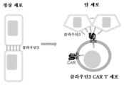

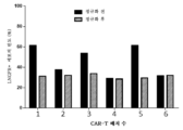
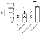


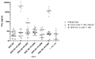


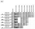


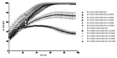


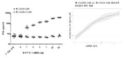


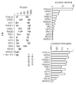
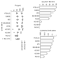


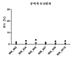


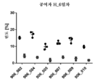


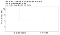

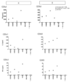


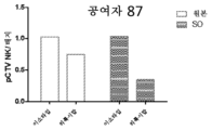

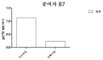

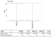


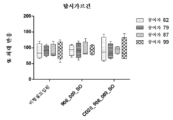
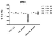


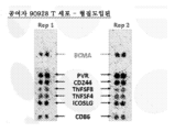


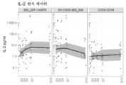

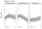

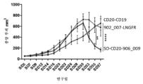


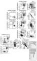


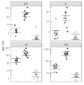


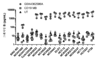


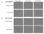


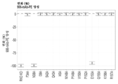

Classifications
-
- C—CHEMISTRY; METALLURGY
- C07—ORGANIC CHEMISTRY
- C07K—PEPTIDES
- C07K16/00—Immunoglobulins [IGs], e.g. monoclonal or polyclonal antibodies
- C07K16/18—Immunoglobulins [IGs], e.g. monoclonal or polyclonal antibodies against material from animals or humans
- C07K16/28—Immunoglobulins [IGs], e.g. monoclonal or polyclonal antibodies against material from animals or humans against receptors, cell surface antigens or cell surface determinants
-
- A—HUMAN NECESSITIES
- A61—MEDICAL OR VETERINARY SCIENCE; HYGIENE
- A61K—PREPARATIONS FOR MEDICAL, DENTAL OR TOILETRY PURPOSES
- A61K35/00—Medicinal preparations containing materials or reaction products thereof with undetermined constitution
- A61K35/12—Materials from mammals; Compositions comprising non-specified tissues or cells; Compositions comprising non-embryonic stem cells; Genetically modified cells
- A61K35/14—Blood; Artificial blood
- A61K35/17—Lymphocytes; B-cells; T-cells; Natural killer cells; Interferon-activated or cytokine-activated lymphocytes
-
- A—HUMAN NECESSITIES
- A61—MEDICAL OR VETERINARY SCIENCE; HYGIENE
- A61K—PREPARATIONS FOR MEDICAL, DENTAL OR TOILETRY PURPOSES
- A61K39/00—Medicinal preparations containing antigens or antibodies
- A61K39/46—Cellular immunotherapy
- A61K39/461—Cellular immunotherapy characterised by the cell type used
- A61K39/4611—T-cells, e.g. tumor infiltrating lymphocytes [TIL], lymphokine-activated killer cells [LAK] or regulatory T cells [Treg]
-
- A—HUMAN NECESSITIES
- A61—MEDICAL OR VETERINARY SCIENCE; HYGIENE
- A61K—PREPARATIONS FOR MEDICAL, DENTAL OR TOILETRY PURPOSES
- A61K39/00—Medicinal preparations containing antigens or antibodies
- A61K39/46—Cellular immunotherapy
- A61K39/463—Cellular immunotherapy characterised by recombinant expression
- A61K39/4631—Chimeric Antigen Receptors [CAR]
-
- A—HUMAN NECESSITIES
- A61—MEDICAL OR VETERINARY SCIENCE; HYGIENE
- A61K—PREPARATIONS FOR MEDICAL, DENTAL OR TOILETRY PURPOSES
- A61K39/00—Medicinal preparations containing antigens or antibodies
- A61K39/46—Cellular immunotherapy
- A61K39/464—Cellular immunotherapy characterised by the antigen targeted or presented
- A61K39/4643—Vertebrate antigens
- A61K39/4644—Cancer antigens
- A61K39/464402—Receptors, cell surface antigens or cell surface determinants
- A61K39/464411—Immunoglobulin superfamily
- A61K39/464412—CD19 or B4
-
- A—HUMAN NECESSITIES
- A61—MEDICAL OR VETERINARY SCIENCE; HYGIENE
- A61K—PREPARATIONS FOR MEDICAL, DENTAL OR TOILETRY PURPOSES
- A61K39/00—Medicinal preparations containing antigens or antibodies
- A61K39/46—Cellular immunotherapy
- A61K39/464—Cellular immunotherapy characterised by the antigen targeted or presented
- A61K39/4643—Vertebrate antigens
- A61K39/4644—Cancer antigens
- A61K39/464402—Receptors, cell surface antigens or cell surface determinants
- A61K39/464416—Receptors for cytokines
- A61K39/464417—Receptors for tumor necrosis factors [TNF], e.g. lymphotoxin receptor [LTR], CD30
-
- A—HUMAN NECESSITIES
- A61—MEDICAL OR VETERINARY SCIENCE; HYGIENE
- A61P—SPECIFIC THERAPEUTIC ACTIVITY OF CHEMICAL COMPOUNDS OR MEDICINAL PREPARATIONS
- A61P1/00—Drugs for disorders of the alimentary tract or the digestive system
-
- A—HUMAN NECESSITIES
- A61—MEDICAL OR VETERINARY SCIENCE; HYGIENE
- A61P—SPECIFIC THERAPEUTIC ACTIVITY OF CHEMICAL COMPOUNDS OR MEDICINAL PREPARATIONS
- A61P11/00—Drugs for disorders of the respiratory system
-
- A—HUMAN NECESSITIES
- A61—MEDICAL OR VETERINARY SCIENCE; HYGIENE
- A61P—SPECIFIC THERAPEUTIC ACTIVITY OF CHEMICAL COMPOUNDS OR MEDICINAL PREPARATIONS
- A61P35/00—Antineoplastic agents
-
- C—CHEMISTRY; METALLURGY
- C07—ORGANIC CHEMISTRY
- C07K—PEPTIDES
- C07K14/00—Peptides having more than 20 amino acids; Gastrins; Somatostatins; Melanotropins; Derivatives thereof
- C07K14/435—Peptides having more than 20 amino acids; Gastrins; Somatostatins; Melanotropins; Derivatives thereof from animals; from humans
- C07K14/705—Receptors; Cell surface antigens; Cell surface determinants
- C07K14/70503—Immunoglobulin superfamily
- C07K14/7051—T-cell receptor (TcR)-CD3 complex
-
- C—CHEMISTRY; METALLURGY
- C07—ORGANIC CHEMISTRY
- C07K—PEPTIDES
- C07K14/00—Peptides having more than 20 amino acids; Gastrins; Somatostatins; Melanotropins; Derivatives thereof
- C07K14/435—Peptides having more than 20 amino acids; Gastrins; Somatostatins; Melanotropins; Derivatives thereof from animals; from humans
- C07K14/705—Receptors; Cell surface antigens; Cell surface determinants
- C07K14/70503—Immunoglobulin superfamily
- C07K14/70517—CD8
-
- C—CHEMISTRY; METALLURGY
- C07—ORGANIC CHEMISTRY
- C07K—PEPTIDES
- C07K14/00—Peptides having more than 20 amino acids; Gastrins; Somatostatins; Melanotropins; Derivatives thereof
- C07K14/435—Peptides having more than 20 amino acids; Gastrins; Somatostatins; Melanotropins; Derivatives thereof from animals; from humans
- C07K14/705—Receptors; Cell surface antigens; Cell surface determinants
- C07K14/70578—NGF-receptor/TNF-receptor superfamily, e.g. CD27, CD30, CD40, CD95
-
- C—CHEMISTRY; METALLURGY
- C07—ORGANIC CHEMISTRY
- C07K—PEPTIDES
- C07K14/00—Peptides having more than 20 amino acids; Gastrins; Somatostatins; Melanotropins; Derivatives thereof
- C07K14/435—Peptides having more than 20 amino acids; Gastrins; Somatostatins; Melanotropins; Derivatives thereof from animals; from humans
- C07K14/705—Receptors; Cell surface antigens; Cell surface determinants
- C07K14/70596—Molecules with a "CD"-designation not provided for elsewhere
-
- C—CHEMISTRY; METALLURGY
- C07—ORGANIC CHEMISTRY
- C07K—PEPTIDES
- C07K16/00—Immunoglobulins [IGs], e.g. monoclonal or polyclonal antibodies
- C07K16/18—Immunoglobulins [IGs], e.g. monoclonal or polyclonal antibodies against material from animals or humans
- C07K16/28—Immunoglobulins [IGs], e.g. monoclonal or polyclonal antibodies against material from animals or humans against receptors, cell surface antigens or cell surface determinants
- C07K16/2887—Immunoglobulins [IGs], e.g. monoclonal or polyclonal antibodies against material from animals or humans against receptors, cell surface antigens or cell surface determinants against CD20
-
- C—CHEMISTRY; METALLURGY
- C12—BIOCHEMISTRY; BEER; SPIRITS; WINE; VINEGAR; MICROBIOLOGY; ENZYMOLOGY; MUTATION OR GENETIC ENGINEERING
- C12N—MICROORGANISMS OR ENZYMES; COMPOSITIONS THEREOF; PROPAGATING, PRESERVING, OR MAINTAINING MICROORGANISMS; MUTATION OR GENETIC ENGINEERING; CULTURE MEDIA
- C12N5/00—Undifferentiated human, animal or plant cells, e.g. cell lines; Tissues; Cultivation or maintenance thereof; Culture media therefor
- C12N5/06—Animal cells or tissues; Human cells or tissues
- C12N5/0602—Vertebrate cells
- C12N5/0634—Cells from the blood or the immune system
- C12N5/0636—T lymphocytes
-
- A—HUMAN NECESSITIES
- A61—MEDICAL OR VETERINARY SCIENCE; HYGIENE
- A61K—PREPARATIONS FOR MEDICAL, DENTAL OR TOILETRY PURPOSES
- A61K39/00—Medicinal preparations containing antigens or antibodies
- A61K2039/505—Medicinal preparations containing antigens or antibodies comprising antibodies
-
- A—HUMAN NECESSITIES
- A61—MEDICAL OR VETERINARY SCIENCE; HYGIENE
- A61K—PREPARATIONS FOR MEDICAL, DENTAL OR TOILETRY PURPOSES
- A61K39/00—Medicinal preparations containing antigens or antibodies
- A61K2039/51—Medicinal preparations containing antigens or antibodies comprising whole cells, viruses or DNA/RNA
- A61K2039/515—Animal cells
- A61K2039/5156—Animal cells expressing foreign proteins
-
- A—HUMAN NECESSITIES
- A61—MEDICAL OR VETERINARY SCIENCE; HYGIENE
- A61K—PREPARATIONS FOR MEDICAL, DENTAL OR TOILETRY PURPOSES
- A61K39/00—Medicinal preparations containing antigens or antibodies
- A61K2039/80—Vaccine for a specifically defined cancer
- A61K2039/812—Breast
-
- A—HUMAN NECESSITIES
- A61—MEDICAL OR VETERINARY SCIENCE; HYGIENE
- A61K—PREPARATIONS FOR MEDICAL, DENTAL OR TOILETRY PURPOSES
- A61K39/00—Medicinal preparations containing antigens or antibodies
- A61K2039/80—Vaccine for a specifically defined cancer
- A61K2039/82—Colon
-
- A—HUMAN NECESSITIES
- A61—MEDICAL OR VETERINARY SCIENCE; HYGIENE
- A61K—PREPARATIONS FOR MEDICAL, DENTAL OR TOILETRY PURPOSES
- A61K39/00—Medicinal preparations containing antigens or antibodies
- A61K2039/80—Vaccine for a specifically defined cancer
- A61K2039/86—Lung
-
- A—HUMAN NECESSITIES
- A61—MEDICAL OR VETERINARY SCIENCE; HYGIENE
- A61K—PREPARATIONS FOR MEDICAL, DENTAL OR TOILETRY PURPOSES
- A61K2239/00—Indexing codes associated with cellular immunotherapy of group A61K39/46
- A61K2239/31—Indexing codes associated with cellular immunotherapy of group A61K39/46 characterized by the route of administration
-
- A—HUMAN NECESSITIES
- A61—MEDICAL OR VETERINARY SCIENCE; HYGIENE
- A61K—PREPARATIONS FOR MEDICAL, DENTAL OR TOILETRY PURPOSES
- A61K2239/00—Indexing codes associated with cellular immunotherapy of group A61K39/46
- A61K2239/38—Indexing codes associated with cellular immunotherapy of group A61K39/46 characterised by the dose, timing or administration schedule
-
- A—HUMAN NECESSITIES
- A61—MEDICAL OR VETERINARY SCIENCE; HYGIENE
- A61K—PREPARATIONS FOR MEDICAL, DENTAL OR TOILETRY PURPOSES
- A61K2239/00—Indexing codes associated with cellular immunotherapy of group A61K39/46
- A61K2239/46—Indexing codes associated with cellular immunotherapy of group A61K39/46 characterised by the cancer treated
- A61K2239/50—Colon
-
- A—HUMAN NECESSITIES
- A61—MEDICAL OR VETERINARY SCIENCE; HYGIENE
- A61K—PREPARATIONS FOR MEDICAL, DENTAL OR TOILETRY PURPOSES
- A61K2239/00—Indexing codes associated with cellular immunotherapy of group A61K39/46
- A61K2239/46—Indexing codes associated with cellular immunotherapy of group A61K39/46 characterised by the cancer treated
- A61K2239/56—Kidney
-
- A—HUMAN NECESSITIES
- A61—MEDICAL OR VETERINARY SCIENCE; HYGIENE
- A61K—PREPARATIONS FOR MEDICAL, DENTAL OR TOILETRY PURPOSES
- A61K38/00—Medicinal preparations containing peptides
-
- C—CHEMISTRY; METALLURGY
- C07—ORGANIC CHEMISTRY
- C07K—PEPTIDES
- C07K2317/00—Immunoglobulins specific features
- C07K2317/20—Immunoglobulins specific features characterized by taxonomic origin
- C07K2317/24—Immunoglobulins specific features characterized by taxonomic origin containing regions, domains or residues from different species, e.g. chimeric, humanized or veneered
-
- C—CHEMISTRY; METALLURGY
- C07—ORGANIC CHEMISTRY
- C07K—PEPTIDES
- C07K2317/00—Immunoglobulins specific features
- C07K2317/30—Immunoglobulins specific features characterized by aspects of specificity or valency
- C07K2317/33—Crossreactivity, e.g. for species or epitope, or lack of said crossreactivity
-
- C—CHEMISTRY; METALLURGY
- C07—ORGANIC CHEMISTRY
- C07K—PEPTIDES
- C07K2317/00—Immunoglobulins specific features
- C07K2317/30—Immunoglobulins specific features characterized by aspects of specificity or valency
- C07K2317/34—Identification of a linear epitope shorter than 20 amino acid residues or of a conformational epitope defined by amino acid residues
-
- C—CHEMISTRY; METALLURGY
- C07—ORGANIC CHEMISTRY
- C07K—PEPTIDES
- C07K2317/00—Immunoglobulins specific features
- C07K2317/50—Immunoglobulins specific features characterized by immunoglobulin fragments
- C07K2317/56—Immunoglobulins specific features characterized by immunoglobulin fragments variable (Fv) region, i.e. VH and/or VL
- C07K2317/565—Complementarity determining region [CDR]
-
- C—CHEMISTRY; METALLURGY
- C07—ORGANIC CHEMISTRY
- C07K—PEPTIDES
- C07K2317/00—Immunoglobulins specific features
- C07K2317/60—Immunoglobulins specific features characterized by non-natural combinations of immunoglobulin fragments
- C07K2317/62—Immunoglobulins specific features characterized by non-natural combinations of immunoglobulin fragments comprising only variable region components
- C07K2317/622—Single chain antibody (scFv)
-
- C—CHEMISTRY; METALLURGY
- C07—ORGANIC CHEMISTRY
- C07K—PEPTIDES
- C07K2317/00—Immunoglobulins specific features
- C07K2317/70—Immunoglobulins specific features characterized by effect upon binding to a cell or to an antigen
-
- C—CHEMISTRY; METALLURGY
- C07—ORGANIC CHEMISTRY
- C07K—PEPTIDES
- C07K2317/00—Immunoglobulins specific features
- C07K2317/70—Immunoglobulins specific features characterized by effect upon binding to a cell or to an antigen
- C07K2317/73—Inducing cell death, e.g. apoptosis, necrosis or inhibition of cell proliferation
- C07K2317/732—Antibody-dependent cellular cytotoxicity [ADCC]
-
- C—CHEMISTRY; METALLURGY
- C07—ORGANIC CHEMISTRY
- C07K—PEPTIDES
- C07K2317/00—Immunoglobulins specific features
- C07K2317/70—Immunoglobulins specific features characterized by effect upon binding to a cell or to an antigen
- C07K2317/73—Inducing cell death, e.g. apoptosis, necrosis or inhibition of cell proliferation
- C07K2317/734—Complement-dependent cytotoxicity [CDC]
-
- C—CHEMISTRY; METALLURGY
- C07—ORGANIC CHEMISTRY
- C07K—PEPTIDES
- C07K2317/00—Immunoglobulins specific features
- C07K2317/70—Immunoglobulins specific features characterized by effect upon binding to a cell or to an antigen
- C07K2317/76—Antagonist effect on antigen, e.g. neutralization or inhibition of binding
-
- C—CHEMISTRY; METALLURGY
- C07—ORGANIC CHEMISTRY
- C07K—PEPTIDES
- C07K2319/00—Fusion polypeptide
- C07K2319/01—Fusion polypeptide containing a localisation/targetting motif
- C07K2319/02—Fusion polypeptide containing a localisation/targetting motif containing a signal sequence
-
- C—CHEMISTRY; METALLURGY
- C07—ORGANIC CHEMISTRY
- C07K—PEPTIDES
- C07K2319/00—Fusion polypeptide
- C07K2319/01—Fusion polypeptide containing a localisation/targetting motif
- C07K2319/03—Fusion polypeptide containing a localisation/targetting motif containing a transmembrane segment
-
- C—CHEMISTRY; METALLURGY
- C07—ORGANIC CHEMISTRY
- C07K—PEPTIDES
- C07K2319/00—Fusion polypeptide
- C07K2319/33—Fusion polypeptide fusions for targeting to specific cell types, e.g. tissue specific targeting, targeting of a bacterial subspecies
-
- C—CHEMISTRY; METALLURGY
- C12—BIOCHEMISTRY; BEER; SPIRITS; WINE; VINEGAR; MICROBIOLOGY; ENZYMOLOGY; MUTATION OR GENETIC ENGINEERING
- C12N—MICROORGANISMS OR ENZYMES; COMPOSITIONS THEREOF; PROPAGATING, PRESERVING, OR MAINTAINING MICROORGANISMS; MUTATION OR GENETIC ENGINEERING; CULTURE MEDIA
- C12N2510/00—Genetically modified cells
-
- C—CHEMISTRY; METALLURGY
- C12—BIOCHEMISTRY; BEER; SPIRITS; WINE; VINEGAR; MICROBIOLOGY; ENZYMOLOGY; MUTATION OR GENETIC ENGINEERING
- C12N—MICROORGANISMS OR ENZYMES; COMPOSITIONS THEREOF; PROPAGATING, PRESERVING, OR MAINTAINING MICROORGANISMS; MUTATION OR GENETIC ENGINEERING; CULTURE MEDIA
- C12N2740/00—Reverse transcribing RNA viruses
- C12N2740/00011—Details
- C12N2740/10011—Retroviridae
- C12N2740/16011—Human Immunodeficiency Virus, HIV
- C12N2740/16041—Use of virus, viral particle or viral elements as a vector
- C12N2740/16043—Use of virus, viral particle or viral elements as a vector viral genome or elements thereof as genetic vector
Abstract
서열식별번호: 13의 적어도 N38 및 E153을 포함하는 인간 클라우딘-3 상의 불연속적 에피토프에 결합하는 항원 결합 단백질을 포함하는 키메라 항원 수용체 (CAR)가 기재되어 있다. 또한, 본원에 기재된 항원 결합 단백질을 코딩하는 폴리뉴클레오티드, CAR, CAR을 함유하는 면역 이펙터 세포, 면역 이펙터 세포를 함유하는 제약 조성물, 및 면역 이펙터 세포를 사용하여 암을 치료하는 방법이 포함된다.A chimeric antigen receptor (CAR) comprising an antigen binding protein that binds to a discontinuous epitope on human claudin-3, including at least N38 and E153 of SEQ ID NO: 13, is described. Also included are polynucleotides encoding antigen binding proteins described herein, CARs, immune effector cells containing CARs, pharmaceutical compositions containing immune effector cells, and methods of treating cancer using immune effector cells.
Description
본 발명은 세포 이음부 단백질의 적어도 하나의 에피토프에 결합하는 항원 결합 단백질을 포함하는 키메라 항원 수용체 (CAR)에 관한 것이며, 여기서 상기 세포 이음부 단백질은 세포-세포 이음부 내에 위치되고, 여기서 세포 이음부 단백질의 상기 하나 이상의 에피토프는 암 세포에서 상기 CAR 세포외 도메인에 의한 결합에만 접근가능하고/거나 이용가능하다.The present invention relates to a chimeric antigen receptor (CAR) comprising an antigen binding protein that binds to at least one epitope of a cell junction protein, wherein the cell junction protein is located within a cell-cell junction, wherein the cell junction protein The one or more epitopes of the minor protein are only accessible and/or available for binding by the CAR extracellular domain in cancer cells.
입양 T 세포 요법은 암 환자에 대한 치유 잠재성을 갖는 혁신적인 약제이다. 환자를 위한 이들 강력한 자가유래 세포 요법을 조작하기 위해, 말초 혈액을 사용하여 T 세포를 수득하며, 이는 그 후 유전적으로 변형된다. 이들 세포에 키메라 항원 수용체 (CAR)를 도입하는 것은 선택된 항원에 특이적으로 결합하는 것을 가능하게 한다. 이들 변형된 세포는 일치하는 항원을 발현하는 암 세포로 트래피킹하고 후속적으로 이를 사멸시키려는 목적으로 생체외에서 증식되고 환자에게 재주입된다 (Yeku et al., Am Soc Clin Oncol Educ Book. 2017; 37: 193-204; McBride et al., Wiley Interdiscip Rev Nanomed Nanobiotechnol. 2019; 11(5): e1557).Adoptive T cell therapy is an innovative agent with curative potential for cancer patients. To engineer these powerful autologous cell therapies for patients, peripheral blood is used to obtain T cells, which are then genetically modified. Introduction of chimeric antigen receptors (CARs) into these cells allows for specific binding to selected antigens. These modified cells are propagated ex vivo and reinjected into the patient with the goal of trafficking to and subsequently killing cancer cells expressing matching antigens (Yeku et al., Am Soc Clin Oncol Educ Book. 2017; 37 : 193-204; McBride et al., Wiley Interdiscip Rev Nanomed Nanobiotechnol. 2019; 11(5): e1557).
CAR은 T 세포 특이성, 기능 및 지속성을 재지시하는 합성 항원 수용체이다. CAR은 항원 결합 단백질의 항원 특이적 영역, 예컨대 T 세포 활성화 도메인, 예를 들어 CD3 복합체의 제타 쇄, 및 공동-자극 도메인, 예를 들어 CD28 또는 4-1BB에 융합된 단일-쇄 가변 단편 (scFv)으로 구성된다. 이러한 구성은 항원 특이적 활성화를 촉진하고 인간 1차 T 세포의 증식 및 항아폽토시스 기능을 향상시킨다.CARs are synthetic antigen receptors that redirect T cell specificity, function and persistence. A CAR is a single-chain variable fragment (scFv) fused to an antigen-specific region of an antigen binding protein, such as a T cell activation domain, such as the zeta chain of the CD3 complex, and a co-stimulatory domain, such as CD28 or 4-1BB. ) is composed of. This construct promotes antigen-specific activation and enhances the proliferation and antiapoptotic functions of human primary T cells.
CAR-T 세포는 다양한 액체 종양 B-세포 악성종양에 대해 현저한 효능을 입증하였으며; 초기 임상 시험 결과는 다발성 골수종에서의 활성을 시사한다 (Sadelain et al., Nature. 2017; 545(7655): 423-31; June et al., N Engl J Med. 2018; 379(1): 64-73; Brudno et al., Nat Rev Clin Oncol. 2017; 15(1): 31-46). 림프종 및 백혈병에 대한 CD19 CAR-T의 2017년 FDA 승인은 고형 종양에 대한 CAR-T 세포의 개발에 집중적인 노력을 다시 활성화시켰다. 그러나, CAR-T 세포 요법은 현재까지 고형 종양에서 제한된 효능을 입증하였다.CAR-T cells have demonstrated remarkable efficacy against a variety of liquid tumor B-cell malignancies; Early clinical trial results suggest activity in multiple myeloma (Sadelain et al., Nature. 2017; 545(7655): 423-31; June et al., N Engl J Med. 2018; 379(1): 64 -73; Brudno et al., Nat Rev Clin Oncol. 2017; 15(1): 31-46). The 2017 FDA approval of CD19 CAR-T for lymphoma and leukemia reinvigorated focused efforts on the development of CAR-T cells for solid tumors. However, CAR-T cell therapy has demonstrated limited efficacy in solid tumors to date.
고형암에 대한 안전하고 효능있는 T 세포 요법을 생성하기 위해, T 세포는 제조 전반에 걸쳐 높고 효능있는 사멸 잠재성을 보유하고 종양으로 트래피킹할 수 있어야 하며 면역억압 종양 미세환경 (TME)을 극복해야 한다. 주요 성공 인자 중 하나는 항원 표적의 선택이다. 전형적으로, 표적화를 위해 선택된 항원은 암 세포 표면에서 충분한 양으로 발현되는 반면, 정상 조직의 발현은 건강한 세포에 대한 낮은 교차-반응성을 보장하기 위해 낮게 유지된다 (표적-이탈 및 표적-적중 효과 둘 다). 고형 종양에서의 CAR 면역요법은 주로 악성 조직에만 발현이 국한되는 적절한 표면 항원의 결여로 인해 여전히 어려운 과제이다. 항원 표적의 종양-이탈 발현은 영향을 받은 장기 조직에 따라 다양한 정도의 중증도로 표적-적중 독성을 유발할 잠재성을 갖는다 (Watanabe et al., Front Immunol. 2018; 9: 2486; Park et al., Sci Rep. 2017; 7(1): 14366).To generate safe and efficacious T cell therapies for solid tumors, T cells must possess high and efficacious killing potential throughout manufacturing, be able to traffic to the tumor, and overcome the immunosuppressive tumor microenvironment (TME). do. One of the key success factors is the choice of antigenic target. Typically, the antigen selected for targeting is expressed in sufficient amounts on the cancer cell surface, while expression in normal tissues is kept low to ensure low cross-reactivity to healthy cells (both off-target and on-target effects). all). CAR immunotherapy in solid tumors remains a challenge due to the lack of appropriate surface antigens, whose expression is mainly limited to malignant tissues. Tumor-escaping expression of antigenic targets has the potential to cause off-target toxicity with varying degrees of severity depending on the organ tissue affected (Watanabe et al., Front Immunol. 2018; 9: 2486; Park et al., Sci Rep. 2017; 7(1): 14366).
따라서, CAR 면역요법, 특히 고형 종양 및 암에 대한 T 세포 요법을 생성하기 위한 새로운 표적을 식별할 필요가 있다.Therefore, there is a need to identify new targets for generating CAR immunotherapy, especially T cell therapy for solid tumors and cancer.
제1 측면에 따라, 하기를 포함하는 키메라 항원 수용체 (CAR)가 제공된다:According to a first aspect, a chimeric antigen receptor (CAR) is provided comprising:
a) 서열식별번호(SEQ ID NO): 13의 적어도 N38 및 E153을 포함하는 인간 클라우딘-3 상의 불연속적 에피토프에 결합하는 단리된 클라우딘-3 결합 단백질을 포함하는 세포외 도메인;a) an extracellular domain comprising an isolated Claudin-3 binding protein that binds to a discrete epitope on human Claudin-3 comprising at least N38 and E153 of SEQ ID NO: 13;
b) 막횡단 도메인; 및b) transmembrane domain; and
c) 하나 이상의 세포내 신호전달 도메인.c) one or more intracellular signaling domains.
추가 측면에서, 하기를 포함하는 폴리펩티드를 포함하는 키메라 항원 수용체 (CAR)가 제공된다:In a further aspect, provided is a chimeric antigen receptor (CAR) comprising a polypeptide comprising:
a) 서열식별번호: 1의 중쇄 상보성 결정 영역 1 (CDRH1) 서열; 서열식별번호: 2의 중쇄 상보성 결정 영역 2 (CDRH2) 서열; 서열식별번호: 3의 중쇄 상보성 결정 영역 3 (CDRH3) 서열을 포함하는 중쇄 가변 영역 (VH)을 포함하는 클라우딘-3 결합 도메인을 포함하는 세포외 도메인;a) Heavy chain complementarity determining region 1 (CDRH1) sequence of SEQ ID NO: 1; Heavy chain complementarity determining region 2 (CDRH2) sequence of SEQ ID NO: 2; an extracellular domain comprising a claudin-3 binding domain comprising a heavy chain variable region (VH) comprising the heavy chain complementarity determining region 3 (CDRH3) sequence of SEQ ID NO: 3;
b) 막횡단 도메인; 및b) transmembrane domain; and
c) 하나 이상의 세포내 신호전달 도메인.c) one or more intracellular signaling domains.
추가 측면에서, 서열식별번호: 13의 적어도 N38 및 E153을 포함하는 인간 클라우딘-3 상의 불연속적 에피토프에 결합하는 단리된 클라우딘-3 결합 단백질이 제공된다.In a further aspect, an isolated Claudin-3 binding protein is provided that binds a discontinuous epitope on human Claudin-3 comprising at least N38 and E153 of SEQ ID NO: 13.
추가 측면에서, 서열식별번호: 1의 중쇄 상보성 결정 영역 1 (CDRH1) 서열; 서열식별번호: 2의 중쇄 상보성 결정 영역 2 (CDRH2) 서열; 서열식별번호: 3의 중쇄 상보성 결정 영역 3 (CDRH3) 서열을 포함하는 중쇄 가변 영역 (VH)을 포함하는 클라우딘-3 결합 단백질이 제공된다.In a further aspect, the heavy chain complementarity determining region 1 (CDRH1) sequence of SEQ ID NO: 1; Heavy chain complementarity determining region 2 (CDRH2) sequence of SEQ ID NO: 2; A claudin-3 binding protein comprising a heavy chain variable region (VH) comprising the heavy chain complementarity determining region 3 (CDRH3) sequence of SEQ ID NO: 3 is provided.
추가 측면에서, 본원에 개시된 CAR 또는 클라우딘-3 결합 단백질의 아미노산 서열을 포함하는 폴리펩티드가 제공된다.In a further aspect, polypeptides comprising the amino acid sequence of a CAR or claudin-3 binding protein disclosed herein are provided.
다른 측면에 따라, 본원에 개시된 CAR 또는 클라우딘-3 결합 단백질을 코딩하는 서열을 포함하는 폴리뉴클레오티드, 본원에 개시된 폴리뉴클레오티드 서열을 포함하는 벡터, 및 본원에 개시된 폴리뉴클레오티드 서열 및/또는 벡터를 포함하는 벡터 생산자 세포가 제공된다.According to another aspect, a polynucleotide comprising a sequence encoding a CAR or claudin-3 binding protein disclosed herein, a vector comprising a polynucleotide sequence disclosed herein, and a polynucleotide comprising a polynucleotide sequence and/or vector disclosed herein. Vector producer cells are provided.
또 다른 측면에서, 본원에 개시된 CAR, 폴리펩티드, 폴리뉴클레오티드 및/또는 벡터를 포함하는 면역 이펙터 세포가 제공된다.In another aspect, an immune effector cell comprising a CAR, polypeptide, polynucleotide and/or vector disclosed herein is provided.
또 다른 측면에서, 본원에 개시된 면역 이펙터 세포 또는 클라우딘-3 결합 단백질 및 제약상 허용되는 부형제를 포함하는 제약 조성물이 제공된다.In another aspect, a pharmaceutical composition comprising an immune effector cell or claudin-3 binding protein disclosed herein and a pharmaceutically acceptable excipient is provided.
또 다른 측면에서, 본원에 개시된 폴리펩티드, 폴리뉴클레오티드 및/또는 벡터를 면역 이펙터 세포에 도입하는 것을 포함하는, 본원에 개시된 CAR을 포함하는 면역 이펙터 세포를 생성하는 방법이 제공된다.In another aspect, a method of generating an immune effector cell comprising a CAR disclosed herein is provided, comprising introducing a polypeptide, polynucleotide and/or vector disclosed herein into the immune effector cell.
또 다른 측면에서, 암 치료에서 사용하기 위한 본원에 개시된 CAR, 클라우딘-3 결합 단백질, 폴리펩티드, 폴리뉴클레오티드, 벡터, 면역 이펙터 세포 또는 제약 조성물이 제공된다.In another aspect, a CAR, claudin-3 binding protein, polypeptide, polynucleotide, vector, immune effector cell, or pharmaceutical composition disclosed herein for use in cancer treatment is provided.
또 다른 측면에서, 본원에 개시된 CAR 또는 클라우딘-3 결합 단백질, 폴리펩티드, 폴리뉴클레오티드, 벡터, 면역 이펙터 세포 또는 제약 조성물의 치료 유효량을 대상체에게 투여하는 것을 포함하는, 대상체에서 암을 치료하는 방법이 제공된다.In another aspect, a method of treating cancer in a subject is provided, comprising administering to the subject a therapeutically effective amount of a CAR or claudin-3 binding protein, polypeptide, polynucleotide, vector, immune effector cell, or pharmaceutical composition disclosed herein. do.
또 다른 측면에서, 본원에 개시된 CAR 또는 클라우딘-3 결합 단백질, 폴리펩티드, 폴리뉴클레오티드, 벡터, 면역 이펙터 세포 또는 제약 조성물의 유효량을 대상체에게 투여하는 것을 포함하는, 암을 갖는 대상체에서 암 세포에 대한 세포독성을 증가시키는 방법이 제공된다.In another aspect, cells against cancer cells in a subject having cancer, comprising administering to the subject an effective amount of a CAR or claudin-3 binding protein, polypeptide, polynucleotide, vector, immune effector cell, or pharmaceutical composition disclosed herein. Methods for increasing toxicity are provided.
또 다른 측면에서, 본원에 개시된 CAR 또는 클라우딘-3 결합 단백질, 폴리펩티드, 폴리뉴클레오티드, 벡터, 면역 이펙터 세포 또는 제약 조성물의 유효량을 대상체에게 투여하는 것을 포함하는, 암을 갖는 대상체에서 암 세포의 수를 저하시키는 방법이 제공된다.In another aspect, the number of cancer cells in a subject having cancer comprising administering to the subject an effective amount of a CAR or claudin-3 binding protein, polypeptide, polynucleotide, vector, immune effector cell, or pharmaceutical composition disclosed herein. A method for lowering the temperature is provided.
또 다른 측면에서, 암 치료용 의약의 제조에서의, 본원에 개시된 CAR 또는 클라우딘-3 결합 단백질, 폴리펩티드, 폴리뉴클레오티드, 벡터, 면역 이펙터 세포 또는 제약 조성물의 용도가 제공된다.In another aspect, provided is the use of a CAR or claudin-3 binding protein, polypeptide, polynucleotide, vector, immune effector cell, or pharmaceutical composition disclosed herein in the manufacture of a medicament for the treatment of cancer.
또 다른 측면에서, 요법에서 사용하기 위한, 본원에 개시된 CAR 또는 클라우딘-3 결합 단백질, 폴리펩티드, 폴리뉴클레오티드, 벡터, 면역 이펙터 세포 또는 제약 조성물이 제공된다.In another aspect, a CAR or claudin-3 binding protein, polypeptide, polynucleotide, vector, immune effector cell, or pharmaceutical composition disclosed herein for use in therapy is provided.
도 1: 암 세포 vs. 건강한 비암성 세포 ("정상 세포")에서 결합에 대한 클라우딘-3의 접근가능성을 제시하는 개략적 다이어그램. CLDN3은 상피 세포 사이의 치밀 이음부 (TJ) 형성에 중요한 내재성 막 단백질의 큰 패밀리에 속한다. 정상 조직 아키텍처의 파괴는 암의 특징이며, CLDN3 변경된 발현은 결장직장, 유방, 췌장 및 난소 암종과 같은 충족되지 않은 수요가 높은 암을 포함하여 다양한 상피암의 발생과 연관되어 있다 (Singh, Sharma, and Dhawan 2010). 제시된 바와 같이, CLDN3은 종양의 TJ 외부에 잘못 편재화되어 있지만 건강한 조직에서는 그렇지 않으며, 이는 CLDN3을 종양 세포의 선택적 사멸을 위한 CAR-T 세포 표적으로 바꾸는 동시에 치밀 이음부에 숨겨져 있는 정상 세포를 보호하는 메커니즘이다.
도 2a-2b: 도 2a: LNGFR+ CAR-T 배치의 풍부화. 이지셉(EasySep) LNGFR 양성 선택을 CAR-T 배치에서 수행하여 기능 검정에서 사용하기 위한 100% CAR-T 세포 집단을 생산하였다. 도 2b: LNGFR+ 세포의 필수 빈도에 대한 CAR-T 배치의 정규화. CAR-T 배치는 비형질도입된 T 세포의 첨가에 의해 정규화되어 모든 구축물에 걸쳐 30%의 형질도입 효율을 달성하였다.
도 3a-3h: 도 3a: 비형질도입된 및 형질도입된 CAR-T 세포에 대한 기본 IFNγ 시토카인 분비 프로파일. 6명의 상이한 건강한 공여자로부터 수득된 CAR-T 세포를 사용하여 배양 상청액 내 IFNγ 분비물의 농도의 평균 및 표준 편차 (**** = p<0.0001 본페로니 일원 ANOVA에 의해 계산됨; UT = 비형질도입된). 도 3b-3g: 비형질도입된 및 형질도입된 CAR-T 세포에 대한 기본 고갈/활성화 표현형 (CD69+, TIM3+ 및 PD-1+). 평균 및 표준 편차가 제시된다 (p 값 범위는 본페로니 일원 ANOVA에 의해 계산된 <0.02 내지 <0.0001임; UT = 비형질도입된). 도 3h: 형질도입된 CAR-T 세포에서 CAR 특이적 pCD3ζ 수준 (PEGGY-SUE에 의해 평가된 바와 같음). 음성 대조군 (항-CD19 CAR)과 비교하여 정규화된 pCD3ζ 염색.
도 4a-4f: 도 4a: 클라우딘 단백질에 대한 반응으로 IFNγ, IL-2 및 TNFα의 분비. hCLDN3, hCLDN4, hCLDN5, hCLDN6, hCLDN8, hCLDN9, hCLDN17 또는 mCLDN3을 과발현하는 RKO KO 세포주와 공동-배양된 항-클라우딘-3 또는 항-CD19 CAR-T 세포로부터의 시토카인 방출의 정량화 (n=3의 평균 + 95% 신뢰 한계). 도 4b 및 4c: 클라우딘 단백질에 대한 반응으로 IFNγ 분비. hCLDN3, hCLDN4, hCLDN6, hCLDN9 또는 mCLDN3을 과발현하는 RKO KO와 공동-배양된 항-클라우딘-3 CAR, 항-CD19 CAR (대조군) 또는 비형질도입된 T 세포로부터의 시토카인 방출. 도 4b: 6명의 공여자의 평균 + 95% 신뢰 한계로서 제시된 IFNγ의 농도. 도 4c: 클라우딘 단백질을 발현하는 세포주와 함께 배양될 때 항-CD19 대조군 또는 비형질도입된 T 세포와 비교하여 항-클라우딘-3 CAR-T 세포로부터의 IFNγ 분비의 배수 변화. 도 4d-4f: 클라우딘 단백질에 대한 반응으로 분비된 시토카인의 수준. 시토카인의 농도 (pg/mL)는 대조군 T 세포와 함께 배양된 RKO KO와 비교하여 각 조건의 배수 변화를 나타내는 히트맵 상에서 오버레이된다. 배수 변화는 각 실험 및 공여자 내에서 계산되고 로그 변환된다. 이 데이터는 3개의 시토카인 IFNγ (상단 패널), IL-2 (중간 패널) 및 TNFα (하단 패널)에 대해 제시되며 특정 공여자는 좌측 컬럼에 표시된다.
도 5a-5b: 도 5a: 클라우딘 단백질을 발현하는 RKO KO 세포와 공동-배양된 항-클라우딘-3 CAR-T 세포의 이미지. 이들 이미지는 hCLDN3, hCLDN4, hCLDN6, hCLDN9 또는 mCLDN3을 발현하는 각 세포주에 대해 4일차에 촬영되었다. 좌측 패널: 이미지는 세포 구역의 윤곽을 그리는 황색 라인으로 제시되는 % 전면생장을 계산하는데 사용되는 마스크와 오버레이된다. 우측 패널: 이미지는 사이토톡스 레드 (CYTOTOX red) 염료의 강도 및 편재화를 제시하는 적색 형광과 오버레이된다. 도 5b: 이미지는 4일 동안 2시간마다 촬영되었으며 각 웰의 % 전면생장은 각 시점에서 인큐사이트(INCUCYTE) 소프트웨어를 사용하여 계산되었다. 이 실험을 위해 6명의 공여자를 3중으로 사용하였다. 3중 웰의 평균이 계산되었으며 상기 그래프의 각 포인트는 6명의 공여자의 평균 ± SEM을 나타낸다.
도 6: 클라우딘 패밀리 단백질에 대한 CAR-T 세포의 세포독성 반응. 항-클라우딘-3 CAR ("906-009"), 항-CD19 CAR ("CD19"; 대조군) 및 비형질도입된 T 세포 ("UT")를 4일 동안 hCLDN3, hCLDN4, hCLDN6, hCLDN9 또는 mCLDN3을 발현하는 RKO KO 세포와 공동-배양하였다. 사이토톡스 레드의 흡광도 및 그에 따른 적색 형광을 마스크로 분석하고, 데이터를 사용하여 % 살아있는 세포를 계산하였다. 1명의 대표적인 공여자로부터의 데이터는 3개의 3중 웰의 평균 ± SEM으로서 제시된다.
도 7a-7b: 정량화 비드를 사용한 표면 분자 정량화. 도 7a: T 세포당 LNGFR 분자의 평균 수. T 세포 및 퀀텀 심플리 셀룰러(Quantum Simply Cellular) 정량화 비드를 항-LNGFR PE로 염색하고, 이들의 평균 형광 강도 (MFI)를 측정하였다. 비드 MFI는 세포당 LNGFR 수를 보간하는데 사용된 표준 곡선을 생성하는데 사용되었다. 공여자 12031 및 92024의 경우 n=3 및 공여자 D5의 경우 n=5 및 오차 막대가 표준 편차를 결정한다. 도 7b: RKO 인간 결장암 세포에 대한 hCLDN3 발현의 정량화. 내인성 hCLDN3이 녹아웃된 후 hCLDN3을 발현하도록 조작된 RKO 세포를 낮은 또는 높은 hCLDN3 발현에 대해 분류하였다. RKO 세포 및 퀀텀 심플리 셀룰러 정량화 비드를 항-hCLDN3-PE로 염색하고 이들의 MFI를 측정하였다. 비드 MFI는 세포당 hCLDN3 수를 보간하는데 사용된 표준 곡선을 생성하는데 사용되었다. 4개 반복이 측정되었으며 오차 막대가 표준 편차를 결정한다.
도 8a-8d: 예시적인 인큐사이트 및 엑스셀리젠스(XCELLIGENCE) 사멸 검정. 90시간 동안 RKO-KO CLDN3 L14와 함께 인큐베이션된 LNGFR 풍부화된 항-CD19 CAR-T 세포 (도 8a) 또는 항-클라우딘-3 CAR-T 세포 (도 8b)의 인큐사이트 원시 데이터가 제시된다. 예시적인 사멸 곡선은 도 8a-8b의 원시 데이터에 기초하며 도 8c에 제시된다. 3개 반복이 측정되었으며 오차 막대가 표준 편차를 결정한다. 도 8d: 엑스셀리젠스 사멸 검정 예. 1:1 비로 HT-29-LUC 표적 세포주와 공동-배양된 비분류된 항-클라우딘-3 CAR-T 세포 또는 비형질도입된 T 세포에 대한 정규화된 데이터가 제시된다. 데이터는 n=3의 평균 ± 표준 편차로서 제시된다.
도 9: 다양한 수의 RKO-KO hCLDN3-발현 표적 세포를 사용한 인큐사이트 사멸 검정. RKO-KO 및 RKO-KO hCLDN3 폴리클로날 세포를 다양한 비로 혼합하고, 항-CD19 대조군 CAR-T 세포 또는 항-클라우딘-3 CAR-T 세포와 공동-배양하였다. 시간 경과에 따른 살아있는 세포의 %를 측정하였다. 3개 반복이 측정되었으며 오차 막대는 표준 편차를 나타낸다.
도 10a-10d: hCLDN3 발현의 구배는 T 세포 용량 반응을 제시하였다. RKO-KO 세포를 hCLDN3 mRNA의 구배로 전기천공하고, 3명의 공여자로부터 생산된 CLDN3 및 항-CD19 대조군 CAR-T 세포와 공동-배양하였다. 도 10a: 핵감염된 hCLDN3 mRNA의 질량과 관련된 유동 세포계측법에 의해 평가된 hCLDN3의 발현. 이 데이터는 평균 형광 강도 또는 표적 양성 집단의 %로서 제시된다. 피어슨(Pearson)의 R2는 mRNA 질량 및 평균 형광 강도 간의 상관관계에 대해 계산되었다. 도 10b: 공동-배양 후 항-클라우딘-3 CAR-T 세포를 염색하여 CD69 발현을 식별하였다. % CD69 발현은 3명의 공여자의 평균 + 95% CI로서 제시된다. 도 10c-10d: 공동-배양 상청액을 사용하여 T 세포에 의해 분비된 시토카인의 농도를 정량화하였다. 도 10c: 3명의 공여자의 평균 + 95% 신뢰 구간으로서 제시된 IFNγ pg/ml. 항-클라우딘-3 vs 항-CD19 대조군 CAR-T 세포로부터의 IFNγ 분비의 비. 도 10d: 3명의 공여자의 평균 + 95% 신뢰 구간으로서 제시된 그랜자임 B pg/mL. 항-클라우딘-3 vs 항-CD19 대조군 CAR-T 세포로부터의 그랜자임 B 분비의 비.
도 11a-11c: 유동 세포계측법 및 RT-qPCR에 의한 CLDN3 발현 및 상이한 적응증: 결장직장 (도 11a), 췌장암 (도 11b) 및 유방 (도 11c) 암으로부터의 다양한 세포주에 대한 반응으로 IFNγ 분비. hCLDN3 발현은 유동 세포계측법 (좌측)에 의해 단백질 수준에서 및 RT-qPCR (중간)에 의해 mRNA 수준에서 측정되었다. HT-29-LUC 및 RKO-KO 세포주는 각각 양성 및 음성 대조군으로서 모든 실험에 포함되었다. 상이한 적응증으로부터의 다양한 세포주에 대한 반응으로 IFNγ 분비를 또한 평가하였다. T 세포를 표적 세포주와 함께 24시간 동안 1:1 비로 인큐베이션하고, IFNγ 분비를 MSD에 의해 측정하였다 (우측).
도 12: 선택된 세포주의 예시적인 사멸 이미지 및 원시 데이터. 완전 사멸을 보인 2개 세포주 (HT-29-LUC 및 MDA MB468), 부분 사멸을 보인 3개 세포주 (HCC1954, HPAC 및 BxPC3) 및 어떤 사멸도 나타내지 않은 1개 세포주 (COLO-320DM)의 예시적인 이미지 (상단) 및 원시 데이터 (하단)가 제시된다. 원시 데이터는 웰당 총 사이토톡스 레드로서 제시된다. 상이한 세포주 간에는 데이터를 정규화할 수 없어 원시 데이터로만 비교가 가능하기 때문에 원시 데이터를 사용하였다.
도 13: 단백질 L-비오틴 (1차) 및 항-비오틴-PE (2차) 염색에 의해 결정된 CAR 발현. 형질도입 효율은 LNGFR-PE 염색에 의해 결정되었다. 여기서는 명명된 CAR 변이체를 사용한 형질도입 후 7일차 동안 예시적인 공여자 D5의 CAR 및 LNGFR 빈도가 제시된다.
도 14: CAR T 세포 (공여자 D5)를 사용한 사멸 검정의 결과가 제시된다. 루시퍼라제 형질도입된 T-47D 세포와 CAR-T 세포의 공동-배양에서 루시퍼라제 활성을 측정한 후 용균된 표적 세포의 빈도를 계산하였다 (각 데이터 포인트는 기술적 반복의 평균을 나타냄, n=2).
도 15a-15c: 상이한 scFv 구축물을 갖는 항-클라우딘-3 CAR-T 세포에 대한 반응으로 분비된 시토카인의 농도는 MACSPlex 시토카인 12 키트 (인간)를 사용하여 결정되었다. T 세포 및 T-47D 세포와의 공동-배양 또는 T 세포 단독의 상청액을 수집하고 분석하였다 (희석되지 않음). T 세포 공여자 D5의 분비된 IFNγ (도 15a), IL-2 (도 15b) 및 TNF-α (도 15c)의 농도가 제시된다.
도 16a-16f: CAR-T 세포 (공여자 G5 및 H5)와 RKO-KO CLDN3 H1 (RKO-hCLD3으로서 표지됨) 세포의 장기간 공동-배양의 결과는 도 16a-16c에 제시된 공여자 G5에 대한 라운드 1-3 및 도 16c-16f에 제시된 공여자 H5에 대한 라운드 1-3과 함께 제시된다. CAR-T 세포를 총 3회 라운드 동안 신선한 RKO-KO CLDN3 H1 세포로 옮겼다. GFP를 발현하는 RKO-KO CLDN3 H1의 성장을 녹색 물체 전면생장 (퍼센트)을 통해 인큐사이트를 사용하여 모니터링하고 시작 값 (4시간)으로 정규화하였다. 반복 없는 조건은 별표 (*)로 표시된다.
도 17a-17j: 항-LAG3 (CD223)-VioBlue, 항-PD-1 (CD279)-PE-Vio770 및 항-TIM3 (CD366)-APC를 사용하여 염색함으로써 고갈 마커 발현을 결정하였다. LNGFR-양성 T 세포를 이중 (TIM3, PD-1; 채워진 원으로 표시) 및 삼중 (TIM3, PD-1, LAG3; 채워진 사각형으로 표시) 양성 고갈 마커 발현의 발현에 대해 평가하였다. 공여자 H 및 공여자 P에 대한 이중 및 삼중 양성 CAR T 세포의 빈도가 제시된다. 0일차는 표적 세포의 첨가 전 및 표적 세포의 제1 첨가 후 1일차의 빈도를 표시한다. 1, 2 및 3일차에, 신선한 RKO-KO CLDN3 H1 세포를 첨가하였다. 0일차 (도 17a-17b), 1일차 (도 17c-17d), 2일차 (도 17e-17f), 3일차 (도 17g-17h) 및 6일차 (도 17i-17j)에 공여자 H 및 공여자 P 각각에 대한 결과가 제시된다.
도 18: 항-클라우딘-3 CAR-T 세포 (906-009)는 클라우딘- 3 양성 표적 세포를 사용한 자극 후 다른 스페이서 또는 배향 변이체를 갖는 항-클라우딘-3 CAR-T 세포 (906-002, 906-004 및 906-007)와 비교하여 향상된 클라우딘-3-특이적 증식적 반응을 나타낸다.
도 19a-19b: HT-29 인간 결장 선암종 세포주로 접종된 NSG 마우스의 성장 동역학. 접종 7일 후, 종양이 촉지가능하였고, 동물은 PBS (T 세포 없음), 항-CD19 (대조군 CAR) 또는 항-클라우딘-3 CAR-T 세포를 1x10⁷개 세포의 용량으로 투약받았다. 투약일은 D0으로 지칭된다. 도 19a: 종양 부피 결과는 각 측정된 시점에서 각 그룹에 대한 95% 신뢰 구간으로 한계 평균으로서 제시된다. 도 19b: 각 그룹에서 평균 종양 부피 간의 차이는 음성 대조군 항-CD19 그룹을 참조하여 제시된다. 더 큰 음성 값은 항-CD19 그룹이 비교자 그룹보다 더 큰 종양을 갖는다는 것을 나타낸다. 통계적 유의성을 나타내기 위해 별표가 오버레이된다: * p<0.05, ** p<0.01, *** p<0.001.
도 20: 유동 세포계측법을 통해 분석된 투약 후 28일차에 CAR-T 투약된 NSG 마우스의 말초 혈액 내 인간 CD3 세포 집단 (CD45+, CD3+ LNGFR+) 상에 게이팅된 LNGFR -양성 (+) CAR-T 세포의 백분율 (%). PBS 그룹 (T 세포 없음)의 6마리 마우스, 항-CD19 CAR 그룹의 3마리 마우스 및 항-클라우딘-3 CAR-T 세포 그룹의 8마리 마우스가 28일차에도 여전히 연구 중이라는 점에 주목한다.
도 21a-21b: PBS (T 세포 없음), 항-CD19 (대조군 CAR) 또는 항-클라우딘-3 CAR-T 세포를 투약하기 전 및 투약한 후 7일차에 HT-29-종양 보유 NSG 마우스의 혈청에서 측정된 IFNγ 방출. N=그룹당 6-8마리 마우스, 하나의 데이터 포인트는 수집될 수 있는 혈액 부피에 따라 각 마우스에 대한 단일 측정 또는 기술적 제어 2중 또는 3중의 평균을 나타낸다. 도 21a: 처리 전 및 처리 후 그룹당 IFNγ 방출 (pg/mL). HD는 가장 높은 밀도를 의미한다. 도 21b: 항-클라우딘-3 CAR 및 항-CD19 CAR 대조군 간의 처리 전 및 처리 후 간의 IFNγ 방출의 비교. 항-클라우딘-3 CAR은 항-CD19 CAR 그룹과 비교하여 IFNγ 방출의 15배 증가를 제시한다. 이 변화가 1x 변화보다 클 것이라는 베이지안(Bayesian) 사후 확률 (즉, 처리를 사용하여 IFNγ가 전혀 증가할 확률)은 98.2%이다. 이는 항-클라우딘-3 CAR-T 세포를 사용한 처리 후 IFNγ가 증가할 강한 확률을 나타낸다.
도 22: 유동 세포계측법에 의한 결장직장 환자 유래 이종이식편 (PDX) 모델의 특징화. 2개의 별도의 실험에서 해동 후 검출된 EpCAM-CLDN3- 이중 양성 (+) 세포 집단의 백분율 (%) (한 실험은 모델 CR5052, CR5080, CR89 (패널 A, 좌측)를 사용하고 또 다른 실험은 모델 CR5030, CR5087 (패널 B, 우측)을 사용함)이 요약되어 있다. 두 실험 모두에서 CLDN3 양성 (HT-29 Luc, HT-29로 지칭됨) 및 음성 대조군 (RKO-KO) 세포주가 포함되었다. EpCAM-양성 종양 세포 집단의 백분율은 CR 모델에서 41 내지 65% 범위였다. 제1 실험 (패널 A, 좌측)에서 34.6-55.4%의 CLDN3-발현 종양 세포의 범위가 관찰되었다. 제2 실험 (패널 B, 우측)에서 CLDN3-발현 종양 세포의 백분율은 26 내지 38%였다. 또한, 예상된 바와 같이 양성 대조군 (HT-29)에서는 높은 CLDN3 발현이 검출되었고, RKO-KO 세포 (음성 대조군)에서는 CLDN3이 검출되지 않았다. OV PDX 모델은 EpCAM-CLDN3-이중 양성 세포를 갖는 집단이 샘플에 존재하지 않는 것으로 도시되지 않는다. 각 실험은 모델당 하나의 종양 샘플을 지칭한다. 이들 실험은 모델당 다중 생물학적 반복을 사용하여 향후 PDX 시험관내 검정에 대한 표적 발현에 초점을 맞춘 PDX 샘플의 특징화를 설정하는데 사용되었다.
도 23a-23f: 2개의 공동-배양 실험의 시토카인 방출 (한 실험은 모델 CR5030, CR5087, OV5287 (패널 A, 좌측; 도 23a, 23c, 23e)을 사용하고 또 다른 실험은 모델 CR5052, CR5080, CR89 (패널 B, 우측; 도 23b, 23d, 23f)를 사용함). 두 실험 모두는 패널 A의 경우 웰당 50,000개 세포 및 패널 B의 경우 웰당 25,000개 세포로 대조군 세포주 HT-29 Luc (HT-29로 지칭됨, 양성 대조군) 및 RKO-KO (음성 대조군) 및 세포당 50,000개 세포로 T 세포 단독으로 실행되었다. CAR-T 세포 (이펙터)를 1:1 표적 대 이펙터 비로 PDX 세포 (표적)에 첨가하였다. 시토카인은 낮은 CLDN3 발현을 갖는 모델 OV5287을 포함하는 항-클라우딘-3 CAR-T 세포와의 모든 공동-배양에서 상승되었지만, CLDN3 음성 대조군 RKO-KO 및 T 세포 단독에서는 상승되지 않았다. 각 실험은 이용가능한 세포 수에 따라 기술적 2중 또는 3중에서 1명의 T 세포 공여자를 사용한 모델당 하나의 생물학적 반복 (종양)을 사용하였다. 이들 파일럿 실험 데이터에 대해서는 통계가 수행되지 않았다.
도 24: B 세포와 비교하여 항-클라우딘-3 CAR-T 세포 (CD20_906_009)의 이용가능한 CD20 결합 부위. CD20+ 항-클라우딘-3 CAR-T 세포 (CD20_906_009) 및 B 세포의 중앙 형광 강도는 각 조건에 대한 CD20 결합 부위의 수를 계산하는데 사용된다.
도 25: 보체 의존적 세포독성 (CDC)의 실험 조건을 개략적으로 설명하는 다이어그램.
도 26: 상이한 CDC 조건에 걸쳐 CD20 발현에 대해 결실된 세포 비율의 비교. 혼합 모델은 4시간 후에 살아있는 CTV 세포의 비율에 대해 이항 비율로 고정되었다. 보체 및 항체의 모든 조합 및 CD20 발현과의 상호작용의 고정 효과. 그 후, 무작위 효과는 무작위 절편 내의 공여자에 대한 무작위 절편을 사용하여 스플릿-플롯 설계 하에 적합하다.
도 27a-27e: 스플라이스 부위 최적화 (SO)가 있는 경우 및 없는 경우에 CD20+ 항-클라우딘-3 CAR-T 세포 (CD20_906_009) 및 항-클라우딘-3 CAR-T 세포 (906_009)를 사용하는 ADCC에 의한 CAR-T 결실. 상이한 ADCC 조건에 대한 RTX:HI 및 RTX:RAB 간의 '비율 CTV'의 비. F(Ab)2에 의한 CAR 발현이 풍부화된 CAR T 세포.
도 28: 엑스셀리젠스 세포독성 검정 20시간에 살아있는 CD20+ 항-클라우딘-3 CAR-T 세포 (CD20_906_009) 및 대조군 항-클라우딘-3 CAR-T 세포 (906_009). 선형 혼합 효과 모델이 이 데이터에 적합하다. % 살아있는은 반응으로서 모델링되고, CAR은 고정 효과로서 모델링된다. 이는 스플릿 플롯 설계이므로, 검정 내에 중첩된 개별 검정 및 공여자에 대한 무작위 효과가 포함된다. 선형 대비를 사용하여 CAR 쌍 간의 예상된 % 살아있는의 차이를 결정하고, 이들은 p-값 및 95% 신뢰 구간과 함께 보고된다.
도 29: 항-클라우딘-3 CAR-T 세포 (906_009) 및 CD20+ 항-클라우딘-3 CAR-T 세포 (CD20_906_009)의 엑스셀리젠스 KT50 값. CAR (벡터)의 고정 효과 및 개별 검정 및 공여자의 중첩된 무작위 효과를 사용하여 선형 모델을 KT50에 적합화한다. KT50에 대해 log10 변환을 사용한다.
도 30: 엑스셀리젠스 세포독성 검정 20시간에 살아있는 세포에 대한 스플라이스 부위 최적화의 효과. 선형 혼합 효과 모델이 이 데이터에 적합하다. % 살아있는은 반응으로서 모델링되고, CAR은 고정 효과로서 모델링된다. 이는 스플릿 플롯 설계이므로, 검정 내에 중첩된 개별 검정 및 공여자에 대한 무작위 효과가 포함된다. 선형 대비를 사용하여 CAR 쌍 간의 예상된 % 살아있는의 차이를 결정하고, 이들은 p-값 및 95% 신뢰 구간과 함께 보고된다.
도 31: 엑스셀리젠스 KT50 값에 대한 스플라이스 부위 최적화의 효과. CAR (벡터)의 고정 효과 및 개별 검정 및 공여자의 중첩된 무작위 효과를 사용하여 선형 모델을 KT50에 적합화한다. KT50 log10 변환됨.
도 32a-32b: CAR-T 세포에서 칼슘 플럭스에 대한 CD20의 효과. 탑시가르긴 (도 32a) 또는 DMSO (도 32b)로 전처리되고 후속적으로 이오노마이신으로 자극된 비형질도입된 항-클라우딘-3 CAR-T 세포 (906_009) 및 CD20+ 항-클라우딘-3 CAR-T 세포 (CD20_906_009)에서 칼슘 플럭스.
도 33a-33d: 원형질막 단백질 어레이: 공여자 90928로부터의 비형질도입된 세포, BCMA-CAR T 세포 및 항-클라우딘-3 CAR-T 세포 (906-009)를 사용한 사전-스크리닝 연구. HEK293 세포 (도 33a), 비형질도입된 T 세포 (도 33b), BCMA CAR-T 세포 (도 33c) 및 항-클라우딘-3 CAR-T 세포 (906-009; 도 33d) 상의 단백질 발현에 대한 ZsGreen 핵심 스팟팅 패턴.
도 34a-34d: 원형질막 단백질 어레이: 확증적 스크리닝. 비형질도입된 세포 (도 34b), BCMA CAR-T 세포 (도 34c) 및 항-클라우딘-3 CAR-T 세포 (906-009; 도 34d)에서 스팟팅 패턴 (도 34a)에 대한 핵심.
도 35a-35f: 902_007-LNGFR, SO-CD20-906_009 또는 CD20-CD19를 투약한 후 NSG 종양-보유 마우스에서 시간 경과에 따른 IFNγ, IL-2 및 TNF-α의 분비: T 세포 투약 전 (기준선) 또는 902_007-LNGFR, SO-CD20-906_009 또는 CD20-CD19로 T 세포 투약 후 3, 4, 5, 7 및 14일에 HT-29Luc 종양-보유 NSG 마우스의 혈청에서 측정된 (도 35a-35b) IFNγ, (도 35c-35d) IL-2 및 (도 35e-35f) TNF-α의 분비된 수준. 분비된 수준은 pg/ml (y-축)로 제시된다. 각 점은 주어진 마우스에 대해 주어진 시점의 시토카인 농도를 제시한다. 그래프 (도 35a, 35c, 35e)는 데이터를 모든 시점에 대한 평균 및 95% 신뢰 구간으로서 제시한다. 그래프 (도 35b, 35d, 35f)는 선형 혼합 모델로부터의 한계 평균 및 95% 신뢰 구간이 기준선을 제외한 모든 시점에 대한 원시 데이터에 오버레이되어 있음을 제시한다.
도 36: 902_007-LNGFR, SO-CD20-906_009 또는 CD20-CD19를 투약한 후 NSG 종양-보유 마우스에서 IFNγ, IL-10, IL-12p70, IL-13, IL-1β, IL-2, IL-4, IL-6, IL-8 및 TNF-α에 대한 시간 경과에 따른 분비 변화: 902_007-LNGFR, SO-CD20-906_009 또는 CD20-CD19가 투약된 HT-29Luc 종양-보유 NSG 마우스에서 기준선 (T 세포 투약 전)과 T 세포 투약 후 각 시점 (3, 4, 5, 7 및 14일)을 비교하는 IFNγ, IL-10, IL-12p70, IL-13, IL-1β, IL-2, IL-4, IL-6, IL-8, TNF-α에 대한 분비 변화를 제시하는 히트맵. 선형 대비는 기준선 수준 대비 T 세포 투약 후 상이한 시점에 분비 변화를 계산하는데 사용되고 여기에 배수 변화로서 제시된다.
도 37: '14일차' 종점으로부터 902_007-LNGFR, SO-CD20-906_009 또는 CD20-CD19가 투약된 NSG 종양-보유 마우스에서 종양 성장 동역학. '14일차' 종점으로부터 902_007-LNGFR, SO-CD20-906_009 또는 CD20-CD19가 투약된 NSG 종양-보유 마우스에서 종양 성장 동역학. 마우스를 SD0에 HT-29Luc 세포로 접종하고, 종양이 ~320㎣에 도달하였을 때 SD23에 CAR T 세포를 투약받았다. '14일차' 종점으로부터의 마우스는 SD37; T 세포 투약 후 14일에 도태되었다. Y-축은 종양 부피 (㎣)를 제시하고, x-축은 모든 캘리퍼 측정에 대한 연구일을 제시한다. 모든 시점에서 모든 CAR T 그룹을 비교하기 위해 이원 ANOVA, 이어서 본페로니 다중 비교를 수행하였다. 오차 막대는 평균의 표준 오차를 나타낸다. ns > 0.05, ** p < 0.01, **** p < 0.0001.
도 38a-38b: '4일차' 및 '7일차' 종점으로부터 902_007-LNGFR, SO-CD20-906_009 또는 CD20-CD19가 투약된 NSG 종양-보유 마우스에서 종양 성장. (도 38a) '4일차' 및 (도 38b) '7일차' 종점으로부터 902_007-LNGFR, SO-CD20-906_009 또는 CD20-CD19가 투약된 NSG 종양-보유 마우스에서 종양 성장. 마우스를 SD0에 HT-29Luc 세포로 접종하고, 종양이 ~320㎣에 도달하였을 때 SD23에 CAR T 세포를 투약받았다. '4일차' 종점으로부터의 마우스는 SD27; T 세포 투약 후 4일에 도태되었다. '7일차' 종점으로부터의 마우스는 SD30; T 세포 투약 후 7일에 도태되었다. Y-축은 종양 부피 (㎣)를 나타내고, x-축은 CAR T 그룹을 나타낸다. 표시된 종점에서 CAR T 그룹을 비교하기 위해 일원 ANOVA, 이어서 터키의 다중 비교 테스트를 수행하였다. 오차 막대는 평균의 표준 오차를 나타낸다. ns >0.05. 모든 비교는 유의하지 않았다.
도 39: 연구 설계. 개략도는 연구 설계를 예시한다. 간략하게, 암컷 NSG 마우스를 연구 (SD) 0일차에 HT-29Luc로 접종하였다. SD23에, 마우스는 CAR T 세포를 투약받았다 (종양이 ~320㎣에 도달하였을 때). 혈액 샘플을 SD5, SD26, SD27, SD28, SD30 및 SD37에 수집하였다. 조직 및 종양을 SD26, SD27, SD30 및 SD37에 수집하였다.
도 40: 마우스 모델. NSG-SGM3마리 마우스는 마우스 대식세포를 보유하고 인간 시토카인은 트랜스제닉적으로 발현된다. 인간 PBMC 및 인간 표적 세포 (SO-CD20-906_009 T 세포)는 공동-주사된다. 이들 세포는 동일한 건강한 공여자로부터 생성된다. 말초 혈액 및 조직에서 SO-CD20-906_009 T 세포의 사멸을 유도하기 위해 항-CD20 mAb 리툭시맙을 주사한다. 대조군 마우스는 SO-CD20-906_009 T 세포의 존재 하에 이소타입 또는 비히클을 받거나 SO-CD20-906_009 T 세포의 부재 하에 리툭시맙을 받는다.
도 41: 연구 타임라인. -1일차 (D-1)에 세포를 접종하였다 (38%의 SO-CD20-906_009 형질도입 효율을 갖는 1x10^7 hPBMC, 1x10^7 T 세포). 0일차 (D0)에 각 마우스로부터 혈액을 수집한 후 (기준선으로서 사전-RTX 혈액), i.p. 경로를 통해 RTX, 이소타입 또는 비히클 주사를 투여하였다. mAb 투약 후 24 및 72시간에, 각 마우스로부터 혈액을 다시 수집하였다. mAb 투약 후 7 및 8일차에, 마우스를 인도적으로 희생시키고, 높은 샘플 품질 및 타당성을 보장하기 위해 시차를 두는 접근법으로 최종 혈액 및 조직을 수집하였다.
도 42a-42c: 주사 당일에 접종물의 특징화. (a) SO-CD20-906_009 및 PBMC 하위집단을 식별하기 위한 게이팅 전략 (b) 접종물 내 PBMC 조성물 (c) 접종물 내 T 세포 상의 CAR 및 CD20 발현의 특징화. 그래프는 CAR 발현 T 세포 및 CAR 및 CD20 공동-발현 T 세포의 평균 (n=3개 반복) 백분율을 제시한다. 오차 막대는 표준 편차를 나타낸다. F(ab')2+ CD20+ = SO-CD20-906_009. F(ab')2 = CAR.
도 43a-43d: 마우스 최종 혈액 내 SO-CD20-906_009 및 hPBMC의 특징화 및 카운팅 (유동 세포계측법). (a) mAb/이소타입 처리 후 7일 vs. 8일에 최종 혈액 내 CD3+ (T 세포) 카운트 vs. f(ab')2 카운트의 비교. (b) 마우스 최종 혈액 내 총 f(ab')2 카운트. (c) 마우스 최종 혈액 내 총 CD3+ 카운트. (d) 마우스 최종 혈액 내 CD3+의 SO-CD20-906_009 비율. (b,c,d) 각 점은 한계 평균 및 95% 신뢰 구간을 갖는 단일 마우스를 나타낸다. F(ab')2 = CAR, CD3+ = T 세포, mAb = 모노클로날 항체 (리툭시맙), ISO = 항-RSV 항체. * = p.값 ≤ 0.05, ** = p.값 ≤ 0.01, *** = p.값 ≤ 0.001.
도 44: 접종전 및 접종후 SO-CD20-906_009 상의 CAR 및 CD20 발현의 비교. 주사 당일 (접종일)에 접종물 내 및 도태 당일 (mAb 후 7 또는 8일차)에 마우스의 혈액으로부터 SO-CD20-906_009 T 세포 상의 CD20 및 CAR (F(ab')2) 공동-발현을 제시하는 히스토그램. F(ab')2 = CAR 발현.
도 45a-45b: 혈액 내 SO-CD20-906_009는 mAb 투여 후 24시간까지 mAb-처리된 마우스에서 감소된다. mAb 전 및 mAb 처리 후 24시간, 72시간 및 7/8일 (최종)에 마우스로부터 혈액 샘플을 수집하였다. 마우스 혈액 내 SO-CD20-906_009의 존재의 마커로서 ddPCR을 사용하여 HIV DNA 카피를 측정하였다. (a) mAb 전, SO-CD20-906_009 및 mAb 없음 ctrl, SO-CD20-906_009 및 이소타입 mAb ctrl 및 SO-CD20-906_009 및 mAb로 처리된 마우스는 혈액 내 SO-CD20-906_009의 필적하는 발현을 가졌다. mAb 투여 후 24시간에, mAb 처리된 그룹은 SO-CD20-906_009 및 이소타입 mAb 그룹과 비교하여 SO-CD20-906_009를 유의하게 감소시켰으며, 이는 연구 종료 시점까지 지속되었다. 그래프는 HIV 카피의 평균 백분율 변화 및 95% 신뢰 구간을 제시한다. ▲는 SO-CD20-906_009 및 mAb 없음 ctrl로 처리된 마우스를 제시하고, ■는 SO-CD20-906_009 및 이소타입 mAb ctrl로 처리된 마우스를 제시하고, □는 SO-CD20-906_009 및 mAb로 처리된 마우스를 제시한다. 그래프는 각 마우스에 대한 기하 평균 및 95% 신뢰 구간을 제시한다. (b) SO-CD20-906_009 및 mAb 및 SO-CD20-906_009 및 이소타입 mAb 간의 HIV 카피의 백분율 변화가 계산되었으며, 이는 이소타입 mAb 처리된 그룹과 비교하여 mAb 처리된 그룹에서 24시간 후 HIV 카피의 85.11% 저하를 제시한다. mAb 처리 후 72시간 및 연구 종료 시점에, SO-CD20-906_009 및 이소타입 mAb ctrl과 비교하여 SO-CD20-906_009 및 mAb에서 HIV 카피의 70.44% 및 61.56% 저하가 있었다. 그래프는 HIV 카피의 평균 백분율 변화 및 95% 신뢰 구간을 제시한다.
도 46a-46b: SO-CD20-906_009는 mAb-처리된 마우스의 골수, 간, 폐 및 비장에서 감소된다. 연구 종료 시점 (mAb 후 7/8일차)에, 골수, 간, 폐 및 비장을 수집하고, 마우스 조직 내 SO-CD20-906_009의 존재의 마커로서 HIV DNA 카피를 측정하였다. (a) 골수, 간, 폐 및 비장에서, SO-CD20-906_009 및 이소타입 mAb 그룹과 비교하여 SO-CD20-906_009 및 mAb 그룹에서 HIV 카피의 유의한 저하가 있었다 (모든 조직에 대해 p<0.0001). 또한, SO-CD20-906_009 및 이소타입 mAb 그룹과 비교하여 SO-CD20-906_009 및 mAb 없음 ctrl 그룹에서 유의하게 더 낮은 수의 HIV 카피가 있었다. 그래프는 각 마우스에 대한 기하 평균 및 95% 신뢰 구간을 제시한다. ▲는 SO-CD20-906_009 및 mAb 없음 ctrl로 처리된 마우스를 제시하고, ■는 SO-CD20-906_009 및 이소타입 mAb ctrl로 처리된 마우스를 제시하고, □는 SO-CD20-906_009 및 mAb로 처리된 마우스를 제시한다. (b) SO-CD20-906_009 및 mAb 및 SO-CD20-906_009 및 이소타입 mAb ctrl 간의 HIV 카피의 백분율 변화를 계산하였다. 골수에서 95.75%, 간에서 88.05%, 폐에서 95.75% 및 비장에서 98.66%의 HIV 카피의 저하가 있었다. 그래프는 HIV 카피의 평균 백분율 변화 및 95% 신뢰 구간을 제시한다.
도 47a-47b: NSCLC 세포주 패널에 의한 CLDN3의 발현. (a) CLDN3 발현은 PCR에 의해 측정되고 2-ΔCT에서 제시되었다. (b) CLDN3 발현은 유동 세포계측법에 의해 측정되었으며 % CLDN3 양성 집단이 제시된다. HT-29 및 RKO KO를 각각 양성 및 음성 대조군으로서 사용하였다. 세포를 6주에 걸쳐 배양하고, 3개의 별개의 실험을 수행하였다. 이 데이터는 평균 + 표준 오차로서 제시된다. 주황색의 세포주는 기능적 연구에 사용되었다.
도 48a-48b: 기능 검정에서 사용하기 위한 NSCLC 및 CRC 세포주 패널에 의한 CLDN3의 발현. (a) 상대적 CLDN3 발현은 qPCR에 의해 측정되고 2-ΔCT에서 제시되었다. 이 데이터는 평균 +/- 표준 오차이다 (n = 2개의 기술적 반복). (b) hCLDN3 발현은 유동 세포계측법에 의해 측정되고 이소타입 대조군에 대해 정규화된 hCLDN3의 MFI로서 제시되었다. 기능적 실험을 위해 세포를 플레이팅한 날에 실험을 수행하였다. 두 그래프 모두에 대한 데이터는 CLDN3에 비해 낮음 내지 높음에 의해 조직화된다.
도 49a-49b: 906-009_LNGFR, CD19-LNGFR 및 UT와 NSCLC 세포주의 공동-배양으로부터의 활성화 인자의 정량화. 다양한 CLDN3 발현 수준의 5개의 CRC 세포주를 대조군으로서 사용하였다. 이는 1:1 CAR:표적 비로 공동-배양 시점 24시간 후 활성화 인자 수준을 나타낸다. 데이터는 표적 세포 시딩 당일에 상대적 CLDN3 (2-ΔCT) 발현에 의해 조직화되고 평균 +/- 표준 오차 평균으로 제시된다. (n = 3명의 공여자) (a) IFNγ pg/mL (b) 그랜자임 B pg/mL.
도 50a-50b: T 세포 활성화 및 상대적 CLDN3 mRNA 발현 간의 관계의 모델링 (dCT, 즉 2-ΔCT에 의해 정량화됨). (a) IFNγ (b) 그랜자임 B. 그래프의 포인트는 906-009_LNGFR (3명의 공여자)과 세포주 (다양한 CLDN3 발현)의 공동-배양에서 활성화 인자 분비를 나타낸다. 906-009_LNGFR (흑색), CD19_LNGFR (중간 회색), UT (밝은 회색).
도 51: 결장암 세포주에서 표적 세포 사멸의 이미지. 결장암 세포주와 906-009_LNGFR 또는 CD19_LNGFR CAR-T 세포의 공동-배양의 이미지. 3명의 공여자에 대한 이미지가 표시된다. 이미지는 아넥신 V 염색 (파란색)을 제시하고, 보라색 윤곽선은 마스크를 나타낸다. 906-009_LNGFR 공동-배양에서 HT-29 및 DLD1 세포주에서의 표적 세포 사멸을 입증하기 위해 검정 종점으로부터 이미지가 제시된다. RKO-KO는 906-009_LNGFR에서 표적 세포 사멸을 나타내지 않았다.
도 52: NSCLC 세포주에서 표적 세포 사멸의 이미지. 다양한 NSCLC 세포주와 906-009_LNGFR CAR-T 세포 또는 CD19_LNGFR CAR-T 세포의 공동-배양의 이미지. 3명의 공여자에 대한 이미지가 제시된다. 표적 세포 사멸을 입증하기 위해 검정 종점으로부터 이미지가 제시된다.
도 53a-53b: CLDN3 낮은 발현과 906-009_LNGFR 또는 CD19_LNGFR CAR-T 세포의 공동-배양의 이미지. 3명의 공여자에 대한 이미지가 제시된다. 검정 종점으로부터 이미지가 제시되며, 이 시점에서 부분적인 세포독성을 입증한다. Colo320DM은 906-009_LNGFR만을 사용한 공여자 PR19K133900에서의 부분적인 표적 세포 사멸을 나타내었으며, 이는 공여자 PR19C133904 및 PR19W133916에서는 관찰되지 않았다. NCI-H1650은 공여자 PR19K133900 및 PR19C133904 906-009_LNGFR 공동-배양에 의한 부분 사멸을 나타내었다.
도 54a-54j: 세포주는 CLDN3 mRNA의 발현에 의해 정렬된다: RKO KO (CLDN3 단백질 KO), NCI-H1650, NCI-H2023, NCI-H1651 및 DLD1. (a, c, e, g, i) 이소타입 염색된 세포주의 예시적인 플롯. (b, d, f, h, j) CLDN3 항체 염색된 플롯의 예시적인 플롯. 게이트는 이소타입 염색된 대조군에 기초하여 설정되었다.
도 55a-55d: 906-mAb 결합에 대한 CLDN3 돌연변이의 효과. (a) GFP-FITC 및 906-mAb-PE 양성 집단을 결정하는데 사용된 게이팅 전략. (b) RKO KO 표적 세포에서 GFP 발현의 대표적인 히스토그램 오버레이. (c) WT와 비교하여 돌연변이체 세포주에 대한 906-mAb 결합의 배수-변화를 제시하는 그래프 (중앙 형광 강도 (MFI)에 의해 표시됨). (d) 906-mAb 양성 세포의 % 집단의 변화를 제시하는 그래프. (c 및 d n=2, 95% 신뢰 구간 제시).
도 56: RKO KO 표적 세포와의 공동-배양 후 906-009_LNGFR의 활성화에 대한 CLDN3 돌연변이의 효과. 1:1 표적: 형질도입된 CAR T 비로 RKO KO 표적 세포와 906-009_LNGFR의 24시간 공동-배양 후 IFNγ 방출. 데이터는 WT와 비교하여 % 변화 IFNγ로서 표현되며, 비형질도입된 T 세포 (n=3)에 대해 정규화된다. 데이터는 3명의 T 세포 공여자 (n=3)의 평균이다. 95% 신뢰 구간이 제시된다.Figure 1: Cancer cells vs. Schematic diagram presenting the accessibility of claudin-3 for binding in healthy, non-cancerous cells (“normal cells”). CLDN3 belongs to a large family of integral membrane proteins that are important in the formation of tight junctions (TJs) between epithelial cells. Disruption of normal tissue architecture is a hallmark of cancer, and CLDN3 altered expression has been associated with the development of a variety of epithelial cancers, including cancers with high unmet need such as colorectal, breast, pancreatic, and ovarian carcinomas (Singh, Sharma, and Dhawan 2010). As shown, CLDN3 is mislocalized outside the TJ in tumors, but not in healthy tissues, turning CLDN3 into a CAR-T cell target for selective killing of tumor cells while protecting normal cells hidden in the tight junction. It is a mechanism that does this.
Figure 2A-2B: Figure 2A: Enrichment of LNGFR+ CAR-T batches. EasySep LNGFR positive selection was performed on CAR-T batches to produce a 100% CAR-T cell population for use in functional assays. Figure 2B: Normalization of CAR-T placement to the required frequency of LNGFR+ cells. CAR-T batches were normalized by the addition of non-transduced T cells to achieve a transduction efficiency of 30% across all constructs.
Figures 3A-3H: Figure 3A: Basal IFNγ cytokine secretion profile for untransduced and transduced CAR-T cells. Mean and standard deviation of the concentration of IFNγ secretion in culture supernatants using CAR-T cells obtained from six different healthy donors (**** = p < 0.0001 calculated by Bonferroni one-way ANOVA; UT = untransformed) introduced). Figures 3B-3G: Baseline depletion/activation phenotypes (CD69+, TIM3+, and PD-1+) for non-transduced and transduced CAR-T cells. Means and standard deviations are shown (p values range from <0.02 to <0.0001 calculated by Bonferroni one-way ANOVA; UT = non-transduced). Figure 3H: CAR specific pCD3ζ levels in transduced CAR-T cells (as assessed by PEGGY-SUE). Normalized pCD3ζ staining compared to negative control (anti-CD19 CAR).
Figures 4A-4F: Figure 4A: Secretion of IFNγ, IL-2, and TNFα in response to claudin proteins. Quantification of cytokine release from anti-claudin-3 or anti-CD19 CAR-T cells co-cultured with RKO KO cell lines overexpressing hCLDN3, hCLDN4, hCLDN5, hCLDN6, hCLDN8, hCLDN9, hCLDN17 or mCLDN3 (n=3 of mean + 95% confidence limits). Figures 4B and 4C: IFNγ secretion in response to claudin proteins. Cytokine release from anti-claudin-3 CAR, anti-CD19 CAR (control) or non-transduced T cells co-cultured with RKO KO overexpressing hCLDN3, hCLDN4, hCLDN6, hCLDN9 or mCLDN3. Figure 4b: Concentrations of IFNγ presented as the mean of 6 donors + 95% confidence limits. Figure 4C: Fold change in IFNγ secretion from anti-claudin-3 CAR-T cells compared to anti-CD19 control or non-transduced T cells when cultured with cell lines expressing claudin protein. Figures 4D-4F: Levels of cytokines secreted in response to claudin proteins. Concentrations of cytokines (pg/mL) are overlaid on a heatmap showing the fold change for each condition compared to RKO KO cultured with control T cells. Fold changes are calculated and log-transformed within each experiment and donor. This data is presented for the three cytokines IFNγ (top panel), IL-2 (middle panel) and TNFα (bottom panel) and specific donors are indicated in the left column.
Figures 5A-5B: Figure 5A: Images of anti-claudin-3 CAR-T cells co-cultured with RKO KO cells expressing claudin protein. These images were taken on
Figure 6: Cytotoxic response of CAR-T cells to claudin family proteins. Anti-claudin-3 CAR (“906-009”), anti-CD19 CAR (“CD19”; control) and untransduced T cells (“UT”) were incubated with hCLDN3, hCLDN4, hCLDN6, hCLDN9 or mCLDN3 for 4 days. were co-cultured with RKO KO cells expressing . The absorbance of Cytotox Red and the resulting red fluorescence were analyzed with a mask, and the data were used to calculate % viable cells. Data from one representative donor are presented as mean ± SEM of three triplicate wells.
Figures 7A-7B: Quantification of surface molecules using quantification beads. Figure 7A: Average number of LNGFR molecules per T cell. T cell and Quantum Simply Cellular quantification beads were stained with anti-LNGFR PE and their mean fluorescence intensity (MFI) was measured. Bead MFI was used to generate a standard curve that was used to interpolate LNGFR numbers per cell. n=3 for
8A-8D: Exemplary Incusite and XCELLIGENCE killing assays. Incubate raw data of LNGFR enriched anti-CD19 CAR-T cells (Figure 8A) or anti-claudin-3 CAR-T cells (Figure 8B) incubated with RKO-KO CLDN3 L14 for 90 hours are presented. An exemplary killing curve is based on the raw data in Figures 8A-8B and is presented in Figure 8C. Three replicates were measured and error bars determine the standard deviation. Figure 8D: Example of Excellisens killing assay. Normalized data are presented for unsorted anti-claudin-3 CAR-T cells or non-transduced T cells co-cultured with the HT-29-LUC target cell line at a 1:1 ratio. Data are presented as mean ± standard deviation of n=3.
Figure 9: Incubite killing assay using various numbers of RKO-KO hCLDN3-expressing target cells. RKO-KO and RKO-KO hCLDN3 polyclonal cells were mixed in various ratios and co-cultured with anti-CD19 control CAR-T cells or anti-claudin-3 CAR-T cells. The % of viable cells was measured over time. Three replicates were measured and error bars represent standard deviation.
Figures 10A-10D: Gradient of hCLDN3 expression shows T cell dose response. RKO-KO cells were electroporated with a gradient of hCLDN3 mRNA and co-cultured with CLDN3 and anti-CD19 control CAR-T cells produced from three donors. Figure 10A: Expression of hCLDN3 assessed by flow cytometry relative to the mass of nuclear transfected hCLDN3 mRNA. This data is presented as mean fluorescence intensity or % of target positive population. Pearson's R2 was calculated for the correlation between mRNA mass and mean fluorescence intensity. Figure 10B: Anti-claudin-3 CAR-T cells were stained for CD69 expression after co-culture. % CD69 expression is presented as the mean of 3 donors + 95% CI. Figures 10C-10D: Co-culture supernatants were used to quantify the concentration of cytokines secreted by T cells. Figure 10C: IFNγ pg/ml presented as mean of 3 donors + 95% confidence interval. Ratio of IFNγ secretion from anti-claudin-3 vs. anti-CD19 control CAR-T cells. Figure 10D: Granzyme B pg/mL presented as mean of 3 donors + 95% confidence interval. Ratio of granzyme B secretion from anti-claudin-3 vs. anti-CD19 control CAR-T cells.
Figures 11A-11C: CLDN3 expression by flow cytometry and RT-qPCR and secretion of IFNγ in response to various cell lines from different indications: colorectal (Figure 11A), pancreatic (Figure 11B) and breast (Figure 11C) cancers. hCLDN3 expression was measured at the protein level by flow cytometry (left) and at the mRNA level by RT-qPCR (middle). HT-29-LUC and RKO-KO cell lines were included in all experiments as positive and negative controls, respectively. IFNγ secretion was also assessed in response to various cell lines from different indications. T cells were incubated with target cell lines at a 1:1 ratio for 24 hours, and IFNγ secretion was measured by MSD (right).
Figure 12: Exemplary apoptosis images and raw data of selected cell lines. Illustrative images of two cell lines showing complete death (HT-29-LUC and MDA MB468), three cell lines showing partial death (HCC1954, HPAC and BxPC3) and one cell line showing no death (COLO-320DM) (top) and raw data (bottom) are presented. Raw data is presented as total cytotoxin red per well. Raw data was used because data cannot be normalized between different cell lines and comparisons can only be made with raw data.
Figure 13: CAR expression determined by protein L-biotin (primary) and anti-biotin-PE (secondary) staining. Transduction efficiency was determined by LNGFR-PE staining. Presented herein are the CAR and LNGFR frequencies of exemplary donor D5 during
Figure 14: Results of killing assay using CAR T cells (donor D5) are presented. Luciferase activity was measured in co-cultures of luciferase transduced T-47D cells and CAR-T cells and then the frequency of lysed target cells was calculated (each data point represents the average of technical replicates, n=2 ).
Figures 15A-15C: Concentrations of cytokines secreted in response to anti-claudin-3 CAR-T cells with different scFv constructs were determined using the
Figures 16A-16F: Results of long-term co-culture of CAR-T cells (donors G5 and H5) and RKO-KO CLDN3 H1 (labeled as RKO-hCLD3) cells,
Figures 17A-17J: Depletion marker expression was determined by staining using anti-LAG3 (CD223)-VioBlue, anti-PD-1 (CD279)-PE-Vio770 and anti-TIM3 (CD366)-APC. LNGFR-positive T cells were assessed for expression of double (TIM3, PD-1; indicated by filled circles) and triple (TIM3, PD-1, LAG3; indicated by filled squares) positive exhaustion marker expression. Frequencies of double and triple positive CAR T cells for donor H and donor P are presented.
Figure 18: Anti-claudin-3 CAR-T cells (906-009) have different spacer or orientation variants after stimulation with claudin-3 positive target cells. Anti-claudin-3 CAR-T cells (906-002, 906 -004 and 906-007) shows an improved claudin-3-specific proliferative response.
Figures 19A-19B: Growth kinetics of NSG mice inoculated with HT-29 human colon adenocarcinoma cell line. Seven days after inoculation, tumors were palpable and animals were dosed with PBS (no T cells), anti-CD19 (control CAR), or anti-claudin-3 CAR-T cells at a dose of 1x10 7 cells. The day of administration is referred to as D0. Figure 19A: Tumor volume results are presented as marginal means with 95% confidence intervals for each group at each measured time point. Figure 19B: Differences between average tumor volumes in each group are shown with reference to the negative control anti-CD19 group. A larger negative value indicates that the anti-CD19 group had larger tumors than the comparator group. Asterisks are overlaid to indicate statistical significance: * p < 0.05, ** p < 0.01, *** p < 0.001.
Figure 20: LNGFR-positive (+) CAR-T cells gated on human CD3 cell population (CD45+, CD3+ LNGFR+) in peripheral blood of CAR-T dosed NSG mice at
Figures 21A-21B: Serum of HT-29-tumor bearing NSG mice before and 7 days after dosing with PBS (no T cells), anti-CD19 (control CAR), or anti-claudin-3 CAR-T cells. IFNγ release measured in . N=6-8 mice per group, one data point represents a single measurement for each mouse or the average of technical control duplicates or triplicates depending on the blood volume that can be collected. Figure 21A: IFNγ release per group before and after treatment (pg/mL). HD means highest density. Figure 21B: Comparison of IFNγ release between anti-claudin-3 CAR and anti-CD19 CAR controls before and after treatment. The anti-claudin-3 CAR shows a 15-fold increase in IFNγ release compared to the anti-CD19 CAR group. The Bayesian posterior probability that this change will be greater than the 1x change (i.e., the probability that there will be any increase in IFNγ using the treatment) is 98.2%. This indicates a strong probability that IFNγ will increase after treatment with anti-claudin-3 CAR-T cells.
Figure 22: Characterization of colorectal patient-derived xenograft (PDX) model by flow cytometry. Percentage (%) of the EpCAM-CLDN3- double positive (+) cell population detected after thawing in two separate experiments (one experiment using models CR5052, CR5080, CR89 (panel A, left) and another using models (using CR5030, CR5087 (Panel B, right)) is summarized. In both experiments, CLDN3 positive (HT-29 Luc, referred to as HT-29) and negative control (RKO-KO) cell lines were included. The percentage of EpCAM-positive tumor cell population ranged from 41 to 65% in the CR model. A range of CLDN3-expressing tumor cells of 34.6-55.4% was observed in the first experiment (Panel A, left). The percentage of CLDN3-expressing tumor cells in the second experiment (panel B, right) was 26 to 38%. Additionally, as expected, high CLDN3 expression was detected in positive control (HT-29), while CLDN3 was not detected in RKO-KO cells (negative control). The OV PDX model is not shown as no population with EpCAM-CLDN3-double positive cells is present in the sample. Each experiment refers to one tumor sample per model. These experiments were used to establish characterization of PDX samples focused on target expression for future PDX in vitro assays using multiple biological repeats per model.
Figures 23A-23F: Cytokine release of two co-culture experiments (one experiment using models CR5030, CR5087, OV5287 (Panel A, left; Figures 23A, 23C, 23E) and the other using models CR5052, CR5080, CR89 (Panel B, right; using Figures 23b, 23d, 23f). Both experiments were performed on control cell lines HT-29 Luc (referred to as HT-29, positive control) and RKO-KO (negative control) and at 50,000 cells per well for panel A and 25,000 cells per well for panel B. T cells alone were run with 50,000 cells. CAR-T cells (effector) were added to PDX cells (target) at a 1:1 target to effector ratio. Cytokines were elevated in all co-cultures with anti-claudin-3 CAR-T cells, including model OV5287 with low CLDN3 expression, but not with CLDN3 negative control RKO-KO and T cells alone. Each experiment used one biological replicate (tumor) per model using one T cell donor in technical duplicates or triplicates depending on the number of cells available. No statistics were performed on these pilot experiment data.
Figure 24: Available CD20 binding sites for anti-claudin-3 CAR-T cells (CD20_906_009) compared to B cells. The median fluorescence intensity of CD20+ anti-claudin-3 CAR-T cells (CD20_906_009) and B cells is used to calculate the number of CD20 binding sites for each condition.
Figure 25: Diagram schematically illustrating experimental conditions for complement dependent cytotoxicity (CDC).
Figure 26: Comparison of the percentage of cells deleted for CD20 expression across different CDC conditions. The mixed model was fixed to the binomial ratio for the proportion of CTV cells alive after 4 h. Fixed effect of all combinations of complement and antibodies and their interaction with CD20 expression. The random effects are then fit under a split-plot design using a random intercept for donor within the random intercept.
Figures 27A-27E: ADCC using CD20+ anti-claudin-3 CAR-T cells (CD20_906_009) and anti-claudin-3 CAR-T cells (906_009) with and without splice site optimization (SO) CAR-T deletion. Ratio of ‘ratio CTV’ between RTX:HI and RTX:RAB for different ADCC conditions. CAR T cells enriched for CAR expression by F(Ab)2.
Figure 28: Live CD20+ anti-claudin-3 CAR-T cells (CD20_906_009) and control anti-claudin-3 CAR-T cells (906_009) at 20 hours in Excellisence cytotoxicity assay. A linear mixed effects model is appropriate for this data. % Alive is modeled as a response and CAR is modeled as a fixed effect. Because this is a split plot design, random effects for individual assays and donors are included nested within the assay. Linear contrasts are used to determine differences in expected % alive between CAR pairs, and these are reported along with p-values and 95% confidence intervals.
Figure 29: Excellisense KT50 values of anti-claudin-3 CAR-T cells (906_009) and CD20 + anti-claudin-3 CAR-T cells (CD20_906_009). A linear model is fit to KT50 using fixed effects of CAR (vector) and nested random effects of individual assays and donors. Use log10 transformation for KT50.
Figure 30: Effect of splice site optimization on live cells at 20 hours of Excellisens cytotoxicity assay. A linear mixed effects model is appropriate for this data. % Alive is modeled as a response and CAR is modeled as a fixed effect. Because this is a split plot design, random effects for individual assays and donors are included nested within the assay. Linear contrasts are used to determine differences in expected % alive between CAR pairs, and these are reported along with p-values and 95% confidence intervals.
Figure 31: Effect of splice site optimization on Excelligence KT50 values. A linear model is fit to KT50 using fixed effects of CAR (vector) and nested random effects of individual assays and donors. KT50 log10 transformed.
Figures 32A-32B: Effect of CD20 on calcium flux in CAR-T cells. Non-transduced anti-claudin-3 CAR-T cells (906_009) and CD20+ anti-claudin-3 CAR- T cells pretreated with thapsigargin (Figure 32A) or DMSO (Figure 32B) and subsequently stimulated with ionomycin. Calcium flux in T cells (CD20_906_009).
Figures 33A-33D: Plasma membrane protein array: pre-screening study using non-transduced cells from donor 90928, BCMA-CAR T cells and anti-claudin-3 CAR-T cells (906-009). Protein expression on HEK293 cells (Figure 33A), non-transduced T cells (Figure 33B), BCMA CAR-T cells (Figure 33C) and anti-claudin-3 CAR-T cells (906-009; Figure 33D) ZsGreen core spotting pattern.
Figures 34A-34D: Plasma membrane protein array: confirmatory screening. Key to spotting patterns (Figure 34A) in untransduced cells (Figure 34B), BCMA CAR-T cells (Figure 34C) and anti-claudin-3 CAR-T cells (906-009; Figure 34D).
Figures 35A-35F: Secretion of IFNγ, IL-2 and TNF-α over time in NSG tumor-bearing mice following dosing with 902_007-LNGFR, SO-CD20-906_009 or CD20-CD19: before T cell dosing (baseline) ) or 902_007-LNGFR, SO-CD20-906_009, or CD20-CD19 measured in the serum of HT-29Luc tumor-bearing NSG mice on
Figure 36: IFNγ, IL-10, IL-12p70, IL-13, IL-1β, IL-2, IL- in NSG tumor-bearing mice following dosing with 902_007-LNGFR, SO-CD20-906_009 or CD20-CD19. 4, Secretion changes over time for IL-6, IL-8, and TNF-α: baseline (T IFNγ, IL-10, IL-12p70, IL-13, IL-1β, IL-2, IL- comparing each time point (3, 4, 5, 7, and 14 days) before cell administration) and after T cell administration. 4, Heatmap presenting secretion changes for IL-6, IL-8, and TNF-α. Linear contrasts are used to calculate secretory changes at different time points after T cell dosing relative to baseline levels and are presented here as fold changes.
Figure 37: Tumor growth kinetics in NSG tumor-bearing mice dosed with 902_007-LNGFR, SO-CD20-906_009 or CD20-CD19 from the 'day 14' endpoint. Tumor growth kinetics in NSG tumor-bearing mice dosed with 902_007-LNGFR, SO-CD20-906_009 or CD20-CD19 from the 'day 14' endpoint. Mice were inoculated with HT-29Luc cells at SD0 and dosed with CAR T cells at SD23 when tumors reached ~320 mm3. Mice from the ‘day 14’ endpoint were SD37; T cells were culled 14 days after administration. Y-axis presents tumor volume (mm3) and x-axis presents study date for all caliper measurements. Two-way ANOVA followed by Bonferroni multiple comparisons was performed to compare all CAR T groups at all time points. Error bars represent standard error of the mean. ns > 0.05, ** p < 0.01, **** p < 0.0001.
Figures 38A-38B: Tumor growth in NSG tumor-bearing mice dosed with 902_007-LNGFR, SO-CD20-906_009 or CD20-CD19 from 'Day 4' and 'Day 7' endpoints. Tumor growth in NSG tumor-bearing mice dosed with 902_007-LNGFR, SO-CD20-906_009 or CD20-CD19 from (Figure 38A) 'Day 4' and (Figure 38B) 'Day 7' endpoints. Mice were inoculated with HT-29Luc cells at SD0 and dosed with CAR T cells at SD23 when tumors reached ~320 mm3. Mice from the ‘day 4’ endpoint were SD27; T cells were culled 4 days after administration. Mice from the ‘day 7’ endpoint were SD30; T cells were culled 7 days after administration. Y-axis represents tumor volume (mm3) and x-axis represents CAR T group. One-way ANOVA followed by Turkey's multiple comparison test was performed to compare CAR T groups at the indicated endpoints. Error bars represent standard error of the mean. ns >0.05. All comparisons were not significant.
Figure 39: Study design. The schematic diagram illustrates the study design. Briefly, female NSG mice were inoculated with HT-29Luc on
Figure 40: Mouse model. NSG-SGM3 mice harbor mouse macrophages and human cytokines are transgenically expressed. Human PBMCs and human target cells (SO-CD20-906_009 T cells) are co-injected. These cells come from the same healthy donor. Anti-CD20 mAb rituximab is injected to induce death of SO-CD20-906_009 T cells in peripheral blood and tissues. Control mice receive isotype or vehicle in the presence of SO-CD20-906_009 T cells or rituximab in the absence of SO-CD20-906_009 T cells.
Figure 41: Study timeline. On day -1 (D-1), cells were inoculated (1x10^7 hPBMC, 1x10^7 T cells with SO-CD20-906_009 transduction efficiency of 38%). Blood was collected from each mouse on day 0 (D0) (pre-RTX blood as baseline) and then administered an injection of RTX, isotype, or vehicle via the i.p. route. At 24 and 72 hours after mAb dosing, blood was again collected from each mouse. On
Figures 42a-42c: Characterization of the inoculum on the day of injection. (a) Gating strategy to identify SO-CD20-906_009 and PBMC subpopulations (b) PBMC composition in the inoculum (c) Characterization of CAR and CD20 expression on T cells in the inoculum. The graph presents the average (n=3 replicates) percentages of CAR expressing T cells and CAR and CD20 co-expressing T cells. Error bars represent standard deviation. F(ab')2+ CD20+ = SO-CD20-906_009. F(ab')2 = CAR.
Figures 43A-43D: Characterization and counting of SO-CD20-906_009 and hPBMC in final mouse blood (flow cytometry). (a) 7 days after mAb/isotype treatment vs. Final blood CD3+ (T cell) count at
Figure 44: Comparison of CAR and CD20 expression on SO-CD20-906_009 before and after inoculation. Shown is CD20 and CAR (F(ab')2) co-expression on SO-CD20-906_009 T cells in the inoculum on the day of injection (day of inoculation) and from the blood of mice on the day of culling (
Figures 45A-45B: SO-CD20-906_009 in blood is reduced in mAb-treated mice by 24 hours after mAb administration. Blood samples were collected from mice before mAb and at 24 hours, 72 hours, and 7/8 days (final) after mAb treatment. HIV DNA copies were measured using ddPCR as a marker of the presence of SO-CD20-906_009 in mouse blood. (a) Mice treated with mAb before, SO-CD20-906_009 and no mAb ctrl, SO-CD20-906_009 and isotype mAb ctrl and SO-CD20-906_009 and mAb have comparable expression of SO-CD20-906_009 in blood. had At 24 hours after mAb administration, the mAb treated group had a significant reduction in SO-CD20-906_009 compared to the SO-CD20-906_009 and isotype mAb groups, which was sustained until the end of the study. The graph presents the mean percent change in HIV copies and 95% confidence intervals. ▲ represents mice treated with SO-CD20-906_009 and no mAb ctrl, ■ represents mice treated with SO-CD20-906_009 and isotype mAb ctrl, and □ represents mice treated with SO-CD20-906_009 and mAb. Present the mouse. Graphs present geometric means and 95% confidence intervals for each mouse. (b) The percent change in HIV copies between SO-CD20-906_009 and mAb and SO-CD20-906_009 and isotype mAb was calculated, which is the difference in HIV copies after 24 hours in the mAb treated group compared to the isotype mAb treated group. suggests a decrease of 85.11%. At 72 hours after mAb treatment and at the end of the study, there was a 70.44% and 61.56% reduction in HIV copies in SO-CD20-906_009 and mAb compared to SO-CD20-906_009 and isotype mAb ctrl. The graph presents the mean percent change in HIV copies and 95% confidence intervals.
Figures 46A-46B: SO-CD20-906_009 is reduced in bone marrow, liver, lung and spleen of mAb-treated mice. At the end of the study (
Figures 47A-47B: Expression of CLDN3 by a panel of NSCLC cell lines. (a) CLDN3 expression was measured by PCR and presented at 2-ΔCT. (b) CLDN3 expression was measured by flow cytometry and % CLDN3 positive population is shown. HT-29 and RKO KO were used as positive and negative controls, respectively. Cells were cultured over 6 weeks and three separate experiments were performed. This data is presented as mean + standard error. Orange colored cell lines were used for functional studies.
Figures 48A-48B: Expression of CLDN3 by a panel of NSCLC and CRC cell lines for use in functional assays. (a) Relative CLDN3 expression was measured by qPCR and presented at 2-ΔCT. This data is mean +/- standard error (n = 2 technical repeats). (b) hCLDN3 expression was measured by flow cytometry and presented as MFI of hCLDN3 normalized to isotype control. Experiments were performed on the day cells were plated for functional experiments. The data for both graphs is organized by low to high relative to CLDN3.
Figures 49A-49B: Quantification of activating factors from co-cultures of 906-009_LNGFR, CD19-LNGFR and UT with NSCLC cell lines. Five CRC cell lines with various CLDN3 expression levels were used as controls. This represents the activator level after 24 hours of co-culture at a 1:1 CAR:target ratio. Data are organized by relative CLDN3(2-ΔCT) expression on the day of target cell seeding and are presented as the mean +/- standard error of the mean. (n = 3 donors) (a) IFNγ pg/mL (b) Granzyme B pg/mL.
Figures 50A-50B: Modeling of the relationship between T cell activation and relative CLDN3 mRNA expression (quantified by dCT, i.e. 2-ΔCT). (a) IFNγ (b) Granzyme B. Points in the graph represent activating factor secretion in co-cultures of 906-009_LNGFR (3 donors) and cell lines (various CLDN3 expression). 906-009_LNGFR (black), CD19_LNGFR (medium grey), UT (light grey).
Figure 51: Images of targeted cell death in colon cancer cell lines. Images of co-culture of colon cancer cell lines and 906-009_LNGFR or CD19_LNGFR CAR-T cells. Images for three donors are shown. Images present Annexin V staining (blue), and the purple outline indicates the mask. Images from the assay endpoint are shown to demonstrate target cell killing in HT-29 and DLD1 cell lines in 906-009_LNGFR co-culture. RKO-KO did not show target cell death in 906-009_LNGFR.
Figure 52: Images of targeted cell death in NSCLC cell lines. Images of co-culture of various NSCLC cell lines with 906-009_LNGFR CAR-T cells or CD19_LNGFR CAR-T cells. Images of three donors are presented. Images from assay endpoints are presented to demonstrate target cell death.
Figures 53A-53B: Images of co-culture of CLDN3 low expression and 906-009_LNGFR or CD19_LNGFR CAR-T cells. Images of three donors are presented. Images are presented from the assay endpoint, demonstrating partial cytotoxicity at this point. Colo320DM showed partial target cell death in donor PR19K133900 using only 906-009_LNGFR, which was not observed in donors PR19C133904 and PR19W133916. NCI-H1650 showed partial killing by co-culture with donors PR19K133900 and PR19C133904 906-009_LNGFR.
Figures 54A-54J: Cell lines are sorted by expression of CLDN3 mRNA: RKO KO (CLDN3 protein KO), NCI-H1650, NCI-H2023, NCI-H1651 and DLD1. (a, c, e, g, i) Exemplary plots of isotype stained cell lines. (b, d, f, h, j) Exemplary plots of CLDN3 antibody stained plots. Gates were set based on isotype stained controls.
Figures 55A-55D: Effect of CLDN3 mutations on 906-mAb binding. (a) Gating strategy used to determine GFP-FITC and 906-mAb-PE positive populations. (b) Representative histogram overlay of GFP expression in RKO KO target cells. (c) Graph presenting fold-change in 906-mAb binding to mutant cell lines compared to WT (indicated by median fluorescence intensity (MFI)). (d) Graph presenting change in % population of 906-mAb positive cells. (c and dn=2, 95% confidence intervals given).
Figure 56: Effect of CLDN3 mutations on activation of 906-009_LNGFR after co-culture with RKO KO target cells. IFNγ release after 24 hours of co-culture of 906-009_LNGFR with RKO KO target cells at a 1:1 target:transduced CAR T ratio. Data are expressed as % change IFNγ compared to WT and normalized to non-transduced T cells (n=3). Data are averages of three T cell donors (n=3). 95% confidence intervals are presented.
본 명세서 및 청구범위에서 사용된 바와 같은 단수 형태는 문맥상 명확하게 달리 명시되지 않는 한 복수 지시대상을 포함한다. 그러므로, 예를 들어 "펩티드 쇄"에 대한 언급은 하나 이상의 펩티드 쇄에 대한 언급이며, 관련 기술분야의 통상의 기술자에게 공지된 그의 등가물을 포함한다.As used in this specification and claims, the singular forms include plural referents unless the context clearly dictates otherwise. Thus, for example, reference to a “peptide chain” is a reference to one or more peptide chains and includes equivalents thereof known to those skilled in the art.
본 명세서 및 청구범위에서 사용된 바와 같은 용어 "포함하는"은 "수반하는" 또는 "이루어진"을 포함하며, 예를 들어 X를 "포함하는" 조성물은 X로 배타적으로 이루어질 수 있거나, 추가 요소, 예를 들어 X + Y를 포함할 수 있다.As used in the specification and claims, the term “comprising” includes “comprising” or “consisting of”, for example a composition “comprising” X may consist exclusively of For example, it may contain X + Y.
용어 "로 본질적으로 이루어진"은 특색의 범위를 특정된 재료 또는 단계 및 청구된 특색의 기본 특징(들)에 실질적으로 영향을 미치지 않는 것으로 제한한다. 용어 "로 이루어진"은 임의의 추가 구성성분(들)의 존재를 배제한다.The term “consisting essentially of” limits the scope of a feature to those that do not materially affect the specified materials or steps and the basic feature(s) of the claimed feature. The term “consisting of” excludes the presence of any additional ingredient(s).
달리 정의되지 않는 한, 본원에서 사용된 모든 기술 및 과학 용어는 본 발명이 속하는 기술분야의 통상의 기술자가 일반적으로 이해하는 것과 동일한 의미를 갖는다. 본원에 기재된 것과 유사하거나 등가인 임의의 조성물 및 방법이 본 개시내용의 방법의 실행 또는 테스트에서 사용될 수 있지만, 예시적인 조성물 및 방법이 본원에 기재되어 있다. 본원에 기재된 본 개시내용의 임의의 측면 및 실시양태가 또한 조합될 수 있다. 예를 들어, 본원에 개시된 임의의 종속항 또는 독립항의 대상은 다중 조합될 수 있다 (예를 들어, 각 종속항으로부터의 하나 이상의 인용은 이들이 의존하는 독립항에 기초하여 단일 청구항으로 조합될 수 있음).Unless otherwise defined, all technical and scientific terms used herein have the same meaning as commonly understood by a person skilled in the art to which the present invention pertains. Although any compositions and methods similar or equivalent to those described herein can be used in the practice or testing of the methods of the disclosure, exemplary compositions and methods are described herein. Any aspects and embodiments of the disclosure described herein may also be combined. For example, the subject matter of any dependent or independent claims disclosed herein may be multiple combined (e.g., one or more citations from each dependent claim may be combined into a single claim based on the independent claims on which they depend). .
본원에 제공된 범위는 기재된 특정한 범위 내의 모든 값 및 특정한 범위의 종점에 대한 값을 포함한다. 본 개시내용의 도면 및 표는 또한 본원에 개시된 임의의 방법의 요소를 구성할 수 있는 범위 및 별개의 값을 설명한다.Ranges provided herein include all values within the specific range stated and the values at the endpoints of the specific range. The figures and tables of this disclosure also illustrate ranges and discrete values that may constitute elements of any method disclosed herein.
본원에 기재된 농도는 주위 온도 및 압력에서 결정된다. 예를 들어, 이는 실온 또는 프로세스 스트림의 특정한 부분 내의 온도 및 압력일 수 있다. 바람직하게는, 농도는 25℃ 및 1 bar의 압력의 표준 상태에서 결정된다.Concentrations described herein are determined at ambient temperature and pressure. For example, this may be room temperature or the temperature and pressure within a specific portion of the process stream. Preferably, the concentration is determined at standard conditions of 25° C. and a pressure of 1 bar.
용어 "약"은 임의의 특정한 측정된 값에 대한 평균의 2개의 표준 편차 내의 값을 의미한다.The term “about” means a value within two standard deviations of the mean for any particular measured value.
개요outline
네오에피토프 또는 네오항원 (암-특이적 돌연변이 또는 암 세포에 의해 배타적으로 발현되는 단백질)의 인식은 항암 요법의 개발에 중요하였다. 이러한 네오에피토프는 암 세포가 건강한 비암성 세포와 구별되도록 허용하며, 건강한 비암성 세포는 영향을 받지 않는 동안 항암제 및 환자 자신의 면역 시스템이 고유하게 표적화되도록 허용한다. 유사하지만 덜 구체적인 접근법은 건강한 비암성 세포와 비교하여 암 세포에서 상향조절되거나 과발현되는 단백질 또는 기타 세포 구성성분인 종양-연관 자기-항원을 표적화하는 것이다. 그러나, 이들 접근법의 단점은 진정한 암-특이적 네오에피토프가 희귀하고 쉽게 예측될 수 없는 반면, 종양-연관 자기-항원의 표적화는 건강한 비암성 세포에서의 발현으로 인해 표적 이탈 효과를 유발할 수 있다는 것이다.Recognition of neoepitopes or neoantigens (cancer-specific mutations or proteins expressed exclusively by cancer cells) has been important in the development of anticancer therapies. These neoepitopes allow cancer cells to differentiate from healthy non-cancerous cells and allow anti-cancer drugs and the patient's own immune system to uniquely target them while healthy non-cancerous cells remain unaffected. A similar but less specific approach is to target tumor-associated self-antigens, which are proteins or other cellular components that are upregulated or overexpressed in cancer cells compared to healthy non-cancerous cells. However, a drawback of these approaches is that true cancer-specific neoepitopes are rare and cannot be easily predicted, while targeting tumor-associated self-antigens may lead to off-target effects due to their expression in healthy non-cancerous cells. .
이들 단점을 해결하기 위해, 세포 이음부 단백질의 적어도 하나의 에피토프에 결합하는 키메라 항원 수용체 (CAR)가 본원에 제공되며, 여기서 상기 세포 이음부 단백질은 세포-세포 이음부 내에 위치되고, 여기서 세포 이음부 단백질의 상기 적어도 하나의 에피토프는 암 세포에서 상기 CAR 세포외 도메인에 의한 결합에만 접근가능하다. 이러한 하나 이상의 에피토프는 암 세포 및 건강한 비암성 세포 및 조직화된 조직 내의 세포 모두에 존재하거나 발현되는 반면, 조직화된 조직 내의 세포에서의 결합에 대한 접근불가능성 및/또는 이용불가능성으로 인해 표적 이탈 효과가 감소된다.To address these shortcomings, provided herein are chimeric antigen receptors (CARs) that bind to at least one epitope of a cell junction protein, wherein the cell junction protein is located within a cell-cell junction, wherein the cell junction protein The at least one epitope of the minor protein is only accessible for binding by the CAR extracellular domain in cancer cells. While one or more such epitopes are present or expressed on both cancer cells and healthy non-cancerous cells and cells within organized tissues, their inaccessibility and/or unavailability for binding in cells within organized tissues results in off-target effects. is reduced.
키메라 항원 수용체 (CAR)Chimeric Antigen Receptor (CAR)
다양한 실시양태에서, 본원에 기재된 바와 같은 에피토프를 발현하는 암 세포를 향해 면역 이펙터 세포를 재지시하는 유전적으로 조작된 수용체가 제공된다. 이들 유전적으로 조작된 수용체 (본원에서 키메라 항원 수용체 (CAR)로 지칭됨)는 원하는 항원/에피토프에 대한 항체-기반 특이성을 T 세포 수용체-활성화 세포내 도메인과 조합하여 특이적 세포성 면역 활성을 나타내는 키메라 단백질을 생성하는 분자이다. 용어 "키메라"는 상이한 기원으로부터의 상이한 단백질 또는 DNA의 일부로 구성되는 것을 설명한다.In various embodiments, genetically engineered receptors are provided that redirect immune effector cells toward cancer cells expressing an epitope as described herein. These genetically engineered receptors (referred to herein as chimeric antigen receptors (CARs)) combine antibody-based specificity for the desired antigen/epitope with a T cell receptor-activating intracellular domain to produce specific cellular immune activity. It is a molecule that creates a chimeric protein. The term “chimera” describes something composed of different proteins or parts of DNA from different origins.
특정한 실시양태에서, 하기를 포함하는 키메라 항원 수용체 (CAR)가 제공된다:In certain embodiments, a chimeric antigen receptor (CAR) is provided comprising:
a) 서열식별번호: 1의 중쇄 상보성 결정 영역 1 (CDRH1) 서열; 서열식별번호: 2의 중쇄 상보성 결정 영역 2 (CDRH2) 서열; 서열식별번호: 3의 중쇄 상보성 결정 영역 3 (CDRH3) 서열을 포함하는 중쇄 가변 영역 (VH)을 포함하는 클라우딘-3 결합 도메인을 포함하는 세포외 도메인;a) Heavy chain complementarity determining region 1 (CDRH1) sequence of SEQ ID NO: 1; Heavy chain complementarity determining region 2 (CDRH2) sequence of SEQ ID NO: 2; an extracellular domain comprising a claudin-3 binding domain comprising a heavy chain variable region (VH) comprising the heavy chain complementarity determining region 3 (CDRH3) sequence of SEQ ID NO: 3;
b) 막횡단 도메인; 및b) transmembrane domain; and
c) 하나 이상의 세포내 신호전달 도메인.c) one or more intracellular signaling domains.
CAR의 항원 결합 도메인과 표적 세포 표면 상의 표적 항원의 결착은 CAR의 클러스터링을 초래하고 CAR-함유 세포에 활성화 자극을 전달한다. CAR의 주요 특징은 면역 이펙터 세포 특이성을 재지시하여, 이에 의해 증식, 시토카인 생산, 식균작용, 또는 주요 조직적합성 (MHC) 독립적 방식으로 표적 항원 발현 세포의 세포 사멸을 매개할 수 있는 분자의 생산을 촉발하고, 모노클로날 항체, 가용성 리간드 또는 세포 특이적 공동-수용체의 세포 특이적 표적화 능력을 이용하는 능력이다.Binding of the antigen binding domain of the CAR to the target antigen on the surface of the target cell results in clustering of the CAR and delivers an activating stimulus to the CAR-containing cell. A key feature of CARs is that they redirect immune effector cell specificity, thereby producing molecules that can mediate proliferation, cytokine production, phagocytosis, or apoptosis of target antigen-expressing cells in a major histocompatibility (MHC)-independent manner. The ability to trigger and utilize the cell-specific targeting capabilities of monoclonal antibodies, soluble ligands or cell-specific co-receptors.
세포외 도메인extracellular domain
다양한 실시양태에서, CAR은 항원 결합 도메인 (예를 들어, 클라우딘-3 특이적 결합 도메인)을 포함하는 세포외 결합 도메인; 막횡단 도메인; 하나 이상의 공동-자극 신호전달 도메인; 및 하나 이상의 세포내 신호전달 도메인을 포함한다.In various embodiments, the CAR comprises an extracellular binding domain comprising an antigen binding domain (e.g., a claudin-3 specific binding domain); transmembrane domain; One or more co-stimulatory signaling domains; and one or more intracellular signaling domains.
본원에서 사용된 바와 같은 용어 "키메라 항원 수용체" ("CAR")는 세포외 항원 결합 도메인 (대개 모노클로날 항체 또는 이의 단편으로부터 유래됨, 예를 들어 단일-도메인 항체 (sdAb) 형태의 VH 도메인 또는 scFv 형태의 VH 도메인 및 VL 도메인), 및 임의로 스페이서 영역, 막횡단 영역, 및 하나 이상의 세포내 이펙터 도메인을 포함하는 조작된 수용체를 지칭한다. 특정한 실시양태에서, CAR은 항원 결합 도메인 및 세포내 신호전달 도메인 사이에 힌지 영역을 추가로 포함한다. CAR은 또한 세포외 결합 도메인, 막횡단 도메인, 공동-자극 도메인 및/또는 세포내 신호전달 도메인 중 임의의 것 사이에 힌지 도메인 또는 스페이서 도메인을 포함할 수 있다. CAR은 또한 키메라 T 세포 수용체 또는 키메라 면역수용체 (CIR)로 지칭되었다. CAR은 조혈 세포, 예컨대 T 세포에 유전적으로 도입되어 원하는 세포-표면 항원에 대한 T 세포 특이성을 재지시하여 CAR-T 치료제를 초래한다. 본원에서 사용된 바와 같은 용어 "스페이서 도메인"은 막횡단 도메인을 세포외 항원/표적 결합 도메인에 연결하는 기능을 하는 올리고- 또는 폴리펩티드를 지칭한다. 이 영역은 또한 "힌지 도메인" 또는 "스토크 도메인"으로 지칭될 수 있다. 스페이서의 크기는 CAR:표적/항원 결합 시 최적의 기능을 갖기 위해 표적 에피토프의 위치에 따라 달라질 수 있다. 일부 경우에, 임의의 이론에 얽매이기를 원하지 않고, CAR:표적/항원 결합 시 설정된 거리 (예를 들어, 14nm)를 유지함으로써 최적의 기능이 달성될 수 있다.As used herein, the term "chimeric antigen receptor" ("CAR") refers to an extracellular antigen binding domain (usually derived from a monoclonal antibody or fragment thereof, e.g., a VH domain in the form of a single-domain antibody (sdAb). or a VH domain and a VL domain in scFv form), and optionally a spacer region, a transmembrane region, and one or more intracellular effector domains. In certain embodiments, the CAR further comprises a hinge region between the antigen binding domain and the intracellular signaling domain. CARs may also include a hinge domain or spacer domain between any of the extracellular binding domain, transmembrane domain, co-stimulatory domain, and/or intracellular signaling domain. CAR has also been referred to as chimeric T cell receptor or chimeric immunoreceptor (CIR). CARs are genetically introduced into hematopoietic cells, such as T cells, to redirect T cell specificity to the desired cell-surface antigen, resulting in CAR-T therapeutics. As used herein, the term “spacer domain” refers to an oligo- or polypeptide that functions to link a transmembrane domain to an extracellular antigen/target binding domain. This region may also be referred to as the “hinge domain” or “stalk domain”. The size of the spacer may vary depending on the location of the target epitope to have optimal function during CAR:target/antigen binding. In some cases, without wishing to be bound by any theory, optimal functionality may be achieved by maintaining a set distance (e.g., 14 nm) upon CAR:target/antigen binding.
특정한 실시양태에서, CAR은 다중 세포에 존재하지만 표적 세포, 예를 들어 암 세포에 대한 결합에만 접근가능하고/거나 이용가능한 에피토프에 특이적으로 결합하는 항원 결합 단백질을 포함하는 세포외 결합 도메인을 포함한다. 본원에서 사용된 바와 같은 용어 "결합 도메인", "항원 결합 도메인", "세포외 도메인", "세포외 결합 도메인", "항원-특이적 결합 도메인" 및 "세포외 항원 특이적 결합 도메인"은 상호교환적으로 사용되며 관심 표적 항원/에피토프에 특이적으로 결합하는 능력을 갖는 CAR을 제공한다. 결합 도메인은 천연, 합성, 반합성 또는 재조합 공급원으로부터 유래될 수 있다.In certain embodiments, the CAR comprises an extracellular binding domain comprising an antigen binding protein that specifically binds to an epitope that is present on multiple cells but is only accessible and/or available for binding to target cells, e.g., cancer cells. do. As used herein, the terms “binding domain”, “antigen binding domain”, “extracellular domain”, “extracellular binding domain”, “antigen-specific binding domain” and “extracellular antigen-specific binding domain” mean Used interchangeably, CARs are provided that have the ability to specifically bind to a target antigen/epitope of interest. The binding domain may be derived from natural, synthetic, semisynthetic or recombinant sources.
본원에서 사용된 바와 같은 용어 "항원 결합 단백질"은 단백질, 항체, 항체 단편 (예를 들어, Fabs, scFv 등) 및 다른 항체 유래 단백질 구축물, 예컨대 표적 항원에 결합할 수 있는 도메인 항체 (dAb) 및 sdAb를 포함하는 것들을 지칭한다. 용어 "항원 결합 단백질" 및 "에피토프 결합 단백질"은 본원에서 상호교환적으로 사용된다. 이는 천연 동족 리간드 또는 수용체를 포함하지 않는다. 일부 실시양태에서, 항원 결합 단백질은 클라우딘-3 (RVP1, HRVP1, C7orf1, CPE-R2, CPETR2로도 공지됨)에 결합할 수 있으며, 이는 "클라우딘-3 결합 단백질" 또는 "클라우딘-3 특이적 결합 단백질"로 지칭될 수 있다. "클라우딘-3 결합 단백질"은 단백질, 항체, 항체 단편 (예를 들어, Fabs, scFv 등) 및 다른 항체 유래 단백질 구축물, 예컨대 클라우딘-3, 바람직하게는 인간 클라우딘-3에 결합할 수 있는 도메인 (예를 들어, dAb, sdAb 등)을 포함하는 것들을 지칭한다.As used herein, the term “antigen binding protein” refers to proteins, antibodies, antibody fragments (e.g., Fabs, scFvs, etc.) and other antibody-derived protein constructs, such as domain antibodies (dAbs) capable of binding a target antigen, and Refers to those containing sdAb. The terms “antigen binding protein” and “epitope binding protein” are used interchangeably herein. It does not contain a natural cognate ligand or receptor. In some embodiments, the antigen binding protein is capable of binding claudin-3 (also known as RVP1, HRVP1, C7orf1, CPE-R2, CPETR2), referred to as “claudin-3 binding protein” or “claudin-3 specific binding. It may be referred to as “protein”. “Claudin-3 binding protein” refers to proteins, antibodies, antibody fragments (e.g. Fabs, scFvs, etc.) and other antibody derived protein constructs such as domains capable of binding claudin-3, preferably human claudin-3 ( For example, dAb, sdAb, etc.).
본원에서 사용된 바와 같은 용어 "항원"은 항원 결합 단백질에 의해 선택적으로 인식되는 거대분자의 구조를 지칭한다. 항원은 단백질 (다당류가 있거나 없음) 또는 하나 이상의 T 세포 에피토프를 포함하는 단백질 조성물을 포함하나 이에 제한되지는 않는다.As used herein, the term “antigen” refers to the structure of a macromolecule that is selectively recognized by an antigen binding protein. Antigens include, but are not limited to, proteins (with or without polysaccharides) or protein compositions comprising one or more T cell epitopes.
본원에서 사용된 바와 같은 용어 "에피토프"는 파라토프로도 공지된 항원 결합 단백질의 특정한 결합 도메인과 접촉하는 항원의 부분을 지칭한다. 에피토프는 선형 또는 입체형태적/불연속적일 수 있다. 입체형태적 또는 불연속적 에피토프는 다른 서열에 의해 분리된, 즉 항원의 1차 서열에서 연속 서열에 있지 않은 아미노산 잔기를 포함하며, 폴리펩티드 쇄의 3차 폴딩에 의해 어셈블리될 수 있다. 잔기가 폴리펩티드 쇄의 상이한 영역으로부터 유래될 수 있지만, 항원의 3차원 구조에서는 매우 근접해 있다. 다량체 항원의 경우, 입체형태적 또는 불연속적 에피토프는 상이한 펩티드 쇄로부터의 잔기를 포함할 수 있다. 에피토프 내에 포함된 특정한 잔기는 컴퓨터 모델링 프로그램을 통해 또는 관련 기술분야에 공지된 방법, 예컨대 X선 결정학을 통해 수득된 3차원 구조를 통해 결정될 수 있다. 에피토프 맵핑은 간행물, 예컨대 문헌 [Methods in Molecular Biology 'Epitope Mapping Protocols', Mike Schutkowski and Ulrich Reineke (volume 524, 2009)] 및 [Johan Rockberg and Johan Nilvebrant (volume 1785, 2018)]에 기재된 것들을 포함하나 이에 제한되지는 않는 관련 기술분야의 통상의 기술자에게 공지된 다양한 기술을 사용하여 수행될 수 있다. 비제한적인 예시적인 방법은 펩티드-기반 접근법, 예컨대 펩스캔(pepscan)을 포함하며, 여기서 일련의 중복 펩티드는 기술, 예컨대 ELISA를 사용하여 또는 예를 들어 파지 상의 펩티드 또는 단백질 돌연변이체의 대규모 라이브러리의 시험관내 디스플레이에 의해 결합에 대해 스크리닝된다. 상세한 에피토프 정보는 X선 결정학, 용액 핵자기공명 (NMR) 분광법 및 극저온-전자 현미경법 (cryo-EM)을 포함하나 이에 제한되지는 않는 구조적 기술에 의해 결정될 수 있다. 돌연변이유발, 예컨대 알라닌 스캐닝은 결합 손실 분석을 에피토프 맵핑에 사용하는 또 다른 효과적인 접근법이다. 또 다른 방법은 불연속적 또는 입체형태적 에피토프를 특징화하기 위해 단백질분해 및 액체-크로마토그래피 질량 분광광도법 (LC-MS) 분석과 조합된 수소/중수소 교환 (HDX)이다.As used herein, the term “epitope” refers to the portion of an antigen that contacts a specific binding domain of an antigen binding protein, also known as a paratope. Epitopes can be linear or conformational/discontinuous. Conformational or discontinuous epitopes contain amino acid residues that are separated by another sequence, i.e., are not in a continuous sequence in the primary sequence of the antigen, and can be assembled by tertiary folding of the polypeptide chain. Although the residues may come from different regions of the polypeptide chain, they are very close together in the three-dimensional structure of the antigen. For multimeric antigens, conformational or discontinuous epitopes may include residues from different peptide chains. Specific residues contained within an epitope can be determined through computer modeling programs or through three-dimensional structures obtained through methods known in the art, such as X-ray crystallography. Epitope mapping includes, but is not limited to, those described in publications such as Methods in Molecular Biology 'Epitope Mapping Protocols', Mike Schutkowski and Ulrich Reineke (volume 524, 2009) and Johan Rockberg and Johan Nilvebrant (volume 1785, 2018). It can be performed using a variety of techniques known to those skilled in the art, but are not limited thereto. Non-limiting exemplary methods include peptide-based approaches, such as pepscan, in which a series of overlapping peptides are sequenced using techniques such as ELISA or of large libraries of peptides or protein mutants, for example on phage. Screened for binding by in vitro display. Detailed epitope information can be determined by structural techniques including, but not limited to, X-ray crystallography, solution nuclear magnetic resonance (NMR) spectroscopy, and cryo-electron microscopy (cryo-EM). Mutagenesis, such as alanine scanning, is another effective approach using loss-of-binding analysis for epitope mapping. Another method is hydrogen/deuterium exchange (HDX) combined with proteolysis and liquid-chromatography mass spectrometry (LC-MS) analysis to characterize discrete or conformational epitopes.
특정한 실시양태에서, CAR의 세포외 결합 도메인은 항체 또는 이의 항원 결합 도메인을 포함한다.In certain embodiments, the extracellular binding domain of the CAR comprises an antibody or antigen binding domain thereof.
용어 "항체"는 이뮤노글로불린-유사 도메인 (예를 들어 IgG, IgM, IgA, IgD 또는 IgE)을 갖는 분자를 지칭하기 위해 가장 넓은 의미로 본원에서 사용되며, 모노클로날, 재조합, 폴리클로날, 키메라, 인간, 인간화, 다중특이적 항체 (이중특이적 항체 포함), 및 이종접합체 항체; dAb, sdAb, 항원 결합 항체 단편, Fab, F(ab')2, Fv, 디술피드 연결된 Fv, scFv, 디술피드-연결된 scFv, 디아바디, TANDABS 등 및 전술한 것 중 임의의 것의 변형된 버전 (대안적인 "항체" 형식의 요약에 대해, 문헌 [Holliger and Hudson 2005 Nature Biotechnology 23(9):1126-1136] 참조)을 포함한다.The term “antibody” is used herein in its broadest sense to refer to molecules with immunoglobulin-like domains (e.g., IgG, IgM, IgA, IgD, or IgE), whether monoclonal, recombinant, polyclonal, or , chimeric, human, humanized, multispecific antibodies (including bispecific antibodies), and heterozygous antibodies; dAb, sdAb, antigen binding antibody fragment, Fab, F(ab')2, Fv, disulfide linked Fv, scFv, disulfide-linked scFv, diabody, TANDABS, etc. and modified versions of any of the foregoing ( For summaries of alternative “antibody” formats, see Holliger and Hudson 2005 Nature Biotechnology 23(9):1126-1136.
본원에서 상호교환적으로 사용되는 완전한, 전체 또는 무손상 항체라는 용어는 대략 150,000 달톤의 분자량을 갖는 이종사량체 당단백질을 지칭한다. 무손상 항체는 공유결합적 디술피드 결합에 의해 연결된 2개의 동일한 중쇄 (HC) 및 2개의 동일한 경쇄 (LC)로 구성된다. 이 H2L2 구조는 폴딩되어 'Fab' 단편으로 공지된 2개의 항원-결합 단편 및 'Fc' 결정화가능 단편을 포함하는 3개의 기능적 도메인을 형성한다. Fab 단편은 아미노-말단에 있는 가변 도메인, 가변 중쇄 (VH) 또는 가변 경쇄 (VL), 및 카르복실 말단에 있는 불변 도메인, CH1 (중쇄) 및 CL (경쇄)로 구성된다. Fc 단편은 쌍형성된 CH2 및 CH3 영역의 이량체화에 의해 형성된 2개의 도메인으로 구성된다. Fc는 면역 세포 상의 수용체에 결합함으로써 또는 고전적인 보체 경로의 제1 구성성분인 C1q에 결합함으로써 이펙터 기능을 유발할 수 있다. 항체 IgM, IgA, IgG, IgE 및 IgD의 5개 클래스는 각각 μ, α, γ, ε 및 δ라고 불리는 별개의 중쇄 아미노산 서열에 의해 정의되며, 각 중쇄는 κ 또는 λ 경쇄와 쌍형성할 수 있다. 혈청 내 항체의 대부분은 IgG 클래스에 속하며, 인간 IgG에는 4개 이소타입 (IgG1, IgG2, IgG3 및 IgG4)이 있으며, 이들의 서열은 주로 힌지 영역에서 상이하다.The terms intact, whole or intact antibody, used interchangeably herein, refer to heterotetrameric glycoproteins with a molecular weight of approximately 150,000 daltons. An intact antibody consists of two identical heavy chains (HC) and two identical light chains (LC) linked by covalent disulfide bonds. This H 2 L 2 structure folds to form three functional domains comprising two antigen-binding fragments known as 'Fab' fragments and an 'Fc' crystallizable fragment. Fab fragments consist of a variable domain at the amino-terminus, a variable heavy chain (VH) or variable light chain (VL), and a constant domain at the carboxyl terminus, CH1 (heavy chain) and CL (light chain). The Fc fragment consists of two domains formed by dimerization of paired CH2 and CH3 regions. Fc can trigger effector functions by binding to receptors on immune cells or by binding to C1q, the first component of the classical complement pathway. The five classes of antibodies, IgM, IgA, IgG, IgE, and IgD, are defined by distinct heavy chain amino acid sequences called μ, α, γ, ε, and δ, respectively, and each heavy chain can pair with a κ or λ light chain. . The majority of antibodies in serum belong to the IgG class, and there are four isotypes of human IgG (IgG1, IgG2, IgG3 and IgG4), whose sequences differ mainly in the hinge region.
완전한 인간 항체는 다양한 방법을 사용하여, 예를 들어 인간 항체의 레퍼토리를 생산할 수 있는 효모-기반 라이브러리 또는 트랜스제닉 동물 (예를 들어, 마우스)을 사용하여 수득될 수 있다. 관심 항원에 결합하는 표면 상에 인간 항체를 제시하는 효모는 FACS (형광-활성화 세포 분류) 기반 방법을 사용하여 또는 표지된 항원을 사용하여 비드 상에 포획에 의해 선택될 수 있다. 인간 이뮤노글로불린 유전자를 발현하도록 변형된 트랜스제닉 동물은 관심 항원 및 B 세포 분류 기술을 사용하여 단리된 항원-특이적 인간 항체로 면역화될 수 있다. 그 후, 이들 기술을 사용하여 생산된 인간 항체는 원하는 특성, 예컨대 친화성, 개발성 및 선택성에 대해 특징화될 수 있다.Fully human antibodies can be obtained using a variety of methods, for example, using yeast-based libraries or transgenic animals (e.g., mice) capable of producing a repertoire of human antibodies. Yeast that present human antibodies on their surface that bind to the antigen of interest can be selected using FACS (fluorescence-activated cell sorting) based methods or by capture on beads using labeled antigen. Transgenic animals modified to express human immunoglobulin genes can be immunized with the antigen of interest and antigen-specific human antibodies isolated using B cell sorting techniques. Human antibodies produced using these techniques can then be characterized for desired properties, such as affinity, developability, and selectivity.
대안적인 항체 형식은 항원 결합 단백질의 하나 이상의 CDR이 적합한 비-이뮤노글로불린 단백질 스캐폴드 또는 골격, 예컨대 아피바디, SpA 스캐폴드, LDL 수용체 클래스 A 도메인, 아비머 (예를 들어, 미국 특허 출원 공개 번호 2005/0053973, 2005/0089932 및 2005/0164301 참조) 또는 EGF 도메인 상에 배열될 수 있는 대안적인 스캐폴드를 포함한다.Alternative antibody formats include one or more CDRs of an antigen binding protein in a suitable non-immunoglobulin protein scaffold or backbone, such as an apibody, SpA scaffold, LDL receptor class A domain, avimer (e.g., U.S. Patent Application Publication see numbers 2005/0053973, 2005/0089932 and 2005/0164301) or alternative scaffolds that can be arranged on the EGF domain.
본 명세서 전반에 걸쳐, 전장 항원 결합 서열 내, 예를 들어 항체 중쇄 서열 또는 항체 경쇄 서열 내의 가변 도메인 서열 및 가변 도메인 영역의 아미노산 잔기는 카바트(Kabat) 넘버링 관례에 따라 넘버링된다. 유사하게는, 실시예에서 사용된 용어 "CDR", "CDRL1", "CDRL2", "CDRL3", "CDRH1", "CDRH2" 및 "CDRH3"은 카바트 넘버링 관례를 따른다. 추가 정보에 대해서는, 문헌 [Kabat et al., Sequences of Proteins of Immunological Interest, 4th Ed., U.S. Department of Health and Human Services, National Institutes of Health (1987)]을 참조한다.Throughout this specification, amino acid residues of variable domain sequences and variable domain regions within a full-length antigen binding sequence, e.g., within an antibody heavy chain sequence or an antibody light chain sequence, are numbered according to the Kabat numbering convention. Similarly, the terms “CDR”, “CDRL1”, “CDRL2”, “CDRL3”, “CDRH1”, “CDRH2”, and “CDRH3” used in the examples follow the Kabat numbering convention. For further information, see Kabat et al. , Sequences of Proteins of Immunological Interest, 4th Ed., US Department of Health and Human Services, National Institutes of Health (1987).
가변 도메인 서열 및 전장 항체 서열의 아미노산 잔기에 대한 대안적인 넘버링 관례가 있음이 관련 기술분야의 통상의 기술자에게 명백할 것이다. 또한, CDR 서열에 대한 대안적인 넘버링 관례, 예를 들어 문헌 [Chothia et al., 1989 Nature 342(6252):877-83]에 제시된 관례가 있다. 항원 결합 단백질의 구조 및 단백질 폴딩은 다른 잔기가 CDR 서열의 일부로 간주되고 통상의 기술자에 의해 그렇게 이해될 것임을 의미할 수 있다.It will be apparent to those skilled in the art that there are alternative numbering conventions for amino acid residues in variable domain sequences and full-length antibody sequences. There are also alternative numbering conventions for CDR sequences, such as those presented in Chothia et al., 1989 Nature 342(6252):877-83. The structure and protein folding of the antigen binding protein may mean that other residues are considered to be part of the CDR sequence and will be understood as such by those skilled in the art.
통상의 기술자에게 이용가능한 CDR 서열에 대한 다른 넘버링 관례는 "AbM" (배스대학교) 및 "접촉" (유니버시티 칼리지 런던) 방법을 포함한다. 카바트, 코티아(Chothia), AbM 및 접촉 방법 중 적어도 2개를 사용하여 최소 중복 영역을 결정하여 "최소 결합 단위"를 제공할 수 있다. 최소 결합 단위는 CDR의 하위-부분일 수 있다.Other numbering conventions for CDR sequences available to those skilled in the art include the “AbM” (University of Bath) and “Contact” (University College London) methods. At least two of the Kabat, Chothia, AbM and contact methods can be used to determine the minimum region of overlap to provide the "minimum binding unit". The smallest binding unit may be a sub-portion of a CDR.
하기 표 1은 각 CDR 또는 결합 단위에 대해 각 넘버링 관례를 사용하는 하나의 정의를 나타낸다. 카바트 넘버링 방식은 가변 도메인 아미노산 서열의 넘버링을 위해 하기 표 1에서 사용된다. CDR 정의 중 일부는 사용된 개별 간행물에 따라 다를 수 있다는 점에 주목해야 한다.Table 1 below shows one definition using each numbering convention for each CDR or combining unit. The Kabat numbering scheme is used in Table 1 below for numbering the variable domain amino acid sequences. It should be noted that some of the CDR definitions may vary depending on the individual publication used.
표 1Table 1

예시적인 클라우딘-3 결합 단백질은 하기 CDR 중 어느 하나 또는 이들의 조합을 포함한다:Exemplary Claudin-3 binding proteins include any one or combination of the following CDRs:
서열식별번호: 1의 CDRH1;CDRH1 of SEQ ID NO: 1;
서열식별번호: 2의 CDRH2;CDRH2 of SEQ ID NO: 2;
서열식별번호: 3의 CDRH3;CDRH3 of SEQ ID NO: 3;
서열식별번호: 4의 CDRL1;CDRL1 of SEQ ID NO: 4;
서열식별번호: 5의 CDRL2; 및/또는CDRL2 of SEQ ID NO: 5; and/or
서열식별번호: 6의 CDRL3,CDRL3 of SEQ ID NO: 6,
또는or
서열식별번호: 7로부터의 CDRH1, CDRH2, CDRH3; 및/또는CDRH1, CDRH2, CDRH3 from SEQ ID NO: 7; and/or
서열식별번호: 8로부터의 CDRL1, CDRL2, CDRL3,SEQ ID NO: CDRL1, CDRL2, CDRL3 from 8,
또는or
서열식별번호: 7로부터의 CDRH1, CDRH2, CDRH3; 및CDRH1, CDRH2, CDRH3 from SEQ ID NO: 7; and
서열식별번호: 8로부터의 CDRL1, CDRL2, CDRL3.SEQ ID NO: CDRL1, CDRL2, CDRL3 from 8.
CDR은 적어도 하나의 아미노산 치환, 결실 또는 첨가에 의해 변형될 수 있으며, 여기서 변이체 항원 결합 단백질은 비변형된 단백질의 생물학적 특징을 실질적으로 보유한다.A CDR may be modified by at least one amino acid substitution, deletion, or addition, wherein the variant antigen binding protein substantially retains the biological characteristics of the unmodified protein.
CDR H1, H2, H3, L1, L2, L3 각각은 단독으로 또는 임의의 다른 CDR과 조합하여, 임의의 순열 또는 조합으로 변형될 수 있음이 이해될 것이다. 한 실시양태에서, CDR은 최대 3개 아미노산, 예를 들어 1 또는 2개 아미노산, 예를 들어 1개 아미노산의 치환, 결실 또는 첨가에 의해 변형된다. 전형적으로, 변형은 치환, 특히 보존적 치환, 예를 들어 하기 표 2에 제시된 바와 같은 보존적 치환이다.It will be understood that each of the CDRs H1, H2, H3, L1, L2, L3, alone or in combination with any other CDR, may be modified in any permutation or combination. In one embodiment, the CDR is modified by substitution, deletion or addition of up to 3 amino acids, such as 1 or 2 amino acids, such as 1 amino acid. Typically, the modifications are substitutions, especially conservative substitutions, for example as shown in Table 2 below.
표 2Table 2

예를 들어, 변이체 CDR에서, 대안적인 정의(들), 예를 들어 카바트 또는 코티아의 일부로서 CDR을 포함하는 플랭킹 잔기는 보존적 아미노산 잔기로 치환될 수 있다.For example, in a variant CDR, flanking residues comprising the CDR as part of an alternative definition(s), e.g., Kabat or Chothia, may be replaced with conservative amino acid residues.
상기 기재된 바와 같은 변이체 CDR을 포함하는 이러한 항원 결합 단백질은 본원에서 "기능적 CDR 변이체"로 지칭될 수 있다.Such antigen binding proteins comprising variant CDRs as described above may be referred to herein as “functional CDR variants.”
한 실시양태에서, 클라우딘-3 결합 단백질은 서열식별번호: 1의 중쇄 상보성 결정 영역 1 (CDRH1) 서열; 서열식별번호: 2의 중쇄 상보성 결정 영역 2 (CDRH2) 서열; 서열식별번호: 3의 중쇄 상보성 결정 영역 3 (CDRH3) 서열을 포함하는 중쇄 가변 영역 (VH)을 포함한다. 한 실시양태에서, 클라우딘-3 결합 단백질은 서열식별번호: 4의 경쇄 상보성 결정 영역 1 (CDRL1) 서열; 서열식별번호: 5의 경쇄 상보성 결정 영역 2 (CDRL2) 서열; 서열식별번호: 6의 경쇄 상보성 결정 영역 3 (CDRL3) 서열을 포함하는 경쇄 가변 영역 (VL)을 추가로 포함한다. 특정한 실시양태에서, 본원에 제공된 CAR의 세포외 도메인은 본원에 개시된 클라우딘-3 결합 단백질을 포함한다.In one embodiment, the Claudin-3 binding protein comprises the heavy chain complementarity determining region 1 (CDRH1) sequence of SEQ ID NO: 1; Heavy chain complementarity determining region 2 (CDRH2) sequence of SEQ ID NO: 2; and a heavy chain variable region (VH) comprising the heavy chain complementarity determining region 3 (CDRH3) sequence of SEQ ID NO: 3. In one embodiment, the Claudin-3 binding protein comprises the light chain complementarity determining region 1 (CDRL1) sequence of SEQ ID NO:4; Light chain complementarity determining region 2 (CDRL2) sequence of SEQ ID NO: 5; It further comprises a light chain variable region (VL) comprising the light chain complementarity determining region 3 (CDRL3) sequence of SEQ ID NO:6. In certain embodiments, the extracellular domain of a CAR provided herein comprises a claudin-3 binding protein disclosed herein.
한 실시양태에서, 본 개시내용의 클라우딘-3 결합 단백질은 인간 클라우딘-3 및 또 다른 종으로부터의 클라우딘-3, 예컨대 마우스 클라우딘-3 및/또는 시노몰구스 원숭이 클라우딘-3 간의 교차-반응성을 나타낸다. 한 실시양태에서, 본원에 기재된 클라우딘-3 결합 단백질은 인간, 시노몰구스 원숭이 및 뮤린 클라우딘-3에 특이적으로 결합한다. 이는 약물 개발이 전형적으로 인간에서 약물을 테스트하기 전에 마우스 시스템에서 선도 약물 후보의 테스트를 필요로 하기 때문에 특히 유용하다. 인간 및 마우스 종에 결합할 수 있는 약물의 제공은 이들 시스템에서 결과를 테스트하도록 허용하고 동일한 약물을 사용하여 데이터를 나란히 비교할 수 있다. 이는 마우스 클라우딘-3에 대해 작용하는 약물 및 인간 클라우딘-3에 대해 작용하는 별도의 약물을 찾아야 하는 복잡성을 회피하고, 또한 동일하지 않은 약물을 사용하여 인간 및 마우스에서의 결과를 비교할 필요성을 회피한다. 질환 모델에서 사용되는 다른 종, 예컨대 개 또는 원숭이, 예컨대 시노몰구스 원숭이 간의 교차 반응성이 또한 예상된다.In one embodiment, the claudin-3 binding proteins of the present disclosure exhibit cross-reactivity between human claudin-3 and claudin-3 from another species, such as mouse claudin-3 and/or cynomolgus monkey claudin-3. . In one embodiment, a claudin-3 binding protein described herein specifically binds human, cynomolgus monkey, and murine claudin-3. This is particularly useful because drug development typically requires testing a lead drug candidate in a mouse system before testing the drug in humans. The provision of drugs that can bind to human and mouse species allows testing of results in these systems and allows for side-by-side comparison of data using the same drugs. This avoids the complexity of having to find separate drugs that act against mouse claudin-3 and one that acts against human claudin-3, and also avoids the need to compare results in humans and mice using non-identical drugs. . Cross-reactivity between other species used in the disease model, such as dogs or monkeys, such as cynomolgus monkeys, is also expected.
"항원 결합 도메인"은 항원에 특이적으로 결합할 수 있는 항원 결합 단백질 상의 도메인을 지칭하며, 이는 단일 가변 도메인일 수 있거나, 표준 항체에서 발견될 수 있는 쌍형성된 VH/VL 도메인일 수 있다. sdAb 또는 scFv 도메인은 또한 항원-결합 부위를 제공할 수 있다. 한 실시양태에서, 항원 결합 단백질은 클라우딘-3 결합 단백질이다. 한 실시양태에서, 클라우딘-3 결합 단백질은 sdAb이고, 중쇄 가변 영역 (VH)을 포함한다. 한 실시양태에서, 클라우딘-3 결합 단백질은 scFv이고, 중쇄 가변 영역 (VH) 및 경쇄 가변 영역 (VL)을 포함한다. 일부 실시양태에서, VL은 VH의 N-말단에 위치되거나, 또는 VH는 VL의 N-말단에 위치된다. 일부 실시양태에서, VL 및 VH는 펩티드 결합을 통해 서로 직접적으로 융합되거나 또는 펩티드 링커를 통해 서로 연결된다.“Antigen binding domain” refers to a domain on an antigen binding protein that is capable of specifically binding an antigen, which may be a single variable domain or paired VH/VL domains as can be found in standard antibodies. The sdAb or scFv domain may also provide an antigen-binding site. In one embodiment, the antigen binding protein is claudin-3 binding protein. In one embodiment, the claudin-3 binding protein is an sdAb and includes a heavy chain variable region (VH). In one embodiment, the claudin-3 binding protein is an scFv and comprises a heavy chain variable region (VH) and a light chain variable region (VL). In some embodiments, VL is located at the N-terminus of VH, or VH is located at the N-terminus of VL. In some embodiments, VL and VH are fused directly to each other via a peptide bond or linked to each other via a peptide linker.
상이한 경쇄 또는 중쇄의 프레임워크 영역의 서열은 종, 예컨대 인간 내에서 상대적으로 보존된다. 구성적 경쇄 및 중쇄의 조합된 프레임워크 영역인 항체의 프레임워크 영역은 3차원 공간에서 CDR을 위치시키고 정렬하는 역할을 한다. CDR은 주로 항원의 에피토프에 대한 결합을 담당한다.The sequences of the framework regions of different light or heavy chains are relatively conserved within species, such as humans. The framework region of an antibody, which is the combined framework region of the constitutive light and heavy chains, is responsible for positioning and aligning the CDRs in three-dimensional space. CDRs are mainly responsible for binding to the epitope of the antigen.
"모노클로날 항체"는 B 림프구의 단일 클론에 의해 또는 단일 항체의 경쇄 및 중쇄 유전자가 형질감염된 세포에 의해 생산되는 항체이다. 모노클로날 항체는 관련 기술분야의 통상의 기술자에게 공지된 방법에 의해, 예를 들어 골수종 세포와 면역 비장 세포의 융합으로부터 하이브리드 항체-형성 세포를 제조함으로써 생산된다. 모노클로날 항체는 인간화 모노클로날 항체를 포함한다.A “monoclonal antibody” is an antibody produced by a single clone of a B lymphocyte or by a cell transfected with the light and heavy chain genes of a single antibody. Monoclonal antibodies are produced by methods known to those skilled in the art, for example, by producing hybrid antibody-forming cells from the fusion of myeloma cells and immune spleen cells. Monoclonal antibodies include humanized monoclonal antibodies.
"키메라 항체"는 수용자 Ab로부터 유래된 경쇄 및 중쇄 불변 영역과 회합하여 공여자 Ab로부터 유래된 자연 발생 가변 영역 (경쇄 및 중쇄)을 함유하는 조작된 Ab의 유형이다. 그러므로, 특정 실시양태에서, 본원에서 고려되는 CAR은 키메라 항체 또는 이의 항원 결합 도메인인 항원 결합 도메인을 포함한다.A “chimeric antibody” is a type of engineered Ab that contains naturally occurring variable regions (light and heavy chains) derived from a donor Ab in association with light and heavy chain constant regions derived from a recipient Ab. Therefore, in certain embodiments, the CARs contemplated herein comprise an antigen binding domain that is a chimeric antibody or antigen binding domain thereof.
일부 실시양태에서, 항체는 인간 클라우딘-3 단백질에 특이적으로 결합하는 인간 항체 (예컨대 인간 모노클로날 항체) 또는 이의 단편이다.In some embodiments, the antibody is a human antibody (such as a human monoclonal antibody) or fragment thereof that specifically binds to human claudin-3 protein.
한 실시양태에서, CAR은 "인간화" 항체 또는 항체 결합 단편을 포함한다. "인간화 항체"는 비인간 공여자 이뮤노글로불린으로부터 유래된 CDR을 갖는 조작된 항체의 유형을 지칭하며, 분자의 나머지 이뮤노글로불린-유래 부분은 하나 (또는 그 초과)의 인간 이뮤노글로불린(들)으로부터 유래된다. 또한, 프레임워크 지지체 잔기는 결합 친화성을 보존하기 위해 변경될 수 있다 (예를 들어, 문헌 [Queen, et al., Proc. Natl Acad Sci USA. 1989; 86(24): 10029-10032] 및 [Hodgson, et al., Biotechnology, 1991; 9(5): 421-5] 참조). 적합한 인간 수용자 항체는 공여자 항체의 뉴클레오티드 및 아미노산 서열에 대한 상동성에 의해 통상적인 데이터베이스, 예를 들어 카바트 데이터베이스, 로스 알라모스(Los Alamos) 데이터베이스 및 스위스 프로테인(Swiss Protein) 데이터베이스로부터 선택된 것일 수 있다. 공여자 항체의 프레임워크 영역에 대한 상동성 (아미노산 기준)에 의해 특징화된 인간 항체는 공여자 CDR의 삽입을 위한 중쇄 불변 영역 및/또는 중쇄 가변 프레임워크 영역을 제공하는데 적합할 수 있다. 경쇄 불변 또는 가변 프레임워크 영역을 공여할 수 있는 적합한 수용자 항체는 유사한 방식으로 선택될 수 있다. 수용자 항체 중쇄 및 경쇄가 동일한 수용자 항체로부터 유래할 필요는 없다는 점에 주목해야 한다. 이러한 인간화 항체를 생산하는 방법의 비제한적인 예는 EP-A-0239400 및 EP-A-054951에 자세히 설명되어 있다.In one embodiment, the CAR comprises a “humanized” antibody or antibody binding fragment. “Humanized antibody” refers to a type of engineered antibody that has CDRs derived from non-human donor immunoglobulin and the remaining immunoglobulin-derived portion of the molecule is derived from one (or more) human immunoglobulin(s). It is derived from Additionally, framework support residues may be altered to preserve binding affinity (e.g., Queen, et al., Proc. Natl Acad Sci USA. 1989; 86(24): 10029-10032) and [Hodgson, et al., Biotechnology, 1991; 9(5): 421-5]). Suitable human recipient antibodies may be those selected from conventional databases, such as the Kabat database, Los Alamos database and Swiss Protein database, by homology to the nucleotide and amino acid sequences of the donor antibody. Human antibodies characterized by homology (by amino acids) to the framework regions of the donor antibody may be suitable to provide heavy chain constant regions and/or heavy chain variable framework regions for insertion of donor CDRs. Suitable recipient antibodies capable of donating light chain constant or variable framework regions can be selected in a similar manner. It should be noted that the recipient antibody heavy and light chains do not have to be derived from the same recipient antibody. Non-limiting examples of methods for producing such humanized antibodies are described in detail in EP-A-0239400 and EP-A-054951.
쿼리 핵산 서열 및 대상체 핵산 서열 간의 "퍼센트 동일성"은 백분율로서 표현되는 "동일성" 값이며, 이는 적합한 알고리즘 또는 소프트웨어, 예컨대 BLASTN, FASTA, ClustalW, MUSCLE, MAFFT, EMBOSS Needle, T-Coffee, 및 DNASTAR Lasergene을 사용하여 쌍별 글로벌 서열 정렬을 수행한 후 쿼리 서열의 전체 길이에 걸쳐 적합한 알고리즘 또는 소프트웨어, 예컨대 BLASTN, FASTA, DNASTAR Lasergene, GeneDoc, Bioedit, EMBOSS needle 또는 EMBOSS infoalign을 사용하여 계산된다. 중요하게도, 쿼리 핵산 서열은 본원의 하나 이상의 청구항에서 식별된 핵산 서열에 의해 설명될 수 있다.The “percent identity” between the query nucleic acid sequence and the subject nucleic acid sequence is the “identity” value expressed as a percentage, which can be calculated using a suitable algorithm or software, such as BLASTN, FASTA, ClustalW, MUSCLE, MAFFT, EMBOSS Needle, T-Coffee, and DNASTAR Lasergene. A pairwise global sequence alignment is performed using and then calculated over the entire length of the query sequence using a suitable algorithm or software, such as BLASTN, FASTA, DNASTAR Lasergene, GeneDoc, Bioedit, EMBOSS needle or EMBOSS infoalign. Importantly, the query nucleic acid sequence may be described by a nucleic acid sequence identified in one or more claims herein.
쿼리 아미노산 서열 및 대상체 아미노산 서열 간의 "퍼센트 동일성"은 백분율로서 표현되는 "동일성" 값이며, 이는 적합한 알고리즘/소프트웨어, 예컨대 BLASTP, FASTA, ClustalW, MUSCLE, MAFFT, EMBOSS Needle, T-Coffee, 및 DNASTAR Lasergene을 사용하여 쌍별 글로벌 서열 정렬을 수행한 후 쿼리 서열의 전체 길이에 걸쳐 적합한 알고리즘 또는 소프트웨어, 예컨대 BLASTP, FASTA, DNASTAR Lasergene, GeneDoc, Bioedit, EMBOSS needle 또는 EMBOSS infoalign을 사용하여 계산된다. 중요하게도, 쿼리 아미노산 서열은 본원의 하나 이상의 청구항에서 식별된 아미노산 서열에 의해 설명될 수 있다.The “percent identity” between the query amino acid sequence and the subject amino acid sequence is the “identity” value expressed as a percentage, which can be calculated using suitable algorithms/software such as BLASTP, FASTA, ClustalW, MUSCLE, MAFFT, EMBOSS Needle, T-Coffee, and DNASTAR Lasergene. A pairwise global sequence alignment is performed using and then calculated over the entire length of the query sequence using a suitable algorithm or software, such as BLASTP, FASTA, DNASTAR Lasergene, GeneDoc, Bioedit, EMBOSS needle or EMBOSS infoalign. Importantly, the query amino acid sequence may be described by an amino acid sequence identified in one or more claims herein.
쿼리 서열은 대상체 서열과 100% 동일할 수 있거나, % 동일성이 100% 미만이 되도록 대상체 서열과 비교하여 특정 정수까지의 아미노산 또는 뉴클레오티드 변경을 포함할 수 있다. 예를 들어, 쿼리 서열은 대상체 서열과 적어도 50%, 60%, 70%, 75%, 80%, 85%, 90%, 95%, 96%, 97%, 98% 또는 99% 동일하다. 이러한 변경은 적어도 하나의 아미노산 결실, 치환 (보존적 및 비보존적 치환 포함) 또는 삽입을 포함하며, 여기서 상기 변경은 쿼리 서열의 아미노- 또는 카르복시-말단 위치 또는 이들 말단 위치 사이 어디에서나 발생할 수 있으며, 쿼리 서열 내의 아미노산 또는 뉴클레오티드 사이에 개별적으로 또는 쿼리 서열 내의 하나 이상의 인접 그룹에 산재된다.The query sequence may be 100% identical to the subject sequence, or may include up to a certain integer number of amino acid or nucleotide changes compared to the subject sequence such that the percent identity is less than 100%. For example, the query sequence is at least 50%, 60%, 70%, 75%, 80%, 85%, 90%, 95%, 96%, 97%, 98% or 99% identical to the subject sequence. Such alterations include at least one amino acid deletion, substitution (including conservative and non-conservative substitutions) or insertion, wherein the alteration may occur at the amino- or carboxy-terminal position of the query sequence or anywhere between these terminal positions; , individually between amino acids or nucleotides within the query sequence, or interspersed in one or more contiguous groups within the query sequence.
퍼센트 (%) 동일성은 CDR을 포함하여 쿼리 서열의 전체 길이에 걸쳐 결정될 수 있다. 대안적으로, 퍼센트 동일성은 CDR 중 하나 이상 또는 모두를 배제할 수 있으며, 예를 들어 모든 CDR은 대상체 서열과 100% 동일하고, 퍼센트 동일성 변동은 쿼리 서열의 나머지 부분, 예를 들어 프레임워크 서열에 있으므로 CDR 서열은 고정되고 무손상이다.Percent (%) identity can be determined over the entire length of the query sequence, including CDRs. Alternatively, percent identity may exclude one or more or all of the CDRs, e.g., all CDRs are 100% identical to the subject sequence, and percent identity variations may exclude the remainder of the query sequence, e.g., the framework sequence. Therefore, the CDR sequence is fixed and intact.
관련 기술분야의 통상의 기술자는 본원에 개시된 VH 및/또는 VL 도메인이 예를 들어 sdAb 또는 scFv의 형태로 CAR-T 치료제에 혼입될 수 있음을 이해할 것이다.Those skilled in the art will understand that the VH and/or VL domains disclosed herein can be incorporated into CAR-T therapeutics, for example in the form of an sdAb or scFv.
조직 구조의 해체는 암의 특징이며, 그에 따른 세포-세포 이음부의 파괴는 암성 조직 및 암 세포에서 에피토프에 접근가능/이용가능하게 만들 수 있으며 그렇지 않으면 조직화된 또는 건강한 조직에 '숨겨진' 상태로 남아있을 것이다 [Corsini et al. (2018) Oncotarget]. 세포-세포 접촉의 변경은 또한 세포 극성의 손실 및 수많은 세포외 신호, 예컨대 성장 인자로부터의 신호의 노출을 유발한다 (정점-기저 극성의 부재 하에, 정점 도메인에서 뿐만 아니라 성장 신호를 받는 상피 세포는 유사분열 축의 잘못된 배향에 의해 촉진된 평면 외 분열에 의해 증식하는 경향이 있음).Disorganization of tissue architecture is a hallmark of cancer, and the resulting disruption of cell-cell junctions can make epitopes accessible/available in cancerous tissues and cancer cells that would otherwise remain 'hidden' in organized or healthy tissues. There will be [Corsini et al . (2018) Oncotarget]. Alterations in cell-cell contacts also lead to loss of cell polarity and exposure to numerous extracellular signals, such as those from growth factors (in the absence of apical-basal polarity, epithelial cells that receive growth signals in the apical domain as well as tend to proliferate by out-of-plane divisions promoted by misorientation of the mitotic axis).
일부 실시양태에서, CAR은 세포-세포 이음부 내에 위치된 세포 이음부 단백질 (예를 들어, 클라우딘-3)의 적어도 하나의 에피토프에 결합하는 항원 결합 단백질을 포함하는 세포외 도메인을 포함하며, 여기서 세포 이음부 단백질 (예를 들어, 클라우딘-3)의 상기 적어도 하나의 에피토프는 세포 이음부 단백질이 세포-세포 이음부 외부에 잘못 편재화되고 이에 의해 세포 표면에 노출되는 경우에만 상기 CAR 세포외 도메인에 의한 결합에 접근가능하다. 세포-세포 이음부 외부에 잘못 편재화된 세포 이음부 단백질은 세포 이음부 단백질이 세포-세포 이음부, 예를 들어 치밀 이음부에 국한되지 않도록 세포 이음부 단백질의 비정상적인 편재화를 지칭한다. 예시적이고 비제한적인 예로서, 세포 이음부 단백질 (예를 들어, 클라우딘-3)은 암에서 세포-세포 이음부 외부에 잘못 편재화되고 본원에 개시된 CAR 또는 항체에 의해 인식되고 결합될 수 있는 세포 표면에 노출된다. 대조적으로, 치밀 이음부 내에 위치된 세포 이음부 단백질 (예를 들어, 클라우딘-3)은 건강한 또는 비암성 조직의 치밀 이음부에 적절하게 국한되어, 이에 의해 세포 이음부 단백질을 표적화하는 CAR 또는 항체에 의한 접근을 금지한다.In some embodiments, the CAR comprises an extracellular domain comprising an antigen binding protein that binds to at least one epitope of a cell junction protein (e.g., claudin-3) located within a cell-cell junction, wherein Said at least one epitope of a cell junction protein (e.g., claudin-3) is present in the CAR extracellular domain only if the cell junction protein is mislocalized outside the cell-cell junction and thereby exposed to the cell surface. It is possible to access the combination by . Cell junction proteins mislocalized outside cell-cell junctions refer to abnormal localization of cell junction proteins such that they are not localized to cell-cell junctions, e.g., tight junctions. By way of illustrative and non-limiting example, cell junction proteins (e.g., claudin-3) are mislocalized outside the cell-cell junction in cancer and can be recognized and bound by CARs or antibodies disclosed herein. exposed to the surface. In contrast, cell junction proteins (e.g., claudin-3) located within tight junctions are appropriately localized to tight junctions in healthy or non-cancerous tissue, thereby allowing CARs or antibodies targeting cell junction proteins. Access by is prohibited.
그러므로, 본원에 기재된 CAR에 의해 표적화되는 에피토프는 건강한 (비암성) 세포 및 암 세포 둘 다에서 발현되는 세포 이음부 단백질 상에서 발견된다. 그러나, CAR 세포외 도메인의 항원 결합 단백질에 의해 결합된 세포 이음부 단백질의 에피토프(들)는 상기 에피토프(들)가 잘못 편재화되거나 세포 이음부 단백질을 세포 표면 (예를 들어, 암 세포 또는 해체된 조직의 세포)에 노출시키는 세포-세포 이음부 외부에 제시되는 경우 결합에 이용가능하고/거나 접근가능하다. 대안적으로, CAR 세포외 도메인의 항원 결합 단백질에 의해 결합된 에피토프(들)는 세포-세포 이음부가 손상 (예를 들어, 누출) 또는 파괴되는 경우 암 세포에서의 결합에 이용가능하고/거나 접근가능하다. 예를 들어, 건강한 비암성 세포 또는 조직화된 조직 내의 세포에서, 하나 이상의 에피토프는 CAR 세포외 도메인, 항체 또는 항원 결합 단편이 상기 에피토프에 결합하는 것을 차단되도록 세포-세포 이음부 내에 위치되므로 숨겨질 수 있다. 그러므로, 건강한 비암성 세포에서 또는 건강한 비암성 세포 사이의 세포-세포 이음부에서 또는 조직화된 조직 내의 세포 사이에 위치된 세포-세포 이음부에서, 하나 이상의 에피토프는 CAR 세포외 도메인에 의한 결합에 접근불가능한/이용불가능하다.Therefore, the epitopes targeted by the CARs described herein are found on cell junction proteins expressed on both healthy (non-cancerous) and cancer cells. However, the epitope(s) of the cell junction protein bound by the antigen-binding protein of the CAR extracellular domain may cause the epitope(s) to become mislocalized or cause the cell junction protein to bind to the cell surface (e.g., cancer cells or disintegrating cells). It is available and/or accessible for binding when presented outside a cell-cell junction where it is exposed to cells of the tissue). Alternatively, the epitope(s) bound by the antigen binding protein of the CAR extracellular domain are available and/or accessible for binding in cancer cells when cell-cell junctions are damaged (e.g., leaky) or disrupted. possible. For example, in healthy non-cancerous cells or cells within organized tissue, one or more epitopes may be hidden because they are located within a cell-cell junction such that CAR extracellular domains, antibodies, or antigen binding fragments are blocked from binding to the epitopes. there is. Therefore, in healthy noncancerous cells or at cell-cell junctions between healthy noncancerous cells or at cell-cell junctions located between cells within organized tissue, one or more epitopes are accessible for binding by the CAR extracellular domain. Impossible/unavailable.
따라서, 임의의 특정한 이론에 얽매이기를 원하지 않고, 본원에 기재된 CAR 세포외 도메인은 건강한 비암성 세포 및 암 세포 둘 다에 존재하는 하나 이상의 에피토프에 결합하지만 상기 에피토프는 암 세포 또는 해체된 조직의 세포 사이에서 상기 결합에만 접근가능하고/거나 이용가능하다고 가설을 세운다 (도 1 참조). CAR 세포외 결합 도메인에 의한 결합에 대한 이러한 접근 및 이용가능성은 예를 들어 세포 이음부 단백질에서 매립된 루프를 언폴딩함으로써 또는 건강한 비암성 세포 또는 조직화된 조직에서 매우 근접하게 발견되지 않는 암 세포 아미노산에서 함께 가져옴으로써 CAR 세포외 도메인이 결합하는 하나 이상의 에피토프의 형성 또는 노출을 초래하는 세포 이음부 단백질의 입체형태적 변화로 인한 것일 수 있다. 대안적으로, 이용가능성 및/또는 접근은 건강한 비암성 세포 또는 조직화된 조직과 비교하여 암 세포에서 세포 이음부 단백질이 결합 파트너와 복합체를 형성하거나 결착되지 않기 때문일 수 있다. 세포-세포 이음부 내에 위치된 세포 이음부 단백질은 이와 관련하여 CAR로 표적화하기에 특히 매력적인 에피토프를 포함할 수 있으며, 조직화된 조직의 무손상 또는 손상되지 않은 (예를 들어, 비파괴된) 세포-세포 이음부는 CAR에 의한 접근을 방지하고 상기 에피토프를 결합에 접근불가능하고/거나 이용불가능하게 만든다는 것이 이해될 것이다.Accordingly, without wishing to be bound by any particular theory, the CAR extracellular domains described herein bind to one or more epitopes present on both healthy non-cancerous cells and cancer cells, but said epitopes do not bind to cancer cells or between cells of disorganized tissue. It is hypothesized that only the above combination is accessible and/or available in (see Figure 1). This accessibility and availability for binding by CAR extracellular binding domains can be achieved, for example, by unfolding buried loops in cell junction proteins or cancer cell amino acids that are not found in close proximity in healthy non-cancerous cells or organized tissue. This may be due to conformational changes in the cell junction proteins that bring them together, resulting in the formation or exposure of one or more epitopes to which the CAR extracellular domain binds. Alternatively, availability and/or accessibility may be due to the lack of cell junction proteins complexing or binding with binding partners in cancer cells compared to healthy non-cancerous cells or organized tissues. Cell junction proteins located within cell-cell junctions may contain epitopes that are particularly attractive for targeting with CARs in this regard, and may be directed to intact or intact (e.g., non-destroyed) cells of organized tissue. It will be appreciated that cell junctions prevent access by the CAR and render the epitope inaccessible and/or unavailable for binding.
그러므로, 특정 실시양태에서, 하나 이상의 에피토프는 건강한 비암성 세포에 존재하고, CAR 세포외 도메인에 의한 결합에 접근불가능하고/거나, 하나 이상의 에피토프는 세포-세포 이음부에 위치되고, 상기 세포-세포 이음부가 건강한 비암성 세포 사이에 있을 때 CAR 세포외 도메인에 의한 결합에 접근불가능하다. 추가 실시양태에서, CAR 세포외 도메인은 건강한 비암성 세포에서 하나 이상의 에피토프에 결합하는 것이 입체적으로 방해되고/거나, 건강한 비암성 세포 사이의 세포-세포 이음부에 위치된 하나 이상의 에피토프에 결합하는 것이 입체적으로 방해된다.Therefore, in certain embodiments, the one or more epitopes are present in a healthy, non-cancerous cell and are inaccessible to binding by the CAR extracellular domain, and/or the one or more epitopes are located at a cell-cell junction and the cell-cell When the junction is between healthy, non-cancerous cells, it is inaccessible to binding by the CAR extracellular domain. In a further embodiment, the CAR extracellular domain is sterically hindered from binding to one or more epitopes on a healthy non-cancerous cell and/or is prevented from binding to one or more epitopes located at a cell-cell junction between healthy non-cancerous cells. It is three-dimensionally disturbed.
그러므로, 한 실시양태에서, 세포-세포 이음부는 조직화된 조직의 세포 사이, 예를 들어 건강한 비암성 세포 사이에 존재하는 세포-세포 이음부와 비교하여 해체된 조직의 세포 사이 또는 암 세포 사이에서 파괴, 예컨대 파괴된다. 추가 실시양태에서, 세포-세포 이음부는 해체된 조직 내의 세포 사이, 암 세포 사이, 또는 암 세포 및 건강한 비암성 세포 사이에서 손상, 예컨대 손상된다. 따라서, 용어 "손상된" 및 "파괴된"은 본원에서 상호교환적으로 사용될 수 있으며, 세포-세포 이음부가 물리적으로 파괴되는 경우 (예컨대 이의 구조가 변경되는 경우), 및/또는 세포-세포 이음부가 기능적으로 손상되는 경우 (예를 들어 증가된 '누출성'을 갖는 경우)를 포함한다는 것이 이해될 것이다. 또 다른 추가 실시양태에서, 세포-세포 이음부는 상기 이음부 내에 포함된 단백질이 잘못 편재화된 경우 손상되고/거나 파괴된다. 또 다른 실시양태에서, 세포-세포 이음부 내에 포함된 단백질은 상기 이음부가 손상되고/거나 파괴되는 경우 잘못 편재화된다.Therefore, in one embodiment, cell-cell junctions are broken between cells of disorganized tissue or between cancerous cells compared to cell-cell junctions that exist between cells of organized tissue, e.g., between healthy, non-cancerous cells. , for example, is destroyed. In further embodiments, cell-cell junctions are damaged, such as between cells in disorganized tissue, between cancer cells, or between cancer cells and healthy non-cancerous cells. Accordingly, the terms “damaged” and “destroyed” may be used interchangeably herein and refer to when a cell-cell junction is physically disrupted (e.g., its structure is altered), and/or when a cell-cell junction is It will be understood that this includes cases where it is functionally impaired (eg having increased 'leakage'). In yet a further embodiment, cell-cell junctions are damaged and/or destroyed when proteins contained within said junctions become mislocalized. In another embodiment, proteins contained within cell-cell junctions become mislocalized when said junctions are damaged and/or disrupted.
임의의 특정한 이론에 얽매이기를 원하지 않고, 구조적으로 파괴된 세포-세포 이음부는 대개 비파괴된 세포-세포 이음부에 '숨겨진' 에피토프가 결합에 접근가능/이용가능하게 되도록 (예를 들어, 결합에 입체적으로 접근가능/이용가능하게 되도록) 초래 (예를 들어, 노출)할 수 있고/거나, 기능적으로 손상된 (예를 들어, 증가된 '누출성'을 갖는) 세포-세포 이음부는 상기 세포-세포 이음부를 통한 림프구의 증가된 침입/통과를 허용하여, 그러므로 예를 들어 CAR에 혼입된 항원 결합 단백질을 포함하는 항원 결합 단백질에 의한 결합에 대한 특정 에피토프의 접근가능성/이용가능성을 초래할 수 있다는 가설을 세운다.Without wishing to be bound by any particular theory, structurally disrupted cell-cell junctions are usually such that epitopes that are 'hidden' at non-disrupted cell-cell junctions become accessible/available for binding (e.g., sterically dependent on binding). cell-cell junctions that may result in (e.g. be exposed to) and/or be functionally damaged (e.g. have increased 'leakability'). It is hypothesized that this may allow for increased invasion/passage of lymphocytes through the region, thus resulting in the accessibility/availability of certain epitopes for binding by antigen binding proteins, including, for example, antigen binding proteins incorporated into the CAR. .
한 실시양태에서, 하나 이상의 에피토프는 세포-세포 이음부가 파괴되지 않은 경우, 예컨대 세포-세포 이음부가 건강한 비암성 세포 사이 또는 조직화된 조직 내의 세포 사이에 있는 경우 CAR 세포외 도메인에 의한 결합에 접근불가능/이용불가능하다 (도 1, 좌측 패널 참조). 또 다른 실시양태에서, 하나 이상의 에피토프는 세포-세포 이음부가 파괴되는 경우, 예컨대 세포-세포 이음부가 암 또는 종양 세포 사이에 있거나 세포-세포 이음부가 건강한 비암성 세포 및 암 세포 사이, 또는 해체된 조직 내의 세포 사이에 있는 경우 CAR 세포외 도메인에 의한 결합에 접근가능/이용가능하다 (도 1, 우측 패널 참조). 추가 실시양태에서, 하나 이상의 에피토프는 세포-세포 이음부가 파괴되는 경우에만, 예컨대 세포-세포 이음부가 암 또는 종양 세포 사이에 있거나 세포-세포 이음부가 건강한 비암성 세포 및 암 또는 종양 세포 사이 또는 해체된 조직 내의 세포 사이에 있는 경우에만 CAR 세포외 도메인에 의한 결합에 접근가능/이용가능하다.In one embodiment, one or more epitopes are inaccessible for binding by the CAR extracellular domain when the cell-cell junction is not disrupted, such as when the cell-cell junction is between healthy non-cancerous cells or between cells within organized tissue. /Not available (see Figure 1, left panel). In another embodiment, the one or more epitopes are present when cell-cell junctions are disrupted, such as when cell-cell junctions are between cancerous or tumor cells, or between healthy non-cancerous cells and cancer cells, or in disorganized tissues. When located between cells within the cell, it is accessible/available for binding by the CAR extracellular domain (see Figure 1, right panel). In a further embodiment, the one or more epitopes are present only when the cell-cell junction is disrupted, e.g., when the cell-cell junction is between cancer or tumor cells, or when the cell-cell junction is between healthy non-cancerous cells and cancer or tumor cells, or when the cell-cell junction is disrupted. It is accessible/available for binding by the CAR extracellular domain only when it is between cells within a tissue.
추가 실시양태에서, 하나 이상의 에피토프는 세포-세포 이음부가 손상되지 않은 경우, 예컨대 세포-세포 이음부가 건강한 비암성 세포 사이 또는 조직화된 조직 내의 세포 사이에 있는 경우 CAR 세포외 도메인에 의한 결합에 접근불가능/이용불가능하다. 또 다른 실시양태에서, 하나 이상의 에피토프는 세포-세포 이음부가 손상되는 경우, 예컨대 세포-세포 이음부가 암 또는 종양 세포 사이에 있거나, 세포-세포 이음부가 건강한 비암성 세포 및 암 세포 사이에 있거나, 또는 세포-세포 이음부가 해체된 조직 내의 세포 사이에 있는 경우 CAR 세포외 도메인에 의한 결합에 접근가능/이용가능하다. 추가 실시양태에서, 하나 이상의 에피토프는 세포-세포 이음부가 손상되는 경우에만, 예컨대 세포-세포 이음부가 암 또는 종양 세포 사이에 있거나, 세포-세포 이음부가 건강한 비암성 세포 및 암 세포 사이에 있거나, 또는 세포-세포 이음부가 해체된 조직 내의 세포 사이에 있는 경우에만 CAR 세포외 도메인에 의한 결합에 접근가능/이용가능하다.In a further embodiment, the one or more epitopes are inaccessible for binding by the CAR extracellular domain when the cell-cell junction is intact, such as when the cell-cell junction is between healthy non-cancerous cells or between cells within organized tissue. /Not available. In another embodiment, one or more epitopes are present when cell-cell junctions are damaged, such as when cell-cell junctions are between cancer or tumor cells, when cell-cell junctions are between healthy non-cancerous cells and cancer cells, or Cell-cell junctions are accessible/available for binding by the CAR extracellular domain when they are between cells in a disorganized tissue. In a further embodiment, the one or more epitopes are present only when the cell-cell junction is damaged, such as when the cell-cell junction is between cancer or tumor cells, the cell-cell junction is between healthy non-cancerous cells and cancer cells, or Cell-cell junctions are accessible/available for binding by the CAR extracellular domain only if they are between cells in a disorganized tissue.
암과 관련해서만 결합에 접근가능하고/거나 이용가능한 에피토프의 추가 예는 건강한 비암성 세포와 비교하여 암 세포에서 잘못 편재화된 세포 구성성분 (예를 들어, 세포 이음부 단백질)에 존재하는 것들을 포함한다. 이러한 잘못된 편재화는 세포 구성성분의 과발현 또는 상향조절, 돌연변이, 단백질의 번역후 변형에 대한 변화, 세포 분극화에 대한 변화 및/또는 조직 해체의 결과일 수 있다. 그러므로, 일부 실시양태에서, 하나 이상의 에피토프는 세포-세포 이음부가 잘못 편재화되지 않은 표적 에피토프를 함유하는 세포 이음부 단백질을 포함하는 경우, 예컨대 세포-세포 이음부가 건강한 비암성 세포 사이 또는 조직화된 조직 내의 세포 사이에 있는 경우 CAR 세포외 도메인에 의한 결합에 접근불가능/이용불가능하다. 추가 실시양태에서, 하나 이상의 에피토프는 세포-세포 이음부가 잘못 편재화된 표적 에피토프를 함유하는 세포 이음부 단백질을 포함하는 경우, 예컨대 세포-세포 이음부가 암 또는 종양 세포 사이에 있거나, 세포-세포 이음부가 건강한 비암성 세포 및 암 또는 종양 세포 사이에 있거나, 또는 세포-세포 이음부가 해체된 조직 내의 세포 사이에 있는 경우 CAR 세포외 도메인에 의한 결합에 접근가능/이용가능하다. 다른 실시양태에서, 하나 이상의 에피토프는 세포-세포 이음부가 잘못 편재화된 표적 에피토프를 함유하는 세포 이음부 단백질을 포함하는 경우에만, 예컨대 세포-세포 이음부가 암 또는 종양 세포 사이에 있거나, 세포-세포 이음부가 건강한 비암성 세포 및 암 세포 사이에 있거나, 또는 세포-세포 이음부가 해체된 조직 내의 세포 사이에 있는 경우에만 CAR 세포외 도메인에 의한 결합에 접근가능/이용가능하다. 또 다른 추가 실시양태에서, 하나 이상의 에피토프는 세포-세포 이음부가 세포-세포 이음부 외부에 잘못 편재화된 표적 에피토프를 함유하는 세포 이음부 단백질을 포함하는 경우에만, 예컨대 세포-세포 이음부가 암 세포 사이에 있거나, 세포-세포 이음부가 건강한 세포 및 암 세포 사이에 있거나, 또는 세포-세포 이음부가 그렇지 않으면 조직 내의 세포 사이에 손상 또는 파괴, 예컨대 해체되는 경우에만 CAR 세포외 도메인에 의한 결합에 접근가능/이용가능하다.Additional examples of epitopes that are accessible and/or available for binding only in the context of cancer include those present on cellular components (e.g., cell junction proteins) that are mislocalized in cancer cells compared to healthy non-cancerous cells. Includes. This mislocalization may be the result of overexpression or upregulation of cellular components, mutations, changes in post-translational modifications of proteins, changes in cell polarization, and/or tissue disorganization. Therefore, in some embodiments, the one or more epitopes comprise a cell junction protein containing a target epitope that is not mislocalized at the cell-cell junction, such as between healthy non-cancerous cells or in organized tissue. When located between cells, they are inaccessible/unavailable for binding by the CAR extracellular domain. In a further embodiment, the one or more epitopes are present when the cell-cell junction comprises a cell junction protein containing a mislocalized target epitope, e.g., when the cell-cell junction is between cancer or tumor cells, or at a cell-cell junction. The appendage is accessible/available for binding by the CAR extracellular domain when it is between healthy non-cancerous cells and cancer or tumor cells, or between cells in tissues where cell-cell junctions have been dismantled. In other embodiments, the one or more epitopes are present only when the cell-cell junction comprises a cell junction protein containing a mislocalized target epitope, e.g., when the cell-cell junction is between cancer or tumor cells, or at a cell-cell junction. It is accessible/available for binding by the CAR extracellular domain only if the junction is between healthy non-cancerous cells and cancer cells, or if the cell-cell junction is between cells in a disorganized tissue. In yet a further embodiment, the one or more epitopes are present only when the cell-cell junction comprises a cell junction protein containing a target epitope mislocalized outside the cell-cell junction, e.g., when the cell-cell junction is a cancer cell. accessible for binding by the CAR extracellular domain only if the cell-cell junction is between healthy cells and cancer cells, or if the cell-cell junction is otherwise damaged or destroyed, such as disassembled, between cells in the tissue. /Available.
그러므로, 이해되는 바와 같이, 용어 "접근가능한" 및 "이용가능한"은 본원에서 상호교환적으로 사용되며, 건강한 비암성 세포와 비교하여 암 세포 상의 에피토프의 공간적 및/또는 입체적 접근가능성/이용가능성 또는 에피토프의 발현을 지칭할 수 있다. 유사하게는, 용어 "접근불가능한" 및 "이용불가능한"은 또한 본원에서 상호교환적으로 사용된다.Therefore, as understood, the terms “accessible” and “available” are used interchangeably herein and refer to the spatial and/or steric accessibility/availability or the spatial and/or steric accessibility/availability of an epitope on a cancer cell compared to a healthy non-cancerous cell. It may refer to the expression of an epitope. Similarly, the terms “inaccessible” and “unavailable” are also used interchangeably herein.
특정한 실시양태에서, 하나 이상의 에피토프는 치밀 이음부 내에 위치된 세포 이음부 단백질에 존재한다.In certain embodiments, one or more epitopes are present on cell junction proteins located within tight junctions.
치밀 이음부 (폐색 이음부 또는 폐쇄 소대로도 공지됨)는 상피 세포 사이의 다중단백질 복합체이며, 이의 일반적인 기능은 상피 장벽을 가로질러 수송된 용질 및 물의 누출을 방지하고 세포주위 경로를 밀봉하는 것이다. 이들은 또한 소분자, 예컨대 양이온, 음이온 또는 물에 대한 선택적 채널을 형성함으로써 누출 경로를 제공할 수 있으며, 상피 장벽이 '치밀' 또는 '누출'로서 분류되는지 여부는 세포 사이의 치밀 이음부가 용질 및 물의 이동을 방지하는 능력에 의존한다. '치밀' 상피 장벽의 비제한적인 예는 혈액-뇌 장벽이며, '누출' 상피 장벽의 비제한적인 예는 신장 근위 세뇨관에 있다. 따라서, 치밀 이음부는 상피 장벽을 형성하기 위해 세포를 함께 유지하는 기능을 할 뿐만 아니라, 인접한 세포막 사이의 공간을 통한 분자 및 이온의 통과를 방지/제어할 뿐만 아니라, 정점 및 측면/기저 표면 사이의 세포막 구성성분의 측면 확산을 방지함으로써 세포의 극성을 유지한다.Tight junctions (also known as occlusive junctions or occlusive zonules) are multiprotein complexes between epithelial cells, whose general function is to seal paracellular pathways and prevent leakage of solutes and water transported across the epithelial barrier. . They can also provide leakage pathways by forming selective channels for small molecules, such as cations, anions or water, and whether epithelial barriers are classified as 'dense' or 'leaky', the tight junctions between cells allow for the movement of solutes and water. relies on its ability to prevent A non-limiting example of a 'dense' epithelial barrier is the blood-brain barrier, and a non-limiting example of a 'leaky' epithelial barrier is the proximal tubule of the kidney. Therefore, tight junctions not only function to hold cells together to form an epithelial barrier, but also prevent/control the passage of molecules and ions through the space between adjacent cell membranes, as well as between the apical and lateral/basal surfaces. Maintains cell polarity by preventing lateral diffusion of cell membrane components.
치밀 이음부는 각 가닥가 다른 가닥과 독립적으로 작용하는 밀봉 가닥의 분기 네트워크로 구성된다. 각 가닥은 상피 세포의 원형질막에 매립된 일련의 막횡단 단백질로부터 형성되며, 세포외 도메인은 서로 직접 연결된다. 치밀 이음부에는 적어도 40개의 상이한 단백질이 발견되며, 이들은 막횡단 및 세포질 단백질 둘 다를 포함한다. 치밀 이음부에서 발견되는 세 가지 주요 막횡단 단백질은 오클루딘, 클라우딘 및 이음부 접착 분자 (JAM) 단백질이다. 이들은 세포골격의 액틴 구성성분에 가닥을 고정시키는 원형질막의 세포내 측면에 위치된 ZO-1과 같은 상이한 말초 막 단백질과 회합한다. 이러한 방식으로, 치밀 이음부는 인접한 세포의 세포골격을 함께 연결한다.Tight joints are composed of a branching network of sealing strands where each strand acts independently of the other strands. Each strand is formed from a series of transmembrane proteins embedded in the plasma membrane of epithelial cells, and the extracellular domains are directly connected to each other. At least 40 different proteins are found in tight junctions, including both transmembrane and cytoplasmic proteins. The three major transmembrane proteins found at tight junctions are occludin, claudin, and junction adhesion molecule (JAM) proteins. They associate with different peripheral membrane proteins, such as ZO-1, located on the intracellular side of the plasma membrane where they anchor the strands to the actin component of the cytoskeleton. In this way, tight junctions link the cytoskeletons of adjacent cells together.
오클루딘은 세포 대사의 특정 측면, 예컨대 글루코스 흡수, ATP 생산 및 유전자 발현에 영향을 미치는 NADH 옥시다제이다. 또한, 이는 치밀 이음부 단백질 1, 치밀 이음부 단백질 2 및 YES1과 상호작용하는 것으로 나타난 치밀 이음부의 기능에 중요하며, 치밀 이음부의 어셈블리에는 필요하지 않지만, 장벽 특성의 유지에 역할을 한다. 오클루딘의 돌연변이 또는 부재는 상피 누출성을 증가시키고, 오클루딘의 손실 또는 비정상적인 발현은 유방암 조직에서 증가된 침입, 감소된 접착 및 유의하게 감소된 치밀 이음부 기능을 유발하는 것으로 나타났다.Occludin is an NADH oxidase that affects certain aspects of cellular metabolism, such as glucose uptake, ATP production, and gene expression. It is also important for tight junction function where it has been shown to interact with
클라우딘은 많은 유기체에서 발견되는 작은 (20-27 kDa) 막횡단 단백질이다. 클라우딘은 세포막을 4번 가로지르며 (즉, 4개의 막횡단 도메인을 가짐), N- 및 C-말단은 둘 다 세포질에 위치되며 2개의 세포외 루프는 가장 높은 정도의 보존을 나타낸다. 제1 세포외 루프 (ECL1)는 길이가 대략 53개 아미노산이고, 제2 세포외 루프 (ECL2)는 길이가 대략 24개 아미노산이다. ECL1은 세포주위 이온 선택성을 제어하고 ECL2는 치밀 이음부 내에서 인접한 클라우딘 단백질과의 동종- 및 이종이량체화를 제어한다. N-말단 종결부는 대개 짧은 반면 (예를 들어, 4-10개 아미노산), C-말단 종결부는 더 길고, 길이가 예를 들어 21-63개 아미노산으로 다양하며, 치밀 이음부에서 이들 단백질의 편재화에 필요하다. 개별 또는 별도의 클라우딘의 시스테인이 디술피드 결합을 형성하는 것으로 의심된다. 모든 인간 클라우딘 (클라우딘 12 예외)은 스캐폴드 단백질의 PDZ 도메인에 결합하도록 허용하는 도메인을 갖는다.Claudins are small (20-27 kDa) transmembrane proteins found in many organisms. Claudins cross the cell membrane four times (i.e. have four transmembrane domains), both the N- and C-termini are located in the cytoplasm and the two extracellular loops show the highest degree of conservation. The first extracellular loop (ECL1) is approximately 53 amino acids in length and the second extracellular loop (ECL2) is approximately 24 amino acids in length. ECL1 controls pericellular ion selectivity and ECL2 controls homo- and heterodimerization with neighboring claudin proteins within tight junctions. The N-terminal terminus is usually short (e.g., 4-10 amino acids), while the C-terminal terminus is longer, varying in length, e.g., 21-63 amino acids, and forms part of these proteins in tight junctions. It is necessary for goods. It is suspected that individual or separate cysteines of claudins form disulfide bonds. All human claudins (with the exception of claudin 12) have a domain that allows them to bind to the PDZ domain of the scaffold protein.
이음부 접착 분자 (JAM) 단백질은 높은 내피 세포 사이의 치밀 이음부에 위치되며, 이들 이음부의 형성에 관여할 뿐만 아니라 면역 세포에 대한 접착성 리간드로서 기능한다. 이는 또한 PAR-3 및 JAM3과의 상호작용을 통해 내피 세포 극성에 연루되어 있다.Junction adhesion molecule (JAM) proteins are located at tight junctions between higher endothelial cells and are involved in the formation of these junctions as well as functioning as adhesive ligands for immune cells. It has also been implicated in endothelial cell polarity through interaction with PAR-3 and JAM3.
한 실시양태에서, 세포 이음부 단백질은 클라우딘 패밀리 단백질의 구성원이고, 예를 들어 하기 중 어느 하나로부터 선택된다: 클라우딘-1, 클라우딘-2, 클라우딘-3, 클라우딘-4, 클라우딘-5, 클라우딘-6, 클라우딘-7, 클라우딘-8, 클라우딘-9, 클라우딘-10a, 클라우딘-10b, 클라우딘-11, 클라우딘-12, 클라우딘-14, 클라우딘-15, 클라우딘-16, 클라우딘-17, 클라우딘-18, 클라우딘-19, 클라우딘-20, 클라우딘-22, 클라우딘-23 및 클라우딘-25, 또는 관련 클라우딘 도메인 함유 1, 클라우딘 도메인 함 2, 막횡단 단백질 204 및 말초 미엘린 단백질 22.In one embodiment, the cell junction protein is a member of the claudin family of proteins, for example selected from any of the following: Claudin-1, Claudin-2, Claudin-3, Claudin-4, Claudin-5, Claudin- 6, Claudin-7, Claudin-8, Claudin-9, Claudin-10a, Claudin-10b, Claudin-11, Claudin-12, Claudin-14, Claudin-15, Claudin-16, Claudin-17, Claudin-18, Claudin-19, Claudin-20, Claudin-22, Claudin-23 and Claudin-25, or the related Claudin domain-containing 1, Claudin domain-containing 2, transmembrane protein 204 and
또 다른 추가 실시양태에서, 세포 이음부 단백질은 클라우딘-3이다. 특정한 실시양태에서, 세포 이음부 단백질은 인간 클라우딘-3 (예를 들어, 서열식별번호: 13에 제시된 바와 같은 인간 클라우딘-3)이다.In yet a further embodiment, the cell junction protein is claudin-3. In certain embodiments, the cell junction protein is human claudin-3 (e.g., human claudin-3 as set forth in SEQ ID NO: 13).
클라우딘-3 (CLDN3으로도 공지됨)은 CLDN3 유전자에 의해 인간에서 코딩되며, 치밀 이음부의 내재성 구성성분일 뿐만 아니라 또한 클로스트리디움 페르프린겐스(Clostridium perfringens) 엔테로톡신에 대한 저친화성 수용체이다. 이는 CLDN1 및 CLDN5와 상호작용하는 것으로 나타났으며 인간 클라우딘-3은 하기 아미노산 서열을 갖는다:Claudin-3 (also known as CLDN3) is encoded in humans by the CLDN3 gene and is not only an intrinsic component of the tight junction but also a low-affinity receptor for the Clostridium perfringens enterotoxin. It has been shown to interact with CLDN1 and CLDN5 and human claudin-3 has the following amino acid sequence:
MSMGLEITGTALAVLGWLGTIVCCALPMWRVSAFIGS N IITSQNIWEGLWMNCVVQSTGQMQCKVYDSLLALPQDLQAARALIVVAILLAAFGLLVALVGAQCTNCVQDDTAKAKITIVAGVLFLLAALLTLVPVSWSANTIIRDFYNPVVP E AQKREMGAGLYVGWAAAALQLLGGALLCCSCPPREKKYTATKVVYSAPRSTGPGASLGTGYDRKDYV (서열식별번호: 13).MSMGLEITGTALAVLGWLGTIVCCALPMWRVSAFIGS N IITSQNIWEGLWMNCVVQSTGQMQCKVYDSLLALPQDLQAARALIVVAILLAAFGLLVALVGAQCTNCVQDDTAKAKITIVAGVLFLLAALLTLVPVSWSANTIIRDFYNPVVP E AQKREMGAGLYVGWAAAALQLLGGALLCCSCPPREKKYTATKVVYSAPRSTGPG ASLGTGYDRKDYV (SEQ ID NO: 13).
상기 굵게 밑줄 친 잔기는 N38 및 E153을 나타낸다. 추가 실시양태에서, 하나 이상의 에피토프는 세포 이음부 단백질의 하나 이상의 세포외 루프에 존재한다. 일부 실시양태에서, 상기 세포외 루프는 하기 서열을 포함하는 인간 클라우딘-3의 세포외 루프 2 (ECL2)를 포함한다:The boldly underlined residues represent N38 and E153. In a further embodiment, the one or more epitopes are present in one or more extracellular loops of a cell junction protein. In some embodiments, the extracellular loop comprises extracellular loop 2 (ECL2) of human claudin-3 comprising the following sequence:
WSANTIIRDFYNPVVPEAQKREM (서열식별번호: 14).WSANTIIRDFYNPVVPEAQKREM (SEQ ID NO: 14).
일부 실시양태에서, 상기 세포외 루프는 하기 서열을 포함하는 인간 클라우딘-3의 세포외 루프 1 (ECL1)을 포함한다:In some embodiments, the extracellular loop comprises extracellular loop 1 (ECL1) of human claudin-3 comprising the following sequence:
RVSAFIGSNIITSQNIWEGLWMNCVVQSTGQMQCKVYDSLLALPQDLQAAR (서열식별번호: 26).RVSAFIGSNIITSQNIWEGLWMNCVVQSTGQMQCKVYDSLALPQDLQAAR (SEQ ID NO: 26).
또 다른 추가 실시양태에서, 하나 이상의 에피토프는 클라우딘-3에 고유하게 존재한다. 본원에서 사용된 바와 같은 용어 "고유한" 및 "고유하게 존재하는"은 인용된 특색이 동일한 단백질 패밀리 내의 다른 밀접하게 관련된 단백질에서는 발견되지 않음을 지칭한다. 예를 들어, 하나 이상의 에피토프가 클라우딘-3에 고유하게 존재하는 경우, 상기 하나 이상의 에피토프는 또 다른 클라우딘 패밀리 단백질, 예컨대 밀접하게 관련된 단백질 클라우딘-4, 클라우딘-6, 클라우딘-5, 클라우딘-9 또는 클라우딘-17에서는 발견되지 않으며, 특히 상기 하나 이상의 에피토프는 클라우딘-4, 클라우딘-6, 클라우딘-5 또는 클라우딘-9, 예컨대 클라우딘-3과 가장 가까운 것으로 공지된 상동체인 클라우딘-4에서는 발견되지 않는다. 이러한 에피토프는 작을 수 있거나 짧은 길이의 아미노산, 예컨대 4 내지 10개의 아미노산, 예를 들어 4, 5, 6, 7, 8, 9 또는 10개의 아미노산일 수 있다. 그러므로, 한 실시양태에서, 하나 이상의 에피토프는 길이가 4개 아미노산이다. 클라우딘-3에 고유하게 존재하는 에피토프는 선형 에피토프 또는 입체형태적 에피토프일 수 있다. 선형 에피토프는 단백질 1차 서열 내의 연속 잔기로 구성된다. 예를 들어, 아미노산 서열 PVVP (서열식별번호: 15)를 포함하는 에피토프는 클라우딘-3에는 존재하지만 다른 밀접하게 관련된 클라우딘 패밀리 단백질, 예컨대 클라우딘-4에는 존재하지 않는 선형 에피토프이며, 그러므로 클라우딘-3에 고유하게 존재하는 에피토프로 간주될 수 있다. 입체형태적 에피토프는 1차 단백질 서열에서 불연속적이지만 단백질/항원의 3차원 구조에서 항원성 표면을 형성하기 위해 매우 근접한 잔기로 구성된다. 클라우딘-3에 고유하게 존재하는 에피토프는 또한 입체형태적 에피토프일 수 있다.In yet a further embodiment, the one or more epitopes are native to claudin-3. As used herein, the terms “unique” and “uniquely present” refer to the recited feature not being found in other closely related proteins within the same protein family. For example, if one or more epitopes are unique to claudin-3, then those one or more epitopes may be present in another claudin family protein, such as the closely related proteins claudin-4, claudin-6, claudin-5, claudin-9, or It is not found on Claudin-17, and in particular, one or more of the above epitopes is not found on Claudin-4, Claudin-6, Claudin-5 or Claudin-9, such as Claudin-4, which is the closest known homolog of Claudin-3. These epitopes may be small or may be of short length, such as 4 to 10 amino acids, for example 4, 5, 6, 7, 8, 9 or 10 amino acids. Therefore, in one embodiment, the one or more epitopes are 4 amino acids in length. Epitopes unique to claudin-3 may be linear epitopes or conformational epitopes. A linear epitope consists of consecutive residues within the primary sequence of a protein. For example, the epitope containing the amino acid sequence PVVP (SEQ ID NO: 15) is a linear epitope that is present in claudin-3 but not in other closely related claudin family proteins such as claudin-4, and is therefore It can be considered a uniquely existing epitope. Conformational epitopes are composed of residues that are discontinuous in the primary protein sequence but are in close proximity to form an antigenic surface in the three-dimensional structure of the protein/antigen. Epitopes unique to claudin-3 may also be conformational epitopes.
일부 실시양태에서, 하나 이상의 에피토프는 클라우딘-3의 ECL1 및 ECL2에 존재하는 잔기로 구성된 입체형태적 에피토프이다. 일부 실시양태에서, 하나 이상의 에피토프는 클라우딘-3의 ECL1에 존재하는 적어도 하나의 잔기 및 클라우딘-3의 ECL2에 존재하는 적어도 하나의 잔기를 포함하는 입체형태적 에피토프이다. 일부 실시양태에서, 하나 이상의 에피토프는 서열식별번호: 13에 따라 넘버링될 때 클라우딘-3의 ECL1에 존재하는 적어도 N38 및 클라우딘-3의 ECL2에 존재하는 적어도 E153을 포함하는 입체형태적 에피토프이다. 일부 실시양태에서, 하나 이상의 에피토프는 서열식별번호: 13의 적어도 N38 및 E153을 포함한다. 일부 실시양태에서, 하나 이상의 에피토프는 서열식별번호: 13의 N38 및 E153으로 본질적으로 이루어진다.In some embodiments, the one or more epitopes are conformational epitopes comprised of residues present in ECL1 and ECL2 of claudin-3. In some embodiments, the one or more epitopes are conformational epitopes comprising at least one residue present in ECL1 of Claudin-3 and at least one residue present in ECL2 of Claudin-3. In some embodiments, the one or more epitopes are conformational epitopes comprising at least N38 present in the ECL1 of Claudin-3 and at least E153 present in the ECL2 of Claudin-3 when numbered according to SEQ ID NO: 13. In some embodiments, the one or more epitopes comprise at least N38 and E153 of SEQ ID NO:13. In some embodiments, the one or more epitopes consist essentially of N38 and E153 of SEQ ID NO:13.
한 실시양태에서, 서열식별번호: 13의 적어도 N38 및 E153을 포함하는 인간 클라우딘-3 상의 불연속적 에피토프에 결합하는 단리된 클라우딘-3 결합 단백질이 제공된다. 한 실시양태에서, 서열식별번호: 13의 N38 및 E153으로 본질적으로 이루어진 인간 클라우딘-3 상의 불연속적 에피토프에 결합하는 단리된 클라우딘-3 결합 단백질이 제공된다. 한 실시양태에서, 클라우딘-3 결합 단백질은 키메라 또는 인간화이고/거나; 여기서 클라우딘-3 결합 단백질은 하기로 이루어진 군으로부터 선택된다: 모노클로날 항체, 인간 IgG1 이소타입, 낙타 Ig, Ig NAR, Fab 단편, Fab' 단편, F(ab)'2 단편, F(ab)'3 단편, Fv, scFv, bis-scFv, (scFv)2, 미니바디, 디아바디, 트리아바디, 테트라바디, 디술피드 안정화된 Fv 단백질 (dsFv), 및 sdAb. 또 다른 실시양태에서, 하기를 포함하는 폴리펩티드를 포함하는 키메라 항원 수용체 (CAR)가 제공된다: a) 서열식별번호: 13의 적어도 N38 및 E153을 포함하는 인간 클라우딘-3 상의 불연속적 에피토프에 결합하는 단리된 클라우딘-3 결합 단백질을 포함하는 세포외 도메인; b) 막횡단 도메인; 및 c) 하나 이상의 세포내 신호전달 도메인. 또 다른 실시양태에서, 세포외 도메인은 서열식별번호: 13의 N38 및 E153으로 본질적으로 이루어진 인간 클라우딘-3 상의 불연속적 에피토프에 결합하는 단리된 클라우딘-3 결합 단백질을 포함한다.In one embodiment, an isolated Claudin-3 binding protein is provided that binds a discontinuous epitope on human Claudin-3 comprising at least N38 and E153 of SEQ ID NO:13. In one embodiment, an isolated Claudin-3 binding protein is provided that binds a discontinuous epitope on human Claudin-3 consisting essentially of N38 and E153 of SEQ ID NO:13. In one embodiment, the claudin-3 binding protein is chimeric or humanized; wherein the claudin-3 binding protein is selected from the group consisting of: monoclonal antibody, human IgG1 isotype, camel Ig, Ig NAR, Fab fragment, Fab' fragment, F(ab)'2 fragment, F(ab) '3 fragments, Fv, scFv, bis-scFv, (scFv)2, minibody, diabody, triabody, tetrabody, disulfide stabilized Fv protein (dsFv), and sdAb. In another embodiment, a chimeric antigen receptor (CAR) is provided comprising a polypeptide comprising: a) binding to a discontinuous epitope on human Claudin-3 comprising at least N38 and E153 of SEQ ID NO: 13 an extracellular domain containing isolated claudin-3 binding protein; b) transmembrane domain; and c) one or more intracellular signaling domains. In another embodiment, the extracellular domain comprises an isolated Claudin-3 binding protein that binds a discontinuous epitope on human Claudin-3 consisting essentially of N38 and E153 of SEQ ID NO:13.
본원에서 사용된 바와 같은 용어 "특이적 결합 친화성", "특이적으로 결합하다", "특이적으로 결합된", "특이적 결합" 또는 "특이적으로 표적화하다"는 암 세포 상의 결합에만 접근가능하고/거나 이용가능한 에피토프에 대한 항원/에피토프 결합 도메인 (또는 이를 포함하는 CAR)의 결합을 설명한다. 특정 실시양태에서, 결합 도메인 (또는 결합 도메인을 포함하는 CAR 또는 결합 도메인을 함유하는 융합 단백질)은 약 106 M-1, 107 M-1, 108 M-1, 109 M-1, 1010 M-1, 1011 M-1 또는 1012 M-1 이상의 Ka로 표적에 결합한다. "고친화성" 결합 도메인 (또는 이의 단일 쇄 융합 단백질)은 적어도 107 M-1, 적어도 108 M-1, 적어도 109 M-1, 적어도 1010 M-1, 적어도 1011 M-1 또는 적어도 1012 M-1 또는 그 초과의 Ka를 갖는 결합 도메인을 지칭한다. 결합 도메인 (또는 결합 도메인을 포함하는 CAR 또는 결합 도메인을 함유하는 융합 단백질)은 예를 들어 103 M-1 내지 1010 M-1의 친화성 또는 Ka (즉, I/M 단위와의 특정한 결합 상호작용의 평형 회합 상수)로 클라우딘-3에 결합하거나 회합하는 경우 클라우딘-3에 "특이적으로 결합"하는 것으로 간주될 수 있다.As used herein, the terms “specific binding affinity,” “specifically binds,” “specifically bound,” “specific binding,” or “specifically targeting” refer only to binding on cancer cells. Describes binding of an antigen/epitope binding domain (or CAR comprising the same) to an accessible and/or available epitope. In certain embodiments, the binding domain (or a CAR comprising a binding domain or a fusion protein containing a binding domain) has about 10 6 M -1 , 10 7 M -1 , 10 8 M -1 , 10 9 M -1 , It binds to the target with a Ka of 10 10 M -1 , 10 11 M -1 , or 10 12 M -1 or more. A “high affinity” binding domain (or single chain fusion protein thereof) has at least 10 7 M -1 , at least 10 8 M -1 , at least 10 9 M -1 , at least 10 10 M -1 , at least 10 11 M -1 or refers to a binding domain having a Ka of at least 10 12 M -1 or greater. The binding domain (or a CAR containing a binding domain or a fusion protein containing a binding domain) may have an affinity or Ka (i.e. a specific binding to an I/M unit), for example from 10 3 M -1 to 10 10 M -1 can be considered to “specifically bind” to claudin-3 if it binds to or associates with claudin-3 (equilibrium association constant of interaction).
대안적으로, 친화성은 M 단위 (예를 들어, 10-5 M 내지 10-13 M, 또는 그 이하)로 특정한 결합 상호작용의 평형 해리 상수 (Kd)로서 정의될 수 있다. 본 개시내용에 따른 결합 도메인 폴리펩티드 및 CAR의 친화성은 통상적인 기술을 사용하여, 예를 들어 경쟁적 ELISA (효소 결합 면역흡착 검정), 결합 회합, 표지된 리간드를 사용한 대체 검정, 표면-플라즈몬 공명 장치, 예컨대 비아코어(Biacore) T 100 (비아코어, 인크. (미국 뉴저지주 피스카타웨이)로부터 입수가능함) 또는 광학 바이오센서 기술, 예컨대 EPIC 시스템 또는 EnSpire (각각 코닝(Corning) 및 퍼킨 엘머(Perkin Elmer)로부터 입수가능함) (또한, 예를 들어 문헌 [Scatchard, Ann NY Acad Sci. 1949; 51(4): 660] 및 미국 특허 번호 5,283,173 또는 등가물 참조)에 의해 쉽게 결정될 수 있다.Alternatively, affinity can be defined as the equilibrium dissociation constant (Kd) of a particular binding interaction in units of M (e.g., 10 -5 M to 10 -13 M, or less). The affinity of the binding domain polypeptides and CARs according to the present disclosure can be determined using conventional techniques, such as competitive ELISA (enzyme-linked immunosorbent assay), binding association, substitution assays using labeled ligands, surface-plasmon resonance devices, such as Biacore T 100 (available from Biacore, Inc. (Piscataway, NJ)) or optical biosensor technologies such as the EPIC system or EnSpire (Corning and Perkin Elmer, respectively) available from) (see also, e.g., Scatchard, Ann NY Acad Sci. 1949; 51(4): 660 and U.S. Pat. No. 5,283,173 or equivalent).
그러나, 통상의 기술자는 본원에 개시된 바와 같은 항원/에피토프 결합 단백질을 포함하는 세포외 도메인을 포함하는 CAR이 동일한 항원/에피토프 결합 도메인을 포함하는 항체보다 더 큰 민감성 및 선택성을 나타낼 수 있음을 이해할 것이다. 이는 결합이 예컨대 표지된 항체 또는 CAR을 사용한 상기 결합의 시각화에 의해 직접적으로 검출될 수 없는 반면, 결합은 예컨대 세포 기능 검정 및 측정을 통해 간접적으로 결정될 수 있음을 의미한다. 예를 들어, 결합은 표지된 항체 또는 CAR, 예를 들어 형광으로 표지된 가용성 항체를 사용하여 상기 결합의 시각화에 의해 직접적으로 검출될 수 있거나, 또는 항체 또는 CAR을 발현하는 세포의 기능 (예컨대 활성화)을 통해, 예컨대 시토카인 방출 검정 (예를 들어, IFNγ 방출의 측정), 세포 사멸의 측정, 또는 하기 실시예 섹션에 자세히 기재된 바와 같은 다른 기능적 측정/기술에 의해 간접적으로 검출될 수 있다. 그러므로, 특정 실시양태에서, 결합은 항체 또는 항원/에피토프 결합 단편이 CAR에 포함되며 그러므로 세포에 의해 불용성 세포 형식으로 발현되는 경우 예를 들어 CAR-발현 세포의 활성화에 의해 검출되지만, 항체 또는 항원/에피토프 결합 단편이 가용성 비세포 형식 (예를 들어, 가용성 항체 또는 이의 항원 결합 단편)에 포함된 경우 검출되지 않는다. 본원에 기재된 바와 같은 CAR은 다른 관련 단백질, 예컨대 클라우딘-4, 클라우딘-5, 클라우딘-6 및/또는 클라우딘-9를 포함하는 다른 클라우딘 패밀리 구성원 단백질과 비교하여 표적 항원/에피토프, 예컨대 클라우딘-3에 대해 더 큰 선택성을 가질 수 있다는 것이 통상의 기술자에 의해 추가로 이해될 것이다.However, one of skill in the art will understand that a CAR comprising an extracellular domain comprising an antigen/epitope binding protein as disclosed herein may exhibit greater sensitivity and selectivity than an antibody comprising the same antigen/epitope binding domain. . This means that binding cannot be detected directly, for example by visualization of the binding using labeled antibodies or CARs, whereas binding can be determined indirectly, for example through cell function assays and measurements. For example, binding can be detected directly by visualization of the binding using a labeled antibody or CAR, e.g., a fluorescently labeled soluble antibody, or by the function of the cell expressing the antibody or CAR (e.g., activation ), such as by cytokine release assays (e.g., measurement of IFNγ release), measurement of cell death, or other functional measurements/techniques as described in detail in the Examples section below. Therefore, in certain embodiments, binding is detected, for example, by activation of a CAR-expressing cell, when the antibody or antigen/epitope binding fragment is comprised by the CAR and is therefore expressed by the cell in an insoluble cellular format. Epitope binding fragments are not detected when included in a soluble non-cellular format (e.g., a soluble antibody or antigen-binding fragment thereof). CARs as described herein bind to target antigens/epitopes, such as claudin-3, compared to other claudin family member proteins, including other related proteins, such as claudin-4, claudin-5, claudin-6, and/or claudin-9. It will be further understood by those skilled in the art that greater selectivity can be achieved.
특정한 실시양태에서, CAR의 세포외 결합 도메인은 항원 결합 단백질, 예컨대 항-클라우딘-3 결합 단백질을 포함하며, 여기서 항원 결합 단백질은 항체 또는 이의 항원 결합 단편으로부터 선택된다.In certain embodiments, the extracellular binding domain of the CAR comprises an antigen binding protein, such as an anti-claudin-3 binding protein, wherein the antigen binding protein is selected from an antibody or antigen binding fragment thereof.
특정한 실시양태에서, 항원 결합 단백질, 예컨대 항-클라우딘-3 항체 또는 이의 항원 결합 단편은 낙타 Ig (낙타과 항체 (VHH)), Ig NAR, Fab 단편, Fab' 단편, F(ab)'2 단편, F(ab)'3 단편, Fv, scFv, bis-scFv, (scFv)2, 미니바디, 디아바디, 트리아바디, 테트라바디, 디술피드 안정화된 Fv 단백질 ("dsFv") 및 sdAb (나노바디로도 공지됨)를 포함하나 이에 제한되지는 않는다.In certain embodiments, an antigen binding protein, such as an anti-claudin-3 antibody or antigen binding fragment thereof, is a camelid Ig (Camelidae antibody (VHH)), Ig NAR, Fab fragment, Fab' fragment, F(ab)'2 fragment, F(ab)'3 fragment, Fv, scFv, bis-scFv, (scFv)2, minibody, diabody, triabody, tetrabody, disulfide stabilized Fv protein ("dsFv") and sdAb (as nanobody) also known), but is not limited thereto.
한 실시양태에서, 항원 결합 단백질, 예컨대 항-클라우딘-3 항체 또는 이의 항원 결합 단편은 scFv이다.In one embodiment, the antigen binding protein, such as an anti-claudin-3 antibody or antigen binding fragment thereof, is an scFv.
일부 실시양태에서, CAR 세포외 도메인은 클라우딘-3 결합 단백질을 포함한다. 예시적인 클라우딘-3 결합 단백질은 적어도 하나의 인간 프레임워크 영역을 포함하는 클라우딘-3에 대해 특이적인 이뮤노글로불린 가변 영역이다. "인간 프레임워크 영역"은 인간 이뮤노글로불린 가변 영역의 야생형 (즉, 자연 발생) 프레임워크 영역, 영역 내 아미노산의 약 50% 미만 (예를 들어, 바람직하게는 약 45%, 40%, 35%, 30%, 25%, 20%, 15%, 10%, 5% 또는 1% 미만)이 (예를 들어, 상응하는 위치에서 비인간 이뮤노글로불린 프레임워크 영역의 하나 이상의 아미노산 잔기로) 결실 또는 치환된 비인간 이뮤노글로불린 가변 영역의 변경된 프레임워크 영역, 또는 영역 내 아미노산의 약 50% 미만 (예를 들어, 바람직하게는 약 45%, 40%, 35%, 30%, 25%, 20%, 15%, 10%, 5% 또는 1% 미만)이 (예를 들어, 노출된 잔기의 위치에서 및/또는 상응하는 위치에서 인간 이뮤노글로불린 프레임워크 영역의 하나 이상의 아미노산 잔기로) 결실 또는 치환되어 한 실시양태에서 면역원성이 감소된 비인간 이뮤노글로불린 가변 영역의 변경된 프레임워크 영역을 지칭한다.In some embodiments, the CAR extracellular domain comprises a claudin-3 binding protein. An exemplary claudin-3 binding protein is an immunoglobulin variable region specific for claudin-3 that includes at least one human framework region. “Human framework region” refers to a wild-type (i.e. naturally occurring) framework region of a human immunoglobulin variable region, less than about 50% of the amino acids within the region (e.g., preferably about 45%, 40%, 35%) , less than 30%, 25%, 20%, 15%, 10%, 5% or 1%) of a deletion or substitution (e.g., with one or more amino acid residues of a non-human immunoglobulin framework region at the corresponding position) The altered framework region of the non-human immunoglobulin variable region, or less than about 50% of the amino acids within the region (e.g., preferably about 45%, 40%, 35%, 30%, 25%, 20%, 15%) %, 10%, 5% or less than 1%) are deleted or substituted (e.g., at the position of the exposed residue and/or at the corresponding position with one or more amino acid residues of the human immunoglobulin framework region) In embodiments it refers to an altered framework region of a non-human immunoglobulin variable region with reduced immunogenicity.
특정 실시양태에서, 인간 프레임워크 영역은 인간 이뮤노글로불린 가변 영역의 야생형 프레임워크 영역이다. 특정 다른 실시양태에서, 인간 프레임워크 영역은 1, 2, 3, 4, 5, 6, 7, 8, 9, 10개 또는 그 초과의 위치에서 아미노산 결실 또는 치환을 갖는 인간 이뮤노글로불린 가변 영역의 변경된 프레임워크 영역이다. 다른 실시양태에서, 인간 프레임워크 영역은 1, 2, 3, 4, 5, 6, 7, 8, 9, 10개 또는 그 초과의 위치에서 아미노산 결실 또는 치환을 갖는 비인간 이뮤노글로불린 가변 영역의 변경된 프레임워크 영역이다.In certain embodiments, the human framework region is a wild-type framework region of a human immunoglobulin variable region. In certain other embodiments, the human framework region is a human immunoglobulin variable region with amino acid deletions or substitutions at 1, 2, 3, 4, 5, 6, 7, 8, 9, 10 or more positions. This is a changed framework area. In other embodiments, the human framework region is an altered version of a non-human immunoglobulin variable region having amino acid deletions or substitutions at 1, 2, 3, 4, 5, 6, 7, 8, 9, 10 or more positions. This is the framework area.
특정한 실시양태에서, 클라우딘-3 결합 단백질은 인간 경쇄 FR1, 인간 중쇄 FR1, 인간 경쇄 FR2, 인간 중쇄 FR2, 인간 경쇄 FR3, 인간 중쇄 FR3, 인간 경쇄 FR4 및 인간 중쇄 FR4로부터 선택된 적어도 1, 2, 3, 4, 5, 6, 7 또는 8개의 인간 프레임워크 영역 (FR)을 포함한다.In certain embodiments, the claudin-3 binding protein is at least 1, 2, 3 selected from human light chain FR1, human heavy chain FR1, human light chain FR2, human heavy chain FR2, human light chain FR3, human heavy chain FR3, human light chain FR4, and human heavy chain FR4. , 4, 5, 6, 7 or 8 human framework regions (FR).
클라우딘-3-특이적 결합 도메인에 존재할 수 있는 인간 FR은 또한 예시적인 FR의 1, 2, 3, 4, 5, 6, 7, 8, 9, 10개 또는 그 초과의 아미노산이 치환 또는 결실된 본원에 제공된 예시적인 FRS의 변이체를 포함한다.Human FRs that may be present in the claudin-3-specific binding domain also include those in which 1, 2, 3, 4, 5, 6, 7, 8, 9, 10 or more amino acids of an exemplary FR are substituted or deleted. Includes variants of the exemplary FRS provided herein.
특정 실시양태에서, 클라우딘-3 결합 단백질은 하기를 포함한다: (a) 인간 경쇄 FR1, 인간 경쇄 FR2, 인간 경쇄 FR3 및 인간 경쇄 FR4를 포함하는 인간화 경쇄 가변 영역; 및 (b) 인간 중쇄 FR1, 인간 중쇄 FR2, 인간 중쇄 FR3 및 인간 중쇄 FR4를 포함하는 인간화 중쇄 가변 영역.In certain embodiments, the claudin-3 binding protein comprises: (a) a humanized light chain variable region comprising human light chain FR1, human light chain FR2, human light chain FR3, and human light chain FR4; and (b) a humanized heavy chain variable region comprising human heavy chain FR1, human heavy chain FR2, human heavy chain FR3, and human heavy chain FR4.
본원에 제공된 클라우딘-3 결합 단백질은 또한 1, 2, 3, 4, 5 또는 6개의 CDR을 포함할 수 있다. 이러한 CDR은 중쇄 가변 영역의 CDRH1, CDRH2 및 CDRH3 및 경쇄 가변 영역의 CDRL1, CDRL2 및 CDRL3으로부터 선택된 비인간 CDR 또는 변경된 비인간 CDR일 수 있다. 특정 실시양태에서, 클라우딘-3 결합 단백질은 중쇄 CDRH1, 중쇄 CDRH1 및 중쇄 CDRH3을 포함하는 중쇄 가변 영역을 포함한다. 특정 실시양태에서, 클라우딘-3 결합 단백질은 경쇄 CDRL1, 경쇄 CDRL2 및 경쇄 CDRL3을 포함하는 경쇄 가변 영역을 포함하는 중쇄 가변 영역을 포함한다. 특정 실시양태에서, 클라우딘-3 결합 단백질은 (a) 중쇄 CDRH1, 중쇄 CDRH1 및 중쇄 CDRH3을 포함하는 중쇄 가변 영역; 및 (b) 경쇄 CDRL1, 경쇄 CDRL2 및 경쇄 CDRL3을 포함하는 경쇄 가변 영역을 포함한다.Claudin-3 binding proteins provided herein may also include 1, 2, 3, 4, 5 or 6 CDRs. These CDRs may be non-human CDRs or modified non-human CDRs selected from CDRH1, CDRH2 and CDRH3 of the heavy chain variable region and CDRL1, CDRL2 and CDRL3 of the light chain variable region. In certain embodiments, the claudin-3 binding protein comprises a heavy chain variable region comprising heavy chain CDRH1, heavy chain CDRH1, and heavy chain CDRH3. In certain embodiments, the claudin-3 binding protein comprises a heavy chain variable region comprising a light chain variable region comprising light chain CDRL1, light chain CDRL2, and light chain CDRL3. In certain embodiments, the claudin-3 binding protein comprises (a) a heavy chain variable region comprising heavy chain CDRH1, heavy chain CDRH1, and heavy chain CDRH3; and (b) a light chain variable region comprising light chain CDRL1, light chain CDRL2, and light chain CDRL3.
그러므로, 한 실시양태에서, 클라우딘-3 결합 단백질은 서열식별번호: 7로부터의 CDRH1, CDRH2 및 CDRH3 및/또는 서열식별번호: 8로부터의 CDRL1, CDRL2 및 CDRL3으로부터 선택된 CDR 중 어느 하나 또는 이들의 조합을 포함한다. 추가 실시양태에서, 클라우딘-3 결합 단백질은 서열식별번호: 7 및 8로부터의 6개 CDR 모두를 포함한다.Therefore, in one embodiment, the claudin-3 binding protein is any one or a combination of CDRs selected from CDRH1, CDRH2 and CDRH3 from SEQ ID NO: 7 and/or CDRL1, CDRL2 and CDRL3 from SEQ ID NO: 8 Includes. In a further embodiment, the claudin-3 binding protein comprises all six CDRs from SEQ ID NOs: 7 and 8.
한 실시양태에서, 클라우딘-3 결합 단백질은 서열식별번호: 1-3에 제시된 적어도 하나의 중쇄 CDR 서열을 포함한다. 특정한 실시양태에서, 클라우딘-3 결합 단백질은 서열식별번호: 1-3에 제시된 중쇄 CDR 서열과 적어도 85%, 86%, 87%, 88%, 89%, 90%, 91%, 92%, 93%, 94%, 95%, 96%, 97%, 98% 또는 99% 아미노산 동일성을 갖는 적어도 하나의 중쇄 CDR 서열을 포함한다. 추가 실시양태에서, 클라우딘-3 결합 단백질은 서열식별번호: 1-3에 제시된 중쇄 CDR 서열과 적어도 90% 동일한 적어도 하나의 중쇄 CDR 서열을 포함한다. 특정한 실시양태에서, 클라우딘-3 결합 단백질은 서열식별번호: 1과 적어도 90% 동일한 CDRH1, 서열식별번호: 2와 적어도 90% 동일한 CDRH2 및/또는 서열식별번호: 3과 적어도 90% 동일한 CDRH3을 포함한다. 다른 실시양태에서, 클라우딘-3 결합 단백질은 서열식별번호: 1의 CDRH1, 서열식별번호: 2의 CDRH2 및/또는 서열식별번호: 3의 CDRH3을 포함한다.In one embodiment, the claudin-3 binding protein comprises at least one heavy chain CDR sequence set forth in SEQ ID NOs: 1-3. In certain embodiments, the claudin-3 binding protein comprises at least 85%, 86%, 87%, 88%, 89%, 90%, 91%, 92%, 93% of the heavy chain CDR sequence set forth in SEQ ID NOs: 1-3. %, 94%, 95%, 96%, 97%, 98% or 99% amino acid identity. In a further embodiment, the claudin-3 binding protein comprises at least one heavy chain CDR sequence that is at least 90% identical to the heavy chain CDR sequence set forth in SEQ ID NOs: 1-3. In certain embodiments, the claudin-3 binding protein comprises a CDRH1 that is at least 90% identical to SEQ ID NO: 1, a CDRH2 that is at least 90% identical to SEQ ID NO: 2, and/or a CDRH3 that is at least 90% identical to SEQ ID NO: 3. do. In other embodiments, the claudin-3 binding protein comprises CDRH1 of SEQ ID NO:1, CDRH2 of SEQ ID NO:2, and/or CDRH3 of SEQ ID NO:3.
한 실시양태에서, 클라우딘-3 결합 단백질은 서열식별번호: 4-6에 제시된 적어도 하나의 경쇄 CDR 서열을 포함한다. 특정한 실시양태에서, 클라우딘-3 결합 단백질은 서열식별번호: 4-6에 제시된 경쇄 CDR 서열과 적어도 85%, 86%, 87%, 88%, 89%, 90%, 91%, 92%, 93%, 94%, 95%, 96%, 97%, 98% 또는 99% 아미노산 동일성을 갖는 적어도 하나의 경쇄 CDR 서열을 포함한다. 추가 실시양태에서, 클라우딘-3 결합 단백질은 서열식별번호: 4-6에 제시된 경쇄 CDR 서열과 적어도 90% 동일한 적어도 하나의 경쇄 CDR 서열을 포함한다. 특정한 실시양태에서, 클라우딘-3 결합 단백질은 서열식별번호: 4와 적어도 90% 동일한 CDRL1, 서열식별번호: 5와 적어도 90% 동일한 CDRL2 및/또는 서열식별번호: 6과 적어도 90% 동일한 CDRL3을 포함한다. 다른 실시양태에서, 클라우딘-3 결합 단백질은 서열식별번호: 4의 CDRL1, 서열식별번호: 5의 CDRL2 및/또는 서열식별번호: 6의 CDRL3을 포함한다.In one embodiment, the claudin-3 binding protein comprises at least one light chain CDR sequence set forth in SEQ ID NOs: 4-6. In certain embodiments, the claudin-3 binding protein comprises at least 85%, 86%, 87%, 88%, 89%, 90%, 91%, 92%, 93% of the light chain CDR sequence set forth in SEQ ID NOs: 4-6. %, 94%, 95%, 96%, 97%, 98% or 99% amino acid identity. In a further embodiment, the claudin-3 binding protein comprises at least one light chain CDR sequence that is at least 90% identical to the light chain CDR sequence set forth in SEQ ID NOs: 4-6. In certain embodiments, the claudin-3 binding protein comprises CDRL1 at least 90% identical to SEQ ID NO:4, CDRL2 at least 90% identical to SEQ ID NO:5, and/or CDRL3 at least 90% identical to SEQ ID NO:6. do. In another embodiment, the claudin-3 binding protein comprises CDRL1 of SEQ ID NO:4, CDRL2 of SEQ ID NO:5, and/or CDRL3 of SEQ ID NO:6.
일부 실시양태에서, 클라우딘-3 결합 단백질은 서열식별번호: 1과 적어도 90% 동일한 CDRH1, 서열식별번호: 2와 적어도 90% 동일한 CDRH2, 서열식별번호: 3과 적어도 90% 동일한 CDRH3, 서열식별번호: 4와 적어도 90% 동일한 CDRL1, 서열식별번호: 5와 적어도 90% 동일한 CDRL2 및 서열식별번호: 6과 적어도 90% 동일한 CDRL3을 포함한다.In some embodiments, the Claudin-3 binding protein is CDRH1 at least 90% identical to SEQ ID NO: 1, CDRH2 at least 90% identical to SEQ ID NO: 2, CDRH3 at least 90% identical to SEQ ID NO: 3, SEQ ID NO: : CDRL1 at least 90% identical to 4, CDRL2 at least 90% identical to SEQ ID NO: 5, and CDRL3 at least 90% identical to SEQ ID NO: 6.
또 다른 추가 실시양태에서, 클라우딘-3 결합 단백질은 서열식별번호: 1의 CDRH1, 서열식별번호: 2의 CDRH2, 서열식별번호: 3의 CDRH3, 서열식별번호: 4의 CDRL1, 서열식별번호: 5의 CDRL2 및 서열식별번호: 6의 CDRL3을 포함한다.In yet a further embodiment, the claudin-3 binding protein is CDRH1 of SEQ ID NO: 1, CDRH2 of SEQ ID NO: 2, CDRH3 of SEQ ID NO: 3, CDRL1 of SEQ ID NO: 4, SEQ ID NO: 5 CDRL2 of and CDRL3 of SEQ ID NO: 6.
"VH"에 대한 언급은 항체, Fv, scFv, dsFv, Fab, sdAb, 또는 본원에 개시된 바와 같은 다른 항체 단편의 가변 영역을 포함하는 이뮤노글로불린 중쇄의 가변 영역을 지칭한다. 본원에서 고려되는 클라우딘-3 결합 단백질을 구축하는데 적합한 중쇄 가변 영역의 예시적인 예는 서열식별번호: 7에 제시된 중쇄 가변 영역 서열을 포함하나 이에 제한되지는 않는다.Reference to “VH” refers to the variable region of an immunoglobulin heavy chain, including the variable region of an antibody, Fv, scFv, dsFv, Fab, sdAb, or other antibody fragment as disclosed herein. Illustrative examples of heavy chain variable regions suitable for constructing claudin-3 binding proteins contemplated herein include, but are not limited to, the heavy chain variable region sequence set forth in SEQ ID NO:7.
"VL"에 대한 언급은 항체, Fv, scFv, dsFv, Fab, 또는 본원에 개시된 바와 같은 다른 항체 단편의 가변 영역을 포함하는 이뮤노글로불린 경쇄의 가변 영역을 지칭한다. 본원에서 고려되는 클라우딘-3 결합 단백질을 구축하는데 적합한 경쇄 가변 영역의 예시적인 예는 서열식별번호: 8에 제시된 경쇄 가변 영역 서열을 포함하나 이에 제한되지는 않는다.Reference to “VL” refers to the variable region of an immunoglobulin light chain that includes the variable region of an antibody, Fv, scFv, dsFv, Fab, or other antibody fragment as disclosed herein. Illustrative examples of light chain variable regions suitable for constructing claudin-3 binding proteins contemplated herein include, but are not limited to, the light chain variable region sequence set forth in SEQ ID NO: 8.
한 실시양태에서, 클라우딘-3 결합 단백질은 서열식별번호: 7의 서열과 적어도 75%, 85%, 86%, 87%, 88%, 89%, 90%, 91%, 92%, 93%, 94%, 95%, 96%, 97%, 98% 또는 99% 동일한 VH 서열을 포함한다. 또 다른 실시양태에서, 클라우딘-3 결합 단백질은 서열식별번호: 7의 서열과 적어도 90% 동일한 VH 서열을 포함한다. 대안적인 실시양태에서, 클라우딘-3 결합 단백질은 서열식별번호: 7의 VH 서열을 포함한다.In one embodiment, the claudin-3 binding protein has the sequence of SEQ ID NO:7 and at least 75%, 85%, 86%, 87%, 88%, 89%, 90%, 91%, 92%, 93%, Contains VH sequences that are 94%, 95%, 96%, 97%, 98% or 99% identical. In another embodiment, the claudin-3 binding protein comprises a VH sequence that is at least 90% identical to the sequence of SEQ ID NO:7. In an alternative embodiment, the claudin-3 binding protein comprises the VH sequence of SEQ ID NO:7.
추가 실시양태에서, 클라우딘-3 결합 단백질은 서열식별번호: 8의 서열과 적어도 75%, 85%, 86%, 87%, 88%, 89%, 90%, 91%, 92%, 93%, 94%, 95%, 96%, 97%, 98% 또는 99% 동일한 VL 서열을 포함한다. 또 다른 실시양태에서, 클라우딘-3 결합 단백질은 서열식별번호: 8의 서열과 적어도 90% 동일한 VL 서열을 포함한다. 대안적인 실시양태에서, 클라우딘-3 결합 단백질은 서열식별번호: 8의 VL 서열을 포함한다.In a further embodiment, the claudin-3 binding protein has the sequence of SEQ ID NO:8 and at least 75%, 85%, 86%, 87%, 88%, 89%, 90%, 91%, 92%, 93%, Contains VL sequences that are 94%, 95%, 96%, 97%, 98% or 99% identical. In another embodiment, the claudin-3 binding protein comprises a VL sequence that is at least 90% identical to the sequence of SEQ ID NO:8. In an alternative embodiment, the claudin-3 binding protein comprises the VL sequence of SEQ ID NO:8.
그러므로, 한 실시양태에서, 클라우딘-3 결합 단백질은 서열식별번호: 7의 서열과 적어도 90% 동일한 VH 서열 및 서열식별번호: 8의 서열과 적어도 90% 동일한 VL 서열을 포함한다. 추가 실시양태에서, 클라우딘-3 결합 도메인은 서열식별번호: 7의 VH 서열 및 서열식별번호: 8의 VL 서열을 포함한다.Therefore, in one embodiment, the claudin-3 binding protein comprises a VH sequence that is at least 90% identical to the sequence of SEQ ID NO:7 and a VL sequence that is at least 90% identical to the sequence of SEQ ID NO:8. In a further embodiment, the claudin-3 binding domain comprises the VH sequence of SEQ ID NO:7 and the VL sequence of SEQ ID NO:8.
특정한 실시양태에서, 클라우딘-3 결합 단백질은 서열식별번호: 7의 서열과 적어도 90% 동일한 VH 서열을 포함하는 sdAb이다. 한 실시양태에서, 클라우딘-3 결합 단백질은 서열식별번호: 7의 VH 서열을 포함하는 sdAb이다.In certain embodiments, the claudin-3 binding protein is an sdAb comprising a VH sequence that is at least 90% identical to the sequence of SEQ ID NO:7. In one embodiment, the claudin-3 binding protein is an sdAb comprising the VH sequence of SEQ ID NO:7.
특정한 실시양태에서, 클라우딘-3 결합 단백질은 서열식별번호: 7의 서열과 적어도 90% 동일한 VH 서열 및 서열식별번호: 8의 서열과 적어도 90% 동일한 VL 서열을 포함하는 scFv이고, 바람직하게는 서열식별번호: 7의 VH 서열 및 서열식별번호: 8의 VL 서열을 포함한다.In certain embodiments, the Claudin-3 binding protein is an scFv comprising a VH sequence at least 90% identical to the sequence of SEQ ID NO: 7 and a VL sequence at least 90% identical to the sequence of SEQ ID NO: 8, preferably the sequence It contains the VH sequence of SEQ ID NO: 7 and the VL sequence of SEQ ID NO: 8.
일부 실시양태에서, 클라우딘-3 결합 단백질은 N-말단에서 C-말단으로 VH 서열 및 VL 서열을 포함하는 scFv이고, 여기서 VH 및 VL 서열은 임의로 링커 서열에 의해 분리된다. 특정한 실시양태에서, 클라우딘-3 결합 단백질은 N-말단에서 C-말단으로 서열식별번호: 7의 VH 서열 및 서열식별번호: 8의 VL 서열을 포함하는 scFv이다. 다른 실시양태에서, 클라우딘-3 결합 단백질은 N-말단에서 C-말단으로 VL 서열 및 VH 서열을 포함하는 scFv이고, 여기서 VL 및 VH 서열은 임의로 링커 서열에 의해 분리된다. 특정한 실시양태에서, 클라우딘-3 결합 단백질은 N-말단에서 C-말단으로 서열식별번호: 8의 VL 서열 및 서열식별번호: 7의 VH 서열을 포함하는 scFv이다.In some embodiments, the claudin-3 binding protein is an scFv comprising from N-terminus to C-terminus a VH sequence and a VL sequence, where the VH and VL sequences are optionally separated by a linker sequence. In a specific embodiment, the Claudin-3 binding protein is an scFv comprising the VH sequence of SEQ ID NO: 7 and the VL sequence of SEQ ID NO: 8 from N-terminus to C-terminus. In another embodiment, the claudin-3 binding protein is an scFv comprising from N-terminus to C-terminus a VL sequence and a VH sequence, where the VL and VH sequences are optionally separated by a linker sequence. In a specific embodiment, the Claudin-3 binding protein is an scFv comprising, from N-terminus to C-terminus, the VL sequence of SEQ ID NO: 8 and the VH sequence of SEQ ID NO: 7.
특정 실시양태에서, 클라우딘-3 결합 단백질은 서열식별번호: 11의 서열과 적어도 85%, 86%, 87%, 88%, 89%, 90%, 91%, 92%, 93%, 94%, 95%, 96%, 97%, 98% 또는 99% 동일한 서열을 포함한다. 추가 실시양태에서, 클라우딘-3 결합 단백질은 서열식별번호: 11과 적어도 90% 동일한 서열을 포함한다. 또 다른 추가 실시양태에서, 클라우딘-3 결합 단백질은 서열식별번호: 11의 서열을 포함한다.In certain embodiments, the claudin-3 binding protein has at least 85%, 86%, 87%, 88%, 89%, 90%, 91%, 92%, 93%, 94%, Contains sequences that are 95%, 96%, 97%, 98% or 99% identical. In a further embodiment, the claudin-3 binding protein comprises a sequence that is at least 90% identical to SEQ ID NO: 11. In yet a further embodiment, the claudin-3 binding protein comprises the sequence of SEQ ID NO: 11.
특정 실시양태에서, 클라우딘-3 결합 단백질은 서열식별번호: 18의 서열과 적어도 85%, 86%, 87%, 88%, 89%, 90%, 91%, 92%, 93%, 94%, 95%, 96%, 97%, 98% 또는 99% 동일한 서열을 포함한다. 추가 실시양태에서, 클라우딘-3 결합 단백질은 서열식별번호: 18과 적어도 90% 동일한 서열을 포함한다. 또 다른 추가 실시양태에서, 클라우딘-3 결합 단백질은 서열식별번호: 18의 서열을 포함한다.In certain embodiments, the claudin-3 binding protein has the sequence of SEQ ID NO: 18 and at least 85%, 86%, 87%, 88%, 89%, 90%, 91%, 92%, 93%, 94%, Contains sequences that are 95%, 96%, 97%, 98% or 99% identical. In a further embodiment, the claudin-3 binding protein comprises a sequence that is at least 90% identical to SEQ ID NO: 18. In yet a further embodiment, the claudin-3 binding protein comprises the sequence of SEQ ID NO: 18.
링커linker
특정 실시양태에서, CAR은 분자의 적절한 간격 및 입체형태를 위해 첨가된 다양한 도메인 사이, 예를 들어 VH 및 VL 도메인 사이에 링커 잔기를 포함한다. 특정한 실시양태에서 링커는 가변 영역 연결 서열이다. "가변 영역 연결 서열"은 VH 및 VL 도메인을 연결하는 아미노산 서열이고, 2개의 하위-결합 도메인의 상호작용과 양립가능한 스페이서 기능을 제공하여 생성된 폴리펩티드가 동일한 경쇄 및 중쇄 가변 영역을 포함하는 항체와 동일한 표적 분자에 대한 특이적 결합 친화성을 보유한다. 특정한 실시양태에서, 링커는 하나 이상의 중쇄 또는 경쇄 가변 도메인, 힌지 도메인, 막횡단 도메인, 공동-자극 도메인 및/또는 세포내 신호전달 도메인을 분리한다. CAR은 1, 2, 3, 4, 5개 또는 그 초과의 링커를 포함할 수 있다. 특정한 실시양태에서, 링커의 길이는 약 1 내지 약 25개의 아미노산, 약 5 내지 약 20개의 아미노산, 약 10 내지 약 20개의 아미노산 또는 임의의 개입 길이의 아미노산이다. 일부 실시양태에서, 링커는 1, 2, 3, 4, 5, 6, 7, 8, 9, 10, 11, 12, 13, 14, 15, 16, 17, 18, 19, 20, 21, 22, 23, 24, 25개 또는 그 초과의 아미노산 길이이다.In certain embodiments, the CAR includes linker residues between the various domains, for example between the VH and VL domains, added to ensure proper spacing and conformation of the molecule. In certain embodiments the linker is a variable region linking sequence. “Variable region linking sequence” is an amino acid sequence that connects the VH and VL domains and provides a spacer function compatible with the interaction of the two sub-binding domains so that the resulting polypeptide is an antibody comprising identical light and heavy chain variable regions. It possesses specific binding affinity for the same target molecule. In certain embodiments, the linker separates one or more heavy or light chain variable domains, hinge domains, transmembrane domains, co-stimulatory domains, and/or intracellular signaling domains. A CAR may contain 1, 2, 3, 4, 5 or more linkers. In certain embodiments, the linker is about 1 to about 25 amino acids, about 5 to about 20 amino acids, about 10 to about 20 amino acids, or any intervening length of amino acids. In some embodiments, the linker is 1, 2, 3, 4, 5, 6, 7, 8, 9, 10, 11, 12, 13, 14, 15, 16, 17, 18, 19, 20, 21, 22 , 23, 24, 25 or more amino acids in length.
링커의 예시적인 예는 글리신 폴리머 (G)n; 글리신-세린 폴리머 (G1-5S1-5)n (여기서 n은 적어도 1, 2, 3, 4 또는 5의 정수임); 글리신-알라닌 폴리머; 알라닌-세린 폴리머; 및 관련 기술분야에 공지된 다른 가요성 링커를 포함한다. 예시적인 링커는 서열식별번호: 9에 제시된 바와 같은 글리신-세린 폴리머이다.Illustrative examples of linkers include glycine polymer (G) n ; Glycine-serine polymer (G 1-5 S 1-5 ) n (where n is an integer of at least 1, 2, 3, 4 or 5); glycine-alanine polymer; Alanine-serine polymer; and other flexible linkers known in the art. An exemplary linker is a glycine-serine polymer as shown in SEQ ID NO:9.
스페이서 도메인spacer domain
특정한 실시양태에서, 세포외 도메인, 즉 CAR의 결합 도메인 다음에는 하나 이상의 "스페이서 도메인"이 오며, 이는 항원 결합 도메인을 이펙터 세포 표면으로부터 멀리 이동시켜 적절한 세포/세포 접촉, 항원 결합 및 활성화를 가능하게 하는 영역을 지칭한다 (Patel et al., Gene Therapy, 1999; 6: 412-419). 스페이서 도메인은 천연, 합성, 반합성 또는 재조합 공급원으로부터 유래될 수 있다. 특정 실시양태에서, 스페이서 도메인은 하나 이상의 중쇄 불변 영역, 예를 들어 CH2 및 CH3을 포함하나 이에 제한되지는 않는 이뮤노글로불린의 일부이다. 스페이서 도메인은 자연 발생 이뮤노글로불린 힌지 영역 또는 변경된 이뮤노글로불린 힌지 영역의 아미노산 서열을 포함할 수 있다.In certain embodiments, the extracellular domain, i.e., the binding domain of the CAR, is followed by one or more “spacer domains,” which move the antigen binding domain away from the effector cell surface to allow for proper cell/cell contact, antigen binding, and activation. (Patel et al ., Gene Therapy, 1999; 6: 412-419). The spacer domain may be derived from natural, synthetic, semisynthetic, or recombinant sources. In certain embodiments, the spacer domain is part of an immunoglobulin, including but not limited to one or more heavy chain constant regions, such as CH2 and CH3. The spacer domain may comprise the amino acid sequence of a naturally occurring immunoglobulin hinge region or an altered immunoglobulin hinge region.
한 실시양태에서, 스페이서 도메인은 IgG1, IgG4 또는 IgD의 CH2 및 CH3 도메인을 포함한다.In one embodiment, the spacer domain comprises the CH2 and CH3 domains of IgG1, IgG4, or IgD.
힌지 도메인hinge domain
일반적으로 CAR의 결합 도메인 다음에는 하나 이상의 "힌지 도메인"이 오며, 이는 항원 결합 도메인을 이펙터 세포 표면으로부터 멀리 위치시키는 역할을 하여 적절한 세포/세포 접촉, 항원 결합 및 활성화를 가능하게 한다. CAR은 일반적으로 결합 도메인 및 막횡단 도메인 (TM) 사이에 하나 이상의 힌지 도메인을 포함한다. 힌지 도메인은 천연, 합성, 반합성 또는 재조합 공급원으로부터 유래될 수 있다. 힌지 도메인은 자연 발생 이뮤노글로불린 힌지 영역 또는 변경된 이뮤노글로불린 힌지 영역의 아미노산 서열을 포함할 수 있다.Typically, the binding domain of a CAR is followed by one or more "hinge domains", which serve to position the antigen binding domain away from the effector cell surface to allow proper cell/cell contact, antigen binding, and activation. CARs generally include one or more hinge domains between the binding domain and the transmembrane domain (TM). The hinge domain may be derived from natural, synthetic, semisynthetic, or recombinant sources. The hinge domain may comprise the amino acid sequence of a naturally occurring immunoglobulin hinge region or an altered immunoglobulin hinge region.
본원에 기재된 CAR에서 사용하기에 적합한 예시적인 힌지 도메인은 유형 I 막 단백질, 예컨대 CD8α 및 CD4의 세포외 영역으로부터 유래된 힌지 영역을 포함하며, 이는 이들 분자로부터의 야생형 힌지 영역일 수 있거나 변경될 수 있다. 특정한 실시양태에서, 힌지 도메인은 CD8α 힌지 영역으로부터 유래되거나 이를 포함한다.Exemplary hinge domains suitable for use in the CARs described herein include hinge regions derived from the extracellular regions of type I membrane proteins such as CD8α and CD4, which may be wild-type hinge regions from these molecules or may be altered. there is. In certain embodiments, the hinge domain is derived from or comprises the CD8α hinge region.
막횡단 도메인transmembrane domain
특정한 실시양태에서, CAR은 막횡단 도메인을 추가로 포함한다. "막횡단 도메인" (TM)은 세포외 결합 부분 및 공동-자극 도메인/세포내 신호전달 도메인을 융합하고, 예를 들어 세포막을 횡단함으로써 면역 이펙터 세포의 원형질막에 CAR을 고정시키는 CAR의 부분이다. TM 도메인은 천연, 합성, 반합성 또는 재조합 공급원으로부터 유래될 수 있다. TM 도메인은 적어도 T-세포 수용체의 알파 또는 베타 쇄의 막횡단 영역(들), CD3δ, CD3ε, CD3γ, CD3ζ, CD4, CD5, CD8α, CD9, CD16, CD22, CD27, CD28, CD33, CD37, CD45, CD64, CD80, CD86, CD134, CD137 (4-1BB), CD152, CD154, CD278 (ICOS) 및 PD1로부터 유래될 수 있다 (예를 들어, 이를 포함함). 특정한 실시양태에서, TM 도메인은 합성이고 소수성 잔기, 예컨대 류신 및 발린을 우세하게 포함한다.In certain embodiments, the CAR further comprises a transmembrane domain. The “transmembrane domain” (TM) is the portion of the CAR that fuses an extracellular binding portion and a co-stimulatory domain/intracellular signaling domain and anchors the CAR to the plasma membrane of an immune effector cell, for example by traversing the cell membrane. TM domains may be derived from natural, synthetic, semisynthetic or recombinant sources. The TM domain includes at least the transmembrane region(s) of the alpha or beta chain of the T-cell receptor, CD3δ, CD3ε, CD3γ, CD3ζ, CD4, CD5, CD8α, CD9, CD16, CD22, CD27, CD28, CD33, CD37, CD45. , CD64, CD80, CD86, CD134, CD137 (4-1BB), CD152, CD154, CD278 (ICOS) and PD1. In certain embodiments, the TM domain is synthetic and predominantly contains hydrophobic residues, such as leucine and valine.
한 실시양태에서, CAR은 CD8α로부터 유래된 TM 도메인을 포함한다. 또 다른 실시양태에서, CAR은 CD8α로부터 유래된 TM 도메인 및 TM 도메인 및 CAR의 공동-자극/세포내 신호전달 도메인을 연결하는 짧은 올리고- 또는 폴리펩티드 링커 (바람직하게는 길이가 1, 2, 3, 4, 5, 6, 7, 8, 9 또는 10개의 아미노산임)를 포함한다. 글리신-세린 기반 링커는 특히 적합한 링커를 제공한다. CD8α로부터 유래된 예시적인 TM 도메인은 서열식별번호: 19에 제시다.In one embodiment, the CAR comprises a TM domain derived from CD8α. In another embodiment, the CAR comprises a TM domain derived from CD8α and a short oligo- or polypeptide linker (preferably of
세포내 신호전달 도메인Intracellular signaling domain
특정한 실시양태에서, CAR은 세포내 신호전달 도메인을 추가로 포함한다. "세포내 신호전달 도메인" ("세포내 이펙터 도메인" 또는 "신호전달 도메인"으로도 지칭됨)은 표적 항원 (예를 들어, 클라우딘-3 단백질)에 대한 세포외 도메인의 효과적인 결합 (예를 들어, 항-클라우딘-3 CAR 결합)의 메시지를 면역 이펙터 세포의 내부로 전달하여 이펙터 세포 기능을 유발하는데 참여하는 CAR의 부분을 지칭한다. 세포내 신호전달 도메인은 CAR이 발현되는 면역 세포의 정상적인 이펙터 기능 중 적어도 하나의 활성화, 예를 들어 활성화, 시토카인 생산, 증식 및/또는 세포독성 활성 (세포독성 인자의 CAR-결합된 표적 세포로의 방출 포함), 또는 세포외 CAR 도메인에 대한 항원 결합으로 유발된 다른 세포 반응을 담당한다.In certain embodiments, the CAR further comprises an intracellular signaling domain. An “intracellular signaling domain” (also referred to as an “intracellular effector domain” or “signaling domain”) refers to the effective binding of an extracellular domain to a target antigen (e.g., a claudin-3 protein) (e.g. , anti-claudin-3 CAR binding) refers to the portion of the CAR that participates in delivering the message to the interior of the immune effector cell to trigger effector cell function. The intracellular signaling domain may be responsible for activating at least one of the normal effector functions of the immune cell on which the CAR is expressed, such as activation, cytokine production, proliferation and/or cytotoxic activity (transduction of cytotoxic factors to CAR-bound target cells). (including release), or other cellular responses triggered by antigen binding to the extracellular CAR domain.
용어 "이펙터 기능"은 면역 이펙터 세포의 특화된 기능을 지칭한다. 예를 들어, T 세포의 이펙터 기능은 세포용균 활성 또는 시토카인의 분비를 포함한 헬퍼 활성일 수 있다. 그러므로, 용어 "세포내 신호전달 도메인"은 이펙터 기능 신호를 전달하고 세포가 특화된 기능을 수행하도록 지시하는 단백질의 부분을 지칭한다. 대개 전체 세포내 신호전달 도메인이 사용될 수 있지만, 많은 경우에 전체 도메인을 사용할 필요는 없다. 세포내 신호전달 도메인의 말단절단된 부분이 사용되는 정도로, 이러한 말단절단된 부분은 이펙터 기능 신호를 전달하는 한 전체 도메인 대신 사용될 수 있다.The term “effector function” refers to the specialized functions of immune effector cells. For example, the effector function of a T cell may be cytolytic activity or helper activity including secretion of cytokines. Therefore, the term “intracellular signaling domain” refers to the portion of a protein that transmits effector function signals and directs the cell to perform a specialized function. Usually the entire intracellular signaling domain can be used, but in many cases it is not necessary to use the entire domain. To the extent that truncated portions of intracellular signaling domains are used, such truncated portions may be used in place of the entire domain as long as they convey the effector function signal.
용어 세포내 신호전달 도메인은 이펙터 기능 신호를 전달하기에 충분한 세포내 신호전달 도메인의 임의의 말단절단된 부분을 포함하는 것을 의미한다.The term intracellular signaling domain is meant to include any truncated portion of an intracellular signaling domain sufficient to transduce an effector function signal.
T 세포 수용체 (TCR)를 통해 생성된 신호만으로는 T 세포의 완전한 활성화가 불충분하며 2차 또는 공동-자극 신호가 또한 필요한 것으로 공지되어 있다. 그러므로, T 세포 활성화는 2개의 별개의 클래스의 신호전달 도메인에 의해 매개된다고 말할 수 있다: TCR (예를 들어, TCR/CD3 복합체)을 통해 항원-의존적 1차 활성화를 개시하는 세포내 신호전달 도메인 및 2차 또는 공동-자극 신호를 제공하는 항원-독립적 방식으로 작용하는 공동-자극 신호전달 도메인. 일부 실시양태에서, CAR은 적어도 하나의 "공동-자극 신호전달 도메인" 및 적어도 하나의 "세포내 신호전달 도메인"을 포함한다.It is known that signals generated through the T cell receptor (TCR) alone are insufficient for complete activation of T cells and that secondary or co-stimulatory signals are also required. Therefore, T cell activation can be said to be mediated by two distinct classes of signaling domains: intracellular signaling domains that initiate antigen-dependent primary activation via the TCR (e.g., TCR/CD3 complex); and a co-stimulatory signaling domain that acts in an antigen-independent manner to provide secondary or co-stimulatory signals. In some embodiments, the CAR comprises at least one “co-stimulatory signaling domain” and at least one “intracellular signaling domain”.
세포내 신호전달 도메인은 자극 방식으로 또는 억제 방식으로 TCR 복합체의 1차 활성화를 조절한다. 자극 방식으로 작용하는 세포내 신호전달 도메인은 면역수용체 티로신-기반 활성화 모티프 또는 ITAM으로 공지된 신호전달 모티프를 함유할 수 있다.Intracellular signaling domains regulate the primary activation of the TCR complex in a stimulatory or inhibitory manner. Intracellular signaling domains that act in a stimulatory manner may contain signaling motifs known as immunoreceptor tyrosine-based activation motifs, or ITAMs.
본원에 기재된 CAR의 특정한 실시양태에서 사용하기에 적합한 ITAM 함유 세포내 신호전달 도메인의 예시적인 예는 FcRγ, FcRß, CD3γ, CD3ε, CD3δ, CD3ζ, CD22, CD66d, CD79a 및 CD79b로부터 유래된 것들을 포함한다. 한 실시양태에서, 하나 이상의 세포내 신호전달 도메인은 CD3ζ이다. 예시적인 CD3ζ 세포내 신호전달 도메인은 서열식별번호: 21에 제시된다. 특정한 바람직한 실시양태에서, CAR은 CD3ζ 세포내 신호전달 도메인 및 하나 이상의 공동-자극 신호전달 도메인을 포함한다. 세포내 신호전달 및 공동-자극 신호전달 도메인은 막횡단 도메인의 카르복실 말단에 탠덤으로 임의의 순서로 연결될 수 있다.Illustrative examples of ITAM-containing intracellular signaling domains suitable for use in certain embodiments of the CARs described herein include those derived from FcRγ, FcRß, CD3γ, CD3ε, CD3δ, CD3ζ, CD22, CD66d, CD79a, and CD79b. . In one embodiment, the one or more intracellular signaling domains are CD3ζ. An exemplary CD3ζ intracellular signaling domain is shown in SEQ ID NO: 21. In certain preferred embodiments, the CAR comprises a CD3ζ intracellular signaling domain and one or more co-stimulatory signaling domains. Intracellular signaling and co-stimulation The signaling domains can be linked in tandem to the carboxyl terminus of the transmembrane domain in any order.
공동-자극 도메인Co-stimulation domain
특정한 실시양태에서, CAR은 CAR을 발현하는 T 세포의 효능 및 확장을 향상시키기 위해 하나 이상의 공동-자극 신호전달 도메인을 추가로 포함한다. 본원에서 사용된 바와 같은 용어 "공동-자극 신호전달 도메인" 또는 "공동-자극 도메인"은 공동-자극 분자의 세포내 신호전달 도메인을 지칭한다. 공동-자극 분자는 항원에 결합 시 T 림프구의 효율적인 활성화 및 기능에 필요한 제2 신호를 제공하는 항원 수용체 또는 Fc 수용체 이외의 세포 표면 분자이다. 이러한 공동-자극 분자의 예시적인 예는 CARD11, CD2, CD7, CD27, CD28, CD30, CD40, CD54 (ICAM), CD83, CD134 (OX40), CD137 (4-1BB), CD278 (ICOS), DAP10, LAT, NKD2C, SLP76, TLR1, TLR2, TLR3, TLR4, TLR5, TLR6, TLR7, TLR8, TLR9, TLR10, TRIM 및 ZAP70을 포함한다.In certain embodiments, the CAR further comprises one or more co-stimulatory signaling domains to enhance the efficacy and expansion of T cells expressing the CAR. As used herein, the term “co-stimulatory signaling domain” or “co-stimulatory domain” refers to the intracellular signaling domain of a co-stimulatory molecule. A co-stimulatory molecule is a cell surface molecule other than an antigen receptor or Fc receptor that, upon binding to an antigen, provides the secondary signal necessary for efficient activation and function of the T lymphocyte. Illustrative examples of these co-stimulatory molecules include CARD11, CD2, CD7, CD27, CD28, CD30, CD40, CD54 (ICAM), CD83, CD134 (OX40), CD137 (4-1BB), CD278 (ICOS), DAP10, Includes LAT, NKD2C, SLP76, TLR1, TLR2, TLR3, TLR4, TLR5, TLR6, TLR7, TLR8, TLR9, TLR10, TRIM and ZAP70.
한 실시양태에서, CAR은 CD28, CD134 (OX40) 및 CD137 (4-1BB)로 이루어진 군으로부터 선택된 하나 이상의 공동-자극 신호전달 도메인을 포함한다. 추가 실시양태에서, 하나 이상의 공동-자극 도메인은 CD137 (4-1BB)이다. 예시적인 CD137 (4-1BB) 공동-자극 도메인은 서열식별번호: 20에 제시된다.In one embodiment, the CAR comprises one or more co-stimulatory signaling domains selected from the group consisting of CD28, CD134 (OX40), and CD137 (4-1BB). In a further embodiment, the one or more co-stimulatory domains are CD137 (4-1BB). An exemplary CD137 (4-1BB) co-stimulatory domain is shown in SEQ ID NO: 20.
또 다른 실시양태에서, CAR은 CD137 (4-1BB) 공동-자극 신호전달 도메인 및 CD3ζ 세포내 신호전달 도메인을 포함한다.In another embodiment, the CAR comprises a CD137 (4-1BB) co-stimulatory signaling domain and a CD3ζ intracellular signaling domain.
특정 실시양태에서, CAR은 리더 서열을 추가로 포함한다. 특정한 실시양태에서, 리더 서열은 CD8 리더 서열이다. 예시적인 CD8 리더 서열은 서열식별번호: 10에 제시된다.In certain embodiments, the CAR further comprises a leader sequence. In certain embodiments, the leader sequence is a CD8 leader sequence. An exemplary CD8 leader sequence is shown in SEQ ID NO: 10.
그러므로, 특정 실시양태에서, 본원에서 고려되는 CAR은 서열식별번호: 12, 34, 35, 36, 37, 38 또는 39의 서열과 적어도 85%, 86%, 87%, 88%, 89%, 90%, 91%, 92%, 93%, 94%, 95%, 96%, 97%, 98% 또는 99% 아미노산 동일성을 갖는 서열을 포함한다. 추가 실시양태에서, CAR은 서열식별번호: 12, 34, 35, 36, 37, 38 또는 39와 적어도 90% 동일한 서열을 포함한다. 또 다른 추가 실시양태에서, CAR은 서열식별번호: 12, 34, 35, 36, 37, 38 또는 39의 서열을 포함한다.Therefore, in certain embodiments, the CAR contemplated herein has the sequence of SEQ ID NO: 12, 34, 35, 36, 37, 38 or 39 and at least 85%, 86%, 87%, 88%, 89%, 90 %, 91%, 92%, 93%, 94%, 95%, 96%, 97%, 98% or 99% amino acid identity. In a further embodiment, the CAR comprises a sequence that is at least 90% identical to SEQ ID NO: 12, 34, 35, 36, 37, 38 or 39. In yet a further embodiment, the CAR comprises the sequence of SEQ ID NO: 12, 34, 35, 36, 37, 38 or 39.
특정 실시양태에서, 본원에서 고려되는 CAR은 서열식별번호: 25의 서열과 적어도 85%, 86%, 87%, 88%, 89%, 90%, 91%, 92%, 93%, 94%, 95%, 96%, 97%, 98% 또는 99% 아미노산 동일성을 갖는 서열을 포함한다. 추가 실시양태에서, CAR은 서열식별번호: 25와 적어도 90% 동일한 서열을 포함한다. 또 다른 추가 실시양태에서, CAR은 서열식별번호: 25의 서열을 포함한다.In certain embodiments, the CAR contemplated herein has the sequence of SEQ ID NO: 25 and at least 85%, 86%, 87%, 88%, 89%, 90%, 91%, 92%, 93%, 94%, Contains sequences with 95%, 96%, 97%, 98% or 99% amino acid identity. In a further embodiment, the CAR comprises a sequence that is at least 90% identical to SEQ ID NO:25. In yet a further embodiment, the CAR comprises the sequence of SEQ ID NO:25.
특정 실시양태에서, 본원에서 고려되는 CAR은 서열식별번호: 27의 서열과 적어도 85%, 86%, 87%, 88%, 89%, 90%, 91%, 92%, 93%, 94%, 95%, 96%, 97%, 98% 또는 99% 아미노산 동일성을 갖는 서열을 포함한다. 추가 실시양태에서, CAR은 서열식별번호: 27과 적어도 90% 동일한 서열을 포함한다. 또 다른 추가 실시양태에서, CAR은 서열식별번호: 27의 서열을 포함한다.In certain embodiments, the CAR contemplated herein has the sequence of SEQ ID NO: 27 and at least 85%, 86%, 87%, 88%, 89%, 90%, 91%, 92%, 93%, 94%, Contains sequences with 95%, 96%, 97%, 98% or 99% amino acid identity. In a further embodiment, the CAR comprises a sequence that is at least 90% identical to SEQ ID NO:27. In yet a further embodiment, the CAR comprises the sequence of SEQ ID NO:27.
특정 실시양태에서, 본원에서 고려되는 CAR은 서열식별번호: 28의 서열과 적어도 85%, 86%, 87%, 88%, 89%, 90%, 91%, 92%, 93%, 94%, 95%, 96%, 97%, 98% 또는 99% 아미노산 동일성을 갖는 서열을 포함한다. 추가 실시양태에서, CAR은 서열식별번호: 28과 적어도 90% 동일한 서열을 포함한다. 또 다른 추가 실시양태에서, CAR은 서열식별번호: 28의 서열을 포함한다.In certain embodiments, the CAR contemplated herein has the sequence of SEQ ID NO: 28 and at least 85%, 86%, 87%, 88%, 89%, 90%, 91%, 92%, 93%, 94%, Contains sequences with 95%, 96%, 97%, 98% or 99% amino acid identity. In a further embodiment, the CAR comprises a sequence that is at least 90% identical to SEQ ID NO:28. In yet a further embodiment, the CAR comprises the sequence of SEQ ID NO: 28.
특정 실시양태에서, 본원에서 고려되는 CAR은 서열식별번호: 29의 서열과 적어도 85%, 86%, 87%, 88%, 89%, 90%, 91%, 92%, 93%, 94%, 95%, 96%, 97%, 98% 또는 99% 아미노산 동일성을 갖는 서열을 포함한다. 추가 실시양태에서, CAR은 서열식별번호: 29와 적어도 90% 동일한 서열을 포함한다. 또 다른 추가 실시양태에서, CAR은 서열식별번호: 29의 서열을 포함한다.In certain embodiments, the CAR contemplated herein has the sequence of SEQ ID NO: 29 and at least 85%, 86%, 87%, 88%, 89%, 90%, 91%, 92%, 93%, 94%, Contains sequences with 95%, 96%, 97%, 98% or 99% amino acid identity. In a further embodiment, the CAR comprises a sequence that is at least 90% identical to SEQ ID NO:29. In yet a further embodiment, the CAR comprises the sequence of SEQ ID NO:29.
특정 실시양태에서, 본원에서 고려되는 CAR은 서열식별번호: 30의 서열과 적어도 85%, 86%, 87%, 88%, 89%, 90%, 91%, 92%, 93%, 94%, 95%, 96%, 97%, 98% 또는 99% 아미노산 동일성을 갖는 서열을 포함한다. 추가 실시양태에서, CAR은 서열식별번호: 30과 적어도 90% 동일한 서열을 포함한다. 또 다른 추가 실시양태에서, CAR은 서열식별번호: 30의 서열을 포함한다.In certain embodiments, the CAR contemplated herein has the sequence of SEQ ID NO:30 and at least 85%, 86%, 87%, 88%, 89%, 90%, 91%, 92%, 93%, 94%, Contains sequences with 95%, 96%, 97%, 98% or 99% amino acid identity. In a further embodiment, the CAR comprises a sequence that is at least 90% identical to SEQ ID NO:30. In yet a further embodiment, the CAR comprises the sequence of SEQ ID NO:30.
특정한 실시양태에서, 하기를 포함하는 키메라 항원 수용체 (CAR)가 제공된다:In certain embodiments, a chimeric antigen receptor (CAR) is provided comprising:
a) 본원에 개시된 실시양태 중 어느 하나에 따른 클라우딘-3 결합 단백질을 포함하는 세포외 도메인;a) an extracellular domain comprising a claudin-3 binding protein according to any one of the embodiments disclosed herein;
b) CD8α로부터 유래된 막횡단 도메인; 및b) transmembrane domain derived from CD8α; and
c) CD3ζ로부터 유래된 세포내 신호전달 도메인.c) Intracellular signaling domain derived from CD3ζ.
또 다른 특정한 실시양태에서, 하기를 포함하는 키메라 항원 수용체 (CAR)가 제공된다:In another specific embodiment, a chimeric antigen receptor (CAR) is provided comprising:
a) 서열식별번호: 1의 CDRH1 서열; 서열식별번호: 2의 CDRH2 서열; 서열식별번호: 3의 CDRH3 서열; 서열식별번호: 4의 CDRL1 서열; 서열식별번호: 5의 CDRL2 서열; 및 서열식별번호: 6의 CDRL3 서열을 포함하는 클라우딘-3 결합 단백질을 포함하는 세포외 도메인;a) CDRH1 sequence of SEQ ID NO: 1; CDRH2 sequence of SEQ ID NO: 2; CDRH3 sequence of SEQ ID NO: 3; CDRL1 sequence of SEQ ID NO: 4; CDRL2 sequence of SEQ ID NO: 5; and an extracellular domain comprising a claudin-3 binding protein comprising the CDRL3 sequence of SEQ ID NO: 6;
b) CD8α로부터 유래된 막횡단 도메인;b) transmembrane domain derived from CD8α;
c) CD3ζ로부터 유래된 세포내 신호전달 도메인; 및c) an intracellular signaling domain derived from CD3ζ; and
d) CD137 (4-1BB)로부터 유래된 공동-자극 도메인.d) Co-stimulatory domain derived from CD137 (4-1BB).
일부 실시양태에서, 세포외 도메인의 클라우딘-3 결합 단백질은 sdAb이다. 다른 실시양태에서, 세포외 도메인의 클라우딘-3 결합 단백질은 서열식별번호: 1의 CDRH1 서열; 서열식별번호: 2의 CDRH2 서열; 서열식별번호: 3의 CDRH3 서열을 포함하는 sdAb이다. 일부 실시양태에서, 세포외 도메인의 클라우딘-3 결합 단백질은 scFv이다. 다른 실시양태에서, 세포외 도메인의 클라우딘-3 결합 단백질은 서열식별번호: 1의 CDRH1 서열; 서열식별번호: 2의 CDRH2 서열; 서열식별번호: 3의 CDRH3 서열; 서열식별번호: 4의 CDRL1 서열; 서열식별번호: 5의 CDRL2 서열; 및 서열식별번호: 6의 CDRL3 서열을 포함하는 scFv이다. 또 다른 실시양태에서, 세포외 도메인의 클라우딘-3 결합 단백질은 서열식별번호: 7의 VH 서열 및 서열식별번호: 8의 VL 서열을 포함하는 scFv이다.In some embodiments, the claudin-3 binding protein in the extracellular domain is an sdAb. In another embodiment, the claudin-3 binding protein in the extracellular domain has the CDRH1 sequence of SEQ ID NO: 1; CDRH2 sequence of SEQ ID NO: 2; It is an sdAb containing the CDRH3 sequence of SEQ ID NO: 3. In some embodiments, the claudin-3 binding protein in the extracellular domain is an scFv. In another embodiment, the claudin-3 binding protein in the extracellular domain has the CDRH1 sequence of SEQ ID NO: 1; CDRH2 sequence of SEQ ID NO: 2; CDRH3 sequence of SEQ ID NO: 3; CDRL1 sequence of SEQ ID NO: 4; CDRL2 sequence of SEQ ID NO: 5; and an scFv comprising the CDRL3 sequence of SEQ ID NO: 6. In another embodiment, the claudin-3 binding protein of the extracellular domain is an scFv comprising the VH sequence of SEQ ID NO:7 and the VL sequence of SEQ ID NO:8.
또한, 서열식별번호: 1의 CDRH1 서열; 서열식별번호: 2의 CDRH2 서열; 서열식별번호: 3의 CDRH3 서열; 서열식별번호: 4의 CDRL1 서열; 서열식별번호: 5의 CDRL2 서열; 및 서열식별번호: 6의 CDRL3 서열을 포함하는 클라우딘-3 결합 단백질을 포함하는 세포외 도메인을 포함하는 CAR과 결합에 대해 경쟁하는 클라우딘-3 결합 단백질 또는 키메라 항원 수용체 (CAR)가 본원에 제공된다. 특정 실시양태에서, 본원에서 고려되는 CAR은 서열식별번호: 7의 VH 서열 및 서열식별번호: 8의 VL 서열을 포함하는 클라우딘-3 결합 단백질을 포함하는 세포외 도메인을 포함하는 CAR과 결합에 대해 경쟁한다. 적합하게는, 상기 CAR은 scFv인 클라우딘-3 결합 단백질을 포함한다.Additionally, the CDRH1 sequence of SEQ ID NO: 1; CDRH2 sequence of SEQ ID NO: 2; CDRH3 sequence of SEQ ID NO: 3; CDRL1 sequence of SEQ ID NO: 4; CDRL2 sequence of SEQ ID NO: 5; And provided herein is a Claudin-3 binding protein or chimeric antigen receptor (CAR) that competes for binding with a CAR comprising an extracellular domain comprising a Claudin-3 binding protein comprising the CDRL3 sequence of SEQ ID NO: 6. . In certain embodiments, the CAR contemplated herein is capable of binding to a CAR comprising an extracellular domain comprising a claudin-3 binding protein comprising the VH sequence of SEQ ID NO: 7 and the VL sequence of SEQ ID NO: 8. Compete. Suitably, the CAR comprises a claudin-3 binding protein that is an scFv.
폴리펩티드polypeptide
CAR 폴리펩티드 및 이의 단편, 이를 포함하는 세포 및 조성물, 폴리펩티드를 발현하는 항체 및 벡터를 포함하나 이에 제한되지는 않는 다양한 폴리펩티드가 본원에서 고려된다. 바람직한 실시양태에서, 하나 이상의 CAR을 포함하는 폴리펩티드가 제공된다. 특정한 실시양태에서, CAR은 서열식별번호: 11과 적어도 90% 동일한 아미노산 서열을 포함하는, 바람직하게는 서열식별번호: 12, 34, 35, 36, 37, 38 또는 39와 적어도 90% 동일한 서열을 포함하는 클라우딘-3 결합 CAR이다.A variety of polypeptides are contemplated herein, including, but not limited to, CAR polypeptides and fragments thereof, cells and compositions comprising them, antibodies and vectors expressing the polypeptides. In a preferred embodiment, polypeptides comprising one or more CARs are provided. In certain embodiments, the CAR comprises an amino acid sequence that is at least 90% identical to SEQ ID NO: 11, preferably a sequence at least 90% identical to SEQ ID NO: 12, 34, 35, 36, 37, 38 or 39. It is a claudin-3 binding CAR containing.
"폴리펩티드", "폴리펩티드 단편", "펩티드" 및 "단백질"은 달리 명시되지 않는 한, 그리고 통상적인 의미, 즉 아미노산의 서열에 따라 상호교환적으로 사용된다. 폴리펩티드는 합성되거나 재조합적으로 생산될 수 있다. 폴리펩티드는 특정 길이로 제한되지 않으며, 예를 들어 전장 단백질 서열 또는 전장 단백질의 단편을 포함할 수 있고, 폴리펩티드의 번역후 변형, 예를 들어 글리코실화, 아세틸화, 인산화 등, 뿐만 아니라 관련 기술분야에 공지된 다른 변형 (자연 발생 및 비-자연 발생 둘 다)을 포함할 수 있다. 다양한 실시양태에서, CAR 폴리펩티드는 단백질의 N-말단 종결부에 단백질의 전달을 공동-번역적으로 또는 번역후로 지시하는 신호 (또는 리더) 서열을 포함한다. 본원에서 고려되는 CAR에 유용한 적합한 신호 서열의 예시적인 예는 IgG1 중쇄 신호 폴리펩티드, CD8α 신호 폴리펩티드, 또는 인간 GM-CSF 수용체 알파 신호 폴리펩티드를 포함하나 이에 제한되지는 않는다. 폴리펩티드는 다양한 널리 공지된 재조합 및/또는 합성 기술 중 임의의 것을 사용하여 제조될 수 있다. 본원에서 고려되는 폴리펩티드는 구체적으로 본 개시내용의 CAR, 또는 본원에서 고려되는 바와 같은 CAR의 하나 이상의 아미노산으로부터의 결실, 첨가 및/또는 치환을 갖는 서열을 포함한다.“Polypeptide,” “polypeptide fragment,” “peptide,” and “protein” are used interchangeably, unless otherwise specified, and according to their conventional meaning, i.e., sequence of amino acids. Polypeptides can be synthesized or recombinantly produced. The polypeptide is not limited to a particular length and may include, for example, the full-length protein sequence or fragments of the full-length protein, and may include post-translational modifications of the polypeptide, such as glycosylation, acetylation, phosphorylation, etc., as well as those described in the related art. Other known modifications (both naturally occurring and non-naturally occurring) may be included. In various embodiments, the CAR polypeptide comprises a signal (or leader) sequence at the N-terminal terminus of the protein that directs delivery of the protein either co-translationally or post-translationally. Illustrative examples of suitable signal sequences useful in the CARs contemplated herein include, but are not limited to, an IgG1 heavy chain signal polypeptide, a CD8α signal polypeptide, or a human GM-CSF receptor alpha signal polypeptide. Polypeptides can be produced using any of a variety of well-known recombinant and/or synthetic techniques. Polypeptides contemplated herein specifically include sequences having deletions, additions and/or substitutions from one or more amino acids of a CAR of the present disclosure, or a CAR as contemplated herein.
본원에서 사용된 바와 같은 "단리된 펩티드" 또는 "단리된 폴리펩티드" 등은 세포 환경으로부터, 및 세포의 다른 구성성분과의 회합으로부터 펩티드 또는 폴리펩티드 분자의 시험관내 단리 및/또는 정제를 지칭한다 (즉, 이는 생체내 성분과 유의하게 회합되지 않음). 유사하게는, "단리된 세포"는 생체내 조직 또는 장기로부터 수득되고 세포외 기질이 실질적으로 없는 세포를 지칭한다.As used herein, “isolated peptide” or “isolated polypeptide” and the like refer to the in vitro isolation and/or purification of a peptide or polypeptide molecule from the cellular environment and from association with other components of the cell (i.e. , which is not significantly associated with components in vivo). Similarly, “isolated cells” refers to cells obtained from an in vivo tissue or organ and substantially free of extracellular matrix.
폴리펩티드는 "폴리펩티드 변이체"를 포함한다. 폴리펩티드 변이체는 하나 이상의 치환, 결실, 첨가 및/또는 삽입에서 자연 발생 폴리펩티드와 상이할 수 있다. 이러한 변이체는 자연적으로 발생할 수 있거나, 예를 들어 상기 폴리펩티드 서열 중 하나 이상을 변형함으로써 합성적으로 생성될 수 있다.Polypeptides include “polypeptide variants”. A polypeptide variant may differ from a naturally occurring polypeptide in one or more substitutions, deletions, additions and/or insertions. Such variants may occur naturally or may be created synthetically, for example, by modifying one or more of the polypeptide sequences.
본원에 제공된 바와 같은 CAR, 예컨대 서열식별번호: 11 또는 서열식별번호: 12, 34, 35, 36, 37, 38 또는 39의 아미노산 서열을 포함하는 CAR을 포함하는 폴리펩티드가 추가적 및/또는 부가적 폴리펩티드 서열 또는 요소를 포함할 수 있음을 이해할 것이다. 이러한 추가 요소는 세포에서 폴리펩티드 서열의 발현을 제어하거나 폴리펩티드-함유 세포를 표적화하는데 사용될 수 있는 절제 또는 제어 요소를 포함하나 이에 제한되지는 않는다. 발현을 제어하는 요소 또는 폴리펩티드 서열은 내부 리보솜 진입 부위 (IRES), 번역 시작 서열 및/또는 번역 후 폴리펩티드 서열의 요소의 분리를 허용하는 절단 부위를 포함할 수 있다.A polypeptide comprising a CAR as provided herein, such as a CAR comprising the amino acid sequence of SEQ ID NO: 11 or SEQ ID NO: 12, 34, 35, 36, 37, 38 or 39, may be used as an additional and/or additional polypeptide. It will be understood that sequences or elements may be included. These additional elements include, but are not limited to, excision or control elements that can be used to control expression of the polypeptide sequence in cells or to target polypeptide-containing cells. The elements or polypeptide sequences that control expression may include an internal ribosome entry site (IRES), a translation initiation sequence, and/or a cleavage site that allows separation of elements of the polypeptide sequence after translation.
그러므로, 한 실시양태에서, 본원에서 고려되는 폴리펩티드는 절제 요소를 추가로 포함한다. 본원에서 사용된 바와 같은 "절제 요소"는 세포 표면 상에 발현되고 상기 세포를 표적화 또는 검출하는데 사용될 수 있는 폴리펩티드 서열 및/또는 단백질 ("소거 마커"로도 공지됨)을 지칭한다. 예를 들어, 절제 요소는 항체 또는 이의 항원 결합 단편에 대한 세포외 에피토프 또는 결합 영역을 포함하는 세포 표면 단백질의 폴리펩티드 서열일 수 있다. 그러므로, 한 실시양태에서, 절제 요소는 세포 표면 단백질에 대해 특이적인 항체 또는 이의 항원 결합 단편을 사용하여 항체-의존적 세포성 세포독성 (ADCC) 및/또는 보체-의존적 세포독성 (CDC)에 대해 표적화되는 세포 표면 단백질이다. 따라서, 이러한 메커니즘을 활용함으로써, 본원에서 고려되는 폴리펩티드를 발현하는 세포는 특이적으로 표지/검출될 수 있고, 예를 들어 형질도입된 및 비형질도입된 세포의 혼합 집단으로부터 특이적으로 선택 또는 단리될 수 있음이 이해될 것이다. 또한, 절제 요소를 포함하는 폴리펩티드를 발현하는 세포는 치료되는 대상체의 순환으로부터 특이적으로 및 선택적으로 소거된, 예컨대 소거/제거될 수 있다.Therefore, in one embodiment, the polypeptide contemplated herein further comprises an excision element. As used herein, “ablation element” refers to a polypeptide sequence and/or protein (also known as a “clearance marker”) that is expressed on the surface of a cell and can be used to target or detect said cell. For example, the excision element can be a polypeptide sequence of a cell surface protein that contains an extracellular epitope or binding region for an antibody or antigen-binding fragment thereof. Therefore, in one embodiment, the ablation element is targeted for antibody-dependent cellular cytotoxicity (ADCC) and/or complement-dependent cytotoxicity (CDC) using an antibody or antigen-binding fragment thereof specific for a cell surface protein. It is a cell surface protein. Accordingly, by utilizing this mechanism, cells expressing the polypeptides contemplated herein can be specifically labeled/detected, for example, specifically selected or isolated from a mixed population of transduced and non-transduced cells. You will understand that it can be done. Additionally, cells expressing a polypeptide comprising an ablation element can be specifically and selectively cleared, e.g., cleared/eliminated, from the circulation of the subject being treated.
적합한 절제 요소의 예는 각각 세툭시맙 및 리툭시맙에 의해 인식될 수 있는 말단절단된 인간 EGFR 폴리펩티드 (huEGFRt) 및 CD20을 포함하나 이에 제한되지는 않는다 (문헌 [Wang et al., Blood, 2011; 118(5): 1255-1263], [Paszkiewicz et al., J Clin Invest, 2016; 126(11):4262-4272], [Vogler et al., Mol Ther J Am Soc Gene Ther, 2010; 18:1330-8], [Griffioen et al., Haematologica, 2009; 94:1316-20] 및 [Philip et al., Blood, 2014; 124:1277-87]). 적합한 절제 요소의 또 다른 예는 CAR의 세포외 도메인에 혼입된 짧은 폴리펩티드 에피토프 태그 (소위 "E-태그")이며, 그 후 여기서 항-에피토프 태그 CAR이 생성될 수 있다 (Koristka et al., Cancer Immunol Immunother CII, 2019; 68:1401-15).Examples of suitable ablation elements include, but are not limited to, truncated human EGFR polypeptide (huEGFRt) and CD20, which can be recognized by cetuximab and rituximab, respectively (Wang et al. , Blood, 2011 ; 118(5): 1255-1263], [Paszkiewicz et al. , J Clin Invest, 2016; 126(11):4262-4272], [Vogler et al. , Mol Ther J Am Soc Gene Ther, 2010; 18 :1330-8], [Griffioen et al. , Haematologica, 2009; 94:1316-20] and [Philip et al ., Blood, 2014; 124:1277-87]). Another example of a suitable excision element is a short polypeptide epitope tag (so-called “E-tag”) incorporated into the extracellular domain of the CAR, from which an anti-epitope tagged CAR can then be generated (Koristka et al ., Cancer Immunol Immunother CII, 2019; 68:1401-15).
그러므로, 한 실시양태에서, 절제 요소는 말단절단된 인간 EGFR 폴리펩티드 (huEGFRt) 및 CD20으로 이루어진 군으로부터 선택된다. 특정한 실시양태에서, 절제 요소는 CD20이다.Therefore, in one embodiment, the excision element is selected from the group consisting of truncated human EGFR polypeptide (huEGFRt) and CD20. In certain embodiments, the ablation element is CD20.
일부 실시양태에서, 절제 요소는 CAR 폴리펩티드 서열로부터 절단된다. 그러므로, 한 실시양태에서, 본원에서 고려되는 폴리펩티드는 절단 부위, 예컨대 P2A 절단 부위를 포함한다.In some embodiments, the excision element is cleaved from the CAR polypeptide sequence. Therefore, in one embodiment, the polypeptide contemplated herein comprises a cleavage site, such as a P2A cleavage site.
특정 실시양태에서, 폴리펩티드는 서열식별번호: 24에 제시된 서열을 포함한다.In certain embodiments, the polypeptide comprises the sequence set forth in SEQ ID NO:24.
폴리뉴클레오티드polynucleotide
또 다른 측면에서, 본원에 기재된 바와 같은 하나 이상의 CAR을 코딩하는 폴리뉴클레오티드가 제공된다. 본원에서 사용된 바와 같은 용어 "폴리뉴클레오티드" 또는 "핵산"은 메신저 RNA (mRNA), RNA, 게놈 RNA (gRNA), 플러스 가닥 RNA (RNA(+)), 마이너스 가닥 RNA (RNA(-)), 게놈 DNA (gDNA), 상보적 DNA (cDNA) 또는 재조합 DNA를 지칭한다. 폴리뉴클레오티드는 단일 및 이중 가닥 폴리뉴클레오티드를 포함한다.In another aspect, polynucleotides encoding one or more CARs as described herein are provided. As used herein, the term “polynucleotide” or “nucleic acid” includes messenger RNA (mRNA), RNA, genomic RNA (gRNA), plus-strand RNA (RNA(+)), minus-strand RNA (RNA(-)), Refers to genomic DNA (gDNA), complementary DNA (cDNA), or recombinant DNA. Polynucleotides include single and double stranded polynucleotides.
다양한 예시적인 실시양태에서, 폴리뉴클레오티드는 발현 벡터, 바이러스 벡터 및 전이 플라스미드, 및 이를 포함하는 조성물 및 세포를 포함한다. 다양한 예시적인 실시양태에서, 폴리뉴클레오티드는 서열식별번호: 12, 34, 35, 36, 37, 38 또는 39의 서열을 갖는 CAR 또는 서열식별번호: 12, 34, 35, 36, 37, 38 또는 39를 코딩하는 폴리뉴클레오티드 서열 또는 서열식별번호: 16 및 17에 제시된 폴리뉴클레오티드 서열을 포함하나 이에 제한되지는 않는 본원에서 고려되는 CAR 또는 폴리펩티드를 코딩한다.In various exemplary embodiments, polynucleotides include expression vectors, viral vectors, and transfer plasmids, and compositions and cells comprising the same. In various exemplary embodiments, the polynucleotide is a CAR having the sequence of SEQ ID NO: 12, 34, 35, 36, 37, 38 or 39 or a CAR having the sequence of SEQ ID NO: 12, 34, 35, 36, 37, 38 or 39 Encoding a CAR or polypeptide contemplated herein, including but not limited to the polynucleotide sequences set forth in SEQ ID NOs: 16 and 17.
그러므로, 또한 서열식별번호: 16 및/또는 17과 적어도 75%, 85%, 86%, 87%, 88%, 89%, 90%, 91%, 92%, 93%, 94%, 95%, 96%, 97%, 98% 또는 99% 동일한 서열을 포함하는 폴리뉴클레오티드를 포함하는, 본원에 개시된 항원 결합 단백질을 코딩하는 폴리뉴클레오티드가 제공된다. 한 실시양태에서, 폴리뉴클레오티드는 서열식별번호: 16 및/또는 17의 서열을 포함한다.Therefore, also SEQ ID NO: 16 and/or 17 and at least 75%, 85%, 86%, 87%, 88%, 89%, 90%, 91%, 92%, 93%, 94%, 95%, Provided are polynucleotides encoding antigen binding proteins disclosed herein, including polynucleotides comprising sequences that are 96%, 97%, 98% or 99% identical. In one embodiment, the polynucleotide comprises the sequence of SEQ ID NO: 16 and/or 17.
본원에서 사용된 바와 같은 "단리된 폴리뉴클레오티드"는 자연-발생 상태에서 플랭킹하는 서열로부터 정제된 폴리뉴클레오티드, 예를 들어 정상적으로 단편에 인접한 서열로부터 제거된 DNA 단편을 지칭한다. "단리된 폴리뉴클레오티드"는 또한 상보적 DNA (cDNA), 재조합 DNA, 또는 자연에 존재하지 않고 인간의 손에 의해 만들어진 다른 폴리뉴클레오티드를 지칭한다.As used herein, “isolated polynucleotide” refers to a polynucleotide that has been purified from sequences flanking the naturally-occurring state, e.g., a DNA fragment removed from sequences normally adjacent to the fragment. “Isolated polynucleotide” also refers to complementary DNA (cDNA), recombinant DNA, or other polynucleotide that does not exist in nature and has been created by human hands.
폴리뉴클레오티드는 관련 기술분야에 공지되고 이용가능한 다양한 잘 확립된 기술 중 임의의 것을 사용하여 제조, 조작 및/또는 발현될 수 있다. 원하는 폴리펩티드를 발현하기 위해, 폴리펩티드를 코딩하는 뉴클레오티드 서열이 적절한 벡터에 삽입될 수 있다.Polynucleotides can be manufactured, manipulated and/or expressed using any of a variety of well-established techniques known and available in the art. To express the desired polypeptide, the nucleotide sequence encoding the polypeptide can be inserted into an appropriate vector.
벡터vector
또 다른 측면에서, 본 발명은 본원에 기재된 바와 같은 하나 이상의 CAR 및/또는 폴리펩티드를 코딩하는 폴리뉴클레오티드를 포함하는 벡터를 제공한다.In another aspect, the invention provides a vector comprising a polynucleotide encoding one or more CARs and/or polypeptides as described herein.
본원에서 용어 "벡터"는 또 다른 핵산 분자를 전이 또는 수송할 수 있는 핵산 분자를 지칭하는데 사용된다. 전이된 핵산은 일반적으로 벡터 핵산 분자에 연결, 예를 들어 삽입된다. 벡터는 세포에서 자율 복제를 지시하는 서열을 포함할 수 있거나, 숙주 세포 DNA로의 통합을 허용하기에 충분한 서열을 포함할 수 있다. 유용한 벡터는 예를 들어 플라스미드 (예를 들어, DNA 플라스미드 또는 RNA 플라스미드), 트랜스포손, 코스미드, 박테리아 인공 염색체 및 바이러스 벡터를 포함한다. 유용한 바이러스 벡터는 예를 들어 복제 결함이 있는 레트로바이러스 및 렌티바이러스를 포함한다.The term “vector” is used herein to refer to a nucleic acid molecule capable of transferring or transporting another nucleic acid molecule. The transferred nucleic acid is usually linked to, for example inserted into, a vector nucleic acid molecule. Vectors may contain sequences that direct autonomous replication in the cell, or may contain sequences sufficient to allow integration into host cell DNA. Useful vectors include, for example, plasmids (e.g., DNA plasmids or RNA plasmids), transposons, cosmids, bacterial artificial chromosomes, and viral vectors. Useful viral vectors include, for example, replication-defective retroviruses and lentiviruses.
특정한 실시양태에서, 벡터는 발현 벡터이다. 발현 벡터는 본원에서 고려되는 CAR 및 폴리펩티드를 생산하는데 사용될 수 있다. 또한, 발현 벡터는 바이러스 벡터의 생산을 허용하는 추가 구성성분을 포함할 수 있으며, 이는 결국 본원에서 고려되는 폴리뉴클레오티드를 포함한다. 바이러스 벡터는 대상체 또는 대상체의 세포로의 본원에서 고려되는 폴리뉴클레오티드의 전달을 위해 사용될 수 있다. 발현 벡터의 예는 플라스미드, 자율 복제 서열 및 전이가능한 요소를 포함하나 이에 제한되지는 않는다. 추가적인 예시적인 벡터는 제한 없이, 플라스미드, 파지미드, 코스미드, 트랜스포손, 인공 염색체, 예컨대 효모 인공 염색체 (YAC), 박테리아 인공 염색체 (BAC), 또는 PI-유래 인공 염색체 (PAC), 박테리오파지, 예컨대 람다 파지 또는 Ml 3 파지, 및 동물 바이러스를 포함한다.In certain embodiments, the vector is an expression vector. Expression vectors can be used to produce CARs and polypeptides contemplated herein. Additionally, expression vectors may contain additional components that allow for the production of viral vectors, which in turn include polynucleotides contemplated herein. Viral vectors can be used for delivery of the polynucleotides contemplated herein to a subject or a cell of a subject. Examples of expression vectors include, but are not limited to, plasmids, autonomously replicating sequences, and transposable elements. Additional exemplary vectors include, but are not limited to, plasmids, phagemids, cosmids, transposons, artificial chromosomes such as yeast artificial chromosomes (YAC), bacterial artificial chromosomes (BAC), or PI-derived artificial chromosomes (PAC), bacteriophages, such as Lambda phage or
발현 벡터의 추가적인 예는 포유동물 세포에서의 발현을 위한 pClneo 벡터 (프로메가(Promega)); 포유동물 세포에서의 렌티바이러스-매개 유전자 전이 및 발현을 위한 pLenti4/V5-DESTTM pLenti6/V5-DESTTM 및 pLenti6.2/V5-GW/lacZ (인비트로젠(Invitrogen))이다. 특정한 실시양태에서, 본원에 개시된 CAR 및 폴리펩티드의 코딩 서열은 포유동물 세포에서 CAR 및/또는 폴리펩티드의 발현을 위해 이러한 발현 벡터에 라이게이션될 수 있다.Additional examples of expression vectors include the pClneo vector (Promega) for expression in mammalian cells; pLenti4/V5-DESTTM pLenti6/V5-DESTTM and pLenti6.2/V5-GW/lacZ (Invitrogen) for lentivirus-mediated gene transfer and expression in mammalian cells. In certain embodiments, the coding sequences of the CARs and polypeptides disclosed herein can be ligated into such expression vectors for expression of the CARs and/or polypeptides in mammalian cells.
특정한 실시양태에서, 본원에 제공된 발현 벡터는 본원에 기재된 바와 같은 폴리뉴클레오티드를 포함하는 BAC이다. 특정한 실시양태에서, BAC는 생산자 또는 패키징 세포주에서 발현될 때 바이러스 벡터의 생산을 허용하는데 필요한 단백질을 코딩하는 하나 이상의 폴리뉴클레오티드를 추가로 포함한다. 예로서, PCT 출원 WO2017/089307 및 WO2017/089308은 레트로바이러스 벡터, 특히 렌티바이러스 벡터를 생산하는데 사용되는 발현 벡터를 설명한다. 특정한 실시양태에서, 본원에 기재된 바와 같은 폴리뉴클레오티드를 포함하는 WO2017/089307 및 WO2017/089308에 기재된 발현 벡터가 제공된다.In certain embodiments, the expression vector provided herein is a BAC comprising a polynucleotide as described herein. In certain embodiments, the BAC further comprises one or more polynucleotides encoding proteins necessary to allow production of the viral vector when expressed in a producer or packaging cell line. By way of example, PCT applications WO2017/089307 and WO2017/089308 describe expression vectors used to produce retroviral vectors, especially lentiviral vectors. In certain embodiments, expression vectors described in WO2017/089307 and WO2017/089308 comprising polynucleotides as described herein are provided.
발현 벡터에 존재하는 "제어 요소" 또는 "조절 서열"은 벡터-복제 기점의 비번역된 영역, 선택 카세트, 프로모터, 인핸서, 번역 개시 신호 (샤인 달가노(Shine Dalgarno) 서열 또는 코작(Kozak) 서열), 인트론, 폴리아데닐화 서열, 5' 및 3' 비번역된 영역 (숙주 세포 단백질과 상호작용하여 전사 및 번역을 수행함)이다. 이러한 요소는 세기 및 특이성이 다양할 수 있다. 활용되는 벡터 시스템 및 숙주에 따라, 유비쿼터스 프로모터 및 유도성 프로모터를 포함하여 임의의 수의 적합한 전사 및 번역 요소가 사용될 수 있다.“Control elements” or “regulatory sequences” present in the expression vector include the untranslated region of the vector-origin of replication, selection cassette, promoter, enhancer, translation initiation signal (Shine Dalgarno sequence or Kozak sequence). ), introns, polyadenylation sequences, and 5' and 3' untranslated regions (which interact with host cell proteins to carry out transcription and translation). These elements can vary in intensity and specificity. Depending on the vector system and host utilized, any number of suitable transcription and translation elements may be used, including ubiquitous promoters and inducible promoters.
전달용 벡터vector for transfer
또한, 대상체 및/또는 대상체의 세포로의 본원에 기재된 폴리뉴클레오티드의 전달을 위한 벡터가 제공된다. 이러한 벡터의 예는 플라스미드, 자율 복제 서열, 전이가능한 요소, 파지미드, 코스미드, 인공 염색체, 예컨대 효모 인공 염색체 (YAC), 박테리아 인공 염색체 (BAC), 또는 PI-유래 인공 염색체 (PAC), 박테리오파지, 예컨대 람다 파지 또는 Ml 3 파지, 및 바이러스 벡터를 포함하나 이에 제한되지는 않는다.Also provided are vectors for delivery of the polynucleotides described herein to a subject and/or cells of the subject. Examples of such vectors include plasmids, autonomously replicating sequences, transposable elements, phagemids, cosmids, artificial chromosomes such as yeast artificial chromosomes (YAC), bacterial artificial chromosomes (BAC), or PI-derived artificial chromosomes (PAC), bacteriophages. , such as lambda phage or
바이러스 벡터로서 유용한 동물 바이러스 카테고리의 예는 제한 없이, 레트로바이러스 (렌티바이러스 포함), 아데노바이러스, 아데노-연관 바이러스 (AAV), 헤르페스바이러스 (예를 들어, 헤르페스 심플렉스 바이러스), 폭스바이러스, 바큘로바이러스, 유두종바이러스 및 파포바바이러스 (예를 들어, SV40)를 포함한다. 이들 벡터는 본원에서 "바이러스 벡터"로 지칭된다.Examples of categories of animal viruses useful as viral vectors include, but are not limited to, retroviruses (including lentiviruses), adenoviruses, adeno-associated viruses (AAV), herpesviruses (e.g., herpes simplex virus), poxviruses, baculoviruses, viruses, papillomaviruses, and papovaviruses (e.g., SV40). These vectors are referred to herein as “viral vectors.”
통상의 기술자가 이해하는 바와 같이, 용어 "바이러스 벡터"는 전형적으로 핵산 분자의 전이 또는 세포 게놈으로의 통합을 용이하게 하는 바이러스-유래 핵산 요소를 포함하는 핵산 분자 (예를 들어, 전이 플라스미드) 또는 핵산 전이를 매개하는 바이러스 입자를 지칭하기 위해 널리 사용된다.As understood by those skilled in the art, the term “viral vector” typically refers to a nucleic acid molecule (e.g., a transfer plasmid) or Widely used to refer to viral particles that mediate nucleic acid transfer.
레트로바이러스는 유전자 전달을 위한 일반적인 도구이다 (Miller, 2000, Nature. 357: 455-460). 특정한 실시양태에서, 레트로바이러스는 본원에 기재된 바와 같은 CAR을 코딩하는 폴리뉴클레오티드를 세포에 전달하는데 사용된다. 본원에서 사용된 바와 같은 용어 "레트로바이러스"는 이의 게놈 RNA를 선형 이중-가닥 DNA 카피로 역전사하고 후속적으로 이의 게놈 DNA를 숙주 게놈에 공유결합적으로 통합하는 RNA 바이러스를 지칭한다. 바이러스가 숙주 게놈에 통합되면, 이는 "프로바이러스"로 지칭된다. 프로바이러스는 RNA 폴리머라제 II에 대한 주형 역할을 하며 새로운 바이러스 입자를 생산하는데 필요한 구조적 단백질 및 효소를 코딩하는 RNA 분자의 발현을 지시한다.Retroviruses are common tools for gene transfer (Miller, 2000, Nature. 357: 455-460). In certain embodiments, a retrovirus is used to deliver a polynucleotide encoding a CAR as described herein to cells. As used herein, the term “retrovirus” refers to an RNA virus that reverse transcribes its genomic RNA into a linear double-stranded DNA copy and subsequently covalently integrates its genomic DNA into the host genome. Once a virus has integrated into the host genome, it is referred to as a “provirus.” The provirus serves as a template for RNA polymerase II and directs the expression of RNA molecules that encode structural proteins and enzymes required to produce new viral particles.
특정한 실시양태에서 사용하기에 적합한 예시적인 레트로바이러스는 몰로니 뮤린 백혈병 바이러스 (M-MuLV), 몰로니 뮤린 육종 바이러스 (MoMSV), 하비 뮤린 육종 바이러스 (HaMuSV), 뮤린 유선 종양 바이러스 (MuMTV), 기번 유인원 백혈병 바이러스 (GaLV), 고양이 백혈병 바이러스 (FLV), 스푸마바이러스, 프렌드 뮤린 백혈병 바이러스, 뮤린 줄기 세포 바이러스 (MSCV) 및 라우스 육종 바이러스 (RSV)) 및 렌티바이러스를 포함하나 이에 제한되지는 않는다.Exemplary retroviruses suitable for use in certain embodiments include Moloney murine leukemia virus (M-MuLV), Moloney murine sarcoma virus (MoMSV), Harvey murine sarcoma virus (HaMuSV), murine mammary tumor virus (MuMTV), Gibbon. Including, but not limited to, simian leukemia virus (GaLV), feline leukemia virus (FLV), spumavirus, friendly murine leukemia virus, murine stem cell virus (MSCV), and Rous sarcoma virus (RSV)) and lentiviruses.
본원에서 사용된 바와 같은 용어 "렌티바이러스"는 복합 레트로바이러스의 그룹 (또는 속)을 지칭한다. 예시적인 렌티바이러스는 HIV (인간 면역결핍 바이러스; HIV 유형 1, 및 HIV 유형 2 포함); 비스나-매디 바이러스 (VMV); 염소 관절염-뇌염 바이러스 (CAEV); 말 감염성 빈혈 바이러스 (EIAV); 고양이 면역결핍 바이러스 (FIV); 소 면역결핍 바이러스 (BIV); 및 유인원 면역결핍 바이러스 (SIV)를 포함하나 이에 제한되지는 않는다. 한 실시양태에서, HIV 기반 벡터 백본 (즉, HIV 시스-작용 서열 요소)이 바람직하다.As used herein, the term “lentivirus” refers to a group (or genus) of complex retroviruses. Exemplary lentiviruses include HIV (human immunodeficiency virus; including
레트로바이러스 벡터 및 더욱 특히 렌티바이러스 벡터는 특정한 실시양태를 실행하는데 사용될 수 있다. 따라서, 본원에서 사용된 바와 같은 용어 "레트로바이러스" 또는 "레트로바이러스 벡터"는 각각 "렌티바이러스" 및 "렌티바이러스 벡터"를 포함하는 의미이다.Retroviral vectors and more particularly lentiviral vectors can be used to practice certain embodiments. Accordingly, the terms “retrovirus” or “retroviral vector” as used herein are meant to include “lentivirus” and “lentiviral vector”, respectively.
바이러스 입자는 전형적으로 다양한 바이러스 구성성분을 포함하며 때로는 핵산(들) 외에 숙주 세포 구성성분을 또한 포함할 것이다.Viral particles typically contain various viral components and sometimes will also contain host cell components in addition to the nucleic acid(s).
용어 바이러스 벡터는 핵산을 세포 내로 전이할 수 있는 바이러스 또는 바이러스 입자 또는 전이된 핵산 자체를 지칭할 수 있다. 바이러스 벡터 및 전이 플라스미드는 주로 바이러스로부터 유래된 구조적 및/또는 기능적 유전적 요소를 함유한다. 용어 "레트로바이러스 벡터"는 주로 레트로바이러스로부터 유래된 구조적 및 기능적 유전적 요소 또는 이의 일부를 함유하는 바이러스 벡터 또는 플라스미드를 지칭한다. 용어 "렌티바이러스 벡터"는 주로 렌티바이러스로부터 유래된 LTR을 포함하여 구조적 및 기능적 유전적 요소 또는 이의 일부를 함유하는 바이러스 벡터 또는 플라스미드를 지칭한다. 용어 "하이브리드 벡터"는 레트로바이러스 (예를 들어 렌티바이러스) 서열 및 비-렌티바이러스 바이러스 서열 둘 다를 함유하는 벡터, LTR 또는 기타 핵산을 지칭한다. 한 실시양태에서, 하이브리드 벡터는 역전사, 복제, 통합 및/또는 패키징을 위한 레트로바이러스 (예를 들어, 렌티바이러스) 서열을 포함하는 벡터 또는 전이 플라스미드를 지칭한다.The term viral vector may refer to a virus or viral particle capable of transferring nucleic acid into a cell, or to the transferred nucleic acid itself. Viral vectors and transfer plasmids contain structural and/or functional genetic elements primarily derived from viruses. The term “retroviral vector” refers to a viral vector or plasmid containing structural and functional genetic elements or portions thereof primarily derived from a retrovirus. The term “lentiviral vector” refers to a viral vector or plasmid containing structural and functional genetic elements, or portions thereof, including LTRs, primarily derived from lentiviruses. The term “hybrid vector” refers to a vector, LTR, or other nucleic acid that contains both retroviral (eg, lentiviral) sequences and non-lentiviral viral sequences. In one embodiment, a hybrid vector refers to a vector or transfer plasmid that includes retroviral (e.g., lentiviral) sequences for reverse transcription, replication, integration, and/or packaging.
특정한 실시양태에서, 용어 "렌티바이러스 벡터" 및 "렌티바이러스 발현 벡터"는 렌티바이러스 전이 플라스미드 및/또는 감염성 렌티바이러스 입자를 지칭하는데 사용될 수 있다. 본원에서 요소, 예컨대 클로닝 부위, 프로모터, 조절 요소, 이종 핵산 등에 대해 언급하는 경우, 이들 요소의 서열은 렌티바이러스 입자에서 RNA 형태로 존재하고 DNA 플라스미드에서 DNA 형태로 존재하는 것으로 이해되어야 한다.In certain embodiments, the terms “lentiviral vector” and “lentiviral expression vector” may be used to refer to lentiviral transfer plasmids and/or infectious lentiviral particles. When reference is made herein to elements such as cloning sites, promoters, regulatory elements, heterologous nucleic acids, etc., it should be understood that the sequences of these elements exist in RNA form in lentiviral particles and in DNA form in DNA plasmids.
프로바이러스의 각 말단에 "긴 말단 반복부" 또는 "LTR"이라고 불리는 구조가 있다. 용어 "긴 말단 반복부 (LTR)"는 천연 서열 문맥에서 직접 반복부이고 U3, R 및 U5 영역을 함유하는 레트로바이러스 DNA의 말단에 위치된 염기 쌍의 도메인을 지칭한다. LTR은 일반적으로 레트로바이러스 유전자의 발현 (예를 들어, 유전자 전사체의 촉진, 개시 및 폴리아데닐화) 및 바이러스 복제에 근본적인 기능을 제공한다. LTR은 전사 제어 요소, 폴리아데닐화 신호 및 바이러스 게놈의 복제 및 통합에 필요한 서열을 포함하는 수많은 조절 신호를 함유한다. 바이러스 LTR은 U3, R 및 US라고 불리는 3개 영역으로 나뉜다. U3 영역은 인핸서 및 프로모터 요소를 함유한다. U5 영역은 프라이머 결합 부위 및 R 영역 사이의 서열이며 폴리아데닐화 서열을 함유한다. R (반복부) 영역은 U3 및 U5 영역에 의해 플랭킹된다. LTR은 U3, R, 및 U5 영역을 포함하며 바이러스 게놈의 5' 및 3' 말단 둘 다에 나타난다. 게놈의 역전사에 필요한 서열 (tRNA 프라이머 결합 부위) 및 바이러스 RNA의 입자로의 효율적인 패키징에 필요한 서열 (Psi 부위)은 5' LTR에 인접하여 있다.At each end of the provirus is a structure called a "long terminal repeat" or "LTR." The term “long terminal repeat (LTR)” refers to a domain of base pairs located at the ends of retroviral DNA, which in its native sequence context is a direct repeat and contains the U3, R and U5 regions. LTRs generally provide fundamental functions in the expression of retroviral genes (e.g., promotion, initiation, and polyadenylation of gene transcripts) and viral replication. The LTR contains numerous regulatory signals, including transcriptional control elements, polyadenylation signals, and sequences required for replication and integration of the viral genome. The viral LTR is divided into three regions called U3, R, and US. The U3 region contains enhancer and promoter elements. The U5 region is the sequence between the primer binding site and the R region and contains a polyadenylation sequence. The R (repeat) region is flanked by U3 and U5 regions. The LTR contains U3, R, and U5 regions and appears at both the 5' and 3' ends of the viral genome. Sequences required for reverse transcription of the genome (tRNA primer binding site) and sequences required for efficient packaging of viral RNA into particles (Psi site) are adjacent to the 5' LTR.
본원에서 사용된 바와 같은 용어 "패키징 신호" 또는 "패키징 서열"은 바이러스 RNA의 바이러스 캡시드 또는 입자로의 삽입에 필요한 레트로바이러스 게놈 내에 위치된 서열을 지칭한다 (예를 들어, 문헌 [Clever et al., J Virol. 1995; 69(4): 2101-9] 참조). 여러 레트로바이러스 벡터는 바이러스 게놈의 캡시드화에 필요한 최소 패키징 신호 (psi [W] 서열로도 지칭됨)를 사용한다. 그러므로, 본원에서 사용된 바와 같은 용어 "패키징 서열", "패키징 신호", "psi" 및 기호 "W"는 바이러스 입자 형성 동안 레트로바이러스 RNA 가닥의 캡시드화에 필요한 비코딩 서열과 관련하여 사용된다.As used herein, the term “packaging signal” or “packaging sequence” refers to a sequence located within the retroviral genome that is required for insertion of viral RNA into a viral capsid or particle (see, e.g., Clever et al. , J Virol. 1995; 69(4): 2101-9]. Several retroviral vectors use the minimal packaging signal (also referred to as the psi [W] sequence) required for encapsidation of the viral genome. Therefore, as used herein, the terms “packaging sequence,” “packaging signal,” “psi,” and the symbol “W” are used in reference to non-coding sequences required for encapsidation of retroviral RNA strands during viral particle formation.
다양한 실시양태에서, 벡터는 변형된 5' LTR 및/또는 3' LTR을 포함한다. LTR 중 어느 하나 또는 둘 다는 하나 이상의 결실, 삽입 또는 치환을 포함하나 이에 제한되지는 않는 하나 이상의 변형을 포함할 수 있다. 3' LTR의 변형은 종종 바이러스에 복제 결함을 제공함으로써 렌티바이러스 또는 레트로바이러스 시스템의 안전성을 개선하기 위해 이루어진다. 본원에서 사용된 바와 같은 용어 "복제-결함"은 감염성 비리온이 생성되지 않을 정도로 완전하고 효과적인 복제가 불가능한 바이러스를 지칭한다 (예를 들어, 복제-결함 렌티바이러스 자손). 용어 "복제-적격"은 바이러스의 바이러스 복제가 감염성 비리온을 생성할 수 있을 정도로 복제가 가능한 야생형 바이러스 또는 돌연변이체 바이러스를 지칭한다 (예를 들어, 복제-적격 렌티바이러스 자손).In various embodiments, the vector comprises a modified 5' LTR and/or 3' LTR. Either or both LTRs may contain one or more modifications, including but not limited to one or more deletions, insertions, or substitutions. Modifications of the 3' LTR are often made to improve the safety of lentiviral or retroviral systems by providing the virus with a replication defect. As used herein, the term “replication-defective” refers to a virus that is incapable of complete and efficient replication such that infectious virions are not produced (e.g., replication-defective lentivirus progeny). The term “replication-competent” refers to a wild-type virus or a mutant virus that is capable of replication such that viral replication of the virus can produce infectious virions (e.g., replication-competent lentiviral progeny).
"자기-불활성화" (SIN) 벡터는 우측 (3') LTR 인핸서-프로모터 영역 (U3 영역으로 공지됨)이 바이러스 복제의 제1 라운드 이후의 바이러스 전사를 방지하도록 (예를 들어, 결실 또는 치환에 의해) 변형된 복제-결함 벡터, 예를 들어 레트로바이러스 또는 렌티바이러스 벡터를 지칭한다. 이는 우측 (3') LTR U3 영역이 바이러스 복제 동안 좌측 (5') LTR U3 영역에 대한 주형으로서 사용되며 그러므로 U3 인핸서-프로모터 없이는 바이러스 전사체가 만들어질 수 없기 때문이다. 추가 실시양태에서, 3' LTR은 U5 영역이 예를 들어 이상적인 폴리(A) 서열로 대체되도록 변형된다. LTR에 대한 변형, 예컨대 3' LTR, 5' LTR, 또는 3' 및 5' LTR 둘 다에 대한 변형이 또한 포함된다는 점에 주목해야 한다.“Self-inactivating” (SIN) vectors have the right (3′) LTR enhancer-promoter region (known as the U3 region) modified (e.g., deleted or substituted) to prevent viral transcription beyond the first round of viral replication. refers to a modified replication-defective vector, such as a retroviral or lentiviral vector. This is because the right (3') LTR U3 region is used as a template for the left (5') LTR U3 region during viral replication and therefore viral transcripts cannot be made without the U3 enhancer-promoter. In a further embodiment, the 3' LTR is modified such that the U5 region is replaced with, for example, an ideal poly(A) sequence. It should be noted that modifications to the LTR are also included, such as modifications to the 3' LTR, 5' LTR, or both 3' and 5' LTR.
5' LTR의 U3 영역을 이종 프로모터로 대체하여 바이러스 입자 생산 동안 바이러스 게놈의 전사를 구동함으로써 추가적인 안전성 향상이 제공된다. 사용될 수 있는 이종 프로모터의 예는 예를 들어 바이러스성 유인원 바이러스 40 (SV40) (예를 들어, 초기 또는 후기), 사이토메갈로바이러스 (CMV) (예를 들어, 급초기), 몰로니 뮤린 백혈병 바이러스 (MoMLV), 라우스 육종 바이러스 (RSV) 및 헤르페스 심플렉스 바이러스 (HSV) (티미딘 키나제) 프로모터를 포함한다. 전형적인 프로모터는 Tat-독립적 방식으로 높은 수준의 전사를 구동할 수 있다. 이러한 대체는 바이러스 생산 시스템에 완전한 U3 서열이 없기 때문에 복제-적격 바이러스를 생성하기 위한 재조합 가능성을 감소시킨다. 특정 실시양태에서, 이종 프로모터는 바이러스 게놈이 전사되는 방식을 제어하는데 추가적인 장점을 갖는다. 예를 들어, 이종 프로모터는 유도 인자가 존재할 때만 바이러스 게놈의 전부 또는 일부의 전사가 발생하도록 유도성일 수 있다. 유도 인자는 하나 이상의 화학적 화합물 또는 숙주 세포가 배양되는 생리학적 조건, 예컨대 온도 또는 pH를 포함하나 이에 제한되지는 않는다.Additional safety improvements are provided by replacing the U3 region of the 5' LTR with a heterologous promoter to drive transcription of the viral genome during viral particle production. Examples of heterologous promoters that can be used include, for example, viral simian virus 40 (SV40) (e.g., early or late), cytomegalovirus (CMV) (e.g., rapid early), Moloney murine leukemia virus ( MoMLV), Rous sarcoma virus (RSV) and herpes simplex virus (HSV) (thymidine kinase) promoters. A typical promoter can drive high levels of transcription in a Tat-independent manner. This substitution reduces the potential for recombination to generate replication-competent viruses because the complete U3 sequence is not present in the virus production system. In certain embodiments, heterologous promoters have additional advantages in controlling how the viral genome is transcribed. For example, a heterologous promoter may be inducible such that transcription of all or part of the viral genome occurs only when an inducing factor is present. Inducing factors include, but are not limited to, one or more chemical compounds or physiological conditions under which the host cells are cultured, such as temperature or pH.
특정 구체적인 실시양태에 따라, 바이러스 벡터 백본 서열의 대부분 또는 전부는 렌티바이러스, 예를 들어 HIV-I로부터 유래된다. 그러나, 레트로바이러스 및/또는 렌티바이러스 서열의 많은 상이한 공급원이 사용 또는 조합될 수 있고 특정 렌티바이러스 서열의 수많은 치환 및 변경이 본원에 기재된 기능을 수행하는 전이 벡터의 능력을 손상시키지 않고 수용될 수 있는 것으로 이해되어야 한다. 더욱이, 다양한 렌티바이러스 벡터가 관련 기술분야에 공지되어 있으며 (문헌 [Naldini et al., (Science. 1996; 272(5259): 263-7; Proc Natl Acad Sci USA. 1996; 93(21): 11382-8; Curr Opin Biotechnol. 1998; 9(5): 457-63)]; [Zufferey al., Nat Biotechnol. 1997; 15(9): 871-5]; [Dull et al., J Virol. 1998; 72(11): 8463-71]; 미국 특허 번호 6,013,516; 및 5,994,136 참조), 이들 중 다수는 바이러스 벡터 또는 전이 플라스미드를 생산하도록 적응될 수 있다.Depending on certain specific embodiments, most or all of the viral vector backbone sequence is derived from a lentivirus, such as HIV-I. However, many different sources of retroviral and/or lentiviral sequences can be used or combined and numerous substitutions and alterations of specific lentiviral sequences can be accommodated without impairing the ability of the transfer vector to perform the functions described herein. It should be understood as Moreover, various lentiviral vectors are known in the art (Naldini et al., (Science. 1996; 272(5259): 263-7; Proc Natl Acad Sci USA. 1996; 93(21): 11382 -8; Curr Opin Biotechnol. 1998; 9(5): 457-63)]; [Zufferey et al., Nat Biotechnol. 1997; 15(9): 871-5]; [Dull et al., J Virol. 1998 ; 72(11): 8463-71]; see US Pat. Nos. 6,013,516; and 5,994,136), many of which can be adapted to produce viral vectors or transfer plasmids.
다양한 실시양태에서, 벡터는 본원에 기재된 바와 같은 CAR 또는 폴리펩티드를 코딩하는 폴리뉴클레오티드에 작동가능하게 연결된 프로모터를 포함한다.In various embodiments, the vector comprises a promoter operably linked to a polynucleotide encoding a CAR or polypeptide as described herein.
특정한 실시양태에서, 벡터는 에피솜 벡터 또는 염색체외에서 유지되는 벡터를 포함하나 이에 제한되지는 않는 비-통합 벡터이다. 본원에서 사용된 바와 같은 용어 "에피솜"은 숙주의 염색체 DNA로의 통합 없이 및 분열하는 숙주 세포로부터의 점진적인 손실 없이 복제할 수 있는 벡터를 지칭하며, 또한 상기 벡터가 염색체외에서 또는 에피솜에서 복제하는 것을 의미한다.In certain embodiments, the vector is a non-integrating vector, including but not limited to an episomal vector or an extrachromosomally maintained vector. As used herein, the term “episome” refers to a vector that is capable of replicating without integration into the chromosomal DNA of the host and without progressive loss from a dividing host cell, and also refers to a vector that replicates extrachromosomally or episomally. means that
일부 실시양태에서, 본원에 기재된 벡터는 바이러스 벡터이다.In some embodiments, the vectors described herein are viral vectors.
일부 실시양태에서, 본원에 기재된 바이러스 벡터는 레트로바이러스 벡터이다.In some embodiments, the viral vectors described herein are retroviral vectors.
일부 실시양태에서, 본원에 기재된 레트로바이러스 벡터는 렌티바이러스 벡터이다.In some embodiments, the retroviral vectors described herein are lentiviral vectors.
일부 실시양태에서, 본원에 기재된 바와 같은 레트로바이러스 벡터는 인간 면역결핍 바이러스 I (HIV-I); 인간 면역결핍 바이러스 2 (HIV-2), 비스나-매디 바이러스 (VMV); 염소 관절염-뇌염 바이러스 (CAEV); 말 감염성 빈혈 바이러스 (EIAV); 고양이 면역결핍 바이러스 (FIV); 소 면역결핍 바이러스 (BIV); 및 유인원 면역결핍 바이러스로 이루어진 군으로부터 선택된다.In some embodiments, a retroviral vector as described herein is a human immunodeficiency virus I (HIV-I); Human immunodeficiency virus 2 (HIV-2), Visna-Medi virus (VMV); Caprine arthritis-encephalitis virus (CAEV); Equine infectious anemia virus (EIAV); feline immunodeficiency virus (FIV); bovine immunodeficiency virus (BIV); and simian immunodeficiency virus.
일부 실시양태에서, 벡터는 서열식별번호: 17의 서열을 포함하는 핵산을 포함한다. 일부 실시양태에서, 벡터는 서열식별번호: 17의 서열을 포함하는 핵산 서열을 포함하는 바이러스 벡터이다. 일부 실시양태에서, 바이러스 벡터는 서열식별번호: 17의 서열을 포함하는 핵산 서열을 포함하는 레트로바이러스 벡터이다. 일부 실시양태에서, 레트로바이러스 벡터는 서열식별번호: 17의 서열을 포함하는 핵산을 포함하는 렌티바이러스 벡터이다. 일부 실시양태에서, 서열식별번호: 17의 서열을 포함하는 핵산을 포함하는 레트로바이러스 벡터는 인간 면역결핍 바이러스 I (HIV-I); 인간 면역결핍 바이러스 2 (HIV-2), 비스나-매디 바이러스 (VMV); 염소 관절염-뇌염 바이러스 (CAEV); 말 감염성 빈혈 바이러스 (EIAV); 고양이 면역결핍 바이러스 (FIV); 소 면역결핍 바이러스 (BIV); 및 유인원 면역결핍 바이러스로 이루어진 군으로부터 선택된 레트로바이러스 벡터이다.In some embodiments, the vector comprises a nucleic acid comprising the sequence of SEQ ID NO: 17. In some embodiments, the vector is a viral vector comprising a nucleic acid sequence comprising the sequence of SEQ ID NO: 17. In some embodiments, the viral vector is a retroviral vector comprising a nucleic acid sequence comprising the sequence of SEQ ID NO: 17. In some embodiments, the retroviral vector is a lentiviral vector comprising a nucleic acid comprising the sequence of SEQ ID NO: 17. In some embodiments, the retroviral vector comprising a nucleic acid comprising the sequence of SEQ ID NO: 17 is a human immunodeficiency virus I (HIV-I); Human immunodeficiency virus 2 (HIV-2), Visna-Medi virus (VMV); Caprine arthritis-encephalitis virus (CAEV); Equine infectious anemia virus (EIAV); feline immunodeficiency virus (FIV); bovine immunodeficiency virus (BIV); and simian immunodeficiency virus.
제어 요소control elements
특정한 실시양태에서, 발현 벡터 및 바이러스 벡터를 포함하나 이에 제한되지는 않는 벡터는 외인성, 내인성 또는 이종성 제어 서열, 예컨대 프로모터 및/또는 인핸서를 포함할 것이다. "내인성" 제어 서열은 게놈의 주어진 유전자와 자연적으로 연결된 서열이다. "외인성" 제어 서열은 해당 유전자의 전사가 연결된 인핸서/프로모터에 의해 지시되도록 유전자 조작 (즉, 분자 생물학적 기술)에 의해 유전자에 병치되어 배치된 서열이다. "이종" 제어 서열은 유전적으로 조작되는 세포와는 상이한 종으로부터 유래된 외인성 서열이다.In certain embodiments, vectors, including but not limited to expression vectors and viral vectors, will include exogenous, endogenous, or heterologous control sequences, such as promoters and/or enhancers. An “endogenous” control sequence is a sequence that is naturally associated with a given gene in the genome. An “exogenous” control sequence is a sequence placed in juxtaposition to a gene by genetic engineering (i.e., molecular biology techniques) such that transcription of that gene is directed by a linked enhancer/promoter. A “heterologous” control sequence is an exogenous sequence derived from a different species than the cell being genetically manipulated.
본원에서 사용된 바와 같은 용어 "프로모터"는 RNA 폴리머라제가 결합하는 폴리뉴클레오티드 (DNA 또는 RNA)의 인식 부위를 지칭한다. RNA 폴리머라제는 프로모터에 작동가능하게 연결된 폴리뉴클레오티드를 개시하고 전사한다. 특정한 실시양태에서, 포유동물 세포에서 작동하는 프로모터는 전사가 개시되는 부위로부터 대략 25 내지 30개 염기 상류에 위치된 AT-풍부 영역 및/또는 전사 시작으로부터 70 내지 80개 염기 상류에서 발견되는 또 다른 서열인 CNCAAT 영역 (여기서 N은 임의의 뉴클레오티드일 수 있음)을 포함한다.As used herein, the term “promoter” refers to the recognition site on a polynucleotide (DNA or RNA) to which RNA polymerase binds. RNA polymerase initiates and transcribes a polynucleotide operably linked to a promoter. In certain embodiments, a promoter operating in mammalian cells is an AT-rich region located approximately 25 to 30 bases upstream from the site where transcription is initiated and/or another promoter found 70 to 80 bases upstream from the start of transcription. and a CNCAAT region, where N may be any nucleotide.
용어 "인핸서"는 향상된 전사를 제공할 수 있는 서열을 함유하고 일부 경우에 또 다른 제어 서열에 대한 배향과 관계없이 기능할 수 있는 DNA의 세그먼트를 지칭한다. 인핸서는 프로모터 및/또는 다른 인핸서 요소와 협력적으로 또는 부가적으로 기능할 수 있다. 용어 "프로모터/인핸서"는 프로모터 및 인핸서 기능 둘 다를 제공할 수 있는 서열을 함유하는 DNA의 세그먼트를 지칭한다.The term “enhancer” refers to a segment of DNA that contains sequences that can provide enhanced transcription and, in some cases, can function regardless of orientation relative to another control sequence. Enhancers may function cooperatively or additively with promoters and/or other enhancer elements. The term “promoter/enhancer” refers to a segment of DNA containing a sequence that can provide both promoter and enhancer functions.
용어 "작동가능하게 연결된"은 기재된 구성성분이 의도된 방식으로 기능하도록 허용하는 관계에 있는 병치를 지칭한다. 한 실시양태에서, 상기 용어는 핵산 발현 제어 서열 (예컨대 프로모터 및/또는 인핸서) 및 제2 폴리뉴클레오티드 서열, 예를 들어 관심 폴리뉴클레오티드 사이의 기능적 연결을 지칭하며, 여기서 발현 제어 서열은 제2 서열에 상응하는 핵산의 전사를 지시한다.The term “operably linked” refers to the juxtaposition of the described components in a relationship that allows them to function in the intended manner. In one embodiment, the term refers to a functional linkage between a nucleic acid expression control sequence (e.g., a promoter and/or enhancer) and a second polynucleotide sequence, e.g., a polynucleotide of interest, wherein the expression control sequence is Directs transcription of the corresponding nucleic acid.
본원에서 사용된 바와 같은 용어 "구성적 발현 제어 서열"은 작동가능하게 연결된 서열의 전사를 끊임없이 또는 연속적으로 허용하는 프로모터, 인핸서 또는 프로모터/인핸서를 지칭한다. 구성적 발현 제어 서열은 각각 광범위하게 다양한 세포 및 조직 유형에서 발현을 허용하는 "유비쿼터스" 프로모터, 인핸서 또는 프로모터/인핸서일 수 있거나, 제한된 다양한 세포 및 조직 유형에서 발현을 허용하는 "세포-특이적", "세포 유형-특이적", "세포 계통-특이적" 또는 "조직-특이적" 프로모터, 인핸서 또는 프로모터/인핸서일 수 있다.As used herein, the term “constitutive expression control sequence” refers to a promoter, enhancer, or promoter/enhancer that permits transcription of an operably linked sequence continuously or continuously. Constitutive expression control sequences may be “ubiquitous” promoters, enhancers, or promoter/enhancers, allowing expression in a wide variety of cell and tissue types, respectively, or “cell-specific”, allowing expression in a limited variety of cell and tissue types. , “cell type-specific”, “cell lineage-specific” or “tissue-specific” promoter, enhancer or promoter/enhancer.
특정한 실시양태에서 사용하기에 적합한 예시적인 유비쿼터스 발현 제어 서열은 사이토메갈로바이러스 (CMV) 급초기 프로모터, 바이러스성 유인원 바이러스 40 (SV40) (예를 들어, 초기 또는 후기), 몰로니 뮤린 백혈병 바이러스 (MoN4LV) LTR 프로모터, 라우스 육종 바이러스 (RSV) LTR, 헤르페스 심플렉스 바이러스 (HSV) (티미딘 키나제) 프로모터, 백시니아 바이러스로부터의 H5, P7.5 및 P11 프로모터, 신장 인자 1-알파 (EF1a) 프로모터, 초기 성장 반응 1 (EGR1), 페리틴 H (FerH), 페리틴 L (FerL), 글리세르알데히드 3-포스페이트 데히드로게나제 (GAPDH), 진핵생물 번역 개시 인자 4A1 (EIF4A1), 열 충격 70kDa 단백질 5 (HSPA5), 열 충격 단백질 90kDa 베타, 구성원 1 (HSP90B1), 열 충격 단백질 70kDa (HSP70), ß-키네신 (ßKIN), 인간 ROSA 26 유전자좌 (Irions et al., Nature Biotechnology 25, 1477 - 1482 (2007)), 유비퀴틴 C 프로모터 (UBC), 포스포글리세레이트 키나제-1 (PGK) 프로모터, 사이토메갈로바이러스 인핸서/닭 ß-액틴 (CAG) 프로모터, ß-액틴 프로모터 및 골수증식성 육종 바이러스 인핸서, 음성 대조군 영역 결실된, d1587rev 프라이머 결합 부위 치환된 (MND) 프로모터 (Challita et al., J Virol. 69(2)·748-55 (1995))를 포함하나 이에 제한되지는 않는다.Exemplary ubiquitous expression control sequences suitable for use in certain embodiments include the cytomegalovirus (CMV) rapid early promoter, viral simian virus 40 (SV40) (e.g., early or late), Moloney murine leukemia virus (MoN4LV) ) LTR promoter, Rous sarcoma virus (RSV) LTR, herpes simplex virus (HSV) (thymidine kinase) promoter, H5, P7.5 and P11 promoters from vaccinia virus, elongation factor 1-alpha (EF1a) promoter, Early growth response 1 (EGR1), ferritin H (FerH), ferritin L (FerL), glyceraldehyde 3-phosphate dehydrogenase (GAPDH), eukaryotic translation initiation factor 4A1 (EIF4A1),
한 실시양태에서, 벡터는 PGK 프로모터를 포함한다.In one embodiment, the vector comprises a PGK promoter.
특정한 실시양태에서, T 세포 특이적 프로모터로부터 CAR을 포함하는 폴리뉴클레오티드를 발현하는 것이 바람직할 수 있다.In certain embodiments, it may be desirable to express a polynucleotide comprising a CAR from a T cell specific promoter.
본원에서 사용된 바와 같은 "조건부 발현"은 유도성 발현; 억압성 발현; 특정한 생리학적, 생물학적 또는 질환 상태 등을 갖는 세포 또는 조직에서의 발현을 포함하나 이에 제한되지는 않는 임의의 유형의 조건부 발현을 지칭할 수 있다. 이 정의는 세포 유형- 또는 조직-특이적 발현을 배제하려는 것이 아니다. 특정 실시양태는 관심 폴리뉴클레오티드의 조건부 발현을 제공하며, 예를 들어 폴리뉴클레오티드가 발현되도록 유발하거나 관심 폴리뉴클레오티드에 의해 코딩된 폴리뉴클레오티드의 발현의 증가 또는 저하를 유발하는 치료 또는 조건을 세포, 조직, 유기체 등에게 적용함으로써 발현이 제어된다.“Conditional expression” as used herein refers to inducible expression; suppressive expression; It may refer to any type of conditional expression, including but not limited to expression in cells or tissues with a specific physiological, biological or disease state, etc. This definition is not intended to exclude cell type- or tissue-specific expression. Certain embodiments provide for conditional expression of a polynucleotide of interest, e.g., treatment or conditions that cause the polynucleotide to be expressed or cause an increase or decrease in the expression of a polynucleotide encoded by the polynucleotide of interest, in a cell, tissue, Expression is controlled by application to organisms, etc.
유도성 프로모터/시스템의 예시적인 예는 스테로이드-유도성 프로모터, 예컨대 글루코코르티코이드 또는 에스트로겐 수용체를 코딩하는 유전자에 대한 프로모터 (상응하는 호르몬을 사용한 처리에 의해 유도가능), 메탈로티오닌 프로모터 (다양한 중금속을 사용한 처리에 의해 유도가능), MX-I 프로모터 (인터페론에 의해 유도가능), "진스위치" 미페프리스톤-조절가능 시스템 (Sirin et al., 2003, Gene, 323 :67), 쿠메이트 유도성 유전자 스위치 (WO 2002/088346), 테트라사이클린-의존적 조절 시스템 등을 포함하나 이에 제한되지는 않는다.Illustrative examples of inducible promoters/systems include steroid-inducible promoters, such as promoters for genes encoding glucocorticoid or estrogen receptors (inducible by treatment with the corresponding hormones), metallothioneine promoters (various heavy metals (inducible by treatment with), MX-I promoter (inducible by interferon), “GeneSwitch” mifepristone-regulatable system (Sirin et al. , 2003, Gene, 323:67), cumate inducible gene Switch (WO 2002/088346), tetracycline-dependent regulatory system, etc.
일부 실시양태에서, 폴리뉴클레오티드 또는 폴리뉴클레오티드를 포함하는 세포는 유도성 자살 유전자를 포함한 자살 유전자를 활용하여 직접적인 독성 및/또는 비제어된 증식의 위험을 감소시킨다. 특정 실시양태에서, 자살 유전자는 폴리뉴클레오티드 또는 세포를 포함하는 숙주에 대해 면역원성이 아니다. 사용될 수 있는 자살 유전자의 특정 예는 카스파제-9 또는 카스파제-8 또는 시토신 데아미나제이다. 카스파제-9는 특정 화학적 이량체화 유도제 (CID)를 사용하여 활성화될 수 있다.In some embodiments, the polynucleotide or cells comprising the polynucleotide utilize suicide genes, including inducible suicide genes, to reduce the risk of direct toxicity and/or uncontrolled proliferation. In certain embodiments, the suicide gene is not immunogenic to the host comprising the polynucleotide or cell. Specific examples of suicide genes that can be used are caspase-9 or caspase-8 or cytosine deaminase. Caspase-9 can be activated using specific chemical dimerization inducers (CIDs).
특정 실시양태에서, 벡터는 면역 이펙터 세포, 예를 들어 T 세포가 생체내 음성 선택에 대해 감수성이 있게 만드는 유전자 세그먼트를 포함한다. "음성 선택"은 개체의 생체내 조건 변화의 결과로 주입된 세포가 소거될 수 있음을 의미한다. 음성 선택가능한 표현형은 투여되는 작용제, 예를 들어 화합물에 민감성을 부여하는 유전자의 삽입으로 인해 발생할 수 있다.In certain embodiments, the vector comprises a gene segment that renders immune effector cells, such as T cells, susceptible to negative selection in vivo. “Negative selection” means that the injected cells may be cleared as a result of changing in vivo conditions of the subject. Negatively selectable phenotypes may result from the insertion of genes that confer sensitivity to an administered agent, such as a compound.
음성 선택가능한 유전자는 관련 기술분야에 공지되어 있으며, 특히 하기를 포함한다: 간시클로비르 민감성을 부여하는 헤르페스 심플렉스 바이러스 유형 I 티미딘 키나제 (HSV-I TK) 유전자 (Wigler et al., Cell I :223, 1977); 세포성 히포크산틴 포스포리보실트랜스퍼라제 (HPRT) 유전자, 세포성 아데닌 포스포리보실트랜스퍼라제 (APRT) 유전자, 및 박테리아성 시토신 데아미나제 (Mullen et al., Proc. Natl. Acad. Sci. USA, 1992; 89(33)).Negatively selectable genes are known in the art and include, among others: the herpes simplex virus type I thymidine kinase (HSV-I TK) gene, which confers ganciclovir sensitivity (Wigler et al. , Cell I :223, 1977); Cellular hypoxanthine phosphoribosyltransferase (HPRT) gene, cellular adenine phosphoribosyltransferase (APRT) gene, and bacterial cytosine deaminase (Mullen et al., Proc. Natl. Acad. Sci. USA , 1992; 89(33)).
일부 실시양태에서, 유전적으로 변형된 면역 이펙터 세포, 예컨대 T 세포는 시험관내에서 음성 선택가능한 표현형의 세포의 선택을 가능하게 하는 양성 마커를 추가로 포함하는 폴리뉴클레오티드를 포함한다. 양성 선택가능한 마커는 숙주 세포에 도입 시 유전자를 보유하는 세포의 양성 선택을 허용하는 우성 표현형을 발현하는 유전자일 수 있다. 이러한 유형의 유전자는 관련 기술분야에 공지되어 있으며, 특히, 히그로마이신 B에 대한 저항성을 부여하는 히그로마이신-B 포스포트랜스퍼라제 유전자 (hph), 항생제 G418에 대한 저항성을 코딩하는 Tn5로부터의 아미노 글리코시드 포스포트랜스퍼라제 유전자 (neo 또는 aph), 디히드로폴레이트 리덕타제 (DHFR) 유전자, 아데노신 데아미나제 유전자 (ADA), 및 다중-약물 저항성 (MDR) 유전자를 포함한다.In some embodiments, the genetically modified immune effector cell, such as a T cell, comprises a polynucleotide that further comprises a positive marker that allows selection of cells of a negative selectable phenotype in vitro. A positive selectable marker may be a gene that, when introduced into a host cell, expresses a dominant phenotype that allows positive selection of cells carrying the gene. Genes of this type are known in the art, in particular the hygromycin-B phosphotransferase gene (hph), which confers resistance to hygromycin B, from Tn5, which encodes resistance to the antibiotic G418. Includes aminoglycoside phosphotransferase gene (neo or aph), dihydrofolate reductase (DHFR) gene, adenosine deaminase gene (ADA), and multi-drug resistance (MDR) gene.
바람직하게는, 양성 선택가능한 마커 및 음성 선택가능한 요소는 음성 선택가능한 요소의 손실이 반드시 양성 선택가능한 마커의 손실도 동반하도록 연결된다. 훨씬 더 바람직하게는, 양성 및 음성 선택가능한 마커가 융합되어 하나의 손실이 의무적으로 다른 하나의 손실을 유발한다. 상기 기재된 원하는 양성 및 음성 선택 특색 둘 다를 부여하는 폴리펩티드를 발현 생성물로서 생성하는 융합된 폴리뉴클레오티드의 예는 히그로마이신 포스포트랜스퍼라제 티미딘 키나제 융합 유전자 (HyTK)이다. 이 유전자의 발현은 시험관내 양성 선택을 위한 히그로마이신 B 저항성 및 생체내 음성 선택을 위한 간시클로비르 민감성을 부여하는 폴리펩티드를 생성한다. 문헌 [Lupton S. D., et al., Mol. And Cell. Biology, 1991; 11:3374-3378]을 참조한다. 또한, 바람직한 실시양태에서, CAR을 코딩하는 폴리뉴클레오티드는 융합된 유전자를 함유하는 레트로바이러스 벡터, 특히 시험관내 양성 선택을 위한 히그로마이신 B 저항성 및 생체내 음성 선택을 위한 간시클로비르 민감성을 부여하는 벡터, 예를 들어 문헌 [Lupton, S. D. et al. (1991), supra]에 기재된 HyTK 레트로바이러스 벡터에 존재한다.Preferably, the positive selectable marker and the negative selectable element are linked such that loss of the negative selectable element is necessarily accompanied by loss of the positive selectable marker. Even more preferably, the positive and negative selectable markers are fused so that loss of one obligately causes loss of the other. An example of a fused polynucleotide that produces as an expression product a polypeptide that confers both the desired positive and negative selection characteristics described above is the hygromycin phosphotransferase thymidine kinase fusion gene (HyTK). Expression of this gene produces a polypeptide that confers hygromycin B resistance for positive selection in vitro and ganciclovir sensitivity for negative selection in vivo. Lupton SD, et al ., Mol. And Cell. Biology, 1991; 11:3374-3378]. Additionally, in a preferred embodiment, the polynucleotide encoding the CAR is a retroviral vector containing the fused gene, in particular conferring hygromycin B resistance for positive selection in vitro and ganciclovir sensitivity for negative selection in vivo. Vectors, for example Lupton, SD et al. (1991), supra].
벡터 생산vector production
특정한 실시양태에서, 세포 (예를 들어, 면역 이펙터 세포)는 CAR을 코딩하는 레트로바이러스 벡터, 예를 들어 렌티바이러스 벡터로 형질도입된다. 예를 들어, 면역 이펙터 세포는 본원에 기재된 바와 같은 CAR을 코딩하는 벡터로 형질도입된다. 이들 형질도입된 세포는 CAR 매개 세포독성 반응을 유발할 수 있다.In certain embodiments, cells (e.g., immune effector cells) are transduced with a retroviral vector encoding a CAR, e.g., a lentiviral vector. For example, immune effector cells are transduced with a vector encoding a CAR as described herein. These transduced cells can induce CAR-mediated cytotoxic responses.
"숙주 세포"는 벡터 또는 폴리뉴클레오티드로 생체내, 생체외 또는 시험관내에서 전기천공, 형질감염, 감염 또는 형질도입된 세포를 포함한다. 숙주 세포는 패키징 세포, 생산자 세포, 및 바이러스 벡터로 형질도입된 세포를 포함할 수 있다. 특정한 실시양태에서, 바이러스 벡터로 형질도입된 숙주 세포는 요법을 필요로 하는 대상체에게 투여된다. 특정 실시양태에서, 용어 "표적 세포"는 숙주 세포와 상호교환적으로 사용되며, 원하는 세포 유형의 형질감염, 감염 또는 형질도입된 세포를 지칭한다. 바람직한 실시양태에서, 표적 세포는 T 세포이다.“Host cell” includes a cell that has been electroporated, transfected, infected or transduced in vivo, ex vivo or in vitro with a vector or polynucleotide. Host cells can include packaging cells, producer cells, and cells transduced with viral vectors. In certain embodiments, host cells transduced with a viral vector are administered to a subject in need of therapy. In certain embodiments, the term “target cell” is used interchangeably with host cell and refers to a transfected, infected, or transduced cell of the desired cell type. In a preferred embodiment, the target cells are T cells.
적합한 바이러스 역가를 달성하기 위해 대규모 바이러스 벡터 생산이 종종 필요하다. 바이러스 입자는 바이러스 구조적 및/또는 보조적 유전자, 예를 들어 gag, POI, env, tat, rev, vif, vpr, vpu, vpx, 또는 nef 유전자 또는 다른 레트로바이러스 유전자를 포함하는 패키징 세포주에 전이 벡터를 형질감염시킴으로써 생산될 수 있다.Large-scale viral vector production is often necessary to achieve acceptable virus titers. The viral particles transfect the transfer vector into a packaging cell line containing viral structural and/or accessory genes, such as gag, POI, env, tat, rev, vif, vpr, vpu, vpx, or nef genes or other retroviral genes. It can be produced by infection.
본원에서 사용된 바와 같은 용어 "패키징 벡터"는 패키징 신호가 결여되어 있고 1, 2, 3, 4개 또는 그 초과의 바이러스 구조적 및/또는 보조적 유전자를 코딩하는 폴리뉴클레오티드를 포함하는 발현 벡터 또는 바이러스 벡터를 지칭한다. 전형적으로, 패키징 벡터는 패키징 세포에 포함되며 형질감염, 형질도입 또는 감염을 통해 세포에 도입된다. 형질감염, 형질도입 또는 감염 방법은 관련 기술분야의 통상의 기술자에게 널리 공지되어 있다. 특정한 실시양태에서, 레트로바이러스/렌티바이러스 전이 벡터는 형질감염, 형질도입 또는 감염을 통해 패키징 세포주에 도입되어 생산자 세포 또는 세포주를 생성한다. 특정한 실시양태에서, 패키징 벡터는 예를 들어 인산칼슘 형질감염, 리포펙션 또는 전기천공을 포함하는 표준 방법에 의해 인간 세포 또는 세포주에 도입된다. 일부 실시양태에서, 패키징 벡터는 우성 선택가능한 마커, 예컨대 네오마이신, 히그로마이신, 퓨로마이신, 블라스토시딘, 제오신, 티미딘 키나제, DHFR, Gln 신세타제 또는 ADA와 함께 세포에 도입된 후, 적절한 약물의 존재 하에 선택되고 클론이 단리된다. 선택가능한 마커 유전자는 패키징 벡터, 예를 들어 IRES 또는 자기-절단 바이러스 펩티드에 의해 코딩되는 유전자에 물리적으로 연결될 수 있다.As used herein, the term “packaging vector” refers to an expression vector or viral vector that lacks packaging signals and contains polynucleotides encoding 1, 2, 3, 4 or more viral structural and/or auxiliary genes. refers to Typically, the packaging vector is incorporated into the packaging cell and introduced into the cell via transfection, transduction, or infection. Methods for transfection, transduction or infection are well known to those skilled in the art. In certain embodiments, a retroviral/lentiviral transfer vector is introduced into a packaging cell line via transfection, transduction, or infection to generate a producer cell or cell line. In certain embodiments, the packaging vector is introduced into a human cell or cell line by standard methods, including, for example, calcium phosphate transfection, lipofection, or electroporation. In some embodiments, the packaging vector is introduced into the cell with a dominant selectable marker, such as neomycin, hygromycin, puromycin, blastocidin, zeocin, thymidine kinase, DHFR, Gln synthetase, or ADA, Selection and clones are isolated in the presence of appropriate drugs. The selectable marker gene can be physically linked to a gene encoded by a packaging vector, for example, an IRES or a self-cleaving viral peptide.
본원에서 사용된 바와 같은 용어 "패키징 세포주"는 패키징 신호를 함유하지 않지만 바이러스 입자의 올바른 패키징에 필요한 바이러스 구조적 단백질 및 복제 효소 (예를 들어, gag, pol 및 env)를 안정적으로 또는 일시적으로 발현하는 세포주와 관련하여 사용된다. 임의의 적합한 세포주를 사용하여 패키징 세포를 제조할 수 있다. 일반적으로, 세포는 포유동물 세포이다. 특정한 실시양태에서, 패키징 세포주를 생산하는데 사용되는 세포는 인간 세포이다. 사용될 수 있는 적합한 세포주는 예를 들어 CHO 세포, BHK 세포, NOCK 세포, C3H IOT1/2 세포, FLY 세포, Psi2 세포, BOSC 23 세포, PA317 세포, WEHI 세포, COS 세포, BSC 1 세포, BSC 40 세포, BMT 10 세포, VERO 세포, W 138 세포, MRC5 세포, A549 세포, HT1080 세포, HEK293 세포, HEK293T 세포, B-50 세포, 3T3 세포, NIH3T3 세포, HepG2 세포, Saos-2 세포, Huh7 세포, HeLa 세포, W 163 세포, 211 세포 및 21 IA 세포를 포함한다. 바람직한 실시양태에서, 패키징 세포는 HEKK293 세포 또는 HEK293 T 세포이다. 또 다른 바람직한 실시양태에서, 세포는 HEK293T 세포이다.As used herein, the term “packaging cell line” refers to a cell line that does not contain packaging signals but stably or transiently expresses viral structural proteins and replication enzymes (e.g., gag, pol, and env) required for proper packaging of viral particles. Used in relation to cell lines. Packaging cells can be prepared using any suitable cell line. Typically, the cells are mammalian cells. In certain embodiments, the cells used to produce the packaging cell line are human cells. Suitable cell lines that can be used are for example CHO cells, BHK cells, NOCK cells, C3H IOT1/2 cells, FLY cells, Psi2 cells, BOSC 23 cells, PA317 cells, WEHI cells, COS cells,
본원에서 사용된 바와 같은 용어 "생산자 세포주"는 패키징 세포주 및 패키징 신호를 포함하는 전이 벡터 구축물을 포함하는 재조합 레트로바이러스 입자를 생산할 수 있는 세포주를 지칭한다. 감염성 바이러스 입자 및 바이러스 스톡 용액의 생산은 통상적인 기술을 사용하여 수행될 수 있다. 생산자 세포주는 예를 들어 WO2017/089307 및 WO2017/089308에 기재된 세포주를 포함하며, 이는 숙주 세포 게놈의 단일 유전자좌에서 레트로바이러스 벡터의 생산에 필요한 모든 요소를 포함한다.As used herein, the term “producer cell line” refers to a packaging cell line and a cell line capable of producing recombinant retroviral particles comprising a transfer vector construct containing packaging signals. Production of infectious virus particles and virus stock solutions can be performed using conventional techniques. Producer cell lines include, for example, those described in WO2017/089307 and WO2017/089308, which contain all elements required for the production of a retroviral vector at a single locus in the host cell genome.
바이러스 스톡 용액을 제조하는 방법은 관련 기술분야에 공지되어 있으며, 예를 들어 문헌 [Y. Soneoka et al., (1995) Nucl. Acids Res. 23:628-633] 및 [N. R. Landau et al., (1992) J. Virol. 66:5110-5113]에 예시되어 있다. 감염성 바이러스 입자는 통상적인 기술을 사용하여 패키징 세포로부터 수집될 수 있다. 예를 들어, 감염성 입자는 관련 기술분야에 공지된 바와 같이 세포 용균 또는 세포 배양 상청액의 수집에 의해 수집될 수 있다. 임의로, 수집된 바이러스 입자는 원하는 경우 정제될 수 있다. 적합한 정제 기술은 관련 기술분야의 통상의 기술자에게 널리 공지되어 있다.Methods for preparing virus stock solutions are known in the art, see, for example, Y. Soneoka et al., (1995) Nucl. Acids Res. 23:628-633] and [N. R. Landau et al., (1992) J. Virol. 66:5110-5113]. Infectious virus particles can be collected from packaging cells using routine techniques. For example, infectious particles can be collected by cell lysis or collection of cell culture supernatants, as known in the art. Optionally, collected viral particles can be purified if desired. Suitable purification techniques are well known to those skilled in the art.
바이러스 외피 단백질 (env)은 세포주로부터 생성된 재조합 레트로바이러스에 의해 궁극적으로 감염 및 형질전환될 수 있는 숙주 세포의 범위를 결정한다. 렌티바이러스, 예컨대 HIV-1, HIV-2, SIV, FIV 및 EIV의 경우, env 단백질은 gp41 및 gp120을 포함한다.The viral envelope protein (env) determines the range of host cells that can ultimately be infected and transformed by recombinant retroviruses produced from cell lines. For lentiviruses such as HIV-1, HIV-2, SIV, FIV and EIV, the env proteins include gp41 and gp120.
본원에서 사용된 바와 같은 용어 "위형" 또는 "위형화"는 바이러스 외피 단백질이 바람직한 특징을 보유하는 또 다른 바이러스의 단백질로 치환된 바이러스를 지칭한다. 예를 들어, HIV는 수포성 구내염 바이러스 G 단백질 (VSV-G) 외피 단백질로 위형화될 수 있으며, 이는 HIV 외피 단백질 (env 유전자에 의해 코딩됨)이 정상적으로 바이러스를 CD4+ 제시 세포로 표적화하기 때문에 HIV가 더 넓은 범위의 세포를 감염시키도록 허용한다. 바람직한 실시양태에서, 렌티바이러스 외피 단백질은 VSV-G로 위형화된다. 한 실시양태에서, 패키징 세포는 VSV-G 외피 당단백질로 위형화된 재조합 레트로바이러스, 예를 들어 렌티바이러스를 생산한다.As used herein, the term “pseudotype” or “pseudotype” refers to a virus in which the viral envelope protein has been replaced with a protein from another virus that retains the desired characteristics. For example, HIV can be pseudotyped with the vesicular stomatitis virus G protein (VSV-G) envelope protein, because the HIV envelope protein (encoded by the env gene) normally targets the virus to CD4+ presenting cells. allows to infect a wider range of cells. In a preferred embodiment, the lentiviral envelope protein is pseudotyped with VSV-G. In one embodiment, the packaging cell produces a recombinant retrovirus, e.g., a lentivirus, pseudotyped with the VSV-G envelope glycoprotein.
다른 실시양태에서, 바이러스 벡터는 또 다른 레트로바이러스 또는 비관련 바이러스로부터의 외피 단백질로 위형화될 수 있다. 통상의 기술자는 본원에 기재된 바이러스 벡터가 임의의 적합한 외피 단백질로 위형화될 수 있음을 이해할 것이다.In other embodiments, the viral vector can be pseudotyped with an envelope protein from another retrovirus or an unrelated virus. Those skilled in the art will understand that the viral vectors described herein may be pseudotyped with any suitable envelope protein.
형질감염보다는 바이러스 감염을 통해 레트로바이러스 또는 렌티바이러스 벡터를 사용하여 유전자(들) 또는 다른 폴리뉴클레오티드 서열을 전달하는 것은 "형질도입"으로 지칭된다. 한 실시양태에서, 레트로바이러스 벡터는 감염 및 프로바이러스 통합을 통해 세포에 형질도입된다. 특정 실시양태에서, 표적 세포, 예를 들어 T 세포는 바이러스 또는 레트로바이러스 벡터를 사용한 감염에 의해 세포에 전달된 유전자 또는 다른 폴리뉴클레오티드 서열을 포함하는 경우 "형질도입"된다. 특정한 실시양태에서, 형질도입된 세포는 이의 세포 게놈에서 레트로바이러스 또는 렌티바이러스 벡터에 의해 전달되는 하나 이상의 유전자 또는 다른 폴리뉴클레오티드 서열을 포함한다.Transfer of gene(s) or other polynucleotide sequences using retroviral or lentiviral vectors through viral infection rather than transfection is referred to as “transduction.” In one embodiment, a retroviral vector is transduced into cells through infection and proviral integration. In certain embodiments, a target cell, e.g., a T cell, is “transduced” if it contains a gene or other polynucleotide sequence transferred to the cell by infection with a viral or retroviral vector. In certain embodiments, the transduced cell comprises one or more genes or other polynucleotide sequences in its cellular genome that are delivered by a retroviral or lentiviral vector.
면역 이펙터 세포immune effector cells
또 다른 측면에서, 본원에 기재된 바와 같은 CAR, 폴리펩티드, 폴리뉴클레오티드 및/또는 벡터를 포함하는 면역 이펙터 세포가 제공된다. 다양한 실시양태에서, 암 치료에서 사용하기 위한 본원에서 고려되는 CAR을 발현하도록 유전적으로 변형된 세포가 제공된다. 본원에서 사용된 바와 같은 용어 "유전적으로 조작된" 또는 "유전적으로 변형된"은 세포 내의 총 유전 물질에 DNA 또는 RNA 형태의 추가 유전 물질을 첨가하는 것을 지칭한다. 용어 "유전적으로 변형된 세포", "변형된 세포" 및 "재지시된 세포"는 상호교환적으로 사용된다. 본원에서 사용된 바와 같은 용어 "유전자 요법"은 유전자의 발현을 복원, 교정 또는 변형하거나, 또는 치료용 폴리펩티드, 예를 들어 CAR의 발현의 목적으로 세포 내의 총 유전 물질로의 DNA 또는 RNA 형태의 추가 유전 물질의 도입을 지칭한다.In another aspect, an immune effector cell comprising a CAR, polypeptide, polynucleotide and/or vector as described herein is provided. In various embodiments, cells that have been genetically modified to express CARs contemplated herein for use in cancer treatment are provided. As used herein, the terms “genetically engineered” or “genetically modified” refer to the addition of additional genetic material in the form of DNA or RNA to the total genetic material within a cell. The terms “genetically modified cell”, “modified cell” and “redirected cell” are used interchangeably. As used herein, the term “gene therapy” refers to the addition of DNA or RNA form to the total genetic material within a cell for the purpose of restoring, correcting or modifying the expression of a gene, or expression of a therapeutic polypeptide, e.g., a CAR. Refers to the introduction of genetic material.
특정한 실시양태에서, 본원에서 고려되는 CAR은 면역 이펙터 세포의 특이성을 관심 표적 항원, 예를 들어 세포-세포 이음부 내에 위치된 세포 이음부 단백질, 예컨대 클라우딘 패밀리 단백질의 구성원, 특히 클라우딘-3으로 재지시하도록 면역 이펙터 세포에 도입되고 발현된다.In certain embodiments, CARs contemplated herein redirect the specificity of immune effector cells to a target antigen of interest, e.g., a cell junction protein located within a cell-cell junction, such as a member of the claudin family of proteins, particularly claudin-3. It is introduced and expressed in immune effector cells to stimulate the immune system.
"면역 이펙터 세포"는 하나 이상의 이펙터 기능 (예를 들어, 세포독성 세포 사멸 활성, 시토카인의 분비, ADCC 및/또는 CDC의 유도)을 갖는 면역 시스템의 임의의 세포이다. 본원에서 고려되는 예시적인 면역 이펙터 세포는 T 림프구, 특히 세포독성 T 세포 (CTL; CD8+ T 세포), 종양 침윤 림프구 (TIL) 및 헬퍼 T 세포 (HTL; CD4+ T 세포)이다. 한 실시양태에서, 면역 이펙터 세포는 자연 살해 (NK) 세포를 포함한다. 한 실시양태에서, 면역 이펙터 세포는 자연 살해 T 세포를 포함한다. 또 다른 실시양태에서, 면역 이펙터 세포는 대식세포를 포함한다. 면역 이펙터 세포는 자가유래/자가발생 ("자기'" 또는 비-자가유래 ("비자기"), 예를 들어 동종이계, 동계 또는 이종발생)일 수 있다.An “immune effector cell” is any cell of the immune system that has one or more effector functions (e.g., cytotoxic cell death activity, secretion of cytokines, induction of ADCC and/or CDC). Exemplary immune effector cells contemplated herein are T lymphocytes, particularly cytotoxic T cells (CTL; CD8+ T cells), tumor infiltrating lymphocytes (TIL), and helper T cells (HTL; CD4+ T cells). In one embodiment, the immune effector cells include natural killer (NK) cells. In one embodiment, the immune effector cells include natural killer T cells. In another embodiment, the immune effector cells include macrophages. Immune effector cells may be autologous/autologous (“self’” or non-autologous (“non-autologous”), eg allogeneic, syngeneic or xenogeneic).
본원에서 사용된 바와 같은 "자가유래"는 동일한 대상체로부터의 세포를 지칭한다.As used herein, “autologous” refers to cells from the same subject.
본원에서 사용된 바와 같은 "동종이계"는 비교 대상 세포와 유전적으로 상이한 동일한 종의 세포를 지칭한다.As used herein, “allogeneic” refers to cells of the same species that are genetically different from the cells being compared.
본원에서 사용된 바와 같은 "동계"는 비교 대상 세포와 유전적으로 동일한 상이한 대상체의 세포를 지칭한다.As used herein, “syngeneic” refers to cells from a different subject that are genetically identical to the cells being compared.
본원에서 사용된 바와 같은 "이종발생"은 비교 대상 세포와 상이한 종의 세포를 지칭한다.As used herein, “xenogeneic” refers to cells of a different species than the cell being compared.
바람직한 실시양태에서, 세포, 예를 들어 면역 이펙터 세포는 동종이계이다.In a preferred embodiment, the cells, eg immune effector cells, are allogeneic.
본원에서 고려되는 CAR과 함께 사용되는 예시적인 면역 이펙터 세포는 T 림프구를 포함한다. 용어 "T 세포" 또는 "T 림프구"는 관련 기술분야에서 인정된 것이며 흉선세포, 미성숙 T 림프구, 성숙 T 림프구, 휴지기 T 림프구 또는 활성화된 T 림프구를 포함하도록 의도된다. T 세포는 T 헬퍼 (Th) 세포, 예를 들어 T 헬퍼 I (Th1) 또는 T 헬퍼 2 (Th2) 세포일 수 있다. T 세포는 헬퍼 T 세포 (HTL; CD4+ T 세포), 세포독성 T 세포 (CTL; CD8+ T 세포), CD4+ CD8+ T 세포, CD4- CD8- T 세포 또는 임의의 다른 서브세트의 T 세포일 수 있다. 특정한 실시양태에서 사용하기에 적합한 다른 예시적인 T 세포 집단은 나이브 T 세포 및 기억 T 세포를 포함한다.Exemplary immune effector cells used with CARs contemplated herein include T lymphocytes. The terms “T cell” or “T lymphocyte” are art-recognized and are intended to include thymocytes, immature T lymphocytes, mature T lymphocytes, resting T lymphocytes, or activated T lymphocytes. T cells may be T helper (Th) cells, such as T helper I (Th1) or T helper 2 (Th2) cells. T cells may be helper T cells (HTL; CD4+ T cells), cytotoxic T cells (CTL; CD8+ T cells), CD4+ CD8+ T cells, CD4-CD8- T cells, or any other subset of T cells. Other exemplary T cell populations suitable for use in certain embodiments include naive T cells and memory T cells.
일부 실시양태에서, 면역 이펙터 세포는 T 림프구, 자연 살해 T 림프구 (NKT) 세포, 대식세포 및 자연 살해 (NK) 세포로 이루어진 군으로부터 선택된다.In some embodiments, the immune effector cells are selected from the group consisting of T lymphocytes, natural killer T lymphocytes (NKT) cells, macrophages, and natural killer (NK) cells.
한 실시양태에서, 면역 이펙터 세포는 세포독성 T 림프구 (CD8+)이다.In one embodiment, the immune effector cell is a cytotoxic T lymphocyte (CD8+).
통상의 기술자가 이해하는 바와 같이, 다른 세포는 또한 본원에 기재된 바와 같은 CAR과 함께 면역 이펙터 세포로서 사용될 수 있다. 특히, 면역 이펙터 세포는 또한 NK 세포, NKT 세포, 호중구 및 대식세포를 포함한다. 면역 이펙터 세포는 또한 이펙터 세포의 선조를 포함하며, 여기서 이러한 선조 세포는 생체내 또는 시험관내에서 면역 이펙터 세포로 분화되도록 유도될 수 있다. 그러므로, 특정한 실시양태에서, 면역 이펙터 세포는 제대혈, 골수 또는 동원된 말초 혈액으로부터 유래된 세포의 CD34 집단 내에 함유된 면역 이펙터 세포의 선조, 예컨대 조혈 줄기 세포 (HSC)를 포함하며, 이는 대상체에 투여 시 성숙 면역 이펙터 세포로 분화되거나, 시험관내에서 유도되어 성숙 면역 이펙터 세포로 분화될 수 있다.As those skilled in the art will understand, other cells can also be used as immune effector cells in conjunction with CARs as described herein. In particular, immune effector cells also include NK cells, NKT cells, neutrophils, and macrophages. Immune effector cells also include progenitors of effector cells, where such progenitor cells can be induced to differentiate into immune effector cells in vivo or in vitro. Therefore, in certain embodiments, the immune effector cells include progenitors of immune effector cells, such as hematopoietic stem cells (HSCs), contained within the CD34 population of cells derived from umbilical cord blood, bone marrow, or mobilized peripheral blood, which are administered to a subject. The cells can be differentiated into mature immune effector cells or can be induced in vitro to differentiate into mature immune effector cells.
본원에서 사용된 바와 같이, 예를 들어 클라우딘-3 특이적 CAR을 함유하도록 유전적으로 조작된 면역 이펙터 세포는 "항원-특이적 재지시된 면역 이펙터 세포" 또는 "AG-특이적 재지시된 면역 이펙터 세포"로 지칭될 수 있다.As used herein, immune effector cells genetically engineered to contain, for example, a claudin-3 specific CAR, are “antigen-specific redirected immune effector cells” or “AG-specific redirected immune effector cells. It may be referred to as a “cell.”
본원에 기재된 CAR을 발현하는 면역 이펙터 세포를 제조하거나 생성하는 방법이 특정한 실시양태에서 제공된다. 다양한 실시양태에서, 이러한 방법은 본원에 기재된 바와 같은 폴리뉴클레오티드 및/또는 벡터를 면역 이펙터 세포에 도입하는 것을 포함한다. 한 실시양태에서, 방법은 면역 이펙터 세포가 본원에서 고려되는 하나 이상의 CAR을 발현하도록 개체로부터 단리된 면역 이펙터 세포를 형질감염 또는 형질도입하는 것을 포함한다. 특정 실시양태에서, 면역 이펙터 세포는 개체로부터 단리되고 시험관내에서 추가 조작 없이 유전적으로 변형된다. 그 후, 이러한 세포는 개체에게 직접 재투여될 수 있다. 추가 실시양태에서, 면역 이펙터 세포는 CAR을 발현하도록 유전적으로 변형되기 전에 먼저 활성화되고 시험관내에서 증식되도록 자극된다. 이와 관련하여, 면역 이펙터 세포는 유전적으로 변형되기 전 및/또는 후에 배양될 수 있다 (즉, 본원에서 고려되는 CAR을 발현하도록 형질도입 또는 형질감염됨). 그러므로, 특정 실시양태에서, 면역 이펙터 세포는 세포를 CD3에 결합하는 항체 또는 항원 결합 단편 및/또는 CD28에 결합하는 항체 또는 항원 결합 단편과 접촉시키고; 이에 의해 면역 이펙터 세포 집단을 생성함으로써 증식하도록 자극 및 유도될 수 있다. 추가 실시양태에서, 본원에서 고려되는 면역 이펙터 세포를 생성하는 방법은 세포를 CD3에 결합하는 항체 또는 항원 결합 단편 및 CD28에 결합하는 항체 또는 항원 결합 단편과 접촉시키고; 이에 의해 면역 이펙터 세포 집단을 생성함으로써 증식하도록 면역 이펙터 세포를 자극하고 세포를 유도하는 것을 포함한다.Methods of making or generating immune effector cells expressing CARs described herein are provided in certain embodiments. In various embodiments, such methods include introducing a polynucleotide and/or vector as described herein into an immune effector cell. In one embodiment, the method comprises transfecting or transducing an immune effector cell isolated from an individual such that the immune effector cell expresses one or more CARs contemplated herein. In certain embodiments, immune effector cells are isolated from an individual and genetically modified in vitro without further manipulation. These cells can then be re-administered directly to the individual. In a further embodiment, the immune effector cells are first activated and stimulated to proliferate in vitro before being genetically modified to express the CAR. In this regard, immune effector cells can be cultured before and/or after being genetically modified (i.e., transduced or transfected to express a CAR contemplated herein). Therefore, in certain embodiments, the immune effector cell contacts the cell with an antibody or antigen-binding fragment that binds CD3 and/or an antibody or antigen-binding fragment that binds CD28; Thereby they can be stimulated and induced to proliferate by creating populations of immune effector cells. In a further embodiment, a method of generating an immune effector cell contemplated herein comprises contacting the cell with an antibody or antigen-binding fragment that binds CD3 and an antibody or antigen-binding fragment that binds CD28; This includes stimulating the immune effector cells and inducing the cells to proliferate by generating a population of immune effector cells.
특정한 실시양태에서, 본원에 기재된 면역 이펙터 세포의 시험관내 조작 또는 유전적 변형 전에, 면역 이펙터 세포는 대상체로부터 수득된다. 특정한 실시양태에서, CAR-변형 면역 이펙터 세포는 T 세포를 포함한다.In certain embodiments, prior to in vitro manipulation or genetic modification of the immune effector cells described herein, the immune effector cells are obtained from the subject. In certain embodiments, CAR-modified immune effector cells include T cells.
특정한 실시양태에서, 말초 혈액 단핵 세포 (PBMC)는 본원에서 고려되는 방법을 사용하여 CAR을 발현하도록 직접적으로 유전적으로 변형될 수 있다. 특정 실시양태에서, PBMC의 단리 후, T 림프구는 추가로 단리되고, 특정 실시양태에서 세포독성 및 헬퍼 T 림프구 둘 다는 유전적 변형 및/또는 확장 전 또는 후에 나이브, 기억 및 이펙터 T 세포 하위집단으로 분류될 수 있다.In certain embodiments, peripheral blood mononuclear cells (PBMCs) can be directly genetically modified to express CARs using the methods contemplated herein. In certain embodiments, following isolation of PBMCs, T lymphocytes are further isolated, and in certain embodiments both cytotoxic and helper T lymphocytes are divided into naive, memory and effector T cell subpopulations before or after genetic modification and/or expansion. can be classified.
면역 이펙터 세포, 예컨대 T 세포는 공지된 방법을 사용하여 단리 후 유전적으로 변형될 수 있거나, 면역 이펙터 세포는 유전적으로 변형되기 전에 시험관내에서 활성화 및 확장 (또는 선조의 경우 분화)될 수 있다. 특정한 실시양태에서, 면역 이펙터 세포, 예컨대 T 세포는 본원에서 고려되는 CAR로 유전적으로 변형 (예를 들어, CAR을 코딩하는 핵산 을 포함하는 바이러스 벡터로 형질도입)된 후, 시험관내에서 활성화 및 확장된다. 다양한 실시양태에서, T 세포는 CAR을 발현하기 위한 유전적 변형 전 또는 후에 활성화 및 확장될 수 있다.Immune effector cells, such as T cells, can be genetically modified following isolation using known methods, or immune effector cells can be activated and expanded (or differentiated in the case of progenitors) in vitro before being genetically modified. In certain embodiments, immune effector cells, such as T cells, are genetically modified (e.g., transduced with a viral vector comprising a nucleic acid encoding a CAR) with a CAR contemplated herein, and then activated and expanded in vitro. do. In various embodiments, T cells can be activated and expanded before or after genetic modification to express a CAR.
특정한 실시양태에서, 암 치료를 위한 변형된 면역 이펙터 세포 집단은 본원에 개시된 바와 같은 CAR을 포함한다. 예를 들어, 변형된 면역 이펙터 세포 집단은 암으로 진단된 환자 (자가유래 공여자)로부터 수득된 말초 혈액 단핵 세포 (PBMC)로부터 제조된다. PBMC는 CD4+, CD8+, 또는 CD4+ 및 CD8+일 수 있는 T 림프구의 이질성 집단을 형성한다.In certain embodiments, the modified immune effector cell population for cancer treatment comprises a CAR as disclosed herein. For example, modified immune effector cell populations are prepared from peripheral blood mononuclear cells (PBMC) obtained from patients diagnosed with cancer (autologous donors). PBMCs form a heterogeneous population of T lymphocytes that can be CD4+, CD8+, or CD4+ and CD8+.
PBMC는 또한 다른 세포독성 림프구, 예컨대 NK 세포 또는 NKT 세포를 포함할 수 있다. 본원에 기재된 CAR의 코딩 서열을 보유하는 벡터는 인간 공여자 T 세포, NK 세포 또는 NKT 세포의 집단에 도입될 수 있다. 특정한 실시양태에서, 발현 벡터를 보유하는 성공적으로 형질도입된 T 세포는 유동 세포계측법을 사용하여 분류되어 CD3 양성 T 세포를 단리한 후, 항-CD3 항체 및 또는 항-CD28 항체 및 IL-2 또는 본원의 다른 곳에 기재된 바와 같이 관련 기술분야에 공지된 임의의 다른 방법을 사용하여 추가로 전파시켜 세포 활성화 외에도 T 세포를 발현하는 이들 CAR 단백질의 수를 증가시킬 수 있다.PBMCs may also contain other cytotoxic lymphocytes, such as NK cells or NKT cells. Vectors carrying the coding sequence of a CAR described herein can be introduced into a population of human donor T cells, NK cells, or NKT cells. In certain embodiments, successfully transduced T cells carrying the expression vector are sorted using flow cytometry to isolate CD3 positive T cells followed by an anti-CD3 antibody and or an anti-CD28 antibody and IL-2 or Additional propagation may be performed using any other method known in the art as described elsewhere herein to increase the number of these CAR proteins expressing T cells in addition to cell activation.
인간 대상체에서 사용하기 위한 저장 및/또는 제조를 위해 CAR 단백질을 발현하는 T 세포의 동결보존을 위해 표준 절차가 사용된다.Standard procedures are used for cryopreservation of T cells expressing CAR proteins for storage and/or manufacture for use in human subjects.
추가 실시양태에서, 예를 들어 1, 2, 3, 4, 5개 또는 그 초과의 상이한 벡터의 혼합물이 면역 이펙터 세포의 공여자 집단을 유전적으로 변형하는데 사용될 수 있으며, 여기서 각 벡터는 본원에서 고려되는 바와 같은 상이한 키메라 항원 수용체 단백질을 코딩한다. 생성된 변형된 면역 이펙터 세포는 변형된 세포의 혼합 집단을 형성하며, 변형된 세포의 일부는 하나 초과의 상이한 CAR 단백질을 발현한다.In a further embodiment, for example, a mixture of 1, 2, 3, 4, 5 or more different vectors may be used to genetically modify a donor population of immune effector cells, where each vector is Encoding different chimeric antigen receptor proteins as indicated. The resulting modified immune effector cells form a mixed population of modified cells, some of which express more than one different CAR protein.
T 세포 제조 방법T cell manufacturing method
인간 요법을 위한 T 세포를 제조하는 방법은 관련 기술분야에 공지되어 있다. 바람직한 실시양태에서, 본원에서 고려되는 방법에 의해 제조된 T 세포는 개선된 입양 면역요법 조성물을 제공한다. 임의의 특정한 이론에 얽매이기를 원하지 않고, 본원에서 고려되는 특정한 실시양태의 방법에 의해 제조된 T 세포 조성물은 증가된 생존, 분화의 상대적 부재 하의 확장, 생체내 지속성 및 우수한 항-고갈 특성을 포함하는 우수한 특성을 갖는 것으로 여겨진다. 예를 들어, 항-클라우딘-3 CAR을 발현하도록 변형된 T 세포는 접근가능한 클라우딘-3에 대한 더 낮은 결합 동역학을 나타내고 접근가능한 클라우딘-3 표적의 존재 하에 생체내에서 고갈될 더 낮은 잠재성을 갖는다. 항-클라우딘-3 CAR-T 세포의 고갈에 대한 낮은 경향은 이펙터 기능을 보유하고, 억제성 수용체의 낮은 지속적인 발현을 유발하고, 기능적 이펙터 또는 기억 T 세포의 전사 상태를 보유한다. 고갈은 감염 및 종양의 최적 제어를 방지하기 때문에 CAR-T 요법의 원하는 특색이 아니다.Methods for producing T cells for human therapy are known in the art. In a preferred embodiment, T cells produced by the methods contemplated herein provide improved adoptive immunotherapy compositions. Without wishing to be bound by any particular theory, T cell compositions produced by the methods of certain embodiments contemplated herein include increased survival, expansion in the relative absence of differentiation, persistence in vivo, and excellent anti-depletion properties. It is believed to have excellent properties. For example, T cells modified to express an anti-claudin-3 CAR exhibit lower binding kinetics to accessible claudin-3 and have a lower potential to be depleted in vivo in the presence of accessible claudin-3 targets. have The low propensity for anti-claudin-3 CAR-T cells to be depleted retains effector functions, results in low sustained expression of inhibitory receptors, and retains the transcriptional state of functional effector or memory T cells. Depletion is not a desired feature of CAR-T therapy because it prevents optimal control of infection and tumors.
한 실시양태에서, T 세포는 본원에서 고려되는 항-클라우딘-3 CAR을 포함하는 바이러스 벡터로 T 세포를 형질도입함으로써 변형된다. 항-클라우딘-3 CAR-T 세포는 CAR-T 요법의 원하는 속성인 항원의 부재 하에 낮은 수준의 기본 CAR 활성화 및 인터페론-감마 (IFNγ) 분비를 나타낸다. 항원-독립적 (기본) 신호전달에 대한 CAR 성향은 항원-독립적 CAR 활성화로 이어지는 자기-응집을 나타낼 수 있으며, 이는 결국 초기 CAR 고갈을 유발하여 치료 효력의 손실을 초래할 수 있다 (문헌 [Ajina and Maher, 2018] 및 [Long et al., 2015a]). CAR-T 세포의 기본 활성화는 활성화 마커 CD69, 고갈 마커 PD1 및 TIM3의 수준, CD3ζ 세포내 신호전달 도메인의 인산화, 및 항원의 부재 하에 IFNγ를 분비하는 CAR-T 세포의 능력을 통해 결정될 수 있다. 인간화 항-클라우딘-3 CAR-T 세포는 유사한 수준의 IFNγ 분비, 활성화 및 고갈 마커 발현, 및 비형질도입된 세포에 대한 시험관내 강장성 CD3ζ 신호전달의 수준을 나타내었고, 양성 대조군 CAR로 형질도입된 CAR-T 세포와 비교하여 더 낮은 수준을 나타내었다.In one embodiment, the T cells are modified by transducing the T cells with a viral vector comprising an anti-claudin-3 CAR contemplated herein. Anti-claudin-3 CAR-T cells exhibit low levels of basal CAR activation and interferon-gamma (IFNγ) secretion in the absence of antigen, which are desired properties of CAR-T therapy. CAR propensity for antigen-independent (basic) signaling may indicate self-aggregation leading to antigen-independent CAR activation, which in turn may cause initial CAR depletion and loss of therapeutic efficacy (Ajina and Maher , 2018] and [Long et al. , 2015a]). Basal activation of CAR-T cells can be determined through levels of the activation marker CD69, depletion markers PD1 and TIM3, phosphorylation of the CD3ζ intracellular signaling domain, and the ability of CAR-T cells to secrete IFNγ in the absence of antigen. Humanized anti-claudin-3 CAR-T cells exhibited similar levels of IFNγ secretion, expression of activation and depletion markers, and tonic CD3ζ signaling in vitro to untransduced cells and transduced with positive control CAR. It showed a lower level compared to CAR-T cells.
한 실시양태에서, T 세포는 활성화되기 위해 더 높은 표적 역치를 요구하여 '더 안전한' CAR을 제공하는 본원에서 고려되는 항-클라우딘-3 CAR을 포함하는 바이러스 벡터로 T 세포를 형질도입함으로써 변형된다. 높은 친화성을 갖는 CAR은 건강한 조직의 부수적인 표적화를 유발하여 표적-적중/이탈, 종양-이탈 독성을 초래할 수 있는 것으로 나타났다 (Johnson et al., 2015; Park et al., 2017; Watanabe et al., 2018).In one embodiment, the T cells are modified by transducing them with a viral vector comprising an anti-claudin-3 CAR contemplated herein, which requires a higher targeting threshold to become activated, thereby providing a 'safer' CAR. . CARs with high affinity have been shown to cause collateral targeting of healthy tissues, which can lead to on- and off-target and tumor-off toxicities (Johnson et al., 2015; Park et al., 2017; Watanabe et al. ., 2018).
제약 조성물pharmaceutical composition
본원에 기재된 면역 이펙터 세포는 본원에 기재된 인간 질환의 치료에서 사용하기 위한 제약 조성물에 혼입될 수 있다. 한 실시양태에서, 제약 조성물은 임의로 하나 이상의 제약상 허용되는 담체 및/또는 부형제와 조합하여 면역 이펙터 세포를 포함한다.Immune effector cells described herein can be incorporated into pharmaceutical compositions for use in the treatment of human diseases described herein. In one embodiment, the pharmaceutical composition comprises immune effector cells, optionally in combination with one or more pharmaceutically acceptable carriers and/or excipients.
이러한 조성물은 공지되어 있고 허용되는 제약 실무에 의해 요구되는 바와 같은 제약상 허용되는 담체를 포함한다 (예를 들어, 문헌 [Remington's Pharmaceutical Sciences, 16th edition (1980) Mack Publishing Co.] 참조)Such compositions include pharmaceutically acceptable carriers as required by known and accepted pharmaceutical practice (see, e.g., Remington's Pharmaceutical Sciences, 16th edition (1980) Mack Publishing Co.)
제약 조성물은 주사 또는 연속적 주입 (예는 정맥내, 복강내, 피내, 피하, 근육내, 안구내 및 문맥내를 포함하나 이에 제한되지는 않음)에 의해 투여될 수 있다. 한 실시양태에서, 조성물은 정맥내 투여에 적합하다.Pharmaceutical compositions can be administered by injection or continuous infusion (examples include, but are not limited to, intravenous, intraperitoneal, intradermal, subcutaneous, intramuscular, intraocular, and intraportal). In one embodiment, the composition is suitable for intravenous administration.
유전적으로 변형된 치료적 세포의 "치료 유효량"은 인자, 예컨대 개체의 질환 상태, 연령, 성별 및 체중, 및 개체에서 원하는 반응을 유발하는 줄기 및 선조 세포의 능력에 따라 달라질 수 있다. 치료 유효량은 또한 바이러스 또는 형질도입된 치료적 세포의 임의의 독성 또는 해로운 효과보다 치료적으로 유익한 효과가 더 큰 양이다. 용어 "치료 유효량"은 대상체 (예를 들어, 환자)를 "치료"하는데 효과적인 양을 포함한다. 치료량이 표시되는 경우, 투여될 조성물의 정확한 양은 연령, 체중, 종양 크기, 감염 또는 전이의 정도 및 환자 (대상체)의 상태의 개체 차이를 고려하여 의사에 의해 결정될 수 있다. 일반적으로, 본원에 기재된 T 세포를 포함하는 제약 조성물은 해당 범위 내의 모든 정수 값을 포함하여 102 내지 1010개 세포/kg 체중, 바람직하게는 105 내지 106개 세포/kg 체중의 투여량으로 투여될 수 있다고 언급할 수 있다. 세포의 수는 조성물에 포함된 세포의 유형과 마찬가지로 조성물이 의도하는 궁극적 용도에 의존할 것이다. 본원에 제공된 용도를 위해, 세포의 부피는 일반적으로 1 리터 이하이며, 500ml 이하, 심지어 250ml 또는 100ml 이하일 수 있다. 따라서 원하는 세포의 밀도는 전형적으로 106개 세포/ml 초과, 예를 들어 106, 107, 108 또는 109개 세포/ml 초과이다.The “therapeutically effective amount” of a genetically modified therapeutic cell may vary depending on factors such as the disease state, age, sex and weight of the individual, and the ability of the stem and progenitor cells to induce the desired response in the individual. A therapeutically effective amount is also an amount that has a therapeutically beneficial effect that outweighs any toxic or deleterious effects of the virus or transduced therapeutic cells. The term “therapeutically effective amount” includes an amount effective to “treat” a subject (e.g., a patient). When a therapeutic amount is indicated, the exact amount of the composition to be administered can be determined by the physician taking into account individual differences in age, weight, tumor size, degree of infection or metastasis, and condition of the patient (subject). Generally, pharmaceutical compositions comprising T cells described herein are administered at a dosage of 10 2 to 10 10 cells/kg body weight, preferably 10 5 to 10 6 cells/kg body weight, including all integer values within that range. It may be mentioned that it can be administered. The number of cells will depend on the ultimate use for which the composition is intended, as will the types of cells included in the composition. For the purposes provided herein, the volume of cells is generally less than 1 liter and may be less than 500 ml, even less than 250 ml or 100 ml. The desired density of cells is therefore typically greater than 10 6 cells/ml, for example greater than 10 6 , 10 7 , 10 8 or 10 9 cells/ml.
임상적으로 관련된 면역 세포 수는 누적적으로 105, 106, 107, 108 109, 1010, 1011 또는 1012개 세포와 동일하거나 이를 초과하는 다중 주입으로 분배될 수 있다. 일부 실시양태에서, 특히 주입된 모든 세포가 특정한 표적 항원으로 재지시될 것이기 때문에, 106/킬로그램 범위 (환자당 106 내지 1011)의 더 적은 수의 세포가 투여될 수 있다. 특정한 실시양태에서, 1 x 10⁷ 내지 9 x 10⁷개의 CAR-T 세포가 투여될 수 있다. CAR 발현 세포 조성물은 이들 범위 내의 투여량으로 여러 번 투여될 수 있다. 예를 들어, CAR-발현 세포 조성물은 7일마다 투여될 수 있다. 대안적으로, CAR-발현 세포 조성물은 단일 용량으로 투여될 수 있다. 세포는 요법을 받는 환자에 대해 동종이계, 동계, 이종발생 또는 자가유래일 수 있다. 원하는 경우, 치료는 또한 면역 반응의 유도를 향상시키기 위한 본원에 기재된 바와 같은 미토겐 (예를 들어, PHA) 또는 림포카인, 시토카인 및/또는 케모카인 (예를 들어, IFNγ, IL-2, IL-12, TNFα, IL-18 및 TNFβ, GM-CSF, IL-4, IL-13, Flt3-L, RANTES, MIP1α 등)의 투여를 포함할 수 있다.Clinically relevant numbers of immune cells can be distributed in multiple injections cumulatively equal to or exceeding 10 5 , 10 6 , 10 7 , 10 8 10 9 , 10 10 , 10 11 or 10 12 cells. In some embodiments, smaller numbers of cells may be administered, in the range of 10 6 per kilogram (10 6 to 10 11 per patient), especially since all cells injected will be redirected to a specific target antigen. In certain embodiments, 1 x 10 7 to 9 x 10 7 CAR-T cells may be administered. CAR expressing cell compositions can be administered multiple times at doses within these ranges. For example, the CAR-expressing cell composition can be administered every 7 days. Alternatively, the CAR-expressing cell composition can be administered as a single dose. The cells may be allogeneic, syngeneic, xenogeneic, or autologous to the patient receiving therapy. If desired, treatment may also include mitogens (e.g., PHA) or lymphokines, cytokines and/or chemokines (e.g., IFNγ, IL-2, IL) as described herein to enhance the induction of an immune response. -12, TNFα, IL-18 and TNFβ, GM-CSF, IL-4, IL-13, Flt3-L, RANTES, MIP1α, etc.).
일반적으로, 본원에 기재된 바와 같이 활성화 및 확장된 세포를 포함하는 조성물은 면역손상된 개체에서 발생하는 질환의 치료 및 예방에 활용될 수 있다. 특히, 본원에서 고려되는 CAR-변형 T 세포를 포함하는 조성물은 암 치료에 사용된다. 특정한 실시양태에서, CAR-변형 T 세포는 단독으로 투여되거나, 담체, 희석제, 부형제 및/또는 다른 구성성분, 예컨대 IL-2 또는 다른 시토카인 또는 세포 집단과 조합하여 제약 조성물로서 투여될 수 있다. 특정한 실시양태에서, 제약 조성물은 하나 이상의 제약상 또는 생리학적으로 허용되는 담체, 희석제 또는 부형제와 조합하여 일정량의 유전적으로 변형된 T 세포를 포함한다.In general, compositions comprising activated and expanded cells as described herein can be utilized in the treatment and prevention of diseases occurring in immunocompromised individuals. In particular, compositions comprising CAR-modified T cells contemplated herein are used in the treatment of cancer. In certain embodiments, CAR-modified T cells may be administered alone or as a pharmaceutical composition in combination with a carrier, diluent, excipient and/or other ingredients, such as IL-2 or other cytokines or cell populations. In certain embodiments, the pharmaceutical composition comprises an amount of genetically modified T cells in combination with one or more pharmaceutically or physiologically acceptable carriers, diluents, or excipients.
CAR-발현 면역 이펙터 세포 집단, 예컨대 T 세포를 포함하는 제약 조성물은 완충제, 예컨대 중성 완충 식염수, 포스페이트 완충 식염수 등; 탄수화물, 예컨대 글루코스, 만노스, 수크로스 또는 덱스트란, 만니톨; 단백질; 폴리펩티드 또는 아미노산, 예컨대 글리신; 항산화제; 킬레이트제, 예컨대 EDTA 또는 글루타티온; 아주반트 (예를 들어, 수산화알루미늄); 및 보존제를 포함할 수 있다. 특정한 실시양태에서, 조성물은 바람직하게는 비경구 투여, 예를 들어 혈관내 (정맥내 또는 동맥내), 복강내 또는 근육내 투여용으로 제형화된다.Pharmaceutical compositions comprising CAR-expressing immune effector cell populations, such as T cells, can be prepared in a buffer such as neutral buffered saline, phosphate buffered saline, etc.; Carbohydrates such as glucose, mannose, sucrose or dextran, mannitol; protein; polypeptides or amino acids such as glycine; antioxidant; Chelating agents such as EDTA or glutathione; adjuvants (eg, aluminum hydroxide); and preservatives. In certain embodiments, the composition is preferably formulated for parenteral administration, such as intravascular (intravenous or intraarterial), intraperitoneal, or intramuscular administration.
용액, 현탁액 또는 다른 유사 형태인지에 관계없이 액체 제약 조성물은 하기 중 하나 이상을 포함할 수 있다: 멸균 희석제, 예컨대 주사용수, 식염수 용액, 바람직하게는 생리 식염수, 링거액, 등장성 염화나트륨, 고정 오일, 예컨대 용매 또는 현탁 매질로서 작용할 수 있는 합성 모노 또는 디글리세리드, 폴리에틸렌 글리콜, 글리세린, 프로필렌 글리콜 또는 다른 용매; 항박테리아제, 예컨대 벤질 알콜 또는 메틸 파라벤; 항산화제, 예컨대 아스코르브산 또는 중아황산나트륨; 킬레이트제, 예컨대 에틸렌디아민테트라아세트산; 완충제, 예컨대 아세테이트, 시트레이트 또는 포스페이트, 및 강장성 조정용 작용제, 예컨대 염화나트륨 또는 덱스트로스. 비경구 제제는 앰플, 일회용 주사기 또는 유리 또는 플라스틱으로 만들어진 다중 용량 바이알에 담을 수 있다. 주사가능한 제약 조성물은 바람직하게는 멸균이다.Liquid pharmaceutical compositions, whether in solution, suspension or other similar form, may comprise one or more of the following: sterile diluents such as water for injection, saline solutions, preferably saline, Ringer's solution, isotonic sodium chloride, fixed oils, For example, synthetic mono- or diglycerides, polyethylene glycol, glycerin, propylene glycol or other solvents that can act as solvents or suspending media; Antibacterial agents such as benzyl alcohol or methyl paraben; Antioxidants such as ascorbic acid or sodium bisulfite; Chelating agents such as ethylenediaminetetraacetic acid; Buffers such as acetate, citrate or phosphate, and agents for adjusting tonicity such as sodium chloride or dextrose. Parenteral preparations may be contained in ampoules, disposable syringes, or multi-dose vials made of glass or plastic. Injectable pharmaceutical compositions are preferably sterile.
한 실시양태에서, 본원에서 고려되는 T 세포 조성물은 제약상 허용되는 세포 배양 배지에서 제형화된다. 이러한 조성물은 인간 대상체에게 투여하기에 적합하다. 특정한 실시양태에서, 제약상 허용되는 세포 배양 배지는 무혈청 배지이다.In one embodiment, the T cell composition contemplated herein is formulated in a pharmaceutically acceptable cell culture medium. These compositions are suitable for administration to human subjects. In certain embodiments, the pharmaceutically acceptable cell culture medium is serum-free.
또 다른 바람직한 실시양태에서, 본원에서 고려되는 T 세포를 포함하는 조성물은 동결보존 배지를 포함하는 용액으로 제형화된다. 예를 들어, 동결보존제가 포함된 동결보존 배지를 사용하여 해동후 높은 세포 생존력 결과를 유지할 수 있다. 특정한 조성물에 사용되는 동결보존 배지의 예시적인 예는 CRYOSTOR CS10, CRYOSTOR CS5 및 CRYOSTOR CS2를 포함하나 이에 제한되지는 않는다.In another preferred embodiment, the composition comprising T cells contemplated herein is formulated in a solution comprising cryopreservation medium. For example, cryopreservation media containing cryopreservatives can be used to maintain high cell viability results after thawing. Illustrative examples of cryopreservation media used in specific compositions include, but are not limited to, CRYOSTOR CS10, CRYOSTOR CS5, and CRYOSTOR CS2.
특정한 실시양태에서, 조성물은 단독으로 또는 하나 이상의 치료제와 조합하여 CAR 발현 면역 이펙터 세포의 유효량을 포함한다. 그러므로, CAR 발현 면역 이펙터 세포 조성물은 단독으로 또는 다른 공지된 암 치료, 예컨대 방사선 요법, 화학요법, 이식, 면역요법, 호르몬 요법, 광역학 요법 등과 조합하여 투여될 수 있다. 조성물은 또한 항생제와 조합하여 투여될 수 있다. 이러한 치료제는 본원에 기재된 바와 같은 특정한 질환 상태, 예컨대 특정한 암에 대한 표준 치료로서 관련 기술분야에서 인정될 수 있다. 고려되는 예시적인 치료제는 시토카인, 성장 인자, 스테로이드, NSAID, DMARD, 항염증제, 화학요법제, 방사선요법제, 치료용 항체, 또는 다른 활성 및 보조 작용제를 포함한다.In certain embodiments, the composition comprises an effective amount of CAR expressing immune effector cells, alone or in combination with one or more therapeutic agents. Therefore, CAR expressing immune effector cell compositions can be administered alone or in combination with other known cancer treatments such as radiation therapy, chemotherapy, transplantation, immunotherapy, hormone therapy, photodynamic therapy, etc. The composition may also be administered in combination with antibiotics. Such therapeutic agents may be art-accepted as standard treatments for certain disease states, such as certain cancers, as described herein. Exemplary therapeutic agents contemplated include cytokines, growth factors, steroids, NSAIDs, DMARDs, anti-inflammatory agents, chemotherapy agents, radiotherapy agents, therapeutic antibodies, or other active and adjuvant agents.
특정 실시양태에서, 본원에 개시된 CAR-발현 면역 이펙터 세포를 포함하는 조성물은 관련 기술분야에 공지된 임의의 수의 화학요법제와 함께 투여될 수 있다.In certain embodiments, compositions comprising CAR-expressing immune effector cells disclosed herein may be administered with any number of chemotherapeutic agents known in the art.
다양한 다른 치료제가 본원에 기재된 조성물과 함께 사용될 수 있다. 한 실시양태에서, CAR 발현 면역 이펙터 세포를 포함하는 조성물은 항염증제와 함께 투여된다. 항염증제 또는 약물은 관련 기술분야에 공지되어 있다.A variety of other therapeutic agents can be used with the compositions described herein. In one embodiment, a composition comprising CAR expressing immune effector cells is administered in conjunction with an anti-inflammatory agent. Anti-inflammatory agents or drugs are known in the art.
특정 실시양태에서, 본원에 기재된 조성물은 시토카인과 함께 투여된다. 본원에서 사용된 바와 같은 "시토카인"은 세포간 매개체로서 또 다른 세포에 작용하는 한 세포 집단에 의해 방출되는 단백질에 대한 일반적인 용어를 의미한다. 이러한 시토카인의 예는 관련 기술분야에 공지되어 있다.In certain embodiments, compositions described herein are administered in conjunction with cytokines. As used herein, “cytokine” refers to a general term for proteins released by one population of cells that act on another cell as intercellular mediators. Examples of such cytokines are known in the art.
추가 실시양태에서, 본원에서 고려되는 조성물 및 CAR은 다른 CAR 또는 CAR-발현 세포 및/또는 조성물과 함께 투여된다. 예를 들어, 항-클라우딘-3 CAR 또는 조성물은 항-세포 표면 회합된 뮤신 1 (MUC1) CAR 또는 CAR-발현 세포 조성물과 함께 투여될 수 있다. 한 실시양태에서, 항-MUC1 CAR은 비정상적으로 글리코실화된 MUC1 단백질 ("AG-MUC1"; 예를 들어, TnMUC1, STnMUC1 등), 예컨대 암 세포에 의해 발현되는 것을 표적화한다. 대안적으로, 본원에서 고려되는 항-클라우딘-3 CAR 또는 조성물은 항-뉴욕 식도 편평 세포 암종 1 (NY-ESO-1) T-세포 수용체 (TCR) 또는 TCR-발현 세포 조성물과 함께 투여될 수 있다.In further embodiments, the compositions and CARs contemplated herein are administered in combination with other CARs or CAR-expressing cells and/or compositions. For example, an anti-claudin-3 CAR or composition can be administered together with an anti-cell surface associated mucin 1 (MUC1) CAR or CAR-expressing cell composition. In one embodiment, the anti-MUC1 CAR targets aberrantly glycosylated MUC1 proteins (“AG-MUC1”; e.g., TnMUC1, STnMUC1, etc.), such as those expressed by cancer cells. Alternatively, the anti-claudin-3 CAR or composition contemplated herein may be administered in conjunction with an anti-New York Esophageal Squamous Cell Carcinoma 1 (NY-ESO-1) T-cell receptor (TCR) or TCR-expressing cell composition. there is.
제약 조성물은 다른 의약과 함께, 임의로 및/또는 사용 지침서와 함께 CAR 발현 면역 이펙터 세포를 함유하는 파트들의 키트에 포함될 수 있다. 편의를 위해, 키트는 사용 지침서와 함께 미리 결정된 양의 시약을 포함할 수 있다. 키트는 또한 제약 조성물의 투여에 사용되는 장치를 포함할 수 있다.The pharmaceutical composition may be included in a kit of parts containing CAR expressing immune effector cells, optionally together with other medicaments and/or instructions for use. For convenience, kits may contain predetermined amounts of reagents along with instructions for use. The kit may also include a device used for administering the pharmaceutical composition.
용어 "개체", "대상체" 및 "환자"는 본원에서 상호교환적으로 사용되며, 본원의 다른 곳에서 고려되는 CAR, 유전자 요법 벡터, 세포-기반 치료제 및 방법으로 치료될 수 있는 질환, 장애 또는 병태의 증상을 나타내는 임의의 동물을 지칭한다. 바람직한 실시양태에서, 대상체는 본원의 다른 곳에서 고려되는 CAR, 유전자 요법 벡터, 세포-기반 치료제 및 방법으로 치료될 수 있는 암과 관련된 질환, 장애 또는 병태의 증상을 나타내는 임의의 동물을 포함한다. 적합한 대상체 (예를 들어, 환자)는 실험 동물 (예컨대 마우스, 래트, 토끼 또는 기니피그), 농장 동물, 및 가축 또는 애완동물 (예컨대 고양이 또는 개)을 포함한다. 비인간 영장류 및 바람직하게는 인간 환자가 포함된다. 전형적인 대상체는 접근가능한 클라우딘-3 단백질을 발현하는 암으로 진단되었거나 암에 걸릴 위험이 있는 인간 환자를 포함한다. 그러므로, 한 실시양태에서, 대상체는 포유동물, 예컨대 영장류, 예를 들어 마모셋 또는 원숭이이다. 또 다른 실시양태에서, 대상체는 인간이다. 추가 실시양태에서, 대상체는 마우스이다.The terms “individual,” “subject,” and “patient” are used interchangeably herein, and the terms “individual,” “subject,” and “patient” are used interchangeably herein and refer to a disease, disorder, or condition that can be treated with the CARs, gene therapy vectors, cell-based therapeutics, and methods contemplated elsewhere herein. Refers to any animal that exhibits symptoms of a condition. In a preferred embodiment, the subject includes any animal exhibiting symptoms of a disease, disorder or condition associated with cancer that can be treated with the CARs, gene therapy vectors, cell-based therapeutics and methods contemplated elsewhere herein. Suitable subjects (e.g., patients) include laboratory animals (e.g., mice, rats, rabbits, or guinea pigs), farm animals, and livestock or pets (e.g., cats or dogs). Non-human primates and preferably human patients are included. Typical subjects include human patients diagnosed with or at risk of cancer expressing accessible claudin-3 protein. Therefore, in one embodiment, the subject is a mammal, such as a primate, eg a marmoset or monkey. In another embodiment, the subject is a human. In a further embodiment, the subject is a mouse.
본원에서 사용된 바와 같은 용어 "환자"는 본원의 다른 곳에 개시된 CAR, 유전자 요법 벡터, 세포-기반 치료제 및 방법으로 치료될 수 있는 특정한 질환, 장애 또는 병태로 진단된 대상체를 지칭한다.As used herein, the term “patient” refers to a subject diagnosed with a particular disease, disorder or condition that can be treated with the CARs, gene therapy vectors, cell-based therapeutics and methods disclosed elsewhere herein.
본원에서 사용된 바와 같은 "치료" 또는 "치료하는"은 질환 또는 병리학적 병태의 증상 또는 병리학에 대한 임의의 유익한 또는 바람직한 효과를 포함하며, 치료될 질환 또는 병태의 하나 이상의 측정가능한 마커의 최소한의 감소도 포함할 수 있다. 치료는 임의로 질환 또는 병태의 감소, 또는 질환 또는 병태의 진행의 지연, 예를 들어 종양 성장의 지연을 수반할 수 있다. "치료"가 반드시 질환 또는 병태, 또는 이와 연관된 증상의 완전한 근절 또는 치유를 나타내는 것은 아니다.As used herein, “treatment” or “treating” includes any beneficial or desirable effect on the symptoms or pathology of the disease or pathological condition and includes at least one measurable marker of one or more measurable markers of the disease or pathological condition to be treated. It may also include reduction. Treatment may optionally involve reducing the disease or condition, or delaying the progression of the disease or condition, for example, slowing tumor growth. “Treatment” does not necessarily indicate complete eradication or cure of the disease or condition, or the symptoms associated therewith.
본원에서 사용된 바와 같은 "예방하다" 및 유사한 단어, 예컨대 "예방된", "예방하는" 등은 질환 또는 병태의 발생 또는 재발의 가능도를 예방, 억제 또는 감소시키기 위한 접근법을 나타낸다. 이는 또한 질환 또는 병태의 발병 또는 재발을 지연시키거나 질환 또는 병태의 증상의 발생 또는 재발을 지연시키는 것을 지칭한다. 본원에서 사용된 바와 같은 "예방" 및 유사한 단어는 또한 질환 또는 병태의 발병 또는 재발 이전에 질환 또는 병태의 강도, 효과, 증상 및/또는 부담을 감소시키는 것을 포함한다.As used herein, “prevent” and similar words, such as “prevented,” “preventing,” and the like, refer to approaches for preventing, inhibiting, or reducing the likelihood of occurrence or recurrence of a disease or condition. It also refers to delaying the onset or recurrence of a disease or condition or delaying the development or recurrence of symptoms of a disease or condition. As used herein, “prophylaxis” and similar words also include reducing the intensity, effects, symptoms and/or burden of a disease or condition prior to its onset or recurrence.
암cancer
본원에서 사용된 바와 같은 용어 "암", "신생물" 및 "종양"은 상호교환적으로 사용되며, 단수 또는 복수 형태로 악성 형질전환을 겪거나 비정상적인 또는 조절되지 않는 성장 또는 과다증식을 초래하는 세포 변화를 겪은 세포를 지칭한다. 이러한 변화 또는 악성 형질전환은 대개 이러한 세포를 숙주 유기체에 대해 병리적으로 만들므로, 병리적이거나 병리적이 될 수 있고 개입을 필요로 하거나 개입으로부터 이익을 얻을 수 있는 전암 또는 전암성 세포가 또한 포함되도록 의도된다. 원발성 암 세포 (즉, 악성 형질전환 부위 근처로부터 수득된 세포)는 잘 확립된 기술, 예컨대 조직학적 검사에 의해 비암성 세포와 쉽게 구별될 수 있다. 예를 들어, 이러한 조직학적 검사는 파괴된 또는 손상된 세포-세포 이음부, 예컨대 치밀 이음부를 식별함으로써 암 세포를 비암성 세포와 구별할 수 있다.As used herein, the terms “cancer,” “neoplasm,” and “tumor,” in their singular or plural form, are used interchangeably and refer to a disease that undergoes malignant transformation or results in abnormal or uncontrolled growth or hyperproliferation. Refers to cells that have undergone cellular changes. This change or malignant transformation usually renders these cells pathological to the host organism, so that premalignant or precancerous cells that are or may become pathological and require or benefit from intervention are also included. It is intended. Primary cancer cells (i.e., cells obtained from near the site of malignant transformation) can be readily distinguished from noncancerous cells by well-established techniques, such as histological examination. For example, such histological examination can distinguish cancerous cells from noncancerous cells by identifying disrupted or damaged cell-cell junctions, such as tight junctions.
그러므로, 특정한 실시양태에서, 암 세포는 파괴된 또는 손상된 세포-세포 이음부를 포함한다. 추가 실시양태에서, 암 세포는 파괴된 또는 손상된 치밀 이음부를 포함한다. 이러한 실시양태에서, 세포-세포 이음부, 예컨대 치밀 이음부 내에 위치된 세포 이음부 단백질은 잘못 편재화되며 본원에 기재된 CAR 및 CAR 발현 세포에 의해 결합에 접근가능하고/거나 이용가능하게 된다. 다른 실시양태에서, 암 세포는 잘못 편재화된 세포 이음부 단백질, 예컨대 표면의 세포-세포 이음부에 위치된 것을 포함한다. 그러므로, 일부 실시양태에서, 암 세포는 표면에 노출된 잘못 편재화된 세포 이음부 단백질을 포함한다.Therefore, in certain embodiments, cancer cells comprise disrupted or damaged cell-cell junctions. In a further embodiment, the cancer cells comprise disrupted or damaged tight junctions. In this embodiment, cell junction proteins located within cell-cell junctions, such as tight junctions, are mislocalized and become accessible and/or available for binding by the CAR and CAR expressing cells described herein. In other embodiments, cancer cells contain mislocalized cell junction proteins, such as those located at surface cell-cell junctions. Therefore, in some embodiments, cancer cells contain mislocalized cell junction proteins exposed to the surface.
본원에서 사용된 바와 같은 암 세포의 정의는 원발성 암 세포 뿐만 아니라 암 세포 조상으로부터 유래된 임의의 세포를 포함한다. 이는 전이된 암 세포, 및 암 세포로부터 유래된 시험관내 배양 및 세포주를 포함한다. 정상적으로 고형 종양으로서 나타나는 암 유형을 언급할 때, "임상적으로 검출가능한" 종양은 예를 들어 절차, 예컨대 CAT 스캔, MR 이미징, X선, 초음파 또는 촉지에 의해 종양 덩어리를 기준으로 검출가능한 종양; 및/또는 환자로부터 수득가능한 샘플에서 하나 이상의 암-특이적 항원의 발현 때문에 검출가능한 종양이다. 달리 말하면, 본원에서 상기 용어는 임상의가 전암, 종양, 제자리 성장, 뿐만 아니라 말기 전이성 성장으로 지칭하는 것을 포함하는 임의의 단계의 세포, 신생물, 암 및 종양을 포함한다.The definition of cancer cell as used herein includes primary cancer cells as well as any cells derived from cancer cell progenitors. This includes metastasized cancer cells, and in vitro cultures and cell lines derived from cancer cells. When referring to cancer types that normally present as solid tumors, a “clinically detectable” tumor refers to a tumor that is detectable based on the tumor mass, eg, by a procedure such as a CAT scan, MR imaging, X-ray, ultrasound, or palpation; and/or a tumor that is detectable due to expression of one or more cancer-specific antigens in a sample obtainable from the patient. In other words, the terms herein include cells, neoplasms, cancers, and tumors at any stage, including what clinicians refer to as precancerous, neoplastic, in situ growth, as well as late-stage metastatic growth.
본원에서 사용된 바와 같은 용어 "치료하는", "치료" 및 이의 파생어는 치료적 요법을 의미한다. 특정한 병태와 관련하여, 치료하는 또는 치료는 하기를 의미한다: (1) 병태 또는 병태의 하나 이상의 생물학적 징후를 호전시킴; (2) (a) 병태를 유발하거나 원인이 되는 생물학적 캐스케이드의 하나 이상의 포인트 또는 (b) 병태의 하나 이상의 생물학적 징후를 방해함; (3) 병태와 연관된 하나 이상의 증상, 효과 또는 부작용, 또는 병태 또는 이의 치료와 연관된 하나 이상의 증상, 효과 또는 부작용을 경감시킴; 또는 (4) 병태의 진행 또는 병태의 하나 이상의 생물학적 징후를 늦춤.As used herein, the terms “treating,” “treatment,” and their derivatives refer to therapeutic regimens. With respect to a particular condition, treating or treating means: (1) improving the condition or one or more biological signs of the condition; (2) disrupts (a) one or more points in a biological cascade that cause or contribute to a condition, or (b) one or more biological manifestations of a condition; (3) alleviate one or more symptoms, effects, or side effects associated with the condition, or one or more symptoms, effects, or side effects associated with the condition or its treatment; or (4) slowing the progression of a condition or one or more biological signs of a condition.
본원에서 사용된 바와 같은 "예방"은 병태 또는 이의 생물학적 징후의 가능도 또는 중증도를 실질적으로 감쇠시키거나 이러한 병태 또는 이의 생물학적 징후의 발병을 지연시키기 위한 약물, 예컨대 작용제의 예방적 투여를 의미한다. 관련 기술분야의 통상의 기술자는 "예방"이 절대적인 용어가 아니라는 것을 이해할 것이다. 예를 들어, 대상체가 암 발병 위험이 높은 것으로 간주되는 경우, 예컨대 대상체가 암에 대한 강한 가족력을 갖는 경우, 또는 대상체가 발암물질에 노출된 경우 예방적 요법이 적절하다.As used herein, “prophylaxis” refers to the prophylactic administration of a drug, such as an agent, to substantially attenuate the likelihood or severity of a condition or its biological manifestations or to delay the onset of such a condition or its biological manifestations. Those skilled in the art will understand that “prevention” is not an absolute term. For example, prophylactic therapy is appropriate if the subject is considered to be at high risk of developing cancer, such as if the subject has a strong family history of cancer, or if the subject has been exposed to carcinogens.
본원에서 사용된 바와 같은 용어 "악성"은 종양 세포 그룹이 비제어된 성장 (예를 들어, 정상 한계를 넘어서는 분열), 침입 (예를 들어, 인접한 조직에 대한 침범 및 파괴), 및 전이 (예를 들어, 림프 또는 혈액을 통해 신체의 다른 위치로 확산) 중 하나 이상을 나타내는 암을 지칭한다.As used herein, the term “malignant” refers to a group of tumor cells that can grow uncontrolled (e.g., divide beyond normal limits), invade (e.g., invade and destroy adjacent tissue), and metastasize (e.g. Refers to cancer that has spread to other locations in the body (for example, through the lymph or blood).
"암 세포"는 암성 성장 또는 조직의 개별 세포를 지칭한다. 암 세포는 고형암 및 액상암 둘 다를 포함한다. "종양" 또는 "종양 세포"는 일반적으로 양성, 전악성 또는 악성일 수 있는 세포의 비정상적인 성장에 의해 형성된 종창 또는 병변을 지칭한다. 대부분의 암은 종양을 형성하지만, 액상암, 예를 들어 백혈병은 반드시 종양을 형성하는 것은 아니다. 종양을 형성하는 암의 경우, 용어 암 (세포) 및 종양 (세포)은 상호교환적으로 사용된다. 개체의 종양의 양은 종양의 수, 부피 또는 중량으로서 측정될 수 있는 "종양 부담"이다.“Cancer cell” refers to an individual cell of a cancerous growth or tissue. Cancer cells include both solid and liquid cancers. “Tumor” or “tumor cells” generally refers to a swelling or lesion formed by abnormal growth of cells, which may be benign, premalignant, or malignant. Most cancers form tumors, but liquid cancers, such as leukemia, do not necessarily form tumors. In the case of cancer forming a tumor, the terms cancer (cell) and tumor (cell) are used interchangeably. The amount of tumor in an individual is the “tumor burden,” which can be measured as the number, volume, or weight of tumors.
한 실시양태에서, 표적 세포 (예를 들어, 암 세포)는 건강한 비암성 세포 상에서 및 일부 경우에 이와 동일한 수준으로 또한 발현되는 항원 또는 에피토프를 포함하는 세포 이음부 단백질을 발현한다. 추가 실시양태에서, 상기 세포 이음부 단백질의 항원 또는 에피토프는 암 세포에 의해 발현되는 경우에만 이용가능하고/거나 접근가능하다. 그러므로, 특정한 실시양태에서, 상기 세포 이음부 단백질의 항원 또는 에피토프는 건강한 비암성 세포에 의해 발현되는 경우 이용가능하지 않거나 접근가능하지 않다 (예를 들어, '숨겨져' 있음).In one embodiment, the target cell (e.g., a cancer cell) expresses a cell junction protein comprising an antigen or epitope that is also expressed on, and in some cases at the same level as, healthy non-cancerous cells. In a further embodiment, the antigen or epitope of the cell junction protein is available and/or accessible only when expressed by the cancer cell. Therefore, in certain embodiments, the antigen or epitope of the cell junction protein is not available or accessible (eg, 'hidden') when expressed by healthy non-cancerous cells.
특정한 실시양태에서, 암은 클라우딘-3이 본원에 기재된 바와 같은 CAR에 의한 결합에 접근가능하도록 치밀 이음부 외부의 클라우딘-3의 잘못된 편재화 및/또는 치밀 이음부의 파괴를 포함하거나 이에 의해 특징화된다.In certain embodiments, the cancer comprises or is characterized by mislocalization of Claudin-3 outside the tight junction and/or disruption of the tight junction such that Claudin-3 is accessible for binding by CAR as described herein. do.
한 실시양태에서, 표적 세포는 골 세포, 골세포, 골모세포, 지방 세포, 연골세포, 연골모세포, 근육 세포, 골격근 세포, 근모세포, 근세포, 평활근 세포, 방광 세포, 골수 세포, 중추 신경계 (CNS) 세포, 말초 신경계 (PNS) 세포, 신경아교세포, 성상세포, 뉴런, 색소 세포, 상피 세포, 피부 세포, 내피 세포, 혈관 내피 세포, 유방 세포, 결장 세포, 식도 세포, 위장 세포, 위 세포, 결장 세포, 머리 세포, 목 세포, 잇몸 세포, 혀 세포, 신장 세포, 간 세포, 폐 세포, 비인두 세포, 난소 세포, 여포 세포, 자궁경부 세포, 질 세포, 자궁 세포, 췌장 세포, 췌장 실질 세포, 췌장 관 세포, 췌장 섬 세포, 전립선 세포, 음경 세포, 생식선 세포, 고환 세포, 조혈 세포, 림프양 세포 또는 골수양 세포이다.In one embodiment, the target cell is an osteocyte, osteocyte, osteoblast, adipocyte, chondrocyte, chondroblast, muscle cell, skeletal muscle cell, myoblast, myocyte, smooth muscle cell, bladder cell, bone marrow cell, central nervous system (CNS) ) cells, peripheral nervous system (PNS) cells, glial cells, astrocytes, neurons, pigment cells, epithelial cells, skin cells, endothelial cells, vascular endothelial cells, breast cells, colon cells, esophageal cells, gastrointestinal cells, stomach cells, colon cells, hair cells, neck cells, gum cells, tongue cells, kidney cells, liver cells, lung cells, nasopharyngeal cells, ovarian cells, follicular cells, cervix cells, vaginal cells, uterine cells, pancreatic cells, pancreatic parenchymal cells, Pancreatic duct cells, pancreatic islet cells, prostate cells, penile cells, gonad cells, testicular cells, hematopoietic cells, lymphoid cells or myeloid cells.
한 실시양태에서, 표적 세포는 클라우딘-3 단백질을 발현한다. 한 실시양태에서, 표적 세포는 조혈 세포, 식도 세포, 폐 세포, 난소 세포, 자궁경부 세포, 췌장 세포, 담낭 또는 담관 세포, 위 세포, 결장 세포, 유방 세포, 술잔 세포, 창자세포, 줄기 세포, 내피 세포, 상피 세포, 또는 클라우딘-3 단백질을 발현하는 임의의 세포이다. 특정한 실시양태에서, 표적 세포는 내피 또는 상피 세포이다.In one embodiment, the target cell expresses claudin-3 protein. In one embodiment, the target cell is a hematopoietic cell, an esophageal cell, a lung cell, an ovarian cell, a cervical cell, a pancreatic cell, a gallbladder or bile duct cell, a stomach cell, a colon cell, a breast cell, a goblet cell, an enterocyte, a stem cell, Endothelial cells, epithelial cells, or any cell that expresses claudin-3 protein. In certain embodiments, the target cells are endothelial or epithelial cells.
특정한 실시양태에서 고려되는 조성물 및 방법에 의해 표적화될 수 있는 세포의 예시적인 예는 하기 고형암을 포함하나 이에 제한되지는 않는다: 부신암, 부신피질 암종, 항문암, 충수암, 성상세포종, 비정형 기형양/횡문근양 종양, 기저 세포 암종, 담관암, 방광암, 골암, 뇌/CNS 암, 유방암, 기관지 종양, 심장 종양, 자궁경부암, 담관암종, 연골육종, 척색종, 결장암, 결장직장암, 두개인두종, 관내 제자리 암종 (DCIS) 자궁내막암, 뇌실막종, 식도암, 감각신경모세포종, 유잉 육종, 두개외 생식 세포 종양, 생식선외 생식 세포 종양, 눈암, 나팔관암, 섬유성 조직육종, 섬유육종, 담낭암, 위암, 위장 카르시노이드 종양, 위장 간질 종양 (GIST), 생식 세포 종양, 신경교종, 교모세포종, 두경부암, 혈관모세포종, 간세포암, 하인두암, 안구내 흑색종, 카포시 육종, 신장암, 후두암, 평활근육종, 구순암, 지방육종, 간암, 폐암, 비소세포 폐암, 폐 카르시노이드 종양, 악성 중피종, 수질 암종, 수모세포종, 수막종, 흑색종, 메르켈 세포 암종, 정중선 관 암종, 입안 암, 점액육종, 골수이형성 증후군, 골수증식성 신생물, 비강 및 부비동 암, 비인두암, 신경모세포종, 희돌기교종, 구내암, 구강암, 구인두암, 골육종, 난소암, 췌장암, 췌장 섬 세포 종양, 유두상 암종, 부신경절종, 부갑상선암, 음경암, 인두암, 크롬친화세포종, 송과체종, 뇌하수체 종양, 흉막폐 모세포종, 원발성 복막암, 전립선암, 직장암, 망막모세포종, 신장 세포 암종, 신우 및 요관 암, 횡문근육종, 타액선암, 피지선 암종, 피부암, 연조직 육종, 편평 세포 암종, 소세포 폐암, 소장암, 위암, 땀샘 암종, 윤활막종, 고환암, 인후암, 흉선암, 갑상선암, 요도암, 자궁암, 자궁 육종, 질암, 혈관암, 외음부암 및 윌름스 종양.Illustrative examples of cells that can be targeted by the compositions and methods contemplated in certain embodiments include, but are not limited to, the following solid cancers: adrenal cancer, adrenocortical carcinoma, anal cancer, appendix cancer, astrocytoma, atypical malformation. Sheep/rhabdoid tumor, basal cell carcinoma, cholangiocarcinoma, bladder cancer, bone cancer, brain/CNS cancer, breast cancer, bronchial tumor, cardiac tumor, cervical cancer, cholangiocarcinoma, chondrosarcoma, chordoma, colon cancer, colorectal cancer, craniopharyngioma, intraluminal Carcinoma in situ (DCIS) Endometrial cancer, ependymoma, esophageal cancer, sensory neuroblastoma, Ewing's sarcoma, extracranial germ cell tumor, extragonadal germ cell tumor, eye cancer, fallopian tube cancer, fibrous histosarcoma, fibrosarcoma, gallbladder cancer, stomach cancer, Gastrointestinal carcinoid tumor, gastrointestinal stromal tumor (GIST), germ cell tumor, glioma, glioblastoma, head and neck cancer, hemangioblastoma, hepatocellular carcinoma, hypopharyngeal cancer, intraocular melanoma, Kaposi's sarcoma, kidney cancer, laryngeal cancer, leiomyosarcoma, Lip cancer, liposarcoma, liver cancer, lung cancer, non-small cell lung cancer, lung carcinoid tumor, malignant mesothelioma, medullary carcinoma, medulloblastoma, meningioma, melanoma, Merkel cell carcinoma, midline duct carcinoma, oral cancer, myxosarcoma, myelodysplasia syndrome, myeloproliferative neoplasms, nasal and paranasal sinus cancer, nasopharyngeal cancer, neuroblastoma, oligodendroglioma, intraoral cancer, oral cancer, oropharyngeal cancer, osteosarcoma, ovarian cancer, pancreatic cancer, pancreatic islet cell tumor, papillary carcinoma, paraganglioma, Parathyroid cancer, penile cancer, pharyngeal cancer, pheochromocytoma, pinealoma, pituitary tumor, pleuropulmonary tumor, primary peritoneal cancer, prostate cancer, rectal cancer, retinoblastoma, renal cell carcinoma, renal pelvis and ureter cancer, rhabdomyosarcoma, salivary gland cancer, Sebaceous carcinoma, skin cancer, soft tissue sarcoma, squamous cell carcinoma, small cell lung cancer, small intestine cancer, stomach cancer, sweat gland carcinoma, synovium, testicular cancer, throat cancer, thymus cancer, thyroid cancer, urethral cancer, uterine cancer, uterine sarcoma, vaginal cancer, vascular cancer, vulvar cancer and Wilms tumor.
또 다른 실시양태에서, 세포는 접근가능한 클라우딘-3 단백질을 발현하는 고형암 세포이다. 또 다른 추가 실시양태에서, 암은 고형암이다. 본원에 기재된 CAR, CAR 발현 세포 및 조성물로 예방, 치료 또는 호전될 수 있는 접근가능한 클라우딘-3 단백질을 발현하는 예시적인 고형암 세포는 식도암, 폐암 (예를 들어, 비소세포 폐암 (NSCLC)), 난소암, 자궁경부암, 췌장암, 담관암종, 위암, 결장암, 결장직장암, 방광암, 신장암 및 유방암 (예를 들어, 삼중-음성 유방암 (TNBC)) 세포를 포함하나 이에 제한되지는 않는다.In another embodiment, the cells are solid tumor cells that express accessible claudin-3 protein. In yet a further embodiment, the cancer is a solid tumor. Exemplary solid cancer cells expressing accessible claudin-3 protein that can be prevented, treated or ameliorated with CAR, CAR expressing cells and compositions described herein include esophageal cancer, lung cancer (e.g., non-small cell lung cancer (NSCLC)), ovarian cancer, Cancer, cervical cancer, pancreatic cancer, cholangiocarcinoma, stomach cancer, colon cancer, colorectal cancer, bladder cancer, kidney cancer, and breast cancer (e.g., triple-negative breast cancer (TNBC)) cells.
특정 실시양태에서, 암 세포는 결장직장암, 췌장암, 유방암, 난소암 또는 폐암이다. 일부 실시양태에서, 유방암은 삼중-음성 유방암 (TNBC)이다. 일부 실시양태에서, 폐암은 비소세포 폐암 (NSCLC)이다. 그러므로, 추가 실시양태에서, 암은 결장직장암, 췌장암, 삼중-음성 유방암 (TNBC), 난소암 및 비소세포 폐암 (NSCLC)으로부터 선택된다.In certain embodiments, the cancer cells are colorectal cancer, pancreatic cancer, breast cancer, ovarian cancer, or lung cancer. In some embodiments, the breast cancer is triple-negative breast cancer (TNBC). In some embodiments, the lung cancer is non-small cell lung cancer (NSCLC). Therefore, in a further embodiment, the cancer is selected from colorectal cancer, pancreatic cancer, triple-negative breast cancer (TNBC), ovarian cancer, and non-small cell lung cancer (NSCLC).
다른 실시양태에서, 세포는 상피 세포이다. 또 다른 실시양태에서, 암은 상피암이다. 예시적인 상피암은 고형암, 예컨대 상기 기재된 것들을 포함하나 이에 제한되지는 않는다.In other embodiments, the cells are epithelial cells. In another embodiment, the cancer is epithelial cancer. Exemplary epithelial cancers include, but are not limited to, solid cancers, such as those described above.
본원에서 고려되는 CAR, CAR 발현 세포 및 조성물로 예방, 치료 또는 호전될 수 있는 액상암 또는 혈액암의 예시적인 예는 백혈병, 림프종 및 다발성 골수종을 포함하나 이에 제한되지는 않는다. 본원에서 고려되는 CAR 및 조성물에 의해 표적화될 수 있는 세포의 예시적인 예는 하기 백혈병을 포함하나 이에 제한되지는 않는다: 급성 림프구성 백혈병 (ALL), T 세포 급성 림프모구성 백혈병, 급성 골수양 백혈병 (AML), 골수모구성, 전골수구성, 골수단핵구성, 단핵구성, 적백혈병, 털모양 세포 백혈병 (HCL), 만성 림프구성 백혈병 (CLL), 및 만성 골수양 백혈병 (CML), 만성 골수단핵구성 백혈병 (CNNL) 및 진성 다혈구증.Illustrative examples of liquid or hematological cancers that can be prevented, treated or ameliorated with the CAR, CAR expressing cells and compositions contemplated herein include, but are not limited to, leukemia, lymphoma and multiple myeloma. Illustrative examples of cells that can be targeted by the CARs and compositions contemplated herein include, but are not limited to, the following leukemias: acute lymphoblastic leukemia (ALL), T cell acute lymphoblastic leukemia, acute myeloid leukemia (AML), myeloblastic, promyelocytic, myelomonocytic, monocytic, erythroleukemia, hairy cell leukemia (HCL), chronic lymphocytic leukemia (CLL), and chronic myeloid leukemia (CML), chronic myelomonocytic leukemia (CNNL) and polycythemia vera.
치료 방법 및 세포독성 증가Treatment methods and increased cytotoxicity
본원에서 고려되는 CAR 분자는 본원에 기재된 암을 치료하기 위한 조성물, 세포 및 방법에 사용되어, 이에 의해 상기 암과 연관된 적어도 하나의 증상을 예방, 치료 또는 호전시키도록 의도된다. 특정한 실시양태에서, 본 발명은 유전적으로 변형된 면역 이펙터 세포를 사용하여 상기 암에서 CAR의 결합에만 접근가능하고/거나 이용가능한 에피토프를 발현하는 암의 개선된 세포 요법에 관한 것이다.CAR molecules contemplated herein are intended for use in the compositions, cells and methods for treating cancer described herein, thereby preventing, treating or ameliorating at least one symptom associated with said cancer. In certain embodiments, the invention relates to improved cell therapy of cancer using genetically modified immune effector cells that express epitopes in said cancer that are only accessible and/or available for binding of a CAR.
본원에서 고려되는 입양 세포 요법의 개선된 조성물 및 방법은 쉽게 확장될 수 있고, 생체내에서 장기간 지속성을 나타내고, 본원에 기재된 에피토프를 발현하는 암 세포에 대한 항원 의존적 세포독성을 입증하는 유전적으로 변형된 면역 이펙터 세포를 제공한다.The improved compositions and methods of adoptive cell therapy contemplated herein can be easily scaled up, exhibit long-term persistence in vivo, and demonstrate antigen-dependent cytotoxicity against cancer cells expressing the epitopes described herein. Provides immune effector cells.
용어 "개체", "대상체" 및 "환자"는 본원에서 상호교환적으로 사용된다. 한 실시양태에서, 대상체는 동물이다. 또 다른 실시양태에서, 대상체는 포유동물, 예컨대 영장류, 예를 들어 마모셋 또는 원숭이이다. 또 다른 실시양태에서, 대상체는 인간이다.The terms “individual”, “subject” and “patient” are used interchangeably herein. In one embodiment, the subject is an animal. In another embodiment, the subject is a mammal, such as a primate, eg a marmoset or monkey. In another embodiment, the subject is a human.
그러므로, 다른 측면에서, 본원에 기재된 CAR, 폴리펩티드, 벡터, 면역 이펙터 세포 및 조성물을 사용하여 암을 치료하는 방법이 제공된다. 본원에서 고려되는 유전적으로 변형된 면역 이펙터 세포는 접근가능한 클라우딘-3 단백질을 발현하는 암의 예방, 치료 및 호전에서 사용하기 위한, 또는 접근가능한 클라우딘-3 단백질 발현 암과 연관된 적어도 하나의 증상을 예방, 치료 또는 호전시키기 위한 입양 면역요법의 개선된 방법을 제공한다.Therefore, in another aspect, methods of treating cancer using the CARs, polypeptides, vectors, immune effector cells and compositions described herein are provided. Genetically modified immune effector cells contemplated herein are for use in the prevention, treatment and amelioration of cancer expressing accessible claudin-3 protein, or preventing at least one symptom associated with cancer expressing accessible claudin-3 protein. , provides an improved method of adoptive immunotherapy for treatment or improvement.
다양한 실시양태에서, 본원에서 고려되는 유전적으로 변형된 면역 이펙터 세포는 암을 갖는 대상체에서 암 세포에 대한 세포독성을 증가시키는데 사용하기 위한 또는 암을 갖는 대상체에서 암 세포의 수를 저하시키는데 사용하기 위한 입양 면역요법의 개선된 방법을 제공한다. 일부 실시양태에서, 암 또는 암 세포는 본원에 기재된 CAR 및 CAR 발현 세포에 의해 결합에 접근가능하고/거나 이용가능한 클라우딘-3 단백질을 발현한다.In various embodiments, the genetically modified immune effector cells contemplated herein are for use in increasing cytotoxicity against cancer cells in a subject having cancer or for use in lowering the number of cancer cells in a subject having cancer. Provides an improved method of adoptive immunotherapy. In some embodiments, the cancer or cancer cells express Claudin-3 protein that is accessible and/or available for binding by the CAR and CAR expressing cells described herein.
특정한 실시양태에서, 1차 면역 이펙터 세포의 특이성은 본원에서 고려되는 CAR을 사용하여 1차 면역 이펙터 세포를 유전적으로 변형함으로써 접근가능한 클라우딘-3 단백질을 발현하는 세포, 예를 들어 암 세포로 재지시된다. 다양한 실시양태에서, 바이러스 벡터는 클라우딘-3 결합 단백질, 예를 들어 항-클라우딘-3 항체 또는 항원 결합 도메인; 힌지 도메인; 막횡단 (TM) 도메인; 하나 이상의 공동-자극 도메인; 및 하나 이상의 세포내 신호전달 도메인을 포함하는 CAR을 코딩하는 특정한 폴리뉴클레오티드로 면역 이펙터 세포를 유전적으로 변형하는데 사용된다.In certain embodiments, the specificity of the primary immune effector cells is redirected to cells expressing the accessible Claudin-3 protein, e.g., cancer cells, by genetically modifying the primary immune effector cells using the CARs contemplated herein. do. In various embodiments, the viral vector comprises a claudin-3 binding protein, e.g., an anti-claudin-3 antibody or antigen binding domain; hinge domain; transmembrane (TM) domain; one or more co-stimulatory domains; and a specific polynucleotide encoding a CAR comprising one or more intracellular signaling domains.
한 실시양태에서, T 세포가 본원에 기재된 바와 같은 CAR을 발현하도록 유전적으로 변형되어 그러므로 CAR-T 세포를 제공하는 세포 요법의 유형이 제공되며, 여기서 CAR-T 세포는 이를 필요로 하는 수용자 또는 대상체에게 주입된다. 주입된 세포는 수용자에서 질환 유발 세포를 사멸시킬 수 있다. 항체 요법과 달리, CAR-T 세포는 생체내에서 복제할 수 있어 장기간 지속성을 초래하여 지속적인 암 요법을 유발할 수 있다. 또한, 본원에서 고려되는 CAR-T 세포 요법은 상기 본원에 기재된 바와 같은 항체 요법과 비교하여 증가된 민감성 및 선택성을 입증할 수 있다. 예를 들어, 본원에 기재된 바와 같은 항원/에피토프 결합 도메인을 포함하는 세포외 도메인을 포함하는 CAR은 동일한 항원/에피토프 결합 도메인을 포함하는 항체보다 더 큰 민감성 및 선택성을 나타내어 CAR-발현 세포의 활성화가 검출될 수 있지만 (즉, 항체 또는 항원/에피토프 결합 도메인이 CAR에 포함되며 그러므로 불용성 세포 형식으로 세포에 의해 발현되는 경우), 가용성 항체의 결합은 직접적인 시각화 방법을 통해 검출되지 않는다.In one embodiment, a type of cell therapy is provided wherein T cells are genetically modified to express a CAR as described herein and therefore provide CAR-T cells, wherein the CAR-T cells are used in a recipient or subject in need thereof. is injected into The injected cells can kill disease-causing cells in the recipient. Unlike antibody therapies, CAR-T cells can replicate in vivo, resulting in long-term persistence, leading to ongoing cancer therapy. Additionally, CAR-T cell therapies contemplated herein may demonstrate increased sensitivity and selectivity compared to antibody therapies as described herein above. For example, CARs comprising an extracellular domain comprising an antigen/epitope binding domain as described herein exhibit greater sensitivity and selectivity than antibodies comprising the same antigen/epitope binding domain, allowing activation of CAR-expressing cells. Although detectable (i.e., if the antibody or antigen/epitope binding domain is included in the CAR and therefore expressed by the cell in an insoluble cellular format), binding of soluble antibodies is not detected through direct visualization methods.
한 실시양태에서, CAR-T 세포는 견고한 생체내 T 세포 확장을 겪을 수 있고 연장된 시간 동안 지속될 수 있다. 또 다른 실시양태에서, CAR-T 세포는 임의의 추가적인 종양 형성 또는 성장을 억제하기 위해 재활성화될 수 있는 특정 기억 T 세포로 진화한다.In one embodiment, CAR-T cells can undergo robust in vivo T cell expansion and persist for extended periods of time. In another embodiment, CAR-T cells evolve into specific memory T cells that can be reactivated to inhibit any further tumor formation or growth.
일부 실시양태에서, 본원에서 고려되는 CAR을 포함하는 면역 이펙터 세포를 포함하는 조성물은 접근가능한 클라우딘-3 단백질을 발현하는 암 세포 또는 암 줄기 세포와 연관된 병태의 치료에 사용된다.In some embodiments, compositions comprising immune effector cells comprising CARs contemplated herein are used in the treatment of conditions associated with cancer cells or cancer stem cells expressing accessible claudin-3 proteins.
본원에서 사용된 바와 같은 어구 "적어도 하나의 증상을 호전시키는"은 대상체가 치료되는 질환 또는 병태의 하나 이상의 증상을 저하시키는 것을 지칭한다. 특정한 실시양태에서, 치료될 질환 또는 병태는 암이며, 여기서 호전된 하나 이상의 증상은 쇠약, 피로, 숨가쁨, 쉽게 멍이 들고 출혈이 발생함, 빈번한 감염, 림프절 비대, 복부가 팽창하거나 통증이 있음 (복부 장기 비대로 인해), 뼈 또는 관절 통증, 골절, 계획되지 않은 체중 감소, 식욕 부진, 야간 발한, 지속적인 미열, 및 배뇨 저하 (신장 기능 손상으로 인해)를 포함하나 이에 제한되지는 않는다.As used herein, the phrase “alleviating at least one symptom” refers to reducing one or more symptoms of the disease or condition for which a subject is being treated. In certain embodiments, the disease or condition to be treated is cancer, wherein one or more symptoms improved include weakness, fatigue, shortness of breath, easy bruising and bleeding, frequent infections, enlarged lymph nodes, swollen or painful abdomen ( These include, but are not limited to, bone or joint pain (due to enlarged abdominal organs), bone or joint pain, fractures, unplanned weight loss, loss of appetite, night sweats, persistent low-grade fever, and decreased urination (due to impaired kidney function).
본원에서 사용된 용어 "향상시키다", "촉진하다", "증가시키다" 또는 "확장하다"는 일반적으로 대조군 분자/조성물에 의해 또는 대조군 조건에서 유발된 반응과 비교하여 더 큰 생리학적 반응 (즉, 하류 효과)을 생성, 유발 또는 초래하는 본원에서 고려되는 조성물, 예를 들어 CAR을 코딩하는 유전적으로 변형된 T 세포 또는 벡터의 능력을 지칭한다. 측정가능한 생리학적 반응은 T 세포 확장, 활성화, 지속성의 증가 및/또는 암 세포 사멸 능력의 증가를 포함할 수 있으며, 특히 관련 기술분야의 이해 및 본원의 설명으로부터 명백하다. "증가된" 또는 "향상된" 양은 전형적으로 "통계적으로 유의한" 양이며, 대조군 조성물 또는 대조군 세포 계통에 의해 생성된 반응의 1.1, 1.2, 1.5, 2, 3, 4, 5, 6, 7, 8, 9, 10, 15, 20, 30, 50, 100, 200, 500배 또는 그 초과의 증가를 포함할 수 있다. 예를 들어, 이러한 증가된 또는 향상된 양은 접근불가능/이용불가능한 클라우딘-3 단백질을 발현하고/거나 무손상 또는 손상되지 않은 (예를 들어, 비파괴된) 세포-세포 이음부를 포함하는 건강한 비암성 세포에 나타나는 반응과 비교될 수 있다. 그러므로, 일부 실시양태에서, 이러한 증가된 또는 향상된 반응은 접근가능한 클라우딘-3 단백질을 발현하고/거나 손상된/파괴된 세포-세포 이음부를 포함하는 암 세포에 대해 나타난다.As used herein, the terms “enhance,” “facilitate,” “increase,” or “extend” generally mean a greater physiological response (i.e., greater physiological response) compared to the response elicited by a control molecule/composition or under control conditions. , refers to the ability of a genetically modified T cell or vector encoding a composition contemplated herein, e.g., a CAR, to produce, induce or cause a downstream effect). Measurable physiological responses may include increased T cell expansion, activation, persistence, and/or increased ability to kill cancer cells, especially as is apparent from the understanding of the art and the description herein. An “increased” or “enhanced” amount is typically a “statistically significant” amount and is 1.1, 1.2, 1.5, 2, 3, 4, 5, 6, 7, or greater than the response produced by the control composition or control cell line. It may include increases of 8, 9, 10, 15, 20, 30, 50, 100, 200, 500 times or more. For example, such increased or enhanced amounts may be expressed in healthy, non-cancerous cells that express inaccessible/unavailable claudin-3 protein and/or contain intact or intact (e.g., unbroken) cell-cell junctions. It can be compared to the reaction that appears. Therefore, in some embodiments, such increased or enhanced response is shown against cancer cells that express accessible claudin-3 protein and/or contain damaged/disrupted cell-cell junctions.
용어 "저하시키다", "낮추다", "줄이다", "감소시키다" 또는 "약해지다"는 일반적으로 대조군 분자/조성물에 의해 또는 대조군 조건에서 유발된 반응과 비교하여 더 적은 생리학적 반응 (즉, 하류 효과)을 생성, 유발 또는 초래하는 본원에서 고려되는 조성물의 능력을 지칭한다. "저하된" 또는 "감소된" 양은 전형적으로 "통계적으로 유의한" 양이며, 대조군 조성물에 의해 생성된 반응 (참조 반응) 또는 특정한 세포 계통에서의 반응의 1.1, 1.2, 1.5, 2, 3, 4, 5, 6, 7, 8, 9, 10, 15, 20, 30, 50, 100, 200, 500배 또는 그 초과의 저하를 포함할 수 있다.The terms “degrade,” “lower,” “reduce,” “reduce,” or “attenuate” generally mean a less physiological response (i.e., less physiological response) compared to the response elicited by a control molecule/composition or under control conditions. refers to the ability of a composition contemplated herein to create, cause, or cause a downstream effect). A “reduced” or “reduced” amount is typically a “statistically significant” amount and is 1.1, 1.2, 1.5, 2, 3, or 1.1, 1.2, 1.5, 2, 3, or greater than the response produced by the control composition (reference response) or the response in a particular cell lineage. It may include a reduction of 4, 5, 6, 7, 8, 9, 10, 15, 20, 30, 50, 100, 200, 500 times or more.
한 측면에서, 요법에서 사용하기 위한, 예컨대 암의 요법 및/또는 치료에서 사용하기 위한, 본원에서 고려되는 CAR, 폴리펩티드, 폴리뉴클레오티드, 벡터, 면역 이펙터 세포 또는 조성물이 제공된다. 추가 측면에서, 의약, 예컨대 항암 의약으로서 사용하기 위한, 본원에서 고려되는 CAR, 폴리펩티드, 폴리뉴클레오티드, 벡터, 면역 이펙터 세포 또는 조성물이 제공된다. 일부 실시양태에서, 요법에서 사용하기 위한, 본원에서 고려되는 CAR, 폴리펩티드, 폴리뉴클레오티드, 벡터, 면역 이펙터 세포 또는 조성물은 요법 방법 및/또는 치료 방법, 예컨대 암 요법 방법 및/또는 암 치료 방법에서 사용하기 위한 것이다.In one aspect, provided are CARs, polypeptides, polynucleotides, vectors, immune effector cells, or compositions contemplated herein for use in therapy, such as for use in the therapy and/or treatment of cancer. In a further aspect, provided are CARs, polypeptides, polynucleotides, vectors, immune effector cells, or compositions contemplated herein for use as medicaments, such as anti-cancer medicaments. In some embodiments, a CAR, polypeptide, polynucleotide, vector, immune effector cell or composition contemplated herein for use in therapy is used in a method of therapy and/or a method of treatment, such as a method of treating cancer and/or a method of treating cancer. It is for this purpose.
한 측면에서, 암 치료에서 사용하기 위한, 본원에서 고려되는 CAR, 폴리펩티드, 폴리뉴클레오티드, 벡터, 면역 이펙터 세포 또는 조성물이 제공되며, 여기서 암은 세포-세포 이음부의 파괴 및/또는 손상된 세포-세포 이음부를 포함한다. 또 다른 측면에서, 암에 걸린 대상체를 치료하는 방법이 제공되며, 상기 방법은 본원에서 고려되는 CAR, 폴리펩티드, 폴리뉴클레오티드, 벡터, 면역 이펙터 세포 또는 제약 조성물의 치료 유효량을 대상체에게 투여하는 것을 포함하고, 여기서 암은 세포-세포 이음부의 파괴 및/또는 손상된 세포-세포 이음부를 포함한다. 그러므로, 일부 실시양태에서, 암 치료를 필요로 하는 대상체에서 암 치료 방법은 본원에서 고려되는 유전적으로 변형된 면역 이펙터 세포를 포함하는 조성물의 유효량, 예를 들어 치료 유효량을 투여하는 것을 포함한다. 투여량 및 투여 빈도는 인자, 예컨대 환자의 상태 및 환자의 질환의 유형 및 중증도에 의해 결정될 것이지만, 적절한 투여량은 임상 시험에 의해 결정될 수 있다.In one aspect, provided are CARs, polypeptides, polynucleotides, vectors, immune effector cells or compositions contemplated herein for use in the treatment of cancer, wherein the cancer is caused by disruption of cell-cell junctions and/or impaired cell-cell junctions. Includes wealth. In another aspect, a method of treating a subject suffering from cancer is provided, comprising administering to the subject a therapeutically effective amount of a CAR, polypeptide, polynucleotide, vector, immune effector cell, or pharmaceutical composition contemplated herein, , wherein cancer involves destruction of cell-cell junctions and/or damaged cell-cell junctions. Therefore, in some embodiments, a method of treating cancer in a subject in need thereof includes administering an effective amount, e.g., a therapeutically effective amount, of a composition comprising a genetically modified immune effector cell contemplated herein. The dosage and frequency of administration will be determined by factors such as the patient's condition and the type and severity of the patient's disease, but the appropriate dosage can be determined by clinical trials.
관련 기술분야의 통상의 기술자는 원하는 요법에 영향을 미치기 위해 본원에서 고려되는 조성물의 다중 투여가 필요할 수 있음을 인식할 것이다. 예를 들어, 조성물은 1주, 2주, 3주, 1개월, 2개월, 3개월, 4개월, 5개월, 6개월, 1년, 2년, 5년, 10년 또는 그 초과의 기간에 걸쳐 1, 2, 3, 4, 5, 6, 7, 8, 9 또는 10회 또는 그 초과 투여될 수 있다. 대안적으로, 본원에서 고려되는 조성물의 단 1회 투여가 필요할 수 있다.Those skilled in the art will recognize that multiple administrations of the compositions contemplated herein may be necessary to effect the desired therapy. For example, the composition may be used for 1 week, 2 weeks, 3 weeks, 1 month, 2 months, 3 months, 4 months, 5 months, 6 months, 1 year, 2 years, 5 years, 10 years or more. It may be administered 1, 2, 3, 4, 5, 6, 7, 8, 9 or 10 times or more. Alternatively, only one administration of the composition contemplated herein may be required.
한 실시양태에서, 이를 필요로 하는 대상체는 대상체에서 암에 대한 세포성 면역 반응을 증가시키기 위한 조성물의 유효량을 투여받는다. 그러므로, 추가 측면에서, 이를 필요로 하는 대상체, 예컨대 암에 걸린 대상체에서 파괴된 세포-세포 이음부를 포함하는 암 세포에 대한 세포독성을 증가시키는 방법이 제공되며, 상기 방법은 본원에서 고려되는 CAR, 폴리펩티드, 폴리뉴클레오티드, 벡터, 면역 이펙터 세포 또는 조성물의 일정량을 대상체에게 투여하는 것을 포함한다.In one embodiment, a subject in need thereof receives an effective amount of a composition to increase a cellular immune response against cancer in the subject. Therefore, in a further aspect, a method is provided for increasing cytotoxicity against cancer cells comprising disrupted cell-cell junctions in a subject in need thereof, such as a subject with cancer, said method comprising: It involves administering to a subject an amount of a polypeptide, polynucleotide, vector, immune effector cell, or composition.
면역 반응은 감염된 세포를 사멸시킬 수 있는 세포독성 T 세포, 조절성 T 세포 및 헬퍼 T 세포 반응에 의해 매개되는 세포성 면역 반응을 포함할 수 있다. B 세포를 활성화시켜 항체 생산을 유발할 수 있는 헬퍼 T 세포에 의해 주로 매개되는 체액성 면역 반응이 또한 유도될 수 있다. 조성물에 의해 유도된 면역 반응의 유형을 분석하기 위해 다양한 기술이 사용될 수 있으며, 이는 관련 기술분야에 잘 기재되어 있다; 예를 들어, 문헌 [Current Protocols in Immunology, Edited by: John E. Coligan, Ada M. Kruisbeek, David H. Margulies, Ethan M. Shevach, Warren Strober (2001) John Wiley & sons, NY, N.Y.].The immune response may include a cellular immune response mediated by cytotoxic T cells, regulatory T cells, and helper T cell responses that can kill infected cells. A humoral immune response, mediated primarily by helper T cells, which can activate B cells and trigger antibody production, can also be induced. A variety of techniques can be used to analyze the type of immune response induced by a composition and are well described in the art; See, for example, Current Protocols in Immunology, Edited by: John E. Coligan, Ada M. Kruisbeek, David H. Margulies, Ethan M. Shevach, Warren Strober (2001) John Wiley & sons, NY, N.Y.].
T 세포-매개 사멸의 경우, CAR-리간드 결합은 T 세포에 대한 CAR 신호전달을 개시하여, T 세포가 다양한 메커니즘에 의해 표적 세포 아폽토시스를 유도할 수 있는 단백질을 생성 또는 방출하도록 유도하는 다양한 T 세포 신호전달 경로의 활성화를 초래한다. 이들 T 세포-매개 메커니즘은 T 세포로부터 표적 세포로의 세포내 세포독성 과립의 전이, 표적 세포 사멸을 직접적으로 (또는 다른 살해 이펙터 세포의 모집을 통해 간접적으로) 유도할 수 있는 전염증성 시토카인의 T 세포 분비, 및 표적 세포 상의 동족 사멸 수용체 (예를 들어, Fas)에 결합한 후 표적 세포 아폽토시스를 유도하는 T 세포 표면 상의 사멸 수용체 리간드 (예를 들어, FasL)의 상향조절을 포함한다 (그러나 이에 제한되지는 않음). 그러므로, 한 측면에서, 암에 걸린 대상체에서 파괴된 세포-세포 이음부를 포함하는 암 세포의 수를 저하시키는 방법이 제공되며, 상기 방법은 본원에서 고려되는 CAR, 폴리펩티드, 폴리뉴클레오티드, 벡터, 면역 이펙터 세포 또는 조성물의 치료 유효량을 대상체에게 투여하는 것을 포함한다. 한 실시양태에서, 파괴된 세포-세포 이음부를 포함하는 암 세포의 수를 저하시키는 것은 T 세포-매개 사멸을 포함한다.In the case of T cell-mediated killing, CAR-ligand binding initiates CAR signaling on T cells, which induces T cells to produce or release proteins that can induce target cell apoptosis by various mechanisms. It results in activation of signaling pathways. These T cell-mediated mechanisms include the transfer of intracellular cytotoxic granules from T cells to target cells, the release of pro-inflammatory cytokines that can induce target cell death directly (or indirectly through the recruitment of other killing effector cells). cell secretion, and upregulation of a death receptor ligand (e.g., FasL) on the T cell surface that binds to the cognate death receptor (e.g., Fas) on the target cell and then induces target cell apoptosis. does not work). Therefore, in one aspect, a method is provided for reducing the number of cancer cells comprising disrupted cell-cell junctions in a subject with cancer, said method comprising a CAR, polypeptide, polynucleotide, vector, immune effector contemplated herein. and administering a therapeutically effective amount of the cells or composition to the subject. In one embodiment, lowering the number of cancer cells containing disrupted cell-cell junctions comprises T cell-mediated killing.
한 실시양태에서, 치료 유효량을 포함할 수 있는 "유효량"은 투여 전에 파괴된 세포-세포 이음부 및/또는 손상된 세포-세포 이음부를 포함하는 암 세포에 대한 세포독성과 비교하여 암 세포, 예컨대 파괴된 세포-세포 이음부 및/또는 손상된 세포-세포 이음부를 포함하는 암 세포에 대한 세포독성을 증가시키기에 충분하다. 또 다른 실시양태에서, "유효량"은 투여 전에 파괴된 세포-세포 이음부 및/또는 손상된 세포-세포 이음부를 포함하는 암 세포의 수와 비교하여 암 세포, 예컨대 파괴된 세포-세포 이음부 및/또는 손상된 세포-세포 이음부를 포함하는 암 세포의 수를 저하시키기에 충분하다.In one embodiment, an “effective amount,” which may include a therapeutically effective amount, is defined as a method for destroying cancer cells, e.g., as compared to cytotoxicity against cancer cells that contain disrupted cell-cell junctions and/or damaged cell-cell junctions prior to administration. It is sufficient to increase cytotoxicity against cancer cells containing damaged cell-cell junctions and/or damaged cell-cell junctions. In another embodiment, an “effective amount” refers to cancer cells, such as disrupted cell-cell junctions and/or cancer cells, as compared to the number of cancer cells comprising disrupted cell-cell junctions and/or damaged cell-cell junctions prior to administration. or sufficient to reduce the number of cancer cells containing damaged cell-cell junctions.
특정 실시양태에서, 본원에서 고려되는 방법은 절제 요소의 활성화제 또는 결합제를 투여하는 것을 추가로 포함한다. 이러한 활성화제 또는 결합제는 항체 (예를 들어, 임상적으로 승인된 항체), 예컨대 huEGFRt 또는 CD20을 인식하고 결합하는 항체 (즉, 각각 세툭시맙 또는 리툭시맙), CD20의 소분자 길항제 등을 포함하나 이에 제한되지는 않는다. 예컨대 대상체 또는 암의 완전한 반응 이후 대상체의 치료가 완료된 것으로 간주되면 절제 요소의 활성화제 또는 결합제의 투여가 수행될 수 있다. 그러므로 절제 요소 활성화제 또는 결합제의 투여가 대상체에서 임의의 만성 CAR-발현 T 세포 활성을 예방할 것이라는 것이 이해될 것이다. 추가 실시양태에서, 본원에서 고려되는 방법은 항체-의존적 세포성 세포독성 (ADCC) 및/또는 보체-의존적 세포독성 (CDC)에 대해 CAR-발현 세포를 표적화하기 위해 절제 요소를 활용하는 것을 추가로 포함한다. 그러므로, 특정 실시양태에서, 본원에서 고려되는 방법은 CAR-발현 세포, 예컨대 CAR-발현 T 세포의 소거를 추가로 포함한다. 다른 실시양태에서, 예를 들어 시토카인 폭풍을 치료하기 위해, 예컨대 스테로이드를 투여함으로써 대상체에서 임의의 급성 CAR-발현 T 세포 활성을 억압하는 것이 바람직할 수 있다.In certain embodiments, the methods contemplated herein further comprise administering an activator or binding agent of the ablation element. Such activators or binders include antibodies (e.g., clinically approved antibodies), such as antibodies that recognize and bind to huEGFRt or CD20 (i.e., cetuximab or rituximab, respectively), small molecule antagonists of CD20, etc. However, it is not limited to this. Administration of the activator or binding agent of the ablation component may be performed once treatment of the subject is considered complete, for example, following a complete response of the subject or cancer. It will therefore be understood that administration of an ablation element activator or binding agent will prevent any chronic CAR-expressing T cell activation in the subject. In further embodiments, the methods contemplated herein further utilize an ablation element to target CAR-expressing cells for antibody-dependent cellular cytotoxicity (ADCC) and/or complement-dependent cytotoxicity (CDC). Includes. Therefore, in certain embodiments, the methods contemplated herein further comprise clearance of CAR-expressing cells, such as CAR-expressing T cells. In other embodiments, it may be desirable to suppress any acute CAR-expressing T cell activity in the subject, such as by administering steroids, for example to treat a cytokine storm.
추가 측면에서, 의약, 예컨대 항암 의약의 제조를 위한, 본원에서 고려되는 CAR, 폴리펩티드, 폴리뉴클레오티드, 벡터, 면역 이펙터 세포 또는 조성물의 용도가 제공된다. 한 실시양태에서, 본원에서 고려되는 의약의 제조를 위한 용도는 암 치료용 의약의 제조이다.In a further aspect, use of a CAR, polypeptide, polynucleotide, vector, immune effector cell or composition contemplated herein for the manufacture of a medicament, such as an anti-cancer medicament, is provided. In one embodiment, the use for the manufacture of a medicament contemplated herein is the manufacture of a medicament for the treatment of cancer.
한 실시양태에서, 하기를 포함하는 키메라 항원 수용체 (CAR): a) 세포 이음부 단백질의 적어도 하나의 에피토프에 결합하는 항원 결합 단백질을 포함하는 세포외 도메인, 여기서 상기 세포 이음부 단백질은 세포-세포 이음부 내에 위치되고, 여기서 세포 이음부 단백질의 상기 적어도 하나의 에피토프는 암 세포에서 상기 CAR 세포외 도메인에 의한 결합에만 접근가능함; b) 막횡단 도메인; 및 c) 하나 이상의 세포내 신호전달 도메인. 한 실시양태에서, CAR은 하나 이상의 공동-자극 도메인을 추가로 포함한다. 한 실시양태에서, 본원에 개시된 실시양태 중 어느 하나에 따른 CAR, 여기서 세포 이음부 단백질은 치밀 이음부 단백질이고/거나, 여기서 적어도 하나의 에피토프는 세포-세포 이음부가 조직화된 조직 내의 세포 사이에 있는 경우; 및/또는 세포-세포 이음부가 손상되지 않은 경우 CAR 세포외 도메인에 의한 결합에 접근불가능하고/거나; 여기서 적어도 하나의 에피토프는 세포-세포 이음부가 암 세포 사이, 암 세포 및 비암성 세포 사이에 있는 경우, 세포-세포 이음부가 손상되는 경우, 및/또는 세포 이음부 단백질이 세포-세포 이음부 외부에 잘못 편재화된 경우 CAR의 세포외 도메인에 의한 결합에 접근가능하다.In one embodiment, a chimeric antigen receptor (CAR) comprising: a) an extracellular domain comprising an antigen binding protein that binds to at least one epitope of a cell junction protein, wherein the cell junction protein is a cell-cell located within a junction, wherein the at least one epitope of a cell junction protein is only accessible for binding by the CAR extracellular domain in cancer cells; b) transmembrane domain; and c) one or more intracellular signaling domains. In one embodiment, the CAR further comprises one or more co-stimulatory domains. In one embodiment, a CAR according to any one of the embodiments disclosed herein, wherein the cell junction protein is a tight junction protein, and/or wherein the at least one epitope is a cell-cell junction between cells in an organized tissue. case; and/or is inaccessible to binding by the CAR extracellular domain when the cell-cell junction is intact; wherein the at least one epitope is present when a cell-cell junction is between cancer cells, between a cancer cell and a non-cancerous cell, when the cell-cell junction is damaged, and/or when a cell junction protein is outside the cell-cell junction. When mislocalized, it is accessible for binding by the extracellular domain of CAR.
한 실시양태에서, 선행 실시양태 중 어느 하나에 따른 CAR, 여기서 세포 이음부 단백질은 클라우딘 패밀리 단백질의 구성원이고/거나; 여기서 적어도 하나의 에피토프는 세포 이음부 단백질의 하나 이상의 세포외 루프에 존재하고/거나; 여기서 세포 이음부 단백질은 클라우딘-3이고/거나; 여기서 클라우딘-3은 파괴된 또는 해체된 치밀 이음부를 갖는 고형암에서 세포 표면에 노출되고/거나; 여기서 클라우딘-3은 세포 표면에 노출되지 않고, 정상 또는 비암성 세포에서 세포-세포 이음부에 편재화된다.In one embodiment, a CAR according to any one of the preceding embodiments, wherein the cell junction protein is a member of the claudin family proteins; wherein the at least one epitope is present in one or more extracellular loops of a cell junction protein; wherein the cell junction protein is claudin-3; wherein claudin-3 is exposed on the cell surface in solid tumors with disrupted or disorganized tight junctions; Here, claudin-3 is not exposed to the cell surface, but is localized to cell-cell junctions in normal or non-cancerous cells.
한 실시양태에서, 선행 실시양태 중 어느 하나에 따른 CAR, 여기서 적어도 하나의 에피토프는 클라우딘-3에 고유하게 존재하고/거나; 여기서 적어도 하나의 에피토프는 길이가 4개 아미노산이고/거나; 여기서 적어도 하나의 에피토프는 불연속적 에피토프이다.In one embodiment, a CAR according to any one of the preceding embodiments, wherein at least one epitope is native to claudin-3; wherein at least one epitope is 4 amino acids in length; wherein at least one epitope is a discontinuous epitope.
한 실시양태에서, 선행 실시양태 중 어느 하나에 따른 CAR, 여기서 항원 결합 단백질은 항체 또는 이의 항원 결합 단편으로부터 선택되고/거나; 여기서 항원 결합 단백질은 모노클로날 항체, 낙타 Ig, Ig NAR, Fab 단편, Fab' 단편, F(ab)'2 단편, F(ab)'3 단편, Fv, scFv, bis-scFv, (scFv)2, 미니바디, 디아바디, 트리아바디, 테트라바디, 디술피드 안정화된 Fv 단백질 ("dsFv") 및 sdAb로 이루어진 군으로부터 선택되고/거나; 여기서 항원 결합 단백질은 scFv이다.In one embodiment, a CAR according to any one of the preceding embodiments, wherein the antigen binding protein is selected from an antibody or antigen binding fragment thereof; Here, the antigen binding protein is a monoclonal antibody, camel Ig, Ig NAR, Fab fragment, Fab' fragment, F(ab)'2 fragment, F(ab)'3 fragment, Fv, scFv, bis-scFv, (scFv) 2, is selected from the group consisting of minibodies, diabodies, triabodies, tetrabodies, disulfide stabilized Fv proteins (“dsFv”) and sdAbs; Here, the antigen binding protein is scFv.
한 실시양태에서, 선행 실시양태 중 어느 하나에 따른 CAR, 여기서 항원 결합 단백질은 서열식별번호: 7로부터의 CDRH1, CDRH2 및 CDRH3 및/또는 서열식별번호: 8로부터의 CDRL1, CDRL2 및 CDRL3으로부터 선택된 CDR 중 어느 하나 또는 이들의 조합을 포함하거나; 또는 항원 결합 단백질은 서열식별번호: 7 및 8로부터의 6개 CDR 모두를 포함하거나; 또는 항원 결합 단백질은 서열식별번호: 1의 CDRH1 서열; 서열식별번호: 2의 CDRH2 서열; 서열식별번호: 3의 CDRH3 서열; 서열식별번호: 4의 CDRL1 서열; 서열식별번호: 5의 CDRL2 서열; 및 서열식별번호: 6의 CDRL3 서열을 포함한다. 이들 실시양태와 일치하여, 항원 결합 단백질은 서열식별번호: 7의 서열과 적어도 90% 동일한 가변 중쇄 (VH) 서열, 및 서열식별번호: 8의 서열과 적어도 90% 동일한 가변 경쇄 (VL) 서열을 포함하거나; 또는 여기서 항원 결합 단백질은 서열식별번호: 7의 가변 중쇄 (VH) 서열, 및 서열식별번호: 8의 가변 경쇄 (VL) 서열을 포함하거나; 또는 여기서 항원 결합 단백질은 N-말단에서 C-말단으로 서열식별번호: 7의 VH 서열 및 서열식별번호: 8의 VL 서열을 포함하거나; 또는 여기서 항원 결합 단백질은 N-말단에서 C-말단으로 서열식별번호: 8의 VL 서열 및 서열식별번호: 7의 VH 서열을 포함한다.In one embodiment, a CAR according to any one of the preceding embodiments, wherein the antigen binding protein is a CDR selected from CDRH1, CDRH2 and CDRH3 from SEQ ID NO: 7 and/or CDRL1, CDRL2 and CDRL3 from SEQ ID NO: 8 It includes any one or a combination thereof; or the antigen binding protein comprises all six CDRs from SEQ ID NOs: 7 and 8; or the antigen binding protein has the CDRH1 sequence of SEQ ID NO: 1; CDRH2 sequence of SEQ ID NO: 2; CDRH3 sequence of SEQ ID NO: 3; CDRL1 sequence of SEQ ID NO: 4; CDRL2 sequence of SEQ ID NO: 5; and the CDRL3 sequence of SEQ ID NO:6. Consistent with these embodiments, the antigen binding protein has a variable heavy chain (VH) sequence that is at least 90% identical to the sequence of SEQ ID NO: 7, and a variable light chain (VL) sequence that is at least 90% identical to the sequence of SEQ ID NO: 8. Contains; or wherein the antigen binding protein comprises a variable heavy chain (VH) sequence of SEQ ID NO: 7 and a variable light chain (VL) sequence of SEQ ID NO: 8; or wherein the antigen binding protein comprises from N-terminus to C-terminus the VH sequence of SEQ ID NO:7 and the VL sequence of SEQ ID NO:8; or wherein the antigen binding protein comprises, from N-terminus to C-terminus, the VL sequence of SEQ ID NO: 8 and the VH sequence of SEQ ID NO: 7.
한 실시양태에서, 선행 실시양태 중 어느 하나에 따른 CAR, 여기서 막횡단 도메인은 T-세포 수용체의 알파 또는 베타 쇄, CD3δ, CD3ε, CD3γ, CD3ζ, CD4, CD5, CD8α CD9, CD16, CD22, CD27, CD28, CD33, CD37, CD45, CD64, CD80, CD86, CD134, CD137 (4-1BB), CD152, CD154, CD278 (ICOS) 및 PD1로 이루어진 군으로부터 선택된 폴리펩티드로부터 유래되거나; 또는 여기서 막횡단 도메인은 CD8α로부터 유래된다. 한 실시양태에서, 선행 실시양태 중 어느 하나에 따른 CAR, 선행 실시양태 중 어느 하나에 따른 CAR, 여기서 하나 이상의 세포내 신호전달 도메인은 FcRγ, FcRβ, CD3γ, CD3ε, CD3δ, CD3ζ, CD22, CD66d, CD79a 및 CD79b로 이루어진 군으로부터 선택된 세포내 신호전달 분자로부터 유래되거나; 또는 여기서 하나 이상의 세포내 신호전달 도메인은 CD3ζ이다. 한 실시양태에서, 선행 실시양태 중 어느 하나에 따른 CAR, CAR은 CARD11, CD2, CD7, CD27, CD28, CD30, CD40, CD54 (ICAM), CD83, CD134 (OX40), CD137 (4-1BB), CD278 (ICOS), DAP10, LAT, NKD2C, SLP76, TLR1, TLR2, TLR3, TLR4, TLR5, TLR6, TLR7, TLR8, TLR9, TLR10, TRIM 및 ZAP70으로 이루어진 군으로부터 선택된 공동-자극 분자로부터 유래된 하나 이상의 공동-자극 도메인을 추가로 포함하거나; 또는 여기서 하나 이상의 공동-자극 도메인은 CD137 (4-1BB)이다.In one embodiment, a CAR according to any one of the preceding embodiments, wherein the transmembrane domain is selected from the alpha or beta chain of the T-cell receptor, CD3δ, CD3ε, CD3γ, CD3ζ, CD4, CD5, CD8α CD9, CD16, CD22, CD27 , CD28, CD33, CD37, CD45, CD64, CD80, CD86, CD134, CD137 (4-1BB), CD152, CD154, CD278 (ICOS) and PD1; or wherein the transmembrane domain is derived from CD8α. In one embodiment, a CAR according to any one of the preceding embodiments, a CAR according to any one of the preceding embodiments, wherein one or more intracellular signaling domains are FcRγ, FcRβ, CD3γ, CD3ε, CD3δ, CD3ζ, CD22, CD66d, derived from an intracellular signaling molecule selected from the group consisting of CD79a and CD79b; or wherein the one or more intracellular signaling domains are CD3ζ. In one embodiment, a CAR according to any one of the preceding embodiments, the CAR comprises CARD11, CD2, CD7, CD27, CD28, CD30, CD40, CD54 (ICAM), CD83, CD134 (OX40), CD137 (4-1BB), One or more co-stimulatory molecules selected from the group consisting of CD278 (ICOS), DAP10, LAT, NKD2C, SLP76, TLR1, TLR2, TLR3, TLR4, TLR5, TLR6, TLR7, TLR8, TLR9, TLR10, TRIM and ZAP70 further comprising a co-stimulatory domain; or wherein the one or more co-stimulatory domains are CD137 (4-1BB).
한 실시양태에서, 선행 실시양태 중 어느 하나에 따른 CAR, 세포외 도메인은 서열식별번호: 11과 적어도 90%, 91%, 92%, 93%, 94%, 95%, 96%, 97%,98%, 99% 또는 100% 서열 동일성을 갖는 아미노산을 포함하거나; 또는 여기서 CAR은 서열식별번호: 11의 아미노 서열을 포함한다. 한 실시양태에서, 선행 실시양태 중 어느 하나에 따른 CAR, 세포외 도메인은 서열식별번호: 18과 적어도 90%, 91%, 92%, 93%, 94%, 95%, 96%, 97%,98%, 99% 또는 100% 서열 동일성을 갖는 아미노산을 포함하거나; 또는 여기서 CAR은 서열식별번호: 18의 아미노 서열을 포함한다.In one embodiment, the CAR according to any one of the preceding embodiments, the extracellular domain is at least 90%, 91%, 92%, 93%, 94%, 95%, 96%, 97%, contains amino acids with 98%, 99% or 100% sequence identity; or wherein CAR comprises the amino sequence of SEQ ID NO: 11. In one embodiment, the CAR according to any one of the preceding embodiments, the extracellular domain is at least 90%, 91%, 92%, 93%, 94%, 95%, 96%, 97% of SEQ ID NO: 18, contains amino acids with 98%, 99% or 100% sequence identity; or wherein CAR comprises the amino sequence of SEQ ID NO: 18.
한 실시양태에서, 선행 실시양태 중 어느 하나에 따른 CAR, CAR은 서열식별번호: 12, 24, 25, 27, 28, 29 또는 30과 적어도 90%, 91%, 92%, 93%, 94%, 95%, 96%, 97%,98%, 99% 또는 100% 서열 동일성을 갖는 아미노산 서열을 포함하거나; 또는 여기서 CAR은 서열식별번호: 12, 24, 25, 27, 28, 29 또는 30의 아미노산 서열을 포함하거나; 또는 여기서 CAR은 서열식별번호: 10의 CD8 리더 서열 없이 서열식별번호: 12, 24, 25, 27, 28, 29 또는 30과 적어도 90%, 91%, 92%, 93%, 94%, 95%, 96%, 97%,98%, 99% 또는 100% 서열 동일성을 갖는 아미노산 서열을 포함한다. 관련 기술분야의 통상의 기술자는 선행 실시양태 중 어느 하나에 따른 CAR에 도입된 서열식별번호: 10의 CD8 리더 서열이 관련 기술분야에 공지된 표준 기술을 사용하여 CAR의 기능에 영향을 주지 않고 변형 또는 결실될 수 있음을 이해할 것이다. 이들 실시양태와 일치하여, 선행 실시양태 중 어느 하나에 따른 CAR, CAR은 서열식별번호: 34, 35, 36, 37, 38 또는 39와 적어도 90%, 91%, 92%, 93%, 94%, 95%, 96%, 97%,98%, 99% 또는 100% 서열 동일성을 갖는 아미노산 서열을 포함한다.In one embodiment, the CAR according to any one of the preceding embodiments, the CAR is at least 90%, 91%, 92%, 93%, 94% of SEQ ID NO: 12, 24, 25, 27, 28, 29 or 30. , comprises an amino acid sequence with 95%, 96%, 97%, 98%, 99% or 100% sequence identity; or wherein CAR comprises the amino acid sequence of SEQ ID NO: 12, 24, 25, 27, 28, 29 or 30; or wherein the CAR is at least 90%, 91%, 92%, 93%, 94%, 95% of SEQ ID NO: 12, 24, 25, 27, 28, 29 or 30 without the CD8 leader sequence of SEQ ID NO: 10 , includes amino acid sequences with 96%, 97%, 98%, 99% or 100% sequence identity. Those skilled in the art will recognize that the CD8 leader sequence of SEQ ID NO: 10 introduced into the CAR according to any of the preceding embodiments can be modified without affecting the function of the CAR using standard techniques known in the art. Or you will understand that it can come to fruition. Consistent with these embodiments, the CAR according to any one of the preceding embodiments, the CAR is at least 90%, 91%, 92%, 93%, 94% of SEQ ID NO: 34, 35, 36, 37, 38 or 39. , 95%, 96%, 97%, 98%, 99% or 100% sequence identity.
한 실시양태에서, 선행 실시양태 중 어느 하나에 따른 CAR, CAR은 a) 서열식별번호: 1의 CDRH1 서열; 서열식별번호: 2의 CDRH2 서열; 서열식별번호: 3의 CDRH3 서열; 서열식별번호: 4의 CDRL1 서열; 서열식별번호: 5의 CDRL2 서열; 및 서열식별번호: 6의 CDRL3 서열을 포함하는 클라우딘-3 결합 단백질을 포함하는 세포외 도메인; b) CD8α로부터 유래된 막횡단 도메인; c) CD137 (4-1BB)로부터 유래된 공동-자극 도메인; 및 d) CD3ζ로부터 유래된 세포내 신호전달 도메인을 포함한다. 한 실시양태에서, 선행 실시양태 중 어느 하나에 따른 CAR와 결합에 대해 경쟁하는 CAR이 제공된다.In one embodiment, a CAR according to any one of the preceding embodiments, the CAR comprises a) the CDRH1 sequence of SEQ ID NO: 1; CDRH2 sequence of SEQ ID NO: 2; CDRH3 sequence of SEQ ID NO: 3; CDRL1 sequence of SEQ ID NO: 4; CDRL2 sequence of SEQ ID NO: 5; and an extracellular domain comprising a claudin-3 binding protein comprising the CDRL3 sequence of SEQ ID NO: 6; b) transmembrane domain derived from CD8α; c) co-stimulatory domain derived from CD137 (4-1BB); and d) an intracellular signaling domain derived from CD3ζ. In one embodiment, a CAR is provided that competes for binding with a CAR according to any of the preceding embodiments.
한 실시양태에서, 선행 실시양태 중 어느 하나의 CAR의 아미노산 서열을 포함하는 폴리펩티드가 제공된다. 또 다른 실시양태에서, 폴리펩티드는 절제 요소를 추가로 포함한다. 이들 실시양태와 일치하여, 절제 요소는 항체 또는 항원 결합 단편을 사용하여 항체-의존적 세포성 세포독성 (ADCC) 및/또는 보체-의존적 세포독성 (CDC)에 대해 표적화되는 세포 표면 단백질이고/거나; 여기서 절제 요소는 말단절단된 인간 EGFR 폴리펩티드 (huEGFRt) 및 CD20으로 이루어진 군으로부터 선택된 폴리펩티드로부터 유래되거나, 또는 여기서 절제 요소는 CD20이다. 한 실시양태에서, 선행 실시양태 중 어느 하나에 따른 폴리뉴클레오티드를 포함하는 벡터가 제공된다. 또 다른 실시양태에서, 벡터는 바이러스 벡터이고/거나; 여기서 바이러스 벡터는 레트로바이러스 벡터, 예컨대 렌티바이러스 벡터이고/거나; 여기서 레트로바이러스 벡터는 인간 면역결핍 바이러스 I (HIV-I); 인간 면역결핍 바이러스 2 (HIV-2), 비스나-매디 바이러스 (VMV); 염소 관절염-뇌염 바이러스 (CAEV); 말 감염성 빈혈 바이러스 (EIAV); 고양이 면역결핍 바이러스 (FIV); 소 면역결핍 바이러스 (BIV); 및 유인원 면역결핍 바이러스로 이루어진 군으로부터 선택된다. 한 실시양태에서, 본원에 개시된 실시양태 중 어느 하나에 따른 폴리뉴클레오티드 서열 및/또는 선행 실시양태 중 어느 하나에 따른 벡터를 포함하는 벡터 생산자 세포가 제공된다.In one embodiment, a polypeptide comprising the amino acid sequence of the CAR of any of the preceding embodiments is provided. In another embodiment, the polypeptide further comprises an excision element. Consistent with these embodiments, the ablation element is a cell surface protein that is targeted for antibody-dependent cellular cytotoxicity (ADCC) and/or complement-dependent cytotoxicity (CDC) using an antibody or antigen binding fragment; wherein the excision element is derived from a polypeptide selected from the group consisting of truncated human EGFR polypeptide (huEGFRt) and CD20, or wherein the excision element is CD20. In one embodiment, a vector comprising a polynucleotide according to any of the preceding embodiments is provided. In another embodiment, the vector is a viral vector; wherein the viral vector is a retroviral vector, such as a lentiviral vector; wherein the retroviral vector is human immunodeficiency virus I (HIV-I); Human immunodeficiency virus 2 (HIV-2), Visna-Medi virus (VMV); Caprine arthritis-encephalitis virus (CAEV); Equine infectious anemia virus (EIAV); feline immunodeficiency virus (FIV); bovine immunodeficiency virus (BIV); and simian immunodeficiency virus. In one embodiment, a vector producer cell is provided comprising a polynucleotide sequence according to any of the embodiments disclosed herein and/or a vector according to any of the preceding embodiments.
한 실시양태에서, 선행 실시양태 중 어느 하나에 따른 CAR, 폴리펩티드, 폴리뉴클레오티드 및/또는 벡터를 포함하는 면역 이펙터 세포가 제공된다. 이들 실시양태와 일치하여, 면역 이펙터 세포는 T 림프구, 자연 살해 T 림프구 (NKT) 세포, 대식세포 및 자연 살해 (NK) 세포로 이루어진 군으로부터 선택되거나; 또는 여기서 면역 이펙터 세포는 세포독성 T 림프구 (CD8+)이다. 한 실시양태에서, 선행 실시양태 중 어느 하나에 따른 면역 이펙터 세포 및 제약상 허용되는 부형제를 포함하는 제약 조성물이 제공된다. 또한, 선행 실시양태 중 어느 하나에 따른 CAR을 포함하는 면역 이펙터 세포를 생성하는 방법이 제공되며, 상기 방법은 선행 실시양태 중 어느 하나에 따른 폴리뉴클레오티드 및/또는 벡터를 면역 이펙터 세포에 도입하는 것을 포함한다. 이들 실시양태와 일치하여, 상기 방법은 세포를 CD3에 결합하는 항체 또는 이의 항원 결합 단편 및 CD28에 결합하는 항체 또는 이의 항원 결합 단편과 접촉시키고; 이에 의해 면역 이펙터 세포 집단을 생성함으로써 증식하도록 면역 이펙터 세포를 자극하고 세포를 유도하는 것을 추가로 포함한다. 한 실시양태에서, 면역 이펙터 세포를 자극하는 것은 선행 실시양태 중 어느 하나에 따른 벡터를 세포에 도입하기 전에 수행되고/거나; 여기서 면역 이펙터 세포는 T 림프구를 포함한다.In one embodiment, an immune effector cell comprising a CAR, polypeptide, polynucleotide and/or vector according to any of the preceding embodiments is provided. Consistent with these embodiments, the immune effector cells are selected from the group consisting of T lymphocytes, natural killer T lymphocytes (NKT) cells, macrophages, and natural killer (NK) cells; or wherein the immune effector cell is a cytotoxic T lymphocyte (CD8+). In one embodiment, a pharmaceutical composition is provided comprising an immune effector cell according to any one of the preceding embodiments and a pharmaceutically acceptable excipient. Also provided is a method of generating an immune effector cell comprising a CAR according to any one of the preceding embodiments, said method comprising introducing a polynucleotide and/or vector according to any of the preceding embodiments into the immune effector cell. Includes. Consistent with these embodiments, the method comprises contacting the cell with an antibody or antigen-binding fragment thereof that binds CD3 and an antibody or antigen-binding fragment thereof that binds CD28; It further includes stimulating the immune effector cells and inducing the cells to proliferate by thereby generating a population of immune effector cells. In one embodiment, stimulating the immune effector cells is performed prior to introducing the vector according to any one of the preceding embodiments into the cells; The immune effector cells here include T lymphocytes.
한 실시양태에서, 암 치료에서 사용하기 위한 선행 실시양태 중 어느 하나에 따른 CAR, 폴리펩티드, 폴리뉴클레오티드, 벡터, 면역 이펙터 세포 또는 제약 조성물이 제공된다. 한 실시양태에서, 암 치료를 필요로 하는 대상체에서 암을 치료하는 방법이 제공되며, 상기 방법은 선행 실시양태 중 어느 하나에 따른 CAR, 폴리펩티드, 폴리뉴클레오티드, 벡터, 면역 이펙터 세포 또는 제약 조성물의 치료 유효량을 대상체에게 투여하는 것을 포함한다. 또 다른 실시양태에서, 암을 갖는 대상체에서 암 세포에 대한 세포독성을 증가시키는 방법이 제공되며, 상기 방법은 선행 실시양태 중 어느 하나에 따른 CAR, 폴리펩티드, 폴리뉴클레오티드, 벡터, 면역 이펙터 세포 또는 제약 조성물의 유효량을 대상체에게 투여하는 것을 포함한다. 한 실시양태에서, 암을 갖는 대상체에서 암 세포에 대한 세포독성을 증가시키는 방법이 제공되며, 상기 방법은 선행 실시양태 중 어느 하나에 따른 CAR, 폴리펩티드, 폴리뉴클레오티드, 벡터, 면역 이펙터 세포 또는 제약 조성물의 유효량을 대상체에게 투여하는 것을 포함한다. 다른 실시양태에서, 암을 갖는 대상체에서 암 세포의 수를 저하시키는 방법이 제공되며, 상기 방법은 선행 실시양태 중 어느 하나에 따른 CAR, 폴리펩티드, 폴리뉴클레오티드, 벡터, 면역 이펙터 세포 또는 제약 조성물의 유효량을 대상체에게 투여하는 것을 포함한다. 이들 실시양태와 일치하여, 암은 클라우딘-3이 결합에 접근가능하도록 치밀 이음부 외부의 클라우딘-3의 잘못된 편재화 및/또는 치밀 이음부의 파괴에 의해 특징화되거나; 또는 여기서 암은 치밀 이음부의 파괴로 인해 세포 표면에 노출된 클라우딘-3에 의해 특징화된다. 한 실시양태에서, 암은 고형암이거나; 또는 여기서 고형암은 결장직장암, 췌장암, 유방암 (예를 들어, 삼중-음성 유방암 (TNBC)), 난소암, 폐암 (예를 들어, 비소세포 폐암 (NSCLC)), 또는 전립선암이거나; 또는 여기서 암은 상피암이다.In one embodiment, a CAR, polypeptide, polynucleotide, vector, immune effector cell or pharmaceutical composition according to any of the preceding embodiments for use in treating cancer is provided. In one embodiment, a method of treating cancer in a subject in need thereof is provided, said method comprising treatment of a CAR, polypeptide, polynucleotide, vector, immune effector cell, or pharmaceutical composition according to any of the preceding embodiments. and administering an effective amount to the subject. In another embodiment, a method is provided for increasing cytotoxicity against cancer cells in a subject having cancer, said method comprising a CAR, polypeptide, polynucleotide, vector, immune effector cell, or pharmaceutical according to any of the preceding embodiments. and administering an effective amount of the composition to the subject. In one embodiment, a method is provided for increasing cytotoxicity against cancer cells in a subject having cancer, said method comprising a CAR, polypeptide, polynucleotide, vector, immune effector cell, or pharmaceutical composition according to any of the preceding embodiments. It includes administering an effective amount of to the subject. In another embodiment, a method is provided for lowering the number of cancer cells in a subject having cancer, said method comprising an effective amount of a CAR, polypeptide, polynucleotide, vector, immune effector cell, or pharmaceutical composition according to any of the preceding embodiments. It includes administering to the subject. Consistent with these embodiments, cancer is characterized by mislocalization of claudin-3 outside the tight junction and/or disruption of the tight junction such that claudin-3 is accessible for binding; Alternatively, here cancer is characterized by claudin-3 exposed on the cell surface due to disruption of tight junctions. In one embodiment, the cancer is a solid tumor; or wherein the solid tumor is colorectal cancer, pancreatic cancer, breast cancer (e.g., triple-negative breast cancer (TNBC)), ovarian cancer, lung cancer (e.g., non-small cell lung cancer (NSCLC)), or prostate cancer; Or here, the cancer is epithelial cancer.
한 실시양태에서, 암 치료용 의약의 제조에서 선행 실시양태 중 어느 하나에 따른 CAR, 폴리펩티드, 폴리뉴클레오티드, 벡터, 면역 이펙터 세포 또는 제약 조성물의 용도가 제공된다. 또 다른 실시양태에서, 요법에서 사용하기 위한, 제48항에 따른 선행 실시양태 중 어느 하나에 따른 CAR, 폴리펩티드, 폴리뉴클레오티드, 벡터, 면역 이펙터 세포 또는 제약 조성물이 제공된다.In one embodiment, there is provided the use of a CAR, polypeptide, polynucleotide, vector, immune effector cell or pharmaceutical composition according to any of the preceding embodiments in the manufacture of a medicament for the treatment of cancer. In another embodiment, a CAR, polypeptide, polynucleotide, vector, immune effector cell or pharmaceutical composition according to any one of the preceding embodiments according to
전술한 실시양태는 명확한 이해를 위해 예시 및 실시예를 통해 어느 정도 상세하게 설명되었지만, 본원에서 고려되는 교시에 비추어 특정 변화 및 변형이 첨부된 청구범위의 취지 또는 범위를 벗어나지 않으면서 이에 따라 이루어질 수 있다는 것은 관련 기술분야의 통상의 기술자에게 쉽게 명백할 것이다. 하기 실시예는 예시로서만 제공되며 제한으로서 제공되지 않는다. 관련 기술분야의 통상의 기술자는 본질적으로 유사한 결과를 산출하기 위해 변화 또는 변형될 수 있는 다양한 중요하지 않은 파라미터를 쉽게 인식할 것이다.Although the foregoing embodiments have been described in some detail by way of illustration and example for clarity of understanding, certain changes and modifications may be made in light of the teachings contemplated herein without departing from the spirit or scope of the appended claims. It will be readily apparent to those skilled in the art that there is. The following examples are provided by way of example only and not by way of limitation. Those skilled in the art will readily recognize various non-critical parameters that can be changed or modified to produce essentially similar results.
실시예Example
실시예 1 - CAR-T 세포의 생성Example 1 - Generation of CAR-T cells
건강한 인간 말초 혈액으로부터의 CD4+ 및 CD8+ T 세포를 단리하고, 후속적으로 항-클라우딘-3 또는 대조군 CAR 구축물 (대조군 CAR은 항-CD19 CAR이었음)을 코딩하는 렌티바이러스 벡터로 형질도입하였다. 건강한 공여자로부터 단리된 CD4+ 및 CD8+ T 세포를 모두 항-클라우딘-3 CAR 또는 대조군 항-CD19 CAR을 코딩하는 렌티바이러스 벡터로 성공적으로 형질도입하였다. 다중 공여자로부터 단리된 세포로부터 CAR T 세포를 생성하고, 후속 시험관내 및 생체내 기능 검정에 필요한 바와 같이 확장 및 동결하였다.CD4+ and CD8+ T cells from healthy human peripheral blood were isolated and subsequently transduced with lentiviral vectors encoding anti-claudin-3 or control CAR constructs (control CAR was anti-CD19 CAR). Both CD4+ and CD8+ T cells isolated from healthy donors were successfully transduced with lentiviral vectors encoding anti-claudin-3 CAR or control anti-CD19 CAR. CAR T cells were generated from cells isolated from multiple donors, expanded and frozen as needed for subsequent in vitro and in vivo functional assays.
재료 및 방법Materials and Methods
CD4+ 및 CD8+ T 세포의 단리 및 T 세포의 활성화Isolation of CD4 + and CD8 + T cells and activation of T cells
제조업체의 지침에 따라 히스토파크(Histopaque) (시그마(Sigma), 카탈로그 번호 10771)를 사용하여 하기와 같이 전체 말초 혈액 및 NHS 혈액 및 이식물 (NHSBT) 콘으로부터 말초 혈액 단핵구 (PBMC)를 단리하였다. 세포를 AutoMACS 러닝 완충액에 재현탁하고, FcR 차단 시약, CD4 마이크로비드 및 CD8 마이크로비드 (모두 밀테니 바이오텍(Miltenyi Biotec))를 107개 세포당 첨가하였다. 세포를 혼합하고, 15분 동안 4℃에서 인큐베이션하였다. 그 후, 세포를 세척하고, 원심분리하고, 108개 세포당 냉 AutoMACS 러닝 완충액에 재현탁하였다. Possel_S 분리 프로토콜을 사용하여 AutoMACS 프로-분리기 (밀테니 바이오텍)에서 세포 용액을 실행하였다. EDTA가 T 세포 활성화에 영향을 미칠 수 있으므로, 자기적으로 표지된 CD4+ 및 CD8+ T 세포를 함유하는 양성 분획을 PBS로 3회 세척하여 세포 용액 내 EDTA 양의 적어도 200배 감소를 보장하였다. 최종 PBS 세척 후, 세포 펠릿을 적절한 부피의 TEXMACS 배지 (밀테니 바이오텍)에 재현탁하고, NC-250 뉴클레오카운터(Nucleocounter) (케모메텍(ChemoMetec))에서 카운팅하기 위해 샘플을 제거하였다.Peripheral blood mononuclear cells (PBMCs) were isolated from whole peripheral blood and NHS Blood and Transplant (NHSBT) cones using Histopaque (Sigma, catalog number 10771) according to the manufacturer's instructions as follows. . Cells were resuspended in AutoMACS running buffer and FcR blocking reagent, CD4 microbeads and CD8 microbeads (all Miltenyi Biotec) were added per 10 7 cells. Cells were mixed and incubated at 4°C for 15 minutes. Cells were then washed, centrifuged, and resuspended in cold AutoMACS running buffer at 10 8 cells per cell. Cell solutions were run on an AutoMACS Pro-Separator (Miltenyi Biotech) using the Possel_S separation protocol. Because EDTA may affect T cell activation, the positive fraction containing magnetically labeled CD4 + and CD8 + T cells were washed three times with PBS to ensure at least a 200-fold reduction in the amount of EDTA in the cell solution. After a final PBS wash, the cell pellet was resuspended in an appropriate volume of TEXMACS medium (Miltenyi Biotech) and samples were removed for counting in an NC-250 Nucleocounter (ChemoMetec).
세포를 TEXMACS 배지에 재현탁하였다. 트랜스액트(TransAct) T 세포 활성화 시약 (밀테니 바이오텍) 뿐만 아니라 IL-7 및 IL-15를 세포에 첨가하여 각 시토카인에 대한 10ng/mL의 최종 농도를 달성하였다. 세포 용액을 세포 배양 플레이트에 플레이팅하고 (1x10⁶개 세포/mL), 후속적으로 세포를 5% CO2와 함께 가습 인큐베이터 내에서 37℃에서 24시간 동안 인큐베이션하였다.Cells were resuspended in TEXMACS medium. TransAct T cell activation reagent (Miltenyi Biotech) as well as IL-7 and IL-15 were added to the cells to achieve a final concentration of 10 ng/mL for each cytokine. The cell solution was plated on cell culture plates (1x10 6 cells/mL), and the cells were subsequently incubated for 24 hours at 37°C in a humidified incubator with 5% CO 2 .
렌티바이러스 벡터를 사용한 T 세포의 형질도입 및 CAR-T 세포의 확장Transduction of T cells and expansion of CAR-T cells using lentiviral vectors
906_009-LNGFR로 지칭되는 리포터 유전자 (LNGFR)를 갖는 항-클라우딘-3 CAR (906_009)을 코딩하는 렌티바이러스 벡터, 또는 항-CD19 CAR 벡터 (대조군))로 3의 MOI에서 세포를 형질도입하였다. CAR 구축물은 T-세포를 발현하는 CAR-T 세포의 검출 및/또는 풍부화를 가능하게 하는 LNGFR 마커를 포함하였다. LNGFR 마커 시스템은 일시적으로 발현된 말단절단된 인간 저친화성 신경 성장 인자 수용체 (LNGFR) 분자를 표면 마커로서 사용하여 형질감염된 세포를 검출 및/또는 선택한다. 세포를 가습 인큐베이터 내에서 5% CO2와 함께 37℃에서 인큐베이션하였다. 세포를 배양 기간 전반에 걸쳐 각 시토카인의 10 ng/mL 농도에서 TEXMACs 배지 및 IL-17 및 IL-15에서 유지하였다. 일부 배치의 경우, IL-7 & IL-15 대신 IL-2에서 세포를 배양하였다. IL-2를 사용한 경우, 동일한 배양 절차를 따랐지만, IL-7 & IL-15 대신 100 국제 단위 (IU)/mL의 IL-2를 첨가하였다. 형질도입 후 12일에 T 세포를 수확하고, 1x10⁷ 내지 1x10⁸개 세포/mL의 세포 밀도로 CS5 동결 배지 (시그마, # C2999)에서 동결하였다.Cells were transduced at an MOI of 3 with a lentiviral vector encoding an anti-Claudin-3 CAR (906_009) with a reporter gene (LNGFR), referred to as 906_009-LNGFR, or an anti-CD19 CAR vector (control). The CAR construct included an LNGFR marker that allows detection and/or enrichment of CAR-T expressing T-cells. The LNGFR marker system uses transiently expressed truncated human low-affinity nerve growth factor receptor (LNGFR) molecules as surface markers to detect and/or select transfected cells. Cells were incubated at 37°C with 5% CO 2 in a humidified incubator. Cells were maintained in TEXMACs medium and IL-17 and IL-15 at 10 ng/mL concentrations of each cytokine throughout the culture period. For some batches, cells were cultured in IL-2 instead of IL-7 & IL-15. If IL-2 was used, the same culture procedure was followed, but 100 International Units (IU)/mL of IL-2 was added instead of IL-7 & IL-15. T cells were harvested 12 days after transduction and frozen in CS5 freezing medium (Sigma, #C2999) at a cell density of 1x10 7 to 1x10 8 cells/mL.
유동 세포계측법 (맥스퀀트(MACSQuant) 분석기 10)을 사용하여 PE 접합된 항-LNGFR 항체 (Ab)를 사용하여 말단절단된 인간 저친화성 신경 성장 인자 수용체 (LNGFR; CD271)의 발현을 검출함으로써 형질도입 효율을 결정하였다. 플로우조(FlowJo) v10.1을 사용하여 데이터를 분석하였다.Transduction by detecting the expression of truncated human low-affinity nerve growth factor receptor (LNGFR; CD271) using a PE-conjugated anti-LNGFR antibody (Ab) using flow cytometry (MACSQuant analyzer 10). Efficiency was determined. Data were analyzed using FlowJo v10.1.
특정 CAR-T 배치의 경우, 모든 CAR-T 집단을 가장 낮은 형질도입 효율로 정규화해야 하였다. CAR-T 세포 집단을 정확하게 정규화하기 위해, LNGFR+ 세포의 빈도를 분석하고 세포를 카운팅하였다. 그 후, LNGFR+ 세포의 빈도를 정의된 수준으로 낮추는데 필요한 비형질도입된 T 세포의 부피를 계산하고, 적절하게는 각 세포 집단에 첨가하였다.For a particular CAR-T batch, all CAR-T populations had to be normalized to the lowest transduction efficiency. To accurately normalize the CAR-T cell population, the frequency of LNGFR + cells was analyzed and cells were counted. The volume of non-transduced T cells required to reduce the frequency of LNGFR + cells to a defined level was then calculated and added to each cell population as appropriate.
항-클라우딘-3 CAR 벡터 (906_009-LNGFR)로 형질도입된 세포의 2개 제제를 제조하였으며, 하나는 현탁액 세포 ("벡터 1"로 지칭됨)에서, 다른 하나는 접착성 세포 ("벡터 3"으로 지칭됨)에서 제조하였다. 2개의 상이한 세포 제제 사이의 형질도입 효율의 차이 (하기 논의 참조) 외에, 2개의 상이한 세포 제제 방법 사이에 유의한 차이가 관찰되지 않았다.Two preparations of cells transduced with the anti-claudin-3 CAR vector (906_009-LNGFR) were prepared, one in suspension cells (referred to as “
CAR-T 세포의 순수한 집단의 생성을 위한 T 세포 풍부화T cell enrichment for generation of pure populations of CAR-T cells
일부 CAR-T 배치의 경우, 하기 기재된 바와 같이 AutoMACs 프로-분리기 풍부화 또는 이지셉 풍부화를 사용한 양성 선택에 의해 CAR-T 세포의 순수한 집단을 생성하기 위해 유도 후 12일차에 T 세포를 풍부화하였다.For some CAR-T batches, T cells were enriched at
AutoMACs 프로-분리기 풍부화를 위해, LNGFR 마이크로비드 (밀테니 바이오텍)를 107개 세포당 첨가하고, 세포를 잘 혼합한 후, 15분 동안 4℃에서 인큐베이션하였다. 세포를 세척하고, Possel_S 분리 프로토콜을 사용하여 AutoMACS 프로-분리기에서 세포 용액을 실행하였다. EDTA가 T 세포 활성화에 영향을 미칠 수 있으므로, 자기적으로 표지된 LNGFR+ T 세포를 함유하는 양성 분획을 PBS로 3회 세척하여 세포 용액 내 EDTA 양의 적어도 200배 감소를 보장하였다.For AutoMACs pro-separator enrichment, LNGFR microbeads (Miltenyi Biotech) were added per 10 7 cells, cells were mixed well and incubated at 4°C for 15 minutes. Cells were washed and cell solutions were run on an AutoMACS Pro-Separator using the Possel_S separation protocol. Because EDTA may affect T cell activation, the positive fraction containing magnetically labeled LNGFR + T cells was washed three times with PBS to ensure at least a 200-fold reduction in the amount of EDTA in the cell solution.
검정 요구사항 및 배양 내 총 세포 수에 따라 형질도입 후 9일차 또는 형질도입 후 12일차에 이지셉 풍부화를 수행하였다. 이지셉 인간 CD271 양성 선택 키트 II (스템셀 테크놀로지스 유케이 리미티드(STEMCELL Technologies UK Ltd)) 및 이지셉 래피드스피어 비드 (스템셀 테크놀로지스)를 사용하여 LNGFR로 태그부착된 상이한 CAR을 갖는 형질도입된 T-세포를 양성으로 선택하였다. 풍부화될 형질도입된 T-세포의 수에 따라, 이지플레이트(EASYPLATE) 자석 (스템셀 테크놀로지스) 또는 이지에이트(EASYEIGHT) 자석 (스템셀 테크놀로지스)을 사용하였다. 신선하게 해동된 또는 배양된 형질도입된 T 세포를 이지셉 인간 FcR 차단제 (25μL/ml) 및 이지셉 인간 CD271 양성 선택 칵테일 (50μL/ml)이 보충된 TEXMACS 배지에 1x10⁷ 내지 2x10⁷ 사이의 밀도로 재현탁하고, 실온에서 15분 동안 인큐베이션하였다.EasySep enrichment was performed on
더 높은 세포 밀도를 위해, 1x10⁸ 내지 2x10⁸개 세포를 표시된 농도의 이지셉 인간 FcR 차단제 및 이지셉 인간 CD271 양성 선택 칵테일이 보충된 TEXMACS 배지에 재현탁하였다. 50μL/mL의 이지셉 래피드스피어 비드를 각 샘플에 첨가하고, 세포를 실온에서 15분 동안 인큐베이션하였다. 인큐베이션 후, 샘플을 세척 완충액 (2% 소태아 혈청 (FBS) 및 2mM EDTA를 함유하는 PBS)으로 토핑하고, 이지플레이트 또는 이지에이트 이지셉 자석으로 이동시키고, 10분 동안 인큐베이션하였다. 비드에 부착된 양성 선택된 세포를 방해하지 않고 상청액을 조심스럽게 제거하였다. 3회 더 세척을 수행한 후, 세포를 TEXMACS 배지에 재현탁하고, NC-250 뉴클레오카운터에서 카운팅을 위해 샘플을 제거하고, 풍부화 후 LNGFR 분석을 위해 풍부화가 성공적이었는지 확증하였다.For higher cell densities, 1x10 8 to 2x10 8 cells were resuspended in TEXMACS medium supplemented with the indicated concentrations of EasySep Human FcR Blocker and EasySep Human CD271 Positive Selection Cocktail. 50 μL/mL of EasySep RapidSphere beads were added to each sample, and cells were incubated for 15 minutes at room temperature. After incubation, samples were topped with wash buffer (PBS containing 2% fetal bovine serum (FBS) and 2mM EDTA), transferred to an EasyPlate or EasyPlate EasySep magnet, and incubated for 10 minutes. The supernatant was carefully removed without disturbing the positively selected cells attached to the beads. After three more washes, cells were resuspended in TEXMACS medium, samples were removed for counting on an NC-250 nucleocounter, and post-enrichment LNGFR analysis was performed to confirm that the enrichment was successful.
형질도입 후 9일차에 풍부화를 수행한 경우, 풍부화된 CAR-T 세포를 10ng/mL 농도의 IL-7 및 IL-15를 갖는 TEXMACS 배지로 재플레이팅하였다. 세포를 37℃에서 5% CO2와 함께 가습 인큐베이터 내에서 72시간 동안 인큐베이션하고, 형질도입 후 12일차에 상기 기재된 바와 같이 동결시켰다.If enrichment was performed on
형질도입 후 12일차에 풍부화를 수행한 경우, 풍부화된 세포를 기능 검정에 즉시 사용하거나 동결하였다.If enrichment was performed 12 days after transduction, enriched cells were used immediately for functional assays or frozen.
결과result
T 세포의 확장, 형질도입 효율 및 CAR-T 세포 집단의 풍부화 및 정규화Expansion of T cells, transduction efficiency, and enrichment and normalization of CAR-T cell populations.
모든 T 세포 집단은 성공적으로 확장되었으며, 배수 확장은 특정 공여자에 따라 14배 내지 178배 범위였다. 모든 T 세포 집단에 걸친 평균 배수 확장은 76이었다.All T cell populations were successfully expanded, with fold expansion ranging from 14- to 178-fold depending on the specific donor. The average fold expansion across all T cell populations was 76.
3의 MOI에서 항-클라우딘-3 CAR 벡터 1로 형질도입된 CAR-T 세포는 다중 공여자 및 벡터 배치에 걸쳐 41-60%의 형질도입 효율 (LNGFR 양성 세포의 빈도 기준)을 가졌다. 3의 MOI에서 항-클라우딘-3 CAR 벡터 3으로 형질도입된 CAR-T 세포는 사용된 3명의 공여자에서 29-37%의 더 낮은 형질도입 효율을 달성하였다. 모든 대조군 CAR T 세포 (항-CD19 CAR)는 3의 MOI를 사용하여 다중 공여자 및 벡터 배치에 걸쳐 48-75%의 형질도입 효율을 달성하였다.CAR-T cells transduced with anti-claudin-3
100% LNGFR+ CAR-T 세포 집단을 제공하기 위한 LNGFR+ CAR-T 세포의 풍부화는 생산된 모든 CAR-T 세포 배치에 대해 도 2a에 제시된 바와 같이 AutoMACS 프로-분리기 풍부화 (데이터는 제시되지 않음) 및 이지셉 LNGFR 풍부화 둘 다를 통해 성공적이었다.Enrichment of LNGFR + CAR-T cells to provide 100% LNGFR + CAR-T cell population was performed using AutoMACS Pro-Separator enrichment as shown in Figure 2A for all CAR-T cell batches produced (data not shown). and EasySep LNGFR enrichment were both successful.
LNGFR 발현 T 세포의 필수 빈도에 대한 CAR-T 세포 집단의 정규화는 생산된 CAR-T 배치에 대해 도 2b에 제시된 바와 같이 성공적이었다.Normalization of the CAR-T cell population to the required frequency of LNGFR-expressing T cells was successful, as shown in Figure 2B for the CAR-T batches produced.
AutoMACS 프로-분리기를 사용한 CD4 및 CD8 양성 선택은 0일차에 >95% CD3+ 세포 집단이 형질도입되는 것을 가능하게 하였다. 이는 벡터가 원하는 세포 유형만을 효율적으로 형질도입하는 것을 가능하게 하였다. CD4/CD8 양성 선택 후 최소 단핵구 오염 (<5%)이 있었고, 남아있는 단핵구는 배양 기간에 걸쳐 사멸하여 형질도입 후 12일차에 순수한 CD3+ 세포 집단이 생성되었다.CD4 and CD8 positive selection using the AutoMACS Pro-Separator allowed >95% CD3 + cell population to be transduced at
건강한 공여자로부터 단리된 CD4+ 및 CD8+ T 세포를 모두 항-클라우딘-3 CAR 또는 대조군 항-CD19 CAR을 코딩하는 렌티바이러스 벡터로 성공적으로 형질도입하였다. 렌티바이러스 벡터 1 내지 3 사이에서 형질도입 효율의 차이가 관찰되었다. 모든 T 세포 집단은 성공적으로 확장되었으며, 특정 CAR 구축물에 대한 공여자 내에서 일부 변칙적인 확장이 관찰되었다.Both CD4 + and CD8 + T cells isolated from healthy donors were successfully transduced with lentiviral vectors encoding anti-claudin-3 CAR or control anti-CD19 CAR. Differences in transduction efficiency were observed between
AutoMACs 프로-분리기 및 이지셉 LNGFR 마이크로비드 둘 다를 사용함으로써 CAR-T 세포의 풍부화는 성공적이었으며, 후속 기능 검정을 위해 100% LNGFR+ CAR-T 집단의 제공을 가능하게 하였다. 이 외에도, LNGFR+ 세포의 필수 빈도에 대한 CAR-T 세포 집단의 정규화는 성공적이었다.Enrichment of CAR-T cells by using both AutoMACs Pro-Separator and EasyCep LNGFR microbeads was successful, enabling provision of a 100% LNGFR+ CAR-T population for subsequent functional assays. In addition to this, normalization of the CAR-T cell population to the required frequency of LNGFR+ cells was successful.
생산된 모든 CAR-T 세포는 항-클라우딘-3 CAR 벡터 1을 테스트하기 위한 기능 검정에 사용될 수 있었다. 현탁액 세포에서 생산된 CAR-T 세포를 후속 실험에 사용하였다.All CAR-T cells produced could be used in functional assays to test anti-claudin-3
실시예 2 - CAR 발현의 효과 및 T 세포 표현형Example 2 - Effect of CAR expression and T cell phenotype
이 연구의 목적은 시험관내에서 항-클라우딘-3 CAR-T 세포에 대한 강장성 신호전달 (항원 독립적 신호전달)의 효과를 평가하는 것이었다. 강장성 신호전달을 나타내는 CAR-T 세포는 손상된 시험관내 T 세포 기능 및 고갈 및 열등한 생체내 효능을 유발한다. 강장성 신호전달은 CAR 구조, 링커 또는 힌지, 신호전달 도메인, 표면 발현 위치 및 수준의 특색의 조합에 의해 영향을 받는다. 강장성 신호전달은 세포 상청액에서 분비된 시토카인의 기본 수준 (IFNγ)의 측정에 의해 평가되었으며, 연속적 T-세포 표현형의 분화는 활성화 (CD69) 및 고갈 (PD-1 및 TIM-3) 마커의 측정 및 향상된 항원 독립적 신호전달 (pCD3ζ)의 측정에 의해 평가되었다. 반응은 각각 낮은 수준 및 높은 수준의 강장성 신호전달을 나타내는 음성 대조군 항-CD19 CAR 및 양성 대조군 (GD2-28ζ) CAR에 대해 벤치마킹되었다.The purpose of this study was to evaluate the effect of tonic signaling (antigen independent signaling) on anti-claudin-3 CAR-T cells in vitro. CAR-T cells that exhibit tonic signaling lead to impaired T cell function and exhaustion in vitro and poor in vivo efficacy. Tonic signaling is influenced by a combination of features of the CAR structure, linker or hinge, signaling domain, surface expression location and level. Tonic signaling was assessed by measurement of basal levels of secreted cytokines (IFNγ) in cell supernatants, and subsequent differentiation of T-cell phenotypes was assessed by measurement of activation (CD69) and exhaustion (PD-1 and TIM-3) markers. and enhanced antigen-independent signaling (pCD3ζ). Responses were benchmarked against negative control anti-CD19 CAR and positive control (GD2-28ζ) CAR showing low and high levels of tonic signaling, respectively.
결과는 항-CD19 CAR 음성 대조군 및 항-클라우딘-3 CAR 둘 다가 양성 대조군 (GD2-28ζ) CAR-T 세포와 비교하여 낮은 수준의 강장성 신호전달을 부여하였음을 입증하였으며, 이는 클라우딘-3 CAR 구축물에 대한 시험관내 낮은 수준의 항원 독립적 활성화를 나타낸다.Results demonstrated that both the anti-CD19 CAR negative control and anti-claudin-3 CAR conferred lower levels of tonic signaling compared to the positive control (GD2-28ζ) CAR-T cells, which indicated that the Claudin-3 CAR Shows low levels of antigen-independent activation in vitro for the construct.
재료 및 방법Materials and Methods
음성 및 양성 대조군 CAR의 생성Generation of negative and positive control CARs
음성 대조군 항-CD19 CAR은 4-1BB-CD3ζ 세포질 신호전달 도메인을 갖는 FMC62 ScFv를 사용하여 생성되었으며, 낮은 수준의 강장성 신호전달 반응을 벤치마킹하기 위한 대조군으로서 본원에서 사용되었다. 양성 대조군 CAR (GD2-28ζ)은 CH2-CH3 IgG1 링커 및 CD28-CD3ζ 막횡단 및 세포질 관통 도메인을 갖는 14g2a scFv를 사용하여 생성되었다. 여기서, EF1a 프로모터는 CAR의 형질도입 효율을 향상시키는데 사용되었으며 강장성 신호전달 반응을 구동하는 증가된 성향을 초래해야 한다. CD28ζ 막횡단 및 세포질 도메인은 사용된 렌티벡터 형질도입 프로모터와 독립적인 4-1BBζ 세포질 도메인과 비교하여 강장성 신호전달의 수준을 증가시켜야 한다. 또한, 14g2a 항-GD2 scFv 클론은 올리고머화하는 성향을 갖는다 - CAR 의존적 신호전달의 본질적인 활성화를 초래하는 GD2-28ζ CAR 구조에 의해 특징화된 특색. 양성 대조군 CAR에 사용된 IgG1 CH2-CH3 세포외 링커는 또한 관찰된 강장성 신호전달 수준에 기여할 수 있다.A negative control anti-CD19 CAR was generated using FMC62 ScFv with the 4-1BB-CD3ζ cytoplasmic signaling domain and was used herein as a control to benchmark low level tonic signaling responses. A positive control CAR (GD2-28ζ) was generated using a 14g2a scFv with a CH 2 -CH 3 IgG1 linker and CD28-CD3ζ transmembrane and transcytoplasmic domains. Here, the EF1a promoter was used to improve the transduction efficiency of CAR and should result in an increased propensity to drive a tonic signaling response. The CD28ζ transmembrane and cytoplasmic domains should increase the level of tonic signaling compared to the 4-1BBζ cytoplasmic domain independent of the lentivector transduction promoter used. Additionally, the 14g2a anti-GD2 scFv clone has a propensity to oligomerize - a trait characterized by the GD2-28ζ CAR structure, resulting in constitutive activation of CAR-dependent signaling. The IgG1 CH 2 -CH 3 extracellular linker used in the positive control CAR may also contribute to the observed level of tonic signaling.
CAR T-세포 해동 및 배양CAR T-cell thawing and culture
CAR-T 세포 (냉동-동결된 스톡 또는 신선한 세포로부터 해동됨)를 TEXMACS 배지에 재현탁하고, 세포 밀도를 10ng/mL IL-7/IL-15가 포함된 TEXMACS 배지에서 2×10⁶개 세포/mL로 조정하였다. 재현탁된 세포를 LNGFR 풍부화 전에 24시간 동안 37℃에서 5% CO2와 함께 가습 인큐베이터에 배치하였다.CAR-T cells (thawed from cryo-frozen stock or fresh cells) were resuspended in TEXMACS medium and cell density was adjusted to 2 × 10 cells in TEXMACS medium containing 10 ng/mL IL-7/IL-15. It was adjusted to /mL. The resuspended cells were placed in a humidified incubator with 5% CO 2 at 37°C for 24 hours prior to LNGFR enrichment.
해동 후 CAR-T 세포 LNGFR 풍부화CAR-T cell LNGFR enrichment after thawing
LNGFR 발현 CAR-T 웰은 이지셉 인간 CD271 양성 선택 키트 및 이지셉 덱스트란 래피드스피어스(RAPIDSPHERES)를 사용하여 양성으로 선택되었다. CAR-T 세포를 수확하고, 비-조직 배양 처리된 96-웰 플레이트에서 이지셉 인간 FcR 차단제 및 이지셉 인간 CD271 양성 선택 칵테일이 보충된 TEXMACS 배지에 10 내지 20×106개 세포의 밀도로 재현탁하고, 15분 동안 실온에서 인큐베이션하였다. 이지셉 덱스트란 래피드스피어스를 세포 현탁액에 첨가하고, 15분 동안 실온에서 인큐베이션하였다. 그 후, 이지플레이트 이지셉 자석을 사용하여 LNGFR 발현 세포를 선택하고, 10ng/mL의 인간 IL-7/IL-15가 보충된 TEXMACS 배지에 재현탁하고, 후속 검정 또는 동결보존 전에 72시간 동안 37℃에서 5% CO2와 함께 가습 인큐베이터에 배치하였다. 동결보존된 LNGFR 풍부화된 세포를 해동하고, 10U/mL IL-2가 보충된 TEXMACS 배지에 2.5×10⁶/웰의 밀도로 시딩하고, 24시간 동안 37℃에서 5% CO2와 함께 가습 인큐베이터에 배치하였다. 후속 검정을 위해 상청액 및 세포를 제공하였다.LNGFR expressing CAR-T wells were positively selected using the EasySep Human CD271 Positive Selection Kit and EasySep Dextran RAPIDSPHERES. CAR-T cells were harvested and seeded at a density of 10 to 20× 10 cells in TEXMACS medium supplemented with EasySep Human FcR Blocker and EasySep Human CD271 Positive Selection Cocktail in non-tissue culture treated 96-well plates. It was cloudy and incubated at room temperature for 15 minutes. EasySep Dextran Rapidspheres were added to the cell suspension and incubated at room temperature for 15 minutes. LNGFR-expressing cells were then selected using an EasyPlate EasySep magnet, resuspended in TEXMACS medium supplemented with 10 ng/mL human IL-7/IL-15, and incubated for 72 h prior to subsequent assays or cryopreservation. Placed in a humidified incubator with 5% CO 2 at °C. Cryopreserved LNGFR-enriched cells were thawed and seeded at a density of 2.5 × 10 6 /well in TEXMACS medium supplemented with 10 U/mL IL-2 and incubated in a humidified incubator with 5% CO 2 at 37°C for 24 h. It was placed. Supernatants and cells were provided for subsequent assays.
용균물 생성 및 단백질 정량화Lysate generation and protein quantification
CAR-T 및 비형질도입된 T 세포를 배양물로부터 수확하고, 2×10⁶개 세포를 냉 dPBS (칼슘 및 마그네슘 포함)에 재현탁하고, 원심분리하고, 생성된 세포 펠릿을 냉 용균 완충액의 반복 피펫팅에 의해 용균하였다. 용균물을 원심분리하고, 분취하고, 급속 동결하고, 장기간 저장을 위해 -80℃에서 저장하였다. 비신코닌산 (BCA) 검정을 사용하여 용균물 내 단백질 수준을 정량화하였다.CAR-T and non-transduced T cells were harvested from
CAR-T 세포 상청액에서 IFNγ 시토카인 분비의 결정Determination of IFNγ cytokine secretion in CAR-T cell supernatants.
인간 IFNγ 메조 스케일 디스커버리(meso scale discovery) (MSD) 플레이트를 테스트 샘플로 로딩하였다. 그 후, 플레이트를 밀봉하고, 실온에서 플레이트 진탕기 상에서 90분 동안 인큐베이션하였다. 플레이트를 세척하고, 검출 항체를 각 웰에 첨가하였다. 플레이트를 밀봉하고, 실온에서 플레이트 진탕기 상에서 2시간 동안 인큐베이션하였다. 그 후, 플레이트를 세척하고, MSD 섹터(Sector) 600 이미저에서 판독하였다. 6명의 공여자에 걸쳐 테스트 CAR T 세포에 대해 분비된 IFNγ 수준에 대한 평균 및 평균의 표준 오차를 계산하고, 그래프패드 프리즘(GRAPHPAD PRISM) (본페로니 일원 ANOVA)을 사용하여 데이터를 플롯팅하였다.Human IFNγ meso scale discovery (MSD) plates were loaded with test samples. The plate was then sealed and incubated for 90 minutes on a plate shaker at room temperature. The plate was washed and detection antibody was added to each well. The plate was sealed and incubated for 2 hours on a plate shaker at room temperature. The plates were then washed and read on an
CAR-T 세포 표현형 (CD69, TIM-3, PD-1)의 결정Determination of CAR-T cell phenotype (CD69, TIM-3, PD-1)
비형질도입된 T 세포 및 CAR-T 세포를 해동하고 카운팅하였다. 2.5x10⁵개 세포/웰을 96 웰 플레이트에 분취하였다. 그 후, 세포를 세척하고, 적절한 항체 믹스 (CD3, CD8, CD69, TIM3, PD1에 대한 항체 함유)를 각 웰에 첨가하였다. 세포를 15분 동안 실온에서 암실에서 인큐베이션한 후, 1μg/mL의 최종 농도의 DAPI 살아있는/죽은 염료가 포함된 배지에 재현탁하였다. 샘플을 유동 세포계측법에 의해 분석하고, 플로우조 V10 소프트웨어를 사용하여 유동 세포계측 데이터를 분석하였다.Non-transduced T cells and CAR-T cells were thawed and counted. 2.5x10 5 cells/well were aliquoted into a 96 well plate. Afterwards, the cells were washed and the appropriate antibody mix (containing antibodies against CD3, CD8, CD69, TIM3, and PD1) was added to each well. Cells were incubated in the dark at room temperature for 15 min and then resuspended in medium containing DAPI live/dead dye at a final concentration of 1 μg/mL. Samples were analyzed by flow cytometry and flow cytometry data were analyzed using Flowzo V10 software.
CD4+ 및 CD8+ 집단에 의해 단일 살아있는 세포를 계층화함으로써 활성화/고갈 마커 CD69, PD-1 및 TIM-3의 발현 및 공동-발현을 생성하였다. CD4+ 또는 CD8+ 세포 집단으로 게이팅되면, 활성화/고갈 마커는 단일 양성에 의해서만 식별되었다. 그 후, 후속 부울 게이팅 논리를 적용하여 3개 활성화/고갈 마커의 단일, 이중 및 삼중 양성/음성 세포 집단을 특징화하였다.Expression and co-expression of activation/depletion markers CD69, PD-1 and TIM-3 were generated by stratifying single living cells by CD4 + and CD8 + populations. When gated on CD4 + or CD8 + cell populations, activation/depletion markers were identified only by single positives. Subsequent Boolean gating logic was then applied to characterize single, double and triple positive/negative cell populations of the three activation/depletion markers.
데이터 분석을 위해, 6명의 PBMC 공여자에 걸쳐 음성 대조군 (항-CD19 CAR), 양성 대조군 (GD2-28ζ CAR) 및 항-클라우딘-3 CAR에 대한 삼중 양성 (PD-1, TIM-3 및 CD69), 이중 양성 (CD69 및 TIM-3 또는 CD69 및 PD-1 또는 TIM-3 및 PD-1) 및 단일 양성 (PD-1 또는 TIM-3 또는 CD69)의 평균 백분율을 계산하였다. 그래프패드 프리즘 (본페로니 일원 ANOVA)을 사용하여 데이터를 분석하였다.For data analysis, negative control (anti-CD19 CAR), positive control (GD2-28ζ CAR) and triple positivity for anti-claudin-3 CAR (PD-1, TIM-3 and CD69) across six PBMC donors. , the average percentage of double positivity (CD69 and TIM-3 or CD69 and PD-1 or TIM-3 and PD-1) and single positivity (PD-1 or TIM-3 or CD69) was calculated. Data were analyzed using GraphPad Prism (Bonferroni one-way ANOVA).
CAR 특이적 CD3ζ의 인산화를 통한 하류 신호전달의 결정Determination of downstream signaling through CAR-specific phosphorylation of CD3ζ
2×10⁶개 CAR-T 세포로부터 용균물을 수득하고, 농도를 300μg/mL에 대해 정규화하고 가열하였다. 로딩 전에 항-pCD3ζ, 항-CD3ζ, GAPDH 및 2차 항체를 용균물에 첨가하였다. PEGGY-SUE 고처리량 모세관 웨스턴 기술을 사용하여 단백질 수준을 평가하였다. 최대 6명의 공여자로부터의 총-CD3ζ 및 GAPDH 로딩 대조군에 기초하여 인산화된 CD3ζ (pCD3ζ)의 정규화된 수준을 계산하였다.Lysates were obtained from 2×10 6 CAR-T cells, the concentration was normalized to 300 μg/mL and heated. Anti-pCD3ζ, anti-CD3ζ, GAPDH and secondary antibodies were added to the lysates before loading. Protein levels were assessed using PEGGY-SUE high-throughput capillary Western technology. Normalized levels of phosphorylated CD3ζ (pCD3ζ) were calculated based on total-CD3ζ and GAPDH loading controls from up to 6 donors.
1차 메트릭스를 생성하는 SW 소프트웨어 (PEGGY-SUE)를 위한 Compass를 사용하여 항원 독립적 신호전달 데이터 분석을 수행하였다. 여기서, 소프트웨어를 사용하여 각각의 염색에 대한 피크 아래 면적 (AuP)을 결정하고, GAPDH (총 단백질 로드) 수준의 AuP에 기초하여 반응을 정규화하였다. 테스트 CAR-T 세포 (양성 대조군 (GD2-28ζ) 및 항-클라우딘-3 CAR-T 세포)에 대한 정규화된 pCD3ζ 수준을 음성 대조군 CAR-T 세포 (항-CD19 CAR)에 대해 검출된 정규화된 pCD3ζ 수준으로 나누었다. 6명의 공여자에 걸쳐 테스트 CAR T 세포에 대한 CAR 특이적 인산화 (pCD3ζ) 수준에 대한 평균 및 평균의 표준 오차를 계산하였다. 그래프패드 프리즘 (본페로니 일원 ANOVA)을 사용하여 데이터를 플롯팅하였다.Antigen-independent signaling data analysis was performed using Compass for SW software (PEGGY-SUE) to generate primary metrics. Here, the area under the peak (AuP) for each staining was determined using software, and responses were normalized based on AuP at GAPDH (total protein load) levels. Normalized pCD3ζ levels detected for test CAR-T cells (positive control (GD2-28ζ) and anti-claudin-3 CAR-T cells) and normalized pCD3ζ detected for negative control CAR-T cells (anti-CD19 CAR). Divided into levels. The mean and standard error of the mean were calculated for CAR specific phosphorylation (pCD3ζ) levels for tested CAR T cells across six donors. Data were plotted using GraphPad Prism (Bonferroni one-way ANOVA).
결과result
CAR T 세포로부터의 기본 수준 IFNγ 분비Basal levels of IFNγ secretion from CAR T cells
T 세포에 의한 IFNγ 분비는 T 세포 활성화의 핵심 측정이며, 항원 독립적 신호전달은 부분적으로 이 시토카인의 분비에 의해 평가될 수 있다. 도 3a에 제시된 데이터는 양성 대조군 (GD2-28ζ) CAR-T 세포 (22557 ± 12903pg/mL)와 비교하여 항-클라우딘-3 CAR T-세포 (611.8 ± 755.1pg/mL)로부터의 유의하게 더 적은 IFNγ 분비를 제시한다. 비형질도입된 T 세포 (123.7 ± 103.0pg/mL), 음성 대조군 (항-CD19 CAR; 666.4 ± 725.1pg/mL) 및 항-클라우딘-3 CAR-T 세포 사이에는 유의한 차이가 관찰되지 않았다 (도 3a).Secretion of IFNγ by T cells is a key measure of T cell activation, and antigen-independent signaling can be assessed, in part, by secretion of this cytokine. Data presented in Figure 3A show significantly less cell death from anti-claudin-3 CAR T-cells (611.8 ± 755.1 pg/mL) compared to positive control (GD2-28ζ) CAR-T cells (22557 ± 12903 pg/mL). suggests secretion of IFNγ. No significant differences were observed between non-transduced T cells (123.7 ± 103.0 pg/mL), negative control (anti-CD19 CAR; 666.4 ± 725.1 pg/mL) and anti-claudin-3 CAR-T cells ( Figure 3a).
기본 T 세포 활성화 (CD69+) 및 고갈 (TIM-3+ 및 PD-1+) 표현형Primary T cell activation (CD69 + ) and depletion (TIM-3 + and PD-1 + ) phenotypes
활성화 (CD69+) 및 고갈 마커 (TIM-3+ 및 PD-1+)의 증가를 제시하기 위한 T 세포의 기본 연속적 표현형의 분화는 강장성 신호전달을 검출하는데 사용되는 검정의 서브세트를 보완할 수 있다. 도 3b에 제시된 데이터는 음성 대조군 (항-CD19 CAR) 및 항-클라우딘-3 CAR-T 세포와 비교하여 양성 대조군 (GD2-28ζ CAR)에서 활성화 및 고갈 표현형의 유의한 증가를 제시한다. 양성 대조군 (GD2-28ζ CAR)의 경우, CD4+ T 세포 (삼중 양성: 2.05 ± 1.57%, 이중 양성: 6.89 ± 3.61, 단일 양성: 14.59 ± 7.36%)는 CD8+ T 세포 (삼중 양성: 0.42 ± 0.4%, 이중 양성: 3.9 ± 2.31%, 단일 양성: 14.18 ± 4.75%)와 비교하여 활성화 및 고갈 표현형의 더 높은 증가를 나타내었다. 항-클라우딘-3 CAR-T 세포의 경우, CD4+ T 세포 (삼중 양성: 0.06 ± 0.06%, 이중 양성: 0.71 ± 0.44, 단일 양성: 2.99 ± 0.96%)는 유의하게 더 낮은 CD8+ T 세포 (삼중 양성: 0.54 ± 0.5%, 이중 양성: 0.61 ± 0.25%, 단일 양성: 3.51 ± 1.37%)와 비교하여 활성화 및 고갈 표현형의 더 높은 증가를 나타내었다 (도 3b).Differentiation of the primary continuous phenotype of T cells to show increases in activation (CD69 + ) and exhaustion markers (TIM-3 + and PD-1 + ) may complement a subset of assays used to detect tonic signaling. You can. Data presented in Figure 3B suggest a significant increase in activation and depletion phenotypes in positive control (GD2-28ζ CAR) compared to negative control (anti-CD19 CAR) and anti-claudin-3 CAR-T cells. For the positive control (GD2-28ζ CAR), CD4 + T cells (triple positive: 2.05 ± 1.57%, double positive: 6.89 ± 3.61, single positive: 14.59 ± 7.36%) were compared to CD8 + T cells (triple positive: 0.42 ± 7.36%). 0.4%, double positive: 3.9 ± 2.31%, single positive: 14.18 ± 4.75%). For anti-claudin-3 CAR-T cells, significantly lower CD4 + T cells (triple positive: 0.06 ± 0.06%, double positive: 0.71 ± 0.44, single positive: 2.99 ± 0.96%) than CD8 + T cells ( showed a higher increase in activation and depletion phenotypes compared to triple positives: 0.54 ± 0.5%, double positives: 0.61 ± 0.25%, single positives: 3.51 ± 1.37%) (Figure 3b).
CAR 특이적 CD3ζ의 인산화Phosphorylation of CAR-specific CD3ζ
결과 분석으로부터, 항-클라우딘-3 CAR (0.68 ± 0.39)에 대한 CAR pCD3ζ의 정규화된 수준은 양성 대조군 (GD2-28ζ) CAR (7.32 ± 3.84)과 비교하여 유의하게 더 낮았으며 음성 대조군 (항-CD19) CAR pCD3ζ과 유의하게 상이하지 않았다 (도 3c).From the results analysis, the normalized level of CAR pCD3ζ for anti-claudin-3 CAR (0.68 ± 0.39) was significantly lower compared to the positive control (GD2-28ζ) CAR (7.32 ± 3.84) and for the negative control (anti-claudin-3 CAR) (7.32 ± 3.84). CD19) CAR was not significantly different from pCD3ζ (Figure 3c).
IFNγ 분비, 활성화 (CD69+) 및 고갈 (TIM-3+ 및 PD-1+) 표현형 및 CAR pCD3ζ 수준으로부터 수득된 결과로부터, 항-CD19 CAR 및 항-클라우딘-3 CAR 둘 다는 양성 대조군 (GD2-28ζ) CAR-T 세포와 비교하여 낮은 수준의 강장성 신호전달을 부여하였다. 총-CD3ζ 염색으로부터 추정된 T 세포의 CAR 형질도입 수준은 사용된 프로모터에 기초하여 차이를 나타내었다. EF1a 프로모터를 사용하여 형질도입된 양성 대조군 (GD2-28ζ) CAR-T 세포는 PGK 프로모터를 사용하여 발현된 항-CD19 CAR 및 항-클라우딘-3 CAR과 비교하여 더 높은 수준의 CAR 특이적 총-CD3ζ를 부여하였다. 결과적으로, 항-CD19 CAR 및 항-클라우딘-3 CAR은 또한 더 낮은 수준의 포스포-CD3ζ, 시토카인 방출 및 활성화 및 고갈 표현형의 분화를 나타내었다. 이는 벡터의 효율이 강장성 신호전달을 유도하는 기여 인자일 수 있음을 재확인한다.From the results obtained from the IFNγ secretion, activation (CD69 + ) and depletion (TIM-3 + and PD-1 + ) phenotypes and CAR pCD3ζ levels, both anti-CD19 CAR and anti-claudin-3 CAR were compared with the positive control (GD2- 28ζ) conferred a lower level of tonic signaling compared to CAR-T cells. CAR transduction levels of T cells estimated from total-CD3ζ staining varied based on the promoter used. Positive control (GD2-28ζ) CAR-T cells transduced using the EF1a promoter showed higher levels of CAR-specific total-cell levels compared to anti-CD19 CAR and anti-claudin-3 CAR expressed using the PGK promoter. CD3ζ was assigned. As a result, anti-CD19 CAR and anti-claudin-3 CAR also showed lower levels of phospho-CD3ζ, cytokine release and differentiation of activation and depletion phenotypes. This reaffirms that vector efficiency may be a contributing factor in inducing tonic signaling.
프로모터 외에, 양성 대조군 (GD2-28ζ) CAR-T 세포와 달리, 항-CD19 CAR 및 항-클라우딘-3 CAR-T 세포는 IgG1 CH2-CH3 링커 없이 4-1BBζ 세포질 도메인으로 생성되며, 이는 임의의 강장성 신호전달 효과를 호전시킨다. 이 데이터는 항-클라우딘-3 CAR-T 세포가 시험관내에서 CAR-T 세포 기능에 달리 부정적인 영향을 미칠 수 있는 낮은 수준의 강장성 신호전달 및 항원 독립적 활성화를 나타낸다는 것을 재확인한다.Besides the promoter, unlike the positive control (GD2-28ζ) CAR-T cells, anti-CD19 CAR and anti-claudin-3 CAR-T cells are generated with the 4-1BBζ cytoplasmic domain without the IgG1 CH 2 -CH 3 linker, which Improves any tonic signaling effects. These data reaffirm that anti-claudin-3 CAR-T cells exhibit low levels of tonic signaling and antigen-independent activation that may otherwise negatively impact CAR-T cell function in vitro.
실시예 3 - 클라우딘-발현 세포주에 대한 항-클라우딘-3 CAR-T 세포의 특이성Example 3 - Specificity of anti-claudin-3 CAR-T cells for claudin-expressing cell lines
이들 연구의 목적은 항-클라우딘-3 CAR-T 세포의 특이성의 검증 및 기능적 활성의 평가를 위한 클라우딘-3-발현 세포주를 생성하는 것이었다. 구체적으로, 사이토톡스 레드 검정 및 표적 세포의 전면생장을 사용하여 항-클라우딘-3 CAR-T 세포의 세포독성을 측정하였다. 또한, 클라우딘-3-발현 세포와의 24시간 공동-배양 후 메조 스케일 디스커버리 (MSD) 검정을 사용하여 IFNγ 방출을 측정함으로써 CAR-T 세포의 활성화를 평가하였다. 결과는 항-클라우딘-3 CAR-T 세포가 주로 hCLDN3에 대한 반응으로 사멸하고 다른 인간 클라우딘에 대한 세포독성 교차 반응성이 거의 또는 전혀 없음을 시사한다.The purpose of these studies was to generate a claudin-3-expressing cell line for validation of the specificity of anti-claudin-3 CAR-T cells and assessment of functional activity. Specifically, the cytotoxicity of anti-claudin-3 CAR-T cells was measured using Cytotox Red assay and confluent outgrowth of target cells. Additionally, activation of CAR-T cells was assessed by measuring IFNγ release using the mesoscale discovery (MSD) assay after 24 hours of co-culture with claudin-3-expressing cells. The results suggest that anti-claudin-3 CAR-T cells die primarily in response to hCLDN3 and have little or no cytotoxic cross-reactivity to other human claudins.
재료 및 방법Materials and Methods
RKO-KO 세포주, RKO-KO 인간 CLDN3 GFP 및 마우스 CLDN3 GFP 세포주의 생성 및 특징화Generation and characterization of the RKO-KO cell line, RKO-KO human CLDN3 GFP and mouse CLDN3 GFP cell lines.
RKO-KO 세포주는 CRISPR-CAS 편집 기술을 사용하여 결장직장암 (RKO) 세포주에서 CLDN3 유전자를 녹아웃시킴으로써 만들어졌다.The RKO-KO cell line was created by knocking out the CLDN3 gene in colorectal cancer (RKO) cell line using CRISPR-CAS editing technology.
세포외 CLDN3 발현의 검출 결여를 확증하기 위해 항-CLDN3 항체를 사용하는 유동 세포계측법에 의해 선택된 RKO-KO 클론을 추가로 테스트하였다. 과발현 hCLDN3, mCLDN3 및 다른 인간 클라우딘 세포주의 추가 생성을 위한 부모 세포주로서 1차 클론으로서 RKO-KO 클론 26.1을 선택하였다.Selected RKO-KO clones were further tested by flow cytometry using an anti-CLDN3 antibody to confirm the lack of detection of extracellular CLDN3 expression. RKO-KO clone 26.1 was selected as the primary clone as the parent cell line for further generation of overexpressing hCLDN3, mCLDN3 and other human claudin cell lines.
(i) C-말단 단량체성 GFP 태그로 태그부착된 단백질로서 발현된 인간 CLDN3 유전자 또는 (ii) MOI 5에서 C-말단 단량체성 GFP 태그 및 퓨로마이신 선택 마커로 태그부착된 단백질로서 발현된 마우스 CLDN3 유전자를 함유하는 상업용 렌티바이러스 벡터 (LV)로 RKO-KO 클론 26.1 세포주를 형질도입함으로써 인간 CLDN3 및 마우스 CLDN3을 과발현하는 RKO-KO 세포주를 생성하였다. 유동 세포계측법을 사용하여 형질도입 효율 (GFP 발현)을 평가하였다.(i) human CLDN3 gene expressed as a protein tagged with a C-terminal monomeric GFP tag or (ii) mouse CLDN3 expressed as a protein tagged with a C-terminal monomeric GFP tag and a puromycin selection marker at an MOI of 5. RKO-KO cell lines overexpressing human CLDN3 and mouse CLDN3 were generated by transducing the RKO-KO clone 26.1 cell line with a commercial lentiviral vector (LV) containing the genes. Transduction efficiency (GFP expression) was assessed using flow cytometry.
이질성 집단으로부터 단일-세포 분류에 의해 높은, 중간 및 낮은 수준의 GFP를 발현하는 세포를 선택함으로써 모노클로날 세포주를 개발하였다.Monoclonal cell lines were developed by selecting cells expressing high, medium and low levels of GFP by single-cell sorting from a heterogeneous population.
퓨로마이신 선택 마커와 함께 C-말단 단량체성 GFP 태그로 태그부착된 단백질로서 발현된 CLDN4, CLDN5, CLDN6, CLDN8, CLDN9 또는 CLDN17 유전자를 코딩하는 상업용 렌티바이러스로 RKO-KO 클론 26.1 세포주를 형질도입함으로써 상기 기재된 바와 같은 유사한 방법을 사용하여 인간 CLDN 패밀리 구성원 CLDN4, CLDN5, CLDN6, CLDN8, CLDN9 및 CLDN17 세포주를 발현하는 폴리클로날 RKO-KO 세포주를 생성하였다.By transducing the RKO-KO clone 26.1 cell line with a commercial lentivirus encoding the CLDN4, CLDN5, CLDN6, CLDN8, CLDN9, or CLDN17 genes expressed as proteins tagged with a C-terminal monomeric GFP tag along with a puromycin selection marker. Similar methods as described above were used to generate polyclonal RKO-KO cell lines expressing human CLDN family members CLDN4, CLDN5, CLDN6, CLDN8, CLDN9 and CLDN17 cell lines.
qPCR에 의한 RKO-KO 및 RKO-KO 형질도입된 세포주의 특징화Characterization of RKO-KO and RKO-KO transduced cell lines by qPCR
RKO-KO 비형질도입된 세포 뿐만 아니라 각각 CLDN3, 4, 6, 9, 17 및 mCLDN3을 과발현하는 RKO-KO에서 하우스키핑 유전자 ACTB에 비해 인간 클라우딘 3, 4, 5, 6, 9 및 17 및 마우스 CLDN3 유전자의 mRNA 발현을 평가하였다. 실시간 정량적 PCR (RT-qPCR)에 의해 mRNA 발현을 검출하였다. RT-qPCR 결과는 형질도입된 CLDN이 각각의 RKO-KO 세포주에서 과발현되었음을 제시하였다 (데이터는 제시되지 않음).
CAR-T 세포 해동 및 배양CAR-T cell thawing and culture
냉동-동결된 CAR-T 세포를 사용한 경우, 세포를 해동하고 TEXMACS로 재현탁하였다. 일부 실험에서 CAR-T 세포는 본원의 다른 곳에 기재된 바와 같이 풍부화되었다.If cryo-frozen CAR-T cells were used, cells were thawed and resuspended by TEXMACS. In some experiments CAR-T cells were enriched as described elsewhere herein.
인큐사이트 사멸 검정을 위한 공동-배양 설정Co-culture setup for incubite killing assay
표적 세포를 세포 배양 배지에 2×10⁵개 세포/ml의 밀도로 재현탁한 후, 검정 플레이트의 각각의 웰로 옮겨 웰당 2x10⁴개 세포를 생성하였다. 그 후, 검정 배양 플레이트를 CAR-T 세포 공동-배양 전에 24시간 동안 36.5℃/5% CO2에서 가습된 인큐사이트 S3으로 옮겼다.The target cells were resuspended in cell culture medium at a density of 2×10 5 cells/ml and then transferred to each well of the assay plate to generate 2×10 4 cells per well. The assay culture plates were then transferred to a humidified Incusite S3 at 36.5°C/5% CO 2 for 24 hours prior to CAR-T cell co-culture.
세포 상청액을 각각의 검정 웰로부터 제거하고, 500nM 사이토톡스 레드 시약을 함유하는 신선한 세포 배양 배지로 대체한 후, 플레이트를 이펙터 세포 (CAR-T 세포) 첨가 전에 36.5℃ / 5% CO2에서 가습된 인큐사이트 S3으로 옮겼다. CAR-T 세포 및 비형질도입된 T 세포를 세포 배양 배지에 2×10⁵개 세포/mL의 밀도로 재현탁하고, 웰에 첨가하였다. 검정 플레이트를 37℃ / 5% CO2에서 가습된 인큐사이트 S3에 배치하였다. 이미지 획득을 6일 기간에 걸쳐 2시간 간격으로 예약하였다. 표적 세포 사멸을 도시하는 총 적색 구역의 증가 및 총 구역 (μm2/이미지)을 결정하기 위해 생성된 총 적색 구역 마스크의 특이적 시각화를 보장하기 위해 이미지 분석을 수행하였다. 정규화를 각 공여자 내에서 수행하였다.Cell supernatant was removed from each assay well, replaced with fresh cell culture medium containing 500 nM Cytotox Red reagent, and the plate was incubated in a humidified cell at 36.5°C/5% CO 2 prior to the addition of effector cells (CAR-T cells). Moved to InQSite S3. CAR-T cells and non-transduced T cells were resuspended in cell culture medium at a density of 2×10 5 cells/mL and added to the wells. The assay plate was placed in a humidified IncuSite S3 at 37°C/5% CO 2 . Image acquisition was scheduled at 2-hour intervals over a 6-day period. Image analysis was performed to ensure specific visualization of the resulting total red area mask to determine the total area (μm 2 /image) and increase in total red area showing target cell death. Normalization was performed within each donor.
MSD를 사용한 시토카인 농도 측정Cytokine concentration measurement using MSD
정규화된 또는 풍부화된 T 세포를 표적 세포와 1:1 E:T (이펙터:표적 세포, 여기서 "이펙터"는 형질도입된 CAR-T 세포임) 비로 혼합하고, 37℃, 5% CO2에서 공동-배양하였다. 24시간 후 플레이트를 원심분리하고, 적절한 검출 항체 (술포-태그 항-hIFNγ, 술포-태그 항-hTNFa 및 술포-태그 항-hIL2 Ab)를 사용하여 상기 실시예 2에 기재된 것과 유사한 방법을 사용하여 시토카인 분비를 정량화하기 위해 상청액을 수집하였다.Normalized or enriched T cells were mixed with target cells at a 1:1 E:T (effector:target cell, where “effector” is the transduced CAR-T cell) ratio and incubated at 37°C, 5% CO 2 . -Cultivated. After 24 hours the plates were centrifuged and assayed using a method similar to that described in Example 2 above using the appropriate detection antibodies (sulfo-tagged anti-hIFNγ, sulfo-tagged anti-hTNFa and sulfo-tagged anti-hIL2 Ab). Supernatants were collected to quantify cytokine secretion.
결과result
클라우딘을 발현하는 RKO KO에 대한 CAR T 세포의 활성화 반응Activation response of CAR T cells to RKO KO expressing claudin
비형질도입된 또는 항-CD19 (대조군) 및 항-클라우딘-3 CAR-T 세포를 클라우딘 3과 밀접하게 관련된 클라우딘 단백질을 발현하는 RKO KO 세포주와 공동-배양하였다. 그 후, 이들 공동-배양물로부터의 상청액을 수집하고, 관심 시토카인을 정량화하였다. 이들 실험으로부터의 데이터는 도 4a-4d 및 표 3에 제시된다.Non-transduced or anti-CD19 (control) and anti-claudin-3 CAR-T cells were co-cultured with the RKO KO cell
초기에 CAR-T 세포에 의한 IL-2, IFNγ 및 TNFα의 분비를 전체 세포주 패널에 대한 반응으로 연구하였다. hCLDN3에 대한 항-클라우딘-3 CAR-T 세포의 극적인 반응 외에, 도 4a에 제시된 이 데이터는 hCLDN4에 대한 반응으로 3개 시토카인 모두의 분비의 작은 증가를 제시한다.Initially, the secretion of IL-2, IFNγ and TNFα by CAR-T cells was studied in response to a full panel of cell lines. In addition to the dramatic response of anti-claudin-3 CAR-T cells to hCLDN3, these data presented in Figure 4A suggest a small increase in secretion of all three cytokines in response to hCLDN4.
그 후, 6명의 공여자에서 IFNγ 반응을 조사한 실험에서 세포주의 코어 패널 (hCLDN4, hCLDN6 및 hCLDN9)에 주의가 집중되었다 (도 4b 및 4c에 제시됨). 다시 한번 항-클라우딘-3 CAR-T 세포는 hCLDN4에 반응하였고, 이 실험에서는 hCLDN9에 대한 반응이 또한 관찰되었다. hCLDN4 및 hCLDN9에 대한 반응으로 항-클라우딘-3 CAR-T 세포 대 항-CD19 대조군 CAR-T 세포로부터 분비된 IFNγ의 배수 변화는 각각 25 및 14.3이었다. 이 반응은 유의하지만, hCLDN3 공동-배양에서 2495배 더 높은 반응과 비교하여 균형감 있게 봐야 한다.Attention was then focused on a core panel of cell lines (hCLDN4, hCLDN6 and hCLDN9) in experiments examining the IFNγ response in six donors (shown in Figures 4B and 4C). Once again anti-claudin-3 CAR-T cells responded to hCLDN4, and in this experiment a response to hCLDN9 was also observed. The fold change in IFNγ secreted from anti-claudin-3 CAR-T cells versus anti-CD19 control CAR-T cells in response to hCLDN4 and hCLDN9 was 25 and 14.3, respectively. This response is significant, but must be balanced against the 2495-fold higher response in hCLDN3 co-culture.
이들 2개 실험으로부터의 데이터는 7명의 추가 공여자로부터의 시토카인 분비를 제시하는 도 4d에 제시된 정보에 의해 추가로 뒷받침된다. 이들 공여자 중 한 명만이 hCLDN9에 대한 반응을 나타내었지만, hCLDN4에 대해서는 보다 일관된 반응이 나타났다 (이 역시 최소였음). 그 후, 이 도면에 제시된 공여자 중 일부는 다시 사용되었다 (12031, 92024 및 C1700657). 이들 각각의 경우, 데이터는 이전 결과를 뒷받침하였다.Data from these two experiments are further supported by information presented in Figure 4D presenting cytokine secretion from seven additional donors. Only one of these donors showed a response to hCLDN9, but a more consistent response was seen to hCLDN4 (which was also minimal). Subsequently, some of the donors shown in this figure were used again (12031, 92024, and C1700657). In each of these cases, the data supported previous results.
hCLDN3에 대한 항-클라우딘-3 CAR-T 세포의 강한 반응 외에, 가장 일관된 반응은 mCLDN3에 대한 것이었다. 이는 상향조절된 IFNγ의 형태 뿐만 아니라 IL-2 및 TNFα의 형태로도 볼 수 있다 (도 4a, 4b 및 4c). 이 반응의 유의성은 또한 도 4b 및 4c 및 표 3에서 입증되었으며, 여기서 항-클라우딘-3 CAR-T 세포의 IFNγ 반응은 항-CD19 대조군 CAR-T 세포의 것보다 439배 더 높았다.Besides the strong response of anti-claudin-3 CAR-T cells to hCLDN3, the most consistent response was to mCLDN3. This can be seen not only in the form of upregulated IFNγ, but also in the form of IL-2 and TNFα (Figures 4a, 4b, and 4c). The significance of this response is also demonstrated in Figures 4B and 4C and Table 3, where the IFNγ response of anti-claudin-3 CAR-T cells was 439-fold higher than that of anti-CD19 control CAR-T cells.
표 3 - 대조군 (항-CD19 CAR-T 세포)과 비교하여 항-클라우딘-3 CAR T 세포로부터의 IFNg 분비 배수 변화의 통계적 유의성. CL = 신뢰 구간.Table 3 - Statistical significance of fold change in IFNg secretion from anti-claudin-3 CAR T cells compared to control (anti-CD19 CAR-T cells). CL = confidence interval.

1 UT=비형질도입된 1 UT=non-transduced
CAR-T 세포에 의한 RKO 세포 사멸RKO cell death by CAR-T cells
CAR-T 세포와 함께 배양될 때 표적 세포 생존력을 구체적으로 연구하기 위해, T 세포 활성화 (시토카인 반응에 의해 측정됨)가 세포독성 및 후속 표적 세포 아폽토시스로 어떻게 번역되는지 결정하기 위해 수많은 사멸 검정을 수행하였다.To specifically study target cell viability when co-cultured with CAR-T cells, numerous killing assays were performed to determine how T cell activation (measured by cytokine responses) translates into cytotoxicity and subsequent target cell apoptosis. did.
이 일련의 실험으로부터 수집된 데이터는 표 4에 요약되어 있다 (검정 종점에서 % 살아있는 세포로서 보고됨). 어떤 실험도 hCLDN4, hCLDN6 또는 hCLDN9의 생존력 손실을 유발하지 않았지만, 가끔 mCLDN3에 대한 반응이 관찰되었다. 이는 RKO KO mCLDN3이 71% 및 86%에 도달한 96시간 또는 2명의 공여자로부터의 항-클라우딘-3 CAR-T 세포와 함께 배양될 때 최대 반응에 가장 분명하였다.Data collected from this series of experiments are summarized in Table 4 (reported as % viable cells at assay endpoint). None of the experiments resulted in loss of viability of hCLDN4, hCLDN6 or hCLDN9, although responses to mCLDN3 were occasionally observed. This was most evident in the maximal response when RKO KO mCLDN3 was incubated with anti-claudin-3 CAR-T cells from two donors at 96 hours, which reached 71% and 86%.
항-클라우딘-3 CAR-T 세포의 CLDN3 특이적 세포독성 반응을 확증하기 위해 6명의 공여자 및 세포주의 코어 패널에서 실험을 수행하였다. % 전면생장 (도 5a 및 5b) 및 사이토톡스 발색 현상, 또는 % 살아있는 세포 (도 6a-6c)의 두 가지 판독값이 사용되었다. 이들 중 첫 번째인 % 전면생장은 특정 공동-배양에 특이적인 표적 세포 수의 변화를 통한 세포 사멸을 나타낸다. 두 번째 판독값은 생존력 손실이 사이토톡스 염료의 인플럭스를 유발할 때 발생하는 적색 발색 현상을 정량화한다. 세포 사멸의 증가가 있는 곳에서는 사이토톡스 레드 반응이 더 높아서 낮은 % 살아있는 세포로 전환되었다. 이 데이터를 수집하는데 사용된 이미지의 예는 도 5a에 제시된다.To confirm the CLDN3-specific cytotoxic response of anti-claudin-3 CAR-T cells, experiments were performed on a core panel of six donors and cell lines. Two readings were used: % full growth (Figures 5A and 5B) and cytotoxic development, or % viable cells (Figures 6A-6C). The first of these, % full growth, represents cell death through changes in target cell numbers specific to a particular co-culture. The second reading quantifies the red color phenomenon that occurs when loss of viability causes flux of cytotox dye. Where there was an increase in cell death, the Cytotox Red response was higher, resulting in conversion to a low % viable cells. An example of the image used to collect this data is presented in Figure 5A.
IFNγ 분비와 마찬가지로 hCLDN3에 대한 반응의 크기가 훨씬 더 컸지만 항-클라우딘-3 CAR-T 세포가 hCLDN3 또는 mCLDN3을 발현하는 RKO KO와 함께 배양되었을 때 % 전면생장의 변화가 구체적으로 관찰되었다. 이는 항-클라우딘-3 CAR-T 세포가 대조군과 비교하여 RKO KO mCLDN3 또는 RKO KO hCLDN3과 공동-배양되었을 때 유의한 세포독성 효과가 관찰된 % 살아있는 세포 판독값에 의해 확증되었다.As with IFNγ secretion, changes in % confluency were specifically observed when anti-claudin-3 CAR-T cells were cultured with RKO KO expressing hCLDN3 or mCLDN3, although the magnitude of the response to hCLDN3 was much greater. This was confirmed by the % live cell readings where a significant cytotoxic effect was observed when anti-claudin-3 CAR-T cells were co-cultured with RKO KO mCLDN3 or RKO KO hCLDN3 compared to controls.
표 4 - 폴리클로날 세포주에 대한 검정 종점에서의 % 살아있는 세포Table 4 - % Viable Cells at Assay Endpoint for Polyclonal Cell Lines

* RKO KO hCLDN3 L14와의 공동-배양*Co-culture with RKO KO hCLDN3 L14
항-클라우딘-3 CAR-T 세포는 mCLDN3-발현 세포와 교차-반응한다Anti-claudin-3 CAR-T cells cross-react with mCLDN3-expressing cells
마우스 조직을 사용하여 수많은 관련 실험을 수행할 수 있으며, 따라서 항-클라우딘-3 CAR-T 세포가 mCLDN3에 어떻게 반응하는지 이해하는 것이 유용하다. 본원에서 수행된 실험은 항-클라우딘-3 CAR-T 세포가 mCLDN3 발현에 의해 활성화되어 IFNγ, TNFα 및 IL-2의 분비를 유도한다는 것을 일관되게 제시한다. 비록 hCLDN3에 대한 반응만큼 높지는 않지만, 활성화된 T 세포는 표적 세포의 손실에 의해 나타나는 유의한 세포독성 반응을 나타낸다.A number of relevant experiments can be performed using mouse tissue, and it is therefore useful to understand how anti-claudin-3 CAR-T cells respond to mCLDN3. Experiments performed herein consistently suggest that anti-claudin-3 CAR-T cells are activated by mCLDN3 expression, leading to secretion of IFNγ, TNFα and IL-2. Although not as high as the response to hCLDN3, activated T cells exhibit a significant cytotoxic response resulting in loss of target cells.
이 데이터의 대부분은 상이한 수준의 mCLDN3 발현을 발현하는 세포주를 사용하여 수행되었지만, 반응을 추가로 이해하기 위해 높은 또는 낮은 수준의 mCLDN3을 발현하는 몇 가지 세포주가 또한 사용되었다. 이들 조건에서 활성화 반응은 항-클라우딘-3 CAR-T 세포가 높은 mCLDN3을 발현하는 RKO KO 세포와 함께 배양되었을 때만 관찰되었다.Most of this data was performed using cell lines expressing different levels of mCLDN3 expression, but several cell lines expressing high or low levels of mCLDN3 were also used to further understand the response. Under these conditions, an activation response was observed only when anti-claudin-3 CAR-T cells were co-cultured with RKO KO cells expressing high mCLDN3.
항-클라우딘-3 CAR-T 세포는 다른 인간 클라우딘 단백질에 대한 반응으로 사멸하지 않는다Anti-claudin-3 CAR-T cells do not die in response to other human claudin proteins
본원에 제시된 임의의 실험에서 hCLND6, hCLDN5, hCLDN8 또는 hCLDN17에 대한 반응으로 항-클라우딘-3 CAR-T 세포의 활성화가 관찰되지 않았다. 그러나, hCLDN4 및 hCLDN9에 대한 일부 반응성은 시토카인의 분비에 의해 나타났다. 유의한 활성화 반응이 도 4a에 기재되어 있지만, 이는 hCLDN3 및 mCLDN3에 대한 항-클라우딘-3 CAR-T 세포의 반응과 비교하지 않는다. 특히, 이는 임의의 사멸 반응으로 번역되지 않으며, 이는 항-클라우딘-3 CAR-T 세포가 hCLDN3 이외의 임의의 인간 클라우딘에 대한 반응으로 유의하게 사멸하지 않는다는 것을 시사한다.No activation of anti-claudin-3 CAR-T cells was observed in response to hCLND6, hCLDN5, hCLDN8 or hCLDN17 in any of the experiments presented herein. However, some reactivity to hCLDN4 and hCLDN9 was shown by secretion of cytokines. Although a significant activation response is depicted in Figure 4A, it does not compare to the response of anti-claudin-3 CAR-T cells to hCLDN3 and mCLDN3. Notably, this does not translate into any killing response, suggesting that anti-claudin-3 CAR-T cells do not significantly die in response to any human claudins other than hCLDN3.
본원에 제시된 데이터는 항-클라우딘-3 CAR-T 세포가 hCLDN4 및 hCLDN9에 의해 적어도 부분적으로 활성화된다는 것을 제시한다. 이 반응은 hCLDN3에 대한 반응보다 유의하게 더 적으며, 세포독성 효과 또는 세포 사멸로 번역되지 않는다. 항-클라우딘-3 CAR-T 세포는 mCLDN3과 교차-반응하지만 표적 세포의 부분 사멸이 설명되었다. 이 데이터는 항-클라우딘-3 CAR-T 세포가 주로 hCLDN3에 대한 반응으로 사멸하고 다른 인간 클라우딘에 대한 세포독성 교차 반응성이 거의 또는 전혀 없음을 시사한다.The data presented herein suggest that anti-claudin-3 CAR-T cells are activated, at least in part, by hCLDN4 and hCLDN9. This response is significantly smaller than the response to hCLDN3 and does not translate into cytotoxic effects or cell death. Anti-claudin-3 CAR-T cells cross-react with mCLDN3 but partial killing of target cells has been described. These data suggest that anti-claudin-3 CAR-T cells die primarily in response to hCLDN3 and have little or no cytotoxic cross-reactivity to other human claudins.
실시예 4 - 클라우딘-3 양성 종양 세포주에 대한 항-클라우딘-3 CAR-T 세포의 세포독성Example 4 - Cytotoxicity of anti-claudin-3 CAR-T cells against claudin-3 positive tumor cell lines
이 연구의 목적은 인간 클라우딘-3 (hCLDN3) 발현 표적 세포를 특이적으로 사멸시키는 이 구축물을 발현하는 T 세포의 성향에 기초하여 항-클라우딘-3 CAR의 발현 및 세포독성 효력을 제시하는 것이다. 이들 데이터는 항-클라우딘-3 CAR-T 세포가 IFNγ를 분비하고 결장직장암, 유방암 및 췌장암으로부터 유래된 hCLDN3-발현 암 세포주를 사멸시킬 수 있음을 입증한다.The purpose of this study is to demonstrate the expression and cytotoxic potency of an anti-claudin-3 CAR based on the propensity of T cells expressing this construct to specifically kill human claudin-3 (hCLDN3) expressing target cells. These data demonstrate that anti-claudin-3 CAR-T cells can secrete IFNγ and kill hCLDN3-expressing cancer cell lines derived from colorectal, breast, and pancreatic cancer.
재료 및 방법Materials and Methods
맥스퀀트 Tyto 분류에 의한 LNGFR 양성 CAR-T 세포의 풍부화Enrichment of LNGFR-positive CAR-T cells by Maxquant Tyto sorting
세포를 완충액에서 세척하고, 30분 동안 4℃에서 1:50의 희석으로 LNGFR PE 항체로 염색한 후, 제조업체의 지침에 따라 맥스퀀트 Tyto 분류를 사용하여 분류하였다. 본원에 기재된 실험에 사용된 모든 CAR-T 세포는 적어도 90% LNGFR 양성의 순도를 가졌다.Cells were washed in buffer, stained with LNGFR PE antibody at a dilution of 1:50 at 4°C for 30 min, and then sorted using MaxQuant Tyto sorting according to the manufacturer's instructions. All CAR-T cells used in the experiments described herein had a purity of at least 90% LNGFR positive.
T 세포 표면 상의 CAR 분자의 정량화Quantification of CAR molecules on the T cell surface
1x10⁵개 세포 및 50 μL 방스 랩스(Bangs Labs) 퀀텀 심플리 셀룰러 비드를 항-LNGFR PE에 1:20 희석으로 재현탁하거나 항-hCLDN3 PE에 1:50 희석으로 재현탁하고, 30분 동안 4℃에서 인큐베이션하였다. 세포 및 비드를 2회 세척한 후, 세포 및 비드의 PE 신호를 사이토플렉스(CytoFLEX) S 기계에서 측정하였다.1x10 5 cells and 50 μL Bangs Labs Quantum Simply Cellular Beads were resuspended in anti-LNGFR PE at a 1:20 dilution or in anti-hCLDN3 PE at a 1:50 dilution and incubated at 4°C for 30 min. was incubated. After washing the cells and beads twice, the PE signals of the cells and beads were measured on a CytoFLEX S machine.
세포 사멸 검정을 위한 공동-배양 설정Co-culture setup for cell death assay
인큐사이트 사멸 검정을 위한 공동-배양은 인큐사이트 S3보다는 인큐사이트 줌(INCUCYTE Zoom)을 사용한 점을 제외하고는 상기 실시예 3에 기재된 바와 같이 설정되었다.Co-cultures for the Incucyte killing assay were set up as described in Example 3 above, except that INCUCYTE Zoom was used rather than the Incucyte S3.
표적 세포를 세포 배양 플레이트에 25,000개 세포/웰의 밀도로 시딩하고 엑스셀리젠스 실시간 세포 분석 (RTCA) 기구의 세포 배양 인큐베이터에서 배양함으로써 엑스셀리젠스 사멸 검정을 위한 공동-배양을 설정하였다. 시딩 후 대략 20시간에, 이펙터 세포를 0.5:1 또는 1:1 CAR-T 세포 대 표적 세포의 비로 첨가하고, 다시 세포 배양 인큐베이터에 배치하였다. 사용된 표적 세포는 하기 표 5에 제시된 암 세포주였다.Co-cultures for the Excellisense killing assay were set up by seeding target cells at a density of 25,000 cells/well in cell culture plates and culturing them in the cell culture incubator of the Excellisense Real-Time Cell Analysis (RTCA) instrument. Approximately 20 hours after seeding, effector cells were added at a ratio of 0.5:1 or 1:1 CAR-T cells to target cells and placed back into the cell culture incubator. The target cells used were the cancer cell lines shown in Table 5 below.
표 5 - 암 세포주:Table 5 - Cancer cell lines:


MSD를 사용한 시토카인 농도 측정Cytokine concentration measurement using MSD
표적 세포주를 재현탁한 후, 2 또는 2.5x10⁴개 세포를 96-웰 플레이트에 시딩하였다. 그 후, 정규화된 또는 풍부화된 T 세포를 플레이트에 1:1 E:T (이펙터: 표적 세포, 여기서 "이펙터"는 형질도입된 CAR-T 세포임) 비로 첨가하고, 37℃, 5% CO2에서 24-48시간 동안 공동-배양하였다. 공동-배양 후, 플레이트를 원심분리하고, 상기 실시예 2에 기재된 바와 같이 적절한 검출 항체를 사용하여 시토카인 분비를 정량화하기 위해 상청액을 수집하였다.After resuspending the target cell line, 2 or 2.5x10 4 cells were seeded in a 96-well plate. Normalized or enriched T cells are then added to the plate at a 1:1 E:T (effector: target cell, where “effector” is the transduced CAR-T cell) ratio and incubated at 37°C, 5% CO 2 Co-cultured for 24-48 hours. After co-culture, the plates were centrifuged and the supernatants were collected to quantify cytokine secretion using appropriate detection antibodies as described in Example 2 above.
결과result
CAR 발현 및 hCLDN3 발현의 정량화Quantification of CAR expression and hCLDN3 expression
T 세포 표면 상의 CAR 분자의 발현은 CAR 기능에 대한 요구사항이다. LNGFR 발현을 CAR 발현에 대한 대용으로서 측정하였다. LNGFR 및 CAR 분자는 1 대 1 비로 번역되어야 하지만, 단백질 안정성의 잠재적인 차이로 인해 LNGFR 정량화는 따라서 CAR 분자 수의 추정치일 뿐이다.Expression of CAR molecules on the T cell surface is a requirement for CAR function. LNGFR expression was measured as a surrogate for CAR expression. LNGFR and CAR molecules should be translated in a 1 to 1 ratio, but due to potential differences in protein stability, LNGFR quantification is therefore only an estimate of the number of CAR molecules.
비형질도입된 세포 (UT)는 LNGFR 발현에 대해 매우 낮은 신호만을 나타내었으며 (10,000 내지 20,000 (도 7a)), 이는 배경 신호로 간주되었다.Untransduced cells (UT) showed only very low signal for LNGFR expression (10,000 to 20,000 (Figure 7A)), which was considered background signal.
공여자 12031 및 92024는 표면 상에 평균 190,000 및 166,000개의 LNGFR 분자를 가졌고, 공여자 D5는 표면 상에 301,000개의 분자를 가졌다. 이 차이는 잠재적으로 T-세포 생성의 변화로 인한 것이었다.
상기 본원에 기재된 바와 같이, hCLDN3 녹아웃 RKO 세포주 (RKO-KO)를 생성하였으며, 기능 검정을 위한 음성 대조군으로서 사용하였다. 다양한 수준 (폴리클로날 RKO-KO hCLDN3 세포주; 단일-세포 분류되지 않음)으로 또는 높은 (RKO-KO hCLDN3 H12) 또는 낮은 (RKO-KO hCLDN3 L14) 발현에 대해 분류된 hCLDN3을 발현하는 RKO-KO 세포의 사멸은 CAR 효력의 표시이다. 퀀텀 심플리 셀룰러 비드 및 항-CLDN3 PE를 사용하여 발현을 정량화함으로써 hCLDN3 발현의 차이를 확증하였다 (도 7b). RKO-KO 세포는 비드 수준 미만의 신호를 나타내었으며, 따라서 hCLDN3 수준은 0으로 간주되었다. RKO-KO hCLDN3 L14 낮은 세포는 표면 상에 평균 80,000개의 hCLDN3 분자를 갖는 반면, RKO-KO hCLDN3 H12 높은 세포는 표면 상에 평균 800,000개의 hCLDN3 분자를 가졌다. 그러므로, 이들 2개 세포주는 hCLDN3 발현의 10배 차이를 가졌다.As described herein above, the hCLDN3 knockout RKO cell line (RKO-KO) was generated and used as a negative control for functional assays. RKO-KO expressing hCLDN3 sorted at various levels (polyclonal RKO-KO hCLDN3 cell line; single-cell not sorted) or for high (RKO-KO hCLDN3 H12) or low (RKO-KO hCLDN3 L14) expression. Cell death is an indication of CAR efficacy. Differences in hCLDN3 expression were confirmed by quantifying expression using Quantum Simply Cellular Beads and anti-CLDN3 PE (Figure 7b). RKO-KO cells showed signal below bead levels, and therefore hCLDN3 levels were considered 0. RKO-KO hCLDN3 L14 low cells had an average of 80,000 hCLDN3 molecules on the surface, while RKO-KO hCLDN3 H12 high cells had an average of 800,000 hCLDN3 molecules on the surface. Therefore, these two cell lines had a 10-fold difference in hCLDN3 expression.
CAR-T 세포에 의한 세포독성Cytotoxicity by CAR-T cells
도 8a-8d는 90시간 인큐베이션을 사용한 예시적인 인큐사이트 사멸 검정을 제시한다. 낮은 수준으로 hCLDN3을 발현하는 RKO-KO 세포 또는 RKO-KO 세포와 함께 인큐베이션된 LNGFR 풍부화된 항-CD19 대조군 CAR-T 세포 (도 8a) 또는 항-클라우딘-3 CAR-T 세포 (도 8b)의 예시적인 이미지가 제시되어 있으며, 상응하는 사멸 곡선은 항-클라우딘-3 CAR-T 세포에서 세포독성을 나타내지만 대조군에서는 그렇지 않다 (도 8c). 항-클라우딘-3 CAR-T 세포를 표적 세포주와 함께 인큐베이션하였을 때 적색 염료의 면적에 의해 측정된 신호 발달은 가시적이었다. 정량화된 및 정규화된 데이터는 도 8c에 제시되어 있으며, 이는 표적-음성 세포와 함께 인큐베이션된 항-클라우딘-3 CAR-T 세포가 세포 용균을 유발하지 않았지만, hCLDN3-발현 RKO 세포와 함께 인큐베이션된 항-클라우딘-3 CAR-T 세포가 완전한 표적 세포 사멸을 유발한 반면, 항-CD19 대조군은 그렇지 않았음을 제시한다.Figures 8A-8D present an exemplary incubate killing assay using 90 hour incubation. LNGFR-enriched anti-CD19 control CAR-T cells (Figure 8A) or anti-claudin-3 CAR-T cells (Figure 8B) incubated with RKO-KO cells expressing low levels of hCLDN3 or with RKO-KO cells. Exemplary images are shown, and the corresponding killing curves show cytotoxicity in anti-claudin-3 CAR-T cells but not controls (Figure 8C). Signal development, measured by the area of red dye, was visible when anti-claudin-3 CAR-T cells were incubated with the target cell line. Quantified and normalized data are presented in Figure 8C, which shows that anti-claudin-3 CAR-T cells incubated with target-negative cells did not cause cell lysis, but anti-claudin-3 CAR-T cells incubated with hCLDN3-expressing RKO cells did not. -Claudin-3 CAR-T cells induced complete target cell death, whereas the anti-CD19 control did not.
표 6은 표적 세포 사멸의 50%를 나타내는 최대 반응의 50%에 도달하는데 필요한 시간을 제시한다. 이들 값은 hCLDN3 발현의 차이에 의해 설명될 수 있는 검정 간의 차이를 나타냈지만 이는 실험 전반에 걸쳐 일관되지 않았다. 일부 실험에서는 24 내지 41시간 후에 최대 반응의 50%가 달성된 반면, 다른 실험에서는 51시간 후 최대 74시간 후에 최대 반응의 50%가 나타났다. 6명의 공여자 모두에 대해 T 세포 및 표적 세포를 동등하게 처리한 다른 데이터 (제시되지 않음)는 80 내지 86시간 사이에 6명의 공여자 중 4명에서 감소된 확산을 제시하였으며, 1명의 공여자는 68시간 동안, 그리고 1명의 공여자는 실험 96시간 이내에 50% 세포독성에 도달하지 못하였다. 사멸 동역학의 차이는 설명할 수 없지만 중요한 것은 모든 실험이 완전한 표적 세포 용균을 유발하였다는 것이다.Table 6 presents the time required to reach 50% of maximal response, which represents 50% of target cell death. These values indicated differences between assays that could be explained by differences in hCLDN3 expression, but this was not consistent across experiments. In some experiments, 50% of the maximum response was achieved after 24 to 41 hours, while in other experiments, 50% of the maximum response was achieved after 51 hours and up to 74 hours. Other data (not shown) treating T cells and target cells equally for all six donors suggested reduced proliferation in four of six donors between 80 and 86 hours, and one donor at 68 hours. while, and one donor did not reach 50% cytotoxicity within 96 hours of the experiment. Differences in killing kinetics cannot be explained, but importantly, all experiments resulted in complete target cell lysis.
클라우딘-3 발현 세포주 HT-29-LUC에 대해 엑스셀리젠스 방법을 사용하여 유사한 세포독성 결과를 수득하였다 (도 8d). 100% 세포독성은 3명의 공여자에서 1:1 비로 관찰되었으며, 80시간, 30시간 및 50시간 후에 100% 세포독성에 도달하였다. 1:2 비 (이펙터:표적)에서 100% 세포독성은 40시간 및 60시간 후에 달성되었다. 엑스셀리젠스 실험과 동일한 방식으로 수행된 공동-배양을 MSD에 의한 시토카인 분비에 대해 분석하였다 (데이터는 제시되지 않음). T 세포를 HT-29-LUC와 공동-배양한 경우에는 동결 및 해동된 공여자 및 신선하게 사용한 공여자에 대해 인터페론 γ (IFNγ)가 검출되었으나 세포주 없이 인큐베이션한 경우에는 검출되지 않았다.Similar cytotoxicity results were obtained using the Excelligens method for the claudin-3 expressing cell line HT-29-LUC (Figure 8d). 100% cytotoxicity was observed in 3 donors at a 1:1 ratio, with 100% cytotoxicity reached after 80, 30, and 50 hours. At a 1:2 ratio (effector:target) 100% cytotoxicity was achieved after 40 and 60 hours. Co-cultures, performed in the same manner as the Excelligence experiments, were analyzed for cytokine secretion by MSD (data not shown). Interferon γ (IFNγ) was detected for frozen and thawed and freshly used donors when T cells were co-cultured with HT-29-LUC, but not when incubated without cell lines.
클라우딘-3 발현 세포에 대한 반응으로 비형질도입된 세포 또는 항-CD19 대조군 CAR-T 세포에 의한 IFNγ의 분비는 클라우딘-3을 발현하지 않는 세포에 대한 반응과 동일한 수준이었다. 이들 결과는 비특이적 시토카인 분비가 관찰되지 않았음을 시사한다. 클라우딘-3을 발현하지 않는 표적 세포에 대한 반응으로 항-클라우딘-3 CAR-T 세포에 의한 분비는 음성 대조군과 유사한 배경 시토카인 분비를 유도하였으며, 이는 항-클라우딘-3 CAR이 표적 세포 상의 다른 분자를 인식하지 못한다는 것을 나타낸다.Secretion of IFNγ by non-transduced cells or anti-CD19 control CAR-T cells in response to claudin-3 expressing cells was at the same level as in response to cells not expressing claudin-3. These results suggest that non-specific cytokine secretion was not observed. Secretion by anti-claudin-3 CAR-T cells in response to target cells that do not express claudin-3 induced background cytokine secretion similar to the negative control, suggesting that the anti-claudin-3 CAR was not activated by other molecules on the target cells. Indicates that it does not recognize .
클라우딘-3 발현 표적 세포와 함께 항-클라우딘-3 CAR-T 세포를 배양하는 것은 8회 수행된 실험 모두에서 IFNγ 분비를 유발하였다. 측정된 실제 시토카인 양은 실험 간에 상이하지만, 모든 경우에 이의 표적에 대한 반응으로 항-클라우딘-3 CAR-T 세포에 의한 특이적 시토카인 분비는 표적-음성 세포에 대한 반응으로 항-클라우딘-3 CAR-T 세포와 비교하여 적어도 100배 증가되었다.Culturing anti-claudin-3 CAR-T cells with claudin-3 expressing target cells induced secretion of IFNγ in all eight experiments performed. Although the actual amount of cytokine measured varies between experiments, in all cases, specific cytokine secretion by anti-claudin-3 CAR-T cells in response to their targets was consistent with anti-claudin-3 CAR-T cells in response to target-negative cells. It was increased at least 100-fold compared to T cells.
표 6 - CLDN3을 발현하는 RKO 세포의 최대 반응 (완전한 표적 세포 사멸)의 50%에 도달하는 시간Table 6 - Time to reach 50% of maximal response (complete target cell death) for RKO cells expressing CLDN3

* 이들 값은 3개 반복으로부터 결정되었음* These values were determined from three replicates
RKO-KO CLDN3-발현 표적 세포의 적정Titration of RKO-KO CLDN3-expressing target cells
최대 반응 (완전한 표적 세포 사멸)은 세포주 간에 상이하지만 부분적인 표적 세포 사멸을 나타낼 수도 있다. 이는 불충분한 세포독성 이펙터 기능으로 인한 것일 수 있거나 모든 종양 세포가 표적을 발현하지 않을 때 발생한다. 최대 반응의 차이를 결정하고 인큐사이트 검정에서 부분 사멸이 달성가능한지 확증하기 위해, 표적 세포의 적정을 수행하였다. RKO-KO 및 RKO-KO hCLDN3 폴리클로날 세포를 다양한 비로 혼합하고 항-클라우딘-3 CAR-T 세포와 함께 인큐베이션하였다. RKO-KO 세포의 2%만이 hCLDN3을 발현하였을 때 신호가 가시적이 아니었지만, hCLDN3 발현 세포의 더 높은 비율에서 신호의 증가가 가시적이었다 (도 9). 이들 결과는 동일한 표적 세포주를 비교할 경우 최대 반응의 차이에서 가시적인 부분 사멸이 달성될 수 있음을 나타낸다.The maximal response (complete target cell death) varies between cell lines but may also indicate partial target cell death. This may be due to insufficient cytotoxic effector function or occurs when not all tumor cells express the target. To determine the difference in maximal response and to confirm that partial killing is achievable in the Incusite assay, titration of target cells was performed. RKO-KO and RKO-KO hCLDN3 polyclonal cells were mixed in various ratios and incubated with anti-claudin-3 CAR-T cells. No signal was visible when only 2% of RKO-KO cells expressed hCLDN3, but an increase in signal was visible at higher percentages of hCLDN3 expressing cells (Figure 9). These results indicate that visible partial killing can be achieved at differences in maximal response when comparing the same target cell lines.
hCLDN3 발현 및 CAR-T 활성화 간의 관계Relationship between hCLDN3 expression and CAR-T activation
CAR-T 세포가 표적 밀도에 어떻게 반응하는지 이해하는 것은 대체 설정에서 표적 발현의 효과를 예측하는데 도움이 될 수 있다. T 세포 활성화 및 표적 발현 간의 관계를 연구하기 위해 실험을 수행하였다.Understanding how CAR-T cells respond to target density may help predict the effectiveness of target expression in alternative settings. Experiments were performed to study the relationship between T cell activation and target expression.
hCLDN3 발현은 hCLDN3 발현의 구배를 생성하기 위해 hCLDN3 mRNA (mMESSAGE mMACHINE T7 울트라 키트를 사용하여 선형화된 플라스미드로부터 시험관내에서 생산됨)로 hCLDN3 녹아웃 (KO) 세포주 (RKO-KO)를 핵감염시킴으로써 치밀하게 제어되었다 (도 10a). 이는 hCLDN3에 대한 T 세포 반응에 영향을 미칠 수 있는 가변 인자 (예컨대 다른 바이오마커)와 관계없이 잘 정의된 시스템에서 연구가 수행되도록 허용하였다. hCLDN3의 발현을 확증한 후, 표적 세포를 CAR-T 세포와 함께 배양하고, 활성화 반응을 CD69 발현 (이전 실시예에서 기재된 바와 같이 유동 세포계측법에 의해 측정됨) 또는 시토카인 분비 (IFNγ/그랜자임 B)에 의해 평가하였다.hCLDN3 expression was accomplished compactly by nuclear transfection of the hCLDN3 knockout (KO) cell line (RKO-KO) with hCLDN3 mRNA (produced in vitro from a linearized plasmid using the mMESSAGE mMACHINE T7 Ultra kit) to generate a gradient of hCLDN3 expression. was controlled (Figure 10a). This allowed the study to be conducted in a well-defined system independent of variable factors (such as other biomarkers) that may influence T cell responses to hCLDN3. After confirming the expression of hCLDN3, target cells were incubated with CAR-T cells and the activation response was measured by CD69 expression (measured by flow cytometry as described in the previous examples) or cytokine secretion (IFNγ/granzyme B). ) was evaluated.
각 실험에서 항-클라우딘-3 CAR-T 세포에 대해 특이적인 활성화 및 hCLDN3 발현이 관찰되었다. 이러한 hCLDN3 의존적 활성화는 표적 발현 및 IFNγ 및 그랜자임 B의 T 분비 간의 상관관계를 제시하였다 (도 10c 및 10d 및 표 7). 통계적 분석은 수행되지 않았지만, 또한 hCLDN3 양성 집단의 크기 및 100% 표적 발현에서 정체된 CD69 발현 간에 명확한 관계가 있었다 (도 10b). 전반적으로, 이는 표적 발현 및 CAR-T 활성화 간의 관계를 제시한다.In each experiment, specific activation and hCLDN3 expression was observed for anti-claudin-3 CAR-T cells. This hCLDN3-dependent activation suggested a correlation between target expression and T secretion of IFNγ and granzyme B (Figures 10C and 10D and Table 7). Although statistical analysis was not performed, there was also a clear relationship between the size of the hCLDN3 positive population and CD69 expression plateaued at 100% target expression (Figure 10B). Overall, this suggests a relationship between target expression and CAR-T activation.
상기 기재된 실험에서, RKO-KO는 hCLDN3 양성 집단을 저하시키면서 표적 구배를 생성하는 최적화된 구배로 핵감염되었다. 또 다른 실험에서, 전체 세포 집단 내에서 hCLDN3의 발현을 저하시키는 효과를 연구하였다 (표 8). 이 경우, 데이터는 hCLDN3 발현이 증가함에 따라 T 세포 활성화가 저하하였음을 시사하였다 (IFNγ 분비에 의해 제시된 바와 같음). CD69 발현은 각 발현 수준 간에 일관되게 유지되었다.In the experiments described above, RKO-KO was nucleated with an optimized gradient that generated a targeting gradient while depressing the hCLDN3 positive population. In another experiment, the effect of lowering the expression of hCLDN3 in the entire cell population was studied (Table 8). In this case, the data suggested that T cell activation decreased (as indicated by IFNγ secretion) as hCLDN3 expression increased. CD69 expression remained consistent between each expression level.
표 7 - 다양한 수준의 표적 발현에서 시토카인 분비의 유의성Table 7 - Significance of cytokine secretion at different levels of target expression

추정치는 항-클라우딘-3 및 항-CD19 대조군 CAR-T 세포 간의 IFNγ 수준의 배수 변화로서 보고된다. CL= 신뢰 구간.Estimates are reported as fold change in IFNγ levels between anti-claudin-3 and anti-CD19 control CAR-T cells. CL=confidence interval.
표 8 - 표적 발현 구배에 대한 T 세포 반응을 연구하는 데이터 요약Table 8 - Summary of data studying T cell responses to target expression gradients

hCLDN3 발현 및 T 세포 활성화에 대한 3개의 상이한 적응증으로부터의 암 세포주의 스크리닝Screening of cancer cell lines from three different indications for hCLDN3 expression and T cell activation
상기 기재된 연구에서, 상이한 수준의 외인성 hCLDN3을 발현하는 단 하나의 결장직장암 세포주에 대한 항-클라우딘-3 CAR-T 세포의 반응을 조사하였다. 여기서, 각각 양성 및 음성 대조군으로서 HT-29-LUC 및 RKO-KO 세포주를 포함하여, 내인성 hCLDN3을 발현하는 3개의 1차 적응증 (결장직장암, 췌장암 및 유방암)으로부터의 31개 암 세포주 패널에 대한 항-클라우딘-3 CAR-T 세포의 반응을 평가하였다. 결장직장암 (도 11a), 췌장암 (도 11b) 및 유방암 (도 11c)으로부터의 세포주를 유동 세포계측법 및 실시간 정량적 PCR (RT-qPCR)에 의해 hCLDN3 발현에 대해 먼저 스크리닝하였다. 0% 내지 100% 범위의 모든 세포주 (도 11a-11c, 좌측)에서 상이한 수준의 hCLDN3 발현이 검출되었다. 흥미롭게도, 부분적으로 양성인 세포주, 예컨대 SW403 또는 SW480에서는, 잘 정의된 양성 및 음성 집단 대신 hCLDN3 항체와 함께 세포를 인큐베이션하였을 때 총 집단의 이동이 나타났다. hCLDN3 유전자의 발현 수준을 세포주의 동일한 패널에서 RT-qPCR에 의해 정량화하였으며 (도 11a-11c, 중간), 유사한 패턴이 일정량의 CLDN3 mRNA에서 나타났다. CRISPR 방법론으로 인해 음성 세포주 대조군 (RKO-KO)에서도 소량의 hCLDN3 mRNA가 검출될 수 있었다. RT-qPCR 및 유동 세포계측법 데이터를 비교할 때, HT-29-LUC, T84 및 SW403은 두 검정 모두에서 가장 높은 클라우딘 3 발현인자인 반면, RKO-KO 및 COLO320-DM은 가장 낮은 발현인자인 것으로 관찰되었다.In the study described above, the response of anti-claudin-3 CAR-T cells to a single colorectal cancer cell line expressing different levels of exogenous hCLDN3 was examined. Here, against a panel of 31 cancer cell lines from three primary indications (colorectal cancer, pancreatic cancer, and breast cancer) expressing endogenous hCLDN3, including HT-29-LUC and RKO-KO cell lines as positive and negative controls, respectively. -The response of Claudin-3 CAR-T cells was evaluated. Cell lines from colorectal cancer (Figure 11A), pancreatic cancer (Figure 11B) and breast cancer (Figure 11C) were first screened for hCLDN3 expression by flow cytometry and real-time quantitative PCR (RT-qPCR). Different levels of hCLDN3 expression were detected in all cell lines (Figures 11A-11C, left) ranging from 0% to 100%. Interestingly, in partially positive cell lines, such as SW403 or SW480, a shift in the total population was seen when cells were incubated with hCLDN3 antibody instead of well-defined positive and negative populations. Expression levels of the hCLDN3 gene were quantified by RT-qPCR in the same panel of cell lines (Figures 11A-11C, middle), and a similar pattern was seen for constant amounts of CLDN3 mRNA. Due to the CRISPR methodology, small amounts of hCLDN3 mRNA could be detected even in the negative cell line control (RKO-KO). When comparing RT-qPCR and flow cytometry data, HT-29-LUC, T84, and SW403 were observed to be the
음성 CAR 대조군으로서 항-CD19를 사용하여 선택된 암 세포주와 공동-배양 후에 항-클라우딘-3 CAR- T 세포 활성화를 또한 연구하였다 (도 11a-11c, 우측). 중요하게도, hCLDN3을 발현하는 모든 세포주는 높은 수준의 IFNγ에 의해 나타낸 바와 같이 항-클라우딘-3 CAR-T 세포를 활성화시킨 반면, RKO-KO와 공동-배양된 항-클라우딘-3 CAR-T 세포로부터는 반응이 보이지 않았다. 그럼에도 불구하고, 유동에 의해 hCLDN3의 검출가능한 발현을 나타내지 않은 일부 세포주인 COLO320-DM (0.068%), DLD-1 (0.347%), HC1954 (0.55%) 및 BxPC3 (1.95%)은 또한 항-클라우딘-3 CAR-T 세포를 활성화시킬 수 있었다. 하나의 가능한 설명은 사용되는 상업용 항체의 검출 한계이다.Anti-claudin-3 CAR-T cell activation was also studied after co-culture with selected cancer cell lines using anti-CD19 as a negative CAR control (Figures 11A-11C, right). Importantly, all cell lines expressing hCLDN3 activated anti-claudin-3 CAR-T cells as indicated by high levels of IFNγ, whereas anti-claudin-3 CAR-T cells co-cultured with RKO-KO There was no reaction from . Nevertheless, some cell lines that did not show detectable expression of hCLDN3 by flow, COLO320-DM (0.068%), DLD-1 (0.347%), HC1954 (0.55%) and BxPC3 (1.95%), also showed anti-claudin. -3 CAR-T cells could be activated. One possible explanation is the detection limit of the commercial antibodies used.
모든 hCLDN3 양성 세포주는 항-클라우딘-3을 활성화시킬 수 있었지만 항-CD19 대조군 CAR-T 세포는 그렇지 않았다. 그러나, 유동 세포계측법 또는 RT-qPCR에 의해 항-클라우딘-3 CAR-T 활성화 및 hCLDN3 발현 수준 간에는 명확한 상관관계가 나타나지 않았다.All hCLDN3 positive cell lines were able to activate anti-claudin-3, but not anti-CD19 control CAR-T cells. However, no clear correlation was seen between anti-claudin-3 CAR-T activation and hCLDN3 expression levels by flow cytometry or RT-qPCR.
3개 적응증으로부터의 암 세포주의 사멸Killing of cancer cell lines from three indications
더 넓은 범위의 종양에 대한 항-클라우딘-3 CAR-T 세포의 기능성을 연구하기 위해, 상이한 수준의 hCLDN3 발현을 갖는 여러 암 세포주를 선택하여 사멸 검정을 수행하였다. 표 9는 각각 3명의 공여자로 수행된 3개의 인큐사이트 실험의 요약을 제시한다. 결과는 공여자 간에 매우 일관되었으므로, 표 9는 실험당 3명의 공여자를 요약한다. 3개 적응증으로부터의 세포주는 항-클라우딘-3 CAR-T 세포에 의해 사멸되었으며, 이는 이러한 CAR이 여러 적응증에 사용될 잠재성이 있음을 제시한다. HT-29-LUC 완전 사멸이 또한 가시적이었다 (데이터는 제시되지 않음). 단 하나의 결장직장암 세포주인 COLO-320DM만이 항-클라우딘-3 CAR-T 세포에 의해 사멸되지 않았다. 3개의 세포주인 HCC1954, BxPC3 및 HPAC가 부분적으로 사멸되었다. 부분 사멸은 현미경법 이미지에서 아폽토시스 세포 또는 세포층의 구멍으로서 가시적이었지만, 수득된 신호는 원시 데이터에서 가시적인 바와 같이 매우 낮거나 없었다 (도 12).To study the functionality of anti-claudin-3 CAR-T cells against a wider range of tumors, several cancer cell lines with different levels of hCLDN3 expression were selected and subjected to killing assays. Table 9 presents a summary of three EncuSite experiments conducted with three donors each. Results were very consistent between donors, so Table 9 summarizes the three donors per experiment. Cell lines from three indications were killed by anti-claudin-3 CAR-T cells, suggesting that these CARs have the potential to be used for multiple indications. Complete HT-29-LUC killing was also visible (data not shown). Only one colorectal cancer cell line, COLO-320DM, was not killed by anti-claudin-3 CAR-T cells. Three cell lines, HCC1954, BxPC3 and HPAC, were partially killed. Partial death was visible in the microscopy images as apoptotic cells or holes in the cell layer, but the signal obtained was very low or absent as visible in the raw data (Figure 12).
표 9 - 결장직장암, 유방암 또는 췌장암으로부터 유래된 표적 세포에 대한 항-클라우딘-3 CAR-T 세포의 세포독성Table 9 - Cytotoxicity of anti-claudin-3 CAR-T cells against target cells derived from colorectal, breast or pancreatic cancer

n 사멸 없음n no death
p 부분 사멸 (사멸은 이미지에서 가시적이지만 정량화가능하지 않음)p partial death (death is visible in images but not quantifiable)
c 완전 사멸c complete death
N/A 수득된 데이터 없음N/A No data obtained
LNGFR 분자 정량는 견고한 CAR 발현을 나타낸다LNGFR molecular quantification reveals robust CAR expression
166,000 내지 301,000의 LNGFR 수가 검출되었다. CAR 발현 및 LNGFR 발현이 필적하다고 가정하고 평균 CD8+ T 세포가 표면 상에 50,000개의 T 세포 수용체를 갖는다는 점을 고려하면, T 세포 표면 상의 hCLDN3 CAR 풍부도는 T 세포 활성화에 충분한 것으로 추정된다.LNGFR counts ranging from 166,000 to 301,000 were detected. Assuming CAR expression and LNGFR expression are comparable and considering that the average CD8+ T cell has 50,000 T cell receptors on its surface, hCLDN3 CAR abundance on the T cell surface is assumed to be sufficient for T cell activation.
항-클라우딘-3 CAR-T 세포 사멸 동역학은 실험 간에 다르다Anti-claudin-3 CAR-T cell death kinetics differ between experiments
CAR-T 세포 효력을 결정하기 위한 한 측면은 세포독성이 얼마나 빠르게 유도되는지이다. 최대 반응의 50%가 몇 시간 후에 달성되는지를 결정함으로써 이를 분석하였다. 이들 결과는 실험 간에 달라서, 결론을 도출하기 어렵게 만든다. 그러나, 모든 경우에, CAR 효력을 결정하기 위한 중요한 측면인 완전 표적 세포 사멸이 달성되었다고 언급할 수 있다.One aspect for determining CAR-T cell potency is how quickly cytotoxicity is induced. This was analyzed by determining after how
항-클라우딘-3 CAR-T 세포는 CLDN3-발현 세포를 특이적으로 사멸시킨다Anti-claudin-3 CAR-T cells specifically kill CLDN3-expressing cells
항-클라우딘-3 CAR-T 세포는 hCLDN3을 발현하는 표적 세포에 대해 특이적이다. 이에 대한 증거는 내인성 hCLDN3이 녹아웃된 RKO 세포 (RKO-KO)가 항-클라우딘-3 CAR-T 세포에 의해 사멸되지 않은 실험에 기인한다. 대조적으로, hCLDN3 발현을 보인 세포주는 항-클라우딘-3 CAR-T 세포에 의해 사멸되었다. 또한, RKO-KO 및 RKO-KO 과발현 외인성 hCLDN3 세포를 혼합한 경우, 부분적인 세포독성만이 검출되었으며, 이는 심지어 T 세포가 hCLDN3을 발현하는 표적 세포에 의해서만 특이적으로 활성화되는 것을 제시한다. 이들 결과는 항-클라우딘-3 CAR-T 세포 활성이 hCLDN3에 대해 특이적임을 시사한다.Anti-claudin-3 CAR-T cells are specific for target cells expressing hCLDN3. Evidence for this comes from experiments in which RKO cells in which endogenous hCLDN3 was knocked out (RKO-KO) were not killed by anti-claudin-3 CAR-T cells. In contrast, cell lines that showed hCLDN3 expression were killed by anti-claudin-3 CAR-T cells. Additionally, when RKO-KO and RKO-KO overexpressing exogenous hCLDN3 cells were mixed, only partial cytotoxicity was detected, even suggesting that T cells are specifically activated only by target cells expressing hCLDN3. These results suggest that anti-claudin-3 CAR-T cell activity is specific for hCLDN3.
항-클라우딘-3 CAR-T 세포 활성화의 증가는 클라우딘 3 발현의 증가와 상관관계가 있다Increased anti-claudin-3 CAR-T cell activation correlates with increased
어떤 환자 집단이 항-클라우딘-3 CAR-T 세포 치료에 반응할 수 있는지 조사하기 위해, CAR의 활성화 역치를 이해하는 것이 유용하다. 상기 기재된 바와 같이 제어된 모델을 사용하여 표적 발현 및 T 세포 활성화 간의 관계를 구체적으로 연구하였다. 유동 세포계측법에 의해 더 이상 검출할 수 없는 포인트까지 표적의 발현을 저하시킴으로써, 목적은 항-클라우딘-3 CAR-T 세포를 활성화시키는데 필요한 표적 발현 수준을 정의하는 것이었다.To investigate which patient populations may respond to anti-claudin-3 CAR-T cell therapy, it is useful to understand the activation threshold of CAR. The relationship between target expression and T cell activation was specifically studied using a controlled model as described above. By reducing expression of the target to the point where it is no longer detectable by flow cytometry, the goal was to define the level of target expression required to activate anti-claudin-3 CAR-T cells.
IFNγ 분비는 T 세포 활성화 반응의 핵심 측정이며, CD69는 일반적으로 활성화의 마커로 사용된다. 표적 세포 아폽토시스의 내재성 유도제인 그랜자임 B의 정량화는 또한 표적 발현 및 CAR-T 세포 세포독성 간의 관계에 대한 명확한 증거를 제공하는데 사용되었다. RKO-KO가 1ng의 클라우딘 3 mRNA로 핵감염되어 5% 양성 집단을 유발하는 경우 이들 지표의 상향조절은 항-클라우딘-3 CAR-T 세포의 민감성을 명확하게 제시한다. 항원 이용가능성이 낮은 경우에도 CAR-T 세포 반응은 효능있다. 배양 배지 내 그랜자임 B의 존재는 표적 세포 아폽토시스가 발생하였음을 시사하지만, 표적 세포 자체를 연구하지 않으면 이는 명백히 언급될 수 없다.IFNγ secretion is a key measure of T cell activation response, and CD69 is commonly used as a marker of activation. Quantification of granzyme B, an endogenous inducer of target cell apoptosis, was also used to provide clear evidence of the relationship between target expression and CAR-T cell cytotoxicity. Upregulation of these indicators clearly suggests the sensitivity of anti-claudin-3 CAR-T cells when RKO-KO is nuclear transfected with 1 ng of
항체의 검출 한계로 인해, 관찰된 발현이 클라우딘 3 발현 세포의 작은 집단인지 또는 낮은 발현을 갖는 100% 표적 양성 집단인지를 언급하는 것은 어렵다. 관찰된 임의의 표적 세포 아폽토시스는 클라우딘 양성 집단의 정확한 정량화와 상관관계가 있을 수 없다.Due to the detection limits of the antibody, it is difficult to state whether the observed expression is a small population of
항원 발현 및 항-클라우딘-3 CAR-T 세포 활성화 간의 명확한 관계의 관찰에도 불구하고, 주로 항원 검출의 한계로 인해 활성화 역치를 확립할 수 없었다. 중요하게도, 그러나, 이 인공 시스템 내에서 클라우딘 3 발현에 대한 항-클라우딘-3 CAR-T 세포의 용량 반응이 있었다.Despite the observation of a clear relationship between antigen expression and anti-claudin-3 CAR-T cell activation, activation thresholds could not be established, primarily due to limitations in antigen detection. Importantly, however, there was a dose response of anti-claudin-3 CAR-T cells to
상이한 적응증의 세포주 패널과 공동-배양될 때 항-클라우딘-3 CAR-T 세포 활성화가 관찰된다Anti-claudin-3 CAR-T cell activation is observed when co-cultured with a panel of cell lines from different indications
보다 광범위한 표적 세포 패널을 사용하여 항-클라우딘-3 CAR-T 세포의 효능을 조사하기 위해, 결장직장암, 췌장암 및 유방암을 포함한 31개의 암 세포주를 mRNA 및 단백질 수준 둘 다에서 클라우딘 3 발현에 대해 스크리닝하였다. 특히, hCLDN3을 발현하는 모든 세포주는 항-클라우딘-3 CAR-T 세포를 활성화시킬 수 있으며, 이는 이러한 CAR의 효능을 더 광범위한 암 적응증 패널로 확장한다.To investigate the efficacy of anti-claudin-3 CAR-T cells using a broader panel of target cells, 31 cancer cell lines, including colorectal cancer, pancreatic cancer, and breast cancer, were screened for
항-클라우딘-3 CAR-T 세포는 상이한 적응증으로부터 유래된 종양 세포를 사멸시킬 잠재성을 갖는다Anti-claudin-3 CAR-T cells have the potential to kill tumor cells derived from different indications
항-클라우딘-3 CAR-T 세포의 활성화는 표적 세포의 사멸을 초래하지 않을 수 있으므로 3개의 종양 적응증에 대해 세포독성을 테스트하였다. 테스트된 대부분의 표적 세포주에서 완전 또는 부분 사멸이 가시적이었으며, 이는 CAR-T 세포의 IFNγ 방출과 일치하였다 (도 11a-11c). 결장직장암 세포주 COLO-320DM만이 항-클라우딘-3 CAR-T 세포에 의해 사멸되지 않았으며, 이는 CAR-T 세포를 부분적으로 활성화시킬 수 있음에도 불구하고 hCLDN3 발현이 나타나지 않았기 때문일 가능성이 높다.As activation of anti-claudin-3 CAR-T cells may not result in death of target cells, cytotoxicity was tested for three tumor indications. Complete or partial death was visible in most target cell lines tested, consistent with the release of IFNγ from CAR-T cells (Figures 11A-11C). Only the colorectal cancer cell line COLO-320DM was not killed by anti-claudin-3 CAR-T cells, most likely because it did not show hCLDN3 expression despite being able to partially activate CAR-T cells.
항-클라우딘-3 CAR은 T 세포 활성을 hCLDN3-발현 종양 세포로 재지시할 수 있음을 시사하는 수준으로 T 세포에서 발현된다. 이는 hCLDN3의 외인성 발현으로 표적 세포를 특이적으로 사멸시키는 동시에, CRISPR/Cas9 기술을 통해 항원이 제거된 세포를 보호한다. 항-클라우딘-3 CAR-T 세포는 IFNγ를 분비하고 결장직장암, 유방암 및 췌장암으로부터 유래된 hCLDN3-발현 암 세포주를 사멸시킬 수 있었지만, 검출 시약의 한계로 인해 활성화 역치를 정의할 수 없었다. 이는 CAR이 T 세포를 여러 유형의 암으로 재지시할 수 있음을 시사한다. 최종적으로, 이들 세포주의 hCLDN3 발현 및 IFNγ 방출 수준 간에는 명확한 상관관계가 나타나지 않았는데, 이는 아마도 이들 세포주의 생물학적 특징이 상이하기 때문일 것이다.The anti-claudin-3 CAR is expressed on T cells at levels suggesting that it can redirect T cell activity to hCLDN3-expressing tumor cells. This specifically kills target cells through exogenous expression of hCLDN3, while protecting cells from which antigens have been removed through CRISPR/Cas9 technology. Anti-claudin-3 CAR-T cells were able to secrete IFNγ and kill hCLDN3-expressing cancer cell lines derived from colorectal, breast, and pancreatic cancer, but the activation threshold could not be defined due to limitations in detection reagents. This suggests that CAR can redirect T cells to multiple types of cancer. Finally, no clear correlation was seen between the levels of hCLDN3 expression and IFNγ release in these cell lines, probably due to the different biological characteristics of these cell lines.
실시예 5 - 시험관내에서 항-클라우딘-3 CAR-T 세포의 장기간 항종양 활성을 평가하기 위한 반복된 항원-의존적 자극Example 5 - Repeated antigen-dependent stimulation to evaluate long-term antitumor activity of anti-claudin-3 CAR-T cells in vitro
이 연구의 목적은 동일한 scFv 변이체에 기초한 6개의 항-클라우딘-3 CAR 구축물의 발현, 시토카인 분비 및 세포독성 효력을 분석하는 것이었다. 또한, 반복된 항원 자극에 의해 항-클라우딘-3 CAR-T 세포의 장기간 기능성을 평가하였다.The aim of this study was to analyze the expression, cytokine secretion and cytotoxic potency of six anti-claudin-3 CAR constructs based on the same scFv variant. Additionally, the long-term functionality of anti-claudin-3 CAR-T cells was evaluated by repeated antigen stimulation.
재료 및 방법Materials and Methods
밀테니 바이오텍 CAR 스페이서 라이브러리의 3개 백본 (L, S, XS)으로 클라우딘-3에 대해 지시된 scFv의 클로닝Cloning of the scFv directed against claudin-3 into the three backbones (L, S, XS) of the Milteny Biotech CAR spacer library.
scFv 변이체를 코딩하는 플라스미드는 2개의 상이한 배향: 중쇄-경쇄 (VH-VL) 및 경쇄-중쇄 (VL-VH)으로 제조되었다. scFv는 스페이서 길이, 긴 (L, hIgG4 H-CH2-CH3), 짧은 (S, hCD8) 및 매우 짧은 스페이서 (XS, hIgG4 힌지)가 상이한 밀테니 바이오텍 CAR 스페이서 라이브러리의 3개 백본에 클로닝되었으며, 이는 6개의 상이한 구축물을 생성하였다:Plasmids encoding scFv variants were prepared in two different orientations: heavy chain-light chain (V H -V L ) and light chain-heavy chain (V L -V H ). The scFv was cloned into three backbones of the Miltenyi Biotech CAR spacer library with different spacer lengths, long (L, hIgG4 H-CH2-CH3), short (S, hCD8) and very short spacers (XS, hIgG4 hinge), which Six different constructs were generated:

1 스페이서 (또는 힌지 도메인): 세포외 도메인 및 막횡단 도메인 사이의 도메인. 1 Spacer (or hinge domain): The domain between the extracellular domain and the transmembrane domain.
Pan T 세포의 PBMC 제조 및 단리PBMC preparation and isolation of Pan T cells
2명의 건강한 공여자로부터 수득된 버피 코트로부터 PBMC를 단리하였다. Pan T 세포 인간 단리 키트를 사용하여 T 세포를 손대지 않은 상태로 단리하였다. 2x10⁸개 백혈구를 사용하여 제조업체의 프로토콜에 따라 Pan T 세포의 단리를 수행하였다. T 세포를 IL-7 (10ng/mL), IL-15 (10ng/mL) 및 T 세포 트랜스액트 인간 (1:100)을 함유하는 TEXMACS 배지에 재현탁하고, 1x10⁶개 세포/mL의 농도로 조정하였다.PBMCs were isolated from buffy coats obtained from two healthy donors. T cells were isolated intact using the Pan T cell human isolation kit. Isolation of Pan T cells was performed according to the manufacturer's protocol using 2x10⁸ leukocytes. T cells were resuspended in TEXMACS medium containing IL-7 (10 ng/mL), IL-15 (10 ng/mL) and T cell transact human (1:100) at a concentration of 1x10 cells/mL. Adjusted.
CAR-T 세포의 생성 및 확장Generation and expansion of CAR-T cells
Pan T 세포를 1x10⁶개 세포/ml의 농도로 및 24-웰 플레이트에 웰당 2ml 시딩하였다 (상기 참조). 트랜스액트를 사용한 T 세포의 활성화 후 1일에, 형질도입을 수행하였다. 렌티바이러스 벡터를 5의 감염 다중도 (MOI)로 T 세포에 첨가하였다. 형질도입 후 24 내지 48시간에, 각 웰의 상청액을 제거하고, IL-7 및 IL-15를 함유하는 신선한 TEXMACS를 첨가하였다. T 세포 밀도에 따라 T 세포 배양물을 2 내지 3일마다 1:2 또는 1:3 스플릿하여 세포의 농도를 0.5x10⁶ 내지 2x10⁶개 세포/ml로 유지하였다.Pan T cells were seeded at a concentration of 1x10 6 cells/ml and 2 ml per well in 24-well plates (see above). Transduction was performed 1 day after activation of T cells using Transact. Lentiviral vectors were added to T cells at a multiplicity of infection (MOI) of 5. 24 to 48 hours after transduction, the supernatant from each well was removed and fresh TEXMACS containing IL-7 and IL-15 were added. Depending on the T cell density, the T cell culture was split 1:2 or 1:3 every 2 to 3 days to maintain the cell concentration at 0.5x10 6 to 2x10 6 cells/ml.
형질도입 효율 (LNGFR에 의한) 및 CAR 발현의 결정Determination of transduction efficiency (by LNGFR) and CAR expression
생성된 CAR 구축물은 마커 유전자로서 LNGFR을 함유하므로, 이전에 기재된 바와 같이 유동 세포계측법 (맥스퀀트 분석기 10)에 의해 항-LNGFR-PE를 사용한 항-LNGFR 염색을 통해 형질도입 효율을 분석하였다.Since the resulting CAR construct contains LNGFR as a marker gene, transduction efficiency was analyzed via anti-LNGFR staining using anti-LNGFR-PE by flow cytometry (MaxQuant Analyzer 10) as previously described.
CAR 발현을 분석하기 위해, 가변 경쇄 (카파 쇄)를 통해 CAR을 염색하기 위해 단백질 L을 사용하였다. 세포를 단백질 L-비오틴 (5μg/1000μl)을 함유하는 완충액에 재현탁하고, 45분 동안 4℃에서 인큐베이션 후 세포를 세척하고, 항-비오틴-PE를 함유하는 완충액에 재현탁하였다. 그 후, 세포를 세척하고, 유동 세포계측법 (맥스퀀트 분석기 10)에 의해 분석하였다.To analyze CAR expression, protein L was used to stain CAR via the variable light chain (kappa chain). Cells were resuspended in buffer containing protein L-biotin (5 μg/1000 μl), and after incubation at 4°C for 45 min, cells were washed and resuspended in buffer containing anti-biotin-PE. The cells were then washed and analyzed by flow cytometry (MaxQuant Analyzer 10).
장기간 공동-배양: 인큐사이트Long-term co-culture: InqSite
표적 세포 RKO-KO CLDN3 H1 (인간 클라우딘-3 녹아웃 + 인간 클라우딘-3 및 렌티바이러스 형질도입을 통한 GFP 마커 도입)을 이 검정에 사용하였다. 검정을 설정하기 72시간 전에 T 세포를 해동하고 (상기 참조), IL-7 및 IL-15가 포함된 TEXMACS에서 회수하였다. 검정 당일에, T 세포를 잘 재현탁하고, 동일한 조건 (동일한 공여자 및 동일한 CAR 구축물을 발현함)을 풀링하고, LNGFR 양성 T 세포의 세포 농도 및 빈도를 고려하여, T 세포를 형질도입 효율로 조정하고, 공동-배양을 4x10⁴ 및 3x10⁴개 표적 세포에 대해 이펙터:표적 2.5:1로 설정하였다. T 세포 현탁액을 첨가하고, 공동-배양물을 인큐사이트에서 인큐베이션하여 녹색 전면생장을 통해 표적 세포 성장을 모니터링하였다. 3일차에, 신선한 표적 세포 배지를 첨가하였다. 4일차에, T 세포를 첨가하기 대략 4 내지 5시간 전에 신선한 표적 세포를 제1 라운드와 동일한 농도로 새로운 세포 배양 플레이트에 시딩하였다. 동일한 조건으로부터의 T 세포를 풀링하고, 조정된 T 세포를 신선하게 시딩된 표적 세포에 2.5:1의 E:T로 첨가하였다. 세포 배양 플레이트를 인큐사이트에서 인큐베이션하여 녹색 전면생장을 통해 표적 세포 성장을 모니터링하였다. 7일차에 T 세포를 첨가하기 대략 4 내지 5시간 전에 신선한 표적 세포를 3x10⁴개 표적 세포의 농도로 새로운 세포 배양 플레이트에 시딩하였다. 동일한 조건으로부터의 모든 T 세포를 풀링하였다.Target cells RKO-KO CLDN3 H1 (human claudin-3 knockout + introduction of human claudin-3 and GFP marker via lentiviral transduction) were used in this assay. T cells were thawed 72 hours prior to assay setup (see above) and recovered in TEXMACS containing IL-7 and IL-15. On the day of assay, T cells are well resuspended, pooled under identical conditions (same donor and expressing the same CAR construct), and adjusted for transduction efficiency, taking into account the cell concentration and frequency of LNGFR-positive T cells. and co-culture was set at effector:target 2.5:1 for 4x10⁴ and 3x10⁴ target cells. T cell suspension was added and the co-culture was incubated in an incubate to monitor target cell growth via green outgrowth. On
반복된 항원 자극: 항원 스파이크-인Repeated antigen stimulation: antigen spike-in
표적 세포 RKO-KO CLDN3 H1 (인간 클라우딘-3 녹아웃 + 인간 클라우딘-3 및 마커 GFP)을 T 세포와 공동-배양하였다. 공여자 H5 및 P로부터의 T 세포를 공동-배양을 설정하기 96시간 전에 해동하고, IL-7 및 IL-15가 포함된 TEXMACS에서 회수하였다.Target cells RKO-KO CLDN3 H1 (human claudin-3 knockout + human claudin-3 and marker GFP) were co-cultured with T cells. T cells from donors H5 and P were thawed 96 hours before setting up co-cultures and recovered in TEXMACS containing IL-7 and IL-15.
고갈 마커, 형질도입 효율, CD4 및 CD8에 대해 염색하기 위해, 하기 접합체를 사용하였다: LAG3 (CD223)-VioBlue, PD-1 (CD279)-PE-Vio770, TIM3 (CD366)-APC, CD8-APC-Vio770, CD4-VioGreen, LNGFR-PE, 및 7-AAD. 이들 접합체의 마스터믹스를 제조하였다. 세포를 마스터믹스에 재현탁한 후, 10분 동안 4℃에서 (암실에서) 인큐베이션하였다. 세포를 PEB (0.5% BSA가 포함된 CliniMACS)에 재현탁하고, 샘플을 맥스퀀트 분석기 10에서 측정하였다. 형질도입된 T 세포를 기준으로 2:1의 E:T에 도달하는데 필요한 T 세포 현탁액의 부피를 적절한 부피의 표적 세포 배지에 재현탁하고, T 세포 현탁액을 표적 세포에 첨가하고, 공동-배양물을 가습 인큐베이터 (37℃ 및 5% CO2)에서 인큐베이션하였다. 24시간마다 T 세포를 고갈 마커에 대해 염색하고, 신선한 표적 세포를 공동-배양물에 첨가하였다. 신선한 표적 세포의 마지막 첨가는 3일차에 있었다.To stain for depletion markers, transduction efficiency, CD4 and CD8, the following conjugates were used: LAG3 (CD223)-VioBlue, PD-1 (CD279)-PE-Vio770, TIM3 (CD366)-APC, CD8-APC. -Vio770, CD4-VioGreen, LNGFR-PE, and 7-AAD. A mastermix of these conjugates was prepared. Cells were resuspended in the mastermix and incubated at 4°C (in the dark) for 10 minutes. Cells were resuspended in PEB (CliniMACS with 0.5% BSA) and samples were measured on a
결과result
형질도입 효율 (LNGFR을 통해) 및 CAR 발현을 분석하기 위한 T 세포의 염색Staining of T cells to analyze transduction efficiency (via LNGFR) and CAR expression
공여자 D5로부터 생성된 CAR T 세포의 형질도입 효율을 분석하기 위해 (상기 참조), LNGFR 마커 유전자 발현을 7일차에 유동 세포계측법에 의해 측정하였다. 염색으로부터 수득된 데이터는 형질도입이 성공적이었고 39% 내지 50% LNGFR 양성 T 세포의 빈도가 달성되었음을 제시하였다 (도 13 참조). 더욱이, 기능성 테스트를 위해 T 세포를 형질도입 효율로 조정하기 위해 15일차에 LNGFR 발현을 다시 분석하였다 (도 13 참조). T 세포 표면 상의 CAR 분자의 발현은 CAR 기능에 대한 요구사항이다. 따라서, CAR 발현을 단백질 L 염색을 통해 결정하였다 (상기 참조). 수득된 데이터는 모든 생성된 CAR 변이체가 발현되었고 35% 내지 43% 범위의 CAR 양성 T 세포 집단의 빈도에 도달하였음을 제시하였다 (도 13 참조). 또한, 클라우딘-3에 대해 상이한 scFv를 발현하는 CAR-T 세포가 양성 대조군으로서 포함되었다.To analyze the transduction efficiency of CAR T cells generated from donor D5 (see above), LNGFR marker gene expression was measured by flow cytometry on
루시퍼라제 사멸 검정Luciferase killing assay
항-클라우딘-3 CAR-T 세포의 세포독성을 평가하기 위해, 루시퍼라제-기반 사멸 검정을 수행하였다. 공동-배양을 위해 유방암 세포주 T-47D를 사용하였다. 이 표적 세포주는 2개의 마커를 코딩하는 렌티바이러스 벡터, 이어서 세포를 사용한 형질도입 후 루시퍼라제 및 eGFP 둘 다를 발현하도록 조작되었다. 다양한 항-클라우딘-3 CAR을 발현하는 T 세포 또는 비형질도입된 T 세포를 제조하고 확장하고, 공동-배양 전 48시간 동안 시토카인 없이 배양하였다. 그 후, T 세포를 가장 낮은 형질도입 효율 (LNGFR 양성 세포의 빈도)에 따라 조정된 3개의 상이한 이펙터 대 표적 (E:T) 비, 즉 5:1, 1:1 및 0.2:1로 표적 세포주에 첨가하고, 공동-배양물을 인큐베이션하였다 (가습됨, 37℃, 5% CO2). 공동-배양을 설정한 후 20시간에 상청액을 제거하고, MACSPlex 검정을 통해 시토카인이 측정될 때까지 저장하였다. D-루시페린 용액을 세포에 첨가하고, 인큐베이션 5분 후, 루미노미터 (VICTOR, 퍼킨엘머)를 사용하여 발광성을 판독하였다.To evaluate the cytotoxicity of anti-claudin-3 CAR-T cells, a luciferase-based killing assay was performed. Breast cancer cell line T-47D was used for co-culture. This target cell line was engineered to express both luciferase and eGFP after transduction with a lentiviral vector encoding the two markers, followed by cells. T cells expressing various anti-claudin-3 CARs or non-transduced T cells were prepared and expanded and cultured without cytokines for 48 hours prior to co-culture. Thereafter, T cells were transduced into target cell lines at three different effector to target (E:T) ratios, i.e. 5:1, 1:1 and 0.2:1, adjusted according to the lowest transduction efficiency (frequency of LNGFR positive cells). and the co-culture was incubated (humidified, 37°C, 5% CO 2 ). Supernatant was removed 20 hours after setting up the co-culture and stored until cytokines were measured via MACSPlex assay. D-luciferin solution was added to the cells, and after 5 minutes of incubation, luminescence was read using a luminometer (VICTOR, PerkinElmer).
클라우딘-3에 대해 상이한 scFv를 발현하는 CAR T 세포를 양성 대조군으로서 사용하였다. 그래프 (도 14)는 20시간의 공동-배양 후 사멸된 표적 세포의 빈도를 표시한다. 비형질도입된 T 세포와 공동-배양될 때 사멸된 표적 세포의 빈도의 증가는 가시적이 아니었다. 인간 클라우딘-3을 발현하는 T-47D와 공동-배양된 다양한 항-클라우딘-3 CAR 구축물을 발현하는 T 세포는 E:T 비에 따라 사멸된 표적 세포 및 세포독성의 증가된 빈도를 나타내었다. 5:1의 E:T에서, 사멸된 표적 세포의 빈도는 96%-99.7%였으며, 더 낮은 E:T 비로 사멸된 표적 세포의 빈도가 저하하는 것으로 밝혀졌다. 더욱이, 사멸된 표적 세포의 빈도는 7개의 테스트된 CAR 구축물 간에 유사하였다.CAR T cells expressing different scFvs against claudin-3 were used as positive controls. The graph (Figure 14) displays the frequency of target cells killed after 20 hours of co-culture. No increase in the frequency of killed target cells was visible when co-cultured with non-transduced T cells. T cells expressing various anti-claudin-3 CAR constructs co-cultured with T-47D expressing human claudin-3 showed increased frequencies of killed target cells and cytotoxicity depending on the E:T ratio. At an E:T of 5:1, the frequency of killed target cells was 96%-99.7%, and the frequency of killed target cells was found to decrease with lower E:T ratios. Moreover, the frequency of killed target cells was similar among the seven CAR constructs tested.
MACSPlex 검정을 통한 상청액으로의 시토카인 분비의 결정Determination of cytokine secretion into supernatant via MACSPlex assay
MACSPlex 검정을 사용하여 공동-배양 상청액 (상기 참조)을 시토카인 IL-2, IFNγ 및 TNF-α의 농도에 대해 분석하였다. 1:1의 E:T 비로 공동-배양물로부터의 샘플만을 분석하였다. 샘플 및 MACSPlex 검정을 MACSPlex 시토카인 12 키트 (인간)에 대한 제조업체의 프로토콜에 따라 제조하고, 맥스퀀트 분석기 10에서 분석하였다. 생성된 농도는 pg/ml로 도시된다 (도 15a-15c). 분석에 사용된 상청액은 희석하지 않았다. IL-2, IFNγ 및 TNF-α에 대한 상승된 수준은 비형질도입된 T 세포가 암 세포주 T-47D와 공동-배양된 조건으로부터의 상청액에서 검출되지 않았다. 또한, "표적 세포 단독" 조건으로부터의 샘플에서는 시토카인이 검출될 수 없었다. 906 변이체에 기초한 다양한 항-클라우딘-3 CAR을 발현하는 CAR T 세포를 T-47D 세포와 공동-배양한 모든 조건은 모의 T 세포와 비교하여 IL-2 및 IFNγ의 상승된 수준을 나타내었다. IL-2의 가장 높은 양은 T-47D의 존재 하에 906_004, 906_009 및 양성 대조군 클라우딘-3 CAR에 의해 분비되었다. 906_005를 포함하는 이들 구축물은 또한 가장 높은 IFNγ 분비를 나타내었다. 낮은 수준의 TNF-α만이 검출되었으며, 그 중 샘플 906_009 및 양성 대조군 클라우딘-3 CAR에 대해 가장 높은 농도가 검출되었다.Co-culture supernatants (see above) were analyzed for concentrations of the cytokines IL-2, IFNγ and TNF-α using the MACSPlex assay. Only samples from co-cultures with an E:T ratio of 1:1 were analyzed. Samples and MACSPlex assays were prepared according to the manufacturer's protocol for the
반복된 항원 자극을 사용한 T 세포의 장기간 공동-배양Long-term co-culture of T cells using repeated antigen stimulation
906_002 (긴 스페이서), 906_004 (짧은 스페이서) 또는 906_005 (매우 짧은 스페이서) CAR 변이체를 발현하는 T 세포와의 공동-배양에서 표적 세포의 성장을 인큐사이트에 의해 모니터링하였다. 결과는 두 공여자 (G5 및 H5) 모두에 대해 표적 세포 노출의 제1 라운드에서 표적 세포가 모든 3개의 CAR 구축물에 의해 효율적으로 제거되었음을 나타낸다 (도 16a 및 16d). 표적 세포 RKO-KO CLDN3 H1을 단독으로 배양한 대조군에서는 증식이 관찰되었다. 제2 및 제3 라운드의 표적 세포 만남을 위해 CAR T 세포를 신선한 표적 세포로 옮긴 후, CAR 변이체 간의 차이는 가시적이 되었다. 긴 스페이서를 갖는 CAR 변이체를 발현하는 T 세포는 짧은 및 매우 짧은 스페이서 변이체를 갖는 항-클라우딘-3 CAR을 발현하는 T 세포와 비교하여 표적 세포 성장을 덜 효율적으로 제어하였다 (도 16c 및 16e). 공여자 H5의 경우, 제2 라운드 이후 LNGFR 양성 T 세포가 충분하게 수득되지 않아서, 제3 라운드를 수행하지 않았다.The growth of target cells in co-culture with T cells expressing 906_002 (long spacer), 906_004 (short spacer) or 906_005 (very short spacer) CAR variants was monitored by IncuSight. Results show that target cells were efficiently eliminated by all three CAR constructs in the first round of target cell exposure for both donors (G5 and H5) (Figures 16A and 16D). Proliferation was observed in the control group in which target cells RKO-KO CLDN3 H1 were cultured alone. After transferring CAR T cells to fresh target cells for the second and third rounds of target cell encounter, the differences between CAR variants became visible. T cells expressing CAR variants with long spacers controlled target cell growth less efficiently compared to T cells expressing anti-claudin-3 CARs with short and very short spacer variants (Figures 16C and 16E). For donor H5, sufficient LNGFR positive T cells were not obtained after the second round, so a third round was not performed.
반복된 항원 자극: 항원 스파이크-인Repeated antigen stimulation: antigen spike-in
고갈 마커 (TIM3, PD-1, LAG3)의 발현을 0, 1, 2, 3 및 6일차에 유동 세포계측법에 의해 분석하였다. 이중 (TIM3, PD-1) 및 삼중 양성 (TIM3, PD-1, LAG3) T 세포에 대한 분석에는 LNGFR 양성 T 세포만이 포함되었다.Expression of exhaustion markers (TIM3, PD-1, LAG3) was analyzed by flow cytometry on
도 17a-17d에 도시된 결과는 표적 세포의 처음 첨가 전 0일차에 이중 및 삼중 양성 형질도입된 T 세포의 빈도가 5% 미만임을 제시하였다. 그러나, 이중 및 삼중 양성 T 세포의 빈도는 두 공여자 모두에 대해 1일차부터 3일차까지 표적 세포 만남과 함께 증가하였다. 공여자 P는 공여자 H5와 비교하여 고갈 마커 발현의 더 높은 증가를 나타내었다 (도 17b). 신선한 표적 세포의 첨가 없이 2일 (4일차 및 5일차) 후인 6일차에 염색은 이중 및 삼중 양성 형질도입된 T 세포의 빈도가 저하하였음을 나타내었으며, 이는 공여자 H5보다 공여자 P에서 더 두드러졌다.The results shown in Figures 17A-17D suggest that the frequency of double and triple positive transduced T cells was less than 5% on
인간 클라우딘-3을 발현하는 T-47D와 공동-배양된 906 scFv 변이체에 기초한 다양한 항-클라우딘-3 CAR 구축물을 발현하는 T 세포는 E:T 비에 따라 사멸된 표적 세포의 증가된 빈도를 나타내었다. 이는 암 세포주가 비형질도입된 T 세포와 공동-배양되었을 때 관찰되지 않았다. 이는 항-인간 클라우딘-3 CAR-T 세포와 공동-배양될 때 인간 클라우딘-3을 발현하는 표적 세포의 특이적 용균을 나타내었다. 더욱이, scFv 배향 및 스페이서 길이 (L, S 및 XS)가 상이한 항-클라우딘-3 CAR 구축물을 발현하는 모든 T 세포는 테스트된 표적 세포에 대해 필적하는 용균 능력을 나타내었다. 전반적으로, 인간 클라우딘-3을 발현하는 표적 세포의 용균에 관한 항-클라우딘-3 CAR의 기능성은 양성 대조군 클라우딘-3 구축물을 발현하는 CAR-T 세포에 필적하였다.T cells expressing various anti-claudin-3 CAR constructs based on the 906 scFv variant co-cultured with T-47D expressing human claudin-3 showed increased frequencies of killed target cells depending on the E:T ratio. It was. This was not observed when cancer cell lines were co-cultured with non-transduced T cells. This showed specific lysis of target cells expressing human claudin-3 when co-cultured with anti-human claudin-3 CAR-T cells. Moreover, all T cells expressing anti-claudin-3 CAR constructs with different scFv orientation and spacer length (L, S and XS) showed comparable lytic capacity against the target cells tested. Overall, the functionality of the anti-claudin-3 CAR with respect to lysis of target cells expressing human claudin-3 was comparable to CAR-T cells expressing the positive control claudin-3 construct.
비형질도입된 T 세포와 비교하여 T-47D와 공동-배양된 항-클라우딘-3 CAR-T 세포에 대해 상청액 내 분비된 시토카인 IL-2 및 IFNγ 농도가 증가되었다. 이는 인간 클라우딘-3을 발현하는 표적 세포의 존재 하에 906 변이체에 기초한 항-클라우딘-3 CAR-T 세포의 특이적 시토카인 분비를 나타내었다. 구축물 906_009 및 906_004는 분비된 시토카인의 가장 높은 농도를 나타냈으며, 둘 다는 공통적으로 짧은 스페이서를 갖는다. 이들 구축물의 경우, 분비된 시토카인 수준은 양성 대조군 클라우딘-3 구축물을 발현하는 CAR-T 세포에 필적하거나 훨씬 더 높았다.Concentrations of secreted cytokines IL-2 and IFNγ in the supernatant were increased for anti-claudin-3 CAR-T cells co-cultured with T-47D compared to non-transduced T cells. This showed specific cytokine secretion of anti-claudin-3 CAR-T cells based on the 906 variant in the presence of target cells expressing human claudin-3. Constructs 906_009 and 906_004 showed the highest concentrations of secreted cytokines and both have a short spacer in common. For these constructs, secreted cytokine levels were comparable to or even higher than CAR-T cells expressing the positive control claudin-3 construct.
906 scFv에 기초한 상이한 CAR 변이체를 발현하는 T 세포의 성능을 추가로 평가하기 위해, 더욱 까다로운 검정을 수행하였다. 따라서, 장기간 공동-배양을 수행하였으며, 여기서 3회 라운드의 신선한 표적 세포를 첨가하였다. 3개의 상이한 스페이서 (L, S 및 XS)를 갖는 906 변이체를 발현하는 CAR-T 세포는 제1 라운드의 표적 세포 만남에서 동등하게 잘 수행하였다. 그러나, 짧은 및 매우 짧은 스페이서를 발현하는 변이체는 3회 라운드 모두의 공동-배양 동안 신선하게 첨가된 RKO-KO CLDN3 H1 세포를 제거할 수 있었다. 긴 스페이서 변이체에 기초한 CAR의 경우, 1명의 공여자 (H5)로부터의 CAR-T 세포는 제1 라운드 동안에만 표적 세포를 제거하였고, 제2 공여자 (G5)로부터의 CAR T 세포는 처음 2회 라운드의 표적 세포 노출 동안에만 표적 세포를 제거하였다. scFv의 배향은 이들 결과에 영향을 미치지 않을 것으로 예상된다.To further evaluate the performance of T cells expressing different CAR variants based on the 906 scFv, a more challenging assay was performed. Therefore, long-term co-cultures were performed, in which three rounds of fresh target cells were added. CAR-T cells expressing the 906 variant with three different spacers (L, S and XS) performed equally well in the first round of target cell encounters. However, variants expressing short and very short spacers were able to eliminate freshly added RKO-KO CLDN3 H1 cells during all three rounds of co-culture. For CARs based on long spacer variants, CAR-T cells from one donor (H5) eliminated target cells only during the first round, and CAR T cells from a second donor (G5) eliminated target cells during the first two rounds. Target cells were removed only during target cell exposure. The orientation of the scFv is not expected to affect these results.
항원 스파이크-인을 통한 반복된 항원 자극이 수행된 검정에서, 반복된 항원 만남 후 LNGFR 양성 T 세포의 고갈 마커의 증가된 발현이 명백하였지만 (도 17a-17e), 2명의 테스트된 공여자 간의 차이는 906 scFv에 기초한 다양한 CAR 구축물을 발현하는 T 세포보다 더 두드러졌다. 따라서, 이 검정에서, 특이적 CAR 구축물 변이체의 기능성의 차이에 관한 결론을 내릴 수 없었다. 또한, 6일차의 데이터는 4 및 5일차에 신선한 표적 세포를 첨가하지 않은 후 고갈 마커 이중 및 삼중 양성 CAR T 세포의 빈도가 저하하였음을 나타내었다. 빈도의 저하가 LNGFR 양성 T 세포 상의 해당 마커의 실제 감소로 인한 것인지 또는 또 다른 이유 (예를 들어, 비고갈된 T 세포의 확장 및 비고갈된 T 세포의 감소된 전체 빈도를 유발할 수 있음)에 기인할 수 있는 것인지 결정되지 않았다.In assays in which repeated antigen stimulation via antigen spike-in was performed, increased expression of markers of exhaustion of LNGFR positive T cells after repeated antigen encounters was evident (Figures 17A-17E), but differences between the two tested
항-클라우딘-3 CAR은 T 세포 활성을 클라우딘-3-발현 종양 세포로 재지시할 수 있음을 시사하는 수준으로 1차 T 세포에서 발현된다. 이러한 항-클라우딘-3 CAR-T 세포는 클라우딘-3을 발현하는 표적 세포를 용균시킬 수 있었고 표적의 존재 하에 선택적으로 IL-2 및 IFNγ를 분비하였다. 또한, 항-클라우딘-3 CAR-T 세포는 여러 라운드의 표적 세포 노출 동안 클라우딘-3-발현 종양 세포를 제거할 수 있었다.Anti-claudin-3 CARs are expressed on primary T cells at levels suggesting that they can redirect T cell activity to claudin-3-expressing tumor cells. These anti-claudin-3 CAR-T cells were able to lyse target cells expressing claudin-3 and selectively secrete IL-2 and IFNγ in the presence of the target. Additionally, anti-claudin-3 CAR-T cells were able to eliminate claudin-3-expressing tumor cells during multiple rounds of target cell exposure.
실시예 6 - CLDN3 양성 종양 세포에 대한 항-클라우딘-3 CAR-T 세포의 증식적 반응Example 6 - Proliferative response of anti-claudin-3 CAR-T cells against CLDN3 positive tumor cells
이 연구의 목적은 클라우딘-3 양성 표적 세포를 사용한 항원성 자극에 대한 반응으로 증식하는 항-클라우딘-3 CAR-T 세포의 능력을 평가하는 것이었다.The purpose of this study was to evaluate the ability of anti-claudin-3 CAR-T cells to proliferate in response to antigenic stimulation with claudin-3 positive target cells.
재료 및 방법Materials and Methods
실험 준비(들)Experiment preparation(s)
CAR T 세포의 증식은 이펙터 및 표적 세포를 72시간 동안 배양함으로써 평가하였다.Proliferation of CAR T cells was assessed by culturing effector and target cells for 72 hours.
이들 실험을 위해, 2개의 독립적 실험에서 6명의 공여자로부터의 T 세포를 동일한 scFv 변이체 (906-002, 906-004, 906-007 및 906-009; 실시예 5 참조)에 기초한 4개의 구축물로 렌티바이러스로 형질도입하였다. T 세포는 CAR 서열에 직접적으로 저친화성 신경-성장-인자 수용체 (LNGFR) 마커 유전자로 조작하여, CAR 분자의 세포외 부분에서 발현된 LNGFR 마커 유전자를 표적화하는 면역-자성 비드를 사용한 분류에 의해 CAR 양성 세포의 단리를 허용하였다.For these experiments, T cells from six donors in two independent experiments were lentitransfected with four constructs based on the same scFv variants (906-002, 906-004, 906-007, and 906-009; see Example 5). Transduction was carried out with virus. T cells are engineered with a low-affinity nerve-growth-factor receptor (LNGFR) marker gene directly on the CAR sequence, resulting in CAR generation by sorting using immuno-magnetic beads targeting the LNGFR marker gene expressed in the extracellular portion of the CAR molecule. Isolation of positive cells was allowed.
클라우딘-3 양성 (RKO) 및 클라우딘-3 음성 (RKO-KO) 세포주와의 72시간 1:1 공동-배양 후 [3H] 티미딘의 혼입에 의해 CAR-T 세포 증식을 측정하였다. 티미딘 혼입 검정은 유사분열 세포 분열 동안 방사성 뉴클레오시드인 3H-티미딘이 염색체 DNA의 새로운 가닥에 혼입되는 전략을 활용한다.CAR-T cell proliferation was measured by incorporation of [ 3H ]thymidine after 72 hours 1:1 co-culture with claudin-3 positive (RKO) and claudin-3 negative (RKO-KO) cell lines. The thymidine incorporation assay utilizes a strategy in which the radioactive nucleoside 3H-thymidine is incorporated into new strands of chromosomal DNA during mitotic cell division.
실험 프로토콜(들)Experimental Protocol(s)
이펙터 세포 및 표적 세포를 1:1 비로 공동-배양함으로써 CAR-T 세포 증식을 측정하였다. 1×10⁵개 풍부화된 CAR-T 세포를 1×10⁵개 CLDN-3 양성 RKO 또는 CLDN-3-음성 (RKO huCLDN-3ko) 세포주와 공동-배양하였다. 48시간 후, 세포를 1μCi (37Bq)의 [3H]-티미딘 (퍼킨엘머)으로 펄스화하고, 추가 21시간 동안 인큐베이션하여 T 세포가 분열하는 세포의 새로 합성된 DNA에 방사능을 혼입하도록 허용하였다. 세포 수확기 (Micro 96 수확기- 스카트론 인스트루먼츠(Skatron Instruments))를 사용하여 세포를 필터 매트에 수확하였다. [3H] 티미딘 혼입에 대한 반응으로 발생한 세포 분열의 정도를 결정하기 위해, 왈락 1450 마이크로베타 트릴룩스(Wallac 1450 MicroBeta trilux) 액체 섬광 및 발광성 베타- 카운터 (퍼킨 엘머)를 사용하여 DNA에 혼입된 방사능을 측정하였으며, 이를 분당 카운트 (CPM)로서 표현하였다. 데이터 분석을 그래프패드 프리즘, 버전 5.0.4에서 수행하였다. 데이터를 평균 ± 표준 오차로서 표현하였으며, 분석을 도면 범례에 제시된 바와 같이 양쪽꼬리 스튜던트 t-테스트에 의해 수행하였다. 발견의 유의성은 하기와 같이 정의된다: NS, 유의하지 않음; *P < 0.05; **P < 0.01; ***P < 0.001; ****P < 0.0001.CAR-T cell proliferation was measured by co-culturing effector cells and target cells at a 1:1 ratio. 1×10 5 enriched CAR-T cells were co-cultured with 1×10 5 CLDN-3 positive RKO or CLDN-3-negative (RKO huCLDN-3ko) cell lines. After 48 h, cells were pulsed with 1 μCi (37 Bq) of [ 3 H]-thymidine (PerkinElmer) and incubated for an additional 21 h to allow T cells to incorporate radioactivity into the newly synthesized DNA of dividing cells. did. Cells were harvested onto filter mats using a cell harvester (Micro 96 harvester - Skatron Instruments). [ 3H ] To determine the extent of cell division that occurred in response to thymidine incorporation into DNA using a Wallac 1450 MicroBeta trilux liquid scintillation and luminescence beta-counter (Perkin Elmer). The radioactivity was measured and expressed as counts per minute (CPM). Data analysis was performed in GraphPad Prism, version 5.0.4. Data were expressed as mean ± standard error, and analysis was performed by two-tailed Student's t-test as indicated in the figure legends. The significance of findings is defined as follows: NS, not significant; *P <0.05; **P <0.01; ***P <0.001; ****P < 0.0001.
결과result
2개의 상이한 배향 (VH-VL 및 VL-VH) 및 2개의 스페이서 길이의 항-클라우딘-3 scFv를 코딩하는 4개의 렌티바이러스 구축물 (906-002, 906-004, 906-007 및 906-009)로 형질도입된 CAR-T 세포의 증식적 능력을 비교하였다.Four lentiviral constructs encoding anti-claudin-3 scFvs in two different orientations (V H -V L and V L -V H ) and two spacer lengths (906-002, 906-004, 906-007 and The proliferative ability of CAR-T cells transduced with 906-009) was compared.
결과는 906-009 항-클라우딘-3 CAR T 세포가 클라우딘-3 발현하는 세포주 (RKO)에 대한 다른 구축물 906-002, 906-004, 906-007 CAR (도 18)로 형질도입된 T 세포보다 시험관내에서 유의하게 더 큰 항원-특이적 증식을 나타내었음을 입증한다.Results showed that 906-009 anti-claudin-3 CAR T cells outperformed T cells transduced with other constructs 906-002, 906-004, and 906-007 CAR (Figure 18) against a claudin-3 expressing cell line (RKO). Demonstrated significantly greater antigen-specific proliferation in vitro.
항-클라우딘-3 CAR-T 세포는 RKO-클라우딘-3 KO 세포주와 함께 배양될 때 어떠한 증식도 나타내지 않았다.Anti-claudin-3 CAR-T cells did not show any proliferation when co-cultured with the RKO-claudin-3 KO cell line.
항-CD19 CAR-T 세포는 클라우딘-3 RKO 세포와 함께 배양될 때 증식을 나타내지 않았다.Anti-CD19 CAR-T cells showed no proliferation when co-cultured with Claudin-3 RKO cells.
이들 데이터는 906-009 항-클라우딘-3 CAR-T 세포가 더 큰 항-종양 임상적 활성을 초래할 수 있는 더 큰 생체내 증식을 가질 수 있다고 예측한다. 이들 발견은 결장암에 대한 암 면역요법으로서 항-클라우딘-3 CAR-T 세포를 사용하는 것이 종양 항원에 대한 증식적 및 세포독성 반응을 지속시킬 것임을 시사한다.These data predict that 906-009 anti-claudin-3 CAR-T cells may have greater in vivo proliferation, which may result in greater anti-tumor clinical activity. These findings suggest that using anti-claudin-3 CAR-T cells as cancer immunotherapy for colon cancer will sustain proliferative and cytotoxic responses to tumor antigens.
실시예 7 - CDX NSG 모델에서의 항-클라우딘-3 CAR-T 세포 활성의 생체내 평가 및 인간 CRC PDX 샘플에 대한 시험관내 평가Example 7 - In vivo evaluation of anti-claudin-3 CAR-T cell activity in CDX NSG model and in vitro evaluation on human CRC PDX samples
이 연구의 목적은 생체내 마우스 모델에서 항-클라우딘-3 CAR로 형질도입된 T 세포의 효능 및 시험관내 환자-유래 인간 이종이식편 (PDX) 모델에서의 이의 기능성을 평가하는 것이었다. 결과는 항-클라우딘-3 CAR-T 세포가 마우스의 생존을 연장하고 종양 성장을 제어한다는 것을 입증한다.The purpose of this study was to evaluate the efficacy of T cells transduced with anti-claudin-3 CAR in an in vivo mouse model and their functionality in an in vitro patient-derived human xenograft (PDX) model. Results demonstrate that anti-claudin-3 CAR-T cells prolong survival of mice and control tumor growth.
재료 및 방법Materials and Methods
생체내에서 종양 세포를 사멸시키는 항-클라우딘-3 CAR-T 세포의 효능을 평가하기 위해, CLDN3 발현 결장암 세포주 HT-29 Luc를 사용하였다. 연구의 1차 판독값은 (1) 마우스의 종양 성장 및 생존에 미치는 영향이었다. 2차 판독값은 (2) T 세포 활성화를 평가하기 위한 혈청 시토카인 방출, (3) 조직병리학에 의한 종양 및 마우스 조직 내 T 세포의 분포 및 (4) 혈액 내 CAR T 검출이었다.To evaluate the efficacy of anti-claudin-3 CAR-T cells in killing tumor cells in vivo, the CLDN3-expressing colon cancer cell line HT-29 Luc was used. The primary readouts of the study were (1) effects on tumor growth and survival in mice; Secondary readouts were (2) serum cytokine release to assess T cell activation, (3) distribution of T cells in tumor and mouse tissues by histopathology, and (4) CAR T detection in blood.
항-클라우딘-3 CAR-T 세포의 기능성을 환자-유래 인간 이종이식편 (PDX) 모델에서 추가로 평가하였다. 0일차 (D0)에, PDX 세포를 해동하고, 유동 세포계측법에 의해 특징화하고, 시딩하였다. 1일차 (D1)에, T 세포를 해동하고, PDX 세포의 상단에 1:1 비로 시딩하였다. 2일차 (D2)에, 상청액을 수집하고, MSD 검정을 통해 시토카인 수준을 평가하였다. PDX 및 T 세포를 수확하고, 유동 세포계측법을 통해 CLDN3, PD-L1, EpCAM (종양 세포) 및 CD69 (T 세포) 발현에 대해 특징화하였다.The functionality of anti-claudin-3 CAR-T cells was further evaluated in a patient-derived human xenograft (PDX) model. On day 0 (D0), PDX cells were thawed, characterized by flow cytometry, and seeded. On day 1 (D1), T cells were thawed and seeded at a 1:1 ratio on top of PDX cells. On day 2 (D2), supernatants were collected and cytokine levels were assessed via MSD assay. PDX and T cells were harvested and characterized for CLDN3, PD-L1, EpCAM (tumor cells) and CD69 (T cells) expression via flow cytometry.
NSG 마우스NSG mouse
연구 착수 전에, 접종에 사용된 HT-29 Luc 세포를 인간 및 뮤린 병원체 (찰스 리버(Charles River))의 포괄적 패널에 대해 스크리닝하였으며, 모든 결과는 음성으로 나타났다. 병행하여, 공여자 혈액은 B형, C형 간염 및 HIV I/II에 대해 음성으로 테스트되었다.Prior to initiation of the study, HT-29 Luc cells used for inoculation were screened against a comprehensive panel of human and murine pathogens (Charles River) and all results were negative. In parallel, the donor blood tested negative for hepatitis B, C and HIV I/II.
추가적인 병원체에 대한 연구 동안 마우스의 대변을 테스트하였다. 연구에 참여한 마우스의 대변 뿐만 아니라 또 다른 연구에 참여한 동일한 공급업체로부터의 마우스의 대변은 아스트로바이러스-1 및 분절형 사상 박테리아 (SFB)에 대해 양성으로 테스트되었다. 수의사와 상담한 후 두 유기체 모두는 임상적 건강에 영향을 미치지 않는 것으로 가정되었다. SFB 및 아스트로바이러스-1 둘 다는 적격 면역 시스템의 발달에 영향을 미치는 것으로 보고되었으며 SFB는 또한 염증 조정에서 역할을 하였다.Mice feces were tested during the study for additional pathogens. Feces from mice participating in the study, as well as mice from the same supplier participating in another study, tested positive for astrovirus-1 and segmented filamentous bacteria (SFB). After consultation with a veterinarian, both organisms were assumed to have no impact on clinical health. Both SFB and Astrovirus-1 have been reported to influence the development of the competent immune system and SFB also plays a role in modulating inflammation.
종양 세포 준비 및 NSG 마우스에 대한 접종Tumor cell preparation and inoculation into NSG mice.
HT-29 Luc 세포를 수확하고, 세포의 무병원체 상태의 확증을 위한 인간 및 뮤린 병원체 테스트를 위해 상청액을 수집하였다. 수확된 세포를 카운팅하고, 후속적으로 각 NSG 마우스의 우측 옆구리에 0.5x10⁶개 HT-29 Luc 세포의 피하 (s/c) 접종에 사용하였다.HT-29 Luc cells were harvested and supernatants were collected for human and murine pathogen testing to confirm the pathogen-free status of the cells. Harvested cells were counted and subsequently used for subcutaneous (s/c) inoculation of 0.5x10 6 HT-29 Luc cells into the right flank of each NSG mouse.
종양 성장, 안락사 및 조직 수확Tumor growth, euthanasia, and tissue harvesting
연구 종결까지 마우스를 밀접하게 모니터링하였다. 모든 마우스의 종양 크기를 촉지/캘리퍼 측정에 의해 측정하고, 일주일에 3회 기록한 후, 일주일에 2회 체중을 기록하였다.Mice were closely monitored until study termination. The tumor size of all mice was measured by palpation/caliper measurement and recorded three times a week, followed by body weights recorded twice a week.
종점 기준, 예컨대 종양 부피로 인해 마우스를 도태하고 개별 종점에서 조직을 수확하였다. 종양 및 비장 (전체 조직/장기 무손상)을 얼음 위의 PBS에서 수집하였다. 절반은 조직 프로세싱에 사용하였고, 나머지 절반은 조직병리학적 검사를 위해 최대 48시간 동안 10% 중성 완충 포르말린 (NBF)으로 고정하였다. 심장, 폐, 결장, 신장, 간, 난소 및 뇌를 수집하고, 10% NBF로 직접적으로 고정하였다. 모든 고정된 조직을 GSK TMCP UK 히스톨로지, 웨어(GSK TMCP UK Histology, Ware)에 공급하였다.Mice were culled due to endpoint criteria, such as tumor volume, and tissues were harvested at individual endpoints. Tumors and spleens (total tissue/organ intact) were collected in PBS on ice. Half were used for tissue processing, and the other half were fixed in 10% neutral buffered formalin (NBF) for up to 48 hours for histopathological examination. Heart, lung, colon, kidney, liver, ovaries and brain were collected and fixed directly in 10% NBF. All fixed tissue was supplied to GSK TMCP UK Histology, Ware.
T 세포 해동, 배양 및 투약T cell thawing, culture, and dosing
그룹 전체에 걸쳐 종양 크기가 균등하게 확산되도록 보장하기 위해, 마우스를 종양 부피에 따라 7-8마리 마우스 그룹으로 블록 무작위화하였다. 종양이 촉지가능할 때 (~100㎣), CAR-T 세포를 하기 표 10에 제시된 바와 같이 마우스당 1x10⁷개 세포의 용량으로 꼬리 정맥 주사를 통해 투약하였다.To ensure an equal spread of tumor size across groups, mice were block randomized into groups of 7-8 mice according to tumor volume. When tumors were palpable (~100 mm 3 ), CAR-T cells were dosed via tail vein injection at a dose of 1x10 7 cells per mouse as shown in Table 10 below.
표 10: 마우스 연구에서 CAR-T 세포의 투약Table 10: Dosing of CAR-T cells in mouse studies

혈청 시토카인 검정 및 혈액 내 CAR-T 세포 검출Serum cytokine assay and detection of CAR-T cells in blood
혈청 시토카인 방출을 평가하기 위해 연구에 참여한 모든 마우스로부터의 혈액 샘플을 T 세포 투약 전 및 투약 후 7일에 수집하고, 혈액 내 CAR-T 세포를 평가하기 위해 투약 후 28일차에 수집하였다.Blood samples from all mice participating in the study were collected before and 7 days after T cell dosing to evaluate serum cytokine release, and on
하기 검출 항체를 사용하여 MSD에 의해 수집된 마우스 혈청 샘플에서 시토카인을 검출하였다: 술포-태그 항-hu IFNγ 항체, 술포-태그 항-hu IL-1β 항체, 술포-태그 항-hu IL-2 항체, 술포-태그 항-hu IL-4 항체, 술포-태그 항-hu IL-6 항체, 술포-태그 항-hu IL-8 항체, 술포-태그 항-hu IL-10 항체, 술포-태그 항-hu IL-12p70 항체, 술포-태그 항-hu IL-13 항체, 및 술포-태그 항-hu TNFα 항체.Cytokines were detected in mouse serum samples collected by MSD using the following detection antibodies: sulfo-tag anti-hu IFNγ antibody, sulfo-tag anti-hu IL-1β antibody, sulfo-tag anti-hu IL-2 antibody. , sulfo-tag anti-hu IL-4 antibody, sulfo-tag anti-hu IL-6 antibody, sulfo-tag anti-hu IL-8 antibody, sulfo-tag anti-hu IL-10 antibody, sulfo-tag anti- hu IL-12p70 antibody, sulfo-tagged anti-hu IL-13 antibody, and sulfo-tagged anti-hu TNFα antibody.
투약 후 28일차에 여전히 연구 중인 각 마우스로부터의 전혈을 수집하고, 하기 항체로 염색하였다: CD45-FITC (1/100 희석); CD3-BUV395 (1/50 희석); CD8-APCVio770 (1/200 희석); CD4-PerCPVio770 (1/50 희석); 및 LNGFR-PEVio770 (1/600 희석).On
PDX 공동-배양 및 유동 세포계측법PDX co-culture and flow cytometry
5개의 결장직장암 모델 및 1개의 난소암 모델을 사용하여 환자-유래 인간 이종이식편 (PDX) 모델을 확립하였다. PDX 결장직장암 세포 모델 (CR5052, CR5080, CR5089, CR5030, CR5087) 및 PDX 난소암 세포 모델 (OV5287)을 크라운 바이오사이언시스(Crown Biosciences)로부터 수득하였다.Patient-derived human xenograft (PDX) models were established using five colorectal cancer models and one ovarian cancer model. PDX colorectal cancer cell models (CR5052, CR5080, CR5089, CR5030, CR5087) and PDX ovarian cancer cell models (OV5287) were obtained from Crown Biosciences.
0일차: PDX 세포 현탁액을 해동하고, 카운팅하고, 50,000-100,000개 세포/웰로 시딩하였다.Day 0: PDX cell suspensions were thawed, counted, and seeded at 50,000-100,000 cells/well.
나머지 PDX 세포를 하기 항체 패널을 사용하여 유동 세포계측법 분석에 의해 특징화하였다: EpCAM-BV650 (1/600 희석); Cldn3-PE (1/10 희석); PDL1-BV421 (1/100 희석); CD45-FITC (1/100 희석); LNGFR-PEVio770 (1/100 희석); 및 CD69-BV786 (1/100 희석).The remaining PDX cells were characterized by flow cytometry analysis using the following antibody panel: EpCAM-BV650 (1/600 dilution); Cldn3-PE (1/10 dilution); PDL1-BV421 (1/100 dilution); CD45-FITC (1/100 dilution); LNGFR-PEVio770 (1/100 dilution); and CD69-BV786 (1/100 dilution).
1일차: T 세포를 해동하고, 1:1 CAR-T 세포 대 PDX 세포 비로 PDX 세포에 첨가하였다. PDX 세포 단독 및 T 세포 단독을 갖는 추가적인 웰을 사용하였다.Day 1: T cells were thawed and added to PDX cells at a 1:1 CAR-T cell to PDX cell ratio. Additional wells with PDX cells alone and T cells alone were used.
2일차: 상청액을 수집하고, 시토카인 검정에 적용하였다. 또한, 공동-배양된 세포를 수확하고, D0에 사용된 것과 동일한 패널을 사용하여 유동 세포계측법에 의해 평가하였다.Day 2: Supernatants were collected and applied to cytokine assay. Additionally, co-cultured cells were harvested and evaluated by flow cytometry using the same panel as used for D0.
상청액을 수집하고, 상기 기재된 바와 같이 시토카인 검정을 수행하였다.Supernatants were collected and cytokine assays were performed as described above.
결과result
종양 성장 지연 및 생존Tumor growth delay and survival
생체내 연구의 1차 목적은 생체내 HT-29 Luc 결장암 모델에서 항-클라우딘-3 CAR-T 세포의 효능을 평가하는 것이었다.The primary objective of the in vivo study was to evaluate the efficacy of anti-claudin-3 CAR-T cells in the HT-29 Luc colon cancer model in vivo.
인간 T 세포를 형질도입 효율, 기억 및 고갈 표현형과 관련하여 생체내 투약과 동일한 날에 표현형분석하였다. 세포는 높은 생존력 (87-92%)을 나타내었고, 형질도입 효율은 LNGFR 염색에 의해 항-CD19 CAR의 경우 32% 및 항-클라우딘-3 CAR의 경우 35.8%로 결정되었다. 이는 T 세포가 30% 형질도입 효율에 대해 정규화되었을 때 동결 전에 수득된 형질도입 효율 및 생존력과 일치하였다. 또한, 보다 복잡한 T 세포 표현형분석은 세포의 30% (CD19의 경우 27% 및 항-클라우딘-3 CAR의 경우 32.7%)에서 LNGFR 발현을 확증하였으며, 두 CAR 모두에서 CD8 T 세포가 CD4 T 세포보다 더 풍부하다는 것을 예시하였다. LNGFR 발현 백분율은 CD8 T 세포보다 CD4 T 세포에서 더 높았다. TIM3 및 PDL-1은 CD3 T 세포에서 각각 97% 및 86-88% 발현되었다.Human T cells were phenotyped on the same day as in vivo dosing with respect to transduction efficiency, memory, and exhaustion phenotypes. Cells showed high viability (87-92%) and transduction efficiency was determined by LNGFR staining to be 32% for anti-CD19 CAR and 35.8% for anti-claudin-3 CAR. This was consistent with the transduction efficiency and viability obtained before freezing when T cells were normalized to 30% transduction efficiency. Additionally, more complex T cell phenotyping confirmed LNGFR expression in 30% of cells (27% for CD19 and 32.7% for anti-claudin-3 CAR), with CD8 T cells outnumbering CD4 T cells in both CARs. It is shown that it is more abundant. The percentage of LNGFR expression was higher in CD4 T cells than in CD8 T cells. TIM3 and PDL-1 were expressed in 97% and 86-88% of CD3 T cells, respectively.
항-클라우딘-3 CAR-T 세포는 종양 성장을 제어하였다 (도 19a-19b). 종양 접종 부위로부터의 조직을 생체외 조직학적 분석에 적용하였다. 종양 세포는 검출되지 않았으므로, 항-클라우딘-3 CAR-T 세포는 종양을 완전히 파괴하였다. 이 연구에서 생존 시간은 '마우스의 종양이 1000 ㎣에 도달하는데 필요한 시간'으로 정의되었다. 1000㎣ 미만의 종양을 갖는 각 그룹의 마우스 비율은 도 19a에 제시되어 있으며, 이는 항-CD19 및 항-클라우딘-3 CAR-T 세포 간의 생존 시간의 유의한 차이를 확증한다.Anti-claudin-3 CAR-T cells controlled tumor growth (Figures 19A-19B). Tissue from the tumor inoculation site was subjected to ex vivo histological analysis. Anti-claudin-3 CAR-T cells completely destroyed the tumor, as no tumor cells were detected. In this study, survival time was defined as ‘the time required for the tumor in the mouse to reach 1000 ㎣’. The proportion of mice in each group with tumors less than 1000 mm is shown in Figure 19A, confirming the significant difference in survival time between anti-CD19 and anti-claudin-3 CAR-T cells.
T 세포 접종 후 30일차에 시작하여, 일부 마우스는 차분한 자세, 눈을 가늘게 뜸, 탈모, 호흡 곤란, 비정상적인 보행, 입모 및 체중 감소의 징후를 나타내었다. 이들 마우스는 동물 복지에 따라 이들 증상의 첫 징후가 나타나면 도태되었다. 이들 증상은 발병 시점을 고려하면 종양 파괴와 연관된 시토카인 방출 증후군 (CRS) 또는 이식편-대-숙주-질환 (GvHD)에 기인할 수 있다. 이들 임상적 증상은 항-CD19 CAR 치료된 대조군 그룹의 모든 마우스가 큰 종양 부피로 인해 더 이른 시점에 희생되었기 때문에 항-클라우딘-3 CAR-T 세포 치료된 그룹에서만 관찰되었다.Beginning on
종양 및 건강한 조직 (IHC)에서 항-클라우딘-3 CAR-T 세포의 분포Distribution of anti-claudin-3 CAR-T cells in tumors and healthy tissues (IHC)
항-클라우딘-3 CAR-T 세포의 조직 분포 및 잠재적인 마우스 조직 손상을 조직병리학에 의해 평가하였다. 이 연구에 대한 평가는 T 세포 투약된 그룹 둘 다의 뮤린 조직에서 광범위한 혈관주위 인간 T 세포 축적을 나타내었다. 항-클라우딘-3 CAR은 마우스 CLDN3을 인식할 수 있으므로 잠재적인 독성 효과를 고려할 필요가 있다. 항-CD19 또는 항-클라우딘-3 CAR-T 세포를 제공받은 동물에서는 매우 미미하게 증가된 간세포 및 상피 전환이 존재하였다. 또한, 결장 또는 폐의 상피 손상이 관찰되지 않았다. 따라서, 항-클라우딘-3 CAR-T 세포 관련 조직 손상 또는 뮤린 내인성 표적의 파괴에 대한 조직학적 증거는 찾을 수 없었다.Tissue distribution of anti-claudin-3 CAR-T cells and potential mouse tissue damage were assessed by histopathology. Evaluation of this study revealed extensive perivascular human T cell accumulation in murine tissues in both T cell dosed groups. Since anti-claudin-3 CAR can recognize mouse CLDN3, potential toxic effects need to be considered. There was only slightly increased hepatocyte and epithelial turnover in animals receiving anti-CD19 or anti-claudin-3 CAR-T cells. Additionally, no epithelial damage in the colon or lung was observed. Accordingly, no histological evidence of anti-claudin-3 CAR-T cell-related tissue damage or destruction of murine endogenous targets was found.
말초 혈액 내 CAR T 세포CAR T cells in peripheral blood
투약 후 28일차에, CAR-T 세포의 존재를 식별하기 위해 여전히 연구 중인 모든 마우스에 대해 혈액의 유동 세포계측법 분석을 수행하였다. 이들 세포는 25 내지 4438 CAR-T 세포 카운트/전혈 uL 범위에서 검출되었다. T 세포 상의 LNGFR 발현에 의해 측정된 바와 같은 CAR-T 세포의 백분율은 대략 30-40%로 유지되었으며, 투약 당일에 테스트된 발현에 필적하였다 (도 20). 연구 그룹 간에는 큰 차이가 없었다. LNGFR 발현의 빈도는 항-CD19 및 항-클라우딘-3 CAR 그룹 둘 다에서 CD8 T 세포보다 CD4 T 세포에서 더 높았다.At
혈청 시토카인 수준Serum cytokine levels
혈청 내 시토카인 수준을 평가하기 위해, T 세포 투약 전 및 투약 후 D7에 모든 마우스로부터 샘플을 수집하였다. 항-클라우딘-3 CAR-T 세포 투약된 마우스는 도 21a-21b에 제시된 바와 같이 처리 후 7일에 증가된 IFNγ 수준을 나타내었다 (CD19의 경우 32pg/mL와 비교하여 225pg/mL의 중앙값). 다른 테스트된 시토카인은 명확한 추세를 나타내지 않았거나 측정값이 검출 수준보다 낮았다.To assess cytokine levels in serum, samples were collected from all mice pre-dose and on D7 post-dose. Mice dosed with anti-claudin-3 CAR-T cells showed increased levels of IFNγ at 7 days post-treatment (median of 225 pg/mL compared to 32 pg/mL for CD19) as shown in Figures 21A-21B. Other tested cytokines did not show a clear trend or had measured values below detection levels.
환자-유래 이종이식편 (PDX) 특징화 및 공동-배양 확립Patient-derived xenograft (PDX) characterization and co-culture establishment
항-클라우딘-3 CAR-T 세포의 기능성을 환자-유래 인간 이종이식편 (PDX) 모델에서 테스트하였다. 이들 모델은 인간의 종양에서 나타나는 종양 세포의 이질성의 재현을 허용한다. CLDN3 발현, 조직병리학적 종양 특징 및 시험관내 배양물에서의 세포 생존에 기초하여 5개의 결장직장암 모델을 선택하였다. 또한, 낮은 CLDN3 발현인자로서 1개의 난소암 모델을 선택하였다.The functionality of anti-claudin-3 CAR-T cells was tested in a patient-derived human xenograft (PDX) model. These models allow for reproduction of the tumor cell heterogeneity seen in human tumors. Five colorectal cancer models were selected based on CLDN3 expression, histopathological tumor characteristics, and cell survival in in vitro culture. Additionally, one ovarian cancer model with low CLDN3 expression was selected.
그 후, PDX 샘플을 사용하여 항-클라우딘-3 CAR vs. 항-CD19 CAR (음성 대조군) T 세포와의 공동-배양을 설정하였다. 1차 판독값은 하기와 같았다: (1) 종양 마커 EpCAM, PDL-1 및 CLDN3을 사용한 유동 세포계측법을 통한 해동 (D0) 후 PDX 샘플의 특징화 및 (2) 시토카인 방출에 의해 측정된 T 세포 활성화 (24시간 공동-배양물로부터의 상청액에 대한 MSD 검정). 2차 판독값은 하기와 같았다: (3) 종양 마커 EpCAM, PDL-1 및 CLDN3 및 T 세포 마커 CD45, LNGFR (CAR T 세포의 지표) 및 CD69 (활성화 마커)를 사용한 유동 세포계측법을 통한 공동-배양된 샘플의 특징화. 이들 실험은 HT-29 세포를 CLDN3 양성 대조군으로 사용하고 RKO-KO 세포를 CLDN3 음성 대조군으로 사용하여 실행하였다.Afterwards, PDX samples were used to determine anti-claudin-3 CAR vs. Co-cultures with anti-CD19 CAR (negative control) T cells were set up. Primary readouts were: (1) characterization of PDX samples after thawing (D0) by flow cytometry using tumor markers EpCAM, PDL-1, and CLDN3 and (2) T cells measured by cytokine release. Activation (MSD assay on supernatants from 24-hour co-cultures). Secondary readouts were as follows: (3) co-activation via flow cytometry using tumor markers EpCAM, PDL-1 and CLDN3 and T cell markers CD45, LNGFR (indicator of CAR T cells) and CD69 (activation marker); Characterization of cultured samples. These experiments were performed using HT-29 cells as a CLDN3 positive control and RKO-KO cells as a CLDN3 negative control.
PDX 모델 공급업체로부터 수득된 RNAseq 데이터는 EpCAM이 결장직장 (CR) PDX 모델에 적합한 종양 세포 마커이지만 난소 (OV) PDX 모델에는 그렇지 않음을 나타내었다. EpCAM-양성 세포 집단의 백분율은 CR 모델의 경우 41 내지 65% 범위였지만 OV PDX 샘플에서는 14 내지 17%만이 검출되었다. CR PDX 샘플의 특징화는 EpCAM-양성 종양 세포에서 26 내지 55%의 CLDN3 발현을 입증하였다 (도 22). 유동 세포계측법을 통해 OV 모델에서는 CLDN3이 검출될 수 없었다 (0.29%). 또한, 예상된 바와 같이 임의의 실험에서 RKO-KO 세포 (음성 대조군)에서는 CLDN3이 검출되지 않았다. PDL-1 발현 표적 세포 (EpCAM+ CLDN3+ PDL-1+ 집단)의 백분율은 D0에 PDX 샘플에서 2% 미만이었지만 공동-배양 후에 증가하였으며, D2에 항-CD19 CAR-T 세포 공동-배양과 비교하여 항-클라우딘-3 CAR-T 세포 공동-배양에서 상승되었다.RNAseq data obtained from the PDX model supplier indicated that EpCAM was a suitable tumor cell marker for the colorectal (CR) PDX model, but not the ovarian (OV) PDX model. The percentage of EpCAM-positive cell population ranged from 41 to 65% for CR models, but only 14 to 17% was detected in OV PDX samples. Characterization of CR PDX samples demonstrated CLDN3 expression of 26 to 55% in EpCAM-positive tumor cells (Figure 22). CLDN3 could not be detected in the OV model via flow cytometry (0.29%). Additionally, as expected, CLDN3 was not detected in RKO-KO cells (negative control) in any of the experiments. The percentage of PDL-1 expressing target cells (EpCAM+ CLDN3+ PDL-1+ population) was less than 2% in PDX samples on D0 but increased after co-culture, compared to anti-CD19 CAR-T cell co-culture on D2. -Claudin-3 was elevated in CAR-T cell co-culture.
이 파일럿 공동-배양 실험 (CR5030, CR5080, CR5052, CR5087, CR5089, OV5287) 및 양성 대조군 (HT-29) 내에서 테스트된 모든 PDX 모델은 항-클라우딘-3 CAR-T 세포 시토카인 방출 (IFNγ, IL-2 및 TNF-α)을 유도한 반면, 음성 대조군 (RKO-KO, T 세포 단독 및 항-CD19 CAR T 세포와의 모든 공동-배양)은 MSD 검정에서 측정된 바와 같이 T 세포 반응을 유도하지 않았다 (도 23).All PDX models tested within this pilot co-culture experiment (CR5030, CR5080, CR5052, CR5087, CR5089, OV5287) and positive control (HT-29) produced anti-claudin-3 CAR-T cell cytokine release (IFNγ, IL -2 and TNF-α), whereas negative controls (RKO-KO, T cells alone and all co-cultures with anti-CD19 CAR T cells) did not induce T cell responses as measured in the MSD assay. (Figure 23).
T 세포의 특징화는 항-클라우딘-3 CAR-T 세포 공동-배양을 항-CD19 CAR-T 세포 공동-배양과 비교할 때 초기 T 세포 활성화 마커 CD69의 상승된 발현을 나타내었다 (CD45+ LNGFR+ CD69+ 집단은 항-CD19 T 세포 공동-배양의 경우 11 내지 22%와 비교하여 항-클라우딘-3 CAR-T 세포 공동-배양의 경우 69 내지 82%의 범위였음).Characterization of T cells revealed elevated expression of the early T cell activation marker CD69 when comparing anti-claudin-3 CAR-T cell co-cultures to anti-CD19 CAR-T cell co-cultures (CD45+ LNGFR+ CD69+ population ranged from 69 to 82% for anti-claudin-3 CAR-T cell co-cultures compared to 11 to 22% for anti-CD19 T cell co-cultures).
촉지가능한 HT-29 Luc 종양을 갖는 NSG 마우스를 항-클라우딘-3 또는 항-CD19 CAR-T 세포 (1x10⁷개 총 세포 수의 용량으로) 또는 PBS (T 세포 없음)로 접종하였다. 항-클라우딘-3 CAR-T 세포는 마우스의 생존을 연장하고, 종양 덩어리의 완전한 파괴 (조직학)에 의해 확증된 바와 같이 종양 성장을 제어하였다. 이들 데이터는 T 세포 투약 후 D7에 IFNγ의 상승된 혈청 수준에 의해 뒷받침되었다. 따라서, 항-클라우딘-3 CAR-T 세포는 생체내 종양 사멸 측면에서 높은 효능을 입증하였다.NSG mice bearing palpable HT-29 Luc tumors were inoculated with anti-claudin-3 or anti-CD19 CAR-T cells (at a dose of 1x10 7 total cells) or PBS (no T cells). Anti-claudin-3 CAR-T cells prolonged survival of mice and controlled tumor growth as confirmed by complete destruction of tumor masses (histology). These data were supported by elevated serum levels of IFNγ on D7 after T cell dosing. Accordingly, anti-claudin-3 CAR-T cells demonstrated high efficacy in terms of tumor killing in vivo.
마우스 조직의 조직병리학적 분석은 T 세포 투약된 그룹 둘 다의 광범위한 혈관주위 인간 T 세포 축적을 입증하였다. 항-클라우딘-3 CAR-T 세포 관련 조직 손상의 증거는 관찰되지 않았다. 이는 건강한 조직에서 치밀 이음부 (TJ)로 제한된 CLDN3이 항-클라우딘-3 CAR-T 세포에 접근가능하지 않다는 가설을 뒷받침하였다. 그러나, TJ 외부에 잘못 편재화된 경우 CLDN3이 종양의 항-클라우딘-3 CAR-T 세포에 의해 인식되었다.Histopathological analysis of mouse tissues demonstrated extensive perivascular human T cell accumulation in both T cell dosed groups. No evidence of anti-claudin-3 CAR-T cell-related tissue damage was observed. This supported the hypothesis that CLDN3 restricted to tight junctions (TJs) in healthy tissues is not accessible to anti-claudin-3 CAR-T cells. However, when mislocalized outside the TJ, CLDN3 was recognized by tumor anti-claudin-3 CAR-T cells.
결론적으로, 항-클라우딘-3 CAR-T 세포는 생체내에서 효율적인 항암 요법임이 입증되었다.In conclusion, anti-claudin-3 CAR-T cells were proven to be an efficient anticancer therapy in vivo.
또 다른 모델에서 CAR-T 세포의 효능을 평가하기 위해, 인간에서 볼 수 있는 종양 세포의 이질성의 모방을 가능하게 하는 환자-유래 인간 이종이식편 (PDX) 모델을 사용하였다. 시험관내 항-클라우딘-3 CAR-T 세포의 기능성을 평가하기 위해 이를 사용하였다. 5개의 결장직장 PDX 모델 및 CLDN3 양성 대조군 세포 (HT-29)를 사용한 실험은 상승된 항-클라우딘-3 CAR-T 세포 시토카인 방출 (IFNγ, IL-2 및 TNF-α)을 나타낸 반면, 음성 대조군 (RKO-KO 공동-배양 및 T 세포 단독 및 항-CD19 CAR-T 세포와의 모든 공동-배양)은 T 세포 반응을 유도하지 않았다. RNAseq에 의한 매우 낮은 CLDN3 발현을 갖는 OV 모델은 또한 동일한 실험 및 필적하는 TNF-α 측정에서 실행된 2개의 CR 모델과 비교하여 더 낮은 수준의 IFNγ 및 IL-2로 항-클라우딘-3 CAR-T 세포 반응을 유도하였다.To evaluate the efficacy of CAR-T cells in another model, a patient-derived human xenograft (PDX) model was used, which allows for mimicking the heterogeneity of tumor cells seen in humans. This was used to evaluate the functionality of anti-claudin-3 CAR-T cells in vitro. Experiments using five colorectal PDX models and CLDN3 positive control cells (HT-29) showed elevated anti-claudin-3 CAR-T cell cytokine release (IFNγ, IL-2 and TNF-α), whereas the negative control (RKO-KO co-cultures and all co-cultures with T cells alone and anti-CD19 CAR-T cells) did not induce T cell responses. The OV model with very low CLDN3 expression by RNAseq also produced anti-claudin-3 CAR-T with lower levels of IFNγ and IL-2 compared to the two CR models run in the same experiment and comparable TNF-α measurements. A cellular response was induced.
하류 PDX 세포 특징화는 유동 세포계측법을 통해 난소 모델에서 CLDN3 발현 없음을 입증하였다. 항-클라우딘-3 CAR-T 세포는 다른 클라우딘 패밀리 구성원에 대한 세포독성 교차-반응성을 나타내지 않았고 (상기 참조) 표적-이탈 결합 효과가 스크리닝에서 나타나지 않았음 (하기 참조)이 입증되었으므로, 난소 세포는 유동 세포계측법의 검출 수준 미만의 낮은 CLDN3 수준을 발현할 수 있다. 이는 매우 낮은 CLDN3 발현을 갖는 세포주의 존재 하에 증가된 시토카인 수준을 나타낸 항-클라우딘-3 CAR-T 세포를 사용한 이전의 공동-배양 실험과 일치한다. 예상된 바와 같이 어떠한 실험에서도 RKO-KO 세포 (음성 대조군)에서 CLDN3이 검출되지 않았다. 공동-배양 후, 항-CD19 대조군 그룹과 비교하여 항-클라우딘-3 CAR-T 세포 그룹에서 표적 발현 종양 세포의 PDL-1 수준이 상승되었다. 또한, 항-클라우딘-3 CAR-T 세포는 항-CD19 CAR-T 세포와 비교하여 증가된 CD69 수준을 나타내었으며, 이는 공동-배양에 대한 반응을 추가로 확증하였다.Downstream PDX cell characterization demonstrated no CLDN3 expression in the ovarian model via flow cytometry. Since anti-claudin-3 CAR-T cells demonstrated no cytotoxic cross-reactivity to other claudin family members (see above) and no off-target binding effects were shown in screening (see below), ovarian cells May express low CLDN3 levels below detection levels by flow cytometry. This is consistent with previous co-culture experiments using anti-claudin-3 CAR-T cells that showed increased cytokine levels in the presence of cell lines with very low CLDN3 expression. As expected, CLDN3 was not detected in RKO-KO cells (negative control) in any experiment. After co-culture, PDL-1 levels of target expressing tumor cells were elevated in the anti-claudin-3 CAR-T cell group compared to the anti-CD19 control group. Additionally, anti-claudin-3 CAR-T cells showed increased CD69 levels compared to anti-CD19 CAR-T cells, further confirming the response to co-culture.
실시예 8 - 항-클라우딘-3 CAR 벡터에 CD20의 포함은 항-클라우딘-3 CAR-T 세포 표적화된 세포독성 활성에 대한 변화 없이 제어된 항-클라우딘-3 CAR-T 세포 결실의 메커니즘을 제공한다Example 8 - Inclusion of CD20 in an anti-claudin-3 CAR vector provides a mechanism for controlled anti-claudin-3 CAR-T cell deletion without changes to anti-claudin-3 CAR-T cell targeted cytotoxic activity do
클라우딘-3의 제시는 비-종양 관련된 비정상적인 클라우딘-3 발현이 CAR-T 세포를 재활성화시키고 세포독성 T 세포가 정상 세포 상의 암 항원을 공격하도록 재지시할 수 있는 연관 위험이 있다. 클라우딘-3 표적화 CAR-T 세포의 안전성 프로파일을 개선하기 위해, T 세포 결실 기술 형태의 사전 프로그래밍된 제어 안전성 측정이 치료용 벡터 내에 도입되어 T 세포 생성물이 T 세포 결실에 대해 감수성이 되도록 만들 수 있다.Presentation of claudin-3 carries an associated risk that non-tumor associated abnormal claudin-3 expression may reactivate CAR-T cells and redirect cytotoxic T cells to attack cancer antigens on normal cells. To improve the safety profile of claudin-3 targeting CAR-T cells, preprogrammed controlled safety measures in the form of T cell deletion technology can be introduced within the therapeutic vector to render the T cell product susceptible to T cell deletion. .
세포 표면 B 세포 항원인 CD20은 여러 치료용 항체, 즉 CD20 주요 세포외 루프의 디술피드-제약 부분에 결합하고 보체 의존적 세포독성 (CDC) 및 항체 의존적 세포성 세포독성 (ADCC; Golay et al., 2013 MAbs 5:826-837)을 통해 아폽토시스를 유도하는 유형 I 항체인 FDA 승인된 리툭시맙에 대한 표적이다.CD20, a cell surface B cell antigen, is capable of producing several therapeutic antibodies, namely, binding to the disulfide-constrained portion of the CD20 major extracellular loop and producing complement-dependent cytotoxicity (CDC) and antibody-dependent cellular cytotoxicity (ADCC; Golay et al., 2013 MAbs 5:826-837) is the target for the FDA-approved rituximab, a type I antibody that induces apoptosis.
연구의 목적은 i) 효과적인 CAR-T 세포 결실 기술로서 CD20을 평가하고, ii) 906_009 치료용 벡터에 CD20의 포함이 클라우딘 3 발현 표적 세포에 대한 906_009 CAR-T 세포의 세포독성 반응을 변경하는지 여부를 평가하고, iii) CD20을 발현하는 CAR-T 세포에서 칼슘 플럭스의 임의의 변화를 관찰하고, iv) CD20_906_009_SO (항-클라우딘-3 CAR, 스플라이스 부위 최적화된 (SO) 벡터)의 면역원성을 예측하는 것이었다. 결과는 항-클라우딘-3 CAR-T 세포 요법 전략에 CD20의 포함이 CAR-T 세포 결실 기술로서 사용될 수 있음을 입증한다.The objectives of the study were i) to evaluate CD20 as an effective CAR-T cell deletion technology and ii) whether inclusion of CD20 in the 906_009 therapeutic vector alters the cytotoxic response of 906_009 CAR-T cells against
재료 및 방법Materials and Methods
CD20의 활성을 평가하기 위해, CD20 절제 요소 (CD20_906_009)를 포함하거나 CD20 절제 요소 (906_009)가 결여된 클라우딘-3 CAR 벡터로 형질도입된 CAR-T 세포를 비교하였다. 비형질도입된 및 CD20_ZSGreen (CD20 및 ZSGreen 형광 단백질을 발현하는 벡터) 또는 CD20_CD19 (CD20 및 CD19를 발현하는 벡터) 형질도입된 T 세포를 대조군으로서 포함시켰다. CD20을 CDC 및 ADCC 검정에 의해 표적화된 T 세포 결실에 대해 평가하였다. 906_009의 상류에 CD20의 포함에 의한 CAR-T 세포 세포독성 활성의 임의의 변화를 엑스셀리젠스 세포독성 검정에 의해 평가하였다.To assess the activity of CD20, CAR-T cells transduced with Claudin-3 CAR vectors containing the CD20 excision element (CD20_906_009) or lacking the CD20 excision element (906_009) were compared. Non-transduced and CD20_ZSGreen (vector expressing CD20 and ZSGreen fluorescent proteins) or CD20_CD19 (vector expressing CD20 and CD19) transduced T cells were included as controls. CD20 was assessed for targeted T cell deletion by CDC and ADCC assays. Any changes in CAR-T cell cytotoxic activity due to the inclusion of CD20 upstream of 906_009 were assessed by Excelligens cytotoxicity assay.
렌티바이러스 전이 벡터의 설계 및 생성Design and generation of lentiviral transfer vectors
906_009 CAR의 상류에 있는 CD20 세포 절제 유전자를 코딩하는 렌티바이러스 (pG3) 전이 구축물은 CD20_906_009를 생성하도록 설계되었다. 이와 함께, 상류 CD20 및 짧은 스페이서 CD8α 힌지를 갖는 항-CD19 CAR 분자 (906_009 CAR에 존재하는 2개의 아키텍처를 미러링)는 또한 CD20_CD19_GSK로 설계되었다. 서열을 코돈 최적화하고, 서열로부터 임의의 잠재적인 스플라이스 부위를 제거하도록 추가로 변형하였다. 생성된 트랜스진 플라스미드 CD20_906_009_SO 및 906_009_SO은 각각 전임자 CD20_906_009 및 906_009와 동일한 단백질 서열을 갖는다.A lentiviral (pG3) transfer construct encoding the CD20 cell excision gene upstream of the 906_009 CAR was designed to generate CD20_906_009. In parallel, an anti-CD19 CAR molecule with an upstream CD20 and a short spacer CD8α hinge (mirroring the two architectures present in the 906_009 CAR) was also designed as CD20_CD19_GSK. The sequence was codon optimized and further modified to remove any potential splice sites from the sequence. The resulting transgene plasmids CD20_906_009_SO and 906_009_SO have the same protein sequences as their predecessors CD20_906_009 and 906_009, respectively.
CAR T-세포의 풍부화Enrichment of CAR T-cells
형질도입 후 13일차에, CAR-T 세포는 세포를 항-LNGFR/CD271 Ab보다는 염소 항-마우스 F(Ab)2 - 비오틴에 재현탁한 것을 제외하고는 상기 실시예 2에 기재된 바와 같이 래피드스피어스를 사용하여 CAR 발현에 의해 선택되었다.At
CDC 및 ADCC 검정CDC and ADCC assays
CAR-T 세포 및 대조군 세포를 염색 용액에 재현탁하였다. 특히, CD20_906_009 CAR-T 세포를 셀 트레이스 바이올렛 (Cell Trace Violet; CTV)으로 염색하고, 906_009 CAR T-세포를 셀 트레이스 파 레드 (Cell Trace Far Red; CTFR)로 염색하였다 (CTFR은 비형질도입된 세포 또는 항-클라우딘-3 CAR만을 발현하는 세포를 염색하는데 사용되었으며, CTV는 CD20을 발현하는 세포를 염색하는데 사용되었음). CTV 및 CTFR 염색된 세포는 1:1 비로 공여자에 의해 쌍형성되었다.CAR-T cells and control cells were resuspended in staining solution. In particular, CD20_906_009 CAR-T cells were stained with Cell Trace Violet (CTV), and 906_009 CAR T-cells were stained with Cell Trace Far Red (CTFR) (CTFR is a non-transduced was used to stain cells expressing only the anti-claudin-3 CAR or CTV was used to stain cells expressing CD20). CTV and CTFR stained cells were paired by donor in a 1:1 ratio.
CDC 검정을 위해, 그 후 쌍을 리툭시맙 (맙테라(MabThera)) 또는 항-RSV 이소타입 대조군 및 토끼 보체 (Rab) 또는 열 불활성화된 토끼 보체 (HI)로 처리하였다 (도 25). 13명의 공여자로부터의 총 세포 풀 내 CTV의 비율을 도 26에서 CD20 발현 세포에 대해 플롯팅하였다.For CDC assays, pairs were then treated with rituximab (MabThera) or anti-RSV isotype control and rabbit complement (Rab) or heat-inactivated rabbit complement (HI) (Figure 25). The proportion of CTV in the total cell pool from 13 donors was plotted against CD20 expressing cells in Figure 26.
ADCC 검정을 위해, 신선한 혈액을 헌혈 기관 (GSK-스티버니지(Stevenage))로부터 수득하였다. PBMC를 상기 본원에 기재된 바와 같이 단리하였다. 그 후, 세포에 대해 NK 세포 비오틴-항체 및 마이크로비드를 사용하여 NK 세포의 음성 선택을 진행하였다. 그 후, 세포에 대해 LS 컬럼에서 자성 분리를 진행하고, 비표지된 세포를 수집하고 염색되고 쌍형성된 T-세포에 첨가하였다. 공동-배양물을 37℃에서 20시간 동안 인큐베이션하였다.For ADCC assay, fresh blood was obtained from a blood donation organization (GSK-Stevenage). PBMCs were isolated as described herein above. Afterwards, the cells were subjected to negative selection of NK cells using NK cell biotin-antibody and microbeads. The cells were then subjected to magnetic separation on an LS column, and unlabeled cells were collected and added to the stained and paired T-cells. Co-cultures were incubated at 37°C for 20 hours.
엑스셀리젠스 세포독성 검정ExcelliGens Cytotoxicity Assay
엑스셀리젠스 사멸 검정을 위한 공동-배양을 상기 실시예 4에 기재된 바와 같이 설정하였으며, 표적 세포 K562 및 RKO-KO를 이펙터 대 표적 세포의 1:1 비로 이펙터 세포 (CAR-T 및 대조군 T 세포)와 공동-배양하였다. 존재하는 대조군은 표적 세포 단독, 이펙터 세포 단독 및 표적 플러스 100% 용균 (0.5% Triton X)이었다.Co-cultures for Excelligens killing assays were set up as described in Example 4 above, with target cells K562 and RKO-KO mixed with effector cells (CAR-T and control T cells) at a 1:1 ratio of effector to target cells. and co-cultured. The controls present were target cells only, effector cells only, and target plus 100% lysis (0.5% Triton X).
칼슘 플럭스 분석Calcium flux analysis
CAR-T 세포 및 비형질도입된 대조군을 세포 배양 플레이트에 웰당 5x10⁴개 세포로 시딩하고, 37℃, 5% CO2에서 인큐베이션한 후, 하기를 함유하는 검정 완충액을 첨가하였다: 1) 탑시가르긴 (5XFAC = 18μM, 3.6μM FAC) 및 DMSO (5XFAC = 0.6% v/v, 0.12% v/v FAC); 및 2) 이오노마이신 (6XFAC = 4μM, 0.67 μM FAC). 그 후, 처리된 세포를 FLIPR에 의해 분석하였다.CAR-T cells and non-transduced controls were seeded in cell culture plates at 5x10 4 cells per well, incubated at 37°C, 5% CO 2 and then assay buffer containing: 1) Thapsigar was added. long (5XFAC = 18 μM, 3.6 μM FAC) and DMSO (5XFAC = 0.6% v/v, 0.12% v/v FAC); and 2) ionomycin (6XFAC = 4 μM, 0.67 μM FAC). Afterwards, the treated cells were analyzed by FLIPR.
CD20은 보체 의존적 세포독성 (CDC) 및 항체 의존적 세포성 세포독성 (ADCC) 둘 다로부터 리툭시맙에 의해 표적화된다CD20 is targeted by rituximab for both complement-dependent cytotoxicity (CDC) and antibody-dependent cellular cytotoxicity (ADCC)
항-클라우딘-3 CAR-T 세포에 CD20의 포함이 리툭시맙에 의한 결실에 대해 요법 세포를 표시할 수 있는지 여부를 확증하기 위해, CDC 및 ADCC 검정을 수행하였다.To confirm whether inclusion of CD20 on anti-claudin-3 CAR-T cells can mark therapy cells for deletion by rituximab, CDC and ADCC assays were performed.
치료용 항-클라우딘-3 CAR-T 세포 상의 CD20 발현의 수준을 잘 확립된 리툭시맙 표적인 B 세포와 비교하였다 (도 24). 공지된 인간 Fc 결합 부위를 갖는 비드 및 CD20에 대해 지시된 인간 mAb (각각 항-인간 퀀텀 심플리 셀룰러 비드 및 항-CD20-PE-Vio770)를 사용하여, CD20의 중앙 형광 강도를 사용하여 잠재적인 CD20 결합 부위의 수를 계산하였다. 데이터는 CD20_906_009 CAR-T 세포 상의 CD20 결합 부위의 수가 5.75 내지 5.9 범위의 공여자 매치된 B 세포와 비교하여 3명의 공여자에 걸쳐 5.16 내지 5.24 범위임을 제시한다. CD20_906_009 CAR-T 세포 내의 CD20+ 집단은 35-41% 범위였다. 본원에 제시된 CDC 및 ADCC 데이터 전반에 걸쳐 사용된 CD20_906_009 CAR-T 세포의 CD20+ 발현 범위는 35-74%이며, 따라서 이 검정에서 세포는 더 낮은 형질도입 비율을 나타내며, 이는 CD20_906_009 CAR-T 세포의 CD20 발현이 B 세포에 필적한다는 결론에 이르게 한다.The level of CD20 expression on therapeutic anti-claudin-3 CAR-T cells was compared to B cells, a well-established rituximab target (Figure 24). Using beads with known human Fc binding sites and human mAbs directed against CD20 (anti-human Quantum Simply Cellular Beads and anti-CD20-PE-Vio770, respectively), the median fluorescence intensity of CD20 was used to identify potential CD20 The number of binding sites was calculated. Data suggest that the number of CD20 binding sites on CD20_906_009 CAR-T cells ranged from 5.16 to 5.24 across three donors compared to donor matched B cells ranging from 5.75 to 5.9. CD20_906_009 The CD20+ population in CAR-T cells ranged from 35-41%. The range of CD20+ expression on CD20_906_009 CAR-T cells used throughout the CDC and ADCC data presented herein is 35-74%, and therefore the cells in this assay show a lower transduction rate, which is consistent with the CD20+ expression of CD20_906_009 CAR-T cells. This leads to the conclusion that expression is comparable to B cells.
리툭시맙 및 보체로 처리될 때 CD20을 발현하는 항-클라우딘-3 CAR-T 세포가 결실될 수 있음을 확증하기 위해 CDC 검정을 수행하였다. 이 데이터는 토끼 보체 (Rab) 플러스 리툭시맙으로 처리될 때 CTV 염색된 세포에서 결실이 발생하는 반면, 이소타입 및 HI 처리된 CTV 염색된 세포 (대조군)는 결실되지 않음을 입증한다 (도 25 및 26). 결실 효과는 또한 CTV 염색된 세포 내의 % CD20 발현에 의존적이며, CD20+ 집단이 증가함에 따라 더 많은 세포 결실이 관찰된다. 추가 분석은 Rab의 CTV 세포 (pCTV)의 비율을 HI 처리된 조건과 비교한다.A CDC assay was performed to confirm that anti-claudin-3 CAR-T cells expressing CD20 could be deleted when treated with rituximab and complement. These data demonstrate that deletion occurs in CTV stained cells when treated with rabbit complement (Rab) plus rituximab, whereas isotype and HI treated CTV stained cells (control) do not (Figure 25 and 26). The deletion effect is also dependent on % CD20 expression within CTV stained cells, with more cell deletions observed as the CD20+ population increases. Additional analysis compares the proportion of CTV cells (pCTV) in Rab to HI-treated conditions.
리툭시맙이 CD20+ 세포를 결실시키는 메커니즘은 완전히 이해되지 않았고 CDC 및/또는 ADCC에 의한 것일 수 있으며, 인간 보체 대신 토끼 보체의 사용은 인간에서 CDC를 정확하게 예측할 수 없기 때문에, CD20을 발현하는 항-클라우딘-3 CAR-T 세포가 리툭시맙 플러스 NK 세포로 처리될 때 결실될 수 있음을 확증하기 위해 ADCC 검정을 수행하였다. 원본 CD20_906_009 및 906_009 벡터로 생성된 항-클라우딘-3 CAR-T 세포를 스플라이스 부위 최적화된 (SO) 벡터 CD20_906_009_SO 및 906_009_SO와 비교하였다. 본원에 제시된 ADCC에 대한 데이터 세트는 3명의 공여자로부터 유래된 것이다.The mechanism by which rituximab causes deletion of CD20+ cells is not fully understood and may be due to CDC and/or ADCC, and the use of rabbit complement instead of human complement does not accurately predict CDC in humans, since anti-CD20-expressing An ADCC assay was performed to confirm that claudin-3 CAR-T cells can be deleted when treated with rituximab plus NK cells. Anti-claudin-3 CAR-T cells generated with the original CD20_906_009 and 906_009 vectors were compared with the splice site optimized (SO) vectors CD20_906_009_SO and 906_009_SO. The data set for ADCC presented herein is derived from three donors.
도 27은 이소타입 또는 리툭시맙 조건에 대해 플롯팅된 배지 대조군과 비교하여 NK 처리의 pCTV 비를 제시한다. 공여자 62에 대한 검정을 반복하였고, 데이터는 각각 제1 및 제2 실험에 대해 1 및 2로 표지되었다. 결과는 원본 CD20_906_009 및 CD20_906_009_SO CAR-T 세포 둘 다에 대해 세포 수가 저하하였음을 시사한다. 공여자 62에서, 결실 사건은 각각의 독립적 검정에 대한 원본 및 SO 변이체 간에 유사하지만, CD20 발현은 각각 54% 및 76%로 상이하다. 결과를 방해할 수 있는 신선하게 해동된 세포를 사용하여 제2 검정을 수행하였다. 공여자 79를 사용한 ADCC를 또한 신선하게 해동된 세포를 사용하여 수행하였으며, 이는 다시 CD20_906_009 또는 CD20_906_009_SO CAR-T 세포에 대한 놀라운 결과를 나타내지 않는다. 공여자 87에서 CAR-T 세포 결실은 CD20_906_009 CAR-T 세포보다 CD20_906_009_SO에 대해 더 크며, 이는 CD20 발현이 각각 83% 및 62%이기 때문일 수 있다. ADCC의 효과를 향상시키기 위해, 공여자 62 및 87로부터의 CD20_906_009 및 906_009 비-동결보존된 CAR-T 세포를 906_009 CAR 발현에 의해 풍부화하였다. pCTV 비의 차이가 관찰되는데, 이는 CTV 염색된 조건에서 CD20+ 집단이 증가하였기 때문일 가능성이 높다.Figure 27 presents pCTV ratios of NK treatment compared to media control plotted for isotype or rituximab conditions. The assay was repeated for donor 62, and data were labeled 1 and 2 for the first and second experiments, respectively. Results suggest decreased cell numbers for both original CD20_906_009 and CD20_906_009_SO CAR-T cells. In donor 62, deletion events are similar between the original and SO variants for each independent assay, but CD20 expression differs by 54% and 76%, respectively. A second assay was performed using freshly thawed cells, which may interfere with the results. ADCC using donor 79 was also performed using freshly thawed cells, again showing no surprising results for CD20_906_009 or CD20_906_009_SO CAR-T cells. CAR-T cell deletion in donor 87 is greater for CD20_906_009_SO than for CD20_906_009 CAR-T cells, possibly because CD20 expression is 83% and 62%, respectively. To enhance the effectiveness of ADCC, CD20_906_009 and 906_009 non-cryopreserved CAR-T cells from donors 62 and 87 were enriched by 906_009 CAR expression. Differences in pCTV ratios are observed, most likely due to increased CD20+ populations in CTV stained conditions.
CD20은 클라우딘-3 표적 세포의 906_009 CAR T 세포 세포독성을 변경하지 않는다CD20 does not alter 906_009 CAR T cell cytotoxicity of claudin-3 target cells
CD20이 있거나 없는 항-클라우딘-3 CAR-T 세포 세포독성의 검증을 엑스셀리젠스 검정에 의해 실시간으로 측정하였으며, 여기서 임피던스 측정을 사용하여 시간 경과에 따른 세포 성장을 추적하였다. 클라우딘 3 발현 세포주인 HT-29-Luc는 13명의 공여자로부터의 906_009 및 CD20_906_009 CAR-T 세포에 의해 표적화되었다. 비형질도입된, CD20-ZSGreen T 세포 및 CD20_CD19_GSK CAR-T 세포를 대조군에 포함시켰다. 세포독성을 30분마다 측정하였으며, 여기서 임피던스 결여는 종양 세포 사멸과 상관관계가 있었다. 대조군 표적 세포의 과도한 전면생장으로 인해, 세포독성 분석은 최대 24시간까지만 유효하였다. 도 28은 20시간에 살아있는 세포의 %가 CD20_906_009 및 906_009 CAR-T 세포 간에 유의하게 상이하지 않다는 것을 제시하며, 여기서 두 값 모두는 0%에 가까운 반면, 대조군 세포는 100% 살아있는에 더 가깝다. 도 29에서 KT50 값은 표적 세포의 50%를 사멸시키는데 걸리는 시간이 CD20_906_009 및 906_009 CAR-T 세포 간에 유의하게 상이하지 않다는 것을 입증한다.Validation of anti-claudin-3 CAR-T cell cytotoxicity with or without CD20 was measured in real time by the Excelligens assay, in which impedance measurements were used to track cell growth over time. HT-29-Luc, a
도 30은 4명의 공여자로부터의 SO 및 원본 CAR-T 세포에 대한 20시간의 % 살아있는 세포를 제시한다. 모든 조건은 20시간에 살아있는 세포가 0%이거나 거의 0%에 가까웠지만, 20시간에 % 살아있는 세포는 CD20_906_009_SO에 비해 CD20_906_009에서 유의하게 더 낮았고, 906_009_SO에 비해 906_009에서 암시적으로 더 낮았다. 또한, KT50 값은 CD20_906_009_SO에 비해 CD20_906_009에서 유의하게 더 낮았고, 906_009_SO에 비해 906_009에서 암시적으로 더 낮았다 (도 31).Figure 30 presents % viable cells at 20 hours for SO and original CAR-T cells from 4 donors. All conditions had 0% or close to 0% live cells at 20 hours, but % live cells at 20 hours was significantly lower for CD20_906_009 compared to CD20_906_009_SO and implicitly lower for 906_009 compared to 906_009_SO. Additionally, KT50 values were significantly lower in CD20_906_009 compared to CD20_906_009_SO and implicitly lower in 906_009 compared to 906_009_SO (Figure 31).
CD20이 있거나 없는 CAR-T 세포의 칼슘 플럭스에는 변화가 없다There is no change in calcium flux in CAR-T cells with or without CD20
CD20이 CAR-T 세포의 칼슘 플럭스에 영향을 미치는지 여부를 결정하기 위해, 4명의 공여자로부터의 비형질도입된 T 세포, CD20_906_009_SO 및 906_009 CAR-T 세포를 먼저 소포체 Ca2+-의존적 ATPase를 억제하여 증가된 세포질 칼슘 수준을 유발하는 탑시가르긴에 노출시킨 후, 칼슘 인플럭스를 자극하기 위해 이오노마이신을 첨가하였다. 대조군을 위해 DMSO를 포함시켰고, 처리된 세포를 FLIPR에 의해 분석하였다. 공여자 99 906_009_SO CAR-T 세포에 대한 DMSO 조건은 플레이트로부터 분리되어 따라서 외부 음성 값을 생성하였다. 도 32의 결과는 CD20이 있거나 없는 비형질도입된 또는 CAR-T 세포 간에 칼슘 플럭스의 차이가 없음을 제시한다.To determine whether CD20 affects the calcium flux of CAR-T cells, non-transduced T cells from four donors, CD20_906_009_SO and 906_009 CAR-T cells, were first incubated by inhibiting the endoplasmic reticulum Ca2+-dependent ATPase. After exposure to thapsigargin, which induces cytosolic calcium levels, ionomycin was added to stimulate calcium flux. DMSO was included for control, and treated cells were analyzed by FLIPR. Donor 99 906_009_SO DMSO conditions for CAR-T cells detached from the plate and therefore produced external negative values. The results in Figure 32 suggest that there is no difference in calcium flux between non-transduced or CAR-T cells with or without CD20.
본원에 제시된 데이터는 CAR-T 세포 결실 기술로서 항-클라우딘-3 CAR-T 세포 요법 전략에서 CD20의 사용을 뒷받침한다.The data presented herein support the use of CD20 in anti-claudin-3 CAR-T cell therapy strategies as a CAR-T cell deletion technique.
CDC 데이터는 CD20을 발현하는 항-클라우딘-3 CAR-T 세포가 리툭시맙에 의해 결실에 대해 표시되는 것으로 입증하였다. 예비 ADCC 데이터는 또한 리툭시맙을 사용한 결실이 또한 NK 세포에서 수행된다는 것을 시사한다. CDC 및 ADCC 검정 둘 다에서 CAR-T 세포 결실의 성능은 CD20+ 집단에 의존하며, 이는 이들 시험관내 방법에서 CD20+ CAR-T 세포의 완전한 제거 잠재성을 시사할 수 있다. 트랜스진 벡터에서 906_009 CAR의 상류에 CD20을 코딩하는 것은 항-클라우딘-3 CAR-T 세포의 세포독성에 영향을 미치지 않았다.CDC data demonstrated that anti-claudin-3 CAR-T cells expressing CD20 were marked for deletion by rituximab. Preliminary ADCC data also suggest that deletion with rituximab is also performed in NK cells. The performance of CAR-T cell deletion in both CDC and ADCC assays is dependent on the CD20+ population, which may suggest the potential for complete elimination of CD20+ CAR-T cells in these in vitro methods. Encoding CD20 upstream of the 906_009 CAR in the transgene vector did not affect the cytotoxicity of anti-claudin-3 CAR-T cells.
CD20은 B 세포에서 칼슘 채널로서 작용하는 것으로 생각되지만, CD20은 비형질도입된 또는 906_009 CAR-T 세포와 비교하여 CD20_906_009_SO으로 생성된 항-클라우딘-3 CAR-T 세포의 칼슘 플럭스를 변경하지 않는 것으로 보인다.CD20 is thought to act as a calcium channel in B cells, but CD20 did not appear to alter calcium flux in anti-claudin-3 CAR-T cells generated with CD20_906_009_SO compared to untransduced or 906_009 CAR-T cells. see.
실시예 9 - 원형질막 단백질 어레이를 사용하여 의도된 표적 이외의 단백질에 대한 항-클라우딘-3 CAR-T 세포의 결합 (표적-이탈 결합)의 평가Example 9 - Evaluation of binding of anti-claudin-3 CAR-T cells to proteins other than the intended target (off-target binding) using plasma membrane protein arrays
이 연구의 목적은 형질도입된 T 세포에 대한 임의의 표적-이탈 활성을 식별하는 것이었다. 의도된 표적 이외의 단백질에 대한 항-클라우딘-3 CAR-T 세포의 결합은 3500개 초과의 상이한 원형질막 단백질을 커버하는 >5000개의 전장 클론으로 이루어진 패널이 있는 발현 벡터 세트를 사용하여 원형질막 단백질 어레이에 의해 평가하였으며, 많은 단백질은 다중 변이체에 의해 표시된다. BCMA CAR-T 세포를 양성 대조군으로서 연구에 포함시켰다.The purpose of this study was to identify any off-target activity on transduced T cells. Binding of anti-claudin-3 CAR-T cells to proteins other than their intended targets can be achieved using a set of expression vectors with a panel of >5000 full-length clones covering >3500 different plasma membrane proteins. , many proteins are marked by multiple variants. BCMA CAR-T cells were included in the study as a positive control.
재료 및 방법Materials and Methods
사전, 1차 및 확증적 스크리닝을 지원하기 위한 CAR-T 세포의 생성Generation of CAR-T cells to support preliminary, primary and confirmatory screening
실시예 1에 기재된 바와 같이 인간 혈액으로부터 단리된 PBMC로부터 정제된 T 세포를 2.4의 MOI로 BCMA-CAR 렌티바이러스 벡터 (BCMA-030) 또는 5의 MOI로 클라우딘 3 CAR 렌티바이러스 벡터 (906-009)로 형질도입하였다. 세포를 5% CO2와 함께 37℃에서 인큐베이션하고, 배양 기간 전반에 걸쳐 TEXMACS 배지 및 100 IU/ml의 IL-2에서 유지하였다. 형질도입 후 12일에 세포를 수확하고, CryStor CS5 동결 배지에서 1x10⁸개 세포/ml로 동결하였다. 비형질도입된 T 세포는 음성 대조군으로서 생성되었다. T 세포는 1명의 공여자, 90928로부터 생성되었다.T cells purified from PBMCs isolated from human blood as described in Example 1 were transfected with BCMA-CAR lentiviral vector (BCMA-030) at an MOI of 2.4 or
유동 세포계측법 (맥스퀀트 분석기 10)을 사용하여 BCMA-AF647에 대한 결합을 측정함으로써 BCMA CAR-T 세포에 대한 형질도입 효율을 결정하였다. PE 접합된 항-LNGFR Ab 및 유동 세포계측법 (맥스퀀트 분석기 10)을 사용하여 LNGFR 발현을 측정함으로써 항-클라우딘-3 CAR-T 세포에 대한 형질도입 효율을 결정하였다. 플로우조 v10.1을 사용하여 데이터를 분석하였다.Transduction efficiency for BCMA CAR-T cells was determined by measuring binding to BCMA-AF647 using flow cytometry (MaxQuant Analyzer 10). Transduction efficiency for anti-claudin-3 CAR-T cells was determined by measuring LNGFR expression using PE conjugated anti-LNGFR Ab and flow cytometry (MaxQuant Analyzer 10). Data were analyzed using Flowzo v10.1.
원형질막 단백질 어레이Plasma membrane protein array
사전-스크리닝 연구: 비형질도입된 및 CAR 형질도입된 T 세포 (공여자 90928)를 고정된 비형질감염된 HEK293 세포 및 BCMA, 클라우딘 3, 공지된 T 세포 상호작용자 및 대조군 단백질을 과발현하는 HEK293 세포의 슬라이드에 첨가하여, 1차 스크리닝 전 배경 염색 수준을 조사하였다.Pre-screening studies: Non-transduced and CAR transduced T cells (donor 90928) were compared with fixed non-transfected HEK293 cells and HEK293 cells overexpressing BCMA,
1차 스크리닝: 1차 스크리닝을 위해, 전장 인간 원형질막 단백질을 코딩하는 4070 단백질을 역형질감염을 사용하여 인간 HEK293 세포에서 개별적으로 발현하였다. 세포를 13개의 마이크로어레이 슬라이드에 걸쳐 2중으로 어레이하고 고정하였다. 공여자 90928로부터의 비형질도입된 및 CAR 형질도입된 T 세포를 세포 트레이서 적색 염료로 표지하고, T 세포 대 HEK293 세포의 사전-최적화된 비로 원형질막 단백질 어레이에 적용하였다.Primary Screening: For primary screening, 4070 proteins encoding full-length human plasma membrane proteins were individually expressed in human HEK293 cells using reverse transfection. Cells were arrayed in duplicate across 13 microarray slides and fixed. Non-transduced and CAR transduced T cells from donor 90928 were labeled with Cell Tracer red dye and applied to the plasma membrane protein array at a pre-optimized ratio of T cells to HEK293 cells.
확증적 스크리닝: 1차 스크리닝에서 식별된 히트를 코딩하는 벡터를 2중으로 스팟팅하고, 인간 HEK293 세포를 역형질감염시키는데 사용하였다. 2중 슬라이드를 설정하였다. 공여자 90928로부터의 비형질도입된 및 형질도입된 T 세포 (슬라이드당 3.2 x 10⁷개 세포)를 원형질막 단백질 어레이에 적용하였다.Confirmatory screening: Vectors encoding hits identified in the primary screening were spotted in duplicate and used to reverse transfection of human HEK293 cells. A double slide was set up. Non-transduced and transduced T cells (3.2 x 10 7 cells per slide) from donor 90928 were applied to the plasma membrane protein array.
결합을 형광 이미징에 의해 평가하고, 이미지퀀트(ImageQuant) 소프트웨어 (GE)를 사용하여 형질도입 효율에 대해 정량하였다. 단백질 '히트'는 배경 수준과 비교하여 상승된 신호를 나타내는 2중 스팟으로서 정의되었다. 이는 이미지퀀트 소프트웨어에 그리드로 표시된 이미지를 사용하여 육안 검사에 의해 달성되었다. 히트는 2중 스팟의 강도에 따라 '강함', '중간', '약함' 또는 '매우 약함'으로 분류되었다.Binding was assessed by fluorescence imaging and quantified for transduction efficiency using ImageQuant software (GE). Protein 'hits' were defined as double spots showing elevated signal compared to background levels. This was achieved by visual inspection using images displayed as a grid in ImageQuant software. Hits were classified as ‘strong’, ‘moderate’, ‘weak’ or ‘very weak’ depending on the intensity of the double spot.
결과result
1차 및 확증적 스크리닝을 지원하기 위한 CAR-T 세포의 생성Generation of CAR-T cells to support primary and confirmatory screening
형질도입 후 12일에 형질도입 효율을 결정하였다. BCMA CAR-T 세포의 형질도입 효율은 63.1%였고, 906-009 CAR-T 세포의 형질도입 효율은 50%였다.Transduction efficiency was determined 12 days after transduction. The transduction efficiency of BCMA CAR-T cells was 63.1%, and the transduction efficiency of 906-009 CAR-T cells was 50%.
원형질막 단백질 어레이: 사전-스크리닝Plasma membrane protein array: pre-screening
1차 스크리닝을 위해 공여자 90928을 선택하였다. HEK 형질도입된 세포에 대한 스팟팅 패턴은 도 33a에 제시되어 있다. 공지된 T 세포 상호작용자 (PVR, CD244, TNFSF4, ICOSLG, CD86)에 대한 비형질도입된 T 세포의 결합이 관찰되었다 (도 33b). BCMA 형질감염된 HEK293 세포에 대한 BCMA 형질도입된 T 세포의 결합 (도 33c) 및 클라우딘 3 형질감염된 HEK293 세포에 대한 906-009 CAR-T 세포의 결합 (도 33d)이 관찰되었다.Donor 90928 was selected for primary screening. Spotting patterns for HEK transduced cells are shown in Figure 33A. Binding of non-transduced T cells to known T cell interactors (PVR, CD244, TNFSF4, ICOSLG, CD86) was observed (Figure 33b). Binding of BCMA transduced T cells to BCMA transfected HEK293 cells (Figure 33C) and binding of 906-009 CAR-T cells to
원형질막 단백질 어레이: 1차 스크리닝Plasma membrane protein array: primary screening
이미지퀀트에서 형광을 분석함으로써 총 28개의 히트가 식별되었다. 염색의 강도는 매우 약함 내지 강함의 범위였다.A total of 28 hits were identified by analyzing fluorescence in ImageQuant. The intensity of staining ranged from very weak to strong.
원형질막 단백질 어레이: 확증적 스크리닝Plasma membrane protein array: confirmatory screening.
28개의 히트에 대한 스팟팅 패턴은 도 34a에 제시되어 있다. 공지된 T 세포 상호작용자에 대한 비형질도입된 T 세포의 결합이 관찰되었다. BCMA 발현 HEK 세포와의 하나의 특이적 상호작용은 BCMA CAR-T 세포에서 강한 강도로 식별되었다. 906-009 CAR-T 세포에서 클라우딘 3 발현 HEK 세포와 하나의 CAR-특이적 상호작용이 식별되었다 (도 34d 및 표 11). 매우 약한 강도 결합은 확증 스크리닝 내에서 SLC6A6 발현 HEK 세포에서 906-009 CAR-T 세포와 일관되게 관찰되지 않았지만, 1차 스크리닝 내에서 그렇지 않았다 (데이터는 제시되지 않음).The spotting pattern for 28 hits is presented in Figure 34A. Binding of non-transduced T cells to known T cell interactors was observed. One specific interaction with BCMA expressing HEK cells was identified with strong intensity on BCMA CAR-T cells. One CAR-specific interaction was identified in 906-009 CAR-T cells with
표 11 - CAR의 요약 - 특이적 히트Table 11 - Summary of CARs - Specific Hits

4070개 인간 단백질의 라이브러리를 과발현하는 인간 HEK293에 대한 결합에 대해 CAR-T 세포를 스크리닝한 후, 비형질도입된 T 세포는 많은 공지된 T 세포 상호작용자에 대한 결합을 나타내었다. 양성 대조군을 사용한 BCMA CAR-T 세포는 강한 강도로 BCMA와의 단일 특이적 상호작용을 나타내었다. 906-009 CAR-T 세포는 클라우딘 3 발현 HEK 세포에 대한 약한 강도 결합을 입증하였다.After screening CAR-T cells for binding to human HEK293 overexpressing a library of 4070 human proteins, non-transduced T cells showed binding to many known T cell interactors. BCMA CAR-T cells using the positive control showed strong monospecific interaction with BCMA. 906-009 CAR-T cells demonstrated weak intensity binding to
실시예 10 - 시토카인 피크 생체내 연구Example 10 - Cytokine Peak In Vivo Study
CAR은 T 세포 특이성, 기능 및 지속성을 재프로그래밍하는 합성 항원 수용체이다. 이들은 일반적으로 T 세포 활성화 도메인 - CD3 복합체의 제타 쇄 및 공동-자극 도메인 - 전형적으로 CD28 또는 4-1BB에 융합된 ScFv 또는 sdAb로 구성된다. 특이적 리간드와의 결착은 CAR 아암 T 세포의 활성화를 촉진하고 표적 종양 세포의 사멸을 향상시킬 것이다 (June and Sadelain 2018). 최근 10년 동안 키메라 항원 수용체 (CAR)-T 요법은 혈액학적 종양에서 높은 성공을 나타내고 고형 종양의 치료 잠재성을 입증하는 면역요법에서 유망한 분야가 되었다 (Jackson, Rafiq, and Brentjens 2016; Fuca et al., 2020).CARs are synthetic antigen receptors that reprogram T cell specificity, function and persistence. These generally consist of a T cell activation domain - the zeta chain of the CD3 complex and a co-stimulatory domain - typically a ScFv or sdAb fused to CD28 or 4-1BB. Binding with specific ligands will promote activation of CAR arm T cells and enhance killing of target tumor cells (June and Sadelain 2018). In the last decade, chimeric antigen receptor (CAR)-T therapy has become a promising field in immunotherapy, showing high success in hematological tumors and demonstrating therapeutic potential in solid tumors (Jackson, Rafiq, and Brentjens 2016; Fuca et al. ., 2020).
CLDN3은 상피 세포 사이의 치밀 이음부 (TJ) 형성에 중요한 내재성 막 단백질의 큰 패밀리에 속한다 (Itallie and Anderson 2004). 정상 조직 아키텍처의 파괴는 암의 특징이며, CLDN3 변경된 발현은 결장직장, 유방, 췌장 및 난소 암종과 같은 충족되지 않은 수요가 높은 암을 포함하여 다양한 상피암의 발생과 연관되어 있다 (Singh, Sharma, and Dhawan 2010). CLDN3은 종양의 TJ 외부에 잘못 편재화되어 있지만 건강한 조직에서는 그렇지 않으며 (Corsini et al., 2018), 이는 CLDN3을 종양 세포의 선택적 사멸을 위한 CAR-T 세포 표적으로 바꾸는 동시에 치밀 이음부에 숨겨져 있는 정상 세포를 보호하는 메커니즘임이 보고되었다.CLDN3 belongs to a large family of integral membrane proteins that are important in the formation of tight junctions (TJs) between epithelial cells (Itallie and Anderson 2004). Disruption of normal tissue architecture is a hallmark of cancer, and CLDN3 altered expression has been associated with the development of a variety of epithelial cancers, including cancers with high unmet need such as colorectal, breast, pancreatic, and ovarian carcinomas (Singh, Sharma, and Dhawan 2010). CLDN3 is mislocalized outside TJs in tumors, but not in healthy tissues (Corsini et al., 2018), which turns CLDN3 into a CAR-T cell target for selective killing of tumor cells, while at the same time remaining hidden in the tight junctions. It has been reported that it is a mechanism that protects normal cells.
"SO-CD20-906_009"는 CD8 힌지, CD3ζ 신호전달 도메인 및 4-1BB 공동-자극 도메인과 함께 인간화 scFv로 구성된 CLDN3 항원을 특이적으로 표적화하는 인간화 CAR T이다. "902_007-LNGFR"은 인간 및 마우스 CLDN3 둘 다에 대해 유사한 친화성을 갖는 scFv CAR-T 대조군이다. "CD20-CD19"는 CD20 절제 구성성분을 갖는 비-CLDN3 CAR-T 대조군이다.“SO-CD20-906_009” is a humanized CAR T that specifically targets the CLDN3 antigen consisting of a humanized scFv with a CD8 hinge, CD3ζ signaling domain and 4-1BB co-stimulatory domain. “902_007-LNGFR” is an scFv CAR-T control with similar affinity for both human and mouse CLDN3. “CD20-CD19” is a non-CLDN3 CAR-T control with a CD20 ablation component.
염증으로 인한 조직 손상 (즉, 증가된 시토카인 방출)은 치밀 이음부의 손실로 인해 건강한 조직 상의 CLDN3의 노출을 유발하며, 이는 CLDN3 CAR T 세포에 접근가능하게 만들어 따라서 잠재적인 안전성 위험을 초래할 수 있다. 이 연구의 1차 목적은 CLDN3 CAR T /종양 세포 결착에 의해 유도된 시토카인 분비의 잠재적 증가가 치밀 이음부의 잠재적 파괴로 인해 건강한 조직에 독성을 초래할 수 있는지 여부를 평가하는 것이었다. 이 방향으로, 902_007-LNGFR, SO-CD20-906_009 또는 CD20-CD19가 투약된 CDX 마우스 모델로부터의 혈청 샘플에서 생체외 시토카인 방출 측정을 위해 여러 시점을 선택하였다. 시토카인 분비 피크가 식별될 수 있도록 하고 후속적으로 시토카인 분비 피크 시점에 정상 조직이 평가될 수 있도록 보장하기 위해 시점을 선택하였다. 시간 경과에 따른 하기 시토카인: IFNγ, IL-10, IL-12p70, IL-13, IL-1β, IL-2, IL-4, IL-6, IL-8, TNF-α의 검출을 위해 MSD 멀티플렉스 검정을 사용하였다. 정상 조직 및 종양의 조직병리학적 평가를 개별 종점에서 수행하였다.Tissue damage due to inflammation (i.e., increased cytokine release) causes exposure of CLDN3 on healthy tissue due to loss of tight junctions, making it accessible to CLDN3 CAR T cells and thus posing a potential safety risk. The primary objective of this study was to evaluate whether the potential increase in cytokine secretion induced by CLDN3 CAR T/tumor cell binding could result in toxicity to healthy tissue due to potential disruption of tight junctions. In this direction, several time points were selected for measuring in vitro cytokine release in serum samples from CDX mouse models dosed with 902_007-LNGFR, SO-CD20-906_009 or CD20-CD19. Time points were chosen to ensure that the peak of cytokine secretion could be identified and that normal tissue could subsequently be assessed at the time of the peak cytokine secretion. MSD Multi for detection of the following cytokines over time: IFNγ, IL-10, IL-12p70, IL-13, IL-1β, IL-2, IL-4, IL-6, IL-8, and TNF-α Flex assay was used. Histopathological evaluation of normal tissue and tumor was performed at individual endpoints.
재료 및 방법Materials and Methods
CLDN3 CAR T /종양 세포 결착에 의해 유도된 시토카인 분비의 증가가 치밀 이음부의 잠재적 파괴로 인해 건강한 조직에 독성을 초래할 수 있는지 여부를 평가하기 위해, HT-29 Luc 인간 결장직장암 모델을 사용하였다. HT29-Luc 종양-보유 NSG 마우스는 종양이 320 ㎣의 평균 종양 부피에 도달하였을 때 CLDN3 CAR T 세포 (SO-CD20-906_009, 902_007-LNGFR) 또는 비-표적화 대조군 CD19 CAR T 세포 (CD20-CD19)를 투약받았다. 시토카인 분비 프로파일의 시간-경과를 수행하였다. 이는 종양 및 장기 (폐, 간, 비장, 심장, 결장, 신장, 난소, 뇌, 눈, 시신경)의 조직병리학적 평가를 동반하였다. 구체적으로, 하기 연구 판독값이 있었다: a) T 세포 투약 후 3, 4, 5, 7 및 14일차 동안 MSD에 의해 측정된 혈액 혈청 샘플 내 시토카인 방출 (IFNγ, IL-10, IL-12p70, IL-13, IL-1β, IL-2, IL-4, IL-6, IL-8, TNF-α) 및 b) T 세포 투약 후 3, 4, 7 및 14일차 동안 종양 및 장기 (폐, 간, 비장, 심장, 결장, 신장, 난소, 뇌) 및 T 세포 투약 후 14일차 동안 눈 및 시신경의 조직병리학적 평가. 시점 'T 세포 투약 후 5일'은 초기 시점 (3, 4일차), 7일차 (이전 생체내 효능 연구에서 역사적으로 선택됨) 및 후기 시점 (14일차) 사이의 중간으로서만 혈액/혈청 수집에 포함되었다.To assess whether increased cytokine secretion induced by CLDN3 CAR T/tumor cell binding may result in toxicity to healthy tissue due to potential disruption of the tight junction, the HT-29 Luc human colorectal cancer model was used. HT29-Luc tumor-bearing NSG mice were incubated with CLDN3 CAR T cells (SO-CD20-906_009, 902_007-LNGFR) or non-targeted control CD19 CAR T cells (CD20-CD19) when tumors reached a mean tumor volume of 320 mm3. received medication. A time-course of cytokine secretion profiles was performed. This was accompanied by histopathological evaluation of the tumor and organs (lung, liver, spleen, heart, colon, kidney, ovary, brain, eye, and optic nerve). Specifically, the following study readouts were: a) Cytokine release (IFNγ, IL-10, IL-12p70, IL) in blood serum samples measured by MSD during
NOD SCID 감마 (NSG) 마우스의 소싱Sourcing of NOD SCID Gamma (NSG) mice
96마리의 암컷 8-9주령 NSG 마우스를 찰스 리버 (영국)로부터 획득하였다.Ninety-six female 8-9 week old NSG mice were obtained from Charles River (UK).
종양 세포 접종 (연구 0일차)Tumor cell inoculation (study day 0)
연구가 시작되기 전에, HT29-Luc 세포로부터의 상청액 (3x 200 ul)을 마우스/래트 병원체 (찰스 리버)의 포괄적 PCR 패널에 대한 테스트 및 무균 테스트를 위해 제출하였다. 모든 샘플은 음성으로 테스트되었다. HT29-Luc 세포는 마우스에 접종하기 전 2주 동안 5% CO2, 37℃ 인큐베이터에서 맥코이(McCoy), 10% FBS 배양 배지에서 확대되었다. 접종 전에, HT29-Luc 세포를 수확하고, 마우스/래트 병원체 테스트 (찰스 리버) 및 세포의 무병원체 상태의 확증을 위한 무균 테스트를 위해 상청액 (3x 200 μl)을 수집하였다. 수확된 HT29-Luc 세포를 카운팅하고, 미리 냉각된 PBS: 마트리겔 (1:1)에 얼음 위에 마우스당 100 μl 중 0.5 x 10⁶개 세포의 최종 농도로 재현탁하였다. 총 95마리의 마우스에 대해, 세포 필요량은 하기와 같았다: 95 x 0.5x 106개 세포 (세포 용량/마우스) x 2 (주사기 불용 부피의 경우) = 9.5 x10⁷개 세포. 그러므로, 총 10 x107개 세포를 20 ml의 미리 냉각된 PBS: 마트리겔 (1:1)에 재현탁하였다. 세포를 얼음 위에서 유지하고, 각 NSG 마우스의 우측 옆구리에 세포의 피하 (s/c) 접종을 위해 IVSD (8F, 동물 유닛)로 옮겼다.Before the study began, supernatants (
T 세포 투약 (연구 23일차)T cell dosing (study day 23)
형질도입 후 12일차 및 T 세포 동결 전에, CAR T 세포 (SO-CD20-906_009, 902_007-LNGFR 및 CD20-CD19)로부터의 상청액 (3x 200 ul)을 마우스/래트 병원체 (찰스 리버)의 포괄적 PCR 패널에 대한 테스트 및 무균 테스트를 위해 제출하였다. 모든 샘플은 음성으로 테스트되었다. 모든 세포를 형질도입 후 12일차에 동결하고, 동결된 상태 (-150℃)로 유지하였다. T 세포 투약에 필요한 바이알의 수를 미리 계산하였으며, 하기 기재된 바와 같이 동일한 날짜에 T 세포 투약 사용을 위해 세포를 해동하였다.At
T 세포 투약 당일에, CAR T 세포 (SO-CD20-906_009, 902_007-LNGFR 및 CD20-CD19)를 수조 (37℃)에서 해동하고, 냉 TexMACS 배지를 함유하는 50 mL 튜브로 옮기고, 부드럽게 위 아래로 피펫팅하여 해동 프로세스를 계속하였다. 냉 TexMACS를 각 튜브에 첨가하여 50 mL의 최종 부피로 만들었다. 300xg에서 10분 동안 실온에서 원심분리 후, 세포 펠릿을 냉 TexMACS에 재현탁하였다. 세포를 300xg에서 10분 동안 실온에서 원심분리한 후, 따뜻한 TexMACS에 재현탁하고 카운팅하였다. 그 후, 세포를 300xg에서 10분 동안 실온에서 원심분리하고, 미리 냉각된 PBS에 마우스당 100 μl 중 1x10⁷개 세포의 최종 농도로 재현탁하였다. 세포를 얼음 위에서 유지하고, HT29-Luc 종양-보유 NSG 마우스의 정맥내 (i.v) 주사를 위해 동물 유닛 (8F)으로 IVSD로 옮겼다. 자세한 계산은 하기와 같았다:On the day of T cell dosing, CAR T cells (SO-CD20-906_009, 902_007-LNGFR, and CD20-CD19) were thawed in a water bath (37°C), transferred to a 50 mL tube containing cold TexMACS medium, and gently up and down. The thawing process was continued by pipetting. Cold TexMACS was added to each tube to a final volume of 50 mL. After centrifugation at 300xg for 10 minutes at room temperature, the cell pellet was resuspended in cold TexMACS. Cells were centrifuged at 300xg for 10 minutes at room temperature, then resuspended in warm TexMACS and counted. Cells were then centrifuged at 300xg for 10 min at room temperature and resuspended in pre-chilled PBS to a final concentration of 1x10 cells in 100 μl per mouse. Cells were maintained on ice and transferred to the IVSD to the animal unit (8F) for intravenous (iv) injection of HT29-Luc tumor-bearing NSG mice. The detailed calculations were as follows:
· SO-CD20-906_009: 인계됨: 5.5 x10⁸개 세포· SO-CD20-906_009: Handed over: 5.5 x10 8 cells
· 902_007-LNGFR: 인계됨: 6.47 x10⁸개 세포· 902_007-LNGFR: Handed over: 6.47 x10 8 cells
· CD20-CD19: 인계됨: 6.4x10⁸개 세포CD20-CD19: Handed over: 6.4x10 8 cells
연구 설계study design
도 39는 연구 설계를 예시한다. 간략하게, 암컷 NSG 마우스를 연구 (SD) 0일차에 HT-29Luc로 접종하였다. SD23에, 마우스는 CAR T 세포를 투약받았다 (종양이 ~320㎣에 도달하였을 때). 혈액 샘플을 SD5, SD26, SD27, SD28, SD30 및 SD37에 수집하였다. 조직 및 종양을 SD26, SD27, SD30 및 SD37에 수집하였다.Figure 39 illustrates the study design. Briefly, female NSG mice were inoculated with HT-29Luc on
견고한 연구 설계 고려사항Robust study design considerations
UK SRF와 일치하여, 연구 통계학자 및 견고한 연구 설계 가이드라인, 무작위화 및 맹검 전략은 하기와 같았다:In line with the UK SRF, the study statistician and sound study design guidelines, randomization and blinding strategy were as follows:
무작위화: 동물을 도착 시 무작위화하였다. 추가적으로, T 세포 투약 전에, 연구 통계학자: 잭 유스덴(Jack Euesden)과 상담한 후 종양 부피 확산에 기초한 공식 무작위화 계획에 따라 동물을 치료 그룹에 할당하였다. 종양이 평균 부피 ~320㎣에 도달하였을 때, 종양 부피 확산에 따라 동물을 무작위화하였다 (무작위화 결정을 위한 시간을 허용하기 위해 T 세포 투약 1일 전 종양 측정에 기초함). 구체적으로, 각 케이지에 대해 평균 log10 종양 부피를 계산하고, 케이지를 2개 블록으로 스플릿하였다 - '낮은' 종양 부피 (전체 중앙값 이하) 또는 '높은' (전체 중앙값 초과) 종양 부피. JMP v14를 사용하여 2개 치료 인자 (일 및 치료)를 사용한 무작위화된 완전한 블록 설계를 수행하였다. 모든 12x 그룹 (상이한 치료 및 상이한 종점/혈액 샘플링/종양 수집)에 대해 무작위화를 수행하였다. 또한, 생체외 MSD 판독값을 무작위화에 적용하였다.Randomization: Animals were randomized upon arrival. Additionally, prior to T cell dosing, animals were assigned to treatment groups according to a formal randomization scheme based on tumor volume spread after consultation with Study Statistician: Jack Euesden. When tumors reached a mean volume of ∼320
맹검: 연구 인력은 연구 전반에 걸쳐 맹검되었다. 구체적으로, 세포 용량 및 치료는 IVSD에 대해 맹검되었고, 과학자들은 투약을 위한 T 세포 준비를 지원하였다. 또한, MSD 및 조직병리학 판독값 둘 다는 맹검되었다.Blinding: Study personnel were blinded throughout the study. Specifically, cell dose and treatment were blinded to IVSD, and scientists assisted in preparing T cells for dosing. Additionally, both MSD and histopathology readings were blinded.
종양 세포 접종 (연구 0일차)Tumor cell inoculation (study day 0)
S/c 종양 이식을 클래스 II 멸균 캐비닛에서 수행하였다. 사용된 모든 장비를 사용 전에 멸균화하였다. 동물을 이소플루란-산소 믹스에 의해 챔버에서 간략하게 마취시키고 이끝 원뿔로 이동시켰다. 우측 옆구리를 면도한 후, 알콜 와이프로 닦아내었다. 세포를 PBS에 재현탁한 후, 얼음 위에서 마트리겔과 잘 혼합하였다 (1:1 PBS/세포:마트리겔). 총 부피 100uL의 마트리겔 및 세포가 포함된 PBS 용액을 마우스당 s/c 주사하였다. 동물을 회복 구역으로 이동시켜 완전히 회복될 때까지 모니터링한 후, 다시 홈 케이지에 넣고 모니터링하였다.S/c tumor implantation was performed in a class II sterile cabinet. All equipment used was sterilized before use. Animals were briefly anesthetized in the chamber by isoflurane-oxygen mix and moved to the distal cone. After shaving the right side, I wiped it with an alcohol wipe. The cells were resuspended in PBS and mixed well with Matrigel on ice (1:1 PBS/cell:Matrigel). A total volume of 100 uL of PBS solution containing Matrigel and cells was injected s/c per mouse. Animals were moved to the recovery area and monitored until complete recovery, then returned to their home cages and monitored.
T 세포 투약 (연구 23일차)T cell dosing (study day 23)
종양이 320㎣의 평균 부피에 도달하였을 때, CAR T 세포를 마우스당 1x10⁷개 세포의 용량으로 꼬리 정맥 주사를 통해 투약하였다. 요법의 정맥내 (i.v.) 용량을 클래스 II 멸균 캐비닛에서 수행하였다.When tumors reached an average volume of 320 mm3, CAR T cells were dosed via tail vein injection at a dose of 1x10 cells per mouse. Intravenous (iv) doses of therapy were performed in a class II sterile cabinet.
종양 측정, 연구 계획 및 개별 종점Tumor measurements, study design, and individual endpoints
모든 마우스의 종양 크기를 캘리퍼 측정에 의해 측정하고, 일주일에 3회 기록하였다. 모든 마우스에 대한 체중을 연구 -21일차 (마우스 출산 후 7일)부터 시작하여 측정하였다. 연구 -16일차에 체중 측정 후, 모든 후속 측정을 2일 간격으로 기록하였다. 종양 부피를 하기 나타낸 바와 같이 엑셀로 계산하였다:The tumor size of all mice was measured by caliper measurement and recorded three times a week. Body weights for all mice were measured starting on study day -21 (7 days after mouse birth). After body weight measurement on study day -16, all subsequent measurements were recorded at 2-day intervals. Tumor volume was calculated in Excel as shown below:
종양 부피 = 종양 길이 * (종양 너비^2) * 0.5Tumor volume = tumor length * (tumor width^2) * 0.5
이 연구에는 개별 종점 (T 세포 투약 후 3, 4, 7 및 14일차)이 있었다.The study had individual endpoints (
혈액 수집blood collection
저체중으로 인해 마우스 #43 ('3일차' 종점, 그룹: 902_007-LNGFR)을 제외하고 모든 마우스로부터의 혈액 샘플을 연구 5일차 (T 세포 투약 23일 전)에 수집하였다. 상기 기재된 바와 같이 공식 무작위화 계획에 기초하여 케이지 ID에 따라 마우스에서 연구 26, 27, 28, 30 및 37일차 (각각 T-세포 투약 후 3, 4, 5, 7 및 14일차)에 후속 혈액 회수를 수행하였다. 주목할 점은, 모든 시점에 걸쳐 모든 CAR T 그룹으로부터 혈액 샘플을 수집하였다는 것이다. 샘플당 낭비를 위해 추가 5 μl의 혈액을 포함하여 마우스당 대략 100 μl 혈액을 수집하였다. 혈청 샘플의 경우, 전혈을 혈청 마이크로테이너(Microtainer) 튜브에 수집하고, 실온(RT)에서 최소 30분 동안 응혈되도록 허용하였다. 일단 응혈되면, 혈액을 14840 x g에서 3분 동안 원심분리하고, 혈청을 마이크로닉스(Micronics) 튜브로 옮겼다. 혈청을 MSD 검정에서 사용될 때까지 -80℃에서 동결하였다. 허가에 따라, 연속 28일 이내에 마우스당 채취할 수 있는 최대 혈액 부피는 마우스 체중의 10%이다.Blood samples from all mice were collected on study day 5 (23 days prior to T cell dosing) except mouse #43 ('day 3' endpoint, group: 902_007-LNGFR) due to low body weight. Follow-up blood withdrawals on
혈청 시토카인 검정 MSDSerum Cytokine Assay MSD
10개의 플렉스 플레이트를 사용한 MSD 검정을 제조업체의 지침에 따라 수행하였다:The MSD assay using 10 flex plates was performed according to the manufacturer's instructions:
시약 및 샘플 제조: 동결된 마우스 혈청 샘플, 상업용 마우스 혈청 및 V-플렉스 희석제를 해동하고 RT에서 평형화하였다. 검정 교정기를 제조업체의 지침에 따라 재구성하였다. 실험 동안 사용하지 않을 때는 모든 시약 및 항체를 얼음 위에서 유지하였다.Reagents and sample preparation: Frozen mouse serum samples, commercial mouse serum, and V-plex diluent were thawed and equilibrated at RT. The calibration calibrator was reconstituted according to the manufacturer's instructions. All reagents and antibodies were kept on ice when not in use during the experiment.
교정기 및 샘플 희석: MSD 키트에 의해 공급된 교정기 1을 1mL 희석제 2에 재현탁하고, 3회 뒤집고, 실온에서 15분 동안 평형화한 후, 짧은 펄스를 사용하여 간략하게 볼텍싱하였다. 시토카인 표준 교정 곡선을 생성하기 위한 연속 희석의 경우: 300 μl의 재구성된 교정기 1 용액을 신선한 에펜도르프 튜브에 옮겨 가장 높은 교정기 1 농도를 만들었다. 그 후, 100 μl의 가장 높은 교정기를 300 ul의 희석제 2에 옮겨 다음 교정기 희석을 수행하고, 볼텍싱에 의해 잘 혼합하였다. 이러한 4배 연속 희석을 5회 추가로 수행하여 7개의 교정기를 생성하였다. 제8 바이알을 희석제 2만으로 충전하였다. 회수를 확증하기 위해 추가적인 교정 세트 (혈청 표준) 상업적으로 이용가능한 마우스 혈청을 포함시켰다: 교정기 희석물을 상기 기재된 바와 같이 20% 상업적으로 이용가능한 마우스 혈청을 사용하여 희석제 2에서 제조하였다 (4배 연속 희석). 최종 바이알 (제8 바이알)은 희석제 2에만 20% 마우스 혈청을 함유하였다. 샘플 희석의 경우: 25 ul의 혈청을 100 ul의 희석제 2 (1 대 5 희석)에 첨가함으로써 모든 마우스 혈청 샘플을 제조하고 적절하게 혼합하였다. 검출 항체 용액 제조의 경우: MSD는 각 검출 항체를 50x 스톡 용액으로서 별도로 제공하였다. 작업 용액은 1x였다. 그 후, 검출 항체 용액을 사용 직전에 검출하였다: 60 μl의 각 항체 (총 10개)를 조합하고 2.40 mL의 희석제 3에 첨가하였다. 판독 완충액 제조의 경우: MSD는 판독 완충액 T를 4x 스톡 용액으로서 제공하였다. 작업 용액은 2x였다. 1 플레이트의 경우, 10 mL의 판독 완충액 T (4x)를 10 mL의 탈이온수와 조합하였다.Calibrator and sample dilution:
검정 프로토콜: 플레이트를 150 μl/웰의 세척 완충액으로 3회 세척하였다. 플레이트 레이아웃에 따라 웰당 50 μl의 교정기 또는 제조된 혈청 샘플을 첨가하였다. 다음으로, 플레이트를 실온에서 750 rpm에서 2시간 동안 진탕하면서 인큐베이션하였다. 인큐베이션 후, 플레이트를 150 μl/웰의 세척 완충액으로 3회 세척한 후, 검출 항체 용액 (25 μl/웰)을 첨가하고, 플레이트를 실온에서 750 rpm에서 2시간 동안 진탕하면서 인큐베이션하였다. 인큐베이션 후, 플레이트를 150 μl/웰의 세척 완충액으로 3회 세척하였다. 다음으로, 150 ul의 2x 판독 완충액 T를 각 웰에 첨가한 후, 즉시 MSD 섹터 600 이미저에서 플레이트를 판독하였다.Assay Protocol: Plates were washed three times with 150 μl/well of wash buffer. Depending on the plate layout, 50 μl of calibrator or prepared serum sample was added per well. Next, the plate was incubated at room temperature with shaking at 750 rpm for 2 hours. After incubation, the plate was washed three times with 150 μl/well of wash buffer, then detection antibody solution (25 μl/well) was added, and the plate was incubated at room temperature with shaking at 750 rpm for 2 hours. After incubation, the plates were washed three times with 150 μl/well of wash buffer. Next, 150 ul of 2x Read Buffer T was added to each well and the plate was immediately read on an
종양 성장tumor growth
종양 부피 데이터 (㎣)를 그래프패드 프리즘에 플롯팅하였다. 터키의 다중 비교 테스트를 사용한 일원 ANOVA를 사용하여 개별 시점에 대해 CAR T 그룹을 비교하였다. 본페로니 다중 비교를 사용한 이원 ANOVA를 사용하여 'T 세포 투약 후 14일차' 종점에 대해 시간 경과에 따른 CAR T 그룹을 비교하였다.Tumor volume data (mm3) was plotted in GraphPad Prism. CAR T groups were compared for individual time points using one-way ANOVA with Turkey's multiple comparison test. Two-way ANOVA with Bonferroni multiple comparisons was used to compare CAR T groups over time for the '
혈청 시토카인 검정Serum cytokine assay
연구 통계학자가 R 버전 3.6.1 내의 lme4 패키지에 구현된 선형 혼합 모델을 사용하여 MSD 원시 데이터를 분석하였다. 각 시토카인을 별도로 모델링하였다. 전체 데이터세트는 eLNB: N74766-9에서 볼 수 있다. 동분산성을 보장하기 위해 시토카인 방출을 log10으로 변환하였다. CAR T 그룹 및 시간 (및 이들의 상호작용)에 대해 고정 효과를 사용하였다. 4 자유도 (AIC에 의해 결정됨)를 사용한 천연 스플라인을 사용하여 시간을 모델링하였다. 무작위 절편을 플레이트 및 마우스에 사용하였으며, 각 마우스에 대해 무작위 기울기를 사용하였다. 구축물/시점 사이의 한계 평균을 비교하기 위해 선형 대비를 사용하였으며, 적절한 자유도 수정과 함께 정량화 하한 미만의 값을 취급하기 위해 1,000회 반복의 다중 대치를 사용하였다 (Barnard and Rubin 1999).A study statistician analyzed the MSD raw data using linear mixed models implemented in the lme4 package in R version 3.6.1. Each cytokine was modeled separately. The full dataset can be viewed at eLNB: N74766-9. Cytokine release was log10 transformed to ensure homoscedasticity. Fixed effects were used for CAR T group and time (and their interaction). Time was modeled using natural splines with 4 degrees of freedom (determined by AIC). Random sections were used for plates and mice, and random slopes were used for each mouse. Linear contrasts were used to compare marginal means between constructs/time points, and multiple imputation with 1,000 replicates was used to handle values below the lower limit of quantification with appropriate degree of freedom corrections (Barnard and Rubin 1999).
결과result
암컷 NSG 마우스를 SD0에 결장직장암 세포주 HT-29Luc (0.5x10⁶개 세포 /마우스)로 접종하였다. 종양이 ~320㎣에 도달하였을 때 (SD23), 마우스는 CAR T 세포 (SO-CD20-906_009, 902_007-LNGFR 또는 CD20-CD19); (1x10⁷개 세포/마우스)를 투약받았다. 저체중으로 인해 마우스 #43 ('3일차' 종점, 그룹: 902_007-LNGFR)을 제외하고 SD5 (T 세포 투약 23일 전)에 모든 마우스로부터 혈청 샘플을 수득하였다. 다음으로, T 세포 투약 후 하기 일자: 3, 4, 5, 7 및 14일차 (각각 SD26, SD27, SD28, SD30 및 SD37)에 상응하는 케이지 (무작위화 계획에 기초함)로부터 혈청 샘플을 수집하였다. 시점: 3, 4, 7 및 14는 종점이었고; 후속 혈청 단리를 위한 혈액 회수를 안락사 전에 사용하였다. 종점 '14일차'로부터의 마우스를 또한 '5일차' 시점의 혈청 수집에 사용하였다. 모든 개별 종점에 대해, 종양 및 조직 (폐, 간, 비장, 심장, 결장, 신장, 난소, 뇌)을 수집하고, 조직병리학적 평가를 위해 제출하였다. '14일차' 종점의 경우, 구체적으로, 눈 및 시신경을 또한 수집하였다.Female NSG mice were inoculated at SD0 with the colorectal cancer cell line HT-29Luc (0.5x10 6 cells/mouse). When tumors reached ~320 mm3 (SD23), mice were inoculated with CAR T cells (SO-CD20-906_009, 902_007-LNGFR, or CD20-CD19); (1x10 7 cells/mouse) were administered. Serum samples were obtained from all mice at SD5 (23 days prior to T cell dosing) except mouse #43 ('Day 3' endpoint, group: 902_007-LNGFR) due to low body weight. Next, serum samples were collected from the corresponding cages (based on the randomization scheme) on the following days after T cell dosing:
SO-CD20-906_009, 902_007-LNGFR 또는 CD20-CD19를 투약받은 CDX 마우스 모델에서 시간 경과에 따른 시토카인 방출Cytokine release over time in CDX mouse models dosed with SO-CD20-906_009, 902_007-LNGFR, or CD20-CD19
모든 시점에 걸쳐 모든 마우스로부터의 혈청 샘플을 공식 무작위화 계획에 따라 MSD 10-플렉스 검정을 사용하여 3회 후속 라운드로 실행하였다. 라운드 간 변동성을 회피하고 견고한 연구 설계 원칙을 준수하기 위해, MSD 검정을 위한 무작위화된 플레이트 레이아웃은 모든 시점으로부터의 샘플을 포함하였다. IFNγ, IL-2 및 TNF-α 분비에 대한 일부 핵심 관찰 (도 35)은 하기 요약되어 있다:Serum samples from all mice across all time points were run in three subsequent rounds using the MSD 10-plex assay according to the formal randomization scheme. To avoid round-to-round variability and adhere to sound study design principles, the randomized plate layout for the MSD assay included samples from all time points. Some key observations on IFNγ, IL-2 and TNF-α secretion (Figure 35) are summarized below:
IFNγ:IFNγ:
· 902_007-LNGFR은 3일차, 4일차, 5일차 및 7일차 시점에서 CD20-CD19 대조군 그룹과 비교하여 기준선으로부터 유의하게 상이하였다 (전체에 대해 p<0.0001) (도 35a-도 35b).· 902_007-LNGFR was significantly different from baseline compared to the CD20-CD19 control group at
· SO-CD20-906_009는 3일차, 4일차, 5일차 및 7일차 시점에서 CD20-CD19 대조군 그룹과 비교하여 기준선으로부터 유의하게 상이하였다 (전체에 대해 p<0.0001) (도 35a-도 35b).· SO-CD20-906_009 was significantly different from baseline compared to the CD20-CD19 control group at
· 3일차, 4일차, 5일차 및 7일차 시점과 비교하여 902_007-LNGFR 그룹의 경우 14일차에 통계적으로 유의한 감소가 있었다 (전체에 대해 p<0.0001, 3일차 vs. 14일차 제외: p= 0.0079) (도 35a-도 35b).· There was a statistically significant decrease on
· 3일차, 4일차, 5일차 및 7일차 시점과 비교하여 SO-CD20-906_009 그룹의 경우 14일차에 통계적으로 유의한 감소가 있었다 (전체에 대해 p<0.0001), (도 35a-도 35b).· There was a statistically significant decrease on
· 902_007-LNGFR 및 SO-CD20-906_009 그룹은 상이한 동역학을 갖는 것으로 보이며; 3일차부터 SO-CD20-906_009 그룹에서 더 일찍 증가하는 추세가 있다 (도 35a).· 902_007-LNGFR and SO-CD20-906_009 groups appear to have different kinetics; There is an earlier increasing trend in the SO-CD20-906_009 group from day 3 (Figure 35a).
· 분비 '피크'는 7일차에 902_007-LNGFR 및 SO-CD20-906_009 그룹 둘 다에서 관찰될 수 있다 (도 35b).· Secretion 'peak' can be observed in both 902_007-LNGFR and SO-CD20-906_009 groups on day 7 (Figure 35b).
IL-2:IL-2:
· 902_007-LNGFR은 4일차 (p= 0.0331) 및 5일차 시점 (p= 0.0098)에서 CD20-CD19 대조군 그룹과 비교하여 기준선으로부터 유의하게 상이하였다 (도 35c-도 35d).· 902_007-LNGFR was significantly different from baseline compared to the CD20-CD19 control group at day 4 (p=0.0331) and day 5 (p=0.0098) (Figure 35C-Figure 35D).
· SO-CD20-906_009는 4일차 (p= 0.0022) 및 5일차 (p= 0.0031) 시점에서 CD20-CD19 대조군 그룹과 비교하여 기준선으로부터 유의하게 상이하였다 (도 35c-도 35d).· SO-CD20-906_009 was significantly different from baseline compared to the CD20-CD19 control group at day 4 (p= 0.0022) and day 5 (p= 0.0031) (Figure 35C-Figure 35D).
· 4일차 (p= 0.582) 및 5일차 (p= 0.8862) 시점과 비교하여 902_007-LNGFR의 경우 14일차에 통계적으로 유의한 감소가 없었다 (도 35c-도 35d).· There was no statistically significant decrease on
· 4일차 (p= 0.0963) 및 5일차 (p= 0.0518) 시점과 비교하여 SO-CD20-906_009의 경우 14일차에 통계적으로 유의한 감소가 없었다 (도 35c-도 35d).· There was no statistically significant decrease on
TNF-αTNF-α
· 902_007-LNGFR은 4일차 (p= 0.0377), 5일차 (p= 0.0094) 및 7일차 (p= 0.0291) 시점에서 CD20-CD19 대조군 그룹과 비교하여 기준선으로부터 유의하게 상이하였다 (도 35e-도 35f).· 902_007-LNGFR was significantly different from baseline compared to the CD20-CD19 control group at day 4 (p= 0.0377), day 5 (p= 0.0094) and day 7 (p= 0.0291) (Figure 35E-Figure 35F ).
· SO-CD20-906_009는 3일차 (p= 0.003), 4일차 ( p<0.0001), 5일차 ( p<0.0001) 및 7일차 (p= 0.0126) 시점에서 CD20-CD19 대조군 그룹과 비교하여 기준선으로부터 유의하게 상이하였다 (도 35e-도 35f).SO-CD20-906_009 compared to CD20-CD19 control group from baseline at day 3 (p= 0.003), day 4 (p<0.0001), day 5 (p<0.0001) and day 7 (p= 0.0126) were significantly different (Figure 35E-Figure 35F).
· 7일차 (p= 0.0317)와 비교하여 902_007-LNGFR 그룹의 경우 14일차에 통계적으로 유의한 감소가 있었다 (도 35e-도 35f).· There was a statistically significant decrease on
· 3일차 (p= 0.0011), 4일차 (p<0.0001), 5일차 (p<0.0001) 및 7일차 (p=0.0002)와 비교하여 SO-CD20-906_009 그룹의 경우 14일차에 통계적으로 유의한 감소가 있었다 (도 35e-도 35f).· Statistically significant difference on
전반적으로, T-세포 투약 후 모든 시토카인 (도 36)에 대해 CD20-CD19 대조군 그룹과 비교하여 902_007-LNGFR 및 SO-CD20-906_009 그룹 둘 다에서 증가된 시토카인 분비가 있었다. 모든 시토카인에 대해 SO-CD20-906_009의 경우 14일차에 시토카인 분비의 감소가 있다는 점을 언급할 가치가 있다 (도 36).Overall, there was increased cytokine secretion in both the 902_007-LNGFR and SO-CD20-906_009 groups compared to the CD20-CD19 control group for all cytokines (Figure 36) after T-cell dosing. It is worth mentioning that for all cytokines there was a decrease in cytokine secretion at
CDX 마우스 모델에서 종양 성장에 대한 902_007-LNGFR 및 SO-CD20-906_009의 영향Effect of 902_007-LNGFR and SO-CD20-906_009 on tumor growth in CDX mouse model
이 연구에서 사용하기 전에, CLDN3 CAR T 세포 (902_007-LNGFR 또는 SO-CD20-906_009) 둘 다를 시험관내에서 QC-테스트하였다. 간략하게, 902_007-LNGFR 또는 SO-CD20-906_009의 기능적 활성을 MSD에 의해 측정된 시토카인 (IFNγ, IL-2 및 TNF-α) 방출에 의해 평가하였다. 구체적으로, 902_007-LNGFR, SO-CD20-906_009, CD20-CD19 또는 비형질도입된 ("UT") 세포를 HT-29Luc 세포를 포함한 결장직장암 세포주 패널과 ~22시간 동안 공동-배양하였다. 이 패널은 CLDN3 표적을 발현하는 암 세포주 (HT-29Luc, RKO KO 인간 CLDN3) 및 CLDN3 발현이 낮거나 없는 RKO KO 세포주를 포함하도록 선택되었다. 전반적으로, CLDN3 CAR T 세포는 생체내 이전에 QC를 성공적으로 통과하였으며, 예상된 바와 같이 CLDN3 CAR T 세포가 CLDN3-발현 결장직장 종양 세포에 대한 반응으로 IFNγ, IL-2 및 TNF-α를 분비한 것으로 나타났다.Prior to use in this study, both CLDN3 CAR T cells (902_007-LNGFR or SO-CD20-906_009) were QC-tested in vitro. Briefly, the functional activity of 902_007-LNGFR or SO-CD20-906_009 was assessed by cytokine (IFNγ, IL-2 and TNF-α) release measured by MSD. Specifically, 902_007-LNGFR, SO-CD20-906_009, CD20-CD19 or untransduced (“UT”) cells were co-cultured with a panel of colorectal cancer cell lines, including HT-29Luc cells, for ~22 hours. This panel was selected to include cancer cell lines expressing CLDN3 targets (HT-29Luc, RKO KO human CLDN3) and RKO KO cell lines with low or no CLDN3 expression. Overall, CLDN3 CAR T cells successfully passed QC prior to in vivo, and as expected, CLDN3 CAR T cells secreted IFNγ, IL-2, and TNF-α in response to CLDN3-expressing colorectal tumor cells. It turned out that it did.
기능적 평가는 마우스 CLDN3에 대한 902_007-LNGFR 및 SO-CD20-906_009의 교차-반응성을 입증하였다. 902_007-LNGFR 및 SO-CD20-906_009는 인간 CLDN3에 대해 유사한 수준의 세포독성을 나타내었다 (유사한 비율로 감소하는 세포 구역에 의해 제시됨). SO-CD20-906_009는 또한 마우스 CLDN3 표적 세포 및 부분적으로 사멸된 마우스 CLDN3 세포주에 대한 반응으로 시토카인을 분비하였다 (검정 종료 시 이미지에서 대조군과 비교하여 저하된 세포 구역 및 살아있는 및 죽은 세포의 혼합물에 의해 제시됨). 마우스 CLDN3에 대한 SO-CD20-906_009의 사멸 반응은 마우스 CLDN3에 대한 902_007-LNGFR의 반응보다 유의하게 적었다. 마우스 CLDN3 및 인간 CLDN3에 대한 902_007-LNGFR의 반응을 비교했을 때, 마우스 CLDN3 및 902_007-LNGFR 공동-배양에서 죽은 세포 구역의 백분율이 더 적었다.Functional evaluation demonstrated cross-reactivity of 902_007-LNGFR and SO-CD20-906_009 to mouse CLDN3. 902_007-LNGFR and SO-CD20-906_009 showed similar levels of cytotoxicity against human CLDN3 (as indicated by cell areas decreasing at similar rates). SO-CD20-906_009 also secreted cytokines in response to mouse CLDN3 target cells and a partially killed mouse CLDN3 cell line (by degraded cell zones and a mixture of live and dead cells compared to controls in images at the end of the assay). presented). The killing response of SO-CD20-906_009 to mouse CLDN3 was significantly less than that of 902_007-LNGFR to mouse CLDN3. When comparing the response of 902_007-LNGFR to mouse CLDN3 and human CLDN3, there was a lower percentage of dead cell areas in mouse CLDN3 and 902_007-LNGFR co-cultures.
'14일차' 종점으로부터 마우스 그룹에 대한 종양 성장 동역학을 평가하였다 (도 37). SD35 (T 세포 투약 후 12일)에 CD20-CD19를 투약받은 마우스와 비교하여 SO-CD20-906_009를 투약받은 마우스에서 종양 부피의 통계적으로 유의한 저하가 있었으며 (p<0.01); 종점 (SD37; T 세포 투약 후 14일)까지 효과가 연장되었다 (p < 0.0001). 주목할 점은, SO-CD20-906_009 및 CD20-CD19에 대한 형질도입 효율 (T.E.)은 이러한 비교를 허용하기 위해 마우스에 투약하기 전에 63%에 대해 정규화되었다는 점이다. 전반적으로, SO-CD20-906_009는 급격한 종양 부피 감소를 나타내는 강력한 항-종양 효과를 가졌다.Tumor growth kinetics were assessed for groups of mice from the 'day 14' endpoint (Figure 37). There was a statistically significant decrease in tumor volume in mice dosed with SO-CD20-906_009 compared to mice dosed with CD20-CD19 at SD35 (12 days after T cell dose) (p<0.01); The effect was prolonged until the endpoint (SD37; 14 days after T cell dosing) (p < 0.0001). Of note, the transduction efficiency (T.E.) for SO-CD20-906_009 and CD20-CD19 was normalized to 63% before dosing mice to allow for this comparison. Overall, SO-CD20-906_009 had a strong anti-tumor effect, showing rapid tumor volume reduction.
다른 한편, SD33에 시작하여 902_007-LNGFR에 의한 종양 성장 제어의 추세가 있었지만, 다른 CAR T 그룹과 비교하여 통계적으로 유의한 차이가 없었다 (도 37). 이는 902_007-LNGFR이 생체내에서 종양 성장을 적극적으로 제어하고, 시토카인 분비 프로파일을 고려하여 902_007-LNGFR이 이 종양 모델에서 효능있는 것으로 보인다는 것을 나타내었다. 902_007-LNGFR이 다른 CAR 분자와 비교하여 더 낮은 T.E.를 가졌다는 점에 주목해야 한다. 구체적으로, 902_007-LNGFR 세포는 CD20-CD19 및 SO-CD20-906_009와 비교하여 거의 절반의 T.E.를 가졌다. 902_007-LNGFR은 마우스에 투약하기 전에 정규화되지 않았다. 그러므로, 902_007-LNGFR 및 다른 CAR T 그룹 간의 종양 성장 영향의 차이에 관해 어떠한 가정 또는 결론도 도출할 수 없다. 주목할 점은, 본 연구는 CLDN3 CAR T 세포-처리된 종양의 종양 성장 동역학을 비교 또는 평가하는 것을 목표로 하지 않았는데, 이는 표준 효능 연구가 아니었지만 대신 정의된 종점을 가졌기 때문이다.On the other hand, there was a trend of tumor growth control by 902_007-LNGFR starting at SD33, but there was no statistically significant difference compared to other CAR T groups (Figure 37). This indicated that 902_007-LNGFR actively controls tumor growth in vivo and, considering its cytokine secretion profile, 902_007-LNGFR appears to be efficacious in this tumor model. It should be noted that 902_007-LNGFR had a lower T.E. compared to other CAR molecules. Specifically, 902_007-LNGFR cells had almost half the T.E. compared to CD20-CD19 and SO-CD20-906_009. 902_007-LNGFR was not normalized before dosing mice. Therefore, no assumptions or conclusions can be drawn regarding differences in tumor growth impact between 902_007-LNGFR and other CAR T groups. Of note, this study did not aim to compare or evaluate tumor growth kinetics of CLDN3 CAR T cell-treated tumors, as it was not a standard efficacy study but instead had defined endpoints.
최종적으로, '4일차' 또는 '7일차' 종점에 CD20-CD19 대 SO-CD20-906_009 또는 902_007-LNGFR을 투약받은 마우스에서 종양 부피의 통계적으로 유의한 차이가 없었다는 점을 언급할 가치가 있다 (도 38).Finally, it is worth mentioning that there was no statistically significant difference in tumor volume in mice dosed with CD20-CD19 versus SO-CD20-906_009 or 902_007-LNGFR at the 'Day 4' or 'Day 7' endpoints ( Figure 38).
902_007-LNGFR, SO-CD20-906_009 또는 CD20-CD19를 투약받은 CDX 마우스 모델에서 독성 평가Toxicity evaluation in CDX mouse models dosed with 902_007-LNGFR, SO-CD20-906_009, or CD20-CD19
902_007-LNGFR 또는 SO-CD20-906_009의 독성을 HT-29 Luc 인간 결장직장 암종의 NSG 마우스 모델에서 2주에 걸쳐 조사하였다. 종양을 포함한 선택된 조직을 현미경으로 검사하였다. 조직병리학적 평가는 견고한 종양 결착 후 CLDN3 CAR T 세포에 의해 혈액 순환으로 잠재적으로 방출되는 높은 순환 수준의 전염증성 시토카인이 CLDN3의 노출을 유발하는 정상 조직의 상피에서 치밀 이음부를 파괴함으로써 잠재적으로 후속 표적-적중-종양-이탈 독성을 유도할 수 있는지 여부를 결정하기 위한 조사의 일부였다.The toxicity of 902_007-LNGFR or SO-CD20-906_009 was examined over 2 weeks in the NSG mouse model of HT-29 Luc human colorectal carcinoma. Selected tissues, including tumors, were examined under a microscope. Histopathological evaluation suggests that high circulating levels of pro-inflammatory cytokines potentially released into the blood circulation by CLDN3 CAR T cells after robust tumor attachment may disrupt tight junctions in the epithelium of normal tissue, leading to exposure of CLDN3, potentially leading to subsequent targeting. It was part of an investigation to determine whether it could induce on-tumor-absent toxicity.
902_007-LNGFR 또는 SO-CD20-906_009는 독성을 유발하지 않았으며 뮤린 CLDN3을 발현하는 정상적인 비-염증 (내재적 또는 유도된 염증 없음) 조직, 즉 폐, 간, 비장, 심장, 결장, 신장, 난소 및 뇌에서 축적되지 않았다. 그러나, CLDN3 CAR T 생성물 둘 다는 인간 CLDN3 양성 결장직장 암종 종양을 절제하였다.902_007-LNGFR or SO-CD20-906_009 did not cause toxicity and were observed in normal non-inflamed (no intrinsic or induced inflammation) tissues expressing murine CLDN3, namely lung, liver, spleen, heart, colon, kidney, ovary and It did not accumulate in the brain. However, both CLDN3 CAR T products resected human CLDN3 positive colorectal carcinoma tumors.
종양 조직에서 CLDN3의 잘못된 편재화는 이 표적이 선택적 종양 세포 사멸을 위한 CAR T 세포 요법에 접근가능하게 만든다. 다른 한편, 염증으로 인한 조직 손상 (즉, 증가된 시토카인 방출)은 건강한 조직에 CLDN3의 노출 및/또는 치밀 이음부의 손실을 유발할 수 있으며, 이는 CLDN3 CAR-T 세포에 접근가능하게 만들어 잠재적인 위험을 초래할 수 있다. TJ 및 상피 투과성에 대한 전염증성 시토카인, 예컨대 IFNγ 및 TNF-α의 효과가 설명되었다 (Coyne et al., 2002; Prasad et al., 2005; Capaldo and Nusrat 2009).Mislocalization of CLDN3 in tumor tissues makes this target accessible to CAR T cell therapy for selective tumor cell killing. On the other hand, tissue damage due to inflammation (i.e., increased cytokine release) may cause exposure of CLDN3 to healthy tissues and/or loss of tight junctions, making them accessible to CLDN3 CAR-T cells, thereby posing a potential risk. It can result. The effects of pro-inflammatory cytokines, such as IFNγ and TNF-α, on TJs and epithelial permeability have been described (Coyne et al. , 2002; Prasad et al ., 2005; Capaldo and Nusrat 2009).
CLDN3이 편재화되는 것으로 공지된 정상 조직에 대한 임의의 CAR T 세포-관련 효과의 부재는 NSG 마우스를 사용한 이전 생체내 연구에서 보고되었다. 이러한 발견은 고무적이지만 인간 임상적 안전성 측면에서 결정적인 것은 아니다. 따라서, 견고한 종양 결착 후 CLDN3 CAR T 세포에 의해 높은 수준의 전염증성 시토카인이 혈액 순환으로 방출되는지 여부를 평가하기 위한 생체내 조사가 필요하였다. 더욱이, 이러한 시토카인 분비가 CLDN3의 노출을 유발하는 정상 조직의 상피에서 치밀 이음부를 파괴함으로써 잠재적으로 후속 표적-적중-종양-이탈 독성을 유도할 수 있는지 여부를 평가하였다. HT-29Luc 종양 모델 및 CLDN3 CAR T 세포 (다양한 구축물)를 사용한 이전 생체내 효능 연구는 T 세포 투약 전 (기준선) 및 T 세포 투약 후 7일에 시토카인 분비를 평가하였다. 비-표적화 대조군 CAR T 세포를 투약받은 마우스와 비교하여 CLDN3 CAR T 세포를 투약받은 마우스에서 IFNγ 분비의 증가는 기준선과 비교하여 T 세포 투약 후 7일에 보고되었다. 그럼에도 불구하고, 기준선과 비교하여 T 세포 투약 후 7일에 비-표적화 대조군 CAR T 세포를 투약받은 마우스와 비교하여 CLDN3 CAR T 세포를 투약받은 마우스에서 TNF-α 및 IL-2의 최소/무시가능한 분비가 관찰되었다. 이러한 발견은 생체내에서 CLDN3 CAR T 세포의 효능에 관한 핵심 증거를 제공하였지만, 시토카인 분비 동역학은 탐구되지 않았다. 동시에, 정상 조직 및 종양의 조직병리학적 평가를 앞서 언급된 연구에서만 종점에서 수행하였다. 마찬가지로, 잠재적인 표적-적중-종양-이탈 독성을 평가하기 위한 '창'은 이들 연구의 종점까지 완료된 빠른 조직 회수로 인해 놓쳤을 수 있다. 따라서, 시토카인 분비 피크에 가까운 시점에서 독성을 평가하기로 결정하였다. 이를 위해, 상승된 시토카인 분비의 존재 하에 CLDN3 CAR T/ 종양 세포 결착 효과의 결과로서 시토카인 분비 동역학 및 잠재적인 독성 둘 다를 빈번한 시점에서 조사하였다.The absence of any CAR T cell-related effects on normal tissues where CLDN3 is known to localize was reported in previous in vivo studies using NSG mice. These findings are encouraging but not conclusive in terms of human clinical safety. Therefore, an in vivo investigation was needed to assess whether high levels of proinflammatory cytokines are released into the blood circulation by CLDN3 CAR T cells following robust tumor attachment. Furthermore, we assessed whether this cytokine secretion could potentially induce subsequent on-target-tumor-off toxicity by disrupting tight junctions in the epithelium of normal tissues, leading to exposure of CLDN3. Previous in vivo efficacy studies using the HT-29Luc tumor model and CLDN3 CAR T cells (various constructs) assessed cytokine secretion before T cell dosing (baseline) and 7 days after T cell dosing. An increase in IFNγ secretion in mice dosed with CLDN3 CAR T cells compared to mice dosed with non-targeting control CAR T cells was reported 7 days after T cell dosing compared to baseline. Nonetheless, minimal/negligible levels of TNF-α and IL-2 in mice dosed with CLDN3 CAR T cells compared to mice dosed with non-targeting control CAR T cells at 7 days post T cell dose compared to baseline. Secretion was observed. Although these findings provided key evidence regarding the efficacy of CLDN3 CAR T cells in vivo, the kinetics of cytokine secretion were not explored. At the same time, histopathological evaluation of normal tissues and tumors was performed at the endpoint only in the previously mentioned studies. Likewise, the 'window' for assessing potential off-target-on-tumor toxicity may have been missed due to the rapid tissue retrieval completed by the endpoint of these studies. Therefore, it was decided to evaluate toxicity at a time point close to the peak of cytokine secretion. To this end, both cytokine secretion kinetics and potential toxicity as a result of CLDN3 CAR T/tumor cell binding effects in the presence of elevated cytokine secretion were investigated at frequent time points.
이 방향으로, HT-29Luc-종양 보유 NSG 마우스의 확립된 종양은 CLDN3 CAR T 세포 (SO-CD20-906_009 또는 902_007-LNGFR) 또는 비-표적화 대조군 CAR T 세포 (CD20-CD19)를 투약받았다. 주목할 점은, T 세포 용량은 이전 생체내 효능 연구와 비교하여 동일하게 유지되었다는 점이다. 높은 시토카인 분비 수준을 촉발하기 위해 생체내 모델을 '스트레치'하기 위해, T 세포 투약 시 종양 부피는 이전 생체내 효능 연구와 비교하여 더 높았다. 두 CLDN3 CAR T 그룹 모두는 IFNγ에 대한 분비 '피크'를 나타내었다. 더 이른 시점 (3 4 또는 5일차) 및 7일차 사이에는 통계적으로 유의한 차이가 없지만, 여기서는 분비 '피크'는 엄격하게 정의되지 않는다. 3일차, 4일차, 5일차 및 7일차 시점과 비교하여 14일차에 IFNγ 분비의 통계적으로 유의한 감소가 있었다는 점을 강조할 가치가 있다. 추가적으로, 7일차는 IFNγ 분비 수준이 14일차에 강하하기 전에 유의하게 더 높은 마지막 시점으로 볼 수 있다. 이를 고려하면, IFNγ 방출이 생체내에서 전신적으로 CLDN3 CAR T 세포 투약 후 7일차에 이의 최대 수준인 '피크'에 도달하였다고 제안할 수 있다. 재발성 또는 불응성 외투세포림프종을 갖는 환자에서 항-CD19 CAR T 세포 요법인 KTE-X19의 경우 투약 후 7일차가 또한 분비 '피크'인 것으로 보고되었다 (Wang et al., 2020).In this direction, established tumors in HT-29Luc-tumor bearing NSG mice were dosed with CLDN3 CAR T cells (SO-CD20-906_009 or 902_007-LNGFR) or non-targeted control CAR T cells (CD20-CD19). Of note, T cell dosage remained the same compared to previous in vivo efficacy studies. To 'stretch' the in vivo model to trigger high levels of cytokine secretion, tumor volumes upon T cell dosing were higher compared to previous in vivo efficacy studies. Both CLDN3 CAR T groups exhibited a secretory 'peak' for IFNγ. There is no statistically significant difference between earlier time points (
주목할 점은, SO-CD20-906_009에서 IFNγ 분비된 수준이 초기 시점 (3일차)부터 상승되었으며, 7일차까지 이러한 프로파일을 보유하였다는 점이다. 그러나, T 세포 투약 후 7일에 모든 CAR T 그룹 간에 종양 성장에 차별적인 영향은 없었다. 대조적으로, SO-CD20-906_009는 투약 12일 후 생체내 종양 부피를 유의하게 저하시켰다. 이는 CLDN3 CAR-T /종양 세포 결착 후 시토카인 분비가 생체내에서 종양 성장 제어에 선행한다는 것을 시사한다.Of note, the level of IFNγ secreted from SO-CD20-906_009 increased from the initial time point (day 3) and maintained this profile until
중요하게도, SO-CD20-906_009 및 902_007-LNGFR은 이 연구에서 뮤린 CLDN3-발현 정상적인 비-염증 (내재적 또는 유도된 염증 없음) 조직, 즉 폐, 간, 비장, 심장, 결장, 신장, 난소 및 뇌에서 독성 또는 축적을 나타내지 않았다.Importantly, SO-CD20-906_009 and 902_007-LNGFR were detected in murine CLDN3-expressing normal non-inflamed (no intrinsic or induced inflammation) tissues in this study, namely lung, liver, spleen, heart, colon, kidney, ovary and brain. did not show toxicity or accumulation.
조직병리학 판독값은 캘리퍼에 의한 종양 성장 측정으로부터 결론을 보완하였으며 종양이 절제 시 즉시 파열되지 않기 때문에 캘리퍼 측정은 조직병리학과 비교하여 약간 더 낮은 민감성 및 더 늦은 발병을 가질 수 있다는 점을 언급할 가치가 있다. SO-CD20-906_009는 종양 성장을 효율적으로 제어하였으며, 이는 SO-CD20-906_009가 인간 CLDN3 양성 결장직장 암종 종양을 절제하였음을 제시하는 조직병리학 판독값과 일치하였다. 그러나, 조직병리학 판독값은 902_007-LNGFR이 종양 성장을 제어하는 효력/능력을 갖는다는 결론을 내릴 수 있었지만, 캘리퍼 측정으로부터 이를 명시하기에는 연구가 너무 일찍 종결되었다 (이 연구의 1차 목적이 아님). 902_007-LNGFR이 본 생체내 연구에서 CD20-CD19 및 SO-CD20-906_009와 비교하여 거의 절반의 T.E.를 가졌다는 점을 강조해야 한다. 그러므로, 902_007-LNGFR은 종양 성장에 영향을 미치기 위해 더 많은 시간이 필요하다.Histopathology readings supplemented conclusions from tumor growth measurements by caliper and it is worth mentioning that caliper measurements may have a slightly lower sensitivity and later onset compared to histopathology because the tumor does not rupture immediately upon resection. there is. SO-CD20-906_009 efficiently controlled tumor growth, consistent with histopathology readings suggesting that SO-CD20-906_009 resected human CLDN3-positive colorectal carcinoma tumors. However, although histopathology readings could conclude that 902_007-LNGFR has potency/ability to control tumor growth, the study was terminated too early to specify this from caliper measurements (not the primary objective of this study). . It should be emphasized that 902_007-LNGFR had almost half the T.E. compared to CD20-CD19 and SO-CD20-906_009 in this in vivo study. Therefore, 902_007-LNGFR needs more time to affect tumor growth.
결론적으로, 이 연구는 생체내에서 SO-CD20-906_009 또는 902_007-LNGFR /종양 세포 결착에 의해 유도된 증가된 시토카인 분비가 뮤린 CLDN3 발현 정상적인 비-염증 (내재적 또는 유도된 염증 없음) 조직에서 독성 또는 축적을 유발하지 않았음을 제시하였다.In conclusion, this study shows that increased cytokine secretion induced by SO-CD20-906_009 or 902_007-LNGFR/tumor cell adhesion in vivo is toxic or toxic in murine CLDN3-expressing normal non-inflamed (no intrinsic or induced inflammation) tissues. It was suggested that it did not cause accumulation.
실시예 11 - 절제 생체내 연구Example 11 - Ablation In Vivo Study
CAR은 T 세포 특이성, 기능 및 지속성을 재프로그래밍하는 합성 항원 수용체이다. "SO-CD20-906_009"는 CD8 힌지, CD3ζ 신호전달 도메인 및 4-1BB 공동-자극 도메인과 함께 인간화 scFv로 구성된 CLDN3 항원을 특이적으로 표적화하는 인간화 CAR-T 세포이다.CARs are synthetic antigen receptors that reprogram T cell specificity, function and persistence. “SO-CD20-906_009” is a humanized CAR-T cell that specifically targets the CLDN3 antigen consisting of a humanized scFv with a CD8 hinge, CD3ζ signaling domain and 4-1BB co-stimulatory domain.
CLDN3은 종양의 치밀 이음부 (TJ) 외부에 잘못 편재화되어 있지만 건강한 조직에서는 그렇지 않으며 (Corsini et al., 2018), 이는 CLDN3을 종양 세포의 선택적 사멸을 위한 CAR-T 세포 표적으로 바꾸는 동시에 치밀 이음부에 숨겨져 있는 정상 세포를 보호하는 메커니즘임이 보고되었다. 그러나, CLDN3은 표적-적중 종양-이탈 독성의 위험을 보유할 수 있다. 이러한 잠재적인 위험을 제어하기 위해, 장기적으로 부적절하게 활성화된 CAR T 세포의 표적화된 고갈을 가능하게 하는 "절제 기술"을 조사하였다. 이는 CAR-T 세포의 CD20 공동-발현 및 항-CD20 항체의 적용에 의해 달성된다.CLDN3 is mislocalized outside the tight junctions (TJs) in tumors, but not in healthy tissues (Corsini et al., 2018), which turns CLDN3 into a CAR-T cell target for selective killing of tumor cells while at the same time supporting the tight junctions (TJs) in the dense junctions (TJs) in tumors. It has been reported that it is a mechanism that protects normal cells hidden in the joint. However, CLDN3 may carry the risk of off-target tumor-off toxicity. To control this potential risk, “ablation techniques” that allow targeted depletion of inappropriately activated CAR T cells over the long term were investigated. This is achieved by co-expression of CD20 on CAR-T cells and application of anti-CD20 antibodies.
본 연구의 목적은 항-CD20 mAb인 리툭시맙의 투여에 의한 생체내 SO-CD20-906_009 (CD20-공동-발현 CAR) T 세포의 절제에 대한 원리 증명을 제공하는 것이었다. mAb 투여 후 CAR T 세포 존재를 ddPCR, 유동 세포계측법 및 면역조직화학 (IHC)에 의해 혈액 및 조직 (비장, 골수, 폐 및 간)에서 조사하였다.The purpose of this study was to provide proof of principle for ablation of SO-CD20-906_009 (CD20-co-expressing CAR) T cells in vivo by administration of rituximab, an anti-CD20 mAb. CAR T cell presence after mAb administration was examined in blood and tissues (spleen, bone marrow, lung and liver) by ddPCR, flow cytometry and immunohistochemistry (IHC).
리툭시맙 (리툭산, 이 보고서 내에서는 RTX로 약칭됨)은 B 세포 림프종 치료용으로 FDA-승인된 마우스-인간 키메라 항-CD20 mAb이다. 인간에서 RTX의 작용 방식 (MoA)은 주로 대식세포 및 자연 살해 (NK) 세포에 의해 매개된다 (Marshall et al., 2017). 마우스에서, 골수양, 특히 대식세포에 의해 매개되는 것으로 생각되는 반면, 다른 보고서는 NK 세포의 필수적인 영향을 입증한다 (문헌 [Marshall et al., 2017] 및 해당 참고문헌 [Uchida 2004, Shiokawa et al., 2010]에 의한 검토). 이 연구에서, T, B, 및 NK 세포가 결핍된 NSG-SGM3 마우스 라인을 사용하였다. 그러나, 이 균주는 마우스 대식세포를 통해 식세포 이펙터 기능을 보유하고 인간 IL3, GM-CSF 및 SCF를 트랜스제닉적으로 발현하며, 이는 마우스 대식세포 존재를 증가시키는 것으로 나타났다 (Nicolini et al., 2004). 또한, NK 세포 및 단핵구를 포함하는 이들 마우스에 인간 PBMC (hPBMC)를 주사하였다. 이 시스템은 RTX의 항체-의존적 세포성 식균작용 (ADCP) 뿐만 아니라 항체 의존적 세포-매개 세포독성 (ADCC) 메커니즘의 사용을 용이하게 한다. 보체 시스템을 통한 직접적인 세포 사멸 및 보체-의존적 세포독성 (CDC)은 마우스 설정에서 RTX MoA에 대해 다소 무시할 수 있으며 후자는 설정에 따라 유리할 수도 있고 불리할 수도 있다고 주장되었다 (Marshall et al., 2017). 사용된 마우스 모델은 원리 증명에 적합한 것으로 밝혀졌지만 모든 마우스 모델과 마찬가지로 환자에게 번역가능성에 관한 제한사항이 있다. 이 연구 내에서 하기 파라미터를 평가하였다:Rituximab (Rituxan, abbreviated as RTX within this report) is an FDA-approved mouse-human chimeric anti-CD20 mAb for the treatment of B cell lymphoma. In humans, the mode of action (MoA) of RTX is primarily mediated by macrophages and natural killer (NK) cells (Marshall et al., 2017). In mice, it is thought to be mediated by myeloid cells, especially macrophages, while other reports demonstrate an essential influence of NK cells (Marshall et al., 2017) and references therein (Uchida 2004, Shiokawa et al. ., 2010]). In this study, the NSG-SGM3 mouse line deficient in T, B, and NK cells was used. However, this strain retains phagocytic effector functions through mouse macrophages and transgenically expresses human IL3, GM-CSF and SCF, which has been shown to increase mouse macrophage abundance (Nicolini et al., 2004) . Additionally, these mice were injected with human PBMC (hPBMC) containing NK cells and monocytes. This system facilitates the use of the antibody-dependent cellular phagocytosis (ADCP) as well as antibody-dependent cell-mediated cytotoxicity (ADCC) mechanisms of RTX. It has been argued that direct cell death and complement-dependent cytotoxicity (CDC) via the complement system are somewhat negligible for RTX MoA in the mouse setting and the latter may be advantageous or disadvantageous depending on the setting (Marshall et al., 2017) . The mouse model used was found to be suitable for proof-of-principle, but as with all mouse models, there are limitations regarding translatability to patients. The following parameters were evaluated within this study:
1) 최종 혈액 샘플에서 SO-CD20-906_009 CAR-T 세포의 수를 평가하기 위한 유동 세포계측 분석.1) Flow cytometry analysis to assess the number of SO-CD20-906_009 CAR-T cells in the final blood sample.
2) 연구 과정 (이전, 24시간 및 72시간 및 말기)에 걸쳐 DNA로의 HIV 벡터 통합의 측정에 의해 마우스 혈액에서 및 종료 시점에 조직 (골수, 간, 폐, 비장)에서 SO-CD20-906_009 CAR-T 세포의 존재를 평가하기 위한 ddPCR 분석.2) SO-CD20-906_009 CAR in mouse blood and in tissues (bone marrow, liver, lung, spleen) at the end of the study by measurement of HIV vector integration into DNA over the course of the study (previous, 24 and 72 h and late) -ddPCR analysis to assess the presence of T cells.
4) 일반적인 생착 및 분포를 위한 CD3+ T 세포, RTX-표적 세포로서 CD20 발현, 및 종료 시점에 조직 (골수, 간, 폐, 비장)에서 SO-CD20-906_009 벡터 RNA 발현으로서 WPRE-04를 식별하기 위한 면역조직화학적 (IHC) 및 제자리 혼성화 (ISH) 분석.4) Identifying WPRE-04 as CD3+ T cells for general engraftment and distribution, CD20 expression as RTX-target cells, and SO-CD20-906_009 vector RNA expression in tissues (bone marrow, liver, lung, spleen) at endpoint. for immunohistochemical (IHC) and in situ hybridization (ISH) analysis.
5) 최종 혈청 RTX 농도를 평가하기 위한 생분석적 분석. 이는 성공적 RTX 적용을 확증하고 이 모델 내에서 SO-CD20-906_009 CAR-T 세포 절제의 부재의 경우 잠재적인 설명으로 최종 수준을 평가하기 위한 것이다. 이는 면역결핍된 마우스 계통이 더 높은 mAb 제거율을 나타내는 것으로 보고되었기 때문에 특히 관련이 있다 (Oldham et al., 2020).5) Bioanalytical analysis to assess final serum RTX concentration. This is to confirm successful RTX application and evaluate the final level as a potential explanation in case of absence of SO-CD20-906_009 CAR-T cell ablation within this model. This is particularly relevant because immunodeficient mouse strains have been reported to exhibit higher mAb clearance (Oldham et al., 2020).
6) 종료 시점에 이러한 신규 NSG-SGM3 마우스 계통에서 정상적인 건강한 조직에 대한 CAR-T 세포의 잠재적인 독성을 추가로 이해하기 위한 조직 (심장, 결장, 신장, 뇌, 난소, 폐, 간, 비장)의 조직병리학적 분석.6) Tissues (heart, colon, kidney, brain, ovary, lung, liver, spleen) at endpoint to further understand the potential toxicity of CAR-T cells on normal healthy tissues in this novel NSG-SGM3 mouse strain. Histopathological analysis.
구체적인 주장은 없지만, 이 연구는 이러한 유형의 연구에 대해 허용되는 과학적 관행에 따라 수행되었다.Although no specific claims are made, this study was conducted in accordance with accepted scientific practices for this type of research.
재료 및 방법Materials and Methods
마우스 계통: NOD.Cg-Prkdcscid Il2rgtm1WjlTg(CMVIL3,CSF2,KITLG)1Eav/MloySzJ (약어: NSG-SGM3), 10-12주령 암컷. 배송 시, 마우스를 10일 동안 순화되도록 허용하였다.Mouse strain: NOD.Cg-Prkdcscid Il2rgtm1WjlTg(CMVIL3,CSF2,KITLG)1Eav/MloySzJ (abbreviation: NSG-SGM3), 10-12 week old female. Upon delivery, mice were allowed to acclimate for 10 days.
무작위화: 마우스에게 연구 시작 전 가중치를 부여하고 체중을 기준으로 치료 그룹 (A-D) 및 최종 샘플링 날짜로 무작위화하였다.Randomization: Mice were weighted prior to study entry and randomized into treatment groups (A-D) and final sampling date based on body weight.
샘플 크기는 케이지당 5마리의 마우스가 있는 2개의 케이지를 사용하여 그룹당 10마리의 마우스에 대한 통계학자의 권장사항에 기초하였다.Sample size was based on the statistician's recommendation of 10 mice per group using 2 cages with 5 mice per cage.
표 12: 투약 레지멘 및 샘플 크기에 따른 치료 그룹. X는 각각의 제목이 적용됨을 의미한다. N/A는 적용되지 않음 (세포 없음)을 의미한다. 비히클은 mAb가 없고 대신 비히클이 투여되었음을 나타낸다.Table 12: Treatment groups by dosing regimen and sample size. X means that the respective title applies. N/A means not applicable (no cells). Vehicle indicates that there was no mAb and instead vehicle was administered.

연구 -1일차: 동일한 공여자로부터의 hPBMC 및 SO-CD20-906_009 CAR-T 세포를 TexMACS 배지에서 해동하고, 카운팅하고, 1:1 비율로 혼합하고, 포스페이트 완충 식염수 (PBS)로 1회 세척하고, 투약을 위해 멸균 PBS에서 제조하였다. T 세포 용량은 CLDN3 CAR-T 세포를 사용한 이전 생체내 효능 연구에 기초하였으며, hPBMC 용량은 사내 연구에 기초하였다. SO-CD20-906_009 구축물의 형질도입 효율 (TE)은 주사 전에 37.8%로 측정된 반면, 32.4% CD20+ 세포가 검출되었다 (N74546-5). SO-CD20-906_009 T 세포 생성물의 벡터 카피 수 (VCN)는 세포당 0.93 카피이다. 1x10⁷개 SO-CD20-906_009 T 세포 또는 1x10⁷개 hPBMC 또는 1x10⁷개 T 세포 플러스 1x10⁷개 hPBMC를 상기 표 12의 레지멘에 따라 꼬리 정맥 (i.v.)을 통해 200uL PBS 중 마우스에 주사하였다. 세포 현탁액을 절차 전반에 걸쳐 부드럽게 교반하여 세포가 주사기에 침전되는 것을 방지하였다. TE, CD20 발현을 확증하고 세포 조성을 평가하기 위해 유동 세포계측 분석을 위해 나머지 세포를 사용하였다.Study - Day 1: hPBMCs and SO-CD20-906_009 CAR-T cells from the same donor were thawed in TexMACS medium, counted, mixed in a 1:1 ratio, washed once with phosphate buffered saline (PBS), Prepared in sterile PBS for dosing. T cell doses were based on previous in vivo efficacy studies using CLDN3 CAR-T cells, and hPBMC doses were based on in-house studies. The transduction efficiency (TE) of the SO-CD20-906_009 construct was determined to be 37.8% before injection, while 32.4% CD20+ cells were detected (N74546-5). The vector copy number (VCN) of the SO-CD20-906_009 T cell product is 0.93 copies per cell. 1x10⁷ SO-CD20-906_009 T cells or 1x10⁷ hPBMC or 1x10⁷ T cells plus 1x10⁷ hPBMC were injected into mice in 200uL PBS via tail vein (iv) according to the regimen in Table 12 above. The cell suspension was gently agitated throughout the procedure to prevent cells from settling into the syringe. The remaining cells were used for flow cytometric analysis to confirm TE and CD20 expression and evaluate cell composition.
연구 0일차: 최종 항체 농도를 아침에 신선하게 제조하고, 상기 표 12의 레지멘에 따라 복강내 (i.p.) 경로를 통해 100uL 중 마우스당 250ug을 투여하였다. RTX 용량은 문헌 ([Bonifant et al., 2016] 및 [Valton et al., 2018]) 및 해당 분야 전문가의 컨설팅에 기초하였다. 이소타입 대조군으로서, 항-호흡기 세포융합 바이러스 (RSV) mAb 시나지스를 사용하였다. 이는 RSV 질환에 걸릴 위험이 높은 어린이에서 호흡기 세포융합 바이러스 (RSV)로 인해 입원이 필요한 심각한 하기도 질환의 치료 또는 예방용으로 FDA-승인되었다. 시나지스 용량은 RTX 용량에 기초하며, 이전에는 보고된 임의의 독성 없이 마우스 모델에서 유사하거나 더 높은 용량이 사용되었다 (Mejias et al., 2004). 비히클 대조군으로서 5% 덱스트로스를 사용하였다.Study Day 0: Final antibody concentrations were prepared fresh in the morning and administered at 250 ug per mouse in 100 uL via the intraperitoneal (i.p.) route according to the regimen in Table 12 above. RTX capacity was based on literature ([Bonifant et al., 2016] and [Valton et al., 2018]) and consultation with experts in the field. As an isotype control, the anti-respiratory syncytial virus (RSV) mAb Synagis was used. It is FDA-approved for the treatment or prevention of serious lower respiratory tract disease requiring hospitalization due to respiratory syncytial virus (RSV) in children at high risk for RSV disease. Synagis doses are based on RTX doses, and similar or higher doses have previously been used in mouse models without any reported toxicity (Mejias et al., 2004). 5% dextrose was used as a vehicle control.
샘플링: 샘플링 시점은 문헌 및 전문가 권장사항에 기초한다 (Tasian et al., 2017, Bonifant et al., 2016, Valton et al., 2018, 해당 분야 전문가와의 커뮤니케이션). 생애 단계 동안, mAb 투약 전 (mAb 전) D0에 및 그 후 mAb 또는 이소타입 또는 비히클 후 24 및 72시간에 PCR 분석을 위해 각 마우스로부터 65 μL 혈액을 수집하였다. 혈액 수집을 위해, 동물을 멸균 전이 컨테이너에 배치하고, 꼬리 채혈 전 대략 10분 동안 39℃의 주위 온도에서 가온 캐비닛에서 가온하였다. 타당성 및 높은 샘플 품질을 보장하기 위해 최종 샘플링은 최종 2일 (각각 mAb 후 7 및 8일)에 걸쳐 시차를 두었다. 각 최종 샘플링 당일에, 그룹당 5마리의 마우스를 인도적으로 사멸시키고 샘플을 수집하였다. 하루에 그룹당 희생된 마우스는 상기 기재된 바와 같이 연구 개시에 앞서 무작위화되었다.Sampling: Sampling timing is based on literature and expert recommendations (Tasian et al., 2017, Bonifant et al., 2016, Valton et al., 2018, communications with experts in the field). During life stages, 65 μL blood was collected from each mouse for PCR analysis on D0 before mAb dosing (pre-mAb) and then at 24 and 72 hours after mAb or isotype or vehicle. For blood collection, animals were placed in a sterile transfer container and warmed in a warming cabinet at an ambient temperature of 39°C for approximately 10 minutes prior to tail blood draw. To ensure feasibility and high sample quality, final sampling was staggered over the final 2 days (7 and 8 days after mAb, respectively). On each final sampling day, five mice per group were humanely killed and samples collected. Mice sacrificed per group per day were randomized prior to study initiation as described above.
각 마우스를 이소플루란으로 깊게 마취시키고, 유동 세포계측법, PCR 및 혈청 RTX 농도 검정을 위해 심장 천자를 통해 최종 혈액을 수집하였다. 마우스를 경추 탈구로 안락사시킨 후, 심장 제거를 통해 순환을 중단시켜 사망을 확증하였다. 일부 주목할만한 사례에서는, 최종 혈액 수집 동안 혈액이 덩어리졌다. 이는 SO-CD20-906_009에서 1마리의 마우스에 대한 혈청 샘플 없음 및 mAb ctrl 그룹 없음을 초래하였다.Each mouse was deeply anesthetized with isoflurane, and final blood was collected via cardiac puncture for flow cytometry, PCR, and serum RTX concentration assays. Mice were euthanized by cervical dislocation, and death was confirmed by cessation of circulation through heart removal. In some notable cases, the blood became clotted during the final blood collection. This resulted in no serum samples for 1 mouse and no mAb ctrl group in SO-CD20-906_009.
그 후, PCR 및 조직학을 위해 골수, 비장, 간 및 폐를 수확하였다. 추가적으로, 마우스 계통에서 일반적인 조직병리학적 평가를 위해 심장, 결장, 신장, 뇌 및 난소를 수집하였다.Afterwards, bone marrow, spleen, liver, and lungs were harvested for PCR and histology. Additionally, heart, colon, kidney, brain, and ovary were collected for routine histopathological evaluation in mouse strains.
맹검: 1차 및 2차 판독값의 평가 및 분석은 완전히 맹검되었다. 이 연구 내에서 1차 및 2차 판독값은 SO-CD20-906_009 및 CD20 세포의 검출을 위한 ddPCR, 유동 세포계측법 및 IHC, ISH였다. 3차 판독값은 RTX 농도 및 일반적인 조직병리학적 평가였다.Blinding: Assessment and analysis of primary and secondary readings were completely blinded. Primary and secondary readouts within this study were ddPCR, flow cytometry and IHC, ISH for detection of SO-CD20-906_009 and CD20 cells. Tertiary readings were RTX concentration and general histopathological evaluation.
유동 세포계측 분석Flow cytometric analysis
접종전 접종물 내 SO-CD20-906_009 & 인간 PBMC 조성물의 특징화Characterization of SO-CD20-906_009 & human PBMC composition in inoculum before inoculation
항체 염색을 위해 웰당 1-2x10⁵개 세포를 96-웰 V 바닥 폴리프로필렌 플레이트에 첨가하였다. 먼저 플레이트를 5분 동안 원심분리 (300 x g에서)하고, 상청액을 폐기하고, 200μL FACS 완충액에 재현탁함으로써 샘플을 세척하였다. 원심분리를 반복하고, 상청액을 폐기하였다. 그 후, 샘플을 100μL의 Fc 차단제에 재현탁하고, 10분 동안 실온에서 인큐베이션하였다. 그 후, 100μL의 FACS 완충액을 첨가함으로써 샘플을 세척한 후, 5분 동안 원심분리하고 상청액을 폐기하였다. 그 후, 샘플을 100μL의 항-f(ab')2-비오틴으로 염색하고 30분 동안 4℃에서 암실에서 인큐베이션하였다. 그 후, 2회 세척하고 (먼저 100μL FACS 완충액을 첨가하고, 5분 동안 원심분리하고, 상청액을 폐기함), 그 후 200 μL FACS 완충액으로 반복하였다. 그 후, 샘플을 100 μL의 항체 칵테일 (hPBMC 특이적 항체 및 스트렙타비딘-APC 2차 함유)로 염색하고, 30분 동안 4℃에서 암실에서 인큐베이션하였다. 상기 기재된 바와 같이 2회 더 세척을 수행한 후, 샘플을 DAPI를 함유하는 100 μL의 FACS 완충액에 재현탁하였다. 실온에서 암실에서 10분 인큐베이션 후, BD LSRFORTESSA X-20 유동 세포계측기에서 샘플을 획득하였다.For antibody staining, 1-2x10 5 cells per well were added to 96-well V-bottom polypropylene plates. Samples were washed by first centrifuging the plate (at 300 xg) for 5 minutes, discarding the supernatant, and resuspending in 200 μL FACS buffer. Centrifugation was repeated and the supernatant was discarded. The samples were then resuspended in 100 μL of Fc blocker and incubated at room temperature for 10 min. The samples were then washed by adding 100 μL of FACS buffer, centrifuged for 5 minutes, and the supernatant was discarded. Afterwards, samples were stained with 100 μL of anti-f(ab')2-biotin and incubated in the dark at 4°C for 30 min. Afterwards, wash twice (first add 100 μL FACS buffer, centrifuge for 5 minutes, discard supernatant) and then repeat with 200 μL FACS buffer. Samples were then stained with 100 μL of antibody cocktail (containing hPBMC-specific antibodies and streptavidin-APC secondary) and incubated for 30 min at 4°C in the dark. After two more washes as described above, the samples were resuspended in 100 μL of FACS buffer containing DAPI. After 10 minutes incubation in the dark at room temperature, samples were acquired on a BD LSRFORTESA X-20 flow cytometer.
보상 대조군의 제조: 울트라 COMP EBEADS를 사용하여 보상 대조군을 제조하였다. 간략하게, 1 방울의 울트라 COMP EBEADS를 96-웰 v-바닥 플레이트의 웰에 첨가하고, 0.5 μL의 각 항체-접합체를 스톡 농도로 첨가하였다. 항-f(ab'2)-비오틴 + 스트렙타비딘-APC 보상 대조군의 경우, 0.5 μL의 각 시약을 비드에 첨가하였다. DAPI 보상 대조군의 경우, 100 μL의 세포를 플레이팅하고, 스톡 농도의 0.5 μL DAPI를 첨가하였다. 실온에서 암실에서 15분 인큐베이션 후, BD LSRFORTESSA X-20에서 보상 대조군을 획득하고, 혈액 샘플의 획득 전에 보상 매트릭스를 계산하였다.Preparation of compensation controls: Compensation controls were prepared using Ultra COMP EBEADS. Briefly, 1 drop of Ultra COMP EBEADS was added to the wells of a 96-well v-bottom plate and 0.5 μL of each antibody-conjugate was added at stock concentration. For anti-f(ab'2)-biotin + streptavidin-APC compensation control, 0.5 μL of each reagent was added to the beads. For DAPI compensation control, 100 μL of cells were plated and 0.5 μL of stock concentration of DAPI was added. After 15 minutes incubation in the dark at room temperature, compensation controls were acquired on a BD LSRFORTESA X-20 and compensation matrices were calculated prior to acquisition of blood samples.
마우스 최종 혈액 내 SO-CD20-906_009 & 인간 PBMC의 특징화 & 카운팅 (유동 세포계측법)Characterization & counting of SO-CD20-906_009 & human PBMC in mouse final blood (flow cytometry)
RBC 용균: RBC 용균 용액을 제조업체의 사양에 따라 제조하였다. 마우스 전혈을 받으면, 마우스당 대략 400 μL의 혈액을 EDTA를 함유하는 배큐테이너로부터 10 mL RBC 용균 용액을 함유하는 15mL 팔콘 튜브로 옮겼다. 샘플을 간략하게 볼텍싱한 후, 10분 동안 실온에서 인큐베이션하였다. 그 후, 샘플을 7분 동안 원심분리하고 (300 x g에서), 상청액을 조심스럽게 제거하였다. 그 후, 샘플을 추가 1-5 mL의 RBC 용균 용액에 재현탁하고, 추가 5분 동안 인큐베이션하여 임의의 남아있는 RBC를 용균시켰다. 그 후, 5mL의 FACS 완충액 (DPBS + 2% FBS (HI) + 0.05% 아지드화나트륨 +2mM EDTA)을 첨가한 후, 샘플을 5분 동안 원심분리하였다. 상청액을 제거한 후, 샘플을 남아있는 상청액 (튜브에 ~100-200 μL 남음)에 재현탁한 후, 항체 염색을 위해 96-웰 V 바닥 폴리프로필렌 플레이트로 옮겼다.RBC lysis: RBC lysis solution was prepared according to the manufacturer's specifications. Upon receiving mouse whole blood, approximately 400 μL of blood per mouse was transferred from a vacutainer containing EDTA to a 15 mL Falcon tube containing 10 mL RBC lysis solution. Samples were briefly vortexed and then incubated at room temperature for 10 minutes. The samples were then centrifuged (at 300 x g) for 7 minutes and the supernatant was carefully removed. The sample was then resuspended in an additional 1-5 mL of RBC lysing solution and incubated for an additional 5 minutes to lyse any remaining RBC. Afterwards, 5 mL of FACS buffer (DPBS + 2% FBS (HI) + 0.05% sodium azide + 2mM EDTA) was added, and the samples were centrifuged for 5 minutes. After removal of the supernatant, samples were resuspended in the remaining supernatant (~100-200 μL remaining in the tube) and then transferred to a 96-well V-bottom polypropylene plate for antibody staining.
항체 염색: 항체 염색을 준비하기 위해, 먼저 플레이트를 5분 동안 원심분리 (300 x g에서)하고, 상청액을 폐기하고, 200μL FACS 완충액에 재현탁함으로써 샘플을 세척하였다. 원심분리를 반복하고 상청액을 폐기하였다. 그 후, 샘플을 100 μL의 Fc 차단제에 재현탁하고, 10분 동안 실온에서 인큐베이션하였다. 그 후, 100 μL의 FACS 완충액을 첨가함으로써 샘플을 세척한 후, 5분 동안 원심분리하고 상청액을 폐기하였다. 그 후, 샘플을 100 μL의 항-f(ab')2-비오틴으로 염색하고 30분 동안 4℃에서 암실에서 인큐베이션하였다. 그 후, 2회 세척하고 (먼저 100μL FACS 완충액을 첨가하고, 5분 동안 원심분리하고, 상청액을 폐기함), 그 후 200 μL FACS 완충액으로 반복하였다. 그 후, 샘플을 100 μL의 항체 칵테일 (hPBMC 특이적 항체 및 스트렙타비딘-APC 2차 함유)로 염색하고, 30분 동안 4℃에서 암실에서 인큐베이션하였다. 상기 기재된 바와 같이 2회 더 세척을 수행한 후, 샘플을 DAPI를 함유하는 100-200 μL의 FACS 완충액에 재현탁하였다. 또한, 10 μL의 카운트브라이트 비드를 각 샘플에 첨가하였다. 실온에서 암실에서 10분 인큐베이션 후, BD LSRFORTESSA X-20 유동 세포계측기에서 샘플을 획득하였다.Antibody Staining: To prepare for antibody staining, samples were first washed by centrifuging the plate (at 300 x g) for 5 minutes, discarding the supernatant, and resuspending in 200 μL FACS buffer. Centrifugation was repeated and the supernatant was discarded. The samples were then resuspended in 100 μL of Fc blocker and incubated at room temperature for 10 min. The samples were then washed by adding 100 μL of FACS buffer, centrifuged for 5 minutes, and the supernatant was discarded. Afterwards, samples were stained with 100 μL of anti-f(ab')2-biotin and incubated in the dark at 4°C for 30 min. Afterwards, wash twice (first add 100 μL FACS buffer, centrifuge for 5 minutes, discard supernatant) and then repeat with 200 μL FACS buffer. Samples were then stained with 100 μL of antibody cocktail (containing hPBMC-specific antibodies and streptavidin-APC secondary) and incubated for 30 min at 4°C in the dark. After performing two more washes as described above, the samples were resuspended in 100-200 μL of FACS buffer containing DAPI. Additionally, 10 μL of Countbrite beads were added to each sample. After 10 minutes incubation in the dark at room temperature, samples were acquired on a BD LSRFORTESA X-20 flow cytometer.
보상 대조군의 제조: 울트라 COMP EBEADS를 사용하여 보상 대조군을 제조하였다. 간략하게, 1 방울의 울트라 COMP EBEADS를 96-웰 v-바닥 플레이트의 웰에 첨가하고, 0.5 μL의 각 항체-접합체를 스톡 농도로 첨가하였다. 항-f(ab'2)-비오틴 + 스트렙타비딘-APC 보상 대조군의 경우, 0.5 μL의 각 시약을 비드에 첨가하였다. DAPI 보상 대조군의 경우, 100 μL의 세포를 플레이팅하고, 스톡 농도의 0.5 μL DAPI를 첨가하였다. 실온에서 암실에서 15분 인큐베이션 후, BD LSRFORTESSA X-20에서 보상 대조군을 획득하고, 혈액 샘플의 획득 전에 보상 매트릭스를 계산하였다.Preparation of compensation controls: Compensation controls were prepared using Ultra COMP EBEADS. Briefly, 1 drop of Ultra COMP EBEADS was added to the wells of a 96-well v-bottom plate and 0.5 μL of each antibody-conjugate was added at stock concentration. For anti-f(ab'2)-biotin + streptavidin-APC compensation control, 0.5 μL of each reagent was added to the beads. For DAPI compensation control, 100 μL of cells were plated and 0.5 μL of stock concentration of DAPI was added. After 15 minutes incubation in the dark at room temperature, compensation controls were acquired on a BD LSRFORTESA X-20 and compensation matrices were calculated prior to acquisition of blood samples.
ddPCR을 사용한 마우스 혈액 및 조직 내 SO-CD20-906_009 CAR-T 세포 DNA의 측정Measurement of SO-CD20-906_009 CAR-T cell DNA in mouse blood and tissue using ddPCR
마우스 혈액으로부터 DNA의 추출Extraction of DNA from mouse blood
제조업체의 지침에 따라 QIAamp DNA Micro 키트를 사용하여 마우스 혈액으로부터 DNA를 추출하였다. 간략하게, 35 μl의 완충액 ATL을 65 μl 혈액 샘플에 첨가하여 총 100 μl의 부피를 만들고, 여기에 10 μl 프로테이나제 K 및 100 μl 완충액 AL을 첨가하였다. 샘플을 볼텍싱에 의해 완전히 혼합하고, 56℃에서 10분 동안 진탕하면서 인큐베이션하였다. 샘플을 간략하게 원심분리하여 뚜껑으로부터 소적을 수집하고, 50 μl의 에탄올을 첨가하였다. 샘플을 완전히 볼텍싱하고, 실온에서 3분 동안 인큐베이션하였다. 전체 용균물을 QIAamp MinElute 컬럼으로 옮기고, 뚜껑을 닫고, 컬럼을 6000g에서 1분 동안 원심분리하고, 통과액을 폐기하였다. 500 μl의 완충액 AW2를 첨가하고, 샘플을 6,000 x g에서 1분 동안 원심분리하고, 통과액을 폐기하였다. 그 후, 컬럼을 3분 동안 20,000 x g에서 원심분리하여 막을 완전히 건조하였다. 그 후, QIAamp MinElute 컬럼을 깨끗한 1.5 ml 미세원심분리기 튜브에 배치하고, 20 μl의 뉴클레아제-비함유 물을 막에 첨가하고, 실온에서 10분 동안 인큐베이션하였다. 20,000 x g에서 1분 동안 원심분리함으로써 DNA를 용리하였다. 나노드롭(Nanodrop) 2000을 사용하여 DNA 농도를 측정하였다.DNA was extracted from mouse blood using the QIAamp DNA Micro kit according to the manufacturer's instructions. Briefly, 35 μl of buffer ATL was added to a 65 μl blood sample to create a total volume of 100 μl, to which 10 μl proteinase K and 100 μl buffer AL were added. Samples were mixed thoroughly by vortexing and incubated with shaking at 56°C for 10 minutes. The sample was briefly centrifuged to collect droplets from the cap, and 50 μl of ethanol was added. Samples were thoroughly vortexed and incubated for 3 minutes at room temperature. The entire lysate was transferred to a QIAamp MinElute column, capped, the column centrifuged at 6000 g for 1 minute, and the flow-through was discarded. 500 μl of buffer AW2 was added, samples were centrifuged at 6,000 x g for 1 min, and the flow-through was discarded. The column was then centrifuged at 20,000 x g for 3 min to completely dry the membrane. The QIAamp MinElute column was then placed in a clean 1.5 ml microcentrifuge tube and 20 μl of nuclease-free water was added to the membrane and incubated for 10 minutes at room temperature. DNA was eluted by centrifugation at 20,000 x g for 1 minute. DNA concentration was measured using
마우스 조직으로부터 DNA의 추출Extraction of DNA from mouse tissue
마우스 장기 (간, 폐 및 비장)를 2 ml 에펜도르프 세이프-락 튜브에 수집하고, 여기에 TE 완충액 및 1개의 스테인레스 스틸 비드 (5mm 직경)를 첨가하였다. 골수 펠릿을 TE 완충액에 재현탁하고, 1개의 스테인레스 스틸 비드 (5mm 직경)가 있는 2 ml 에펜도르프 세이프-락 튜브로 옮겼다. 튜브를 티슈라이저(TISSUELYSER) 어댑터에 배치하고, 20초 동안 15 Hz에서 균질화하였다. 가시적 덩어리가 남지 않을 때까지 이 균질화 단계를 반복하였다. 골수 샘플의 경우, 2개의 샘플은 추가 20초 균질화를 필요로 하였다. 다른 장기의 경우, 균질화를 3회 반복하여, 총 균질화 시간이 80초가 되었다. 제조업체의 지침에 따라 프로메가 맥스웰 RSC 조직 DNA 키트를 사용하여 균질화된 샘플로부터 DNA를 추출하였다. 카트리지를 데크 트레이에 로딩하고, 균질화된 샘플을 카트리지의 웰 1에 첨가하였다. 플런저를 카트리지의 웰 8에 배치하고, 100 μl의 용리 완충액을 함유하는 빈 용리 튜브를 랙의 용리 튜브 구역에 배치하고, 실행을 개시하였다. 나노드롭 2000을 사용하여 DNA 농도를 측정하였다.Mouse organs (liver, lung and spleen) were collected in 2 ml Eppendorf Safe-Lock tubes, to which TE buffer and one stainless steel bead (5 mm diameter) were added. The bone marrow pellet was resuspended in TE buffer and transferred to a 2 ml Eppendorf Safe-Lock tube with one stainless steel bead (5 mm diameter). The tube was placed in a TISSUELYSER adapter and homogenized at 15 Hz for 20 seconds. This homogenization step was repeated until no visible lumps remained. For bone marrow samples, two samples required an additional 20 seconds of homogenization. For other organs, homogenization was repeated three times, resulting in a total homogenization time of 80 seconds. DNA was extracted from homogenized samples using the Promega Maxwell RSC Tissue DNA Kit according to the manufacturer's instructions. The cartridge was loaded into a deck tray and the homogenized sample was added to well 1 of the cartridge. The plunger was placed in well 8 of the cartridge, an empty elution tube containing 100 μl of elution buffer was placed in the elution tube section of the rack, and the run was started. DNA concentration was measured using
ddPCRddPCR
추출된 DNA를 MluI를 사용하여 소화시켜 ddPCR에 적합한 단편을 생성하였으며, 이를 96 웰 플레이트에서 준비하였다. 혈액 샘플의 경우, 500 ng의 DNA를 20 μl 반응에서 소화시키고, 조직 샘플의 경우, 1 μg의 DNA를 40 μl 반응에서 소화시켰다. 하기 표에 기재된 바와 같이 반응물을 제조하고, 37℃에서 15분 동안 인큐베이션한 후, 80℃에서 5분 동안 인큐베이션하였다. 50ng의 MluI 소화된 DNA 및 1X의 최종 농도로 프로브용 ddPCR 슈퍼믹스를 함유하는 22μl ddPCR 반응물을 제조하였다. 프라이머는 900 nM의 최종 농도로 사용하였으며, 프로브는 CDKN1A의 경우 125 nM 및 HIV의 경우 250nM의 최종 농도로 사용하였다. 22μl 반응물 중 20μl를 소적 생성에 사용하였다. 샘플을 2중으로 실행하였다. PX1 PCR 플레이트 밀봉기를 사용하여 5초 동안 180℃에서 플레이트를 밀봉하고, 반응 믹스를 간략하게 볼텍싱하고 원심분리하였다. 샘플을 나노리터 크기의 소적으로 분배하는 QX200 자동 소적 생성기를 사용하여 소적을 생성하였으며, 각 소적은 개별 반응 역할을 한다. 소적 생성 후, PX1 PCR 플레이트 밀봉기를 사용하여 5초 동안 180℃에서 플레이트를 밀봉하였다. 플레이트를 PCR 열순환기에 삽입하고, 10분 동안 95℃에서 인큐베이션한 후, 30초 동안 95℃ 및 1분 동안 60℃의 40회 사이클을 수행하였다. 효소를 98℃에서 10분 동안 불활성화시키고, 플레이트를 소적 판독 전에 4℃로 냉각하였다. 소적 판독기에서 소적을 판독하고, HIV (FAM)를 채널 1에서 측정하고, CDKN1A (VIC)를 채널 2에서 측정하였다.The extracted DNA was digested using MluI to generate a fragment suitable for ddPCR, which was prepared in a 96 well plate. For blood samples, 500 ng of DNA was digested in a 20 μl reaction, and for tissue samples, 1 μg of DNA was digested in a 40 μl reaction. Reactions were prepared as described in the table below and incubated at 37°C for 15 minutes followed by 5 minutes at 80°
혈청 리툭시맙 농도Serum rituximab concentration
샘플 희석 (1/10 MRD, 최대 회수 희석제) 및 항-이디오타입 (ID) 포획 및 항-인간 항체 검출에 기초한 검증된 자이로랩(Gyrolab) 면역검정 방법을 사용하여 마우스 혈청 샘플을 리툭시맙에 대해 분석하였다. 정량화 하한 (LLQ)은 1μL의 혈청 분취액을 사용하여 0.3 ug/mL였다. 정량화 상한 (HLQ)은 100 μg/mL였다. 3개의 상이한 분석물 농도로 제조되고 연구 샘플과 함께 저장된 리툭시맙을 함유하는 품질 제어 샘플 (QC)을 별도로 제조된 교정 표준에 대해 각 샘플 배치로 분석하였다. 분석이 허용되려면, 총 QC 결과의 1/3 이하 및 각 농도 수준으로부터의 결과의 1/2 이하가 명목 농도로부터 25% 이상 벗어나서는 안된다. 적용가능한 분석 실행은 미리 정의된 실행 허용 기준을 모두 충족하였다. 간단히 말해서, 비오틴화된 항-리툭시맙 Rexxip A 및 Rexxip F 내 알렉사 표지된 항-인간 IgG를 희석하였다. 마우스 혈청의 작업 용액을 제조하였다. 작업 용액을 384LDV 플레이트 상의 스톡_용액에 첨가하였다. 대조군 매트릭스를 대조군 웰에 첨가하였다. 혈청 샘플을 스톡_용액 384LDV 플레이트에 첨가하고, 5분 동안 1,500 x g에서 원심분리하였다. LABCYTE ECHO 525를 활용함으로써 교정기 및 품질 대조군을 PCR 플레이트에 첨가하였다. 포획 항체, 검출 항체 및 세척 완충액을 PCR 플레이트에 첨가하고, 플레이트를 5분 동안 3,000 x g에서 원심분리하였다. 최종적으로, 플레이트를 밀봉하고, 자이로랩 XPAND를 사용하여 실행하였다.Mouse serum samples were subjected to rituximab using sample dilution (1/10 MRD, maximum recovery diluent) and a validated Gyrolab immunoassay method based on anti-idiotype (ID) capture and anti-human antibody detection. was analyzed. The lower limit of quantification (LLQ) was 0.3 ug/mL using 1 μL serum aliquots. The upper limit of quantification (HLQ) was 100 μg/mL. Quality control samples (QC) containing rituximab prepared at three different analyte concentrations and stored with the study samples were analyzed with each sample batch against separately prepared calibration standards. For an analysis to be acceptable, no more than one-third of the total QC results and no more than one-half of the results from each concentration level should deviate by more than 25% from the nominal concentration. Applicable analytical runs met all predefined run acceptance criteria. Briefly, Alexa labeled anti-human IgG in biotinylated anti-rituximab Rexxip A and Rexxip F was diluted. A working solution of mouse serum was prepared. The working solution was added to the stock_solution on the 384LDV plate. Control matrix was added to control wells. Serum samples were added to Stock_Solution 384LDV plates and centrifuged at 1,500 x g for 5 minutes. Calibrators and quality controls were added to PCR plates by utilizing LABCYTE ECHO 525. Capture antibody, detection antibody, and wash buffer were added to the PCR plate, and the plate was centrifuged at 3,000 x g for 5 minutes. Finally, the plate was sealed and run using Gyrolab XPAND.
결과result
이 연구 내에서 사용된 SO-CD20-906_009 CAR-T 세포 생성물은 동결 전 초기 평가에서 37.8% TE를 갖는다. 이 세포 배치를 ADCC 및 CDC 검정에 적용하여, 세포가 생체내 연구의 개시 전에 시험관내에서 절제될 수 있는지를 확증하였다.The SO-CD20-906_009 CAR-T cell product used within this study has a 37.8% TE at initial evaluation prior to freezing. This batch of cells was subjected to ADCC and CDC assays to confirm that the cells could be excised in vitro prior to initiation of in vivo studies.
마우스에 접종하는 것과 병행하여, 접종된 세포를 유동 세포계측법을 통해 세포 조성, TE 및 CD20 발현에 대해 평가하였다 (도 42a-42c). 이 분석은 hPBMC 내 세포의 대다수 (51%)가 T 세포이고, 그 다음이 단핵구 (21%) 및 B 세포 (21%)이고, NK 세포의 극히 일부 (4%)가 있음을 제시하였다. 예상된 바와 같이, T 세포 단독은 99% CD3+ T 세포 순도로 확증되었다. 둘 다를 받은 그룹에 대한 hPBMC 및 T 세포 믹스는 1:1 혼합물 비를 나타내며, 74% T 세포, 그 다음에 11% 단핵구 및 11% B 세포 및 단지 2% NK 세포이다. 접종일 (해동후)에, 35%의 TE가 검출된 반면 (T 세포 단독을 갖는 그룹 B의 경우 33+2% F(ab')2+), T 세포의 38%는 CD20+였다. PBMC 및 T 세포 혼합물에서, B 세포도 존재하기 때문에 접종된 T 세포와 비교하여 CD20+ F(ab')2- 세포의 백분율이 약간 더 높았다. hPBMC 단독은 B 세포를 차지하는 세포인 13% CD20+ F(ab')2를 가졌다.In parallel with inoculating mice, the inoculated cells were assessed for cell composition, TE and CD20 expression via flow cytometry (Figures 42A-42C). This analysis suggested that the majority (51%) of cells in hPBMC were T cells, followed by monocytes (21%) and B cells (21%), with a small proportion (4%) of NK cells. As expected, T cells alone were confirmed with 99% CD3+ T cell purity. The hPBMC and T cell mix for the group that received both represents a 1:1 mixture ratio, with 74% T cells, followed by 11% monocytes and 11% B cells and only 2% NK cells. On the day of challenge (post-thaw), 35% TE was detected (33+2% F(ab')2+ for group B with T cells alone), whereas 38% of T cells were CD20+. In the PBMC and T cell mixture, there was a slightly higher percentage of CD20+ F(ab')2− cells compared to inoculated T cells as B cells were also present. hPBMCs alone had 13% CD20+ F(ab')2, cells that make up B cells.
mAb 처리 후 7 또는 8일에 마우스 최종 혈액 내 SO-CD20-906_009 카운트 및 hPBMC 조성 (유동 세포계측법)SO-CD20-906_009 counts and hPBMC composition in mouse final blood at
f(ab')2 항체를 통해 SO-CD20-906_009를 검출하였다. 평균 2,293개의 f(ab')2-양성 세포 (95%CI: 1,484-3,544)가 도태 당일에 SO-CD20-906_009 및 mAb 없음 ctrl 마우스로부터의 ~400μL의 혈액으로부터 회수되었다 (도 43a-43b, 표 12-13). 이는 평균 135개의 f(ab')2-양성 세포 (95%CI: 87-210)가 회수된 SO-CD20-906_009 없음 ctrl 마우스에서 관찰된 것보다 평균 17배 더 높았으며, 이는 이 검정에서 관찰된 배경 f(ab')2 검출의 수준 (T 세포에 대한 항-f(ab)2' 결합)을 반영한다. mAb 없음 ctrl 마우스 및 SO-CD20-906_009 없음 ctrl 마우스는 이 연구에서 혈액 f(ab')2 카운트에 대해 각각 양성 및 음성 대조군 그룹을 나타내었다. SO-CD20-906_009 및 mAb 처리된 마우스에서, f(ab')2 카운트는 SO-CD20-906_009 및 이소타입 mAb ctrl 마우스와 비교하여 mAb 처리 후 7 및 8일에 유의하게 더 낮았다. 평균적으로, 이소타입 mAb ctrl 마우스에서 2,527 (95%CI: 1,635 - 3,906)과 비교하여, mAb 처리된 마우스의 혈액에서 413개의 f(ab')2-양성 세포 (95%CI: 267-639)가 검출되었다. 또한, mAb 없음 ctrl 마우스로부터 회수된 f(ab')2 카운트와 이소타입 mAb ctrl 마우스로부터 회수된 f(ab')2 카운트에 차이가 없었으며, 둘 다는 동일한 용량의 T 세포를 받았다. f(ab')2 카운트는 이소타입 mAb ctrl 마우스와 비교하여 mAb 처리된 마우스에서 유의하게 감소되었지만, f(ab')2 카운트는 여전히 SO-CD20-906_009 없음 ctrl 마우스보다 더 높게 유지되었다 (<0.001의 p-값). F(ab')2 카운트는 평균 18.6배 더 높은 이소타입 mAb ctrl 마우스 (2,392 카운트의 평균 차이)와는 대조적으로 SO-CD20-906_009 없음 ctrl (278 카운트의 평균 차이)보다 mAb 처리된 마우스에서 평균 3배 더 높았다. 따라서 이는 절대 카운트에 기초하여, mAb 처리 후 7 및 8일 후에 마우스에서 f(ab')-양성 세포의 유의하지만 완전하지 않은 절제를 나타낸다.SO-CD20-906_009 was detected through f(ab')2 antibody. An average of 2,293 f(ab')2-positive cells (95%CI: 1,484-3,544) were recovered from ~400 μL of blood from SO-CD20-906_009 and no mAb ctrl mice on the day of culling (Figures 43A-43B, Table 12-13). This was on average 17-fold higher than that observed in SO-CD20-906_009 no ctrl mice, where an average of 135 f(ab')2-positive cells (95%CI: 87-210) were recovered, which was observed in this assay. reflects the level of background f(ab')2 detection (anti-f(ab)2' binding to T cells). No mAb ctrl mice and SO-CD20-906_009 no ctrl mice represented the positive and negative control groups for blood f(ab')2 counts, respectively, in this study. In SO-CD20-906_009 and mAb treated mice, f(ab')2 counts were significantly lower at
표 13. 마우스 최종 혈액으로부터 유래된 카운트 (유동 세포계측법):Table 13. Counts derived from mouse final blood (flow cytometry):

Std = 표준. df = 자유도. CL = 신뢰 구간.Std = standard. df = degrees of freedom. CL = confidence interval.
표 14. 마우스 최종 혈액으로부터 유래된 카운트의 통계적 분석 (유동 세포계측법)Table 14. Statistical analysis of counts derived from mouse final blood (flow cytometry)

Std = 표준. df = 자유도. CL = 신뢰 구간.Std = standard. df = degrees of freedom. CL = confidence interval.
회수된 f(ab')2 카운트에서, 상당한 마우스 간 변동성이 관찰되었다. 이는 총 T 세포 카운트를 포함하여 마우스에서 회수된 전체 인간 세포의 수에 반영되었다 (도 43c). SO-CD20-906_009 없음 ctrl 마우스에서, 평균적으로, T 세포 카운트는 접종 레지멘으로 인해 mAb 없음 ctrl 마우스 또는 이소타입 mAb ctrl 마우스보다 더 낮았다. 더욱이, 마우스에서 회수된 T 세포 카운트의 수 및 회수된 f(ab')2 카운트의 수 간에 양의 상관관계가 관찰되었다 (SO-CD20-906_009 T 세포를 함유하지 않은 SO-CD20-906_009 없음 ctrl 마우스에서도). 이 때문에, f(ab')2 카운트는 또한 이를 설명하기 위해 각 마우스 내에서 회수된 T 세포 카운트의 비율로 간주되었다 (도 43d). mAb 없음 ctrl 마우스에서, f(ab')2-양성인 T 세포의 비율은 도태 당일에 평균 0.34 (95%CI: 0.32-0.37)였다. 이는 주사 시 관찰된 것과 비교하여 변화하지 않았다 (도 43a). 대조적으로, SO-CD20-906_009 없음 ctrl 마우스에서, f(ab')2-양성인 T 세포의 비율은 평균 0.049 (95%CI: 0.043-0.055)로 유의하게 더 낮았다. 이는 이 검정에서 관찰된 f(ab')2 검출의 배경 수준을 반영한다. 유사하게는, mAb 처리된 마우스에서, f(ab')2-양성인 T 세포의 비율은 또한 평균 0.055 (95%CI: 0.047-0.059)로 낮았으며; 이는 평균 0.26 (95%CI: 0.24-0.28)인 이소타입 mAb ctrl 마우스에서 f(ab')2-양성 세포의 비율보다 유의하게 더 낮았다 (>0.0001의 p-값). 그러나, 가장 중요하게도 mAb 처리된 마우스에서 f(ab')2-양성인 T 세포의 비율은 SO-CD20-906_009 없음 ctrl 마우스에 필적하였다 (0.35의 p-값). 따라서 이는 SO-CD20-906_009인 T 세포의 비율에 기초하여 mAb 처리 후 7 및 8일에 마우스 혈액에서 SO-CD20-906_009 CAR-T 세포의 고도로 효율적인 절제를 나타낸다.In the recovered f(ab')2 counts, significant mouse-to-mouse variability was observed. This was reflected in the total number of human cells recovered from mice, including total T cell counts (Figure 43C). In SO-CD20-906_009 no ctrl mice, on average, T cell counts were lower than in mAb no ctrl mice or isotype mAb ctrl mice due to the immunization regimen. Moreover, a positive correlation was observed between the number of T cell counts recovered and the number of f(ab')2 counts recovered in mice (SO-CD20-906_009 no SO-CD20-906_009 containing no T cells ctrl (also in mouse). For this reason, f(ab')2 counts were also considered as the proportion of T cell counts recovered within each mouse to account for this (Figure 43D). In no mAb ctrl mice, the proportion of T cells that were f(ab')2-positive averaged 0.34 (95%CI: 0.32-0.37) on the day of culling. This was unchanged compared to what was observed upon injection (Figure 43A). In contrast, in SO-CD20-906_009 no ctrl mice, the proportion of T cells that were f(ab')2-positive was significantly lower, with a mean of 0.049 (95%CI: 0.043-0.055). This reflects the background level of f(ab')2 detection observed in this assay. Similarly, in mAb treated mice, the proportion of T cells that were f(ab')2-positive was also low, with a mean of 0.055 (95%CI: 0.047-0.059); This was significantly lower than the proportion of f(ab')2-positive cells in isotype mAb ctrl mice, which averaged 0.26 (95%CI: 0.24-0.28) (p-value of >0.0001). However, most importantly, the proportion of f(ab')2-positive T cells in mAb treated mice was comparable to SO-CD20-906_009 no ctrl mice (p-value of 0.35). This therefore indicates highly efficient ablation of SO-CD20-906_009 CAR-T cells in mouse blood at
T 세포 내의 f(ab')2-양성 세포의 비율의 저하가 mAb 없음 ctrl 마우스와 비교하여 이소타입 mAb ctrl 마우스에서 관찰되었지만 (각각 0.26 vs. 0.34), 절대 f(ab')2 카운트는 필적하였다. 또한, 도태 시 이소타입 mAb ctrl 마우스에서 관찰된 f(ab')2의 비율은 주사 시 관찰된 비율로 변화하지 않았다. T 세포 내의 f(ab')2의 비율의 관찰된 차이는 사용된 접종물의 조성으로 인한 것이다 (추가 hPBMC를 함유하지 않은 mAb 없음 ctrl 마우스와 비교하여 이소타입 mAb ctrl 마우스에서 더 많은 양의 비형질도입된 T 세포 (hPBMC에 의해 기여됨)를 함유함). 언급된 바와 같이, 사전-접종물 및 최종 혈액을 비교할 때 T 세포 내의 f(ab')2-양성 세포의 동등한 비율이 mAb 없음 ctrl 마우스 및 이소타입 mAb ctrl 마우스에서 유지되었다. 또한, 도태 시 혈액에서 CAR 및 CD20 공동-발현 SO-CD20-906_009의 비율이 세포 접종물의 비율에 필적하였기 때문에 f(ab')2 상의 CD20의 발현은 또한 유지되었다 (도 44).Although a decrease in the proportion of f(ab')2-positive cells within T cells was observed in isotype mAb ctrl mice compared to no mAb ctrl mice (0.26 vs. 0.34, respectively), absolute f(ab')2 counts were comparable. did. Additionally, the ratio of f(ab')2 observed in isotype mAb ctrl mice at culling did not change to the ratio observed at injection. The observed difference in the proportion of f(ab')2 in T cells is due to the composition of the inoculum used (higher amounts of non-transformed isotype mAb ctrl mice compared to no-mAb ctrl mice containing no additional hPBMCs). containing introduced T cells (contributed by hPBMC). As noted, equal proportions of f(ab')2-positive cells in T cells were maintained in no mAb ctrl mice and isotype mAb ctrl mice when comparing pre-inoculum and final blood. Additionally, expression of CD20 on f(ab')2 was also maintained because the proportion of CAR and CD20 co-expressing SO-CD20-906_009 in the blood at culling was comparable to that of the cell inoculum (Figure 44).
f(ab')2 카운트 외에도, 마우스 혈액 내 hPBMC 조성을 또한 도태 시 평가하였다. 모든 hPBMC 및 SO-CD20-906_009 세포를 대표하는 마우스에서 회수된 hCD45+ 세포 중 대다수는 도태 시 T 세포였다 (데이터는 제시되지 않음). 평균적으로, T 세포는 이소타입 mAb ctrl 마우스에서 모든 hCD45+ 세포의 98.44%를 차지하였고, SO-CD20-906_009 없음 ctrl 마우스에서 94.69%를 차지하였다. 이는 주사 시점보다 유의하게 더 높았다 - 여기서 T 세포는 각각 이소타입 mAb ctrl 마우스 및 SO-CD20-906_009 없음 ctrl 마우스에서 hCD45+ 세포의 74.03% 및 51.3%를 차지하였다 (도 42a-42c). 일부 B, NK 및 단핵구가 마우스 혈액에서 검출가능하였지만, 일반적으로 검출 민감성 수준보다 낮았다.In addition to f(ab')2 counts, hPBMC composition in mouse blood was also assessed at culling. The majority of hCD45+ cells recovered from mice, representing all hPBMCs and SO-CD20-906_009 cells, were T cells at the time of culling (data not shown). On average, T cells accounted for 98.44% of all hCD45+ cells in isotype mAb ctrl mice and 94.69% in SO-CD20-906_009 no ctrl mice. This was significantly higher than at the time of injection - where T cells accounted for 74.03% and 51.3% of hCD45+ cells in isotype mAb ctrl mice and SO-CD20-906_009 no ctrl mice, respectively (Figures 42A-42C). Some B, NK and monocytes were detectable in mouse blood, but generally below detection sensitivity levels.
요약하면, SO-CD20-906_009 CAR-T 세포는 유동 세포계측법 (f(ab')2)에 의해 검출된 바와 같이 mAb 처리 후 7 및 8일에 마우스 혈액에서 효율적으로 절제되었다.In summary, SO-CD20-906_009 CAR-T cells were efficiently excised from mouse blood at 7 and 8 days after mAb treatment as detected by flow cytometry (f(ab')2).
ddPCR을 사용한 마우스 샘플 내 SO-CD20-906_009의 검출Detection of SO-CD20-906_009 in mouse samples using ddPCR
시간 경과에 따른 마우스 혈액Mouse blood over time
mAb 투여 전에, SO-CD20-906_009를 받은 모든 그룹 사이에 혈액 내에 필적하는 HIV 카피 수가 있었다 (도 45a). 예상된 바와 같이, SO-CD20-906_009 없음 그룹은 검출가능하지 않은 수준의 HIV 카피를 가졌고, 대부분의 값이 0이었기 때문에 HIV 카피의 분석으로부터 배제되었다. SO-CD20-906_009를 받은 모든 그룹의 경우, 각 그룹 내에서 측정된 HIV 카피에 대해 상당한 변이가 있었다. 연구 종료 시점에, 마우스는 mAb 후 7일차 및 8일차에 샘플링 날짜에 걸쳐 스플릿되었다. 이들 최종 날짜 둘 다의 경우 HIV 카피에 유의한 차이가 없었다. 연구 전반에 걸쳐, SO-CD20-906_009를 받은 모든 그룹에서 관찰된 총 HIV 카피의 꾸준한 저하가 있었으며, 이는 mAb 후 72시간 및 종료 시점에서 가장 두드러졌다. mAb 투여 후, 이소타입 mAb 그룹과 비교하여 mAb 처리된 그룹에서 HIV 카피의 유의한 감소가 mAb 투여 후 24시간에 관찰되었다 (p<0.0001, 도 45b). 이는 mAb 처리된 그룹에서 HIV 카피의 85.11% 저하에 상응하였다. HIV 카피의 감소는 또한 mAb 후 72시간 및 종료 시점에 관찰되었으며, 여기서 70.44% 및 61.56%의 백분율 저하가 관찰되었다.Before mAb administration, there were comparable HIV copy numbers in the blood between all groups that received SO-CD20-906_009 (Figure 45A). As expected, the SO-CD20-906_009 no group had undetectable levels of HIV copies and was excluded from analysis of HIV copies because most values were 0. For all groups receiving SO-CD20-906_009, there was significant variation in measured HIV copies within each group. At the end of the study, mice were split across sampling dates on
mAb 처리 후 7 또는 8일의 마우스 조직
혈액 샘플과 마찬가지로, SO-CD20-906_009 없음 ctrl 그룹은 대부분의 값이 0이므로 HIV 카피의 분석으로부터 배제되었다. 연구 종료 시점에서, 2개의 도태 날짜 (mAb 후 7 및 8일차)에 걸쳐 마우스를 스플릿하였다. 도태 날짜는 조직에서 측정된 HIV 카피에 유의한 영향을 미쳤으며 (F 테스트, p=0.001), 따라서 이 용어는 통계적 분석 모델에 포함되었다. 테스트된 4개 조직 모두 (골수, 간, 폐 및 비장)에서, 이소타입 mAb 처리된 그룹과 비교하여 mAb 처리된 그룹에서 HIV 카피의 유의한 감소가 있었다 (p<0.0001, 도 46a-46b). 이소타입 mAb 그룹과 비교하여 mAb 그룹에서 골수에서 95.75%, 간에서 88.05%, 폐에서 95.75% 및 비장에서 98.66%의 HIV 카피의 저하가 있었다. 또한, 이소타입 mAb ctrl 그룹과 비교하여 mAb 없음 ctrl 그룹에서 측정된 HIV 카피에 유의한 차이가 있었으며 이는 골수, 간, 폐 및 비장에서 관찰되었다는 점에 주목하였다 (p<0.0001).As with blood samples, the SO-CD20-906_009 no ctrl group was excluded from analysis of HIV copies as most values were 0. At the end of the study, mice were split over two culling dates (
조직학histology
복강내 투여 후 7/8일에 RTX가 특히 비장 뿐만 아니라 간 및 폐에서도 SO-CD20-906_009를 70 내지 98% (95% 신뢰 구간 30 내지 100%) 절제하였다는 강력한 증거가 수득되었다. SO-CD20-906_009는 정상적인 비-염증 뮤린 조직에서 어떠한 독성도 유발하지 않았으며, 이는 약물 후보의 잠재적인 표적-적중-종양-이탈 독성 위험을 정의하는데 도움이 된다.Strong evidence was obtained that RTX ablated 70-98% (95% confidence interval 30-100%) of SO-CD20-906_009, especially in the spleen, but also in the liver and lungs, at 7/8 days after intraperitoneal administration. SO-CD20-906_009 did not cause any toxicity in normal non-inflamed murine tissue, which helps define the potential off-target-tumor-off-tumor toxicity risk of the drug candidate.
혈청 리툭시맙 농도Serum rituximab concentration
모든 리툭시맙-처리된 마우스에 대해 최종 혈청 리툭시맙 농도 (ug/mL)를 측정하였다 (표 17). SO-CD20-906_009 없음 ctrl 그룹은 18.038 내지 39.862 μg/mL의 농도 범위 (평균 28.672 μg/mL)를 나타내었다. mAb 그룹은 18.657 내지 38.646 μg/mL의 범위 (평균 27.372 μg/mL)를 나타내었다.Final serum rituximab concentrations (ug/mL) were determined for all rituximab-treated mice (Table 17). The SO-CD20-906_009 none ctrl group showed a concentration range of 18.038 to 39.862 μg/mL (average 28.672 μg/mL). The mAb group ranged from 18.657 to 38.646 μg/mL (average 27.372 μg/mL).
면역손상된 마우스 계통에서 hPBMC의 생착에 관한 이전 보고와 일치하여 (Schultz et al., 2012), T 세포는 세포 주입 후 8 또는 9일 (mAb 후 D7/8일) 후에 혈액 내 대부분의 세포를 제시하며, B 세포 또는 골수양 세포의 오직 소량의 생착만 있다 (데이터는 제시되지 않음). 또한, T 세포는 주입 후 6 내지 7일 후 다른 연구 (Valton et al., 2018, King et al., 2008, Bonifant et al., 2016, Tasian et al., 2014)에서 이전에 제시된 바와 같이 혈액 및 조직에서 검출될 수 있다 (도 43a-43d, 44, 45a-45b, 및 46a-46b).Consistent with previous reports on engraftment of hPBMCs in immunocompromised mouse strains (Schultz et al., 2012), T cells represented the majority of cells in the
유동 세포계측법에 의해 결정된 마우스 혈액 내 절대 f(ab')2 카운트는 mAb 처리 후 SO-CD20-906_009 절제를 나타내었다. SO-CD20-906_009 세포로 접종되지 않은 경우에도 혈액 내 더 높은 전체 T 세포를 갖는 마우스가 또한 더 높은 f(ab')2 카운트를 가졌다는 것을 관찰한 것처럼, 절대 f(ab')2 카운트는 총 T 세포 생착을 설명하지 못하였다. 이는 SO-CD20-906_009 없음 ctrl 마우스 및 mAb 마우스 간에 f(ab')2 카운트를 비교할 때 특히 중요하였으며, 이는 SO-CD20-906_009 없음 ctrl 마우스가 mAb 마우스와 비교하여 더 적은 전체 T 세포로 접종되었기 때문이다. 유사하게는, 혈액에서 관찰된 T 세포 카운트의 마우스 간 변동성을 고려하는 것도 중요하였다. 이 때문에, f(ab')2 카운트는 총 T 세포 카운트에 비해 평가되었으며, 이를 통해 마우스 사이에 뿐만 아니라 더 중요하게는 치료 그룹 간에 마우스 혈액 내 f(ab')2 카운트의 더 정확한 비교가 가능하게 되었다.Absolute f(ab')2 counts in mouse blood determined by flow cytometry indicated SO-CD20-906_009 ablation after mAb treatment. Absolute f(ab')2 counts are Total T cell engraftment was not accounted for. This was particularly important when comparing f(ab')2 counts between SO-CD20-906_009 no ctrl mice and mAb mice, as SO-CD20-906_009 no ctrl mice were immunized with fewer total T cells compared to mAb mice. Because. Similarly, it was also important to consider the mouse-to-mouse variability in T cell counts observed in the blood. For this reason, f(ab')2 counts were evaluated relative to total T cell counts, which allows for a more accurate comparison of f(ab')2 counts in mouse blood not only between mice but, more importantly, between treatment groups. I did it.
SO-CD20-906_009 없음 ctrl 마우스에서 f(ab')2의 일부 배경 검출이 유동 세포계측법에 의해 관찰되었다. 이는 항-F(ab')2 염색이 관찰된 T 세포의 작은 비율에 기초하였다. 일부 배경 f(ab')2 검출은 검정 개발 작업 및 1,000개 세포 하에 f(ab')2 검출의 감소된 민감성을 입증한 각 실험 당일에 실행된 공지된 카운트 참조 대조군에 기초하여 예상되었다 (데이터는 제시되지 않음). 그러나, 모든 PBMC 집단의 검출 민감성이 또한 1,000 카운트 미만으로 감소되었기 때문에 이는 f(ab')2-양성 세포에만 제한되지 않았다 (데이터는 제시되지 않음). 그 결과, 유동 세포계측법에 의해 1,000 미만으로 떨어진 마우스 혈액 내 정확한 f(ab')2 카운트를 결정할 수 없었다 (이는 모든 SO-CD20-906_009 및 mAb 마우스, 뿐만 아니라 SO-CD20-906_009 없음 ctrl 마우스를 포함함). 그럼에도 불구하고, mAb 마우스 (RTX로 처리됨)의 혈액에서, f(ab')2 카운트가 배경 (SO-CD20-906_009 없음 ctrl 마우스의 혈액 내 f(ab')2 카운트) 이상으로 검출되지 않았음을 보여줄 수 있었으며, 따라서 이는 mAb로 처리된 마우스의 혈액에서 SO-CD20-906_009 T 세포의 고도로 효율적인 절제를 나타내었다.SO-CD20-906_009 None Some background detection of f(ab')2 in ctrl mice was observed by flow cytometry. This was based on the small proportion of T cells in which anti-F(ab')2 staining was observed. Some background f(ab')2 detection was expected based on assay development work and known count reference controls run on the day of each experiment that demonstrated reduced sensitivity of f(ab')2 detection under 1,000 cells (Data is not presented). However, this was not limited to f(ab')2-positive cells, as the detection sensitivity of all PBMC populations was also reduced to less than 1,000 counts (data not shown). As a result, it was not possible to determine the exact f(ab')2 count in mouse blood, which fell below 1,000 by flow cytometry (this is true for all SO-CD20-906_009 and mAb mice, as well as SO-CD20-906_009 no ctrl mice). included). Nevertheless, in the blood of mAb mice (treated with RTX), no f(ab')2 counts were detected above background (f(ab')2 counts in the blood of ctrl mice without SO-CD20-906_009). could be shown, thus indicating highly efficient ablation of SO-CD20-906_009 T cells in the blood of mice treated with mAb.
또한, 이소타입 mAb ctrl 마우스, 뿐만 아니라 mAb 없음 ctrl 마우스에서, 최종 혈액에 존재하는 SO-CD20-906_009 CAR-T 세포가 접종전 수준과 동등한 CAR 및 CD20 발현을 보유하였음을 관찰하였다. 이는 형질도입된 세포가 생체내 세포 표면에서 CD20 및 CAR 둘 다의 발현을 유지하였음을 나타낸다.Additionally, we observed that in isotype mAb ctrl mice, as well as no mAb ctrl mice, SO-CD20-906_009 CAR-T cells present in the final blood retained CAR and CD20 expression equivalent to pre-inoculation levels. This indicates that the transduced cells maintained expression of both CD20 and CAR on the cell surface in vivo.
연구의 핵심 종점은 혈액 및 조직에서 SO-CD20-906_009 및 mAb 그룹의 HIV 카피를 SO-CD20-906_009 및 이소타입 mAb 그룹과 비교하는 것이었으며, 따라서 mAb 및 이소타입 처리된 그룹을 비교하기 위해 백분율 저하를 계산하였다. ddPCR을 사용함으로써, 이 연구는 이소타입 mAb ctrl 그룹과 비교하여 mAb 그룹의 혈액에서 최대 85.11% 및 조직에서 최대 98.66%의 HIV 카피의 효율적인 저하를 제시하였다. 혈액 내 HIV 카운트를 비교할 때, 시간 경과에 따라 모든 SO-CD20-906_009 생착된 그룹에 걸쳐 총 HIV 카피의 꾸준한 저하가 있었다. 이는 mAb 후 72시간 및 연구 종료 시점에서 가장 두드러졌다. 따라서, 2개 그룹 (mAb 및 이소타입 mAb ctrl) 간의 백분율 차이는 시간 경과에 따라 감소한다 (mAb 후 24시간에 85.11%, 종료 시점에 61.56%). 따라서, 백분율 변화 데이터는 mAb 처리된 그룹에서 총 HIV 카피와 함께 해석되어야 하며, 이는 연구 종료 시점까지 HIV 카피의 지속적인 저하를 제시한다.The key endpoint of the study was to compare HIV copies in blood and tissue in the SO-CD20-906_009 and mAb groups with the SO-CD20-906_009 and isotype mAb groups, and therefore to compare the mAb and isotype treated groups. Deterioration was calculated. By using ddPCR, this study showed an efficient reduction of HIV copies of up to 85.11% in blood and up to 98.66% in tissues in the mAb group compared to the isotype mAb ctrl group. When comparing HIV counts in the blood, there was a steady decline in total HIV copies across all SO-CD20-906_009 engrafted groups over time. This was most noticeable 72 hours after mAb and at the end of the study. Accordingly, the percentage difference between the two groups (mAb and isotype mAb ctrl) decreases over time (85.11% at 24 hours post mAb, 61.56% at end). Therefore, the percentage change data should be interpreted in conjunction with total HIV copies in the mAb treated group, suggesting a continued decline in HIV copies until the end of the study.
연구 종료 시점 (mAb 후 7/8일차)에, 골수, 간, 폐 및 비장을 수확하였고, 이소타입 mAb를 받은 모든 조직은 검출가능한 HIV 카피를 가졌다. 이는 조직에 SO-CD20-906_009의 존재를 확증하고, 혈액으로부터 조직으로 SO-CD20-906_009 재분포를 시사하며, 이는 부분적으로 이후 연구 시점에서 혈액에서 측정된 HIV 카피 감소에 기여할 수 있다. 연구된 모든 조직에서, 이소타입 mAb 그룹과 비교하여 mAb 그룹에서 HIV 카피의 강하게 유의한 감소가 있었다. 이는 mAb 처리된 그룹에서 SO-CD20-906_009 T 세포의 성공적 절제를 추가로 확증한다.At the end of the study (
이 데이터를 해석할 때 주목할 점은 혈액 또는 조직에서 측정된 HIV (또는 연구 내에서 대조군으로서 사용된 인간 참조 유전자 CDKN1A, 여기서 보고되지 않음)의 카피로부터 SO-CD20-906_009 세포 수를 정확하게 정량화하는 것이 불가능하다는 것이다. 이론적으로, 세포 수는 PCR 반응에서 측정된 유전자의 카피 수로부터 추정될 수 있다. 그러나, 이는 추출 절차 동안 모든 DNA의 총 회수 및 PCR에서 전체 증폭을 가정할 것이다. 또한, 샘플에 걸쳐 세포 카운트를 비교하기 위해, 이는 모든 샘플에 걸쳐 DNA의 동등한 추출 효율을 가정할 것이다. 공지된 수의 CAR-T 세포를 혈액으로 테스트하고 추출하는 예비 실험은 스파이크된 세포와 비교하여 DNA 카피의 낮은 회수 및 샘플에 걸쳐 총 DNA 수율의 변동을 밝혀내었다. 이는 모든 PCR 반응에서 동등한 부피의 DNA를 로딩함으로써 샘플에 걸쳐 정규화되었다. 따라서, PCR은 총 세포 수를 추정하기 위한 정확한 정량화 방법으로 간주되어서는 안되며, 대신 HIV 카피 수는 임의의 결론을 위해 사용된다.When interpreting these data, it is important to note that it is difficult to accurately quantify SO-CD20-906_009 cell numbers from copies of HIV (or the human reference gene CDKN1A, used as a control within the study, not reported here) measured in blood or tissue. It is impossible. In theory, cell number can be estimated from the copy number of a gene measured in a PCR reaction. However, this will assume total recovery of all DNA during the extraction procedure and full amplification in PCR. Additionally, to compare cell counts across samples, this will assume equal extraction efficiency of DNA across all samples. Preliminary experiments testing and extracting known numbers of CAR-T cells from blood revealed low recovery of DNA copies compared to spiked cells and variation in total DNA yield across samples. This was normalized across samples by loading equal volumes of DNA in all PCR reactions. Therefore, PCR should not be considered an accurate quantification method for estimating total cell number, and instead HIV copy number is used for arbitrary conclusions.
성공적 RTX 적용을 확증하고 SO-CD20-906_009 CAR-T 세포 절제의 부재의 경우 최종 수준을 평가하기 위해, 최종 혈청 샘플에서 RTX 농도를 측정하였다. 본 발명자들의 결과는 RTX-처리된 그룹의 모든 마우스가 RTX를 투약받았으며 최종 샘플링에서 10ug/mL 이상의 표시된 수준이 유지되었음을 확증한다. 이는 면역결핍된 마우스 계통이 더 높은 mAb 제거율을 나타내는 것으로 보고되었기 때문에 특히 관련이 있다 (Oldham et al., 2020).To confirm successful RTX application and assess final levels in the absence of SO-CD20-906_009 CAR-T cell ablation, RTX concentrations were measured in final serum samples. Our results confirm that all mice in the RTX-treated group received RTX and maintained the indicated levels above 10 ug/mL at the final sampling. This is particularly relevant because immunodeficient mouse strains have been reported to exhibit higher mAb clearance (Oldham et al., 2020).
요약하면, 제시된 연구는 SO-CD20-906_009 CAR-T 세포가 단일 RTX 용량으로 주어진 마우스 모델에서 효율적으로 절제될 수 있음을 제시한다. 혈액 내 절제 (24시간 이내)가 입증되었으며, 임상적 설정에서 혈액과 비교하여 덜 접근가능하고 더 낮은 RTX 효율을 갖는 조직에서 절제를 입증할 수 있었다 (EMA, 2005).In summary, the presented study suggests that SO-CD20-906_009 CAR-T cells can be efficiently ablated in a given mouse model with a single RTX dose. Ablation in blood has been demonstrated (within 24 hours) and in clinical settings, ablation could be demonstrated in tissues that are less accessible and have lower RTX efficiency compared to blood (EMA, 2005).
표 15 - SO-CD20-906_009 및 mAb로 처리된 마우스 및 SO-CD20-906_009 및 이소타입 mAb ctrl로 처리된 마우스 간의 혈액 HIV 카피의 백분율 변화의 요약.Table 15 - Summary of percentage changes in blood HIV copies between mice treated with SO-CD20-906_009 and mAb and mice treated with SO-CD20-906_009 and isotype mAb ctrl.

CL=신뢰 구간.CL=confidence interval.
표 16 - SO-CD20-906_009 및 mAb로 처리된 마우스 및 SO-CD20-906_009 및 이소타입 mAb ctrl로 처리된 마우스의 조직 내 HIV 카피의 백분율 변화의 요약.Table 16 - Summary of percentage changes in HIV copies in tissues of mice treated with SO-CD20-906_009 and mAb and mice treated with SO-CD20-906_009 and isotype mAb ctrl.

CL=신뢰 구간.CL=confidence interval.
표 17 - 리툭시맙-처리된 마우스에 대한 최종 혈청 리툭시맙 농도 (ug/mL). SO-CD20-906_009 없음 ctrl 그룹은 18.038 내지 39.862 μg/mL의 농도 범위 (평균 28.672 μg/mL)를 제시한다. SO-CD20-906_009 및 mAb 그룹은 18.657 내지 38.646 μg/mL의 범위 (평균 27.372 μg/mL)를 제시한다.Table 17 - Final serum rituximab concentration (ug/mL) for rituximab-treated mice. The SO-CD20-906_009 none ctrl group presents a concentration range of 18.038 to 39.862 μg/mL (average 28.672 μg/mL). SO-CD20-906_009 and mAb groups range from 18.657 to 38.646 μg/mL (average 27.372 μg/mL).

실시예 12 - 폐암에서의 효과Example 12 - Effect in Lung Cancer
비소세포 폐암은 CAR-T 세포 요법에 의해 충족될 수 있는 미충족 환자 수요가 높다. 이 연구의 목적은 시험관내에서 비소세포 폐암 (NSCLC) 세포주에 대한 CLDN3 CAR-T 세포의 효력을 조사하는 것이었다. "906-009_LNGFR"은 "SO-CD20-906_009" CLDN3 CAR-T 세포와 동일한 scFv, 힌지, 신호전달 모이어티 및 공동-자극 도메인을 함유하지만, CD20 도메인을 함유하지 않고 LNGFR 태그를 함유한다.Non-small cell lung cancer has a high unmet patient need that can be met by CAR-T cell therapy. The purpose of this study was to investigate the efficacy of CLDN3 CAR-T cells against non-small cell lung cancer (NSCLC) cell lines in vitro. “906-009_LNGFR” contains the same scFv, hinge, signaling moieties and co-stimulatory domains as “SO-CD20-906_009” CLDN3 CAR-T cells, but does not contain the CD20 domain and contains the LNGFR tag.
먼저, 다양한 NSCLC 세포주를 CLDN3 발현에 대해 연구하고, 세포주 패널을 선택하였다. 후속적으로, NSCLC 세포주에 대한 906-009_LNGFR CAR-T 세포 ("906-009_LNGFR")의 반응을 조사하기 위해 기능적 실험을 수행하였다. 기능적 실험을 위해 사용된 세포주 패널은 다양한 CLDN3 발현 수준 (mRNA 및 단백질), 관심 질환 하위유형 (편평 또는 선암종) 및 전이성 및 원발성 병리 둘 다를 포괄하도록 선택되었다. 활성화 및 세포독성은 NSCLC 세포주에 대한 906-009_LNGFR의 기능적 반응에 대한 지표로서 사용되었으며, 활성화 인자 (IFNγ 및 그랜자임 B) 및 아넥신 V 발현을 각각 정량화함으로써 시험관내에서 조사되었다.First, various NSCLC cell lines were studied for CLDN3 expression, and a panel of cell lines was selected. Subsequently, functional experiments were performed to investigate the response of 906-009_LNGFR CAR-T cells (“906-009_LNGFR”) against NSCLC cell lines. The panel of cell lines used for functional experiments was selected to encompass a variety of CLDN3 expression levels (mRNA and protein), disease subtypes of interest (squamous or adenocarcinoma), and both metastatic and primary pathologies. Activation and cytotoxicity were used as indicators for the functional response of 906-009_LNGFR against NSCLC cell lines and were investigated in vitro by quantifying activating factors (IFNγ and granzyme B) and Annexin V expression, respectively.
CLDN3을 발현하는 모든 NSCLC 세포주는 906-009_LNGFR을 활성화시켜, UT ("비형질도입된") 및 CD19 MB CAR-T 세포 ("CD19_LNGFR")와 비교하여 IFNγ 및 그랜자임 B의 증가된 분비를 유발하였다. CLDN3을 발현하는 NSCLC에 대한 반응으로 강력한 세포독성이 또한 관찰되었다. 가장 낮은 수준의 CLDN3 발현을 갖는 2개 세포주 (NCI-H1650 및 Colo320DM)를 제외한 모든 세포주에서 완전 사멸이 관찰되었다. 이는 그랜자임 B 수준과 상관관계가 있었으며; 완전 사멸이 관찰된 모든 공동-배양물은 1998 pg/mL (3명의 공여자의 평균) 초과의 그랜자임 B 수준을 가졌다. 오직 부분적인 공여자 특이적 사멸이 관찰된 NCI-H1650 및 Colo320DM 공동-배양에서, 훨씬 더 낮은 수준의 그랜자임 B가 정량화되었다.All NSCLC cell lines expressing CLDN3 activate 906-009_LNGFR, resulting in increased secretion of IFNγ and granzyme B compared to UT (“nontransduced”) and CD19 MB CAR-T cells (“CD19_LNGFR”) did. Strong cytotoxicity was also observed in response to NSCLC expressing CLDN3. Complete death was observed in all cell lines except the two cell lines with the lowest levels of CLDN3 expression (NCI-H1650 and Colo320DM). This correlated with granzyme B levels; All co-cultures in which complete killing was observed had granzyme B levels above 1998 pg/mL (average of 3 donors). In NCI-H1650 and Colo320DM co-cultures where only partial donor specific killing was observed, much lower levels of granzyme B were quantified.
완전히 사멸된 세포주 중에서, 가장 낮은 CLDN3 mRNA (NCI-H1651) 및 가장 높은 CLDN3 발현 (HT-29) 세포주 (9.55 및 93.93 (FPKM), 0.004 및 0.13 상대적 CLDN3) 둘 다는 906-009_LNGFR에 의해 유사한 수준의 IFNγ 분비를 유도할 수 있었으며 (각각 40,534 pg/mL 및 31,138 pg/mL), 이는 낮은 수준의 CLDN3이 906-009_LNGFR을 활성화시킬 수 있음을 시사한다. 활성화된 T 세포는 또한 CD19 및 UT보다 높은 수준의 그랜자임 B를 분비하였으며, 이는 간접적으로 NSCLC 세포주에 대한 906-009_LNGFR 세포독성 활성을 나타낸다.Among completely killed cell lines, the lowest CLDN3 mRNA (NCI-H1651) and highest CLDN3 expressing (HT-29) cell lines (9.55 and 93.93 (FPKM), 0.004 and 0.13 relative CLDN3) both had similar levels of expression by 906-009_LNGFR. could induce IFNγ secretion (40,534 pg/mL and 31,138 pg/mL, respectively), suggesting that low levels of CLDN3 can activate 906-009_LNGFR. Activated T cells also secreted higher levels of granzyme B than CD19 and UT, indirectly indicating 906-009_LNGFR cytotoxic activity against NSCLC cell lines.
전반적으로, 이 연구의 데이터는 906-009_LNGFR의 활성화 및 세포독성 반응이 높은 수준에서 낮은 수준의 CLDN3을 발현하는 NSCLC 세포주에 의해 유도될 것임을 제시한다. 유동 세포계측법에 의한 검출 한계에 있는 세포주는 또한 강한 반응을 유도하여 CAR의 민감성을 나타내었다. 질환 하위유형 분석 및 병리학은 906-009_LNGFR CAR-T 세포가 질환 하위유형 (편평 또는 선암종) 및 병리학 (전이성 또는 원발성)에 관계없이 CLDN3을 발현하는 NSCLC 세포주에 반응할 것임을 입증하였다. 요약하면, 이 보고서는 NSCLC가 CLDN3 CAR-T 세포에 적절한 환자 집단일 수 있다는 시험관내 증거를 제공한다.Overall, the data from this study suggest that activation of 906-009_LNGFR and a cytotoxic response will be induced by NSCLC cell lines expressing high to low levels of CLDN3. Cell lines at the limit of detection by flow cytometry also induced strong responses, demonstrating the sensitivity of CAR. Disease subtype analysis and pathology demonstrated that 906-009_LNGFR CAR-T cells will respond to NSCLC cell lines expressing CLDN3 regardless of disease subtype (squamous or adenocarcinoma) and pathology (metastatic or primary). In summary, this report provides in vitro evidence that NSCLC may be an appropriate patient population for CLDN3 CAR-T cells.
재료 및 방법Materials and Methods
이 연구의 목적은 NSCLC 세포주에서 견고한 CLDN3 발현 및 CLDN3 CAR-T 세포에 의한 기능적 반응을 확증함으로써 NSCLC 환자가 CLDN3 CAR-T 세포로 치료될 수 있는지 여부를 이해하는 것이었다.The purpose of this study was to understand whether NSCLC patients can be treated with CLDN3 CAR-T cells by confirming robust CLDN3 expression in NSCLC cell lines and functional responses by CLDN3 CAR-T cells.
초기에 RNASeq 데이터를 사용하여 3개의 주요 질환 하위집단으로부터 다양한 수준의 CLDN3을 갖는 세포주를 선택하였다. 후속적으로, CLDN3 단백질 및 CLDN3 mRNA 발현을 각각 유동 세포계측법 및 qRT-PCR에 의해 평가하였다. 수집된 데이터에 기초하여, 기능적 실험에서 사용하기 위해 높은 수준 내지 낮은 수준의 표적 발현을 발현한 다양한 세포주를 선택하였다. NSCLC 환자 집단의 다양성을 설명하기 위해, 전이성 및 원발성 병리 둘 다에서 가장 일반적인 두 가지 하위유형 (선암종 및 편평 세포 암종)으로부터 세포주를 선택하였다.Initially, RNASeq data was used to select cell lines with varying levels of CLDN3 from three major disease subgroups. Subsequently, CLDN3 protein and CLDN3 mRNA expression were assessed by flow cytometry and qRT-PCR, respectively. Based on the data collected, various cell lines expressing high to low levels of target expression were selected for use in functional experiments. To account for the diversity of the NSCLC patient population, cell lines were selected from the two most common subtypes (adenocarcinoma and squamous cell carcinoma) in both metastatic and primary pathology.
2개의 핵심 판독값, 활성화 인자 분비 (그랜자임 B 및 IFNγ) 및 사멸 (아넥신 V의 발현에 의해 확증됨)을 사용하여 906-009_LNGFR CAR-T 세포의 기능적 반응을 평가하였다. T 세포 활성화 및 표적 세포 사멸의 조합은 세포독성 반응을 확증하는 반면, 활성화 인자의 수준 단독은 세포독성 반응을 나타내거나 농도가 낮은 경우 감소된 반응을 제안하는데 사용될 수 있다. CLDN3 발현을 또한 평가하여 표적 세포 플레이팅 당일에 표적 발현에 대한 반응을 비교하였다. CRC는 FTiH 연구의 1차 적응증이므로, 이전 효력 검정에서 사용된 수많은 CRC 세포주가 906-009_LNGFR에 대한 벤치마크로서 패널에 포함되었다. 구체적인 주장은 없지만, 이 연구는 이러한 유형의 연구에 대해 허용되는 과학적 관행에 따라 수행되었다.The functional response of 906-009_LNGFR CAR-T cells was assessed using two key readouts, activating factor secretion (granzyme B and IFNγ) and killing (confirmed by expression of Annexin V). The combination of T cell activation and target cell killing confirms a cytotoxic response, whereas levels of activating factors alone can be used to indicate a cytotoxic response or, at lower concentrations, to suggest a reduced response. CLDN3 expression was also assessed to compare response to target expression on the day of target cell plating. Because CRC is the primary indication for FTiH studies, numerous CRC cell lines used in previous potency assays were included in the panel as benchmarks for 906-009_LNGFR. Although no specific claims are made, this study was conducted in accordance with accepted scientific practices for this type of research.
세포주 배양. 10% FBS 및 1% 글루타맥스(GLUTAMAX)가 보충된 RPMI를 사용하여 공동-배양하기 1 내지 2주 전에 세포주를 해동하였다. 세포를 3-4일마다 스플릿하고: 공동-배양을 위해 시딩 당일에 TryplE로 세포를 수집하고, 뉴클레오카운터 202에서 카운팅하였다.Cell line culture. Cell lines were thawed 1-2 weeks prior to co-culture using RPMI supplemented with 10% FBS and 1% GLUTAMAX. Cells were split every 3-4 days: cells were collected with TryplE on the day of seeding for co-culture and counted in a
T 세포 해동. 공여자 PR19K133900, PR19C133904 및 PR19W133916으로부터의 906-009_LNGFR, CD19 MB (CD19 CAR 음성 대조군, "CD19_LNGFR") 및 UT (비형질도입된) T 세포 (2021N467314에 기재된 생산)를 공동-배양 당일에 해동하였다. T 세포를 손으로 해동하고, 10 mL의 냉 TEXMACS에 재현탁하였다. 세포를 300xg에서 10분 동안 (RT) 회전시키고, 냉 TEXMACS에 재현탁하였다. 세포 현탁액을 300xg에서 20분 동안 한 번 더 회전시키고, 5mL의 냉 TEXMACS에 재현탁하였다. 그 후, 세포를 뉴클레오카운터 202에서 카운팅하고, 추가 검정을 위해 분취하였다.T cell thawing. 906-009_LNGFR, CD19 MB (CD19 CAR negative control, “CD19_LNGFR”) and UT (non-transduced) T cells (production described in 2021N467314) from donors PR19K133900, PR19C133904 and PR19W133916 were thawed on the day of co-culture. T cells were thawed by hand and resuspended in 10 mL of cold TEXMACS. Cells were spun at 300xg for 10 min (RT) and resuspended in cold TEXMACS. The cell suspension was spun once more at 300xg for 20 min and resuspended in 5 mL of cold TEXMACS. Cells were then counted on a
qPCR. RNA 추출: 프로메가 맥스웰 RSC SIMPLYRNA 세포 키트를 사용하여 제조업체의 프로토콜에 따라 세포주 펠릿으로부터 RNA를 추출하였다. 간략하게, 세포 펠릿을 티오글리세롤을 함유하는 200μl 균질화 용액에 재현탁하였다. 그 후, 균질화된 세포는 200μl 용균 완충액의 첨가 시 용균되었다. 그 후, 용균된 세포를 맥스웰 카트리지의 웰 1에 첨가하고, 5μl 재구성된 DNAse 1을 웰 5에 첨가하였다. 플런저를 웰 8에 첨가하고, 카트리지를 맥스웰® RSC 48 기계에서 실행하였다. RNA를 50μL 뉴클레아제-비함유 물에서 용리하고, cDNA 합성 전에 -80℃에서 저장하였다. cDNA 합성: 나노드롭 2000을 사용하여 RNA를 측정하였다. 그 후, 제조업체의 프로토콜에 따라 샘플당 4μL SUPERSCRIPT IV VILO™ 마스터 믹스 및 1 μg의 RNA를 사용하여 RNA를 역전사하였다. 4개의 샘플에 대해, 1μg RNA 및 4μl의 No RT 마스터 믹스를 함유한 No RT 대조군을 생성하였다. C1000 TOUCH 열순환기를 사용하여 반응물을 25℃에서 10분 동안, 그 후 50℃에서 10분 동안, 이어서 85℃에서 5분 동안 인큐베이션하였다. RT-qPCR: 인간 CLDN3 뿐만 아니라 내인성 참조 유전자 액틴 B (ACTB) 및 유비퀴틴 C (UBC)에 대해 택맨(TAQMAN) 유전자 발현 검정을 사용하여 cDNA에 대해 RT-qPCR을 수행하였다. 간단히 말해서, 샘플 cDNA를 뉴클레아제-비함유 물로 1/5로 미리 희석하였다. 1/5 7 포인트 gDNA 연속 희석이 생성되었다. 5 μL 택맨 패스트 어드밴스드 마스터 믹스, 0.5 μL 택맨 유전자 발현 검정, 2.5 μL 뉴클레아제-비함유 물 및 2 μL의 cDNA/gDNA (상기 제조된 바와 같음)를 혼합함으로써 제조업체의 프로토콜에 따라 각 PCR 반응을 설정하였다. QUANTSTUDIO 6 Flex 실시간 PCR 시스템을 사용하여 마이크로앰프(MICROAMP) 광학 384-웰 반응 플레이트에서 PCR을 수행하였다.qPCR. RNA Extraction: RNA was extracted from cell line pellets using the Promega Maxwell RSC SIMPLYRNA Cell Kit according to the manufacturer's protocol. Briefly, the cell pellet was resuspended in 200 μl homogenization solution containing thioglycerol. Afterwards, the homogenized cells were lysed upon addition of 200 μl lysis buffer. Afterwards, lysed cells were added to well 1 of the Maxwell cartridge, and 5 μl reconstituted
초기 실행에는 gDNA 오염이 있었기 때문에 IP08 및 UBC 를 사용한 문제 해결 실행을 사용하여 실패 원인을 결정하였다. IV VILO No RT 마스터 믹스의 오염은 실행 실패의 원인이므로, 신선한 IV VILO cDNA 합성 키트를 사용하여 상기 방법을 반복하였다.Because the initial run had gDNA contamination, a troubleshooting run using IP08 and UBC was used to determine the cause of the failure. Contamination of the IV VILO No RT master mix was the cause of run failure, so the method was repeated using fresh IV VILO cDNA synthesis kit.
유동 세포계측법. 표적 세포주를 유동 세포계측법에 의해 분석하여 CLDN3 발현을 결정하였다. 세포 현탁액을 FACS 완충액 (D-PBS + 2% FBS)에서 2회 세척하고, 40μl 인간 트루스테인(TRUSTAIN) FCX Fc 차단제에 재현탁하고, 10분 동안 실온에서 인큐베이션하였다. 그 후, 세포를 40μl의 2X PE 클라우딘-3 항체 또는 PE REA IgG1 이소타입 대조군 항체 (5μg/ml의 작업 농도)로 염색하고, 30분 동안 실온에서 암실에서 인큐베이션하였다. 그 후, 세포를 FACS 완충액에서 3회 세척한 후, DAPI 용액 (D-PBS 중 1μg/mL DAPI)에 재현탁하였다. 사이토플렉스 유동 세포계측기를 사용하여 세포를 즉시 분석하였다.Flow cytometry. Target cell lines were analyzed by flow cytometry to determine CLDN3 expression. The cell suspension was washed twice in FACS buffer (D-
시토카인 검출을 위한 공동-배양 설정. 표적 세포주를 분리하고, T 세포와의 공동-배양 전날 카운팅하였다. 세포를 세척하고, 300 xg에서 원심분리하고, 각 실험에 적합한 밀도로 배지에 재현탁하였다. 그 후, 2.5 x 10⁴개 세포를 96-웰 플레이트에 시딩하였다. 다음 날, 신선하게 해동되고 정규화된 T 세포를 그 후 플레이트에 1:1 E:T (이펙터: 표적 세포, 여기서 "이펙터"는 형질도입된 CAR-T 세포임) 비로 첨가하고, 37℃, 5% CO2에서 공동-배양하였다. 각 공동-배양 조건을 3중으로 실행하였다. 24시간 후 플레이트를 원심분리하고, 상청액을 수집하고, 시토카인 분비를 정량화하기 위해 -80℃에서 저장하였다.Co-culture setup for cytokine detection. Target cell lines were isolated and counted the day before co-culture with T cells. Cells were washed, centrifuged at 300 xg, and resuspended in medium at a density appropriate for each experiment. Afterwards, 2.5 x 10 4 cells were seeded in a 96-well plate. The next day, freshly thawed and normalized T cells were then added to the plates at a 1:1 E:T (effector: target cell, where “effector” is the transduced CAR-T cell) ratio and incubated for 5 days at 37°C. Co-cultured in % CO 2 . Each co-culture condition was run in triplicate. After 24 hours, the plates were centrifuged, the supernatants were collected, and stored at -80°C to quantify cytokine secretion.
인간 IFNγ 시토카인 U-플렉스 MSD 검정. 2개의 시토카인 U-플렉스 플레이트를 사용한 MSD 검정을 제조업체의 지침에 따라 수행하였다.Human IFNγ Cytokine U-Plex MSD Assay. MSD assays using two cytokine U-Flex plates were performed according to the manufacturer's instructions.
플레이트 준비. 링커:항체를 3:2 비로 첨가함으로써 비오틴화된 항체를 링커 (IFNγ는 링커 1로, 및 그랜자임 B는 링커 10으로)에 커플링하였다. 믹스를 볼텍싱하고, 실온에서 30분 동안 인큐베이션하였다. 3:2:2의 링커:항체:정지 용액에 대해 정지 용액을 첨가하고, 믹스를 볼텍싱한 후, 실온에서 30분 동안 인큐베이션하였다. 링커 커플링된 항체를 정지 용액에 1/10으로 조합 및 희석함으로써 코팅 용액을 제조하였다. 코팅 용액을 볼텍싱하고, 각 웰에 50μL를 첨가함으로써 플레이트를 코팅하였다. 플레이트를 밀봉하고, 실온에서 1시간 동안 진탕하면서 인큐베이션하였다.Plate preparation. The biotinylated antibody was coupled to the linker (IFNγ as
시약 및 샘플 준비. 동결된 샘플 및 희석제 3 및 2를 해동하고, RT로 평형화하였다. 그 후, 샘플 플레이트를 2000xg에서 3분 동안 회전시켰다. 검정 교정기 1을 250μL 희석제 2에서 재구성하고, 실온에서 30분 동안 인큐베이션하고, 검정 교정기 23을 얼음 위에서 해동하였다.Reagents and sample preparation. Frozen samples and
교정기 및 샘플 희석. 시토카인 표준 교정 곡선을 생성하기 위한 연속 희석의 경우: 교정기 1 및 교정기 23을 희석제 2에서 1/10으로 희석함으로써 제1 표준을 제조하였다. 그 후, 4배 연속 희석으로 표준 2 내지 7을 제조하였다. 또한, 표준 곡선의 상단 및 하단에 피팅하기 위해 샘플을 희석제 2에서 희석하였다.Calibrator and sample dilution. For serial dilutions to generate cytokine standard calibration curves: First standards were prepared by diluting
검정 프로토콜. 플레이트를 150 μL/웰의 세척 완충액 (PBS + 0.05% Tween)으로 3회 세척하고, 50 μl의 교정기 또는 희석된 상청액 샘플을 플레이팅하였다. 플레이트를 실온에서 2시간 동안 적어도 750 rpm으로 진탕하면서 인큐베이션하였다. 인큐베이션 후, 플레이트를 150 μL/웰의 세척 완충액으로 3회 세척한 후, 50μL의 검출 항체 용액을 각 웰에 첨가하였다 (희석제 3에 1/100 희석된 IFNγ 및 그랜자임 B에 대한 항체). 플레이트를 실온에서 1시간 동안 적어도 750 rpm으로 진탕하면서 인큐베이션하였다. 인큐베이션 후, 플레이트를 150 μL/웰의 세척 완충액으로 3회 세척하였다. 다음으로, 150 ul의 MSD GOLD 판독 완충액을 각 웰에 첨가한 후, 플레이트를 MSD 섹터 600 이미저에서 즉시 판독하였다.Assay protocol. Plates were washed three times with 150 μL/well of wash buffer (PBS + 0.05% Tween), and 50 μl of calibrator or diluted supernatant samples were plated. Plates were incubated at room temperature for 2 hours with shaking at at least 750 rpm. After incubation, the plates were washed three times with 150 μL/well of wash buffer, and then 50 μL of detection antibody solution was added to each well (antibodies against IFNγ and granzyme B diluted 1/100 in diluent 3). Plates were incubated at room temperature for 1 hour with shaking at at least 750 rpm. After incubation, the plates were washed three times with 150 μL/well of wash buffer. Next, 150 ul of MSD GOLD Read Buffer was added to each well and the plate was immediately read on an
인큐사이트 기반 사멸 검정Enquesite-based killing assay
플레이트 코팅. NUNCLON 델타 표면 96 웰 플레이트의 각 웰에 50 μl의 0.01% 폴리-L-오르니틴을 첨가하고, 플레이트를 밤새 4℃에서 인큐베이션하였다. 다음 날, 플레이트를 150 μl PBS로 3회 세척하고, 생물안전성 캐비닛에서 1시간 동안 공기 건조하였다.Plate coating. 50 μl of 0.01% poly-L-ornithine was added to each well of a NUNCLON delta surface 96 well plate and the plate was incubated overnight at 4°C. The next day, the plates were washed three times with 150 μl PBS and air dried in a biosafety cabinet for 1 hour.
표적 세포 플레이팅. 표적 세포주를 상기 기재된 바와 같이 분리하고 카운팅하고, 적절한 농도로 재현탁하여 50 μl의 공동-배양 배지 (페놀-비함유 RPMI + 10% FBS + 1% 글루타맥스 + 1% 피루브산나트륨 + 1% NEAA)의 부피에 웰당 15,000 또는 25,000개 세포를 시딩하였다. 시딩 밀도를 각 세포주에 대해 개별적으로 결정하였다. 플레이트를 밤새 37℃에서 5% CO2와 함께 인큐베이션하였다.Target cell plating. Target cell lines were isolated and counted as described above and resuspended at the appropriate concentration in 50 μl of co-culture medium (phenol-free RPMI + 10% FBS + 1% Glutamax + 1% sodium pyruvate + 1% NEAA). ) were seeded at 15,000 or 25,000 cells per well. Seeding density was determined individually for each cell line. Plates were incubated overnight at 37°C with 5% CO 2 .
T 세포 플레이팅. 다음 날, 아폽토시스를 위한 50 μl의 아넥신 V 염료 (150 μl 총 웰 부피에서 1:500의 최종 농도를 달성하기 위해 공동-배양 배지에서 희석됨)를 표적 세포를 함유하는 플레이트에 첨가하였다. T 세포가 준비되는 동안 플레이트를 37℃에서 5% CO2와 함께 인큐베이션하였다. 상기 기재된 바와 같이 준비된 T 세포를 적절한 농도로 재현탁하여 50 μl의 공동-배양 배지에서 1:1 표적 세포:T 세포 비를 달성하였다. 그 후, 플레이트를 인큐사이트 SX5로 옮기고, 실험 전반에 걸쳐 37℃에서 5% CO2와 함께 인큐베이션하였다.T cell plating. The next day, 50 μl of Annexin V dye for apoptosis (diluted in co-culture medium to achieve a final concentration of 1:500 in 150 μl total well volume) was added to the plates containing target cells. Plates were incubated at 37°C with 5% CO 2 while T cells were prepared. T cells prepared as described above were resuspended at the appropriate concentration to achieve a 1:1 target cell:T cell ratio in 50 μl of co-culture medium. The plates were then transferred to Incusite SX5 and incubated with 5% CO 2 at 37°C throughout the experiment.
이미지 수집. 10X 배율의 접착성 셀바이셀(Cell-by-Cell) 스캔 유형을 사용하여 인큐사이트 SX5를 사용하여 이미지를 획득하였다. 데이터를 Phase 및 NIR 채널에서 수집하였다. 7일의 기간 동안 3시간 간격으로 웰당 4개의 이미지를 획득하였다.Image acquisition. Images were acquired using an IncuSight SX5 using the adhesive Cell-by-Cell scan type at 10X magnification. Data was collected in Phase and NIR channels. Four images per well were acquired at 3-hour intervals over a period of 7 days.
결과result
NSCLC 세포주에 의한 CLDN3 발현.CLDN3 expression by NSCLC cell lines.
24개의 NSCLC 세포주 및 양성 (HT-29) 및 음성 (RKO KO) CRC 대조군에 의한 CLDN3 mRNA 및 단백질의 발현을 평가하였다. 세포주를 6주 기간에 걸쳐 배양하고, 3개의 별개의 유동 세포계측법 및 qPCR 실험을 위해 샘플을 수집하였다 (도 47a-47b 및 표 18).Expression of CLDN3 mRNA and protein by 24 NSCLC cell lines and positive (HT-29) and negative (RKO KO) CRC controls was evaluated. Cell lines were cultured over a period of 6 weeks, and samples were collected for three separate flow cytometry and qPCR experiments (Figures 47A-47B and Table 18).
유동 세포계측법 분석에 의해 결정된 바와 같이, NSCLC 세포주의 대다수 (16개)는 균질한 CLDN3 양성 집단으로 구성되었으며, 상이한 세포주 (1.2 (RKO KO) 내지 738 (HT-29) 정규화된 MFI)의 세포 표면 상의 다양한 수준의 CLDN3을 반영하는 MFI 범위를 갖는다. 나머지 8개 세포주는 별개의 집단이 아닌 형광의 집단 이동에 기초하여 CLDN3에 대해 부분적으로 양성이었고, 하나는 CLDN3 음성 (NCI-H1703)이었다. 일부 특이치, 예컨대 NCI-H1650이 있었지만 단백질 수준의 증가는 대부분 mRNA의 증가를 반영하였다.The majority (16) of NSCLC cell lines consisted of a homogeneous CLDN3-positive population, as determined by flow cytometry analysis, with cell surfaces of different cell lines (1.2 (RKO KO) to 738 (HT-29) normalized MFI). There are MFI ranges that reflect various levels of CLDN3. The remaining eight cell lines were partially positive for CLDN3 based on population shifts in fluorescence rather than distinct populations, and one was CLDN3 negative (NCI-H1703). The increase in protein levels mostly reflected an increase in mRNA, although there were some outliers, such as NCI-H1650.
발현 데이터에 기초하여, 병리학 (전이성 또는 원발성 둘 다) 및 2개의 주요 질환 서브세트 (선암종 및 편평)와 관계없이 다양한 발현 수준에서 NSCLC CLDN3 발현 세포주에 대한 기능적 반응을 제시하기 위한 목적으로 다양한 세포주를 선택하였다 (표 19). 낮은 수준의 표적 발현 뿐만 아니라 높은 수준에 대한 906-009_LNGFR의 반응을 조사하기 위해 광범위한 CLDN3 발현 수준 (상대적 CLDN3 및 % 막 결합된 CLDN3 집단 기준)을 갖는 12개의 세포주 패널을 선택하였다. 부분 양성 집단을 갖는 3개 세포주, 뿐만 아니라 낮은 상대적 CLDN3 mRNA, 0% 막 결합된 CLDN3 집단 및 RKO KO와 유사한 정규화된 MFI (음성 대조군으로서 사용된 CLDN3 KO 세포주)에 기초하여 음성으로 추정되는 NSCLC 세포주, NCI-H1703을 선택하였다 (표 18).Based on the expression data, a variety of cell lines were selected with the aim of presenting functional responses to NSCLC CLDN3-expressing cell lines at different expression levels, regardless of pathology (both metastatic or primary) and the two major disease subsets (adenocarcinoma and squamous). were selected (Table 19). A panel of 12 cell lines with a wide range of CLDN3 expression levels (based on relative CLDN3 and % membrane bound CLDN3 population) were selected to examine the response of 906-009_LNGFR to low as well as high levels of target expression. Three cell lines with partially positive populations, as well as an NSCLC cell line presumed negative based on low relative CLDN3 mRNA, 0% membrane-bound CLDN3 population, and normalized MFI similar to RKO KO (CLDN3 KO cell line used as negative control). , NCI-H1703 was selected (Table 18).
본 출원에서 특징화되었던 여러 CRC 세포주가 또한 연구에 포함되었다. 하기 세포주를 선택하였다; CLDN3 mRNA의 매우 낮은 발현을 갖는 CRC 세포주 (Colo320DM), 및 중간 및 높은 CLDN3 발현 NSCLC 세포주에 필적하는 CDLN3 발현 수준을 갖는 3개의 추가 세포주 (DLD1, HCT15 및 HT-29). 세포주에 대한 CLDN3 CAR-T 세포의 반응을 연구하는 대량의 데이터가 이미 존재하며, 이들 CRC 세포주를 이 연구에서 벤치마크로서 사용하였다.Several CRC cell lines that were characterized in this application were also included in the study. The following cell lines were selected; A CRC cell line with very low expression of CLDN3 mRNA (Colo320DM), and three additional cell lines (DLD1, HCT15, and HT-29) with CDLN3 expression levels comparable to intermediate and high CLDN3 expressing NSCLC cell lines. A large amount of data already exists studying the response of CLDN3 CAR-T cells to cell lines, and these CRC cell lines were used as benchmarks in this study.
공동-배양을 위한 시딩 당일 (T 세포 첨가 전날)에 표적 세포 내 CLDN3 발현을 재확증하였다. CLDN3 단백질 발현 수준 (도 47b 및 표 18에서 정규화된 MFI로서 분석됨)에 대한 실험에 걸친 차이를 주목하였다. 구체적으로, 초기 세포주 스크리닝 단계에서, NCI-H1650은 RKO KO (2.7 vs 1.4, n=3)보다 더 큰 정규화된 CLDN3 MFI를 갖는 부분적으로 양성인 낮은 CLDN3 집단 (35%, n=3)을 함유하는 것으로 일관되게 입증되었다. 기능적 실험을 위한 표적 세포 시딩 당일에 NCI-H1650은 RKO KO (1.55 vs 1.8 정규화된 MFI)에서 관찰된 배경보다 낮은 CLDN3 수준을 발현하였으며, 단지 2.28% CLDN3 양성이었다 (도 54a-54j). 주목할 점은, NCI-H1650이 35% CLDN3 양성임을 제시하는 실험에서 NCI-H1650은 일관되게 NCI-H1703보다 낮은 상대적 CLDN3 mRNA 수준을 가졌다 (표 18).CLDN3 expression in target cells was reconfirmed on the day of seeding for co-culture (the day before T cell addition). Differences across experiments were noted for CLDN3 protein expression levels (analyzed as normalized MFI in Figure 47B and Table 18). Specifically, in the initial cell line screening phase, NCI-H1650 contained a partially positive low CLDN3 population (35%, n=3) with a greater normalized CLDN3 MFI than RKO KO (2.7 vs 1.4, n=3). It has been consistently proven that On the day of target cell seeding for functional experiments, NCI-H1650 expressed CLDN3 levels below background observed in RKO KO (1.55 vs 1.8 normalized MFI), with only 2.28% CLDN3 positive (Figures 54A-54J). Of note, NCI-H1650 consistently had lower relative CLDN3 mRNA levels than NCI-H1703 in experiments showing that NCI-H1650 was 35% CLDN3 positive (Table 18).
표 18 - NSCLC 세포주에 의한 CLDN3의 발현. CLDN3 발현은 3개 실험에 걸쳐 qPCR (2-ΔCT) 및 유동 세포계측법 (정규화된 PE MFI)에 의해 평가되었다.Table 18 - Expression of CLDN3 by NSCLC cell lines. CLDN3 expression was assessed by qPCR (2-ΔCT) and flow cytometry (normalized PE MFI) across three experiments.

표 18 - 계속Table 18 - continued

표 19 - 추가 실험을 위해 선택된 세포주 및 기능 검정에서의 용도Table 19 - Cell lines selected for further experiments and use in functional assays

표 20 - 선택된 상대적 CLDN3 수준에서 IFNγ 배수 변화 (906-009_LNGFR vs CD19_LNGFR) 발현 추정. CL = 신뢰 구간.Table 20 - Estimated IFNγ fold change (906-009_LNGFR vs CD19_LNGFR) expression at selected relative CLDN3 levels. CL = confidence interval.

표 21 - 선택된 상대적 CLDN3 수준에서 그랜자임 B 배수 변화 (906-009_LNGFR vs CD19_LNGFR) 발현 추정. CL = 신뢰 구간.Table 21 - Granzyme B fold change (906-009_LNGFR vs CD19_LNGFR) expression estimates at selected relative CLDN3 levels. CL = confidence interval.

표 22 - 906-009_LNGFR 공동-배양에서 표적 세포 사멸 반응의 정성적 요약Table 22 - Qualitative summary of target cell death responses in 906-009_LNGFR co-culture

표 23 - 다양한 NSCLC 세포주 및 결장직장 대조군 세포주에서 906-009_LNGFR의 CLDN3 발현 및 기능적 반응의 요약.Table 23 - Summary of CLDN3 expression and functional response of 906-009_LNGFR in various NSCLC cell lines and colorectal control cell lines.

NSCLC 세포주에 의한 906-009_LNGFR의 활성화.Activation of 906-009_LNGFR by NSCLC cell lines.
NSCLC 세포주에 대한 906-009_LNGFR의 효능을 조사하기 위해, 활성화 인자 IFNγ 및 그랜자임 B를 906-009_LNGFR 및 표적 세포 공동-배양 24시간 후에 정량화하였다. 이 데이터는 도 49a-49b에서 3명의 공여자의 평균으로서 제시된다.To investigate the efficacy of 906-009_LNGFR against NSCLC cell lines, the activating factors IFNγ and granzyme B were quantified after 24 hours of co-culture of 906-009_LNGFR and target cells. This data is presented as an average of three donors in Figures 49A-49B.
0.001 dCT (가장 높은 CLDN3 발현 세포주 HT-29, 0.13 dCT까지)를 초과하는 상대적 CLDN3 mRNA 발현을 갖는 모든 세포주는 906-009_LNGFR CAR-T 세포를 활성화시켜 대조군 수준 (UT/CD19_LNGFR 공동-배양)을 초과하는 견고한 IFNγ 및 그랜자임 B 분비를 유발하였다. 2개의 세포주 (CRC 1개 및 NSCLC 1개)를 제외한 모든 세포주에서 그랜자임 B의 수준이 800pg/mL 이상이었다. 더 낮은 수준의 그랜자임 B를 갖는 두 세포주 모두는 또한 더 낮은 수준의 IFNγ를 분비하였으며, 0.001 미만의 CLDN3 mRNA 및 배경 수준의 CLDN3 단백질을 발현하는 것으로 나타났다.All cell lines with relative CLDN3 mRNA expression exceeding 0.001 dCT (highest CLDN3 expressing cell line HT-29, up to 0.13 dCT) activated 906-009_LNGFR CAR-T cells exceeding control levels (UT/CD19_LNGFR co-culture) It induced robust secretion of IFNγ and granzyme B. All but two cell lines (1 CRC and 1 NSCLC) had levels of granzyme B above 800 pg/mL. Both cell lines with lower levels of granzyme B also secreted lower levels of IFNγ and appeared to express less than 0.001 CLDN3 mRNA and background levels of CLDN3 protein.
이전 효력 검정에 사용되었던 유사한 수준의 CLDN3 (낮음, 중간 및 높음)을 갖는 CRC 세포주가 공동-배양에 포함되었다. CRC 세포주에 대한 반응으로 906-009_LNGFR에 의해 분비된 IFNγ 및 그랜자임 B의 수준은 NSCLC 세포주에 대한 반응보다 높지 않았다. 가장 높은 수준의 CLDN3 mRNA를 갖는 CRC 세포주인 HT-29는 가장 높은 CLDN3 mRNA 발현 NSCLC 세포주 HCC0827 (65,415 pg/ML IFNγ 및 3,678 pg/mL 그랜자임 B)과 유사한 수준의 IFNγ 및 그랜자임 B (31,139 pg/mL 및 4,357 pg/mL)를 가졌다 (표 23).CRC cell lines with similar levels of CLDN3 (low, medium and high) that were used in previous potency assays were included in the co-culture. The levels of IFNγ and granzyme B secreted by 906-009_LNGFR in response to CRC cell lines were not higher than those in response to NSCLC cell lines. HT-29, the CRC cell line with the highest levels of CLDN3 mRNA, had similar levels of IFNγ and granzyme B (31,139 pg) as the highest CLDN3 mRNA expressing NSCLC cell line, HCC0827 (65,415 pg/ML IFNγ and 3,678 pg/mL granzyme B). /mL and 4,357 pg/mL) (Table 23).
906-009_LNGFR의 이들 반응은 표적 발현 세포에 대해 특이적이었다. UT 수준 이상으로 CD19_LNGFR에 의한 IFNγ 및 그랜자임 B의 분비가 없었고, CLDN3 음성 세포주 NCI-H1703 및 RKO KO에 대한 반응으로 906-009_LNGFR에 의한 이들 인자의 분비가 없었다.These responses of 906-009_LNGFR were specific for target expressing cells. There was no secretion of IFNγ and granzyme B by CD19_LNGFR above UT levels, and no secretion of these factors by 906-009_LNGFR in response to CLDN3 negative cell lines NCI-H1703 and RKO KO.
발현 및 반응 간의 관계.Relationship between expression and response.
도 3에 제시된 데이터는 낮은 수준의 CLDN3이 906-009_LNGFR에 의해 유의한 활성화 반응을 유도할 수 있음을 입증한다 (IFNγ 및 그랜자임 B 분비에 의해 정량화됨). CLDN3 mRNA의 어느 수준에서 활성화 반응이 피크에 도달하고 정체될 수 있는지 이해하기 위해 발현 및 반응 간의 관계를 모델링하였다 (도 50a-50b). 그 후, 곡선을 사용하여 CLDN3 발현의 특정 수준에서 CD19 MB 대비 배수 변화를 추정하였다 (표 20 및 표 21).The data presented in Figure 3 demonstrate that low levels of CLDN3 can induce a significant activation response by 906-009_LNGFR (as quantified by IFNγ and granzyme B secretion). To understand at what level of CLDN3 mRNA the activation response may peak and plateau, the relationship between expression and response was modeled (Figures 50A-50B). The curves were then used to estimate the fold change relative to CD19 MB at specific levels of CLDN3 expression (Tables 20 and 21).
이들 모델은 낮은 수준의 CLDN3에서 활성화 인자의 통계적으로 유의한 증가가 예상된다는 것을 시사한다. 0.00037 상대적 CLDN3에서도 IFNγ의 경우 10.37 및 그랜자임 B의 경우 3.63의 유의한 배수 변화 (CD19_LNGFR 대비)가 추정되었다. 모델에 사용된 가장 높은 CLDN3 mRNA 수준은 0.099 (HCC0827 NSCLC) 세포주였다. 소수의 세포주만이 매우 낮은 수준의 CLDN3을 나타내었기 때문에, 낮은 발현 수준에서 CD19_LNGFR 대비 배수 변화를 추정하는 통계적 검정력은 더 낮으며; 그럼에도 불구하고, 906-009_LNGFR이 NSCLC 세포주에 의한 낮은 수준의 CLDN3 발현에 반응한다는 것은 분명하다.These models suggest that a statistically significant increase in the activator is expected at low levels of CLDN3. Even at 0.00037 relative CLDN3, significant fold changes (relative to CD19_LNGFR) of 10.37 for IFNγ and 3.63 for granzyme B were estimated. The highest CLDN3 mRNA level used in the model was 0.099 (HCC0827 NSCLC) cell line. Because only a few cell lines expressed very low levels of CLDN3, the statistical power to estimate fold change relative to CD19_LNGFR at low expression levels is lower; Nonetheless, it is clear that 906-009_LNGFR responds to low levels of CLDN3 expression by NSCLC cell lines.
도 50a의 곡선에 기초하여, IFNγ 농도가 ~ 0.02 상대적 CLDN3 mRNA 발현에서 정체되었다. 0.03 상대적 CLDN3 mRNA에서 추정된 IFNγ 분비 배수 변화는 CD19_LNGFR에 비해 14594배 더 높았으며, 이는 NSCLC 세포주에 대한 강력한 활성화 반응을 나타낸다. 도 4b에 기초하여, 그랜자임 B 반응은 더 낮은 수준의 CLDN3 발현에서; 대략 0.005 상대적 CLDN3 mRNA의 상대적 CLDN3 발현에서 정체되었다.Based on the curve in Figure 50A, IFNγ concentrations plateaued at ~0.02 relative CLDN3 mRNA expression. The estimated fold change in IFNγ secretion from 0.03 relative CLDN3 mRNA was 14594-fold higher compared to CD19_LNGFR, indicating a strong activation response for NSCLC cell lines. Based on Figure 4B, the granzyme B response occurs at lower levels of CLDN3 expression; Relative CLDN3 expression plateaued at approximately 0.005 relative CLDN3 mRNA.
NSCLC 세포주에서 표적 세포 사멸을 유도하는 906-009_LNGFR의 효력Effect of 906-009_LNGFR in inducing target cell death in NSCLC cell lines
이 작업은 다양한 NSCLC 표적 세포에서 세포 사멸을 유도하는 906-009_LNGFR의 능력을 평가하는 것을 목표로 하였다. 아폽토시스 세포 사멸 동안, 포스파티딜세린은 외부화되며, 이는 아넥신 V 염색에 의해 시각화될 수 있다. 이 연구에서 표적 세포 사멸을 평가하기 위해, 906-009_LNGFR과의 표적 세포 공동-배양의 지속기간 전반에 걸쳐 아넥신 V 염색을 정량화하였다. 906-009_LNGFR과의 공동-배양 후 표적 세포의 존재를 평가하기 위해 아넥신 V 염색의 총 면적을 위상 이미지와 함께 해석하였다.This work aimed to evaluate the ability of 906-009_LNGFR to induce apoptosis in various NSCLC target cells. During apoptotic cell death, phosphatidylserine is externalized, which can be visualized by Annexin V staining. To assess target cell death in this study, Annexin V staining was quantified throughout the duration of target cell co-culture with 906-009_LNGFR. The total area of Annexin V staining was interpreted with phase images to assess the presence of target cells after co-culture with 906-009_LNGFR.
906-009_LNGFR에 의해 유도된 표적 세포의 완전 사멸은 테스트된 여러 NSCLC 세포주에서 관찰되었으며, 이는 아넥신 V 발현 세포의 클러스터의 존재 및 가시적 아넥신 V 음성 표적 세포 없음에 의해 결정되었다 (CRC 세포주의 경우 도 51 및 NSCLC 세포주의 경우 도 52). 906-009_LNGFR과의 공동-배양에서 표적 세포 사멸이 관찰된 경우, 이는 짧은 시간 프레임 내에 발생하였다 (CAR-T 세포의 첨가 후 최대 4일). CLDN3 발현에 대해 음성인 NCI-H1703 세포주에서, 906-009_LNGFR과의 공동-배양 동안 표적 세포 사멸이 관찰되지 않았다.Complete killing of target cells induced by 906-009_LNGFR was observed in several NSCLC cell lines tested, as determined by the presence of clusters of Annexin V-expressing cells and no visible Annexin V-negative target cells (for CRC cell lines Figure 51 and Figure 52 for NSCLC cell lines). When target cell death was observed in co-culture with 906-009_LNGFR, it occurred within a short time frame (up to 4 days after addition of CAR-T cells). In the NCI-H1703 cell line, which is negative for CLDN3 expression, no target cell death was observed during co-culture with 906-009_LNGFR.
낮은 수준의 CLDN3을 발현하는 세포주에서 표적 세포 사멸을 유도하는 906-009_LNGFR의 능력을 평가하기 위해, CRC 세포주 Colo320DM 및 NSCLC 세포주 NCI-H1650을 906-009_LNGFR과 함께 배양하였다. 두 세포주 모두에서, 부분적인 표적 세포 사멸이 관찰되었으며, 이는 CD19_LNGFR 공동-배양과 비교하여 906-009_LNGFR 공동-배양에서 아넥신 V 염색의 증가 및 표적 세포 수 또는 무결성의 감소로서 정의되었다 (도 53a-53b). CRC 세포주 Colo320DM의 경우, 사멸은 테스트된 3명의 공여자 중 1명의 공여자에서만 관찰되었으며, 아넥신 V 염색의 증가 및 표적 세포 수의 감소가 동반되었다. NSCLC 세포주 NCI-H1650에서, 아넥신 V 염색은 테스트된 3명의 공여자 모두에서 CD19_LNGFR 공동-배양보다 더 일찍 906-009_LNGFR 공동-배양에서 증가하였지만, 감소된 표적 세포 무결성은 테스트된 3명의 공여자 중 2명에서만 관찰되었다.To evaluate the ability of 906-009_LNGFR to induce target cell death in cell lines expressing low levels of CLDN3, CRC cell line Colo320DM and NSCLC cell line NCI-H1650 were cultured with 906-009_LNGFR. In both cell lines, partial target cell death was observed, defined as an increase in Annexin V staining and a decrease in target cell number or integrity in 906-009_LNGFR co-cultures compared to CD19_LNGFR co-cultures (Figure 53a- 53b). For the CRC cell line Colo320DM, death was observed in only 1 of 3 donors tested and was accompanied by an increase in Annexin V staining and a decrease in target cell numbers. In the NSCLC cell line NCI-H1650, Annexin V staining increased in 906-009_LNGFR co-cultures earlier than CD19_LNGFR co-cultures in all three donors tested, but reduced target cell integrity was observed in two of the three donors tested. was observed only in
이 연구는 편평 세포 암종 및 선암종 NSCLC 하위유형 둘 다로부터 유래된 다양한 CLDN3 발현 NSCLC 세포주에 대해 표적 세포 사멸을 유도하는 906-009_LNGFR CLDN3 CAR-T 세포의 능력을 입증하였다 (표 22 및 도 51, 52 및 53a-53b).This study demonstrated the ability of 906-009_LNGFR CLDN3 CAR-T cells to induce targeted cell death against a variety of CLDN3 expressing NSCLC cell lines derived from both squamous cell carcinoma and adenocarcinoma NSCLC subtypes (Table 22 and Figures 51, 52 and 53a-53b).
CLDN3 CAR T 세포 요법으로부터 혜택을 받을 수 있는 잠재적 적응증을 확장하기 위해, 관심 질환으로부터 유래한 다양한 세포주에 대한 견고한 기능적 반응이 있음을 제시하는 것이 중요하다. 이 연구는 NSCLC 세포주와의 906-009_LNGFR 공동-배양을 사용하고 CLDN3 CAR-T 세포가 NSCLC에 대한 요법으로서 사용될 수 있다는 증거가 있는지 결정함으로써 이를 제시하는 것을 목표로 하였다. 따라서, 이 작업에서, 상이한 수준의 CLDN3, 및 전이성 및 원발성 병리상태를 갖는 편평 및 선암종 하위유형로부터 세포주 패널을 선택하였다.To expand the potential indications that may benefit from CLDN3 CAR T cell therapy, it is important to demonstrate that there is a robust functional response for a variety of cell lines derived from the disease of interest. This study aimed to address this by using 906-009_LNGFR co-cultures with NSCLC cell lines and determining whether there is evidence that CLDN3 CAR-T cells can be used as therapy for NSCLC. Therefore, in this work, a panel of cell lines was selected from squamous and adenocarcinoma subtypes with different levels of CLDN3, and metastatic and primary pathology.
유동 세포계측법에 의한 CLDN3 단백질 검출이 제한적이라는 증거가 있다. 첫째, 부분적으로 양성인 세포주에서는 별개의 CLDN3 양성 및 음성 집단이 관찰되지 않았고, 음성 대조군과 비교하여 형광의 이동만 관찰되었다 (도 54a, 54c, 54e, 54g, 54i). 둘째, 기능적 실험 당일에 수행된 유동 세포계측법에 기초하여 CLDN3 (DLD1 - 36% 및 NCI-H1651 - 75%)에 대해 부분적으로만 양성인 일부 세포주가 100% 사멸되었다.There is evidence that detection of CLDN3 protein by flow cytometry is limited. First, in the partially positive cell lines, distinct CLDN3 positive and negative populations were not observed, and only a shift in fluorescence was observed compared to the negative control (Figures 54a, 54c, 54e, 54g, 54i). Second, some cell lines that were only partially positive for CLDN3 (DLD1 - 36% and NCI-H1651 - 75%) were 100% killed based on flow cytometry performed on the day of functional experiments.
이 연구 내의 데이터는 다양한 NSCLC 세포주에 대한 906-009_LNGFR의 효력을 제시한다. CLDN3 FPKM이 5.82 이상인 모든 세포주에서 완전한 표적 세포 사멸이 관찰되었다 (나머지 모든 표적 세포에 의한 아넥신 V 발현) (표 23). 이들 모든 조건에서 800pg/mL 이상의 그랜자임 B 분비가 또한 있었다. 공여자 PR19W133916 906-009_LNGFR (969 pg/mL)과의 NCI-H520 공동-배양에서 가장 낮은 수준의 그랜자임 B (동등한 실험에서 완전 사멸이 명백함)가 관찰되었다. 이는 그랜자임 B의 이러한 수준이 모든 표적 세포에서 아폽토시스를 유도하는 반응을 나타낸다는 것을 시사한다. 이와 같이, 이 수준보다 높은 그랜자임 B를 갖는 모든 세포주는 (본질적으로 그랜자임 B를 통한 T 세포 사멸을 회피할 수 없는 한) 906-009_LNGFR에 의해 사멸될 가능성이 있다. 표적 세포와의 공동-배양 후 969pg/mL 이상의 그랜자임 B의 수준은 적응증, 질환 하위유형 및 병리상태와 관계없이 관찰될 수 있으며, 이는 아마도 상대적 CLDN3 수준과 관련이 있을 것이다.Data within this study suggest the efficacy of 906-009_LNGFR against various NSCLC cell lines. Complete target cell death was observed in all cell lines with CLDN3 FPKM ≥5.82 (annexin V expression by all remaining target cells) (Table 23). There was also granzyme B secretion of more than 800 pg/mL in all of these conditions. The lowest level of granzyme B (complete killing was evident in equivalent experiments) was observed in NCI-H520 co-culture with donor PR19W133916 906-009_LNGFR (969 pg/mL). This suggests that this level of granzyme B represents a response that induces apoptosis in all target cells. As such, any cell line with granzyme B above this level is likely to be killed by 906-009_LNGFR (unless it is able to essentially avoid T cell death via granzyme B). After co-culture with target cells, levels of granzyme B above 969 pg/mL can be observed regardless of indication, disease subtype and pathology, and are probably related to relative CLDN3 levels.
CLDN3의 변화하는 수준에서 예상되는 활성화 인자 수준을 모델링하기 위해 이 연구에서 수집된 데이터를 사용하여 IFNγ/그랜자임 B 및 CLDN3 발현 간의 관계를 또한 연구하였다. 주목해야 할 중요한 점은 낮은 수준의 CLDN3 발현에서 활성화 반응이 피크에 도달한다는 것이다; (IFNγ의 경우 ~0.02 상대적 CLDN3 및 그랜자임 B의 경우 ~0.005 상대적 CLDN3). 더 낮은 수준의 CLDN3 mRNA에서 그랜자임 B 분비의 이러한 정체는 완전 사멸과 상관관계가 있으며, 이는 그랜자임 B 분비의 이러한 뚜렷한 패턴이 사멸을 나타낸다는 것을 추가로 시사한다.The relationship between IFNγ/granzyme B and CLDN3 expression was also studied using data collected in this study to model the expected activator levels at varying levels of CLDN3. An important point to note is that the activation response peaks at low levels of CLDN3 expression; (~0.02 relative CLDN3 for IFNγ and ~0.005 relative CLDN3 for granzyme B). This stagnation of granzyme B secretion at lower levels of CLDN3 mRNA correlated with complete death, further suggesting that this distinct pattern of granzyme B secretion is indicative of death.
세포주 패널에 포함된 4개의 CLDN3 양성 CRC 세포주는 906-009_LNGFR에 의해 유사한 수준의 IFNγ 및 그랜자임 B 분비를 유도하였다. HT-29 (가장 높은 CLDN3 발현 NSCLC 세포주 HCC0827 0.099 상대적 CLDN3과 비교하여 0.12 상대적 CLDN3을 발현함)는 더 높은 수준의 IFNγ를 분비하지 않았고 (65,414 pg/mL와 비교하여 31,138 pg/mL), 유사한 수준의 그랜자임 B를 분비하였으며 (3,678 pg/mL와 비교하여 4,357 pg/mL), 이는 CLDN3 양성 세포 내 항원 수준에 관계없이 가장 높은 활성화 수준에 도달하였음을 입증한다. 가장 낮은 수준의 CLDN3 발현 (mRNA 및 단백질)을 갖는 NSCLC 및 CRC 세포주 둘 다에서, 906-009_LNGFR에 의한 IFNγ 분비는 또한 비교적 더 낮았다. 전반적으로, 이는 NSCLC 세포주가 유사한 수준의 발현을 갖는 CRC 세포주에 의해 유도된 반응만큼 견고한 시험관내 활성화 반응을 유도할 수 있음을 제시한다.The four CLDN3-positive CRC cell lines included in the cell line panel induced similar levels of IFNγ and granzyme B secretion by 906-009_LNGFR. HT-29 (the highest CLDN3 expressing NSCLC cell line HCC0827 expressing 0.12 relative CLDN3 compared to 0.099 relative CLDN3) did not secrete higher levels of IFNγ (31,138 pg/mL compared to 65,414 pg/mL) and had similar levels. of granzyme B (4,357 pg/mL compared to 3,678 pg/mL), demonstrating that the highest level of activation was reached regardless of antigen level in CLDN3-positive cells. In both NSCLC and CRC cell lines with the lowest levels of CLDN3 expression (mRNA and protein), IFNγ secretion by 906-009_LNGFR was also relatively lower. Overall, this suggests that NSCLC cell lines can induce in vitro activation responses as robust as those induced by CRC cell lines with similar levels of expression.
IFNγ/그랜자임 B의 상향조절은 CLDN3의 기준선 수준 (0.0036 상대적 CLDN3 및 0.61 FPKM)에도 불구하고 NCI-H1650과 906-009_LNGFR CAR-T 세포의 공동-배양에서 관찰되었다. 기능적 실험으로부터의 발현 데이터에 기초하여, NCI-H1650에 의한 CLDN3 단백질의 발현은 기준선 수준인 1.5 정규화된 CLDN3 발현보다 높지 않았다. 그러나, 일부 공동-배양에서는 명확한 활성화 반응 (2,525 pg/mL IFNγ 및 220 pg/mL 그랜자임 B) 및 부분 사멸이 있었다. 결과에 제시된 바와 같이, NCI-H1650은 이전에 낮은 상대적 CLDN3에도 불구하고 음성 세포주보다 일관되게 더 높은 CLDN3 단백질을 나타내었으며, 이는 이 세포주 mRNA가 단백질 발현을 나타내지 않음을 시사한다. 실험 간의 불일치는 사용된 항체의 검출 하한에 접근할 때 유동 세포계측법 검정의 저하된 신뢰성로 인한 것일 수 있다. 데이터는 또한 표적 세포 플레이팅 후 16시간에 T 세포 플레이트로서 공동-배양 시점에 CLDN3 발현을 나타내지 않을 수도 있다. 이와 같이, 이 세포주는 이 연구에서 사용된 유동 항체에 의해 검출되지 않은 낮은 수준의 CLDN3을 발현할 가능성이 있으며, 이는 활성화 반응 및 최소 아넥신 V 발현을 유도하기에 충분하다.Upregulation of IFNγ/granzyme B was observed in co-cultures of NCI-H1650 and 906-009_LNGFR CAR-T cells despite baseline levels of CLDN3 (0.0036 relative CLDN3 and 0.61 FPKM). Based on expression data from functional experiments, expression of CLDN3 protein by NCI-H1650 was not higher than the baseline level of 1.5 normalized CLDN3 expression. However, in some co-cultures there was a clear activation response (2,525 pg/mL IFNγ and 220 pg/mL granzyme B) and partial killing. As shown in the results, NCI-H1650 consistently expressed higher CLDN3 protein than the negative cell lines despite previously low relative CLDN3, suggesting that this cell line mRNA does not exhibit protein expression. Discrepancies between experiments may be due to reduced reliability of the flow cytometry assay as the lower detection limit of the antibody used is approached. The data may also not show CLDN3 expression at the time of co-culture with T cell plates at 16 hours after target cell plating. As such, this cell line likely expresses low levels of CLDN3 that were not detected by the flow antibody used in this study, which is sufficient to induce an activation response and minimal Annexin V expression.
NSCLC 세포주 NCI-H1650의 부분 사멸 반응은 표적 세포 성장 및 부분적 아폽토시스의 감소된 제어에 의해 특징화되었다 (아넥신 V 발현에 의해 확증됨). 이는 완전히 사멸된 세포주 (969 pg/mL 이상의 그랜자임 B가 있는 경우)와 비교하여 저하된 그랜자임 B 분비 (220 pg/mL)와 상관관계가 있었다. 이러한 부분 사멸 반응은 유동 세포계측법에 의한 검출 한계에서 낮은 CLDN3 단백질 발현을 갖는 CRC 세포주인 Colo320DM과 유사하였지만, NCI-H1703보다 CLDN3 mRNA가 더 높았다. 1명의 공여자 (가장 높은 IFNγ 및 그랜자임 B 농도를 가짐) 내에서 부분 사멸이 관찰되었으며, 세포주의 성장이 계속되었다. 이는 906-009_LNGFR CAR-T 세포의 이러한 감소된 반응이 적응증 의존적이지 않음을 시사한다.The partial death response of NSCLC cell line NCI-H1650 was characterized by reduced control of target cell growth and partial apoptosis (corroborated by Annexin V expression). This correlated with reduced granzyme B secretion (220 pg/mL) compared to completely killed cell lines (with >969 pg/mL granzyme B). This partial death response was similar to Colo320DM, a CRC cell line with low CLDN3 protein expression at the limit of detection by flow cytometry, but higher CLDN3 mRNA than NCI-H1703. Partial death was observed in one donor (with the highest IFNγ and granzyme B concentrations) and growth of the cell line continued. This suggests that this reduced response of 906-009_LNGFR CAR-T cells is not indication dependent.
요약하면, NSCLC 세포주는 견고한 활성화 반응 (유의한 그랜자임 B 및 IFNγ 분비 vs CD19가 0.00037 상대적 CLDN3만큼 낮은 것으로 추정됨) 및 강력한 사멸 반응 (0.0038 이상의 상대적 CLDN3을 갖는 세포주에서 100% 세포 사멸)을 유도하였다. 병리상태 및 질환 서브세트와 관계없이 낮은 수준의 CLDN3에서 활성화 및 사멸이 관찰되었다. CRC 및 NSCLC 세포주에서 유사한 수준의 CLDN3 발현이 있었던 경우 유사한 활성화 및 사멸 반응이 있었다. CLDN3 CAR-T 세포의 효력을 나타내는 이들 CRC 세포주에 대한 광범위한 데이터 세트가 이미 존재하므로 NSCLC에 대한 유사한 반응 및 벤치마크는 이 데이터 세트를 추가로 검증한다. 표적 세포 사멸은 2개의 핵심 NSCLC 서브세트 (선암종 및 편평 세포 암종)로부터의 NSCLC 세포주에서 유도되었으며, 이는 906-009_LNGFR CAR-T 세포가 별개의 질환 하위유형으로부터 유래한 다양한 NSCLC 세포주에 대해 효과적이라는 것을 나타낸다.In summary, NSCLC cell lines induce a robust activation response (significant granzyme B and IFNγ secretion vs CD19 estimated as low as 0.00037 relative CLDN3) and a robust apoptotic response (100% cell death in cell lines with relative CLDN3 above 0.0038). did. Activation and killing were observed at low levels of CLDN3 regardless of pathology and disease subset. When CRC and NSCLC cell lines had similar levels of CLDN3 expression, there were similar activation and death responses. As extensive data sets already exist for these CRC cell lines demonstrating the potency of CLDN3 CAR-T cells, similar responses and benchmarks for NSCLC further validate this data set. Targeted cell death was induced in NSCLC cell lines from two key NSCLC subsets (adenocarcinoma and squamous cell carcinoma), demonstrating that 906-009_LNGFR CAR-T cells are effective against a variety of NSCLC cell lines derived from distinct disease subtypes. indicates.
CLDN3의 발현 및 IFNγ 및 그랜자임 B에 대해 상이한 활성화 인자 분비 간에 관계가 또한 관찰되었다. 이들 활성화 인자의 수준은 낮은 수준의 CLDN3 발현에서 피크에 도달하였으며 (실험 당일에 각각 0.02 dCT 및 0.005 dCT 상대적 CLDN3), 이는 이 적응증에서 CLDN3 발현 세포주에 대한 906-009_LNGFR CAR-T 세포의 민감성을 제시한다. 제한된 활성화 및 세포독성 반응은 또한 0.0008 상대적 CLDN3 mRNA 이하에서 관찰되었으며, 여기서 부분 사멸만이 유도되었고 더 낮은 수준의 IFNγ 및 그랜자임 B가 검출되었다.A relationship was also observed between the expression of CLDN3 and the secretion of different activating factors for IFNγ and granzyme B. The levels of these activators peaked at low levels of CLDN3 expression (0.02 dCT and 0.005 dCT relative CLDN3 on the day of experiment, respectively), suggesting the sensitivity of 906-009_LNGFR CAR-T cells to CLDN3-expressing cell lines in this indication. do. Limited activation and cytotoxic responses were also observed below 0.0008 relative CLDN3 mRNA, where only partial death was induced and lower levels of IFNγ and granzyme B were detected.
전반적으로, 이는 높은 및 낮은 수준의 CLDN3을 발현하는 다양한 NSCLC 세포주가 906-009_LNGFR CAR-T 세포를 활성화시켜 표적 세포 사멸을 유발할 수 있음을 확증하며, 이는 NSCLC가 이 요법에 대한 관심의 적응증이 될 수 있음을 시사한다.Overall, this confirms that various NSCLC cell lines expressing high and low levels of CLDN3 are capable of activating 906-009_LNGFR CAR-T cells to trigger target cell death, making NSCLC an indication of interest for this therapy. It suggests that it is possible.
실시예 13 - CLDN3 에피토프 맵핑Example 13 - CLDN3 Epitope Mapping
본 연구 내에서, CLDN3 CAR 에피토프의 평가를 위해 906-009_LNGFR을 사용하였다. "906-009_LNGFR"은 "SO-CD20-906_009" CLDN3 CAR-T 세포와 동일한 scFv, 힌지, 신호전달 모이어티 및 공동-자극 도메인을 함유하지만; CD20 도메인을 함유하지 않고 LNGFR 태그를 함유한다. CLDN3 결합 요소는 동일하게 유지되며, 2개의 분자는 필적하는 기능성을 갖는 것으로 입증되었다. 야생형 잔기를 알라닌으로 대체하는 상이한 CLDN3 돌연변이체를 발현하는 도구 RKO 표적 세포가 생성되었다. 906-009_LNGFR 활성화는 돌연변이체를 발현하는 RKO 표적 세포주와의 공동-배양 후 IFNγ 방출에 의해 평가되었다. 또한, 906-009_LNGFR에 scFv를 포함하는 모노클로날 항체 버전인 906-mAb를 유동 세포계측법에서 사용하여 세포주에 대한 결합을 평가하였다.Within this study, 906-009_LNGFR was used for evaluation of the CLDN3 CAR epitope. “906-009_LNGFR” contains the same scFv, hinge, signaling moieties and co-stimulatory domains as “SO-CD20-906_009” CLDN3 CAR-T cells; It does not contain a CD20 domain and contains an LNGFR tag. The CLDN3 binding element remains the same and the two molecules have been demonstrated to have comparable functionality. Instrumental RKO target cells were generated expressing different CLDN3 mutants replacing wild-type residues with alanine. 906-009_LNGFR activation was assessed by IFNγ release after co-culture with RKO target cell lines expressing the mutants. Additionally, 906-mAb, a monoclonal antibody version containing an scFv to 906-009_LNGFR, was used in flow cytometry to evaluate binding to cell lines.
906-009_LNGFR의 에피토프를 식별하기 위해, 인실리코 단백질 구조 분석을 수행하여 잔기 표면 접근가능성을 예측하였다. 데이터는 후보 야생형 잔기를 알라닌으로 대체하는 상이한 돌연변이된 CLDN3 버전을 발현하는 도구 RKO CLDN3 KO 표적 세포를 생성하는데 사용되었다. CLDN3 세포외 루프 1 및 2 (ECL-1 및 ECL-2) 둘 다에 걸쳐 돌연변이가 생성되었다.To identify the epitope of 906-009_LNGFR, in silico protein structural analysis was performed to predict residue surface accessibility. The data were used to generate instrumental RKO CLDN3 KO target cells expressing different mutated CLDN3 versions replacing candidate wild-type residues with alanine. Mutations were generated spanning both CLDN3
유동 세포계측법을 사용하여 RKO KO 표적 세포에 대한 906-mAb의 결합을 측정함으로써 에피토프를 결정하였다. RKO KO 표적 세포와 3명의 건강한 공여자로부터 생산된 906-009_LNGFR의 공동-배양 후 IFNγ 분비에 의해 CAR T 활성화를 평가하였다. 돌연변이가 906-009_LNGFR의 에피토프 내에 있는 경우, 결합 및 활성화의 감소가 예상되며, 따라서 RKO KO CLDN3 야생형 (WT) 세포와 비교하여 906-mAb 및 IFNγ 신호의 감소가 예상된다.Epitopes were determined by measuring binding of 906-mAb to RKO KO target cells using flow cytometry. CAR T activation was assessed by IFNγ secretion after co-culture of RKO KO target cells with 906-009_LNGFR produced from three healthy donors. If the mutation is within the epitope of 906-009_LNGFR, a decrease in binding and activation is expected, and therefore a decrease in 906-mAb and IFNγ signals compared to RKO KO CLDN3 wild type (WT) cells.
906-mAb의 결합은 WT CLDN3을 발현하는 RKO 세포와 비교하여 N38A 및 E153A 돌연변이체 표적 세포에서 유의하게 감소되었다. 이들 돌연변이체와 906-009_LNGFR의 공동-배양은 또한 WT CLDN3을 발현하는 RKO 세포와 비교하여 24시간 후 IFNγ 방출의 유의한 저하를 유발하였다. 데이터는 아미노산 N38 및 E153이 906-009_LNGFR의 결합 및 활성화에 중요하고 따라서 CAR 결합 에피토프의 일부임을 시사한다. 데이터는 또한 906-009_LNGFR 에피토프가 CLDN3 단백질의 세포외 루프 1 (N38) 및 세포외 루프 2 (E153) 둘 다에 걸쳐 비선형이라는 것을 제시한다 (예를 들어, 서열식별번호: 13 참조).Binding of 906-mAb was significantly reduced in N38A and E153A mutant target cells compared to RKO cells expressing WT CLDN3. Co-culture of 906-009_LNGFR with these mutants also resulted in a significant decrease in IFNγ release after 24 hours compared to RKO cells expressing WT CLDN3. The data suggest that amino acids N38 and E153 are important for binding and activation of 906-009_LNGFR and are therefore part of the CAR binding epitope. The data also suggest that the 906-009_LNGFR epitope is non-linear across both extracellular loop 1 (N38) and extracellular loop 2 (E153) of the CLDN3 protein (see, e.g., SEQ ID NO: 13).
재료 및 방법Materials and Methods
단백질 구조 분석. 세포주 생성을 위한 CLDN3 돌연변이를 선택하기 위해 단백질 구조 분석을 수행하였다. 단백질 서열은 CCG (케미칼 컴퓨팅 그룹(Chemical Computing Group)) MOE (몰레큘라 오퍼레이팅 인바이런먼트(Molecular Operating Environment)) 2018.01 또는 2019.0101에서 수동으로 또는 자동화된 MOE 정렬 함수를 사용하여 정렬하였다. CCG MOE 2018.01을 사용하여 단백질 결정 구조를 서로 중첩하였다. 구조의 임의의 비-클라우딘 쇄가 결실되었으며, 존재하는 경우 다중 클라우딘 쇄는 중첩 전에 별개의 태그로 분리되었다. CCG MOE 2018.01 또는 2019.0101에서 잔기 특성 함수를 사용하여 잔기 표면 접근가능성을 계산하였다. 다중 상이한 중첩된 구조가 사용된 경우, 출력을 마이크로소프트 엑셀에서 분석하였다. CCG MOE에서 생성된 자동화된 서열 정렬을 사용하여 연관 표면 접근가능성 데이터와 함께 엑셀에서 단백질 서열을 수동으로 정렬하였다. 평균 "ASA (A^2)" [표면 접근가능한 구역] 및 "노출 (%)" [Gly-X-Gly 트리펩티드 펩티드 내의 잔기에 대한 백분율 표면 접근가능성] 값을 관련 구조에 걸쳐 평균화하고, 노출됨 (>36%) 또는 부분적으로 노출됨 (>9%)으로 주석을 달았다. 디폴트 파라미터를 사용하여 CCG MOE 2019.0101 상동성 모델 함수를 사용하여 단백질 상동성 모델링을 수행하였다.Protein structure analysis. Protein structural analysis was performed to select CLDN3 mutants for cell line generation. Protein sequences were aligned in CCG (Chemical Computing Group) MOE (Molecular Operating Environment) 2018.01 or 2019.0101 manually or using the automated MOE alignment function. Protein crystal structures were superimposed on each other using CCG MOE 2018.01. Any non-claudin chains in the structure were deleted and, if present, multiple claudin chains were separated into separate tags before overlap. Residue surface accessibility was calculated using the residue property function in CCG MOE 2018.01 or 2019.0101. If multiple different nested structures were used, the output was analyzed in Microsoft Excel. Protein sequences were aligned manually in Excel with associated surface accessibility data using an automated sequence alignment generated in CCG MOE. Average "ASA (A^2)" [Surface Accessible Area] and "Exposed (%)" [Percent Surface Accessibility for Residues in Gly-X-Gly Tripeptide Peptides] values are averaged across relevant structures and exposed annotated as (>36%) or partially exposed (>9%). Protein homology modeling was performed using the CCG MOE 2019.0101 homology model function using default parameters.
CLDN3 돌연변이체 표적 세포주 생성. 밀리포어(MILLIPORE) 시그마는 15개의 상이한 카세트 (각각은 CLDN3 WT 또는 CLDN3 단일-포인트 아미노산 돌연변이체 (본원에서 "RKO KO 표적 세포"로 지칭됨)의 발현을 구동하는 EF1알파 프로모터 및 RKO CLDN3 KO 세포 (본원에서 "RKO KO"로 지칭됨)에서 AAVS1 유전자좌에 있는 GFP를 코딩함)의 표적화된 통합을 사용하여 총 15개의 모노클로날 세포주를 생성하였다. 세포를 사용 전에 -150℃에서 저장하였다.Generation of CLDN3 mutant target cell lines. MILLIPORE Sigma has 15 different cassettes, each with the EF1alpha promoter driving expression of CLDN3 WT or CLDN3 single-point amino acid mutants (referred to herein as “RKO KO target cells”) and RKO CLDN3 KO cells. A total of 15 monoclonal cell lines were generated using targeted integration of (encoding GFP in the AAVS1 locus) (referred to herein as “RKO KO”). Cells were stored at -150°C before use.
항-CLDN 3 906-mAb 생성. 항-CLDN3 906-mAb는 실험 N65028-27에 따라 생성되었으며 BIORAD에서 외부적으로 PE-접합되었다.Generation of
906-009_LNGFR 및 UT- T 세포 생산. 이전 섹션에 약술된 바와 같이 T 세포를 생산하고 정규화하였다. 간략하게, CD4/CD8 T 세포를 인간 전혈로부터 단리하고 형질도입하고 확장하였다. -150℃에서 동결보존 및 저장 전에 세포를 30% 형질도입 효율에 대해 정규화하였다.906-009_LNGFR and UT- T cell production. T cells were produced and normalized as outlined in the previous section. Briefly, CD4/CD8 T cells were isolated from human whole blood, transduced and expanded. Cells were normalized to 30% transduction efficiency prior to cryopreservation and storage at -150°C.
RKO KO 표적 세포주의 배양. 10% FBS, 1% 글루타맥스 및 1% 피루브산나트륨을 함유하는 RPMI에서 세포를 배양하였다. 공동-배양 설정과 동일한 날짜에 유동 세포계측법을 사용하여 세포를 분석하였다.Culture of RKO KO target cell lines. Cells were cultured in RPMI containing 10% FBS, 1% Glutamax, and 1% sodium pyruvate. Cells were analyzed using flow cytometry on the same day as the co-culture setup.
공동-배양 및 유동을 위한 RKO KO 표적 세포주 준비. 모든 세포를 10% FBS, 1% 글루타맥스 및 1% 피루브산나트륨을 함유하는 RPMI에 시딩하였다. RKO KO 세포 (CLDN3에 대해 음성임, 사내에서 생성됨)를 음성 대조군 세포주로서 사용하고, 시그마에 의해 생성된 RKO KO CLDN3 WT 세포를 양성 대조군 세포주로서 사용하였다. T75 플라스크로부터 배지를 제거하고, 플라스크당 3mL trypLE의 첨가 전에 플라스크를 PBS로 세척하였다. 세포를 5분 이상 방치하지 않고, 플라스크를 두드려서 세포를 제거하였다. TrypleE를 비활성화시키기 위해, 플라스크당 9mL 배지를 첨가하고, 뉴클레오카운터 NC-250을 사용하여 세포 카운트를 수행하였다. 플레이팅에 필요한 세포의 부피를 팔콘 튜브로 옮기고, 400 xg에서 5분 동안 원심분리하였다. 상청액을 제거하고, 세포를 3x10⁵개 세포/mL의 최종 농도로 플레이팅 배지에 재현탁하였다. 세포를 100uL 중 3x10⁴개 세포/웰로 96-웰 평면-바닥 플레이트에 3중으로 시딩하고, 공동-배양을 위한 T 세포를 준비하는 동안 1시간 동안 37℃, 5% CO2에서 인큐베이션하였다. 유동 세포계측법을 위해, 1x10⁵개 세포를 2중 96-웰 V-바닥 플레이트에 시딩하였다.Preparation of RKO KO target cell lines for co-culture and flow. All cells were seeded in RPMI containing 10% FBS, 1% Glutamax, and 1% sodium pyruvate. RKO KO cells (negative for CLDN3, generated in-house) were used as a negative control cell line, and RKO KO CLDN3 WT cells generated by Sigma were used as a positive control cell line. Media was removed from the T75 flask and the flask was washed with PBS prior to addition of 3 mL trypLE per flask. The cells were not left for more than 5 minutes, and the cells were removed by tapping the flask. To inactivate TrypleE, 9 mL medium was added per flask and cell counts were performed using a NucleoCounter NC-250. The volume of cells required for plating was transferred to a falcon tube and centrifuged at 400 xg for 5 minutes. The supernatant was removed and the cells were resuspended in plating medium at a final concentration of 3x10 5 cells/mL. Cells were seeded in triplicate in 96-well flat-bottom plates at 3x10 4 cells/well in 100 uL and incubated at 37°C, 5% CO 2 for 1 hour while preparing T cells for co-culture. For flow cytometry, 1x10 5 cells were seeded in duplicate 96-well V-bottom plates.
T 세포의 해동 및 표적 세포와의 공동-배양. T 세포 (-150℃에서 저장됨)를 37℃로 해동하고, 10mL 가온된 플레이팅 배지에 적가하여 옮겼다. 세포를 400xg에서 5분 동안 원심분리하고, 상청액을 제거하고, 5mL의 플레이팅 배지에 재현탁하였다. 뉴클레오카운터 NC-250을 사용하여 세포를 카운팅하였다. 표적 세포:형질도입된 T 세포의 1:1 비를 위해 세포를 표적 세포의 상단에 9x10⁴개 세포/웰로 시딩하였다. 공동-배양 플레이트를 24시간 동안 37℃, 5% CO2에서 인큐베이션하였다.Thawing of T cells and co-culture with target cells. T cells (stored at -150°C) were thawed at 37°C and transferred dropwise into 10mL warmed plating medium. Cells were centrifuged at 400xg for 5 minutes, supernatant was removed, and resuspended in 5 mL of plating medium. Cells were counted using NucleoCounter NC-250. Cells were seeded at 9x10 4 cells/well on top of target cells for a 1:1 ratio of target cells:transduced T cells. Co-culture plates were incubated at 37°C, 5% CO 2 for 24 hours.
유동 세포계측법. RKO KO 표적 세포를 이전 섹션에 기재된 바와 같이 유동 세포계측법을 위해 시딩하였다. 웰을 유동 완충액 (PBS + 2% FBS +2mM EDTA + 0.05% 아지드화나트륨)으로 토핑하고, 플레이트를 300xg에서 5분 동안 원심분리하였다. 상청액을 제거하고, 세포를 150uL 유동 완충액에 재현탁하였다. 플레이트를 300xg에서 5분 동안 원심분리하였다. 상청액을 제거하고, 세포를 1:100으로 10분 동안 실온에서 50uL IgG 블록에 재현탁하였다. 인큐베이션 후, 플레이트를 상기와 같이 2회 세척하였다. 세포를 유동 완충액 중 50uL의 희석된 항체 (모두 1/300 희석) 또는 유동 완충액 단독 (비염색된 대조군)에 재현탁하였다. 세포를 30분 동안 실온에서 인큐베이션하였다. 인큐베이션 후, 플레이트를 150uL 유동 완충액으로 2회 세척하였다. 그 후, 세포를 살아있는/죽은 염색으로서 1:200 농도의 DAPI를 함유하는 100uL 유동 완충액에 재현탁하였다. 세포를 사이토플렉스 S에서 즉시 실행하였다. 10,000개의 살아있는 세포의 정지 조건 (총>단일체>살아있는 기준)으로 빠른 유속으로 세포를 획득하였다.Flow cytometry. RKO KO target cells were seeded for flow cytometry as described in the previous section. Wells were topped with flow buffer (PBS + 2% FBS + 2mM EDTA + 0.05% sodium azide) and the plates were centrifuged at 300xg for 5 minutes. The supernatant was removed and the cells were resuspended in 150 uL flow buffer. The plate was centrifuged at 300xg for 5 minutes. The supernatant was removed, and the cells were resuspended in 50uL IgG block at 1:100 for 10 minutes at room temperature. After incubation, the plates were washed twice as above. Cells were resuspended in 50 uL of diluted antibody in flow buffer (all 1/300 dilutions) or flow buffer alone (unstained control). Cells were incubated at room temperature for 30 minutes. After incubation, the plate was washed twice with 150 uL flow buffer. The cells were then resuspended in 100uL flow buffer containing 1:200 concentration of DAPI as a live/dead stain. Cells were immediately run on Cytoplex S. Cells were acquired at a high flow rate under suspension conditions of 10,000 live cells (total > singlet > live basis).
MSD. 공동-배양 플레이트를 400xg에서 5분 동안 원심분리하고, 상청액을 96-웰 V 바닥 플레이트로 옮기고, MSD 분석까지 -80℃에서 저장하였다. 제조업체의 지침에 따라 IFNγ MSD 검정을 수행하였다. 상청액을 실온으로 해동하고, 희석제 2에 적절하게 희석하였다. V-플렉스 MSD 플레이트를 플레이트 세척기를 사용하여 150 μl의 PBS + 0.05% Tween (소덱소(Sodexo))로 3회 세척하였다. 인간 IFNγ 교정기를 1000 μl의 희석제 2에 재구성하고, 실온에서 15분 동안 평형화하고, 간략하게 볼텍싱하였다. 희석제 2를 사용하여, 1:4 희석 시리즈를 수행하여 블랭크로서 희석제 2만을 포함하는 8-포인트 교정 곡선을 작성하였다. 각 플레이트를 50 μl의 교정기 및 관련 샘플로 로딩하고, 밀봉하고, 실온에서 진탕하면서 2시간 동안 인큐베이션하였다. 플레이트를 이전과 같이 세척하였다. 검출 항체를 희석제 3 (1:50)에 희석하고, 25 μl를 각 웰에 첨가하였다. 플레이트를 밀봉하고, 실온에서 2시간 동안 진탕하면서 인큐베이션하였다. 플레이트를 이전과 같이 세척하고, MSD 섹터 600 이미저에서 판독하기 전에 150 μl의 2X 판독 완충액 T를 각 웰에 첨가하였다.M.S.D. Co-culture plates were centrifuged at 400xg for 5 minutes and supernatants were transferred to 96-well V bottom plates and stored at -80°C until MSD analysis. The IFNγ MSD assay was performed according to the manufacturer's instructions. The supernatant was thawed to room temperature and diluted appropriately in Diluent 2. V-Plex MSD plates were washed three times with 150 μl of PBS + 0.05% Tween (Sodexo) using a plate washer. Human IFNγ calibrator was reconstituted in 1000 μl of Diluent 2, equilibrated for 15 min at room temperature, and briefly vortexed. Using Diluent 2, a 1:4 dilution series was performed to create an 8-point calibration curve containing only Diluent 2 as a blank. Each plate was loaded with 50 μl of calibrator and associated samples, sealed, and incubated for 2 hours with shaking at room temperature. Plates were washed as before. The detection antibody was diluted in diluent 3 (1:50) and 25 μl was added to each well. The plate was sealed and incubated with shaking for 2 hours at room temperature. Plates were washed as before and 150 μl of 2X Read Buffer T was added to each well before reading on the MSD
데이터 분석: 유동 세포계측법. 플로우조를 사용하여 유동 세포계측법 데이터를 분석하였으며, 마이크로소프트 엑셀 및 RStudio에서 추가로 분석하였다. 906-mAb의 결합은 비염색된 대조군 상에 게이팅된 PE-양성 세포로서 결정된 반면, GFP-발현은 GFP-음성 RKO KO 대조군 세포주 상에 게이팅된 FITC-양성 세포로서 결정되었다. 게이트는 총 세포 > 단일체 > 살아있는 > 906-mAb-PE 양성 내에서 설정되었다 (도 55a). % PE-양성 세포 (살아있는 세포의 %로서) 및 중앙 형광 강도 (MFI)에 대한 통계를 플로우조에서 계산하고, 추가 분석을 위해 마이크로소프트 엑셀로 내보내었다. 통계적 분석을 R 버전 3.6.3에서 수행하였다. 간략하게, MFI는 log10 변환되었고, 혼합 모델은 세포주에 대한 고정 효과 및 플레이트에 대한 무작위 효과와 함께 사용된 반면, % 부모는 세포주에 대한 고정 효과 및 플레이트에 대한 무작위 효과가 있는 혼합 모델에서 선형 규모로 분석되었다.Data analysis: flow cytometry. Flow cytometry data were analyzed using Flowzo and further analyzed in Microsoft Excel and RStudio. Binding of 906-mAb was determined as PE-positive cells gated on unstained controls, while GFP-expression was determined as FITC-positive cells gated on GFP-negative RKO KO control cell lines. The gate was set within total cells > singlets > alive > 906-mAb-PE positive (Figure 55A). Statistics for % PE-positive cells (as % of viable cells) and median fluorescence intensity (MFI) were calculated in Flowzo and exported to Microsoft Excel for further analysis. Statistical analyzes were performed in R version 3.6.3. Briefly, MFI was log10 transformed and a mixed model was used with a fixed effect for cell line and a random effect for plate, whereas % Parent was linearly scaled in a mixed model with a fixed effect for cell line and a random effect for plate. was analyzed.
데이터 분석: IFNγ 방출의 MSD 분석. MSD WORKBENCH 소프트웨어를 사용하여 MSD 데이터를 분석하였다. 표준 및 미지물질을 각 MSD 플레이트의 관련 웰에 할당하였다. 교정기로부터의 원시 신호는 소프트웨어에 의해 사용되어 1/y2 가중치 함수를 사용하는 4-파라미터 로지스틱 모델 (또는 S자형 용량-반응)을 사용하여 표준 곡선을 생성하였다. 상대적 표준 곡선으로부터 미지의 샘플을 보간하고, 정의된 희석 인자를 곱하여 IFNγ의 '계산된 농도' (pg/mL)를 생성하였다. 이들 값을 통계적 분석 및 프레젠테이션용 데이터 플롯팅을 위해 마이크로소프트 엑셀로 내보내었다. 시토카인 방출은 log10 변환되었으며, CAR, 세포주 및 이들의 상호작용에 대한 고정 효과가 있는 혼합 모델을 사용하였다. 플레이트 및 공여자에 대해 무작위 효과를 사용하였다.Data analysis: MSD analysis of IFNγ release. MSD data were analyzed using MSD WORKBENCH software. Standards and unknowns were assigned to the relevant wells of each MSD plate. The raw signal from the calibrator was used by the software to generate a standard curve using a 4-parameter logistic model (or sigmoidal dose-response) using a 1/y2 weighting function. Unknown samples were interpolated from the relative standard curve and multiplied by a defined dilution factor to generate the 'calculated concentration' of IFNγ (pg/mL). These values were exported to Microsoft Excel for statistical analysis and data plotting for presentation. Cytokine release was log10 transformed and a mixed model with fixed effects for CAR, cell line, and their interaction was used. Random effects were used for plate and donor.
결과result
돌연변이체 세포주 생성의 선택을 위한 단백질 구조 분석. 어떤 아미노산이 알라닌으로 돌연변이될 것인지 우선순위를 정하기 위해 인실리코 단백질 구조 분석을 수행하였다. 아미노산의 우선순위 지정은 906-009_LNGFR 결합 에피토프의 일부가 될 가능성이 가장 높은 아미노산을 선택하기 위한 표면 접근가능성 예측에 기초하였다. 각 후속 단백질 분석에 대한 정보를 제공하기 위해 예비 시험관내 실험을 사용하여 순차적으로 분석을 수행하였다 (데이터는 제시되지 않음).Protein structure analysis for selection of mutant cell line generation. In silico protein structural analysis was performed to prioritize which amino acids would be mutated to alanine. Prioritization of amino acids was based on surface accessibility predictions to select amino acids most likely to be part of the 906-009_LNGFR binding epitope. The analyzes were performed sequentially using preliminary in vitro experiments to provide information for each subsequent protein analysis (data not shown).
분석 1. 인간 CLDN4 (PDB 5B2G), 마우스 CLDN15 (PDB 4P79) 및 마우스 CLDN19 (PDB 3X29)의 단백질 결정 구조를 CCG MOE 2018.01에서 정렬 및 중첩하였고, 평균 ASA 및 백분율 표면 노출을 오직 CLDN4 쇄 또는 모든 구조에 걸쳐 계산하고, 정렬된 CLDN3 단백질 서열에 맵핑하였다. 표면 접근가능성 예측은 알라닌 스캐닝 돌연변이유발을 위한 CLDN3 잔기를 선택하는데 사용되었으며, 초기에는 [1] CLDN4 쇄에 걸쳐 "노출된", [2] 모든 구조에 걸친 추가적인 "노출된" 잔기, [3] CLDN4 쇄에 걸쳐 "부분적으로 노출된", 및 [4] 모든 구조에 걸친 추가적인 "부분적으로 노출된" 잔기로서 카테고리화되었다. 선형 펩티드 스캐닝은 이전에 세포외 루프 2 (ECL2)를 잠재적으로 에피토프의 일부로서 식별하였으며, 따라서 이 정보는 하기 나열된 바와 같이 알라닌 스캐닝 돌연변이유발을 위한 "노출된" 잔기의 우선순위를 추가로 지정하기 위해 시각적으로 사용되었다.
1. 공간적으로 관련된 ECL2 잔기: Phe146, Tyr147, Pro149, Leu150, Pro152 및 Glu1531. Spatially related ECL2 residues: Phe146, Tyr147, Pro149, Leu150, Pro152 and Glu153.
2. 공간적으로 관련된 ECL1 루프 1 잔기: Ile35, Gly36, Ser37, Asn38, Ile40 및 Thr412. Spatially
3. 공간적으로 관련된 ECL1 루프 3 잔기: Asp67, Ser68, Leu69, Leu70, Ala71, Leu72, Pro73 및 Gln743. Spatially
4. ECL1 루프 2, 플러스 추가적인 공간적으로 덜 관련된 ECL1/2 잔기: Ser57, Thr58, Gly59, Gln60, Met61, Gln62, Cys63, Lys64, Val65, Gln77, Ala78, Asn140, Arg144, Lys156 및 Glu1584.
분석 2. 알라닌 스캐닝 돌연변이유발은 ECL2 내의 잔기 Glu153을 잠재적으로 에피토프의 일부로서 식별하였다. 인간 CLDN4 (PDB 5B2G)를 주형으로서 사용하여 인간 CLDN3의 상동성 모델을 생성하였다. ECL1의 2개의 고도로 표면 노출된 영역 (잔기 35-42 및 57-61)에 대해 평균 ASA 및 퍼센트 노출을 계산하였고, 알라닌 스캐닝 출력으로부터 ECL2에 대한 근접성과 조합하여, Ser37, Asn38, Gly36, Ile35, Ile40, Thr41, Ser57, Thr58, Gly59, Gln60에서 Met61 순서로 알라닌 스캐닝 돌연변이유발을 위해 이들 ECL1 잔기의 우선순위를 지정하는데 사용되었다.
분석 3. 추가적인 알라닌 스캐닝 돌연변이유발은 ECL1 내의 잔기 Asn 38을 잠재적으로 에피토프의 일부로서 추가로 식별하였다. 이 정보는 CCG MOE 2019.0101의 CLDN3 상동성 모델에 맵핑되었으며, 이전 돌연변이유발 데이터와 조합하여, 하기 제시된 바와 같이 추가적인 알라닌 스캐닝 돌연변이유발을 위한 추가 잔기를 시각적으로 선택하는데 사용되었다.
1. ECL2: Phe146, Tyr147 및 Gln1551. ECL2: Phe146, Tyr147 and Gln155
2. ECL1: Gln43, Ile45, Gln56, Leu70 및 Ala712. ECL1: Gln43, Ile45, Gln56, Leu70 and Ala71
분석 4. 인간 클라우딘 3, 4, 5, 6, 8, 9 및 17의 단백질 서열은 CCG MOE 2019.0101에서 수동으로 정렬되었다. CLDN4는 CAR-T IFNγ 검정에서 작은 신호를 나타내는 반면, 나열된 나머지 클라우딘 중 어느 것도 임의의 IFNγ 신호를 나타내지 않는다. 따라서 CLDN3 및 후가 클라우딘 패밀리 구성원 간에만 상이한 잔기가 관찰된 신호 결여의 원인일 수 있다. (1) CLDN4와 상이하거나 또는 (2) 임의의 다른 CLDN 서열 (그러나 CLDN4는 아님)과 상이한 CLDN3의 세포외 루프 내의 잔기는 CLDN3 상동성 모델에 독립적으로 맵핑되었다. 그러나, CAR-T의 결합/효력에 영향을 미치는 이전 돌연변이의 위치와 조합된 정렬 및 구조에 대한 이들 후자 잔기의 분석은 알라닌 돌연변이유발을 위한 임의의 명백한 추가 잔기 위치를 식별할 수 없었다.
분석 5. CCG MOE 2019.0101을 활용하여 CLDN3 상동성 모델에 맵핑된 모든 알라닌 스캐닝 돌연변이유발 데이터의 추가 육안 검사를 사용하여 돌연변이유발을 위한 3개의 추가 잔기: Ala154, Phe34 및 Arg157을 선택하였다. 최종 RKO KO CLDN3 돌연변이체 세포는 이전 섹션에 기재된 바와 같이 이 단백질 구조 분석에 기초하여 생성되었다.
906-mAb 및 항-hCLDN3 결합의 유동 세포계측법 분석. CAR T 결합에 대한 CLDN3 돌연변이의 효과를 평가하기 위해, 906-mAb로 공지된 906-009_LNGFR 결합 도메인을 구성하는 scFv의 모노클로날 항체 버전의 RKO KO 표적 세포에 대한 결합을 유동 세포계측법에 의해 평가하였다. 분석을 위한 세포주는 RKO KO, CLDN3 녹아웃 (음성 대조군으로서 포함됨), RKO KO CLDN3 돌연변이체 세포 (다양한 단일-아미노산 CLDN3 돌연변이 포함) 및 RKO KO CLDN3 WT 세포 (양성 대조군)를 포함하였다. RKO KO CLDN3 돌연변이체 세포주를 선택하고, 이전 섹션에 약술된 바와 같이 생성하였다.Flow cytometry analysis of 906-mAb and anti-hCLDN3 binding. To assess the effect of CLDN3 mutations on CAR T binding, the binding of a monoclonal antibody version of the scFv comprising the 906-009_LNGFR binding domain, known as 906-mAb, to RKO KO target cells was assessed by flow cytometry. did. Cell lines for analysis included RKO KO, CLDN3 knockout (included as negative control), RKO KO CLDN3 mutant cells (containing various single-amino acid CLDN3 mutations), and RKO KO CLDN3 WT cells (positive control). The RKO KO CLDN3 mutant cell line was selected and generated as outlined in the previous section.
906-mAb는 RKO KO CLDN3 WT 세포주 및 모든 RKO KO CLDN3 돌연변이체 세포에 대한 결합 (PE-MFI에 의해 표시된 바와 같음)을 나타내었으며, 단, WT와 비교하여 유의하게 저하된 906-mAb-PE MFI 및 % 906-mAb-PE 양성 집단을 나타낸 N38A 및 E153A 돌연변이체 세포주는 예외이다 (도 55b, 55c, 55d; 표 24, 25). 예상된 바와 같이, 906-mAb는 RKO KO 음성 대조군 세포주에 결합하지 않았다.906-mAb exhibited binding (as indicated by PE-MFI) to the RKO KO CLDN3 WT cell line and all RKO KO CLDN3 mutant cells, except for 906-mAb-PE MFI, which was significantly reduced compared to WT. and N38A and E153A mutant cell lines, which showed % 906-mAb-PE positive population (Figures 55b, 55c, 55d; Tables 24, 25). As expected, 906-mAb did not bind to the RKO KO negative control cell line.
GFP 발현은 RKO KO CLDN3 WT 및 돌연변이체 세포주 간에 유사하게 유지되었으며 (도 55b), 이는 906-mAb 결합의 차이가 총 CLDN3 단백질 발현의 차이로 인한 인공물이 아니라는 것을 시사한다.GFP expression remained similar between RKO KO CLDN3 WT and mutant cell lines (Figure 55B), suggesting that differences in 906-mAb binding were not an artifact due to differences in total CLDN3 protein expression.
이들 데이터는 CLDN3 단백질의 잔기 N38 및 E153의 돌연변이가 906-mAb의 결합 능력 저하를 유발한다는 것을 제시하며, 이는 이들 아미노산이 906-009_LNGFR 결합 에피토프에 관련되어 있음을 시사한다.These data suggest that mutations in residues N38 and E153 of the CLDN3 protein cause reduced binding ability of 906-mAb, suggesting that these amino acids are involved in the 906-009_LNGFR binding epitope.
표 24 - RKO KO CLDN3 WT 세포와 비교하여 RKO KO CLDN3 돌연변이체 세포에 대한 906-mAb 결합의 비교. 추정치는 CLDN3 WT 대비 MFI의 배수 변화이며, 여기서 1x는 동일함이다. CL = 신뢰 구간.Table 24 - Comparison of 906-mAb binding to RKO KO CLDN3 mutant cells compared to RKO KO CLDN3 WT cells. The estimate is the fold change in MFI relative to CLDN3 WT, where 1x is equal. CL = confidence interval.

표 25 - RKO KO CLDN3 WT 세포와 비교하여 RKO KO CLDN3 돌연변이체 세포에 대한 906-mAb 결합의 비교. 추정치는 CLDN3 WT 대비 % 부모의 변화이며, 여기서 0%는 변화 없음이다. CL = 신뢰 구간.Table 25 - Comparison of 906-mAb binding to RKO KO CLDN3 mutant cells compared to RKO KO CLDN3 WT cells. Estimates are % parental change relative to CLDN3 WT, where 0% is no change. CL = confidence interval.

표 26 - RKO KO CLDN3 WT 세포와 비교하여 RKO KO CLDN3 돌연변이체 세포와 906-009_LNGFR의 공동-배양 후 IFNγ 방출 (비형질도입된 T 세포에 대해 정규화됨). 추정치는 CLDN3 WT 내 IFNγ의 %로서 IFNγ이며, 여기서 100%는 동일함이다. CL = 신뢰 구간.Table 26 - IFNγ release after co-culture of 906-009_LNGFR with RKO KO CLDN3 mutant cells compared to RKO KO CLDN3 WT cells (normalized to non-transduced T cells). The estimate is IFNγ as a percentage of IFNγ in CLDN3 WT, where 100% is equal. CL = confidence interval.

RKO 표적 세포주와 906-009_LNGFR의 공동-배양 후 IFNγ 시토카인 분비. 시토카인 분비는 항원 결착에 대한 T 세포 반응의 일부이며, IFNγ 방출의 검출을 사용하여 CAR T 활성화에 대한 CLDN3 돌연변이의 효과를 결정하였다. RKO KO 표적 세포와 906-009_LNGFR의 24시간 공동-배양 후 IFNγ 시토카인 방출을 측정하였다. 예상된 바와 같이, RKO KO 세포 (CLDN3 음성)와 906-009_LNGFR의 공동-배양은 T 세포 단독 대조군에서의 수준 이상으로 IFNγ를 유도하지 않았다 (데이터는 제시되지 않음). IFNγ 분비는 WT와 비교하여 N38A 및 E153A 돌연변이체 세포주 단독과의 906-009_LNGFR 공동-배양 후 유의하게 감소되었다 (두 돌연변이체 모두에 대해 0.01의 배수-변화, P<0.001). 일치하는 공여자로부터의 비형질도입된 T 세포와의 공동-배양에서 IFNγ 분비에 대해 데이터를 정규화하였다 (도 56; 표 26). S42A, P152A 및 Q155A 돌연변이체 세포주와의 공동-배양은 WT와 비교하여 IFNγ 분비의 작지만 유의한 증가를 초래하였다 (p<0.05) (표 26). 그러나, 이들 돌연변이체에 대한 % 906-mAb-PE 양성 세포 집단은 WT 세포와 비교하여 유의하게 저하하지 않았고, 또한 이들 돌연변이체 세포와의 공동-배양 후 906-009_LNGFR IFNγ 방출의 유의한 저하가 없었으므로, 이는 MFI에 대한 이들 관찰이 생물학적으로 관련이 있을 가능성은 거의 없다. 이들 데이터는 CLDN3 (예를 들어, 서열식별번호: 13 참조) 단백질의 잔기 N38 및 E153의 돌연변이가 906-009_LNGFR이 표적 세포에 의해 활성화되는 능력을 저하시킨다는 것을 시사한다.IFNγ cytokine secretion after co-culture of 906-009_LNGFR with RKO target cell line. Cytokine secretion is part of the T cell response to antigen binding, and detection of IFNγ release was used to determine the effect of CLDN3 mutations on CAR T activation. IFNγ cytokine release was measured after 24 hours of co-culture of RKO KO target cells and 906-009_LNGFR. As expected, co-culture of 906-009_LNGFR with RKO KO cells (CLDN3 negative) did not induce IFNγ above the level in T cell alone controls (data not shown). IFNγ secretion was significantly reduced after 906-009_LNGFR co-culture with N38A and E153A mutant cell lines alone compared to WT (fold-change of 0.01 for both mutants, P<0.001). Data were normalized to IFNγ secretion in co-culture with non-transduced T cells from matched donors (Figure 56; Table 26). Co-culture with S42A, P152A and Q155A mutant cell lines resulted in a small but significant increase in IFNγ secretion compared to WT (p<0.05) (Table 26). However, the % 906-mAb-PE positive cell population for these mutants was not significantly reduced compared to WT cells, and there was also no significant reduction in 906-009_LNGFR IFNγ release after co-culture with these mutant cells. Therefore, it is unlikely that these observations of MFI are biologically relevant. These data suggest that mutations in residues N38 and E153 of the CLDN3 (see, e.g., SEQ ID NO: 13) protein reduce the ability of 906-009_LNGFR to be activated by target cells.
이 연구의 목적은 906-009_LNGFR 결합 및 활성화에 필요한 아미노산 잔기를 결정함으로써 906-009_LNGFR 에피토프를 식별하는 것이었다. CLDN3 세포외 루프 둘 다에 걸쳐 단일-아미노산을 알라닌으로 돌연변이시킴으로써 도구 세포주를 생성하였다. 유동 세포계측법을 사용하여 이들 변경 중 어느 것이 CAR 결합 (906-009_LNGFR scFv 결합 도메인의 모노클로날 항체 버전인 906-mAb의 결합에 의해 측정됨)을 감소시키는지 식별하였으며, 돌연변이체 세포주와의 CAR T 공동-배양 후 IFNγ 분비의 평가는 돌연변이가 CAR T 활성화를 감소시키는지 여부를 결정하는 것이었다.The purpose of this study was to identify the 906-009_LNGFR epitope by determining the amino acid residues required for 906-009_LNGFR binding and activation. A tool cell line was generated by mutating a single-amino acid across both CLDN3 extracellular loops to alanine. Flow cytometry was used to identify which of these changes reduced CAR binding (measured by binding of 906-mAb, a monoclonal antibody version of the 906-009_LNGFR scFv binding domain), and CAR with mutant cell lines. Assessment of IFNγ secretion after T co-culture was to determine whether the mutation reduces CAR T activation.
테스트된 돌연변이 중에서 CLDN3 아미노산 잔기 N38 및 E153의 돌연변이만이 906-mAb의 결합의 유의한 저하를 유발하였다. 이들 돌연변이는 또한 배타적으로 906-009_LNGFR의 활성화의 유의한 저하를 유발하였다. 종합하면, 이들 데이터는 N38 및 E153 잔기가 906-009_LNGFR (및 SO-CD20-906_009) 결합 에피토프의 일부이고 906-009_LNGFR 표적 결합 및 후속 활성화에 중요하다는 것을 시사한다. 이들 데이터는 또한 CAR 에피토프가 CLDN3 단백질의 ECL-1 (N38) 및 ECL-2 (E153) 둘 다에 걸쳐 비선형적이고 불연속적이라는 것을 제시한다.Among the mutations tested, only mutations of CLDN3 amino acid residues N38 and E153 caused a significant decrease in binding of 906-mAb. These mutations also exclusively caused a significant decrease in the activation of 906-009_LNGFR. Taken together, these data suggest that N38 and E153 residues are part of the 906-009_LNGFR (and SO-CD20-906_009) binding epitope and are important for 906-009_LNGFR target binding and subsequent activation. These data also suggest that the CAR epitope is non-linear and discontinuous across both ECL-1 (N38) and ECL-2 (E153) of the CLDN3 protein.
전반적으로, 이 연구로부터의 데이터는 잔기 N38 및 E153이 돌연변이될 때 906-009_LNGFR (및 SO-CD20-906_009)의 활성화 및 결합 둘 다의 저하를 제시하며, 이는 이들 아미노산이 CLDN3 CAR 에피토프에서 중요하다는 강력한 증거를 제공한다.Overall, the data from this study suggest a decrease in both activation and binding of 906-009_LNGFR (and SO-CD20-906_009) when residues N38 and E153 are mutated, suggesting that these amino acids are important in the CLDN3 CAR epitope. Provides strong evidence.
본 발명의 바람직한 실시양태가 본원에 제시되고 설명되었지만, 이러한 실시양태는 단지 예로서 제공된다는 것이 관련 기술분야의 통상의 기술자에게 명백할 것이다. 본 발명을 벗어나지 않으면서 관련 기술분야의 통상의 기술자는 이제 수많은 변이, 변화 및 치환을 할 수 있을 것이다. 본원에 기재된 본 발명의 실시양태에 대한 다양한 대안이 본 발명을 실시하는데 사용될 수 있다는 것이 이해되어야 한다.While preferred embodiments of the invention have been presented and described herein, it will be clear to those skilled in the art that such embodiments are provided by way of example only. Numerous variations, changes and substitutions will now occur to those skilled in the art without departing from the scope of the invention. It should be understood that various alternatives to the embodiments of the invention described herein may be used in practicing the invention.


SEQUENCE LISTING <110> GLAXOSMITHKLINE INTELLECTUAL PROPERTY DEVELOPMENT LIMITED <120> CHIMERIC ANTIGEN RECEPTORS TARGETING CLAUDIN-3 AND METHODS FOR TREATING CANCER <130> PU66983 <140> <141> <150> 63/163,217 <151> 2021-03-19 <160> 39 <170> PatentIn version 3.5 <210> 1 <211> 5 <212> PRT <213> Artificial Sequence <220> <221> source <223> /note="906 CDRH1" <400> 1 Asn Tyr Trp Val His 1 5 <210> 2 <211> 17 <212> PRT <213> Artificial Sequence <220> <221> source <223> /note="906 CDRH2" <400> 2 Arg Ile Glu Pro Asn Ser Ser Gly Ser Gln Tyr Asn Glu Lys Phe Lys 1 5 10 15 Asn <210> 3 <211> 8 <212> PRT <213> Artificial Sequence <220> <221> source <223> /note="906 CDRH3" <400> 3 Gly Val Met Val Pro Leu Asp Tyr 1 5 <210> 4 <211> 11 <212> PRT <213> Artificial Sequence <220> <221> source <223> /note="906 CDRL1" <400> 4 Lys Ala Ser Gln Asp Ile Asn Arg Tyr Ile Ala 1 5 10 <210> 5 <211> 7 <212> PRT <213> Artificial Sequence <220> <221> source <223> /note="906 CDRL2" <400> 5 Tyr Thr Ser Thr Leu Gln Pro 1 5 <210> 6 <211> 8 <212> PRT <213> Artificial Sequence <220> <221> source <223> /note="906 CDRL3" <400> 6 Leu Gln Tyr Glu Thr Leu Tyr Ser 1 5 <210> 7 <211> 117 <212> PRT <213> Artificial Sequence <220> <221> source <223> /note="906 VH" <400> 7 Gln Val Gln Leu Val Gln Ser Gly Ala Glu Val Lys Lys Pro Gly Ser 1 5 10 15 Ser Val Lys Val Ser Cys Lys Ala Ser Gly Tyr Thr Phe Thr Asn Tyr 20 25 30 Trp Val His Trp Val Arg Gln Ala Pro Gly Gln Gly Leu Glu Trp Met 35 40 45 Gly Arg Ile Glu Pro Asn Ser Ser Gly Ser Gln Tyr Asn Glu Lys Phe 50 55 60 Lys Asn Arg Val Thr Ile Thr Ala Asp Lys Ser Thr Ser Thr Ala Tyr 65 70 75 80 Met Glu Leu Ser Ser Leu Arg Ser Glu Asp Thr Ala Val Tyr Tyr Cys 85 90 95 Ala Arg Gly Val Met Val Pro Leu Asp Tyr Trp Gly Gln Gly Thr Leu 100 105 110 Val Thr Val Ser Ser 115 <210> 8 <211> 106 <212> PRT <213> Artificial Sequence <220> <221> source <223> /note="906 VL" <400> 8 Asp Ile Gln Met Thr Gln Ser Pro Ser Ser Leu Ser Ala Ser Val Gly 1 5 10 15 Asp Arg Val Thr Ile Thr Cys Lys Ala Ser Gln Asp Ile Asn Arg Tyr 20 25 30 Ile Ala Trp Tyr Gln Gln Lys Pro Gly Lys Ala Pro Lys Leu Leu Ile 35 40 45 His Tyr Thr Ser Thr Leu Gln Pro Gly Val Pro Ser Arg Phe Ser Gly 50 55 60 Ser Gly Ser Gly Thr Asp Phe Thr Leu Thr Ile Ser Ser Leu Gln Pro 65 70 75 80 Glu Asp Phe Ala Thr Tyr Tyr Cys Leu Gln Tyr Glu Thr Leu Tyr Ser 85 90 95 Phe Gly Gln Gly Thr Lys Leu Glu Ile Lys 100 105 <210> 9 <211> 20 <212> PRT <213> Artificial Sequence <220> <221> source <223> /note="Glycine serine linker" <400> 9 Gly Gly Gly Gly Ser Gly Gly Gly Gly Ser Gly Gly Gly Gly Ser Gly 1 5 10 15 Gly Gly Gly Ser 20 <210> 10 <211> 21 <212> PRT <213> Artificial Sequence <220> <221> source <223> /note="CD8 leader sequence" <400> 10 Met Ala Leu Pro Val Thr Ala Leu Leu Leu Pro Leu Ala Leu Leu Leu 1 5 10 15 His Ala Ala Arg Pro 20 <210> 11 <211> 243 <212> PRT <213> Artificial Sequence <220> <221> source <223> /note="906 scFv (VL-VH orientation)" <400> 11 Asp Ile Gln Met Thr Gln Ser Pro Ser Ser Leu Ser Ala Ser Val Gly 1 5 10 15 Asp Arg Val Thr Ile Thr Cys Lys Ala Ser Gln Asp Ile Asn Arg Tyr 20 25 30 Ile Ala Trp Tyr Gln Gln Lys Pro Gly Lys Ala Pro Lys Leu Leu Ile 35 40 45 His Tyr Thr Ser Thr Leu Gln Pro Gly Val Pro Ser Arg Phe Ser Gly 50 55 60 Ser Gly Ser Gly Thr Asp Phe Thr Leu Thr Ile Ser Ser Leu Gln Pro 65 70 75 80 Glu Asp Phe Ala Thr Tyr Tyr Cys Leu Gln Tyr Glu Thr Leu Tyr Ser 85 90 95 Phe Gly Gln Gly Thr Lys Leu Glu Ile Lys Gly Gly Gly Gly Ser Gly 100 105 110 Gly Gly Gly Ser Gly Gly Gly Gly Ser Gly Gly Gly Gly Ser Gln Val 115 120 125 Gln Leu Val Gln Ser Gly Ala Glu Val Lys Lys Pro Gly Ser Ser Val 130 135 140 Lys Val Ser Cys Lys Ala Ser Gly Tyr Thr Phe Thr Asn Tyr Trp Val 145 150 155 160 His Trp Val Arg Gln Ala Pro Gly Gln Gly Leu Glu Trp Met Gly Arg 165 170 175 Ile Glu Pro Asn Ser Ser Gly Ser Gln Tyr Asn Glu Lys Phe Lys Asn 180 185 190 Arg Val Thr Ile Thr Ala Asp Lys Ser Thr Ser Thr Ala Tyr Met Glu 195 200 205 Leu Ser Ser Leu Arg Ser Glu Asp Thr Ala Val Tyr Tyr Cys Ala Arg 210 215 220 Gly Val Met Val Pro Leu Asp Tyr Trp Gly Gln Gly Thr Leu Val Thr 225 230 235 240 Val Ser Ser <210> 12 <211> 489 <212> PRT <213> Artificial Sequence <220> <221> source <223> /note="906_009 full CAR sequence" <400> 12 Met Ala Leu Pro Val Thr Ala Leu Leu Leu Pro Leu Ala Leu Leu Leu 1 5 10 15 His Ala Ala Arg Pro Asp Ile Gln Met Thr Gln Ser Pro Ser Ser Leu 20 25 30 Ser Ala Ser Val Gly Asp Arg Val Thr Ile Thr Cys Lys Ala Ser Gln 35 40 45 Asp Ile Asn Arg Tyr Ile Ala Trp Tyr Gln Gln Lys Pro Gly Lys Ala 50 55 60 Pro Lys Leu Leu Ile His Tyr Thr Ser Thr Leu Gln Pro Gly Val Pro 65 70 75 80 Ser Arg Phe Ser Gly Ser Gly Ser Gly Thr Asp Phe Thr Leu Thr Ile 85 90 95 Ser Ser Leu Gln Pro Glu Asp Phe Ala Thr Tyr Tyr Cys Leu Gln Tyr 100 105 110 Glu Thr Leu Tyr Ser Phe Gly Gln Gly Thr Lys Leu Glu Ile Lys Gly 115 120 125 Gly Gly Gly Ser Gly Gly Gly Gly Ser Gly Gly Gly Gly Ser Gly Gly 130 135 140 Gly Gly Ser Gln Val Gln Leu Val Gln Ser Gly Ala Glu Val Lys Lys 145 150 155 160 Pro Gly Ser Ser Val Lys Val Ser Cys Lys Ala Ser Gly Tyr Thr Phe 165 170 175 Thr Asn Tyr Trp Val His Trp Val Arg Gln Ala Pro Gly Gln Gly Leu 180 185 190 Glu Trp Met Gly Arg Ile Glu Pro Asn Ser Ser Gly Ser Gln Tyr Asn 195 200 205 Glu Lys Phe Lys Asn Arg Val Thr Ile Thr Ala Asp Lys Ser Thr Ser 210 215 220 Thr Ala Tyr Met Glu Leu Ser Ser Leu Arg Ser Glu Asp Thr Ala Val 225 230 235 240 Tyr Tyr Cys Ala Arg Gly Val Met Val Pro Leu Asp Tyr Trp Gly Gln 245 250 255 Gly Thr Leu Val Thr Val Ser Ser Ala Ser Thr Thr Thr Pro Ala Pro 260 265 270 Arg Pro Pro Thr Pro Ala Pro Thr Ile Ala Ser Gln Pro Leu Ser Leu 275 280 285 Arg Pro Glu Ala Cys Arg Pro Ala Ala Gly Gly Ala Val His Thr Arg 290 295 300 Gly Leu Asp Phe Ala Cys Asp Ile Tyr Ile Trp Ala Pro Leu Ala Gly 305 310 315 320 Thr Cys Gly Val Leu Leu Leu Ser Leu Val Ile Thr Leu Tyr Cys Lys 325 330 335 Arg Gly Arg Lys Lys Leu Leu Tyr Ile Phe Lys Gln Pro Phe Met Arg 340 345 350 Pro Val Gln Thr Thr Gln Glu Glu Asp Gly Cys Ser Cys Arg Phe Pro 355 360 365 Glu Glu Glu Glu Gly Gly Cys Glu Leu Arg Val Lys Phe Ser Arg Ser 370 375 380 Ala Asp Ala Pro Ala Tyr Gln Gln Gly Gln Asn Gln Leu Tyr Asn Glu 385 390 395 400 Leu Asn Leu Gly Arg Arg Glu Glu Tyr Asp Val Leu Asp Lys Arg Arg 405 410 415 Gly Arg Asp Pro Glu Met Gly Gly Lys Pro Arg Arg Lys Asn Pro Gln 420 425 430 Glu Gly Leu Tyr Asn Glu Leu Gln Lys Asp Lys Met Ala Glu Ala Tyr 435 440 445 Ser Glu Ile Gly Met Lys Gly Glu Arg Arg Arg Gly Lys Gly His Asp 450 455 460 Gly Leu Tyr Gln Gly Leu Ser Thr Ala Thr Lys Asp Thr Tyr Asp Ala 465 470 475 480 Leu His Met Gln Ala Leu Pro Pro Arg 485 <210> 13 <211> 220 <212> PRT <213> Homo sapiens <400> 13 Met Ser Met Gly Leu Glu Ile Thr Gly Thr Ala Leu Ala Val Leu Gly 1 5 10 15 Trp Leu Gly Thr Ile Val Cys Cys Ala Leu Pro Met Trp Arg Val Ser 20 25 30 Ala Phe Ile Gly Ser Asn Ile Ile Thr Ser Gln Asn Ile Trp Glu Gly 35 40 45 Leu Trp Met Asn Cys Val Val Gln Ser Thr Gly Gln Met Gln Cys Lys 50 55 60 Val Tyr Asp Ser Leu Leu Ala Leu Pro Gln Asp Leu Gln Ala Ala Arg 65 70 75 80 Ala Leu Ile Val Val Ala Ile Leu Leu Ala Ala Phe Gly Leu Leu Val 85 90 95 Ala Leu Val Gly Ala Gln Cys Thr Asn Cys Val Gln Asp Asp Thr Ala 100 105 110 Lys Ala Lys Ile Thr Ile Val Ala Gly Val Leu Phe Leu Leu Ala Ala 115 120 125 Leu Leu Thr Leu Val Pro Val Ser Trp Ser Ala Asn Thr Ile Ile Arg 130 135 140 Asp Phe Tyr Asn Pro Val Val Pro Glu Ala Gln Lys Arg Glu Met Gly 145 150 155 160 Ala Gly Leu Tyr Val Gly Trp Ala Ala Ala Ala Leu Gln Leu Leu Gly 165 170 175 Gly Ala Leu Leu Cys Cys Ser Cys Pro Pro Arg Glu Lys Lys Tyr Thr 180 185 190 Ala Thr Lys Val Val Tyr Ser Ala Pro Arg Ser Thr Gly Pro Gly Ala 195 200 205 Ser Leu Gly Thr Gly Tyr Asp Arg Lys Asp Tyr Val 210 215 220 <210> 14 <211> 28 <212> PRT <213> Homo sapiens <400> 14 Trp Ser Ala Asn Thr Ile Ile Arg Asp Phe Tyr Asn Pro Val Val Pro 1 5 10 15 Glu Ala Gln Lys Arg Glu Met Gly Ala Gly Leu Tyr 20 25 <210> 15 <211> 4 <212> PRT <213> Artificial Sequence <220> <221> source <223> /note="PVVP epitope" <400> 15 Pro Val Val Pro 1 <210> 16 <211> 729 <212> DNA <213> Artificial Sequence <220> <221> source <223> /note="Nt sequence encoding 906 scFv (VL-VH orientation)" <400> 16 gacatccaga tgacccagag ccctagcagc ctgagcgcca gcgtgggaga cagggtgacc 60 atcacctgca aggccagcca ggacatcaac aggtacatcg cctggtacca gcagaagccc 120 ggcaaggccc ccaagctgct gatccactac accagcaccc tgcagcccgg cgtgccctct 180 aggtttagcg gcagcggcag cggcaccgac ttcaccctga ccatcagcag cctccagccc 240 gaggacttcg ccacctacta ctgcctgcag tacgagaccc tgtacagctt cggccagggc 300 accaagctgg agattaaggg cggaggtggg agcggcggag gaggcagcgg cggaggcggt 360 agcgggggcg gaggcagcca ggtgcagctc gtgcagagcg gagccgaggt gaaaaagccc 420 ggaagctctg tcaaggtgag ctgcaaggcc agcggctaca ccttcaccaa ctactgggtg 480 cactgggtga ggcaggctcc cggacagggc ctggagtgga tgggcaggat cgagcccaac 540 agcagcggca gccagtacaa cgagaagttc aagaacaggg tgaccatcac cgccgacaag 600 agcaccagca ccgcctacat ggaactgagc agcctgagga gcgaggacac cgccgtgtat 660 tactgcgcca ggggcgtgat ggtgcccctg gactactggg gccagggcac cctggtgaca 720 gtgagcagc 729 <210> 17 <211> 2448 <212> DNA <213> Artificial Sequence <220> <221> source <223> /note="Nt sequence encoding 906_009 full CAR sequence" <400> 17 atgaccaccc ccaggaactc cgtgaacggc accttccccg ccgagccaat gaagggccct 60 atcgctatgc agagcggccc caagcccctg ttcaggagga tgtcaagcct cgtgggccct 120 acccagagct tcttcatgag ggagagcaag accctgggcg ccgtgcagat catgaacggc 180 ctcttccata tcgccctggg cggcctgctg atgatccccg ctggcattta cgcccccatc 240 tgcgtgaccg tgtggtatcc cctgtggggc ggcatcatgt acatcattag cgggagcctg 300 ctggccgcca ccgagaagaa ctctcggaag tgcctggtga agggcaagat gatcatgaac 360 agcctgagcc tcttcgccgc catctccggc atgatcctga gcatcatgga catcctgaac 420 atcaagatca gccacttcct gaagatggaa agcctcaact tcatcagggc ccacaccccc 480 tacatcaaca tctacaactg cgagcccgcc aatcccagcg agaagaacag ccccagcacc 540 cagtactgct acagcatcca gagcctgttc ctcggcatcc tgagcgtgat gctgatcttc 600 gccttcttcc aagagctggt gatcgccggc atcgtggaga acgagtggaa gaggacctgc 660 agcaggccaa agagcaacat cgtgctgctg agcgccgagg agaagaagga gcagactatc 720 gagatcaagg aggaggtggt gggcctgaca gagaccagca gccagcccaa gaacgaggag 780 gacatcgaga tcatccccat ccaggaggag gaggaggagg aaaccgagac caacttcccc 840 gagccccccc aggatcagga gtctagcccc atcgagaacg acagcagccc cggcagcagg 900 gccaaaagga gcggcagcgg cgcaaccaac ttcagcctgc tgaagcaggc cggagacgtg 960 gaggagaatc ccggcccaat ggcactgcca gtcaccgctc tgctgctgcc cctggccctg 1020 ctgctgcacg ccgccaggcc cgatattcag atgacccagt ccccctctag cctgagcgcc 1080 agcgtgggcg acagggtgac catcacctgc aaggccagcc aggacatcaa cagatacatc 1140 gcctggtacc agcagaagcc cggcaaggcc cccaagctgc tgatccacta caccagcacc 1200 ctgcagcccg gcgtgcctag cagatttagc ggcagcggca gcggcaccga cttcaccctg 1260 actatcagca gcctgcagcc cgaggacttc gccacctact actgcctgca gtacgagaca 1320 ctgtacagct tcggccaggg caccaagctg gaaattaaag gcggaggcgg cagcggcggc 1380 ggcggctcag gcggcggagg cagcggcggc gggggcagcc aggtgcagct ggtccagagc 1440 ggcgccgagg tgaaaaagcc cggcagcagc gtgaaagtgt cctgcaaggc cagcggctac 1500 accttcacca actactgggt gcactgggtc cggcaggccc ccggccaggg actggagtgg 1560 atggggagga tcgagcctaa cagcagcggc agccagtaca acgagaagtt caagaacagg 1620 gtgaccatca ccgccgacaa gagcaccagc accgcctaca tggagctcag cagcctgcgc 1680 agcgaagaca ccgccgtgta ttactgcgcc aggggcgtga tggtgcccct ggactactgg 1740 ggacagggca ccctggtgac cgtgagcagc gcctctacca caacccccgc tcccaggccc 1800 cccacccctg cccccaccat tgcctcacaa cccctgagcc tgaggcccga ggcctgtagg 1860 cccgccgccg gaggcgccgt gcacaccagg ggcctggact tcgcctgcga catctatatc 1920 tgggcccccc tggccggaac ctgtggcgtg ctgctcctga gcctggtgat caccctgtac 1980 tgcaagcggg gcaggaagaa gctgctgtac atcttcaagc agcccttcat gaggcccgtc 2040 cagaccaccc aggaggagga cgggtgcagc tgcaggtttc ccgaagagga ggaaggcggc 2100 tgcgagctga gggtcaagtt tagcaggagc gccgacgctc ccgcctacca gcaagggcag 2160 aatcagctct acaacgagct gaacctgggc aggagggagg agtacgacgt gctggacaaa 2220 aggaggggca gggaccccga gatgggcggc aagcccagaa ggaagaaccc ccaggagggc 2280 ctgtacaacg agctgcagaa ggacaaaatg gccgaggcct acagcgagat cggcatgaag 2340 ggcgagagga ggaggggcaa gggccacgac ggcctgtacc agggcctgag caccgctacc 2400 aaggacacct acgacgccct gcacatgcag gccctgcctc ccagatga 2448 <210> 18 <211> 243 <212> PRT <213> Artificial Sequence <220> <221> source <223> /note="scFv (VH-VL orientation)" <400> 18 Gln Val Gln Leu Val Gln Ser Gly Ala Glu Val Lys Lys Pro Gly Ser 1 5 10 15 Ser Val Lys Val Ser Cys Lys Ala Ser Gly Tyr Thr Phe Thr Asn Tyr 20 25 30 Trp Val His Trp Val Arg Gln Ala Pro Gly Gln Gly Leu Glu Trp Met 35 40 45 Gly Arg Ile Glu Pro Asn Ser Ser Gly Ser Gln Tyr Asn Glu Lys Phe 50 55 60 Lys Asn Arg Val Thr Ile Thr Ala Asp Lys Ser Thr Ser Thr Ala Tyr 65 70 75 80 Met Glu Leu Ser Ser Leu Arg Ser Glu Asp Thr Ala Val Tyr Tyr Cys 85 90 95 Ala Arg Gly Val Met Val Pro Leu Asp Tyr Trp Gly Gln Gly Thr Leu 100 105 110 Val Thr Val Ser Ser Gly Gly Gly Gly Ser Gly Gly Gly Gly Ser Gly 115 120 125 Gly Gly Gly Ser Gly Gly Gly Gly Ser Asp Ile Gln Met Thr Gln Ser 130 135 140 Pro Ser Ser Leu Ser Ala Ser Val Gly Asp Arg Val Thr Ile Thr Cys 145 150 155 160 Lys Ala Ser Gln Asp Ile Asn Arg Tyr Ile Ala Trp Tyr Gln Gln Lys 165 170 175 Pro Gly Lys Ala Pro Lys Leu Leu Ile His Tyr Thr Ser Thr Leu Gln 180 185 190 Pro Gly Val Pro Ser Arg Phe Ser Gly Ser Gly Ser Gly Thr Asp Phe 195 200 205 Thr Leu Thr Ile Ser Ser Leu Gln Pro Glu Asp Phe Ala Thr Tyr Tyr 210 215 220 Cys Leu Gln Tyr Glu Thr Leu Tyr Ser Phe Gly Gln Gly Thr Lys Leu 225 230 235 240 Glu Ile Lys <210> 19 <211> 24 <212> PRT <213> Artificial Sequence <220> <221> source <223> /note="CD8 transmembrane domain" <400> 19 Ile Tyr Ile Trp Ala Pro Leu Ala Gly Thr Cys Gly Val Leu Leu Leu 1 5 10 15 Ser Leu Val Ile Thr Leu Tyr Cys 20 <210> 20 <211> 42 <212> PRT <213> Artificial Sequence <220> <221> source <223> /note="4-1BB costimulatory domain" <400> 20 Lys Arg Gly Arg Lys Lys Leu Leu Tyr Ile Phe Lys Gln Pro Phe Met 1 5 10 15 Arg Pro Val Gln Thr Thr Gln Glu Glu Asp Gly Cys Ser Cys Arg Phe 20 25 30 Pro Glu Glu Glu Glu Gly Gly Cys Glu Leu 35 40 <210> 21 <211> 112 <212> PRT <213> Artificial Sequence <220> <221> source <223> /note="CD3z intracellular signalling domain" <400> 21 Arg Val Lys Phe Ser Arg Ser Ala Asp Ala Pro Ala Tyr Gln Gln Gly 1 5 10 15 Gln Asn Gln Leu Tyr Asn Glu Leu Asn Leu Gly Arg Arg Glu Glu Tyr 20 25 30 Asp Val Leu Asp Lys Arg Arg Gly Arg Asp Pro Glu Met Gly Gly Lys 35 40 45 Pro Arg Arg Lys Asn Pro Gln Glu Gly Leu Tyr Asn Glu Leu Gln Lys 50 55 60 Asp Lys Met Ala Glu Ala Tyr Ser Glu Ile Gly Met Lys Gly Glu Arg 65 70 75 80 Arg Arg Gly Lys Gly His Asp Gly Leu Tyr Gln Gly Leu Ser Thr Ala 85 90 95 Thr Lys Asp Thr Tyr Asp Ala Leu His Met Gln Ala Leu Pro Pro Arg 100 105 110 <210> 22 <211> 297 <212> PRT <213> Homo sapiens <400> 22 Met Thr Thr Pro Arg Asn Ser Val Asn Gly Thr Phe Pro Ala Glu Pro 1 5 10 15 Met Lys Gly Pro Ile Ala Met Gln Ser Gly Pro Lys Pro Leu Phe Arg 20 25 30 Arg Met Ser Ser Leu Val Gly Pro Thr Gln Ser Phe Phe Met Arg Glu 35 40 45 Ser Lys Thr Leu Gly Ala Val Gln Ile Met Asn Gly Leu Phe His Ile 50 55 60 Ala Leu Gly Gly Leu Leu Met Ile Pro Ala Gly Ile Tyr Ala Pro Ile 65 70 75 80 Cys Val Thr Val Trp Tyr Pro Leu Trp Gly Gly Ile Met Tyr Ile Ile 85 90 95 Ser Gly Ser Leu Leu Ala Ala Thr Glu Lys Asn Ser Arg Lys Cys Leu 100 105 110 Val Lys Gly Lys Met Ile Met Asn Ser Leu Ser Leu Phe Ala Ala Ile 115 120 125 Ser Gly Met Ile Leu Ser Ile Met Asp Ile Leu Asn Ile Lys Ile Ser 130 135 140 His Phe Leu Lys Met Glu Ser Leu Asn Phe Ile Arg Ala His Thr Pro 145 150 155 160 Tyr Ile Asn Ile Tyr Asn Cys Glu Pro Ala Asn Pro Ser Glu Lys Asn 165 170 175 Ser Pro Ser Thr Gln Tyr Cys Tyr Ser Ile Gln Ser Leu Phe Leu Gly 180 185 190 Ile Leu Ser Val Met Leu Ile Phe Ala Phe Phe Gln Glu Leu Val Ile 195 200 205 Ala Gly Ile Val Glu Asn Glu Trp Lys Arg Thr Cys Ser Arg Pro Lys 210 215 220 Ser Asn Ile Val Leu Leu Ser Ala Glu Glu Lys Lys Glu Gln Thr Ile 225 230 235 240 Glu Ile Lys Glu Glu Val Val Gly Leu Thr Glu Thr Ser Ser Gln Pro 245 250 255 Lys Asn Glu Glu Asp Ile Glu Ile Ile Pro Ile Gln Glu Glu Glu Glu 260 265 270 Glu Glu Thr Glu Thr Asn Phe Pro Glu Pro Pro Gln Asp Gln Glu Ser 275 280 285 Ser Pro Ile Glu Asn Asp Ser Ser Pro 290 295 <210> 23 <211> 29 <212> PRT <213> Artificial Sequence <220> <221> source <223> /note="P2A cleavage site" <400> 23 Gly Ser Arg Ala Lys Arg Ser Gly Ser Gly Ala Thr Asn Phe Ser Leu 1 5 10 15 Leu Lys Gln Ala Gly Asp Val Glu Glu Asn Pro Gly Pro 20 25 <210> 24 <211> 815 <212> PRT <213> Artificial Sequence <220> <221> source <223> /note="polypeptide including hCD20 and 906_009 CAR" <400> 24 Met Thr Thr Pro Arg Asn Ser Val Asn Gly Thr Phe Pro Ala Glu Pro 1 5 10 15 Met Lys Gly Pro Ile Ala Met Gln Ser Gly Pro Lys Pro Leu Phe Arg 20 25 30 Arg Met Ser Ser Leu Val Gly Pro Thr Gln Ser Phe Phe Met Arg Glu 35 40 45 Ser Lys Thr Leu Gly Ala Val Gln Ile Met Asn Gly Leu Phe His Ile 50 55 60 Ala Leu Gly Gly Leu Leu Met Ile Pro Ala Gly Ile Tyr Ala Pro Ile 65 70 75 80 Cys Val Thr Val Trp Tyr Pro Leu Trp Gly Gly Ile Met Tyr Ile Ile 85 90 95 Ser Gly Ser Leu Leu Ala Ala Thr Glu Lys Asn Ser Arg Lys Cys Leu 100 105 110 Val Lys Gly Lys Met Ile Met Asn Ser Leu Ser Leu Phe Ala Ala Ile 115 120 125 Ser Gly Met Ile Leu Ser Ile Met Asp Ile Leu Asn Ile Lys Ile Ser 130 135 140 His Phe Leu Lys Met Glu Ser Leu Asn Phe Ile Arg Ala His Thr Pro 145 150 155 160 Tyr Ile Asn Ile Tyr Asn Cys Glu Pro Ala Asn Pro Ser Glu Lys Asn 165 170 175 Ser Pro Ser Thr Gln Tyr Cys Tyr Ser Ile Gln Ser Leu Phe Leu Gly 180 185 190 Ile Leu Ser Val Met Leu Ile Phe Ala Phe Phe Gln Glu Leu Val Ile 195 200 205 Ala Gly Ile Val Glu Asn Glu Trp Lys Arg Thr Cys Ser Arg Pro Lys 210 215 220 Ser Asn Ile Val Leu Leu Ser Ala Glu Glu Lys Lys Glu Gln Thr Ile 225 230 235 240 Glu Ile Lys Glu Glu Val Val Gly Leu Thr Glu Thr Ser Ser Gln Pro 245 250 255 Lys Asn Glu Glu Asp Ile Glu Ile Ile Pro Ile Gln Glu Glu Glu Glu 260 265 270 Glu Glu Thr Glu Thr Asn Phe Pro Glu Pro Pro Gln Asp Gln Glu Ser 275 280 285 Ser Pro Ile Glu Asn Asp Ser Ser Pro Gly Ser Arg Ala Lys Arg Ser 290 295 300 Gly Ser Gly Ala Thr Asn Phe Ser Leu Leu Lys Gln Ala Gly Asp Val 305 310 315 320 Glu Glu Asn Pro Gly Pro Met Ala Leu Pro Val Thr Ala Leu Leu Leu 325 330 335 Pro Leu Ala Leu Leu Leu His Ala Ala Arg Pro Asp Ile Gln Met Thr 340 345 350 Gln Ser Pro Ser Ser Leu Ser Ala Ser Val Gly Asp Arg Val Thr Ile 355 360 365 Thr Cys Lys Ala Ser Gln Asp Ile Asn Arg Tyr Ile Ala Trp Tyr Gln 370 375 380 Gln Lys Pro Gly Lys Ala Pro Lys Leu Leu Ile His Tyr Thr Ser Thr 385 390 395 400 Leu Gln Pro Gly Val Pro Ser Arg Phe Ser Gly Ser Gly Ser Gly Thr 405 410 415 Asp Phe Thr Leu Thr Ile Ser Ser Leu Gln Pro Glu Asp Phe Ala Thr 420 425 430 Tyr Tyr Cys Leu Gln Tyr Glu Thr Leu Tyr Ser Phe Gly Gln Gly Thr 435 440 445 Lys Leu Glu Ile Lys Gly Gly Gly Gly Ser Gly Gly Gly Gly Ser Gly 450 455 460 Gly Gly Gly Ser Gly Gly Gly Gly Ser Gln Val Gln Leu Val Gln Ser 465 470 475 480 Gly Ala Glu Val Lys Lys Pro Gly Ser Ser Val Lys Val Ser Cys Lys 485 490 495 Ala Ser Gly Tyr Thr Phe Thr Asn Tyr Trp Val His Trp Val Arg Gln 500 505 510 Ala Pro Gly Gln Gly Leu Glu Trp Met Gly Arg Ile Glu Pro Asn Ser 515 520 525 Ser Gly Ser Gln Tyr Asn Glu Lys Phe Lys Asn Arg Val Thr Ile Thr 530 535 540 Ala Asp Lys Ser Thr Ser Thr Ala Tyr Met Glu Leu Ser Ser Leu Arg 545 550 555 560 Ser Glu Asp Thr Ala Val Tyr Tyr Cys Ala Arg Gly Val Met Val Pro 565 570 575 Leu Asp Tyr Trp Gly Gln Gly Thr Leu Val Thr Val Ser Ser Ala Ser 580 585 590 Thr Thr Thr Pro Ala Pro Arg Pro Pro Thr Pro Ala Pro Thr Ile Ala 595 600 605 Ser Gln Pro Leu Ser Leu Arg Pro Glu Ala Cys Arg Pro Ala Ala Gly 610 615 620 Gly Ala Val His Thr Arg Gly Leu Asp Phe Ala Cys Asp Ile Tyr Ile 625 630 635 640 Trp Ala Pro Leu Ala Gly Thr Cys Gly Val Leu Leu Leu Ser Leu Val 645 650 655 Ile Thr Leu Tyr Cys Lys Arg Gly Arg Lys Lys Leu Leu Tyr Ile Phe 660 665 670 Lys Gln Pro Phe Met Arg Pro Val Gln Thr Thr Gln Glu Glu Asp Gly 675 680 685 Cys Ser Cys Arg Phe Pro Glu Glu Glu Glu Gly Gly Cys Glu Leu Arg 690 695 700 Val Lys Phe Ser Arg Ser Ala Asp Ala Pro Ala Tyr Gln Gln Gly Gln 705 710 715 720 Asn Gln Leu Tyr Asn Glu Leu Asn Leu Gly Arg Arg Glu Glu Tyr Asp 725 730 735 Val Leu Asp Lys Arg Arg Gly Arg Asp Pro Glu Met Gly Gly Lys Pro 740 745 750 Arg Arg Lys Asn Pro Gln Glu Gly Leu Tyr Asn Glu Leu Gln Lys Asp 755 760 765 Lys Met Ala Glu Ala Tyr Ser Glu Ile Gly Met Lys Gly Glu Arg Arg 770 775 780 Arg Gly Lys Gly His Asp Gly Leu Tyr Gln Gly Leu Ser Thr Ala Thr 785 790 795 800 Lys Asp Thr Tyr Asp Ala Leu His Met Gln Ala Leu Pro Pro Arg 805 810 815 <210> 25 <211> 489 <212> PRT <213> Artificial Sequence <220> <221> source <223> /note="906_004 full CAR sequence" <400> 25 Met Ala Leu Pro Val Thr Ala Leu Leu Leu Pro Leu Ala Leu Leu Leu 1 5 10 15 His Ala Ala Arg Pro Gln Val Gln Leu Val Gln Ser Gly Ala Glu Val 20 25 30 Lys Lys Pro Gly Ser Ser Val Lys Val Ser Cys Lys Ala Ser Gly Tyr 35 40 45 Thr Phe Thr Asn Tyr Trp Val His Trp Val Arg Gln Ala Pro Gly Gln 50 55 60 Gly Leu Glu Trp Met Gly Arg Ile Glu Pro Asn Ser Ser Gly Ser Gln 65 70 75 80 Tyr Asn Glu Lys Phe Lys Asn Arg Val Thr Ile Thr Ala Asp Lys Ser 85 90 95 Thr Ser Thr Ala Tyr Met Glu Leu Ser Ser Leu Arg Ser Glu Asp Thr 100 105 110 Ala Val Tyr Tyr Cys Ala Arg Gly Val Met Val Pro Leu Asp Tyr Trp 115 120 125 Gly Gln Gly Thr Leu Val Thr Val Ser Ser Gly Gly Gly Gly Ser Gly 130 135 140 Gly Gly Gly Ser Gly Gly Gly Gly Ser Gly Gly Gly Gly Ser Asp Ile 145 150 155 160 Gln Met Thr Gln Ser Pro Ser Ser Leu Ser Ala Ser Val Gly Asp Arg 165 170 175 Val Thr Ile Thr Cys Lys Ala Ser Gln Asp Ile Asn Arg Tyr Ile Ala 180 185 190 Trp Tyr Gln Gln Lys Pro Gly Lys Ala Pro Lys Leu Leu Ile His Tyr 195 200 205 Thr Ser Thr Leu Gln Pro Gly Val Pro Ser Arg Phe Ser Gly Ser Gly 210 215 220 Ser Gly Thr Asp Phe Thr Leu Thr Ile Ser Ser Leu Gln Pro Glu Asp 225 230 235 240 Phe Ala Thr Tyr Tyr Cys Leu Gln Tyr Glu Thr Leu Tyr Ser Phe Gly 245 250 255 Gln Gly Thr Lys Leu Glu Ile Lys Ala Ser Thr Thr Thr Pro Ala Pro 260 265 270 Arg Pro Pro Thr Pro Ala Pro Thr Ile Ala Ser Gln Pro Leu Ser Leu 275 280 285 Arg Pro Glu Ala Cys Arg Pro Ala Ala Gly Gly Ala Val His Thr Arg 290 295 300 Gly Leu Asp Phe Ala Cys Asp Ile Tyr Ile Trp Ala Pro Leu Ala Gly 305 310 315 320 Thr Cys Gly Val Leu Leu Leu Ser Leu Val Ile Thr Leu Tyr Cys Lys 325 330 335 Arg Gly Arg Lys Lys Leu Leu Tyr Ile Phe Lys Gln Pro Phe Met Arg 340 345 350 Pro Val Gln Thr Thr Gln Glu Glu Asp Gly Cys Ser Cys Arg Phe Pro 355 360 365 Glu Glu Glu Glu Gly Gly Cys Glu Leu Arg Val Lys Phe Ser Arg Ser 370 375 380 Ala Asp Ala Pro Ala Tyr Gln Gln Gly Gln Asn Gln Leu Tyr Asn Glu 385 390 395 400 Leu Asn Leu Gly Arg Arg Glu Glu Tyr Asp Val Leu Asp Lys Arg Arg 405 410 415 Gly Arg Asp Pro Glu Met Gly Gly Lys Pro Arg Arg Lys Asn Pro Gln 420 425 430 Glu Gly Leu Tyr Asn Glu Leu Gln Lys Asp Lys Met Ala Glu Ala Tyr 435 440 445 Ser Glu Ile Gly Met Lys Gly Glu Arg Arg Arg Gly Lys Gly His Asp 450 455 460 Gly Leu Tyr Gln Gly Leu Ser Thr Ala Thr Lys Asp Thr Tyr Asp Ala 465 470 475 480 Leu His Met Gln Ala Leu Pro Pro Arg 485 <210> 26 <211> 51 <212> PRT <213> Homo sapiens <400> 26 Arg Val Ser Ala Phe Ile Gly Ser Asn Ile Ile Thr Ser Gln Asn Ile 1 5 10 15 Trp Glu Gly Leu Trp Met Asn Cys Val Val Gln Ser Thr Gly Gln Met 20 25 30 Gln Cys Lys Val Tyr Asp Ser Leu Leu Ala Leu Pro Gln Asp Leu Gln 35 40 45 Ala Ala Arg 50 <210> 27 <211> 672 <212> PRT <213> Artificial Sequence <220> <221> source <223> /note="906_007 full CAR sequence" <400> 27 Met Ala Leu Pro Val Thr Ala Leu Leu Leu Pro Leu Ala Leu Leu Leu 1 5 10 15 His Ala Ala Arg Pro Asp Ile Gln Met Thr Gln Ser Pro Ser Ser Leu 20 25 30 Ser Ala Ser Val Gly Asp Arg Val Thr Ile Thr Cys Lys Ala Ser Gln 35 40 45 Asp Ile Asn Arg Tyr Ile Ala Trp Tyr Gln Gln Lys Pro Gly Lys Ala 50 55 60 Pro Lys Leu Leu Ile His Tyr Thr Ser Thr Leu Gln Pro Gly Val Pro 65 70 75 80 Ser Arg Phe Ser Gly Ser Gly Ser Gly Thr Asp Phe Thr Leu Thr Ile 85 90 95 Ser Ser Leu Gln Pro Glu Asp Phe Ala Thr Tyr Tyr Cys Leu Gln Tyr 100 105 110 Glu Thr Leu Tyr Ser Phe Gly Gln Gly Thr Lys Leu Glu Ile Lys Gly 115 120 125 Gly Gly Gly Ser Gly Gly Gly Gly Ser Gly Gly Gly Gly Ser Gly Gly 130 135 140 Gly Gly Ser Gln Val Gln Leu Val Gln Ser Gly Ala Glu Val Lys Lys 145 150 155 160 Pro Gly Ser Ser Val Lys Val Ser Cys Lys Ala Ser Gly Tyr Thr Phe 165 170 175 Thr Asn Tyr Trp Val His Trp Val Arg Gln Ala Pro Gly Gln Gly Leu 180 185 190 Glu Trp Met Gly Arg Ile Glu Pro Asn Ser Ser Gly Ser Gln Tyr Asn 195 200 205 Glu Lys Phe Lys Asn Arg Val Thr Ile Thr Ala Asp Lys Ser Thr Ser 210 215 220 Thr Ala Tyr Met Glu Leu Ser Ser Leu Arg Ser Glu Asp Thr Ala Val 225 230 235 240 Tyr Tyr Cys Ala Arg Gly Val Met Val Pro Leu Asp Tyr Trp Gly Gln 245 250 255 Gly Thr Leu Val Thr Val Ser Ser Ala Ser Glu Ser Lys Tyr Gly Pro 260 265 270 Pro Cys Pro Pro Cys Pro Ala Pro Pro Val Ala Gly Pro Ser Val Phe 275 280 285 Leu Phe Pro Pro Lys Pro Lys Asp Thr Leu Met Ile Ser Arg Thr Pro 290 295 300 Glu Val Thr Cys Val Val Val Asp Val Ser Gln Glu Asp Pro Glu Val 305 310 315 320 Gln Phe Asn Trp Tyr Val Asp Gly Val Glu Val His Asn Ala Lys Thr 325 330 335 Lys Pro Arg Glu Glu Gln Phe Gln Ser Thr Tyr Arg Val Val Ser Val 340 345 350 Leu Thr Val Leu His Gln Asp Trp Leu Asn Gly Lys Glu Tyr Lys Cys 355 360 365 Lys Val Ser Asn Lys Gly Leu Pro Ser Ser Ile Glu Lys Thr Ile Ser 370 375 380 Lys Ala Lys Gly Gln Pro Arg Glu Pro Gln Val Tyr Thr Leu Pro Pro 385 390 395 400 Ser Gln Glu Glu Met Thr Lys Asn Gln Val Ser Leu Thr Cys Leu Val 405 410 415 Lys Gly Phe Tyr Pro Ser Asp Ile Ala Val Glu Trp Glu Ser Asn Gly 420 425 430 Gln Pro Glu Asn Asn Tyr Lys Thr Thr Pro Pro Val Leu Asp Ser Asp 435 440 445 Gly Ser Phe Phe Leu Tyr Ser Arg Leu Thr Val Asp Lys Ser Arg Trp 450 455 460 Gln Glu Gly Asn Val Phe Ser Cys Ser Val Met His Glu Ala Leu His 465 470 475 480 Asn His Tyr Thr Gln Lys Ser Leu Ser Leu Ser Leu Gly Lys Ile Tyr 485 490 495 Ile Trp Ala Pro Leu Ala Gly Thr Cys Gly Val Leu Leu Leu Ser Leu 500 505 510 Val Ile Thr Leu Tyr Cys Lys Arg Gly Arg Lys Lys Leu Leu Tyr Ile 515 520 525 Phe Lys Gln Pro Phe Met Arg Pro Val Gln Thr Thr Gln Glu Glu Asp 530 535 540 Gly Cys Ser Cys Arg Phe Pro Glu Glu Glu Glu Gly Gly Cys Glu Leu 545 550 555 560 Arg Val Lys Phe Ser Arg Ser Ala Asp Ala Pro Ala Tyr Gln Gln Gly 565 570 575 Gln Asn Gln Leu Tyr Asn Glu Leu Asn Leu Gly Arg Arg Glu Glu Tyr 580 585 590 Asp Val Leu Asp Lys Arg Arg Gly Arg Asp Pro Glu Met Gly Gly Lys 595 600 605 Pro Arg Arg Lys Asn Pro Gln Glu Gly Leu Tyr Asn Glu Leu Gln Lys 610 615 620 Asp Lys Met Ala Glu Ala Tyr Ser Glu Ile Gly Met Lys Gly Glu Arg 625 630 635 640 Arg Arg Gly Lys Gly His Asp Gly Leu Tyr Gln Gly Leu Ser Thr Ala 645 650 655 Thr Lys Asp Thr Tyr Asp Ala Leu His Met Gln Ala Leu Pro Pro Arg 660 665 670 <210> 28 <211> 456 <212> PRT <213> Artificial Sequence <220> <221> source <223> /note="906_010 full CAR sequence" <400> 28 Met Ala Leu Pro Val Thr Ala Leu Leu Leu Pro Leu Ala Leu Leu Leu 1 5 10 15 His Ala Ala Arg Pro Asp Ile Gln Met Thr Gln Ser Pro Ser Ser Leu 20 25 30 Ser Ala Ser Val Gly Asp Arg Val Thr Ile Thr Cys Lys Ala Ser Gln 35 40 45 Asp Ile Asn Arg Tyr Ile Ala Trp Tyr Gln Gln Lys Pro Gly Lys Ala 50 55 60 Pro Lys Leu Leu Ile His Tyr Thr Ser Thr Leu Gln Pro Gly Val Pro 65 70 75 80 Ser Arg Phe Ser Gly Ser Gly Ser Gly Thr Asp Phe Thr Leu Thr Ile 85 90 95 Ser Ser Leu Gln Pro Glu Asp Phe Ala Thr Tyr Tyr Cys Leu Gln Tyr 100 105 110 Glu Thr Leu Tyr Ser Phe Gly Gln Gly Thr Lys Leu Glu Ile Lys Gly 115 120 125 Gly Gly Gly Ser Gly Gly Gly Gly Ser Gly Gly Gly Gly Ser Gly Gly 130 135 140 Gly Gly Ser Gln Val Gln Leu Val Gln Ser Gly Ala Glu Val Lys Lys 145 150 155 160 Pro Gly Ser Ser Val Lys Val Ser Cys Lys Ala Ser Gly Tyr Thr Phe 165 170 175 Thr Asn Tyr Trp Val His Trp Val Arg Gln Ala Pro Gly Gln Gly Leu 180 185 190 Glu Trp Met Gly Arg Ile Glu Pro Asn Ser Ser Gly Ser Gln Tyr Asn 195 200 205 Glu Lys Phe Lys Asn Arg Val Thr Ile Thr Ala Asp Lys Ser Thr Ser 210 215 220 Thr Ala Tyr Met Glu Leu Ser Ser Leu Arg Ser Glu Asp Thr Ala Val 225 230 235 240 Tyr Tyr Cys Ala Arg Gly Val Met Val Pro Leu Asp Tyr Trp Gly Gln 245 250 255 Gly Thr Leu Val Thr Val Ser Ser Ala Ser Glu Ser Lys Tyr Gly Pro 260 265 270 Pro Cys Pro Pro Cys Pro Ile Tyr Ile Trp Ala Pro Leu Ala Gly Thr 275 280 285 Cys Gly Val Leu Leu Leu Ser Leu Val Ile Thr Leu Tyr Cys Lys Arg 290 295 300 Gly Arg Lys Lys Leu Leu Tyr Ile Phe Lys Gln Pro Phe Met Arg Pro 305 310 315 320 Val Gln Thr Thr Gln Glu Glu Asp Gly Cys Ser Cys Arg Phe Pro Glu 325 330 335 Glu Glu Glu Gly Gly Cys Glu Leu Arg Val Lys Phe Ser Arg Ser Ala 340 345 350 Asp Ala Pro Ala Tyr Gln Gln Gly Gln Asn Gln Leu Tyr Asn Glu Leu 355 360 365 Asn Leu Gly Arg Arg Glu Glu Tyr Asp Val Leu Asp Lys Arg Arg Gly 370 375 380 Arg Asp Pro Glu Met Gly Gly Lys Pro Arg Arg Lys Asn Pro Gln Glu 385 390 395 400 Gly Leu Tyr Asn Glu Leu Gln Lys Asp Lys Met Ala Glu Ala Tyr Ser 405 410 415 Glu Ile Gly Met Lys Gly Glu Arg Arg Arg Gly Lys Gly His Asp Gly 420 425 430 Leu Tyr Gln Gly Leu Ser Thr Ala Thr Lys Asp Thr Tyr Asp Ala Leu 435 440 445 His Met Gln Ala Leu Pro Pro Arg 450 455 <210> 29 <211> 672 <212> PRT <213> Artificial Sequence <220> <221> source <223> /note="906_002 full CAR sequence" <400> 29 Met Ala Leu Pro Val Thr Ala Leu Leu Leu Pro Leu Ala Leu Leu Leu 1 5 10 15 His Ala Ala Arg Pro Gln Val Gln Leu Val Gln Ser Gly Ala Glu Val 20 25 30 Lys Lys Pro Gly Ser Ser Val Lys Val Ser Cys Lys Ala Ser Gly Tyr 35 40 45 Thr Phe Thr Asn Tyr Trp Val His Trp Val Arg Gln Ala Pro Gly Gln 50 55 60 Gly Leu Glu Trp Met Gly Arg Ile Glu Pro Asn Ser Ser Gly Ser Gln 65 70 75 80 Tyr Asn Glu Lys Phe Lys Asn Arg Val Thr Ile Thr Ala Asp Lys Ser 85 90 95 Thr Ser Thr Ala Tyr Met Glu Leu Ser Ser Leu Arg Ser Glu Asp Thr 100 105 110 Ala Val Tyr Tyr Cys Ala Arg Gly Val Met Val Pro Leu Asp Tyr Trp 115 120 125 Gly Gln Gly Thr Leu Val Thr Val Ser Ser Gly Gly Gly Gly Ser Gly 130 135 140 Gly Gly Gly Ser Gly Gly Gly Gly Ser Gly Gly Gly Gly Ser Asp Ile 145 150 155 160 Gln Met Thr Gln Ser Pro Ser Ser Leu Ser Ala Ser Val Gly Asp Arg 165 170 175 Val Thr Ile Thr Cys Lys Ala Ser Gln Asp Ile Asn Arg Tyr Ile Ala 180 185 190 Trp Tyr Gln Gln Lys Pro Gly Lys Ala Pro Lys Leu Leu Ile His Tyr 195 200 205 Thr Ser Thr Leu Gln Pro Gly Val Pro Ser Arg Phe Ser Gly Ser Gly 210 215 220 Ser Gly Thr Asp Phe Thr Leu Thr Ile Ser Ser Leu Gln Pro Glu Asp 225 230 235 240 Phe Ala Thr Tyr Tyr Cys Leu Gln Tyr Glu Thr Leu Tyr Ser Phe Gly 245 250 255 Gln Gly Thr Lys Leu Glu Ile Lys Ala Ser Glu Ser Lys Tyr Gly Pro 260 265 270 Pro Cys Pro Pro Cys Pro Ala Pro Pro Val Ala Gly Pro Ser Val Phe 275 280 285 Leu Phe Pro Pro Lys Pro Lys Asp Thr Leu Met Ile Ser Arg Thr Pro 290 295 300 Glu Val Thr Cys Val Val Val Asp Val Ser Gln Glu Asp Pro Glu Val 305 310 315 320 Gln Phe Asn Trp Tyr Val Asp Gly Val Glu Val His Asn Ala Lys Thr 325 330 335 Lys Pro Arg Glu Glu Gln Phe Gln Ser Thr Tyr Arg Val Val Ser Val 340 345 350 Leu Thr Val Leu His Gln Asp Trp Leu Asn Gly Lys Glu Tyr Lys Cys 355 360 365 Lys Val Ser Asn Lys Gly Leu Pro Ser Ser Ile Glu Lys Thr Ile Ser 370 375 380 Lys Ala Lys Gly Gln Pro Arg Glu Pro Gln Val Tyr Thr Leu Pro Pro 385 390 395 400 Ser Gln Glu Glu Met Thr Lys Asn Gln Val Ser Leu Thr Cys Leu Val 405 410 415 Lys Gly Phe Tyr Pro Ser Asp Ile Ala Val Glu Trp Glu Ser Asn Gly 420 425 430 Gln Pro Glu Asn Asn Tyr Lys Thr Thr Pro Pro Val Leu Asp Ser Asp 435 440 445 Gly Ser Phe Phe Leu Tyr Ser Arg Leu Thr Val Asp Lys Ser Arg Trp 450 455 460 Gln Glu Gly Asn Val Phe Ser Cys Ser Val Met His Glu Ala Leu His 465 470 475 480 Asn His Tyr Thr Gln Lys Ser Leu Ser Leu Ser Leu Gly Lys Ile Tyr 485 490 495 Ile Trp Ala Pro Leu Ala Gly Thr Cys Gly Val Leu Leu Leu Ser Leu 500 505 510 Val Ile Thr Leu Tyr Cys Lys Arg Gly Arg Lys Lys Leu Leu Tyr Ile 515 520 525 Phe Lys Gln Pro Phe Met Arg Pro Val Gln Thr Thr Gln Glu Glu Asp 530 535 540 Gly Cys Ser Cys Arg Phe Pro Glu Glu Glu Glu Gly Gly Cys Glu Leu 545 550 555 560 Arg Val Lys Phe Ser Arg Ser Ala Asp Ala Pro Ala Tyr Gln Gln Gly 565 570 575 Gln Asn Gln Leu Tyr Asn Glu Leu Asn Leu Gly Arg Arg Glu Glu Tyr 580 585 590 Asp Val Leu Asp Lys Arg Arg Gly Arg Asp Pro Glu Met Gly Gly Lys 595 600 605 Pro Arg Arg Lys Asn Pro Gln Glu Gly Leu Tyr Asn Glu Leu Gln Lys 610 615 620 Asp Lys Met Ala Glu Ala Tyr Ser Glu Ile Gly Met Lys Gly Glu Arg 625 630 635 640 Arg Arg Gly Lys Gly His Asp Gly Leu Tyr Gln Gly Leu Ser Thr Ala 645 650 655 Thr Lys Asp Thr Tyr Asp Ala Leu His Met Gln Ala Leu Pro Pro Arg 660 665 670 <210> 30 <211> 456 <212> PRT <213> Artificial Sequence <220> <221> source <223> /note="906_005 full CAR sequence" <400> 30 Met Ala Leu Pro Val Thr Ala Leu Leu Leu Pro Leu Ala Leu Leu Leu 1 5 10 15 His Ala Ala Arg Pro Gln Val Gln Leu Val Gln Ser Gly Ala Glu Val 20 25 30 Lys Lys Pro Gly Ser Ser Val Lys Val Ser Cys Lys Ala Ser Gly Tyr 35 40 45 Thr Phe Thr Asn Tyr Trp Val His Trp Val Arg Gln Ala Pro Gly Gln 50 55 60 Gly Leu Glu Trp Met Gly Arg Ile Glu Pro Asn Ser Ser Gly Ser Gln 65 70 75 80 Tyr Asn Glu Lys Phe Lys Asn Arg Val Thr Ile Thr Ala Asp Lys Ser 85 90 95 Thr Ser Thr Ala Tyr Met Glu Leu Ser Ser Leu Arg Ser Glu Asp Thr 100 105 110 Ala Val Tyr Tyr Cys Ala Arg Gly Val Met Val Pro Leu Asp Tyr Trp 115 120 125 Gly Gln Gly Thr Leu Val Thr Val Ser Ser Gly Gly Gly Gly Ser Gly 130 135 140 Gly Gly Gly Ser Gly Gly Gly Gly Ser Gly Gly Gly Gly Ser Asp Ile 145 150 155 160 Gln Met Thr Gln Ser Pro Ser Ser Leu Ser Ala Ser Val Gly Asp Arg 165 170 175 Val Thr Ile Thr Cys Lys Ala Ser Gln Asp Ile Asn Arg Tyr Ile Ala 180 185 190 Trp Tyr Gln Gln Lys Pro Gly Lys Ala Pro Lys Leu Leu Ile His Tyr 195 200 205 Thr Ser Thr Leu Gln Pro Gly Val Pro Ser Arg Phe Ser Gly Ser Gly 210 215 220 Ser Gly Thr Asp Phe Thr Leu Thr Ile Ser Ser Leu Gln Pro Glu Asp 225 230 235 240 Phe Ala Thr Tyr Tyr Cys Leu Gln Tyr Glu Thr Leu Tyr Ser Phe Gly 245 250 255 Gln Gly Thr Lys Leu Glu Ile Lys Ala Ser Glu Ser Lys Tyr Gly Pro 260 265 270 Pro Cys Pro Pro Cys Pro Ile Tyr Ile Trp Ala Pro Leu Ala Gly Thr 275 280 285 Cys Gly Val Leu Leu Leu Ser Leu Val Ile Thr Leu Tyr Cys Lys Arg 290 295 300 Gly Arg Lys Lys Leu Leu Tyr Ile Phe Lys Gln Pro Phe Met Arg Pro 305 310 315 320 Val Gln Thr Thr Gln Glu Glu Asp Gly Cys Ser Cys Arg Phe Pro Glu 325 330 335 Glu Glu Glu Gly Gly Cys Glu Leu Arg Val Lys Phe Ser Arg Ser Ala 340 345 350 Asp Ala Pro Ala Tyr Gln Gln Gly Gln Asn Gln Leu Tyr Asn Glu Leu 355 360 365 Asn Leu Gly Arg Arg Glu Glu Tyr Asp Val Leu Asp Lys Arg Arg Gly 370 375 380 Arg Asp Pro Glu Met Gly Gly Lys Pro Arg Arg Lys Asn Pro Gln Glu 385 390 395 400 Gly Leu Tyr Asn Glu Leu Gln Lys Asp Lys Met Ala Glu Ala Tyr Ser 405 410 415 Glu Ile Gly Met Lys Gly Glu Arg Arg Arg Gly Lys Gly His Asp Gly 420 425 430 Leu Tyr Gln Gly Leu Ser Thr Ala Thr Lys Asp Thr Tyr Asp Ala Leu 435 440 445 His Met Gln Ala Leu Pro Pro Arg 450 455 <210> 31 <211> 228 <212> PRT <213> Artificial Sequence <220> <221> source <223> /note="spacer L" <400> 31 Glu Ser Lys Tyr Gly Pro Pro Cys Pro Pro Cys Pro Ala Pro Pro Val 1 5 10 15 Ala Gly Pro Ser Val Phe Leu Phe Pro Pro Lys Pro Lys Asp Thr Leu 20 25 30 Met Ile Ser Arg Thr Pro Glu Val Thr Cys Val Val Val Asp Val Ser 35 40 45 Gln Glu Asp Pro Glu Val Gln Phe Asn Trp Tyr Val Asp Gly Val Glu 50 55 60 Val His Asn Ala Lys Thr Lys Pro Arg Glu Glu Gln Phe Gln Ser Thr 65 70 75 80 Tyr Arg Val Val Ser Val Leu Thr Val Leu His Gln Asp Trp Leu Asn 85 90 95 Gly Lys Glu Tyr Lys Cys Lys Val Ser Asn Lys Gly Leu Pro Ser Ser 100 105 110 Ile Glu Lys Thr Ile Ser Lys Ala Lys Gly Gln Pro Arg Glu Pro Gln 115 120 125 Val Tyr Thr Leu Pro Pro Ser Gln Glu Glu Met Thr Lys Asn Gln Val 130 135 140 Ser Leu Thr Cys Leu Val Lys Gly Phe Tyr Pro Ser Asp Ile Ala Val 145 150 155 160 Glu Trp Glu Ser Asn Gly Gln Pro Glu Asn Asn Tyr Lys Thr Thr Pro 165 170 175 Pro Val Leu Asp Ser Asp Gly Ser Phe Phe Leu Tyr Ser Arg Leu Thr 180 185 190 Val Asp Lys Ser Arg Trp Gln Glu Gly Asn Val Phe Ser Cys Ser Val 195 200 205 Met His Glu Ala Leu His Asn His Tyr Thr Gln Lys Ser Leu Ser Leu 210 215 220 Ser Leu Gly Lys 225 <210> 32 <211> 45 <212> PRT <213> Artificial Sequence <220> <221> source <223> /note="spacer S" <400> 32 Thr Thr Thr Pro Ala Pro Arg Pro Pro Thr Pro Ala Pro Thr Ile Ala 1 5 10 15 Ser Gln Pro Leu Ser Leu Arg Pro Glu Ala Cys Arg Pro Ala Ala Gly 20 25 30 Gly Ala Val His Thr Arg Gly Leu Asp Phe Ala Cys Asp 35 40 45 <210> 33 <211> 12 <212> PRT <213> Artificial Sequence <220> <221> source <223> /note="spacer XS <400> 33 Glu Ser Lys Tyr Gly Pro Pro Cys Pro Pro Cys Pro 1 5 10 <210> 34 <211> 468 <212> PRT <213> Artificial Sequence <220> <221> source <223> /note="906_009 full CAR sequence without CD8 Leader sequence" <400> 34 Asp Ile Gln Met Thr Gln Ser Pro Ser Ser Leu Ser Ala Ser Val Gly 1 5 10 15 Asp Arg Val Thr Ile Thr Cys Lys Ala Ser Gln Asp Ile Asn Arg Tyr 20 25 30 Ile Ala Trp Tyr Gln Gln Lys Pro Gly Lys Ala Pro Lys Leu Leu Ile 35 40 45 His Tyr Thr Ser Thr Leu Gln Pro Gly Val Pro Ser Arg Phe Ser Gly 50 55 60 Ser Gly Ser Gly Thr Asp Phe Thr Leu Thr Ile Ser Ser Leu Gln Pro 65 70 75 80 Glu Asp Phe Ala Thr Tyr Tyr Cys Leu Gln Tyr Glu Thr Leu Tyr Ser 85 90 95 Phe Gly Gln Gly Thr Lys Leu Glu Ile Lys Gly Gly Gly Gly Ser Gly 100 105 110 Gly Gly Gly Ser Gly Gly Gly Gly Ser Gly Gly Gly Gly Ser Gln Val 115 120 125 Gln Leu Val Gln Ser Gly Ala Glu Val Lys Lys Pro Gly Ser Ser Val 130 135 140 Lys Val Ser Cys Lys Ala Ser Gly Tyr Thr Phe Thr Asn Tyr Trp Val 145 150 155 160 His Trp Val Arg Gln Ala Pro Gly Gln Gly Leu Glu Trp Met Gly Arg 165 170 175 Ile Glu Pro Asn Ser Ser Gly Ser Gln Tyr Asn Glu Lys Phe Lys Asn 180 185 190 Arg Val Thr Ile Thr Ala Asp Lys Ser Thr Ser Thr Ala Tyr Met Glu 195 200 205 Leu Ser Ser Leu Arg Ser Glu Asp Thr Ala Val Tyr Tyr Cys Ala Arg 210 215 220 Gly Val Met Val Pro Leu Asp Tyr Trp Gly Gln Gly Thr Leu Val Thr 225 230 235 240 Val Ser Ser Ala Ser Thr Thr Thr Pro Ala Pro Arg Pro Pro Thr Pro 245 250 255 Ala Pro Thr Ile Ala Ser Gln Pro Leu Ser Leu Arg Pro Glu Ala Cys 260 265 270 Arg Pro Ala Ala Gly Gly Ala Val His Thr Arg Gly Leu Asp Phe Ala 275 280 285 Cys Asp Ile Tyr Ile Trp Ala Pro Leu Ala Gly Thr Cys Gly Val Leu 290 295 300 Leu Leu Ser Leu Val Ile Thr Leu Tyr Cys Lys Arg Gly Arg Lys Lys 305 310 315 320 Leu Leu Tyr Ile Phe Lys Gln Pro Phe Met Arg Pro Val Gln Thr Thr 325 330 335 Gln Glu Glu Asp Gly Cys Ser Cys Arg Phe Pro Glu Glu Glu Glu Gly 340 345 350 Gly Cys Glu Leu Arg Val Lys Phe Ser Arg Ser Ala Asp Ala Pro Ala 355 360 365 Tyr Gln Gln Gly Gln Asn Gln Leu Tyr Asn Glu Leu Asn Leu Gly Arg 370 375 380 Arg Glu Glu Tyr Asp Val Leu Asp Lys Arg Arg Gly Arg Asp Pro Glu 385 390 395 400 Met Gly Gly Lys Pro Arg Arg Lys Asn Pro Gln Glu Gly Leu Tyr Asn 405 410 415 Glu Leu Gln Lys Asp Lys Met Ala Glu Ala Tyr Ser Glu Ile Gly Met 420 425 430 Lys Gly Glu Arg Arg Arg Gly Lys Gly His Asp Gly Leu Tyr Gln Gly 435 440 445 Leu Ser Thr Ala Thr Lys Asp Thr Tyr Asp Ala Leu His Met Gln Ala 450 455 460 Leu Pro Pro Arg 465 <210> 35 <211> 468 <212> PRT <213> Artificial Sequence <220> <221> source <223> /note="906_004 full CAR sequence without CD8 Leader sequence" <400> 35 Gln Val Gln Leu Val Gln Ser Gly Ala Glu Val Lys Lys Pro Gly Ser 1 5 10 15 Ser Val Lys Val Ser Cys Lys Ala Ser Gly Tyr Thr Phe Thr Asn Tyr 20 25 30 Trp Val His Trp Val Arg Gln Ala Pro Gly Gln Gly Leu Glu Trp Met 35 40 45 Gly Arg Ile Glu Pro Asn Ser Ser Gly Ser Gln Tyr Asn Glu Lys Phe 50 55 60 Lys Asn Arg Val Thr Ile Thr Ala Asp Lys Ser Thr Ser Thr Ala Tyr 65 70 75 80 Met Glu Leu Ser Ser Leu Arg Ser Glu Asp Thr Ala Val Tyr Tyr Cys 85 90 95 Ala Arg Gly Val Met Val Pro Leu Asp Tyr Trp Gly Gln Gly Thr Leu 100 105 110 Val Thr Val Ser Ser Gly Gly Gly Gly Ser Gly Gly Gly Gly Ser Gly 115 120 125 Gly Gly Gly Ser Gly Gly Gly Gly Ser Asp Ile Gln Met Thr Gln Ser 130 135 140 Pro Ser Ser Leu Ser Ala Ser Val Gly Asp Arg Val Thr Ile Thr Cys 145 150 155 160 Lys Ala Ser Gln Asp Ile Asn Arg Tyr Ile Ala Trp Tyr Gln Gln Lys 165 170 175 Pro Gly Lys Ala Pro Lys Leu Leu Ile His Tyr Thr Ser Thr Leu Gln 180 185 190 Pro Gly Val Pro Ser Arg Phe Ser Gly Ser Gly Ser Gly Thr Asp Phe 195 200 205 Thr Leu Thr Ile Ser Ser Leu Gln Pro Glu Asp Phe Ala Thr Tyr Tyr 210 215 220 Cys Leu Gln Tyr Glu Thr Leu Tyr Ser Phe Gly Gln Gly Thr Lys Leu 225 230 235 240 Glu Ile Lys Ala Ser Thr Thr Thr Pro Ala Pro Arg Pro Pro Thr Pro 245 250 255 Ala Pro Thr Ile Ala Ser Gln Pro Leu Ser Leu Arg Pro Glu Ala Cys 260 265 270 Arg Pro Ala Ala Gly Gly Ala Val His Thr Arg Gly Leu Asp Phe Ala 275 280 285 Cys Asp Ile Tyr Ile Trp Ala Pro Leu Ala Gly Thr Cys Gly Val Leu 290 295 300 Leu Leu Ser Leu Val Ile Thr Leu Tyr Cys Lys Arg Gly Arg Lys Lys 305 310 315 320 Leu Leu Tyr Ile Phe Lys Gln Pro Phe Met Arg Pro Val Gln Thr Thr 325 330 335 Gln Glu Glu Asp Gly Cys Ser Cys Arg Phe Pro Glu Glu Glu Glu Gly 340 345 350 Gly Cys Glu Leu Arg Val Lys Phe Ser Arg Ser Ala Asp Ala Pro Ala 355 360 365 Tyr Gln Gln Gly Gln Asn Gln Leu Tyr Asn Glu Leu Asn Leu Gly Arg 370 375 380 Arg Glu Glu Tyr Asp Val Leu Asp Lys Arg Arg Gly Arg Asp Pro Glu 385 390 395 400 Met Gly Gly Lys Pro Arg Arg Lys Asn Pro Gln Glu Gly Leu Tyr Asn 405 410 415 Glu Leu Gln Lys Asp Lys Met Ala Glu Ala Tyr Ser Glu Ile Gly Met 420 425 430 Lys Gly Glu Arg Arg Arg Gly Lys Gly His Asp Gly Leu Tyr Gln Gly 435 440 445 Leu Ser Thr Ala Thr Lys Asp Thr Tyr Asp Ala Leu His Met Gln Ala 450 455 460 Leu Pro Pro Arg 465 <210> 36 <211> 651 <212> PRT <213> Artificial Sequence <220> <221> source <223> /note="906_007 full CAR sequence without CD8 Leader sequence" <400> 36 Asp Ile Gln Met Thr Gln Ser Pro Ser Ser Leu Ser Ala Ser Val Gly 1 5 10 15 Asp Arg Val Thr Ile Thr Cys Lys Ala Ser Gln Asp Ile Asn Arg Tyr 20 25 30 Ile Ala Trp Tyr Gln Gln Lys Pro Gly Lys Ala Pro Lys Leu Leu Ile 35 40 45 His Tyr Thr Ser Thr Leu Gln Pro Gly Val Pro Ser Arg Phe Ser Gly 50 55 60 Ser Gly Ser Gly Thr Asp Phe Thr Leu Thr Ile Ser Ser Leu Gln Pro 65 70 75 80 Glu Asp Phe Ala Thr Tyr Tyr Cys Leu Gln Tyr Glu Thr Leu Tyr Ser 85 90 95 Phe Gly Gln Gly Thr Lys Leu Glu Ile Lys Gly Gly Gly Gly Ser Gly 100 105 110 Gly Gly Gly Ser Gly Gly Gly Gly Ser Gly Gly Gly Gly Ser Gln Val 115 120 125 Gln Leu Val Gln Ser Gly Ala Glu Val Lys Lys Pro Gly Ser Ser Val 130 135 140 Lys Val Ser Cys Lys Ala Ser Gly Tyr Thr Phe Thr Asn Tyr Trp Val 145 150 155 160 His Trp Val Arg Gln Ala Pro Gly Gln Gly Leu Glu Trp Met Gly Arg 165 170 175 Ile Glu Pro Asn Ser Ser Gly Ser Gln Tyr Asn Glu Lys Phe Lys Asn 180 185 190 Arg Val Thr Ile Thr Ala Asp Lys Ser Thr Ser Thr Ala Tyr Met Glu 195 200 205 Leu Ser Ser Leu Arg Ser Glu Asp Thr Ala Val Tyr Tyr Cys Ala Arg 210 215 220 Gly Val Met Val Pro Leu Asp Tyr Trp Gly Gln Gly Thr Leu Val Thr 225 230 235 240 Val Ser Ser Ala Ser Glu Ser Lys Tyr Gly Pro Pro Cys Pro Pro Cys 245 250 255 Pro Ala Pro Pro Val Ala Gly Pro Ser Val Phe Leu Phe Pro Pro Lys 260 265 270 Pro Lys Asp Thr Leu Met Ile Ser Arg Thr Pro Glu Val Thr Cys Val 275 280 285 Val Val Asp Val Ser Gln Glu Asp Pro Glu Val Gln Phe Asn Trp Tyr 290 295 300 Val Asp Gly Val Glu Val His Asn Ala Lys Thr Lys Pro Arg Glu Glu 305 310 315 320 Gln Phe Gln Ser Thr Tyr Arg Val Val Ser Val Leu Thr Val Leu His 325 330 335 Gln Asp Trp Leu Asn Gly Lys Glu Tyr Lys Cys Lys Val Ser Asn Lys 340 345 350 Gly Leu Pro Ser Ser Ile Glu Lys Thr Ile Ser Lys Ala Lys Gly Gln 355 360 365 Pro Arg Glu Pro Gln Val Tyr Thr Leu Pro Pro Ser Gln Glu Glu Met 370 375 380 Thr Lys Asn Gln Val Ser Leu Thr Cys Leu Val Lys Gly Phe Tyr Pro 385 390 395 400 Ser Asp Ile Ala Val Glu Trp Glu Ser Asn Gly Gln Pro Glu Asn Asn 405 410 415 Tyr Lys Thr Thr Pro Pro Val Leu Asp Ser Asp Gly Ser Phe Phe Leu 420 425 430 Tyr Ser Arg Leu Thr Val Asp Lys Ser Arg Trp Gln Glu Gly Asn Val 435 440 445 Phe Ser Cys Ser Val Met His Glu Ala Leu His Asn His Tyr Thr Gln 450 455 460 Lys Ser Leu Ser Leu Ser Leu Gly Lys Ile Tyr Ile Trp Ala Pro Leu 465 470 475 480 Ala Gly Thr Cys Gly Val Leu Leu Leu Ser Leu Val Ile Thr Leu Tyr 485 490 495 Cys Lys Arg Gly Arg Lys Lys Leu Leu Tyr Ile Phe Lys Gln Pro Phe 500 505 510 Met Arg Pro Val Gln Thr Thr Gln Glu Glu Asp Gly Cys Ser Cys Arg 515 520 525 Phe Pro Glu Glu Glu Glu Gly Gly Cys Glu Leu Arg Val Lys Phe Ser 530 535 540 Arg Ser Ala Asp Ala Pro Ala Tyr Gln Gln Gly Gln Asn Gln Leu Tyr 545 550 555 560 Asn Glu Leu Asn Leu Gly Arg Arg Glu Glu Tyr Asp Val Leu Asp Lys 565 570 575 Arg Arg Gly Arg Asp Pro Glu Met Gly Gly Lys Pro Arg Arg Lys Asn 580 585 590 Pro Gln Glu Gly Leu Tyr Asn Glu Leu Gln Lys Asp Lys Met Ala Glu 595 600 605 Ala Tyr Ser Glu Ile Gly Met Lys Gly Glu Arg Arg Arg Gly Lys Gly 610 615 620 His Asp Gly Leu Tyr Gln Gly Leu Ser Thr Ala Thr Lys Asp Thr Tyr 625 630 635 640 Asp Ala Leu His Met Gln Ala Leu Pro Pro Arg 645 650 <210> 37 <211> 435 <212> PRT <213> Artificial Sequence <220> <221> source <223> /note="906_010 full CAR sequence without CD8 Leader sequence" <400> 37 Asp Ile Gln Met Thr Gln Ser Pro Ser Ser Leu Ser Ala Ser Val Gly 1 5 10 15 Asp Arg Val Thr Ile Thr Cys Lys Ala Ser Gln Asp Ile Asn Arg Tyr 20 25 30 Ile Ala Trp Tyr Gln Gln Lys Pro Gly Lys Ala Pro Lys Leu Leu Ile 35 40 45 His Tyr Thr Ser Thr Leu Gln Pro Gly Val Pro Ser Arg Phe Ser Gly 50 55 60 Ser Gly Ser Gly Thr Asp Phe Thr Leu Thr Ile Ser Ser Leu Gln Pro 65 70 75 80 Glu Asp Phe Ala Thr Tyr Tyr Cys Leu Gln Tyr Glu Thr Leu Tyr Ser 85 90 95 Phe Gly Gln Gly Thr Lys Leu Glu Ile Lys Gly Gly Gly Gly Ser Gly 100 105 110 Gly Gly Gly Ser Gly Gly Gly Gly Ser Gly Gly Gly Gly Ser Gln Val 115 120 125 Gln Leu Val Gln Ser Gly Ala Glu Val Lys Lys Pro Gly Ser Ser Val 130 135 140 Lys Val Ser Cys Lys Ala Ser Gly Tyr Thr Phe Thr Asn Tyr Trp Val 145 150 155 160 His Trp Val Arg Gln Ala Pro Gly Gln Gly Leu Glu Trp Met Gly Arg 165 170 175 Ile Glu Pro Asn Ser Ser Gly Ser Gln Tyr Asn Glu Lys Phe Lys Asn 180 185 190 Arg Val Thr Ile Thr Ala Asp Lys Ser Thr Ser Thr Ala Tyr Met Glu 195 200 205 Leu Ser Ser Leu Arg Ser Glu Asp Thr Ala Val Tyr Tyr Cys Ala Arg 210 215 220 Gly Val Met Val Pro Leu Asp Tyr Trp Gly Gln Gly Thr Leu Val Thr 225 230 235 240 Val Ser Ser Ala Ser Glu Ser Lys Tyr Gly Pro Pro Cys Pro Pro Cys 245 250 255 Pro Ile Tyr Ile Trp Ala Pro Leu Ala Gly Thr Cys Gly Val Leu Leu 260 265 270 Leu Ser Leu Val Ile Thr Leu Tyr Cys Lys Arg Gly Arg Lys Lys Leu 275 280 285 Leu Tyr Ile Phe Lys Gln Pro Phe Met Arg Pro Val Gln Thr Thr Gln 290 295 300 Glu Glu Asp Gly Cys Ser Cys Arg Phe Pro Glu Glu Glu Glu Gly Gly 305 310 315 320 Cys Glu Leu Arg Val Lys Phe Ser Arg Ser Ala Asp Ala Pro Ala Tyr 325 330 335 Gln Gln Gly Gln Asn Gln Leu Tyr Asn Glu Leu Asn Leu Gly Arg Arg 340 345 350 Glu Glu Tyr Asp Val Leu Asp Lys Arg Arg Gly Arg Asp Pro Glu Met 355 360 365 Gly Gly Lys Pro Arg Arg Lys Asn Pro Gln Glu Gly Leu Tyr Asn Glu 370 375 380 Leu Gln Lys Asp Lys Met Ala Glu Ala Tyr Ser Glu Ile Gly Met Lys 385 390 395 400 Gly Glu Arg Arg Arg Gly Lys Gly His Asp Gly Leu Tyr Gln Gly Leu 405 410 415 Ser Thr Ala Thr Lys Asp Thr Tyr Asp Ala Leu His Met Gln Ala Leu 420 425 430 Pro Pro Arg 435 <210> 38 <211> 651 <212> PRT <213> Artificial Sequence <220> <221> source <223> /note="906_002 full CAR sequence without CD8 Leader sequence" <400> 38 Gln Val Gln Leu Val Gln Ser Gly Ala Glu Val Lys Lys Pro Gly Ser 1 5 10 15 Ser Val Lys Val Ser Cys Lys Ala Ser Gly Tyr Thr Phe Thr Asn Tyr 20 25 30 Trp Val His Trp Val Arg Gln Ala Pro Gly Gln Gly Leu Glu Trp Met 35 40 45 Gly Arg Ile Glu Pro Asn Ser Ser Gly Ser Gln Tyr Asn Glu Lys Phe 50 55 60 Lys Asn Arg Val Thr Ile Thr Ala Asp Lys Ser Thr Ser Thr Ala Tyr 65 70 75 80 Met Glu Leu Ser Ser Leu Arg Ser Glu Asp Thr Ala Val Tyr Tyr Cys 85 90 95 Ala Arg Gly Val Met Val Pro Leu Asp Tyr Trp Gly Gln Gly Thr Leu 100 105 110 Val Thr Val Ser Ser Gly Gly Gly Gly Ser Gly Gly Gly Gly Ser Gly 115 120 125 Gly Gly Gly Ser Gly Gly Gly Gly Ser Asp Ile Gln Met Thr Gln Ser 130 135 140 Pro Ser Ser Leu Ser Ala Ser Val Gly Asp Arg Val Thr Ile Thr Cys 145 150 155 160 Lys Ala Ser Gln Asp Ile Asn Arg Tyr Ile Ala Trp Tyr Gln Gln Lys 165 170 175 Pro Gly Lys Ala Pro Lys Leu Leu Ile His Tyr Thr Ser Thr Leu Gln 180 185 190 Pro Gly Val Pro Ser Arg Phe Ser Gly Ser Gly Ser Gly Thr Asp Phe 195 200 205 Thr Leu Thr Ile Ser Ser Leu Gln Pro Glu Asp Phe Ala Thr Tyr Tyr 210 215 220 Cys Leu Gln Tyr Glu Thr Leu Tyr Ser Phe Gly Gln Gly Thr Lys Leu 225 230 235 240 Glu Ile Lys Ala Ser Glu Ser Lys Tyr Gly Pro Pro Cys Pro Pro Cys 245 250 255 Pro Ala Pro Pro Val Ala Gly Pro Ser Val Phe Leu Phe Pro Pro Lys 260 265 270 Pro Lys Asp Thr Leu Met Ile Ser Arg Thr Pro Glu Val Thr Cys Val 275 280 285 Val Val Asp Val Ser Gln Glu Asp Pro Glu Val Gln Phe Asn Trp Tyr 290 295 300 Val Asp Gly Val Glu Val His Asn Ala Lys Thr Lys Pro Arg Glu Glu 305 310 315 320 Gln Phe Gln Ser Thr Tyr Arg Val Val Ser Val Leu Thr Val Leu His 325 330 335 Gln Asp Trp Leu Asn Gly Lys Glu Tyr Lys Cys Lys Val Ser Asn Lys 340 345 350 Gly Leu Pro Ser Ser Ile Glu Lys Thr Ile Ser Lys Ala Lys Gly Gln 355 360 365 Pro Arg Glu Pro Gln Val Tyr Thr Leu Pro Pro Ser Gln Glu Glu Met 370 375 380 Thr Lys Asn Gln Val Ser Leu Thr Cys Leu Val Lys Gly Phe Tyr Pro 385 390 395 400 Ser Asp Ile Ala Val Glu Trp Glu Ser Asn Gly Gln Pro Glu Asn Asn 405 410 415 Tyr Lys Thr Thr Pro Pro Val Leu Asp Ser Asp Gly Ser Phe Phe Leu 420 425 430 Tyr Ser Arg Leu Thr Val Asp Lys Ser Arg Trp Gln Glu Gly Asn Val 435 440 445 Phe Ser Cys Ser Val Met His Glu Ala Leu His Asn His Tyr Thr Gln 450 455 460 Lys Ser Leu Ser Leu Ser Leu Gly Lys Ile Tyr Ile Trp Ala Pro Leu 465 470 475 480 Ala Gly Thr Cys Gly Val Leu Leu Leu Ser Leu Val Ile Thr Leu Tyr 485 490 495 Cys Lys Arg Gly Arg Lys Lys Leu Leu Tyr Ile Phe Lys Gln Pro Phe 500 505 510 Met Arg Pro Val Gln Thr Thr Gln Glu Glu Asp Gly Cys Ser Cys Arg 515 520 525 Phe Pro Glu Glu Glu Glu Gly Gly Cys Glu Leu Arg Val Lys Phe Ser 530 535 540 Arg Ser Ala Asp Ala Pro Ala Tyr Gln Gln Gly Gln Asn Gln Leu Tyr 545 550 555 560 Asn Glu Leu Asn Leu Gly Arg Arg Glu Glu Tyr Asp Val Leu Asp Lys 565 570 575 Arg Arg Gly Arg Asp Pro Glu Met Gly Gly Lys Pro Arg Arg Lys Asn 580 585 590 Pro Gln Glu Gly Leu Tyr Asn Glu Leu Gln Lys Asp Lys Met Ala Glu 595 600 605 Ala Tyr Ser Glu Ile Gly Met Lys Gly Glu Arg Arg Arg Gly Lys Gly 610 615 620 His Asp Gly Leu Tyr Gln Gly Leu Ser Thr Ala Thr Lys Asp Thr Tyr 625 630 635 640 Asp Ala Leu His Met Gln Ala Leu Pro Pro Arg 645 650 <210> 39 <211> 435 <212> PRT <213> Artificial Sequence <220> <221> source <223> /note="906_005 full CAR sequence without CD8 Leader sequence" <400> 39 Gln Val Gln Leu Val Gln Ser Gly Ala Glu Val Lys Lys Pro Gly Ser 1 5 10 15 Ser Val Lys Val Ser Cys Lys Ala Ser Gly Tyr Thr Phe Thr Asn Tyr 20 25 30 Trp Val His Trp Val Arg Gln Ala Pro Gly Gln Gly Leu Glu Trp Met 35 40 45 Gly Arg Ile Glu Pro Asn Ser Ser Gly Ser Gln Tyr Asn Glu Lys Phe 50 55 60 Lys Asn Arg Val Thr Ile Thr Ala Asp Lys Ser Thr Ser Thr Ala Tyr 65 70 75 80 Met Glu Leu Ser Ser Leu Arg Ser Glu Asp Thr Ala Val Tyr Tyr Cys 85 90 95 Ala Arg Gly Val Met Val Pro Leu Asp Tyr Trp Gly Gln Gly Thr Leu 100 105 110 Val Thr Val Ser Ser Gly Gly Gly Gly Ser Gly Gly Gly Gly Ser Gly 115 120 125 Gly Gly Gly Ser Gly Gly Gly Gly Ser Asp Ile Gln Met Thr Gln Ser 130 135 140 Pro Ser Ser Leu Ser Ala Ser Val Gly Asp Arg Val Thr Ile Thr Cys 145 150 155 160 Lys Ala Ser Gln Asp Ile Asn Arg Tyr Ile Ala Trp Tyr Gln Gln Lys 165 170 175 Pro Gly Lys Ala Pro Lys Leu Leu Ile His Tyr Thr Ser Thr Leu Gln 180 185 190 Pro Gly Val Pro Ser Arg Phe Ser Gly Ser Gly Ser Gly Thr Asp Phe 195 200 205 Thr Leu Thr Ile Ser Ser Leu Gln Pro Glu Asp Phe Ala Thr Tyr Tyr 210 215 220 Cys Leu Gln Tyr Glu Thr Leu Tyr Ser Phe Gly Gln Gly Thr Lys Leu 225 230 235 240 Glu Ile Lys Ala Ser Glu Ser Lys Tyr Gly Pro Pro Cys Pro Pro Cys 245 250 255 Pro Ile Tyr Ile Trp Ala Pro Leu Ala Gly Thr Cys Gly Val Leu Leu 260 265 270 Leu Ser Leu Val Ile Thr Leu Tyr Cys Lys Arg Gly Arg Lys Lys Leu 275 280 285 Leu Tyr Ile Phe Lys Gln Pro Phe Met Arg Pro Val Gln Thr Thr Gln 290 295 300 Glu Glu Asp Gly Cys Ser Cys Arg Phe Pro Glu Glu Glu Glu Gly Gly 305 310 315 320 Cys Glu Leu Arg Val Lys Phe Ser Arg Ser Ala Asp Ala Pro Ala Tyr 325 330 335 Gln Gln Gly Gln Asn Gln Leu Tyr Asn Glu Leu Asn Leu Gly Arg Arg 340 345 350 Glu Glu Tyr Asp Val Leu Asp Lys Arg Arg Gly Arg Asp Pro Glu Met 355 360 365 Gly Gly Lys Pro Arg Arg Lys Asn Pro Gln Glu Gly Leu Tyr Asn Glu 370 375 380 Leu Gln Lys Asp Lys Met Ala Glu Ala Tyr Ser Glu Ile Gly Met Lys 385 390 395 400 Gly Glu Arg Arg Arg Gly Lys Gly His Asp Gly Leu Tyr Gln Gly Leu 405 410 415 Ser Thr Ala Thr Lys Asp Thr Tyr Asp Ala Leu His Met Gln Ala Leu 420 425 430 Pro Pro Arg 435 SEQUENCE LISTING <110> GLAXOSMITHKLINE INTELLECTUAL PROPERTY DEVELOPMENT LIMITED <120> CHIMERIC ANTIGEN RECEPTORS TARGETING CLAUDIN-3 AND METHODS FOR TREATING CANCER <130> PU66983 <140> <141> <150> 63/163,217 <151> 2021 -03-19 < 160> 39 <170> PatentIn version 3.5 <210> 1 <211> 5 <212> PRT <213> Artificial Sequence <220> <221> source <223> /note="906 CDRH1" <400> 1 Asn Tyr Trp Val His 1 5 <210> 2 <211> 17 <212> PRT <213> Artificial Sequence <220> <221> source <223> /note="906 CDRH2" <400> 2 Arg Ile Glu Pro Asn Ser Ser Gly Ser Gln Tyr Asn Glu Lys Phe Lys 1 5 10 15 Asn <210> 3 <211> 8 <212> PRT <213> Artificial Sequence <220> <221> source <223> /note="906 CDRH3" <400> 3 Gly Val Met Val Pro Leu Asp Tyr 1 5 <210> 4 <211> 11 <212> PRT <213> Artificial Sequence <220> <221> source <223> /note="906 CDRL1" <400> 4 Lys Ala Ser Gln Asp Ile Asn Arg Tyr Ile Ala 1 5 10 <210> 5 <211> 7 <212> PRT <213> Artificial Sequence <220> <221> source <223> /note="906 CDRL2" <400> 5 Tyr Thr Ser Thr Leu Gln Pro 1 5 <210> 6 <211> 8 <212> PRT <213> Artificial Sequence <220> <221> source <223> /note="906 CDRL3" <400> 6 Leu Gln Tyr Glu Thr Leu Tyr Ser 1 5 <210> 7 <211> 117 <212> PRT <213> Artificial Sequence <220> <221> source <223> /note="906 VH" <400> 7 Gln Val Gln Leu Val Gln Ser Gly Ala Glu Val Lys Lys Pro Gly Ser 1 5 10 15 Ser Val Lys Val Ser Cys Lys Ala Ser Gly Tyr Thr Phe Thr Asn Tyr 20 25 30 Trp Val His Trp Val Arg Gln Ala Pro Gly Gln Gly Leu Glu Trp Met 35 40 45 Gly Arg Ile Glu Pro Asn Ser Ser Gly Ser Gln Tyr Asn Glu Lys Phe 50 55 60 Lys Asn Arg Val Thr Ile Thr Ala Asp Lys Ser Thr Ser Thr Ala Tyr 65 70 75 80 Met Glu Leu Ser Ser Leu Arg Ser Glu Asp Thr Ala Val Tyr Tyr Cys 85 90 95 Ala Arg Gly Val Met Val Pro Leu Asp Tyr Trp Gly Gln Gly Thr Leu 100 105 110 Val Thr Val Ser Ser 115 <210> 8 <211> 106 <212> PRT < 213> Artificial Sequence <220> <221> source <223> /note="906 VL" <400> 8 Asp Ile Gln Met Thr Gln Ser Pro Ser Ser Leu Ser Ala Ser Val Gly 1 5 10 15 Asp Arg Val Thr Ile Thr Cys Lys Ala Ser Gln Asp Ile Asn Arg Tyr 20 25 30 Ile Ala Trp Tyr Gln Gln Lys Pro Gly Lys Ala Pro Lys Leu Leu Ile 35 40 45 His Tyr Thr Ser Thr Leu Gln Pro Gly Val Pro Ser Arg Phe Ser Gly 50 55 60 Ser Gly Ser Gly Thr Asp Phe Thr Leu Thr Ile Ser Ser Leu Gln Pro 65 70 75 80 Glu Asp Phe Ala Thr Tyr Tyr Cys Leu Gln Tyr Glu Thr Leu Tyr Ser 85 90 95 Phe Gly Gln Gly Thr Lys Leu Glu Ile Lys 100 105 <210> 9 <211> 20 <212> PRT <213> Artificial Sequence <220> <221> source <223> /note="Glycine serine linker" <400> 9 Gly Gly Gly Gly Ser Gly Gly Gly Gly Ser Gly Gly Gly Gly Ser Gly 1 5 10 15 Gly Gly Gly Ser 20 <210> 10 <211> 21 <212> PRT <213> Artificial Sequence <220> <221> source <223> /note="CD8 leader sequence" <400> 10 Met Ala Leu Pro Val Thr Ala Leu Leu Leu Pro Leu Ala Leu Leu Leu 1 5 10 15 His Ala Ala Arg Pro 20 <210> 11 <211> 243 <212> PRT <213> Artificial Sequence < 220> <221> source <223> /note="906 scFv (VL-VH orientation)" <400> 11 Asp Ile Gln Met Thr Gln Ser Pro Ser Ser Leu Ser Ala Ser Val Gly 1 5 10 15 Asp Arg Val Thr Ile Thr Cys Lys Ala Ser Gln Asp Ile Asn Arg Tyr 20 25 30 Ile Ala Trp Tyr Gln Gln Lys Pro Gly Lys Ala Pro Lys Leu Leu Ile 35 40 45 His Tyr Thr Ser Thr Leu Gln Pro Gly Val Pro Ser Arg Phe Ser Gly 50 55 60 Ser Gly Ser Gly Thr Asp Phe Thr Leu Thr Ile Ser Ser Leu Gln Pro 65 70 75 80 Glu Asp Phe Ala Thr Tyr Tyr Cys Leu Gln Tyr Glu Thr Leu Tyr Ser 85 90 95 Phe Gly Gln Gly Thr Lys Leu Glu Ile Lys Gly Gly Gly Gly Ser Gly 100 105 110 Gly Gly Gly Ser Gly Gly Gly Gly Ser Gly Gly Gly Gly Ser Gln Val 115 120 125 Gln Leu Val Gln Ser Gly Ala Glu Val Lys Lys Pro Gly Ser Ser Val 130 135 140 Lys Val Ser Cys Lys Ala Ser Gly Tyr Thr Phe Thr Asn Tyr Trp Val 145 150 155 160 His Trp Val Arg Gln Ala Pro Gly Gln Gly Leu Glu Trp Met Gly Arg 165 170 175 Ile Glu Pro Asn Ser Ser Gly Ser Gln Tyr Asn Glu Lys Phe Lys Asn 180 185 190 Arg Val Thr Ile Thr Ala Asp Lys Ser Thr Ser Thr Ala Tyr Met Glu 195 200 205 Leu Ser Ser Leu Arg Ser Glu Asp Thr Ala Val Tyr Tyr Cys Ala Arg 210 215 220 Gly Val Met Val Pro Leu Asp Tyr Trp Gly Gln Gly Thr Leu Val Thr 225 230 235 240 Val Ser Ser <210> 12 <211> 489 <212> PRT <213> Artificial Sequence <220> <221> source <223> /note="906_009 full CAR sequence" <400> 12 Met Ala Leu Pro Val Thr Ala Leu Leu Leu Pro Leu Ala Leu Leu Leu 1 5 10 15 His Ala Ala Arg Pro Asp Ile Gln Met Thr Gln Ser Pro Ser Ser Leu 20 25 30 Ser Ala Ser Val Gly Asp Arg Val Thr Ile Thr Cys Lys Ala Ser Gln 35 40 45 Asp Ile Asn Arg Tyr Ile Ala Trp Tyr Gln Gln Lys Pro Gly Lys Ala 50 55 60 Pro Lys Leu Leu Ile His Tyr Thr Ser Thr Leu Gln Pro Gly Val Pro 65 70 75 80 Ser Arg Phe Ser Gly Ser Gly Ser Gly Thr Asp Phe Thr Leu Thr Ile 85 90 95 Ser Ser Leu Gln Pro Glu Asp Phe Ala Thr Tyr Tyr Cys Leu Gln Tyr 100 105 110 Glu Thr Leu Tyr Ser Phe Gly Gln Gly Thr Lys Leu Glu Ile Lys Gly 115 120 125 Gly Gly Gly Ser Gly Gly Gly Gly Ser Gly Gly Gly Gly Ser Gly Gly 130 135 140 Gly Gly Ser Gln Val Gln Leu Val Gln Ser Gly Ala Glu Val Lys Lys 145 150 155 160 Pro Gly Ser Ser Val Lys Val Ser Cys Lys Ala Ser Gly Tyr Thr Phe 165 170 175 Thr Asn Tyr Trp Val His Trp Val Arg Gln Ala Pro Gly Gln Gly Leu 180 185 190 Glu Trp Met Gly Arg Ile Glu Pro Asn Ser Ser Gly Ser Gln Tyr Asn 195 200 205 Glu Lys Phe Lys Asn Arg Val Thr Ile Thr Ala Asp Lys Ser Thr Ser 210 215 220 Thr Ala Tyr Met Glu Leu Ser Ser Leu Arg Ser Glu Asp Thr Ala Val 225 230 235 240 Tyr Tyr Cys Ala Arg Gly Val Met Val Pro Leu Asp Tyr Trp Gly Gln 245 250 255 Gly Thr Leu Val Thr Val Ser Ser Ala Ser Thr Thr Thr Pro Ala Pro 260 265 270 Arg Pro Pro Thr Pro Ala Pro Thr Ile Ala Ser Gln Pro Leu Ser Leu 275 280 285 Arg Pro Glu Ala Cys Arg Pro Ala Ala Gly Gly Ala Val His Thr Arg 290 295 300 Gly Leu Asp Phe Ala Cys Asp Ile Tyr Ile Trp Ala Pro Leu Ala Gly 305 310 315 320 Thr Cys Gly Val Leu Leu Leu Ser Leu Val Ile Thr Leu Tyr Cys Lys 325 330 335 Arg Gly Arg Lys Lys Leu Leu Tyr Ile Phe Lys Gln Pro Phe Met Arg 340 345 350 Pro Val Gln Thr Thr Gln Glu Glu Asp Gly Cys Ser Cys Arg Phe Pro 355 360 365 Glu Glu Glu Glu Gly Gly Cys Glu Leu Arg Val Lys Phe Ser Arg Ser 370 375 380 Ala Asp Ala Pro Ala Tyr Gln Gln Gly Gln Asn Gln Leu Tyr Asn Glu 385 390 395 400 Leu Asn Leu Gly Arg Arg Glu Glu Tyr Asp Val Leu Asp Lys Arg Arg 405 410 415 Gly Arg Asp Pro Glu Met Gly Gly Lys Pro Arg Arg Lys Asn Pro Gln 420 425 430 Glu Gly Leu Tyr Asn Glu Leu Gln Lys Asp Lys Met Ala Glu Ala Tyr 435 440 445 Ser Glu Ile Gly Met Lys Gly Glu Arg Arg Arg Gly Lys Gly His Asp 450 455 460 Gly Leu Tyr Gln Gly Leu Ser Thr Ala Thr Lys Asp Thr Tyr Asp Ala 465 470 475 480 Leu His Met Gln Ala Leu Pro Pro Arg 485 <210> 13 < 211> 220 <212> PRT <213> Homo sapiens <400> 13 Met Ser Met Gly Leu Glu Ile Thr Gly Thr Ala Leu Ala Val Leu Gly 1 5 10 15 Trp Leu Gly Thr Ile Val Cys Cys Ala Leu Pro Met Trp Arg Val Ser 20 25 30 Ala Phe Ile Gly Ser Asn Ile Ile Thr Ser Gln Asn Ile Trp Glu Gly 35 40 45 Leu Trp Met Asn Cys Val Val Gln Ser Thr Gly Gln Met Gln Cys Lys 50 55 60 Val Tyr Asp Ser Leu Leu Ala Leu Pro Gln Asp Leu Gln Ala Ala Arg 65 70 75 80 Ala Leu Ile Val Val Ala Ile Leu Leu Ala Ala Phe Gly Leu Leu Val 85 90 95 Ala Leu Val Gly Ala Gln Cys Thr Asn Cys Val Gln Asp Asp Thr Ala 100 105 110 Lys Ala Lys Ile Thr Ile Val Ala Gly Val Leu Phe Leu Leu Ala Ala 115 120 125 Leu Leu Thr Leu Val Pro Val Ser Trp Ser Ala Asn Thr Ile Ile Arg 130 135 140 Asp Phe Tyr Asn Pro Val Val Pro Glu Ala Gln Lys Arg Glu Met Gly 145 150 155 160 Ala Gly Leu Tyr Val Gly Trp Ala Ala Ala Leu Gln Leu Leu Gly 165 170 175 Gly Ala Leu Leu Cys Cys Ser Cys Pro Pro Arg Glu Lys Lys Tyr Thr 180 185 190 Ala Thr Lys Val Val Tyr Ser Ala Pro Arg Ser Thr Gly Pro Gly Ala 195 200 205 Ser Leu Gly Thr Gly Tyr Asp Arg Lys Asp Tyr Val 210 215 220 <210> 14 <211> 28 <212> PRT <213> Homo sapiens <400 > 14 Trp Ser Ala Asn Thr Ile Ile Arg Asp Phe Tyr Asn Pro Val Val Pro 1 5 10 15 Glu Ala Gln Lys Arg Glu Met Gly Ala Gly Leu Tyr 20 25 <210> 15 <211> 4 <212> PRT <213 > Artificial Sequence <220> <221> source <223> /note="PVVP epitope" <400> 15 Pro Val Val Pro 1 <210> 16 <211> 729 <212> DNA <213> Artificial Sequence <220> < 221> source <223> /note="Nt sequence encoding 906 scFv (VL-VH orientation)" <400> 16 gacatccaga tgacccagag ccctagcagc ctgagcgcca gcgtggggaga cagggtgacc 60 atcacctgca aggccagcca ggacatcaac aggtacatcg cctggtacca gcagaagccc 120 ggcaaggccc ccaagctgct gatccactac accagcaccc tgcagcccgg cgtgccctct 180 aggtttagcg gcagcggcag cggcaccgac ttcaccctga ccatcagcag cctccagccc 240 gaggacttcg ccacctacta ctgcctgcag tacgagaccc tgtacagctt cggccagggc 300 accaagctgg agattaaggg cggaggtggg agcggcggag gaggcagcgg cggaggcggt 360 agcggggggcg gaggcagcca gg tgcagctc gtgcagagcg gagccgaggt gaaaaaagccc 420 ggaagctctg tcaaggtgag ctgcaaggcc agcggctaca ccttcaccaa ctactgggtg 480 cactgggtga ggcaggctcc cggacagggc ctggagtgga tgggcaggat cgagcccaac 540 agcagcggca g ccagtacaa cgagaagttc aagaacaggg tgaccatcac cgccgacaag 600 agcaccagca ccgcctacat ggaactgagc agcctgagga gcgaggacac cgccgtgtat 660 tactgcgcca ggggcgtgat ggtgcccctg gactactggg gccagggcac cctggtgaca 720 gtgagcagc 729 <210> 17 <211> 2448 <212> DNA <213> Artificial Sequence <220> <221> source <22 3> /note="Nt sequence encoding 906_009 full CAR sequence " <400> 17 atgaccaccc ccaggaactc cgtgaacggc accttccccg ccgagccaat gaagggccct 60 atcgctatgc agagcggccc caagcccctg ttcaggagga tgtcaagcct cgtgggccct 120 acccagagct tcttcatgag ggagagcaag accctgggcg ccg tgcagat catgaacggc 180 ctcttccata tcgccctggg cggcctgctg atgatccccg ctggcattta cgcccccatc 240 tgcgtgaccg tgtggtatcc cctgtggggc ggcatcatgt acatcattag cgggagcctg 300 ctggccgcca ccgagaagaa ctctcggaag tgcctgg tga agggcaagat gatcatgaac 360 agcctgagcc tcttcgccgc catctccggc atgatcctga gcatcatgga catcctgaac 420 atcaagatca gccacttcct gaagatggaa agcctcaact tcatcagggc ccacaccccc 480 tacatcaaca tctacaactg cgagcccgcc aatcccagcg agaagaacag ccccagcacc 540 cagtactgct acagcatcca gagcctgttc ctcgg catcc tgagcgtgat gctgatcttc 600 gccttcttcc aagagctggt gatcgccggc atcgtggaga acgagtggaa gaggacctgc 660 agcaggccaa agagcaacat cgtgctgctg agcgccgagg agaagaagga gcagactatc 720 gagatcaagg aggaggtggt gggcctga ca gagaccagca gccagcccaa gaacgaggag 780 gacatcgaga tcatccccat ccaggaggag gaggaggagg aaaccgagac caacttcccc 840 gagccccccc aggatcagga gtctagcccc atcgagaacg acagcagccc cggcagcagg 900 gccaaaagga gcggcagcgg cgcaaccaac ttcagcctgc tgaagcaggc cggagacgtg 960 gaggagaatc ccggcccaat ggcactgcca gtcaccgct c tgctgctgcc cctggccctg 1020 ctgctgcacg ccgccaggcc cgatattcag atgacccagt ccccctctag cctgagcgcc 1080 agcgtgggcg acagggtgac catcacctgc aaggccagcc aggacatcaa cagatacatc 1140 gcctggtacc agcagaagcc cggca aggcc cccaagctgc tgatccacta caccagcacc 1200 ctgcagcccg gcgtgcctag cagatttagc ggcagcggca gcggcaccga cttcaccctg 1260 actatcagca gcctgcagcc cgaggacttc gccacctact actgcctgca gtacgagaca 1320 ctgtacagct tcggccaggg caccaagctg gaaattaaag gcggaggcgg cagcggcggc 1380 ggcggctcag gcggcggagg cagcggcggc gggggcagcc aggtgcagct ggtccagagc 1440 ggcgccgagg tgaaaaagcc cggcagcagc gtgaaagtgt cctgcaaggc cagcggctac 1500 accttcacca actactgggt gcactgggtc cggcaggccc ccggccaggg actggagtgg 1560 atggggagga tcgagcctaa cagcagcgg c agccagtaca acgagaagtt caagaacagg 1620 gtgaccatca ccgccgacaa gagcaccagc accgcctaca tggagctcag cagcctgcgc 1680 agcgaagaca ccgccgtgta ttactgcgcc aggggcgtga tggtgcccct ggactactgg 1740 ggacagggca ccctggtgac cgtgagcagc gcctctacca caacccccgc tcccaggccc 1800 cccacccctg cccccaccat tgcctcacaa cccctgagcc tgaggccc ga ggcctgtagg 1860 cccgccgccg gaggcgccgt gcacaccagg ggcctggact tcgcctgcga catctatatc 1920 tgggcccccc tggccggaac ctgtggcgtg ctgctcctga gcctggtgat caccctgtac 1980 tgcaagcggg gcaggaagaa gctgctgtac at cttcaagc agcccttcat gaggcccgtc 2040 cagaccaccc aggaggagga cgggtgcagc tgcaggtttc ccgaagagga ggaaggcggc 2100 tgcgagctga gggtcaagtt tagcaggagc gccgacgctc ccgcctacca gcaagggcag 2160 aatcagctct acaacgagct gaacctgggc aggaggggagg agtacgacgt gctggacaaa 2220 aggaggggca gggaccccga gatgggcggc aagcccagaa ggaagaaccc ccaggaggg c 2280 ctgtacaacg agctgcagaa ggacaaaatg gccgaggcct acagcgagat cggcatgaag 2340 ggcgagagga ggaggggcaa gggccacgac ggcctgtacc agggcctgag caccgctacc 2400 aaggacacct acgacgccct gcacatgcag gccctgcctc ccagat ga 2448 <210> 18 <211> 243 < 212> PRT <213> Artificial Sequence <220> <221> source <223> /note="scFv (VH-VL orientation)" <400> 18 Gln Val Gln Leu Val Gln Ser Gly Ala Glu Val Lys Lys Pro Gly Ser 1 5 10 15 Ser Val Lys Val Ser Cys Lys Ala Ser Gly Tyr Thr Phe Thr Asn Tyr 20 25 30 Trp Val His Trp Val Arg Gln Ala Pro Gly Gln Gly Leu Glu Trp Met 35 40 45 Gly Arg Ile Glu Pro Asn Ser Ser Gly Ser Gln Tyr Asn Glu Lys Phe 50 55 60 Lys Asn Arg Val Thr Ile Thr Ala Asp Lys Ser Thr Ser Thr Ala Tyr 65 70 75 80 Met Glu Leu Ser Ser Leu Arg Ser Glu Asp Thr Ala Val Tyr Tyr Cys 85 90 95 Ala Arg Gly Val Met Val Pro Leu Asp Tyr Trp Gly Gln Gly Thr Leu 100 105 110 Val Thr Val Ser Ser Gly Gly Gly Gly Ser Gly Gly Gly Gly Ser Gly 115 120 125 Gly Gly Gly Ser Gly Gly Gly Gly Ser Asp Ile Gln Met Thr Gln Ser 130 135 140 Pro Ser Ser Leu Ser Ala Ser Val Gly Asp Arg Val Thr Ile Thr Cys 145 150 155 160 Lys Ala Ser Gln Asp Ile Asn Arg Tyr Ile Ala Trp Tyr Gln Gln Lys 165 170 175 Pro Gly Lys Ala Pro Lys Leu Leu Ile His Tyr Thr Ser Thr Leu Gln 180 185 190 Pro Gly Val Pro Ser Arg Phe Ser Gly Ser Gly Ser Gly Thr Asp Phe 195 200 205 Thr Leu Thr Ile Ser Ser Leu Gln Pro Glu Asp Phe Ala Thr Tyr Tyr 210 215 220 Cys Leu Gln Tyr Glu Thr Leu Tyr Ser Phe Gly Gln Gly Thr Lys Leu 225 230 235 240 Glu Ile Lys <210> 19 <211> 24 <212> PRT <213> Artificial Sequence <220> <221> source <223> /note="CD8 transmembrane domain" <400> 19 Ile Tyr Ile Trp Ala Pro Leu Ala Gly Thr Cys Gly Val Leu Leu Leu 1 5 10 15 Ser Leu Val Ile Thr Leu Tyr Cys 20 <210> 20 <211 > 42 <212> PRT <213> Artificial Sequence <220> <221> source <223> /note="4-1BB costimulatory domain" <400> 20 Lys Arg Gly Arg Lys Lys Leu Leu Tyr Ile Phe Lys Gln Pro Phe Met 1 5 10 15 Arg Pro Val Gln Thr Thr Gln Glu Glu Asp Gly Cys Ser Cys Arg Phe 20 25 30 Pro Glu Glu Glu Glu Gly Gly Cys Glu Leu 35 40 <210> 21 <211> 112 <212> PRT <213 > Artificial Sequence <220> <221> source <223> /note="CD3z intracellular signaling domain" <400> 21 Arg Val Lys Phe Ser Arg Ser Ala Asp Ala Pro Ala Tyr Gln Gln Gly 1 5 10 15 Gln Asn Gln Leu Tyr Asn Glu Leu Asn Leu Gly Arg Arg Glu Glu Tyr 20 25 30 Asp Val Leu Asp Lys Arg Arg Gly Arg Asp Pro Glu Met Gly Gly Lys 35 40 45 Pro Arg Arg Lys Asn Pro Gln Glu Gly Leu Tyr Asn Glu Leu Gln Lys 50 55 60 Asp Lys Met Ala Glu Ala Tyr Ser Glu Ile Gly Met Lys Gly Glu Arg 65 70 75 80 Arg Arg Gly Lys Gly His Asp Gly Leu Tyr Gln Gly Leu Ser Thr Ala 85 90 95 Thr Lys Asp Thr Tyr Asp Ala Leu His Met Gln Ala Leu Pro Pro Arg 100 105 110 <210> 22 <211> 297 <212> PRT <213> Homo sapiens <400> 22 Met Thr Thr Pro Arg Asn Ser Val Asn Gly Thr Phe Pro Ala Glu Pro 1 5 10 15 Met Lys Gly Pro Ile Ala Met Gln Ser Gly Pro Lys Pro Leu Phe Arg 20 25 30 Arg Met Ser Ser Leu Val Gly Pro Thr Gln Ser Phe Phe Met Arg Glu 35 40 45 Ser Lys Thr Leu Gly Ala Val Gln Ile Met Asn Gly Leu Phe His Ile 50 55 60 Ala Leu Gly Gly Leu Leu Met Ile Pro Ala Gly Ile Tyr Ala Pro Ile 65 70 75 80 Cys Val Thr Val Trp Tyr Pro Leu Trp Gly Gly Ile Met Tyr Ile Ile 85 90 95 Ser Gly Ser Leu Leu Ala Ala Thr Glu Lys Asn Ser Arg Lys Cys Leu 100 105 110 Val Lys Gly Lys Met Ile Met Asn Ser Leu Ser Leu Phe Ala Ala Ile 115 120 125 Ser Gly Met Ile Leu Ser Ile Met Asp Ile Leu Asn Ile Lys Ile Ser 130 135 140 His Phe Leu Lys Met Glu Ser Leu Asn Phe Ile Arg Ala His Thr Pro 145 150 155 160 Tyr Ile Asn Ile Tyr Asn Cys Glu Pro Ala Asn Pro Ser Glu Lys Asn 165 170 175 Ser Pro Ser Thr Gln Tyr Cys Tyr Ser Ile Gln Ser Leu Phe Leu Gly 180 185 190 Ile Leu Ser Val Met Leu Ile Phe Ala Phe Phe Gln Glu Leu Val Ile 195 200 205 Ala Gly Ile Val Glu Asn Glu Trp Lys Arg Thr Cys Ser Arg Pro Lys 210 215 220 Ser Asn Ile Val Leu Leu Ser Ala Glu Glu Lys Lys Glu Gln Thr Ile 225 230 235 240 Glu Ile Lys Glu Glu Val Gly Leu Thr Glu Thr Ser Ser Gln Pro 245 250 255 Lys Asn Glu Glu Asp Ile Glu Ile Ile Pro Ile Gln Glu Glu Glu Glu 260 265 270 Glu Glu Thr Glu Thr Asn Phe Pro Glu Pro Pro Gln Asp Gln Glu Ser 275 280 285 Ser Pro Ile Glu Asn Asp Ser Ser Pro 290 295 <210> 23 <211> 29 <212> PRT <213 > Artificial Sequence <220> <221> source <223> /note="P2A cleavage site" <400> 23 Gly Ser Arg Ala Lys Arg Ser Gly Ser Gly Ala Thr Asn Phe Ser Leu 1 5 10 15 Leu Lys Gln Ala Gly Asp Val Glu Glu Asn Pro Gly Pro 20 25 <210> 24 <211> 815 <212> PRT <213> Artificial Sequence <220> <221> source <223> /note="polypeptide including hCD20 and 906_009 CAR" <400 > 24 Met Thr Thr Pro Arg Asn Ser Val Asn Gly Thr Phe Pro Ala Glu Pro 1 5 10 15 Met Lys Gly Pro Ile Ala Met Gln Ser Gly Pro Lys Pro Leu Phe Arg 20 25 30 Arg Met Ser Ser Leu Val Gly Pro Thr Gln Ser Phe Phe Met Arg Glu 35 40 45 Ser Lys Thr Leu Gly Ala Val Gln Ile Met Asn Gly Leu Phe His Ile 50 55 60 Ala Leu Gly Gly Leu Leu Met Ile Pro Ala Gly Ile Tyr Ala Pro Ile 65 70 75 80 Cys Val Thr Val Trp Tyr Pro Leu Trp Gly Gly Ile Met Tyr Ile Ile 85 90 95 Ser Gly Ser Leu Leu Ala Ala Thr Glu Lys Asn Ser Arg Lys Cys Leu 100 105 110 Val Lys Gly Lys Met Ile Met Asn Ser Leu Ser Leu Phe Ala Ala Ile 115 120 125 Ser Gly Met Ile Leu Ser Ile Met Asp Ile Leu Asn Ile Lys Ile Ser 130 135 140 His Phe Leu Lys Met Glu Ser Leu Asn Phe Ile Arg Ala His Thr Pro 145 150 155 160 Tyr Ile Asn Ile Tyr Asn Cys Glu Pro Ala Asn Pro Ser Glu Lys Asn 165 170 175 Ser Pro Ser Thr Gln Tyr Cys Tyr Ser Ile Gln Ser Leu Phe Leu Gly 180 185 190 Ile Leu Ser Val Met Leu Ile Phe Ala Phe Phe Gln Glu Leu Val Ile 195 200 205 Ala Gly Ile Val Glu Asn Glu Trp Lys Arg Thr Cys Ser Arg Pro Lys 210 215 220 Ser Asn Ile Val Leu Leu Ser Ala Glu Glu Lys Lys Glu Gln Thr Ile 225 230 235 240 Glu Ile Lys Glu Glu Val Val Gly Leu Thr Glu Thr Ser Ser Gln Pro 245 250 255 Lys Asn Glu Glu Asp Ile Glu Ile Ile Pro Ile Gln Glu Glu Glu Glu 260 265 270 Glu Glu Thr Glu Thr Asn Phe Pro Glu Pro Pro Gln Asp Gln Glu Ser 275 280 285 Ser Pro Ile Glu Asn Asp Ser Ser Pro Gly Ser Arg Ala Lys Arg Ser 290 295 300 Gly Ser Gly Ala Thr Asn Phe Ser Leu Leu Lys Gln Ala Gly Asp Val 305 310 315 320 Glu Glu Asn Pro Gly Pro Met Ala Leu Pro Val Thr Ala Leu Leu Leu 325 330 335 Pro Leu Ala Leu Leu Leu His Ala Ala Arg Pro Asp Ile Gln Met Thr 340 345 350 Gln Ser Pro Ser Ser Leu Ser Ala Ser Val Gly Asp Arg Val Thr Ile 355 360 365 Thr Cys Lys Ala Ser Gln Asp Ile Asn Arg Tyr Ile Ala Trp Tyr Gln 370 375 380 Gln Lys Pro Gly Lys Ala Pro Lys Leu Leu Ile His Tyr Thr Ser Thr 385 390 395 400 Leu Gln Pro Gly Val Pro Ser Arg Phe Ser Gly Ser Gly Ser Gly Thr 405 410 415 Asp Phe Thr Leu Thr Ile Ser Ser Leu Gln Pro Glu Asp Phe Ala Thr 420 425 430 Tyr Tyr Cys Leu Gln Tyr Glu Thr Leu Tyr Ser Phe Gly Gln Gly Thr 435 440 445 Lys Leu Glu Ile Lys Gly Gly Gly Gly Ser Gly Gly Gly Gly Ser Gly 450 455 460 Gly Gly Gly Ser Gly Gly Gly Gly Ser Gln Val Gln Leu Val Gln Ser 465 470 475 480 Gly Ala Glu Val Lys Lys Pro Gly Ser Ser Val Lys Val Ser Cys Lys 485 490 495 Ala Ser Gly Tyr Thr Phe Thr Asn Tyr Trp Val His Trp Val Arg Gln 500 505 510 Ala Pro Gly Gln Gly Leu Glu Trp Met Gly Arg Ile Glu Pro Asn Ser 515 520 525 Ser Gly Ser Gln Tyr Asn Glu Lys Phe Lys Asn Arg Val Thr Ile Thr 530 535 540 Ala Asp Lys Ser Thr Ser Thr Ala Tyr Met Glu Leu Ser Ser Leu Arg 545 550 555 560 Ser Glu Asp Thr Ala Val Tyr Tyr Cys Ala Arg Gly Val Met Val Pro 565 570 575 Leu Asp Tyr Trp Gly Gln Gly Thr Leu Val Thr Val Ser Ser Ala Ser 580 585 590 Thr Thr Thr Pro Ala Pro Arg Pro Pro Thr Pro Ala Pro Thr Ile Ala 595 600 605 Ser Gln Pro Leu Ser Leu Arg Pro Glu Ala Cys Arg Pro Ala Ala Gly 610 615 620 Gly Ala Val His Thr Arg Gly Leu Asp Phe Ala Cys Asp Ile Tyr Ile 625 630 635 640 Trp Ala Pro Leu Ala Gly Thr Cys Gly Val Leu Leu Leu Ser Leu Val 645 650 655 Ile Thr Leu Tyr Cys Lys Arg Gly Arg Lys Lys Leu Leu Tyr Ile Phe 660 665 670 Lys Gln Pro Phe Met Arg Pro Val Gln Thr Thr Gln Glu Glu Asp Gly 675 680 685 Cys Ser Cys Arg Phe Pro Glu Glu Glu Glu Gly Gly Cys Glu Leu Arg 690 695 700 Val Lys Phe Ser Arg Ser Ala Asp Ala Pro Ala Tyr Gln Gln Gly Gln 705 710 715 720 Asn Gln Leu Tyr Asn Glu Leu Asn Leu Gly Arg Arg Glu Glu Tyr Asp 725 730 735 Val Leu Asp Lys Arg Arg Gly Arg Asp Pro Glu Met Gly Gly Lys Pro 740 745 750 Arg Arg Lys Asn Pro Gln Glu Gly Leu Tyr Asn Glu Leu Gln Lys Asp 755 760 765 Lys Met Ala Glu Ala Tyr Ser Glu Ile Gly Met Lys Gly Glu Arg Arg 770 775 780 Arg Gly Lys Gly His Asp Gly Leu Tyr Gln Gly Leu Ser Thr Ala Thr 785 790 795 800 Lys Asp Thr Tyr Asp Ala Leu His Met Gln Ala Leu Pro Pro Arg 805 810 815 <210> 25 <211> 489 <212> PRT <213> Artificial Sequence <220 > <221> source <223> /note="906_004 full CAR sequence" <400> 25 Met Ala Leu Pro Val Thr Ala Leu Leu Leu Pro Leu Ala Leu Leu Leu 1 5 10 15 His Ala Ala Arg Pro Gln Val Gln Leu Val Gln Ser Gly Ala Glu Val 20 25 30 Lys Lys Pro Gly Ser Ser Val Lys Val Ser Cys Lys Ala Ser Gly Tyr 35 40 45 Thr Phe Thr Asn Tyr Trp Val His Trp Val Arg Gln Ala Pro Gly Gln 50 55 60 Gly Leu Glu Trp Met Gly Arg Ile Glu Pro Asn Ser Ser Gly Ser Gln 65 70 75 80 Tyr Asn Glu Lys Phe Lys Asn Arg Val Thr Ile Thr Ala Asp Lys Ser 85 90 95 Thr Ser Thr Ala Tyr Met Glu Leu Ser Ser Leu Arg Ser Glu Asp Thr 100 105 110 Ala Val Tyr Tyr Cys Ala Arg Gly Val Met Val Pro Leu Asp Tyr Trp 115 120 125 Gly Gln Gly Thr Leu Val Thr Val Ser Ser Gly Gly Gly Gly Ser Gly 130 135 140 Gly Gly Gly Ser Gly Gly Gly Gly Ser Gly Gly Gly Gly Ser Asp Ile 145 150 155 160 Gln Met Thr Gln Ser Pro Ser Ser Leu Ser Ala Ser Val Gly Asp Arg 165 170 175 Val Thr Ile Thr Cys Lys Ala Ser Gln Asp Ile Asn Arg Tyr Ile Ala 180 185 190 Trp Tyr Gln Gln Lys Pro Gly Lys Ala Pro Lys Leu Leu Ile His Tyr 195 200 205 Thr Ser Thr Leu Gln Pro Gly Val Pro Ser Arg Phe Ser Gly Ser Gly 210 215 220 Ser Gly Thr Asp Phe Thr Leu Thr Ile Ser Ser Leu Gln Pro Glu Asp 225 230 235 240 Phe Ala Thr Tyr Tyr Cys Leu Gln Tyr Glu Thr Leu Tyr Ser Phe Gly 245 250 255 Gln Gly Thr Lys Leu Glu Ile Lys Ala Ser Thr Thr Thr Pro Ala Pro 260 265 270 Arg Pro Pro Thr Pro Ala Pro Thr Ile Ala Ser Gln Pro Leu Ser Leu 275 280 285 Arg Pro Glu Ala Cys Arg Pro Ala Ala Gly Gly Ala Val His Thr Arg 290 295 300 Gly Leu Asp Phe Ala Cys Asp Ile Tyr Ile Trp Ala Pro Leu Ala Gly 305 310 315 320 Thr Cys Gly Val Leu Leu Leu Ser Leu Val Ile Thr Leu Tyr Cys Lys 325 330 335 Arg Gly Arg Lys Lys Leu Leu Tyr Ile Phe Lys Gln Pro Phe Met Arg 340 345 350 Pro Val Gln Thr Thr Gln Glu Glu Asp Gly Cys Ser Cys Arg Phe Pro 355 360 365 Glu Glu Glu Glu Gly Gly Cys Glu Leu Arg Val Lys Phe Ser Arg Ser 370 375 380 Ala Asp Ala Pro Ala Tyr Gln Gln Gly Gln Asn Gln Leu Tyr Asn Glu 385 390 395 400 Leu Asn Leu Gly Arg Arg Glu Glu Tyr Asp Val Leu Asp Lys Arg Arg 405 410 415 Gly Arg Asp Pro Glu Met Gly Gly Lys Pro Arg Arg Lys Asn Pro Gln 420 425 430 Glu Gly Leu Tyr Asn Glu Leu Gln Lys Asp Lys Met Ala Glu Ala Tyr 435 440 445 Ser Glu Ile Gly Met Lys Gly Glu Arg Arg Arg Gly Lys Gly His Asp 450 455 460 Gly Leu Tyr Gln Gly Leu Ser Thr Ala Thr Lys Asp Thr Tyr Asp Ala 465 470 475 480 Leu His Met Gln Ala Leu Pro Pro Arg 485 <210> 26 <211> 51 <212> PRT <213> Homo sapiens <400> 26 Arg Val Ser Ala Phe Ile Gly Ser Asn Ile Ile Thr Ser Gln Asn Ile 1 5 10 15 Trp Glu Gly Leu Trp Met Asn Cys Val Val Gln Ser Thr Gly Gln Met 20 25 30 Gln Cys Lys Val Tyr Asp Ser Leu Leu Ala Leu Pro Gln Asp Leu Gln 35 40 45 Ala Ala Arg 50 <210> 27 <211> 672 < 212> PRT <213> Artificial Sequence <220> <221> source <223> /note="906_007 full CAR sequence" <400> 27 Met Ala Leu Pro Val Thr Ala Leu Leu Leu Pro Leu Ala Leu Leu Leu 1 5 10 15 His Ala Ala Arg Pro Asp Ile Gln Met Thr Gln Ser Pro Ser Ser Leu 20 25 30 Ser Ala Ser Val Gly Asp Arg Val Thr Ile Thr Cys Lys Ala Ser Gln 35 40 45 Asp Ile Asn Arg Tyr Ile Ala Trp Tyr Gln Gln Lys Pro Gly Lys Ala 50 55 60 Pro Lys Leu Leu Ile His Tyr Thr Ser Thr Leu Gln Pro Gly Val Pro 65 70 75 80 Ser Arg Phe Ser Gly Ser Gly Ser Gly Thr Asp Phe Thr Leu Thr Ile 85 90 95 Ser Ser Leu Gln Pro Glu Asp Phe Ala Thr Tyr Tyr Cys Leu Gln Tyr 100 105 110 Glu Thr Leu Tyr Ser Phe Gly Gln Gly Thr Lys Leu Glu Ile Lys Gly 115 120 125 Gly Gly Gly Ser Gly Gly Gly Gly Ser Gly Gly Gly Gly Ser Gly Gly 130 135 140 Gly Gly Ser Gln Val Gln Leu Val Gln Ser Gly Ala Glu Val Lys Lys 145 150 155 160 Pro Gly Ser Ser Val Lys Val Ser Cys Lys Ala Ser Gly Tyr Thr Phe 165 170 175 Thr Asn Tyr Trp Val His Trp Val Arg Gln Ala Pro Gly Gln Gly Leu 180 185 190 Glu Trp Met Gly Arg Ile Glu Pro Asn Ser Ser Gly Ser Gln Tyr Asn 195 200 205 Glu Lys Phe Lys Asn Arg Val Thr Ile Thr Ala Asp Lys Ser Thr Ser 210 215 220 Thr Ala Tyr Met Glu Leu Ser Ser Leu Arg Ser Glu Asp Thr Ala Val 225 230 235 240 Tyr Tyr Cys Ala Arg Gly Val Met Val Pro Leu Asp Tyr Trp Gly Gln 245 250 255 Gly Thr Leu Val Thr Val Ser Ser Ala Ser Glu Ser Lys Tyr Gly Pro 260 265 270 Pro Cys Pro Pro Cys Pro Ala Pro Pro Val Ala Gly Pro Ser Val Phe 275 280 285 Leu Phe Pro Pro Lys Pro Lys Asp Thr Leu Met Ile Ser Arg Thr Pro 290 295 300 Glu Val Thr Cys Val Val Val Asp Val Ser Gln Glu Asp Pro Glu Val 305 310 315 320 Gln Phe Asn Trp Tyr Val Asp Gly Val Glu Val His Asn Ala Lys Thr 325 330 335 Lys Pro Arg Glu Glu Gln Phe Gln Ser Thr Tyr Arg Val Val Ser Val 340 345 350 Leu Thr Val Leu His Gln Asp Trp Leu Asn Gly Lys Glu Tyr Lys Cys 355 360 365 Lys Val Ser Asn Lys Gly Leu Pro Ser Ser Ile Glu Lys Thr Ile Ser 370 375 380 Lys Ala Lys Gly Gln Pro Arg Glu Pro Gln Val Tyr Thr Leu Pro Pro 385 390 395 400 Ser Gln Glu Glu Met Thr Lys Asn Gln Val Ser Leu Thr Cys Leu Val 405 410 415 Lys Gly Phe Tyr Pro Ser Asp Ile Ala Val Glu Trp Glu Ser Asn Gly 420 425 430 Gln Pro Glu Asn Asn Tyr Lys Thr Thr Pro Pro Val Leu Asp Ser Asp 435 440 445 Gly Ser Phe Phe Leu Tyr Ser Arg Leu Thr Val Asp Lys Ser Arg Trp 450 455 460 Gln Glu Gly Asn Val Phe Ser Cys Ser Val Met His Glu Ala Leu His 465 470 475 480 Asn His Tyr Thr Gln Lys Ser Leu Ser Leu Ser Leu Gly Lys Ile Tyr 485 490 495 Ile Trp Ala Pro Leu Ala Gly Thr Cys Gly Val Leu Leu Leu Ser Leu 500 505 510 Val Ile Thr Leu Tyr Cys Lys Arg Gly Arg Lys Lys Leu Leu Tyr Ile 515 520 525 Phe Lys Gln Pro Phe Met Arg Pro Val Gln Thr Thr Gln Glu Glu Asp 530 535 540 Gly Cys Ser Cys Arg Phe Pro Glu Glu Glu Glu Gly Gly Cys Glu Leu 545 550 555 560 Arg Val Lys Phe Ser Arg Ser Ala Asp Ala Pro Ala Tyr Gln Gln Gly 565 570 575 Gln Asn Gln Leu Tyr Asn Glu Leu Asn Leu Gly Arg Arg Glu Glu Tyr 580 585 590 Asp Val Leu Asp Lys Arg Arg Gly Arg Asp Pro Glu Met Gly Gly Lys 595 600 605 Pro Arg Arg Lys Asn Pro Gln Glu Gly Leu Tyr Asn Glu Leu Gln Lys 610 615 620 Asp Lys Met Ala Glu Ala Tyr Ser Glu Ile Gly Met Lys Gly Glu Arg 625 630 635 640 Arg Arg Gly Lys Gly His Asp Gly Leu Tyr Gln Gly Leu Ser Thr Ala 645 650 655 Thr Lys Asp Thr Tyr Asp Ala Leu His Met Gln Ala Leu Pro Pro Arg 660 665 670 <210> 28 <211> 456 <212> PRT <213> Artificial Sequence <220> <221> source <223> /note="906_010 full CAR sequence" <400> 28 Met Ala Leu Pro Val Thr Ala Leu Leu Leu Pro Leu Ala Leu Leu Leu 1 5 10 15 His Ala Ala Arg Pro Asp Ile Gln Met Thr Gln Ser Pro Ser Ser Leu 20 25 30 Ser Ala Ser Val Gly Asp Arg Val Thr Ile Thr Cys Lys Ala Ser Gln 35 40 45 Asp Ile Asn Arg Tyr Ile Ala Trp Tyr Gln Gln Lys Pro Gly Lys Ala 50 55 60 Pro Lys Leu Leu Ile His Tyr Thr Ser Thr Leu Gln Pro Gly Val Pro 65 70 75 80 Ser Arg Phe Ser Gly Ser Gly Ser Gly Thr Asp Phe Thr Leu Thr Ile 85 90 95 Ser Ser Leu Gln Pro Glu Asp Phe Ala Thr Tyr Tyr Cys Leu Gln Tyr 100 105 110 Glu Thr Leu Tyr Ser Phe Gly Gln Gly Thr Lys Leu Glu Ile Lys Gly 115 120 125 Gly Gly Gly Ser Gly Gly Gly Gly Ser Gly Gly Gly Gly Ser Gly Gly 130 135 140 Gly Gly Ser Gln Val Gln Leu Val Gln Ser Gly Ala Glu Val Lys Lys 145 150 155 160 Pro Gly Ser Ser Val Lys Val Ser Cys Lys Ala Ser Gly Tyr Thr Phe 165 170 175 Thr Asn Tyr Trp Val His Trp Val Arg Gln Ala Pro Gly Gln Gly Leu 180 185 190 Glu Trp Met Gly Arg Ile Glu Pro Asn Ser Ser Gly Ser Gln Tyr Asn 195 200 205 Glu Lys Phe Lys Asn Arg Val Thr Ile Thr Ala Asp Lys Ser Thr Ser 210 215 220 Thr Ala Tyr Met Glu Leu Ser Ser Leu Arg Ser Glu Asp Thr Ala Val 225 230 235 240 Tyr Tyr Cys Ala Arg Gly Val Met Val Pro Leu Asp Tyr Trp Gly Gln 245 250 255 Gly Thr Leu Val Thr Val Ser Ser Ala Ser Glu Ser Lys Tyr Gly Pro 260 265 270 Pro Cys Pro Pro Cys Pro Ile Tyr Ile Trp Ala Pro Leu Ala Gly Thr 275 280 285 Cys Gly Val Leu Leu Leu Ser Leu Val Ile Thr Leu Tyr Cys Lys Arg 290 295 300 Gly Arg Lys Lys Leu Leu Tyr Ile Phe Lys Gln Pro Phe Met Arg Pro 305 310 315 320 Val Gln Thr Thr Gln Glu Glu Asp Gly Cys Ser Cys Arg Phe Pro Glu 325 330 335 Glu Glu Glu Gly Gly Cys Glu Leu Arg Val Lys Phe Ser Arg Ser Ala 340 345 350 Asp Ala Pro Ala Tyr Gln Gln Gly Gln Asn Gln Leu Tyr Asn Glu Leu 355 360 365 Asn Leu Gly Arg Arg Glu Glu Tyr Asp Val Leu Asp Lys Arg Arg Gly 370 375 380 Arg Asp Pro Glu Met Gly Gly Lys Pro Arg Arg Lys Asn Pro Gln Glu 385 390 395 400 Gly Leu Tyr Asn Glu Leu Gln Lys Asp Lys Met Ala Glu Ala Tyr Ser 405 410 415 Glu Ile Gly Met Lys Gly Glu Arg Arg Arg Gly Lys Gly His Asp Gly 420 425 430 Leu Tyr Gln Gly Leu Ser Thr Ala Thr Lys Asp Thr Tyr Asp Ala Leu 435 440 445 His Met Gln Ala Leu Pro Pro Arg 450 455 <210> 29 <211> 672 <212> PRT <213> Artificial Sequence <220> <221> source <223> /note="906_002 full CAR sequence" <400> 29 Met Ala Leu Pro Val Thr Ala Leu Leu Leu Pro Leu Ala Leu Leu Leu 1 5 10 15 His Ala Ala Arg Pro Gln Val Gln Leu Val Gln Ser Gly Ala Glu Val 20 25 30 Lys Lys Pro Gly Ser Ser Val Lys Val Ser Cys Lys Ala Ser Gly Tyr 35 40 45 Thr Phe Thr Asn Tyr Trp Val His Trp Val Arg Gln Ala Pro Gly Gln 50 55 60 Gly Leu Glu Trp Met Gly Arg Ile Glu Pro Asn Ser Ser Gly Ser Gln 65 70 75 80 Tyr Asn Glu Lys Phe Lys Asn Arg Val Thr Ile Thr Ala Asp Lys Ser 85 90 95 Thr Ser Thr Ala Tyr Met Glu Leu Ser Ser Leu Arg Ser Glu Asp Thr 100 105 110 Ala Val Tyr Tyr Cys Ala Arg Gly Val Met Val Pro Leu Asp Tyr Trp 115 120 125 Gly Gln Gly Thr Leu Val Thr Val Ser Ser Gly Gly Gly Gly Ser Gly 130 135 140 Gly Gly Gly Ser Gly Gly Gly Gly Ser Gly Gly Gly Gly Ser Asp Ile 145 150 155 160 Gln Met Thr Gln Ser Pro Ser Ser Leu Ser Ala Ser Val Gly Asp Arg 165 170 175 Val Thr Ile Thr Cys Lys Ala Ser Gln Asp Ile Asn Arg Tyr Ile Ala 180 185 190 Trp Tyr Gln Gln Lys Pro Gly Lys Ala Pro Lys Leu Leu Ile His Tyr 195 200 205 Thr Ser Thr Leu Gln Pro Gly Val Pro Ser Arg Phe Ser Gly Ser Gly 210 215 220 Ser Gly Thr Asp Phe Thr Leu Thr Ile Ser Ser Leu Gln Pro Glu Asp 225 230 235 240 Phe Ala Thr Tyr Tyr Cys Leu Gln Tyr Glu Thr Leu Tyr Ser Phe Gly 245 250 255 Gln Gly Thr Lys Leu Glu Ile Lys Ala Ser Glu Ser Lys Tyr Gly Pro 260 265 270 Pro Cys Pro Pro Cys Pro Ala Pro Pro Val Ala Gly Pro Ser Val Phe 275 280 285 Leu Phe Pro Pro Lys Pro Lys Asp Thr Leu Met Ile Ser Arg Thr Pro 290 295 300 Glu Val Thr Cys Val Val Val Asp Val Ser Gln Glu Asp Pro Glu Val 305 310 315 320 Gln Phe Asn Trp Tyr Val Asp Gly Val Glu Val His Asn Ala Lys Thr 325 330 335 Lys Pro Arg Glu Glu Gln Phe Gln Ser Thr Tyr Arg Val Val Ser Val 340 345 350 Leu Thr Val Leu His Gln Asp Trp Leu Asn Gly Lys Glu Tyr Lys Cys 355 360 365 Lys Val Ser Asn Lys Gly Leu Pro Ser Ser Ile Glu Lys Thr Ile Ser 370 375 380 Lys Ala Lys Gly Gln Pro Arg Glu Pro Gln Val Tyr Thr Leu Pro Pro 385 390 395 400 Ser Gln Glu Glu Met Thr Lys Asn Gln Val Ser Leu Thr Cys Leu Val 405 410 415 Lys Gly Phe Tyr Pro Ser Asp Ile Ala Val Glu Trp Glu Ser Asn Gly 420 425 430 Gln Pro Glu Asn Asn Tyr Lys Thr Thr Pro Pro Val Leu Asp Ser Asp 435 440 445 Gly Ser Phe Phe Leu Tyr Ser Arg Leu Thr Val Asp Lys Ser Arg Trp 450 455 460 Gln Glu Gly Asn Val Phe Ser Cys Ser Val Met His Glu Ala Leu His 465 470 475 480 Asn His Tyr Thr Gln Lys Ser Leu Ser Leu Ser Leu Gly Lys Ile Tyr 485 490 495 Ile Trp Ala Pro Leu Ala Gly Thr Cys Gly Val Leu Leu Leu Ser Leu 500 505 510 Val Ile Thr Leu Tyr Cys Lys Arg Gly Arg Lys Lys Leu Leu Tyr Ile 515 520 525 Phe Lys Gln Pro Phe Met Arg Pro Val Gln Thr Thr Gln Glu Glu Asp 530 535 540 Gly Cys Ser Cys Arg Phe Pro Glu Glu Glu Glu Gly Gly Cys Glu Leu 545 550 555 560 Arg Val Lys Phe Ser Arg Ser Ala Asp Ala Pro Ala Tyr Gln Gln Gly 565 570 575 Gln Asn Gln Leu Tyr Asn Glu Leu Asn Leu Gly Arg Arg Glu Glu Tyr 580 585 590 Asp Val Leu Asp Lys Arg Arg Gly Arg Asp Pro Glu Met Gly Gly Lys 595 600 605 Pro Arg Arg Lys Asn Pro Gln Glu Gly Leu Tyr Asn Glu Leu Gln Lys 610 615 620 Asp Lys Met Ala Glu Ala Tyr Ser Glu Ile Gly Met Lys Gly Glu Arg 625 630 635 640 Arg Arg Gly Lys Gly His Asp Gly Leu Tyr Gln Gly Leu Ser Thr Ala 645 650 655 Thr Lys Asp Thr Tyr Asp Ala Leu His Met Gln Ala Leu Pro Pro Arg 660 665 670 <210> 30 <211> 456 <212> PRT <213> Artificial Sequence <220> <221> source <223> /note="906_005 full CAR sequence" <400> 30 Met Ala Leu Pro Val Thr Ala Leu Leu Leu Pro Leu Ala Leu Leu Leu 1 5 10 15 His Ala Ala Arg Pro Gln Val Gln Leu Val Gln Ser Gly Ala Glu Val 20 25 30 Lys Lys Pro Gly Ser Ser Val Lys Val Ser Cys Lys Ala Ser Gly Tyr 35 40 45 Thr Phe Thr Asn Tyr Trp Val His Trp Val Arg Gln Ala Pro Gly Gln 50 55 60 Gly Leu Glu Trp Met Gly Arg Ile Glu Pro Asn Ser Ser Gly Ser Gln 65 70 75 80 Tyr Asn Glu Lys Phe Lys Asn Arg Val Thr Ile Thr Ala Asp Lys Ser 85 90 95 Thr Ser Thr Ala Tyr Met Glu Leu Ser Ser Leu Arg Ser Glu Asp Thr 100 105 110 Ala Val Tyr Tyr Cys Ala Arg Gly Val Met Val Pro Leu Asp Tyr Trp 115 120 125 Gly Gln Gly Thr Leu Val Thr Val Ser Ser Gly Gly Gly Gly Ser Gly 130 135 140 Gly Gly Gly Ser Gly Gly Gly Gly Ser Gly Gly Gly Gly Ser Asp Ile 145 150 155 160 Gln Met Thr Gln Ser Pro Ser Ser Leu Ser Ala Ser Val Gly Asp Arg 165 170 175 Val Thr Ile Thr Cys Lys Ala Ser Gln Asp Ile Asn Arg Tyr Ile Ala 180 185 190 Trp Tyr Gln Gln Lys Pro Gly Lys Ala Pro Lys Leu Leu Ile His Tyr 195 200 205 Thr Ser Thr Leu Gln Pro Gly Val Pro Ser Arg Phe Ser Gly Ser Gly 210 215 220 Ser Gly Thr Asp Phe Thr Leu Thr Ile Ser Ser Leu Gln Pro Glu Asp 225 230 235 240 Phe Ala Thr Tyr Tyr Cys Leu Gln Tyr Glu Thr Leu Tyr Ser Phe Gly 245 250 255 Gln Gly Thr Lys Leu Glu Ile Lys Ala Ser Glu Ser Lys Tyr Gly Pro 260 265 270 Pro Cys Pro Pro Cys Pro Ile Tyr Ile Trp Ala Pro Leu Ala Gly Thr 275 280 285 Cys Gly Val Leu Leu Leu Ser Leu Val Ile Thr Leu Tyr Cys Lys Arg 290 295 300 Gly Arg Lys Lys Leu Leu Tyr Ile Phe Lys Gln Pro Phe Met Arg Pro 305 310 315 320 Val Gln Thr Thr Gln Glu Glu Asp Gly Cys Ser Cys Arg Phe Pro Glu 325 330 335 Glu Glu Glu Gly Gly Cys Glu Leu Arg Val Lys Phe Ser Arg Ser Ala 340 345 350 Asp Ala Pro Ala Tyr Gln Gln Gly Gln Asn Gln Leu Tyr Asn Glu Leu 355 360 365 Asn Leu Gly Arg Arg Glu Glu Tyr Asp Val Leu Asp Lys Arg Arg Gly 370 375 380 Arg Asp Pro Glu Met Gly Gly Lys Pro Arg Arg Lys Asn Pro Gln Glu 385 390 395 400 Gly Leu Tyr Asn Glu Leu Gln Lys Asp Lys Met Ala Glu Ala Tyr Ser 405 410 415 Glu Ile Gly Met Lys Gly Glu Arg Arg Arg Gly Lys Gly His Asp Gly 420 425 430 Leu Tyr Gln Gly Leu Ser Thr Ala Thr Lys Asp Thr Tyr Asp Ala Leu 435 440 445 His Met Gln Ala Leu Pro Pro Arg 450 455 <210> 31 <211> 228 <212> PRT <213> Artificial Sequence <220> <221> source <223> /note="spacer L" <400> 31 Glu Ser Lys Tyr Gly Pro Pro Cys Pro Pro Cys Pro Ala Pro Pro Val 1 5 10 15 Ala Gly Pro Ser Val Phe Leu Phe Pro Pro Lys Pro Lys Asp Thr Leu 20 25 30 Met Ile Ser Arg Thr Pro Glu Val Thr Cys Val Val Val Asp Val Ser 35 40 45 Gln Glu Asp Pro Glu Val Gln Phe Asn Trp Tyr Val Asp Gly Val Glu 50 55 60 Val His Asn Ala Lys Thr Lys Pro Arg Glu Glu Gln Phe Gln Ser Thr 65 70 75 80 Tyr Arg Val Val Ser Val Leu Thr Val Leu His Gln Asp Trp Leu Asn 85 90 95 Gly Lys Glu Tyr Lys Cys Lys Val Ser Asn Lys Gly Leu Pro Ser Ser 100 105 110 Ile Glu Lys Thr Ile Ser Lys Ala Lys Gly Gln Pro Arg Glu Pro Gln 115 120 125 Val Tyr Thr Leu Pro Pro Ser Gln Glu Glu Met Thr Lys Asn Gln Val 130 135 140 Ser Leu Thr Cys Leu Val Lys Gly Phe Tyr Pro Ser Asp Ile Ala Val 145 150 155 160 Glu Trp Glu Ser Asn Gly Gln Pro Glu Asn Asn Tyr Lys Thr Thr Pro 165 170 175 Pro Val Leu Asp Ser Asp Gly Ser Phe Phe Leu Tyr Ser Arg Leu Thr 180 185 190 Val Asp Lys Ser Arg Trp Gln Glu Gly Asn Val Phe Ser Cys Ser Val 195 200 205 Met His Glu Ala Leu His Asn His Tyr Thr Gln Lys Ser Leu Ser Leu 210 215 220 Ser Leu Gly Lys 225 <210> 32 <211> 45 <212> PRT <213> Artificial Sequence <220> <221> source <223> /note="spacer S" <400> 32 Thr Thr Thr Pro Ala Pro Arg Pro Pro Thr Pro Ala Pro Thr Ile Ala 1 5 10 15 Ser Gln Pro Leu Ser Leu Arg Pro Glu Ala Cys Arg Pro Ala Ala Gly 20 25 30 Gly Ala Val His Thr Arg Gly Leu Asp Phe Ala Cys Asp 35 40 45 <210> 33 <211> 12 <212> PRT <213> Artificial Sequence <220> <221 > source <223> /note="spacer <221> source <223> /note="906_009 full CAR sequence without CD8 Leader sequence" <400> 34 Asp Ile Gln Met Thr Gln Ser Pro Ser Ser Leu Ser Ala Ser Val Gly 1 5 10 15 Asp Arg Val Thr Ile Thr Cys Lys Ala Ser Gln Asp Ile Asn Arg Tyr 20 25 30 Ile Ala Trp Tyr Gln Gln Lys Pro Gly Lys Ala Pro Lys Leu Leu Ile 35 40 45 His Tyr Thr Ser Thr Leu Gln Pro Gly Val Pro Ser Arg Phe Ser Gly 50 55 60 Ser Gly Ser Gly Thr Asp Phe Thr Leu Thr Ile Ser Ser Leu Gln Pro 65 70 75 80 Glu Asp Phe Ala Thr Tyr Tyr Cys Leu Gln Tyr Glu Thr Leu Tyr Ser 85 90 95 Phe Gly Gln Gly Thr Lys Leu Glu Ile Lys Gly Gly Gly Gly Ser Gly 100 105 110 Gly Gly Gly Ser Gly Gly Gly Gly Ser Gly Gly Gly Gly Ser Gln Val 115 120 125 Gln Leu Val Gln Ser Gly Ala Glu Val Lys Lys Pro Gly Ser Ser Val 130 135 140 Lys Val Ser Cys Lys Ala Ser Gly Tyr Thr Phe Thr Asn Tyr Trp Val 145 150 155 160 His Trp Val Arg Gln Ala Pro Gly Gln Gly Leu Glu Trp Met Gly Arg 165 170 175 Ile Glu Pro Asn Ser Ser Gly Ser Gln Tyr Asn Glu Lys Phe Lys Asn 180 185 190 Arg Val Thr Ile Thr Ala Asp Lys Ser Thr Ser Thr Ala Tyr Met Glu 195 200 205 Leu Ser Ser Leu Arg Ser Glu Asp Thr Ala Val Tyr Tyr Cys Ala Arg 210 215 220 Gly Val Met Val Pro Leu Asp Tyr Trp Gly Gln Gly Thr Leu Val Thr 225 230 235 240 Val Ser Ser Ala Ser Thr Thr Thr Pro Ala Pro Arg Pro Pro Thr Pro 245 250 255 Ala Pro Thr Ile Ala Ser Gln Pro Leu Ser Leu Arg Pro Glu Ala Cys 260 265 270 Arg Pro Ala Ala Gly Gly Ala Val His Thr Arg Gly Leu Asp Phe Ala 275 280 285 Cys Asp Ile Tyr Ile Trp Ala Pro Leu Ala Gly Thr Cys Gly Val Leu 290 295 300 Leu Leu Ser Leu Val Ile Thr Leu Tyr Cys Lys Arg Gly Arg Lys Lys 305 310 315 320 Leu Leu Tyr Ile Phe Lys Gln Pro Phe Met Arg Pro Val Gln Thr Thr 325 330 335 Gln Glu Glu Asp Gly Cys Ser Cys Arg Phe Pro Glu Glu Glu Glu Gly 340 345 350 Gly Cys Glu Leu Arg Val Lys Phe Ser Arg Ser Ala Asp Ala Pro Ala 355 360 365 Tyr Gln Gln Gly Gln Asn Gln Leu Tyr Asn Glu Leu Asn Leu Gly Arg 370 375 380 Arg Glu Glu Tyr Asp Val Leu Asp Lys Arg Arg Gly Arg Asp Pro Glu 385 390 395 400 Met Gly Gly Lys Pro Arg Arg Lys Asn Pro Gln Glu Gly Leu Tyr Asn 405 410 415 Glu Leu Gln Lys Asp Lys Met Ala Glu Ala Tyr Ser Glu Ile Gly Met 420 425 430 Lys Gly Glu Arg Arg Arg Gly Lys Gly His Asp Gly Leu Tyr Gln Gly 435 440 445 Leu Ser Thr Ala Thr Lys Asp Thr Tyr Asp Ala Leu His Met Gln Ala 450 455 460 Leu Pro Pro Arg 465 <210> 35 <211> 468 <212> PRT <213 > Artificial Sequence <220> <221> source <223> /note="906_004 full CAR sequence without CD8 Leader sequence" <400> 35 Gln Val Gln Leu Val Gln Ser Gly Ala Glu Val Lys Lys Pro Gly Ser 1 5 10 15 Ser Val Lys Val Ser Cys Lys Ala Ser Gly Tyr Thr Phe Thr Asn Tyr 20 25 30 Trp Val His Trp Val Arg Gln Ala Pro Gly Gln Gly Leu Glu Glu Trp Met 35 40 45 Gly Arg Ile Glu Pro Asn Ser Ser Gly Ser Gln Tyr Asn Glu Lys Phe 50 55 60 Lys Asn Arg Val Thr Ile Thr Ala Asp Lys Ser Thr Ser Thr Ala Tyr 65 70 75 80 Met Glu Leu Ser Ser Leu Arg Ser Glu Asp Thr Ala Val Tyr Tyr Cys 85 90 95 Ala Arg Gly Val Met Val Pro Leu Asp Tyr Trp Gly Gln Gly Thr Leu 100 105 110 Val Thr Val Ser Ser Gly Gly Gly Gly Ser Gly Gly Gly Gly Ser Gly 115 120 125 Gly Gly Gly Ser Gly Gly Gly Gly Ser Asp Ile Gln Met Thr Gln Ser 130 135 140 Pro Ser Ser Leu Ser Ala Ser Val Gly Asp Arg Val Thr Ile Thr Cys 145 150 155 160 Lys Ala Ser Gln Asp Ile Asn Arg Tyr Ile Ala Trp Tyr Gln Gln Lys 165 170 175 Pro Gly Lys Ala Pro Lys Leu Leu Ile His Tyr Thr Ser Thr Leu Gln 180 185 190 Pro Gly Val Pro Ser Arg Phe Ser Gly Ser Gly Ser Gly Thr Asp Phe 195 200 205 Thr Leu Thr Ile Ser Ser Leu Gln Pro Glu Asp Phe Ala Thr Tyr Tyr 210 215 220 Cys Leu Gln Tyr Glu Thr Leu Tyr Ser Phe Gly Gln Gly Thr Lys Leu 225 230 235 240 Glu Ile Lys Ala Ser Thr Thr Thr Pro Ala Pro Arg Pro Pro Thr Pro 245 250 255 Ala Pro Thr Ile Ala Ser Gln Pro Leu Ser Leu Arg Pro Glu Ala Cys 260 265 270 Arg Pro Ala Ala Gly Gly Ala Val His Thr Arg Gly Leu Asp Phe Ala 275 280 285 Cys Asp Ile Tyr Ile Trp Ala Pro Leu Ala Gly Thr Cys Gly Val Leu 290 295 300 Leu Leu Ser Leu Val Ile Thr Leu Tyr Cys Lys Arg Gly Arg Lys Lys 305 310 315 320 Leu Leu Tyr Ile Phe Lys Gln Pro Phe Met Arg Pro Val Gln Thr Thr 325 330 335 Gln Glu Glu Asp Gly Cys Ser Cys Arg Phe Pro Glu Glu Glu Glu Gly 340 345 350 Gly Cys Glu Leu Arg Val Lys Phe Ser Arg Ser Ala Asp Ala Pro Ala 355 360 365 Tyr Gln Gln Gly Gln Asn Gln Leu Tyr Asn Glu Leu Asn Leu Gly Arg 370 375 380 Arg Glu Glu Tyr Asp Val Leu Asp Lys Arg Arg Gly Arg Asp Pro Glu 385 390 395 400 Met Gly Gly Lys Pro Arg Arg Lys Asn Pro Gln Glu Gly Leu Tyr Asn 405 410 415 Glu Leu Gln Lys Asp Lys Met Ala Glu Ala Tyr Ser Glu Ile Gly Met 420 425 430 Lys Gly Glu Arg Arg Arg Gly Lys Gly His Asp Gly Leu Tyr Gln Gly 435 440 445 Leu Ser Thr Ala Thr Lys Asp Thr Tyr Asp Ala Leu His Met Gln Ala 450 455 460 Leu Pro Pro Arg 465 <210> 36 <211> 651 <212> PRT <213> Artificial Sequence <220> <221> source <223> /note="906_007 full CAR sequence without CD8 Leader sequence" <400> 36 Asp Ile Gln Met Thr Gln Ser Pro Ser Ser Leu Ser Ala Ser Val Gly 1 5 10 15 Asp Arg Val Thr Ile Thr Cys Lys Ala Ser Gln Asp Ile Asn Arg Tyr 20 25 30 Ile Ala Trp Tyr Gln Gln Lys Pro Gly Lys Ala Pro Lys Leu Leu Ile 35 40 45 His Tyr Thr Ser Thr Leu Gln Pro Gly Val Pro Ser Arg Phe Ser Gly 50 55 60 Ser Gly Ser Gly Thr Asp Phe Thr Leu Thr Ile Ser Ser Leu Gln Pro 65 70 75 80 Glu Asp Phe Ala Thr Tyr Tyr Cys Leu Gln Tyr Glu Thr Leu Tyr Ser 85 90 95 Phe Gly Gln Gly Thr Lys Leu Glu Ile Lys Gly Gly Gly Gly Ser Gly 100 105 110 Gly Gly Gly Ser Gly Gly Gly Gly Ser Gly Gly Gly Gly Ser Gln Val 115 120 125 Gln Leu Val Gln Ser Gly Ala Glu Val Lys Lys Pro Gly Ser Ser Val 130 135 140 Lys Val Ser Cys Lys Ala Ser Gly Tyr Thr Phe Thr Asn Tyr Trp Val 145 150 155 160 His Trp Val Arg Gln Ala Pro Gly Gln Gly Leu Glu Trp Met Gly Arg 165 170 175 Ile Glu Pro Asn Ser Ser Gly Ser Gln Tyr Asn Glu Lys Phe Lys Asn 180 185 190 Arg Val Thr Ile Thr Ala Asp Lys Ser Thr Ser Thr Ala Tyr Met Glu 195 200 205 Leu Ser Ser Leu Arg Ser Glu Asp Thr Ala Val Tyr Tyr Cys Ala Arg 210 215 220 Gly Val Met Val Pro Leu Asp Tyr Trp Gly Gln Gly Thr Leu Val Thr 225 230 235 240 Val Ser Ser Ala Ser Glu Ser Lys Tyr Gly Pro Pro Cys Pro Pro Cys 245 250 255 Pro Ala Pro Pro Val Ala Gly Pro Ser Val Phe Leu Phe Pro Pro Lys 260 265 270 Pro Lys Asp Thr Leu Met Ile Ser Arg Thr Pro Glu Val Thr Cys Val 275 280 285 Val Val Asp Val Ser Gln Glu Asp Pro Glu Val Gln Phe Asn Trp Tyr 290 295 300 Val Asp Gly Val Glu Val His Asn Ala Lys Thr Lys Pro Arg Glu Glu 305 310 315 320 Gln Phe Gln Ser Thr Tyr Arg Val Val Ser Val Leu Thr Val Leu His 325 330 335 Gln Asp Trp Leu Asn Gly Lys Glu Tyr Lys Cys Lys Val Ser Asn Lys 340 345 350 Gly Leu Pro Ser Ser Ile Glu Lys Thr Ile Ser Lys Ala Lys Gly Gln 355 360 365 Pro Arg Glu Pro Gln Val Tyr Thr Leu Pro Pro Ser Gln Glu Glu Met 370 375 380 Thr Lys Asn Gln Val Ser Leu Thr Cys Leu Val Lys Gly Phe Tyr Pro 385 390 395 400 Ser Asp Ile Ala Val Glu Trp Glu Ser Asn Gly Gln Pro Glu Asn Asn 405 410 415 Tyr Lys Thr Thr Pro Pro Val Leu Asp Ser Asp Gly Ser Phe Phe Leu 420 425 430 Tyr Ser Arg Leu Thr Val Asp Lys Ser Arg Trp Gln Glu Gly Asn Val 435 440 445 Phe Ser Cys Ser Val Met His Glu Ala Leu His Asn His Tyr Thr Gln 450 455 460 Lys Ser Leu Ser Leu Ser Leu Gly Lys Ile Tyr Ile Trp Ala Pro Leu 465 470 475 480 Ala Gly Thr Cys Gly Val Leu Leu Leu Ser Leu Val Ile Thr Leu Tyr 485 490 495 Cys Lys Arg Gly Arg Lys Lys Leu Leu Tyr Ile Phe Lys Gln Pro Phe 500 505 510 Met Arg Pro Val Gln Thr Thr Gln Glu Glu Asp Gly Cys Ser Cys Arg 515 520 525 Phe Pro Glu Glu Glu Glu Gly Gly Cys Glu Leu Arg Val Lys Phe Ser 530 535 540 Arg Ser Ala Asp Ala Pro Ala Tyr Gln Gln Gly Gln Asn Gln Leu Tyr 545 550 555 560 Asn Glu Leu Asn Leu Gly Arg Arg Glu Glu Tyr Asp Val Leu Asp Lys 565 570 575 Arg Arg Gly Arg Asp Pro Glu Met Gly Gly Lys Pro Arg Arg Lys Asn 580 585 590 Pro Gln Glu Gly Leu Tyr Asn Glu Leu Gln Lys Asp Lys Met Ala Glu 595 600 605 Ala Tyr Ser Glu Ile Gly Met Lys Gly Glu Arg Arg Arg Gly Lys Gly 610 615 620 His Asp Gly Leu Tyr Gln Gly Leu Ser Thr Ala Thr Lys Asp Thr Tyr 625 630 635 640 Asp Ala Leu His Met Gln Ala Leu Pro Pro Arg 645 650 < 210> 37 <211> 435 <212> PRT <213> Artificial Sequence <220> <221> source <223> /note="906_010 full CAR sequence without CD8 Leader sequence" <400> 37 Asp Ile Gln Met Thr Gln Ser Pro Ser Ser Leu Ser Ala Ser Val Gly 1 5 10 15 Asp Arg Val Thr Ile Thr Cys Lys Ala Ser Gln Asp Ile Asn Arg Tyr 20 25 30 Ile Ala Trp Tyr Gln Gln Lys Pro Gly Lys Ala Pro Lys Leu Leu Ile 35 40 45 His Tyr Thr Ser Thr Leu Gln Pro Gly Val Pro Ser Arg Phe Ser Gly 50 55 60 Ser Gly Ser Gly Thr Asp Phe Thr Leu Thr Ile Ser Ser Leu Gln Pro 65 70 75 80 Glu Asp Phe Ala Thr Tyr Tyr Cys Leu Gln Tyr Glu Thr Leu Tyr Ser 85 90 95 Phe Gly Gln Gly Thr Lys Leu Glu Ile Lys Gly Gly Gly Gly Ser Gly 100 105 110 Gly Gly Gly Ser Gly Gly Gly Gly Ser Gly Gly Gly Gly Ser Gln Val 115 120 125 Gln Leu Val Gln Ser Gly Ala Glu Val Lys Lys Pro Gly Ser Ser Val 130 135 140 Lys Val Ser Cys Lys Ala Ser Gly Tyr Thr Phe Thr Asn Tyr Trp Val 145 150 155 160 His Trp Val Arg Gln Ala Pro Gly Gln Gly Leu Glu Trp Met Gly Arg 165 170 175 Ile Glu Pro Asn Ser Ser Gly Ser Gln Tyr Asn Glu Lys Phe Lys Asn 180 185 190 Arg Val Thr Ile Thr Ala Asp Lys Ser Thr Ser Thr Ala Tyr Met Glu 195 200 205 Leu Ser Ser Leu Arg Ser Glu Asp Thr Ala Val Tyr Tyr Cys Ala Arg 210 215 220 Gly Val Met Val Pro Leu Asp Tyr Trp Gly Gln Gly Thr Leu Val Thr 225 230 235 240 Val Ser Ser Ala Ser Glu Ser Lys Tyr Gly Pro Pro Cys Pro Pro Cys 245 250 255 Pro Ile Tyr Ile Trp Ala Pro Leu Ala Gly Thr Cys Gly Val Leu Leu 260 265 270 Leu Ser Leu Val Ile Thr Leu Tyr Cys Lys Arg Gly Arg Lys Lys Leu 275 280 285 Leu Tyr Ile Phe Lys Gln Pro Phe Met Arg Pro Val Gln Thr Thr Gln 290 295 300 Glu Glu Asp Gly Cys Ser Cys Arg Phe Pro Glu Glu Glu Glu Gly Gly 305 310 315 320 Cys Glu Leu Arg Val Lys Phe Ser Arg Ser Ala Asp Ala Pro Ala Tyr 325 330 335 Gln Gln Gly Gln Asn Gln Leu Tyr Asn Glu Leu Asn Leu Gly Arg Arg 340 345 350 Glu Glu Tyr Asp Val Leu Asp Lys Arg Arg Gly Arg Asp Pro Glu Met 355 360 365 Gly Gly Lys Pro Arg Arg Lys Asn Pro Gln Glu Gly Leu Tyr Asn Glu 370 375 380 Leu Gln Lys Asp Lys Met Ala Glu Ala Tyr Ser Glu Ile Gly Met Lys 385 390 395 400 Gly Glu Arg Arg Arg Gly Lys Gly His Asp Gly Leu Tyr Gln Gly Leu 405 410 415 Ser Thr Ala Thr Lys Asp Thr Tyr Asp Ala Leu His Met Gln Ala Leu 420 425 430 Pro Pro Arg 435 <210> 38 <211> 651 <212> PRT <213> Artificial Sequence <220> <221> source <223> /note="906_002 full CAR sequence without CD8 Leader sequence" <400> 38 Gln Val Gln Leu Val Gln Ser Gly Ala Glu Val Lys Lys Pro Gly Ser 1 5 10 15 Ser Val Lys Val Ser Cys Lys Ala Ser Gly Tyr Thr Phe Thr Asn Tyr 20 25 30 Trp Val His Trp Val Arg Gln Ala Pro Gly Gln Gly Leu Glu Trp Met 35 40 45 Gly Arg Ile Glu Pro Asn Ser Ser Gly Ser Gln Tyr Asn Glu Lys Phe 50 55 60 Lys Asn Arg Val Thr Ile Thr Ala Asp Lys Ser Thr Ser Thr Ala Tyr 65 70 75 80 Met Glu Leu Ser Ser Leu Arg Ser Glu Asp Thr Ala Val Tyr Tyr Cys 85 90 95 Ala Arg Gly Val Met Val Pro Leu Asp Tyr Trp Gly Gln Gly Thr Leu 100 105 110 Val Thr Val Ser Ser Gly Gly Gly Gly Ser Gly Gly Gly Gly Ser Gly 115 120 125 Gly Gly Gly Ser Gly Gly Gly Gly Ser Asp Ile Gln Met Thr Gln Ser 130 135 140 Pro Ser Ser Leu Ser Ala Ser Val Gly Asp Arg Val Thr Ile Thr Cys 145 150 155 160 Lys Ala Ser Gln Asp Ile Asn Arg Tyr Ile Ala Trp Tyr Gln Gln Lys 165 170 175 Pro Gly Lys Ala Pro Lys Leu Leu Ile His Tyr Thr Ser Thr Leu Gln 180 185 190 Pro Gly Val Pro Ser Arg Phe Ser Gly Ser Gly Ser Gly Thr Asp Phe 195 200 205 Thr Leu Thr Ile Ser Ser Leu Gln Pro Glu Asp Phe Ala Thr Tyr Tyr 210 215 220 Cys Leu Gln Tyr Glu Thr Leu Tyr Ser Phe Gly Gln Gly Thr Lys Leu 225 230 235 240 Glu Ile Lys Ala Ser Glu Ser Lys Tyr Gly Pro Pro Cys Pro Pro Cys 245 250 255 Pro Ala Pro Pro Val Ala Gly Pro Ser Val Phe Leu Phe Pro Pro Lys 260 265 270 Pro Lys Asp Thr Leu Met Ile Ser Arg Thr Pro Glu Val Thr Cys Val 275 280 285 Val Val Asp Val Ser Gln Glu Asp Pro Glu Val Gln Phe Asn Trp Tyr 290 295 300 Val Asp Gly Val Glu Val His Asn Ala Lys Thr Lys Pro Arg Glu Glu 305 310 315 320 Gln Phe Gln Ser Thr Tyr Arg Val Val Ser Val Leu Thr Val Leu His 325 330 335 Gln Asp Trp Leu Asn Gly Lys Glu Tyr Lys Cys Lys Val Ser Asn Lys 340 345 350 Gly Leu Pro Ser Ser Ile Glu Lys Thr Ile Ser Lys Ala Lys Gly Gln 355 360 365 Pro Arg Glu Pro Gln Val Tyr Thr Leu Pro Pro Ser Gln Glu Glu Met 370 375 380 Thr Lys Asn Gln Val Ser Leu Thr Cys Leu Val Lys Gly Phe Tyr Pro 385 390 395 400 Ser Asp Ile Ala Val Glu Trp Glu Ser Asn Gly Gln Pro Glu Asn Asn 405 410 415 Tyr Lys Thr Thr Pro Pro Val Leu Asp Ser Asp Gly Ser Phe Phe Leu 420 425 430 Tyr Ser Arg Leu Thr Val Asp Lys Ser Arg Trp Gln Glu Gly Asn Val 435 440 445 Phe Ser Cys Ser Val Met His Glu Ala Leu His Asn His Tyr Thr Gln 450 455 460 Lys Ser Leu Ser Leu Ser Leu Gly Lys Ile Tyr Ile Trp Ala Pro Leu 465 470 475 480 Ala Gly Thr Cys Gly Val Leu Leu Leu Ser Leu Val Ile Thr Leu Tyr 485 490 495 Cys Lys Arg Gly Arg Lys Lys Leu Leu Tyr Ile Phe Lys Gln Pro Phe 500 505 510 Met Arg Pro Val Gln Thr Thr Gln Glu Glu Asp Gly Cys Ser Cys Arg 515 520 525 Phe Pro Glu Glu Glu Glu Gly Gly Cys Glu Leu Arg Val Lys Phe Ser 530 535 540 Arg Ser Ala Asp Ala Pro Ala Tyr Gln Gln Gly Gln Asn Gln Leu Tyr 545 550 555 560 Asn Glu Leu Asn Leu Gly Arg Arg Glu Glu Tyr Asp Val Leu Asp Lys 565 570 575 Arg Arg Gly Arg Asp Pro Glu Met Gly Gly Lys Pro Arg Arg Lys Asn 580 585 590 Pro Gln Glu Gly Leu Tyr Asn Glu Leu Gln Lys Asp Lys Met Ala Glu 595 600 605 Ala Tyr Ser Glu Ile Gly Met Lys Gly Glu Arg Arg Arg Gly Lys Gly 610 615 620 His Asp Gly Leu Tyr Gln Gly Leu Ser Thr Ala Thr Lys Asp Thr Tyr 625 630 635 640 Asp Ala Leu His Met Gln Ala Leu Pro Pro Arg 645 650 <210> 39 <211> 435 <212> PRT <213> Artificial Sequence <220> <221> source <223> /note="906_005 full CAR sequence without CD8 Leader sequence" <400> 39 Gln Val Gln Leu Val Gln Ser Gly Ala Glu Val Lys Lys Pro Gly Ser 1 5 10 15 Ser Val Lys Val Ser Cys Lys Ala Ser Gly Tyr Thr Phe Thr Asn Tyr 20 25 30 Trp Val His Trp Val Arg Gln Ala Pro Gly Gln Gly Leu Glu Trp Met 35 40 45 Gly Arg Ile Glu Pro Asn Ser Ser Gly Ser Gln Tyr Asn Glu Lys Phe 50 55 60 Lys Asn Arg Val Thr Ile Thr Ala Asp Lys Ser Thr Ser Thr Ala Tyr 65 70 75 80 Met Glu Leu Ser Ser Leu Arg Ser Glu Asp Thr Ala Val Tyr Tyr Cys 85 90 95 Ala Arg Gly Val Met Val Pro Leu Asp Tyr Trp Gly Gln Gly Thr Leu 100 105 110 Val Thr Val Ser Ser Gly Gly Gly Gly Ser Gly Gly Gly Gly Ser Gly 115 120 125 Gly Gly Gly Ser Gly Gly Gly Gly Ser Asp Ile Gln Met Thr Gln Ser 130 135 140 Pro Ser Ser Leu Ser Ala Ser Val Gly Asp Arg Val Thr Ile Thr Cys 145 150 155 160 Lys Ala Ser Gln Asp Ile Asn Arg Tyr Ile Ala Trp Tyr Gln Gln Lys 165 170 175 Pro Gly Lys Ala Pro Lys Leu Leu Ile His Tyr Thr Ser Thr Leu Gln 180 185 190 Pro Gly Val Pro Ser Arg Phe Ser Gly Ser Gly Ser Gly Thr Asp Phe 195 200 205 Thr Leu Thr Ile Ser Ser Leu Gln Pro Glu Asp Phe Ala Thr Tyr Tyr 210 215 220 Cys Leu Gln Tyr Glu Thr Leu Tyr Ser Phe Gly Gln Gly Thr Lys Leu 225 230 235 240 Glu Ile Lys Ala Ser Glu Ser Lys Tyr Gly Pro Pro Cys Pro Pro Cys 245 250 255 Pro Ile Tyr Ile Trp Ala Pro Leu Ala Gly Thr Cys Gly Val Leu Leu 260 265 270 Leu Ser Leu Val Ile Thr Leu Tyr Cys Lys Arg Gly Arg Lys Lys Leu 275 280 285 Leu Tyr Ile Phe Lys Gln Pro Phe Met Arg Pro Val Gln Thr Thr Gln 290 295 300 Glu Glu Asp Gly Cys Ser Cys Arg Phe Pro Glu Glu Glu Glu Gly Gly 305 310 315 320 Cys Glu Leu Arg Val Lys Phe Ser Arg Ser Ala Asp Ala Pro Ala Tyr 325 330 335 Gln Gln Gly Gln Asn Gln Leu Tyr Asn Glu Leu Asn Leu Gly Arg Arg 340 345 350 Glu Glu Tyr Asp Val Leu Asp Lys Arg Arg Gly Arg Asp Pro Glu Met 355 360 365 Gly Gly Lys Pro Arg Arg Lys Asn Pro Gln Glu Gly Leu Tyr Asn Glu 370 375 380 Leu Gln Lys Asp Lys Met Ala Glu Ala Tyr Ser Glu Ile Gly Met Lys 385 390 395 400 Gly Glu Arg Arg Arg Gly Lys Gly His Asp Gly Leu Tyr Gln Gly Leu 405 410 415 Ser Thr Ala Thr Lys Asp Thr Tyr Asp Ala Leu His Met Gln Ala Leu 420 425 430Pro Pro Arg 435
Claims (71)
a) 서열식별번호: 1의 중쇄 상보성 결정 영역 1 (CDRH1) 서열; 서열식별번호: 2의 중쇄 상보성 결정 영역 2 (CDRH2) 서열; 서열식별번호: 3의 중쇄 상보성 결정 영역 3 (CDRH3) 서열을 포함하는 중쇄 가변 영역 (VH)을 포함하는 클라우딘-3 결합 도메인을 포함하는 세포외 도메인;
b) 막횡단 도메인; 및
c) 하나 이상의 세포내 신호전달 도메인.A chimeric antigen receptor comprising a polypeptide comprising:
a) Heavy chain complementarity determining region 1 (CDRH1) sequence of SEQ ID NO: 1; Heavy chain complementarity determining region 2 (CDRH2) sequence of SEQ ID NO: 2; an extracellular domain comprising a claudin-3 binding domain comprising a heavy chain variable region (VH) comprising the heavy chain complementarity determining region 3 (CDRH3) sequence of SEQ ID NO: 3;
b) transmembrane domain; and
c) one or more intracellular signaling domains.
a) 서열식별번호: 1의 CDRH1 서열; 서열식별번호: 2의 CDRH2 서열; 서열식별번호: 3의 CDRH3 서열; 서열식별번호: 4의 CDRL1 서열; 서열식별번호: 5의 CDRL2 서열; 및 서열식별번호: 6의 CDRL3 서열을 포함하는 클라우딘-3 결합 단백질을 포함하는 세포외 도메인;
b) CD8α로부터 유래된 막횡단 도메인;
c) CD137 (4-1BB)로부터 유래된 공동자극 도메인; 및
d) CD3ζ로부터 유래된 세포내 신호전달 도메인.Chimeric antigen receptor comprising:
a) CDRH1 sequence of SEQ ID NO: 1; CDRH2 sequence of SEQ ID NO: 2; CDRH3 sequence of SEQ ID NO: 3; CDRL1 sequence of SEQ ID NO: 4; CDRL2 sequence of SEQ ID NO: 5; and an extracellular domain comprising a claudin-3 binding protein comprising the CDRL3 sequence of SEQ ID NO: 6;
b) transmembrane domain derived from CD8α;
c) costimulatory domain derived from CD137 (4-1BB); and
d) Intracellular signaling domain derived from CD3ζ.
a) 제48항 내지 제52항 중 어느 한 항에 따른 단리된 클라우딘-3 결합 단백질을 포함하는 세포외 도메인;
b) 막횡단 도메인; 및
c) 하나 이상의 세포내 신호전달 도메인.A chimeric antigen receptor comprising a polypeptide comprising:
a) an extracellular domain comprising an isolated Claudin-3 binding protein according to any one of claims 48 to 52;
b) transmembrane domain; and
c) one or more intracellular signaling domains.
Applications Claiming Priority (3)
| Application Number | Priority Date | Filing Date | Title |
|---|---|---|---|
| US202163163217P | 2021-03-19 | 2021-03-19 | |
| US63/163,217 | 2021-03-19 | ||
| PCT/IB2022/052443 WO2022195535A1 (en) | 2021-03-19 | 2022-03-17 | Chimeric antigen receptors targeting claudin-3 and methods for treating cancer |
Publications (1)
| Publication Number | Publication Date |
|---|---|
| KR20230160307A true KR20230160307A (en) | 2023-11-23 |
Family
ID=80937091
Family Applications (1)
| Application Number | Title | Priority Date | Filing Date |
|---|---|---|---|
| KR1020237035622A KR20230160307A (en) | 2021-03-19 | 2022-03-17 | Chimeric Antigen Receptors Targeting Claudin-3 and Methods for Treating Cancer |
Country Status (10)
| Country | Link |
|---|---|
| US (1) | US20230192842A1 (en) |
| EP (1) | EP4308605A1 (en) |
| JP (1) | JP2024513713A (en) |
| KR (1) | KR20230160307A (en) |
| CN (1) | CN117377691A (en) |
| AR (1) | AR125136A1 (en) |
| BR (1) | BR112023018987A2 (en) |
| CA (1) | CA3212615A1 (en) |
| TW (1) | TW202304960A (en) |
| WO (1) | WO2022195535A1 (en) |
Family Cites Families (14)
| Publication number | Priority date | Publication date | Assignee | Title |
|---|---|---|---|---|
| JPS57106673A (en) | 1980-12-24 | 1982-07-02 | Chugai Pharmaceut Co Ltd | Dibenzo(b,f)(1,4)oxazepin derivative |
| GB8607679D0 (en) | 1986-03-27 | 1986-04-30 | Winter G P | Recombinant dna product |
| US5283173A (en) | 1990-01-24 | 1994-02-01 | The Research Foundation Of State University Of New York | System to detect protein-protein interactions |
| US6013516A (en) | 1995-10-06 | 2000-01-11 | The Salk Institute For Biological Studies | Vector and method of use for nucleic acid delivery to non-dividing cells |
| US5994136A (en) | 1997-12-12 | 1999-11-30 | Cell Genesys, Inc. | Method and means for producing high titer, safe, recombinant lentivirus vectors |
| US20050089932A1 (en) | 2001-04-26 | 2005-04-28 | Avidia Research Institute | Novel proteins with targeted binding |
| US20050053973A1 (en) | 2001-04-26 | 2005-03-10 | Avidia Research Institute | Novel proteins with targeted binding |
| CA2446110C (en) | 2001-05-01 | 2013-06-25 | National Research Council Of Canada | A system for inducible expression in eukaryotic cells |
| WO2005040229A2 (en) | 2003-10-24 | 2005-05-06 | Avidia, Inc. | Ldl receptor class a and egf domain monomers and multimers |
| JP5632582B2 (en) * | 2006-12-14 | 2014-11-26 | 中外製薬株式会社 | Anti-Claudin3 monoclonal antibody and cancer treatment and diagnosis using same |
| DE102016122317A1 (en) | 2015-11-24 | 2017-05-24 | Glaxosmithkline Intellectual Property Development Limited | TRANSIENT TRANSFECTION PROCEDURE FOR RETROVIRAL PRODUCTION |
| EP3380604B1 (en) | 2015-11-24 | 2022-12-28 | GlaxoSmithKline Intellectual Property Development Limited | Stable cell lines for retroviral production |
| KR102340989B1 (en) * | 2018-03-28 | 2021-12-20 | 에이비온 주식회사 | Antibody specifically binding to extracellular second loop of claudin 3, its fragment, and uses thereof |
| KR20210037581A (en) * | 2019-09-26 | 2021-04-06 | (재)록원바이오융합연구재단 | Anti-claudin-3 chimeric antigen receptors |
-
2022
- 2022-03-17 TW TW111109789A patent/TW202304960A/en unknown
- 2022-03-17 CA CA3212615A patent/CA3212615A1/en active Pending
- 2022-03-17 AR ARP220100626A patent/AR125136A1/en unknown
- 2022-03-17 EP EP22712638.0A patent/EP4308605A1/en active Pending
- 2022-03-17 JP JP2023557116A patent/JP2024513713A/en active Pending
- 2022-03-17 BR BR112023018987A patent/BR112023018987A2/en unknown
- 2022-03-17 CN CN202280036179.9A patent/CN117377691A/en active Pending
- 2022-03-17 KR KR1020237035622A patent/KR20230160307A/en unknown
- 2022-03-17 WO PCT/IB2022/052443 patent/WO2022195535A1/en active Application Filing
- 2022-09-22 US US17/950,667 patent/US20230192842A1/en active Pending
Also Published As
| Publication number | Publication date |
|---|---|
| US20230192842A1 (en) | 2023-06-22 |
| CN117377691A (en) | 2024-01-09 |
| EP4308605A1 (en) | 2024-01-24 |
| TW202304960A (en) | 2023-02-01 |
| BR112023018987A2 (en) | 2023-12-05 |
| CA3212615A1 (en) | 2022-09-22 |
| AR125136A1 (en) | 2023-06-14 |
| JP2024513713A (en) | 2024-03-27 |
| WO2022195535A1 (en) | 2022-09-22 |
Similar Documents
| Publication | Publication Date | Title |
|---|---|---|
| US20230287111A1 (en) | Multipartite signaling proteins and uses thereof | |
| CN109715207B (en) | Chimeric antigen receptors for targeting cancer | |
| EP3363816A1 (en) | Anti-ox40 antibody and application thereof | |
| CN108373504B (en) | CD 24-specific antibodies and anti-CD 24-CAR-T cells | |
| KR20210122272A (en) | Antibodies and chimeric antigen receptors specific for receptor tyrosine kinase-like orphan receptor 1 (ROR1) | |
| KR20170120158A (en) | Anti-DLL3 chimeric antigen receptors and methods for their use | |
| JP7360441B2 (en) | Mesothelin and CD137 binding molecules | |
| CN110582298A (en) | Combined use of anti-BCMA antibodies and antibody-coupled T cell receptors (ACTRs) in cancer therapy and B cell disease | |
| CN112840019A (en) | Chimeric antigen receptor polypeptides in combination with trans-metabolic molecules that modulate the krebs cycle and therapeutic uses thereof | |
| JP2022532173A (en) | Humanized anti-CD137 antibody and its use | |
| CN111818939A (en) | anti-DCLK 1 antibodies and chimeric antigen receptors, and compositions and methods of use thereof | |
| US20240050473A1 (en) | Compositions of guanylyl cyclase c (gcc) antigen binding agents and methods of use thereof | |
| CN109897114B (en) | CD 47-targeted engineered immune cells with suicide gene switch | |
| US20230192842A1 (en) | Chimeric antigen receptors targeting claudin-3 and methods for treating cancer | |
| US20230374151A1 (en) | Anti-tn-muc1 chimeric antigen receptors | |
| RU2809160C2 (en) | Types of combination therapy using chimeric antigen receptors and pd-1 inhibitors | |
| KR20230107294A (en) | Bi-Specific Antibodies Comprising Anti-CD137 Binding Molecules | |
| CN113710703A (en) | LYM-1 and LYM-2 antibody compositions and improved CAR constructs | |
| WO2024040208A1 (en) | Genetically engineered immune cells with chimeric receptor polypeptides in combination with multiple trans metabolism molecules and therapeutic uses thereof | |
| CN117431217A (en) | CD 5-targeting Chimeric Antigen Receptor (CAR) expressing cells and uses thereof | |
| IL305181A (en) | Cell therapy compositions and methods for modulating tgf-b signaling |